DNA Methylation in Tissue Regeneration: Epigenetic Mechanisms and Therapeutic Applications
This article explores the pivotal role of DNA methylation, a key epigenetic mechanism, in governing tissue regeneration.
DNA Methylation in Tissue Regeneration: Epigenetic Mechanisms and Therapeutic Applications
Abstract
This article explores the pivotal role of DNA methylation, a key epigenetic mechanism, in governing tissue regeneration. It details how dynamic methylation and demethylation processes, mediated by DNMT and TET enzymes, precisely control the gene expression networks essential for stem cell differentiation and regenerative responses. The content covers foundational principles, comparative analyses of regenerative models, the consequences of dysregulation in fibrotic disease, and the translation of this knowledge into emerging diagnostic and therapeutic strategies. Aimed at researchers and drug development professionals, this review synthesizes current evidence to highlight DNA methylation as a central regulator and promising target for advancing regenerative medicine.
The Epigenetic Blueprint: How DNA Methylation Governs Regenerative Pathways
DNA methylation, the process of adding a methyl group to the cytosine base in DNA, represents a fundamental layer of epigenetic regulation crucial for guiding cellular identity and function during tissue regeneration [1]. In mammalian cells, this modification primarily occurs at cytosine-guanine dinucleotides (CpG sites) and is dynamically regulated by two antagonistic enzyme families: DNA methyltransferases (DNMTs) and Ten-eleven translocation (TET) dioxygenases [2] [3]. The balance between these enzymes establishes DNA methylation patterns that control gene expression without altering the underlying DNA sequence, serving as a critical mechanism for directing cellular differentiation, reprogramming, and regenerative responses [4]. Understanding the core principles of these enzymatic systems provides the foundation for developing innovative epigenetic engineering strategies aimed at promoting tissue repair and reversing aging-associated epigenetic alterations [5] [6].
Enzymatic Machinery of DNA Methylation and Demethylation
DNA Methyltransferases (DNMTs): Establishing and Maintaining Methylation Patterns
The DNMT family in mammals includes multiple enzymes with specialized functions in establishing and maintaining DNA methylation patterns. DNMT1 is predominantly responsible for maintenance methylation, demonstrating a strong preference for hemimethylated DNA substrates that occur following DNA replication [2] [1]. This enzyme ensures the faithful propagation of methylation patterns to daughter cells during cell division, a critical function for maintaining cellular identity in regenerating tissues. In contrast, DNMT3A and DNMT3B function primarily as de novo methyltransferases, establishing new methylation patterns during embryonic development and cellular differentiation [2] [3]. These enzymes are essential for the epigenetic reprogramming that occurs during tissue regeneration, where they help define new cellular identities. A regulatory cofactor, DNMT3L, although catalytically inactive, enhances the methylation activity of DNMT3A and DNMT3B [3]. Notably, DNMT2 represents an evolutionary outlier that primarily methylates transfer RNA rather than DNA, highlighting functional diversification within this enzyme family [3].
All catalytically active DNMT enzymes utilize S-adenosyl methionine (SAM) as the methyl group donor [2]. The one-carbon metabolism that produces SAM therefore significantly influences DNA methylation homeostasis, with implications for regenerative processes as SAM availability affects the global methylation capacity of cells [2].
Table 1: DNA Methyltransferases (DNMTs) and Their Functions
| Enzyme | Primary Function | Key Domains | Biological Role in Regeneration |
|---|---|---|---|
| DNMT1 | Maintenance methylation | RFTS, CXXC, BAH, MTase | Preserves cellular identity during cell division in regenerating tissues |
| DNMT3A | De novo methylation | PWWP, ADD, MTase | Establishes new methylation patterns during cellular differentiation |
| DNMT3B | De novo methylation | PWWP, ADD, MTase | Works with DNMT3A to set new methylation landscapes |
| DNMT3L | Regulatory cofactor | ADD | Enhances activity of DNMT3A/3B; important for imprinting |
| DNMT2 | RNA methylation | MTase | Methylates transfer RNA; limited DNA methylation activity |
TET Dioxygenases: Catalyzing Active DNA Demethylation
The TET enzyme family—comprising TET1, TET2, and TET3—functions as the primary catalytic machinery for active DNA demethylation through an iterative oxidation process [7] [3]. These Fe(II)/α-ketoglutarate (αKG)-dependent dioxygenases catalyze the stepwise oxidation of 5-methylcytosine (5mC) to 5-hydroxymethylcytosine (5hmC), then to 5-formylcytosine (5fC), and finally to 5-carboxylcytosine (5caC) [2] [8]. This oxidation cascade initiates DNA demethylation through two primary mechanisms: (1) passive dilution during DNA replication, where 5hmC is not recognized by maintenance DNMTs and thus becomes progressively lost through cell divisions; and (2) active excision via thymine DNA glycosylase (TDG)-mediated base excision repair (BER), which specifically recognizes 5fC and 5caC intermediates and replaces them with unmodified cytosine [7] [8].
The TET-mediated demethylation pathway provides a mechanism for rapid epigenetic reprogramming in response to regenerative signals, allowing for dynamic changes in gene expression patterns necessary for tissue repair [7]. Each TET family member exhibits distinct expression patterns and functional specializations: TET1 and TET3 are critical for embryonic development and stem cell differentiation, while TET2 dysfunction is particularly linked to hematopoietic malignancies [3].
Table 2: TET Dioxygenases and the Active Demethylation Pathway
| Enzyme | Function | Catalytic Cofactors | Oxidation Products | Role in Regeneration |
|---|---|---|---|---|
| TET1 | 5mC oxidation | Fe(II), α-ketoglutarate | 5hmC, 5fC, 5caC | Embryonic development, stem cell differentiation |
| TET2 | 5mC oxidation | Fe(II), α-ketoglutarate | 5hmC, 5fC, 5caC | Hematopoietic differentiation; commonly mutated in cancers |
| TET3 | 5mC oxidation | Fe(II), α-ketoglutarate | 5hmC, 5fC, 5caC | Early embryonic development, neuronal differentiation |
| TDG | Base excision | N/A (DNA glycosylase) | Excises 5fC/5caC | Completes demethylation via BER pathway |
Integrated Methylation-Demethylation Cycle
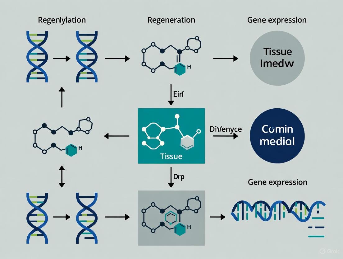
The coordinated actions of DNMT and TET enzymes establish a dynamic cycle of cytosine modification that enables precise epigenetic regulation [7]. This cycle begins with unmodified cytosine being converted to 5mC by DNMT enzymes, followed by stepwise oxidation to 5hmC, 5fC, and 5caC by TET enzymes. The process culminates with TDG-mediated base excision repair that restores unmodified cytosine, completing the demethylation cycle [8]. The balance between these opposing enzymatic activities creates an epigenetic landscape that can respond to developmental cues, environmental signals, and regenerative requirements, allowing cells to maintain stability while retaining plasticity for fate transitions during tissue repair [3].
Diagram 1: DNA methylation-demethylation cycle. DNMTs add methyl groups using SAM, while TET enzymes oxidize 5mC in stepwise reactions. TDG with BER completes active demethylation.
Experimental Methodologies for Analyzing DNA Methylation Dynamics
Bisulfite Sequencing and Its Variations
Bisulfite conversion represents the gold standard technique for detecting DNA methylation at single-base resolution [8]. This method relies on the differential sensitivity of cytosine and 5-methylcytosine to bisulfite treatment: unconverted cytosines are read as thymine in subsequent sequencing, while methylated cytosines remain protected and are still detected as cytosines [8]. Several bisulfite-based approaches have been developed for different research applications:
- Whole-Genome Bisulfite Sequencing (WGBS) provides comprehensive methylation maps across the entire genome, offering single-base resolution but requiring substantial sequencing depth [8].
- Reduced Representation Bisulfite Sequencing (RRBS) uses methylation-sensitive restriction enzymes to enrich for CpG-dense regions, providing a cost-effective alternative for analyzing methylation patterns in genomic regions of high interest [8].
- Targeted Bisulfite Sequencing approaches focus on specific genomic regions of interest, allowing for higher throughput analysis of predetermined loci [8].
A significant limitation of conventional bisulfite sequencing is its inability to distinguish between 5mC and 5hmC, as both modifications protect cytosines from conversion [8]. This challenge has led to the development of additional techniques specifically designed to resolve oxidized methylation intermediates.
Immunoprecipitation-Based Methods
DNA immunoprecipitation (DIP) offers an alternative approach for mapping DNA modifications using antibodies specific to modified cytosine variants [8]. This methodology involves shearing genomic DNA, immunoprecipitating fragments containing the modification of interest, and then analyzing the pulled-down DNA by quantitative PCR (DIP-qPCR), microarray (DIP-chip), or sequencing (DIP-seq) [8]. The key advantages of DIP include its relatively low cost compared to WGBS and the ability to specifically interrogate 5mC, 5hmC, 5fC, and 5caC when appropriate antibodies are available [8]. However, this technique provides lower resolution than bisulfite-based methods and is dependent on antibody specificity.
Diagram 2: Experimental workflows for DNA methylation analysis. Two main approaches are bisulfite conversion and immunoprecipitation.
The Scientist's Toolkit: Essential Research Reagents
Table 3: Key Research Reagents for DNA Methylation Studies
| Reagent/Technique | Function | Application in Regeneration Research |
|---|---|---|
| Bisulfite Conversion Reagents | Chemical deamination of unmethylated cytosine | Distinguishes methylated vs. unmethylated cytosines in tissue samples |
| 5mC/5hmC/5fC/5caC Specific Antibodies | Immunoprecipitation of modified DNA | Enrichment of specific methylation states for genomic mapping |
| DNMT Inhibitors (e.g., Zebularine, RG108) | Chemical inhibition of DNMT activity | Probing the functional role of methylation in regenerative processes |
| TET Activity Assays | Measurement of oxidation activity | Determining TET enzyme function in stem cell differentiation |
| SAM/SAH Analysis Kits | Quantification of methyl donor availability | Assessing metabolic regulation of methylation in regenerating tissues |
| CRISPR/dCas9 Epigenetic Editors | Targeted methylation or demethylation | Precise epigenetic manipulation of regenerative gene pathways |
| Nanoelectroporation Systems | Efficient delivery of epigenetic editors | In vivo reprogramming for tissue regeneration [4] |
| Acetylene--thiirane (1/1) | Acetylene--thiirane (1/1)|High-Purity Research Chemical | Acetylene--thiirane (1/1) is a high-purity chemical complex for research applications. This product is For Research Use Only. Not for human or veterinary use. |
| Cobalt--ruthenium (2/3) | Cobalt--ruthenium (2/3), CAS:823185-74-6, MF:Co2Ru3, MW:421.1 g/mol | Chemical Reagent |
DNMT and TET Dynamics in Tissue Regeneration and Cellular Reprogramming
Epigenetic Reprogramming in Regenerative Processes
The dynamic interplay between DNMT and TET enzymes facilitates the epigenetic reprogramming necessary for successful tissue regeneration [4]. During cellular reprogramming for regenerative applications, somatic cells can be converted to alternative fates through several approaches: induced pluripotency (iPSCs), direct lineage conversion (transdifferentiation), or partial cellular rejuvenation [4]. Each of these strategies involves significant reconfiguration of DNA methylation patterns orchestrated by coordinated DNMT and TET activity. Direct reprogramming approaches are particularly promising for regenerative medicine as they enable in vivo cell fate conversion without traversing a pluripotent state, thereby reducing tumorigenesis risks [4].
Emerging technologies such as tissue nanotransfection (TNT) leverage nanoelectroporation to deliver epigenetic effectors directly into tissues, enabling in vivo reprogramming for regenerative applications [4]. This approach demonstrates the translational potential of targeting DNA methylation dynamics for therapeutic tissue repair, including applications in wound healing, ischemia repair, and age-related tissue dysfunction [4].
Aging and DNA Methylation Dynamics
Aging is associated with progressive changes in DNA methylation patterns, including both generalized hypomethylation and locus-specific hypermethylation, which contribute to reduced tissue function and regenerative capacity [5]. Recent comprehensive mapping of DNA methylation changes across human organs has revealed that the precision of DNA methylation maintenance declines with age, leading to alterations in gene expression that are linked to age-related tissue dysfunction [5]. These age-associated epigenetic changes represent potential therapeutic targets for regenerative interventions aimed at restoring youthful epigenetic patterns and improving tissue repair capacity in aging individuals.
Partial reprogramming approaches using transient expression of reprogramming factors (Oct4, Sox2, Klf-4, c-Myc) have demonstrated the ability to reverse aging-associated epigenetic alterations, including resetting DNA methylation clocks, without fully altering cellular identity [4]. This strategy represents a promising avenue for combating age-related decline in regenerative capacity through epigenetic rejuvenation.
Emerging Technologies and Future Directions
Epigenetic Engineering for Regenerative Medicine
Recent discoveries revealing that specific DNA sequences can instruct de novo DNA methylation patterns represent a paradigm shift in epigenetic engineering [6]. This finding enables the development of precision epigenetic editing approaches where engineered transcription factors can be designed to target DNA methylation machinery to specific genomic loci, creating predetermined methylation patterns that could enhance cellular function [6]. The ability to use DNA sequences to target methylation has broad implications for regenerative medicine, as it would allow epigenetic defects to be corrected with high specificity, potentially restoring proper gene expression patterns in diseased or damaged tissues [6].
CRISPR/dCas9-based epigenetic editing systems provide a versatile platform for implementing targeted methylation changes [4]. When coupled with advanced delivery systems such as tissue nanotransfection, these technologies enable precise in vivo epigenetic manipulation for regenerative applications [4]. The ongoing optimization of these systems focuses on improving specificity, efficiency, and persistence of the desired epigenetic changes while minimizing off-target effects.
Clinical Implications and Therapeutic Applications
The dynamic regulation of DNA methylation by DNMT and TET enzymes offers promising therapeutic targets for enhancing tissue regeneration. Dysregulation of these enzymes is implicated in various disease states, including cancers, neurodegenerative diseases, and developmental disorders [2]. In cancer development, for instance, global hypomethylation accompanied by localized hypermethylation of tumor suppressor genes is a common feature, with demethylation affecting approximately 5-20% of methylated CpG sites [9]. Understanding these disease-associated epigenetic alterations provides insights for developing targeted epigenetic therapies that could potentially reverse pathological methylation patterns and restore normal cellular function.
In regenerative contexts, modulating DNMT and TET activity represents a strategy for promoting cellular plasticity, enhancing stem cell function, and reversing age-related epigenetic changes that impair tissue repair [5] [4]. As our understanding of the intricate balance between DNA methylation and demethylation continues to evolve, so too will opportunities for developing innovative epigenetic interventions for tissue regeneration and repair.
The journey from a pluripotent stem cell to a fully differentiated somatic cell is governed by a complex interplay of genetic and epigenetic factors. Among these, DNA methylation—the addition of a methyl group to a cytosine base—serves as a critical mechanism for regulating gene expression without altering the underlying DNA sequence. Within the context of tissue regeneration research, understanding and potentially directing these methylation dynamics offers promising therapeutic avenues. DNA methylation works in concert with other epigenetic marks, including histone modifications, to establish and maintain cellular identity by precisely controlling which genes are accessible for transcription [10]. This in-depth technical guide explores the mechanisms by which DNA methylation directs stem cell differentiation and proliferation, providing researchers and drug development professionals with current experimental insights and methodologies.
The dynamic nature of the epigenome is particularly evident in stem cells, which utilize precise methylation patterns to maintain pluripotency while retaining the capacity to differentiate into all three germ layers. As stated in one review, "The molecular mechanisms that regulate stem cell pluripotency and differentiation has shown the crucial role that methylation plays in this process" [10]. This methylation machinery involves "writer" enzymes (DNA methyltransferases, or DNMTs) that establish methylation patterns, "eraser" enzymes (Ten-Eleven Translocation, or TET, proteins) that remove these marks, and "reader" proteins that interpret them, creating a dynamic, responsive system for gene regulation [10]. In regenerative medicine, the ability to reset or redirect these epigenetic patterns through technologies like tissue nanotransfection (TNT) or cellular reprogramming represents a frontier for developing novel therapies aimed at repairing damaged tissues and reversing age-related degeneration.
Methylation Dynamics in Development and Aging
Prenatal Programming and Postnatal Decline
Methylation dynamics exhibit distinct patterns across the human lifespan, with particularly pronounced activity during prenatal development. A comprehensive study profiling genome-wide DNA methylation across the human cortex from 6 post-conception weeks to 108 years of age identified widespread, developmentally regulated changes, with pronounced shifts occurring during early- and mid-gestation [11]. These prenatal methylation changes were notably distinct from age-associated modifications occurring in the postnatal cortex. Through fluorescence-activated nuclei sorting of SATB2-positive neuronal nuclei, researchers demonstrated that these dynamics follow cell-type-specific trajectories, underscoring the precision of epigenetic programming during development [11].
Critically, these developmentally dynamic DNA methylation sites were significantly enriched near genes implicated in autism and schizophrenia, providing a mechanistic link between epigenetic dysregulation during critical developmental windows and the pathogenesis of neurodevelopmental disorders [11]. This finding highlights the importance of precise temporal and cell-type-specific methylation control for normal brain development and function.
In contrast to the targeted methylation changes during development, aging is characterized by more generalized epigenetic alterations. As noted in a recent news article discussing a large epigenetic atlas, "The epigenetic process of DNA methylation — the addition or removal of tags called methyl groups — becomes less precise as we age. The result is changes to gene expression that are linked to reduced organ function and increased susceptibility to disease" [5]. This age-related erosion of epigenetic precision contributes to the functional decline of tissues and organs.
Replicative Stress and Hematopoietic Stem Cell Aging
The relationship between proliferative history and epigenetic aging has been particularly well-documented in hematopoietic stem cells (HSCs). A 2024 study induced forced replication of HSCs in vivo through cyclical treatment with low-dose fluorouracil (5FU) and demonstrated that proliferative stress induces several aging phenotypes, including altered leukocyte counts, decreased lymphoid progenitors, and reduced reconstitution potential [12]. Importantly, the divisional history of HSCs was imprinted in the DNA methylome, consistent with functional decline.
The DNA methylation changes observed in aged HSCs included global hypermethylation in non-coding regions and similar frequencies of hypo- and hyper-methylation at promoter regions, particularly affecting genes targeted by the Polycomb Repressive Complex 2 (PRC2) [12]. These findings suggest that HSC proliferation can drive aging phenotypes primarily through epigenetic mechanisms, independent of initial functional decline.
Table 1: DNA Methylation Changes in Hematopoietic Stem Cell Aging
| Parameter | Prenatal/Young HSCs | Aged/Stressed HSCs | Functional Consequence |
|---|---|---|---|
| Global Methylation Pattern | Developmentally programmed | Erosion of precision | Increased disease susceptibility |
| Non-coding Regions | Balanced methylation | Global hypermethylation | Genomic instability |
| Promoter Regions | Tissue-specific patterns | PRC2 target disruption | Altered differentiation capacity |
| Lymphoid Output | High | Decreased | Immune dysfunction |
| Reconstitution Potential | Robust | Reduced | Impaired tissue maintenance |
Novel Mechanisms and Methodologies
Synergistic Histone Modifications in Cell Fate Determination
Beyond DNA methylation, other epigenetic marks work in concert to direct cell fate. Recent research has revealed a remarkable synergy between two histone modifications—H3K79 methylation and H3K36 trimethylation—that critically orchestrates gene expression during cell differentiation [13] [14]. Using CRISPR-based genetic engineering to generate stem cell models deficient in enzymes responsible for these modifications, researchers discovered that while loss of either mark alone caused minor changes, concomitant absence triggered unexpected gene hyperactivation and blocked the cells' ability to differentiate into neurons [13].
This finding overturned the previous assumption that these histone marks solely facilitate gene activation, revealing instead a nuanced regulatory system where they also function as critical modulators preventing excessive transcriptional activity. The investigation further identified a hyperactive YAP-TEAD transcriptional pathway that becomes unleashed when both methylation marks are lost, pointing to a potential therapeutic target for cancers, including leukemia, characterized by such epigenetic dysregulation [13] [14].
Diagram 1: Synergistic histone regulation of cell fate.
Genetic Regulation of Epigenetic Patterning
A paradigm-shifting discovery in the field of epigenetics has revealed that DNA methylation can be regulated by genetic mechanisms, not just by pre-existing epigenetic marks. Research in Arabidopsis thaliana identified that specific DNA sequences, recognized by proteins called RIMs (a subset of REPRODUCTIVE MERISTEM transcription factors), act with CLASSY3 proteins to establish DNA methylation at specific genomic targets [6]. When researchers disrupted these DNA sequences, the entire methylation pathway failed.
This discovery of sequence-driven DNA methylation represents a fundamental shift in understanding how novel methylation patterns arise during development, moving beyond the model where pre-existing epigenetic modifications solely guide new methylation [6]. As the senior author noted, "This finding represents a paradigm shift in the field's view of how methylation is regulated in plants. All previous work pointed to pre-existing epigenetic modifications as the starting place for targeting methylation, which didn't explain how novel methylation patterns could arise. Now we know the DNA itself can instruct new methylation patterns, too" [6]. This mechanism has significant implications for epigenetic engineering strategies aimed at generating specific methylation patterns to repair or enhance cell function.
Cell Cycle as an Epigenetic Regulator
The intersection of cell cycle dynamics and epigenetic remodeling represents another emerging frontier in understanding cell fate regulation. A recent study demonstrated that enhancing the activities of transcription factors OCT4 and SOX2 by fusing them with the herpesvirus VP16 activation domain (creating OvSvK complex) significantly accelerated somatic cell reprogramming into induced pluripotent stem cells (iPSCs) [15]. Single-cell analyses revealed that OvSvK restructures the cell cycle, shortening the G1 phase while extending S phase, thereby establishing an embryonic stem cell-like pattern.
This cell cycle restructuring directly influences epigenetic inheritance, particularly the restoration of H3K27me3 marks during the G1 phase [15]. The shortened G1 phase impedes the complete restoration of repressive H3K27me3 marks, consequently elevating expression of genes that facilitate pluripotency. This research establishes cell cycle dynamics as key epigenetic modulators for cell fate transitions, providing fundamental insights into reprogramming mechanisms with significant implications for regenerative medicine.
Advanced Technologies for Epigenetic Manipulation
EPI-Clone: Transgene-Free Lineage Tracing
Recent methodological advances have expanded our ability to track cell fate decisions at unprecedented resolution. EPI-Clone is a novel method that exploits targeted single-cell profiling of DNA methylation at single-CpG resolution to track clones while providing detailed cell-state information [16]. This approach distinguishes between two types of CpG sites: those whose methylation status reflects cellular differentiation, and those that undergo stochastic epimutations and can serve as digital barcodes of clonal identity.
Applied to mouse and human haematopoiesis, EPI-Clone has revealed that in mouse ageing, myeloid bias and low output of old haematopoietic stem cells are restricted to a small number of expanded clones, whereas many functionally young-like clones persist in old age [16]. In human ageing, clones with and without known driver mutations of clonal haematopoiesis display similar lineage biases, suggesting convergent mechanisms of age-related clonal expansion. This transgene-free lineage tracing method enables accurate single-cell lineage tracing on hematopoietic cell state landscapes at scale, providing powerful insights into the clonal dynamics of aging and differentiation.
Tissue Nanotransfection for In Vivo Reprogramming
Tissue nanotransfection (TNT) has emerged as a novel non-viral platform capable of delivering genetic material directly into tissues via localized nanoelectroporation, enabling cellular reprogramming in situ [4]. The TNT device consists of a hollow-needle silicon chip mounted beneath a cargo reservoir containing genetic material. When electrical pulses are applied, the hollow needles concentrate the electric field at their tips, temporarily porating nearby cell membranes and enabling targeted delivery of charged genetic material into tissue.
TNT can deliver various genetic cargoes, including plasmid DNA, mRNA, and CRISPR/Cas9 components, for different reprogramming strategies:
- Induced pluripotency: Transforming somatic cells into a pluripotent state
- Direct reprogramming (transdifferentiation): Converting one somatic cell type into another without a pluripotent intermediate
- Partial reprogramming (cellular rejuvenation): Transient factor expression to reverse aging-related changes without altering cell identity [4]
The optimization of electrical pulse parameters—voltage amplitude, pulse duration, and inter-pulse intervals—is critical for maximizing delivery efficiency while preserving cellular viability during the nanotransfection process [4].
Diagram 2: Tissue nanotransfection workflow for epigenetic reprogramming.
Experimental Protocols and Research Tools
Key Methodologies for Studying Methylation Dynamics
Isolation of Cell-Type-Specific Nuclei for Methylation Analysis The protocol for isolating neuron-specific nuclei from human cortex tissue exemplifies approaches for cell-type-specific epigenetic analysis [11]:
- Tissue Preparation: Fresh or frozen human cortex tissue is homogenized in a sucrose-based buffer with detergent to create a nuclear suspension.
- Fluorescence-Activated Nuclei Sorting (FANS): Nuclei are incubated with a SATB2 antibody (a neuronal marker) conjugated to a fluorescent tag.
- Flow Cytometry: SATB2-positive neuronal nuclei are sorted from SATB2-negative non-neuronal nuclei using a fluorescence-activated cell sorter.
- DNA Extraction and Bisulfite Treatment: Genomic DNA is extracted from sorted nuclei and treated with bisulfite, which converts unmethylated cytosines to uracils while leaving methylated cytosines unchanged.
- Genome-Wide Methylation Profiling: Bisulfite-treated DNA is subjected to whole-genome bisulfite sequencing or reduced representation bisulfite sequencing to determine methylation status at single-base resolution.
In Vivo Hematopoietic Stem Cell Aging Model To study the effects of proliferative stress on HSC aging [12]:
- Animal Model: 3-month-old C57BL/6 J female mice are used at study start.
- 5-FU Treatment: 5-Fluorouracil is administered at 150 mg/kg by intraperitoneal injection, once every 3 weeks.
- Monitoring: Animals are continuously monitored for general health and body weight.
- HSC Isolation: At experimental endpoints, bone marrow is harvested from hind legs, pelvis, femur, and tibia. HSCs are isolated using c-Kit enrichment followed by fluorescence-activated cell sorting for PI-Lin−cKit+Sca1+CD34−Flk2−CD150+ cells.
- Downstream Analysis: Isolated HSCs can be used for transplantation assays, DNA methylation analysis by RRBS, DNA damage assays (alkaline comet assay, gH2AX assay), or mRNA expression profiling.
The Scientist's Toolkit: Essential Research Reagents
Table 2: Key Research Reagents for Methylation Dynamics Studies
| Reagent/Technology | Function/Application | Example Use Case |
|---|---|---|
| SATB2 Antibody | Marker for sorting neuronal nuclei | Isolation of neuron-specific nuclei for methylation analysis in human cortex [11] |
| 5-Fluorouracil (5-FU) | Chemotherapeutic agent inducing proliferative stress | In vivo model of HSC aging through forced replication [12] |
| LARRY Barcoding System | Lentiviral genetic barcoding for lineage tracing | Ground-truth dataset for clonal identity in haematopoiesis [16] |
| scTAM-seq | Single-cell targeted analysis of methylome | High-resolution DNA methylation profiling at single-CpG level [16] |
| CRISPR-based Genetic Engineering | Targeted gene knockout or editing | Generating stem cell models deficient in specific histone-modifying enzymes [13] [14] |
| OCT4-VP16/SOX2-VP16 | Enhanced transcription factors with VP16 activation domain | Accelerated somatic cell reprogramming to iPSCs [15] |
| TNT Device | Nanoelectroporation for in vivo gene delivery | Direct cellular reprogramming in tissue regeneration [4] |
| Mission Bio Tapestri Platform | Microfluidic platform for single-cell multi-omics | Implementation of scTAM-seq for DNA methylation analysis [16] |
| 4-(Diethylamino)but-2-enal | 4-(Diethylamino)but-2-enal|RUO | 4-(Diethylamino)but-2-enal for research use only. A high-purity building block for organic synthesis. Get quotes and spec sheets. Not for human use. |
| Furan;tetramethylazanium | Furan;tetramethylazanium CAS 396101-14-7 Supplier |
The precise control of DNA methylation dynamics represents a central mechanism directing stem cell fate decisions throughout development, aging, and disease. The emerging picture reveals an increasingly complex regulatory network where DNA methylation interacts with histone modifications, genetic sequences, cell cycle dynamics, and environmental cues to maintain cellular identity or enable fate transitions. The recent discoveries of sequence-driven DNA methylation and cell cycle-mediated epigenetic remodeling represent significant paradigm shifts in our understanding of how epigenetic patterns are established and maintained.
For tissue regeneration research, the ability to manipulate these methylation dynamics through technologies like tissue nanotransfection, epigenetic editing, and directed reprogramming offers promising pathways for therapeutic intervention. The finding that partial reprogramming can reset epigenetic age without altering cellular identity suggests potential strategies for rejuvenating aged or damaged tissues. Meanwhile, the identification of specific epigenetic synergies, such as that between H3K79 and H3K36 methylation, reveals new therapeutic targets for diseases characterized by epigenetic dysregulation.
As methodologies for tracking and manipulating methylation dynamics continue to advance—with technologies like EPI-Clone enabling high-resolution lineage tracing and TNT facilitating in vivo reprogramming—researchers and drug development professionals are equipped with an increasingly sophisticated toolkit for probing and directing cell fate. These advances hold significant promise for developing novel regenerative therapies that harness the epigenetic control of cellular identity and function.
The differentiation of mesenchymal stem/stromal cells (MSCs) into osteogenic, chondrogenic, and adipogenic lineages is a tightly regulated process crucial for tissue homeostasis and regeneration. These lineage commitments are controlled by complex interactions between transcription factors, signaling pathways, and epigenetic modifications [17] [18]. Recent advances in transcriptome analysis and epigenetics have revealed the intricate regulatory networks that determine stem cell fate. Understanding these mechanisms is paramount for developing effective regenerative medicine strategies for conditions ranging from osteoporosis to cartilage repair [17] [18] [19]. This review synthesizes current knowledge on the key regulators, signaling pathways, and emerging role of DNA methylation in controlling lineage-specific differentiation of MSCs, with implications for targeted therapeutic interventions.
Transcriptional Regulation of Lineage Commitment
The fate of MSCs is primarily directed by core transcription factors that activate lineage-specific gene programs while suppressing alternative pathways. The balance between these transcriptional regulators determines differentiation outcomes.
Core Transcription Factors
- Chondrogenesis: SOX9 is the master regulator that works in concert with SOX5 and SOX6 to induce chondrocyte differentiation and maintain chondrocytic phenotypes. SOX9 directly regulates type II collagen (COL2A1) and aggrecan (ACAN) expression [17]. RUNX2 also promotes chondrogenic differentiation in certain contexts [17].
- Adipogenesis: PPARγ serves as the central regulator that is both necessary and sufficient for adipogenesis. The C/EBP family members (C/EBPα, C/EBPβ, and C/EBPδ) work in coordination with PPARγ to drive adipocyte differentiation, with C/EBPα being particularly required for white adipocyte differentiation [17].
- Osteogenesis: RUNX2 is the critical transcription factor directing osteogenic differentiation, activating expression of bone-specific genes including osteocalcin and bone sialoprotein [18].
Transcriptional Crosstalk and Antagonism
Complex antagonistic relationships exist between transcription factors of different lineages, creating mutually exclusive differentiation paths:
- SOX9 downregulation appears necessary for adipocyte differentiation, as it can bind to and suppress C/EBPβ and C/EBPδ promoter activity [17].
- Conversely, C/EBPα, C/EBPβ, and C/EBPδ suppress chondrogenic marker genes COL2A1, ACAN, and SOX9 in ATDC5 cells [17].
- The balance between adipogenic and osteogenic transcription factors is crucial for bone homeostasis, with PPARγ activation promoting adipogenesis at the expense of osteogenesis [18].
Table 1: Key Transcription Factors in MSC Lineage Differentiation
| Lineage | Master Regulator | Co-factors | Target Genes | Antagonists |
|---|---|---|---|---|
| Chondrogenesis | SOX9 | SOX5, SOX6 | COL2A1, ACAN | C/EBP family |
| Adipogenesis | PPARγ | C/EBPα, C/EBPβ, C/EBPδ | FABP4, LPL | SOX9, RUNX2 |
| Osteogenesis | RUNX2 | OSX, ATF4 | Osteocalcin, Bone sialoprotein | PPARγ |
Signaling Pathways Governing Lineage Determination
Multiple signaling pathways interact to guide MSC fate decisions, often exhibiting contrasting effects on different lineages. The precise outcome depends on specific ligands, concentrations, temporal activation, and cellular context.
TGF-β/BMP Superfamily
The TGF-β/BMP pathway plays particularly important roles in lineage specification with different members showing distinct effects:
- TGF-β (especially TGF-β2 and TGF-β3) strongly promotes chondrogenic differentiation of human BMSCs at concentrations around 10 ng/mL, while simultaneously inhibiting adipogenesis [17]. TGF-β1 induces dominant chondrogenesis while suppressing adipogenic differentiation in CL-1 cells and human BMSCs [17]. Mechanistically, TGF-β signals through Smad3 to inhibit adipogenesis by associating with C/EBPβ and C/EBPδ, resulting in decreased PPARγ expression [17].
- BMP family members show diverse effects: BMP2 is the most effective in promoting chondrogenic differentiation compared to BMP4 and BMP6 in human BMSCs [17]. Interestingly, BMP2 also stimulates adipogenesis in 3T3-L1 cells and rat BMSCs when associated with a PPARγ activator [17]. BMP7 serves as a unique brown fat inducer, while BMP2 and BMP4 act as white adipogenic factors [17].
Additional Signaling Pathways
- Wnt Signaling: Wnt proteins prevent MSCs from proceeding toward adipogenic lineage while promoting osteogenesis [17].
- Hedgehog Signaling: Similar to Wnt, Hedgehog proteins are important for MSC myogenic lineage commitment but inhibit adipogenic differentiation [17].
The following diagram illustrates the key signaling pathways and their interactions in regulating MSC lineage commitment:
Diagram 1: Signaling Pathways in MSC Lineage Commitment. Arrows indicate activation, while T-bars indicate inhibition.
Epigenetic Regulation: The Role of DNA Methylation
Beyond transcription factors and signaling pathways, epigenetic mechanisms including DNA methylation provide an additional layer of regulation in lineage-specific differentiation. DNA methylation patterns are dynamically regulated during cellular differentiation and play crucial roles in defining cell identity.
Mechanisms of DNA Methylation Patterning
Recent research has revealed that transcription factors can actively instruct DNA methylation patterns in specific tissues:
- In plant reproductive tissues, REPRODUCTIVE MERISTEM (REM) transcription factors target the RNA-directed DNA methylation (RdDM) machinery to distinct loci, generating tissue-specific epigenomes [19]. These REM INSTRUCTS METHYLATION (RIM) factors are required for methylation at specific targets in anther or ovule tissues [19].
- Disruption of DNA-binding domains in these transcription factors or the motifs they recognize blocks RNA-directed DNA methylation, demonstrating the direct link between genetic information and epigenetic patterning [19].
- This mechanism represents a departure from the traditional view that DNA methylation patterns are regulated primarily by chromatin features rather than DNA sequence motifs [19].
DNA Methylation in Mammalian Lineage Differentiation
While the precise mechanisms of DNA methylation in mammalian MSC differentiation are still being elucidated, several key principles emerge:
- DNA methylation patterns are established through coordinated action of DNA methyltransferases and demethylases that respond to differentiation signals.
- Lineage-specific transcription factors may recruit epigenetic modifiers to establish methylation patterns that stabilize the differentiated state.
- The interplay between genetic information (transcription factor binding) and epigenetic mechanisms (DNA methylation) creates stable cellular identities necessary for tissue function and regeneration.
Table 2: Experimental Approaches for Studying DNA Methylation in Lineage Differentiation
| Method | Application | Key Readouts | Considerations |
|---|---|---|---|
| Bisulfite Sequencing | Genome-wide methylation mapping | Methylation levels at single-base resolution | Distinguishes between CG, CHG, CHH contexts |
| smRNA-seq | siRNA profiling at methylated loci | siRNA cluster abundance | Identifies active RdDM targets |
| Methyl-Cutting PCR Assays | Rapid screening of methylation status | Methylation at specific loci | Medium-throughput candidate validation |
| Genetic Screens (EMS mutants) | Identification of novel methylation factors | DNA methylation defects | Follow-up mapping required |
Experimental Methodologies for Lineage Differentiation
Standardized protocols have been established for directing MSCs toward specific lineages in vitro. These methodologies enable the study of differentiation mechanisms and screening of potential therapeutic compounds.
Chondrogenic Differentiation Protocol
- Cell Culture Format: Pellet culture or micromass systems to enhance cell-cell interactions [17].
- Basal Medium: High-glucose DMEM supplemented with ITS+1 premix (insulin, transferrin, selenium), ascorbate-2-phosphate, sodium pyruvate, proline, and dexamethasone [17].
- Key Inducers: TGF-β1, TGF-β2, or TGF-β3 at 10 ng/mL concentration [17]. TGF-β2 and TGF-β3 are more effective than TGF-β1 in human BMSCs [17].
- Duration: 14-28 days with medium changes every 2-3 days.
- Validation: Histological staining for sulfated proteoglycans (Alcian blue, Safranin O), immunohistochemistry for type II collagen, and gene expression analysis of SOX9, COL2A1, and ACAN.
Adipogenic Differentiation Protocol
- Cell Culture Format: Monolayer culture at high density (confluent).
- Basal Medium: DMEM with high glucose, supplemented with fetal bovine serum.
- Induction Cocktail: IBMX (0.5 mM), dexamethasone (1 μM), indomethacin (200 μM), and insulin (10 μg/mL) [18].
- Maintenance Medium: DMEM with high glucose and insulin (10 μg/mL) only.
- Cycling Protocol: 2-3 cycles of induction/maintenance (2-3 days each) followed by culture in maintenance medium for an additional 7-14 days.
- Validation: Oil Red O staining of lipid droplets, gene expression analysis of PPARγ, C/EBPα, and FABP4.
Osteogenic Differentiation Protocol
- Cell Culture Format: Monolayer culture at 50-70% confluence.
- Basal Medium: DMEM with low glucose, supplemented with fetal bovine serum.
- Key Inducers: Dexamethasone (100 nM), ascorbate-2-phosphate (50-100 μM), and β-glycerophosphate (10 mM) [18].
- Duration: 14-21 days with medium changes every 3-4 days.
- Validation: Alizarin Red S or Von Kossa staining of mineralized matrix, alkaline phosphatase activity assay, gene expression analysis of RUNX2, osteocalcin, and osteopontin.
The following workflow diagram outlines a comprehensive experimental approach for studying lineage differentiation and DNA methylation:
Diagram 2: Experimental Workflow for Studying Lineage Differentiation and DNA Methylation.
The Scientist's Toolkit: Essential Research Reagents
Table 3: Key Research Reagents for Studying Lineage Differentiation
| Reagent/Category | Specific Examples | Function/Application | Considerations |
|---|---|---|---|
| Lineage Inducers | TGF-β1/2/3 (10 ng/mL), BMP2 (500 ng/mL) | Chondrogenic differentiation | Concentration-dependent effects [17] |
| Dexamethasone (0.1-1 μM), IBMX (0.5 mM), Insulin (10 μg/mL) | Adipogenic differentiation | Cocktail required for efficient differentiation [18] | |
| Dexamethasone (100 nM), Ascorbate-2-phosphate (50-100 μM), β-glycerophosphate (10 mM) | Osteogenic differentiation | Required for matrix mineralization [18] | |
| Cell Markers | CD105, CD73, CD90 (>95% positive) | MSC identification | Minimum criteria per ISCT guidelines [18] |
| CD45, CD34, CD14/CD11b, CD79α/CD19, HLA-DR (<2% positive) | Hematopoietic contamination exclusion | Essential for MSC purity assessment [18] | |
| Epigenetic Tools | Bisulfite conversion reagents | DNA methylation analysis | Distinguishes methylated/unmethylated cytosines [19] |
| siRNA/miRNA inhibitors | Functional studies of non-coding RNAs | Identifies regulatory networks [18] | |
| Analysis Kits | Alcian Blue, Oil Red O, Alizarin Red S | Histological validation of differentiation | Quantitative extraction possible |
| RNA-seq library preparation kits | Transcriptome analysis | Full-length transcript information [18] | |
| 1,2-Bis(sulfanyl)ethan-1-ol | 1,2-Bis(sulfanyl)ethan-1-ol | Get 1,2-Bis(sulfanyl)ethan-1-ol (C2H6OS2), also known as 1,2-dimercaptoethanol. This product is designated For Research Use Only and is not intended for diagnostic or personal use. | Bench Chemicals |
| Octa-1,7-diene-1,8-dione | Octa-1,7-diene-1,8-dione, CAS:197152-47-9, MF:C8H10O2, MW:138.16 g/mol | Chemical Reagent | Bench Chemicals |
The lineage-specific differentiation of MSCs into osteogenic, chondrogenic, and adipogenic cells is governed by sophisticated transcriptional networks, signaling pathways, and epigenetic mechanisms. The emerging understanding of how transcription factors instruct DNA methylation patterns provides new insights into how stable cellular identities are established during tissue regeneration. Future research focusing on the interplay between genetic and epigenetic information will undoubtedly yield novel therapeutic approaches for regenerative medicine, potentially enabling more precise control over stem cell fate decisions in clinical applications.
The precise orchestration of tissue repair following injury represents a critical biological process where the balance between regenerative healing and pathological fibrosis determines clinical outcomes. DNA methylation, the covalent addition of a methyl group to the carbon-5 position of cytosine in CpG dinucleotides, has emerged as a fundamental epigenetic mechanism governing this balance [20] [21]. This whitepaper examines how dynamic DNA methylation patterns direct cellular responses during wound healing, where proper regulation leads to tissue regeneration, while dysregulation drives aberrant repair processes including fibrosis and chronic wounds [22] [20] [23]. The context of a broader thesis on DNA methylation in tissue regeneration research frames this exploration, highlighting the therapeutic potential of targeting epigenetic mechanisms to redirect pathological healing toward regenerative outcomes. For researchers and drug development professionals, understanding these mechanisms provides a foundation for developing novel epigenetic-based interventions that could revolutionize treatment for fibrotic diseases and chronic wounds.
Molecular Mechanisms of DNA Methylation in Healing
The DNA Methylation Machinery
DNA methylation is catalyzed by DNA methyltransferases (DNMTs), which transfer methyl groups from S-adenosylmethionine (SAM) to cytosine bases [21] [24]. DNMT3A and DNMT3B establish de novo methylation patterns, while DNMT1 maintains these patterns during cell division, ensuring faithful inheritance of methylation marks [21] [24]. This methylation process is counterbalanced by demethylation enzymes, particularly the ten-eleven translocation (TET) family, including TET1, TET2, and TET3, which oxidize 5-methylcytosine (5mC) to 5-hydroxymethylcytosine (5hmC) and further to 5-formylcytosine (5fC) and 5-carboxylcytosine (5caC) [20] [24]. The dynamic interplay between these "writers" and "erasers" enables precise temporal and spatial control of gene expression during the complex process of tissue repair [21].
The functional consequences of DNA methylation depend critically on genomic context. Methylation within gene promoter regions, particularly at CpG islands, typically leads to transcriptional repression by physically impeding transcription factor binding or recruiting methyl-CpG-binding proteins (MBPs) like MeCP2, MBD1, and MBD2, which subsequently associate with histone deacetylases (HDACs) to promote chromatin condensation [20] [21]. In contrast, gene body methylation often correlates with transcriptional activity, potentially suppressing spurious transcription initiation or regulating alternative splicing [21]. This contextual understanding is essential for deciphering how methylation changes direct healing outcomes.
Interplay with Other Epigenetic Mechanisms
DNA methylation does not function in isolation but participates in extensive crosstalk with other epigenetic regulatory systems. A bidirectional relationship exists between DNA methylation and histone modifications, where DNA methylation can template specific histone modifications after DNA replication, and histone methylation can help direct DNA methylation patterns [20]. Similarly, complex feedback loops connect DNA methylation with non-coding RNAs (ncRNAs); DNA methylation regulates the expression of some microRNAs (miRNAs), while another subset of miRNAs controls the expression of DNMTs and other epigenetic regulators [20]. For instance, Dakhlallah et al. identified a miRNA-DNMT regulatory circuit in pulmonary fibrosis where reduced expression of the miR-17~92 cluster was associated with increased DNMT1 expression and a pro-fibrotic phenotype [20]. This sophisticated epigenetics-miRNA regulatory circuit organizes global gene expression profiles, and its disruption can interfere with normal wound healing processes.
DNA Methylation in Normal Wound Healing
Spatiotemporal Regulation of Healing Phases
Normal wound healing progresses through highly coordinated phases—hemostasis, inflammation, proliferation, and remodeling—each characterized by distinct gene expression patterns guided by epigenetic modifications [22]. DNA methylation dynamically regulates these phase transitions by controlling key genes and pathways. During the hemostasis and inflammatory phases, methylation changes influence platelet function and immune cell activity; for example, DNA methylation of genes like platelet endothelial aggregation receptor 1 can impact platelet function [22]. As healing progresses to the proliferation phase, methylation patterns direct fibroblast proliferation, angiogenesis, and temporary matrix deposition [22] [25].
The final remodeling phase, where the balance between matrix synthesis and degradation determines regenerative versus fibrotic outcomes, is particularly influenced by epigenetic controls. During this phase, myofibroblasts—the primary ECM-producing cells—typically undergo apoptosis or revert to a quiescent phenotype [20]. DNA methylation helps regulate this critical transition; proper methylation patterns ensure the timely elimination of myofibroblasts, preventing excessive ECM accumulation [20]. The spatiotemporal precision of these methylation changes ensures that healing progresses efficiently from inflammation to resolution, restoring tissue integrity without pathological scarring.
Key Regulated Genes and Pathways
Table 1: DNA Methylation Changes in Key Genes During Normal Healing
| Gene/Pathway | Methylation Change | Biological Effect | Phase of Healing |
|---|---|---|---|
| Platelet endothelial aggregation receptor 1 | Hypermethylation | Modulates platelet function | Hemostasis |
| Cell cycle-related genes | Dynamic methylation | Controls fibroblast proliferation | Proliferation |
| ECM component genes | Temporal methylation | Regulates collagen deposition | Proliferation/Remodeling |
| Pro-inflammatory cytokines | Progressive methylation | Resolves inflammation | Inflammation/Remodeling |
| Myofibroblast apoptosis genes | Demethylation | Promotes myofibroblast elimination | Remodeling |
Multiple specific genes and pathways critical to healing outcomes are regulated by DNA methylation. For instance, methylation of cell cycle-related genes influences the proliferation and differentiation of skin cells, ensuring adequate cellular expansion during the proliferative phase while preventing excessive growth [22]. Genes encoding extracellular matrix (ECM) components like collagens and fibronectin undergo temporal methylation changes that control their expression patterns, facilitating initial matrix deposition followed by controlled remodeling [22] [25]. Additionally, methylation of genes involved in growth factor signaling pathways, such as TGF-β and its receptors, helps modulate their activity at different healing stages [25] [26]. The coordinated regulation of these diverse genetic elements through DNA methylation enables the precise cellular behaviors necessary for regenerative healing.
Aberrant DNA Methylation in Pathological Healing
Fibrotic Healing and Scarring
Fibrosis represents an exaggerated wound healing response characterized by excessive ECM deposition and scarring, which can disrupt normal organ architecture and function [20] [25]. DNA methylation changes play a fundamental role in driving this pathological process, particularly through the establishment of persistent myofibroblast activation. During normal healing, myofibroblasts undergo apoptosis or revert to quiescence as repair resolves; in fibrosis, however, these cells persist due to stable phenotypic changes maintained by epigenetic alterations [20]. Supporting this concept, fibroblasts isolated from fibrotic tissue maintain their hyperactive phenotype ex vivo even without continued pro-fibrotic stimulation, suggesting heritable epigenetic changes underpin their persistent activation [20].
Multiple specific methylation changes have been identified in fibrotic conditions. Profibrotic genes often display hypomethylation leading to their sustained expression, while genes promoting matrix degradation or myofibroblast apoptosis may become hypermethylated and silenced [20]. This aberrant methylation landscape creates a self-sustaining profibrotic environment. The origins of these persistent myofibroblasts vary, with contributions from resident fibroblast proliferation, epithelial-to-mesenchymal transition (EMT), endothelial-to-mesenchymal transition (EndMT), and other sources [20] [25]. Regardless of origin, the maintained hyperactive state appears fundamentally enabled by stable alterations in DNA methylation patterns that are faithfully propagated through cell division.
Chronic Wounds
In contrast to fibrosis, chronic wounds represent a state of deficient healing characterized by failure to re-epithelialize and reconstitute functional tissue [23]. Diabetic foot ulcers, venous leg ulcers, and pressure injuries exemplify this condition, exhibiting delayed granulation tissue formation, persistent inflammation, and impaired angiogenesis [23]. Emerging evidence indicates that aberrant DNA methylation contributes significantly to this pathological state, potentially through the establishment of a "chronic wound memory"—a maladaptive epigenetic program that perpetuates abnormal cellular behaviors even in ideal wound environments [23].
This pathological epigenetic code appears particularly impactful on dermal fibroblasts and keratinocytes, dictating abnormal traits that persist through successive cell passages in vitro [23]. In diabetic wounds, hyperglycemia-induced epigenetic imprinting forms the foundation for metabolic memory that perpetuates cellular senescence and dysfunction through an inflammotoxic secretome [23]. Key processes impaired in chronic wounds, including growth factor responsiveness, antioxidant defense, and stem cell functionality, are all influenced by DNA methylation changes [23]. The identification of specific methylation signatures associated with chronicity could provide both prognostic biomarkers and therapeutic targets for these challenging clinical conditions.
Analytical Approaches and Research Methodologies
DNA Methylation Detection Techniques
Table 2: Technical Approaches for DNA Methylation Analysis in Wound Healing Research
| Method | Resolution | Throughput | Key Applications | Limitations |
|---|---|---|---|---|
| Whole-Genome Bisulfite Sequencing (WGBS) | Single-base | High | Comprehensive methylation mapping, DMR discovery | High cost, computational demands [27] [24] |
| Reduced Representation Bisulfite Sequencing (RRBS) | Single-base | Medium | Cost-effective methylation profiling | Limited genomic coverage [24] |
| Illumina Methylation BeadChip arrays | Single-CpG site | High | Population studies, clinical biomarker development | Targeted to predefined CpG sites [27] [24] |
| Methylated DNA Immunoprecipitation (MeDIP) | 100-500 bp | Medium | Methylome studies without bisulfite conversion | Lower resolution, antibody-dependent [24] |
| Pyrosequencing | Single-base | Low | Validation of targeted CpG sites | Limited multiplexing capability [24] |
Advanced methodologies for detecting DNA methylation patterns have dramatically accelerated research into wound healing epigenetics. Whole-genome bisulfite sequencing (WGBS) provides comprehensive, single-base resolution methylation mapping across the entire genome, enabling unbiased discovery of differentially methylated regions (DMRs) [27] [24]. For large-scale studies, Illumina Infinium Methylation BeadChip arrays offer a cost-effective solution for profiling methylation at predefined CpG sites, making them suitable for clinical biomarker development [27] [24]. Emerging techniques like single-cell bisulfite sequencing (scBS-Seq) are particularly promising as they resolve methylation heterogeneity at cellular resolution, revealing the epigenetic diversity within the complex cellular milieu of healing tissues [24].
The selection of appropriate methylation detection methods depends on research goals, balancing resolution, coverage, throughput, and cost. For discovery-phase research, genome-wide approaches like WGBS or BeadChip arrays are ideal, while targeted methods like pyrosequencing provide higher accuracy for validating specific CpG sites [24]. Additionally, specialized techniques like enhanced linear splint adapter sequencing (ELSA-seq) have emerged for liquid biopsy applications, enabling sensitive detection of circulating tumor DNA methylation for cancer monitoring [24]. This methodological diversity provides researchers with a versatile toolkit for investigating DNA methylation in various wound healing contexts.
Data Analysis and Computational Approaches
The analysis of DNA methylation data presents unique computational challenges, particularly for genome-wide datasets comprising millions of CpG sites. Machine learning (ML) approaches have revolutionized this field, enabling pattern recognition and predictive modeling from complex methylation data [24]. Conventional supervised methods, including support vector machines, random forests, and gradient boosting, have been widely employed for classification, prognosis, and feature selection across tens to hundreds of thousands of CpG sites [24]. These approaches can identify methylation signatures predictive of healing outcomes or fibrotic progression.
More recently, deep learning architectures have demonstrated superior capability for capturing nonlinear interactions between CpGs and genomic context directly from data [24]. Multilayer perceptrons and convolutional neural networks have been successfully applied to tumor subtyping, tissue-of-origin classification, and survival risk evaluation. Transformer-based foundation models like MethylGPT and CpGPT, pretrained on extensive methylome datasets (over 150,000 human methylomes), show exceptional promise for cross-cohort generalization and contextually aware CpG embeddings that transfer efficiently to age and disease-related outcomes [24]. These advanced computational approaches are essential for extracting biologically and clinically meaningful insights from the vast datasets generated by modern epigenetic studies.
Experimental Models and Research Tools
In Vivo and In Vitro Models
Appropriate experimental models are crucial for investigating the role of DNA methylation in wound healing and fibrosis. Murine models of cutaneous wound healing, particularly those utilizing diabetic (db/db) or genetically modified mice, have been instrumental in establishing causal relationships between specific methylation changes and healing outcomes [22] [20]. For fracture healing and bone regeneration, murine tibia fracture models have revealed essential functions for DNMTs; for example, DNMT3b loss in chondrocytes delayed endochondral ossification and impaired cartilage-to-bone transition, reducing the mechanical strength of healed bone [28]. Similarly, ablation of DNMT3b in Gli1-positive periosteal stem cells significantly impaired fracture repair, demonstrating the importance of cell-type-specific methylation patterns [28].
In vitro systems provide complementary approaches for mechanistic studies. Primary human fibroblasts from normal and fibrotic tissues or chronic wounds enable investigation of cell-intrinsic epigenetic differences [20] [23]. These cells maintain their differential methylation patterns and phenotypic characteristics in culture, supporting the concept of stable epigenetic memory [20] [23]. Three-dimensional culture systems, including engineered skin equivalents and organoid models, more closely recapitulate the tissue context of healing and allow manipulation of methylation machinery in a controlled environment [25]. For instance, oxidative stress-induced DNA methylation changes mediated by DNMT3a were shown to regulate osteogenic differentiation of human mesenchymal stem cells within 3D scaffold environments mimicking post-implantation conditions [28].
The Researcher's Toolkit
Table 3: Essential Research Reagents for DNA Methylation Studies in Wound Healing
| Reagent/Category | Specific Examples | Research Application | Functional Role |
|---|---|---|---|
| DNMT Inhibitors | 5-azacytidine, 5-aza-2'-deoxycytidine | Experimental demethylation | Reduce global DNA methylation, reactivate silenced genes [20] |
| TET Activators | Vitamin C, 2-oxoglutarate analogs | Promote active demethylation | Enhance TET enzyme activity, facilitate DNA demethylation [24] |
| SAM Analogs | Sinefungin, periodate-oxidized adenosine | Methylation inhibition | Compete with SAM, reduce methyl group availability [21] |
| CRISPR-dCas9 Systems | dCas9-DNMT3A, dCas9-TET1 | Locus-specific methylation editing | Precisely target methylation or demethylation to specific genomic loci [21] |
| Methylation-Specific Antibodies | Anti-5mC, anti-5hmC | Methylation detection and enrichment | Immunoprecipitation, imaging, and quantification of methylation marks [24] |
| 2-Iodylbut-2-enedioic acid | 2-Iodylbut-2-enedioic acid, CAS:185116-76-1, MF:C4H3IO6, MW:273.97 g/mol | Chemical Reagent | Bench Chemicals |
| Piperidin-4-YL pentanoate | Piperidin-4-YL Pentanoate| | Piperidin-4-YL Pentanoate for research. Explore its potential as a biochemical building block. For Research Use Only. Not for human or veterinary use. | Bench Chemicals |
A diverse array of research reagents enables precise investigation and manipulation of DNA methylation in wound healing contexts. Pharmacological inhibitors of DNMTs, such as 5-azacytidine and 5-aza-2'-deoxycytidine, have been widely used to reduce global DNA methylation and assess the functional consequences on healing processes [20]. These compounds incorporate into DNA during replication and form covalent complexes with DNMTs, leading to their degradation and subsequent DNA hypomethylation [20]. Conversely, compounds that enhance methylation, such as SAM analogs or DNMT expression vectors, allow testing of the opposite manipulation.
Advanced epigenome editing tools based on CRISPR-Cas9 technology represent particularly powerful approaches for establishing causal relationships. Catalytically dead Cas9 (dCas9) fused to DNMT3A or TET1 catalytic domains enables locus-specific methylation or demethylation, respectively [21]. These tools allow researchers to precisely modify methylation at specific genes or regulatory elements suspected to influence healing outcomes, then directly assess the functional consequences. For example, targeting dCas9-DNMT3A to the promoter of an anti-fibrotic gene could test whether its silencing is sufficient to drive fibrotic responses in healing-relevant cell types. Additional essential reagents include methylation-specific antibodies for techniques like methylated DNA immunoprecipitation (MeDIP) and bisulfite conversion kits that form the basis of most sequencing-based methylation detection methods [24].
Visualization of Molecular Pathways
DNA Methylation Dynamics in Normal vs. Aberrant Healing
DNA Methylation in Healing Outcomes
The diagram illustrates how dynamic DNA methylation patterns direct healing toward regenerative versus pathological outcomes. In normal healing (green pathway), coordinated methylation changes ensure proper phase progression: transient demethylation of inflammatory genes followed by re-methylation prevents persistent inflammation; temporal methylation of cell cycle genes controls proliferation; and demethylation of myofibroblast apoptosis genes enables resolution [22] [20]. This precise regulation by DNMTs and TET enzymes restores tissue architecture with minimal scarring.
In contrast, aberrant healing (red pathway) features methylation dysregulation at multiple phases: sustained hypomethylation of inflammatory genes perpetuates inflammation; aberrant methylation of growth control genes leads to sustained myofibroblast activation; and hypermethylation of apoptosis genes prevents myofibroblast elimination [20] [23]. The resulting imbalance favors excessive ECM deposition in fibrosis or prevents proper healing in chronic wounds. This visualization highlights the therapeutic potential of correcting specific methylation defects to redirect pathological healing toward regenerative outcomes.
DNA Methylation Analysis Workflow
Methylation Analysis Experimental Pipeline
The experimental workflow for DNA methylation analysis in wound healing research encompasses multiple stages from sample collection to data interpretation. Sample types include tissue biopsies from normal, fibrotic, or chronic wounds; primary cells such as fibroblasts and keratinocytes; and blood samples for liquid biopsy approaches [27] [24] [23]. Following DNA extraction, bisulfite conversion represents a critical step that deaminates unmethylated cytosines to uracils while leaving methylated cytosines unchanged, enabling discrimination based on methylation status [24].
Detection platforms offer different tradeoffs: array-based methods (Infinium BeadChip) provide cost-effective profiling of predefined CpG sites; sequencing-based approaches (WGBS, RRBS) offer comprehensive or targeted genome-wide coverage; and targeted methods (pyrosequencing) deliver high accuracy for specific loci [27] [24]. Downstream bioinformatic analysis includes quality control, normalization to address technical variation, identification of differentially methylated regions (DMRs), integration with transcriptomic data to link methylation changes to gene expression, and pathway enrichment analysis to extract biological meaning [27] [24]. This pipeline supports diverse applications including biomarker discovery, therapeutic target identification, and mechanistic investigation of healing processes.
Therapeutic Implications and Future Directions
The growing understanding of DNA methylation's role in healing balance opens promising therapeutic avenues. Epigenetic therapies targeting DNMTs, particularly 5-azacytidine and related compounds, have demonstrated potential in experimental models of fibrosis [20]. These agents can reverse pathological methylation patterns and attenuate fibrotic processes, though their genome-wide effects present challenges for specific therapeutic application. More targeted approaches using CRISPR-based epigenetic editors (e.g., dCas9-DNMT3A, dCas9-TET1) offer the potential for locus-specific methylation manipulation to correct specific dysregulated genes without global epigenetic disruption [21]. Additionally, small molecule inhibitors targeting methylation regulatory proteins or readers represent another strategic approach.
Future research directions should focus on elucidating the cell-specific and spatiotemporal dynamics of methylation changes throughout healing, leveraging single-cell methylation technologies [22] [24]. The development of tissue-specific epigenetic editors would enhance therapeutic precision, while combinatorial approaches targeting multiple epigenetic mechanisms simultaneously may yield enhanced efficacy [21]. For clinical translation, DNA methylation signatures show exceptional promise as prognostic biomarkers to predict healing outcomes or stratify patients for targeted therapies [24] [23]. The ongoing development of comprehensive methylation databases like MethAgingDB, which includes tissue-specific differentially methylated sites (DMSs) and regions (DMRs), will significantly accelerate these efforts [27]. As these research fronts advance, epigenetic interventions that selectively promote regenerative healing while preventing fibrosis represent a promising frontier in regenerative medicine.
From Bench to Bedside: Analyzing Methylation and Developing Epigenetic Therapies
The role of DNA methylation in tissue regeneration represents a dynamic frontier in epigenetic research, where precise profiling of methylation patterns is crucial for understanding cellular reprogramming and differentiation. As reviewed in Nature, DNA methylation is a canonical epigenetic mark extensively implicated in transcriptional regulation, playing a critical role in lineage specification during regenerative processes [29]. Detecting organ and tissue damage is essential for early diagnosis, treatment decisions, and monitoring disease progression, with methylation-based assays offering a promising approach for identifying regenerative biomarkers [30]. Within this context, advanced profiling techniques like Genome-Wide Bisulfite Sequencing (WGBS) and Methylation-Sensitive Amplified Polymorphism (MSAP) provide powerful tools for mapping epigenetic landscapes during tissue repair and regeneration. These methods enable researchers to uncover the epigenetic mechanisms that facilitate tissue restoration, offering potential therapeutic avenues for enhancing regenerative capacity in clinical applications.
Whole-Genome Bisulfite Sequencing (WGBS)
Principles: WGBS is considered the gold standard for DNA methylation analysis, providing single-base resolution across the entire genome. The core principle relies on bisulfite conversion of genomic DNA, where unmethylated cytosines are chemically deaminated to uracils, while methylated cytosines remain unchanged [31] [32]. After PCR amplification and sequencing, the original unmethylated cytosines appear as thymines in the sequencing data, allowing for precise mapping of methylation status by comparing the sequencing data with the reference genome.
Applications in Tissue Regeneration: WGBS enables comprehensive analysis of methylation dynamics during cellular differentiation and tissue repair processes. Its applications in regeneration research include:
- Mapping complete methylomes of stem cells during differentiation
- Identifying differentially methylated regions (DMRs) in regenerative pathways
- Analyzing cell-free DNA methylation patterns as biomarkers of tissue damage and repair [30] [32]
Methylation-Sensitive Amplified Polymorphism (MSAP)
Principles: MSAP is a modified AFLP (Amplified Fragment Length Polymorphism) technique that utilizes the differential sensitivity of isoschizomer enzymes HpaII and MspI to methylation status at 5'-CCGG-3' recognition sites [33]. HpaII only cleaves sites with hemimethylated external cytosines (mCCGG), whereas MspI cleaves at hemi- or fully methylated internal cytosines (CmCGG) [33]. The resulting fragment patterns from parallel digestions provide a methylation profile without requiring prior genome sequence knowledge.
Applications in Tissue Regeneration: MSAP offers a practical approach for methylation screening in regeneration studies, particularly when analyzing multiple samples or non-model organisms:
- Rapid assessment of global methylation changes during tissue repair
- Identification of methylation polymorphisms in regeneration-associated genes
- Epigenetic stability assessment in engineered tissues and regenerative therapies [33]
Comparative Technical Analysis
Performance Characteristics and Method Selection
Table 1: Technical comparison of WGBS and MSAP methodologies
| Parameter | Whole-Genome Bisulfite Sequencing | Methylation-Sensitive Amplified Polymorphism |
|---|---|---|
| Resolution | Single-base resolution [31] [32] | Site-specific (5'-CCGG-3' contexts) [33] |
| Genomic Coverage | ~80% of all CpG sites [34] | Limited to CCGG sites; no requirement for prior sequence knowledge [33] |
| DNA Input | μg-level requirements; degraded after bisulfite treatment [31] | Lower input requirements; minimal DNA degradation [33] |
| Throughput | Lower throughput due to computational demands [35] | Higher throughput suitable for population-level studies [33] |
| Cost Considerations | Higher sequencing and computational costs [31] | Lower cost per sample; minimal equipment requirements [33] |
| Ideal Use Cases | Comprehensive methylome mapping, biomarker discovery [32] | Population epigenetics, methylation screening [33] |
Practical Implementation Considerations
For tissue regeneration research, method selection depends on specific experimental goals and resource constraints. WGBS provides unparalleled comprehensive data but requires significant bioinformatics infrastructure and higher-quality DNA inputs [31]. Recent advancements have addressed some limitations through techniques like tagmentation-based WGBS (T-WGBS) and post-bisulfite adaptor tagging (PBAT) for low-input samples [35]. EM-seq (Enzymatic Methyl-seq) has emerged as an alternative that reduces DNA damage through enzymatic conversion rather than bisulfite treatment, showing high concordance with WGBS while better preserving DNA integrity [34] [31].
MSAP remains valuable for studies requiring rapid methylation assessment across multiple samples, particularly in non-model organisms or when working with degraded DNA from clinical specimens [33]. Its limitation to CCGG sites provides less comprehensive coverage but offers practical advantages for focused hypothesis testing in regeneration research.
Experimental Protocols and Workflows
Whole-Genome Bisulfite Sequencing Protocol
Sample Preparation and Quality Control:
- Extract high-molecular-weight DNA using phenol-chloroform or column-based methods
- Quantify DNA using fluorometric methods (e.g., Qubit) to ensure accurate concentration measurement
- Assess DNA integrity via agarose gel electrophoresis or Fragment Analyzer; 260/280 ratio should be 1.8-2.0 [31] [32]
Library Preparation and Bisulfite Conversion:
- Fragment DNA to desired size (200-300bp) via sonication or enzymatic fragmentation
- Perform end-repair, A-tailing, and adapter ligation using methylated adapters for Illumina platforms
- Treat with bisulfite reagent (e.g., EZ DNA Methylation Kit) under optimized conditions: incubation at 95°C for 30-45 seconds followed by 50°C for 15-60 minutes [35] [32]
- Purify bisulfite-converted DNA using column-based cleanups
- Amplify library with 8-12 PCR cycles using uracil-tolerant polymerases [35]
Sequencing and Data Analysis:
- Sequence on Illumina platforms (150bp paired-end recommended)
- Process data through bioinformatics pipeline: quality control (FastQC), adapter trimming (Trim Galore!), alignment (Bismark, BWA-meth), methylation extraction, and DMR calling (methylKit, DSS) [35]
Methylation-Sensitive Amplified Polymorphism (MSAP) Protocol
Restriction-Ligation Reaction:
- Set up parallel digestion reactions with EcoRI/HpaII and EcoRI/MspI
- Use 100-500ng genomic DNA per reaction
- Incubate at 37°C for 4-16 hours for complete digestion [33]
- Perform ligation of appropriate adapters to restriction fragments
Pre-Amplification and Selective Amplification:
- Perform pre-amplification with primers complementary to adapter sequences
- Conduct selective amplification using fluorescently labeled primers with 1-3 selective nucleotides
- Optimize primer combinations through empirical testing [33]
Fragment Analysis and Data Processing:
- Separate amplification products via capillary electrophoresis
- Analyze fragment patterns using genotyping software (e.g., GeneMapper)
- Score methylation polymorphisms as present/absent fragments between HpaII and MspI profiles
- Calculate methylation percentages using formula: (number of polymorphic fragments / total fragments) × 100 [33]
The Scientist's Toolkit: Essential Research Reagents
Table 2: Key reagents and materials for DNA methylation profiling
| Reagent/Material | Function | Specific Examples |
|---|---|---|
| Bisulfite Conversion Kits | Chemical deamination of unmethylated cytosines | EZ DNA Methylation Kit (Zymo Research) [34] |
| Methylation-Sensitive Restriction Enzymes | Differential cleavage based on methylation status | HpaII, MspI isoschizomers [33] |
| Methylated Adapters | Library preparation for bisulfite sequencing | Illumina TruSeq DNA Methylated Adapters [35] |
| Uracil-Tolerant Polymerases | Amplification of bisulfite-converted DNA | VeraSeq Ultra DNA polymerase [29] |
| TET Enzymes | Oxidation of 5mC in enzymatic conversion methods | TET2 protein (EM-seq) [34] [29] |
| APOBEC Deaminase | Enzymatic deamination of unmodified cytosines | APOBEC protein (EM-seq) [34] [29] |
| Methylation-Free Water | Prevention of sample contamination | Nuclease-free water verified for methylation studies |
| 1,4-Difluorobenzene;krypton | 1,4-Difluorobenzene;krypton, CAS:401841-06-3, MF:C6H4F2Kr, MW:197.89 g/mol | Chemical Reagent |
| 4-Ethyldecane-3,3-diol | 4-Ethyldecane-3,3-diol, CAS:261731-66-2, MF:C12H26O2, MW:202.33 g/mol | Chemical Reagent |
Applications in Tissue Regeneration Research
Mechanistic Insights from Current Research
The application of these profiling techniques has revealed critical mechanisms in regeneration biology. Spatial joint profiling of DNA methylome and transcriptome in tissues has demonstrated the intricate relationship between methylation patterns and gene expression during mammalian development [29]. In studying human regulatory T cells (Treg) in cutaneous tissue, genome-wide DNA methylation analysis identified hypomethylation traits that define tissue-specific Treg cells, revealing their recirculating counterparts in blood [36]. This provides insights into the epigenetic regulation of immune cells in tissue repair and regeneration.
Research utilizing WGBS has demonstrated that cfDNA methylation patterns can serve as sensitive biomarkers for tissue and organ damage detection [30]. The epigenetic state of cfDNA, including DNA hypermethylation and hypomethylation patterns, provides insights into the extent of tissue and organ damage, offering a non-invasive method for monitoring regeneration processes [30]. This approach has been successfully applied in neurodegenerative disease research, where WGBS of cell-free DNA unveiled age-dependent and disease-associated methylation alterations in amyotrophic lateral sclerosis (ALS) patients [32].
Emerging Technologies and Future Directions
Recent technological advances are expanding the possibilities for methylation research in regeneration biology. Spatial co-profiling technologies now enable simultaneous analysis of DNA methylome and transcriptome from the same tissue section at near single-cell resolution [29]. This approach has been applied to mouse embryogenesis and postnatal mouse brain, generating rich DNA-RNA bimodal tissue maps that reveal spatial context of methylation biology and its interplay with gene expression [29].
Third-generation sequencing technologies like Oxford Nanopore offer additional advantages for regeneration research, including the ability to detect methylation without chemical conversion and access challenging genomic regions [34]. While showing lower agreement with WGBS and EM-seq in some comparisons, these methods capture certain loci uniquely and enable long-range methylation profiling [34].
Advanced profiling techniques including WGBS, MSAP, and emerging methodologies provide powerful tools for deciphering the epigenetic regulation of tissue regeneration. The complementary strengths of these approaches enable comprehensive analysis of methylation dynamics during repair processes, from genome-wide single-base resolution to cost-effective population screening. As spatial multi-omics technologies continue to evolve, integrating methylation data with transcriptomic and proteomic information will further enhance our understanding of regenerative mechanisms. These technical advances hold significant promise for developing epigenetic biomarkers and therapies that enhance tissue regeneration and repair in clinical contexts.
DNA Methylation as a Biomarker for Regenerative Potential and Disease Prognosis
DNA methylation, the addition of a methyl group to a cytosine base in a CpG dinucleotide context, is a fundamental epigenetic mechanism that regulates gene expression without altering the underlying DNA sequence. While its roles in development and cancer are well-established, DNA methylation is now emerging as a critical regulator of tissue regeneration and a powerful biomarker for disease prognosis. The stability, dynamic nature, and tissue-specificity of DNA methylation patterns make them uniquely suited for monitoring regenerative processes, predicting therapeutic responses, and assessing disease progression. This technical review examines the current state of DNA methylation biomarkers within the context of tissue regeneration research, providing researchers and drug development professionals with methodologies, resources, and analytical frameworks for advancing this promising field.
DNA Methylation in Tissue Regeneration and Wound Healing
Molecular Mechanisms in Regenerative Processes
The role of epigenetic modifications, particularly DNA methylation, in coordinating the complex process of wound healing is increasingly recognized. Wound healing is a highly coordinated physiological process essential for restoring the structural and functional integrity of damaged tissues, typically divided into four interconnected phases: hemostasis, inflammation, proliferation, and remodeling. During this process, epigenetic mechanisms influence the speed and quality of wound repair by regulating gene expression, cell function, and intercellular signaling [22].
In the hemostasis phase, DNA methylation of genes such as platelet endothelial aggregation receptor 1 can impact platelet function. Throughout the healing process, DNA methylation influences wound healing by regulating the expression of cell cycle-related genes, thereby affecting the proliferation and differentiation of skin cells. Notably, N6-methyladenosine (m6A) methylation modifications on the mRNAs of type XVII collagen, integrin β4, and integrin α6 exert crucial regulatory effects on epidermal cell regeneration. Pathological scarring, a common manifestation of aberrant wound healing, arises from a complex interplay of genetic factors and dysregulated inflammatory responses, where imbalances in collagen synthesis and degradation are central to scar formation, influenced by genetic predispositions and epigenetic regulation [22].
DNA Methylation Biomarkers in Regenerative Applications
Table 1: DNA Methylation Biomarkers in Regeneration and Disease
| Biological Context | Key DNA Methylation Biomarkers | Functional Role | Potential Application |
|---|---|---|---|
| Wound Healing | Platelet endothelial aggregation receptor 1; Collagen XVII, Integrin β4, Integrin α6 (m6A modification) | Regulates platelet function, epidermal cell regeneration | Monitoring healing progression; Predicting pathological scarring |
| Psychiatric Treatment Response | GrimAge V2, PhenoAge, OMICmAge epigenetic clocks | Measures biological age reduction post-treatment | Tracking therapeutic efficacy of rapid-acting antidepressants |
| Cardiovascular Risk in T2D | ARID3A, GATA5, HDAC4, IRS2, TMEM51 | Associates with incident macrovascular events | Predicting cardiovascular complications in diabetic patients |
| Cancer Prognosis | SHOX2, RASSF1A (lung); SEPT9 (colorectal); TRDJ3, PLXNA4 (breast) | Tumor suppressor silencing/oncogene activation | Early detection, monitoring minimal residual disease |
Analytical Methodologies for DNA Methylation Biomarker Discovery
Detection Technologies
DNA methylation analysis employs various technological platforms, each with distinct strengths and applications in regenerative research:
Bisulfite Conversion-Based Methods: Treatment with bisulfite converts unmethylated cytosines to uracils while methylated cytosines remain unchanged, allowing for methylation status determination through subsequent PCR and sequencing. This approach forms the basis for several key technologies including Whole-Genome Bisulfite Sequencing (WGBS) for comprehensive single-base resolution mapping, Reduced Representation Bisulfite Sequencing (RRBS) for cost-effective analysis of CpG-rich regions, and Methylation-Specific PCR (MSP) for targeted analysis of specific loci [24] [37].
Microarray-Based Platforms: Illumina Infinium BeadChip arrays (450K and EPIC 850K) provide a cost-effective solution for profiling methylation at predetermined CpG sites across the genome, making them suitable for large cohort studies in regenerative medicine [24] [27].
Enzymatic and Affinity Enrichment Methods: Methylated DNA Immunoprecipitation (MeDIP) uses antibodies specific to 5-methylcytosine to enrich methylated DNA fragments, followed by sequencing to identify methylated regions [24].
Third-Generation Sequencing: Emerging long-read sequencing technologies like PacBio SMRT and Oxford Nanopore enable direct detection of methylation patterns without prior bisulfite conversion, preserving native DNA configuration [37].
Machine Learning and Computational Approaches
Advanced computational methods are essential for extracting biological insights from complex DNA methylation data:
Traditional Machine Learning: Support vector machines, random forests, and gradient boosting algorithms have been successfully employed for classification, prognosis, and feature selection across tens to hundreds of thousands of CpG sites [24].
Deep Learning: Multilayer perceptrons and convolutional neural networks enable tumor subtyping, tissue-of-origin classification, and survival risk evaluation by capturing nonlinear interactions between CpGs and genomic context directly from data [24].
Foundation Models: Recently developed transformer-based models like MethylGPT and CpGPT undergo pretraining on extensive methylome datasets (e.g., >150,000 human methylomes) and support imputation and prediction with physiologically interpretable focus on regulatory regions [24].
Deconvolution Algorithms: Mathematical approaches that estimate cell-type proportions in heterogeneous tissue samples, crucial for distinguishing cell-type-specific methylation changes in regenerative contexts [38].
The following workflow diagram illustrates a comprehensive DNA methylation biomarker discovery pipeline:
Experimental Protocol: DNA Methylation Analysis from Blood Samples
Objective: Isolate and analyze genome-wide DNA methylation patterns from peripheral blood mononuclear cells (PBMCs) for regenerative potential assessment.
Materials and Reagents:
- EDTA blood collection tubes
- Lymphocyte separation medium (e.g., Ficoll-Paque)
- DNA extraction kit (phenol-chloroform or column-based)
- EZ DNA Methylation Kit (Zymo Research) or equivalent bisulfite conversion kit
- Illumina Infinium HumanMethylationEPIC 850k BeadChip
- Qubit fluorometer and DNA quantification reagents
Procedure:
- Sample Collection and Processing:
- Collect peripheral blood in EDTA tubes and process within 2 hours.
- Isolate PBMCs using density gradient centrifugation with Ficoll-Paque.
- Extract genomic DNA using standardized protocols, ensuring DNA integrity (A260/A280 ratio 1.8-2.0).
- Quantify DNA using fluorometric methods; require ≥500 ng DNA per sample.
Bisulfite Conversion:
- Treat 500 ng genomic DNA with sodium bisulfite using EZ DNA Methylation Kit.
- Follow manufacturer's protocol for conversion conditions (98°C for 10 minutes, 64°C for 2.5 hours).
- Purify converted DNA and elute in 10-20 µL elution buffer.
Methylation Array Processing:
- Amplify converted DNA and hybridize to Illumina Infinium HumanMethylationEPIC 850k BeadChip.
- Perform primer extension and staining according to Illumina protocols.
- Scan arrays using Illumina iScan SQ instrument.
Data Preprocessing:
- Process raw intensity data using Minfi package in R.
- Perform quality control: remove probes with detection p-value >0.01, probes overlapping SNPs, cross-reactive probes.
- Normalize data using ssNoob method for background correction.
- Impute missing values using k-nearest neighbors algorithm.
Differential Methylation Analysis:
- Calculate β-values (methylation levels) for each CpG site: β = M/(M + U + 100), where M is methylated signal intensity and U is unmethylated signal intensity.
- Identify differentially methylated positions (DMPs) using linear models with empirical Bayes moderation (limma package).
- Define significant DMPs as those with false discovery rate (FDR) <0.05 and |Δβ| >0.1.
- Perform gene ontology and pathway enrichment analysis on significant DMPs.
Table 2: Research Reagent Solutions for DNA Methylation Studies
| Reagent/Kit | Manufacturer | Application | Key Features |
|---|---|---|---|
| EZ DNA Methylation Kit | Zymo Research | Bisulfite conversion | High conversion efficiency, DNA protection technology |
| Infinium HumanMethylationEPIC Kit | Illumina | Genome-wide methylation profiling | >850,000 CpG sites, enhanced coverage of regulatory regions |
| MethylMiniSeq System | Illumina | Targeted methylation sequencing | Focused panels for cost-effective validation studies |
| MethylEdge Bisulfite Conversion System | Promega | Rapid bisulfite conversion | 90-minute conversion protocol, column-based purification |
| NEBNext Enzymatic Methyl-seq Kit | New England Biolabs | Library preparation for methylation sequencing | Enzyme-based alternative to bisulfite conversion |
DNA Methylation Biomarkers in Clinical Applications
Epigenetic Clocks and Biological Aging
Epigenetic clocks are mathematical models that predict biological age based on DNA methylation patterns at specific CpG sites. These clocks are particularly relevant in regenerative medicine as they provide quantitative measures of biological aging that may reflect regenerative capacity:
First-Generation Clocks: Horvath's clock and Hannum's clock primarily predict chronological age based on methylation patterns at 353 and 71 CpG sites, respectively [38].
Second-Generation Clocks: GrimAge, PhenoAge, and OMICmAge measure clinical features associated with aging and show stronger associations with mortality and disease risk than chronological age predictors [38].
Third-Generation Biomarkers: DunedinPACE predicts the rate of aging rather than biological age itself, potentially offering more sensitive measures of intervention effects [38].
Notably, a recent pilot study demonstrated that ketamine treatment in patients with major depressive disorder (MDD) and posttraumatic stress disorder (PTSD) resulted in reduction of epigenetic age as measured by OMICmAge, GrimAge V2, and PhenoAge biomarkers, suggesting that interventions with rapid therapeutic effects may have measurable impacts on biological aging processes relevant to regeneration [38].
Prognostic Biomarkers in Chronic Diseases
DNA methylation biomarkers show remarkable promise for predicting disease progression and complications:
In type 2 diabetes, a blood-based epigenetic biomarker panel comprising 87 methylation sites (including those near ARID3A, GATA5, HDAC4, IRS2, and TMEM51) predicts incident macrovascular events with an area under the curve (AUC) of 0.81, outperforming established clinical risk scores like SCORE2-Diabetes (AUC=0.54-0.62). This epigenetic biomarker demonstrated a negative predictive value of 95.9% and improved classification of macrovascular events with continuous net reclassification improvement showing 90.2% improvement versus clinical factors alone [39].
In cancer diagnostics, DNA methylation biomarkers enable early detection, tissue-of-origin identification, and monitoring of treatment response. For central nervous system cancers, a DNA methylation-based classifier standardized diagnoses across over 100 subtypes and altered the histopathologic diagnosis in approximately 12% of prospective cases [24]. Similarly, in liquid biopsy applications, targeted methylation assays combined with machine learning provide early detection of many cancers from plasma cell-free DNA, showing excellent specificity and accurate tissue-of-origin prediction [24].
The following diagram illustrates the relationship between DNA methylation patterns and their clinical applications in regeneration and disease:
The expansion of DNA methylation research has prompted the development of specialized databases and resources:
MethAgingDB is a comprehensive DNA methylation database for aging biology that includes 93 datasets with 12,835 DNA methylation profiles from 17 different tissues in both human and mouse, covering a wide range of age groups. The database comprises 11,474 profiles from 13 distinct human tissues and 1,361 profiles from 9 distinct mouse tissues, all preprocessed and available in matrix format. Additionally, MethAgingDB encompasses tissue-specific aging-related differentially methylated sites (DMSs) and regions (DMRs), facilitating the investigation of tissue-specific aging mechanisms [27].
Other valuable resources include:
- Gene Expression Omnibus (GEO): Primary repository for raw methylation data from individual studies
- The Cancer Genome Atlas (TCGA): Contains methylation data for multiple cancer types
- Epigenomics Roadmap: Reference epigenomes for diverse cell types
- EWAS Atlas: Catalog of epigenome-wide association studies
DNA methylation has evolved from a basic regulatory mechanism to a sophisticated biomarker platform with significant applications in regenerative medicine and disease prognosis. The dynamic nature of DNA methylation patterns, their tissue specificity, and stability in circulating nucleic acids make them ideal for monitoring regenerative processes, assessing biological age, predicting disease progression, and evaluating therapeutic interventions.
Future directions in this field will likely focus on:
- Development of more sensitive multi-optic platforms that integrate methylation data with transcriptomic and proteomic profiles
- Advancement of single-cell methylation technologies to resolve cellular heterogeneity in regenerative contexts
- Validation of liquid biopsy approaches for non-invasive monitoring of tissue regeneration
- Implementation of machine learning algorithms for predictive modeling of regenerative outcomes
- Clinical translation of epigenetic clocks as biomarkers for interventional trials targeting age-related functional decline
As methodologies continue to advance and databases expand, DNA methylation biomarkers are poised to become integral components of precision medicine approaches in regeneration and disease management, enabling more accurate prognosis, patient stratification, and therapeutic monitoring.
Epigenetic modifications, particularly DNA methylation, serve as a fundamental regulatory layer for controlling gene expression without altering the underlying DNA sequence. This dynamic system governs cellular identity, fate, and function—processes paramount to tissue regeneration and repair. DNA methylation involves the covalent addition of a methyl group to the C-5 position of cytosine in CpG dinucleotides, catalyzed by DNA methyltransferases (DNMTs) using S-adenosyl-l-methionine (SAM) as a methyl donor [40]. In mammalian systems, the primary DNMTs include DNMT1, responsible for maintaining methylation patterns during DNA replication, and DNMT3A and DNMT3B, which establish de novo methylation during embryonic development and cellular differentiation [40] [41].
The reversible nature of epigenetic modifications presents a compelling therapeutic opportunity. In the context of tissue regeneration, the epigenetic landscape must be sufficiently plastic to allow differentiated cells to reprogram and contribute to repair processes. However, aging and disease states are characterized by epigenetic drift—a collapse of orderly epigenetic patterns that leads to decline in tissue function and regenerative capacity [41]. This drift manifests as both global hypomethylation, which can trigger genomic instability, and focal hypermethylation at specific gene promoters, particularly those of tumor suppressor genes [40] [42]. The reversibility of these modifications makes DNMTs attractive pharmacological targets for resetting aberrant epigenetic states and potentially restoring a youthful, regeneration-competent cellular profile [41].
DNA Methylation Machinery and Mechanisms
The DNMT Enzyme Family
The DNMT family comprises multiple enzymes with specialized functions in establishing and maintaining DNA methylation patterns. DNMT1 exhibits a preference for hemi-methylated DNA, making it essential for copying methylation patterns to the daughter strand during DNA replication, thus preserving epigenetic memory across cell divisions [40] [41]. Its structure includes several domains: the RFTS domain, CXXC domain, MTase domain, and two BAH domains, which collectively regulate its auto-inhibitory conformation and catalytic activity [40].
In contrast, DNMT3A and DNMT3B function as de novo methyltransferases, establishing new methylation patterns during embryogenesis and in response to cellular cues [41]. These enzymes are particularly relevant for tissue regeneration, as they can establish new epigenetic states during cellular reprogramming and differentiation. A regulatory partner, DNMT3L, although lacking catalytic activity itself, stimulates the methylation activity of DNMT3A and DNMT3B [40]. The coordinated activity of these enzymes ensures the establishment and maintenance of proper DNA methylation landscapes essential for cellular function and identity.
Catalytic Mechanism of DNA Methylation
The enzymatic process of DNA methylation follows a well-characterized biochemical pathway. Initially, the target cytosine base is flipped out of the DNA double helix and positioned within the catalytic pocket of the DNMT enzyme. A cysteine residue within the catalytic site then performs a nucleophilic attack on the C-6 position of the cytosine ring, forming a covalent intermediate. This action activates the C-5 position for methyl group transfer from the SAM cofactor. Following methyl transfer, the enzyme releases a proton and resolves the covalent bond, resulting in the formation of 5-methylcytosine and S-adenosylhomocysteine (SAH) as a byproduct [40]. Understanding this mechanism is crucial for rational drug design, as inhibitors can target various stages of this catalytic cycle.
Table 1: DNA Methyltransferases and Their Functions in Mammalian Systems
| Enzyme | Type | Primary Function | Role in Regeneration |
|---|---|---|---|
| DNMT1 | Maintenance | Copies methylation patterns during DNA replication | Preserves cellular identity during tissue turnover |
| DNMT3A | De novo | Establishes new methylation patterns | Cellular reprogramming, differentiation |
| DNMT3B | De novo | Establishes new methylation patterns | Cellular reprogramming, differentiation |
| DNMT3L | Regulatory | Stimulates DNMT3A/3B activity | Enhances de novo methylation capacity |
Established and Emerging DNMT Inhibitors
Nucleoside Analog Inhibitors
Nucleoside analog DNMT inhibitors represent the first generation of epigenetic drugs approved for clinical use. These compounds, including azacitidine (Vidaza) and decitabine (Dacogen), are cytidine analogs modified at the 5-position of the pyrimidine ring [42]. They require phosphorylation and incorporation into newly synthesized DNA, where they form irreversible covalent complexes with DNMTs, leading to enzyme degradation and subsequent passive DNA demethylation [40] [42].
Azacitidine, as a ribonucleoside, is incorporated into both RNA and DNA, while decitabine, being a deoxyribonucleoside, is incorporated exclusively into DNA [42]. Both drugs have received FDA approval for the treatment of hematological malignancies including myelodysplastic syndromes (MDS) and acute myeloid leukemia (AML) [40] [42]. Despite their clinical efficacy, their use is associated with significant limitations, including chemical instability in aqueous solutions, dose-limiting toxicities such as thrombocytopenia and febrile neutropenia, and non-specific effects on global DNA methylation [43] [42]. These challenges have motivated the development of next-generation inhibitors with improved pharmacological properties.
Non-Nucleoside and Novel Inhibitors
Non-nucleoside DNMT inhibitors offer an alternative mechanism of action that avoids incorporation into DNA, potentially reducing genotoxic side effects. This diverse class includes natural compounds such as curcumin and epigallocatechin (EGCG), as well as synthetic molecules like the local anesthetic procaine and the oligonucleotide MG98 [43]. These compounds typically function by directly binding to DNMT enzymes or interfering with their regulatory mechanisms.
Recent drug repurposing efforts using in silico screening platforms have identified several existing drugs with previously unrecognized DNMT inhibitory activity. Computational screening of glioblastoma cell lines revealed sensitivity to compounds including droperidol, demeclocycline, benzthiazide, ozagrel, and pizotifen [43]. These findings highlight the potential for discovering new epigenetic therapies among already-approved pharmaceuticals, which could significantly accelerate their translation to clinical use for regenerative applications. Novel biostable derivatives such as MA14, MA16, and MA22 (derived from psammaplin A) have shown promising radiosensitizing effects in glioblastoma models with improved stability profiles [43].
Table 2: Categories of DNMT Inhibitors and Their Properties
| Category | Examples | Mechanism of Action | Advantages | Limitations |
|---|---|---|---|---|
| Nucleoside Analogs | Azacitidine, Decitabine, Zebularine | Incorporate into DNA, trap DNMTs | Clinically validated, potent demethylation | Chemical instability, genotoxicity, side effects |
| Non-Nucleoside Compounds | Curcumin, EGCG, Procaine | Direct enzyme binding | Potentially less toxic, diverse structures | Often lower potency, off-target effects |
| Repurposed Drugs | Droperidol, Pizotifen, Nifedipine | DNMT inhibition (predicted) | Established safety profiles | Novel mechanisms being elucidated |
| Novel Derivatives | MA14, MA16, MA22 | DNMT1 inhibition, radiosensitization | Improved biostability | Preclinical stage, limited data |
Experimental Approaches for DNMT Inhibitor Evaluation
In Silico Screening and Computational Methods
Computational approaches have become invaluable tools for the initial identification and characterization of potential DNMT inhibitors. The Gene2Drug platform enables systematic screening of compound libraries by ranking molecules based on their predicted ability to dysregulate DNMT gene expression or activity [43]. This platform analyzes patterns from gene expression databases to connect compound structures with their effects on specific molecular targets.
A typical screening workflow involves several sequential steps. First, Gene2Drug ranks thousands of compounds for their potential to modulate DNMT1, DNMT3A, and DNMT3B. Subsequently, PRISM viability assays performed on diverse cell line panels (e.g., 68 cancer cell lines) provide experimental validation of anti-tumor activity [43]. The integration of data from resources like DepMap allows researchers to correlate compound sensitivity with genetic dependencies across cellular models. For compounds showing promising activity, tools like SwissTargetPrediction and SwissADME help identify potential off-target interactions and evaluate pharmacokinetic properties, including absorption, distribution, metabolism, and excretion [43]. These computational pipelines enable the prioritization of lead compounds for further experimental validation.
DNA Methylation Detection Technologies
Evaluating the efficacy of DNMT inhibitors requires robust methods for assessing changes in DNA methylation patterns. Established locus-specific techniques include methylation-specific PCR (MSP) and its quantitative variant (qMSP), which enable sensitive detection of hypermethylated CpG sites in specific genes of interest [44]. Pyrosequencing provides quantitative methylation data across multiple adjacent CpG sites, offering higher resolution for biomarker validation [44].
For comprehensive epigenome-wide analysis, several high-throughput approaches are available. Whole-genome bisulfite sequencing (WGBS) remains the gold standard, providing single-base resolution methylation data across the entire genome [45] [44]. However, its application to low-input samples, such as liquid biopsies, has been challenging. Recent advancements have led to optimized methods like low-pass WGBS and ctDNA-WGBS, which can generate quality methylation profiles from as little as 1 ng of cell-free DNA [44]. Reduced representation bisulfite sequencing (RRBS) offers a cost-effective alternative by focusing on CpG-rich regions, while methylation arrays (e.g., Illumina's EPIC array) enable high-throughput profiling of up to 930,000 CpG sites in a single experiment [44].
Emerging bisulfite-free technologies such as enzymatic methyl sequencing (EM-seq) and Tet-assisted pyridine borane sequencing (TAPS) better preserve DNA integrity by avoiding the harsh bisulfite conversion step, which is particularly advantageous for fragmented DNA samples from liquid biopsies [45] [44]. Third-generation sequencing platforms from Oxford Nanopore and Pacific Biosciences allow for direct detection of DNA modifications in native DNA without chemical conversion, further expanding the toolkit for epigenetic analysis [45] [44].
Diagram 1: Experimental workflow for DNMT inhibitor discovery and validation, showing the integration of computational and experimental approaches.
The Scientist's Toolkit: Research Reagent Solutions
Table 3: Essential Research Reagents for DNMT Inhibitor Studies
| Reagent/Category | Specific Examples | Function/Application | Key Considerations |
|---|---|---|---|
| Cell Line Panels | PRISM 68-cell line panel, Cancer Cell Line Encyclopedia (CCLE) | High-throughput compound screening, sensitivity profiling | Representation of diverse cancer types, genetic backgrounds |
| Computational Platforms | Gene2Drug, DepMap, SwissTargetPrediction, SwissADME | In silico screening, target prediction, PK/PD modeling | Data quality, algorithm selection, validation requirements |
| Methylation Analysis Technologies | Illumina Methylation BeadChips, WGBS, RRBS, EM-seq | Genome-wide methylation profiling, biomarker discovery | Coverage, resolution, input requirements, cost |
| Targeted Methylation Assays | ddPCR, MSP, Pyrosequencing | Validation of specific CpG sites, liquid biopsy applications | Sensitivity, specificity, quantitative accuracy |
| Epigenetic Editing Tools | CRISPR-dCas9-DNMT, CRISPR-dCas9-TET | Causal validation of methylation changes | Specificity, efficiency, persistence of effects |
| DNMT Activity Assays | Radiolabeled SAM-based assays, fluorescent kits | Direct measurement of enzymatic inhibition | Throughput, sensitivity, physiological relevance |
| (2R)-Pentane-2-thiol | (2R)-Pentane-2-thiol, CAS:212195-83-0, MF:C5H12S, MW:104.22 g/mol | Chemical Reagent | Bench Chemicals |
| 2-(2-Nitrosophenyl)pyridine | 2-(2-Nitrosophenyl)pyridine, CAS:137938-90-0, MF:C11H8N2O, MW:184.19 g/mol | Chemical Reagent | Bench Chemicals |
DNMT Inhibitors in Regeneration: Mechanisms and Applications
Rejuvenating Aged Tissues through Epigenetic Remodeling
The potential application of DNMT inhibitors extends beyond oncology into the realm of regenerative medicine and aging. Epigenetic clocks, which measure biological age based on DNA methylation patterns at specific CpG sites, have emerged as powerful tools for quantifying aging and the effects of rejuvenation interventions [46] [41]. These clocks reveal that aging is accompanied by predictable changes in methylation patterns, including global hypomethylation with focal hypermethylation at specific sites, particularly in promoter regions of genes involved in tumor suppression and metabolic regulation [41].
DNMT inhibitors may contribute to tissue regeneration by reversing age-related methylation changes that lock cells in a differentiated, non-proliferative state. Studies have shown that treatment with the DNMT inhibitor decitabine can reverse hypermethylation of tumor suppressor genes and induce a senescence-like phenotype in tumor cell lines [47]. In the context of regeneration, this approach could potentially reset epigenetic aging in somatic cells, restoring their plasticity and regenerative capacity. Furthermore, DNMT inhibitors may enhance stem cell function by modulating the methylation status of key developmental genes, thereby improving the efficacy of cell-based regenerative therapies [41].
Immunomodulatory Effects for Regenerative Applications
Beyond direct effects on tissue cells, DNMT inhibitors exhibit significant immunomodulatory properties that can create a more favorable microenvironment for regeneration. These compounds can enhance tumor immunogenicity by upregulating the expression of tumor-associated antigens and major histocompatibility complexes [40]. Additionally, they can strengthen anti-tumor immune responses by maintaining or upregulating the expression of cytokines such as IL-2 and IFN-γ, enhancing NK cell-mediated cytotoxicity, and modulating immunosuppressive cell populations [40].
In regenerative contexts, these immunomodulatory effects could be harnessed to control inflammation and promote tolerance—critical factors for successful tissue engineering and regenerative therapies. Chronic inflammation is a major barrier to regeneration in many degenerative conditions, and the ability to epigenetically modulate immune cell function presents a promising strategy for overcoming this limitation. The combination of DNMT inhibitors with other epigenetic drugs, such as histone deacetylase (HDAC) inhibitors, may further enhance these effects through synergistic actions on the epigenetic regulatory network [47].
Diagram 2: Mechanism of action of DNMT inhibitors in regenerative applications, showing the pathway from DNMT inhibition to functional regenerative outcomes.
Future Perspectives and Concluding Remarks
The field of epigenetic pharmacology is rapidly evolving, with DNMT inhibitors at the forefront of this therapeutic revolution. While current clinical applications focus predominantly on hematological malignancies, the potential of these agents to modulate cellular identity and function holds significant promise for regenerative medicine. Future research directions should focus on developing tissue-specific delivery systems to minimize off-target effects, optimizing dosing regimens that balance efficacy with toxicity, and identifying predictive biomarkers to guide patient selection for epigenetic therapies.
The integration of DNMT inhibitors with other therapeutic modalities represents a particularly promising avenue. Combinations with HDAC inhibitors, immune checkpoint inhibitors, and cell therapies may produce synergistic effects that enhance regenerative outcomes [47]. Additionally, the emergence of epigenetic editing technologies using CRISPR-dCas9 systems fused to epigenetic modifiers offers the potential for precise, locus-specific manipulation of DNA methylation patterns without the global effects associated with pharmacological inhibition [46] [41].
As our understanding of the epigenetic regulation of regeneration deepens, DNMT inhibitors are poised to become increasingly important tools for promoting tissue repair and reversing age-related functional decline. However, realizing this potential will require continued advances in our understanding of epigenetic mechanisms in different tissue contexts, as well as the development of more sophisticated delivery and targeting strategies to ensure precise control over epigenetic remodeling. The ongoing convergence of epigenetics, regenerative biology, and pharmacology holds tremendous promise for developing transformative therapies that harness the body's innate capacity for repair and regeneration.
Clinical Applications in Wound Healing and Anti-Fibrotic Treatment Strategies
The pursuit of effective wound healing and anti-fibrotic strategies represents a central challenge in regenerative medicine. Chronic wounds, characterized by prolonged inflammation, impaired angiogenesis, and dysfunctional cellular activity, affect millions globally and impose substantial economic burdens. Similarly, pathological fibrosis arises from aberrant extracellular matrix (ECM) deposition, leading to organ dysfunction. This whitepaper examines current and emerging clinical applications that address these pathophysiological processes, with a particular emphasis on how DNA methylation and other epigenetic mechanisms inform therapeutic development. We synthesize recent advances in biologics, biomaterials, and physical interventions, providing detailed methodologies and resource guides to accelerate translational research.
Wound healing is a highly coordinated, multiphase physiological process essential for restoring the structural and functional integrity of damaged tissues. The classic progression involves hemostasis, inflammation, proliferation, and remodeling [22] [48]. Disruption of this delicate cascade by factors such as infection, diabetes, or aging can lead to chronic, non-healing wounds or pathological scar formation [22]. The global incidence of chronic wounds, including diabetic foot ulcers, venous leg ulcers, and pressure injuries, is rising in tandem with aging populations and increasing rates of chronic diseases, affecting approximately 50 million people annually in China alone [49]. These conditions severely impair quality of life and carry a significant risk of amputation and mortality [22] [48].
Fibrosis, characterized by the excessive deposition of stiff, cross-linked collagen and other ECM components, is a common endpoint of many chronic diseases and imperfect repair attempts [25]. It results from a dysregulated repair process where ECM formation outstrips its degradation. The ECM transitions from a passive consequence of cellular dysregulation to the backbone of a persistently fibrotic niche that actively compromises organic function [25]. This whitepaper explores clinical strategies that target the core mechanisms of impaired healing and fibrosis, framing these advances within the broader context of epigenetic regulation, particularly DNA methylation, as a key modulator of tissue regeneration.
Molecular Mechanisms and Signaling Pathways
Core Pathways in Fibrosis and Scar Formation
The pathogenesis of fibrosis is driven by core pro-fibrotic mechanisms, predominantly centered around myofibroblast activation. Myofibroblasts, which can originate from fibroblasts, pericytes, epithelial cells (via epithelial-mesenchymal transition), and other progenitor cells, are defined by their expression of α-smooth muscle actin (α-SMA), enhanced contractility, and excessive production of ECM proteins like collagen I and III [25]. The transformation of fibroblasts into myofibroblasts is largely governed by the TGF-β signaling pathway.
- TGF-β Superfamily Signaling: Activation begins with the release of active TGF-β from its latent complex (LAP) in the ECM, often prompted by mechanical stress or integrins (e.g., αVβ1, αvβ6). Active TGF-β binds to its receptors (TGFBR1/2), initiating canonical SMAD signaling (phosphorylation of SMAD2/3, complex formation with SMAD4, and translocation to the nucleus) to promote the transcription of genes encoding α-SMA (ACTA2) and ECM components [25]. Non-canonical signaling through MAP kinase pathways also contributes [25].
- AP-1 Transcription Factor: The transcription factor AP-1, a heterodimer often composed of c-Fos and c-Jun, is a key driver of fibrotic scarring. c-Jun overexpression in fibroblasts triggers excessive ECM production and is a potential regulatory target for anti-scarring therapy [49].
- ECM Cross-linking and Stiffness: The nascent ECM undergoes extensive remodeling during fibrosis, becoming enriched with cross-links mediated by enzymes like lysyl oxidases and transglutaminases. This creates a stiff, viscoelastic matrix that further promotes myofibroblast activity through mechanotransduction, creating a vicious cycle of fibrosis [25].
The following diagram illustrates the core signaling pathways involved in myofibroblast activation and the fibrotic cascade:
Epigenetic Regulation in Wound Healing
Epigenetic modifications, including DNA methylation, histone modification, and non-coding RNA regulation, provide a critical layer of control over gene expression during wound healing without altering the underlying DNA sequence [22]. These mechanisms influence the speed and quality of repair by modulating gene expression, cell function, and intercellular signaling across all phases of healing.
- DNA Methylation: This process involves the addition of a methyl group to cytosine bases in CpG dinucleotides, typically leading to gene silencing when it occurs in promoter regions. During wound healing, DNA methylation of genes like platelet endothelial aggregation receptor 1 can impact platelet function in the hemostasis phase. More broadly, DNA methylation regulates the expression of cell cycle-related genes, affecting the proliferation and differentiation of skin cells [22]. Age-related changes in DNA methylation patterns (epigenetic drift) are also linked to reduced healing capacity and increased susceptibility to chronic wounds [5] [50].
- Histone Modifications: Histone methylation and acetylation are crucial for modulating inflammation and fibroblast activation. For example, histone methyltransferases (e.g., EZH2) and demethylases (e.g., JMJD3) dynamically regulate gene expression by altering chromatin structure. Similarly, histone acetylation, controlled by histone acetyltransferases (HATs) and deacetylases (HDACs), relaxes chromatin to promote the transcription of pro-regenerative genes [22].
- RNA Methylation and Non-coding RNAs: N6-methyladenosine (m6A) RNA methylation impacts autophagy and fibrosis through interactions with reader proteins like YTHDF2. Non-coding RNAs, particularly microRNAs (e.g., miR-19a, miR-20a) and long non-coding RNAs (e.g., GAS5), fine-tune processes like cell proliferation, collagen deposition, and scar formation [22].
Current and Emerging Clinical Applications
The following table summarizes the key therapeutic strategies for wound healing and anti-fibrotic treatment, their mechanisms of action, and representative examples.
Table 1: Overview of Wound Healing and Anti-Fibrotic Clinical Applications
| Therapeutic Strategy | Mechanism of Action | Key Examples & Clinical Applications |
|---|---|---|
| Stem Cell Therapy | Promotes angiogenesis, modulates immune responses, provides paracrine signals (growth factors, cytokines) to stimulate resident cells. | - MSCs/ASCs: Diabetic foot ulcers, radiation-induced fibrosis [48] [51].- Adipose-derived Regenerative Cells (ADRCs): Point-of-care application for chronic wounds and burns [48]. |
| Growth Factor Modulation | Replaces deficient endogenous factors to directly orchestrate cell migration, proliferation, and angiogenesis. | - PDGF (Becaplermin): FDA-approved for diabetic foot ulcers [48].- VEGF, EGF (Heberprot-P): Investigated for diabetic foot ulcers and burns [48]. |
| Advanced Biomaterials & Dressings | Provides a supportive scaffold, modulates the wound microenvironment (e.g., reduces oxidative stress, dampens inflammation), and enables controlled drug delivery. | - Phase-adaptive Hydrogel (F/R gel): Eradicates biofilms, scavenges ROS, sequentially releases bFGF and c-Jun siRNA for scarless repair [49].- Mechanically Active Dressings (MADs): Applies controlled mechanical stress to wound edges to accelerate closure and reduce scarring [52].- Decellularized Adipose Matrices (DAM): Off-the-shelf scaffold that attenuates fibrosis and improves healing in radiation-induced injury [51]. |
| Physical & Mechanical Therapies | Enhances tissue perfusion, granulation, and cellular mechanotransduction. | - Negative Pressure Wound Therapy (NPWT): Standard care for many acute and chronic wounds [48].- Shock Wave Therapy: Used for diabetic foot ulcers and other chronic wounds [48]. |
| Epigenetic-Targeted Therapies | Modifies aberrant gene expression patterns driving chronic inflammation and fibrosis. | - Targeting Histone Modifications: Inhibitors of EZH2 or JMJD3 are under investigation [22].- RNA-based Therapies: siRNA against c-Jun to prevent fibroblast activation and scar formation [49]. |
Detailed Protocol: Phase-Adaptive Hydrogel for Scarless Repair
A groundbreaking example of a sophisticated therapeutic strategy is the dynamically Schiff base-crosslinked hydrogel (F/R gel) developed to promote scarless healing in chronic infected wounds [49]. The following workflow details its preparation, mechanism, and application.
Experimental Protocol for F/R Gel Fabrication and Testing:
Synthesis of Components:
- Aldehyde Hyaluronic Acid (HA-CHO): Synthesize by oxidizing hyaluronic acid (HA) with sodium periodate. Confirm the degree of oxidation via spectroscopic methods [49].
- εPL-modified Nanoceria (εPL-CeOv): Synthesize uniform cerium oxide nanoparticles (~2.8 nm) via thermal decomposition. Modify the surface with cationic ε-polylysine (εPL) to ensure hydrophilic dispersion and antimicrobial properties. Characterize size and morphology using Transmission Electron Microscopy (TEM) and assess enzymatic activity (superoxide dismutase and catalase mimetic activity) [49].
- F/R Microcapsules (MCs): Prepare microcapsules loaded with basic fibroblast growth factor (bFGF) and c-Jun siRNA using a W/O/W (water-in-oil-in-water) double emulsion technique. Use poly(lactic-co-glycolic acid) (PLGA) as the biodegradable polymer matrix. Characterize microcapsule size (~6.23 µm) and surface morphology using Scanning Electron Microscopy (SEM). Confirm drug loading efficiency using fluorescent labeling [49].
Hydrogel Cross-linking:
- Combine the aqueous solutions of HA-CHO and εPL (which includes free εPL and εPL-CeOv). The Schiff base reaction between the aldehyde groups of HA-CHO and the amino groups of εPL will form a cross-linked hydrogel network within seconds [49].
- Characterization: Perform rheological tests to confirm gelation time (~3 seconds) and measure storage (G') and loss (G") moduli. Demonstrate self-healing and injectable properties by performing strain recovery cycles (e.g., oscillatory strain from 1% to 1000% and back) [49].
In Vitro and In Vivo Evaluation:
- Drug Release Kinetics: Conduct in vitro release studies in buffers mimicking physiological and infectious (weakly acidic) pH. Quantify the release profiles of εPL, CeOv nanozyme, bFGF, and siRNA over time [49].
- Gene Silencing Efficiency: Transfert fibroblasts with the F/R MCs and quantify c-Jun protein expression levels using immunofluorescence staining or Western blot to confirm knockdown [49].
- In Vivo Efficacy: Establish chronic infected wound models (e.g., in male mice) and hyperplastic scar models (e.g., on female rabbit ears). Apply the F/R gel to the wounds and monitor healing rates, scar size, and bacterial load over 21 days. Compare against control groups (e.g., saline, blank gel). Perform histological analysis (H&E staining) to assess tissue architecture, collagen deposition, and presence of skin appendages [49].
The Scientist's Toolkit: Key Research Reagents
Table 2: Essential Research Reagents for Wound Healing and Anti-Fibrotic Studies
| Reagent / Material | Function & Application in Research | Key Characteristics |
|---|---|---|
| Mesenchymal Stem Cells (MSCs) | Used in cell therapy studies; modulate immune response, promote angiogenesis via paracrine signaling. Can be derived from bone marrow, adipose tissue, or umbilical cord [48] [53]. | Low immunogenicity; multipotent; source impacts secretory profile and efficacy. |
| Adipose-derived Regenerative Cells (ADRCs) | Heterogeneous cell population for point-of-care therapeutic application; promotes healing and reduces scarring [48]. | Isolated from lipoaspirate via enzymatic/mechanical digestion; contains stem, progenitor, and immune cells. |
| Decellularized Adipose Matrix (DAM) | Injectable, off-the-shelf biomaterial scaffold; preserves native ECM architecture and growth factors; attenuates fibrosis [51]. | Derived from human lipoaspirate; supports cellular infiltration and tissue remodeling. |
| Recombinant Growth Factors (PDGF, VEGF, EGF, bFGF) | Used to supplement deficient growth factors in chronic wounds; applied topically or via delivery systems to stimulate cell proliferation and angiogenesis [49] [48]. | Requires stabilization and controlled delivery to maintain bioactivity at the wound site. |
| Small Interfering RNA (siRNA) | Used to silence pro-fibrotic genes (e.g., c-Jun) in fibroblasts; key component of advanced, targeted anti-scarring therapies [49]. | Requires a delivery vehicle (e.g., PLGA microcapsules, nanoparticles) for stability and cellular uptake. |
| Metallic Nanoparticles (Ag, ZnO, CeOv) | Ag and ZnO provide antimicrobial activity; CeOv nanozymes scavenge ROS to break the oxidative stress-inflammation cycle [49] [53]. | Size, shape, and surface modification critically determine activity and biocompatibility. |
| Temperature-Responsive Polymers (e.g., PNIPAM) | Base material for mechanically active dressings (MADs); contracts at skin temperature to apply wound-closing mechanical stress [52]. | Exhibits a Lower Critical Solution Temperature (LCST) ~32°C, enabling thermal responsiveness. |
The field of wound healing and anti-fibrotic therapy is rapidly evolving from passive wound coverage to active, intelligent regeneration strategies. The integration of advanced biomaterials that provide spatiotemporal control over therapeutic release, combined with a deeper understanding of core fibroblast biology and epigenetic regulation, is paving the way for transformative clinical applications. The phase-adaptive hydrogel represents a paradigm of this approach, successfully coordinating anti-infection, immunomodulation, pro-angiogenesis, and targeted anti-fibrotic actions. Future progress hinges on continued interdisciplinary collaboration, leveraging insights from DNA methylation and other epigenetic mechanisms to further personalize treatments and overcome the challenges of clinical translation, ultimately achieving the goal of perfect, scarless tissue regeneration.
Overcoming Hurdles: Dysregulation, Fibrosis, and Therapeutic Challenges
Fibrosis, characterized by the excessive deposition of extracellular matrix (ECM) proteins that leads to scarring and organ dysfunction, represents a major cause of morbidity and mortality worldwide. This pathological process occurs when the finely orchestrated process of tissue regeneration fails, resulting in an exaggerated wound healing response. Emerging research has established epigenetic mechanisms, particularly DNA methylation, as central regulators in the progression of fibrotic diseases across multiple organ systems. Unlike genetic mutations, epigenetic modifications are reversible, positioning them as promising therapeutic targets for intervention. This whitepaper examines the pivotal role of DNA methylation in driving the fibrotic process, details cutting-edge investigative methodologies, and explores the translational potential of targeting the epigenetic machinery to combat pathological scarring.
Fibrosis is a progressive and potentially fatal process that can occur in numerous organ systems, including the lung, liver, heart, and kidney. It is essentially an overhealing wound response where the normal, transient repair process becomes persistent and dysregulated [54] [55]. This dysregulation is characterized by the sustained activation of fibroblasts and their differentiation into myofibroblasts—contractile cells that are the primary effectors of ECM deposition [55]. A critical question in the field has been what maintains the persistently activated phenotype of myofibroblasts in fibrotic tissue. Mounting evidence indicates that epigenetic modifications, and specifically alterations in DNA methylation patterns, provide a "molecular memory" that sustishes this pro-fibrotic state [56] [54] [55]. DNA methylation involves the addition of a methyl group to the fifth carbon of a cytosine residue, typically within CpG dinucleotides, a reaction catalyzed by DNA methyltransferases (DNMTs) [57] [24]. This modification can lead to transcriptional repression of the affected gene. The dynamic nature of this mark is maintained by the Ten-Eleven Translocation (TET) family of enzymes, which catalyze DNA demethylation [57] [24]. The balance between these "writers" and "erasers" of methylation is profoundly disrupted in fibrosis, making this epigenetic pathway a key focus for understanding and treating fibrotic diseases.
Molecular Mechanisms: How Methylation Drives Scarring
Dysregulation of DNA Methylation in Fibrotic Tissues
In fibrotic diseases, global and gene-specific DNA methylation patterns are significantly altered. Research across various organs has identified two primary patterns of dysregulation:
- Promoter Hypermethylation and Tumor Suppressor Silencing: A key mechanism in fibrosis is the hypermethylation and consequent silencing of genes that normally act as brakes on the fibrotic process. For instance, in renal fibrosis, RASAL1, a suppressor of fibroblast activation, is silenced via promoter hypermethylation, leading to sustained fibroblast proliferation [54] [55]. Similarly, in idiopathic pulmonary fibrosis (IPF), the TET2 gene, which promotes demethylation, is downregulated, contributing to a profibrotic methylation landscape [57].
- Global Hypomethylation and Oncogene Activation: Conversely, widespread hypomethylation can occur, leading to the activation of normally silenced genes. This is often observed in genes encoding ECM components like collagen, contributing to their excessive production and deposition in fibrotic scars [54] [57].
The table below summarizes key genes whose methylation status is altered in organ-specific fibrosis:
Table 1: Key Genes with Altered DNA Methylation Status in Fibrosis
| Gene | Methylation Change | Functional Consequence | Fibrotic Disease Context |
|---|---|---|---|
| RASAL1 | Hypermethylation | Silencing of GTPase activator, sustained fibroblast activation | Renal Fibrosis [55] |
| TET2 | Downregulated Expression | Reduced demethylation, profibrotic gene expression | Idiopathic Pulmonary Fibrosis (IPF) [57] |
| KLOTHO | Hypermethylation | Loss of anti-fibrotic activity | Renal Fibrosis [58] |
| THY1 | Hypermethylation | Loss of fibroblast repressor marker, myofibroblast differentiation | Pulmonary & Systemic Fibrosis [57] |
The Interplay with Other Epigenetic and Profibrotic Pathways
DNA methylation does not function in isolation but engages in extensive crosstalk with other epigenetic regulators and core fibrotic signaling pathways.
- Interaction with Histone Modifications: Methyl-binding proteins (e.g., MeCP2) can recruit histone deacetylases (HDACs) and other chromatin-modifying complexes to methylated DNA regions, leading to a more compact, transcriptionally repressive chromatin state [55]. This creates a reinforcing loop that stabilizes gene silencing.
- Regulation by Non-Coding RNAs: A bidirectional relationship exists between DNA methylation and non-coding RNAs. For example, in pulmonary fibrosis, the miR-17~92 cluster is downregulated, which relieves its repression of DNMT1, leading to increased methylation and further pro-fibrotic changes [55]. Conversely, DNA methylation can directly regulate the expression of long non-coding RNAs (lncRNAs) like BDNF-AS, which acts as an epigenetic repressor in chronic kidney disease [56].
- Integration with TGF-β Signaling: The master cytokine TGF-β1 is a potent driver of fibrosis that can influence the expression of DNMTs, thereby shaping the methylation landscape. In a positive feedback loop, TET3, a demethylase, can be upregulated by TGF-β1, promoting the expression of pro-fibrotic genes and accelerating liver fibrosis [59].
The following diagram illustrates the core molecular interplay between DNA methylation machinery and key fibrotic pathways:
Diagram 1: Molecular Interplay in Fibrosis. This diagram shows how TGF-β signaling influences DNMT and TET enzymes to alter DNA methylation patterns, which in turn regulate pro-fibrotic gene expression. A feedback loop with miRNAs is also depicted.
Investigative Methodologies: Probing the Methylome in Fibrosis
DNA Methylation Detection Techniques
Advancements in technology have enabled high-resolution mapping of the "methylome" in fibrotic tissues. The choice of technique depends on the research goal, required resolution, and available resources.
- Bisulfite Conversion-Based Methods: Treatment of DNA with bisulfite converts unmethylated cytosines to uracils, while methylated cytosines remain unchanged. This forms the basis for several powerful techniques:
- Whole-Genome Bisulfite Sequencing (WGBS): Provides single-base resolution methylation maps across the entire genome, ideal for discovery-based studies [24].
- Reduced Representation Bisulfite Sequencing (RRBS): A cost-effective method that enriches for CpG-rich regions, offering high resolution for a subset of the genome [24].
- Methylation-Specific PCR (MSP): A targeted method to quickly assess the methylation status of a specific gene's promoter region [24].
- Microarray-Based Platforms: The Illumina Infinium Methylation BeadChip arrays (e.g., EPIC array) are widely used for epigenome-wide association studies (EWAS). They provide a cost-effective and high-throughput solution for profiling hundreds of thousands of CpG sites across the genome [24].
- Enrichment-Based Methods: Methylated DNA Immunoprecipitation (MeDIP) uses an antibody specific for 5-methylcytosine to pull down methylated DNA fragments, which are then sequenced [24].
Table 2: Key Techniques for DNA Methylation Analysis in Fibrosis Research
| Technique | Resolution | Throughput | Primary Application | Limitations |
|---|---|---|---|---|
| WGBS | Single-base | High | Unbiased genome-wide discovery | High cost, complex data analysis [24] |
| RRBS | Single-base | Medium | CpG island & promoter analysis | Incomplete genome coverage [24] |
| Infinium BeadChip | Single-CpG site | Very High | EWAS in large cohorts | Targeted to pre-defined CpG sites [24] |
| MeDIP-seq | Regional (100-500 bp) | High | Genome-wide methylation enrichment | Lower resolution than bisulfite-seq [24] |
| Pyrosequencing | Quantitative, single-base | Low | Validation of specific CpG sites | Targeted analysis only [24] |
Integrating Machine Learning and Single-Cell Analysis
The complexity of methylation data is increasingly being decoded with advanced computational tools.
- Machine Learning (ML) for Diagnostics: ML algorithms, including support vector machines and random forests, are used to identify DNA methylation signatures (episignatures) that can diagnose and subclassify fibrotic diseases [24]. For example, classifiers have been developed for central nervous system tumors that can be adapted for fibrotic disease stratification.
- Single-Cell Multi-Omics: The application of single-cell bisulfite sequencing (scBS-seq) allows researchers to deconstruct the cellular heterogeneity of fibrotic tissues and identify cell-type-specific methylation changes, such as those in fibroblasts, epithelial cells, and immune cells [24]. This is critical for understanding the unique epigenetic drivers in different cellular compartments.
The typical workflow for a methylation study in fibrosis, from sample to insight, is outlined below:
Diagram 2: Methylation Analysis Workflow. The standard pipeline from tissue sample collection to the identification of Differentially Methylated Regions (DMRs) for biological validation.
The Scientist's Toolkit: Research Reagent Solutions
Successful investigation into methylation-driven fibrosis relies on a suite of specific reagents and tools.
Table 3: Essential Research Reagents for DNA Methylation Studies in Fibrosis
| Reagent / Tool Category | Specific Examples | Key Function in Research |
|---|---|---|
| DNMT Inhibitors | 5-azacytidine, 5-aza-2'-deoxycytidine (Decitabine) | Demethylating agents used to test the functional role of methylation in in vitro and in vivo fibrosis models [54] |
| TET Activators | Small molecule agonists (e.g., Vitamin C) | Experimental tools to enhance demethylation and assess reversal of fibrotic gene expression [57] |
| DNMT/TET Antibodies | Anti-DNMT1, Anti-TET2 | For Western blot, immunohistochemistry, and ChIP to quantify enzyme expression and localization in fibrotic tissues |
| Bisulfite Conversion Kits | EZ DNA Methylation kits (Zymo Research) | Prepare DNA for downstream methylation analysis by sequencing or PCR |
| Methylation-Specific PCR Primers | Custom-designed primers | Validate hyper/hypomethylation status of candidate gene promoters identified by global analyses |
| Methylated DNA Standards | Fully methylated and unmethylated human DNA | Controls for bisulfite conversion efficiency and specificity in methylation assays |
Experimental Protocols: Key Methodologies in Practice
Protocol: Validating Promoter Hypermethylation via Bisulfite Sequencing Pyrosequencing
This targeted protocol is considered the gold standard for quantitative validation of methylation changes at specific CpG sites identified from genome-wide screens.
- DNA Extraction & Bisulfite Conversion: Isolate high-quality genomic DNA from fibrotic and control tissues (e.g., lung fibroblasts or explant tissue). Treat 500 ng - 1 µg of DNA with a bisulfite conversion reagent, which deaminates unmethylated cytosines to uracils. Purify the converted DNA.
- PCR Amplification: Design PCR primers that flank the genomic region of interest (e.g., the RASAL1 or THY1 promoter) but are specific to the bisulfite-converted sequence. The primers must not contain CpG sites to ensure unbiased amplification.
- Pyrosequencing: Perform sequencing by synthesis on the PCR amplicon. The instrument dispenses nucleotides sequentially, and the incorporation of a nucleotide results in light release proportional to the number of bases incorporated. The ratio of T (from unmethylated C) to C (from methylated C) at each CpG site is calculated, providing a precise, quantitative percentage of methylation for each site analyzed.
- Data Analysis: Compare the percentage methylation at each CpG site between fibrotic and control samples using statistical tests (e.g., t-test). A significant increase confirms promoter hypermethylation.
Protocol: Functional Assessment with DNMT InhibitorsIn Vivo
This protocol tests the causal role of DNA methylation and the therapeutic potential of demethylating agents in an animal model of fibrosis.
- Disease Model Induction: Establish a robust murine model of organ fibrosis. For pulmonary fibrosis, this is commonly done by a single oropharyngeal instillation of bleomycin. For liver fibrosis, administer carbon tetrachloride (CClâ‚„) via intraperitoneal injection repeatedly over several weeks.
- Therapeutic Dosing: Randomize animals into groups: vehicle control, disease model, and disease model treated with a DNMT inhibitor (e.g., 5-azacytidine or decitabine). Administer the drug prophylactically (concurrent with injury) or therapeutically (after fibrosis is established) via an appropriate route (e.g., intraperitoneal injection).
- Endpoint Analysis: Sacrifice animals and harvest target organs for analysis.
- Histology: Assess collagen deposition using stains like Masson's Trichrome or Picrosirius Red.
- Hydroxyproline Assay: Quantify total collagen content biochemically.
- Gene Expression: Isolve RNA and perform qRT-PCR for key fibrotic markers (e.g., Acta2, Col1a1, Fn1) and genes suspected of being silenced by methylation.
- Methylation Analysis: Use targeted bisulfite pyrosequencing on isolated fibroblasts or whole tissue to confirm that drug treatment reduced methylation at specific loci.
Therapeutic Implications and Future Directions
The reversible nature of DNA methylation has sparked significant interest in developing epigenetic therapies for fibrosis.
- Current Status of DNMT Inhibitors: DNMT inhibitors like 5-azacytidine and decitabine are approved for myelodysplastic syndromes and have shown efficacy in reducing fibrosis in preclinical models [54] [55]. However, their systemic toxicity and lack of specificity have limited their application for chronic fibrotic diseases.
- Novel Targeting Strategies: Future efforts are focused on achieving greater precision. This includes:
- Cell-Type Specific Delivery: Using nanoparticle or antibody-based systems to deliver epigenetic drugs specifically to activated fibroblasts or injured epithelial cells.
- Targeting Downstream Effectors: Developing inhibitors for methyl-binding proteins (e.g., MBD2) to block the reading of methylation marks rather than the marks themselves.
- Combination Therapies: Integrating epigenetic drugs with existing anti-fibrotics (e.g., nintedanib or pirfenidone) or with regulators of other epigenetic layers (e.g., HDAC inhibitors) for synergistic effects [56] [59].
- Methylation Biomarkers: The stability of DNA methylation patterns in blood and tissues makes them ideal biomarkers for diagnosing fibrotic diseases, predicting progression, and monitoring response to therapy [24]. ML models trained on methylation array data are being refined for this purpose.
In conclusion, the role of DNA methylation as a critical regulator of fibrotic scarring is now firmly established. It acts as a persistent molecular switch that maintains myofibroblast activation and drives excessive ECM deposition. While challenges remain in translating this knowledge into safe and effective therapies, the integration of advanced methylome mapping, single-cell technologies, and machine learning continues to refine our understanding. Targeting the epigenetic drivers of fibrosis offers a promising avenue for shifting the paradigm from simply slowing disease progression to achieving genuine regression and regeneration.
Age-Related Decline in Regenerative Capacity and Associated Methylation Changes
The progressive decline in the body's intrinsic capacity to repair and regenerate tissues is a hallmark of aging, driven in part by epigenetic alterations. This whitepaper examines the central role of DNA methylation (DNAm) in mediating age-related regenerative decline, synthesizing recent advances in epigenetic clocks, biomarker discovery, and molecular mechanisms. We present quantitative evidence linking epigenetic drift and methylation entropy to diminished regenerative potential across tissue systems, with particular focus on immune, neuronal, and metabolic tissues. For researchers and drug development professionals, this review provides structured experimental data, methodological protocols, and visualization of key pathways to accelerate therapeutic discovery in regenerative medicine.
Aging represents a complex biological phenomenon characterized by the progressive decline of physiological function and a corresponding increase in vulnerability to disease. The evolutionary theory of aging posits that this decline results from the diminishing force of natural selection with increasing age [50]. At the molecular level, aging involves multiple interconnected processes, with epigenetic alterations emerging as a fundamental hallmark [50]. Among these alterations, DNA methylation—the covalent addition of a methyl group to cytosine bases at CpG sites—undergoes significant changes throughout the lifespan, creating patterns that strongly correlate with both chronological age and functional decline [50] [5].
The concept of intrinsic capacity (IC), defined by the World Health Organization as the composite of all an individual's physical and mental capacities, provides a clinical framework for understanding functional aging [60] [61]. This capacity peaks in early adulthood and demonstrates progressive decline after midlife, closely tracking with alterations to the epigenetic landscape [60]. Research now indicates that DNA methylation patterns not only serve as biomarkers of this decline but may actively participate in its regulation through modulation of gene expression networks critical for tissue maintenance and repair [50] [62].
Fundamental Mechanisms: DNA Methylation in Aging and Regeneration
Molecular Basis of DNA Methylation
DNA methylation involves the enzymatic transfer of a methyl group from S-adenosyl methionine (SAM) to the fifth carbon of cytosine residues, primarily within cytosine-phosphate-guanine (CpG) dinucleotides [50] [63]. This process is regulated by DNA methyltransferases (DNMTs), including DNMT1 (maintenance methylation) and DNMT3A/B (de novo methylation), and actively removed by ten-eleven translocation (TET) enzymes through oxidation [50] [63]. The established paradigm suggests that promoter methylation typically suppresses gene expression by recruiting methyl-binding proteins and histone deacetylases that promote chromatin condensation, while gene body methylation may enhance transcription [63].
Recent research has revealed a paradigm shift in understanding what regulates these epigenetic patterns. While epigenetic marks were previously thought to be guided exclusively by pre-existing epigenetic features, Salk Institute researchers demonstrated that genetic sequences can directly instruct new DNA methylation patterns in plants through specific transcription factors called RIMs [62]. This discovery of sequence-directed epigenetic targeting opens new possibilities for precisely correcting epigenetic defects to improve human health.
Age-Related Alterations in Methylation Patterns
With advancing age, organisms experience two significant forms of epigenetic change: epigenetic drift and increased methylation entropy. Epigenetic drift describes the progressive divergence of methylation patterns from their youthful state, characterized by generalized hypomethylation with localized hypermethylation at specific loci [50]. Simultaneously, methylation entropy reflects the increasing stochasticity and disorder of methylation patterns at specific genomic loci [64].
A groundbreaking study introduced DNA methylation entropy as a novel biomarker for aging, demonstrating that the randomness of methylation states predicts chronological age with accuracy comparable to traditional epigenetic clocks [64]. Using targeted bisulfite sequencing of buccal swabs from individuals aged 7-84, researchers found that entropy changes reproducibly with age—sometimes increasing (reflecting more random patterns) and sometimes decreasing (showing more uniformity)—independently of whether average methylation levels were increasing or decreasing [64]. This suggests that entropy provides distinct information about epigenetic aging beyond what conventional methods capture.
Table 1: Types of Age-Related Methylation Changes and Their Functional Consequences
| Change Type | Molecular Definition | Impact on Regenerative Capacity | Associated Tissues/Cells |
|---|---|---|---|
| Epigenetic Drift | Progressive divergence from youthful methylation patterns | Altered stem cell differentiation; Impaired tissue repair | Blood, Muscle, Brain [50] |
| Methylation Entropy | Increased stochasticity in methylation patterns | Reduced transcriptional fidelity; Cellular dysfunction | Buccal cells, Blood [64] |
| Focal Hypermethylation | Increased methylation at specific CpG islands | Silencing of developmental genes; Reduced plasticity | Stem cell niches [50] |
| Global Hypomethylation | Genome-wide loss of methylation | Genomic instability; Activated transposable elements | Multiple tissues [50] |
Quantitative Assessment of Methylation Changes in Aging
Evolution of Epigenetic Clocks
The development of epigenetic clocks has provided powerful tools for quantifying biological age and predicting functional decline. First-generation clocks, such as Horvath's clock (353 CpG sites across 51 tissues) and the Hannum clock, were trained primarily on chronological age [50] [65]. Second-generation clocks, including PhenoAge and GrimAge, incorporated clinical biomarkers and mortality data, improving their ability to predict health outcomes [60] [65]. More recently, third-generation clocks like DunedinPACE focus on the pace of aging rather than a static age estimate, while fourth-generation "causal clocks" use Mendelian randomization to identify putatively causal sites in aging [65].
The recently developed IC clock represents a significant advance by specifically targeting intrinsic capacity rather than chronological age or mortality risk [60] [61]. Trained on clinical assessments of cognition, locomotion, psychological well-being, sensory abilities, and vitality across 1,014 individuals aged 20-102 years, this blood-based epigenetic predictor utilizes just 91 CpG sites yet outperforms previous clocks in predicting all-cause mortality [60]. Notably, the CpGs with the highest coefficients in the IC clock showed nearly zero correlation with chronological age, suggesting it captures distinct biological processes related to functional decline rather than simply tracking time [60].
Methylation Biomarkers of Regenerative Decline
Recent large-scale studies have identified specific methylation patterns associated with diminished regenerative capacity across tissue types. A comprehensive epigenetic atlas of aging has revealed organ-specific methylation changes that could inform targeted regenerative approaches [5]. Furthermore, research has established strong connections between epigenetic aging and structural decline in the brain, with DunedinPACE associated with reduced total brain volume, hippocampal volume, and cortical thickness across three independent cohorts [66].
Table 2: Clinically Validated Epigenetic Clocks and Their Associations with Regenerative Decline
| Epigenetic Clock | Generation | CpG Sites | Training Basis | Association with Regenerative Decline |
|---|---|---|---|---|
| Horvath | First | 353 | Chronological age across 51 tissues | Limited disease specificity [50] |
| Hannum | First | 71 | Chronological age in blood | Mixed associations with brain structure [66] |
| PhenoAge | Second | 513 | Clinical biomarkers, mortality | Associated with frailty (β=0.07, p<0.05) [67] |
| GrimAge | Second | 1030 | Smoking pack-years, mortality | Strongest association with frailty (β=0.11, p<0.05) [67] |
| DunedinPACE | Third | Not specified | Pace of physiological decline in 19 biomarkers | Associated with brain structure changes (p<0.05) [66] |
| IC Clock | Advanced | 91 | Intrinsic capacity domains | Superior mortality prediction; immune senescence [60] |
Experimental Approaches and Methodologies
Standard Protocols for Methylation Analysis
Robust assessment of age-related methylation changes requires standardized methodologies. The following experimental workflow represents current best practices for evaluating methylation patterns in regeneration research:
Sample Collection: Obtain target tissues via minimally invasive methods (blood, saliva, buccal swabs) or tissue-specific biopsies when ethically justified [64] [60].
DNA Extraction: Use commercial kits with quality control measures to ensure high-molecular-weight DNA, assessing purity and concentration through spectrophotometry.
Bisulfite Conversion: Treat DNA with sodium bisulfite using established kits (e.g., EZ-96 DNA Methylation Kit), converting unmethylated cytosines to uracils while preserving methylated cytosines.
Methylation Profiling:
Data Processing:
- Quality control with appropriate software
- Normalization using established algorithms
- Beta-value calculation (methylation proportion) for each CpG
Epigenetic Clock Calculation: Apply published algorithms to estimate biological age using specific CpG panels [60] [66].
The following diagram illustrates the core experimental workflow for DNA methylation analysis in aging and regeneration research:
The Scientist's Toolkit: Essential Research Reagents
Table 3: Key Research Reagents for Methylation Studies in Regeneration Research
| Reagent/Category | Specific Examples | Function/Application | Technical Notes |
|---|---|---|---|
| DNA Methylation Profiling | Illumina Infinium EPIC array | Genome-wide methylation analysis (~850K CpG sites) | Standard for epigenetic clock calculations [60] |
| Bisulfite Conversion Kits | EZ-96 DNA Methylation Kit | Converts unmethylated C to U for detection | Critical step for both arrays and sequencing [64] |
| Targeted Bisulfite Sequencing | Custom panels for regenerative loci | Focused analysis of specific genomic regions | Enables deep sequencing of key areas [64] |
| DNA Methyltransferases | DNMT1, DNMT3A/B inhibitors | Functional studies of methylation mechanisms | Pharmaceutical targeting potential [50] |
| TET Enzymes | TET activators | Demethylation studies | Potential rejuvenation applications [63] |
| Methylation-Specific PCR | MSP, qMSP primers | Validation of specific CpG sites | Cost-effective for candidate loci [63] |
Molecular Pathways Linking Methylation to Regenerative Decline
Immune System Senescence and Regeneration
The immune system demonstrates particularly strong connections between epigenetic changes and regenerative decline. Research on the IC clock revealed striking associations with immunosenescence, particularly through differential expression of CD28—a critical T-cell costimulatory molecule whose loss represents a hallmark of immune aging [60]. Higher DNAm IC scores strongly correlated with increased CD28 expression, suggesting preserved immune function [60]. Additionally, Gene Ontology analysis demonstrated that genes associated with age-adjusted DNAm IC were predominantly involved in T cell activation and immune response pathways [60].
The thymus, essential for T-cell maturation, plays a central role in age-related immune decline. Thymectomy studies demonstrate significantly increased all-cause mortality (8.1% vs. 2.8% at 5 years) and cancer risk (7.4% vs. 3.7%) compared to controls [65]. Conversely, thymic regeneration approaches like the TRIIM trial using recombinant human growth hormone demonstrated evidence of epigenetic rejuvenation, with GrimAge predictors showing a two-year decrease in epigenetic age that persisted six months post-treatment [65]. This suggests that targeting epigenetic mechanisms may potentially reverse age-related immune decline.
The following diagram illustrates the key molecular pathways through which DNA methylation regulates regenerative capacity:
Neural and Metabolic Tissue Regeneration
Beyond the immune system, methylation changes significantly impact regeneration in neural and metabolic tissues. DunedinPACE demonstrates consistent associations with structural brain changes, including reduced total brain volume, hippocampal volume, and cortical thickness across independent cohorts [66]. These associations remain significant after adjusting for chronological age and cardiovascular risk factors, suggesting that methylation patterns capture aspects of biological aging directly relevant to brain maintenance and repair [66].
In metabolic tissues, adipose function proves particularly sensitive to epigenetic regulation. DNA methylation patterns regulate key adipokines like leptin and adiponectin, which influence systemic metabolism and tissue repair capacity [63]. Obesity accelerates epigenetic aging in adipose tissue, creating a vicious cycle that may further impair metabolic regeneration [63]. Specific loci in genes such as FTO and IRS1 show methylation patterns strongly correlated with BMI, potentially explaining the link between metabolic dysfunction and accelerated aging [63].
Therapeutic Implications and Future Directions
Rejuvenation Strategies Targeting Methylation
Emerging evidence suggests that epigenetic aging may be malleable to intervention. Studies indicate that biological age is fluid, exhibiting rapid changes in response to stressors like surgery or pregnancy, with reversal possible during recovery [65]. This fluidity presents opportunities for therapeutic intervention, with several approaches showing promise:
Pharmacological interventions: The TRIIM trial demonstrated that thymic regeneration regimens can reduce epigenetic age by approximately 1.5 years compared to baseline [65]. Similarly, semaglutide has shown potential to modulate epigenetic aging, particularly in inflammation, brain, and heart clocks, possibly through reduction of visceral fat and mitigation of adipose-driven pro-aging signals [65].
Lifestyle interventions: Vigorous physical activity demonstrates transient rejuvenating effects, with significant decreases in DNAmGrimAge2 and DNAmFitAge observed immediately after intense exercise [65]. This suggests that exercise may directly modulate epigenetic patterns relevant to regenerative capacity.
Epigenetic engineering: The discovery that genetic sequences can guide DNA methylation patterns in plants [62] opens possibilities for targeted epigenetic editing in humans. Such approaches could potentially correct aberrant methylation patterns that impair regenerative capacity.
Translation to Clinical Applications
For drug development professionals, epigenetic biomarkers offer promising tools for evaluating regenerative therapies. The IC clock's ability to predict mortality better than previous clocks and its association with immune parameters makes it particularly valuable for clinical trials targeting age-related functional decline [60] [61]. Similarly, DunedinPACE provides a sensitive measure of aging rate that correlates with brain structure, making it applicable for neurodegenerative interventions [66].
Future research directions should focus on developing tissue-specific epigenetic clocks that better reflect regenerative capacity in particular organs, validating these biomarkers in diverse populations, and identifying key methylation sites that causally influence aging processes rather than simply correlating with them. The emerging generation of causal clocks using Mendelian randomization represents a promising step in this direction [65].
Age-related decline in regenerative capacity is intimately connected to systematic changes in DNA methylation patterns, including epigenetic drift, increased methylation entropy, and specific alterations at genes controlling immune function, stem cell activity, and tissue homeostasis. The development of increasingly sophisticated epigenetic clocks provides powerful tools for quantifying biological aging and predicting functional decline. Recent discoveries demonstrating the fluidity of epigenetic age and the potential for sequence-directed epigenetic targeting offer promising avenues for therapeutic intervention. As research in this field advances, methylation-based biomarkers and interventions are poised to play an increasingly important role in maintaining regenerative capacity and promoting healthy aging.
The burgeoning field of epigenetic engineering holds transformative potential for tissue regeneration research, promising the ability to reprogram cell fate and restore function to damaged organs. At the heart of this potential lies DNA methylation, a key epigenetic mechanism that regulates gene expression without altering the underlying DNA sequence [6]. In mammalian systems, incorrect DNA methylation patterns can cause severe developmental defects and are implicated in numerous diseases, including cancer [6]. The central challenge, however, lies in achieving precision targeting—directing epigenetic modifications to specific genomic locations while avoiding off-target effects that could disrupt normal cellular function. This challenge is particularly acute in regeneration research, where the goal is to recreate developmental patterns in mature tissues. A paradigm-shifting discovery in plant biology reveals that specific DNA sequences can instruct new methylation patterns through transcription factors like CLASSY3 and RIMs (REPRODUCTIVE MERISTEM), offering a new model for understanding how genetic features can guide epigenetic changes [6]. This review examines the current challenges in targeted epigenetic modulation and outlines experimental frameworks for advancing its application in regeneration medicine.
Key Challenges in Achieving Targeting Specificity
Fundamental Biological Barriers
The pursuit of targeted epigenetic modulation faces several fundamental biological barriers that must be overcome for successful therapeutic application:
Cellular Diversity of Epigenetic Landscapes: Different cell types maintain distinct epigenetic patterns, making universal targeting approaches ineffective. The same genomic locus may exhibit different methylation states across tissues, necessitating cell-type specific delivery systems.
Dynamic Nature of Epigenetic States: Unlike genetic engineering, epigenetic modifications exist in a dynamic equilibrium with continuous removal and re-establishment. Maintaining engineered states through cell divisions requires stable epigenetic memory systems.
Interconnected Epigenetic Regulation: DNA methylation does not function in isolation; it interacts with histone modifications, chromatin remodeling complexes, and non-coding RNAs. Isolated manipulation of DNA methylation without considering these interconnected systems often yields incomplete or transient outcomes.
Technical Limitations in Current Methodologies
Current methodologies for epigenetic engineering face significant technical limitations that hamper their specificity and efficacy:
Context-Dependent Activity of Epigenetic Effectors: DNA methyltransferases and demethylases exhibit different activities depending on the local chromatin environment, leading to unpredictable outcomes when targeted to new genomic locations.
Delivery System Limitations: Viral vectors, lipid nanoparticles, and other delivery systems struggle to achieve both high efficiency and cell-type specificity simultaneously, particularly in complex tissue environments.
Insufficient Understanding of Sequence-Based Targeting: While recent research has identified specific DNA sequences that can recruit DNA methylation machinery in plants [6], our understanding of similar mechanisms in mammalian systems remains limited, restricting our ability to harness innate targeting mechanisms.
Quantitative Analysis of Epigenetic Dysregulation
Recent studies have provided quantitative insights into epigenetic dysregulation patterns, offering guidance for targeting priorities. Analysis of early-onset colorectal cancer (EOCRC) reveals striking epigenetic aging patterns that illuminate the relationship between methylation and tissue homeostasis.
Table 1: Epigenetic Age Acceleration in Early-Onset Colorectal Cancer
| Measurement Parameter | EOCRC Cohort | AOCRC Cohort | Difference | Measurement Method |
|---|---|---|---|---|
| DNA Methylation Age | 12 years older than chronological age | Aligned with chronological age | +12 years | Three independent epigenetic clocks |
| Key Dysregulated Pathways | cAMP-responsive element binding protein signaling, G protein–coupled receptor signaling, phagosome formation, S100 family signaling | Standard age-related methylation patterns | Chronic inflammation-associated pathways | Differential methylation analysis (∣Δ beta∣ > 0.1, FDR-adjusted P < 0.05) |
| Immune-Microbiome Interactions | More and larger positive correlations between abundant microbes and immune cell abundances | Fewer, weaker immune-microbiome correlations | Increased connectivity in EOCRC | Microbiome deconvolution and immune cell correlation analysis |
This accelerated epigenetic aging, characterized by a methylation profile 12 years older than the chronological age [68], highlights how specific methylation patterns can disrupt tissue homeostasis. The identified differentially methylated sites associated with chronic inflammation pathways provide potential targets for corrective epigenetic interventions in regeneration contexts.
Experimental Frameworks for Specificity Validation
Methodologies for Assessing Targeting Precision
Rigorous experimental protocols are essential for validating the specificity of epigenetic targeting approaches. The following methodology outlines a comprehensive framework for assessing targeting precision:
Table 2: Experimental Protocol for Specificity Validation of Targeted Epigenetic Modulation
| Experimental Phase | Protocol Details | Quality Control Metrics | Interpretation Guidelines |
|---|---|---|---|
| Genome-Wide Methylation Profiling | - Use Infinium HumanMethylation450 or EPIC array- Process with TCGAbiolinks Bioconductor package- Apply ComBat function from sva package for batch correction | - Bisulfite conversion efficiency >99%- Detection P-value < 0.01- Probe-wise standard deviation analysis | - Differentially methylated positions: ∣Δ beta∣ > 0.1, FDR-adjusted P < 0.05- Regional analysis using DMRcate or Bumphunter |
| Epigenetic Clock Analysis | - Apply multiple epigenetic clocks (Horvath, Hannum, PhenoAge)- Calculate age acceleration residuals- Cross-validate with DNAmTL (telomere length estimator) | - Consistency across different clock algorithms- Correlation with transcriptional age signatures | - Significant acceleration: >5 years beyond chronological age- Tissue-specific clock preferred when available |
| Off-Target Effect Assessment | - Whole-genome bisulfite sequencing (WGBS) at minimum 30x coverage- Compare treated vs. untreated isogenic controls- Analyze differentially methylated regions (DMRs) outside target loci | - Sequence coverage uniformity across genomic contexts- Bisulfite conversion rate >99.5%- Spike-in controls for normalization | - Off-target DMRs: >10% methylation change in non-target regions- Functional annotation of off-target DMRs for gene regulatory elements |
| Functional Validation | - RNA-seq of targeted cells- Chromatin immunoprecipitation (ChIP) for histone modifications | - RNA integrity number (RIN) >8.0- ChIP enrichment >5-fold over input- ATAC-seq library complexity assessment | - Integration of methylation, expression, and accessibility data- Pathway enrichment analysis of altered genes |
Advanced Technique: Sequence-Guided Targeting
Building on the discovery that transcription factors can instruct DNA methylation patterns through specific DNA sequences [6], the following diagram illustrates an experimental workflow for developing sequence-guided epigenetic editing systems:
Diagram 1: Sequence-guided epigenetic targeting workflow.
This paradigm-shifting approach, discovered in plant reproductive tissues [6], demonstrates how specific DNA sequences can recruit methylation machinery through transcription factors (RIMs) and CLASSY proteins. Adapting this principle to mammalian systems represents a promising avenue for improving targeting specificity in regeneration contexts.
The Scientist's Toolkit: Research Reagent Solutions
Successful targeted epigenetic modulation requires a comprehensive toolkit of specialized reagents and methodologies. The following table catalogs essential resources for researchers in this field:
Table 3: Essential Research Reagents for Targeted Epigenetic Modulation
| Reagent Category | Specific Examples | Primary Function | Considerations for Tissue Regeneration |
|---|---|---|---|
| Targeting Systems | dCas9-DNMT3A fusions, ZF-DNMTs, TALE-DNMTs | Site-specific DNA methylation | Cell-specific promoters enhance precision; viral vs. non-viral delivery impacts longevity |
| Demethylating Agents | dCas9-TET1 fusions, ZF-TET1, siRNA against DNMTs | Targeted DNA demethylation | Catalytic domain choice affects processivity; fusion partners influence chromatin accessibility |
| Methylation Readers | MBD-seq, MeDIP-seq kits, Methylation arrays | Genome-wide methylation mapping | Antibody specificity critical for enrichment-based methods; bisulfite conversion efficiency affects all methods |
| Control Reagents | Catalytically dead epigenetic editors, scramble gRNAs, isogenic cell lines | Experimental specificity controls | Essential for distinguishing on-target vs. off-target effects; verify absence of endogenous activity |
| Delivery Vehicles | AAV variants, LNPs, exosomes, electroporation systems | In vivo delivery of editing machinery | Capsid selection determines tropism; formulation affects stability and immune activation |
| Validation Tools | Bisulfite conversion kits, targeted bisulfite sequencing panels, epigenetic clocks | Assessment of editing efficiency | Multiplexed validation enables high-throughput screening; orthogonal validation essential |
Analytical Framework for Specificity Assessment
Evaluating the specificity of epigenetic interventions requires a multi-faceted analytical approach. The following diagram outlines a comprehensive framework for assessing targeting precision and functional outcomes:
Diagram 2: Multi-dimensional specificity assessment framework.
This comprehensive analytical approach integrates data from multiple molecular profiling platforms to generate a holistic assessment of targeting specificity. The framework emphasizes the importance of evaluating not only direct editing efficiency but also potential off-target effects, cell-type specificity, and the stability of induced epigenetic changes over time—all critical considerations for regeneration applications.
The challenges facing targeted epigenetic modulation for tissue regeneration are substantial, yet recent advances provide promising pathways forward. The discovery of sequence-guided methylation targeting in plants [6] suggests that harnessing innate genetic targeting mechanisms may overcome current specificity limitations. The quantitative demonstration of epigenetic age acceleration in pathological states [68] provides both a warning about the consequences of epigenetic dysregulation and a potential roadmap for corrective interventions. As the field progresses, integrating multiple targeting modalities—including sequence-specific guides, cell-type specific delivery, and precision epigenetic editors—will be essential for achieving the specificity required for clinical regeneration applications. Success in this endeavor will ultimately enable the precise epigenetic reprogramming necessary to restore youthful function to aged or damaged tissues, fulfilling the promise of epigenetic engineering in regenerative medicine.
The role of DNA methylation represents a critical nexus in the competing fields of tissue regeneration and oncology. As a reversible epigenetic mark, DNA methylation dynamically regulates gene expression without altering the underlying DNA sequence, serving as a master switch for cellular identity and function [47]. In the context of tissue regeneration, precise DNA methylation patterns are essential for cellular reprogramming and differentiation, while in cancer therapy, aberrant methylation patterns silence tumor suppressor genes and drive therapeutic resistance [69] [70]. This dual significance positions DNA methylation-targeting therapies as powerful tools that can be strategically deployed to enhance conventional treatment modalities.
The fundamental premise of combining epigenetic therapies with conventional treatments lies in their complementary mechanisms of action. While conventional therapies directly target pathological cells or processes, epigenetic therapies modify the cellular landscape to increase susceptibility to these interventions. The reversibility of epigenetic modifications creates a therapeutic window wherein transient modulation can resensitize resistant cells, reactivate silenced genes, and alter cellular plasticity without permanent genetic alteration [71] [72]. This approach is particularly valuable for addressing the challenge of therapy resistance, a longstanding obstacle across chemotherapy, radiotherapy, targeted therapy, and immunotherapy [70].
This technical guide examines the mechanistic basis, experimental evidence, and practical implementation of combining epigenetic therapies with conventional treatments, with particular emphasis on the role of DNA methylation modulation in creating permissive states for therapeutic response. We present detailed methodologies, quantitative comparisons, and visualization tools to facilitate research and development in this emerging paradigm.
DNA Methylation Fundamentals: Mechanisms and Therapeutic Implications
DNA methylation involves the addition of a methyl group to the 5-position of cytosine residues, primarily within CpG dinucleotides, catalyzed by DNA methyltransferases (DNMTs) [47]. This modification recruits proteins that promote chromatin condensation and gene silencing, effectively functioning as a "molecular switch" that regulates gene expression patterns. The therapeutic implications of DNA methylation are profound, as abnormal methylation patterns are implicated in numerous disease states, including cancer, developmental disorders, and aging-related conditions [69] [5].
The enzymatic machinery governing DNA methylation includes writers (DNMTs), erasers (TET enzymes), and readers (methyl-CpG-binding domain proteins) that collectively establish, remove, and interpret methylation patterns [47]. In cancer, hypermethylation of tumor suppressor gene promoters effectively silences their expression, while global hypomethylation contributes to genomic instability [73]. Conversely, in regeneration research, targeted demethylation of pluripotency and differentiation genes enables cellular reprogramming and tissue repair [74].
Recent research has revealed that DNA methylation patterns can be regulated by both existing epigenetic marks and, surprisingly, by specific genetic sequences themselves. A landmark study demonstrated that transcription factors can recruit DNA methylation machinery to specific genomic locations based on underlying DNA sequences, representing a paradigm shift in understanding how novel methylation patterns are established during development and regeneration [6]. This discovery opens new avenues for epigenetic engineering by enabling precise targeting of methylation modifications to specific genomic loci.
Table 1: DNA Methylation-Targeting Agents in Clinical Use and Development
| Agent | Target | Clinical Applications | Key Findings |
|---|---|---|---|
| Azacytidine (Vidaza) | DNMT1 | Myelodysplastic Syndromes (MDS) | 60% response rate; 7% complete remission; 16% partial response; 37% improved; 2-year survival rate twice that of conventional treatments [73] |
| Decitabine (Dacogen) | DNMT1 | MDS, AML | 17-49% response rate; effect on CMML: 25% response, 14% complete remission, 11% partial response [73] |
| 5-azacytidine (experimental) | DNMT1 | Tissue regeneration research | Reprograms adipose-derived stromal cells into myoblast-like cells at 1.25-12.5 ng dose in 3D culture [74] |
Mechanistic Rationale for Combination Therapy Approaches
Reversing Therapy Resistance Through Epigenetic Reprogramming
The combination of epigenetic therapies with conventional treatments addresses fundamental mechanisms of therapeutic resistance. Cancer cells utilize epigenetic adaptations to survive treatment pressures, including silencing of pro-apoptotic genes, upregulation of drug efflux transporters, and induction of stem-like states [70]. DNMT inhibitors reverse these adaptations by reactivating silenced genes, including tumor suppressors and genes involved in drug metabolism and apoptosis [71]. This approach is particularly effective against cancer stem cells (CSCs), a subpopulation characterized by self-renewal capacity, therapy resistance, and metastatic potential that are maintained by aberrant epigenetic mechanisms [69].
The biological rationale for combination strategies operates through multiple interconnected mechanisms. First, DNMT inhibitors increase tumor immunogenicity by demethylating and reactivating cancer-testis antigens and components of antigen presentation machinery. Second, they reverse epithelial-to-mesenchymal transition by modulating methylation of key developmental pathway genes. Third, they alter DNA conformation to increase accessibility of chemotherapeutic agents to their targets [71] [70]. This multi-mechanistic action explains the synergistic benefits observed when epigenetic therapies are combined with conventional treatments.
Enhancing Regenerative Capacity Through Epigenetic Modulation
In regenerative medicine, DNMT inhibitors facilitate tissue repair by promoting cellular plasticity and reprogramming. The transient inhibition of DNA methylation mimics natural developmental processes where DNA methylation reprogramming occurs through genome-wide removal followed by remethylation, enabling cells to acquire pluripotency and redetermine cell fate [47] [74]. This approach has demonstrated efficacy in muscle regeneration, wound healing, and stem cell differentiation.
Research has shown that brief exposures to low doses of epigenetic modulators can produce significant alterations to the epigenetic landscape. In tissue engineering applications, treatment with 5-azacytidine at intermediate doses (1.25-12.5 ng) effectively reprogrammed adipose-derived stromal cells into myoblast-like cells, particularly when combined with appropriate matrix rigidity (15 ± 5 kPa) [74]. This highlights the importance of microenvironmental context in epigenetic reprogramming and suggests that optimal outcomes require simultaneous consideration of chemical, physical, and epigenetic factors.
Diagram 1: Mechanism of DNMT Inhibitors in Combination Therapy. This diagram illustrates how DNMT inhibitors and conventional treatments interact through multiple pathways to enhance therapeutic outcomes in both oncology and regeneration contexts.
Quantitative Analysis of Combination Therapy Efficacy
Substantial clinical and preclinical evidence supports the efficacy of combining epigenetic therapies with conventional treatments. The synergistic benefits observed across multiple cancer types and regenerative models provide a compelling case for this approach.
Table 2: Efficacy of Combination Epigenetic Therapy Across Cancer Types
| Cancer Type | Combination Regimen | Response Rate | Comparison | Clinical Context |
|---|---|---|---|---|
| Refractory Prostate Cancer | Azacytidine + Docetaxel & Cisplatin | Increased response by 13% | Conventional chemotherapy alone | Clinical trial [73] |
| Non-Small Cell Lung Carcinoma | Vorinostat + Carboplatin & Paclitaxel | Increased response rate by 22% | Chemotherapy alone | Clinical trial [73] |
| Small Cell Lung Cancer | Decitabine + LBH589 or MGCD0103 | Reduced proliferation by 56% of strains | Single agent | Preclinical study [73] |
| Breast Cancer | Decitabine + Radiation Therapy | Increased response rate by 33% | Radiation alone | Preclinical study [73] |
| Cutaneous T-cell Lymphoma | Vorinostat monotherapy | 29.7% response; Continued 2yr: 16.7% complete, 66.7% partial remission | Conventional treatments failed | FDA-approved [73] |
The quantitative benefits extend beyond oncology into regenerative medicine. Research utilizing 5-azacytidine in tissue engineering demonstrated that optimal dosing (1.25-12.5 ng) combined with appropriate matrix stiffness (15 ± 5 kPa) enhanced reprogramming of adipose-derived stromal cells into myoblast-like cells, with significantly upregulated expression of pluripotency markers including Oct4, Abcg2, and Hif1a [74]. This combinatorial approach of epigenetic modulation with biomechanical cues resulted in approximately 80% higher expression of pluripotency markers in optimized conditions compared to suboptimal environments.
The timing and sequencing of combination therapies significantly impact outcomes. Research indicates that epigenetic priming - administering DNMT inhibitors prior to conventional therapies - often yields superior results compared to concurrent or sequential administration. This approach allows for epigenetic reprogramming to establish a more susceptible cellular state before applying conventional treatments, maximizing synergistic interactions [71] [70].
Experimental Protocols for Combination Therapy Research
In Vitro Assessment of DNMT Inhibitors in 3D Culture Systems
The following protocol details methodology for evaluating DNMT inhibitors in combination with microenvironmental manipulation for tissue regeneration applications, adapted from established procedures [74]:
Materials and Reagents:
- Tunable transglutaminase cross-linked gelatin (Col-Tgel) with adjustable rigidity (Soft: 0.9 ± 0.1 kPa, Med: 15 ± 5 kPa, Stiff: 40 ± 10 kPa)
- Adipose-derived stromal cells (ADSCs) from appropriate model system
- 5-azacytidine (5-Aza-CR) at concentrations ranging from 0.125 ng to 67.5 ng
- Cell culture media and standard supplements
- Fixation and staining solutions: Phalloidin for actin staining, β-galactosidase staining solution, Oil Red O working solution
- RNA extraction and RT-PCR reagents
- Antibodies for immunostaining (Oct4, Abcg2, Hif1a)
Procedure:
- Prepare 3D Matrices: Formulate Col-Tgel matrices at three stiffness levels (Soft, Med, Stiff) by controlling gelatin concentration and cross-linking parameters.
- Cell Encapsulation: Isolate and expand ADSCs, then encapsulate in 3D matrices at standardized density (e.g., 5×10^6 cells/mL).
- Epigenetic Treatment: Apply 5-Aza-CR across concentration gradient (0.125-67.5 ng) to culture media. Include untreated controls.
- Culture Conditions: Maintain constructs in standard culture conditions (37°C, 5% CO2) for 7-21 days with medium changes every 2-3 days.
- Assessment of Phenotypic Changes:
- Perform Oil Red O staining and colorimetric quantification at day 14 to assess adipocyte content reduction.
- Conduct β-galactosidase staining to evaluate senescence markers in aged ADSCs.
- Use phalloidin staining to visualize actin filament organization and cell morphology.
- Gene Expression Analysis:
- Extract total RNA at predetermined timepoints (days 7, 14, 21).
- Perform RT-PCR for pluripotency markers (Oct4), upstream factors (Abcg2, Hif1a), and myogenic differentiation markers.
- Quantify expression changes relative to untreated controls and across matrix conditions.
- Immunostaining: Fix constructs and perform immunofluorescence staining for protein-level validation of key markers.
Data Analysis:
- Quantify trans-differentiation efficiency by counting myoblast-like cells expressing specific markers.
- Compare gene expression patterns across stiffness conditions and drug concentrations.
- Determine optimal combination parameters (matrix stiffness + drug dose) for maximal reprogramming efficiency.
In Vivo Evaluation of Epigenetic Priming for Tissue Regeneration
This protocol describes the assessment of DNMT inhibitor pretreatment followed by conventional intervention in animal models:
Materials and Reagents:
- Animal model of tissue injury (muscle damage, wound healing, or disease-specific model)
- 5-azacytidine or decitabine at clinically relevant doses
- Appropriate delivery scaffold (e.g., collagen-based matrix tuned to optimal stiffness)
- Cell tracking labels (e.g., GFP-labeled cells, membrane dyes)
- Histology reagents and antibodies for tissue analysis
Procedure:
- Experimental Groups: Randomize animals into four groups: (1) Untreated control, (2) DNMT inhibitor alone, (3) Conventional therapy alone, (4) Combination (DNMT inhibitor pretreatment followed by conventional therapy).
- Epigenetic Priming: Administer DNMT inhibitor (e.g., 5-Aza-CR at 1.25-12.5 ng/mL loaded in optimized Col-Tgel) to injury site or systemically for 3-7 days prior to conventional intervention.
- Conventional Intervention: Apply standard treatment for specific condition (surgical repair, physical rehabilitation, pharmacological treatment).
- Assessment Timeline: Evaluate outcomes at multiple timepoints (acute: 1-7 days; intermediate: 2-4 weeks; long-term: 4-8 weeks).
- Outcome Measures:
- Functional recovery (muscle strength, wound closure, organ function)
- Histological analysis (tissue organization, cellular infiltration, vascularization)
- Molecular analysis (DNA methylation status, gene expression, protein localization)
- Cellular tracking (donor cell integration, proliferation, differentiation)
Analytical Methods:
- Bisulfite sequencing for DNA methylation mapping at specific gene loci
- Immunohistochemistry for tissue structure and cellular markers
- Western blot and qPCR for molecular pathway analysis
- Functional assessments specific to tissue type
Diagram 2: Experimental Workflow for 3D Epigenetic Reprogramming. This diagram outlines the key steps in evaluating DNMT inhibitors in combination with microenvironment manipulation for tissue regeneration applications.
Research Reagent Solutions for Combination Therapy Studies
Table 3: Essential Research Reagents for Epigenetic Combination Therapy Studies
| Reagent/Category | Specific Examples | Function/Application | Technical Notes |
|---|---|---|---|
| DNMT Inhibitors | 5-azacytidine, Decitabine, Azacytidine | DNA demethylating agents; reactivate silenced genes | Dose-dependent effects: low (epigenetic reprogramming), high (cytotoxicity) [74] [73] |
| HDAC Inhibitors | Vorinostat, Romidepsin, LBH589, MGCD0103 | Histone deacetylase inhibitors; enhance chromatin accessibility | Often combined with DNMT inhibitors for synergistic effects [69] [73] |
| Tunable Matrices | Transglutaminase cross-linked gelatin (Col-Tgel), Various hydrogels | 3D culture microenvironment with adjustable stiffness | Optimal myogenic matrix: 15 ± 5 kPa; influences epigenetic reprogramming efficiency [74] |
| Cell Sources | Adipose-derived stromal cells (ADSCs), Cancer stem cells (CSCs), Primary tissue-specific cells | Targets for epigenetic reprogramming; disease modeling | ADSCs easily accessible with simple isolation; CSCs for therapy resistance studies [69] [74] |
| Analysis Tools | Bisulfite sequencing, Chromatin immunoprecipitation, RNA-seq, Mass cytometry | Assessment of epigenetic status, gene expression, protein quantification | Multi-omics approaches identify core epigenetic factors from complex networks [70] |
| Animal Models | Disease-specific models (cancer, muscle injury, wound healing) | In vivo validation of combination therapies | Consider immunocompromised models for human cell transplantation [74] |
The strategic combination of epigenetic therapies, particularly DNA methylation inhibitors, with conventional treatments represents a paradigm shift in both oncology and regenerative medicine. The evidence presented demonstrates that epigenetic priming can significantly enhance therapeutic outcomes by modifying cellular states to increase susceptibility to subsequent interventions. The reversibility of epigenetic modifications provides a therapeutic window for transient reprogramming without permanent genetic alteration, offering a favorable risk-benefit profile when properly optimized.
Future developments in this field will likely focus on precision epigenetic approaches that leverage multi-omics technologies to identify core regulatory nodes within complex epigenetic networks [70]. The emerging understanding that genetic sequences can directly instruct epigenetic patterns opens possibilities for epigenetic engineering strategies aimed at generating specific methylation patterns to repair or enhance cellular function [6]. Additionally, advances in delivery technologies including tissue-engineered scaffolds that provide both biochemical and biomechanical cues will enhance the specificity and efficacy of epigenetic therapies [74].
The integration of epigenetic approaches with conventional therapies represents a promising avenue for addressing the persistent challenge of treatment resistance while simultaneously promoting regenerative responses. As research continues to elucidate the complex interplay between epigenetic mechanisms, microenvironmental cues, and therapeutic interventions, more sophisticated and effective combination strategies will emerge, ultimately improving outcomes across a spectrum of diseases and conditions.
Validating the Model: Comparative Biology and Functional Evidence
Deer antlers represent a unique mammalian model for studying rapid tissue regeneration, capable of growing over 2 cm per day and producing more than 10 kg of bone tissue within a few months [75]. This unparalleled regenerative process is governed by sophisticated epigenetic mechanisms, with DNA methylation playing a pivotal role in directing the rapid cartilage differentiation essential for antler regeneration [76]. Recent research has revealed that DNA demethylation is a critical driver of this process, as reserve mesenchyme cells (RMCs) exhibit significantly lower methylation levels compared to cartilage cells, facilitating their rapid differentiation into chondrocytes [76] [75]. This technical guide examines the layer-specific methylation patterns in deer antler tissue and explores their implications for regenerative medicine and therapeutic development, providing researchers with comprehensive methodologies and analytical frameworks for leveraging these natural regenerative mechanisms.
Epigenetics refers to heritable modifications of the genome that regulate gene expression without altering the underlying DNA sequence [77]. Discovered in the 1940s, DNA methylation was the first epigenetic mark identified and involves the enzymatic attachment of a methyl group to the 5' position of cytosine pyrimidine rings, primarily within CpG dinucleotides, producing 5-methyl-cytosine [77]. This modification has profound biological implications for DNA function, with hypomethylation typically leading to increased gene expression and hypermethylation resulting in transcriptional silencing [77].
In contemporary terms, epigenetic regulation reflects contributions from both DNA methylation and complex modifications of histone proteins and chromatin structure [77]. Nonetheless, DNA methylation remains the most accessible epigenomic feature due to its inherent stability and fundamental role in nongenomic inheritance, cellular identity preservation, and developmental processes [77] [75]. The strategic manipulation of these epigenetic mechanisms in highly regenerative tissues like deer antlers offers unprecedented opportunities for understanding and potentially engineering tissue regeneration in mammalian systems.
DNA Methylation Patterns in Deer Antler Regeneration
Layer-Specific Methylation Landscapes
The remarkable regenerative capacity of deer antlers is driven by a highly coordinated process of endochondral ossification involving several distinct tissue layers: reserve mesenchyme (RM), precartilage (PC), and cartilage (CA) [75]. The rapid differentiation of reserve mesenchymal cells (RMCs) into chondrocytes serves as the primary engine for antler growth, exceeding growth rates observed in any other mammalian system [75].
Comprehensive DNA methylation analysis using Fluorescence-labeled Methylation-Sensitive Amplified Polymorphism (F-MSAP) has revealed striking differences in epigenetic landscapes across these tissue layers:
Table: DNA Methylation Patterns in Deer Antler Tissue Layers
| Tissue/Cell Type | Total Fragments Detected | Methylation Level | Biological Significance |
|---|---|---|---|
| Reserve Mesenchyme (RM) Tissue | 10,302 | Lower | Primed state for rapid differentiation |
| Cartilage (CA) Tissue | 10,107 | Higher | Differentiated, stable state |
| Reserve Mesenchyme Cells (RMCs) | 6,570 | Significantly Lower | Demethylation facilitates differentiation capacity |
| Chondrocytes | 6,630 | Higher | Terminally differentiated phenotype |
The data demonstrate that RMCs display significantly lower DNA methylation levels compared to fully differentiated chondrocytes, suggesting that active DNA demethylation may be a prerequisite for the rapid cartilage differentiation observed in antler regeneration [76] [75]. This hypomethylated state potentially maintains RMCs in a transcriptionally permissive condition, enabling rapid activation of genetic programs necessary for chondrogenesis when regenerative signals are received.
Technical Validation of Methylation Patterns
The F-MSAP technique employed in these studies represents an advanced methodological approach for detecting large-scale changes in genomic DNA methylation [75]. This fluorescent labeling technology improves upon traditional MSAP by replacing denaturing acrylamide gel electrophoresis and silver staining with capillary gel electrophoresis using internal lane size standards and fluorescently labelled primers, resulting in enhanced effectiveness, reliability, and sensitivity [75].
Further validation through southern blot analysis confirmed the identification of 20 differentially methylated fragments specific to either RMCs or CA tissue [76]. These fragments represent candidate regulatory elements that may control the expression of genes critical to the antler regeneration process, providing targets for further functional characterization and potential therapeutic exploitation.
Experimental Protocols and Methodologies
Tissue Sampling and Cell Culture Protocols
Ethics Statement: All animal experiments should be conducted in accordance with established ethical standards for the care and use of laboratory animals, typically overseen by an institutional Animal Care and Use Committee [75].
Tissue Collection:
- Utilize healthy 3-year-old sika deer as model organisms
- Collect antler tissue during the regenerative phase (30 days after antler casting)
- Excise the last 5 cm of the growing antler tip
- Dissect tissue into 4-6 mm pieces and immediately freeze in liquid nitrogen
- Store at -70°C for subsequent DNA extraction [75]
Cell Culture:
- Isclude RMCs and chondrocytes from the respective antler tissue layers
- Culture cells under standard conditions appropriate for primary mammalian cells
- Confirm cell type identity through morphological and molecular markers [75]
F-MSAP Analysis Workflow
The F-MSAP technique provides a robust methodology for genome-wide DNA methylation analysis:
Diagram: F-MSAP Experimental Workflow for DNA Methylation Analysis
Key Procedural Steps:
Genomic DNA Extraction: Isolate high-quality DNA from RM and CA tissues and cultured cells using standard phenol-chloroform extraction or commercial kits [75].
Restriction Digest: Digest DNA with the isoschizomers HpaII and MspI (both recognizing CCGG sites) in combination with EcoRI. HpaII is sensitive to methylation at the internal cytosine, while MspI is sensitive to methylation at the external cytosine, allowing differentiation of methylation states [75].
Adapter Ligation: Ligate specific adapters to the restriction fragment ends to create template DNA for amplification [75].
Selective PCR Amplification: Perform PCR using 16 pairs of selective primers labeled with fluorescent dyes, generating 68-107 fragments per primer pair per genome [75].
Capillary Gel Electrophoresis: Separate amplification products using capillary gel electrophoresis with internal lane size standards for precise fragment sizing [75].
Methylation Scoring: Identify three cleavage patterns representing unmethylated, hemi-methylated, and fully methylated states based on fragment presence/absence across HpaII and MspI digests [75].
Sodium Bisulfite Conversion and Quantitative Analysis
For gene-specific methylation analysis, sodium bisulfite conversion represents the gold standard approach [77]. This method involves:
- Treating DNA with sodium bisulfite, which converts cytosine to uracil while 5-methylcytosine remains resistant
- Subsequent PCR amplification and sequencing
- Comparison of sequence changes to identify methylated cytosines [77]
Quantitative approaches like MethyLight real-time PCR provide high-sensitivity analysis of small DNA samples, eliminating the need for gel electrophoresis and enabling high-throughput processing [77]. Critical parameters for reproducible quantitative methylation analysis include:
- Controlling reaction-to-reaction variation in sodium bisulfite conversion
- Managing run-to-run variation in real-time PCR assays
- Validating assays against parallel immunohistochemical staining of protein products [77]
The Scientist's Toolkit: Essential Research Reagents
Table: Key Research Reagents for DNA Methylation Analysis in Regenerative Tissues
| Reagent/Technique | Function | Application in Antler Research |
|---|---|---|
| F-MSAP | Genome-wide methylation profiling | Comparing methylation patterns between RM and CA tissues [76] [75] |
| Sodium Bisulfite | Chemical conversion of unmethylated cytosines | Enabling detection of methylation at single-base resolution [77] |
| HpaII/MspI Isoschizomers | Methylation-sensitive restriction enzymes | Differential detection of methylation states at CCGG sites [75] |
| MethyLight PCR | Quantitative methylation analysis | High-throughput assessment of specific gene promoters [77] |
| Antler Blastema Progenitor Cells (ABPCs) | Primary stem cells from regenerating antlers | Studying cellular differentiation and epigenetic reprogramming [78] |
| 5-Azacytidine (5-AC) | DNA methyltransferase inhibitor | Experimental manipulation of methylation states to test functional roles [75] |
Translational Applications and Research Implications
Rejuvenation Potential of Antler-Derived Signals
Beyond the mechanistic insights into regeneration, deer antler biology demonstrates remarkable translational potential. Recent research has revealed that extracellular vesicles (EVs) from antler blastema progenitor cells (ABPCs) can reverse signs of aging in mammalian models [78].
In aged mice, treatment with ABPC-EVs produced striking effects:
- Increased bone mineral density and improved bone structure
- Elongated telomeres in bone marrow stem cells
- Reduced senescence markers and inflammatory molecules (IL-6, TNF)
- Reversal of epigenetic age by more than three months [78]
Notably, these effects transferred to non-human primates, with aged rhesus macaques showing:
- Improved movement and coordination
- Reduced inflammation
- Reversal of epigenetic age by more than two years [78]
Molecular analysis identified the Prkar2a gene as a critical mediator of these rejuvenation effects. When researchers silenced Prkar2a, the therapeutic benefits disappeared, while its addition to regular stem cells promoted a more youthful phenotype [78].
Signaling Pathways in Antler-Mediated Rejuvenation
The molecular pathway through which antler-derived extracellular vesicles exert their effects involves specific genetic regulators:
Diagram: Signaling Pathway of Antler-Mediated Rejuvenation
Implications for Regenerative Medicine
The unique epigenetic regulation observed in deer antler regeneration presents several promising directions for therapeutic development:
Epigenetic Engineering Strategies: Emerging research indicates that DNA methylation can be regulated by genetic mechanisms, with specific DNA sequences directing methylation machinery to target locations [6]. This suggests potential strategies for engineering methylation patterns to repair or enhance cellular function.
Vesicle-Based Therapeutics: Extracellular vesicles from regenerative tissues offer a promising therapeutic platform with advantages over whole cell transplantation, including lower risk of immune rejection and uncontrolled growth, plus scalable production capabilities [78].
Age-Related Disease Intervention: The demonstrated ability of antler-derived signals to reverse epigenetic aging markers suggests potential applications for treating age-related conditions including osteoporosis, sarcopenia, and cognitive decline [78].
Deer antlers provide a remarkable natural model for understanding the epigenetic regulation of mammalian regeneration. The layer-specific DNA methylation patterns observed during antler growth, particularly the hypomethylated state of reserve mesenchyme cells, reveal a sophisticated epigenetic framework that enables rapid tissue regeneration. The experimental methodologies and research reagents detailed in this guide provide scientists with robust tools for investigating these mechanisms further.
The demonstrated capacity of antler-derived extracellular vesicles to reverse aging phenotypes in mice and primates underscores the significant translational potential of this research. As our understanding of epigenetic regulation advances, particularly with discoveries that genetic sequences can directly guide DNA methylation patterns, the potential for developing targeted epigenetic therapies for regenerative medicine continues to expand [6]. The continued investigation of nature's regenerative specialists like deer will undoubtedly yield critical insights and innovative approaches for addressing human disease and age-related degeneration.
Tissue-specific knockout (TSKO) mouse models represent a cornerstone of modern genetic research, enabling the functional dissection of genes in specific cell types and tissues, particularly those whose global deletion results in embryonic lethality. By providing precise spatiotemporal control over gene inactivation, these models are indispensable for validating gene function in physiological contexts, understanding disease pathogenesis, and evaluating therapeutic targets. This whitepaper provides an in-depth technical guide to the generation, validation, and application of TSKO models, with a specific focus on elucidating the role of DNA methylation in the emerging field of tissue regeneration. We detail core methodologies, present quantitative phenotypic data from key studies, and outline standardized protocols to equip researchers with the practical framework for implementing these powerful models in their investigative work.
In vivo functional genomics aims to bridge the gap between genetic sequence and phenotypic outcome. Conventional knockout models, while foundational, are often limited by developmental lethality or complex systemic pathologies when the target gene is ubiquitously deleted, obscuring its tissue-specific roles. This is particularly true for genes regulating fundamental processes like DNA methylation, a key epigenetic mechanism catalyzed by DNA methyltransferases (DNMTs) [79]. The DNMT family, including DNMT1 (the maintenance methyltransferase) and DNMT3A/DNMT3B (de novo methyltransferases), is essential for embryonic development, genome stability, and gene regulation [79].
The embryonic or perinatal lethality observed in constitutive Dnmt3a, Dnmt3b, and Dnmt1 knockout mice necessitates alternative strategies for studying their functions in adult tissues and in specific physiological processes like regeneration [79]. TSKO models circumvent this limitation by restricting gene deletion to a defined tissue or cell type at a chosen time, allowing for the precise dissection of gene function in a physiological context. This guide explores how these models are engineered and leveraged to uncover the tissue-specific roles of genes, with a special emphasis on insights gained into the epigenetic regulation of tissue regeneration.
Core Principles and Methodologies of TSKO
The most widely adopted method for generating TSKO mice is the Cre-loxP system. This two-component system provides the flexibility and specificity required for sophisticated genetic manipulation [80].
The system relies on Cre recombinase, an enzyme from bacteriophage P1 that catalyzes recombination between specific 34-base-pair DNA sequences known as loxP sites. When two loxP sites flank a DNA segment ("floxed" allele) and are oriented in the same direction, Cre-mediated recombination excises the intervening sequence.
Key Genetic Components:
- Floxed Allele: The target gene is modified by inserting loxP sites into intronic regions flanking one or more critical exons. This configuration does not disrupt the gene's function in the absence of Cre, ensuring normal development.
- Cre Transgene: The Cre recombinase gene is placed under the control of a tissue-specific promoter. Expression of Cre is restricted to the desired cell type, leading to deletion of the floxed allele only in that population.
Standard Breeding Scheme: To generate experimental TSKO mice, researchers follow a cross between two distinct lines [80]:
- A mouse homozygous for the floxed allele of the target gene (e.g., Dnmt3afl/fl).
- A mouse expressing Cre recombinase under a tissue-specific promoter (e.g., Myh6-Cre for cardiomyocytes).
The offspring that are homozygous for the floxed allele and carry the Cre transgene (Dnmt3afl/fl; Myh6-Cre) constitute the TSKO experimental group. Littermates lacking the Cre transgene but homozygous for the floxed allele (Dnmt3afl/fl) serve as the ideal controls, as they share the same genetic background and floxed allele configuration without undergoing gene deletion [80].
Advanced System: Inducible Cre-loxP
For temporal control, an inducible form of Cre recombinase, such as Cre-ERT2, is used. This fusion protein is inactive until the administration of a synthetic ligand like tamoxifen. This allows researchers to induce gene deletion at a specific time point in adult animals, disentangling the gene's role in development from its function in regeneration or adult tissue homeostasis.
Experimental Workflows and Validation
A robust TSKO study requires a multi-step workflow from design to phenotypic analysis.
Workflow Diagram
The following diagram illustrates the key stages of a TSKO experiment, from initial design to final validation.
Critical Validation Steps
Genotyping confirms the presence of the floxed allele and Cre transgene. Knockout efficiency validation is critical and is typically assessed by:
- Genomic DNA PCR: Detecting the recombined (deleted) allele.
- qRT-PCR: Quantifying the reduction in target gene mRNA levels in the tissue of interest.
- Western Blot / Immunohistochemistry: Confirming the loss of the target protein.
Phenotypic characterization then proceeds using methodologies relevant to the tissue and biological question, such as histology, imaging, and functional assays.
TSKO Insights into DNA Methylation and Tissue Regeneration
TSKO models have been instrumental in uncovering the distinct, tissue-specific roles of epigenetic regulators in regeneration. The table below summarizes key findings from recent studies.
Table 1: Phenotypes of Tissue-Specific DNMT Knockout Models in Regeneration and Disease
| Target Gene | Tissue/Cell Type | Knockout Phenotype | Functional Implication | Citation |
|---|---|---|---|---|
| Dnmt1 | Myocardium (Rat model) | Protected against pathological stress; resistance to cardiac changes & failure. | Dnmt1 promotes maladaptive gene reprogramming in heart failure; its loss activates protective pathways. [81] | |
| Mest | Digit tip blastema (mesenchymal fibroblasts) | Delayed bone regeneration; impaired neutrophil recruitment/clearance. | Mest is a pro-regenerative factor required for proper digit tip regeneration, linked to inflammatory response. [82] | |
| Dnmt3a | Hematopoietic system | Dysregulated hematopoietic differentiation. | DNMT3A is a primary regulator of epigenetic programming during blood cell development. [79] | |
| Dnmt3b | Cartilage & bone | Cartilage homeostasis disruption; defective ossification. | DNMT3B is critical for skeletal development and cartilage function. [79] |
Case Study: Epigenetic Regulation of Mammalian Digit Regeneration
The mouse digit tip offers a rare example of mammalian epimorphic regeneration. Research using a Mest TSKO model revealed several critical insights [82]:
- Regeneration-Specific Expression: Mest expression is highly upregulated in the regenerative blastema of the distal (P3) digit but not in the non-regenerative, fibrotic proximal (P2) digit following amputation.
- Biallelic Expression via Promoter Switching: Mest is a maternally imprinted gene typically expressed only from the paternal allele. However, during regeneration, the blastema activates the normally silent maternal allele through a regeneration-specific promoter switch, leading to biallelic expression and higher Mest protein levels. This does not occur during embryogenesis, highlighting a unique epigenetic state of the regenerative cells [82].
- Role in Regeneration: Mest homozygous KO mice show delayed bone regeneration. Functional analyses indicated that Mest is required for proper neutrophil response at the injury site, linking this gene to the modulation of the immune environment during regeneration [82].
This case demonstrates how TSKO models can uncover not only the function of a gene but also unique, regeneration-specific epigenetic mechanisms that control its expression.
The Scientist's Toolkit: Essential Reagents and Methods
Success in TSKO research relies on a suite of well-characterized reagents and standardized protocols.
Table 2: Key Research Reagent Solutions for TSKO Studies
| Reagent/Method | Function | Example Application | Considerations |
|---|---|---|---|
| Cre-driver Mouse Lines | Provides tissue-specific expression of Cre recombinase. | Myh6-Cre (heart); Prx1-Cre (limb mesenchyme). | Promoter specificity and leakiness must be validated. |
| Floxed Mouse Lines | Provides the conditional allele ready for deletion. | Dnmt1fl/fl, Dnmt3afl/fl | Efficiency of deletion varies with loxP site placement. |
| Cre Reporters (e.g., Ai14) | Validates the pattern and efficiency of Cre activity. | tdTomato expression maps all Cre-active cells. | Essential for confirming the specificity of the TSKO model. [80] |
| Tamoxifen | Induces nuclear translocation of Cre-ERT2. | Allows timed deletion in adult animals. | Dose and administration route (oral gavage, intraperitoneal injection) must be optimized. |
| CRISPR-Cas9 | An alternative to Cre-lox for direct gene editing in vivo. | In vivo screening of genetic modifiers in disease models. [83] | Enables rapid model generation and in vivo screens. |
Detailed Protocol: Validating a Cardiac-Specific Dnmt1 Knockout
The following protocol is adapted from a study investigating Dnmt1 in heart failure [81].
Objective: To generate and validate a myocardium-specific Dnmt1 knockout rat model and assess its response to pathological stress.
Materials:
- Dnmt1fl/fl rats
- Myh6-Cre transgenic rats (expressing Cre specifically in cardiomyocytes)
- Tamoxifen (if using Cre-ERT2)
- Reagents for genotyping (DNA extraction kits, PCR reagents)
- Reagents for mRNA/protein validation (TRIzol, qRT-PCR kits, Western blot apparatus, anti-DNMT1 antibody)
- Echocardiography machine
- Histology equipment (tissue processor, embedding station, microtome, H&E stain)
Method:
- Model Generation: Cross Dnmt1fl/fl rats with Myh6-Cre rats to generate Dnmt1fl/fl; Myh6-Cre (experimental KO) and Dnmt1fl/fl (control) littermates.
- Genotyping: At weaning (3 weeks), collect tail biopsies. Extract genomic DNA and perform PCR to identify animals carrying the floxed Dnmt1 allele and the Cre transgene.
- Knockout Validation:
- Molecular Validation: Sacrifice a subset of adult (8-12 week) KO and control animals. Extract total RNA and protein from heart tissue and liver (as a negative control).
- Perform qRT-PCR to quantify Dnmt1 mRNA levels. Expect a significant reduction (>70%) in KO heart tissue but not in liver or control heart.
- Perform Western Blot to confirm the loss of DNMT1 protein in KO heart tissue.
- Phenotypic Characterization:
- Baseline Function: Use transthoracic echocardiography on awake, lightly sedated mice to assess baseline cardiac structure and function (e.g., ejection fraction, fractional shortening) in KO and control groups. No significant difference is expected at baseline.
- Challenge Model: Subject both groups to a pathological stressor, such as transverse aortic constriction (TAC) to induce pressure overload.
- Outcome Assessment: 4-6 weeks post-TAC, repeat echocardiography. Subsequently, euthanize animals and collect hearts for:
- Histological analysis (H&E, Masson's Trichrome) to measure myocyte cross-sectional area and cardiac fibrosis.
- Transcriptomic analysis (RNA-seq) to identify gene expression changes and pathways altered by Dnmt1 deletion.
Expected Outcome: As reported, Dnmt1 KO hearts are protected from pathological remodeling, showing less hypertrophy, fibrosis, and functional decline compared to stressed controls, with transcriptome analysis revealing activation of protective pathways [81].
Tissue-specific knockout mouse models are a powerful and refined tool for in vivo validation of gene function. They have become indispensable for deconstructing the complex roles of essential genes, particularly those involved in epigenetic regulation like the DNMTs, in a tissue- and time-controlled manner. The insights gained from these models, especially in the context of tissue regeneration, are not only illuminating fundamental biology but also paving the way for the development of novel epigenetic therapies for regenerative medicine. As the toolkit expands with new technologies like CRISPR-Cas12a models for multiplexed editing [84], the precision and scope of in vivo functional genomics will continue to accelerate, driving discovery in basic science and drug development.
DNA methylation, a prototypic epigenetic modification, serves as a critical regulatory layer in orchestrating complex tissue regeneration across the animal kingdom. This whitepaper synthesizes recent advances in comparative epigenomics to elucidate deeply conserved principles and species-specific innovations in regenerative epigenetics. For researchers and drug development professionals, understanding these principles is paramount for developing novel regenerative therapies that leverage or mimic endogenous epigenetic programs. The evidence confirms that conserved epigenetic regulation, particularly through mechanisms like DNA methylation and histone modification by the MLL3/4 tumour suppressor, underpins stem cell function and cellular reprogramming during regeneration in organisms from planarians to mammals [85]. This analysis is framed within the broader thesis that DNA methylation is not merely a correlate but a fundamental regulator of the regenerative process.
Regeneration—the ability to redevelop lost body parts—is broadly represented in the animal kingdom, from the unparalleled capabilities of planarians and salamanders to the more restricted regenerative potential of mammals [86]. The cellular events of regeneration, including wound healing, programmed cell death, and a burst of progenitor cell proliferation, are often conserved; however, the underlying molecular controls, especially epigenetic regulation, are only now being fully appreciated [86]. DNA methylation, involving the addition of a methyl group to cytosine bases, provides an epigenetic layer of genome regulation that does not involve changes in the DNA sequence [87]. In vertebrates, this occurs preferentially at CpG dinucleotides and is essential for genome integrity, cell differentiation, and the maintenance of cellular identity [87]. During regenerative processes, somatic cells must undergo significant epigenomic reprogramming—a substantial restructuring of their epigenetic landscape—to acquire the plasticity needed to replace damaged tissues [88]. This can occur through the dedifferentiation of mature cells or the activation of organ-specific stem cells [88]. This whitpaper delves into the cross-species conservation of these epigenetic principles, examining the technical approaches for their study and their implications for therapeutic development.
Evolutionary Conservation of DNA Methylation in Regeneration
Large-scale comparative studies have begun to map the evolutionary dynamics of DNA methylation, providing a foundation for understanding its role in conserved processes like regeneration.
Cross-Species DNA Methylome Landscapes
A landmark study analyzing genome-scale, base-resolution DNA methylation profiles across 580 animal species (535 vertebrates and 45 invertebrates) revealed a broadly conserved link between DNA methylation and the underlying genomic DNA sequence [87]. This analysis identified two major evolutionary transitions: one at the emergence of the first vertebrates and another with the emergence of reptiles [87]. Furthermore, cross-species comparisons focusing on individual organs supported a deeply conserved association of DNA methylation with tissue type, suggesting that the epigenetic rules governing tissue identity are ancient and functionally critical [87].
Table 1: Key Insights from Large-Scale Comparative DNA Methylation Studies
| Study Focus | Number of Species & Tissues | Key Finding | Relevance to Regeneration |
|---|---|---|---|
| Evolution of DNA Methylation [87] | 580 species; multiple tissues (e.g., heart, liver) | Two major evolutionary transitions in DNA methylation patterns; tissue-type association is deeply conserved. | Provides an evolutionary baseline for identifying conserved regenerative epigenetic signatures. |
| Ruminant Livestock Comparison [89] | 7 mammalian species (including cattle, sheep, goats); 3 somatic tissues and sperm. | Dynamic, tissue-type changes in DNA hypomethylated regions; 25,074 hypomethylated region extension events specific to cattle. | Demonstrates tissue-specific rewiring of regulatory networks, a key process in regeneration. |
| Mammalian Methylation Array Imputation [90] | 348 species; 59 tissue types; 19,786 imputed species-tissue combinations. | Strong correlation between imputed and observed methylation values; tissue and species signals can be computationally predicted. | Enables methylation studies in species/tissues with scarce data, accelerating comparative regenerative research. |
Conserved Epigenetic Regulators of Stemness and Reprogramming
The restoration of complex structures depends on the ability of cells to undergo significant changes in proliferative activity and differentiation, processes requiring large changes in gene expression facilitated by extensive chromatin rearrangements [86].
MLL3/4 Tumor Suppressor Function: Research in the planarian Schmidtea mediterranea, a model organism with extraordinary regenerative capacity, demonstrates that the function of the COMPASS family of MLL3/4 histone methyltransferases as tumor suppressors is conserved over a long evolutionary distance [85]. Planarian orthologs of Mll3/4 are expressed in pluripotent stem cells (neoblasts), and their knockdown leads to stem cell over-proliferation, differentiation defects, and the formation of tissue outgrowths [85]. This highlights the conservation of a critical epigenetic regulatory program that balances stem cell self-renewal and differentiation—a balance essential for controlled regeneration rather than tumorigenesis.
Principles of Reprogramming: Across species, regenerative processes reactivate developmental genetic pathways within the context of differentiated tissue [86]. Key conserved principles include:
- Distalization and Intercalation: Regeneration often initiates by establishing the most distal structure first, followed by expansion of the intermediate regions [86].
- Apoptosis-Induced Proliferation: A burst of apoptosis following amputation releases signaling factors that induce the proliferation of progenitor cells, a phenomenon observed in planarians, frogs, and flies [86].
- Developmental Pathway Reactivation: Conserved molecular pathways like Wnt/β-catenin and Noggin/Bone Morphogenic Protein (BMP) are reactivated to control regeneration outcomes in species from planarians to mice [86].
Experimental Methodologies for Cross-Species Epigenetic Analysis
Investigating DNA methylation across diverse species presents unique challenges, including the lack of high-quality reference genomes for many organisms. The following methodologies are foundational to this field.
Genome-Scale DNA Methylation Profiling
Reduced Representation Bisulfite Sequencing (RRBS)
- Principle: RRBS uses restriction enzymes (e.g., MspI and TaqI) to cut DNA at specific sites (CCGG and TCGA, respectively), followed by bisulfite sequencing of the size-selected fragments. This enriches for CpG-rich regions, including promoters and enhancers, providing single-base resolution methylation data [87].
- Cross-Species Application: A major advantage is its applicability for reference-genome independent analysis. Because RRBS fragments start and end at defined restriction sites, sequencing reads can be grouped and overlaid based on sequence identity alone, enabling methylation analysis in species without a reference genome [87]. In-silico simulations across 76 species with reference genomes have confirmed that RRBS consistently enriches for CpG islands across diverse taxonomic groups [87].
- Protocol Workflow:
- DNA Extraction: Isolate high-quality genomic DNA from tissue of interest.
- Restriction Digest: Digest DNA with MspI and TaqI.
- Size Selection: Purify fragments in the 40-220 bp range via gel electrophoresis.
- Bisulfite Conversion: Treat size-selected DNA with sodium bisulfite, converting unmethylated cytosines to uracils (and later, thymines during PCR) while leaving methylated cytosines unchanged.
- Library Preparation & Sequencing: Prepare sequencing libraries from the converted DNA and perform high-throughput sequencing.
- Bioinformatic Analysis: Map sequencing reads and calculate methylation percentages at each cytosine. For non-model species, reference-free approaches analyze the sequence composition of fragments directly [87].
Mammalian Methylation Array
- Principle: This microarray platform measures DNA methylation at a common set of 36,000 CpG sites that are highly conserved across mammals [90].
- Advantages: It provides a cost-effective, robust, and standardized method for profiling methylation across many mammalian species, facilitating large-scale comparative analyses like those undertaken by the Mammalian Methylation Consortium [90].
- Limitations and Solutions: The opportunistic collection of samples leads to an incomplete matrix of species-tissue combinations. To address this, computational tools like CMImpute (Cross-species Methylation Imputation) have been developed. CMImpute uses a conditional variational autoencoder (CVAE) neural network, conditioned on species and tissue labels, to impute methylation values for missing species-tissue combinations with high accuracy [90].
Functional Validation in Model Systems
Planarian Stem Cell System: Planarians are a powerful model for studying epigenetic regulation in adult stem cells. Their pluripotent stem cells (neoblasts) can be functionally interrogated using RNA interference (RNAi) [85].
- Protocol: The standard protocol involves:
- dsRNA Synthesis: Generating double-stranded RNA (dsRNA) targeting the gene of interest (e.g., Smed-LPT).
- RNAi Delivery: Injecting dsRNA into the planarian body cavity or delivering it through feeding.
- Phenotypic Analysis: Assessing regeneration defects, outgrowth formation, and stem cell proliferation via imaging and histology.
- Molecular Analysis: Combining RNAi with RNA-seq and ChIP-seq on the stem cell population to identify downstream target genes and direct epigenetic changes, such as alterations in H3K4 methylation [85].
Table 2: Essential Reagents and Resources for Cross-Species Epigenetic Research in Regeneration
| Reagent / Resource | Function / Description | Application in Regeneration Research |
|---|---|---|
| RRBS Assay Kits | Standardized kits for reduced representation bisulfite sequencing. | Profiling genome-scale DNA methylation in regenerative tissues (e.g., blastema) across model and non-model species [87]. |
| Mammalian Methylation Array | Microarray for profiling 36k conserved CpGs across mammals. | Large-scale, comparative epigenomic studies of regeneration in mammalian models (e.g., spiny mouse, rabbit ear) [90]. |
| CMImpute Software | Conditional variational autoencoder (CVAE)-based computational tool. | Imputing DNA methylation data for unprofiled species-tissue combinations to expand the scope of comparative analysis [90]. |
| Planarian Mll3/4 RNAi Reagents | dsRNA targeting Smed-LPT, trr-1, and trr-2. | Functional validation of conserved epigenetic regulators in planarian stem cell biology and tumor suppression [85]. |
| Antibodies for H3K4me1/me3 | Antibodies specific for mono- and tri-methylated histone H3 lysine 4. | Chromatin Immunoprecipitation (ChIP) to map active promoters and enhancers during regeneration (e.g., in axolotl limb, zebrafish heart) [85]. |
Conserved Signaling Pathways and Epigenetic Logic in Regeneration
The following diagram illustrates the core conserved signaling and epigenetic pathway that integrates environmental cues (injury) with cellular reprogramming during regeneration, as observed across species like planarians, zebrafish, and axolotls.
This pathway highlights how an injury signal triggers apoptosis, which in turn releases molecular signals that reactivate conserved developmental pathways like Wnt and BMP. These signals are integrated epigenetically, potentially through complexes like MLL3/4, which modify chromatin (e.g., via H3K4 methylation) to poise embryonic genes for activation. This chromatin-level change facilitates the cellular reprogramming necessary for forming a regeneration blastema and, ultimately, the new tissue.
Cross-species comparative epigenetics has unequivocally demonstrated that fundamental principles of epigenetic regulation, particularly involving DNA methylation and histone modifications, are deeply conserved in regenerative processes. The large-scale mapping of DNA methylomes across the animal kingdom provides an invaluable resource for identifying evolutionarily stable epigenetic signatures associated with tissue identity and repair [87]. Furthermore, functional studies in regenerative models like planarians confirm that the roles of key chromatin regulators, such as the tumor suppressor MLL3/4, are conserved and essential for maintaining the delicate balance between stem cell proliferation and differentiation [85].
For the field of drug development, these insights open promising avenues. The conservation of epigenetic mechanisms suggests that therapies designed to modulate these pathways (e.g., via small molecule inhibitors or activators of DNA methyltransferases or histone modifiers) could potentially be effective across a range of mammalian species, including humans. The challenge remains to precisely control this modulation to safely enhance regenerative capacity without inducing oncogenesis. Future research must focus on:
- High-Resolution Mapping: Applying single-cell multi-omics to regenerative models to deconstruct the heterogeneity of the blastema and identify precise epigenetic states of progenitor cells.
- Mechanistic Insight: Determining how injury signals are transduced into specific epigenetic changes that drive reprogramming.
- Therapeutic Translation: Leveraging conserved epigenetic principles to design reprogramming cocktails that can coax human cells into a regenerative state for tissue repair and engineering.
By embracing a comparative approach, scientists can distill the essential epigenetic logic of regeneration, bringing us closer to harnessing this potential for human medicine.
This technical guide examines the critical functional relationship between targeted DNA methylation changes, subsequent gene reactivation, and the resulting phenotypic rescue in disease models. Framed within the expanding field of tissue regeneration research, this whitepaper synthesizes current experimental evidence and methodologies that demonstrate how precise epigenetic manipulation can reverse pathological states. We provide comprehensive data analysis, detailed protocols for key experiments, and essential resource guidance to equip researchers and drug development professionals with tools for advancing epigenetic therapies in regenerative medicine.
DNA methylation represents a fundamental epigenetic mechanism that regulates gene expression without altering the underlying DNA sequence. This process involves the addition of a methyl group to the fifth carbon of cytosine residues primarily within CpG dinucleotides, catalyzed by DNA methyltransferases (DNMTs) [91] [24]. In mammalian systems, DNA methylation typically mediates transcriptional silencing when present in gene promoter regions, while gene body methylation may exhibit more complex regulatory functions [50]. The reversible nature of DNA methylation positions it as a crucial regulatory switch for controlling gene expression programs during development, cellular differentiation, and tissue regeneration.
The investigation of functional relationships between methylation status and gene activity requires robust experimental frameworks that demonstrate not only molecular changes but also biologically relevant outcomes. True functional evidence in epigenetic research establishes causality through three interconnected components: (1) specific alteration of methylation patterns at regulatory elements, (2) quantifiable changes in target gene expression, and (3) measurable rescue of disease-associated phenotypes. This whitepaper examines current approaches for generating and validating such evidence, with particular emphasis on applications in tissue regeneration research.
Quantitative Evidence: Correlating Methylation Changes with Functional Outcomes
DNA Methylation and Gene Reactivation Data from Disease Models
Table 1: Quantitative relationships between methylation changes and gene reactivation in human disease models
| Disease Model | Target Gene/Locus | Methylation Change | Expression Change | Measurement Method | Reference |
|---|---|---|---|---|---|
| Prader-Willi Syndrome (iPSCs) | SNRPN | 100% → 0% (deletion type) | Restored to healthy control levels | Nanopore sequencing, RT-qPCR | [92] |
| Prader-Willi Syndrome (iPSCs) | SNORD116 | PWS-ICR demethylation | Significantly upregulated | RT-qPCR, RNA sequencing | [92] |
| Prader-Willi Syndrome (iPSCs) | MAGEL2 | Promoter demethylation | Significantly upregulated | Targeted bisulfite sequencing | [92] |
| Prostate Cancer | GSTP1 | Hyper-methylation (AUC=0.939) | Transcriptional silencing | Methylation-specific PCR | [93] |
| Prostate Cancer | CCND2 | Hyper-methylation | Transcriptional silencing | Illumina BeadChip arrays | [93] |
| High-Altitude Adaptation | LIPN (cg25907743) | Significant hypermethylation | Regulation of lipid metabolism | Illumina Methylation EPIC array | [94] |
| High-Altitude Adaptation | PLCH1 (cg18623216) | Significant hypermethylation | Neural function adaptation | Illumina Methylation EPIC array | [94] |
Phenotypic Rescue Metrics Following Epigenetic Intervention
Table 2: Phenotypic outcomes following targeted DNA demethylation
| Disease Model | Intervention | Molecular Phenotype | Cellular/Physiological Phenotype | Validation Method | Reference |
|---|---|---|---|---|---|
| Prader-Willi Syndrome (iPSCs) | CRISPR/dCas9-Suntag-TET1 targeting PWS-ICR | Restoration of paternally expressed genes (PEGs) | Partial transcriptomic rescue in hypothalamic organoids | Single-cell RNA sequencing | [92] |
| High-Altitude Adaptation | Repeated extreme altitude exposure | 13 CpG loci strongly correlated with exposure (R²>0.8) | Improved SpO₂ (Coef=0.61), reduced systolic pressure (Coef=-0.66) | Bayesian network analysis of 39 phenotypes | [94] |
| Autoimmune Disease Models | MBD2 inhibition | Altered IL-2, IFN-γ expression | Restored T-cell function, reduced inflammation | Flow cytometry, cytokine assays | [95] |
| Aging Intervention | Epigenetic clock modulation | Reversal of age-related methylation patterns | Improved physiological function markers | Horvath's clock, health biomarkers | [50] |
Experimental Approaches for Establishing Functional Correlations
Epigenome Editing for Targeted Methylation Manipulation
The CRISPR/dCas9 epigenome editing platform represents a breakthrough technology for establishing direct functional relationships between specific methylation events and gene expression changes. The following protocol describes the approach used to demonstrate functional rescue in Prader-Willi syndrome models [92]:
Protocol: CRISPR/dCas9-TET1-Mediated Targeted Demethylation
Guide RNA Design: Design and clone 5 guide RNAs (gRNAs) targeting the PWS imprinting control region (PWS-ICR) using CRISPRdirect software to minimize off-target effects.
Lentiviral Delivery: Deliver gRNAs to patient-derived induced pluripotent stem cells (iPSCs) via lentiviral transduction at MOI 5-10 with polybrene (8μg/mL).
dCas9-Suntag-TET1 Transfection: Transfect plasmids encoding dCas9-Suntag-TET1 components (dCas9-GCN4s and scFv-sfGFP-TET1CD) using lipofection 24 hours post-transduction.
Single-Cell Cloning: Isolate single-cell-derived clones by fluorescence-activated cell sorting (FACS) based on sfGFP fluorescence 72-96 hours post-transfection.
Methylation Validation: Assess DNA methylation status at targeted loci using:
- Methylation-sensitive restriction enzyme digestion followed by qPCR
- Targeted bisulfite genomic sequencing
- Nanopore long-read sequencing for haplotype-resolution
Functional Expression Analysis: Evaluate gene expression reactivation using:
- RT-qPCR for imprinted genes (SNRPN, SNORD116, MAGEL2, NDN)
- RNA sequencing for transcriptome-wide assessment
- Allele-specific expression analysis
Phenotypic Assessment: Differentiate edited iPSCs into disease-relevant cell types (e.g., hypothalamic organoids for PWS) and evaluate:
- Cell-type-specific marker expression
- Electrophysiological properties (if applicable)
- Global transcriptomic profiling
This approach successfully achieved reduction from 100% to 0% methylation at the PWS-ICR in deletion-type iPSCs and restored expression of key imprinted genes to levels comparable with healthy controls [92].
Causality-Driven Biomarker Discovery Framework
For identifying methylation biomarkers with true functional relationships to disease phenotypes, traditional statistical approaches often fail to distinguish causal from correlative relationships. The Causality-driven Deep Regularization (CDReg) framework addresses this challenge through [96]:
Spatial-relation Regularization: Prioritizes clustered discriminative sites over spatially isolated noise sites using total variation-based regularization that aligns importance weights with refined spatial correlation.
Deep Contrastive Scheme: Amplifies weights of disease-specific differential sites using a supervised contrastive learning approach that pushes apart embeddings of paired diseased-normal samples from the same subject.
Contrast-guided Shrinkage Algorithm: Enables concurrent optimization of convex regularized regression and nonconvex contrastive loss for integrated model training.
This framework demonstrated superior performance in simulation studies (AUROC >0.9) and correctly identified established biomarker relationships in lung adenocarcinoma, Alzheimer's disease, and prostate cancer datasets while excluding confounding sites [96].
Visualization of Key Functional Relationships and Experimental Workflows
Figure 1: Functional pathway from targeted DNA demethylation to phenotypic rescue. This cascade demonstrates the established mechanistic relationship between precise epigenetic editing, chromatin reorganization, gene reactivation, and ultimately, functional recovery in disease models.
Figure 2: Experimental workflow for establishing functional correlation between methylation status and phenotypic rescue. This iterative process ensures robust validation of epigenetic functional relationships through multidisciplinary approaches.
Table 3: Essential research reagents for investigating methylation-phenotype relationships
| Reagent/Resource | Specific Examples | Function/Application | Technical Notes |
|---|---|---|---|
| Epigenome Editing Systems | dCas9-Suntag-TET1, dCas9-DNMT3A | Targeted methylation/demethylation | TET1 catalytic domain for demethylation; SunTag system for signal amplification |
| Methylation Detection Reagents | Bisulfite conversion kits, Methylation-sensitive restriction enzymes | DNA methylation mapping | Bisulfite sequencing for single-base resolution; MSRE-qPCR for rapid validation |
| Cell Culture Models | Patient-derived iPSCs, Primary cell cultures, Organoid systems | Disease-relevant experimental platforms | iPSCs enable disease modeling & differentiation to affected cell types |
| Antibodies for Epigenetic Marks | 5-methylcytosine, 5-hydroxymethylcytosine, MBD2 | Immunodetection of methylation | 5hmC antibodies distinguish oxidation products; MBD2 for "reader" protein studies |
| Methylation Arrays | Illumina Infinium MethylationEPIC | Genome-wide methylation profiling | Covers >850,000 CpG sites; optimized for human samples |
| Bioinformatic Tools | CRISPRdirect, MethylGPT, CDReg framework | gRNA design, methylation analysis, biomarker discovery | CDReg addresses causality vs. correlation in biomarker identification |
Discussion and Research Applications in Tissue Regeneration
The functional correlation between DNA methylation status, gene reactivation, and phenotypic rescue represents more than an experimental observation—it establishes a foundational principle for epigenetic-based regenerative therapies. The evidence presented demonstrates that targeted reversal of pathological methylation patterns can restore normal cellular function across diverse disease contexts, from neurodevelopmental disorders to age-related degeneration.
In tissue regeneration research, several promising applications emerge from these functional correlations:
Cellular Reprogramming for Tissue Repair: Direct reprogramming of somatic cells to regenerative cell types through targeted epigenetic remodeling offers potential for in situ tissue repair without stem cell transplantation.
Reversal of Age-Related Epigenetic Drift: The demonstrated capacity to reset epigenetic clocks [50] and reverse age-related methylation patterns suggests potential for restoring regenerative capacity in aged tissues.
Enhancing Stem Cell Function: Targeted epigenetic manipulation of stem cell populations may enhance their regenerative potential by activating pro-regenerative gene programs while suppressing differentiation barriers.
Inflammatory Modulation: As evidenced by MBD2's role in immune cell regulation [95], epigenetic interventions may create pro-regenerative immune environments by modulating inflammatory responses that impede tissue repair.
The experimental frameworks and validation methodologies outlined in this whitepaper provide a rigorous foundation for advancing these applications from proof-of-concept studies toward therapeutic development. As the resolution of epigenetic technologies continues to improve, particularly through single-cell methylation profiling and spatial epigenomics, the precision with which we can establish and exploit functional methylation-phenotype relationships will correspondingly accelerate the development of epigenetic regenerative medicines.
Conclusion
DNA methylation is unequivocally established as a master regulator of tissue regeneration, providing a dynamic and reversible code that controls stem cell fate and regenerative capacity. The integration of foundational knowledge, methodological advances, and comparative models confirms that a precise balance of methylation and demethylation is crucial for successful repair, while its dysregulation underpins fibrotic disease and age-related regenerative decline. Future research must focus on developing highly specific epigenetic drugs, spatiotemporally controlled delivery systems, and combinatorial therapies that target the methylome. For biomedical and clinical research, harnessing these epigenetic mechanisms opens a transformative path for diagnosing regenerative potential, preventing fibrosis, and engineering novel, effective regenerative medicines.
References
- 1. DNA methyltransferases hitchhiking on chromatin [https://smw.ch/index.php/smw/article/download/2859/4666?inline=1]
- 2. The Role of DNMT Methyltransferases and TET Dioxygenases ... [https://pmc.ncbi.nlm.nih.gov/articles/PMC11430729/]
- 3. Evolutionary and structural insights into DNMTs and TETs [https://bmcmolcellbiol.biomedcentral.com/articles/10.1186/s12860-025-00552-w]
- 4. Tissue nanotransfection and cellular reprogramming in ... [https://pmc.ncbi.nlm.nih.gov/articles/PMC12213499/]
- 5. How ageing changes our genes — huge epigenetic atlas ... [https://www.nature.com/articles/d41586-025-02735-z]
- 6. Discovery of sequence-driven DNA methylation offers new ... [https://www.news-medical.net/news/20251122/Discovery-of-sequence-driven-DNA-methylation-offers-new-path-for-epigenetic-engineering.aspx]
- 7. TET enzymes , TDG and the dynamics of DNA demethylation [https://pubmed.ncbi.nlm.nih.gov/24153300/]
- 8. and DNA | Abcam methylation demethylation [https://www.abcam.com/en-us/technical-resources/guides/epigenetics-guide/dna-methylation-and-demethylation]
- 9. - Wikipedia DNA demethylation [https://en.wikipedia.org/wiki/DNA_demethylation]
- 10. Resetting the epigenome: Methylation dynamics in cancer ... [https://www.frontiersin.org/journals/cell-and-developmental-biology/articles/10.3389/fcell.2022.909424/full]
- 11. Cell-type-specific DNA methylation dynamics in the ... [https://www.sciencedirect.com/science/article/pii/S2666979X25002666]
- 12. DNA methylation drives hematopoietic stem cell aging ... [https://pmc.ncbi.nlm.nih.gov/articles/PMC11978565/]
- 13. duo of gene-regulating markers found to shape cell fate ... [https://case.edu/news/double-trouble-duo-gene-regulating-markers-found-shape-cell-fate-and-disease]
- 14. Epigenetic Duo Drives Cell Fate and Disease [https://bioengineer.org/epigenetic-duo-drives-cell-fate-and-disease-unraveling-double-trouble/]
- 15. Researcher Unveils Novel Mechanism of Cell Fate ... [https://english.gibh.cas.cn/new/kydt/202506/t20250621_1045938.html]
- 16. Clonal tracing with somatic epimutations reveals dynamics ... [https://www.nature.com/articles/s41586-025-09041-8]
- 17. Determinants of stem cell lineage differentiation toward ... [https://pmc.ncbi.nlm.nih.gov/articles/PMC6456412/]
- 18. Adipogenesis, Osteogenesis, and Chondrogenesis of ... [https://www.frontiersin.org/journals/cell-and-developmental-biology/articles/10.3389/fcell.2020.00561/full]
- 19. Transcription factors instruct DNA methylation patterns in ... [https://www.nature.com/articles/s41556-025-01808-5]
- 20. the role of DNA methylation in fibrosis - PMC - PubMed Central [https://pmc.ncbi.nlm.nih.gov/articles/PMC4591063/]
- 21. Methylation across the central dogma in health and diseases [https://www.nature.com/articles/s41392-023-01528-y]
- 22. Function of epigenetic modifications in wound healing and ... [https://pmc.ncbi.nlm.nih.gov/articles/PMC12457877/]
- 23. Major Common Hallmarks and Potential Epigenetic Drivers ... [https://www.mdpi.com/1422-0067/26/17/8745]
- 24. DNA methylation and machine learning - PubMed Central - NIH [https://pmc.ncbi.nlm.nih.gov/articles/PMC12512482/]
- 25. ECM formation and degradation during fibrosis, repair, and ... [https://www.nature.com/articles/s44324-025-00063-4]
- 26. Growth Factors in Regeneration and Regenerative Medicine [https://www.frontiersin.org/journals/endocrinology/articles/10.3389/fendo.2020.00384/full]
- 27. a comprehensive DNA methylation database for aging ... [https://www.nature.com/articles/s41597-025-05538-z]
- 28. Epigenetic Regulation of Bone Healing - PubMed Central - NIH [https://pmc.ncbi.nlm.nih.gov/articles/PMC12204031/]
- 29. Spatial joint profiling of DNA methylome and transcriptome ... [https://www.nature.com/articles/s41586-025-09478-x]
- 30. the power of methylation-based cfDNA detection of tissue ... [https://pmc.ncbi.nlm.nih.gov/articles/PMC10588141/]
- 31. WGBS vs. EM-seq: Key Differences in Principles, Pros ... [https://www.cd-genomics.com/resource-em-seq-wgbs-comparison-workflow-applications.html]
- 32. Whole Genome Bisulfite Sequencing: Case Studies Across ... [https://www.cd-genomics.com/resource-whole-genome-bisulfite-sequencing-case-studies.html]
- 33. a comparison of DNA methylation profiling techniques - PMC [https://pmc.ncbi.nlm.nih.gov/articles/PMC10306282/]
- 34. Comparison of current methods for genome-wide DNA ... [https://pmc.ncbi.nlm.nih.gov/articles/PMC12376410/]
- 35. PMC - PubMed Central [https://pmc.ncbi.nlm.nih.gov/articles/PMC12539629/]
- 36. DNA hypomethylation traits define human regulatory T ... [https://www.nature.com/articles/s41590-025-02210-x]
- 37. Advancements in DNA methylation technologies and their ... [https://pmc.ncbi.nlm.nih.gov/articles/PMC12309559/]
- 38. Epigenetic aging and DNA methylation biomarker changes ... [https://www.nature.com/articles/s41398-025-03683-y]
- 39. Epigenetic biomarkers predict macrovascular events in ... [https://www.sciencedirect.com/science/article/pii/S2666379125003635]
- 40. Recent progress in DNA methyltransferase inhibitors as ... [https://pmc.ncbi.nlm.nih.gov/articles/PMC10107375/]
- 41. Epigenetic Regulation of Aging and its Rejuvenation - PMC [https://pmc.ncbi.nlm.nih.gov/articles/PMC12402629/]
- 42. DNA Methyltransferase Inhibitors and Their Emerging Role ... [https://ar.iiarjournals.org/content/33/8/2989]
- 43. In Silico Identification of DNMT Inhibitors for the Treatment ... [https://www.mdpi.com/2673-8937/5/4/48]
- 44. DNA methylation in breast cancer: early detection and ... [https://translational-medicine.biomedcentral.com/articles/10.1186/s12967-025-06495-2]
- 45. a roadmap for DNA methylation biomarkers in liquid biopsies [https://www.nature.com/articles/s41388-025-03624-5]
- 46. Clinical epigenetics: recent advances and opportunities [https://epicom.biomedcentral.com/articles/10.1186/s43682-025-00038-y]
- 47. Epigenetics-targeted drugs: current paradigms and future ... [https://www.nature.com/articles/s41392-024-02039-0]
- 48. Emerging Therapies in Chronic Wound Healing [https://pmc.ncbi.nlm.nih.gov/articles/PMC12619873/]
- 49. A dynamically phase-adaptive regulating hydrogel ... [https://www.nature.com/articles/s41467-025-58987-w]
- 50. Investigating Aging and DNA Methylation: A Path to Improving ... [https://pmc.ncbi.nlm.nih.gov/articles/PMC12204039/]
- 51. Enhancing Wound Healing After Radiation-Induced Fibrosis [https://med.stanford.edu/surgery/holman/2025/enhancing-wound-healing1.html]
- 52. Next-generation mechanically active dressings for wound ... [https://www.sciencedirect.com/science/article/pii/S1742706125008293?dgcid=rss_sd_all]
- 53. Skin Wound Healing: The Impact of Treatment with ... [https://www.mdpi.com/2039-4713/15/4/119]
- 54. the role of DNA methylation in fibrosis [https://pubmed.ncbi.nlm.nih.gov/26435749/]
- 55. Epigenetics and the overhealing wound: the role of DNA ... [https://fibrogenesis.biomedcentral.com/articles/10.1186/s13069-015-0035-8]
- 56. Editorial: Epigenetic regulation in fibrosis - PMC [https://pmc.ncbi.nlm.nih.gov/articles/PMC12590500/]
- 57. DNA methylation modification in Idiopathic pulmonary fibrosis [https://pmc.ncbi.nlm.nih.gov/articles/PMC11194555/]
- 58. Emerging epigenetic modifications in renal fibrosis [https://www.sciencedirect.com/science/article/pii/S2211383525006094]
- 59. Unraveling strategies for resolving pathological fibrosis [https://www.sciencedirect.com/science/article/abs/pii/S1934590925003704]
- 60. A blood-based epigenetic clock for intrinsic capacity ... [https://www.nature.com/articles/s43587-025-00883-5]
- 61. A blood-based epigenetic clock for intrinsic capacity predicts ... [https://pubmed.ncbi.nlm.nih.gov/40467932/]
- 62. Epigenetic changes regulate gene expression, but what ... [https://www.salk.edu/news-release/epigenetic-changes-regulate-gene-expression-but-what-regulates-epigenetics/]
- 63. DNA methylation reshapes the expression of adipokines and ... [https://epigeneticsandchromatin.biomedcentral.com/articles/10.1186/s13072-025-00640-3]
- 64. DNA Methylation Entropy: A New Way to Track and Predict ... [https://www.aging-us.com/news-room/dna-methylation-entropy-a-new-way-to-track-and-predict-aging]
- 65. Turning back the epigenetic clock: Can we reverse ageing? [https://www.ddw-online.com/turning-back-the-epigenetic-clock-can-we-reverse-ageing-38601-202511/]
- 66. A blood biomarker of the pace of aging is associated with ... [https://www.sciencedirect.com/science/article/abs/pii/S0197458024000095]
- 67. Biological age measured by DNA methylation clocks and ... [https://www.sciencedirect.com/science/article/pii/S2666756825000923]
- 68. Epigenetic Modulation, Intratumoral Microbiome, and ... [https://pmc.ncbi.nlm.nih.gov/articles/PMC12606411/]
- 69. Frontiers | Editorial: Epigenetics and stem cell therapy in cancer and... [https://www.frontiersin.org/journals/molecular-medicine/articles/10.3389/fmmed.2025.1621093/full]
- 70. Targeting epigenetic regulators as a promising avenue to ... [https://pubmed.ncbi.nlm.nih.gov/40675967/]
- 71. Role of epigenetic drugs in sensitizing cancers to anticancer therapies ... [https://pubmed.ncbi.nlm.nih.gov/37313832/]
- 72. Epigenetics in drug development: Unlocking novel ... [https://pharmaceuticsconference.com/program/scientific-program/2025/epigenetics-in-drug-development-unlocking-novel-therapeutic-strategies-for-precision-medicine]
- 73. of Efficacy as cancer epigenetic | PPTX therapy treatment [https://www.slideshare.net/slideshow/efficacy-of-epigenetic-therapy-as-cancer-treatment/9926417]
- 74. Muscle tissue engineering and regeneration through ... [https://www.nature.com/articles/srep16333]
- 75. Genome-wide DNA methylation analysis reveals layer ... [https://www.sciencedirect.com/science/article/abs/pii/S0378111923005851]
- 76. Genome-wide DNA methylation analysis reveals layer ... [https://pubmed.ncbi.nlm.nih.gov/37640118/]
- 77. Quantitative DNA Methylation Analysis: The Promise of High ... [https://pmc.ncbi.nlm.nih.gov/articles/PMC1867580/]
- 78. Scientists Discover Deer Antler Stem Cells That Reverse ... [https://www.nad.com/news/scientists-discover-deer-antler-stem-cells-that-reverse-aging-in-mice-and-monkeys]
- 79. Tissue-specific roles of de novo DNA methyltransferases [https://epigeneticsandchromatin.biomedcentral.com/articles/10.1186/s13072-024-00566-2]
- 80. Cre Lox Breeding for Beginners, Part 1 [https://www.jax.org/news-and-insights/jax-blog/2011/september/cre-lox-breeding]
- 81. Myocardial tissue-specific Dnmt1 knockout in rats protects ... [https://www.sciencedirect.com/science/article/pii/S0023683722004512]
- 82. Regeneration-specific promoter switching facilitates Mest ... [https://pmc.ncbi.nlm.nih.gov/articles/PMC11519450/]
- 83. In vivo CRISPR–Cas9 genome editing in mice identifies ... [https://www.nature.com/articles/s41588-024-02054-5]
- 84. Cas12a-knock-in mice for multiplexed genome editing ... [https://www.nature.com/articles/s41551-025-01371-2]
- 85. Conservation of epigenetic regulation by the MLL3/4 ... [https://www.nature.com/articles/s41467-018-06092-6]
- 86. Epigenetics and Shared Molecular Processes in the ... [https://pmc.ncbi.nlm.nih.gov/articles/PMC4670690/]
- 87. analysis of genome-scale, base-resolution Comparative ... DNA [https://www.nature.com/articles/s41467-022-34828-y?error=cookies_not_supported&code=b70a5837-c753-46e7-83a8-7c2a59833490]
- 88. Regeneration and Reprogramming - Introduction to Epigenetics [https://www.ncbi.nlm.nih.gov/books/NBK585715/]
- 89. - Cross Reveals Novel... Species Comparative DNA Methylation [https://pubmed.ncbi.nlm.nih.gov/38266195/]
- 90. CMImpute: cross - species and tissue imputation of species-level DNA ... [https://genomebiology.biomedcentral.com/articles/10.1186/s13059-025-03561-2]
- 91. New Generation of Clinical Epigenetics Analysis and ... [https://pmc.ncbi.nlm.nih.gov/articles/PMC12192280/]
- 92. Rescue of imprinted genes by epigenome editing in human ... [https://www.nature.com/articles/s41467-025-64932-8]
- 93. The Years 2015–2025 as a Prospective Decade for the ... [https://pmc.ncbi.nlm.nih.gov/articles/PMC12467249/]
- 94. DNA Methylation Changes and Phenotypic Adaptations ... [https://www.mdpi.com/1422-0067/25/23/12652]
- 95. The role of MBD2 in immune cell development, function, ... [https://www.nature.com/articles/s41420-025-02563-0]
- 96. Causality-driven candidate identification for reliable DNA ... [https://www.nature.com/articles/s41467-025-56054-y]