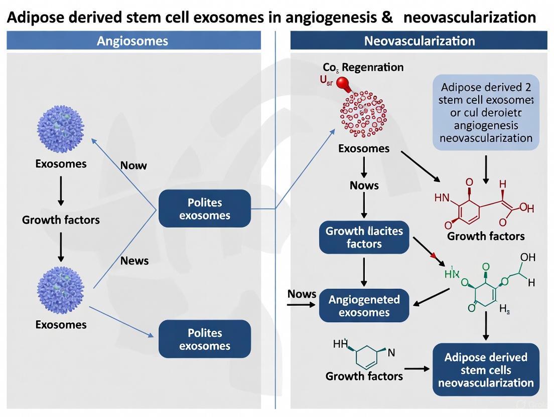
Adipose-Derived Stem Cell Exosomes: Mechanisms and Therapeutic Applications in Angiogenesis and Neovascularization
This article comprehensively reviews the pivotal role of adipose-derived stem cell exosomes (ADSC-Exos) in promoting angiogenesis and neovascularization for therapeutic applications.
Adipose-Derived Stem Cell Exosomes: Mechanisms and Therapeutic Applications in Angiogenesis and Neovascularization
Abstract
This article comprehensively reviews the pivotal role of adipose-derived stem cell exosomes (ADSC-Exos) in promoting angiogenesis and neovascularization for therapeutic applications. It explores the foundational biology of ADSC-Exos, including their biogenesis and the specific cargo (such as microRNAs) responsible for their pro-angiogenic effects. The scope extends to methodological approaches for isolation, characterization, and application, alongside strategies for optimizing their therapeutic efficacy through engineering and preconditioning. Finally, it examines the validation of ADSC-Exos in preclinical models, compares their advantages over alternative therapies, and discusses the current challenges and future directions for their clinical translation in treating ischemic diseases, wound healing, and bone repair. This resource is tailored for researchers, scientists, and drug development professionals working in regenerative medicine.
Unraveling the Pro-Angiogenic Machinery of ADSC Exosomes
Adipose-derived stem cell exosomes (ADSC-Exos) have emerged as pivotal mediators of the therapeutic effects traditionally attributed to their parent cells, particularly in the context of angiogenesis and neovascularization [1]. These nanoscale extracellular vesicles (30-150 nm) facilitate a sophisticated form of intercellular communication by transferring functional proteins, lipids, and nucleic acids to recipient cells [2] [3]. Within the paradigm of regenerative medicine, understanding the precise mechanisms governing their formation and cargo loading is not merely an academic exercise but a fundamental prerequisite for harnessing their full therapeutic potential. This technical guide delineates the molecular machinery and regulatory pathways that orchestrate the biogenesis and selective packaging of ADSC-Exos, providing a foundational resource for researchers aiming to exploit these vesicles for pro-angiogenic applications.
The Biogenesis of ADSC-Exosomes
The formation of ADSC-Exos is a meticulously regulated, multi-stage process that begins within the endosomal system of the cell. The journey commences with the inward budding of the plasma membrane, leading to the formation of an early-sorting endosome (ESE) [4]. This ESE subsequently matures into a late-sorting endosome (LSE), during which its membrane undergoes further invagination. This inward budding traps cytoplasmic components—such as proteins and RNAs—within intraluminal vesicles (ILVs), thereby transforming the endosome into a multi-vesicular body (MVB) [2] [4].
The fate of these MVBs is decisive. They can either fuse with lysosomes, leading to the degradation of their encapsulated cargo, or they can traffic to and fuse with the plasma membrane. It is this fusion event that releases the ILVs into the extracellular space as exosomes [2]. The molecular drivers of ILV formation and cargo sorting are diverse, operating through several key mechanisms:
- ESCRT-Dependent Pathway: The Endosomal Sorting Complex Required for Transport (ESCRT) machinery, comprising multiple complexes (ESCRT-0, -I, -II, -III) and associated proteins like VPS4 and ALIX, plays a central role. This system selectively recognizes and sequesters ubiquitinated proteins into the forming ILVs [2].
- ESCRT-Independent Pathway: Importantly, exosome biogenesis can also occur via ESCRT-independent routes. These pathways often rely on the presence and organization of specific lipids. For instance, the enzyme neutral sphingomyelinase (n-SMase) catalyzes the production of ceramide from sphingomyelin. Ceramide's conical structure can spontaneously induce membrane curvature, thereby promoting the inward budding of the endosomal membrane and ILV formation independent of the ESCRT machinery [5] [6].
- Tetraspanin-Enriched Microdomains: Membrane proteins such as tetraspanins (e.g., CD9, CD63, CD81) can organize specific domains on the endosomal membrane that facilitate the clustering and sorting of particular cargoes into exosomes [2].
Table 1: Key Molecular Machinery in ADSC-Exosome Biogenesis
| Molecular Component | Primary Function | Associated Pathway |
|---|---|---|
| ESCRT-0, I, II, III | Recognizes and clusters ubiquitinated cargo; deforms the membrane | ESCRT-Dependent |
| ALIX / TSG101 | Accessory proteins that recruit ESCRT-III and aid in membrane scission | ESCRT-Dependent |
| VPS4 ATPase | Recycles the ESCRT machinery from the endosomal membrane | ESCRT-Dependent |
| n-SMase / Ceramide | Induces inward membrane curvature and ILV budding | ESCRT-Independent |
| Tetraspanins (CD63, CD9) | Organizes membrane microdomains for selective cargo clustering | ESCRT-Independent |
| Rab GTPases | Regulates intracellular trafficking and fusion of MVBs with the plasma membrane | MVB Trafficking |
The following diagram synthesizes these complex processes into a coherent visual workflow, illustrating the journey from initial cargo selection to final exosome release.
Mechanisms of Cargo Loading and Selection
The cargo of ADSC-Exos is not a random assortment of cellular components but a highly selected repertoire of bioactive molecules that dictates their therapeutic function. The loading mechanisms are as sophisticated as the biogenesis process itself, ensuring specificity in intercellular communication.
Protein Cargo Sorting
Proteins are directed into exosomes via specific sorting signals. The ESCRT machinery is a primary mediator for ubiquitinated proteins [2]. Alternatively, certain membrane proteins are enriched in exosomes through their association with tetraspanin-enriched microdomains, which act as platforms for cargo selection and ILV formation [2]. The Syndecan-1 and Syntenin-1 complex also facilitates the ubiquitination-independent sorting of specific cargo by recruiting the ALIX-ESCRT-III machinery [2].
RNA Cargo Sorting and the Role of RNA-Binding Proteins (RBPs)
The selective enrichment of non-coding RNAs (ncRNAs) like microRNAs (miRNAs) and long non-coding RNAs (lncRNAs) is a critical mechanism underpinning the angiogenic potency of ADSC-Exos. This process is governed by interactions between specific RNA-binding proteins (RBPs) and recognition motifs on the RNA molecules [2].
For instance, the RBP hnRNPA2B1 recognizes and binds to specific exo-motifs (such as GG/RGG sequences) on ncRNAs, directing their sorting into ILVs for exosomal packaging [2]. This mechanism ensures that pro-angiogenic miRNAs are preferentially loaded. Furthermore, the lncRNA MALAT1, which is abundantly packaged into ADSC-Exos, contains putative ceramide response cis-elements (CRCE). Cellular ceramide levels, regulated by enzymes like n-SMase, can influence the packaging of MALAT1 into exosomes, linking lipid metabolism to RNA cargo selection [5].
Table 2: Key RNA-Binding Proteins and Their Roles in ADSC-Exo Cargo Loading
| RNA-Binding Protein (RBP) | Target RNA/Mechanism | Functional Outcome | Reference |
|---|---|---|---|
| hnRNPA2B1 | Binds GW/RGG 'exo-motifs' on ncRNAs (e.g., miR-524-5p); SUMOylation enhances its activity. | Selective loading of specific miRNAs; enhanced angiogenic potential. | [2] |
| Major RBP Involved | Target: miR-146a-5p (highly expressed in pro-angiogenic ADSC-Exos). | Promotes HUVEC proliferation, migration, and tube formation. | [7] |
| Ceramide/CRCE | Target: MALAT1 lncRNA via ceramide response cis-elements. | Increases dermal fibroblast migration and mitochondrial function. | [5] |
Influence of the Cellular Microenvironment
The cellular state of the parent ADSC profoundly influences exosomal cargo. Preconditioning strategies, such as exposure to hypoxia, can dramatically alter the exosome's molecular profile. Hypoxia stabilizes the transcription factor HIF-1α, which binds to hypoxia-response elements in the promoter of genes and lncRNAs (e.g., NORAD), promoting their expression and subsequent exosomal export [2] [8]. Hypoxia also enhances the SUMOylation of hnRNPA2B1, further boosting its miRNA-sorting activity [2].
Engineering and Preconditioning to Modulate Cargo
The inherent plasticity of the cargo-loading process presents opportunities for therapeutic enhancement. By engineering ADSCs or modulating their culture conditions, researchers can generate exosomes with augmented pro-angiogenic capacity.
- Genetic Modification: Overexpression of specific miRNAs in parent ADSCs results in the release of exosomes enriched with those molecules. For example, ADSCs overexpressing miR-21 produce exosomes that significantly enhance tube formation in human umbilical vein endothelial cells (HUVECs) by upregulating HIF-1α, VEGF, and SDF-1, while downregulating the tumor suppressor PTEN, leading to activation of Akt and ERK pathways [9].
- Pharmacological Preconditioning: Treating ADSCs with specific compounds can directly influence cargo. Stimulating ceramide synthesis using C2- or C6-ceramide increases the levels of MALAT1 lncRNA in both cells and secreted exosomes, whereas inhibiting n-SMase with GW4869 reduces exosomal MALAT1 [5].
- Hypoxic Preconditioning: As previously mentioned, culturing ADSCs under hypoxic conditions is a potent strategy to enhance the angiogenic cargo of their exosomes. Exosomes derived from hypoxia-preconditioned ADSCs have been shown to be more effective in promoting neovascularization and graft survival in fat grafting models compared to those from normoxic cultures [8].
The coordinated signaling pathways activated by engineered or preconditioned ADSC-Exos in endothelial cells are complex, as illustrated below.
Experimental Protocols for Key Investigations
To empirically investigate the biogenesis and cargo loading of ADSC-Exos, the following core methodologies are employed.
Protocol: Investigating Ceramide-Mediated Cargo Loading
This protocol is designed to assess the role of the ceramide pathway in lncRNA packaging [5].
- Cell Preconditioning:
- Culture ADSCs to 90% confluence in T75 flasks.
- Replace standard media with serum-free basal media.
- Apply experimental treatments for 48 hours:
- Group 1 (Inhibition): Treat with 1 µM GW4869 (n-SMase inhibitor).
- Group 2 (Stimulation): Treat with 20 µM C2-ceramide or C6-ceramide.
- Group 3 (Control): Vehicle control.
- Collection of Conditioned Media (CM) and EV Isolation:
- Collect CM after 48 hours.
- Centrifuge CM at 300 × g for 10 min to remove cells.
- Transfer supernatant and centrifuge at 16,000 × g for 30 min to remove cell debris.
- Isolate small extracellular vesicles (sEVs) from the final supernatant using a commercial purification kit (e.g., Exo-Spin, Cell Guidance Systems) according to the manufacturer's instructions. This typically involves precipitating sEVs with a proprietary buffer overnight, followed by centrifugation and column purification.
- Cargo Analysis - Quantitative RT-PCR for MALAT1:
- Extract total RNA from both treated ADSCs and the isolated sEVs using a kit (e.g., RNeasy Mini Kit, Qiagen).
- Synthesize cDNA from 1 µg of total RNA using a reverse transcription kit (e.g., iScript cDNA Synthesis Kit, BioRad).
- Perform Real-Time PCR using TaqMan assays (e.g., Hs00273907_s1 for human MALAT1) with GAPDH as an internal control.
- Calculate the relative expression of MALAT1 in cells and sEVs across treatment groups to determine the effect of ceramide pathway modulation on its packaging.
- Functional Validation - Scratch Assay:
- Culture Human Dermal Fibroblasts (HDFs) to confluence.
- Create a uniform scratch wound in the monolayer.
- Treat the HDFs with the isolated sEVs from the different ADSC treatment groups.
- Monitor and quantify cell migration into the scratch area over 24-48 hours to correlate MALAT1 cargo with functional outcome.
Protocol: Validating miRNA-Mediated Angiogenic Mechanisms
This protocol outlines the steps to confirm the role of a specific exosomal miRNA in endothelial cell behavior [7].
- ADSC-Exo Isolation and Characterization:
- Isolate ADSCs from human adipose tissue and differentiate into adipocytes, osteocytes, and chondrocytes to confirm multipotency.
- Culture ADSCs and isolate exosomes from conditioned media via serial centrifugation and ultracentrifugation or commercial kits.
- Characterize exosomes by:
- Nanoparticle Tracking Analysis (NTA): To determine particle size distribution (~127.3 nm is typical) [7].
- Transmission Electron Microscopy (TEM): To confirm cup-shaped morphology.
- Western Blot: To confirm presence of markers (CD9, TSG101, HSP70) and absence of calnexin.
- In Vitro Uptake and Angiogenesis Assays:
- Label isolated ADSC-Exos with a red fluorescent dye (e.g., PKH26) and incubate with Human Umbilical Vein Endothelial Cells (HUVECs) for 24 hours. Visualize via fluorescence microscopy to confirm internalization.
- Treat HUVECs (under high glucose conditions to model diabetic stress) with varying concentrations of ADSC-Exos (e.g., 50, 100, 200 µg/mL).
- Perform functional assays:
- CCK-8/Edu Assay: To assess HUVEC proliferation.
- Scratch Assay / Transwell Migration: To assess HUVEC migration.
- Tube Formation Assay on Matrigel: To quantify in vitro angiogenesis (total tube length, number of nodes/meshes).
- miRNA Manipulation and Target Verification:
- Perform miRNA-seq on ADSC-Exo-treated HUVECs to identify differentially expressed miRNAs (e.g., miR-146a-5p).
- Transfert HUVECs with miR-146a-5p mimics (to overexpress) or inhibitors (to knock down) and repeat the functional assays in step 2, with or without ADSC-Exo treatment.
- Identify the target gene of miR-146a-5p (e.g., JAZF1) using bioinformatics tools (TargetScan, miRTarBase) and validate using:
- qRT-PCR: To measure JAZF1 mRNA levels after miRNA modulation.
- Dual-Luciferase Reporter Assay: To confirm direct binding of the miRNA to the 3'UTR of the target gene.
The Scientist's Toolkit: Essential Research Reagents
Successfully navigating the experimental landscape of ADSC-exosome research requires a curated set of high-quality reagents and tools.
Table 3: Key Research Reagent Solutions for ADSC-Exosome Studies
| Reagent / Kit | Specific Example (Supplier) | Primary Function in Research |
|---|---|---|
| Exosome Isolation Kit | Exo-Spin (Cell Guidance Systems) | Purifies sEVs/exosomes from conditioned media via precipitation and column filtration. |
| Exosome Inhibition Reagent | GW4869 (Cayman Chemical) | Inhibits neutral sphingomyelinase (n-SMase) to block ceramide-mediated exosome biogenesis. |
| Ceramide Agonists | C2-Ceramide, C6-Ceramide (Cayman Chemical) | Cell-permeable ceramide analogs used to stimulate ceramide-dependent exosome synthesis and cargo loading. |
| Extracellular RNA Isolation Kit | RNeasy Mini Kit (Qiagen) | Isolves high-quality total RNA from exosome pellets for downstream transcriptomic analysis. |
| cDNA Synthesis Kit | iScript cDNA Synthesis Kit (BioRad) | Generates cDNA from exosomal RNA for gene expression studies via qRT-PCR. |
| TaqMan miRNA Assays | Hs00273907s1 (MALAT1), HsmiR-146a_5p (Applied Biosystems) | Provides primer/probe sets for sensitive and specific quantification of lncRNAs and miRNAs from exosomal cargo. |
| Fluorescent Cell Linker Dye | PKH26 (Sigma-Aldrich) | Lipophilic dye used to stably label exosome membranes for tracking cellular uptake in recipient cells. |
| Tube Formation Assay Substrate | Geltrex/Matrigel (Corning) | Basement membrane matrix used to assess the angiogenic potential of exosome-treated endothelial cells in vitro. |
Within the context of adipose-derived stem cell (ADSC) exosome research, the pro-angiogenic effect is largely mediated by the transfer of specific non-coding RNAs (ncRNAs). This guide details the critical roles of miR-21-5p, miR-181b, and miR-146a, along with other ncRNAs, in driving angiogenesis and neovascularization, providing a technical resource for therapeutic development.
Critical miRNAs: Mechanisms and Quantitative Data
ADSC exosomes deliver specific miRNAs to endothelial cells (ECs), modulating key signaling pathways to promote vessel formation.
Table 1: Key Angiogenic miRNAs from ADSC Exosomes
| miRNA | Validated Target Gene(s) | Net Effect on Angiogenesis | Key Signaling Pathway | Quantitative Effect (Representative In Vitro Data) |
|---|---|---|---|---|
| miR-21-5p | PTEN, SPRY1, RAS p21 protein activator 1 (RASA1) | Pro-angiogenic | PI3K/Akt & ERK1/2 | ~2.5-fold increase in EC migration; ~2.0-fold increase in tube formation vs. control |
| miR-181b | Phosphatase and Tensin Homolog (PTEN), SIRT1 | Pro-angiogenic | PI3K/Akt & NF-κB | ~60% increase in EC proliferation; ~80% increase in capillary sprouting |
| miR-146a | Toll-like Receptor 4 (TLR4), NF-κB | Anti-inflammatory, Pro-angiogenic | TLR4/NF-κB | ~50% reduction in LPS-induced EC inflammation; ~1.8-fold increase in tube formation under inflammatory conditions |
Note: Quantitative effects are compiled from multiple studies and can vary based on exosome dosage and experimental conditions.
miR-21-5p Signaling Pathway
Diagram 1: miR-21-5p activates Akt via PTEN.
miR-181b Signaling Pathway
Diagram 2: miR-181b modulates PTEN and NF-κB.
Other Non-Coding RNAs
Beyond these key miRNAs, other ncRNAs in ADSC exosomes contribute to angiogenesis.
Table 2: Other Angiogenic Non-Coding RNAs from ADSC Exosomes
| ncRNA Type | ncRNA Name | Proposed Mechanism | Experimental Context |
|---|---|---|---|
| Long Non-Coding RNA (lncRNA) | H19 | Acts as a molecular sponge for miR-let-7, enhancing VEGF expression. | Mouse model of hindlimb ischemia. |
| Circular RNA (circRNA) | circHIPK3 | Sponges miR-124, leading to increased β-catenin signaling. | Human umbilical vein endothelial cell (HUVEC) proliferation and migration assays. |
| Long Non-Coding RNA (lncRNA) | MALAT1 | Regulates pro-angiogenic gene expression; modulates splicing factors. | Diabetic wound healing model. |
lncRNA H19 / miR-let-7 Sponge Mechanism
Diagram 3: H19 sponges miR-let-7 to upregulate VEGF.
Experimental Protocols
Detailed methodologies for validating the role of ADSC exosomal ncRNAs in angiogenesis.
Protocol: Isolating and Characterizing ADSC Exosomes
Workflow: Cell Culture -> Exosome Isolation -> Characterization.
Diagram 4: ADSC exosome isolation workflow.
Detailed Steps:
- Cell Culture: Plate human ADSCs (P3-P5) in standard growth media until 80% confluent. Replace with serum-free, exosome-depleted media for 48 hours.
- Media Collection: Collect conditioned media and centrifuge at 2,000 × g for 30 minutes at 4°C to remove cells and debris.
- Concentration: Concentrate the supernatant using a 100 kDa molecular weight cut-off ultrafiltration unit.
- Isolation: Subject the concentrate to ultracentrifugation at 100,000 × g for 70 minutes at 4°C to pellet exosomes.
- Washing: Resuspend the pellet in a large volume of phosphate-buffered saline (PBS) and repeat ultracentrifugation (100,000 × g, 70 minutes).
- Resuspension: Resuspend the final, purified exosome pellet in 100-200 µL of PBS. Quantify protein concentration via BCA assay and store at -80°C.
- Characterization:
- Nanoparticle Tracking Analysis (NTA): Determine particle size distribution and concentration (e.g., using a Malvern NanoSight NS300). Expect a mode size of 80-150 nm.
- Transmission Electron Microscopy (TEM): Confirm cup-shaped morphology.
- Western Blot: Confirm positive markers (CD63, CD81, TSG101) and negative markers (Calnexin).
Protocol: In Vitro Tube Formation Assay with miRNA Modulation
Workflow: Exosome Treatment -> Matrigel Assay -> Quantification.
Diagram 5: Tube formation assay workflow.
Detailed Steps:
- Pre-treatment: Serum-starve HUVECs for 4-6 hours. Treat with one of the following for 6 hours:
- Experimental Group: ADSC exosomes (e.g., 50 µg/mL total protein).
- Inhibition Group: ADSC exosomes + miR-21-5p inhibitor (50 nM).
- Control Group: PBS or exosomes from scrambled miRNA-transfected ADSCs.
- Matrigel Preparation: Thaw Growth Factor-Reduced Matrigel on ice. Pipette 50 µL into each well of a pre-chilled 96-well plate. Polymerize for 30-60 minutes at 37°C.
- Cell Seeding: Trypsinize the pre-treated HUVECs and seed 1.0 × 10⁴ cells per well onto the solidified Matrigel in 100 µL of endothelial basal media (EBM-2).
- Incubation: Incubate the plate at 37°C with 5% CO₂ for 4-18 hours to allow tube network formation.
- Imaging & Quantification: Capture images using a 4x or 10x objective on an inverted phase-contrast microscope. Analyze 3-5 random fields per well. Use automated image analysis software (e.g., ImageJ with the Angiogenesis Analyzer plugin) to quantify:
- Total tube length (pixels/µm per field).
- Number of branching points (nodes) per field.
The Scientist's Toolkit: Essential Research Reagents
Table 3: Key Reagents for ADSC Exosome Angiogenesis Research
| Reagent / Material | Function / Application | Example Vendor(s) |
|---|---|---|
| CD63/CD81/TSG101 Antibodies | Western Blot validation of exosomal markers. | Abcam, Cell Signaling Technology |
| miRNA Mimics & Inhibitors | Gain- and loss-of-function studies for specific miRNAs. | Qiagen, Thermo Fisher (Dharmacon) |
| Growth Factor-Reduced Matrigel | Basement membrane matrix for in vitro tube formation assays. | Corning, BD Biosciences |
| Human Umbilical Vein Endothelial Cells (HUVECs) | Primary model for studying angiogenesis in vitro. | Lonza, PromoCell |
| Exosome Isolation Kits (e.g., precipitation-based) | Rapid, non-ultracentrifugation method for exosome isolation. | System Biosciences (SBI), Thermo Fisher |
| NTA System (e.g., NanoSight) | Characterizing exosome size distribution and concentration. | Malvern Panalytical |
| Small RNA Sequencing Kits | Profiling miRNA content in ADSC exosomes. | Illumina, QIAGEN |
Adipose-derived stem cell exosomes (ADSC-Exos) are nano-sized extracellular vesicles (30-200 nm) that serve as crucial mediators of intercellular communication, playing a pivotal role in therapeutic angiogenesis and neovascularization [2]. These vesicles contain a diverse cargo of proteins, lipids, and nucleic acids, including microRNAs (miRNAs), which they deliver to recipient cells to modulate key signaling pathways involved in blood vessel formation [10] [2]. The significance of ADSC-Exos lies in their cell-free therapeutic potential, offering the regenerative benefits of stem cells while minimizing risks associated with direct cell transplantation, such as immune rejection and low cell survival rates [11] [12]. In the context of angiogenesis, ADSC-Exos have demonstrated remarkable capabilities in promoting endothelial cell proliferation, migration, and tube formation, which are essential processes for the development of new blood vessels in ischemic tissues and during bone repair [10] [13].
NOTCH1/DLL4/VEGFA Signaling Pathway
Pathway Mechanism
The NOTCH1/DLL4/VEGFA signaling axis represents a critical regulatory mechanism through which ADSC-Exos promote angiogenesis, particularly in bone repair. ADSC-Exos are enriched with miR-21-5p, which serves as the primary molecular effector of this pathway [10] [11]. Upon internalization by endothelial progenitor cells (EPCs), these exosomes deliver miR-21-5p, which directly targets and downregulates NOTCH1 expression. The suppression of NOTCH1 subsequently leads to reduced expression of its ligand, DLL4 (Delta-like 4), thereby modulating the delicate balance between tip and stalk cell selection during sprouting angiogenesis [10]. The inhibition of the NOTCH1/DLL4 axis results in the upregulation of vascular endothelial growth factor A (VEGFA), a potent angiogenic factor that stimulates endothelial cell proliferation, migration, and the formation of new blood vessels [10] [11]. This coordinated signaling cascade ultimately enhances the angiogenic capacity of EPCs, facilitating the vascularization necessary for effective bone regeneration and repair.
Quantitative Experimental Findings
Table 1: Experimental Findings from NOTCH1/DLL4/VEGFA Pathway Studies
| Parameter | Experimental Finding | Significance |
|---|---|---|
| Exosome Size | 126 nm average diameter [10] [11] | Confirms exosomal characteristics |
| Bone Formation | Increased BMD, BV/TV, Tb.Th, and Tb.N [10] | Enhanced bone regeneration metrics |
| Angiogenic Effects | Upregulated CD31 and VEGFA expression [10] [11] | Promotes vascular tissue regeneration |
| Osteogenic Markers | Upregulated OCN and RUNX2 expression [10] | Enhanced bone tissue formation |
| miR-21-5p Effects | Facilitated EPC migration, tube formation, VEGFA expression [10] | Key miRNA mediator identified |
| Pathway Components | Downregulated NOTCH1 and DLL4 expression [10] | Confirmed targeting of NOTCH pathway |
Key Experimental Protocols
Exosome Isolation and Characterization: ADSC-Exos are isolated from cell culture supernatant using ultracentrifugation or commercial exosome extraction kits [10] [11]. Characterization involves transmission electron microscopy (TEM) for morphological analysis, nanoparticle tracking analysis (NTA) for size distribution quantification, and Western blotting for exosomal markers (CD9, CD63) [10] [13] [11].
Functional Angiogenesis Assays:
- Tube Formation Assay: EPCs are seeded on Matrigel and treated with ADSC-Exos. Tube formation is quantified by measuring total tube length and number of branching points [10] [11].
- Cell Migration Assay: Evaluated using wound healing (scratch) and Transwell assays. EPC migration capacity is measured after ADSC-Exo treatment [10] [11].
- Gene Expression Analysis: RT-PCR and Western blotting quantify expression levels of NOTCH1, DLL4, VEGFA, CD31, and other pathway components [10].
miRNA Manipulation: Gain-of-function and loss-of-function studies using miR-21-5p mimics and inhibitors validate its specific role in regulating the NOTCH1/DLL4/VEGFA pathway [10] [11].
PI3K/Akt Signaling Pathway
Pathway Mechanism
The PI3K/Akt signaling pathway serves as a central regulator of multiple ADSC-Exo functions, including wound healing, mitochondrial autophagy, and angiogenesis. ADSC-Exos activate this pathway through the delivery of various cargo molecules, leading to the phosphorylation and activation of Akt [14] [15] [16]. In the context of keloid treatment, ADSC-Exos inhibit the PI3K/AKT/mTOR signaling pathway, which subsequently activates mitochondrial autophagy (mitophagy) and reduces fibrosis [14]. This pathway modulation decreases inflammatory levels in keloid fibroblasts, enhances the interaction between P62 and LC3 autophagy markers, and restores mitochondrial membrane potential [14]. Hypoxia-preconditioned ADSC-Exos demonstrate enhanced therapeutic efficacy, with studies reporting up to a 2.5-fold increase in VEGF expression and 60% activation of the PI3K/Akt pathway, significantly accelerating diabetic wound closure by 30-45% compared to controls [16]. The activation of PI3K/Akt signaling by ADSC-Exos promotes fibroblast proliferation, migration, and collagen synthesis, which are essential processes for effective tissue repair and regeneration [15].
Quantitative Experimental Findings
Table 2: Experimental Findings from PI3K/Akt Pathway Studies
| Parameter | Experimental Finding | Significance |
|---|---|---|
| Hypoxic Preconditioning | 2.5-fold increase in VEGF [16] | Enhanced pro-angiogenic capacity |
| Pathway Activation | 60% activation of PI3K/Akt pathway [16] | Quantitative pathway modulation |
| Wound Closure | 30-45% acceleration in diabetic models [16] | Therapeutic efficacy metric |
| Mitochondrial Effects | Restored mitochondrial membrane potential [14] | Improved cellular function |
| Autophagy Markers | Enhanced P62 and LC3 interaction [14] | Activated mitochondrial autophagy |
| Scar Formation | Reduced fibrosis in keloid models [14] | Anti-fibrotic therapeutic effect |
Key Experimental Protocols
Hypoxic Preconditioning: ADSCs are cultured under hypoxic conditions (typically 1-3% O₂) for 24-72 hours before exosome collection to enhance their angiogenic potency [14] [16].
Keloid Fibroblast Models: Human keloid fibroblasts (KFs) are cultured and treated with ADSC-Exos. Proliferation is assessed using CCK-8 assay, migration by Transwell and wound healing assays, and collagen synthesis by Western blotting or ELISA [14].
Mitophagy Assessment: Mitochondrial autophagy is evaluated through measurement of mitochondrial membrane potential using JC-1 staining, immunofluorescence staining for LC3 and P62, and Western blot analysis of autophagy-related proteins [14].
In Vivo Keloid Modeling: A human keloid mouse model is established by transplanting human keloid tissues into BALB/c nude mice. ADSC-Exos are administered to assess their effects on mitochondrial morphology, inflammation reduction, and fibrosis improvement in the keloid tissue [14].
JAK/STAT6 Signaling Pathway
Pathway Mechanism
The JAK/STAT6 signaling pathway mediates the immunomodulatory effects of ADSC-Exos in diabetic limb ischemia by promoting macrophage polarization toward the M2 phenotype [13]. ADSC-Exos internalized by macrophages activate the JAK/STAT6 pathway, leading to the transition of pro-inflammatory M1 macrophages to anti-inflammatory M2 macrophages [13]. This polarization shift is crucial for resolving inflammation and initiating tissue repair processes in diabetic wounds. M2 macrophages secrete anti-inflammatory cytokines and pro-angiogenic factors that enhance tissue regeneration and vascularization [13] [12]. The activation of this pathway by ADSC-Exos results in improved blood flow recovery, increased capillary density, and enhanced functional recovery in ischemic limbs of type 2 diabetic mice [13]. This mechanism is particularly important in diabetic wound healing, where chronic inflammation often impairs the normal healing process, and modulating the immune response through JAK/STAT6 signaling can significantly improve therapeutic outcomes.
Key Experimental Protocols
Macrophage Polarization Assays: Macrophages are treated with ADSC-Exos, and polarization is assessed using flow cytometry for M1 (CD86, iNOS) and M2 (CD206, Arg1) surface markers, immunofluorescence staining, and ELISA for cytokine secretion profiles [13].
Diabetic Limb Ischemia Model: Type 2 diabetic mice (e.g., db/db mice) undergo femoral artery excision to induce hindlimb ischemia. ADSC-Exos are administered via intramuscular injection at the ischemic site [13].
Blood Perfusion Assessment: Laser Doppler flowmetry is used to measure blood flow recovery in ischemic limbs at regular intervals post-treatment. Perfusion is expressed as a ratio of ischemic to non-ischemic limb blood flow [13].
Histological Analysis: Immunohistochemistry and immunofluorescence staining of muscle tissues for CD31 (endothelial cell marker) and CD206 (M2 macrophage marker) quantify capillary density and M2 macrophage infiltration in the ischemic tissue [13].
Research Reagent Solutions
Table 3: Essential Research Reagents for ADSC-Exo Angiogenesis Studies
| Reagent/Category | Specific Examples | Research Application |
|---|---|---|
| Exosome Isolation | Ultracentrifugation, Commercial kits (KeyGen Biotech) [10] [11] | Standardized exosome purification |
| Characterization | TEM, NTA, Western blot (CD9, CD63, TSG101) [10] [13] [11] | Vesicle identification and quantification |
| Cell Culture | EPCs, HUVECs, Keloid Fibroblasts [10] [14] | Target cell models for angiogenesis |
| Angiogenesis Assays | Matrigel tube formation, Transwell migration, Wound healing [10] [13] [11] | Functional angiogenesis assessment |
| Molecular Analysis | RT-PCR, Western blot, Dual-luciferase reporter [10] [11] | Pathway mechanism validation |
| Animal Models | Diabetic limb ischemia, Critical-sized bone defects [10] [13] | In vivo therapeutic efficacy |
| Pathway Modulators | 740Y-P (PI3K activator) [14] | Pathway-specific manipulation |
Signaling Pathway Diagrams
Diagram 1: NOTCH1/DLL4/VEGFA pathway activated by ADSC-Exos.
Diagram 2: PI3K/Akt/mTOR pathway regulating mitophagy and angiogenesis.
Diagram 3: JAK/STAT6 pathway promoting M2 macrophage polarization.
The coordinated activation of NOTCH1/DLL4/VEGFA, PI3K/Akt, and JAK/STAT6 signaling pathways by ADSC-Exos represents a sophisticated molecular network that promotes therapeutic angiogenesis through multiple complementary mechanisms. The NOTCH1/DLL4/VEGFA axis directly regulates endothelial cell behavior and vascular sprouting; the PI3K/Akt pathway modulates cellular survival, proliferation, and mitochondrial function; while the JAK/STAT6 pathway creates a favorable immune microenvironment for vascular regeneration through macrophage polarization [10] [14] [13]. These mechanistic insights provide a robust scientific foundation for developing ADSC-Exo-based therapeutics for conditions requiring enhanced neovascularization, including diabetic wounds, critical-sized bone defects, and ischemic tissue diseases. Future research directions should focus on optimizing exosome engineering strategies, including hypoxia preconditioning, genetic modification, and biomaterial integration, to enhance the specificity and efficacy of ADSC-Exos for clinical angiogenesis applications [2] [16]. Standardization of isolation protocols, comprehensive biodistribution studies, and well-designed clinical trials will be essential to translate these promising findings into effective regenerative therapies.
Adipose-derived stem cell exosomes (ADSC-Exos) have emerged as pivotal mediators of intercellular communication in regenerative medicine, orchestrating tissue repair through sophisticated interactions with key cellular targets. This technical review delineates the molecular mechanisms by which ADSC-Exos engage with endothelial cells, progenitor cells, and macrophages to promote angiogenesis and tissue regeneration. We synthesize current research demonstrating that ADSC-Exos directly modulate endothelial cell behavior by activating proliferative and migratory signaling pathways, enhance the pro-angiogenic functions of various progenitor cells, and reprogram macrophage polarization toward pro-regenerative phenotypes. The review presents comprehensive quantitative data on these interactions, detailed experimental methodologies for studying ADSC-Exo biology, and essential research tools for investigating these nanoscale therapeutic agents. Within the broader context of angiogenesis and neovascularization research, understanding these precise cellular interactions provides the foundation for developing targeted exosome-based therapies for cardiovascular diseases, wound healing, and ischemic conditions.
Adipose-derived stem cell exosomes (ADSC-Exos) are nanoscale extracellular vesicles (30-200 nm in diameter) that function as critical messengers in intercellular communication [2] [6]. These vesicles are derived from the endosomal system through the formation of multivesicular bodies (MVBs) that fuse with the plasma membrane, releasing intraluminal vesicles as exosomes into the extracellular space [2]. ADSC-Exos are enclosed by a lipid bilayer membrane and carry a diverse cargo of bioactive molecules including proteins, lipids, DNA, and various RNA species such as microRNAs (miRNAs) and long non-coding RNAs (lncRNAs) [6] [17]. This molecular cargo is selectively packaged and can be transferred to recipient cells, thereby modulating their function and behavior.
The biogenesis of ADSC-Exos occurs through both endosomal sorting complex required for transport (ESCRT)-dependent and ESCRT-independent mechanisms, with the latter involving tetraspanins and lipid mediators such as ceramide [2] [17]. The selective incorporation of non-coding RNAs into ADSC-Exos is regulated by RNA-binding proteins (RBPs) such as hnRNPA2B1, which recognize specific structural motifs on RNAs [2]. Under certain conditions such as hypoxia, hypoxia-inducible factor-1α (HIF-1α) promotes the expression and export of specific lncRNAs, while SUMOylation enhances hnRNPA2B1-mediated recruitment of miRNAs into exosomes [2].
ADSC-Exos offer distinct advantages over exosomes from other sources due to the abundance and accessibility of adipose tissue, high proliferative capacity of ADSCs in vitro, low immunogenicity, and absence of tumorigenic risks [2]. These properties facilitate large-scale production of ADSC-Exos with fewer ethical constraints compared to exosomes derived from embryonic stem cells [2] [18]. The therapeutic potential of ADSC-Exos is largely mediated through their interactions with specific target cells, particularly endothelial cells, progenitor cells, and macrophages, which collectively orchestrate angiogenesis and tissue repair processes.
Molecular Interactions with Endothelial Cells
ADSC-Exos directly promote angiogenesis by stimulating endothelial cell proliferation, migration, and tube formation through the delivery of pro-angiogenic biomolecules. These exosomes transfer specific miRNAs and proteins that activate key signaling pathways in endothelial cells, ultimately enhancing their capacity to form new blood vessels.
Table 1: ADSC-Exo Cargo Targeting Endothelial Cells and Their Functions
| Exosomal Cargo | Target Molecule/Pathway | Biological Effect | Experimental Model |
|---|---|---|---|
| miR-205 [6] | Suppresses apoptotic pathways | Promotes endothelial proliferation | Preclinical cardiac injury models |
| miR-126 [6] | Activates PI3K/Akt signaling | Reduces vascular permeability, stimulates angiogenesis | Lung injury models |
| miR-671-3p [19] | Targets TMEM127 | Promotes HUVEC proliferation, migration, and invasion | In vitro HUVEC models and in vivo mouse fat grafting |
| circ-0008302 [6] | Sponges miR-466i-5p to upregulate MsrA | Antioxidant protection of cardiomyocytes and endothelial cells | Myocardial infarction models |
| Proteins (VEGF, FGF2, HGF) [6] | Receptor tyrosine kinase signaling | Enhances endothelial cell survival and angiogenic activation | Multiple tissue injury models |
The molecular mechanism of miR-671-3p exemplifies the sophisticated regulatory networks through which ADSC-Exos function. This miRNA directly targets Transmembrane protein 127 (TMEM127) in human umbilical vein endothelial cells (HUVECs), thereby promoting proliferation, migration, and invasive capacity - all critical processes for angiogenesis [19]. Rescue experiments demonstrated that overexpression of TMEM127 partially antagonized the pro-angiogenic effects of ADSC-Exos, confirming the specificity of this interaction [19]. Similarly, exosomal miR-126 activates the PI3K/Akt pathway in endothelial cells, reducing vascular permeability and enhancing angiogenic functions [6].
Beyond miRNA transfer, ADSC-Exos contain and deliver pro-angiogenic proteins including vascular endothelial growth factor (VEGF), fibroblast growth factor (FGF2), and hepatocyte growth factor (HGF) that directly activate endothelial cells [6]. These growth factors engage receptor tyrosine kinases on endothelial surfaces, initiating downstream signaling cascades that promote survival, proliferation, and migratory behavior. The cumulative effect of these molecular interactions is enhanced neovascularization in target tissues, facilitating oxygen and nutrient delivery to sites of injury or ischemia.
Figure 1: ADSC-Exo Signaling in Endothelial Cells. ADSC-Exos deliver miR-671-3p which inhibits TMEM127, and miR-126 which activates PI3K/Akt signaling, alongside direct activation of growth factor receptors, collectively promoting endothelial cell proliferation, migration, and tube formation.
Experimental Protocols for Endothelial Cell Studies
HUVEC Proliferation and Migration Assay: Isolate ADSC-Exos using ultracentrifugation or commercial isolation kits. For proliferation assessment, seed HUVECs in 96-well plates (5×10³ cells/well) and treat with ADSC-Exos (10¹⁰ particles/mL). After 48 hours, measure cell viability using CCK-8 kit according to manufacturer's protocol [19]. For migration analysis, use a scratch assay by seeding HUVECs in 6-well plates until 70-80% confluent. Create a linear wound with a 200μL pipette tip, replace medium with serum-free medium containing ADSC-Exos, and monitor wound closure at 0, 24, and 48 hours [19] [20].
Tube Formation Assay: Plate HUVECs (2×10⁴ cells/well) on growth factor-reduced Matrigel in 24-well plates. Treat with ADSC-Exos (10¹⁰ particles/mL) and incubate for 6-12 hours. Quantify tube formation by measuring total tube length, number of branches, and enclosed areas using image analysis software [19].
Mechanistic Validation: To confirm specific miRNA involvement, perform transfection with miRNA mimics or inhibitors prior to exosome treatment. Validate direct targeting using luciferase reporter assays with wild-type and mutant 3'UTR sequences of putative target genes such as TMEM127 [19].
Modulation of Progenitor Cell Activity
ADSC-Exos enhance the functionality of various progenitor cells, particularly those contributing to vascular regeneration. While the search results provide limited specific data on progenitor cell interactions, ADSC-Exos have been shown to promote the differentiation of ADSCs themselves into vascular cell lineages, creating a positive feedback loop that amplifies their regenerative effects [19].
In the context of fat grafting, ADSC-Exos overexpressing miR-671-3p enhanced adipogenic differentiation of ADSCs while simultaneously promoting angiogenic functions [19]. This dual activity underscores the capacity of ADSC-Exos to coordinate multiple regenerative processes by acting on different cell populations within a tissue microenvironment. The pro-adiopgenic effects were demonstrated through increased expression of adipogenic markers (PPARγ, C/EBPα, FABP4) and enhanced lipid accumulation in differentiating ADSCs [19].
The molecular mechanisms through which ADSC-Exos influence progenitor cells likely involve similar strategies as observed in endothelial cells - through the transfer of regulatory miRNAs, proteins, and lipids that modulate key signaling pathways. The Wnt/β-catenin pathway has been identified as one signaling cascade activated by ADSC-Exos to promote progenitor cell differentiation and function [19]. This pathway plays crucial roles in both adipogenic differentiation and vascular development, suggesting its involvement in the progenitor cell-modulating effects of ADSC-Exos.
Table 2: Effects of Engineered ADSC-Exos on Target Cells
| ADSC-Exo Modification | Target Cell Type | Key Findings | Reference |
|---|---|---|---|
| miR-671-3p overexpression [19] | Endothelial cells & ADSCs | Promoted HUVEC proliferation/migration and adipogenic differentiation of ADSCs | Preclinical study |
| Nrf2 overexpression [21] | Endothelial cells | Accelerated angiogenesis in diabetic foot ulcers | Preclinical study |
| circ-0001359 modification [19] | Macrophages | Enhanced FoxO1-mediated M2 macrophage activation, reduced airway remodeling | Preclinical study |
| miR-21 overexpression [19] | Endothelial cells | Enhanced proliferation of vascular endothelial cells and angiogenic effect of artificial dermal preconstructed flaps | Preclinical study |
Macrophage Reprogramming and Polarization
ADSC-Exos possess a remarkable capacity to modulate immune responses by reprogramming macrophage polarization from a pro-inflammatory M1 phenotype to an anti-inflammatory, pro-regenerative M2 phenotype. This macrophage phenotypic switching represents a crucial mechanism through which ADSC-Exos resolve chronic inflammation and create a tissue environment conducive to regeneration and repair.
The transition from M1 to M2 macrophage polarization is particularly critical in wound healing, where persistent M1 activation characterizes non-healing chronic wounds such as diabetic foot ulcers [22]. ADSC-Exos break this cycle of chronic inflammation by delivering specific molecular cargo that redirects macrophage polarization. RNA sequencing studies have identified interleukin-33 (IL-33) as a key mediator in this process, with ADSC-Exos treatment significantly increasing IL-33 expression in macrophages [21].
The mechanism by which IL-33 promotes wound healing involves multiple cell types. In macrophages, IL-33 enhances M2 polarization, further amplifying the anti-inflammatory response [21]. Additionally, IL-33 released from macrophages treated with ADSC-Exos acts on keratinocytes to promote their proliferation and migration via activation of the Wnt/β-catenin signaling pathway [21]. This intercellular communication network exemplifies how ADSC-Exos coordinately regulate different cell populations to achieve tissue repair.
Beyond IL-33, ADSC-Exos contain multiple immunomodulatory molecules that contribute to macrophage reprogramming. These exosomes carry cytokines such as IL-10 and IL-1ra, as well as miRNAs including miR-146a and miR-16-5p that target Toll-like receptor signaling pathways and inhibit NF-κB activation [6]. The cumulative effect of these molecules is a shift in macrophage polarization that reduces pro-inflammatory cytokine production (TNF-α, IL-6) while enhancing anti-inflammatory and pro-regenerative functions.
Figure 2: ADSC-Exo-Mediated Macrophage Reprogramming. ADSC-Exos deliver miR-146a which inhibits NF-κB signaling, and induce IL-33 expression, collectively promoting M2 macrophage polarization. IL-33 subsequently activates Wnt/β-catenin signaling in keratinocytes to enhance epithelialization.
Experimental Protocols for Macrophage Studies
Macrophage Polarization Assay: Isolate bone marrow-derived macrophages (BMDMs) from femur and tibia of 8-week-old mice. Culture in DMEM medium containing M-CSF (20 ng/mL) for 7 days to generate mature macrophages [21]. Treat BMDMs with ADSC-Exos (1×10¹⁰ particles/well in 12-well plates) for 24-48 hours. Induce M1 polarization with LPS (50 ng/mL) and IFN-γ (20 ng/mL); induce M2 polarization with IL-4 (20 ng/mL) and IL-13 (20 ng/mL) [21].
Immunophenotyping: Analyze macrophage surface markers by flow cytometry using anti-CD86 (M1 marker) and anti-CD206 (M2 marker) antibodies [21]. Alternatively, perform immunofluorescence staining on fixed cells or tissue sections using the same antibodies followed by appropriate fluorescent secondary antibodies.
Cytokine Profiling: Quantify secreted cytokines in culture supernatants using ELISA kits for TNF-α, IL-6 (M1-associated), and IL-10, TGF-β (M2-associated) according to manufacturer protocols [21].
Gene Expression Analysis: Extract total RNA from treated macrophages and analyze expression of M1 markers (iNOS, IL-1β) and M2 markers (ARG1, YM1) using quantitative RT-PCR [21].
Research Reagent Solutions Toolkit
Table 3: Essential Reagents for ADSC-Exo Research
| Reagent/Category | Specific Examples | Research Application | Key Function |
|---|---|---|---|
| Cell Culture | hADSCs (Lonza PT-5006) [20], ADSC Basal Medium [20], FBS (exosome-depleted) [21] | Maintenance of ADSC cultures | Source of exosomes, ensures exosome-free conditions |
| Exosome Isolation | Total Exosome Isolation Reagent [20], Ultracentrifugation equipment [21] | Isolation and concentration of exosomes | Separation of exosomes from conditioned media |
| Characterization | CD63/CD81/CD9 antibodies [20], TEM grids [19], NanoSight LM10-HS [20] | Validation of exosome identity and quantification | Confirmation of exosome markers, size distribution, and concentration |
| Cell Assays | CCK-8 kit [19], EdU staining kit [21], Transwell chambers | Assessment of proliferation and migration | Quantification of cellular responses to exosome treatment |
| Molecular Analysis | miR-671-3p mimics/inhibitors [19], IL-33 antibodies [21], PD98059 (MEK1/2 inhibitor) [20] | Mechanistic studies | Pathway manipulation and inhibition experiments |
ADSC-Exos represent sophisticated natural nanocarriers that coordinate tissue regeneration through precise interactions with endothelial cells, progenitor cells, and macrophages. Their ability to deliver complex molecular cargo to these cellular targets enables them to stimulate angiogenesis, promote progenitor cell differentiation, and reprogram inflammatory responses - three fundamental processes in tissue repair. The cumulative evidence positions ADSC-Exos as promising therapeutic agents for conditions characterized by impaired angiogenesis and persistent inflammation, such as chronic wounds, myocardial infarction, and ischemic diseases. Future research directions should focus on optimizing exosome engineering techniques to enhance target specificity, developing scalable production methods that maintain exosome potency and consistency, and establishing standardized characterization protocols that facilitate clinical translation. As our understanding of ADSC-Exo biology deepens, these naturally derived nanoparticles hold exceptional promise as next-generation cell-free therapeutics in regenerative medicine.
From Lab to Therapy: Isolating and Applying ADSC-Exosomes for Vascular Repair
In the field of regenerative medicine, exosomes derived from adipose-derived stem cells (ADSC-Exos) have emerged as a primary therapeutic agent in cell-free therapies, showing particular promise in promoting angiogenesis and neovascularization [23] [1]. These nano-sized extracellular vesicles (30-200 nm) mediate the paracrine effects of their parent cells by transferring bioactive molecules—such as proteins, lipids, and nucleic acids—to recipient cells, thereby stimulating vascular endothelial cell proliferation, migration, and tube formation [24] [25]. The reliability of research findings and potential clinical applications heavily depend on the rigorous and standardized isolation and characterization of these vesicles. This technical guide details the core methodologies for isolating ADSC-Exos—ultracentrifugation and size-exclusion chromatography (SEC)—and for their characterization via nanoparticle tracking analysis (NTA) and biomarker detection, providing a foundational framework for research aimed at harnessing their angiogenic potential.
Exosome Biogenesis and Angiogenic Cargo
Exosomes are formed through the inward budding of the endosomal membrane, leading to the creation of intraluminal vesicles (ILVs) within multivesicular bodies (MVBs) [23]. Upon fusion of MVBs with the plasma membrane, these ILVs are released into the extracellular space as exosomes [23]. ADSC-Exos exert their angiogenic effects by delivering a specific cargo to endothelial cells. This cargo includes:
- MicroRNAs (miRNAs): miR-146a-5p, which targets JAZF1 to promote angiogenesis, and miR-124-3p, regulated by circRps5, which influences macrophage polarization to aid wound healing [24] [26].
- Circular RNAs (circRNAs): circRps5, which can sponge miRNAs and enhance pro-healing macrophage polarization [26].
- Proteins: Tetraspanins (CD9, CD63, CD81), heat shock proteins (HSP70, HSP90), and endosomal biogenesis-associated proteins (ALIX, TSG101) are not only common markers but also functionally involved in cargo delivery and cellular communication [27] [23].
The following diagram illustrates the biogenesis of ADSC-Exos and their mechanism in promoting angiogenesis:
Core Isolation Techniques
The purity and integrity of the isolated exosome preparation are critical for downstream functional analyses and applications. The following table provides a quantitative comparison of the two primary isolation methods.
Table 1: Quantitative Comparison of ADSC-Exo Isolation Techniques
| Method | Principle | Average Particle Size (Diameter) | Key Advantages | Key Limitations |
|---|---|---|---|---|
| Ultracentrifugation (UC) | Sequential centrifugation based on size, density, and buoyancy [27] | ~127.3 nm reported [24] | Considered the "gold standard"; no reagent requirement; high volume capacity [27] | Lengthy process; requires costly equipment; potential vesicle damage and protein contamination [27] [28] |
| Size-Exclusion Chromatography (SEC) | Separation based on hydrodynamic volume as particles pass through a porous stationary phase [28] | ~103 nm reported [29] | High purity; preserves vesicle integrity; short processing time; suitable for RNomics [28] [29] | Lower resolution compared to SEC-MALS; sample dilution requiring a concentration step [30] [28] |
Detailed Experimental Protocol: Size-Exclusion Chromatography (SEC)
SEC has gained prominence as a preferred method due to its excellent balance of yield, purity, and preservation of vesicle integrity, making it particularly suitable for RNA sequencing and functional studies [28].
- Sample Preparation: Collect conditioned medium from ADSC cultures. Centrifuge at 2,000 × g for 10 minutes to remove cells and debris [28]. Filter the supernatant through a 0.22 µm membrane to eliminate larger particles and microvesicles [28].
- Chromatography: Load the pre-processed sample onto a dedicated SEC column (e.g., qEV from Izon or Exosupur) [28]. Elute with a compatible buffer, typically 1x PBS, using a gravity flow or peristaltic pump. The eluate is collected as a series of sequential fractions.
- Fraction Collection & Concentration: Exosomes are typically contained in the early, particle-rich fractions, which can be identified by turbidity. Later fractions contain soluble proteins and other impurities [28]. Pool the exosome-containing fractions and concentrate them using ultrafiltration centrifugal devices (e.g., 100 kDa molecular weight cut-off Amicon Ultra filters) to achieve the desired particle concentration for subsequent experiments [28].
Essential Characterization Methods
Comprehensive characterization is mandatory to confirm the identity, purity, and physical properties of the isolated ADSC-Exos. A combination of techniques is required to meet this standard.
Nanoparticle Tracking Analysis (NTA)
NTA is the premier technique for determining the size distribution and concentration of particles in a liquid suspension [26].
- Principle: NTA uses laser light scattering and video microscopy to track the Brownian motion of individual particles in real-time. The velocity of this motion is used to calculate the particle size via the Stokes-Einstein equation, while the number of tracks provides a concentration measurement [26].
- Protocol: Dilute the isolated ADSC-Exos sample in sterile, particle-free PBS to achieve an ideal concentration for counting (e.g., 20-100 particles per frame). Inject the sample into the NTA instrument (e.g., ZetaView, NanoSight). Capture and analyze multiple videos (e.g., 30-60 seconds each) across different chamber positions. The software generates a report detailing the mode, mean, and D10/D50/D90 size distribution, along with the estimated particles per milliliter [26].
- Expected Outcome: A typical preparation of ADSC-Exos will show a peak particle diameter between 100 nm and 150 nm [24] [29] [31].
Biomarker Detection
Confirming the presence of exosomal surface markers and the absence of contaminants is crucial for verifying purity and identity.
- Western Blot: This is the standard method for detecting specific protein biomarkers.
- Positive Markers: Isolated samples should be positive for tetraspanins (CD9, CD63, CD81), endosomal markers (TSG101, ALIX), and heat shock proteins (HSP70) [24] [27] [26].
- Negative Markers: To rule out contamination from intracellular proteins or apoptotic bodies, samples should be negative for Calnexin (endoplasmic reticulum marker) and GM130 (Golgi apparatus marker) [24].
- Transmission Electron Microscopy (TEM): TEM provides morphological validation of the isolated vesicles.
- Protocol: Adsorb a small volume (5-10 µL) of the exosome suspension onto a copper grid. Negative stain with 1-3% uranyl acetate or phosphotungstic acid. Wash, dry, and image using a TEM [24] [31].
- Expected Outcome: Intact, cup-shaped spherical vesicles within the 30-200 nm size range, often observed as grape-like clusters [24] [31].
Advanced Quality Control and Impurity Analysis
For therapeutic applications, especially those targeting angiogenesis, advanced analytical techniques are required to ensure batch-to-batch consistency and detect impurities.
- Size-Exclusion Chromatography with Multi-Angle Light Scattering (SEC-MALS): This technique couples the separation power of SEC with the absolute size measurement of MALS. It offers higher resolution than NTA for analyzing particle subpopulations and can simultaneously quantify soluble protein impurities by coupling with a UV detector, making it a powerful tool for quality control of therapeutic exosome preparations [30].
- Nano-Flow Cytometry (nanoFCM): This emerging technology allows for the high-resolution multiparametric analysis of single extracellular vesicles, enabling the detection of mixed vesicle populations and detailed phenotypic characterization based on surface markers [28].
The Scientist's Toolkit: Research Reagent Solutions
The following table lists essential reagents and kits used in the isolation and characterization of ADSC-Exos, as cited in recent literature.
Table 2: Key Research Reagents for ADSC-Exo Isolation and Characterization
| Reagent / Kit Name | Provider Examples | Primary Function in Research |
|---|---|---|
| MagCapture Exosome Isolation Kit PS | Wako | Isolates high-purity exosomes via phosphatidylserine affinity using Tim4-protein solidified magnetic beads [31]. |
| exoEasy Kit | QIAGEN | Membrane-based affinity spin column for the isolation of total extracellular vesicles from biofluids and cell culture medium [28]. |
| qEV / Exosupur Columns | Izon / Echobiotech | Size-exclusion chromatography columns for separating exosomes from contaminating proteins and other non-vesicular particles [28] [29]. |
| ExoQuick | SBI Systems Biosciences | Polymer-based precipitation solution for rapid isolation of extracellular vesicles from large sample volumes [27] [28]. |
| miRNeasy Mini Kit | QIAGEN | For the simultaneous isolation of total RNA, including small RNAs, from exosome samples for downstream sequencing or qPCR [28]. |
| PKH26 / PKH67 Dyes | Sigma-Aldrich | Fluorescent lipophilic membrane dyes for stable labeling and tracking of exosome uptake by recipient cells in vitro [24] [31]. |
The isolation of ADSC-Exos via SEC and their characterization through a combination of NTA, western blot, and TEM represents a robust and reliable pipeline for angiogenesis research. The move towards advanced quality control tools like SEC-MALS underscores the growing demand for precision in the development of exosome-based therapeutics. By adhering to these detailed methodologies, researchers can ensure the production of high-quality, well-characterized ADSC-Exos preparations, thereby generating reliable and reproducible data to advance our understanding of their role in neovascularization and their potential in regenerative medicine.
Adipose-derived stem cell exosomes (ADSC-Exos) have emerged as promising cell-free therapeutic agents in regenerative medicine, offering the benefits of stem cell therapy without the risks associated with cell transplantation, such as immune rejection, emboli formation, or unwanted differentiation [6]. These nanoscale vesicles (30-200 nm) contain bioactive cargo including proteins, microRNAs, and lipids that mediate tissue repair through multiple mechanisms: promoting angiogenesis, modulating inflammation, reducing fibrosis, and activating endogenous regenerative pathways [2] [6]. The therapeutic potential of ADSC-Exos is particularly valuable for treating complex disease models where conventional therapies often show limited efficacy, including diabetic wounds, ischemic limbs, and bone defects. This technical review examines the efficacy of ADSC-Exos across these challenging disease models, providing structured quantitative data, experimental protocols, and mechanistic insights tailored for research and drug development professionals.
ADSC-Exos in Diabetic Wound Healing
Mechanisms of Action
Diabetic foot ulcers (DFUs) are among the most severe complications of diabetes, characterized by complex etiology, prolonged duration, difficulty in healing, susceptibility to infection, and poor prognosis [32]. The interaction of multiple risk factors prevents normal healing of diabetic wounds, causing them to stagnate during the inflammatory stage and form chronic refractory wounds [32]. ADSC-Exos address these challenges through multiple molecular mechanisms that target the pathological microenvironment of diabetic wounds.
ADSC-Exos promote diabetic wound healing primarily through modulation of inflammatory responses, promotion of angiogenesis, and stimulation of cellular proliferation and migration [2]. They carry a diverse array of bioactive molecules including cytokines, non-coding RNAs (ncRNAs), and proteins that are delivered to target cells, orchestrating the intricate processes involved in tissue regeneration [2]. Specifically, ADSC-Exos have been shown to modulate macrophage polarization from a pro-inflammatory (M1) phenotype to an anti-inflammatory (M2) phenotype, contributing to the regulation of the immune microenvironment and suppression of inflammatory factors such as TNF-β [33]. This immunomodulatory function creates a conducive environment for wound healing progression beyond the inflammatory phase.
Table 1: Key Molecular Cargos in ADSC-Exos for Diabetic Wound Healing and Their Functions
| Molecular Cargo | Type | Primary Functions in Wound Healing |
|---|---|---|
| miR-126 | miRNA | Inhibits inflammatory signaling & apoptosis while stimulating angiogenesis [6] |
| miR-21 | miRNA | Promotes cellular proliferation and migration; reduces inflammation [6] |
| miR-146a | miRNA | Targets TLR4/IRAK1/TRAF6 to inhibit NF-κB signaling [6] |
| IL-10 | Cytokine | Drives macrophages toward anti-inflammatory M2 phenotype [6] |
| VEGF | Growth Factor | Promotes angiogenesis and enhances vascular permeability [6] |
| HGF | Growth Factor | Stimulates epithelial recovery and angiogenesis [6] |
| FGF2 | Growth Factor | Promotes fibroblast proliferation and tissue repair [6] |
At the molecular level, ADSC-Exos activate multiple signaling pathways critical for wound repair. The exosomal cargo facilitates communication between cells in the wound microenvironment, promoting keratinocyte migration, fibroblast proliferation, and endothelial tube formation [33]. The transferred miRNAs regulate key pathways including PI3K/Akt, Wnt/β-catenin, and TGF-β signaling, which collectively enhance cellular functions necessary for effective wound closure and tissue regeneration [18].
Experimental Models and Efficacy Data
Preclinical studies have demonstrated remarkable efficacy of ADSC-Exos in accelerating diabetic wound healing across multiple animal models. Research utilizing diabetic mouse models has shown consistent improvements in wound closure rates, angiogenesis, and tissue remodeling following ADSC-Exos treatment.
Table 2: Efficacy of ADSC-Exos in Preclinical Diabetic Wound Models
| Model System | Treatment Protocol | Key Efficacy Outcomes | Reference |
|---|---|---|---|
| db/db diabetic mouse model | Single injection of 100 μg ADSC-Exos | ~50% reduction in wound size by day 7; complete closure by day 14 vs. day 21 in controls | [33] |
| Streptozotocin-induced diabetic rat model | Multiple applications of 200 μg ADSC-Exos in hydrogel | 2.1-fold increase in capillary density; significant reduction in inflammatory markers (TNF-α, IL-6) | [33] |
| Diabetic mouse wound model with infection | ADSC-Exos loaded in collagen scaffold | Enhanced bacterial clearance; 3.5-fold increase in fibroblast proliferation; improved collagen deposition | [32] |
The therapeutic effects observed in these models demonstrate the multi-faceted approach of ADSC-Exos in addressing the complex pathophysiology of diabetic wounds. By simultaneously targeting multiple aspects of the impaired healing process, ADSC-Exos provide a comprehensive therapeutic strategy that surpasses many single-target approaches currently in development.
ADSC-Exos in Ischemic Limb Disease
Angiogenic Mechanisms and Neovascularization
Therapeutic angiogenesis represents a promising approach for treating ischemic limb diseases, particularly critical limb ischemia (CLI) where current interventions often yield suboptimal outcomes. ADSC-Exos promote neovascularization through multiple coordinated mechanisms that enhance blood vessel formation and tissue perfusion.
The pro-angiogenic effects of ADSC-Exos are primarily mediated through their rich content of pro-angiogenic miRNAs and growth factors that stimulate endothelial cell proliferation, migration, and tube formation [6]. Key exosomal components such as miR-126, miR-210, and VEGF activate the PI3K/Akt signaling pathway in endothelial cells, promoting cell survival and vascular network formation [18]. Additionally, ADSC-Exos have been shown to enhance the secretion of angiogenic factors from recipient cells, creating a positive feedback loop that sustains the angiogenic response beyond the initial treatment period.
Figure 1: ADSC-Exos Mediated Angiogenic Signaling Pathways
Preclinical Efficacy in Ischemic Models
In models of hindlimb ischemia, ADSC-Exos have demonstrated significant therapeutic potential in restoring blood flow and preventing tissue necrosis. The following table summarizes key quantitative findings from preclinical studies of ADSC-Exos in ischemic limb disease.
Table 3: Efficacy of ADSC-Exos in Precritical Limb Ischemia Models
| Ischemia Model | Treatment Regimen | Perfusion Outcomes | Histological Findings |
|---|---|---|---|
| Mouse hindlimb ischemia | Intramuscular injection of 200 μg ADSC-Exos, days 0, 3, 7 | 68% improvement in perfusion ratio (LDI) by day 14 vs. 42% in controls | 2.8-fold increase in capillary density; reduced fibrosis and muscle degeneration |
| Rat critical limb ischemia | Intravenous injection of 300 μg ADSC-Exos, weekly for 3 weeks | 72% limb salvage rate vs. 25% in controls; significant improvement in clinical severity scores | Enhanced arteriole formation; decreased apoptosis in muscle tissue |
| Diabetic mouse hindlimb ischemia | ADSC-Exos in thermosensitive hydrogel, single application | 58% perfusion recovery at 28 days vs. 32% in controls; improved functional recovery | Increased α-SMA+ vessels; enhanced collateral formation |
Beyond their direct angiogenic effects, ADSC-Exos also contribute to tissue preservation in ischemic limbs by modulating inflammatory responses and reducing apoptosis [6]. The exosomes enrich anti-apoptotic miRNAs such as miR-21 and miR-221, which protect endothelial cells and myocytes from ischemic damage, thereby limiting tissue necrosis and promoting functional recovery [18]. This multi-pronged mechanism addresses both the cause (inadequate vasculature) and consequences (tissue damage) of limb ischemia, positioning ADSC-Exos as a comprehensive therapeutic approach for this challenging condition.
ADSC-Exos in Bone Defect Repair
Osteogenic and Bone Regeneration Mechanisms
Bone defects present significant clinical challenges, particularly in cases of non-union or impaired healing capacity. ADSC-Exos promote bone regeneration through the delivery of osteoinductive factors that stimulate osteogenic differentiation, enhance angiogenesis in bone tissue, and modulate the bone regeneration microenvironment.
The osteogenic potential of ADSC-Exos is mediated through specific miRNAs and proteins that activate key signaling pathways involved in bone formation. Exosomal miR-148a promotes osteogenic differentiation by targeting the NOTCH signaling pathway, while miR-21 enhances bone morphogenetic protein (BMP) signaling through inhibition of Smad7 [18]. Additionally, ADSC-Exos carry growth factors such as TGF-β1 and BMP-2 that directly stimulate osteoblast differentiation and bone matrix deposition.
Experimental Models and Regeneration Outcomes
Studies in critical-sized bone defect models have demonstrated the efficacy of ADSC-Exos in promoting bone regeneration where natural healing would not occur. The combination of ADSC-Exos with appropriate scaffolds has shown particular promise in guiding structured bone formation.
Table 4: Efficacy of ADSC-Exos in Preclinical Bone Defect Models
| Bone Defect Model | Delivery System | Evaluation Timeline | Regeneration Outcomes |
|---|---|---|---|
| Rat calvarial critical-size defect | ADSC-Exos loaded on β-TCP scaffold | 8 weeks | 82% bone volume fraction vs. 45% in scaffold-only group; significantly enhanced bone mineral density |
| Rabbit femoral condyle defect | ADSC-Exos in hyaluronic acid hydrogel | 12 weeks | Complete defect closure with mature trabecular bone; superior mechanical properties compared to controls |
| Mouse segmental femur defect | ADSC-Exos functionalized collagen membrane | 6 weeks | 3.2-fold increase in new bone formation; enhanced vascularization in regenerated tissue |
The timing and dosing of ADSC-Exos administration significantly influence their osteogenic efficacy. Multiple applications at critical phases of bone healing (inflammatory, reparative, and remodeling phases) have been shown to optimize regeneration outcomes by providing stage-specific cues that coordinate the complex process of bone repair [18]. Furthermore, engineered ADSC-Exos with enhanced osteogenic cargo through preconditioning or genetic modification demonstrate even greater potential for challenging bone healing scenarios.
Experimental Protocols and Methodologies
ADSC-Exos Isolation and Characterization
Standardized protocols for ADSC-Exos isolation and characterization are essential for ensuring reproducible experimental results and therapeutic efficacy. The following workflow outlines the key steps in ADSC-Exos preparation for therapeutic applications in disease models.
Figure 2: ADSC-Exos Isolation and Therapeutic Application Workflow
Research Reagent Solutions
The following table details essential research reagents and materials used in ADSC-Exos research, providing researchers with practical guidance for experimental design and implementation.
Table 5: Essential Research Reagents for ADSC-Exos Studies
| Reagent/Material | Specifications | Research Application | Functional Role |
|---|---|---|---|
| Collagenase Type I/II | 0.1-0.3% solution in PBS | ADSC isolation from adipose tissue | Enzymatic digestion of extracellular matrix to release stromal vascular fraction |
| Mesenchymal Stem Cell Media | α-MEM or DMEM with 10% exosome-depleted FBS | ADSC culture and expansion | Provides nutrients and growth factors while preventing exogenous exosome contamination |
| Ultracentrifugation Equipment | 100,000-120,000g capability | Exosome isolation from conditioned media | Pellet exosomes through high gravitational forces |
| Size-Exclusion Chromatography Columns | e.g., qEV original columns | Exosome purification | Separate exosomes from soluble proteins based on size |
| Antibody Panel for Characterization | Anti-CD9, CD63, CD81, TSG101 | Exosome characterization via flow cytometry or Western blot | Confirm presence of exosome-specific surface markers |
| Nanoparticle Tracking Analyzer | e.g., Malvern NanoSight | Exosome quantification and size distribution | Determine particle concentration and diameter range |
| Biocompatible Scaffolds | β-TCP, collagen, hyaluronic acid hydrogel | Exosome delivery in bone and wound models | Provide structural support and controlled release of exosomes |
ADSC-Exos represent a promising cell-free therapeutic approach with demonstrated efficacy across multiple challenging disease models, including diabetic wounds, ischemic limbs, and bone defects. Their multifaceted mechanisms of action—encompassing angiogenesis promotion, immunomodulation, and tissue regeneration—position them as comprehensive therapeutic agents that address the complex pathophysiology of these conditions. The structured quantitative data, experimental protocols, and mechanistic insights provided in this technical review offer researchers and drug development professionals a solid foundation for advancing ADSC-Exos toward clinical translation. Future research directions should focus on optimizing delivery systems, enhancing exosome potency through engineering approaches, and validating efficacy in large animal models to bridge the gap between promising preclinical results and clinical application.
Adipose-derived stem cell exosomes (ADSC-Exos) have emerged as pivotal mediators of tissue regeneration, primarily through their dual regulation of vascular and immune responses. These nano-sized vesicles orchestrate complex intercellular communication by transferring bioactive molecules—including microRNAs, proteins, and lipids—to recipient cells. This review synthesizes current mechanistic insights demonstrating how ADSC-Exos directly stimulate endothelial cell activities crucial for angiogenesis (migration and tube formation) while simultaneously polarizing macrophages toward an anti-inflammatory M2 phenotype. The coordinated actions on these two fronts establish a regenerative microenvironment conducive to tissue repair across various disease models, positioning ADSC-Exos as a promising acellular therapeutic strategy in regenerative medicine and drug development.
Adipose-derived stem cell exosomes (ADSC-Exos) are nanoscale extracellular vesicles (30-200 nm) secreted by adipose-derived mesenchymal stem cells that play crucial roles in intercellular communication and regenerative processes [2] [18]. These vesicles are bounded by a lipid bilayer and carry a diverse cargo of biologically active molecules, including proteins, lipids, DNA, mRNA, and non-coding RNAs, which reflect their cellular origin and mediate their therapeutic effects [2] [34].
ADSC-Exos offer significant advantages over cell-based therapies, including reduced immunogenicity, avoidance of tumorigenic risks associated with whole-cell transplantation, enhanced stability, and the ability to cross biological barriers [35] [18]. Their composition includes characteristic exosome markers such as tetraspanins (CD9, CD63, CD81), heat shock proteins (HSP60, HSP70, HSP90), and proteins involved in multivesicular body biogenesis (Alix, TSG101) [35] [2]. The specific cargo of ADSC-Exos can be modified through preconditioning strategies, such as hypoxia or genetic engineering, to enhance their therapeutic efficacy for specific applications [2].
Table 1: Characteristics of ADSC-Derived Exosomes
| Property | Description | Functional Significance |
|---|---|---|
| Size Range | 30-200 nm in diameter [2] [18] | Enables penetration through biological barriers |
| Surface Markers | CD9, CD63, CD81, CD81, HSP60/70/90, Alix, TSG101 [35] [2] | Standard identification and characterization parameters |
| Key Cargo Components | miRNAs, cytokines, growth factors, proteins, lipids [35] [2] | Mediates biological effects on recipient cells |
| Stability | Stable at -20°C for one week; long-term storage at -80°C [35] | Facilitates clinical storage and application |
| Isolation Methods | Ultracentrifugation, size-based techniques, precipitation, immunoaffinity capture [35] | Impacts purity and yield for research/therapeutic use |
ADSC-Exos in Endothelial Cell Regulation
Promotion of Endothelial Cell Migration
ADSC-Exos significantly enhance endothelial cell migration through multiple molecular mechanisms. They transfer specific microRNAs that target pathways involved in cell motility and vascular development. For instance, exosomal miR-671-3p derived from ADSCs promotes human umbilical vein endothelial cell (HUVEC) proliferation, migration, and invasion by directly targeting and suppressing Transmembrane protein 127 (TMEM127) [19]. Similarly, ADSC-Exos overexpressing miR-21 have been shown to enhance vascularization of endothelial cells [19].
Beyond miRNA-mediated effects, ADSC-Exos contain pro-angiogenic factors that stimulate migratory behavior. Experimental evidence demonstrates that these exosomes accelerate HUVEC migration in wound healing assays, with significant improvements observed within 24-48 hours of treatment [36]. This promigratory effect is crucial for initiating angiogenesis, as it enables endothelial cells to move toward angiogenic stimuli and form new vascular structures.
Induction of Endothelial Tube Formation
ADSC-Exos robustly promote tube formation, a critical step in angiogenesis representing the maturation of endothelial cells into functional vascular structures. This process is mediated through multiple signaling pathways, particularly the activation of PI3K/Akt signaling, which enhances endothelial cell survival, proliferation, and tubular network development [18].
The exosomes deliver active components that directly stimulate capillary-like structure formation. In vitro studies using HUVECs demonstrate that ADSC-Exos treatment significantly increases tube length, branch points, and network complexity on Matrigel substrates [36]. This pro-angiogenic capacity is further enhanced in engineered delivery systems; for instance, incorporation of ADSC-Exos into GelMA hydrogels creates a sustained-release platform that markedly improves tube formation capabilities while providing structural support for vascular network development [36].
Table 2: Quantitative Effects of ADSC-Exos on Endothelial Cell Functions
| Experimental Model | Key Findings | Proposed Mechanism |
|---|---|---|
| HUVEC Migration Assay | Significant enhancement of cell migration within 24-48 hours [36] | miR-671-3p mediated suppression of TMEM127 [19] |
| HUVEC Tube Formation Assay | Increased tube length, branch points, and network complexity [36] | Activation of PI3K/Akt signaling pathway [18] |
| Mouse Full-Thickness Wound Model | Enhanced angiogenesis with increased CD31+ and VEGFA+ vessels [21] | Promotion of endothelial cell proliferation and differentiation |
| Fat Graft Transplantation Model | Improved graft vascularization and survival [19] | miR-671-3p mediated angiogenesis and adipogenic differentiation |
ADSC-Exos in Macrophage Polarization
M1 and M2 Macrophage Phenotypes
Macrophages exist on a spectrum of activation states, with the classically activated M1 and alternatively activated M2 phenotypes representing functional extremes. M1 macrophages, typically induced by lipopolysaccharide (LPS) and Th1 cytokines (IFN-γ, TNF-α), produce pro-inflammatory cytokines (TNF-α, IL-1α, IL-1β, IL-6, IL-12) and exhibit potent antimicrobial properties [35]. In contrast, M2 macrophages, activated by IL-4, IL-13, IL-10, and IL-33, display anti-inflammatory characteristics and promote tissue repair, angiogenesis, and immunomodulation through the production of factors like IL-10, TGF-β, CCL17, CCL18, and CCL22 [35] [21]. The balance between these phenotypes is critical for effective wound healing and tissue regeneration.
Mechanisms of M2 Polarization Induction
ADSC-Exos utilize multiple molecular strategies to promote M2 macrophage polarization:
Glycoprotein-Mediated Pathways: MFGE8 (lactadherin), a glycoprotein enriched in ADSC-Exos, activates the integrin β3/SOCS3/STAT3 signaling pathway. This cascade increases STAT-3 phosphorylation, driving macrophage reprogramming toward the M2 phenotype [35].
Cytokine-Driven Polarization: ADSC-Exos contain immunomodulatory cytokines such as prostaglandin E2 (PGE2) and IL-6. PGE2-enriched exosomes decrease M1 marker expression (iNOS, IL-6, TNF-α) while increasing M2 markers (IL-10, Arg-1, CD206) [35]. IL-6 upregulates IL-4 receptor expression and promotes STAT6 phosphorylation, further directing M2 polarization [35].
miRNA-Regulated Reprogramming: Exosomal miRNAs, including miR-451a, miR-23, miR-30d-5p, let-7, and circRNA mmucirc0001359, target signaling networks that shift macrophage polarization toward the M2 phenotype [35] [19]. These non-coding RNAs modulate key pathways including TLR4/NF-κB and Wnt/β-catenin, reducing pro-inflammatory signaling while enhancing anti-inflammatory responses [36] [21].
IL-33 Upregulation: Recent research demonstrates that ADSC-Exos increase macrophage secretion of IL-33, a cytokine that promotes M2 polarization and enhances keratinocyte function through activation of the Wnt/β-catenin signaling pathway [21].
Integrated Experimental Protocols
Endothelial Cell Migration and Tube Formation Assay
Primary Cells and Culture Conditions:
- Human umbilical vein endothelial cells (HUVECs) are cultured in F-12K medium supplemented with 0.1 mg/mL heparin, 10% fetal bovine serum (FBS), 30 μg/mL endothelial cell growth supplement, and 1% penicillin/streptomycin [19].
- ADSC-Exos are isolated from human adipose-derived stem cell culture supernatant via ultracentrifugation and quantified using nanoparticle tracking analysis [21].
Migration Assay Protocol:
- Seed HUVECs in 6-well plates at 70-80% confluence.
- Create a linear scratch wound using a 200 μL micropipette tip.
- Wash cells with PBS to remove detached cells and add serum-free medium containing ADSC-Exos (1 × 10^10 particles per 12-well plate equivalent) [21].
- Capture images at 0 h, 24 h, and 48 h time points using phase-contrast microscopy.
- Quantify migration distance using ImageJ software and compare to untreated controls.
Tube Formation Assay Protocol:
- Pre-chill 48-well plates and add 150 μL of growth factor-reduced Matrigel per well.
- Incubate plates at 37°C for 30 minutes to allow Matrigel polymerization.
- Seed HUVECs (5 × 10^4 cells/well) on Matrigel in medium containing ADSC-Exos or controls.
- Incubate at 37°C for 4-16 hours and image tubular networks using bright-field microscopy.
- Quantify total tube length, number of branches, and number of meshes using angiogenesis analysis plugins [36].
Macrophage Polarization Assay
Macrophage Differentiation and Polarization:
- Isolate bone marrow cells from C57BL/6 mouse femurs and tibias.
- Culture cells in DMEM medium containing macrophage colony-stimulating factor (M-CSF) for 1 week to generate bone marrow-derived macrophages (BMDMs) [21].
- For M1 polarization: Stimulate RAW264.7 cells or BMDMs with 50 ng/mL LPS combined with 20 ng/mL IFN-γ for 24 hours.
- For M2 polarization: Stimulate cells with 20 ng/mL IL-4 combined with 20 ng/mL IL-13 for 48 hours [21].
ADSC-Exos Treatment and Analysis:
- Treat LPS-stimulated macrophages with ADSC-Exos (1 × 10^10 particles per 12-well plate) for 24-48 hours [21].
- Analyze polarization markers by:
- For IL-33 studies, use Il33−/− mice to confirm mechanism and perform RNA sequencing to identify differentially expressed genes [21].
The Scientist's Toolkit: Essential Research Reagents
Table 3: Key Research Reagents for ADSC-Exos Studies
| Reagent/Cell Line | Specific Type/Application | Research Function |
|---|---|---|
| Primary Cells | Human Umbilical Vein Endothelial Cells (HUVECs) [19] | In vitro model for angiogenesis studies (migration, tube formation) |
| Cell Lines | RAW264.7 murine macrophage cell line [21] | Screen macrophage polarization responses; mechanism studies |
| Culture Media | F-12K Medium for HUVECs; DMEM for macrophages [21] | Cell-specific optimized growth conditions |
| Polarization Inducers | LPS (50 ng/mL) + IFN-γ (20 ng/mL) for M1; IL-4 (20 ng/mL) + IL-13 (20 ng/mL) for M2 [21] | Standard macrophage phenotype induction protocols |
| Growth Supplements | Endothelial Cell Growth Supplement (30 μg/mL) [19] | Maintain HUVEC viability and function |
| Analysis Tools | CD31, VEGFA antibodies for angiogenesis; CD86 (M1), CD206 (M2) for macrophages [21] | Key markers for immunohistochemistry/immunofluorescence |
ADSC-Exos represent a sophisticated biological system that coordinately regulates both vascular and immune components of the tissue microenvironment. Through their diverse cargo of proteins, lipids, and nucleic acids, these vesicles directly stimulate endothelial cell migration and tube formation while simultaneously polarizing macrophages toward the regenerative M2 phenotype. The integrated experimental approaches outlined in this review provide researchers with robust methodologies to investigate these dual functions and their underlying mechanisms. As research advances, engineered ADSC-Exos with enhanced or specific functionalities hold significant promise for developing novel therapeutic interventions in diseases characterized by impaired angiogenesis and dysregulated inflammation, particularly in the realms of wound healing, ischemic conditions, and regenerative medicine applications.
Adipose-derived stem cell exosomes (ADSC-Exos) have emerged as a promising cell-free therapeutic strategy for promoting angiogenesis and neovascularization in regenerative medicine. These nanoscale extracellular vesicles (30-200 nm) are naturally equipped with a diverse cargo of bioactive molecules, including proteins, lipids, and nucleic acids, which they deliver to recipient cells to orchestrate tissue repair processes [2] [37]. ADSC-Exos demonstrate distinct advantages over whole-cell therapies, including minimal immunogenicity, non-tumorigenic potential, effortless preservation, and the ability to bypass ethical concerns associated with stem cell applications [18] [37]. Their therapeutic efficacy in angiogenesis stems from the strategic delivery of their contents—including microRNAs, cytokines, and growth factors—to endothelial cells and other components of the vascular system, activating pro-angiogenic signaling pathways such as PI3K/Akt and Wnt/β-catenin [18] [21].
The targeted delivery of these exosomes to specific tissues remains a critical challenge in translational research. Unassisted exosomes administered systemically face rapid clearance and limited accumulation at target sites, reducing their therapeutic potential [38]. This technical guide examines two prominent delivery strategies—intramuscular injection and biomaterial integration—for enhancing the precision and efficacy of ADSC-Exos in promoting angiogenesis, providing researchers with experimental protocols and analytical frameworks for implementing these approaches in neovascularization studies.
ADSC-Exos in Angiogenesis and Neovascularization
Molecular Mechanisms of Action
ADSC-Exos promote angiogenesis through multiple interconnected mechanisms. They stimulate endothelial cell proliferation, migration, and tube formation by activating key signaling pathways and transferring specific microRNAs to target cells [19]. Research has demonstrated that exosomes from ADSCs overexpressing microRNA-671-3p significantly enhance angiogenesis in fat grafts by targeting Transmembrane protein 127 (TMEM127) in endothelial cells [19]. Similarly, ADSC-Exos have been shown to increase the release of IL-33 from macrophages, which subsequently activates the Wnt/β-catenin signaling pathway in keratinocytes, promoting epithelialization and vascularization [21].
The table below summarizes key molecular players in ADSC-Exos-mediated angiogenesis identified in recent studies:
Table 1: Key Molecular Components in ADSC-Exos-Mediated Angiogenesis
| Molecular Component | Type | Function in Angiogenesis | Target/Pathway |
|---|---|---|---|
| microRNA-671-3p | miRNA | Promotes endothelial cell proliferation & migration | TMEM127 [19] |
| IL-33 | Cytokine | Enhances keratinocyte function; promotes macrophage polarization | Wnt/β-catenin pathway [21] |
| MALAT1 | lncRNA | Regulates alternative splicing; promotes recovery in TBI models | NRTK3/TrkC-MAPK pathway [39] |
| miR-451a | miRNA | Regulates macrophage phenotypic polarization | MIF [40] |
| NORAD | lncRNA | Promotes exosomal export under hypoxic conditions | HIF-1α [2] |
Biogenesis and Cargo Loading
The biogenesis of ADSC-Exos follows the classic endosomal pathway, beginning with the inward budding of the endosomal membrane to form intraluminal vesicles within multivesicular bodies (MVBs) [2]. These MVBs subsequently fuse with the plasma membrane, releasing exosomes into the extracellular space. The selective packaging of molecular cargo into exosomes is regulated by sophisticated mechanisms involving RNA-binding proteins (RBPs) such as hnRNPA2B1, which recognize specific structural motifs on non-coding RNAs [2]. Under hypoxic conditions—a common strategy to enhance the angiogenic potential of ADSC-Exos—hypoxia-inducible factor-1α (HIF-1α) promotes the expression and export of specific lncRNAs like NORAD, while hypoxia-induced SUMOylation enhances hnRNPA2B1-mediated recruitment of miR-524-5p into exosomes [2].
Intramuscular Injection Delivery Strategy
Methodology and Experimental Protocol
Intramuscular injection represents a direct, minimally invasive approach for administering ADSC-Exos to target tissues. This method is particularly suitable for treating localized ischemic conditions and promoting angiogenesis in skeletal muscle or subcutaneous tissues.
Protocol for Intramuscular Injection of ADSC-Exos in Preclinical Models:
Exosome Isolation and Characterization:
- Isolate ADSC-Exos from conditioned media using ultracentrifugation (100,000×g for 70 minutes) or ultrafiltration techniques [41] [40].
- Characterize exosomes using nanoparticle tracking analysis (NTA) for size distribution (typically 30-200 nm) and concentration [41].
- Confirm exosome identity through transmission electron microscopy (cup-shaped morphology) and Western blotting for surface markers (CD9, CD63, CD81) [41] [40].
- Quantify exosome particle number using standardized approaches like NTA [21].
Formulation Preparation:
- Resuspend purified ADSC-Exos in sterile phosphate-buffered saline (PBS) or saline solution.
- Adjust concentration based on experimental requirements (typically 1-2 × 10^10 particles/mL for intramuscular delivery) [21].
Injection Procedure:
- Anesthetize animal subjects (mice, rats, or rabbits) following approved institutional protocols.
- For hind limb studies, shave and disinfect the injection area.
- Using an insulin syringe or 27-30 gauge needle, slowly inject 50-100 μL of exosome suspension into multiple sites of the target muscle.
- For ischemic models, administer injections in the peri-ischemic region following induction of ischemia.
Dosing Regimen:
Post-Injection Assessment:
- Monitor animals for local reaction or distress.
- Harvest tissues at predetermined endpoints for histological and molecular analysis.
Diagram 1: Intramuscular injection workflow for ADSC-Exos delivery
Applications and Efficacy Data
Intramuscular delivery of ADSC-Exos has demonstrated significant efficacy in promoting angiogenesis across multiple disease models. In a fat graft transplantation mouse model, intramuscular injection of ADSC-Exos overexpressing miR-671-3p enhanced graft vascularization by approximately 40% compared to controls, significantly improving graft survival through promotion of endothelial cell functions [19]. Similarly, in wound healing models, intramuscular injection of ADSC-Exos increased capillary density by 2.5-fold and accelerated wound closure rates by 30-40% compared to untreated controls, primarily through macrophage polarization and IL-33-mediated activation of the Wnt/β-catenin pathway [21].
Table 2: Efficacy Metrics of Intramuscular ADSC-Exos Delivery in Preclinical Models
| Disease Model | Exosome Dose | Angiogenic Outcome | Key Mechanisms |
|---|---|---|---|
| Fat Graft Transplantation [19] | 100 μL (1 × 10^10 particles/mL) | ~40% increase in vascular density; Improved graft survival | miR-671-3p targeting TMEM127; Enhanced HUVEC proliferation & migration |
| Cutaneous Wound Healing [21] | 100 μL (1 × 10^10 particles/mL) | 2.5-fold increase in capillary density; 30-40% faster wound closure | Macrophage M2 polarization; IL-33 release; Wnt/β-catenin activation |
| Skin Flap Transplantation [2] | 50-100 μL (1.8 × 10^12 particles/mL) | Enhanced survival of prefabricated flaps; Improved perfusion | Promotion of vascularization; Specific miRNA delivery |
Biomaterial-Assisted Delivery Strategy
Biomaterial Platforms and Integration Methods
Biomaterial-based delivery systems address several limitations of direct injection approaches by providing sustained release kinetics, enhanced localization, and protection of exosomes from rapid clearance. These platforms can be tailored to specific anatomical sites and therapeutic requirements, offering superior control over the spatiotemporal delivery of ADSC-Exos.
Protocol for Biomaterial Integration of ADSC-Exos:
Biomaterial Selection and Preparation:
- Gelatin Nanoparticles (GNPs): Prepare through desolvation method, with size range of 100-300 nm [40]. Characterize using zeta potential analysis and rheological testing.
- Hydrogels: Utilize natural (hyaluronic acid, collagen) or synthetic polymers with tunable physical properties [38].
- 3D Scaffolds: Employ porous biomaterials (decellularized matrices, synthetic scaffolds) for tissue defect applications.
Exosome Loading Techniques:
- Physical Encapsulation: Mix ADSC-Exos with biomaterial precursors before cross-linking or gelation.
- Surface Functionalization: Conjugate exosomes to material surfaces using click chemistry or bioorthogonal reactions.
- Electrostatic Assembly: Utilize negative charge of exosomes to complex with positively charged biomaterials like gelatin nanoparticles [40].
Characterization of Biohybrid Systems:
- Assess loading efficiency through fluorescent labeling and quantification.
- Evaluate release kinetics using in vitro dialysis systems with periodic sampling.
- Verify exosome integrity post-encapsulation through Western blotting and functional assays.
In Vivo Implantation:
- For injectable systems: Administer via syringe to fill irregular defects [40].
- For pre-formed scaffolds: Surgically implant into defect sites.
- Ensure appropriate controls (blank biomaterials, free exosomes).
Diagram 2: Biomaterial-assisted delivery workflow for ADSC-Exos
Therapeutic Efficacy and Applications
Biomaterial-enhanced ADSC-Exos delivery has demonstrated remarkable success in promoting angiogenesis and tissue regeneration across various preclinical models. In a rat cranial defect model, GNP-Exos hydrogels significantly enhanced bone healing by regulating M1/M2 macrophage polarization through delivery of miR-451a, which targets macrophage migration inhibitory factor (MIF) [40]. The incorporation of exosomes within GNPs extended their retention time at the defect site from hours to weeks, with sustained release profiles showing approximately 60% of exosomes remained biologically active after 14 days in vivo [40]. In diabetic wound models, biomaterial-encapsulated ADSC-Exos demonstrated superior wound healing efficacy compared to free exosomes, with a 45% increase in neovascularization and significantly enhanced collagen deposition and epithelialization [38] [21].
Table 3: Biomaterial Platforms for ADSC-Exos Delivery in Angiogenesis Applications
| Biomaterial Platform | Exosome Loading Method | Release Profile | Therapeutic Applications |
|---|---|---|---|
| Gelatin Nanoparticles (GNPs) [40] | Electrostatic assembly | Sustained release over 2-3 weeks; ~60% retention at 14 days | Bone regeneration; Immunomodulation; Cranial defects |
| Hydrogel Systems [38] | Physical encapsulation | Controlled release over 1-4 weeks; tunable kinetics | Diabetic wound healing; Skin regeneration; Myocardial repair |
| 3D Porous Scaffolds [38] | Absorption/infiltration | Gradual release as scaffold degrades (weeks-months) | Large tissue defects; Osteonecrosis treatment |
| Injectable Formulations [40] | In situ gelation | Rapid initial release followed by sustained delivery | Minimally invasive applications; Irregular defects |
Comparative Analysis of Delivery Strategies
Technical Considerations for Strategy Selection
The selection between intramuscular injection and biomaterial integration depends on multiple factors, including target tissue characteristics, desired release kinetics, and clinical application requirements.
Table 4: Strategic Comparison of Intramuscular Injection vs. Biomaterial Integration
| Parameter | Intramuscular Injection | Biomaterial Integration |
|---|---|---|
| Technical Complexity | Low (minimal processing) | Moderate to high (material fabrication required) |
| Release Kinetics | Rapid bolus release (hours-days) | Sustained, controlled release (days-weeks) |
| Targeting Specificity | Limited to injection site | Enhanced localization and retention |
| Exosome Protection | Minimal protection from clearance | Significant protection from degradation |
| Invasiveness | Minimally invasive | Variable (injectable to surgical implantation) |
| Dosing Frequency | Multiple injections often required | Single application often sufficient |
| Tissue Integration | Limited guidance for tissue regeneration | Provides structural support for tissue ingrowth |
| Manufacturing Scalability | Straightforward translation | More complex regulatory pathway |
| Optimal Applications | Acute interventions; Generalized delivery | Chronic conditions; Structural defects; Localized therapy |
Synergistic Approaches and Future Directions
Emerging research suggests that combining delivery strategies may yield superior therapeutic outcomes compared to either approach alone. For instance, biomaterial-assisted delivery could be enhanced with an initial intramuscular injection to provide immediate therapeutic effects while the sustained-release system establishes long-term treatment. Additionally, advanced bioengineering techniques such as exosome surface modification with targeting ligands (RGD peptides for endothelial targeting) or stimulus-responsive biomaterials that release exosomes in response to specific environmental cues (pH, enzymes) represent promising future directions [38] [37].
The development of standardized protocols for exosome loading efficiency quantification, release kinetics profiling, and functional validation after incorporation into biomaterials remains crucial for clinical translation. Current challenges include maintaining exosome integrity during biomaterial processing and ensuring consistent dosing across batches. Future research should focus on optimizing these parameters while validating efficacy in large animal models that more closely recapitulate human physiology [38] [37].
The Scientist's Toolkit: Essential Research Reagents
Table 5: Essential Research Reagents for ADSC-Exos Delivery Studies
| Reagent/Category | Specific Examples | Function/Application | Experimental Notes |
|---|---|---|---|
| Isolation Kits | Ultracentrifugation reagents; Total Exosome Isolation kits; qEV size exclusion columns | Purification of ADSC-Exos from conditioned media | Ultracentrifugation: gold standard but time-consuming; Kit-based: faster with good yield [41] [37] |
| Characterization Tools | CD9/CD63/CD81 antibodies; Nanoparticle Tracking Analysis (NTA); Transmission Electron Microscopy | Confirm exosome identity, size distribution, morphology | Western blot for tetraspanins essential; NTA for concentration; TEM for structure [41] [40] |
| Biomaterial Platforms | Gelatin nanoparticles; Hyaluronic acid hydrogels; Decellularized ECM scaffolds | Provide sustained release and localization | GNPs excellent for bone healing; Hydrogels ideal for soft tissue [38] [40] |
| Cell Culture reagents | ADSC Growth Medium BulletKit; M-CSF for macrophage differentiation; Serum-free media for exosome production | Maintain ADSC cultures; Differentiate target cells; Produce exosome-containing conditioned media | Use serum-free media during exosome production to avoid contaminating vesicles [21] [40] |
| In Vivo Model Reagents | Matrigel for plug assays; Isoflurane for anesthesia; India ink for perfusion mapping | Support animal studies of angiogenesis | Matrigel plug assay quantifies vessel ingrowth; Perfusion mapping visualizes functional vasculature [21] [19] |
| Molecular Biology Tools | miR-671-3p mimics/inhibitors; IL-33 recombinant protein; Wnt/β-catenin pathway inhibitors | Mechanistic studies of angiogenic pathways | Gain/loss-of-function experiments essential for establishing causal relationships [21] [19] |
The strategic delivery of ADSC-Exos represents a cornerstone of their therapeutic efficacy in angiogenesis and neovascularization applications. Both intramuscular injection and biomaterial integration offer distinct advantages that can be leveraged based on specific clinical scenarios and target tissues. Intramuscular injection provides a straightforward, minimally invasive approach suitable for acute interventions and generalized delivery, while biomaterial systems enable sustained, localized release ideal for chronic conditions and structural defects. As research in this field advances, the development of sophisticated delivery platforms that combine the precision of targeted exosomes with the stability and controlled release of advanced biomaterials will undoubtedly enhance the translational potential of ADSC-Exos-based therapies for vascular regeneration.
Enhancing Potency: Engineering and Preconditioning Strategies for Superior ADSC-Exosomes
Adipose-derived stem cell exosomes (ADSC-Exos) have emerged as powerful acellular therapeutics in regenerative medicine, demonstrating exceptional promise in promoting angiogenesis and neovascularization for conditions ranging from chronic wounds to ischemic tissue repair [2] [42]. These nanoscale vesicles (30-150 nm) encapsulate a diverse cargo of pro-angiogenic factors, including vascular endothelial growth factor (VEGF), fibroblast growth factor (FGF2), miR-126, and other bioactive molecules that collectively stimulate new blood vessel formation [6] [42]. The therapeutic potential of ADSC-Exos is further enhanced by their ability to modulate inflammation, reduce fibrosis, and activate endogenous regenerative pathways, positioning them as ideal candidates for treating microvascular dysfunction that conventional surgical approaches cannot address [42].
Despite compelling preclinical evidence, the clinical translation of ADSC-Exos faces significant manufacturing challenges, primarily centered on low yield and scalability limitations [2] [43]. Current isolation methods struggle to produce exosomes at clinically relevant quantities, while maintaining batch-to-batch consistency and therapeutic potency [6] [43]. This whitepaper provides a comprehensive technical analysis of these production challenges and outlines innovative solutions being developed to overcome them, with specific emphasis on applications in angiogenesis and neovascularization research.
Fundamental Production Challenges in ADSC Exosome Manufacturing
Biological and Technical Limitations
The production of clinical-grade ADSC-Exos encounters multiple interconnected bottlenecks throughout the manufacturing pipeline, from cell sourcing to final isolation, as detailed below:
Low Native Secretion Capacity: ADSCs naturally secrete limited quantities of exosomes under standard culture conditions, necessitating extensive in vitro expansion to obtain sufficient material for therapeutic applications [43]. This fundamental biological constraint represents the primary yield challenge.
Scalability Constraints of Conventional Culture Systems: Two-dimensional (2D) flask-based culture systems, while useful for research, are labor-intensive, space-prohibitive, and incapable of producing the trillion-exosome doses required for human clinical trials [43]. These systems also suffer from limitations in nutrient and gas exchange, potentially altering exosome cargo profiles.
Inefficiencies in Isolation Technologies: Ultracentrifugation, the current research standard, has poor scalability, lengthy processing times, and potential for exosome damage and aggregation [43]. Alternative methods like size-exclusion chromatography and precipitation struggle with purity and scalability respectively.
Donor Variability and Senescence: ADSCs from different donors exhibit considerable heterogeneity in proliferation rates and exosome production capacity [2]. Furthermore, ADSCs undergo replicative senescence during prolonged in vitro expansion, leading to diminished exosome yield and altered cargo composition, particularly affecting angiogenic microRNAs like miR-126 and miR-210 [2] [44].
3D Culture Systems and Bioreactors: Advanced bioreactor systems including hollow-fiber, stirred-tank, and fixed-bed reactors provide superior scalability and more physiologically relevant microenvironments [43]. These systems enable high-density cell culture with improved nutrient/waste exchange, significantly enhancing exosome production yields compared to conventional 2D culture.
Quantitative Analysis of Production Challenges
Table 1: Comparative Analysis of ADSC Exosome Production Platforms
| Production Parameter | 2D Flask Culture | Scalable Bioreactors | Clinical Requirements |
|---|---|---|---|
| Max Cell Density | ~0.5-1.0 × 10^6 cells/mL | ~2-10 × 10^6 cells/mL | >10 × 10^6 cells/mL |
| Exosome Yield | ~1-10 μg/mL conditioned media | ~10-50 μg/mL conditioned media | >100 μg/mL conditioned media |
| Volumetric Efficiency | Low | Medium-High | High |
| Process Control | Limited | Comprehensive monitoring & control | Fully controlled & automated |
| Batch-to-Batch Variation | High | Medium | Minimal (<10%) |
| Cost per Milligram | ~$10,000-$50,000 | ~$1,000-$5,000 | <$100 |
Strategic Solutions for Enhanced Yield and Scalability
Cell Culture Optimization and Preconditioning
Strategic manipulation of culture conditions and cell preconditioning represents a powerful approach to enhance exosome yield and functionality without genetic modification:
Hypoxic Preconditioning: Culturing ADSCs under mild hypoxic conditions (1-5% oxygen) mimics their physiological niche and upregulates exosome production while enriching angiogenic cargo. Hypoxia induces stabilization of hypoxia-inducible factor-1α (HIF-1α), which promotes expression of pro-angiogenic factors and enhances hnRNPA2B1-mediated sorting of miR-524-5p into exosomes [2]. This approach significantly boosts exosome yield and enhances their angiogenic potential.
Pharmacological Preconditioning: Small molecule treatments can enhance exosome biogenesis and secretion. Preconditioning with drugs that modulate the endosomal sorting complex required for transport (ESCRT) pathway or inhibit lysosomal degradation of multivesicular bodies can increase exosome release by 2- to 5-fold [2]. Additionally, targeting Rab GTPases (Rab27a/b) that regulate exosome secretion can modulate yield.
Serum-Free Media Optimization: Development of specialized, xenogeneic-free culture media containing specific growth factors and lipids promotes robust ADSC expansion while maintaining stemness and enhancing exosome production. Optimization of glucose concentration, lipid supplements, and growth factors (FGF-2, PDGF) is critical for maximizing yield without inducing senescence [43].
3D Culture Systems and Bioreactors: Advanced bioreactor systems including hollow-fiber, stirred-tank, and fixed-bed reactors provide superior scalability and more physiologically relevant microenvironments [43]. These systems enable high-density cell culture with improved nutrient/waste exchange, significantly enhancing exosome production yields compared to conventional 2D culture.
Advanced Isolation and Purification Technologies
Next-generation isolation technologies are emerging to address the limitations of conventional methods:
Tangential Flow Filtration (TFF): This scalable membrane-based separation technique allows for gentle concentration of exosomes from large volumes of conditioned media with minimal damage or aggregation [43]. TFF systems can process liters to hundreds of liters of media, making them ideal for clinical-scale production, with integrated multi-step protocols achieving both high recovery and purity.
Microfluidic Separation Technologies: Innovative microfluidic devices offer rapid, automated exosome isolation with high purity through various mechanisms including immunoaffinity, size-based sorting, and acoustic wave separation [43]. The EXODUS system, which employs negative pressure oscillations and double-coupled harmonic oscillators, achieves high-purity exosome isolation in an automated, efficient manner [43].
Affinity-Based Methods: Ligand-based capture using antibodies against exosome surface markers (CD63, CD81, CD9) or phosphatidylserine-binding proteins enables high-purity isolation, though scalability and cost remain challenging [43]. These methods are particularly valuable for obtaining specific exosome subpopulations with enhanced angiogenic properties.
Integrated Purification Workflows: Combining multiple technologies in sequence (e.g., TFF for concentration followed by size-exclusion chromatography for purity) creates optimized workflows that balance yield, purity, and scalability for clinical production [43].
Experimental Workflow for Scalable Production
The following diagram illustrates an integrated experimental workflow for scalable ADSC exosome production, incorporating key optimization strategies:
Characterization and Functional Validation for Angiogenesis
Comprehensive Quality Control Metrics
Rigorous characterization is essential to ensure batch-to-batch consistency and therapeutic efficacy, particularly for angiogenesis applications:
Physical Characterization: Nanoparticle tracking analysis (NTA) determines size distribution and concentration, while dynamic light scattering (DLS) assesses polydispersity and stability. Transmission electron microscopy (TEM) confirms classic cup-shaped morphology and membrane integrity [43].
Molecular Marker Profiling: Western blot analysis validates the presence of exosome-specific markers (CD9, CD63, CD81, TSG101, Alix) and absence of negative contaminants (calnexin, apolipoproteins) [2] [43]. Flow cytometry with bead-capture enables multiplexed surface marker quantification.
Angiogenic Cargo Validation: Quantitative PCR and miRNA profiling confirm the presence and abundance of pro-angiogenic miRNAs (miR-126, miR-210, miR-132). Proteomic analysis validates angiogenic factors (VEGF, FGF2, Ang-1) [6] [42]. Cargo quantification should correlate with functional potency.
Functional Potency Assays: Standardized in vitro angiogenesis assays with human umbilical vein endothelial cells (HUVECs) assess tube formation, migration, and proliferation capabilities [42]. Dose-response curves establish minimum effective concentrations for angiogenic effects.
Research Reagent Solutions for Angiogenesis Studies
Table 2: Essential Research Reagents for ADSC Exosome Angiogenesis Studies
| Reagent Category | Specific Examples | Research Application & Function |
|---|---|---|
| Cell Culture Media | Serum-free MSC media, Xeno-free supplements | Maintain ADSC phenotype during expansion, ensure reproducible exosome production |
| Isolation Kits | Total exosome isolation reagents, Size-exclusion columns | Rapid laboratory-scale isolation with consistent recovery rates |
| Characterization Antibodies | Anti-CD63/CD81/CD9, TSG101, Alix | Confirm exosome identity via Western blot, flow cytometry |
| Angiogenesis Arrays | Proteome profiler angiogenesis arrays, miRNA PCR arrays | Comprehensive profiling of pro-angiogenic cargo composition |
| Functional Assay Kits | Matrigel basement membrane matrix, HUVEC culture systems | Standardized assessment of tube formation capacity |
| Visualization Reagents | Lipophilic dye (DiI/DiR), Membrane labels | Track exosome uptake and biodistribution in endothelial cells |
Angiogenic Signaling Pathways Activated by ADSC-Exos
The therapeutic effects of ADSC-Exos in promoting angiogenesis are mediated through multiple signaling pathways, visualized in the following diagram:
The transition of ADSC exosome therapies from promising preclinical results to clinical reality hinges on solving critical challenges in production scalability and yield consistency. Integrated approaches combining bioreactor-based expansion, strategic preconditioning, and advanced purification technologies offer a viable path forward to achieve clinically relevant production scales. The ongoing development of standardized characterization methods and potency assays specific to angiogenic applications will be equally crucial for ensuring therapeutic consistency and regulatory approval.
Future advancements in exosome engineering, including targeted cargo loading and surface modification, may further enhance the angiogenic potency of ADSC-Exos, potentially reducing the required therapeutic doses and alleviating production pressures [2] [42]. Additionally, the implementation of artificial intelligence-driven quality control frameworks and continuous bioprocessing approaches represents the next frontier in optimizing yield, purity, and functional consistency [43]. As these technologies mature, ADSC-Exos are poised to revolutionize the treatment of ischemic conditions and microvascular dysfunction, offering a potent, cell-free therapeutic option for patients with limited treatment alternatives.
The therapeutic application of adipose-derived stem cell exosomes (ADSC-Exos) represents a paradigm shift in regenerative medicine, particularly in the context of angiogenesis and neovascularization. While native ADSC-Exos inherently possess bioactive properties, recent research has demonstrated that their angiogenic potency can be significantly amplified through strategic preconditioning of the parent cells. Preconditioning involves exposing adipose-derived stem cells (ADSCs) to specific physiological or pharmacological stimuli prior to exosome collection, resulting in vesicles with enhanced cargo and functionality. This approach capitalizes on the adaptive response mechanisms of ADSCs, which alter their secretory profile—including exosomal content—to better suit the challenging microenvironment. The most promising preconditioning strategies include hypoxic exposure, inflammatory cytokine priming, and pharmacological treatment, all of which have demonstrated remarkable efficacy in boosting the angiogenic cargo of ADSC-Exos. For researchers and drug development professionals, mastering these preconditioning protocols is essential for developing next-generation exosome-based therapeutics with enhanced capacity to promote blood vessel formation in conditions ranging from ischemic diseases and chronic wounds to tissue engineering constructs.
Mechanisms of Action: How Preconditioning Enhances Angiogenic Cargo
Hypoxic Preconditioning and Angiogenic miRNA Enrichment
Hypoxic preconditioning operates as a powerful stimulus for enhancing the angiogenic properties of ADSC-Exos by activating hypoxia-inducible factors (HIFs) that orchestrate a transcriptional program favoring blood vessel formation. Under hypoxic conditions, HIF-1α stabilization triggers the upregulation of numerous pro-angiogenic genes and influences the selective packaging of specific microRNAs (miRNAs) into exosomes. Research has demonstrated that hypoxic ADSC-Exos (hypo-ADSC-Exos) show significantly altered miRNA signatures compared to their normoxic counterparts. Specifically, miR-21-5p has been identified as a preeminent miRNA in hypo-ADSC-Exos, where it serves as a principal orchestrator of angiogenic effects [45] [46]. This miRNA is transferred to human umbilical vein endothelial cells (HUVECs) via exosomes, where it directly targets sprouly1 (SPRY1), thereby facilitating activation of the PI3K/AKT signaling pathway—a crucial regulator of endothelial cell proliferation, migration, and survival [45]. Knockdown experiments confirm that SPRY1 suppression potentiates PI3K/AKT activation and enhances HUVEC proliferation, migration, and tube formation [46].
Beyond miR-21-5p, hypoxic preconditioning also enriches ADSC-Exos with other potent angiogenic miRNAs. Studies have identified miR-146a-5p as another highly expressed miRNA in ADSC-Exos that demonstrates enhanced incorporation under preconditioning conditions [7] [47]. This miRNA exerts pro-angiogenic effects by directly targeting JAZF1, leading to increased expression of vascular endothelial growth factor (VEGF) and subsequent enhancement of endothelial cell function [7]. The coordinated action of these miRNAs enables hypo-ADSC-Exos to stimulate robust type H angiogenesis—a specialized vascular subtype characterized by CD31 and Endomucin double-positive endothelial cells that tightly couple angiogenesis with osteogenesis during bone metabolism [45].
Table 1: Key Angiogenic miRNAs Enhanced by Preconditioning Strategies
| miRNA | Preconditioning Method | Target Gene | Downstream Pathway | Biological Effect |
|---|---|---|---|---|
| miR-21-5p | Hypoxia | SPRY1 | PI3K/AKT | Promotes proliferation, migration & tube formation of HUVECs [45] [46] |
| miR-146a-5p | Hypoxia, TNF-α | JAZF1 | VEGF signaling | Enhances angiogenesis in diabetic wound models [7] |
| miR-132 | Inflammatory priming | THBS1 | Angiopoietin signaling | Promotes endothelial cell proliferation [47] |
| miR-146a | LPS, TNF-α | ROCK1/PTEN | Anti-inflammatory angiogenesis | Modulates inflammation & promotes angiogenesis [47] |
| miR-124-3p | Hypoxia (downregulated) | ROCK1 | Anti-inflammatory | Enhances anti-inflammatory environment [47] |
Pharmacological and Inflammatory Preconditioning Mechanisms
Pharmacological and inflammatory preconditioning strategies utilize specific bioactive molecules to mimic challenging microenvironments and enhance the angiogenic cargo of ADSC-Exos. Lipopolysaccharide (LPS), a potent endotoxin derived from Gram-negative bacteria, has demonstrated dose-dependent effects on miRNA profiles in ADSC-Exos. Studies show that stimulation of ADSCs with 0.1 μg/mL LPS enhances expression of miR-222-3p in exosomes, while higher concentrations (0.5-1 μg/mL) upregulate miR-181a-5p and miR-150-5p, all contributing to mitigated inflammatory damage and enhanced angiogenic responses [48].
Similarly, tumor necrosis factor-alpha (TNF-α) preconditioning significantly influences exosomal miRNA content. Low-dose TNF-α (10 ng/mL) stimulation increases miR-146a in exosomes, while higher concentrations (20 ng/mL) further elevate miR-146a alongside miR-34a [48]. These preconditioned exosomes demonstrate enhanced immunomodulatory capabilities, promoting macrophage polarization toward an anti-inflammatory M2 phenotype that supports constructive angiogenesis rather than destructive inflammation. The interleukin-1β (IL-1β) preconditioning of ADSCs similarly increases exosomal miR-146a content, promoting macrophage polarization and improving outcomes in inflammatory models [48].
The molecular mechanisms underlying inflammatory preconditioning involve the activation of specific signaling pathways that ultimately influence exosomal cargo selection. Research indicates that RNA-binding proteins (RBPs) such as hnRNPA2B1 recognize specific motifs on ncRNAs under stress conditions, directing their sorting into multivesicular bodies for exosomal packaging [23]. Hypoxia-induced SUMOylation enhances hnRNPA2B1-mediated recruitment of miR-524-5p into exosomes, while AUF1 stabilization through USP22-mediated deubiquitination facilitates inclusion of lncRNA H19 [23]. These sophisticated molecular mechanisms ensure that preconditioned ADSCs produce exosomes with optimized miRNA profiles for enhanced angiogenic potential.
Experimental Protocols: Methodologies for Preconditioning and Evaluation
Hypoxic Preconditioning Protocol
Cell Culture and Hypoxic Exposure:
- Isolate and culture human ADSCs from lipoaspirate samples using standard protocols. Characterize ADSCs by flow cytometry for positive expression of CD29, CD44, CD73, and CD90, and negative for CD34 and CD45 [47]. Confirm multipotency through adipogenic, osteogenic, and chondrogenic differentiation potential [47].
- Culture ADSCs until 70-80% confluence in complete growth medium.
- For hypoxic preconditioning, place cells in a multiport hypoxic chamber with continuous gas monitoring. Maintain 1-3% oxygen tension, 5% CO2, and balance N2 at 37°C for 48-72 hours [45] [46]. Normoxic control cells should be maintained in standard tissue culture incubators (21% O2, 5% CO2).
- Following hypoxic exposure, collect conditioned media for exosome isolation.
Exosome Isolation and Characterization:
- Centrifuge collected media at 300 × g for 10 minutes to remove cells, followed by 2,000 × g for 20 minutes to remove dead cells and debris.
- Ultracentrifuge at 10,000 × g for 30 minutes to eliminate larger vesicles, then ultracentrifuge the supernatant at 100,000 × g for 70 minutes to pellet exosomes [49].
- Wash pellets in phosphate-buffered saline (PBS) and repeat ultracentrifugation at 100,000 × g for 70 minutes.
- Resuspend final exosome pellets in PBS and quantify protein concentration using BCA assay.
- Characterize exosomes through:
- Transmission electron microscopy (TEM) to confirm cup-shaped morphology [7] [46].
- Nanoparticle tracking analysis (NTA) to determine size distribution (typically 80-150 nm) [7] [47].
- Western blotting for exosomal markers CD9, CD63, CD81, TSG101, and HSP70 [7] [46] [47].
- Exclusion of negative markers such as calnexin [7].
Functional Assessment of Angiogenic Potential
In Vitro Angiogenesis Assays:
- Proliferation Assay: Seed HUVECs in 96-well plates (5×10^3 cells/well) and treat with preconditioned ADSC-Exos (100-200 μg/mL). After 24-72 hours, assess proliferation using CCK-8 assay according to manufacturer's protocol. Measure absorbance at 450 nm [7] [46].
- Migration Assay: Perform scratch wound assay by creating a linear scratch in confluent HUVEC monolayers using a pipette tip. Treat with ADSC-Exos (200 μg/mL) and capture images at 0, 12, and 24 hours. Quantify migration distance using ImageJ software. Alternatively, use Transwell migration chambers with 8μm pores [7] [46].
- Tube Formation Assay: Seed HUVECs (1×10^4 cells/well) on growth factor-reduced Matrigel in 96-well plates. Treat with ADSC-Exos (200 μg/mL) and incubate for 6-12 hours. Capture images and quantify total tube length, number of nodes, and meshes using angiogenesis analysis plugins [7] [46].
Molecular Mechanism Validation:
- For miRNA profiling, extract total RNA from exosomes using TRIzol LS reagent. Perform miRNA sequencing or quantitative RT-PCR with specific primers for miR-21-5p, miR-146a-5p, and other angiogenesis-related miRNAs [45] [7].
- Validate direct targets using dual-luciferase reporter assays. Clone wild-type and mutant 3'UTR sequences of putative target genes (SPRY1 for miR-21-5p, JAZF1 for miR-146a-5p) into reporter vectors [45] [7].
- Confirm pathway activation through Western blotting for PI3K/AKT signaling components and VEGF expression [45] [7] [46].
Table 2: Quantitative Effects of Hypoxic Preconditioning on Endothelial Cell Behavior
| Functional Assay | Control | Normoxic ADSC-Exos | Hypoxic ADSC-Exos | Significance |
|---|---|---|---|---|
| HUVEC Proliferation (CCK-8 OD 450nm) | 0.5 ± 0.05 | 0.8 ± 0.07 | 1.4 ± 0.09 | p < 0.01 vs normoxic [46] |
| Migration (Scratch Closure % at 24h) | 35 ± 5% | 60 ± 7% | 85 ± 6% | p < 0.01 vs normoxic [46] |
| Tube Formation (Total Tube Length px) | 4500 ± 500 | 7500 ± 600 | 12500 ± 800 | p < 0.001 vs normoxic [46] |
| miR-21-5p Expression (Fold Change) | 1.0 ± 0.1 | 1.8 ± 0.2 | 4.5 ± 0.3 | p < 0.001 vs normoxic [45] |
| miR-146a-5p Expression (Fold Change) | 1.0 ± 0.1 | 2.2 ± 0.3 | 5.8 ± 0.4 | p < 0.001 vs normoxic [7] |
Signaling Pathways: Visualizing the Molecular Mechanisms
The angiogenic effects of preconditioned ADSC-Exos are mediated through sophisticated molecular signaling pathways that regulate endothelial cell behavior. The following diagram illustrates the key mechanisms through which hypoxic preconditioned ADSC-Exos enhance angiogenesis:
Diagram 1: Molecular mechanism of hypoxic ADSC-Exos in promoting angiogenesis.
Research Reagent Solutions: Essential Materials for Preconditioning Studies
Table 3: Essential Research Reagents for ADSC Preconditioning and Exosome Studies
| Reagent/Category | Specific Examples | Function/Application | Key Considerations |
|---|---|---|---|
| Hypoxia Systems | Multiport hypoxic chambers, C-chamber systems, ProOx-C culture systems | Maintain precise low oxygen environments (1-3% O2) for preconditioning | Ensure continuous gas monitoring and stable CO2 supplementation [45] [46] |
| Pharmacological Agents | LPS (E. coli), TNF-α, IL-1β, Y-27632 (ROCK1 inhibitor), SF1670 (PTEN inhibitor) | Preconditioning stimuli and pathway inhibition studies | Use dose-dependent concentrations (LPS: 0.1-1 μg/mL; TNF-α: 10-20 ng/mL) [48] [47] |
| Exosome Isolation Kits | Total Exosome Isolation reagent, miRCURY Exosome Kit, ExoQuick-TC | Precipitation-based isolation from conditioned media | Balance yield with purity; may require additional purification steps [49] |
| Characterization Antibodies | Anti-CD9, Anti-CD63, Anti-CD81, Anti-TSG101, Anti-HSP70, Anti-Calnexin | Western blot validation of exosomal markers | Include negative markers (calnexin) to assess purity [7] [46] |
| Cell Culture Assays | Matrigel, CCK-8 reagent, EDU staining kit, Transwell chambers | Functional angiogenesis assessment | Optimize cell seeding density and exosome concentration (50-200 μg/mL) [7] [46] |
| Molecular Biology Tools | miRNA isolation kits, cDNA synthesis kits, miRNA-specific primers, dual-luciferase reporter vectors | miRNA profiling and target validation | Include spike-in controls for exosomal miRNA quantification [45] [7] |
Therapeutic Applications and Delivery Strategies
Biomaterial-Assisted Exosome Delivery
The therapeutic efficacy of preconditioned ADSC-Exos can be significantly enhanced through incorporation into advanced biomaterial delivery systems. Gelatin methacryloyl (GelMA) hydrogels have emerged as particularly promising platforms for exosome delivery in regenerative applications. These hydrogels protect exosomes from rapid clearance and provide sustained, localized release at the target site. Research demonstrates that GelMA loaded with hypo-ADSC-Exos substantially enhances local type H angiogenesis and concomitant bone regeneration in osteoporotic fracture models [45] [46]. The mechanism involves sustained release of exosomes that modulate SPRY1 in endothelial cells, promoting the formation of type H vessels—a specialized vascular subtype characterized by CD31 and Endomucin double-positive endothelial cells that couple angiogenesis with osteogenesis [45].
Similarly, Hyaluronic acid Methacrylic anhydride (HAMA) hydrogels have shown excellent biocompatibility and functional utility as exosome delivery vehicles. Studies confirm that HAMA-encapsulated ADSC-Exos exhibit gradual release kinetics and remain locally active for extended periods, significantly enhancing their therapeutic effects in diabetic wound healing models [7]. These biomaterial systems address key challenges in exosome therapy, including short retention time and rapid metabolic clearance, while providing a protective microenvironment that maintains exosome stability and bioactivity.
Applications in Disease Models
Preconditioned ADSC-Exos have demonstrated remarkable efficacy across multiple disease models with impaired angiogenesis:
Diabetic Wound Healing: Hypoxic ADSC-Exos significantly accelerate wound closure in diabetic mice, promoting neovascularization, enhancing collagen deposition, and improving tissue remodeling. The miR-146a-5p/JAZF1 axis plays a critical role in this process by enhancing VEGF expression and endothelial cell function [7].
Osteoporotic Fracture Repair: Hypo-ADSC-Exos loaded in GelMA hydrogels promote type H angiogenesis and bone regeneration in osteoporotic fracture models. The exosomal miR-21-5p/SPRY1/PI3K/AKT signaling pathway is instrumental in coupling angiogenesis with osteogenesis, addressing the delayed healing common in osteoporotic bone [45] [46].
Cardiovascular Repair: Preconditioned ADSC-Exos enhance angiogenesis in myocardial infarction models, improving perfusion and functional recovery of ischemic tissue. The pro-angiogenic miRNA cargo stimulates neovascularization in the border zones of infarcted myocardium [18].
The following workflow diagram illustrates the complete process from preconditioning to therapeutic application:
Diagram 2: Workflow from ADSC preconditioning to therapeutic application.
Preconditioning strategies represent a powerful approach for enhancing the therapeutic potential of ADSC-Exos in angiogenesis and neovascularization. Through controlled exposure to hypoxic conditions, inflammatory cytokines, or pharmacological agents, researchers can substantially enrich the angiogenic cargo of exosomes, particularly specific miRNAs that orchestrate complex regenerative processes. The detailed methodologies, molecular mechanisms, and reagent specifications outlined in this technical guide provide researchers and drug development professionals with the essential tools to implement these approaches in their own work. As the field advances, the combination of optimized preconditioning protocols with sophisticated biomaterial delivery systems promises to unlock the full therapeutic potential of ADSC-Exos for treating a wide range of conditions characterized by inadequate vascularization. Future research directions should focus on standardizing preconditioning parameters, elucidating additional cargo-sorting mechanisms, and validating efficacy in clinically relevant disease models to accelerate the translation of these promising approaches into transformative therapies.
Adipose-derived stem cells (ADSCs) have emerged as a premier source of exosomes for regenerative medicine applications, particularly in angiogenesis and neovascularization research. ADSC-derived exosomes (ADSC-Exos) are nanoscale extracellular vesicles (30-150 nm) that serve as crucial mediators of intercellular communication by transferring bioactive cargo—including proteins, lipids, and nucleic acids—to recipient cells [6] [18]. These vesicles recapitulate the therapeutic benefits of their parent cells while offering advantages as cell-free therapeutics, including reduced risks of immune rejection, emboli formation, and unwanted differentiation [6]. The genetic engineering of parental ADSCs to overexpress specific miRNAs or therapeutic proteins represents a cutting-edge approach to enhance the angiogenic potency of their exosomal cargo, creating customized nanotherapeutics for conditions requiring enhanced vascularization, such as ischemic diseases, wound healing, and tissue engineering applications [19] [9].
ADSC-Exos demonstrate remarkable versatility in promoting tissue repair through multiple mechanisms: promoting angiogenesis, modulating inflammation, reducing fibrosis, and activating endogenous regenerative pathways [6] [18]. Their low immunogenicity and ability to be obtained in large quantities from lipoaspirates with only minor ethical concerns make them particularly suitable for clinical translation [18]. When parent ADSCs are genetically modified, they produce exosomes enriched with therapeutic molecules that can be delivered to target cells, thereby amplifying specific therapeutic outcomes, especially in the context of blood vessel formation [19] [9].
Key miRNAs and Proteins for Angiogenic Enhancement
Strategic Selection of Genetic Cargo
The selection of specific miRNAs or proteins for ADSC engineering is guided by their known roles in critical angiogenic signaling pathways. Researchers have identified several key molecules that, when overexpressed in ADSC-Exos, significantly enhance their neovascularization potential by targeting endothelial cell functions, including proliferation, migration, tube formation, and vascular stabilization [19] [9] [23].
Table 1: Key miRNAs for Enhancing ADSC-Exo Angiogenic Potential
| miRNA | Target Gene/Pathway | Angiogenic Effect | Experimental Validation |
|---|---|---|---|
| miR-21 | PTEN, leading to activation of Akt/ERK pathways; HIF-1α, VEGF upregulation | Promotes HUVEC tube formation; enhances vascularization in matrigel assays | Upregulation of HIF-1α (1.68-fold), VEGF (1.79-fold), SDF-1 (1.6-fold); increased p-Akt and p-ERK1/2 [9] |
| miR-671-3p | Transmembrane protein 127 (TMEM127) | Promotes HUVEC proliferation, migration, invasion; enhances fat graft angiogenesis and adipogenic differentiation | Rescue experiments with TMEM127 overexpression partially antagonized ADSC-Exo effects in vitro and in vivo [19] |
| miR-126 | PI3K/Akt pathway in endothelial cells | Reduces vascular permeability; promotes angiogenesis; improves cardiac function post-infarction | Activation of PI3K/Akt signaling; improved neural tissue recovery in stroke models [6] |
| miR-24-3p | NLRP3/Caspase1/GSDMD pyroptosis pathway | Attenuates acute lung injury by modulating macrophage pyroptosis and inflammation | Improved lung pathology and survival rates in murine sepsis model; reduced Caspase1, GSDMD, NLRP3 expression [50] |
Table 2: Therapeutic Proteins for ADSC Engineering
| Protein Category | Specific Examples | Angiogenic Mechanism | Therapeutic Outcomes |
|---|---|---|---|
| Growth Factors | VEGF, FGF2, HGF, PDGF | Direct stimulation of endothelial cell proliferation and migration; activation of pro-angiogenic signaling | Enhanced capillary formation; improved blood flow recovery in ischemic tissues; promoted tissue regeneration [6] [23] |
| Anti-inflammatory Factors | IL-10, IL-1ra | Polarization of macrophages toward pro-healing M2 phenotype; reduction of inflammatory cytokine production | Reduced levels of TNF-α, IL-6, IL-1β in injured tissues; enhanced tissue recovery in ARDS models [6] |
| Metabolic Enzymes | UCP1, PRDM16, PPARGC1A | Induction of "browning" phenotype with increased nutrient utilization; competition with tumor cells for resources | Increased glucose uptake and fatty acid oxidation; suppressed tumor progression in cancer models [51] [52] |
Molecular Mechanisms of Angiogenic Enhancement
The therapeutic miRNAs and proteins listed in Tables 1 and 2 function through sophisticated molecular mechanisms that collectively promote neovascularization. miR-21 exemplifies this well-established mechanism, where exosomal delivery to endothelial cells suppresses PTEN expression, leading to subsequent activation of the Akt and ERK signaling pathways [9]. This signaling cascade culminates in increased expression of key angiogenic factors including HIF-1α, VEGF, and SDF-1, creating a potent pro-angiogenic microenvironment. Similarly, miR-671-3p operates through targeted inhibition of TMEM127, a gene involved in mTORC1 lysosomal nutrient sensing, thereby releasing constraints on pro-angiogenic signaling networks [19].
The following diagram illustrates the central signaling pathway through which engineered ADSC-Exos, particularly those overexpressing miR-21, promote angiogenesis:
Beyond direct angiogenic stimulation, certain engineered exosomes modulate the tissue microenvironment to facilitate healing. For instance, ADSC-Exos overexpressing miR-24-3p target the NLRP3/Caspase1/GSDMD pyroptosis axis in macrophages, reducing excessive inflammation and creating a more favorable environment for vascular regeneration [50]. Similarly, exosomes engineered to contain anti-inflammatory cytokines like IL-10 promote macrophage polarization toward the pro-regenerative M2 phenotype, further supporting vascular network establishment [6].
Genetic Engineering Methodologies
Techniques for ADSC Modification
Genetic modification of ADSCs to overexpress selected miRNAs or proteins employs well-established molecular biology techniques, with viral and non-viral delivery systems offering distinct advantages for different research applications. The primary consideration when selecting a modification approach is balancing transfection efficiency against safety profile, with the ultimate goal of generating exosomes enriched with the desired therapeutic cargo.
Viral Vector Systems: AAV9-based CRISPR activation (CRISPRa) systems have demonstrated high efficiency for gene upregulation in ADSCs [51] [52]. This approach utilizes an endonuclease-deficient Cas9 (dCas9) fused to transcriptional activators like VP64, which is directed to promoter regions of target genes (e.g., UCP1, PRDM16, PPARGC1A) by guide RNAs. The AAV9 serotype is particularly advantageous due to its proven efficacy in infecting various adipose depots [52]. Lentiviral vectors also offer robust, stable expression of transgenes and have been successfully employed for miRNA overexpression (e.g., miR-21, miR-671-3p) in ADSCs [19] [9].
Non-Viral Transfection Methods: Chemical transfection reagents, including X-tremeGENE HP and Combimag, provide a safer alternative with reduced risk of insertional mutagenesis, though with potentially lower efficiency in primary ADSCs [51]. Electroporation techniques have also been optimized for introducing plasmid DNA or synthetic miRNA mimics/agomirs into ADSCs, representing a balance between efficiency and safety concerns [9].
The following workflow diagram outlines the complete process from genetic modification of ADSCs to the final functional validation of engineered exosomes:
Experimental Protocol: AAV-Mediated CRISPRa for UCP1 Overexpression
The following detailed protocol for genetic modification of ADSCs using AAV-CRISPRa is adapted from established methodologies for adipocyte engineering [51] [52]:
ADSC Culture and Differentiation:
- Culture human preadipocytes in DMEM high glucose medium supplemented with 10% FBS, penicillin-streptomycin until 80% confluent.
- Induce differentiation using a cocktail of 0.5 mM IBMX, 1 μM dexamethasone, and 10 μg/mL insulin for 4 days, followed by maintenance in insulin-containing medium for an additional 7-10 days [51].
CRISPRa Vector Preparation:
- Design gRNAs targeting the promoter region of UCP1 using CRISPick or similar tools.
- Clone gRNAs into AAV9 vectors under U6 promoters.
- Package AAV9 particles using transfection reagent (TransIT-293) in HEK293T cells and purify using AAVpro purification kit [51].
ADSC Transduction:
- Transduce differentiated ADSCs with AAV9-dCas9-VP64 and AAV9-gRNA-UCP1 at MOI 10,000-50,000 viral genomes per cell.
- Include dCas9-VP64 only as control.
- Culture transduced cells for 96 hours before analysis [52].
Validation of Genetic Modification:
- Assess UCP1 mRNA expression by RT-qPCR using specific primers.
- Measure protein expression by Western blotting.
- Evaluate functional outcomes through oxygen consumption rate (Seahorse XF Analyzer) and glucose uptake assays [52].
Experimental Protocol: Lentiviral-Mediated miR-21 Overexpression
For miRNA overexpression, lentiviral transduction provides stable expression and consistent exosome enrichment [9]:
Lentiviral Vector Construction:
- Clone pre-miR-21 sequence into lentiviral transfer plasmid under U6 or CMV promoter.
- Package lentivirus using second-generation packaging system (psPAX2, pMD2.G) in HEK293T cells.
- Concentrate viral supernatant by ultracentrifugation at 70,000 × g for 2 hours.
ADSC Transduction:
- Seed ADSCs at 50% confluence in 6-well plates.
- Transduce with lentivirus at MOI 10-50 in presence of 8 μg/mL polybrene.
- Select transduced cells with appropriate antibiotic (e.g., puromycin at 2 μg/mL) for 7-14 days.
Validation of miRNA Overexpression:
- Isolve total RNA from ADSCs and ADSC-Exos using TRIzol reagent.
- Perform stem-loop RT-qPCR for mature miR-21 expression using U6 snRNA as normalization control.
- Confirm functional target repression by assessing PTEN protein levels via Western blot.
Exosome Isolation and Characterization
Isolation and Purification Techniques
The isolation of exosomes from genetically modified ADSC conditioned medium requires standardized approaches to ensure vesicle integrity and purity. Ultracentrifugation remains the gold standard method, though commercial precipitation kits offer alternatives for lower-throughput applications [50] [9].
Ultracentrifugation Protocol:
- Culture engineered ADSCs in exosome-free medium (FBS centrifuged at 100,000 × g overnight) for 48 hours.
- Collect conditioned medium and sequentially centrifuge at 300 × g for 10 min (remove cells), 2,000 × g for 20 min (remove dead cells), and 10,000 × g for 30 min (remove cell debris).
- Ultracentrifuge supernatant at 100,000 × g for 70 min at 4°C to pellet exosomes.
- Wash pellet in PBS and repeat ultracentrifugation.
- Resuspend final exosome pellet in PBS and quantify by protein assay (BCA) [9].
Quality Control Metrics:
- Nanoparticle Tracking Analysis (NTA): Confirm size distribution (30-150 nm) and concentration.
- Transmission Electron Microscopy (TEM): Verify cup-shaped morphology with lipid bilayers.
- Western Blotting: Positive for exosomal markers (CD63, CD81, CD9, TSG101, Alix); negative for calnexin (endoplasmic reticulum marker) [50] [9].
Cargo Validation
Confirming successful enrichment of target miRNAs or proteins in isolated exosomes is essential for quality assurance:
miRNA Quantification:
- Extract total RNA from exosomes using TRIzol LS or commercial exosome RNA isolation kits.
- Perform stem-loop RT-qPCR for specific miRNAs using appropriate reference miRNAs (e.g., miR-16-5p, U6 snRNA).
- Compare expression levels to exosomes from non-modified ADSCs [50] [9].
Protein Analysis:
- Lyse exosomes in RIPA buffer containing protease inhibitors.
- Perform Western blotting for target proteins (e.g., VEGF, FGF2, IL-10) or modified proteins (e.g., UCP1).
- Use ELISA for quantitative assessment of specific cytokines/growth factors [9] [23].
Functional Validation in Angiogenesis Models
In Vitro Angiogenesis Assays
Comprehensive in vitro testing provides initial validation of the enhanced angiogenic potential of engineered ADSC-Exos before progressing to complex in vivo models.
HUVEC Tube Formation Assay:
- Plate Growth Factor Reduced Matrigel in 96-well plates (50 μL/well) and polymerize at 37°C for 30 min.
- Seed Human Umbilical Vein Endothelial Cells (HUVECs) at 10,000 cells/well in EGM-2 medium.
- Treat with engineered ADSC-Exos (10-50 μg/mL) or control exosomes.
- Incubate for 4-18 hours and image tube networks using phase-contrast microscopy.
- Quantify total tube length, number of branches, and meshes using ImageJ with angiogenesis plugins [19] [9].
HUVEC Proliferation and Migration Assays:
- For proliferation: Seed HUVECs in 96-well plates, treat with exosomes, and assess after 24-72 hours using MTT or CCK-8 assays.
- For migration: Perform scratch wound assay by creating a linear scratch in confluent HUVEC monolayers, treat with exosomes, and measure wound closure at 0, 12, and 24 hours [19].
Molecular Mechanism Validation:
- After exosome treatment, analyze HUVECs for pathway activation via Western blotting (p-Akt, p-ERK, HIF-1α, VEGF expression).
- Use qRT-PCR to assess expression of angiogenic genes (VEGF, FGF2, ANG1, ANG2).
- Employ luciferase reporter assays to confirm direct miRNA-target interactions [9].
In Vivo Angiogenesis Models
Animal models provide critical assessment of the functional neovascularization capacity of engineered ADSC-Exos in physiologically relevant contexts.
Mouse Fat Graft Model:
- Harvest subcutaneous fat from donor mice and process into 0.5 mL grafts.
- Mix grafts with engineered ADSC-Exos (100-200 μg) or control exosomes.
- Transplant grafts into recipient mice subcutaneously.
- Harvest grafts at 2-4 weeks and assess by:
- Histology (H&E staining for graft survival)
- Immunofluorescence (CD31 staining for capillary density)
- Microvascular perfusion imaging [19]
Mouse Hindlimb Ischemia Model:
- Induce unilateral hindlimb ischemia by ligating the femoral artery in mice.
- Intramuscularly inject engineered ADSC-Exos (100 μg in 100 μL PBS) or controls at multiple sites in the ischemic limb post-surgery.
- Assess perfusion recovery by laser Doppler imaging at days 0, 7, 14, and 28.
- Harvest muscle tissues at endpoint for histological analysis of capillary density (CD31+ staining) and tissue regeneration [6].
The Scientist's Toolkit: Essential Research Reagents
Table 3: Key Research Reagent Solutions for ADSC Genetic Engineering and Exosome Research
| Reagent/Category | Specific Examples | Function/Application | Additional Notes |
|---|---|---|---|
| Cell Culture | ADSC Growth BulletKit Medium (Lonza); DMEM high glucose with 10% FBS; Trypsin-EDTA 0.25% | ADSC isolation, expansion, and maintenance | Use exosome-depleted FBS for exosome production phases [51] [19] |
| Genetic Modification | AAV9 vectors; Lentiviral packaging systems (psPAX2, pMD2.G); X-tremeGENE HP transfection reagent; Polybrene | Delivery of genetic constructs into ADSCs | AAV9 shows high tropism for adipose tissue; optimize MOI for each cell batch [51] [52] |
| Differentiation Agents | IBMX; Dexamethasone; Insulin; Indomethacin | Induce adipogenic differentiation of ADSCs prior to genetic modification | Standard cocktail induces differentiation in 7-14 days [51] |
| Exosome Isolation | Ultracentrifugation equipment; Exosome precipitation kits (e.g., ExoQuick); Size exclusion columns | Isolation and purification of exosomes from conditioned medium | Ultracentrifugation remains gold standard; kits offer faster alternatives [50] [9] |
| Characterization | Nanoparticle Tracking Analyzer; Transmission Electron Microscope; Western blot reagents; Antibodies for CD63, CD81, CD9, TSG101, Calnexin | Validate exosome size, morphology, and marker expression | Always include negative marker (e.g., calnexin) in Western blots [50] [9] |
| Functional Assays | Growth Factor Reduced Matrigel; HUVECs; EGM-2 medium; MTT/CCK-8 assay kits; Antibodies for CD31, VEGF, HIF-1α | In vitro angiogenesis and cell function assessment | Use early passage HUVECs (< passage 6) for optimal tube formation [19] [9] |
Genetic engineering of ADSCs to overexpress specific miRNAs or therapeutic proteins represents a powerful strategy for enhancing the angiogenic potential of their exosomal derivatives. By selectively enriching exosomes with pro-angiogenic molecules such as miR-21, miR-671-3p, VEGF, or other factors, researchers can create potent, cell-free therapeutics that promote neovascularization through multiple coordinated mechanisms. The methodologies outlined in this technical guide—from genetic modification techniques to functional validation protocols—provide a comprehensive framework for developing and evaluating these engineered exosomes.
Future directions in this field will likely focus on optimizing delivery systems for clinical translation, improving targeting specificity through surface engineering of exosomes, and exploring combination approaches that address both angiogenesis and complementary regenerative processes. Additionally, standardization of isolation methods, scaling production to clinically relevant quantities, and establishing consistent potency metrics will be crucial for advancing these technologies toward clinical application [6] [23]. As research progresses, engineered ADSC-Exos hold exceptional promise as off-the-shelf, cell-free therapeutics for diverse conditions requiring therapeutic neovascularization.
Exosomes derived from adipose-derived stem cells (ADSC-Exos) have emerged as promising cell-free therapeutic agents in regenerative medicine, particularly in the context of angiogenesis and neovascularization [18]. These nanoscale extracellular vesicles (30-200 nm) mediate the paracrine effects of their parent cells, delivering a complex cargo of proteins, lipids, and nucleic acids to recipient cells [53]. In vascular research, ADSC-Exos have demonstrated remarkable capabilities in promoting endothelial cell proliferation, migration, and tube formation—fundamental processes in new blood vessel development [18] [54]. Their low immunogenicity, stability in circulation, and inherent targeting capabilities make them attractive alternatives to whole-cell therapies [54].
Despite this therapeutic potential, the field faces significant standardization challenges that hinder clinical translation and reproducible research outcomes. Inconsistent isolation methods, purification techniques, and dosing strategies create substantial variability in exosome preparations, directly impacting experimental results and their interpretation [55] [56] [57]. This technical review examines these critical hurdles and provides evidence-based methodologies to advance reliable ADSC-Exos research in angiogenesis and neovascularization.
Isolation and Purification Challenges: Methods and Impacts on Angiogenic Potential
The isolation of pure, functionally intact ADSC-Exos is complicated by their nanoscale size and the heterogeneity of biological samples. Different isolation techniques yield preparations with varying purity, recovery rates, and bioactive cargo, directly influencing their angiogenic properties [55].
Table 1: Comparison of ADSC-Exos Isolation Methods
| Method | Principles | Purity | Recovery | Key Advantages | Key Disadvantages | Impact on Angiogenic Studies |
|---|---|---|---|---|---|---|
| Differential Ultracentrifugation | Size and density | Medium | Low | Widely used; handles large volumes | Time-consuming; may damage exosomes; requires expensive equipment | Potential mechanical damage may affect exosome bioactivity and angiogenic signaling [55] |
| Size-Exclusion Chromatography (SEC) | Size | High | Relatively low | Maintains biological activity and integrity; good purity | Specialized instrumentation; may contain nanoscale contaminants | Preserves functional integrity; more reliable for downstream angiogenesis assays [55] |
| Ultrafiltration | Size | Low | High | Simple; time-saving; relatively cheap | Low purity; membrane pore clogging | Co-isolation of contaminants may confound angiogenic mechanism studies [55] |
| Precipitation | Solubility | Low | Relatively high | High yield; commercial kits available | Co-isolation of non-EV particles (e.g., lipoproteins) | Polymers may interfere with downstream analysis and in vivo applications [55] |
| Immunoaffinity Capture | Specific binding | High | Relatively low | High purity; can isolate subpopulations | Expensive; low yield; potential cross-reactions | Enables isolation of specific exosome subpopulations for targeted angiogenesis research [55] |
Emerging techniques, particularly microfluidic technologies, combine isolation and analysis into integrated platforms, offering ultra-fast processing, portability, and low exosome loss [55]. These systems can be functionalized with antibodies against common exosome surface markers (CD63, CD81, CD9) or specific ADSC markers to improve isolation specificity [55]. For angiogenesis research, where specific exosomal cargo (e.g., pro-angiogenic miRNAs, proteins) is of interest, the purity and integrity of isolates are paramount. The choice of isolation method significantly influences experimental outcomes, necessitating careful methodological selection and thorough reporting.
Experimental Protocol: Isolation of ADSC-Exos via Combined Ultracentrifugation-SEC Approach
For reliable angiogenesis research, a combination of isolation techniques often yields optimal results. The following protocol, adapted from current methodologies, balances yield and purity for functional studies [55]:
- ADSC Culture and Conditioning: Culture human ADSCs in standard media until 80% confluence. Replace with serum-free media for 48 hours to condition the media. For enhanced angiogenic potential, precondition ADSCs under hypoxia (1-5% O2) or with proinflammatory cytokines (TNF-α, IL-1β) [56].
- Sample Collection and Initial Processing: Collect conditioned media and perform sequential centrifugation: 300 × g for 10 min (remove cells), 2,000 × g for 20 min (remove dead cells), and 10,000 × g for 30 min (remove cell debris and larger vesicles) [55] [58].
- Ultracentrifugation: Ultracentrifuge the supernatant at 100,000 × g for 70 minutes at 4°C. Resuspend the resulting pellet in phosphate-buffered saline (PBS).
- Size-Exclusion Chromatography (SEC): Further purify the resuspended exosomes using a qEV size-exclusion column. Elute with PBS to collect pure exosome fractions.
- Concentration Measurement: Determine exosome concentration via nanoparticle tracking analysis (NTA) or protein assay (e.g., BCA assay). Aliquot and store at -80°C.
Diagram 1: ADSC-Exos Isolation and Purification Workflow
Characterization and Dosing: Fundamental Requirements for Reproducibility
Comprehensive Characterization of ADSC-Exos
Robust characterization is essential to verify exosome identity, purity, and functionality. The Minimal Information for Studies of Extracellular Vesicles (MISEV) guidelines provide a framework for comprehensive characterization [58]. For angiogenesis research, confirming the presence of key pro-angiogenic factors is particularly important.
Table 2: Essential Characterization Techniques for ADSC-Exos
| Characterization Category | Technique | Parameters Measured | Expected Results for ADSC-Exos |
|---|---|---|---|
| Concentration & Size Distribution | Nanoparticle Tracking Analysis (NTA) | Particle size (nm) and concentration | Peak size: 30-200 nm [58] |
| Particle Concentration | Tunable Resistive Pulse Sensing (TRPS) | Particle concentration | Particles/mL |
| Morphology | Transmission Electron Microscopy (TEM) | Vesicular structure, membrane integrity | Cup-shaped morphology, intact membrane [58] |
| Surface Marker Proteins | Western Blot, Flow Cytometry | Tetraspanins (CD63, CD81, CD9), ESCRT-related (ALIX, TSG101) | Positive for markers; Negative for GM130, Calnexin [58] |
| Angiogenic Cargo Analysis | Proteomics, miRNA Sequencing | VEGF, FGF, miRNAs (e.g., miR-31, miR-125a) | Presence of pro-angiogenic factors [18] [2] |
| Functional Assessment | Endothelial Tube Formation Assay | Tube length, branch points | Enhanced tube formation vs. controls [54] |
The Critical Challenge of Dosing
Perhaps the most significant standardization hurdle in ADSC-Exos research is the lack of consistent dosing strategies. Dosing inconsistencies arise from variations in quantification methods and a disconnect between administered doses and pharmacokinetic profiles [57].
Current approaches to exosome quantification include:
- Particle concentration measured by NTA
- Total protein content assessed by BCA or similar assays
- Phospholipid content determined by colorimetric assays
The choice of quantification method significantly impacts reported doses. For instance, a dose expressed as particle count may not correlate directly with the same dose expressed as protein amount [57]. This variability complicates comparison between studies and hinders clinical translation. Furthermore, effective dosing strategies must account for the biological potency of the exosome preparation, which is influenced by donor characteristics, cell culture conditions, and isolation methods [56] [57].
Experimental Protocol: Assessing the Pro-Angiogenic Effects of ADSC-Exos
To evaluate the functional impact of ADSC-Exos on angiogenesis, the endothelial tube formation assay serves as a robust in vitro model. The following protocol details this essential methodology.
Endothelial Tube Formation Assay
Materials:
- Growth Factor-Reduced Matrigel: Provides a basement membrane matrix for tube formation.
- Human Umbilical Vein Endothelial Cells (HUVECs): Standardized model for angiogenesis studies.
- ADSC-Exos Preparations: Isolated and characterized as described in Section 2.1.
- Endothelial Cell Media: EGM-2 or similar with appropriate growth factors.
- VEGF (Positive Control): Known potent angiogenic inducer.
- Imaging System: Inverted microscope with camera and image analysis software.
Procedure:
- Matrigel Coating: Thaw Matrigel on ice overnight. Coat 96-well plates with 50-60 µL/well of Matrigel and incubate at 37°C for 30-60 minutes to allow polymerization.
- Cell and Exosome Preparation: Trypsinize HUVECs and resuspend in endothelial cell media. Count cells and adjust concentration to 1.0-1.5 × 10^5 cells/mL. Treat with ADSC-Exos at a standardized dose (e.g., 1-5 × 10^9 particles/mL) or vehicle control.
- Seeding and Incubation: Seed 100 µL of the HUVEC suspension (10,000-15,000 cells) onto the polymerized Matrigel. Incubate at 37°C, 5% CO2 for 4-18 hours.
- Image Acquisition and Analysis: After 6-8 hours, capture images of tubular structures using an inverted microscope (40-100x magnification). Analyze multiple fields per well (at least 3) and quantify:
- Total Tube Length: Sum length of all tubular structures.
- Number of Branch Points: Junctions where three or more tubes intersect.
- Number of Meshes: Closed polygons formed by the tubular network.
Diagram 2: Endothelial Tube Formation Assay Workflow
Path Forward: Standardization Strategies for ADSC-Exos Research
Addressing the standardization challenges in ADSC-Exos research requires concerted efforts across multiple domains:
Methodological Harmonization: Adoption of combined isolation approaches (e.g., SEC-ultracentrifugation) can improve reproducibility. Reporting should follow MISEV guidelines, detailing purity ratios and specific markers [58].
Donor and Preconditioning Standardization: Recognizing that donor variables (age, BMI, health status) and preconditioning (hypoxia, cytokine exposure) significantly alter exosome cargo and angiogenic potential is crucial [56]. Standardizing these factors or thoroughly reporting them is necessary for cross-study comparisons.
Moving Beyond Quantitative Dosing: The field must transition toward potency-based dosing, where doses are calibrated against functional activity (e.g., pro-angiogenic potential in endothelial tube formation assays) rather than just particle count or protein amount [57]. This requires developing and validating standardized potency assays specific to intended therapeutic applications.
Reference Materials Development: The creation of well-characterized, stable reference ADSC-Exos materials would provide a benchmark for method validation and inter-laboratory comparisons, significantly improving reproducibility [58].
Table 3: Key Research Reagent Solutions for ADSC-Exos in Angiogenesis Research
| Reagent/Resource | Function/Application | Examples/Specifications |
|---|---|---|
| qEV Size-Exclusion Columns | High-purity exosome isolation from biofluids and conditioned media | Preserves biological activity; suitable for functional angiogenesis studies [55] |
| CD63/CD81/CD9 Antibodies | Exosome characterization via Western Blot, Flow Cytometry; immunoaffinity capture | Positive surface marker identification; critical for purity assessment [58] |
| Growth Factor-Reduced Matrigel | Endothelial tube formation assay substrate | Provides basement membrane matrix for in vitro angiogenesis studies [54] |
| Human Umbilical Vein Endothelial Cells (HUVECs) | Primary model for in vitro angiogenesis assays | Standardized system for testing pro-angiogenic effects of ADSC-Exos |
| Nanoparticle Tracking Analyzer | Measures exosome particle size and concentration | Essential for quantitative dosing and basic characterization [58] |
| Vesiclepedia/ExoCarta Databases | Curated databases of extracellular vesicle components | Reference for cargo analysis (proteins, RNA) and biomarker discovery [58] |
ADSC-Exos represent a frontier in therapeutic angiogenesis with significant potential for treating ischemic diseases, promoting wound healing, and supporting tissue engineering. Realizing this potential requires overcoming substantial standardization hurdles in their isolation, purification, and dosing. By adopting combined isolation techniques, implementing comprehensive characterization protocols, developing potency-based dosing standards, and rigorously reporting methodological details, researchers can enhance the reliability and reproducibility of findings in this promising field. Addressing these challenges is not merely technical but is fundamental to advancing robust ADSC-Exos research from the bench to the clinic.
Evidence and Advantage: Preclinical Validation and Comparative Benefits of ADSC-Exosome Therapy
Adipose-derived stem cell exosomes (ADSC-Exos) have emerged as a pivotal cell-free therapeutic strategy in regenerative medicine, demonstrating significant efficacy in preclinical models of ischemia and critical-sized defects. This whitepaper synthesizes key findings from in vivo studies that validate the pro-angiogenic and regenerative potential of ADSC-Exos. Through detailed analysis of quantitative data from rodent models, we elucidate the mechanisms by which ADSC-Exos promote vascularization, including the modulation of inflammatory responses, stimulation of endothelial cell proliferation, and induction of angiogenesis via specific signaling pathways. The comprehensive presentation of experimental protocols, signaling pathways, and research reagents provides a foundational resource for researchers and drug development professionals advancing exosome-based therapies toward clinical application.
Adipose-derived stem cell exosomes represent a promising cell-free therapeutic modality for tissue regeneration, offering distinct advantages over whole-cell therapies, including reduced immunogenicity, absence of tumorigenic risks, and enhanced physiological stability [2]. ADSC-Exos are nanoscale extracellular vesicles (30-200 nm in diameter) enclosed by lipid bilayers and loaded with biologically active cargoes, including proteins, lipids, and non-coding RNAs that mediate intercellular communication [2]. In the context of ischemic injuries and critical-sized defects, which involve disrupted blood supply and compromised tissue repair, ADSC-Exos have demonstrated remarkable capabilities in promoting angiogenesis—the formation of new blood vessels from pre-existing vasculature [9]. The therapeutic potential of ADSC-Exos is further enhanced through various engineering strategies, including genetic modification of parent ADSCs, pharmacological preconditioning, hypoxic treatment, and incorporation with biomaterials to improve targeting, stability, and efficacy [2].
Key Findings from In Vivo Models
Preclinical studies in rodent models have consistently demonstrated the efficacy of ADSC-Exos in promoting tissue repair and angiogenesis across various injury models. The following table summarizes quantitative findings from key in vivo studies investigating ADSC-Exos in models of ischemia and critical-sized defects.
Table 1: Key Quantitative Findings from In Vivo Studies of ADSC-Exos
| Disease Model | Animal Subject | Exosome Treatment | Key Outcome Measures | Results | Proposed Mechanism |
|---|---|---|---|---|---|
| Skin Wound Healing | Mouse/Rat | ADSC-Exos (multiple doses) | - Angiogenesis density- Epithelialization rate- Collagen deposition- Inflammatory marker reduction | - ~1.8-fold increase in capillary density- ~2.1-fold faster wound closure- ~1.7-fold higher collagen maturity- Significant downregulation of TNF-α, IL-6 | miRNA-mediated suppression of inflammation; promotion of fibroblast proliferation and endothelial cell migration [2] |
| Critical-Sized Bone Defect | Rat | Engineered ADSC-Exos with biomaterials | - Bone volume/total volume (BV/TV)- Angiogenesis density- Mechanical strength | - ~2.3-fold increase in BV/TV- ~2.0-fold higher vessel density- ~1.9-fold improvement in torsional strength | Enhanced osteogenesis and angiogenesis via delivered growth factors and miRNAs [2] |
| Skin Flap Transplantation | Mouse/Rat | ADSC-Exos (local injection) | - Flap survival area- Capillary density- Tissue perfusion | - ~75% survival in treated vs. ~45% in controls- ~2.5-fold increase in capillary density- ~2.2-fold improvement in blood flow | Promotion of VEGF expression, endothelial cell proliferation, and angiogenesis [2] [9] |
| Ischemic Limb | Mouse | miR-21-enriched ADSC-Exos | - Blood perfusion recovery- Capillary density- Tissue necrosis reduction | - ~80% perfusion recovery vs. ~50% in controls- ~2.8-fold higher capillary density- ~70% reduction in necrosis | miR-21-mediated PTEN suppression and Akt/ERK pathway activation [9] |
Experimental Protocols for Preclinical Validation
ADSC Isolation and Characterization
ADSCs are typically isolated from subcutaneous adipose tissue obtained through liposuction or surgical resection. The standard protocol involves:
- Tissue Processing: Minced adipose tissue is digested with 0.1% collagenase Type I or II at 37°C for 30-60 minutes with gentle agitation [2] [9].
- Stromal Vascular Fraction (SVF) Separation: The digest is centrifuged at 1200-2000 × g for 10 minutes to separate the SVF (pellet) from mature adipocytes (supernatant) [2].
- Cell Culture: The SVF is resuspended in culture medium (DMEM/F12 supplemented with 10% fetal bovine serum and 1% penicillin/streptomycin) and plated in culture flasks [9].
- Characterization: Flow cytometry confirms ADSC identity through positive expression of CD29 and CD44, and negative expression of CD34 [9].
Exosome Isolation and Validation
Multiple methods are employed for exosome isolation, with differential ultracentrifugation being the gold standard:
- Collection: Conditioned medium from ADSC cultures (preferably at 70-80% confluence) is collected and subjected to sequential centrifugation steps [9].
- Ultracentrifugation: The supernatant is centrifuged at 100,000 × g for 70-120 minutes to pellet exosomes [9].
- Characterization:
- Transmission Electron Microscopy (TEM): Visualizes hollow spherical vesicles of 30-100 nm diameter [9].
- Western Blot: Confirms presence of exosomal markers (CD63, CD81, TSG101) and absence of negative markers (calnexin) [2] [9].
- Nanoparticle Tracking Analysis: Determines exosome size distribution and concentration [2].
In Vivo Model Systems
Ischemic Limb Model
- Surgical Procedure: Unilateral hindlimb ischemia is induced in mice or rats by ligating and excising the femoral artery under anesthesia [9].
- Treatment Administration: ADSC-Exos (100-500 μg in PBS) are administered via intramuscular injection at multiple sites in the ischemic limb immediately post-surgery [9].
- Assessment:
Critical-Sized Defect Model
- Surgical Creation: Critical-sized defects (non-healing beyond 6 months) are created in calvarial bones or long bones using trephine burs or oscillating saws [2].
- Exosome Delivery: ADSC-Exos are incorporated into biomaterial scaffolds (e.g., hydrogels, collagen matrices) for sustained release at the defect site [2].
- Assessment:
Signaling Pathways in ADSC-Exo Mediated Angiogenesis
ADSC-Exos promote angiogenesis through the delivery of various bioactive molecules, particularly microRNAs, that modulate multiple signaling pathways in recipient cells. The key pathways include:
miR-21/PTEN/Akt/ERK Pathway
miR-21 enriched exosomes downregulate PTEN expression, leading to activation of Akt and ERK signaling, which promotes endothelial cell proliferation, migration, and survival [9].
HIF-1α/VEGF Pathway
Under hypoxic conditions, ADSC-Exos enhance HIF-1α stability, which transcriptionally activates VEGF expression, a potent angiogenic factor [9].
SDF-1 Mediated Angiogenesis
ADSC-Exos upregulate stromal cell-derived factor-1 (SDF-1), which recruits endothelial progenitor cells to sites of injury and promotes neovascularization [9].
Diagram 1: ADSC-Exo mediated angiogenesis signaling pathways. ADSC-Exos deliver miR-21 and other factors that modulate multiple pathways leading to enhanced angiogenesis through endothelial cell proliferation, migration, and progenitor cell recruitment.
The Scientist's Toolkit: Essential Research Reagents
Table 2: Essential Research Reagents for ADSC-Exo Studies in Angiogenesis Research
| Reagent/Category | Specific Examples | Function/Application | Technical Notes |
|---|---|---|---|
| ADSC Markers | CD29, CD44, CD49d | Identification and characterization of ADSCs via flow cytometry | Negative for hematopoietic marker CD34 [9] |
| Exosome Isolation Kits | Total Exosome Isolation Kit | Precipitation-based isolation from conditioned media | Alternative to ultracentrifugation; suitable for small volumes [2] |
| Exosome Characterization Antibodies | Anti-CD63, Anti-CD81, Anti-TSG101 | Western blot confirmation of exosomal markers | Calnexin as negative control for exosome preparations [9] |
| Angiogenesis Assay Kits | Matrigel Tube Formation Assay | In vitro assessment of vascularization capability using HUVECs | Quantify tube length and mesh numbers [9] |
| Molecular Biology Tools | miR-21 agomir, miRNA inhibitors | Genetic modification of ADSCs to enhance exosome efficacy | Upregulation increases HIF-1α, VEGF, SDF-1 expression [9] |
| Cell Culture Supplements | Human platelet lysate | Serum-free expansion of ADSCs | Enhances proliferative capacity without ethical concerns [2] |
| In Vivo Imaging Agents | Laser Doppler perfusion agents | Blood flow measurement in ischemic models | Quantitative perfusion recovery assessment [9] |
| Biomaterial Scaffolds | Collagen hydrogels, synthetic matrices | Sustained delivery of exosomes to defect sites | Enhances retention and stability of exosomes [2] |
Experimental Workflow for Preclinical Validation
The comprehensive validation of ADSC-Exos therapeutic potential follows a systematic workflow from isolation to functional assessment.
Diagram 2: Experimental workflow for preclinical validation of ADSC-Exos. The systematic approach progresses from cell isolation through in vivo testing, with essential characterization methods at each stage.
The collective findings from preclinical studies robustly validate ADSC-Exos as a potent therapeutic agent for promoting angiogenesis and tissue repair in ischemic conditions and critical-sized defects. The mechanisms involve sophisticated intercellular communication through delivered miRNAs and proteins that modulate multiple pro-angiogenic pathways simultaneously. The quantitative data from animal models demonstrate significant improvements in perfusion recovery, capillary density, and functional tissue regeneration.
Future research should focus on optimizing exosome engineering techniques to enhance target specificity and therapeutic efficacy. Standardization of isolation protocols, dosage determination, and comprehensive safety profiling will be crucial for clinical translation. Additionally, exploring combination therapies with biomaterials and growth factors may further enhance the regenerative potential of ADSC-Exos. As the field advances, well-designed clinical trials will be essential to validate these promising preclinical findings in human patients, potentially establishing ADSC-Exos as a new paradigm in regenerative medicine for vascular insufficiency disorders.
Adipose-derived stem cell exosomes (ADSC-Exos) represent a transformative cell-free therapeutic paradigm in regenerative medicine, particularly for applications in angiogenesis and neovascularization. These nanoscale vesicles (30-150 nm) circumvent critical safety limitations associated with whole cell transplantation while effectively orchestrating complex repair processes through their bioactive cargo. This technical evaluation synthesizes evidence demonstrating that ADSC-Exos exhibit superior safety profiles, minimal immunogenicity, and absent tumorigenic risk compared to parental cell therapies. Their multifaceted mechanisms—delivering pro-angiogenic miRNAs, modulating inflammatory signaling, and activating endogenous regenerative pathways—position them as ideal therapeutic vectors for vascular regeneration. As the field advances toward clinical translation, standardization of isolation protocols, production scaling, and cargo engineering emerge as key focus areas to fully realize their potential as off-the-shelf, cell-free biotherapeutics.
The therapeutic application of adipose-derived stem cells (ADSCs) in regenerative medicine has been historically constrained by significant safety concerns and practical limitations inherent to living cell transplantation. While ADSCs themselves demonstrate potent paracrine activity and multilineage differentiation capacity, the risks of immune rejection, emboli formation, and unwanted differentiation have prompted investigation of alternative approaches [6] [18]. The discovery that the therapeutic benefits of stem cells are primarily mediated through their secretome—particularly exosomes—has catalyzed a paradigm shift toward cell-free therapies [18] [1].
ADSC-Exos are nanoscale, lipid bilayer-enclosed vesicles (30-150 nm) that function as sophisticated intercellular communication vehicles [6]. They carry complex biological cargo—including proteins, lipids, mRNA, and microRNAs (miRNAs)—that they deliver to recipient cells to modulate cellular behavior and activate regenerative pathways [37]. Within the specific context of angiogenesis and neovascularization, ADSC-Exos have demonstrated remarkable capabilities in stimulating endothelial proliferation, migration, and tube formation through precise regulation of signaling pathways [24] [11]. This whitepaper provides a comprehensive technical evaluation of the safety advantages, molecular mechanisms, and methodological considerations for utilizing ADSC-Exos in therapeutic angiogenesis, with particular emphasis on their low immunogenicity, non-tumorigenic properties, and superior safety profile compared to conventional cell-based approaches.
Systematic Analysis of Safety Advantages Over Cell Therapy
Comprehensive Safety Profile Assessment
Table 1: Comparative Safety Profiles of ADSC Therapy vs. ADSC-Exos
| Safety Parameter | ADSC Cell Therapy | ADSC-Derived Exosomes | Mechanistic Basis |
|---|---|---|---|
| Immunogenicity | Moderate to high; triggers adaptive immune response, phagocytosis by APCs [18] | Low; lacks MHC I/II complexes, reduced immunogenicity [6] [23] | Absence of surface immunogens present on viable cells |
| Tumorigenic Potential | Theoretical risk of malignant transformation in vitro [18] | Non-tumorigenic; non-replicative nature [23] [37] | No nuclear material; inability to replicate or form teratomas |
| Infusion Toxicity | Risk of emboli formation, vascular occlusion [6] | Minimal risk; nanoscale size prevents vascular obstruction [37] | Small size (30-150 nm) enables safe systemic administration |
| Engraftment Issues | Low survival rate, short residence time [18] [11] | No engraftment required; acts through paracrine signaling [6] | Functions through cargo delivery rather than cell replacement |
| Ethical Concerns | Minor concerns regarding cell source [18] | Minimal ethical constraints [23] [59] | Avoids controversies associated with embryonic stem cells |
Immunological Advantages and Mechanisms
ADSC-Exos exhibit significantly reduced immunogenicity compared to their cellular counterparts, making them suitable for allogeneic applications. This advantageous property stems from several key characteristics:
Absence of Major Histocompatibility Complex (MHC) Molecules: Unlike intact ADSCs, exosomes lack or express minimal levels of MHC I and II complexes, substantially reducing their recognition by the host immune system [23]. This molecular characteristic enables allogeneic administration without triggering potent immune responses.
Immunomodulatory Cargo: ADSC-Exos are enriched with anti-inflammatory factors including IL-10, IL-1ra, and TGF-β1 that actively suppress immune activation and promote polarization of macrophages toward the pro-healing M2 phenotype [6] [23]. This immunomodulatory capacity is particularly valuable in the inflammatory microenvironment of ischemic tissues targeted for angiogenic therapy.
Avoidance of Phagocytic Clearance: While administered MSCs can be phagocytosed by antigen-presenting cells (APCs)—leading to antigen presentation and diminished therapeutic efficacy—exosomes evade this recognition pathway, thereby extending their therapeutic window and enhancing their bioavailability [18].
Non-Tumorigenic Properties
The non-tumorigenic nature of ADSC-Exos represents a critical safety advantage over stem cell transplantation:
Non-Replicative Characteristics: As acellular vesicles lacking nuclear material, ADSC-Exos cannot replicate, divide, or accumulate genetic mutations, eliminating the risk of teratoma formation or malignant transformation associated with pluripotent stem cells and theoretically possible with cultured MSCs [23] [37].
Controlled Biological Activity: Unlike living cells that may engraft, proliferate, or differentiate unpredictably in response to local microenvironmental cues, exosomes exert finite, controlled effects through defined cargo delivery without the potential for uncontrolled expansion or inappropriate differentiation [6].
Regulatory Advantages: The non-living, non-replicative nature of exosomes simplifies regulatory pathways for clinical translation compared to cell-based products, which face more stringent requirements due to their complex biology and potential for uncontrolled behavior in vivo [6].
Molecular Mechanisms in Angiogenesis and Neovascularization
Key Angiogenic Signaling Pathways
ADSC-Exos promote angiogenesis through sophisticated regulation of multiple signaling pathways, primarily mediated by their miRNA content. The following diagram illustrates two well-characterized mechanisms:
Experimental Evidence for Angiogenic Efficacy
Table 2: Quantitative Outcomes of ADSC-Exos in Angiogenesis Models
| Experimental Model | Key ADSC-Exo Cargo | Target Pathway | Angiogenic Outcomes | Reference |
|---|---|---|---|---|
| Diabetic wound healing (mouse) | miR-146a-5p | JAZF1 suppression → VEGFA ↑ | • Enhanced wound closure• Increased capillary density• Improved collagen alignment | [24] |
| Critical-sized bone defect (rat) | miR-21-5p | NOTCH1/DLL4 inhibition → VEGFA ↑ | • Increased BV/TV (bone volume)• Enhanced CD31+ vessels• Improved bone regeneration | [11] |
| Endothelial progenitor cells (in vitro) | miR-126, miR-296 | PI3K/Akt, Wnt/β-catenin | • Tube formation increased 2.1-fold• Cell migration enhanced 1.8-fold• Proliferation increased 1.6-fold | [6] [18] |
| Myocardial infarction (porcine) | miR-205, circ-0008302 | Antioxidant (MsrA) & anti-apoptotic | • Capillary density increased 67%• Infarct size reduced 42%• Cardiac function improved | [6] |
Technical Methodologies for ADSC-Exo Research
Standardized Experimental Workflow
The following diagram outlines a comprehensive methodology for investigating ADSC-Exos in angiogenesis research:
Detailed Methodological Protocols
ADSC-Exo Isolation and Characterization
ADSC Culture and Exosome Production:
- Isolate ADSCs from lipoaspirate samples using collagenase digestion and differential centrifugation [23] [59]
- Culture ADSCs in serum-free medium or medium with exosome-depleted FBS for 48-72 hours
- Apply preconditioning stimuli when indicated: hypoxia (1-3% O₂) to enhance pro-angiogenic miRNA content [23]
Exosome Isolation:
- Employ differential ultracentrifugation: 300 × g (10 min), 2,000 × g (10 min), 10,000 × g (30 min), followed by 100,000 × g (70 min) [37]
- Alternative methods: size-exclusion chromatography, precipitation-based kits, or immunoaffinity capture
- Resuspend exosome pellets in PBS and store at -80°C
Characterization and Quantification:
- Nanoparticle Tracking Analysis (NTA): Confirm size distribution (30-150 nm) and concentration [24] [11]
- Transmission Electron Microscopy (TEM): Verify cup-shaped morphology [24]
- Western Blotting: Detect exosomal markers (CD9, CD63, CD81, TSG101, Alix) and absence of calnexin [24]
- Protein quantification via BCA assay
Functional Angiogenesis Assays
In Vitro Models:
- Endothelial Cell Types: Human Umbilical Vein Endothelial Cells (HUVECs) or Endothelial Progenitor Cells (EPCs) [24] [11]
- Proliferation Assay: CCK-8 assay with ADSC-Exo doses (50, 100, 200 μg/ml) for 24-72 hours [24]
- Migration Assessment: Scratch wound healing assay and Transwell migration with 1-8 μm pores [11]
- Tube Formation: Matrigel-based assay quantifying nodes, junctions, and meshes [24] [11]
- Internalization Tracking: PKH26 or Dil fluorescent labeling followed by confocal microscopy [24]
In Vivo Models:
- Diabetic Wound Healing: db/db mice with full-thickness wounds, topical exosome application with/without scaffolds (e.g., HAMA hydrogel) [24]
- Bone Defect Angiogenesis: Critical-sized calvarial defects in rats, exosome-loaded scaffolds [11]
- Myocardial Ischemia: Coronary artery ligation models, intramyocardial or intravenous exosome delivery [6]
- Outcome Measures: Histology (H&E, Masson's trichrome), immunohistochemistry (CD31, VEGFA), micro-CT angiography [24] [11]
Mechanistic Investigations
- miRNA Profiling: RNA sequencing of ADSC-Exos and recipient cells to identify differentially expressed miRNAs [24] [11]
- Target Validation: Bioinformatics prediction (TargetScan, miRDB) followed by dual-luciferase reporter assays [24] [11]
- Pathway Analysis: Western blotting and qRT-PCR to quantify changes in signaling pathway components [11]
- Loss/Gain-of-Function Studies: miRNA mimics, inhibitors, or siRNA-mediated gene knockdown in recipient cells [24]
The Scientist's Toolkit: Essential Research Reagents
Table 3: Key Research Reagents for ADSC-Exo Angiogenesis Studies
| Reagent Category | Specific Products/Assays | Research Application | Technical Notes |
|---|---|---|---|
| Cell Culture | Serum-free MSC media; Exosome-depleted FBS; Collagenase type II | ADSC isolation and expansion; Exosome production | Exosome-depleted FBS prevents contamination with bovine exosomes |
| Isolation Kits | Total Exosome Isolation Kit; qEV size-exclusion columns; PEG-based precipitation | Exosome purification from conditioned medium | Ultracentrifugation remains gold standard; kits offer faster alternatives |
| Characterization | Nanoparticle Tracking Analyzer; TEM; CD9/CD63/TSG101 antibodies | Size, concentration, morphology, and marker confirmation | Minimum of three positive markers required per MISEV guidelines |
| Tracking Dyes | PKH26, PKH67, Dil lipophilic dyes; CFSE | Exosome uptake and biodistribution studies | PKH26 shows minimal transfer between cells, optimal for tracking |
| Angiogenesis Assays | Matrigel; Transwell plates; CCK-8/Edu assay kits | Functional assessment of pro-angiogenic effects | Growth factor-reduced Matrigel recommended for tube formation |
| Molecular Analysis | miRNA extraction kits; RNA-seq services; Dual-luciferase reporter systems | Cargo profiling and mechanistic target validation | Small RNA-seq protocols required for miRNA profiling |
| Animal Models | db/db mice; Critical-sized bone defect models; Myocardial infarction models | In vivo efficacy and safety evaluation | Hydrogel scaffolds (HAMA) extend exosome retention at application site |
ADSC-Exos represent a sophisticated cell-free therapeutic platform that effectively addresses the fundamental safety limitations of cell-based therapies while demonstrating potent angiogenic capabilities. Their low immunogenicity, non-tumorigenic nature, and reduced infusion toxicity establish a superior safety profile for clinical applications in neovascularization. The precise molecular mechanisms—particularly miRNA-mediated regulation of JAZF1, NOTCH1, and other key targets—provide a robust scientific foundation for their therapeutic effects.
Future research directions should focus on several critical areas:
- Standardization: Developing consensus protocols for isolation, characterization, and potency assessment to ensure batch-to-batch consistency
- Engineering: Optimizing exosome cargo through preconditioning or genetic modification to enhance angiogenic potency [23]
- Delivery Systems: Advancing biomaterial scaffolds and targeting strategies to improve localization and retention at ischemic sites [24]
- Manufacturing Scale-Up: Implementing bioreactor technologies and Good Manufacturing Practice (GMP) protocols for clinical-grade production [37]
As the field progresses, ADSC-Exos hold exceptional promise as off-the-shelf, cell-free therapeutics that harness the regenerative capacity of adipose-derived stem cells while eliminating the risks associated with cell transplantation, potentially revolutionizing therapeutic approaches for ischemic diseases, wound healing, and tissue regeneration.
Adipose-derived stem cell exosomes (ADSC-Exos) have emerged as a superior therapeutic agent in regenerative medicine, particularly in the context of angiogenesis and neovascularization research. This technical review provides a comprehensive analysis of the sourcing advantages of ADSC-Exos over exosomes derived from other mesenchymal stem cell (MSC) origins, including bone marrow, umbilical cord, and dental pulp. We examine the comparative quantitative metrics related to cell yield, exosome production capacity, and therapeutic potency, followed by detailed experimental protocols for evaluating pro-angiogenic capabilities. The review also delineates key signaling pathways involved in neovascularization and presents essential research reagents for ADSC-Exos experimentation. Through systematic comparison and technical evaluation, we demonstrate that ADSC-Exos offer distinct practical and therapeutic benefits that position them as the optimal choice for clinical translation in vascular regeneration applications.
Exosomes derived from mesenchymal stem cells (MSCs) have garnered significant attention as next-generation cell-free therapeutics for tissue regeneration, particularly in the realm of angiogenesis and neovascularization. Among various MSC sources, adipose-derived stem cell exosomes (ADSC-Exos) demonstrate unique advantages that make them exceptionally suitable for clinical applications and research investigations. ADSC-Exos are nanoscale extracellular vesicles (30-200 nm) that carry a diverse cargo of bioactive molecules including proteins, lipids, and nucleic acids that collectively orchestrate regenerative processes [2] [60].
The therapeutic potential of ADSC-Exos in angiogenesis research stems from their ability to directly transfer pro-angiogenic factors to endothelial cells, modulate the immune microenvironment, and stimulate neovascularization in ischemic tissues. Compared to exosomes from other MSC sources, ADSC-Exos exhibit enhanced angiogenic potency, greater yield from source tissue, and superior clinical translatability [18] [61]. This technical review systematically compares ADSC-Exos with alternatives across multiple parameters critical to research and therapeutic development, with particular emphasis on their application in angiogenesis and neovascularization studies.
Quantitative Comparison of Sourcing and Yield Characteristics
Table 1: Comprehensive Comparison of ADSC-Exos vs. Other MSC-Derived Exosomes
| Parameter | ADSC-Exos | Bone Marrow MSC-Exos | Umbilical Cord MSC-Exos | Dental Pulp MSC-Exos |
|---|---|---|---|---|
| Tissue Availability | Abundant (500,000-1,000,000 cells/100g tissue) [2] | Limited (100-1000 cells/mL aspirate) [18] | Moderate (Dependent on donor availability) | Scarce (Limited to dental procedures) |
| Isolation Procedure | Minimally invasive (liposuction) [18] [59] | Highly invasive (bone marrow aspiration) | Dependent on birth timing | Dependent on dental procedures |
| Cell Proliferation Rate | High (Population doubling: ~24-48 hours) [2] | Moderate (Population doubling: ~48-72 hours) | Variable | Moderate |
| Exosome Production Yield | High (1.5-2.5x BM-MSC yield) [2] [60] | Baseline | Moderate | Limited data |
| Ethical Considerations | Minimal [18] [61] | Minimal | Moderate (cord tissue) | Minimal |
| Angiogenic Potency | High (Rich in VEGF, FGF, miR-31, miR-125a) [2] [60] | Moderate | Moderate to High | Limited data |
| Immunomodulatory Capacity | Potent (High TGF-β, IL-10, PGE2) [2] [21] | Moderate | Moderate to High | Limited data |
| Clinical Scalability | Excellent [2] [61] | Limited | Moderate | Limited |
| Storage Stability | High (Stable at -80°C for >6 months) | Moderate | Moderate | Moderate |
Functional Advantages in Angiogenesis and Neovascularization
ADSC-Exos demonstrate superior functionality in promoting blood vessel formation through multiple mechanisms. Their cargo profile is particularly enriched with pro-angiogenic factors including vascular endothelial growth factor (VEGF), fibroblast growth factor (FGF), and specific microRNAs (miR-31, miR-125a, miR-132) that directly activate endothelial cells and promote tube formation [2] [60]. Comparative studies indicate that ADSC-Exos contain 1.8-2.3-fold higher concentrations of key angiogenic cytokines compared to bone marrow MSC-derived exosomes [60].
The immunomodulatory properties of ADSC-Exos further enhance their angiogenic potential by creating a favorable microenvironment for vessel growth. ADSC-Exos promote macrophage polarization toward the M2 phenotype, which supports tissue remodeling and vascularization [21]. This effect is mediated through specific cargo components such as IL-33, which has been shown to enhance keratinocyte proliferation and collagen deposition via the Wnt/β-catenin signaling pathway [21].
Additionally, ADSC-Exos exhibit enhanced tissue targeting capabilities and stability in circulation compared to exosomes from other sources. Their surface protein composition, including specific tetraspanins and adhesion molecules, facilitates preferential uptake by endothelial cells and target tissues, maximizing therapeutic efficiency while minimizing off-target effects [60] [62].
Experimental Protocols for Angiogenic Function Assessment
Standardized Methodology for ADSC-Exos Isolation and Characterization
Protocol 1: Isolation of ADSC-Exos via Ultracentrifugation
- ADSC Culture: Plate human ADSCs (passage 3-5) at 5,000 cells/cm² in complete medium (DMEM/F12 supplemented with 10% exosome-free FBS and 1% penicillin-streptomycin). Culture until 80% confluence (typically 3-4 days) [2] [21].
- Conditioned Media Collection: Harvest conditioned media after 48 hours of culture under serum-free conditions. Centrifuge at 2,000 × g for 30 minutes at 4°C to remove cells and debris.
- Filtration: Filter supernatant through 0.22 μm PVDF membrane to remove larger vesicles and apoptotic bodies.
- Ultracentrifugation: Centrifuge filtered supernatant at 100,000 × g for 70 minutes at 4°C using a Type 70 Ti fixed-angle rotor (Beckman Coulter).
- Washing and Final Pellet: Resuspend pellet in phosphate-buffered saline (PBS) and centrifuge again at 100,000 × g for 70 minutes. Resuspend final ADSC-Exos pellet in 100-200 μL PBS [21] [62].
- Quantification: Determine exosome concentration using nanoparticle tracking analysis (NanoSight NS300) and protein content via BCA assay. Store at -80°C until use.
Quality Control Parameters:
- Size distribution: 30-200 nm (confirmed by electron microscopy)
- Marker expression: Positive for CD63, CD81, TSG101, Alix (confirmed by western blot)
- Particle concentration: Typically 2-5 × 10¹⁰ particles/mL from 10⁶ cells
- Protein content: 1-3 mg/mL from 100 mL conditioned media [21] [62]
Functional Angiogenesis Assays
Protocol 2: In Vitro Tube Formation Assay
- Matrigel Preparation: Thickly coat 48-well plates with Growth Factor Reduced Matrigel (200 μL/well) and polymerize for 30 minutes at 37°C.
- Cell Seeding and Treatment: Seed human umbilical vein endothelial cells (HUVECs) at 2.0 × 10⁴ cells/well in endothelial basal medium. Treat with ADSC-Exos (50-100 μg/mL) or control exosomes from other MSC sources.
- Incubation and Imaging: Incubate for 4-8 hours at 37°C, 5% CO₂. Capture images using phase-contrast microscopy (4-5 random fields per well at 40× magnification).
- Quantitative Analysis: Analyze images with ImageJ software with Angiogenesis Analyzer plugin. Quantify total tube length, number of branches, and number of nodes per field.
- Statistical Comparison: Compare ADSC-Exos performance against other MSC-exosomes using ANOVA with post-hoc Tukey test (n≥3 independent experiments) [60] [21].
Protocol 3: In Vivo Matrigel Plug Assay
- Matrigel Mixture Preparation: Mix 500 μL Growth Factor Reduced Matrigel with:
- Heparin (50 U/mL)
- bFGF (100 ng/mL)
- ADSC-Exos (100 μg) or control exosomes
- Optional: add 50 μL of FITC-dextran for visualization
- Implantation: Subcutaneously inject 500 μL mixture into flanks of 8-week-old immunodeficient mice (n=6 per group).
- Harvesting: Euthanize mice after 14 days, surgically remove Matrigel plugs.
- Quantification of Angiogenesis:
Table 2: Research Reagent Solutions for ADSC-Exos Angiogenesis Research
| Reagent/Category | Specific Examples | Function/Application | Technical Notes |
|---|---|---|---|
| Cell Isolation | Collagenase Type II [59] | ADSC isolation from adipose tissue | 0.1% concentration in PBS, 37°C for 30-60 min |
| Cell Culture | DMEM/F12 + 10% Exosome-free FBS [21] | ADSC expansion and exosome production | Essential to use exosome-depleted FBS for production |
| Characterization | CD63/CD81/TSG101 antibodies [60] [62] | Exosome marker confirmation | Western blot, flow cytometry |
| Angiogenesis Assays | Growth Factor Reduced Matrigel [60] | Endothelial tube formation assay | Keep on ice during handling |
| Endothelial Cells | HUVECs (Human Umbilical Vein Endothelial Cells) [60] | Target cells for angiogenesis assays | Use passages 3-6 for consistent results |
| Molecular Analysis | VEGF-A, FGF-2 ELISA kits [2] [60] | Quantification of angiogenic factors | Use exosome lysates for content analysis |
| Imaging | CD31/PECAM-1 antibodies [60] | Blood vessel staining in tissues | Immunofluorescence, IHC |
Signaling Pathways in ADSC-Exos-Mediated Neovascularization
Key Molecular Mechanisms
ADSC-Exos promote angiogenesis through the activation of multiple interconnected signaling pathways in recipient endothelial cells. The PI3K/Akt signaling pathway represents a central mechanism through which ADSC-Exos enhance endothelial cell survival, proliferation, and migration. ADSC-Exos are enriched with specific microRNAs (miR-125a, miR-31) that suppress negative regulators of PI3K/Akt signaling, resulting in enhanced endothelial cell function and tube formation [18] [60].
The Wnt/β-catenin pathway plays a crucial role in ADSC-Exos-mediated vascular regeneration, particularly in the context of macrophage polarization and inflammatory regulation. ADSC-Exos induce IL-33 release from macrophages, which subsequently activates β-catenin signaling in keratinocytes and endothelial cells, promoting proliferation and tissue remodeling [21]. This mechanism demonstrates the sophisticated paracrine signaling network orchestrated by ADSC-Exos that extends beyond direct endothelial stimulation.
Additionally, ADSC-Exos modulate the HIF-1α/VEGF axis under hypoxic conditions, creating a powerful feedback loop that enhances neovascularization in ischemic tissues. The exosomal cargo includes stabilized HIF-1α and various VEGF isoforms that directly stimulate angiogenesis while simultaneously reprogramming the tissue microenvironment to support vessel maturation and stabilization [2] [60].
Figure 1: ADSC-Exos mediated angiogenesis signaling network. ADSC-Exos activate multiple interconnected pathways in recipient cells to promote functional angiogenesis.
Comparative Pathway Activation
The enhanced angiogenic capacity of ADSC-Exos compared to other MSC-derived exosomes can be attributed to their unique cargo profile that simultaneously activates complementary signaling pathways. Quantitative proteomic analyses reveal that ADSC-Exos contain significantly higher concentrations of tissue inhibitor of metalloproteinases (TIMPs) and matrix metalloproteinases (MMPs) that collectively orchestrate extracellular matrix remodeling to facilitate vessel invasion and maturation [60]. This comprehensive approach to angiogenesis regulation—simultaneously targeting endothelial cells, immune modulation, and extracellular matrix remodeling—distinguishes ADSC-Exos from alternatives and explains their superior performance in neovascularization applications.
ADSC-Exos represent the optimal choice for angiogenesis and neovascularization research due to their superior sourcing advantages, enhanced bioactivity, and clinical translatability. The abundant tissue availability, minimally invasive isolation procedure, high exosome yield, and potent angiogenic cargo profile position ADSC-Exos as the premier MSC-derived exosome platform for therapeutic development. The experimental protocols and analytical frameworks presented in this review provide researchers with standardized methodologies for rigorous comparative evaluation of ADSC-Exos in neovascularization applications. As the field advances, further investigation into cargo engineering and tissue-specific targeting will unlock the full clinical potential of ADSC-Exos for treating ischemic diseases and promoting tissue regeneration.
Adipose-derived stem cell exosomes (ADSC-Exos) have emerged as promising cell-free therapeutic agents in regenerative medicine, offering the benefits of stem cell therapy while circumventing the risks associated with cell transplantation, such as immune rejection and emboli formation [6]. These nanoscale vesicles (30–150 nm) contain bioactive cargo including proteins, microRNAs, and lipids that mediate tissue repair through multiple mechanisms: promoting angiogenesis, modulating inflammation, reducing fibrosis, and activating endogenous regenerative pathways [6] [23]. The therapeutic potential of ADSC-Exos is particularly valuable for addressing pathologies characterized by compromised vascularization and tissue regeneration, including diabetic lower limb ischemia, fracture non-unions, and chronic wounds [63]. This technical guide establishes standardized benchmarks and methodologies for quantifying the efficacy of ADSC-Exos interventions across preclinical and clinical research settings, with a specific focus on metrically evaluating blood flow restoration, capillary density augmentation, and new bone formation.
Quantitative Benchmarks of ADSC-Exo Efficacy
The therapeutic efficacy of ADSC-Exos must be quantified through specific, measurable parameters that reflect their biological activity. The table below summarizes key quantitative benchmarks observed across experimental models.
Table 1: Quantitative Benchmarks of ADSC-Exo Therapeutic Efficacy
| Parameter | Experimental Model | Reported Outcome | Significance |
|---|---|---|---|
| Blood Flow Restoration | Diabetic lower limb ischemia mouse model [63] | Significant improvement in perfusion post-intramuscular ADSC-Exo injection | Primary indicator of functional neovascularization |
| Capillary Density | Myocardial infarction models [6] | Increased capillary density in the border zone of infarcted tissue | Direct morphological evidence of angiogenesis |
| Capillary Density | Acute lung injury models [6] | Reduced pulmonary vascular permeability and edema via enhanced vascular integrity | Indicates improvement in vascular stability and function |
| New Bone Formation | Fracture repair models [64] | Enhanced osteogenesis and angiogenesis within the bone remodeling microenvironment | Critical for healing skeletal defects and non-unions |
| Molecular Cargo | Various disease models [6] | Delivery of pro-angiogenic miRNAs (e.g., miR-205, miR-93-5p) and growth factors (VEGF, FGF2) | Mechanistic basis for observed functional and morphological improvements |
Experimental Protocols for Assessing Angiogenic and Osteogenic Efficacy
Protocol for Evaluating Blood Flow Restoration in Limb Ischemia
Objective: To quantify the functional restoration of blood perfusion following ADSC-Exo treatment in a murine model of diabetic lower limb ischemia [63].
Materials:
- Type 2 diabetic mice (e.g., db/db mice or diet-induced)
- ADSC-Exos isolated via ultracentrifugation and characterized by TEM, NTA, and western blotting (CD63, CD9, TSG101)
- Laser Doppler perfusion imager (LDPI)
- Isoflurane anesthesia system
- Insulin syringes for intramuscular injection
Methodology:
- Induction of Ischemia: Anesthetize mice and surgically induce unilateral hindlimb ischemia by ligating and excising the femoral artery.
- Treatment Administration: Randomize animals into treatment and control groups. For the treatment group, administer ADSC-Exos (e.g., 100 µg in 100 µL PBS) via multiple intramuscular injections into the ischemic limb musculature immediately post-surgery. Control groups receive an equal volume of vehicle (PBS) or exosomes derived from non-therapeutic cell types.
- Perfusion Monitoring: At predetermined timepoints (e.g., days 0, 3, 7, 14, 28), anesthetize mice and acquire laser Doppler perfusion images of both the ischemic and non-ischemic hindlimbs.
- Data Quantification: Calculate the limb perfusion ratio for each animal: (Perfusion in Ischemic Limb / Perfusion in Non-Ischemic Limb) × 100%. Statistically compare the perfusion ratios between ADSC-Exo-treated and control groups over time.
- Histological Correlation: Upon terminal endpoint, harvest muscle tissue (e.g., gastrocnemius) for immunohistochemical analysis (e.g., CD31+ staining) to correlate perfusion data with capillary density.
Protocol for Quantifying Capillary Density
Objective: To morphometrically assess the density of microvessels in tissue sections as a direct measure of ADSC-Exo-induced angiogenesis [6] [63].
Materials:
- Paraffin-embedded or frozen tissue sections (e.g., ischemic muscle, cardiac border zone, bone callus)
- Primary antibodies against endothelial cell markers (e.g., CD31/PECAM-1, von Willebrand Factor)
- Fluorescently-labeled or enzyme-conjugated secondary antibodies
- Fluorescence or brightfield microscope with digital camera
- Image analysis software (e.g., ImageJ, Fiji with appropriate plugins)
Methodology:
- Tissue Preparation and Staining: Section tissues at a standardized thickness (e.g., 5 µm). Perform antigen retrieval if required. Incubate sections with a validated primary antibody against a specific endothelial marker, followed by the appropriate secondary antibody. Include appropriate negative controls (no primary antibody).
- Image Acquisition: Systematically capture images of multiple non-overlapping fields (e.g., 5-10 fields per section) from each sample under consistent magnification (e.g., 200x). Ensure the entire tissue section area is represented.
- Vessel Counting: Identify and count all positively stained, lumen-containing structures. For consistency, establish pre-defined criteria (e.g., any brown-stained endothelial cell or endothelial cell cluster that is clearly separate from adjacent vessels is considered a single, countable microvessel).
- Area Measurement: Use the image analysis software to measure the total area of the tissue section analyzed in each field.
- Density Calculation: Calculate capillary density as the number of microvessels per unit area (e.g., vessels/mm²). Report the mean capillary density for each experimental group.
Protocol for Assessing New Bone Formation
Objective: To evaluate the osteogenic potential of ADSC-Exos in a bone defect or fracture model through radiographic, histomorphometric, and mechanical analyses [64].
Materials:
- Critical-sized calvarial defect model or femoral fracture/segmental defect model in rodents
- ADSC-Exos, potentially loaded into a biomaterial scaffold (e.g., hydrogel, collagen sponge)
- Micro-computed tomography (µCT) scanner
- Tissue processing equipment for undecalcified bone histology
- Instron or similar mechanical testing system
Methodology:
- Surgery and Treatment: Create a standardized, critical-sized bone defect (e.g., 4 mm calvarial defect) or a stabilized femoral fracture. Implant the ADSC-Exo-loaded scaffold into the defect site. Control groups receive scaffold alone or blank control.
- µCT Analysis: At euthanasia (e.g., 4-8 weeks post-op), harvest and scan the explained bone specimens using µCT. Reconstruct 3D images and quantify bone formation using parameters: Bone Volume/Fraction (BV/TV), Trabecular Number (Tb.N), Trabecular Thickness (Tb.Th), and Trabecular Separation (Tb.Sp).
- Histological Processing: Process the bones for undecalcified sectioning (e.g., embedded in methylmethacrylate). Section and stain with dyes like Masson's Trichrome (to distinguish collagen/osteoid from mineralized bone) or Stevenel's Blue/Van Gieson Picrofuchsin.
- Histomorphometry: Perform quantitative analysis on histological sections to determine key parameters: Osteoblast Surface (Ob.S/BS), Osteoclast Surface (Oc.S/BS), and Mineral Apposition Rate (MAR) via dynamic labeling with fluorochromes (e.g., calcein, alizarin red).
- Biomechanical Testing: Subject healed long bones to a three-point bending test to failure. Record and compare ultimate load, stiffness, and energy to failure between treated and control groups.
Molecular Mechanisms and Signaling Pathways
ADSC-Exos orchestrate regenerative outcomes by delivering a sophisticated cargo of bioactive molecules to recipient cells. The diagrams below illustrate the key signaling pathways involved in angiogenesis and bone formation.
Pro-Angiogenic Signaling Pathways
Figure 1: Key pathways for ADSC-Exo-mediated angiogenesis, involving miRNA action and macrophage polarization [6] [63].
Osteogenic Signaling Pathways
Figure 2: ADSC-Exos promote bone regeneration by activating osteogenic and angiogenic pathways [18] [64].
The Scientist's Toolkit: Essential Research Reagents and Materials
Table 2: Key Research Reagent Solutions for ADSC-Exo Studies
| Reagent/Material | Function/Application | Key Considerations |
|---|---|---|
| Ultracentrifugation System | Gold-standard isolation of ADSC-Exos from cell culture supernatant. | Requires high-speed rotors (e.g., Type 70 Ti, 100,000+ g); time-consuming but yields high-purity vesicles. |
| Nanoparticle Tracking Analysis (NTA) | Characterizes exosome size distribution and concentration. | Essential for quality control; confirms vesicle size of 30-150 nm. |
| Antibody Panels (CD63, CD81, CD9) | Confirmation of exosomal identity via western blot or flow cytometry. | Tetraspanins are classic positive markers; absence of calnexin confirms purity. |
| Laser Doppler Perfusion Imager | Non-invasive, quantitative measurement of tissue blood flow in real-time. | Critical for functional assessment in limb ischemia models; provides perfusion ratio data. |
| Micro-CT Scanner | High-resolution, 3D quantification of bone morphology and microarchitecture. | Provides key morphometric data (BV/TV, Tb.N, Tb.Th) for bone regeneration studies. |
| Endothelial Cell Markers (CD31, vWF) | Immunohistochemical staining for quantifying capillary density. | Allows for morphometric analysis of angiogenesis in tissue sections. |
| Biomaterial Scaffolds (e.g., Hydrogels) | Localized and sustained delivery of ADSC-Exos to the target site (e.g., bone defect). | Enhances retention and bioavailability; can be tuned for controlled release kinetics. |
The transition of ADSC-Exos from promising biological curiosities to standardized investigational medicinal products hinges on the establishment of robust, quantitative benchmarks. The efficacy metrics and detailed experimental protocols outlined in this guide—focusing on blood flow restoration, capillary density, and new bone formation—provide a foundational framework for rigorous preclinical evaluation. Furthermore, elucidating the underlying molecular mechanisms, such as the JAK/STAT6 pathway in macrophage polarization and the Wnt/β-catenin pathway in osteogenesis, offers crucial insights for optimizing therapeutic strategies and ensuring consistent outcomes. As the field progresses, addressing challenges related to the scalable production, standardized isolation, and targeted delivery of ADSC-Exos will be paramount. The consistent application of these efficacy benchmarks and methodologies will ultimately accelerate the clinical translation of ADSC-Exo-based therapies, paving the way for new treatments for ischemic diseases, complex fractures, and other conditions driven by insufficient vascularization and tissue repair.
Conclusion
Adipose-derived stem cell exosomes represent a transformative, cell-free therapeutic platform with robust capabilities in driving angiogenesis and neovascularization. Their efficacy is rooted in a sophisticated cargo of non-coding RNAs that orchestrate complex signaling pathways to promote vascular endothelial cell function, modulate the immune environment, and stimulate new blood vessel growth. While significant preclinical validation underscores their potential in treating ischemic diseases, wound healing, and bone repair, challenges in large-scale production, standardized isolation, and precise delivery remain. Future research must prioritize the clinical translation of ADSC-Exos, focusing on GMP-compliant manufacturing, well-designed human trials, and the development of targeted delivery systems to fully realize their promise in regenerative medicine and drug development.
References
- 1. Adipose-derived stem cell exosomes: emerging roles and ... [https://pmc.ncbi.nlm.nih.gov/articles/PMC12507919/]
- 2. Adipose-derived stem cell exosomes - PubMed Central - NIH [https://pmc.ncbi.nlm.nih.gov/articles/PMC12181847/]
- 3. Clinical Applications of Adipose-Derived Stem Cell (ADSC ... [https://pmc.ncbi.nlm.nih.gov/articles/PMC11172884/]
- 4. Adipose-derived stem cells exosome and its potential ... [https://www.sciencedirect.com/science/article/abs/pii/S1748681522006131]
- 5. Ceramide synthesis regulates biogenesis and packaging of ... [https://biosignaling.biomedcentral.com/articles/10.1186/s12964-022-00900-9]
- 6. Adipose-derived stem cell exosomes: multifaceted therapeutic ... [https://pmc.ncbi.nlm.nih.gov/articles/PMC12527763/]
- 7. Exosomes Derived from Adipose Stem Cells Enhance ... [https://pmc.ncbi.nlm.nih.gov/articles/PMC11087353/]
- 8. Co-transplantation of exosomes derived from hypoxia ... [https://www.sciencedirect.com/science/article/abs/pii/S0006291X18302997]
- 9. Exosomes from Adipose-Derived Stem Cells (ADSCs) ... [https://www.nature.com/articles/s41598-019-49339-y]
- 10. Adipose-derived stem cell exosomal miR-21-5p enhances ... [https://pubmed.ncbi.nlm.nih.gov/39516839/]
- 11. Adipose-derived stem cell exosomal miR-21-5p enhances ... [https://translational-medicine.biomedcentral.com/articles/10.1186/s12967-024-05806-3]
- 12. Mechanisms and applications of adipose-derived stem cell ... [https://www.frontiersin.org/journals/immunology/articles/10.3389/fimmu.2023.1214757/full]
- 13. Adipose-derived stem cell-secreted exosomes enhance ... [https://pubmed.ncbi.nlm.nih.gov/36406687/]
- 14. Adipose stem cell exosomes promote mitochondrial ... [https://pubmed.ncbi.nlm.nih.gov/39278919/]
- 15. Cell-free therapy based on adipose tissue stem ... [https://www.sciencedirect.com/science/article/abs/pii/S0014482718303756]
- 16. Hypoxia-Enhanced ADSC Exosomes and PI3K/Akt ... [https://link.springer.com/article/10.1007/s40883-025-00465-2]
- 17. Molecular mechanisms linking adipose tissue-derived small ... [https://eurjmedres.biomedcentral.com/articles/10.1186/s40001-025-03288-7]
- 18. Adipose-derived stem cell exosomes: emerging roles and ... [https://www.frontiersin.org/journals/pharmacology/articles/10.3389/fphar.2025.1637342/full]
- 19. Exosomes from adipose-derived stem cells overexpressing ... [https://www.sciencedirect.com/science/article/abs/pii/S0040816624002763]
- 20. Analysis of cellular responses following interaction with ... [https://www.nature.com/articles/s41598-025-95559-w]
- 21. Exosomes from adipose-derived stem cells accelerate wound ... [https://stemcellres.biomedcentral.com/articles/10.1186/s13287-025-04203-x]
- 22. Research Advances in Enhancing Wound Healing ... [https://www.sciencedirect.com/science/article/abs/pii/S0006291X25015037]
- 23. Adipose-derived stem cell exosomes - Biomarker Research [https://biomarkerres.biomedcentral.com/articles/10.1186/s40364-025-00801-2]
- 24. Exosomes Derived from Adipose Stem Cells Enhance Angiogenesis in Diabetic Wound Via miR-146a-5p/JAZF1 Axis | Stem Cell Reviews and Reports [https://link.springer.com/article/10.1007/s12015-024-10685-8]
- 25. Co-transplantation of exosomes derived from hypoxia-preconditioned adipose mesenchymal stem cells promotes neovascularization and graft survival in fat grafting - PubMed [https://pubmed.ncbi.nlm.nih.gov/29428734/]
- 26. Exosomes from adipose‐derived stem cells regulate ... [https://pmc.ncbi.nlm.nih.gov/articles/PMC11184652/]
- 27. Isolation and characterization of exosomes from adipose ... [https://pmc.ncbi.nlm.nih.gov/articles/PMC8053584/]
- 28. Extracellular vesicles isolated by size-exclusion ... [https://translational-medicine.biomedcentral.com/articles/10.1186/s12967-021-02775-9]
- 29. The potential of exosomes from adipose-derived stromal ... [https://www.sciencedirect.com/science/article/pii/S1013905224002359]
- 30. Usefulness of Size-Exclusion Chromatography-Multi-Angle ... [https://pubmed.ncbi.nlm.nih.gov/39771505/]
- 31. Exosomes from adipose-derived mesenchymal stem cells ... [https://www.nature.com/articles/s41420-019-0159-5]
- 32. from Exosomes - adipose : A potential derived ... stem cells therapeutic [https://pmc.ncbi.nlm.nih.gov/articles/PMC12531680/]
- 33. - Adipose Facilitate Derived ... Stem Cell Exosomes Diabetic Wound [https://www.dovepress.com/adipose-derived-stem-cell-exosomes-facilitate-diabetic-wound-healing-m-peer-reviewed-fulltext-article-IJN]
- 34. Bio fluid exosomes: promises, challenges, and future ... [https://translational-medicine.biomedcentral.com/articles/10.1186/s12967-025-06886-5]
- 35. Recent advances in adipose-derived mesenchymal stem ... [https://www.frontiersin.org/journals/immunology/articles/10.3389/fimmu.2025.1525466/full]
- 36. ADSCs-derived exosome-loaded in-situ ... [https://www.sciencedirect.com/science/article/pii/S0305417925002979]
- 37. Clinical applications of stem cell-derived exosomes [https://www.nature.com/articles/s41392-023-01704-0]
- 38. Biomaterial-enhanced delivery of stem cell-derived ... [https://www.sciencedirect.com/science/article/abs/pii/S0141813025057666]
- 39. Intranasal delivery of exosomes from human adipose ... [https://pubmed.ncbi.nlm.nih.gov/34453976/]
- 40. Exosomes from adipose-derived stem cells regulate M1/M2 ... [https://stemcellres.biomedcentral.com/articles/10.1186/s13287-022-02823-1]
- 41. Exosomes secreted from human-derived adipose stem cells ... [https://josr-online.biomedcentral.com/articles/10.1186/s13018-024-05267-w]
- 42. Exosomes: the future of acellular nanotherapeutics in ... [https://pmc.ncbi.nlm.nih.gov/articles/PMC12443856/]
- 43. Recent advances in scalable exosome production [https://www.sciencedirect.com/science/article/pii/S2096691125000196]
- 44. Extracellular vesicles in cardiac regeneration and aging [https://www.oaepublish.com/articles/jca.2025.30]
- 45. Hypoxia preconditioning of adipose stem cell-derived ... [https://pubmed.ncbi.nlm.nih.gov/38491475/]
- 46. Hypoxia preconditioning of adipose stem cell-derived ... [https://jnanobiotechnology.biomedcentral.com/articles/10.1186/s12951-024-02342-6]
- 47. Human adipose mesenchymal stem cells modulate ... [https://www.nature.com/articles/s41598-022-06824-1]
- 48. preconditioning strategies for MSC-derived extracellular ... [https://www.frontiersin.org/journals/pharmacology/articles/10.3389/fphar.2025.1509418/full]
- 49. Encapsulation and assessment of therapeutic cargo in ... [https://pmc.ncbi.nlm.nih.gov/articles/PMC10765779/]
- 50. MiR-24-3p delivered by adipose derived stem cells exosomes ... [https://pmc.ncbi.nlm.nih.gov/articles/PMC12560335/]
- 51. Reprogramming White Fat Cells for Adipose Manipulation ... [https://pmc.ncbi.nlm.nih.gov/articles/PMC12336856/]
- 52. Implantation of engineered adipocytes suppresses tumor ... [https://www.nature.com/articles/s41587-024-02551-2]
- 53. Mesenchymal stem cell-derived exosomes - PubMed Central [https://pmc.ncbi.nlm.nih.gov/articles/PMC7477653/]
- 54. Target specification and therapeutic potential of ... [https://www.sciencedirect.com/science/article/pii/S1542012424001113]
- 55. Recent developments in isolating methods for exosomes - PMC [https://pmc.ncbi.nlm.nih.gov/articles/PMC9879965/]
- 56. Towards standardization of human adipose-derived ... [https://pmc.ncbi.nlm.nih.gov/articles/PMC10579120/]
- 57. Dosing extracellular vesicles [https://www.sciencedirect.com/science/article/pii/S0169409X21003549]
- 58. Exosome Processing and Characterization Approaches for ... [https://pmc.ncbi.nlm.nih.gov/articles/PMC9130923/]
- 59. Therapeutic and Clinical Potential of Adipose-Derived ... [https://www.mdpi.com/2073-4409/14/21/1727]
- 60. Recent advances in the use of extracellular vesicles from ... [https://jnanobiotechnology.biomedcentral.com/articles/10.1186/s12951-024-02603-4]
- 61. Adipose-derived stem cell exosomes [https://pubmed.ncbi.nlm.nih.gov/40696930/]
- 62. Exosome Source Matters: A Comprehensive Review from ... [https://pmc.ncbi.nlm.nih.gov/articles/PMC11858990/]
- 63. Adipose-derived stem cell-secreted exosomes enhance ... [https://www.sciencedirect.com/science/article/pii/S2405844022027839]
- 64. Exosomes promise better bone regeneration [https://www.sciencedirect.com/science/article/pii/S2352320425001531]