Advanced Analytical Methods for Cell Therapy Potency Evaluation: From Foundational Concepts to Regulatory Success
This article provides a comprehensive guide to analytical methods for cell therapy potency evaluation, tailored for researchers, scientists, and drug development professionals.
Advanced Analytical Methods for Cell Therapy Potency Evaluation: From Foundational Concepts to Regulatory Success
Abstract
This article provides a comprehensive guide to analytical methods for cell therapy potency evaluation, tailored for researchers, scientists, and drug development professionals. It covers the foundational principles of potency as a critical quality attribute, explores a wide array of methodological approaches—from basic viability counts to advanced multi-omics and bioassays—and addresses common development challenges with practical troubleshooting strategies. Furthermore, it outlines the rigorous path to assay validation and comparability, synthesizing insights from regulatory documents for FDA-approved therapies and emerging industry best practices to support robust CMC strategies and successful regulatory submissions.
Understanding Potency: The Cornerstone of Cell Therapy Quality and Regulatory Strategy
In the development of cell and gene therapy products, potency stands as a defining Critical Quality Attribute (CQA). It is the quantitative measure of a product's biological activity, which is linked to its relevant biological properties and, ideally, its clinical mechanism of action (MoA) [1]. For biologics, a CQA is a physical, chemical, biological, or microbiological property or characteristic that must be within an appropriate limit, range, or distribution to ensure the desired product quality [2]. Controlling potency is therefore not just a regulatory requirement but is fundamental to ensuring that each product lot will consistently deliver the intended therapeutic effect.
The Central Role of Potency in Cell Therapy
Potency testing presents a significant challenge for cell therapy product (CTP) developers [3]. Unlike traditional drugs, cellular therapies are complex, often living products. Their potency is a reflection of their multifaceted biological activities. A review of the 31 US FDA-approved CTPs reveals that developers employ a matrix of tests to assure potency, with an average of 3.4 potency tests per product [3].
The biological activity measured should be closely related to the product's intended biological effect and, ideally, correlated with its clinical response [1]. For a Chimeric Antigen Receptor (CAR) T-cell product, this involves a cascade of activities—from antigen recognition and cell activation to target cell destruction and in vivo persistence [4] [5]. Consequently, a single assay is often insufficient. A comprehensive potency matrix is required to capture this functional complexity and ensure consistent product quality across manufacturing lots [4] [3].
Current Potency Testing Landscape for Approved Therapies
An analysis of FDA-approved Cell Therapy Products (CTPs) provides a practical view of how potency is measured for lot release. The following table summarizes the types of potency tests used for these commercial products.
Table 1: Categories of Potency Tests Used for 31 US FDA-Approved Cell Therapy Products [3]
| Category of Potency Test | Number of Tests (Non-redacted) | Percentage of Total | Example Measurements |
|---|---|---|---|
| Viability and Count | 37 | 52% | Cell viability, total nucleated cell count |
| Expression | 19 | 27% | CAR expression, surface marker expression |
| Bioassays | 7 | 7% | Cytokine release (e.g., IFN-γ), cytotoxicity |
| Genetic Modification | 6 | 9% | Vector copy number (VCN) |
| Histology | 2 | 3% | Tissue structure assessment |
This data shows that "Viability and Count" and "Expression" are the most commonly used potency tests, employed by 61% and 65% of CTPs, respectively [3]. These tests are often used together, with 52% of CTPs using both categories. While only 23% of CTPs publicly report using a bioassay, a significant number of potency tests are redacted (32%), suggesting bioassays may be used more widely than what is fully disclosed [3].
Troubleshooting Guides and FAQs for Potency Assays
FAQ 1: What are the most common causes of high variability in cell-based potency bioassays?
High variability often stems from several factors related to the inherent complexity of living systems:
- Donor Heterogeneity: Genetic polymorphisms in cytokines, growth factors, and their receptors can affect how cells respond to stimulation in vitro, leading to lot-to-lot variability in the final product [1].
- Assay Sensitivity: Many conventional assays, such as histological staining for trilineage differentiation, are subjective and qualitative, lacking the sensitivity to capture critical shifts in cell function [2].
- Process Variations: Slight differences in starting materials, culture conditions, or reagents during the multi-step manufacturing process can significantly impact the final product's biological activity [1].
FAQ 2: How do I determine if a quality attribute is "critical" (a CQA) for my cell therapy product?
The criticality of a quality attribute is determined through a structured risk assessment process [6]:
- Identify Potential CQAs (pCQAs): Begin by defining your Quality Target Product Profile (QTPP). Based on the mechanism of action (MoA), list all potential attributes. For example, if Fc effector function is part of the MoA, Fc glycosylation variants would be a pCQA.
- Risk Assessment and Scoring: A multidisciplinary team scores each pCQA based on its potential impact on safety and efficacy, and the uncertainty of available knowledge. Higher scores indicate greater criticality.
- Filtering: pCQAs are categorized, and the risk-ranking filter is applied primarily to product-specific attributes (e.g., molecular size, glycosylation) and process-related impurities (e.g., host cell proteins) [6].
FAQ 3: My potency measurements are inconsistent between production lots, but the process is unchanged. What should I investigate?
This is a common challenge in cell therapy, often summarized by the phrase "the product is the process" [2]. When facing this issue, consider:
- Characterization of Starting Material: Increase monitoring of the raw biological material (e.g., donor cells). Use functional assessments like expansion potential or immunomodulatory activity to select and screen donors more effectively [2].
- In-Process Controls: Implement more sensitive in-process tests to detect subtle shifts in cell phenotype or function earlier in the manufacturing process. The field is moving towards developing real-time CQA monitoring systems [2].
- Advanced Analytics: Move beyond basic identity tests. For example, a consistent surface marker expression does not guarantee consistent function if the cells have undergone epigenetic or metabolic changes that are not captured by the standard release assays [2] [5].
FAQ 4: What are the emerging technologies for developing more predictive potency assays?
Next-generation potency assays are increasingly leveraging multi-omics approaches to gain a deeper, more predictive understanding of product function [4] [5]:
- Genomics: Assessing vector copy number (VCN) is standard, but new methods also analyze vector integration sites and T-cell receptor (TCR) repertoire diversity, which can influence clinical outcomes [4] [5].
- Epigenomics: Techniques like ATAC-seq and Methyl-seq analyze DNA methylation and chromatin accessibility, which define T-cell differentiation states—a critical factor in therapeutic efficacy and persistence [4] [5].
- Transcriptomics & Proteomics: Bulk and single-cell RNA sequencing, combined with high-parameter technologies like CyTOF, allow for deep profiling of gene and protein expression in individual cells, identifying functional subpopulations [5].
- Metabolomics: Using tools like the Seahorse XF Analyzer to measure real-time cellular metabolism (e.g., mitochondrial respiration, glycolysis) provides insights into the metabolic fitness of therapeutic cells [5].
Essential Research Reagent Solutions
The following table lists key reagents and tools essential for developing and performing robust potency assays.
Table 2: Key Research Reagent Solutions for Potency Assay Development
| Reagent / Tool | Primary Function in Potency Testing | Specific Examples |
|---|---|---|
| Enzymatic Dissociation Reagents | Detaching adherent cells for analysis and subculturing while maintaining cellular integrity. | Trypsin, TrypLE Express, Collagenase, Dispase [7] |
| Cell Culture Media & Supplements | Supporting the growth and maintenance of specific cell types, including during manufacturing. | DMEM, RPMI-1640; Fetal Bovine Serum (FBS), non-essential amino acids, glutamine [8] [9] |
| Flow Cytometry Reagents | Quantifying cell surface and intracellular marker expression (Identity), viability, and intracellular cytokines. | Antibodies for CAR expression, T-cell subsets (CD4, CD8), activation markers; 7-AAD viability stain [4] [10] |
| ELISA & Multiplex Assay Kits | Quantifying soluble factors (e.g., cytokines) released upon product activation as a functional potency readout. | IFN-γ, TNF-α, IL-2 ELISA kits; Luminex-based multiplex panels [4] [10] |
| qPCR/ddPCR Reagents | Quantifying genetic attributes such as Vector Copy Number (VCN) for genetically modified products. | Assays for vector-specific sequences [4] [5] |
Experimental Protocols for Key Potency Assays
Protocol 1: Cytokine Release Assay for CAR T-Cell Potency
This protocol measures T-cell activation and effector function, a common bioassay for CAR T-cell products [4] [3].
- Co-culture Setup: Seed target antigen-positive cells and CAR T-cells in a culture plate at a predefined effector-to-target (E:T) ratio. Include controls (CAR T-cells alone, target cells alone).
- Incubation: Incubate the co-culture for 18-24 hours at 37°C, 5% CO₂.
- Supernatant Collection: Centrifuge the plate and carefully collect the cell-free supernatant.
- Cytokine Quantification: Analyze the supernatant using a validated ELISA or multiplex immunoassay (e.g., Luminex) to quantify the concentration of released cytokines, such as IFN-γ, TNF-α, and IL-2 [4].
Protocol 2: Flow Cytometry for CAR Expression (Identity and Potency)
The percentage of cells expressing the CAR is a critical quality and potency attribute [4] [3].
- Cell Preparation: Harvest and wash the CAR T-cell product. Aliquot a sufficient number of cells (e.g., 0.5-1 x 10⁶) into flow cytometry tubes.
- Staining: Resuspend the cell pellet in a staining buffer containing a fluorochrome-conjugated detection reagent. This is often a recombinant protein that binds to the extracellular domain of the CAR, or an antibody against a tag expressed on the CAR.
- Incubation and Wash: Incubate in the dark for 20-30 minutes at 4°C. Wash the cells twice with buffer to remove unbound antibody.
- Analysis: Resuspend the cells in buffer and acquire data on a flow cytometer. The percentage of CAR-positive viable cells is determined and must meet the specified release criteria.
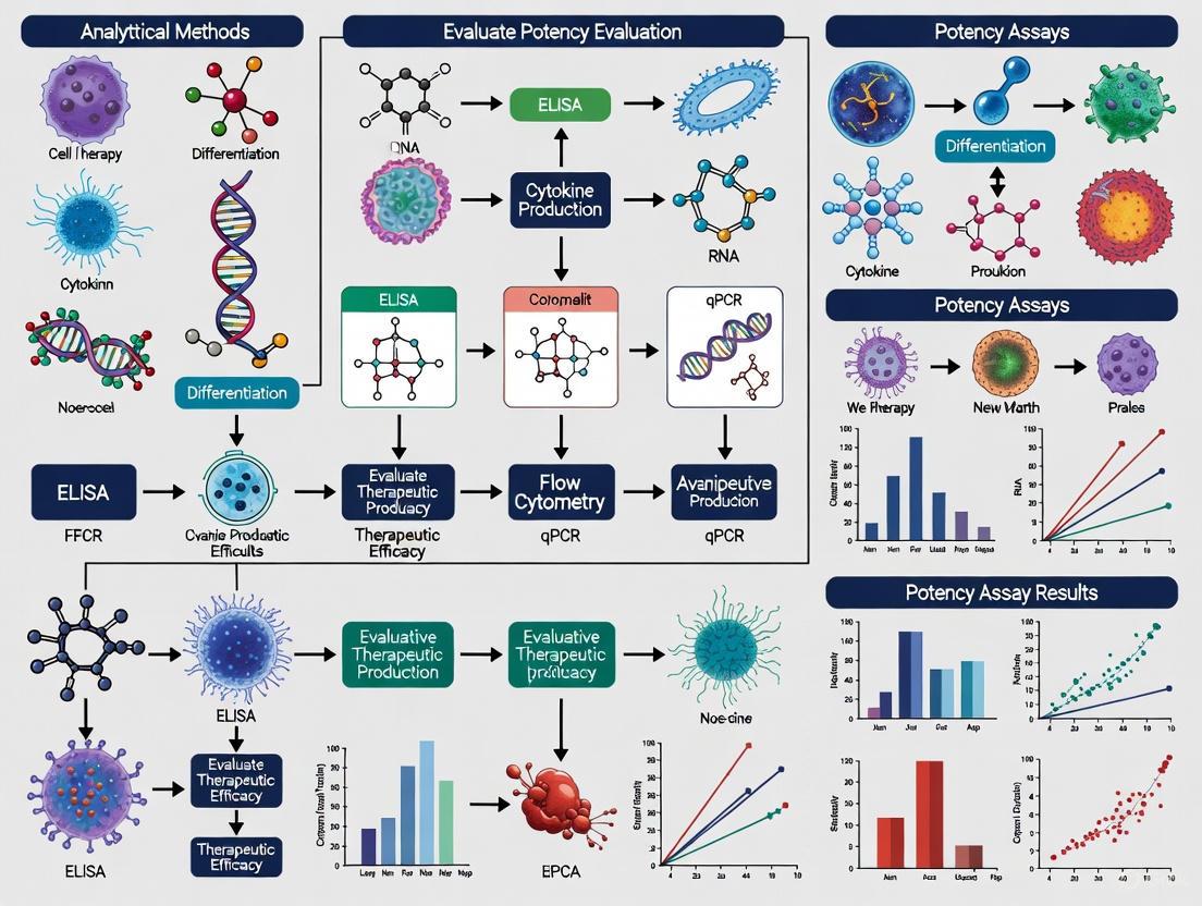
Visualizing the Potency Assay Strategy
The following diagram illustrates the multi-faceted approach to developing a comprehensive potency assay strategy, linking mechanism of action to measurable attributes and advanced analytical methods.
The development of robust potency assays is a critical regulatory requirement for the approval of Cell Therapy Products (CTPs) and Advanced Therapy Medicinal Products (ATMPs). Regulatory agencies including the U.S. Food and Drug Administration (FDA), European Medicines Agency (EMA), and International Council for Harmonisation (ICH) mandate that potency assays demonstrate a product's biological activity and link it to its intended mechanism of action (MoA) [11] [3]. These assays must be validated and used for lot-release testing to ensure product consistency, quality, and stability throughout the product lifecycle [3] [12]. The complex nature of cell-based therapies necessitates innovative approaches to potency testing, often requiring an orthogonal methodology that employs multiple independent methods to comprehensively characterize critical quality attributes like identity, purity, and potency [13]. This technical support center provides troubleshooting guidance and detailed protocols to assist researchers in navigating this challenging regulatory landscape.
Frequently Asked Questions (FAQs) on Regulatory Requirements
Q1: What is the fundamental regulatory purpose of a potency assay? A potency assay must quantitatively measure the biological activity of a cell therapy product that is linked to its relevant mechanism of action (MoA). According to FDA requirements, it serves to "assure that the product can achieve its intended mechanism of action, to assess manufacturing consistency and to evaluate product stability" [3]. It is a mandatory lot-release test for all licensed biologics.
Q2: How many potency assays are typically required for an FDA-approved cell therapy product? An analysis of 31 FDA-approved CTPs revealed an average of 3.4 potency tests per product (standard deviation 2.0), with a median of 3.0 tests. The number ranges from as few as 1 to as many as 8 tests per product, depending on product complexity [3] [14].
Q3: What are the most common types of potency measurements used in approved products? Table 1: Distribution of Potency Test Types in FDA-Approved CTPs
| Test Category | Frequency | Percentage | Example Methods |
|---|---|---|---|
| Viability and Count | 37 tests | 52% | Cell viability, CD34+ cell count [3] [14] |
| Expression | 19 tests | 27% | CAR expression by flow cytometry [3] [14] |
| Bioassays | 7 tests | 7% | IFN-γ release, cytotoxicity assays [3] [14] |
| Genetic Modification | 6 tests | 9% | Vector copy number (qPCR/ddPCR) [3] [14] |
| Histology | 2 tests | 3% | Tissue organization, cell viability [3] [14] |
Q4: What is the "orthogonal approach" recommended by regulators? The orthogonal approach involves "employing multiple independent methods – to assess critical quality attributes" such as identity, potency, and purity [13]. This methodology uses different analytical techniques to characterize the same attribute, providing comprehensive product characterization and reducing the risk of false results. For example, CAR-T cell identity may be confirmed through genotypic, phenotypic, and morphological analyses simultaneously [13].
Q5: How do regulatory expectations evolve throughout product development? Regulators recommend "flexible, risk-based strategies in potency assay development that evolve throughout product development and clinical trial phases" [11]. The FDA's draft guidance on "Potency Assurance for Cellular and Gene Therapy Products" (December 2023) describes a quality risk management (QRM) based approach with phase-appropriate expectations, becoming more rigorous as products advance toward licensure [12].
Troubleshooting Guides for Common Technical Challenges
Challenge 1: High Variability in Bioassay Results
Problem: Excessive variability in functional potency assays (e.g., cytokine release, cytotoxicity).
- Root Cause: Cell-based assays inherently contain multiple sources of variability including donor-derived biological materials, reagent inconsistencies, and assay conditions.
- Solution:
Challenge 2: Correlating In Vitro Potency with Clinical Outcomes
Problem: Difficulty establishing meaningful correlation between in vitro potency measurements and clinical efficacy.
- Root Cause: Oversimplified assays that don't fully capture the complex mechanism of action.
- Solution:
- Develop a "matrix approach" using multiple assays to capture different aspects of product function
- Incorporate novel biomarkers discovered through multi-omics approaches (genomics, epigenomics, transcriptomics, proteomics, metabolomics) [4]
- Collect clinical data early in development to inform assay refinement
Challenge 3: Managing Potency Testing for Complex MoAs
Problem: The mechanism of action involves multiple biological processes that are difficult to capture in a single assay.
- Root Cause: Cell therapies often work through complex, multifaceted biological pathways.
- Solution:
Experimental Protocols for Key Potency Assessments
Protocol 1: CAR T-Cell Potency Assessment via Cytokine Release
Principle: Measure IFN-γ release upon antigen-specific stimulation to assess T-cell activation potential, a key potency indicator for CAR-T products [4] [14].
Materials:
- CAR T-cell product
- Antigen-expressing target cells
- IFN-γ ELISA or ELISpot kit
- Cell culture medium with appropriate cytokines
- CO² incubator maintained at 37°C
Procedure:
- Co-culture CAR T-cells with antigen-positive target cells at effector:target ratios (e.g., 1:1, 5:1, 10:1)
- Include controls: CAR T-cells alone, target cells alone, and antigen-negative target cells
- Incubate for 16-24 hours under standard culture conditions
- Collect supernatant and measure IFN-γ secretion using quantitative ELISA
- Analyze data by comparing response to antigen-positive vs. antigen-negative targets
Technical Notes: This method is used in multiple FDA-approved CAR-T products including Kymriah and Yescarta [14]. Assay should be validated for precision, accuracy, and linearity.
Protocol 2: Vector Copy Number (VCN) Analysis by ddPCR
Principle: Quantify the average number of vector copies integrated per cell using droplet digital PCR, a critical safety and potency assessment for genetically modified cells [4] [14].
Materials:
- Genomic DNA from cell product
- ddPCR system (Bio-Rad QX200 or equivalent)
- Target probe for vector sequence
- Reference gene probe (e.g., RNase P, RPP30)
- ddPCR Supermix for Probes
- Droplet generator and reader
Procedure:
- Extract high-quality genomic DNA and quantify using fluorometric methods
- Prepare reaction mix with target and reference assays according to manufacturer's instructions
- Generate droplets using droplet generator
- Perform PCR amplification with optimized cycling conditions
- Read plate and analyze data to calculate copies/μl of target and reference genes
- Calculate VCN = (copies/μl target)/(copies/μl reference gene)
Technical Notes: VCN is a mandatory component of lot-release testing for most FDA-approved genetically modified cell products [4]. Acceptance criteria typically range between 1-5 vector copies per cell.
Essential Research Reagent Solutions
Table 2: Key Reagents for Cell Therapy Potency Evaluation
| Reagent Category | Specific Examples | Research Function | Regulatory Considerations |
|---|---|---|---|
| Flow Cytometry Reagents | Anti-CAR detection antibodies, Cell viability dyes, Cell subset markers (CD3, CD4, CD8, CD45, CD34) | Quantitative measurement of cell surface markers, viability, and CAR expression | Critical for identity and purity assessments; 65% of CTPs use expression markers as potency tests [3] [14] |
| Molecular Biology Kits | ddPCR/qPCR reagents for VCN, TCR sequencing kits, Integration site analysis reagents | Genetic modification quantification, safety assessment | VCN testing required for genetically modified products; integration site analysis important for safety [4] |
| Cell Culture Reagents | Antigen-presenting cells, Cytokine release assay kits, Cytotoxicity detection reagents | Functional potency assessment through bioassays | 23% of approved CTPs report bioassays as potency tests; essential for mechanism of action confirmation [3] [14] |
| Multi-omics Platforms | Single-cell RNA-seq kits, ATAC-seq reagents, Metabolomics profiling assays | Comprehensive product characterization beyond traditional potency | Emerging approach for understanding correlation between product characteristics and clinical outcomes [4] |
Signaling Pathways and Experimental Workflows
Orthogonal Potency Assay Development Workflow
CAR T-Cell Mechanism of Action and Potency Measurements
Emerging Technologies and Future Directions
The field of cell therapy potency assessment is rapidly evolving with several emerging technologies:
Multi-omics Integration: Advanced profiling techniques including genomics, epigenomics, transcriptomics, proteomics, and metabolomics are providing unprecedented insights into CAR T-cell function at the molecular level [4]. DNA methylation profiles in CD19 CAR T-cell products have identified distinct epigenetic loci associated with complete response and survival outcomes [4].
Advanced Vector Integration Analysis: New pipelines like INSPIIRED and EpiVIA enable detection of viral integration sites at bulk-cell and single-cell resolution respectively, addressing safety concerns related to insertional mutagenesis while providing potency information [4].
TCR Repertoire Profiling: Paired single-cell RNA analysis and TCR repertoire profiling allow identification of individual CAR T-cells with distinct transcriptional phenotypes, enabling use of TCR clonotypes as surrogates for expansion and persistence of functional T-cell states [4].
Regulatory agencies are keeping pace with these technological advances. The FDA's recent draft guidance "Potency Assurance for Cellular and Gene Therapy Products" (December 2023) emphasizes a science- and risk-based strategy throughout the product lifecycle [12], while the EMA's new guideline on clinical-stage ATMPs (effective July 2025) provides updated requirements for quality documentation [15]. Together, these frameworks support the development of more predictive potency assays that better correlate with clinical outcomes, ultimately ensuring the consistent quality, safety, and efficacy of cell therapy products.
For researchers and developers in the cell therapy field, developing robust potency assays remains one of the most significant analytical challenges. Potency testing is not merely a regulatory checkbox; it represents a quantitative measure of a product's biological activity and is considered a Critical Quality Attribute (CQA) that must be thoroughly characterized throughout development and manufacturing [16]. Analysis of the 31 United States Food and Drug Administration-approved cell therapy products (CTPs) reveals valuable insights into successful strategies for potency assurance [3]. This technical resource distills these insights into practical guidance, troubleshooting tips, and methodological frameworks to support your analytical development workflow.
Quantitative Analysis of Approved Product Testing Strategies
Distribution of Potency Test Types
A comprehensive analysis of regulatory submissions for 31 approved CTPs identified 104 individual potency tests, with 32% redacted for proprietary reasons [3]. The remaining 71 tests categorize into five primary measurement types, with viability/count and expression assays dominating the testing landscape.
Table 1: Potency Test Categories and Their Prevalence in Approved CTPs
| Test Category | Number of Tests | Percentage | Description |
|---|---|---|---|
| Viability and Count | 37 | 52% | Cell viability, total nucleated cell count, CD34+ cell count |
| Expression | 19 | 27% | CAR expression, surface marker expression (flow cytometry) |
| Bioassays | 7 | 7% | Functional activity (e.g., cytotoxicity, cytokine release) |
| Genetic Modification | 6 | 9% | Vector copy number, transduction efficiency |
| Histology | 2 | 3% | Tissue structure and composition assessment |
Testing Combinations and Product-Specific Approaches
The data reveals that CTPs employ an average of 3.4 potency tests per product (standard deviation: 2.0), with numbers ranging from 1 to 8 tests [3]. The most frequent testing combination pairs "Viability and Count" with "Expression" assays, occurring in 16 CTPs (52%) [3]. Hematopoietic stem cell-cord blood products utilize the highest number of potency tests (average 4.4), while CAR T-cell products and tissue-engineered therapies employ fewer (averages of 1.9 and 1.8 respectively) [3].
Table 2: Number of Potency Tests by Product Category
| Product Category | Number of CTPs | Average Number of Potency Tests | Standard Deviation |
|---|---|---|---|
| Hematopoietic Stem Cell-Cord Blood | 5 | 4.4 | 0.7 |
| Genetically Modified Cell Therapy | 7 | 2.4 | 1.1 |
| CAR T-Cell Therapy | 7 | 1.9 | 0.9 |
| Tissue Engineered | 5 | 1.8 | 1.1 |
Essential Reagents and Research Solutions
Core Analytical Toolkit for Potency Assessment
Developing robust potency assays requires specific reagent systems and analytical tools. The following table outlines essential research solutions referenced in approved product characterizations.
Table 3: Key Research Reagent Solutions for Cell Therapy Potency Evaluation
| Reagent/Assay Type | Specific Examples | Research Function | Application in Approved CTPs |
|---|---|---|---|
| Flow Cytometry Panels | Anti-CAR antibodies, T-cell subset markers | Phenotypic characterization and identity | CAR expression measurement in 65% of CTPs [3] |
| Cytokine Detection | IFN-γ, IL-2, TNF-α ELISA/ELISpot | Functional potency assessment | Effector function evaluation [4] |
| Molecular Biology Tools | ddPCR for VCN, TCR-seq | Genomic profile characterization | Vector copy number quantification [4] |
| Cell Culture Assays | Cytotoxicity (LDH), proliferation | Functional bioactivity | Target cell killing capacity [4] |
| Metabolic Assays | Glycolytic activity, mitochondrial fitness | Metabolic profiling | Persistence and durability potential [4] |
Experimental Protocols: Methodologies from Approved Products
Multi-Omics Product Profiling Workflow
Advanced characterization of CTPs increasingly employs orthogonal multi-omics approaches to comprehensively assess product characteristics that correlate with clinical potency [4]. The following workflow outlines a comprehensive profiling strategy:
Protocol: Cytokine Release Potency Assay
Purpose: Measure functional activation through cytokine secretion upon target cell engagement, a cornerstone of potency assessment for CAR T-cell products [4].
Materials:
- CAR T-cell final product (cryopreserved)
- Target antigen-positive and negative cell lines
- Complete RPMI-1640 medium with 10% FBS
- 96-well U-bottom plates
- Cytokine capture antibodies (IFN-γ, IL-2, TNF-α)
- ELISA or multiplex Luminex detection system
- CO2 incubator (37°C, 5% CO2)
Procedure:
- Cell Preparation: Thaw CAR T-cells and rest overnight in complete medium. Count and adjust concentration to 1×10^6 cells/mL.
- Target Cell Preparation: Harvest and wash target cells (antigen-positive and negative controls), adjusting to 1×10^6 cells/mL.
- Co-culture Setup: In 96-well plate, add effector and target cells at 1:1 E:T ratio (100μL each). Include effector-only and target-only controls.
- Incubation: Incubate plates for 24 hours at 37°C, 5% CO2.
- Supernatant Collection: Centrifuge plates at 300×g for 5 minutes, carefully transfer 150μL supernatant to clean plates.
- Cytokine Quantification: Perform ELISA or multiplex immunoassay according to manufacturer protocols.
- Data Analysis: Calculate antigen-specific cytokine release by subtracting values from negative control wells.
Technical Notes: Include a reference standard with known activity to enable relative potency calculation. Validate assay precision (CV <20%) and accuracy (70-130% recovery) according to ICH Q2(R2) guidelines [17].
Protocol: Vector Copy Number Determination by ddPCR
Purpose: Quantify vector copies per cell to ensure consistent genetic modification and monitor potential safety concerns [4].
Materials:
- Genomic DNA isolation kit
- Droplet digital PCR system (Bio-Rad QX200 or equivalent)
- ddPCR Supermix for Probes (no dUTP)
- Target and reference assay primers/probes
- Droplet generator and reader
- Thermal cycler
Procedure:
- DNA Extraction: Isolate genomic DNA from CAR T-cell product, quantify by fluorometry.
- Reaction Setup: Prepare 20μL reactions containing 1X ddPCR Supermix, 50-100ng DNA, and target/reference assays.
- Droplet Generation: Transfer reactions to DG8 cartridges, generate droplets using droplet generation oil.
- PCR Amplification: Transfer droplets to 96-well plate, seal, and run thermal cycling: 95°C for 10min; 40 cycles of 94°C for 30s and 60°C for 60s; 98°C for 10min.
- Droplet Reading: Read plate on droplet reader, analyze with QuantaSoft software.
- Calculation: VCN = (Concentration of target amplicon) / (Concentration of reference gene amplicon).
Technical Notes: Use reference gene (e.g., RPP30) for normalization. Establish acceptance criteria for DNA quality (A260/280 ratio 1.8-2.0). Include no-template controls and reference standards in each run [4].
Troubleshooting Guides & FAQs
Common Potency Assay Challenges and Solutions
Q1: Our cell-based potency assay shows high variability (CV >25%). How can we improve precision?
A: High variability in cell-based assays is common. Implement these strategies:
- Cell Bank Controls: Use well-characterized master cell banks as assay controls to minimize biological variability [17].
- Standardized Culture: Control passage number, culture duration, and media components rigorously.
- Assay Design Optimization: Increase replication (n≥6), use reference standard normalization, and establish robust positive/negative controls.
- Analyst Training: Ensure consistent technique across operators with standardized protocols.
Q2: For our allogeneic CAR-T product, what potency assays best predict clinical response?
A: Based on approved products, employ a matrix approach:
- Primary Potency: Cytotoxic activity against target cells (specific lysis)
- Secondary Mechanisms: Cytokine release profile (IFN-γ, IL-2) upon antigen engagement
- Product Characterization: CAR expression (flow cytometry), VCN (ddPCR), and T-cell differentiation markers [4] [3]
- Emerging Biomarkers: Consider adding metabolic profiling and epigenetic markers associated with persistence [4]
Q3: How do we address disconnect between in vitro potency results and in vivo efficacy?
A: This challenge indicates incomplete mechanistic understanding:
- Enhanced Assay Conditions: Modify culture duration to capture delayed effects; incorporate physiological stressors.
- Additional Functional Readouts: Include proliferation capacity, exhaustion marker expression (PD-1, TIM-3), and persistence assays.
- Multi-Omics Correlation: Integrate transcriptomic and proteomic data to identify biomarkers predictive of in vivo performance [4].
- Clinical Correlation: If possible, correlate assay parameters with clinical outcomes in early trials.
Q4: What are the regulatory expectations for potency assay validation at different stages?
A: Expectations are phase-appropriate:
- Early Clinical: Assay qualification demonstrating specificity, precision, and linearity is sufficient.
- Pivotal Trials: Partial validation addressing accuracy, precision, specificity, and range.
- Commercial Submission: Full validation per ICH Q2(R2) including robustness testing [16] [17].
Q5: How can we implement the "orthogonal approach" recommended by regulators?
A: Orthogonal methods use different principles to measure the same attribute:
- CAR Expression Example: Combine flow cytometry (protein) with ddPCR (DNA) and RNA-seq (transcript) [13].
- Functional Potency: Pair cytotoxicity (direct function) with cytokine release (signaling output).
- Data Integration: Statistically correlate results from orthogonal methods to build comprehensive product understanding [13].
Regulatory Considerations and Future Directions
The FDA's 2023 draft guidance on "Potency Assurance for Cellular and Gene Therapy Products" emphasizes science- and risk-based strategies that extend beyond traditional lot-release testing [18]. The guidance recommends comprehensive potency assurance strategies incorporating manufacturing process design, process controls, material controls, in-process testing, and formal potency release assays [18].
Emerging trends in potency assessment include:
- Multi-Omics Integration: Combining genomic, epigenomic, transcriptomic, proteomic, and metabolomic data to develop predictive potency signatures [4].
- Single-Cell Technologies: Applying single-cell RNA-seq and ATAC-seq to understand product heterogeneity and identify potency biomarkers [4].
- Advanced Bioanalytics: Implementing novel sensors and real-time monitoring systems for in-line potency assessment.
As the CTP landscape evolves with increased regulatory approvals—eight novel CTPs in 2024 alone—potency assay strategies continue to advance in sophistication [19]. Developers should anticipate increased regulatory focus on assay clinical relevance and should begin correlating potency measurements with clinical outcomes as early as possible in product development.
Distinguishing Potency from Titer, Identity, and Purity Assays
Core Definitions and Their Relationship to Mechanism of Action
For cell and gene therapy products, understanding the distinct roles of potency, titer, identity, and purity assays is fundamental to accurate product characterization. These assays measure different Critical Quality Attributes (CQAs) and are integral to demonstrating that a product consistently meets its predefined quality standards [20].
- Potency: The specific biological attribute of a product that enables it to achieve its intended Mechanism of Action (MoA) [20]. A potency test measures this attribute, linking the product's biological activity to its therapeutic effect.
- Titer: In the context of viral vectors used in gene therapy, titer refers to the concentration of functional vector particles, often measured as Transducing Units (TU) per milliliter [21] [22]. It quantifies the "delivery capability" of a vector but does not confirm the resulting biological activity.
- Identity: A test that confirms the product's identity, verifying that it contains the components it is purported to contain (e.g., specific cell surface markers, genetic constructs) [23].
- Purity: An assessment of the product's freedom from extraneous materials, such as process-related impurities or contaminants [24].
The following diagram illustrates the logical relationship between a product's MoA and the key assays used for its quality control, highlighting that potency is the attribute most directly linked to the therapeutic effect.
Comparative Analysis of Quality Control Assays
The table below provides a detailed comparison of these four assay categories, outlining their primary purpose, typical methodologies, and how they relate to the product's MoA.
| Assay Type | Primary Purpose | Key Methodologies | Relation to MoA |
|---|---|---|---|
| Potency | Measures biological function and therapeutic activity [20]. | Cytotoxicity (e.g., chromium release, bioelectronic impedance) [25], cytokine release (e.g., IFN-γ ELISA, Luminex) [4] [23], degranulation (CD107a), cell proliferation [4]. | Direct. Designed based on the known or proposed MoA [20]. |
| Titer | Quantifies functional vector concentration for gene delivery [21] [22]. | Digital PCR (ddPCR) for vector copy number (VCN) [4] [22], TCID50, transduction efficiency via flow cytometry [21] [22]. | Indirect. Measures the "delivery vehicle" strength, not the final biological effect. |
| Identity | Confirms the presence of key components of the product. | Flow cytometry (cell surface markers, CAR expression) [23] [24], PCR (transgene detection). | Ancillary. Verifies the product contains the correct elements but not their function. |
| Purity | Evaluates freedom from impurities (e.g., process residuals, host cell proteins). | Host Cell Protein (HCP) ELISAs, residual DNA analysis, mycoplasma testing, micro-flow imaging for particulates [24]. | Indirect (Safety). Ensures safety and quality but does not measure biological activity. |
Frequently Asked Questions (FAQs)
FAQ 1: Why is a separate potency assay needed if we already measure titer and transduction efficiency?
While titer and transduction efficiency confirm the successful delivery and presence of the genetic material, a potency assay is required to confirm that the delivered gene functions as intended. A high titer ensures a high number of functional vectors, and high transduction efficiency confirms successful gene transfer into target cells. However, only a potency assay can verify that the transduced cells perform the desired biological activity, such as killing tumor cells or secreting a therapeutic protein [26] [27]. Potency closes the loop between delivery and biological function.
FAQ 2: Our product's Mechanism of Action is not fully understood. How can we develop a valid potency assay?
This is a common challenge in cell therapy development [20]. The regulatory guidance recommends an incremental approach.
- Utilize a Matrix of Assays: Initially, you may employ a matrix of tests to evaluate different aspects of product quality and function that are plausibly linked to the therapeutic effect. For a CAR-T cell product, this could include a combination of cytotoxicity, cytokine secretion, and phenotyping assays [4] [23].
- Correlate with Clinical Data: As clinical data becomes available, refine your potency assay by correlating the in vitro assay results with clinical outcomes in patients [20].
- Leverage Multi-Omics: Advanced profiling (e.g., transcriptomics, epigenomics) can help identify novel biomarkers that correlate with clinical response, which can then be developed into surrogate potency assays [4].
FAQ 3: Can a product be "potent" in the lab but not "efficacious" in patients?
Yes. A product can be "potent but not efficacious" [20]. This can occur if the in vitro potency test does not adequately capture the complex in vivo environment. Factors such as the immunosuppressive tumor microenvironment, poor cell persistence in vivo, or T-cell exhaustion that is not modeled in the short-term lab assay can lead to a lack of clinical efficacy despite high measured potency [4] [20]. This underscores the importance of developing biologically relevant potency assays.
Troubleshooting Guide for Potency Assays
| Problem | Potential Root Cause | Recommended Solution |
|---|---|---|
| High assay variability | Inconsistent cell culture conditions, unstable reference standard, or operator-dependent readouts (e.g., manual counting). | Implement rigorous cell culture protocols, use a well-characterized reference standard, and adopt automated, quantitative readouts (e.g., bioelectronic impedance, flow cytometry) [26] [25]. |
| Poor correlation with clinical outcome | The in vitro assay conditions do not recapitulate key aspects of the in vivo MoA. | Incorporate more physiologically relevant components into the assay, such as primary target cells, 3D co-culture systems, or biomarkers of persistence and exhaustion identified from clinical samples [4] [20]. |
| Low signal-to-noise ratio in cytotoxicity assay | Inappropriate effector-to-target (E:T) ratio, insufficient assay duration, or low sensitivity of detection method. | Perform a kinetic E:T ratio titration using a real-time, label-free method (e.g., impedance-based killing assays) to identify the optimal conditions for detecting cytotoxicity [25]. |
| Failure to distinguish between product batches with known clinical differences | The potency assay is measuring an attribute not critical to the therapeutic MoA. | Re-evaluate the proposed MoA using multi-omics data from clinical batches (e.g., epigenomic profiles associated with memory T-cell subsets) and develop an assay that measures that critical attribute [4]. |
Essential Research Reagent Solutions
The following table lists key reagents and tools essential for developing and executing robust potency and quality control assays.
| Reagent / Tool | Function in Assays | Example Use-Case |
|---|---|---|
| Luminex Multiplex Assays | Simultaneously quantify multiple soluble analytes (e.g., cytokines) from a single sample [24]. | Profiling IFN-γ, TNF-α, IL-2, and other cytokines in CAR-T cell supernatant after co-culture with target cells as a multi-parametric potency readout [23]. |
| Fluorokines & Flow Cytometry Kits | Fluorokines are fluorescent-labeled proteins used to directly stain and detect CAR+ cells by flow cytometry [24]. | Quantifying CAR expression on transduced T-cells for identity and to ensure adequate transduction efficiency [24]. |
| ddPCR (Droplet Digital PCR) | Provides absolute quantification of nucleic acids with high precision and sensitivity, without relying on a standard curve [4] [22]. | Measuring Vector Copy Number (VCN) in transduced cells as a critical safety and titer attribute [4]. |
| Impedance-Based Bioelectronic Assays | Enable real-time, label-free monitoring of cell-mediated killing of target cells by measuring electrical impedance [25]. | Performing kinetic potency assays for CAR-T cells, capturing the dynamics of tumor cell killing without the use of labels [25]. |
| RNAscope ISH Assays | Enable single-cell RNA expression analysis within intact cells, providing spatial context [24] [28]. | Localizing and quantifying transgene expression in situ in target tissues during pre-clinical studies [24]. |
| Simple Plex Immunoassays | Automated, microfluidic platform for highly reproducible quantitation of proteins from small sample volumes [24]. | Rapid (90-minute) and precise assessment of secreted T-cell activation markers (e.g., IFN-γ) for lot-release potency testing [24]. |
Experimental Protocol: Cytokine Release Potency Assay for CAR-T Cells
This protocol outlines a common method for assessing CAR-T cell potency based on IFN-γ release upon antigen-specific activation [4] [20].
Principle: Functional CAR-T cells will recognize and activate in response to target cells expressing the cognate antigen, leading to the secretion of effector cytokines like IFN-γ. The amount of IFN-γ released is quantified as a measure of potency.
Workflow:
Detailed Methodology:
- Preparation of Target Cells:
- Harvest and count tumor cells that endogenously express the target antigen (e.g., CD19). Alternatively, use engineered cell lines.
- Plate the target cells in a 96-well tissue culture plate at a pre-determined density (e.g., 50,000 cells/well) in complete media. Include wells with target cells alone as a background control.
Co-culture Setup:
- Harvest, count, and resuspend the CAR-T cell product in complete media.
- Add CAR-T cells to the plated target cells at multiple Effector-to-Target (E:T) ratios (e.g., 1:1, 5:1, 10:1) to establish a dose-response curve. Set up replicates for each condition.
- Critical Controls: Include CAR-T cells cultured alone (to assess background activation) and a reference standard CAR-T cell batch if available.
Incubation and Supernatant Collection:
- Incubate the co-culture plates for 18-24 hours at 37°C with 5% CO₂.
- After incubation, centrifuge the plates at a low speed (e.g., 300 x g for 5 minutes).
- Carefully collect the supernatant from each well without disturbing the cell pellet, and transfer it to a new plate. Store at -80°C if not testing immediately.
Cytokine Quantification:
- Use a validated, quantitative immunoassay to measure IFN-γ concentration in the supernatants. This can be a traditional ELISA or a more automated platform like the Ella Simple Plex system [24].
- Follow the manufacturer's instructions precisely. Include a standard curve of known IFN-γ concentrations to interpolate sample values.
Data Analysis and Potency Calculation:
- Subtract the background cytokine signal from the target-only and effector-only controls from the co-culture values.
- Plot the IFN-γ concentration against the E:T ratio. The potency of a test sample can be reported relative to the reference standard, often based on the EC₅₀ (half-maximal effective concentration) derived from the dose-response curve [26].
For researchers and scientists in cell and gene therapy, developing a robust potency assay is a critical, non-negotiable regulatory requirement. A potency assay quantifies the biological activity of a product—the specific ability or capacity of a cell therapy product to achieve a defined biological effect [23]. It is the cornerstone of quality control (QC), essential for assuring the safety, efficacy, and consistency of your Advanced Therapy Medicinal Product (ATMP) throughout its lifecycle, from early development to commercial lot release [23] [3]. This guide addresses the specific challenges you may encounter during this complex process.
Regulatory Framework & Core Principles
Why are Potency Assays Required?
Regulatory bodies like the FDA and EMA mandate potency testing for all biologics, including cell and gene therapies [3]. The primary goals are to:
- Ensure Product Efficacy: Confirm that each product batch can perform its intended biological function according to its mechanism of action (MoA).
- Guarantee Manufacturing Consistency: Verify that your manufacturing process consistently produces a product with the same biological activity.
- Assess Product Stability: Monitor the stability of your product throughout its shelf life.
The "Matrix" Approach
For complex products like CAR-T cells, a single assay is often insufficient to fully capture the product's biological activity. Regulators recommend a "potency assay matrix"—a combination of multiple, orthogonal assays that collectively measure different aspects of the product's MoA [4]. An analysis of FDA-approved cell therapy products (CTPs) reveals that each product uses an average of 3.4 potency tests, with some products using up to 8 [3].
Table: Prevalence of Potency Test Types in 31 FDA-Approved Cell Therapies [3]
| Type of Measurement | Percentage of CTPs Using It | Example |
|---|---|---|
| Viability and Count | 61% | Cell viability via flow cytometry or dye exclusion |
| Expression | 65% | CAR expression by flow cytometry |
| Bioassay | At least 23% (data limited by redactions) | Cytokine release (IFN-γ) upon target cell co-culture |
| Genetic Modification | Information redacted in public documents | Vector Copy Number (VCN) by ddPCR |
The diagram below illustrates the logical relationship between a product's Mechanism of Action (MoA) and the development of a potency assay matrix.
Troubleshooting Guide: FAQs & Solutions
Assay Development
Q1: My functional bioassay has high variability. How can I improve robustness?
- Problem: Inconsistent results in cytotoxicity or cytokine release assays.
- Solutions:
- Implement Design of Experiments (DoE): Systematically evaluate critical factors like effector-to-target cell ratio, incubation time, serum lot, and analyst technique to identify and control key sources of variability [29].
- Move to Real-Time Methods: Replace endpoint assays (e.g., Chromium-51, LDH) with real-time, label-free methods like bioelectronic impedance. These provide kinetic data and continuous monitoring, offering a richer dataset and reducing the impact of single time-point variability [25].
- Standardize Reagents: Use a qualified and consistent source for critical reagents like target cell lines and cytokines.
Q2: How do I handle the high heterogeneity of autologous cell therapy products?
- Problem: Patient-to-patient variability in starting material makes setting universal potency specifications difficult.
- Solutions:
- Adopt a Multi-Parameter Approach: Do not rely on a single metric. Use a matrix that includes identity (e.g., CAR expression), purity (e.g., CD3+ percentage), and multiple functional readouts [4] [3].
- Establish a "Consistency Range": Instead of a fixed value, define an acceptable range for potency based on data from multiple clinical trial batches that demonstrated safety and efficacy.
- Leverage Advanced Analytics: Use multi-omics data (transcriptomics, epigenomics) to identify correlative biomarkers that can serve as surrogates for complex functional assays [4].
Assay Qualification & Validation
Q3: What are the key differences between qualification and validation?
- Answer:
- Qualification occurs during clinical development. It demonstrates that the assay is suitable for its intended purpose (e.g., characterizing your product for Phase I/II trials). It assesses precision, accuracy, and linearity.
- Validation is required for product licensure (BLA/MAA). It provides a high level of assurance that the assay consistently performs as intended for commercial lot release. This involves rigorous, GMP-compliant assessment of all validation parameters (specificity, accuracy, precision, linearity, range, robustness) [29].
Q4: My potency assay failed during tech transfer to the QC unit. What went wrong?
- Problem: The assay no longer performs as it did in the development lab.
- Solutions:
- Document Meticulously: Ensure the analytical procedure is exhaustively detailed, leaving no room for interpretation on critical steps like pipetting technique, incubation times, or instrument settings.
- Conduct Robustness Testing Early: During development, deliberately introduce small, realistic variations in parameters (e.g., pH, temperature, reagent age) to define the assay's operable range [30].
- Joint Experimentation: Have scientists from the development and QC labs run the assay side-by-side to identify and resolve discrepancies in technique.
Step-by-Step Experimental Protocols
Protocol: IFN-γ Release Assay for CAR-T Cell Potency
This is a common bioassay used to measure T-cell activation upon engagement with target antigen [4] [23].
1. Principle: CAR-T cells are co-cultured with antigen-positive target cells. Activation through the CAR leads to secretion of IFN-γ, which is quantified by ELISA or ELISpot as a measure of potency.
2. Materials:
- Effector Cells: Final formulated CAR-T cell product.
- Target Cells: Antigen-positive tumor cell line (e.g., NALM-6 for CD19).
- Control Cells: Antigen-negative cell line (isogenic control recommended).
- Culture Medium: Appropriate for both cell types, serum-free recommended to avoid assay interference.
- Cell Culture Plates: 96-well U-bottom plates.
- IFN-γ ELISA or ELISpot Kit: Qualified for use in cell culture supernatants.
- Plate Reader: (Spectrophotometer for ELISA, or plate reader for ELISpot).
3. Procedure:
- Prepare Effector Cells: Thaw and wash CAR-T cells if frozen. Count and resuspend in culture medium at a pre-determined concentration (e.g., (1 \times 10^6)/mL).
- Prepare Target Cells: Harvest and count target and control cells. Resuspend at the same concentration as effector cells.
- Co-culture Setup: In the 96-well plate, add effector and target cells at various effector-to-target (E:T) ratios (e.g., 1:1, 5:1, 10:1) in triplicate. Include controls:
- Effector Alone: CAR-T cells + medium.
- Target Alone: Target cells + medium.
- Stimulation Positive Control: CAR-T cells + PMA/Ionomycin.
- Incubate: Incubate plates for 18-24 hours at 37°C, 5% CO₂.
- Harvest Supernatant: Centrifuge plates and carefully transfer supernatant to a new plate for ELISA analysis.
- Quantify IFN-γ: Perform ELISA according to the manufacturer's instructions.
- Data Analysis: Calculate the concentration of IFN-γ in test samples using a standard curve. Subtract the background from effector-alone and target-alone controls. The potency is reported as the amount of IFN-γ released per cell or per mL at a specified E:T ratio.
The workflow for developing and validating such a functional bioassay is outlined below.
Protocol: CAR Expression by Flow Cytometry
This is a critical identity and potency test for CAR-T products [3].
1. Principle: A recombinant protein or antibody that binds to the extracellular domain of the CAR is used to detect and quantify the percentage of CAR-positive cells and the density of CAR expression (Mean Fluorescence Intensity, MFI).
2. Materials:
- CAR-T Cell Sample
- Staining Buffer: PBS with 1-2% FBS.
- Detection Reagent: Biotinylated or fluorescently-labeled antigen (e.g., CD19-Fc for CD19 CAR) or anti-CAR antibody.
- Secondary Reagent (if needed): Streptavidin conjugated to a fluorophore.
- Viability Dye: e.g., 7-AAD or DAPI.
- Flow Cytometer
3. Procedure:
- Prepare Cells: Aliquot (1 \times 10^5) to (5 \times 10^5) cells into a flow cytometry tube.
- Staining: Wash cells with staining buffer. Resuspend cell pellet in staining buffer containing the detection reagent. Incubate for 30 minutes in the dark at 4°C.
- Secondary Stain (if needed): Wash cells twice. If using a biotinylated primary reagent, resuspend in staining buffer containing the fluorescent streptavidin. Incubate for 15-30 minutes in the dark at 4°C.
- Wash and Resuspend: Wash cells twice and resuspend in staining buffer containing a viability dye.
- Acquisition and Analysis: Run samples on the flow cytometer. Use a viability dye to gate on live cells. Report the percentage of CAR-positive cells and the MFI relative to an unstained control.
The Scientist's Toolkit: Essential Research Reagents & Solutions
Table: Key Reagents for Cell Therapy Potency Assays
| Reagent / Solution | Function / Application | Key Considerations |
|---|---|---|
| Qualified Target Cell Line | Used in functional bioassays (cytotoxicity, cytokine release) to trigger CAR/TCR activation. | Ensure consistent antigen expression and growth characteristics. Master Cell Bank is recommended. |
| Recombinant Antigen / CAR Detection Reagent | Quantifying CAR surface expression via flow cytometry. | Binding affinity and specificity must be qualified. Critical reagent. |
| Cytokine ELISA/ELISpot Kits | Quantifying cytokine release (IFN-γ, IL-2) as a measure of T-cell activation. | Select kits validated for use with cell culture supernatants. |
| Droplet Digital PCR (ddPCR) | Absolute quantification of Vector Copy Number (VCN) for genetically modified products [4] [22]. | Offers high precision and sensitivity without a standard curve. |
| Cell Viability Assays (e.g., Impedance-based) | Real-time, label-free monitoring of cell health and cytotoxicity [25]. | Provides kinetic data superior to endpoint assays (e.g., MTT, LDH). |
| Multi-omics Tools (scRNA-seq, ATAC-seq) | Deep product characterization to identify novel biomarkers of potency and persistence [4]. | Used in development to build a deeper understanding of the product's MoA. |
A Toolkit of Methods: From Viability Counts to Mechanism-of-Action Bioassays
For cell therapy products (CTPs), demonstrating biological activity through potency assays is a regulatory requirement for product release. An analysis of all 31 U.S. FDA-approved CTPs reveals that measurements of cell viability and count and gene or protein expression are the most frequently used methods for potency testing, employed by 61% and 65% of approved products, respectively [3]. These physicochemical methods form the foundation for ensuring that a therapy contains a sufficient dose of functional cells capable of eliciting the intended therapeutic effect.
This technical resource provides troubleshooting guides and detailed protocols to support researchers in the robust execution of these core analytical methods within a cell therapy development context.
Troubleshooting Guides & FAQs
Cell Viability and Count
Q: My cell viability is low after cryopreservation and thawing. What could be the cause?
- A: Low post-thaw viability is a common challenge. Review your process against these critical points:
- Incorrect Thawing: Avoid thawing cells for longer than 2 minutes at 37°C. Thaw quickly and do not expose cells to air [31].
- Osmotic Shock: After thawing, do not add a full volume of medium at once. Transfer cells to a pre-rinsed tube and add pre-warmed complete medium drop-wise (approximately 1 drop per second) while swirling the tube [31].
- Improper Centrifugation: For fragile primary cells like neurons, avoid centrifugation immediately upon recovery from cryopreservation, as it can drastically reduce viability [31].
- Storage Condition: Ensure cells were stored correctly in liquid nitrogen until use [31].
Q: My cells are dying in culture for no apparent reason. I have ruled out microbial contamination. What should I check?
- A: Systematically investigate your culture system:
- Culture Media Quality: Check the condition and quality of your culture and freezing media, including the expiration dates of key supplements like B-27 [31] [9].
- Supplement Applicability: Verify the quality and application suitability of serum and other supplements. Lot-to-lot variability can significantly impact cell health [9].
- Environmental Stress: Review critical process parameters such as passage number, confluence (avoid over-confluency), and the activity of dissociation enzymes (avoid over-digestion) [9].
- Handling of Ancillary Materials: Thawed supplements like B-27 should not be exposed to room temperature for more than 30 minutes and should be used within one week if stored at 4°C [31].
Transgene Expression
Q: I am achieving low transfection/transduction efficiency in primary human T lymphocytes. What can I optimize?
- A: Primary T cells are notoriously hard to transfect. Consider these strategies:
- Electroporation Method: Utilize square-wave electroporation systems (e.g., Lonza Nucleofector) over lipid-based methods, which are associated with high toxicity and low efficiency in T cells [32].
- Electroporation Buffer: The composition of the electroporation buffer is critical. Research indicates that using optimized, in-house buffers can render high transgene expression levels (mean 45%) in primary human T cells at a lower cost than commercial kits [32].
- Cell Activation: Ensure T cells are properly activated before genetic modification. For murine T cells, activation with anti-CD3 and anti-CD28 antibodies 24 hours before electroporation is a standard protocol [32].
- Vector and Promoter: For viral transduction, choose an appropriate vector and promoter. For example, in lentiviral transduction of mesenchymal stem cells (MSCs), the EF1-α promoter has been shown to provide durable transgene expression [33].
Q: I observe high cytotoxicity in my cells after viral transduction. Is this normal, and what can I do?
- A: Some cytotoxicity is expected, but it can be managed.
- Expected Toxicity: It is common to see cytotoxicity 24–48 hours post-transduction, which can affect over 50% of your cells. This is often an indication of high viral uptake and expression of exogenous genes. It is recommended to continue culturing the cells according to the protocol [31].
- Use Updated Systems: Newer viral vector backbones (e.g., those in the CytoTune-iPS Sendai 2.0 Reprogramming Kit) have been shown to cause less cytotoxicity compared to their predecessors [31].
- Confirm Vector Clearance: For systems like Sendai virus, use RT-PCR to confirm the clearance of vectors from your cell lines after several passages [31].
Experimental Protocols & Data Presentation
Detailed Protocol: Electroporation of Primary T Lymphocytes
This protocol is adapted from a method demonstrating high transgene expression levels (mean 45%) in primary human T lymphocytes and viable murine T cells (mean 38%) using in-house buffers and a Lonza Nucleofector II device [32].
Key Materials:
- Cells: Primary human T cells from PBMCs or murine T cells from lymph nodes.
- Plasmids: Transposon plasmid (e.g., pT2-GFP) and transposase (e.g., SB100X for stable expression) [32].
- Equipment: Lonza Nucleofector II device and appropriate cuvettes.
- Buffers: Pre-warmed, optimized electroporation buffer. The specific buffer composition is a critical variable for efficiency [32].
- Culture Media: RPMI medium supplemented with 10% FCS, L-Glu, and Penicillin/Streptomycin. For primary lymphocytes, add 50 U/mL of human rIL-2 post-transfection [32].
Workflow:
Procedure:
- Cell Preparation: Isolate T cells from human PBMCs or mouse lymph nodes. Pellet 1x10^7 cells at 200 g for 10 minutes. For murine T cells, activate them 24 hours before electroporation using 2 µg/mL aCD3 and 1 µg/mL aCD28 in the presence of 50 U/mL IL-2 [32].
- DNA Complexation: Resuspend the cell pellet in 100 µL of pre-warmed electroporation buffer. Add your plasmid DNA (e.g., transposon for the gene of interest and transposase for genomic integration) [32].
- Electroporation: Transfer the cell-DNA mixture to a certified cuvette and electroporate using the designated program on the Nucleofector II device. The specific program will depend on the cell type [32].
- Recovery: Immediately after electroporation, carefully transfer the cells to a pre-warmed culture plate containing complete medium. Supplement the medium with 50 U/mL of human recombinant IL-2 to support recovery and expansion [32].
- Activation and Expansion: For human T cells, activate them with 1 µg/mL OKT-3 and 0.5 µg/mL aCD28. Alternatively, expand transfected cells by co-culture with irradiated feeder cells [32].
- Analysis: Cells can be expanded for up to 12 days and analyzed for transgene expression (e.g., via flow cytometry for GFP), viability, and functional capacity (e.g., CAR-mediated target cell lysis) [32].
The table below summarizes the analysis of potency tests used for the 31 US FDA-approved Cell Therapy Products as of 2025, providing a benchmark for developers [3].
Table 1: Prevalence of Potency Test Types in FDA-Approved Cell Therapies
| Potency Test Category | Number of CTPs Using This Test | Percentage of Total CTPs (n=31) | Total Number of Tests (Incl. Multi-Use) |
|---|---|---|---|
| Viability and Cell Count | 19 | 61% | 37 |
| Expression (Gene/Protein) | 20 | 65% | 19 |
| Bioassays | 7 | 23% | 7 |
| Genetic Modification | Not Specified | Not Specified | 6 |
| Histology | Not Specified | Not Specified | 2 |
| The same CTP often uses multiple test types. On average, each CTP has 3.4 potency tests. |
The Scientist's Toolkit: Key Research Reagents
Table 2: Essential Materials for Cell Therapy Potency Evaluation
| Item | Function / Application | Example(s) / Notes |
|---|---|---|
| Electroporation System | Non-viral genetic modification of hard-to-transfect cells like primary T lymphocytes. | Lonza Nucleofector II device. Using optimized in-house buffers can reduce cost and maintain high efficiency [32]. |
| Lentiviral Vectors | Stable genetic modification of dividing and non-dividing cells for long-term transgene expression. | HIV-1 based, VSV-G pseudotyped vectors. Second-generation (e.g., rHIV-pWPT-EF1-α-GFP-W) showed high efficiency in MSCs [33]. |
| Culture Media & Supplements | Supports ex vivo expansion, viability, and function of therapeutic cells. | RPMI, DMEM, α-MEM. Critical: Always check expiration dates and handling requirements for supplements like B-27, which is stable for only 2 weeks at 4°C after preparation [32] [31]. |
| Cytokines & Activation Agents | Stimulates T cell activation, expansion, and survival post-genetic modification. | Recombinant IL-2, anti-CD3 (OKT-3 for human, 2C11 for mouse), anti-CD28 antibodies [32]. |
| Flow Cytometry Reagents | Quantifies transgene expression (e.g., CAR), cell identity, and viability. | Fluorochrome-conjugated antibodies, viability dyes. Used in 27% of CTPs for "Expression" potency tests [3]. |
| Transposon/Transposase System | Enables stable genomic integration and long-term transgene expression without viral vectors. | Sleeping Beauty (SB) system (e.g., SB100X transposase with pT2/pT3 transposon). Configures a non-viral approach for CAR-T cell generation [32]. |
Logical Workflow for Method Selection
The following diagram outlines a decision pathway for selecting and troubleshooting the core physicochemical methods based on the therapeutic product's key characteristics.
Frequently Asked Questions (FAQs)
Q1: What are the key functional readouts for assessing CAR T-cell potency in vitro? The key functional readouts include cytotoxic activity (cell-mediated killing of target cells) and cytokine release (e.g., IFN-γ and TNF-α) upon antigen engagement [34]. These activities are core to the mechanism of action (MoA) of immune effector cells and are crucial for potency evaluation [4].
Q2: How does target cell antigen density impact CAR T-cell function? Target cell antigen density is a critical factor. Studies show that CAR T-cell potency is directly dependent on the level of antigen expression on the target cancer cell line [34]. High antigen density results in robust cytolysis and high levels of cytokine release, while low antigen expression leads to reduced killing and cytokine production. If the antigen is absent, no specific cytokine release and only minimal nonspecific killing may be observed [34].
Q3: My cytokine release data is variable between experimental repeats. What could be the cause? Variability can arise from several factors:
- Effector-to-Target (E:T) Ratio: The ratio of immune effector cells to target cells significantly impacts the magnitude of response. Consistently use defined E:T ratios, such as 1:1 or 1:5, across experiments [34].
- Timepoint of Measurement: Cytokine release is dynamic. Measuring at a single, predetermined endpoint might miss peak secretion. Collect supernatant at multiple timepoints (e.g., 24, 48, and 72 hours) to capture the full kinetic profile [34].
- Target Cell Health: Ensure target cells are healthy and antigen expression is consistent. The use of an impedance-based platform can help monitor target cell condition in real-time before T-cell addition [34].
Q4: What are the advantages of using a real-time, multiplexed approach for potency assays? Traditional endpoint assays may miss critical biological events. A multiplexed approach allows for the continuous monitoring of cytolysis alongside measurement of cytokine secretion at relevant timepoints from the same well [34]. This provides a more comprehensive understanding of the kinetic relationship between killing and cytokine release, reduces experimental resources, and can reveal transient or sequential events [34].
Q5: Beyond cytokine release and cytotoxicity, what other product characteristics are important for potency? Emerging insights highlight the importance of profiling a broader matrix of characteristics. These include:
- Phenotype and Differentiation State: The proportion of naïve, stem-cell memory, and effector memory T-cells can influence persistence and long-term efficacy [4].
- TCR Repertoire: The clonal diversity of the T-cell product may be associated with treatment outcomes [4].
- Genomic and Epigenomic Profiles: Vector copy number, integration sites, and DNA methylation patterns can impact safety, potency, and product consistency [4].
Troubleshooting Guide
| Problem | Possible Cause | Suggested Solution |
|---|---|---|
| Low or absent cytotoxicity | Low antigen expression on target cells | Confirm antigen density on target cells via flow cytometry. Use a cell line with high antigen expression as a positive control [34]. |
| Low E:T ratio | Increase the ratio of effector to target cells [34]. | |
| Impaired CAR T-cell function | Check CAR T-cell viability and transduction efficiency. Include a positive control for general T-cell function (e.g., anti-CD3/CD28 stimulation) [4]. | |
| High background cytotoxicity | Allogeneic or non-specific effects | Include a negative control target cell line that does not express the target antigen to quantify nonspecific killing [34]. Ensure CAR T-cells and target cells are properly HLA-matched if relevant. |
| High variability in cytokine measurements | Inconsistent supernatant collection | Centrifuge co-culture medium to remove cells before freezing supernatant. Ensure consistent collection timepoints post-activation [34]. |
| Suboptimal assay sensitivity | Validate the detection range of your immunoassay (e.g., ELISA, Lumit) and confirm the sample dilution is within the linear range [34]. | |
| Discrepancy between cytotoxicity and cytokine data | Different kinetic profiles | Cytolysis and cytokine release can have different timelines. Perform real-time cytotoxicity monitoring and multiplexed cytokine measurement to understand the temporal relationship [34]. |
| T-cell exhaustion | A highly exhausted T-cell phenotype may retain cytotoxic potential but have diminished cytokine production. Assess markers of exhaustion (e.g., PD-1, LAG-3, TIM-3) [4]. |
Detailed Methodology: CAR T-cell Co-culture for Cytotoxicity and Cytokine Release
This protocol outlines a multiplexed approach to assess CAR T-cell-mediated killing and cytokine release in response to target cells with varying antigen density [34].
1. Materials
- Target Cells: Select cell lines with high, low, and no expression of the target antigen (e.g., SKOV3 - high HER2; A549 - low HER2; MDA-MB-231 - no HER2) [34].
- Effector Cells: CAR T-cells targeting the antigen of interest.
- Equipment: Maestro Z platform or other real-time cell analyzer, plate reader for immunoassays.
- Assay Kits: Immunoassays for IFN-γ and TNF-α (e.g., Promega Lumit immunoassays) [34].
- Plate: CytoView-Z 96-well plate.
2. Plate Preparation and Seeding
- Coat the plate with fibronectin and incubate.
- Aspirate coating, add culture medium, and record a media-only baseline on the Maestro Z.
- Seed target cells (e.g., 2,500-5,000 cells/well in 100 µL) and rest for 1 hour at room temperature.
- Dock the plate in the Maestro Z to begin continuous impedance monitoring.
3. CAR T-cell Addition
- Approximately 24 hours after plating target cells, add CAR T-cells at desired E:T ratios (e.g., 1:1 and 1:5) in a small volume.
- Add media to "No Treatment" control wells and a lysis agent (e.g., 1% TritonX-100) to "Full Lysis" control wells.
4. Data Collection
- Cytotoxicity: The Maestro Z records impedance continuously for up to 72 hours. Calculate % Cytolysis using the formula:
% Cytolysis = (Impedance_NT - Impedance_Experimental) / (Impedance_NT - Impedance_Full Lysis) * 100whereImpedance_NTis the mean of no treatment controls andImpedance_Full Lysisis the mean of full lysis controls [34]. - Cytokine Release: At relevant timepoints (e.g., 48 hours), collect supernatant from co-culture wells. Centrifuge to remove cells and store supernatant at -20°C until analysis. Measure cytokine concentrations using the appropriate immunoassays [34].
Quantitative Data from Representative Experiment The table below summarizes expected outcomes from a HER2 CAR T-cell co-culture experiment, demonstrating the impact of antigen density [34].
| Target Cell Line | Antigen Expression | E:T Ratio | % Cytolysis (at 72h) | IFN-γ Release | TNF-α Release |
|---|---|---|---|---|---|
| SKOV3 | High HER2 | 1:1 | ~100% | High (Reference) | High (Reference) |
| SKOV3 | High HER2 | 1:5 | High (e.g., >90%) | High | High |
| A549 | Low HER2 | 1:1 | ~80% | 41.6% lower than SKOV3 | 80.5% lower than SKOV3 |
| A549 | Low HER2 | 1:5 | Moderate (e.g., ~80%) | Lower than 1:1 ratio | Lower than 1:1 ratio |
| MDA-MB-231 | No HER2 | 1:1 or 1:5 | Low (e.g., ~20%, nonspecific) | Not detectable | Not detectable |
Standardized Bioassay Conditions for Key Cytokines The table below provides generalized protocol parameters for human cytokine bioassays, which can be adapted for quality control or functional testing of cell therapy products [35].
| Cytokine | Indicator Cell Line | Cell Density (cells/well) | Incubation Time (hours) | Read-Out |
|---|---|---|---|---|
| IFN-γ | A549 | 3.5 x 10⁵ | 40 | EMCV protection assay |
| TNF-α | L929 | 3.5 x 10⁵ | 24 | Cytotoxicity assay |
| IL-2 | CTLL-2 | 2 x 10⁵ | 24 | Proliferation |
| IL-6 | TF-1 | 2 x 10⁵ | 48 | Proliferation |
The Scientist's Toolkit: Research Reagent Solutions
| Item | Function / Application |
|---|---|
| Maestro Z Platform & CytoView-Z Plates | A label-free, impedance-based system for continuous, real-time monitoring of immune cell-mediated cytotoxicity in co-cultures over minutes, hours, or days [34]. |
| Lumit Immunoassays | Homogeneous immunoassays used to measure cytokine release (e.g., IFN-γ, TNF-α) from supernatant samples collected from co-culture experiments [34]. |
| Impedance-Based Cytolysis Calculation | A quantitative metric for cell death, calculated using impedance data from experimental, no treatment control, and full lysis control wells [34]. |
| Target Cell Panels with Varied Antigen Density | A set of cancer cell lines expressing high, low, and no target antigen to systematically investigate the impact of antigen density on CAR T-cell potency [34]. |
| Flow Cytometry-Based NK-Cytotoxicity Test | A non-radioactive method to assess the cytotoxic function of Natural Killer (NK) cells, which can also be adapted for cytotoxic T-cell or CAR T-cell analysis [36]. |
Experimental Workflow and Signaling Visualization
Troubleshooting Common Multi-Omics Profiling Challenges
FAQ: How can we address batch effects and technical variability in multi-omics data integration?
Issue: Inconsistent results when integrating datasets from different batches or technological platforms.
Solutions:
- Pre-processing: Apply harmony-corrected PCA to eliminate batch effects while preserving biological variation [37].
- Tool Selection: Utilize integration tools like scCross or Seurat v4 that are designed for unmatched data integration across different laboratories [38] [39].
- Validation: Employ metrics such as Adjusted Rand Index (ARI) and Normalized Mutual Information (NMI) to quantitatively validate cell-type reproducibility across batches [38] [40].
Preventive Measures:
- Implement cross-platform normalization protocols during experimental design
- Use reference standards and controls across all batches
- Apply mutual nearest neighbors (MNN) technique as alignment anchors during computational integration [38]
FAQ: What strategies can overcome the challenge of missing modalities in single-cell multi-omics data?
Issue: Incomplete data for some molecular layers (e.g., having transcriptomic data but missing epigenomic data).
Solutions:
- Cross-modal Generation: Use deep generative models like scCross, which leverages variational autoencoders (VAEs) and generative adversarial networks (GANs) to generate missing modalities from available data [38].
- Mosaic Integration: Apply tools such as COBOLT or MultiVI that can integrate datasets with various modality combinations through sufficient overlapping features [39].
- Knowledge Transfer: Transfer learned representations from well-characterized reference datasets to fill gaps in target datasets.
Limitations:
- Generated data should be validated with orthogonal methods when possible
- Accuracy depends on the quality and completeness of reference data
- May not capture rare cell states absent from training data
FAQ: How do we resolve discordance between different omic layers when they don't correlate as expected?
Issue: Actively transcribed genes don't always correlate with open chromatin accessibility, or protein abundance doesn't match mRNA expression levels.
Solutions:
- Biological Validation: Recognize that some discordance reflects genuine biological regulation rather than technical artifacts [41].
- Multi-layer Network Theory: Apply computational frameworks that account for the complex, non-linear relationships between omic layers [41].
- Temporal Analysis: Consider time-course experiments to understand regulatory delays between molecular events.
Technical Considerations:
- Ensure sufficient sequencing depth across all modalities
- Account for differences in technological maturity between omic platforms
- Recognize that proteomics and metabolomics have inherently lower coverage than genomics and transcriptomics [41]
Experimental Protocols for Robust Multi-Omics Profiling
Protocol: Integrated Single-Cell RNA-seq and ATAC-seq Workflow
Table 1: Key Steps in scRNA-seq and scATAC-seq Integration
| Step | Procedure | Purpose | Quality Metrics |
|---|---|---|---|
| Cell Preparation | Isolate single cells or nuclei using improved isolation protocols [40] | Ensure high-quality input material | Cell viability >85%, minimal debris |
| Library Preparation | Use 10x Chromium 3' v3 platform for scRNA-seq and snATAC-seq concurrently | Generate matched multi-omic libraries | Median genes/cell: 3,100-12,700 UMIs (scRNA-seq); 3,778 unique fragments/cell (scATAC-seq) [40] |
| Sequencing | Depth: 50,000 reads/cell for scRNA-seq; 25,000 reads/cell for scATAC-seq | Achieve sufficient coverage for integration | Sequencing saturation >70% |
| Data Processing | Remove cells with >20% mitochondrial genes or <500 detected genes [37] | Filter low-quality cells | Retain cells meeting quality thresholds |
| Integration | Apply Seurat v4, Harmony, or scCross algorithms | Combine modalities into unified analysis | High ARI and NMI scores [38] |
Figure 1: Single-Cell Multi-Omics Experimental Workflow
Protocol: CAR T-cell Potency Assessment Using Multi-Omics Approaches
Table 2: Multi-Omics Potency Assay Components for CAR T-cell Therapy
| Assay Component | Method | Measured Parameters | Clinical Relevance |
|---|---|---|---|
| Genomic Profiling | ddPCR, Bulk TCR-seq, Single-cell DNA-seq | Vector copy number (VCN), TCR repertoire/diversity, Vector integration sites [5] | Safety (insertional mutagenesis risk), Persistence |
| Epigenomic Profiling | scATAC-seq, Methyl-seq, ATAC-seq | Chromatin accessibility, DNA methylation, Differentiation states [5] | Long-term efficacy, Exhaustion status |
| Transcriptomic Profiling | Bulk RNA-seq, Single-cell RNA-seq | Gene expression signatures, Cellular states, Exhaustion markers [5] | Potency, Functional capacity |
| Functional Assays | IFN-γ release, Cytotoxicity | Biological activity, Target cell killing | Mechanism of action |
Detailed Procedure:
- Cell Processing: Collect CAR T-cell products at multiple manufacturing timepoints
- Multi-omic Capture: Partition samples for simultaneous genomic, epigenomic, and transcriptomic analysis
- Vector Integration Analysis: Apply INSPIIRED or EpiVIA pipelines to detect integration sites and assess genotoxicity risk [5]
- TCR Repertoire Analysis: Use paired single-cell RNA analysis and TCR repertoire profiling to identify functional T-cell states
- Potency Correlation: Integrate multi-omics data with traditional potency measures (e.g., IFN-γ release) to establish predictive signatures
Research Reagent Solutions for Multi-Omics Profiling
Table 3: Essential Research Reagents and Platforms for Multi-Omics Profiling
| Reagent/Platform | Function | Application Examples |
|---|---|---|
| 10x Chromium Platform | Single-cell partitioning and barcoding | Simultaneous scRNA-seq and scATAC-seq profiling [40] |
| SMART-Seq v4 | Full-length transcript sequencing | Deep transcriptome coverage for fewer cells [40] |
| snmC-seq2 | Single-nucleus DNA methylation profiling | Epigenomic mapping of chromatin states [40] |
| Cytometry by Time-of-Flight (CyTOF) | Simultaneous measurement of multiple protein markers | Proteomic validation of transcriptomic findings [5] |
| Seahorse XF Analyzer | Real-time cellular metabolism measurement | Metabolic profiling linked to transcriptional states [5] |
| Droplet Digital PCR (ddPCR) | Absolute quantification of vector copy number | Safety assessment in cell therapy products [5] |
Advanced Data Integration and Computational Methods
FAQ: How to select the appropriate computational tool for multi-omics integration?
Issue: Overwhelming number of computational tools available with different strengths and limitations.
Selection Framework:
- Matched vs. Unmatched Data: Choose tools based on whether your multi-omics data comes from the same cells (matched) or different cells (unmatched) [39]
- Data Scale: Consider computational efficiency for large datasets (>10,000 cells)
- Modality Combination: Select tools optimized for your specific modality pairs (e.g., RNA+ATAC, RNA+protein)
Table 4: Computational Tools for Multi-Omics Integration
| Tool | Integration Type | Key Methodology | Best For |
|---|---|---|---|
| scCross [38] | Matched & Unmatched | VAE-GAN framework with MNN alignment | Cross-modal generation, Large datasets |
| Seurat v4 [39] | Matched | Weighted nearest-neighbor | RNA+ATAC+protein integration |
| MOFA+ [39] | Matched | Factor analysis | Multi-group integration |
| GLUE [39] | Unmatched | Graph variational autoencoders | Triple-omic integration |
| Harmony [38] | Unmatched | Graph-based integration | Batch correction |
Figure 2: Multi-Omics Integration Tool Selection Guide
Quality Control and Validation Standards
Protocol: Establishing Quality Thresholds for Multi-Omics Data
Genomic QC Parameters:
- Vector copy number (VCN): Defined regulatory cutoffs required for lot-release testing [5]
- Integration site analysis: Monitor for oncogenic insertion events (e.g., TET2, CBL genes)
- TCR diversity: Assess clonotypic diversity as surrogate for functional persistence
Epigenomic QC Parameters:
- Sequencing depth: Median of 1.66 million unique fragments for snmC-seq2 [40]
- Coverage: 6.2% of genome coverage for DNA methylation profiling
- Fragment distribution: Assess open chromatin peak characteristics
Transcriptomic QC Parameters:
- Cell viability: Remove cells with >20% mitochondrial genes [37]
- Gene detection: Filter cells with <500 detected genes or >6000 genes (potential doublets)
- Batch consistency: Ensure cell types cluster by biology rather than dataset
Validation Approaches:
- Cross-platform replication: Verify findings across multiple technologies (10x v2, v3, SMART-Seq)
- Orthogonal validation: Use protein-level measures (CyTOF) to confirm transcriptomic findings
- Functional correlation: Link molecular signatures to clinical outcomes in cell therapies
For cell therapy products (CTPs) classified as Advanced Therapeutic Medicinal Products (ATMPs), the development of a robust potency assay is a mandatory regulatory requirement before commencing pivotal Phase 3 clinical studies and commercialization [42] [43]. A potency assay must quantitatively measure the biological activity linked to the product's intended mechanism of action, demonstrate lot-to-lot consistency, and serve as a critical quality control measure for batch release [42] [3]. This case study examines the development and validation of a vascular endothelial growth factor (VEGF) quantification assay as a potency test for ProtheraCytes—expanded autologous CD34+ cells—used in treating acute myocardial infarction (AMI) [44] [45]. The approach exemplifies how a mechanistic understanding of a therapy's function can be translated into a practical, quantitative, and validated release test.
Background and Mechanism of Action of CD34+ Cell Therapies
CD34+ Cells in Regenerative Medicine
CD34+ hematopoietic stem and progenitor cells (HSPCs) are traditionally used for treating hematological diseases but have demonstrated significant potential in regenerative medicine, particularly for cardiac repair [42] [46]. The postulated mechanism of action for cardiac regeneration involves revascularization of damaged myocardial tissue through paracrine activity and angiogenesis rather than direct engraftment and differentiation [42] [43]. CD34+ cells secrete pro-angiogenic factors, with VEGF being a key mediator, to promote the formation of new blood vessels and repair of infarcted heart tissue [42].
ProtheraCytes for Acute Myocardial Infarction
ProtheraCytes are autologous CD34+ cells expanded from the peripheral blood of AMI patients using a Good Manufacturing Practice (GMP) automated manufacturing process [42]. Clinical evidence from a pilot study showed that intramyocardial injection of these cells is safe and leads to long-term functional improvement, to the extent that some patients initially recommended for heart transplantation no longer required it years after cell injection [42] [43]. The Phase I/IIb EXCELLENT clinical trial (NCT02669810) was initiated to further investigate the safety and efficacy of this therapy [42].
Assay Development: From Mechanism of Action to Practical Potency Test
Comprehensive In Vitro Characterization
The development of a potency assay began with extensive in vitro characterization of ProtheraCytes clinical batches to identify a measurable attribute that best reflects the product's biological activity [42]. Researchers evaluated multiple parameters:
- Growth factor secretion profiles, including VEGF and other angiogenic factors
- Exosome secretion and their miRNA content (e.g., pro-angiogenic miRNAs 126, 130a, 378, 26a)
- Gene expression patterns via transcriptomic analysis
- Cell surface markers indicative of endothelial progenitor cells
- In vitro differentiation potential into endothelial cells
- Functional angiogenic activity using tube formation assays [42]
Correlation Analysis and Assay Selection
Analysis revealed that VEGF secretion showed a significant positive correlation with the number of CD34+ cells obtained after expansion (Pearson r = 0.7484; p-value = 0.0009) [43]. Furthermore, VEGF is a well-established potent mediator of both angiogenesis and vasculogenesis, playing a crucial role in recruiting endogenous endothelial cells that subsequently proliferate and differentiate into new blood vessels [42] [43]. While the cells also secreted exosomes containing pro-angiogenic, antiapoptotic, antifibrotic, and regeneration-promoting miRNAs, the quantification of VEGF concentration provided the most practical, reliable, and consistent assay for routine batch release [42].
Table 1: Key Findings from ProtheraCytes Characterization Supporting VEGF as a Potency Marker
| Parameter Characterized | Key Finding | Significance for Potency Assay |
|---|---|---|
| VEGF Secretion | High concentration in culture supernatant (mean ~596 pg/mL in AMI patients) | Directly linked to proposed mechanism of action [42] |
| Correlation with Cell Number | Strong positive correlation (r = 0.7484) with final CD34+ cell count | Serves as surrogate for functional cell population [43] |
| Exosomal miRNA Content | Presence of pro-angiogenic miRNAs (126, 130a, 378, 26a) | Supports angiogenic mechanism but less practical for routine testing [42] |
| In Vitro Angiogenic Activity | Promoted tube formation in HUVEC assay | Functional confirmation of angiogenic potential [42] |
Experimental Protocol: VEGF Quantification Method
Sample Collection and Preparation
- Cell Culture: ProtheraCytes are obtained after 9 days of expansion of mobilized CD34+ cells from AMI patients in StemFeed cell culture medium [42].
- Supernatant Collection: Culture supernatants are collected after the 9-day expansion period and stored at -80°C until analysis [42].
- Sample Thawing: Frozen supernatants are thawed for analysis, with care taken to minimize freeze-thaw cycles [42].
VEGF Quantification Using Automated ELLA System
The validated method uses an automated immunoassay platform:
- Equipment: ELLA system (Bio-Techne) with Simple Plex cartridge for VEGF-A [45]
- Principle: Fully automated sandwich-type quantitative ELISA using specific antibodies against VEGF-A [45]
- Measurement: Relative fluorescence units (RFU) proportional to VEGF concentration are measured [45]
- Quantification: Concentration is evaluated against a factory-calibrated standard curve [45]
In Vitro Tube Formation Assay (Functional Correlation)
To confirm the functional significance of VEGF secretion:
- HUVEC Preparation: 2.5×10⁴ serum-starved Human Umbilical Vein Endothelial Cells (HUVECs) are prepared [42].
- Matrix Coating: Cells are seeded into 48-well plates coated with 150 µL of growth-factor-reduced Matrigel [42].
- Conditioned Media Testing: HUVECs are cultured with ProtheraCytes supernatant or control medium [42].
- Quantification: Tube formation is examined by phase-contrast microscopy after 6 hours, and the number of tubes is quantified in triplicate [42].
Diagram 1: VEGF Potency Assay Workflow
Assay Validation: Meeting Regulatory Standards
The VEGF quantification method was validated according to international guidelines (EMA, ICH Q2(R2), FDA Guidance) for cell therapy products [45]. The table below summarizes the key validation parameters and results:
Table 2: Validation Parameters and Results for VEGF Potency Assay
| Validation Parameter | Experimental Design | Acceptance Criterion | Result | Conclusion |
|---|---|---|---|---|
| Specificity | Analysis of unspiked StemFeed medium | VEGF concentration < LLOQ (20 pg/mL) | Max 2 pg/mL detected [45] | Method is specific |
| Linearity & Range | 8 concentrations from 20-2800 pg/mL | R² ≥ 0.95 | R² = 0.9972 [45] | Linear across range |
| Repeatability Precision | Multiple replicates of same sample | CV ≤ 10% | CV ≤ 10% [45] | Method is precise |
| Intermediate Precision | Different days, different operators | CV ≤ 20% | CV ≤ 20% [45] | Method is precise |
| Accuracy | Spike recovery at multiple concentrations | Recovery 85-105% | 85-105% recovery [45] | Method is accurate |
| Robustness | Deliberate variations in procedure | Conformance with validity criteria | Not specified | Demonstrated [45] |
Troubleshooting Guide and FAQs
Frequently Asked Questions
Q1: Why was VEGF chosen over other angiogenic factors or exosomal miRNAs as the potency marker? A: While ProtheraCytes secrete multiple angiogenic factors and exosomes containing pro-angiogenic miRNAs, VEGF concentration demonstrated a strong, significant correlation with the final CD34+ cell count (Pearson r = 0.7484; p-value = 0.0009) and provided the most practical, reliable, and consistent assay for routine batch release [42] [43].
Q2: What are the advantages of the ELLA system over traditional ELISA? A: The ELLA system provided improved precision with all coefficients of variation (CVs) below 15%, compared to some CVs exceeding 15-30% with traditional ELISA. It also offers full automation, reduced handling, reproducible assays, and no cross-contamination risk [45].
Q3: How does this potency assay align with regulatory expectations for cell therapy products? A: The assay meets FDA, EMA, and ICH guidelines by quantitatively measuring a biological attribute (VEGF secretion) that is directly linked to the proposed mechanism of action (angiogenesis), demonstrates manufacturing consistency across 38 clinical batches, and allows for timely batch release before clinical use [45] [3].
Q4: What is the evidence that VEGF secretion actually correlates with the therapeutic effect? A: In vitro functional assays demonstrated that ProtheraCytes supernatant promotes tube formation in HUVECs, a key indicator of angiogenic potential. This activity was confirmed to be VEGF-dependent by showing that recombinant VEGF at concentrations similar to those in ProtheraCytes supernatant induced equivalent tube formation [42].
Troubleshooting Common Experimental Issues
Issue: High variability in VEGF measurements
- Potential Cause: Inconsistent sample handling or storage
- Solution: Ensure consistent freeze-thaw cycles; use the automated ELLA system to reduce manual handling variability [45]
Issue: VEGF concentrations below quantification limit
- Potential Cause: Insufficient cell expansion or sample degradation
- Solution: Verify CD34+ cell count after expansion; check sample storage conditions; confirm cell viability and functionality [42]
Issue: Poor correlation between VEGF levels and cell counts
- Potential Cause: Cellular stress during expansion or assay interference
- Solution: Review expansion protocol consistency; test for potential matrix effects in the culture medium [45]
Diagram 2: Troubleshooting High Variability
The Scientist's Toolkit: Essential Research Reagents and Materials
Table 3: Key Research Reagents and Materials for VEGF Potency Assay
| Reagent/Material | Specification/Supplier | Function in Assay |
|---|---|---|
| CD34+ Cell Source | Mobilized peripheral blood from patients (ProtheraCytes) or Frozen Healthy Donor cells (Lonza) [42] | Therapeutic product for potency testing |
| Cell Culture Medium | StemFeed medium (Eurobio, France) [42] | Supports CD34+ cell expansion and VEGF secretion |
| VEGF Quantification Kit | Simple Plex Cartridge Kit, VEGF-A (Bio-Techne) for ELLA system [45] | Automated VEGF detection and quantification |
| Positive Control | Immunoassay Control Set 732 for Human VEGF (R&D Systems) [42] | Assay performance verification |
| Alternative Method | Human VEGF QuantiGlo ELISA Kit (R&D Systems) [42] | Traditional ELISA option (less precise) |
| Tube Formation Assay | HUVECs + Growth-factor-reduced Matrigel (Corning) [42] | Functional validation of angiogenic potential |
The development and validation of VEGF quantification as a potency assay for CD34+ cell therapy exemplifies a science-based approach to meeting regulatory requirements for ATMPs. The assay successfully addresses key regulatory expectations by: (1) measuring a biological attribute (VEGF secretion) directly linked to the mechanism of action (angiogenesis); (2) demonstrating consistency across 38 clinical batches; and (3) providing a rapid, quantitative method suitable for timely batch release before product administration [44] [45]. This case study provides a template for other cell therapy developers seeking to establish robust, mechanistically grounded potency assays that bridge the gap between scientific understanding and regulatory requirements.
Integrating a Potency Assay Matrix for Complex Products like CAR-T Cells
Frequently Asked Questions
Q1: What is a potency assay matrix, and why is it critical for CAR-T cell therapies? A potency assay matrix is a combination of multiple tests that collectively measure the biological activities of a Cellular Therapy Product (CTP) based on its Mechanism of Action (MoA) [4]. For CAR-T cells, whose therapeutic effect is a multi-step process, a single test is insufficient to fully characterize the product's potency and predict its clinical efficacy. Using an assay matrix ensures a comprehensive assessment of critical quality attributes, helps assure manufacturing consistency, and is a regulatory requirement for product licensure [3].
Q2: My killing assay results are inconsistent. What could be the cause? Inconsistent killing results can stem from several factors related to your assay conditions:
- Effector-to-Target (E:T) Ratio and Co-culture Time: Excessive cell death can occur if E:T ratios are too high or co-culture times are too long, making flow cytometric analysis difficult [47]. It is crucial to empirically determine the optimal E:T ratio and timepoint for your specific CAR-T and target cell lines.
- Target Cell Antigen Density: The level of target antigen expression significantly impacts the extent of CAR-T cell killing and cytokine release [34]. Consistently use target cell lines with stable, defined antigen expression levels and confirm this expression before each assay.
- Non-Specific Killing: Immune cells in your product that are not transduced with the CAR can exhibit intrinsic cytotoxic activity, leading to background killing. Always include control wells with non-transduced T cells from the same donor to measure and subtract this background signal [47].
Q3: What are the limitations of traditional endpoint cytotoxicity assays, and what are the alternatives? Traditional endpoint assays, like Chromium-51 release, MTT, or LDH, only measure cell death at a single point in time [25]. This prevents the capture of critical kinetic data, such as the peak killing rate, signs of chronic T-cell activation, or the point of T-cell exhaustion [25]. Bioelectronic impedance-based assays (e.g., Maestro Z platform) enable real-time, label-free monitoring of immune cell-mediated killing over days, providing a richer, kinetic profile of potency [34] [25]. This allows for a more nuanced understanding of CAR-T cell function.
Q4: How many potency tests are typically used for an FDA-approved cell therapy? An analysis of 31 FDA-approved CTPs found that, on average, each product employs 3.4 potency tests (standard deviation of 2.0) for lot release [3]. The number of tests varies by product type; for example, the seven approved CAR-T products used an average of 1.9 potency tests each [3].
Q5: Beyond killing and cytokines, what other attributes should a potency matrix assess? A comprehensive matrix should profile the product at multiple molecular and functional levels [4]. Key emerging attributes include:
- Genomic Profiles: Monitoring Vector Copy Number (VCN) is standard, but there is growing emphasis on analyzing viral vector integration sites to assess the risk of insertional mutagenesis and clonal expansion [4].
- T-cell Receptor (TCR) Repertoire: Assessing the diversity of the TCR repertoire in the infusion product can provide insights into its expansion potential and persistence [4].
- Epigenomic Profiles: DNA methylation and other epigenetic markers can define the differentiation state of CAR-T cells (e.g., naïve, memory), which is a critical determinant of in vivo persistence and long-term efficacy [4].
- Phenotype and Metabolic State: Characterizing the proportions of T-cell subsets (e.g., stem cell memory, effector memory) and their metabolic fitness provides a deeper understanding of the product's quality [4].
Troubleshooting Guides
Issue 1: Low or Highly Variable Cytokine Release
Potential Causes and Solutions:
- Cause: Insufficient antigen engagement due to low antigen density on target cells.
- Solution: Characterize antigen expression on your target cell line before the assay. Use a cell line with high antigen expression as a positive control and confirm that cytokine release is antigen-specific by using antigen-negative cells [34].
- Cause: Suboptimal timing of supernatant collection.
- Solution: Cytokine release is dynamic. Perform a time-course experiment to identify the peak release timepoint for your specific CAR-T product, rather than relying on a single, predetermined endpoint [34].
- Cause: Poor CAR-T cell health or functionality.
- Solution: Ensure CAR-T cells are viable and have been properly activated. Include a positive control, such as a stimulator like PMA/Ionomycin, to confirm the cells' capacity to produce cytokines.
Issue 2: Poor CAR-T Cell Expansion During Manufacturing
Potential Causes and Solutions:
- Cause: Low starting T-cell quality from the donor apheresis.
- Solution: Implement stringent quality controls on the starting material, including cell viability and immunophenotyping to assess the presence of desired T-cell subsets.
- Cause: Inefficient transduction.
- Solution: Optimize transduction protocols by titrating the viral vector multiplicity of infection (MOI) and evaluating the use of transduction enhancers.
- Cause: Suboptimal culture conditions.
- Solution: Systematically evaluate the type and concentration of cytokines (e.g., IL-2, IL-7, IL-15) in the culture media, as these profoundly impact expansion and differentiation [4].
Issue 3: Failing to Meet Regulatory Expectations for Potency Assays
Potential Causes and Solutions:
- Cause: Relying on a single functional assay.
- Solution: Develop a potency assay matrix that combines different types of tests. The most commonly used tests for approved CTPs are shown in Table 1 below. A combination of "Viability and Count" with "Expression" tests is frequently used together [3].
- Cause: The potency assay is not sufficiently linked to the proposed Mechanism of Action (MoA).
- Solution: Clearly define the key biological steps in your product's MoA (e.g., target binding, activation, cytotoxicity, expansion) and ensure each step is measured by at least one assay in your matrix [4].
- Cause: The assay lacks proper validation.
- Solution: Follow regulatory guidelines for assay validation. A validated cytotoxicity assay, for example, must demonstrate specificity, linearity, accuracy, robustness, and precision (both intra-assay and inter-assay) [47].
Table 1: Common Potency Test Categories for FDA-Approved Cell Therapy Products
| Category | Description | Examples from Approved Products |
|---|---|---|
| Viability and Count | Measures cell survival and/or number. | Cell viability, Total nucleated cell count [3] |
| Expression | Quantifies the presence of specific proteins or genes. | CAR expression (by flow cytometry), CD34 expression [3] |
| Bioassay | Measures a specific biological function or activity. | Cytotoxicity (killing) assays, cytokine release assays [3] |
| Genetic Modification | Assesses genetic engineering success and safety. | Vector Copy Number (VCN), tests for replication-competent virus [3] |
Issue 4: Choosing the Right Cytotoxicity Assay Technology
Decision Guide: The choice of assay technology depends on your need for throughput, kinetics, and sensitivity. Table 2 compares common methods.
Table 2: Comparison of Cytotoxicity Assay Methods for Potency Testing
| Assay Method | Readout | Key Advantage | Key Limitation |
|---|---|---|---|
| Chromium-51 Release | Radioactivity | Widely accepted "gold standard" [25] | Endpoint only; uses hazardous radioactivity [25] |
| Flow Cytometry (e.g., 7-AAD) | Fluorescence (cell death) | Can multiplex with phenotyping markers [47] | Typically endpoint; complex sample prep [47] |
| LDH Release | Colorimetry or Fluorescence | Non-radioactive; relatively simple | Endpoint only; can be complex [25] |
| Impedance (Bioelectronic) | Electrical (Cell Index) | Real-time, kinetic data; label-free [34] [25] | Requires adherent target cells; instrument cost |
Experimental Protocols
Detailed Protocol: Validated Flow Cytometry-Based Killing Assay
This protocol is adapted from a validated method for assessing anti-CD19 CAR-T cell potency [47].
1. Research Reagent Solutions
| Item | Function in the Assay |
|---|---|
| CAR-T Cells | Effector cells whose antigen-specific cytotoxic potency is being evaluated. |
| REH (CD19+) Cell Line | Target cells that express the antigen (CD19) recognized by the CAR. |
| MOLM-13 (CD19-) Cell Line | Control target cells used to demonstrate assay specificity and measure non-specific killing. |
| 7-AAD Viability Stain | Fluorescent dye that stains dead cells (used in flow cytometry). |
| Anti-CD3 Antibody | Identifies T-cells (effectors) in the co-culture. |
| Anti-CD19 Antibody | Identifies REH target cells in the co-culture. |
| Lentiviral Vector (e.g., CD19 CAR SF) | Used to genetically modify T-cells to express the CAR. |
2. Procedure
- Preparation: Thaw and wash cryopreserved CAR-T cells, CD4+/CD8+ lymphocytes (non-transduced control), and target cells. Determine cell density and viability using trypan blue.
- Staining: Stain target cells (REH, MOLM-13) with anti-CD19 antibody to confirm antigen expression levels prior to the assay.
- Co-culture Setup: In a 24-well plate, co-culture effector and target cells at a 1:1 ratio for 24 hours. Adjust the number of CAR-T effector cells based on the percentage of CAR+ cells in the product. Include the following control wells:
- Background: CD4+/CD8+ lymphocytes (non-transduced) + REH cells.
- Sample: CAR-T cells + REH cells.
- Negative Control (CTR-): REH cells only.
- Staining and Acquisition: After 24 hours, harvest cells and stain with anti-CD3, anti-CD19, and 7-AAD.
- Flow Cytometry Acquisition: Acquire samples on a flow cytometer (e.g., MACSQuant Analyzer). Record at least 10,000 events per sample.
- Data Analysis:
- Gate on CD3-/CD19+ cells to identify the target cell population.
- The frequency of dead target cells is determined as the percentage of CD3-/CD19+ cells that are 7-AAD+.
- Calculate specific potency using the formula:
% Specific Lysis = [ (% 7-AAD+ in Sample) - (% 7-AAD+ in Background) ]
3. Validation Parameters For a potency assay to be used in quality control, it must be validated. The parameters and acceptance criteria from the published validation are [47]:
- Specificity: CAR-T cells should induce significantly higher death in CD19+ targets than non-transduced T-cells.
- Linearity: r² ≥ 0.97.
- Accuracy: Average relative error ≤ 10%.
- Robustness: The assay should perform consistently between 23-25 hours of co-culture.
- Precision: Low intra-assay, inter-assay, and inter-analyst variability (Intra-class Correlation Coefficient > 0.4).
Workflow Diagram: Multi-Omics in Potency Assay Development
Diagram Title: Multi-omics profiling informs potency assay matrix development.
Workflow Diagram: Potency Assay Troubleshooting Logic
Diagram Title: Systematic troubleshooting logic for inconsistent potency assay results.
Navigating Development Challenges and Implementing Phase-Appropriate Strategies
FAQ: Managing Variability and Ensuring Robustness in Cell Therapy Potency Assays
Why does our potency assay pass validation but then fail during routine testing at a different site?
This common failure occurs due to uncontrolled variables during analytical method transfer, not a flaw in the assay itself. Even well-developed, validated methods can break down when moved to a new location if the transfer focuses only on the protocol document rather than the complete analytical process [48].
- Root Cause: The method was not transferred as a process, but as a file. Hidden variables include differences in equipment calibration, reagent handling, analyst technique, or data interpretation that were not identified and aligned between the sending and receiving labs [48].
- Real-World Example: A company transferred a validated flow cytometry-based viability and phenotype assay to a new GMP site. The receiving team's results consistently failed acceptance criteria. The investigation revealed [48]:
- An incompletely documented gating strategy in the SOP, lacking detailed visual examples.
- The use of different software versions or custom plug-ins not available at the new site.
- Subtle differences between versions of the same flow cytometer model.
- Solution: Implement a structured transfer process that builds shared understanding [48]:
- Pre-Transfer Joint Review: Involve QC, QA, development scientists, and project managers from both sites to review the method.
- Transfer Checklists: Use standardized checklists for document readiness, reagent sourcing, equipment comparison, and sample handling.
- Dry Runs and Shadow Testing: Allow the receiving lab to practice the procedure and compare results with historical data before formal qualification.
- Live Walkthroughs: Conduct joint sessions where one team performs the assay live while the other observes, discussing decisions in real-time.
Bioassays, especially cell-based functional assays, have inherently higher variability than physicochemical methods due to their biological nature [49].
- Sources of Variability: Key contributors include biological system responsiveness, reagent consistency, analyst technique, and equipment performance [49].
- Quantifying Variability: A statistical framework, often using a classical linear mixed model, is used to estimate different components of variability. This is assessed during method development, qualification, and throughout the assay's lifecycle [49].
- Managing Variability through Replication: The reportable potency value for a test sample is often derived from multiple valid assay runs. A single assay run generates one Relative Potency (%RP) value. Averaging %RP values from multiple independent runs reduces the impact of variability and increases the precision of the final reportable result [49].
The table below summarizes strategies to control key sources of variability.
Table 1: Common Sources of Bioassay Variability and Mitigation Strategies
| Source of Variability | Impact | Mitigation Strategy |
|---|---|---|
| Biological System (e.g., cell line responsiveness) | High; affects dose-response curve shape and EC50 | Use well-characterized, low-passage cell banks; include system suitability controls [49]. |
| Reagent Lot Changes | Medium-High; can shift absolute signal and potency values | Implement rigorous reagent qualification; use large reagent lots where possible [48]. |
| Analyst Technique | Medium; affects cell culture health, pipetting accuracy | Standardized training, competency assessments, and procedural shadowing [48]. |
| Inter-assay (Run-to-Run) | Expected and quantified | Control via a Reference Standard; determine reportable result from multiple runs [49]. |
How can we design a potency assay that remains robust over time and resists "drift"?
Assay drift—a gradual change in performance over time—can be mitigated by focusing on method robustness during development and continuous monitoring.
- Proactive Robustness Testing: During the Analytical Quality by Design (AQbD) phase, use Design of Experiments (DoE) to find optimal parameter ranges and identify critical interactions. Test factors like incubation times, temperatures, cell passage number, and reagent concentrations to establish a robust operating range, not just a single set point [49].
- Control with a Reference Standard: Always report potency as a % Relative Potency (%RP) against a well-characterized reference standard. This helps control for intra-lab (day-to-day, analyst-to-analyst) and inter-lab variability by providing a stable benchmark for comparison [49].
- Continuous Monitoring and Trend Analysis: Post-validation, continuously monitor assay performance. Track control charts for key parameters like the %RP of the reference standard or the EC50. This allows for the early detection of drift before it leads to an out-of-specification (OOS) result [49].
Our automated potency method is showing high CVs. Should we revert to a manual ELISA?
High Coefficient of Variation (CV) in an automated system often points to an implementation issue, not a failure of automation. Reverting to a manual method typically increases, rather than decreases, variability.
- Investigate Before Abandoning: A case study highlights this pitfall and solution. A team developing a VEGF ELISA potency assay for a CD34+ cell therapy initially found some CVs above 15-30% with a traditional manual ELISA. Instead of abandoning the approach, they transitioned to a fully automated immunoassay platform (the ELLA system). This microfluidic, cartridge-based system reduced handling, eliminated cross-contamination risk, and delivered CVs below 15%, making the method suitable for validation and GMP release [45].
- Solution: Troubleshoot the automation process. Ensure proper operator training on the new equipment, verify liquid handling accuracy, and confirm reagent stability and preparation. Automation, when correctly implemented, significantly enhances reproducibility [45].
Experimental Protocol: Validating a Quantitative Potency ELISA
This protocol outlines the key validation experiments for a quantitative potency assay based on cytokine secretion (e.g., VEGF, IFN-γ), as derived from a validated method for a CD34+ cell therapy [45].
Aim: To validate an automated immunoassay for quantifying a critical cytokine as a potency assay for a cell therapy product according to ICH Q2(R2) guidelines.
Principle: The assay is a sandwich-type quantitative ELISA. The fluorescence signal is proportional to the cytokine concentration in the cell culture supernatant, measured against a calibrated standard curve [45].
Method:
- Sample Preparation: Collect cell culture supernatant after a defined period of cell expansion (e.g., 9 days). Centrifuge to remove cells and debris. Store aliquots at ≤ -60°C [45].
- Assay Execution: Use an automated immunoassay system (e.g., ELLA system, Bio-Techne). Load the single-analyte cartridge, standards, controls, and test samples according to the manufacturer's instructions. The system automatically runs the assay, including all incubation and washing steps [45].
- Data Analysis: The instrument software generates a standard curve and interpolates the cytokine concentration for unknown samples in pg/mL.
Key Validation Experiments:
Table 2: Summary of Assay Validation Parameters and Acceptance Criteria
| Validation Parameter | Experimental Design | Acceptance Criteria |
|---|---|---|
| Specificity | Analyze culture medium without cells (unspiked) and spiked with the target cytokine. | Concentration in unspiked medium must be below the Lower Limit of Quantification (LLOQ) [45]. |
| Linearity & Range | Analyze a series of samples spiked with the cytokine across the expected range (e.g., 20-2800 pg/mL). | Correlation coefficient R² ≥ 0.99. The range must cover 80-120% of expected sample and control values [45]. |
| Accuracy (Recovery) | Spike the cytokine at known concentrations into the sample matrix and calculate the measured vs. expected concentration. | Mean recovery between 85% and 105% [45]. |
| Precision (Repeatability) | Analyze multiple aliquots of the same sample (low, mid, high concentrations) in the same run by the same analyst. | CV ≤ 10% [45]. |
| Precision (Intermediate Precision) | Analyze the same samples in different runs, on different days, by different analysts. | CV ≤ 20% [45]. |
| Robustness | Intentionally vary critical parameters (e.g., incubation time ± 5%, temperature ± 2°C) using a DoE approach. | The method must remain within predefined performance criteria despite these variations [49]. |
Research Reagent Solutions
Table 3: Key Reagents and Materials for Cell-Based Potency Assays
| Reagent / Material | Function / Description | Example from Literature |
|---|---|---|
| Automated Immunoassay System | Microfluidic platform for running quantitative, automated sandwich ELISAs. Reduces hands-on time and variability. | ELLA system (Bio-Techne) with VEGF cartridge [45]. |
| Reference Standard (RS) | A well-characterized batch of the product or cytokine with a known assigned potency. Critical for calculating Relative Potency (%RP). | A qualified sample of VEGF-A for the VEGF potency assay [45] [49]. |
| Cell Culture Medium | The base medium used to expand cells. Serves as the negative control matrix for specificity testing. | StemFeed medium [45]. |
| Cytokine / Protein Standard | A highly purified, quantifiable standard for generating the calibration curve. | Recombinant Human VEGF-A [45]. |
| Cell Line for Co-culture | Target cells expressing the antigen for CAR-T or TCR cell therapy potency assays. Used in cytotoxicity and cytokine release assays. | CD19-expressing cells for CD19 CAR-T potency assays [14] [50]. |
Troubleshooting Workflows
The following diagrams outline a systematic approach to diagnosing and resolving two common assay issues.
Workflow 1: Investigating High Inter-Assay Variability
This chart provides a logical pathway for troubleshooting high variability between different assay runs.
Workflow 2: Addressing Method Failure During Tech Transfer
This chart details the critical steps to recover from a failure when transferring a method to a new site or team.
In the field of cell therapy potency evaluation, the reliability of your analytical data is fundamentally dependent on the quality and consistency of your critical reagents. Critical reagents are the essential biological components—such as antibodies, cell banks, and reference standards—whose unique characteristics are crucial to the performance of ligand binding assays (LBAs) and cell-based bioassays [51] [52]. These reagents form the foundation of methods used for potency testing, pharmacokinetic (PK) evaluations, and immunogenicity assessments throughout drug development [53] [54].
Proper lifecycle management of these reagents is vital, as inconsistencies can lead to unreliable results, potentially delaying preclinical and clinical studies with significant impacts on time, cost, and program credibility [51] [55]. This technical support center provides targeted guidance to help you navigate common challenges and maintain the integrity of your critical reagents.
Frequently Asked Questions (FAQs)
Q1: What defines a reagent as "critical" in a regulated bioanalytical method? A critical reagent is any component whose unique characteristics are crucial to the specificity, sensitivity, and robustness of an assay. For ligand binding assays (LBAs), this typically includes analyte-specific reagents like antibodies (monoclonal and polyclonal), engineered proteins, peptides, their conjugates, and the drug substance itself when used in immunogenicity or biomarker assays [53] [52]. Even certain biological matrices or assay buffers can be deemed critical if they significantly impact assay performance [53].
Q2: How much of a critical antibody reagent should we produce initially? Planning requires a delicate balance. Generating an excessively large lot is cost-ineffective, while producing too little risks running out and facing challenges in reproducing a consistent subsequent lot [51]. The lifespan of an assay can range from months to years, making precise predictions difficult. You should develop a credible plan and criteria for bridging between lots early in the process [51].
Q3: What is the difference between a Master Cell Bank (MCB) and a Working Cell Bank (WCB) for analytical assays? The MCB is the primary, extensively characterized stock derived from a single cell clone, serving as the foundational source for all production [56]. The WCB is generated from one or more vials of the MCB and acts as the immediate source of cells for your actual assay or reagent production runs [57] [56]. This two-tiered system preserves the original characterized cell stock and provides a practical, flexible supply for routine use.
Q4: Are cell banks used in GXP assays subject to the same regulations as production cell banks? No, the regulatory expectations are different. While cell lines used to manufacture biotechnology products must follow strict guidelines like ICH Q5D, cell banks used solely as part of analytical test methods are not subject to the same level of GXP regulations during their establishment [57]. However, it is a best practice to generate them using sound scientific principles and good documentation practices to ensure confidence in the assay data they help produce [57].
Q5: How should we handle the expiry of a rare critical reagent that is difficult to replace? For rare reagents, expiry or re-test dates can be extended based on continuous performance data from the LBAs that use them, rather than relying solely on a fixed calendar date [55]. This data-driven decision should be based on a systematic monitoring process of the reagent's attributes and its performance in the assay over time [55].
Troubleshooting Guides
Issue: Poor Assay Performance or Drift Over Time
Potential Causes and Solutions:
Reagent Degradation:
- Cause: Chemical (e.g., oxidation) or physical (e.g., aggregation) degradation during storage [51].
- Solution: Verify storage conditions (temperature, light exposure). Ensure the reagent formulation includes appropriate stabilizers or cryoprotectants [51]. Conduct functional testing to confirm activity.
Uncontrolled Lot Change:
- Cause: A new reagent lot was introduced without proper bridging studies, leading to performance variability [51] [55].
- Solution: Always establish predefined characterization and assay performance criteria for new lots [51]. Execute a formal bridging experiment to compare the new lot's performance against the old one before implementation.
Inconsistent Cell-Based Assay Results:
- Cause: Using cells from an uncharacterized or unstable source, leading to genetic drift or changes in responsiveness [57] [54].
- Solution: Ensure you are using cells from a properly established and characterized MCB or WCB. Implement a cell banking strategy to guarantee a consistent starting source [57].
Issue: Running Out of a Critical Reagent
Proactive and Reactive Measures:
- Proactive Planning: Maintain a risk-based inventory management system. Forecast consumption based on the drug development stage and expected assay usage [51]. For long-running clinical studies, aim to generate a supply that lasts the entire study to avoid mid-study lot changes [53].
- Reactive Strategy: If a new lot must be generated, you must have a documented plan for lot-to-lot bridging [51]. This involves a side-by-side comparison of the old and new lots using established characterization profiles and a full assay validation or partial re-validation to demonstrate comparable performance [51] [53].
Issue: Failed Characterization of a New Antibody Lot
Actions to Take:
- Re-evaluate Characterization Data: Compare the new lot's biophysical data (purity, aggregation, concentration) against the previous lot's specifications [53] [52].
- Investigate the Source: Review documentation of the generation process (e.g., animal immunization, purification process) for any deviations [51] [52].
- Functional Testing: Test the reagent in the specific assay format. A failure may indicate a need to adjust assay conditions slightly to accommodate the new lot's profile, if scientifically justified.
- Consider Alternatives: If the lot consistently fails, you may need to return to the supplier or initiate production of a new lot, which underscores the importance of allowing sufficient lead time [52].
Essential Data and Protocols
Critical Reagent Characterization Parameters
Table 1: Recommended Characterization for Critical Reagents [56] [53] [52]
| Reagent Type | Key Characterization Parameters |
|---|---|
| Antibodies (MAb/PAb) | Identity, concentration/titer, purity, binding affinity/specificity, isotype, molecular weight, aggregation level, functional activity in the assay. |
| Cell Banks (MCB/WCB) | Identity (e.g., STR profiling, isoenzyme analysis), viability, genetic stability (karyotyping), sterility, freedom from mycoplasma and adventitious viruses, phenotypic characterization. |
| Engineered Proteins & Peptides | Identity, purity, concentration, structural integrity (mass spec, circular dichroism), functional activity, post-translational modification analysis. |
| Conjugates (e.g., drug-enzyme) | Identity, incorporation ratio, degree of labeling, free dye/label quantification, activity of both the protein and the label. |
Example Protocol: Establishing a Research Cell Bank
This protocol outlines the key steps for creating a cell bank to be used in bioassays or for generating cell-derived reagents [57].
1. Cell Line Acquisition and Pre-Banking Assessment:
- Source cells from a reputable repository.
- Perform a risk-based assessment. For lower-risk cells, perform minimal testing (e.g., mycoplasma, sterility, and demonstration of functionality in your bioassay). For higher-risk cells (e.g., from academic labs with poor documentation), additional tests like species identification and Short Tandem Repeat (STR) profiling are recommended [57].
2. Cell Expansion and Banking:
- Propagate cells under optimized culture conditions to achieve a sufficient population for banking.
- Use aseptic techniques in a controlled environment. It is assumed that laboratory personnel are skilled and instruments are calibrated [57].
- Harvest cells during their logarithmic growth phase at a specific passage number.
- Cryopreserve cells at a consistent density in a suitable cryoprotectant medium (e.g., with DMSO). Aliquot into multiple vials.
3. Bank Characterization (Phase-Appropriate):
- Label vials as a "Research Cell Bank" (R&D CB) or similar, clearly defining their intended use [56].
- Characterize the bank based on its application. As a minimum, confirm identity, viability, sterility, and absence of mycoplasma. For a critical potency assay, more extensive characterization, including genetic stability testing, may be warranted [57] [56].
4. Storage and Documentation:
- Store the bank in liquid nitrogen vapor phase or an ultra-low temperature freezer (< -130°C) [56].
- Document the entire process, including the cell lineage, passage number, culture conditions, freezing protocol, and all characterization data. This history is invaluable for troubleshooting [57] [55].
Workflow Visualizations
Cell Banking Workflow
Reagent Lifecycle Management
The Scientist's Toolkit
Table 2: Key Research Reagent Solutions and Their Functions [56] [54] [52]
| Tool/Reagent | Function in Critical Reagent Management |
|---|---|
| Characterized Cell Banks (MCB/WCB) | Provide a consistent, traceable, and qualified source of cells for cell-based potency assays or for producing cell-derived critical reagents (e.g., monoclonal antibodies). |
| Well-Defined Antibody Reagents | Act as the primary capture and detection components in ligand binding assays (e.g., ELISA), determining the assay's specificity, sensitivity, and robustness. |
| Reference Standards & Calibrators | Serve as the benchmark for quantifying the analyte (e.g., drug concentration) in PK assays and for ensuring the calibration and continuity of the assay over time. |
| Conjugates (e.g., enzyme-labeled) | Enable signal generation in immunoassays. The consistency of the conjugation process (incorporation ratio) is critical for assay performance. |
| Biological Matrices | Provide the solvent for samples and calibrators. The selectivity of critical reagents for the analyte in the presence of matrix components is vital for a robust assay. |
| Inventory Management Database | A system to track reagent lots, storage location, quantities, expiration/re-test dates, and associated characterization data, mitigating supply chain risk. |
Adopting a Phase-Appropriate Lifecycle Approach from Preclinical to Commercial
For researchers and scientists developing cell therapy products (CTPs), establishing a phase-appropriate lifecycle approach for potency evaluation is a critical challenge. Potency assays are essential for demonstrating that a product can achieve its intended biological effect and are a regulatory requirement for licensure [3]. This technical support center provides a structured framework and troubleshooting guide to help you navigate the complexities of potency assay development and implementation from early research through commercial stages.
The Phase-Appropriate Framework for Potency Assays
A phase-appropriate Chemistry, Manufacturing, and Controls (CMC) strategy balances scientific rigor with regulatory expectations and commercial feasibility, dynamically adapting to each development stage [58]. The table below summarizes the analytical focus for potency assessment throughout the product lifecycle.
Table 1: Phase-Appropriate Analytical Focus for Potency Evaluation
| Development Phase | Primary Potency Strategy & Objectives | Typical Assay Considerations |
|---|---|---|
| Preclinical to Phase 1 | Establish proof-of-concept and initial safety [58]. Focus on flexible, rapid analytical methods to support first-in-human studies. | Simpler, fit-for-purpose methods (e.g., cell viability, transgene expression). Avoid over-engineering assays to prevent IND delays [58]. |
| Phase 2 | Refine process and scale-up. Begin assessing batch consistency and defining Critical Process Parameters (CPPs) [58]. | Introduce more robust assays. Make key decisions on outsourcing vs. in-house testing. |
| Phase 3 & Commercial Readiness | Establish robust, validated manufacturing processes and analytical control strategies for regulatory approval [58]. | Implement full suite of GMP-compliant, validated potency assays (e.g., bioassays). Ensure stability data aligns with regulatory requirements. |
| Post-Approval | Lifecycle management, continuous improvement, and support for process changes [58]. | Implement risk-based post-market modifications and monitoring. |
The following workflow diagram illustrates the logical progression of activities and decisions across the development lifecycle.
Troubleshooting Guides and FAQs
FAQ 1: What types of potency tests are most commonly used for approved cell therapies?
An analysis of the 31 US FDA-approved CTPs reveals that a matrix of tests is typically employed. On average, each approved product has 3.4 potency tests, with some having as many as 8 [3]. The measurements used can be categorized as follows.
Table 2: Potency Test Measurements for FDA-Approved Cell Therapy Products
| Category of Measurement | Percentage of Non-Redacted Tests | Example Assays | Key Considerations |
|---|---|---|---|
| Viability and Count | 52% (37 tests) | Cell viability, total nucleated cell count [3]. | Often used in combination with other tests; fundamental but insufficient alone. |
| Expression | 27% (19 tests) | CAR expression (flow cytometry), protein marker expression [3]. | Links product attribute to potential function. |
| Bioassays | 7% (7 tests) | Cytokine release (e.g., IFN-γ), cytotoxicity assays [4] [3]. | Directly measures biological function; may correlate with clinical outcome. |
| Genetic Modification | 9% (6 tests) | Vector Copy Number (VCN) [3]. | Critical for genetically modified products like CAR-Ts; a regulatory requirement. |
FAQ 2: How should our potency assay strategy evolve as we incorporate novel product characteristics?
As research advances, conventional potency assays may not capture the full complexity of next-generation CTPs. It is essential to develop tailored assays that reflect new insights into a product's mechanism of action (MoA) [4].
Challenge: A potency assay based solely on interferon-gamma (IFN-γ) release may not predict in vivo efficacy for a novel CAR-T cell product, as it misses critical factors like persistence and cellular differentiation state.
Solution: Develop a multi-attribute potency matrix that incorporates emerging characteristics correlated with clinical response.
- Advanced Product Profiling: Utilize multi-omics approaches (transcriptomics, epigenomics, proteomics) to identify novel biomarkers of potency [4]. For example, DNA methylation profiles in CD19 CAR-T products have been linked to complete response and survival rates [4].
- Monitor T-cell Differentiation: Assess memory subsets (e.g., stem-cell memory, central memory) pre-infusion, as these are epigenetically programmed and critically impact long-term persistence [4].
- Analyze Clonal Dynamics: Implement T-cell receptor (TCR) sequencing to monitor the diversity and clonality of the infusion product, as reduced diversity can be associated with poor clinical responses [4].
FAQ 3: Our potency assay results are highly variable. How can we improve robustness?
Assay variability is a common hurdle in CTP development, often stemming from the biological nature of the products and the assays themselves.
Challenge: High variability in a cytotoxicity bioassay makes it difficult to establish meaningful release specifications and demonstrate manufacturing consistency.
Solution: A multi-faceted approach to process and assay control is necessary.
- Control Critical Inputs: Standardize the starting material (e.g., apheresis quality) and cell culture reagents. Always use pre-warmed, complete growth medium formulated for your specific cell type and ensure consistent cell plating density [59].
- Implement Quality by Design (QbD): From the outset, define Critical Quality Attributes (CQAs) and Critical Process Parameters (CPPs) for your assay. Use statistical design of experiments (DoE) to understand and control the factors that most impact assay performance [58].
- Adopt Advanced Technologies: Consider methods like multi-attribute methods (MAM) and Process Analytical Technology (PAT) for real-time or near-real-time monitoring, which can reduce reliance on end-point bioassays and provide more consistent data [58].
FAQ 4: What are the key analytical considerations for a phase-appropriate technology transfer?
Successful technology transfer between development sites or to a Contract Development and Manufacturing Organization (CDMO) is vital for progression.
Challenge: Transferring a potency assay method from an R&D lab to a QC unit for GMP release testing fails, with the receiving lab unable to replicate the original results.
Solution: A structured, documented, and collaborative transfer process.
- Early Engagement: Involve the receiving unit or CDMO early in the development process. Their expertise in process development, analytical testing, and quality control can help optimize the assay for transferability [60].
- Comprehensive Documentation: Provide a detailed protocol, including raw material specifications, equipment models, data on assay robustness (e.g., precision, accuracy), and examples of expected results for reference standards.
- Joint Execution: Perform the transfer as a collaborative study. This includes training analysts, jointly testing pre-qualified samples, and statistically comparing results between the sending and receiving sites to establish equivalence.
The Scientist's Toolkit: Essential Materials for Potency Assays
Table 3: Key Research Reagent Solutions for Cell Therapy Potency Evaluation
| Reagent / Material | Function in Potency Evaluation | Application Notes |
|---|---|---|
| Complete Growth Medium | Supports cell viability, expansion, and function during the assay [59]. | Must be pre-warmed to 37°C; formulation (basal medium, serum, supplements) is cell line-specific [59]. |
| Cytokines & Growth Factors | Used in cell culture media to maintain specific T-cell phenotypes (e.g., memory subsets) [4]. | Critical for generating a consistent product; quality and concentration must be tightly controlled. |
| Flow Cytometry Antibodies | Measures critical quality attributes (CQAs) like CAR expression and T-cell differentiation markers (CD45RA, CD62L) [4]. | Conjugated antibodies for flow cytometry (FC) are essential for phenotypic characterization. |
| Target Cells | Essential for bioassays that measure MoA, such as cytotoxicity and antigen-specific cytokine release [4]. | Cell line must express the target antigen; batch-to-batch consistency is crucial for assay robustness. |
| ELISA Kits | Quantifies cytokine secretion (e.g., IFN-γ, IL-2) as a measure of T-cell activation and effector function [4]. | A standard tool for quantifying soluble analytes in potency bioassays. |
Leveraging Automation and High-Throughput Technologies to Improve Reproducibility
Technical Support Center
Troubleshooting Guides
This section provides solutions to common challenges encountered when implementing automation and high-throughput technologies for cell therapy potency evaluation.
FAQ 1: My high-throughput screening results show high well-to-well variability in cell growth and potency metrics. What could be causing this?
Answer: High variability in high-throughput screening often stems from inconsistencies in the microenvironment or liquid handling. To address this, follow this troubleshooting guide:
Table: Troubleshooting High Well-to-Well Variability
| Problem Area | Symptoms | Potential Causes | Corrective Actions |
|---|---|---|---|
| Liquid Handling | Inconsistent cell seeding, variable reagent concentrations. | Clogged or inaccurate pipette tips; improper liquid handler calibration. | Execute regular maintenance and calibration; use liquid class optimization; verify volumes with dye tests. |
| Culture Conditions | Uneven cell growth, varying metabolite levels across the plate. | Evaporation in edge wells; inconsistent temperature/CO₂ in incubator. | Use plate seals for long assays; ensure incubator uniformity and calibration; utilize microplates with controlled evaporation lids. |
| Cell Source | Donor-to-donor variability in potency, even with identical protocols. | Inherent genetic and physiological heterogeneity of starting cell material [1]. | Implement robust donor screening; pool cells from multiple donors if possible; use in-house reference standards to normalize inter-donor data [1]. |
| Assay Readiness | High background noise, low signal-to-noise ratio in potency assays. | Sub-optimal assay miniaturization; reagent instability. | Re-optimize assay for smaller volumes during scale-down; use homogeneous assay formats to eliminate wash steps [61]. |
FAQ 2: My automated, closed-system bioreactor is yielding T cells with lower-than-expected potency and expansion. How can I diagnose the issue?
Answer: Suboptimal T cell performance in automated bioreactors requires a systematic check of the process parameters and cell environment.
- Symptom Elaboration: Document specific observations. What is the exact fold-expansion? Which potency marker is low (e.g., cytotoxicity, cytokine secretion)? Are viability metrics also affected?
- Review Process Parameters:
- Bioreactor Environment: Verify that critical parameters like dissolved oxygen (DO), pH, and temperature are maintained within set ranges and are consistent with your optimized small-scale process [62]. The shift from flask-based expansion to a bioreactor can cause phenotypic drift [63].
- Perfusion & Feeding: Confirm the schedule and rates for media exchange or perfusion. Ensure fresh media and necessary supplements like cytokines are added on schedule and that the system's sterile liquid transfer is functioning correctly [64].
- Check Critical Reagents:
- Activation & Transduction: Verify the activity and concentration of T cell activation reagents (e.g., anti-CD3/CD28 beads). If performing genetic modification, ensure the viral vector titer or electroporation parameters are correctly configured in the system's software [64].
- Localize the Issue: Compare the performance of cells from the automated system with a parallel, small-scale manual culture using the same starting cell batch. If the manual process performs well, the issue is likely with the automated process parameters and not the cells themselves.
FAQ 3: My potency assay results are inconsistent and do not correlate with the product's in vivo mechanism of action. What should I do?
Answer: Developing a robust, mechanism-relevant potency assay is critical for product consistency [1].
- Challenge: Cellular therapies have complex, often multi-factorial mechanisms of action (MoA). A single-parameter assay (e.g., only flow cytometry for a surface marker) may not capture the full biological activity.
- Solution:
- Define Critical Quality Attributes (CQAs): Identify the biological functions most critical to your therapy's clinical effect (e.g., target cell killing, specific cytokine secretion, migratory capacity).
- Develop a Matrix of Assays: Instead of a single test, use a combination of orthogonal assays to build a complete "potency profile." This could include a cell-based cytotoxicity assay, a cytokine release ELISA, and a migration assay [54].
- Incorporate Molecular Profiling: For a deeper understanding, leverage molecular tools like gene expression profiling or microRNA analysis. These can provide a sensitive and quantitative signature of cell state and potency that may be more reproducible than functional assays alone [1].
- Use a Reference Standard: Always test potency against a well-characterized, in-house reference standard to control for inter-assay variability and ensure consistency across production lots [54] [1].
Experimental Protocols
Protocol 1: High-Throughput CRISPR Screening to Identify Modulators of T Cell Potency
This protocol uses a loss-of-function genetic screen to find genes that, when knocked out, enhance T cell persistence or cytotoxicity [65].
Methodology:
- Library Design: Select a genome-wide or immunology-focused sgRNA library. A typical library contains 4-6 sgRNAs per gene, plus non-targeting control sgRNAs.
- Viral Transduction: Activate primary human T cells and transduce them with the sgRNA library at a low Multiplicity of Infection (MOI) to ensure most cells receive only one sgRNA. Use a spinfection protocol to enhance efficiency.
- Selection and Expansion: After transduction, select transduced cells with an antibiotic (e.g., puromycin) for 3-5 days. Expand the pooled, gene-edited T cell population.
- Functional Screening:
- In vitro Model: Co-culture the T cell pool with target tumor cells for multiple days under selective pressure (e.g., low cytokine conditions).
- In vivo Model: Inject the T cell pool into immune-deficient NSG mice bearing human tumors.
- Sample Collection and Sequencing: Harvest T cells from the co-culture or mouse tumors at the end point. Also, harvest a sample of the initial transduced pool as a reference. Extract genomic DNA, amplify the integrated sgRNA sequences via PCR, and perform next-generation sequencing.
- Data Analysis: Compare the abundance of each sgRNA in the endpoint sample to the reference pool. sgRNAs that are statistically enriched identify genes whose knockout conferred a selective advantage (e.g., enhanced survival or tumor infiltration) [65].
Visualization of Workflow:
Protocol 2: Development and Validation of a Cell-Based Potency Assay
This outlines the lifecycle development of a stability-indicating potency assay for a cell therapy product release [54].
Methodology:
- Feasibility Stage (Preclinical/Phase 1):
- Objective: Link assay to the product's MoA.
- Activities: Choose assay format (e.g., cytotoxicity, migration, secretion). Select critical reagents and instruments. Establish a preliminary Standard Operating Procedure (SOP). Begin developing an in-house reference standard [54].
- Optimization & Qualification Stage (Phase 2):
- Objective: Define assay performance characteristics.
- Activities: Challenge the assay's robustness by varying parameters (e.g., cell number, incubation time). Establish preliminary acceptance criteria and a qualified range. Perform a "mini" validation study [54].
- Validation Stage (Phase 3):
- Objective: Provide evidence the assay is reliable for lot release.
- Activities: Perform full validation per ICH Q2(R1) guidelines. Test parameters including:
- Accuracy/Precision: How close the result is to the true value and its reproducibility.
- Range: The interval between upper and lower concentration where the assay has suitable precision and linearity.
- Specificity: Ability to measure the analyte in a complex mixture.
- Routine QC & Monitoring (Commercial):
- Objective: Ensure ongoing assay and product consistency.
- Activities: Use control charts to monitor for drift in the manufacturing process or the analytical method. Revalidate if any significant changes occur [54].
Visualization of Assay Lifecycle:
The Scientist's Toolkit: Research Reagent Solutions
Table: Essential Materials for High-Throughput Cell Therapy Research
| Item | Function | Application Example |
|---|---|---|
| sgRNA Library | A pooled collection of single-guide RNAs for targeted gene knockout or activation in CRISPR screens [65]. | Identifying genetic modifiers of T cell exhaustion in a high-throughput co-culture assay [65]. |
| G-Rex Bioreactor | A gas-permeable bioreactor that provides oxygen and nutrients on-demand, simplifying scalable cell expansion [63]. | Uncoupling cell culture from processing to enable parallel expansion of T cells for dozens of patients in a single incubator [63]. |
| Multiwell Microplates (384-/1536-well) | Miniaturized platforms for high-throughput or high-content screening, reducing reagent volumes and increasing experimental throughput [61]. | Screening large compound libraries for their effect on cardiomyocyte beat frequency or hepatocyte cytotoxicity [61]. |
| cGMP-Grade Cytokines & Media | High-quality, standardized raw materials that ensure process consistency and compliance with regulatory standards for manufacturing [62] [66]. | Used in the production of clinical-grade cell therapies to minimize batch-to-batch variability and support regulatory filings. |
| Automated Cell Counter (e.g., Vi-Cell) | Provides consistent, quantitative measurements of cell viability and concentration for in-process controls [54]. | Monitoring cell yield and calculating population doubling levels during scale-up to ensure consistency with critical quality attributes [54]. |
| Flow Cytometer (e.g., CytoFlex) | Multi-parameter analysis of cell surface and intracellular markers for product identity, purity, and potency assessment [54]. | Characterizing the immunophenotype of a final cell therapy product as part of its identity and potency release criteria. |
Troubleshooting Guide: Parallel-Line Analysis (PLA)
FAQ: Why is similarity testing crucial for relative potency calculation?
Similarity, assessed through parallelism testing, is a fundamental assumption for valid relative potency calculation. Without demonstrated parallelism, the relative potency value is statistically meaningless and cannot be reliably interpreted. The test and standard sample must share common functional parameters and differ only in their horizontal displacement (relative potency) along the concentration axis [67].
FAQ: My curves are not parallel. What could be the cause?
Several experimental factors can cause non-parallelism:
- Inappropriate curve fit model: Selecting a model that doesn't accommodate the actual dose-response relationship can introduce bias [68].
- High noise levels: Excessive random variability, especially without proper weighting, can obscure true parallelism [68].
- Insufficient linear range: For parallel-line models, the selected concentration range must be adequately linear [67].
- Incorrect weighting: Bioassays often have larger variance in the upper curve; without inverse variance weighting, results can be dominated by the most variable data points [68].
FAQ: How do I choose between F-test and chi-squared test for parallelism?
The choice depends on your noise characteristics and weighting strategy [68]:
| Test Type | Noise Sensitivity | Weighting Requirement | Probability Interpretation |
|---|---|---|---|
| F-test | Unaffected by noise level | Not required for validity | Probability > 0.05 indicates parallelism [68] |
| Chi-squared test | Highly dependent on noise | Requires inverse variance weighting | Probability > 0.05 indicates parallelism [68] |
FAQ: What are the software options for parallel-line analysis?
Specialized software solutions are available for parallel-line potency assays:
| Software | Key Features | Regulatory Support |
|---|---|---|
| SoftMax Pro GxP/Standard | Constrained global fit, automatic relative potency calculation, pre-written PLA protocols | Supports FDA 21 CFR Part 11 and EudraLex Annex 11 [68] |
| PLA 3.0 | Multiple assay designs, configuration optimization for dose-response | Supported by European and US Pharmacopoeia [69] |
Experimental Protocol: Parallel-Line Analysis for Relative Potency
Materials and Equipment
- Reference standard and test samples
- Cell culture reagents (for cell-based assays) or binding reagents (for ELISA)
- Microplate reader or other appropriate detection instrument
- Statistical software with PLA capability (e.g., SoftMax Pro, PLA 3.0)
Step-by-Step Methodology
- Experimental Design: Prepare minimum 4-point dilution series for standard and test samples [67].
- Assay Execution: Run assay in appropriate replicates following validated protocols.
- Data Acquisition: Collect response data and import into analysis software.
- Curve Fitting: Apply global fit with parallelism constraint [68].
- Similarity Testing: Assess parallelism using F-test or chi-squared test (accept if probability > 0.05) [68].
- Relative Potency Calculation: If parallel, compute relative potency with confidence intervals [68].
Critical Parameters for Success
- Ensure sufficient data points in linear range (minimum 4 points between 10%-90% asymptotes) [67].
- Select appropriate weighting factor (often 1/y or 1/y² for heteroscedastic data) [68].
- Verify linearity assumption through quadratic coefficient testing [67].
Research Reagent Solutions for PLA
| Reagent Type | Function | Example Applications |
|---|---|---|
| Cell Culture Media | Supports cell growth and viability | Cell proliferation assays, cytotoxicity assays [69] |
| Detection Antibodies | Binds to analytes for signal generation | ELISA, binding assays [67] |
| Enzyme Substrates | Generates measurable signal | Spectrophotometric, fluorometric assays [69] |
| Cytokine Standards | Reference for quantitative comparison | IFN-γ, TNF-α release assays for CAR T-cell potency [23] |
Troubleshooting Guide: Logistic Regression
FAQ: When should I use logistic regression in cell therapy research?
Logistic regression is particularly valuable for:
- Predicting patient responses to CAR T-cell therapy based on baseline characteristics [70]
- Classification of therapeutic outcomes (responder vs. non-responder) [71]
- Risk stratification for adverse events (CRS, ICANS) [70]
- Analyzing biomarker data with binary outcomes (e.g., viability thresholds) [72]
FAQ: How can I improve my logistic regression model performance?
Optimize these key hyperparameters using systematic approaches like GridSearchCV [73]:
| Hyperparameter | Function | Optimization Guidelines |
|---|---|---|
| Penalty (L1/L2) | Controls regularization to prevent overfitting | L1 for feature selection, L2 for multicollinearity [73] |
| C (Inverse regularization) | Balances fit vs. generalization | Smaller values increase regularization strength [73] |
| Solver | Algorithm for optimization | "liblinear" for small datasets, "sag/saga" for large datasets [73] |
| max_iter | Maximum iterations for convergence | Increase if model doesn't converge (default=100) [73] |
FAQ: My logistic model isn't converging. What should I check?
- Increase max_iter: Allow more iterations for complex datasets [73]
- Scale features: Normalize data using z-score normalization [72]
- Try different solvers: Some solvers (e.g., Newton-cg) may perform better [73]
- Check for separation: Complete separation can cause convergence issues
FAQ: What evaluation metrics are most appropriate for logistic regression in potency assays?
Use multiple complementary metrics for comprehensive evaluation [72]:
- Accuracy: Overall correct classification rate
- Precision and Recall: Balance for imbalanced datasets
- AUC-ROC: Overall discriminative ability across thresholds
- Confusion Matrix: Detailed breakdown of error types
Experimental Protocol: Logistic Regression for Therapy Response Prediction
Data Preparation and Preprocessing
- Data Collection: Compile baseline patient characteristics (age, tumor burden, biomarkers, prior treatments) [70]
- Feature Engineering: Create biologically relevant features and interactions [72]
- Handle Missing Data: Apply appropriate imputation or deletion methods [72]
- Normalization: Use z-score normalization for continuous variables [72]
Model Development and Validation
- Data Splitting: Split into training (70-80%) and validation (20-30%) sets
- Hyperparameter Tuning: Use GridSearchCV with 3-5 fold cross-validation [73]
- Model Training: Fit multiple algorithms (SLR, MTLR, PSSP) for comparison [71]
- Performance Evaluation: Assess using AUC, precision-recall, and calibration plots [71]
Implementation Example for CAR T-cell Response Prediction
Research Reagent Solutions for Logistic Regression Applications
| Tool/Reagent | Function | Application Examples |
|---|---|---|
| Multi-omics Profiling Tools | Genomic, transcriptomic, proteomic characterization | CAR T-cell product profiling [4] |
| Cytokine Detection Assays | Quantify inflammatory mediators | CRS prediction models [70] |
| Flow Cytometry Panels | Immunophenotyping for biomarker discovery | T-cell differentiation states [4] |
| Statistical Software (R/Python) | Model implementation and validation | Logistic regression modeling [72] |
Advanced Applications in Cell Therapy Potency Evaluation
Multi-Omics Integration for Potency Assessment
Advanced profiling technologies enable comprehensive CAR T-cell characterization [4]:
| Profiling Method | Key Parameters | Potency Relevance |
|---|---|---|
| Genomic | Vector copy number, integration sites, TCR repertoire | Safety, clonal expansion, persistence [4] |
| Epigenomic | DNA methylation, chromatin accessibility | T-cell differentiation states, memory potential [4] |
| Transcriptomic | Gene expression patterns, single-cell RNA-seq | Functional subsets, exhaustion markers [4] |
| Proteomic | Surface marker expression, signaling proteins | Activation status, cytotoxic potential [4] |
Biomarker Panels for Therapy Outcome Prediction
Emerging biomarker categories for CAR T-cell therapy monitoring [70]:
| Biomarker Category | Specific Markers | Predictive Value |
|---|---|---|
| Inflammatory Markers | CRP, ferritin, IL-6 | CRS severity and onset [70] |
| T-cell Activation | CD107a, IFN-γ, granzyme B | Cytotoxic potential [23] |
| Metabolic Fitness | Glycolytic activity, mitochondrial function | Persistence and expansion capacity [4] |
| Tumor Microenvironment | Immunosuppressive factors | Resistance mechanisms [70] |
By implementing these statistical approaches with appropriate troubleshooting strategies, researchers can enhance the reliability and predictive power of their cell therapy potency evaluations, ultimately supporting more effective therapeutic development.
Ensuring Robustness: Assay Validation, Comparability, and Regulatory Submissions
This technical support guide addresses frequently asked questions on validating potency assays for cell therapies, a critical requirement for ensuring the quality, safety, and efficacy of Advanced Therapy Medicinal Products (ATMPs) in accordance with international guidelines [47] [74].
Frequently Asked Questions
Q1: What are the core validation parameters required for a cell-based potency assay?
For a potency assay to be considered validated for product quality control, it must demonstrate acceptable performance across several key parameters. The table below summarizes these core parameters and their typical acceptance criteria, as defined by ICH guidelines.
| Validation Parameter | Definition & Purpose | Common Acceptance Criteria | Example from Recent Literature |
|---|---|---|---|
| Specificity | Ability to measure the analyte accurately in the presence of other components [74]. | Statistically significant difference (p < 0.05) in measured effect between specific and non-specific conditions [47]. | CAR-T cells showed specific killing of CD19+ REH cells, but not CD19- MOLM-13 cells, confirming assay specificity [47]. |
| Linearity | Ability to obtain results proportional to the analyte concentration across a defined range [74]. | A coefficient of determination (r²) ≥ 0.97 for the linear regression model [47]. | A linear response was validated for relative potencies between 50% and 200% [75]. |
| Accuracy | Closeness of measured value to the true or accepted reference value [74]. | Average relative error or bias of ≤ 10% from the nominal value [47] [75]. | In an ADC potency assay, the largest percent relative bias observed was 3.3% [75]. |
| Precision | Degree of scatter among repeated measurements under defined conditions [74]. | Expressed as Coefficient of Variation (CV%); lower values indicate greater precision. | Repeatability (same run): CV% of 3.9% [75]. Intermediate Precision (different days/operators): CV% of 4.5% [75]. |
| Robustness | Capacity to remain unaffected by small, deliberate variations in method parameters [74]. | The method delivers consistent results when parameters are altered within a normal operating range. | A CAR-T killing assay was robust between 23-25 hours of co-culture and showed an intra-class correlation >0.4 across different analysts [47]. |
Q2: How do I demonstrate specificity for a CAR-T cell killing assay?
Experimental Protocol: This protocol is used to validate the specificity of an anti-CD19 CAR-T cell potency assay [47].
- Effector Cells:
- Anti-CD19 CAR-T cells (test article).
- Non-transduced CD4+ and CD8+ lymphocytes from the same donor (negative control for background).
- Target Cells:
- REH cell line (CD19-positive).
- MOLM-13 cell line (CD19-negative).
- Co-culture Setup:
- Co-culture effector and target cells at a 1:1 ratio (and other E:T ratios during development) for 24 hours.
- Staining and Analysis:
- After co-culture, stain cells with 7-AAD (viability dye), anti-CD3 (T-cell marker), and anti-CD19 (target cell marker).
- Acquire data using a flow cytometer (e.g., MACSQuant Analyzer 10).
- The frequency of dead target cells is quantified as the percentage of CD3-/CD19+/7-AAD+ events.
- Data Interpretation:
- Specific killing is calculated by subtracting the % mortality in the background well (non-transduced lymphocytes + REH) from the % mortality in the test well (CAR-T + REH).
- Specificity is confirmed when CAR-T cells induce significantly higher killing of CD19+ REH cells compared to both the background well and the killing of CD19- MOLM-13 cells. A Student's t-test with p < 0.05 is typically used [47].
The following diagram illustrates the logical workflow for establishing assay specificity.
Q3: My assay lacks precision. What are the main sources of variability and how can I control them?
Poor precision (high variability) in cell-based assays often stems from multiple interacting factors.
Key Sources of Variability:
- Biological: Passage number and health of cell lines, donor-to-donor variability in primary cells [76].
- Reagent: Lot-to-lot variation in critical reagents like fetal bovine serum (FBS) [75].
- Operational: Differences in technique between analysts, slight variations in incubation times, or pipetting errors [47] [74].
- Instrument: Calibration drift in instruments like flow cytometers or plate readers.
Troubleshooting Guide:
- Implement Cell Banking: Create a master cell bank and working cell banks to ensure cells of the same passage are used, drastically reducing inter-assay variability [75].
- Quality Critical Reagents: Test and qualify critical reagent lots (e.g., serum, cytokines). Once qualified, purchase in bulk to ensure consistency [76].
- Standardize and Automate: Develop detailed SOPs and invest in automated liquid handling systems to minimize operator-induced variability [74].
- Design Robust Experiments: Assess intermediate precision by having different analysts perform the assay on different days with different reagent lots [75]. This helps quantify and control for random environmental events.
- Monitor Assay Performance: Implement tracking and trending of control samples with each run to understand the assay's performance over time [76].
Q4: How is robustness systematically tested?
Robustness is tested by deliberately introducing small, controlled variations into the assay procedure and measuring the impact on the results. The goal is to establish a "normal operating range" for key parameters [74].
Commonly Varied Parameters:
Experimental Plan:
- A robustness study should be planned in advance, varying only one parameter at a time to isolate its effect [74].
- The assay is run under these slightly altered conditions, and the results (e.g., calculated potency, EC50) are compared to those obtained under standard conditions.
- The assay is considered robust if the results remain within pre-defined acceptance criteria (e.g., a less than 10% change in the reported relative potency) across the tested variations [47].
The Scientist's Toolkit: Key Research Reagent Solutions
The table below lists essential materials and their functions for setting up a cell-based killing assay, as referenced in the protocols above.
| Reagent / Material | Function in the Assay | Example from Literature |
|---|---|---|
| CD4+/CD8+ Isolation Reagents | Immunomagnetic selection of primary T lymphocytes from donor blood as starting material for CAR-T manufacturing [47]. | CliniMACS CD4 and CD8 Reagents [47]. |
| Lentiviral Vector | Genetic vehicle for stably transducing T cells to express the Chimeric Antigen Receptor (CAR) [47]. | CD19 CAR SF Lentiviral Vector [47]. |
| Cell Culture Media & Cytokines | Supports the expansion and maintenance of T cells and target cell lines during culture and assay. | TexMACS Medium supplemented with recombinant human IL-7 and IL-15 [47]. |
| Target Cell Lines | Provides a consistent source of antigen-positive and antigen-negative cells for the killing assay. | REH (CD19+) and MOLM-13 (CD19-) cell lines [47]. |
| Flow Cytometry Antibodies & Viability Dye | Used to immunophenotype cells and distinguish dead from live cells during the killing readout. | Anti-CD3, Anti-CD19, and 7-AAD viability dye [47]. |
Frequently Asked Questions (FAQs) on Cell Therapy Potency
1. What is a potency assay, and why is it critical for cell therapy products? A potency assay is a quantitative measure of a product's specific biological activity, which is linked to its relevant biological properties and, ideally, its in vivo mechanism of action (MoA) [77]. For cell therapies, it is a mandatory release test required by regulatory agencies to ensure that each product lot can perform its intended biological function, thereby guaranteeing manufacturing consistency, product stability, and patient safety [20].
2. My CAR-T cell therapy shows high cytotoxicity but fails in vivo. What could be wrong? A product can be "potent but not efficacious" if the potency test does not fully capture the biological attributes required for clinical success [20]. Your in vitro cytotoxicity assay may not account for critical in vivo factors such as:
- Poor T-cell Persistence: The cells may become exhausted or fail to expand in the patient [4].
- Incorrect Differentiation State: An abundance of terminally differentiated effector T-cells, as opposed to stem-cell memory T-cells, may limit long-term efficacy [4]. Solution: Implement a potency matrix that evaluates multiple attributes, such as cytokine secretion profile (e.g., IFN-γ), persistence markers, and T-cell memory phenotypes, rather than relying on a single-parameter assay [4] [20].
3. Why is there significant lot-to-lot variability in my cell therapy product's potency? Lot-to-lot variability is a well-known challenge in cellular therapy and can be attributed to several factors [77]:
- Donor Heterogeneity: Genetic and physiological differences between donors affect how cells respond to in vitro manipulation and their final potency.
- Complex Manufacturing: Slight variations in reagents, culture conditions, or stimulation protocols during multi-step manufacturing can significantly impact the final product's biological activity. Strategy: Employ robust process controls and advanced analytics, such as genomic and epigenomic profiling, to better understand and control the sources of this variability [4].
4. What are the key limitations of traditional endpoint potency assays like Chromium-51 release? Traditional endpoint assays, including Chromium-51, MTT, and LDH, provide a snapshot of cell activity at a single point in time [25]. Their main limitations are:
- Inability to Capture Kinetics: They cannot measure the peak killing rate, signs of chronic T-cell activation, or the point of T-cell exhaustion [25].
- Use of Harmful Materials: The Chromium-51 assay involves radioactive isotopes [25].
- Limited Data: They produce fewer data points for comprehensive potency calculations [25]. Solution: Transition to real-time, label-free bioelectronic assays (impedance monitoring) that provide continuous, quantitative data on cell killing and function over days or weeks [25].
5. How can I define a potency assay when the Mechanism of Action (MoA) is not fully understood? For many approved cell therapies, the exact MoA is not completely defined [20]. In such cases, the regulatory strategy involves:
- Leveraging Proposed MoA: Base your potency assay on the best available science and the proposed mechanism, even if some uncertainty exists [20].
- Using a Matrix of Assays: Since a single test may be insufficient, employ a combination of assays that measure different aspects of the product's biological activity (e.g., identity, viability, cytokine secretion, cytotoxicity) to collectively demonstrate potency [4].
- Linking to Clinical Data: As clinical data accumulates, refine the potency assay to better correlate with positive clinical outcomes [20].
Troubleshooting Guides
Issue 1: Low or Inconsistent Potency Readings in a Cytotoxicity Assay
Potential Causes and Solutions:
| Potential Cause | Investigation | Recommended Solution |
|---|---|---|
| Low Effector Cell Viability | Perform a viability count (e.g., trypan blue exclusion) on your CAR-T or effector cells before the assay. | Optimize cell culture conditions and thawing protocol; include a ROCK inhibitor if necessary to improve post-thaw recovery [31]. |
| Incorrect Effector-to-Target (E:T) Ratio | Test a range of E:T ratios (e.g., from 1:1 to 20:1) to establish a dose-response curve. | Use the optimal E:T ratio that provides a dynamic and measurable range of cytotoxicity for your specific cell lines [25]. |
| Target Cell Line Issues | Check the identity and antigen expression level of your target tumor cell line via flow cytometry. | Use a target cell line with consistent, high expression of the target antigen. Regularly authenticate and passage cells to maintain stability. |
| Suboptimal Assay Duration | For endpoint assays, the incubation time may be too short or too long. | Perform a kinetic experiment to determine the peak cytotoxicity time point. Consider switching to a real-time assay for better resolution [25]. |
Issue 2: Failure in Differentiating Pluripotent Stem Cells (PSCs) for a Regenerative Therapy
Potential Causes and Solutions:
| Potential Cause | Investigation | Recommended Solution |
|---|---|---|
| Poor Quality of Starting PSCs | Examine cultures for signs of spontaneous differentiation or contamination. | Remove differentiated areas from the PSC culture before induction. Use a control cell line (e.g., H9 or H7 ESC) to validate the differentiation protocol [31]. |
| Incorrect Seeding Density | Count cells before seeding for differentiation. | Adhere to the recommended plating density (e.g., 2–2.5 x 10^4 cells/cm²). Overly low or high confluency will reduce induction efficiency [31]. |
| Inefficient Induction | Check the activity and concentration of differentiation factors. | For difficult-to-differentiate iPSC lines, adjust cell density or extend the induction time. Treatment with 10 µM ROCK inhibitor at passaging can prevent extensive cell death [31]. |
Experimental Protocols for Key Potency Assays
Protocol 1: Real-Time Cytotoxicity Assay Using Impedance Monitoring
This protocol utilizes bioelectronic sensors to measure the immune cell-mediated killing of tumor cells in real-time without labels [25].
Workflow Diagram: Real-Time Cytotoxicity Assay
Materials:
- Instrument: Bioelectronic impedance monitoring system (e.g., xCELLigence RTCA)
- Multiwell Plate: E-Plate with integrated microelectrodes
- Cells: Target tumor cells and engineered immune effector cells (e.g., CAR-T cells)
- Cell Culture Medium: Appropriate for both cell types
Methodology:
- Seed Target Cells: Harvest and count tumor cells. Seed them into the wells of the E-Plate and allow them to adhere to the electrode surface. Incubate for several hours or overnight.
- Establish Baseline: Once cells are adhered, the instrument measures the baseline impedance, which is displayed as a Cell Index (CI) value. A higher CI indicates more attached cells.
- Initiate Co-culture: Add the immune effector cells to the wells containing the adhered tumor cells.
- Real-Time Monitoring: The instrument continuously monitors impedance. As immune cells kill the tumor cells, the latter detach from the electrode, causing a decrease in the CI.
- Data Analysis: The killing kinetics are quantified by the rate and magnitude of the decrease in CI over time. Data can be analyzed to determine the peak killing rate and total cytotoxicity [25].
Protocol 2: Multi-Omics Profiling for Comprehensive CAR-T Cell Potency Assessment
This protocol outlines a framework for using advanced genomics and transcriptomics to profile CAR-T cell products beyond conventional assays [4].
Workflow Diagram: Multi-Omics CAR-T Profiling
Materials:
- Sample: Final CAR-T cell infusion product.
- Genomic Kits: DNA extraction kits, ddPCR or qPCR reagents for Vector Copy Number (VCN), TCR sequencing kits.
- Epigenomic Kits: Bisulfite conversion kits for DNA methylation analysis, ATAC-seq kits.
- Transcriptomic Kits: RNA extraction kits, single-cell RNA sequencing (scRNA-seq) library preparation kits.
- Instrumentation: Next-generation sequencer (NGS), droplet digital PCR (ddPCR).
Methodology:
- Sample Preparation: Divide the CAR-T cell product for bulk and single-cell analyses.
- Genomic Profiling:
- VCN: Extract genomic DNA and perform ddPCR using primers/probes specific to the CAR transgene and a reference human gene to quantify the average number of vector copies per cell [4].
- TCR Repertoire: Perform TCR sequencing (TCR-seq) on bulk or single cells to assess the clonal diversity of the T-cell product, which can be linked to expansion potential and persistence [4].
- Epigenomic Profiling:
- Perform bulk or single-cell ATAC-seq to assess chromatin accessibility.
- Analyze DNA methylation profiles, as specific epigenetic signatures have been correlated with patient response and T-cell differentiation states [4].
- Transcriptomic Profiling:
- Conduct scRNA-seq to characterize the heterogeneity of the product, identifying proportions of naïve, memory, and exhausted T-cell subsets.
- Data Integration: Correlate the multi-omics data with in vitro cytotoxicity and clinical outcomes to define a predictive signature for product potency [4].
The Scientist's Toolkit: Key Research Reagent Solutions
| Research Reagent | Function in Potency Evaluation |
|---|---|
| CTS Immune Cell Serum-Free Media | A defined, xeno-free medium specifically formulated for the clinical-scale expansion of T cells and other immune cells, ensuring consistency and reducing batch variability [78]. |
| Lactate Dehydrogenase (LDH) | A cytosolic enzyme released upon cell membrane damage; its activity in the supernatant is measured colorimetrically as an indicator of cytotoxicity in LDH release assays [25]. |
| ROCK Inhibitor (Y-27632) | Improves the survival and recovery of single pluripotent stem cells and some immune cells after passaging or thawing, crucial for maintaining cell health and viability in assays [31]. |
| Tetrazolium Salts (e.g., MTT) | Compounds reduced by metabolically active cells to form a colored formazan product, providing a colorimetric measurement of cell viability and metabolic activity [25]. |
| Cytokine Detection Antibodies (e.g., IFN-γ ELISA) | Used to quantify the secretion of critical cytokines like IFN-γ, which is a key potency release parameter for many approved CAR-T cell therapies, indicating T-cell activation [20]. |
| Geltrex / Matrigel Basement Membrane Matrix | A solubilized extracellular matrix preparation used as a substrate to support the attachment and growth of pluripotent stem cells, ensuring consistent culture conditions [31]. |
| Annexin V Apoptosis Detection Kits | Used in flow cytometry to detect phosphatidylserine externalization on the cell surface, a key marker for early apoptosis, providing insight into cell health and death mechanisms. |
Establishing Acceptance Criteria and Demonstrating Assay Comparability
Frequently Asked Questions (FAQs)
General Concepts
What are acceptance criteria in analytical method validation, and why are they critical for cell therapy? Acceptance criteria are predefined, scientifically justified limits that determine whether the performance of an analytical method is suitable for its intended purpose. For cell therapies, they are critical for ensuring that potency assays, which measure the biological activity linked to the therapy's mechanism of action, are reliable, accurate, and precise. This directly impacts the ability to confirm that every batch of a cellular therapy product is consistent, safe, and effective [79] [80].
How is "comparability" different from "comparison" in the context of assay changes? A comparability study is a formal, structured assessment to demonstrate that a manufacturing process change does not adversely impact the drug product's critical quality attributes (CQAs). It is not a simple comparison. For cell and gene therapies (CGTs), a successful comparability demonstration ensures that clinical data generated with the original product can be bridged to the new product, supporting continued clinical development or commercial marketing without jeopardizing patient safety or product efficacy [81].
Acceptance Criteria
What are typical acceptance criteria for the accuracy of a potency assay? Acceptance criteria for accuracy are often expressed as a percentage recovery of a known quantity of analyte. The appropriate range depends on the analytical method's purpose and the component being measured [82].
Table: Example Acceptance Criteria for Accuracy
| Analytical Method Type | Typical Acceptance Criteria (Recovery) | Context & Justification |
|---|---|---|
| Assay for Major Component (e.g., content/potency) | 98.0% - 102.0% [82] | Common for active pharmaceutical ingredients (APIs) and potency assays where high precision is required. |
| Finished Dosage Form | 95.0% - 105.0% [82] | Recognized by the FDA for finished products, accounting for greater complexity of the sample matrix [82]. |
| Related Substances/Impurities (at the 0.1% level) | 80.0% - 120.0% or 90.0% - 110.0% [82] | Wider ranges are justified due to the greater technical challenge of quantifying trace-level analytes accurately [82]. |
How should acceptance criteria for precision be established? Precision, measuring the closeness of agreement between a series of measurements, should have acceptance criteria that are compatible with the product's specification limits. The criteria must account for both manufacturing variability and analytical variability. For impurity methods or other trace analyses, precision is often concentration-dependent, with acceptance criteria such as a relative standard deviation (RSD) of ≤25% or ≤30% at the reporting limit being justifiable [79].
Demonstrating Comparability
What strategic elements are crucial for a successful comparability study for a cell therapy product? A successful comparability strategy is science-driven and prospective. Key elements include:
- Mechanism of Action (MOA): Base the study on a deep understanding of the product's MOA and its link to Critical Quality Attributes (CQAs), with potency assays being a critical component [81].
- Risk Assessment: Perform a risk assessment to determine the likelihood of a process change impacting product safety and effectiveness [81].
- Proactive Planning: Plan for manufacturing changes early in development. Invest in process understanding and analytical methods, and save sufficient product retains for future testing [81].
- Statistical Approach: Carefully select statistical methods (e.g., quality range vs. equivalence testing) and ensure acceptance criteria are tied to biological meaning, not just statistical significance [81].
What are the common strategies for executing an Analytical Method Transfer (AMT), which is a form of comparability? When transferring a validated method from a sending laboratory (SL) to a receiving laboratory (RL), several strategies can be employed, as outlined in the table below [83].
Table: Strategies for Analytical Method Transfer
| Strategy | Design | When to Use |
|---|---|---|
| Comparative Studies | Both SL and RL test identical samples, and results are compared against predefined acceptance criteria. | The standard and most common approach for demonstrating the RL can perform the method equivalently. |
| Co-validation | The method validation and transfer activities are conducted simultaneously. | Can be efficient for new methods that have not yet been fully validated. |
| Revalidation | The RL revalidates specific parameters of the method that are most likely to be affected by the transfer. | Useful when the method or instrumentation at the RL is slightly different. |
| Transfer Waiver | No formal comparative testing is performed. | Requires strong scientific justification, such as the RL already performing an identical procedure. |
Troubleshooting Guides
Guide 1: Failing to Meet Acceptance Criteria for Accuracy
Problem: Recovery results for your potency assay consistently fall outside the predefined acceptance criteria (e.g., 95-105%).
Investigation and Resolution:
- Verify the Standard: Confirm the purity and integrity of your reference standard. Prepare fresh stock solutions and calibrate equipment.
- Check Sample Preparation: Review the sample preparation procedure for errors, including dilution steps, extraction efficiency, or incomplete sample dissolution. Ensure the sample matrix is appropriately accounted for in your calibration standards.
- Assay Interference: Investigate potential interference from the sample matrix or other components. Test for specificity/selectivity by analyzing a blank sample and a sample spiked with the analyte [84].
- Justify Wider Criteria: If the method is for a trace-level analysis (e.g., an impurity at 0.1%), the original criteria may be too strict. After investigation, it may be scientifically justified to widen the acceptance criteria to 90-110% or 85-115%, as is common practice for such methods [82].
Guide 2: Designing a Comparability Study for a Potency Assay After a Manufacturing Change
Problem: Your team has made a change to the cell therapy manufacturing process (e.g., a new reagent, scale-up, or extended culture time), and you need to demonstrate that product potency is comparable.
Step-by-Step Action Plan:
- Define Scope and Protocol: Develop a formal, prospective comparability protocol. Define the change, the CQAs to be tested (with potency as a primary attribute), the specific assays, pre-defined acceptance criteria, and the statistical approach [81].
- Select Appropriate Samples: Ideally, conduct a split-stream or side-by-side analysis. Manufacture multiple batches using both the old and new processes to generate a statistically meaningful data set [81].
- Use a Multi-Faceted Potency Assay Matrix: Do not rely on a single potency readout. As advanced CAR-T cell profiling demonstrates, a comprehensive approach is best. Measure multiple relevant biological activities:
- Analyze with Biological Meaning in Mind: Use statistical equivalence tests rather than simple significance tests. The confidence interval of the difference between the old and new processes should fall within a pre-defined, biologically relevant equivalence range. A statistically significant difference may not be biologically or clinically meaningful [79] [81].
- Document and Report: Compile all data, including any deviations, into a comprehensive comparability report. If acceptance criteria are met, the products are deemed comparable. If not, a root cause investigation and further actions are required [83] [81].
Experimental Protocols and Workflows
Workflow 1: Establishing Acceptance Criteria for a New Potency Assay
This workflow outlines the key steps for defining and validating acceptance criteria.
Workflow 2: Executing a Comparability Study
This diagram illustrates the logical flow for conducting a comparability study following a manufacturing change.
The Scientist's Toolkit: Key Research Reagent Solutions
This table details essential materials and technologies used in developing and validating potency assays for cell therapies.
Table: Essential Tools for Cell Therapy Potency Assays
| Tool / Reagent | Function in Potency & Comparability |
|---|---|
| Bioelectronic Impedance Assays | Enables real-time, label-free, and quantitative monitoring of immune-mediated killing of tumor cells, providing rich kinetic data for potency assessment [25]. |
| Multi-omics Profiling Tools (Genomics, Transcriptomics, Proteomics) | Provides deep molecular characterization of cell products (e.g., CAR T-cells). Used to identify novel biomarkers of potency, persistence, and differentiation states that can be incorporated into improved potency assays [4]. |
| Droplet Digital PCR (ddPCR) | A robust and routine method for quantifying critical genomic attributes like Vector Copy Number (VCN) in genetically modified cell products, a key safety and quality attribute [4]. |
| T Cell Receptor Sequencing (TCR-seq) | Assesses the TCR repertoire diversity and clonality within a cell therapy product. This genomic feature can be a surrogate for the expansion and persistence of functional T-cell states and is associated with clinical response [4]. |
| Cytokine Detection Assays (e.g., IFN-γ ELISA/MSD) | Measures the release of key cytokines in response to target antigen stimulation. This is a well-established functional assay for evaluating T-cell activation and is a common lot-release potency test for FDA-approved CAR T-cell products [4] [25]. |
| Reference Standards & Controls | Well-characterized cell lines or reference materials are critical for ensuring accuracy, precision, and reproducibility across experiments and between laboratories during method transfer and comparability studies [83]. |
Within the rigorous framework of analytical methods for cell therapy potency evaluation, the validation of robust, quantitative potency assays is a critical regulatory requirement. For cell-based therapies whose mechanism of action (MoA) depends on the activity of the Vascular Endothelial Growth Factor (VEGF), developing a validated potency assay is essential for batch release and ensuring product consistency [44]. VEGF is a key signaling protein that promotes angiogenesis and revascularization, functions that are central to the therapeutic effect of products like ProtheraCytes (expanded autologous CD34+ cells) [44]. This case study details the complete validation of an automated, VEGF-specific potency assay, providing a model for researchers and drug development professionals navigating the challenges of qualifying biological assays for complex cell therapy products. The transition from traditional, variable bioassays to automated, precise methods represents a significant advancement in the field, ensuring that products shipped to the clinic possess the intended biological activity.
Assay Principle and Validation Strategy
Automated VEGF Potency Assay Workflow
The core technology featured in this validation study is an automated ELISA system (ELLA). This platform was selected for its ability to deliver a fast, reliable, and quantitative measurement of VEGF secreted by cell therapy products during their expansion phase [44]. The assay quantifies the concentration of VEGF secreted into the culture medium, providing a direct measure of the product's biological activity and its ability to promote revascularization via angiogenesis. This specific, mechanism-relevant readout is crucial for timely batch release before the cell product is shipped for clinical use [44].
The logical workflow for developing and validating this assay progresses from initial setup through comprehensive testing to final application, as illustrated below.
Key Research Reagent Solutions
The successful execution and validation of a potency assay depend on critical reagents and technologies. The table below summarizes essential materials and their functions as demonstrated in this and related case studies.
Table 1: Essential Research Reagents and Technologies for VEGF Potency Assays
| Reagent / Technology | Function in the Assay | Key Features |
|---|---|---|
| Automated ELISA System (ELLA) [44] | Quantifies secreted VEGF in cell culture supernatants. | Enables rapid, reproducible, and quantitative analysis; suitable for lot-release testing. |
| Ready-to-Use Cryopreserved Cells [85] | Provides a consistent cell source for cell-based bioassays (e.g., HUVEC proliferation). | Reduces assay variability, increases efficiency, and ensures data consistency compared to continuous cell culture. |
| PathHunter EFC Technology [86] [85] | Measures VEGFR2 dimerization inhibition in a cell-based system for anti-VEGF antibodies. | Uses enzyme fragment complementation (EFC) to generate a chemiluminescent signal; homogenous protocol. |
| Reporter Gene Assay (NFAT-RE-Luc2P) [86] | Measures VEGF neutralization by antibodies via a luciferase readout. | Utilizes a stably transfected HEK293 cell line expressing VEGFR2 and a luciferase reporter gene. |
| VEGF165-TMR & NanoBRET [87] | Enables real-time, quantitative analysis of VEGF binding to VEGFR2 in living cells. | A biophysical technique to study ligand-receptor binding kinetics and affinity. |
Detailed Experimental Protocols
Protocol 1: Quantification of Secreted VEGF via Automated ELISA
This protocol is adapted from the validated method used for the release of ProtheraCytes [44].
1. Sample Preparation:
- Collect supernatant from the expanded CD34+ cell culture.
- Centrifuge to remove any cellular debris.
- Aliquot and store samples at -80°C if not analyzed immediately.
2. Assay Execution on ELLA:
- Load the samples and standards onto the designated ELLA microfluidic cartridge.
- The system automatically performs all incubation and washing steps.
- The assay uses specific anti-VEGF capture and detection antibodies.
3. Data Analysis:
- The instrument generates a standard curve from known VEGF concentrations.
- The concentration of VEGF in the test samples is interpolated from the standard curve.
- The result is reported as pg/mL of VEGF secreted by the cell product.
Protocol 2: Cell-Based Potency Assay for Anti-VEGF Antibodies (Reporter Gene Method)
This protocol provides an alternative for testing the potency of anti-VEGF biologics, such as biosimilars [86].
1. Cell Culture and Seeding:
- Thaw a vial of cryopreserved NFAT-RE-Luc2P/KDR HEK293 cells.
- Seed the cells into a 96-well plate at a density of 40,000 cells/well in complete medium.
- Incubate the plate for 24 hours at 37°C and 5% CO₂.
2. Compound Incubation and Stimulation:
- Prepare serial dilutions of the anti-VEGF antibody (e.g., Bevacizumab).
- Pre-incubate the antibody dilutions with a fixed, EC80 concentration of VEGF165 for 1 hour.
- Transfer the antibody-VEGF mixture to the cell plate.
- Incubate the plate for 4-6 hours to allow for signal pathway activation.
3. Luciferase Detection:
- Add a luciferase substrate to the wells.
- Measure the luminescent signal using a microplate reader.
- The signal is inversely proportional to the potency of the anti-VEGF antibody.
Validation Parameters and Acceptance Criteria
A comprehensive validation was conducted according to international guidelines for cell therapy products. The quantitative results for each parameter are summarized below.
Table 2: Summary of Validation Parameters and Results for the Automated VEGF Potency Assay
| Validation Parameter | Experimental Result | Acceptance Criterion Met? |
|---|---|---|
| Linearity & Range | R² = 0.9972 across 20 - 2800 pg/mL [44] | Yes |
| Repeatability Precision | Coefficient of Variation (CV) ≤ 10% [44] | Yes |
| Intermediate Precision | CV ≤ 20% [44] | Yes |
| Accuracy (Mean Recovery) | 85% - 105% across tested concentrations [44] | Yes |
| Specificity | VEGF in unspiked medium < LLOQ (2 pg/mL vs. 20 pg/mL LLOQ) [44] | Yes |
| Low Limit of Quantification (LLOQ) | 20 pg/mL [44] | Established |
The relationships between these key validation parameters and their role in confirming assay quality are mapped in the following diagram.
Troubleshooting Guide and FAQs
Frequently Asked Questions
Q1: Our assay is showing high variability between replicates. What could be the cause and how can we address it? A: High variability often stems from inconsistencies in sample handling or reagent preparation. Ensure that:
- Cell culture supernatants are centrifuged thoroughly to remove all debris before analysis [44].
- Reagents are equilibrated to room temperature and mixed gently but completely before use.
- If using a cell-based bioassay, switch to ready-to-use cryopreserved cells instead of maintaining continuous culture, as this dramatically reduces assay variation by providing a consistent, clonal cell source [85].
Q2: We are observing poor recovery of the VEGF spike in our accuracy experiment. What might be the issue? A: Poor recovery can indicate matrix interference.
- Investigate Dilutional Linearity: Perform a dilutional linearity test to see if the measured concentration is proportional to the dilution factor. If not, matrix components in your sample may be interfering.
- Standard Diluent: Ensure that the standard is prepared in a matrix that closely matches your sample (e.g., culture medium).
- Check Specificity: Confirm that the assay's antibodies are specific for VEGF and do not cross-react with other factors in the sample matrix [44].
Q3: Why is our dose-response curve not reaching an upper plateau, making the EC50 difficult to calculate? A: A shallow curve can be caused by an insufficient assay window or degraded reagents.
- Check Reagent Activity: Test a new aliquot of the VEGF standard to ensure it has full biological activity.
- Optimize Incubation Time: The signal may not have reached equilibrium. Consult the manufacturer's protocol or perform a time-course experiment to determine the optimal incubation period [88].
- For Cell-Based Assays: Verify that the cells are healthy and have been passaged appropriately. Using cells at a high passage number can lead to diminished responsiveness.
Q4: How do we establish the appropriate range and LLOQ for our potency assay? A: The range should cover the expected potency of your product.
- Define the Range: The validated range of 20 - 2800 pg/mL from the case study is an example [44]. Your range should be established by testing a minimum of 5 concentrations.
- Establish LLOQ: The Lower Limit of Quantification (LLOQ) is the lowest concentration that can be measured with acceptable precision and accuracy (typically ≤20% CV and 80-120% recovery). In the case study, the LLOQ was set at 20 pg/mL, as concentrations below this were not reliably quantifiable [44].
Q5: Our cell-based potency assay is too long (3-4 days) for timely batch release. Are there faster alternatives? A: Yes, several modern assay formats offer faster results.
- Reporter Gene Assays: These can often be completed in less than 24 hours [86].
- Enzyme Fragment Complementation (EFC): Assays like PathHunter can provide results in under 24 hours with a simple, homogenous protocol ("add-mix-read") [86] [85].
- Automated Immunoassays: As demonstrated in the main case study, automated platforms like ELLA can provide rapid, quantitative results, which is critical for releasing perishable cell therapy products [44].
Understanding the Regulatory Landscape for Potency Assays
What are the key regulatory documents governing potency assays for Cell and Gene Therapies (CGTs)?
Regulatory agencies require potency assays that measure the biological activity of a product based on its Mechanism of Action (MoA) [4]. The following table summarizes critical guidance documents from the U.S. Food and Drug Administration (FDA) and the European Medicines Agency (EMA).
| Agency | Guidance Document / Focus Area | Key Points on Potency Assays |
|---|---|---|
| FDA [89] | Potency Assurance for Cellular and Gene Therapy Products (Draft, Dec 2023) | Provides specific guidance on demonstrating potency assurance for these complex products. |
| FDA [89] | Chemistry, Manufacturing, and Control (CMC) Information for Human Gene Therapy Investigational New Drug Applications (INDs) (Jan 2020) | Outlines CMC expectations for INDs, including product characterization. |
| EMA [90] | Guideline on requirements for investigational Advanced Therapy Medicinal Products (ATMPs) (2024 Update) | Clarifies that a suitable potency assay should be in place for the First-In-Human (FIH) clinical trial. Validation is expected later, prior to confirmatory trials. |
How do regulatory expectations for potency assays evolve from clinical trials to commercial approval?
Regulatory scrutiny intensifies as a product advances. For an initial Investigational New Drug (IND) application, you must have a suitable potency assay in place when the material for the FIH trial is produced [90]. The assay should be based on the product's MoA [4] [47].
As you progress to a Biologics License Application (BLA) for commercial approval, the potency assay must be fully validated [90]. The overall Chemistry, Manufacturing, and Controls (CMC) dossier must provide a structured, science-based demonstration of how the therapy is consistently manufactured and controlled [91].
Developing and Validating Your Potency Assay
What are the essential components of a validated potency assay?
A robust potency assay must undergo formal validation to ensure it is suitable for its intended purpose. The table below outlines key validation parameters and their typical acceptance criteria, based on a case study for a CAR-T cell killing assay [47].
| Validation Parameter | Description | Example Acceptance Criteria [47] |
|---|---|---|
| Specificity | Assay's ability to measure the analyte accurately in the presence of other components. | Significant difference (p < 0.05) in killing between CAR-T cells and non-transduced lymphocytes. |
| Linearity & Range | The ability to obtain results directly proportional to the analyte concentration. | Coefficient of determination (r²) ≥ 0.97. |
| Accuracy | Closeness of test results to the true value. | Average relative error ≤ 10%. |
| Precision | Degree of agreement among individual test results when the procedure is repeated. | Calculated Coefficient of Variation (CV%) for intra-assay, inter-assay, and inter-day variability. |
| Robustness | Capacity to remain unaffected by small, deliberate variations in method parameters. | Reliable performance within a specified co-culture time window (e.g., 23-25 hours). |
What is an "orthogonal approach" in product characterization, and why is it critical?
An orthogonal approach uses multiple, independent methods to assess the same Critical Quality Attribute (CQA), such as identity, potency, or purity [13]. This strategy provides a comprehensive product characterization and reduces the risk of false results from any single method.
For example, characterizing a CAR-T cell product goes beyond a simple killing assay. It involves a matrix of tests to fully profile the cells' key activities and characteristics [4]. The diagram below illustrates this multi-faceted characterization workflow.
Troubleshooting Common Submission Deficiencies
Our CMC dossier was flagged for insufficient control strategy and process understanding. How can we address this?
This is a common focus of regulatory scrutiny. You should implement a science- and risk-based approach [91].
- Define Critical Quality Attributes (CQAs): Clearly identify quality characteristics that impact safety and efficacy.
- Link CQAs to Process Parameters: Demonstrate understanding of how Critical Process Parameters (CPPs) influence your CQAs.
- Justify Your Control Strategy: Provide a strong rationale for your chosen controls, specifications, and testing plans. Utilizing structured quality overall summaries (QOS) and principles of quality by design (QbD) is recommended [91].
We are struggling with data fragmentation and inconsistencies in our Module 3 CMC dossier. What are the best practices for data management?
Data silos between analytical, manufacturing, and regulatory teams often lead to inconsistent documentation and submission errors [91]. Mitigation strategies include:
- Standardize Early: Implement standardized data templates and style guides across internal teams and external partners (e.g., CDMOs).
- Centralize Oversight: Establish centralized CMC project management with cross-functional quality reviews.
- Conduct Mock Audits: Perform internal gap assessments and mock audits ahead of submission to identify and rectify inconsistencies [91].
The Scientist's Toolkit: Essential Reagents and Materials
The following table details key reagents and materials used in the development and validation of cell therapy potency assays, as referenced in the search results.
| Research Reagent / Material | Function / Application | Example in Context |
|---|---|---|
| Cell Lines (e.g., REH, MOLM-13) [47] | Serve as target cells in cytotoxicity (killing) assays to evaluate CAR-T cell function. | REH (CD19+) cells used as antigen-positive targets; MOLM-13 (CD19-) used to demonstrate assay specificity [47]. |
| Lentiviral Vector [47] | Used to genetically modify T-cells to express the Chimeric Antigen Receptor (CAR). | CD19 CAR SF lentiviral vector for transduction of CD4+/CD8+ lymphocytes [47]. |
| Cytokine Supplements (e.g., IL-7, IL-15) [47] | Added to cell culture media to promote T-cell expansion and survival during manufacturing. | MACS GMP recombinant human IL-7 and IL-15 used in TexMACS medium [47]. |
| Flow Cytometry Antibodies [47] | Used for immunophenotyping (e.g., identifying CD3+/CAR+ cells) and detecting dead cells in functional assays. | Anti-CD19 CAR FMC63 Idiotype Antibody PE for transduction efficiency; 7-AAD viability dye; anti-CD3 and anti-CD19 antibodies for cell sorting in killing assays [47]. |
| qPCR/dPCR Reagents [92] | Used for genomic analyses like Vector Copy Number (VCN) determination, a critical safety and quality test. | Primers, probes, and master mixes designed for quantitative (qPCR) or digital (dPCR) platforms to measure VCN [4] [92]. |
Navigating Advanced Topics and Emerging Challenges
How should we monitor for novel safety concerns like insertional mutagenesis and secondary malignancies?
The FDA has announced an investigation into cases of secondary malignancies in patients who received CAR-T therapy [4]. While not currently a standard lot-release test, monitoring vector integration sites is increasingly important for safety evaluations.
- Techniques: Robust pipelines exist for detecting viral insertion events, such as the INSPIIRED (bulk-cell resolution) and EpiVIA (single-cell level) methods [4].
- Application: During product development, these genomic profiling techniques help assess the potential risk of insertional mutagenesis, where the viral vector integrates into and activates an oncogene [4].
What are the specific regulatory challenges for allogeneic ("off-the-shelf") versus autologous products?
While the core principles of potency assessment apply, allogeneic products present additional CMC challenges [93]. These include demonstrating consistency across donor sources and managing a more complex supply chain. The regulatory expectations for control strategies and product comparability are consequently very high [91].
Conclusion
The successful development and validation of potency assays are paramount for bringing safe and effective cell therapies to patients. A deep understanding of the product's mechanism of action, combined with a strategic, phase-appropriate approach that leverages a matrix of methods—from foundational physicochemical tests to sophisticated functional bioassays—forms the bedrock of a robust control strategy. As the field advances, the integration of multi-omics data, increased automation, and high-throughput technologies will further refine potency evaluation. Ultimately, a scientifically rigorous and well-documented potency strategy is not merely a regulatory requirement but a critical enabler that ensures product quality, manufacturing consistency, and clinical success, paving the way for the next generation of transformative cell therapies.
References
- 1. Potency analysis of cellular therapies: the emerging role of molecular assays | Journal of Translational Medicine | Full Text [https://translational-medicine.biomedcentral.com/articles/10.1186/1479-5876-5-24]
- 2. Critical Quality (CQAs): Know Their... - RoosterBio Attributes [https://www.roosterbio.com/blog/critical-quality-attributes-cqas-know-their-importance-limitations-in-product-process-development/]
- 3. of the measurements used as Analysis tests for the 31 US... potency [https://translational-medicine.biomedcentral.com/articles/10.1186/s12967-025-06253-4]
- 4. Frontiers | New insights on potency assays from recent advances and... [https://www.frontiersin.org/journals/immunology/articles/10.3389/fimmu.2025.1597888/full]
- 5. New insights on potency assays from recent advances and discoveries... [https://pmc.ncbi.nlm.nih.gov/articles/PMC12095010/]
- 6. Critical Quality (CQAs) of Attributes Drugs - Evidentic GmbH Biologic [https://evidentic.com/critical-quality-attributes-cqas-of-biologic-drugs/]
- 7. Cell Culture Troubleshooting Guide | Thermo Fisher Scientific - US [https://www.thermofisher.com/us/en/home/references/gibco-cell-culture-basics/cell-culture-troubleshooting/cell-culture-troubleshooting-guide.html]
- 8. Cell culture troubleshooting | Proteintech Group [https://www.ptglab.com/support/cell-culture-protocol/cell-culture-troubleshooting/]
- 9. Cell Culture Troubleshooting [https://www.sigmaaldrich.com/US/en/applications/cell-culture-and-cell-culture-analysis/cell-culture-troubleshooting]
- 10. Protocols & Troubleshooting Guides: R&D Systems [https://www.rndsystems.com/resources/protocols-troubleshooting-guides]
- 11. Advancing Potency Assay Development for Advanced Therapy Medicinal Products: A Comprehensive Approach and Regulatory Insights - PubMed [https://pubmed.ncbi.nlm.nih.gov/40257954/]
- 12. Draft Guidance on FDA Assays for Potency and Gene... Cellular [https://www.gmp-compliance.org/gmp-news/fda-draft-guidance-on-potency-assays-for-cellular-and-gene-therapy-products]
- 13. View of Orthogonal approach and critical quality attributes for gene and... [https://rrpharmacology.ru/index.php/journal/article/view/738/629]
- 14. Analysis of the measurements used as potency tests for the 31 US FDA-approved cell therapy products - PMC [https://pmc.ncbi.nlm.nih.gov/articles/PMC11877884/]
- 15. EMA Guideline On Clinical-Stage ATMPs Comes Into Effect: On The Verge Of Convergence? [https://www.cellandgene.com/doc/ema-guideline-on-clinical-stage-atmps-comes-into-effect-on-the-verge-of-convergence-0001]
- 16. Potency testing of cell and gene therapy products - PMC [https://pmc.ncbi.nlm.nih.gov/articles/PMC10196484/]
- 17. Manufacturing: Cell , Validation & Regulatory Insights Therapy Testing [https://www.allucent.com/resources/blog/cell-therapy-manufacturing]
- 18. Potency Assurance for Cellular and Gene Therapy Products | FDA [https://www.fda.gov/regulatory-information/search-fda-guidance-documents/potency-assurance-cellular-and-gene-therapy-products]
- 19. Cell & Gene Therapies: Will 2025 Represent A Continuation Of FDA's 2024 Developments? [https://www.cellandgene.com/doc/cell-gene-therapies-will-represent-a-continuation-of-fda-s-developments-0001]
- 20. Mechanism of action, potency and efficacy: considerations for cell ... [https://translational-medicine.biomedcentral.com/articles/10.1186/s12967-024-05179-7]
- 21. Long-term stability of clinical-grade lentiviral vectors for cell ... therapy [https://pmc.ncbi.nlm.nih.gov/articles/PMC10811425/]
- 22. Strategy and Application of Viral Potency in Assays and Gene... Cell [https://link.springer.com/protocol/10.1007/978-1-0716-4615-1_20]
- 23. and biomarkers for Potency -based advanced assays ... cell therapy [https://pmc.ncbi.nlm.nih.gov/articles/PMC10288881/]
- 24. Analytical Solutions for Cell and Gene Therapy | Bio-Techne [https://www.bio-techne.com/research-areas/cell-and-gene-therapy/analytical-solutions]
- 25. Five Ways To Measure Immunotherapy Potency | Technology Networks [https://www.technologynetworks.com/cancer-research/lists/five-ways-to-measure-immunotherapy-potency-365227]
- 26. Analytical Strategies for Cell and Gene Therapy Development [https://www.pharmtech.com/view/analytical-strategies-for-cell-and-gene-therapy-development]
- 27. for Potency and Gene Assays Cell Therapy [https://www.genengnews.com/insights/potency-assays-for-cell-and-gene-therapy/]
- 28. RNAscope Troubleshooting Guide and FAQ | Troubleshooting [https://acdbio.com/technical-support/solutions]
- 29. Training Course | Educo Potency Sciences Assay Life [https://educolifesciences.com/product/potency-assay-lifecycle-from-development-to-validation-qc-for-biopharmaceuticals-and-cell-gene-therapies-training-course/]
- 30. Octet Potency : Assay , Development and Qualification ... Validation [https://www.slideshare.net/slideshow/octet-potency-assay-development-qualification-and-validation-strategies/102817069]
- 31. Stem Cell Culture Support—Troubleshooting | Thermo Fisher Scientific - US [https://www.thermofisher.com/us/en/home/technical-resources/technical-reference-library/cell-culture-support-center/stem-cell-culture-support/stem-cell-culture-support-troubleshooting.html]
- 32. An Efficient Low Cost Method for Gene Transfer to T Lymphocytes... [https://pmc.ncbi.nlm.nih.gov/articles/PMC3608570/]
- 33. Lentiviral vector mediated modification of mesenchymal stem cells ... [https://stemcellres.biomedcentral.com/articles/10.1186/scrt53]
- 34. CAR T cell-mediated cytotoxicity and cytokine in response to... release [https://www.axionbiosystems.com/resources/application-note/car-t-cell-mediated-cytotoxicity-and-cytokine-release-response-antigen]
- 35. Quick Cytokine | Thermo Fisher Scientific - KR Bioassays Guide [https://www.thermofisher.com/kr/ko/home/life-science/cell-analysis/cell-analysis-learning-center/cell-analysis-resource-library/ebioscience-resources/cytokine-bioassays-quick-guide.html]
- 36. Natural Killer Cell Function Tests by Flowcytometry-Based... [https://pmc.ncbi.nlm.nih.gov/articles/PMC6862274/]
- 37. Frontiers | Innovative breakthroughs facilitated by single-cell multi-omics: manipulating natural killer cell functionality correlates with a novel subcategory of melanoma cells [https://www.frontiersin.org/journals/immunology/articles/10.3389/fimmu.2023.1196892/full]
- 38. scCross: a deep generative model for unifying single- cell - multi ... omics [https://genomebiology.biomedcentral.com/articles/10.1186/s13059-024-03338-z]
- 39. A Guide to Multi - omics Integration Strategies [https://frontlinegenomics.com/a-guide-to-multi-omics-integration-strategies/]
- 40. A transcriptomic and epigenomic cell atlas of the mouse... | Nature [https://www.nature.com/articles/s41586-021-03500-8?error=cookies_not_supported&code=a9f22589-8479-4793-8c57-861e4ab6c109]
- 41. Designing and interpreting ‘ multi - omic ’ experiments that may change... [https://pmc.ncbi.nlm.nih.gov/articles/PMC7477987/]
- 42. Development of a potency assay for CD34+ cell-based therapy | Scientific Reports [https://www.nature.com/articles/s41598-023-47079-8]
- 43. Development of a potency for assay + CD -based 34 for... cell therapy [https://www.researchsquare.com/article/rs-2864777/v1]
- 44. Validation of a potency for assay + CD based 34 cell therapy [https://pubmed.ncbi.nlm.nih.gov/40790329/]
- 45. Validation of a potency assay for CD34 + cell based therapy | Scientific Reports [https://www.nature.com/articles/s41598-025-14828-w]
- 46. FAQ Series: Comparison of CD34+ Cells from Different Tissue Sources | AllCells [https://allcells.com/faq-series-comparison-of-cd34-cells-from-different-tissue-sources/]
- 47. In vitro CAR - T killing: validation of the cell - PMC potency assay [https://pmc.ncbi.nlm.nih.gov/articles/PMC11219661/]
- 48. When Good Methods Fail in New Hands: How to Build Method ... [https://www.linkedin.com/pulse/when-good-methods-fail-new-hands-how-build-method-4f1nf]
- 49. Estimation in Practice - PMC Potency Assay Variability [https://pmc.ncbi.nlm.nih.gov/articles/PMC11788244/]
- 50. Frontiers | The potential of microfluidic cell in CAR T analysis ... cell [https://www.frontiersin.org/journals/bioengineering-and-biotechnology/articles/10.3389/fbioe.2025.1613836/full]
- 51. Antibody Critical Reagents: Challenges and Risks in Their Lifecycle ... [https://www.labcorp.com/antibody-critical-reagents-challenges-and-risks-their-lifecycle-management]
- 52. Ligand Binding Assays in the 21st Century Laboratory... [https://pmc.ncbi.nlm.nih.gov/articles/PMC3326169/]
- 53. Ligand Binding Assay Critical and Their Stability... Reagents [https://pmc.ncbi.nlm.nih.gov/articles/PMC4012044/]
- 54. Cell-Based Potency Assay Development - BioProcess International [https://www.bioprocessintl.com/product-characterization/analytical-methods-for-cell-therapies-method-development-and-validation-challenges]
- 55. Technology Digest: why is it important to develop a critical reagent management strategy? - Bioanalysis Zone [https://www.bioanalysis-zone.com/tech-digest/technology-digest-why-is-it-important-to-develop-a-critical-reagent-management-strategy_eurofins/]
- 56. Guidelines for Cell to Ensure the Safety of... | Creative Bioarray Banking [https://www.creative-bioarray.com/support/guidelines-for-cell-banking-to-ensure-the-safety-of-biologics.htm]
- 57. Recommendations for Cell Used in GXP Assays Banks [https://www.bioprocessintl.com/cell-therapies/recommendations-for-cell-banks-used-in-gxp-assays]
- 58. How Drug Development is Adapting to New Challenges [https://www.dialectica.io/community-hub/how-drug-development-is-adapting-to-new-challenges]
- 59. Culture Cell | Thermo Fisher Scientific - CF Troubleshooting [https://www.thermofisher.com/cf/en/home/references/gibco-cell-culture-basics/cell-culture-troubleshooting.html]
- 60. Concept to Commercialization: The Role of CDMOs in... | Veranova [https://veranova.com/expert-insights/role-of-cdmos-in-phase-appropriate-technology-transfers/]
- 61. Advances in high culture throughput for... cell technologies [https://pmc.ncbi.nlm.nih.gov/articles/PMC11135158/]
- 62. Addressing Performance, Scalability, and Regulatory Challenges to Accelerate Cell Therapy Manufacturing [https://www.biopharminternational.com/view/addressing-performance-scalability-and-regulatory-challenges-to-accelerate-cell-therapy-manufacturing]
- 63. Cell Culture Systems for Cell Manufacturing | Bio-Techne [https://www.bio-techne.com/resources/articles/cell-culture-systems-cell-manufacturing]
- 64. Fully Automated , Scalable, Cell Manufacturing Therapy [https://www.cellares.com/technology/]
- 65. - High screening for optimizing adoptive T throughput cell therapies [https://ehoonline.biomedcentral.com/articles/10.1186/s40164-024-00580-w]
- 66. Tackling Cell and Gene Therapy Challenges [https://www.bioprocessintl.com/sponsored-content/setting-the-standard-in-cell--and-gene-therapy-manufacturing-tackling-performance-scalability-and-regulatory-compliance-challenges-together]
- 67. Implementing the USP Development - Validation Approach to a Relative Potency Assay [https://www.bioprocessintl.com/assays/assessing-similarity-with-parallel-line-and-parallel-curve-models-implementing-the-usp-development-validation-approach-to-a-relative-potency-assay]
- 68. and relative Parallel in SoftMax Pro GxP and... line analysis potency [https://www.moleculardevices.com/en/assets/app-note/br/parallel-line-analysis-and-relative-potency-in-softmax-pro-gxp-and-standard-software]
- 69. PLA 3.0 – Software for Biostatistical Analysis : Parallel - line ... potency [https://www.bioassay.de/analytical-methods/experiments/parallel-line-potency-assays]
- 70. Biomarkers for prediction of CAR T therapy outcomes: current and future perspectives - PMC [https://pmc.ncbi.nlm.nih.gov/articles/PMC10979304/]
- 71. A comparative study of logistic based machine learning... regression [https://bmcmedinformdecismak.biomedcentral.com/articles/10.1186/s12911-018-0659-x]
- 72. 8 Proven Techniques to Leverage Logistic in... Regression [https://www.numberanalytics.com/blog/8-proven-techniques-logistic-regression-biotechnology]
- 73. How to Optimize Logistic Performance - GeeksforGeeks Regression [https://www.geeksforgeeks.org/machine-learning/how-to-optimize-logistic-regression-performance/]
- 74. Critical steps when validating an assay or analytical method for cell ... [https://www.europeanpharmaceuticalreview.com/article/166389/critical-steps-when-validating-an-assay-or-analytical-method-for-atmps/]
- 75. Applying analytical method validation to cell -based potency assays [https://www.sterlingpharmasolutions.com/knowledge-hub/adc/cell-based-potency-assays/]
- 76. Analytical considerations for cellular therapy manufacturing [https://insights.bio/immuno-oncology-insights/journal/article/448/analytical-considerations-for-cellular-therapy-manufacturing]
- 77. of Potency : the emerging role of... analysis cellular therapies [https://pmc.ncbi.nlm.nih.gov/articles/PMC1891278/]
- 78. Cell Therapy Systems (CTS) Support | Thermo Fisher Scientific - US [https://www.thermofisher.com/us/en/home/clinical/cell-gene-therapy/cell-therapy/cell-therapy-systems/cell-therapy-systems-support.html]
- 79. LOGFILE Feature | Validation of Analytical | Part 2 Methods [https://www.gmp-publishing.com/content/en/gmp-news/gmp-newsletter/gmp-logfile-lead-article/d/1693/gmp-feature-03-2025-regulatory-requirements-for-the-validation-of-analytical-methods-part-2]
- 80. development for Potency products: an ISCT... assay cellular therapy [https://pubmed.ncbi.nlm.nih.gov/23260082/]
- 81. The comparability tales: A phase-appropriate roadmap for CGT drug product development - PMC [https://pmc.ncbi.nlm.nih.gov/articles/PMC10805933/]
- 82. - Chromatography... Analytical Method Validation Acceptance Criteria [https://www.chromforum.org/viewtopic.php?t=88905]
- 83. Transfer: step-by-step guide & best practices Analytical Method [https://www.qbdgroup.com/en/blog/analytical-method-transfer-steps]
- 84. Mastering Method Validation [https://www.numberanalytics.com/blog/mastering-method-validation-in-instrumental-analysis]
- 85. Accelerating Biologic and Biosimilar Drug Development: Ready-to-Use... [https://www.bioprocessintl.com/assays/accelerating-biologic-and-biosimilar-drug-development-ready-to-use-cell-based-assays-for-potency-and-lot-release-testing]
- 86. New potency for anti- assays antibodies VEGF [https://mpl.loesungsfabrik.de/en/english-blog/method-validation/new-potency-assays-for-anti-vegf-antibodies]
- 87. binding to VEGFR2 receptor using NanoBRET | BMG LABTECH VEGF [https://www.bmglabtech.com/en/application-notes/nanobret-assay-quantitatively-evaluates-vegf-binding-to-the-vegfr2-in-real-time-in-living-cells/]
- 88. US6787323B2 - VEGF responsive cell-based assay ... - Google Patents [https://patents.google.com/patent/US6787323B2/en]
- 89. This page contains a listing of cellular and gene therapy guidances. [https://www.fda.gov/vaccines-blood-biologics/biologics-guidances/cellular-gene-therapy-guidances]
- 90. EMA revises investigational ATMP guidelines [https://www.pharmalex.com/thought-leadership/blogs/ema-adopts-collaborative-approach-with-atmp-guideline-update/]
- 91. Demystifying The Common Technical Document For Global... [https://www.cellandgene.com/doc/demystifying-the-common-technical-document-for-global-submissions-0001]
- 92. Recommendations for Method Development and Validation of qPCR... [https://link.springer.com/article/10.1208/s12248-023-00880-9]
- 93. Navigating CAR T Regulatory Challenges: Strategy for FDA Approval [https://www.allucent.com/resources/blog/car-t-therapy-regulatory-strategy-for-fda-approval]