Autologous vs Allogeneic Cell Therapy: A Comprehensive Guide for Researchers and Drug Developers
This article provides a detailed comparison of autologous and allogeneic cell therapies for researchers, scientists, and drug development professionals.
Autologous vs Allogeneic Cell Therapy: A Comprehensive Guide for Researchers and Drug Developers
Abstract
This article provides a detailed comparison of autologous and allogeneic cell therapies for researchers, scientists, and drug development professionals. It covers foundational principles, from basic definitions and sourcing to key immunological considerations. The content explores clinical applications, manufacturing workflows, and scalability. It also addresses critical challenges including graft-versus-host disease (GvHD), product stability, and complex supply chains, offering insights into regulatory landscapes and optimization strategies. A final comparative analysis synthesizes the advantages, limitations, and ideal clinical contexts for each approach, concluding with future directions in gene editing and 'off-the-shelf' solutions.
Core Principles: Defining Autologous and Allogeneic Cell Therapies
In the field of advanced cell therapies, precisely defining the origin of the cellular material is fundamental to understanding therapeutic design, manufacturing, and clinical application. Two central concepts—patient-specific and donor-derived cells—define the fundamental relationship between the cell source and the recipient. These terms are often incorrectly used interchangeably, yet they represent distinct paradigms with significant implications for the entire therapy lifecycle [1].
Framed within the critical comparison of autologous versus allogeneic cell therapies, this guide clarifies that patient-specific describes a therapy's customized manufacturing model, while donor-derived describes the biological origin of the cells. All autologous therapies are inherently patient-specific, but allogeneic therapies can be designed to be either patient-specific (e.g., requiring an HLA-matched donor for a specific recipient) or "off-the-shelf" (OTS) [1]. This distinction is crucial for researchers and drug development professionals navigating the complex landscape of modern cell therapy development.
Core Conceptual Framework
Patient-Specific Cells
- Definition: A Patient-Specific Cell Therapy (PSCT) is a treatment that is specially formulated or customized for a single, particular patient [1]. The critical defining factor is the bespoke, single-patient lot production process.
- Relationship to Autologous and Allogeneic: The term "patient-specific" indicates the customization model but is agnostic to the cell source. While all autologous therapies (using the patient's own cells) are patient-specific, it is a misconception that all patient-specific therapies are autologous. An allogeneic therapy sourced from a donor who is selectively matched (e.g., an HLA-matched related donor) to a specific recipient also qualifies as patient-specific [1].
- Manufacturing Implication: The primary manufacturing strategy for PSCTs is scale-out, which involves establishing multiple parallel, identical production lines to create individual lots for each patient [2] [1]. This model presents significant logistical challenges, including complex supply chains that require precise patient material tracking and scheduling to minimize "vein-to-vein" time [2].
Donor-Derived Cells
- Definition: Donor-Derived Cells are cells harvested from a donor who is not the intended recipient of the therapy [2] [3] [4]. This biological origin is the hallmark of allogeneic cell therapies.
- Relationship to Allogeneic Therapies: "Donor-derived" is synonymous with the allogeneic cell source. These cells can be used in two primary ways: 1) to create patient-specific therapies for a matched recipient, or 2) to create off-the-shelf therapies from a single donor batch that can be expanded, characterized, banked, and used to treat many patients [1] [4].
- Manufacturing Implication: The manufacturing strategy for donor-derived OTS therapies is scale-up. This involves producing a single, large batch of cells from a qualified master donor, which is then aliquoted into individual doses for a large patient population, offering potential economies of scale [2] [1].
The following diagram illustrates the logical relationship between cell source, manufacturing model, and the resulting therapy type.
Comparative Analysis: Technical and Manufacturing Perspectives
The choice between patient-specific and donor-derived (allogeneic) off-the-shelf therapies dictates nearly every aspect of product development, from supply chain logistics to regulatory strategy. The table below summarizes the key comparative parameters.
Table 1: Technical and Manufacturing Comparison: Patient-Specific vs. Donor-Derived Off-the-Shelf Therapies
| Parameter | Patient-Specific Therapy (PSCT) | Donor-Derived Off-the-Shelf Therapy (OTSCT) |
|---|---|---|
| Cell Source & Compatibility | Patient's own cells (Autologous) or Matched Donor (Allogeneic); inherently compatible or matched to minimize rejection [1]. | Unrelated donor; requires strategies to mitigate immunogenicity (e.g., HLA editing, immunosuppression) [2] [5]. |
| Manufacturing Model | Scale-out: Multiple parallel, single-patient production lines [2] [1]. | Scale-up: Single large batch production, aliquoted for many patients [2] [1]. |
| Supply Chain & Logistics | Circular, complex supply chain. Requires robust cold chain, precise tracking, and minimal vein-to-vein time. High logistical burden [2]. | Linear, simpler supply chain. Bulk processing and long-term storage (cryopreservation) are feasible [2] [4]. |
| Product Availability | Not immediate; significant lead time required for manufacturing and quality control for each patient [5]. | Immediate; cryopreserved product is available "on-demand" [4] [5]. |
| Batch Consistency & Quality Control | High product variability between patients. Requires wider specifications for analytical testing [2]. | Higher potential for batch consistency. Quality control focuses on donor eligibility and cell bank characterization [2]. |
| Scalability & Cost | Challenging to scale; high cost per therapy due to custom, single-patient production and complex logistics [2] [1]. | Economies of scale; potentially lower cost per dose due to mass production from a single donor [2] [1]. |
| Primary Clinical Risks | For autologous: Potential for poor cell quality/quantity due to patient disease or prior treatment [5]. For allogeneic PSCT: Graft-versus-Host Disease (GvHD) [4]. | Immune rejection (GvHD) and regimen-related toxicity, often requiring concomitant immunosuppression [2] [3] [4]. |
Experimental Evidence and Clinical Workflows
Clinical Workflow Comparison
The fundamental difference between these approaches is embodied in their distinct clinical workflows, from cell sourcing to patient infusion.
Detailed Experimental Protocol: Virus-Specific T-Cells (VSTs)
A 2021 retrospective cohort study provides a direct clinical comparison of patient-specific (donor-derived) and third-party (off-the-shelf, donor-derived) cell therapies, offering a robust experimental model [6].
- Objective: To compare the clinical efficacy and safety of donor-derived (DD) VSTs versus third-party (TP) VSTs in managing viral infections after hematopoietic stem cell transplantation (HSCT) [6].
- Study Design: A retrospective analysis of 145 pediatric and young adult patients who received VSTs at a single medical center between 2017 and 2021. The cohort was divided into:
- DD Cohort (N=77): Patient-specific VSTs manufactured from the original HSCT donor.
- TP Cohort (N=68): Off-the-shelf VSTs from a partially HLA-matched, third-party donor [6].
- Manufacturing Methodology: A critical aspect of this study was that VST products for both protocols were manufactured in an identical fashion, allowing for a direct comparison of the cell source model [6].
- Key Outcome Measures:
- Primary Endpoint: Clinical response at 4 weeks post-infusion, defined as a decrease in viral load below inclusion thresholds or resolution of symptoms without additional antivirals.
- Secondary Endpoints: Incidence of GvHD, overall survival at 30, 100 days, and 1 year post-HSCT [6].
- Results Summary:
- Clinical Response: No significant difference was found between DD and TP cohorts (65.6% vs. 62.7%; OR=1.162; P=.747).
- Safety: No significant differences in GvHD or other secondary outcomes were observed.
- Conclusion: TP (off-the-shelf) VSTs were a satisfactory substitute for DD (patient-specific) VSTs, effectively controlling infection until immune reconstitution occurred, despite the potential for more rapid clearance [6].
Table 2: Summary of Key Findings from VST Clinical Study [6]
| Outcome Measure | Donor-Derived (DD) VSTs (Patient-Specific) | Third-Party (TP) VSTs (Off-the-Shelf) | Statistical Significance (P-value) |
|---|---|---|---|
| Clinical Response Rate | 65.6% | 62.7% | 0.747 (Not Significant) |
| Need for Multiple Infusions | 38.2% | 32.5% | 0.666 (Not Significant) |
| Overall Safety Profile | Comparable | Comparable | No Significant Differences |
The Scientist's Toolkit: Essential Research Reagents and Materials
The development and manufacturing of both patient-specific and donor-derived therapies rely on a suite of critical reagents and platform technologies. The following table details key items essential for research and process development in this field.
Table 3: Essential Research Reagents and Materials for Cell Therapy Development
| Research Reagent / Material | Primary Function | Technical Considerations |
|---|---|---|
| Bioprocess Containers & Single-Use Assemblies | Closed-system containers for cell expansion, washing, and formulation; minimize contamination risk during manufacturing [4]. | Essential for both scale-out (PSCT) and scale-up (OTSCT) processes. Compatibility with automated filling and aliquotation systems is key [4]. |
| Cell Separation & Activation Reagents | Magnetic-activated cell sorting (MACS) beads or similar reagents for isolating target cells (e.g., T-cells, CD34+ cells) and activating them for expansion or genetic modification [5]. | Critical initial step in manufacturing workflows like CAR-T therapy. Purity and viability of the isolated population directly impact product quality. |
| Genetic Modification Tools (Lentiviral Vectors, CRISPR/Cas9) | Lentiviruses for stable gene insertion (e.g., CAR gene); CRISPR/Cas9 systems for gene editing to create hypoimmunogenic allogeneic cells [5]. | A major area of innovation. The efficiency of transduction/transfection and the safety profile of the integrating vector are paramount concerns. |
| Cell Cryopreservation Media | Formulations containing DMSO and other cryoprotectants to ensure high post-thaw viability of cell products during storage and transport [4]. | Vital for managing the vein-to-vein time in PSCT and enabling the OTS model for allogeneic therapies. Controlled-rate freezing is often required. |
| Serum-Free Cell Culture Media | Chemically defined media formulations for the expansion and differentiation of cells under xeno-free conditions, supporting regulatory compliance and product consistency [4]. | Eliminates lot-to-lot variability and safety risks associated with fetal bovine serum (FBS). Formulations are often cell-type specific. |
| HLA Typing Kits | Molecular biology reagents for high-resolution typing of Human Leukocyte Antigens to assess donor-recipient compatibility [3]. | Foundational for managing immune rejection risks in allogeneic therapies, whether patient-specific (matching) or OTS (screening). |
The development of cell therapies hinges on the critical choice of cell source, a decision that fundamentally shapes manufacturing, efficacy, and clinical applicability. This technical guide provides an in-depth analysis of the primary somatic cell sources—Peripheral Blood Mononuclear Cells (PBMCs), induced Pluripotent Stem Cells (iPSCs), and cord blood—within the overarching framework of autologous versus allogeneic therapy paradigms. We detail the experimental protocols for cell isolation, reprogramming, and differentiation, supported by quantitative data comparisons and visualization of key workflows. The objective is to equip researchers and drug development professionals with the foundational knowledge and practical methodologies necessary to navigate the complex landscape of cell therapy development.
Cell therapies represent a paradigm shift in personalized medicine, offering breakthroughs for diseases untreatable with conventional methods. These therapies fall into two principal categories: autologous, derived from a patient's own cells, and allogeneic, derived from a healthy donor [7]. The choice between these paths carries profound implications. Autologous therapies, such as those derived from a patient's somatic cells, minimize the risk of immunological rejection and graft-versus-host disease (GvHD) but present significant logistical and manufacturing challenges due to their patient-specific nature [7]. In contrast, allogeneic therapies offer the potential for "off-the-shelf" availability, enabling treatment of a wider range of conditions with a more scalable production model, though they require careful donor matching and carry inherent risks of immune rejection [7].
This guide explores the core cell sources that feed these two paradigms. We examine PBMCs as a readily accessible somatic cell source, iPSCs for their remarkable plasticity and expandability, and cord blood as a source of potent allogeneic stem cells. Understanding the origins, handling, and conversion of these cellular raw materials is essential for advancing the next generation of cell therapies.
Peripheral Blood Mononuclear Cells (PBMCs)
Overview and Origins: PBMCs are a critical cell population isolated from peripheral blood, comprising lymphocytes (T cells, B cells, NK cells) and monocytes. They are a primary source for many cell-based immunotherapies, most notably chimeric antigen receptor (CAR)-T cell therapies. Their key advantage is accessibility via minimally invasive blood draws (apheresis).
Isolation Protocol:
- Material Collection: Collect peripheral blood via venipuncture or apheresis into anticoagulant-containing tubes (e.g., EDTA or heparin).
- Density Gradient Centrifugation: Dilute blood with phosphate-buffered saline (PBS). Carefully layer the diluted blood over a Ficoll-Paque PLUS or similar density gradient medium in a centrifuge tube. Centrifuge at 400-500 × g for 30-40 minutes at room temperature with the brake disengaged.
- PBMC Harvesting: After centrifugation, aspirate the upper plasma layer. The PBMCs will form a distinct opaque interface layer between the plasma and the Ficoll. Gently transfer this interface layer to a new sterile tube.
- Washing and Counting: Wash the harvested cells with PBS or a suitable buffer 2-3 times by centrifugation (300-400 × g for 10 minutes). Resuspend the final cell pellet and perform a cell count and viability assessment using Trypan Blue exclusion and a hemocytometer or automated cell counter.
Key Considerations: The quality and composition of PBMCs can be influenced by donor age, health status, and prior treatments. For autologous therapies, patients pre-exposed to chemotherapeutic agents may yield lower-quality cells [7].
Induced Pluripotent Stem Cells (iPSCs)
Overview and Origins: iPSCs are adult somatic cells that have been reprogrammed to a pluripotent state, resembling embryonic stem cells (ESCs). They possess the capacity for unlimited self-renewal and can differentiate into virtually any cell type in the body, making them a powerful source for both allogeneic and autologous therapies [8]. Fibroblasts from skin biopsies were the original somatic cell source, but less invasive sources like keratinocytes from plucked hair and cells from urine are now also used [9].
Reprogramming Workflow: The following diagram illustrates the general workflow for generating iPSCs, for example, from keratinocytes.
Molecular Mechanisms of Reprogramming: Reprogramming is driven by the forced expression of specific transcription factors, most commonly the Yamanaka factors (OCT4, SOX2, KLF4, and c-MYC, or OSKM) [8]. This process involves profound epigenetic remodeling, erasing somatic cell signatures and re-establishing a pluripotent state. It occurs in two broad phases: an early, stochastic phase where somatic genes are silenced and early pluripotency genes are activated, and a late, more deterministic phase where stable pluripotency networks are established [8]. A key event during reprogramming is the mesenchymal-to-epithelial transition (MET) [8].
Cord Blood
Overview and Origins: Cord blood, collected from the umbilical cord and placenta after birth, is a rich source of hematopoietic stem cells (HSCs). These cells are biologically younger, immunologically naive, and have higher proliferative potential and telomere length compared to HSCs from adult bone marrow or mobilized peripheral blood. This makes them a valuable source for allogeneic hematopoietic stem cell transplantation (HSCT), where they are associated with a lower incidence and severity of GvHD, even with partial HLA mismatches.
Processing and Cryopreservation Protocol:
- Collection: Cord blood is collected in sterile, anticoagulant-containing bags immediately after cord clamping.
- Volume Reduction and RBC Depletion: The collected unit is processed to reduce volume and deplete red blood cells, typically using hydroxethyl starch (HES) sedimentation or automated systems like the Sepax device.
- Cryopreservation: The processed cord blood unit is mixed with a cryoprotectant, such as dimethyl sulfoxide (DMSO). Controlled-rate freezing is employed to gradually cool the unit to -90°C or lower before transfer to liquid nitrogen for long-term storage at -196°C.
Key Considerations: The main limitation of cord blood is the finite number of HSCs per unit, which can lead to delayed engraftment, particularly in adult patients. Strategies to overcome this include ex vivo expansion of cord blood HSCs and the use of double cord blood transplants.
The selection of a cell source involves trade-offs between availability, scalability, plasticity, and immunogenicity. The following tables provide a quantitative and qualitative comparison to guide this decision.
Table 1: Quantitative Comparison of Key Cell Sources
| Feature | PBMCs | iPSCs | Cord Blood HSCs |
|---|---|---|---|
| Primary Cell Yield | ~1-10 x 10⁶ cells/mL blood | Varies by source; requires expansion | ~1-5 x 10⁹ total nucleated cells/unit |
| Reprogramming Efficiency | Not applicable | Varies by method and cell type: Viral: ~0.1-1%, mRNA: ~1-4% [9] | Not applicable |
| Expansion Potential | Limited (senescence) | Unlimited (self-renewal) [8] | High, but finite |
| Time to Generate Therapeutic Product | Weeks (incl. activation/modification) | Several months (reprogramming + differentiation) | Immediate (after thawing) |
| Typical HLA Matching Requirement | Autologous: Perfect match; Allogeneic: High | Allogeneic: "Off-the-shelf" bank with hypoimmunogenic engineering | Permissive (can tolerate 1-2 antigen mismatch) |
Table 2: Qualitative Comparison for Therapy Development
| Feature | PBMCs | iPSCs | Cord Blood |
|---|---|---|---|
| Key Advantages | Readily accessible; Directly usable for immune cell therapies. | Unlimited expansion; Differentiable into any cell type; Ideal for autologous & allogeneic models [8]. | Immunologically naive; Lower GvHD risk; Rapidly available. |
| Key Challenges | Donor variability; Limited expansion potential. | Tumorigenic risk (teratomas); Epigenetic memory; Complex, lengthy manufacturing [9] [7]. | Limited cell dose per unit; Delayed engraftment. |
| Ideal Use Cases | CAR-T/NK therapies; Immune function studies. | Disease modeling; Drug screening; Complex tissue engineering [8]. | Pediatric HSCT; Allogeneic transplantation for patients lacking matched donors. |
Clinical Context: Autologous vs. Allogeneic Transplantation Outcomes
The choice between autologous and allogeneic approaches has direct clinical consequences, as evidenced by studies in hematopoietic stem cell transplantation for multiple myeloma. A comprehensive 2025 meta-analysis comparing Allo-SCT with second Auto-SCT following relapse after first-line auto-SCT found significantly superior outcomes for the autologous approach [10].
Key Findings from Meta-Analysis (Individual Patient Data, n=815):
- Overall Survival (OS): Significantly longer in the auto-SCT group [10].
- Progression-Free Survival (PFS): Superior for auto-SCT in the CIBMTR dataset and pooled smaller studies [10].
- Non-Relapse Mortality (NRM): Consistently higher in the allo-SCT groups, attributed to treatment-related complications and GvHD [10].
These data underscore a critical challenge for allogeneic therapies: balancing the potential for a graft-versus-tumor effect against the risks of NRM and GvHD. They highlight a clinical context where the autologous approach is favored, informing the risk-benefit analysis for developing new cell therapies.
The Scientist's Toolkit: Essential Research Reagents
This table details key reagents and materials essential for working with the cell sources discussed.
Table 3: Key Research Reagent Solutions
| Reagent / Material | Function | Application Example |
|---|---|---|
| Ficoll-Paque PLUS | Density gradient medium for isolating PBMCs from whole blood. | PBMC isolation protocol. |
| Recombinant Human Cytokines (e.g., IL-2, SCF, FLT-3L) | Promote cell survival, proliferation, and differentiation in culture. | T/NK cell expansion from PBMCs; maintenance of hematopoietic progenitors. |
| Lentiviral Vectors (e.g., with OSKM factors) | Efficient, integrating delivery of reprogramming factors for iPSC generation. | Stable reprogramming of somatic cells like fibroblasts or PBMCs. |
| Sendai Viral Vectors | Non-integrating, virus-based delivery of reprogramming factors. | Generation of footprint-free iPSCs for clinical applications. |
| Y-27632 (ROCK Inhibitor) | Improves survival of dissociated single pluripotent stem cells. | Used during passaging and thawing of iPSCs to reduce apoptosis. |
| Matrigel / Geltrex | Basement membrane matrix providing a substrate for cell attachment and growth. | Feeder-free culture of iPSCs and their differentiated progeny. |
| DMSO (Dimethyl Sulfoxide) | Cryoprotectant agent. | Cryopreservation of all cell types, including cord blood HSCs and iPSCs. |
| Flow Cytometry Antibodies (e.g., CD34, CD3, SSEA-4, Tra-1-60) | Identification, characterization, and sorting of specific cell populations based on surface/intracellular markers. | Assessing purity of isolated HSCs; characterizing immune cell subsets; confirming pluripotency of iPSCs. |
The journey from foundational cell sources like PBMCs, iPSCs, and cord blood to effective therapies is complex and guided by the central paradigm of autologous versus allogeneic sourcing. PBMCs offer a direct path to immunotherapies, cord blood provides a potent allogeneic HSC source, and iPSCs represent a versatile platform with near-limitless potential for differentiation and engineering. The optimal choice is disease- and application-specific, requiring careful consideration of logistical constraints, immunological hurdles, and clinical goals. As the field evolves, advances in gene editing, manufacturing, and immunomodulation will further empower scientists to harness the full potential of these diverse cell origins, ultimately delivering transformative treatments to patients.
Key Historical Context and Clinical Evolution
Cell therapy represents a transformative approach in modern medicine, fundamentally shifting the treatment paradigm for conditions previously considered intractable. This field utilizes living cells as therapeutic agents to repair, replace, or regenerate diseased tissues and modulate immune responses. The central distinction defining their development and application lies in their cellular origin: autologous (derived from the patient) versus allogeneic (derived from a healthy donor) approaches [5] [7]. Autologous therapies, being patient-specific, minimize immunological complications but face challenges in manufacturing scalability and timeliness. Allogeneic therapies offer the potential for "off-the-shelf" availability but must overcome immunological rejection risks [5]. The historical evolution of these platforms has been driven by advances in genetic engineering, cell biology, and immunology, leading to their current status as "living drugs" [11]. This review details the key historical context and clinical evolution of both modalities, providing a technical guide for researchers and drug development professionals engaged in this rapidly advancing field.
Historical Foundations and Key Milestones
The conceptual foundation of cell therapy was laid with the first successful bone marrow transplant in the 1950s, demonstrating that cells could be transferred to repopulate a patient's hematopoietic system. The late 1980s and 1990s saw the emergence of more sophisticated concepts, as researchers began exploring ways to advance immunotherapy by transferring immune cells to attack cancer cells [11]. This period marked the development of one of the earliest modern forms of cell therapy—tumor-infiltrating lymphocytes (TILs) for melanoma [11].
The distinction between autologous and allogeneic approaches became clinically significant with the parallel development of hematopoietic stem cell transplantation (HSCT). Allogeneic HSCT demonstrated the curative potential of donor cells, including a potent graft-versus-leukemia effect, but was associated with significant risks like graft-versus-host disease (GvHD) and regimen-related toxicity [12]. Autologous HSCT offered a safer alternative by using the patient's own cells, eliminating the risk of GvHD, though it lacked the immunotherapeutic graft-versus-tumor effect and carried a risk of reinfusing malignant cells [5].
A pivotal milestone was the approval of the first autologous chimeric antigen receptor (CAR) T-cell therapies (tisagenlecleucel, axicabtagene ciloleucel) by the FDA in 2017-2018 [13] [14] [11]. These approvals validated the concept of genetically engineering a patient's own cells to target cancers. The first FDA approval for a TIL therapy followed in 2024, further cementing the autologous platform [11]. Most recently, the field has seen the emergence of allogeneic "off-the-shelf" approaches, signaled by the FDA approval in December 2024 of the first allogeneic, donor-derived mesenchymal stromal cell therapy for steroid-refractory acute GvHD [5].
Table 1: Key Regulatory Milestones in Cell Therapy
| Year | Therapy/Treatment | Type | Indication(s) | Significance |
|---|---|---|---|---|
| 1950s | Bone Marrow Transplant | Allogeneic | Aplastic Anemia, Leukemia | First demonstration of curative cell therapy [11] |
| 2017-2018 | Tisagenlecleucel, Axicabtagene ciloleucel | Autologous CAR-T | B-cell Lymphomas, Leukemia | First approved CAR T-cell therapies [13] [11] |
| 2022 | Lisocabtagene maraleucel | Autologous CAR-T | Large B-cell Lymphoma | Approved based on superior efficacy vs. standard care [13] |
| 2024 | Tumor-Infiltrating Lymphocyte (TIL) Therapy | Autologous | Melanoma | First approved TIL therapy for solid tumors [11] |
| 2024 | Mesenchymal Stromal Cell Therapy | Allogeneic | Steroid-refractory acute GvHD | First FDA-approved allogeneic MSC product [5] |
Clinical Evolution and Efficacy Landscapes
Autologous Cell Therapies: From Salvage to Curative Intent
Autologous cell therapies have demonstrated remarkable efficacy, particularly in hematologic malignancies. CAR T-cell therapy has proven superior to traditional salvage chemotherapy followed by autologous stem cell transplantation (ASCT) as a second-line therapy for relapsed/refractory (R/R) large B-cell lymphoma (LBCL) [13]. Real-world data shows a significant evolution in the clinical pathway for CAR T-cell therapy from 2022 to 2023, with improvements in lymphodepletion and adverse event management leading to a 30% reduction in hospitalization (a 5-day decrease) and a 15% decrease in total personnel time [13].
The ZUMA-7 trial, at a median follow-up of 47.2 months, showed significantly higher 4-year overall survival (OS) with axicabtagene ciloleucel versus standard care (54.6% vs. 46.0%; HR for death 0.73) [13]. Similarly, the TRANSFORM study demonstrated significantly higher event-free survival (EFS) with lisocabtagene maraleucel compared with standard of care, with a median EFS of 29.5 months vs. 2.4 months (HR 0.375) [13]. This data underscores the curative potential of autologous CAR-T products.
Beyond oncology, autologous CAR T-cell therapy is emerging as a promising therapy for autoimmune diseases. Early work suggests it may induce an "immune reset", with one lupus patient reported to be disease-free for three years after a single infusion [14].
Allogeneic Cell Therapies: The "Off-the-Shelf" Revolution
Allogeneic therapies are designed to overcome the key limitations of autologous products: manufacturing complexity, cost, and treatment delay [15] [5] [11]. Clinical progress is evident in several areas. Allogeneic CAR-T and CAR-NK cell therapies derived from healthy donor peripheral blood mononuclear cells, cord blood, or induced pluripotent stem cells (iPSCs) are under investigation for cancer and autoimmune diseases [15]. These "off-the-shelf" products are cryopreserved and ready for immediate use, which is critical for patients with aggressive diseases [11].
In the context of stem cell transplantation for multiple myeloma, a 2025 meta-analysis provided critical insights. The analysis of individual data from 815 patients found that allogeneic SCT after relapse from first-line autologous SCT resulted in inferior OS and PFS compared to a second autologous SCT [12]. This finding has shifted clinical practice, indicating that allo-SCT should no longer be routinely recommended in this setting.
A significant advance for allogeneic therapy was the FDA's 2024 approval of an allogeneic mesenchymal stromal cell (MSC) product for treating steroid-refractory acute graft-versus-host disease (SR-aGVHD) in pediatric patients [5]. MSCs possess immunomodulatory properties that allow for administration with minimal risk of immune rejection, even without HLA matching, showcasing a key advantage of certain allogeneic cell types [5] [7].
Table 2: Comparative Clinical and Economic Analysis: Autologous vs. Allogeneic Cell Therapies
| Parameter | Autologous Therapy | Allogeneic Therapy |
|---|---|---|
| Source | Patient's own cells [5] | Healthy donor (matched or universal) [5] |
| Key Advantage | No immune rejection/GvHD risk [5] [7] | Immediate "off-the-shelf" availability [5] [11] |
| Key Limitation | Logistically complex, time-consuming manufacturing [5] [7] | Risk of immune rejection/GvHD; may require immunosuppression [5] [7] |
| Manufacturing Model | Service-based (per patient) [7] | Batch-based (one donor, multiple patients) [7] |
| Therapeutic Persistence | Long-term (months/years) [7] | May be short-lived due to host rejection [7] |
| Healthcare Resource Use | Lower in delivered care (e.g., 11-13 days hospitalization for CAR-T) [13] | Potentially lower, but costs of immunosuppression & rejection management [7] |
| Comparative Efficacy | Superior to ASCT in R/R LBCL [13] | Inferior to auto-SCT in myeloma post-relapse [12] |
| Cost & Scalability | High cost, challenging to scale [7] | Financially appealing, easier to scale and automate [7] |
Technical and Methodological Deep Dive
Core Experimental and Manufacturing Protocols
The development and production of cell therapies, whether autologous or allogeneic, follow a multi-stage process that requires rigorous protocol adherence.
Software-based Procedural Health Economic Analysis (SPHA) This methodology is used to model clinical pathways and assess healthcare resource utilization (HCRU) [13]. The key milestones are:
- Determination of Core Competencies and Clinical Pathway: Identification of a homogeneous patient group and development of a detailed clinical pathway based on standard operating procedures (SOPs) and guidelines [13].
- Premodeling: Creation of a "draft" process flow using specialized software (e.g., ClipMedPPM), mapping all subprocesses from admission to discharge [13].
- Main Modeling & Validation: Adaptation of the premodeled pathway and collection of data on process duration, responsibilities, and probability of execution. Data is validated through structured discussions with clinical staff [13].
- Controlling and Data Processing: Processing of primary data and calculation using the modeling software [13].
- Quality Control and Validation: Final assurance for completeness, relevance, plausibility, and consistency [13].
Autologous CAR T-cell Therapy Manufacturing Workflow: This patient-specific process is highly centralized.
- Leukapheresis: Collection of the patient's T cells via blood draw [5] [11].
- Shipment: Cryopreserved apheresis material is transported to a centralized Good Manufacturing Practice (GMP) facility.
- T-cell Activation & Genetic Modification: T cells are activated and genetically transduced with a viral vector (e.g., lentivirus, retrovirus) encoding the chimeric antigen receptor (CAR) [5] [11].
- Ex Vivo Expansion: Genetically modified T cells are expanded in culture to achieve a therapeutic dose [5].
- Formulation & Cryopreservation: The final product is formulated, tested for quality, and cryopreserved in a bag.
- Shipment & Chain of Identity Management: The frozen product is shipped back to the treatment center, with strict chain of identity and custody maintained throughout [7].
- Patient Pre-conditioning & Infusion: The patient undergoes lymphodepleting chemotherapy, after which the CAR-T product is thawed and infused [13].
Autologous CAR-T Manufacturing & Delivery
Allogeneic "Off-the-Shelf" Therapy Manufacturing Workflow: This process uses a single donor to create a master cell bank for multiple patients.
- Donor Selection & Leukapheresis: A healthy donor is carefully selected and undergoes leukapheresis.
- Genetic Engineering & Master Cell Bank Creation: The donor cells are genetically modified. This step may include knocking out the T-cell receptor (TCR) and HLA genes to reduce GvHD and allorejection risk, and/or introducing the CAR construct [15] [5]. A master cell bank is created from these engineered cells.
- Large-Scale Expansion & Working Cell Bank Creation: Cells from the master bank are expanded in large-scale bioreactors to create a working cell bank.
- Batch Production & Cryopreservation: Multiple product batches are produced, filled in vials/bags, and cryopreserved.
- Quality Control & Inventory Storage: Batches undergo rigorous release testing and are stored in an inventory.
- On-Demand Distribution & Infusion: Upon physician request, a vial is shipped to the clinic for immediate infusion into a patient, without a complex chain of identity [7].
Allogeneic 'Off-the-Shelf' Therapy Manufacturing
The Scientist's Toolkit: Key Research Reagent Solutions
Table 3: Essential Reagents and Materials for Cell Therapy Research
| Research Reagent / Material | Function / Application |
|---|---|
| Cytokines (e.g., IL-2, IL-15) | Critical for T-cell and NK-cell activation during the ex vivo expansion phase and for enhancing in vivo persistence [11]. |
| Viral Vectors (Lentivirus, Retrovirus) | The primary tool for stable genetic modification of T cells (e.g., CAR gene insertion) [5] [11]. |
| CRISPR/Cas9 Systems | Gene-editing tool used in next-generation allogeneic therapies to knock out endogenous TCR and HLA genes to prevent GvHD and allorejection [15] [5]. |
| Immunomagnetic Beads (e.g., for CD3/CD28) | Used for T-cell activation, a critical step prior to genetic modification and expansion [5]. |
| Hypoimmunogenic iPSC Lines | Engineered induced pluripotent stem cell lines designed to evade immune detection, serving as a renewable source for universal "off-the-shelf" therapies [16] [5]. |
| 3D Cell Culture Systems (e.g., Alvetex) | Scaffolds for developing more physiologically relevant bioengineered tissue models and for scaling up adherent cell culture [16]. |
| CellScrew Bioreactor Systems | 3D-printed, scalable bioreactors for the expansion of adherent stem cells, facilitating transition from research to industrial production [5]. |
Future Directions and Concluding Perspectives
The clinical evolution of cell therapy is accelerating beyond its initial focus on hematologic malignancies. The next frontier is solid tumors, which present unique challenges like the immunosuppressive tumor microenvironment [11]. Furthermore, as highlighted by CAR T-cell therapy pioneer Carl June, MD, a "complete paradigm shift" is occurring with the application of CAR T cells for autoimmune diseases like lupus, potentially offering an "immune reset" for patients [14].
Technological innovation will continue to blur the lines between autologous and allogeneic paradigms. For autologous therapies, advances in automation and bioreactor-based expansion (e.g., CellScrew systems) aim to reduce costs and improve scalability [5] [7]. For allogeneic therapies, the future lies in sophisticated genetic engineering to create enhanced universal cells. This includes the use of CRISPR and the development of hypoimmune induced pluripotent stem cells (iPSCs) that can avoid immune detection, providing a robust, renewable source for "off-the-shelf" therapies with low rejection rates [15] [5].
In conclusion, the historical context of cell therapy is marked by a journey from conceptual transplants to sophisticated genetically engineered "living drugs." The clinical evolution has demonstrated the potent efficacy of autologous therapies while highlighting the practical necessity of developing viable allogeneic alternatives. The future of the field will be shaped by overcoming the remaining biological and technical hurdles—particularly in solid tumors and autoimmune diseases—through a diversified arsenal of both personalized and off-the-shelf platforms, ultimately making these powerful therapies accessible to a broader patient population.
The Critical Role of HLA-Matching and Immune Recognition
The fundamental challenge in allogeneic (donor-derived) cell therapy is preventing immune-mediated rejection, a process governed by the Human Leukocyte Antigen (HLA) system. These highly polymorphic cell surface proteins are the primary triggers of immune recognition. The choice between autologous (patient-derived) and allogeneic cell therapies represents a core strategic dilemma in advanced therapeutic development. While autologous therapies, such as personalized CAR-T cells, minimize risks of immunogenicity, they face significant limitations including manufacturing delays, high costs, and variable starting cell quality from pre-treated patients [17]. Allogeneic, or "off-the-shelf," therapies offer a promising alternative through scalable production from healthy donors, enabling immediate availability for treatment [18] [19]. However, their success is critically dependent on overcoming HLA-mediated immune responses, wherein recipient immune systems识别 donor cells as foreign and initiate their rejection [20] [21]. This whitepaper examines the central role of HLA matching and the innovative strategies being developed to evade immune recognition, thereby enabling the full potential of allogeneic cell therapies.
HLA Molecules and Mechanisms of Immune Recognition
Basic Immunology of HLA
The HLA complex, the human version of the Major Histocompatibility Complex (MHC), encodes two primary classes of molecules crucial for adaptive immunity. HLA Class I molecules (HLA-A, -B, and -C) are expressed on nearly all nucleated cells and present intracellular peptides to CD8+ cytotoxic T cells, leading to the destruction of target cells. HLA Class II molecules (HLA-DR, -DQ, and -DP) are typically expressed on professional antigen-presenting cells and present extracellular peptides to CD4+ helper T cells, orchestrating a broader immune response [22]. The extreme polymorphism of HLA genes is the primary barrier to allogeneic cell therapy, as even small differences between donor and recipient can be recognized by the recipient's T cells.
Mechanisms of Graft Rejection and GVHD
In the context of allogeneic cell therapy, two principal immune reactions can occur:
- Host-versus-Graft (HvG) Reaction: The recipient's immune system recognizes the donor cells as foreign and eliminates them. This is primarily mediated by host T cells attacking donor HLA molecules, leading to graft rejection and loss of therapeutic efficacy [20] [19].
- Graft-versus-Host Disease (GvHD): Donor T cells contained within the therapeutic product recognize the recipient's tissues as foreign and mount an immune attack against them. This can cause severe, multi-organ damage and is a major safety concern [17] [23].
The following diagram illustrates the critical pathways of immune recognition and rejection in allogeneic cell therapies.
Figure 1: Dual Immune Pathways in Allogeneic Therapy. Host-versus-Graft (HvG) rejection occurs when host T cells recognize donor HLA, leading to graft destruction. Graft-versus-Host Disease (GvHD) occurs when donor T cells attack host tissues.
Quantitative HLA Matching Standards in Transplantation
Stringent HLA matching between donor and recipient is a proven strategy to mitigate immune rejection. Standards have been established primarily in hematopoietic cell transplantation (HCT) and provide a framework for allogeneic cell therapy.
Table 1: Standardized HLA Matching Guidelines for Hematopoietic Cell Transplantation [24]
| Transplant Type | Donor Relationship | HLA Loci Typed | Matching Recommendation | Key Considerations |
|---|---|---|---|---|
| Related Donor | Matched Sibling | A, B, DRB1 | 6/6 match (A, B, DRB1) | Considered optimal donor choice [24]. |
| 1 Antigen Mismatched | A, B, C, DRB1 | 7/8 match (A, B, C, DRB1) | Single antigen mismatch acceptable [24]. | |
| Haploidentical | A, B, C, DRB1 | ≥4/8 match | Mismatches permissible with specific protocols [24]. | |
| Unrelated Donor | Adult Volunteer | A, B, C, DRB1 | 8/8 match preferred | Mismatched (7/8) associated with higher mortality [24]. |
| Umbilical Cord Blood | Unrelated Unit | A, B, DRB1 | ≥4/6 match | Cell dose is a critical factor [24]. |
The biological impact of these matching strategies is significant. In vivo studies demonstrate that CD8+ T cells are the primary mediators of allogeneic cell rejection. Research in humanized mouse models shows that HLA-mismatched regulatory T cells (Tregs) are swiftly eliminated by recipient CD8+ T cells, completely abrogating their therapeutic function. This rejection can be circumvented by stringent HLA matching at both Class I and II loci, which restores long-term graft survival and efficacy [20]. Furthermore, even non-traditional cell therapies, such as primary cholangiocyte organoids, upregulate HLA-I and HLA-II expression under inflammatory conditions and exhibit a donor-specific immune response, which is substantially ameliorated by HLA matching [21].
Advanced Engineering Strategies for Evading Immune Recognition
Given the practical difficulty of finding perfect HLA matches, genetic engineering has emerged as a powerful tool to create hypoimmunogenic allogeneic cell products.
CRISPR-Based HLA Disruption
The core engineering strategy involves disrupting key genes in the HLA presentation pathway:
- B2M Knockout (KO): Disruption of the Beta-2-Microglobulin gene prevents the surface expression of all HLA Class I molecules, rendering cells invisible to host CD8+ T cells and preventing HvG rejection [20].
- CIITA Knockout (KO): Disruption of the Class II Major Histocompatibility Complex Transactivator gene ablates the expression of HLA Class II molecules, preventing CD4+ T cell help [20].
Incorporating Safe-Guards Against NK Cell Attack
A major challenge with HLA Class I-negative cells is that they become targets for host Natural Killer (NK) cells via "missing-self" recognition. Advanced engineering addresses this by:
- HLA-E Fusion Gene Knock-in (KI): Inserting a non-polymorphic HLA-E-B2M fusion gene into the endogenous B2M locus. HLA-E engages the inhibitory receptor NKG2A on NK cells, providing a potent "don't eat me" signal and protecting the engineered cells from NK cell-mediated lysis [20].
The combination of these techniques—creating B2M KO/HLA-E KI/CIITA KO cells—generates a hypoimmunogenic product that evades both T and NK cell attack while retaining normal therapeutic function [20]. The workflow for creating such universal cells is outlined below.
Figure 2: Engineering Workflow for Universal Cell Therapy. Sequential genetic editing disrupts HLA expression to evade T cells and incorporates HLA-E to inhibit NK cell activity, resulting in a hypoimmunogenic product.
Experimental Models and Validation Protocols
Rigorous in vitro and in vivo models are essential for validating the efficacy and safety of HLA-matched or engineered cell products.
In Vitro Functional Assays
- Mixed Lymphocyte Reaction (MLR) / Co-culture Suppression Assay: This standard assay tests the functional potency of therapeutic cells (e.g., Tregs) against allogeneic responder immune cells. It is critical to demonstrate that HLA engineering does not impair the cells' intrinsic therapeutic function. For instance, CRISPR-edited hypoimmunogenic Tregs must retain their ability to suppress effector T-cell proliferation comparably to unedited Tregs [20].
- NK Cell Cytotoxicity Assay: To validate that HLA-E knock-in successfully protects from NK cell killing, engineered cells are co-cultured with allogeneic NK cells. Cell survival is measured via flow cytometry, demonstrating resistance to NK-mediated lysis compared to B2M KO-only controls [20].
- Cytokine Profiling: Multiplex cytokine analysis (e.g., measuring IFN-ɣ, TNF-α, IL-6) of co-culture supernatants quantifies the magnitude of the allogeneic immune response. Fully mismatched cells trigger a strong inflammatory response, while matched or engineered cells show minimal cytokine secretion [21].
In Vivo Validation Models
- Humanized Mouse Models: Immunodeficient mice (e.g., NSG) are reconstituted with a functional human immune system from a specific donor. These models are then used to test the persistence and function of allogeneic cell therapies. For example, the survival of a human skin graft can be monitored, with co-infused allogeneic Tregs prolonging graft survival only if they are HLA-matched or genetically engineered to evade immune attack [20] [21].
- Spatial Transcriptomics and Histology: After in vivo experiments, grafted tissues are analyzed using spatial transcriptomics and histology. These techniques confirm minimal cytotoxic T cell infiltration and enrichment of immunoregulatory gene programs in grafts treated with effective (matched or engineered) cell therapies [20].
The Scientist's Toolkit: Key Research Reagents and Solutions
Table 2: Essential Reagents and Tools for HLA and Cell Therapy Research
| Research Tool | Primary Function | Application in HLA/Cell Therapy Research |
|---|---|---|
| CRISPR-Cas9 / Base Editors | Gene knockout (B2M, CIITA) and knock-in (HLA-E). | Creation of hypoimmunogenic universal donor cells [20]. |
| Next-Generation Sequencing (NGS) | High-resolution HLA typing. | Precise allele-level matching for donor selection and epitope analysis [25]. |
| Flow Cytometry | Phenotyping and sorting of immune cells. | Confirming HLA expression (loss after editing) and characterizing immune cell subsets [20] [21]. |
| Single Antigen Beads (SAB) | Detection and specificity analysis of HLA antibodies. | Identifying clinically relevant HLA eplets and assessing sensitization risk [22]. |
| Recombinant Cytokines (e.g., IFN-ɣ) | Induction of inflammatory conditions in vitro. | Modeling inflammatory upregulation of HLA on therapeutic cells [21]. |
| Humanized Mouse Models | In vivo testing of human immune cell interactions. | Evaluating persistence and safety of allogeneic cell products in a physiological context [20] [21]. |
The critical role of HLA matching and immune recognition is a central consideration in the development of allogeneic cell therapies. While stringent HLA matching based on established guidelines provides a solid foundation, it is often logistically challenging. The emergence of advanced gene-editing technologies, particularly CRISPR-based platforms, offers a transformative solution by creating hypoimmunogenic "off-the-shelf" cell products that can evade both T and NK cell surveillance. The future of the field lies in combining deep immunological insight with precise engineering, validated through robust functional assays and predictive humanized models, to deliver safe, effective, and accessible allogeneic therapies to a broad patient population.
Understanding the Graft-versus-Host Disease (GvHD) and Graft-versus-Malignancy Effects
Allogeneic cell-based therapies, particularly hematopoietic stem cell transplantation (allo-HSCT), represent a cornerstone in the treatment of hematological malignancies. Their therapeutic potential hinges on a critical dual-faced immune response: the detrimental Graft-versus-Host Disease (GvHD) and the beneficial Graft-versus-Malignancy (GvM) effect [26] [27]. GvHD occurs when immunocompetent donor T cells recognize recipient tissues as foreign and initiate an immune attack, leading to significant morbidity and mortality [26]. Conversely, the GvM effect describes the crucial ability of these same donor immune cells to identify and eliminate residual malignant cells, preventing disease relapse [28]. This whitepaper provides an in-depth technical analysis of the pathophysiology, clinical presentation, and experimental assessment of these competing processes, framed within the critical comparison of autologous versus allogeneic therapeutic platforms.
Pathophysiology: The Immunological Basis of GvHD and GvM
The Three-Phase Model of Acute GvHD
The pathogenesis of acute GvHD is a complex, multi-stage process, traditionally divided into three sequential phases [26] [28]:
- Phase 1 (Afferent Phase - Tissue Injury & Activation): Pre-transplant conditioning regimens (e.g., chemotherapy, total body irradiation) cause significant tissue damage, particularly in the gastrointestinal (GI) tract. This damage releases pro-inflammatory cytokines such as TNF-α, IL-1, and IL-6 [28]. Damage-associated molecular patterns (DAMPs) activate host antigen-presenting cells (APCs), upregulating their expression of major histocompatibility complex (MHC) molecules and co-stimulatory signals [26] [29].
- Phase 2 (Efferent Phase - Donor T Cell Activation): Donor T cells are primed and activated by host APCs presenting alloantigens. This leads to T cell proliferation, differentiation into effector subsets (primarily Th1 and Th17), and a massive release of additional cytokines, including IL-2, IFN-γ, and TNF-α [26] [27]. The complement system, specifically host-derived fragments C3a and C5a, has been recently identified as a key amplifier, signaling through C3aR/C5aR on donor T cells to promote Th1/Th17 polarization and inhibit regulatory T cell (Treg) differentiation [29].
- Phase 3 (Effector Phase - Cellular Damage): Activated donor T cells, including cytotoxic CD8+ T cells and natural killer (NK) cells, migrate to target organs—primarily the skin, liver, and GI tract. They mediate tissue destruction through direct cytotoxicity via the Fas/FasL and perforin/granzyme pathways and through continued inflammatory cytokine release, leading to the clinical manifestations of GvHD [26] [27].
The diagram below illustrates the core signaling pathways and cellular interactions in GvHD pathogenesis.
The Graft-versus-Malignancy (GvM) Effect
The GvM effect shares mechanistic pathways with GvHD, as both are driven by alloreactive donor T cells. The critical distinction lies in antigen specificity and intensity. GvM results from donor T cells and NK cells recognizing minor histocompatibility antigens or tumor-specific antigens presented on malignant cells [28]. The challenge in allogeneic therapy is to dissociate GvM from GvHD, a goal pursued through strategies like selective T cell depletion or the engineering of tumor-specific T cell receptors. Notably, the graft-versus-leukemia (GVL) effect is a well-established component of GvM, where the donor immune system directly targets leukemic cells, reducing relapse rates [28].
Clinical Manifestations and Differential Diagnosis
GvHD is clinically categorized into acute and chronic forms, each with distinct timelines and symptomatic profiles.
- Acute GvHD (aGvHD): Typically occurs within the first 100 days post-transplant. It primarily affects the skin, gastrointestinal tract, and liver [26].
- Chronic GvHD (cGvHD): Usually manifests after 100 days. It resembles autoimmune disorders like systemic sclerosis and Sjogren's syndrome, potentially involving multiple organs including the skin, mouth, eyes, and liver [26] [28].
Table 1: Clinical Manifestations and Histopathology of Graft-versus-Host Disease
| Organ System | Acute GvHD (≤100 days) | Chronic GvHD (>100 days) | Key Histopathological Findings |
|---|---|---|---|
| Skin | Maculopapular rash, often starting on palms, soles, and nape of neck; can progress to generalized erythroderma or bullous formations [26]. | Lichen planus-like eruptions, sclerosis, poikiloderma, fibrosis [26] [28]. | Apoptotic keratinocytes, vacuolization at the dermal-epidermal junction, dyskeratotic bodies; severe cases show dermal-epidermal separation [26]. |
| Gastrointestinal (GI) Tract | Watery to bloody diarrhea, abdominal cramping, nausea, vomiting, anorexia [26]. | Esophageal web formation, malabsorption, chronic diarrhea [28]. | Single-cell apoptosis of crypt epithelial cells, crypt destruction, dilated crypts, villus atrophy, neutrophilic infiltration [26]. |
| Liver | Cholestatic hepatitis with elevated bilirubin and alkaline phosphatase; hepatomegaly may be present [26]. | Chronic cholestasis, vanishing bile duct syndrome [28]. | Bile duct destruction, dysmorphic small bile ducts, portal inflammation [26]. |
| Other Organs | --- | Oral mucositis (lichenoid), keratoconjunctivitis sicca (dry eyes), bronchiolitis obliterans [26] [28]. | Fibrosis and atrophy in affected exocrine glands and lungs [28]. |
Quantitative Data: Incidence, Mortality, and Therapeutic Impact
The balance between the detrimental effects of GvHD and the beneficial GvM effect directly influences key clinical outcomes, including non-relapse mortality (NRM), relapse rate, and overall survival (OS). The following table synthesizes quantitative data from clinical studies and meta-analyses.
Table 2: Quantitative Outcomes in Allogeneic vs. Autologous Transplantation and Cell Therapy
| Parameter | Reported Incidence / Outcome | Context and Implications |
|---|---|---|
| Acute GvHD Incidence | 20% - 80% of allo-HSCT recipients [29]. Incidence can be up to 50% even with HLA-matched sibling donors [26]. | Higher incidence is associated with HLA mismatch, older donor/recipient age, and specific conditioning regimens [26]. |
| Chronic GvHD Incidence | Ranges from 6% to 80% [26]. | A major cause of long-term morbidity, impairing quality of life and requiring prolonged immunosuppression [26] [28]. |
| GvHD-Related Mortality | >10% of patients undergoing allo-HSCT die from GvHD [26]. Mortality exceeds 50% in patients with severe, steroid-refractory acute GvHD [29]. | Highlights the critical need for more effective prophylaxis and treatment strategies. |
| Allogeneic vs. Autologous SCT in Myeloma | Overall Survival (OS): Significantly longer with second auto-SCT vs. allo-SCT after relapse from first-line auto-SCT [10]. Non-Relapse Mortality (NRM): Auto-SCT: 4-12%; Allo-SCT: 11-45% [10]. | The high NRM of allo-SCT often outweighs the potential GvM benefit in diseases like multiple myeloma, favoring the use of autologous approaches in this context [10]. |
| Allogeneic CAR-T for LBCL (Meta-Analysis) | Best Overall Response Rate (bORR): 52.5% [30]. GvHD Incidence: Only one GvH-like reaction across 334 infused patients in the meta-analysis [30]. | Engineering strategies like TCR knockout effectively mitigate GvHD risk while preserving anti-tumor efficacy in allogeneic CAR-T products [27] [30]. |
| Novel Cell Therapy (Orca-T, Phase 3) | Survival without moderate-to-severe cGvHD at 1 year: 78% (Orca-T) vs. 38% (standard HSCT) [31]. Cumulative incidence of moderate-to-severe GvHD: 13% (Orca-T) vs. 44% (standard HSCT) [31]. | Demonstrates the potential of engineered cellular products (highly purified Tregs/Conventional T cells) to separate GvHD from the GvM effect [31]. |
Experimental Models and Assessment Methodologies
In Vitro and In Vivo Assays for GvHD and GvM
Evaluating the potential for GvHD and the strength of the GvM effect is crucial for developing safer and more effective allogeneic therapies. The following workflow outlines a standard experimental pipeline for assessing allogeneic cell products.
Detailed Protocol: Mixed Lymphocyte Reaction (MLR)
The MLR is a fundamental in vitro assay to quantify the alloreactive potential of donor cells against recipient antigens [27].
- Objective: To measure the proliferation and activation of donor-derived effector cells in response to recipient-derived stimulator cells.
- Methodology:
- Stimulator Cell Preparation: Isolate Peripheral Blood Mononuclear Cells (PBMCs) from the recipient (or a representative HLA-mismatched donor). Render these cells incapable of proliferation via gamma irradiation or treatment with mitomycin C [27].
- Effector Cell Preparation: Isolate and, if applicable, engineer the donor immune cells (e.g., TCR-knockout CAR-T cells, NK cells).
- Co-culture: Plate the irradiated stimulator cells with effector cells at a standardized ratio (e.g., 1:1) in a culture medium for 5-7 days.
- Readouts:
- Proliferation: Measure using tritiated thymidine ([³H]-thymidine) incorporation or flow cytometry-based assays like CFSE dilution.
- Activation: Analyze T cell activation markers (CD69, CD25) via flow cytometry.
- Cytokine Release: Quantify pro-inflammatory cytokines (IFN-γ, TNF-α, IL-2) in the supernatant using ELISA or multiplex bead-based arrays (e.g., Luminex) [27].
The Scientist's Toolkit: Key Research Reagents
Table 3: Essential Research Reagents for Investigating GvHD and GvM
| Reagent / Tool | Function / Application | Technical Notes |
|---|---|---|
| CRISPR/Cas9 or TALEN | Gene editing for TCR knockout in allogeneic T cells to prevent GvHD [27]. | Requires validation of editing efficiency (e.g., T7E1 assay, NGS) and off-target effects. |
| Recombinant Human Cytokines (IL-2, IL-15) | In vitro expansion and maintenance of T and NK cells [27] [30]. | IL-15 can enhance the persistence and anti-tumor activity of NK and CAR-NK cells [30]. |
| Anti-human CD3/CD28 Dynabeads | Polyclonal T cell activation and expansion for CAR-T manufacturing. | Magnetic removal of beads is required before cell infusion. |
| Flow Cytometry Antibodies | Immunophenotyping (e.g., CD3, CD4, CD8, CD56), activation analysis (CD69, CD25), and memory subset characterization. | Critical for assessing product composition and purity pre-infusion. |
| ELISA/Multiplex Kits (IFN-γ, TNF-α, IL-2) | Quantifying cytokine release in MLR and other co-culture assays [27]. | Multiplexing allows for a comprehensive analysis of the cytokine milieu from a small sample volume. |
| Complement Inhibitors (e.g., Anti-C5) | Investigational agents to block complement-mediated amplification of GvHD [29]. | Preclinical studies show reduced GvHD severity in mouse models with C5 inhibition [29]. |
| Humanized Mouse Models | In vivo assessment of human immune cell engraftment, GvHD, and GvM. | NSG or NSG-SGM3 mice are commonly used; allow for study of human-specific pathways. |
Emerging Strategies and Future Directions
The field is rapidly evolving to better separate GvHD from GvM. Key strategies include:
- Cell Engineering: Beyond TCR knockout, strategies include CAR insertion into the TRAC locus and the use of alternative effector cells like CAR-NK or CAR-NKT cells, which have a naturally lower risk of inducing GvHD [27] [30].
- Microbiome Modulation: The recipient's gut microbiome significantly influences GvHD risk. Dysbiosis (loss of beneficial taxa like Faecalibacterium) is linked to increased GvHD severity. Therapeutic strategies like Fecal Microbiota Transplantation (FMT) and probiotics aim to restore microbial balance and mitigate GvHD [32].
- Novel Immunosuppression: Targeting novel pathways like the complement system (C3a/C5a) or JAK/STAT signaling (with drugs like ruxolitinib) offers new avenues for treating steroid-refractory GvHD without completely abolishing the GvM effect [29].
- Precision Cellular Products: Therapies like Orca-T, which consist of purified regulatory T cells (Tregs) and conventional T cells, are designed to modulate the immune response post-transplant, reducing GvHD while preserving anti-tumor immunity [31].
The interplay between Graft-versus-Host Disease and the Graft-versus-Malignancy effect remains the central paradigm of allogeneic cell-based immunotherapy. While GvHD is a life-threatening complication, the GvM effect is a powerful therapeutic tool that can prevent disease relapse. The future of allogeneic therapies lies in sophisticated engineering and immunomodulatory strategies—such as TCR knockout, microbiome editing, and precision T cell dosing—that can effectively uncouple GvHD from GvM. As research progresses, the goal is to develop safer, "off-the-shelf" allogeneic products that deliver the curative potential of the GvM effect without the devastating burden of GvHD, thereby expanding access and improving outcomes for a broader range of patients.
From Bench to Bedside: Manufacturing, Clinical Applications, and Workflows
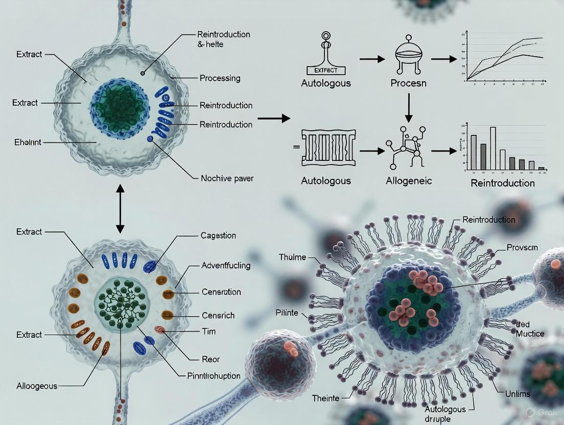
The development of cell therapies has brought to the forefront two fundamentally different manufacturing paradigms: customized and standardized production. These approaches directly correspond to the two main types of cell therapies—autologous (using patient's own cells) and allogeneic (using donor cells)—each with distinct technical, operational, and clinical implications [2]. The choice between these paradigms represents a critical strategic decision for therapy developers, impacting everything from process design and facility planning to supply chain logistics and commercial accessibility [33].
Customized production characterizes autologous cell therapies, where each batch is manufactured individually from a single patient's cells and returned specifically to that patient [2]. This model demands highly personalized production workflows with adaptable environments to accommodate variability in starting cell type and quantity [2]. In contrast, standardized production enables allogeneic cell therapies through mass production of cells from healthy donors, creating "off-the-shelf" products that can be administered to multiple patients [34] [15]. This approach leverages standardized processes, economies of scale, and potentially lower production costs, although it must overcome challenges related to donor variability and immunogenicity [2].
Technical and Operational Comparison
Core Operational Characteristics
The operational differences between customized and standardized production paradigms extend throughout the entire product lifecycle, from cell sourcing to final administration. These differences fundamentally impact manufacturing strategy, quality control approach, and supply chain design.
Table 1: Key Operational Differences Between Customized and Standardized Production Paradigms
| Characteristic | Customized Production (Autologous) | Standardized Production (Allogeneic) |
|---|---|---|
| Cell Source | Patient's own cells [2] | Healthy donor cells [2] |
| Production Scale | Scale-out: Multiple parallel production lines for individual patient products [2] | Scale-up: Large batches aliquoted into individual doses [2] |
| Batch Structure | One batch = One patient [2] | One batch = Hundreds of patients [2] |
| Manufacturing Timeline | Several weeks vein-to-vein time [33] | Pre-manufactured, available immediately [35] |
| Supply Chain Model | Circular, complex logistics with precise scheduling [2] | Linear, bulk processing and storage [2] |
| Product Stability | Short ex vivo half-life (hours) [7] | Cryopreserved, longer shelf life [7] |
Manufacturing Infrastructure and Scalability
The manufacturing infrastructure required for each paradigm differs significantly, particularly regarding scalability approaches. Customized production follows a scale-out strategy, requiring multiple parallel production lines where each manufacturing run produces a single patient-specific batch [2] [36]. This approach maintains identical culture conditions across different batches but introduces logistical challenges including higher labor demands, increased facility footprint, and the need for precise batch tracking systems [36].
Standardized production employs a scale-up strategy, producing larger quantities in single batches that can be aliquoted into individual doses to treat many patients [2]. This approach enables greater efficiency and cost-effectiveness but requires extensive process optimization to ensure parameters such as oxygen transfer, nutrient distribution, and pH control remain consistent at higher volumes [36]. Scale-up introduces significant engineering challenges, as larger bioreactors often struggle with oxygen transfer limitations, shear forces from mixing impellers, and ensuring uniform nutrient distribution [36].
Quality Control and Regulatory Considerations
Quality control systems must be adapted to the specific challenges of each production paradigm. For customized production, quality control focuses on ensuring the safety and efficacy of personalized treatments with stringent tracking of each patient's cells throughout the process [2]. The inherent variability between patient starting materials necessitates wider specifications for analytical testing [2].
For standardized production, quality control emphasizes donor eligibility, cell bank characterization, and managing immune reactions [2]. Batch consistency becomes paramount, with more flexibility in turnaround time and sample volume requirements for release testing compared to autologous approaches [2]. Regulatory validation is more complex in scale-up processes, as changes at larger volumes must be demonstrated to be equivalent to small-scale conditions [36].
Experimental and Manufacturing Protocols
Process Workflows
The manufacturing workflows for customized and standardized production differ fundamentally in their sequence of operations, timing, and technical requirements. The diagrams below illustrate these distinct processes.
Diagram 1: Manufacturing workflows for customized vs. standardized production
Scaling Methodologies
The scaling approaches for each paradigm present distinct technical requirements and challenges. The diagram below illustrates the fundamental differences between scale-out and scale-up strategies.
Diagram 2: Scale-out vs. scale-up strategies for cell therapy manufacturing
The Scientist's Toolkit: Essential Research Reagents and Materials
Successful implementation of either production paradigm requires specialized reagents and materials designed to address their unique technical challenges.
Table 2: Essential Research Reagents and Materials for Cell Therapy Production
| Reagent/Material | Function | Customized Production Application | Standardized Production Application |
|---|---|---|---|
| Cell Separation Media | Isolation of specific cell populations from source material | Critical for processing limited patient apheresis material [37] | Used in initial donor cell processing and master cell bank establishment [37] |
| Genetic Modification Vectors | Introduction of therapeutic genes (e.g., CAR constructs) | Patient-specific viral vectors for T-cell modification [34] | Batch-produced vectors for consistent donor cell engineering [34] |
| Cell Culture Media Formulations | Support cell growth, expansion, and maintenance | Optimized for variable patient cell viability and growth characteristics [37] | Formulated for consistent expansion of healthy donor cells at large scale [37] |
| Cryopreservation Solutions | Long-term storage of cellular products | Patient-specific product storage before reinfusion [7] | Bulk storage of off-the-shelf doses in cell banks [7] |
| Cell Activation Reagents | Stimulate cell proliferation and functionality | Tailored to patient cell responsiveness [37] | Standardized for predictable donor cell activation kinetics [37] |
| Quality Control Assays | Assessment of product safety, potency, and identity | Patient-specific release testing with rapid turnaround [2] | Comprehensive batch characterization with extended panel [2] |
Clinical and Commercial Implications
Therapeutic Performance and Clinical Considerations
The choice between production paradigms carries significant implications for clinical application and therapeutic performance. Customized production offers inherent immune compatibility since the therapy uses the patient's own cells, substantially minimizing rejection risks and eliminating graft-versus-host disease (GVHD) concerns [2] [7]. However, product quality can be variable due to the patient's health status, prior treatments, and cellular characteristics, potentially impacting therapeutic efficacy [33].
Standardized production offers consistent product quality derived from healthy donors but carries inherent immune risks, including graft rejection and GVHD, often necessitating immunosuppressive regimens [2] [7]. Treatment timing differs substantially between paradigms—customized production typically involves several weeks of manufacturing wait time, while standardized products offer immediate, off-the-shelf availability [35]. This timing difference can be critical for patients with rapidly progressing diseases.
Commercial Viability and Access Considerations
From a commercial perspective, each paradigm presents distinct advantages and challenges. Customized production faces significant scalability limitations and higher costs due to its personalized nature, creating substantial access barriers for large patient populations [33] [7]. The complex, circular supply chain requiring precise coordination between collection, manufacturing, and treatment centers further complicates large-scale implementation [2].
Standardized production offers superior scalability potential through batch production that can treat hundreds of patients from a single manufacturing run [2]. This approach benefits from traditional pharmaceutical economies of scale, potentially reducing costs and improving patient access [33] [7]. However, these therapies face challenges related to immune matching requirements and the potential need for immunosuppression, which may limit their applicability across diverse patient populations [7].
Manufacturing Challenges and Solutions
Both paradigms face distinct manufacturing challenges that require specialized approaches and solutions.
Table 3: Manufacturing Challenges and Mitigation Strategies
| Production Paradigm | Primary Challenges | Mitigation Strategies |
|---|---|---|
| Customized Production | - High variability in starting material- Complex supply chain logistics- Limited economies of scale- Time-sensitive manufacturing [2] [7] | - Closed, automated systems to reduce contamination- Digital tracking for chain of identity- Modular manufacturing facilities near treatment centers- Process standardization despite patient variability [2] [33] |
| Standardized Production | - Immune rejection risks- Donor cell variability- Scale-up process optimization- Extensive quality control requirements [2] [36] | - Genetic engineering to reduce immunogenicity- Rigorous donor screening and cell banking- Advanced bioprocess modeling and CFD- Comprehensive batch characterization [34] [36] |
The choice between customized and standardized production paradigms represents a fundamental strategic decision in cell therapy development, with neither approach universally superior. Customized production offers personalization and immune compatibility at the cost of scalability and operational complexity, making it particularly suitable for patient-specific applications where immune matching is challenging [33]. Standardized production enables broader accessibility and potentially lower costs through economies of scale but must overcome immunological barriers and scale-up challenges [34] [15].
The future of cell therapy manufacturing will likely see both paradigms evolving in parallel, with advancements in automation, process control, and genetic engineering addressing their respective limitations [33]. Emerging technologies such as allogeneic CAR-T cells from induced pluripotent stem cells (iPSCs) represent promising approaches to combine the advantages of both paradigms [34] [37]. As the field matures, the optimal manufacturing strategy will continue to depend on the specific therapeutic application, target patient population, and commercial considerations, with both customized and standardized production playing crucial roles in advancing cellular medicines.
Cell therapies represent a paradigm shift in personalized medicine, offering groundbreaking treatments for conditions ranging from hematological malignancies to degenerative diseases. These living medicines are broadly categorized as either autologous (using the patient's own cells) or allogeneic (using cells from a healthy donor). This distinction is the most critical factor determining the subsequent clinical workflow, from the initial cell collection to the final patient infusion. The autologous process is inherently personalized, creating a patient-specific product, while the allogeneic model aims to produce an "off-the-shelf" therapy that can be manufactured in advance for multiple patients [7] [5]. This technical guide provides an in-depth comparison of the clinical workflows for these two approaches, analyzing the distinct logistical, manufacturing, and infusion challenges each presents to researchers and drug development professionals.
Phase 1: Cell Collection and Source Material
The initial phase of the cell therapy workflow centers on obtaining the starting cellular material. The source of these cells is the primary differentiator between autologous and allogeneic pathways and sets the stage for all subsequent processes.
Autologous Cell Collection
In the autologous model, the starting material is collected from the patient destined to receive the therapy. For Chimeric Antigen Receptor (CAR) T-cell therapies, this typically involves leukapheresis, a procedure where the patient's blood is passed through an apheresis machine to separate and collect peripheral blood mononuclear cells, including T cells [38] [39]. The quality of these source cells can be highly variable and is influenced by the patient's disease status, age, and prior treatment history (e.g., chemotherapy), which can affect cell viability and potency [7] [33]. This collection step is a significant bottleneck in the autologous workflow, requiring specialized clinical facilities and coordination between the manufacturing and treatment centers [33].
Allogeneic Cell Collection
In contrast, allogeneic therapies begin with cells harvested from a healthy donor. The donor may be related or unrelated to the patient and is often carefully selected based on strict eligibility and screening requirements [33]. This allows for the selection of starting material with optimal quality and potency. A single donation from a healthy donor can be expanded to create a master cell bank, which in turn can be used to produce large quantities of therapy to treat hundreds of patients [7] [5]. This approach provides a more consistent and controllable starting material compared to the variable patient-derived cells used in autologous therapies.
Table 1: Key Differences in Cell Source and Collection
| Parameter | Autologous Therapy | Allogeneic Therapy |
|---|---|---|
| Cell Source | Patient | Healthy Donor |
| Collection Method | Leukapheresis | Leukapheresis or other donation |
| Material Quality | Variable; impacted by patient health | Consistent; from screened healthy donor |
| Scalability of Collection | Low (one patient, one product) | High (one donor, multiple products) |
| Key Challenge | Patient cell quality and scheduling | Donor screening and HLA matching |
Phase 2: Manufacturing and Logistics
The manufacturing journey from collected cells to final therapeutic product is complex and differs substantially between autologous and allogeneic models, particularly in scale, timing, and logistics.
Despite their different starting points, both autologous and allogeneic therapies often share core manufacturing steps, which may include cell activation, genetic modification (e.g., via viral vector transduction or electroporation), ex vivo expansion, and cryopreservation [38] [7]. The entire process for autologous CAR-T cells typically takes 7 to 14 days from cell collection to cryopreserved product [38]. There is a push to reduce this time, with next-generation platforms aiming for manufacturing in as little as 24 to 72 hours to treat patients more rapidly [33]. A critical best practice for both pathways is to implement closed and automated systems to reduce human touchpoints, minimize contamination risks, and improve process consistency [38] [33].
Autologous: A Personalized Logistics Challenge
Autologous manufacturing is a patient-specific endeavor. Each batch is a unique product, requiring meticulous chain of identity (COI) and chain of custody (COC) tracking throughout its journey [7] [39]. This represents a "scale-out" challenge, where increasing production capacity requires adding more parallel manufacturing platforms or workstations, rather than increasing the size of a single batch [33]. The logistics are exceptionally complex, involving the cryopreserved transport of cells from the clinic to the manufacturing facility and back again, all within a tight, viability-dependent timeframe [39]. Any delay or misstep can directly impact product efficacy and patient safety.
Allogeneic: The "Off-the-Shelf" Model
Allogeneic therapy manufacturing follows a more traditional pharmaceutical "scale-up" model. Cells from a single donor are used to produce a large batch, which is then aliquoted into hundreds or thousands of patient doses [33]. This model allows for advanced, scheduled production, creating a cryopreserved inventory of "off-the-shelf" products that are readily available for immediate use [7] [5]. This eliminates the lengthy waiting period associated with autologous therapies and simplifies logistics, as products can be stored at treatment centers until needed. The primary manufacturing challenge for allogeneic therapies is mitigating the risk of immune rejection (Graft-versus-Host Disease), which often requires additional genetic engineering steps to create hypoimmune cells [7] [5].
Table 2: Manufacturing and Logistics Comparison
| Aspect | Autologous Therapy | Allogeneic Therapy |
|---|---|---|
| Production Model | Patient-specific batch | Large, scalable batch |
| Scalability Approach | Scale-out (multiple parallel units) | Scale-up (larger batch sizes) |
| Key Logistics | Complex; two-way transport per patient | Simplified; one-way distribution to sites |
| Typical Lead Time | Weeks (manufactured to order) | Days (on-demand from inventory) |
| Primary Hurdle | Supply chain complexity & cost | Immune rejection (GvHD) |
Phase 3: Product Infusion and Clinical Administration
The final phase of the workflow is the infusion of the cell therapy product into the patient. While the clinical administration itself may appear similar, the underlying preparation and patient management are strongly influenced by the therapy type.
Pre-Infusion Preparations
Prior to infusion, patients typically undergo lymphodepleting chemotherapy to create a favorable environment for the engineered cells to engraft and expand [7]. For allogeneic therapies, this may be accompanied by a more intensive conditioning regimen and/or the use of immunosuppressive drugs to prevent graft rejection and mitigate GvHD risk [7] [5]. In contrast, autologous therapies, being derived from self, do not carry a risk of GvHD and generally do not require long-term immunosuppression [5].
The Infusion Workflow
The day-of-infusion process is a carefully orchestrated sequence. Observations across multiple academic medical centers reveal a common, multi-step workflow [40] [41]:
- Product Verification: The product undergoes multiple verification checks against the patient's identity. This typically involves confirmation by cell therapy technologists (CTTs), pharmacists, and nurses [40].
- Thawing: Cryopreserved products are thawed, often in a 37°C water bath, in a location close to the patient's room to minimize delay post-thaw [40] [41].
- Infusion: The product is administered to the patient intravenously, similar to a blood transfusion. The time from thaw to infusion is critical and should be minimized; studies have recorded averages ranging from under 20 minutes to over 40 minutes, largely dependent on the distance between the thaw location and the patient's room [40].
The entire process requires close communication and coordination between CTTs, nurses, and physicians to ensure patient stability and product integrity [40] [41]. Standardizing this infusion workflow across treatment centers is an ongoing effort to improve efficiency and reduce the potential for errors [40].
The Scientist's Toolkit: Essential Research Reagents & Materials
The development and manufacturing of cell therapies rely on a suite of specialized reagents and instruments. The following table details key solutions used in the featured workflows.
Table 3: Key Reagents and Materials for Cell Therapy Manufacturing
| Research Reagent / Instrument | Function in Workflow | Technical Application Note |
|---|---|---|
| Magnetic Beads & Separation System | Isolation and activation of target T cells from apheresis product. | Systems like the Gibco CTS DynaClect use magnetic beads for closed, automated cell separation and activation, minimizing open-handling steps [38]. |
| Cell Washing System | Washing cells to remove cytokines, reagents, or cryoprotectants. | Counterflow centrifugation systems (e.g., Gibco CTS Rotea) gently concentrate and wash cells while maintaining viability, a step often performed post-activation and post-expansion [38]. |
| Electroporation System | Introducing genetic material (e.g., CAR transgene) into cells. | Systems like the Gibco CTS Xenon use mechanical electroporation for non-viral gene editing, an alternative to viral vector transduction [38]. |
| Viral Vectors (Lentiviral/Retroviral) | Stable genetic modification of cells to express the chimeric antigen receptor. | Viral vectors are a common method for CAR transduction. Manufacturing requires high-titer, high-purity vectors produced under GMP conditions [38]. |
| Cell Culture Media & Supplements | Supporting the ex vivo expansion of modified T cells. | Serum-free, GMP-grade media formulations are critical for robust cell growth and maintaining consistent product quality attributes during the expansion phase [33]. |
| Cryopreservation Media | Preserving the final cell product for storage and transport. | Formulations containing DMSO are standard for cryopreserving cell therapy products to maintain viability and potency during frozen storage [40] [39]. |
The clinical workflows for autologous and allogeneic cell therapies are distinct, each with inherent trade-offs. The autologous pathway offers a personalized treatment with a lower risk of immune complications but is hampered by complex logistics, high costs, and scalability challenges. In contrast, the allogeneic "off-the-shelf" model promises greater scalability, immediate treatment availability, and potentially lower costs, but must overcome hurdles of immune rejection and GvHD. The choice between these models is not a simple binary; rather, it is dictated by the specific disease target, patient population, and therapeutic mechanism. Future progress will depend on continued innovation in genetic engineering to improve allogeneic cell persistence and safety, coupled with advances in automation and standardization to streamline the manufacturing and infusion processes for both modalities.
The field of advanced cellular therapeutics is fundamentally divided into two paradigms: autologous and allogeneic cell therapies. Autologous therapies involve harvesting a patient's own cells, which are then processed, expanded, and often genetically engineered ex vivo before being reinfused into the same patient [7] [42]. In contrast, allogeneic therapies utilize cells derived from a healthy donor, which are processed and banked to create "off-the-shelf" products that are readily available for multiple patients [5] [35]. This foundational distinction governs every aspect of therapeutic development, from manufacturing and logistics to clinical application and commercial viability. The choice between these approaches involves a complex trade-off between the reduced risk of immune rejection with autologous products and the superior scalability and immediate availability of allogeneic products [7] [5]. This technical guide provides an in-depth analysis of three prominent cellular applications—Chimeric Antigen Receptor T-cell (CAR-T) therapy, Hematopoietic Stem Cell Transplantation (HSCT), and Mesenchymal Stem Cell (MSC) therapy—within this critical autologous versus allogeneic framework, offering researchers and drug development professionals a detailed comparison of their mechanisms, applications, and technical requirements.
Chimeric Antigen Receptor T-Cell (CAR-T) Therapy
Core Mechanism and Engineering Design
CAR-T cell therapy represents a paradigm shift in immunotherapy, primarily for hematological malignancies. A chimeric antigen receptor (CAR) is a synthetic receptor engineered to redirect T-cells to specifically recognize and eliminate cells expressing a target antigen. The core structure of a CAR consists of an extracellular antigen-recognition domain, typically a single-chain variable fragment (scFv) derived from an antibody; a hinge region for flexibility; a transmembrane domain that anchors the receptor; and an intracellular signaling domain that activates the T-cell upon antigen binding [43]. Critically, CAR recognition is MHC-independent, allowing T-cells to target tumor cells that often downregulate MHC molecules to evade immune detection [43].
CAR design has evolved through several generations, each enhancing signaling capacity and therapeutic efficacy:
- First-generation CARs contained only the CD3ζ signaling domain. While capable of initiating T-cell activation, they exhibited limited persistence and efficacy due to the absence of costimulatory signals, often requiring exogenous IL-2 support [43].
- Second-generation CARs incorporated one costimulatory domain (e.g., CD28 or 4-1BB) in tandem with CD3ζ. This dramatically improved T-cell proliferation, cytokine production, cytotoxicity, and in vivo persistence. Most FDA-approved CAR-T therapies utilize this design [43].
- Third-generation CARs combine two costimulatory domains (e.g., CD28 and 4-1BB) with CD3ζ, aiming to further amplify T-cell signaling and functionality, though with variable results reported in preclinical studies [43].
- Fourth-generation CARs (TRUCKs) are engineered to not only kill target cells but also to express transgenic immunomodulators (e.g., IL-12, IL-15) or chemokine receptors upon activation. This modifies the tumor microenvironment and recruits a secondary immune response, showing significant promise for solid tumors [43].
Autologous vs. Allogeneic CAR-T: Technical and Manufacturing Comparison
The clinical success of CAR-T therapy has been demonstrated predominantly with autologous products. However, the field is rapidly advancing toward allogeneic, "off-the-shelf" platforms to address key limitations of autologous approaches.
Table 1: Comparison of Autologous and Allogeneic CAR-T Therapies
| Characteristic | Autologous CAR-T | Allogeneic CAR-T |
|---|---|---|
| Cell Source | Patient's own T-cells [42] | Healthy donor T-cells [5] |
| Manufacturing Model | Personalized, patient-specific [7] | Batch-produced, "off-the-shelf" [5] [35] |
| Production Timeline | Several weeks [7] [42] | Immediate availability from cryobanks [35] |
| Key Challenges | - Manufacturing delays- Product quality variability (impacted by patient disease/prior treatment) [7] [42]- High cost | - Risk of Graft-versus-Host Disease (GvHD) [7]- Host immune-mediated rejection- Requirement for lymphodepletion [7] |
| Mitigation Strategies | - Process automation- Closed system bioreactors | - Gene editing (e.g., CRISPR to disrupt TCR to prevent GvHD) [5]- Use of alternative cell sources (e.g., virus-specific T-cells, NK cells) |
Detailed Experimental Protocol: Manufacturing Autologous CAR-T Cells
Objective: To generate autologous CD19-targeting CAR-T cells for the treatment of B-cell acute lymphoblastic leukemia.
Materials:
- Leukapheresis product from the patient.
- Cell separation reagents: Ficoll-Paque PLUS, anti-CD3/CD28 antibody-conjugated magnetic beads.
- Cell culture media: X-VIVO 15 or TexMACS, supplemented with human serum or FBS, IL-2.
- Activation reagents: Recombinant human IL-2, anti-CD3/CD28 antibodies.
- Gene transfer vector: Lentiviral vector encoding anti-CD19 second-generation CAR (CD28 or 4-1BB costimulatory domain with CD3ζ).
- Quality control assays: Flow cytometry (for CAR expression, T-cell phenotyping), cytotoxicity assays, cytokine release assays.
Methodology:
- Cell Harvesting & Isolation: Isolate peripheral blood mononuclear cells (PBMCs) from the leukapheresis product via density gradient centrifugation. Activate T-cells using anti-CD3/CD28 magnetic beads and recombinant human IL-2 (100-300 IU/mL) [42].
- Genetic Modification: On day 2-3 post-activation, transduce activated T-cells with the lentiviral vector via spinoculation (centrifugation at 2000 x g for 60-90 minutes at 32°C) in the presence of polybrene (8 µg/mL). Maintain cells in culture with IL-2.
- Ex Vivo Expansion: Culture transduced T-cells for 7-14 days, maintaining a cell density of 0.5-2 x 10^6 cells/mL. Monitor cell count and viability.
- Harvest and Formulation: Harvest cells, remove activation beads, and wash. Formulate final product in an infusion-ready cryopreservation medium. Perform quality control testing, including sterility, mycoplasma, endotoxin, CAR expression (by flow cytometry), and potency.
- Infusion: Administer to the patient following a lymphodepleting chemotherapy regimen (typically cyclophosphamide and fludarabine).
Diagram 1: Autologous CAR-T manufacturing workflow.
Hematopoietic Stem Cell Transplantation (HSCT)
Clinical Indications and Transplant Categories
HSCT is a long-established curative procedure for a wide range of hematological, oncological, and genetic disorders. Its primary goal is to rescue the hematopoietic system after myeloablative therapy or to instill a graft-versus-tumor (GvT) effect. According to the latest 2025 EBMT practice recommendations, key indications include [44]:
- Acute Myeloid Leukemia (AML): Allo-HCT is standard in first complete remission (CR1) for adverse-risk and many intermediate-risk AML patients, as defined by ELN guidelines. It is not generally recommended for favorable-risk AML unless MRD persists [44].
- Acute Lymphoblastic Leukaemia (ALL): Allo-HCT is a cornerstone of therapy, particularly for high-risk Ph-ALL and all Ph+ALL. Measurable residual disease (MRD) status is a critical factor in transplant decision-making [44].
- Other diseases: HSCT is also indicated for myelodysplastic syndromes, severe aplastic anemia, inherited immune deficiencies, and certain solid tumors.
HSCT procedures are categorized based on donor type and stem cell source, each with distinct implications for the risk of GvHD, relapse, and overall survival [44].
Table 2: Categorization of Hematopoietic Stem Cell Transplants
| Category | Donor Type / Source | Description & Key Considerations |
|---|---|---|
| Autologous | Patient's own cells [42] | - Source: Bone marrow, peripheral blood, or cord blood [44].- Key Consideration: Avoids GvHD; primarily used for its dose-intensive myeloablative effect (e.g., in lymphomas, multiple myeloma). Lacks GvT effect, leading to higher relapse risk in some malignancies [44]. |
| Allogeneic | Matched Sibling Donor (MSD) | - Source: Bone marrow or peripheral blood [44].- Key Consideration: Historically the gold standard donor; lower rates of GvHD and rejection compared to alternative donors. |
| Matched Unrelated Donor (MUD) | - Source: Bone marrow or peripheral blood [44].- Key Consideration: Defined as a 10/10 HLA-matched unrelated donor; outcomes now approach those of MSD. | |
| Mismatched Unrelated Donor (MMUD) / Haploidentical | - Source: Bone marrow, peripheral blood, or cord blood [44].- Key Consideration: Use of post-transplant cyclophosphamide (PTCy) for GvHD prophylaxis has significantly improved outcomes, greatly expanding the donor pool [44]. |
Autologous vs. Allogeneic HSCT: A Clinical Decision Framework
The choice between autologous and allogeneic HSCT involves a nuanced risk-benefit analysis, balancing the disease-specific risk of relapse against the procedure-related mortality and morbidity.
Table 3: Clinical Comparison of Autologous and Allogeneic HSCT
| Parameter | Autologous HSCT | Allogeneic HSCT |
|---|---|---|
| Primary Mechanism | Myeloablation and hematopoietic rescue | Myeloablation, hematopoietic rescue, and Graft-versus-Tumor (GvT) effect |
| Major Toxicity | Regimen-related toxicity, infections during cytopenia | Graft-versus-Host Disease (GvHD), infections (related to prolonged immunosuppression), organ toxicity |
| Relapse Risk | Higher (no GvT effect) | Lower (powerful GvT effect) |
| Non-Relapse Mortality | Lower | Higher |
| Immunosuppression | Not required | Required long-term to prevent/treat GvHD |
| Donor Availability | Always available | Dependent on HLA matching from related or unrelated donors |
Mesenchymal Stem Cell (MSC) Therapy
Biological Characteristics and Sourcing
MSCs are multipotent stromal cells characterized by their tri-lineage differentiation potential (into osteocytes, chondrocytes, and adipocytes), immunomodulatory properties, and tissue-homing capabilities [45] [46]. According to the International Society for Cellular Therapy (ISCT), MSCs must adhere to plastic in standard culture, express surface markers CD105, CD73, and CD90 (≥95%), while lacking expression of hematopoietic markers CD45, CD34, CD14/CD11b, CD79α/CD19, and HLA-DR (≤2%), and must differentiate into the three mesodermal lineages in vitro [45] [46].
MSCs can be isolated from a variety of tissues, with source-dependent characteristics influencing their therapeutic profile:
- Bone Marrow (BM-MSCs): The most extensively studied source; requires invasive harvest and has limited cell numbers that decline with donor age [45] [46].
- Adipose Tissue (AD-MSCs): Obtained via liposuction; abundant yield and less invasive than bone marrow harvest [45] [46].
- Umbilical Cord (UC-MSCs): Isolated from Wharton's jelly; considered medical waste, thus non-controversial. They exhibit high proliferative capacity, low immunogenicity, and strong immunomodulatory properties [45] [46].
- Placenta (P-MSCs): High proliferative and immunosuppressive capacity, but isolation is complex due to the tissue's heterogeneous composition [46].
Therapeutic Applications and the Emergence of CAR-MSCs
The therapeutic effects of MSCs are primarily mediated through paracrine signaling via secreted growth factors, cytokines, and extracellular vesicles, which modulate the local microenvironment, promote tissue repair, and exert anti-inflammatory and pro-angiogenic effects [45]. They interact with various immune cells (T cells, B cells, dendritic cells, macrophages) to suppress pro-inflammatory responses and promote regulatory pathways [45].
MSC applications are broad, spanning both allogeneic and autologous approaches, with allogeneic being more common due to the immune-privileged status of MSCs [5]. Approved allogeneic MSC products exist for conditions like steroid-refractory acute Graft-versus-Host Disease (SR-aGVHD) [5]. Preclinically, MSCs are being investigated for neurological disorders, cardiovascular diseases, and orthopedic injuries [45] [47].
A cutting-edge advancement is the engineering of CAR-MSCs [43]. This approach combines the precise tumor-targeting ability of CAR technology with the innate tumor-homing and immunomodulatory properties of MSCs. Preclinical studies show CAR-MSCs can effectively target glioblastoma, Ewing sarcoma, and lung cancer by secreting therapeutic agents like TRAIL or bispecific antibodies, or by inducing regulatory T-cells within the tumor microenvironment [43].
Experimental Protocol: In Vitro Immunomodulatory Assay of MSCs
Objective: To evaluate the immunosuppressive capacity of human UC-MSCs on mitogen-activated peripheral blood mononuclear cells (PBMCs).
Materials:
- Test Article: Human UC-MSCs (P3-P5).
- Responder Cells: PBMCs from a healthy donor.
- Stimulant: Phytohemagglutinin (PHA).
- Culture Vessels: 96-well round-bottom plates.
- Cell Culture Media: RPMI-1640 supplemented with 10% FBS.
- Detection Reagent: [^3H]-thymidine.
Methodology:
- Setup Co-culture: Seed UC-MSCs in a 96-well plate at varying densities (e.g., 1x10^4, 2.5x10^4 cells/well) and allow to adhere overnight.
- Activate PBMCs: Isolate PBMCs from donor blood via Ficoll density gradient. Resuspend PBMCs in complete media.
- Initiate Assay: Add PBMCs (1x10^5 cells/well) to the MSC-containing wells. Include controls: PBMCs alone, PBMCs + PHA, MSCs alone.
- Stimulation: Add PHA to the appropriate wells at a final optimal concentration (e.g., 5 µg/mL).
- Proliferation Measurement: After 72-96 hours of co-culture, pulse each well with [^3H]-thymidine (0.5 µCi/well) for the final 16-18 hours. Harvest cells onto a filtermat and measure incorporated radioactivity using a beta-scintillation counter.
- Data Analysis: Calculate percentage suppression of PBMC proliferation using the formula:
- % Suppression = [1 - (cpm in Co-culture with PHA / cpm in PBMCs+PHA control)] x 100
Diagram 2: MSC immunomodulation assay workflow.
Table 4: Key Research Reagents for Cell Therapy Development
| Reagent / Solution | Primary Function | Application Notes |
|---|---|---|
| Ficoll-Paque | Density gradient medium for isolation of PBMCs or MSCs from tissue digests. | Critical first step in cell processing; separation quality impacts downstream functionality. |
| Recombinant Human Cytokines (IL-2, SCF, TPO) | Promote T-cell expansion (IL-2) or hematopoietic stem cell survival and proliferation (SCF, TPO). | Concentration and timing are crucial for maintaining cell fitness and desired phenotype. |
| Anti-CD3/CD28 Antibodies (or beads) | Polyclonal T-cell activation and expansion. | Mimics antigen-presenting cell signal; essential for T-cell transduction and CAR-T manufacturing. |
| Lentiviral / Retroviral Vectors | Stable gene delivery for CAR or other therapeutic transgene expression. | Viral titer and transduction efficiency are key Critical Quality Attributes (CQAs). Safety features (e.g., suicide genes) are increasingly important. |
| Cell Culture Media (X-VIVO, TexMACS, DMEM) | Base medium for ex vivo cell culture and expansion. | Serum-free, GMP-grade formulations are preferred for clinical translation to reduce variability and xenogeneic immune reactions. |
| Flow Cytometry Antibodies (CD3, CD4, CD8, CD19, CD34, CD45, CD73, CD90, CD105) | Cell phenotyping, purity assessment, and characterization of final product. | Essential for confirming identity and purity of cellular products per ISCT and other guidelines. |
The landscape of cellular therapy is defined by the strategic interplay between autologous and allogeneic approaches. Autologous therapies (CAR-T, auto-HSCT) offer the key advantage of immune compatibility but face significant challenges in manufacturing scalability, cost, and production time for individual patients [7] [42]. Allogeneic therapies (donor-derived HSCT, "off-the-shelf" MSCs, and emerging allogeneic CAR-T/CAR-NK) provide the benefits of immediate availability and industrial scalability but contend with the persistent challenges of immune rejection and GvHD, necessitating sophisticated mitigation strategies like immunosuppression or genetic engineering [7] [5].
The future trajectory of the field points toward convergence. Innovations in gene editing (e.g., CRISPR) are enabling the creation of hypoimmunogenic allogeneic cells, blurring the lines between these paradigms [5] [47]. The development of fourth-generation CARs and CAR-MSCs illustrates a move toward more complex, multifunctional engineered cell products designed to overcome the immunosuppressive tumor microenvironment [43]. As these technologies mature, the choice between autologous and allogeneic will increasingly be dictated by disease indication, target product profile, and manufacturing economics, driving the field toward a new era of personalized, yet readily accessible, regenerative and immunotherapeutic medicines.
The field of cell therapy stands at a pivotal juncture, where scientific advancement is increasingly constrained by manufacturing capabilities. As these living medicines transition from laboratory curiosities to commercial therapeutics, scalability has emerged as a critical determinant of clinical and commercial success. The fundamental challenge lies in expanding production capacity while maintaining consistent product quality, particularly when the "product" consists of sensitive, living cells. For researchers and drug development professionals, understanding scalability is no longer a peripheral concern but an essential component of therapeutic development that must be integrated from the earliest stages of research [48].
The choice between scale-up and scale-out strategies represents a fundamental decision point that intersects with the core distinction between autologous (patient-specific) and allogeneic (off-the-shelf) therapy paradigms. Scale-up involves increasing batch size by transitioning to larger bioreactors, while scale-out maintains smaller volumes but increases production by running multiple parallel vessels [36]. This technical whitepaper examines these scalability models through the lens of autologous versus allogeneic cell therapy, providing a structured framework for strategic decision-making in therapeutic development.
Core Concepts: Scale-Up vs. Scale-Out Defined
Scale-Up Strategy
Scale-up refers to the process of increasing production volume by using larger bioreactors or culture vessels. This approach centralizes manufacturing into single, high-volume batches and is characterized by:
- Volume Expansion: Transition from small lab-scale bioreactors to large industrial-scale systems [36]
- Process Intensification: Increased production capacity within a single manufacturing run [49]
- Engineering Challenges: Must maintain homogeneous conditions (oxygen transfer, nutrient distribution, pH control) across expanded culture volumes [36]
Scale-Out Strategy
Scale-out increases capacity by multiplying the number of parallel production units operating simultaneously, rather than increasing individual unit size. Key characteristics include:
- Parallel Processing: Multiple small-scale bioreactors or flasks operating concurrently [36] [49]
- Modular Design: Maintains identical culture conditions across independent units [36]
- Logistical Complexity: Requires sophisticated batch tracking and process control systems [36]
Table 1: Fundamental Characteristics of Scale-Up and Scale-Out Strategies
| Parameter | Scale-Up | Scale-Out |
|---|---|---|
| Batch Size | Single, large batch | Multiple, small batches |
| Facility Footprint | Vertical expansion | Horizontal expansion |
| Process Control | Engineering challenges with mixing, gas exchange | Consistent conditions per unit, but multiple units to monitor |
| Capital Investment | High for equipment, lower per unit output | Distributed across multiple smaller systems |
| Flexibility | Limited once implemented | High flexibility for parallel production |
Scalability in Autologous vs. Allogeneic Cell Therapy Frameworks
The fundamental biological distinction between autologous and allogeneic cell therapies creates inherent alignment with different scalability approaches, with significant implications for manufacturing strategy, facility design, and commercial planning.
Autologous Therapies: Inherent Alignment with Scale-Out
Autologous cell therapies are manufactured from a patient's own cells, creating an intrinsically personalized manufacturing paradigm with natural affinity for scale-out strategies [49]. Each production lot corresponds to an individual patient, making scale-up largely irrelevant as batch size cannot be increased beyond a single patient treatment [36]. The scale-out model enables:
- Parallel Patient Processing: Multiple patients can be treated simultaneously through independent manufacturing streams [49]
- Regional Manufacturing Networks: Decentralized production facilities located closer to treatment centers [36] [49]
- Individual Batch Integrity: Strict chain of identity and custody maintained through discrete manufacturing processes [7]
The autologous scale-out model faces significant logistical challenges, including higher labor demands, increased facility footprint, and complex batch tracking systems [36]. However, it provides crucial flexibility for therapies with short shelf lives that cannot be stored or frozen, requiring rapid and continuous production to meet clinical demand [36].
Allogeneic Therapies: Optimized for Scale-Up
Allogeneic cell therapies, derived from unrelated donors and designed as "off-the-shelf" products, align naturally with scale-up strategies [49]. These therapies treat multiple patients from a single manufacturing run, creating economic imperatives for large-scale production:
- Centralized Manufacturing: Large-scale production facilities supplying global markets [49]
- Economies of Scale: Reduced cost per dose through volume production [7]
- Standardized Products: Consistent quality across patient treatments from same donor source [7]
Allogeneic scale-up enables the creation of cryopreserved cell banks that are readily available to patients, eliminating treatment delays and providing suitable for urgent medical conditions [5] [7]. This "off-the-shelf" availability represents a significant advantage over autologous approaches for conditions requiring immediate intervention.
Table 2: Scalability Alignment in Autologous vs. Allogeneic Cell Therapies
| Consideration | Autologous Therapy | Allogeneic Therapy |
|---|---|---|
| Natural Scalability Approach | Scale-Out | Scale-Up |
| Batch-to-Patient Relationship | One batch per patient | One batch for many patients |
| Manufacturing Model | Personalized, patient-specific | Universal, off-the-shelf |
| Production Infrastructure | Distributed, multi-facility | Centralized, large-scale |
| Inventory Management | Just-in-time manufacturing | Cryopreserved inventory |
| Cost Structure | High per-patient cost | Economies of scale |
Technical Implementation and Manufacturing Platforms
Successful implementation of scalability strategies requires careful selection of culture platforms and bioreactor systems aligned with both the cell type and scalability approach.
Culture Platform Selection: Adherent vs. Suspension
The biological characteristics of the therapeutic cell type fundamentally constrain platform selection, creating distinct scale-up and scale-out pathways:
Suspension Platforms are ideal for blood-derived cells like CAR-T cells, enabling scale-up through conventional stirred-tank bioreactors that reduce processing steps and facility footprint [49]. However, careful agitation control is required to ensure adequate gas and nutrient exchange while minimizing shear stress-induced cell damage [49].
Adherent Platforms better mimic native environments for tissue-derived cells (MSCs, stem cells) and offer advantages for scale-out through multiple parallel vessels [49]. Recent advances in adherent culture technologies, including microcarriers and fixed bed bioreactors, now enable scale-up approaches previously limited to suspension platforms [49].
Bioreactor Systems for Scale-Up and Scale-Out
Table 3: Bioreactor Systems for Scale-Up and Scale-Out Applications
| System Type | Scale Applicability | Cell Type Compatibility | Key Features | Limitations |
|---|---|---|---|---|
| Stirred-Tank Bioreactors | Scale-Up | Suspension cells (T-cells) | Conventional large-scale platform; reduced processing steps | Shear stress concerns; requires careful agitation control [49] |
| Rocking Motion Bioreactors | Scale-Up | Suspension cells | Gentle mixing; suitable for shear-sensitive cells | Limited volume capacity compared to stirred-tank [49] |
| Fixed Bed Bioreactors | Scale-Up | Adherent cells (MSCs, HSCs) | Structured support for adherent cells; high surface-to-volume ratio | Complexity in cell harvesting [49] |
| Hollow Fiber Bioreactors | Scale-Up | Adherent cells | High cell density; continuous perfusion | Monitoring challenges; potential nutrient gradients [49] |
| Multi-Layered Flasks | Scale-Out | Adherent cells | Simple scale-out; direct visualization | Manual handling; limited scale potential [49] |
| Stacked Vessels | Scale-Out | Adherent cells | Increased surface area in compact footprint | Potential heterogeneity between stacks [49] |
Strategic Decision Framework: Selecting the Optimal Scalability Approach
The choice between scale-up and scale-out strategies involves multidimensional considerations beyond simple technical feasibility. Researchers and development professionals should evaluate the following factors:
Product and Process Characteristics
- Therapeutic Model: Autologous therapies inherently align with scale-out; allogeneic with scale-up [49] [7]
- Cell Type Characteristics: Suspension cells favor scale-up; adherent cells may accommodate both with appropriate platforms [49]
- Batch Definition: Individual patient batches (scale-out) versus multi-patient batches (scale-up) [36]
Manufacturing and Operational Considerations
- Facility Design: Scale-up requires vertical integration; scale-out enables distributed manufacturing [36]
- Automation Strategy: Scale-out benefits from parallel processing automation; scale-up from process intensification technologies [48]
- Quality Control: Scale-up enables single batch characterization; scale-out requires robust across-batch consistency [7]
Commercial and Regulatory Factors
- Market Size: Scale-up for large patient populations; scale-out for targeted applications [36]
- Regulatory Pathway: Scale-up facilitates traditional biologics regulatory approach; scale-out may require novel frameworks for personalized products [36]
- Cost Structure: Scale-up reduces cost through volume; scale-out through parallelization and decentralization [7]
The Scientist's Toolkit: Essential Research Reagents and Materials
Successful scalability research requires specific reagents and systems designed to model manufacturing conditions at various scales:
Table 4: Essential Research Reagents and Materials for Scalability Studies
| Reagent/Material | Function | Scale Application | Key Considerations |
|---|---|---|---|
| Microcarriers | Small spheres providing surface for adherent cell growth in suspension systems | Scale-Up | Various sizes, materials, and coatings available; enable preliminary tests before large bioreactors [49] |
| Specialized Culture Media | Formulated to maintain cell phenotype and functionality during expansion | Both | Must be scalable to manufacturing-grade sources; composition affects critical quality attributes |
| Cell Dissociation Reagents | Enzyme-based solutions for detaching adherent cells from surfaces | Both | Impact cell viability and functionality; must be compatible with closed systems at scale |
| Cryopreservation Media | Formulations for freezing and storing cell products | Both | Critical for maintaining viability in scale-out models with banking strategies |
| Genetic Modification Tools | Viral/non-viral vectors for cell engineering (e.g., CAR constructs) | Both | Transduction efficiency must be maintained across scales; critical for process consistency |
| Process Analytics | Reagents for in-process testing (viability, potency, identity) | Both | Must be scalable to quality control requirements; essential for batch release |
Methodologies: Experimental Protocols for Scalability Assessment
Scale-Down Modeling Protocol
Scale-down models are essential for predicting performance at commercial manufacturing scale, enabling researchers to de-risk scale-up and optimize scale-out strategies early in development.
Objective: Establish representative small-scale models that accurately predict performance at manufacturing scale. Materials:
- Bench-scale bioreactor systems (0.1-10L)
- Single-use bioreactor vessels
- Process analytical technology (PAT) for monitoring
- Cell culture media identical to planned manufacturing scale
Procedure:
- Parameter Identification: Determine critical process parameters (CPPs) through risk assessment (e.g., oxygen transfer rate, pH control, nutrient feeding strategy)
- System Characterization: Quantify engineering parameters (kLa, mixing time, power input) at both small and manufacturing scales
- Process Translation: Establish equivalent operating conditions across scales using dimensionless numbers (Reynolds, Power)
- Performance Verification: Conduct multiple runs comparing cell growth, metabolism, and product quality attributes across scales
- Model Validation: Confirm predictive capability through statistical comparison of critical quality attributes (CQAs)
Key Measurements:
- Cell viability and density throughout culture
- Metabolite profiles (glucose, lactate, ammonia)
- Product titer and quality (phenotype, potency)
- Oxygen consumption rate (OUR) and carbon dioxide evolution rate (CER)
Parallel System Comparability Protocol
For scale-out strategies, demonstrating consistency across multiple parallel units is essential for regulatory approval and commercial implementation.
Objective: Demonstrate equivalent performance across multiple parallel production units in a scale-out model. Materials:
- Multiple identical small-scale bioreactors or culture vessels
- Standardized cell banking system
- Automated sampling and analysis systems
- Statistical software for data analysis
Procedure:
- System Qualification: Confirm identical performance of all empty culture vessels through controlled testing
- Inoculum Standardization: Use identical cell source (same passage, same donor) for all parallel units
- Parallel Processing: Operate multiple units simultaneously using standardized procedures
- Intensive Monitoring: Sample all units at matched timepoints for critical process parameters
- Data Analysis: Apply statistical methods (ANOVA, equivalence testing) to demonstrate comparability
Acceptance Criteria:
- ≤15% coefficient of variation in final cell yield
- No statistically significant differences in critical quality attributes
- Equivalent kinetics (doubling time, metabolic profile) across systems
Future Directions and Emerging Solutions
The evolving landscape of cell therapy manufacturing is driving innovation in scalability approaches, with several promising developments:
Integrated Scalability Solutions
Next-generation manufacturing platforms are emerging that blend scale-up and scale-out advantages through modular, flexible designs. As noted by industry leaders, "Therapy developers can adopt a flexible, modular approach that integrates best-of-breed technologies alongside expert service provision to achieve flexibility, reproducibility, and scalability" [48]. These systems enable:
- Adaptable Scale: Transition between scale-up and scale-out configurations based on product phase
- Digital Integration: Advanced process control and data analytics for enhanced reproducibility
- Closed Processing: Reduced contamination risk while maintaining flexibility
Automation and Digital Technologies
Advanced automation is transforming both scale-up and scale-out paradigms by enhancing reproducibility and efficiency. As emphasized in industry discussions, "Automation is not just robotics... You can think about this ahead of time and find something that is going to be a two-minute addition versus a two-hour pain" [48]. Key advancements include:
- Flexible Automation: Modular systems that can be adapted to specific process needs
- Digital Twins: Virtual modeling of processes to predict scale-up behavior
- PAT Implementation: Real-time monitoring and control of critical process parameters
The strategic selection between scale-up and scale-out manufacturing models represents a fundamental determinant of success in cell therapy development. This decision must be aligned with the core therapeutic approach—autologous or allogeneic—and integrated early in the development lifecycle. As the industry advances, the traditional boundaries between these scalability approaches are blurring through innovative technologies that combine the advantages of both paradigms. For researchers and drug development professionals, embracing scalability as a primary design consideration—rather than a post-approval afterthought—is essential for delivering these transformative therapies to the patients who need them.
The advancement of cell therapies marks a paradigm shift in the treatment of complex diseases, yet scalable and quality-driven manufacturing remains a significant bottleneck for the industry [50]. The inherent complexity of these living therapies, particularly the distinction between autologous (patient-specific) and allogeneic (off-the-shelf) products, creates unprecedented supply chain challenges that directly impact therapeutic efficacy and commercial viability. While autologous therapies involve complex, patient-specific journeys, allogeneic products offer the potential for a more standardized, batch-based approach [50] [51].
The cell and gene therapy supply chain logistics market is projected to grow from USD 1.5 billion in 2024 to USD 4.4 billion by 2034, reflecting the critical expansion needed to support these innovative treatments [52]. This growth is driven by the need for robust, adaptable services that can coordinate highly personalized processes from patient identification through cell harvest, therapeutic intervention, and long-term outcomes data collection [52]. This analysis examines how traditional linear and emerging circular supply chain models address the unique requirements of autologous and allogeneic cell therapies, with significant implications for research direction, clinical outcomes, and commercial scalability.
Linear vs. Circular Supply Chain Models: Core Concepts
The Linear Supply Chain Model
The traditional linear supply chain operates on a "Take ➔ Make ➔ Dispose" model that has dominated manufacturing for decades [53]. This approach relies on a continuous influx of new raw materials and results in significant waste accumulation, with global waste expenses reaching $163 billion annually and approximately two billion tons of waste materials in landfills [53].
In the context of cell therapy, this model manifests as a unidirectional flow of materials:
- Patient cell collection → Manufacturing → Administration → Disposal of consumables
This linear approach creates vulnerability to raw material cost volatility and supply disruptions, particularly problematic for therapies with critical timing constraints [53].
The Circular Supply Chain Model
The circular supply chain embraces a "minimal waste, maximum utility" philosophy, creating a closed-loop system that maximizes resource efficiency [53]. This model operates through six core principles known as the Six R's of waste management:
- Rethink: Re-evaluate material sourcing, manufacturing, and end-of-life strategies
- Refuse: Eliminate unnecessary or inefficient materials
- Reduce: Minimize material consumption and overproduction
- Reuse: Repurpose materials, defective products, and customer returns
- Repurpose: Innovatively find new uses for materials that would otherwise be wasted
- Recover: Reintroduce valuable materials and products into the supply chain [53]
This framework creates resilience against supply disruptions and extends the life cycle of already-invested materials, offering both economic and environmental benefits [53].
Supply Chain Implications of Autologous vs. Allogeneic Therapies
The fundamental biological distinction between autologous (using patient's own cells) and allogeneic (using donor cells for multiple patients) therapies creates dramatically different supply chain requirements with direct implications for manufacturing and logistics complexity [50].
Table 1: Supply Chain Implications by Therapy Type
| Supply Chain Aspect | Autologous Therapies | Allogeneic Therapies |
|---|---|---|
| Starting Material | Patient-specific cells; varies with each manufacturing run [50] | Single donor lot for multiple patients; more consistent [50] |
| Manufacturing Model | Decentralized or hub-and-spoke; multiple parallel batches [51] | Centralized; large-scale batches [50] |
| Supply Chain Structure | Complex web with multiple hand-off points [51] | More linear distribution from manufacturer to treatment centers |
| Key Logistics Challenge | Maintaining chain of identity and managing multiple temperature profiles [51] | Scaling up while maintaining product quality and stability [50] |
| Inventory Management | Just-in-time; product expires if not used [51] | Traditional inventory models possible [50] |
| Waste Generation | High (patient-specific materials, kits, containers) [51] | Lower per dose (standardized materials) |
The autologous therapy supply chain involves exceptionally complex logistics with multiple material transfers at different temperatures. A typical process includes: tumor cell collection (shipped at controlled ambient temperature), apheresis collection (shipped refrigerated at 2-8°C), frozen tissue transport (on dry ice), and finished product distribution (at cryogenic temperatures) [51]. Each transfer point represents a potential failure point requiring rigorous qualification and monitoring.
Quantitative Analysis of Cell Therapy Supply Chain Market
The growing importance of cell therapy logistics is reflected in market projections and segmentation data, which highlight the critical role of digital infrastructure and specialized services in supporting both autologous and allogeneic approaches.
Table 2: Cell and Gene Therapy Supply Chain/Logistics Market Analysis
| Market Segment | 2024 Value | 2034 Projection | CAGR | Key Applications |
|---|---|---|---|---|
| Total Market | USD 1.5 Billion | USD 4.4 Billion | 11.9% | Donor assessment to treatment follow-up [52] |
| Software Solutions | - | - | - | Cell orchestration, tracking, patient management [52] |
| End-Users | - | - | - | Biobanks, cell therapy labs, hospitals, research institutes [52] |
| Regional Leadership | North America > Europe > Other Regions | Research expenditure and technology adoption drive growth [52] |
Major players in this space include DHL Life Sciences & Healthcare, Marken (a UPS Company), Cryoport Systems, and World Courier (a Cencora Company), alongside technology platform providers like Vineti and TrakCel that offer specialized software solutions [52]. Recent industry developments include the merger of RoslinCT and Lykan Bioscience to create a leading Advanced Cell and Gene Therapy CDMO, and the strategic partnership between Be The Match BioTherapies and Cryoport to expand bioprocessing and distribution capabilities [52].
Experimental Protocols for Supply Chain Validation
Shipping Container Qualification Protocol
Objective: Validate the performance of dry-shipping containers for cryopreserved cell therapy products to ensure maintenance of required temperatures during transit.
Materials:
- Dry-shipping units (multiple from same model/lot)
- Temperature data loggers
- Simulated product payload matching actual density and thermal properties
- Thermal testing chamber or environmental monitoring equipment
Methodology:
- Baseline Testing: Measure static hold time of empty shipping units under controlled conditions to establish baseline performance [51].
- Configured Testing: Repeat testing with data loggers and payload installed to determine performance under actual use conditions [51].
- Dynamic Testing: Subject units to simulated transit conditions including vibration, impact, and orientation changes.
- Statistical Analysis: Evaluate unit-to-unit variability; establish minimum acceptable hold time with safety margin.
Validation Parameters:
- Minimum Acceptable Hold Time: Based on maximum anticipated transit duration plus safety margin for customs delays (typically 24-48 hours, but variable by country) and clinical site preparation time [51].
- Performance Variance: Account for potential hold time reduction from mishandling (e.g., units tipped on their side can lose up to 50% of hold-time capability) [51].
Supply Chain Process Standardization Protocol
Objective: Implement standardized kits and procedures across multiple clinical sites to minimize process variation in cell collection and product administration.
Materials:
- Custom-designed collection and administration kits
- Standard operating procedures for kit use
- Qualified shipping containers
- Patient-specific labeling materials
Methodology:
- Kit Design: Develop comprehensive kits containing all materials required for cell collection, product preparation, and administration, including instructions and standardized labeling [51].
- Site Training: Implement standardized training programs for all clinical sites participating in trials or treatment programs.
- Process Validation: Conduct audits to ensure consistent kit use and procedure implementation across sites.
- Performance Monitoring: Track key metrics including sample viability, shipment temperature maintenance, and administration success rates.
Key Considerations:
- Phase-Appropriate Implementation: Standardization becomes increasingly critical as therapies progress from Phase 1/2a trials (1-2 sites) to Phase 2b/3 trials (20-50 sites) [51].
- Administration Complexity: Kit complexity should correspond to procedure complexity, with surgical implantation requiring more comprehensive kits than intravenous administration [51].
Visualization of Supply Chain Workflows
Autologous vs. Allogeneic Cell Therapy Supply Chains
Autologous vs. Allogeneic Supply Chain Structure
Linear vs. Circular Supply Chain Models
Linear vs. Circular Supply Chain Flows
The Scientist's Toolkit: Essential Research Reagents and Solutions
Table 3: Key Research Reagents and Supply Chain Solutions
| Category | Specific Solution | Research/Clinical Application |
|---|---|---|
| Specialized Software Platforms | Cell Orchestration Platforms (Vineti, TrakCel) [52] | Manage patient-specific material tracking and chain of identity/chain of custody |
| Logistics Management Systems | Logistics Management Systems (DHL, Marken, Cryoport) [52] | Coordinate time-sensitive, temperature-controlled shipments |
| Temperature Maintenance | Qualified Dry-Shipping Containers [51] | Maintain cryogenic temperatures during transit; require unit-specific validation |
| Process Standardization | Custom Collection & Administration Kits [51] | Ensure consistent procedures across multiple clinical sites |
| Quality Management | Enterprise Manufacturing Systems & QMS [52] | Monitor critical process parameters and quality attributes |
| Chain of Custody | Tracking & Tracing Systems [52] | Maintain patient-specific product identification throughout complex journeys |
The analysis of supply chain models reveals critical strategic implications for cell therapy development. Autologous therapies, while offering personalized treatment benefits, inherently align with linear supply chain principles that generate significant waste and face scalability challenges. Allogeneic therapies present greater compatibility with circular supply chain approaches, offering potential for standardization, batch processing, and material recovery.
The growing cell therapy supply chain market reflects increasing recognition that therapeutic success depends not only on biological efficacy but also on logistical excellence. The emergence of specialized software platforms and logistics providers underscores the industry's response to these complex challenges. Future research and development should consider supply chain implications at early stages of therapy design, as the choice between autologous and allogeneic approaches carries significant logistical consequences that ultimately impact clinical feasibility, economic sustainability, and patient access.
Navigating Challenges: Immunogenicity, Logistics, and Regulatory Hurdles
Mitigating Immune Rejection and GvHD in Allogeneic Therapies
The field of cell therapeutics has embarked on a new era characterized by the development of allogeneic ("off-the-shelf") products derived from healthy donors. Unlike autologous therapies that use a patient's own cells, allogeneic therapies could be scaled and made available for a much larger patient population if immune rejection could reliably be overcome [54]. The fundamental challenge in allogeneic cell therapy lies in navigating the dual immune barriers of graft-versus-host disease (GvHD), where donor immune cells attack host tissues, and host-versus-graft (HvG) reactions, where the recipient's immune system eliminates the therapeutic cells [55] [27]. This technical guide comprehensively examines the molecular mechanisms underlying these immune barriers and details the advanced strategies being developed to mitigate them, providing researchers and drug development professionals with both theoretical foundations and practical experimental approaches.
Immunological Mechanisms of Rejection and GvHD
Fundamental Pathways of Graft-versus-Host Disease
GvHD progresses through a cascade of immunological events initiated when donor-derived T cells recognize host tissues as foreign. The process begins with the establishment of a pro-inflammatory environment in patients, who are typically in advanced disease stages with elevated levels of cytokines such as TNF-α, IL-1, and various chemokines [27]. This inflammatory milieu enhances antigen-presenting cell (APC) activation and upregulates major histocompatibility complex (MHC) molecules and co-stimulatory markers on host APCs [27].
Upon infusion, donor T cells engage with host APCs through T-cell receptor (TCR) recognition of mismatched human leukocyte antigen (HLA) molecules. This initial recognition triggers TCR signaling that induces conformational shifts in adhesion molecules, enhancing binding affinity to APCs and leading to full T-cell activation through co-stimulatory pathways [27]. Activated donor T cells then mediate tissue damage through both cytokine secretion (IFN-γ, IL-2, TNF-α) and direct cytotoxicity via the Fas/FasL and perforin/granzyme pathways [27]. The characteristic tissue tropism of GvHD for the gastrointestinal tract, skin, and liver reflects the heightened immune sensitivity of these organs due to continuous antigen exposure [27].
Host-versus-Graft Reactions
In parallel to GvHD, allogeneic cell therapies face elimination through HvG responses, where the host immune system recognizes donor cells as foreign. CD8+ T cells play a dominant role in this process by recognizing mismatched HLA class I molecules on infused cells [56]. Additionally, host NK cells contribute to rejection through "missing-self" recognition—when donor cells lack the appropriate HLA molecules to engage inhibitory receptors on NK cells [57]. Macrophages further mediate clearance through phagocytosis, particularly in reticuloendothelial tissues such as the liver and spleen [57]. This multi-layered immune response significantly limits the persistence and efficacy of allogeneic cell therapies, necessitating comprehensive evasion strategies.
Strategic Approaches to Mitigate Immune Rejection
Genetic Engineering of Cell Products
T-cell Receptor Disruption
The most direct approach to prevent GvHD involves disrupting the T-cell receptor (TCR) complex in allogeneic T-cell products. Complete elimination of TCR expression prevents donor T cells from recognizing host antigens, thereby abrogating their capacity to initiate GvHD [27]. Advanced gene-editing technologies including CRISPR/Cas9, TALENs, and zinc finger nucleases (ZFNs) have been successfully employed to target the TCR alpha constant (TRAC) locus, achieving efficient TCR knockout while preserving CAR function [55] [27]. Clinical trials with TCR-deficient allogeneic CAR-T cells such as UCART19 and CTX110 have demonstrated the feasibility of this approach with acceptable safety profiles [27].
HLA Modification Strategies
To circumvent HvG responses, researchers have developed sophisticated HLA modification approaches. Traditional methods involve complete knockout of β2-microglobulin (B2M), a essential component of HLA class I molecules [57]. However, this renders cells vulnerable to NK cell-mediated killing via "missing-self" recognition [57] [56]. Innovative solutions include:
- Selective HLA knockdown: Using shRNA to target HLA-A, B, and C while preserving HLA-E expression [56]
- HLA class I ablation with HLA-E engineering: Knocking out B2M while introducing non-classical HLA-E or single-chain HLA-E (SCE) to engage NKG2A inhibitory receptors on NK cells [57] [56]
- HLA retention with matched inhibition: Selective retention of HLA-C to engage killer immunoglobulin-like receptors (KIRs) on NK cells [57]
Table 1: Genetic Engineering Strategies for Immune Evasion
| Strategy | Molecular Target | Mechanism of Action | Advantages | Limitations |
|---|---|---|---|---|
| TCR Disruption | TRAC locus | Prevents GvHD by eliminating TCR recognition of host antigens | Complete prevention of GvHD risk | Does not address host rejection |
| B2M Knockout | B2M gene | Ablates HLA class I expression to evade T cell recognition | Effective against CD8+ T cell rejection | Triggers NK "missing-self" response |
| Selective HLA Knockdown | HLA-A,B,C via shRNA | Reduces classical HLA expression while preserving HLA-E | Maintains NK inhibition via HLA-E/NKG2A | Requires precise targeting to avoid HLA-E disruption |
| HLA-E Overexpression | HLA-E transgene | Engages NKG2A inhibitory receptor on NK cells | Suppresses NK cell-mediated killing | Limited efficacy against NKG2A-negative NK subsets |
| CD47 Overexpression | CD47 transgene | Engages SIRPα on macrophages to inhibit phagocytosis | Reduces macrophage-mediated clearance | Potential interference with antigen presentation |
Natural Killer (NK) Cells
NK cells present a compelling alternative to T cells for allogeneic therapy due to their inherently lower risk of inducing GvHD [57]. Unlike T cells, NK cells recognize target cells through a balance of activating and inhibitory signals rather than antigen-specific TCRs, eliminating the primary driver of GvHD [57]. Additionally, NK cells are associated with lower risks of cytokine release syndrome (CRS) and neurotoxicity compared to T cells [57]. Current research focuses on enhancing NK cell persistence and antitumor activity through CAR engineering, cytokine support (e.g., IL-15), and genetic modifications to overcome the immunosuppressive tumor microenvironment [57].
Umbilical Cord Blood (UCB) and Induced Pluripotent Stem Cells (iPSCs)
UCB-derived immune cells offer distinct immunological advantages, including an "antigen-naïve" phenotype that reduces alloreactivity [55] [17]. Additionally, UCB T cells exhibit reduced NFAT signaling and lower NF-κB activation, leading to decreased production of pro-inflammatory cytokines and reduced GvHD severity [17]. UCB cells also show lower expression of exhaustion markers like PD-1, TIM-3, and LAG-3 compared to peripheral blood T cells, potentially enhancing long-term persistence [17].
iPSC technology enables the generation of standardized, scalable cell products with defined genetic modifications [55] [57]. iPSCs can proliferate indefinitely while maintaining pluripotency, allowing for the production of diverse, genetically modified immune cells with superior proliferation capacity and longer telomeres than mature primary cells [17]. The derivation of immune cells from engineered iPSC master cell lines facilitates the creation of hypoimmunogenic products with consistent performance characteristics [57].
Experimental Protocols for Evaluating Immune Evasion
In Vitro Assessment of Alloreactivity
Mixed Lymphocyte Reaction (MLR) Assay
The MLR assay represents a cornerstone method for evaluating the alloreactive potential of engineered cell products. The standard protocol involves:
- Stimulator preparation: Isolate peripheral blood mononuclear cells (PBMCs) from a healthy donor and render them incapable of proliferation through gamma irradiation (25-50 Gy) [27]
- Effector preparation: Harvest the engineered allogeneic cells (e.g., CAR-T, CAR-NK) to be tested
- Co-culture establishment: Combine stimulators and effectors at optimized ratios (typically 1:1 to 1:5 stimulator:effector ratio) in culture medium supplemented with appropriate cytokines [27]
- Incubation and analysis: Culture for 3-7 days, then analyze using:
- Flow cytometry for T cell activation markers (CD69, CD25) and differentiation
- ELISA for pro-inflammatory cytokine production (IFN-γ, TNF-α, IL-2) [27]
- Cytotoxicity assays to measure specific lysis
For allogeneic CAR-NKT cells, one published protocol used irradiated healthy donor PBMCs as stimulators and Allo15BCAR-NKT cells as effectors, with subsequent IFN-γ measurement by ELISA [27].
Organoid and 3D Tissue Culture Models
Advanced model systems using organoids have emerged as more physiologically relevant platforms for GvHD assessment. These protocols typically involve:
- Organoid derivation: Generate intestinal or other tissue organoids from pluripotent stem cells or tissue-resident stem cells [27]
- Co-culture with effector cells: Introduce allogeneic immune cells into the organoid culture system
- Damage assessment: Quantify epithelial damage through:
- Transepithelial electrical resistance (TEER) measurements
- Permeability assays
- Histological analysis of barrier integrity
- Apoptosis markers (activated caspase-3) [27]
One published approach demonstrated the utility of intestinal organoids in showcasing the protective effect of ATG16L1 against GvHD following allogeneic cell transplantation [27].
In Vivo Persistence and Functionality Assessment
Robust evaluation of engineered allogeneic cells requires comprehensive in vivo modeling using immunodeficient mice reconstituted with human immune systems (humanized mice). The standard methodology includes:
- Mouse humanization: Engraft immunodeficient NSG or similar mice with human PBMCs or hematopoietic stem cells to create a functional human immune system
- Cell product administration: Introduce genetically modified allogeneic cells (matched or mismatched to the human immune system donor)
- Persistence tracking: Monitor cell persistence through:
- Bioluminescent imaging (if luciferase-transduced)
- Flow cytometry of peripheral blood and tissues at serial timepoints
- Quantitative PCR for vector sequences
- Function assessment: Evaluate antitumor efficacy against co-engrafted human tumors
- GvHD monitoring: Score for clinical signs (weight loss, posture, activity) and confirm by histological analysis of target tissues [56]
A recent study demonstrated that allogeneic CAR-NK cells with combined HLA-ABC knockdown and PD-L1 expression exhibited significantly improved persistence and antitumor activity in xenograft mouse models while showing reduced GvHD-like pathology [56].
Research Reagent Solutions for Immune Evasion Studies
Table 2: Essential Research Reagents for Allogeneic Cell Therapy Development
| Reagent/Category | Specific Examples | Research Application | Key Function |
|---|---|---|---|
| Gene Editing Systems | CRISPR/Cas9, TALEN, ZFN | TCR disruption, HLA modification | Precise genomic manipulation to eliminate immunogenic proteins |
| Viral Vectors | Lentivirus, Retrovirus | CAR transduction, gene expression | Stable integration of CAR constructs and immune modulators |
| Immune Modulators | shRNA for HLA-ABC, PD-L1, HLA-E, CD47 | Immune evasion engineering | Knockdown or overexpression of key immune regulatory molecules |
| Cell Separation | CD3, CD56, CD8 microbeads | Immune cell isolation | Purification of specific cell populations for engineering or testing |
| Flow Cytometry | HLA-ABC, TCR, CD47, PD-L1 antibodies | Phenotypic validation | Confirmation of protein expression following genetic modification |
| Cytokine Detection | IFN-γ, TNF-α, IL-2 ELISA kits | Functional assessment | Quantification of inflammatory responses in co-culture assays |
Clinical Translation and Current Outcomes
Pharmacologic Prophylaxis Strategies
Beyond genetic engineering, pharmacologic approaches remain important for GvHD prophylaxis in allogeneic cell therapy. Post-transplant cyclophosphamide (PTCy) has emerged as a particularly effective strategy, as demonstrated in the phase II ACCESS trial [58]. This study enrolled 145 adults with advanced hematologic malignancies receiving peripheral blood stem cell grafts from HLA-mismatched, unrelated donors (MMUDs) with a GvHD prophylaxis regimen of PTCy, tacrolimus, and mycophenolate mofetil [58]. The reported outcomes were promising, with 1-year overall survival rates of 83.8% in the myeloablative conditioning group and 78.6% in the reduced-intensity conditioning group [58]. The incidence of severe acute GvHD (grades III-IV) was only 8-10%, and moderate/severe chronic GvHD occurred in just 8.6-10.3% of patients at 1 year [58].
Clinical Outcomes of Engineered Allogeneic Products
Clinical trials of genetically engineered allogeneic cell products have demonstrated progressively improving outcomes. Early results indicate that TCR disruption effectively prevents GvHD without completely abrogating antitumor activity [27]. However, challenges remain regarding the persistence of allogeneic cells, likely due to residual host immune responses despite engineering efforts [55] [27]. Current clinical development is focusing on multi-armored products that combine TCR disruption with additional immune evasion modalities to simultaneously address GvHD and HvG responses [27] [56].
The successful mitigation of immune rejection and GvHD in allogeneic cell therapies requires a multi-faceted approach that addresses both donor-mediated attack on host tissues and host-mediated clearance of therapeutic cells. Genetic engineering strategies centered on TCR disruption and sophisticated HLA modification form the cornerstone of current efforts, complemented by alternative cell sources such as NK cells and UCB-derived products. The integration of these approaches with pharmacologic prophylaxis and improved conditioning regimens is yielding progressively better clinical outcomes. As the field advances, the optimal solution will likely involve personalized engineering strategies tailored to specific patient HLA and immune profiles, potentially guided by artificial intelligence and computational design tools. The ongoing refinement of these approaches continues to advance the field toward realizing the full potential of truly effective "off-the-shelf" allogeneic cell therapies.
Addressing Product Stability and Vein-to-Vein Time in Autologous Therapies
Autologous cell therapies represent a paradigm shift in personalized medicine, yet their clinical application and commercial viability are constrained by two critical technical challenges: inherent product stability and protracted vein-to-vein time. This whitepaper provides a technical analysis of these limitations, framing them within the broader competitive landscape of allogeneic therapies. We detail current quantitative data, experimental methodologies for assessing critical quality attributes (CQAs), and present a structured toolkit of reagent and protocol solutions aimed at mitigating these bottlenecks. The objective is to furnish researchers and drug development professionals with a consolidated resource to optimize the autologous therapy pipeline, enhancing both product integrity and patient access.
Autologous cell therapies are manufactured from a patient's own cells, which are harvested, processed, often genetically modified, expanded, and then reinfused back into the same individual [7] [2]. This paradigm offers the significant immunological advantage of avoiding graft-versus-host disease (GvHD) and generally circumventing the need for host immunosuppression [7] [5]. However, this personalized "service-based" model introduces profound complexities not encountered in traditional pharmaceuticals or allogeneic "off-the-shelf" products [7] [59].
The competitiveness of autologous therapies is fundamentally tested by two intertwined parameters: product stability and vein-to-vein time. Product stability refers to the maintenance of cellular viability, potency, and identity throughout the ex vivo manufacturing process and storage. The living cells used in these therapies are sensitive to minor changes in their environment, and autologous products can exhibit an ex vivo half-life of as little as a few hours, demanding an exceptionally efficient and well-timed process chain [7]. Vein-to-vein time is the total duration from the initial cell collection (apheresis) from the patient to the final reinfusion of the finished drug product [60]. This interval is critical; extended turnaround times can directly reduce therapeutic efficacy due to cellular aging or senescence and are particularly detrimental for patients with rapidly progressing diseases [7]. Within the context of autologous versus allogeneic development, these challenges contrast sharply with the allogeneic model, where "off-the-shelf" products are manufactured from healthy donor cells in large, centralized batches, offering immediate availability and potentially lower production costs per dose [59] [34].
Quantitative Analysis of Stability and Time Parameters
A clear understanding of the current landscape and target metrics is essential for process optimization. The data below summarizes key parameters that define the challenges in autologous therapy production.
Table 1: Key Challenge Parameters in Autologous Cell Therapy Manufacturing
| Parameter | Typical Range or Status | Impact & Consequence |
|---|---|---|
| Ex Vivo Product Stability | A few hours (short half-life) [7] | Dictates need for rapid processing and cryopreservation; risks product integrity loss. |
| Reported Vein-to-Vein Time | Median of 18 days (range: 16-26 days) [60] | Treatment delays can worsen patient prognosis; limits therapy applicability for aggressive diseases. |
| Target Vein-to-Vein Time | Significantly less than reported median [7] | Goal is to reduce to minimal feasible duration to maximize cell potency and patient benefit. |
Table 2: Comparative Analysis: Autologous vs. Allogeneic Cell Therapies
| Feature | Autologous Therapy | Allogeneic Therapy |
|---|---|---|
| Cell Source | Patient's own cells [2] | Healthy donor(s) [2] |
| Immunological Risk | Minimal risk of GvHD/rejection [7] | Requires immunosuppression and/or HLA matching to mitigate GvHD and host rejection [7] [59] |
| Manufacturing Model | Personalized, patient-specific batch [7] [2] | Large-scale, "off-the-shelf" batch from a single donor for multiple patients [7] [59] |
| Product Stability | Highly time-sensitive; short ex vivo half-life [7] | Improved consistency; cells sourced from healthy donors, enabling banking [7] [59] |
| Vein-to-Vein Time | Several weeks; complex scheduling [7] [60] | Immediate availability post-manufacturing; "on-demand" [7] |
| Scalability | Scale-out (multiple parallel, small batches) [2] | Scale-up (large-volume, centralized batches) [2] |
| Cost Structure | High cost per dose; service-based model [7] [2] | Potential for lower cost per dose due to economies of scale [7] [59] |
Experimental Protocols for Assessing Critical Quality Attributes (CQAs)
Ensuring product quality throughout a compressed manufacturing timeline requires rigorous, often concurrent, assessment of CQAs. The following protocols are essential for evaluating the stability and potency of autologous cell products.
Protocol for Cellular Potency and Functional Activity
Objective: To determine the biological functionality and therapeutic potential of the manufactured cell product, ensuring it meets release specifications before infusion [61].
Materials:
- Target Cells: Cell lines or primary cells expressing the target antigen.
- Culture Medium: Appropriate assay medium (e.g., RPMI-1640 + 10% FBS).
- Cytokine Detection Kit: Multiplex bead-based array (e.g., CBA for IFN-γ, IL-2) or ELISA.
- Flow Cytometer: Equipped with necessary lasers and filters.
Method:
- Co-culture Setup: Seed effector (CAR-T) cells and target cells at a predefined effector-to-target (E:T) ratio in a multi-well plate. Include target-only and effector-only controls.
- Incubation: Incubate for a specified period (e.g., 6-24 hours) at 37°C, 5% CO₂.
- Supernatant Collection: Centrifuge the plate and carefully collect supernatant.
- Cytokine Analysis: Quantify the concentration of secreted cytokines (e.g., IFN-γ) from the supernatant using the CBA or ELISA kit, according to the manufacturer's instructions.
- Data Analysis: Calculate specific cytokine release by subtracting background from control wells. Potency is often expressed as a percentage relative to a reference lot or as the amount of cytokine produced per cell.
Protocol for Genomic Stability Assessment
Objective: To screen for genomic alterations, such as copy number variations (CNVs) or structural variants, that may arise during ex vivo cell culture and manipulation, which is critical for product safety [62].
Materials:
- Genomic DNA Extraction Kit: For high-quality, high-molecular-weight DNA.
- Sequencing Library Prep Kits: For both short-read (e.g., Illumina) and long-read (e.g., PacBio, Oxford Nanopore) platforms.
- Next-Generation Sequencer: Illumina platform for short-read sequencing.
- Bioinformatics Pipelines: For alignment (e.g., BWA, Minimap2) and variant calling (e.g., CNVkit, Sniffles).
Method:
- DNA Extraction: Extract genomic DNA from a sample of the final drug product and, if available, from the starting cellular material.
- Library Preparation & Sequencing:
- Short-Read Sequencing: Fragment DNA and prepare libraries for Illumina sequencing to achieve high-depth coverage (~30-50x). This is highly effective for identifying single nucleotide variants (SNVs) and small indels [62].
- Long-Read Sequencing: Use intact, high-molecular-weight DNA for library preparation on a platform like PacBio. This is superior for detecting large structural variants, translocations, and complex rearrangements [62].
- Data Analysis:
- Align sequencing reads to the human reference genome (GRCh38).
- Use specialized tools to call CNVs and structural variants from both datasets.
- Orthogonally validate any identified risky variants (e.g., in known oncogenes) using methods like digital PCR (dPCR) or karyotyping.
Diagram 1: Autologous workflow with monitoring points.
The Scientist's Toolkit: Essential Reagents and Materials
Successful development and manufacturing of autologous therapies rely on a suite of specialized reagents and materials designed to maintain cell health and ensure process consistency.
Table 3: Key Research Reagent Solutions for Autologous Therapy Development
| Reagent/Material | Function & Application | Key Considerations |
|---|---|---|
| GMP-grade Culture Media & Supplements | Provides nutrients and growth factors for ex vivo cell expansion. | Must be xeno-free or human serum-derived to reduce immunogenicity risks and ensure regulatory compliance [63] [61]. |
| Cryopreservation Media | Protects cells from ice-crystal damage during freeze-thaw cycles for storage and transport. | Formulations with defined DMSO concentrations and other cryoprotectants are critical for post-thaw viability and function recovery [7]. |
| Activation/Transduction Enhancers | Facilitates genetic engineering (e.g., lentiviral transduction of CAR constructs) and T-cell activation. | Includes recombinant cytokines (e.g., IL-2, IL-7/IL-15) and activating beads/antibodies. Quality and consistency are vital for transduction efficiency [61]. |
| Cell Selection Kits | Isulates specific cell populations (e.g., CD3+ T cells) from apheresis material. | Closed-system, automated magnetic-activated cell sorting (MACS) systems help minimize processing time and contamination risk [2]. |
| Potency & Cytokine Assay Kits | Measures critical quality attributes like target cell killing and cytokine secretion (e.g., IFN-γ ELISpot/CBA). | Required for lot release; assays must be robust, validated, and indicative of clinical mechanism of action [61]. |
| Genomic Stability Testing Kits | Detects genetic anomalies post-manufacturing. Includes kits for NGS library prep and dPCR. | Long-read and short-read sequencing provide complementary data on structural variants and point mutations, respectively [62]. |
Diagram 2: Mitigation strategies and their impacts.
The future of autologous therapies hinges on the industry's ability to transform its manufacturing and logistics paradigm. While the challenges of product stability and vein-to-vein time are significant, they are not insurmountable. The path forward requires a concerted effort to integrate purpose-built automation, advanced analytical technologies, and intelligent data management directly into the production workflow [64]. By adopting the detailed experimental protocols and strategic reagents outlined in this whitepaper, developers can systematically address these CQAs. Successfully overcoming these hurdles will not only solidify the role of autologous therapies in the treatment arsenal but will also enable them to effectively complement the off-the-shelf allogeneic approaches, ultimately expanding the reach of these transformative treatments to a global patient population.
Optimizing Cryopreservation, Transport, and Day-of-Infusion Protocols
Cryopreservation serves as a fundamental enabling technology in both autologous and allogeneic cell therapy, directly impacting product viability, therapeutic efficacy, and commercial viability. For allogeneic "off-the-shelf" therapies, cryopreservation allows for the creation of cell banks that can treat thousands of patients from a single manufacturing batch, fundamentally changing the economic and accessibility landscape of cellular medicines [65] [66]. For autologous therapies, effective cryopreservation provides critical flexibility in logistics and scheduling, though it introduces additional complexity to already challenging supply chains [7] [33].
The biological imperative for optimized cryopreservation stems from the inherent sensitivity of living cells to freezing and thawing processes. As one industry expert explains, "Every time you remove biological tissue from a patient's body, they become hypoxic without oxygen. Without those nutrients, they become stressed and don't sustain their viability" [66]. Cryopreservation arrests biological activity, theoretically allowing cells to be preserved for thousands of years, but only if the freezing and thawing processes are executed correctly to minimize cellular damage [66]. This technical guide examines the current best practices, experimental protocols, and emerging innovations in cryopreservation, transport, and infusion protocols for cell therapies.
Differential Approaches: Autologous vs. Allogeneic Cryopreservation
The cryopreservation strategy for cell therapies must be aligned with the fundamental nature of the product—whether it is autologous (patient-specific) or allogeneic (donor-derived). Each approach presents distinct challenges and requirements throughout the cryopreservation, transport, and infusion continuum.
Table 1: Key Differences in Cryopreservation Strategy Between Autologous and Allogeneic Therapies
| Parameter | Autologous Cell Therapy | Allogeneic Cell Therapy |
|---|---|---|
| Production Scale | Patient-specific, small scale [33] | Batch production, large scale [33] [65] |
| Storage Rationale | Logistical coordination between manufacturing and clinical scheduling [7] | Long-term preservation for "off-the-shelf" availability [65] [66] |
| Supply Chain Complexity | High, due to personalized production and rapid turnaround requirements [7] [33] | Lower, due to centralized manufacturing and inventory management [33] [65] |
| Critical Quality Attributes | Consistent post-thaw viability despite variable starting material [7] | Batch-to-batch consistency and stability throughout shelf life [65] |
| Cryopreservation Impact on Business Model | Enables logistical feasibility but increases cost and complexity [7] | Enables scalable, distributable product model [65] [66] |
For autologous therapies, cryopreservation primarily addresses the challenge of synchronizing manufacturing with patient readiness. These therapies "exhibit a short half-life of as little as a few hours ex vivo," making cryopreservation essential for preserving product integrity throughout the logistics chain [7]. The highly variable nature of starting material from patients who may have undergone multiple chemotherapy treatments further complicates cryopreservation protocol development [7] [33].
In contrast, allogeneic therapies leverage cryopreservation as a cornerstone of their commercial strategy. These products "often require long-term storage," necessitating "robust cryopreservation methods" that maintain therapeutic properties over extended periods [65]. The ability to create frozen cell banks enables the treatment of multiple patients from a single donor source, dramatically improving production efficiency and cost-effectiveness [65] [66].
Current Best Practices in Cryopreservation and Thawing
Cryopreservation Media and Controlled-Rate Freezing
The foundation of successful cell therapy cryopreservation begins with the formulation of cryoprotectant solutions and the implementation of controlled freezing protocols. Cryopreservation media must balance the protective benefits of cryoprotectants like dimethyl sulfoxide (DMSO) against their potential toxicity to cells and eventual patients [67]. Optimal cryopreservation protocols are designed to "minimize stress on cells and preserve their biological function" through careful selection of "cryopreservation media and conducting thorough post-thaw assessments" [65].
Recent advancements in ice recrystallization technology represent significant progress in freezing techniques. This innovation "minimizes temperature variations and can freeze more kinds of cells without damage," addressing one of the fundamental challenges in cryopreservation—the formation of damaging ice crystals during the freezing process [66]. The technology works by controlling the size and structure of ice crystals that form during freezing, reducing mechanical damage to cellular structures.
Post-Thaw Assessment and Viability Recovery
Ensuring cell viability and functionality after thawing requires comprehensive assessment protocols. Key parameters include viability measurements, phenotypic characterization, and functional assays specific to the therapeutic mechanism of action. "Optimizing cryopreservation and post-thaw recovery processes is crucial to ensure that cell therapies maintain their therapeutic properties over time" [65].
The debate between using fresh versus cryopreserved cells remains active in the field, with evidence supporting both approaches depending on the cell type and application. While fresh cells are generally believed to offer "better viability, proliferation, and cytotoxic function," cryopreserved cells provide advantages in "consistency, scalability, and flexibility" [67]. For allogeneic therapies specifically, "cryopreservation is especially valuable, allowing time for quality control testing and inventory management" before the product reaches patients [67].
Experimental Protocols for Cryopreservation Optimization
Protocol: Systematic Evaluation of Cryoprotectant Formulations
Objective: To identify optimal cryoprotectant composition and concentration for specific cell types used in cell therapies.
Materials:
- Base cryopreservation medium (commercially available or formulated in-house)
- Cryoprotectants (DMSO, glycerol, ethylene glycol, etc.)
- Supplemental additives (sugars, polymers, antioxidants)
- Controlled-rate freezer
- Cell viability analyzer (e.g., flow cytometer with viability stains)
- Functional assay kits specific to cell type (e.g., cytotoxicity, differentiation)
Methodology:
- Prepare multiple cryopreservation formulations varying in cryoprotectant type (5-10% DMSO), sugar concentration (0-100mM trehalose or sucrose), and supplemental additives.
- Aliquot cell samples and resuspend in each test formulation at standardized cell densities.
- Implement controlled-rate freezing using a standardized cooling rate (typically -1°C/min).
- Transfer frozen samples to vapor phase liquid nitrogen for long-term storage (minimum 1 week).
- Thaw samples rapidly in a 37°C water bath with gentle agitation.
- Assess immediately: viability via flow cytometry, recovery rate via cell counting, and functionality through cell-specific assays.
- Include both short-term (24 hours) and longer-term (7 days) culture assessments to evaluate delayed onset apoptosis and functional recovery.
This systematic approach enables researchers to identify formulations that maximize post-thaw recovery while maintaining therapeutic functionality—a critical consideration for both autologous and allogeneic products [65] [66].
Protocol: Temperature Monitoring During Shipping Validation
Objective: To validate shipping containers and procedures for maintaining cryogenic temperatures during transit.
Materials:
- Temperature data loggers with cryogenic capability
- Validated shipping containers
- Representative cell therapy products or simulants
- Dry ice or liquid nitrogen shippers
- Qualified courier services
Methodology:
- Package simulated products equipped with temperature data loggers in validated shipping containers.
- Subject packages to simulated shipping conditions including vibration, impact, and orientation tests.
- Execute actual shipment trials using qualified couriers across expected shipping routes.
- Monitor and record temperature profiles throughout transit.
- Correlate temperature exposures with product quality attributes by shipping actual cell products and assessing post-thaw viability and function.
- Establish validated hold times for each shipping configuration.
- Document all procedures and create standardized operating protocols for clinical sites.
This validation is particularly critical for autologous therapies where "the entire development process must be conducted very efficiently—not only to preserve product integrity and volume but most importantly, to treat patients whose prognosis worsens over time" [7].
The Scientist's Toolkit: Essential Reagents and Materials
Table 2: Key Research Reagent Solutions for Cryopreservation Optimization
| Reagent/Material | Function | Application Notes |
|---|---|---|
| DMSO (Cell Grade) | Penetrating cryoprotectant that reduces ice crystal formation | Standard concentration 5-10%; requires toxicity balancing [67] |
| Trehalose | Non-penetrating cryoprotectant that stabilizes cell membranes | Often used in combination with DMSO; requires optimized concentration |
| Ice Recrystallization Inhibitors | Minimizes ice crystal growth during freezing and thawing | Emerging technology that improves recovery of sensitive cell types [66] |
| Programmable Freezer | Controls cooling rate for reproducible freezing | Essential for protocol standardization; typically uses -1°C/min rate |
| Cryogenic Vials | Secure containment for frozen cells | Must be validated for vapor phase liquid nitrogen storage |
| Temperature Data Loggers | Monitors temperature during storage and transport | Critical for chain of custody and product quality assurance |
Transport Logistics and Cold Chain Management
The transport phase represents one of the most vulnerable periods for cell therapy products. Effective cold chain management requires integrated systems capable of maintaining appropriate temperatures from manufacturing facility to patient bedside.
For allogeneic therapies, the transport logistics are simplified by the ability to "produce large batches in advance so treatment can be more readily available" [33]. This allows for standardized shipping protocols and validated transport routes. In contrast, autologous therapies face the challenge of individualized shipping with strict chain-of-custody requirements and urgent timelines [7] [33].
Advanced monitoring technologies now provide real-time tracking of location and temperature during transit, enabling immediate intervention if temperature excursions occur. These systems are particularly valuable for autologous products where "the high risk of cross-contamination must be mitigated" and strict chain-of-identity must be maintained [7].
Day-of-Infusion Protocols and Quality Assurance
The final thawing and preparation of cell therapies for infusion represents a critical control point in the product journey. Standardized thawing protocols are essential for ensuring consistent product quality at the point of care.
Table 3: Key Considerations for Day-of-Infusion Protocols
| Process Step | Technical Requirements | Quality Assurance Measures |
|---|---|---|
| Product Receipt | Verified temperature monitoring, package integrity check | Documentation of any temperature excursions, chain of custody verification |
| Thawing Process | Standardized warming method (typically 37°C water bath) | Time from thaw to infusion tracking, visual inspection for abnormalities |
| Post-Thaw Processing | Dilution/washing to remove cryoprotectants (if required) | Viability assessment, endotoxin testing if manipulated post-thaw |
| Infusion Preparation | Compatibility with administration sets, appropriate carriers | Final identity verification, dose confirmation |
| Patient Monitoring | Assessment for adverse reactions to cryoprotectants or product | Vital sign monitoring, documentation of infusion-related events |
A key consideration in infusion protocols is managing the potential toxicity of cryoprotectants like DMSO, which "can cause toxicity and necessitate patient monitoring post-infusion" [67]. Some protocols include washing steps to remove DMSO before infusion, though this introduces additional manipulation that may impact cell recovery and function.
For allogeneic therapies, "cryopreservation is especially valuable, allowing time for quality control testing" before distribution to clinical sites [67]. This enables more comprehensive product characterization before administration, potentially enhancing safety. For autologous therapies, the focus is on minimizing the "vein-to-vein" time to ensure optimal product potency [33].
Visualization of Cryopreservation Workflows
The following diagram illustrates the complete cryopreservation and infusion workflow for cell therapies, highlighting key decision points and quality control checkpoints:
Cryopreservation Workflow for Cell Therapies
Emerging Technologies and Future Directions
The field of cell therapy cryopreservation continues to evolve with several promising technological innovations on the horizon. Ice recrystallization technology represents one significant advancement, enabling better preservation of sensitive cell types that have previously been challenging to freeze [66]. This technology works by controlling the size and structure of ice crystals that form during freezing, reducing mechanical damage to cellular structures.
Looking further ahead, researchers are exploring methods for freezing increasingly complex biological structures. As one expert noted, "There are groups... that are working on methods of freezing vital organs like a heart or a liver or a kidney. My hope is that within my lifetime, I can see banks of human organs for transplants to patients" [66]. While such applications remain futuristic for now, they illustrate the potential long-term trajectory of cryopreservation science.
For cell therapies specifically, the ongoing optimization of cryopreservation protocols will play a crucial role in determining which therapeutic approaches—autologous, allogeneic, or both—will ultimately achieve widespread clinical adoption and commercial success. As the field matures, cryopreservation may evolve from a technical challenge to be overcome into a strategic tool that enables new treatment paradigms and business models in cellular medicine.
Cryopreservation, transport, and infusion protocols represent critical determinants of success in both autologous and allogeneic cell therapies. While the specific requirements and strategic importance differ between these approaches, both rely on robust, reproducible cryopreservation methods to deliver viable, functional cellular products to patients. The continued refinement of these processes—through improved cryoprotectant formulations, controlled freezing methods, validated transport systems, and standardized infusion protocols—will directly impact the safety, efficacy, and accessibility of these transformative therapies. As the field advances, cryopreservation science will remain an essential enabler for realizing the full potential of both autologous and allogeneic cell therapies across an expanding range of clinical indications.
Ensuring Quality Control and Batch Consistency Across Donor Variability
The cell and gene therapy (CGT) field is increasingly pivoting toward allogeneic (donor-derived) approaches to overcome the scalability limitations and high costs of autologous (patient-derived) therapies [2] [7]. Allogeneic platforms promise "off-the-shelf" availability, potentially treating thousands of patients from a single donor source and significantly broadening patient access [68] [64]. However, this paradigm shift introduces a fundamental manufacturing challenge: ensuring consistent product quality and performance despite inherent biological variability between healthy donors [68] [69].
Donor variability poses a substantial risk to the reproducibility of cell-based products. Genetic background, age, health status, and immune history can all influence the phenotype, expansion potential, and functional potency of derived cell therapies [68] [69]. For allogeneic therapies to fulfill their commercial and clinical potential, manufacturers must implement robust, scientifically-driven strategies to characterize, control, and mitigate this variability. This guide details the advanced analytical and process control strategies required to ensure batch consistency across donor variability, a critical component for the successful development and regulatory approval of allogeneic cell therapies.
A systematic approach to quality control begins with a deep understanding of the sources of variability. Donor-intrinsic factors can manifest at multiple stages of the product lifecycle, from the initial cell source to the final frozen drug product.
- Genetic Polymorphisms: Variations in genes coding for critical receptors can directly impact cell function. For example, single-nucleotide polymorphisms (SNPs) in genes like KLRK1 (encodes NKG2D) and FCGR3A (encodes CD16a) have been linked to altered receptor expression and diminished cytotoxic function in Natural Killer (NK) cells [69].
- Donor Health and Age: The proliferative capacity and robustness of cells isolated from peripheral blood are influenced by donor age and immune history. Cells from younger donors often demonstrate superior expansion potential and persistence [68].
- Starting Material Heterogeneity: Even from a single donor, the initial composition of the cell population (e.g., the ratio of naive to memory T cells in a leukapheresis product) can affect its behavior in culture and its final therapeutic characteristics [68].
Impact on Critical Quality Attributes (CQAs)
This variability directly impacts a product's Critical Quality Attributes (CQAs), which are biological properties indicative of safety, purity, and potency. The table below summarizes key CQAs and how they are affected by donor variability.
Table 1: Impact of Donor Variability on Critical Quality Attributes (CQAs)
| Critical Quality Attribute (CQA) | Description | Impact of Donor Variability |
|---|---|---|
| Cell Phenotype & Surface Markers | Expression of defining surface proteins (e.g., CD16a on NK cells, TCR on T cells). | Donor-specific differences in receptor expression levels (e.g., NKG2D, NKp46) can be significant, affecting target cell recognition and activation [69]. |
| Growth Kinetics & Expansion Fold | The rate of proliferation and total number of cells generated during manufacturing. | Marked inter-donor differences are observed, with some donors exhibiting impaired proliferation despite identical culture conditions [69]. |
| Functional Potency | The ability to execute a therapeutic effector function (e.g., tumor cell killing). | Genetic variants and epigenetic priming can lead to substantial differences in cytotoxic activity and cytokine secretion [68] [69]. |
| In Vivo Persistence | The longevity and engraftment potential of the cells after administration to the patient. | Influenced by donor age and the differentiation state of the starting cells, which affects long-term therapeutic efficacy [68]. |
A Strategic Framework for Controlling Donor Variability
A multi-layered strategy is essential to mitigate donor variability, combining rigorous donor selection, advanced process controls, and comprehensive analytics.
Donor Screening and Cell Bank Characterization
The first line of defense is the establishment of well-characterized Master Cell Banks (MCBs) from carefully selected donors.
- Genetic Screening: Beyond standard infectious disease testing, targeted genetic screening for high-priority SNPs in genes critical to your therapy's mechanism of action (e.g., KLRK1, FCGR3A) can help select optimal donors [69]. The obligation to inform donors of incidental findings must be considered [70].
- Comprehensive MCB/WCB Characterization: Both Master and Working Cell Banks must be fully characterized for identity, purity, viability, and potency. Potency assays must be specifically designed to reflect the therapy's mechanism of action in the disease context [70].
Process Optimization and Advanced Analytics
Manufacturing process parameters must be designed to minimize the impact of variable starting materials.
- Optimization of Seeding Density: Studies on NK cells show that the initial seeding density can profoundly influence expansion rates and final receptor phenotype. A density of 2.0 × 10^6 cells/cm² in a G-Rex system was found to promote high expansion and favorable receptor expression, helping to standardize output from different donors [69].
- Leveraging Machine Learning (ML/AI): ML algorithms can analyze large datasets from donor screens and process parameters to predict which donor attributes and manufacturing conditions will yield a product within CQA specifications. For regulatory use, these models require extensive, high-quality datasets [70].
- Real-Time Process Control: Integrating purpose-built, inline analytical technologies allows for continuous monitoring of CQAs. This enables immediate process adjustments, moving away from traditional end-product testing and toward a more dynamic control strategy [64].
Analytical and Potency Assays
A tiered testing strategy is crucial for demonstrating product consistency.
- Mechanism of Action (MoA)-Driven Potency Assays: The potency assay is the cornerstone of quality control. It must be quantitatively linked to the biological function responsible for the therapy's clinical effect. For example, a CAR-T cell product's potency assay should measure specific tumor cell killing or cytokine release upon target recognition [70].
- Managing Process Changes: Any change to the manufacturing process (e.g., scaling up for a new indication) requires extensive comparability testing to demonstrate the product remains the same. If comparability data is not meaningful, potency assays become the critical focus for regulators [70].
Table 2: Strategic Mitigation of Donor Variability Challenges
| Challenge | Mitigation Strategy | Key Tools & Methodologies |
|---|---|---|
| Genetic Variability | Rigorous donor pre-screening and genetic testing. | Targeted SNP sequencing (e.g., for KLRK1, FCGR3A); HLA typing [69]. |
| Variable Expansion & Phenotype | Process parameter optimization and standardized culture systems. | Design of Experiments (DoE) to define critical process parameters; G-Rex culture systems [69]. |
| Inconsistent Potency | Development of quantitative, MoA-relevant potency assays. | Co-culture cytotoxicity assays; cytokine secretion profiling; flow-based functional assays [70]. |
| Batch-to-Batch Inconsistency | Implementation of platform processes and advanced data analytics. | Machine Learning/AI for donor prediction; process analytical technology (PAT) for real-time monitoring [70] [64]. |
Detailed Experimental Protocol: Evaluating Donor Variability in NK-Cell Expansion
The following protocol, adapted from a 2025 study, provides a methodology for quantitatively assessing the impact of donor variability and seeding density on NK-cell expansion and phenotype—a common challenge in allogeneic therapy development [69].
Objective
To investigate how initial seeding density and donor-intrinsic factors affect Natural Killer (NK) cell proliferation and surface receptor expression profile during in vitro expansion.
Materials and Reagents
Table 3: Research Reagent Solutions for NK-Cell Variability Studies
| Research Reagent / Material | Function in the Protocol |
|---|---|
| Buffy Coats from Healthy Donors | Provides the starting leukocyte-rich material for NK cell isolation, representing donor-to-donor variability. |
| RosetteSep Human NK Cell Enrichment Cocktail | Immunological negative selection cocktail for isolating untouched NK cells directly from buffy coats. |
| G-Rex 24-Well Plate | Culture vessel with a gas-permeable membrane at the base, enhancing gas exchange and supporting large-scale expansion with minimal handling. |
| NK MACS Medium with IL-2 Supplement | A complete, serum-free medium formulation optimized for the expansion and maintenance of human NK cells. |
| 8-Color Flow Cytometry Antibody Panel | Enables simultaneous analysis of key NK cell markers (CD45, CD3, CD56, CD16a, NKp46, NKG2D, ICAM-1) for phenotypic characterization. |
| Lymphoprep | Density gradient medium for the separation of mononuclear cells after RosetteSep labeling. |
Methodological Workflow
The experimental workflow involves a longitudinal study design with parallel monitoring of multiple donors and culture conditions.
Key Steps and Parameters
- NK Cell Isolation: Isulate NK cells directly from buffy coats of at least five healthy donors using the RosetteSep Human NK Cell Enrichment Cocktail and density-gradient centrifugation with Lymphoprep [69].
- Experimental Seeding: Seed the isolated NK cells from each donor into a 24-well G-Rex plate at four defined initial densities:
0.5 × 10^6,1.0 × 10^6,2.0 × 10^6, and2.5 × 10^6cells per cm². Each well has a growth surface of 2 cm² [69]. - Culture Maintenance: Culture cells in NK MACS Basal Medium supplemented with 5% human AB serum and 500 U/mL of IL-2. Incubate at 37°C in 5% CO₂. Every 3-4 days, carefully remove 6 mL of spent supernatant and replace it with 6.2 mL of fresh, pre-warmed medium containing IL-2 to maintain the concentration [69].
- Longitudinal Sampling: On each measurement day (e.g., days 1, 7, 14, 21, 25), resuspend the cells and collect a 200 µL sample for analysis. Monitor cell density and viability throughout the culture period (e.g., 49 days). Receptor expression analysis is typically most informative during the first 25 days [69].
- Flow Cytometric Analysis: Use an 8-color antibody panel to track the expression of key markers over time. Critical markers include:
- CD45 (pan-leukocyte), CD3 (exclude T-cells), CD56 (identify NK cells)
- CD16a (FcγRIIIa, for ADCC), NKp46 (Natural Cytotoxicity Receptor), NKG2D (activating receptor), ICAM-1 (adhesion) [69].
- Genetic Analysis: Perform targeted SNP sequencing on donor DNA for genes of interest (e.g., IL2RA, IL2RB, FCGR3A, NCR1, KLRK1, ICAM-1) to correlate genetic variants with observed phenotypic and functional outcomes [69].
Expected Outcomes and Data Interpretation
This protocol generates multi-dimensional data on how donor biology and process parameters interact.
- Seeding Density Impact: A density of
2.0 × 10^6 cells/cm²often promotes the highest expansion rates and maintains favorable expression of activating receptors like CD16a, NKp46, and NKG2D [69]. - Donor Variability Impact: Marked inter-donor differences will be observed. Some donors may exhibit consistently robust proliferation across densities, while others may show impaired growth or aberrant receptor expression, potentially linked to specific SNP haplotypes [69].
- Data Integration: The integrated analysis helps define acceptable ranges for donor attributes and identifies process parameters that are most robust to donor variability, forming a basis for a control strategy.
Regulatory and Commercial Considerations
Navigating the regulatory landscape for allogeneic products requires careful planning and a science-based quality philosophy.
- Regulatory Interactions: Engage with regulators early, for instance in pre-IND meetings, to discuss complex strategies like pooling donor materials (which is generally not allowed) or using platform data for comparability [70].
- The "One Donor, Multiple Indications" Challenge: Expanding a product to new indications may require process changes. Each indication requires a separate regulatory package, though CMC data can be leveraged with sufficient comparability testing [70].
- Digital Batch Release: To accelerate the vein-to-vein timeline, companies are implementing electronic batch record (EBR) systems with "review-by-exception" capabilities. This digital transformation can reduce batch review time by up to 75%, ensuring faster release of allogeneic products with shorter shelf-lives [71] [72].
Mastering donor variability is not merely a technical hurdle but a strategic imperative for the allogeneic cell therapy industry. A systematic approach—combining predictive donor screening, robust process design, and mechanistically relevant potency assays—provides a path toward consistent, high-quality "off-the-shelf" therapies. As the field matures, the integration of Machine Learning and real-time analytics will further refine our ability to control biological inputs, ultimately ensuring that these transformative treatments can be delivered reliably and at scale to the patients who need them.
Navigating Global Regulatory Frameworks and Approval Pathways
The regulatory landscape for cell therapies is a complex, evolving framework designed to address the unique challenges posed by these "living medicines." Unlike traditional pharmaceuticals, cell therapies—whether autologous (derived from the patient) or allogeneic (derived from a donor)—require specialized regulatory considerations that account for their biological complexity, manufacturing processes, and potential long-term effects [73]. Global regulatory bodies have established distinct pathways to balance the accelerated delivery of transformative treatments with rigorous safety and efficacy standards.
The fundamental regulatory distinction often hinges on the level of manipulation and intended use. In the United States, for instance, the Food and Drug Administration (FDA) employs a tiered, risk-based approach, categorizing products as either "361 products" (minimally manipulated, for homologous use) regulated solely under Section 361 of the Public Health Service Act, or "351 products" (more than minimally manipulated) regulated as drugs, devices, and/or biological products requiring premarket review [74]. This foundational distinction shapes the entire development and approval trajectory for cell therapy sponsors.
Global Regulatory Bodies and Key Frameworks
Major Regulatory Agencies and Jurisdictions
United States (FDA): The Center for Biologics Evaluation and Research (CBER) oversees cell and gene therapies. The regulatory path depends on the product's characteristics, with "351 products" requiring rigorous premarket approval [74]. Recent leadership changes at CBER have introduced uncertainty, highlighting the dynamic nature of this oversight [75].
European Union (EMA): The European Medicines Agency regulates cell therapies as Advanced Therapy Medicinal Products (ATMPs), requiring centralized authorization. The framework emphasizes risk-benefit assessment and post-authorization monitoring [73].
International Harmonization: The International Council for Harmonisation (ICH) has formed a dedicated cell therapy group to align technical requirements across regions, now including Chinese regulators to build a more global paradigm [73]. Furthermore, the World Health Organization's Guiding Principles provide an ethical framework that has influenced legislation and professional practices worldwide [74].
Collaborative initiatives like Project Orbis, an FDA-led program, provide a framework for concurrent submission and review of oncology products among international partners, potentially accelerating global access to innovative therapies [73]. This trend toward regulatory convergence aims to simplify development pathways while maintaining rigorous safety standards.
Special Regulatory Designations and Pathways
To accelerate the development of promising therapies, regulators have established several expedited pathways, though recent events have prompted more cautious application:
Accelerated Approval: Allows approval based on surrogate endpoints likely to predict clinical benefit. The 2025 Elevidys saga—where a gene therapy for Duchenne Muscular Dystrophy faced commercial suspension following patient fatalities—demonstrates heightened regulatory scrutiny of this pathway for cell and gene therapies [75].
Regenerative Medicine Advanced Therapy (RMAT) Designation: A U.S. designation for regenerative medicine therapies that offers intensive FDA guidance and potential priority review [73].
N-of-1 Pathway: An emerging FDA pathway for ultra-rare diseases that embraces regulatory flexibility, platform technologies, and patient-specific customization, as evidenced by the approval of a custom CRISPR gene-editing therapy for CPS1 deficiency [75].
Table 1: Key Global Regulatory Pathways for Cell Therapies
| Regulatory Pathway | Agency/Jurisdiction | Key Features | Applicability to Cell Therapy |
|---|---|---|---|
| 351 Product Pathway | FDA/CBER | Premarket review of safety and efficacy data; compliance with 21 CFR 200, 600, 800 series | Allogeneic CAR-T, genetically modified therapies [74] |
| Advanced Therapy Medicinal Products (ATMPs) | European Medicines Agency (EMA) | Centralized authorization procedure; risk-based approach | Both autologous and allogeneic advanced therapies [73] |
| Accelerated Approval | FDA | Approval based on surrogate endpoints; post-market confirmation studies | Therapies for serious conditions with unmet needs [75] |
| Platform Technology Designation | FDA | Streamlined review for subsequent products using same core system | Gene-editing platforms, allogeneic cell platforms [75] |
Autologous vs. Allogeneic Therapy Regulatory Considerations
Distinct Regulatory Challenges by Modality
The autologous versus allogeneic distinction profoundly impacts regulatory strategy, manufacturing requirements, and clinical development planning.
Autologous therapies face unique regulatory hurdles related to their patient-specific nature. Key challenges include:
- Manufacturing Variability: Inherent batch-to-batch and patient-to-patient variability complicates Chemistry, Manufacturing, and Controls (CMC) requirements [39] [73].
- Supply Chain Complexity: The logistics of cell collection, transport, processing, and reinfusion require meticulous tracking and validation [39].
- Scalability Constraints: Traditional scale-up models do not apply, as each treatment is manufactured individually, creating significant regulatory challenges for commercial-scale production [39].
Allogeneic ("off-the-shelf") therapies present different regulatory considerations:
- Immune Compatibility: Risks of host-mediated immune rejection, graft-versus-host disease (GvHD), and allosensitization require careful evaluation [19].
- Donor Screening and Cell Banking: Extensive donor qualification and cell bank characterization are necessary to ensure product consistency and safety [34] [5].
- Genetic Stability: Modified allogeneic cells (e.g., with HLA knock-outs) require assessment of off-target effects and long-term genetic stability [19].
Manufacturing and Quality Control Requirements
Both autologous and allogeneic therapies must comply with Good Manufacturing Practice (GMP) standards, but the implementation differs significantly. The FDA requires that "351 products" must comply with applicable provisions in Title 21 of the CFR 600 series (biologicals), 200 series (drugs), and 800 series (devices) [74].
For autologous therapies, the manufacturing process begins with apheresis material collected from the patient, which is then transported to a specialized facility for modification or expansion before being returned to the same patient [39]. This patient-specific workflow demands robust identity testing and chain of custody controls throughout the process.
Allogeneic therapies typically derive from healthy donor peripheral blood mononuclear cells, cord blood, or induced pluripotent stem cells, which are expanded, engineered, and cryopreserved for multiple patients [34] [5]. This approach enables more traditional batch testing and release criteria but requires comprehensive donor screening and meticulous cell banking practices.
Table 2: Comparative Manufacturing and Regulatory Requirements
| Parameter | Autologous Therapies | Allogeneic Therapies |
|---|---|---|
| Starting Material | Patient's own cells (e.g., T cells, stem cells) | Healthy donor cells (PBMCs, cord blood, iPSCs) [34] [39] |
| Manufacturing Scale | Multiple individual batches (one per patient) | Large batches from single donor for multiple patients [5] |
| Key Regulatory Concerns | Product variability, viability during transport, patient-specific potency | Donor screening, immunogenicity, genetic stability, GvHD risk [19] [5] |
| Product Testing | Limited batch size restricts testing; often relies on process validation | Comprehensive batch testing possible; established release criteria [34] |
| Storage and Distribution | Short shelf life; limited cryopreservation opportunities | Cryopreserved "off-the-shelf" inventory; broader distribution [5] |
Clinical Development and Approval Considerations
Preclinical and Clinical Trial Design
The clinical development pathway for cell therapies requires special considerations that differ from conventional pharmaceuticals. Preclinical studies must address the unique biological properties of living cells, including:
- Tumorigenicity: Assessing potential for malignant transformation, especially for genetically modified or pluripotent stem cell-derived products.
- Cell Migration and Distribution: Understanding tissue trafficking and engraftment patterns.
- Long-term Persistence: Evaluating durability of effect and potential late-onset toxicities.
Clinical trial design for cell therapies must account for their mechanism of action and administration as a single or limited-dose treatment. Recent trends indicate increased FDA scrutiny on long-term follow-up data, with mandated 15-year monitoring requirements for gene therapy products [73]. Regulators are also paying closer attention to durability of response, leading to more PDUFA deadline extensions to review longer-term data, as seen with RGX-121 for Hunter syndrome [75].
Safety Monitoring and Risk Management
Cell therapies present unique safety concerns that necessitate specialized monitoring approaches:
- Cytokine Release Syndrome (CRS): A systemic inflammatory response that can range from mild to life-threatening.
- Immune Effector Cell-Associated Neurotoxicity Syndrome (ICANS): Neurological complications that may accompany CRS.
- Graft-versus-Host Disease (GvHD): A significant risk for allogeneic therapies where donor immune cells attack host tissues [19].
- On-Target/Off-Tumor Toxicity: Unwanted effects on healthy tissues expressing the target antigen.
- Long-term Risks: Including insertional mutagenesis, malignant transformation, and delayed immune effects.
These risks have prompted more stringent risk evaluation and mitigation strategies (REMS), including the addition of Black Box Warnings following safety events, as occurred with Elevidys after reports of acute liver failure [75].
Emerging Trends and Future Directions
Regulatory Evolution and International Alignment
The regulatory landscape for cell therapies continues to evolve rapidly, with several notable trends emerging in 2024-2025:
- Increased Regulatory Caution: Recent safety events have prompted more conservative application of accelerated pathways and greater emphasis on long-term safety data [75].
- Digital Transformation: Initiatives like the FDA's Real-time Oncology Review (RTOR) allow earlier agency access to data, potentially streamlining the review process [73].
- Platform Technology Approaches: The FDA's platform technology designation enables more efficient review of subsequent products using validated core systems, though this designation can be revoked based on safety concerns, as occurred with Sarepta's AAVrh74 vector [75].
International harmonization efforts continue to advance, with the ICH cell therapy group working to align standards across regulatory bodies. The inclusion of Chinese regulators in these discussions significantly expands the scope of global regulatory alignment [73].
Advanced Manufacturing and Analytical Technologies
Innovations in manufacturing and analytics are shaping both therapy development and regulatory expectations:
- Automation and Closed Systems: Reducing variability and improving scalability for both autologous and allogeneic platforms [39].
- Advanced Analytics: Sophisticated assays for characterizing cell products, potency, and genetic stability.
- Novel Engineering Approaches: Including molecular "control switches" to turn cell therapies on/off after administration, which may help mitigate long-term safety risks [73].
The Scientist's Toolkit: Essential Research Reagents and Materials
Successful navigation of cell therapy regulatory pathways requires specialized reagents and materials throughout the development process. The following table outlines key solutions and their applications in regulatory compliance and product characterization.
Table 3: Essential Research Reagents and Materials for Cell Therapy Development
| Research Reagent/Material | Function/Application | Regulatory Considerations |
|---|---|---|
| CRISPR-Cas9 Gene Editing Systems | Genetic modification for allogeneic therapies (e.g., HLA knock-out to reduce immunogenicity) | Assessment of off-target effects; validation of editing efficiency; documentation of genetic stability [19] [5] |
| HLA Typing and Donor Screening Assays | Histocompatibility testing for allogeneic donor selection and immune compatibility assessment | Required for allogeneic therapies to evaluate GvHD risk and host rejection potential [19] |
| Cell Sorting and Separation Reagents | Isolation of specific cell populations (e.g., T cells, NK cells, hematopoietic stem cells) | Validation of separation efficiency and purity; documentation of cell population identity [39] [19] |
| Vector Systems (Viral/Non-viral) | Gene delivery for genetic modification of cells (e.g., CAR constructs, TCR engineering) | Comprehensive characterization of vector safety, integration sites, and transduction efficiency [75] |
| Cell Culture Media and Expansion Reagents | Ex vivo cell expansion and maintenance | Documentation of composition; absence of animal-derived components where possible; lot-to-lot consistency [39] |
| Cryopreservation Solutions | Long-term storage of cell products, particularly for allogeneic "off-the-shelf" therapies | Validation of post-thaw viability, potency, and functionality; stability studies [39] [5] |
| Potency Assay Reagents | Measurement of biological activity critical to therapeutic effect | Required for lot release; must reflect mechanism of action; stability-indicating [73] |
| Cytokine and Immune Monitoring Assays | Safety assessment (e.g., CRS monitoring) and efficacy evaluation | Correlation with clinical outcomes; validation for intended use [19] |
Navigating global regulatory frameworks for cell therapies requires a sophisticated understanding of both the scientific and regulatory distinctions between autologous and allogeneic approaches. The regulatory landscape continues to evolve, with recent trends indicating increased emphasis on long-term safety data, more cautious application of accelerated pathways, and greater international collaboration. Success in this complex environment demands early and continuous engagement with regulatory agencies, robust manufacturing and control strategies, and thoughtful clinical development plans that address the unique challenges of "living medicines." As the field advances, continued harmonization of global standards and adoption of innovative regulatory science approaches will be critical to delivering these transformative therapies to patients worldwide while maintaining appropriate safety standards.
Strategic Decision-Making: A Side-by-Side Analysis for Clinical Development
Cell-based therapies represent a paradigm shift in treating cancer, autoimmune diseases, and degenerative disorders. The fundamental distinction lies in the source of the therapeutic cells: autologous therapies use the patient's own cells, while allogeneic therapies utilize cells from a healthy donor [5] [7]. This distinction dictates every aspect of development, manufacturing, and clinical application, presenting a series of trade-offs between personalization and scalability, safety and immediacy [5] [76]. This guide provides a technical comparison for researchers and drug development professionals, summarizing the core advantages, limitations, and technical considerations of each approach within the broader context of therapeutic development.
Core Comparative Analysis
The following table synthesizes the key technical and clinical differentiators between autologous and allogeneic cell therapies.
Table 1: Core Comparative Analysis of Autologous vs. Allogeneic Cell Therapies
| Feature | Autologous Cell Therapy | Allogeneic Cell Therapy |
|---|---|---|
| Cell Source | Patient's own cells (self-derived) [5] [35] | Healthy donor (donor-derived) [5] [35] |
| Key Advantage | Minimal risk of immune rejection and GVHD; no need for long-term immunosuppression [5] [7] [42] | "Off-the-shelf" availability; faster treatment initiation; superior scalability [5] [7] [76] |
| Primary Limitation | Logistically complex, time-consuming, and costly patient-specific manufacturing; variable cell quality [5] [7] [42] | Risk of immune rejection (GvHD) and host-versus-graft reactions; often requires immunosuppression [5] [7] [76] |
| Manufacturing Model | Personalized, bespoke production for a single patient [7] [35] | Batch production from a single donor for multiple patients [7] [35] |
| Production Timeline | Several weeks, leading to treatment delays [5] [7] | Immediate availability of cryopreserved doses [5] [76] |
| Immunological Compatibility | Perfect HLA match; inherently immune-compatible [5] [35] | Requires HLA matching and/or immunosuppression to prevent rejection [5] [76] [19] |
| Scalability & Cost | Difficult to scale; high cost per dose due to personalized manufacturing [7] [42] | Easier to scale and automate; potential for lower cost per dose [7] [76] |
| Cell Quality/Potency | Can be compromised by patient's disease, age, or prior treatments [5] [7] | Can be selected from young, healthy donors for optimal potency and consistency [7] [76] |
| Ideal Use Case | Non-urgent conditions; patients where immunosuppression is undesirable [5] [42] | Acute or rapidly progressing diseases; conditions requiring urgent intervention [5] [76] |
Experimental & Manufacturing Workflows
The divergent manufacturing pathways for autologous and allogeneic therapies underpin their respective advantages and challenges. The following workflows detail the core processes.
Autologous Cell Therapy Manufacturing Workflow
The autologous process is a patient-specific cycle, requiring meticulous coordination from cell collection to reinfusion.
Figure 1: Autologous Therapy Manufacturing Workflow.
Detailed Methodologies:
- Cell Collection: Leukapheresis is the standard method for collecting peripheral blood mononuclear cells (PBMCs), including T cells, for therapies like CAR-T [5] [42]. The process must be timed appropriately, considering the patient's clinical status and prior therapies that may affect cell quality [7].
- Cell Processing and Genetic Modification: Collected cells are isolated via density gradient centrifugation and activated using beads or cytokines. Genetic modification, typically using viral vectors (e.g., lentivirus, retrovirus), introduces the therapeutic transgene (e.g., CAR) [5] [42]. The modified cells are then expanded in culture systems like bioreactors over several days to achieve the target dose [5].
- Quality Control and Release: This is a critical bottleneck. Each batch must pass stringent release criteria, including identity (cell phenotype), viability, potency (e.g., tumor cell killing assay), and safety (sterility, mycoplasma, endotoxin, and replication-competent virus testing) [7] [42]. The heterogeneity of starting material makes standardization difficult [7].
- Reinfusion: After a lymphodepleting chemotherapy regimen, the cryopreserved product is thawed and administered to the patient via intravenous infusion [5].
Allogeneic Cell Therapy Manufacturing Workflow
The allogeneic process centralizes manufacturing, creating a bank of cryopreserved doses for on-demand use.
Figure 2: Allogeneic Therapy Manufacturing Workflow.
Detailed Methodologies:
- Donor Screening and Master Cell Bank (MCB) Creation: Donors undergo extensive screening for pathogens and genetic stability [76]. Cells from a single donation (e.g., PBMCs, umbilical cord blood, or pluripotent stem cells) are used to create a characterized MCB, ensuring a consistent and unlimited starting material for all future batches [76].
- Genetic Engineering to Overcome Immunogenicity: A key experimental step is genome editing to create "universal" cells. This often involves knocking out the T-cell receptor (TCR) and/or HLA genes to prevent GvHD and host rejection [34] [19]. CRISPR/Cas9 is a predominant technology for this purpose [19].
- Batch Production and Banking: Cells from the MCB are expanded in large-scale bioreactors, genetically modified, and then dispensed into multiple cryobags or vials to create a Working Cell Bank (WCB) [76]. This batch-based "off-the-shelf" model is more amenable to automation and standardized QC, improving consistency and reducing costs [7] [76].
Clinical and Experimental Data Analysis
Quantitative Clinical Outcomes
Recent meta-analyses and registry studies provide quantitative comparisons, particularly in hematologic malignancies.
Table 2: Clinical Outcomes in Multiple Myeloma (Post-First Relapse after Auto-SCT)
| Outcome Measure | Second Autologous SCT (Auto-SCT) | Allogeneic SCT (Allo-SCT) | Notes & References |
|---|---|---|---|
| Overall Survival (OS) | Superior | Inferior | Individual patient data analysis from CIBMTR and Japanese registries showed significantly longer OS for auto-SCT [10]. |
| Progression-Free Survival (PFS) | Superior | Inferior | Auto-SCT demonstrated better PFS in the CIBMTR dataset and pooled smaller studies [10]. |
| Non-Relapse Mortality (NRM) | Lower (e.g., 4-12%) | Higher (e.g., 15-45%) | Higher NRM in allo-SCT is a major contributor to inferior survival, often due to GvHD and infections [10]. |
| Graft-versus-Host Disease (GvHD) | Not Applicable | Significant Risk (Acute & Chronic) | GvHD remains a leading cause of morbidity and mortality, requiring prolonged immunosuppression [10] [76]. |
Analysis of Key Limitations
- Overcoming the Tumor Microenvironment (TME) in Solid Tumors: A major experimental hurdle for both autologous and allogeneic CAR-based therapies is the immunosuppressive solid TME [42]. Protocols are exploring co-expression of chemokine receptors (e.g., CXCR2) to improve tumor infiltration, or "armored" CARs expressing cytokines (e.g., IL-18) to modulate the TME [42].
- Ensuring Allogeneic Cell Persistence: The host immune system can rapidly eliminate allogeneic cells, limiting their persistence and efficacy [7] [19]. Experimental strategies to address this include more complete HLA editing, co-administration of targeted immunosuppressants, or using immune-privileged cell sources like mesenchymal stem cells (MSCs) or virus-specific T cells [7] [19].
The Scientist's Toolkit: Key Research Reagent Solutions
Successful development of cell therapies relies on a suite of specialized tools and reagents.
Table 3: Essential Tools for Cell Therapy Research & Development
| Tool/Reagent Category | Function in R&D | Specific Examples & Notes |
|---|---|---|
| Cell Separation & Isolation | To isolate specific cell populations from a heterogeneous mix. | Immunomagnetic beads (e.g., for CD4+/CD8+ T cell isolation); Fluorescence-Activated Cell Sorting (FACS) for high-purity selection of subpopulations (e.g., naive T cells) [4]. |
| Cell Activation & Expansion | To stimulate cells ex vivo to proliferate and maintain function. | Anti-CD3/CD28 antibodies (often coated on beads or bags); Cytokine cocktails (IL-2, IL-7, IL-15) to promote growth and persistence [5] [42]. |
| Genetic Engineering Tools | To introduce or modify genes for therapeutic effect or safety. | Viral Vectors (Lentivirus, Retrovirus) for stable gene integration; CRISPR/Cas9 systems for gene knockout (e.g., TCR, HLA) [34] [19]. |
| Cell Culture Systems | To provide a controlled environment for cell growth and expansion. | Static culture flasks; Rocker-style bioreactors; Closed automated bioreactor systems (e.g., from Danaher Life Sciences) for scalable, sterile expansion [5] [76]. |
| Analytical & QC Assays | To characterize the final product and ensure safety and potency. | Flow Cytometry (identity, purity); qPCR/ddPCR (vector copy number, mycoplasma); Cytotoxicity Assays (potency); LAL/sterility tests (safety) [7] [19]. |
| Cryopreservation Solutions | For long-term storage of cell products and cell banks. | Programmable freezing chambers; Cryogenic storage vials/bags; DMSO-based cryoprotectant media [4]. |
Evaluating Treatment-Related Mortality, Relapse Risk, and Long-Term Efficacy
Autologous and allogeneic hematopoietic cell transplantations (auto-HCT and allo-HCT) represent cornerstone therapeutic modalities for a spectrum of hematologic malignancies and severe autoimmune disorders. Within the broader thesis of autologous versus allogeneic cell therapy comparison, this whitepaper provides a technical evaluation of three critical endpoints: treatment-related mortality (TRM), relapse risk, and long-term efficacy. These endpoints fundamentally define the risk-benefit profile of each approach and guide clinical decision-making for researchers and drug development professionals.
The central therapeutic paradigm balancing these endpoints revolves around the graft-versus-tumor (GVT) effect unique to allo-HCT against the lower TRM characteristic of auto-HCT. Allo-HCT leverages donor-derived immune cells to mediate potent GVT effects, potentially eradicating residual disease and reducing relapse risk. However, this benefit is counterbalanced by significant risks, including graft-versus-host disease (GVHD), infectious complications, and organ toxicity, which collectively contribute to higher TRM. Conversely, auto-HCT, which utilizes reinfused patient-owned stem cells, avoids GVHD and demonstrates a more favorable safety profile, yet lacks the allogeneic GVT effect, resulting in a higher incidence of disease recurrence due to residual malignant cells. This analysis synthesizes contemporary clinical data to quantitatively delineate these trade-offs across specific disease contexts, providing an evidence-based framework for therapy selection and future research directions.
Disease-Specific Outcomes of Cell Therapies
The efficacy and safety profiles of auto-HCT and allo-HCT vary significantly across different disease entities. The following sections and tables provide a structured, quantitative comparison of long-term outcomes, highlighting disease-specific nuances.
Severe Systemic Sclerosis (Auto-HCT)
For severe systemic sclerosis refractory to conventional therapy, autologous HCT has emerged as a promising modality with demonstrated long-term efficacy. A single-center study with a median follow-up of 9.1 years demonstrated significant clinical improvements in a cohort of 17 patients [77].
Table 1: Long-Term Efficacy of Autologous HCT for Severe Systemic Sclerosis [77]
| Efficacy/Safety Parameter | Pre-HCT Baseline | Post-HCT Outcome (Long-Term) |
|---|---|---|
| Skin Sclerosis (mRSS) | Mean 31 (range 2-49) | Mean 7 (range 2-22) |
| Lung Function | Not specified | Stabilized in 53%, improved in 33%, deteriorated in 13% |
| Gastrointestinal Manifestations | Present in 82% (14/17) | Improved in 86% (12/14) |
| Quality of Life Impact | Not specified | "Great impact" reported by 100% (16/16) |
| Post-HCT Immunosuppression | Not applicable | 63% (10/16) free of drugs |
| Overall Survival | Not applicable | 94.2% (16/17) at median 9.1 years |
| Treatment-Related Mortality | Not applicable | 5.8% (1/17) |
Multiple Myeloma
In multiple myeloma, the role of allo-HCT has been controversial. A 2025 meta-analysis and a separate 2023 study with long-term follow-up provide a direct comparison of outcomes between allogeneic and autologous approaches for relapsed disease.
Table 2: Therapy Outcomes in Multiple Myeloma [78] [10]
| Outcome Measure | Allo-SCT (as salvage therapy) | Second Auto-SCT (as salvage therapy) | Comments |
|---|---|---|---|
| Overall Survival (OS) | Inferior to auto-SCT [10] | Superior to allo-SCT [10] | Benefit for auto-SCT was consistent across studies. |
| Median OS | 1.7 years [78] | Not specified in results | From a cohort with median 11.5-year follow-up [78]. |
| 5-Year OS | 22.2% [78] | 29% [10] | CIBMTR registry data (5-year) [10]. |
| Progression-Free Survival (PFS) | Inferior to auto-SCT [10] | Superior to allo-SCT [10] | |
| Median PFS | 0.71 years [78] | Not specified in results | From a cohort with median 11.5-year follow-up [78]. |
| 5-Year PFS | 15.1% [78] | 4% [10] | CIBMTR registry data (5-year) [10]. |
| Treatment-Related Mortality (TRM) | 1-year TRM: 23.5% [78] | 1-year TRM: ~4-5% [10] | Allo-SCT NRM can be as high as 32-45% in some studies [10]. |
| Long-Term Survivors | 12/85 patients lived 10+ years [78] | Not specified |
B-cell Non-Hodgkin Lymphoma (B-NHL)
The therapeutic landscape for R/R B-NHL is evolving with the advent of CAR-T cell therapies. Nonetheless, allo-HCT remains a potentially curative modality, with long-term outcomes characterized by a multicenter study of 281 patients [79].
Table 3: Long-Term Outcomes of Allo-HCT for B-Cell NHL [79]
| Outcome Measure | 3-Year Rate | 9-Year Rate | Key Predictors of Better Outcome |
|---|---|---|---|
| Progression-Free Survival (PFS) | 43.7% | 39.3% | Indolent histology, CR at transplant, post-transplant cyclophosphamide [79] |
| Overall Survival (OS) | 50.4% | 46.6% | Indolent histology, CR at transplant [79] |
| Non-Relapse Mortality (NRM) | 12-month CIF: 26.1% | 5-year CIF: 31.2% | - |
| Long-Term Survivors in CR | Not specified | 33.3% (95/285) at 5-22 years | - |
For mantle cell lymphoma (MCL), a separate study demonstrated the efficacy of auto-HCT, particularly when combined with maintenance rituximab, showing a 5-year OS of 71% and PFS of 53% [80].
Experimental Protocols and Methodologies
A critical understanding of the data necessitates a detailed examination of the underlying clinical protocols and patient selection criteria that generated the evidence.
Autologous HCT for Systemic Sclerosis
The protocol for severe systemic sclerosis, as detailed in [77], involves a multi-stage process with stringent patient eligibility criteria.
- Patient Selection: Eligibility required fulfillment of the 1980 ACR classification for SSc with mRSS ≥15 plus major organ involvement or refractory disease. Key exclusions were severe cardiac (LVEF <50%), pulmonary (sPAP >50 mmHg or DLCO <40%), or renal (CrCl <40 mL/min) impairment [77].
- Mobilization and Collection: Peripheral blood stem cells were mobilized using high-dose cyclophosphamide (4 g/m²) and G-CSF, followed by apheresis and cryopreservation. The target CD34+ cell count was 4 × 10⁶ [77].
- Conditioning and Reinfusion: Patients received an immunoablative regimen of cyclophosphamide (50 mg/kg/d for 4 days) and rabbit anti-thymocyte globulin (2.5 mg/kg/d for 3 days), followed by reinfusion of the autologous graft [77].
- Endpoint Assessment: Long-term efficacy was assessed via mRSS (skin), pulmonary function tests (FEV1, DLCO) and HRCT (lungs), echocardiography and cardiac MRI (heart), patient-reported outcomes (GI symptoms, quality of life), and the need for subsequent immunosuppressive therapy [77].
Allogeneic HCT for Multiple Myeloma
The long-term outcomes study for allo-HCT in multiple myeloma provides insights into the practical application of this modality [78].
- Study Design and Patients: This was a retrospective analysis of 85 patients with multiple myeloma who underwent allo-SCT between 2000 and 2022. The median age at transplant was 51.2 years, and patients had undergone a median of 4 prior lines of therapy [78].
- Conditioning Regimens: The cohort was split between myeloablative (MA) conditioning (55.3%) and reduced-intensity/non-myeloablative (RIC/NMA) conditioning (44.7%) [78].
- Data Collection and Analysis: Overall survival (OS) and progression-free survival (PFS) were calculated from the time of allo-SCT. Statistical analyses included the Kaplan-Meier method for survival estimates and competing risk models for TRM and relapse [78].
Visualizing Therapeutic Pathways and Workflows
The fundamental differences between autologous and allogeneic transplantation are best understood through their distinct clinical workflows.
Autologous HCT Workflow
The following diagram illustrates the key stages of the autologous hematopoietic cell transplantation process.
Allogeneic HCT Workflow
The allogeneic transplantation process involves additional complexity related to donor selection and graft-versus-host disease management.
The Scientist's Toolkit: Essential Research Reagents
Translating cellular therapies from bench to bedside relies on a core set of critical reagents and technologies. This table details key solutions used in the featured studies and their functions in both research and clinical settings.
Table 4: Key Research Reagent Solutions in Cell Therapy
| Reagent / Solution | Function in Research & Therapy | Example Context / Rationale |
|---|---|---|
| Cyclophosphamide | - High-dose: Immunoablation in conditioning [77].- Low-dose: GVHD prophylaxis (post-transplant) [79]. | Alkylating agent that depletes lymphocytes. Dose determines its role in conditioning versus immunomodulation. |
| Anti-Thymocyte Globulin (ATG) | In vivo T-cell depletion to prevent graft rejection and moderate GVHD [77]. | Polyclonal antibody (e.g., rabbit) used in conditioning regimens for auto-HCT (prevent immune reconstitution of autoreactive cells) and allo-HCT. |
| G-CSF (Granulocyte Colony-Stimulating Factor) | Mobilizes hematopoietic stem cells from bone marrow to peripheral blood for collection [77]. | Critical for both auto-HCT (patient stem cells) and allo-HCT (donor stem cells) apheresis protocols. |
| Post-Transplant Cyclophosphamide (PTCy) | Preferential elimination of alloreactive T-cells after allo-HCT, reducing GVHD incidence and severity [79]. | Selective immunomodulation that preserves graft-versus-tumor effect while mitigating a major cause of TRM. |
| Rituximab | Anti-CD20 monoclonal antibody; used as maintenance therapy post-transplant to prolong remission [80]. | In mantle cell lymphoma, post-auto-HCT maintenance rituximab significantly associates with superior OS and PFS [80]. |
| CD34+ Cell Selection & Cryopreservation Media | - Enrichment for hematopoietic stem cells.- Long-term viability of collected grafts. | Target CD34+ cell number (e.g., 4 × 10⁶) is a standard for successful collection and engraftment [77]. |
The comparative analysis of autologous and allogeneic hematopoietic cell therapies reveals a consistent trade-off: the potentially curative graft-versus-tumor effect of allogeneic transplantation is counterbalanced by significantly higher treatment-related mortality. The data unequivocally demonstrates that auto-HCT provides superior overall and progression-free survival compared to allo-HCT for relapsed multiple myeloma, establishing it as the preferred transplant option in this context [10]. Furthermore, in non-malignant conditions like severe systemic sclerosis, auto-HCT can induce sustained drug-free remission with acceptable toxicity, fundamentally altering the disease's natural history [77].
For aggressive hematologic malignancies, allo-HCT remains a potentially curative modality, with long-term data in B-NHL showing a plateau in survival curves, indicating cures in approximately one-third of patients [79]. The critical determinant of success for allo-HCT is achieving complete remission at the time of transplant, which underscores the importance of integrating effective novel agents as bridging therapies.
The future of cell therapy lies in refining these modalities and developing new ones. The emergence of allogeneic, off-the-shelf CAR-based therapies aims to overcome challenges of autologous CAR-T cells, such as high costs and manufacturing delays [34]. Optimizing patient selection, conditioning regimens, and supportive care (e.g., PTCy for GVHD prophylaxis) will continue to improve the therapeutic index. The evolving paradigm involves sequencing and combining these powerful cellular therapies with other novel agents to maximize efficacy while minimizing life-threatening toxicities.
Within the field of advanced cell therapies, the strategic choice between service-based (allogeneic/off-the-shelf) and custom (autologous) models represents a critical juncture for researchers, scientists, and drug development professionals. This analysis provides a structured framework to evaluate these approaches, focusing on their economic impact, technical feasibility, and accessibility. The paradigm of autologous therapies, which involve custom creation for each patient from their own cells, is increasingly contrasted with allogeneic therapies, which are developed as standardized, "off-the-shelf" products derived from donor cells [34]. This whitepaper synthesizes quantitative data and experimental protocols to guide strategic decision-making in therapeutic development.
Defining the Models: Autologous vs. Allogeneic
Autologous Cell Therapy (The Service-Based Model)
The autologous model is the quintessential custom solution. It involves a patient-specific process where cells are harvested from the patient, genetically modified or activated ex vivo, and then reinfused back into the same patient [10]. This model is analogous to a bespoke service, tailored to the individual's unique biology.
Allogeneic Cell Therapy (The Off-the-Shelf Model)
The allogeneic model functions as a standardized product. Therapies are manufactured in large batches from cells harvested from healthy donors [34]. These products are engineered for universal compatibility and can be stored for immediate use, akin to an off-the-shelf solution that is readily accessible for a broad patient population.
Table 1: Core Characteristics of Autologous vs. Allogeneic Models
| Feature | Autologous (Service-Based) | Allogeneic (Off-the-Shelf) |
|---|---|---|
| Cell Source | Patient's own cells (e.g., T-cells, hematopoietic stem cells) | Healthy donor's cells (e.g., PBMCs, cord blood, iPSCs) [34] |
| Manufacturing | Decentralized, multiple patient-specific batches | Centralized, large-scale single batch [34] |
| Key Challenge | High cost, labor-intensive manufacturing, patient eligibility [34] | Graft-versus-host disease (GvHD), host rejection [34] |
| Key Advantage | Perfect HLA match, no GvHD risk | Immediate availability, universal application [34] |
Quantitative Cost-Benefit Analysis
A comprehensive financial analysis must extend beyond initial manufacturing costs to include the total cost of ownership (TCO) and long-term value.
Direct and Indirect Cost Structures
Table 2: Five-Year Total Cost of Ownership (TCO) Projection Framework
| Cost Component | Autologous (Service-Based) | Allogeneic (Off-the-Shelf) |
|---|---|---|
| Initial Development/Manufacturing | High ($400K baseline) [81] | Lower initial per-batch cost, but requires donor screening and master cell line development [34] |
| Year 1 Total | High ($400K) [81] | Moderate ($150K) [81] |
| Annual Ongoing Costs (Years 2-5) | Lower ($80-95K annually for maintenance/updates) [81] | Higher and escalating ($180-275K annually for licensing, vendor fees) [81] |
| 5-Year TCO | $750K [81] | $1.055M [81] |
| Non-Financial Costs | Stringent patient selection, manufacturing delays [34] | Treatment-related mortality, GvHD management [10] |
Clinical and Operational Benefit Analysis
Table 3: Clinical Outcome and Accessibility Metrics
| Metric | Autologous (Service-Based) | Allogeneic (Off-the-Shelf) |
|---|---|---|
| Time to Market/Availability | Longer development cycle (6-18 months) [81] | 60-70% faster deployment [82] |
| Deployment Speed | Patient-specific, requires manufacturing lead time | Immediate, "plug-and-play" deployment [82] |
| Scalability | Limited by manufacturing capacity for individual patients | Designed for mass production and broad scalability [34] |
| Competitive Differentiation | Proprietary, patient-specific solution [81] | Standardized, but allows for competitive pricing and access |
| Overall Survival (OS) | Significantly longer OS in multiple myeloma patients relapsing after first-line auto-SCT [10] | Inferior OS in same patient population [10] |
| Progression-Free Survival (PFS) | Significantly longer PFS [10] | Inferior PFS [10] |
Experimental Protocols and Methodologies
Protocol: Manufacturing Workflow for Allogeneic CAR-T Cell Therapy
Title: Allogeneic CAR-T Cell Manufacturing
Detailed Methodology:
- Donor Selection & Leukapheresis: Select healthy donor based on stringent health and HLA typing criteria. Collect peripheral blood mononuclear cells (PBMCs) via leukapheresis [34].
- T-Cell Isolation: Isolate T-cells from PBMCs using density gradient centrifugation followed by magnetic-activated cell sorting (MACS) for specific T-cell subsets.
- Genetic Engineering: Activate T-cells and transduce with a lentiviral or retroviral vector encoding the chimeric antigen receptor (CAR) gene [34].
- Gene Editing (Critical for Allogeneic Products): Utilize CRISPR/Cas9 or similar technology to knockout the T-cell receptor (TCR) alpha constant (TRAC) locus to prevent GvHD. Additional gene edits (e.g., CD52 knockout) may be incorporated to confer resistance to host lymphodepletion [34].
- Ex Vivo Expansion: Culture the edited CAR-T cells in a bioreactor with appropriate cytokines (e.g., IL-2) to achieve the required therapeutic cell dose.
- Formulation & Cryopreservation: Harvest cells, formulate in a cryoprotectant solution (e.g., containing DMSO), and fill into cryobags for controlled-rate freezing and long-term storage in the vapor phase of liquid nitrogen.
- Quality Control & Release: Perform rigorous testing including sterility, mycoplasma, endotoxin, CAR expression analysis, potency assays, and genomic integrity post-editing before product release.
Protocol: In Vivo Efficacy and Safety Study in Murine Models
Title: In Vivo CAR-T Efficacy & Safety Workflow
Detailed Methodology:
- Establish Tumor Xenograft: Inject immunodeficient NSG mice (e.g., NOD.Cg-Prkdcscid Il2rgtm1Wjl/SzJ) subcutaneously with human tumor cell lines expressing the target antigen.
- Randomization & Grouping: Once tumors are palpable (~50-100 mm³), randomize mice into groups: Allogeneic CAR-T, Autologous CAR-T (if applicable), Untransduced T-cells, and Vehicle control.
- Lymphodepletion: Administer a non-myeloablative dose of cyclophosphamide and fludarabine intraperitoneally to create a favorable environment for T-cell engraftment.
- CAR-T Cell Administration: Intravenously inject a single dose of the respective CAR-T cell product.
- Tumor Biweekly Caliper Measurement: Monitor tumor volume 2-3 times per week using digital calipers. Calculate volume using the formula: (Length × Width²)/2.
- In Vivo Imaging (IVIS): If tumor cells are engineered to express luciferase, monitor tumor burden and CAR-T cell trafficking weekly via bioluminescent imaging after intraperitoneal injection of D-luciferin.
- Terminal Analysis: At study endpoint, euthanize mice and collect tumors, blood, spleen, and bone marrow for further ex vivo analysis (e.g., flow cytometry for immune cell infiltration, cytokine analysis, histopathology).
- GvHD Assessment: In humanized mouse models, monitor mice for clinical signs of GvHD (weight loss, posture, activity, fur texture) and confirm via histopathology of target organs (skin, liver, intestine).
The Scientist's Toolkit: Essential Research Reagents
Table 4: Key Reagent Solutions for Allogeneic CAR-T Cell Development
| Research Reagent | Function in Experimental Protocol |
|---|---|
| Lentiviral Vectors | Delivery of the CAR gene construct into donor T-cells for stable expression [34]. |
| CRISPR/Cas9 System | Ribonucleoprotein (RNP) complexes used for precise gene knockout (e.g., TRAC) to abrogate GvHD potential [34]. |
| Recombinant Human IL-2 | Critical cytokine for promoting the survival and expansion of T-cells during ex vivo culture. |
| Anti-human CD3/CD28 Beads | Synthetic beads for T-cell activation and stimulation, a crucial step prior to genetic modification. |
| FBS-HI or Human Serum | Heat-inactivated fetal bovine serum (FBS-HI) or human serum as a supplement in T-cell culture media to support growth. |
| Immunodeficient NSG Mice | In vivo model lacking adaptive immunity, allowing for the engraftment of human tumors and human immune cells. |
The decision between autologous and allogeneic models is not a binary choice but a strategic one, contingent on the specific therapeutic target, disease indication, and development timeline. The allogeneic, off-the-shelf model presents a compelling case for accessibility and scalability, potentially offering treatments to a wider patient population without the logistical burdens of custom manufacturing [34]. However, this comes with the need to manage GvHD and potential host rejection, which can impact long-term efficacy [10]. Conversely, the autologous, service-based model, while costly and complex, provides a highly personalized and potent solution, with clinical data in some contexts showing superior survival outcomes, albeit for a more limited patient population [10]. The future of cell therapy likely lies in a hybrid approach, leveraging the scalability of allogeneic platforms while incorporating advanced engineering to enhance persistence and safety, ultimately mirroring the evolution of tailored solutions in other technology-driven industries.
Patient selection is a pivotal determinant of success in both autologous and allogeneic cell therapies, influencing clinical outcomes, toxicity profiles, and therapeutic efficacy. The decision-making framework for selecting appropriate candidates requires a nuanced analysis of disease-related, patient-related, and treatment-related factors [83] [84]. Within the broader comparative context of autologous versus allogeneic cell therapy, these criteria dictate not only which patients receive treatment but also which therapeutic modality offers the optimal risk-benefit ratio for a specific clinical scenario. The fundamental distinction between these approaches—using a patient's own cells (autologous) versus donor-derived cells (allogeneic)—creates divergent selection paradigms that balance immunological risks, treatment urgency, manufacturing logistics, and disease-specific considerations [85] [2].
This technical guide provides drug development professionals and researchers with a comprehensive framework for patient selection based on disease type, disease burden, and treatment urgency. By synthesizing current evidence and clinical practices, we aim to establish standardized methodologies for aligning patient profiles with the most appropriate cell therapy platform, thereby optimizing therapeutic development and clinical application in this rapidly advancing field.
Disease Type as a Selection Criterion
Hematologic Malignancies
The biological characteristics of hematologic malignancies significantly influence the choice between autologous and allogeneic approaches. Disease type determines susceptibility to graft-versus-malignancy (GVM) effects, radiation sensitivity, and potential for tumor contamination in harvested cells.
Allogeneic transplantation has been predominantly used in the treatment of leukemias and myelodysplastic syndromes, where the immune-mediated GVM effect provides a crucial therapeutic mechanism against the underlying malignancy [83]. This approach offers the advantage of a tumor-free graft and the potential for durable remission through donor-derived immunocompetent cells that target residual malignant cells [83]. The GVM effect, while therapeutic, is intrinsically linked to graft-versus-host disease (GVHD), requiring careful balancing through immunosuppressive management and donor selection [19].
Autologous transplantation relies solely on the cytoreductive effect of high-dose therapy and is used more frequently in certain lymphomas, myeloma, and select solid tumors [83]. However, this approach carries the risk of graft contamination with clonogenic tumor cells that may contribute to disease relapse [83]. Prior therapy history significantly influences suitability for autologous transplantation, as extensive pretreatment with alkylating agents or purine analogs can cause cumulative myelosuppression, potentially resulting in poor stem cell collection and persistent pancytopenia after transplant [83].
Table 1: Therapy Selection Based on Hematologic Malignancy Type
| Malignancy Type | Preferred Approach | Rationale | Key Considerations |
|---|---|---|---|
| Leukemias | Allogeneic | Strong GVM effect; tumor-free graft | GVHD risk; need for HLA matching |
| Myelodysplastic Syndromes | Allogeneic | GVM effect; replaces abnormal marrow | Treatment-related mortality risk |
| Lymphomas | Autologous or Allogeneic | Disease status and sensitivity to chemotherapy | Autograft contamination risk in marrow-involved disease |
| Multiple Myeloma | Primarily Autologous | Sensitivity to high-dose therapy | Nonablative allogeneic approaches under investigation |
Solid Tumors and Emerging Applications
The application of cell therapy in solid tumors presents distinct challenges, including the immunosuppressive tumor microenvironment, physical barriers to cell trafficking, and antigen heterogeneity. Autologous CAR-T cell therapy has demonstrated promising results in specific solid tumors such as pediatric neuroblastoma, synovial sarcoma, melanoma, and human papillomavirus-associated cancers [19]. The personalized nature of autologous therapy may offer advantages in overcoming individual tumor microenvironments, though manufacturing complexities remain significant hurdles.
Allogeneic approaches are increasingly investigated for solid tumors through platforms including CAR-NK cells, allogeneic CAR-T cells, and macrophage-based therapies [19] [34]. These "off-the-shelf" products address the urgent treatment needs often associated with aggressive solid tumors and leverage standardized manufacturing processes [34]. Current research focuses on enhancing tumor trafficking, overcoming immunosuppressive microenvironments through combination approaches, and improving persistence of allogeneic cells [19].
Non-Malignant Applications
Beyond oncology, both autologous and allogeneic cell therapies show promise for autoimmune diseases, neurodegenerative disorders, and tissue regeneration. Allogeneic mesenchymal stem cell (MSC) therapy has received FDA approval for steroid-refractory acute graft-versus-host disease (SR-aGVHD) in pediatric patients, leveraging the immunomodulatory properties of MSCs [5]. The "off-the-shelf" availability of allogeneic MSCs provides critical advantages for urgent clinical scenarios [86].
In treating heart failure with reduced ejection fraction (HFrEF), recent meta-analyses of randomized controlled trials demonstrate comparable safety and efficacy between autologous and allogeneic MSCs [86]. Allogeneic MSCs significantly improved 6-minute walking distance (31.88 m, 95% CI 5.03-58.74 m) and reduced left ventricular end-diastolic volume, while both approaches showed similar improvements in left ventricular ejection fraction [86]. The ready availability of allogeneic cells from young, healthy donors surmounts logistical challenges associated with harvesting and expanding autologous cells from older patients with multiple comorbidities [86].
Disease Burden Assessment and Management
Quantitative Tumor Metrics
Disease burden quantification provides critical prognostic information for cell therapy outcomes. Assessment methodologies include:
- Metabolic Tumor Volume (MTV): Measured via FDG-PET imaging, MTV provides a quantitative assessment of total metabolically active tumor volume. Higher MTV values correlate with inferior outcomes following CAR T-cell therapy [87].
- Circulating Tumor DNA (ctDNA): This liquid biopsy approach enables non-invasive monitoring of tumor burden and clonal evolution. ctDNA levels serve as sensitive biomarkers for minimal residual disease detection and early relapse monitoring [87].
- Bone Marrow Involvement: Assessment through histopathology, flow cytometry, and molecular techniques determines the extent of marrow infiltration, significantly impacting autograft tumor contamination risk [83].
Responsiveness to conventional-dose chemotherapy serves as a major predictive factor for hematopoietic transplant outcomes. The best results occur when transplantation is performed early in the disease course, when the malignancy remains sensitive to chemoradiotherapy, and when tumor burden is low [83]. Patients with bulky disease, refractory relapse, or multiple relapses of their malignancy have a poor prognosis regardless of therapeutic approach [83].
Disease Burden Stratification in Clinical Decision-Making
The following workflow illustrates how disease burden assessment integrates with other clinical parameters to guide therapy selection:
Diagram 1: Disease Burden in Therapy Selection
Treatment Urgency and Logistics
Time-to-Treatment Considerations
The urgency of therapeutic intervention creates fundamental distinctions between autologous and allogeneic approaches, significantly impacting patient selection.
Autologous cell therapy involves a complex, multi-step process including cell collection, manufacturing, quality control, and reinfusion. This process typically requires 3-6 weeks, creating critical delays that may be prohibitive for patients with rapidly progressive disease [85] [2]. This timeline must be considered against the disease kinetics when evaluating patients for autologous therapy. Additionally, patients with extensive prior therapy may have compromised T-cell fitness or difficulties with leukapheresis, further complicating and potentially delaying manufacturing [83] [87].
Allogeneic "off-the-shelf" therapies offer immediate availability, eliminating the collection and manufacturing timeline associated with autologous approaches [19] [34]. This advantage is particularly critical for patients with aggressive disease progression or those who require urgent intervention [34]. The standardized manufacturing processes of allogeneic products enable batch production, cryopreservation, and rapid deployment when needed [19] [2].
Manufacturing and Scalability Considerations
The manufacturing paradigm fundamentally differs between autologous and allogeneic approaches, directly impacting patient access and selection.
Table 2: Manufacturing Considerations Impacting Patient Selection
| Manufacturing Aspect | Autologous Therapy | Allogeneic Therapy |
|---|---|---|
| Production Timeline | 3-6 weeks (patient-specific) | Pre-manufactured, "off-the-shelf" |
| Scalability Approach | Scale-out (multiple parallel lines) | Scale-up (large batch production) |
| Supply Chain | Complex circular logistics | More linear supply chain |
| Product Consistency | High patient-to-patient variability | Batch consistency across patients |
| Cost Structure | High per-patient cost | Potential economies of scale |
The following diagram illustrates the stark contrast between autologous and allogeneic manufacturing workflows and their implications for treatment timing:
Diagram 2: Therapy Manufacturing Workflows
Integrated Selection Framework
Multidisciplinary Assessment Protocol
Comprehensive patient evaluation for cell therapy requires a structured multidisciplinary approach incorporating both objective metrics and clinical judgment. The optimal framework includes:
Disease-Specific Biological Factors: Histological subtype, target antigen density (e.g., CD19/CD20 expression for CAR-T therapy), molecular markers, and prior treatment history [87]. These factors determine intrinsic susceptibility to cell therapy approaches and influence the likelihood of response.
Tumor Burden Characteristics: Metabolic tumor volume, extranodal involvement, and circulating tumor DNA levels provide quantitative assessment of disease burden [87]. These metrics inform both prognosis and the appropriate timing of intervention.
Patient Factors: Performance status, hematological parameters, organ function, comorbidities, and T-cell fitness constitute critical host factors [84] [88]. Unlike conventional transplant approaches, autologous CAR T-cell therapy has no upper age limit and maintains feasibility with lower thresholds for renal function (creatinine clearance ≥30 mL/min) and cardiac ejection fraction [88].
Risk Mitigation Parameters: CRS/ICANS risk profiling, infectious status, and psychosocial support systems determine the capacity to manage treatment-related toxicities [87] [88]. Pre-existing HLA donor-specific antibodies in allogeneic therapy may necessitate crossmatching to predict rejection risk [19].
Scoring System for Candidate Selection
Based on comprehensive literature analysis, we propose an evidence-based scoring framework adapted from Shestakova et al. [87] that stratifies patients into three distinct prognostic categories:
Table 3: Patient Selection Scoring System for Cell Therapy
| Assessment Domain | Parameters | Score Range | Weighting |
|---|---|---|---|
| Disease Biology | Histologic subtype, antigen density, molecular markers | 0-30 points | High |
| Tumor Burden | Metabolic tumor volume, extranodal involvement, LDH | 0-25 points | High |
| Patient Factors | Performance status, organ function, age, comorbidities | 0-25 points | Medium |
| Treatment Urgency & Logistics | Disease kinetics, manufacturing timeline, donor availability | 0-20 points | Medium |
| Total Score | Classification | Therapy Recommendation | |
| ≥70 points | Ideal candidate | Proceed with planned therapy | |
| 40-69 points | Conditional approval | Individualized risk-benefit assessment required | |
| <40 points | Not recommended | Consider alternative approaches or clinical trials |
Experimental Protocols and Monitoring
Pre-Treatment Assessment Methodology
Standardized pre-therapy evaluation protocols are essential for appropriate patient selection:
Comprehensive Disease Staging Protocol:
- Radiological Assessment: FDG-PET/CT imaging with quantitative metabolic tumor volume measurement [87].
- Laboratory Studies: Complete blood count, comprehensive metabolic panel, LDH, quantitative immunoglobulin levels [87] [88].
- Cardiac Function: Echocardiogram or MUGA scan for ejection fraction assessment (threshold varies by therapy type) [88].
- Disease-Specific Molecular Profiling: Flow cytometry for target antigen density, next-generation sequencing for molecular markers, and circulating tumor DNA analysis [87].
- Infectious Disease Screening: HIV, HBV, HCV, and other relevant pathogens based on patient history [88].
Immune Competence Evaluation:
- Lymphocyte Subset Analysis: CD3+, CD4+, CD8+, CD19+, CD16/56+ quantification by flow cytometry [87].
- T-cell Function Assays: IFN-γ ELISpot, cytokine production profiling, and proliferation capacity assessment [87].
- HLA Typing and Antibody Screening: For allogeneic candidates, high-resolution HLA typing and donor-specific antibody testing [19].
The Scientist's Toolkit: Essential Research Reagents
Table 4: Key Research Reagents for Cell Therapy Assessment
| Reagent/Category | Function/Application | Specifications |
|---|---|---|
| HLA Typing Reagents | Donor-recipient matching for allogeneic therapy | PCR-SSO, PCR-SSP, or NGS-based methods |
| Cytokine Detection Kits | CRS risk assessment and monitoring | Multiplex bead arrays for IL-6, IFN-γ, IL-2, etc. |
| Flow Cytometry Panels | Immune cell phenotyping and antigen density | CD3, CD4, CD8, CD19, CD20, CD52, etc. |
| Cell Separation Kits | Immune cell isolation for manufacturing | Magnetic bead-based positive/negative selection |
| CRISPR/Cas9 Systems | Gene editing for allogeneic cell products | HLA knockout to reduce immunogenicity |
| Cryopreservation Media | Cell product storage and viability maintenance | DMSO-containing formulations with controlled rate freezing |
Patient selection for autologous versus allogeneic cell therapy requires methodical assessment of disease-specific factors, tumor burden metrics, and treatment urgency considerations. The integrated framework presented herein enables rational therapy selection by aligning disease biology with therapeutic mechanism of action, optimizing the risk-benefit profile for individual patients. As the field evolves with advancements in gene editing, manufacturing technologies, and biomarker development, selection criteria will continue to refine, enabling more precise matching of patients to optimal therapy platforms. Drug development professionals should incorporate these structured assessment protocols into clinical trial design and therapeutic development pipelines to advance the field of cellular therapeutics.
Head-to-Head Comparison of Manufacturing Complexity and Logistical Demands
The development of cell therapies represents a paradigm shift in modern medicine, offering new hope for treating previously incurable diseases. Within this field, autologous and allogeneic approaches have emerged as two fundamentally different manufacturing paradigms, each with distinct implications for production complexity and supply chain management. Autologous therapies leverage the patient's own cells, while allogeneic therapies utilize cells from healthy donors. The choice between these approaches significantly impacts everything from production timelines to scalability and ultimate commercial viability [7]. This technical analysis provides a comprehensive comparison of the manufacturing and logistical challenges inherent to both platforms, providing drug development professionals with a framework for strategic decision-making.
Manufacturing Process Workflows
The core distinction between autologous and allogeneic cell therapies begins with their fundamental manufacturing workflows. While both share some common process steps, their overall architecture differs significantly in scale, scheduling, and relationship to the patient.
The following diagram illustrates the parallel yet distinct workflows for autologous and allogeneic cell therapy manufacturing:
Autologous Process: Patient-Specific Manufacturing
The autologous workflow is characterized by its patient-specific nature, creating a dedicated manufacturing batch for each individual. This process begins with leukapheresis to collect the patient's cells, which are then shipped to a centralized manufacturing facility. The cells undergo activation, genetic modification (e.g., CAR transduction), and expansion before being formulated, filled, and shipped back to the treatment center for infusion [89]. This entire cycle is a time-critical activity with typical turnaround times of several weeks, during which the patient's disease may progress [7]. Each batch is a unique product, requiring rigorous chain of identity maintenance and quality control specific to that patient [90].
Allogeneic Process: Batch Manufacturing
The allogeneic model operates on a batch production principle, where cells from a qualified healthy donor are manufactured at scale to create an "off-the-shelf" product [7]. This process begins with donor screening and apheresis, followed by large-scale manufacturing that may involve genetic modification and expansion in industrial-scale bioreactors. The final drug product is cryopreserved and stored in centralized inventories, available for on-demand distribution to multiple treatment sites [35]. This approach enables treatment immediacy, as products are available for infusion upon physician request without the extended manufacturing delay inherent to autologous therapies [7].
Comparative Analysis of Manufacturing Complexities
Scalability and Production Economics
The manufacturing scalability and economic profiles of autologous versus allogeneic therapies differ substantially, impacting their commercial potential and accessibility.
Table 1: Scalability and Economic Comparison
| Attribute | Autologous Therapy | Allogeneic Therapy |
|---|---|---|
| Production Model | Patient-specific, personalized | Batch production from single donor [35] |
| Economies of Scale | Limited; "service-based" model with high cost per dose [7] | Significant; potential for standardized, lower-cost production [7] |
| Typical Production Cost | $300,000 - $500,000 per dose [91] | Lower cost potential through batch production [7] |
| Manufacturing Strategy | Decentralized or hub-based facilities | Centralized, large-scale facilities |
| Batch Consistency | High patient-to-patient variability [7] | Higher consistency across batches [7] |
Raw Materials and Supply Chain Considerations
The starting material and supply chain requirements for each approach present distinct challenges that must be addressed during process development.
Autologous Starting Material Challenges
Autologous processes face significant challenges related to starting material variability. The quality and quantity of patient-derived cells can be highly variable due to factors including prior treatments (e.g., chemotherapy), disease status, and patient age [7]. This variability introduces substantial challenges in process validation and consistency. Additionally, the logistics of cell collection involve complex coordination between apheresis centers, courier services, and manufacturing facilities, with strict time and temperature constraints to maintain cell viability [89].
Allogeneic Donor Management
Allogeneic therapies require rigorous donor screening and qualification to ensure safety and consistent product quality. Once qualified, cells from a single donor can be used to manufacture large batches, potentially treating hundreds or thousands of patients [7]. This approach enables more consistent raw material quality but requires extensive donor characterization and testing for transmissible diseases. The creation of master cell banks provides a consistent starting material for multiple production runs, enhancing process control and reproducibility [7].
Logistical Challenges and Cold Chain Requirements
The logistical demands of cell therapies present some of the most significant challenges for commercial implementation, particularly for autologous products with their time-sensitive, patient-specific supply chains.
Temperature Management and Cryopreservation
Both autologous and allogeneic therapies require sophisticated temperature management systems throughout their lifecycle, though with different operational implications.
Table 2: Cold Chain and Storage Requirements
| Parameter | Autologous Therapy | Allogeneic Therapy |
|---|---|---|
| Primary Storage | Typically cryopreserved at LN2 temperatures [-135°C to -195°C] [90] | Cryopreserved at LN2 temperatures [-135°C to -195°C] [89] |
| Storage Duration | Limited; patient-specific with defined shelf life | Extended; long-term storage in centralized inventories |
| Shipping | Two-way logistics (to facility and back to patient) | One-way distribution from central storage to treatment sites |
| Stability Challenges | Short ex vivo half-life; time-critical processes [7] | Better suited for long-term storage and distribution |
| Packaging | Patient-specific labeling; limited cure time before freezing [90] | Standardized labeling; batch-based |
Packaging and Labeling Challenges
Cell therapies face unique packaging and labeling challenges, particularly for products stored at liquid nitrogen temperatures. Primary package labels must withstand extreme temperature fluctuations from -190°C to 37°C during production, storage, shipping, and end use [90]. Label adhesives and stocks require rigorous testing to ensure they remain adherent through these conditions, with special considerations for print quality and contrast to ensure legibility when packages are covered with frost [90].
Secondary packaging must protect frozen bags and vials during transport while fitting into specific racking systems used in LN2 shipping containers. Additionally, packaging must be designed for handling by personnel wearing protective gloves, with simplified processes to minimize time out of labeled storage temperature [90]. These factors necessitate extensive packaging qualification under conditions that mimic the entire supply chain and end-user experience.
Methodologies for Process Characterization and Optimization
Quality by Design (QbD) and Manufacturing by Design (MbD) Approaches
The implementation of systematic methodologies for process characterization is essential for both autologous and allogeneic therapies. Quality by Design (QbD) provides a framework for identifying Critical Quality Attributes (CQAs) and linking them to Critical Process Parameters (CPPs) [92]. For cell therapy processes, which are characterized by high variability and limited scalability, QbD helps establish a design space that ensures consistent product quality.
To address the unique manufacturing challenges of cell therapies, the Manufacturing by Design (MbD) methodology extends beyond traditional QbD principles to include process efficiency, economics, and an end-to-end vision of the manufacturing process [92]. MbD focuses on Critical Manufacturing Attributes (CMfAs) such as processability, manufacturing strategy, and cost of goods, providing a holistic approach to process optimization.
Experimental Protocols for Process Characterization
Cell Expansion Optimization
Objective: Determine optimal cell density, cytokine combinations, and media formulations to maximize expansion while maintaining desired phenotype.
Methodology:
- Seed cells at varying densities (e.g., 0.5-2.0 × 10^6 cells/mL) in appropriate basal media
- Test different cytokine combinations (IL-2, IL-7, IL-15) at concentrations ranging from 10-100 ng/mL
- Monitor glucose and glutamine uptake patterns daily
- Assess mitochondrial activity and cell viability throughout expansion
- Harvest cells after 7-12 days and evaluate phenotype (flow cytometry), functionality (potency assays), and expansion fold
Critical Parameters: Expansion fold, viability, phenotype markers, metabolic profile, potency
Cryopreservation and Thawing Validation
Objective: Establish robust cryopreservation and thawing processes that maintain cell viability, recovery, and functionality.
Methodology:
- Formulate cells in cryoprotectant solutions (typically containing DMSO)
- Implement controlled-rate freezing at approximately -1°C/minute
- Store frozen products in vapor phase liquid nitrogen (-135°C to -195°C)
- Validate thawing process with careful dilution to minimize osmotic shock
- Assess post-thaw viability, recovery, phenotype, and functionality at multiple time points
Critical Parameters: Post-thaw viability, recovery rate, phenotype stability, functional potency
The Scientist's Toolkit: Essential Research Reagents and Materials
The following table details key reagents, materials, and systems essential for cell therapy manufacturing process development and characterization.
Table 3: Essential Research Reagents and Materials for Cell Therapy Manufacturing
| Category | Specific Examples | Function/Application |
|---|---|---|
| Cell Isolation | Magnetic-activated cell sorting (MACS) beads; Fluorescence-activated cell sorting (FACS) reagents; Density gradient media [89] | Isolation of specific cell populations (e.g., T cells) from heterogeneous mixtures |
| Cell Activation | Anti-CD3/CD28 antibodies (soluble or bead-bound); OKT3 stimulation; Cytokines (IL-2, IL-7, IL-15) [89] | T cell activation and priming for genetic modification and expansion |
| Genetic Modification | Viral vectors (lentiviral, retroviral); CRISPR/Cas9 systems; Electroporation systems [89] [93] | Introduction of therapeutic transgenes (e.g., CAR constructs) or genetic edits |
| Cell Expansion | Bioreactor systems; Culture media with specific growth factors; Serum-free media formulations [89] | Large-scale cell expansion under controlled conditions |
| Analytical Tools | Flow cytometers; Molecular characterization platforms; Functional potency assays [89] | Assessment of critical quality attributes throughout manufacturing |
| Cryopreservation | Cryoprotectants (e.g., DMSO); Controlled-rate freezers; Cryogenic storage systems [89] | Preservation of cell products for storage and transport |
The choice between autologous and allogeneic cell therapy platforms involves fundamental trade-offs between personalized medicine approaches and scalable manufacturing paradigms. Autologous therapies offer the advantage of immune compatibility but face significant challenges in manufacturing scalability, cost, and logistical complexity. Allogeneic therapies promise "off-the-shelf" availability and potentially lower costs but must overcome hurdles of immune rejection and require more complex donor management systems.
Both approaches demand rigorous process characterization using QbD/MbD principles and specialized analytical tools to ensure consistent production of safe and effective therapies. As the field evolves, advancements in automation, process intensification, and analytical technologies will be critical for addressing the current limitations of both platforms and enabling broader patient access to these transformative medicines.
Conclusion
The choice between autologous and allogeneic cell therapy is not a matter of superiority but of strategic alignment with clinical goals and practical constraints. Autologous therapies offer immune compatibility and reduced rejection risks, ideal for personalized oncology treatments, but face scalability and cost challenges. Allogeneic therapies provide scalable, 'off-the-shelf' availability crucial for acute conditions, yet require management of GvHD and immunosuppression. Future progress hinges on overcoming these hurdles through genetic engineering technologies like CRISPR to create hypoimmune cells, advances in bioreactor-based expansion and automation to streamline autologous production, and the development of universal iPSC-derived products. The evolution of global regulatory standards and continued innovation in manufacturing and logistics will be pivotal in unlocking the full potential of both modalities, ultimately expanding treatment access and improving patient outcomes across a broader range of diseases.
References
- 1. Why Autologous and Patient-Specific Aren't the Same in Cell ... [https://www.rm.minaris.com/minaris-insights/why-autologous-and-patient-specific-arent-the-same-in-cell-therapy]
- 2. Differences Between Autologous and Allogeneic Cell ... [https://www.patheon.com/us/en/insights-resources/blog/differences-between-autologous-and-allogeneic-cell-therapies.html]
- 3. Autologous vs. Allogeneic Stem Cell Transplants [https://blog.dana-farber.org/insight/2017/03/autologous-vs-allogenic-stem-cell-transplants-whats-the-difference/]
- 4. Autologous vs. Allogeneic Cell Therapy [https://www.susupport.com/blogs/knowledge/autologous-vs-allogeneic-cell-therapy]
- 5. Autologous vs allogenic cell therapy: key differences and ... [https://greenelephantbiotech.com/blog/autologous-vs-allogenic-cell-therapy-key-differences-and-applications/]
- 6. Third-Party and Patient-Specific Donor-Derived Virus ... [https://www.sciencedirect.com/science/article/pii/S2666636723000647]
- 7. Autologous vs. Allogeneic Cell Therapies: Promises & ... [https://www.genedata.com/resources/learn/details/blog/autologous-allogeneic-cell-therapies]
- 8. Induced pluripotent stem cells (iPSCs) [https://www.nature.com/articles/s41392-024-01809-0]
- 9. A Comparative View on Human Somatic Cell Sources for ... [https://pmc.ncbi.nlm.nih.gov/articles/PMC4241335/]
- 10. Allogeneic versus autologous stem cell transplantation ... [https://pmc.ncbi.nlm.nih.gov/articles/PMC12057802/]
- 11. Cell therapy: The evolution of the 'living drug' [https://www.mdanderson.org/cancerwise/cell-therapy-the-evolution-of-the-living-drug.h00-159775656.html]
- 12. Allogeneic versus autologous stem cell transplantation ... [https://pubmed.ncbi.nlm.nih.gov/40334055/]
- 13. Advancement of clinical practice in delivering CAR T-cell ... [https://pmc.ncbi.nlm.nih.gov/articles/PMC12552251/]
- 14. Pioneer of CAR T Cell Therapy Delivers 2025 Robert ... [https://medschool.duke.edu/news/pioneer-car-t-cell-therapy-delivers-2025-robert-lefkowitz-distinguished-lecture]
- 15. Emerging trends in clinical allogeneic CAR cell therapy [https://pubmed.ncbi.nlm.nih.gov/40367950/]
- 16. Current Landscape of FDA Stem Cell Approvals and Trials ... [https://www.reprocell.com/blog/current-landscape-of-fda-stem-cell-approvals-and-trials-2023-2025]
- 17. Allogeneic CART progress: platforms, current progress and ... [https://www.frontiersin.org/journals/immunology/articles/10.3389/fimmu.2025.1557157/full]
- 18. The Importance of HLA Assessment in “Off-the-Shelf ... - PMC [https://pmc.ncbi.nlm.nih.gov/articles/PMC6888380/]
- 19. Allogeneic Cellular Therapy – Diagnostic Challenges and ... [https://www.cap.org/member-resources/articles/allogeneic-cellular-therapy-diagnostic-challenges-and-opportunities-for-laboratory-practice]
- 20. HLA matching or CRISPR editing of HLA class I/II enables ... [https://www.nature.com/articles/s41467-025-64945-3]
- 21. Immunogenicity of autologous and allogeneic human ... [https://pmc.ncbi.nlm.nih.gov/articles/PMC12281383/]
- 22. The Progress and Challenges of Implementing HLA ... [https://pmc.ncbi.nlm.nih.gov/articles/PMC12381591/]
- 23. Review Allogeneic Hematopoietic Cell Donor Selection [https://www.sciencedirect.com/science/article/pii/S2666636725012904]
- 24. Recommendations for Donor HLA Assessment and ... [https://pmc.ncbi.nlm.nih.gov/articles/PMC4272893/]
- 25. The Crucial Role of HLA Typing in CAR-T Cell Therapy ... [https://dls.com/resources/the-crucial-role-of-hla-typing-in-car-t-cell-therapy-development/]
- 26. Graft-Versus-Host Disease - StatPearls - NCBI Bookshelf - NIH [https://www.ncbi.nlm.nih.gov/books/NBK538235/]
- 27. Addressing graft-versus-host disease in allogeneic cell-based ... [https://ehoonline.biomedcentral.com/articles/10.1186/s40164-025-00654-3]
- 28. Recent Advances in the Treatment of Graft-Versus-Host ... [https://www.clinmedres.org/content/2/4/243]
- 29. understanding and targeting the complement system in ... [https://www.sciencedirect.com/science/article/abs/pii/S2666636725015179]
- 30. Allogeneic CAR-engineered cellular therapy for relapsed ... [https://www.frontiersin.org/journals/immunology/articles/10.3389/fimmu.2025.1585556/full]
- 31. Investigational Cell Therapy May Improve Survival Without ... [https://www.aabb.org/news-resources/news/article/2025/03/19/investigational-cell-therapy-may-improve-survival-without-chronic-gvhd-in-patients-with-blood-cancer]
- 32. The microbiome in graft-versus-host disease: a tale of two ... [https://translational-medicine.biomedcentral.com/articles/10.1186/s12967-025-06797-5]
- 33. Autologous versus allogeneic: the future of manufacturing ... [https://www.insights.bio/cell-and-gene-therapy-insights/journal/article/2599/Autologous-versus-allogeneic-the-future-of-manufacturing-and-standardization-in-cell-therapies]
- 34. Emerging trends in clinical allogeneic CAR cell therapy [https://www.sciencedirect.com/science/article/pii/S2666634025001047]
- 35. Autologous Cell Therapy vs Allogeneic: 7 Key Differences ... [https://int.livhospital.com/autologous-cell-therapy-vs-allogeneic-7-key-differences-benefits/]
- 36. Scale-up vs. scale-out in bioprocessing [https://greenelephantbiotech.com/blog/scale-up-vs-scale-out-in-bioprocessing/]
- 37. Cell Culture for Scale-Up of Cell Therapies [https://www.bioprocessintl.com/sponsored-content/strategizing-scale-up-and-scale-out-for-cell-therapy-production]
- 38. Manufacturing CAR T Cell Therapies: Challenges, Insights ... [https://www.thermofisher.com/blog/behindthebench/manufacturing-car-t-cell-therapies-challenges-insights-and-solutions/]
- 39. Manufacturing autologous cell therapies [https://www.patheon.com/us/en/insights-resources/blog/autologous-cell-therapy-manufacturing-challenges-and-best-practices.html]
- 40. Comparative analysis of cell therapy infusion workflows at ... [https://pmc.ncbi.nlm.nih.gov/articles/PMC9013238/]
- 41. Comparative analysis of cell therapy infusion workflows at ... [https://www.sciencedirect.com/science/article/abs/pii/S1465324920309877]
- 42. Autologous Cell Therapy: Types, Benefits & Challenges [https://lifesciences.danaher.com/us/en/library/autologous-cell-therapy-overview.html]
- 43. Recent advances in CAR-MSCs: the new engine of cellular ... [https://pmc.ncbi.nlm.nih.gov/articles/PMC12557984/]
- 44. Indications for haematopoietic cell transplantation and ... [https://www.nature.com/articles/s41409-025-02701-3]
- 45. Mesenchymal stem cells in treating human diseases [https://www.nature.com/articles/s41392-025-02313-9]
- 46. Mesenchymal stem cells: opening a new chapter in the ... [https://pmc.ncbi.nlm.nih.gov/articles/PMC12465317/]
- 47. recent developments and future prospects in stem-cell therapy [https://pmc.ncbi.nlm.nih.gov/articles/PMC11634165/]
- 48. Endpoints Cell and Gene Day 2025: The Path to ... [https://oribiotech.com/insights/endpoints-cell-and-gene-day-2025-the-path-to-commercial-viability-for-cell-and-gene-therapies]
- 49. How to Scale Out and Scale Up Cell Therapy Production [https://www.fishersci.no/no/en/scientific-products/publications/lab-reporter/2023/issue-1/how-to-scale-out-and-scale-up-cell-therapy-production.html]
- 50. Interfacing data science with cell therapy manufacturing [https://www.sciencedirect.com/science/article/abs/pii/S1465324924000999]
- 51. The Cell Therapy Supply Chain [https://www.bioprocessintl.com/cell-therapies/the-cell-therapy-supply-chain-logistical-considerations-for-autologous-immunotherapies]
- 52. Cell and Gene Therapy Supply Chain Logistics Market ... [https://www.insightaceanalytic.com/report/cell-and-gene-therapy-supply-chain-logistics-market/1694]
- 53. Linear vs. Circular Supply Chains [https://blog.unex.com/linear-vs-circular-supply-chains]
- 54. Progress and challenges in developing allogeneic cell ... [https://www.sciencedirect.com/science/article/pii/S1934590925000943]
- 55. Allogeneic CART progress: platforms, current ... [https://pmc.ncbi.nlm.nih.gov/articles/PMC12198129/]
- 56. Selective HLA knockdown and PD-L1 expression prevent ... [https://www.nature.com/articles/s41467-025-63863-8]
- 57. Overcoming Immune Barriers in Allogeneic CAR-NK Therapy [https://pmc.ncbi.nlm.nih.gov/articles/PMC12292777/]
- 58. New Treatment Approach May Prevent GvHD in ' ... [https://www.aabb.org/news-resources/news/article/2025/07/23/new-treatment-approach-may-prevent-gvhd-in--mismatched--blood-stem-cell-transplants]
- 59. Allogeneic Cell Therapies: Opportunities, Challenges & ... [https://www.phacilitate.com/insights-resources/allogeneic-therapies-silver-bullet-future-cell-therapy-developers]
- 60. Allogenic and autologous anti-CD7 CAR-T cell therapies in ... [https://www.nature.com/articles/s41408-023-00822-w]
- 61. Cell and Gene Therapy Spotlights 2025 [https://www.insights.bio/cell-and-gene-therapy-insights/spotlights/year/2025]
- 62. Evaluating Genomic Stability in Cell Therapy | Market Insights [https://precisionmedicine.lifesciencexchange.com/market-insight/evaluating-genomic-stability-in-cell-therapy]
- 63. Current challenges and future directions of ATMPs in ... [https://pmc.ncbi.nlm.nih.gov/articles/PMC12274769/]
- 64. A purpose-built future: 2025 trends for cell and gene therapy [https://www.team-consulting.com/us/insights/a-purpose-built-future-2025-trends-for-cell-and-gene-therapy/]
- 65. Scaling Allogeneic Cell Therapy Manufacturing [https://www.patheon.com/us/en/insights-resources/blog/scaling-allogeneic-cell-therapy-manufacturing.html]
- 66. In cell therapy, cost is a hot issue. The industry might need ... [https://www.pharmavoice.com/news/cell-therapy-cryopreservation-cost-biolife/758018/]
- 67. Why 'Which Is Best' Is The Wrong Question In CGT ... [https://www.cellandgene.com/doc/why-which-is-best-is-the-wrong-question-in-cgt-development-0001]
- 68. Engineering the next generation of allogeneic CAR cells [https://pmc.ncbi.nlm.nih.gov/articles/PMC12277817/]
- 69. Donor Variability and Seeding Density Shape NK-Cell ... [https://pmc.ncbi.nlm.nih.gov/articles/PMC12384078/]
- 70. ISCT 2025 Global Regulatory Perspectives (GRP) ... [https://www.isctglobal.org/telegrafthub/blogs/isct-head-office1/2025/09/10/isct-2025-grp-roundtable-takeaways]
- 71. How To Optimize Batch Release for Advanced Therapies [https://themedicinemaker.com/issues/2025/articles/october/how-to-optimize-batch-release-for-advanced-therapies/]
- 72. Digital Strategies for Reducing CGT Batch Review Time [https://www.mastercontrol.com/uk/resource-center/documents/digital-strategies-for-reducing-cgt-batch-review-time/]
- 73. Collaboration in cell therapy regulation [https://www.astrazeneca.com/content/astraz/what-science-can-do/topics/next-generation-therapeutics/regulatory-success-cell-therapy.html]
- 74. Regulatory for Cellular Therapies [https://www.aabb.org/regulatory-and-advocacy/regulatory-affairs/regulatory-for-cellular-therapies]
- 75. 2025 Marks Technology, Regulatory, and Disease Area ... [https://www.healthadvances.com/insights/blog/2025-marks-technology-regulatory-and-disease-area-shifts-in-cell-and-gene-therapy]
- 76. Allogeneic Cell Therapy - Danaher Life Sciences [https://lifesciences.danaher.com/us/en/library/allogeneic-cell-therapy.html]
- 77. Safety and long-term efficacy of autologous hematopoietic cell ... [https://pmc.ncbi.nlm.nih.gov/articles/PMC12301330/]
- 78. Long-term outcomes of allogeneic stem cell transplant in ... [https://pmc.ncbi.nlm.nih.gov/articles/PMC10435482/]
- 79. Long-Term Outcomes After Allogeneic Hematopoietic Stem ... [https://www.sciencedirect.com/science/article/pii/S2666636725012205]
- 80. Long-Term Results of High-Dose Therapy and Autologous ... [https://www.sciencedirect.com/science/article/pii/S108387911730575X]
- 81. Custom Software vs. Off-the-Shelf Solutions: A Complete Cost-Benefit Analysis for Growing Businesses [https://fullscale.io/blog/custom-software-vs-off-the-shelf-cost-analysis/]
- 82. Off-the-Shelf vs Custom AI Solutions: Which Fits Your ... [https://rtslabs.com/off-the-shelf-vs-custom-ai-solutions-comparison/]
- 83. Selection of Autologous or Allogeneic Transplantation - NCBI [https://www.ncbi.nlm.nih.gov/books/NBK12844/]
- 84. Eligibility Criteria for Patients Undergoing Allogeneic ... [https://pubmed.ncbi.nlm.nih.gov/32380457/]
- 85. Autologous vs Allogeneic Cell Therapy: 7 Key Differences ... [https://int.livhospital.com/autologous-vs-allogeneic-cell-therapy-7-key-differences-and-practical-applications/]
- 86. a systematic review and meta-analysis of RCTs | Stem Cell ... [https://stemcellres.biomedcentral.com/articles/10.1186/s13287-025-04209-5]
- 87. Who Benefits Most? Patient Selection Frameworks for CAR T ... [https://onco-hema.healthbooktimes.org/article/145888-who-benefits-most-patient-selection-frameworks-for-car-t-cell-therapy-in-non-hodgkin-lymphoma]
- 88. Optimizing Patient Selection for CAR T-Cell Therapy [https://www.onclive.com/view/optimizing-patient-selection-for-car-t-cell-therapy]
- 89. Process Steps in Cell Therapy Manufacturing [https://www.cellandgene.com/topic/cell-therapy-manufacturing]
- 90. Developing Cell Therapy Packaging and Labeling is a ... [https://www.cellandgene.com/doc/developing-cell-therapy-packaging-and-labeling-is-a-time-critical-activity-0001]
- 91. Autologous Cell Therapy Market Expand at 18.9% CAGR ... [https://www.towardshealthcare.com/insights/autologous-cell-therapy-market-sizing]
- 92. Allogeneic T Cell Therapies: Efficient Commercial ... [https://www.pharmasalmanac.com/articles/allogeneic-t-cell-therapies-efficient-commercial-manufacturing-readiness-using-manufacturing-by-design-methodology]
- 93. Everything to Know About Cell and Gene Therapy in 2025 [https://www.biopharminternational.com/view/everything-to-know-about-cell-and-gene-therapy-in-2025]