Automated Cell Isolation for Autologous Therapy: A 2025 Guide to Technologies, Workflows, and Clinical Translation
This article provides a comprehensive overview of automated cell isolation technologies and their pivotal role in advancing autologous cell therapies.
Automated Cell Isolation for Autologous Therapy: A 2025 Guide to Technologies, Workflows, and Clinical Translation
Abstract
This article provides a comprehensive overview of automated cell isolation technologies and their pivotal role in advancing autologous cell therapies. Tailored for researchers, scientists, and drug development professionals, it explores the foundational principles of autologous processes, details current methodological approaches like magnetic-activated cell sorting and microfluidic platforms, and offers practical troubleshooting guidance. It further delivers a critical validation of emerging high-throughput systems and AI-enhanced tools, synthesizing key insights to empower the development of more robust, scalable, and cost-effective manufacturing workflows for personalized medicine.
The Autotherapy Revolution: Why Automated Cell Isolation is a Cornerstone of Personalized Medicine
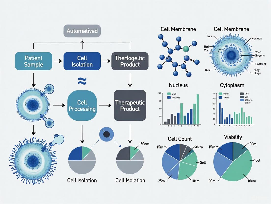
Autologous cell therapy represents a revolutionary paradigm in personalized medicine, where a patient's own cells are harnessed, processed, and reinfused to treat various conditions. This therapeutic approach has demonstrated remarkable success in oncology, particularly for hematologic malignancies, and is increasingly being explored for autoimmune diseases and other conditions [1]. The fundamental workflow begins with leukapheresis to collect specific cell populations, followed by manufacturing processes that may include activation, genetic modification, and expansion, culminating in reinfusion of the therapeutic product back into the patient.
The integration of automation technologies throughout this workflow is critical for enhancing process robustness, scalability, and consistency while reducing manual handling and potential contamination risks [1] [2]. This application note details the standardized protocols and technological advancements that support the development and manufacturing of autologous cell therapies, with particular emphasis on automated cell isolation systems that improve efficiency and reproducibility for research and clinical applications.
Leukapheresis: Initial Cell Collection
Procedure Fundamentals
Leukapheresis is a specialized medical procedure that serves as the critical starting material acquisition step for autologous cell therapies. During this process, blood is drawn from a patient's vein and passed through an apheresis system that continuously centrifuges the blood to separate its various components [3]. The system selectively collects mononuclear cells (including lymphocytes, monocytes, and stem cells) while returning the remaining components (red blood cells, plasma, and platelets) to the patient's circulation [3] [4]. A typical leukapheresis procedure can process 3-6 liters of blood volume over 2-4 hours, collecting a leukopak product containing a high concentration of leukocytes [4].
Leukapheresis Product Specifications
The table below summarizes key characteristics of leukapheresis products based on recent studies:
Table 1: Typical Leukapheresis Product Characteristics
| Parameter | Specification Range | Notes |
|---|---|---|
| Volume | 100-120 mL | Varies by collection center and patient factors [4] |
| WBC Concentration | 21-96 × 10⁶ cells/mL | Approximately 8-fold higher than normal whole blood [4] |
| Total WBC Count | 5.3 ± 2.3 billion cells | Represents 88-fold more WBCs than 10 mL blood sample [4] |
| Platelet Concentration | 1.3 ± 0.4 × 10⁹ cells/mL | Approximately 4-fold higher than peripheral blood [4] |
| Hematocrit | <2% | Tightly maintained at low levels [4] |
| CD3+ T-cell Proportion | 43.82-56.31% | Initial value in leukapheresis product [5] |
Cell Processing and Manufacturing Workflow
Primary Cell Isolation
Following leukapheresis, the leukopak undergoes processing to isolate specific cell populations for downstream manufacturing. While traditional density gradient centrifugation (e.g., Ficoll) has been widely used, automated immunomagnetic separation systems offer significant advantages in consistency, efficiency, and reduced contamination [2].
Table 2: Comparison of PBMC Isolation Methods
| Parameter | Density Gradient Centrifugation | Manual Immunomagnetic Separation | Automated Immunomagnetic Separation (RoboSep-S) |
|---|---|---|---|
| Total Processing Time | ≥77 minutes | ~21 minutes | ~36 minutes [2] |
| Hands-on Time | ≥32 minutes | ~21 minutes | ~6 minutes [2] |
| Cell Purity | Variable, with granulocyte and platelet contamination | High purity | Superior clearance of RBCs, granulocytes, and platelets [2] |
| Throughput Capacity | Limited by manual processing | Moderate | Up to 16 samples per run (RoboSep-16) [2] |
| Contamination Risk | Higher due to multiple open steps | Moderate | Minimal with disposable tips and closed system [2] |
Cryopreservation and Cold Chain Management
Cryopreservation of leukapheresis products enables decoupling from immediate manufacturing needs, improving supply chain resilience and allowing time for quality control testing [5]. Optimized cryopreservation protocols achieve ≥90% post-thaw viability with maintained phenotypic profiles [5]. Key parameters for successful cryopreservation include:
Table 3: Standardized Cryopreservation Protocol Parameters
| Parameter | Specification | Rationale |
|---|---|---|
| Cell Concentration | 5-8 × 10⁷ cells/mL | Optimal balance for cryoprotectant efficacy [5] |
| Cryoprotectant | CS10 (10% DMSO) | Clinical-grade, consistent cryoprotection [5] |
| Formulation Volume | 20 mL/bag | Standardized for storage and handling [5] |
| Formulation Duration | ≤120 minutes | Time-sensitive to maintain cell viability [5] |
| Freezing Protocol | Controlled-rate freezing (Thermo Profile 4) | Prevents ice crystal formation [5] |
Genetic Modification and Expansion
For engineered cell therapies like CAR-T, isolated T-cells undergo genetic modification to express chimeric antigen receptors or other therapeutic constructs. Both viral vector transduction and non-viral methods (electroporation, transposon systems) are employed, followed by ex vivo expansion to achieve therapeutic cell doses [5] [1]. Recent studies demonstrate that cryopreserved leukapheresis products maintain compatibility with multiple manufacturing platforms, including lentiviral CAR-T, non-viral CAR-T, and Fast CAR-T platforms, with comparable performance to fresh leukapheresis in viability, expansion, and cytotoxicity [5].
Automated Cell Processing Workflow
The following diagram illustrates the complete autologous cell therapy workflow with automation integration points:
Figure 1: Complete Autologous Cell Therapy Workflow. The diagram illustrates key process steps from leukapheresis to reinfusion, highlighting automation integration points and alternative manual processes (dashed lines).
Quality Control and Release Criteria
Critical Quality Attributes
Autologous cell therapies must meet stringent release criteria before patient reinfusion. Current consensus recommendations emphasize comprehensive assessment of product sterility, identity, purity, and potency [6]. For fresh products, some regulatory approaches allow interim results from tests performed during production for initial certification and release, with final testing completed after product administration [6].
Table 4: Essential Release Tests for Autologous Cell Therapies
| Test Category | Specific Assays | Acceptance Criteria |
|---|---|---|
| Sterility | Bacterial culture, endotoxin detection, mycoplasma testing | No growth/within specified limits [6] |
| Identity | Flow cytometry for cell surface markers | Consistent with expected product phenotype [6] |
| Viability | Trypan blue exclusion, flow cytometry with viability dyes | Varies by product, typically ≥70-80% [5] [6] |
| Potency | In vitro cytotoxicity, cytokine production, functional assays | Product-specific based on mechanism of action [6] [1] |
| Purity | Residual cell contamination assessment | Meets product specifications [6] |
| Vector Safety | Replication-competent virus testing, vector copy number | Within specified limits [6] |
Expedited Release Strategies
The time-sensitive nature of autologous cell therapies, particularly fresh products, has driven development of innovative release testing strategies. Some institutions implement a two-tier certification system where products can be released based on process testing with final confirmation following administration [6]. This approach requires careful risk-benefit analysis and close coordination between manufacturing facilities, treatment centers, and regulatory authorities.
Research Reagent Solutions and Essential Materials
The table below details key reagents and materials essential for autologous cell therapy research and development:
Table 5: Essential Research Reagents for Autologous Therapy Workflows
| Reagent/Material | Function | Application Notes |
|---|---|---|
| Leukapheresis Collection Kits | Standardized collection of starting material | Maintain cell viability during transport; contain anticoagulants [3] |
| Immunomagnetic Cell Isolation Kits | Positive or negative selection of target cells | Antibody cocktails against CD4, CD8, CD25, CD45, etc.; compatible with automation [2] |
| Cryopreservation Media | Long-term storage of cells | Contain DMSO (typically 7.5-10%) and protein stabilizers [5] |
| Cell Culture Media | Ex vivo expansion of therapeutic cells | Serum-free, GMP-grade with appropriate cytokines (IL-2, IL-7, IL-15) [1] |
| Genetic Modification Tools | Introduce therapeutic transgenes | Viral vectors (lentivirus, retrovirus), mRNA, transposon systems [1] |
| Cell Activation Reagents | Activate T-cells for expansion and engineering | Anti-CD3/CD28 antibodies, cytokine combinations [1] |
| Quality Control Assays | Characterize final product | Flow cytometry panels, sterility tests, potency assays [6] |
Protocol: Automated PBMC Isolation from Leukopaks
Materials and Equipment
- Leukopak product (100-120 mL)
- RoboSep-S instrument (catalog #21000) [2]
- EasySep Direct Human PBMC Isolation Kit for RoboSep (catalog #19654RF) [2]
- RoboSep Buffer (PBS + 2% FBS + 1mM EDTA)
- Sterile disposable pipette tips
- Collection tubes (50 mL conical tubes)
Step-by-Step Procedure
Sample Preparation: Allow leukopak to reach room temperature if cryopreserved. Gently mix the leukopak to ensure homogeneous cell distribution.
Instrument Setup: Power on the RoboSep-S instrument. Initialize according to manufacturer specifications and ensure waste container is empty.
Reagent Preparation: Thaw and prepare all reagents as specified in the EasySep Direct Human PBMC Isolation Kit. Allow reagents to reach room temperature before use.
Sample Loading: Transfer up to 16 mL of leukopak sample into the designated sample tube. Place the tube in the specified position on the instrument carousel.
Reagent Loading: Load the required volumes of isolation cocktail, separation beads, and buffer into their designated positions on the instrument.
Program Selection: Select the pre-programmed "PBMC Isolation from Leukopak" protocol on the RoboSep-S touchscreen interface.
Automated Processing: Initiate the run. The instrument will automatically:
- Add isolation cocktail to the sample and incubate
- Add separation beads and incubate
- Perform magnetic separation
- Transfer purified PBMCs to the collection tube
- The process completes in approximately 30 minutes hands-off time [2]
Cell Collection: Retrieve the collection tube containing isolated PBMCs. Perform cell count and viability assessment using trypan blue exclusion or automated cell counter.
Downstream Processing: Proceed immediately to subsequent manufacturing steps (cell culture, genetic engineering) or cryopreservation.
Expected Results and Quality Metrics
- Cell Yield: Equivalent mononuclear cell numbers compared to density gradient centrifugation [2]
- Purity: Significant reduction in residual platelets (CD41+), red blood cells (Glycophorin A+/CD45-), and granulocytes (CD66b+) compared to density gradient methods [2]
- Viability: Typically ≥95% when processed promptly after leukapheresis
- Processing Time: Total hands-on time approximately 6 minutes versus ≥32 minutes for manual methods [2]
The workflow from leukapheresis to reinfusion for autologous cell therapy involves multiple complex steps that benefit significantly from integration of automation technologies. Standardized protocols for cell collection, processing, and cryopreservation enable manufacturing of consistent, high-quality therapeutic products. Automated cell isolation systems, such as the RoboSep platform, demonstrate measurable improvements in process efficiency, cell purity, and reduction in hands-on time compared to traditional manual methods [2]. As the field advances toward distributed manufacturing models and more personalized therapeutic approaches, continued refinement of these workflows with emphasis on automation, standardization, and quality control will be essential for improving accessibility and clinical outcomes.
The advancement of autologous cell therapies, a cornerstone of personalized medicine, is critically dependent on the initial cell isolation step. In autologous processes, a patient's own cells are harvested, often modified or expanded ex vivo, and then reintroduced as a therapeutic agent. The manufacturing of these Advanced Therapy Medicinal Products (ATMPs) demands rigorous standards of Good Manufacturing Practice (GMP) to ensure patient safety and product efficacy [7] [8]. Manual cell isolation methods, long the standard in research settings, are emerging as a fundamental bottleneck that hinders the transition of these promising treatments from small-scale clinical trials to broad clinical application. These methods, which rely heavily on technician skill and repetitive manual steps, struggle to meet the demands for reproducibility, scalability, and cost-effectiveness required for commercial-scale production [7] [9]. This application note details the quantitative limitations of manual isolation and provides a direct, data-driven comparison with automated alternatives, outlining specific protocols and technology solutions to overcome these critical challenges.
Comparative Analysis: Quantitative Evaluation of Isolation Methods
A controlled study provides clear, quantitative evidence of the limitations of manual isolation. The research directly compared the isolation of Mononuclear Cells (MNCs) from 17 human bone marrow samples using both a manual Ficoll-Paque PLUS density gradient centrifugation method and an automated Sepax S-100 system [7]. The subsequent yield of Mesenchymal Stem Cells (MSCs), a critical cell type for many ATMPs, was then evaluated from the MNC fractions obtained by both methods [7] [10].
Table 1: Performance Comparison of Manual vs. Automated MNC Isolation
| Performance Metric | Manual Isolation | Automated Sepax System | Clinical Significance |
|---|---|---|---|
| MNC Yield | Baseline (Lower) | Slightly Higher [7] | Impacts starting material for subsequent culture and expansion. |
| Processing Time | Longer, highly variable | Standardized, predictable [11] | Affects cell viability, workflow scheduling, and labor costs. |
| Technical Variability | High (Operator-dependent) | Low (System-controlled) [7] | Critical for process consistency and reliability in GMP manufacturing. |
| MSC Colony Formation (CFU) | No significant difference observed [7] | No significant difference observed [7] | Final cell quality and function can be maintained with automation. |
| MSC Phenotype & Differentiation | No significant difference observed [7] | No significant difference observed [7] | Automated isolation does not adversely impact critical cell characteristics. |
The data reveals a critical insight: while the automated system can offer practical advantages in yield and consistency, the core biological quality of the resulting MSCs is equivalent. This demonstrates that automation enhances process control without compromising cell function, a key consideration for regulatory approval.
Table 2: Systemic Bottlenecks of Manual Isolation in Autologous Therapy Manufacturing
| Bottleneck Category | Impact on Autologous Therapy Production |
|---|---|
| Scalability | Manual processing is time-consuming and labor-intensive, creating a logistical impossibility for treating large patient populations [9]. |
| Consistency | Operator-dependent techniques introduce significant run-to-run variability, threatening the reproducibility of the final therapeutic product [7]. |
| Cost of Goods (COGs) | High labor requirements and open-process steps necessitate expensive cleanroom infrastructure and extensive personnel training, driving up costs [9] [12]. |
| Quality Control | Manual steps are difficult to fully monitor and validate, posing challenges for complying with GMP and regulatory standards [8]. |
| Contamination Risk | Open processing increases the risk of microbial contamination, potentially resulting in the loss of a patient's unique therapeutic batch [9]. |
Experimental Protocols: Manual vs. Automated MNC Isolation
The following detailed protocols are adapted from a study conducted under GMP-like conditions, providing a template for evaluating isolation methods in a regulated research environment [7].
Protocol 1: Manual Isolation of MNCs from Bone Marrow
Principle: Separation of mononuclear cells from other bone marrow components using density gradient centrifugation via Ficoll-Paque PLUS.
Materials:
- Source Material: 100 mL of undiluted, heparinized human bone marrow [7].
- Separation Medium: Ficoll-Paque PLUS (Cytiva) [7].
- Wash Medium: α-MEM supplemented with 20% FBS, 10 mmol Glutamine, and 1% Antibiotic-Antimycotic solution [7].
- Labware: Five 50 mL conical tubes (e.g., Corning) [7].
Procedure:
- Gradient Preparation: Aliquot 20 mL of Ficoll-Paque PLUS into each of five 50 mL conical tubes.
- Sample Layering: Carefully layer 20 mL of the undiluted bone marrow sample over the Ficoll in each tube. Avoid mixing the layers.
- Centrifugation: Centrifuge the tubes at 300 × g for 30 minutes at 21°C. Ensure the centrifuge brake is turned off to prevent disturbance of the gradient layers.
- MNC Harvest: After centrifugation, a distinct buffy coat layer containing the MNCs will be visible at the sample-Ficoll interface. Using a pipette, carefully aspirate this MNC layer from each tube and transfer to a new, sterile tube.
- Washing: Pool the harvested MNCs and add wash medium to a total volume of 50 mL. Centrifuge at approximately 450 × g (1,250 rpm as specified) for 10 minutes at 21°C to pellet the cells.
- Resuspension: Decant the supernatant and resuspend the final MNC pellet in 50 mL of wash medium for subsequent cell counting and culture [7].
Protocol 2: Automated Isolation of MNCs Using the Sepax S-100 System
Principle: Closed-system, automated density gradient centrifugation to isolate MNCs with minimal operator intervention.
Materials:
- Source Material: 100 mL of undiluted, heparinized human bone marrow [7].
- Automated System: Sepax S-100 automated cell processing system (Biosafe) [7].
- Consumable Kit: DGBS/Ficoll CS-900 single-use kit (Biosafe), which includes a pre-configured set of tubing, chambers, and a transfer bag [7].
- Solutions: 500 mL of the same α-MEM-based wash medium and 100 mL of Ficoll-Paque PLUS [7].
Procedure:
- System Setup: Load the sterile, single-use CS-900 kit onto the Sepax S-100 instrument according to the manufacturer's instructions.
- Solution Loading: Connect the bags containing the bone marrow sample, the wash medium, and the Ficoll-Paque PLUS to their respective input ports on the kit.
- Program Selection & Initiation: Select the appropriate pre-validated protocol for MNC isolation from bone marrow on the Sepax touchscreen interface. Start the automated run.
- Automated Processing: The system automatically performs all steps, including density gradient formation, centrifugation, and fraction collection, within its closed fluidic pathway.
- Product Recovery: Upon completion, the isolated MNCs are collected in a final output transfer bag in a volume of 50 mL, ready for counting and culture [7]. The waste bag, containing unwanted cells and Ficoll, is disposed of as a single unit.
The field of cell isolation is rapidly evolving beyond traditional centrifugation and manual magnetic sorting. The following workflow diagram illustrates the decision-making path for selecting modern isolation technologies suited for autologous therapy production.
Figure 1: A technology selection workflow for modern cell isolation. LCM: Laser Capture Microdissection.
- Automated Centrifugal Systems (e.g., Sepax): These closed systems automate the density gradient process, standardizing MNC isolation and reducing operator variability and contamination risk, which is a significant advantage in GMP manufacturing [7].
- Negative Selection Technologies: Techniques like buoyancy-activated cell sorting (BACS) using microbubbles (e.g., Akadeum's Alerion system) isolate target cells by removing unwanted cells, leaving the T cells untouched and in a native, non-activated state. This is crucial for preserving cell functionality in therapies like CAR-T [11].
- Advanced Microfluidic Platforms: These systems offer precise, label-free separation based on cell size, deformability, or dielectric properties. They are highly amenable to integration with real-time AI-guided selection and process analytical technologies (PATs) for enhanced control [13].
- AI-Enhanced Cell Sorters: Modern fluorescence-activated cell sorters (FACS) now incorporate adaptive gating algorithms that dynamically adjust sorting parameters in real-time, dramatically improving the reproducibility of isolating rare cell populations across multiple runs [13].
- Gentle, Non-Destructive Methods: Technologies like acoustic focusing and optical tweezers use ultrasonic standing waves or focused laser beams, respectively, to manipulate cells without labels, electrical fields, or high pressures. This minimizes cellular stress and preserves maximum cell viability for delicate primary cells [13].
Essential Research Reagent Solutions
The following table lists key reagents and materials critical for successful and reproducible cell isolation in a research and development setting.
Table 3: Key Reagents and Materials for Cell Isolation Workflows
| Reagent/Material | Function/Application | Example Use Case |
|---|---|---|
| Ficoll-Paque PLUS | Density gradient medium for isolating mononuclear cells (MNCs) from whole blood or bone marrow. | Initial separation of MNCs from bone marrow aspirates as a first step in MSC production [7]. |
| Magnetic Beads (e.g., CTS Detachable Dynabeads) | Antibody-coated beads for positive or negative selection of specific cell populations via MACS. | Isolation or activation of T cell subsets (e.g., CD4+/CD8+) for CAR-T therapy manufacturing [9]. |
| Microbubbles (e.g., Alerion System) | Buoyant particles for gentle, negative selection cell sorting without column-based separation. | Rapid, large-scale isolation of unactivated T cells from leukapheresis product for allogeneic therapies [11]. |
| Cell Culture Media (e.g., α-MEM with supplements) | Provides nutrients and growth factors for the expansion and maintenance of isolated cells. | Ex vivo culture and expansion of MSCs following isolation from MNCs [7]. |
| Fluorochrome-Conjugated Antibodies | Enable detection and sorting of cells based on surface marker expression via FACS. | High-precision isolation of rare cell populations or complex phenotypes using multi-parameter sorting [13] [14]. |
The evidence is clear: manual cell isolation methods present a critical bottleneck that is incompatible with the future scalable and cost-effective manufacturing of autologous cell therapies. The inherent operator variability, limited scalability, and high contamination risk of these methods pose significant challenges to regulatory compliance and commercial viability [7] [9]. The transition to automated, closed-system technologies is not merely an incremental improvement but a necessary evolution for the field.
Adopting automated isolation platforms directly addresses these bottlenecks by enhancing process consistency, ensuring patient safety, and reducing overall Cost of Goods (COGs). As the industry moves towards more decentralized point-of-care (POC) manufacturing models, the role of compact, robust, and highly automated isolation systems will become even more pivotal [9]. For researchers and drug development professionals, the strategic integration of these technologies is essential for successfully translating promising autologous therapies from the laboratory bench to the patient bedside.
Market Landscape and Quantitative Outlook
The autologous cell therapy market is experiencing a period of exceptional growth, fueled by advancements in regenerative medicine and an increasing demand for personalized therapeutic solutions. This sector encompasses therapeutic approaches where a patient’s own cells are collected, manipulated, and reintroduced to treat diseases, thereby minimizing risks of immune rejection and improving biological compatibility [15]. These therapies are broadly applied in oncology, orthopedics, neurology, and cardiovascular diseases.
The market's economic potential is significant, with current valuations and future projections illustrating a robust expansion. The tables below summarize the key quantitative metrics and regional and application-based trends shaping the sector.
Table 1: Global Autologous Cell Therapy Market Size and Growth Projections
| Metric | Value | Time Period / Notes |
|---|---|---|
| Market Size in 2024 | USD 9.6 Billion [15] | Calculated for the year 2024 |
| Market Size in 2025 | USD 11.41 Billion to USD 11.43 Billion [15] [16] | Projected for the year 2025 |
| Projected Market Size by 2033/2034 | USD 47.08 Billion by 2033 [16] to USD 54.21 Billion by 2034 [15] | Long-term forecast |
| Compound Annual Growth Rate (CAGR) | 18.9% [15] | From 2025 to 2034 |
Table 2: Market Segmentation and Regional Analysis
| Segmentation | Dominant Segment (2024) | Fastest-Growing Segment / Region |
|---|---|---|
| Therapy Type | CAR-T Cell Therapy (32% share) [15] | Gene-edited stem cells [15] |
| Application | Oncology (28%-35.5% share) [15] [17] | Neurology [18] / Solid Tumors (within oncology) [15] |
| End User | Hospitals & Clinics (45%-55% share) [15] [18] | Specialty Clinics and Research & Academics [15] [16] |
| Region | North America (41%-44% share) [15] [19] | Asia Pacific [15] [19] [18] |
Key Market Drivers
- Rising Demand for Personalized Medicine: Autologous therapies align perfectly with the shift toward patient-centric, personalized treatments, offering unparalleled biological compatibility and reduced risk of immune rejection compared to donor-based approaches [15] [16].
- Clinical Success and Regulatory Approvals: Positive clinical outcomes and increasing regulatory approvals from agencies like the FDA and European Commission are building confidence among clinicians, patients, and investors. Examples include approvals for CAR-T therapies and treatments for conditions like epidermolysis bullosa [15] [18] [16].
- High Unmet Medical Needs: The growing prevalence of chronic and degenerative diseases, such as cancer, cardiovascular disorders, and neurological conditions, is a primary driver. Autologous therapies provide new hope for conditions with limited treatment options [18] [17].
- Technological Advancements: Innovations in gene-editing (e.g., CRISPR), cell processing, and automation are enhancing the efficacy, precision, and scalability of autologous therapies, thereby accelerating their development and commercialization [15] [16].
Automated Cell Isolation: Core Protocols for Research and Manufacturing
A critical bottleneck in autologous therapy is the complex, multi-step manufacturing process. Automated cell isolation addresses this by enhancing reproducibility, scalability, and yield while minimizing manual handling and variability [20] [21]. The following protocols detail two prominent automated platforms.
Protocol 1: Automated Magnetic Cell Isolation for Immune Cell Subsets
This protocol utilizes the KingFisher instrument system paired with Dynabeads magnetic beads for the rapid and efficient isolation of specific immune cells, such as T cells (CD3+, CD4+, CD8+), B cells (CD19+), and monocytes (CD14+) from peripheral blood mononuclear cells (PBMCs) or whole blood [20].
Experimental Workflow Overview:
Detailed Methodology:
Step 1: Sample Preparation
- Collect blood in a tube containing an anticoagulant (e.g., EDTA, heparin).
- Isolate PBMCs from whole blood using a density gradient centrifugation medium (e.g., Ficoll-Paque) according to standard protocols. Alternatively, use red blood cell (RBC) lysis buffer to lyse red blood cells in whole blood samples.
- Resuspend the final cell pellet in an appropriate buffer, such as PBS containing 2% fetal bovine serum (FBS) and 1 mM EDTA [20] [21].
Step 2: Magnetic Bead Selection and Incubation
- Select the appropriate Dynabeads (e.g., CD3+ for T cells) and isolation method (positive selection, negative selection, or depletion) based on the target cell and downstream application [20].
- Transfer the prepared cell sample to a KingFisher sample tube.
- Add the recommended volume of magnetic beads to the cell suspension.
- Incubate the sample-bead mixture for 30 minutes at 2-8°C with slow mixing to ensure specific binding while maintaining high cell viability [20].
Step 3: KingFisher Instrument Setup
- Load the KingFisher plate according to the manufacturer's layout for your chosen isolation method.
- A typical layout includes wells for: the sample-bead mixture, wash buffers, and elution buffer or collection tube [20].
- Place the plate onto the KingFisher instrument.
Step 4: Running the Automated Protocol
- Select and initiate the pre-programmed script on the KingFisher instrument.
- The instrument automates all subsequent steps: mixing, magnetic separation, washing, and final elution. The process is hands-free and typically completes within 30-60 minutes [20].
Step 5: Collection and Analysis
- Collect the isolated cells from the designated elution well.
- Determine cell count and viability using a trypan blue exclusion assay or an automated cell counter.
- Assess isolation purity via flow cytometry using antibodies against the target cell surface marker (e.g., anti-CD3 for T cells) [20] [21].
Protocol 2: Fully Automated Sequential Isolation for Chimerism Analysis
This protocol describes the use of the RoboSep-S instrument and EasySep reagents for the sequential, fully automated isolation of multiple lymphocyte lineages (B, T, and myeloid cells) from a single, low-volume sample of whole blood, which is critical for lineage-specific chimerism analysis post-transplant [21].
Experimental Workflow Overview:
Detailed Methodology:
Step 1: Sample Preparation
- Collect a maximum of 4.5 mL of whole blood in an anticoagulant tube.
- Transfer the blood to a 14 mL polystyrene round-bottom tube.
- Add an equal volume of 1X EasySep RBC Lysis Buffer, mix gently, and incubate at room temperature for 10-15 minutes. The sample is now ready for loading [21].
Step 2: RoboSep-S Instrument Setup
- The RoboSep-S carousel has four quadrants, each with a magnet and space for tubes and tips.
- Load the prepared sample into the sample tube position in Quadrant 1.
- Load the requisite reagents—EasySep HLA Chimerism Whole Blood CD19, CD3, and Myeloid Positive Selection Kits—into their designated reagent positions in Quadrants 1, 2, and 3, respectively [21].
- Load the necessary buffers and waste tubes as per the protocol layout.
Step 3: Running the Sequential Isolation Protocol
- Initiate the custom protocol on the RoboSep-S instrument.
- Quadrant 1 (B Cell Isolation): The instrument automatically adds CD19 antibody complexes and magnetic particles to the sample. After incubation, the bead-bound CD19+ B cells are retained by the magnet, and the supernatant (depleted of B cells) is transferred to Quadrant 2.
- Quadrant 2 (T Cell Isolation): The instrument adds CD3 antibody complexes and magnetic particles to the incoming supernatant. The bead-bound CD3+ T cells are retained, and the supernatant (now depleted of B and T cells) is transferred to Quadrant 3.
- Quadrant 3 (Myeloid Cell Isolation): The instrument adds a combination of CD33 and CD66b antibody complexes and magnetic particles. The bead-bound myeloid cells are retained by the magnet [21].
- The entire sequential process is completed in less than 2 hours with minimal hands-on time.
Step 4: Collection and Downstream Analysis
- Collect the highly purified CD19+ B cells from Quadrant 1, CD3+ T cells from Quadrant 2, and CD33+/CD66b+ myeloid cells from Quadrant 3.
- The typical purity achieved is >95%, often exceeding 98% for lymphocytes [21].
- Isolated cells are immediately available for downstream applications, including flow cytometry for purity validation or genomic DNA extraction for Short Tandem Repeat (STR) analysis in chimerism testing [21].
The Scientist's Toolkit: Essential Research Reagent Solutions
Successful implementation of automated cell isolation protocols relies on a suite of specialized reagents and instruments. The following table details key solutions for this field.
Table 3: Key Research Reagent Solutions for Automated Cell Isolation
| Item | Function / Application | Example Products / Brands |
|---|---|---|
| Magnetic Beads | Superparamagnetic particles conjugated with antibodies for specific cell surface marker binding, enabling target cell isolation or depletion. | Dynabeads [20], EasySep Reagents [21] |
| Automated Cell Isolation Instruments | Platforms that automate the magnetic separation process, reducing hands-on time and increasing reproducibility and throughput. | KingFisher Systems (Flex, Duo Prime, Apex) [20], RoboSep-S [21] |
| Cell Separation Kits | Pre-configured kits containing optimized antibody mixtures and magnetic particles for isolating specific cell types from various sample sources. | EasySep HLA Chimerism Positive Selection Kits (e.g., CD3, CD19, Myeloid) [21] |
| RBC Lysis Buffer | Lyses red blood cells in whole blood samples to simplify the sample matrix and concentrate nucleated cells for downstream processing. | EasySep RBC Lysis Buffer [21] |
| Cell Culture and Analysis Reagents | For downstream applications including cell expansion, viability testing, and phenotypic characterization of isolated cells. | Flow cytometry antibodies, cell culture media, viability dyes [20] [21] |
Future Outlook and Strategic Directions
The autologous therapy sector is poised for continued growth, with several key trends shaping its future:
- Expansion into New Therapeutic Areas: While oncology currently dominates, autologous therapies show significant promise in treating rare genetic disorders, dermatological conditions (e.g., epidermolysis bullosa), ophthalmology, and cardiovascular repair [15] [16].
- Integration of Advanced Technologies: Artificial Intelligence (AI) is being leveraged to optimize manufacturing, reduce costs, and improve scalability. AI-powered systems automate cell culture, use predictive analytics for process control, and enable "digital twins" for adaptive manufacturing, potentially reducing production costs dramatically [15]. The integration of gene-editing technologies like CRISPR is also expected to create more precise and effective personalized treatments [15] [18].
- Addressing Manufacturing Challenges: The high cost of manufacturing (estimated at $300,000-$500,000 per patient) remains a major restraint [15]. The industry's strategic focus is on advancing automation, closed-system manufacturing, and point-of-care processing to reduce costs, minimize human error, and improve accessibility [15] [20] [16].
CAR-T Cell Therapy: Automated Isolation and Clinical Translation
Application Note
Chimeric Antigen Receptor (CAR)-T cell therapy represents a transformative approach in oncology, leveraging genetically engineered T cells to target and eliminate cancer cells. While highly successful in hematologic malignancies, its application in solid tumors faces challenges including limited tumor infiltration, an immunosuppressive tumor microenvironment, and antigen heterogeneity [22]. Automated cell isolation technologies are critical for standardizing the manufacturing of these "live drugs," particularly for autologous applications where a patient's own T cells are used.
Recent quantitative systems pharmacology (QSP) models have illuminated the complex, multiscale pharmacology of CAR-T cells, from CAR-antigen interactions at the cellular level to biodistribution and phenotype transition in vivo, and finally to clinical tumor response variability [22]. For solid tumors such as hepatocellular carcinoma (HCC), targets like Glypican-3 (GPC3), alpha-fetoprotein (AFP), and claudin18.2 (CLDN18.2) show significant promise. A 2025 meta-analysis of preclinical studies confirmed that CAR-T therapy significantly reduces tumor volume (WMD: -515.77, 95% CI: -634.78 to -396.76) and mass (WMD: -0.30, 95% CI: -0.38 to -0.22) in HCC models [23].
Protocol: Automated CAR-T Cell Manufacturing for Solid Tumors
Principle: Isolate T cells from a patient's leukapheresis product, activate and genetically modify them to express a CAR targeting a tumor-associated antigen (e.g., GPC3), expand the cells, and formulate for reinfusion.
Materials:
- Starting Material: Leukapheresis product from patient.
- Automated Isolation System: Akadeum Alerion Microbubble Cell Separation System or similar [24].
- Reagents: Human T Cell Isolation Kit (GMP-grade), GPC3-specific CAR construct lentivirus, T cell culture media with cytokines (e.g., IL-2), activation beads.
- Quality Control (QC) Assays: Flow cytometry for CAR expression, sterility testing, mycoplasma testing.
Procedure:
- Cell Isolation: Load the leukapheresis product into the automated isolation system. Perform negative selection using a microbubble-based T cell isolation kit to obtain an untouched, high-purity T cell population. Record cell count and viability [24].
- T Cell Activation: Resuspend isolated T cells in culture media supplemented with IL-2. Add magnetic activation beads per manufacturer's instructions. Incubate for 24 hours.
- Genetic Modification: Transduce activated T cells with the GPC3-CAR lentivirus at a pre-optimized multiplicity of infection (MOI). Centrifuge to enhance transduction efficiency if using spinoculation.
- Ex Vivo Expansion: Culture transduced cells in a GMP-compliant, closed-system bioreactor. Maintain cultures for 7-14 days, monitoring cell density and replenishing media and cytokines as needed.
- Formulation and Release: Harvest CAR-T cells, wash, and formulate in infusion buffer. Perform QC testing including:
- Potency: Cytotoxicity assay against GPC3+ tumor cells.
- Purity: Flow cytometry for CD3+ and CAR+ percentage.
- Safety: Sterility, endotoxin, and replication-competent lentivirus testing.
CAR-T Cell Signaling Pathway
Research Reagent Solutions for CAR-T Manufacturing
| Reagent / Solution | Function in Protocol |
|---|---|
| GMP-Grade T Cell Isolation Kit | Isulates untouched, high-purity T cells from leukapheresis via negative selection [24]. |
| T Cell Activation Beads | Provides the necessary signal to activate T cells prior to genetic modification. |
| GPC3-ScFv CAR Lentivirus | Vector for stable integration of the CAR gene into the T cell genome. |
| Serum-Free T Cell Media | Supports the expansion and viability of T cells during culture. |
| Recombinant Human IL-2 | Cytokine that promotes T cell proliferation and survival post-transduction. |
Tumor-Infiltrating Lymphocytes (TILs): Quantitative Profiling and Expansion
Application Note
Tumor-Infiltrating Lymphocytes (TILs) are critical mediators of the host anti-tumor immune response and a key biomarker for prognosis and response to immunotherapy. A 2025 cross-sectional study using AI-driven digital pathology quantified TIL densities across major epithelial cancers, revealing significant variability [25]. Triple-negative breast cancer (TNBC) exhibited the highest mean TIL density, followed by head and neck squamous cell carcinoma (HNSCC), while colorectal adenocarcinoma showed the lowest. Furthermore, higher TIL density was correlated with early-stage disease in breast and lung cancers, underscoring its prognostic value [25].
Table: Mean TIL Densities Quantified by Digital Pathology Across Cancers [25]
| Cancer Type | Mean TIL Density (cells/mm²) | Standard Deviation |
|---|---|---|
| Triple-Negative Breast Cancer (TNBC) | 432.5 | ± 65.3 |
| Head & Neck Squamous Cell Carcinoma (HNSCC) | 387.4 | ± 52.8 |
| Non-Small Cell Lung Carcinoma (NSCLC) | 296.8 | ± 48.9 |
| Colorectal Adenocarcinoma | 215.1 | ± 41.2 |
For autologous therapy, TILs are isolated from resected tumor specimens, rapidly expanded ex vivo, and reinfused into the patient following lymphodepletion. Automated, scalable isolation methods are essential to ensure high yield and viability of these tissue-resident lymphocytes for adoptive cell therapy.
Protocol: Automated TIL Extraction and Expansion for ACT
Principle: Mechanically dissociate and enzymatically digest tumor tissue to extract the embedded lymphocyte population, then rapidly expand them using high-dose IL-2 to generate a therapeutic dose.
Materials:
- Starting Material: Fresh tumor specimen (≥1 cm³).
- Dissociation System: GentleMACS Octo Dissociator or similar automated tissue processor.
- Reagents: Tumor Dissociation Kit (enzymes), TIL Culture Media (e.g., RPMI-1640 with 10% human AB serum, HEPES, Glutamax), Recombinant Human IL-2 (6000 IU/mL).
- Culture Ware: G-Rex bioreactors or similar gas-permeable culture devices.
Procedure:
- Tumor Processing: Aseptically mince the tumor specimen into 2-3 mm fragments using scalpel and forceps.
- Automated Dissociation: Transfer fragments to a C-tube containing enzyme mix. Run the predefined "Tumor Dissociation" program on the GentleMACS Octo Dissociator.
- TIL Extraction: Filter the resulting cell suspension through a 70μm strainer. Wash cells and perform density gradient centrifugation to isolate mononuclear cells.
- Rapid Expansion Protocol (REP):
- Phase 1 (Pre-REP): Plate the isolated TILs at a low density (e.g., 1-5 x 10⁶ cells/well) in a 24-well plate with TIL media containing 6000 IU/mL IL-2. Culture for 1-2 weeks until visible clusters form.
- Phase 2 (REP): Stimulate pre-REP TILs with irradiated feeder cells and anti-CD3 antibody in a G-Rex bioreactor. Maintain in high-dose IL-2 for 14 days.
- Harvest and Formulate: Harvest TILs, perform final wash, and resuspend in infusion buffer. The final product is typically > 1 x 10¹¹ cells with > 80% viability.
TIL Therapy Workflow
Research Reagent Solutions for TIL Therapy
| Reagent / Solution | Function in Protocol |
|---|---|
| Automated Tumor Dissociation Kit | Standardized enzyme blend for consistent mechanical and enzymatic tissue dissociation. |
| TIL Expansion Media (with AB Serum) | Nutrient-rich medium supplemented with serum to support rapid TIL growth. |
| Recombinant Human IL-2 (High Dose) | Critical cytokine driving the activation and massive expansion of TILs in culture. |
| Irradiated Feeder Cells & Anti-CD3 | Provides the necessary stimulus for the Rapid Expansion Protocol (REP). |
| G-Rex Bioreactor | Gas-permeable culture device allowing for large-scale TIL expansion in a single vessel. |
Regenerative Medicine: MSCs and Point-of-Care Manufacturing
Application Note
Regenerative medicine utilizes cells, such as Mesenchymal Stromal Cells (MSCs), to repair, replace, or regenerate damaged tissues and organs. MSCs are investigated for treating a wide range of conditions, including osteoarthritis, graft-versus-host disease (GvHD), and Parkinson's disease, due to their immunomodulatory properties and multi-lineage differentiation potential [26]. A key advancement is the shift towards decentralized, Point-of-Care (POC) manufacturing using isolator-based systems. These closed, automated platforms enable the production of cell therapies within or near the clinical setting, minimizing transportation risks and allowing for the administration of fresh, non-cryopreserved products, which is crucial for cell viability and potency [26].
The regulatory landscape is evolving to support this transition. The U.S. FDA's 2025 draft guidance on Expedited Programs for Regenerative Medicine Therapies encourages flexible trial designs and outlines considerations for manufacturing at multiple clinical sites using a common protocol [27]. Furthermore, extracellular vesicles (EVs) derived from MSCs are emerging as a "Cell Therapy 2.0" paradigm, offering cell-free therapeutic effects while potentially avoiding risks associated with whole-cell administration, such as immunogenicity and tumorigenicity [26].
Protocol: Automated, POC Manufacturing of MSCs for Orthopedic Application
Principle: Expand patient- or donor-derived MSCs in a closed, isolator-based system to generate a therapeutic dose for treating degenerative joint disease.
Materials:
- Cell Source: Bone marrow aspirate or adipose tissue lipoaspirate.
- POC System: Positive pressure isolator with integrated VHP decontamination and closed-system bioreactor (e.g., QuinCell Oct4) [26].
- Reagents: GMP-grade MSC expansion media, trypsin replacement enzyme, human platelet lysate.
- QC Equipment: In-line cell counter, osmometer, flow cytometer for identity panel (CD73+, CD90+, CD105+, CD45-).
Procedure:
- Initial Processing (within isolator): Load the bone marrow or adipose tissue sample. For adipose tissue, perform automated washing and enzymatic digestion to isolate the stromal vascular fraction.
- Primary Culture: Plate the isolated cells in cell factory stacks or hyperstacks within the isolator. Use MSC expansion media supplemented with growth factors. Allow cells to adhere and expand to 70-80% confluence.
- Harvest and Passage: Wash cells and dissociate using a GMP-grade trypsin alternative. Re-plate cells at a lower density for further expansion. Monitor for karyotypic stability.
- Final Formulation: Upon achieving the target cell number (e.g., 50-100 million cells), harvest the MSCs. Wash and resuspend in a final formulation buffer suitable for intra-articular injection.
- POC Quality Control: Perform in-process and lot-release testing. Key tests include:
- Identity: Flow cytometry for MSC surface markers.
- Viability: > 90% by trypan blue exclusion.
- Potency: In vitro trilineage differentiation assay (osteogenic, adipogenic, chondrogenic) or immunomodulation assay (e.g., IDO activity).
- Safety: Sterility (via rapid microbiological methods) and endotoxin testing.
POC MSC Manufacturing Workflow
Research Reagent Solutions for MSC Manufacturing
| Reagent / Solution | Function in Protocol |
|---|---|
| Xeno-Free MSC Expansion Media | Defined, serum-free culture medium designed for robust and consistent MSC growth. |
| GMP-Grade Trypsin Replacement | Enzyme blend for gentle cell detachment, preserving cell surface markers and viability. |
| Human Platelet Lysate (hPL) | Serum substitute rich in growth factors, used to supplement media for enhanced expansion. |
| Flow Cytometry Identity Panel | Antibodies against CD73, CD90, CD105 (positive) and CD45 (negative) for MSC characterization. |
| Trilineage Differentiation Kits | Inductive media to confirm MSC multipotency (osteogenic, adipogenic, chondrogenic). |
The field of advanced therapies is expanding beyond rare diseases to include more common conditions such as autoimmune diseases, type 1 diabetes, and complex neurological conditions [28]. However, business models and supporting infrastructure are still catching up with scientific innovation [28]. A primary challenge is the need to design and deliver highly personalized products within processes that were originally built for mass-market pharmaceuticals [28]. Autologous therapies involve a complex, patient-specific vein-to-vein process that typically requires cold-chain transport, strict chain of identity systems, and near-real-time delivery [28]. The high costs of these therapies, which can reach US$400,000 to over US$2 million per patient, alongside uncertain long-term outcomes, make access and reimbursement perennial concerns [28].
The core manufacturing challenges for autologous cell therapies can be categorized into three universal hurdles: scalability, cost, and dose determination [1]. Current manufacturing is often labor-intensive and contains open manipulations with highly specialized equipment [1]. The high costs are driven by specialized single-use materials, highly skilled manual labor, and extensive analytical testing [1]. Furthermore, the manufacturing process must withstand variability from an uncontrolled starting material (autologous blood product from each patient) and consistently produce a high cell number for the final drug product [1].
Table 1: Primary Challenges in Scaling Autologous Cell Therapies
| Challenge Area | Specific Obstacles | Impact on Therapy |
|---|---|---|
| Manufacturing & Scalability | Labor-intensive, open processes; lack of automation; variable starting material; need for pure cell populations [1]. | Limits patient throughput; increases risk of contamination and production inconsistencies [1] [29]. |
| Cost Management | High costs of raw materials (e.g., viral vectors); specialized equipment; skilled labor; analytical testing; complex logistics [28] [1]. | Total therapy costs of $400,000 to over $2 million per patient, creating affordability and reimbursement challenges [28]. |
| Dose Enabling & Cell Viability | Achieving therapeutic cell numbers from highly variable patient samples; maintaining cell viability, identity, and function during manufacturing and storage [30] [1]. | Directly impacts therapeutic efficacy; failure to deliver a viable dose leads to treatment failure. |
Quantitative Data on Cell Viability and Manufacturing Timelines
Impact of Excipients and Cell Concentration on Viability
A 2025 study systematically evaluated the impact of different excipients and cell concentrations on the viability and functionality of Mesenchymal Stem Cells (MSCs) under hypothermic storage (2–8°C), a critical step for cell storage and transport in autologous therapy workflows [30]. The study found a significant interaction between cell concentration and excipient composition, with a lower cell concentration ( 0.008 × 10^6 MSC/μL ) demonstrating better viability results [30]. Furthermore, excipients combining 50–75% Hypothermosol with human platelet lysate (hPL) improved cell viability and adhesion [30]. Notably, viability, adhesion, and proliferation decreased significantly at 48 hours for all tested conditions, underscoring the time-sensitive nature of cell therapy products [30].
Table 2: MSC Viability (%) Under Different Excipient Formulations and Cell Concentrations Over Time [30]
| Excipient Formulation | Cell Concentration (0.1 x 10^6 MSC/μL) | Cell Concentration (0.008 x 10^6 MSC/μL) | ||
|---|---|---|---|---|
| 24 hours | 48 hours | 24 hours | 48 hours | |
| 100% hPL | 85.2 | 70.1 | 90.5 | 78.3 |
| 75% hPL / 25% Hypothermosol | 86.8 | 72.4 | 92.1 | 80.5 |
| 50% hPL / 50% Hypothermosol | 88.5 | 75.0 | 93.8 | 82.9 |
| 25% hPL / 75% Hypothermosol | 87.1 | 73.2 | 92.5 | 81.1 |
| 100% Hypothermosol | 83.0 | 68.0 | 89.2 | 76.0 |
Vein-to-Vein Timelines for Commercial CAR-T Therapies
The vein-to-vein (V2V) time—the period from patient apheresis to infusion of the drug product—is a critical metric for patient access and outcomes, particularly for those with aggressive diseases. Current V2V times for commercial autologous CAR-T therapies range from 2 to 5 weeks [31]. Prolonged V2V times contribute to patient drop-off, with an estimated 30% of prescribed patients never undergoing leukapheresis and 20% of those who do not receiving infusion, often due to rapid disease progression [31]. Reducing manufacturing time is therefore a key strategy to improve accessibility and clinical outcomes [31].
Table 3: Vein-to-Vein Times for Commercial Autologous CAR-T Cell Therapies [31]
| Product Name (Generic) | Commercial Name | Indication(s) | Typical Vein-to-Vein Time |
|---|---|---|---|
| Brexucabtagene autoleucel | Tecartus | MCL, ALL | 2 - 3 weeks |
| Obecabtagene autoleucel | Aucatzyl | ALL | 3 weeks |
| Tisagenlecleucel | Kymriah | FL, DLBCL, ALL | 3 - 4 weeks |
| Axicabtagene ciloleucel | Yescarta | FL, DLBCL | 3.5 weeks |
| Lisocabtagene maraleucel | Breyanzi | FL, LBCL, MCL, CLL, SLL | 3 - 4 weeks |
| Idecabtagene vicleucel | Abecma | MM | 4 weeks |
| Ciltacabtagene autoleucel | Carvykti | MM | 4 - 5 weeks |
Experimental Protocols for Optimization
Protocol 1: Optimizing MSC Preservation for Storage and Transport
This protocol is designed to evaluate excipients for maintaining the viability and functionality of Mesenchymal Stem Cells (MSCs) during hypothermic storage, a common requirement in cell therapy logistics [30].
1.0 Sample Collection and Cell Culture
- Starting Material: Use mononuclear cells (MNCs) from bone marrow aspirates or other MSC sources like adipose tissue or umbilical cord [30] [32].
- Culture: Culture MNCs in minimum essential medium (α-MEM) supplemented with 5% human platelet lysate (hPL), 10 mmol glutamine, and 1X antibiotic-antimycotic solution [30].
- Selection: Select for MSCs by leveraging their adherence to plastic; remove non-adherent cells through medium changes. Proceed once cultures reach ≥90% confluence [30].
2.0 Study Group Formulation
- Prepare two different cell concentrations: a higher concentration (e.g., 0.1 x 10^6 MSC/μL) and a lower concentration (e.g., 0.008 x 10^6 MSC/μL) [30].
- For each concentration, formulate five experimental excipient groups:
- 100% hPL
- 75% hPL / 25% Hypothermosol
- 50% hPL / 50% Hypothermosol
- 25% hPL / 75% Hypothermosol
- 100% Hypothermosol
- Store all formulated products at 2–8°C (hypothermic conditions) [30].
3.0 Cell Viability Assessment
- Time Points: Assess cell counts and viability at 24 and 48 hours [30].
- Method: Use the standard Trypan Blue dye exclusion method for cell counting and viability determination [30].
4.0 Functional Capacity Assessment
- Cell Adhesion: Seed MSCs at 10,000 cells/cm² in culture flasks. After 24 hours, detach adherent cells using trypsin/EDTA, then centrifuge and resuspend. Count adherent cells using Trypan Blue exclusion [30].
- Proliferation Capacity: Seed MSCs at 2000/cm² onto culture slides. After 24 hours, perform immunohistochemistry for Ki67, a nuclear marker for proliferation. Calculate the Ki67 labeling index as a percentage of stained cells relative to the total cell number [30].
5.0 Statistical Analysis
- Analyze data using a mixed-effect linear regression model to handle fixed effects (excipient group, concentration, time) and random effects (intra-group variability) [30].
- Verify model fit by checking normal error distribution and homoscedasticity of residuals [30].
Protocol 2: Automated Manufacturing of T Cells Using the BECA Platform
This protocol details the transition from manual to automated T cell culture processes using the Bioreactor with Expandable Culture Area (BECA) platform, addressing scalability and consistency challenges [29].
1.0 Manual Process Development with BECA-S
- Equipment: BECA-S culture vessel, Biosafety Cabinet (BSC), CO₂ incubator [29].
- BECA-S Vessel: This is a single-chamber, single-use culture vessel with an internal movable wall that allows the culture surface area to be expanded from 19 cm² to 102.4 cm² [29].
- Handling: All handling of the BECA-S vessel must be performed in a BSC to maintain sterility. Liquid transfer is done through a port accessing the culture region [29].
- Process Optimization: Develop and optimize the T cell expansion process (including activation, transduction, and culture) using the manual BECA-S system [29].
2.0 Automated System Setup with BECA-Auto
- Equipment: BECA-Auto benchtop system, pre-sterilized single-use kits (BECA-S (Closed), Manifold Assembly, Input Manifold) [29].
- Assembly in BSC: Assemble the pre-sterilized single-use kits within a BSC to form a functionally closed flow path [29].
- Installation: Install the assembled flow path onto the BECA-Auto's Actuation Platform and couple it to the control units: the Capsule Internal Fluid Controller (CIFC) and the Device for Automated Aseptic Sampling (DAAS) [29].
- Environment Sealing: Close and seal the system's Enclosure. Activate the Climate Control to establish and maintain the culture environment at 37°C, 90% relative humidity, 5% CO₂, and 20% O₂ [29].
3.0 Automated Culture Process Execution
- Seeding: Connect a sterile bag containing the T cell seeding culture to the Manifold Assembly. Execute the Seeding programme, which initiates CIFC to draw the cell suspension into the BECA-S (Closed) vessel [29].
- Culture Maintenance: The BECA-Auto system automates the entire culture process:
- The Actuation Platform rocks the vessel to resuspend cells and tilts it for media changes [29].
- The CIFC manages the addition of fresh media and removal of waste [29].
- The DAAS automatically extracts small, aseptic samples at set intervals for offline monitoring [29].
- The Actuation Platform moves the internal wall to progressively expand the culture area as the cell number increases [29].
- Harvesting: Upon process completion, execute the Harvesting programme. The CIFC transfers the final cell product to a designated output bag connected to the Manifold Assembly [29].
4.0 Process Comparison and Validation
- Parallel Runs: Culture identical T cell samples using both the manual BECA-S and automated BECA-Auto processes [29].
- Outcome Analysis: Compare critical quality attributes of the final products, including:
- Total Cell Yield
- Cell Viability (e.g., via Trypan Blue exclusion)
- Phenotype (e.g., via flow cytometry)
- Functionality (e.g., in vitro cytotoxic assay)
- Target: Demonstrate insignificant differences in culture outcomes between the manual and automated processes, validating a seamless transition [29].
The Scientist's Toolkit: Key Research Reagent Solutions
Table 4: Essential Reagents and Kits for Cell Therapy Research and Manufacturing
| Tool / Reagent | Primary Function | Application in Autologous Therapy |
|---|---|---|
| Human Platelet Lysate (hPL) | Serum-free cell culture medium supplement rich in growth factors and cytokines [30]. | Provides a xeno-free supplement for MSC expansion, improving safety and efficacy [30]. |
| Hypothermosol | Specialized cell preservation solution for hypothermic storage [30]. | Optimizes cell viability and functionality during storage and transport; used in combination with hPL [30]. |
| Microbubble Cell Separation System (Alerion) | Gentle, buoyancy-based cell isolation via negative selection [33]. | Scalable isolation of untouched, high-viability T cells from leukopaks for autologous or allogeneic therapy manufacturing [33]. |
| BECA-S / BECA-Auto System | Flexible bioreactor platform for manual (BECA-S) and automated (BECA-Auto) cell culture [29]. | Enables seamless translation of T cell expansion processes from R&D to closed, automated manufacturing, reducing vein-to-vein time [29]. |
| T Cell Isolation Kits (e.g., Akadeum) | Antibody-based kits for specific isolation or depletion of T cell populations [33]. | High-throughput, high-purity T cell isolation from patient apheresis or leukopaks, available in GMP-grade formats [33]. |
| Rapamycin | mTOR inhibitor used in cell culture [1]. | Promotes selective expansion of Tregs while inhibiting conventional effector T cells during manufacturing, preserving regulatory function [1]. |
Navigating the Technology Landscape: From Magnetic Beads to AI-Enhanced Sorters
The transition from manual, research-scale processes to automated, robust manufacturing is a critical challenge in the development of autologous T cell therapies. Automated Magnetic-Activated Cell Sorting (MACS) represents a cornerstone technology in this transition, enabling the scalable, consistent, and high-purity cell isolation required for clinical manufacturing. This document details the established platforms, experimental protocols, and key reagents that underpin the use of automated MACS in autologous therapy research.
The Platform: KingFisher Automated Cell Isolation Systems
The KingFisher system is a prominent established workhorse for automating magnetic bead-based cell isolation processes. It integrates with Dynabeads magnetic beads to provide a reproducible and hands-free solution for isolating specific cell populations, such as T cells for subsequent engineering [20].
Table 1: Key Features of KingFisher Systems for Automated MACS
| Feature | Description | Benefit for Autologous Therapy Research |
|---|---|---|
| Automation Principle | Uses a magnetic rod to transfer magnetic bead-cell complexes through a series of pre-loaded plates containing samples, washes, and elution buffers. | Reduces manual hands-on time, increases throughput, and minimizes operator-to-operator variability. |
| Instrument Models | KingFisher Flex, Duo Prime, and Apex systems. | Offers scalability from flexible R&D (Flex) to higher-throughput needs. |
| Cell Isolation Methods | Supports positive isolation (with/without bead release), negative isolation, and cell depletion. | Flexibility to choose the optimal method for preserving cell function or achieving high purity. |
| Gentle Mixing | Programmable mixing speeds; "Slow" mixing recommended for optimal cell viability and yield. | Preserves the health and functionality of precious patient-derived T cells. |
| Efficient Capture | Implements multiple cycles of magnetic bead capture (e.g., 2x) to maximize isolation efficiency. | Ensures high yield and purity of the target cell population from a limited starting sample. |
Table 2: Performance Metrics of Automated vs. Manual MACS
| Parameter | Automated MACS (KingFisher) | Manual MACS |
|---|---|---|
| Isolation Efficiency | Comparable to manual isolation, with most binding occurring within the first 10 minutes [20]. | High, but subject to operator consistency. |
| Incubation Time | 10-30 minutes; extending beyond 30 minutes does not increase yield and may increase non-specific binding [20]. | Often requires longer, protocol-dependent incubation times. |
| Cell Viability | High viability when using optimized "Slow" mixing conditions [20]. | Can be compromised by vigorous or inconsistent manual handling. |
| Reproducibility | High, due to pre-programmed, consistent protocols. | Variable, dependent on technical skill of the operator. |
| Throughput | High; capable of processing multiple samples in a single run with minimal hands-on time. | Low to medium; limited by manual processing time and attention. |
Detailed Protocol: Automated Negative Selection of Human T Cells
This protocol describes the use of the KingFisher system for the negative selection of untouched human T cells from peripheral blood mononuclear cells (PBMCs), ideal for downstream T cell activation and transduction.
The Scientist's Toolkit: Research Reagent Solutions
Table 3: Essential Materials for Automated T Cell Negative Selection
| Item | Function | Example Product |
|---|---|---|
| KingFisher Instrument | Automates the entire magnetic separation process. | KingFisher Flex System [20]. |
| Magnetic Beads | Superparamagnetic particles that bind to unwanted cells for depletion. | Dynabeads magnetic beads [20]. |
| Negative Selection Kit | Contains antibody cocktail for labeling non-T cells. | Human T Cell Isolation Kit (e.g., MagniSort [34]). |
| Sample Type | The starting material for cell isolation. | Leukopak, PBMCs, or whole blood [20]. |
| Binding Buffer | Optimized buffer to facilitate antibody and bead binding to cells. | Often PBS with EDTA and serum albumin. |
| Elution Buffer | Buffer for collecting the final, untouched target cell population. | Often PBS or complete cell culture media. |
Workflow Diagram
Step-by-Step Procedure
Sample Preparation (5-10 minutes): Isolate PBMCs from a leukopak or whole blood using a standard density gradient centrifugation method. Resuspend the cell pellet in an appropriate binding buffer at a recommended concentration of up to 1x10^8 cells/mL.
Cell Labeling (30 minutes):
- Antibody Incubation: Add the biotinylated antibody cocktail (e.g., against CD8, CD11b, CD19, CD24, B220, etc. for negative selection of T cells [34]) to the cell suspension. Mix well and incubate for 15 minutes at 2-8°C.
- Magnetic Bead Incubation: Add the secondary streptavidin-coated magnetic beads (e.g., Dynabeads [20]) to the sample. Mix well and incubate for an additional 15 minutes at 2-8°C.
KingFisher Instrument Setup (5 minutes):
- Plate Layout: Label a deep-well 96-well plate. Load the plate as follows:
- Well 1: Labeled cell sample.
- Wells 2-4: Washing buffer (e.g., PBS with 0.1% BSA).
- Well 5: Elution buffer (for collecting untouched T cells).
- Protocol Selection: Place the plate on the KingFisher instrument and select the pre-programmed "Negative Isolation" protocol.
- Plate Layout: Label a deep-well 96-well plate. Load the plate as follows:
Run Automated Protocol (30-40 minutes): Start the run. The instrument will automatically perform the following steps:
- The magnetic rod collects the bead-bound complexes from the sample and transfers them through the wash buffers to remove unbound cells and reagents.
- The untouched, purified T cells remain in the supernatant and are transferred to the elution buffer well.
- The process involves multiple cycles of magnetic capture and release to ensure high purity.
Cell Collection and Analysis (10 minutes): After the run is complete, retrieve the plate and collect the untouched T cells from the elution well. Perform a cell count and viability assessment (e.g., via Trypan Blue exclusion). Analyze the purity of the isolated T cell population using flow cytometry (e.g., staining for CD3+, CD4+, CD8+).
Optimization Strategies for Automated MACS
To ensure the best outcomes for sensitive autologous T cell samples, several parameters from the standard protocol can be optimized based on validated studies [20].
Optimization Diagram
Key Findings from Optimization Studies [20]:
- Incubation Time: Most specific binding occurs within the first 10 minutes. Extending incubation beyond 30-60 minutes does not increase yield and can increase non-specific binding.
- Incubation Temperature: Performing isolations at lower temperatures (e.g., 2-8°C) slows biological activity and can reduce non-specific binding, beneficial for high-purity requirements.
- Mixing Conditions: "Slow" mixing on KingFisher instruments resulted in equal isolation efficiency and significantly higher cell viability compared to "Medium" or "Fast" mixing, which caused cell loss and reduced viability.
- Bead Capture: Introducing two cycles of bead capture in the automated protocol achieved equal or better isolation efficiency compared to manual isolation.
Automated MACS platforms, exemplified by the KingFisher system, are indispensable tools for standardizing and scaling the initial cell isolation steps in autologous T cell therapy research. By implementing optimized protocols for negative selection, researchers can reliably obtain high yields of untouched, healthy T cells that are critical for successful downstream engineering and manufacturing. This reliability and consistency are foundational to accelerating the translation of autologous therapies from research to clinical application.
In the development of autologous cell therapies, the initial cell isolation step is a critical determinant of the final product's quality. Positive and negative selection strategies represent two fundamental approaches for isolating target cell populations from a complex starting material, such as leukapheresis product or patient biopsy. For autologous therapies, where every patient's cells constitute a unique manufacturing batch, the choice between these methods carries significant implications for purity, cell function, manufacturing consistency, and ultimately, therapeutic efficacy [1] [35].
This application note provides a structured comparison of positive and negative selection methodologies, focusing on their application within automated cell isolation workflows for autologous therapy research and development. We present quantitative performance data, detailed protocols, and strategic guidance to enable informed decision-making for therapy developers.
Comparative Performance Analysis
The choice between positive and negative selection involves balancing multiple, often competing, parameters. The table below summarizes the key characteristics of each method.
Table 1: Strategic Comparison of Positive and Negative Selection Methods
| Parameter | Positive Selection | Negative Selection |
|---|---|---|
| Principle | Direct binding to target cell surface markers [36] | Removal of unwanted cells; enrichment of unlabeled target population [36] |
| Typical Purity | High (can exceed 95%) | Moderate to High (dependent on starting population and antibody panel) |
| Impact on Cell Function | Potential activation via signaling cascades (e.g., CD4/CD8) [35] | Minimal; target cells remain unlabeled and unmanipulated |
| Recovery/Yield | High recovery of labeled cells [36] | Variable recovery; potential for loss of target cells within complex mixtures |
| Antibody/Ligand Binding | Target cells are labeled [36] | Target cells are not labeled [36] |
| Downstream Effects | Potential epitope blocking; may interfere with subsequent functional assays [36] | No antibody binding to target cells, preserving native state for downstream assays |
| Ideal for | Isolating well-defined populations with a unique surface marker | Isolating fragile populations, cells lacking a unique marker, or when receptor signaling must be avoided |
Workflow and Decision Pathways
The following diagram illustrates the procedural and decision-making pathways for positive and negative cell selection strategies.
Detailed Experimental Protocols
Protocol for Positive Selection of CD4+/CD8+ T Cells
This protocol is adapted from methods used in the manufacture of autologous T-cell therapies, including CAR-T cells and TCR-T cells [1] [35]. It is designed for execution on an automated magnetic cell separation platform.
- Objective: To isolate a highly pure population of CD4+ and CD8+ T cells from leukapheresis-derived Peripheral Blood Mononuclear Cells (PBMCs) for subsequent genetic engineering and expansion.
- Principle: Target cells are directly labeled with magnetic bead-conjugated antibodies against CD4 and CD8 surface antigens, enabling their retention in a magnetic field while unlabeled cells are washed away.
Table 2: Research Reagent Solutions for Positive Selection
| Item | Function | Example |
|---|---|---|
| Leukapheresis Sample | Starting material containing mononuclear cells | Human PBMCs |
| Anti-CD4 & Anti-CD8 Magnetic Beads | Immunomagnetic label for positive selection | ClinExVivo CD4/CD8 Dynabeads |
| Automated Cell Separator | Platform for reproducible magnetic separation | RoboSep or Equivalent |
| Cell Separation Buffer | Provides medium for separation; maintains cell viability | PBS + 2 mM EDTA + 0.5% HSA |
| Viability Dye | Assess cell membrane integrity and viability post-isolation | 7-AAD, Trypan Blue |
Procedure:
- Sample Preparation: Thaw and wash leukapheresis sample. Resuspend cells in pre-chilled cell separation buffer. Perform a cell count and viability assessment.
- Cell Labeling: Incubate the cell suspension with anti-CD4 and anti-CD8 magnetic bead conjugates according to the manufacturer's optimized protocol (typically 20-30 minutes at 2-8°C).
- Automated Separation: Load the labeled cell suspension onto the automated cell separator. Execute the manufacturer's pre-programmed positive selection protocol.
- The instrument will apply a magnetic field to retain labeled CD4+/CD8+ cells.
- Unlabeled cells (B cells, NK cells, monocytes, etc.) are washed to waste.
- Elution and Analysis: Elute the magnetically retained target cells from the separation chamber.
- Perform a cell count and viability measurement.
- Assess purity via flow cytometry by staining with fluorescently-labeled antibodies against CD3, CD4, and CD8. Purity is calculated as (CD4+ + CD8+ cells) / Total CD3+ cells × 100% [36].
- Downstream Processing: The isolated T-cell population can now proceed to downstream steps such as activation, genetic transduction (e.g., with CAR or TCR constructs), and expansion [1].
Protocol for Negative Selection of Regulatory T Cells (Tregs)
Isolating Tregs for autologous therapy is challenging due to the lack of a unique surface marker. Negative selection enriches untouched Tregs, preserving their native function and stability, which is critical for therapies aimed at rebalancing autoimmunity [1].
- Objective: To isolate an untouched, functional population of regulatory T cells (Tregs) from PBMCs using a negative selection strategy to deplete non-Treg lineages.
- Principle: A cocktail of antibodies against non-Treg surface markers (e.g., CD8, CD19, CD14, CD16, CD56, CD127) is used to label non-target cells. These labeled cells are then magnetically depleted, leaving an enriched population of unmanipulated Tregs.
Procedure:
- Sample Preparation: Thaw and wash PBMCs. Resuspend cells in cell separation buffer and perform a cell count.
- Cocktail Incubation: Incubate the cell suspension with a biotinylated antibody cocktail against non-Treg lineage markers. Follow this with an incubation step with magnetic bead-conjugated anti-biotin antibodies.
- Automated Depletion: Load the labeled cell suspension onto the automated cell separator. Execute the negative selection/depletion protocol.
- The instrument will magnetically retain the labeled non-target cells.
- The unlabeled, enriched Treg flow-through is collected.
- Elution and Analysis: Collect the flow-through fraction containing the enriched Tregs.
- Perform a cell count and viability measurement.
- Assess purity by flow cytometry. Stain for CD4, CD25, and CD127. Tregs are typically identified as CD4+CD25+CD127lo/-. Purity is calculated as (CD4+CD25+CD127lo/- cells) / Total live cells × 100% [1] [36].
- Critical Step: Assess the suppressive function of the isolated Tregs in a co-culture assay with effector T cells to confirm biological potency [36].
- Downstream Processing: The untouched Tregs can be activated and expanded ex vivo, potentially with rapamycin to prevent effector T-cell outgrowth [1], and may be genetically engineered for enhanced specificity.
Strategic Application in Autologous Therapy
The selection strategy directly impacts the Critical Quality Attributes (CQAs) of the final cell therapy product.
Positive Selection is highly effective for obtaining pure populations when a specific, well-defined surface marker is available and when antibody binding does not trigger detrimental activation pathways. For example, in one study, positively selected CAR-T cells exhibited a significantly higher in vitro interferon-γ and IL-2 secretion profile compared to those generated via negative selection [35]. This highlights how the selection method can intrinsically alter the functional state of the cellular product.
Negative Selection is preferred when:
- The target cell population lacks a unique defining surface marker (e.g., Tregs) [1].
- Antibody binding to the target cell's key surface receptors could induce aberrant signaling, activation, or functional impairment [35].
- Preserving the target cell in its most native, "untouched" state is paramount for subsequent functional assays or in vivo efficacy.
- The goal is to deplete specific inhibitory populations (e.g., CD14+ monocytes) from the starting material to improve the overall manufacturing process.
For autologous therapies, where patient-specific starting material is highly variable, the consistency offered by automated platforms executing these protocols is essential for achieving a robust and reproducible manufacturing process [7].
The transition of autologous cell therapies from clinical success to commercial reality hinges on the development of robust, scalable, and efficient manufacturing processes. Automated isolation systems, such as the KingFisher platform, are central to this evolution, enabling the standardized production of high-quality cell-based products. By leveraging magnetic bead-based technology, these systems minimize manual intervention, enhance reproducibility, and are capable of integrating into closed, automated workflows essential for modern cell therapy manufacturing. This document outlines detailed application notes and protocols for using systems like KingFisher within the context of automated cell isolation for autologous therapy research, providing researchers and drug development professionals with a framework to streamline their processes.
The following diagram illustrates the core steps from sample receipt to final product in the context of autologous cell therapy manufacturing.
Essential Research Reagent Solutions
The following table details key reagents and materials required for automated purification workflows on platforms like KingFisher.
| Component Name | Primary Function | Specific Examples & Applications |
|---|---|---|
| Magnetic Bead-Based Kits | Selective binding and isolation of target biomolecules or cells. | MagMAX kits for nucleic acids; Dynabeads for proteins, cells, exosomes, and viruses [37]. |
| Lysis/Binding Buffers | Disrupt samples and create conditions for target binding to magnetic beads. | Components of kits like the QIAamp circulating nucleic acid kit or Maxwell RSC ccfDNA Plasma Kit [38]. |
| Wash Buffers | Remove impurities and contaminants while target is bound to beads. | Used in automated washing steps on KingFisher to ensure high purity of the isolated material [37]. |
| Elution Buffers | Release the purified target from the magnetic beads into a final solution. | Low-salt aqueous buffers or nuclease-free water to collect nucleic acids [38]. |
| Non-Viral Transfection Reagents | Deliver genetic payloads to cells for gene modification post-isolation. | LipidBrick Cell Ready system for delivering mRNA, circRNA, sgRNA, and pDNA to T cells, NK cells, and HSCs [39]. |
Detailed Protocol: Automated cfDNA Isolation from Plasma
This protocol provides a step-by-step methodology for the isolation of cell-free DNA (cfDNA) from patient plasma, a common starting material for liquid biopsy applications in oncology and other fields.
Pre-Analytical Sample Preparation
- Plasma Separation: Collect peripheral blood in EDTA or Streck Cell-Free DNA BCT tubes. Centrifuge at 800-1600 × g for 10 minutes at room temperature to separate plasma from cellular components. Carefully transfer the supernatant (plasma) to a new tube without disturbing the buffy coat.
- Secondary Centrifugation: Perform a second, high-speed centrifugation of the plasma at 16,000 × g for 10 minutes at 4°C to remove any remaining cellular debris. Transfer the clarified plasma to a new tube. Process samples fresh or store at -80°C until nucleic acid extraction.
Automated Isolation on KingFisher System
- Reagent Setup: Thaw all kit components completely and mix by vortexing. For each sample, combine 1-5 mL of plasma with an equal volume of binding buffer and the provided magnetic beads in a deep-well plate. The binding buffer facilitates the adsorption of cfDNA onto the bead surface.
- Plate Layout: Load the plate onto the KingFisher instrument along with the necessary tip combs and subsequent plates containing wash buffers and elution buffer.
- Program Selection: Initiate the pre-validated protocol on the KingFisher console. A standard protocol typically involves the following automated steps:
- Binding: Mixing the sample with beads for a set duration to allow cfDNA binding.
- Washing (2-3 times): The magnetic particles are transferred through a series of wash solutions to remove proteins, salts, and other contaminants.
- Elution: The purified cfDNA is released from the beads into a small volume (e.g., 50-100 µL) of elution buffer or nuclease-free water.
- Product Recovery: After the run is complete, retrieve the elution plate and store the purified cfDNA at -20°C for immediate analysis or -80°C for long-term storage.
Performance Data: Comparison of Automated Extraction Systems
The following table summarizes quantitative data from comparative studies evaluating different automated cfDNA isolation systems, highlighting key performance metrics critical for assay robustness.
| Extraction System / Kit | Reported cfDNA Yield (QUBIT HS) | cfDNA Integrity / Fragment Size Profile | Suitability for Sensitive Applications (e.g., Chimerism, cffDNA) |
|---|---|---|---|
| MagNA Pure 24 (Roche) | Lower yield compared to IDEAL and LABTurbo [40] | Isolates significantly smaller cfDNA fragments (mean peak 1: ~120 bp) [40] | Reliable for fetal RhD detection; chimerism quantification reliability depends on detection method [40]. |
| IDEAL (IDSolution) | Higher yield [40] | Standard fragment size profile (mean peak 1: ~164 bp) [40] | Efficient for fetal RhD detection [40]. |
| LABTurbo 24 (Taigen) | Higher yield [40] | Standard fragment size profile (mean peak 1: ~165 bp) [40] | Reliable chimerism quantification with NGS; efficient for fetal RhD detection [40]. |
| Chemagic 360 (Perkin Elmer) | Intermediate yield [40] | Standard fragment size profile (mean peak 1: ~166 bp) [40] | Fetal RhD detection confirmed [40]. |
| Maxwell RSC ccfDNA Plasma Kit | High efficiency, comparable to manual QIAamp kit [38] | Not specified in the provided results | High efficiency for mutant ctDNA isolation [38]. |
| QIAamp Circulating Nucleic Acid Kit | High efficiency, comparable to automated Maxwell RSC [38] | Not specified in the provided results | High efficiency for mutant ctDNA isolation [38]. |
Downstream Workflow: Integration into Cell Therapy Manufacturing
The purified cellular or nucleic acid material is a critical starting material for generating autologous cell therapies. The subsequent steps often involve genetic modification and expansion of cells.
Quality Control and Analytical Methods
Rigorous QC is essential to ensure the isolated material meets specifications for downstream therapeutic use. The table below lists common analytical methods used for quality assessment.
| QC Assay | Target of Analysis | Application in Workflow |
|---|---|---|
| Digital Droplet PCR (ddPCR) | Absolute quantification of total cfDNA, specific mutations (e.g., KRAS), or chimerism [40] [38]. | Quantifies target nucleic acids and detects inhibiting substances in eluates [38]. |
| Next-Generation Sequencing (NGS) | Comprehensive genomic profiling and chimerism analysis [40]. | Provides a robust method for chimerism quantification, dependent on the extraction system used [40]. |
| Fluorometry (e.g., QUBIT) | Accurate quantification of nucleic acid concentration [40]. | Standard method for measuring DNA yield, though results can vary between extraction methods [40]. |
| Capillary Electrophoresis (e.g., BIABooster, Bioanalyzer) | DNA fragment size distribution and integrity [40]. | Assesses the quality of cfDNA, revealing differences in fragment profiles between isolation methods [40]. |
| Rapid Sterility Tests | Microbial contamination [39]. | Novel assays can reduce QC bottlenecks, cutting testing time from days to hours [39]. |
The advancement of autologous cell therapies, such as CAR-T cells and Tumor-Infiltrating Lymphocytes (TILs), hinges on the ability to isolate and manipulate specific cell populations from a patient's own tissue without altering their native state [41] [42]. Traditional cell separation methods like Fluorescence-Activated Cell Sorting (FACS) and Magnetic-Activated Cell Sorting (MACS) rely on biochemical labels, which can be costly, time-consuming, and risk activating or damaging the very cells meant for therapeutic use [41] [43]. Label-free technologies represent a paradigm shift by exploiting intrinsic physical cell properties—such as size, density, deformability, and dielectric properties—for separation, thereby preserving cell viability and function [41] [44]. This application note details the use of two prominent label-free microfluidic technologies, dielectrophoresis (DEP) and acoustophoresis, within the context of automated cell isolation for autologous therapy research. We provide structured performance data, detailed experimental protocols, and essential resource guidance to enable researchers in the successful implementation of these platforms.
Dielectrophoresis (DEP) leverages a non-uniform electric field to exert force on polarizable particles or cells. The direction and magnitude of this force depend on the cell's dielectric properties and the applied field frequency, enabling the separation of different cell types without labels [45] [46]. Acoustophoresis uses a standing ultrasonic wave within a microfluidic channel to separate cells based on their size, density, and compressibility, gently directing them toward specific pressure nodes or antinodes [44] [47]. Remote or contactless versions of these technologies have been developed to further enhance biocompatibility by isolating cells from electrodes and minimizing Joule heating [48] [43].
The table below summarizes the key operational parameters and demonstrated performance of these technologies for representative applications in cell therapy.
Table 1: Performance Metrics of Label-Free Cell Separation Technologies
| Technology | Separation Principle | Throughput | Purity / Efficiency | Cell Viability | Key Applications Demonstrated |
|---|---|---|---|---|---|
| Dielectrophoresis (DEP) | Dielectric properties (membrane capacitance, cytoplasm conductivity) [41] [46] | Varies by design; continuous flow systems enable higher throughput [41] | >98% recovery of E. coli from blood [48]; Enrichment of CD34+ cells [41] | High; maintained with contactless designs [48] [43] | Isolation of CTCs [45]; Separation of viable/non-viable cells [43]; Stem cell sorting [41] |
| Acoustophoresis | Size, density, and compressibility [44] | High-throughput capabilities [43] | >98% separation efficacy for viable/non-viable stromal cells [43] | High; minimal adverse effects on viability and osteogenic function [43] | Separation of viable/non-viable human stromal cells [43]; Blood component separation [44] |
Application Notes and Experimental Protocols
Protocol 1: Isolation of Viable Cells Using Dielectrophoresis
This protocol describes the separation of viable from non-viable human stromal cells using a DEP-based microfluidic system, a critical step in ensuring the quality of cell populations for autologous therapy [43].
1. Primary Equipment and Reagents
- DEP microfluidic chip (e.g., electrode-based or contactless cDEP design) [48]
- Function generator and voltage amplifier
- Syringe pumps and tubing
- Inverted microscope with high-speed camera
- Cell suspension: Human stromal cells (e.g., from dental pulp) in low-conductivity buffer (≈ 0.15 S/m) [43]
- Viability stain: Trypan blue
2. Experimental Workflow
The following diagram illustrates the key steps in the DEP separation process.
Diagram 1: DEP Experimental Workflow
3. Step-by-Step Procedure
- Step 1: Sample Preparation. Harvest and resuspend the target cell population in a low-conductivity buffer (e.g., sucrose-dextrose solution) to optimize the DEP force and minimize Joule heating. Adjust the conductivity to approximately 0.15 S/m. For viability separation, a portion of cells may be rendered non-viable (e.g., via alcohol exposure) and stained with Trypan blue for validation [43].
- Step 2: Device Configuration. Prime the DEP microfluidic chip with the low-conductivity buffer. Set the function generator to the predetermined optimal frequency for separation. For viable/unviable cell separation, this is often in the MHz range (e.g., 20 MHz), which exploits differences in the cytoplasmic conductivity between the two populations [43]. Apply a sinusoidal AC voltage (typically 1-20 Vpp).
- Step 3: Sample Introduction and Field Application. Load the cell suspension into a syringe and connect it to the chip via a precision pump. Initiate flow at a controlled rate (e.g., 0.1 - 10 µL/min, depending on chip design). As cells pass through the active DEP region, viable cells experience positive DEP (pDEP) and are attracted to the high-field regions, while non-viable cells experience negative DEP (nDEP) and are repelled, leading to spatial separation into distinct streamlines [43] [46].
- Step 4: Collection. Direct the separated streamlines into different outlet channels for collection. The outlet experiencing nDEP will be enriched with viable cells, while the pDEP outlet will contain non-viable cells and debris.
- Step 5: Analysis. Assess the purity and viability of the collected fractions using a hemocytometer, flow cytometry, or a live/dead staining assay. Determine the recovery rate and cell viability post-separation [48].
Protocol 2: Separation of Cells via Acoustophoresis
This protocol outlines the use of standing surface acoustic waves (SAW) for the high-throughput, label-free separation of cells based on their physical properties, ideal for processing larger volumes required for therapy manufacturing [43] [47].
1. Primary Equipment and Reagents
- SAW acoustophoretic chip (e.g., Lithium Niobate substrate with Interdigitated Transducers, IDTs) [43] [47]
- RF signal generator and amplifier
- Syringe pumps
- Cell suspension: Target cells (e.g., peripheral blood mononuclear cells, PBMCs) in a standard, physiological buffer.
2. Experimental Workflow
The following diagram illustrates the core mechanism of acoustophoretic separation.
Diagram 2: Acoustophoresis Separation Principle
3. Step-by-Step Procedure
- Step 1: Sample and Device Setup. Resuspend the cell sample in an appropriate buffer, such as PBS. Load the sample into a syringe pump and connect it to the microfluidic chip's inlet. Ensure the chip is primed and free of air bubbles [43].
- Step 2: Acoustic Wave Generation. Activate the RF signal generator and amplifier connected to the IDTs. Apply a signal at the resonant frequency of the IDTs (e.g., in the MHz range) to generate a standing surface acoustic wave on the substrate. This wave couples into the microchannel, creating a standing pressure field with nodes and antinodes [43] [47].
- Step 3: Cell Separation via Acoustic Radiation Force. As the cell suspension flows through the channel, the acoustic radiation force pushes cells towards either the pressure nodes or antinodes of the standing wave, depending on their acoustic contrast factor (a function of size, density, and compressibility). Larger, denser cells experience a stronger force. By aligning the flow direction appropriately, differentially displaced cells can be hydrodynamically sorted into distinct outlet channels [44].
- Step 4: Collection and Analysis. Collect the separated cell populations from the different outlets. Characterize the separation efficiency, purity, and post-processing viability using standard cell analysis techniques.
The Scientist's Toolkit
Successful implementation of these platforms requires specific reagents and equipment. The following table lists key solutions and their functions.
Table 2: Essential Research Reagent Solutions for Label-Free Cell Sorting
| Item Name | Function / Application | Notes |
|---|---|---|
| Low-Conductivity Buffer | Suspension medium for DEP to enhance polarization and force generation while minimizing current and heating [43]. | Typically isotonic sucrose-dextrose solutions; conductivity tuned to 0.01 - 0.2 S/m depending on cell type and target frequency. |
| Polydimethylsiloxane (PDMS) | Common elastomer for fabricating microfluidic channels; optically clear, gas-permeable, and biocompatible [48]. | Allows for rapid prototyping via soft lithography; can be bonded to glass or other PDMS layers. |
| Cell Viability Stains (e.g., Trypan Blue) | To distinguish and quantify viable versus non-viable cells before and after separation for protocol validation [43]. | Used for initial setup and calibration of separation parameters. |
| Fusible Metal Alloy | Used to fill electrode channels in some cDEP devices, creating the structures that generate the non-uniform electric field [48]. | Provides a simple method for creating intricate electrode geometries without complex metal deposition. |
| Piezoelectric Substrate (e.g., LiNbO₃) | The base material for SAW devices; generates acoustic waves when an AC signal is applied via patterned IDTs [43]. | Lithium Niobate is a common choice due to its strong piezoelectric coefficient. |
Label-free microfluidic technologies, particularly dielectrophoresis and acoustophoresis, offer powerful and gentle methods for cell isolation that are ideally suited for the demanding requirements of autologous cell therapy research and manufacturing. DEP provides high selectivity based on intracellular dielectric properties, while acoustophoresis enables high-throughput sorting based on size and density. By adopting the detailed protocols and leveraging the performance data outlined in this application note, researchers and therapy developers can advance their automated cell processing workflows, ultimately contributing to the creation of more effective, consistent, and accessible personalized cell therapies. The integration of these platforms into closed, automated systems, such as isolator-based point-of-care manufacturing units, represents the future of decentralized therapy production [26].
Application Note: Intelligent Droplet-Based Cell Sorting
Intelligent droplet systems represent a convergence of microfluidics and artificial intelligence to overcome critical bottlenecks in automated cell isolation for autologous therapy research. Traditional pick-and-place technologies, which remain limited to processing single objects per cycle with typical durations of ~30 seconds per object, create unacceptable delays in therapeutic pipelines requiring high-purity cell isolates [49]. The integration of droplet microfluidics with AI-driven image analysis enables sequential processing of multiple targets per cycle, improving overall throughput by a factor of approximately 20 compared to conventional single-object techniques [49]. This performance enhancement is particularly valuable for morphology-based screening applications in autologous therapy development, where researchers must identify and isolate rare, therapeutically valuable cells based on subtle morphological characteristics that indicate successful transformations or mutations [49].
The operational workflow begins with high-content imaging and AI-based classification, transitions through microfluidic encapsulation, and culminates in precise placement of target cells. This integrated approach maintains cell viability and phenotype integrity—critical considerations for autologous applications where preserving native cell characteristics directly impacts therapeutic efficacy. The sequential fluidic processing creates a temporary storage buffer for selected objects, allowing batch transfer to designated targets and dramatically reducing non-value-added movement operations between source and destination locations [49].
Quantitative Performance Metrics
Table 1: Performance Comparison of Cell Sorting Technologies
| Technology Parameter | Traditional Pick-and-Place | Intelligent Droplet System | Improvement Factor |
|---|---|---|---|
| Throughput (objects/cycle) | 1 | Multiple (sequential processing) | ~20x overall performance [49] |
| Typical Cycle Time | ~30 seconds/object [49] | Sequential processing of multiple targets | Dependent on target number |
| Droplet Volume | Not applicable | Nanoliter-scale [49] | N/A |
| Object Size Range | Limited by manipulator | 45-63 μm (demonstrated) [49] | Compatible with various cell types |
| Processing Basis | Single-object | Image-derived morphological criteria [49] | Morphology-based screening enabled |
Research Reagent Solutions
Table 2: Essential Materials for Intelligent Droplet Systems
| Item | Function | Application Notes |
|---|---|---|
| Microfluidic Transfer Tool (MTT) | Sequential pickup and encapsulation of target objects | 3D-printed using PolyJet technology; incorporates fused-silica capillary and PTFE tubing [49] |
| Fluorinated Oil | Continuous phase for droplet generation | Immiscible with aqueous cell suspension; ensures compartmentalization [49] |
| Biocompatible Polymers | Microfluidic device fabrication | Material selection critical for chemical stability and biocompatibility [49] |
| Fluorescent Labeling Agents | Cell identification and tracking | Optional for morphology-based sorting; used for system validation [49] |
Application Note: AI-Powered Adaptive Gating
Technology Foundation
Adaptive gating represents a paradigm shift in cytometric analysis through the application of artificial intelligence to replicate and enhance human gating expertise. The UNITO framework exemplifies this approach by transforming cell-level classification tasks into image-based segmentation problems, enabling automated identification of hierarchical cytometric subpopulations with human-level performance [50]. This capability is particularly valuable for autologous therapy research, where consistent, reproducible cell population identification is essential for product quality, yet significant biological and technical variance across samples complicates manual analysis.
The technology addresses a fundamental challenge in cytometric analysis: pre-gating to separate live, viable single cells from unwanted events like doublets and debris. Traditional unsupervised clustering methods typically require manual pre-gating, creating a labor-intensive bottleneck that introduces subjectivity and variability into the analytical process [50]. UNITO's deep learning architecture leverages convolutional kernels with translational invariance and equivariance properties to accommodate the substantial fluctuations in protein detection and population "shape" that occur due to differences in sample preparation and instrument variation [50]. This adaptability ensures robust performance across the technical variability expected in clinical samples for autologous therapies.
Performance Validation
In rigorous validation against consensus gates established by multiple experienced immunologists, UNITO demonstrated deviation from human consensus by no more than any individual annotator, effectively achieving human-level performance in gating tasks [50]. The framework has been validated across three independent cohorts encompassing both mass cytometry and flow cytometry datasets, confirming its adaptability across different cytometric modalities [50]. This performance reliability translates directly to autologous therapy applications, where consistent cell population identification informs critical decisions about cell suitability for therapeutic use.
A key advantage of the hierarchical gating approach is its preservation of interpretability—the AI system configures gates in a tree-like structure that mirrors biological hierarchy, maintaining the logical progression from parent populations to increasingly refined subpopulations that biologists expect from manual analysis [50]. This interpretability is essential for building researcher trust and facilitating adoption in regulated therapeutic development environments.
Experimental Protocol: Integrated Workflow for Autologous Therapy Cell Isolation
Workflow Visualization
Protocol: Droplet-Based Sorting with AI Guidance
Sample Preparation and Imaging Optimization
- Purpose: Prepare cell samples and establish optimal imaging conditions for morphological analysis
- Materials: Patient-derived cell samples, appropriate culture media, fluorescent labels (if required), imaging-optimized microplates or dishes
- Procedure:
- Prepare cell suspension at appropriate density (approximately 1,000 cells/mL) to ensure sufficient object spacing for individual cell resolution [49]
- Adjust excitation conditions carefully to ensure optimal imaging quality and contrast for morphological feature extraction
- Capture high-resolution brightfield or fluorescence images using robotic microscopy systems
- Validate image quality sufficient for AI-based morphological classification
- Critical Parameters: Cell density, viability >90%, staining uniformity (if applicable), illumination consistency
AI-Based Image Analysis and Hit Identification
- Purpose: Identify target cells based on morphological criteria using AI algorithms
- Materials: High-content imaging system, computational resources for AI implementation, validated classification model
- Procedure:
- Input acquired images into AI classification system trained on morphological criteria relevant to target cell phenotype
- Execute automated identification of target hits and their spatial locations within the sample container
- System automatically plans optimal pickup order to minimize robotic movement between targets
- Generate coordinate list for targeted retrieval of identified cells
- Critical Parameters: Algorithm training quality, classification confidence thresholds, morphological criteria specificity
Sequential Picking and Droplet Encapsulation
- Purpose: Retrieve identified target cells and encapsulate them into individual droplets for temporary storage
- Materials: Microfluidic Transfer Tool (MTT), fluorinated oil, pressure control system, robotic positioning system
- Procedure:
- Position MTT at first target coordinate using robotic control system
- Aspirate target cell along with minimal surrounding aqueous medium (approximately nanoliter volumes) [49]
- Introduce fluorinated oil to compartmentalize aqueous segment containing cell into discrete droplet
- Repeat process for subsequent targets, creating sequential droplet train in microfluidic channel
- Maintain droplet sequence as temporary storage buffer until transfer cycle initiation
- Critical Parameters: Aspiration volume consistency, oil:aqueous phase ratio, droplet stability, pressure control precision
Adaptive Cytometric Gating and Validation
- Purpose: Confirm identity and purity of isolated cells using AI-enhanced cytometric analysis
- Materials: Flow or mass cytometer, UNITO or comparable adaptive gating software, antibody panels for phenotyping
- Procedure:
- Prepare aliquot of isolated cells for cytometric analysis using appropriate staining panel
- Acquire cytometric data using standardized instrument settings
- Process data through UNITO framework:
- Normalize protein expression to [0,100] range
- Convert normalized expression to bivariate density plots
- Apply trained segmentation model to generate binary masks
- Execute hierarchical cell population assignment through recursive gating
- Validate target cell population purity and identity against established quality thresholds
- Document gating strategy and population statistics for regulatory compliance
- Critical Parameters: Panel validation, instrument calibration, segmentation model training, population purity thresholds
Target Dispensing and Quality Documentation
- Purpose: Transfer validated target cells to final destination with complete process documentation
- Materials: Destination plates (multi-well plates, cryovials), wicking surface (if placing on surfaces), environmental control system
- Procedure:
- Position target receptacle using robotic control system
- Dispense individual droplets containing target cells to designated locations (individual wells or specific surface locations)
- For surface placement, utilize wicking paper or treated surfaces to control droplet spreading [49]
- Document dispensing location for each target cell
- Generate final report including: initial imaging data, classification results, encapsulation parameters, cytometric validation, and final cell coordinates
- Critical Parameters: Dispensing accuracy, environmental conditions (temperature, CO₂ if maintained), container labeling, data integrity
Implementation Requirements
Successful implementation of this integrated protocol requires specific infrastructure and validation steps:
- Robotic Integration: The MTT must be integrated into a robotic environment capable of precise x,y,z positional control with micron-scale accuracy [49]
- Model Training: For new cytometry panels or cell types, training a new UNITO model requires 30-40 manually gated cytometry samples and a defined gating hierarchy [50]
- Biocompatibility Validation: All fluidic path materials must be validated for biocompatibility with target cell types, particularly for sensitive primary cells used in autologous therapies
- Process Validation: Establish full validation protocols addressing imaging sensitivity, classification accuracy, retrieval efficiency, and post-isolation viability
Application in Autologous Therapy Pipeline
The integration of intelligent droplet systems with adaptive gating technologies addresses several critical requirements in autologous therapy development. Automated, closed processing platforms reduce variability and increase quality of recovered cells, which is requisite for clinical application [51]. The combination of high-throughput morphological screening with AI-enhanced cytometric validation creates a robust framework for isolating therapeutically relevant cell populations—particularly important for adipose-derived stem and regenerative cells, which exist in higher concentrations than other adult tissue sources but still require purification from heterogeneous mixtures [51].
The quantitative framework provided by these technologies enables rigorous quality control throughout the isolation process, supporting the documentation requirements for regulatory compliance. By implementing the detailed protocols outlined above, researchers can achieve reproducible, high-quality cell isolates suitable for preclinical and clinical development of autologous therapies.
The transition from manual, laboratory-scale processes to closed and automated systems is a critical evolution in the manufacturing of Advanced Therapy Medicinal Products (ATMPs), particularly for autologous cell therapies. These therapies, which use a patient's own cells, present unique challenges in manufacturing, including the need for strict aseptic processing, high inter-batch consistency, and manageable costs [52] [53]. Automated closed systems address these challenges by physically separating the manufacturing process from the operator and the environment, thereby significantly reducing contamination risks and enhancing process robustness [26]. Furthermore, regulatory bodies like the European Medicines Agency (EMA) are actively updating Good Manufacturing Practice (GMP) guidelines to incorporate principles for these new technologies, emphasizing quality risk management and the use of isolator systems [54]. This document outlines the application and protocols for implementing these systems, specifically within the context of automated cell isolation for autologous therapy research.
Application Note: Automated Cell Isolation in Autologous Therapy Workflows
Experimental Data and Performance Comparison
Implementing automated cell isolation systems has demonstrated significant improvements in key performance metrics compared to manual methods. The following table summarizes quantitative data from a validation study comparing a standard method with a new high-throughput automated cell separator (MultiMACS X, MMX) for isolating cell populations critical for chimerism analysis and other downstream molecular tests [55].
Table 1: Performance Comparison of Standard vs. Automated Cell Isolation
| Performance Metric | Cell Type | Standard Method (Median) | Automated Method (MMX) (Median) |
|---|---|---|---|
| Purity (%) | CD3+ T Cells | 91% | 97.5% |
| CD15+ Granulocytes | 99% | 99.5% | |
| CD19+ B Cells | 83% | 88.5% | |
| Cell Viability (%) | CD3+ T Cells | 70% | 81% |
| CD15+ Granulocytes | 73% | 83% | |
| CD19+ B Cells | 77% | 75% |
The data shows that automation can achieve equal or superior purity in the majority of samples (70% in the cited study) while simultaneously improving cell viability for most cell types [55]. Beyond quality metrics, automation drastically reduces hands-on time, minimizes operator-to-operator variability, and enables higher throughput processing, which is essential for scaling autologous therapies to meet clinical demand [55] [20].
The Scientist's Toolkit: Essential Reagents and Materials
Automated cell isolation relies on a suite of specialized reagents and equipment. The following table details key components for a typical magnetic-activated cell sorting (MACS) workflow.
Table 2: Key Research Reagent Solutions for Automated Cell Isolation
| Item | Function | Example |
|---|---|---|
| Magnetic Beads | Antibody-conjugated nanoparticles for positive or negative selection of target cells based on surface markers. | Dynabeads [20], MACSprep Chimerism MicroBeads [55] |
| Cell Separation Buffers | Specially formulated buffers to maintain cell viability, prevent clumping, and ensure efficient binding during the isolation process. | N/A (Standardized buffers are typically provided with automated systems) |
| Release Buffer | For positive selection with release; dissociates the magnetic beads from the isolated cells, leaving them untouched for downstream applications. | N/A (A specialized reagent for specific protocols) |
| Automated Cell Separator | Instrument that automates the binding, washing, and elution steps of the magnetic separation process. | KingFisher systems [20], autoMACS Pro, MultiMACS X [55] |
| Single-Use Processing Kits/Plates | Disposable plasticware (e.g., tip combs, 96-well plates) designed for the automated instrument to ensure sterility and prevent cross-contamination. | N/A (Instrument-specific consumables) |
Detailed Experimental Protocols
Protocol 1: Automated Magnetic-Activated Cell Sorting (MACS)
This protocol describes the steps for automated positive selection of immune cells using magnetic beads on a KingFisher system [20].
Principle: Target cells are labeled with antibody-conjugated magnetic beads. The automated instrument uses a magnetic field to capture and wash the bead-bound cells, separating them from the heterogeneous sample.
Workflow Diagram:
Step-by-Step Procedure:
- Sample Preparation: Isolate Peripheral Blood Mononuclear Cells (PBMCs) from whole blood using density gradient centrifugation, either manually or with an automated system [56].
- Magnetic Bead Incubation:
- Resuspend the cell sample in an appropriate buffer.
- Add the selected magnetic beads (e.g., Dynabeads) against the target cell surface antigen (e.g., CD3 for T cells).
- Incubate the sample with beads for a defined time (e.g., 10-30 minutes) with gentle mixing to allow for antibody binding [20].
- Instrument Setup:
- Transfer the bead-cell mixture into a designated well of a KingFisher plate or tube strip.
- Load buffers (wash buffer, release buffer if applicable) into other specified wells.
- Securely place the plate/strip onto the KingFisher instrument.
- Run Automated Protocol:
- Select the pre-programmed script for your specific isolation method (e.g., "positive isolation with release").
- Start the run. The instrument will automatically perform all steps, including:
- Binding: Mixing the cell-bead complex in the sample well.
- Capture: Using the magnet to transfer the magnetic particle-bound cells through the wash buffers.
- Washing: Removing unbound cells and contaminants.
- Elution/Release: Transferring the purified target cells into a final collection tube, with or without bead release [20].
- Collection of Isolated Cells: After the run is complete, carefully retrieve the plate and collect the isolated cells from the final elution well.
- Analysis and Downstream Application: Determine cell count and viability. Proceed with downstream applications such as cell culture, flow cytometry analysis, or molecular analysis [20].
Optimization Tips:
- Incubation Time: Most specific binding occurs within the first 10-30 minutes. Prolonged incubation can increase non-specific binding [20].
- Temperature: Performing isolation at lower temperatures (e.g., 2-8°C) can reduce biological activity and non-specific binding, which is beneficial for downstream functional assays [20].
- Mixing: Use gentle mixing settings ("Slow" on KingFisher) to maintain high cell viability and yield [20].
- Bead Capture: Implementing two cycles of magnetic capture in the protocol can enhance isolation efficiency [20].
Protocol 2: Integrated Cell Processing in a Closed System
This protocol outlines the broader workflow for manufacturing an autologous cell therapy, such as CAR-T cells, using an integrated, closed-system platform [39] [26].
Principle: A patient's apheresis product undergoes processing, activation, genetic modification, and expansion within a series of functionally closed, automated systems to generate the final therapeutic dose, minimizing open manipulations and contamination risk.
Workflow Diagram:
Step-by-Step Procedure:
Cell Isolation & Washing:
- The patient's apheresis product is aseptically connected to a closed-system cell processing unit.
- Target cells (e.g., T cells) may be isolated using integrated automated magnetic separation [39] or the entire PBMC population may be used.
- The system performs automatic cell washing and concentration to remove plasma and platelets.
Cell Activation:
- Isolated T cells are transferred to a culture bag or bioreactor within the closed system.
- Activation reagents (e.g., anti-CD3/CD28 beads) are added through a sterile tube welding or connection port to stimulate T cell proliferation.
Genetic Modification:
- The activated T cells are genetically modified to express the chimeric antigen receptor (CAR).
- This is typically achieved via viral vector transduction (e.g., lentivirus) or non-viral methods (e.g., electroporation of mRNA, LipidBrick nanoparticles [39]).
- The process is performed by transferring the cell culture to a sealed bioreactor and introducing the genetic material under controlled conditions.
Cell Expansion:
- The transduced cells are cultured in a closed, automated bioreactor that controls temperature, gas exchange, and perfusion of nutrients.
- The system allows for in-process monitoring of cell density and viability until the target cell number is reached.
Formulation & Final Fill:
- The expanded cell product is harvested, washed, and formulated in the final infusion buffer using integrated closed-system centrifugation or filtration.
- The final product is aseptically filled into infusion bags or cryovials using sterile tube welders and diaphragm pumps.
Quality Control (QC) & Release:
- Integrated, rapid QC assays are critical. For example, a novel sterility assay can reduce testing time from 14 days to a matter of hours [39].
- The final product is released based on pre-defined specifications for viability, identity, potency, and sterility.
The adoption of closed and automated systems is no longer a future aspiration but a present-day necessity for the scalable, robust, and GMP-compliant manufacturing of autologous cell therapies. These systems directly address the critical challenges of contamination risk, process variability, and exorbitant costs that have hindered widespread patient access [53] [39]. As regulatory frameworks evolve to embrace these technologies and platforms become more integrated and digitally connected, they pave the way for decentralized manufacturing models, ultimately ensuring that life-saving advanced therapies can reach patients faster and more reliably [54] [26].
Maximizing Yield and Viability: A Practical Guide to Troubleshooting Automated Isolation
The transition from manual to automated cell isolation represents a pivotal advancement in the development of robust and scalable autologous therapies. In these treatments, where a patient's own cells are harvested, manipulated, and reinfused, the integrity and functionality of the final cell product are paramount. Automated systems, such as those utilizing magnetic bead-based separation, enhance reproducibility and scalability but require meticulous optimization of physical processing parameters to maximize cell yield, viability, and potency. This application note provides a detailed, data-driven framework for optimizing three critical parameters—incubation time, temperature, and mixing conditions—within the context of automated cell isolation workflows for autologous therapy research. By systematically controlling these variables, researchers can ensure the consistent production of high-quality cellular starting materials, a fundamental requirement for successful clinical translation.
The Impact of Critical Parameters on Cell Quality
The journey of a cell from patient to final therapeutic product involves numerous mechanical and environmental stresses. How cells are handled during the isolation phase can significantly influence their biological state, which in turn impacts the efficacy and predictability of downstream manufacturing steps and the final therapeutic outcome. The following parameters are particularly crucial for preserving cells in a state conducive to expansion and engraftment.
- Incubation Time: The duration for which cells interact with isolation reagents, such as antibody-conjugated magnetic beads, must be sufficient for specific binding but minimized to reduce non-specific interactions and maintain cell health. Prolonged incubation can lead to increased non-specific binding, potentially reducing the purity of the target population and unnecessarily extending the manufacturing timeline.
- Incubation Temperature: Temperature governs the kinetic energy of molecular interactions and the overall biological activity of cells. Performing isolations at lower temperatures (e.g., 4°C) can slow down cellular metabolism and reduce non-specific binding, which is beneficial for preserving a specific cellular state or for subsequent metabolomic analysis [20] [57]. Conversely, physiological temperatures (e.g., 37°C) may promote more efficient antibody-antigen binding but can also activate cellular processes that lead to differentiation or activation, which may be undesirable for stem cell isolation [58].
- Mixing Conditions: Consistent and gentle mixing is essential to ensure uniform contact between cells and separation reagents. However, overly vigorous mixing can introduce shear stress, compromising cell membrane integrity and reducing viability and yield. Optimized mixing is therefore a balance between achieving high efficiency and maintaining cell health.
Data from controlled studies provide clear guidance for parameter selection. The following table synthesizes key experimental findings on the effects of time, temperature, and mixing on cell isolation outcomes.
Table 1: Summary of Optimized Parameters for Automated Cell Isolation
| Parameter | Tested Conditions | Key Findings | Recommended Optima for Autologous Therapy |
|---|---|---|---|
| Incubation Time [20] | 10, 30, 60 minutes | Most specific binding occurs within the first 10 minutes. Extending time from 30 to 60 minutes did not increase yield but increased non-specific binding. | 10-30 minutes. Sufficient for high efficiency while minimizing non-specific interactions. |
| Incubation Temperature [58] [20] | 4°C, 12°C, 16°C, 24°C, 37°C | Lower temperatures (e.g., 12°C) reduce biological activity and preserve undifferentiated phenotype (e.g., high ABCG2 expression). 24°C increased mitochondrial ROS and differentiation. | 12-16°C for phenotype preservation (e.g., stem cells). 4°C for metabolomics or to minimize activation. |
| Mixing Conditions [20] | Slow, Medium, Fast | "Medium" and "Fast" mixing caused significant cell loss and reduced viability. "Slow" mixing maintained high efficiency and viability comparable to manual isolation. | Slow, gentle mixing is critical for maximizing yield and viability. |
| Bead Capture [20] | 1x, 2x, 3x capture cycles | Two cycles of bead capture resulted in equal or better efficiency compared to manual isolation and a single cycle. | 2 capture cycles to maximize target cell recovery and isolation efficiency. |
Detailed Experimental Protocols
This section outlines specific methodologies used to generate the data summarized above, providing a reproducible template for researchers to validate and adapt these parameters in their own laboratories.
Protocol for Determining Optimal Incubation Time
This protocol is designed to identify the minimal incubation time required for efficient bead-cell binding without compromising purity.
- Objective: To quantify the kinetics of magnetic bead binding to target cells and determine the point of maximal specific binding.
- Materials:
- Target cells (e.g., CD3+ Jurkat cells or primary T cells).
- Dynabeads magnetic beads conjugated with a target-specific antibody (e.g., CD3) [20].
- Automated cell isolation system (e.g., KingFisher Instrument).
- Flow cytometer for analysis.
- Method:
- Prepare identical samples of the cell suspension.
- Add the magnetic bead cocktail to each sample and incubate within the automated system for varying time points (e.g., 10, 20, 30, 45, 60 minutes).
- Run the remainder of the automated isolation script, ensuring all other parameters (temperature, mixing) are held constant.
- Collect both the isolated (positive) fraction and the depleted (negative) fraction.
- Analyze the depleted fraction via flow cytometry to quantify the percentage of remaining target cells. Isolation efficiency is calculated as the reduction of target cells in the depleted fraction.
- Expected Outcome: Data will typically show a rapid increase in efficiency within the first 10-20 minutes, plateauing thereafter. The optimal time is the shortest duration that achieves plateau-level efficiency, thus minimizing process time and non-specific binding.
Protocol for Assessing Temperature on Cell Phenotype
This protocol evaluates how isolation temperature affects the retention of critical stem cell or progenitor markers, which is vital for autologous therapies relying on undifferentiated cells.
- Objective: To assess the impact of isolation temperature on the viability and undifferentiated phenotype of cultured epidermal cell sheets (CES) as a model for therapy-relevant cells.
- Materials:
- Cultured Epidermal Cell Sheets (CES).
- Storage or isolation buffers.
- Temperature-controlled incubators or chambers (e.g., 4°C, 8°C, 12°C, 16°C, 24°C).
- Immunocytochemistry reagents for markers of undifferentiated state (e.g., ABCG2, p63) and differentiation (e.g., CK10, Involucrin).
- Viability stains (e.g., Calcein AM, Ethidium Homodimer-1).
- Equipment for measuring reactive oxygen species (ROS) and mitochondrial DNA damage.
- Method:
- Subject CES to the isolation or storage process across the defined temperature gradient for a fixed duration (e.g., 1 week for storage studies).
- Assess cell viability using fluorescence-based live/dead assays (e.g., Calcein AM for live, EthD-1 for dead).
- Quantify the expression of undifferentiated cell markers (e.g., ABCG2) and differentiation markers via immunostaining and flow cytometry or fluorescence microscopy.
- Measure intracellular stress markers, such as mitochondrial superoxide levels using Dihydroethidium (DHE) and assess mitochondrial DNA integrity.
- Evaluate cell morphology using phase-contrast microscopy.
- Expected Outcome: Studies indicate that 12°C optimally preserves the undifferentiated phenotype (high ABCG2/p63, low CK10/involucrin), maintains high viability, and minimizes oxidative stress and mtDNA damage compared to both lower (4°C) and higher (24°C) temperatures [58].
Protocol for Optimizing Mixing Conditions
This protocol determines the gentle mixing speed that ensures adequate reagent contact without inflicting shear stress on cells.
- Objective: To identify the mixing speed that maximizes target cell yield and viability during the automated isolation process.
- Materials:
- Cell sample.
- Magnetic bead cocktail.
- Automated system with programmable mixing speeds (e.g., KingFisher "Slow," "Medium," "Fast" settings).
- Method:
- Prepare multiple identical samples of the cell-bead mixture.
- Process these samples on the automated platform using different predefined mixing speeds.
- Complete the isolation protocol and collect the final cell product.
- For each condition, measure:
- Yield: The total number of viable target cells recovered.
- Viability: The percentage of live cells in the product, using a method like trypan blue exclusion or flow cytometry.
- Purity: The percentage of the target cell in the final product, assessed by flow cytometry.
- Expected Outcome: "Slow" mixing speeds will typically result in the highest cell viability and yield, with minimal cell loss, whereas "Medium" and "Fast" speeds will show significant reductions in both metrics due to shear stress-induced damage [20].
The Scientist's Toolkit: Essential Research Reagents and Materials
The successful implementation of optimized protocols relies on high-quality, consistent reagents and instruments.
Table 2: Key Research Reagent Solutions for Automated Cell Isolation
| Item | Function/Description | Example Product/Catalog |
|---|---|---|
| Magnetic Beads | Superparamagnetic particles conjugated to antibodies for specific cell capture. The foundation of magnetic separation. | Dynabeads [20] |
| Automated Isolation System | Instrument that automates the mixing, capture, washing, and elution steps, ensuring reproducibility and reducing hands-on time. | KingFisher Flex, Duo Prime, Apex Systems [20] |
| Density Gradient Medium | Solution for enriching mononuclear cells from whole blood by density centrifugation prior to further isolation. | Ficoll-Paque PLUS, Lymphoprep [59] [60] |
| Cell Separation Cocktail | Antibody mixture for negative selection or immunodensity separation, leaving target cells "untouched." | RosetteSep [60] |
| Viability Assay Kits | Fluorescent stains for quantifying live and dead cells post-isolation, critical for quality control. | Viability stains based on Calcein AM / EthD-1 [58] |
Workflow Diagram for Parameter Optimization
The following diagram illustrates the logical decision-making process for optimizing critical parameters in an automated cell isolation workflow, guiding researchers from assay setup to final validation.
Optimization Workflow for Automated Cell Isolation
The path to reliable and efficacious autologous cell therapies is built upon a foundation of precise and controlled manufacturing processes. The systematic optimization of incubation time, temperature, and mixing conditions in automated cell isolation is not merely a procedural improvement but a critical determinant of cellular product quality. The data and protocols presented herein demonstrate that relatively simple parameter adjustments—such as adopting shorter incubation times, lower temperatures for phenotype preservation, and gentler mixing—can yield significant gains in cell viability, yield, and functional purity. By integrating these optimized parameters into their research workflows, scientists can enhance the reproducibility and scalability of their cell isolation processes, thereby accelerating the development of robust autologous therapies from the laboratory to the clinic.
Within the rapidly advancing field of autologous cell therapy, the manufacturing of patient-specific treatments faces a critical bottleneck: the consistent production of high-quality cell suspensions. The initial step of cell isolation is paramount, as its effectiveness dictates the success of all downstream processes. Low cell yield, poor purity, and contamination from platelets or other unwanted cells can severely compromise therapeutic efficacy, increase production costs, and jeopardize patient safety [61] [62]. This application note delineates these common pitfalls, provides structured experimental data and protocols for their mitigation, and frames solutions within the context of automated, scalable systems essential for the future of autologous therapy research and manufacturing.
Analysis of Common Pitfalls and Comparative Solutions
The challenges of cell isolation are interconnected. Traditional methods often force a trade-off, where optimizing for one metric, such as yield, comes at the expense of another, like purity or viability [61]. The following analysis and data tables summarize the performance of various technologies in addressing these issues.
Table 1: Performance Comparison of Cell Isolation Technologies in Addressing Key Pitfalls
| Technology / Method | Principle | Key Advantage | Reported Viability | Reported Purity / Contamination | Processing Time |
|---|---|---|---|---|---|
| FlowMagic [63] [64] | Density centrifugation with a proprietary two-layer insert | Significantly reduces RBC & granulocyte contamination, even in aged blood | Not explicitly stated | RBC Contamination: Reduced to near zero [63]. GRA Contamination: ~2.5% at 48h [63]. | Standard centrifugation time |
| EasySep Direct [65] | Immunomagnetic negative selection | No centrifugation or lysis required; works with stabilized blood | High viability maintained | Platelet Contamination: Significantly lower vs. density gradient & column-based kits [65]. | ~20 minutes |
| MultiMACS X [55] | Automated magnetic-activated cell sorting (MACS) | High-throughput, reduced manual handling, high purity | 75-83% (varies by cell type) | CD3+ T Cells: Median 97.5% [55]. CD19+ B Cells: Median 88.5% [55]. | Reduced hands-on time |
| Enzyme-Free Acoustic Dissociation [61] | Ultrasound-based dissociation | Preserves cell surface markers; avoids enzymatic damage | 91%-98% (model system) | Efficacy: 53% ± 8% (sonication alone on tissue) [61]. | ~30 minutes |
| Microfluidic Mixed-Modal Platform [61] | Microfluidic + Mechanical + Enzymatic | Rapid processing with high viability for multiple cell types | 60%-95% (varies by cell type) | High-purity populations from complex tissues [61]. | 1-60 minutes |
Table 2: Impact of Sample Age on Cell Isolation Purity (Granulocyte Contamination) [63]
| Isolation Method | Granulocyte Contamination at 24h | Granulocyte Contamination at 48h | Granulocyte Contamination at 72h |
|---|---|---|---|
| FlowMagic | Low | Median 2.5% (IQR: 0.5-3.4) | Median 4.5% (IQR: 2.1-10.3) |
| SepMate | Moderate | Median 12.0% (IQR: 7.8-25.5) | Median 27.5% (IQR: 12.3-29.0) |
| Lymphoprep | Moderate | Median 10.5% (IQR: 6.9-19.8) | Median 17.5% (IQR: 13.3-23.5) |
Root Causes and Downstream Impacts
- Low Cell Yield: Often results from overly aggressive enzymatic digestion that damages cells, or from mechanical disruption methods that are not optimized for specific tissue types. Suboptimal protocols can lead to a 60-90% loss of specific cell populations [61]. In autologous therapy, every cell is precious, and low yield can render a patient's sample unusable.
- Poor Purity & Cross-Contamination: As shown in Table 2, delays in processing whole blood lead to a significant increase in granulocyte contamination in PBMC samples, which is poorly addressed by conventional methods [63]. This contamination is not benign; it has been correlated with reduced T-cell function and altered metabolic pathways, directly impacting the potency of the final therapeutic product [63] [64].
- Platelet Contamination: A pervasive issue in PBMC isolation, platelet contamination can interfere with accurate cell counting and seeding, and their secreted factors can unintentionally activate immune cells, skewing ex vivo expansion and functional assays [65].
Detailed Protocols for Mitigation
The following protocols are designed to be integrated into automated workflows for autologous therapy manufacturing.
Protocol: High-Purity PBMC Isolation from Aged Blood Using FlowMagic
This protocol is designed to mitigate the increased RBC and granulocyte contamination encountered when processing blood samples 24-72 hours post-collection [63] [64].
The Scientist's Toolkit: Key Research Reagents
- FlowMagic Tube: Proprietary 50mL tube with a two-layer insert system designed to float during centrifugation, physically separating contaminants [63].
- Lymphoprep or Ficoll-Paque: Density gradient medium (density ~1.077 g/mL) for buoyancy-based separation of mononuclear cells.
- PBS with 2% FBS: Used for diluting blood; the FBS reduces cell clumping and improves recovery.
- Sodium-Heparin Blood Tubes: Anticoagulant for blood collection.
Experimental Procedure:
- Preparation: Add 15 mL of room temperature (RT) Lymphoprep to a FlowMagic tube. Centrifuge at 1,200 × g for 10 minutes to position the density medium below the divider.
- Blood Preparation: Dilute sodium-heparin anti-coagulated whole blood with an equal volume of PBS containing 2% FBS.
- Layering: Carefully layer the diluted blood mixture directly through the nozzle hole onto the upper insert of the pre-prepared FlowMagic tube.
- Centrifugation: Centrifuge the tube at 1,200 × g for 10 minutes at RT with the brake ON.
- Collection: After centrifugation, press the floating upper insert down with a plastic pipette tip. Decant or pipette the supernatant, which contains the purified PBMCs, into a new 50 mL collection tube.
- Washing: Top up the collection tube with PBS (with 2% FBS) and centrifuge at 400 × g for 10 minutes to wash the cells. Discard the supernatant.
- Resuspension: Resuspend the final PBMC pellet in an appropriate buffer for downstream use or cryopreservation.
Protocol: Rapid, Centrifugation-Free PBMC Isolation with EasySep Direct
This protocol is ideal for workflows requiring speed and compatibility with stabilized blood, minimizing platelet contamination [65].
The Scientist's Toolkit: Key Research Reagents
- EasySep Direct Human PBMC Isolation Kit: Contains antibody complexes and magnetic particles (RapidSpheres) for depleting non-PBMCs.
- EasySep Magnet: A specialized magnet that holds the magnetically labelled unwanted cells in the tube.
- RoboSep Instrument (Optional): Automates the separation process for high-throughput labs [65].
- Streck Cyto-Chex BCT Tubes (Optional): Blood collection tubes that stabilize cellular morphology for extended periods.
Experimental Procedure:
- Incubation: For every 1 mL of whole blood, add 50 μL of the EasySep Direct Isolation Cocktail and 50 μL of RapidSpheres. Mix well and incubate for 15 minutes at RT.
- Magnetic Separation: Place the tube into the EasySep Magnet without a cap. Incubate for 10 minutes.
- Harvesting: In one smooth, continuous motion, pour the supernatant containing the untouched, purified PBMCs into a new tube. Alternatively, the supernatant can be carefully pipetted off. The magnetically labelled unwanted cells (RBCs, granulocytes, platelets) will be retained against the wall of the original tube.
- Completion: The isolated PBMCs are now ready for counting, analysis, or culture. This entire process can be completed in approximately 20 minutes.
Protocol: Automated High-Throughput Cell Sorting with MultiMACS X
This protocol is for clinical-scale sorting of specific cell lineages (e.g., T cells) with high purity for chimerism analysis or CAR-T manufacturing [55].
The Scientist's Toolkit: Key Research Reagents
- MultiMACS X Separator (MMX): Automated, high-throughput magnetic cell separator.
- MACSprep Chimerism MicroBeads: Antibody-coated magnetic beads for specific cell types (e.g., CD3+, CD19+).
- Running Buffer: PBS supplemented with EDTA and FBS or BSA to prevent clumping.
Experimental Procedure:
- Sample Preparation: Prepare a single-cell suspension from whole blood or bone marrow. Centrifuge and resuspend the cell pellet in an appropriate volume of Running Buffer.
- Labeling: Add the desired MACSprep MicroBeads to the cell suspension. Mix thoroughly and incubate for 15 minutes in the refrigerator (2-8°C).
- Setup: Load the labelled cell sample and collection tubes onto the MultiMACS X instrument. Select the appropriate pre-programmed protocol for the target cell type.
- Automated Sorting: Initiate the run. The instrument automatically applies the sample to the column, washes away unlabeled cells, and elutes the positively selected target cells.
- Collection and QC: Collect the sorted cell fraction. It is critical to assess the purity of the sorted population (e.g., via flow cytometry) and cell viability before proceeding to downstream manufacturing steps. The MMX has demonstrated median purities of >97% for CD3+ T cells and improved cell viability compared to manual MACS [55].
Integration in Autologous Therapy Workflows
For autologous therapies, the isolation process must be not only effective but also scalable, reproducible, and compliant with Good Manufacturing Practices (GMP). Automated platforms like the RoboSep and MultiMACS X are critical in this transition, reducing manual handling, operator-dependent variability, and contamination risk [65] [55]. Furthermore, integrating rapid, non-destructive quality control checks, such as the machine learning-aided UV absorbance spectroscopy method for detecting microbial contamination developed by SMART MIT, can provide a crucial safety checkpoint within 30 minutes during the manufacturing process [66].
By adopting these targeted protocols and technologies, researchers and therapy developers can significantly enhance the quality, consistency, and safety of their starting cell material, thereby de-risking the entire autologous cell therapy pipeline.
In the development of autologous cell therapies, the initial steps of sample preparation are critical for determining the quality, efficacy, and safety of the final therapeutic product. Automated manufacturing platforms have emerged as essential tools for standardizing these processes, reducing vein-to-vein timelines, and minimizing operational costs [52] [39]. These integrated systems rely on robust, reproducible protocols for cell isolation that begin with optimal sample handling and progress through carefully selected separation technologies.
This application note provides detailed methodologies for key steps in automated cell isolation workflows, with a specific focus on sample preparation parameters, magnetic bead selection criteria, and buffer composition optimization. The protocols are framed within the context of autologous therapy production, where starting materials are often limited and patient-specific, necessitating maximum recovery and viability at every stage.
Magnetic Bead Selection and Separation Methodologies
Bead Technology Comparison and Selection Guidelines
Magnetic bead-based separation has become a fundamental tool in cell biology and biomedical research due to its versatility and efficiency in isolating specific cell populations [67]. The technique involves using magnetic particles whose surfaces are conjugated with antibodies or ligands that bind specifically to markers on target cells. When exposed to a magnetic field, these labeled cells are separated from the heterogeneous mixture [68].
Table 1: Magnetic Bead Separation Methodologies and Characteristics
| Methodology | Mechanism | Advantages | Limitations | Suitable Cell Types |
|---|---|---|---|---|
| Positive Selection Without Release | Cells directly bound to beads remain attached during isolation [68] | - High purity- Fast protocol- Suitable for molecular analysis | - Beads remain attached to cells- Potential activation of immune cells | T cells (CD2, CD3, CD4, CD8), B cells (CD19), Monocytes (CD14) [68] |
| Positive Selection With Release | Bead-cell complexes are separated, then beads are enzymatically or chemically released [68] | - Bead-free cells for downstream applications- High purity and viability- Suitable for cell culture | - Additional processing steps- Potential cell loss during release | Regulatory T cells (CD4/CD25), Stem cells (CD34), Epithelial cells (EPCAM) [68] |
| Negative Selection | Unwanted cells are labeled and removed, leaving target cells untouched [68] [69] | - Target cells free of labels- Minimal cell activation- Preserves native cell function | - Lower purity for rare cells- Requires specific antibody cocktails | Rare cell populations, Sensitive primary cells [68] |
| Cell Depletion | Specific cell populations are removed to reduce sample complexity [68] | - Enriches rare cells- Reduces background interference- Increases detection sensitivity | - Does not directly isolate target cells- May require multiple rounds | Circulating tumor cells after white blood cell depletion [68] |
Bead Selection Experimental Protocol
Objective: To isolate highly pure, viable T cells from leukapheresis material using magnetic bead separation for autologous CAR-T therapy production.
Materials:
- Leukapheresis sample
- EasySep or Dynabeads magnetic separation system [68] [69]
- Magnetic separator (column-free or column-based)
- Buffer: PBS with 2% FBS and 1mM EDTA
- Release reagent (if using positive selection with release)
- Viabilty stain and flow cytometry reagents for assessment
Method:
- Sample Preparation: Dilute leukapheresis material 1:2 with buffer. Centrifuge at 400 × g for 10 minutes. Resuspend cell pellet in buffer at 1×10^8 cells/mL.
- Bead Incubation: Add magnetic beads conjugated with anti-CD3/CD28 antibodies at manufacturer-recommended ratio (typically 50 μL beads per 1×10^7 cells). Incubate for 30 minutes at 2-8°C with gentle mixing.
- Separation:
- Column-free method: Place tube in magnetic separator for 5 minutes. Carefully pipette off supernatant while tube remains in magnet. For positive selection, retained cells are targets; for negative selection, supernatant contains targets [69].
- Column-based method: Apply sample to pre-rinsed column in magnetic field. Wash with buffer 3x. Remove column from magnet to elute cells [69].
- Bead Release (if applicable): For positive selection with release, incubate bead-bound cells with release reagent (e.g., DETACHaBEAD) for 60 minutes at room temperature with gentle agitation. Apply to magnet and collect supernatant containing bead-free cells.
- Assessment: Determine cell count, viability (trypan blue exclusion), and purity (flow cytometry for CD3+ cells).
Technical Notes: Column-free methods generally offer faster processing and avoid clogging issues, while column-based methods may provide higher purity for complex samples [69]. For automated workflows, systems like the KingFisher instrument can process multiple samples simultaneously with minimal hands-on time [68].
Buffer Composition Optimization for Cell Integrity and Function
Critical Buffer Components and Formulation Principles
Buffer composition significantly impacts cell viability, recovery, and functionality during isolation procedures. Proper formulation preserves cellular integrity while maintaining the biochemical environment necessary for effective separation.
Table 2: Buffer Component Analysis and Optimization Guidelines
| Buffer Component | Function | Concentration Range | Optimization Considerations | Impact on Cell Health |
|---|---|---|---|---|
| Salts (NaCl, KCl) | Maintain osmotic balance and ionic strength [70] | 100-150 mM | - Adjust based on cell type- Higher salt may reduce non-specific binding | Critical for preventing osmotic shock; affects membrane potential |
| Chelators (EDTA, Citrate) | Prevent cell aggregation by binding divalent cations [70] | 1-5 mM | - Higher concentrations improve cell dispersion but may affect adhesion | Minimal at recommended concentrations; preserves viability |
| Buffering Agents (Tris, HEPES) | Maintain stable pH during processing [70] | 10-25 mM | - HEPES preferred for temperature stability- Tris cost-effective for large scale | Critical for enzymatic processes and membrane integrity |
| Proteins (BSA, HSA) | Reduce non-specific binding and cell adhesion [70] | 0.1-2% | - Higher percentages reduce binding but increase cost- Use clinical-grade for therapies | Protects cells from mechanical stress; improves recovery |
| Energy Substrates (Glucose, Pyruvate) | Maintain cellular ATP levels during processing | 5-10 mM | - Essential for lengthy processing steps- Monitor for microbial contamination | Significantly improves viability for processing >2 hours |
| Osmolarity Regulators | Maintain physiological osmolarity (280-320 mOsm/kg) | Target ~300 mOsm/kg | - Verify with osmometer for each formulation- Adjust with salts or sugars | Critical for preventing membrane damage and apoptosis |
Buffer Optimization and Validation Protocol
Objective: To formulate and validate a cell processing buffer that maintains >90% viability and preserves cell function during automated isolation procedures.
Materials:
- Base buffer components: NaCl, KCl, Na2HPO4, KH2PO4, EDTA, HEPES, D-Glucose
- Protein sources: Recombinant Human Serum Albumin (rHSA)
- Test cell lines: Primary human T cells, hMSCs
- Assessment reagents: MTT assay kit, flow cytometry antibodies for activation markers, ELISA kits for inflammatory cytokines
Method:
- Base Formulation Preparation: Prepare 1L of base buffer containing 125 mM NaCl, 5 mM KCl, 1.5 mM MgCl2, 10 mM HEPES, 10 mM D-Glucose, and 0.5% rHSA. Adjust pH to 7.4 and filter sterilize.
- Osmolarity Verification: Measure osmolarity using an osmometer. Adjust to 290-310 mOsm/kg by adding NaCl or sterile water as needed.
- Buffer Performance Testing: Resuspend test cells (1×10^6 cells/mL) in candidate buffer and hold at 4°C and room temperature. Sample at 0, 2, 4, and 6 hours for:
- Functional Assessment: After 4 hours in test buffer, evaluate cells for:
- Proliferation capacity (CFSE dilution over 72 hours)
- Migration capability (Transwell assay)
- Differentiation potential (lineage-specific assays)
- Process-Specific Validation: Test buffer performance in actual separation protocols comparing recovery, purity, and viability to standard buffers.
Technical Notes: As demonstrated in dielectrophoresis studies, even apparently non-cytotoxic buffers can significantly modulate stress response genes like IL-6 and iNOS, underscoring the importance of molecular assessment beyond basic viability metrics [70]. For clinical applications, buffer components must be GMP-grade and formulation consistency rigorously maintained.
Automated Workflow Integration for Autologous Therapy Production
Integrated Platform Configuration for Automated Cell Isolation
Automated systems for autologous therapy manufacturing are designed to streamline the entire production process from sample intake to final formulation, incorporating cell isolation as a key component.
Diagram 1: Automated Cell Therapy Manufacturing Workflow. This integrated process highlights the position of automated cell isolation within the complete autologous therapy production pipeline, featuring closed-system processing and integrated quality control checkpoints.
Automated platforms like the Sartorius integrated system utilize closed, modular designs that enable manufacturing in lower-classification environments (controlled non-classified or grade D areas) [39]. These systems incorporate magnetic bead separation technologies that can be automated using instruments such as the KingFisher or RoboSep systems, processing multiple samples simultaneously with minimal manual intervention [68] [69].
Automated Separation Protocol for CAR-T Cell Manufacturing
Objective: To automate the isolation of T cells from leukapheresis material using magnetic bead separation in a closed system for CAR-T therapy production.
Materials:
- Sartorius integrated platform or similar automated system
- Leukapheresis product
- Closed-system cell processing sets
- GMP-grade magnetic beads (e.g., Dynabeads CD3/CD28)
- Buffer solutions in sterile bags
- Sampling system for quality control
Method:
- System Setup and Priming:
- Load sterile processing set onto automated instrument
- Prime system with buffer solutions
- Verify integrity of all connections and sensors
Sample Loading:
- Aseptically connect leukapheresis product bag
- Transfer predetermined volume to processing chamber
- Record initial cell count and volume automatically
Automated Separation:
- System adds magnetic beads at optimized cell-to-bead ratio
- Incubates with continuous gentle mixing for 30 minutes
- Applies magnetic field and performs wash steps
- For positive selection with release, adds release reagent and incubates
- Collects target cells in output bag
Post-Processing:
- System samples for in-process quality control
- Transfers isolated cells to next processing module or interim storage
- Records all process parameters electronically
Quality Assessment:
- Cell count and viability analysis
- Purity assessment (flow cytometry for CD3+ cells)
- Sterility testing integration
Technical Notes: Automated systems can reduce hands-on time by up to 80% and improve process consistency compared to manual methods [39]. Integrated platforms can reduce Cost of Goods Sold (CoGS) by >50% while quadrupling production capacity from existing facilities [39]. This enhanced efficiency directly addresses the critical access limitations in autologous therapy, where approximately 80% of eligible patients currently cannot access CAR-T therapies due to production constraints [39].
The Scientist's Toolkit: Essential Research Reagent Solutions
Table 3: Key Reagent Solutions for Automated Cell Isolation Workflows
| Reagent/Category | Function | Example Products | Application Notes | Therapeutic Relevance |
|---|---|---|---|---|
| Magnetic Bead Systems | Cell selection via surface marker recognition | Dynabeads FlowComp, EasySep [68] [69] | Choose release vs. non-release based on downstream needs; column-free for simplicity | GMP-grade versions available for clinical production; essential for CAR-T cell isolation |
| Cell Separation Buffers | Maintain viability and function during processing | StemCell Technologies buffers, Custom formulations [70] | Must be optimized for specific cell types; osmolarity critical | Formulation consistency crucial for regulatory compliance; affects product potency |
| Non-Viral Transfection Systems | Genetic modification without viral vectors | LipidBrick Cell Ready [39] | Simple reagent addition; no specialized equipment needed | Emerging approach to reduce costs and regulatory challenges in CAR-T production |
| Tissue Dissociation Kits | Generate single-cell suspensions from tissues | gentleMACS Dissociation Kits, Singleron sCelLive [71] | Tissue-specific optimized enzyme blends | Critical for solid tumor-derived cell processing in autologous therapies |
| Rapid QC Assays | In-process monitoring and final product release | Novel sterility tests, Viability assays [39] | Reduce sterility testing from 7 days to hours | Addresses major bottleneck in vein-to-vein timeline |
| Automation Reagents | Compatible with automated platforms | KingFisher reagents, RoboSep reagents [68] [69] | Formulated for consistent performance in automated systems | Essential for standardized, reproducible manufacturing |
The successful manufacturing of autologous cell therapies depends fundamentally on robust, reproducible cell isolation processes that maintain cell quality while enabling scale-up. As detailed in these protocols, careful attention to bead selection criteria, buffer composition optimization, and automated workflow integration can significantly impact critical quality attributes of the final cellular product. The methodologies presented here provide a foundation for developing standardized approaches to cell isolation that can help overcome current bottlenecks in autologous therapy production, particularly in addressing the critical access limitations that prevent most eligible patients from receiving these transformative treatments. Through continued refinement of these fundamental processes, the field can advance toward more accessible, affordable, and effective autologous therapies for a broader range of patients.
In the field of autologous cell therapy research, the imperative to maximize cell viability is not merely a technical goal but a fundamental determinant of therapeutic success. The manufacturing of living drugs, particularly for applications in immuno-oncology and autoimmune diseases, demands strategies that preserve cellular integrity, functionality, and potency from isolation through to final infusion. Current industry challenges center on scalability, cost, and dose-enabling cell numbers, which are profoundly impacted by the initial cell isolation and handling techniques [1]. Gentle, non-destructive methods are therefore critical for developing robust, reproducible, and clinically effective autologous therapies.
This application note details practical strategies and protocols designed to enhance cell viability by minimizing cellular stress throughout the processing workflow. Focusing on the unique requirements of sensitive primary cells—including T cells, Tregs, and stem cells—we frame these methodologies within the context of automated cell isolation, a key enabler for standardizing autologous therapy manufacturing [29] [1].
The Critical Importance of Gentle Handling
Cell viability and subsequent performance in downstream assays or therapeutic applications are directly correlated with the handling techniques employed during early processing stages. Rough handling can induce a cascade of detrimental effects.
- Preservation of Viability and Morphology: Gentle handling is crucial for maintaining cell viability, morphology, and experimental consistency. Preserving the inner structure and shape of cells is integral to their functionality, as some immune cells utilize their morphology to carry out their duties [72].
- Downstream Consequences of Stress: Exposure to stressful conditions can trigger genetic changes, altering the expression of genes essential to cellular function. This can lead to unreliable downstream assay results. Stress can also inhibit cell communication pathways, affect proliferation, and induce widespread inflammatory responses or apoptosis, ultimately skewing population distributions and jeopardizing experimental outcomes [72].
- Vulnerable Cell Types: Certain cell types are inherently more fragile. Stem cells are highly sensitive to microenvironmental changes. T cells are susceptible to activation-induced cell death (AICD) when excessively stimulated and require precise environmental conditions. B cells are prone to damage or death when subjected to rough handling, prolonged environmental changes, or excess stress [72].
Non-Destructive Cell Isolation Techniques
Selecting an appropriate cell isolation method is paramount for obtaining a high-yield, functional cell population. The following techniques are recognized for their gentleness and compatibility with sensitive primary cells.
Buoyancy-Activated Cell Sorting (BACS)
BACS leverages microbubbles for gentle, negative selection. Antibody-bound microbubbles attach to unwanted cells, or directly to target cells for activation, allowing the complexes to float spontaneously to the sample's surface for removal [72] [14].
- Principle: Utilizes the natural buoyancy of microbubbles to separate cells, avoiding the high pressures, magnetic fields, or electrical fields used in other techniques.
- Advantages: This method is exceptionally gentle, minimizing cell exhaustion and preserving proliferative capacity. For example, CD3+ T cells isolated and activated with BACS showed efficient expansion with sustained proliferative phenotypes [14].
- Applications: Ideal for isolating untouched target cells from complex mixtures like blood or bone marrow aspirates, and for the gentle activation of immune cells like T cells [72] [14].
Acoustic Focusing Systems
This label-free technique uses controlled ultrasonic standing waves to position and separate cells based on their physical properties without the need for labels or harsh forces [13].
- Principle: Cells are manipulated within a microfluidic channel using acoustic waves, which exert negligible forces on the cells.
- Advantages: The absence of labels, strong electrical fields, or high pressures ensures maximal post-sort viability and functionality.
- Applications: Particularly well-suited for sorting delicate stem cells and immune cells where preserving native cell state is critical [13].
Enzyme-Free Cell Detachment
For adherent cell cultures, a novel electrochemical platform offers an alternative to enzymatic detachment like trypsin, which can damage delicate cell membranes and surface proteins [73].
- Principle: A conductive polymer nanocomposite surface is subjected to a low-frequency alternating voltage, which disrupts cell adhesion by modulating the ionic microenvironment [73].
- Advantages: This method achieves over 90% detachment efficiency while maintaining over 90% cell viability, overcoming the limitations of enzymatic and mechanical methods. It also reduces consumable waste [73].
- Applications: Transformative for large-scale biomanufacturing of cell therapies, tissue engineering, and regenerative medicine, enabling safe expansion and harvesting of sensitive adherent cells [73].
Automated Magnetic-Activated Cell Sorting (MACS)
Automated MACS integrates magnetic bead-based separation with instrumentation like the KingFisher system to standardize the isolation process, enhancing reproducibility [20].
- Principle: Superparamagnetic beads (e.g., Dynabeads) conjugated with specific antibodies bind to target cells, which are then isolated using a magnetic field [20].
- Advantages: Automation reduces hands-on time, operator variability, and increases throughput. Gentle "Slow" mixing on KingFisher instruments has been shown to maintain isolation efficiency and viability comparable to manual isolation [20].
- Applications: Widely used for isolating specific immune cells (T cells, B cells, monocytes) from blood or tissues for immunotherapy development and research [14] [20].
Table 1: Comparison of Non-Destructive Cell Isolation Techniques
| Technique | Principle of Separation | Key Advantage | Typical Viability | Throughput |
|---|---|---|---|---|
| Buoyancy-Activated Cell Sorting (BACS) | Buoyancy of antibody-bound microbubbles | Minimal physical stress; suitable for activation | >90% [14] | Medium |
| Acoustic Focusing | Ultrasonic standing waves | Label-free; preserves native cell state | >90% [13] | Medium to High |
| Enzyme-Free Detachment | Electrochemical redox-cycling | Avoids enzymatic damage to membranes & proteins | >90% [73] | High (scalable) |
| Automated MACS | Magnetic field & antibody binding | High reproducibility and scalability | High, protocol-dependent [20] | High |
Experimental Protocols
Protocol: Automated T Cell Isolation Using KingFisher and Dynabeads
This protocol is optimized for the gentle, reproducible isolation of untouched T cells for autologous therapy research [20].
Research Reagent Solutions & Essential Materials
- Dynabeads (e.g., CD3/CD28 for T cell isolation/activation): Superparamagnetic beads for specific cell capture [20].
- Isolation Buffer (e.g., PBS with 0.1% BSA or FBS): Maintains cell health and reduces non-specific binding.
- Release Buffer (e.g., proprietary buffer for bead detachment): For applications requiring bead-free cells.
- KingFisher Instrument (Flex, Duo Prime, or Apex): Automates the magnetic separation process.
- KingFisher Plates or Tips: Single-use consumables for the automated workflow.
Methodology
- Sample Preparation: Isolate PBMCs from whole blood or leukapheresis product using density gradient centrifugation. Resuspend the cell pellet in isolation buffer.
- Magnetic Bead Selection: Choose the appropriate Dynabeads and isolation method (positive selection or negative isolation) based on downstream applications.
- Instrument Setup: Load the KingFisher plate with the following in designated wells:
- Well 1: Prepared cell sample.
- Well 2: Dynabeads.
- Well 3-4: Wash buffers.
- Well 5 (if applicable): Release buffer.
- Well 6: Collection tube for isolated cells.
- Running the Automated Protocol:
- Select the pre-programmed script for your isolation method.
- Initiate the run. The instrument will execute binding, washing, and elution steps.
- Critical parameters to optimize in the script [20]:
- Incubation Time: Most specific binding occurs within 10-30 minutes; avoid longer times to prevent non-specific binding.
- Mixing Speed: Use "Slow" mixing to ensure high yield and viability.
- Bead Capture: Implement two magnetic capture cycles for high efficiency.
- Collection of Isolated Cells: Collect cells from the final elution well. For positive isolation with release, cells will be bead-free.
- Analysis: Determine cell count, viability (e.g., via trypan blue exclusion), and purity (e.g., via flow cytometry for CD3+).
Protocol: Gentle, Enzyme-Free Detachment of Adherent Cells
This protocol details the use of an electrochemical platform for harvesting adherent cells without enzymatic damage [73].
Methodology
- Surface Preparation: Culture adherent cells (e.g., human cancer cells, stem cells) on a conductive biocompatible polymer nanocomposite surface.
- Culture Medium Removal: Aspirate the culture medium and gently rinse the cells with a biocompatible buffer like PBS.
- Application of Low-Frequency Voltage: Apply a low-frequency alternating voltage to the culture surface. The optimal frequency must be determined empirically (e.g., a specific frequency increased detachment efficiency from 1% to 95% in a study using osteosarcoma and ovarian cancer cells) [73].
- Incubation for Detachment: Allow the electrochemical process to proceed for minutes. Monitor for cell detachment.
- Cell Collection: Gently collect the detached cell suspension.
- Analysis: Determine cell count and viability. Viability exceeding 90% is achievable with this method [73].
The Scientist's Toolkit: Essential Reagents & Materials
Table 2: Key Research Reagent Solutions for Gentle Cell Processing
| Item | Function/Description | Example Use-Case |
|---|---|---|
| Gentle Cell Dissociation Reagent | Specialized solutions (enzymes, chelators) to detach cells from tissues/surfaces without damaging structure or function [74]. | Primary cell isolation; stem cell passaging for regenerative medicine. |
| Dynabeads | Uniform, superparamagnetic beads for high-performance, gentle isolation of immune and other cell types [20]. | Automated positive or negative selection of T cell subsets (CD4+, CD8+) on KingFisher systems. |
| Microbubbles for BACS | Biotinylated albumin microbubbles conjugated with antibodies for buoyancy-based separation [72] [14]. | Negative selection isolation of untouched target cells; integrated T cell activation. |
| Rapamycin | An mTOR inhibitor used in culture media to selectively expand Tregs while suppressing effector T cell growth [1]. | Manufacturing of Regulatory T cell (Treg) therapies for autoimmune diseases. |
| Single-Use Bioreactor Kits | Pre-sterilized, closed-system kits (e.g., for BECA-Auto) that minimize open manipulations and contamination risk [29]. | Automated, scalable culture of T cells for autologous therapy manufacturing. |
Workflow Visualization for Automated Cell Therapy Manufacturing
The following diagram illustrates a streamlined workflow for manufacturing autologous T cell therapies, integrating gentle isolation and automated culture platforms.
Automated T Cell Therapy Workflow
The transition from manual, open processes to automated, closed-system manufacturing is key to standardizing autologous therapies. Platforms like the Bioreactor with Expandable Culture Area (BECA) enable a seamless scale-up from R&D (BECA-S) to manufacturing (BECA-Auto) using the same culture vessel design, thereby preserving process integrity and cell quality [29].
The successful development of autologous cell therapies is intrinsically linked to the implementation of strategies that prioritize cell viability from the moment of isolation. By adopting gentle, non-destructive techniques such as BACS, acoustic sorting, enzyme-free detachment, and automated MACS, researchers and therapy developers can ensure that the critical quality attributes of potency, stability, and functionality are retained in the final product. Integrating these methods into automated, closed-system manufacturing platforms represents the future state of the art, enabling the scalable, reproducible, and cost-effective production of the next generation of living drugs.
The adoption of automated cell isolation systems is a critical step for enhancing the scalability and reproducibility of autologous cell therapy research. However, transitioning from manual methods to integrated automation presents significant setup challenges in three core areas: initial financial investment, technical staff training, and workflow integration. This application note details these hurdles within the context of 2025's technological landscape and provides structured data, validated protocols, and practical strategies to navigate them effectively. A proactive approach to these challenges is essential for research institutions and biopharmaceutical companies aiming to establish robust, scalable cell therapy pipelines.
Quantitative Analysis of Setup Requirements
A thorough understanding of the required investment and expected performance is fundamental to planning.
Table 1: Financial and Performance Analysis of Cell Isolation Systems
| Aspect | System A (High-End Automated) | System B (Mid-Range Automated) | System C (Microbubble Integration) |
|---|---|---|---|
| Estimated Initial Investment | $500,000 - $750,000 [13] | ~$350,000 (estimated) | Requires no additional capital equipment [75] |
| Operational Cost Trend | Decreasing due to miniaturization [13] | Moderate | Low (reagent-based cost) |
| Typical Purity | >95% (AI-enhanced systems) [13] | >90% | 93.7% (CD3+ T cells) [75] |
| Typical Recovery Rate | High, but donor-variable [76] | Good | 72.3% (CD3+ T cells) [75] |
| Time to ROI (at 60-70% utilization) | 18-24 months [13] | 18-24 months (estimated) | Immediate (no capital outlay) |
| Staff Training Demand | High (computational biology & ML) [13] | Moderate | Low (fits existing workflows) [75] |
Table 2: Staffing and Expertise Requirements
| Role / Skill Area | Criticality | Training Duration | Key Justification |
|---|---|---|---|
| Computational Biology | High [13] | Ongoing | For analyzing complex, high-dimensional data from modern isolation technologies. |
| Cross-functional Training | High [13] | Program-dependent | Blurs traditional role boundaries; biologists need data skills, computational staff need workflow understanding. |
| Manufacturer-Certified Training | Essential [13] | 3-5 intensive days [13] | Ensures proper operation, maintenance, and application of sophisticated automated systems. |
| Cell Biology & GMP Expertise | Foundational | Continuous | Underpins all process development and ensures regulatory compliance [76]. |
Detailed Experimental Protocols
Protocol 1: Integrated Microbubble T Cell Isolation on Thermo Fisher Rotea
This protocol enhances an existing Rotea cell washing workflow by integrating a buoyancy-based isolation step, demonstrating a path to automation without major capital investment [75].
- Objective: To achieve high-purity, high-recovery T cell isolation from leukopak material within a closed system, integrated with the Thermo Fisher Rotea platform.
- Materials:
- Thermo Fisher Rotea system
- Akadeum Human T Cell Leukopak Isolation Kit [75]
- Leukopak starting material
- Phosphate-Buffered Saline (PBS) with 2% Fetal Bovine Serum (FBS)
- 150 mL blood transfer bags and Rotea consumables
- Pre-isolation Processing:
- Use the Rotea system to perform a platelet wash, buffer exchange, and concentration of the leukopak material.
- Target a final cell concentration of 200-400 million cells/mL [75].
- Microbubble Isolation (Closed System):
- Transfer the washed and concentrated cells to a 150 mL blood transfer bag.
- Add the antibody cocktail from the isolation kit. Mix the bag end-over-end for 15 minutes at room temperature to bind antibodies to unwanted, non-T cells.
- Add the microbubbles to the bag. Continue end-over-end mixing for another 15 minutes. The microbubbles bind to the antibody-tagged cells.
- Suspend the bag from the Rotea sample pole and let it rest undisturbed for 15 minutes. Buoyant microbubbles float to the surface, clearing the desired T cells from the supernatant.
- Post-isolation Harvest:
- Weld the bag containing the separated cells to a Rotea consumable.
- Run a custom 10-minute protocol on the Rotea to harvest the isolated CD3+ T cells.
- Key Outcomes: The complete process takes under 60 minutes and yields an average purity of 93.7% and recovery of 72.3% for CD3+ T cells, providing a superior starting material for downstream CAR-T activation and genetic modification [75].
Protocol 2: Validation of a Novel Device Against Legacy Systems
This protocol outlines a performance benchmarking experiment, crucial for validating any new technology against established methods.
- Objective: To compare the performance of a novel isolation device (e.g., FlowMagic) against established methods (SepMate, Lymphoprep) for PBMC isolation, particularly after extended blood holding times [63].
- Materials:
- Fresh human whole blood (heparinized)
- Density gradient medium (e.g., Lymphoprep)
- Isolation devices: FlowMagic, SepMate, standard 50 mL tubes
- Centrifuge and automated cell counter
- Flow cytometry setup for immunophenotyping
- Method:
- Hold fresh whole blood samples at room temperature.
- At time points 24, 48, and 72 hours post-collection, perform parallel PBMC isolations from the same donor using the three different techniques [63].
- For each method, follow the manufacturer's or standard protocols for density gradient centrifugation.
- Wash collected PBMCs twice with PBS containing 2% FBS.
- Analysis:
- Purity Assessment: Use an automated cell counter to quantify RBC and granulocyte (GRA) contamination.
- Recruitment & Viability: Use a multitest flow cytometry kit (e.g., CD3+, CD4+, CD8+, CD19+, CD16/56+) to analyze immune cell subpopulations and assess recovery rates and viability.
- Key Outcomes: The FlowMagic method demonstrated a significant reduction in RBC and GRA contamination, even at 72 hours post-collection, and significantly improved recovery rates for key immune cell subsets compared to SepMate and Lymphoprep methods [63].
Visualizing the Automated Cell Therapy Workflow
The following diagram illustrates the core workflow and data management requirements for an automated cell isolation process in autologous therapy.
The Scientist's Toolkit: Research Reagent Solutions
Table 3: Essential Materials for Automated Cell Isolation Workflows
| Reagent / Solution | Primary Function | Application Context |
|---|---|---|
| Akadeum Microbubble Kits [75] | Buoyancy-based negative selection of specific cell types. | Gentle, scalable T cell or B cell isolation integrated into automated washers like the Rotea. |
| Density Gradient Medium (e.g., Lymphoprep) [63] | Separates PBMCs from other blood components based on density during centrifugation. | Foundational step for PBMC isolation prior to further, more specific cell subset isolation. |
| Antibody Cocktails (for Negative Selection) | Binds to and labels unwanted cell populations for removal. | Used with microbubble or magnetic bead systems to achieve high-purity target cell populations. |
| Cell Culture Media with 2% FBS | Provides a protein-rich buffer for cell washing and handling, minimizing cell stress and loss. | Used throughout isolation and wash steps (e.g., in Rotea protocols) to maintain cell viability and yield [75] [63]. |
| Functional Viability Assays | Assesses not just cell count but also post-isolation cell health and functionality. | Critical QC step post-isolation to ensure cells are fit for downstream activation and expansion [76]. |
Data-Driven Decisions: Benchmarking Performance of Automated Cell Isolation Systems
Head-to-Head System Comparisons: Throughput, Purity, and Recovery Metrics
Performance Benchmarking: Quantitative System Comparisons
For researchers selecting a cell isolation platform for autologous therapy manufacturing, key performance metrics—throughput, purity, and recovery—are paramount. The following tables summarize quantitative data from recent comparative studies to inform this critical decision.
Table 1: Performance Comparison of Magnetic-Activated Cell Sorting Systems This table compares a newly developed high-throughput magnetic separator, the MultiMACS X (MMX), against a standard-of-care system (assumed to be the autoMACS Pro) for the isolation of key immune cells from human whole-blood samples. Data is derived from a head-to-head assessment of 20 samples processed simultaneously on both platforms [55].
| Cell Type | Metric | Standard of Care (Median) | MultiMACS X (MMX) (Median) |
|---|---|---|---|
| CD3+ T Cells | Purity | Varied by sample (See Table 2) | 97.5% |
| Viability | 70% | 81% | |
| CD15+ Granulocytes | Purity | Varied by sample (See Table 2) | 99.5% |
| Viability | 73% | 83% | |
| CD19+ B Cells | Purity | Varied by sample (See Table 2) | 88.5% |
| Viability | 77% | 75% |
Table 2: Sample-by-Sample Purity Comparison (%) [55] A direct head-to-head purity analysis of 20 clinical samples reveals the consistency of the MMX platform. The MMX demonstrated equal or higher purity in 70% of samples for all three cell types (CD3+, CD15+, and CD19+) when compared to the standard-of-care system [55].
| Sample ID | CD3+ T Cells (SoC) | CD3+ T Cells (MMX) | CD15+ Granulocytes (SoC) | CD15+ Granulocytes (MMX) | CD19+ B Cells (SoC) | CD19+ B Cells (MMX) |
|---|---|---|---|---|---|---|
| 1 | 96 | 98 | 100 | 95 | 84 | 99 |
| 2 | 99 | 99 | 98 | 99 | 94 | 99 |
| 3 | 97 | 96 | 99 | 99 | 92 | 98 |
| 4 | 81 | 89 | 99 | 99 | 88 | 100 |
| 5 | 99 | 99 | 99 | 100 | 77 | 82 |
| ... | ... | ... | ... | ... | ... | ... |
| Summary | MMX superior or equal in 14/20 samples (70%) | MMX superior or equal in 14/20 samples (70%) | MMX superior or equal in 14/20 samples (70%) |
Table 3: Manual vs. Automated Mononuclear Cell (MNC) Isolation A study comparing manual Ficoll separation versus the automated Sepax S-100 system for isolating MNCs from bone marrow found no significant difference in the subsequent yield of Mesenchymal Stem Cells (MSCs), a key cell type for therapy [7].
| Metric | Manual Ficoll Isolation | Automated Sepax S-100 Isolation |
|---|---|---|
| MNC Yield | Baseline | Slightly Higher |
| MSC Yield | No significant difference | No significant difference |
| CFU Formation | No significant difference | No significant difference |
| Key Advantage | - | Standardized, GMP-compliant workflow |
Throughput Considerations:
- Automated MACS: Systems like the MultiMACS X are designed specifically to increase cell sorting capacity and reduce manual processing time in a clinical laboratory setting [55].
- Throughput as a Challenge: For complex therapies like engineered Regulatory T-cell (Treg) manufacturing, throughput (scalability) remains a primary challenge. Current processes are often labor-intensive and require further automation to achieve high patient throughput [1].
Experimental Protocols for System Evaluation
To ensure reliable and reproducible comparison between cell isolation systems, standardized experimental protocols are essential. The following methodologies are adapted from recent studies.
Protocol: Head-to-Head Performance Validation of Magnetic Separation Systems
This protocol is designed for the comparative evaluation of two magnetic cell separation platforms, as used in the validation of the MultiMACS X system [55].
Aim: To compare the purity, viability, and recovery of target cells isolated from the same source using two different magnetic-activated cell sorting (MACS) systems.
Materials:
- Biological Sample: Human whole-blood or bone marrow samples.
- Cell Separation Reagents: MACSprep Chimerism MicroBeads (or equivalent for target cells, e.g., CD3, CD15, CD19) [55].
- Instruments: Two MACS systems for comparison (e.g., AutoMACS Pro and MultiMACS X).
- Staining and Analysis: Flow cytometry antibodies (e.g., CD3, CD20 for B cells if CD19 epitope is blocked), viability dye, and flow cytometer.
Method:
- Sample Preparation: Split a single, well-homogenized patient sample into two equal aliquots.
- Simultaneous Bead Incubation: Incubate both sample aliquots with the same batch of MACSprep Chimerism MicroBeads simultaneously to minimize variability [55].
- Parallel Cell Separation: Process one aliquot on the standard system (e.g., AutoMACS Pro) and the other on the new system (e.g., MultiMACS X) on the same day.
- Assessment of Purity and Viability:
- Label an aliquot of the separated cells with appropriate antibodies for flow cytometry. For B cells, use CD20 if the CD19 epitope is blocked by the bound microbead [55].
- Include a viability dye to distinguish live/dead cells. Purity assessment should be performed on viable cells only [55].
- Analyze using flow cytometry. Example: The purity of CD3+ T cells is calculated as the percentage of CD3+ events within the live cell gate.
- Data Analysis: Compare median purity and viability across multiple samples (e.g., n=20) for each cell type and platform.
Protocol: Evaluation of Automated vs. Manual MNC Isolation for MSC Culture
This protocol outlines the comparison between manual and automated density gradient centrifugation for obtaining Mononuclear Cells (MNCs) that are subsequently cultured to yield Mesenchymal Stem Cells (MSCs) [7].
Aim: To compare the efficacy of MNC isolation using manual and automated methods and its impact on the yield and quality of derived MSCs.
Materials:
- Biological Sample: Human bone marrow aspirates.
- Separation Medium: Ficoll-Paque PLUS.
- Instruments: Automated cell processing system (e.g., Sepax S-100 with DGBS/Ficoll CS-900 kit) and equipment for manual centrifugation.
- Culture Reagents: α-MEM medium supplemented with FBS, glutamine, and antibiotic-antimycotic solution.
- Analysis Equipment: Cell counter (e.g., Sysmex XN-20), CO2 incubator, materials for Colony-Forming Unit (CFU) assay and differentiation studies.
Method:
- Sample Splitting: Divide a single bone marrow aspirate into two equal volumes (e.g., 100 mL each) for manual and automated processing.
- MNC Isolation:
- Manual Method: Use 50 mL tubes for density gradient centrifugation with Ficoll. Centrifuge for 30 min at 300g and 21°C. Harvest the MNC layer and wash [7].
- Automated Method: Use the Sepax S-100 system with the appropriate single-use kit, following the manufacturer's instructions for a closed-system density gradient separation [7].
- Cell Counting and Seeding: Count the isolated MNCs from both methods. Seed the cells at an identical density (e.g., 160,000 cells/cm²) in culture flasks.
- MSC Culture and Analysis:
- Culture the adherent cells under controlled conditions (37°C, 5% CO2).
- After a defined period, detach the MSCs and perform cell counting.
- Perform a CFU assay by seeding a low density of MSCs and staining colonies after 14 days.
- Assess tri-lineage differentiation potential (adiopogenic, osteogenic, chondrogenic) and phenotypic characterization via flow cytometry.
- Data Analysis: Compare MNC yield, MSC yield, CFU counts, and differentiation capacity between the two isolation methods.
Workflow Visualization for System Selection
The following diagram illustrates the key decision pathways and experimental workflows for comparing cell isolation systems, as detailed in the application note.
Cell Isolation System Comparison Workflow
The Scientist's Toolkit: Essential Research Reagent Solutions
Successful cell isolation and culture for autologous therapy research relies on a suite of specialized reagents and materials. The following table details key solutions used in the protocols and studies cited.
Table 4: Key Reagents and Materials for Cell Isolation and Culture
| Item | Function/Application | Example Use-Case in Protocols |
|---|---|---|
| MACSprep Chimerism MicroBeads | Magnetic beads conjugated to antibodies for positive selection of specific cell types (e.g., CD3+ T cells, CD19+ B cells) via MACS [55]. | Head-to-head validation of magnetic separation systems [55]. |
| Ficoll-Paque PLUS | Density gradient medium for the separation of Mononuclear Cells (MNCs) from whole blood or bone marrow based on cell density [7]. | Manual vs. automated isolation of MNCs from bone marrow aspirates [7]. |
| α-MEM Supplemented with FBS | Basal culture medium for the expansion and maintenance of Mesenchymal Stem Cells (MSCs) and other adherent cell types [7]. | Culture of MSCs derived from isolated MNCs post-Ficoll separation [7]. |
| Rapamycin | An mTOR inhibitor used during T-cell expansion to selectively inhibit effector T-cells and promote the expansion and stability of Regulatory T-cells (Tregs) [1]. | Manufacturing of Treg cell therapies to ensure a pure, functional final product [1]. |
| CD25+ Magnetic Beads | Bead-based enrichment tool for the initial isolation of Tregs from PBMCs or apheresis products, often as a first step before further processing or sorting [1]. | High-throughput isolation of Treg populations for autologous cell therapy manufacturing [1]. |
Within the rapidly advancing field of autologous cell therapy research, the transition from manual, research-scale processes to automated, robust manufacturing platforms is critical for ensuring therapeutic consistency and scalability [77]. A pivotal component of this workflow is the precise and sensitive monitoring of cell populations post-transplantation, a process known as chimerism analysis. This application note details the clinical validation of a next-generation sequencing (NGS)-based chimerism assay, framing it within the context of a manufacturing pipeline that begins with automated cell isolation. The performance data, experimental protocols, and analytical frameworks provided herein are designed to equip researchers, scientists, and drug development professionals with the necessary tools to implement high-sensitivity engraftment monitoring, thereby supporting the development of safer and more effective autologous therapies.
The validation of analytical methods is fundamental to generating reliable data for clinical decision-making. For chimerism analysis, which tracks the relative proportions of donor and recipient cells after allogeneic hematopoietic cell transplantation, key performance metrics include sensitivity, linearity, and reproducibility. The tables below summarize the performance characteristics of a validated NGS-based chimerism assay compared to conventional methods, and its reproducibility across testing variables.
Table 1: Analytical Performance of Chimerism Testing Methods
| Performance Metric | NGS-Based Assay | STR-PCR (Gold Standard) | Real-Time Quantitative PCR (qPCR) |
|---|---|---|---|
| Analytical Sensitivity | 0.2% - 0.3% donor DNA [78] [79] | 1% - 5% donor DNA [78] [79] | Approx. 0.1% donor DNA [78] |
| Key Advantage | High sensitivity with high accuracy and multiplexing capacity [79] | Robust and widely established [78] | High sensitivity [78] |
| Key Limitation | Higher complexity and cost | Limited sensitivity for microchimerism detection [78] | Reduced accuracy and precision at mid-range chimerism levels; challenging for multidonor assessment [78] |
Table 2: Reproducibility of NGS-Based Chimerism Testing
| Reproducibility Factor | Performance Outcome |
|---|---|
| Inter-Assay Concordance | Near 100% concordance [79] |
| Inter-Technician Concordance | Near 100% concordance [79] |
| Inter-Instrument Concordance | Near 100% concordance [79] |
| Software Version Concordance | Near 100% concordance [79] |
Experimental Protocol for NGS-Based Chimerism Analysis
Sample Preparation and DNA Extraction
- Cell Enrichment and Starting Material: To enhance assay sensitivity for minor cell populations, begin with the isolation of specific cell subsets from whole blood. Use a positive selection kit (e.g., EasySep Human Whole Blood Positive Selection Kit) to enrich for CD3-positive T cells or CD66-positive myeloid cells. Elute cells in an appropriate buffer and perform cell counting using an automated cell counter [79].
- DNA Extraction and Quantification: Extract DNA from the enriched cell fractions or whole blood using an automated system and corresponding DNA extraction kit (e.g., BioRobot EZ1 system with EZ1 DNA Blood Kit). Quantify the DNA concentration and purity using a spectrophotometer, accepting samples with a 260/280 ratio >1.8. Normalize DNA samples to a concentration of 0.625 ng/μL to meet the input requirement of 10 ng of DNA in a 16 μL volume [78] [79].
Library Preparation and Sequencing
- Target Amplification: Utilize a targeted NGS-based chimerism assay (e.g., ScisGo Chimerism Multi-Donor Assay or AlloSeq-HCT) that genotypes over 200 informative single-nucleotide polymorphisms (SNPs) and insertion/deletion (InDel) markers. Perform the initial multiplex PCR (stage 1 PCR) to amplify these loci and generate the target amplicon library [78].
- Adapter Ligation and Barcoding: In a second PCR (stage 2 PCR), add sequencing adapters and unique sample barcodes to the amplicons. This enables the pooling of up to 48 samples in a single sequencing run [78].
- Library Purification and Sequencing: Purify the final PCR pools using a size-selection clean-up kit. After purification and denaturation, load the library onto a sequencing cartridge (e.g., MiSeq reagent cartridge v3) and perform sequencing on a platform such as the Illumina MiSeq for 2 × 50 bp cycles [78].
Data Analysis and Interpretation
- Bioinformatic Processing: Analyze the generated FASTQ files using a dedicated cloud-based bioinformatics pipeline (e.g., ScisCloud or AlloSeq-HCT software). The pipeline performs NGS quality control, assessing metrics like cluster density, percentage of bases above Q30, and passing filter rate [78].
- Informative Marker Selection: The software automatically identifies informative markers (IMs) by comparing pre-transplant recipient and donor genotypes. IMs are classified as:
- Opposite Homozygous: Donor and recipient are homozygous for different alleles.
- Heterozygous-Homozygous: Recipient is heterozygous and donor is homozygous.
- Homozygous-Heterozygous: Recipient is homozygous and donor is heterozygous [78].
- Chimerism Quantification: The percentage of donor or recipient DNA in the post-transplant sample is calculated as the average of results from all informative markers. The analysis software also computes a 95% confidence interval and a limit of detection (LOD) based on non-informative homozygous markers to account for background noise [78].
Workflow Visualization
The following diagram illustrates the integrated workflow from automated cell manufacturing to sensitive chimerism analysis, highlighting the critical steps for clinical validation.
The Scientist's Toolkit: Research Reagent Solutions
Successful implementation of a validated chimerism testing workflow relies on a suite of specific reagents and instruments. The following table details essential materials and their functions.
Table 3: Essential Research Reagents and Instruments for NGS-Based Chimerism
| Item Category | Specific Examples | Function in Workflow |
|---|---|---|
| Automated Cell Selection Kit | EasySep Human Whole Blood CD3 Positive Selection Kit [79] | Immunomagnetic isolation of specific cell subsets (e.g., T-cells, myeloid cells) from whole blood to enhance sensitivity. |
| Automated DNA Extraction System | QIAGEN BioRobot EZ1 with EZ1 DNA Blood Kit [79] | Standardized, high-quality DNA extraction from cell samples, ensuring purity and yield for downstream steps. |
| Targeted NGS Assay Kit | ScisGo Chimerism Multi-Donor Assay [78] | Contains primers for multiplex PCR amplification of over 200 SNP/InDel markers for donor-recipient discrimination. |
| Sequencing Platform | Illumina MiSeq System with v3 reagent kits [78] | Performs high-throughput sequencing of the barcoded libraries to generate data for chimerism quantification. |
| Analysis Software | ScisCloud or AlloSeq-HCT [78] [79] | Automated bioinformatic pipeline for quality control, informative marker selection, and chimerism percentage calculation. |
The advancement of autologous cell therapies is critically dependent on robust, reproducible, and scalable methods for cell isolation. Efficient cell separation is an upstream prerequisite for downstream molecular applications, such as chimerism analysis and the generation of engineered cell products [80]. This case study evaluates the implementation of the Miltenyi MultiMACS X, a high-throughput, fully automated cell separation system, against a standard of care magnetic bead-based method in a clinical laboratory setting. The research is framed within a broader thesis on automating cell isolation to enhance the reproducibility and scalability of autologous therapy manufacturing. We present a direct comparative analysis of both platforms, focusing on critical performance metrics including cell purity, recovery, and hands-on technical time [80].
Technology Comparison: MultiMACS X vs. Standard of Care
The demand for sorted cell populations for molecular testing continues to grow, driving the development of new cell separation technologies [80]. The primary goal is to increase throughput potential while reducing manual sample handling, all while ensuring that cells are sorted with high efficiency and purity [80].
MultiMACS X Separator: The MultiMACS X is a high-throughput instrument designed for fully automated processing of high sample numbers or large sample volumes [81] [82]. It automates the entire workflow, including sample dilution, buffer transfer, magnetic labeling, cell isolation, and target cell elution [81]. It can process from 1 to 24 samples in parallel using Multi-24 Column Blocks, standardizes each sample processing step for reproducibility, and functions as a walk-away system, allowing laboratory personnel to focus on other tasks [81] [82].
Standard of Care (SoC) Method: The standard of care is represented by an established, manual affinity-based magnetic bead separation system currently in use for clinical laboratory standard of care procedures [80]. This method requires significant manual handling for each step of the separation protocol.
Head-to-Head Performance Metrics
A direct comparison was conducted where cells were sorted simultaneously on both platforms. The results of this comparison are summarized in the table below.
Table 1: Quantitative Comparison of Cell Separation Platforms
| Performance Metric | MultiMACS X | Standard of Care | Context & Significance |
|---|---|---|---|
| Purity | Sufficient for downstream molecular testing [80] | Sufficient for downstream molecular testing [80] | Both methods isolated cells with high purity levels adequate for sensitive applications like chimerism analysis [80]. |
| Cell Recovery | Excellent [81] | Not specified in search results | High cell recovery is critical for applications with limited starting material, such as patient-specific samples in autologous therapy [81]. |
| Hands-on Time | Reduced manual processing [80] | Significant manual handling required [80] | Automation significantly decreases technologist hands-on time, increasing laboratory efficiency and reducing procedural variability [80]. |
| Throughput & Scalability | Processes 1 to 24 samples in parallel [81] | Limited by manual operation | The MultiMACS X enables parallel processing of many samples or large-volume samples (e.g., leukapheresis), which is essential for scaling up production [81]. |
| Standardization | High (full automation of protocol) [81] | Subject to user technique | Automated processing ensures each sample is handled identically, enhancing reproducibility—a cornerstone of Good Manufacturing Practice (GMP) [81]. |
Experimental Protocols
This section outlines the detailed methodologies used for the comparative evaluation of the two cell separation platforms.
Cell Sample Preparation
- Sample Sources: Peripheral blood mononuclear cells (PBMCs), whole blood, buffy coat, or leukapheresis samples can be used as starting material [81].
- Cell Types: The protocol is applicable for isolating specific cell populations, such as T cells or B cells, using appropriate antibody cocktails and magnetic microbeads [80].
Automated Separation with MultiMACS X
- Instrument Setup: The MultiMACS X instrument is equipped with a Multi-24 Column Block and primed with appropriate buffers.
- Sample Loading: Up to 24 samples are loaded into the instrument along with the necessary reagents, including specific magnetic antibody labels.
- Automated Run: The fully automated protocol is initiated. The system performs:
- Sample and buffer transfers.
- Magnetic labeling and incubation.
- Cell isolation using a 24-well magnet.
- Elution of the target cell fraction [81].
- Collection: The positively selected target cells are collected in a output plate or tube, ready for downstream applications.
Standard of Care Manual Separation
- Magnetic Labeling: The sample is manually incubated with the selected antibody-conjugated magnetic beads.
- Column Preparation: A magnetic separation column is placed in the magnetic field and pre-rinsed with buffer.
- Sample Application: The labeled cell suspension is applied to the column. The magnetically labeled cells are retained in the column, while unlabeled cells pass through.
- Washing: The column is washed multiple times with buffer to remove any residual unlabeled cells.
- Elution: The column is removed from the magnetic field, and the target cells are flushed out manually [80].
Downstream Analysis
- Purity Assessment: The purity of the isolated cell populations (e.g., T cells, B cells, granulocytes) is typically analyzed by flow cytometry using specific surface markers [80] [83].
- Cell Recovery and Viability: Cell counts are performed pre- and post-separation to calculate recovery. Viability can be assessed using dyes like trypan blue.
- Functional Testing: The isolated cells are used in downstream molecular applications, such as chimerism analysis, to confirm their functional utility [80].
Workflow Visualization
The fundamental difference between the two methods lies in the integration and automation of the procedural steps. The following diagram illustrates the comparative workflows.
The Scientist's Toolkit: Essential Materials
The following reagents and materials are essential for performing the cell separations described in this case study.
Table 2: Key Research Reagent Solutions for Magnetic Cell Separation
| Item Name | Function / Description | Application in This Study |
|---|---|---|
| Magnetic Microbeads | Antibody-conjugated super-paramagnetic particles. | Binds specifically to surface antigens (e.g., CD4, CD8) on target cells for positive selection [80]. |
| Separation Columns | Columns containing a matrix, placed within a strong magnetic field. | Retains magnetically labeled cells while unlabeled cells are washed away [80]. |
| Cell Separation Buffer | Typically a PBS-based buffer, often containing EDTA and protein (e.g., BSA). | Maintains cell viability and prevents clumping during the separation process [84]. |
| Specific Antibody Cocktails | Pre-mixed antibodies targeting specific cell populations. | Used for labeling T cells, B cells, or granulocytes for isolation and subsequent analysis [80]. |
| Elution Buffer | A buffer suitable for detaching cells from the separation matrix. | Used to recover the purified target cell population from the column after removal from the magnetic field [80]. |
This case study demonstrates that the MultiMACS X platform is sufficient for use in a clinical laboratory setting for high-throughput cell processing [80]. Its performance is comparable to the established standard of care method in terms of cell purity but offers significant advantages in automation, throughput, and standardization.
The reduction in manual hands-on time is a critical benefit, freeing skilled technicians for other complex tasks and reducing the potential for human error [80]. Furthermore, the ability to process up to 24 samples in a parallel and identical manner ensures a high degree of reproducibility, which is paramount in the development and manufacturing of autologous cell therapies where consistency between patient batches is essential [81].
In conclusion, the integration of the MultiMACS X into a clinical laboratory workflow represents a significant step towards the automated manufacturing required for the scalable and cost-effective production of advanced cell therapies. Future work will focus on integrating this technology with other automated systems, such as robotic liquid handlers, to create fully closed and integrated manufacturing workflows for autologous therapies.
The development of effective autologous cell therapies is fundamentally reliant on the consistent, efficient, and safe isolation of specific cell populations, such as T-cells for Chimeric Antigen Receptor (CAR)-T therapy. Traditional manual cell separation methods, while flexible, are characterized by high labor costs, significant batch-to-batch variation, and scalability challenges. Automated cell processing systems have emerged as a critical technological solution, designed to de-risk manufacturing processes and support the sustainable commercial realization of cell and gene therapies [85] [86]. This application note provides a structured, data-driven framework for researchers and drug development professionals to conduct a comprehensive cost-benefit analysis (CBA) when evaluating the return on investment (ROI) for implementing automated cell isolation platforms within autologous therapy research and manufacturing.
The global automated cell processing system market, valued at approximately USD 220 million in 2025, is projected to grow at a compound annual growth rate (CAGR) of 16% [86]. This growth is propelled by the increasing number of cell therapy candidates and the pressing need for more sophisticated and time-efficient production solutions that can reduce the high cost of production associated with labor-intensive manual processes [86].
Quantitative Cost Analysis: Manual vs. Automated Processes
A detailed understanding of cost structures is essential for an accurate ROI calculation. The following analysis breaks down the key cost components and compares manual and automated strategies.
The Dominance of Labor in Manual Processing
In a manual, open-process setting for autologous dendritic cell manufacturing, labor constitutes the largest cost component, accounting for approximately 50% of the overall Cost of Goods (CoG) per batch [87]. A sensitivity analysis demonstrates that headcount has a direct and substantial impact on CoG. Reducing personnel from a baseline of ten to a minimum of seven can achieve a 24% reduction in CoG, decreasing the cost per batch from USD 48,002 to USD 36,482 [87].
Table 1: Impact of Labor Headcount on Cost of Goods (CoG) for Manual Cell Therapy Processing
| Total Personnel Headcount | Estimated Cost of Dendritic Cells (DC) per Batch (USD) | Change vs. Baseline |
|---|---|---|
| 10 (Baseline at full capacity) | 48,002 | Baseline |
| 7 (Reduced headcount) | 36,482 | -24% |
Cost Analysis of Automation Strategies
Adopting automation shifts the cost structure significantly. Fixed costs, particularly capital investment, become more dominant. A study modeling an exemplar CAR-T process stratified automation into four levels and analyzed the associated cost of manufacture (CoM) and throughput [85].
Table 2: Cost and Throughput Comparison of Automation Adoption Strategies for Cell Therapy Manufacture
| Automation Level | Key Characteristics | Relative Cost of Manufacture (CoM) Reduction | Impact on Throughput (Batches/Year) |
|---|---|---|---|
| Manual (Baseline) | Open manipulations in Grade B cleanrooms; highly labor-intensive. | Baseline | Baseline |
| Bolt-together | Individual unit operations are closed and automated, daisy-chained with manual transfers. | 23% | Increased |
| Integrated | Multiple unit operations combined on a single platform; reduces operator intervention. | Up to 30% (max) | Increased proportionally to automation level |
| High-throughput | Idealized system with parallel processing of multiple patient therapies. | - | Significantly increased |
Modeling of a specific autologous process shows that a partially automated system can achieve a cost per patient of USD 46,832, while a fully automated system with doubled capacity (100 batches/year) can further reduce the cost per patient to USD 43,532 [87]. The initial capital investment is higher for automated scenarios, but this is offset by significantly higher throughput and lower labor requirements. For a fully automated process to become the most cost-effective option, sufficient throughput must be achieved to distribute the high fixed costs [87].
Performance Metrics and Non-Financial Benefits
Beyond direct cost savings, automation confers critical non-financial benefits that directly impact research and clinical outcomes.
Key Performance Indicators for Cell Isolation
When evaluating any cell separation method, key performance parameters must be considered [36]:
- Purity: The proportion of desired cells in the final isolated fraction.
- Recovery: The proportion of desired cells isolated compared to those available in the starting sample.
- Viability: The percentage of live cells in the isolated sample, crucial for downstream culture and functionality.
- Reproducibility: The reduction of protocol- and user-induced variability.
- Throughput: The rate at which cell isolation can be completed, which is substantially higher in automated systems.
Strategic Benefits of Automation
- Enhanced Process Consistency and Reduced Risk: Automated systems standardize protocols, minimizing operator error and the risk of contamination [87] [85] [36]. This leads to greater product consistency, a critical factor for regulatory compliance and clinical efficacy.
- Increased Throughput and Scalability: Automation allows for parallel processing. For example, instruments like the RoboSep-16 can perform simultaneous cell isolations from up to 16 samples, dramatically increasing capacity without a linear increase in labor [88].
- Facility and Operational Benefits: Implementing closed, automated systems can reduce the required cleanroom classification from Grade B to Grade C, leading to savings in facility building costs of approximately USD 45,779 in one modeled scenario [87].
- Improved Technician Safety: Automated systems minimize sample handling, thereby reducing the risk of exposure to dangerous pathogens when working with potentially infectious samples [88] [36].
Experimental Protocol: Automated Cell Isolation for T-Cell Therapy
This protocol outlines a standardized methodology for the automated isolation of T-cells from human peripheral blood mononuclear cells (PBMCs) using a magnetic bead-based system, suitable for autologous therapy research.
Research Reagent Solutions and Materials
Table 3: Essential Materials for Automated T-Cell Isolation
| Item Name | Function/Description |
|---|---|
| RoboSep-S Instrument | Fully automated cell separation instrument for up to 4 samples; uses magnetic negative selection [88]. |
| EasySep Human T Cell Isolation Kit | Antibody cocktail and magnetic particles for labeling non-T cells. Compatible with the RoboSep platform [88]. |
| RoboSep Buffer | Pre-formulated buffer to maintain cell viability and support the separation process. |
| Disposable Tips & Tubes | Sterile, single-use tips and sample tubes to prevent cross-contamination. |
| Starting Sample (PBMCs) | Heterogeneous cell mixture isolated from whole blood via density gradient centrifugation. |
| Flow Cytometry Antibodies | Anti-CD3, CD4, CD8 for post-isolation purity and viability analysis [36]. |
Step-by-Step Workflow Protocol
Instrument Setup (5 minutes)
- Place the RoboSep-S instrument in a biological safety cabinet or on a lab bench.
- Power on the instrument and initialize the software.
- Install the appropriate disposable tip rack.
Sample and Reagent Preparation (10 minutes)
- Obtain fresh or thawed PBMCs and resuspend in RoboSep Buffer at a concentration of 5 x 10^7 cells/mL.
- Add the appropriate volume of EasySep T Cell Isolation Cocktail to the cell suspension. Mix thoroughly and incubate for 10 minutes at room temperature.
- Add the appropriate volume of EasySep Magnetic Particles to the sample. Mix thoroughly and incubate for 5 minutes at room temperature.
- Bring the final volume up to the recommended level (e.g., 8.5 mL for RoboSep-S) with buffer.
Automated Separation (25-60 minutes hands-off time)
- Select the pre-programmed "T Cell Isolation" protocol on the RoboSep touchscreen interface.
- Load the prepared sample tube onto the instrument.
- Press "Run". The instrument will automatically perform all subsequent mixing, incubation, and magnetic separation steps.
- The system will isolate untouched T-cells in the suspension.
Cell Collection (5 minutes)
- Once the protocol is complete, carefully remove the sample tube.
- The purified T-cells are ready in the suspension. Transfer the cell suspension to a new tube.
- Centrifuge cells if concentration is required for the next process step.
Quality Control and Analysis
- Cell Counting: Determine the yield and concentration of the isolated T-cells using an automated cell counter or hemocytometer.
- Viability Assessment: Perform a viability stain (e.g., Trypan Blue exclusion) [36].
- Purity Analysis: Stain a sample of the isolated cells with fluorescently-labeled antibodies against CD3, CD4, and CD8. Analyze using flow cytometry to determine the percentage of T-cells in the final product [36].
- Functionality Assay (Optional): Culture the isolated T-cells and stimulate with a mitogen like CD3/CD28 beads. Assess activation and proliferation, or use in downstream genetic modification workflows.
Diagram: Automated T-Cell Isolation Workflow. This diagram outlines the key stages of the automated cell separation protocol.
Integrated Cost-Benefit Decision Framework
To support the investment decision, the following framework synthesizes quantitative costs and qualitative benefits. The ultimate choice depends on the specific context of the research or development program, including scale, regulatory requirements, and available capital.
Diagram: CBA Decision Framework. A decision tree to guide the choice between manual and automated processes based on project scale.
The decision to invest in automated cell isolation platforms for autologous therapy research is multifaceted. A comprehensive CBA must look beyond the sticker price of the instrument to include total CoG, which is dominated by labor in manual processes. As demonstrated, automation can reduce CoM by 23-30% while simultaneously increasing throughput, enhancing process consistency, and improving product quality and safety [87] [85]. For research institutions and therapy developers aiming to transition from early-stage R&D to scalable, commercially viable manufacturing processes, strategic investment in automation is not merely an option but a necessity to de-risk production and ensure the long-term economic sustainability of transformative autologous cell therapies.
The field of autologous cell therapy is at a pivotal juncture. While demonstrating remarkable clinical success in immuno-oncology, regenerative medicine, and genetic disorders, these therapies face critical bottlenecks in manufacturing that threaten their commercial viability and patient accessibility [39]. Current challenges include high costs, lengthy vein-to-vein timelines, complex logistics, and significant personnel requirements [39]. Future-proofing a research and development lab requires strategic investment in technologies that not only address current translational needs but also possess the flexibility and scalability to adapt to tomorrow's clinical and commercial landscapes. This application note provides a structured assessment of emerging automated cell isolation and manufacturing platforms, with detailed protocols and quantitative data to guide technology adoption decisions within the context of autologous therapy research.
The transition from complex science to scalable clinical solutions hinges on platform thinking and modular, integrated systems that streamline production from end to end [39]. This document focuses on evaluating such technologies against the critical metrics of purity, yield, cost, and integration potential, providing a framework for laboratories to build a robust, scalable, and clinically translatable research infrastructure.
Technology Assessment: Comparing Automated Cell Isolation and Manufacturing Platforms
A diverse landscape of automated technologies is emerging to address the specific needs of cell therapy manufacturing. The following assessment compares several platforms and reagents based on key performance metrics.
Table 1: Quantitative Comparison of Automated Cell Isolation Systems
| Technology / Platform | Cell Type / Application | Reported Purity (%) | Reported Recovery / Yield | Process Time | Key Advantage |
|---|---|---|---|---|---|
| RoboSep-S (Automated) [21] | T Cells (CD3+) | 99.1 ± 0.6 | 1.6 μg DNA/mL WB | <2 hours (for multiple cell types) | Sequential isolation of up to 4 cell types from a single sample |
| RoboSep-S (Automated) [21] | B Cells (CD19+) | 98.6 ± 0.9 | 0.8 μg DNA/mL WB | <2 hours (for multiple cell types) | High purity and recovery from low sample volumes |
| RoboSep-S (Automated) [21] | Myeloid Cells | 95.5 ± 1.5 | 9.0 μg DNA/mL WB | <2 hours (for multiple cell types) | Column-free, immunomagnetic separation |
| Akadeum Microbubbles (Kit) [89] | T Cells & Mouse B-cells | Data from mfg. | Data from mfg. | Data from mfg. | Gentle, buoyancy-based separation; improved cell health |
| LipidBrick Cell Ready (Reagent) [39] | T Cells, NK Cells, HSCs | High efficiency (context-dependent) | High cell fitness post-transfection | N/A (Reagent) | Simple, non-viral gene delivery; minimal impact on cell viability |
Table 2: Comparison of cfDNA Extraction Systems for Chimerism Analysis
| Extraction System | Relative cfDNA Yield | Fragment Size Profile | Performance in ddPCR Chimerism | Performance in NGS Chimerism | Fetal RHD Detection |
|---|---|---|---|---|---|
| MagNA Pure 24 (Roche) | Lower | Smaller fragments (90% <239 bp) | Not reliable | Reliable | Detected |
| IDEAL (IDSolution) | Higher | Larger fragments (~74% <239 bp) | Not reliable | Data from study | More efficient |
| LABTurbo 24 (Taigen) | Higher | Larger fragments (~74% <239 bp) | Not reliable | Reliable | More efficient |
| Chemagic 360 (Perkin Elmer) | Intermediate | Larger fragments (~74% <239 bp) | Not reliable | Data from study | Detected |
The data in Table 2 underscores a critical principle for future-proofing: the choice of pre-analytical isolation systems can significantly impact downstream analytical results and must be carefully validated for specific intended applications in routine clinical practice [40]. This highlights the need for integrated platform validation.
Detailed Experimental Protocols
Protocol 1: Automated Sequential Isolation of Lymphoid and Myeloid Lineages from a Single Whole Blood Sample
This protocol, adapted from STEMCELL Technologies, is designed for the RoboSep-S platform and enables the sequential isolation of multiple cell types from a single, low-volume patient sample, which is crucial for comprehensive chimerism analysis and minimizing sample requirements [21].
Research Reagent Solutions & Essential Materials:
- EasySep HLA Chimerism Whole Blood CD19 Positive Selection Kit (#17874): Immunomagnetic reagent for targeting and isolating B cells.
- EasySep HLA Chimerism Whole Blood CD3 Positive Selection Kit (#17871): Immunomagnetic reagent for targeting and isolating T cells.
- EasySep HLA Chimerism Whole Blood Myeloid Positive Selection Kit (#17884): Immunomagnetic reagent for targeting and isolating myeloid cells.
- EasySep RBC Lysis Buffer: For the removal of red blood cells from whole blood samples.
- Phosphate-Buffered Saline (PBS) with 2% Fetal Bovine Serum (FBS) and 1 mM EDTA: For sample dilution and buffy coat preparation.
- RoboSep-S Instrument: The fully automated cell isolation platform.
- 14 mL Polystyrene Round-Bottom Tubes: For sample processing.
- QIAamp DNA Blood Mini Kit (Qiagen): For genomic DNA isolation from recovered cells for downstream analysis.
Methodology:
Sample Preparation (Whole Blood):
- Collect peripheral blood into a tube containing anticoagulant (e.g., K2EDTA).
- Transfer a maximum of 4.5 mL of whole blood to a 14 mL round-bottom tube.
- Add an equal volume of 1X EasySep RBC Lysis Buffer. Mix thoroughly by inversion.
- Incubate for 10-15 minutes at room temperature. The solution should become translucent due to RBC lysis.
- Proceed to load the sample onto the instrument.
Sample Preparation (Buffy Coat):
- Add an equal volume of PBS with 2% FBS and 1 mM EDTA to the whole blood.
- Centrifuge at 800 x g for 10 minutes at room temperature with the brake off.
- Carefully collect the leukocyte-rich buffy coat layer, along with a small amount of plasma and red blood cells, aiming for a 5-fold concentration of leukocytes.
- Transfer the buffy coat to a new tube and dilute with an appropriate buffer as needed before loading onto the instrument.
Instrument Setup and Automated Isolation:
- Load the prepared sample tube into Quadrant 1 of the RoboSep-S carousel.
- Load the requisite EasySep Positive Selection Kits (CD19, CD3, Myeloid) into their designated positions in Quadrants 1, 2, and 3, respectively.
- Place appropriate waste tubes and tip racks as per the instrument manual.
- Select and initiate the pre-programmed sequential isolation protocol. The instrument automates all subsequent steps:
- Step 1 (Quadrant 1): The sample is labeled with anti-CD19 antibody complexes and magnetic particles. CD19+ B cells are magnetically retained, and the depleted supernatant is transferred to Quadrant 2.
- Step 2 (Quadrant 2): The CD19-depleted sample is labeled with anti-CD3 antibody complexes and magnetic particles. CD3+ T cells are magnetically retained, and the further depleted supernatant is transferred to Quadrant 3.
- Step 3 (Quadrant 3): The final supernatant is labeled with anti-CD33 and anti-CD66b antibody complexes and magnetic particles. Myeloid cells are magnetically retained.
- Upon completion, the instrument signals that the isolated cell fractions are ready for collection.
Downstream Processing:
- Collect the tubes containing the isolated B cells, T cells, and myeloid cells from their respective quadrants.
- Cells can be enumerated, and viability assessed via trypan blue exclusion or automated cell counters.
- For chimerism analysis, isolate genomic DNA from each cell fraction using a kit such as the QIAamp DNA Blood Mini Kit, following the manufacturer's instructions [21].
Protocol 2: Integrated Workflow for Automated Cell Therapy Manufacturing
This protocol outlines a conceptual workflow for integrating upstream cell isolation with downstream manufacturing processes, reflecting the industry trend toward closed, automated systems as demonstrated by collaborations between companies like Excellos, Lonza, and Akadeum [89].
Research Reagent Solutions & Essential Materials:
- Characterized Apheresis Material (e.g., from Excellos): Well-defined starting material is foundational for process consistency.
- Gentle Cell Separation Technology (e.g., Akadeum Microbubbles): For high-viability isolation of target cells (e.g., T cells).
- Automated Bioprocessing Platform (e.g., Lonza Cocoon Platform): A functionally closed, automated system for cell activation, transduction, and expansion.
- Non-Viral Transfection Reagent (e.g., LipidBrick Cell Ready): A scalable, reagent-based method for genetic modification [39].
- High-Performance, Ready-to-Use Cell Culture Media and Feeds: Formulated for the specific cell type and process.
- Rapid QC Assays: For sterility, potency, and identity, reducing traditional QC bottlenecks from days to hours [39].
Methodology:
Source and Characterize Starting Material:
- Obtain apheresis material from a qualified donor or patient.
- Perform comprehensive cell characterization and count to establish a baseline and ensure material meets pre-defined specifications.
Gentle Cell Isolation and Selection:
- Isolate target cells (e.g., T cells) using a gentle method like buoyancy-activated cell sorting (BACS) with microbubbles to maximize cell health and viability post-selection [89].
- Wash and concentrate the isolated cells, resuspending them in appropriate pre-activation medium.
Load Closed, Automated Manufacturing System:
- Transfer the cell suspension into the single-use, closed cassette of an automated manufacturing platform like the Cocoon.
- Load all necessary reagents (activation beads, transduction vector, media) into their designated reservoirs on the cassette.
Automated Cell Processing:
- Initiate the automated run protocol. The system manages:
- Cell Activation: Using pre-loaded reagents.
- Genetic Modification: Via transduction with viral vectors or transfection with non-viral methods like LipidBrick complexes [39].
- Expansion: In a controlled bioreactor environment with automated feeding and monitoring.
- The platform's integrated sensors and software monitor critical process parameters (e.g., pH, dissolved oxygen, cell density) in real-time.
- Initiate the automated run protocol. The system manages:
Harvest, Formulate, and Final QC:
- Upon reaching the target cell density or expansion criteria, the system automatically initiates harvest and final formulation.
- The final drug product is aseptically filled into the final product container.
- Employ rapid QC assays aligned with the platform for sterility and potency to enable a shorter vein-to-vein time.
Strategic Implementation for Clinical Translation
The Centralized vs. Decentralized Manufacturing Decision
A future-proof lab must consider the evolving clinical production models. While centralized manufacturing offers economies of scale, decentralized manufacturing at regional centers of excellence can significantly reduce vein-to-vein times by at least two days and increase patient access by leveraging existing infrastructure, such as FACT-accredited centers [39]. The integrated, closed, and automated platforms described in Protocol 2 are inherently suited for both models due to their small footprint and built-in compliance features, allowing labs to scale into either operational paradigm [39].
The Role of Analytics and AI
Embracing advanced analytics is non-negotiable. This includes:
- Adopting Rapid QC Assays: Replacing 7-day sterility tests with faster, novel assays is critical to breaking manufacturing bottlenecks [39].
- Leveraging AI-Enhanced Tools: The use of AI and machine learning is becoming essential in cell and gene therapy operations, from optimizing processes to analyzing complex datasets [89]. Furthermore, AI-powered translation tools can accelerate the global exchange of scientific knowledge and regulatory documentation, fostering international collaboration [90] [91].
Future-proofing a lab for the clinical translation of autologous therapies requires a strategic shift from manual, open processes toward integrated, automated, and closed-system platforms. The technologies and detailed protocols outlined here provide a roadmap for building a lab capable of producing high-quality, clinically relevant data and processes that can seamlessly transition from research to clinical development and commercial production. By prioritizing flexibility, scalability, and data integrity, researchers can position themselves at the forefront of the next wave of cell therapy innovation.
Conclusion
Automated cell isolation has evolved from a convenience to a critical enabler for the viable and scalable manufacture of autologous cell therapies. The integration of robust magnetic sorting systems, complemented by emerging label-free and AI-driven technologies, directly addresses the historic challenges of reproducibility, scalability, and cost. As validated by recent clinical studies, these automated systems consistently deliver the high purity and cell viability required for sensitive downstream applications, from CAR-T manufacturing to post-transplant monitoring. The future trajectory points towards fully closed, automated workflows and intelligent systems that can adapt to patient-specific starting materials, ultimately accelerating the delivery of these transformative personalized treatments to a broader patient population. For researchers and developers, strategic investment in and optimization of these isolation technologies is no longer optional but fundamental to clinical and commercial success.
References
- 1. Treg cell therapy manufacturability: current state of the art ... [https://pmc.ncbi.nlm.nih.gov/articles/PMC12141221/]
- 2. Automate Your PBMC Isolations with RoboSep™ [https://www.stemcell.com/automate-your-pbmc-isolations-with-robosep]
- 3. Leukapheresis Procedure and Removing White Blood ... [https://www.akadeum.com/types-of-cell-separation/leukaphresis/?srsltid=AfmBOorwfs3OmRQTJLdOjpTYu0nDFaR5uLCxWEfEWi6fIkEIJXWM2WdX]
- 4. Tumor cell-based liquid biopsy using high-throughput ... [https://www.nature.com/articles/s41467-024-55140-x]
- 5. a multi-platform comparative study - PMC - PubMed Central [https://pmc.ncbi.nlm.nih.gov/articles/PMC12354897/]
- 6. release criteria for autologous CAR T cells [https://www.nature.com/articles/s41409-024-02482-1]
- 7. Comparison between manual and automated methods for ... [https://www.frontiersin.org/journals/cell-and-developmental-biology/articles/10.3389/fcell.2025.1556697/full]
- 8. 3. Clinical Translation of Stem Cell-based Interventions [https://www.isscr.org/guidelines/clinical-translation-of-stem-cell-based-interventions]
- 9. CAR-T Cell Therapy Isolator Market Outlook 2025-2034 – [https://www.globenewswire.com/news-release/2025/10/09/3164411/0/en/CAR-T-Cell-Therapy-Isolator-Market-Outlook-2025-2034-Insights-from-Towards-Healthcare.html]
- 10. Comparison between manual and automated methods for ... [https://pubmed.ncbi.nlm.nih.gov/40191228/]
- 11. Comparing the Efficiency of T Cell Isolation Techniques [https://www.akadeum.com/blog/efficient-t-cell-isolation-techniques-comparison/?srsltid=AfmBOor8F_ai22MCpkbj4jEn9BTtLNK9w-UWIQ3nYFy-6ha-Z_YmVKto]
- 12. Autologous Cell Therapy Market Progression Supported by ... [https://www.linkedin.com/pulse/autologous-cell-therapy-market-progression-supported-regulatory-e89ge/]
- 13. Cell Isolation Methods 2025: Single-Cell Technologies Guide [https://semionics.com/cell-isolation-methods-2025-guide/]
- 14. Cell isolation and separation: Essential techniques [https://www.abcam.com/en-us/knowledge-center/cell-biology/cell-isolation-and-separation]
- 15. Autologous Cell Therapy Market Expand at 18.9% CAGR ... [https://www.towardshealthcare.com/insights/autologous-cell-therapy-market-sizing]
- 16. Autologous Cell Therapy Market Revenue to Attain USD ... [https://www.precedenceresearch.com/press-release/autologous-cell-therapy-market]
- 17. Autologous Cell Therapy Market Size & Forecast, 2025-2032 [https://www.coherentmarketinsights.com/industry-reports/autologous-cell-therapy-market]
- 18. Autologous Stem Cell and Non-Stem Cell Based Therapies ... [https://www.precedenceresearch.com/autologous-stem-cell-and-non-stem-cell-based-therapies-market]
- 19. Cell Therapy Manufacturing Market Size Worth USD 18.89 ... [https://finance.yahoo.com/news/cell-therapy-manufacturing-market-size-082700522.html]
- 20. Magnetic Cell Separation: How to Automate Cell Isolation [https://www.thermofisher.com/blog/behindthebench/magnetic-cell-separation-how-to-automate-cell-isolation/]
- 21. Fully Automated, High-Purity Cell Isolation for Chimerism ... [https://www.stemcell.com/fully-automated-high-purity-cell-isolation-chimerism-labs.html]
- 22. Mechanistic data-informed multiscale quantitative systems ... [https://pmc.ncbi.nlm.nih.gov/articles/PMC12516987/]
- 23. Application and prospect analysis of chimeric antigen ... [https://www.frontiersin.org/journals/immunology/articles/10.3389/fimmu.2025.1566976/full]
- 24. CAR T Cell Isolation for Manufacturing Cancer Treatment [https://www.akadeum.com/blog/scalable-car-t-cell-isolation-cancer-treatment-manufacturing/?srsltid=AfmBOopVri8itqLA3bdfupWhSl8FZtQfc62EIl4FmzxCAfoD0XBWxjsI]
- 25. Quantitative Assessment of Tumor-Infiltrating Lymphocytes ... [https://healthcare-bulletin.co.uk/article/quantitative-assessment-of-tumor-infiltrating-lymphocytes-across-major-epithelial-malignancies-a-cross-sectional-analysis-using-digital-pathology-algorithms-3453/]
- 26. Isolator-based point-of-care manufacturing - PubMed Central [https://pmc.ncbi.nlm.nih.gov/articles/PMC12440961/]
- 27. FDA Publishes New Draft Guidance on Regenerative ... [https://www.hklaw.com/en/insights/publications/2025/10/fda-publishes-new-draft-guidance-on-regenerative-medicine-therapies]
- 28. Scaling advanced therapies [https://www.deloitte.com/us/en/insights/industry/health-care/cell-and-gene-therapies-growth-innovation.html]
- 29. Transition from manual to automated processes for ... [https://www.nature.com/articles/s41598-025-00015-4]
- 30. Impact of Excipient and Cell Concentration on the Viability, ... [https://pmc.ncbi.nlm.nih.gov/articles/PMC12115096/]
- 31. Full speed ahead: how rapid CAR-T manufacturing can ... [https://www.insights.bio/cell-and-gene-therapy-insights/journal/article/3513/full-speed-ahead-how-rapid-cart-manufacturing-can-shape-the-cell-therapy-landscape]
- 32. Cell Therapy: Revolutionizing Regenerative Medicine (2025) [https://www.dvcstem.com/post/cell-therapy]
- 33. CAR T Cell Isolation for Manufacturing Cancer Treatment [https://www.akadeum.com/blog/scalable-car-t-cell-isolation-cancer-treatment-manufacturing/?srsltid=AfmBOor6ETkwRC6Ro9uE4k6XInUc2u1FA97J6OKbe0D1SqaNHmS7BPuq]
- 34. Magnetic cell sorting [https://bio-protocol.org/exchange/minidetail?id=8330869&type=30]
- 35. Manufacture of CD22 CAR T cells following positive versus ... [https://www.sciencedirect.com/science/article/pii/S2329050123002103]
- 36. Evaluating the Performance of Cell Separation Methods [https://www.stemcell.com/cell-separation/parameters]
- 37. KingFisher Sample Purification System Kit, Reagent, and ... [https://www.thermofisher.com/es/en/home/life-science/dna-rna-purification-analysis/automated-purification-extraction/automated-protocols-software.html]
- 38. Regular article A Comparison of Cell-Free DNA Isolation Kits [https://www.sciencedirect.com/science/article/pii/S1525157816302215]
- 39. Integrated Platforms for Autologous Cell Therapy [https://www.bioprocessintl.com/sponsored-content/autologous-cell-therapy-can-integrated-platforms-break-the-bottlenecks-]
- 40. Qualitative and quantitative comparison of cell-free DNA ... [https://pmc.ncbi.nlm.nih.gov/articles/PMC7788686/]
- 41. Technological developments in dielectrophoresis and its ... [https://www.insights.bio/cell-and-gene-therapy-insights/journal/article/354/Technological-developments-in-dielectrophoresis-and-its-path-to-commercialization]
- 42. Microfluidic Chips: Emerging Technologies for Adoptive ... [https://www.mdpi.com/2072-666X/14/4/877]
- 43. Rapid cell separation with minimal manipulation for ... [https://www.nature.com/articles/srep41872]
- 44. Label-free microfluidic cell separation and sorting techniques [https://elveflow.com/microfluidic-reviews/label-free-microfluidic-cell-separation-and-sorting-techniques-a-review/]
- 45. Dielectrophoresis-based microfluidics for detection and ... [https://www.sciencedirect.com/science/article/pii/S2666053925000232]
- 46. Recent Advances in Dielectrophoretic Manipulation and ... [https://www.mdpi.com/2079-6374/14/9/417]
- 47. Acoustic technologies for the orchestration of cellular functions ... [https://pmc.ncbi.nlm.nih.gov/articles/PMC12273796/]
- 48. Dielectrophoretic Microfluidic Designs for Precision Cell ... [https://pmc.ncbi.nlm.nih.gov/articles/PMC11857104/]
- 49. Smarter cell sorting: droplet microfluidics meets pick-and ... [https://pubs.rsc.org/en/content/articlehtml/2025/lc/d5lc00550g]
- 50. Automated cytometric gating with human-level ... [https://www.nature.com/articles/s41467-025-56622-2]
- 51. and processing of adipose-derived stem and... Automated isolation [https://pubmed.ncbi.nlm.nih.gov/21082397/]
- 52. Automated manufacturing of cell therapies [https://www.sciencedirect.com/science/article/pii/S0168365925001701]
- 53. What is the future of automation in closed systems for cell ... [https://www.cleanroomtechnology.com/the-future-of-automation-in-closed-systems-for]
- 54. EMA Proposes Revisions to GMP Guideline for ATMPs [https://www.gmp-compliance.org/gmp-news/ema-proposes-revisions-to-gmp-guideline-for-atmps]
- 55. Validation of a New High-Throughput Cell Separation ... [https://pmc.ncbi.nlm.nih.gov/articles/PMC12295580/]
- 56. Streamlining PBMC Isolation with Automation: A Biobank's ... [https://www.stemcell.com/streamlining-pbmc-isolation-with-automation-webinar.html]
- 57. Optimization of pre-enrichment strategies for mouse ... [https://www.sciencedirect.com/science/article/pii/S0301472X24004478]
- 58. Optimization of Storage Temperature for Retention ... [https://www.nature.com/articles/s41598-017-08586-7]
- 59. Recommended Standard Method for Isolating ... [https://www.sigmaaldrich.com/US/en/technical-documents/protocol/clinical-testing-and-diagnostics-manufacturing/hematology/recommended-standard-method?srsltid=AfmBOoqplIsrrYj4tlvvtiGV5XNWRt1LLAJsbzbdzObfvky_kOs_8Wxf]
- 60. Isolate specific cell subsets by immunodensity cell separation [https://www.stemcell.com/how-to-isolate-cells-by-immunodensity-cell-separation.html]
- 61. Recent advancements in tissue dissociation techniques for ... [https://pmc.ncbi.nlm.nih.gov/articles/PMC12622302/]
- 62. Autologous Cell Therapy Global Market Report 2025 [https://www.thebusinessresearchcompany.com/report/autologous-cell-therapy-global-market-report]
- 63. Development of a new cell isolation device FlowMagicTM [https://pmc.ncbi.nlm.nih.gov/articles/PMC12543197/]
- 64. Development of a new cell isolation device FlowMagic TM [https://journals.plos.org/plosone/article?id=10.1371/journal.pone.0334936]
- 65. Simplify Your PBMC Isolations with the EasySep™ Direct ... [https://www.stemcell.com/simplify-your-pbmc-isolations.html]
- 66. Novel method detects microbial contamination in cell cultures [https://news.mit.edu/2025/novel-method-detects-microbial-contamination-smart-0425]
- 67. Cell Isolation Methods: Magnetic Bead Separation [https://www.akadeum.com/technology/magnetic-bead-cell-separation/?srsltid=AfmBOoqY74DAR0GEbnUCZir4XW3oYTex2bpYHMc8uW50oV6zWPEBELnu]
- 68. Cell Isolation | Thermo Fisher Scientific - US [https://www.thermofisher.com/us/en/home/life-science/cell-analysis/cell-isolation-and-expansion/cell-isolation.html]
- 69. Magnetic Cell Separation | Cell Isolation Technology [https://www.stemcell.com/cell-separation/magnetic-cell-isolation]
- 70. A tare before dielectrophoretic cell separation and isolation [https://www.sciencedirect.com/science/article/pii/S1936523322002583]
- 71. Sample Prep and Cell Isolation Methods for Single-cell [https://frontlinegenomics.com/cell-prep-possibilities-sample-prep-and-cell-isolation-methods-for-single-cell/]
- 72. and Gentle of Fragile Isolation Handling Cells [https://www.akadeum.com/blog/fragile-immune-cells/]
- 73. An improved way to detach cells from culture surfaces [https://news.mit.edu/2025/improved-way-detach-cells-culture-surfaces-1118]
- 74. What is Gentle Dissociation Reagent? Uses, How It Works & Top... Cell [https://www.linkedin.com/pulse/what-gentle-cell-dissociation-reagent-uses-j5doc]
- 75. Optimize Thermo Rotea Workflow with Microbubble ... [https://www.akadeum.com/integrate-microbubbles/thermo-rotea-workflow-integration/?srsltid=AfmBOoqNz8Kaqq9htCHLMk5TSnEJngDXmF_-_Hd8uiQEBbdMnQMtOQE8]
- 76. 2025 cell and gene challenges: Scalability, supply chain ... [https://www.cellgenetherapyreview.com/3975-Featured-Articles/617533-2025-cell-and-gene-challenges-Scalability-supply-chain-and-manufacturing/]
- 77. Transition from manual to automated processes for ... [https://pmc.ncbi.nlm.nih.gov/articles/PMC12056031/]
- 78. A Practical Approach for Clinical Assay Validation [https://pmc.ncbi.nlm.nih.gov/articles/PMC11524324/]
- 79. Validation of next-generation sequencing-based chimerism ... [https://www.frontiersin.org/journals/genetics/articles/10.3389/fgene.2023.1282947/full]
- 80. Validation of a New High-Throughput Cell Separation ... [https://pubmed.ncbi.nlm.nih.gov/40724996/]
- 81. Miltenyi MultiMACS X Separator, Miltenyi Biotec [https://www.ste-mart.com/miltenyi-multimacs-x-separator-miltenyi-biotec-160171.htm?srsltid=AfmBOopRuhM2OZQ8oWYtjh1OSl0UpOC30IenAEeTHlWxK08fIbHEGUcY]
- 82. Miltenyi MultiMACS X [https://partnering.tecan.com/customer-success-story/miltenyi-multimacs-x]
- 83. Guidelines for the use of flow cytometry and cell sorting in ... [https://pmc.ncbi.nlm.nih.gov/articles/PMC9165548/]
- 84. Comparison of two multiplexed technologies for profiling > ... [https://pmc.ncbi.nlm.nih.gov/articles/PMC8609392/]
- 85. The necessity of automated manufacture for cell-based ... [https://insights.bio/immuno-oncology-insights/journal/article/1605/the-necessity-of-automated-manufacture-for-cell-based-immunotherapies-a-cost-based-analysis]
- 86. Automated Cell Processing System Market Research ... [https://finance.yahoo.com/news/automated-cell-processing-system-market-081800900.html]
- 87. Cost Analysis of Cell Therapy Manufacture: Autologous ... [https://www.bioprocessintl.com/cell-therapies/cost-analysis-of-cell-therapy-manufacture-autologous-cell-therapies-part-2]
- 88. RoboSep™ - Automated Cell Separation Instruments [https://www.stemcell.com/products/brands/robosep-cell-separation-instrument.html]
- 89. Excellos, Lonza, and Akadeum Announcement [https://www.akadeum.com/news/excellos-lonza-akadeum-accouncement/?srsltid=AfmBOoogE5vZUj8YIK3GGDYSaxzo3oISzMA01GbvoogzTK06qtzXcjTU]
- 90. The Future of Medical Translation: Merging AI &… [https://wolfestonegroup.com/insights/blogs/the-future-of-medical-translation-merging-ai-human-expertise]
- 91. Future Trends in Translation Services: Embracing ... [https://globibo.blog/future-trends-in-translation-services-embracing-technology/]