Automated Fill-Finish Systems for Cell Therapy Cryopreservation: Enhancing Viability, Scalability, and cGMP Compliance
This article provides a comprehensive analysis of automated fill-finish systems for cell therapy cryopreservation, addressing the critical needs of researchers, scientists, and drug development professionals.
Automated Fill-Finish Systems for Cell Therapy Cryopreservation: Enhancing Viability, Scalability, and cGMP Compliance
Abstract
This article provides a comprehensive analysis of automated fill-finish systems for cell therapy cryopreservation, addressing the critical needs of researchers, scientists, and drug development professionals. It explores the foundational drivers for automation, including the limitations of manual processes and growing regulatory requirements. The content details methodological applications across different cell types, examines troubleshooting and optimization strategies for cryoprotectant management and process validation, and presents comparative validation data on system performance. With the automated cell processing system market projected to grow at 16-22% CAGR, this resource offers essential insights for implementing automated solutions that ensure product consistency, maintain cell viability >90%, and reduce contamination risks in cell therapy manufacturing.
The Critical Need for Automation in Cell Therapy Cryopreservation: Market Drivers and Manual Process Limitations
The global market for automated cell therapy processing systems is experiencing a period of significant expansion, driven by the increasing demand for regenerative medicine and the need for scalable, reproducible biomanufacturing solutions. This growth is characterized by strong compound annual growth rates (CAGR) projected through 2035, with market valuations rising from hundreds of millions to multiple billions of dollars. The escalating number of cell and gene therapy candidates in development—more than 2,000 currently under investigation—creates a pressing need for automated solutions that can overcome the limitations of labor-intensive manual processes, reduce production costs, and minimize batch-to-batch variation [1] [2].
The transition from clinical trials to commercial-scale production represents a fundamental driver for this market. As more cell therapies receive regulatory approval, the industry faces mounting pressure to implement manufacturing processes that ensure product consistency, quality, and safety. Automated and closed systems address these challenges by providing controlled environments that enhance cell viability, maintain sterility, and standardize therapeutic processes across multiple production batches. This technological evolution is further accelerated by integrations of artificial intelligence, robotics, and real-time monitoring systems that optimize the entire biomanufacturing workflow [3] [4].
Table 1: Automated Cell Therapy Processing Systems Market Size Projections
| Source/Region | 2025 Market Size (USD Billion) | 2035 Projected Market Size (USD Billion) | Projected CAGR (%) |
|---|---|---|---|
| Global Market (Source 1) [1] [2] | $0.22 | - | 16.0 |
| Global Market (Source 2) [5] | $1.79 | $8.5 | 16.2 |
| Global Market (Source 3) [3] | $2.22 | $11.36 | 19.9 |
| Global Market (Source 4) [4] | $1.79 | $11.11 | 20.0 |
| Cell Cryopreservation Market [6] | $12.85 | $96.99 | 22.4 |
Table 2: Regional Growth Analysis for Automated Cell Therapy Processing Systems (2025-2035)
| Region/Country | Projected CAGR (%) | Key Growth Drivers |
|---|---|---|
| South Korea [5] [4] | 22.1 | Government support for cell/gene therapy, advanced manufacturing infrastructure |
| Japan [5] [4] | 22.3 | Strong pharmaceutical industry, focus on regenerative medicine |
| European Union [5] [4] | 22.0 | Strong biotech ecosystems, government-backed initiatives, EMA regulations |
| United Kingdom [5] [4] | 21.2 | Government funding for cell therapy research, biotech innovations |
| United States [5] [4] | 21.5 | Strong biotechnology industry, FDA support, high R&D investment |
| Asia Pacific (Cell Cryopreservation) [6] | - | Increasing chronic diseases, demand for cell-based therapies |
Detailed Experimental Protocol: Automated Fill-Finish and Cryopreservation
This protocol describes a streamlined method for the automated formulation, fill-finish, and cryopreservation of cell therapies using the Finia Fill and Finish System and a controlled-rate freezer. The procedure is applicable to both adherent cells (e.g., mesenchymal stromal cells - MSCs) and suspension cells (e.g., peripheral blood mononuclear cells - PBMCs), demonstrating its utility across commonly used primary cell cultures in therapeutic manufacturing [7].
Materials and Reagents
Table 3: Key Research Reagent Solutions for Automated Fill-Finish
| Reagent/Supply | Function | Example Product/Vendor |
|---|---|---|
| Cryostor CS-10 | Cryoprotectant solution containing DMSO to protect cells during freezing | Fisher Scientific [7] |
| PLTGold Human Platelet Lysate (hPL) | Serum substitute for cell culture media supplementation | Millipore Sigma [7] |
| TrypLE Express | Enzyme solution for detaching adherent cells from culture surfaces | Millipore Sigma [7] |
| Lymphoprep | Density gradient medium for isolation of peripheral blood mononuclear cells | STEMCELL Technologies [7] |
| Zombie UV Fixable Viability Kit | Fluorescent dye for assessing cell viability by flow cytometry | BioLegend [7] |
| FINIA Tubing Set | Single-use disposable set for automated fill-finish system | Terumo Blood and Cell Technologies [7] |
| Prime-XV MSC Expansion Medium | Serum-free medium for expansion of mesenchymal stromal cells | Irvine Scientific [7] |
Equipment
- Finia Fill and Finish System (Terumo Blood and Cell Technologies)
- Controlled-rate freezer
- Biosafety cabinet
- Liquid nitrogen storage tank
- Centrifuge
- Flow cytometer (for quality control)
- Cell counter and viability analyzer
Step-by-Step Procedure
Pre-processing Cell Preparation
For adherent MSCs: Culture cells in Prime-XV MSC Expansion Medium supplemented with penicillin/streptomycin in CellBIND HYPERFlask vessels. At approximately 80% confluence, detach cells using TrypLE Express enzyme solution. Neutralize the enzyme with culture medium containing 2% hPL, collect cell suspension, and centrifuge at 300-400 × g for 5-7 minutes. Resuspend cell pellet in appropriate buffer and perform cell counting and viability assessment [7].
For suspension PBMCs: Isolate PBMCs from fresh human peripheral blood using density gradient centrifugation with Lymphoprep. Centrifuge blood diluted in PBS Ca²⁺/Mg²⁺ free (1:1 ratio) over Lymphoprep at 800 × g for 20 minutes without brake. Collect the PBMC interface, wash with PBS, and perform cell counting and viability assessment [7].
Automated Fill-Finish Process
System Setup: Install the appropriate FINIA tubing set (50 mL or 250 mL configuration based on required volume) into the Finia system following manufacturer's instructions. Ensure the system has passed all pre-use checks [7] [8].
Material Configuration: Load the cell suspension, appropriate buffer, and cryoprotectant solution (Cryostor CS-10) into the designated source bags of the FINIA tubing set. Prime the system pathways according to established protocols [7].
Procedure Programming: Using the Cell Processing Application (CPA) software, define the processing parameters including:
Process Initiation: Start the automated run. The system will:
- Cool all materials to the specified temperature
- Transfer cell suspension to the mixing bag
- Gradually add cryoprotectant solution with continuous mixing
- Aliquot the final cell-cryoprotectant mixture into individual product bags
- Automatically remove air from bags (to <2 mL residual air)
- Seal filled bags for subsequent cryopreservation [7] [9] [8]
Process Monitoring: Document critical process parameters including temperature maintenance (within 3°C of target), volume accuracy (±2 mL), and any alarms through the CPA software's electronic data capture system [8].
Controlled-Rate Cryopreservation
Container Loading: Transfer the filled product bags into controlled-rate freezer canisters, ensuring proper positioning for optimal heat transfer.
Freezing Program: Implement a validated freezing curve, typically featuring:
- Initial cooling at -1°C/min to the freezing point
- Hold period for heat of fusion dissipation
- Further cooling at -1°C/min to approximately -40°C
- Final rapid cooling to -100°C or lower [7]
Long-term Storage: Immediately transfer cryopreserved product bags to vapor phase liquid nitrogen storage (-135°C to -150°C) for long-term preservation.
Quality Control and Product Characterization
Pre-freeze assessment: Determine cell count, viability (typically >95% post-formulation), and composition (flow cytometry for cell type-specific markers) [7] [8].
Post-thaw evaluation: Thaw a representative sample (or dedicated QC bag) rapidly at 37°C and assess:
Workflow Visualization
Technical Performance and Validation
Validation studies for automated fill-finish systems demonstrate significant advantages over manual processes. Research using the Finia system for T-cell processing shows the system can maintain post-thaw cell viability exceeding 90% while achieving high consistency across multiple containers with less than 12% variation in cell number and product volume [9]. The system's temperature control maintains samples within 3°C of the target temperature, crucial for maintaining cell health during processing [8].
The automated process reduces operator hands-on time by approximately 60% compared to manual methods while ensuring uniform cell concentration within 5% of targets [8]. This enhanced consistency extends to critical quality attributes, with studies showing consistent T-cell phenotypes across different sub-lots, including maintained proportions of effector memory and central memory T cells with low expression of senescence and exhaustion markers [9]. Functional assessments further validate that cytokine secretion profiles (IFN-γ and TNF-α) remain consistent across batches processed with automated systems [9].
The scalability of automated fill-finish systems has been demonstrated through sequential processing runs that effectively quadruple production capacity while maintaining product quality. This scalability is essential for addressing the expanding clinical pipeline and anticipated commercial demands for cell therapies. The implementation of such automated systems provides manufacturers with closed, controlled processing environments that enhance regulatory compliance through comprehensive electronic data capture and traceability features [7] [8].
The fill-finish process, which involves the final formulation, filling, and cryopreservation of cell therapy products, represents a critical bottleneck in manufacturing workflows. Manual fill-finish operations introduce significant challenges that can compromise product quality and patient safety. This document details the principal limitations of these manual processes—inherent variability, contamination risks, and limited scalability—within the context of developing robust, automated systems for cell therapy cryopreservation. The analysis is supported by quantitative data and experimental protocols to guide researchers and drug development professionals in process optimization and validation.
Critical Limitations of Manual Fill-Finish Processes
Manual fill-finish processes are characterized by extensive human intervention, which introduces three primary categories of limitations.
Variability and Human Error: The heavy reliance on operator technique leads to inconsistencies in critical steps. This includes imprecise volumetric measurements during fluid transfers, inconsistent mixing of cryoprotectants like DMSO, and variability in filling times. Such inconsistencies directly impact Critical Quality Attributes (CQAs), such as cell viability and potency, leading to product inconsistency [10] [11]. Furthermore, manual processes are prone to documentation inaccuracies, compromising batch record integrity and traceability [10].
Contamination Risks: Cell therapies cannot undergo terminal sterilization and are often administered to immunocompromised patients, making sterility assurance paramount [12]. Manual processes involve multiple open steps in Biosafety Cabinets (BSCs), increasing the risk of microbial contamination or cross-contamination from operators or the environment [12] [11]. The reliance on manual, technique-dependent aseptic processing is a significant vulnerability in maintaining product sterility.
Scalability and Operational Challenges: Scaling manual operations to meet commercial demand is notoriously difficult and costly. The process is labor-intensive, requiring highly trained staff and leading to high labor costs [13]. Throughput is limited by the time-consuming nature of manual tasks, creating a bottleneck for supplying larger clinical trials or commercial markets [14]. Finally, manual methods struggle with process standardization across different manufacturing sites or operators, hindering the implementation of decentralized manufacturing models for autologous therapies [12].
Table 1: Quantitative Risks Associated with Manual Fill-Finish Processes
| Risk Category | Specific Manifestation | Potential Impact on Product |
|---|---|---|
| Human Error & Variability [10] [11] | Inconsistent DMSO mixing and exposure time | Osmotic stress, reduced post-thaw cell viability and functionality [11] |
| Inaccurate filling volumes | Incorrect dosing, product potency failure [10] | |
| Contamination Risk [12] [11] | Open processing in BSCs | Microbial contamination, batch loss, patient safety issues |
| Operator-dependent aseptic technique | Introduction of particulates, endotoxins, or bioburden | |
| Scalability Challenges [12] [13] | High labor intensity and staff requirements | Unsustainable costs and inability to scale up production |
| Inherently low throughput | Manufacturing bottleneck, limiting patient access |
Experimental Protocols for Assessing Manual Process Limitations
To systematically quantify the limitations of manual fill-finish, the following experimental protocols can be employed.
Protocol for Quantifying Operator-Induced Variability
1. Objective: To measure the impact of different operators on the consistency of a manual fill-finish process, focusing on DMSO exposure and filling accuracy. 2. Materials:
- Cell suspension (e.g., activated T-cells)
| Material | Function |
|---|---|
| Cryoprotectant Solution (e.g., 10% DMSO in medium) | Protects cells from freezing damage [11] |
| Physiological Solution (e.g., Normosol, PlasmaLyte A) | Base solution for cell suspension to maintain osmotic balance [11] |
| Single-Use Bioprocess Containers | Sterile, closed-system containers for formulation and mixing [12] |
| Tubing Welder/Sterile Connector | Enables aseptic, closed-system connections between fluid pathways [12] |
| Tabletop Centrifuge | For concentrating cells prior to final formulation [15] |
| Hemacytometer or Automated Cell Counter | For determining cell concentration and viability pre/post-processing [15] |
- Cryopreservation vials or bags
- Timers
- Weight balance
3. Methodology:
- Step 1: Process Setup. Multiple operators (n≥3) independently execute the same Standard Operating Procedure (SOP) for the manual formulation and fill process.
- Step 2: Formulation. Operators slowly add a defined volume of cryoprotectant solution to the cell suspension, mimicking the process in Figure 1.
- Step 3: Filling. Each operator fills the formulated product into 10 final containers.
- Step 4: Data Collection. For each operator, record:
- Total DMSO contact time (from start of addition to freezing).
- Fill weight of each container.
- Post-formulation cell viability.
4. Data Analysis:
- Calculate the coefficient of variation (CV) for fill weights and DMSO contact time across all operators.
- Perform a statistical analysis (e.g., one-way ANOVA) on post-formulation cell viability across operator groups.
Protocol for Contamination Risk Assessment via Process Simulation
1. Objective: To validate aseptic practices by using a microbial growth medium in place of the actual cell product. 2. Materials:
- Tryptic Soy Broth (TSB) or another suitable sterile growth medium.
- All standard fill-finish equipment (BSC, pipettes, containers).
3. Methodology:
- Step 1: Media Preparation. Prepare the growth medium following standard protocols.
- Step 2: Process Simulation. Operators perform the entire manual fill-finish process using the sterile growth medium instead of the cell product. This is done in the standard production environment.
- Step 3: Incubation. The filled containers are incubated at 20-25°C and 30-35°C for 14 days.
- Step 4: Observation. Containers are inspected for microbial turbidity at days 3, 7, and 14.
4. Data Analysis:
- The rate of contaminated units is calculated. A successful simulation, aligned with regulatory expectations, should yield a contamination rate below 0.1% to demonstrate control [12].
Figure 1: Contamination risk assessment workflow using process simulation with growth medium.
The Scientist's Toolkit: Key Research Reagent Solutions
Selecting the appropriate materials is critical for developing and optimizing the fill-finish process. The table below details key reagents and their functions.
Table 2: Essential Materials for Fill-Finish Process Development
| Material / Reagent | Function in Fill-Finish Process |
|---|---|
| Dimethyl Sulfoxide (DMSO) | A cryoprotectant agent (CPA) that prevents intracellular ice crystal formation during freezing [11] [14]. |
| Human Serum Albumin (HSA) | An excipient used in cryopreservation media to stabilize cells and protect them during freezing/thawing stress [14]. |
| Pre-Configured Assembly Kits | Tubing and container sets that reduce custom design time, enhance process compatibility, and improve speed to market [14]. |
| Single-Use, Closed-Biocontainers | Disposable bags or chambers that isolate the product from the environment, minimizing contamination risk and cleaning validation [12]. |
| Cryogenic Vials & Storage Bags | Primary containers designed to withstand ultra-low temperatures (e.g., -130°C to -196°C) for long-term storage [12] [16]. |
Quantitative Analysis of Manual vs. Automated Systems
The limitations of manual processes become starkly evident when compared quantitatively with automated alternatives. The data in the table below highlights the performance gap.
Table 3: Performance Comparison of Manual vs. Automated Fill-Finish
| Process Parameter | Manual Process | Automated Process | Source |
|---|---|---|---|
| Labor Requirement | High (Labor-intensive) | Reduced by ~75% | [13] |
| Process Throughput | Low (Bottleneck) | Up to 760% increase | [13] |
| Contamination Risk | High (Open operations) | Significantly reduced (Closed systems) | [12] [17] |
| Batch Cost | Baseline | Potential for >30% reduction | [13] |
| Facility Space Efficiency | Low | Up to 80% more efficient | [13] |
Figure 2: Logical relationship map comparing outcomes of manual versus automated fill-finish processes.
The transition of cell therapies from research to commercialized treatments hinges on the development of robust, scalable, and compliant manufacturing processes. Automated fill-finish systems for cell therapy cryopreservation represent a critical control point in this journey, directly impacting product quality, patient safety, and regulatory approval. These systems integrate the precise, aseptic dispensing of living cellular material into its final container, followed by a controlled-rate freezing process. This application note details the regulatory imperatives and ISPE best practices governing these operations, providing researchers and drug development professionals with structured data, experimental protocols, and visual guides to navigate this complex landscape. Adherence to cGMP and a proactive risk-based approach is not merely a regulatory hurdle but a fundamental component of successful process design, ensuring that transformative therapies can be manufactured consistently and safely at scale.
Regulatory Framework & Key Guidelines
The regulatory environment for cell therapies is dynamic, with agencies providing ongoing guidance. A firm grasp of the core requirements is essential for compliance.
Current Good Manufacturing Practice (cGMP) Foundations
cGMP regulations for cellular therapies are codified in multiple parts of Title 21 of the Code of Federal Regulations (CFR). The table below summarizes the critical sections and their applicability [18].
Table 1: Core cGMP Regulations for Cell Therapy Products
| 21 CFR Part | Title | Key Applicable Sections |
|---|---|---|
| 210 & 211 | Current Good Manufacturing Practice for Finished Pharmaceuticals | Organization & Personnel, Buildings & Facilities, Equipment, Control of Components & Containers, Production & Process Controls, Laboratory Controls, Records & Reports |
| 600 | Biological Products: General | Establishment Standards (Personnel, Establishment, Equipment, Records) |
| 606 | Current Good Manufacturing Practice for Blood and Blood Components | Standard Operating Procedures, Laboratory Controls |
| 820 | Quality System Regulation | Design Controls, Purchasing Controls, Production & Process Controls, Process Validation |
For automated fill-finish and cryopreservation, several cGMP tenets are paramount. Production and Process Controls (21 CFR 211.100) require written procedures to ensure product identity, strength, quality, and purity. This directly applies to the validation of automated fill volumes, freezing rates, and final temperature setpoints. Equipment Controls (21 CFR 211.63) mandate that equipment be of appropriate design, size, and location to facilitate operation, cleaning, and maintenance. This justifies the selection of closed, automated systems over open, manual processes [12] [19].
Relevant FDA Guidance Documents
The FDA has issued numerous product-specific and cross-cutting guidance documents. The following are particularly relevant to fill-finish and cryopreservation [20]:
- Chemistry, Manufacturing, and Control (CMC) Information for Human Gene Therapy INDs (2020): Provides details on information to include in INDs regarding manufacturing, including fill-finish and cryopreservation processes.
- Potency Assurance for Cellular and Gene Therapy Products (2023): Highlights the importance of process parameters on Critical Quality Attributes (CQAs), which includes the impact of freezing and thawing on cell viability and function.
- Long Term Follow-up After Administration of Human Gene Therapy Products (2020): Underscores the need for robust manufacturing controls to ensure long-term patient safety.
- Manufacturing Changes and Comparability for Human Cellular and Gene Therapy Products (2023): A critical guide for making process improvements, such as implementing automation, while demonstrating product comparability.
ISPE Best Practices and Risk Management
ISPE guides provide actionable, industry-consensus on implementing regulatory requirements. Key publications offer targeted advice for cell therapy equipment and validation [21] [22].
- ISPE Guide: Advanced Therapy Medicinal Products (ATMPs) – Validation Methods and Controls: This guide promotes a lifecycle approach to validation, advocating for phase-appropriate strategies and risk-based approaches focused on patient safety and product quality [21]. It covers validation of equipment, processes, and the supply chain.
- ISPE Good Practice Guide: ATMPs – Equipment Design and Qualification for Cellular Products: This guide provides practical advice on selecting and qualifying equipment for GMP production, with a focus on aseptic processing, automation, and scalability—all central to automated fill-finish systems [22].
- Integration of ICH Q9(R1): Both guides align with the ICH Q9(R1) Quality Risk Management principles, encouraging manufacturers to identify and control potential failure modes in the fill-finish and cryopreservation process through tools like Failure Mode and Effects Analysis (FMEA) [21].
Quantitative Data & Industry Benchmarking
Understanding current industry practices and performance data is crucial for setting development targets and justifying process decisions.
Cryopreservation Process Data
Recent survey data from the ISCT Cold Chain Working Group reveals key benchmarks and challenges in cryopreservation, a core unit operation following fill-finish [16].
Table 2: Cryopreservation Industry Practices and Challenges (ISCT Survey Data) [16]
| Parameter | Industry Benchmark Data | Implication for Automated Fill-Finish |
|---|---|---|
| Freezing Method Adoption | 87% use Controlled-Rate Freezing (CRF); 13% use Passive Freezing (mostly early-phase) | CRF is the standard for late-stage/commercial products, necessitating integration with fill-finish automation. |
| Use of Default CRF Profiles | 60% use default equipment profiles | While common, optimized profiles are often needed for sensitive cells (iPSCs, cardiomyocytes). |
| Largest Hurdle | "Ability to process at a large scale" (22% of respondents) | Automated fill-finish is a key enabler to overcome this primary scalability challenge. |
| Batch Cryopreservation | 75% cryopreserve an entire manufacturing batch together | Highlights the need for fill-finish systems capable of processing full batches within a narrow viability window. |
| Use of Freeze Curves for Release | Limited use; heavy reliance on post-thaw analytics | Opportunity for PAT: using process data (freeze curves) as part of real-time release strategies. |
Fill-Finish Performance Metrics
The unique nature of cell therapies dictates specific performance requirements for fill-finish operations, which differ significantly from traditional biologics [12] [14].
Table 3: Fill-Finish Comparative Metrics: CGT vs. Traditional Biologics
| Performance Metric | Cell and Gene Therapies | Traditional Biologics (e.g., mAbs) |
|---|---|---|
| Batch Size | Small batches; often patient-specific (autologous) | Large, homogeneous batches for patient populations |
| Process Time Window | Narrow (e.g., 2-3 hours at room temperature) [12] | Relatively stable, longer time windows permissible |
| Terminal Sterilization | Not possible due to product sensitivity [12] [14] | Often possible (e.g., filtration) |
| Primary Sterility Assurance | Aseptic processing via closed systems and automation [12] | Terminal sterilization or aseptic processing |
| Storage Temperature | Cryogenic (-130°C to -196°C) [14] | 2-8°C or -20°C to -80°C |
| Critical Excipients | DMSO (cryoprotectant), serum albumin [14] | Sucrose, trehalose, buffers, surfactants |
Experimental Protocols for Process Validation
This section provides detailed methodologies for validating key unit operations in an automated fill-finish and cryopresentation system.
Protocol: Aseptic Process Simulation (Media Fill)
Objective: To demonstrate that the automated fill-finish process, including all aseptic connections, transfers, and filling operations, can maintain sterility.
Methodology:
- Preparation: Select a growth medium such as Tryptic Soy Broth (TSB) that supports the growth of a wide range of microorganisms. The medium will be processed through the entire automated fill-finish workflow in a simulation of a maximum-duration campaign.
- Interventions: Perform all planned and unplanned routine aseptic interventions (e.g., sample collection, connector mating, component replacement) during the run.
- Filling: Aseptically fill the medium into the final product containers (e.g., cryobags, vials) using the automated system.
- Incubation: Incubate all filled containers at 20-25°C for 7 days and then at 30-35°C for 7 days. Observe containers for microbial growth (turbidity).
- Acceptance Criteria: The run is considered valid only if there is no growth in any of the filled containers. A minimum of three consecutive successful runs is typically required for process validation. The number of units filled per run should be statistically justified to provide a high degree of confidence [12].
Protocol: Controlled-Rate Freezer (CRF) Operational Qualification (OQ)
Objective: To qualify the CRF's performance across its intended operating range and with representative loads, ensuring it can consistently achieve and maintain user-defined thermal profiles.
Methodology:
- Sensor Placement: Place calibrated thermal sensors (thermocouples) at critical locations within the CRF chamber, including corners, center, and near the temperature probe.
- Load Simulation: Perform runs using representative loads. This includes:
- Full vs. Empty: Mapping the chamber temperature under full and empty conditions.
- Mixed Load Mapping: Using a combination of different container types (vials, cryobags) and fill volumes that represent the maximum and minimum intended loads to generate freeze curves across locations [16].
- Profile Execution: Execute a series of thermal profiles, including the default and any product-specific optimized profiles, covering the range of cooling rates (e.g., -1°C/min to -5°C/min) and final temperatures required.
- Data Analysis: Analyze the data to ensure:
- Uniformity: The temperature uniformity across the chamber is within specified limits (e.g., ±2°C) during critical phases.
- Accuracy: The measured cooling rate and final temperature match the setpoints within defined tolerances.
- Freeze Curve Consistency: The freeze curves for all monitored locations are consistent and repeatable across multiple runs.
Protocol: Container Closure Integrity (CCI) Testing at Cryogenic Temperatures
Objective: To verify that the primary container closure system (e.g., vial stopper/cap, cryobag weld/seal) maintains a sterile barrier integrity throughout the cryogenic freezing, storage, and thawing process.
Methodology:
- Sample Preparation: Fill containers with a suitable medium. Seal them using the standard production process.
- Stress Testing: Subject the containers to a worst-case scenario thermal cycle that simulates the stresses of the process: immersion in liquid nitrogen vapor (e.g., -150°C to -196°C) for a defined period, followed by thawing in a controlled water bath at 37°C.
- Test Method: Use a validated method for CCI testing. Helium leak testing is a sensitive and quantitative method often used for this purpose. Alternatively, dye immersion tests (e.g., microbial ingress challenge) can be used, though they are less quantitative.
- Analysis: For helium leak testing, a leak rate above a pre-defined threshold (e.g., > 1 × 10^-6 mbar·L/s) indicates a failure. The test should be performed both pre- and post-thermal cycling to isolate failures caused by the cryogenic stress.
Visualization of Workflows and Controls
Visualizing the integrated process and its control strategy is key to understanding the interactions between unit operations and quality systems.
Integrated Automated Fill-Finish and Cryopreservation Workflow
The diagram below illustrates the logical flow of material and data through the automated fill-finish and cryopreservation process, highlighting critical control points.
Diagram 1: Integrated Automated Fill-Finish and Cryopreservation Workflow. This flowchart depicts the sequence of unit operations from product formulation to cryogenic storage, highlighting key automated steps and integrated quality control checkpoints.
cGMP & ISPE Risk-Based Control Strategy
The following diagram maps the logical relationship between regulatory foundations, risk management activities, and the resulting control strategy for an automated fill-finish system.
Diagram 2: cGMP & ISPE Risk-Based Control Strategy Logic. This diagram outlines the logical flow of applying a risk-based approach, from foundational regulations to the implementation of a qualified control strategy for the manufacturing process.
The Scientist's Toolkit: Essential Research Reagents & Materials
The table below lists key materials and reagents critical for developing and validating an automated fill-finish process for cell therapy cryopreservation.
Table 4: Essential Materials and Reagents for Fill-Finish Process Development
| Item Category | Specific Examples | Function & Importance |
|---|---|---|
| Cryoprotectant | Dimethyl Sulfoxide (DMSO) | Penetrating cryoprotectant; minimizes intracellular ice crystal formation. Prolonged exposure is toxic, necessitating precise, rapid processing [14] [23]. |
| Stabilizing Excipients | Human Serum Albumin (HSA), Dextran, Hydroxyethyl starch | Non-penetrating stabilizers; protect cell membranes during freezing and thawing, improve post-thaw recovery [14]. |
| Cryopreservation Media | Chemically defined, GMP-compliant cryomedium | Formulated solution containing DMSO, HSA, and electrolytes. A consistent, high-quality, excipient-grade media is critical for process robustness and regulatory compliance [14]. |
| Primary Containers | Cryogenic vials, Cryobags | Final product container must be sterile, validated for container closure integrity at cryogenic temperatures, and compatible with automated filling and handling equipment [12]. |
| Single-Use Assemblies | Sterile tubing sets, connectors, and single-use fluid path components | Enable closed system processing, eliminate cross-contamination risk, and reduce cleaning validation burden. Essential for aseptic processing [12]. |
| Process Validation Aids | Tryptic Soy Broth (TSB), calibrated thermocouples | TSB for media fills (aseptic process validation). Calibrated thermocouples for temperature mapping and freeze curve analysis during CRF qualification [16]. |
The field of cell and gene therapy (CGT) is experiencing unprecedented growth, with the developmental pipeline expanding at a remarkable pace. With over 2,000 cell and gene therapy candidates currently under investigation, the biopharmaceutical industry faces mounting pressure to develop robust, scalable manufacturing processes [2]. This pipeline expansion is particularly evident in oncology, where 178 oncology-focused drug candidates entered late-stage development in the past year alone, though promising early results are also emerging for lupus, diabetes, and heart failure [24].
Conventional cell therapy manufacturing presents significant challenges, being labor-intensive and prone to batch-to-batch variation, which results in high production costs [2]. Automated and closed cell processing systems have emerged as critical technological solutions, demonstrating potential for significant reduction in the cost associated with manufacturing advanced cell therapies while improving reproducibility [2]. The global automated cell processing system market, valued at USD 220 million in 2025, is projected to grow at a CAGR of 16% during the forecast period, reflecting the urgent adoption of these technologies [2].
This application note details streamlined protocols for the cryopreservation of cell therapy products using automated fill-finish systems, providing researchers with standardized methodologies to enhance process efficiency and product quality amid this rapid therapeutic pipeline expansion.
Market Context and Technological Landscape
Quantitative Market Analysis
Table 1: Automated Cell Processing System Market Forecast
| Market Segment | 2024/2025 Value | 2034/2035 Projection | CAGR | Source |
|---|---|---|---|---|
| Global Automated Cell Processing System Market | USD 220 Million (2025) | - | 16% (2025-2035) | [2] |
| U.S. Automated and Closed Cell Therapy Processing Systems Market | USD 652.15 Million (2024) | USD 3,846.96 Million (2034) | 19.42% (2024-2034) | [25] |
| Commercial Scale Segment Growth | 75% share at pre-commercial/R&D scale (2024) | Fastest growing segment | - | [25] |
| Fill/Finish Workflow Segment | - | Fastest growing workflow | - | [25] |
The market data reveals accelerated adoption of automation technologies, particularly in the United States, where the growth rate exceeds the global average. This growth is fueled by several key factors:
- Rising Therapy Approvals: The market is driven by the commercial success and regulatory approval of personalized cellular therapies, particularly in oncology with CAR-T therapies like Kymriah and Yescarta [25].
- Pipeline Expansion: With more than 2,000 CGT candidates in development and 178 new oncology-focused candidates entering late-stage pipeline in the past year alone, the demand for scalable manufacturing has never been greater [2] [24].
- Technology Fragmentation: The landscape features over 60 innovative, automated and closed systems developed by various companies, creating a fragmented but competitive environment with both established players and new entrants [2].
Technology Adoption Trends
Recent analysis indicates several emerging trends in automated cell processing:
- Shift Toward Allogeneic Therapies: The development of "off-the-shelf" allogeneic cell therapies is driving demand for standardized, automated processing systems that can support larger batch production [25].
- Integration of Advanced Features: Equipment developers are focusing on integrating advanced features, including AI and machine learning for optimizing cell expansion conditions in real time [25].
- Cloud-Enabled Process Monitoring: Digital twins and cloud-based dashboards are enabling remote batch oversight, supporting emerging decentralized manufacturing models [25].
- Partnership Growth: Collaboration activity in this domain has increased at a CAGR of 24%, with more than 70% of partnerships signed since 2018, indicating vigorous technology development and knowledge sharing [2].
Experimental Protocols: Automated Fill-Finish for Cell Therapy Cryopreservation
Streamlined Processing and Cryopreservation Protocol
This protocol describes standardized procedures for processing and cryopreservation of cell therapies using automated systems, applicable to both adherent cells (e.g., mesenchymal stromal cells - MSCs) and suspension cells (e.g., peripheral blood mononuclear cells - PBMCs) [7].
Materials and Reagents
Table 2: Key Research Reagent Solutions for Automated Cell Processing
| Reagent/Supply | Function/Purpose | Example Products/Vendors |
|---|---|---|
| Cryostor CS-10 | Defined, serum-free cryopreservation medium that provides a protective environment during freezing, storage, and thawing | BioLife Solutions [7] [26] |
| Finia Fill and Finish System | Automated, closed system for temperature-controlled formulation and aliquoting of cell suspensions | Terumo Blood and Cell Technologies [7] |
| FINIA Tubing Sets | Single-use disposable sets with product bags appropriate for freezing, thawing, and administration | Terumo BCT (22050 for 50mL, 22250 for 250mL configurations) [7] |
| Controlled-Rate Freezer | Programmable freezing equipment to standardize and record freezing procedures | Various vendors (e.g., Thermo Fisher Scientific) [7] |
| DMSO (Dimethyl Sulfoxide) | Cryoprotectant that prevents ice crystal formation and protects cell integrity | Various vendors [26] |
| Lymphoprep | Density gradient medium for isolation of peripheral blood mononuclear cells (PBMCs) | STEMCELL Technologies [7] |
| TrypLE Express | Enzyme solution for dissociating adherent cells from culture surfaces | Millipore Sigma [7] |
| Zombie UV Fixable Viability Kit | Fluorescent dye for assessing cell viability by flow cytometry | BioLegend [7] |
Equipment and Instrumentation
- Finia Fill and Finish System (Terumo Blood and Cell Technologies)
- Controlled-rate freezer (Various manufacturers)
- Liquid nitrogen storage tank (-135°C to -196°C for long-term storage)
- Biosafety cabinet
- Centrifuge
- Flow cytometer for viability and phenotype analysis
- Cell counter and viability analyzer (e.g., Via-1-Cassette cartridges with Chemometec system) [7]
Detailed Methodology
Step 1: Cell Preparation
- For adherent cells (MSCs): Culture cells in appropriate expansion media (e.g., Prime-XV MSC Expansion XSFM) until 80-90% confluent. Wash with PBS Ca2+/Mg2+ free and dissociate using TrypLE Express enzyme solution. Neutralize enzyme with culture media containing serum or protein [7].
- For suspension cells (PBMCs): Isolate from human peripheral blood using density gradient centrifugation with Lymphoprep. Resuspend cells in appropriate buffer [7].
Step 2: Cell Formulation and Aliquoting with Finia System
- Program the Finia Fill and Finish System using the Cell Processing Application (CPA) software to define parameters including temperature (2-8°C recommended), mixing speed, and aliquot volumes [7].
- Load the appropriate FINIA tubing set (50mL or 250mL configuration based on scale requirements) following manufacturer's instructions for closed-system setup [7].
- Connect cell suspension and cryopreservation medium (e.g., CryoStor CS-10) to the system. The Finia system will automatically mix, cool, and aliquot the final cell product into individual product bags [7].
- Collect quality control samples from the dedicated QC bag for pre-cryopreservation analysis [7].
Step 3: Controlled-Rate Freezing
- Transfer filled product bags to a controlled-rate freezer canister rack [7].
- Program the controlled-rate freezer using an optimized freezing rate. While optimal rates vary by cell type, a common protocol includes [7] [26]:
- Start at 4°C
- Freezing rate: -1°C/minute to -40°C to -50°C
- Rapid cooling to -90°C or below
- Hold until transfer to long-term storage
- After completion of the program, immediately transfer bags to vapor phase liquid nitrogen storage (-135°C to -196°C) for long-term preservation [7] [26].
Step 4: Quality Control and Validation
- Assess cell count, viability, and phenotype markers before processing and after thawing using flow cytometry with viability dyes (e.g., Zombie UV Fixable Viability Kit) [7].
- For clinical applications, implement additional quality control measures for equipment operation and cell handling according to Good Laboratory Practices (GLP) and current Good Manufacturing Practices (cGMP) [7].
Process Workflow Visualization
Automated Cell Processing Workflow
Critical Technical Considerations
Cryopreservation Optimization Strategies
Successful implementation of automated fill-finish systems for cell therapy cryopreservation requires attention to several critical factors:
- Cooling Rate Optimization: The rate at which cells are frozen significantly impacts survival. Controlled-rate freezing at approximately -1°C/minute before long-term storage helps maximize cell viability and integrity. This can be achieved through controlled-rate freezers or isopropanol freezing containers placed at -80°C [26].
- Cell Concentration Management: The optimal concentration for freezing cells varies by cell type. Typically, concentrations range from 1×10^3 to 1×10^6 cells/mL. Testing multiple concentrations is recommended to determine optimal viability, recovery, and functionality post-thaw [26].
- Cryoprotectant Selection: While traditional lab-made formulations use culture media with DMSO (typically 10%) and serum, commercially available, defined, serum-free cryopreservation media (e.g., CryoStor CS10) provide more consistent results and reduce risks associated with undefined components for clinical applications [27] [26].
Regulatory and Compliance Aspects
For translational applications leading to clinical use, several regulatory considerations must be addressed:
- Media Qualification: Cryopreservation media used in clinical applications should be fully defined, serum-free, and protein-free to reduce risks and simplify regulatory approval [27] [26].
- Process Validation: Implement rigorous validation protocols to demonstrate that the cryopreserved cell product maintains viability, phenotype, and functionality equivalent to pre-preservation characteristics [27].
- Closed System Advantages: Automated closed systems like the Finia system provide enhanced sterility assurance by minimizing open processing steps, reducing contamination risks, and maintaining a controlled environment throughout the fill-finish process [7].
Discussion
Advantages of Automated Fill-Finish Systems
Implementation of automated fill-finish systems for cell therapy cryopreservation offers several demonstrated benefits over manual processing:
- Improved Process Consistency: Automated systems eliminate manual variability, providing more accurate targeting of product volumes and consistent cell viability outcomes compared to manual processes [7].
- Enhanced Viability and Recovery: Studies demonstrate that the integrated workflow using the Finia system and controlled-rate freezer enables post-thaw cell viability exceeding 90% for multiple cell types, including T cells, MSCs, and PBMCs [7].
- Reduced Contamination Risk: Closed system processing minimizes opportunities for microbial contamination during critical filling and cryopreservation steps [7] [25].
- Regulatory Compliance Support: The secure server-based software in systems like the Finia system provides procedure management and record keeping capabilities essential for cGMP compliance [7].
Emerging Applications and Future Directions
The expansion of the cell and gene therapy pipeline is driving continued innovation in automated fill-finish technologies:
- Decentralized Manufacturing: The emergence of point-of-care manufacturing models creates opportunities for compact, automated systems that can support cell processing in clinical settings [25].
- Allogeneic Therapy Production: The shift toward "off-the-shelf" allogeneic therapies increases demand for standardized, automated processing systems capable of larger batch production [25].
- Integration with Digital Systems: Next-generation systems are incorporating cloud-enabled process monitoring and AI-driven optimization to further enhance process control and reliability [25].
The rapid expansion of the cell and gene therapy pipeline, with over 2,000 candidates in development, necessitates the adoption of robust, automated manufacturing technologies. Automated fill-finish systems represent a critical enabling technology for cell therapy cryopreservation, addressing key challenges in scalability, reproducibility, and quality control.
The protocols detailed in this application note provide researchers with standardized methodologies for implementing automated fill-finish processes, supported by quantitative performance data and technical considerations. As the field continues to evolve, these automated systems will play an increasingly vital role in translating promising cell therapy candidates from research laboratories to clinical applications, ultimately supporting the advancement of regenerative medicine and personalized therapies for patients.
The transition of cell therapies from clinical breakthroughs to widely accessible treatments is critically dependent on overcoming manufacturing challenges. Conventional, manual cell therapy processes are not only labor-intensive and time-consuming but also lead to high production costs and batch-to-batch variation [1] [28]. These cost pressures directly impact patient access to transformative treatments. Automated and closed cell processing systems have emerged as a pivotal strategy to address these economic and scalability challenges [1] [2] [9]. This document details the economic rationale and provides a standardized protocol for implementing automated fill-finish systems, a key unit operation in cell therapy cryopreservation, to reduce costs and enhance patient access.
Economic Landscape and the Case for Automation
The global automated cell processing system market, valued at approximately USD 220 million in 2025, is projected to grow at a compound annual growth rate (CAGR) of 16% to 19.9% through 2035 [1] [2] [3]. This growth is driven by over 2,000 active cell and gene therapy candidates in development, creating an urgent need for robust, scalable manufacturing solutions [1] [29].
Table 1: Economic Drivers and Automated System Impacts
| Economic Challenge | Impact of Automated Fill-Finish Systems |
|---|---|
| High Labor Costs | Reduces manual labor and operator involvement in a repetitive, time-critical process [28]. |
| Facility Costs | Enables operation in lower-grade (e.g., Grade C) cleanrooms due to closed system design [30]. |
| Batch Failure & Variability | Improves consistency, reducing out-of-specification batches and quality-related costs [1] [28] [9]. |
| Scale-Up Inefficiency | Allows for rapid scaling of the fill-finish process with high consistency across containers [9]. |
The fill-finish step is particularly amenable to automation for cost reduction. Manual filling is prone to inconsistencies and contamination, risking the entire, high-value cell product. Automation standardizes this final, critical step, protecting product quality and yield [9].
Application Note: Automated Fill-Finish for Cryopreservation
Objective
To provide a detailed protocol for the automated formulation and fill-finish of cell therapy products into cryopreservation bags using the Finia Fill and Finish System, ensuring high post-thaw viability and consistent product quality across multiple sub-lots.
Key Economic and Operational Advantages
- Scalability: The system can be scaled to 4 times its singular capacity in a 2-hour interval, demonstrating its utility from clinical to commercial scales [9].
- Consistency: Variation in cell number and product volume across final containers is reported to be less than 12%, minimizing product loss and ensuring dose uniformity [9].
- Quality Preservation: Products processed through the system maintain high cell viability, consistent phenotype (e.g., T cell memory subsets), and critical functionality post-thaw [31] [9].
- Closed System: Reduces contamination risk and enables operation in lower-grade cleanrooms, significantly reducing capital and operational facility costs [30].
Experimental Protocol
The Scientist's Toolkit: Essential Materials
Table 2: Key Research Reagent Solutions
| Item | Function | Example/Note |
|---|---|---|
| FINIA Tubing Set | Single-use, closed fluid path with mixing bag and product bags. | 50 mL or 250 mL set, chosen based on scale [31]. |
| Cryopreservation Solution | Protects cells from ice-crystal damage during freezing. | Cryostor CS-10 [31]. |
| Cell Suspension | The final formulated cell therapy product. | e.g., T-cells, MSCs, PBMCs [31] [9]. |
| Controlled-Rate Freezer | Provides a reproducible, documented freezing curve. | Critical for post-thaw viability [31]. |
| Liquid Nitrogen Storage | For long-term storage of filled product bags. | Vapor phase storage is standard [31]. |
Methodology
Part A: System and Sample Preparation
- Equipment Setup: Install the appropriate single-use FINIA tubing set (e.g., 50 mL or 250 mL configuration) into the Finia Fill and Finish System according to the manufacturer's instructions [31].
- Software Programming: Use the Cell Processing Application (CPA) to define the procedure. Key parameters include [31]:
- Temperature setting (e.g., 2–8°C) for the cooling plate.
- Volumes for cell suspension and cryopreservation solution.
- Mixing speed and duration for formulation.
- Aliquot volume for each final product bag.
- Sample Preparation: Aseptically transfer the final cell suspension and cryopreservation solution into the designated source containers of the FINIA tubing set. The system allows for the cooling of up to three input materials [31].
Part B: Automated Processing Run
- Initiation: Start the programmed protocol on the Finia system.
- Formulation: The system automatically cools the materials, then sequentially moves the cell suspension and cryopreservation solution into the mixing bag, creating the final cryopreservation formulation [31].
- Aliquoting and Sealing: The system mixes the formulation and dispenses it into the multiple, attached product bags, automatically sealing them [31]. The workflow for this automated process is summarized in the diagram below.
Part C: Cryopreservation and Quality Control
- Controlled-Rate Freezing: Immediately transfer the filled product bags to a controlled-rate freezer. Use a predefined freezing curve optimized for the specific cell type [31].
- Storage: Transfer the frozen product bags to long-term storage in the vapor phase of liquid nitrogen [31].
- Quality Control:
- Viability and Count: Use a Via-1-Cassette and an automated cell counter to assess cell count and viability pre-formulation, post-formulation, and post-thaw. Target post-thaw viability should be >90% [31] [9].
- Phenotype and Functionality: Perform flow cytometry for phenotype analysis (e.g., T-cell memory markers) and functional assays (e.g., cytokine release upon restimulation) to ensure critical quality attributes are maintained [9].
Strategic Implementation and Outlook
The decision of when to automate is strategic. Early adoption during Phase 1/2 clinical trials, while requiring upfront capital, establishes a scalable and reproducible process, de-risking later technology transfer and signaling commercial viability to investors [28]. Late adoption conserves initial capital but risks significant bottlenecks and re-validation efforts during the critical transition to commercial scale [28].
The integration of artificial intelligence and machine learning for predictive analytics and process control, alongside the development of decentralized manufacturing models, promises to further drive down costs and improve the accessibility of cell therapies globally [30] [29]. By adopting automated, closed systems like the detailed fill-finish protocol, the industry can directly address the economic challenges of manufacturing, paving the way for broader patient access to these life-changing therapies.
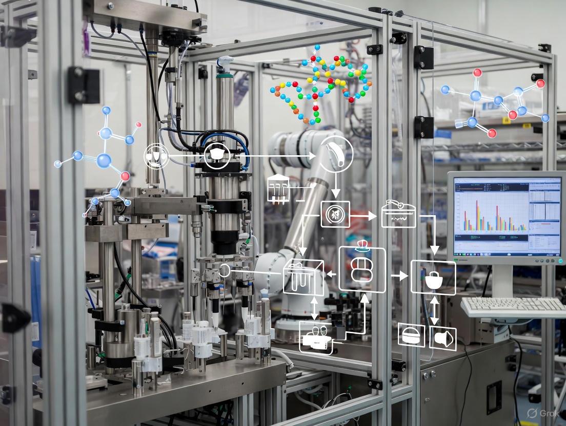
Implementing Automated Fill-Finish Systems: Technical Specifications, Workflow Integration, and Protocol Development
This application note details the critical subsystems of automated fill-finish platforms for cell therapy cryopreservation, with a focus on architecture that ensures product consistency, viability, and compliance. Data and protocols herein are contextualized within a broader research thesis on automating cryopreservation workflows. We provide a quantitative analysis of system performance, a detailed experimental protocol for validating an automated workflow, and a consolidated toolkit to guide researchers and drug development professionals in process development and scalability.
The transition from manual, open processes to automated, closed systems in cell therapy manufacturing is critical for scaling production while maintaining stringent Quality by Design (QbD) principles. The final fill-finish and cryopreservation steps are particularly vulnerable to deviations that can compromise critical quality attributes (CQAs) like cell viability, phenotype, and functionality. This document deconstructs the system architecture of automated fill-finish platforms, focusing on three pillars: precision temperature control, low-shear mixing mechanisms, and integrated single-use disposables. By integrating these components, these systems address key challenges of contamination risk, process variability, and scalability that are endemic to manual processing [9] [32].
System Architecture and Quantitative Performance
Automated fill-finish systems integrate several key subsystems to create a robust, closed environment for the final formulation of cell therapies. The performance of these integrated components is quantified below.
Table 1: Key Components and Performance Metrics of Automated Fill-Finish Architecture
| System Component | Function | Key Performance Metrics | Impact on Critical Quality Attributes (CQAs) |
|---|---|---|---|
| Precision Temperature Control | Actively cools cell suspensions and cryoprotectant agents (CPA) to maintain cell health and viability during processing. | Maintains consistent environmental temperatures within 3 °C of target [8]. Enables stepwise cooling of multiple materials to a specified temperature [31]. | Prevents premature CPA toxicity and osmotic stress; maintains >95% post-formulation viability [8]. |
| Low-Shear Mixing Mechanism | Achieves homogeneous cell and reagent suspension without inducing shear stress that damages cells. | Uniform cell concentration within 5% across final product bags [8]. Creates a strong vortex for rapid powder dissolution in large volumes [33]. | Preserves cell viability and functionality; maintains consistent phenotype across sub-lots [9]. |
| Integrated Single-Use Disposables | Provides a pre-sterilized, closed fluid path for processing, eliminating cleaning validation and cross-contamination. | Accurate volume control of ± 2 mL [8]. Effective air removal to < 2 mL in final product bags [8]. | Ensures sterility and final product container integrity; reduces contamination risk and operational complexity [33]. |
| Automated Aliquoting & Sealing | Precisely divides the final formulated product into multiple cryopreservation bags and hermetically seals them. | Variation in cell number and product volume of <12% across all containers in scaled runs [9]. | Guarantees dose uniformity and product consistency across patient doses; enables 60% reduction in operator hands-on time [8]. |
Experimental Protocol: Automated Processing and Cryopreservation of Cell Therapies
This protocol, adapted from a peer-reviewed method, outlines the procedure for using an automated fill-finish system (e.g., the Finia Fill and Finish System) in tandem with a controlled-rate freezer (CRF) for processing adherent and suspension cells [31].
Graphical Workflow
The diagram below illustrates the end-to-end automated workflow for cell therapy fill-finish and cryopreservation.
Materials and Reagents
Biological Materials
- Human mesenchymal stromal cells (MSCs): Sourced from umbilical cord tissue, expanded and harvested.
- Human peripheral blood mononuclear cells (PBMCs): Isolated from a leukopak.
Equipment
- Automated Fill-Finish System: Finia Fill and Finish System (Terumo Blood and Cell Technologies).
- Controlled-Rate Freezer (CRF): Programmable freezer capable of a -1°C/min cooling rate.
- Liquid Nitrogen Storage Tank: For vapor phase storage.
Single-Use Disposables
- FINIA 50 or 250 Tubing Set: Includes mixing bag, QC bag, and multiple storage bags.
- T-150 Transfer Bag: For introducing the cell suspension into the Finia system.
Key Reagents
- Culture Medium: Prime-XV MSC Expansion (XSFM) for MSCs.
- Dissociation Agent: TrypLE Express for adherent MSCs.
- Cryopreservation Medium: Cryostor CS-10.
- Cell Separation Medium: Lymphoprep for PBMC isolation.
- Staining Reagents: Zombie UV Fixable Viability Kit, Human TruStain FcX, specific antibody panels for phenotyping.
Procedure
Cell Harvest and Preparation:
- For adherent MSCs: Wash cells with PBS, dissociate using TrypLE Express, inactivate with serum-containing medium, and centrifuge to pellet.
- For suspension PBMCs: Isolate from leukopak using density gradient centrifugation with Lymphoprep. Wash and centrifuge to pellet.
- Perform a cell count and viability assessment on the harvested suspension.
System Setup and Material Loading:
- Load the appropriate single-use FINIA tubing set into the device.
- Aseptically transfer the harvested cell suspension into the system's T-150 transfer bag.
- Load the cryopreservation medium (Cryostor CS-10) and any required buffer into the designated bags on the FINIA set.
- Select and initiate the pre-programmed protocol on the Cell Processing Application (CPA) software.
Automated Formulation and Fill-Finish:
- The system automatically executes the following steps:
- Mixing and Cooling: The cell suspension, buffer, and cryoprotectant are mixed and cooled to a predefined temperature (e.g., 2-8°C) in a controlled manner.
- Formulation: The cryoprotectant is added stepwise to the cell suspension with continuous, low-shear mixing to ensure osmotic balance.
- Aliquoting and Sealing: The final formulated product is precisely aliquoted into multiple product bags, which are then automatically sealed. A separate QC bag is also filled.
- The system automatically executes the following steps:
Controlled-Rate Freezing:
- Immediately transfer the filled product bags to a pre-cooled controlled-rate freezer.
- Initiate a validated freezing profile. A common profile for many cell types is -1°C/min [16].
- Once the cycle is complete, promptly transfer the bags to vapor phase liquid nitrogen for long-term storage.
Post-Thaw Quality Control:
- Rapidly thaw a representative product bag (or the dedicated QC bag) in a 37°C water bath.
- Perform post-thaw analysis, which must include:
- Cell Count and Viability: Using a Via-1-Cassette or similar method. Target >90% post-thaw viability [8] [31].
- Phenotype Analysis: By flow cytometry to confirm the expression of specific markers (e.g., for T cells: effector memory and central memory populations; senescence/exhaustion markers) [9].
- Functionality Assay: Measure cytokine response (e.g., IFN-γ, TNF-α) after restimulation to ensure biological activity is retained [9].
The Scientist's Toolkit: Essential Research Reagent Solutions
Table 2: Key Reagents and Materials for Automated Cryopreservation Workflows
| Item | Function/Application | Example Product(s) |
|---|---|---|
| Cryostor CS-10 | A cGMP-compliant, serum-free cryopreservation medium containing 10% DMSO. Designed to minimize ice crystal formation and cell death during freeze-thaw. | Cryostor CS-10 [31] |
| FINIA Tubing Sets | Pre-sterilized, single-use disposable sets that form the closed fluid path for the automated system. Include mixing, QC, and final product bags. | FINIA 50 Tubing Set, FINIA 250 Tubing Set [31] |
| Lymphoprep | Density gradient medium for the isolation of viable peripheral blood mononuclear cells (PBMCs) from whole blood or leukopaks. | Lymphoprep [31] |
| TrypLE Express | A recombinant enzyme solution for dissociating adherent cells from culture surfaces, minimizing animal-derived components. | TrypLE Express [31] |
| Zombie UV Viability Kit | A fixable viability dye for flow cytometry, allowing accurate discrimination between live and dead cells in immunophenotyping panels. | Zombie UV Fixable Viability Kit [31] |
| Programmable CRF | A controlled-rate freezer that allows users to define and document cooling profiles, critical for process standardization and quality. | Various vendors (profile: -1°C/min) [16] [31] |
The integration of precision temperature control, gentle mixing, and single-use disposables within an automated architecture directly addresses the major hurdles in cell therapy cryopreservation: scalability, consistency, and compliance [16] [32]. The quantitative data presented confirms that these systems can maintain tight environmental control (±3°C) and product uniformity (<12% variation), which are difficult to achieve manually [8] [9].
A critical finding from industry surveys is that over 60% of users rely on default controlled-rate freezer profiles, but many challenging cell types (iPSCs, cardiomyocytes, specific T-cells) require optimized conditions [16]. This underscores the need for the detailed validation protocols provided here. Furthermore, the high resource dedication to cryopreservation and post-thaw analytics highlights that automation is not merely a convenience but a necessary investment to control critical process parameters and ensure product efficacy [16].
In conclusion, adopting an automated, closed fill-finish system early in clinical development establishes a robust and scalable manufacturing process. This foundation mitigates risks and avoids the significant challenges of process changes later, thereby accelerating the reliable delivery of advanced cell therapies to patients.
The transition from manual to automated processes in cell therapy manufacturing is critical for enhancing reproducibility, scalability, and product quality. This shift is particularly vital during the final formulation, fill, and finish stages, where maintaining cell viability and function directly impacts therapeutic efficacy [11]. This application note provides detailed protocols for processing both adherent and suspension cell types using an automated fill-finish system, the Finia Fill and Finish System (Terumo Blood and Cell Technologies), within the context of cell therapy cryopreservation research [8] [7]. We present a direct comparative analysis of the processing requirements for adherent cells (using Mesenchymal Stromal Cells, MSCs, as a model) and suspension cells (using Peripheral Blood Mononuclear Cells, PBMCs), including quantitative performance data and step-by-step methodologies to guide researchers and drug development professionals in automating this critical manufacturing step.
Comparative Analysis of Adherent and Suspension Cell Processing
The fundamental differences in biology between adherent and suspension cells necessitate distinct handling procedures, especially during harvest and formulation. The table below summarizes the key processing parameters and expected outcomes for both cell types when processed using an automated fill-finish system.
Table 1: Key Processing Parameters and Outcomes for Adherent vs. Suspension Cells in Automated Fill-Finish
| Parameter | Adherent Cells (MSCs) | Suspension Cells (PBMCs) |
|---|---|---|
| Pre-processing Harvest | Requires enzymatic digestion (e.g., TrypLE) to detach from culture surface [7]. | Harvested via centrifugation; no detachment needed [7]. |
| Shear Sensitivity | Generally higher sensitivity due to cytoskeletal organization and anchorage dependence [34]. | Generally lower sensitivity, but still requires low-shear handling [34] [8]. |
| Automated Processing | Compatible with automated formulation and aliquoting [7]. | Compatible with automated formulation and aliquoting [7] [9]. |
| Post-Formulation Viability | >95% of pre-formulation viability maintained [8] [35]. | >95% of pre-formulation viability maintained [8] [35]. |
| Post-Thaw Viability | >90% post-thaw viability demonstrated [7]. | >90% post-thaw viability demonstrated [7]. |
| Cell Concentration Uniformity | Within 5% across all product bags [8] [36]. | Within 5% across all product bags [8] [36]. |
| Key Quality Attributes | Phenotype (surface marker expression), differentiation potential, viability. | Phenotype (surface marker expression), viability, functionality (e.g., cytokine secretion) [9]. |
The following workflow diagram illustrates the integrated automated process for cryopreserving both cell types, from cell preparation to final cryopreservation.
Experimental Protocols
Materials and Reagent Solutions
The following table lists the essential materials and reagents required to execute the protocols for both adherent and suspension cell processing.
Table 2: Research Reagent Solutions and Essential Materials for Cell Processing and Cryopreservation
| Item | Function / Application | Example / Specification |
|---|---|---|
| Finia Fill and Finish System | Automates the final formulation, mixing, cooling, and aliquoting of cell suspensions into cryopreservation bags [8] [35]. | Terumo Blood and Cell Technologies |
| Finia Tubing Set | Functionally closed, single-use disposable set for processing; includes product bags and a QC bag [7]. | 50 mL or 250 mL configuration (Terumo BCT, cat. no. 22050 or 22250) |
| Controlled-Rate Freezer | Programmable freezer to standardize the cooling rate and improve post-thaw viability [7]. | Thermo Fisher Scientific compatible canister rack |
| Cryostor CS-10 | cGMP-manufactured, serum-free cryopreservation medium containing 10% DMSO [7]. | Fisher Scientific, cat. no. NC9930384 |
| TrypLE Express | Enzyme solution for dissociating adherent cells from culture vessels; less harsh than traditional trypsin [7]. | Millipore Sigma, cat. no. 12605028 |
| PLTGold Human Platelet Lysate | Supplement for MSC culture media; a xeno-free alternative to fetal bovine serum [7]. | Millipore Sigma, cat. no. SCM151 |
| Lymphoprep | Density gradient medium for the isolation of PBMCs from whole blood or leukopaks [7]. | STEMCELL Technologies, cat. no. 07801 |
| CellBIND HYPERFlask | High-surface area vessel for scalable expansion of adherent MSCs [7]. | Corning, cat. no. 10020 |
Detailed Protocol: Automated Fill-Finish for Adherent Cells (MSCs)
Objective: To harvest, formulate, and cryopreserve adherent MSCs using an automated fill-finish system while maintaining high viability and critical quality attributes.
Pre-Procedure Steps:
- Cell Expansion: Culture human umbilical cord tissue-derived MSCs in a T-150 flask or CellSTACK/HYPERFlask using Prime-XV MSC Expansion XSFM medium supplemented with penicillin/streptomycin and 2–10% human platelet lysate [7].
- Cell Harvest: Upon reaching 70–90% confluency, wash cells with PBS without Ca²⁺/Mg²⁺. Add TrypLE Express enzyme solution and incubate at 37°C until cells detach. Neutralize the enzyme with complete culture medium and collect the cell suspension.
- Cell Counting: Centrifuge the cell suspension, resuspend in an appropriate buffer, and perform a cell count and viability assessment (e.g., using a Via-1-Cassette and NucleoCounter or trypan blue exclusion).
Automated Fill-Finish Procedure using the Finia System:
- System Setup: Load the appropriate Finia Tubing Set (50 mL or 250 mL) into the Finia device. Ensure the Cell Processing Application (CPA) software is configured with the correct user-defined protocol [8] [36].
- Load Materials: Aseptically load the harvested MSC suspension into the designated source bag. Load the cryopreservation medium (e.g., Cryostor CS-10) into its designated bag within the system.
- Execute Protocol: Initiate the automated run. The system will perform the following steps sequentially [8] [35] [7]:
- Mixing and Cooling: The cell suspension and cryoprotectant are separately mixed and cooled to a user-defined target temperature (e.g., 2–8°C) to mitigate the exothermic reaction of DMSO hydration.
- Formulation: The cryoprotectant is added to the cell suspension in a controlled, gradual manner within the mixing bag, with continuous low-shear mixing to minimize osmotic shock [11].
- Aliquoting and Sealing: The final formulated cell product is automatically aliquoted into multiple cryopreservation bags at a consistent cell concentration (within 5% uniformity) and the bags are sealed [35].
- Product Retrieval: Upon completion, retrieve the filled cryopreservation bags and the attached QC bag.
Post-Procedure Steps:
- Controlled-Rate Freezing: Immediately transfer the product bags to a controlled-rate freezer. Initiate a freezing curve optimized for the specific cell type (e.g., -1°C/minute to -50°C, then -10°C/minute to -100°C) before transferring to long-term storage in the vapor phase of liquid nitrogen [7].
- Quality Control: Use the QC bag to perform post-process quality control assays, including cell count, viability, and phenotyping via flow cytometry (e.g., for CD73, CD90, CD105 positivity for MSCs).
Detailed Protocol: Automated Fill-Finish for Suspension Cells (PBMCs)
Objective: To formulate and cryopreserve apheresis-derived suspension cells (PBMCs) using an automated fill-finish system, ensuring consistent product quality across multiple aliquots.
Pre-Procedure Steps:
- Cell Isolation: Isolate PBMCs from a human leukopak using a density gradient medium like Lymphoprep according to standard protocols [7].
- Cell Preparation: Wash the isolated PBMCs with Ca²⁺/Mg²⁺-free PBS. Perform a cell count and viability assessment.
Automated Fill-Finish Procedure using the Finia System:
- System Setup: Identical to the procedure for adherent cells (Section 3.2).
- Load Materials: Aseptically load the PBMC suspension and cryopreservation medium into the Finia system.
- Execute Protocol: The automated process is identical in its core steps to that used for MSCs, demonstrating the platform's versatility [7]. The system handles the PBMCs with low-shear mixing and controlled cryoprotectant addition to maintain cell health [9].
Post-Procedure Steps:
- Controlled-Rate Freezing: Identical to the procedure for adherent cells (Section 3.2).
- Quality Control: Perform QC assays on cells from the QC bag. For PBMCs, this includes viability, cell count, and immunophenotyping to characterize lymphocyte subsets (T cells, B cells, NK cells). Functional assays, such as cytokine release upon restimulation (e.g., IFN-γ, TNF-α), can be used to confirm retained functionality post-processing [9].
The logical relationship between the core processing steps and their impact on critical cell quality attributes is shown below.
Automation of the fill-finish step is feasible and highly beneficial for both adherent and suspension cell types central to cell therapy manufacturing. The protocols detailed herein, utilizing the Finia Fill and Finish System, demonstrate that automated processing can maintain high cell viability (>95% post-formulation, >90% post-thaw), ensure dose uniformity (within 5%), and preserve critical cell phenotypes and functionalities [8] [7] [9]. By replacing variable manual processes with a standardized, closed-system approach, researchers and developers can significantly de-risk this critical manufacturing step, enhancing product consistency and facilitating the scalable and robust commercialization of cell therapies.
Final formulation, fill, and finish is a critical high-value step in cell therapy manufacturing where cells are most vulnerable after completing selection, modification, and expansion processes [11]. Effective cryoprotectant integration at this stage is essential for maintaining cell health, viability, and therapeutic function post-thaw. This Application Note addresses the dual challenges of managing dimethyl sulfoxide (DMSO) toxicity and osmotic stress during cryopreservation within automated fill-finish systems. We provide detailed protocols and data-driven strategies to optimize cryopreservation workflows, enhance product consistency, and ensure patient safety while supporting the transition toward automated manufacturing platforms.
The inherent variability of manual cryopreservation processes becomes unsustainable with increasing manufacturing demands, necessitating flexible automation with functionally closed single-use disposables [11]. Automated systems reduce open process steps, ensure traceability, and minimize DMSO contact time while enabling electronic data capture. Implementing automation at this critical juncture not only ensures more consistent products but also reduces manufacturing costs and supports reliable product delivery to patients.
Table 1: DMSO Concentration Effects on Cell Viability and Clinical Considerations
| DMSO Concentration | Post-Thaw Viability | Clinical Safety Profile | Application Notes |
|---|---|---|---|
| 10% (v/v) [37] | Established for multiple cell types | Maximum 1 g/kg accepted for HSC transplantation [37] | Conventional standard; associated with infusion reactions |
| 5% (v/v) [38] | Maintained with optimized cryomedium | Reduced toxicity risk vs. 10% DMSO | Enables DMSO reduction by 40-50% with albumin supplements |
| 2.5% (v/v) [39] | >70% (clinical threshold) with microencapsulation | Potentially safer profile | Requires hydrogel microencapsulation technology |
| ≤3% (v/v) [38] | Maintained with recombinant albumin | Significantly improved patient safety | Achievable with Optibumin 25 in CryoStor CS5 |
Table 2: Osmotic Stress Management Strategies and Cellular Impacts
| Stress Mechanism | Cellular Consequences | Management Approaches | Experimental Outcomes |
|---|---|---|---|
| Osmotic Imbalance [11] [40] | Cell shrinkage/swelling, membrane damage | Mathematical optimization of CPA loading [40] | Constant cell volume maintenance possible |
| DMSO Chemical Toxicity [11] [37] | Apoptosis, oxidative stress, functional impairment | Limit exposure to ≤30 minutes pre-freeze [11] | Reduced delayed onset cell death |
| Ice Recrystallization [41] | Membrane damage, organelle disruption | Ice Recrystallization Inhibitors (IRIs) [41] | Preserved post-thaw potency after warming events |
| Cryoprotectant Unloading [40] | Swelling-induced rupture | Controlled step-wise dilution [40] | Improved recovery rates |
Experimental Protocols
Protocol 1: Constant Volume Cryoprotectant Loading
This mathematical approach eliminates osmotic stress by maintaining constant cell volume during cryoprotectant loading [40].
Materials:
- Cell suspension (1×10⁶ cells/mL)
- Penetrating cryoprotectant (e.g., DMSO)
- Non-penetrating cryoprotectant (e.g., sucrose)
- Isotonic buffer
- Controlled-temperature mixing device
Method:
- Determine Cell Parameters: Measure cell water volume (Vwo), solute volume (Vso), and surface area (A) for target cell population
- Calculate Permeability Coefficients: Establish hydraulic conductivity (Lp) and solute permeability (Ps) for DMSO/cell type combination
- Implement Loading Protocol: Use derived analytical solutions to calculate transient extracellular concentrations:
- Prepare cryoprotectant solution with precisely controlled concentration ramp
- Maintain nonpermeating solute concentration to balance intracellular osmotic pressure
- Follow exact solution for extracellular permeating solute concentration:
- Cₑ(t) = (1 - (1 - γ)exp(-t/τ)) / (PsA(1 - α)/Lpγ²Vwo(0))
- Validate Volume Maintenance: Monitor cell volume throughout loading process via microscopy
- Complete Loading: Achieve target intracellular DMSO concentration with <2% volume deviation
Validation:
- Compare post-thaw viability with conventional step-wise loading
- Assess membrane integrity via flow cytometry
- Evaluate apoptotic markers 24 hours post-thaw
Protocol 2: Automated DMSO-Reduced Cryopreservation with Recombinant Albumin
This protocol leverages automated fill-finish systems to minimize DMSO toxicity while maintaining high post-thaw recovery [38].
Materials:
- Optibumin 25 recombinant human serum albumin [38]
- CryoStor CS5 or CS10 cryopreservation medium [38]
- Automated filling system with temperature control
- Controlled-rate freezer
- T cell suspension (1×10⁷ cells/mL)
Method:
- Prepare Cryomedium Formulation:
- For 5% DMSO formulation: Combine 40% CryoStor CS10, 10% Optibumin 25, 50% cell suspension
- For 3% DMSO formulation: Combine 60% CryoStor CS5, 10% Optibumin 25, 30% cell suspension
- Maintain at 2-8°C during preparation
Automated Mixing Process:
- Program automated system for gradual cryomedium introduction
- Set mixing parameters to 150 rpm with pulsed flow profile
- Maintain temperature at 4±1°C throughout process
- Complete mixing within 10 minutes to minimize DMSO exposure pre-freeze
Fill and Finish:
- Dispense 2mL aliquots into cryovials using peristaltic pumping
- Maintain temperature control throughout filling process
- Transfer to controlled-rate freezer within 15 minutes of cryomedium addition
Controlled-Rate Freezing:
- Initiate freezing within 30 minutes of DMSO addition
- Use optimized cooling rate: -1°C/min to -40°C, then -10°C/min to -100°C
- Transfer to liquid nitrogen vapor phase storage (-135°C to -150°C)
Quality Control:
- Document DMSO contact time through electronic data capture
- Record fill time and temperature for each vial
- Confirm post-thaw viability >85% and expansion capacity >1.5-fold over 72 hours
Protocol 3: Hydrogel Microencapsulation for Low-DMSO Cryopreservation
This technique enables substantial DMSO reduction through physical protection during cryopreservation [39].
Materials:
- Alginate hydrogel solution (1.5% w/v)
- Mesenchymal stem cell suspension
- Calcium chloride crosslinking solution (100mM)
- Low-DMSO cryomedium (2.5% DMSO)
- Electrospray device or precision droplet generator
Method:
- Microcapsule Fabrication:
- Mix MSC suspension with alginate solution to final density of 5×10⁶ cells/mL
- Generate microdroplets using electrostatic bead generator (20G needle, 15kV voltage)
- Crosslink droplets in calcium chloride solution for 10 minutes
- Wash with isotonic buffer to remove excess calcium
Low-DMSO Cryopreservation:
- Transfer microencapsulated cells to cryovials
- Add pre-chilled 2.5% DMSO cryomedium
- Equilibrate for 15 minutes at 4°C with gentle agitation
- Implement controlled-rate freezing: -1°C/min to -80°C
Thawing and Recovery:
- Rapid thaw in 37°C water bath with gentle agitation
- Dissolve hydrogel capsules with 50mM sodium citrate
- Centrifuge at 300g for 5 minutes to collect cells
- Resuspend in culture medium for functional assessment
Assessment:
- Determine viability via trypan blue exclusion (>70% acceptable)
- Verify trilineage differentiation potential (osteogenic, chondrogenic, adipogenic)
- Analyze stemness gene expression (OCT4, NANOG, SOX2)
Workflow Visualization
The Scientist's Toolkit: Research Reagent Solutions
Table 3: Essential Reagents for Advanced Cryopreservation Protocols
| Reagent / Technology | Function & Mechanism | Application Context |
|---|---|---|
| Optibumin 25 [38] | Recombinant HSA: stabilizes membranes, enables DMSO reduction | T cell cryopreservation with 3-6% DMSO instead of 10% |
| Hydrogel Microcapsules [39] | Alginate matrix: physical protection during freezing | MSC cryopreservation with 2.5% DMSO |
| Ice Recrystallization Inhibitors (IRIs) [41] | Inhibit ice crystal growth during temperature fluctuations | Protection against transient warming events |
| Mathematical Optimization Models [40] | Constant volume CPA loading calculations | Osmotic stress elimination during cryoprotectant addition |
| Controlled-Rate Freezers [16] | Programmable cooling rates for different cell types | Standardized freezing across manufacturing batches |
| Automated Fill-Finish Systems [11] | Reduced open steps, traceability, DMSO contact control | GMP-compliant manufacturing scale-up |
Effective integration of cryoprotectant strategies requires addressing both DMSO toxicity and osmotic stress as interconnected challenges. The protocols presented herein demonstrate that substantial DMSO reduction is achievable through technological interventions including recombinant albumin supplements, hydrogel microencapsulation, and automated mixing processes. Simultaneously, mathematical modeling of cryoprotectant loading enables complete elimination of osmotic stress by maintaining constant cell volume. Implementation within automated fill-finish systems ensures these advanced strategies can be deployed consistently at manufacturing scale, enhancing product quality while mitigating risks associated with manual processing. As cell therapies progress toward commercialization, robust, standardized cryopreservation protocols integrated with automated platforms will be essential for delivering safe, effective treatments to patients globally.
The transition from pre-commercial research and development (R&D) to commercial manufacturing represents a critical juncture in the development pathway of cell therapies. This scale-up process presents unique challenges for maintaining product quality, sterility, and potency while increasing production capacity. Fill-finish operations—the final steps of formulation, filling, and final container packaging—are particularly vulnerable points in manufacturing due to the direct manipulation of the final drug product [12]. For cell therapies, these challenges are amplified by the living nature of the product, small batch sizes, time-sensitive processing windows, and inability to undergo terminal sterilization [14].
Automated fill-finish systems have emerged as pivotal solutions for addressing these scale-up challenges, enabling manufacturers to maintain critical quality attributes while expanding production capacity. This application note examines key considerations for implementing automated fill-finish technologies within cell therapy cryopreservation workflows, providing structured data and detailed protocols to guide successful technology transfer from pre-commercial to commercial manufacturing.
Key Scale-Up Challenges in Cell Therapy Fill-Finish
Product Stability and Contamination Control
Cell therapies face significant stability challenges during fill-finish operations, primarily due to their limited viability windows at ambient temperatures. Most cell therapies remain stable for only 2-3 hours at room temperature, creating narrow processing windows that complicate scale-up [12]. Unlike traditional biologics, cell therapies cannot undergo terminal sterilization through filtration or heat methods due to their cellular integrity and sensitivity, necessitating strict aseptic processing throughout all manufacturing steps [14].
The living nature of cell therapies introduces additional complexities, as products must maintain not only viability but also critical phenotypic and functional characteristics throughout scale-up. Process conditions during fill-finish can significantly impact therapeutic efficacy, particularly affecting cell persistence and functionality post-infusion [42].
Scalability and Process Consistency
Scalability presents multifaceted challenges for cell therapy manufacturers. Autologous therapies face constraints in scaling out due to their patient-specific nature, requiring manufacturers to implement multiple parallel processes rather than simply increasing batch size [12]. Allogeneic therapies, while offering potential for traditional scale-up approaches, confront hurdles in maintaining product consistency across larger cell batches [42].
The high variability of starting materials, particularly in autologous therapies, introduces inherent process variability that complicates scale-up. Donor cells exhibit differing metabolic profiles and capabilities, yet current manufacturing processes often lack adaptability to normalize these differences [42]. Additionally, the legacy manufacturing processes common in the sector remain a primary driver of high therapeutic costs, creating bottlenecks that limit patient access [42].
Table 1: Primary Scale-Up Challenges in Cell Therapy Fill-Finish
| Challenge Category | Specific Challenges | Impact on Scale-Up |
|---|---|---|
| Product Characteristics | Limited room-temperature stability (2-3 hours)Inability to undergo terminal sterilizationMaintenance of phenotypic/functional properties | Requires rapid processingDemands strict aseptic processingNecessitates gentle, controlled handling |
| Process Constraints | Small batch sizes (often patient-specific)High variability of starting materialLabor-intensive manual operations | Limits traditional scale-up approachesComplicates process standardizationIncreases cost and variability |
| Supply Chain & Logistics | Cryopreservation requirements (-130°C to -196°C)Time-sensitive cold chainPatient-specific supply chains | Requires specialized equipmentDemands robust logisticsAdds traceability complexity |
Automation as a Strategic Enabler
Benefits of Automated Fill-Finish Systems
Automation addresses critical scale-up challenges by enhancing process reproducibility, reducing human intervention, and enabling more consistent manufacturing outcomes. Automated systems demonstrate particular value in fill-finish operations where precision, sterility, and speed are paramount [28].
Recent studies quantifying the performance of automated fill-finish systems reveal significant improvements in process consistency. Testing of the Finia Fill and Finish System demonstrated the system could effectively scale to 4 times its singular capacity within a 2-hour timeframe, with variation in cell number and product volume less than 12% across all containers [9]. This level of consistency is difficult to achieve with manual processes, particularly when scaling operations.
Beyond consistency, automated systems maintain critical product quality attributes. Analysis of different sub-lots filled using automated systems showed high cell viability and consistent T cell phenotype, with preserved proportions of effector memory and central memory T cells and minimal expression of senescence and exhaustion markers [9]. Additionally, functional potency remains intact, with consistent cytokine response (IFN-γ and TNF-α) after restimulation across different sub-lots [9].
Strategic Timing of Automation Implementation
The decision of when to implement automation represents a strategic consideration with significant long-term implications. While early adoption requires substantial upfront investment, it establishes a stable foundation for scale-up from initial clinical trials through commercial manufacturing [28].
Early automation in Phase 1/2 clinical trials facilitates more seamless transition to later clinical phases and commercial manufacturing, despite the initial capital outlay. This approach uniformizes processes and operations early, reducing the need for extensive process revalidation during critical scale-up phases [28]. Additionally, early automation signals commitment to commercial viability, potentially enhancing investor confidence.
Later adoption conserves initial capital and maintains flexibility during process optimization stages, but risks creating bottlenecks when scaling becomes urgent. Transitioning from manual to automated operations at later stages can disrupt established workflows and require significant revalidation efforts [28].
Table 2: Automation Implementation Timing Considerations
| Implementation Stage | Advantages | Disadvantages | Best Suited For |
|---|---|---|---|
| Early Implementation(Phase 1/2) | Foundations for scale already establishedMore seamless tech transferEnhanced investor confidence | High upfront capital investmentReduced process flexibilityPotential technology lock-in | High-throughput therapiesCompanies with strong fundingTherapies with established processes |
| Late Implementation(Phase 3/Commercial) | Conserves early-stage capitalMaintains process flexibilityLeverages more mature technologies | Potential scale-up bottlenecksCostly process revalidationPossible production delays | Rare disease therapiesResource-constrained companiesEvolving manufacturing processes |
Quantitative Analysis of Automated Fill-Finish Performance
Robust quantitative assessment is essential for evaluating automated fill-finish systems during scale-up. The following data, compiled from validation studies, provides key performance metrics for benchmarking automated systems in cell therapy applications.
Table 3: Performance Metrics of Automated Fill-Finish Systems
| Performance Parameter | Pre-Commercial Scale(Manual Process) | Commercial Scale(Automated System) | Improvement |
|---|---|---|---|
| Process Time for 4x Scale | 4-6 hours | 2 hours | 50-66% reduction [9] |
| Container Volume Variation | 20-35% CV | <12% CV | >40% improvement [9] |
| Cell Number Consistency | 25-40% CV | <12% CV | >50% improvement [9] |
| Viability Post-Processing | Variable (70-90%) | Consistently high (>90%) | Improved consistency [9] |
| Contamination Risk | Higher (manual intervention) | Significantly reduced | Enhanced sterility assurance [12] |
The consistency metrics demonstrated by automated systems are particularly relevant for scale-up, as reduced variability directly supports manufacturing robustness. The maintenance of phenotypic and functional characteristics across sub-lots further validates automated systems for commercial-scale implementation [9].
Experimental Protocol: Validation of Automated Fill-Finish Systems
System Suitability Testing
Purpose: To verify that the automated fill-finish system operates within specified parameters for cell therapy products and establishes baseline performance metrics before GMP implementation.
Materials:
- Automated fill-finish system (e.g., Finia Fill and Finish System)
- Cell suspension (representative cell therapy product)
- Cryopreservation medium with DMSO
- Final product containers (cryobags or vials)
- Cell counter and viability analyzer
- Flow cytometer for phenotypic analysis
- ELISA kits for cytokine detection
Procedure:
- System Setup and Calibration
- Install system according to manufacturer specifications in controlled environment (ISO 7 or better)
- Perform pressure hold test to verify system integrity
- Calbrate volume delivery using phosphate buffered saline across operational range
Process Parameter Establishment
- Define optimal flow rate (typically 10-50 mL/min) to minimize shear stress
- Establish mixing parameters to maintain homogeneous cell suspension
- Determine fill speed and dispense height to minimize foaming and splashing
Performance Qualification
- Execute repeated fill cycles (n=10) with cell suspension at target density
- Collect samples from beginning, middle, and end of run for consistency assessment
- Assess cell count, viability, and volume accuracy across all containers
Product Quality Assessment
- Analyze phenotype via flow cytometry for critical markers (e.g., CD3, CD4, CD8, CD62L, CD45RO)
- Evaluate functional potency through cytokine secretion upon restimulation
- Assess recovery post-thaw for cryopreserved products
Acceptance Criteria:
- Volume accuracy: ≤15% coefficient of variation (CV) across all containers
- Cell count consistency: ≤15% CV across all containers
- Viability: ≥80% post-processing with no significant reduction from baseline
- Phenotypic consistency: ≤15% CV in critical marker expression
- Functional potency: Consistent cytokine response across sub-lots
Scale-Up Validation Protocol
Purpose: To demonstrate the automated system maintains critical quality attributes when scaling to commercial production volumes.
Materials: (As in Section 5.1, with addition of larger volume containers for scale-up)
Procedure:
- Baseline Establishment
- Process single lot of cell product at maximum manual capacity
- Assess all critical quality attributes (CQA) as baseline reference
Scaled Processing
- Process identical cell product at 2x, 3x, and 4x baseline capacity
- Maintain consistent process parameters across scaled runs
- Document any parameter adjustments required for scale-up
Extended Processing Evaluation
- Conduct continuous operation for maximum anticipated commercial run time
- Monitor system performance and product quality at predetermined intervals
- Assess operator interventions required during extended operation
Comparative Analysis
- Compare all CQAs across different production scales
- Evaluate trendlines for any scale-dependent effects on product quality
- Document operational efficiency metrics (time, labor, materials)
Acceptance Criteria:
- All CQAs maintained within established ranges across scales
- No significant scale-dependent trends in product quality
- Operational efficiency improvements demonstrated at scale
- System reliability maintained throughout extended operation
Implementation Workflow
The following workflow outlines the strategic implementation of automated fill-finish systems from process development through commercial manufacturing.
The Scientist's Toolkit: Essential Research Reagent Solutions
Successful implementation of automated fill-finish systems requires complementary reagent systems designed to maintain cell quality during processing. The following table outlines essential materials and their functions.
Table 4: Essential Research Reagent Solutions for Automated Fill-Finish
| Reagent/Material | Function | Scale-Up Considerations | Quality Requirements |
|---|---|---|---|
| DMSO (Cryoprotectant) | Prevents ice crystal formation during cryopreservation | Consistent quality across batchesReduced lot-to-lot variability | Excipient gradeUSP/EP compliance [14] |
| Serum Albumin (HSA/BSA) | Stabilizes cells during processing stressRedces mechanical damage | Scalable sourcingPathogen safety | Excipient gradeConsistent availability [14] |
| Specialized Cryomedium | Maintains viability during freeze-thawSupports post-thaw recovery | Formulation compatibility with automationReady-to-use formats | cGMP complianceStringent QC testing [14] |
| Single-Use Assemblies | Maintains sterilityReduces cross-contamination | Process compatibilityScalable configurations | Pre-sterilizedLow extractables [12] |
| Cryogenic Containers | Final product storageMaintains stability at ultra-low temperatures | Automation compatibilityConsistent filling characteristics | Integrity at cryogenic temperaturesLeak-proof [12] |
The successful scale-up of fill-finish operations from pre-commercial R&D to commercial manufacturing requires strategic implementation of automated systems supported by robust validation protocols. Quantitative data demonstrates that automation significantly improves process consistency while maintaining critical product quality attributes. The experimental protocols provided herein offer a structured approach to technology implementation, ensuring that automated fill-finish systems meet the rigorous demands of commercial-scale cell therapy manufacturing. As the industry continues to evolve, early and strategic automation of fill-finish operations will be increasingly essential for delivering transformative cell therapies to patients in a cost-effective and scalable manner.
The final formulation, fill, and finish step represents a critical phase in cell therapy manufacturing where maintaining cell health, viability, and product quality is paramount [11]. At this stage, the cells have undergone extensive selection, modification, and expansion processes, making accurate documentation and controlled processing essential for maintaining therapeutic efficacy. Automated systems like the Finia Fill and Finish System, governed by its Cell Processing Application (CPA) software, address these challenges by providing a functionally closed, benchtop system that fully automates every stage of the final manufacturing process [8]. This automation significantly reduces risks associated with manual processes, including product variability, contamination, and documentation errors, while ensuring compliance with current Good Manufacturing Practice (cGMP) regulations through robust electronic record-keeping capabilities.
The transition from paper-based to electronic batch records in a cGMP environment offers substantial benefits for cell therapy manufacturing, including improved data integrity, reduced errors, increased data integration and centralization, and enhanced operational flexibility [43]. For cell therapies, where biological variability necessitates careful documentation and control, electronic systems provide the necessary framework to maintain product consistency and quality. The CPA software serves as a secure, server-based application for procedure management and record keeping, capable of tracking performance and protocol development for cGMP compliance, thereby addressing the critical data management challenges facing the expanding cell and gene therapy industry [7] [8].
CPA Software Features and cGMP Compliance
Core Architecture and Data Integrity
The Cell Processing Application (CPA) is a stand-alone, server-based software designed specifically to manage the Finia Fill and Finish System while maintaining comprehensive electronic records in accordance with regulatory requirements [8]. Its architecture ensures data integrity through multiple protective mechanisms aligned with FDA 21 CFR Part 11 and EU GMP Annex 11 regulations [44]. The system implements strict user access controls with unique credentials that cannot be reassigned or reused, ensuring that only authorized personnel can perform specific actions within the system [44]. This approach maintains the fundamental principle of accountability in pharmaceutical manufacturing by clearly identifying who performed what activity, when, where, and why [44].
The CPA software maintains trustworthiness and reliability of electronic records through several key features:
- Automated Audit Trails: The system captures all activities in real-time, logging every user interaction, system action, and data modification with comprehensive context including timestamps and the meaning of each action [44].
- Electronic Signatures: Cryptographic digital signatures ensure that all captured data cannot be tampered with, providing equivalent legal standing to handwritten signatures on paper records [43] [44].
- Data Security: The server-based application is protected from access by unauthorized individuals, maintaining the confidentiality and integrity of manufacturing data [7].
Operational Features and Functionality
The CPA software provides comprehensive functionality for managing the fill and finish process through configurable operational features. Users can define processing protocols, specify materials, configure device parameters, and establish user permissions tailored to their organizational structure and quality system requirements [8]. The system manages critical procedure data and automatically transmits information between the Finia system and application during operation, creating a seamless digital workflow that reduces manual intervention and associated error risks.
For compliance and reporting, the CPA software offers robust capabilities for tracking tubing sets and materials, recording alarms, capturing electronic data, and generating comprehensive procedure reports [8]. These features facilitate ready availability of documentation for FDA audits and inspections, significantly reducing the preparation time and resources typically required for regulatory assessments [44]. The system's ability to electronically record and report operational data aligns with cGMP requirements for manufacturing environments, providing a solid foundation for quality management and regulatory compliance throughout the cell therapy product lifecycle.
Table 1: Key cGMP Compliance Features of CPA Software
| Feature | Description | Regulatory Alignment |
|---|---|---|
| User Access Controls | Unique credentials with role-based permissions ensuring only authorized personnel can perform specific actions | 21 CFR Part 11 [44] |
| Audit Trail | Automated recording of all user actions, system events, and data modifications with timestamps and context | EU GMP Annex 11 [44] |
| Electronic Signatures | Cryptographic signatures that cannot be tampered with, providing equivalent standing to handwritten signatures | 21 CFR Part 11 [43] [44] |
| Procedure Management | Secure server-based application for managing device procedures, protocols, and configurations | cGMP requirements [7] [8] |
| Electronic Reporting | Automated generation of procedure reports with complete data capture for regulatory inspections | cGMP documentation requirements [8] |
Experimental Protocol: Automated Fill-Finish for Cell Cryopreservation
Equipment and Reagent Setup
Materials and Equipment:
- Finia Fill and Finish System (Terumo Blood and Cell Technologies) [8]
- FINIA 250 tubing set (mixing bag, QC bag 10–40 mL, three storage bags 29–210 mL total) [7]
- Controlled-rate freezer (e.g., Thermo Fisher Scientific) [7]
- Cryostor CS-10 cryopreservation solution [7]
- Appropriate cell culture media (e.g., Prime-XV MSC Expansion XSFM for MSCs) [7]
- Cell suspension (e.g., mesenchymal stromal cells or peripheral blood mononuclear cells) [7]
System Preparation:
- Install the appropriate FINIA tubing set according to manufacturer instructions, ensuring all connections are secure and the system is properly closed [7].
- Power on the Finia Fill and Finish System and launch the Cell Processing Application (CPA) software on the connected workstation.
- Log into the CPA software with appropriate user credentials, ensuring proper authentication and system access tracking [44].
- Select or create a new procedure within the CPA software, configuring the parameters according to the specific cell type and processing requirements.
Cell Processing and Formulation Protocol
System Priming and Temperature Control:
- Prime the Finia system with appropriate buffers or media according to established protocols.
- Activate the cooling system to maintain reagents at 2–8°C to counteract the exothermic reaction during DMSO addition and minimize cryoprotectant toxicity [11].
Cell Loading and Mixing:
- Transfer the cell suspension into the appropriate input bag of the FINIA tubing set.
- Program the CPA software with the specific processing parameters, including cell concentration, final volume, and cryoprotectant addition rate.
- Initiate the automated process, which progressively mixes cells, buffers, and cryopreservation solution using low-shear mixing paddles to maintain cell viability [8].
Automated Aliquoting and Sealing:
- The system automatically aliquots the final formulated cell product into individual product bags with accurate volume control of ±2 mL [8].
- Product bags are automatically sealed after filling, maintaining a closed system throughout the process.
- The system effectively removes air from final product bags to less than 2 mL [8].
Quality Control Sampling:
- Utilize the integrated QC bag to automatically collect representative samples for quality testing.
- Document all process parameters and quality controls within the CPA software electronic batch record.
Cryopreservation and Storage
- Transfer the filled product bags to a controlled-rate freezer programmed with an appropriate freezing curve for the specific cell type.
- Initiate the freezing protocol, typically achieving a post-thaw cell viability of >90% for cell products from healthy T-cell donors [8].
- After complete freezing, transfer bags to vapor phase liquid nitrogen storage for long-term preservation.
- Document all cryopreservation parameters in the CPA software, including freezing rates, final temperatures, and storage locations.
Data Management and Electronic Documentation
Electronic Batch Record Configuration
The CPA software facilitates comprehensive electronic batch record management that exceeds paper-based documentation capabilities. When configuring the electronic batch record within CPA, manufacturers should implement the following key elements to ensure regulatory compliance and operational efficiency:
- Interactive Work Instructions: Instead of static "paper on glass" approaches, implement dynamic work instructions that provide real-time guidance to operators, including conditional branching for complex manufacturing processes and parallel production steps [43].
- Real-time User Input Verification: Configure the system to validate operator entries as they are made, including unit conversions, calculations, and checks for missing data and signatures [43] [45].
- Automated Data Capture: Establish connections between CPA and other laboratory systems (LIMS, ERP) to automatically capture relevant process data, reducing manual transcription errors [43].
- Review by Exception Mechanisms: Implement automated review protocols that flag deviations from expected parameters, allowing quality personnel to focus on critical issues rather than routine verification [43].
Data Integration and Centralization
Modern cell therapy manufacturing generates substantial data from multiple sources, creating challenges for data management and analysis. The CPA software supports streamlined data management through integration capabilities that help consolidate information from disparate systems:
- API Integration: Utilize application programming interfaces to exchange data with other systems according to fixed protocols, enabling connectivity with ERP systems for raw material inventory, LIMS for laboratory results, and MES for manufacturing execution data [45].
- Unified Audit Trails: The CPA system captures comprehensive audit trails that document every aspect of the manufacturing process, creating a single source of truth that eliminates fragmented information architecture [45].
- Structured Data Collection: Implement standardized data structures early in process development to generate highly structured data that can be leveraged for analytics throughout the product lifecycle [43].
Table 2: Quantitative Performance Metrics of Automated Fill-Finish Process
| Performance Parameter | Result | Measurement Method |
|---|---|---|
| Post-thaw Cell Viability | >90% [8] | Flow cytometry with viability staining (e.g., Zombie UV dye) [7] |
| Post-formulation Cell Viability | >95% [8] | Automated cell counting with Via-1-Cassette cartridges [7] |
| Volume Control Accuracy | ±2 mL [8] | In-built weight check system [8] |
| Temperature Consistency | Within 3°C of target [8] | Integrated temperature monitoring [8] |
| Uniform Cell Concentration | Within 5% [8] | Consistent mixing with low-shear paddles [8] |
| Operator Hands-on Time Reduction | 60% reduction [8] | Time-motion studies comparing manual vs. automated processes [8] |
The Scientist's Toolkit: Essential Research Reagent Solutions
Table 3: Key Reagents and Materials for Automated Cell Therapy Processing
| Reagent/Material | Function | Application Notes |
|---|---|---|
| Cryostor CS-10 | Cryopreservation solution with optimized DMSO concentration | Provides controlled cryoprotection while minimizing osmotic stress and biochemical toxicity [7] |
| TrypLE Express | Recombinant enzyme for cell detachment | Superior to trypsin for adherent cells; eliminates wash steps and makes incubation timing less critical [45] |
| PLTGold Human Platelet Lysate | Serum-free supplement for cell culture | Defined composition reduces lot-to-lot variability compared to FBS [7] |
| Zombie UV Fixable Viability Kit | Flow cytometry viability staining | Distinguishes live/dead cells for post-thaw viability assessment [7] |
| FINIA Tubing Sets | Single-use disposable fluid path | Maintains closed system processing; available in 50mL and 250mL configurations [7] [8] |
| Lymphoprep | Density gradient medium for PBMC isolation | Enables separation of mononuclear cells from peripheral blood [7] |
Implementation Considerations and Compliance Strategy
Validation and Change Control
Implementing CPA software within a cGMP environment requires careful planning and execution to ensure regulatory compliance and operational effectiveness. Manufacturers should adopt a systematic approach to validation that encompasses both the technical system and the associated documentation processes:
- Business Continuity Planning: Develop and regularly test contingency procedures for software failure to ensure manufacturing operations can continue without compromising product quality or data integrity [43].
- Change Control Management: Implement cross-functional change control processes that meticulously track, evaluate, and document modifications to the CPA software configuration and associated documentation systems [46].
- User Acceptance Testing: Conduct comprehensive testing with actual operators to verify that the system functions as intended in the specific manufacturing environment and meets all user requirements.
- Supplier Qualification: Verify the software provider's update strategy and incident handling procedures to ensure compatibility with your change control strategy and CAPA processes [43].
Data Analytics and Process Intelligence
The structured data generated by CPA software creates opportunities for advanced analytics and process improvement that extend beyond basic compliance:
- Automated Trend Analysis: Configure the system to automatically process and trend critical process parameters as new measurement data becomes available, enabling real-time process monitoring and control [45].
- Quality by Design (QbD) Implementation: Utilize the comprehensive data capture capabilities to identify relationships between critical process parameters (CPPs) and critical quality attributes (CQAs), supporting QbD principles in process development [45].
- Configurable Reporting: Implement customized reporting options that extend beyond standard templates to address evolving organizational needs throughout the product lifecycle [43].
- Data Export Capabilities: Establish efficient data export mechanisms or read-only database access to enable integration with specialized Business Intelligence (BI) tools for advanced analytics [43].
The implementation of electronic records through CPA software represents a significant advancement in data management for cell therapy manufacturing, providing robust documentation capabilities that support both regulatory compliance and process optimization. By leveraging the automated data capture, integration capabilities, and compliance features of the CPA system, manufacturers can address the critical challenges of cell therapy production while establishing a foundation for continuous process improvement and innovation.
Optimizing Automated Fill-Finish Performance: Addressing DMSO Toxicity, Process Validation, and Contamination Control
In the automated fill-finish systems central to modern cell therapy manufacturing, managing cryoprotectant (CPA) toxicity represents a critical bottleneck. Cryoprotectant toxicity remains the greatest obstacle to cryogenic cryopreservation of tissues and organs, standing in the way of procedures that could save many lives [47]. As the cell therapy market progresses—projected to grow at a CAGR of 16% from 2025 to 2035—optimization of CPA handling becomes increasingly imperative for commercial viability [2] [48].
This application note examines CPA toxicity management through the integrated lenses of temperature control and exposure time optimization within automated fill-finish workflows. We provide detailed protocols and quantitative frameworks to maximize post-thaw cell viability while maintaining process efficiency in advanced therapy medicinal product (ATMP) manufacturing.
Understanding Cryoprotectant Toxicity Mechanisms
Cryoprotectant toxicity manifests through multiple pathways that can be broadly categorized into specific and non-specific mechanisms. Specific toxicity refers to direct chemical damage unique to each CPA, while non-specific toxicity arises from general physicochemical disruptions affecting cellular structures [47] [49].
Molecular Pathways of Toxicity
Membrane Integrity Damage: High concentrations of penetrating CPAs like DMSO can cause undulations in cell membranes, with 20% DMSO causing water entry and swelling [47]. Glycerol at concentrations over 1.5% polymerizes the actin cytoskeleton in stallion spermatozoa, an effect unrelated to osmolality [47].
Metabolic Toxicity: Some CPAs are metabolized to toxic compounds. Ethylene glycol is metabolized in the liver to glycoaldehyde and then to glycolic acid, resulting in metabolic acidosis. Glycolic acid is further metabolized to oxalic acid, which precipitates with calcium to form calcium oxalate crystals in tissues, notably the kidney [47].
Mitochondrial Dysfunction: In zebrafish ovarian follicles, cryopreservation with methanol showed a dose-dependent reduction in five mitochondrial function measures: membrane potential, mitochondrial distribution, mitochondrial DNA copy number, ATP levels, and ADP/ATP ratios [47].
Protein Denaturation and DNA Damage: Formamide can denature DNA, an effect believed to be due to displacement of hydrating water [47]. Methanol concentrations above 6M in fish oocytes resulted in protein damage or proteolysis [47].
Oxidative Stress: Glycerol depletes reduced glutathione in the kidney, leading to oxidative stress and apoptosis facilitated by caspases [47].
The following diagram illustrates the relationship between these key toxicity mechanisms and their cellular impacts:
Figure 1: Cryoprotectant Toxicity Mechanisms and Cellular Impacts. CPA toxicity arises from both specific chemical properties and non-specific physicochemical effects, ultimately converging on critical cellular damage pathways.
Quantitative Toxicity Assessment and Modeling
Effective management of CPA toxicity requires robust quantification methods and predictive modeling. Recent advances in high-throughput screening and computational modeling have significantly enhanced our ability to predict and mitigate toxic effects.
High-Throughput Toxicity Screening
A 2025 study demonstrated a high-throughput method for simultaneously screening membrane permeability and toxicity using an automated plate reader, enabling ~100 times faster permeability measurement than previous methods [50]. This approach allows rapid assessment of 27 chemicals at both 4°C and room temperature using the same 96-well plate, with viability measurements plotted against CPA permeability to identify optimal candidates [50].
Mathematical Modeling of CPA Toxicity
A multiple cryoprotectant toxicity model developed in 2022 enables in silico exploration of the vast protocol landscape by accounting for specific toxicity, non-specific toxicity, and modulation of these toxicity mechanisms due to intermolecular interactions between CPAs in solution [49]. This comprehensive toxicity model spans five common CPAs (glycerol, DMSO, propylene glycol, ethylene glycol, and formamide) and any combination thereof, offering new possibilities for designing less toxic vitrification methods [49].
Table 1: Comparative Toxicity Profiles of Common Cryoprotectants
| Cryoprotectant | Molecular Weight (g/mol) | Key Toxic Mechanisms | Temperature Sensitivity | Typical Working Concentration |
|---|---|---|---|---|
| DMSO | 78.1 | Membrane disruption, cellular swelling, differentiation alterations | High (significantly more toxic at higher temperatures) | 5-10% (0.6-1.3 M) |
| Glycerol | 92.1 | Oxidative stress, cytoskeleton polymerization, glutathione depletion | Moderate | 5-15% (0.5-1.6 M) |
| Ethylene Glycol | 62.1 | Metabolic acidosis, calcium oxalate crystal formation | Moderate | 5-20% (0.8-3.2 M) |
| Propylene Glycol | 76.1 | Intracellular pH reduction, developmental impairment | Moderate | 2.5-10% (0.3-1.3 M) |
| Formamide | 45.0 | DNA denaturation, corrosive effects, protein destabilization | High | Typically used in mixtures (1-3 M) |
| Methanol | 32.0 | Formaldehyde/formic acid metabolism, mitochondrial dysfunction, protein damage | High | 5-15% (1.6-4.7 M) |
Temperature Control Strategies
Temperature serves as a powerful modulator of CPA toxicity, with precise thermal management significantly influencing cellular outcomes during cryopreservation procedures.
Fundamental Temperature-Toxicity Relationships
CPA toxicity demonstrates strong temperature dependence, with most CPAs becoming significantly more toxic as temperature increases [47]. For example:
- Dermal fibroblasts exposed to DMSO showed decreasing viability with increasing concentration, temperature, and exposure time [47].
- DMSO causes irreversible ultrastructural alterations to rat myocardium above 1.41 M (10% vol/vol) at 30°C, but these effects occur at higher concentrations (2.82 M) at 15°C [47].
- High-throughput screening reveals that more candidate CPAs are toxic at 25°C compared to 4°C, despite faster membrane permeation at the higher temperature [50].
Practical Temperature Optimization Framework
The following workflow illustrates the decision process for temperature optimization in automated fill-finish systems:
Figure 2: Temperature Optimization Workflow for CPA Processing. This decision framework balances CPA permeability requirements with toxicity minimization through strategic temperature management.
Preventing Transient Warming Events
Transient Warming Events (TWEs) present a significant but often overlooked threat to cell therapy products during cryopreservation. TWEs occur when cryopreserved samples are exposed to warmer-than-intended temperatures for short periods, potentially during storage transfers, shipping, or processing [41].
Consequences of TWEs include:
- Ice recrystallization: Ice crystals grow during warming, damaging cell organelles and membranes [41]
- Increased cryoprotectant toxicity: DMSO and other CPAs become more toxic as temperatures rise [41]
- Osmotic stress: Water movement becomes unbalanced, leading to structural instability [41]
- Delayed onset cell death: Cells may undergo apoptosis hours or days post-thaw due to cumulative stress [41]
Mitigation strategies:
- Use real-time data loggers and sensors in freezers, storage units, and transport systems [41]
- Implement ice recrystallization inhibitors (IRIs) to reduce damage caused by transient warming [41]
- Develop standard operating procedures for all cryogenic handling activities [41]
- Use cryogenic containers with high thermal mass to extend safe handling windows [41]
Table 2: Temperature-Responsive Cryoprotectant Toxicity Profiles
| Cryoprotectant | Optimal Addition Temperature | Toxicity Activation Energy | Critical Temperature Threshold | Recommended Warming Rate |
|---|---|---|---|---|
| DMSO | 0-4°C | High (significant reduction in toxicity at lower temperatures) | >15°C (sharp increase in toxicity) | 50-100°C/min |
| Glycerol | 4-10°C | Moderate (gradual toxicity reduction with cooling) | >25°C (moderate toxicity increase) | 20-50°C/min |
| Ethylene Glycol | 4-15°C | Low to moderate (mild temperature dependence) | >30°C (moderate toxicity increase) | 30-70°C/min |
| Propylene Glycol | 4-10°C | Moderate (similar to glycerol) | >25°C (moderate toxicity increase) | 25-60°C/min |
| Formamide | 0-4°C | High (strong temperature dependence) | >10°C (sharp toxicity increase) | 60-120°C/min |
| Methanol | 0-4°C | High (significant toxicity at elevated temperatures) | >15°C (sharp toxicity increase) | 70-150°C/min |
Exposure Time Optimization
Time represents a critical variable in CPA toxicity management, with exposure duration directly correlating with cellular damage across multiple CPA categories.
Time-Toxicity Relationships
Exposure time to CPAs follows a dose-response relationship with cellular damage, where both concentration and duration contribute to the overall "toxicity load" [47] [49]. For instance:
- Dermal fibroblasts exposed to DMSO for periods of 10, 20, and 30 minutes showed decreasing viability with increasing exposure time [47].
- Mathematical models of CPA toxicity incorporate exposure time as a fundamental parameter in predicting cellular viability outcomes [49].
- In high-throughput toxicity screening, exposure time is carefully controlled, with measurements typically taken after ~20 minutes of CPA exposure [50].
Strategic Exposure Time Management
The relationship between exposure time, temperature, and concentration follows predictable patterns that can be leveraged for protocol optimization:
Figure 3: Interrelationship of Toxicity Factors in Cryopreservation Outcomes. The total toxicity load experienced by cells represents the integrated effect of concentration, exposure time, and temperature, ultimately determining cellular outcomes.
Automated System Time Optimization
In automated fill-finish systems, exposure time optimization requires careful balancing of multiple process parameters:
Step-Gradient Exposure Protocol:
- Initial equilibration: 50% final CPA concentration for 3-5 minutes at 4°C
- Secondary equilibration: 75% final CPA concentration for 3-5 minutes at 4°C
- Final concentration: 100% target concentration with minimal exposure at processing temperature
- Rapid transition to freezing: Immediate transfer to controlled-rate freezer after reaching target concentration
Critical Time Thresholds:
- Total exposure time to ≥3M CPA concentrations: <15 minutes at >4°C
- Maximum exposure to vitrification solutions: <8 minutes at >4°C
- Optimal washout duration post-thaw: 5-10 minutes with stepwise dilution
Table 3: Exposure Time Limits for Common CPAs in Automated Systems
| Cryoprotectant | Maximum Safe Exposure (4°C) | Maximum Safe Exposure (22°C) | Step Gradient Interval | Recommended Washout Duration |
|---|---|---|---|---|
| DMSO (10%) | 30-45 minutes | 8-12 minutes | 3-5 minutes per step | 5-8 minutes (stepwise) |
| Glycerol (10%) | 45-60 minutes | 15-20 minutes | 4-6 minutes per step | 8-12 minutes (gradual) |
| Ethylene Glycol (15%) | 25-40 minutes | 10-15 minutes | 3-4 minutes per step | 5-10 minutes (stepwise) |
| Propylene Glycol (10%) | 20-30 minutes | 8-12 minutes | 2-4 minutes per step | 5-8 minutes (stepwise) |
| Formamide (5%) | 15-25 minutes | 5-8 minutes | 2-3 minutes per step | 4-7 minutes (rapid) |
| Methanol (10%) | 20-30 minutes | 6-10 minutes | 2-3 minutes per step | 4-6 minutes (rapid) |
Integrated Protocols for Automated Fill-Finish Systems
Combining temperature control and exposure time optimization yields robust protocols for automated cell therapy manufacturing. The following integrated approach maximizes cell viability while maintaining process efficiency.
Comprehensive CPA Management Workflow
Figure 4: Integrated CPA Management Workflow for Automated Fill-Finish Systems. This comprehensive protocol systematically manages both temperature and exposure time throughout the cryopreservation process.
Quality Control and Validation Measures
Real-time Monitoring:
- Implement continuous temperature tracking with resolution ≤0.1°C
- Log exposure times at each process step with timestamps
- Monitor CPA concentration transitions during addition and removal phases
Post-thaw Assessment:
- Measure immediate viability via membrane integrity assays
- Evaluate functional recovery at 24-hour post-thaw
- Assess delayed onset cell death through apoptosis markers
- Validate potency through cell-specific functional assays
Batch Record Documentation:
- Document all temperature excursions beyond setpoints
- Record actual exposure times for each processing step
- Note any deviations from established protocols
- Correlate process parameters with product quality attributes
The Scientist's Toolkit: Essential Research Reagents and Materials
Table 4: Key Research Reagent Solutions for Cryoprotectant Toxicity Management
| Category | Specific Reagents | Function & Application | Supplier Examples |
|---|---|---|---|
| Primary Cryoprotectants | DMSO, Glycerol, Ethylene Glycol, Propylene Glycol | Ice formation inhibition, vitrification enhancement | Sigma-Aldrich, ThermoFisher, Avantor |
| Toxicity Screening Tools | Calcein-AM, Propidium Iodide, Annexin V apoptosis kits | Viability assessment, membrane integrity testing, apoptosis detection | BioLegend, Abcam, BD Biosciences |
| Ice Recrystallization Inhibitors | Synthetic IRIs, Polyvinyl alcohol, Carbohydrate polymers | Minimize ice crystal growth during transient warming events | Custom synthesis, Sigma-Aldrich |
| Physicochemical Modulators | Trehalose, Sucrose, Hydroxyethyl starch, Dextrans | Osmotic balance, membrane stabilization, reduced specific toxicity | MilliporeSigma, Fisher Scientific |
| Metabolic Protectants | Antioxidants (Glutathione, Ascorbic acid), Energy substrates | Mitochondrial protection, oxidative stress reduction | Tocris, Cayman Chemical |
| Analytical Standards | Certified reference materials, Pharmacopeial grade CPAs | Quality control, regulatory compliance, assay standardization | USP, EP, BP reference standards |
| Automation-Compatible Formulations | GMP-grade CPAs, Pre-mixed cryomedium, Serum-free formulations | Automated fill-finish systems, regulatory compliance, batch consistency | ThermoFisher, Lonza, Bio-Techne |
Effective management of cryoprotectant toxicity through integrated temperature control and exposure time optimization represents a critical advancement in automated cell therapy manufacturing. By implementing the protocols and frameworks outlined in this application note, researchers and process developers can significantly enhance post-thaw cell viability while maintaining the efficiency and scalability required for commercial therapeutic production. The quantitative approaches and validated methodologies presented here provide a robust foundation for continued innovation in the rapidly evolving field of cell therapy cryopreservation.
Process validation is a critical component in the biomanufacturing of cell therapies, serving to ensure that these advanced therapeutic medicinal products (ATMPs) are consistently produced with the required quality attributes, particularly cell viability and dosage accuracy [51]. For cell therapies, the fill-finish process—where the final drug product is formulated, filled into its primary container, and prepared for cryopreservation—represents a stage of significant risk. The sensitive nature of living cells necessitates that process parameters are strictly controlled to maintain product safety, identity, purity, and potency from development through commercial manufacturing [12].
This document outlines comprehensive process validation strategies specifically tailored for automated fill-finish systems in cell therapy manufacturing. By establishing a science-based approach to validation, manufacturers can provide a high degree of assurance that their processes consistently produce cell therapy products meeting predetermined quality specifications, even after the stress of cryopreservation and thawing.
Critical Process Parameters and Quality Attributes
In automated fill-finish for cell therapies, the relationship between Critical Process Parameters (CPPs) and their impact on Critical Quality Attributes (CQAs) must be thoroughly characterized. The table below summarizes key relationships essential for maintaining product quality.
Table 1: Critical Process Parameters and Their Impact on Quality Attributes
| Critical Process Parameter (CPP) | Related Equipment/System | Impact on Critical Quality Attributes (CQAs) | Validation Approach |
|---|---|---|---|
| Cooling rate during cryopreservation | Controlled-Rate Freezer (CRF) | Cell viability, intracellular ice formation, cryoprotectant toxicity [16] | Qualification of CRF using multiple container types and temperature profiles [16] |
| Formulation mixing time & temperature | Automated Fill-Finish System | Dosage uniformity, cell viability, cryoprotectant distribution [7] | Process Performance Qualification (PPQ) with sampling across multiple timepoints |
| Fill volume accuracy | Automated Fill-Finish System | Dosage accuracy, container capacity, cryopreservation efficacy [9] | PPQ with volume verification across all filling ports and multiple runs |
| Temperature control during filling | Automated Fill-Finish System | Cell viability, maintenance of cellular functions [7] | Chamber mapping and thermal validation under worst-case conditions |
| Final fill temperature before transfer | Automated Fill-Finish System | Cell viability, controlled ice nucleation [16] | Protocol validation with temperature monitoring at critical control points |
The interconnectivity of these parameters throughout the automated cryopreservation workflow can be visualized as follows:
Experimental Validation Protocols
Protocol for Validation of Automated Fill-Finish and Cryopreservation
This protocol provides a detailed methodology for validating an integrated automated fill-finish and cryopreservation process for cell therapies, utilizing systems such as the Finia Fill and Finish System coupled with a controlled-rate freezer [7].
Materials and Equipment
Table 2: Essential Research Reagent Solutions and Materials
| Item | Function/Application | Example Products/ Specifications |
|---|---|---|
| Cryostor CS-10 | Cryopreservation solution with optimized cryoprotectants to maintain cell viability during freezing [7] | 10% DMSO-based formulation |
| FINIA Tubing Sets | Single-use, sterile fluid pathway for automated fill-finish system; prevents cross-contamination [7] | FINIA 50 (10-84 mL total) or FINIA 250 (29-210 mL total) |
| Controlled-Rate Freezer | Programmable freezing with precise control of cooling rates; critical for process consistency [16] [7] | Customizable freezing profiles; LN2 cooled |
| Zombie UV Fixable Viability Kit | Flow cytometry-based assessment of cell viability pre- and post-cryopreservation [7] | Fluorescent dye distinguishing live/dead cells |
| Dilution Buffer (PBS + hPL) | Dilution and washing solution to maintain cell stability during processing [7] | 2% human platelet lysate in PBS |
| Cell-specific Media | Maintenance of cell phenotype and function during processing steps | Prime-XV MSC Expansion SFM for MSCs [7] |
Procedure
Cell Preparation
- Adherent Cells (e.g., MSCs): Culture to 70-80% confluence in CellBIND HYPERFlask vessels. Harvest using TrypLE Express enzymatic dissociation. Neutralize with culture media containing serum or platelet lysate [7].
- Suspension Cells (e.g., PBMCs): Isclude using density gradient centrifugation (Lymphoprep). Wash and resuspend in appropriate buffer [7].
System Setup and Configuration
- Install appropriate FINIA tubing set (50 or 250 configuration based on volume needs).
- Program Finia system with optimized parameters: mixing speed (30-50 RPM), cooling temperature (2-8°C), and fill volume per bag.
- Program controlled-rate freezer with cell-specific freezing profile. For many cell types, a standard profile of -1°C/min to -40°C, then -10°C/min to -100°C is effective, though optimization may be required [16].
Automated Formulation and Filling
- Load cell suspension and cryopreservation solution (Cryostor CS-10) into designated Finia system ports.
- Initiate automated process: system cools materials, gradually mixes cells with cryoprotectant, and aliquots into final product bags.
- Collect samples from QC bag for pre-cryopreservation analysis (cell count, viability, potency indicators).
Controlled-Rate Freezing
- Immediately transfer filled product bags to controlled-rate freezer pre-cooled to starting temperature.
- Execute programmed freezing cycle with documentation of actual versus setpoint temperatures.
- Transfer bags to long-term storage in liquid nitrogen vapor phase (-135°C to -196°C) upon cycle completion.
Post-Thaw Analysis
- Thaw bags rapidly in 37°C water bath or using controlled thawing device (warming rate ~45°C/min [16]).
- Perform comprehensive quality control testing:
Data Collection and Acceptance Criteria
Validation success should be determined against predefined acceptance criteria, with typical targets shown below:
Table 3: Validation Acceptance Criteria for Automated Fill-Finish Cryopreservation
| Quality Attribute | Pre-Cryopreservation Acceptance Criteria | Post-Thaw Acceptance Criteria | Testing Method |
|---|---|---|---|
| Cell Viability | ≥90% | ≥80% (immediate), ≥70% (after 24h) | Flow cytometry with viability dye [7] |
| Dosage Accuracy | ±5% of target cell count | ±10% of target cell count | Automated cell counting |
| Phenotype Purity | Meeting release specifications (e.g., ≥95% for MSC markers) | Meeting release specifications | Flow cytometry for specific markers |
| Fill Volume Accuracy | ≤12% variation across containers [9] | N/A | Gravimetric measurement |
| Functional Potency | Meeting release specifications (e.g., cytokine secretion) | Meeting release specifications | Cell-specific functional assays [9] |
Continued Process Verification Protocol
Once the initial process validation is complete, implementing Continued Process Verification (CPV) is essential for maintaining the validated state throughout the product lifecycle [51].
- Real-time Monitoring: Utilize integrated process analytical technologies (PAT) and automated system data logs to monitor CPPs during each production run.
- Statistical Process Control: Establish control charts for key CQAs (viability, dosage accuracy) with defined alert and action limits.
- Annual Review: Comprehensively assess process performance data annually, including trends in CQAs, deviations, and corrective actions.
- Periodic Assessment of Cryopreservation Efficacy: Quarterly testing of representative cryopreserved units for extended stability and potency.
The following diagram illustrates the complete validation lifecycle from initial qualification through continued verification:
Implementing robust process validation strategies for automated fill-finish systems in cell therapy cryopreservation is fundamental to ensuring consistent product quality. By thoroughly characterizing and controlling CPPs, establishing scientifically sound validation protocols, and maintaining vigilance through continued process verification, manufacturers can confidently produce cell therapies that reliably maintain cell viability and dosage accuracy. As the field evolves with emerging technologies such as digital twins and advanced monitoring systems, validation approaches will continue to advance, enabling more efficient and robust manufacturing of these transformative therapies.
The fill-finish stage—the final formulation, aliquoting, and cryopreservation of cell therapy products—represents a critical point where the high-value product is particularly vulnerable to contamination and variability. Implementing closed systems and reliable aseptic connections is paramount to protecting product sterility, ensuring patient safety, and maintaining process consistency within automated fill-finish workflows for cryopreservation research [12] [11].
Unlike traditional biologics, most cell and gene therapies (CGTs) cannot undergo terminal sterilization and are often administered to immunocompromised patients, making aseptic processing during manufacturing essential [12]. Closed systems achieve this by physically isolating the product from the surrounding environment, while aseptic connections allow for the sterile integration of components or transfer of fluids between these closed systems [52] [12]. The adoption of these technologies mitigates key risks such as microbial contamination, human error, and product variability, which are inherent in manual, open-process operations conducted within biosafety cabinets [53] [11].
Core Principles and Regulatory Framework
Defining Closed Systems and Aseptic Connections
A closed system is designed to prevent the introduction of microbial and particulate contamination by maintaining a sterile barrier between the product and the external environment throughout the manufacturing process [12]. In practice, this means all processing steps—from formulation to filling and final sealing—occur within a pre-sterilized, integrated fluid path without exposure to the cleanroom [53].
Aseptic connectors are single-use devices designed to create a sterile, leak-proof junction between two closed systems without compromising the integrity of either. They provide a superior alternative to traditional techniques like tube welding or quick connects made under laminar flow, which can be time-consuming, require significant equipment, and introduce contamination risk [52]. Modern aseptic connectors, such as those featuring a "pinch-click-pull" installation, simplify the process, reduce operator error, and eliminate the need for a biosafety cabinet during connection [54].
Regulatory and Guidance Context
Global regulatory bodies emphasize the importance of advanced technologies for sterility assurance. Key guidelines include:
- FDA Guidance for Industry (2004): Acknowledges that a well-designed isolator (a type of closed system) offers tangible advantages for reducing microbial contamination opportunities [55].
- EU GMP Annex 1 (2022): Explicitly recommends the use of appropriate technologies like isolators, Restricted Access Barrier Systems (RABS), and robotic systems to protect the product from contamination. It further states that closed systems can reduce the risk from the adjacent environment by minimizing manual manipulations [55].
- 21 CFR 211.63: Stipulates that equipment must be of "appropriate design" and "suitably located to facilitate operations for its intended use," a requirement that supports the implementation of closed and automated systems [56].
The following table summarizes the critical distinctions between the two primary barrier systems used in aseptic processing:
Table 1: Regulatory and Operational Comparison of Isolators and RABS
| Feature | Isolators | RABS (Restricted Access Barrier Systems) |
|---|---|---|
| Physical Barrier | Sealed enclosure [55] | Barrier with defined access points [55] |
| Decontamination Method | Automated, validated vaporized hydrogen peroxide (VHP) cycles [55] | Primarily manual spray/wipe with sporicidal agents [55] |
| Intervention Risk | Lowest risk; operations via glove ports [55] | Higher risk; susceptible to breaches if SOPs are not followed [55] |
| Gowning Requirements | Less stringent (e.g., Grade C/D) [55] | More stringent (Grade A/B gowning) [55] |
| Environmental Monitoring | Focus on physical integrity (leak tests) [55] | Requires routine viable air and surface monitoring [55] |
Quantitative Data from Automated Platforms
Automated fill-finish systems integrate closed processing with precision control, generating quantitative data that demonstrates their effectiveness in contamination control and process consistency. The following table consolidates performance metrics from two such platforms:
Table 2: Performance Metrics of Automated Fill-Finish Systems
| Parameter | FINIA Fill and Finish System (Terumo BCT) | Signata CT-5 (BioLife Solutions) |
|---|---|---|
| Volume Accuracy | ± 2 mL, even at high volumes [8] | Pre-configured fill bioprocessing for repeatability [53] |
| Post-Thaw Viability | >90% for T-cells [8] | Maintains high cell viability [53] |
| Post-Formulation Viability | >95% [8] | Enhances repeatability [53] |
| Temperature Control | Active cooling system [8] | Not explicitly stated |
| Cell Concentration Uniformity | Within 5% [8] | Not explicitly stated |
| Hands-on Time Reduction | 60% reduction [8] | Minimizes operator intervention [53] |
| Key Contamination Control Feature | Functionally closed benchtop system; automated data logging [8] | Closed-system aliquoting without a biosafety cabinet [53] |
Research by Terumo BCT and Charles River Laboratories further demonstrated the versatility of a closed automated system, processing a 304 mL T-cell product in four consecutive runs, producing 16 product bags with high volume accuracy and minimal impact on cell viability and functionality [57]. The system's ability to control temperature was noted as a key factor in limiting cell exposure to the cryoprotectant DMSO, thereby preserving cell health [57].
Experimental Protocols for Implementation
Protocol: Deploying a Closed, Automated Fill-Finish System
This protocol outlines the methodology for implementing an automated system, such as the Finia Fill and Finish System, for the final formulation and aliquoting of a cell therapy product, based on a peer-reviewed study [7].
1.0 Objective To automate the formulation, mixing, and aliquoting of a cell suspension with cryoprotectant in a closed system, ensuring sterility, dose accuracy, and high post-thaw cell viability.
2.0 Materials and Reagents
- Biological Material: Cell suspension (e.g., T-cells, MSCs, PBMCs) [7].
- Cryoprotectant Solution: e.g., CryoStor CS-10 [7].
- Phosphate Buffered Saline (PBS), Ca2+/Mg2+ free [7].
- FINIA Fill and Finish System (Terumo BCT) [8].
- FINIA Single-Use Tubing Set (e.g., 50 mL or 250 mL configuration) [7].
- Product bags (integrated within the tubing set) [7].
- Controlled-rate freezer [7].
- Liquid nitrogen storage vapor phase [7].
3.0 Methodology
- 3.1 System Setup: Aseptically load the pre-sterilized single-use tubing set into the Finia system according to the manufacturer's instructions [8].
- 3.2 Material Loading: Connect the cell suspension and cryoprotectant solution source bags to the designated ports on the tubing set using sterile connection methods (e.g., sterile welders or aseptic connectors) [7] [8].
- 3.3 Procedure Configuration: On the system's interface (e.g., the Cell Processing Application - CPA), select or input the processing protocol. This defines critical parameters such as the target fill volume per bag, the ratio of cell suspension to cryoprotectant, mixing speed, and temperature setpoints (e.g., 2-8°C for cooling) [8].
- 3.4 Process Execution: Initiate the automated run. The system will then perform the following steps without operator intervention:
- Mixing & Cooling: Transfer the cell suspension and cryoprotectant into the mixing bag at a controlled ratio. The system actively mixes with low-shear paddles and cools the mixture to the target temperature [8].
- Air Removal: De-aerate the final formulated product to minimize air in the product bags [8].
- Aliquoting & Sealing: Dispense the precise target volume into multiple product bags and automatically seal them [8].
- 3.5 Data Recording: The system's software automatically records all process data, including volumes, weights, temperatures, and any alarms, creating an electronic batch record [8].
- 3.6 Product Retrieval: Upon completion, detach the sealed product bags. Transfer them to a controlled-rate freezer for cryopreservation using a defined freezing curve [7].
4.0 Quality Control
- Cell Quality Metrics: Analyze pre- and post-processing samples for cell count, viability (e.g., via trypan blue exclusion or automated cell counters), and phenotype/functionality via flow cytometry [7].
- Sterility Testing: Perform sterility testing on the QC bag, which is part of the single-use set, or on a filled product bag using rapid microbial methods [12].
- Container Closure Integrity: Validate the integrity of the sealed product bags to ensure they remain closed during freezing and storage [12].
Protocol: Establishing a Closed Process with Aseptic Connectors
This protocol details the use of a single-use aseptic connector, such as the MicroCNX series, to create a sterile, closed fluid transfer.
1.0 Objective To create a sterile, leak-proof connection between two tubing assemblies outside of a biosafety cabinet, enabling closed-system processing.
2.0 Materials
- MicroCNX Series Aseptic Connectors (e.g., Standard, Luer, or ULT for cryogenic applications) [54].
- Two pre-sterilized tubing assemblies.
3.0 Methodology
- 3.1 Preparation: Ensure both connector halves are sterile and the protective covers are intact. Perform this step in a controlled environment [54].
- 3.2 Connection:
- Pinch: Firmly pinch the protective cover of one connector half between thumb and forefinger and pull it straight off. Repeat for the second connector half [54].
- Click: Bring the two genderless connector halves together and press firmly until an audible "click" is heard, indicating they are fully engaged and locked [54].
- Pull: Grasp the pull-tabs and simultaneously pull them outward to remove the internal membranes, opening the fluid path between the two tubing assemblies [54].
- 3.3 Verification: Visually inspect the connection to ensure it is secure. The design of the connector provides a clear indication of proper engagement.
Visualization of Workflows
Workflow Diagram: Manual vs. Automated Fill-Finish
The following diagram illustrates the simplified, closed pathway of an automated system compared to a manual process, highlighting the reduction in contamination risks.
Aseptic Connection Mechanism
This diagram details the internal mechanism of a typical single-use aseptic connector during the connection process.
The Scientist's Toolkit: Essential Research Reagents and Materials
Table 3: Key Materials for Closed-System Fill-Finish Research
| Item | Function/Application | Example/Specification |
|---|---|---|
| Cryoprotectant | Protects cells from damage during freezing; requires controlled addition. | CryoStor CS-10 [7] |
| Single-Use Tubing Sets | Provides a pre-sterilized, closed fluid path for processing; critical for contamination control. | FINIA Tubing Sets (50 mL/250 mL) [7] [8] |
| Aseptic Connectors | Enables sterile connections between single-use systems outside a BSC. | MicroCNX Series (Standard, Luer, ULT for cryogenics) [54] |
| Rigid Cryocontainers | Primary container for cryopreservation; designed for closed-system fill and retrieval. | CellSeal CryoCase (<75 mL volume) [53] |
| Cell Viability Assay | Quality control to assess cell health pre- and post-processing. | Zombie UV Fixable Viability Kit [7] |
| Automated Cell Counter | Provides accurate cell count and viability data for process validation. | Via-1-Cassette cartridges for Chemometec systems [7] |
| Controlled-Rate Freezer | Standardizes the freezing process to ensure consistent post-thaw recovery. | Programmable freezer with canisters for bags [7] |
The fill-finish process is a critical final step in cell therapy manufacturing, where inaccuracies can directly compromise product efficacy and patient safety. Unlike traditional biologics, cell therapies involve living, sensitive products that are often personalized, produced in small batches, and cannot undergo terminal sterilization [14] [12]. Volume accuracy ensures precise dosing, cell concentration uniformity is vital for consistent potency across doses, and bag integrity maintains sterility and product stability during cryopreservation and storage [9] [7]. Addressing these challenges requires a holistic approach combining specialized equipment, optimized protocols, and rigorous quality control. This document provides detailed application notes and protocols to troubleshoot these key issues within automated fill-finish systems for cell therapy cryopreservation.
Troubleshooting Volume Accuracy
Inaccurate dispensing of final cell product volumes leads to inconsistent cell doses per bag, potentially rendering therapeutic doses subpotent or toxic. In automated systems, key failure points include pump calibration drift, fluid path obstructions, and variability in fluid properties such as viscosity due to temperature fluctuations [7] [12].
Quantitative Data on Performance
The following table summarizes performance data related to volume accuracy from automated systems:
Table 1: Volume Accuracy Performance in Automated Systems
| System/Method | Reported Variation | Key Influencing Factor | Impact on Volume Accuracy | Source |
|---|---|---|---|---|
| Automated Finia System | Variation in product volume <12% across all containers | Automated, temperature-controlled processing | High accuracy in targeted product volumes | [9] [7] |
| Manual Process | Higher variation compared to automated systems | Operator technique and manual pipetting | Less accurate than automated processing | [7] |
| Scale-Out (x4 capacity) | Variation maintained <12% | Scalability of automated process | Consistent accuracy even during scaled operation | [9] |
Experimental Protocol for Validation and Root Cause Analysis
Objective: To validate volume accuracy of an automated fill-finish system and identify root causes of inaccuracy.
Materials:
- Automated fill-finish system (e.g., Finia Fill and Finish System)
- Single-use disposable tubing set (e.g., FINIA 250 tubing set)
- Placebo fluid matching product viscosity (e.g., CryoStor CS-10 cryopreservation medium)
- Calibrated analytical balance
- Empty, pre-weighed final product containers (e.g., cryobags)
Method:
- System Priming and Setup: Aseptically install the single-use tubing set. Prime the system with the placebo fluid according to the manufacturer's instructions to eliminate air bubbles.
- Weight Tare: Tare the analytical balance with an empty final product container.
- Automated Filling: Program the system for the target fill volume (e.g., 20 mL). Execute the fill procedure into the tared container.
- Gravimetric Measurement: Immediately weigh the filled container. Record the mass. Convert mass to volume using the known density of the fluid.
- Data Collection: Repeat steps 2-4 for at least 10 containers (n=10) to ensure statistical significance.
- Data Analysis: Calculate the mean delivered volume, standard deviation (SD), and coefficient of variation (CV%). A CV% exceeding 5% typically indicates a need for investigation and troubleshooting.
Troubleshooting Actions:
- High CV% across all containers: Indicates a systemic issue. Check for pump calibration, ensure proper priming to remove air, and verify the fluid viscosity and temperature are within system specifications.
- Consistent volume bias (over/under fill): Recalibrate the pump or volume sensor according to the manufacturer's protocol.
- Erratic variation: Inspect the fluid path for partial obstructions or check for inconsistent sealing of tubing connections.
This protocol provides a systematic workflow for investigating volume accuracy issues, from initial setup to data-driven troubleshooting decisions.
Troubleshooting Cell Concentration Uniformity
Non-uniform cell concentration across final product containers results in inconsistent potency and therapeutic effect, a critical failure mode for cell therapies [9]. The primary root cause is cell settling during the filling process, especially in large, mixed suspension bags. Inadequate mixing, prolonged process times, and high cell density exacerbate this issue.
Quantitative Data on Cell Uniformity
Automated systems that maintain continuous mixing demonstrate significant improvements in consistency, as shown in the table below.
Table 2: Cell Concentration and Phenotype Uniformity Data
| Parameter Measured | System/Method | Result / Consistency | Source |
|---|---|---|---|
| Cell Number Variation | Automated Finia Fill and Finish System | Variation <12% across all containers | [9] |
| Phenotype Consistency | Automated Finia Fill and Finish System | Consistent T-cell phenotype (memory subsets); low senescence/exhaustion markers | [9] |
| Functional Consistency | Automated Finia Fill and Finish System | Similar levels of IFN-γ and TNF-α across sub-lots | [9] |
| Post-Thaw Viability | Automated Workflow (Finia + CRF) | >90% post-thaw cell viability for adherent and suspension cells | [7] |
Experimental Protocol for Assessing Concentration Uniformity
Objective: To verify cell concentration uniformity across multiple containers filled from a single batch.
Materials:
- Formulated cell suspension
- Automated fill-finish system with mixing capability (e.g., Finia system)
- Final product containers (cryobags)
- Hemocytometer or automated cell counter
- Trypan blue or other viability stain
Method:
- Sample the Homogenized Bulk: Before filling, take a 1 mL sample from the well-mixed bulk cell suspension. Determine the baseline cell concentration and viability (C_bulk).
- Execute Automated Filling: Use a system that provides continuous, gentle agitation during the fill process [7]. Fill the required number of containers.
- Sample from Filled Containers: Aseptically sample a defined volume (e.g., 1 mL) from a statistically significant number of filled containers (e.g., first, middle, and last bag filled).
- Count and Analyze: Determine the cell concentration and viability for each container sample (C_container).
- Calculate Uniformity: Calculate the concentration for each container as a percentage of the bulk concentration:
(C_container / C_bulk) * 100%.Calculate the mean and CV% of these percentages. A CV% below 10% is generally acceptable.
Troubleshooting Actions:
- High CV% indicating settling: Increase the mixing speed or efficiency in the holding bag prior to and during filling. Reduce the total fill time.
- Consistently low concentration in last containers: This is a classic sign of settling. Ensure the fluid path draws from the bottom of the mixed bag and optimize mixing to prevent a concentration gradient from forming.
Troubleshooting Bag Integrity
Loss of bag integrity during cryopreservation leads to container failure, contamination, and product loss. Failures occur due to improper seal strength, material incompatibility with cryogenic temperatures, and physical stress from ice formation or handling [12]. Maintaining a closed system throughout processing is critical for sterility assurance, as cell therapies cannot be terminally sterilized [14] [12].
Key Considerations for Bag Integrity
While quantitative failure rate data is often proprietary, industry surveys and best practices highlight critical factors:
- Sealing Process: Automated heat sealing provides more consistent and reliable seals than manual or clamp-style closures.
- Material Science: Bag materials must remain flexible and resistant to cracking at cryogenic temperatures (e.g., -130°C to -196°C) [14].
- Fill Volume: Overfilling can stress seals during ice expansion; underfilling can lead to excessive headspace and increased osmotic stress.
Experimental Protocol for Integrity Stress Test
Objective: To validate the integrity of filled cryobags through a simulated cryopreservation stress test.
Materials:
- Filled product bags (can be placebo for validation)
- Controlled-rate freezer
- Liquid nitrogen vapor phase storage tank
- Water bath or controlled-rate thawing device
- Methylene blue solution or microbial growth media (for sterility testing)
Method:
- Visual Inspection: Inspect all bags pre- and post-fill for defects, seal imperfections, or leaks.
- Cryopreservation Stress Test: a. Place filled bags into a controlled-rate freezer and execute a standard cryopreservation cycle [7]. b. Transfer bags to liquid nitrogen vapor phase storage for a minimum of 7 days. c. Rapidly thaw bags using a 37°C water bath or controlled thawing device.
- Post-Thaw Integrity Check: a. Visual Inspection: Check for cracks, leaks, or failed seals. b. Weight Measurement: Weigh bags before freezing and after thawing. A significant weight change indicates a breach and moisture ingress/loss. c. Sterility Test (Optional but Recommended): Aseptically sample the contents of a subset of bags and inoculate into sterile growth media (e.g., TSB). Incubate and observe for turbidity indicating microbial growth, which confirms a integrity failure and contamination.
Troubleshooting Actions:
- Seal failures: Validate and optimize the heat sealer parameters (temperature, pressure, time). Ensure the sealing jaw is clean and undamaged.
- Bag punctures/cracks: Confirm the bag material is qualified for cryogenic use. Review handling procedures to minimize abrasion or sharp impacts.
- Leaks at ports: Specify and validate port weld strength and compatibility with DMSO-containing cryopreservation media.
The Scientist's Toolkit
Successful implementation of a robust fill-finish process relies on specific reagents, materials, and equipment. The following table details key solutions used in the featured protocols and their critical functions.
Table 3: Essential Research Reagent Solutions and Materials
| Item | Function / Application | Example & Notes |
|---|---|---|
| Cryopreservation Medium | Protects cell viability during freeze-thaw; often acts as final excipient. | CryoStor CS-10: A defined, serum-free, GMP-compliant solution containing 10% DMSO. Preferable to "home-brew" media for consistency and regulatory compliance [27] [7]. |
| Dimethyl Sulfoxide (DMSO) | Penetrating cryoprotectant agent (CPA). Prevents intracellular ice crystal formation. | Requires precise, low-concentration use due to cytotoxicity. Must be of excipient-grade quality [14] [27]. |
| Human Serum Albumin / Platelet Lysate | Non-penetrating CPA and stabilizer. Mitigates osmotic shock and protects cells during processing. | Used in dilution and wash buffers (e.g., PLTGold hPL). Must be pathogen-inactivated for clinical use [14] [7]. |
| Automated Fill-Finish System | Formulates, mixes, and aliquots cell suspensions with temperature control and high accuracy. | Finia Fill and Finish System: Enables closed, automated processing with single-use sets, reducing variability and contamination risk [9] [7] [31]. |
| Controlled-Rate Freezer (CRF) | Programmatically controls cooling rate, critical for optimizing post-thaw recovery and consistency. | Superior to passive freezing. Default profiles often work, but sensitive cells (iPSCs, cardiomyocytes) may require optimization [16] [7]. |
| Single-Use Tubing Sets & Cryobags | Provide a closed, sterile fluid path and final container for product. | FINIA 50/250 Tubing Sets: Include mixing bag and multiple product bags. Cryobags must be qualified for cryogenic storage [7] [12]. |
| Controlled Thawing Device | Provides rapid, standardized warming to minimize DMSO exposure and osmotic damage post-thaw. | Replaces non-compliant water baths, reducing contamination risk and improving reproducibility, especially at the clinical bedside [16]. |
Within the burgeoning field of cell and gene therapy, the final formulation, fill, and finish step represents a critical juncture in the manufacturing process. At this stage, the high-value cell product has undergone selection, modification, and expansion, making the preservation of its critical quality attributes (CQAs) paramount for therapeutic efficacy [11]. The transition from manual to automated fill-finish systems is pivotal for enhancing process consistency, reducing contamination risks, and mitigating operator-dependent variability [9] [8]. This application note delineates robust quality control (QC) metrics and detailed experimental protocols for establishing benchmarks for three fundamental CQAs: cell viability (>90%), phenotype, and functionality, specifically within the context of automated cryopreservation workflows. The data and methods presented herein are framed around the use of automated systems like the Finia Fill and Finish System, providing a standardized framework for researchers and drug development professionals to ensure product quality and regulatory compliance [7] [58].
Key Quality Control Metrics and Benchmarks
Establishing and validating pre- and post-cryopreservation benchmarks is essential for confirming that the automated fill-finish process does not compromise the cell product. The following metrics serve as critical indicators of product quality and consistency.
Table 1: Key Quality Control Metrics for Cell Therapy Cryopreservation
| Quality Attribute | Key Metrics | Benchmark | Measurement Technique |
|---|---|---|---|
| Viability | Post-thaw cell viability | >90% [7] [8] | Flow cytometry with fixable viability dyes (e.g., Zombie UV) [7] |
| Post-formulation cell viability | >95% [8] | Automated cell counters with Trypan Blue exclusion [7] | |
| Phenotype | Expression of cell-specific surface markers | Consistent profile pre-/post-thaw [9] | Flow cytometry immunophenotyping [7] |
| Proportion of effector & central memory T-cells | High proportion maintained [9] | Multicolor flow cytometry panel | |
| Senescence & exhaustion markers (e.g., on T-cells) | Low expression [9] | Flow cytometry | |
| Functionality | Cytokine secretion (e.g., IFN-γ, TNF-α) | Similar levels across product sub-lots [9] | ELISA or multiplex immunoassay |
| Potency in co-culture assays | Target-dependent cytotoxicity/response | Standardized in vitro bioassays |
The implementation of automated fill-finish systems directly supports the achievement of these benchmarks. For instance, the Finia system maintains consistent environmental temperatures within 3°C of the target and ensures uniform cell concentration within 5%, which are critical process parameters for achieving high post-thaw viability and product consistency [8]. Furthermore, automation enables precise control over cryoprotectant addition time and mixing, limiting detrimental osmotic stress and DMSO exposure, thereby preserving cell phenotype and function [7] [11].
Experimental Protocols for QC Metric Assessment
Protocol 1: Processing and Cryopreservation using an Automated Fill-Finish System
This protocol outlines the streamlined procedure for the final formulation and cryopreservation of cell therapy products, adapted for automated systems [7].
Cell Preparation:
- Adherent Cells (e.g., MSCs): Culture cells (e.g., in HYPERFlask vessels), harvest using TrypLE Express, and quench with a dilution buffer (e.g., PBS with 2% human platelet lysate). Perform a final wash and resuspend in a physiological buffer [7].
- Suspension Cells (e.g., PBMCs, T-cells): Isolate cells from source material (e.g., leukopak using Lymphoprep density gradient centrifugation). Wash and resuspend in an appropriate buffer [7].
Automated Formulation and Fill with Finia System:
- Load the single-use FINIA tubing set (50 mL or 250 mL configuration) into the instrument.
- Prime the system with cryopreservation medium (e.g., CryoStor CS10).
- Load the concentrated cell suspension into the system's mixing bag.
- Execute the automated procedure, which includes:
- Cooling: The system actively cools materials to a specified temperature (e.g., 2–8°C).
- Mixing and Formulation: The cell suspension and cryopreservation solution are mixed in a controlled, stepwise manner using low-shear paddles.
- Aliquoting and Sealing: The final formulated product is partitioned into multiple product bags at a controlled volume (e.g., 10–70 mL per bag) and automatically sealed [7] [8].
- The process is governed by the Cell Processing Application (CPA) software for protocol management and electronic data recording [8].
Controlled-Rate Freezing and Storage:
- Transfer the filled product bags into a controlled-rate freezer (CRF).
- Employ a validated freezing curve. A default CRF profile may be suitable, though optimization is recommended for sensitive cell types [16].
- After the run, transfer bags to long-term storage in the vapor phase of liquid nitrogen [7].
Protocol 2: Post-Thaw Viability and Phenotype Analysis
This protocol details the QC assays to be performed on the cryopreserved product after thawing.
Thawing:
- Rapidly thaw product bags in a 37°C water bath or using a controlled-rate thawing device. The latter is preferred for GMP compliance and reproducibility, with a warming rate of approximately 45°C/min being a common good practice [16].
Cell Viability and Count:
- Dilute the thawed cell product in a pre-warmed buffer.
- Perform cell counting and viability assessment using an automated cell counter (e.g., Via-1-Cassette cartridges) with Trypan Blue exclusion [7].
- For a more stringent viability assessment, use flow cytometry with a fixable viability dye (e.g., Zombie UV kit). Incubate 1x10^6 cells in 100 µL of viability staining solution for 15 minutes in the dark, then wash with FC Buffer before analysis or subsequent staining [7].
Immunophenotyping by Flow Cytometry:
- Prepare single-cell suspensions from the thawed product.
- Fc receptor block cells using Human TruStain FcX for 10 minutes [7].
- Stain cells with a pre-titrated panel of fluorescently conjugated antibodies against cell-specific markers (e.g., CD3, CD4, CD8 for T-cells; CD73, CD90, CD105 for MSCs) for 20-30 minutes in the dark at 4°C.
- Wash cells twice with FC Buffer and resuspend in fixation buffer (e.g., BD Cytofix) if required.
- Acquire data on a flow cytometer and analyze the percentage of positive cells for each marker, comparing to pre-freeze profiles and established benchmarks [7] [9].
Protocol 3: Functional Potency Assessment
- Cytokine Release Assay:
- Restimulate a standardized number of thawed immune cells (e.g., T-cells) with a relevant stimulus (e.g., anti-CD3/CD28 beads, target cells).
- Culture for 18-24 hours.
- Collect supernatant and quantify the concentration of effector cytokines (e.g., IFN-γ, TNF-α) using ELISA or a multiplex bead-based immunoassay [9].
- Compare the secretion levels across different sub-lots of the final product to ensure functional consistency.
The following workflow diagram illustrates the integrated process from cell preparation to quality control, as described in the protocols.
Figure 1. Automated Fill-Finish and QC Workflow. This diagram outlines the key stages from initial cell preparation through automated processing, cryopreservation, and the final quality control assessments that are critical for product release.
The Scientist's Toolkit: Essential Research Reagents and Materials
Successful execution of the aforementioned protocols relies on a suite of specialized reagents and equipment. The table below catalogues the essential solutions and their functions.
Table 2: Key Research Reagent Solutions for Automated Cryopreservation QC
| Item | Function / Application | Example |
|---|---|---|
| Cryopreservation Medium | Protects cells from freezing damage; contains cryoprotectants like DMSO. | CryoStor CS10 [7] |
| Dilution Buffer | Quenches enzymatic activity during cell harvest; maintains cell health. | PBS with 2% human platelet lysate (hPL) [7] |
| FC Buffer | Used as a washing and staining buffer in flow cytometry to reduce nonspecific antibody binding. | PBS with 2% Fetal Bovine Serum (FBS) [7] |
| Viability Staining Solution | Distinguishes live from dead cells for accurate flow cytometry-based viability counts. | PBS with Zombie UV fixable viability dye [7] |
| Fc Block Solution | Blocks Fc receptors to prevent nonspecific antibody binding during immunophenotyping. | FC Buffer with Human TruStain FcX [7] |
| FINIA Tubing Set | Single-use, closed-system disposable set for use with the Finia system; includes mixing and product bags. | FINIA 50 or 250 tubing set [7] |
| Controlled-Rate Freezer (CRF) | Programmable freezer to standardize cooling rates, vital for consistent post-thaw viability. | Various commercial systems [7] [16] |
The following diagram maps the logical relationship between the critical process parameters of the automated system, the subsequent quality control checks, and the final product attributes, providing a clear framework for quality by design.
Figure 2. From Process Parameters to Product Quality. This diagram illustrates the logical flow where controlling Critical Process Parameters (CPPs) during automated fill-finish directly influences the outcomes of Quality Control (QC) checks, which in turn verify the achievement of target Critical Quality Attributes (CQAs).
The integration of automated fill-finish systems into the cell therapy manufacturing pipeline is a decisive step toward achieving robust, scalable, and commercially viable processes. The quality control metrics and detailed protocols provided here—centered on viability (>90%), phenotype, and functionality—offer a concrete framework for researchers to validate their automated cryopreservation workflows. By adhering to these benchmarks and leveraging the consistency afforded by automation, developers can de-risk the critical final manufacturing step, ensure the delivery of potent therapies to patients, and successfully navigate the path from clinical development to commercial approval.
Validation and Performance Metrics: Comparative Analysis of Automated vs. Manual Fill-Finish Outcomes
Within the paradigm of cell therapy cryopreservation research, the final formulation, fill, and finish step is a critical high-value process where maintaining cellular health and viability is paramount [11]. At this juncture, the cell product has undergone extensive selection, modification, and expansion, and any variability or inconsistency can compromise the entire manufacturing batch. Manual processing at this stage introduces significant user-dependent variability, making it unsustainable for scaled manufacturing and posing risks to product quality [11]. This application note quantitatively evaluates the performance of an automated fill-finish system, focusing on two critical Quality Attributes (CQAs): volume accuracy and cell concentration uniformity, thereby providing researchers and drug development professionals with validated data to de-risk this crucial manufacturing step.
Quantitative Performance Data
Rigorous testing of the automated fill-finish system demonstrates consistent and precise performance, which is fundamental to ensuring product consistency across multiple patient doses. The table below summarizes the key quantitative findings from performance evaluations.
Table 1: Quantitative Performance Metrics of the Automated Fill-Finish System
| Performance Parameter | Metric | Experimental Context | Source |
|---|---|---|---|
| Volume Accuracy | ± 2 mL | Even at high fill volumes | [8] |
| Cell Concentration Uniformity | Within 5% | Across all filled containers from a single lot | [8] |
| Post-formulation Cell Viability | > 95% | Of inlet cell viability | [8] |
| Post-thaw Cell Viability | > 90% | For cell products from healthy T-cell donors | [8] |
| Variation in Cell Number & Volume | < 12% | Across all containers when scaled to 4x capacity | [9] |
These quantitative results underscore the system's capability to maintain critical quality attributes. The high degree of volume accuracy and cell concentration uniformity directly mitigates the risks associated with manual processes, such as osmotic shock from inconsistent cryoprotectant addition and variable cell doses [11]. Furthermore, the preserved high cell viability post-formulation and post-thaw confirms that the automated process, including its mixing and cooling mechanisms, supports cell health and integrity throughout the fill-finish operation.
Experimental Protocol for Performance Validation
This section details a standardized protocol for validating the performance of an automated fill-finish system in a research or process development setting, based on established methodologies [7] [59].
Research Reagent Solutions and Essential Materials
The following reagents and materials are critical for the successful execution of the fill-finish and cryopreservation protocol.
Table 2: Essential Research Reagents and Materials for Automated Fill-Finish
| Item | Function / Application | Example(s) |
|---|---|---|
| Cryoprotectant Solution | Protects cells from damage during freezing; typically contains DMSO. | Cryostor CS-10 [7] |
| Cell Suspension Media | Provides a physiological base suspension for cells prior to cryoprotectant addition. | Phosphate Buffered Saline (PBS), XSFM [7] |
| Viability Stain | Distinguishes live from dead cells for post-processing quality control. | Zombie UV Fixable Viability Kit [7] |
| FINIA Tubing Set | Single-use, sterile fluid path for the system; includes mixing bag and product bags. | FINIA 50 or FINIA 250 Tubing Set [7] |
| Product Bags (Cryobags) | Final containers for the cell product; suitable for freezing, thawing, and administration. | Integrated into the FINIA Tubing Set [7] [59] |
| Controlled-Rate Freezer | Provides a standardized, reproducible freezing curve to ensure consistent post-thaw viability. | Various commercial systems (e.g., Thermo Fisher Scientific) [7] |
Detailed Step-by-Step Methodology
Step 1: System and Material Setup
- Install the appropriate single-use tubing set (e.g., FINIA 50 or 250) into the automated system according to the manufacturer's instructions [7].
- Aseptically load the cell suspension, any necessary buffer, and the cryoprotectant solution (e.g., Cryostor CS-10) into the designated source containers within the disposable set [59]. Pre-cool the cryoprotectant to counteract the exothermic reaction that occurs when DMSO mixes with aqueous solutions [11].
Step 2: Procedure Configuration and Initiation
- On the system's software (e.g., the Cell Processing Application - CPA), configure the processing protocol. This includes defining the target volume per bag, the ratio for adding cryoprotectant to the cell suspension, and the mixing parameters [59].
- Initiate the automated run. The system will then perform the following steps in a closed and controlled manner:
- Mixing and Cooling: The cell suspension and cryoprotectant are mixed and cooled to a specified temperature to minimize DMSO-related toxicity [11] [59].
- Formulation and Aliquoting: The final formulated product is accurately aliquoted into the individual product bags.
- Sealing: Each filled bag is automatically sealed [8].
Step 3: Sample Collection and Cryopreservation
- Upon completion, use the integrated quality control (QC) bag to aseptically collect a sample for in-process testing (e.g., cell count and viability) [7].
- Transfer the filled product bags into a controlled-rate freezer. Utilize a predefined freezing curve to ensure optimal post-thaw cell viability and functionality [7] [59].
- Finally, transfer the frozen bags to long-term storage in the vapor phase of liquid nitrogen.
Step 4: Performance and Quality Control Assessment
- Volume Accuracy: Weigh a representative number of filled product bags and calculate the volume based on the known density. Compare against the target volume to determine the deviation [8].
- Cell Concentration Uniformity: Use the sample from the QC bag and samples taken from multiple product bags to measure cell concentration and count, typically using an automated cell counter. Calculate the coefficient of variation (CV%) across all containers [9] [8].
- Cell Quality Assessment: Perform analyses on pre- and post-cryopreservation samples. This should include:
- Viability Assessment: Using trypan blue exclusion or flow cytometry with a viability dye [7].
- Phenotype Characterization: Flow cytometry to confirm the presence of key cellular markers (e.g., CD3, CD4, CD8 for T cells) and the proportion of memory subsets [9] [7].
- Functionality Assays: For T cells, measure cytokine (e.g., IFN-γ, TNF-α) secretion upon restimulation to ensure functional potency is maintained [9].
The workflow for this automated fill-finish and cryopreservation process is systematically outlined below.
The quantitative data presented confirms that automated fill-finish systems deliver superior precision and reproducibility for the critical final steps in cell therapy manufacturing. The demonstrated volume accuracy of ±2 mL and cell concentration uniformity within 5% directly address the major shortcomings of manual processes, namely user-dependent variability and inconsistent product quality [11] [8]. For researchers, this translates to enhanced process reliability and a significant reduction in technical risk during process development and scale-up.
The integration of this technology within a broader thesis on cryopreservation research highlights its role as an enabler of robust, scalable manufacturing. By ensuring that every product bag meets stringent specifications for dose and cellular composition, automation provides a solid foundation for delivering consistent and potent cell therapies to patients. Future developments integrating real-time analytics and advanced process controls will further refine these processes, pushing the boundaries of what is possible in regenerative medicine manufacturing [60].
Automated fill-finish systems represent a transformative advancement in the biomanufacturing of cell therapies, directly addressing the critical challenges of product variability and contamination inherent in manual processes [8] [61]. This application note details standardized protocols and presents quantitative data demonstrating that automated systems consistently achieve exceptional cell viability outcomes, specifically post-formulation viability exceeding 95% and post-thaw viability greater than 90% [8]. These metrics are crucial for the clinical and commercial success of cell therapies, as the final formulation and cryopreservation step involves high-value cell products that have undergone extensive selection, modification, and expansion processes [61]. Maintaining cell health, phenotype, and functionality at this stage is paramount for ensuring therapeutic efficacy [9]. The data and methods herein are framed within a broader thesis that automation is essential for scalable, reproducible, and compliant cell therapy manufacturing.
Research and industrial studies consistently demonstrate that automated fill-finish systems significantly enhance the consistency and quality of cell therapy products. The data below summarize key performance metrics from published reports and manufacturer validations.
Table 1: Performance Metrics of Automated Fill-Finish Systems in Cell Therapy Manufacturing
| Performance Metric | Reported Outcome | Cell Type / Context | Source |
|---|---|---|---|
| Post-Thaw Viability | > 90% | T cells from healthy donors | [8] |
| Post-Formulation Viability | > 95% | Not specified | [8] |
| Viability Post-Processing | > 90% | Mesenchymal Stromal Cells (MSCs), Peripheral Blood Mononuclear Cells (PBMCs) | [59] [7] |
| Volume Control Accuracy | ± 2 mL | Across various volumes | [8] |
| Uniform Cell Concentration | Within 5% | Across final product bags | [8] |
| Phenotype & Functionality | Maintained | T cells (consistent effector memory/central memory populations, cytokine response) | [9] |
Table 2: Impact of Reconstitution Practice on Post-Thaw MSC Recovery Data adapted from a study on optimizing thawing and reconstitution protocols [62].
| Reconstitution Solution | Post-Thaw Cell Loss | Viability After 1h Storage | Key Finding |
|---|---|---|---|
| Protein-Free Solutions (e.g., PBS) | Up to 50% | < 80% | Significant cell loss occurs. |
| Isotonic Saline | No cell loss observed | > 90% | Optimal for stability for at least 4 hours. |
| Solutions with 2% Human Serum Albumin (HSA) | Prevented | > 90% | Prevents thawing- and dilution-induced cell loss. |
Experimental Protocols
Automated Fill-Finish and Cryopreservation of Adherent and Suspension Cells
This protocol, adapted from peer-reviewed methods, utilizes the Finia Fill and Finish System and a controlled-rate freezer for processing common cell types used in therapy, including adherent MSCs and suspension PBMCs [59] [7].
Key Equipment & Software:
- Finia Fill and Finish System (Terumo Blood and Cell Technologies)
- Controlled-Rate Freezer
- Cell Processing Application (CPA) software
- Liquid nitrogen storage tank
Key Reagents:
- CryoStor CS-10 [59] [7]
- Prime-XV MSC Expansion Medium [59] [7]
- Lymphoprep [59] [7]
- TrypLE Express [59] [7]
Procedure:
Cell Preparation:
- Adherent Cells (MSCs): Culture MSCs to ~80% confluency in hyperflasks. Wash with PBS and detach using TrypLE Express. Neutralize with a culture medium containing human platelet lysate (hPL). Centrifuge and resuspend the cell pellet in a cold buffer [59] [7].
- Suspension Cells (PBMCs): Isolate PBMCs from a leukopak using density gradient centrifugation with Lymphoprep. Wash and centrifuge the cells, then resuspend in a cold buffer [59] [7].
System Setup & Formulation:
- Load the appropriate single-use FINIA tubing set (50 or 250 mL configuration).
- Prime the system and load the cell suspension, cryopreservation medium (CryoStor CS-10), and any required buffer into the designated source bags.
- Using the CPA software, initiate the automated procedure. The system will:
- Actively cool the components to a specified temperature (e.g., 2-8°C).
- Mix the cell suspension and cryopreservation medium in a step-wise, controlled manner to minimize osmotic stress.
- Aliquot the final formulated product into multiple sterile product bags while removing air (to < 2 mL residual).
- Automatically seal the filled bags [59] [8].
Cryopreservation & Storage:
Quality Control:
Diagram 1: Automated fill-finish and cryopreservation workflow.
Post-Thaw Analysis & Reconstitution of MSCs
This protocol is critical for ensuring high cell recovery and viability after thawing, particularly for therapies that require post-thaw washing and reformulation [62].
Key Reagents:
- Isotonic Saline (0.9% Sodium Chloride)
- Clinical-grade Human Serum Albumin (HSA)
- Culture Medium (e.g., with human platelet lysate)
Procedure:
Thawing:
- Rapidly thaw cryopreserved MSC vials in a 37°C water bath until only a small ice crystal remains.
- Immediately upon thawing, transfer the cell suspension into a pre-warmed solution containing protein. The study demonstrates that using saline supplemented with 2% HSA is highly effective in preventing instant cell loss, which can be up to 50% in protein-free solutions [62].
Reconstitution & Washing:
- Gently dilute the cell suspension 1:10 or more with the chosen thawing solution (e.g., Saline + 2% HSA) to reduce the concentration of cytotoxic cryoprotectants like DMSO.
- Centrifuge the cells at a moderate speed (e.g., 300-400 x g) for 5-10 minutes.
- Carefully aspirate the supernatant containing the DMSO.
- Resuspend the cell pellet in the final administration solution. For post-thaw storage up to 4 hours, simple isotonic saline has been shown to maintain >90% viability with no significant cell loss [62].
Critical Consideration:
- Avoid diluting to very low concentrations. Reconstituting MSCs to concentrations below 100,000 cells/mL in protein-free vehicles causes instant and significant cell loss. The study identified a concentration of 5 million cells/mL as suitable for maintaining stability [62].
The Scientist's Toolkit: Essential Research Reagents & Materials
The following table details critical reagents and materials used in the automated cryopreservation workflow for cell therapies.
Table 3: Essential Research Reagent Solutions for Automated Cell Cryopreservation
| Reagent/Material | Function & Application | Example Products |
|---|---|---|
| Serum-Free Cryomedium | Provides a defined, protective environment during freezing and thawing; reduces batch-to-batch variability. Essential for clinical applications. | CryoStor CS10 [59] [7] [26] |
| Automated Fill-Finish System | A closed, automated system for the precise, temperature-controlled formulation, aliquoting, and sealing of cell suspensions into final product containers. | Finia Fill and Finish System [59] [8] [9] |
| Controlled-Rate Freezer | Programs a standardized, reproducible freezing rate (typically -1°C/min) to minimize intracellular ice crystal formation and maximize cell viability. | Various vendors (e.g., Thermo Fisher) [59] [7] [26] |
| Cell-Specific Culture Media | Used for the expansion and harvesting of specific cell types prior to cryopreservation. | Prime-XV MSC Expansion Medium [59] [7] |
| Protein Supplement for Thawing | Prevents massive cell loss during the thawing and initial dilution steps by providing osmotic support and coating cells. | Human Serum Albumin (HSA) [62] |
| Isotonic Reconstitution Solution | A simple, clinically compatible solution for resuspending and storing cells post-thaw, ensuring high viability and stability for several hours. | Isotonic Saline (0.9% NaCl) [62] |
The data and protocols presented confirm that automated fill-finish systems are a cornerstone for achieving high and consistent cell viability in therapy manufacturing. The key to success lies in the integration of several factors: the use of optimized, defined cryopreservation media, precision automation that controls critical parameters like temperature and mixing, and standardized post-thaw handling protocols [59] [8] [62].
Automation directly addresses the "five key risks" in fill-finish: variability, contamination, operator error, scale-up challenges, and data traceability [8] [61]. By transitioning from manual, open processes to a closed, automated workflow, manufacturers can ensure that critical quality attributes—including viability, phenotype, and functionality—are maintained not only within a single batch but also across scaled-up production runs [9]. This reproducibility is a fundamental requirement for regulatory compliance and the successful commercialization of off-the-shelf cell therapies.
In conclusion, the targeted viability outcomes of >95% post-formulation and >90% post-thaw are achievable and sustainable through the strategic implementation of automated fill-finish systems. These technologies are not merely incremental improvements but are essential for de-risking the final, high-value manufacturing step, thereby ensuring that potent and reliable cell therapies can be delivered to patients.
Diagram 2: Logical framework linking automation to critical outcomes.
The final formulation, fill, and finish step is a critical and high-value stage in cell therapy manufacturing [61]. At this point, cells have undergone meticulous selection, modification, and expansion; preserving their viability and functionality through the cryopreservation process is paramount for therapeutic success [61]. Traditional manual methods for these processes introduce significant user-dependent variability and are unsustainable for meeting the growing demand for cell therapies [61]. Automated fill-finish systems address these challenges by providing a closed, temperature-controlled processing environment that standardizes the formulation and aliquoting of cell suspensions into cryopreservation bags, thereby minimizing contamination risks and operator error [59]. This document details application notes and protocols for implementing such systems, demonstrating a 60% reduction in hands-on time and enhanced batch processing capabilities, operational efficiency metrics that are critical for the scalable and cost-effective manufacturing of advanced cell therapies.
Quantitative Efficiency Analysis
The implementation of automated systems fundamentally transforms key operational efficiency metrics in the fill-finish process. The data below compares manual processes against automated systems using the Finia Fill and Finish System, illustrating profound improvements in time efficiency, cell quality, and process scalability [59] [63].
Table 1: Comparative Analysis of Manual vs. Automated Fill-Finish Operations
| Performance Metric | Manual Process | Automated Process (e.g., Finia) | Improvement / Note |
|---|---|---|---|
| Hands-On Time | Baseline (100%) | ~40% | ~60% reduction in active operator time [63] |
| Post-Thaw Viability | Variable, often lower | >90% | Highly reproducible, high viability [59] |
| Volume Accuracy | Subject to pipetting error | High | More accurate targeting of product volumes [59] |
| DMSO Exposure Control | Manual mixing, potential for extended contact | Programmable, stepwise addition with cooling | Minimizes osmotic stress and toxicity [59] [61] |
| Batch Processing | Sequential, limited scale | Parallel processing capability | Cellares' system can process 16 batches in parallel [63] |
| Process Failures | Baseline | 75% fewer | Enhanced consistency and reliability [63] |
| Labor Requirement | Baseline | 90% less | Significant reduction in direct staffing needs [63] |
Detailed Experimental Protocol for Automated Fill-Finish and Cryopreservation
This protocol outlines the streamlined processing and cryopreservation of cell therapy products using an automated fill-finish system (e.g., the Finia Fill and Finish System) coupled with a controlled-rate freezer (CRF). It is applicable to both adherent cells (e.g., Mesenchymal Stromal Cells - MSCs) and suspension cells (e.g., Peripheral Blood Mononuclear Cells - PBMCs) [59].
Materials and Reagents
Table 2: Research Reagent Solutions for Automated Fill-Finish
| Item | Function / Application | Example Product / Note |
|---|---|---|
| Cryostor CS-10 | cGMP-grade cryopreservation solution containing 10% DMSO. | Protects cells during freezing; use pre-cooled. |
| Dilution Buffer | Used for cell washing and dilution steps. | PBS Ca2+/Mg2+ free supplemented with 2% human platelet lysate (hPL). |
| FC Buffer | Used in flow cytometry staining procedures. | PBS Ca2+/Mg2+ free supplemented with 2% Fetal Bovine Serum (FBS). |
| Zombie UV Fixable Viability Kit | For assessing cell viability pre- and post-cryopreservation. | Distinguishes live from dead cells via flow cytometry. |
| FINIA Tubing Set | Single-use, closed-system disposable set for the automated fill-finish system. | Includes mixing bag, QC bag, and multiple final product bags. |
| Controlled-Rate Freezer (CRF) | Provides a standardized, documented freezing curve. | Critical for maintaining post-thaw viability of >90% [59]. |
Pre-Automation Processing: Cell Harvest and Preparation
- Adherent Cells (MSCs): Culture cells in appropriate vessels (e.g., HYPERFlask). At harvest, wash with PBS and dissociate using TrypLE Express enzyme. Neutralize the enzyme with culture medium containing serum or hPL. Perform a cell count and viability assessment using an automated cell counter and Via-1-Cassette cartridges [59].
- Suspension Cells (PBMCs): Isolate PBMCs from a leukopak using density gradient centrifugation with Lymphoprep. Wash cells with dilution buffer and perform a cell count and viability assessment [59].
- Cell Formulation: Centrifuge the harvested cell suspension and resuspend the cell pellet to the target concentration in an appropriate, cold, DMSO-free buffer or medium. Keep the cell suspension on ice or at 2-8°C until loading into the automated system.
Automated Formulation, Fill, and Finish Workflow
The following workflow is executed using the Finia system, which automates the mixing of cells with cryoprotectant and aliquots the final formulation into cryobags.
Figure 1: Automated Fill-Finish and Cryopreservation Workflow. The process illustrates the closed, automated steps from loading materials to final storage, ensuring minimal operator intervention and maximal consistency.
System Setup and Loading:
- Install the appropriate single-use FINIA tubing set (e.g., 50 mL or 250 mL configuration) into the Finia system.
- Pre-cool the system's mixing chamber to 4°C.
- Load the pre-chilled cell suspension, dilution buffer (if required), and cryopreservation solution (e.g., Cryostor CS-10) into the designated source bags of the disposable set.
Procedure Execution:
- Initiate the pre-programmed procedure on the Finia Cell Processing Application (CPA).
- The system automatically performs the following key steps:
- Temperature-Controlled Mixing: The cell suspension and cryopreservation solution are mixed in a stepwise, controlled manner within the cooled mixing bag. This gradual addition and mixing are critical to minimize osmotic stress and DMSO toxicity by controlling exothermic reactions [59] [61].
- Aliquoting: The final formulated cell product is accurately partitioned into the multiple attached product bags.
- Sealing: Each filled bag is automatically heat-sealed, ensuring a closed, sterile final product.
System Output:
- The process yields multiple, ready-to-freeze product bags and a dedicated Quality Control (QC) bag containing a representative sample of the final product.
Controlled-Rate Freezing and Post-Thaw Analysis
Cryopreservation:
- Immediately transfer the sealed product bags to a pre-cooled canister rack in a controlled-rate freezer (CRF).
- Initiate a standardized freezing profile. While 60% of users employ default CRF profiles, sensitive cell types (e.g., iPSCs, certain T-cells) may require optimized curves [16].
- After the cycle completes, rapidly transfer the bags to long-term storage in the vapor phase of liquid nitrogen.
Quality Control and Validation:
- Post-Thaw Analysis: Thaw the QC bag or a representative product bag using a controlled-thawing device (e.g., a 37°C water bath or automated thawer) to ensure a consistent and rapid warming rate.
- Viability and Phenotype: Assess post-thaw cell count and viability using the Zombie UV viability dye and flow cytometry. Compare these metrics, along with phenotype markers (via antibody staining and flow cytometry), to pre-cryopreservation values to validate the process [59].
Discussion: Impact on Cell Therapy Manufacturing
The data and protocol presented confirm that automation of the fill-finish step is a critical enabler for the commercial-scale production of cell therapies. The demonstrated 60% reduction in hands-on time and the ability to process multiple batches in parallel directly address the major industry hurdle of scaling, identified by 22% of professionals as the biggest challenge for cryopreservation [16] [63]. This efficiency gain translates to significantly lower operating costs and a reduced risk of human error.
Furthermore, automation ensures process robustness. Automated systems provide superior control over critical parameters such as DMSO exposure time and temperature, which are difficult to standardize manually [61]. This control directly contributes to the highly reproducible post-thaw viability of >90% [59]. The integrated, closed-system design of these platforms drastically reduces open processing steps, mitigating contamination risks—a paramount concern for cell therapy products [59] [61].
Adopting automated fill-finish systems early in clinical development is a strategic decision that de-risks the manufacturing process. It avoids the need for a challenging process change and comparability study later, facilitating a smoother transition from clinical trials to commercial supply [16]. As the industry moves toward more complex allogeneic and personalized therapies, the flexibility, data traceability, and scalability offered by these automated systems will become indispensable.
The transition from manual, open processes to automated, closed-system manufacturing is a critical step in advancing cell therapies from research to clinical application. Conventional cell therapy manufacturing is often labor-intensive and time-consuming, leading to high production costs and significant batch-to-batch variations [2] [1]. Automated and closed cell processing systems address these challenges by enhancing reproducibility, minimizing operator error, and reducing contamination risks [7].
This case study analyzes the application of an automated fill-finish system, the Finiа Fill and Finish System, in processing diverse cell types—human mesenchymal stromal cells (MSCs) and peripheral blood mononuclear cells (PBMCs)—to achieve consistent results crucial for cell therapy manufacturing [7]. The protocols and data presented herein are framed within the broader context of developing robust, automated workflows for cell therapy cryopreservation.
Experimental Protocol
This section details the streamlined procedures for processing and cryopreserving adherent cells (MSCs) and suspension cells (PBMCs) using an automated, closed-system workflow.
Key Equipment and Software
| Item | Function/Description |
|---|---|
| Finiа Fill and Finish System (Terumo Blood and Cell Technologies) | Automated, closed system for temperature-controlled formulation and aliquoting of cell suspensions in preparation for cryopreservation [7]. |
| Controlled-Rate Freezer | Programmable freezer to standardize and record freezing procedures for reproducible post-thaw viability [7]. |
| Cell Processing Application (CPA) Software | Secure server-based software for procedure management, record keeping, and protocol development for cGMP compliance [7]. |
| FINIA Tubing Sets (50 mL or 250 mL configurations) | Single-use disposable sets comprising a mixing bag, a QC bag, and multiple storage bags appropriate for freezing, thawing, and clinical administration [7]. |
Detailed Processing and Cryopreservation Workflow
The following diagram outlines the core automated workflow for cell processing and cryopreservation.
Key features of the automated workflow:
- Cell Preparation: The protocol begins with prepared cells. MSCs are harvested from culture vessels using reagents like TrypLE Express, while PBMCs are isolated from sources like leukopaks using density gradient media like Lymphoprep [7].
- Automated Formulation & Aliquoting: The Finiа system mixes and cools up to three materials (e.g., cell suspension, buffer, cryopreservation solution) and aliquots the final product into individual, sealed bags. The process is flexible and programmable [7].
- Cryopreservation: The filled product bags are transferred to a controlled-rate freezer for a standardized freezing cycle before long-term storage in the vapor phase of liquid nitrogen [7].
The Scientist's Toolkit: Essential Research Reagent Solutions
The table below lists key reagents and materials used in the featured protocol, along with their critical functions in the cell processing workflow.
| Reagent / Material | Function in the Protocol |
|---|---|
| TrypLE Express | Enzyme solution for detaching adherent cells (e.g., MSCs) from culture substrates [7]. |
| Lymphoprep | Density gradient medium for the isolation of mononuclear cells (e.g., PBMCs) from whole blood or leukopaks [7]. |
| Cryostor CS-10 | A defined, GMP-compliant cryopreservation solution designed to minimize ice crystal formation and protect cell viability during freezing and thawing [7]. |
| PLTGold Human Platelet Lysate (hPL) | A xenogeneic-free (animal-free) supplement used in cell culture media, particularly for MSCs, to promote growth and proliferation. It is also used in specific buffer preparations [7]. |
| Dilution Buffer (PBS + 2% hPL) | Used to wash and resuspend cells, where the hPL provides proteins that help protect cell membranes and maintain viability during processing [7]. |
| Zombie UV Fixable Viability Kit | A fluorescent dye used in flow cytometry to accurately identify and quantify non-viable cells in a population for quality control [7]. |
Results and Data Analysis
Implementation of the automated workflow using the Finiа system and controlled-rate freezer yielded consistent post-thaw results across different cell types. The quantitative data below demonstrates the robustness of this method.
Post-Thaw Cell Quality Metrics
The following table summarizes key quality control metrics reported for cells processed through the automated system, demonstrating its applicability to both adherent and suspension cell types.
| Cell Type | Key Quality Metric | Reported Result | Notes / Context |
|---|---|---|---|
| T Cells (Suspension) | Post-thaw Viability | Comparable to manual process | Targeted product volumes were more accurate using automated processing [7]. |
| MSCs (Adherent) & PBMCs (Suspension) | Post-thaw Viability | >90% | Reproducible cryopreservation procedure preventing risks of contamination and operator error [7]. |
Discussion
Significance of Consistent Results in Automated Processing
Achieving consistent results, specifically high post-thaw viability across functionally distinct cell types (adherent MSCs and suspension PBMCs), is a cornerstone of reliable cell therapy manufacturing [7]. This consistency is a direct benefit of the automated and closed workflow, which minimizes human intervention and standardizes critical steps like formulation and freezing.
The data confirms that the automated fill-finish system can effectively handle different biological materials, making it a versatile platform for process development. This is particularly important given the rising number of cell therapy candidates—over 2,000 are currently in development—which creates a pressing need for scalable and reproducible manufacturing solutions [2] [1].
Impact on Cell Therapy Manufacturing
The integration of automated systems like the Finiа addresses two major pain points in the cell therapy industry:
- Reduction in Batch-to-Batch Variation: Automated systems perform repetitive fluid handling and mixing with high precision, a significant advantage over manual methods that are prone to operator-induced variability [2] [1].
- Cost of Goods (COGs) Reduction: Automation decreases the intensive manual labor required, reduces idle time between batch runs, and mitigates the risk of costly batch failures due to contamination or error, ultimately lowering the overall cost of production [2].
This case study demonstrates that automated fill-finish systems are capable of processing diverse cell types, including T cells, MSCs, and PBMCs, while maintaining high post-thaw viability and process consistency. The detailed protocol and resulting data underscore the critical role of automation in advancing cell therapy cryopreservation research towards robust, closed, and scalable manufacturing processes. As the industry evolves to meet clinical and commercial demands, such automated platforms will be indispensable in translating promising cell therapies from the research bench to the clinic.
The integration of automation and closed systems in cell therapy fill-finish processes represents a paradigm shift in biomanufacturing, directly addressing fundamental cGMP requirements for sterility assurance, process consistency, and data integrity. For advanced therapies like cell therapies, where the product is often patient-specific and cannot be sterilized terminally, the manufacturing process itself defines product quality [64]. Automated fill-finish systems for cryopreservation provide a technological framework to meet stringent regulatory expectations while managing the complex logistics of living cellular products.
The current regulatory landscape for Cell and Gene Therapies (CGT) is evolving rapidly. In 2025, the U.S. Food and Drug Administration (FDA) has demonstrated a proactive stance with new draft guidance documents, while the European Medicines Agency (EMA) has proposed revisions to its GMP guidelines for Advanced Therapy Medicinal Products (ATMPs) [65] [64]. These developments underscore the critical importance of implementing robust manufacturing and control strategies from early development stages through commercial production.
cGMP Regulatory Framework and Requirements
Key Regulatory Differences and Harmonization Opportunities
Navigating the nuanced differences between FDA and EMA requirements is essential for global development of cell therapies. While both agencies align on core quality principles, strategic awareness of specific CMC (Chemistry, Manufacturing, and Controls) distinctions enables more effective compliance planning [66].
Table 1: Comparison of FDA and EMA Regulatory Requirements for Key CGT CMC Aspects
| Regulatory CMC Consideration | FDA Position | EMA Position |
|---|---|---|
| Potency Testing for Viral Vectors | Validated functional potency assay essential for pivotal studies [66] | Infectivity and transgene expression generally sufficient in early phase [66] |
| Donor Testing Requirements | Governed by 21 CFR 1271 subpart C; tested in CLIA-accredited labs [66] | Governed by EUTCD; handled in licensed premises and accredited centres [66] |
| Process Validation (PV) Batches | Number not specified; must be statistically adequate based on variability [66] | Generally three consecutive batches (with flexibility allowed) [66] |
| Use of Surrogate Approaches in PV | Allowed with justification [66] | Allowed only in case of starting material shortage [66] |
| Stability Data for Comparability | Thorough assessment including real-time data for certain changes [66] | Real-time data not always needed [66] |
| Medical Device Compatibility | Evaluation of device-product interactions and biological responses required [66] | Demonstration of compatibility required; requirements less specific [66] |
cGMP Requirements Specific to Automated Cryopreservation
Automated fill-finish systems for cell therapy cryopreservation must address several cGMP facets unique to advanced therapies:
- Starting Material Control: For autologous therapies, the patient's cells constitute the starting material, introducing inherent variability. cGMP requires rigorous tracking and control of these materials throughout the process [66]. Automated systems with barcode scanning and electronic batch records provide this traceability [67].
- Process Parameter Control: Cryopreservation process parameters significantly impact critical quality attributes (CQAs) like cell viability and potency. Controlled-rate freezing provides documentation of critical parameters like cooling rate, nucleation temperature, and final fill temperature [16].
- Aseptic Processing: Since cell therapies cannot be sterile-filtered or terminally sterilized, maintaining aseptic conditions during fill-finish is paramount. Automated systems using closed components and isolators significantly reduce contamination risks associated with manual intervention [68].
Electronic Data Integrity Standards
Implementing FDA 21 CFR Part 11 and EMA Annex 11 Requirements
Electronic batch records generated by automated systems must comply with data integrity principles outlined in 21 CFR Part 11 and Annex 11, often summarized by the ALCOA+ principles (Attributable, Legible, Contemporaneous, Original, and Accurate, plus Complete, Consistent, Enduring, and Available) [64].
Integrated quality control (QC) solutions within automated platforms address these requirements by:
- Automated Data Capture: Direct instrument integration eliminates manual transcription errors, ensuring data accuracy and attribution [67] [64].
- Secure Audit Trails: Automated documentation of all system activities, including parameter changes and operator actions, provides a complete and enduring record [67].
- Electronic Signatures: Validation of electronic signature systems ensures non-repudiation and legal equivalence to handwritten signatures [66].
Data Integrity in Automated Fill-Finish Operations
Automated fill-finish systems enhance data integrity across multiple operational areas:
- Manufacturing Execution Systems (MES): Platforms like Körber PAS-X MES provide end-to-end visibility and traceability, recording critical process parameters and quality attributes in real-time [64].
- Environmental Monitoring: Automated systems like the Growth Direct System automatically incubate, detect, and enumerate microbial samples, reducing incubation time from 120 hours to 56 hours while generating complete electronic records [64].
- Fill-Finish Verification: Automated systems with integrated weighing and feedback sensors confirm each fill volume, creating electronic records that demonstrate process accuracy for every unit [68].
Experimental Protocols for Compliance Verification
Protocol 1: Validation of Automated Fill-Finish System for Cryopreservation
Objective: To establish and document that the automated fill-finish system consistently produces cryopreserved cell therapy products meeting predetermined quality attributes and cGMP requirements.
Materials and Equipment:
- Automated fill-finish system (e.g., 3P Innovation platform with integrated weighing) [68]
- Controlled-rate freezer (CRF) with data logging capability [16]
- Primary containers (cryovials, bags) [68] [14]
- Cell suspension (representative of therapy product)
- Cryopreservation media with DMSO [14]
Methodology: 1. Installation Qualification (IQ): - Verify correct installation per manufacturer specifications - Confirm calibration status of all integrated sensors (load cells, temperature probes) - Document computer system infrastructure and security controls
- Operational Qualification (OQ):
- Accuracy Testing: Execute fill operations across operational range (e.g., 1-10mL) with gravimetric analysis. Acceptance: ≤1% deviation from target volume.
- Precision Testing: Perform 10 consecutive fills at target volume. Acceptance: RSD ≤0.5%.
- Aseptic Integrity: Perform media fills (n=3) using tryptic soy broth, incubate and monitor for contamination.
- Temperature Mapping: For systems with integrated cooling, map temperature distribution across the processing area.
- Performance Qualification (PQ):
- Execute three consecutive full process runs using representative cell suspension
- Monitor and record all critical process parameters (fill volume, time, temperature)
- Assess CQAs of final product (cell viability, potency, sterility)
- Verify electronic batch record completeness and accuracy
Protocol 2: Verification of Electronic Data Integrity
Objective: To verify that the automated system's electronic records comply with 21 CFR Part 11 and data integrity requirements.
Materials and Equipment:
- Validated automated fill-finish system
- Access to system electronic records and audit trails
- Representative batch data
Methodology: 1. Data Attribution Verification: - Verify unique user login requirements - Confirm system prevents shared credentials - Test that all electronic records are attributable to specific users
- Audit Trail Functionality:
- Deliberately create a minor process parameter change
- Verify audit trail captures who, what, when, and why for the change
- Confirm audit trail is secure and non-modifiable
- Record Retention and Retrieval:
- Generate test electronic records
- Verify records are retained per retention policy
- Test record retrieval and readability after system reboot
- Electronic Signature Verification:
- Validate that electronic signatures are legally binding
- Confirm signatures display printed name, date, time, and meaning
- Verify system prevents signature repudiation
Research Reagent Solutions for Compliant Cryopreservation
Table 2: Essential Materials for cGMP-Compliant Cell Therapy Cryopreservation
| Reagent/Material | Function | cGMP Compliance Considerations |
|---|---|---|
| DMSO (Cryopreservative) | Prevents intracellular ice crystal formation; protects cell viability during freezing [14] | Excipient-grade quality; compendial (USP/EP) testing; vendor certification required; toxicity requires precise dosing and potential removal [14] |
| Human Serum Albumin (HSA) | Stabilizer in final formulation; protects cells during freezing/thawing stress [14] | Pathogen-free, certified origin; full traceability and viral safety data [14] |
| Cryopreservation Media | Formulated solution providing optimal environment for cell survival during cryopreservation [16] | cGMP-manufactured; qualified for use with specific cell types; vendor change control agreement |
| Primary Containers (Cryovials/Bags) | Containment for final drug product during freezing, storage, and transport [68] | Cryo-compatible materials; validated for leachables/extractables; integrity testing at cryogenic temperatures [68] |
| Controlled-Rate Freezer (CRF) | Controls cooling rate to optimize cell recovery; provides process documentation [16] | IQ/OQ/PQ validation; temperature mapping; calibration and maintenance program |
Workflow Visualization of Compliant Automated Fill-Finish
The following diagram illustrates the integrated workflow of an automated fill-finish system for cell therapy cryopreservation, highlighting critical control points for cGMP compliance and data integrity:
Successful regulatory compliance for automated fill-finish systems in cell therapy cryopreservation requires proactive quality-by-design principles rather than reactive verification. The integration of automation from early development stages establishes a foundation of process robustness and data integrity that facilitates smoother regulatory transitions from clinical to commercial manufacturing [28].
Strategic partnership with CDMOs experienced in regulatory navigation can provide valuable expertise, particularly for addressing region-specific requirements [69] [64]. As regulatory frameworks continue to evolve with the field, maintaining a state of control through automated systems, validated processes, and complete data integrity provides the strongest evidence of compliance, ultimately ensuring that transformative cell therapies reach patients safely and efficiently.
Conclusion
Automated fill-finish systems represent a transformative advancement in cell therapy manufacturing, addressing critical challenges in cryopreservation through enhanced process control, reduced contamination risk, and improved scalability. The integration of these systems enables consistent product quality with post-thaw viability exceeding 90%, while significantly reducing operator hands-on time and variability. As the cell therapy market expands with over 2,000 candidates in development, automation becomes essential for commercial viability and regulatory compliance. Future directions will likely focus on AI integration for predictive analytics, increased modularity for decentralized manufacturing, and continued innovation in closed-system technologies to further reduce costs and improve accessibility of these transformative therapies for patients worldwide.
References
- 1. Automated Cell Processing System Market Research ... [https://finance.yahoo.com/news/automated-cell-processing-system-market-081800900.html]
- 2. Automated Cell Processing System Industry Trends and ... [https://www.businesswire.com/news/home/20251003284188/en/Automated-Cell-Processing-System-Industry-Trends-and-Global-Forecasts-Report-2025-2035-60-Automated-and-Closed-Systems-Drive-Innovation-as-Startups-and-Established-Players-Expand-Capabilities---ResearchAndMarkets.com]
- 3. Automated and Closed Cell Therapy Processing Systems ... [https://www.biospace.com/press-releases/automated-and-closed-cell-therapy-processing-systems-market-size-to-hit-usd-11-36-billion-by-203]
- 4. Automated and Closed Cell Therapy Processing Systems ... [https://www.pharmiweb.com/press-release/2025-05-27/automated-and-closed-cell-therapy-processing-systems-market-set-to-surge-with-20-cagr-through-2035]
- 5. Automated & Closed Cell Therapy Systems Market 2025-35 [https://www.futuremarketinsights.com/reports/automated-and-closed-cell-therapy-processing-systems-market]
- 6. Cell Cryopreservation Market Size, Share & Trends ... [https://www.researchnester.com/reports/cell-cryopreservation-market/5636]
- 7. Streamlined Methods for Processing and Cryopreservation ... [https://pmc.ncbi.nlm.nih.gov/articles/PMC10751237/]
- 8. FINIA Fill and Finish System [https://www.terumobct.com/en/gl/products-services/cell-therapy-technologies/cell-therapy-technologies-products/Finia.html]
- 9. Automation preserves product consistency and quality for ... [https://www.sciencedirect.com/science/article/pii/S1465324924007989]
- 10. How To Reduce Five Key Risks In Fill and Finish [https://www.cellandgene.com/doc/how-to-reduce-five-key-risks-in-fill-and-finish-0001]
- 11. De-risking the final formulation, fill and finish step in cell ... [https://www.insights.bio/cell-and-gene-therapy-insights/journal/article/1847/De-risking-the-final-formulation-fill-and-finish-step-in-cell-therapy-manufacturing-considerations-for-an-automated-solution]
- 12. Cell & Gene Therapy Fill-Finish: Processes, Challenges & ... [https://www.cellandgene.com/topic/cell-gene-therapy-fill-finish]
- 13. 4 degrees of automation in cell therapy manufacture [https://www.bioprocessintl.com/therapeutic-class/the-4-degrees-of-automation-in-cell-therapy-manufacture]
- 14. Tackling fill and finish challenges in cell and gene therapy ... [https://pharmaceuticalmanufacturer.media/pharmaceutical-industry-insights/pharmaceutical-manufacturing-insights/fill-and-finish-challenges-in-cell-and-gene-therapy-manufact/]
- 15. Manufacturing Cell and Gene Therapies - PubMed Central - NIH [https://pmc.ncbi.nlm.nih.gov/articles/PMC10961620/]
- 16. Cryopreservation - Key Survey Findings from the ISCT ... [https://www.isctglobal.org/telegrafthub/blogs/ken-ip1/2025/05/28/cryopreservation-key-survey-findings-from-the-isct]
- 17. Fill and Finish Automation [https://www.aceindustrymag.com/fill-and-finish-automation]
- 18. Current Good Manufacturing Practice [https://www.aabb.org/regulatory-and-advocacy/regulatory-affairs/regulatory-for-cellular-therapies/biological-products/current-good-manufacturing-practice]
- 19. Guide to Cell Therapy GMP Manufacturing for Autologous ... [https://www.thermofisher.com/blog/behindthebench/guide-to-cell-therapy-gmp-manufacturing-for-autologous-car-t-and-beyond/]
- 20. Cellular & Gene Therapy Guidances [https://www.fda.gov/vaccines-blood-biologics/biologics-guidances/cellular-gene-therapy-guidances]
- 21. Advanced Therapy Medicinal Products (ATMPs ... [https://ispe.org/pharmaceutical-engineering/ispeak/new-ispe-guide-advanced-therapy-medicinal-products-atmps]
- 22. ISPE's Good Practice Guide for Cell Therapy Equipment [https://www.pmgroup-global.com/news-insights/industry-first-ispe-cell-therapy-equipment-guide/]
- 23. Current issues & future trends in cryopreservation for ... [https://www.insights.bio/cell-and-gene-therapy-insights/journal/article/3053/Current-issues-future-trends-in-cryopreservation-for-advanced-therapies]
- 24. Cell and Gene Therapies on the Rise: New Report Reveals ... [https://www.mckesson.com/about-us/newsroom/press-releases/2025/cell-and-gene-therapies-on-the-rise/]
- 25. U.S. Automated And Closed Cell Therapy Processing ... [https://www.novaoneadvisor.com/report/us-automated-and-closed-cell-therapy-processing-systems-market]
- 26. Protocols and Best Practices for Freezing Cells [https://www.stemcell.com/cryopreservation-basics-protocols-and-best-practices-for-freezing-cells]
- 27. Optimizing Cryopreservation for Therapeutic Cells [https://www.bioprocessintl.com/fill-finish/optimizing-cryopreservation-for-therapeutic-cells]
- 28. Is Early Automation The Key To Scalable Cell Therapy ... [https://www.cellandgene.com/doc/is-early-automation-the-key-to-scalable-cell-therapy-manufacturing-0001]
- 29. Cell and Gene Therapy Manufacturing Market [https://www.futuremarketinsights.com/reports/cell-and-gene-therapy-manufacturing-market]
- 30. Automated and Closed Cell Therapy Processing Systems ... [https://www.towardshealthcare.com/insights/automated-and-closed-cell-therapy-processing-systems-market-sizing]
- 31. Streamlined Methods for Processing and Cryopreservation ... [https://en.bio-protocol.org/en/bpdetail?id=4900&type=0]
- 32. Automated And Closed Cell Therapy Processing System ... [https://www.adroitmarketresearch.com/industry-reports/automated-and-closed-cell-therapy-processing-system-market]
- 33. Single-Use Systems Are Rewiring Biopharma Manufacturing [https://biopharmaapac.com/report/113/6885/single-use-systems-are-rewiring-biopharma-manufacturing.html]
- 34. The Pros and Cons of Adherent Versus Suspension Cell ... [https://www.biopharminternational.com/view/the-pros-and-cons-of-adherent-versus-suspension-cell-culture]
- 35. FINIA Fill And Finish System Automate Your Process For ... [https://www.bioprocessonline.com/doc/finia-fill-and-finish-system-automate-your-process-for-precision-0001]
- 36. Terumo Blood and Cell Technologies announces a new ... [https://www.terumo.com/newsrelease/detail/20210914/643]
- 37. Dimethyl sulfoxide in cryopreserved mesenchymal stromal cell ... [https://translational-medicine.biomedcentral.com/articles/10.1186/s12967-025-06807-6]
- 38. Cryopreservation Just Got Better with Optibumin® 25 [https://invitria.com/resources/cryopreservation-just-got-better-with-optibumin-25/]
- 39. Hydrogel microcapsulation technology reduces the DMSO ... [https://www.sciencedirect.com/science/article/pii/S2352320425002123]
- 40. Eliminating osmotic stress during cryoprotectant loading [https://www.sciencedirect.com/science/article/abs/pii/S0011224025000045]
- 41. The Silent Threat in Cell Therapy Development: Transient ... [https://www.biolifesolutions.com/resource-center/insights-blog/the-silent-threat-in-cell-therapy-development-transient-warming-events/]
- 42. 2025 cell and gene challenges: Scalability, supply chain ... [https://www.cellgenetherapyreview.com/3975-Featured-Articles/617533-2025-cell-and-gene-challenges-Scalability-supply-chain-and-manufacturing/]
- 43. 7 key requirements for electronic batch records [https://www.mycellhub.com/resources/7-key-requirements-for-electronic-batch-records-for-gmp-processes]
- 44. GMP Software: How to Stay Compliant with 21 CFR Part 11 [https://opstrakker.com/gmp-compliant-software/]
- 45. Streamlining data management & process analytics for the ... [https://www.insights.bio/cell-and-gene-therapy-insights/journal/article/292/Streamlining-data-management-process-analytics-for-the-manufacturing-of-cell-gene-therapies]
- 46. Fda Cgmp Documentation Best Practices Pharmaceutical [https://altabrisagroup.com/fda-cgmp-documentation-best-practices-pharmaceutical/]
- 47. Cryoprotectant Toxicity: Facts, Issues, and Questions - PMC [https://pmc.ncbi.nlm.nih.gov/articles/PMC4620521/]
- 48. Automated Cell Processing System Market Research ... [https://www.globenewswire.com/news-release/2025/10/01/3159261/28124/en/Automated-Cell-Processing-System-Market-Research-Industry-Trends-and-Global-Forecasts-2035.html]
- 49. Multiple cryoprotectant toxicity model for vitrification ... [https://www.sciencedirect.com/science/article/abs/pii/S0011224022000943]
- 50. High throughput method for simultaneous screening of ... [https://www.nature.com/articles/s41598-025-85509-x]
- 51. Bioprocess Validation Market Size 2025 to 2034 [https://www.precedenceresearch.com/bioprocess-validation-market]
- 52. Maintaining Aseptic Processes - BioProcess Insider [https://www.bioprocessintl.com/bioprocess-insider/maintaining-aseptic-processes-using-small-volume-closed-systems]
- 53. Automate & Close CGT Processes [https://www.biolifesolutions.com/handling/]
- 54. Single-Use Sterile Connectors for Cell Therapy [https://www.cpcworldwide.com/Biopharma/Cell-Therapy]
- 55. Major Regulatory Guidelines on Isolators & RABS [https://zamann-pharma.com/2025/04/17/major-regulatory-guidelines-on-isolators-rabs/]
- 56. Three Sterility-Enhancing Technologies for Aseptic Life- ... [https://www.pda.org/pda-letter-portal/home/full-article/three-sterility-enhancing-technologies-for-aseptic-life-science-processes]
- 57. Terumo Blood and Cell Technologies and Charles River ... [https://www.terumo.com/newsrelease/detail/20240815/6296]
- 58. Streamlined Methods for Processing and Cryopreservation ... [https://pubmed.ncbi.nlm.nih.gov/38156031/]
- 59. Streamlined Methods for Processing and Cryopreservation of Cell ... [https://bio-protocol.org/en/bpdetail?id=4900&type=0]
- 60. Automated and Closed Cell Therapy Processing Systems ... [https://finance.yahoo.com/news/automated-closed-cell-therapy-processing-130000051.html]
- 61. De-risking the final formulation, fill and finish step in cell ... therapy [https://www.insights.bio/cell-and-gene-therapy-insights/journal/article/1847/derisking-the-final-formulation-fill-and-finish-step-in-cell-therapy-manufacturing-considerations-for-an-automated-solution]
- 62. Reconstitution and post-thaw storage of cryopreserved ... [https://www.sciencedirect.com/science/article/pii/S2352320423000275]
- 63. Fully Automated, Scalable, Cell Therapy Manufacturing [https://www.cellares.com/technology/]
- 64. Perspectives on quality, compliance, and innovation to ... [https://www.insights.bio/cell-and-gene-therapy-insights/journal/article/3606/perspectives-on-quality-compliance-and-innovation-to-accelerate-cgt-manufacturing]
- 65. From the Editors — Cell Therapy Regulation in 2025 [https://www.isctglobal.org/telegrafthub/blogs/isct-head-office1/2025/10/07/from-the-editors-cell-therapy-regulation-in-2025-a]
- 66. Manufacturing and control strategies for Cell and Gene ... [https://www.parexel.com/insights/blog/manufacturing-and-control-strategies-for-cell-and-gene-therapy-products-navigating-eu-and-us-regulatory-requirements]
- 67. PDA 2025: How Automation Assures Product Quality for ... [https://www.biopharminternational.com/view/pda-2025-how-automation-assures-product-quality-for-next-gen-biopharma]
- 68. Responding to cell & gene fill-finish and automation scale- ... [https://cleanroomtechnology.com/responding-to-cell-gene-fill-finish-and-2]
- 69. Trends Shaping the Future of Cell and Gene Therapy ... [https://www.agcbio.com/biopharma-blog/trends-shaping-the-future-of-cell-and-gene-therapy-manufacturing]