Automated vs Manual Cryopreservation: A Data-Driven Guide to Process Consistency for Cell Therapy Development
For researchers and drug development professionals in cell and gene therapy, achieving consistent cryopreservation outcomes is critical for therapeutic efficacy and regulatory compliance.
Automated vs Manual Cryopreservation: A Data-Driven Guide to Process Consistency for Cell Therapy Development
Abstract
For researchers and drug development professionals in cell and gene therapy, achieving consistent cryopreservation outcomes is critical for therapeutic efficacy and regulatory compliance. This article systematically compares automated and manual cryopreservation processes, evaluating their impact on cell viability, phenotype, and functional recovery. Drawing on recent industry surveys and technological advances, we provide a foundational understanding of both methods, detail their practical applications, offer troubleshooting strategies for common challenges like operator variability and scaling bottlenecks, and present a framework for comparative validation. The insights herein are designed to guide the selection and optimization of cryopreservation protocols from research to commercial manufacturing.
Why Process Consistency is Critical in Cryopreservation for Advanced Therapies
This guide objectively compares the process consistency of automated and manual cryopreservation techniques, providing researchers and drug development professionals with supporting experimental data to inform their protocol decisions.
Cryopreservation is a critical unit operation in cell and gene therapy (CGT), ensuring the long-term viability and functionality of biological materials. However, as a complex process involving multiple steps—from cryoprotectant addition and controlled-rate freezing to storage and thawing—it is inherently susceptible to variability. This variability can compromise Critical Quality Attributes (CQAs) such as cell viability, recovery, and potency. The central challenge lies in controlling this variability to ensure process consistency, a key tenet of Good Manufacturing Practice (GMP) [1] [2].
The emergence of automated and semi-automated cryopreservation systems promises to address this by standardizing critical process parameters. This guide examines the key metrics that define process consistency, using comparative experimental data to evaluate the performance of automated versus manual methods. A systematic bibliometric analysis of 349 publications up to 2024 confirms that cryopreservation is a cornerstone of the field, with research clusters focusing on cell banking, quality control, and advanced therapies like CAR-T [3].
Comparative Methodology: Evaluating Manual and Automated Systems
To ensure a fair comparison, the data presented herein are drawn from studies that directly compared manual and automated techniques under controlled conditions. The primary systems evaluated are:
- Manual Vitrification (MV): Often performed using the Irvine-CBS system with CryoBioSystem vitrification High Security straws. This method relies on technician skill for precise timing and handling during the cryoprotectant exposure and loading steps [4].
- Semi-Automated Vitrification (AV): Represented by the GAVI system (Genea). This closed-system platform automates stepwise exposure to vitrification solutions, controlling for timing, temperature, and media volume replacement to reduce human intervention [4] [5].
- Automated Fill-Finish and Formulation: Systems like the Finia Fill and Finish System automate the final formulation and vialing of cell therapy products, directly impacting the consistency of cryopreservation units [6].
These systems were compared using standardized Key Performance Indicators (KPIs) for cryopreservation, as outlined in the Vienna and Alpha consensus reports [4]. The following section details the core experimental protocols common to these comparative studies.
Detailed Experimental Protocols
1. Protocol for Manual Embryo Vitrification (Irvine-CBS)
- Base Materials: CryoBioSystem CBS-VIT High Security straws; Vitrification Freeze Kit (FUJIFILM Irvine Scientific) with DMSO-ethylene glycol-sucrose cryoprotectants [4].
- Procedure: Embryos are equilibrated at room temperature in a 50 µL droplet of equilibration solution for 10 minutes, then transferred to a 50 µL droplet of vitrification solution. They are immediately loaded onto the CBS straw in a minimal volume (<1 µL). The straw is heat-sealed and plunged directly into liquid nitrogen, with the total time from vitrification solution to plunging not exceeding 110 seconds [4].
- Warming: The sealed straw is warmed in a 37°C thawing solution for 1 minute. Embryos are then sequentially incubated in dilution and washing solutions before transfer to culture medium for morphological assessment [4].
2. Protocol for Semi-Automated Oocyte/Embryo Vitrification (GAVI)
- Base Materials: GAVI instrument; specialized closed "pods" and "cassettes" [4] [5].
- Procedure: The oocyte/embryo is placed into the pod, which is inserted into the cassette. The instrument then automatically controls the temperature, exposure times, and fluidics for the stepwise addition and removal of vitrification solutions. The entire process is traceable, minimizing manual handling [4] [5].
- Warming: The pod is placed back into the GAVI system, which executes a standardized warming protocol to remove cryoprotectants and rehydrate the cell [4].
3. Protocol for Automated Leukapheresis Cryopreservation
- Base Materials: Closed-system automated platform; clinical-grade cryoprotectant (e.g., CS10 with 10% DMSO) [3].
- Procedure: Leukapheresis product undergoes a centrifugation step to reduce non-cellular impurities. It is then mixed with cryoprotectant and formulated into bags at a target concentration of ~5 x 10⁷ cells/mL using an automated system. The time from cryoprotectant addition to the start of controlled-rate freezing is critically controlled to ≤120 minutes [3].
- Key Parameters: Controlled-rate freezing is performed, and post-thaw viability ≥90% and consistent CD3+ T-cell purity are established as CQAs [3].
Key Metrics for Process Consistency: A Data-Driven Comparison
Process consistency in cryopreservation can be quantified through several key metrics. The tables below synthesize comparative data from multiple studies to highlight performance differences between manual and automated methods.
Table 1: Comparison of Survival and Clinical Outcomes
This table consolidates data from studies on embryo and oocyte cryopreservation, showing direct comparisons of survival rates and pregnancy outcomes [4] [5].
| Metric | Manual Vitrification (MV) | Semi-Automated Vitrification (AV) | Notes |
|---|---|---|---|
| Positive Survival Rate (Embryos) | 96% (323/338) [4] | 90% (191/212) [4] | p < 0.05. Embryos with ≥50% intact blastomeres. |
| Intact Survival Rate (Embryos) | 86% (292/338) [4] | 84% (178/212) [4] | Not statistically significant. Embryos with 100% intact blastomeres. |
| Oocyte Survival Rate | 92.7% (76/82) [5] | 82.9% (68/82) [5] | Odds Ratio: 2.91, p = 0.053 (near significance). |
| Clinical Pregnancy Rate | 27% (73/266 cycles) [4] | 22% (36/162 cycles) [4] | Not statistically significant. |
Table 2: Comparison of Process Consistency and Operational Metrics
This table summarizes metrics related to variability, scalability, and product quality, drawing from broader cell therapy research [4] [6] [1].
| Metric | Manual/Slow Methods | Automated/Rapid Methods | Notes |
|---|---|---|---|
| Inter-Operator Variability | Little significant difference found in embryo survival between 5 technicians [4]. | Little significant difference found in embryo survival between 5 technicians [4]. | Both showed low variability in this specific study. |
| Post-Thaw Phenotype/Function | High risk of variability in cell composition and function [2]. | Maintains consistent T-cell phenotype and cytokine secretion across sub-lots [6] [3]. | Automation ensures product uniformity. |
| Processing Time | More time-efficient for embryo vitrification (e.g., minus 11±9 min) [4]. | Faster formulation and fill-finish for cell therapies, reducing labor [6]. | Time savings depend on the specific process and scale. |
| Scalability | Major hurdle; bottlenecks batch scale-up [1]. | Enables scale-up; maintains quality across containers (variation <12%) [6] [3]. | Automation is identified as key to overcoming scaling challenges. |
The Scientist's Toolkit: Essential Research Reagents and Materials
Successful and consistent cryopreservation relies on a set of core reagents and materials. The following table details essential components and their functions for researchers developing or optimizing protocols.
Table 3: Key Research Reagent Solutions for Cryopreservation
| Item | Function & Application | Example Brands/Chemicals |
|---|---|---|
| Cryoprotectant Agents (CPAs) | Penetrating (e.g., DMSO, ethylene glycol) protect intracellularly; non-penetrating (e.g., sucrose) control osmotic stress [4] [2]. | DMSO, Ethylene Glycol, Sucrose, Recovery Cell Culture Freezing Medium [4] [7]. |
| Controlled-Rate Freezer (CRF) | Controls cooling rate to minimize intracellular ice formation and cellular damage; provides documentation for cGMP [1]. | CryoMed, Thermo Profile 4 [7] [3]. |
| Cryogenic Containers | Secure, sterile containers for storage; options include vials, straws, and specialized closed-system bags/pods. | CryoELITE vials, CBS High Security straws, GAVI pods [4] [7] [3]. |
| Programmed Freezing Media | Serum-free, GMP-compliant media formulated for specific cell types to ensure reproducibility and regulatory alignment [2]. | CS10, Commercial GMP Cryomedium [2] [3]. |
| Cell-Specific Culture Media | Used for post-thaw washing and recovery to support cell viability and function (e.g., RPMI-1640) [7] [2]. | RPMI-1640, Global Total [4] [7]. |
Analysis of Transcriptomic Impact and Functional Recovery
Beyond immediate survival rates, assessing the molecular and functional integrity of cryopreserved cells is crucial. Studies using single-cell RNA sequencing (scRNA-seq) provide deep insights into the transcriptomic safety of different methods.
- Oocyte Vitrification: A sibling oocyte study found limited transcriptomic differences between manual and semi-automated vitrification. Only 5 differentially expressed genes (DEGs) were identified, all with low expression levels and no known interactions. This suggests minimal influence from the automation process itself [5].
- PBMC Cryopreservation: Research on peripheral blood mononuclear cells (PBMCs) cryopreserved for 6-12 months using an optimized protocol showed that transcriptome profiles did not show substantial perturbation. While scRNA-seq cell capture efficiency declined after 12 months, cell viability, population composition, and gene expression remained stable, demonstrating that optimized cryopreservation has minimal long-term molecular impact [7].
- Functional Recovery in CAR-T Manufacturing: A 2025 multi-platform study demonstrated that cryopreserved leukapheresis products, processed with a standardized automated protocol, showed comparable compatibility with non-viral, lentiviral, and Fast CAR-T manufacturing platforms when compared to fresh leukapheresis. Key metrics like cell expansion, CAR+ cell proportion, and cytotoxicity were equivalent, validating its use as a universal raw material that preserves critical T-cell fitness and functionality [3].
The drive toward automation in cryopreservation is fundamentally rooted in the need for process standardization, reduced variability, and enhanced scalability. While manual methods can yield excellent results in skilled hands, evidence shows that automated systems provide a more reliable path to consistent product quality, especially in a GMP environment [6] [3].
For researchers and drug development professionals, the choice between methods involves a strategic trade-off. Manual techniques may offer short-term flexibility, but automated systems provide the robustness and documentation required for late-stage clinical trials and commercial manufacturing. As the field advances, protocol standardization, a deeper understanding of the impact of cryopreservation on diverse cell types, and large-scale clinical validation will be the critical next steps in fully realizing the potential of automated cryopreservation for global cell and gene therapy distribution [1] [2] [3].
Cryopreservation is a cornerstone of modern biotechnology, enabling the long-term storage of cells for therapeutic, research, and fertility applications. However, the process of freezing and thawing inflicts substantial stress on cellular systems, leading to cryoinjury that compromises both cell viability and function. Understanding these injury mechanisms is critical for developing improved preservation protocols, particularly as the field advances toward automated systems for enhanced consistency. Cryoinjury manifests through two primary physical mechanisms: ice crystal formation and osmotic stress. Intracellular ice crystals physically rupture membranes and organelles, while extracellular ice formation concentrates solutes, creating osmotic imbalances that damage cell membranes [8].
The manifestation of cryoinjury extends beyond immediate cell death to include more subtle functional impairments. Research reveals that S phase cells are exquisitely sensitive to cryoinjury, demonstrating heightened levels of delayed apoptosis post-thaw and reduced immunomodulatory function in mesenchymal stem/stromal cells (MSCs) [9]. This cell cycle-dependent vulnerability results from double-stranded DNA breaks that form during cryopreservation and thawing processes. Additionally, cryopreservation triggers apoptotic pathways, with studies showing increased activation of executioner caspases like Caspase-3 in sperm cells post-thaw, indicating programmed cell death initiation [8]. These functional impacts occur even when immediate viability appears adequate, creating a "silent" cryoinjury that only manifests hours or days after thawing.
Comparative Analysis: Automated vs. Manual Cryopreservation
The transition from manual to automated cryopreservation represents a significant evolution in biopreservation technology, primarily driven by the need for enhanced process control and reproducibility. Both approaches present distinct advantages and limitations that must be carefully considered based on specific application requirements.
Table 1: System Comparison Between Manual and Automated Cryopreservation
| Parameter | Manual Cryopreservation | Automated Cryopreservation |
|---|---|---|
| Process Control | Limited control over critical parameters | Precise control over cooling rate, nucleation temperature |
| Consistency | High inter-operator variability | Standardized processes across operators |
| Documentation | Manual record-keeping | Automated data logging for regulatory compliance |
| Scalability | Limited by technician capacity and time | Enabled for large batch processing |
| Initial Investment | Low-cost infrastructure | High-cost equipment and consumables |
| Technical Expertise | Moderate technical barrier | Specialized operational knowledge required |
| Flexibility | High protocol adaptability | Limited to programmed parameters |
Industry surveys reveal that 87% of respondents currently use controlled-rate freezing (typically automated) for cell-based products, while only 13% rely exclusively on passive freezing (typically manual), with the latter predominantly in early clinical stages [1]. This distribution reflects the industry's recognition that automated systems provide superior process control, which becomes increasingly critical as products advance toward commercialization.
Process Consistency and Operator Variability
A critical advantage of automated systems lies in their ability to minimize operator-dependent variability. In manual vitrification of embryos, despite being considered an operator-sensitive procedure, one study surprisingly found no significant difference in positive and intact survival rates between five different technicians [4]. However, this consistency emerged only after extensive training, whereas automated systems can standardize outcomes immediately upon implementation.
Semi-automated vitrification systems like Gavi control stepwise exposure to vitrification solutions, timing, temperature, and solution volumes [4]. This controlled environment reduces the "learning curve" traditionally associated with manual vitrification and creates a more robust process less susceptible to individual technique variations. For therapeutic applications where product consistency is paramount, this reproducibility provides significant advantages in manufacturing quality control.
Scaling Challenges and Industry Adoption
Despite technological advances, scaling cryopreservation remains a significant industry challenge. Surveys identify the "ability to process at a large scale" as the biggest hurdle (cited by 22% of respondents) for cryopreservation in cell and gene therapy [1]. Currently, 75% of manufacturers cryopreserve all units from an entire manufacturing batch together, indicating that most production scales remain small enough to not require batch splitting [1].
Automated systems address these scaling challenges through increased processing capacity and reduced hands-on time. Testing of automated fill-finish systems demonstrated the ability to effectively scale 4 times the singular capacity within a 2-hour window, with variation in cell number and product volume less than 12% across all containers [6]. This consistency across scaled processing is difficult to achieve with manual methods and becomes increasingly valuable as therapies progress toward commercial distribution.
Experimental Data and Performance Comparison
Quantitative comparisons between cryopreservation methods provide critical insights for protocol selection. The following data, synthesized from multiple studies, illustrates key performance differences across preservation variables.
Table 2: Experimental Outcomes Across Cryopreservation Methods and Cell Types
| Cell Type | Method | Viability/Recovery | Functional Metrics | Study Details |
|---|---|---|---|---|
| hCAR-T Cells | Glucose (50 mM) + DMSO | Significantly improved recovery (1.59 vs 1.03×10⁶ cells) | Reduced apoptosis (39.5% vs 52.6%); 1.9× higher proliferation vs CellBanker | [10] |
| Embryos | Manual Vitrification | 96% positive survival rate (≥50% intact blastomeres) | 86% intact survival rate; 27% clinical pregnancy rate | 338 embryos [4] |
| Embryos | Semi-Automated Vitrification | 90% positive survival rate (≥50% intact blastomeres) | 84% intact survival rate; 22% clinical pregnancy rate | 212 embryos [4] |
| MSCs | Standard Cryopreservation | High delayed apoptosis in S-phase cells | Reduced immunomodulatory function | Cell cycle-dependent effect [9] |
| MSCs | Serum Starvation (G0/G1 block) | Preserved viability at pre-cryo levels | Maintained T cell suppression function | [9] |
| Sperm (Fertile) | Egg-yolk + Glycerol | Reduced motility post-thaw | Increased DNA fragmentation & Caspase-3 | Less affected than infertile [8] |
Cryoprotectant Formulation Comparisons
Cryoprotectant composition significantly influences post-thaw outcomes, with different cell types responding variably to specific formulations. Research demonstrates that sugar-based cryoprotectants like glucose, trehalose, and sucrose offer defined alternatives to proprietary commercial media, providing membrane stabilization and reducing osmotic stress [10]. For hCAR-T cells, 50 mM glucose combined with DMSO significantly enhanced cell recovery and reduced apoptosis compared to DMSO alone, while also preserving critical T cell phenotypes including the CD4+/CD8+ ratio and central memory T cell (TCM) profile [10].
In sperm cryopreservation, compositions containing egg-yolk with glycerol provide superior protection compared to glycerol alone, though all cryoprotectants caused some degree of DNA fragmentation and apoptotic marker elevation [8]. Importantly, fertile samples demonstrated greater resistance to cryodamage than infertile samples, highlighting how sample quality influences cryopreservation outcomes. These findings emphasize that cryoprotectant optimization must be cell-type specific and account for the metabolic and physiological characteristics of each cellular system.
Detailed Experimental Protocols
Automated Formulation and Fill-Finish Protocol
For cell therapy manufacturing, automated systems standardize the final formulation and vialing steps. One documented protocol utilizing the Finia Fill and Finish System demonstrates scalable processing:
- Cell Preparation: Harvest and wash T cells, resuspend in final formulation buffer at target concentration.
- System Setup: Load disposable pathway set and product container into the automated system.
- Parameter Programming: Set fill volume, container number, and mixing parameters.
- Automated Processing: System automatically formulates cell suspension into multiple containers with continuous mixing.
- Quality Sampling: Aseptically remove samples for in-process testing.
- Cryopreservation: Transfer filled containers to controlled-rate freezer.
This automated approach enabled scaling to 4 times the singular capacity within 2 hours while maintaining less than 12% variation across containers [6]. Post-thaw analysis confirmed consistent phenotype with high proportions of effector memory and central memory T cells, plus stable cytokine secretion profiles upon restimulation.
Manual Vitrification Protocol for Embryos
A standardized manual vitrification protocol for cleavage-stage embryos follows these steps:
- Equilibration: Transfer embryo to 50 µL droplet of equilibration solution for 10 minutes at room temperature.
- Vitrification Solution Exposure: Move embryo to 50 µL droplet of vitrification solution (15% DMSO + 15% ethylene glycol).
- Loading: Within 110 seconds, load embryo onto CryoBioSystem HS straw in minimal volume (<1 µL).
- Sealing and Plunging: Heat-seal straw and plunge directly into liquid nitrogen.
- Storage: Transfer to long-term storage tanks.
The corresponding warming process occurs 2-3 hours pre-transfer:
- Thawing: Cut straw and immediately place in 500 µL thawing solution (1.0 M sucrose) at 37°C for 1 minute.
- Dilution: Transfer to 50 µL dilution solution (0.5 M sucrose) for 4 minutes at room temperature.
- Washing: Sequential transfer through two 50 µL droplets of washing solution for 4 minutes each.
- Culture: Transfer to culture medium for 2 hours before assessment [4].
This manual method achieved 96% positive survival rates (embryos with ≥50% intact blastomeres) despite inter-operator handling [4].
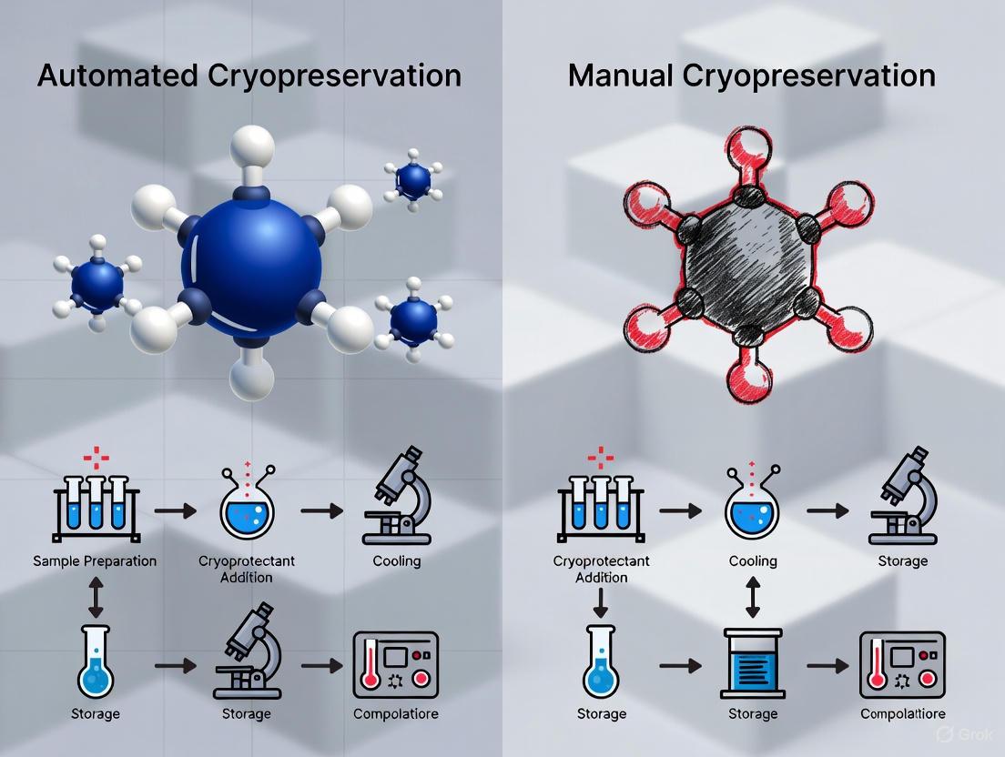
Cryopreservation Workflow Comparison: This diagram illustrates the key stages of cryopreservation while highlighting differences between manual and automated methods in process control and potential variability points.
Analytical Methods for Assessing Cryoinjury
Comprehensive assessment of cryopreservation outcomes requires multiple analytical approaches to capture both immediate viability and longer-term functionality.
Viability and Functionality Assessment
Cell Viability Assays: Standard methods include trypan blue exclusion for immediate post-thaw viability, MTT assays for metabolic activity, and neutral red uptake for lysosomal integrity [11]. These methods provide quantitative viability metrics but may not detect subtler functional impairments.
Delayed Apoptosis Measurement: Since cryoinjury often manifests hours post-thaw, assessment at 18-24 hours provides crucial information about delayed cell death. Methods include Annexin V/PI staining for early apoptosis detection and Caspase-3 activation measurements [8] [10].
DNA Damage Assessment: The Sperm Chromatin Structure Assay (SCSA) quantifies DNA fragmentation index (DFI) in sperm cells [8]. Similar principles apply to other cell types, with TUNEL assays also employed to detect DNA strand breaks resulting from cryoinjury.
Functional Assays: For immune cells like CAR-T cells, functionality is assessed through cytokine secretion (IFN-γ, TNF-α) upon restimulation [6]. For MSCs, immunomodulatory function is measured via T cell suppression assays [9].
Research Reagent Solutions Toolkit
Table 3: Essential Reagents and Equipment for Cryopreservation Research
| Category | Specific Examples | Function & Application |
|---|---|---|
| Cryoprotectants | DMSO, ethylene glycol, glycerol, trehalose, sucrose, glucose | Membrane protection, ice crystal inhibition, osmotic balance |
| Commercial Media | CellBanker, Vitrification Freeze Kit (Irvine Scientific) | Optimized, ready-to-use formulations for specific applications |
| Viability Assays | Trypan blue, MTT, Annexin V/PI, LDH release | Assessment of membrane integrity, metabolic activity, apoptosis |
| DNA Damage Kits | Sperm Chromatin Structure Assay (SCSA), TUNEL assay | Quantification of DNA fragmentation from cryoinjury |
| Specialized Equipment | Controlled-rate freezers, Automated vitrification systems, Liquid nitrogen storage tanks | Precise temperature control, process standardization, long-term preservation |
| Consumables | Cryovials, High-security straws, Cryogenic labels | Sample containment, identification, and storage |
The science of cryoinjury reveals that freezing and thawing variables significantly impact both immediate cell viability and long-term cellular function. While manual cryopreservation methods can achieve excellent outcomes in skilled hands, they introduce operator-dependent variability that challenges manufacturing consistency. Automated systems address this variability through process control and documentation, particularly valuable for late-stage clinical and commercial therapeutic applications.
The optimal approach depends on specific application requirements. For research settings with diverse sample types, manual methods offer valuable flexibility. For standardized processing of clinically-bound therapeutics, automated systems provide the consistency and documentation necessary for regulatory compliance. Future directions will likely see increased adoption of defined cryoprotectant formulations that replace proprietary media, along with advanced monitoring systems that use process data like freeze curves as quality indicators rather than relying solely on post-thaw analytics [1] [10].
Understanding cryoinjury mechanisms enables researchers to select appropriate preservation strategies based on cell type sensitivity, scale requirements, and application criticality. As cryopreservation science advances, the integration of physiological insights with technological innovations will continue to enhance our ability to preserve cellular function across the expanding spectrum of biomedical applications.
Cryopreservation is a cornerstone technique in biomedical research and clinical therapy, enabling the long-term storage of cells, tissues, and other biological materials. Within this field, manual cryopreservation techniques remain widely utilized despite the emergence of automated systems. This guide provides an objective comparison between manual and automated cryopreservation processes, with a specific focus on principles, flexibility, and inherent variability of manual methods. The ability to preserve cellular viability and function during freezing and thawing is critical for applications ranging from regenerative medicine to assisted reproductive technologies. As the cell and gene therapy industry advances, understanding the technical aspects and consistency of cryopreservation methods becomes increasingly important for manufacturing standards and regulatory compliance [12] [1]. This analysis synthesizes current research findings to evaluate the performance characteristics of manual cryopreservation, providing researchers and drug development professionals with evidence-based insights for protocol selection and optimization.
Fundamental Principles of Manual Cryopreservation
Manual cryopreservation relies on operator-driven techniques to control the freezing and thawing processes of biological samples. The fundamental principle involves gradually reducing sample temperature while using cryoprotective agents (CPAs) to mitigate damage from ice crystal formation and osmotic stress [13]. During slow freezing, which typically occurs at approximately -1°C/min, water progressively moves out of cells before freezing extracellularly, thus minimizing lethal intracellular ice formation [13]. This process requires precise timing and technique from the operator throughout multiple critical phases.
The origins of low-temperature tissue storage research date back to the late 1800s, but significant understanding emerged in the 1950s when James Lovelock discovered that cryopreservation caused osmotic stress contributing to ice crystal formation in red blood cells [13]. In 1963, Mazur characterized how the rate of temperature change controls water movement across cell membranes, informing the development of controlled freezing protocols [13]. Manual methods implement these scientific principles through operator-dependent techniques using relatively simple equipment such as insulated containers, pre-cooled metal blocks, or vapor phase nitrogen chambers.
The manual cryopreservation process depends heavily on two protective actions: use of appropriate cryoprotectants and selection of optimal cooling and thawing rates [13]. Permeating cryoprotective agents like dimethyl sulfoxide (DMSO), glycerol (GLY), ethylene glycol (EG), and propylene glycol (PG) function by depressing the freezing point of water and reducing available water molecules for crystal formation through hydrogen bonding [13]. These compounds must be highly water soluble at low temperatures, able to cross biological membranes easily, and ideally minimally toxic to cells. Manual protocols typically implement CPA addition in a stepwise fashion at temperatures near 0°C to minimize toxicity while allowing sufficient permeation for protection during freezing.
Comparative Performance Analysis
Quantitative Comparison of Manual vs. Automated Cryopreservation Outcomes
Table 1: Post-Thaw Quality Assessment Across Biological Materials
| Biological Material | Preservation Method | Survival Rate (%) | Motility/Function | Membrane Integrity | Study |
|---|---|---|---|---|---|
| Buffalo Semen | Manual (nitrogen vapor) | - | Progressive motility: 38.70±4.04 | - | [14] |
| Automated programmable | - | Progressive motility: 45.54±3.64 | - | [14] | |
| Equine Semen | Manual system | - | Motility: 20% | 5.1% | [15] |
| Controlled-rate freezer | - | Motility: 44.6% | 29.3% | [15] | |
| Human Embryos | Manual vitrification | Positive survival: 96%Intact survival: 86% | Clinical pregnancy: 27% | - | [4] |
| Semi-automated vitrification | Positive survival: 90%Intact survival: 84% | Clinical pregnancy: 22% | - | [4] | |
| Human Oocytes | Manual vitrification | 92.7% | Resistance to micro-injection: 94.0% | Normal rehydration kinetics | [5] |
| Semi-automated vitrification | 82.9% | Resistance to micro-injection: 93.2% | Normal rehydration kinetics | [5] |
Table 2: Inter-Operator Variability Assessment
| Parameter | Manual Vitrification | Semi-Automated Vitrification | Statistical Significance |
|---|---|---|---|
| Inter-operator variability in positive survival rate | No significant difference between 5 technicians | No significant difference between 5 technicians | Not significant (p>0.05) |
| Inter-operator variability in intact survival rate | No significant difference between 5 technicians | No significant difference between 5 technicians | Not significant (p>0.05) |
| Process time efficiency | More time-efficient | Less time-efficient (additional 11±9 minutes) | Significant (p<0.05) |
Advantages and Limitations of Each Approach
Table 3: System Capabilities and Constraints
| Aspect | Manual Cryopreservation | Automated/Controlled-Rate |
|---|---|---|
| Equipment Cost | Low-cost, low-consumable infrastructure [1] | High-cost, high-consumable infrastructure [1] |
| Process Control | Limited control over critical process parameters [1] | Precise control over cooling rates and other parameters [1] |
| Technical Barrier | Low technical barrier to adoption [1] | Specialized expertise required [1] |
| Scalability | Ease of scaling [1] | Bottleneck for batch scale-up [1] |
| Flexibility | High protocol adaptability | Limited to programmed parameters |
| Documentation | Manual documentation | Automated data recording |
The quality of cryopreserved products represents the cumulative effects of all processing steps and reagents used throughout the preservation workflow [16]. In manual cryopreservation, multiple factors contribute to outcome variability, making consistency challenging to maintain across operators and facilities.
Pre-Freeze Processing Variability
The initial stages of sample collection and preparation introduce significant variability in manual protocols. Factors such as anesthesia type, collection technique, anticoagulant selection, volume collected, and time-temperature conditions between collection and processing can dramatically influence final product quality [16]. For cells isolated from intact tissue, mechanical or chemical digestion methods vary considerably between operators, creating inconsistent starting material. Furthermore, cells may experience various stresses during processing, including mechanical shear forces, nutrient deprivation, or viral infection during genetic modification procedures [16]. While viability and membrane integrity are commonly monitored, sub-lethal stresses that impact post-thaw recovery may go undetected without specialized assessment of early apoptotic markers or phenotypic shifts [16].
Cryoprotectant Exposure and Formulation Inconsistencies
Introduction of cryopreservation solutions presents two primary mechanisms of damage in manual protocols: osmotic stress and biochemical toxicity [16]. When cells are transferred from physiological solutions to cryopreservation media, they initially dehydrate due to higher chemical potential extracellularly, followed by slow volume increase as permeating CPAs like DMSO enter cells [16]. The manual implementation of this process varies significantly between operators in terms of addition rate, mixing technique, and temperature control. Furthermore, biochemical toxicity from CPA exposure represents another source of variability, as DMSO exposure time prior to freezing significantly impacts cell viability and function [16]. Studies suggest limiting DMSO exposure to 30 minutes or less before freezing for therapeutic cells, but manual methods often struggle to standardize this parameter across operators [16].
Cooling Rate Inconsistencies
Post-thaw cell survival strongly depends on cooling rates, with most cell types exhibiting an inverted U-shaped survival curve where rates that are too high or too low prove detrimental [16]. Manual cryopreservation methods using passive freezing devices or nitrogen vapor techniques inherently struggle to maintain consistent cooling rates across different sample positions, volumes, or operators. A study comparing manual nitrogen vapor techniques with automated systems for buffalo semen demonstrated that specific height above liquid nitrogen (1.6 inches) yielded optimal motility (62.67±1.12) and progressive motility (38.97±1.10), highlighting the precision required in manual methods [14]. This level of positional consistency proves challenging to maintain across multiple operators in manual protocols.
Experimental Protocols and Methodologies
Detailed Manual Vitrification Protocol for Embryos
The manual vitrification process for embryos follows a standardized but operator-dependent methodology. As described in the comparative study of manual versus semi-automated systems, manual vitrification utilizes closed CryoBioSystem vitrification (CBS-VIT) High Security (HS) straws with a Vitrification Freeze Kit containing DMSO-ethylene glycol-sucrose as cryoprotectants [4]. The protocol proceeds as follows:
Equilibration Phase: Embryos are transferred individually to a 50 µL droplet of equilibration solution at room temperature for exactly 10 minutes [4].
Vitrification Solution Exposure: Embryos are moved to a 50 µL droplet of vitrification solution containing 15% (v/v) DMSO and 15% (v/v) ethylene glycol [4].
Loading and Sealing: Embryos are loaded onto the CBS HS straw device in the smallest possible volume of vitrification solution (less than 1 µL). The straw is heat-sealed before being plunged directly into liquid nitrogen [4].
Time Constraint: The total time from vitrification solution exposure to liquid nitrogen immersion must not exceed 110 seconds [4].
The corresponding warming process occurs on the day of embryo transfer, approximately 2-3 hours prior to the procedure:
Thawing Solution Incubation: A well of 500 µL thawing solution (1.0 M sucrose in HEPES buffered human tubal fluid medium) is maintained at 37°C. The sealed straw is cut, and the capillary is removed, allowing embryos to be released into the warm thawing solution for 1 minute [4].
Dilution Steps: Embryos are transferred to 50 µL of dilution solution (0.5 M sucrose in HEPES buffered HTF medium) for 4 minutes at room temperature [4].
Washing Phase: Embryos are sequentially washed in two 50 µL-droplets of washing solution (HEPES buffered HTF medium) for 4 minutes each [4].
Recovery Culture: Finally, embryos are transferred to culture dish with 20% protein supplemented culture medium for 2 hours before morphological assessment and transfer [4].
Manual Cryopreservation Protocol for Semen
For semen cryopreservation, manual methods often employ nitrogen vapor techniques with specific extenders. The protocol for buffalo semen preservation exemplifies this approach:
Cooling Phase: Semen is initially cooled from 37°C to 5°C for 30 minutes in an equilibration chamber [14].
Freezing Phase: Samples are transferred to a Styrofoam box using liquid nitrogen vapor from different distances (0.5, 1.5, 1.6, 2, and 3 inches) [14].
Optimized Parameters: The highest number of motile sperms (62.67±1.12) and progressive motility (38.97±1.10) was observed at 1.6 inches above liquid nitrogen [14].
Extender Efficacy: The study compared a locally developed Tris-fructose-egg yolk-based (TFE) diluter with three commercial diluters (Andromed, Triladyl, and Steridyl), finding the highest recovery rate (82.4%) and conception rate (80%) with TFE [14].
Manual Cryopreservation Workflow and Variability Sources
The Scientist's Toolkit: Essential Research Reagents
Table 4: Key Reagents for Manual Cryopreservation Protocols
| Reagent Category | Specific Examples | Function and Application | Considerations |
|---|---|---|---|
| Permeating Cryoprotectants | Dimethyl sulfoxide (DMSO), Glycerol (GLY), Ethylene glycol (EG), Propylene glycol (PG) | Depress freezing point, promote vitrification, prevent intracellular ice formation [13] | Concentration-dependent toxicity; DMSO increases membrane permeability at 10% but damages lipid bilayers at 40% [13] |
| Non-Permeating Cryoprotectants | Trehalose, Sucrose, Raffinose, Polyethylene glycol (PEG), Polyvinylpyrrolidone (PVP) | Extracellular vitrification, osmotic pressure regulation, membrane stabilization [13] | Trehalose offers exceptional stability due to α-1,1-glycosidic bond preventing reduction [13] |
| Ice Recrystallization Inhibitors | Synthetic IRIs (PanTHERA CryoSolutions) | Mitigate cellular damage from ice crystal growth during transient warming [17] | Mimic activity of natural antifreeze proteins without inducing dynamic ice shaping [17] |
| Semen Extenders | Tris-fructose-egg yolk-based (TFE), Andromed, Triladyl, Steridyl | Maintain sperm motility, membrane integrity during freeze-thaw cycle [14] | TFE showed highest recovery (82.4%) and conception rates (80%) in buffalo semen [14] |
| Vitrification Mixtures | DMSO-ethylene glycol-sucrose combinations | Enable glass state formation without ice crystals at high cooling rates [4] | Stepwise addition at 0°C reduces CPA toxicity; used in manual embryo vitrification [13] [4] |
Emerging Technologies and Future Directions
The field of cryopreservation continues to evolve with new technologies aimed at addressing the variability inherent in manual methods. Ice recrystallization inhibitors (IRIs) represent a significant advancement, specifically targeting ice crystal growth that occurs during transient warming events in storage or handling [17]. These synthetic small molecules mitigate cellular damage from uncontrolled ice growth without the cytotoxic effects associated with high concentrations of traditional CPAs like DMSO [17]. Studies demonstrate that IRI supplementation improves post-thaw viability and functional recovery across multiple cell types, including induced pluripotent stem cells (iPSCs), iPSC-derived neurons, platelets, and hematopoietic stem and progenitor cells [17].
Automation and standardization technologies are also advancing to address variability concerns. Semi-automated vitrification systems like Gavi provide controlled stepwise exposure to vitrification solutions with precise timing, temperature regulation, and volume control [4] [5]. While studies show comparable survival outcomes between manual and semi-automated methods for embryos and oocytes, the automated systems offer superior traceability and reduced inter-operator variability [4] [5]. Recent industry surveys indicate that 87% of cell therapy manufacturers use controlled-rate freezing, particularly for late-stage and commercial products, while only 13% rely exclusively on passive freezing methods, predominantly in early clinical development stages [1].
Technological Advancements in Cryopreservation
Manual cryopreservation remains a valuable technique in the biopreservation toolkit, offering flexibility, lower infrastructure costs, and protocol adaptability. However, this analysis demonstrates that manual methods exhibit inherent variability stemming from multiple sources, including pre-freeze processing inconsistencies, cryoprotectant exposure differences, and cooling rate irregularities. The comparative data reveals that while manual techniques can achieve excellent outcomes with experienced operators—particularly for sensitive materials like embryos and oocytes—they generally show greater outcome variability compared to automated systems.
For research and clinical applications requiring high consistency and reproducibility, automated cryopreservation systems provide superior control over critical process parameters with enhanced documentation capabilities. Nevertheless, manual methods maintain relevance for applications requiring protocol flexibility, in resource-limited settings, or when processing smaller sample volumes. As the field advances, integration of novel approaches like ice recrystallization inhibitors with both manual and automated protocols shows promise for improving post-thaw recovery and function across diverse cell types. Researchers and therapy developers should carefully consider their specific quality requirements, regulatory constraints, and scalability needs when selecting between manual and automated cryopreservation approaches.
Cryopreservation is a transformative technology that enables the long-term storage of biological materials by cooling them to extremely low temperatures, effectively slowing or halting metabolic and biochemical processes. This technology bridges the spatiotemporal gap between the sources and acquisition times of biospecimens and their destinations and times of use [18]. The global market for assisted reproductive technologies, which heavily relies on cryopreservation, is forecasted to reach over $45.4 billion by 2025, underscoring the significant economic and clinical importance of these techniques [18].
The two primary methods for cryopreservation are slow freezing and vitrification. Slow freezing, commonly used for preserving mammalian cells and microtissues, involves cooling samples at controlled rates of about 1°C min⁻¹ either in an insulated container at -80°C or in a programmable freezer [18]. Vitrification, in contrast, directly transforms biospecimens from a liquid state into a glassy state through non-equilibrium cooling to minimize or eliminate ice formation [18]. This method generally employs higher concentrations of cryoprotective agents (CPAs) and higher cooling rates than slow freezing [18]. Vitrification is considered superior to slow freezing for banking stress-sensitive biospecimens, including oocytes, stem cells, and some tissues [18].
Recent engineering advances have introduced automated and semi-automated systems to address limitations in manual cryopreservation techniques. These systems aim to standardize procedures, reduce inter-operator variability, and improve reproducibility across different operators and facilities [4] [19]. As the field continues to evolve, understanding the principles, standardization approaches, and engineering controls of automated cryopreservation has become increasingly important for researchers, scientists, and drug development professionals working with biological systems.
Fundamental Principles of Cryopreservation
Thermodynamic Foundations
Cryopreservation operates on the fundamental principle of reducing temperature to suspend biochemical and metabolic activities in biological materials. Depending on temperature and solute concentrations, biospecimens can exist in liquid, vitrified, supercooled, or supersaturated states during preservation processes [18]. The thermodynamic paths of different preservation methods follow distinct trajectories:
- Slow freezing follows the path A→C→E→F→G→I→L→Z, where ice crystals initiate at point E and propagate through freeze concentration, elevating extracellular osmolality and driving cell dehydration and deformation [18].
- Conventional vitrification follows the path A→D→II→M→Z, where high-concentration CPAs are loaded at non-freezing temperatures, followed by rapid, non-equilibrium cooling that leads to vitrification [18].
- Low-CPA vitrification follows the path A→C→III→N→Z, utilizing high cooling rates that reduce the required CPA concentration to levels comparable with slow freezing [18].
- Hypothermic storage typically follows the path A→B→IV→O→Y, with storage temperatures usually above the equilibrium freezing point [18].
The phase behavior of aqueous solutions during cooling and warming cycles determines the success of cryopreservation protocols. Each method positions biospecimens in different thermodynamic regions: the vitrified state for long-term cryopreservation or the liquid state for short-term hypothermic storage [18].
Cryoinjury Mechanisms and Protective Strategies
Cryopreservation techniques must mitigate several mechanisms of cellular injury:
Intracellular ice formation (IIF): Almost always fatal to cells, IIF disrupts subcellular organelles, cellular membranes, and the cytoskeleton [18]. The classical two-factor theory of cryoinjury considers solute effects under slow cooling and intracellular ice formation under fast cooling, suggesting an optimal cooling rate for each specific cell type that minimizes both injury types [18].
Solution effects and osmotic stress: During slow freezing, freeze concentration causes significant osmotic stress, leading to cell dehydration and deformation under high solute concentrations [18]. This occurs at point G in the thermodynamic pathway and represents a critical challenge for conventional slow-freezing protocols.
CPA toxicity: Conventional vitrification uses high CPA concentrations (6-8 M) to increase glass-transition temperature and solution viscosity [18]. However, highly concentrated CPAs at high temperatures can be toxic to mammalian cells, necessitating minimized exposure times and multistep loading protocols that are time-consuming and stressful to cells [18].
Chilling injury: During conventional hypothermic storage above equilibrium freezing temperatures, biospecimens undergo substantial physiochemical and metabolic activities under suboptimal conditions, consuming nutrients and oxygen while producing noxious metabolites [18]. The cutoff from circulation systems and lack of nutrient transport aggravate ischemic injuries [18].
Recent research has identified that higher glass transition temperatures reduce thermal stress cracking in aqueous solutions relevant to cryopreservation, providing important insights for preserving larger organs and tissues [20]. This understanding enables researchers to focus on creating aqueous vitrification solutions with higher glass transition temperatures to help avoid cracking while maintaining biocompatibility with tissues [20].
Automated Cryopreservation Systems
Engineering Controls and System Architecture
Automated cryopreservation systems incorporate sophisticated engineering controls to standardize the vitrification process. These systems typically consist of several integrated components:
Microfluidic mixing units: Enable flexible and diverse cryoprotectant loading and removal protocols with precise control over concentration gradients [19]. These units allow for the creation of various precisely controlled and continuous linear CPA curves, reducing osmotic damage caused by rapid shifts in CPA concentration [19].
Integrated vitrification carriers: Advanced systems feature carriers that combine loading, vitrification, and warming capabilities in a single device [19]. For instance, some automated systems use a microgrid capillary that serves both for CPA loading/removal and as the vitrification carrier itself, eliminating the need to transfer cells between devices [19].
Temperature and timing controls: Semi-automated systems like the Gavi (Genea, Sydney, Australia) provide closed vitrification systems that control stepwise exposure to vitrification solutions, timing, temperature, and duration of exposure to the vitrification medium [4] [5]. This automation makes each act of vitrification reproducible and traceable [5].
Mechanical handling systems: Robotic systems embedded with microfluidic chips can automate the entire vitrification process, including solution exchange and embryo transfer using positive pressure and gripper mechanisms [19].
These engineering controls address critical variables in the vitrification process, including contact time with cryoprotectants, solution volumes, temperature stability, and cooling/warming rates. By standardizing these parameters, automated systems aim to reduce the inter-operator variability inherent in manual techniques [4].
Commercial Systems and Their Architectures
Several automated and semi-automated cryopreservation systems have been developed with distinct engineering approaches:
Gavi System: A semi-automatic closed system that uses a "pod" inserted into a "cassette" that can hold up to four pods [4]. The system injects low-concentration CPA into the frozen carrier pod, allows balancing, then aspirates the CPA and injects higher-concentration CPA for a second balancing stage before sealing the pod for manual plunging into liquid nitrogen [19].
Sarah System: An automated vitrification system that uses a carrier combining 0.25-mL wheat tubes with a special net [19]. A robotic arm moves the wheat tubes into different CPA containers before plunging them directly into liquid nitrogen for vitrification [19].
Microfluidic Robotic Systems: Newer systems propose open microfluidic chips where equilibrium solution and vitrification solution are loaded/removed in solution exchange chambers via injection pumps [19]. These systems can include microvalves and transfer channels to move embryos using positive pressure [19].
The evolution of these systems demonstrates a trend toward greater integration, reduced manual intervention, and improved control over the critical parameters that influence cryopreservation outcomes. The global automated cryopreservation systems market includes key players such as Azenta Life Sciences, Cytiva, Planer Limited, CryoLogic Pty Ltd, PHC Corporation, Hamilton Company, Thermo Fisher Scientific, and Brooks Life Sciences, reflecting the growing commercial importance of these technologies [21].
Performance Comparison: Automated vs. Manual Cryopreservation
Embryo Cryopreservation Outcomes
Comparative studies between automated and manual vitrification systems reveal important differences in performance metrics. A retrospective analysis of 282 patients compared semi-automated vitrification with the GAVI method (AV) against manual vitrification with Irvine-CBS (MV) [4]. The study involved 5 operators and evaluated inter-operator variability, survival rates, and clinical outcomes.
Table 1: Embryo Cryopreservation Outcomes - Manual vs. Automated Vitrification
| Parameter | Manual Vitrification (MV) | Semi-Automated Vitrification (AV) | Statistical Significance |
|---|---|---|---|
| Number of warmed embryos | 338 | 212 | - |
| Positive survival rate (≥50% intact blastomeres) | 96% (323/338) | 90% (191/212) | p < 0.05 |
| Intact survival rate (100% intact blastomeres) | 86% (292/338) | 84% (178/212) | Not Significant |
| Clinical pregnancy rate | 27% (73/266) | 22% (36/162) | Not Significant |
| Complete vitrification time | Faster | Slower (plus 11±9 minutes) | - |
| Inter-operator variability | No significant difference between technicians | No significant difference between technicians | Not Significant |
The data demonstrates that while manual vitrification showed a statistically higher positive survival rate (96% vs. 90%, p < 0.05), the intact survival rates and clinical pregnancy rates were not significantly different between the two methods [4]. Regarding time efficiency, the manual vitrification technique proved quicker than the semi-automated approach, requiring approximately 11±9 minutes less time [4]. Notably, both techniques showed little inter-operator variability in survival rates between the five technicians, suggesting that automation may not provide significant advantages in standardization for experienced operators [4].
Oocyte Cryopreservation Outcomes
Research on oocyte vitrification has yielded comparable results between automated and manual systems. A study comparing semi-automated and manual vitrification in human sibling oocytes found no significant differences in key performance metrics [5].
Table 2: Oocyte Cryopreservation Outcomes - Manual vs. Automated Vitrification
| Parameter | Manual Vitrification | Semi-Automated Vitrification | Statistical Significance |
|---|---|---|---|
| Post-warming survival rate | 92.7% (76/82) | 82.9% (68/82) | p = 0.053 (near threshold) |
| Survival after micro-injection | 94.0% (63/67) | 93.2% (55/59) | Not Significant |
| Oocyte surface rehydration kinetics | Comparable pattern | Comparable pattern | Not Significant |
| Transcriptomic differences | Limited differences | Only 5 differentially expressed genes | Minimal biological impact |
The post-warming survival rate was comparable between the two groups, though the difference approached statistical significance (92.7% for manual vs. 82.9% for semi-automated, p = 0.053) [5]. Among intact oocytes subjected to empty micro-injection gestures three hours after warming, survival rates were nearly identical between the two groups (94.0% vs. 93.2%) [5]. Oocyte surfaces followed comparable rehydration kinetics in both groups, with reduced surface area immediately after thawing that recovered within one hour post-thawing regardless of the vitrification method [5].
Transcriptomic analysis revealed minimal differences between the two techniques, with only five differentially expressed genes identified, all upregulated in the semi-automated vitrification group [5]. These genes (ARSD, CCDC124, CLPS, PLCH2, RHBDF1) had low expression levels in oocytes, and no interactions between them were recorded in the STRING database, suggesting limited biological impact [5]. RNA integrity was similar between the two vitrification methods, with no difference in median transcript integrity numbers [5].
Experimental Protocols and Methodologies
Manual Vitrification-Warming Protocol
The manual vitrification protocol typically follows standardized procedures using commercially available kits. One well-established method utilizes closed CryoBioSystem vitrification (CBS-VIT) High Security (HS) straws with the Vitrification Freeze Kit containing DMSO-ethylene glycol-sucrose as cryoprotectants [4]. The process involves:
- Equilibration phase: Embryos are transferred to a 50 µL droplet of equilibration solution for 10 minutes at room temperature [4].
- Vitrification phase: Embryos are moved into a 50 µL droplet of vitrification solution containing 15% (v/v) dimethyl sulfoxide and 15% (v/v) ethylene glycol [4].
- Loading and sealing: Embryos are loaded onto the CBS HS straw device in the smallest possible volume of vitrification solution (less than 1 µL) [4].
- Freezing: The straw is heat-sealed before being plunged directly into liquid nitrogen [4].
- Time constraint: The total time from exposure to vitrification solution to plunging into liquid nitrogen does not exceed 110 seconds [4].
The warming process is performed on the day of embryo transfer, 2-3 hours prior to the procedure [4]. It involves:
- Thawing: Using the Vitrification Thaw Kit, a Nunc 4-well dish with 500 µL thawing solution (1.0 M sucrose in HEPES buffered human tubal fluid medium) maintained at 37°C [4].
- Recovery: After cutting the straw, the capillary is removed, and the gutter-end is placed in the warm thawing solution, allowing embryos to be released and eluted for 1 minute [4].
- Dilution: Embryos are incubated for 4 minutes at room temperature in 50 µL of dilution solution (0.5 M sucrose in HEPES buffered HTF medium) [4].
- Washing: Sequential washing in two 50 µL-droplets of washing solution (HEPES buffered HTF medium) for 4 minutes each [4].
- Culture: Transfer to a culture dish with 20% protein-supplemented culture medium for 2 hours before morphological assessment according to the Istanbul Consensus [4].
Semi-Automated Vitrification Protocol
Semi-automated systems like Gavi standardize the vitrification process through controlled mechanical systems:
- Pod-based system: The instrument uses a closed device called a "pod" inserted into a "cassette" that can hold up to four pods [4]. The embryo is held in a cavity at the bottom of the pod throughout the process.
- Automated solution exchange: The system controls stepwise exposure to vitrification solutions, timing, temperature, and duration of exposure to the vitrification medium [4] [5].
- Standardized loading: The process involves injecting low-concentration CPA into the pod, allowing equilibration, then aspirating and injecting higher-concentration CPA for a second equilibration stage [19].
- Sealing and storage: After loading, the pod is sealed and manually plunged into liquid nitrogen for storage [19].
Automated Microfluidic Vitrification Protocol
Advanced automated systems incorporate microfluidic technology to optimize CPA loading and removal:
- Curved channel microfluidic devices: Gradually load CPA to achieve continuous concentration changes, reducing osmotic stress [19]. These devices can load 1.5 M propylene glycol into oocytes within 15 minutes with a volume change of less than 10% [19].
- Protocol optimization: Research has identified optimal loading and removal protocols, with the highest oocyte survival rate (100%) observed in 8-minute loading and removal protocols following quadratic function curves [19].
- Integrated carrier systems: New devices enable loading/removal of CPA and vitrification in the same carrier, eliminating transfer steps [19].
Diagram 1: Comparative Workflow of Manual vs. Automated Cryopreservation Methods. This diagram illustrates the key procedural differences between manual and semi-automated vitrification protocols, highlighting the standardized, multi-step nature of automated systems versus the time-sensitive manual approach.
Standardization and Quality Control
Inter-Operator Variability
A critical advantage proposed for automated cryopreservation systems is reduced inter-operator variability. However, research findings present a nuanced picture:
Limited difference in variability: A retrospective analysis of 5 operators performing both manual and semi-automated vitrification found no significant difference in positive and intact survival rates between technicians for either method [4]. This suggests that both manual and automated techniques can achieve consistent results across multiple operators in controlled settings.
Procedure-dependent standardization: While automation theoretically standardizes procedures, the actual reduction in variability appears dependent on specific protocol elements. One study noted that manual vitrification of oocytes/embryos constitutes one of the most time-consuming and labor-intensive procedures in IVF laboratories, requiring skilled embryologists [19]. The large amount of manual manipulation increases the risk of cellular loss, which automation aims to reduce.
Learning curve considerations: Vitrification requires operator training and is generally accepted to have a learning curve [4]. Automation may shorten this learning curve by providing standardized protocols that require less technical expertise to achieve consistent results.
Process Control and Traceability
Automated systems offer enhanced process control and traceability features:
Parameter standardization: Semi-automated systems control critical parameters including temperature, exposure times, media volume, and solution replacement [5]. This comprehensive control ensures that each vitrification event follows identical parameters.
Traceability: Automated systems provide documentation of process parameters, creating an audit trail for quality control and regulatory compliance [5]. This feature is particularly valuable in regulated environments and multi-center research studies.
Reduced manual intervention: By minimizing direct handling of specimens, automated systems reduce opportunities for technical error and introduction of contaminants [19].
The Scientist's Toolkit: Essential Research Reagents and Systems
Table 3: Key Research Reagents and Systems for Cryopreservation Studies
| Reagent/System | Type | Function/Application | Research Context |
|---|---|---|---|
| CryoStor CS10 | Cryopreservation Medium | Freeze media combining cryoprotectants for improved post-thaw viability | Used in 3D culture of hiPSCs with Y-27632 Rho kinase inhibitor [22] |
| Y-27632 Rho kinase inhibitor | Small Molecule Inhibitor | Enhances post-thaw viability and preserves trilineage differentiation potential | Combined with CryoStor CS10 for hiPSC cryopreservation [22] |
| Gavi System | Semi-Automated Vitrification System | Closed system automating temperature, timing, and solution exposures | Compared with manual vitrification in sibling oocyte study [5] |
| Rapid-I | Manual Vitrification System | Open manual vitrification device using straws and precise timing | Reference method in comparative studies with automated systems [5] |
| Irvine-CBS | Manual Vitrification System | Utilizes closed High Security straws with DMSO-EG-sucrose cryoprotectants | Compared with GAVI method in multi-operator study [4] |
| VitroGel Hydrogel Matrix | 3D Culture System | Animal-free, ligand-functionalized hydrogel mimicking ECM architecture | Used for 3D culture of hiPSCs in spaceflight experiments [22] |
| Dimethyl Sulfoxide (DMSO) | Penetrating Cryoprotectant | Prevents intracellular ice formation, stabilizes membranes | Common CPA in vitrification solutions (15% concentration) [4] |
| Ethylene Glycol (EG) | Penetrating Cryoprotectant | Lowers freezing point, reduces ice crystal formation | Used in combination with DMSO in vitrification solutions [4] |
| Sucrose | Non-Penetrating Cryoprotectant | Creates osmotic gradient, promotes dehydration | Component of thawing (1.0M) and dilution (0.5M) solutions [4] |
Automated cryopreservation systems represent a significant engineering advancement in the field of biopreservation, offering standardized protocols, reduced manual intervention, and enhanced process control. The current evidence suggests that while these systems provide theoretical advantages in reproducibility and operator independence, their practical performance relative to manual methods reveals a more nuanced picture.
The comparative data indicates that manual vitrification techniques currently demonstrate comparable or slightly superior survival rates for embryos (96% vs. 90% positive survival rate) with greater time efficiency [4]. For oocyte cryopreservation, both methods achieve similar outcomes in survival, function, and transcriptomic profiles [5]. The minimal transcriptomic differences between manual and automated methods, with only five differentially expressed genes of low abundance, provides reassurance regarding the biological safety of both approaches [5].
The ideal application of automated systems appears to be in settings requiring high throughput, standardized protocols across multiple operators, or enhanced traceability for regulatory compliance. However, for experienced operators, manual techniques continue to provide excellent results with potentially greater efficiency. Future developments in automated systems, particularly those integrating microfluidic technology for optimized CPA exchange and all-in-one vitrification carriers, may address current limitations related to osmotic stress and manual transfer steps [19].
As cryopreservation technologies continue to evolve, the focus should remain on validating both manual and automated methods through rigorous comparative studies assessing not only survival rates but also long-term functional outcomes, genetic stability, and clinical success measures. The combination of engineering innovation with biological understanding will drive the next generation of cryopreservation techniques, potentially expanding applications to include complex tissues and organs [20].
Diagram 2: Experimental Framework for Comparing Cryopreservation Methods. This diagram outlines a comprehensive approach for evaluating manual versus automated cryopreservation techniques, incorporating immediate outcomes, functional assessments, and technical metrics to provide a multidimensional comparison.
Cryopreservation is a foundational technology in biomedical research and cell-based therapy development, enabling long-term storage of vital biological materials such as cell lines, gametes, and tissues. The fundamental challenge in cryopreservation lies in balancing the need to preserve cell viability and functionality while minimizing damage from intracellular ice formation, osmotic stress, and cryoprotectant toxicity. As the field advances toward more complex cellular products and scaled manufacturing, the debate between automated and manual cryopreservation methodologies has intensified, with significant implications for process consistency, reproducibility, and regulatory compliance.
Recent industry surveys and experimental studies have begun to quantify the performance differences between these approaches, yet notable consensus gaps persist in qualification standards, scaling methodologies, and technology adoption. This guide objectively compares automated versus manual cryopreservation processes, focusing specifically on empirical data related to process consistency across different biological systems and applications.
Industry Survey Data on Current Practices
Recent data from the ISCT Cold Chain Management and Logistics Working Group survey provides crucial insights into current industry practices and technology adoption trends among researchers and therapy developers.
Table 1: Cryopreservation Technology Adoption Trends (2025 ISCT Survey Data)
| Technology/Method | Adoption Rate | Primary Application Context | Key Driver for Adoption |
|---|---|---|---|
| Controlled-Rate Freezing (CRF) | 87% | Late-stage clinical & commercial products | Process control & documentation [1] |
| Passive Freezing | 13% | Early-stage clinical development (up to Phase II) | Cost infrastructure & technical simplicity [1] |
| Default CRF Profiles | 60% | Across all clinical stages & sectors | Convenience & manufacturer validation [1] |
| Optimized CRF Profiles | 40% | Specialized cells (iPSCs, cardiomyocytes, etc.) | Cell-specific optimization needs [1] |
The survey data reveals a strong industry preference for controlled-rate freezing, particularly as products advance toward commercialization. This transition is driven by the enhanced process control and comprehensive documentation capabilities that automated systems provide, which are essential for regulatory compliance in later-stage clinical development [1]. Notably, a significant majority (60%) of users rely on default freezing profiles provided by equipment manufacturers, while approximately 40% invest resources in developing optimized profiles for specialized cell types including induced pluripotent stem cells (iPSCs), hepatocytes, cardiomyocytes, and engineered cell products [1].
Identified Consensus Gaps and Challenges
Despite widespread technology adoption, the ISCT survey identified several critical areas where industry consensus remains underdeveloped, creating inconsistencies in practice across different organizations and research institutions.
Table 2: Key Consensus Gaps in Cryopreservation Practices
| Area of Practice | Current Status | Impact on Process Consistency |
|---|---|---|
| System Qualification | Little consensus; ~30% rely on vendors | Variable equipment performance & validation standards [1] |
| Freeze Curve Utilization | Limited use in release processes | Over-reliance on post-thaw analytics only [1] |
| Scaling Methodologies | 22% identify scaling as major hurdle | Batch-to-batch variability in large-scale operations [1] |
| Thawing Procedures | Poorly regulated at bedside | Variable cell viability & recovery at clinical administration [1] |
The qualification of controlled-rate freezers represents one of the most significant consensus gaps, with nearly 30% of survey respondents relying primarily on vendor qualifications rather than developing institution-specific protocols based on intended use cases. This practice potentially introduces variability, as vendor qualifications may not adequately represent final operational conditions, including specific container types, sample masses, and temperature profiles [1].
Additionally, the limited use of freeze curves as part of product release criteria represents another consistency challenge. While post-thaw analytics remain the primary release mechanism, process data from freeze curves can provide valuable insights into system performance and identify potential processing issues before they impact product quality [1].
Experimental Comparison: Automated vs. Manual Vitrification
Study Methodology and Experimental Design
A comprehensive retrospective study directly compared manual and semi-automated vitrification techniques, providing valuable experimental data on process consistency. The study analyzed 282 patients whose embryos were cryopreserved using either manual vitrification with Irvine-CBS systems or semi-automated vitrification with the GAVI method between November 2017 and September 2020 [4].
Manual Vitrification Protocol:
- Utilized closed CryoBioSystem vitrification (CBS-VIT) High Security straws
- Employed Vitrification Freeze Kit with DMSO-ethylene glycol-sucrose cryoprotectants
- Procedures conducted at room temperature with individual embryo processing
- Embryos equilibrated in 50 µL droplet for 10 minutes, then transferred to vitrification solution
- Loading completed in <1 µL volume, with straw heat-sealed before plunging into liquid nitrogen
- Total processing time from vitrification solution to plunging did not exceed 110 seconds [4]
Semi-Automated Vitrification Protocol:
- Implemented using GAVI system (Genea, Sydney, Australia)
- Utilized closed "pod" devices inserted into cassettes holding multiple pods
- System controlled stepwise exposure to vitrification solutions, timing, temperature, and exposure duration
- Reduced manual intervention through automated fluid handling and timing control [4]
Both techniques were performed by five operators during the same period, enabling direct comparison of inter-operator variability—a key metric for assessing process consistency [4]. Survival rates were assessed using Key Performance Indicators aligned with the Vienna and Alpha consensus standards, including positive survival rate (embryos with ≥50% morphologically intact blastomeres) and intact survival rate (embryos with 100% morphologically intact blastomeres) [4].
Comparative Performance Data
The study generated quantitative data on survival rates, operator variability, and processing efficiency, providing objective measures for comparing technique consistency.
Table 3: Experimental Results: Manual vs. Semi-Automated Vitrification
| Performance Metric | Manual Vitrification (MV) | Semi-Automated Vitrification (AV) | Statistical Significance |
|---|---|---|---|
| Positive Survival Rate | 96% (323/338) | 90% (191/212) | p < 0.05 [4] |
| Intact Survival Rate | 86% (292/338) | 84% (178/212) | Not Significant [4] |
| Clinical Pregnancy Rate | 27% (73/266) | 22% (36/162) | Not Significant [4] |
| Inter-Operator Variability | No significant difference | No significant difference | Not Significant [4] |
| Average Processing Time | Baseline | +11 ± 9 minutes longer | N/A [4] |
Contrary to expectations, the study found that manual vitrification demonstrated significantly higher positive survival rates (96% vs. 90%, p<0.05) while maintaining equivalent intact survival rates and clinical outcomes [4]. Both techniques showed minimal inter-operator variability, suggesting that well-trained personnel can achieve consistent results with either method [4].
The semi-automated system required approximately 11 minutes longer processing time per operation, potentially impacting workflow efficiency in high-volume settings [4]. This time differential highlights the trade-off between potential standardization benefits and operational throughput in automated systems.
Experimental vitrification outcomes for manual versus semi-automated methods.
Process Consistency Analysis Across Cell Types
Variable Performance with Specialized Cells
The consistency of cryopreservation outcomes varies significantly across different cell types, with particular challenges observed in specialized and sensitive cells. Survey data indicates that researchers working with certain cell types frequently report challenges with default freezing profiles and require customized approaches.
Challenging Cell Types for Standardized Protocols:
- Induced pluripotent stem cells (iPSCs) and their differentiated derivatives
- Primary hepatocytes and cardiomyocytes
- Engineered cell products (CAR-T cells, TCR-T cells)
- Photoreceptor cells and other solid tissue derivatives
- Macrophages and certain lymphocyte populations [1]
For these sensitive cell types, approximately 33% of survey respondents reported dedicating significant resources to freezing process development, indicating that standardized automated protocols may not adequately address the specific biological requirements of these cells [1]. The critical process parameters affecting cryopreservation success—including cooling rate before and after nucleation, nucleation temperature, and final storage temperature—must be optimized on a case-by-case basis, considering factors such as cell type, harvest conditions, cryoprotectant formulation, and primary container systems [1].
Scaling Challenges and Batch Consistency
Scaling cryopreservation processes presents substantial consistency challenges, with survey respondents identifying "ability to process at a large scale" as the single biggest hurdle (22% of responses) for advancing cell and gene therapies [1].
Current Scaling Practices:
- 75% of respondents cryopreserve all units from an entire manufacturing batch together
- 25% divide manufacturing batches into sub-batches for cryopreservation
- Batch size limitations create efficiency challenges in commercial settings [1]
The practice of cryopreserving entire batches together introduces temporal variance between the start and end of the freezing process for large batches, while dividing batches introduces potential inter-batch variability from different freezing cycles [1]. This represents a significant consistency challenge for automated systems designed for standardized processing.
Emerging Technologies and Methodologies
Novel Cryoprotectant Formulations
Recent developments in cryoprotectant science aim to address consistency challenges through improved formulations that enhance cell viability and reduce process variability.
Multicomponent Cryopreservation Agents: Researchers at the University of Leeds developed a multi-component cryopreservation agent called PaDT that demonstrates potential for improving process consistency and efficiency for red blood cell cryopreservation [23].
Table 4: Multicomponent Cryoprotectant Formulation
| Component | Concentration | Primary Mechanism | Functional Benefit |
|---|---|---|---|
| Polyampholyte | Proprietary | Membrane stabilization & vitrification enhancement | Improved structural integrity |
| Dimethyl Sulfoxide (DMSO) | Standard | Rapid cellular penetration & ice reduction | Proven cryoprotection |
| Trehalose | Proprietary | Extracellular stabilization | Osmotic balance & membrane protection |
The PaDT system enables washout in approximately 30 minutes—roughly three times faster than conventional glycerol-based systems—while maintaining normal cell morphology, metabolic health, and structural integrity [23]. This approach demonstrates potential for improving process consistency through reduced processing time and comparable recovery rates, though further validation across different cell types is needed.
Automation and Robotics Integration
The cell line cryopreservation market is experiencing significant growth in automated cryogenic biobanking and robotic storage solutions, projected to be the fastest-growing product segment between 2025-2034 [24]. These systems enhance process consistency through:
- Automated sample handling and tracking
- Reduced manual intervention points
- Integrated monitoring and documentation
- Standardized storage conditions [24]
The integration of AI-driven monitoring systems further enhances consistency by providing real-time analysis of cryopreservation parameters, automated data logging, and improved compliance management [24]. These technological advances address key consensus gaps in documentation and process monitoring identified in industry surveys.
The Scientist's Toolkit: Essential Research Reagents
Successful cryopreservation requires carefully selected reagents and materials tailored to specific cell types and applications. The following table outlines key solutions used in the experimental protocols discussed in this guide.
Table 5: Essential Cryopreservation Research Reagents
| Reagent/Material | Function | Example Applications | Experimental Context |
|---|---|---|---|
| DMSO-Ethylene Glycol-Sucrose Solutions | Permeating & non-permeating cryoprotection | Embryo vitrification | Manual vitrification protocols [4] |
| Glycerol-Based Media | Penetrating cryoprotectant | Semen cryopreservation | Fertility preservation studies [8] |
| Egg Yolk-Containing Buffers | Membrane stabilization | Sperm cryopreservation | Comparative cryoprotectant studies [8] |
| Sucrose-Glycerol Combinations | Osmotic regulation | Sperm cryopreservation | Alternative to egg yolk buffers [8] |
| Polyampholyte-DMSO-Trehalose (PaDT) | Multi-mechanism protection | RBC cryopreservation | Emerging technology [23] |
| Closed CBS-VIT HS Straws | Aseptic containment | Embryo vitrification | Manual system components [4] |
| GAVI Pods and Cassettes | Automated fluid handling | Embryo vitrification | Semi-automated system [4] |
Decision workflow for cryopreservation method selection.
The comparison between automated and manual cryopreservation reveals a complex landscape where process consistency must be evaluated across multiple dimensions, including cell type specificity, developmental stage, scaling requirements, and available resources. While automated systems offer advantages in documentation standardization and process control, manual methods demonstrate comparable—and in some cases superior—performance outcomes when implemented by trained personnel.
The most significant consensus gaps identified—including standardized equipment qualification, freeze curve utilization in quality systems, scaling methodologies, and thawing procedures—represent critical opportunities for industry collaboration and standardization. Future developments in cryoprotectant formulations, automated monitoring systems, and cell-specific protocol optimization will likely enhance consistency across both automated and manual platforms.
Researchers and therapy developers should prioritize method selection based on specific cell requirements, regulatory context, and long-term scalability needs rather than assuming universal superiority of either approach. The establishment of evidence-based best practices through continued comparative studies will be essential for addressing current consensus gaps and advancing cryopreservation consistency across the field.
Implementing Manual and Automated Protocols in the Lab and Clinic
Cryopreservation is a foundational technology in biomedical research and cell therapy, enabling the long-term storage of vital cell types such as hematopoietic stem cells, mesenchymal stem cells (MSCs), T cells for CAR-T therapies, and oocytes and embryos for assisted reproduction [25] [1]. While automated systems are emerging, manual cryopreservation remains widely practiced, particularly in research settings and early clinical development phases [1] [19]. This process involves the manual manipulation of cells through a series of chemical and temperature changes to achieve a state of suspended animation at cryogenic temperatures. The core principle involves using cryoprotective agents (CPAs) like dimethyl sulfoxide (DMSO) to disrupt hydrogen bonding and prevent lethal intracellular ice crystal formation, which can puncture cell membranes [25]. Understanding the meticulous step-by-step workflow and its inherent critical control points is essential for researchers and drug development professionals aiming to ensure maximum post-thaw cell viability, functionality, and process consistency.
Detailed Manual Cryopreservation Workflow
The manual cryopreservation process can be divided into three main phases: pre-freeze processing, the freezing operation itself, and post-thaw assessment. The following diagram illustrates the complete workflow and its key stages.
Pre-Freeze Processing
The pre-freeze phase focuses on preparing cells in their optimal state for the freezing process. This begins with harvesting cells during their peak viability, typically in the logarithmic growth phase, followed by a precise cell count and viability assessment [26]. Cells are then centrifuged and resuspended in a suitable freezing medium. The composition of this medium is a critical factor, often consisting of a base medium (e.g., saline or culture medium) supplemented with a protein source (e.g., fetal bovine serum or human serum albumin) and a penetrating cryoprotectant, most commonly DMSO at concentrations ranging from 5% to 10% [25]. The addition of CPA must be performed gradually, often in a dropwise manner with gentle mixing, to prevent osmotic shock and allow for sufficient cellular dehydration and CPA penetration [25]. Once prepared, the cell suspension is aliquoted into cryogenic vials.
Freezing Operation
The freezing process itself is a critical determinant of success. While passive freezing devices exist, controlled-rate freezing (CRF) is widely used for sensitive and therapeutic cell types. As noted in an industry survey, "CRFs allow us to control the rate of cooling within a product’s tolerance and provide automated solutions for documentation," which is crucial for cGMP manufacturing [1]. The standard protocol involves cooling at a rate of -1°C/min from room temperature to a target between -40°C and -80°C [1]. This controlled slow cooling facilitates the gradual movement of water out of the cell, minimizing the formation of damaging intracellular ice. After the controlled-rate cycle, vials are immediately transferred to the vapor phase of liquid nitrogen (typically -130°C to -150°C) for short-term holding before being moved to long-term storage in liquid nitrogen freezers (-196°C) [25].
Thawing and Post-Thaw Analysis
Thawing is a critical and often overlooked part of the workflow. To minimize ice recrystallization damage, thawing must be rapid. This is achieved by placing the cryovial in a 37°C water bath with gentle agitation until only a small ice crystal remains [25]. The cell suspension is then promptly and gradually diluted with pre-warmed culture medium to reduce the CPA concentration and avoid osmotic shock. A common practice is a stepwise dilution, where the thawed cell suspension is slowly diluted with medium, followed by centrifugation to remove the CPA-containing supernatant [25]. Finally, post-thaw viability is assessed using dyes like Trypan Blue, and functionality is confirmed through specific assays such as clonogenicity, differentiation potential, or, in the case of immune cells, proliferation and cytotoxic activity [26].
Critical Control Points and Comparative Performance Data
The reliability of manual cryopreservation hinges on strict control over several key parameters. The table below summarizes the primary critical control points (CCPs) and the consequences of process deviation.
Table 1: Critical Control Points in Manual Cryopreservation
| Process Stage | Critical Control Point | Target Parameter | Consequence of Deviation |
|---|---|---|---|
| CPA Addition | Addition Rate & Mixing | Gradual, stepwise addition with mixing | Osmotic stress, cell lysis, reduced viability [25] |
| Freezing | Cooling Rate | Standard: -1°C/min (varies by cell type) | Intracellular ice formation (too fast) or excessive dehydration/CPA toxicity (too slow) [1] |
| Thawing | Thawing Rate & Temperature | Rapid thaw in 37°C water bath | Ice crystal damage, reduced recovery [25] |
| CPA Removal | Dilution Method & Speed | Gradual dilution, stepwise | Osmotic shock, cell lysis, reduced viability [25] |
Comparative Data: Manual vs. Automated/Semi-Automated Systems
When comparing manual cryopreservation to emerging automated and semi-automated alternatives, performance can be evaluated based on survival rates, consistency, and process efficiency. The following table consolidates experimental data from several studies, primarily in reproductive medicine.
Table 2: Experimental Comparison of Manual and Semi-Automated Vitrification Outcomes
| Cell Type / System | Manual Survival Rate | Semi-Automated Survival Rate | Key Study Findings | Source |
|---|---|---|---|---|
| Human Embryos | 96% (Positive Survival) | 90% (Positive Survival) | Manual vitrification was more time-efficient (minus 11±9 min) with little operator variability for both. | [4] |
| Human Oocytes (Sibling Study) | 92.7% | 82.9% | Survival was comparable, though near significant (p=0.053). Transcriptomic profiles showed limited differences. | [5] |
| Mouse Oocytes | ~95% | ~95% | Automated vitrification-thawing system (AVTS) showed no significant difference in survival or subsequent embryo development. | [27] |
The data reveals a nuanced picture. In some studies, manual cryopreservation has demonstrated statistically superior survival rates for specific cell types, such as embryos [4]. It has also proven to be more time-efficient in certain protocols. However, a key challenge with manual protocols is their potential for inter-operator variability, a factor that automated systems are explicitly designed to minimize [5] [27]. Furthermore, a sibling oocyte study found that both manual and semi-automated techniques resulted in only minimal transcriptomic differences, providing reassurance about the safety profile of both approaches [5].
Experimental Protocols for Consistency Research
For scientists comparing manual and automated process consistency, robust and replicable experimental protocols are essential. Below is a detailed methodology for a comparative study.
Comparative Viability and Functionality Protocol
Objective: To evaluate the impact of manual vs. automated cryopreservation on cell viability, recovery, and critical quality attributes (CQAs).
Materials:
- Cells: A relevant cell type (e.g., T cells, MSCs, oocytes).
- Freezing Equipment: Controlled-rate freezer (for manual) and automated vitrification system (e.g., Gavi).
- Media: Culture medium, freezing medium with CPA (e.g., with DMSO), thawing medium.
- Consumables: Cryovials, sterile pipettes.
- Analysis Equipment: Hemocytometer, flow cytometer, cell culture incubator, functional assay kits (e.g., CFU, differentiation).
Methodology:
- Cell Preparation: Split a single, well-characterized cell batch into two equal groups. Ensure both groups are processed at the same passage and population doubling level to ensure a fair comparison [26].
- Cryopreservation:
- Storage and Thawing: Store all samples in liquid nitrogen for a consistent duration (e.g., 1 week). Thaw both groups using their respective optimized protocols—rapid thaw for manual, system-specific thaw for automated.
- Post-Thaw Analysis:
- Viability & Yield: Measure post-thaw viability (e.g., via Trypan Blue exclusion) and calculate total cell recovery [26].
- Phenotype: Use flow cytometry to analyze the expression of critical surface markers (e.g., CD105 for MSCs) to check for immunophenotypic changes [26].
- Functionality: Perform functional assays relevant to the cell type. For stem cells, this includes clonogenic assays (CFU) and trilineage differentiation (adirogenic, osteogenic, chondrogenic) [26].
- Process Data: For the manual arm, record and analyze freeze curves if available, as they can provide insight into system performance and identify non-conforming units [1].
The Scientist's Toolkit: Essential Reagents & Materials
Successful manual cryopreservation relies on a suite of specialized reagents and tools. The following table details key solutions and their functions in the protocol.
Table 3: Essential Research Reagents for Manual Cryopreservation
| Reagent / Material | Function / Purpose | Example & Notes |
|---|---|---|
| Cryoprotective Agent (CPA) | Penetrates cells, lowers freezing point, prevents intracellular ice formation. | DMSO is most common. Alternatives include glycerol. Concentration (5-10%) is cell-type specific [25]. |
| Basal Freezing Medium | Provides ionic and nutrient foundation for cells during freeze-thaw. | Saline solution or culture medium (e.g., DMEM). |
| Protein Supplement | Provides extracellular cryoprotection, stabilizes cell membranes. | Fetal Bovine Serum (FBS) or defined alternatives like Human Serum Albumin (HSA) [28]. |
| Controlled-Rate Freezer | Provides precise, reproducible control over cooling rate. | Critical for process consistency and documentation in cGMP [1]. |
| Cryogenic Storage Vials | Secure, leak-proof containment for cells under cryogenic conditions. | Must be certified for liquid nitrogen storage. |
| Liquid Nitrogen | Provides ultra-low temperature environment for long-term storage. | Storage is typically in liquid phase (-196°C) or vapor phase (-150°C to -190°C). |
Cryopreservation is a foundational technology for modern cell and gene therapies, enabling the long-term storage of biological materials by cooling them to sub-zero temperatures where biochemical activity effectively halts. This process is critical for maintaining product stability, cell viability, and therapeutic efficacy from manufacturing through to patient administration. The core challenge lies in managing the complex physical and chemical changes that occur during freezing and thawing, particularly the prevention of intracellular ice crystal formation that can rupture cell membranes and cause irreversible damage [1] [29].
The evolution from manual cryopreservation methods to automated controlled-rate systems represents a significant technological advancement addressing these challenges. While traditional methods like passive freezing in isopropyl alcohol containers or manual liquid nitrogen immersion rely on relatively uncontrolled temperature changes, automated controlled-rate freezers (CRFs) provide precise, programmable cooling profiles tailored to specific cell types and container configurations [30] [31]. This shift toward automation reflects the growing demands for standardization, reproducibility, and scalability in cell therapy manufacturing, where consistent process outcomes are paramount for regulatory compliance and therapeutic success [1] [6].
Operational Principles of Automated Controlled-Rate Freezers
Core Mechanism and Temperature Control
Automated controlled-rate freezers operate on the principle of precise, user-defined temperature reduction managed by sophisticated computer systems. Unlike passive freezing methods that rely on fixed thermal mass properties, these systems actively monitor and adjust cooling parameters in real-time. The freezing process typically begins with a holding period at 4°C to ensure uniform sample temperature, followed by a controlled descent at specified rates (commonly -1°C/min for many cell types) through critical temperature zones [32] [29]. This gradual cooling allows for controlled dehydration of cells, minimizing intracellular ice formation.
A key differentiator of automated systems is their ability to manage the heat of fusion – the latent heat released when water changes phase from liquid to solid. This exothermic event can create undesirable temperature spikes if not properly compensated. Automated CRFs detect this temperature increase and adjust cooling capacity accordingly, maintaining the prescribed cooling rate throughout the phase change period [1]. This precise thermal management is achieved through sophisticated cooling mechanisms, typically employing liquid nitrogen jets or electrically modulated refrigeration systems that can respond rapidly to temperature deviations.
Advanced Process Monitoring and Documentation
Modern automated CRFs incorporate comprehensive monitoring and data logging capabilities that far exceed what is possible with manual methods. These systems continuously track multiple parameters including chamber temperature, sample temperatures (via probes), liquid nitrogen consumption, and system pressures throughout the freeze cycle [29]. This data is automatically recorded and can be integrated with Laboratory Information Management Systems (LIMS) for complete traceability, a critical feature for regulated Good Manufacturing Practice (GMP) environments [29].
The utilization of freeze curve analysis represents another significant advantage of automated systems. By monitoring and recording the actual temperature profile of samples throughout the process, these systems provide valuable data for process optimization and quality control. While survey data indicates that freeze curves are not universally used for product release (with many facilities relying on post-thaw analytics alone), they serve as an important tool for monitoring controlled-rate freezer performance and identifying process deviations that could impact product quality [1].
Operational Principles of Automated Thawing Devices
Precision Warming and Thermal Stress Management
Automated thawing devices function on the complementary principle of controlled-rate warming, recognizing that the thawing process is equally critical to maintaining cell viability as the freezing process. These systems typically employ precise temperature control mechanisms, often using heated fluid baths or conductive heating plates, to raise sample temperatures at optimal rates [1]. The established good practice for thawing many cell therapies involves rapid warming at approximately 45°C/min, though recent evidence suggests some cell types (such as T cells frozen at slow cooling rates) may benefit from different warming profiles [1].
The primary technical challenge in thawing is minimizing osmotic stress and intracellular ice crystal growth during the phase transition from frozen to liquid state. Automated systems address this by ensuring uniform heating throughout the sample volume, preventing the formation of localized hot spots that can cause protein denaturation or creating temperature gradients that lead to inconsistent thawing [1]. Advanced systems may incorporate specific protocols that include a slow warming step through the glass transition temperature (Tg') to limit thermal and mechanical shocks, followed by rapid warming to the melting temperature (Tm) to quickly complete the phase change [32].
Contamination Control and Standardization
A significant advantage of automated thawing systems is their enhanced contamination control compared to traditional water bath thawing. Conventional water baths represent a contamination risk in GMP settings and require rigorous cleaning validation [1]. Automated thawing devices typically employ sealed, single-use thawing bags or containers that maintain sterility while ensuring efficient heat transfer. This closed-system approach significantly reduces the risk of microbial contamination during one of the most vulnerable points in the cell therapy workflow – particularly important for products like leukopaks where post-thaw recoveries exceeding 80% are critical for downstream applications [33].
Comparative Performance Analysis: Automated vs. Manual Methods
Quantitative Comparison of Cell Recovery and Viability
The transition from manual to automated cryopreservation methods demonstrates measurable improvements in key performance metrics across multiple cell types and applications. The following table summarizes comparative experimental data from published studies:
Table 1: Performance Comparison of Automated vs. Manual Cryopreservation Methods
| Cell Type/Application | Manual Method | Automated Method | Key Performance Metrics | Source |
|---|---|---|---|---|
| Dendritic Cells from PBMC | Standard IPA freezing at -1°C/min to -80°C | Controlled-rate freezer with temperature-controlled program | ≈50% higher DC yields with automated vs. manual; Similar viability, phenotype, and allostimulatory capacity; Significantly higher antigen-specific IFN-γ release from T cells | [30] |
| Tendon Decellularization | Manual freeze-thaw in LN₂ (5 cycles of 2min freeze/10min thaw) | Automated freeze-thaw in CRF (2 different rate protocols) | No significant difference in decellularization effectiveness (≈2% residual nuclei, ≈13% residual DNA for all methods); Equivalent cytocompatibility for reseeding | [31] |
| Cryopreserved Leukopaks | Not specified | Automated processing with CryoStor (5% DMSO) | >80% post-thaw recovery and viability; 24-month shelf life (extendable to 48 months); Consistent performance across operators and sites | [33] |
| T Cell Therapy Formulation | Manual fill-finish | Automated Finia Fill and Finish System | <12% variation in cell number and volume across containers; Consistent phenotype/function (high memory T cells, low exhaustion markers) | [6] |
Process Consistency and Standardization
Beyond quantitative recovery metrics, automated systems demonstrate significant advantages in process consistency and standardization – critical factors for clinical-scale manufacturing. A validation study of cryopreserved leukopaks processed across multiple sites and operators demonstrated that automated systems could achieve high post-thaw recovery rates consistently, regardless of operator or processing location [33]. This reproducibility is particularly valuable for therapy developers who require predictable outcomes across multiple manufacturing batches and clinical sites.
The capacity for detailed process documentation represents another distinct advantage of automated systems. While manual methods typically record only basic parameters (e.g., time in freezer), automated systems generate comprehensive data logs including actual versus setpoint temperatures, cooling rates through critical phases, system performance metrics, and any deviation events [1] [29]. This detailed data trail supports rigorous quality control and troubleshooting efforts, and is essential for regulatory compliance in advanced clinical trials and commercial manufacturing.
Scaling Capacity and Workflow Integration
Automation enables significant scaling advantages over manual methods, particularly for cell therapy applications requiring multiple aliquots or large batch sizes. Research demonstrates that automated fill-finish systems can effectively scale to 4 times their singular capacity within a 2-hour interval while maintaining variation in cell number and product volume below 12% across all containers [6]. This scalability is difficult to achieve with manual methods, which become increasingly labor-intensive and variable as batch sizes increase.
Survey data from the cell therapy industry identifies the "ability to process at a large scale" as the biggest hurdle for cryopreservation, with 22% of respondents highlighting this challenge [1]. Automated systems address this limitation by enabling standardized processing of larger batches while maintaining critical quality attributes. Additionally, 75% of survey respondents reported cryopreserving all units from an entire manufacturing batch together, underscoring the need for systems capable of handling significant volume while maintaining process consistency [1].
Experimental Protocols and Methodologies
Protocol for Controlled-Rate Freezing of PBMC for Dendritic Cell Therapy
A head-to-head comparison study of automated versus manual cryopreservation provides a detailed methodological framework for evaluating system performance [30]. The protocol begins with PBMC isolation from leukapheresis products using density gradient centrifugation, with cells resuspended in freezing medium containing 20% DMSO, 40% fetal calf serum, and 40% RPMI 1640 at a concentration of 2×10⁸ cells/mL in 1mL cryovials.
For the automated controlled-rate freezing arm, samples are processed in a computer-assisted controlled-rate freezer (Planer Kryo10 SerieII) programmed with a temperature-controlled protocol to -80°C. The manual comparison arm employs standard isopropyl alcohol freezing containers placed at -80°C to achieve an approximate cooling rate of -1°C/minute. Both methods subsequently transfer samples to liquid nitrogen storage after reaching -80°C.
The assessment methodology includes comprehensive post-thaw analysis: quantitative recovery (cell counting), viability (flow cytometry with propidium iodide), phenotype characterization (surface markers CD3, CD4, CD8, CD14, CD19, CD83, CD86, HLA-DR), and functional assays including allogeneic T-cell stimulation and antigen-specific IFN-γ release using ELISPOT [30]. This multifaceted assessment provides a robust framework for evaluating not just cell survival but also functional potency after cryopreservation.
Protocol for Automated Freeze-Thaw Decellularization of Tendon Tissue
A pilot study comparing automated and manual freeze-thaw cycles for tissue decellularization provides another illustrative experimental protocol [31]. For the automated approach, equine superficial digital flexor tendon samples were processed in a liquid nitrogen-based controlled rate freezer (PLANER Kryo 360-1.7) using two different cooling and heating rate protocols. The manual control group underwent five cycles of 2-minute freezing in liquid nitrogen followed by 10-minute thawing in PBS at 37°C.
Assessment of decellularization effectiveness included histology with hematoxylin and eosin staining for residual nuclei count, biochemical DNA quantification via papain digestion, and cytocompatibility evaluation through reseeding with allogeneic adipose tissue-derived mesenchymal stromal cells [31]. This protocol demonstrates how automated systems can be applied to tissue engineering applications beyond cell suspension cryopreservation.
The Scientist's Toolkit: Essential Research Reagent Solutions
Table 2: Key Reagents and Materials for Automated Cryopreservation
| Item | Function/Application | Examples/Specifications |
|---|---|---|
| Cryoprotectant Solutions | Prevent intracellular ice crystal formation; Reduce freezing damage | DMSO (5-10% concentration); CryoStor (serum-free, protein-free); Sucrose (cryoprotectant adjunct) [32] [33] |
| Primary Containers | Hold cells during freezing/storage; Maintain sterility | Cryovials (1mL-5mL); Cryobags; Specialized cassettes for tissue/organ preservation [1] |
| Controlled-Rate Freezer | Precision cooling with programmable profiles | Planer Kryo series; Customizable cooling rates (e.g., -1°C/min); Temperature monitoring to -180°C [30] [31] |
| Controlled Thawing Device | Precision warming at optimal rates | 37°C water baths (contamination risk); Automated dry thawers (45°C/min rate) [1] |
| Storage Systems | Long-term preservation at cryogenic temperatures | Liquid nitrogen (vapor phase: -150°C to -196°C); Mechanical freezers [1] [34] |
| Vitrification Solutions | Enable glassy state without ice formation | High concentration cryoprotectants (e.g., 49% DMSO, 79% Glycerol); Higher Tg reduces cracking [35] [36] |
Visualizing Cryopreservation Workflows
Automated Controlled-Rate Freezing Process
Automated CRF Workflow: This diagram illustrates the sequential stages of automated controlled-rate freezing, highlighting critical temperature transitions and continuous monitoring.
Comparative Experimental Assessment Workflow
Experimental Comparison Methodology: This workflow outlines the parallel processing approach used to generate comparative data between automated and manual cryopreservation methods.
Automated controlled-rate freezers and thawing devices represent a significant technological advancement over manual methods, offering enhanced precision, reproducibility, and documentation capabilities that align with the rigorous requirements of modern cell therapy development and manufacturing. While manual methods retain utility for early research stages and certain applications, automated systems provide measurable improvements in process consistency, scalability, and quality control that become increasingly critical as therapies advance through clinical development toward commercialization.
The operational principles of these systems – centered on precise thermal management during critical phase transitions – address fundamental challenges in cryopreservation, including intracellular ice formation, osmotic stress, and cryoprotectant toxicity. Experimental evidence demonstrates that automated systems can achieve equivalent or superior results compared to manual methods across diverse cell types and applications, while also providing the standardization and documentation required for regulatory compliance and manufacturing scale-up.
As cryopreservation technologies continue to evolve, with emerging research focusing on vitrification approaches for larger systems including tissues and organs, the precision and control offered by automated systems will likely become increasingly essential. The integration of advanced monitoring, data analytics, and potentially artificial intelligence into these platforms promises to further enhance their capabilities, supporting the continued advancement of cell and gene therapies through improved cryopreservation outcomes.
In the evolving landscape of biopreservation, automated vitrification systems offer standardization and reproducibility, yet manual cryopreservation methods maintain critical importance in specific research and development contexts. While automation demonstrates significant promise for clinical applications and high-throughput workflows, manual techniques provide researchers with unparalleled flexibility during early-stage process development, method optimization, and low-volume scenarios where protocol adaptability outweighs the benefits of standardization. Understanding the precise performance characteristics and limitations of manual methods enables researchers to make informed decisions about technology implementation throughout the drug development pipeline.
The continued relevance of manual cryopreservation is substantiated by comparative studies across multiple cell types and research applications. Evidence from embryology, stem cell research, and immunology reveals that manual methods maintain competitive performance in key metrics including survival rates, functional recovery, and transcriptomic stability when implemented by trained personnel. This objective analysis synthesizes experimental data across diverse biological systems to define the specific scenarios where manual cryopreservation delivers optimal value for researchers and drug development professionals.
Comparative Performance Data: Manual vs. Automated/Semi-Automated Methods
Survival Rates and Clinical Outcomes
Table 1: Comparative Survival Rates and Outcomes for Embryo and Oocyte Cryopreservation
| Cell Type | Method | Positive Survival Rate | Intact Survival Rate | Additional Key Findings | Citation |
|---|---|---|---|---|---|
| Human Embryos | Manual Vitrification (Irvine-CBS) | 96% (323/338) | 86% (292/338) | Clinical pregnancy rate: 27%; More time-efficient (minus 11±9 min) | [4] |
| Human Embryos | Semi-automated (GAVI) | 90% (191/212) | 84% (178/212) | Clinical pregnancy rate: 22%; Reduced inter-operator variability | [4] |
| Human Oocytes | Manual Vitrification (Rapid-I) | 92.7% (76/82) | N/A | 94% survived micro-injection gesture post-thaw | [5] |
| Human Oocytes | Semi-automated (Gavi) | 82.9% (68/82) | N/A | 93.2% survived micro-injection gesture post-thaw | [5] |
| Mouse Oocytes | Automated Vitrification-Thawing System | No significant difference from manual | N/A | No significant differences in fertilization rates or subsequent embryo development | [37] |
Post-Thaw Functional Recovery and Cellular Integrity
Table 2: Functional Recovery Metrics Across Cell Types After Cryopreservation
| Cell Type | Method | Recovery Timeline | Key Functional Assessments | Findings | Citation |
|---|---|---|---|---|---|
| Human BM-MSCs | Standard Manual (DMSO) | 0-24 hours post-thaw | Viability, apoptosis, metabolic activity, adhesion potential | Reduced viability (0h), increased apoptosis (0-4h), impaired metabolic activity and adhesion (persisting at 24h) | [38] |
| Human BM-MSCs | Standard Manual (DMSO) | Beyond 24 hours | Proliferation, CFU-F ability, differentiation potential | Variable effects: No difference in proliferation; Reduced CFU-F in 2/3 lines; Variably affected differentiation | [38] |
| Human PBMCs | Optimized Manual | 6-12 months storage | scRNA-seq, population composition, viability | Minimal effects on transcriptome profiles and population composition at 6 months; Reduced cell capture efficiency at 12 months | [39] |
| Ovine Fibroblast Spheroids (140µm) | Slow Freezing (DMSO) | Post-thaw | Metabolism, adhesion, spreading, biophysical properties | Rapidly regained normal metabolism; Formed continuous layers within 24h; Maintained integrity and gene activity | [40] |
Experimental Protocols and Methodologies
Manual Vitrification Protocol for Embryos and Oocytes
The manual vitrification protocol employing the Irvine-CBS system represents a well-established approach for embryo and oocyte cryopreservation. The methodology involves a precise sequence of steps performed at room temperature: embryos are individually transferred to a 50 µL droplet of equilibration solution for 10 minutes, followed by exposure to a 50 µL droplet of vitrification solution containing 15% (v/v) dimethyl sulfoxide and 15% (v/v) ethylene glycol. The embryos are then immediately loaded onto the CBS High Security straw device in the smallest possible volume of vitrification solution (less than 1 µL). The straw is heat-sealed before being plunged directly into liquid nitrogen, with the total time from vitrification solution exposure to LN2 immersion not exceeding 110 seconds [4].
The corresponding warming process is performed several hours prior to embryo transfer. Using the Vitrification Thaw Kit, a Nunc 4-well dish with one well of 500 µL thawing solution (1.0 M sucrose in HEPES buffered human tubal fluid medium) is maintained at 37°C. After cutting the straw, the capillary is removed, and the gutter-end is immediately placed in the warm solution droplet, allowing embryos to be released and eluted for 1 minute. Embryos are subsequently incubated for 4 minutes at room temperature in 50 µL of dilution solution (0.5 M sucrose in HEPES buffered HTF medium), followed by sequential washing in two 50 µL-droplets of washing solution (HEPES buffered HTF medium) for 4 minutes each. Finally, embryos are transferred to a culture dish with 20% protein-supplemented culture medium for 2 hours before morphological assessment according to Istanbul Consensus criteria [4].
Workflow Comparison: Manual vs. Automated Vitrification
Cryopreservation Method Selection Workflow
Assessment Methods for Post-Thaw Recovery
Comprehensive evaluation of cryopreservation outcomes employs multiple assessment methodologies spanning immediate viability, functional recovery, and molecular integrity. Survival rates for oocytes and embryos are typically determined by morphological assessment post-thaw, with positive survival defined as ≥50% intact blastomeres for embryos, and intact membrane and morphology for oocytes [4] [5].
Functional assessments for mesenchymal stem cells include metabolic activity measurement via Alamar Blue assay, adhesion potential through replating assays, and proliferative capacity via colony-forming unit (CFU-F) analysis. Advanced molecular techniques include single-cell RNA sequencing (scRNA-seq) for transcriptomic profiling, which has demonstrated minimal differences between manual and semi-automated vitrification methods in oocytes, with only 5 differentially expressed genes identified in one study [5]. Flow cytometric analysis of immunophenotypic markers confirms maintenance of lineage-specific surface proteins post-thaw, critical for therapeutic applications [38] [39].
The Scientist's Toolkit: Essential Research Reagent Solutions
Table 3: Key Reagents and Materials for Manual Cryopreservation Protocols
| Reagent/Material | Function | Example Applications | Critical Parameters | |
|---|---|---|---|---|
| Dimethyl Sulfoxide (DMSO) | Permeating cryoprotectant | MSC, fibroblast, and PBMC cryopreservation | Concentration (typically 10%), exposure time, temperature during addition/removal | [13] [38] |
| Ethylene Glycol (EG) | Permeating cryoprotectant | Embryo and oocyte vitrification | Often used in combination (e.g., 15% DMSO + 15% EG); stepwise exposure | [4] [13] |
| Sucrose | Non-permeating agent | Osmotic control; reduces CPA toxicity | Concentration (0.5-1.0 M); critical for gradual dehydration/rehydration | [4] [13] |
| Trehalose | Non-permeating CPA | Natural disaccharide with stabilizing properties | Membrane stabilization; hydrogen bonding with water molecules | [13] |
| CBS High Security Straws | Closed vitrification device | Manual embryo vitrification | Secure containment; minimal volume (<1µL) loading | [4] |
| Fetal Bovine Serum (FBS) | Medium supplement | Provides protein and growth factors | Batch consistency; typically 10-90% in freezing media | [38] [39] |
| Programmable Freezer | Controlled-rate freezing | Slow freezing protocols | Standard cooling rate: -1°C/min to -80°C | [40] [38] |
Molecular Insights: Cellular Stress Responses to Cryopreservation
Cryopreservation induces characteristic molecular stress responses that vary by cell type and methodology. Transcriptomic analyses of cryopreserved PBMCs revealed minimal perturbation in gene expression profiles after 6-12 months of storage, with only a few key genes involved in AP-1 complex, stress response, or response to calcium ion showing significant change (albeit at very small scale < two folds) [39]. In ovine fibroblast spheroids, cryopreservation upregulated stress-related genes including HSPA1A (HSP70) and HSP90AB1, while downregulating the anti-apoptotic gene BCL2 [40].
The differential impact of manual versus automated methods on molecular integrity appears limited in scope. Comparative scRNA-seq analysis of human oocytes subjected to manual versus semi-automated vitrification revealed only 5 differentially expressed genes (ARSD, CCDC124, CLPS, PLCH2, RHBDF1), all with low expression levels in oocytes and no known interactions in the STRING database [5]. This suggests that while cryopreservation itself triggers measurable stress responses, the choice between manual and automated methodology may have relatively minor impact on transcriptional profiles.
Cellular Stress Pathway Activation During Cryopreservation
Cellular Stress Pathways in Cryopreservation
Manual cryopreservation methods maintain significant relevance in specific research contexts where flexibility, cost considerations, and protocol adaptability outweigh the benefits of automation. The experimental data reveals that well-executed manual techniques can achieve survival rates and functional outcomes comparable to automated systems for multiple cell types, particularly in embryos and oocytes where manual vitrification demonstrated 96% positive survival rates versus 90% with semi-automated systems [4].
The strategic advantage of manual methods emerges most prominently during early R&D and process development, where protocol optimization requires frequent adjustments and real-time modifications. Similarly, low-volume scenarios and specialized applications with diverse sample types benefit from the adaptability of manual approaches without requiring substantial capital investment in specialized equipment. Furthermore, research environments with highly trained personnel can leverage manual techniques to maintain performance while avoiding the operational constraints of automated systems.
As cryopreservation science advances, the optimal approach increasingly emphasizes methodological alignment with specific research objectives rather than categorical preference for manual or automated systems. Researchers must weigh factors including throughput requirements, personnel expertise, regulatory considerations, and process standardization needs when selecting cryopreservation methodologies. The comprehensive performance data presented herein provides evidence-based guidance for these critical technology decisions throughout the drug product development pipeline.
In the rapidly advancing field of cell and gene therapy (CGT), cryopreservation is a critical unit operation that can significantly influence product quality, manufacturing scalability, and clinical trial feasibility. As the industry moves toward commercial-scale production, the limitations of manual cryopreservation processes—including operator dependency, process variability, and logistical constraints—have become increasingly apparent. Automated and semi-automated technologies are now emerging to address these challenges, offering the potential for enhanced process control, improved reproducibility, and reduced operational complexity. This comparison guide objectively evaluates the performance of automated versus manual cryopreservation across key applications, drawing on current experimental data and industry survey findings to provide researchers and drug development professionals with evidence-based insights for process development decisions.
The transition toward automation is occurring within a specific industry context. Recent survey data from the ISCT Cold Chain Management and Logistics Working Group indicates that scaling cryopreservation was identified as a major hurdle for the industry, with the majority of respondents (22%) identifying "Ability to process at a large scale" as the biggest challenge to overcome [1]. Furthermore, the same survey revealed that 87% of participants reported using controlled-rate freezing for cryopreservation of cell-based products, highlighting the need for standardized, reproducible methods [1]. Against this backdrop, automation presents a promising path forward for enhancing process consistency while addressing the scaling challenges facing the CGT industry.
Comparative Performance Data: Automated vs. Manual Methods
Substantial experimental data now exist comparing the performance of automated and manual cryopreservation across different cell types and applications. The tables below summarize key findings from recent studies, providing direct comparisons of critical performance metrics.
Table 1: Comparative Performance of Automated vs. Manual Cryopreservation in Clinical Applications
| Application/Platform | Post-Thaw Viability (Automated) | Post-Thaw Viability (Manual) | Process Consistency | Key Findings |
|---|---|---|---|---|
| CAR-T Manufacturing (Leukapheresis) | 90.9-97.0% [3] | 99.0% (fresh reference) [3] | High (automated) | Automated closed-system achieved ≥90% viability, compatibility with multiple CAR-T platforms [3] |
| Embryo Vitrification (GAVI vs. Irvine-CBS) | 90% positive survival rate [4] | 96% positive survival rate [4] | Comparable (both methods) | No significant inter-operator variability with either method; manual process was quicker [4] |
| PBMC Cryopreservation (Optimized Protocol) | ~32% reduction in scRNA-seq cell capture after 12 months [39] | N/A (comparison to fresh) | High (long-term) | Minimal effects on viability, population composition, and transcriptomic profiles after 6-12 months [39] |
Table 2: Processing Efficiency and Scalability Metrics
| Parameter | Manual Cryopreservation | Automated/Semi-Automated Cryopreservation |
|---|---|---|
| Processing Time | Faster (e.g., embryo vitrification: minus 11±9 min) [4] | Slower initial processing but potential for parallel processing |
| Operator Variability | Lower than expected in controlled studies [4] | Minimal with proper system qualification [1] |
| Batch Scaling Capability | Limited by manual operations and scheduling [1] | Higher potential with integrated closed systems [3] |
| Documentation & Compliance | Manual documentation, potential for human error | Automated data recording, facilitated regulatory compliance [1] |
The data reveal a nuanced picture of automation benefits. In the CAR-T manufacturing space, automated cryopreservation of leukapheresis material achieved ≥90% post-thaw viability with high process consistency, demonstrating compatibility with multiple manufacturing platforms including non-viral CAR-T, lentiviral CAR-T, and Fast CAR-T platforms [3]. This represents a significant advancement for scalable, distributed manufacturing models.
Conversely, in embryo vitrification, a direct comparative study found that while both manual and semi-automated methods showed little operator variability, the manual method demonstrated a statistically higher positive survival rate (96% vs. 90%) and was more time-efficient [4]. This suggests that the benefits of automation may be application-specific and dependent on the technological maturity of the automated system.
Detailed Experimental Protocols and Methodologies
Standardized Cryopreservation of Leukapheresis for CAR-T Manufacturing
Recent research has established a robust protocol for automated cryopreservation of leukapheresis material, addressing a critical bottleneck in allogeneic CAR-T production [3]. The methodology involves:
- Initial Processing: Centrifugation-based removal of non-cellular impurities (residual red blood cells, platelets) using a closed-system automated platform.
- Cryoprotectant Formulation: Use of clinical-grade CS10 (10% DMSO) cryoprotectant with careful control of DMSO concentration to ensure consistent cryoprotection efficacy.
- Cell Concentration Optimization: Target concentration range of 5×10^7–8×10^7 cells/ml with a formulation volume of 20 ml/bag, ensuring ≥1×10^9 cells per bag as a critical quality attribute (CQA).
- Time-Sensitive Freezing Protocol: Strict limitation of the interval from cryoprotectant addition to controlled-rate freezing initiation to ≤120 minutes, validated using the Thermo Profile 4 system.
- Quality Assessment: Post-thaw evaluation includes viability assessment (≥90% target), CD3+ T lymphocyte proportion, and functional compatibility with CAR-T manufacturing platforms.
This protocol achieved median cell concentrations of 3.49–4.67×10^7 cells/ml post-cryopreservation with viabilities of 90.9–97.0% and CD3+ T lymphocyte proportions of 42.01–51.21%, confirming effective cryoprotection and minimal cellular damage [3]. The processing times ranged from 43–108 minutes, demonstrating the efficiency of the closed automated system.
Semi-Automated Embryo Vitrification Protocol
The comparative study of semi-automated versus manual embryo vitrification provides detailed methodology for both approaches [4]:
Manual Vitrification (MV): Using closed CryoBioSystem vitrification (CBS-VIT) High Security straws and Vitrification Freeze Kit with DMSO-ethylene glycol-sucrose cryoprotectants. Embryos were vitrified individually with precise timing: 10 minutes in equilibration solution, followed by vitrification solution (15% DMSO, 15% ethylene glycol), loaded onto CBS HS straw device in <1μL volume, and plunged into liquid nitrogen within 110 seconds.
Semi-Automated Vitrification (AV): Using the GAVI system with a closed "pod" device inserted into a cassette holding up to four pods. The instrument controls stepwise exposure to vitrification solutions, timing, temperature, and duration of exposure automatically.
Warming Process: For both methods, warming was performed 2-3 hours prior to embryo transfer using a thawing solution (1.0M sucrose) at 37°C, followed by dilution solution (0.5M sucrose) for 4 minutes at room temperature, and two washes in washing solution before transfer to culture medium.
Assessment: Key Performance Indicators included positive survival rate (embryos with ≥50% morphologically intact blastomeres) and intact survival rate (embryos with 100% morphologically intact blastomeres), evaluated according to the Vienna and Alpha consensus standards on cryopreservation.
This study evaluated 338 manually vitrified embryos across 266 cycles and 212 semi-automatically vitrified embryos across 162 cycles, with women's age at vitrification averaging 33.1±4.6 and 33.2±4.1 years respectively, with no significant demographic differences between groups [4].
Workflow Visualization: Automated vs. Manual Cryopreservation
The following diagram illustrates the key decision points and comparative workflows for implementing automated versus manual cryopreservation processes:
The workflow highlights the fundamental differences in approach between manual and automated cryopreservation, with automated systems offering standardized processing that is particularly valuable for cGMP manufacturing and commercial-scale production [1] [3].
The Scientist's Toolkit: Essential Research Reagent Solutions
Successful implementation of cryopreservation protocols, whether manual or automated, requires specific reagents and materials. The following table details essential components and their functions based on current experimental methodologies:
Table 3: Essential Reagents and Materials for Cryopreservation Protocols
| Reagent/Material | Function | Example Protocols | Considerations |
|---|---|---|---|
| Cryoprotective Agents (CPAs) | Prevent ice crystal formation, protect cell integrity | DMSO (5-10%) [41], CS10 [3], DMSO-ethylene glycol-sucrose combinations [4] | DMSO concentration critical; cytotoxicity concerns above 0°C [41] |
| Cell-Specific Formulations | Optimize recovery for specific cell types | CryoStor CS10 with Y-27632 (hiPSCs) [22], CS10 (leukapheresis) [3] | Rho kinase inhibitor Y-27632 enhances post-thaw viability for sensitive cells [22] |
| Controlled-Rate Freezers | Control cooling rate parameters | Thermo Profile 4 system [3], CryoMed Freezer [39] | Default profiles sufficient for 60% of users; sensitive cells require optimization [1] |
| Specialized Culture Matrices | Support 3D culture systems prior to cryopreservation | VitroGel Hydrogel Matrix [22], PDMS-based chambers [22] | Mimics native extracellular matrix architecture for complex cellular structures |
| Automated Processing Systems | Standardize cryopreservation workflows | GAVI system (embryos) [4], closed automated systems (leukapheresis) [3] | Reduce inter-operator variability; enable process documentation |
The selection of appropriate cryoprotective agents is particularly critical, with DMSO remaining the gold standard despite known cytotoxicity concerns [41]. Recent research indicates that all preclinical iPSC-based cell therapy candidates (12/12) used Me2SO (DMSO) as a cryoprotectant, with 67% employing a uniform freeze rate of 1°C/min [41]. However, there is growing interest in DMSO-free cryopreservation media, with research exploring combinations of FDA-approved CPAs including sugars, alcohols, and proteins [41].
Use Case Applications and Implementation Considerations
cGMP Manufacturing Compliance
Implementation of automated cryopreservation in cGMP manufacturing environments addresses several critical compliance challenges:
System Qualification: Current industry practice shows that nearly 30% of respondents rely on vendors for system qualification [1]. However, users must understand that vendor qualifications often don't represent final use cases and should conduct comprehensive qualification including range of mass, container configurations, and temperature profiles specific to their processes.
Process Monitoring: Automated systems facilitate enhanced process monitoring through freeze curve data collection, which can identify changes in controlled-rate freezer performance and alert users to intervene before critical failure [1]. This is particularly valuable for maintaining product quality and investigating deviations.
Documentation Practices: Automated cryopreservation systems provide comprehensive electronic documentation of critical process parameters, supporting regulatory submissions and quality system requirements more effectively than manual documentation practices [1].
Scaling Challenges and Solutions
Scaling cryopreservation processes presents distinct challenges that automation can help address:
Batch Processing: Survey data indicates that 75% of respondents cryopreserve all units from an entire manufacturing batch together [1], highlighting the need for scalable solutions that maintain process consistency across larger batch sizes.
Supply Chain Resilience: Cryopreserved leukapheresis enables decoupling from fresh material logistics, significantly improving supply chain resilience for distributed manufacturing models [3]. This is particularly valuable for autologous therapies where manufacturing sites may be geographically dispersed.
Economic Considerations: While automated systems require significant capital investment, they can reduce long-term costs associated with manual labor, which accounts for nearly 50% of the total cost of cell therapies [41]. Automation also minimizes losses due to process variability and failed batches.
Multicenter Clinical Trial Support
Automated cryopreservation systems offer significant advantages for multicenter clinical trials:
Process Standardization: Automated systems ensure consistent cryopreservation protocols across multiple clinical sites, reducing site-to-site variability that could compromise trial results or require extensive bridging studies.
Extended Processing Windows: Research demonstrates that cryopreservation following density gradient centrifugation enables delayed CTC analysis with consistent recovery rates exceeding 81% after one year of storage [42]. This facilitates centralized analysis in multicenter trials where immediate processing isn't feasible.
Chain of Identity Maintenance: Automated systems with integrated tracking capabilities provide enhanced chain of identity and chain of custody documentation, critical for regulatory compliance in complex multicenter trial designs.
The comparative data and experimental protocols presented support several strategic recommendations for researchers and therapy developers considering automated cryopreservation implementation:
For early-stage research and process development, manual cryopreservation may offer sufficient consistency with lower capital investment, particularly for organizations with experienced technical staff. However, documenting critical process parameters and establishing robust qualification protocols is essential even at these early stages to facilitate later technology transfer.
For late-stage clinical development and commercial manufacturing, automated cryopreservation provides significant advantages in process consistency, regulatory compliance, and scalability. The initial investment in automation is justified by reduced operational risks, enhanced product quality assurance, and lower long-term costs associated with manual processing.
Implementation planning should include comprehensive technology qualification that reflects actual use cases, including the full range of container configurations, cell types, and process parameters anticipated in production. Additionally, organizations should establish robust comparator bridges when transitioning from manual to automated processes, with careful attention to critical quality attributes that may be impacted by the change.
As the field continues to evolve, further development of closed, integrated systems and DMSO-reduced or DMSO-free formulations will enhance the utility of automated cryopreservation across the full spectrum of cell and gene therapy applications, ultimately supporting more reliable, scalable, and accessible advanced therapies for patients.
In the development of advanced cell and gene therapies (CGTs), the transition from research to commercial manufacturing represents a critical juncture where process consistency often determines success or failure. Cryopreservation, as a fundamental step in preserving cellular starting materials and final products, presents a significant challenge: manual methods, while flexible for research, introduce substantial variability that impedes technology transfer and scale-up. This guide objectively examines the performance of manual, automated, and hybrid cryopreservation strategies, providing experimental data to inform selection and implementation for researchers and drug development professionals. The core challenge lies in the inherent variability of fresh donor materials; even cells from the same donor can differ between collections due to factors like health status and timing, making it difficult to control critical quality attributes (CQAs) and demonstrate process robustness required by a Quality-by-Design (QbD) approach [43].
Core Technologies and Comparative Framework
Manual Cryopreservation: The Traditional Standard
Manual cryopreservation relies on operator-driven procedures for adding cryoprotective agents (CPAs), aliquoting, and freezing in insulated containers or programmable freezers. This method offers flexibility but suffers from operator-dependent variability in timing, pipetting accuracy, and handling techniques, which can impact post-thaw recovery and functionality [44] [18].
Automated Cryopreservation: Engineered Consistency
Automated systems integrate controlled-rate freezing, precise fluid handling, and reduced operator intervention. A prominent example is the development of an automated process for leukapheresis, which utilizes optimized cryoprotectant selection and controlled cooling profiles to minimize ice crystal formation and improve consistency [44]. Microfluidic devices represent another automated approach; a lab-on-a-chip device for oocyte vitrification confines cells in a limited chamber, ensures uniform solution distribution, and reduces reagent consumption by over 100-fold compared to manual methods [45].
The Emerging Hybrid Strategy
Hybrid strategies seek to balance flexibility and consistency by combining selective automation of critical, high-variability steps with manual execution of more robust processes. This approach is particularly valuable during process development and early-stage tech transfer, where full automation may be cost-prohibitive or impractical, but key bottlenecks must be standardized to ensure reproducible outcomes.
Performance Data and Comparative Analysis
Quantitative Comparison of Cryopreservation Modalities
Table 1: Comparative Performance of Manual, Automated, and Hybrid Cryopreservation
| Performance Metric | Manual | Automated | Hybrid |
|---|---|---|---|
| Process Consistency | High operator-dependent variability [44] | High reproducibility across batches [44] | Moderate to high consistency in automated steps |
| Cell Viability / Recovery | Variable post-thaw outcomes | High, maintained viability in optimized processes [44] | Aims to match automated viability for critical steps |
| Scalability | Limited by personnel time and skill | Highly scalable for clinical and commercial manufacturing [44] | Scalable with strategic automation investment |
| Reagent Consumption | Higher, especially in vitrification [45] | Significantly reduced (e.g., >100x for microfluidics) [45] | Reduced for automated unit operations |
| Implementation Cost | Lower initial investment | High capital expenditure [43] | Moderate, incremental cost optimization |
| Tech Transfer Complexity | High, due to inherent variability | Lower, with standardized, documented protocols [44] | Moderate, streamlined for automated unit operations |
Case Study: Automated Leukapheresis Cryopreservation
Experimental data from a direct comparison between manual and automated cryopreservation of leukapheresis material demonstrates the tangible benefits of automation. The automated process was designed with key goals of reducing variability, enhancing reproducibility, and maintaining high cell viability [44].
Experimental Protocol:
- Material: Leukapheresis products.
- Automated System: An automated cryopreservation platform integrating controlled-rate freezing and precise fluid handling.
- Comparison: Parallel processing of samples via manual and automated methods.
- Key Parameters Measured: Post-thaw cell viability, recovery, and consistency of CQAs between batches.
Results: The automated process significantly improved consistency and reproducibility while maintaining high cell viability, proving that standardization is achievable through engineered solutions [44]. This data underscores the value of automation in creating a more reliable and scalable supply chain for CGTs.
The Scientist's Toolkit: Essential Research Reagents and Materials
Successful implementation of cryopreservation strategies, particularly hybrid approaches, relies on a suite of specialized reagents and materials. The table below details key solutions used in advanced protocols featured in recent research.
Table 2: Key Research Reagent Solutions for Advanced Cryopreservation
| Reagent / Material | Function / Application | Example Use Case |
|---|---|---|
| CryoStor CS10 | A clinical-grade, serum-free cryopreservation medium containing 10% DMSO. Minimizes ice formation and improves post-thaw viability. | Used in a vitrification protocol for 3D hiPSC cultures, combined with a ROCK inhibitor [22]. |
| Y-27632 (ROCK inhibitor) | A small molecule inhibitor that reduces apoptosis and improves the survival of dissociated stem cells post-thaw. | Added to cryopreservation media or recovery media to enhance viability of hiPSCs and other sensitive cell types [22]. |
| VitroGel Hydrogel Matrix | A synthetic, animal-free hydrogel that mimics the extracellular matrix (ECM) for 3D cell culture. | Used to support the 3D growth of hiPSCs in a culture system designed for spaceflight experiments [22]. |
| Polydimethylsiloxane (PDMS) | A biocompatible polymer used to fabricate microfluidic devices and custom culture chambers. | Used in a lab-on-a-chip device for oocyte vitrification and in custom 3D culture chambers for space-based experiments [22] [45]. |
| RNALater Stabilization Solution | A reagent that rapidly permeates tissues to stabilize and protect cellular RNA at low temperatures. | Critical for preserving RNA integrity in cryopreserved tissues during thawing, especially for small tissue aliquots [46]. |
Implementation Workflow and Visual Strategy
The following diagram illustrates a logical workflow for developing and implementing a hybrid cryopreservation strategy, from initial analysis to tech transfer.
Hybrid Strategy Development Workflow
The workflow begins with a thorough analysis of the existing cryopreservation process to identify sources of variability. High-impact, critical steps such as CPA loading and mixing, precision cooling control, and process data logging are prioritized for automation. A hybrid protocol is then developed, validated against purely manual and fully automated benchmarks, and finally implemented for robust technology transfer.
The data clearly indicates that while manual cryopreservation suffices for early research, automated systems provide the consistency required for clinical and commercial manufacturing. The hybrid strategy emerges as a pragmatic, transitional pathway, enabling teams to bridge the gap between flexible process development and robust, transferable production. By strategically automating the most variable unit operations, organizations can de-risk technology transfer, build a compelling case for further automation investment, and accelerate the delivery of reliable advanced therapies to patients. As the field moves toward Industry 4.0 principles with increased automation and digitization, the reliability and planning precision afforded by frozen cellular materials—and the processes that preserve them—will become indispensable [43].
Solving Common Challenges in Manual and Automated Cryopreservation
Mitigating Operator-Induced Variability in Manual Processes
In the rapidly advancing fields of cell and gene therapy (CGT) and assisted reproductive technology (ART), cryopreservation is a cornerstone process upon which manufacturing and clinical success heavily depend. Whether preserving leukapheresis starting material for CAR-T manufacturing or human oocytes and embryos for fertility treatments, the transition from a liquid to a solid state at cryogenic temperatures is a critical juncture. This process has traditionally been manual, requiring precision, coordination, and speed from highly trained technicians. Consequently, it is often cited as a significant source of operator-dependent variability, which can impact cell viability, product efficacy, and overall process reproducibility [4] [5].
The growing emphasis on standardized, scalable biomanufacturing and consistent clinical outcomes has intensified the focus on mitigating this variability. A key strategy is the introduction of semi-automated platforms, such as the Gavi system, which are designed to control critical process parameters programmatically [4] [5]. This guide provides an objective comparison of manual and semi-automated cryopreservation processes, synthesizing current experimental data to evaluate their relative performance in achieving consistent, high-quality results.
Performance Comparison: Manual vs. Semi-Automated Vitrification
To objectively assess the impact of automation on process consistency, we examine comparative data from studies on embryo and oocyte vitrification. The following table summarizes key performance indicators from direct comparisons.
Table 1: Comparative Performance of Manual and Semi-Automated Vitrification Systems in ART
| Performance Metric | Manual Vitrification (Rapid-I) | Semi-Automated Vitrification (Gavi) | Research Context |
|---|---|---|---|
| Positive Survival Rate (≥50% intact blastomeres) | 96% (323/338) [4] | 90% (191/212) [4] | Embryo (Cleavage Stage) |
| Intact Survival Rate (100% intact blastomeres) | 86% (292/338) [4] | 84% (178/212) [4] | Embryo (Cleavage Stage) |
| Oocyte Survival Rate (Post-Warming) | 92.7% (76/82) [5] | 82.9% (68/82) [5] | Human Sibling Oocytes |
| Oocyte Survival Post Micro-injection | 94.0% (63/67) [5] | 93.2% (55/59) [5] | Human Sibling Oocytes |
| Clinical Pregnancy Rate | 27% (73/266 cycles) [4] | 22% (36/162 cycles) [4] | Embryo Transfer |
| Inter-Operator Variability | No significant difference found between 5 operators [4] | No significant difference found between 5 operators [4] | Embryo Vitrification/Warming |
| Process Time | Quicker (by 11 ± 9 minutes) [4] | Slower | Embryo Vitrification |
The data reveals a nuanced picture. In the study on cleavage-stage embryos, the manual system (Rapid-I) demonstrated a statistically higher positive survival rate (96% vs. 90%) [4]. A similar, nearly significant trend was observed in human oocytes, where manual vitrification yielded a 92.7% survival rate compared to 82.9% for the semi-automated system [5]. However, for surviving oocytes, the ability to withstand the physical stress of an empty micro-injection gesture was nearly identical between the two methods [5].
Crucially, the study on embryo vitrification found no statistically significant difference in inter-operator variability for either survival rates or process time across five technicians using both systems [4]. This suggests that for trained operators in a controlled setting, manual vitrification can be performed with high consistency. The primary advantage of the semi-automated system is the inherent traceability and programmatic control of variables like temperature and exposure times, which may reduce the burden of training and the potential for human error in less standardized environments [5].
Detailed Experimental Protocols
A clear understanding of the cited comparative data requires insight into the underlying experimental methodologies.
Protocol: Embryo Vitrification and Warming Comparison
A 2021 retrospective analysis directly compared manual and semi-automated (Gavi) vitrification of cleavage-stage embryos [4].
- Sample Preparation: Day 2 or 3 embryos with less than 20% fragmentation and a minimum cell count (3 or 6 cells, respectively) from 282 patients were included. All embryos from a single couple were cryopreserved using the same technique.
- Manual Vitrification (CryoBioSystem): Embryos were exposed to an equilibration solution for 10 minutes at room temperature, followed by a vitrification solution containing 15% DMSO and 15% ethylene glycol. They were then loaded in a minimal volume (<1 µL) onto a CBS HS straw, which was heat-sealed and plunged into liquid nitrogen. The total time from exposure to the vitrification solution to plunging did not exceed 110 seconds.
- Semi-Automated Vitrification (Gavi): The embryo was placed into a specialized "pod," which was inserted into the automated instrument. The Gavi system then controlled all subsequent steps, including the timing, temperature, and volume of solution exchange during the exposure to cryoprotectants.
- Warming and Assessment: Warming was performed using manufacturer-specified thaw kits. Survival was assessed morphologically immediately after warming. Key Performance Indicators (KPIs) were defined as the "positive survival rate" (≥50% intact blastomeres) and the "intact survival rate" (100% intact blastomeres), in accordance with the Vienna and Alpha consensus guidelines [4].
Protocol: Oocyte Vitrification and Transcriptomic Analysis
A 2022 study employed a sibling oocyte design to compare manual and semi-automated methods, adding a molecular layer of analysis [5].
- Sample Preparation: A total of 173 in vitro matured oocytes (MII) from 69 donors were randomly allocated into semi-automated (Gavi), manual (Rapid-I), or fresh control groups.
- Vitrification and Warming: Both vitrification and the subsequent warming processes were performed according to the respective system's manufacturers' protocols.
- Primary and Secondary Outcomes: The primary outcome was the post-warming survival rate, assessed morphologically. A subset of intact oocytes was then subjected to an "empty micro-injection" gesture to assess structural resilience and survival after physical stress.
- Transcriptomic Analysis: Single-cell RNA sequencing (scRNA-seq) was performed on sibling oocytes from a subset of donors to compare the transcriptomic profiles of oocytes processed by the two vitrification methods and fresh controls. This allowed for a deep investigation into the molecular "footprint" of each technique [5].
Process Workflow Visualization
The diagram below illustrates the high-level workflows for manual and semi-automated vitrification, highlighting key sources of variability and control.
The Scientist's Toolkit: Essential Research Reagents and Materials
Successful and reproducible cryopreservation, whether manual or automated, relies on a suite of specialized materials. The following table details key solutions and consumables used in the featured experiments and the broader field.
Table 2: Key Reagents and Materials for Cryopreservation Research
| Item Name | Function & Description | Example Use Cases |
|---|---|---|
| Dimethyl Sulfoxide (DMSO) | A permeating cryoprotective agent (CPA) that penetrates the cell to prevent intracellular ice crystal formation, which causes lethal mechanical damage [47] [48]. | Standard component of cryopreservation media for lymphocytes, stem cells, and leukapheresis products [3] [44]. |
| Closed Vitrification Straws (e.g., CBS HS) | A secure, closed-system device for holding the sample during vitrification and storage in liquid nitrogen. Minimizes contamination risk [4]. | Used in manual vitrification protocols for oocytes and embryos [4]. |
| Semi-Automated Pods (e.g., Gavi Pod) | A single-use, closed device that holds the sample and interfaces with the automated instrument, enabling programmed fluidic handling [4] [5]. | Used with the Gavi semi-automated vitrification system for oocytes and embryos [4] [5]. |
| Serum-Free Cryopreservation Media | Chemically defined media formulations that eliminate the variability and safety concerns associated with fetal bovine serum (FBS). | Critical for GMP-compliant manufacturing of cell therapies; a major industry trend [49]. |
| Single-Use Cryopreservation Bags | Flexible, sterile bags designed for freezing larger volumes of cell suspensions, such as leukapheresis products. Made from materials like EVA or ULDPE [50]. | Enables scalable cryopreservation for cell therapy starting materials in biopharmaceutical manufacturing [50]. |
| Controlled-Rate Freezer | Equipment that precisely controls the cooling rate of samples during freezing, a key critical process parameter [1]. | Used for cryopreserving leukapheresis and cell therapy products to ensure consistent ice nucleation and cooling [1] [44]. |
The choice between manual and semi-automated cryopreservation is not a simple binary. The experimental data indicates that highly skilled operators can achieve excellent and consistent results with manual protocols, sometimes surpassing initial outcomes from semi-automated systems [4]. The decision matrix should therefore be guided by the specific application's requirements.
- Manual processes remain a powerful and efficient option in environments with stable, expert teams and for applications where peak survival rates are the overriding priority.
- Semi-automated systems offer a compelling path toward radical standardization, reduced training burdens, and enhanced traceability. They are particularly valuable in distributed manufacturing models, multi-operator facilities, and for establishing universally consistent protocols [44].
For the field of advanced therapies to continue scaling robustly, the strategic mitigation of operator-induced variability is paramount. This will likely involve not a wholesale replacement of manual skill, but a thoughtful integration of automation technologies that augment human expertise, thereby enhancing the overall resilience, scalability, and consistency of the cryopreservation supply chain.
In the fields of regenerative medicine, assisted reproductive technologies (ART), and biopharmaceutical development, cryopreservation has transitioned from a supporting technique to a central process. Modern treatment paradigms, including "freeze-all" cycles in IVF and the expanding use of cell therapies, have led to rapidly expanding inventories of cryopreserved biological materials [51]. This expansion brings unprecedented challenges in managing scale while preserving the viability and consistency of irreplaceable specimens. The manual protocols that have long been the standard in cryobiology labs now represent significant bottlenecks, introducing operator-dependent variability and limiting production throughput.
Automation, particularly through sophisticated batch scheduling and workflow integration, presents a compelling solution to these challenges. This guide objectively compares the performance of automated versus manual cryopreservation systems, focusing on experimental data related to process consistency—a critical metric for researchers and drug development professionals who require predictable, reproducible outcomes for their clinical and research applications.
Core Concepts: Batch Scheduling and Its Role in Manufacturing
Defining Batch Processing in a Manufacturing Context
Batch processing involves grouping similar or related production tasks and executing them sequentially as a single unit [52]. In manufacturing, this approach optimizes resource utilization by reducing setup times, minimizing transition periods between tasks, and maximizing overall equipment effectiveness. The principle is highly applicable to cryopreservation workflows, where processing multiple samples with identical protocols in a single batch can standardize conditions and improve efficiency.
The batch scheduling process typically follows a structured pathway, as illustrated below:
Figure 1: Batch Scheduling Workflow for Manufacturing
The Integration Imperative
Isolated systems and manual data entry create significant operational inefficiencies in manufacturing environments, leading to delays in decision-making and increased error rates that directly impact product quality [53]. Seamless integration of Electronic Batch Records (EBR), Warehouse Management Systems (WMS), and Laboratory Information Management Systems (LIMS) creates a unified data platform that empowers real-time decision-making and enhances overall production quality [53]. This integrated approach is particularly valuable in cryopreservation, where maintaining chain of custody and precise environmental conditions is critical.
Experimental Comparisons: Automated vs. Manual Cryopreservation
Semen Cryopreservation in Equine Models
A 2015 study directly compared the efficiency of a controlled-rate freezer (automated system) against a conventional manual system for freezing equine semen after cooling at 16°C [15]. The parameters evaluated included motility, strength, plasmatic and acrosomal membrane integrity of spermatozoa from twelve stallions.
Experimental Protocol: Ejaculates were collected three times per week for four weeks. The gel-free semen was diluted in skim milk extender and cooled at 16°C for 24 hours. After cooling, the extended semen was centrifuged at 600 x g for 10 minutes. The supernatant was removed, and sperm pellets were re-suspended using a freezing extender. Samples were packed into 0.5 ml straws and divided for processing either in a controlled-rate freezer or via the manual system [15].
Table 1: Post-Thaw Sperm Quality Parameters: Automated vs. Manual Cryopreservation
| Parameter | Automated System | Manual System | P-value |
|---|---|---|---|
| Motility (%) | 44.6 | 20.0 | < 0.05 |
| Viability (%) | 57.9 | 35.7 | < 0.05 |
| Plasmatic Membrane Integrity (%) | 29.3 | 5.1 | < 0.05 |
The results demonstrate significantly higher values for all measured parameters using the automated system, confirming its superior efficiency for cryopreservation of cooled semen [15].
Embryo Vitrification in Assisted Reproductive Technology
A 2021 retrospective analysis compared inter-operator variability and clinical outcomes between manual vitrification (MV) using Irvine-CBS and semi-automated vitrification (AV) using the GAVI method [4]. The study involved 282 patients, with embryos cryopreserved by five operators during the same period.
Experimental Protocol: Day 2 or day 3 embryos with less than 20% fragments and with at least 3 cells (day 2) or 6 cells (day 3) were cryopreserved after conventional IVF or ICSI [4]. For manual vitrification, embryos were placed in equilibration solution for 10 minutes, then transferred to vitrification solution before being loaded onto CBS HS straw devices and plunged into liquid nitrogen. The semi-automated process used the GAVI instrument, which employs a closed "pod" system inserted into a "cassette" that controls stepwise exposure to vitrification solutions, timing, temperature, and duration of exposure [4].
Table 2: Embryo Survival and Clinical Outcomes: Manual vs. Semi-Automated Vitrification
| Outcome Measure | Manual Vitrification (n=338 embryos) | Semi-Automated Vitrification (n=212 embryos) | Statistical Significance |
|---|---|---|---|
| Positive Survival Rate (≥50% intact blastomeres) | 96% (323/338) | 90% (191/212) | p < 0.05 |
| Intact Survival Rate (100% intact blastomeres) | 86% (292/338) | 84% (178/212) | NS |
| Clinical Pregnancy Rate (% cycles) | 27% (73/266) | 22% (36/162) | NS |
| Process Time (minutes) | Faster (Reference) | +11 ± 9 minutes longer | N/A |
While manual vitrification showed a statistically higher positive survival rate and was more time-efficient, both methods demonstrated low inter-operator variability, and there was no significant difference in intact survival or clinical pregnancy rates [4].
Fill-Finish Automation in Cell Therapy Manufacturing
A 2024 study tested the automated Finia Fill and Finish System to scale up the formulation and fill-finish of a T cell product, assessing cell quality and product consistency across different sub-lots [6].
Experimental Protocol: Researchers scaled the automated system to four times its singular capacity within a 2-hour interval. They assessed variation in cell number and product volume across all containers, then analyzed different sub-lots of the final product for cell viability, T cell phenotype, senescence and exhaustion markers, and functionality via cytokine response after restimulation [6].
Table 3: Automated Fill-Finish Process Consistency in Cell Therapy Manufacturing
| Consistency Metric | Result | Implication |
|---|---|---|
| Variation in Cell Number & Volume | < 12% across all containers | High process uniformity |
| Cell Viability & Phenotype | High viability, consistent T cell phenotype | Maintained critical quality attributes |
| T Cell Subpopulations | High proportion of effector and central memory cells | Preserved therapeutic potential |
| Senescence & Exhaustion Markers | Low expression | Reduced product deterioration |
| Functional Cytokine Response | Similar IFN-γ and TNF-α levels across sub-lots | Consistent functional potency |
The automated system maintained product uniformity and critical quality attributes during scale-up, demonstrating its ability to overcome a significant bottleneck in cell therapy manufacturing [6].
The Scientist's Toolkit: Essential Research Reagents and Materials
Table 4: Key Reagents and Materials for Cryopreservation Research
| Reagent/Material | Function | Example Applications |
|---|---|---|
| Permeating Cryoprotectants (e.g., DMSO, Glycerol, Ethylene Glycol) | Small molecules that penetrate cells, depress freezing point, and inhibit intracellular ice crystal formation [13] | Standard cryopreservation protocols for most cell types |
| Non-Permeating Cryoprotectants (e.g., Sucrose, Trehalose, Raffinose) | Large molecules that remain extracellular, creating osmotic gradient and promoting vitrification [13] | Vitrification protocols; often combined with permeating agents |
| Vitrification Mixtures | Combinations of permeating and non-permeating agents that enable glass state formation without ice crystals [13] | Oocyte and embryo cryopreservation |
| Closed Cryodevices (e.g., CBS HS Straws) | Secure containers for specimen storage that maintain sterility during cryopreservation [4] | Manual vitrification systems |
| Controlled-Rate Freezers | Automated systems that precisely control cooling rates according to programmed protocols [15] | Standardized freezing for semen, stem cells |
| Semi-Automated Vitrification Platforms (e.g., Gavi) | Systems that standardize exposure times, solution volumes, and temperature steps [4] | Embryo and oocyte vitrification with reduced variability |
Workflow Visualization: Manual vs. Automated Cryopreservation
The fundamental difference between manual and automated cryopreservation lies in the control of critical process parameters, as illustrated in the following workflow comparison:
Figure 2: Cryopreservation Process Control Comparison
The experimental data presented in this guide demonstrates that automation in cryopreservation consistently reduces variability and enhances process control, though the specific advantages vary by application. For basic and translational research, where reproducibility is paramount, automated systems provide more standardized outcomes independent of operator expertise. In drug development and clinical applications, automation enables scaling while maintaining critical quality attributes—a essential requirement for regulatory compliance.
The choice between manual and automated approaches should be guided by specific research goals, scale requirements, and quality thresholds. Manual methods may suffice for small-scale research with experienced technicians, while automated systems become essential for manufacturing scale-up, multi-operator environments, and processes where documentation and reproducibility are critical. As cryopreservation continues to evolve as a central technology in cell-based therapies and reproductive medicine, strategic implementation of batch scheduling and workflow integration will be crucial for addressing automation bottlenecks and advancing scientific discovery.
Optimizing Cryoprotectant Addition and Removal for Sensitive Cell Types (e.g., iPSCs, CAR-T)
For cell therapies like those based on induced pluripotent stem cells (iPSCs) and chimeric antigen receptor T-cells (CAR-T), cryopreservation is not merely a storage method but a critical unit operation that directly impacts critical quality attributes (CQAs) including viability, phenotype, and ultimate therapeutic function. The process of adding and removing cryoprotectants presents a particular vulnerability for sensitive cell types, where suboptimal handling can compromise product consistency and efficacy. Within the broader context of comparing automated versus manual cryopreservation processes, this guide objectively examines the protocols, performance data, and technological solutions that ensure the preservation of these valuable cellular products. As the field advances toward allogeneic, "off-the-shelf" therapies, mastering these processes becomes indispensable for scalable and commercially viable manufacturing [54] [55].
Performance Comparison: Automated vs. Manual Cryopreservation
The transition from manual to automated processing represents a significant paradigm shift in cell therapy manufacturing. The following comparison summarizes key performance metrics documented across recent studies.
Table 1: Performance Comparison of Automated vs. Manual Cryopreservation Processes
| Performance Metric | Automated Process | Manual Process |
|---|---|---|
| Post-Thaw Viability | ≥ 90% consistency achieved [3] [44] | High variability between operators [54] |
| Process Consistency (Variation) | < 12% variation across containers [6] | Significant operator-dependent variability [54] |
| Processing Time | 43-108 minutes for leukapheresis [3] | Highly variable, dependent on skill [44] |
| Handling Risk | Closed systems reduce contamination risk [54] [44] | Open processes increase contamination risk [55] |
| Scalability | High; enables distributed manufacturing [3] | Limited by personnel training and capacity [54] |
| Cell Recovery/Expansion | Comparable to fresh samples [56] [3] | Can be compromised by handling stress [54] |
| Data Capture | Integrated, digital documentation | Manual record-keeping, prone to error |
Cryoprotectant Addition: Protocols and Formulations
Standard Cryoprotectant Formulations
The foundation of successful cryopreservation lies in the initial formulation. While Dimethyl sulfoxide (Me₂SO) remains the most prevalent cryoprotectant, its cytotoxicity poses a significant challenge, particularly for sensitive cell types and novel administration routes [55].
- Me₂SO-based Formulations: Clinical-grade CS10 (10% DMSO) is commonly used for cryopreserving leukapheresis products, with a target cell concentration of ~5 × 10⁷ cells/ml to ensure consistent cryoprotection [3].
- Me₂SO-free Formulations: There is a critical need for Me₂SO-free cryopreservation media that are safe for direct post-thaw administration, especially for novel routes like intracerebral or epicardial injection. However, these methods typically yield suboptimal post-thaw viability with conventional slow-freeze protocols, necessitating further optimization [55].
Manual Addition Protocol
The manual process requires meticulous execution to minimize cell stress.
- Slow, Controlled Addition: Cryoprotectants like Me₂SO must be added to the cell suspension dropwise, with gentle mixing, to prevent osmotic shock and minimize cytotoxic effects [54] [55].
- Temperature Control: The process is typically performed on ice or in a cold environment to mitigate chemical toxicity.
- Time Constraint: For leukapheresis, the interval from cryoprotectant addition to the initiation of controlled-rate freezing should be strictly limited to ≤ 120 minutes to maintain cell viability [3].
Automated Addition Protocol
Automated systems standardize this critical step.
- Pre-programmed Profiles: Automated systems use predefined protocols that control the rate, volume, and mixing of cryoprotectant addition, ensuring unparalleled consistency [44].
- Closed Systems: These platforms integrate cryoprotectant addition within a closed fluidic pathway, drastically reducing the risk of contamination and operator-dependent variability [54] [44].
- Integrated Monitoring: Parameters such as temperature and exposure time are continuously monitored and documented, providing a complete data trail for regulatory compliance [54].
Post-Thaw Cryoprotectant Removal: A Critical Recovery Step
The Rationale for Removal
While necessary for protection, Me₂SO must be removed post-thaw due to its concentration-dependent cytotoxicity at temperatures above 0°C. This is especially critical for therapies administered via novel routes (e.g., direct injection into the brain or eye), where even low concentrations can be detrimental [55]. The standard removal process involves diluting the Me₂SO, centrifuging the cell suspension to remove the supernatant, and resuspending the cells in a final formulation buffer [55].
Manual Removal and Washing
The manual washing process is a significant bottleneck and risk point.
- Process: Involves serial dilution and centrifugation steps, often requiring multiple wash cycles to effectively reduce Me₂SO concentration [55].
- Risks: This open process increases the risk of contamination from adventitious agents. Furthermore, pipetting-induced shear stress can physically damage cells, reducing yield and viability [55] [54].
- Point-of-Care Burden: The requirement for post-thaw washing complicates logistics and adds significant, technically demanding steps at the clinical site, hindering the off-the-shelf model [55].
Automated Removal and Washing
Automated systems are designed to mitigate the risks of manual washing.
- Standardized Washing: Automated platforms, such as the Finia Fill and Finish System, perform closed, automated washing and formulation steps. This ensures consistent removal of cryoprotectants and minimal cell loss, with documented variation of less than 12% across all containers [6].
- Reduced Handling: By automating dilution, centrifugation, and resuspension, these systems virtually eliminate operator-induced variability and reduce contamination risk [6] [54].
- Functional Outcomes: Studies confirm that cells processed through automated systems maintain high viability, consistent phenotype (e.g., memory T-cell subsets), and critical functionality, such as cytokine secretion and target cell cytotoxicity [6] [56].
Experimental Data and Validation
Comparative Study: Cryopreserved vs. Fresh Starting Materials
Robust experimental data validates the use of optimized cryopreservation protocols. A seminal 2025 study compared CAR-T cells generated from fresh and long-term cryopreserved PBMCs (up to 2 years) [56].
- Methodology: PBMCs from healthy donors were cryopreserved in CS10 cryoprotectant. After thawing, T-cells were activated and transfected with a mesothelin-targeting CAR vector using the PiggyBac electroporation system, then cultured for 11 days [56].
- Key Findings: The study found no significant difference in expansion potential, cell phenotype, T-cell differentiation profiles, or exhaustion markers between CAR-T products derived from fresh versus cryopreserved PBMCs [56].
- Functional Validation: Cytotoxicity assays demonstrated that CAR-T cells from cryopreserved PBMCs (CAR-2Y) exhibited potent activity (95.46%–98.07%) comparable to those from fresh PBMCs (91.02%–100.00%) at an effector-to-target ratio of 4:1. Cytokine secretion profiles (e.g., IL-2, TNF-α) were also consistent across groups, confirming preserved functionality [56].
Protocol for Automated Cryopreservation of Leukapheresis
A 2025 multi-platform study established a standardized, automated protocol for cryopreserving leukapheresis material, a key starting material for CAR-T manufacturing [3].
- Initial Processing: Leukapheresis products underwent a centrifugation-based strategy to reduce non-cellular impurities like red blood cells and platelets, which can interfere with freezing efficacy [3].
- Cryopreservation: The concentrated leukocytes were resuspended in CS10 cryoprotectant at a target concentration of 5–8 × 10⁷ cells/ml using a closed-system automated platform.
- Freezing and Storage: The formulated product was transferred to freezing bags, and controlled-rate freezing was initiated within 120 minutes of cryoprotectant addition. The frozen material was stored in the vapor phase of liquid nitrogen [3].
- Quality Control: Post-thaw viability consistently met or exceeded 90%, with CD3+ T-cell purity maintained between 42%–51%, demonstrating successful preservation of the critical therapeutic cell population [3].
Diagram of a standardized cryopreservation workflow, highlighting the parallel steps in automated and manual pathways and the critical points where process consistency diverges.
The Scientist's Toolkit: Essential Reagents and Materials
Table 2: Key Research Reagent Solutions for Cryopreservation Optimization
| Reagent/Material | Function & Application | Considerations |
|---|---|---|
| Clinical-Grade DMSO (e.g., CS10) | Standard cryoprotectant for PBMCs and leukapheresis; prevents intracellular ice formation. | Cytotoxicity requires post-thaw removal; concentration and exposure time are CQAs [3] [55]. |
| Me₂SO-Free Cryopreservation Media | Enables direct administration post-thaw; critical for novel injection routes (e.g., CNS). | Performance often suboptimal with standard slow-freeze; requires protocol optimization [55]. |
| Closed-System Cryobags | Container for freezing and storing cell suspensions; compatible with automated fill-finish. | Susceptible to shock/breakage at cryogenic temps; requires protective packaging for transport [57]. |
| Controlled-Rate Freezer | Provides precise, reproducible cooling profiles (e.g., -1°C/min) to ensure consistent vitrification. | Critical for minimizing ice crystal formation; automated profiles enhance reproducibility [3] [55]. |
| Automated Wash Buffers | Formulated solutions for diluting and removing cryoprotectants in closed systems. | Must maintain osmolarity and pH to support cell viability during the sensitive post-thaw recovery [6] [54]. |
The optimization of cryoprotectant addition and removal is a cornerstone in the development of robust, scalable cell therapies. Automated systems provide a clear path toward superior process consistency, reducing variability, enhancing sterility, and delivering functionally validated cells. While manual protocols remain viable in research and early clinical stages, the comprehensive data from recent studies underscore that automation is indispensable for achieving the level of control, documentation, and reproducibility required for commercial-scale manufacturing of sensitive cell types like iPSCs and CAR-T cells. The transition to standardized, automated cryopreservation is not merely a technical improvement but a necessary evolution to ensure that transformative cell therapies can reach patients reliably and effectively.
In the field of assisted reproductive technologies (ART) and cell-based therapies, cryopreservation serves as a pivotal technique for preserving biological materials. The core challenge lies in the inherent variability of manual techniques and the critical need for consistency in preserving cell viability and functionality. This guide objectively compares manual and semi-automated cryopreservation, focusing on the underutilized role of freeze curve data as a critical parameter for proactive process control. A recent industry survey reveals that a significant number of professionals do not currently use freeze curves as part of the product release process, relying instead on post-thaw analytics alone [1]. This practice overlooks a vital source of real-time process intelligence. This guide provides a data-driven comparison for researchers and scientists, demonstrating how the monitoring and analysis of freeze curves can transform cryopreservation from a black-box procedure into a transparent, controlled, and optimized process.
Comparative Experimental Data: Manual vs. Automated Vitrification
A 2021 retrospective study provides a direct clinical comparison between manual vitrification (MV) and semi-automated vitrification (AV) systems. The study involved 282 patients and was conducted by five operators to also evaluate inter-operator variability [4].
Table 1: Clinical Outcomes of Manual vs. Semi-Automated Vitrification
| Performance Metric | Manual Vitrification (MV) | Semi-Automated Vitrification (AV) | Statistical Significance |
|---|---|---|---|
| Number of Warmed Embryos | 338 | 212 | |
| Positive Survival Rate (≥50% intact blastomeres) | 96% (323/338) | 90% (191/212) | p < 0.05 |
| Intact Survival Rate (100% intact blastomeres) | 86% (292/338) | 84% (178/212) | Not Significant (NS) |
| Clinical Pregnancy Rate (per cycle) | 27% (73/266) | 22% (36/162) | NS |
| Inter-Operator Variability | No significant difference | No significant difference | NS |
| Average Process Time | Faster | Slower (by 11 ± 9 minutes) |
Table 2: Analysis of Industry Cryopreservation Practices (ISCT Survey Data) [1]
| Aspect of Practice | Survey Finding | Implication |
|---|---|---|
| Use of Default Freezing Profiles | 60% of respondents | Suggests potential for sub-optimal preservation for sensitive cell types. |
| Freeze Curve Utilization | Limited use in release process; heavy reliance on post-thaw analytics. | Missed opportunity for proactive process control and early fault detection. |
| Controlled-Rate Freezer (CRF) Qualification | Nearly 30% rely on vendors; lack of consensus on qualification methods. | Risk of gaps in understanding system performance for specific user applications. |
| Perceived Biggest Hurdle | "Ability to process at a large scale" (22% of respondents) | Highlights need for scalable, consistent technologies. |
Detailed Experimental Protocols
To ensure reproducibility and provide a clear basis for comparison, the methodologies for the key experiments cited are outlined below.
Protocol for Manual vs. Semi-Automated Vitrification Study
The comparative clinical study was designed as follows [4]:
- Study Design: Retrospective analysis of 282 patients whose embryos were cryopreserved between November 2017 and September 2020.
- Groups: Embryos were cryopreserved either manually with Irvine-CBS (MV) or semi-automatically with the GAVI method (AV). All embryos from a single couple were frozen using the same technique.
- Operators: Both techniques were performed concurrently by the same five operators to assess inter-operator variability.
- Embryo Criteria: Day 2 or day 3 embryos with less than 20% fragmentation and at least 3 cells (Day 2) or 6 cells (Day 3) after conventional IVF or ICSI.
- Key Performance Indicators (KPIs): Assessed according to the Vienna and Alpha consensus criteria, including:
- Positive survival rate: Percentage of embryos with ≥50% morphologically intact blastomeres post-warming.
- Intact survival rate: Percentage of embryos with 100% morphologically intact blastomeres post-warming.
- Manual Vitrification Protocol: Utilized closed CryoBioSystem (CBS) High Security straws and a Vitrification Freeze Kit. The process was performed at room temperature, with embryos exposed to equilibration solution for 10 minutes, then to a vitrification solution before being loaded onto the straw and plunged into liquid nitrogen.
- Semi-Automated Protocol: The GAVI system used a closed "pod" device, which is inserted into a "cassette." The system automates the timing, temperature, and duration of exposure to vitrification solutions.
Protocol for Ovarian Tissue Cryopreservation Optimization
A study on optimizing human ovarian tissue cryopreservation (OTC) provides a exemplary model of a data-driven approach to protocol development [32]:
- Calorimetry Analysis: The freezing medium associated with the most live births after autograft was characterized using differential scanning calorimetry (DSC) to determine its fundamental thermodynamic properties:
- Glass Transition Temperature (Tg'): -120.49 °C
- Crystallization Temperature (Tc): -20 °C (when cooling at 2.5 °C/min)
- Melting Temperature (Tm): -4.11 °C
- Freezing Protocol: Based on these parameters, an optimized freezing curve was programmed into a programmable freezer (Nano-Digitcool, Cryo Bio System):
- 5 minutes at 4°C
- -1°C/min to -7°C
- Seeding induced
- -60°C/min to -32°C, and -10°C/min to -15°C
- -0.3°C/min to -40°C
- -10°C/min to -140°C
- Thawing Protocol: A two-step warming protocol was designed based on the Tg':
- 3.5-minute step in a cold chamber to slowly reach Tg', limiting thermal and mechanical shocks.
- 2-minute incubation at 37°C to quickly reach Tm.
Visualizing the Role of Freeze Curves in Proactive Control
The following workflow diagram illustrates how freeze curve data can be integrated into a cryopreservation process for proactive control and continuous improvement.
The Scientist's Toolkit: Essential Research Reagent Solutions
Successful cryopreservation and process monitoring rely on a foundation of specific materials and instruments. The following table details key solutions used in the featured studies.
Table 3: Essential Research Reagent Solutions for Cryopreservation Studies
| Item | Function | Example from Research |
|---|---|---|
| Programmable Controlled-Rate Freezer | Precisely controls cooling rate according to a defined curve; critical for process consistency and data collection. | Nano-Digitcool (Cryo Bio System) used in OTC study [32]. |
| Differential Scanning Calorimeter | Characterizes the thermodynamic properties of cryoprotectant solutions (Tg', Tc, Tm), informing protocol development. | Used to optimize OTC medium [32]. |
| Closed Vitrification System | Provides an aseptic, secure physical carrier for samples during vitrification and storage in liquid nitrogen. | CryoBioSystem (CBS) High Security straws [4]; GAVI pod and cassette [4]. |
| Dimethyl Sulfoxide (DMSO)-based Cryomedium | Permeating cryoprotectant that protects cells from intra- and extracellular ice crystal formation. | Leibovitz L-15 medium with 1.5M DMSO, 0.1M sucrose for OTC [32]; Commercial Vitrification Freeze/Thaw Kits [4]. |
| Temperature Logging System | Monitors and records the actual temperature profile of the sample during a freeze or thaw cycle. | Implicit in the analysis of freeze curves and qualification of freezers [1]. |
The comparative data reveals a nuanced landscape: while manual vitrification can show high survival rates and time-efficiency in experienced hands, semi-automated systems are engineered to minimize human-driven variability. The critical differentiator for advancing cryopreservation consistency is not solely the choice of manual versus automated platforms, but the systematic implementation of proactive process control. The ISCT survey data shows that the industry largely overlooks freeze curve monitoring, creating a significant gap in process understanding and control [1]. As the field moves toward larger-scale production and more sensitive cell types, leveraging freeze curve data will be indispensable. It provides a real-time, non-destructive window into the process, enabling researchers to move from reactive quality checks to predictive quality assurance, ultimately ensuring the consistent efficacy of cell-based therapies and reproductive treatments.
In the rapidly advancing fields of assisted reproductive technology (ART) and cell therapy, cryopreservation has evolved from a supporting technique to a critical unit operation directly impacting product efficacy and patient outcomes. While vendors provide standardized protocols with their equipment, a growing body of evidence demonstrates that these default settings often fall short of ensuring optimal results across diverse cell types and clinical applications. The transition from merely following vendor guidelines to implementing process-specific validation represents a fundamental shift in quality assurance—moving from basic equipment function verification to comprehensive process characterization that ensures consistent biological outcomes.
Current industry surveys reveal that approximately 60% of users rely on controlled-rate freezer default profiles [1]. However, this same data indicates that those working with specialized cell types—including iPSCs, hepatocytes, cardiomyocytes, and certain immune cells—frequently encounter challenges requiring protocol optimization. This discrepancy highlights the crucial gap between equipment functionality and process efficacy. As the cell and gene therapy market expands toward US$74 billion by 2034 [58], the imperative for robust, validated cryopreservation processes becomes increasingly critical for both regulatory compliance and clinical success.
This article examines the critical comparison between automated and manual cryopreservation methodologies through the lens of process-specific validation, analyzing experimental data across multiple cell types to provide researchers and drug development professionals with evidence-based framework for system qualification.
Comparative Performance Analysis: Automated vs. Manual Cryopreservation Systems
Embryo Cryopreservation Outcomes
Retrospective analysis of 282 patients comparing manual vitrification (Irvine-CBS) versus semi-automated vitrification (GAVI method) revealed nuanced performance differences between the two approaches. The study, conducted over nearly three years with five operators, demonstrated that manual vitrification achieved significantly higher positive survival rates (96%; 323/338) compared to the semi-automated system (90%; 191/212) with p < 0.05 [4]. However, both methods showed comparable intact survival rates (86% vs 84%) and clinical pregnancy rates (27% vs 22%), suggesting that while the manual method showed advantages in initial survival metrics, ultimate clinical outcomes were similar between approaches [4].
Table 1: Comparison of Embryo Vitrification Outcomes Between Manual and Semi-Automated Systems
| Performance Metric | Manual Vitrification (Irvine-CBS) | Semi-Automated Vitrification (GAVI) | Statistical Significance |
|---|---|---|---|
| Positive Survival Rate (≥50% intact blastomeres) | 96% (323/338) | 90% (191/212) | p < 0.05 |
| Intact Survival Rate (100% intact blastomeres) | 86% (292/338) | 84% (178/212) | Not Significant |
| Clinical Pregnancy Rate (per cycle) | 27% (73/266) | 22% (36/162) | Not Significant |
| Inter-Operator Variability | No significant difference between 5 technicians | No significant difference between 5 technicians | Not Significant |
| Average Processing Time | Reference method | +11 ± 9 minutes longer | Not Reported |
Oocyte Cryopreservation and Transcriptomic Analysis
A sibling oocyte study comparing semi-automated and manual vitrification systems revealed comparable survival rates, though a trend toward improved outcomes with manual techniques was observed. The post-warming survival rate was 82.9% (68/82) for the semi-automated system versus 92.7% (76/82) for the manual method (OR: 2.91, 95% CI: 0.98–8.63, p = 0.053) [5]. Importantly, single-cell RNA sequencing analysis demonstrated minimal transcriptomic differences between the two methods, with only 5 differentially expressed genes identified, all showing low expression levels and no known interactions [5]. This molecular evidence suggests that while survival rates may differ, the fundamental cellular integrity remains similar between techniques.
Scalability and Industrial Application
In the context of CAR-T manufacturing, cryopreserved leukapheresis products achieved ≥90% post-thaw viability with recovery and phenotypic profiles comparable to peripheral blood mononuclear cells (PBMCs) [3]. Automated closed-system processing enabled significant standardization, reducing processing times to 43–108 minutes while maintaining CD3+ T lymphocyte proportions between 41.19–56.45% pre-cryopreservation and 42.01–51.21% post-thaw [3]. This demonstrates the critical role of process-specific optimization in scaling advanced therapies.
Table 2: CAR-T Manufacturing Performance with Cryopreserved Leukapheresis
| Quality Attribute | Initial Leukapheresis | Pre-Cryopreservation | Post-Thaw |
|---|---|---|---|
| Cell Concentration (×10⁷ cells/ml) | 5.09–9.71 | 4.06–5.12 | 3.49–4.67 |
| Viability (%) | 99.2–99.5 | 94.0–96.15 | 90.9–97.0 |
| CD3+ T Cell Proportion (%) | 43.82–56.31 | 41.19–56.45 | 42.01–51.21 |
| Lymphocyte Proportion (%) | 68.68 ± 1.78 | N/A | 66.59 ± 2.64 |
| Formulation Time (minutes) | N/A | 43–108 | N/A |
Experimental Protocols and Methodologies
Manual Vitrification-Warming Procedure for Embryos
The manual vitrification protocol utilized closed CryoBioSystem vitrification (CBS-VIT) High Security straws with Vitrification Freeze Kit containing DMSO-ethylene glycol-sucrose cryoprotectants [4]. The process was conducted at room temperature with individual embryo handling:
- Equilibration Phase: Embryos transferred to 50 µL droplet of equilibration solution for 10 minutes
- Vitrification Phase: Transition to 50 µL droplet of vitrification solution (15% DMSO, 15% ethylene glycol)
- Loading and Sealing: Embryos loaded onto CBS HS straw in minimal volume (<1 µL), followed by heat-sealing
- Cooling: Direct plunging into liquid nitrogen with total process time not exceeding 110 seconds [4]
The warming process employed Vitrification Thaw Kit with precise temperature and timing controls:
- Thawing: Straw placed in 500 µL thawing solution (1.0 M sucrose) at 37°C for 1 minute
- Dilution: Incubation in 50 µL dilution solution (0.5 M sucrose) for 4 minutes at room temperature
- Washing: Two sequential washes in 50 µL droplets of washing solution for 4 minutes each
- Recovery: Transfer to culture dish with 20% protein-supplemented medium for 2 hours before assessment [4]
Semi-Automated Vitrification with GAVI System
The GAVI system employs a closed device called a "pod" inserted into a "cassette" holding up to four pods [4]. The system automates critical parameters:
- Temperature Control: Maintains precise thermal conditions throughout the process
- Exposure Timing: Automated control of cryoprotectant exposure durations
- Solution Volume: Consistent management of media volumes and replacement
- Process Tracing: Comprehensive documentation of each vitrification event [5]
Controlled-Rate Freezing for PBMCs and Leukapheresis Products
Optimized cryopreservation of PBMCs employed Recovery Cell Culture Freezing Medium with a defined freezing curve in CryoMed Freezer:
- Cooling Profile: 1.0°C/min to -4°C, 25.0°C/min to -40°C, 10.0°C/min to -12.0°C, 1.0°C/min to -40°C, 10.0°C/min to -90°C [7]
- Storage: Transfer to liquid nitrogen vapor phase at -161°C
- Thawing Protocol: Rapid warming in 37°C water bath until small ice crystal remains, transfer to pre-warmed RP10 medium, centrifugation at 500 × g for 5 minutes, two additional washes in RP10 medium [7]
For leukapheresis products, a closed automated system standardized the process with critical parameters:
- Cell Concentration: Target 5 × 10⁷–8 × 10⁷ cells/ml
- Cryoprotectant: Clinical-grade CS10 (10% DMSO)
- Time Sensitivity: ≤120 minutes from cryoprotectant addition to controlled-rate freezing initiation [3]
Process Architecture: Manual vs. Automated Vitrification
System Qualification Framework: Beyond Vendor Protocols
Comprehensive Temperature Mapping
Current industry surveys reveal that nearly 30% of users rely solely on vendors for system qualification, creating potential gaps in process understanding [1]. Effective qualification requires comprehensive temperature mapping that includes:
- Spatial Mapping: Grid-based sensor placement (typically 9-15 sensors) throughout the storage or processing volume
- Load Variation: Assessment of both empty and fully loaded conditions
- Stress Testing: Evaluation of door openings, power interruptions, and recovery cycles
- Seasonal Variation: Bi-annual mapping to account for environmental fluctuations [58]
A robust mapping strategy should identify both hot and cold spots to inform permanent monitoring probe placement and establish alert/action limits for ongoing process control.
Validation Phases for Cryopreservation Systems
Moving beyond vendor protocols requires implementation of structured validation phases:
- Design Qualification (DQ): Evaluation of system design against specific process requirements, including materials, insulation properties, and sensor placement
- Installation Qualification (IQ): Verification of proper installation, calibration, and documentation of all system components
- Operational Qualification (OQ): Testing under various operating conditions to confirm performance within specified parameters, including worst-case scenarios
- Performance Qualification (PQ): Evaluation under actual process conditions with representative materials to demonstrate consistent performance [58]
Process-Specific Critical Quality Attributes
Defining Critical Quality Attributes (CQAs) specific to each cell type represents the foundation of process-specific validation. Current research identifies several key CQAs:
- Viability Metrics: Post-thaw viability ≥90% for leukapheresis products [3]
- Functional Recovery: Maintenance of differentiation potential for stem cells [1]
- Transcriptomic Stability: Minimal differential gene expression following cryopreservation [5]
- Phenotypic Consistency: Preservation of surface markers and cellular identity [7]
System Qualification Pathway to CQAs
The Scientist's Toolkit: Essential Research Reagents and Materials
Table 3: Essential Research Reagents for Cryopreservation Validation
| Reagent/Material | Function | Application Examples |
|---|---|---|
| DMSO-Ethylene Glycol Solutions | Cryoprotectant agents preventing intracellular ice formation | Embryo vitrification (Manual: Irvine Scientific Vitrification Freeze Kit) [4] |
| Recovery Cell Culture Freezing Medium | Defined formulation cryopreservation medium | PBMC cryopreservation with controlled-rate freezing [7] |
| CS10 Cryoprotectant | Clinical-grade 10% DMSO formulation | Leukapheresis product cryopreservation in closed systems [3] |
| Lymphocyte Separation Medium | Density gradient medium for PBMC isolation | Primary immune cell isolation from whole blood [7] |
| Live/Dead Viability Stains | Fluorescent discrimination of viable/non-viable cells | Flow cytometry-based viability assessment (e.g., PI staining, Trypan blue) [7] |
| CryoELITE Cryogenic Vials | Secure containment for cryogenic storage | PBMC banking in vapor phase liquid nitrogen [7] |
| Closed System Cassettes and Pods | Standardized containment for automated vitrification | GAVI semi-automated vitrification system [4] |
| CBS-VIT High Security Straws | Secure loading and sealing for manual vitrification | Irvine-CBS manual vitrification system [4] |
The comparative analysis of automated and manual cryopreservation systems reveals that both approaches can achieve successful outcomes when supported by comprehensive, process-specific validation. Rather than relying solely on vendor protocols, successful implementation requires thorough characterization of critical process parameters and their relationship to critical quality attributes for each specific cell type and application.
The emerging evidence demonstrates that manual systems currently show advantages in certain survival metrics and processing time, while automated systems provide superior traceability, documentation, and reduction of technical variability [4] [5]. For clinical applications and industrialized therapy manufacturing, the consistency advantages of automated systems must be balanced against their current limitations in survival rates for specific sensitive cell types.
As the field advances toward distributed manufacturing models for advanced therapies, the implementation of risk-based validation approaches that prioritize process understanding over protocol compliance will be essential for ensuring both product quality and patient safety. By moving beyond vendor protocols to process-specific validation, researchers and drug development professionals can maximize the potential of both manual and automated cryopreservation technologies while maintaining the critical quality attributes of their specific cellular products.
Data-Driven Comparison: Measuring the Impact on Product Quality and Consistency
The transition from manual to automated cryopreservation represents a pivotal advancement in cellular therapy and assisted reproductive technology. While manual vitrification has long been the standard, it introduces operator-dependent variables that can impact reproducibility across different technicians and facilities [4]. Automated systems promise enhanced consistency but require rigorous validation to ensure they do not compromise cell quality and viability [5]. This creates an pressing need for a standardized comparative framework of analytical assays for post-thaw assessment, enabling researchers to objectively evaluate cryopreservation outcomes across different methodologies.
Establishing such a framework is particularly crucial as the field moves toward more complex cellular products. Current research indicates that while survival rates provide initial quality indicators, they fail to capture subtler forms of cryo-damage that may impair long-term functionality [8]. A comprehensive assessment strategy must therefore integrate morphological, functional, molecular, and potency endpoints to fully characterize post-thaw recovery. This guide establishes a standardized approach for comparing automated versus manual cryopreservation methods, providing researchers with validated assays and benchmarking parameters essential for objective process evaluation.
Core Analytical Assays for Post-Thaw Assessment
Viability and Morphological Integrity Assays
The foundational layer of any post-thaw assessment involves evaluating immediate survival and structural integrity. These assays provide the first indication of cryopreservation success and help identify gross cellular damage.
Intact Survival Rate Assessment: This fundamental metric evaluates the percentage of cells or embryos maintaining 100% morphological integrity post-thaw. In comparative studies of manual versus semi-automated vitrification, intact survival rates showed no significant difference (86% manual vs. 84% automated), suggesting both methods can effectively preserve basic structural integrity [4]. The assessment typically involves microscopic examination by trained technicians using standardized scoring criteria established in consensus guidelines [4].
Positive Survival Rate Evaluation: Defined as samples with at least 50% intact blastomeres, this metric offers a more practical threshold for therapeutic utility. Interestingly, manual vitrification demonstrated a statistically significant advantage in one large retrospective analysis (96% vs. 90% for automated systems), highlighting potential methodological differences in cryoprotectant exposure or cooling rates [4]. This parameter is particularly valuable for determining the proportion of a cryopreserved batch suitable for downstream applications.
Morphometric Analysis: Advanced imaging systems can quantify subtle changes in cellular dimensions that may indicate osmotic stress or membrane damage. Research on oocyte vitrification revealed that both manual and semi-automated methods initially caused reduced oocyte surface area post-thaw, but this difference normalized within one hour, demonstrating recovery capacity [5]. Such morphometric tracking provides insights into osmotic stress responses and rehydration kinetics across different cryopreservation techniques.
Functional and Potency Assays
Beyond mere survival, functional assays determine whether cryopreserved cells maintain their biological capabilities, offering critical insights for therapeutic applications.
DNA Fragmentation Analysis: The Sperm Chromatin Structure Assay (SCSA) has been effectively utilized to quantify DNA fragmentation index (DFI) in sperm cells post-thaw, revealing significant cryo-damage in both fertile and infertile samples, with the latter being more severely affected [8]. This assay is particularly valuable for evaluating genetic material integrity following cryopreservation, with DFI thresholds established (<15% normal, 15-30% intermediate concern, >30% significant concern) providing standardized benchmarks for quality assessment [8].
Apoptotic Marker Detection: Caspase-3 activation measurements provide insights into programmed cell death pathways triggered by cryopreservation stress. Studies have demonstrated increased caspase-3 levels post-thaw, with low-motility sperm cells showing particularly elevated markers of apoptosis [8]. This assay helps distinguish between immediate necrotic damage and delayed apoptotic responses, informing better timing for post-thaw utilization in therapeutic settings.
Transcriptomic Integrity Evaluation: Single-cell RNA sequencing assesses gene expression profiles following cryopreservation, detecting subtle molecular perturbations. Comparative studies between manual and semi-automated vitrification found limited transcriptomic differences (only 5 differentially expressed genes), suggesting comparable effects on RNA integrity [5]. The median Transcript Integrity Number (medTIN) serves as a quantitative measure of RNA quality, with high values indicating preserved transcriptomic integrity despite the cryopreservation process [5].
Process Consistency Metrics
Automated systems primarily aim to reduce operator-dependent variability, making consistency metrics essential for comparative evaluation.
Inter-Operator Variability Assessment: Studies comparing manual and semi-automated vitrification across multiple technicians have found no significant differences in survival rates between operators for either system, challenging the assumption that automation necessarily reduces operator variability [4]. This unexpected finding highlights the importance of empirical validation rather than theoretical advantages when implementing new cryopreservation technologies.
Time Efficiency Analysis: Process timing directly impacts laboratory throughput and potential procedural drift. Contrary to expectations that automation would accelerate the process, manual vitrification proved quicker by 11±9 minutes per procedure in direct comparisons [4]. This efficiency metric has practical implications for laboratory workflow planning and resource allocation when considering automation implementation.
Comparative Experimental Data: Manual vs. Automated Cryopreservation
Rigorous comparative data provides the evidence base for selecting cryopreservation methodologies. The following synthesized findings from multiple studies offer insights into performance differences between manual and automated approaches.
Table 1: Comparative Performance of Manual vs. Automated Vitrification Systems
| Assessment Parameter | Manual Vitrification | Semi-Automated Vitrification | Significance | Study Context |
|---|---|---|---|---|
| Intact Survival Rate | 86% | 84% | Not significant | Embryo vitrification [4] |
| Positive Survival Rate | 96% | 90% | p < 0.05 | Embryo vitrification [4] |
| Oocyte Survival Rate | 92.7% | 82.9% | p = 0.053 (near significance) | Oocyte vitrification [5] |
| Clinical Pregnancy Rate | 27% | 22% | Not significant | Embryo transfer cycles [4] |
| Process Time | Faster by 11±9 minutes | Slower | Not specified | Time-motion analysis [4] |
| Inter-Operator Variability | No significant differences | No significant differences | Not significant | Multi-operator study [4] |
| Transcriptomic Changes | Reference group | 5 differentially expressed genes | Limited impact | Oocyte scRNA-seq [5] |
Table 2: DNA Fragmentation Assessment Across Different Cryoprotectants
| Cryoprotectant Formulation | DNA Fragmentation Index | Apoptotic Marker (Caspase-3) | Cell Type | Reference |
|---|---|---|---|---|
| Egg-yolk + glycerol | Intermediate increase | Elevated post-thaw | Human sperm | [8] |
| Sucrose + glycerol | Intermediate increase | Elevated post-thaw | Human sperm | [8] |
| Glycerol alone | Highest increase | Most elevated | Human sperm | [8] |
| Fresh (unfrozen) reference | Baseline | Baseline | Human sperm | [8] |
Detailed Experimental Protocols for Key Assays
Post-Thaw Survival Rate Assessment
Principle: This protocol evaluates the structural integrity and immediate survival of embryos following cryopreservation, providing the fundamental metric for cryopreservation success [4].
Workflow:
- Warm cryopreserved samples using standardized thawing kits according to manufacturer specifications
- For embryos, transfer to culture medium for 2 hours recovery at 37°C
- Assess morphological integrity using bright-field microscopy
- Classify as "intact survival" (100% morphologically intact blastomeres) or "positive survival" (≥50% intact blastomeres)
- Calculate percentages based on total warmed samples
Key Modifications for Automation: When comparing automated versus manual systems, maintain identical warming protocols and assessment timepoints. The Vienna Consensus and Alpha guidelines provide standardized Key Performance Indicators for objective evaluation [4].
Sperm Chromatin Structure Assay (SCSA) for DNA Fragmentation
Principle: This flow cytometry-based method quantifies DNA susceptibility to acid-induced denaturation, correlating with fragmentation levels and prognosticating fertility potential [8].
Workflow:
- Thaw cryopreserved sperm samples and prepare suspensions
- Mix with acid detergent solution (pH 1.2) to denature fragmented DNA
- Stain with acridine orange to differentially label double-stranded (green) vs. single-stranded (red) DNA
- Analyze by flow cytometry within 3-5 minutes of staining
- Calculate DNA Fragmentation Index (DFI) as ratio of red to total fluorescence
Interpretation Guidelines: DFI <15% considered normal; 15-30% intermediate concern; >30% indicates significant fertility challenges [8].
Single-Cell RNA Sequencing for Transcriptomic Assessment
Principle: This high-resolution method evaluates transcriptomic integrity and gene expression profiles following cryopreservation, detecting subtle molecular perturbations [5].
Workflow:
- Isplicate individual oocytes or cells post-thaw
- Perform single-cell RNA sequencing library preparation
- Sequence using appropriate platform (Illumina recommended)
- Align reads to reference genome and quantify gene expression
- Calculate median Transcript Integrity Number (medTIN)
- Perform differential expression analysis between experimental groups
Quality Controls: Ensure medTIN values >30, uniform read distribution across gene bodies, and account for patient-specific effects in study design [5].
Visualization of Experimental Workflows
Essential Research Reagent Solutions
A standardized toolkit of reagents and materials is essential for conducting reproducible post-thaw assessments across different laboratories and cryopreservation platforms.
Table 3: Essential Research Reagents for Post-Thaw Assessment
| Reagent/Category | Specific Examples | Application & Function | Considerations for Automation |
|---|---|---|---|
| Vitrification Systems | Irvine CBS (manual), Gavi (semi-automated) | Standardized cryopreservation protocols | Closed vs. open system compatibility [4] [5] |
| Cryoprotectant Media | DMSO-ethylene glycol-sucrose combinations; Egg-yolk + glycerol; Sucrose + glycerol | Cell membrane protection during freezing | Viscosity and compatibility with automated fluid handling [4] [8] |
| Thawing Kits | Vitrification Thaw Kit (Irvine Scientific) | Sequential removal of cryoprotectants | Temperature control precision requirements [4] |
| Viability Stains | Acridine orange, Trypan blue | Membrane integrity assessment | Compatibility with automated imaging systems [8] |
| Culture Media | Global Total with protein supplement | Post-thaw recovery environment | Standardization across comparison groups [4] |
| DNA Fragmentation Kits | Sperm Chromatin Structure Assay reagents | Genetic material integrity assessment | Quantitative output for objective comparison [8] |
| Apoptosis Detection | Caspase-3 activity assays | Programmed cell death measurement | Sensitivity to detect subtle differences [8] |
| RNA Sequencing Kits | Single-cell RNA-seq platforms | Transcriptomic integrity evaluation | Required sequencing depth for robust results [5] |
The establishment of a comprehensive comparative framework for post-thaw assessment represents a critical advancement in cryopreservation research. This systematic approach enables objective evaluation of emerging automated technologies against manual standards, focusing not only on immediate survival but also on functional potency and molecular integrity. The experimental data and methodologies presented provide researchers with validated tools for rigorous comparison, essential for driving the field toward more reproducible and effective cryopreservation outcomes.
As cryopreservation continues to enable advancements in cellular therapies and reproductive medicine, standardized assessment protocols will become increasingly important for quality control and regulatory compliance. The integration of traditional viability metrics with modern molecular analyses creates a multidimensional assessment framework capable of detecting subtle yet biologically significant differences between cryopreservation methodologies. This comprehensive approach will ultimately support the development of more robust and reliable preservation techniques, accelerating the translation of cellular therapies from research to clinical application.
In the rapidly advancing field of cell and gene therapy (CGT), the shift from manual to automated cryopreservation represents a critical evolution aimed at enhancing process robustness. This review objectively compares the performance of automated and manual cryopreservation methodologies, focusing on empirical data related to cell viability, yield, and process consistency. For researchers and drug development professionals, the choice between these methods has significant implications for scalability, regulatory compliance, and ultimately, the success of clinical and commercial therapies. This analysis is framed within the broader thesis that automation enhances process control, reducing the variability inherent in manual techniques and establishing a more reliable foundation for manufacturing advanced therapy medicinal products (ATMPs).
Comparative Data at a Glance
The following tables summarize key quantitative findings from comparative studies, highlighting the performance differences between automated and manual cryopreservation processes.
Table 1: Head-to-Head Comparison of Manual vs. Automated Cryopreservation for Leukapheresis Material
| Performance Metric | Manual Process | Automated Process | Context of Data |
|---|---|---|---|
| Post-Thaw Viability | High (Baseline) | Significantly Improved Consistency | Cryopreserved leukapheresis; automated process reduced operator-dependent variability [44]. |
| Process Reproducibility | Variable between operators and sites | High, standardized across batches | Direct comparison showed automated system enhanced reproducibility [44]. |
| Handling Variability | High (Operator-dependent) | Significantly Reduced | Integration of automation minimized risks from human handling [44]. |
Table 2: Industry Survey Data on Cryopreservation Practices and Challenges
| Aspect | Finding | Implication |
|---|---|---|
| Primary Method | 87% use Controlled-Rate Freezing (CRF); 13% use Passive Freezing [1]. | Controlled-rate freezing is the industry standard for late-stage and commercial products [1]. |
| Use of Default Freezer Profiles | 60% of CRF users rely on the equipment's default freezing profile [1]. | While common, default profiles may not be optimal for all cell types (e.g., iPSCs, CAR-T cells) [1]. |
| Largest Hurdle | "Ability to process at a large scale" was identified as the biggest challenge (by 22% of respondents) [1]. | Scaling cryopreservation efficiently is a critical bottleneck for the CGT industry [1]. |
Detailed Experimental Protocols and Methodologies
Protocol for Automated Cryopreservation of Leukapheresis
The development of an automated process for leukapheresis material, as presented by IntegriCell, serves as a key case study for a direct head-to-head comparison [44]. The protocol was designed with the key goals of reducing variability, enhancing reproducibility, and maintaining high cell viability.
- Optimized Parameters: The process involved rigorous optimization of several parameters:
- Controlled-Rate Freezing: A precise cooling profile was established to prevent damaging intracellular ice crystal formation [44].
- Cryoprotectant Selection: The concentration of cryoprotectants (CPAs) was optimized to effectively protect cells without introducing toxicity [44].
- Automated Handling: The integration of automation aimed to eliminate the inconsistencies of manual operation, from CPA addition to the final freezing steps [44].
- Outcome: The results demonstrated that the automated cryopreservation process significantly improved consistency and reproducibility while maintaining high cell viability, providing a more robust platform for CGT manufacturing [44].
Protocol for PBMC Cryopreservation and scRNA-seq Analysis
A detailed study on Peripheral Blood Mononuclear Cells (PBMCs) provides a protocol for assessing the long-term effects of cryopreservation, which can be adapted for comparative studies between manual and automated methods [39].
- Freezing Procedure:
- PBMCs were resuspended in a commercial freezing medium (e.g., Recovery Cell Culture Freezing Medium).
- Cells were frozen using a programmable freezer (CryoMed) with a specific multi-step rate profile: 1.0°C/min to -4°C, then 25.0°C/min to -40°C, followed by 10.0°C/min to -12.0°C, 1.0°C/min to -40°C, and finally 10.0°C/min to -90°C.
- The frozen vials were transferred to long-term storage in the vapor phase of liquid nitrogen (-161°C) [39].
- Thawing and Recovery:
- Vials were thawed in a 37°C water bath until only a small ice crystal remained.
- Cell suspension was gently transferred to a tube containing pre-warmed culture medium (RP10: RPMI1640 with 10% FBS).
- Cells were washed twice by centrifugation at 500 x g for 5 minutes at room temperature before analysis or culture [39].
- Assessment Techniques:
- Viability Analysis: Cell viability was evaluated using trypan blue exclusion assays and propidium iodide (PI) staining with flow cytometry [39].
- Cell Composition and Transcriptomics: Immunophenotyping was performed using flow cytometry with a panel of antibodies (e.g., CD3, CD16/56, CD45, CD4, CD19, CD8). The impact of cryopreservation on transcriptomic profiles was assessed using single-cell RNA sequencing (scRNA-seq), which allowed for the examination of individual immune cell types [39].
Visualizing the Cryopreservation Workflow
The diagram below illustrates the logical workflow for a comparative study between automated and manual cryopreservation, from sample preparation through to data analysis.
Diagram Title: Comparative Cryopreservation Study Workflow
The Scientist's Toolkit: Essential Reagents and Materials
Successful cryopreservation, whether manual or automated, relies on a suite of critical reagents and equipment. The following table details key solutions and their functions in the process.
Table 3: Key Research Reagent Solutions for Cryopreservation
| Reagent/Material | Function | Example from Protocols |
|---|---|---|
| Cryoprotective Agent (CPA) | Prevents lethal ice crystal formation by penetrating cells and promoting a glassy state during freezing [18]. | Dimethyl sulfoxide (DMSO) is a standard; concentrations and cocktail compositions are optimized for specific cell types [44]. |
| Programmable Controlled-Rate Freezer | Provides precise control over cooling rate, a critical process parameter to minimize cryo-injuries like solute effects and intracellular ice [1] [18]. | CryoMed freezer used in PBMC protocol [39]; essential for implementing optimized freezing profiles. |
| Cell-Specific Freezing Medium | A balanced solution providing nutrients, buffers, and a defined CPA concentration to support cell health during the freezing process. | Recovery Cell Culture Freezing Medium used for PBMCs [39]; serum-free, GMP-compliant media are increasingly adopted [59]. |
| Liquid Nitrogen Storage System | Maintains cryogenic temperatures (typically -150°C to -196°C) for long-term storage, halting all biochemical activity and ensuring cellular integrity over time [39]. | Storage in vapor-phase liquid nitrogen (-161°C) for PBMC biobanking [39]. |
| Controlled Thawing Device | Provides a rapid, consistent, and sterile warming rate to minimize devitrification and osmotic stress during the critical thawing phase [1]. | While 37°C water baths are common, GMP-compliant controlled-rate thawing devices are available to reduce contamination risk [1]. |
The comparative data and methodologies reviewed demonstrate a clear trend: automated cryopreservation offers superior process consistency and reproducibility compared to manual methods. While manual techniques can achieve high viability, they are susceptible to operator-dependent variability that poses a significant risk to the scalable manufacturing of cell and gene therapies. Automation directly addresses this by standardizing critical steps like cryoprotectant addition and cooling rates, thereby reducing variance and enhancing robustness. The transition to automated systems, supported by optimized protocols and reagents, is pivotal for the progression of CGTs from research through clinical trials and into successful commercialization. As the industry prioritizes scaling and process validation, automation emerges as the foundational technology for ensuring that cryopreserved cellular products consistently meet the stringent requirements for safety, potency, and efficacy.
In the rapidly advancing fields of cell therapy and regenerative medicine, cryopreservation serves as a critical bridge between cell manufacturing and clinical application. While maintaining cell viability post-thaw has traditionally been the primary benchmark for success, a more sophisticated understanding is emerging. For researchers and drug development professionals, the true measure of cryopreservation efficacy extends far beyond simple viability metrics to encompass the preservation of phenotypic identity, pluripotency, and—most critically—functional potency. This guide provides an objective comparison between automated and manual cryopreservation methodologies, examining their respective capabilities in maintaining these essential cellular attributes through experimental data and standardized protocols.
Comparative Performance: Automated vs. Manual Cryopreservation
The transition from manual to automated cryopreservation represents a significant evolution in cell processing technology. The table below summarizes key comparative findings from controlled studies evaluating both approaches.
Table 1: Comparative Analysis of Post-Thaw Cell Quality and Functionality
| Parameter | Manual Cryopreservation | Automated Cryopreservation | Experimental Context |
|---|---|---|---|
| CD34+ Cell Viability | 90.6% ± 6.9% [60] | 94.7% ± 3.5% [60] | Umbilical Cord Blood (UCB) [60] |
| Mononuclear Cell (MNC) Viability | 78.2% ± 6.8% [60] | 81.7% ± 7.2% [60] | Umbilical Cord Blood (UCB) [60] |
| CFU-GM Content | 7.1 × 10⁵ ± 5.9 × 10⁵ [60] | 12.3 × 10⁵ ± 12.0 × 10⁵ [60] | Umbilical Cord Blood (UCB) [60] |
| Process Consistency | Low; susceptible to transient warming events (TWEs) and operator variance [1] | High; minimal transient warming, enhanced process control [60] | Industry Survey & UCB Study [1] [60] |
| Phenotypic Marker Recovery | Reduced CD44 and CD105 expression post-thaw [61] | Improved maintenance of standard immunophenotype [60] | Mesenchymal Stem Cells (MSCs) [61] |
| Apoptosis Post-Thaw | Significantly increased [61] | Not Specifically Reported | Mesenchymal Stem Cells (MSCs) [61] |
| Functional Potency Recovery | Requires 24-hour acclimation period to restore immunomodulatory function [61] | Higher CFU-GM suggests better maintained functional potency [60] | MSC T-cell suppression & Clonogenic assays [61] [60] |
Decoding the Experimental Protocols
To critically assess the data in comparative studies, understanding the underlying experimental methodologies is essential. Below are detailed protocols from key investigations that have shaped the current understanding of cryopreservation outcomes.
Protocol 1: Umbilical Cord Blood (UCB) Quality Comparison
This seminal study directly compared conventional manual and automated BioArchive systems using 80 human UCB units [60].
- Cell Source: 80 Umbilical Cord Blood units [60].
- Freezing Methods:
- Post-Thaw Analysis:
- Cell Recovery & Viability: Recovery rates of total nucleated cells (TNCs), mononuclear cells (MNCs), and CD34+ cells were calculated and viability was assessed via flow cytometry [60].
- Functional Potency Assay: The colony-forming unit-granulocyte/macrophage (CFU-GM) assay was performed to quantify the clonogenic potential of hematopoietic progenitor cells [60].
- Process Analysis: Records were reviewed to quantify transient warming events (TWEs)—instances where samples were exposed to room temperature—and their impact on quality was analyzed [60].
Protocol 2: Mesenchymal Stem Cell (MSC) Functional Recovery
This study investigated the impact of cryopreservation and a post-thaw acclimation period on the functional properties of bone-marrow derived MSCs [61].
- Cell Source: Human bone-marrow derived MSCs [61].
- Experimental Groups:
- Cryopreservation Method: Slow freezing in cryomedium composed of 90% FBS and 10% DMSO, stored in liquid nitrogen for 7 weeks [61].
- Post-Thaw Analysis:
- Immunophenotyping: Flow cytometry for MSC markers (CD44, CD73, CD90, CD105) and negative markers [61].
- Viability & Apoptosis: Annexin V/propidium iodide staining by flow cytometry [61].
- Clonogenic Capacity: Colony-forming unit (CFU) assay [61].
- Multipotency: Differentiation into osteogenic and chondrogenic lineages, confirmed by Alizarin Red and Alcian Blue staining, respectively [61].
- Functional Potency: Measurement of immunomodulatory function via T-cell proliferation arrest and IFN-γ secretion assays [61].
Process Workflow and Impact on Critical Quality Attributes
The following diagram illustrates the general workflow for cryopreservation and post-thaw analysis, highlighting key stages where the choice of method influences critical quality attributes (CQAs). The red path indicates steps where manual processes introduce variability, while the green path shows where automation enhances control and consistency.
Diagram 1: Cryopreservation Workflow and CQA Impact
The Scientist's Toolkit: Essential Research Reagents and Materials
Successful cryopreservation relies on a suite of specialized reagents and equipment. The table below details key solutions and their functions in the process.
Table 2: Essential Reagents and Equipment for Cryopreservation Research
| Tool Name | Primary Function | Application Notes |
|---|---|---|
| Cryoprotective Agents (CPAs) | Protect cells from ice crystal formation and osmotic stress during freeze-thaw cycles [61] [62]. | DMSO (5-10%) is standard; Trehalose (0.25 M) and FBS (20-90%) are common components. Research focuses on reducing DMSO and developing xeno-free formulations [61] [62]. |
| Controlled-Rate Freezer (CRF) | Precisely controls cooling rate (typically -1°C/min) to optimize dehydration before ice crystal formation [1]. | 87% of industry survey participants use CRF. 60% use default profiles, but sensitive cells (iPSCs, CAR-T) often require optimized protocols [1]. |
| Automated Cryopreservation System (e.g., BioArchive) | Integrates freezing and storage in a single, automated platform to minimize handling and transient warming events [60]. | Shows superior post-thaw recovery of MNC viability and CFU-GM content compared to conventional methods [60]. |
| Programmable Thawing Device | Provides controlled warming at defined rates (e.g., ~45°C/min) to minimize osmotic shock and DMSO toxicity [1]. | Replaces non-compliant water baths, reducing contamination risk and improving reproducibility, especially at the clinical bedside [1]. |
| Vapor Phase Liquid Nitrogen Storage | Maintains samples at -150°C to -196°C in nitrogen vapor, halting biochemical activity for long-term storage [63]. | Dominant storage technique (46% market share); preferred for minimizing cross-contamination risks compared to liquid phase immersion [63]. |
The collective data indicates that automated cryopreservation systems offer tangible advantages over manual methods in preserving functional cellular attributes. The enhanced consistency of automated protocols translates into better-maintained phenotypic markers, viability, and, crucially, functional potency, as evidenced by superior clonogenic potential in hematopoietic cells. For clinical applications where product consistency and predictability are paramount, automation provides a more reliable path. However, the high infrastructure cost and the ongoing need for cell-specific protocol optimization [1] mean that manual cryopreservation, particularly with controlled-rate freezers, remains a viable and important tool, especially in research and early-stage clinical development. The choice between methods should be guided by a comprehensive understanding of the specific cell type's sensitivity, the stage of product development, and the ultimate requirement for functional potency.
For researchers and scientists in cell and gene therapy, the choice between automated and manual cryopreservation is critical. This guide provides an objective, data-driven comparison of these methods, focusing on their performance in minimizing batch-to-batch variability—a key factor in ensuring the consistency, efficacy, and safety of advanced therapeutic products.
Quantitative Comparison of Manual vs. Automated Cryopreservation
The following table summarizes key experimental findings from direct comparisons of manual and automated cryopreservation techniques across different biological materials.
| Biological Material | Manual Method | Automated Method | Key Performance Metrics | Variability Observation | Source |
|---|---|---|---|---|---|
| T Cell Therapy Product | Manual Fill-Finish | Automated Fill-Finish (Finia System) | Variation in cell number & volume across containers: <12% [6] | Automation demonstrated high consistency across multiple sub-lots [6] | [6] |
| Buffalo Semen | Nitrogen Vapor Technique | Programmable Freezer | Overall Motility: 57.49% vs 65.94%Progressive Motility: 38.70% vs 45.54% [14] | No statistically significant difference in overall or progressive motility between the cheap manual and expensive automated techniques [14] | [14] |
| Human Oocytes | Manual Vitrification (Rapid-I) | Semi-Automated Vitrification (Gavi) | Post-Warming Survival Rate: 92.7% vs 82.9% [5] | Survival was comparable; manual was slightly higher. Transcriptomic analysis showed minimal differences, confirming both techniques' safety [5] | [5] |
| Leukapheresis Material | Manual Cryopreservation | Automated Cryopreservation | N/A (Qualitative Assessment) | Automated process significantly improved consistency and reproducibility, reducing operator-dependent variability [44] | [44] |
Detailed Experimental Protocols
To critically assess the data, it is essential to understand the methodologies used in generating these comparative results.
Protocol for Fill-Finish of T Cell Therapy Products
This study tested the automated Finia Fill and Finish System against manual processing for scaling up the formulation and fill-finish of a T cell product [6].
- Scale-Up Process: The automated system's capacity was scaled up to 4 times its singular capacity within a 2-hour interval.
- Consistency Measurement: Researchers filled the final product into multiple containers and assessed consistency by measuring the variation in cell number and product volume across all containers [6].
- Product Quality Analysis: Across the different sub-lots filled during the expanded process, the team analyzed critical quality attributes, including:
- Cell viability
- T cell phenotype (proportions of effector and central memory T cells)
- Expression of senescence and exhaustion markers
- Product functionality (measured by cytokine response after restimulation) [6]
Protocol for Oocyte Vitrification Comparison
This study employed a sibling oocyte design to directly compare a semi-automated vitrification system (Gavi) with a manual closed system (Rapid-I) [5].
- Sample Allocation: Oocytes from the same donor (siblings) were randomly allocated to either the semi-automated or manual vitrification group.
- Primary Endpoint: The primary objective was to compare the post-warming survival rate between the two groups [5].
- Secondary Assessments:
- Morphometric Assessment: Oocyte surfaces were measured before vitrification, immediately after thawing, and one hour post-thawing to track rehydration kinetics.
- Resistance to Mechanical Stress: Oocytes were subjected to an empty micro-injection gesture to assess their structural resilience post-warming.
- Transcriptomic Analysis: Single-cell RNA sequencing (scRNA-seq) was performed on sibling oocytes to identify any differentially expressed genes (DEGs) between the two vitrification methods [5].
Protocol for Semen Cryopreservation in Buffalo
This study compared a low-cost manual method against an automated controlled-rate freezer for buffalo semen [14].
- Manual Freezing Method: Semen was frozen in a two-step process using a Styrofoam box and liquid nitrogen vapor.
- Cooled from 37°C to 5°C for 30 minutes in an equilibration chamber.
- Cooled from 5°C to -120°C by suspending vials at different heights (0.5 to 3 inches) above liquid nitrogen to determine the optimal cooling rate [14].
- Automated Freezing Method: Semen was frozen using a programmable freezer employing a three-step cooling profile.
- Post-Thaw Analysis: Cryopreserved samples were evaluated for:
- Motility Parameters: Using Computer-Assisted Sperm Analysis (CASA) to assess overall and progressive motility.
- Morphological Quality [14].
Logical Workflow for Cryopreservation Consistency Analysis
The diagram below illustrates the logical process for designing an experiment to quantify consistency between two cryopreservation methods, from initial setup to data-driven conclusions.
The Scientist's Toolkit: Essential Research Reagents & Solutions
The table below lists key reagents and materials used in the cited cryopreservation experiments, along with their critical functions.
| Reagent / Solution | Function in Cryopreservation |
|---|---|
| Cryoprotective Agents (CPAs) | Protect cells from freezing and thawing damage by modifying ice crystal formation and stabilizing cell structures [16]. |
| DMSO (Dimethyl Sulfoxide) | A common penetrating CPA; protects the cell membrane and intracellular proteins. Requires controlled exposure due to biochemical toxicity [16]. |
| Sucrose and Trehalose | Non-penetrating CPAs that help in dehydration and stabilize the cell membrane, often used in vitrification solutions [36]. |
| Pre-formulated Cryomedium | Ready-to-use, GMP-quality solutions that reduce variability associated with in-house formulation [16]. |
| Specific Culture Media | Base medium (e.g., for HepG2 cells) supplemented with serum (FBS), used to suspend cells before adding CPA [64]. |
| Liquid Nitrogen | Standard cryogen for plunge cooling and long-term storage at cryogenic temperatures [14] [36]. |
| Controlled-Rate Freezer | Programmable instrument that ensures a precise, reproducible cooling profile, minimizing variability from inconsistent freezing rates [64]. |
| "Passive" Freezing Device | Simple, alcohol-filled containers (e.g., "Mr. Frosty") that provide an approximate -1°C/min cooling rate, but with significant profile variability [64]. |
Key Insights for Method Selection
The collective data indicates that the choice between manual and automated cryopreservation is not a simple binary. Automation shows a clear advantage in standardizing complex, multi-step processes like the fill-finish of cell therapies, effectively reducing container-to-container and operator-induced variability [6] [44]. For fundamental freezing and thawing acts, the performance gap can be narrow. With meticulous optimization, manual techniques can achieve post-thaw outcomes comparable to automated systems, as seen in buffalo semen and oocyte studies [14] [5]. The decision should be guided by the specific needs of the development stage: automated systems offer scalability and robustness for clinical and commercial manufacturing, whereas well-controlled manual methods can be sufficient and cost-effective for research and early development.
This guide provides an objective comparison between automated and manual cryopreservation processes, focusing on performance metrics, process consistency, and economic viability for research and drug development. The analysis synthesizes data from recent technological evaluations and industrial applications, offering a framework for selecting cryopreservation methodologies based on empirical evidence and operational requirements.
Key Performance Indicators (KPIs) for Cryopreservation Methods
| Performance Metric | Manual Cryopreservation | Automated Cryopreservation | Data Source / Context |
|---|---|---|---|
| Throughput (Straw Processing) | ~2-4 straws/minute (operator-dependent) | Up to 14 straws/minute (standardized) | MAPI System for livestock/fish sperm [65] |
| Concentration Dilution Precision | High variability (operator-dependent) | ±6.7% to ±12.6% precision from target | Automated Concentration Measurement & Adjustment System (CMAS) [66] |
| Post-Thaw Cellular Viability | Variable; high risk of inconsistency | Significantly improved uniformity and predictability | High-throughput processing for aquatic species [65] |
| Contamination Risk | Higher (open systems, manual handling) | Lower (closed, automated systems) | GMP-compliant, closed automated systems [67] |
| Sample Traceability & Labeling | Prone to manual error | Automated (e.g., RFID, barcoding, AI witnessing) | RI-witness System; AI embryo witnessing [68] |
Experimental Protocols for Comparative Analysis
High-Throughput Toxicity Screening of Cryoprotective Agents (CPAs)
Objective: To simultaneously evaluate the membrane permeability and toxicity of candidate CPAs using an automated platform, enabling the identification of superior mixtures for vitrification [69] [70].
- Materials: Bovine Pulmonary Artery Endothelial Cells (BPAECs), 96-well plates, candidate CPAs, calcein-AM fluorescent dye, HEPES Buffered Saline (HBS), Hamilton Microlab STARlet liquid handling system, automated plate reader [69] [70].
- Methodology:
- Cell Preparation and Staining: Culture BPAECs in 96-well plates and load them with calcein-AM, which is converted to cell-membrane-impermeant calcein inside live cells [70].
- Automated CPA Exposure: Use the automated liquid handler to expose cells to different CPA solutions, including binary and ternary mixtures. The system randomizes treatments across the plate to eliminate bias [69].
- Permeability Measurement: The plate reader monitors calcein fluorescence intensity in real-time. As permeating CPAs enter the cell, water follows, causing cell volume changes that linearly correlate with fluorescence quenching. The system calculates membrane permeability coefficients from these kinetics [70].
- Toxicity Assessment: After a defined exposure time, the liquid handler removes CPA solutions and adds fresh medium. Viability is determined by measuring retained intracellular calcein; dead cells release the dye, increasing background fluorescence [70].
- Key Findings: This automated workflow screened 27 chemicals, identifying 23 with favorable permeability and low toxicity. It confirmed known toxicity-neutralization effects (e.g., between formamide and DMSO) and discovered new ones (e.g., formamide and glycerol), demonstrating the power of high-throughput methods for optimizing complex CPA cocktails [69].
Automated vs. Manual Sperm Cryopreservation in Biomedical Research
Objective: To compare the post-thaw motility, fertility, and processing consistency of fish sperm cryopreserved using manual methods versus an automated high-throughput system [65].
- Materials: Sperm from model fishes (e.g., zebrafish, blue catfish), cryoprotectant solution, 0.5-mL French straws, MAPI automated system (for filling, sealing, labeling), controlled-rate freezer, Computer-Assisted Sperm Analysis (CASA) system [65].
- Methodology:
- Sample Preparation: Sperm is mixed with a standardized cryoprotectant medium.
- Straw Processing (Manual): Operators manually draw samples into straws using syringes and seal them with heat clamps or polyvinyl powder, typically processing a few straws per minute.
- Straw Processing (Automated): Samples are placed in the MAPI system, which uses vacuum to fill straws, heat clamps to seal ends, and an inkjet printer to label and barcode them at a rate of up to 14 straws per minute [65].
- Freezing and Assessment: All straws undergo controlled-rate freezing. Post-thaw motility is analyzed using CASA, and fertilization rates are assessed [65].
- Key Findings: The automated protocol yielded uniform post-thaw motility and fertility results across sperm from 10 individual males. This high level of consistency is difficult to achieve with manual processing due to inherent operator variability in sample handling, sealing quality, and labeling accuracy [65].
Visualizing Workflows and Decision Pathways
High-Throughput CPA Screening Workflow
Automation Decision Pathway
The Scientist's Toolkit: Essential Research Reagents and Solutions
Key Reagents and Equipment for Cryopreservation Research
| Item Name | Function / Application | Specific Example / Benefit |
|---|---|---|
| Cryoprotective Agents (CPAs) | Penetrate cells to inhibit ice crystal formation during freezing. | DMSO, ethylene glycol, glycerol; new candidates identified via high-throughput screening [69] [70]. |
| Cryopreservation Media | Formulated solutions to maintain cell viability during freeze-thaw cycles. | HypoThermosol FRS or CryoStor; used to extend sample shelf life and improve post-thaw recovery [71]. |
| Liquid Handling Robots | Automate repetitive fluid transfer tasks like CPA addition/removal. | Hamilton Microlab STARlet; improves accuracy, throughput, and randomizes treatments in 96-well plates [69]. |
| Controlled-Rate Freezers | Precisely control cooling speed to optimize cell survival. | Standardized procedures (e.g., -1°C/min); reduces risk of supercooling, improves consistency [65] [71]. |
| Automated Straw Processing | High-throughput filling, sealing, and labeling of cryogenic straws. | MAPI System; processes ~14 straws/min, ensures product uniformity and traceability with barcoding [65]. |
| Viability Assay Kits | Quantify cell survival and function after thawing. | PrestoBlue, Calcein-AM; used in high-throughput toxicity and permeability screening [69] [70]. |
| GMP-Compliant Closed Systems | Automated cryopreservation within a closed pathway to minimize contamination. | IntegriCell system; reduces contamination risk and quality variations from human intervention [67]. |
The capital investment required for automated cryopreservation platforms is substantiated by significant gains in throughput, reproducibility, and sample quality—factors critical for the scalable and compliant development of advanced therapies. While manual protocols retain utility for small-scale, exploratory research, the data demonstrates that automation is a pivotal enabler for translating biological research into reliable clinical and commercial applications. The decision to automate must be strategically aligned with program volume, regulatory needs, and the economic impact of process variability.
Conclusion
The choice between automated and manual cryopreservation is not a simple binary but a strategic decision that evolves with a therapy's development stage. While manual methods offer essential flexibility for R&D, automated systems are unequivocally superior for delivering the process consistency required in late-stage clinical and commercial manufacturing. The transition to automation demands upfront investment in process understanding, system qualification, and staff training, but pays dividends in reduced variability, enhanced contamination control, and more robust product quality. Future advancements will likely focus on smarter, more integrated closed systems that further minimize human intervention and leverage process analytical technology (PAT) for real-time quality assurance. For the field to mature, standardized qualification practices and a deeper mechanistic understanding of how freezing parameters affect critical quality attributes are imperative. Ultimately, a deliberate and data-driven approach to cryopreservation is a cornerstone for delivering reliable and effective cell and gene therapies to patients.
References
- 1. Cryopreservation - Key Survey Findings from the ISCT ... [https://www.isctglobal.org/telegrafthub/blogs/ken-ip1/2025/05/28/cryopreservation-key-survey-findings-from-the-isct]
- 2. Cryopreservation: Key to Scalable & Global Cell Therapy ... [https://www.atlantisbioscience.com/blog/cryopreservation-in-cell-therapy-enabling-scalability-quality-and-global-distribution/?srsltid=AfmBOorx3dYNlqHsEiEEmTSvtIlbaYITk80p21mLng2OOpUyALRS8Jpt]
- 3. a multi-platform comparative study | Scientific Reports [https://www.nature.com/articles/s41598-025-14865-5]
- 4. Semi-automated versus manual embryo vitrification [https://pmc.ncbi.nlm.nih.gov/articles/PMC8666398/]
- 5. Comparison of oocyte vitrification using a semi-automated or a ... [https://ovarianresearch.biomedcentral.com/articles/10.1186/s13048-022-01064-3]
- 6. Automation preserves product consistency and quality for ... [https://www.sciencedirect.com/science/article/pii/S1465324924007989]
- 7. The effects of cryopreservation on PBMCs transcriptome ... [https://pmc.ncbi.nlm.nih.gov/articles/PMC12575299/]
- 8. Impact of cryopreservation agents on sperm quality, DNA ... [https://www.nature.com/articles/s41598-025-15456-0]
- 9. Identification of a fundamental cryoinjury mechanism in ... [https://www.sciencedirect.com/science/article/pii/S0011224023000913]
- 10. Glucose-Enhanced Cryopreservation of hCAR-T Cells [https://pmc.ncbi.nlm.nih.gov/articles/PMC12580645/]
- 11. Advances in the detection methods for assessing ... [http://www.frigidzonemedicine.com.cn/en/article/doi/10.1515/fzm-2025-0012]
- 12. Cryopreservation as a Key Element in the Successful ... [https://www.frontiersin.org/journals/medicine/articles/10.3389/fmed.2020.592242/full]
- 13. Cryopreservation: An Overview of Principles and Cell- ... [https://pmc.ncbi.nlm.nih.gov/articles/PMC7995302/]
- 14. Comparison between Manual and Automated ... [https://www.preprints.org/manuscript/202211.0520/v1]
- 15. Comparison between the conventional and automated ... [https://www.animal-reproduction.org/article/5b5a6037f7783717068b462c]
- 16. The role of preservation in the variability of regenerative ... [https://pmc.ncbi.nlm.nih.gov/articles/PMC7677879/]
- 17. Targeting Ice Recrystallization to Improve Cryopreservation ... [https://www.genengnews.com/topics/translational-medicine/targeting-ice-recrystallization-to-improve-cryopreservation-consistency/]
- 18. Advanced technologies for the preservation of mammalian ... [https://pmc.ncbi.nlm.nih.gov/articles/PMC8765766/]
- 19. Development of an automated device for the optimization ... [https://www.sciencedirect.com/science/article/abs/pii/S0011224025000811]
- 20. New cryopreservation technique reduces cracking risk in ... [https://www.news-medical.net/news/20250919/New-cryopreservation-technique-reduces-cracking-risk-in-organ-freezing.aspx]
- 21. Robust Growth in Automated Cryopreservation Systems ... [https://www.openpr.com/news/4101263/robust-growth-in-automated-cryopreservation-systems-market]
- 22. 3D culture and cryopreservation/thawing strategy of human ... [https://www.sciencedirect.com/science/article/abs/pii/S2214552425000951]
- 23. Multicomponent Cryopreservation Agent Could Speed ... [https://www.aabb.org/news-resources/news/article/2025/09/10/multicomponent-cryopreservation-agent-could-speed-rbc-availability]
- 24. Cell Line Cryopreservation Market Size 2024 to 2034 [https://www.towardshealthcare.com/insights/cell-line-cryopreservation-market-sizing]
- 25. Cryopreservation in Cell and Gene Therapy [https://cgt.global/cryopreservation-in-cell-and-gene-therapy-ensuring-high-viability-cells-for-research-manufacturing/]
- 26. Comparative analysis of cryopreserved adipose stem cells ... [https://www.nature.com/articles/s41598-024-83255-0]
- 27. Automation in vitrification and thawing of mouse oocytes and ... [https://pmc.ncbi.nlm.nih.gov/articles/PMC10764471/]
- 28. Stem Cell Culture System – PSC Resource Handbook [https://www.thermofisher.com/us/en/home/life-science/stem-cell-research/stem-cell-research-learning-center/stem-cell-research-resource-library/pluripotent-stem-cell-handbook/psc-handbook-culturing-pluripotent-stem-cells.html]
- 29. The Benefits of an Automated Rate Controlled Freezer for ... [https://cryometrix.com/2025/01/21/the-benefits-of-an-automated-rate-controlled-freezer-for-labs/]
- 30. Controlled-rate freezer cryopreservation of highly ... [https://pmc.ncbi.nlm.nih.gov/articles/PMC3493671/]
- 31. Automated freeze-thaw cycles for decellularization of tendon ... [https://bmcbiotechnol.biomedcentral.com/articles/10.1186/s12896-017-0329-6]
- 32. Optimization of freezing and thawing protocols for human ... [https://www.sciencedirect.com/science/article/pii/S0011224025000513]
- 33. Advancing Cell Therapy with Cryopreserved Leukopaks [https://www.isctglobal.org/telegrafthub/blogs/isct-head-office1/2025/09/10/advancing-cell-therapy-with-cryopreserved-leuk]
- 34. Cryopreservation & Platelet Storage Technologies Market ... [https://www.towardshealthcare.com/insights/cryopreservation-and-platelet-storage-technologies-market-sizing]
- 35. Texas A&M researchers pioneer cryopreservation method to ... [https://stories.tamu.edu/news/2025/09/17/texas-am-researchers-pioneer-cryopreservation-method-to-prevent-organ-cracking/]
- 36. Higher glass transition temperatures reduce thermal stress ... [https://www.nature.com/articles/s41598-025-13295-7]
- 37. Automation in vitrification and thawing of mouse oocytes ... [https://www.frontiersin.org/journals/cell-and-developmental-biology/articles/10.3389/fcell.2023.1330684/full]
- 38. Quantitative assessment of the impact of cryopreservation on ... [https://stemcellres.biomedcentral.com/articles/10.1186/s13287-020-02054-2]
- 39. The effects of cryopreservation on PBMCs transcriptome ... [https://www.frontiersin.org/journals/immunology/articles/10.3389/fimmu.2025.1690316/full]
- 40. Evaluating the Response to Cryopreservation of Ovine ... [https://pmc.ncbi.nlm.nih.gov/articles/PMC12561703/]
- 41. Cryopreservation practices in clinical and preclinical iPSC ... [https://pmc.ncbi.nlm.nih.gov/articles/PMC12348306/]
- 42. Viable Cryopreservation Strategy for Extending the Timeframe ... [https://pmc.ncbi.nlm.nih.gov/articles/PMC12109437/]
- 43. Frozen Cellular Materials for CGT in 2025 [https://www.organabio.com/from-research-to-clinical-trials-to-commercialization-advancing-cgt-with-frozen-cellular-starting-materials/?srsltid=AfmBOop6iJmeE-evvx4Xa3QLA43je-mPj_lxIsIZDw-MQRTmxuweVzLi]
- 44. Optimizing Cryopreservation for Leukapheresis... - Cryoport Systems [https://www.cryoport.com/optimizing-cryopreservation-for-leukapheresis-advancing-cell-and-gene-therapy-supply-chains/]
- 45. Development of a lab-on-a-chip device for optimising CPA ... [https://pubmed.ncbi.nlm.nih.gov/41159387/]
- 46. Optimizing RNA quality in cryopreserved tissues without ... [https://pmc.ncbi.nlm.nih.gov/articles/PMC12584263/]
- 47. Cryopreservation of biological materials: applications and ... [https://link.springer.com/article/10.1007/s11626-025-01027-0]
- 48. Strategies to improve platelet cryopreservation: A narrative ... [https://pmc.ncbi.nlm.nih.gov/articles/PMC12005584/]
- 49. Cell Cryopreservation Market Size and Forecast – 2025-2032 [https://www.coherentmarketinsights.com/market-insight/cell-cryopreservation-market-1698]
- 50. Single-Use Cryopreservation Bags 2025 Trends and ... [https://www.archivemarketresearch.com/reports/single-use-cryopreservation-bags-767025]
- 51. Deep technology for the optimization of cryostorage - PMC [https://pmc.ncbi.nlm.nih.gov/articles/PMC10371920/]
- 52. Batch scheduling for improved production optimization [https://katanamrp.com/blog/batch-scheduling/]
- 53. Elevate batch production quality with workflow automation [https://www.yokogawa.com/us/solutions/solutions/operational-excellence/elevate-batch-production-quality-with-workflow-automation/]
- 54. Strategies For Optimizing iPSC Cryopreservation, Storage, And Post-Thaw Quality [https://www.cellandgene.com/doc/strategies-for-optimizing-ipsc-cryopreservation-storage-and-post-thaw-quality-0001]
- 55. Optimizing cryopreservation strategies for scalable cell therapies: A comprehensive review with insights from iPSC‐derived therapies - PMC [https://pmc.ncbi.nlm.nih.gov/articles/PMC11659802/]
- 56. Comparative analysis and process optimization for manufacturing CAR-T using the PiggyBac system derived from cryopreserved versus fresh PBMCs | Scientific Reports [https://www.nature.com/articles/s41598-025-89686-7]
- 57. Frozen Cellular Materials for CGT in 2025 [https://www.organabio.com/from-research-to-clinical-trials-to-commercialization-advancing-cgt-with-frozen-cellular-starting-materials/?srsltid=AfmBOoqt8kZoH2eEXNZ4Da_CYpLc3iL0ni1soONPJVROQhTPZARYAbBd]
- 58. Cold chain test for pharmaceuticals: 2025 compliance guide [https://www.tempcontrolpack.com/knowledge/cold-chain-test-for-pharmaceuticals-2025-compliance-guide/]
- 59. Automated Manufacturing Processes and Platforms for ... [https://pmc.ncbi.nlm.nih.gov/articles/PMC11872983/]
- 60. Quality comparison of umbilical cord blood cryopreserved with... [https://pubmed.ncbi.nlm.nih.gov/28716596/]
- 61. Cryopreserved mesenchymal stem cells regain functional ... [https://pmc.ncbi.nlm.nih.gov/articles/PMC6716839/]
- 62. Phenotypic and Functional Characterization of Long-Term ... [https://www.nature.com/articles/srep09596]
- 63. Cryopreservation & Platelet Storage Technologies Market [https://www.globenewswire.com/news-release/2025/10/15/3167303/0/en/Cryopreservation-Platelet-Storage-Technologies-Market.html]
- 64. Standardizing Cell-Based Assays By Reducing ... [https://www.bioprocessintl.com/cell-therapies/variables-in-passive-cryopreservative-methods-standardizing-cell-based-assays-by-reducing-cryopreservation-induced-variability]
- 65. Outlook for Development of High-throughput Cryopreservation ... [https://pmc.ncbi.nlm.nih.gov/articles/PMC3113708/]
- 66. An automated modular open-technology device to ... [https://www.sciencedirect.com/science/article/pii/S2472630322051858]
- 67. IntegriCell™ Cryopreservation [https://www.cryoport.com/solutions/integricell/]
- 68. Automation in ART: Paving the Way for the Future of Infertility ... [https://pmc.ncbi.nlm.nih.gov/articles/PMC10160149/]
- 69. High-throughput evaluation of cryoprotective agents for ... [https://www.sciencedirect.com/science/article/pii/S0011224025001221]
- 70. High throughput method for simultaneous screening of ... [https://www.nature.com/articles/s41598-025-85509-x]
- 71. Overcoming Cryopreservation Challenges - Biolife Solutions [https://www.biolifesolutions.com/blog/cryopreservation/overcoming-cryopreservation-challenges/]