Automating GMP Cell Therapy Manufacturing: A Comprehensive Guide to the Lovo Cell Processing System
This article provides researchers, scientists, and drug development professionals with a complete overview of the Lovo Automated Cell Processing System for GMP-compliant workflows.
Automating GMP Cell Therapy Manufacturing: A Comprehensive Guide to the Lovo Cell Processing System
Abstract
This article provides researchers, scientists, and drug development professionals with a complete overview of the Lovo Automated Cell Processing System for GMP-compliant workflows. It covers the foundational principles of its spinning membrane filtration technology, detailed methodological applications for various cell types, practical troubleshooting and optimization strategies, and a critical analysis of validation data and performance comparisons with traditional centrifugation. The content is designed to support informed decision-making for implementing automated, closed-system cell washing and concentration in therapeutic development, from process development to commercial scale.
Beyond Centrifugation: Understanding Lovo's Spinning Membrane Filtration Technology
Spinning membrane filtration is a dynamic, shear-enhanced filtration technology that represents a significant advancement over traditional filtration methods. In this process, a membrane is set in motion, either by rotating or vibrating, to generate high shear rates at the membrane surface. This shear force is the core principle that makes the technology so effective, as it dramatically reduces concentration polarization and cake layer formation, which are the primary causes of fouling and flux decline in conventional cross-flow filtration [1]. By controlling these phenomena, spinning membrane systems can maintain high permeate fluxes and offer superior selectivity, making them particularly valuable for processing sensitive and high-value biological products.
This technology finds a prominent application in automated cell processing systems, such as the LOVO Cell Processing System, which is designed for the washing and concentration of cell therapy products. Unlike traditional centrifugation, the LOVO system uses a spinning membrane to separate cells from supernatant, minimizing the detrimental physical stresses on cells that can occur during centrifugal pelleting and manual resuspension [2] [3]. This article breaks down the core technology and provides a practical technical support framework for scientists and drug development professionals working within the context of cGMP research.
Core Components & Working Principle
Key System Components
A spinning membrane filtration system, such as the LOVO, is built around several integral components [4]:
- Spinning Membrane Module: This is the core separation unit. It typically consists of a polycarbonate membrane with defined pore sizes (e.g., 0.8 μm or 4 μm). This membrane is wrapped around an inner rotor within a stationary outer housing.
- Inner Rotor: The rotating part that induces high shear at the membrane surface.
- Outer Housing: The stationary enclosure that contains the rotating membrane and the process fluid.
- Automated Fluidic Management: The system includes pumps, sensors, and valves to automatically control the inflow of cell suspension and wash buffers, as well as the outflow of filtrate and concentrated retentate.
The Mechanism of Action
The filtration process is a pressure-driven separation, but its efficiency is derived from the dynamic action of the membrane [5] [1].
- Generation of High Shear: As the inner rotor spins at high speeds (e.g., 3,000 to 4,000 rpm), it creates a high-shear environment at the surface of the stationary membrane. This shear force sweeps away cells and particles, preventing them from depositing and forming a cake layer.
- Size-Based Separation: The cell suspension is pumped into the gap between the spinning rotor and the outer housing. Hydrostatic pressure pushes the fluid and small particles smaller than the membrane's pore size through the membrane as filtrate (e.g., supernatant, proteins, platelets). Larger components (e.g., the target cells) are retained and exit through a separate port as retentate [6].
- Continuous Processing: The system can be programmed for multiple "wash cycles," where fresh buffer is introduced to continuously dilute and wash the cell suspension of unwanted solutes and by-products, all without stopping the process [4].
The following diagram illustrates the logical workflow and key technological advantages of this process:
Quantitative Performance Data in Cell Therapy
Independent, multi-center studies have validated the performance of spinning membrane filtration in the final harvest and wash of critical cell therapy products. The data below summarizes its effectiveness in processing different cell types, demonstrating high recovery rates and maintained viability, which are crucial for clinical success [2] [3].
Table 1: Cell Recovery and Viability in Multi-Center LOVO System Studies
| Cell Type | Average Cell Recovery (%) | Average Viability (%) | Key Improvement Over Centrifugation |
|---|---|---|---|
| Activated T Cells (ATCs) [2] | 84.7% | 96.5% | Satisfactory recovery and viability, comparable to manual methods. |
| Tumor-Infiltrating Lymphocytes (TILs) [2] | 83.6% | 95.7% | Reduced processing time and physical stress on cells. |
| Mesenchymal Stromal Cells (MSCs) [2] | 84.9% | 92.1% | Effective for large volume harvest and post-thaw wash. |
| CD34+ Cells (Leukapheresis) [6] | 76.2% | N/R | High platelet removal (>90%) prior to selection, improving purity. |
N/R: Not explicitly reported in the sourced study.
The technology's benefits extend beyond recovery and viability. A key advantage is its efficiency in removing undesirable components, which is critical for downstream processing steps. For example, when preparing leukapheresis products for CD34+ cell selection, the LOVO system demonstrated >90% platelet removal prior to immunomagnetic bead incubation [6]. This high level of platelet depletion is directly correlated with higher post-selection CD34+ cell recovery and purity, as excess platelets can compete for or sterically hinder antibody binding during the selection process [6].
Troubleshooting Guide & FAQs
This section addresses common operational challenges and provides evidence-based solutions to ensure optimal system performance.
Table 2: Troubleshooting Common Spinning Membrane Filtration Issues
| Problem | Potential Cause | Recommended Solution | Supporting Research & Rationale |
|---|---|---|---|
| Slow Processing / Low Permeate Flow | Membrane fouling or clogging. | Increase spinner revolution rate within protocol limits. Implement "Auto Dilution" feature if available [4]. | Higher rotation increases shear rates, reducing concentration polarization and cake formation [1]. |
| Poor Cell Recovery | Excessive loss in filtrate line. | Verify membrane pore size is appropriate for target cell type. For T-cells and MSCs, a 4 µm pore is typically used [2]. | The 4 µm pore allows passage of platelets (~2-3 µm) but retains larger target cells, enabling high recovery [6]. |
| Low Post-Process Cell Viability | Excessive shear stress or prolonged processing. | Optimize wash cycle number and spinner inlet flow rate. Reduce processing time where possible. | The LOVO system exposes cells to minimal g-forces compared to centrifugation, but over-processing should be avoided. Studies show it maintains high viability when used correctly [2] [3]. |
| High Platelet Residual in Leukapheresis Product | Insufficient platelet removal cycles. | Utilize a two-cycle wash protocol designed for platelet reduction [6]. | A dedicated two-cycle LOVO protocol was shown to remove >90% of platelets, which is critical for efficient downstream CD34+ cell selection [6]. |
Frequently Asked Questions (FAQs)
Q1: How does spinning membrane filtration specifically benefit cell viability compared to centrifugation? Centrifugation subjects cells to high g-forces during pelleting, and the subsequent manual resuspension step introduces significant shear stress and operator-dependent variability. Spinning membrane filtration minimizes these forces. Cells experience minimal g-forces as they pass through the spinner only once or twice, and the automated, closed system eliminates the need for aggressive manual resuspension, thereby preserving viability and reducing the risk of sterility breaches [2] [3].
Q2: What is the "Auto Dilution" feature and how does it prevent fouling? Auto Dilution is a system feature that simplifies the processing of highly concentrated products. The instrument automatically dilutes the incoming cell suspension with wash buffer as it enters the spinning membrane module. This continuous dilution lowers the cell concentration and viscosity at the membrane surface during the critical concentration phase, which prevents local over-concentration and drastically reduces the rate of membrane fouling, leading to more consistent and faster processing [4].
Q3: Can the system be validated for cGMP manufacturing? Yes. Systems like the LOVO are designed as automated, closed systems, which is a key requirement for cGMP processes as it standardizes operations and reduces contamination risk. The use of a sterilized, single-use disposable kit for each processing run further supports cGMP compliance by eliminating cross-contamination. Manufacturers can provide technical documentation to support regulatory submissions [4] [6].
Experimental Protocol: Cell Product Final Harvest and Wash
The following methodology is adapted from multi-center studies assessing the LOVO system for the final harvest and wash of activated T cells and mesenchymal stromal cells [2] [3].
Objective: To automatically concentrate and wash an expanded cell culture, replacing the culture medium with an appropriate infusion buffer while maximizing viable cell recovery and viability.
Materials:
- LOVO Cell Processing System (or equivalent spinning membrane device).
- Sterile, single-use LOVO disposable kit.
- Cell culture harvest (e.g., from G-Rex bioreactors or flasks).
- Wash Buffer (e.g., Plasma-Lyte A supplemented with 0.5-5% Human Serum Albumin).
- Transfer packs and sampling accessories.
Procedure:
- System Setup: Install the disposable kit onto the LOVO instrument according to the manufacturer's instructions. Aseptically connect the source bag containing the harvested cell culture and the wash buffer bag.
- Protocol Selection: Select or create a protocol on the instrument. A typical protocol may include the following parameters:
- Wash Cycles: 2-6 cycles.
- Spinner Inlet Flow Rate: 80-150 mL/min.
- Reduction Retentate Flow Rate: 8-30 mL/min.
- Spinner Revolution Rate: 3000-4000 rpm.
- Parameter Input: Enter the initial product volume, white blood cell (WBC) concentration, and hematocrit (HCT) % as prompted by the instrument. The system will calculate the required wash buffer volume.
- Process Initiation: Start the automated run. The system will sequentially pump the cell suspension through the spinning membrane module, removing supernatant and replacing it with fresh wash buffer over the programmed number of cycles.
- Product Collection: At the end of the process, the final, washed, and concentrated cell product is collected into the retentate bag.
- Sampling and Analysis: Aseptically sample the final product for cell count, viability (e.g., via trypan blue exclusion or flow cytometry with 7-AAD), and any other required quality control tests.
The Scientist's Toolkit: Essential Research Reagent Solutions
Table 3: Key Materials for Spinning Membrane Filtration Processes
| Item | Function / Application | Example |
|---|---|---|
| Spinning Membrane Disposable Kit | Single-use, sterile flow path for processing; contains the core membrane module. | LOVO Cell Processing Disposable Kit [4] [2]. |
| Wash / Resuspension Buffer | To replace culture medium and maintain cell stability and viability during and after processing. | PBS/EDTA with HSA; Plasma-Lyte A with HSA; HBSS with HSA [2] [6]. |
| Human Serum Albumin (HSA) | A common buffer supplement to reduce cell clumping and adhesion, and to protect cell membranes. | Typically used at 0.5% - 5% concentration [2] [6]. |
| Viability Stain | To accurately assess cell viability pre- and post-processing. | Trypan Blue; 7-AAD for flow cytometry [2]. |
| Characterization Antibodies | For immunophenotyping to ensure cell population identity and purity are maintained. | Antibodies against CD3, CD4, CD8 for T-cells; CD34 for HSPCs; CD45 for pan-leukocyte marker [2] [6]. |
The Lovo Cell Processing System, coupled with the DXT Data Management Platform, represents an advanced automated solution for cell processing in Good Manufacturing Practice (GMP) environments. This integrated system is designed to enhance efficiency, standardization, and compliance in critical workflows for cell therapy manufacturing and research.
The core Lovo Instrument is a benchtop automated cell processing system that utilizes spinning membrane filtration technology to wash, concentrate, and formulate cell products. This technology replaces traditional centrifugation, reducing cell loss and maintaining high cell viability by minimizing exposure to high g-forces and eliminating the need for manual resuspension. The system is functionally closed, reducing contamination risk, and can process cell volumes from 10 mL to 22 L, making it scalable from early-phase research through commercial production [7] [2].
The DXT Data Management System is an open-architecture software platform that integrates with the Lovo instrument. It supports 21 CFR Part 11 compliance by providing electronic procedure records, secure data storage, and powerful analytics. DXT enables bi-directional communication with a Blood Establishment Computer System (BECS), allowing for paperless operations, reduced documentation errors, and insightful operational reporting [7] [8].
Troubleshooting Guides
Addressing Common Lovo Instrument Alerts
| Alert / Issue | Possible Cause | Recommended Resolution |
|---|---|---|
| Low Cell Recovery [2] | Overly aggressive wash cycles, membrane fouling. | Optimize wash cycle parameters (e.g., number of cycles, supernatant removal rate). Ensure proper pre-use priming of the membrane. |
| Extended Processing Time | High cell concentration or debris clogging the membrane. | For very dense cultures, consider pre-dilution of the starting material. Follow recommended cell concentration limits. |
| Disposable Set Connection Error | Improper seating of the single-use kit, luer locks not fully engaged. | Re-seat the disposable kit within the biosafety cabinet, ensuring all luer connections are securely fastened before starting the run [2]. |
| System Priming Failure | Air bubbles in the fluid path, kinks in tubing. | Check that all tubing is free of kinks. Follow priming procedures to remove air from the system. |
DXT Platform Connectivity and Data Issues
| Alert / Issue | Possible Cause | Recommended Resolution |
|---|---|---|
| Failure to Transmit Procedure Record | Wireless network interruption, BECS interface error. | Verify network connectivity. Confirm procedure parameters are correctly formatted for BECS integration [8]. |
| Inability to Access Historical Data | User permission restrictions, data archive corruption. | Contact system administrator to verify user account permissions. Restore data from secure backups if necessary. |
Frequently Asked Questions (FAQs)
Q1: What are the key advantages of using spinning membrane filtration over centrifugation? Spinning membrane filtration minimizes the high g-forces and mechanical stress associated with traditional centrifugation, which can lead to cell loss and reduced viability. It is an automated, closed system that standardizes the washing process, reducing operator-to-operator variability and contamination risk. Studies show it yields satisfactory cell viability and recovery for T cells and mesenchymal stromal cells [2].
Q2: For which cell types and applications has the Lovo system been validated? Independent multicenter assessments have validated the Lovo system for the final harvest and wash of activated T cells (ATCs), tumor-infiltrating lymphocytes (TILs), and bone marrow-derived mesenchymal stromal cells (MSCs). Its flexibility supports multiple applications at any stage of operation, including cell therapy manufacturing, stem cell research, and bioprocessing upstream preparation [2] [7] [9].
Q3: How does the DXT platform support regulatory compliance? DXT is designed to support compliance with 21 CFR Part 11. It provides a secure, electronic procedure record that reduces manual documentation errors and omissions, which can lead to product discards. The system offers complete and accurate documentation of procedure events, supports regulatory reporting, and maintains data integrity [7] [8].
Q4: What training is available for the Lovo and DXT systems? The manufacturer, Fresenius Kabi, offers monthly training sessions. If a scheduled date is unavailable, users can submit a request to determine potential accommodations. Purchase Orders for training must be received two weeks before the scheduled date [10].
Q5: Can the Lovo system be used for direct patient transfusion? No. The Lovo Cell Processing System is for laboratory use only and may not be used for direct transfusion. Appropriate regulatory clearance (like an Investigational New Drug application) is required by the user for clinical use. Technical documentation from Fresenius Kabi can be requested to support such submissions [7] [10].
Experimental Protocols & Performance Data
Validated Workflow: Final Harvest and Wash of Activated T Cells
The following protocol is derived from a multicenter study that successfully processed Activated T Cells (ATCs) using the Lovo system [2].
1. Objective: To automate the final harvest and wash of expanded ATCs, removing residual cytokines and serum to formulate a final cell product.
2. Research Reagent Solutions & Essential Materials
| Item | Function |
|---|---|
| LOVO Cell Processing System | Automated benchtop instrument for cell washing and concentration. |
| LOVO Disposable Kits | Single-use, sterile fluid path sets. |
| Wash Buffer (e.g., HBSS with 5% HSA) | Solution for washing away unwanted media components and residues. |
| Source Material (e.g., ATCs from G-Rex devices) | Expanded cell culture to be processed. |
| Hemocytometer & Trypan Blue | For manual cell counting and viability assessment. |
3. Methodology:
- Preparation: Modify the Lovo disposable kit pre-procedure in a biosafety cabinet to allow for luer connections.
- Instrument Setup: Prime the Lovo system with wash buffer according to the Operator's Manual.
- Protocol Programming: Select or create a protocol. The cited study used a six-wash-cycle protocol, with a 90% supernatant wash-out programmed for each cycle [2].
- Target Volume: Set the final product volume parameters. For a target of ~78 mL, settings included a 300 mL final in-process bag dilution and a 10 mL rinse [2].
- Run Initiation: Load the source material and initiate the automated run.
- Product Harvest: Upon completion, aseptically harvest the washed, concentrated cells from the output bag for final formulation and counting.
4. Performance Metrics: The table below summarizes quantitative outcomes from the multicenter study comparing Lovo to manual centrifugation methods [2].
| Cell Type / Metric | Cell Viability | Cell Recovery | Key Finding |
|---|---|---|---|
| Activated T Cells (ATCs) | Maintained high viability | Satisfactory recovery | Effective removal of residual cytokine (IL-15) |
| Tumor-Infiltrating Lymphocytes (TILs) | Maintained high viability | Satisfactory recovery | Suitable for final product formulation |
| Mesenchymal Stromal Cells (MSCs) | Maintained high viability | Satisfactory recovery | Successful for large-volume harvest |
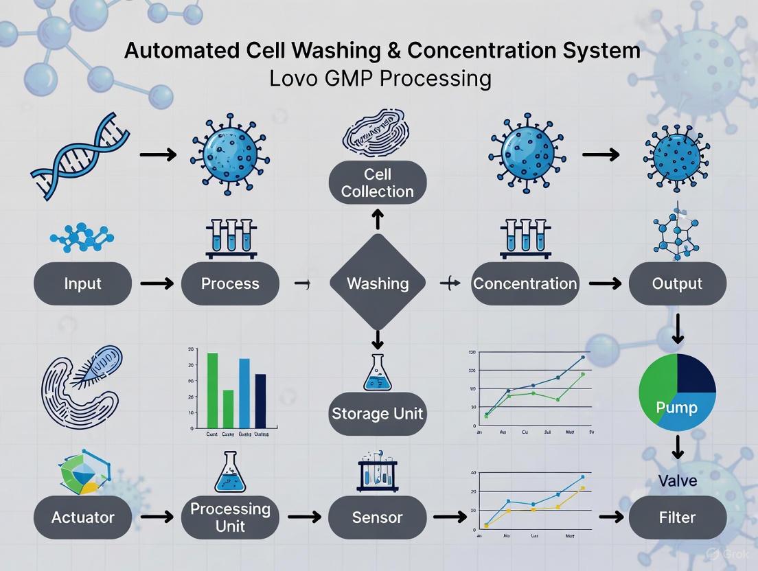
Integrated System Workflow Diagram
Within the context of automated cell washing and concentration system research, particularly involving the Lovo GMP processing platform, managing centrifugation-related stress is a fundamental challenge. Centrifugation is a critical step in the manufacturing of cell therapies and biologics, but its improper application can lead to significant cell loss, reduced viability, and compromised product quality due to shear forces and mechanical damage. This technical support center provides targeted troubleshooting guides and FAQs to help researchers and scientists optimize their protocols, minimize these pitfalls, and understand the role of alternative technologies like the Lovo system in a GMP environment.
Frequently Asked Questions (FAQs)
1. What are the primary mechanisms of cell damage during centrifugation? Cell damage during centrifugation primarily occurs through two mechanisms: shear stress and solid surface interaction. High shear stresses, particularly those exceeding 1500 dynes/cm², can directly rupture cell membranes [11]. Additionally, interactions with the centrifuge tube walls during pelleting and the subsequent resuspension steps can cause significant mechanical damage [2].
2. How does the Lovo System differ from traditional centrifugation? The Lovo Cell Processing System uses spinning membrane filtration instead of centrifugal force to wash and concentrate cells. This automated, functionally closed system minimizes exposure to high g-forces and eliminates the need for manual resuspension, which is a known source of cell damage in traditional centrifugation protocols [7] [2]. It is designed to support scalability from Phase 1 through commercialization while maintaining cell viability and recovery [7].
3. Can centrifugation protocols be optimized for shear-sensitive cells? Yes. Research demonstrates that using a dimensionless centrifugation number (Ce) can help identify an operating window that balances recovery efficiency with the avoidance of cell damage. This approach, which can be modeled with Computer Fluid Dynamics (CFD), helps in selecting the optimal combination of settling depth, retention time, and centrifugal force for specific cell types [12].
4. Why might my depth filters clog after a centrifugation step? Disk-stack centrifuges generate high shear in their feed zones, which can damage shear-sensitive mammalian cells. This damage produces submicron particles that are carried over into the centrate. These fine particles are a primary cause of subsequent fouling and clogging in depth filters and chromatographic columns [13].
Troubleshooting Guides
Problem: Low Cell Viability After Centrifugation
Potential Causes and Solutions:
- Cause 1: Excessive Centrifugal Force. High g-forces can directly damage cells through shear stress.
- Solution: Optimize the g-force. Use the lowest possible force that effectively pellets the cells. Refer to the table below for a force comparison. The concept of a centrifugation number (Ce) can be used to find a safe operating window [12].
- Cause 2: Prolonged Centrifugation Time. Overly long spins can lead to compacted pellets that are difficult to resuspend without damage.
- Solution: Minimize centrifugation time. Determine the minimum time required for adequate separation. A scale-down model can help characterize this parameter with limited material [13].
- Cause 3: Damaging Resuspension Techniques. Vigorous pipetting of a tight pellet is a major source of cell loss.
- Solution: Use gentle resuspension methods. Consider automated systems like Lovo, which are designed to minimize the need for manual resuspension, thereby preserving viability [2].
Problem: High Cell Loss in the Supernatant
Potential Causes and Solutions:
- Cause 1: Insufficient Centrifugal Force or Time. Cells are not fully sedimented.
- Solution: Systematically increase g-force or time in small increments. Using a benchtop centrifuge to model the performance of a production-scale disk-stack centrifuge via Sigma theory can help define the correct parameters [13].
- Cause 2: Incorrect Rotor Selection. Different rotors have different separation efficiencies.
- Solution: Select the appropriate rotor. Fixed-angle rotors are often more efficient for pelleting proteins or small particles, while swinging-bucket rotors are better for separating larger components like organelles [14].
Quantitative Data and Protocol Optimization
The following table summarizes key parameters and their impact on cell processing outcomes, comparing traditional centrifugation with the Lovo system.
Table 1: Comparison of Cell Processing Methods
| Parameter | Traditional Centrifugation | Lovo System | References |
|---|---|---|---|
| Cell Recovery | Variable; often impacted by pellet resuspension | Satisfactory and often improved over in-house methods | [2] |
| Cell Viability | Can be reduced by shear and resuspension | Satisfactory viability maintained | [2] |
| Shear Stress | High in centrifuge feed zones; threshold of ~1500 dynes/cm² | Minimal g-forces due to spinning membrane | [11] [7] |
| Process Automation | Mostly manual, open-system steps | Automated and closed system | [7] [2] |
| Typical Protocol | Multiple spins (e.g., 400xg, 5 mins, 3 washes) | Automated wash cycles with spinning membrane | [2] |
Detailed Experimental Protocol: Optimized Sucrose Gradient Centrifugation
For the purification of sensitive enveloped viruses or organelles, an optimized continuous sucrose density gradient protocol can significantly preserve structural integrity [15].
Method:
- Cell Culture and Virus Amplification: Culture Sf9 cells in Sf-900 medium at 27°C. Transfect with recombinant bacmid to generate baculovirus (e.g., AcMNPV) and amplify to create a working stock.
- Differential Centrifugation:
- Centrifuge culture supernatant at 1,000 × g for 10 min at 4°C.
- Transfer supernatant and centrifuge at 10,000 × g for 15 min at 4°C to remove cell debris.
- Ultracentrifuge the resulting supernatant at 70,000 × g for 45 min at 4°C. Resuspend the pellet in a small volume of PBS.
- Prepare Continuous Sucrose Gradient: Create a 15% to 50% (w/w) continuous sucrose gradient using a Gradient Master instrument.
- Ultracentrifugation: Layer 1 mL of the virus sample on top of the continuous gradient. Ultracentrifuge at 100,000 × g for 2 hours at 4°C.
- Fraction Collection and Buffer Exchange: Using a cut pipette tip (0.3–0.4 cm wide) to minimize shear, carefully collect the opalescent band containing the virions. Dilute the fraction with TN buffer (50 mM Tris-HCl pH 8, 150 mM NaCl) and pellet the virions via ultracentrifugation at 70,000 × g for 45 min. Resuspend the final pellet in 100 µL of TN buffer.
Outcome: This optimized protocol increased the proportion of budded virions with intact envelopes from 36% to 81%, preserving the native conformation of envelope proteins [15].
Workflow Visualization
Centrifugation Optimization Workflow
Research Reagent Solutions
Table 2: Essential Materials for Centrifugation and Cell Processing
| Reagent / Material | Function / Application | References |
|---|---|---|
| Sucrose (for density gradients) | Forms isosmotic gradients for the purification of enveloped viruses and organelles while preserving integrity. | [15] |
| Hank's Balanced Salt Solution (HBSS) with HSA | Serves as a wash buffer for cell formulations, providing a physiologically compatible environment. | [2] |
| Protease Inhibitors | Added to lysis and wash buffers to prevent protein degradation during cell processing and purification. | [14] |
| Ficoll-Paque / Lymphoprep | Density gradient medium for the initial separation of peripheral blood mononuclear cells (PBMCs) from whole blood. | [2] |
| Sf-900 Medium / AIM V Medium | Specialized culture media for insect cell lines (e.g., Sf9) used in baculovirus expression systems and TIL culture, respectively. | [15] [2] |
In the field of advanced cell therapies, the consistency, safety, and potency of the final product are paramount. Automated cell processing systems, such as the Lovo Cell Processing System, are integral to achieving these goals within Good Manufacturing Practice (GMP) frameworks. These systems automate the traditionally labor-intensive tasks of washing and concentrating cell therapy products, enhancing standardization and reducing contamination risks [3] [2]. This technical resource defines the core performance metrics—cell viability, cell recovery, and process efficiency—that researchers and professionals must monitor to optimize their processes using the Lovo system. The following guides and data, synthesized from recent studies and manufacturer specifications, are designed to support your experimental and manufacturing protocols.
★ Core Performance Metrics & Quantitative Data
The table below summarizes key quantitative data for the Lovo system across different cell processing applications, providing benchmarks for your experimental outcomes.
| Application | Cell Type / Product | Key Metric | Reported Performance | Source / Context |
|---|---|---|---|---|
| Culture Harvest & Media Exchange | T Cells | Viable Cell Recovery | 93% ± 4% | Comparison vs. Manual (91%) & CS5+ (87%) [16] |
| Processing Time | 30 ± 1 minutes | [16] | ||
| Washout Efficiency | 99.5% | [16] | ||
| Thawed Wash & DMSO Removal | Hematopoietic Progenitor Cells (HPCs) | Viable CD34+ Cell Recovery | 84% (median, IQR: 61-93%) | 3-cycle wash protocol [16] |
| CD34+ Cell Viability | 92% (median, IQR: 81-94%) | [16] | ||
| DMSO Elimination | 97% (median, IQR: 97-98%) | [16] | ||
| Fresh Leukapheresis Wash | Total Nucleated Cells (TNC) | TNC Recovery | 98.6% | [16] |
| TNC Viability | 97.5% | [16] | ||
| Platelet Depletion | >98% | [16] | ||
| Final Product Harvest (Multi-Center Study) | Activated T Cells (ATCs), Tumor-Infiltrating Lymphocytes (TILs), Mesenchymal Stromal Cells (MSCs) | Cell Viability & Recovery | Satisfactory, with substantial improvements over in-house methods | PACT multi-center study [3] [2] |
★ Detailed Experimental Protocols
Protocol 1: Final Harvest and Wash of Cultured T Cells
This protocol is adapted from a multi-center study that compared the Lovo system to centrifuge-based methods for harvesting activated T cells [2].
- 1. Cell Culture and Source: Activate and expand T cells from Peripheral Blood Mononuclear Cells (PBMCs) using standard methods, potentially scaling up in devices like G-Rex bioreactors [2].
- 2. Pre-Processing Analysis:
- 3. Lovo System Setup:
- Software: Use Lovo software (e.g., version 3.0) to design the protocol [2] [16].
- Disposable Kit: Aseptically install the Lovo disposable kit, making any necessary pre-procedure modifications to luer connections within a biosafety cabinet [2].
- Wash Buffer: Use an appropriate buffer, such as Hank’s Balanced Salt Solution (HBSS) supplemented with 5% Human Serum Albumin (HSA) [2].
- 4. Processing Parameters:
- Protocol: Employ a multi-wash-cycle protocol. A study using a six-wash-cycle protocol, with a 90% supernatant wash-out programmed for each cycle, successfully concentrated cells from 1.7L to a target volume of 150mL [2] [16].
- Final Volume: Adjust the "final In-Process bag dilution volume" and "final IP Bottom Port Rinse" settings in the software to achieve your desired final concentration and volume [2].
- 5. Post-Processing Analysis:
- Cell Recovery and Viability: Count the harvested cells and assess viability again (hemocytometer and flow cytometry) to calculate percentage recovery and post-process viability [2].
- Washout Efficiency: Measure the concentration of a key media component (e.g., a cytokine like IL-15) in the pre- and post-processing supernatant using an ELISA to confirm efficient removal (>99%) [2] [16].
- Product Quality: Assess critical quality attributes (CQAs) such as cell phenotype (via flow cytometry for CD3, CD4, CD8) and functionality to ensure they are comparable to pre-processing states [16].
Protocol 2: DMSO Removal from Thawed Cell Products
This protocol is designed for washing cryopreserved products, such as hematopoietic progenitor cells, after thawing [16].
- 1. Product Thaw: Rapidly thaw the cryopreserved cell product using a standard water bath or validated thawing device.
- 2. Lovo System Setup:
- Software: Select or design a protocol for thawed wash. A three-cycle wash protocol has been validated for this application [16].
- Dilution: The system can be primed and operated with a suitable washing buffer, such as Plasma-Lyte A containing 5% Human Serum Albumin, to gently dilute and wash the thawed product [16].
- 3. Processing and Output:
- The automated process will remove the DMSO-containing supernatant, wash the cells, and concentrate them into a final bag.
- Reported performance for a three-cycle wash shows a median of 97% DMSO elimination and 84% viable CD34+ cell recovery [16].
- 4. Post-Processing Analysis:
- Viability and Recovery: Determine viable cell recovery and final viability.
- DMSO Residual: Test the final product supernatant for residual DMSO to confirm elimination efficiency.
★ Troubleshooting FAQs
Q1: Our post-processing cell recovery is lower than expected. What could be the cause?
- A: Suboptimal recovery can stem from several factors. First, review the starting cell concentration and packed cell volume (PCV%); the system is designed for specific ranges, and operating outside these can affect performance [16]. Second, verify the wash cycle parameters. Excessive wash cycles or overly aggressive volume reduction per cycle can contribute to cell loss. Finally, ensure the spinning membrane is functioning correctly and that there are no blockages or issues with the disposable kit.
Q2: How can we improve the efficiency of reagent or cryoprotectant removal?
- A: Washout efficiency is a key strength of the Lovo system. To achieve >99% removal of materials like cytokines or DMSO [16], you should:
- Optimize Wash Cycles: Increasing the number of wash cycles (e.g., from three to six) can enhance the logarithmic reduction of soluble factors [2] [16].
- Validate with Analytics: Always measure the concentration of the target molecule (e.g., via ELISA for cytokines) in the final product to quantitatively confirm washout efficiency for your specific process [2].
Q3: We are observing a decrease in cell viability after processing. What should we investigate?
- A: A decline in viability can be related to process timing or mechanical stress.
- Minimize Hold Times: Reduce the time cells spend in the final product bag before or after processing. Prolonged holds, especially at non-ideal temperatures, can decrease viability.
- Review Software Version: Ensure you are using the latest software version, as updates may include optimizations to processing algorithms that improve cell health [2].
- General Cell Handling: Remember that factors outside the Lovo system can also impact viability. The health of the starting culture and the post-thaw status of the cells are critical variables that must be controlled [17].
★ The Scientist's Toolkit: Essential Research Reagents & Materials
The table below lists key materials used in experiments with the Lovo system, as cited in the referenced literature.
| Reagent / Material | Function / Application | Example Usage in Context |
|---|---|---|
| Human Serum Albumin (HSA) | A key additive in wash buffers to maintain cell stability and viability during processing. | Used at 5% concentration in HBSS or Plasma-Lyte A for washing T cells and hematopoietic progenitors [2] [16]. |
| Plasma-Lyte A | A balanced electrolyte solution used as a base for wash buffers. | Served as the primary washing solution for thawed hematopoietic progenitor cells [16]. |
| CliniMACS PBS/EDTA Buffer | A specialized buffer used in cell processing to prevent clumping and provide a suitable ionic environment. | Employed as a washing buffer during CD34+ cell enrichment processes on automated systems [18]. |
| CryoStor | A serum-free, protein-free cryopreservation medium designed to improve post-thaw cell viability and recovery. | Selected for the cryopreservation of leukopaks to ensure high post-thaw recovery and maintain cell integrity [19]. |
| Dimethyl Sulfoxide (DMSO) | A cryoprotectant agent (CPA) used to protect cells during freezing. Its removal is critical post-thaw. | Standard cryopreservation of cell products; its efficient removal (≥97%) is a key application for the Lovo system [16]. |
| Interleukins (e.g., IL-7, IL-15) | Cytokines used in cell culture media to promote expansion and maintain function of immune cells like T cells. | Presence in culture media; washout efficiency of such soluble factors is a critical metric post-processing [2]. |
★ Lovo Cell Processing Workflow
The following diagram illustrates the general workflow for processing cells using the Lovo system, from sample preparation to final output.
Technical Support Center
Troubleshooting Guides
This guide addresses common issues you might encounter with the Lovo Cell Processing System to ensure consistent, GMP-compliant operation.
Low Cell Recovery
- Problem: Lower than expected cell yield post-processing.
- Possible Causes:
- Incorrect protocol parameters: The number of wash cycles or supernatant removal percentage may be insufficient for your cell type.
- Membrane fouling: Cell debris or high particle concentration may be obstructing the spinning membrane.
- Sample-specific factors: The cell type being processed may have unique size or fragility characteristics.
- Solutions:
- Consult application-specific data to optimize your protocol. For T-cells, a study achieved 93% recovery using a validated protocol [20].
- Ensure your starting sample is not overly concentrated with debris. Pre-filtration may be necessary.
- Refer to the operator's manual for approved cleaning and priming procedures to maintain membrane performance [7].
Inconsistent Wash Efficiency
- Problem: Incomplete removal of cytokines, DMSO, or other ancillary materials.
- Possible Causes:
- Inadequate wash volume: The total wash solution volume is insufficient for the starting concentration of the material to be removed.
- Incorrect final volume settings: Rinse and dilution volumes are not optimized for maximum recovery of washed cells.
- Solutions:
- Increase the number of wash cycles. A pre-clinical assessment for DMSO removal used a three-cycle protocol, achieving 97% DMSO elimination [20].
- Adjust the "final In-Process bag dilution volume" and "final IP Bottom Port Rinse" settings as needed. One study increased these parameters to improve final product volume accuracy [2].
Alarms or System Errors
- Problem: The instrument halts and displays an error code.
- Possible Causes:
- Disposable kit issues: Improper loading or a defective kit.
- Clogs or pressure deviations: An obstruction in the fluid path.
- Software anomaly.
- Solutions:
- Ensure all kit connections are secure and the kit is properly seated.
- Follow the system's prompts and refer to the Operator’s Manual for specific error code resolutions.
- For unresolved errors, contact technical support. Note that training on the Lovo system is offered monthly to ensure user proficiency [10].
Frequently Asked Questions (FAQs)
Q1: How does Lovo's technology minimize cell loss and maintain viability compared to centrifugation?
Lovo uses spinning membrane filtration technology, which exposes cells to significantly lower g-forces than traditional centrifugation [2]. This gentle processing, combined with the elimination of manual resuspension steps—a known source of cell loss and damage—helps maximize both cell recovery and viability [2]. Studies on T-cells and MSCs have demonstrated satisfactory viability and recovery, with some showing substantial improvement over in-house centrifugation methods [2].
Q2: Can the Lovo system be integrated into a cGMP-compliant manufacturing environment?
Yes. The Lovo system is designed with GMP-ready features. Its functionally closed and automated processing reduces the risk of operator error and protects product sterility [2]. Furthermore, the optional DXT Data Management System is an open architecture software platform that supports 21 CFR Part 11 compliance for electronic records, which is critical for regulatory submissions [7].
Q3: What applications is the Lovo system validated for?
The Lovo system is flexible and supports multiple cell processing applications, including [20]:
- Culture Harvest & Media Exchange
- Thawed Wash & DMSO Removal
- Fresh Leukapheresis Wash
- Immunomagnetic Selection Prep
Q4: Where can I find technical documentation to support a regulatory submission?
Fresenius Kabi can provide the required Lovo technical documentation upon request to support your regulatory filings for applications requiring clearance or approval [7] [10]. It is the user's responsibility to obtain appropriate regulatory clearance for clinical use [7].
Experimental Data and Protocols
The following table summarizes key quantitative data from independent studies evaluating the Lovo system for various applications.
Table 1: Performance Data for Lovo Cell Processing Applications
| Application | Cell Type | Key Metric | Result | Source |
|---|---|---|---|---|
| Culture Harvest [20] | T-Cells | Recovery | 93% | IBC Commercialization, 2014 |
| Processing Time | 30 min | |||
| Washout Efficiency | 99.5% | |||
| Thawed Wash & DMSO Removal [20] | Hematopoietic Progenitor Cells | Viable CD34+ Cell Recovery | 84% (median) | Cytotherapy, 2017 |
| DMSO Elimination | 97% (median) | |||
| Fresh Leukapheresis Wash [20] | Total Nucleated Cells (TNC) | TNC Recovery | 98.6% | Data on File 223-REP-048957 |
| TNC Viability | 97.5% | |||
| Immunomagnetic Selection Prep [20] | Total Nucleated Cells (TNC) | Platelet Depletion | 98.4% | Data on File 223-REP-048957 |
| TNC Recovery | 97.2% |
Detailed Protocol: Final Harvest and Wash of Activated T-Cells (ATC)
This methodology is adapted from a multicenter NIH/PACT study [2].
1. Sample Preparation
- Culture ATCs in G-Rex100 devices.
- On harvest day (e.g., days 13-21), pool cultures from two devices.
- Determine the pre-processing viable cell count using a hemocytometer and the trypan blue exclusion method.
2. Lovo System Setup
- Use the Lovo Cell Processing System (software version 3.0 or later).
- Modify the disposable kit pre-procedure to allow Source and Wash Solution luer connections to be made in a biosafety cabinet.
- Use a 1 L bag of wash buffer (e.g., HBSS with 5% Human Serum Albumin) as the wash solution.
3. Processing Parameters
- Program a protocol with six wash cycles.
- Set a 90% supernatant wash-out for each cycle.
- To target a final product volume of approximately 50-78 mL, adjust parameters such as:
- Final In-Process bag dilution volume: 300 mL
- Final IP Bottom Port Rinse: 10 mL
4. Post-Processing Analysis
- Harvest cells from the Lovo and perform a final cell count and viability assessment (e.g., via trypan blue or flow cytometry using 7-AAD).
- Sample the final product for sterility testing (e.g., using BD BACTEC bottles) and for residual analyte analysis (e.g., IL-15 ELISA) if required.
The workflow for this process is outlined below:
The Scientist's Toolkit: Essential Research Reagent Solutions
Table 2: Key Materials and Reagents for Cell Processing with Lovo
| Item | Function | Example from Literature |
|---|---|---|
| Wash Buffer | To suspend and wash cells, removing unwanted media components and contaminants. | Hank’s Balanced Salt Solution (HBSS) supplemented with 5% Human Serum Albumin (HSA) [2]. |
| Culture Media | To expand cells prior to harvest. | Advanced RPMI 1640 mixed with Click's medium, GlutaMAX, and FBS, supplemented with cytokines (e.g., IL-7, IL-15) [2]. |
| Cell Staining Reagents | To assess cell viability, phenotype, and critical quality attributes (CQAs) post-processing. | Trypan blue for manual count viability; 7-AAD and fluorescent antibodies (e.g., CD3, CD4, CD8) for flow cytometry [2]. |
| Sterility Testing Media | To test the final cell product for microbial contamination as part of quality control. | BD BACTEC Standard/10 Aerobic/F, Lytic/10 Anaerobic/F, and Myco/F culture bottles [2]. |
| Analysis Kits | To quantify the removal of specific ancillary materials, such as cytokines or growth factors. | IL-15 ELISA kit to measure residual cytokine levels post-wash [2]. |
Optimizing Your Workflow: Lovo Protocol Development for Key Cell Therapies
Cell washing is a fundamental laboratory and clinical procedure used to remove unwanted components from cell suspensions. This process eliminates contaminants such as residual plasma, platelets, red blood cells, dead cells, and debris to improve sample purity and quality. Accessing pure, viable cell samples is integral to effective clinical treatments, biomedical research, and drug development initiatives. The procedure is critical for preparing cells for downstream applications including flow cytometry, PCR, cell culturing, antibody labeling, and transfusion medicine [21].
Within the context of automated cell washing concentration systems like the Lovo GMP, establishing standardized protocols for wash cycles, volumes, and flow rates becomes paramount for ensuring process consistency, cell viability, and compliance with Good Manufacturing Practices (GMP). This technical guide provides detailed methodologies and troubleshooting advice to support researchers and scientists in optimizing their cell washing procedures for reliable, reproducible results.
Core Principles and Key Terminology
What is Cell Washing?
Broadly defined, cell washing is any process that involves the washing and separation of cells from interfering substances. In automated systems, this is typically achieved through programmed cycles of buffer addition, mixing, and separation steps. The primary objectives are:
- Purification: Removing unwanted cellular material and contaminants.
- Concentration: Achieving a desired cell density in the final product.
- Buffer Exchange: Replacing the original suspension medium with a fresh, compatible solution.
- Volume Reduction: Reducing the overall volume for downstream processing or storage [21].
Importance in GMP Processing
For GMP-compliant cell therapy manufacturing, cell washing protocols must be not only effective but also scalable and reproducible. Consistency in the process ensures the end product is of high quality, pure, and viable for clinical applications. Automated systems like the Lovo are designed to meet these requirements by providing a closed, controlled environment for processing [21].
Troubleshooting Common Cell Washing Issues
Poor Cell Recovery and Viability
Problem: Low percentage of target cells recovered post-wash, or reduced cell viability. Potential Causes and Solutions:
- Cause: Excessive centrifugal force or duration (in centrifugation-based systems).
- Solution: Optimize centrifugation speed and time. Validate minimal g-force required for effective pelleting.
- Cause: Overly aggressive resuspension damaging cells.
- Solution: Use gentle pipetting techniques or automated systems with controlled resuspension parameters.
- Solution: Utilize biocompatible buffers that maintain osmolarity and pH [21].
- Cause: Excessive number of wash cycles.
- Solution: Determine the minimum number of wash cycles required to achieve target purity. Each cycle inherently risks cell loss.
- Cause: Incompatible flow rates in centrifuge-free systems causing shear stress.
- Solution: Titrate flow rates to find the optimal balance between washing efficiency and cell retention. Refer to system-specific validation data [22].
Inadequate Contaminant Removal
Problem: Persistent presence of unwanted components (e.g., RBCs, platelets, free proteins) after washing. Potential Causes and Solutions:
- Cause: Insufficient wash volume per cycle.
- Solution: Increase the buffer-to-sample volume ratio in each wash cycle to improve dilution and removal of soluble contaminants.
- Cause: Inefficient mixing during the wash phase.
- Solution: Ensure proper mixing is achieved to keep cells in suspension and allow contaminants to be carried away.
- Cause: Clogged filters or lines in automated systems.
- Solution: Perform routine preventive maintenance and system checks as per the manufacturer's instructions.
- Cause: Protocol not optimized for specific contaminant.
- Solution: For specific contaminants like RBCs, consider specialized reagents or protocols. For example, Akadeum's Human RBC Depletion Microbubbles can remove 99% of RBC contamination in pre-processed samples [21].
Inconsistent Results Across Runs
Problem: Variable cell recovery, viability, or purity between identical experiments. Potential Causes and Solutions:
- Cause: Manual protocol execution leading to operator-dependent variability.
- Cause: Uncontrolled temperature or processing time.
- Solution: Perform all steps in a temperature-controlled environment and standardize the time from sample collection to processing.
- Cause: Lot-to-lot variability in buffers or reagents.
- Solution: Use GMP-grade, qualified reagents and perform incoming quality control checks.
Frequently Asked Questions (FAQs)
Q1: How do I determine the optimal number of wash cycles for my specific cell type? A1: Start with a baseline of two cycles and assess purity (e.g., via flow cytometry) and recovery. Incrementally increase the number of cycles until purity plateaus, then select the cycle count just before a significant drop in recovery or viability occurs. Always balance purity with the need to maximize the yield of your target cells.
Q2: What is the recommended buffer-to-sample ratio for an effective wash? A2: While the optimal ratio depends on the initial contaminant load, a common starting point is a 5:1 to 10:1 buffer-to-sample volume ratio. This provides sufficient dilution to reduce contaminant concentration effectively without excessively prolonging processing time or buffer consumption [21].
Q3: My downstream flow cytometry results show high background. Could cell washing be the issue? A3: Yes. Inadequate washing can leave behind unbound antibodies or reagents, creating conflicting "noise." Ensure your protocol includes sufficient washes after staining steps. Also, confirm that the wash protocol effectively removes red blood cells, as their presence can interfere with viability dyes and cause inaccurate measurements [21].
Q4: Are there alternatives to centrifugation for delicate cell types? A4: Yes, centrifuge-free technologies are emerging. For instance, the C-FREE Pluto System uses an automated, non-centrifugal washing method. One study demonstrated its ability to achieve higher cell retention with comparable population frequencies for immunophenotyping, making it a viable option for sensitive primary cells [22].
Q5: How critical is flow rate control in automated systems? A5: Extremely critical. Flow rate directly impacts shear stress applied to cells and the efficiency of contaminant removal. A rate that is too high can damage cells and reduce viability, while a rate that is too low may be ineffective at washing and lead to long, impractical processing times. Always adhere to manufacturer-recommended ranges for your cell type.
Quantitative Data for Protocol Optimization
Table 1: Comparison of Cell Washing Modalities
| Parameter | Manual Centrifugation | Automated Centrifugation (e.g., Rotea) | Centrifuge-Free Automation (e.g., C-FREE Pluto) |
|---|---|---|---|
| Typical Cell Recovery | Variable (operator-dependent) | Designed to minimize cell loss [21] | Higher retention reported [22] |
| Consistency | Low | High | Improved across donors [22] |
| Hands-on Time | High | Reduced | Reduced [22] |
| Shear Stress | During spin/resuspension | Controlled, uses counterflow centrifugation [21] | Low (no centrifugal force) [22] |
| Throughput | Low | Medium to High | Configurable |
| GMP Compliance | Requires strict SOPs | Closed system available [21] | Amenable to automation |
Table 2: Performance Metrics for Contaminant Removal
| Contaminant / Metric | Standard Protocol Performance | Enhanced Method Performance |
|---|---|---|
| Red Blood Cell (RBC) Removal | Varies with protocol (e.g., 1-2 manual washes) | 99% removal with specialized microbubbles in debulked samples [21] |
| Dead Cell Removal | Varies with protocol | Up to 86% recovery of viable cells with microbubble kit [21] |
| Unbound Antibody/Reagent | Effective with adequate washes | Effectively removed by standard and automated wash cycles [21] |
| Impact on Downstream Viability (Flow Cytometry) | Can be compromised by RBC contamination | Improved by effective RBC and dead cell removal [21] |
Experimental Workflow and Signaling
The following diagram illustrates a generalized decision-making workflow for establishing and troubleshooting a foundational cell washing protocol, applicable to systems like the Lovo GMP.
Cell Washing Protocol Workflow
The Scientist's Toolkit: Research Reagent Solutions
Table 3: Essential Materials for Cell Washing Protocols
| Item | Function & Description |
|---|---|
| Cell Washer Instrument | Automated instruments (e.g., Rotea system, C-FREE Pluto) designed to wash cells, separate them from buffer, and prepare them for downstream assays in a reproducible manner [21] [22]. |
| GMP-Grade Washing Buffer (e.g., PBS, Saline) | A balanced salt solution used to dilute the original sample, carry away contaminants, and maintain cell viability and osmolarity during the washing process [21]. |
| Specialized Depletion Kits (e.g., RBC, Dead Cell) | Reagent kits that selectively bind and remove specific contaminants. For example, negative selection microbubbles can remove 99% of RBCs or recover up to 86% of viable cells [21]. |
| Closed System Processing Set | Single-use, sterile fluid pathways that integrate with automated washers to maintain a closed environment, which is critical for GMP compliance and preventing contamination [21]. |
| Cell Counting & Viability Assay | Essential for quantifying protocol success. Methods like flow cytometry or automated cell counters are used to measure cell recovery, viability, and purity post-wash [21] [22]. |
Performance Data and Comparisons
The following tables summarize key quantitative data from a multicenter study assessing the LOVO system for final harvest and wash of Activated T Cells (ATCs) and Tumor-Infiltrating Lymphocytes (TILs) [2] [3] [23].
Table 1: Cell Recovery and Viability for ATCs and TILs processed with LOVO
| Cell Type | Volume Processed (L) | Viability (%) | Cell Recovery (%) | Viable Cell Recovery (%) |
|---|---|---|---|---|
| Activated T Cells (ATCs) | 1.5 - 2.0 | 95 - 97 | 85 - 95 | 84 - 90 |
| Tumor-Infiltrating Lymphocytes (TILs) | 0.8 - 1.0 | 88 - 95 | 85 - 92 | 80 - 85 |
Table 2: Processing Time and Wash Efficiency Comparison
| Parameter | LOVO System | Manual Centrifugation (In-house method) |
|---|---|---|
| Processing Time for ATCs | Variable by facility/cell type | Variable by facility/cell type |
| Cytokine (IL-15) Washout | ~97% reduction | ~97% reduction |
| System Type | Automated and closed | Often open-system, manual |
| Cell Resuspension | Reduces or eliminates manual resuspension | Requires manual resuspension, a known cause of cell stress |
Experimental Protocols
Protocol: Final Harvest and Wash of Activated T Cells (ATCs) using LOVO
This protocol is adapted from the multicenter study conducted at Baylor College of Medicine [2].
Step 1: Cell Culture and Preparation
- Culture ATCs in G-Rex100 devices or similar expansion platforms.
- On harvest day (days 13-21), pool cultures and take a viable cell count using a hemocytometer and trypan blue exclusion.
- Split the pooled cells into two fractions for comparative processing (e.g., LOVO vs. manual wash).
Step 2: LOVO System Setup
- Use the LOVO Cell Processing System (software version 3.0 or later).
- Aseptically modify the LOVO disposable kit's Source and Wash Solution luer connections inside a biosafety cabinet to maintain a closed system.
- Prime the system using a 1 L bag of wash buffer (e.g., HBSS without Ca²+/Mg²+ supplemented with 5% Human Serum Albumin).
Step 3: LOVO Processing Parameters
- Load the cell harvest into the LOVO system.
- Run a six-wash-cycle protocol.
- Program a 90% supernatant wash-out for each cycle.
- To target a final product volume of approximately 78 mL, set a final In-Process bag dilution volume of 300 mL and a final IP Bottom Port Rinse of 10 mL.
Step 4: Post-Processing Handling
- Harvest the cells from the LOVO and perform a final cell count and viability assessment.
- Centrifuge and re-suspend cells in the final formulation buffer at the target concentration (e.g., 1x10⁷ cells/mL).
- Sample the final product for required quality control tests, such as sterility (e.g., using BD BACTEC bottles) and flow cytometry for phenotype (CD3, CD4, CD8) and viability (e.g., 7-AAD).
Protocol: Final Harvest and Wash of Tumor-Infiltrating Lymphocytes (TILs) using LOVO
This protocol is adapted from the experiments performed at Moffitt Cancer Center [2].
Step 1: TIL Expansion and Preparation
- Expand TILs using a rapid expansion protocol in G-Rex100M-CS flasks.
- On the day of harvest, pool the TIL cultures from multiple flasks.
Step 2: LOVO Processing
- Set up the LOVO system as described in the ATC protocol.
- Process the pooled TIL culture using a similar multi-cycle wash protocol.
- Use an appropriate wash buffer, such as Plasma-Lyte A or RPMI-1640, potentially supplemented with human AB serum.
Step 3: Final Formulation and Testing
- Harvest the washed and concentrated TILs.
- Formulate the final product in the desired infusion buffer.
- Perform cell count, viability, and sterility testing.
- Optionally, test for specific functionality, such as IFN-γ release upon tumor antigen stimulation, to confirm retained effector function post-processing.
Diagram 1: Experimental workflow for T-cell harvest and wash using the LOVO system.
Troubleshooting Guides
Low Cell Recovery
- Problem: Lower than expected cell recovery after LOVO processing.
- Potential Cause 1: Excessive cell loss on the spinning membrane filter.
- Solution: Ensure the cell concentration and total number processed are within the manufacturer's specified range for the disposable kit being used. For very small cell populations, consider protocol optimization.
- Potential Cause 2: Overly aggressive wash cycles leading to cell loss.
- Solution: Review the programmed wash cycle parameters. Reducing the number of cycles or the percent supernatant removal per cycle may improve recovery, though this must be balanced against wash efficiency needs.
- Potential Cause 3: Incorrect filter membrane for the cell type.
Low Post-Process Viability
- Problem: Cell viability is low after processing with LOVO.
- Potential Cause 1: Cell stress during extended processing time.
- Solution: Minimize total processing and hold times. Keep cells in optimized, temperature-controlled buffers throughout the process.
- Potential Cause 2: Shear stress from the spinning membrane.
- Solution: Although LOVO is gentler than centrifugation [2], verify that the membrane spin speed is within recommended limits. The system is designed to minimize g-forces and shear stress compared to centrifugation.
- Potential Cause 3: Inadequate wash buffer composition.
- Solution: Ensure the wash buffer contains protective agents like albumin (e.g., 5% HSA). Research shows recombinant Human Serum Albumin (rHSA) can improve viable cell retention during washing steps [24].
Inadequate Washout Efficiency
- Problem: Residual cytokines (e.g., IL-15) or DMSO levels are higher than desired.
- Potential Cause 1: Insufficient number of wash cycles.
- Solution: Increase the number of wash cycles. The referenced study successfully used a six-cycle protocol for cytokine removal [2].
- Potential Cause 2: Inefficient volume exchange per cycle.
- Solution: Ensure the "supernatant wash-out" percentage per cycle is set appropriately (e.g., 90%). Confirm the system is functioning correctly and there are no occlusions.
- Potential Cause 3: Re-contamination from the system or lines.
- Solution: Perform adequate priming and rinsing of the disposable kit and lines with wash buffer before introducing the cell product. Follow the rinse steps programmed into the method.
Frequently Asked Questions (FAQs)
Q1: How does the LOVO system fundamentally differ from traditional centrifugation? A1: LOVO uses spinning membrane filtration instead of centrifugal force to concentrate cells [2] [7]. The supernatant and small particles (<4µm) pass through the membrane, while cells are retained. This exposes cells to minimal g-forces and reduces or eliminates the need for manual pellet resuspension, a key source of cell stress and loss in centrifugation [2] [3].
Q2: Is the LOVO system considered a closed and GMP-compliant platform? A2: Yes. The LOVO is an automated and closed system, which minimizes the risk of contamination and supports standardization [2] [25]. It is designed as a GMP-ready platform suitable for process development through commercialization, and manufacturers can request technical documentation from Fresenius Kabi to support regulatory submissions [7].
Q3: Can the LOVO system be used for other cell therapy products besides T cells? A3: Yes. While this spotlight focuses on ATCs and TILs, the multicenter study also demonstrated satisfactory results for the large-volume harvest and wash of Mesenchymal Stromal Cells (MSCs) [2] [23]. Its flexibility supports various applications, including immunomagnetic selection preparation, fresh leukapheresis wash, and post-thaw DMSO removal [25].
Q4: What are the critical parameters to define when setting up a wash protocol on the LOVO? A4: Key protocol parameters include the number of wash cycles, the percentage of supernatant removed per cycle, the volume and composition of the wash buffer, and the target final product volume. These should be optimized for the specific cell type and processing objective (e.g., cytokine removal vs. concentration) [2].
Q5: How does the choice of albumin in the wash buffer impact cell recovery? A5: Albumin is a critical buffer component that significantly improves viable cell retention during washing [24]. Studies show that recombinant Human Serum Albumin (rHSA) performs comparably to, and in some cases better than, serum-derived HSA, while offering the advantages of being animal-origin-free (AOF) and having a more reliable supply chain [24].
The Scientist's Toolkit: Key Research Reagent Solutions
Table 3: Essential Materials for T-Cell Therapy Harvest and Wash
| Reagent / Material | Function in the Protocol | Example & Notes |
|---|---|---|
| Wash Buffer Base | Provides an isotonic environment for cells during processing. | HBSS without Ca²+/Mg²+ or Plasma-Lyte A [2]. |
| Albumin Supplement | Protects cells from shear stress and aggregation, improving viability and recovery. | 5% Human Serum Albumin (HSA) or Recombinant HSA (e.g., Optibumin). rHSA is animal-origin-free and reduces supply chain risk [24]. |
| LOVO Disposable Kit | Sterile, single-use fluid pathway for cell processing. | Ensure the kit is compatible with the software version and the planned processing volumes [2] [7]. |
| Cell Culture Media | For final product formulation or as a base for wash buffers. | AIM V or RPMI-1640, potentially supplemented with human serum [2]. |
| Quality Control Reagents | For assessing the final product. | Trypan Blue (viability), 7-AAD (flow cytometry viability), Antibodies (CD3, CD4, CD8 for phenotype), and BACTEC bottles (sterility) [2]. |
Frequently Asked Questions (FAQs)
Q1: What is the primary advantage of using the Lovo system for MSC processing over traditional centrifugation? The Lovo system uses spinning membrane filtration technology, which minimizes exposure to the high g-forces typical of centrifugation. This approach reduces mechanical stress on cells, leading to higher cell viability and recovery. Furthermore, it is an automated, closed system that standardizes the wash and concentration process, enhancing sterility assurance and process consistency [2] [3].
Q2: For MSC processing, what are the typical performance metrics I can expect with the Lovo system? In a multi-center study, the Lovo system was evaluated for processing large volumes of bone marrow-derived MSCs. The results demonstrated that the system is capable of achieving satisfactory cell viability and recovery, providing a reliable alternative to centrifuge-based methods [2] [3].
Q3: Is the Lovo system suitable for scaling MSC manufacturing from clinical trials to commercialization? Yes. The Lovo instrument is designed as a platform that scales with your process, capable of handling cell volumes from 10 mL to 22 L, supporting manufacturing from Phase 1 trials through to commercialization [7].
Q4: Where can I find training resources and technical documentation for the Lovo system? The manufacturer, Fresenius Kabi, offers monthly training sessions. For applications that require regulatory submissions, users can request the necessary technical documentation from Fresenius Kabi to support their filings [7] [10].
Troubleshooting Guide for MSC Processing on Lovo
This guide addresses common challenges encountered during MSC processing.
Issue 1: Lower-than-expected cell recovery
- Potential Cause: Excessive cell loss due to adherence or processing parameters.
- Solution: Ensure the system is configured for optimal MSC handling. The spinning membrane technology is designed to maximize cell recovery by minimizing the forces that cause cells to be lost [7] [2]. Verify that your protocol settings (e.g., wash cycles, flow rates) are appropriate for your specific MSC sample.
Issue 2: Concerns about maintaining sterility
- Potential Cause: Manual, open-system processing steps.
- Solution: The Lovo system is a functionally closed and automated system, which significantly reduces the risk of contamination during the wash and concentration steps compared to open centrifugation methods [2] [3].
Issue 3: Inefficient removal of culture reagents or DMSO
- Potential Cause: Inadequate wash cycle configuration.
- Solution: The Lovo system's spinning membrane filtration is highly effective at supernatant removal. One study highlighted its use for post-thaw DMSO removal from MSCs. You can optimize the number of wash cycles and the volume of wash solution to achieve the desired level of reagent removal [2].
Experimental Protocol: MSC Harvest and Wash via Lovo
The following workflow is based on a multicenter study that assessed the Lovo system for the final harvest of bone marrow-derived Mesenchymal Stromal Cells (MSCs) [2] [3].
Methodology Details:
- Cell Type: Bone marrow-derived mesenchymal stromal cells (MSCs) [2] [3].
- Objective: Final harvest and/or wash of large-volume MSC cultures [2].
- Process: The pooled cell culture is processed through the Lovo system using its spinning membrane filtration. This step automatically concentrates the cells and exchanges the culture medium for the desired wash buffer without the need for manual centrifugation and resuspension [2] [3].
- Outcome Analysis: The final product was analyzed for critical quality attributes, including cell recovery and viability, which were found to be satisfactory and comparable or superior to the facility's in-house centrifugation methods [2] [3].
Quantitative Performance Data
The table below summarizes key performance metrics from independent evaluations of the Lovo system across different cell types, providing a benchmark for expectations with MSCs.
| Cell Type | Key Performance Metric | Reported Result | Source |
|---|---|---|---|
| Peripheral Blood Mononuclear Cells (PBMCs) | Total Nucleated Cell (TNC) Recovery | 94% (SD data available in source) | [26] |
| Peripheral Blood Mononuclear Cells (PBMCs) | Supernatant/Waste Removal | >98% | [26] |
| Activated T Cells (ATCs), Tumor-Infiltrating Lymphocytes (TILs), MSCs | Cell Viability and Recovery | Satisfactory results, with substantial improvements over in-house methods in some cases | [2] [3] |
| General Performance | Platelet Removal (from PBMCs) | >90% | [26] |
The Scientist's Toolkit: Essential Research Reagents & Materials
The following table lists key materials used in conjunction with the Lovo system for cell processing, as referenced in the studies.
| Item | Function in the Experiment | Example from Research Context |
|---|---|---|
| Wash Buffer | Fluid for diluting and washing cells to remove unwanted media components or cryoprotectants. | Hank’s Balanced Salt Solution (HBSS) supplemented with 5% Human Serum Albumin (HSA) [2]. |
| Cell Culture Media | Expansion and maintenance of cells prior to harvest. | Various basal media like Advanced RPMI 1640 and AIM V, often supplemented with serum and cytokines [2]. |
| Disposable Processing Set | Sterile, single-use fluid path for the Lovo system that maintains a closed processing environment. | Lovo disposable kits [2]. |
| Characterization Reagents | Assessment of cell quality post-processing (e.g., viability, phenotype). | Trypan blue for viability count; antibodies for flow cytometry (e.g., CD3, CD4, CD8); ELISA kits for cytokine measurement [2]. |
The Lovo Cell Processing System is an automated benchtop cell processor that utilizes spinning membrane technology to perform post-thaw washing and concentration of cell suspensions without pelletizing cells, thereby maintaining high cell viability and recovery rates [25]. This technology is particularly valuable in GMP-compliant cell therapy manufacturing workflows where reproducibility and reduction of manual processing are critical.
The table below summarizes key performance metrics for DMSO removal using the Lovo system:
| Performance Measure | Lovo 2.0 (Three-Cycle Wash) |
|---|---|
| Number of Runs | 6 |
| PCV% | 8.4% (6.9 - 11.4) |
| Viable CD34+ Cell Recovery | 84% (61 - 93) |
| CD34+ Cell Viability | 92% (81 - 94) |
| DMSO Elimination | 97% (97 - 98) |
| Total Processing Time | 62 minutes |
Source: B. Mfarrej, et al. Cytotherapy, Volume 19, Issue 12, 1501-1508 [16].
Troubleshooting FAQs
Q1: Our team is observing lower-than-expected post-thaw viable CD34+ cell recoveries after using the standard Lovo protocol. What factors should we investigate?
- Confirm Initial Product Characteristics: The packed cell volume (PCV%) of your thawed product can significantly impact recovery. The protocol is optimized for a median PCV of 8.4%. Products with substantially higher density may require protocol adjustments [16].
- Validate Wash Cycle Parameters: Ensure you are using the validated three-cycle wash protocol for cryopreserved products. Fewer cycles may result in insufficient DMSO removal, while excessive cycles could contribute to mechanical stress and cell loss [16].
- Verify System Calibration and Membrane Integrity: Check that the spinning membrane and fluidic pathways are functioning correctly. Any deviations could affect shear forces and processing efficiency. Contact Fresenius Kabi Field Service Engineers for performance qualification if needed [25].
Q2: We are achieving excellent DMSO removal (>97%) but are concerned about total processing time. How can we optimize throughput without compromising cell quality?
- Understand the Trade-off: The approximately 60-minute processing time for a three-cycle wash represents a balance between DMSO clearance and preserving cell viability. Manual processing of comparable volumes can take a similar amount of time (approximately 61 minutes) but with greater variability [16].
- Streamline Pre- and Post-Processing Steps: The automated Lovo process reduces hands-on time significantly. For workflow optimization, focus on parallel tasks such as preparing downstream culture vessels or performing quality control checks during the automated run.
- Review Software Settings: Utilize the Lovo 3.0 software features, which allow for timed incubations, to ensure the protocol is running at its most efficient without unnecessary pauses [16].
Q3: What technical documentation is available from the manufacturer to support our pre-clinical validation and eventual regulatory submission for our therapy?
- Access the Customer Portal: Fresenius Kabi provides a Customer Portal containing essential resources, including Operator's Manuals, User Guides, operator training materials, and regulatory support documentation. Access requires registration [25].
- Request Submission-Ready Documentation: For applications requiring regulatory clearance, you can formally request required Lovo technical documentation (e.g., detailed performance study reports, design controls) directly from Fresenius Kabi to support your Investigational New Drug (IND) or Marketing Authorization Application (MAA) submission [16].
- Leverage Field Support: The company has Field Application Specialists (FAS) and Field Service Engineers (FSE) who can provide rapid response support for process optimization and troubleshooting, which is crucial for maintaining GMP compliance [25].
Experimental Protocol: Post-Thaw Wash and DMSO Removal
This methodology outlines the validated procedure for washing thawed hematopoietic progenitor cell grafts using the Lovo Cell Processing System [16].
1. Pre-Processing Setup:
- Equipment Preparation: Ensure the Lovo system is clean and primed according to the Operator's Manual. Confirm that all necessary tubing sets and collection bags are sterile and properly loaded.
- Thaw Cell Product: Rapidly thaw the cryopreserved cell bag (containing 10% DMSO) using a 37°C water bath. Ensure the product is mixed gently immediately after thawing to prevent clumping.
- System Loading: Aseptically connect the thawed cell product bag to the Lovo's input line. Place the appropriate output buffer solution (e.g., saline or culture media without supplements) in its designated position.
2. Protocol Execution:
- Protocol Selection: On the Lovo touchscreen interface, select the validated "Thawed Wash & DMSO Removal" protocol or a custom protocol based on the same parameters.
- Parameter Input: Input critical product characteristics, such as the estimated packed cell volume (PCV), which averages 8.4% for this application. The system uses this to optimize processing conditions.
- Initiate Run: Start the automated process. The standard validated protocol involves three sequential wash and concentration cycles with a buffered salt solution. The system automatically performs volume reduction, media exchange, and final concentration.
3. Post-Processing and Harvest:
- Product Harvest: Upon completion (approximately 62 minutes), the final concentrated and washed cell product will be suspended in your preferred buffer or culture media within the output bag.
- Quality Control Sampling: Aseptically sample the final product for critical quality attribute (CQA) testing, including:
- Viable CD34+ Cell Count and Recovery (Target median: 84%)
- Final Cell Viability (Target median: 92%)
- DMSO Residual Measurement (Target median elimination: 97%)
- System Sanitization: Follow manufacturer guidelines for post-run cleaning and sanitization to maintain the system for future GMP use.
The Scientist's Toolkit: Essential Research Reagent Solutions
The table below details key materials and reagents used in the post-thaw wash process with the Lovo system.
| Item | Function in the Experiment |
|---|---|
| Cryopreserved Cell Graft | The starting material containing hematopoietic progenitor cells (e.g., CD34+ cells) cryopreserved in a solution containing DMSO. |
| Buffered Salt Solution (e.g., PBS) | Serves as the wash and dilution buffer to remove DMSO and cryoprotectant agents while maintaining osmotic balance and cell viability. |
| Final Suspension Buffer / Media | The solution (without supplements like serum) in which the final, washed cell product is resuspended for immediate use or short-term storage. |
| Lovo Disposable Tubing Set | A sterile, single-use fluid pathway that ensures a functionally closed processing environment, critical for maintaining aseptic conditions in GMP workflows [25]. |
Workflow Visualization
The following diagram illustrates the logical workflow for the post-thaw DMSO removal process using the Lovo system:
Post-Thaw DMSO Removal Workflow
Adapting cell therapy manufacturing processes from small-scale laboratory volumes to industrialized production scales is a critical challenge in the successful commercialization of therapies. The transition from 10 mL bench protocols to 22-liter bioprocessing requires thorough planning and consideration of product quality, cost, and scale of the manufacturing process. The implementation of automation, particularly through systems like the Lovo Automated Cell Processing System, is central to establishing a robust and reproducible manufacturing process. This guide addresses the specific technical hurdles and frequently asked questions researchers encounter when scaling their cell washing and concentration protocols within the context of Lovo GMP processing research.
Troubleshooting Guides
Common Scaling Challenges and Solutions
| Challenge | Root Cause | Solution | Key Parameters to Monitor |
|---|---|---|---|
| Low Cell Viability | Increased shear stress during processing; longer processing times. | Optimize spinning membrane rotation speed; pre-cool buffers and process samples at 4°C; shorten processing time [7] [27]. | Viability (target >95%), lactate dehydrogenase (LDH) release, osmolarity. |
| Inconsistent Cell Recovery | Non-linear scaling of wash volumes; cell loss to system surfaces. | Validate wash buffer volume-to-sample volume ratios at each scale; use appropriate priming and flushing protocols [28]. | Total nucleated cell count, percent recovery, cell count per mL. |
| Poor Wash Efficiency | Inadequate buffer exchange at higher volumes; concentration gradients. | Increase number of wash cycles; confirm mixing efficiency; validate final conductivity [29]. | Endotoxin levels, host cell protein (HCP) clearance, residual cytokine levels. |
| Process Variability | Manual intervention in scale-up; user-to-user variability. | Implement automated, functionally closed processing with spinning membrane filtration [7]. | Process consistency (CV<%15), cell population identity (flow cytometry). |
| System Fouling/Clogging | High cell debris or DNA in large-volume cultures. | Implement pre-filtration or nucleolytic treatment of culture; optimize cell concentration input [7]. | Pressure profile, processing time, filter integrity. |
Performance Metrics Across Scales
The table below summarizes expected performance outcomes when properly scaling a process with the Lovo system.
| Process Scale | Typical Processing Time | Target Cell Recovery | Target Viability | Recommended Wash Buffer Volume |
|---|---|---|---|---|
| 10 mL | 15-30 minutes | >90% | >95% | 2-3x sample volume per wash |
| 100 mL | 30-45 minutes | >90% | >95% | 2-3x sample volume per wash |
| 1 Liter | 1-1.5 hours | >85% | >93% | 2.5-3.5x sample volume per wash |
| 22 Liters | 2-3 hours | >85% | >90% | 3-4x sample volume per wash |
Frequently Asked Questions (FAQs)
Q1: Can I directly linearly scale wash buffer volumes from a 10 mL protocol to a 22 L process? No, linear scaling is not always effective. While a starting point is to maintain the same wash buffer-to-sample volume ratio, the larger the scale, the less efficient buffer exchange can become. It is crucial to empirically validate wash efficiency at the target scale. We recommend starting with a 10-20% higher buffer-to-sample ratio at the 22 L scale and confirming the removal of impurities through analytical testing like HPLC or conductivity measurement [29].
Q2: How does the Lovo's spinning membrane technology mitigate cell damage during large-scale processing? The Lovo system uses spinning membrane filtration, which is fundamentally different and gentler than traditional centrifugation. The membrane spins to create uniform shear forces, preventing membrane fouling and cell clogging that can occur in static filters. This action allows for efficient washing and concentration without the high gravitational forces and pellet-forming steps of centrifugation that can damage sensitive primary cells [7]. This technology is key to maintaining high viability and recovery from 10 mL to 22 L.
Q3: What are the critical quality attributes (CQAs) I should monitor when scaling up my cell therapy process? When scaling up, you should consistently monitor the following CQAs across all scales:
- Cell Viability and Recovery: Fundamental indicators of process gentleness.
- Identity and Potency: Ensure the target cell population retains its therapeutic function (e.g., via flow cytometry or a functional assay).
- Purity and Impurity Clearance: Measure the removal of process-related impurities like host cell proteins, DNA, and residuals from culture media [29].
- Sterility: A critical safety attribute, ensured by the functionally closed nature of the Lovo system [7].
Q4: Our current 10 mL process uses centrifugation. How do we transition this to the automated Lovo system? Transitioning involves a method transfer and optimization phase.
- Define Critical Parameters: Identify your current centrifugation speed, time, brake settings, and wash buffer composition.
- Initial Lovo Run at 10 mL: Mimic your manual process outcome on the Lovo by adjusting spinning speed, wash volume, and number of cycles. The goal is to match or exceed the recovery and viability of your manual process.
- Scale-Up: Once optimized at 10 mL, use the Lovo's scalable parameters to directly translate the process to 100 mL, 1 L, and finally 22 L, with validation at each step. The instrument's design allows for this direct scale-up with minimal re-optimization [28] [7].
Q5: How can we ensure data integrity and GMP compliance when using the Lovo at a commercial 22 L scale? The Lovo system, when integrated with the DXT Data Management System, supports 21 CFR Part 11 compliance. This system provides an open architecture software platform for secure procedure data management, electronic signatures, and comprehensive audit trails, which are essential for GMP documentation and regulatory submissions [7].
Experimental Workflow for Process Scaling
The following diagram illustrates the logical workflow and decision points for scaling a cell therapy process using the Lovo system.
Research Reagent Solutions
This table details essential materials and their functions for cell washing and concentration processes.
| Item | Function in Process | Application Note |
|---|---|---|
| Lovo Cell Processing System | Automated, functionally closed platform for cell washing and concentration using spinning membrane filtration. | Scales from 10 mL to 22 L; suitable from Phase 1 through Commercialization [7]. |
| Protein A Magnetic Beads | Affinity resin for antibody capture directly from cell culture, eliminating centrifugation and filtration. | Enables in-culture capturing, significantly shortening production timelines [30]. |
| Chemically Defined Wash Buffers | Medium for exchanging original culture media, removing impurities, and maintaining cell health. | Must be formulation-matched across scales; often contain stabilizers like HSA or electrolytes. |
| DXT Data Management System | Open architecture software platform for managing process data, supporting GMP compliance. | Enables 21 CFR Part 11 compliance with audit trails and electronic signatures [7]. |
| Viability Dyes (e.g., PI, 7-AAD) | Fluorescent dyes to distinguish live cells from dead cells in post-processing quality checks. | Critical for accurately assessing process gentleness and final product quality [27]. |
Maximizing Performance: Troubleshooting Common Challenges and Advanced Optimization
Low cell recovery is a significant challenge in the manufacturing of cell therapies using automated systems. Within Good Manufacturing Practice (GMP) environments, consistent and high cell recovery is critical for product quality, cost-effectiveness, and successful therapeutic outcomes. The Lovo Cell Processing System, which uses spinning membrane filtration instead of traditional centrifugation, presents unique advantages and potential pitfalls. This guide provides targeted troubleshooting strategies to help researchers and scientists identify and resolve the causes of low cell recovery in their Lovo processes, ensuring robust and reproducible results for cell therapy manufacturing.
FAQ: Common Questions on Cell Recovery
Q1: What are the primary advantages of using the Lovo system over traditional centrifugation for cell washing and concentration?
The Lovo system offers several key advantages for GMP-compliant cell therapy manufacturing. Its core technology uses spinning membrane filtration, which minimizes exposure to the high g-forces typical of centrifugation [2]. This is crucial because centrifugation and the subsequent manual resuspension of cell pellets are known to cause cell loss and decreased viability [2]. Furthermore, the Lovo system is automated and functionally closed, reducing manual labor, enhancing process standardization, and minimizing the risk of sterility compromises [7] [2].
Q2: My cell recovery rates with the Lovo system are low. What are the most common factors I should investigate?
Low cell recovery can stem from several process-related factors. The most common areas to investigate are:
- Protocol Parameters: Incorrect settings for the number of wash cycles, wash volume, or membrane spin speed.
- Cell Type and State: Variations in cell size, sensitivity, and activation state (e.g., T cells vs. Mesenchymal Stromal Cells) can affect their interaction with the membrane.
- Membrane Fouling: High cell concentrations or the presence of excessive debris can clog the membrane.
- Starting Material Quality: The viability and composition of the initial cell suspension (e.g., a leukopak) can significantly impact final recovery [31] [32].
Q3: How can I optimize my Lovo protocol to improve recovery of sensitive cell types like T cells or MSCs?
Optimization should be methodical. For sensitive cells like T cells, ensure the protocol uses a sufficient number of wash cycles with appropriate wash volumes to gently remove contaminants without losing cells. A multicenter study demonstrated that using a protocol with six wash cycles resulted in satisfactory recovery and viability for activated T cells and MSCs [2]. Always refer to cell-type-specific application notes and conduct small-scale feasibility studies to define the optimal parameters for your specific product.
Q4: Does the quality of my starting material affect recovery on the Lovo, and how can I address this?
Absolutely. The initial cell suspension quality is a critical factor. For example, starting with a leukopak that has a high degree of red blood cell (RBC) or platelet contamination can overwhelm the system and lead to poor recovery of target white blood cells [32]. Implementing a pre-processing step, such as a density gradient centrifugation or using a specialized RBC depletion kit, can remove these contaminants and significantly improve the performance of the subsequent Lovo processing step [31] [32].
Troubleshooting Guide: Low Cell Recovery
Use the following flowchart to systematically diagnose and address the root causes of low cell recovery in your Lovo process.
Experimental Protocols for Optimization
The following detailed methodologies are derived from published studies evaluating the Lovo system and can serve as a foundation for your own optimization experiments.
Protocol 1: Final Harvest and Wash of Cultured Cells
This protocol is adapted from a multicenter study that successfully processed Activated T Cells (ATCs), Tumor-Infiltrating Lymphocytes (TILs), and Mesenchymal Stromal Cells (MSCs) on the Lovo system [2].
- Objective: To efficiently harvest and wash expanded cells from culture vessels, removing unwanted media components and cytokines while maximizing viable cell recovery.
- Materials:
- Method:
- Harvest Cells: Pool the cell culture from your expansion vessels (e.g., G-Rex100s) into a single suspension.
- Prime System: Load the Lovo disposable kit and prime the system with wash buffer according to the manufacturer's instructions.
- Load Sample: Transfer the cell suspension into the source bag.
- Set Parameters: Program the Lovo protocol. The cited study used a protocol with six wash cycles, with a 90% supernatant wash-out programmed for each cycle [2]. The final product volume was targeted by adjusting the "Final In-Process bag dilution volume" (e.g., 300 mL) and the "Final IP Bottom Port Rinse" (e.g., 10 mL) [2].
- Run Process: Start the automated run. The system will concentrate the cells and perform repeated washes via spinning membrane filtration.
- Collect Product: Harvest the final concentrated and washed cell product from the collection bag.
- Post-Processing Analysis: Perform a cell count and viability assessment (e.g., trypan blue exclusion). Samples can be taken for sterility testing, residual analyte measurement (e.g., cytokine ELISA), or flow cytometry [2].
Protocol 2: Pre-Processing of Challenging Starting Materials
This protocol addresses the issue of low recovery caused by poor-quality starting material, such as a leukopak with high contamination.
- Objective: To prepare a complex and contaminated starting material (like a leukopak) for efficient processing on the Lovo system.
- Materials:
- Method:
- Dilution: Dilute the leukopak material with an appropriate buffer to reduce viscosity and cell concentration [32].
- Initial Wash (Debulking):
- Transfer the diluted sample to centrifuge tubes.
- Centrifuge at a pre-optimized, gentle speed (e.g., 400 x g for 5-10 minutes) to pellet the cells without causing excessive stress [31] [2].
- Carefully aspirate and discard the supernatant, which contains plasma, platelets, and other contaminants.
- Resuspend the cell pellet in fresh buffer.
- Optional Contaminant Removal: For samples with heavy RBC contamination, perform an additional separation step. This can be a density gradient centrifugation (e.g., using Ficoll) or using a specialized kit that selectively removes RBCs, such as microbubble-based technologies that target red blood cells for removal without harming the target white blood cells [31] [32].
- Final Resuspension: Resuspend the pre-processed, cleaned cell pellet in a suitable buffer. This product is now a much cleaner starting material for the Lovo system, which will improve recovery during the automated wash and concentration step.
Performance Data and Comparison
The table below summarizes quantitative recovery data from an independent study of the Lovo system, providing a benchmark for expected performance with different cell types.
Table 1: Cell Recovery and Viability of Different Cell Types Processed with the Lovo System [2]
| Cell Type | Application on Lovo | Reported Cell Viability | Reported Cell Recovery | Key Findings |
|---|---|---|---|---|
| Activated T Cells (ATCs) | Final harvest and wash from culture | Satisfactory, comparable to or better than manual method | Satisfactory, with substantial improvement over in-house method | Reduced processing time and elimination of manual resuspension. |
| Tumor-Infiltrating Lymphocytes (TILs) | Final harvest and wash from rapid expansion | Satisfactory | Satisfactory | Automated, closed system beneficial for complex cell products. |
| Mesenchymal Stromal Cells (MSCs) | Large volume harvest | Satisfactory | Satisfactory | Effective for processing large culture volumes. |
The Scientist's Toolkit: Essential Research Reagent Solutions
The following reagents and materials are critical for successful cell processing with the Lovo system, from sample preparation to final formulation.
Table 2: Key Reagents and Materials for Lovo Cell Processing
| Reagent/Material | Function & Importance | Example |
|---|---|---|
| Wash Buffer | Diluent and washing solution to remove contaminants and resuspend cells. Formulation is critical for maintaining cell viability and function. | Hank's Balanced Salt Solution (HBSS) without Ca²⁺/Mg²⁺, supplemented with 5% Human Serum Albumin (HSA) [2]. |
| Cell Separation Kits | For pre-processing to remove specific contaminants (e.g., RBCs, dead cells) from the starting material, improving Lovo performance. | Microbubble-based kits for RBC depletion or dead cell removal [31] [32]. |
| Density Gradient Medium | Used in initial sample preparation to isolate mononuclear cells from whole blood or leukopaks before Lovo processing. | Ficoll (Lymphoprep) [2]. |
| Lovo Disposable Kit | Single-use, sterile fluid path that interfaces with the instrument. Essential for maintaining a closed system and GMP compliance. | Lovo disposable kit with integrated bags and membrane [7]. |
| Culture Media & Supplements | For expanding cells prior to harvest. Quality and composition directly impact cell health and post-processing recovery. | Media like AIM V or RPMI-1640, supplemented with serum (e.g., FBS, hAB serum) and cytokines (e.g., IL-2, IL-7) [2]. |
Frequently Asked Questions
How does processing time on the Lovo system typically affect final cell viability? Automated processing with the Lovo system is designed to maintain high cell viability. In a study washing thawed hematopoietic progenitor cells, a three-cycle wash procedure taking a median of 62 minutes resulted in a final CD34+ cell viability of 92% [16]. For fresh leukapheresis products, an average processing time of 11 minutes achieved a TNC viability of 97.5% [16]. These data demonstrate that the Lovo system can complete washes rapidly while preserving cell health.
My post-wash cell viability is lower than expected. What are the primary factors I should investigate? Suboptimal viability is often linked to processing parameters or buffer composition. Key factors to review are:
- Processing Time: Excessively long processing times can subject cells to unnecessary stress.
- Buffer Formulation: The osmolality, pH, and presence of protective additives (e.g., human serum albumin) in your wash and resuspension buffer are critical.
- Centrifugation Force: While Lovo uses spinning membrane technology, the equivalent g-force should be verified as being within the recommended range for your specific cell type.
- Temperature: Ensure all solutions and the processing environment are maintained at the appropriate temperature throughout the procedure.
I need to adapt a manual wash protocol for the Lovo. How can I optimize buffer composition to protect viability during processing? Transitioning from manual centrifugation requires a systematic approach to buffer optimization. Begin by using a balanced salt solution with a proven base buffer like DPBS or HBSS. To safeguard viability, incorporate additives such as human serum albumin (HSA) at 0.5-1% or plasmalyte solutions to provide protein stability and maintain osmolarity. A methodical DOE approach, outlined in the protocol section below, can help you efficiently identify the optimal buffer for your specific cell type [28].
Can the Lovo system handle the high cell concentrations required for my final product, and how does this impact viability? Yes, the Lovo system is designed to concentrate cells effectively. For example, in a T-cell culture harvest, the system concentrated cells from 1.7L down to 150mL with a recovery of 93% and no negative impact on critical quality attributes, including viability [16]. The spinning membrane technology is intended to minimize cell loss and damage during this concentration phase.
Troubleshooting Guides
Low Post-Wash Cell Viability
Problem: Final cell product has unacceptably low viability after processing on the Lovo system.
Potential Causes and Solutions:
| Potential Cause | Investigation Steps | Recommended Solution |
|---|---|---|
| Suboptimal Buffer | Check the osmolarity and pH of your wash buffer. Compare viability results using different base buffers or albumin concentrations. | Reformulate the wash buffer. Use a balanced salt solution and incorporate 0.5-1% HSA as a stabilizer. |
| Excessive Processing Time | Review the protocol steps and durations. | Simplify the protocol to the fewest necessary wash cycles. Ensure all solutions are primed and ready to minimize delays. |
| High Cell Loss for Viable Cells | Analyze the cell population in waste bags. | Verify that the processing parameters (spin speed, flow rates) are appropriate for your cell type and are not overly aggressive. |
Verification Protocol: To systematically identify the cause, run a viability assessment using the following steps:
- Prepare two identical cell samples.
- Process one sample with your current protocol.
- For the second sample, use an optimized buffer (e.g., DPBS + 1% HSA) and minimize processing time.
- Measure and compare viability (e.g., via flow cytometry using Annexin V/PI) and recovery for both samples.
Inconsistent Viability Between Runs
Problem: Cell viability is acceptable in some runs but low in others, despite using the same cell type and protocol.
Potential Causes and Solutions:
| Potential Cause | Investigation Steps | Recommended Solution |
|---|---|---|
| Variable Starting Material | Review quality metrics of the cell source (e.g., leukapheresis product viability, thawing method). | Establish strict acceptance criteria for incoming cell materials. Standardize thawing procedures. |
| Buffer Preparation Variability | Audit the procedure for preparing and quality-controlling wash buffers. | Implement standardized, documented buffer preparation protocols. Measure osmolarity and pH of every buffer batch before use. |
Verification Protocol: Implement a pre-processing quality control check:
- Quality Control the Cell Source: Assess the viability and vitality of cells before loading them onto the Lovo.
- Quality Control the Buffer: Test the osmolarity and pH of the prepared wash buffer prior to starting the run.
- Document all parameters including room temperature, solution lot numbers, and processing times for correlation with final outcomes.
Experimental Data and Protocols
The following table summarizes key quantitative data from various processing applications on the Lovo system, demonstrating its impact on cell recovery and viability [16].
| Application | Processing Time | Cell Recovery | Cell Viability | Key Metric |
|---|---|---|---|---|
| Thawed Wash & DMSO Removal | Median 62 min (3 cycles) | Viable CD34+: 84% | CD34+ Viability: 92% | DMSO Elimination: 97% [16] |
| Fresh Leukapheresis Wash | Average 11:01 min | TNC Recovery: 98.6% | TNC Viability: 97.5% | Platelet Depletion: >98% [16] |
| Culture Harvest & Media Exchange | 30 ± 1 min | Recovery: 93 ± 4% | Cellular CQAs: Comparable | Washout Efficiency: 99.5% [16] |
| Immunomagnetic Selection Prep | Max 61 min | TNC Recovery: 97.2 ± 3.7% | TNC Viability: 96.3 ± 1.4% | Platelet Depletion: 98.4% [16] |
Protocol for Buffer Optimization Using Design of Experiments (DOE)
Objective: To determine the optimal wash buffer composition for maximizing cell viability and recovery for a specific cell type on the Lovo system.
Methodology:
- Define Factors and Ranges:
- Factor A: Albumin Concentration (e.g., 0%, 0.5%, 1.0%)
- Factor B: Base Buffer Type (e.g., DPBS, HBSS, Plasmalyte)
- Factor C: Additional Additive (e.g., None, Glucose)
- Prepare Buffer Formulations: Prepare buffers according to your experimental design matrix.
- Process Cell Samples: Using a standardized, small-scale Lovo protocol, process identical aliquots of your cell source with each buffer formulation.
- Analyze Outcomes: Measure key output variables:
- Primary Output: Final Cell Viability (e.g., by flow cytometry).
- Secondary Outputs: Total Cell Recovery, and Cell Function (e.g., by potency assay).
Protocol for System Qualification for a GMP Process
Objective: To qualify that the Lovo system and a specific protocol consistently meet pre-defined acceptance criteria for cell viability and recovery.
Methodology:
- Define Acceptance Criteria: Based on developmental data, set thresholds for minimum viability (e.g., >90%) and recovery (e.g., >85%).
- Perform Multiple Runs: Execute three consecutive validation runs using the finalized protocol and a representative cell source.
- Execute the Protocol: Process cells according to the established Lovo procedure, monitoring and documenting all critical process parameters (CPPs).
- Measure and Report: For each run, measure the critical quality attributes (CQAs) of viability and recovery. The system is considered qualified if all runs meet all acceptance criteria.
The Scientist's Toolkit: Essential Research Reagents
| Item | Function in Lovo Processing |
|---|---|
| Dulbecco's Phosphate Buffered Saline (DPBS) | A common balanced salt solution used as a base for wash buffers, providing a physiologically compatible ionic environment. |
| Human Serum Albumin (HSA) | Added to wash buffers (typically 0.5-1%) to stabilize cells, reduce shear stress, and prevent clumping and adhesion during processing. |
| Hespan (Hetastarch) | Used in some protocols to promote rouleaux formation of red blood cells, facilitating their separation from white blood cells during leukapheresis wash steps. |
| Dimethyl Sulfoxide (DMSO) | A cryoprotectant that must be effectively removed (to levels >97%) from thawed cell products post-thaw to minimize toxicity and maintain viability [16]. |
| Immunomagnetic Beads & Selection Reagents | Used in conjunction with Lovo for pre-selection preparation to deplete platelets and other contaminants, improving the efficiency of the subsequent selection step [16]. |
Process Optimization Workflow
Lovo Cell Processing Pathway
Preventing Clogging and Maximizing Filter Longevity
Troubleshooting Guides
FAQ: How can I prevent clogging when processing "sticky" cells or cells prone to aggregation?
Answer: Clogging in cell processing systems often occurs due to cell aggregation or the presence of sticky components like DNA or mucus. To mitigate this, you can modify your sample preparation protocol using specific reagents.
- EDTA (Ethylenediaminetetraacetic acid): Adding 2-5 mM EDTA to your staining or wash buffer can reduce calcium-dependent cell adhesion, a common cause of clumping. This is generally compatible with antibody staining, though epitopes dependent on 3D metal-ion structures should be verified [33].
- DNase (Deoxyribonuclease): For clumping caused by free DNA from dead cells, using a solution of 10 U/mL DNase can degrade this sticky DNA. Note that DNase is calcium-dependent, so it should not be used simultaneously with EDTA [33].
- Combined Approach: A protocol using 10 U/mL DNase and 5 mM EDTA, with incubation at 37°C for 20 minutes, has shown success. However, EDTA and DNase should be used sequentially, not mixed, as EDTA can inhibit DNase activity [33].
FAQ: What system features help maximize filter longevity and reduce clogging in automated processors?
Answer: Automated systems incorporate specific technologies to minimize clogging and extend operational life. The key is using a non-pelletizing, gentle separation method.
The Lovo system utilizes spinning membrane filtration technology. This method keeps the membrane in motion, which helps prevent cells and debris from settling and clogging the pores. This non-fouling design allows for the processing of a wide range of cell volumes and concentrations quickly while maximizing cell recovery and viability [7]. This approach is fundamentally different from static filtration or pelletizing centrifugation, which are more prone to clogging and cell loss.
FAQ: Are there automated monitoring systems to help prevent process failures?
Answer: Yes, integrated software platforms can significantly enhance process control and compliance.
The DXT Data Management System, designed for use with the Lovo system, is an open-architecture software platform. It allows for secure procedure data access and supports 21 CFR Part 11 compliance, which is critical for GMP processing. This enables real-time monitoring and data tracking of your cell processing parameters [7].
Experimental Protocols
Protocol: Testing Anti-Aggregation Reagents for Sticky Cells
This protocol is designed to optimize sample preparation for difficult-to-dissociate cells, such as decidua stromal cells, to prevent clogging in the Lovo system [33].
1. Reagent Preparation:
- Prepare a stock staining buffer (e.g., PBS with 0.5-1% BSA).
- Create two separate additive buffers:
- Buffer A (EDTA Buffer): Add EDTA to staining buffer for a final concentration of 2-5 mM.
- Buffer B (DNase Solution): Add DNase I to staining buffer for a final concentration of 10 U/mL.
2. Staining Procedure:
- Divide your cell sample into three aliquots:
- Control: Process using standard staining buffer only.
- Test 1 (EDTA): Stain cells using Buffer A (EDTA Buffer) for all washing and staining steps.
- Test 2 (Sequential DNase): First, stain cells using standard protocol. After staining and before the final wash, resuspend the cell pellet in Buffer B (DNase Solution) and incubate at 37°C for 20 minutes.
- Complete the final wash and resuspension of all samples in an appropriate buffer for processing.
3. Clogging and Viability Assessment:
- Process all samples through the Lovo system and monitor the system pressure or event log for clogging alerts.
- Post-processing, assess cell viability and recovery using a cell counter or flow cytometer. Compare the results between the control and test samples.
The table below summarizes the key reagents and their typical working concentrations for preventing cell clumping.
| Reagent | Function | Typical Working Concentration | Key Consideration |
|---|---|---|---|
| EDTA | Chelates divalent cations (Ca²⁺) to reduce ion-dependent adhesion [33] | 2 - 5 mM | Generally compatible with surface staining; may affect metal-dependent epitopes. |
| DNase I | Degrades extracellular DNA that binds cells into clumps [33] | 10 U/mL | Requires Ca²⁺ for activity; do not mix with EDTA. Use sequentially. |
*
The Scientist's Toolkit: Essential Research Reagent Solutions
| Item | Function |
|---|---|
| EDTA | An anticoagulant and chelating agent that reduces cell adhesion and activation by binding calcium ions [33]. |
| DNase I | An enzyme that hydrolyzes cell-free DNA, breaking down the sticky network that causes cell aggregation, especially in samples with significant cell death [33]. |
| Spinning Membrane Filter | The core component of systems like Lovo, it uses motion to prevent pore clogging, enabling high cell recovery and washout efficiency [7]. |
| GMP-Compliant Buffer/BSA | Formulations used in cell washing to remove contaminants like plasma and unbound antibodies while maintaining cell viability and compliance for clinical applications [34]. |
Workflow for Clogging Prevention
The following diagram illustrates a systematic workflow for diagnosing and addressing clogging issues in an automated cell processing system.
In the context of automated cell washing and concentration using the Lovo Cell Processing System, buffer composition is critical for maximizing cell recovery and viability. The Lovo system, which uses spinning membrane filtration instead of centrifugation, processes cell suspensions like activated T cells (ATCs), tumor-infiltrating lymphocytes (TILs), and mesenchymal stromal cells (MSCs) [2]. The optimization of wash buffers with additives such as Bovine Serum Albumin (BSA) and Deoxyribonuclease (DNase) is a key step in mitigating cell loss and maintaining product sterility and potency during steps such as final harvest, media exchange, and dimethyl sulfoxide (DMSO) removal [2] [25]. This guide addresses common issues and provides protocols to support robust, reproducible results in your Lovo-based GMP processing research.
Troubleshooting Guide: Buffer-Related Issues in Lovo Processing
| Problem Phenomenon | Possible Root Cause | Recommended Solution | Preventive Measures |
|---|---|---|---|
| Clogging of spinning membrane filter | Cell-free DNA release and subsequent accumulation (viscosity increase). | Flush system with a buffer containing DNase (1-50 µg/mL) to digest DNA networks [2]. | Pre-treat cell suspension from sensitive cultures (e.g., TILs) with DNase prophylactically. |
| Low cell viability post-processing | Shear stress or mechanical damage during filtration; absence of protective agents in buffer. | Supplement wash buffer with a protective agent like 0.5-1% BSA or Human Serum Albumin (HSA) [2]. | Standardize all wash and harvest buffers to include a protective protein. |
| High cell loss/low cell recovery | Non-specific cell adhesion to tubing and filter membrane; excessive washing cycles. | Add BSA (0.5-1%) to buffer to passivate system surfaces and block adhesion sites. | Optimize number of wash cycles in Lovo protocol; validate recovery at each scale. |
| Failure in sterility testing | Introduction of contaminants during manual buffer preparation or connection steps. | Use sterile, single-use buffers and modify Lovo disposable kits within a biosafety cabinet [2]. | Implement aseptic technique for all pre-connection procedures. |
Frequently Asked Questions (FAQs)
Q1: Why is DNase used in cell processing buffers, and when is it critical? DNase degrades high-molecular-weight cell-free DNA released from dead or stressed cells. This DNA can increase solution viscosity and form networks that clog the spinning membrane filter in the Lovo system [2]. It is particularly critical when processing cell types prone to high levels of apoptosis or sensitive to mechanical stress, such as Tumor-Infiltrating Lymphocytes (TILs) or cells from long-term cultures.
Q2: What is the function of BSA in wash buffers? BSA serves multiple functions. It acts as a protective agent by reducing shear stress and mechanical damage to cells during the filtration process. Furthermore, it passivates the internal surfaces of the processing set (tubing, membrane) by adsorbing to them, thereby minimizing non-specific cell adhesion and improving overall cell recovery [2].
Q3: How does buffer optimization differ when scaling up a process on the Lovo? While the fundamental role of additives remains consistent, their effective concentration and the volume of buffer required can change with scale. It is essential to validate that the chosen concentrations of BSA and DNase remain effective across the different processing volumes (from 10mL to 22L) that the Lovo system supports [7] [2]. Parameters such as contact time for DNase action may need adjustment.
Q4: Can Human Serum Albumin (HSA) be used instead of BSA in GMP-compliant processes? Yes, for clinical-grade cell therapy manufacturing, HSA is often the preferred additive due to its human origin and regulatory acceptance, as evidenced in research using wash buffers with 5% HSA for formulating clinical products [2]. It functions similarly to BSA in protecting cells and reducing adhesion.
Q5: Where can I find official support and documentation for the Lovo system? Fresenius Kabi provides a customer portal with access to Operator's Manuals, training materials, and regulatory support documentation [25]. You can also submit requests for technical documentation to support regulatory submissions for clinical use [10] [7].
Experimental Protocols for Buffer Additive Evaluation
Protocol: Assessing DNase for Membrane Clogging Mitigation
Objective: To evaluate the efficacy of DNase in reducing filter clogging during the processing of DNA-rich cell suspensions on the Lovo system.
Materials:
- Lovo Cell Processing System [2]
- Appropriate Lovo disposable kit
- DNase I solution (sterile)
- Cell suspension (e.g., expanded TILs or ATCs)
- Control wash buffer (e.g., HBSS without Ca2+/Mg2+)
- Experimental wash buffer (Control buffer + DNase I at 10 µg/mL)
Methodology:
- Preparation: Modify the Lovo disposable kit in a biosafety cabinet to maintain a closed system [2].
- Split Pool: Divide the cell suspension into two equal fractions.
- Processing:
- Control: Process one fraction using the standard Lovo protocol with the control wash buffer.
- Experimental: Process the other fraction using an identical Lovo protocol but with the DNase-supplemented wash buffer.
- Monitoring: Record the system pressure or processing time for each run as an indicator of filter resistance.
- Analysis:
- Measure final cell recovery and viability via trypan blue exclusion [2].
- Compare the processing parameters and outcomes between the two conditions.
Protocol: Evaluating BSA for Improved Cell Viability and Recovery
Objective: To quantify the impact of BSA on cell viability and recovery after wash and concentration on the Lovo.
Materials:
- Lovo Cell Processing System [2]
- Appropriate Lovo disposable kit
- BSA (Fraction V, sterile)
- Cell suspension (e.g., MSCs or ATCs)
- Base wash buffer (e.g., HBSS)
- Experimental wash buffer (Base buffer + 0.5% BSA)
Methodology:
- Preparation: Prepare the buffers and Lovo set aseptically.
- Split Pool: Divide the cell suspension into two fractions.
- Processing:
- Control: Process one fraction using the base wash buffer.
- Experimental: Process the other fraction using the BSA-supplemented buffer.
- Harvest and Count: After processing, harvest cells and perform cell counting and viability assessment using a hemocytometer and trypan blue or flow cytometry with 7-AAD staining [2].
- Analysis:
- Calculate percentage cell recovery and viability for each condition.
- Statistically compare results to determine the effect of BSA.
Signaling Pathways and Experimental Workflows
Buffer Optimization Decision Pathway
The Scientist's Toolkit: Research Reagent Solutions
| Essential Material | Function in Lovo Cell Processing |
|---|---|
| Bovine Serum Albumin (BSA) | Protective agent that reduces shear stress and minimizes non-specific cell adhesion to filters and tubing, thereby improving viability and recovery [2]. |
| Human Serum Albumin (HSA) | Clinical-grade alternative to BSA, used as a protective component in wash buffers for formulating final cell therapy products [2]. |
| Deoxyribonuclease (DNase) | Enzyme that degrades cell-free DNA in the solution, reducing viscosity and preventing clogging of the spinning membrane filter [2]. |
| Hank's Balanced Salt Solution (HBSS) | A common base solution for wash buffers, often supplemented with albumin, used for steps like final formulation and DMSO removal [2]. |
| Lovo Disposable Processing Kit | A functionally closed, single-use set that interfaces with the Lovo instrument, ensuring sterility during processing [2]. |
Integrating with Upstream and Downstream Unit Operations
For researchers and drug development professionals working with the Lovo Automated Cell Processing System, seamless integration with upstream and downstream unit operations is critical for establishing robust, scalable GMP-compliant processes. This technical support center addresses specific challenges encountered when interfacing the Lovo system within complete cell therapy manufacturing workflows, from initial cell expansion to final formulation.
The Lovo system utilizes spinning membrane filtration technology for cell washing and concentration, enabling processing of cell volumes from 10mL to 22L with high viability and recovery rates [7]. Understanding how to optimize connectivity with both upstream bioreactor systems and downstream fill-finish operations is essential for maintaining cell quality and meeting regulatory requirements.
Troubleshooting Guides
Upstream Integration Issues
Problem: Poor Cell Viability Post-Lovo Processing Following Bioreactor Harvest
Symptoms: Reduced cell recovery, decreased viability measurements post-processing, and inconsistent cell counts.
Potential Causes and Solutions:
| Cause | Diagnostic Steps | Solution |
|---|---|---|
| Enzyme Residue from Cell Detachment | Check exposure time to proteolytic enzymes (e.g., trypsin) during upstream harvesting [35]. | Optimize enzyme neutralization protocol; implement additional wash step prior to Lovo processing. |
| Shear Stress from Bioreactor Harvest | Review harvest method (pumping rates, transfer line diameters) [35]. | Adjust harvest parameters; use low-shear transfer pumps; minimize travel distance to Lovo system. |
| Media Incompatibility | Analyze base media composition and supplements from upstream process [35]. | Ensure wash buffer compatibility; consider gradual buffer exchange; test different formulation buffers. |
Problem: Clogging During Microcarrier Separation
Symptoms: Increased processing time, pressure alarms on the Lovo system, and visible particulates in product stream.
Potential Causes and Solutions:
| Cause | Diagnostic Steps | Solution |
|---|---|---|
| Microcarrier Debris | Inspect for microcarrier fragmentation generated during upstream harvesting [35]. | Implement pre-filtration (100-150µm) before loading to Lovo; optimize harvest mechanics to minimize debris. |
| Cell Aggregates | Assess upstream culture for aggregate formation prior to harvest. | Adjust dissociation protocol; use aggregate-removal step prior to Lovo processing. |
| High Cell Density | Verify cell concentration falls within Lovo's specified range [7]. | Dilute input material or split processing into multiple runs to avoid system overload. |
Downstream Integration Issues
Problem: Low Post-Cryopreservation Recovery After Lovo Processing
Symptoms: acceptable cell viability immediately after Lovo processing but significant drop after thawing.
Potential Causes and Solutions:
| Cause | Diagnostic Steps | Solution |
|---|---|---|
| Incomplete Serum Removal | Test for residual animal products (e.g., FBS) in final product [35]. | Increase wash volume or cycle count on Lovo; validate clearance of residuals through analytical testing. |
| DMSO Mixing Inefficiency | Review formulation method for cryoprotectant addition. | Implement controlled DMSO addition during final concentration step; ensure homogeneous mixing. |
| Extended Processing Time | Document time between harvest and cryopreservation [35]. | Streamline workflow between Lovo processing and fill-finish; minimize hold times in DMSO-based buffer. |
Problem: Particulate Contamination in Final Product
Symptoms: Visible particles in final product bags/vials following Lovo processing.
Potential Causes and Solutions:
| Cause | Diagnostic Steps | Solution |
|---|---|---|
| Single-use Component Shedding | Inspect tubing welds and connector integrity [35]. | Implement strict quality checks on single-use components; use validated welding/connecting techniques. |
| Protein Aggregation | Analyze for cell culture components precipitating during buffer exchange. | Optimize wash buffer composition; maintain temperature control during processing. |
| Environmental Contamination | Review ISO classification monitoring data for processing area. | Ensure Lovo system is operated in appropriate cleanroom environment; follow strict aseptic technique. |
Frequently Asked Questions (FAQs)
Q1: What is the maximum cell concentration the Lovo system can handle effectively? The Lovo system is designed to process a wide range of cell concentrations, from millions to billions of cells, in volumes from 10mL to 22L [7]. For optimal performance and to avoid clogging, consult technical specifications for your specific application, as maximum concentration may vary depending on cell type and size.
Q2: How can we minimize processing time between bioreactor harvest and Lovo processing to maintain cell viability? Implement a closed-system transfer pathway between your bioreactor and the Lovo system. Pre-stage and prime the Lovo with appropriate wash buffers before harvest completion. Process development studies should determine the maximum allowable hold time for your specific cell type [35].
Q3: What are the critical quality attributes (CQAs) to monitor when integrating Lovo into a GMP process? Key CQAs include: cell viability and recovery post-processing, residual impurity clearance (e.g., enzymes, cytokines, serum), and maintenance of critical cell functions (e.g., potency, differentiation potential). Additionally, monitor for particle generation and sterility throughout the process [35] [36].
Q4: Can the Lovo system be integrated with automated downstream fill-finish systems? Yes, the Lovo system's closed architecture supports integration with automated fill systems. This requires validation of transfer lines, compatibility with fill system interfaces, and establishing acceptable processing parameters for maintaining cell quality during the extended workflow [7] [36].
Q5: How do we validate the clearance of process-related impurities during Lovo washing steps? Implement a risk-based approach including spike-and-recovery studies for specific impurities (e.g., antibiotics, cytokines). Analytical testing (e.g., ELISA) should demonstrate consistent reduction of impurities to acceptable levels across multiple wash cycles and multiple runs [35].
Experimental Protocols for Process Integration
Protocol 1: Validation of Upstream Harvest to Lovo Processing Interface
Objective: To establish and validate a seamless, closed-system transfer from bioreactor harvest to Lovo cell processing that maintains cell viability and function.
Materials:
- Bioreactor with harvested cell culture
- Lovo Automated Cell Processing System
- Transfer kits (sterile, single-use, validated for compatibility)
- Wash buffers (pre-warmed to process-appropriate temperature)
- Sample ports for intermediate testing
Methodology:
- Pre-processing Setup: Prime the Lovo system with validated wash buffers according to manufacturer instructions. Ensure all transfer lines are securely connected using aseptic connections.
- Harvest Transfer: Initiate transfer from bioreactor to Lovo system using peristaltic pump settings optimized for minimal shear stress (typically 100-200 mL/min depending on cell type).
- Process Monitoring: Record key parameters throughout processing including processing time, transmembrane pressure, and flow rates.
- Sampling Points: Collect samples at harvest, post-transfer to Lovo, and post-washing/concentration for comparative analysis.
- Quality Assessment: Analyze samples for viability (trypan blue exclusion or flow cytometry), cell count (automated cell counter), and function (assay-specific to cell type).
- Data Analysis: Compare quality metrics across time points to identify any detrimental effects of the transfer and processing steps.
Protocol 2: Optimization of Wash Buffer and Volume for Impurity Clearance
Objective: To determine the optimal wash buffer composition and volume required for effective clearance of process-related impurities while maintaining cell health.
Materials:
- Lovo system with appropriate single-use kits
- Cell suspension from standardized upstream process
- Multiple buffer formulations for testing
- Analytical equipment for impurity quantification (e.g., HPLC, ELISA)
Methodology:
- Buffer Preparation: Prepare 3-5 different buffer formulations with variations in key components (e.g., electrolyte composition, pH, stabilizers).
- Experimental Design: Process identical aliquots of cell suspension using each buffer formulation, keeping all other Lovo parameters constant.
- Wash Volume Titration: For each buffer, test multiple wash volumes (e.g., 3x, 5x, 7x sample volume) to establish dose-response relationship.
- Sample Analysis: Analyze post-process samples for:
- Residual impurity levels (process-specific assays)
- Cell viability and recovery
- Cell function (assay-specific)
- Data Interpretation: Identify the buffer and wash volume combination that provides optimal impurity clearance while maintaining acceptable cell quality attributes.
System Integration Workflow
The following diagram illustrates the complete integration of the Lovo system within a typical cell therapy manufacturing workflow, highlighting key interfaces with upstream and downstream unit operations.
The Scientist's Toolkit: Essential Research Reagent Solutions
| Reagent/ Material | Function in Integration | Application Notes |
|---|---|---|
| Cell Dissociation Reagents | Detaches adherent cells from microcarriers or culture surfaces prior to processing [35]. | Optimize concentration and exposure time to minimize cell surface marker damage. |
| Proteinaceous Stabilizers | Protects cells during washing and concentration steps [35]. | HSA or recombinant alternatives help maintain viability during volume reduction. |
| Defined Wash Buffers | Removes process impurities while maintaining physiological conditions [35]. | Must be compatible with both upstream media and downstream formulation buffers. |
| Cryopreservation Media | Formulates final product for frozen storage [35]. | DMSO concentration and cooling rate must be optimized for specific cell types. |
| Closed Transfer Systems | Maintains sterility during material transfer between unit operations [36]. | Validate connection integrity and compatibility with both source and destination systems. |
| Process Analytical Tools | Monitors critical quality attributes throughout processing [36]. | Includes cell counters, metabolite analyzers, and flow cytometers for quality assessment. |
Successful integration of the Lovo system with upstream and downstream unit operations requires careful attention to process parameters, material compatibility, and timing constraints. By addressing the common challenges outlined in this technical support guide and implementing robust experimental approaches, researchers can establish reliable, scalable processes for cell therapy manufacturing. Always refer to the most current Lovo Operator's Manual and consult with regulatory affairs professionals when implementing processes for GMP manufacturing [7] [36].
Data-Driven Decisions: Validation, Comparative Analysis, and Regulatory Strategy
Technical Support Center
Troubleshooting Guides
Common Cell Processing Challenges in Multicenter Settings
This guide addresses frequent issues encountered during the processing of T Cells and MSCs in automated systems like the Lovo GMP, particularly in multicenter studies where standardization is critical.
| Problem | Possible Cause | Solution |
|---|---|---|
| Low Cell Viability Post-Processing | Overly aggressive washing parameters causing shear stress [37]. | Reduce dispense flow rate and aspiration speed; ensure wash buffer is isotonic and contains Ca²⁺/Mg²⁺ for MSCs [37]. |
| High Background in Flow Cytometry | Inadequate washing, leaving excess unbound antibodies [38] [39]. | Increase wash cycles; add low-concentration TWEEN 20 (e.g., 0.1%) to wash buffers to displace weakly bound proteins [37] [38]. |
| Low/Weak Fluorescent Signal (Flow Cytometry) | Antibody concentration too low, fluorochrome faded, or inadequate permeabilization for intracellular targets [38] [39]. | Titrate antibodies for optimal concentration; use fresh aliquots protected from light; optimize permeabilization protocol and validate with positive control [38]. |
| High Non-Specific Staining | Inadequate blocking of Fc receptors or presence of dead cells [38]. | Include a blocking step with 1-3% BSA or serum; use a viability dye to gate out dead cells during analysis [38] [40]. |
| Loss of Cell-Specific Epitopes | Over-fixation with paraformaldehyde or sample not kept on ice [38]. | Limit fixation time to less than 15 minutes; keep samples and antibodies at 4°C during staining to prevent epitope degradation [38]. |
| Low Cell Recovery/Yield | Cells lost to excessive aspiration or adherence to tubing [41]. | Optimize aspiration depth to minimize residual volume without scraping wells; use closed systems with low-binding materials [37] [41]. |
| Inconsistent Results Between Centers | Variations in wash buffer composition, incubation times, or instrument calibration [37]. | Standardize all protocols, buffer recipes, and equipment calibration; implement a shared quality control program [37] [42]. |
Troubleshooting MSC Potency & T Cell Function Assays
This guide focuses on challenges specific to validating the functional properties of MSCs and T cells in multicenter studies.
| Problem | Possible Cause | Solution |
|---|---|---|
| Low MSC Immunomodulatory Potency | Loss of MSC phenotype or function during shipment or storage [43]. | Ship cells in temperature-monitored, insulated containers at 4°C; validate viability, phenotype (CD105+), and potency (e.g., T cell suppression assay) upon arrival [43]. |
| Poor T Cell Suppression by MSCs | MSCs not properly "licensed" by immune signals like IFN-γ [43]. | Ensure MSCs are co-cultured with activated immune cells; confirm IFN-γ presence in the assay system to trigger immunosuppressive functions [43]. |
| Variable MSC Differentiation Potential | Serum batch variability or suboptimal differentiation media components. | Use standardized, qualified lots of fetal bovine serum (FBS) or xeno-free alternatives; prepare differentiation media in bulk and aliquot to all centers [44]. |
| High T Cell Activation in Co-culture | Inadequate MSC:T cell ratio or MSC confluency. | Titrate the MSC to T cell ratio (common ratios 1:5 to 1:10); ensure MSCs are at a consistent, high confluency (70-90%) for maximal effect [43]. |
| Failure to Identify Predictive Biomarkers | Not tracking appropriate cellular subsets in responder vs. non-responder models. | In FCGS models, a decreased percentage of CD8lo cells in blood post-therapy predicted positive response; incorporate deep immunophenotyping [43]. |
Frequently Asked Questions (FAQs)
Q1: What are the critical parameters to validate when shipping cells like MSCs for a multicenter trial? The viability, identity, and potency of the cells must be validated upon arrival. For MSCs, this means confirming:
- Viability: >80-90% via Trypan Blue or flow cytometry with 7-AAD [43].
- Identity (Phenotype): Confirming expression of CD105, CD73, and CD90, and lack of expression of hematopoietic markers (CD34, CD45, HLA-DR) by flow cytometry [44] [43].
- Potency: Demonstrating functional capacity in a standardized assay, such as the ability to inhibit the proliferation of activated peripheral blood mononuclear cells (PBMCs) in vitro [43].
Q2: How can we minimize inter-center variability in cell washing steps using automated systems? Standardize the entire washing workflow across all centers. Key parameters to align include:
- Wash Buffer: Use a single source or recipe for PBS, with a standardized surfactant concentration (e.g., 0.1% TWEEN 20 for assays) [37].
- Mechanical Settings: Calibrate equipment to use identical dispense volumes, flow rates (low for sensitive cells), and number of wash cycles [37].
- Residual Volume: Meticulously control aspiration depth and speed to achieve a consistent, low residual volume (<5 µL is a common target for microplates) to prevent reagent dilution [37].
- Quality Control: Implement a shared routine maintenance and validation schedule for all equipment [37].
Q3: Our flow cytometry results show high background. What are the first steps to resolve this? High background is often due to unbound antibodies or dead cells. Start with these steps:
- Increase Washing: Add more wash cycles and include a mild detergent like TWEEN 20 or Triton X in your wash buffers [38] [39].
- Block Fc Receptors: Use an Fc receptor blocking agent prior to antibody incubation to prevent non-specific antibody binding [38].
- Use a Viability Dye: Incorporate a viability dye (e.g., PI or 7-AAD) to identify and gate out dead cells, which often bind antibodies non-specifically [38].
- Titrate Antibodies: Ensure you are not using an excessive concentration of antibody [38].
Q4: What is a key cellular indicator that can predict a positive response to MSC therapy in inflammatory models? In a feline model of chronic gingivostomatitis (FCGS), a naturally occurring inflammatory condition, a significant decrease in the percentage of CD8lo T cells in the blood was strongly associated with a positive response to systemic MSC therapy. This suggests CD8lo cells could serve as a potential predictive biomarker [43].
Experimental Protocols & Data
Detailed Protocol: Multicenter MSC Shipping and Potency Validation
This protocol is adapted from a published multicenter study on shipping fresh MSCs [43].
1. Cell Preparation:
- Harvest and wash MSCs (e.g., adipose-derived ASCs) using an automated cell washing system like the Lovo.
- Resuspend cells at a high concentration (e.g., 20 x 10⁶ cells in 2 mL) in a lactated Ringer's solution or other appropriate clinical-grade solution [43].
2. Shipping Simulation:
- Aliquot cells into sterile, threaded vials.
- Package vials upright in an insulated shipper with a temperature logger.
- Ship via commercial courier with a 24-48 hour delivery target, maintaining a temperature of ~4°C [43].
3. Post-Shipment Analysis:
- Cell Recovery & Viability: Count cells using a hemocytometer with Trypan Blue exclusion or flow cytometry with 7-AAD. >80% viability is typically required [43].
- Sterility & Endotoxin: Perform aerobic/anaerobic culture and Mycoplasma PCR. Use an Endosafe device to ensure endotoxin levels are within limits [43].
- Phenotyping by Flow Cytometry: Stain cells with antibodies against positive (e.g., CD105) and negative (e.g., CD18, MHC II) markers. Analyze on a flow cytometer [43].
- Potency Assay (Lymphocyte Proliferation Inhibition):
- Isolate PBMCs from a donor.
- Activate PBMCs with Concanavalin A (ConA).
- Co-culture activated PBMCs with shipped MSCs (and controls).
- Measure PBMC proliferation after a set duration (e.g., 72 hours) using a assay like ³H-thymidine incorporation or CFSE dilution.
- Potency is confirmed if shipped MSCs maintain their ability to significantly suppress PBMC proliferation compared to controls [43].
Quantitative Data from Multicenter MSC Study
The table below summarizes key validation data from a study that shipped feline MSCs to multiple clinical sites [43].
| Metric | Pre-Shipment (Baseline) | Post-24hr Shipment | Post-48hr Shipment | Acceptance Criteria |
|---|---|---|---|---|
| Cell Viability | >95% | >90% | >85% | >80% [43] |
| CD105+ (Identity) | >95% | >95% | >95% | >95% positive [43] |
| CD18- (Purity) | <2% | <2% | <2% | <2% positive [43] |
| Potency (% Inhibition of Lymphocyte Proliferation) | >50% | >50% | >50% | >50% suppression [43] |
| Sterility (Culture) | No Growth | No Growth | No Growth | No microbial growth [43] |
| Clinical Response Rate (in FCGS cats) | N/A | 72% | 72% (in 24h cohort) | N/A [43] |
The Scientist's Toolkit: Research Reagent Solutions
| Item | Function / Explanation |
|---|---|
| TWEEN 20 (Polysorbate 20) | Non-ionic detergent added to wash buffers to reduce surface tension and displace weakly bound, non-specific proteins, lowering background in assays like ELISA and flow cytometry [37]. |
| Fc Receptor Blocking Agent | Used prior to antibody staining to block Fc receptors on immune cells, preventing non-specific binding of antibodies and reducing false-positive signals in flow cytometry [38]. |
| Viability Dyes (7-AAD, PI) | DNA-binding dyes excluded by live cells. They are used in flow cytometry to identify and gate out dead cells, which non-specifically bind antibodies and increase background [38] [43]. |
| Brefeldin A | A "Golgi-blocker" used in intracellular cytokine staining. It inhibits protein transport, causing proteins to accumulate within the cell, thereby enhancing the signal for detection by flow cytometry [38] [39]. |
| Sodium Azide | Added to antibody storage buffers (at 0.09%) to prevent microbial growth. It can also be used during surface staining to prevent antigen internalization and modulation [38] [39]. |
| Phosphate-Buffered Saline (PBS) | The isotonic, physiological-pH base for most wash buffers, essential for maintaining cell integrity and preventing osmotic stress during washing procedures [37]. |
| Concanavalin A (ConA) | A plant lectin used as a mitogen to non-specifically activate T lymphocytes in vitro. It is commonly used in MSC potency assays to stimulate target PBMCs [43]. |
| Paraformaldehyde (PFA) | A cross-linking fixative used to stabilize cells for subsequent analysis by flow cytometry. Typically used at 1-4% concentration; over-fixation can damage epitopes [38]. |
Workflow and Signaling Pathway Diagrams
Multicenter Cell Therapy Validation Workflow
MSC Immunomodulation Signaling Mechanism
The table below summarizes the core differences between the Lovo system and traditional centrifugation methods for final product harvest in cell therapy manufacturing.
| Feature | Lovo Cell Processing System | Traditional Centrifugation |
|---|---|---|
| Core Technology | Spinning membrane filtration [3] [7] | Centrifugal force pelleting cells [3] [45] |
| Process Automation | Automated, functionally closed system [3] [7] | Primarily manual, often open-system steps [3] |
| Cell Concentration Method | Non-pelletizing; filters particles <4 µm [3] | Pelletizing; cells form a pellet at bottom of tube [3] |
| Key Cell Processing Advantage | Eliminates detrimental effects of centrifugation and manual resuspension [3] | Well-established but manual resuspulation can damage cells [3] |
| Typical Application | Culture harvest, media exchange, thawed wash/DMSO removal [25] | Primary recovery, cell separation, washing [45] |
| System Closure | Closed system, minimizing sterility risk [3] | Often requires open processing in biosafety cabinet [45] |
Experimental Protocols for System Evaluation
Protocol 1: Assessing Final Harvest Performance Using a Multi-Center Study Model
This methodology is based on an independent, multi-center assessment designed to objectively evaluate cell processing devices [3].
- Objective: To compare the performance of the Lovo system against various centrifuge-based methods for the final harvest and wash of cell cultures.
- Cell Types: The study can utilize various cell therapy-relevant cells, such as T cells and mesenchymal stem/stromal cells (MSCs) [3].
- Materials:
- Lovo Cell Processing System.
- Control centrifuges (e.g., manual systems, Cobe 2991, Cell Saver 5+).
- Cell culture bags or flasks with expanded cells.
- Appropriate wash buffers (e.g., Plasma-Lyte A with 5% Human Serum Albumin).
- Cell counting equipment (e.g., hemocytometer, automated cell counter).
- Viability assay reagents (e.g., trypan blue, flow cytometry with 7-AAD).
- Procedure:
- Cell Preparation: Culture cells to the desired density and volume.
- Final Harvest: Split the cell culture into two equal portions for processing.
- Parallel Processing:
- Lovo Arm: Process cells using a predefined Lovo protocol for concentration and washing [3].
- Centrifuge Arm: Process cells using the standard centrifugation and manual resuspension protocol.
- Post-Processing Analysis:
- Cell Recovery: Calculate the percentage of total cells recovered post-processing.
- Viability: Determine cell viability using a standardized assay.
- Viable Cell Recovery: Calculate the final yield of viable cells.
- Process Time: Record the total hands-on and processing time for each method.
- Key Metrics for Analysis: Compare the two methods based on cell recovery (%), cell viability (%), viable cell recovery (%), and total process time (minutes) [3].
Protocol 2: Post-Thaw Wash and Formulation
This protocol is critical for therapies using cryopreserved cells, where the efficient removal of cryoprotectants like DMSO is essential.
- Objective: To evaluate the efficiency of DMSO and cryopreservation reagent removal from a thawed cell product.
- Materials:
- Vial(s) of cryopreserved cells.
- Lovo system or water bath/centrifuge setup.
- Pre-warmed wash buffer.
- Procedure:
- Cell Thaw: Rapidly thaw the cryopreserved cell vial in a 37°C water bath.
- Cell Processing:
- Lovo Arm: Directly load the thawed cell suspension into the Lovo and run the "Thawed Wash & DMSO Removal" protocol [25].
- Centrifuge Arm: Dilute the thawed cells with buffer and perform multiple centrifugation and resuspension wash steps.
- Final Formulation: Concentrate the cells to the target volume and density for infusion.
- Key Metrics for Analysis: Measure post-wash cell viability, cell recovery, and the concentration of residual DMSO in the final product.
System Workflows and Logical Pathways
Lovo Spinning Membrane Filtration Workflow
Traditional Centrifugation Workflow
Troubleshooting Guides & FAQs
Frequently Asked Questions (FAQs)
Q1: What is the main advantage of Lovo's spinning membrane technology over centrifugation for sensitive cells? The primary advantage is the elimination of the pelleting and manual resuspension steps, which are known to cause cell stress and damage [3]. The Lovo system gently concentrates cells without forming a hard pellet, preserving cell viability and function.
Q2: Can the Lovo system handle the cell densities required for my final product formulation? Yes. The Lovo is designed to process a wide range of cell volumes and concentrations, from 10 mL to 22 L, and is capable of concentrating cells to the high densities needed for final product formulation [7].
Q3: Is the Lovo system considered a closed system, and why is that important for GMP? Yes, the Lovo is an automated, functionally closed system [3]. This is critical in GMP processing as it minimizes the risk of microbial contamination and process variability, which is essential for products that cannot be terminally sterilized, like cell therapies [45].
Q4: What kind of regulatory support is available for implementing Lovo in a clinical trial? Users can request required Lovo technical documentation from Fresenius Kabi to support regulatory submissions for clinical use [7] [25]. The system is designed to support processes from Phase 1 through commercialization [7].
Troubleshooting Guide
| Problem | Potential Cause | Solution |
|---|---|---|
| Low Cell Recovery | Membrane pore size (4µm) may be too small for very large cells. | Verify cell size is appropriate for the system. Consult manufacturer for application suitability [3]. |
| Reduced Cell Viability | Excessive processing time or pressure. | Optimize processing parameters (flow rates, cycles) for specific cell type. Refer to application protocols [25]. |
| Instrument Error | Software glitch or improper setup. | Restart the instrument. Ensure all consumables are properly loaded. Contact Field Service Engineer for support [25]. |
| Need Procedure Data | Lack of process documentation for audits. | Use the integrated DXT Data Management System to access and secure all procedure data, supporting 21 CFR Part 11 compliance [7]. |
The Scientist's Toolkit: Research Reagent Solutions
The table below lists essential materials and their functions for experiments involving cell processing systems like the Lovo.
| Reagent / Material | Function in Experiment |
|---|---|
| Plasma-Lyte A with 5% HSA | A common wash and formulation buffer used to suspend cells during processing, providing an isotonic environment and protein stability [3]. |
| Human Serum Albumin (HSA) | Used as a supplement in wash buffers to maintain cell stability and prevent clumping during processing [3]. |
| Viability Assay Dyes (e.g., 7-AAD) | Critical for assessing cell health and calculating viable cell recovery post-processing, a key quality attribute [3]. |
| Cryopreservation Medium (with DMSO) | Used to prepare cell samples for testing the "Thawed Wash & DMSO Removal" application [25]. |
Demonstrating Process Consistency and Supporting 21 CFR Part 11 Compliance
For researchers and scientists in cell therapy, the final harvest and wash of a cell product is a critical manufacturing step where process consistency and data integrity are paramount. The Lovo Cell Processing System automates this process using spinning membrane filtration technology, offering an alternative to traditional centrifugation [2] [3]. This guide provides technical support for integrating the Lovo system into your GMP-compliant research, ensuring consistent cell output and adherence to electronic records regulations under 21 CFR Part 11 [7] [46].
Frequently Asked Questions (FAQs) & Troubleshooting
FAQ 1: How does the Lovo system improve process consistency compared to manual centrifugation? The system enhances consistency by automating a traditionally manual and variable process. It reduces the detrimental effects on cells associated with manual resuspension and centrifugation, and its closed-system design minimizes the risk of sterility compromises [2] [3]. Automation standardizes the wash and concentration steps, leading to more predictable and reliable outcomes.
FAQ 2: What are the common causes of lower-than-expected cell recovery, and how can I troubleshoot them? Lower cell recovery can often be linked to the processing parameters or the initial cell state.
- Cause: Overly aggressive wash cycles or incorrect membrane pore size for your specific cell type.
- Solution: Ensure you are using the appropriate protocol for your cell type. Verify that the system is set to retain particles greater than 4 μm [2]. Always confirm starting cell viability and adjust processing time or wash cycle volume as needed.
- Action: Document all parameters and outcomes in the system's audit trail for root cause analysis.
FAQ 3: How does the integrated DXT Data Management System support 21 CFR Part 11 compliance? The DXT system is designed with features that address key pillars of 21 CFR Part 11 [7] [46]. It provides:
- Secure, computer-generated, time-stamped audit trails that record all operator entries and actions.
- User access controls that restrict system functions to authorized personnel.
- Electronic signature capabilities that are uniquely linked to records. These controls help ensure the trustworthiness, reliability, and confidentiality of your electronic records [46] [47].
Key Experimental Data and Performance
The following data summarizes findings from a multi-center study that evaluated the Lovo system for final harvest and/or wash of different cell therapies, comparing it to each facility's in-house manual centrifugation methods [2].
Table 1: Lovo System Performance Across Different Cell Types
| Cell Type | Processing Volume Range | Average Cell Recovery | Average Viability | Key Findings |
|---|---|---|---|---|
| Activated T Cells (ATCs) | 10 mL - 22 L [7] | Satisfactory, with substantial improvements over in-house methods in some cases [2] | Satisfactory [2] | Efficient removal of cytokine (IL-15) from the supernatant [2]. |
| Tumor-Infiltrating Lymphocytes (TILs) | 10 mL - 22 L [7] | Satisfactory, with substantial improvements over in-house methods in some cases [2] | Satisfactory [2] | Maintained critical cell function and reactivity post-processing [2]. |
| Mesenchymal Stromal Cells (MSCs) | 10 mL - 22 L [7] | Satisfactory [2] | Satisfactory [2] | Effective for large-volume harvest and for post-thaw DMSO removal [2]. |
Table 2: Essential Research Reagent Solutions
This table lists key materials used in the cited experiments with the Lovo system. Their quality and consistency are critical for reproducible results.
| Reagent/Material | Function in the Protocol |
|---|---|
| Wash Buffer (e.g., HBSS with 5% HSA or Plasma-Lyte A with 5% HSA) | The solution used to wash and suspend cells during processing; removes media constituents and waste products while maintaining cell health [2]. |
| Fetal Bovine Serum (FBS) or Human AB Serum | A component of the cell culture medium used to expand cells prior to harvest; provides essential nutrients and growth factors [2]. |
| Recombinant Cytokines (e.g., IL-2, IL-7, IL-15) | Used in cell culture to promote specific cell growth and activation (e.g., T-cell expansion) [2]. |
| LOVO Disposable Processing Kits | Single-use, sterile kits that contain the spinning membrane and fluid pathway; essential for ensuring a closed-system processing and preventing cross-contamination [2]. |
Experimental Protocols for GMP Processing
Protocol 1: Final Harvest and Wash of Activated T Cells
This methodology is adapted from the multi-center study assessing the Lovo system [2].
1. Objective: To automatically harvest, wash, and concentrate Activated T Cells (ATCs) after expansion, ensuring high viability and recovery while removing culture reagents.
2. Materials:
- Lovo Cell Processing System (Software V3.0 or later) [2]
- Lovo disposable processing kit
- Wash Buffer: Hank's Balanced Salt Solution (HBSS) without Ca²⁺/Mg²⁺ supplemented with 5% Human Serum Albumin (HSA) [2]
- Source material: Expanded ATC culture in G-Rex devices
- Transfer sets and sterile welder
3. Method:
- Preparation: Aseptically connect the source material bag (pooled ATC culture) and the 1 L bag of Wash Buffer to the Lovo disposable set within a biosafety cabinet [2].
- Instrument Setup: Prime the Lovo system with Wash Buffer. Load the disposable set and initiate the system's self-check.
- Process Execution: Run a protocol with six wash cycles, programmed for a 90% supernatant wash-out per cycle [2].
- Final Formulation: After the final wash cycle, the system will concentrate the cells into a final product bag. Use settings like a "300 mL final In-Process bag dilution" and a "10 mL final IP Bottom Port Rinse" to achieve your target cell concentration and volume [2].
- Sample & Analysis: Aseptically sample the final product for cell count (using a hemocytometer and trypan blue exclusion) and viability assessment. Additional samples for sterility (BACTEC culture) and residual cytokine analysis (ELISA) can be taken [2].
Protocol 2: Supporting 21 CFR Part 11 Compliance for Electronic Records
This outlines the steps to ensure your Lovo and DXT system implementation meets electronic records requirements.
1. Objective: To configure and operate the Lovo and DXT systems in a manner that complies with 21 CFR Part 11, ensuring data integrity and trustworthiness.
2. Pre-Implementation:
- System Validation: Perform Installation, Operational, and Performance Qualification to prove the system works as intended in your specific environment [46].
- User Access Setup: Establish role-based access controls so only authorized and trained personnel can use the system, sign records, or alter data [46] [47].
3. During Operation:
- Electronic Signatures: When signing records electronically, the system must clearly display the signer's name, date/time, and the meaning of the signature (e.g., "approval" or "review") [46] [47].
- Audit Trail Reliance: Do not attempt to disable or modify the secure, time-stamped audit trail. This trail must record all operator actions and be retained for the required record retention period [47].
4. Ongoing Maintenance:
- Change Control: Document and approve any system changes through a formal change control process [46].
- Periodic Review: Regularly review system access logs and audit trails to ensure ongoing compliance and detect any anomalies [46].
System Workflows and Data Integrity Controls
The following diagrams illustrate the core technology of the Lovo system and the logical framework for maintaining data integrity.
Diagram 1: Lovo Spinning Membrane Filtration Workflow
Diagram 2: 21 CFR Part 11 Electronic Records Compliance Logic
FAQs: DMSO and Cytokine Reduction in Cell Processing
Q1: Why is it critical to reduce residual DMSO in cellular grafts?
Residual Dimethylsulfoxide (DMSO) is associated with a high frequency of adverse events upon infusion of thawed cells. While used as a cryoprotectant, its presence in the final product can be harmful. Introducing a washing step post-thaw is essential to eliminate this substance. Studies show that a washing procedure can achieve a reduction of more than 90% of DMSO in hematopoietic stem cell grafts. Furthermore, employing two washing steps significantly improves DMSO elimination compared to a single step [48].
Q2: At what concentration does DMSO begin to affect cell proliferation and cytokine production?
- Cell Proliferation: DMSO concentrations of 1% and 2% (v/v) have been shown to reduce the relative proliferation index of lymphocytes by 55% and 90%, respectively, after five days in culture [49] [50].
- Cytokine Production: DMSO demonstrates an anti-inflammatory effect by reducing the production of key cytokines. At concentrations of 5% and 10% (v/v), it significantly reduces the percentage of lymphocytes producing IFN-γ, TNF-α, and IL-2. A concentration of 2.5% (v/v) can also reduce IL-2 production [49].
Q3: What is an effective method for quantifying residual DMSO in complex biological samples?
A technique based on capillary zone electrophoresis has been developed and validated for assaying DMSO. This method is simple, feasible on complex matrices like protein samples after dilution, and is an appropriate for the residual quantification of the cryoprotectant before graft administration [48].
Q4: How does the Lovo system facilitate DMSO clearance?
The Lovo Cell Processing System utilizes spinning membrane filtration technology. This automated and functionally closed system allows for the rapid processing of a wide range of cell volumes and concentrations. Its design enables high cell recovery while maximizing washout efficiency, making it highly effective for reducing residual DMSO and other analytes [7].
Troubleshooting Guides
Issue: Inconsistent DMSO Clearance After Washing
Problem: Residual DMSO levels in final cell products are variable and sometimes unacceptably high.
Solution:
- Increase Number of Washes: Implement a two-step washing procedure. Research confirms this significantly improves DMSO elimination compared to a single wash [48].
- Validate Wash Volume and Agitation: Ensure the volume of wash buffer is sufficient and that the mixing process (e.g., on the Lovo system) is consistent and optimized for your cell type to maximize solute exchange.
- Quantify Post-Wash DMSO: Implement a quality control step using a method like capillary zone electrophoresis to quantitatively measure residual DMSO in your final product batches. This data will help you refine your washing protocol [48].
Issue: Unexpected Reduction in Cell Viability or Function Post-Processing
Problem: While DMSO is cleared, the resulting cells show low viability or impaired function.
Solution:
- Review DMSO Exposure Time: Minimize the time cells are exposed to DMSO at physiological temperatures post-thaw. Prolonged exposure, even to low concentrations, can be cytotoxic [49] [50].
- Verify Critical DMSO Concentrations: Ensure that your process reduces DMSO to below 0.5% (v/v), a concentration shown not to modify lymphocyte viability or proliferation index over 120 hours [49].
- Assess Apoptosis Markers: If viability is low, investigate apoptosis. Studies indicate that treatment with 2% (v/v) DMSO for 48 hours can increase apoptosis in some cell lines [49].
The following tables consolidate key experimental data on the effects of DMSO and the efficacy of clearance protocols.
Table 1: Impact of DMSO Concentration on Lymphocyte Function In Vitro
| DMSO Concentration (v/v) | Effect on Proliferation | Effect on Cytokine Production |
|---|---|---|
| 0.5% | No change in relative proliferation index [49] | Not specified in results. |
| 1% | 55% reduction in relative proliferation index [49] | Not specified in results. |
| 2% | 90% reduction in relative proliferation index [49] | Not specified in results. |
| 2.5% | Not specified in results. | Reduced IL-2 production (38-50% decrease) [49]. |
| 5% | Not specified in results. | Reduced IFN-γ (56-61% decrease) and TNF-α (53-61% decrease) production [49]. |
| 10% | Increased PBMC death at 24 hours [49] | Reduced IL-2, IFN-γ, and TNF-α production [49]. |
Table 2: Efficacy of Washing Protocols on DMSO Clearance
| Sample Type | Washing Protocol | DMSO Reduction |
|---|---|---|
| Hematopoietic Stem Cell Grafts | Washing procedure | >90% reduction [48] |
| Hematopoietic Stem Cell Grafts | Two washing steps (vs. one) | Significant improvement [48] |
| Parathyroids & Blood Vessels | Bathing in saline solution after thawing | >95% reduction [48] |
Experimental Protocols
Protocol 1: Quantifying Residual DMSO via Capillary Zone Electrophoresis
This protocol is adapted from the method used to assay DMSO in stem cell grafts and tissue grafts [48].
1. Sample Preparation:
- Obtain a representative sample of the processed cell suspension or tissue bath supernatant.
- Dilute the sample in an appropriate buffer to fit the analytical range of the instrument. Protein-rich matrices may require specific dilution factors.
2. Instrument Setup:
- Employ a capillary electrophoresis system with UV detection.
- Condition a new capillary according to the manufacturer's instructions using NaOH, followed by running buffer.
- Use a phosphate or borate buffer at an alkaline pH as the background electrolyte.
3. Analysis:
- Inject the prepared samples hydrodynamically or electrokinetically.
- Run the separation at a specified voltage and temperature. DMSO should elute as a distinct peak.
- Detect DMSO using UV absorbance at a wavelength of 214 nm.
4. Quantification:
- Prepare a standard curve using known concentrations of DMSO in the same matrix as the sample (e.g., saline solution).
- Integrate the peak areas and interpolate the sample concentrations from the standard curve.
- Report the residual DMSO concentration as a percentage (v/v) or in mg/mL.
Protocol 2: Assessing Lymphocyte Proliferation and Cytokine Production
This protocol summarizes the key methods used to evaluate the immunomodulatory effects of DMSO [49] [50].
1. Cell Culture and Treatment:
- Isolate Peripheral Blood Mononuclear Cells (PBMCs) from healthy donors using density gradient centrifugation.
- Culture cells in a complete medium.
- Stimulate lymphocytes with a mitogen like Phytohemagglutinin (PHA).
- Treat cultures with a range of DMSO concentrations (e.g., 0.5%, 1%, 2%, 5%, 10% v/v). Include a positive control (stimulated, no DMSO) and an inhibition control (e.g., with Cyclosporine A).
2. Proliferation Assay:
- Culture cells for 120 hours (5 days).
- Measure proliferation using a validated assay such as CFSE dilution analyzed by flow cytometry or 3H-thymidine incorporation.
- Calculate a relative proliferation index compared to the stimulated positive control.
3. Cytokine Analysis:
- After a shorter culture period (e.g., 24-48 hours) with stimulation using PMA/lonomycin, assess intracellular cytokines.
- Treat cells with a protein transport inhibitor to accumulate cytokines intracellularly.
- Fix and permeabilize the cells, then stain with fluorescently-labeled antibodies against IL-2, IFN-γ, and TNF-α, along with surface markers for CD4+ and CD8+ T cells.
- Analyze the samples using flow cytometry to determine the percentage of cytokine-producing cells within the total, CD4+, and CD8+ lymphocyte populations.
Experimental Workflow and Pathway Diagrams
Diagram 1: Residual Analyte Clearance Workflow. This diagram outlines the key steps from thawing a cryopreserved product through washing and final quality assessment.
Diagram 2: Mechanism of DMSO's Immunomodulatory Action. This diagram illustrates the proposed cellular mechanisms by which DMSO suppresses lymphocyte activation and cytokine production.
The Scientist's Toolkit: Research Reagent Solutions
Table 3: Essential Materials for Residual Analyte Studies
| Item | Function / Application |
|---|---|
| Capillary Zone Electrophoresis System | Analytical instrument for precise quantification of residual DMSO in complex biological matrices [48]. |
| Lovo Cell Processing System | Automated, closed system for performing consistent and efficient cell washing via spinning membrane filtration to remove DMSO [7]. |
| Flow Cytometer | Multiplexed analyzer for assessing cell viability, proliferation (e.g., CFSE), and intracellular cytokine production [49]. |
| DMSO (Cell Culture Grade) | Cryoprotective agent. The substance of interest for clearance and toxicity studies [48] [49]. |
| Lymphocyte Mitogens (PHA, PMA/Ionomycin) | Chemical agents used to activate T-cells in culture to simulate an immune response and study the effects of DMSO on proliferation and cytokine production [49]. |
| Fluorescent Antibodies (CD4, CD8, IL-2, IFN-γ, TNF-α) | Key reagents for flow cytometry to identify specific lymphocyte subsets and their functional cytokine output [49]. |
Troubleshooting Guide & FAQs for Lovo Automated Cell Processing
This technical support center addresses common challenges researchers encounter when using the Lovo automated cell washing and concentration system in GMP-compliant processing environments. These questions and solutions are designed to support robust data collection for regulatory submissions.
Frequently Asked Questions
Q1: Our team is observing lower-than-expected cell recovery rates after processing on the Lovo system. What are the primary factors we should investigate?
Low cell recovery can result from several process-related factors. First, review your input cell concentration and viability; starting with a low-viability cell population can significantly impact final recovery metrics. Second, optimize the processing parameters, particularly the centrifugation speed and buffer exchange volume, as these are critical for maximizing yield. The Lovo system uses spinning membrane filtration technology, which is designed to be non-fouling and maximize recovery, but the process must be calibrated for your specific cell type [7]. Finally, ensure you are using GMP-compliant reagents consistently, as variations in buffer composition or serum supplements can affect cell stability and retention [51].
Q2: We are experiencing cell clumping/aggregation during the concentration phase, which affects our final cell count and viability. How can this be mitigated?
Cell clumping is often a result of activation or stress during processing. To mitigate this, first ensure that the processing time is minimized and that the system's temperature control is maintained throughout the run, as recommended for sensitive cell therapies [36]. Incorporating a gentle, GMP-compliant dissociation reagent during the washing steps can help break apart existing aggregates. Furthermore, verify that your post-processing formulation buffer contains appropriate additives, such as human serum albumin, to maintain cell stability and prevent adhesion [51]. Regularly validating that the spinning membrane is functioning within specification is also crucial, as a compromised membrane can cause shear stress that promotes clumping [7].
Q3: What are the critical quality attributes (CQAs) we should monitor for in our Lovo-processed cells to strengthen our regulatory filing?
For a comprehensive regulatory submission, your CQAs should provide a complete picture of the product's safety, identity, purity, and potency. The table below summarizes the key attributes and recommended testing methods.
Table: Critical Quality Attributes for Lovo-Processed Cells
| Category | Critical Quality Attribute | Recommended Test Method |
|---|---|---|
| Safety | Sterility, Endotoxin | Automated microbial detection, Kinetic chromogenic LAL test [51] [36] |
| Identity/Purity | Cell Viability, Specific Cell Markers (e.g., CD45, CD34), Removal of Contaminants (e.g., Granulocytes, Platelets) | Flow cytometry (viability & phenotype), Automated hematology analyzer [51] |
| Potency | Functional Potency (e.g., Immunomodulatory activity, Secretion of IL-12), Cell Recovery Post-Processing | COSTIM assay, ELISA, Population doubling calculations [52] [53] |
Experimental Protocol: Validating a Lovo-based Cell Washing and Concentration Process
This detailed methodology outlines the steps for validating the performance of the Lovo system for a specific cell therapy manufacturing process, generating essential data for a regulatory submission.
1. Sample Preparation:
- Obtain a leukapheresis product or similar starting material [54]. Filter the material through a 100 μm cell strainer to remove large aggregates [51].
- Take a pre-process sample for baseline measurements, including total nucleated cell count, viability (via trypan blue exclusion or flow cytometry), and immunophenotype [51].
2. Lovo System Setup and Processing:
- Ensure the Lovo instrument and all fluid pathways are within a Grade A isolator in a Grade D cleanroom to maintain an aseptic processing environment [52].
- Prime the system with appropriate, GMP-grade wash buffer (e.g., Dulbecco's PBS supplemented with human serum albumin) [51].
- Load the cell sample according to the manufacturer's instructions. For this validation, process samples across a range of input volumes (e.g., 10 mL to 2 L) and cell concentrations to demonstrate scalability [7].
- Execute the automated washing and concentration protocol. The system's spinning membrane filtration technology will perform the wash and concentration steps [7].
3. Post-Processing and Data Collection:
- Harvest the final product. Take a sample for post-process analysis.
- Quantitative Data Collection: Record the post-processing total cell count, viability, and volume to calculate total cell recovery and concentration factor.
- Qualitative/Potency Data Collection: Perform flow cytometry for phenotype (cell identity and purity). For potency, consider a functional assay like a COSTIM assay or measurement of IL-12p70 secretion for dendritic cells, as described in GMP workflows [52].
- Process Efficiency Data: Record the processing time and volume of buffer consumed.
The workflow for this validation protocol is summarized in the following diagram:
Experimental Workflow for Lovo Process Validation
4. Data Analysis and Documentation for Submission: Compile all quantitative data into summary tables. The table below provides a template for reporting key process outcomes.
Table: Example Data Summary for Lovo Process Validation
| Process Parameter | Pre-Process Value | Post-Process Value | Calculation/Result |
|---|---|---|---|
| Total Nucleated Cells | 2.5 x 10^9 | 2.1 x 10^9 | Cell Recovery: 84% |
| Cell Viability | 92% | 95% | Viability Maintenance: +3% |
| CD34+ Cell Population | 1.5% (3.75 x 10^6) | 1.6% (3.36 x 10^6) | Specific Subset Recovery: 89.6% |
| Processing Volume | 500 mL | 25 mL | Concentration Factor: 20x |
| Endotoxin Level | N/A | < 0.5 EU/mL | Pass |
| Total Processing Time | N/A | 75 minutes | Process Efficiency |
The Scientist's Toolkit: Essential Research Reagent Solutions
The table below details key reagents and materials critical for successful and compliant experimentation with the Lovo system.
Table: Essential Reagents for GMP Cell Processing with Lovo
| Reagent/Material | Function in the Process | GMP-Compliant Consideration |
|---|---|---|
| CellGro GMP DC Medium | A defined, serum-free medium for cell culture and washing steps. | Xeno-free, GMP-manufactured to ensure lot-to-lot consistency and reduce contamination risk [52]. |
| Human Serum Albumin (HSA) | Used as a supplement in wash buffers and for final product formulation to stabilize cells. | Must be pharmaceutical grade (e.g., 5% HSA) for clinical use as a stabilizer [51]. |
| Recombinant Cytokines (IL-4, GM-CSF) | For differentiation and maturation of specific cell types like dendritic cells during multi-step processes. | Sourced as GMP-grade, with certificates of analysis to confirm identity, purity, and potency [52]. |
| Density Gradient Medium (e.g., Ficoll-Paque) | For initial isolation of mononuclear cells from leukapheresis or bone marrow before Lovo processing. | Use GMP-compliant versions (e.g., Ficoll-Paque PREMIUM). Optimizing density (e.g., 1.073 g/L) can improve purity [51]. |
| Cryopreservation Solution (e.g., CS10) | For the final cryopreservation of the processed cell product in single-use doses. | A defined, serum-free cryopreservation medium that is GMP-compliant ensures product stability and safety [52]. |
Conclusion
The Lovo Cell Processing System represents a significant advancement in GMP cell therapy manufacturing by replacing labor-intensive, open centrifugation with an automated, closed-system platform. Its core spinning membrane technology directly addresses the critical need for high cell viability and recovery, which are paramount for therapeutic success. As demonstrated in multicenter studies, Lovo consistently delivers satisfactory results across diverse cell types, including T cells and MSCs, while enabling standardization and robust data management. For the field to advance, future efforts must focus on further integrating such automated platforms with continuous process verification and real-time data analytics, paving the way for more robust, scalable, and economically viable cell therapies. Adopting this technology is a strategic step toward streamlining development and accelerating the path to commercial licensure.
References
- 1. Review Dynamic shear-enhanced membrane filtration [https://www.sciencedirect.com/science/article/abs/pii/S0376738808006182]
- 2. Assessment of the LOVO device for final harvest of novel ... [https://pmc.ncbi.nlm.nih.gov/articles/PMC9232931/]
- 3. Assessment of the LOVO device for final harvest of novel ... [https://www.sciencedirect.com/science/article/abs/pii/S1465324922000469]
- 4. Technology - Lovo [https://www.chooselovo.com/technology/]
- 5. What is Membrane Filtration and How Does It Work? [https://filtration.alfa-chemistry.com/resource/what-is-membrane-filtration-and-how-does-it-work.html]
- 6. Automated, spinning membrane filtration for preparation of ... [https://www.insights.bio/cell-and-gene-therapy-insights/journal/article/381/Automated-spinning-membrane-filtration-for-preparation-of-mobilized-leukapheresis-products-for-CD34-cell-selection]
- 7. Lovo: Home [https://www.chooselovo.com/]
- 8. Data Management Solutions - Fresenius Kabi USA [https://www.fresenius-kabi.com/us/products/medtech/data-solutions]
- 9. Cell Washing Systems in the Real World: 5 Uses You'll ... [https://www.linkedin.com/pulse/cell-washing-systems-real-world-5-uses-youll-actually-3lync]
- 10. Operator Resources - Lovo [https://www.chooselovo.com/resources/]
- 11. Red Blood Cell Damage by Shear Stress - PMC [https://pmc.ncbi.nlm.nih.gov/articles/PMC1484094/]
- 12. A new approach to finding optimal centrifugation conditions ... [https://www.sciencedirect.com/science/article/abs/pii/S2211926419302176]
- 13. A scale‐down mimic for mapping the process performance ... [https://pmc.ncbi.nlm.nih.gov/articles/PMC4999036/]
- 14. Optimizing Centrifugation Protocols for Enhanced Protein ... [https://www.longdom.org/open-access/optimizing-centrifugation-protocols-for-enhanced-protein-purification-1101660.html]
- 15. Centrifugation-Based Purification Protocol Optimization ... [https://pmc.ncbi.nlm.nih.gov/articles/PMC12027964/]
- 16. Applications - Lovo [https://www.chooselovo.com/lovo-applications/]
- 17. Optimizing PBMC Viability and Recovery: Updated Best ... [https://cgt.global/pbmc-viability-and-recovery/]
- 18. Closed-system manufacturing of therapeutic NK cells using ... [https://www.frontiersin.org/journals/bioengineering-and-biotechnology/articles/10.3389/fbioe.2025.1586912/full]
- 19. Advancing Cell Therapy with Cryopreserved Leukopaks [https://www.isctglobal.org/telegrafthub/blogs/isct-head-office1/2025/09/10/advancing-cell-therapy-with-cryopreserved-leuk]
- 20. Applications22 - Cue Cell Processing System [https://www.choosecue.com/lovo-applications/]
- 21. Cell Washing: Procedures, Applications, and Blood ... [https://www.akadeum.com/cell-washing/?srsltid=AfmBOopeWK0HtT6oZGzSvOP4DsR-6Kh4D6E4uTC_FpmdpHkWG9GNz68M]
- 22. Centrifuge-Less C-FREE™ Pluto Wash Configurations for ... [https://www.curiox.com/documents/poster-2025-redefining-automated-centrifuge-free-washing/]
- 23. Assessment of the LOVO device for final harvest of novel ... [https://experts.umn.edu/en/publications/assessment-of-the-lovo-device-for-final-harvest-of-novel-cell-the/]
- 24. Retain and Protect Final Cell Product with Optibumin®, a ... [https://invitria.com/resources/retain_protect_final_cell_product_with_optibumin_rhsa/]
- 25. Fresenius Kabi | Cell Therapy Portal [https://celltherapy.fresenius-kabi.us/]
- 26. Cell Washing with the LOVO Cell Processing System [https://www.bioprocessintl.com/sponsored-content/cell-washing-with-the-lovo-cell-processing-system]
- 27. Flow Cytometry Issues and Solutions [https://www.curiox.com/blog/flow-cytometry-issues-and-solutions/]
- 28. Advances in automated cell washing and concentration [https://www.sciencedirect.com/science/article/pii/S1465324921002292]
- 29. Automated optimization of resin selection, wash ... [https://pubmed.ncbi.nlm.nih.gov/41232763/]
- 30. A Robust, High-Titer, Semi-Automated, and In-Culture ... [https://pmc.ncbi.nlm.nih.gov/articles/PMC12551003/]
- 31. Cell Washing: Procedures, Applications, and Blood ... [https://www.akadeum.com/cell-washing/?srsltid=AfmBOoq7FiqjPoeCOl6tEcgk3PVKO_f1REL37293ATpGSj38VF-1Md30]
- 32. Leukopak Cell Washing [https://www.akadeum.com/blog/cell-washing-for-leukopaks/?srsltid=AfmBOopUqpj-JZH0UYHFxfBRf_QUZ3ChsyIORkSQHd4G04JN1LtNNUFK]
- 33. View topic - How to prevent "sticky" cells clogging machine [https://cytoforum.stanford.edu/viewtopic.php?f=4&t=943]
- 34. Cell Washing: Procedures, Applications, and Blood ... [https://www.akadeum.com/cell-washing/?srsltid=AfmBOoqmZQD-p0ZTaqdm824XxubU510E9GAbNxV6wqd2yWzEiQH6EZmB]
- 35. Downstream Processing - an overview [https://www.sciencedirect.com/topics/immunology-and-microbiology/downstream-processing]
- 36. Therapeutic Products: Cell Processing Facility [https://www.fredhutch.org/en/research/shared-resources/core-facilities/therapeutic-products/cell-processing-facility.html]
- 37. How to Optimize Microplate Washing for ELISA and Cell- ... [https://www.labx.com/resources/how-to-optimize-microplate-washing-for-elisa-and-cell-based-assays/5573]
- 38. Troubleshooting Flow Cytometry Issues [https://www.bosterbio.com/protocol-and-troubleshooting/flow-cytometry-troubleshooting?srsltid=AfmBOorEbQhbuXojl0GiUal7bA7D8jUY_Kip9GYbhqdY9hbzk9SGmGHG]
- 39. Flow cytometry troubleshooting [https://www.abcam.com/en-us/technical-resources/applications/flow-cytometry/troubleshooting]
- 40. Flow Cytometry Troubleshooting Guide [https://www.sigmaaldrich.com/US/en/technical-documents/technical-article/protein-biology/flow-cytometry/flow-cytometry-troubleshooting-guide?srsltid=AfmBOorWw1NL0Mvbl45QZGIdMfScgK9302oIjrGI6d_lNc_GHZnQhADD]
- 41. Optimizing Washing Efficiency In Cell Washing And Media ... [https://www.cellandgene.com/doc/optimizing-washing-efficiency-in-cell-washing-and-media-exchange-for-cell-therapy-manufacturing-0001]
- 42. Basic regulatory science behind drug substance and ... [https://www.sciencedirect.com/science/article/pii/S2095177923002897]
- 43. A multicenter experience using adipose-derived ... [https://pmc.ncbi.nlm.nih.gov/articles/PMC7071622/]
- 44. Mesenchymal stem cells in treating human diseases [https://www.nature.com/articles/s41392-025-02313-9]
- 45. Bioseparation and Single-Use Centrifugation [https://www.bioprocessintl.com/sponsored-content/perspectives-on-bioseparation-and-single-use-centrifugation]
- 46. What is FDA 21 CFR Part 11? Compliance & Software ... [https://rzsoftware.com/what-is-fda-21-cfr-part-11-compliance-software-validation-guide/]
- 47. 21 CFR Part 11 -- Electronic Records; Electronic Signatures [https://www.ecfr.gov/current/title-21/chapter-I/subchapter-A/part-11]
- 48. Quantification of residual dimethylsulfoxide after washing ... [https://pubmed.ncbi.nlm.nih.gov/20042796/]
- 49. Dimethyl Sulfoxide (DMSO) Decreases Cell Proliferation ... [https://pmc.ncbi.nlm.nih.gov/articles/PMC6150313/]
- 50. Dimethyl Sulfoxide (DMSO) Decreases Cell Proliferation ... [https://www.semanticscholar.org/paper/Dimethyl-Sulfoxide-(DMSO)-Decreases-Cell-and-TNF-%CE%B1%2C-Costa-Ottoni/159e87f23eb9fded63f098144daeedd78aac4e32]
- 51. Bone marrow-derived cells for cardiovascular cell therapy: an ... [https://translational-medicine.biomedcentral.com/articles/10.1186/s12967-014-0276-0]
- 52. Development and Optimization of a GMP-Compliant ... [https://pmc.ncbi.nlm.nih.gov/articles/PMC7157441/]
- 53. Automated Manufacturing Processes and Platforms for ... [https://pmc.ncbi.nlm.nih.gov/articles/PMC11872983/]
- 54. Leukopak Cell Washing [https://www.akadeum.com/blog/cell-washing-for-leukopaks/?srsltid=AfmBOooFNllDd_wlzBcOlB_EaVFbaiNHNwK79rzEzo_BdFpBtkY-8vke]