Best Practices for Long-Term Storage of Cell Therapy Intermediates: Ensuring Viability from Cryopreservation to Clinical Use
This article provides a comprehensive guide for researchers, scientists, and drug development professionals on establishing robust long-term storage protocols for cell therapy intermediates.
Best Practices for Long-Term Storage of Cell Therapy Intermediates: Ensuring Viability from Cryopreservation to Clinical Use
Abstract
This article provides a comprehensive guide for researchers, scientists, and drug development professionals on establishing robust long-term storage protocols for cell therapy intermediates. It covers the foundational science of cryopreservation, detailed methodological protocols for freezing and storage, strategies for troubleshooting and process optimization, and the critical analytical and validation frameworks required for regulatory compliance and product comparability. By synthesizing current research, regulatory expectations, and practical applications, this resource aims to help teams safeguard the viability, functionality, and stability of high-value cellular materials throughout the product lifecycle.
The Science and Strategy Behind Stable Cell Therapy Intermediates
Cell and gene therapies (CGT) represent a paradigm shift in medicine, offering the promise of curative, one-time treatments for a range of diseases. Unlike traditional pharmaceuticals, these advanced therapies are often living, patient-specific products that are highly sensitive and irreplaceable. This whitepaper examines the central role of long-term storage for cell therapy intermediates, arguing that it is not merely a logistical step but a strategic pillar essential for ensuring product viability, regulatory compliance, and commercial scalability. The ability to reliably preserve high-value biological materials from discovery through to commercialization is a critical determinant of success in the rapidly advancing CGT landscape [1] [2].
The Unique Storage Demands of Cell and Gene Therapies
The strategic importance of long-term storage is rooted in the fundamental biological characteristics of CGT products, which differ radically from traditional small-molecule drugs or even biologics.
- Patient-Tailored and Irreplaceable Products: Many cell therapies, particularly autologous ones like CAR-T cells, are customized for a single patient. Each patient's cells yield exactly one dose, making each manufacturing run a "lot of one" [1]. The starting material (e.g., a leukapheresis sample) and the final drug product are unique and cannot be recreated if compromised, elevating storage from a routine operation to a critical risk-mitigation activity [1] [2].
- Living, Dynamic Materials: CGT products are composed of living cells or genetically modified material. Their viability, potency, and functionality are directly impacted by storage conditions. Unlike chemical compounds, cells are susceptible to ice crystal formation, osmotic stress, and cryoprotectant toxicity during freezing and thawing, necessitating highly controlled processes [3] [4].
- Complex, Multi-Stage Manufacturing: The CGT workflow involves multiple, often geographically separated, steps—from cell collection and processing to genetic modification, expansion, and quality control—creating a pipeline where intermediates must be stabilized for weeks, months, or even years [5]. Long-term storage provides essential flexibility, acting as a buffer within these complex supply chains and enabling better coordination between manufacturing and clinical administration [2] [5].
Critical Technical Challenges in Long-Term Storage
Safeguarding the integrity of CGT materials requires overcoming significant scientific and logistical hurdles. The following table summarizes the core technical challenges and their direct impacts on the therapy.
Table 1: Key Technical Challenges in Long-Term CGT Storage
| Challenge | Technical Description | Impact on Therapy |
|---|---|---|
| Cryogenic Temperature Control | Requires maintenance of ultra-low temperatures, often between -135°C to -196°C in the vapor phase of liquid nitrogen, to halt all metabolic activity and ensure long-term stability [2] [4]. | Prevents ice crystal formation and preserves cell viability and functionality for years. |
| Cryoprotectant Toxicity | Use of cytotoxic agents like DMSO (5-10%) is standard to prevent intracellular ice formation, but it can reduce post-thaw viability, alter cell function, and cause patient side effects [3] [4]. | Drives the need for post-thaw washing, an open process that risks contamination and cell damage [3]. |
| Maintaining Chain of Identity/Custody | The patient-specific ("needle-to-needle") nature of autologous therapies demands an unbroken, documented link between the patient and their product throughout the storage lifecycle [1]. | A single error in traceability can render a therapy useless or pose a direct safety risk to the patient. |
The cryopreservation process itself, while foundational, introduces specific biological risks. Cells contain over 70% water, and the freezing process risks intracellular ice crystal formation, which can mechanically damage membranes and organelles [1] [4]. Cryoprotectant Agents (CPAs) like DMSO mitigate this by depressing the freezing point, but they are a double-edged sword. DMSO is known to be cytotoxic at temperatures above 0°C and is associated with adverse events in patients, including neurological, gastrointestinal, and cardiovascular complications [3] [4]. Furthermore, the slow-freezing process, typically at a controlled rate of -1°C/minute, must be meticulously optimized for each cell type to ensure sufficient dehydration and minimize intracellular ice formation [3] [6].
The logistical implications are equally demanding. Shipping and storing cells with liquid nitrogen (-196°C) or dry ice (-78.5°C) is classified as transporting hazardous materials, subject to international dangerous goods regulations, and is prohibitively expensive [4]. For global clinical trials and commercial distribution, these logistical complexities can create significant bottlenecks and limit patient access [7] [4].
Best Practices and Methodologies for Stable Storage
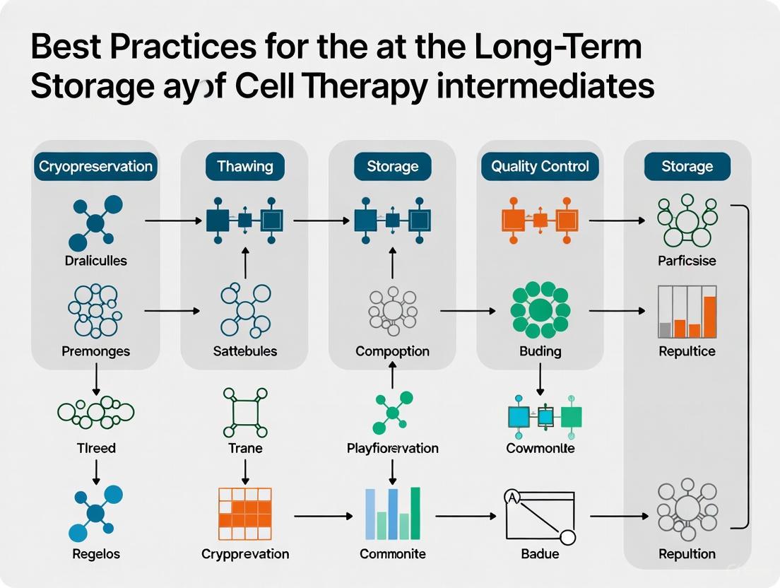
Overcoming the technical challenges requires a rigorous, scientifically-validated approach to storage. The following workflow outlines the critical stages from sample preparation to retrieval, highlighting key control points.
Diagram 1: End-to-End Workflow for Stable CGT Sample Storage
Validated Cryopreservation Protocols
A standardized freezing protocol is critical for maximizing post-thaw viability. The consensus best practice involves:
- Controlled-Rate Freezing: Using a rate of -1°C per minute to allow for sufficient cellular dehydration, preventing lethal intracellular ice formation [3] [6]. This is often achieved using specialized freezing containers filled with isopropanol or controlled-rate freezers.
- Cryoprotectant Formulation: Cells are typically frozen in a medium containing 5-10% DMSO. To enhance post-thaw recovery, this is often combined with non-permeating CPAs like sucrose or trehalose and specialized media (e.g., HypoThermosol), which allow for a reduction in DMSO concentration and its associated toxicity [2] [4].
- Aliquoting to Minimize Freeze-Thaw Cycles: Samples should be aliquoted to avoid repeated freezing and thawing, which degrades nucleic acids, proteins, and cell viability [2].
Infrastructure and Monitoring
The storage infrastructure itself must be designed for resilience and precision.
- Ultra-Low Temperature Storage: CGT materials require storage at -80°C for some stable biologics or, more commonly, in the vapor phase of liquid nitrogen (-135°C to -196°C) for live cells and other sensitive intermediates to ensure long-term stability [2]. The use of frost-free freezers must be avoided.
- Continuous Monitoring and Disaster Recovery: Facilities must implement continuous temperature monitoring with automated alarms. Furthermore, a robust disaster recovery plan with redundant power systems is no longer optional; regulators now require proof that such procedures are tested and effective [2].
- Equipment Qualification: All storage equipment, including freezers, incubators, and monitoring systems, must be formally qualified and maintained under documented calibration and performance checks [2].
Ensuring Data Integrity and Traceability
For patient-specific therapies, data management is as crucial as temperature control.
- Digital Chain of Custody: Each transfer point must be logged digitally with comprehensive metadata, including sample ID, storage conditions, and any temperature excursions with associated corrective actions [1] [2].
- Regulatory Alignment: Systems must be built to meet rising expectations from the FDA, EMA, and MHRA, which now focus heavily on data integrity, requiring validated and secure electronic systems for monitoring and logging. Manual records are insufficient for modern inspection readiness [2].
Essential Research Reagents and Storage Solutions
The successful implementation of long-term storage strategies depends on a suite of specialized reagents and materials. The table below catalogs key solutions for researchers developing storage protocols.
Table 2: Research Reagent Solutions for CGT Storage
| Research Reagent | Function & Technical Specification | Application in CGT Storage |
|---|---|---|
| DMSO (Cell Culture Grade) | A permeating cryoprotectant that depresses the freezing point of water and reduces intracellular ice crystal formation. Typically used at 5-10% (v/v) concentration [3] [6]. | Standard CPA for most cell therapy intermediates, including CAR-T cells and iPSCs. |
| Controlled-Rate Freezer | Equipment that precisely controls cooling velocity (typically -1°C/min) to optimize cell dehydration during freezing [3]. | Provides reproducible, scalable freezing for research and GMP-compliant manufacturing. |
| Liquid Nitrogen Biorepository | Long-term storage system maintaining temperatures of -135°C to -196°C in the vapor phase to preserve cells in a state of metabolic arrest [2] [6]. | Gold-standard for long-term, stable storage of master cell banks, viral vectors, and final drug product. |
| GMP-Grade Cryobags/Vials | Sterile, validated containers designed to withstand ultra-low temperatures and ensure sample integrity during storage [2]. | Primary container for final drug product and critical intermediates; essential for chain of custody. |
| HypoThermosol or Other Stabilizing Media | Specialized, intracellular-like preservation media designed to enhance cell viability and function during hypothermic storage and post-thaw [2]. | Used to reduce DMSO toxicity and improve post-thaw recovery rates of sensitive cell types. |
Regulatory Framework and Future Directions
The regulatory landscape for CGT storage is evolving rapidly, with agencies emphasizing a risk-based approach and data-driven oversight.
- Evolving Global Scrutiny: Key regulatory trends include the EU GMP Annex 1 (2022 revision), which introduces a Contamination Control Strategy (CCS) that implicates storage zones connected to aseptic processes [2]. The FDA has increased its focus on real-time monitoring, validated storage systems, and data-driven chain-of-custody evidence [2].
- Validation and Documentation: Simply having Standard Operating Procedures (SOPs) is insufficient. Regulators expect equipment qualification, validation of storage systems, and detailed documentation of all processes, including temperature excursion logs and corrective actions [2].
Looking ahead, innovation is focused on simplifying and de-risking the storage paradigm. Key future directions include:
- DMSO-Free Cryopreservation: There is a critical need to explore Me₂SO-free cryopreservation methods, particularly for therapies using novel administration routes (e.g., intracerebral, intraocular) where DMSO toxicity is a greater concern [3]. Optimizing freezing profiles for these alternative CPAs is an active area of research.
- Ambient Temperature Transport: Research into ambient transport, using hydrogel encapsulation to provide nutrient, oxygen, and structural support, aims to circumvent the logistical and cellular drawbacks of cryopreservation entirely [4].
- Decentralized Manufacturing: To overcome supply chain bottlenecks, the industry is exploring patient-adjacent, regionalized manufacturing. This model reduces transportation times and storage complexity but requires harmonized, high-quality storage protocols at the point-of-care [7] [5].
Long-term storage is a strategic enabler, not a secondary concern, in the development of cell and gene therapies. The ability to reliably preserve the viability, functionality, and identity of irreplaceable cellular products from vein to vein is fundamental to transforming scientific innovation into reliable and accessible medicines. As the CGT pipeline continues to expand, a proactive, compliant, and globally consistent storage strategy—supported by rigorous science, robust infrastructure, and digital traceability—will be a defining factor in successfully bringing these transformative treatments to patients worldwide.
Cryopreservation is an indispensable technology for the long-term storage of cell therapy intermediates, enabling the decoupling of manufacturing from treatment schedules and ensuring the availability of viable, functional cellular products [8] [9]. The process involves cooling biological samples to ultra-low temperatures (typically at or below -140°C) to halt all biochemical activity [10]. However, the journey to and from these temperatures subjects cells to a series of severe biophysical and biochemical stresses that can compromise cellular viability, recovery, and therapeutic efficacy [11] [10]. For cell therapy products, which often consist of sensitive primary cells, suboptimal cryopreservation can lead to reduced product potency, increased batch-to-batch variability, and potential clinical failure [8] [12].
Understanding the cellular stress response during cryopreservation is therefore fundamental to developing robust protocols for cell therapy intermediates. This guide examines the key biophysical and biochemical challenges encountered during cryopreservation, focusing on their impact on cellular integrity and function. It further details methodologies for assessing cryo-injury and outlines strategies grounded in current research to mitigate these stresses, providing a technical foundation for researchers and drug development professionals working to optimize preservation protocols for advanced therapeutic products.
Fundamental Biophysical Stressors
The process of cryopreservation exposes cells to a sequence of potentially lethal biophysical events. These stressors are primarily physical in nature, arising from phase changes, osmotic imbalances, and thermal extremes.
Ice Formation and Its Mechanical Consequences
The formation of ice crystals is a primary cause of cryo-injury. When extracellular solutions freeze, ice crystals form, excluding solutes and leading to a freeze-concentrated, hypertonic solution [13]. This phenomenon imposes a dual threat:
- Intracellular Ice Formation (IIF): If cooling occurs too rapidly, water does not have sufficient time to exit the cell before reaching intracellular supercooling conditions, leading to lethal intracellular ice formation [11]. Ice crystals mechanically disrupt organelles and the plasma membrane, causing immediate necrotic cell death [13] [10].
- Extracellular Ice Formation: The growth of extracellular ice can mechanically crush cells in confined spaces, a particular concern in dense tissues and three-dimensional constructs [14].
Osmotic Stress and Volume Dysregulation
As extracellular ice forms, dissolved solutes become concentrated in the remaining liquid phase, creating a hyperosmotic environment [13]. This imbalance drives water out of the cell, causing excessive cell shrinkage and subsequent membrane damage [10]. The process is reversed during thawing; as ice melts, the extracellular environment becomes temporarily hypotonic, causing a rapid influx of water that can lead to cell swelling and even lysis if not properly controlled [13] [11]. The addition and removal of cryoprotective agents (CPAs) further compound these osmotic challenges, as these permeating compounds themselves alter intracellular osmolarity and cause significant cell volume fluctuations [9].
Table 1: Key Biophysical Stressors in Cryopreservation
| Stress Category | Underlying Cause | Primary Consequence | Resultant Cell Injury |
|---|---|---|---|
| Intracellular Ice Formation | Rapid cooling prevents water efflux | Ice nucleation inside the cell | Mechanical disruption of membranes and organelles |
| Solution Effects | Slow cooling and solute concentration | Multimolar solute exposure & severe dehydration | Protein denaturation; membrane damage |
| Osmotic Shock | Improper CPA addition/removal | Excessive cell shrinkage or swelling | Membrane rupture; loss of cytoskeletal integrity |
| Chilling Injury | Temperature reduction from room temp to nucleation point | Membrane phase transition | Altered membrane permeability; ion flux imbalance |
Figure 1: Biophysical Stress Pathway. This diagram illustrates the critical branching pathway during cooling, where the cooling rate determines the dominant mechanism of injury—either slow cooling leading to solution effects or rapid cooling causing intracellular ice formation.
Biochemical Stress Pathways
In addition to physical damage, cells undergoing cryopreservation experience profound biochemical stresses that can trigger activation of programmed cell death pathways and oxidative damage, often manifesting as delayed-onset cell death hours or even days after thawing [11] [9].
The Hypothermic Continuum and Metabolic Imbalance
The period of cooling before freezing represents a "hypothermic continuum" where the cell's function is suppressed but does not cease until the glass transition temperature is reached [11]. During this continuum, a cascade of damaging events occurs:
- Ion Pump Failure and Acidosis: The failure of membrane-bound ATPases leads to an influx of sodium and calcium ions and an efflux of potassium, disrupting electrochemical gradients [11]. Anaerobic metabolism takes over, causing intracellular acidosis with pH levels potentially falling to as low as 4.0 [11].
- Membrane Phase Transitions: A reduction in temperature causes the cell membrane to transition from a liquid-crystalline state to a solid gel state, increasing membrane permeability and causing leakage of hydrolases from lysosomes and lipoproteins [11].
Oxidative Stress and Apoptosis
The rewarming phase reactivates metabolic processes and can trigger a burst of reactive oxygen species (ROS) production, overwhelming the cell's antioxidant defenses and leading to oxidative damage of lipids, proteins, and DNA [13] [11]. This oxidative stress, combined with other insults, can activate the mitochondrial apoptosis pathway [11]. Key events include:
- Mitochondrial Permeability Transition: The membrane potential of mitochondria can collapse during freezing, leading to the release of pro-apoptotic factors such as cytochrome c [11].
- Caspase Activation: The execution phase of apoptosis is mediated by caspase proteases, which are activated post-thaw, leading to controlled cellular dismantling [11] [9]. This delayed apoptosis is a significant cause of cell loss in therapeutic products, often peaking 12-36 hours post-thaw [11].
Table 2: Major Biochemical Stress Pathways in Cryopreservation
| Biochemical Pathway | Inducing Stressors | Key Molecular Markers/Events | Temporal Onset Post-Thaw |
|---|---|---|---|
| Apoptosis | Mitochondrial membrane damage, Caspase activation | Cytochrome c release, PS externalization, DNA fragmentation | Delayed (12-36 hours) |
| Necrosis | Severe membrane damage, ATP depletion | Loss of membrane integrity, cellular swelling | Immediate to 6 hours |
| Oxidative Stress | ROS burst upon reperfusion | Lipid peroxidation, Protein carbonylation | Early (0-6 hours) |
| Metabolic Shock | Ion pump failure, Acidosis | Intracellular Ca²⁺ influx, pH drop to ~4.0 | During cooling/rewarming |
Figure 2: Biochemical Stress Network. This diagram outlines the interconnected biochemical pathways activated during the hypothermic continuum and upon rewarming, culminating in delayed-onset cell death—a critical concern for cell therapy recovery.
Assessment Methodologies for Cryo-Injury
A comprehensive assessment of cryopreservation outcomes is vital for protocol optimization. It requires moving beyond simple viability metrics to evaluate functional recovery and the extent of specific stress pathway activation.
Viability and Membrane Integrity Assays
- Trypan Blue Exclusion: A standard, rapid method to assess plasma membrane integrity immediately post-thaw. Dead cells with compromised membranes take up the blue dye.
- Flow Cytometry with Propidium Iodide (PI) / Annexin V: This dual staining is crucial for distinguishing between live (PI-/Annexin V-), early apoptotic (PI-/Annexin V+), late apoptotic (PI+/Annexin V+), and necrotic (PI+/Annexin V-) cell populations at various time points post-thaw (e.g., 2 hours, 24 hours) to capture delayed apoptosis [11] [9].
- Lactate Dehydrogenase (LDH) Release: A colorimetric assay that quantifies the release of the cytosolic enzyme LDH into the supernatant, serving as a direct measure of membrane rupture and necrotic cell death.
Functional and Potency Assays
For cell therapy intermediates, functional recovery is the ultimate validation of a successful cryopreservation protocol. Assays must be tailored to the intended mechanism of action of the product.
- Metabolic Activity: Assays such as AlamarBlue or MTT, which measure cellular reduction potential, can be performed over several days to track metabolic recovery. However, results should be interpreted with caution as they can be influenced by cell number and metabolic rate.
- Cell-Specific Functionality:
- Immune Effector Cells (e.g., CAR-T, NK cells): Perform cytokine release assays (e.g., IFN-γ, IL-2) upon stimulation and measure specific cytotoxicity against target cells [8] [9].
- Stem Cells (e.g., MSCs, HSCs): Assess differentiation potential (osteogenic, adipogenic, chondrogenic for MSCs) and clonogenic capacity (CFU assays for HSCs) [13].
- Oxidative Stress Measurement: Use fluorescent probes like DCFH-DA to detect intracellular ROS levels immediately after thawing and at subsequent intervals.
Table 3: Experimental Reagents and Tools for Cryopreservation Research
| Reagent/Tool | Primary Function | Application Note |
|---|---|---|
| Dimethyl Sulfoxide (DMSO) | Penetrating cryoprotectant | Industry standard; typically used at 10% concentration; concerns over toxicity and clinical side effects drive research into alternatives [13] [9]. |
| Trehalose | Non-penetrating cryoprotectant | Naturally occurring disaccharide; stabilizes membranes via water replacement mechanism; used extracellularly or requires special loading techniques for intracellular delivery [13] [15]. |
| Propidium Iodide (PI) | Membrane integrity dye | Binds to DNA of cells with compromised membranes; used in flow cytometry for viability assessment. |
| Annexin V-FITC | Apoptosis detection | Binds to phosphatidylserine externalized on the surface of apoptotic cells; used with PI for cell death staging. |
| Plant Vitrification Solution 2 (PVS2) | Vitrification solution | Contains 30% glycerol, 15% ethylene glycol, 15% DMSO, 0.4M sucrose; used for vitrification protocols, particularly in reproductive cells [10]. |
| Controlled-Rate Freezer | Cooling rate control | Provides programmable, reproducible cooling; critical for optimizing and scaling cell therapy production [8]. |
Mitigation Strategies and Best Practices
Addressing the multifaceted stress response requires an integrated approach combining optimized biophysical parameters, advanced cryoprotectant strategies, and targeted biochemical interventions.
Optimization of Physical Parameters
- Cooling Rate Control: The single most critical factor. A slow, controlled rate of approximately -1°C/min is standard for many mammalian cells (e.g., MSCs, HSCs), as it allows sufficient time for cell dehydration without excessive solute exposure [13] [8]. However, some cells (e.g., oocytes, certain stem cells) benefit from rapid cooling or vitrification [13]. Systematic empirical testing is required for each cell type.
- Controlled Ice Nucleation ("Seeding"): Initiating extracellular ice formation at a defined temperature (e.g., -5°C) prevents massive supercooling, ensuring a more controlled and reproducible freezing process, reducing intra- and inter-batch variability [11] [9].
- Rapid Thawing: Thawing should be rapid (e.g., in a 37°C water bath) to minimize recrystallization and prolonged exposure to toxic, concentrated solutes. A common target warming rate is 45°C/min or higher [8].
Advanced Cryoprotectant Strategies
- CPA Toxicity Management: Minimize exposure time and temperature. CPA addition and removal should be performed at 0-4°C to reduce chemical toxicity [13]. Stepwise addition and removal can mitigate osmotic shock.
- Combined CPA Cocktails: Utilize lower concentrations of permeating CPAs (e.g., DMSO, ethylene glycol) in combination with non-permeating agents (e.g., trehalose, sucrose, hydroxyethyl starch) [13] [16]. This strategy reduces the toxicity associated with high concentrations of a single permeating CPA while still achieving the necessary vitrification properties [13] [13].
- Emerging Biomaterials: Ice-binding polymers and nanoparticles are being developed to inhibit recrystallization during thawing, thereby reducing mechanical damage [16]. Antifreeze proteins (AFPs) from extremophiles are also being investigated for their ability to modify ice crystal growth [15].
Targeting Biochemical Pathways
- Apoptosis Inhibition: The addition of caspase inhibitors (e.g., Z-VAD-FMK) to the freezing or post-thaw media has been shown to improve recovery of certain cell types, including stem cells and lymphocytes, by temporarily suppressing the apoptotic cascade [11] [9].
- Anti-oxidant Supplementation: Pre-incubation of cells with or inclusion of antioxidants (e.g., Trolox, N-acetylcysteine, glutathione) in the cryopreservation medium can scavenge ROS generated during the rewarming process, mitigating oxidative stress [13].
- Serum-Free and Xeno-Free Formulations: Move away from fetal bovine serum (FBS) due to its batch-to-batch variability, risk of immunogenicity, and potential contamination. Defined, serum-free cryopreservation media enhance reproducibility and regulatory compliance for clinical cell therapies [9].
The successful cryopreservation of cell therapy intermediates hinges on a deep understanding of the complex biophysical and biochemical stress responses that cells undergo during the process. The journey from room temperature to liquid nitrogen storage and back is fraught with challenges, from the mechanical threat of ice crystals and the osmotic stress of shifting solute concentrations to the activation of latent biochemical death pathways like apoptosis.
Mitigating these stresses requires a multi-faceted strategy. There is no universal solution; protocols must be meticulously optimized for each specific cell type and product configuration. This involves the careful selection and combination of cryoprotectants, precise control over cooling and warming rates, and the strategic inhibition of key stress pathways. As the cell and gene therapy field advances toward more complex products and larger-scale manufacturing, addressing these fundamental challenges in cryopreservation will be paramount to ensuring that these transformative therapies realize their full clinical potential, delivering consistent, potent, and safe products to patients.
For researchers and drug development professionals working with cell therapy intermediates, navigating the global regulatory landscape is a critical component of successful product development. Adherence to robust regulatory standards ensures not only the safety and efficacy of these advanced therapies but also their quality and consistency during essential long-term storage. This guide provides an in-depth analysis of the core regulatory frameworks—ICH, FDA, EMA, and the pivotal EU GMP Annex 1—within the specific context of storing cell therapy intermediates. It details practical methodologies, from stability studies to cold chain qualification, and provides a toolkit of essential materials, empowering scientists to design compliant and effective storage protocols for their research.
Advanced Therapy Medicinal Products (ATMPs), which include cell therapies, are medicines for human use based on genes, tissues, or cells [17]. The regulatory environment for these products is complex and rapidly evolving. In the European Union, the European Medicines Agency (EMA), through its Committee for Advanced Therapies (CAT), provides scientific recommendations and evaluates marketing authorization applications for ATMPs [17]. The U.S. Food and Drug Administration (FDA) plays a similar role in the United States, releasing new draft guidances in 2025 to address the unique challenges of cell and gene therapies, including expedited programs and post-approval safety monitoring [18].
A foundational element for quality assurance is Good Manufacturing Practice (GMP). In the EU, the central GMP guidance is detailed in EudraLex Volume 4, with specific, heightened requirements for sterile manufacturing outlined in Annex 1 [19] [20]. For cell therapy products, Chemistry, Manufacturing, and Controls (CMC) activities are paramount, encompassing process development, manufacturing, quality control, and the generation of data for clinical trial applications [17]. These activities ensure that intermediates and final products possess the necessary Critical Quality Attributes (CQAs) to remain safe and effective throughout their shelf life, including during long-term storage [17].
Core Regulatory Guidelines and Their Applications
Understanding the specific requirements of each regulatory body is essential for designing compliant storage protocols.
EU GMP Annex 1: Contamination Control for Sterile Products
The updated EU GMP Annex 1, which became fully applicable in August 2024, introduces a more strategic focus on contamination control, nearly quadrupling the length of the previous 2008 version [20]. Its principles are highly relevant to the aseptic processing and fill-finish stages of cell therapy products.
- Contamination Control Strategy (CCS): Annex 1 mandates a proactive, holistic CCS embedded into every stage of manufacturing [20]. For cell therapy storage, this means designing a strategy that covers the container closure system, storage environment (including gas phase for cryopreserved products), and handling procedures to prevent microbial and particulate contamination.
- Quality Risk Management (QRM): The revision emphasizes the use of QRM principles to identify and control risks [20]. A risk-based approach should be applied to determine critical control points in the storage and thawing process.
- Environmental Monitoring: The guidance provides enhanced descriptions for environmental monitoring, distinguishing between facility qualification and routine monitoring [20]. While directly applicable to production cleanrooms, the principle of monitoring and controlling the processing environment is critical for all steps where the product is exposed.
FDA and EMA Guidance for Cell and Gene Therapies
Both the FDA and EMA have developed specific guidance to address the unique nature of ATMPs.
- FDA's 2025 Draft Guidances: In 2025, the FDA released draft guidances on Expedited Programs, Postapproval Safety Monitoring, and Innovative Clinical Trial Designs for small populations [18]. These documents reflect a regulatory push to balance rapid access for serious conditions with robust, long-term data collection.
- EMA and CAT Oversight: The EMA's CAT is responsible for preparing draft opinions on ATMP marketing authorisation applications [17]. Their assessments heavily rely on the CMC data package, which must include comprehensive information on product characterization and stability [17].
- Global Collaboration: Initiatives like the FDA's Gene Therapies Global Pilot Program aim to increase regulatory harmonization and facilitate collaborative reviews with international partners like the EMA, which could streamline global development pathways [18].
Table 1: Key Regulatory Bodies and Their Relevance to Cell Therapy Storage
| Regulatory Body | Key Document/Framework | Primary Focus in Cell Therapy Storage |
|---|---|---|
| European Medicines Agency (EMA) | EudraLex Volume 4, GMP Guidelines [19] | Ensuring overall quality systems and manufacturing controls are in place for consistent production and storage. |
| EMA Committee for Advanced Therapies (CAT) | Scientific Recommendations on ATMP Classification [17] | Providing specialized evaluation of the quality, safety, and efficacy of cell-based therapies. |
| U.S. Food and Drug Administration (FDA) | 2025 Draft Guidances on CGT [18] | Outlining pathways for accelerated development and post-approval monitoring of safety and efficacy data. |
| EU GMP | Annex 1: Manufacture of Sterile Medicinal Products [20] | Establishing a contamination control strategy for aseptic operations, including final product formulation and filling. |
Experimental Protocols for Storage and Stability
Generating robust, data-driven evidence is a core requirement for regulatory submissions. The following protocols are essential for justifying storage conditions and defining product shelf life.
Stability Studies for Shelf-Life Determination
Objective: To determine the stability profile of the cell therapy intermediate under intended long-term storage conditions and to establish a validated expiration date [21].
- Methodology:
- Storage Condition Simulation: Store the intermediate in its final primary container (e.g., cryobag, vial) at the proposed storage temperature (e.g., -80°C, vapor phase of liquid nitrogen). Testing should also cover transient warming events that may occur during transfer between storage units [21].
- Time-Point Sampling: Establish a schedule for pulling samples for analysis (e.g., 0, 3, 6, 9, 12, 18, 24 months, and annually thereafter).
- Critical Quality Attribute (CQA) Testing: At each time point, test samples against pre-defined CQAs. For a cell therapy intermediate, this typically includes [17]:
- Viability and Potency: Using assays specific to the product's mechanism of action.
- Identity: Confirming the presence of specific cell surface markers or genetic signatures.
- Purity: Assessing levels of product-related impurities (e.g., cellular debris) and process-related impurities.
- Sterility and Mycoplasma: Ensuring the product remains free from microbial contamination.
- Data Analysis: Plot CQA data over time using statistical models (e.g., regression analysis) to determine the point at which any CQA falls outside its acceptance criteria, thus defining the product's shelf life.
Container Closure Integrity (CCI) Testing
Objective: To verify that the primary container closure system maintains its integrity and protects the product from contamination throughout the storage period and under transport conditions [21].
- Methodology:
- Test Method Selection: Choose a validated method suitable for the container type, such as dye immersion (with pressure differential) or high-voltage leak detection.
- Stress Testing: Expose the filled container closure system to simulated transport stresses, including vibration, compression, and drop tests, as per standards like ASTM D4169 [21].
- Integrity Challenge: After stress testing, perform the CCI test method to confirm the container closure system remains intact and prevents microbial ingress.
Cold Chain and Shipping Qualification
Objective: To qualify the entire shipping system (insulated container, refrigerants, packing configuration) to maintain the product within its specified temperature range for the maximum qualified duration [21].
- Methodology:
- Development Testing: Determine the optimal packing configuration (type and amount of refrigerant, product orientation) using ambient profiles representing extreme summer and winter conditions [21].
- Performance Qualification (PQ): Perform thermal mapping studies with the qualified pack-out configuration. The test duration should exceed the maximum expected transit time. Place temperature probes in locations representing the product's worst-case scenario.
- Simulated Distribution Test: Subject the assembled shipping package to a series of physical tests (drop, vibration, compression) per ASTM D4169 to confirm the system provides adequate physical protection [21].
- Route Verification: Conduct a mock shipment along the actual transit route to confirm acceptable performance in a real-world setting, accounting for seasonal variations [21].
The following diagram illustrates the logical workflow and relationships between these key regulatory concepts and experimental activities for cell therapy storage.
The Scientist's Toolkit: Essential Research Reagents and Materials
The following table details key materials and solutions required for the development and execution of robust cell therapy storage protocols.
Table 2: Essential Materials for Cell Therapy Storage Research
| Item | Function & Importance |
|---|---|
| Cryopreservation Solutions | Formulations containing cryoprotectants (e.g., DMSO) and base media to protect cells from ice crystal formation and osmotic stress during freezing and thawing, which is critical for maintaining viability and functionality [22]. |
| Validated Primary Containers | Cryogenic vials, bags, or other final product containers that have been validated for CCI at storage temperatures and are compatible with the product to prevent leachables/extractables and maintain sterility [21]. |
| Controlled-Rate Freezer | Equipment that provides a reproducible, optimized cooling rate to ensure consistent post-thaw recovery and batch-to-batch uniformity, a key regulatory requirement [22]. |
| Qualified Shipping Systems | Insulated shippers with appropriate refrigerants (e.g., dry ice, liquid nitrogen vapor) qualified to maintain the required temperature range for the maximum transit time [21]. |
| Temperature Data Loggers | Portable, calibrated monitoring devices placed inside shipping containers and storage units to provide verified temperature history, a crucial record for quality control and regulatory compliance [21]. |
| Cell-based Potency Assays | Functional bioassays that measure the product's biological activity, which is a critical quality attribute that must be monitored throughout stability studies [17]. |
Successfully navigating the global regulatory landscape for long-term storage of cell therapy intermediates demands a proactive and integrated strategy. It requires a deep understanding of the principles outlined in ICH, FDA, and EMA guidelines, with particular attention to the contamination control focus of EU GMP Annex 1. The path to regulatory compliance is built upon a foundation of robust, data-driven science. By implementing rigorous experimental protocols for stability, container integrity, and cold chain management, and by utilizing a well-characterized toolkit of reagents and materials, researchers and drug developers can ensure their cell therapy intermediates are stored with the quality, safety, and efficacy required to advance these transformative medicines to patients.
The successful development and commercialization of cell and gene therapies (CGTs) depend on a meticulously managed supply chain where storage conditions are not merely supportive but foundational to product integrity. Unlike traditional pharmaceuticals, cell therapy intermediates and final products are often living, biologically active materials whose viability, potency, and safety are exquisitely sensitive to storage parameters. Establishing defined storage requirements by material type is therefore a scientific and regulatory imperative for ensuring reproducible, high-quality outcomes in research and clinical applications.
This technical guide examines the storage landscape across the cell therapy workflow, from the initial acquisition of peripheral blood mononuclear cells (PBMCs) to the final engineered product. It synthesizes current best practices, validated protocols, and emerging technologies to provide a structured framework for safeguarding these high-value biological assets throughout their lifecycle. The principles outlined here are designed to help researchers and drug development professionals navigate the complex interplay between cryobiology, logistics, and regulatory compliance, thereby enhancing the reliability and translational potential of their cell therapy programs.
Foundational Materials: Storage of PBMCs and Primary Cells
Peripheral blood mononuclear cells (PBMCs) serve as a critical starting material for many autologous and allogeneic cell therapy pipelines. Their viability and functional recovery after storage are foundational to downstream manufacturing success.
Key Factors Influencing PBMC Viability and Recovery
Multiple factors during initial cell handling determine post-thaw recovery. Temperature management is crucial; blood transport at ambient temperature (15-25°C) for <24 hours post-collection best preserves cell integrity [6]. Prolonged storage at 2-8°C for over 24 hours can intensify granulocyte contamination in the PBMC fraction following density gradient separation [6]. Furthermore, donor variability remains one of the largest contributors to inconsistent cell recovery, necessitating standardized donor programs and handling methods to reduce variability from the start [6].
During isolation, using cold blood or reagents prevents red blood cell aggregation, leading to contamination of the PBMC fraction. Blood processed less than 24 hours after draw provides optimal separation results [6]. Even with leukopaks, which contain an enriched PBMC fraction, contaminating granulocytes (typically 3-10% of total cells) can be problematic for certain applications, potentially requiring additional purification steps [6].
Cryopreservation Methodologies for PBMCs
Cryopreservation enables long-term storage of PBMCs, but requires precise protocol execution to minimize cellular damage.
Table: Cryopreservation Media and Methods for PBMCs
| Component/Method | Standard Approach | Key Considerations |
|---|---|---|
| Cryoprotectant | 10% DMSO | Low toxicity at <10%; becomes toxic if left on cells too long pre-freezing [6]. |
| Base Medium | 90% FBS or serum-free commercial media (e.g., CryoStor CS10) | FBS raises concerns about lot-to-lot variability and potential infectious agents [23]. |
| Cell Concentration | 0.5 - 10 x 10⁶ cells/mL | Optimal concentration should be validated for specific applications [23]. |
| Freezing Rate | -1°C/minute | Controlled-rate freezing or isopropanol containers (e.g., Mr. Frosty) achieve this rate [6] [23]. |
| Long-Term Storage | Vapor phase liquid nitrogen (< -135°C) | Storage at -80°C is not recommended for long-term preservation [23]. |
The choice of cryopreservation medium involves a trade-off. While a lab-made formulation of 10% DMSO in FBS is common and cost-effective, serum-free, GMP-compatible alternatives like CryoStor CS10 eliminate lot-to-lot variability and risks associated with animal components [23]. A critical procedural note is to work efficiently once DMSO is added, as prolonged exposure at room temperature is toxic to sensitive cells, causing a decline in viability [6] [23]. After resuspending cells in cryoprotectant, they should be cooled at a controlled rate of approximately -1°C/minute, which can be achieved using a controlled-rate freezer or an isopropanol-based freezing container placed in a -80°C freezer [6] [23]. For long-term storage, transfer to vapor phase liquid nitrogen (-135°C to -196°C) is essential to maintain stability [2] [23].
Experimental Protocol: Cryopreserving PBMCs
Title: Protocol for Cryopreserving Purified PBMCs [23]
Principle: Cells are preserved in a cryoprotective medium and cooled at a controlled rate to minimize intracellular ice crystal formation and osmotic stress, enabling long-term storage in liquid nitrogen.
Materials:
- Cryopreservation medium (e.g., CryoStor CS10 or 10% DMSO/90% FBS)
- Cryogenic vials
- Isopropanol freezing container (e.g., Corning CoolCell, Mr. Frosty) or controlled-rate freezer
- Pipettor and tips
- Centrifuge
Procedure:
- Preparation: Ensure PBMCs are in a single-cell suspension. Centrifuge at 300 x g for 10 minutes to pellet cells. Carefully aspirate the supernatant.
- Resuspension: Resuspend the cell pellet in cold (2-8°C) cryopreservation medium at a concentration of 0.5 - 10 x 10⁶ cells/mL. Mix thoroughly.
- Aliquoting: Rapidly transfer 1 mL of the cell suspension into each labeled cryogenic vial.
- Freezing: Immediately place vials into an isopropanol freezing container and transfer the container to a -80°C freezer for overnight freezing. Note: If using DMSO/FBS, keep cells on ice and work rapidly to minimize DMSO exposure time.
- Long-Term Storage: The following day, transfer vials from the -80°C freezer to vapor phase liquid nitrogen for long-term storage. Minimize exposure to room temperature by using dry ice during the transfer.
Intermediate and Final Product Storage: Engineered Cells and Vectors
As materials progress through the manufacturing pipeline, their storage requirements evolve, often demanding more rigorous and validated conditions to ensure the stability of engineered attributes and final product function.
Defining Storage Conditions by Material Type
Different biological materials have distinct stability profiles, necessitating tailored storage conditions.
Table: Storage Conditions by Material Type in Cell and Gene Therapy
| Material Type | Temperature Range | Primary Rationale | Supporting Evidence |
|---|---|---|---|
| PBMCs (Cryopreserved) | -135°C to -196°C (Liquid Nitrogen Vapor Phase) | Long-term viability; prevents intracellular ice formation and metabolic decay [6] [2] [23]. | Transfer from -80°C to LN2 is critical; long-term -80°C storage is not recommended [23]. |
| Final Cell Therapy Product (e.g., CAR-T) | -135°C to -196°C (Liquid Nitrogen Vapor Phase) | Maintains viability and potency of living cells for years; required for most patient-specific "lot-of-one" products [2] [1] [24]. | Ultra-low temperatures keep cells below glass transition temperature for stability [1]. |
| DNA, RNA, Plasma, Proteins | -80°C | Slows degradation and preserves nucleic acid and protein integrity for extended periods [2]. | Standard for biobanking stable molecular analytes [2]. |
| Viral Vectors | -80°C or -135°C to -196°C | Depends on vector stability data; LN2 often preferred for long-term storage of sensitive viral preparations [2]. | Evolving data supports LN2 for master viral banks [2]. |
The storage requirements for final cell therapy products are particularly stringent. These living medicines must be stored at ultra-low temperatures, typically in the vapor phase of liquid nitrogen (-135°C to -196°C), to maintain viability and potency, sometimes for years [2] [1] [24]. This is non-negotiable for most autologous therapies, which are "lots of one" and irreplaceable [1]. The underlying cryobiological principle is that temperatures below the glass transition point (often < -130°C) arrest all metabolic activity and prevent damaging biochemical reactions, effectively placing the cells in a state of "suspended animation" [1].
Advanced Cryopreservation Technologies
Emerging freezing technologies aim to improve upon traditional slow-freezing methods. A 2025 study compared the standard slow-freezing (SLF) method for PBMCs with a method using an electromagnetic field (EMF) applied during freezing [25]. The results demonstrated that while the EMF method was equivalent to SLF in terms of viable cell count, viability, and cell activity, it offered a significant operational advantage: the shortest time required for freezing was drastically shorter with the EMF method (0.25 hours vs. 3 hours for SLF) [25]. This allows for much earlier transfer of PBMCs to the safety of liquid nitrogen, reducing the risk of viability decline associated with prolonged stays at -80°C and improving process consistency, especially for facilities processing many samples [25].
The Storage Ecosystem: Logistics, Packaging, and Validation
Securing cell therapy materials extends beyond the freezer, encompassing a complex ecosystem of packaging, transportation, and rigorous quality control to maintain an unbroken chain of suitable conditions.
Cold Chain Logistics and Shipping
Transporting cell therapies requires specialized logistics to maintain ultra-low temperatures. Cryogenic shippers using liquid nitrogen or dry ice are standard, with liquid nitrogen dewars capable of maintaining temperatures for up to 10 days, which is often necessary for international shipments [26]. A core challenge is that any deviation from the required temperature range can render the therapy non-viable, creating a "zero-margin-for-error" environment [1]. Furthermore, the entire process is underpinned by the need for a secure chain of custody and identity, especially for autologous products. Digital tracking technologies like RFID and telematics provide real-time oversight from collection to infusion, ensuring the right patient receives the right product [1] [26].
Packaging and Labeling for Ultra-Cold Storage
Packaging systems must withstand extreme temperature fluctuations from -190°C to 37°C [24]. This presents unique challenges, particularly for labeling. Primary package labels require special cryo-stable adhesives and stocks that remain adhered at liquid nitrogen temperatures [24]. Print quality must also withstand these conditions without smudging, and labels should have high color contrast to remain legible when covered in frost [24]. Secondary packaging, such as metal cassettes for bags or cartons for vials, must fit into specific racking systems within liquid nitrogen shipping containers and storage tanks [24]. All packaging components, including plastics and foams, must be validated for performance at cryogenic temperatures to avoid warping, shrinking, or becoming brittle [24].
Validation, Quality Control, and Regulatory Alignment
A proactive, validated approach is required to meet regulatory expectations and ensure sample integrity. Equipment and process validation is mandatory; all freezers, monitoring systems, and storage SOPs must be qualified and maintained under documented calibration schedules [2]. Container Closure Integrity (CCI) testing is essential to verify the packaging system maintains a sterile barrier against microbial contamination under extreme temperatures and physical stress [27]. Regulators are increasingly focusing on data integrity and real-time monitoring, requiring validated digital systems for logging storage conditions rather than manual records [2]. Finally, a comprehensive Contamination Control Strategy, as emphasized in the revised EU GMP Annex 1, must be applied not only in cleanrooms but also to any storage associated with aseptic processes [2].
The Scientist's Toolkit: Essential Materials for Cell Storage Research
Diagram Title: Cell Therapy Storage Workflow
Table: Essential Research Reagent Solutions for Cell Storage
| Reagent/Equipment | Function | Application Notes |
|---|---|---|
| Cryopreservation Media (e.g., CryoStor CS10) | Serum-free medium containing DMSO to protect cells during freezing/thawing. | Provides a defined, GMP-compatible alternative to FBS-based media, reducing variability [23]. |
| Controlled-Rate Freezer | Lowers cell temperature at a precise, optimal rate (e.g., -1°C/min). | Critical for minimizing intracellular ice formation; isopropanol containers offer a cost-effective alternative [6] [23]. |
| Liquid Nitrogen Storage Tank | Provides long-term storage at <-135°C (vapor phase) for viable cells. | Essential for preserving cell viability and functionality over many years [2] [23]. |
| Validated Cryogenic Vials | Secure containment for cells during storage and transport. | Must maintain container closure integrity (CCI) at ultra-low temperatures to prevent contamination [27] [24]. |
| Temperature Monitoring System | Tracks and logs temperature history during storage and transport. | Provides data integrity and alerts for temperature excursions; required for regulatory compliance [2] [26]. |
| Density Gradient Medium (e.g., Ficoll-Paque) | Isolates PBMCs from whole blood or apheresis product. | Must be used at room temperature for effective separation of blood components [6]. |
Defining and implementing precise, material-specific storage requirements is a cornerstone of successful cell therapy research and development. From the initial isolation of PBMCs to the final shipment of an engineered product, each stage demands a scientifically-grounded approach to cryopreservation, cold chain management, and quality control. The protocols and guidelines outlined here provide a framework for preserving the viability, identity, and functional potency of these invaluable biological materials.
As the field advances, so too will storage technologies and regulatory standards. Embracing standardized practices, leveraging robust and validated systems, and maintaining a focus on end-to-end sample integrity will be critical for translating innovative cell therapies from the research bench to reliable clinical applications. The strategic management of storage is not merely a logistical task but a fundamental discipline that underpins the entire cell therapy development pipeline.
The emergence of cell and gene therapies (CGTs) represents a paradigm shift in medicine, offering curative potential for a range of diseases. However, the living nature of these products introduces profound logistical challenges, making storage and stability a central factor in supply chain design [2] [1]. The viability of cell therapy intermediates—sensitive biological materials with limited shelf lives—is inextricably linked to the storage conditions and transportation logistics they undergo [2]. This interdependence forces a critical strategic decision: selecting a centralized or decentralized supply chain model. The former relies on large-scale, distant facilities, while the latter brings manufacturing and storage closer to the patient at the point of care (POC).
This technical guide analyzes how the stringent storage requirements of cell therapy intermediates dictate the feasibility, resilience, and cost-effectiveness of centralized versus decentralized logistics models. Framed within best practices for long-term storage research, this review provides researchers and drug development professionals with a foundational understanding of this critical trade-off, which impacts everything from process development to commercial viability.
Core Storage & Stability Requirements for Cell Therapy Intermediates
The integrity of cell therapy intermediates is paramount. These are not stable chemical compounds but living, dynamic biological systems that require meticulous, scientifically-validated storage conditions to maintain viability and function from collection to final administration.
Temperature and Cryopreservation
Most cell therapies require deep cryopreservation to halt biological activity and ensure long-term stability. The required temperatures are dictated by the product's sensitivity and the need to prevent ice crystal formation, which can cause irreversible cellular damage [2] [1].
- Ultra-Low Temperature (-80°C): Often used for storing DNA, RNA, plasma, and proteins [2].
- Cryogenic Temperature (-135°C to -196°C): Essential for preserving the viability of live cells and biologics. This is typically achieved using the vapor phase of liquid nitrogen, which offers superior temperature stability [2] [1]. Products must be kept below their glass transition temperature (often < -130°C) to remain stable for extended periods [1].
Cryoprotectants and Controlled Freezing
To mitigate freezing damage, cryoprotectant agents (CPAs) like dimethyl sulfoxide (DMSO) are routinely used, typically at concentrations around 10% [2]. The freezing process itself is critical. A controlled-rate freezing system, typically at a rate of ~1°C per minute, is a best practice to maximize post-thaw recovery [2]. Combining DMSO with specialized media (e.g., HypoThermosol) can further enhance cell viability upon thawing [2]. For clinical-grade materials, post-thaw washing is often necessary to remove DMSO and reduce its potential toxicity upon infusion [2] [1].
Handling and Contamination Control
Storage is not solely about temperature. Key handling practices include:
- Minimizing Freeze-Thaw Cycles: Repeated cycling can degrade nucleic acids, proteins, and cell viability. Aliquoting samples is a standard practice to avoid this [2].
- Aseptic Handling and Contamination Control: Following Good Manufacturing Practice (GMP) principles is essential. The revised EU GMP Annex 1 mandates a Contamination Control Strategy (CCS) that implicates any storage associated with aseptic processes, requiring sterile containers and validated cleanroom protocols [2].
- Chain of Identity and Custody: Each transfer point must be digitally logged with metadata, including sample ID, storage conditions, and any excursions, to ensure needle-to-needle traceability for patient-specific therapies [2] [1].
Analysis of Supply Chain Models
The strict storage requirements for cell therapy intermediates directly shape the design and operation of their supply chains. The core dilemma is choosing between a centralized model, which leverages economies of scale, and a decentralized model, which prioritizes proximity to the patient.
The Centralized Manufacturing & Storage Model
The centralized model is the established paradigm, where raw materials (e.g., a patient's cells from leukapheresis) are shipped to a large-scale, centralized facility for processing, storage, and then shipped back as a final product.
- Impact of Storage: This model is entirely dependent on a robust, long-distance cryogenic cold chain ("cryochain") [1]. The intermediates and final product must endure multiple legs of transportation in specialized shippers (e.g., liquid nitrogen dry vapor shippers) validated to maintain temperatures below -150°C for days [1] [26]. This necessitates significant investment in qualified packaging, real-time temperature monitoring, and "white-glove" logistics services [1]. Any deviation during transit can render the therapy non-viable, representing a total loss of a patient-specific dose [1].
The Decentralized (Point-of-Care) Manufacturing & Storage Model
The decentralized model proposes manufacturing and interim storage at or near the hospital or treatment center, drastically reducing or eliminating long-distance transport of the fragile final product.
- Impact of Storage: This model fundamentally alters the storage challenge. Instead of a complex transportation cold chain, the primary need shifts to localized ultra-low temperature storage infrastructure [28]. Each point-of-care (POC) unit requires on-site liquid nitrogen freezers or -150°C freezers, along with the technical staff to operate and maintain them under GMP standards [28]. The burden of storage moves from the logistics provider to the clinical site, simplifying transportation but complicating facility validation and quality control across a distributed network [28].
Quantitative Model Comparison
The choice between models involves trade-offs between cost, time, and operational complexity, all influenced by storage logistics. The table below summarizes a quantitative comparison based on discrete event simulation and industry analysis.
Table 1: Quantitative Comparison of Centralized vs. Decentralized Supply Chain Models for Autologous Cell Therapies
| Factor | Centralized Model | Decentralized (POC) Model | Key Insight |
|---|---|---|---|
| Cost per Treatment | Lower at small scale; better economies of scale [29]. | Higher operational overhead with many sites; can be competitive at high demand (500 patients/year) [29]. | Raw material costs are a major driver; decentralization savings from reduced transport are offset by network overhead [29] [28]. |
| Turnaround Time (TAT) | Longer due to transportation, packaging, and potential freeze-thaw [29]. | Consistently shorter; eliminates need for final product transport and freeze-thaw [29]. | Time savings may be insignificant in compact geographies with good transport, but critical for aggressive diseases [29] [28]. |
| Cold Chain Logistics | Highly complex; requires validated cryoshippers and 24/7 monitoring [1] [26]. | Greatly simplified for final product; only local handling required [28]. | A major source of risk and cost (up to 25% of commercialization costs) in the centralized model [1]. |
| Capital & Infrastructure | High cost concentrated in a few large facilities [28]. | High cost distributed across many POC units (equipment, validation, staffing) [28]. | Decentralized model trades transport capital for facility capital, losing economies of scale [28]. |
| Regulatory Harmonization | Single facility is easier to inspect and control [28]. | Complex; requires harmonized quality programs and assays across all sites [28]. | A significant barrier to decentralized models; MHRA and Spanish AEMPS are pioneering POC guidance [28]. |
Experimental Protocols for Storage and Stability Assessment
Robust, data-driven assessment of storage conditions is fundamental to validating any supply chain model. The following protocols outline key methodologies for evaluating the stability of cell therapy intermediates.
Protocol: Accelerated Stability Studies
This protocol is used to predict the long-term stability of cryopreserved cell therapy intermediates under recommended storage conditions [2].
- Sample Preparation: Prepare identical aliquots of the cell therapy intermediate using a standardized cryopreservation protocol (e.g., controlled-rate freezing with a defined CPA).
- Storage Conditions: Store aliquots at a minimum of three different temperatures (e.g., -80°C, -135°C, and -196°C). The vapor phase of liquid nitrogen (-135°C to -196°C) should be included as the intended storage condition.
- Time Points: Remove samples for analysis at predetermined time points (e.g., 0, 3, 6, 9, and 12 months). Real-time stability studies at the intended storage temperature are required for definitive shelf-life determination.
- Analysis: Thaw samples using a standardized protocol and assess:
- Viability: Using trypan blue exclusion or flow cytometry.
- Potency: Measure a relevant biological function (e.g., cytokine secretion, target cell killing for T-cells).
- Phenotype: Use flow cytometry to confirm the identity of the cell population.
- Sterility: Perform tests for mycoplasma, bacteria, and fungi.
Protocol: Cryogenic Shipping Validation
This protocol validates that the shipping system maintains required temperatures throughout the transit process.
- Shipping System Qualification: Select a qualified shipper (e.g., liquid nitrogen dry shipper) validated for a duration exceeding the maximum expected transit time.
- Thermal Mapping: Place calibrated temperature data loggers throughout the shipper, including the geometric center and points closest to the walls.
- Challenge Test: Perform a simulated shipment that covers the maximum duration. For international shipments, this should be validated for up to 10 days [26].
- Data Analysis: Download and analyze temperature data post-simulation. The system is validated if all mapping points remain within the specified temperature range (e.g., below -135°C) for the entire duration.
- Performance Qualification: Repeat the challenge test under seasonal extremes (summer and winter) to account for ambient temperature variations.
Protocol: Post-Thaw Recovery and Potency Assessment
This critical protocol evaluates the functional quality of the cell product after the freeze-thaw cycle, a key stress point in both supply chain models.
- Thawing: Rapidly thaw the cryopreserved vial in a 37°C water bath until only a small ice crystal remains.
- Dilution and Washing: Slowly dilute the cell suspension with a pre-warmed washing medium to reduce CPA toxicity. Centrifuge to remove the CPA-containing supernatant.
- Viability and Cell Count: Assess immediate post-thaw viability and total cell count.
- Functional Potency Assay:
- For CAR-T Cells: Co-culture the thawed cells with target antigen-positive cells at a specific effector-to-target ratio.
- Incubate for a defined period (e.g., 24 hours).
- Measure Outcome: Quantify target cell killing (e.g., via luminescence-based cytotoxicity assay) and/or cytokine secretion (e.g., IFN-γ via ELISA).
- Acceptance Criteria: The product batch must meet pre-defined release criteria for viability (e.g., >70%) and potency (e.g., >XX% specific lysis) to be deemed suitable for administration.
Visualizing the Storage-Driven Decision Pathway for Supply Chain Models
The complex relationship between storage requirements, product attributes, and the optimal supply chain model can be distilled into a logical decision pathway. The diagram below guides researchers through the key considerations.
Diagram 1: Storage and product-driven decision pathway for selecting a supply chain model. Autologous products with aggressive disease indications and sensitivity to freeze-thaw cycles are stronger candidates for decentralized models, provided GMP harmonization across sites is feasible. (Adapted from [28])
The Scientist's Toolkit: Essential Reagents and Materials for Storage & Stability Research
Research into the long-term storage of cell therapy intermediates requires a specific set of reagents and materials to ensure viability, stability, and data integrity. The following table details key solutions for this field.
Table 2: Essential Research Reagents and Materials for Cell Therapy Storage Studies
| Tool | Function/Application | Technical Notes |
|---|---|---|
| Cryoprotectant Agents (CPAs) | Protect cells from ice crystal formation during freezing and thawing. | DMSO is the most common (5-10% concentration). Toxicity requires post-thaw washing. Alternatives and combination media (e.g., HypoThermosol) are areas of active research [2] [1]. |
| Controlled-Rate Freezer | Ensures a reproducible, optimal freezing rate (~1°C/min) to maximize cell viability. | Critical for process standardization. Replaces unreliable manual freezing in -80°C freezers [2]. |
| Liquid Nitrogen Storage System | Provides long-term, stable storage at <-135°C (vapor phase) to -196°C (liquid phase). | The gold standard for preserving cell viability for years. Requires strict temperature monitoring and safety protocols [2] [1]. |
| Validated Cryogenic Shippers | Maintain cryogenic temperatures during transportation of intermediates/final product. | Liquid nitrogen dry vapor shippers are standard. Must be validated for temperature hold times exceeding maximum transit duration [1] [26]. |
| Temperature Data Loggers | Monitor and record temperature throughout storage and shipment. | Essential for validating storage conditions and investigating potential excursions. Data integrity is critical for regulatory filings [2] [26]. |
| Cell Viability & Potency Assays | Assess the impact of storage on cell health and biological function post-thaw. | Viability (e.g., flow cytometry); Potency (e.g., cytokine ELISA, cytotoxicity assays). Required for stability study endpoints and product release [2]. |
The landscape of cell therapy logistics is evolving rapidly. Several emerging trends will further reshape the interaction between storage science and supply chain models:
- Allogeneic Therapies: The commercial maturation of "off-the-shelf" allogeneic therapies will significantly simplify supply chains, moving them closer to traditional biologics models with less extreme storage pressures, though cryopreservation will remain important [30] [31].
- Advanced Analytics and AI: The integration of purpose-built inline analytics and AI will enable real-time process control and better prediction of storage-related outcomes, enhancing both centralized and decentralized model efficiency [32] [30].
- Standardization and Regulation: A strong industry push for standardized cryopreservation processes and GDP will drive efficiency and reliability. Simultaneously, regulatory bodies are increasing scrutiny on real-time monitoring, validated storage systems, and data integrity across the entire chain of custody [2] [30] [26].
In conclusion, storage is not a secondary consideration but a primary strategic driver in designing supply chains for cell therapies. The rigid storage requirements of these living products create a fundamental tension between the economic efficiency of centralized models and the operational resilience and speed of decentralized models. The optimal choice is not universal but must be determined by specific product characteristics, patient disease dynamics, technological readiness, and the evolving regulatory landscape. For researchers focused on long-term storage, the goal must be to develop robust, standardized protocols that not only preserve cell viability but also enable the flexible and scalable supply chains needed to deliver these transformative therapies to patients worldwide.
A Step-by-Step Protocol for Cryopreservation and Storage
Cryopreservation is a foundational technology enabling the advancement of cell and gene therapies by ensuring the long-term viability and functional integrity of cellular therapeutic products. This process allows for rigorous quality control testing, creates "off-the-shelf" availability, and facilitates the transport of living cells between manufacturing and clinical sites [33]. At the heart of successful cryopreservation are Cryoprotective Agents (CPAs), which protect cells from the lethal physical and chemical stresses induced during freezing and thawing. The selection of an appropriate CPA is therefore a critical determinant in the success of cell therapy manufacturing and clinical application.
The damage pathways during freezing are multifaceted. As cells cool, the formation of intracellular and extracellular ice crystals can mechanically disrupt cell membranes and organelles. Simultaneously, the concentration of solutes in the unfrozen fraction can lead to osmotic stress, membrane damage, and protein denaturation, a phenomenon collectively known as "solution effects" [16] [34]. Cryoprotectants mitigate these injuries through several mechanisms: they modify ice crystal structure, suppress ice nucleation and growth, promote the formation of a harmless glassy state (vitrification), and stabilize cellular structures by interacting with lipid bilayers and proteins [16] [34]. For cell therapy intermediates, the goal of cryopreservation is not merely to keep cells alive but to ensure they recover with their critical quality attributes—viability, phenotype, potency, and functionality—fully intact for subsequent manufacturing steps or direct administration.
DMSO: The Current Standard and Its Challenges
Properties and Mechanisms of Action
Dimethyl sulfoxide (DMSO) has been the predominant cryoprotectant for cellular therapeutics for over six decades, valued for its high efficacy and well-documented use in clinical protocols, particularly in hematopoietic stem cell transplantation [16] [35]. As a small, permeable molecule, DMSO readily crosses cell membranes. Its primary cryoprotective mechanism involves replacing water within the cell, thereby reducing the amount of intracellular ice that forms during cooling. It also depresses the freezing point of the solution and increases solution viscosity, which helps to mitigate the damaging "solution effects" caused by concentrated electrolytes [34]. The conventional concentration of DMSO used in slow-freezing protocols for cell therapies is 10% (v/v) [33] [2].
Documented Toxicity and Safety Concerns
Despite its effectiveness, DMSO is associated with significant drawbacks related to cellular and patient toxicity. These concerns are a major focus of debate in the field [33].
Cellular Toxicity: DMSO can induce time-, temperature-, and concentration-dependent toxic effects on cells. Documented impacts include:
- Mitochondrial Damage: Impairment of mitochondrial function has been observed in astrocytes and other cell types [35].
- Membrane and Cytoskeleton Effects: DMSO can interact with proteins and dehydrate lipids, compromising membrane integrity and increasing permeability, as seen in erythrocytes [35].
- Epigenetic Alterations: Repeated exposure to even sub-toxic levels of DMSO can alter the epigenetic profile of cells. For instance, it can interfere with DNA methyltransferases and histone modifiers in human pluripotent stem cells, potentially reducing their pluripotency and causing undesirable phenotypic changes [35].
- Induced Differentiation: The presence of DMSO in culture medium can trigger unwanted differentiation in stem cells, confounding research results and therapeutic outcomes [35].
Clinical Toxicity: When DMSO-cryopreserved cell products are administered to patients, the residual DMSO can cause mild to severe adverse effects. These are often dose-dependent, with 30-60% of hematopoietic stem cell transplant recipients experiencing reactions such as nausea, vomiting, hypotension, and, in rare cases, more severe cardiac or neurological events [36] [35]. A 2025 review analyzing 1,173 patients treated with DMSO-containing mesenchymal stromal cell (MSC) infusions found that the DMSO doses delivered were 2.5–30 times lower than the 1 g/kg typically accepted in stem cell transplantation. With adequate premedication, only isolated infusion-related reactions were reported, suggesting that with careful management, risks can be mitigated [33].
Table 1: Strategies for Mitigating DMSO-Associated Risks
| Strategy | Description | Considerations |
|---|---|---|
| Post-Thaw Washing | Removing DMSO from the cell product through centrifugation or filtration after thawing and before administration. | Can lead to significant cell loss due to the fragile nature of post-thaw cells and introduces additional agitation and osmotic stress [33] [35]. |
| Reduced Concentration | Using lower concentrations of DMSO (e.g., 2-5%) in combination with other protective agents. | Requires validation for each cell type; may compromise cryoprotection if not properly formulated [36] [34]. |
| Optimized Handling | Adhering to "slow freeze, quick thaw" principles and controlling temperature exposure during CPA addition/removal. | Minimizes prolonged exposure to toxic liquid-state DMSO; requires controlled-rate freezers for optimal results [34]. |
Protocol: Cryopreservation with a Novel Low-DMSO Formulation
Recent research demonstrates that DMSO concentration can be significantly reduced without sacrificing efficacy. The following protocol, adapted from a JoVE article, details the cryopreservation of Peripheral Blood Hematopoietic Stem Cells (PBHSCs) using a novel CPA containing only 2% DMSO, enabling storage at -80°C without liquid nitrogen [36].
Objective: To preserve PBHSCs using an ultralow DMSO CPA for clinical autologous stem cell transplantation.
Materials:
- Novel CPA: Composition includes 2% (v/v) DMSO, with other components not fully disclosed but typically includes base saline/nutrient medium and potentially macromolecules like hydroxyethyl starch.
- Traditional CPA (Control): 10% (v/v) DMSO + 5% human serum albumin.
- Cells: Mobilized peripheral blood hematopoietic stem cells.
- Equipment: Sterile biosafety cabinet, pipettes, 1.8 mL cryovials, programmable freezing box or -80°C freezer, 37°C water bath, centrifuge.
Procedure:
- Preparation: Work aseptically in a biosafety cabinet. Pre-cool an ice platform to 2-8°C. Label cryovials with sample ID, date, and cell count.
- Mixing CPA with Cells:
- Mix the novel CPA with the PBHSC sample at a 1:1 (vol/vol) ratio on the ice platform.
- For the control, mix the traditional CPA with a separate aliquot of cells at a 1:1 ratio.
- Cryopreservation:
- For Novel CPA Aliquots: Directly transfer the cryovials to a -80°C freezer for storage.
- For Traditional CPA Aliquots (Control): Transfer cryovials to a programmable freezer, cool at a controlled rate of 1°C/min to -80°C, and then transfer to liquid nitrogen for storage.
- Thawing and Assessment:
- After the storage period (e.g., 1 month), retrieve cryovials and thaw rapidly in a 37°C water bath with gentle agitation (≤ 5 minutes).
- Dilute the cell suspension and centrifuge at 300 × g for 10 minutes to remove the CPA.
- Resuspend the cell pellet in pre-warmed culture medium (e.g., RPMI 1640) for downstream assays.
Outcome: This protocol demonstrated that PBHSCs cryopreserved with the 2% DMSO novel CPA showed comparable survival (91.29% vs. 90.07%) and superior performance in cell viability assays (89.38% vs. 79.55%), mitochondrial activity, and cytoskeletal integrity compared to cells frozen with the traditional 10% DMSO formulation [36].
DMSO-Free Alternatives and Emerging Strategies
The documented challenges of DMSO have accelerated the search for effective DMSO-free cryopreservation strategies. While no single alternative has yet emerged as a universal replacement, several promising classes of compounds and supporting technologies are under development.
Classes of Alternative Cryoprotectants
Table 2: DMSO-Free Cryoprotectants and Their Applications
| Cryoprotectant Class | Examples | Proposed Mechanism of Action | Reported Application & Outcome |
|---|---|---|---|
| Sugars and Sugar Alcohols | Trehalose, Sucrose, Raffinose, Mannitol, Glycerol, 1,2-propanediol | Act as non-penetrating CPAs; stabilize membranes via water replacement; form vitrified matrices that inhibit ice crystal growth [33] [37] [35]. | Effective for MSCs, iPSCs, and probiotics when combined with other agents. Glycerol showed lower toxicity but slower membrane permeation [33] [37] [34]. |
| Polymers and Polyampholytes | Polyvinyl pyrrolidone (PVP), Polyvinyl alcohol (PVA), Polyampholytes, Carboxylated poly-l-lysine (COOH-PLL) | Inhibit ice recrystallization; increase solution viscosity; stabilize cell surfaces. Polyampholytes can show excellent cryoprotection with low toxicity [33] [16] [35]. | Polyampholyte CPA showed high viability for bone marrow MSCs with no impact on biological properties after 24 months at -80°C [35]. PVA (0.1 wt%) enabled significantly high post-thaw recovery of erythrocytes [35]. |
| Biomimetic and Bio-inspired Agents | Antifreeze Protein (AFP) mimetics (e.g., X-Therma's XT-Thrive) | Mimic natural antifreeze proteins from extremophiles; inhibit ice nucleation and recrystallization [38] [16]. | Formulations proposed as clinically safer alternatives for HSCs and other cell types; designed to be scalable and reproducible [38] [35]. |
| Amino Acids and Osmolytes | Glycine, Ectoine, Proline, Betaine | Act as osmoprotectants; help maintain osmotic equilibrium and stabilize proteins and membranes under stress [33] [37]. | Glycine was part of an optimal formulation for lyophilized probiotics [37]. Ectoine was used in cryopreservation of natural killer cells, maintaining viability and cytotoxic activity [35]. |
Enabling Technologies for DMSO-Free Cryopreservation
The efficacy of many non-penetrating CPAs is limited by their inability to cross the cell membrane. Advanced delivery techniques are being developed to overcome this barrier:
- Intracellular Delivery: Methods such as electroporation [33] [35] and nanoparticle-mediated delivery [33] [35] can be used to load sugars like trehalose directly into the cytoplasm, enhancing their cryoprotective effect and eliminating the need for toxic penetrating agents.
- Nanowarming: For vitrification protocols that use high concentrations of CPAs, rapid and uniform warming is critical to avoid devitrification (ice formation during thawing). Incorporating magnetic nanoparticles (e.g., Fe₃O₄) and using electromagnetic induction heating enables ultra-rapid warming, which has shown a threefold increase in viability for human MSCs vitrified with a DMSO-free solution [33] [35].
- Combination Strategies (Osmolyte-Based Solutions): Commercial and research-grade serum-free, DMSO-free freezing media often employ a synergistic combination of non-penetrating CPAs (sugars), osmoprotectants (e.g., ectoine), and polymers (e.g., poloxamer 188) to provide comprehensive protection against freezing stress [39] [35].
Protocol: Lyophilization of Probiotic Strains with a DMSO-Free CPA
While not a cell therapy, this protocol from a 2025 study exemplifies the systematic optimization of a DMSO-free formulation for preserving bacterial strains, a methodology translatable to other biological systems [37].
Objective: To optimize a cryoprotectant formulation for the lyophilization of probiotic strains (Bacillus, Lactobacillus, Staphylococcus) from chicken gut to maintain long-term viability and probiotic functionality.
Materials:
- Cryoprotectants: Glucose, Sucrose, Skim milk powder, Glycine, Dextran, Glycerol.
- Cells: Probiotic strains at the early stationary growth phase.
- Equipment: Centrifuge, lab-scale freeze dryer (e.g., OPERON FDB-5502), sterile cryovials.
Procedure:
- Cell Harvest and Concentration: Harvest bacterial cells by centrifugation (10,000 × g, 10 min, 4°C). Wash the pellet twice with sterile distilled water and concentrate it five-fold using phosphate-buffered saline (PBS, pH 7.4).
- Cryoprotectant Addition: Resuspend the concentrated cell pellet in the test cryoprotectant solution at a 2:1 ratio of cells to excipient. The optimal formulation identified was: 5% glucose, 5% sucrose, 7% skim milk powder, and 2% glycine.
- Freezing and Lyophilization:
- Aliquot the cell-CPA suspension and freeze at -80°C for 18 hours.
- Transfer the frozen samples to a pre-cooled freeze dryer.
- Lyophilize for 8 hours under a vacuum of 2 × 10⁻² Torr with a collector temperature of -50°C.
- Storage and Viability Assessment:
- Store the lyophilized powders at different temperatures (4°C, -20°C, -80°C) for up to 12 months.
- To assess viability, rehydrate the powder in PBS and perform standard plate counts or functionality assays.
Outcome: The optimized DMSO-free formulation, combined with storage at -80°C, provided the best protection, effectively reducing oxidative and gastrointestinal stress and preserving key probiotic traits like adhesion potential and antimicrobial activity over 12 months [37].
The Scientist's Toolkit: Key Reagents and Materials
Table 3: Essential Research Reagents for Cryopreservation Studies
| Reagent / Material | Function | Example Use Case |
|---|---|---|
| Dimethyl Sulfoxide (DMSO) | Penetrating cryoprotectant; reduces intracellular ice formation. | Standard 10% (v/v) solution for slow freezing of hematopoietic stem cells and MSCs [33] [36]. |
| Trehalose | Non-penetrating cryoprotectant; stabilizes membranes and promotes vitrification. | Key component in DMSO-free formulations for MSCs and iPSCs; often requires electroporation or nanoparticles for intracellular delivery [33] [35]. |
| Polyvinyl Alcohol (PVA) | Synthetic polymer; inhibits ice recrystallization. | Used at low concentrations (e.g., 0.1 wt%) to significantly improve post-thaw recovery of erythrocytes and other cell types [35]. |
| Hydroxyethyl Starch (HES) | Non-penetrating macromolecule; provides colloidal support and modulates ice growth. | Common component in clinical-grade low-DMSO CPAs for hematopoietic stem cells [36]. |
| HypoThermosol FRS | Hypothermic preservation medium; mitigates cold-induced stress and apoptosis. | Used to enhance post-thaw recovery by countering cold-induced cell damage during processing [2] [39]. |
| Rock Inhibitor (Y-27632) | Rho-associated kinase inhibitor; reduces apoptosis in dissociated stem cells. | Added to freezing and/or post-thaw media to improve the survival of sensitive cells like iPSCs [35]. |
| Serum-Free Freezing Media | Commercially available, chemically defined formulations (e.g., StemCell Keep, CryoScarless). | Provides a standardized, xeno-free environment for cryopreserving clinical-grade cell therapies [35]. |
The field of cryopreservation for cell therapies is in a dynamic state of transition. While DMSO remains the established standard due to its proven efficacy and deep-rooted use in clinical protocols, the compelling drivers of toxicity and patient side effects are pushing the industry toward safer alternatives.
The strategic selection of a CPA is a critical process decision. Researchers and developers must balance protocol maturity against safety and quality objectives. For many applications, the immediate path forward may involve the mitigation of DMSO-related risks through concentration reduction (e.g., to 2-5%) and protocol optimization [36] [34]. For others, especially new therapies where regulatory pathways are more flexible, the investment in developing a fully DMSO-free protocol using advanced cryoprotectants and enabling technologies could provide a significant long-term advantage.
Future developments will likely be shaped by several key trends: the continued discovery and synthesis of novel, low-toxicity cryoprotectants; the shift towards serum-free and chemically defined formulations for better reproducibility and regulatory compliance; and the integration of advanced technologies like nanowarming and intracellular delivery to make DMSO-free cryopreservation more robust and widely applicable [40] [16] [35]. As these innovations mature, the goal of a safe, effective, and universally applicable DMSO-free cryopreservation system for cell therapy intermediates appears increasingly attainable.
Controlled-rate freezing (CRF) represents a critical technological advancement in the long-term preservation of cell therapy intermediates, enabling the precise manipulation of cooling parameters to maximize post-thaw viability and functionality. Unlike conventional freezing methods that employ uncontrolled cooling rates, CRF systems allow researchers to program specific temperature profiles that mitigate the primary causes of cryoinjury—intracellular ice formation and osmotic shock. Within the context of cell therapy manufacturing, where products are often irreplaceable and must maintain stringent quality attributes, optimized cryopreservation protocols are not merely convenient but essential for commercial viability and regulatory compliance [41] [2].
The principle of "fast-slow-fast" cooling, while seemingly counterintuitive, aligns with the biophysical properties of living cells. The initial rapid cooling phase transitions the sample through a critical temperature range quickly to minimize chilling injury. The subsequent prolonged, slow cooling phase facilitates controlled dehydration, allowing water to exit the cell before freezing intracellularly. The final rapid cooling phase stabilizes the sample efficiently at cryogenic temperatures for storage. This nuanced approach underscores a fundamental understanding of cell-water interactions during phase change and their direct impact on cellular architecture and function [42] [43].
For drug development professionals, implementing a robust CRF protocol is a strategic imperative. It ensures the stability of intermediate products during manufacturing hold times, supports the creation of centralized cell banks, and facilitates the shipping of cellular material between sites without compromising critical quality attributes. As the cell and gene therapy market advances, the demand for reproducible, scalable, and validated freezing methods continues to grow, placing CRF at the heart of effective supply chain and quality management [2] [44].
The Science of the 'Fast-Slow-Fast' Principle
Biophysical Foundations of Cooling Rate Optimization
The "fast-slow-fast" freezing principle is engineered to navigate the two predominant mechanisms of cryoinjury: intracellular ice formation (IIF) and solute-induced toxicity or osmotic shock. During freezing, extracellular water crystallizes first, increasing the concentration of solutes in the unfrozen extracellular matrix. This creates an osmotic gradient that draws water out of the cell, a process necessary to avoid intracellular freezing but one that can also lead to harmful cell shrinkage and exposure to hypertonic conditions [42].
The slow cooling segment (-1°C/minute) is deliberately designed to balance these competing risks. A cooling rate that is too rapid does not provide sufficient time for water to osmotically efflux, resulting in lethal intracellular ice. Conversely, a cooling rate that is too slow subjects cells to prolonged exposure in a hypertonic environment, leading to "solution effects" injury or excessive dehydration. The -1°C/minute rate, widely cited as a standard for many mammalian cell types, represents a thermodynamic compromise, permitting enough cellular dehydration to suppress the intracellular freezing point below the current temperature without inducing excessive volumetric reduction [42] [43].
The initial fast cooling segment rapidly brings the sample from its physiological starting point to a temperature just above its freezing point, minimizing the time spent in a chilled but unfrozen state where some metabolic processes can still occur detrimentally. The final fast cooling segment, often initiated below -40°C, rapidly transitions the sample from the end of the slow freeze to the final storage temperature (e.g., -80°C or -196°C). This is because the majority of freezable water has already left the cell or has frozen, and the remaining amorphous vitrified state benefits from a rapid transition to a temperature where all biological activity is arrested [45].
Impact on Cellular Components and Viability
The consequences of ice formation are particularly catastrophic for sensitive cellular structures. Ice crystals can physically rupture the plasma membrane, nuclear envelope, and intracellular organelles, leading to immediate necrotic cell death upon thawing. Even if integrity is maintained, the mechanical stress on the cytoskeleton and membrane proteins can trigger apoptotic pathways post-thaw. For cell therapies, this translates not only to a simple loss in viable cell number but also to a potential decline in the functional potency of the product—a critical quality attribute (CQA) that must be preserved [46].
The success of the "fast-slow-fast" principle is therefore measured by more than just post-thaw viability counts. It is confirmed by the recovery of normal cellular function, such as the proper differentiation capacity of stem cells, the cytotoxic activity of T-cells in CAR-T therapies, or the metabolic profile of hepatocytes. The use of controlled-rate freezing, as opposed to passive freezing devices, provides a documented and reproducible environment, creating a foundation for validating the entire cryopreservation process as part of a cGMP workflow [41] [2].
Experimental Protocols for Protocol Optimization
Optimizing a controlled-rate freezing protocol requires a structured experimental approach to determine the specific cooling parameters that maximize recovery for a given cell type. The following methodology provides a template for a systematic investigation.
Methodology for Cooling Rate Profiling
Step 1: Cell Preparation and Experimental Setup
- Cell Source: Begin with a characterized cell bank of the therapy intermediate (e.g., human T-cells, MSCs, or iPSC-derived progenitors). Ensure cells are healthy, in the log phase of growth, and have a viability >90% pre-freeze [42].
- Freezing Medium: Use a clinically-grade, defined cryopreservation medium. A common base is CryoStor CS10, which contains 10% DMSO in a balanced salt solution, providing cryoprotection while avoiding the variability of serum-containing media [42].
- Experimental Groups: Divide cells into aliquots and subject them to different slow cooling rates: e.g., -0.5°C/min, -1.0°C/min, -1.5°C/min, and -2.0°C/min. Keep all other variables (cell concentration, freezing medium, fill volume) constant.
Step 2: Controlled-Rate Freezing Execution
- Loading: Aliquot the cell suspension (e.g., 1 mL at 1-10x10^6 cells/mL) into cryovials and place them in the controlled-rate freezer chamber [42].
- Programming the "Fast-Slow-Fast" Profile:
- Initial Fast Cool (4°C to -5°C): Rapidly cool at -10°C/min.
- Slow Cool (-5°C to -40/-50°C): Apply the variable test rate for this segment (e.g., -1.0°C/min). Include a brief hold (e.g., 5-10 minutes) at the nucleation temperature (e.g., -5°C) to ensure consistent, controlled ice formation if the freezer has a manual or automatic seeding function.
- Final Fast Cool (-40°C to -100°C): Rapidly cool at -10°C/min or the maximum rate [45] [43].
- Transfer: Immediately transfer cryovials to a liquid nitrogen vapor phase storage tank (-135°C to -196°C) for a minimum of 24 hours before thawing for analysis.
Step 3: Post-Thaw Analysis and Evaluation
- Thawing: Rapidly thaw samples in a 37°C water bath with gentle agitation until only a small ice crystal remains [42].
- Viability and Yield: Assess immediately using a trypan blue exclusion assay or an automated cell counter. Calculate post-thaw viability and total viable cell recovery.
- Functionality Assays: Perform assays relevant to the cell therapy's mechanism of action. This is critical, as viability alone is an insufficient metric [46]. Examples include:
- Flow Cytometry: For surface marker expression (identity/purity).
- Proliferation Assay: To measure growth potential.
- Potency Assay: A co-culture or cytokine release assay specific to the product's biological function.
Table 1: Key Parameters for Cooling Rate Profiling Experiments
| Parameter | Recommended Range/Specification | Rationale |
|---|---|---|
| Cell Concentration | 1x10^6 to 10x10^6 cells/mL [42] | Prevents low viability from over-dilution or cell clumping from high density. |
| Cryoprotectant | 10% DMSO in defined medium (e.g., CryoStor CS10) [42] | Standard effective concentration; defined medium ensures regulatory compliance. |
| Fill Volume | 1-2 mL in a 2 mL cryovial | Ensures consistent heat transfer. |
| Slow Cooling Rate | -0.5°C/min to -2.0°C/min (test variable) | Target range for optimizing dehydration of most nucleated cells. |
| Nucleation | Manual or automatic seed at ~ -5°C | Prevents supercooling and ensures consistent, extracellular ice formation. |
| Storage Temperature | ≤ -135°C (vapor phase LN₂) [2] | Halts all biochemical activity for long-term stability. |
Workflow Visualization
The following diagram illustrates the logical workflow and decision points for optimizing a controlled-rate freezing protocol, from initial setup to the final selection of the best parameters.
Essential Reagents and Equipment for Controlled-Rate Freezing
A successful and reproducible controlled-rate freezing process is dependent on the quality and consistency of the materials used. The following toolkit details the essential components.
Table 2: The Scientist's Toolkit for Controlled-Rate Freezing
| Category / Item | Specific Examples | Function & Importance |
|---|---|---|
| Programmable Controlled-Rate Freezer | Thermo Fisher Scientific, Planer, Asymptote [41] | Precisely executes the "fast-slow-fast" cooling profile; essential for protocol standardization and validation. |
| Defined Cryopreservation Medium | CryoStor CS10, mFreSR (for pluripotent stem cells) [42] | Provides cryoprotectants (e.g., DMSO) in a defined, serum-free formulation to protect cells and ensure regulatory compliance. |
| Cryogenic Storage Vials | Corning Internal Threaded Vials [42] | Sterile, leak-resistant containers for sample integrity; internal threads prevent contamination during storage in LN₂. |
| Long-Term Storage System | Liquid Nitrogen Tank (vapor phase, -135°C to -196°C) [2] [42] | Maintains sample stability by halting all metabolic and biochemical activity indefinitely. |
| Cryoprotectant Agent (CPA) | Dimethyl Sulfoxide (DMSO) [42] [43] | Penetrates cells, lowers freezing point, and reduces intracellular ice crystal formation. |
| Cell-Specific Media | MesenCult-ACF Freezing Medium (for MSCs), STEMdiff Cardiomyocyte Freezing Medium [42] | Specialized formulations optimized for the specific biophysical properties of different cell types. |
Troubleshooting and Quality Control
Even with a well-defined protocol, variations in process or materials can impact outcomes. A systematic approach to troubleshooting is key.
Table 3: Common Controlled-Rate Freezing Challenges and Solutions
| Problem | Potential Causes | Corrective Actions |
|---|---|---|
| Low Post-Thaw Viability | Suboptimal cooling rate; improper nucleation; toxic cryoprotectant concentration. | Profile different slow-cooling rates; implement a consistent seeding step; ensure correct DMSO concentration and use a defined medium [42] [43]. |
| Poor Functional Recovery | Cell stress during freezing/thawing damages functional pathways, not just membrane integrity. | Incorporate a functional potency assay (e.g., differentiation, cytokine secretion) as the primary metric for optimization, not just viability [46]. |
| High Inter-Batch Variability | Inconsistent starting cell health; deviations in sample prep or freezer performance. | Strictly control pre-freeze cell culture conditions; standardize all steps from harvest to vialing; qualify and calibrate the CRF unit regularly [44]. |
| Sample Contamination | Non-sterile techniques or compromised reagents during processing. | Use aseptic techniques; employ sterile, single-use reagents and internally-threaded vials; consider GMP-grade materials [2] [42]. |
Incorporating Hold-Time Validation
For cell therapy manufacturing, validating the stability of the intermediate product during the freezing process and any subsequent hold times is a regulatory expectation. This involves demonstrating that the product's CQAs remain within acceptable limits during the defined hold period before and after the freezing step [44]. A robust CRF protocol is the foundation upon which these hold-time validations are built, ensuring that the freezing event itself does not introduce variability that compromises the stability study.
Controlled-rate freezing, guided by the scientifically-grounded "fast-slow-fast" principle, is a cornerstone of modern cell therapy development. It transforms cryopreservation from a simple preservation step into a critical and controllable unit operation. By systematically optimizing cooling rates through rigorous experimentation that measures both viability and function, and by employing a defined toolkit of high-quality reagents and equipment, researchers and drug developers can ensure the long-term stability, potency, and safety of invaluable cellular therapeutics. As the field progresses, these validated and reproducible protocols will be indispensable for scaling up manufacturing, navigating global supply chains, and ultimately delivering effective treatments to patients.
The success of cell therapy research and development is fundamentally linked to the precise and stable preservation of cellular intermediates. These materials, which include genetically engineered intermediates and final cell therapy products, are not only irreplaceable but also highly vulnerable to degradation. Selecting an appropriate storage temperature is therefore not a mere logistical detail but a critical strategic decision that directly impacts cellular viability, genetic integrity, and the overall validity of research data. The two dominant standards for long-term storage are ultra-low temperature at -80°C and cryogenic preservation in the vapor phase of liquid nitrogen (LN2), typically ranging from -135°C to -196°C. This technical guide provides an in-depth comparison of these two core storage methodologies, framing the analysis within the context of establishing best practices for the long-term storage of cell therapy intermediates. The objective is to equip researchers and drug development professionals with the data and protocols necessary to make scientifically sound decisions that safeguard valuable samples and ensure the integrity of their developmental pipelines.
Technical Comparison of Storage Temperatures
The choice between -80°C and vapor phase LN2 storage is governed by profound differences in their underlying principles and their resulting impact on biological materials. Understanding these fundamental mechanisms is key to selecting the optimal preservation strategy.
Fundamental Principles of Low-Temperature Preservation
At a biochemical level, cooling slows down molecular motion, thereby decelerating the metabolic and chemical processes that lead to cell degradation and death. However, the critical factor for successful long-term preservation of viable cells is achieving a state of complete metabolic arrest. This state is reached below the glass transition point of water (Tg), which is approximately -135°C. Below this temperature, all biological activity effectively ceases, and water molecules do not rearrange to form larger, damaging ice crystals. Vapor phase LN2 storage, which operates between -135°C and -196°C, reliably maintains samples below this crucial threshold, enabling truly long-term storage. In contrast, -80°C storage lies above the Tg. At this temperature, biochemical processes are drastically slowed but not entirely halted, and some molecular mobility persists, which can lead to gradual degradation over extended periods.
Ice Crystal Formation and Cellular Damage
The formation of ice crystals is a primary cause of cell death during freezing and storage. When the freezing rate is slow, water outside the cell freezes first, causing water to exit the cell and leading to harmful cell shrinkage and membrane distortion—a process known as extracellular freezing. With rapid freezing, water does not have time to exit the cell, resulting in intracellular freezing, where ice crystals form inside the cell, physically damaging organelles and membranes. While controlled-rate freezing is designed to minimize this, the storage temperature itself influences crystal growth over time. At -80°C, the risk of small, residual ice crystals slowly growing or recrystallizing is higher than at the stable, ultra-low temperatures of vapor phase LN2, where all water is in a glassy, amorphous solid state.
The table below summarizes the core technical characteristics of each storage method.
Table 1: Fundamental Technical Characteristics of Storage Systems
| Parameter | Ultra-Low (-80°C) | Cryogenic (Vapor Phase LN2) |
|---|---|---|
| Typical Temperature Range | -60°C to -90°C | -135°C to -196°C |
| Relation to Tg of Water (~-135°C) | Above Tg | Below Tg |
| Metabolic Activity | Drastically slowed, but not fully arrested | Effectively arrested |
| Primary Physical Risk to Cells | Ice crystal recrystallization over long terms; temperature fluctuations | Intracellular ice formation during initial freezing phase |
| Theoretical Shelf Life | Years | Indefinite (theoretically) |
Decision Workflow for Storage Temperature Selection
The following diagram outlines a logical workflow for choosing between -80°C and vapor phase LN2 storage based on key sample and program characteristics.
Quantitative Data and Comparative Analysis
A data-driven comparison reveals clear trade-offs between the two storage systems in terms of stability, operational logistics, and cost.
Stability and Shelf-Life Data
The most significant differentiator is the shelf-life afforded by each method. Storage in the vapor phase of liquid nitrogen, at temperatures below -135°C, is the only method that can vastly extend product shelf life compared to ambient or refrigerated storage (i.e., years of storage compared to days of storage) [47]. More critically, it allows for the decoupling of the manufacturing schedule from the clinical use of the cells, as cells can be frozen almost indefinitely at these cryogenic temperatures [47]. This is a pivotal advantage for creating "off-the-shelf" allogeneic cell therapy products. While -80°C storage can preserve many samples for years, it is not considered suitable for the long-term storage of sensitive live cells and biologics, which require temperatures between -135°C and -196°C to maintain viability [2].
Operational and Safety Considerations
From an operational standpoint, each method presents distinct challenges.
- Contamination Risk: Vapor phase storage offers a significant advantage by minimizing the possibility of transmission via the liquid itself [48]. Certain viruses remain infectious in liquid nitrogen, making vapor phase the safer choice for preventing cross-contamination between samples [48].
- Sample Integrity and Safety: Vials stored in liquid phase risk explosion if LN2 enters the vial during storage, as it rapidly expands upon warming. Vapor phase storage circumvents this risk entirely [48]. Furthermore, vapor phase freezers reduce the risk of user injury from liquid nitrogen splash-back during retrieval [48].
- Temperature Uniformity and Control: Modern vapor phase freezers have largely overcome early issues with temperature gradients and can consistently maintain temperatures around -190°C at the top of the unit [48]. Some advanced models can even operate at a wider range (e.g., -20°C to -150°C), offering flexibility [48]. Ultra-low -80°C mechanical freezers are generally stable but are susceptible to temperature excursions during defrost cycles or power failures.
Table 2: Operational and Economic Comparison of Storage Systems
| Factor | Ultra-Low (-80°C) | Cryogenic (Vapor Phase LN2) |
|---|---|---|
| Theoretical Shelf Life for Live Cells | Limited (years) | Indefinite / Decades |
| Primary Operational Cost Driver | High electricity consumption | Periodic LN2 replenishment |
| Contamination Risk | Low (closed system) | Very Low in Vapor Phase; Higher in Liquid Phase [48] |
| Sample Access Safety | Safe | Safer (no LN2 splash risk) [48] |
| Infrastructure & Maintenance | Standard electrical maintenance | Requires LN2 supply chain and specialized containers |
| Hold Time during Power/LN2 Failure | Short (hours) | Long (several weeks for modern units) [48] |
Experimental Protocols for Storage Validation
Robust stability studies are essential for justifying the chosen storage condition for a specific cell therapy intermediate. These studies should be guided by regulatory frameworks such as ICH Q1A(R2) and tailored to the unique nature of the product [49].
Protocol for Stability Study Design
A comprehensive stability plan should be drafted, which includes the following elements [49]:
- Batch Selection: Use a minimum of three primary batches that are representative of the research or production material. For early-stage research, non-GMP or early GMP batches can provide valuable initial insights [49].
- Storage Conditions:
- Long-term: The intended storage condition (e.g., -80°C ± 5°C or Vapor Phase LN2 ≤ -135°C).
- Accelerated: For frozen products, 5°C ± 3°C or 25°C ± 2°C/60% RH ± 5% RH may be used to understand the impact of temperature excursions, though they are not officially defined for all conditions [49].
- Testing Timepoints: A typical schedule includes 0, 3, 6, 9, 12, 18, and 24 months, with annual testing thereafter.
- Stability-Indicating Attributes: Parameters must be tested at each timepoint to analyze trends. These should include [49]:
- Physical: Viability (e.g., post-thaw recovery), cell count, morphology, and appearance.
- Chemical: Potency (e.g., specific functionality assays), purity, and genetic stability.
- Microbial: Sterility or mycoplasma testing.
Key Methodologies for Assessing Cryo-Injury
Beyond standard quality control, specific experimental methodologies are critical for evaluating the impact of the storage protocol itself.
Post-Thaw Viability and Recovery Assay:
- Procedure: Thaw samples rapidly in a 37°C water bath or using an automated thawing device. Resuspend in culture medium, with or without a washing step to remove cryoprotectants. Perform cell counts using a trypan blue exclusion assay or an automated cell counter at specific time points post-thaw (e.g., 0 hours, 24 hours) to assess immediate recovery and subsequent growth.
- Data Interpretation: A high viability percentage immediately post-thaw indicates minimal acute cryo-injury. An increase in cell number over 24-72 hours demonstrates functional recovery and reproductive capacity.
Functional Potency Assay:
- Procedure: This is cell-type specific. Examples include:
- Differentiation Capacity: For stem cell intermediates, differentiate thawed cells and quantify the yield of target cells (e.g., dopaminergic neurons, cardiomyocytes) using flow cytometry or immunocytochemistry.
- Cytokine Secretion: For immune cells, stimulate with an appropriate activator and measure cytokine output via ELISA or multiplex assays.
- Target Cell Killing: For cytotoxic cells, co-culture with labeled target cells and measure specific lysis.
- Data Interpretation: Compare the functional output of thawed cells to that of a fresh, unfrozen control. A significant drop in potency suggests that the storage or freeze-thaw process has damaged critical cellular functions.
- Procedure: This is cell-type specific. Examples include:
The Scientist's Toolkit: Essential Reagents and Materials
The following table details key reagents and materials essential for conducting cryopreservation and storage validation experiments.
Table 3: Essential Research Reagent Solutions for Cryopreservation Studies
| Item | Function & Brief Explanation |
|---|---|
| Cryoprotective Agent (e.g., DMSO) | Penetrates cells to prevent intracellular ice crystal formation by binding water molecules. Standard concentration is 5-10% [3]. |
| Cryopreservation Media | A specialized solution containing salts, energy sources, and buffers, often combined with DMSO, to provide a protective environment during freezing and thawing. |
| Controlled-Rate Freezer | A device that precisely controls the cooling rate (typically ~1°C/min) to optimize dehydration and minimize lethal intracellular freezing [2]. |
| Cryogenic Vials | Sterile containers designed to withstand extreme thermal stress and prevent leakage at ultra-low temperatures. |
| Liquid Nitrogen Storage Dewar | Specialized vacuum-insulated container for long-term storage of samples in either the liquid or vapor phase of LN2. |
| Viability Assay Kit (e.g., Trypan Blue) | Allows for the differential counting of live (unstained) and dead (blue-stained) cells to quantify post-thaw recovery. |
| Cell Culture Media & Supplements | Essential for post-thaw washing (if required) and for resuspending cells to assess their recovery and functional capacity over time. |
Regulatory and Strategic Considerations for Cell Therapy Development
Adherence to regulatory guidelines and strategic planning of the storage workflow are critical for the successful translation of research into viable therapies.
Evolving Regulatory Landscape
Stability testing is critical for understanding product quality over time and is required for establishing shelf life [49]. The ICH Q1A(R2) guideline outlines expectations for stability data, and its principles are applicable to biological drugs, including cell-based therapies [50] [49]. Furthermore, the revised EU GMP Annex 1 emphasizes a Contamination Control Strategy (CCS) that implicates not only cleanrooms but any storage associated with aseptic processes, which is directly relevant for CGT developers working with viral vectors or sterile cell products [2]. Regulators are increasingly focusing on real-time monitoring, validated storage systems, and data-driven chain-of-custody evidence [2].
Integrating Storage into the Clinical Workflow
The storage strategy must align with the clinical administration plan. Many cell therapy products require a post-thaw wash to remove cytotoxic cryoprotectants like DMSO, especially when administering via novel routes such as direct injection into the brain, spine, or eye [3]. This "point-of-care postprocessing" complicates the workflow and introduces risks of contamination and human error [3]. Therefore, the strategic decision between a "just-in-time" fresh product, a cryopreserved product requiring washing, and a cryopreserved product in a safe-to-infuse medium must be made early. Cryopreservation in vapor phase LN2 is the only method that enables a true "off-the-shelf" model by decoupling manufacturing from treatment, a key factor for scalable allogeneic therapies [47] [3].
The choice between ultra-low (-80°C) and cryogenic (vapor phase LN2) storage is a fundamental one in cell therapy research. Vapor phase LN2 is unequivocally the superior option for preserving the long-term viability and functionality of sensitive cell therapy intermediates, as it halts all biological activity below the glass transition temperature of water and offers a theoretically indefinite shelf-life. Its advantages in preventing cross-contamination and ensuring sample integrity further solidify its position as the gold standard for master cell banks and other critical, irreplaceable materials.
However, -80°C mechanical freezers remain a viable and practical solution for a range of applications, including the storage of certain non-viable intermediates, plasmids, and some stable cell types for shorter durations. Their lower operational complexity and cost make them suitable for less sensitive materials or early-stage research where ultimate shelf-life is not the primary concern.
Future advancements are likely to focus on overcoming the current limitations of both methods. This includes the development of safer, DMSO-free cryopreservation media that are safe for direct administration, eliminating the need for post-thaw washing and simplifying the path to off-the-shelf therapies [3]. Innovations in cryogenic freezer design, offering better temperature control and reduced LN2 consumption, will also make vapor phase storage more accessible and efficient [48]. As the cell and gene therapy field matures, a deliberate, validated, and strategic approach to storage will be an indispensable component of successful and compliant drug development.
In the rapidly advancing field of cell and gene therapy, cryopreservation serves as a critical enabling technology, providing stability and extending the shelf-life of invaluable cell therapy intermediates [51] [52]. While significant attention is often devoted to optimizing freezing protocols, the thawing and post-thaw recovery processes are equally determinative for final product quality. Ineffective thawing can induce severe osmotic stress, intracellular ice crystal formation, and prolonged exposure to cytotoxic cryoprotectants, ultimately compromising cell viability, potency, and therapeutic efficacy [8] [53].
This technical guide provides an in-depth examination of thawing and post-thaw recovery best practices, framed within the broader context of long-term storage strategies for cell therapy products. By synthesizing current industry standards, clinical trial data, and emerging research, we present a scientifically-grounded framework for maximizing post-thaw recovery of sensitive cellular materials, with particular emphasis on mitigating the osmotic shock that frequently undermines cell viability and functionality.
Fundamental Principles of Post-Thaw Cell Recovery
The process of returning cryopreserved cells to a physiological state traverses a complex temperature range where multiple cellular damage pathways can be activated. Understanding these fundamental principles is essential for designing optimized recovery protocols.
Mechanisms of Cell Damage During Thawing
During thawing, cells face several critical dangers. As the temperature rises above approximately -135°C (the glass transition temperature of water), microscopic melting and recrystallization occur, whereby larger, more dangerous ice crystals grow at the expense of smaller ones [53]. This phenomenon is particularly detrimental when warming rates are suboptimal.
Simultaneously, as the extracellular ice melts, cells are suddenly exposed to a hypertonic environment containing high concentrations of cryoprotective agents (CPAs) like dimethyl sulfoxide (DMSO). The rapid influx of water into partially dehydrated cells causes severe osmotic stress, potentially leading to membrane rupture [53] [52]. Additionally, as the temperature continues to rise, CPA toxicity becomes a significant concern, particularly for DMSO, which exhibits increased cytotoxicity at temperatures above 0°C [3] [51].
The Critical Interplay Between Cooling and Warming Rates
The optimal warming rate is intrinsically linked to the cooling rate employed during the initial freezing process [8]. For cells that have been slowly cooled at approximately -1°C/min (the standard rate for many mammalian cell types), the established good practice for thawing involves rapid warming at rates of 45°C to 80°C per minute [8] [53]. This rapid warming minimizes the time cells spend in dangerous intermediate temperature zones where ice recrystallization and osmotic injury are most likely to occur.
Quantitative Analysis of Thawing Methods in Clinical Practice
Current clinical practices for thawing cell therapies vary significantly across different settings, from controlled manufacturing environments to bedside administration. The following table summarizes the predominant methods and their key characteristics based on recent clinical trial data and industry surveys.
Table 1: Thawing Methods and Parameters in Clinical Practice for Cell Therapies
| Method | Prevalence | Typical Warming Rate | Key Advantages | Reported Challenges |
|---|---|---|---|---|
| 37°C Water Bath | Universal in early clinical trials [54] | High (approximately 60-80°C/min) [53] | Rapid, readily available | Contamination risk, variable rate control, manual operation [8] |
| Controlled Thawing Devices | Increasing adoption in GMP settings [51] [8] | 45-100°C/min (programmable) [8] | Consistent, closed-system, GMP-compliant | Higher cost, specialized equipment required [52] |
| Bedside Thawing (Clinic) | Frequently used for final administration [8] | Variable, often unregulated | Direct to patient | Poorly regulated, staff training critical [8] |
The table illustrates a continuing transition within the industry from conventional water baths toward more controlled thawing technologies, particularly as therapies advance toward commercialization. This shift is driven by increasing recognition that thawing consistency is vital for maintaining critical quality attributes (CQAs) of cell-based therapies [8].
Comprehensive Thawing and Post-Thaw Processing Workflow
A systematic approach to thawing and post-thaw processing is essential for maximizing cell recovery. The following diagram and subsequent sections detail an optimized workflow from the initiation of thawing through final cell assessment.
Diagram 1: Comprehensive workflow for thawing and post-thaw processing of cell therapies, highlighting critical control points for minimizing osmotic shock and maximizing viability.
Controlled-Rate Thawing Methodology
The initial thawing phase requires rapid warming to minimize ice recrystallization. As shown in Diagram 1, this should be performed using a controlled-rate thawing device or water bath maintained at 37°C, achieving warming rates of 45-100°C per minute [8] [53]. The frozen product should be agitated gently to ensure uniform heat distribution until only a small ice crystal remains, indicating complete phase transition while maintaining cold temperature to reduce CPA toxicity.
For clinical-grade materials, particularly those in closed systems, controlled thawing devices are strongly recommended over water baths to eliminate contamination risks and improve process consistency [8]. These systems provide documented warming profiles that can be incorporated into quality control records, an increasingly important consideration as therapies advance toward commercialization.
Post-Thaw Processing and DMSO Removal Strategies
Once thawed, cells immediately face osmotic stress from the high CPA concentrations in the cryopreservation medium. Clinical protocols vary significantly in their approach to this challenge, as summarized in the table below.
Table 2: Post-Thaw Processing Methods for Cellular Immunotherapies in Clinical Trials
| Processing Method | Prevalence in Clinical Trials | Reported Applications | Key Considerations |
|---|---|---|---|
| Direct Infusion | Common for CAR-T cells, HSCs [54] [3] | Immediate administration after thawing | DMSO toxicity concerns limit dose; typically requires <1g DMSO/kg patient weight [51] |
| Dilution Before Infusion | Frequent for Tregs, some CAR-T cells [54] | Dilution in dextran/albumin solutions or saline | Reduces DMSO concentration gradually; may cause osmotic shock if not optimized [54] [52] |
| Centrifugal Washing | Standard for iPSC-derived therapies [3] | Requires dedicated cleanroom facilities | Effective DMSO removal but introduces shear stress and contamination risk [3] |
| Post-Thaw Culture | Used for specialized Treg applications [54] | 7-day expansion before second infusion | Allows functional recovery but adds complexity and cost [54] |
The selection of an appropriate post-thaw processing method depends on multiple factors, including cell type, cryopreservation medium composition, administration route, and clinical requirements. For protocols involving dilution or washing, the composition of the carrier solution is critical for minimizing osmotic shock. Solutions containing human serum albumin (HSA), dextran, or other osmotic balancers have demonstrated success in clinical trials [54].
Experimental Protocols for Assessing Post-Thaw Recovery
Rigorous assessment of post-thaw cell quality is essential for validating any thawing protocol. The following methodologies represent current best practices for comprehensive post-thaw analysis.
Viability and Functionality Assessment Protocol
Objective: To comprehensively evaluate post-thaw cell viability, functionality, and recovery quality beyond simple membrane integrity.
Materials:
- Post-thaw cell suspension
- Flow cytometer with appropriate capabilities
- Cell culture reagents (media, supplements)
- Functional assay reagents (depending on cell type)
Procedure:
- Immediate Viability Assessment (0-2 hours post-thaw):
- Perform cell counting using trypan blue exclusion or automated cell counters
- Calculate percentage viability based on membrane integrity
- Note: Dye exclusion methods alone are insufficient as they fail to detect apoptosis and metabolic damage [53]
Extended Functionality Assessment (24-48 hours post-thaw):
- Culture washed cells at appropriate density and conditions
- Monitor recovery of metabolic activity using assays such as MTT, ATP content, or resazurin reduction
- Evaluate recovery of cell-specific functions (e.g., cytokine secretion for immune cells, differentiation capacity for stem cells)
Apoptosis and Necrosis Profiling:
- Stain cells with Annexin V and propidium iodide (or similar markers)
- Analyze by flow cytometry to distinguish early apoptotic, late apoptotic, and necrotic populations
- This is particularly important as cryopreservation can induce delayed-onset apoptosis [51]
Interpretation: Successful cryopreservation should yield >70% immediate viability by membrane integrity and >50% recovery of metabolic activity and specific functions after 24 hours of culture. Significant disparities between immediate viability and functional recovery indicate suboptimal processing conditions.
Osmotic Stress Quantification Protocol
Objective: To specifically measure and quantify cellular osmotic stress following thawing procedures.
Materials:
- Post-thaw cell suspension
- Isotonic buffer (control)
- Hypotonic and hypertonic challenge solutions
- Membrane-impermeant fluorescent dyes (e.g., propidium iodide)
- Real-time cell analyzer or flow cytometer
Procedure:
- Baseline Membrane Integrity:
- Aliquot post-thaw cells into isotonic buffer
- Add membrane-impermeant fluorescent dye
- Measure fluorescence as baseline (F0)
Osmotic Challenge Test:
- Expose separate aliquots to carefully calibrated hypotonic and hypertonic conditions
- Monitor dye uptake over time (5-30 minutes)
- Calculate percentage increase in fluorescence compared to isotonic control
Recovery Capacity Assessment:
- After osmotic challenge, return cells to isotonic conditions
- Monitor continued dye uptake over 60 minutes
- Cells with better osmotic tolerance will show reduced additional dye uptake
Interpretation: Cells experiencing severe osmotic stress during thawing will demonstrate significantly increased membrane permeability under mild osmotic challenge. This sensitive method can detect sublethal osmotic damage not apparent in standard viability assays.
The Scientist's Toolkit: Essential Reagents and Equipment
Successful implementation of thawing and recovery protocols requires specific materials and equipment. The following table details essential components of a complete post-thaw processing toolkit.
Table 3: Essential Research Reagent Solutions for Post-Thaw Recovery
| Category | Specific Examples | Function & Application Notes |
|---|---|---|
| Controlled Thawing Devices | ThawSTAR (Medcision), VIA Thaw (Asymptote) [51] | Provide consistent, rapid warming at 45-100°C/min; closed-system designs reduce contamination risk |
| Dilution/Carrier Solutions | Plasma-Lyte A with HSA, 5% albumin with 10% dextran 40 [54] | Isotonic solutions for gradual DMSO dilution; protein components help stabilize cell membranes |
| Wash Media Formulations | CryoStor, HypoThermosol [2] | Specifically designed to reduce osmotic stress during CPA removal; often contain non-penetrating osmolytes |
| Viability Assessment Tools | Flow cytometry with Annexin V/PI, automated cell counters | Enable distinction between viable, apoptotic, and necrotic populations; superior to trypan blue alone |
| Cell Culture Media | Cell-type specific media with appropriate supplements | Support post-thaw recovery culture when required for functional restoration [54] |
Emerging Trends and Future Directions
The field of cell thawing and recovery continues to evolve with several promising developments on the horizon. DMSO-free cryopreservation formulations represent a significant area of innovation, with multi-osmolyte solutions demonstrating improved cell stability and reduced toxicity concerns [54] [3]. These advanced formulations could potentially eliminate the need for post-thaw washing, simplifying bedside procedures and reducing contamination risks [3].
Additionally, as cell therapies explore novel administration routes (including direct injection into the brain, heart, and eye), the tolerance for DMSO diminishes considerably due to site-specific toxicity concerns at concentrations as low as 0.5-1% [3]. This safety imperative is driving increased investment in alternative cryopreservation strategies that maintain cell quality while eliminating problematic cryoprotectants.
The growing adoption of automated, closed-system technologies for both freezing and thawing processes promises enhanced reproducibility and reduced contamination risk—critical considerations as therapies transition from clinical development to commercial distribution [8] [52]. These systems provide documented process parameters that can be incorporated into quality control systems and regulatory submissions.
Optimized thawing and post-thaw recovery protocols are indispensable components of successful long-term storage strategies for cell therapy intermediates. By implementing rapid, controlled thawing methods, carefully designed osmotic buffering strategies, and comprehensive assessment methodologies, researchers can significantly enhance the viability and functionality of recovered cells. As the cell and gene therapy field continues to mature, standardization and optimization of these critical processes will play an increasingly important role in ensuring consistent, reliable, and potent therapeutic products for clinical application.
The EU GMP Annex 1 revision, effective August 2023, establishes a mandatory, comprehensive framework for contamination control, formalizing the requirement for a holistic Contamination Control Strategy (CCS) for sterile medicinal products [55]. For cell therapy intermediates, which are often irreplaceable patient-specific materials, effective implementation of these guidelines is not merely a regulatory obligation but a critical component of patient safety and product efficacy [2]. These advanced therapy medicinal products (ATMPs) present unique challenges; they cannot undergo terminal sterilization and are highly sensitive to microbial, particulate, and chemical contamination due to their complex biological nature [56]. The revised Annex 1, therefore, demands a scientifically justified, risk-based CCS that is fully integrated into all operations, extending from raw material control to final product storage and shipment [57] [55]. This guide details the application of these principles specifically for the long-term storage of cell therapy intermediates, providing researchers and drug development professionals with the technical protocols and strategic framework needed to ensure product integrity from discovery to commercialization.
Understanding Contamination Risks and Regulatory Framework
In the context of cell therapy manufacturing and storage, contamination can be categorized into four primary types, each posing significant risks to product quality and patient safety [55]:
- Microbial Contamination: This includes bacteria, fungi, and viruses that can infiltrate products during aseptic processing. For cell-based products, which cannot be terminally sterilized, microbial introduction can compromise the entire therapy and pose severe infection risks to immunocompromised patients [55] [56].
- Particulate Contamination: Foreign inorganic or organic particles—such as fibers, dust, or metal fragments—can originate from equipment, personnel, or the environment. When introduced into injectable therapies, these particulates can cause embolisms, inflammation, or allergic reactions [57] [55].
- Chemical Contamination: Residual cleaning agents, disinfectants, or leachables from manufacturing equipment or storage containers can alter the safety, efficacy, and stability of sensitive cell therapy intermediates [55] [58].
- Cross-Contamination: The unintentional transfer of one product's traces to another is a critical risk in facilities handling multiple patient-specific lots. This underscores the need for rigorous segregation and cleaning protocols [57] [55].
The human element represents a predominant contamination vector. As noted in industry analyses, "Every step is a step where contamination can enter the process, because every step is a step where humans are actively involved in the process itself. Our very involvement is a risk to the product and to the patient" [56].
The EU GMP Annex 1 Contamination Control Strategy
Annex 1 defines the CCS as “A planned set of controls for microorganisms, endotoxin/pyrogen and particles, derived from current product and process understanding that assures process performance and product quality” [57]. This strategy is a proactive, holistic system that moves beyond isolated checks to an integrated, knowledge-driven framework [55].
The guideline outlines 16 core elements that a CCS must encompass, which are critically relevant to storing cell therapy intermediates [57]:
- Facility and process design
- Premises and equipment
- Personnel
- Utilities
- Raw material controls
- Product containers and closures
- Vendor approval
- Process risk management
- Process validation
- Sterilization process validation
- Preventative maintenance
- Cleaning and disinfection
- Monitoring systems
- Trending, investigation, and corrective and preventive actions (CAPA)
- Continuous improvement
Strategic Implementation of a Contamination Control Strategy
Developing a CCS: Structured Approaches
Several structured methodologies can be employed to develop a robust CCS. The PDA Technical Report 90 outlines a governance model with three interdependent quality system levels [57]:
- Individual Elements: Fundamental controls including facility design, equipment selection, material controls, and personnel training.
- Quality Processes: Systems for classifying and validating these individual elements to demonstrate their effectiveness.
- Monitoring Systems: Continuous monitoring of air, surfaces, water, and personnel to rapidly detect deviations.
Alternatively, the ECA Foundation recommends a three-phase approach mirroring process validation stages [57]:
- Phase 1 (CCS Development/Review): Involves process understanding, mapping, and risk analysis to identify contamination sources and necessary controls.
- Phase 2 (CCS Document Compilation): Consolidates all analyses and control rationales into a single, coherent CCS document.
- Phase 3 (CCS Assessment): Establishes a cycle for periodic review and continuous improvement.
For a practical, root-cause-based methodology, the 5M Approach (Ishikawa Diagram) structures the CCS around potential contamination sources: Manpower, Machine, Medium, Method, and Material [57]. This ensures a comprehensive examination of all potential risk areas.
Practical CCS Implementation for Cell Therapy Storage
Successful CCS implementation requires translating strategy into actionable, validated protocols. For long-term storage of cell therapy intermediates, this involves several critical components:
- Facility and Process Design: Storage areas must be designed with cleanability, unidirectional material flow, and segregation of high-risk activities as core principles. HVAC systems with appropriate pressure differentials are essential to reduce cross-contamination risks [59] [55].
- Equipment and Utilities: Storage equipment such as ultra-low temperature freezers and liquid nitrogen tanks must be designed for easy cleaning and maintenance. Their qualification and validation are fundamental to the CCS, providing evidence that control measures are effective and reproducible [59] [55].
- Personnel Practices: Given that personnel are a primary contamination risk, the CCS must mandate rigorous training in aseptic techniques, gowning procedures, and behavioral expectations for handling storage materials [59] [55]. Reducing human intervention through automation and closed systems further mitigates this risk [56].
- Cleaning and Disinfection: Validated procedures are vital. A key Annex 1 requirement is the separation of cleaning and disinfection steps; cleaning must effectively remove residues (e.g., disinfectants, product) to ensure subsequent disinfection is effective [58]. Residue removal is critical as residues can harbor microbial growth [58].
- Environmental and Process Monitoring: A data-driven monitoring strategy—covering air, surfaces, and personnel—enables early contamination detection. The CCS must define alert/action levels, trending methodologies, and investigation protocols for any excursions [59] [55].
Table 1: Key Contamination Vectors and Control Measures for Cell Therapy Storage
| Contamination Vector | Associated Risks | Control Measures |
|---|---|---|
| Personnel | Microbial shedding, improper aseptic technique [56] | Training, validated gowning procedures, reduced intervention through automation [59] [56] |
| Equipment & Surfaces | Particulate generation, chemical residues, microbial harborage [55] [58] | Preventive maintenance, validated cleaning/disinfection using residue-free protocols [59] [58] |
| Storage Environment | Non-viable particles, microbial ingress [57] | HVAC controls, particulate and microbial monitoring, cleanroom classification [59] [55] |
| Raw Materials & Containers | Introduction of contaminants, leachables [55] | Vendor qualification, incoming inspection and testing, compatibility studies [57] [59] |
Material Handling and Aseptic Techniques: Experimental and Operational Protocols
Aseptic Handling and Transfer Protocols
Maintaining asepsis during the handling and transfer of cell therapy intermediates is paramount. The following workflow details a critical, high-risk operation: the introduction of a vialed intermediate into a controlled storage unit.
Detailed Methodology:
- Surface Decontamination: Upon receipt, the external surfaces of the primary container (e.g., cryobag, vial) and any secondary packaging must be thoroughly wiped with a sporicidal agent (e.g., validated hydrogen peroxide-based disinfectant). The efficacy of the disinfectant and the wiping technique must be validated to ensure a 6-log reduction of spores [56] [58].
- Material Transfer: The decontaminated container must be transferred into the critical processing or storage area through a validated pass-through chamber or rapid-transfer port. This chamber should be equipped with interlocked doors and a biodecontamination system (e.g., Vaporized Hydrogen Peroxide) to sterilize the outer surfaces before opening the inner door [56].
- Aseptic Technique Verification: During the handling of the sterile container, viable particle monitoring must be performed using an active air sampler. This provides real-time data on the microbial quality of the critical zone and validates that aseptic techniques are maintaining a Grade A/ISO 5 environment. Settle plates should also be placed strategically to monitor over the duration of the operation [57] [55].
- Visual Inspection: Before storage, the container-closure system must be inspected for integrity. Checks include verifying the absence of cracks in cryovials, ensuring seals on cryobags are fully welded, and confirming no visible defects are present. This is a critical check to prevent leaks and contamination during long-term storage [2] [49].
- Storage Placement and Documentation: The intermediate is placed in a pre-designated, qualified storage location (ultra-low freezer or vapor-phase liquid nitrogen tank). This action, along with all previous steps, must be documented in a real-time electronic system to maintain a complete and auditable chain of custody and identity [2] [26].
Residue Management and Cleaning Validation
Effective residue management is a cornerstone of the revised Annex 1, which mandates separate processes for cleaning and disinfection to prevent disinfectant buildup that can harbor microbes [58]. The following protocol outlines a validated method for residue removal from critical surfaces.
Experimental Protocol: Validation of Residue Removal from Stainless Steel Surfaces
- Objective: To qualify a two-step, detergent-free cleaning process for removing dried disinfectant residues from stainless steel coupons, simulating cleanroom equipment and storage unit surfaces.
- Materials:
- Test Surfaces: 316L stainless steel coupons.
- Contaminant: A commonly used sporicidal disinfectant, marked with a traceable fluorescent dye.
- Cleaning System: Foamtec's Sahara+ system, comprising Sahara foam and microfiber wipers/mops [58].
- Solvent: 70% Isopropyl Alcohol (IPA) or Water for Injection (WFI).
- Analytical Instrument: Fluorometer for quantifying residual contaminant.
- Methodology:
- Coupon Preparation: Clean and baseline all coupons to ensure no initial fluorescence.
- Contamination: Apply a known volume and concentration of the doped disinfectant onto each coupon and allow to dry completely under controlled conditions.
- Cleaning Intervention:
- Step 1 (Dislodge): Wet the Sahara foam with the chosen solvent (70% IPA/WFI). Use it to gently scrub the entire coupon surface in a systematic pattern to dislodge the dried residue.
- Step 2 (Remove/Entrap): Use a fresh, dry microfiber wiper or mop face to thoroughly wipe the surface, entrapping and removing the loosened residue and solvent.
- Analysis: Use the fluorometer to measure the residual fluorescence on the coupon post-cleaning. Compare against pre-established acceptance criteria (e.g., ≤1 µg/cm²).
- Validation: Repeat the process across multiple lots and operators to establish robustness. The method is considered validated if it consistently meets the acceptance criteria and returns the surface to a visually clean state [58].
The Researcher's Toolkit: Essential Materials for Contamination Control
Table 2: Key Reagent and Material Solutions for Contamination Control
| Item | Function/Justification | Application Note |
|---|---|---|
| Sporicidal Disinfectant (e.g., H₂O₂-based) | Validated for 6-log sporicidal reduction; crucial for surface biodecontamination [56]. | Requires validation on specific facility isolates. Rotation with a second disinfectant of a different class may be needed to prevent resistance. |
| Sahara+ Foam & Microfiber System | Two-step system for effective residue removal without detergents; addresses Annex 1 mandate [58]. | The foam dislodges residues, the microfiber entraps and removes them. Effective with only WFI or 70% IPA as a solvent. |
| Viable Particle Air Sampler | Provides active, real-time microbial air monitoring during aseptic operations [55]. | Critical for verifying that the aseptic process is maintained at Grade A/ISO 5 during manual handling steps. |
| Cryoprotectant Agent (CPA) (e.g., DMSO) | Preserves cell viability during freeze-thaw by mitigating ice crystal formation [2] [1]. | Typically used at 5-10% concentration. Requires post-thaw washing for clinical-grade materials to reduce patient toxicity [2]. |
| Qualified Shipper & LN₂ Dewar | Maintains ultra-low temperatures (down to -196°C) during transport; validated for duration [1] [26]. | LN₂ dewars can hold ultra-low temperatures for up to 10 days, making them suitable for complex international shipments of cell banks [26]. |
Integrating CCS with Long-Term Storage of Cell Therapy Intermediates
Stability and Storage Considerations
Cell therapy intermediates are highly sensitive and require stringent storage conditions to maintain viability and functionality throughout their shelf-life. Key stability considerations include:
- Temperature Requirements: DNA, RNA, and proteins often require -80°C, while live cells and biologics typically need -135°C to -196°C (vapor-phase liquid nitrogen) for long-term viability [2]. These temperatures must be continuously monitored with alarms to prevent excursions.
- Minimizing Freeze-Thaw Cycles: Aliquotting samples is a critical best practice to avoid repeated freeze-thaw cycles, which degrade nucleic acids, proteins, and cell viability [2].
- Use of Cryoprotectants: Agents like DMSO (typically at 10%) are used for viable cell storage. The freezing rate must be controlled (approximately 1°C/min) using a validated rate-controlled freezing system to maximize post-thaw recovery [2] [1].
- Shelf-Life Determination: Stability studies are essential for assigning a shelf-life. These studies evaluate chemical, physical, and microbiological attributes over time at intended storage conditions. Real-time stability data from GMP batches is ultimately required for regulatory submission [49].
Data Integrity and Chain of Custody
Maintaining an unbroken chain of identity and custody is non-negotiable for patient-specific autologous therapies [1] [26]. Each transfer point must be digitally logged with comprehensive metadata, including sample ID, storage conditions, and any excursions or corrective actions [2]. Regulators increasingly demand validated, secure data systems for monitoring and logging storage conditions; manual or unverified systems are insufficient [2]. Digital tracking technologies like RFID and telematics enable real-time oversight and provide sustainable record-keeping through digital proofs of delivery [26].
The revised EU GMP Annex 1 elevates contamination control from a series of discrete checks to a strategic, scientifically-driven system integral to the success of cell therapy development. For researchers and professionals managing the long-term storage of cell therapy intermediates, this means adopting a holistic CCS that is deeply embedded across people, processes, and technology. By implementing robust material handling protocols, validating aseptic techniques, managing residues effectively, and integrating real-time monitoring with rigorous data management, organizations can protect the integrity of these invaluable biological assets. As the regulatory landscape continues to evolve, a proactive, knowledge-based approach to contamination control will not only ensure compliance but also build a foundation of quality and safety, ultimately accelerating the delivery of transformative therapies to patients.
Solving Common Challenges and Enhancing Process Robustness
For cell therapy intermediates, achieving high post-thaw viability is not merely a technical goal but a critical determinant of therapeutic efficacy and regulatory success. The "vein-to-vein" journey of these living products hinges on cryopreservation protocols that maintain cellular integrity throughout the storage and transport lifecycle. Within this framework, cryoprotectant toxicity and suboptimal freeze/thaw rates emerge as two dominant, interconnected factors compromising cell survival and function. As the cell and gene therapy (CGT) market advances rapidly, offering new hope for patients with previously untreatable conditions, the field remains one of the most complex in healthcare, especially concerning sample storage and stability [2]. This technical guide examines the mechanistic underpinnings of cryopreservation-induced damage and provides evidence-based, practical methodologies for optimizing recovery of precious cell therapy intermediates.
The Dual Challenge: Cryoprotectant Toxicity and Freeze/Thaw Rate Optimization
Mechanisms of Cryoprotectant Toxicity
Dimethyl sulfoxide (DMSO) remains the gold-standard cryoprotectant (CPA) for most cell therapy products due to its membrane-penetrating capability and effectiveness in suppressing ice crystal formation. However, its toxicity profile presents significant challenges for both product quality and patient safety.
- Concentration and Temperature-Dependent Toxicity: DMSO toxicity is markedly exacerbated at elevated temperatures. As cells are exposed to warmer conditions during addition or removal of cryoprotectants, or during transient warming events, the cytotoxic effects intensify [60]. This is particularly critical during the post-thaw wash phase before administration.
- Clinical Safety Profile: A 2025 review of DMSO safety in mesenchymal stromal cell (MSC) therapies analyzed data from 1,173 patients receiving 1–24 intravenous infusions of DMSO-containing MSC products. The study concluded that the DMSO doses delivered (typically 2.5–30 times lower than the 1 g DMSO/kg accepted for hematopoietic stem cell transplantation) did not present significant safety concerns with adequate premedication [33]. Despite this relative safety, concerns persist regarding infusion reactions and potential impacts on cell functionality.
- Impact on Cell Quality: Beyond immediate toxicity, DMSO exposure can trigger delayed onset cell death (DOCD), where cells that appear viable immediately post-thaw undergo apoptosis hours or days later due to cumulative stress [60]. This phenomenon underscores the importance of functional potency assays beyond simple membrane integrity tests.
Fundamental Principles of Freeze/Thaw Rate Impact
The rate at which cells are frozen and thawed directly influences both intracellular ice formation (IIF) and osmotic stress, creating a complex optimization challenge.
- Slow Freezing: Controlled-rate freezing at approximately -1°C/minute allows for gradual dehydration of cells, minimizing intracellular ice formation but potentially exposing cells to prolonged osmotic stress and CPA toxicity [42].
- Rapid Thawing: Rapid warming at rates exceeding 45°C/minute is crucial to avoid ice recrystallization—a process where small ice crystals merge into larger, more damaging structures during slow warming [8] [42]. This is particularly critical for sensitive cell types like T cells and iPSC-derived lineages.
Table 1: Comparative Analysis of Freezing Methodologies
| Parameter | Controlled-Rate Freezing | Passive Freezing |
|---|---|---|
| Cooling Rate Control | Precise control (~1°C/min) | Uncontrolled, variable |
| Process Consistency | High, suitable for GMP | Lower, higher variability |
| Infrastructure Cost | High (equipment, LN₂ consumption) | Low (freezing containers) |
| Technical Expertise | Specialized knowledge required | Low technical barrier |
| Batch Scaling | Potential bottleneck for scale-up | Easier to scale |
| Regulatory Alignment | Preferred for late-stage and commercial products | More common in early research |
Interplay Between Factors
The relationship between CPA toxicity and thermal history is not merely additive but synergistic. Suboptimal freezing rates can exacerbate CPA-related damage by prolonging exposure to concentrated solutions during slow freezing, while rapid freezing without adequate CPA protection causes intracellular ice damage. This creates a narrow "sweet spot" where both parameters must be simultaneously optimized for specific cell types [8] [42].
Investigating Cryoprotectant Toxicity: Experimental Approaches
DMSO Dose-Response and Exposure Time Studies
Objective: To quantify the relationship between DMSO concentration, exposure time, temperature, and cell viability/functionality.
Protocol:
- Cell Preparation: Prepare identical aliquots of cell therapy intermediates (e.g., CAR-T cells, MSCs) at a standardized concentration (typically 1×10⁶ cells/mL) [42].
- CPA Formulation: Create CryoStor CS10 or similar freezing media containing DMSO at concentrations: 5%, 7.5%, 10% (standard), and 12% [42].
- Temperature Conditions: Hold cell-CPA mixtures at three temperatures: 4°C (ideal), 22°C (room temperature), and 37°C (physiological, worst-case).
- Time Course: At each temperature, remove aliquots for analysis at time points: 0, 15, 30, 60, and 120 minutes.
- Assessment Metrics:
- Immediate viability (trypan blue exclusion or flow cytometry with 7-AAD)
- Delayed viability (24-hour post-incubation in culture)
- Functional assays (e.g., cytokine secretion, proliferation capacity)
- Apoptosis markers (Annexin V) at 24 hours
Expected Outcomes: The data will reveal critical thresholds for DMSO exposure at various temperatures, informing safe handling windows during pre-freeze and post-thaw processing.
Alternative Cryoprotectant Screening
Objective: To identify less toxic CPA alternatives or formulations that can reduce or replace DMSO.
Protocol:
- Test Formulations: Prepare freezing media containing:
- Freezing Protocol: Process cell aliquots using standardized controlled-rate freezing (-1°C/minute).
- Assessment: Compare post-thaw recovery to DMSO controls using comprehensive metrics:
- Viable cell recovery percentage
- Phenotypic characterization (surface markers)
- Potency/functional assays specific to cell type
- Genomic stability (for stem cell populations)
Table 2: Cryoprotectant Formulations and Their Efficacy
| Cryoprotectant Formulation | Cell Type Tested | Post-Thaw Viability | Advantages | Limitations |
|---|---|---|---|---|
| 10% DMSO (Standard) | MSCs, CAR-T cells | 70-90% (varies) | Gold standard, reliable | Known toxicity, requires washing |
| 300 mM Trehalose + 10% Glycerol + 0.001% Ectoine | Embryonic Stem Cells | 92% | Reduced toxicity | Complex formulation |
| 3% Trehalose + 5% Dextran 40 + 4% PEG | Adipose Tissue MSCs | ~95% | High viability | Uncommon components |
| 150 mM Sucrose + 300 mM EG + 30 mM Alanine | Embryonic Stem Cells | 96% | Excellent viability | Multiple components |
| Serum-Free Commercial Media | Various cell types | Comparable to DMSO | Defined composition, regulatory friendly | Proprietary formulations |
Optimizing Freeze/Thaw Rates: Methodologies and Tools
Controlled-Rate Freezer Parameter Optimization
Objective: To systematically determine optimal cooling rates for specific cell therapy intermediates.
Protocol:
- Instrument Setup: Utilize a controlled-rate freezer (CRF) capable of precise temperature profiling.
- Cooling Rate Matrix: Program CRF to test multiple cooling rates: 0.5°C/min, 1.0°C/min (commonly used), 1.5°C/min, and 2.0°C/min.
- Container Considerations: Test the same rates across different container systems (cryobags, vials) to assess container-specific effects.
- Temperature Monitoring: Implement distributed temperature sensors (thermocouples) at critical positions within samples to monitor thermal homogeneity [61].
- Nucleation Control: Implement consistent ice nucleation protocols (e.g., manual seeding at -5°C to -7°C) to minimize supercooling variability.
- Post-Thaw Analysis: Assess outcomes using cell-type-specific quality attributes beyond simple viability, including:
- Recovery efficiency (percentage of pre-freeze cell number)
- Potency measures (e.g., target cell killing for CAR-T)
- Activation markers (for immune cells)
- Metabolic activity
Data Utilization: The resulting data identifies the cooling rate that maximizes both survival and functionality for the specific cell type, informing Standard Operating Procedures (SOPs) for GMP manufacturing.
Thawing Rate and Method Evaluation
Objective: To determine the impact of thawing rate and methodology on post-thaw recovery.
Protocol:
- Thawing Methods: Compare:
- 37°C water bath (rapid, common)
- Controlled-temperature bead bath
- Specialized instruments (e.g., ThawSTAR)
- Refrigerated thawing (4°C, slow)
- Temperature Monitoring: Instrument samples with central thermocouples to record actual warming rates.
- Assessment Timeline: Evaluate cells at:
- Immediate post-thaw (0-2 hours)
- Short-term recovery (24 hours)
- Functional assays (48-72 hours)
- Process Consistency: Document handling times and temperature fluctuations during transitions.
The ISCT Cold Chain Working Group emphasizes that controlled, rapid thawing is essential for reproducible GMP processes and bedside thawing, with a general good practice warming rate target of 45°C/min or higher for many cell types [8].
Advanced Considerations in Cryopreservation Science
The Menace of Transient Warming Events
Transient Warming Events (TWEs) represent a silent threat to cell therapy product quality. These brief, often undetected temperature excursions during storage or transport can trigger:
- Ice recrystallization causing mechanical damage to organelles and membranes [60]
- Increased cryoprotectant toxicity as temperatures rise [60]
- Osmotic stress from water movement into and out of cells [60]
- Delayed onset cell death even when immediate post-thaw viability appears acceptable [60]
Mitigation Strategies:
- Implement continuous temperature monitoring with real-time alerts
- Use cryogenic containers with high thermal mass
- Consider ice recrystallization inhibitors (IRIs)
- Establish strict handling SOPs and staff training
- Include TWE assessment in lot release criteria [60]
Scale-Up and Manufacturing Challenges
As therapies progress from research to commercialization, cryopreservation processes must scale accordingly. The ISCT survey identified the "ability to process at a large scale" as the biggest hurdle for cryopreservation (22% of respondents), surpassing challenges like post-thaw analytics and cryomedium composition [8].
Key Considerations:
- Freezer Qualification: Nearly 30% of surveyed organizations rely on vendors for controlled-rate freezer qualification, which may not represent actual use cases [8]. Comprehensive qualification should include temperature mapping across a grid of locations with different container types and fill volumes.
- Process Analytical Technology: Implementation of freeze curve monitoring as part of the release process provides valuable data on system performance and can identify deviations before they impact product quality [8].
- Closed Systems: Adoption of closed systems for apheresis formulation and cryopreservation reduces contamination risk and can be executed in less stringent environments, optimizing costs while maintaining quality [62].
Cryopreservation Optimization Workflow
The Scientist's Toolkit: Essential Reagents and Equipment
Table 3: Research Reagent Solutions for Cryopreservation Studies
| Category | Specific Products/Technologies | Function & Application |
|---|---|---|
| Cryoprotectants | DMSO, CryoStor series, Glycerol, Trehalose solutions | Protect cells from freezing damage; commercial formulations offer defined composition and regulatory support |
| Freezing Media | Serum-containing (lab-made), Serum-free (CryoStor, mFreSR) | Provide environment for freezing; serum-free preferred for clinical applications |
| Controlled-Rate Freezers | Standard CRFs with default/optimized profiles | Control cooling rate (~1°C/min); critical for process consistency |
| Passive Freezing Containers | Nalgene Mr. Frosty, Corning CoolCell | Provide approximate ~1°C/min cooling in -80°C freezer; cost-effective for research |
| Temperature Monitoring | Thermocouples, Data loggers, RDXL6SD-USB systems | Map temperature profiles; identify last point to freeze/thaw |
| Thawing Equipment | 37°C water baths, Bead baths, ThawSTAR CFT2 | Provide rapid, consistent warming; specialized instruments enhance reproducibility |
| Cell Assessment Tools | Flow cytometers, Automated cell counters, Metabolic assays | Quantify viability, recovery, and functionality post-thaw |
Achieving robust post-thaw viability for cell therapy intermediates requires a systematic approach addressing both cryoprotectant toxicity and thermal history parameters. The experimental frameworks presented herein enable researchers to identify cell-type-specific optimal conditions that maximize recovery while maintaining critical quality attributes. As the field advances toward more complex cellular products and larger-scale manufacturing, the integration of advanced monitoring technologies, improved cryoprotectant formulations, and standardized processes will be essential to ensure that cryopreservation supports rather than compromises the transformative potential of cell therapies. Through diligent application of these principles, researchers can significantly de-risk the cold chain and enhance the reliability of cell therapy products from vein to vein.
Mitigating Ice Crystal Formation and Cell Dehydration through Process Parameter Control
Cryopreservation serves as a fundamental enabling technology for the burgeoning field of cell therapy, allowing for long-term storage of critical biological intermediates and final products. For allogeneic "off-the-shelf" therapies in particular, robust cryopreservation protocols are indispensable, providing essential shelf-life and enabling broader distribution of these transformative treatments [63] [64]. The global market for allogeneic cell therapies is projected to grow substantially, with estimates suggesting it will reach $2.4 billion by 2031, underscoring the critical importance of optimized storage protocols [64]. The fundamental challenge lies in the inherent vulnerability of living cells to the physical and chemical stresses imposed during freezing and thawing. Without proper intervention, the formation of intracellular and extracellular ice crystals, coupled with severe osmotic imbalances leading to cell dehydration, inflicts fatal cryoinjury that compromises cell viability, functionality, and therapeutic potential [63] [13]. This technical guide examines the core mechanisms of cryoinjury and provides a detailed framework for controlling process parameters to mitigate these damaging effects, ensuring the consistent quality and potency of cell therapy intermediates required for successful clinical applications.
Fundamental Mechanisms of Cryoinjury
Understanding the dual threats of ice formation and dehydration is paramount to developing effective cryopreservation strategies. When cells are cooled below their freezing point, two primary, interrelated injury mechanisms come into play, the dominance of which is largely determined by the cooling rate.
Extracellular Ice, Solute Concentration, and Osmotic Dehydration
During slow cooling, ice crystals typically nucleate first in the extracellular solution. As these crystals grow, they exclude solutes, leading to a dramatic increase in the concentration of electrolytes and other dissolved substances in the remaining unfrozen liquid. This creates a hypertonic environment, causing water to osmotically flow out of the cell. The resulting cellular dehydration can cause irreversible damage to membranes and proteins [63] [13]. The extent of this dehydration is a function of time and temperature; if too severe, it leads to a phenomenon known as "solution effects" injury [13].
Intracellular Ice Formation
At high cooling rates, water within the cell does not have sufficient time to exit and equilibrate with the external environment. The supercooled intracellular water eventually undergoes homogenous nucleation, forming ice crystals within the cell's confines. Intracellular ice is almost universally lethal, as it can mechanically disrupt organelles, rupture the plasma membrane, and destroy the delicate internal architecture of the cell [63]. The relationship between cooling rate and cell survival, often visualized as the "Mazur curve," demonstrates an optimal cooling rate that minimizes both dehydration and intracellular ice formation [63].
The Threat of Recrystallization during Thawing
Ice-related damage is not confined to the cooling phase. During warming, a process known as recrystallization can occur. This is the growth of larger, more damaging ice crystals at the expense of smaller ones, which is particularly detrimental as the product passes through a "risky temperature zone" (approximately -15 °C to -160 °C) [63]. This phenomenon highlights the critical importance of controlling not just the freezing process, but also the thawing process.
The following diagram illustrates the critical pathways of cryoinjury and the key process parameters that can be controlled to mitigate them.
Key Process Parameters for Ice and Dehydration Control
Optimizing cryopreservation is a multivariate challenge. The following parameters are critical levers for minimizing cryodamage and ensuring high post-thaw recovery.
Cooling Rate Optimization
The cooling rate is arguably the most critical parameter, as it directly dictates the balance between dehydration and intracellular ice formation.
- Slow Freezing (∼1°C/min): This is the conventional rate for many cell types, including hematopoietic stem cells, mesenchymal stem cells (MSCs), and hepatocytes [13] [51]. It allows sufficient time for water to exit the cell, minimizing lethal intracellular ice but potentially exposing cells to longer periods of hypertonic stress.
- Rapid Freezing/Vitrification: Some cell types, such as oocytes, pancreatic islets, and embryonic stem cells, benefit from much faster cooling rates [13]. Vitrification takes this to the extreme, using ultra-rapid cooling combined with high solute concentrations to achieve an amorphous, glassy state that avoids ice crystal formation entirely [63] [51]. However, its application is often limited to small volumes due to technical constraints.
Cryoprotectant Agent (CPA) Selection and Formulation
CPAs are compounds that protect cells from freeze-induced injury. They are broadly categorized as permeating or non-permeating.
- Permeating Agents (PAs): These are small, neutral molecules that readily cross the cell membrane. Their primary mechanism is to colligatively depress the freezing point of water, reduce the amount of ice formed at any given temperature, and slow ice crystal growth kinetics.
- Dimethyl Sulfoxide (DMSO): The most widely used PA (typically at 5-10% v/v). It increases membrane permeability, aiding water efflux, but exhibits concentration-dependent toxicity and must be carefully handled [13] [51].
- Glycerol, Ethylene Glycol: Alternative PAs with different toxicity and permeability profiles.
- Non-Permeating Agents (NPAs): These larger molecules act outside the cell, providing extracellular cryoprotection and stabilizing the cell membrane.
- Sugars (Trehalose, Sucrose): These are highly effective NPAs. Trehalose, in particular, is a naturally occurring disaccharide in stress-tolerant organisms that stabilizes membranes and proteins in a dehydrated state [13] [65]. It is often used as a stabilizer in lyophilized secretome formulations [65].
- Polymers (HSA, PVP, PEG): These macromolecules can modify ice crystal structure and growth, and help mitigate solution effects.
To reduce the toxicity associated with high concentrations of a single permeating CPA, vitrification mixtures are often employed. These use multi-molar combinations of reduced concentrations of different CPAs (e.g., DMSO and ethylene glycol) to achieve vitrification with less overall toxicity [13].
Thawing Rate and Post-Thaw Handling
The thawing process is as critical as freezing but is often less controlled. Rapid thawing is generally recommended to minimize the time spent in the risky temperature zone where recrystallization occurs, thereby preventing the growth of small, initially non-lethal ice crystals into larger, damaging ones [63] [51]. Furthermore, post-thaw handling is vital. Cells are metabolically compromised and may undergo delayed-onset apoptosis. A post-thaw "recovery" period in culture, where possible, can allow cells to repair cryopreservation-induced stress and regain full functionality before use [51].
Table 1: Key Process Parameters for Cryopreservation Control
| Parameter | Typical Range / Examples | Mechanism of Protection | Cell Type Considerations |
|---|---|---|---|
| Cooling Rate | Slow: ~1°C/minRapid: > -50°C/minVitrification: Ultra-rapid | Balances dehydration vs. intracellular ice formation | Slow: MSCs, Hepatocytes, HSCs [13] [51]Rapid: Oocytes, Pancreatic Islets, ESCs [13] |
| Permeating CPA | DMSO (5-10%)GlycerolEthylene Glycol | Depresses freezing point, reduces ice formation, aids vitrification | Standard for most mammalian cells; DMSO toxicity requires post-thaw wash for infusion [13] [51] |
| Non-Permeating CPA | Trehalose, SucroseHydroxyethyl Starch (HES)Albumin (HSA) | Extracellular stabilization, osmotic buffer, membrane protection | Trehalose improves stability in lyophilized secretome [65]; Sucrose used in vitrification mixtures [13] |
| Thawing Rate | Rapid (e.g., 37-100°C water bath) | Minimizes ice recrystallization during risky temp zone (-15°C to -160°C) [63] | Critical for all cells; use of specialized thawing devices is increasing [51] |
| Storage Temperature | ≤ -135°C (Vapor phase LN₂)-80°C to -196°C | Halts all biochemical activity | -135°C to -196°C for cells/live biologics; -80°C for DNA/RNA/proteins [2] |
Experimental Protocols for Process Optimization
Developing a robust cryopreservation protocol requires a systematic, evidence-based approach. The following methodologies are foundational for evaluating and optimizing key parameters.
Protocol: Controlled-Rate Freezing Process Development
This protocol outlines a method to empirically determine the optimal cooling rate for a specific cell type.
Objective: To identify the cooling rate that maximizes post-thaw viability and functionality for a novel cell therapy intermediate.
Materials:
- Cell suspension (e.g., mesenchymal stem cells)
- Cryopreservation medium (e.g., containing 10% DMSO and a non-permeating agent like trehalose)
- Controlled-rate freezer (e.g., Planer Kryo 560)
- Cryogenic vials
- Water bath (37°C)
- Cell viability analyzer (e.g., flow cytometer with PI/Annexin V)
- Cell culture reagents for functional assay (e.g., differentiation or potency assay)
Method:
- Preparation: Harvest and concentrate cells according to standard manufacturing procedures. Resuspend the cell pellet in pre-chilled (4°C) cryopreservation medium to the target concentration (e.g., 5-20 x 10^6 cells/mL).
- Aliquoting: Aseptically dispense the cell suspension into cryogenic vials.
- Freezing: Place vials in the controlled-rate freezer and initiate one of the following program families for each set of vials:
- Program A (Slow): Hold at 4°C for 10 min. Cool from 4°C to -40°C at -1°C/min. Cool from -40°C to -80°C at -5°C/min.
- Program B (Moderate): Hold at 4°C for 10 min. Cool from 4°C to -40°C at -5°C/min. Cool from -40°C to -80°C at -10°C/min.
- Program C (Rapid): Hold at 4°C for 10 min. Cool from 4°C to -80°C at -20°C/min.
- Program D (Custom Vitrification): Hold at 4°C for 5 min. Plunge directly into LN₂ vapor or use an ultra-rapid cooling rate.
- Transfer and Storage: Immediately transfer all vials to long-term storage in the vapor phase of liquid nitrogen (≤ -135°C) for a minimum of 24 hours.
- Thawing and Assessment: Rapidly thaw one vial from each group in a 37°C water bath with gentle agitation until only a small ice crystal remains.
- Immediate Viability: Dilute the thawed cells 1:10 with pre-warmed culture medium and perform a viability count (e.g., Trypan blue exclusion). For a more sensitive measure, use flow cytometry with Annexin V/PI to distinguish early apoptosis from necrosis.
- Functional Recovery: Plate the cells and assess at 24 hours post-thaw for key functional attributes. For MSCs, this could include adherence efficiency, metabolic activity (MTT assay), or specific potency assays (e.g., T-cell suppression for immunomodulation) [51].
Protocol: Evaluating CPA Toxicity and Formulation Efficacy
This protocol assesses the impact of different CPA types and concentrations on cell health pre-freeze and post-thaw.
Objective: To compare the cytotoxicity and cryoprotective efficiency of DMSO versus a DMSO-trehalose combination.
Materials:
- As in Protocol 4.1, plus:
- Trehalose solution (e.g., 0.2-0.5M)
- Serum-free, protein-free base cryomedium
Method:
- Formulation: Prepare four different cryomedium formulations:
- F1: Base medium + 10% DMSO.
- F2: Base medium + 5% DMSO + 0.2M Trehalose.
- F3: Base medium + 0.4M Trehalose (requires delivery method like electroporation if used alone).
- F4: Base medium only (negative control).
- Pre-freeze Toxicity: Incubate a sample of cells in each formulation at 4°C for 30 minutes (simulating typical pre-freeze exposure). Perform viability analysis.
- Freezing and Thawing: Cryopreserve the main aliquots using the optimal cooling rate identified in Protocol 4.1. After storage, thaw the vials as described previously.
- Analysis: Compare post-thaw viability and functional recovery across all four formulations. The formulation that yields the highest recovery with the lowest pre-freeze toxicity is the optimal candidate.
The following workflow provides a visual summary of the integrated experimental approach for cryopreservation process development.
The Scientist's Toolkit: Essential Reagents and Materials
A successful cryopreservation strategy relies on high-quality, well-characterized materials. The table below details key reagents and their functions.
Table 2: Research Reagent Solutions for Cryopreservation
| Reagent/Material | Function & Mechanism | Key Considerations |
|---|---|---|
| Permeating CPAs (DMSO, Glycerol) | Depress freezing point colligatively; reduce intracellular ice formation; enable vitrification at high concentrations. | DMSO is standard but cytotoxic; clinical doses often limited to <1g/kg/day; may require post-thaw washing [13] [51]. |
| Non-Permeating CPAs (Trehalose, Sucrose) | Provide extracellular cryoprotection; stabilize membranes via water replacement; modulate ice crystal growth; reduce required [Permeating CPA] concentration. | Trehalose must be delivered intracellularly for full benefit (e.g., via electroporation); excellent for lyophilized product stabilization [13] [65]. |
| Serum-Free/Protein-Free Cryomedium | A defined, xeno-free base solution for CPA formulation; reduces regulatory risks and improves lot-to-lot consistency for clinical applications. | Supports a closed-system manufacturing process; essential for GMP-compliant cell therapy production [66]. |
| Controlled-Rate Freezer | Provides precise, programmable control over the cooling rate, which is critical for reproducible results and scaling up from vials to cryobags. | Key for optimizing the cooling profile to balance dehydration and intracellular ice; enables freezing of larger volumes [51]. |
| Validated Cryogenic Storage System | Maintains stable, ultra-low temperatures (≤ -135°C) for long-term storage, halting all biochemical degradation. | Vapor phase liquid nitrogen is common but requires sterile LN₂ to mitigate contamination risk; electrically-powered cryocoolers are an alternative [51] [2]. |
Mitigating ice crystal formation and cell dehydration is not a singular action but a holistic process control strategy. It requires the careful optimization and integration of cooling rates, cryoprotectant formulations, and thawing procedures, all tailored to the specific sensitivities of the cell therapy intermediate. As the industry advances toward more complex "off-the-shelf" allogeneic products, the role of robust, scalable, and well-characterized cryopreservation protocols becomes increasingly critical. By adopting a systematic, QbD-driven approach to process development—beginning with a clear Target Product Profile and employing rigorous experimental design—researchers and process developers can ensure that the critical quality attributes of their cellular products are maintained from the manufacturing suite to the patient's bedside, thereby safeguarding the therapeutic promise of these innovative medicines.
In the field of cell and gene therapy, the long-term storage of biological intermediates presents significant challenges for maintaining product viability and functionality. Among these challenges, managing the detrimental effects of freeze-thaw cycles emerges as a critical consideration. This technical guide examines the scientific basis of freeze-thaw damage and establishes strategic aliquotting as a fundamental practice for preserving the structural and functional integrity of cell therapy products. By synthesizing current research and established protocols, we provide evidence-based methodologies for implementing optimized aliquoting strategies that support research reproducibility and therapeutic efficacy.
Cell and gene therapy intermediates represent irreplaceable biological materials that require meticulous preservation strategies to maintain their therapeutic potential throughout the product lifecycle. The process of freezing and thawing these materials, while necessary for long-term storage and distribution, introduces substantial stress that can compromise product quality and consistency [67] [2]. Freeze-thaw cycles induce complex physical and biochemical changes that collectively diminish cell viability, alter biological functions, and ultimately threaten the success of clinical applications [68].
The vulnerability of these advanced therapy products necessitates rigorous storage protocols, particularly as the field advances toward more complex therapeutic modalities. Strategic aliquotting addresses these vulnerabilities by minimizing repeated exposure to freeze-thaw conditions, thereby preserving the integrity of sensitive biological materials from initial preservation through clinical administration [69] [2]. This approach recognizes that effective preservation extends beyond mere temperature control to encompass the entire handling continuum.
Mechanisms of Freeze-Thaw Damage
Understanding the physicochemical processes that occur during freezing and thawing provides the scientific foundation for implementing effective aliquoting strategies. These damaging mechanisms operate at molecular, cellular, and structural levels, collectively contributing to the degradation of biological products.
Physical and Structural Damage Mechanisms
Ice Crystal Formation: During freezing, intracellular and extracellular water forms ice crystals whose size and morphology depend on cooling rates. Rapid freezing promotes the formation of numerous small crystals, while slower cooling rates yield larger, more destructive crystalline structures. These crystals physically disrupt cellular membranes and organelles, leading to mechanical rupture and loss of cellular integrity [67]. The expansion of water during phase transition further exacerbates this structural damage.
Freeze Concentration: As water freezes, dissolved solutes (salts, proteins, and other buffer components) become concentrated in the remaining liquid fraction. This process creates localized regions of hypertonic stress that can denature proteins and disrupt lipid membranes. The phenomenon is particularly damaging at ice-aqueous interfaces, where proteins such as liver alcohol dehydrogenase and alkaline phosphatase have demonstrated vulnerability to unfolding [67].
Biochemical and Molecular Damage Pathways
Oxidative Stress: The freezing process activates cellular rescue systems associated with energy generation, resulting in increased production of reactive oxygen species (ROS). When ROS production overwhelms endogenous antioxidant defenses, oxidative damage occurs to critical cellular components including DNA, proteins, and lipids [67]. Phosphorylated H2AX, a marker of DNA double-strand breaks, has been observed in thawed cells, indicating significant genetic damage [67].
Membrane Fusion and Aggregation: In extracellular vesicles and cellular preparations, freeze-thaw cycles promote membrane instability leading to vesicle fusion, aggregation, and deformation. These alterations change particle size distributions and reduce functional integrity, particularly after multiple freeze-thaw cycles [68]. Electron microscopy studies confirm vesicle enlargement and membrane disruption following suboptimal storage conditions.
Table 1: Primary Mechanisms of Freeze-Thaw Damage
| Damage Mechanism | Cellular Impact | Resultant Effects |
|---|---|---|
| Ice Crystal Formation | Membrane rupture, organelle damage | Decreased cell viability, loss of intracellular contents |
| Freeze Concentration | Protein denaturation, osmotic imbalance | Loss of protein function, enzyme inactivation |
| Oxidative Stress | DNA damage, lipid peroxidation, protein oxidation | Genetic instability, loss of proliferative capacity |
| Membrane Fusion & Aggregation | Altered size distribution, surface marker loss | Reduced targeting efficiency, impaired signaling |
Strategic Aliquotting: Principles and Implementation
Strategic aliquotting represents a proactive approach to minimizing freeze-thaw damage by partitioning bulk biological materials into single-use portions tailored to specific experimental or clinical applications. This methodology requires careful consideration of multiple factors to optimize preservation while maintaining operational efficiency.
Fundamental Aliquotting Considerations
Volume Optimization: Determining appropriate aliquot volumes requires balancing practical usage needs against preservation priorities. Smaller volumes reduce the frequency of freeze-thaw cycles but increase the total surface area-to-volume ratio, potentially amplifying the effects of temperature fluctuations. For cell therapy applications, aliquots should contain sufficient cells for a single experimental replicate or therapeutic dose, typically ranging from 1×10³ to 1×10⁶ cells/mL [42].
Container Selection: The physical characteristics of storage containers significantly influence sample stability. Cryogenic vials with internal threading prevent contamination during storage in liquid nitrogen, while appropriate material composition ensures integrity at ultra-low temperatures [42]. The use of single-use, sterile containers maintains aseptic conditions and prevents cross-contamination between aliquots.
Temperature Management During Processing: The aliquoting process itself exposes samples to potential thermal stress. Controlled environmental conditions during partitioning, including the use of pre-chilled equipment and temperature-regulated workstations, minimize transient warming events that can initiate premature degradation [2].
Technical Implementation Protocol
The following workflow outlines a standardized approach for aliquotting cell therapy intermediates:
Diagram 1: Strategic Aliquotting Workflow
Step-by-Step Procedure:
Cell Harvest and Assessment: Begin with log-phase cells demonstrating >90% viability and >80% confluency. Gently detach adherent cells using appropriate dissociation reagents and determine total cell count and viability using Trypan Blue exclusion or automated cell counting systems [70].
Cryoprotectant Formulation: Resuspend cell pellets in chilled freezing medium at the recommended density for the specific cell type. For sensitive cell therapy products, use cGMP-manufactured, defined cryopreservation media such as CryoStor CS10, which provides a protective environment during freezing and thawing processes [42].
Aliquot Distribution: Distribute the cell suspension into pre-labeled, sterile cryogenic vials, maintaining consistent mixing throughout the process to ensure homogeneous cell distribution. Use automated liquid handling systems for high-value products to improve precision and reproducibility [69].
Controlled-Rate Freezing: Implement a freezing rate of approximately -1°C/minute using a controlled-rate freezer or isopropanol freezing container (e.g., Nalgene Mr. Frosty) [42] [70]. This controlled cooling minimizes ice crystal formation and maintains membrane integrity.
Long-Term Storage: Transfer frozen aliquots to vapor-phase liquid nitrogen storage (-135°C to -196°C) for long-term preservation. Avoid -80°C storage for extended periods, as thermal cycling during freezer access accelerates degradation [42] [2].
Quantitative Impact of Freeze-Thaw Cycles
Systematic investigations across diverse biological systems consistently demonstrate the cumulative detrimental effects of repeated freezing and thawing. The quantitative evidence underscores the necessity of strategic aliquotting for maintaining sample integrity.
Impact on Extracellular Vesicles and Nucleic Acids
Recent systematic review data reveals that multiple freeze-thaw cycles significantly degrade critical biological components. In extracellular vesicles (EVs), these cycles decrease particle concentrations, reduce RNA content, impair bioactivity, and increase particle size through aggregation [68]. These findings establish a direct relationship between freeze-thaw frequency and functional decline in sensitive nanoscale structures.
Table 2: Quantitative Impact of Freeze-Thaw Cycles on Biological Materials
| Material Type | Parameter Measured | Single Freeze-Thaw | Multiple Freeze-Thaw Cycles (≥3) |
|---|---|---|---|
| Extracellular Vesicles | Concentration | ≤10% reduction | 25-40% reduction |
| Extracellular Vesicles | RNA Content | Minimal loss | 15-30% decrease |
| Extracellular Vesicles | Size Distribution | Minimal change | Significant aggregation & increase |
| T Lymphocytes | Post-thaw Viability | 80-90% | 50-70% |
| Protein Function | Enzymatic Activity | 85-95% retention | 60-75% retention |
| DNA Integrity | Fragment Size | Minimal fragmentation | Significant fragmentation |
Functional Consequences in Cellular Systems
In cellular therapeutics, including T lymphocytes and progenitor cells, freeze-thaw cycles impair both viability and functional properties. Post-thaw recovery diminishes with repeated cycling, compromising therapeutic potential [54]. Functional assessments reveal that cryopreserved T regulatory cells maintain phenotype and suppressive function when properly preserved, but these characteristics degrade with inadequate handling practices [54].
Complementary Cryopreservation Strategies
While strategic aliquotting provides the foundation for managing freeze-thaw impact, complementary approaches enhance overall preservation efficacy. These integrated methods address the multifaceted challenges of biological material storage.
Cryoprotectant Formulations
Cryoprotective agents (CPAs) mitigate freezing damage through physicochemical mechanisms that suppress ice crystal formation and stabilize macromolecular structures. These agents fall into two primary categories:
Intracellular Cryoprotectants: Low molecular weight compounds including dimethyl sulfoxide (DMSO), glycerol, and ethylene glycol penetrate cellular membranes to prevent intracellular ice formation. DMSO at concentrations of 5-10% remains the most widely utilized intracellular CPA, despite concerns regarding potential cytotoxicity and effects on cellular differentiation [67] [54].
Extracellular Cryoprotectants: Larger molecules such as sucrose, dextrose, and hydroxyethyl starch remain outside cells, creating osmotic gradients that promote protective dehydration. These agents also modify ice crystal structure and growth dynamics, reducing mechanical damage [67]. Emerging approaches combine intracellular and extracellular protectants in optimized formulations that maximize protection while minimizing toxicity.
Temperature and Storage Management
Consistent ultra-low temperatures are essential for long-term stability of cell therapy intermediates. Storage at -80°C permits gradual degradation, while vapor-phase liquid nitrogen storage (-135°C to -196°C) essentially suspends metabolic and chemical degradation processes [42] [2]. Temperature monitoring systems with continuous alarming provide critical protection against storage system failures that could compromise entire product inventories.
The Researcher's Toolkit: Essential Reagents and Materials
Table 3: Key Reagents and Materials for Strategic Aliquotting and Cryopreservation
| Category | Specific Products/Components | Function & Application Notes |
|---|---|---|
| Cryopreservation Media | CryoStor CS10, Synth-a-Freeze, Recovery Cell Culture Freezing Medium | Provides optimized environment with cryoprotectants; use GMP-manufactured for clinical applications |
| Cryoprotective Agents | DMSO (5-10%), Glycerol, Sucrose, Trehalose | Reduces ice crystal formation; DMSO most common but consider cytotoxicity |
| Storage Containers | Internal-threaded cryogenic vials, Cryobags | Prevents contamination; maintains integrity at ultra-low temperatures |
| Freezing Apparatus | Controlled-rate freezers, Mr. Frosty, CoolCell | Ensures optimal cooling rate (-1°C/min) for cell viability |
| Assessment Tools | Automated cell counters, Trypan Blue, Mycoplasma tests | Determines pre-freeze viability and detects contamination |
| Storage Systems | Liquid nitrogen tanks, Ultra-low temperature freezers | Maintains long-term stability; vapor-phase LN2 preferred |
Regulatory and Quality Considerations
The transition of cell and gene therapies from research to clinical application necessitates adherence to evolving regulatory frameworks governing sample storage and handling. Current guidelines emphasize comprehensive documentation, validated processes, and contamination control throughout the preservation lifecycle [2].
Documentation Requirements: Maintain detailed records of aliquot identification, storage conditions, freeze-thaw history, and any temperature excursions. Electronic inventory systems facilitate tracking and prevent inappropriate use of compromised materials [42] [2].
Contamination Control: Implement EU GMP Annex 1-compliant contamination control strategies during aliquoting operations, particularly for products intended for aseptic processes [2]. Closed-system processing and environmental monitoring reduce microbial contamination risks.
Process Validation: Qualify and validate all equipment, including freezers, monitoring systems, and liquid handling apparatus. Perform regular calibration and maintenance under documented procedures to ensure consistent performance [2].
Strategic aliquotting represents an essential methodology within the comprehensive framework of cell therapy preservation. By minimizing freeze-thaw cycles through thoughtful portioning and integrated cryopreservation practices, researchers and developers significantly enhance the viability, functionality, and reliability of precious biological intermediates. The implementation of evidence-based aliquoting protocols, complemented by robust temperature management and quality systems, supports the successful translation of cell and gene therapies from research discoveries to clinical realities. As the field advances, continued refinement of these preservation strategies will remain critical to realizing the full potential of advanced therapeutic modalities.
The commercialization and clinical application of cellular therapies are inherently dependent on advanced biopreservation strategies. As "living drugs," these cell-based products require specialized biological support to maintain optimal viability, recovery, and functionality from the point of manufacture to patient administration [71]. Cryopreservation, the application of very low temperatures (typically -80°C to -196°C), has been a cornerstone technique, offering extended shelf life and logistical flexibility by decoupling manufacturing from treatment [72] [73]. However, traditional cryopreservation protocols face significant challenges, including the use of potentially toxic cryoprotectants like dimethyl sulfoxide (DMSO) and animal sera, which can trigger adverse reactions in patients and complicate regulatory approval [73] [74]. Furthermore, suboptimal freezing processes can lead to substantial cell death, loss of function, and undesirable selection of cell subpopulations [72].
Hypothermic preservation has emerged as a powerful alternative or complement for short-to-medium term storage. This method maintains cells and tissues at temperatures between 0°C and 4°C, dramatically slowing metabolism without the damaging phase changes associated with freezing [71]. The success of hypothermic storage is critically dependent on the preservation solution used. Advanced, fully defined formulations like HypoThermosol (HTS) are specifically engineered to counteract the unique stresses of cold storage, maintaining cellular integrity and function and thereby enhancing the "vein-to-vein" journey of cell therapy intermediates [74] [71]. This technical guide explores the role of HTS and similar solutions within the broader context of best practices for the long-term storage of cell therapy products.
Hypothermic vs. Cryogenic Preservation: A Comparative Analysis
The choice between hypothermic and cryogenic preservation is dictated by the required storage duration, the biological material's characteristics, and the logistical constraints of the supply chain. The table below summarizes a direct comparison of key parameters based on recent research.
Table 1: Key Differences Between Hypothermic and Cryogenic Preservation
| Parameter | Hypothermic Preservation | Cryogenic Preservation (Slow Freezing) |
|---|---|---|
| Storage Temperature | 4°C to 10°C [71] | -80°C to -196°C [72] |
| Storage Duration | Short to medium term (days to a week) [74] | Long term (months to years) [72] |
| Primary Damaging Factors | Ion pump disruption, ATP depletion, oxidative stress, osmotic swelling [71] | Intracellular ice crystallization, solute concentration, cryoprotectant toxicity [72] [75] |
| Impact on ECM & Structure | Maintains extracellular matrix (ECM) integrity and mechanical properties [76] | Can disrupt ECM structure, including collagen organization [76] |
| Typical Post-Storage Viability | >70% recovery reported for hPSC-CM aggregates after 7 days [74] | Highly variable; can be severely reduced depending on protocol [77] |
| Clinical Compatibility | High; some solutions like HTS can be used as a direct vehicle for administration [74] | Lower; requires removal of cryoprotectants like DMSO and serum before administration [74] |
The Science of Cold-Induced Stress
Despite their temperature differences, both hypothermic and cryogenic storage expose cells to a continuum of cold-induced stress. Under hypothermic conditions, the reduction in temperature suppresses mitochondrial metabolism, leading to depleted ATP levels [71]. This energy shortage impairs ATP-dependent membrane ion pumps (Na+/K+ ATPase), disrupting the critical ionic balance across the plasma membrane. Consequently, sodium (Na+) and calcium (Ca2+) ions flow into the cell, while potassium (K+) ions escape [71]. The influx of Ca2+ is particularly damaging, as it can trigger the formation of mitochondrial permeability transition pores and activate apoptotic pathways [75]. Simultaneously, impaired mitochondrial function increases the generation of reactive oxygen species (ROS), which can overwhelm cellular antioxidant defenses and cause oxidative damage [71]. The combined effect of ion imbalance and oxidative stress often leads to osmotic cell swelling and can culminate in apoptosis or necrosis upon rewarming [71].
During cryopreservation, cells navigate these hypothermic stresses while cooling through the supercooled state before ice formation begins. The primary damage mechanisms then shift to the formation of intra- and extracellular ice crystals, which can cause mechanical damage to membranes and intracellular structures [75]. Furthermore, as water freezes, the remaining unfrozen fraction experiences a dramatic increase in the concentration of solutes, leading to "solution effects" that can denature proteins and cause severe osmotic stress [72] [75]. The cryoprotectants used to mitigate these damages, such as DMSO, can themselves exert toxic and osmotic stress during addition and removal [73].
Diagram 1: Cellular Stress Pathways in Low-Temperature Preservation. This workflow illustrates the distinct yet overlapping damaging pathways activated during hypothermic and cryogenic storage, culminating in cell death or functional loss.
The Formulation and Mechanism of Advanced Hypothermic Solutions
Advanced hypothermic preservation solutions like HypoThermosol are scientifically designed to counteract the specific injury mechanisms activated during cold storage. Unlike simple salt solutions or culture media, they are complex, fully defined formulations that act as a "metabolic arrest" medium [71]. The core design principles include:
- Ionic and Osmotic Balance: They provide optimal concentrations of ions and impermeable molecules to counteract the dissipation of ionic gradients and prevent osmotic swelling. This helps maintain the cell's resting membrane potential and volume [74] [71].
- Energy Substitutes and Metabolic Support: To combat ATP depletion, these solutions may contain substrates that can be metabolized anaerobically to generate a minimal supply of ATP without requiring oxygen [71].
- Antioxidant Defense: They are supplemented with potent antioxidants and free radical scavengers to neutralize the reactive oxygen species (ROS) that accumulate due to impaired mitochondrial function [71].
- pH Stability: Buffers are included to maintain physiological pH despite the production of lactic acid from anaerobic glycolysis [71].
- Oncotic Support: Components like macromolecules or colloids are added to provide oncotic pressure, further countering edema and cell swelling [71].
The primary advantage of HTS in a clinical and GMP manufacturing context is that it is a xeno-free, defined formulation that contains no animal-derived components like serum. Moreover, it is approved for use as an excipient, meaning it can serve as the vehicle solution for direct administration of the cell therapy product to the patient, eliminating a washing step and reducing manipulation-related risks [74].
Experimental Protocols and Efficacy Data
Protocol: Hypothermic Storage of 3D hPSC-Derived Cardiomyocyte Aggregates
The following methodology, adapted from a 2016 study, outlines an effective protocol for hypothermic storage of complex 3D cellular constructs [74].
- Step 1: Cell Culture and Aggregate Formation: Differentiate human pluripotent stem cells (PSCs) into cardiomyocytes (CMs) using a defined, clinically-compliant protocol. Allow the cells to form three-dimensional (3D) aggregates in a controlled bioreactor system.
- Step 2: Preparation for Storage: Upon maturation, harvest the 3D hPSC-CM aggregates. Gently wash the aggregates with a buffered solution like Dulbecco's Phosphate Buffered Saline (DPBS) to remove residual culture medium.
- Step 3: Addition of Preservation Medium: Aspirate the DPBS and completely immerse the aggregates in a cold (4°C) preservation solution. The study used HypoThermosol (HTS). Ensure a sufficient volume of solution to cover the constructs.
- Step 4: Hypothermic Storage: Transfer the samples to a 4°C refrigerator for the desired storage period (e.g., 3 days or 7 days). Use a temperature data logger to ensure consistent temperature maintenance.
- Step 5: Post-Storage Assessment: After storage, carefully remove the aggregates from HTS. Wash once with DPBS and transfer to standard culture medium. Assess key outcome metrics:
- Viability and Cell Recovery: Use assays like flow cytometry with Annexin V/Propidium Iodide to quantify live, apoptotic, and dead cell populations.
- Phenotype and Function: Analyze by quantitative RT-PCR (gene expression), immunostaining (protein expression), and electrophysiology (e.g., patch clamp for action potential).
- Metabolic Activity: Measure using assays such as MTT or PrestoBlue.
Efficacy in Preserving Tissue-Engineered Constructs
A 2024 study directly compared hypothermic and cryogenic preservation for cardiac tissue-engineered (cTE) constructs containing human iPSC-derived cardiomyocytes and cardiac fibroblasts [77]. The constructs were preserved for three days using different methods. The results demonstrated that hypothermic preservation with HypoThermosol ensured the highest cardiomyocyte viability and maintained construct function (beat rate and calcium handling). In contrast, both slow and fast freezing protocols resulted in severely reduced viability and function post-rewarming [77]. This underscores HTS's utility for preserving complex, multicellular systems.
The table below consolidates key quantitative findings from recent research on hypothermic storage using solutions like HTS.
Table 2: Experimental Outcomes of Hypothermic Storage in Various Models
| Cell/Tissue Type | Preservation Solution | Storage Duration & Temp | Key Outcome Metrics | Source |
|---|---|---|---|---|
| hPSC-Cardiomyocyte Aggregates (3D) | HypoThermosol (HTS) | 7 days at 4°C | >70% cell recovery; maintained ultrastructure, phenotype, and electrophysiological function. | [74] |
| Cardiac Tissue-Engineered Constructs | HypoThermosol (HTS) | 3 days at 4°C | High viability and maintained function (beat rate, calcium handling); outperformed slow and fast freezing. | [77] |
| hASC Cell Sheets | Hypothermosol (HTS) | 3 & 7 days at 4°C | Maintained ECM integrity and mechanical properties; no significant structural alterations. | [76] |
| hASC Cell Sheets | FBS + 10% DMSO (Cryo) | 3 & 7 days at -196°C | Induced significant ECM structural alterations; disrupted collagen organization. | [76] |
The Scientist's Toolkit: Essential Reagents for Hypothermic Preservation
Table 3: Key Research Reagent Solutions for Hypothermic Storage
| Reagent/Solution | Function/Description | Application Note |
|---|---|---|
| HypoThermosol (HTS) | A defined, xeno-free solution designed to counteract cold-induced stress mechanisms (ionic imbalance, oxidative stress, ATP depletion). | The gold-standard for hypothermic storage of cell therapies; can be used as a direct vehicle for patient administration. [74] [71] [77] |
| University of Wisconsin (UW) Solution | A high-potassium, low-sodium intracellular-type solution initially developed for organ preservation. | Historically used for pancreas, liver, and kidney hypothermic storage; can be used for cells and tissues. [74] [75] |
| Celsior Solution | An extracellular-type preservation solution designed for cardiac and lung transplantation. | Used in clinical organ preservation; components aim to reduce oxidative stress and edema. [74] |
| Histidine-Tryptophan-Ketoglutarate (HTK) Solution | An intracellular-type solution used for organ preservation, particularly in Europe. | Used for heart, kidney, and liver transplantation; based on a low sodium concentration. [74] [75] |
| Dimethyl Sulfoxide (DMSO) | A permeating cryoprotective agent (CPA) that helps suppress ice crystal formation during freezing. | Common but controversial for cryopreservation due to potential toxicity; not typically used in hypothermic storage. [72] [73] |
Integrated Workflow and Best Practices for Long-Term Storage
Integrating advanced hypothermic solutions into a robust biopreservation strategy is critical for the entire cell product lifecycle. The following workflow diagram outlines a decision framework for selecting and applying the appropriate preservation method based on the storage goal and biological material.
Diagram 2: A Decision Framework for Selecting a Preservation Method. This workflow guides researchers in choosing between hypothermic and cryogenic preservation based on storage duration, cell type, and construct complexity.
Key Best Practices for Implementation
- Prioritize Hypothermia for Short-Term Logistics: For transportation, temporary holding, or "off-the-shelf" availability of final products over days, hypothermic storage with HTS is often superior. It simplifies the process, maintains functionality, and is clinically friendly [74] [77].
- Validate for Each Product: The optimal preservation protocol is cell-type and product-dependent. A one-size-fits-all approach does not work. The shift from 2D monolayers to 3D aggregates can significantly improve hypothermic storage outcomes, as demonstrated with hPSC-CMs [74].
- Acknowledge the Limitations of Cryopreservation: While necessary for long-term storage, be aware that cryopreservation can cause delayed-onset cell death and subtle functional deficits that may not be apparent in immediate post-thaw viability assays [73]. It can also disrupt delicate structures like the extracellular matrix, which are crucial for tissue-engineered products [76].
- Adopt a Quality by Design (QbD) Approach: Within a regulated environment, move beyond empirical protocol testing. A methodological QbD approach to optimizing and validating the entire preservation process—including cooling rates, storage conditions, and thawing/rewarming—is essential for robustness and regulatory compliance [72].
The evolution of cell therapies from research tools to mainstream clinical products hinges on reliable and optimized biopreservation strategies. Advanced hypothermic solutions like HypoThermosol represent a critical technological advancement, offering a defined, clinically compatible method to maintain cell viability and function during short-to-medium term storage and transport. By mitigating the specific stresses of cold exposure without introducing the complications of freezing, HTS helps preserve the integrity of complex cellular systems, including 3D aggregates and tissue-engineered constructs. A strategic biopreservation plan will wisely employ both hypothermic and cryogenic techniques, leveraging the strengths of each to ensure that the critical attributes of living cell therapy products are maintained from the point of manufacture all the way to the patient's bedside.
The cell and gene therapy (CGT) market is rapidly advancing, offering new hope for patients with rare diseases, cancers, and previously untreatable conditions [2]. Cell therapy intermediates, including clinical trial patient specimens, genetically engineered intermediates, and final cell therapy products, represent irreplaceable biological assets whose loss can set development programs back by months or even years [2]. These high-value materials necessitate storage at ultra-low temperatures (-80°C) or in the vapor phase of liquid nitrogen (-135°C to -196°C) to maintain viability and functionality throughout research and development cycles [2].
The convergence of scientific value, regulatory scrutiny, and economic imperative makes robust disaster recovery planning not merely an operational concern but a strategic requirement for any organization engaged in long-term cell therapy research. This technical guide examines the critical components of disaster recovery and redundant storage systems framed within the context of current regulatory expectations and technological capabilities for 2025 and beyond.
Regulatory Landscape and Compliance Requirements
Evolving Global Regulatory Expectations
As cell and gene therapies continue to advance, regulatory frameworks are rapidly evolving to address their growing complexity and unique risks [2]. Global regulatory bodies including the FDA, EMA, and MHRA have heightened their focus on comprehensive disaster preparedness and business continuity measures for facilities handling advanced therapy materials [2].
Recent regulatory updates specifically emphasize that merely having a disaster recovery Standard Operating Procedure (SOP) on paper is insufficient [2]. Regulators now expect documented evidence that emergency procedures are regularly tested, redundant power systems are properly maintained, and real-time risk monitoring is actively implemented throughout the storage infrastructure. The revised EU GMP Annex 1 (2022 revision), with enforcement beginning August 2025, introduces a Contamination Control Strategy (CCS) that extends beyond cleanrooms to any storage associated with aseptic processes – particularly relevant for CGT developers working with viral vectors or sterile components [2].
FDA Guidance and Emphasis on Data Integrity
The U.S. Food and Drug Administration (FDA) has increased its focus on real-time monitoring, validated storage systems, and data-driven chain-of-custody evidence for cell and gene therapy products [2]. The FDA's 2025 draft guidances on CGT products reinforce the need for comprehensive continuity planning throughout the product lifecycle [78] [18] [79].
With the growing adoption of digital systems for monitoring storage conditions, regulatory agencies are placing significant emphasis on data integrity throughout the storage continuum [2]. Validated and secure data systems for monitoring and logging storage conditions have become essential, with manual records or unverified electronic systems increasingly deemed non-compliant during inspections [2] [80].
Technical Components of Redundant Storage Systems
Temperature and Storage Modalities
Cell therapy intermediates have distinct storage requirements based on their composition and stability characteristics. The table below summarizes the primary storage modalities and their applications for different material types.
Table 1: Storage Temperature Requirements for Cell Therapy Intermediates
| Storage Temperature Range | Typical Applications | Critical Considerations |
|---|---|---|
| +4°C (Short-term refrigeration) | Temporary holding of samples, some reagents | Limited stability for cellular materials; typically hours to days |
| -80°C (Ultra-low temperature) | DNA, RNA, plasma, proteins, some cellular intermediates | Avoid frost-free freezers; continuous monitoring with alarms required |
| -135°C to -196°C (Cryogenic) | Live cells, viral vectors, final cell therapy products | Vapor phase liquid nitrogen prevents cross-contamination; controlled-rate freezing critical |
| Ambient to +4°C (Controlled room temperature) | Certain analytical standards, documentation | Environmental control for temperature and humidity fluctuations |
Core Infrastructure Components
A redundant storage system requires multiple layered components to ensure continuous protection of valuable samples:
- Primary Storage Systems: Ultra-low temperature freezers (-80°C) and cryogenic liquid nitrogen storage units serving as the primary repository for active research materials [2] [81].
- Secondary/Backup Systems: Geographically separate storage systems maintaining duplicates of critical samples or functioning as immediate failover capacity [2].
- Continuous Monitoring Systems: IoT-enabled sensors providing real-time monitoring of temperature, liquid nitrogen levels, door status, and system performance with automated alerting capabilities [82] [81].
- Backup Power Infrastructure: Uninterruptible power supply (UPS) systems for immediate bridge power and permanently installed generators for extended outages [2].
- Data Management Systems: Laboratory Information Management Systems (LIMS) and cloud-based platforms tracking sample metadata, storage locations, and lifecycle status with robust data encryption and access controls [82].
Specialized Storage Materials and Reagents
The preservation of cell viability during storage requires specialized reagents and materials that constitute essential components of the research toolkit.
Table 2: Essential Research Reagent Solutions for Cell Therapy Storage
| Reagent/Material | Function | Application Notes |
|---|---|---|
| DMSO (Dimethyl Sulfoxide) | Cryoprotectant | Typically used at 10% concentration; prevents ice crystal formation; requires post-thaw washing for clinical-grade materials to reduce toxicity |
| HypoThermosol | Storage media | Enhances post-thaw recovery; used in combination with cryoprotectants |
| Cryogenic vials & bags | Sample containment | GMP-qualified materials that withstand ultra-low temperatures without cracking |
| Controlled-rate freezing systems | Process equipment | Ensures standardized freezing at approximately ~1°C/minute to maintain cell viability |
| Temperature monitoring devices | Quality control | Wireless sensors with data logging capabilities for continuous condition monitoring |
Disaster Recovery Planning: Methodologies and Implementation
Risk Assessment and Business Impact Analysis
The foundation of an effective disaster recovery plan begins with a comprehensive risk assessment and business impact analysis (BIA). This process systematically identifies vulnerabilities within the storage infrastructure and quantifies the potential consequences of storage failures.
Key elements of the risk assessment should include:
- Equipment Failure Probability: Evaluating mean time between failures (MTBF) for refrigeration compressors, liquid nitrogen supply systems, and backup power components [81].
- Environmental Threats: Assessing region-specific risks including earthquakes, floods, hurricanes, and wildfires that could compromise facility integrity [2].
- Supply Chain Vulnerabilities: Mapping single points of failure in liquid nitrogen supply, reagent availability, and replacement part sourcing [81].
- Personnel Dependencies: Identifying critical staff with specialized knowledge for system operation and recovery procedures.
The business impact analysis should quantify the replacement costs of biological materials, program delays from sample loss, and regulatory consequences of storage condition excursions.
Disaster Recovery Strategy and Plan Components
Based on the risk assessment, a comprehensive disaster recovery strategy should address the full spectrum of potential failure scenarios. The following diagram illustrates the core logical relationships and workflow in a disaster recovery plan for cell therapy storage systems:
Diagram: Disaster Recovery Plan Logical Workflow
Essential components of a comprehensive disaster recovery plan include:
- Emergency Response Procedures: Step-by-step protocols for temperature excursions, power failures, and natural disasters with clearly defined roles and responsibilities [2].
- Communication Plan: Escalation procedures and contact information for internal response teams, vendors, and regulatory bodies if required [2].
- Sample Salvage and Transfer Protocols: Methods for assessing sample viability post-incident and procedures for transferring materials to backup storage facilities [2] [82].
- Alternative Site Arrangements: Formal agreements with commercial biorepository partners for emergency storage capacity [2] [82].
- Documentation and Reporting: Systems for capturing incident details, corrective actions, and communicating with stakeholders including regulatory agencies when required [2].
Redundant System Design Architectures
Implementing a redundant storage architecture requires careful consideration of the balance between protection level and resource investment. The optimal approach typically employs multiple layers of redundancy as illustrated in the following storage architecture diagram:
Diagram: Redundant Storage System Architecture
The architecture incorporates multiple protection layers:
- Site-Level Redundancy: Primary storage systems with duplicated critical components plus on-site backup systems in separate physical locations within the same facility [81].
- Infrastructure Resilience: Backup power systems including UPS for immediate response and generators for extended outages; redundant cooling systems; and diverse utility feeds where possible [2].
- Geographic Redistribution: Formal agreements with commercial biorepository partners in different geographic regions to protect against regional disasters [2] [82].
- Data Replication: Real-time synchronization of sample inventory and storage condition data to geographically separate cloud infrastructure [80] [82].
Testing and Validation Protocols
Disaster Recovery Plan Testing Methodologies
A disaster recovery plan must be regularly tested and validated to ensure effectiveness when needed. A structured testing program should incorporate multiple methodologies:
- Tabletop Exercises: Discussion-based sessions where key personnel walk through simulated disaster scenarios to evaluate plan logic and identify gaps [2].
- Functional Testing: Hands-on execution of specific recovery procedures such as generator startup, sample transfer to backup systems, and data restoration processes [2].
- Full-Scale Simulation: Comprehensive exercises that simulate a major disaster with actual activation of backup systems and involvement of external partners when possible.
Testing frequency should follow a risk-based approach, with high-criticality systems tested at least annually and within defined timeframes following any significant infrastructure or process changes.
System Validation and Performance Qualification
All storage equipment and monitoring systems must undergo rigorous validation following established protocols:
- Installation Qualification (IQ): Documented verification that equipment has been delivered, installed, and configured according to manufacturer specifications and user requirements [2].
- Operational Qualification (OQ): Testing under normal operating conditions to verify that equipment functions according to specifications across its intended operating range [2].
- Performance Qualification (PQ): Demonstration that the equipment consistently performs according to predefined specifications and quality attributes under routine production conditions [2].
For disaster recovery systems, performance qualification should specifically validate:
- Failover Capabilities: Automated or manual switching to backup systems without exceeding temperature tolerances.
- Backup Power Runtime: Demonstration that generator systems can support critical loads for predetermined durations.
- Alert System Effectiveness: Verification that monitoring systems generate appropriate alerts to designated personnel through multiple communication channels.
Emerging Technologies and Future Trends
The landscape of disaster recovery and redundant storage for cell therapy research continues to evolve with several emerging technologies shaping future capabilities:
- AI-Driven Predictive Maintenance: Machine learning algorithms analyzing equipment performance data to predict failures before they occur, allowing proactive maintenance and reducing unplanned downtime [83] [18].
- Blockchain for Chain of Custody: Distributed ledger technology creating immutable audit trails for sample location, storage conditions, and chain of identity throughout the storage lifecycle [82].
- Digital Twin Technology: Virtual replicas of physical storage systems that enable simulation of disaster scenarios and optimization of recovery strategies without impacting actual operations [83].
- Decentralized Storage Models: Networked storage infrastructure across multiple geographic locations providing inherent redundancy through distributed architecture rather than centralized backup facilities [18].
These technologies, combined with increasingly stringent regulatory expectations and the growing value of cell therapy pipelines, will continue to elevate the importance of robust disaster recovery planning in the coming years.
For researchers and drug development professionals working with cell therapy intermediates, comprehensive disaster recovery planning and redundant storage systems represent essential components of responsible research infrastructure rather than optional safeguards. The irreplaceable nature of these biological materials, combined with escalating regulatory expectations and the profound scientific and economic value at stake, demands a systematic approach to business continuity.
A robust strategy integrates redundant physical infrastructure, validated procedural protocols, regular testing regimens, and emerging technologies within a framework of continuous improvement. By implementing the principles and practices outlined in this technical guide, research organizations can significantly enhance their resilience against storage failures while supporting the accelerated development of transformative cell therapies for patients in need.
Proving Product Quality: Analytical Methods and Strategic Comparisons
Establishing a Phase-Appropriate Analytical Control Strategy for Stored Intermediates
The successful development of cell therapies hinges on robust analytical control strategies for stored intermediates, ensuring product viability, identity, purity, and potency from development through commercialization. This whitepaper provides a comprehensive technical guide for implementing a phase-appropriate approach to analytical control, focusing on the unique challenges of managing stored cell therapy intermediates. We examine critical quality attributes (CQAs), storage condition considerations, and analytical method life cycle management, supported by detailed protocols and regulatory frameworks. By adopting a risk-based, phase-appropriate strategy, researchers and drug development professionals can establish scientifically sound controls that evolve with their product's development while maintaining compliance with regulatory expectations.
Cell therapy products present unique challenges for long-term storage due to their inherent complexity and lability. Unlike conventional pharmaceuticals, cell therapies consist of viable mammalian cells that remain functional only within narrow ranges of time and temperature, requiring either just-in-time delivery or cryogenic preservation to maintain viability [47]. Stored intermediates—including cell banks, in-process materials, and final products awaiting quality control release—represent critical points in the manufacturing workflow where comprehensive analytical control is essential.
The phase-appropriate approach to analytical control recognizes that the level of method validation and the understanding of product quality attributes should evolve throughout the product life cycle [84]. This strategy balances the need for rigorous control with the practical realities of drug development, focusing resources on the most critical parameters at each stage while building toward commercial readiness. For stored intermediates, this approach is particularly crucial as stability data accumulate and storage conditions are refined based on increasing product knowledge.
Core Principles of Phase-Appropriate Strategy
Analytical Method Life Cycle Management
The life cycle of analytical methods closely parallels the product development life cycle, with method sophistication increasing as products advance toward commercialization [84]. As shown in Figure 1, this progression begins with method development during preclinical stages and culminates in full validation for commercial marketing applications.
Figure 1: Analytical Method Life Cycle Alignment with Product Development
The Analytical Target Profile (ATP) serves as the foundation for method development, providing a prospective, technology-independent description of the desired performance of an analytical procedure [84]. The ATP defines the required quality of reportable values based on the intended use of the procedure, including target precision and accuracy that serve as the basis for procedure qualification criteria.
Risk-Based Criticality Assessment
A fundamental principle of phase-appropriate control is the risk-based assessment of quality attributes to determine their impact on final product quality. According to ICH Q8(R2) and ICH Q11 guidelines, Critical Quality Attributes (CQAs) are physical, chemical, biological, or microbiological properties or characteristics that should be within an appropriate limit, range, or distribution to ensure the desired product quality [84]. For cell therapy intermediates, typical CQAs include:
- Identity: Confirmation of cell type and genetic modification
- Viability and potency: Functional capacity and biological activity
- Purity: Freedom from contaminants and unintended cell populations
- Safety: Absence of replication-competent vectors, endotoxins, and microbiological contaminants
As development progresses, CQAs are refined based on accumulated data and process understanding. Initially, most quality attributes may be considered potential CQAs (pCQAs) until sufficient data are available to determine their actual impact on safety and efficacy [84].
Analytical Methodologies for Stored Intermediates
Stability-Indicating Methods
For stored intermediates, stability-indicating methods are essential for monitoring product quality throughout storage. The selection of appropriate methods depends on the nature of the intermediate and its storage conditions. As shown in Table 1, method selection should consider both the analytical technology and its phase-appropriate application.
Table 1: Phase-Appropriate Analytical Methods for Stored Intermediates
| Analytical Attribute | Early Phase Methods | Late Phase/Commercial Methods | Storage Condition Considerations |
|---|---|---|---|
| Identity | PCR, flow cytometry for surface markers | Quantitative PCR, digital PCR, multiparameter flow cytometry | Method must demonstrate stability under storage conditions (cryogenic, refrigerated, ambient) |
| Viability | Membrane integrity tests (e.g., trypan blue) | Functional assays, apoptosis markers, metabolic activity | Must account for recovery period post-thaw; correlation with functionality |
| Potency | Transgene expression, cytokine secretion | Mechanism-of-action reflective bioassays, cytotoxicity assays | Stability-indicating capability; ability to detect loss of function |
| Purity | Viability staining, mycoplasma PCR | Residual host cell DNA, vector safety testing, sterility testing | Method sensitivity must account for potential changes during storage |
| Safety | Endotoxin testing, basic sterility | Extended adventitious agent testing, replication-competent virus assays | Validation under actual storage and shipping conditions |
The complexity of analytical methods should be balanced against their intended use. For example, while simple viability measurements (e.g., membrane integrity tests) provide rapid and precise results, they may lack the sensitivity to detect functional changes in stored intermediates [46]. Conversely, complex functionality assays may better indicate stability but require significant development and validation effort.
Potency Assay Development
Potency assays present particular challenges for cell therapy intermediates due to their complex mechanisms of action. Regulatory agencies emphasize that potency assays should reflect biological effects that represent the proposed clinical mechanism of action and be in place even during initial development phases [85]. For advanced therapies like CAR-T cells, a comprehensive potency strategy may include multiple orthogonal methods:
- Genetic level: Measurement of transgene delivery and integration using ddPCR or qPCR
- Protein expression: Quantification of engineered receptor expression via flow cytometry
- Functional activity: Assessment of cytokine release or cytotoxic activity through cell-based assays [85]
As development progresses, potency assays evolve from simpler "litmus tests" to fully quantitative methods that can measure biological activity relative to a reference standard [85]. This evolution requires careful planning and early development of critical reagents, including cell banks and reference materials.
Storage Condition Considerations
Temperature Regimen Selection
The selection of appropriate storage conditions for cell therapy intermediates depends on product stability characteristics and supply chain requirements. As outlined in Table 2, the two primary approaches are controlled temperature (refrigerated or room temperature) and cryogenic preservation, each with distinct advantages and limitations.
Table 2: Storage Temperature Options for Cell Therapy Intermediates
| Storage Condition | Temperature Range | Typical Shelf Life | Advantages | Disadvantages |
|---|---|---|---|---|
| Controlled Room Temperature | 15-25°C [47] | Days | Simplified handling, no freezing damage | Limited stability, just-in-time delivery required |
| Refrigerated | 2-8°C [47] | Days to weeks | Reduced metabolic activity, extended stability | Specialized shipping equipment, limited stability |
| Ultra-low Frozen | -80°C to -135°C [86] | Months to years | Intermediate-term storage, readily available equipment | Potential for transient warming events, ice crystal formation |
| Cryogenic | -135°C to -196°C (liquid nitrogen) [47] | Years to indefinite | Maximum stability, decouples manufacturing from treatment | Complex logistics, potential for CPA toxicity, expensive |
Cryopreservation remains the gold standard for long-term storage of cell therapy intermediates, as it effectively suspends cellular metabolism and provides virtually indefinite stability when properly maintained [54]. However, the freezing process itself introduces stresses that can impact cell viability and function, including ice crystal formation, osmotic stress, and cryoprotectant agent (CPA) toxicity [4].
Cryopreservation Protocol Optimization
Effective cryopreservation requires careful optimization of multiple parameters to maximize post-thaw recovery. The standard protocol involves:
- Harvesting and centrifugation to concentrate cells
- Resuspension in cryopreservation medium containing appropriate CPAs
- Controlled-rate freezing at approximately -1°C/minute
- Transfer to long-term storage at ≤-135°C [42]
The most common cryopreservation medium formulation for clinical cell therapies contains 5-10% DMSO with plasma, serum, or human serum albumin [54]. However, DMSO concentration should be minimized due to its inherent cytotoxicity and potential for adverse effects in patients [4]. Emerging approaches include DMSO-free formulations utilizing saccharides such as sucrose or trehalose as alternative CPAs [54].
Implementation Framework
Phase-Appropriate Validation Approach
The level of analytical method validation should be tailored to the phase of development, with the understanding that methods will evolve and become more rigorous as products approach commercialization. Table 3 outlines the typical validation expectations across development phases.
Table 3: Phase-Appropriate Method Validation Expectations
| Validation Parameter | Early Phase (Preclinical-Phase 1) | Mid Phase (Phase 2) | Late Phase (Phase 3-Commercial) |
|---|---|---|---|
| Accuracy/Precision | Preliminary assessment, established trending | Defined acceptance criteria, intermediate precision | Full validation per ICH Q2(R2) |
| Specificity | Demonstration of intended measurement | Verification against related substances | Comprehensive challenge against likely interferents |
| Range/Linearity | Fit-for-purpose range | Established working range | Validated range covering specification limits |
| Robustness | Limited assessment | Key parameter evaluation | Full robustness study |
| Reference Standards | Research-grade materials | Qualified materials | Fully validated reference standards |
For original investigational new drug submissions for Phase 1 studies, validation of analytical procedures is usually not required; however, it must be demonstrated that test methods are appropriately controlled [84]. The focus should be on applying scientifically sound principles for assay performance, with particular attention to safety-related tests, which should be qualified before the start of clinical trials.
Stability Study Design
Stability studies for stored intermediates should be designed to support the proposed storage conditions and duration. Key elements include:
- Storage conditions that bracket the intended storage temperature
- Timepoints that adequately characterize the degradation profile
- Testing parameters that cover all relevant CQAs
- Container closure system representative of that used in storage
- Statistical approaches appropriate for the sample size and variability
For cryopreserved intermediates, stability studies should account for the entire freeze-thaw cycle, including potential transient warming events during storage container transfer [47]. Studies should demonstrate that product quality is not impacted by short-term warming events that may occur during normal handling procedures.
Experimental Protocols
Cryopreservation Validation Protocol
Objective: To validate the cryopreservation process for a cell therapy intermediate by demonstrating acceptable post-thaw recovery and functionality.
Materials:
- Cryopreservation medium (e.g., CryoStor CS10)
- Controlled-rate freezing device (e.g., CoolCell or programmed freezer)
- Cryogenic storage vessel
- Water bath or thawing device (37°C)
Procedure:
- Harvest cells during maximum growth phase (>80% confluency)
- Centrifuge and resuspend in cryopreservation medium at optimal cell density (typically 1×10^6 to 1×10^7 cells/mL)
- Aliquot into cryogenic vials and transfer to controlled-rate freezing device
- Freeze at -1°C/minute to -80°C, then transfer to long-term storage (-135°C to -196°C)
- After predetermined storage intervals, thaw samples rapidly in a 37°C water bath with gentle agitation
- Assess post-thaw viability, recovery, and functionality using pre-defined methods
Acceptance Criteria: Post-thaw viability ≥70%, functional recovery ≥50% compared to pre-freeze values, and maintenance of critical quality attributes [42] [54].
Container Closure Integrity Testing Protocol
Objective: To demonstrate that the primary container closure system maintains integrity after exposure to transport conditions.
Materials:
- Filled product containers
- Simulated transport apparatus (vibration, drop testing)
- Dye penetration setup
Procedure:
- Subject filled containers to simulated distribution testing according to ASTM D4169
- Perform visual examination of containers for damage
- Conduct dye penetration testing with pressure differential
- Assess container closure integrity using validated method
Acceptance Criteria: No evidence of container damage or closure failure; maintenance of sterility and container integrity [47].
The Scientist's Toolkit: Essential Research Reagents
Table 4: Essential Reagents for Analytical Control of Stored Intermediates
| Reagent Category | Specific Examples | Function | Considerations |
|---|---|---|---|
| Cryopreservation Media | CryoStor CS10, mFreSR, BloodStor | Cell protection during freezing/thawing | DMSO concentration, serum-free formulations, GMP-grade |
| Cell Viability Assays | Trypan blue, flow cytometry apoptosis panels, metabolic assays | Assessment of cell health and recovery | Correlation with functionality, phase-appropriate validation |
| Molecular Biology Reagents | qPCR/ddPCR reagents, sequencing kits | Genetic identity and modification confirmation | Primer/probe validation, reference standards |
| Flow Cytometry Reagents | Antibody panels, viability dyes, intracellular staining kits | Cell phenotype and transgene expression | Antibody validation, panel optimization, controls |
| Cell Culture Reagents | Culture media, cytokines, target cell lines | Functional potency assays | Reagent qualification, consistency, documentation |
| Reference Standards | Characterized cell banks, vector standards | Assay calibration and comparability | Stability, characterization, renewal strategy |
Regulatory and Compliance Considerations
Regulatory agencies recognize the need for a phase-appropriate approach to analytical control, particularly for complex modalities like cell therapies. The FDA's CMC guidance for investigational gene therapies states that validation of analytical procedures is usually not required for original IND submissions for Phase 1 studies, but test methods should be appropriately controlled [84]. However, assays used to determine dose and those assessing safety (e.g., replication-competent vector testing) should be qualified before clinical studies begin.
The International Council for Harmonisation (ICH) provides foundational guidance through Q2(R2) on analytical method validation and Q14 on analytical procedure development, both of which can be applied in a phase-appropriate manner during clinical development [84]. Engaging in early and frequent dialogue with regulatory agencies regarding analytical strategies is recommended to ensure alignment on development plans.
Implementing a phase-appropriate analytical control strategy for stored intermediates requires careful planning and execution throughout the product life cycle. By understanding the unique challenges of cell therapy preservation and applying risk-based principles to method selection and validation, developers can establish robust controls that ensure product quality while maintaining development efficiency. As the field evolves, continued refinement of cryopreservation methods, analytical technologies, and regulatory frameworks will further enhance our ability to preserve the critical quality attributes of these promising therapies.
For cell therapy intermediates, maintaining Critical Quality Attributes (CQAs) throughout storage is not merely a logistical consideration but a fundamental determinant of therapeutic efficacy and patient safety. This technical guide details the core CQAs—viability, phenotype, potency, and function—that must be preserved during the cryopreservation and storage of cell-based intermediates. We examine the experimental methodologies for quantifying these attributes, the impact of storage parameters on product quality, and the analytical frameworks required for compliance with an evolving regulatory landscape. As the American Society of Gene & Cell Therapy (ASGCT) emphasizes, a phase-appropriate, risk-based strategy is essential for potency assurance, particularly given the complex and often personalized nature of these products [87]. Implementing robust, validated monitoring and storage protocols ensures that the biological integrity of these invaluable intermediates is maintained from manufacturing to infusion.
Cell therapy intermediates, ranging from unmodified apheresis material to genetically engineered cell products, are characterized by their irreplaceable nature and exquisite sensitivity to environmental stress. Unlike traditional drug substances, these living products acquire their "medicinal status" at the point of infusion, making the entire logistics chain—including storage—an extension of the manufacturing process [1]. The intrinsic variability of biological starting materials further underscores the need for stringent control during storage [46].
A Critical Quality Attribute (CQA) is a physical, chemical, biological, or microbiological property or characteristic that must be within an appropriate limit, range, or distribution to ensure the desired product quality. For stored intermediates, the central CQAs are viability, phenotype, potency, and function. Any deviation in these attributes during storage can directly compromise clinical efficacy and patient safety, leading to costly program delays or complete product loss [2]. The stability of these attributes is governed by a complex interplay of cryoprotectant formulation, controlled-rate freezing, and stringent temperature maintenance throughout the storage lifecycle [2] [88].
Core Critical Quality Attributes (CQAs)
The following four attributes represent the cornerstone of quality assessment for cell therapy intermediates throughout the storage lifecycle.
Viability
Viability refers to the proportion of live cells in a population post-thaw and is the most fundamental indicator of storage success. It is a direct measure of the effectiveness of the cryopreservation protocol and the stability of the storage conditions.
- Impact of Storage: Cryoinjury during freezing or thawing, intracellular ice crystal formation, and toxic effects of cryoprotectants like DMSO are primary causes of reduced viability. Temperature excursions during storage, even if brief, can significantly accelerate cell death [2] [88].
- Measurement Protocols: The standard methodology is a membrane integrity test using dyes that are excluded by live cells (e.g., Trypan Blue) or enzymatic assays (e.g., MTT). Flow cytometry with vital dyes like 7-AAD or propidium iodide offers a more precise and quantitative assessment, especially for heterogeneous cell populations [46].
Phenotype
Phenotype defines the surface marker and receptor expression profile that identifies the desired cell population and confirms its identity. Maintaining phenotypic fidelity is crucial for ensuring the intended cellular composition and preventing undesired differentiation or selection during storage.
- Impact of Storage: Cryopreservation and thawing stress can alter the expression of key surface markers. Furthermore, if certain subpopulations are more susceptible to freeze-thaw damage, their relative abundance may shift, effectively changing the product's phenotype [46].
- Measurement Protocols: Flow cytometry is the gold-standard, multi-parameter tool for phenotypic characterization. It is used with fluorochrome-conjugated antibodies against specific cluster of differentiation (CD) markers or other surface antigens. A typical panel should include markers for both desired (e.g., CD3 for T-cells) and undesired cell populations to ensure purity and identity [46] [89].
Potency
Potency is "the specific ability or capacity of the product, as indicated by appropriate laboratory tests... to effect a given result" [87] [89]. It is the definitive CQA linking the product's biological activity to its intended mechanism of action (MoA). For regulators, a quantitative potency assay is a cornerstone for product release [89].
- Impact of Storage: Storage conditions can impair the complex biological pathways required for a cell's therapeutic effect. For a CAR-T cell, this could mean a reduction in its capacity to recognize antigen and initiate killing, even if the cell remains viable and phenotypically intact [89].
- Measurement Protocols: Potency assays must be tailored to the product's MoA and are often the most challenging to develop and validate.
- Cytotoxic Activity: For effector cells like CAR-Ts or NK cells, a co-culture assay with target cells expressing the relevant antigen is standard. Lysis of target cells is measured via chromium-51 release, lactate dehydrogenase (LDH) release, or real-time impedance sensing (e.g., xCelligence) [87] [89].
- Cytokine Secretion: The quantity of key cytokines (e.g., IFN-γ, IL-2) secreted upon antigen-specific stimulation can be quantified using ELISA or multiplex bead-based arrays (e.g., Luminex) [89].
- Transduction Efficiency: For genetically modified cells, the percentage of cells expressing the transgene is a key potency-related metric, typically measured by flow cytometry [89].
Function
Function encompasses the broader physiological behaviors and capabilities of the cell product, which may include migration, proliferation, differentiation potential, and metabolic activity. While potency is a specific, quantifiable measure of the primary MoA, function provides a more holistic view of cellular "fitness."
- Impact of Storage: Storage can impair mitochondrial function, reduce proliferative capacity, and alter metabolic pathways, all of which can diminish the product's in vivo performance, even if the primary potency assay remains acceptable [46].
- Measurement Protocols:
- Proliferation Assays: Cells are stimulated and tracked over time using dye dilution (e.g., CFSE) or nucleotide analog incorporation (e.g., EdU) measured by flow cytometry.
- Metabolic Assays: Seahorse Analyzers can measure the Oxygen Consumption Rate (OCR) and Extracellular Acidification Rate (ECAR) to report on mitochondrial respiration and glycolytic flux, respectively.
- Migration Assays: Transwell or Boyden chamber assays quantify the cells' ability to move toward a chemotactic gradient.
The logical relationship between these four core CQAs and their impact on the final drug product can be visualized as a hierarchical network.
Quantitative Data and Analytical Methods
Selecting the right analytical methods is critical for generating meaningful data on CQAs. The methods must be phase-appropriate, with increased rigor and validation required as the product advances toward commercialization [46] [87].
Table 1: Key Analytical Methods for CQA Assessment
| CQA | Primary Analytical Method | Measurable Output | Typical Acceptance Range (Example) | Assay Variability Consideration |
|---|---|---|---|---|
| Viability | Flow cytometry (7-AAD) | % Live cells | >70-80% post-thaw [46] | Low; requires standardized gating |
| Phenotype | Multi-color flow cytometry | % Positive for marker(s) | Product-specific (e.g., >95% CD3+ for T-cells) | Moderate; depends on antibody panel and reagent stability |
| Potency | Cytotoxicity (e.g., LDH) | % Specific lysis | Product-specific; statistically significant vs. control | High; requires careful control of effector:target ratios and culture conditions [46] |
| Potency | Cytokine Secretion (ELISA) | Concentration (pg/mL) | Product-specific; statistically significant vs. unstimulated control | Moderate-High; plate-to-plate variability |
| Function | Metabolic Assay (Seahorse) | OCR, ECAR | Baseline levels vs. reference standard | Moderate; requires immediate testing post-thaw |
The following workflow diagram outlines a generalized potency testing protocol, such as a cytotoxicity assay, which is central to demonstrating biological activity for many cell therapies.
The Impact of Storage Conditions on CQAs
Storage parameters are not passive background variables; they actively and directly influence the stability of CQAs. The following conditions are paramount.
Temperature and Cryopreservation
Maintaining ultra-low temperatures is critical to halting biochemical activity and preserving viability and function.
- Ultra-Low Temperatures (-80°C to -196°C): Most cell therapies require storage at -80°C or in the vapor phase of liquid nitrogen (-135°C to -196°C) to maintain long-term viability [2]. Temperatures must remain below the glass transition temperature of water (approximately -130°C) to prevent ice crystal growth and recrystallization, which can physically damage cells and disrupt organelles [1].
- Cryoprotectants: Agents like DMSO (typically 10%) are used to mitigate ice crystal formation. However, DMSO itself is cytotoxic, and its concentration and exposure time must be carefully controlled. Combining DMSO with specialized media (e.g., HypoThermosol) can enhance post-thaw recovery [2] [88].
- Controlled-Rate Freezing: A critical step where cells are cooled at a defined rate (e.g., ~1°C/minute). This controlled dehydration minimizes intracellular ice formation, a primary cause of cryoinjury. Validated, controlled-rate freezing systems are essential for process consistency [2].
Contamination Control
Stored intermediates are vulnerable to microbial contamination, which can render a product unsafe.
- Aseptic Handling: GMP principles must govern all procedures. The use of sterile containers and validated cleanroom protocols is mandatory [2].
- EU GMP Annex 1: The 2022 revision introduces a comprehensive Contamination Control Strategy (CCS) that implicates not only cleanrooms but any storage zone associated with aseptic processes [2].
Traceability and Chain of Custody
Each transfer point must be digitally logged with metadata, including sample ID, storage conditions, and any recorded excursions with associated corrective actions [2] [26]. This ensures full traceability from the patient to the final product, which is especially critical for autologous "lot-of-one" therapies [1].
Table 2: Summary of Storage Conditions and Their Impact on CQAs
| Storage Parameter | Target Range | Primary Risk | Impacted CQA(s) |
|---|---|---|---|
| Long-Term Storage Temperature | -135°C to -196°C (vapor phase LN₂) [2] | Ice crystal formation, recrystallization | Viability, Function |
| Cryoprotectant (DMSO) Concentration | ~10% [2] | Cytotoxicity, osmotic shock | Viability, Phenotype, Function |
| Freezing Rate | ~1°C/min [2] | Intracellular ice formation | Viability, Potency |
| Temperature Excursion | >-130°C (product-specific) | Loss of stability, accelerated degradation | All (Viability, Phenotype, Potency, Function) |
| Container Integrity | No breach (Tamper-evident seals) [26] | Microbial contamination, viability loss | Viability, Patient Safety |
Regulatory and Quality Considerations
The regulatory framework for CGTs is evolving rapidly, with a heightened focus on data integrity and robust quality systems.
- Potency Assurance: The FDA mandates potency testing for product release, and insufficient potency assurance can potentially lead to clinical holds, even in early phases [87] [89]. The agency expects a quantitative, functional potency assay that reflects the product's Mechanism of Action (MoA). However, the ASGCT advocates for a phase-appropriate approach, where the level of validation and complexity of the potency assay align with the stage of clinical development [87].
- Quality by Design (QbD): Applying QbD principles to assay development and validation, rather than treating it as a "box-checking" exercise, leads to a more complete understanding of assay variability and improved control over the method [46]. This involves risk assessment, planning, and empirical testing.
- Fit-for-Purpose Validation: The level of assay validation should be tailored to its intended use. A method used for in-process control may not require the same rigor as a final product release assay. The concept of "fit-for-purpose" helps prioritize resources and avoid over-investing in early-stage assays [46].
The Scientist's Toolkit: Essential Research Reagents and Materials
Successful CQA monitoring relies on a suite of specialized reagents and equipment. The following table details key solutions used in the featured experiments.
Table 3: Research Reagent Solutions for CQA Analysis
| Reagent / Material | Function / Application | Example Use Case |
|---|---|---|
| DMSO (Cryoprotectant) | Prevents intracellular ice crystal formation during freezing [2]. | Added to cell suspension at 5-10% v/v prior to controlled-rate freezing. |
| HypoThermosol (Freezing Media) | Specialized, DMSO-free or DMSO-compatible medium designed to enhance cell survival during cryopreservation and thawing [2]. | Used as the base medium for formulating the final cryopreservation solution. |
| Viability Dyes (7-AAD, Propidium Iodide) | Membrane-impermeant dyes that bind to DNA of dead/dying cells. | Used in flow cytometry to distinguish and quantify live vs. dead cell populations post-thaw [46]. |
| Fluorochrome-conjugated Antibodies | Bind specifically to surface or intracellular antigens for phenotypic characterization. | Used in multi-color flow cytometry panels to identify cell subsets (e.g., CD3/CD4/CD8 for T-cells) [46] [89]. |
| LDH Assay Kit | Quantifies lactate dehydrogenase enzyme released upon cell lysis. | Used in cytotoxicity assays to measure target cell killing by effector cells (e.g., CAR-T cells) [89]. |
| Recombinant Cytokines & Activation Beads | Provide stimulatory signals to cells to assess functional response. | Used to stimulate T-cells in potency assays to measure proliferation or cytokine secretion [89]. |
| Validated Cryogenic Vials & Mylar Bags | Secure, sterile, and temperature-resistant containers for storage. | Used for final packaging of intermediate product, often with secondary containment for frozen shipments [26]. |
The secure and stable storage of cell therapy intermediates is a strategic pillar of successful drug development, directly underpinned by the vigilant monitoring of CQAs. As regulatory scrutiny intensifies, a proactive and scientifically grounded approach to preserving viability, phenotype, potency, and function is non-negotiable. This requires the integration of robust, phase-appropriate analytical methods, stringent control over storage conditions, and a deep process understanding rooted in Quality by Design principles. By implementing the detailed methodologies and best practices outlined in this guide, researchers and developers can build resilience into their supply chains, mitigate the profound risks associated with product degradation, and confidently advance the next generation of transformative cell therapies to patients.
The development and manufacturing of cell therapies, including chimeric antigen receptor (CAR)-T cells and other immunotherapies, present a fundamental logistical challenge: whether to use fresh or cryopreserved cellular starting materials. This decision carries significant implications for product quality, manufacturing success, and ultimately, therapeutic efficacy. While fresh cells are often intuitively equated with higher quality, cryopreservation provides indispensable logistical flexibility for scaling up production and managing multi-center clinical trials [90]. In fact, every approved autologous cell-based therapy currently relies on cryopreserved cells [90]. This whitepaper provides a comprehensive, data-driven analysis of how cryopreservation impacts critical parameters of cell recovery, phenotypic stability, and anti-tumor function, offering evidence-based guidance for optimizing cell therapy development within the context of long-term storage strategies.
The cell and gene therapy market is experiencing rapid growth, with pipelines booming and an increasing number of therapies moving toward commercialization. These therapies depend on fragile, living products that require stringent temperature control and complex logistics management [1]. The choice between fresh and cryopreserved formats must be informed by rigorous scientific data rather than preconceived notions, as this decision ultimately affects manufacturing success rates, product consistency, and therapeutic outcomes [90].
Impact on Cell Recovery and Viability
Cell recovery and viability post-thaw are primary concerns when considering cryopreservation. Quantitative data from multiple studies provide insights into how cryopreservation affects these fundamental parameters across different cell types and timeframes.
Quantitative Recovery Metrics
Table 1: Impact of Cryopreservation on Cell Recovery and Viability
| Cell Type / Product | Cryopreservation Duration | Viability/Recovery Findings | Study Reference |
|---|---|---|---|
| PBMCs (Healthy Donors) | 3 to 24 months | Minimal decrease (4.00% to 5.67%) compared to fresh; viability remained relatively constant long-term. | [91] |
| PBMCs (Healthy Donors) | 12 months | Cell viability relatively stable; ~32% reduction in scRNA-seq cell capture efficiency. | [92] |
| CAR-T Products | N/A | All approved autologous CAR-T therapies rely on cryopreservation, demonstrating clinical feasibility. | [90] |
| NK Cells | Post-thaw (24h) | Significant decline in viability and function noted; some developers assume ~50% functional cell loss. | [90] |
The data indicate that while certain cell types like PBMCs maintain viability remarkably well during long-term cryopreservation, other cell populations, particularly Natural Killer (NK) cells, demonstrate heightened sensitivity to freeze-thaw cycles [90]. This cell-type-specific vulnerability necessitates careful consideration when developing cryopreservation protocols. A study examining PBMCs from healthy donors found only a 4.00% to 5.67% decrease in viability after cryopreservation compared to fresh cells, with viability remaining stable over periods extending to 2 years [91]. This stability suggests that properly executed cryopreservation can effectively preserve cellular integrity for extended durations.
However, viability measurements alone may not capture the full picture. Research utilizing single-cell RNA sequencing (scRNA-seq) revealed a significant reduction (~32%) in cell capture efficiency after 12 months of cryopreservation, despite relatively stable viability measurements [92]. This finding suggests that cryopreservation may affect cellular properties not detected by standard viability assays, potentially influencing downstream applications and analyses.
Experimental Protocols for Viability Assessment
Standardized protocols are essential for generating reliable and comparable viability data. The following methodologies represent current best practices in the field:
Cell Viability Assessment via NucleoCounter: Process PBMCs within 24 hours of procurement. Separate plasma and cell portions by centrifugation at 400 × g for 10 minutes. Isolate PBMCs using Lymphoprep in SepMate tubes centrifuged at 1200 × g for 10 minutes. Assess cell concentration and viability with Solution 13 (containing acridine orange and DAPI) analyzed by NucleoCounter NC-3000 [93].
Flow Cytometry-Based Viability Staining: Wash fresh or thawed cryopreserved PBMCs and resuspend in PBS. Add Invitrogen Live/Dead Fixable Violet Dead Cell Stain Kit, mix, and incubate on ice protected from light for 30 minutes. Wash cells with cold stain buffer, then resuspend for analysis using a flow cytometer [92].
Trypan Blue Exclusion Assay: Use Trypan Blue Stain in a 2-Chip Hemocytometer to count viable cells. Calculate viability percentage based on the ratio of unstained (viable) to stained (non-viable) cells [92].
Phenotypic and Transcriptomic Stability
Beyond simple viability, maintaining phenotypic and transcriptomic fidelity after cryopreservation is crucial for ensuring consistent product quality and predictable therapeutic performance.
Immunophenotype Preservation
Table 2: Effects of Cryopreservation on Cell Phenotype and Population Composition
| Phenotypic Parameter | Impact of Cryopreservation | Implications for Cell Therapy | Study Reference |
|---|---|---|---|
| T-cell Population | Proportion remains relatively stable. | Critical for CAR-T manufacturing, which primarily derives from CD3+ T cells. | [91] |
| NK and B-cell Populations | Proportions decrease post-cryopreservation. | NK cells are particularly sensitive; requires protocol optimization. | [91] |
| T-cell Differentiation (Tn and Tcm) | No significant changes in Tn (CD45RO-CCR7+) and Tcm (CD45RO+CCR7+) proportions. | Essential for maintaining CAR-T persistence and efficacy. | [91] |
| Treg Immunosuppressive Function | Unchanged suppression of proliferating PBMCs. | Supports use in tolerance-induction trials. | [93] |
| Monocyte, DC, NK, CD4+, CD8+, B-cell Populations | Minimal changes in population composition after 6-12 months. | Maintains representative immune cell diversity for research. | [92] |
Phenotypic stability varies considerably across different immune cell subsets. Research demonstrates that while T-cell populations generally remain stable after cryopreservation, NK and B-cell populations show greater sensitivity to freeze-thaw processes [91]. This differential stability has particular relevance for therapies relying on specific immune cell subsets. For CAR-T manufacturing, which primarily depends on CD3+ T cells, the stability of this population supports the use of cryopreserved starting materials [91].
The preservation of specific T-cell subsets is particularly important for therapeutic efficacy. Studies examining T naïve (Tn) and T central memory (Tcm) populations—known to enhance CAR-T activation, persistence, and effector function—found no significant changes in these subpopulations following cryopreservation compared to fresh samples [91]. Similarly, the immunosuppressive capacity of regulatory T cells (Tregs) remains intact after cryopreservation, supporting their potential use in tolerance-induction trials [93].
Transcriptomic Analysis
Advanced transcriptomic technologies provide deeper insights into how cryopreservation affects cellular function at the molecular level:
Figure 1: scRNA-seq Workflow for Transcriptomic Analysis
Single-cell RNA sequencing (scRNA-seq) enables detailed exploration of cellular heterogeneity and function after cryopreservation. Studies implementing this technology have identified six major immune cell types (monocytes, dendritic cells, NK cells, CD4+ T cells, CD8+ T cells, and B cells) in both fresh and cryopreserved PBMCs [92]. The transcriptome profiles of cryopreserved samples showed minimal perturbation over 12-month storage periods, with only a few key genes involved in the AP-1 complex, stress response, or response to calcium ions exhibiting significant change—and even these with very small fold changes (<2) [92].
Despite stable viability and transcriptomic profiles, research has noted a significant reduction in scRNA-seq cell capture efficiency (~32%) after 12 months of cryopreservation [92]. This finding highlights a potentially important consideration for research applications relying on single-cell technologies, suggesting that cryopreservation may affect cellular properties that influence capture efficiency in certain analytical platforms.
Functional Performance in Anti-Tumor Applications
The ultimate test of any cell therapy product lies in its functional performance. Comparative studies examining the anti-tumor capabilities of products derived from fresh versus cryopreserved cells provide critical insights for therapy development.
CAR-T Cell Functionality
Table 3: Functional Comparison of CAR-T Cells from Fresh vs. Cryopreserved PBMCs
| Functional Parameter | Fresh PBMCs | Cryopreserved PBMCs | Significance |
|---|---|---|---|
| Expansion Potential | Reference standard | Comparable | No significant impact on expansion |
| Cell Phenotype | Reference standard | Consistent CD3+ purity, CD4+/CD8+ ratios | Phenotype maintained post-cryopreservation |
| Differentiation Profile | Reference standard | No significant changes in Tn/Tcm | Supports long-term persistence |
| Exhaustion Markers | Reference standard | Comparable expression | Similar exhaustion profiles |
| Cytotoxicity (SKOV-3) | 91.02%-100% (4:1 E:T) | 95.46%-98.07% (4:1 E:T) | No statistical difference |
| IFN-γ Secretion | Reference standard | Significant decrease in CAR-12M | Cytotoxicity unaffected |
| Other Cytokines (IL-6, IL-10, etc.) | Reference standard | No systematic changes | Similar cytokine profiles |
Studies directly comparing CAR-T cells generated from fresh versus cryopreserved PBMCs have demonstrated generally comparable functional profiles across multiple critical parameters. Research focusing on mesothelin-targeted CAR-T cells (mesoCAR-T) found no significant differences in expansion potential, cell phenotype, differentiation profiles, or exhaustion markers between products derived from fresh versus cryopreserved PBMCs, even after extended cryopreservation periods up to 2 years [91].
In cytotoxicity assays against the human ovarian cancer cell line SKOV-3, both fresh and cryopreserved PBMC-derived CAR-T cells showed potent and comparable anti-tumor activity. At an effector-to-target ratio of 4:1, CAR-T cells from fresh PBMCs demonstrated 91.02%-100% cytotoxicity, while those from PBMCs cryopreserved for 2 years showed 95.46%-98.07% cytotoxicity [91]. This minimal functional difference was consistent across multiple donors and timepoints.
While most functional parameters remained stable, one study noted a significant decrease in IFN-γ secretion in CAR-T cells derived from PBMCs cryopreserved for 12 months (CAR-12M) compared to fresh PBMC-derived products (CAR-F) [91]. Interestingly, this reduction in cytokine secretion did not correlate with diminished cytotoxic function, suggesting that cryopreservation may alter certain cellular functions without necessarily compromising anti-tumor efficacy.
Clinical Outcomes in Hematopoietic Stem Cell Transplantation
Clinical data from hematopoietic stem cell transplantation (HSCT) provides real-world evidence of how cryopreservation affects therapeutic efficacy:
Figure 2: HSCT Meta-analysis Key Findings
A recent meta-analysis and systematic review comparing fresh versus cryopreserved allogeneic peripheral blood stem cell (PBSC) grafts in hematopoietic stem cell transplantation revealed several important findings. Fresh grafts were associated with significantly lower odds of composite graft failure (OR 0.58), primary graft failure (OR 0.60), and secondary graft failure (OR 0.46), all with low heterogeneity across studies [94]. While neutrophil and platelet engraftment times were generally similar, platelet engraftment showed a non-significant trend favoring fresh grafts (-1.34 days; p = .058) [94].
Survival outcomes presented a more complex picture. One-year and 2-year overall survival (OS) favored fresh grafts in fixed-effects models (OR 1.15 and 1.16, respectively), but these associations were not significant under random-effects models due to substantial heterogeneity [94]. In contrast, 2-year relapse-free survival (RFS) consistently favored fresh grafts across both statistical models (OR 1.21) [94]. Importantly, no studies reported superior outcomes with cryopreserved products, supporting the preferential use of fresh grafts when feasible [94].
Not all clinical data uniformly favors fresh products. A retrospective analysis of pediatric allogeneic bone marrow transplantation—the largest such study in the pediatric population—concluded that there was no difference in overall survival, relapse, graft-versus-host disease, or engraftment between fresh and cryopreserved stem cell products [95]. These seemingly contradictory findings highlight the context-dependent nature of cryopreservation effects, which may vary based on patient population, cell source, and specific clinical applications.
Experimental Protocols for Functional Assessment
Standardized functional assays are essential for evaluating the anti-tumor capacity of cell therapy products:
Cell Proliferation Assay via CellTrace Violet: Stain responder PBMCs with CellTrace Violet Cell Proliferation Kit diluted 2000× in PBS. Aliquot 2×10⁵/well CellTrace-labeled responder PBMCs into 96 U-bottomed microculture plates. Culture with: (1) medium alone (negative control), (2) anti-CD3/CD28 antibodies (positive control), (3) anti-CD3/CD28 antibodies plus CD4+CD25+ Treg in varying ratios (1:1, 1:0.5, 1:0.25). Incubate at 37°C with 5% CO₂ for 5 days. Analyze proliferation by flow cytometry [93].
Real-Time Cellular Analysis (RTCA) Cytotoxicity: Seed target cells (e.g., SKOV-3) in RTCA plates. Add CAR-T cells at varying effector-to-target ratios (e.g., 4:1, 2:1). Include control groups (Mock-T, target cells only). Monitor cytotoxicity in real-time using impedance-based signaling. Calculate specific cytotoxicity percentage [91].
Cytokine Release Assay: Collect supernatant from cytotoxicity assays or stimulated CAR-T cultures. Analyze using multiplex bead arrays or ELISA for cytokines including IFN-γ, IL-6, IL-10, IL-5, IL-4, IL-13, IL-2, and TNF-α. Compare secretion profiles between fresh and cryopreserved-derived products [91].
The Scientist's Toolkit: Essential Research Reagents
Table 4: Key Reagents for Cryopreservation and Functional Analysis
| Reagent / Material | Function | Application Notes | Reference |
|---|---|---|---|
| DMSO (10%) | Cryoprotectant agent | Prevents ice crystal formation; standard concentration; may require post-thaw washing to reduce toxicity. | [93] [2] |
| Lymphoprep | PBMC isolation | Density gradient medium for separating PBMCs from other blood components. | [93] |
| CellTrace Violet | Cell proliferation tracking | Fluorescent dye that dilutes with each cell division, enabling proliferation monitoring. | [93] |
| Anti-CD3/CD28 Antibodies | T-cell activation | Mimics antigen presentation, used for T-cell stimulation and expansion. | [93] |
| Recovery Cell Culture Freezing Medium | Cryopreservation | Commercially optimized freezing medium for maintaining cell viability. | [92] |
| Live/Dead Fixable Violet Stain | Viability assessment | Flow cytometry-based viability dye distinguishing live/dead cells. | [92] |
| Roswell Park Memorial Institute (RPMI) Medium | Cell culture | Standard medium for immune cell culture, often supplemented with FBS. | [93] |
Process Optimization and Best Practices
The impact of cryopreservation on cell products can be significantly mitigated through rigorous process optimization and adherence to established best practices throughout the cryopreservation workflow.
Critical Process Parameters
Research indicates that processing factors may have a greater influence on cell function than cryopreservation itself. For cord blood-derived NK cell therapy, the number of nucleated red blood cells and time to cryopreservation emerged as significant predictors of patient outcomes [90]. Specifically, units frozen within 24 hours of collection yielded highly functional cells with superior antitumor activity and significantly better clinical outcomes [90]. This highlights the importance of optimizing not just the freezing process itself, but the entire pre-processing workflow.
Other studies have suggested that shorter manufacturing cycles for CAR-T cells may produce better therapeutic outcomes, even if the final doses are smaller due to less expansion time [90]. Additionally, manufacturing platform parameters such as oxygenation significantly influence CAR T-cell expansion and differentiation [90]. These findings underscore the multifaceted nature of process optimization, where multiple parameters beyond cryopreservation must be carefully controlled.
Strategic Implementation Framework
Figure 3: Strategic Implementation Framework
Implementing a successful cryopreservation strategy requires careful planning and consideration of multiple factors:
Cell Type Evaluation: Assess the sensitivity of specific cell populations to cryopreservation. While T-cells generally maintain phenotype and function, NK cells require special consideration and potentially higher initial cell numbers to offset post-thaw losses [90].
Timing Optimization: Establish protocols for rapid processing, ideally freezing cells within 24 hours of collection to maximize functionality [90]. Consider shorter manufacturing cycles that may produce more potent products despite smaller final doses [90].
Scale-Up Planning: Introduce cryopreservation early in development to avoid comparability studies later. Early changes may only require amendments rather than entirely new regulatory filings [90].
Quality Control Systems: Implement rigorous testing including viability assessment, phenotypic characterization, and functional potency assays to ensure product consistency and predict in vivo performance [90].
The decision to use fresh or cryopreserved cells in cell therapy development involves careful consideration of competing priorities. While fresh cells avoid the potential stresses of freeze-thaw cycles, cryopreservation offers indispensable logistical advantages that enable scale-up, multi-center trials, and commercial viability [90]. The accumulating evidence suggests that for many applications—particularly those involving T-cells—well-optimized cryopreservation protocols can yield products with comparable phenotypic and functional characteristics to their fresh counterparts.
Future advancements in cryopreservation technology will likely focus on improving recovery of sensitive cell types, developing less toxic cryoprotectant agents, and establishing more predictive quality control assays. Additionally, as the field moves toward allogeneic therapies, cryopreservation will play an increasingly critical role in creating off-the-shelf products with consistent potency and reliability.
The current body of evidence supports cryopreservation as a viable strategy for cell therapy development when implemented with careful attention to process optimization and quality control. By making informed decisions based on comprehensive data rather than assumptions, developers can successfully navigate the cryopreservation dilemma to create effective, accessible cell therapies for patients in need.
Leveraging Analytical Method Validation and QbD Principles for Storage Processes
The long-term storage of cell therapy intermediates is a critical link in the vein-to-vein supply chain, directly impacting final product safety, identity, purity, potency, and quality (SIPPQ). For autologous therapies, where the starting material is a single, irreplaceable patient sample, storage failures can be catastrophic. This whitepaper details a structured framework integrating Quality by Design (QbD) principles and rigorous analytical method validation to de-risk and optimize storage processes. Moving beyond traditional, test-heavy approaches to a systematic, science-based strategy is essential for building a robust, scalable, and compliant foundation for the next generation of cell and gene therapies.
The Critical Role of Storage in the Cell Therapy Workflow
In cell therapy, the "product" is often living cells, whose viability and critical quality attributes (CQAs) must be preserved from collection through to final administration. The storage process—particularly cryopreservation and the associated cold chain—is not merely a passive holding step but an active unit operation that can significantly influence the final therapeutic efficacy [7]. The inherent variability of starting materials, the patient-specific nature of autologous therapies, and the complex, often undefined, mechanism of action (MOA) of many cell products make a one-size-fits-all approach to storage untenable [46] [96].
The industry faces significant challenges in this area, including high costs, logistical complexity, and a lack of standardization [7]. A 2025 industry report highlights that the development of a scalable, sustainable, and repeatable vein-to-vein process remains one of the greatest challenges, with storage and time-sensitive cold chain transport being key complexities [7]. Furthermore, storage facilities often grapple with full freezers and the long-term, sometimes decades-long, storage of unused products, creating immense logistical and inventory management burdens [97]. A QbD approach, supported by validated analytical methods, provides a pathway to overcome these challenges by building quality and control directly into the storage protocol design.
Foundations of a QbD Framework for Storage
Quality by Design is a systematic, risk-based approach to development that begins with predefined objectives and emphasizes product and process understanding and control [98]. For storage processes, this means moving from a paradigm of merely testing frozen cells at the end of storage to actively designing a process that ensures their critical quality attributes are maintained throughout their shelf life.
Defining Critical Elements in a QbD Context
The first step in applying QbD is to define the key elements specific to your cell therapy intermediate's storage process. The following table summarizes these core components.
Table 1: Core Elements of a QbD Approach to Cell Therapy Intermediate Storage
| QbD Element | Application to Storage Processes | Objective |
|---|---|---|
| Quality Target Product Profile (QTPP) | A prospectively defined summary of the quality characteristics the intermediate must possess post-thaw to deliver the desired final product (e.g., viability, potency, identity). | To define the strategic goal that guides all storage process development. |
| Critical Quality Attributes (CQAs) | Physical, chemical, biological, or microbiological properties of the intermediate that must be maintained within an appropriate limit, range, or distribution during storage to ensure it meets the QTPP (e.g., cell viability, specific phenotype, functional potency). | To identify the measurable quality benchmarks for the intermediate. |
| Critical Process Parameters (CPPs) | Key variables of the storage process that, when controlled, directly impact the CQAs. For storage, this includes: cooling rate, thawing rate, final storage temperature, cryoprotectant agent (CPA) type and concentration, and storage duration. | To identify the controllable factors that ensure the CQAs are consistently met. |
| Process Design Space | The multidimensional combination and interaction of CPPs (e.g., cooling rate and CPA concentration) that have been demonstrated to provide assurance of quality. Operating within this space is not considered a change. | To establish a validated, flexible operating range for the storage protocol. |
| Control Strategy | A planned set of controls, derived from current product and process understanding, that ensures process performance and product quality. This includes in-process controls, continuous monitoring, and validated analytical methods for stability testing. | To ensure consistent process performance and to mitigate risks to quality. |
The QbD Lifecycle for Storage Process Development
The implementation of these QbD elements follows a logical, iterative lifecycle from risk assessment through to continuous monitoring, as visualized below.
Diagram Title: QbD Lifecycle for Storage Development
As Dr. Shin Kawamata of Cyto-Facto Inc. explains, a good analogy for QbD in this context is fish farming: "just as fish can be raised to consistent size and quality by carefully monitoring conditions rather than testing every individual fish, CAR-T cells can be manufactured to consistent quality by monitoring key process parameters throughout their culture" [98]. This philosophy extends directly to storage—quality is assured by controlling the process environment, not just by destructive testing of the final product.
Analytical Method Validation for Storage Process Control
A QbD approach is only as strong as the analytical tools used to measure CQAs and control CPPs. The complex and living nature of cell therapies makes analytical method selection and validation particularly challenging [46]. The strategy must be fit-for-purpose, meaning the level of validation is tailored to the method's intended use, from early process development to final quality control (QC) release [46].
Key Analytical Methods for Storage CQAs
A broad set of analytical tools is required to characterize the impact of storage on cell therapy intermediates. The selection of methods should be driven by the specific CQAs identified in the QTPP.
Table 2: Key Analytical Methods for Assessing Storage-Related CQAs
| Critical Quality Attribute (CQA) | Example Analytical Methods | Intended Use & Considerations |
|---|---|---|
| Viability | Membrane integrity tests (e.g., Trypan Blue exclusion), Flow cytometry with viability dyes. | Simple, rapid, and precise. May be less sensitive than functional assays for indicating long-term health [46]. |
| Identity/Phenotype | Flow cytometry (surface/intracellular markers), Immunocytochemistry. | A staple technology, but complex to validate for GMP use. Requires careful control of reagents and instrumentation [46]. |
| Potency | Co-culture functional assays (e.g., cytotoxicity), Cytokine secretion profiles (ELISA, Luminex), Gene expression analysis (qRT-PCR). | Biologically meaningful but often lengthy, variable, and difficult to control and validate. Essential for demonstrating product functionality post-thaw [46]. |
| Purity | Flow cytometry (undesired cell populations), Sterility tests, Endotoxin assays. | Critical for safety. Mycoplasma and adventitious virus testing are standard requirements. |
| Content | Total nucleated cell count, Viable cell count and dose. | Fundamental physical attributes. Automated cell counters are typically used. |
A QbD-Inspired Approach to Method Validation
Traditional "box-checking" assay validation is insufficient for the complex bioassays common in cell therapy. Instead, a QbD approach should be applied to the analytical methods themselves, emphasizing risk assessment, planning, and scientific rationale [46]. This ensures the method is robust, reliable, and suitable for its intended purpose.
The following workflow outlines a structured, phase-appropriate method validation strategy suitable for storage process control.
Diagram Title: Analytical Method Validation Workflow
This structured approach prevents over-investing in early-stage assays while ensuring that methods supporting commercial control strategies are thoroughly validated and controlled. As one technical guide notes, "The time and expense needed to characterize an assay destined for the QC lab is substantial... For these reasons, particular care should be taken to select the most appropriate assay before deciding to validate" [46].
Implementation Strategy: From Theory to Practice
Experimental Protocol: Developing a QbD-Based Cryopreservation Process
This protocol provides a detailed methodology for applying QbD principles to establish a robust cryopreservation process for a cell therapy intermediate.
Objective: To define the design space and control strategy for the cryopreservation of [Insert Cell Type, e.g., CAR-T cells] to ensure post-thaw recovery of ≥80% viability and maintenance of potency.
Step 1: Define QTPP and CQAs
- QTPP: The intermediate, after thawing, must be suitable for direct administration or further processing while maintaining its therapeutic mechanism of action.
- Potential CQAs: Post-thaw viability (CQA), potency (e.g., specific cytokine secretion or target cell killing) (CQA), phenotype (CQA), and recovery of total viable cells (CQA).
Step 2: Risk Assessment & Identify CPPs
- Use a tool like an Ishikawa (fishbone) diagram to brainstorm factors impacting post-thaw CQAs.
- Potential CPPs for screening: Cooling rate (1°C/min to -50°C), choice of cryoprotectant (e.g., DMSO concentration: 5-10%), cell concentration at freezing, thawing rate, and storage temperature (-150°C to -196°C).
Step 3: Design of Experiments (DoE)
- DoE Selection: A Response Surface Methodology (e.g., Central Composite Design) is ideal for modeling complex interactions.
- Factors (CPPs): Cooling Rate and DMSO Concentration.
- Responses (CQAs): Post-thaw Viability (%) and Potency (e.g., % specific lysis).
- Procedure:
- Prepare cell samples at a standardized concentration.
- Mix with cryoprotectant solutions to achieve target DMSO concentrations.
- Use a controlled-rate freezer to subject samples to the different cooling rates defined in the DoE matrix.
- Transfer samples to long-term storage in the vapor phase of liquid nitrogen for a predefined period (e.g., 1 week).
- Rapidly thaw samples in a 37°C water bath and perform post-thaw analyses.
- Measure all CQA responses using the pre-validated analytical methods.
Step 4: Statistical Analysis and Design Space Definition
- Analyze DoE data using statistical software (e.g., JMP, Minitab).
- Fit models to the data for each CQA response.
- Identify the combination of Cooling Rate and DMSO Concentration where all CQA responses simultaneously meet their acceptance criteria (e.g., viability ≥80%, potency ≥70% of fresh control). This region constitutes the design space.
Step 5: Establish Control Strategy
- Define the proven acceptable ranges for the CPPs (Cooling Rate, DMSO Concentration).
- Implement a continuous monitoring system for the storage freezer (temperature, liquid nitrogen levels) with defined alarm limits [98].
- Specify the validated analytical methods and their frequency of use for stability testing.
The Scientist's Toolkit: Essential Reagents and Materials
Table 3: Key Research Reagent Solutions for Storage Process Development
| Item | Function / Rationale |
|---|---|
| Controlled-Rate Freezer | Precisely controls the cooling rate during the critical freezing phase, a key CPP. Essential for process consistency and reproducibility. |
| Cryoprotectant Agent (e.g., DMSO) | Penetrating agent that reduces intracellular ice crystal formation, the primary cause of freezing-induced cell death. Its concentration is a critical CPP. |
| Cryopreservation Bags/Vials | Primary container for the intermediate. Must be qualified for cryogenic temperatures and sterility. |
| Programmable Water Bath | Provides a consistent, controlled, and rapid thawing rate, another critical parameter for cell recovery. |
| Cell Culture Media & Supplements | Used for post-thaw wash and dilution to remove cryoprotectant and mitigate its toxicity. |
| Viability & Potency Assay Kits | Pre-formulated, often standardized kits (e.g., for flow cytometry, ELISA) help reduce assay development burden and improve inter-lab comparability. |
The future of cell therapy hinges on making these transformative treatments more accessible and scalable. A deliberate strategy that integrates QbD and robust analytics for storage and other unit operations is fundamental to achieving this goal. As the industry moves towards more decentralized manufacturing models and treats larger patient populations in earlier lines of therapy, the demand for reliable, off-the-shelf intermediates will only grow [99] [32]. By building quality into storage processes through scientific understanding and controlling it with validated methods, developers can create a more resilient and predictable supply chain. This proactive approach not only mitigates the risk of batch failure for irreplaceable patient samples but also streamlines regulatory submissions by demonstrating a deep and defensible level of process understanding and control. Ultimately, this scientific rigor is the pathway to delivering on the full promise of cell and gene therapies for patients worldwide.
The development and storage of cell therapy intermediates present a fundamental logistical and regulatory challenge: ensuring an unbreakable link between a patient's own cells and the final therapeutic product throughout its lifecycle. This requirement, encompassing both chain of identity (COI)—the accurate physical and informational linkage of a patient's cells to their final product—and chain of custody (COC)—the documented sequence of accountability for the product as it moves through the supply chain—is critical for patient safety and regulatory compliance [1]. Unlike traditional pharmaceuticals, autologous cell therapies are "lots of one," where each patient's treatment is a unique, custom-manufactured product [1]. The consequences of failure are severe; any break in traceability can render an irreplaceable therapy unusable, jeopardizing patient treatment and resulting in significant financial loss. This guide examines the digital systems and protocols essential for maintaining COI and COC, with a specific focus on their application to the long-term storage of cell therapy intermediates.
Core Concepts and System Requirements
Distinguishing Chain of Identity and Chain of Custody
While closely related, Chain of Identity and Chain of Custody address distinct aspects of product traceability and control. The table below summarizes their unique focuses.
Table 1: Key Differences Between Chain of Identity and Chain of Custody
| Aspect | Chain of Identity (COI) | Chain of Custody (COC) |
|---|---|---|
| Primary Focus | Unbroken patient-product linkage [1] | Documentary trail of handling & accountability [2] |
| Critical Data Points | Patient ID, product ID, apheresis time, manufacturing lot | Timestamps, personnel signatures, location transfers, storage conditions [2] |
| Primary Risk Mitigated | Misadministration (wrong product to wrong patient) [1] | Handling errors, contamination, condition excursions [2] |
| Typical Digital Solution | Patient registry software, COI management platforms [100] | Electronic data capture (EDC), supply chain management software [100] |
Functional Requirements for Digital Traceability Systems
A robust digital monitoring and traceability system must fulfill several core functions to be effective in a regulated cell therapy environment:
- Needle-to-Needle Tracking: The system must provide end-to-end visibility across the entire workflow, from cell collection (apheresis) through long-term storage, manufacturing, and final infusion back into the patient [1]. This is often described as a "vein-to-vein" or "patient-centric" supply chain [7].
- Unique Identifier Management: The system must generate and manage unique, non-replicable identifiers for each patient and their associated product at every stage. This is the technical foundation of the COI.
- Integration with IoT Sensors: To automate the COC, systems must integrate with Internet of Things (IoT) sensors that monitor the product's environment in real-time, particularly during storage and transport. This includes monitoring temperature (e.g., ultra-low freezers at -80°C or vapor-phase liquid nitrogen at -135°C to -196°C) [2] and location.
- Regulatory Compliance and Audit Readiness: The system must maintain detailed, tamper-evident audit trails for every action and transfer. It should support compliance with evolving global regulations, including FDA 21 CFR Part 11, EU GMP Annex 1, and data integrity requirements [2] [18].
- Interoperability: To avoid siloed data, the platform should be capable of integrating with other critical systems, such as Clinical Trial Management Systems (CTMS), Laboratory Information Management Systems (LIMS), and Electronic Health Records (EHR) [100].
Technology Platforms and Market Landscape
The cellular therapy tracking market is experiencing rapid growth, reflecting its critical role in the industry. The market was valued at $2.56 billion in 2024 and is projected to grow at a compound annual growth rate (CAGR) of 12.9% to reach $4.71 billion by 2029 [100]. This growth is driven by the increasing number of clinical trials, the adoption of personalized medicine, and the need for regulatory compliance.
The market offers a range of software and service solutions, segmented as follows [100]:
Table 2: Cellular Therapy Tracking Market Segmentation
| Category | Sub-segments and Solutions |
|---|---|
| Software | Patient Registry Software, Clinical Trial Management Software (CTMS), Electronic Data Capture (EDC) Systems, Laboratory Information Management Systems (LIMS), Cell Therapy Supply Chain Management Software, Compliance & Reporting Software, Data Analytics & Visualization Tools. |
| Services | Implementation & Integration Services, Training & Education Services, Consulting Services, Managed Services, Technical Support & Maintenance, Data Security & Compliance Services. |
A key trend is the development of purpose-built platforms like the OCELLOS suite from TrakCel, which provides automated COI tracking specifically designed for autologous cell and gene therapy clinical trials [100]. Major pharmaceutical companies such as Johnson & Johnson, Novartis, and Bristol-Myers Squibb are significant players in this market, underscoring the strategic importance of robust tracking systems [100].
Implementation and Experimental Protocols
Workflow for COI/COC in Long-Term Storage
The following diagram illustrates the core process and data flow for maintaining Chain of Identity and Custody during the long-term storage of cell therapy intermediates.
Protocol: Implementing a COI Management System for a Storage Biorepository
This protocol outlines the steps for deploying and validating a digital COI system for a GMP-compliant biorepository storing cell therapy intermediates.
Objective: To establish and qualify a digital Chain of Identity management platform ensuring 100% accuracy in patient-sample linkage throughout the long-term storage lifecycle.
Materials and Reagents: Table 3: Research Reagent Solutions for Traceability Systems
| Item | Function/Explanation |
|---|---|
| COI Management Platform (e.g., OCELLOS) | Digital system for tracing and authenticating patient-specific cells; ensures patient safety and traceability [100]. |
| 2D Barcode/Label Printer & Scanner | Generates and reads unique, cryo-resistant identifiers on sample containers, forming the physical-digital link. |
| Cryogenic Vials with Writable Surfaces | Allows for both digital scanning and manual, human-readable identification as a backup system. |
| IoT Temperature Sensors/Data Loggers | Monitors and records storage conditions (e.g., -196°C) in real-time, integrating data into the COC record [2]. |
| Electronic Batch Record (EBR) System | Digitally documents all handling and storage activities, creating a formal part of the chain of custody. |
| Validated Cloud/Server Infrastructure | Hosts the traceability software, ensuring data integrity, security, and 21 CFR Part 11 compliance [101]. |
Methodology:
- System Configuration and Integration:
- Configure the COI software to align with the storage facility's workflow, defining user roles, permissions, and data fields (e.g., patient ID, collection date, cell type, storage coordinates).
- Integrate the platform with existing IoT monitoring systems for temperature and tank liquid nitrogen levels.
- Establish secure interfaces with clinical databases (if applicable) to pre-populate patient data and minimize manual entry errors.
Labeling and Sample Registration:
- Upon receipt of a cell therapy intermediate, generate a unique, scannable 2D barcode label that is permanently linked to the patient's identifier.
- Affix the label to the cryogenic vial or bag. Scan the label to register the sample into the digital system, capturing its initial state and location within the biorepository.
Ongoing Custody and Condition Monitoring:
- The system automatically logs every instance of sample access, movement, or handling, recording the identity of the personnel, timestamp, and reason.
- Real-time data from temperature sensors in ultra-low freezers or liquid nitrogen tanks is streamed into the sample's digital COC record. Any excursion outside pre-set parameters (e.g., > -150°C) triggers an immediate automated alert to designated staff [1] [2].
Retrieval and Chain of Custody Transfer:
- Initiate a retrieval request within the system, requiring electronic authentication.
- Upon physical retrieval, scan the sample barcode to verify identity. The system records this as a custody transfer event.
- For shipment, the system generates a digital manifest and links to the qualified shipping container's monitoring data, extending the COC to the next destination.
Validation and Quality Control:
- Accuracy Testing: Perform a mock run with a statistically significant number of samples to confirm 100% accuracy in patient-sample linkage through all process steps.
- Data Integrity Audit: Verify that the system creates an immutable audit trail and prevents unauthorized data modification.
- Disaster Recovery Test: Validate the system's data backup and restoration procedures to ensure COI/COC records are protected against loss.
Regulatory Framework and Future Directions
Regulatory expectations for traceability are intensifying globally. The FDA, EMA, and MHRA are placing heightened scrutiny on real-time monitoring, validated storage systems, and data-driven chain-of-custody evidence [2]. The revised EU GMP Annex 1 mandates a comprehensive Contamination Control Strategy that implicates storage zones connected to aseptic processes [2]. Furthermore, the FDA's 2025 draft guidance on postapproval monitoring for cell and gene therapy products underscores the need for long-term data collection, which relies on robust initial traceability [102] [18].
Future advancements are poised to further transform this field:
- Artificial Intelligence and Machine Learning: AI is being integrated to analyze vast datasets from tracking systems, predict supply chain bottlenecks, and automate regulatory compliance checks by scanning thousands of global regulations daily [18].
- Blockchain for Immutable Ledgers: Blockchain technology offers potential for creating a decentralized, tamper-proof ledger for chain of custody, providing an even higher level of data security and transparency.
- Harmonization and Global Pilots: Initiatives like the FDA's Gene Therapies Global Pilot Program (CoGenT) aim to harmonize regulatory reviews internationally, which will necessitate standardized digital traceability platforms to facilitate concurrent submissions and reviews across different regions [18].
Maintaining an unbroken chain of identity and custody is not merely a regulatory hurdle but a fundamental component of patient safety and product efficacy in the realm of cell therapy intermediates. As the industry moves towards more personalized treatments and decentralized manufacturing models, the role of sophisticated digital monitoring and traceability systems becomes only more critical. By implementing purpose-built platforms, adhering to rigorous protocols, and staying abreast of the evolving regulatory and technological landscape, researchers and drug developers can ensure that these transformative therapies are delivered safely, effectively, and to the patients for whom they are uniquely intended.
Conclusion
The long-term storage of cell therapy intermediates is a complex but manageable process that is fundamental to the successful development and commercialization of these transformative treatments. By integrating a deep understanding of cryobiological principles with robust, validated protocols and a proactive regulatory strategy, developers can ensure that their cellular products retain their critical quality attributes from manufacturing to patient infusion. As the field advances, future efforts will focus on standardizing infusion workflows, developing more predictive stability models, and further optimizing DMSO-free cryopreservation, ultimately enhancing the accessibility, efficacy, and safety of cell therapies for patients worldwide.
References
- 1. Cell & Gene Therapy Logistics: Clinical Trial Supply Chain ... [https://intuitionlabs.ai/articles/cell-gene-therapy-logistics-supply-chain]
- 2. sample storage and stability for the next generation of cell ... [https://www.astoriom.com/insight/cell-and-gene-therapy-sample-storage-best-practices/]
- 3. Optimizing cryopreservation strategies for scalable cell therapies [https://pmc.ncbi.nlm.nih.gov/articles/PMC11659802/]
- 4. From cold chain to ambient: Benefits, risks, and evidence ... [https://pmc.ncbi.nlm.nih.gov/articles/PMC12593556/]
- 5. Assessment of intermediate storage and distribution nodes ... [https://www.sciencedirect.com/science/article/abs/pii/B9780323885065503090]
- 6. Optimizing PBMC Viability and Recovery: Updated Best ... [https://cgt.global/pbmc-viability-and-recovery/]
- 7. 2025 cell and gene challenges: Scalability, supply chain ... [https://www.cellgenetherapyreview.com/3975-Featured-Articles/617533-2025-cell-and-gene-challenges-Scalability-supply-chain-and-manufacturing/]
- 8. Cryopreservation - Key Survey Findings from the ISCT ... [https://www.isctglobal.org/telegrafthub/blogs/ken-ip1/2025/05/28/cryopreservation-key-survey-findings-from-the-isct]
- 9. Barriers to Effective Cryopreservation of Cell Therapies [https://pubmed.ncbi.nlm.nih.gov/41136846/?utm_source=FeedFetcher&utm_medium=rss&utm_campaign=None&utm_content=1pSbSzklLaREgqCOci_fXvUSH1I3RNQUmrls55CL_iyROxxEU-&fc=None&ff=20251028044018&v=2.18.0.post22+67771e2]
- 10. Cryopreservation - an overview | ScienceDirect Topics [https://www.sciencedirect.com/topics/biochemistry-genetics-and-molecular-biology/cryopreservation]
- 11. Cryopreservation: An emerging paradigm change - PMC [https://pmc.ncbi.nlm.nih.gov/articles/PMC2781087/]
- 12. Cryopreservation: best practices for cell-based products [https://www.insights.bio/cell-and-gene-therapy-insights/journal/article/3147/cryopreservation-best-practices-for-cellbased-products]
- 13. Cryopreservation: An Overview of Principles and Cell- ... [https://pmc.ncbi.nlm.nih.gov/articles/PMC7995302/]
- 14. Long-term storage of tissues by cryopreservation [https://www.sciencedirect.com/science/article/pii/0142961296855621]
- 15. Applying soft matter techniques to solve challenges in ... [https://www.frontiersin.org/journals/soft-matter/articles/10.3389/frsfm.2023.1219497/full]
- 16. Chemical approaches to cryopreservation [https://www.nature.com/articles/s41570-022-00407-4]
- 17. Manufacturing & Controls for Advanced Therapy Medical ... [https://www.eurogct.org/pl/node/1297]
- 18. From the Editors — Cell Therapy Regulation in 2025 [https://www.isctglobal.org/telegrafthub/blogs/isct-head-office1/2025/10/07/from-the-editors-cell-therapy-regulation-in-2025-a]
- 19. EudraLex - Volume 4 - Public Health - European Commission [https://health.ec.europa.eu/medicinal-products/eudralex/eudralex-volume-4_en]
- 20. Essential Insights into EU GMP Annex 1: 2022 Environmental Monitoring Updates [https://www.pmeasuring.com/industry/eu-gmp-annex-1/]
- 21. Key considerations of cell and gene therapy cold chain logistics [https://www.insights.bio/immuno-oncology-insights/journal/article/501/key-considerations-of-cell-and-gene-therapy-cold-chain-logistics]
- 22. Technical Considerations in the Freezing, Low-Temperature Storage ... [https://pubmed.ncbi.nlm.nih.gov/31244583/]
- 23. Protocol for Cryopreserving PBMCs [https://www.stemcell.com/how-to-cryopreserve-pbmcs.html]
- 24. Developing Cell Therapy Packaging and Labeling is a ... [https://www.cellandgene.com/doc/developing-cell-therapy-packaging-and-labeling-is-a-time-critical-activity-0001]
- 25. Comparing the Activity of Peripheral Blood Mononuclear ... [https://pmc.ncbi.nlm.nih.gov/articles/PMC12631015/]
- 26. Good Distribution Practice Is Evolving — Here's How [https://www.cellandgene.com/doc/good-distribution-practice-is-evolving-here-s-how-0001]
- 27. Navigating Current Standards and Compliance for Cell ... [https://www.biolifesolutions.com/resource-center/insights-blog/cgt-container-standards/]
- 28. The Cell and Gene Therapy Dilemma Centralized ... Vs Decentralized [https://www.cellandgene.com/doc/the-cell-and-gene-therapy-dilemma-centralized-vs-decentralized-manufacturing-0001]
- 29. between Comparison and centralized ... decentralized supply chains [https://pubmed.ncbi.nlm.nih.gov/33674239/]
- 30. Top 10 Industry Predictions for 2025 [https://www.cryoport.com/bio-blog-top-10-industry-predictions-for-2025/]
- 31. A quest for stakeholder synchronization in the CAR T-cell ... [https://pmc.ncbi.nlm.nih.gov/articles/PMC11338886/]
- 32. A purpose-built future: 2025 trends for cell and gene therapy [https://www.team-consulting.com/us/insights/a-purpose-built-future-2025-trends-for-cell-and-gene-therapy/]
- 33. Dimethyl sulfoxide in cryopreserved mesenchymal stromal cell ... [https://pmc.ncbi.nlm.nih.gov/articles/PMC12362920/]
- 34. Cryoprotection: ask the experts [https://www.regmednet.com/cryoprotection-ask-the-experts/]
- 35. Strategies in developing dimethyl sulfoxide (DMSO) [https://pmc.ncbi.nlm.nih.gov/articles/PMC9579275/]
- 36. Development of Novel Low-DMSO Cryoprotectant for ... [https://www.jove.com/t/68275/development-novel-low-dmso-cryoprotectant-for-peripheral-blood-stem]
- 37. Optimization of cryoprotectants and storage temperatures for ... [https://journals.plos.org/plosone/article?id=10.1371/journal.pone.0328216]
- 38. The Future Of DMSO-Free And Serum ... [https://graphenevc.com/blog/the-future-of-dmso-free-and-serum-free-cryopreservation-for-advanced-therapies]
- 39. Mastering Biopreservation [https://www.biolifesolutions.com/mastering-biopreservation/]
- 40. Cell Cryoprotectant Strategic Roadmap: Analysis and Forecasts ... [https://www.archivemarketresearch.com/reports/cell-cryoprotectant-328193]
- 41. Quick Primer | Top 5 Uses of Programmable Controlled ... Rate [https://www.linkedin.com/pulse/quick-primer-top-5-uses-programmable-controlled-rate-freezers-wwe4e]
- 42. Cryopreservation Basics: Protocols and Best Practices for Freezing Cells [https://www.stemcell.com/cryopreservation-basics-protocols-and-best-practices-for-freezing-cells]
- 43. PBMC Storage - Cryopreservation & Controlled - Rate Freezing [https://researchdonors.co.uk/2025/02/18/the-importance-of-cryopreservation-solutions-and-controlled-rate-freezing-in-pbmc-storage/]
- 44. Validation of Intermediate Hold Times [https://www.bioprocessintl.com/bioanalytical-methods/validation-of-intermediate-hold-times]
- 45. (PDF) Optimising protocols controlled rate freezing [https://www.academia.edu/77526597/Optimising_controlled_rate_freezing_protocols]
- 46. Analytical considerations for cellular therapy manufacturing [https://www.insights.bio/cell-and-gene-therapy-insights/journal/article/448/Analytical-considerations-for-cellular-therapy-manufacturing]
- 47. Key considerations of cell and gene therapy cold chain ... [https://www.insights.bio/cell-and-gene-therapy-insights/journal/article/501/Key-considerations-of-cell-and-gene-therapy-cold-chain-logistics]
- 48. The benefits of vapor cryogenic storage vs liquid storage [https://www.selectscience.net/article/the-benefits-of-vapor-cryogenic-storage-vs-liquid-storage]
- 49. The importance of stability programs in early clinical drug ... [https://www.iconplc.com/insights/blog/2025/08/20/importance-stability-programs-early-clinical-drug-product-development]
- 50. The ICH Just Released Its Overhauled Stability Guideline ... [https://insider.thefdagroup.com/p/ich-q1]
- 51. The role of biopreservation in cell and gene therapy ... [https://www.insights.bio/cell-and-gene-therapy-insights/journal/article/414/The-role-of-biopreservation-in-cell-and-gene-therapy-bioprocessing]
- 52. Cryopreservation as a Key Element in the Successful ... [https://www.frontiersin.org/journals/medicine/articles/10.3389/fmed.2020.592242/full]
- 53. Protecting Cell Viability During Cryopreservation [https://www.azenta.com/learning-center/blog/considerations-protecting-cell-viability-during-cryopreservation]
- 54. Preservation of cell-based immunotherapies for clinical trials [https://pmc.ncbi.nlm.nih.gov/articles/PMC6746578/]
- 55. FAQs on Revised EU GMP Annex 1 Contamination Control ... [https://www.westpharma.com/blog/2025/september/eu-gmp-annex-1-contamination-control-strategies-faqs?srsltid=AfmBOopDO0mOE68ne7EiK6H_diwjIfmyVziC0M-pBof-F5VEtUCKooH_]
- 56. Contamination-Control Processes [https://www.bioprocessintl.com/risk-management/what-unseen-trouble-lies-within-your-processes-]
- 57. EU GMP Annex 1. Implementation of Contamination ... [https://www.pda.org/pda-letter-portal/home/full-article/eu-gmp-annex-1.-implementation-of-contamination-control-strategy]
- 58. The Demands of EU GMP Annex 1: A Focus on Residue ... [https://www.foamtecintlwcc.com/the-demands-of-eu-gmp-annex-1-a-focus-on-residue-management/]
- 59. Revised EU GMP Annex 1 FAQs: Sterile Drug ... [https://www.westpharma.com/blog/2025/november/eu-gmp-annex-1-faqs-sterile-drug-contamination-control?srsltid=AfmBOopNyQRjL4ntwtpjJ0-_MUqGVfSdYnxiOAEq3spvqhP8rZxm2bCd]
- 60. The Silent Threat in Cell Therapy Development: Transient ... [https://www.biolifesolutions.com/resource-center/insights-blog/the-silent-threat-in-cell-therapy-development-transient-warming-events/]
- 61. Optimization of Methodologies to Study Freeze/Thaw ... [https://www.mdpi.com/2409-9279/7/5/68]
- 62. Practice of Cryopreservation of Cellular Starting Materials from ... [https://pmc.ncbi.nlm.nih.gov/articles/PMC12446400/]
- 63. Ice Inhibition for Cryopreservation: Materials, Strategies, ... [https://pmc.ncbi.nlm.nih.gov/articles/PMC7967093/]
- 64. Scaling Allogeneic Cell Therapy Manufacturing [https://www.patheon.com/us/en/insights-resources/blog/scaling-allogeneic-cell-therapy-manufacturing.html]
- 65. Storage conditions affect the composition of the lyophilized ... [https://www.nature.com/articles/s41598-024-60787-z]
- 66. Optimizing Cryopreservation for Therapeutic Cells [https://www.bioprocessintl.com/fill-finish/optimizing-cryopreservation-for-therapeutic-cells]
- 67. - Freeze and Why We Shouldn’t Do It Thaw Cycles [https://bitesizebio.com/19700/freeze-thaw-cycles-and-why-we-shouldnt-do-it/]
- 68. Different storage and freezing protocols for extracellular vesicles... [https://pmc.ncbi.nlm.nih.gov/articles/PMC11600612/]
- 69. Optimized Aliquoting for Robust Cell and Gene Therapy ... [https://www.sartorius.com/en/whitepaper-optimized-aliquoting-cell-gene-therapy-1445094]
- 70. Cell Freezing Protocols | Thermo Fisher Scientific - US [https://www.thermofisher.com/us/en/home/references/gibco-cell-culture-basics/cell-culture-protocols/freezing-cells.html]
- 71. Biopreservation Best Practices for regenerative medicine ... [https://www.insights.bio/cell-and-gene-therapy-insights/journal/article/410/Biopreservation-Best-Practices-for-regenerative-medicine-GMP-manufacturing-focus-on-optimized-biopreservation-media]
- 72. Technical Considerations in the Freezing, Low ... [https://pmc.ncbi.nlm.nih.gov/articles/PMC6558338/]
- 73. Barriers to Effective Cryopreservation of Cell Therapies [https://pubmed.ncbi.nlm.nih.gov/41136846/]
- 74. Effective Hypothermic Storage of Human Pluripotent Stem ... [https://pmc.ncbi.nlm.nih.gov/articles/PMC4835249/]
- 75. Nanoparticles in low-temperature preservation of biologic... [https://www.degruyterbrill.com/document/doi/10.1515/ntrev-2025-0143/html?lang=en&srsltid=AfmBOop50gV6ipOZ_aZYmrmd8woZMHEgWmyyKlbQtRX8o2t7k_I7UDeu]
- 76. Cryogenic, but not hypothermic, preservation disrupts the ... [https://pmc.ncbi.nlm.nih.gov/articles/PMC11732602/]
- 77. Hypothermic and cryogenic preservation of cardiac tissue ... [https://pubs.rsc.org/zh-hans/content/articlelanding/2024/bm/d3bm01908j]
- 78. Cellular & Gene Therapy Guidances [https://www.fda.gov/vaccines-blood-biologics/biologics-guidances/cellular-gene-therapy-guidances]
- 79. Rare Disease Focus: FDA's Trio of Cell and Gene Therapy ... [https://www.ropesgray.com/en/insights/alerts/2025/11/rare-disease-focus-fdas-trio-of-cell-and-gene-therapy-draft-guidances-highlight-expedited-programs]
- 80. Protecting Patient Data in Cell & Gene Therapy: The Role ... [https://drug-dev.com/drug-administration-protecting-patient-data-in-cell-gene-therapy-the-role-of-tech-platforms/]
- 81. Navigating the Key Biopharma Trends of 2025 and Beyond [https://www.nwrinc.com/industry-insights/navigating-the-key-biopharma-trends-of-2025-and-beyond]
- 82. How Biostorage Service Works — In One Simple Flow (2025) [https://www.linkedin.com/pulse/how-biostorage-service-works-one-simple-flow-2025-tnple/]
- 83. Trends in Bioprocessing for 2025 | Innovative Insights [https://www.tecnic.eu/trends-in-bioprocessing-for-2025/]
- 84. A Phase-Appropriate Approach For Assay Validation In ... [https://www.cellandgene.com/doc/a-phase-appropriate-approach-for-assay-validation-in-cell-gene-therapies-0001]
- 85. Accelerating Innovative Cell Therapy Development ... [https://elevate.bio/elevated-insights/accelerating-innovative-cell-therapy-development-through-phase-appropriate-potency-control/]
- 86. Using Ultra-Low Freezers to Maintain the Viability of Cell and Gene Therapy Products [https://www.bmedicalsystems.com/en/blog/using-ultra-low-freezers-to-maintain-the-viability-of-cell-and-gene-therapy-products/]
- 87. Potency Assurance for Cellular and Gene Therapy Products [https://www.asgct.org/advocacy/policy-statements/potency-assurance-for-cellular-and-gene-therapy-products]
- 88. Cell & Gene Therapy Formulation [https://www.cellandgene.com/topic/cell-gene-therapy-formulation]
- 89. Potency testing of cell and gene therapy products - PMC [https://pmc.ncbi.nlm.nih.gov/articles/PMC10196484/]
- 90. Fresh or frozen? Navigating the cryopreservation dilemma ... [https://www.europeanpharmaceuticalreview.com/article/229851/fresh-or-frozen-navigating-the-cryopreservation-dilemma-for-cgt/]
- 91. Comparative analysis and process optimization for ... [https://www.nature.com/articles/s41598-025-89686-7]
- 92. The effects of cryopreservation on PBMCs transcriptome ... [https://www.frontiersin.org/journals/immunology/articles/10.3389/fimmu.2025.1690316/full]
- 93. Phenotypic and functional comparisons between ... [https://pmc.ncbi.nlm.nih.gov/articles/PMC12576105/]
- 94. Comparison of Fresh Versus Frozen Allogenic Peripheral ... [https://pubmed.ncbi.nlm.nih.gov/40998263/]
- 95. A Retrospective Analysis of Fresh versus Cryopreserved ... [https://www.sciencedirect.com/science/article/abs/pii/S2666636724008054]
- 96. The Pitfalls and Promise of Cell and Gene Therapy ... [https://globalforum.diaglobal.org/issue/november-2025/the-pitfalls-and-promise-of-cell-and-gene-therapy-development-a-time-for-concerted-action/]
- 97. Strategies for Optimizing Cellular Therapy Product ... [https://www.aabb.org/education-career/education/calendar/event/2025/07/16/aabb-ecast/what-to-do-when-your-freezers-are-full-strategies-for-optimizing-cellular-therapy-product-cryopreservation-and-storage-25el-517]
- 98. Supporting Cell & Gene Therapy Innovation with Quality ... [https://www.vaisala.com/en/expert-article/supporting-cell-gene-therapy-innovation-quality-design-and-continuous-monitoring]
- 99. 2025 Cell and Gene Therapy Report Looks at Industry ... [https://www.targetedonc.com/view/2025-cell-and-gene-therapy-report-looks-at-industry-challenges-how-to-expand-nationally]
- 100. Cellular Therapy Tracking Market to Reach $4.71 Billion by ... [https://natlawreview.com/press-releases/cellular-therapy-tracking-market-reach-471-billion-2029-129-cagr]
- 101. FDA IND CMC Requirements for Biologics: A 2025 ... [https://scxcmc.com/fda-ind-cmc-requirements-for-biologics/]
- 102. Postapproval Methods to Capture Safety and Efficacy Data [https://www.fda.gov/regulatory-information/search-fda-guidance-documents/postapproval-methods-capture-safety-and-efficacy-data-cell-and-gene-therapy-products]