Beyond Vaccines: Harnessing mRNA Mechanisms to Direct Cell Fate and Drive Therapeutic Innovation
This article explores the transformative role of messenger RNA (mRNA) in controlling cell identity and fate transitions, a frontier beyond its established application in vaccines.
Beyond Vaccines: Harnessing mRNA Mechanisms to Direct Cell Fate and Drive Therapeutic Innovation
Abstract
This article explores the transformative role of messenger RNA (mRNA) in controlling cell identity and fate transitions, a frontier beyond its established application in vaccines. We synthesize foundational research on post-transcriptional regulation, including the novel role of biomolecular condensates like P-bodies in sequestering fate-determining transcripts. The content details methodological advances in mRNA design, delivery, and clinical applications for regenerative medicine and cancer immunotherapy. It further addresses key challenges in efficacy, safety, and manufacturing optimization, supported by comparative analyses of different mRNA platforms and validation techniques. Aimed at researchers and drug development professionals, this review provides a comprehensive roadmap for leveraging mRNA technology to program cell behavior for therapeutic ends.
The RNA Blueprint: How mRNA and Post-Transcriptional Control Govern Cell Identity
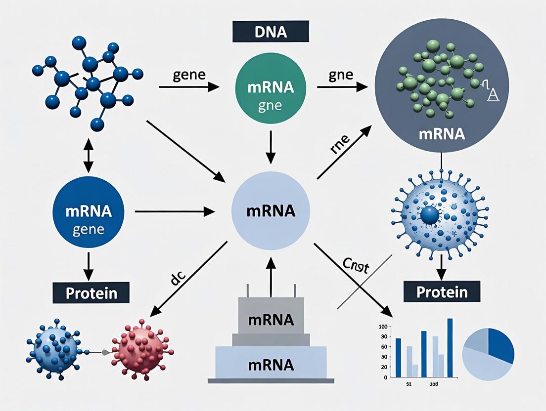
Messenger RNA (mRNA) serves as a critical intermediary in the central dogma of molecular biology, conveying genetic information from DNA in the nucleus to the protein synthesis machinery in the cytoplasm. In recent years, understanding of mRNA biology has expanded beyond its canonical role to encompass its function as a powerful tool for controlling cell fate. Research has demonstrated that mRNA translation is dynamically regulated to instruct stem cell fate decisions, from maintaining quiescence to promoting differentiation [1]. The development of chemically modified mRNA (cmRNA) has further unlocked the potential for directing cell reprogramming and differentiation with significant implications for regenerative medicine and therapeutic development [2] [3]. This technical guide explores the core principles of mRNA biology, from fundamental structure to application in cell fate conversion, providing researchers with both theoretical foundations and practical methodologies.
mRNA Structural Organization and Functional Elements
The functional capacity of mRNA is dictated by its structural components, each playing a specific role in stability, translational efficiency, and regulatory control.
Table 1: Core Structural Elements of Synthetic mRNA and Their Functions
| Structural Element | Position | Key Functions | Optimal Characteristics |
|---|---|---|---|
| 5'-Cap Structure | 5' terminus | Enhances translational efficiency; protects from decapping enzymes; facilitates nuclear export | Modified cap analogs with high affinity for eIF4E; resistant to decapping enzymes |
| 5' Untranslated Region (UTR) | Between cap and start codon | Regulates translational initiation; impacts stability; contains regulatory elements | Highly stabilizing sequences (e.g., from α/β-globin genes); appropriate length and secondary structure |
| Coding Sequence (CDS) | Between start and stop codons | Encodes the protein product; codon optimization enhances translation | Codon-optimized for target species; may contain modified nucleotides to reduce immunogenicity |
| 3' Untranslated Region (UTR) | Between stop codon and poly(A) tail | Influences stability, localization, and translational efficiency; binding site for miRNAs and RBPs | Stabilizing sequences; appropriate length and regulatory elements |
| Poly(A) Tail | 3' terminus | Enhances stability and translational efficiency; protects from exonuclease degradation | Optimal length of 120-150 nucleotides; can be encoded in template or added enzymatically |
The foundational structure of mRNA includes several key regions essential for its function. The 5'-cap structure, typically a 7-methylguanosine (m7G) cap, is critical for translation initiation through its interaction with eukaryotic initiation factor 4E (eIF4E) and also protects the mRNA from degradation by decapping enzymes [2]. Flanking the coding sequence are untranslated regions (UTRs) that contain regulatory elements affecting stability, localization, and translational efficiency. The 3' poly(A) tail plays a crucial role in mRNA stability and translation, with optimal length typically ranging from 120-150 nucleotides [2].
Figure 1: Structural Organization of mRNA with Functional Domains
Chemically Modified mRNA (cmRNA) for Therapeutic Applications
Chemical modifications of mRNA represent a significant advancement for therapeutic applications, addressing limitations of unmodified mRNA while enhancing functionality for cell fate conversion.
Key Modification Strategies
Modified Nucleotides: Incorporation of modified nucleotides such as 5-methylcytidine (5mC), pseudouridine (Ψ), 5-methyluridine (5mU), or N6-methyladenosine significantly decreases immunogenicity by avoiding activation of Toll-like receptors (TLRs) like TLR7 [2]. One study demonstrated a fourfold increase in fluorescence intensity when using either 5-methylcytidine or pseudouridine in GFP mRNAs, with a tenfold increase when both modified nucleotides were combined [2].
Optimized Cap Structures: Selection of cap analogs with high affinity for eIF4E and resistance to decapping enzymes significantly enhances translational efficiency and cmRNA stability [2].
Engineering of UTRs: Incorporation of highly stabilizing UTR sequences, such as those derived from α/β-globin genes, improves both stability and translational efficiency of desired cmRNAs [2].
Applications in Cell Fate Conversion
Chemically modified mRNA has demonstrated remarkable success in cell reprogramming applications. The first successful reprogramming of somatic cells to induced pluripotent stem cells (iPSCs) with cmRNAs encoding Yamanaka factors was performed in 2010, utilizing a "daily transfection regime" for 14 consecutive days with cationic lipid nonviral vectors [2].
Table 2: Comparison of Cell Reprogramming Methods for iPSC Generation
| Reprogramming Method | Time Course for Colony Isolation | Reprogramming Efficiency | Risk of Genome Integration |
|---|---|---|---|
| Protein-based | 8 weeks | 0.001% | No |
| DNA Virus (Retro/Lentivirus) | 2-4 weeks | 0.01%-0.1% | Yes |
| RNA Virus (Sendai Virus) | 4 weeks | 0.01%-1% | No |
| cmRNA-based | 2 weeks | Up to 4.4% | No |
The significantly higher efficiency and reduced timeline of cmRNA-based reprogramming, coupled with the elimination of integration risks, position this technology as a superior approach for generating clinical-grade iPSCs [2].
Quantitative Analysis of mRNA Expression
Accurate quantification of mRNA expression is fundamental to both basic research and therapeutic development. Multiple methodologies exist for relative mRNA quantification, each with distinct advantages and limitations.
Methodological Comparison
A comprehensive comparison of six techniques for analyzing fluorescent data in real-time PCR relative quantification revealed significant differences in performance [4]. The study quantified four cytokine transcripts (IL-1β, IL-6, TNF-α, and GM-CSF) in a model of colonic inflammation, testing method accuracy using samples with known relative amounts of target mRNAs.
Table 3: Comparison of Real-time PCR Data Analysis Methods for mRNA Quantification
| Analytical Method | Accuracy (Average Pearson Correlation) | Key Advantages | Key Limitations |
|---|---|---|---|
| Relative Standard Curve | 0.9991 | High accuracy and reproducibility | Requires standard curve for each target |
| Comparative Ct (ΔΔCt) | 0.9994 | Simplicity; no standard curve needed | Assumes ideal amplification efficiency |
| Sigmoid Curve-Fitting | 0.9953 | Utilizes entire amplification curve | Requires careful cycle selection |
| DART-PCR (Average E) | 0.9990 | Accounts for efficiency variations | Lower accuracy with individual efficiencies |
| Liu & Saint-exp (Average E) | 0.9993 | Exponential model-based | Performance dependent on efficiency calculation |
| LinRegPCR (Individual E) | 0.9577 | Linear regression analysis | Highest variability; lower accuracy |
The research demonstrated that all tested methods can provide quantitative values reflecting mRNA amounts in samples, but they differ significantly in accuracy and reproducibility [4]. The relative standard curve method, comparative Ct method, and DART-PCR, LinRegPCR, and Liu & Saint exponential methods using average amplification efficiency showed the highest correlation with known sample dilutions [5].
Experimental Considerations for mRNA Quantitation
When designing mRNA quantification experiments, several factors require careful consideration:
Normalization Strategy: Selection of appropriate reference genes is critical for accurate relative quantification. The study utilized three reference genes (ACTB, HPRT, SDHA) for normalization [4].
Amplification Efficiency: Genes with low Ct values (e.g., ACTB, IL-1β) typically show amplification efficiencies close to 1.0, while genes with high Ct values (e.g., IL-6, GM-CSF) often demonstrate lower efficiencies, impacting quantification accuracy [4].
Dynamic Range: Reliable quantification depends on maintaining measurements within the dynamic range of detection, with significant deviations observed at extreme dilutions (e.g., 800-fold dilution of IL-6 template) [4].
mRNA Translation Dynamics in Stem Cell Fate Decisions
Regulation of mRNA translation serves as a critical mechanism controlling stem cell behavior and fate decisions. Recent evidence demonstrates that global translation rates are dynamically regulated throughout stem cell activation and differentiation.
Patterns of Translational Regulation
In adult stem cell systems, a consistent pattern of translational regulation emerges:
Quiescent Stem Cells: Hematopoietic stem cells (HSCs), neural stem cells (NSCs), and hair follicle stem cells (HFSCs) in quiescent states display significantly lower translation rates than their activated counterparts [1].
Activated Stem Cells and Progenitors: Upon activation and progression through highly proliferative transit-amplifying stages, translation rates increase substantially [1].
Terminally Differentiated Cells: Differentiation into postmitotic cells correlates with decreased levels of protein synthesis, as observed in further differentiated blood cell types and neuroblasts [1].
Figure 2: mRNA Translation Dynamics During Stem Cell Differentiation
This pattern of translational regulation enables stem cells to rapidly alter their proteome in response to tissue needs or environmental changes without relying solely on transcriptional mechanisms [1]. The observation that proliferation alone does not account for all differences in protein synthesis rates suggests dedicated regulatory mechanisms controlling global translation during fate decisions [1].
Mechanisms Regulating Global Translation
Several key mechanisms control global protein synthesis rates in stem cells:
mTOR Signaling: A central regulator of translation that integrates environmental cues with biosynthetic capacity.
eIF2α Phosphorylation: Regulates initiation of translation and is modulated in response to various stressors.
Ribosome Biogenesis: Controls the production of translational machinery and is dynamically regulated during fate transitions.
These regulatory mechanisms allow stem cells to maintain precise control over their proteome, enabling both stability of identity and plasticity during fate transitions [1].
The Scientist's Toolkit: Essential Reagents and Methodologies
Table 4: Essential Research Reagents for mRNA-Based Cell Fate Conversion
| Reagent Category | Specific Examples | Function | Application Notes |
|---|---|---|---|
| Modified Nucleotides | 5-methylcytidine (5mC), Pseudouridine (Ψ) | Reduce immunogenicity; enhance stability and translation | Combination of modifications often synergistic |
| Cap Analogs | m7G cap analogs | Enhance translational initiation; protect from degradation | Select for high eIF4E affinity and decapping resistance |
| Delivery Vectors | Cationic lipid nanoparticles | Facilitate cellular uptake of mRNA | Enable "daily transfection regime" for reprogramming |
| Template DNA | Plasmids with target sequences and UTRs | IVT template for mRNA synthesis | Include optimized UTRs (e.g., α/β-globin) |
| Poly(A) Tail Enzymes | Poly(A) polymerase | Add poly(A) tail to synthetic mRNA | Alternative: encode in template DNA |
| Reprogramming Factors | OCT3/4, SOX2, KLF4, c-MYC | Mediate cell fate conversion to pluripotency | Yamanaka factors for iPSC generation |
The core principles of mRNA biology extend from fundamental molecular structure to dynamic regulation of translation, providing multiple layers of control over gene expression. The development of chemically modified mRNA technologies has transformed this natural molecule into a powerful tool for directing cell fate with precision and safety. As research continues to elucidate the intricate relationships between mRNA translation dynamics and stem cell behavior, and as quantification methodologies become increasingly refined, the potential for mRNA-based therapies in regenerative medicine continues to expand. The integration of rational mRNA design with advanced delivery platforms positions this technology as a transformative approach for addressing complex degenerative diseases and injury-related conditions through precise cellular reprogramming and tissue regeneration [3].
In the context of mRNA mechanism of action for cell fate conversion research, RNA turnover and stability are not passive background processes but active, decisive regulators of cellular identity. Pluripotent stem cells (PSCs) exhibit remarkable self-renewal capacity and differentiation potential, necessitating tight regulation of gene expression at both transcriptional and post-transcriptional levels [6]. The dynamic control of RNA lifespan provides a critical regulatory layer that fine-tunes transcript abundance and ensures precise timing of developmental transitions [6]. By governing which transcripts persist and which are rapidly cleared, RNA turnover mechanisms enable swift reprogramming of the proteome during lineage commitment—a fundamental requirement for directed cell fate conversion in both developmental biology and therapeutic applications [6] [7].
The integrated network of RNA stability control extends beyond protein-coding mRNAs to include non-coding RNAs (ncRNAs) that modulate signaling pathways and transcript stability [6]. These mechanisms intersect with epitranscriptomic modifications and are functionally interconnected with chromatin states and histone modifications, establishing sophisticated feedback loops between transcriptional and post-transcriptional regulation [6]. This review examines how these post-transcriptional gatekeepers collectively shape pluripotent states and orchestrate lineage commitment, providing researchers with both theoretical frameworks and practical methodologies for investigating these processes.
Core mRNA Degradation Machinery
The RNA degradation machinery consists of multiple conserved pathways that ensure timely and selective decay of transcripts. These mechanisms work in concert to maintain transcriptome homeostasis and enable rapid responses to developmental cues in PSCs [6].
Exonucleolytic and Deadenylation-Dependent Decay Pathways
Pluripotent stem cells maintain a highly dynamic transcriptome characterized by rapid RNA synthesis and turnover. This plasticity relies on canonical RNA degradation pathways, particularly exonucleolytic and deadenylation-dependent decay [6].
In the nucleus, RNA surveillance systems eliminate aberrant or superfluous transcripts. The TRAMP-like complex adds short poly(A) tails to defective RNAs, marking them for degradation by the nuclear exosome. Specialized mammalian pathways include the Nuclear Exosome Targeting (NEXT) complex, which directs the exosome to short-lived non-coding RNAs, and the Poly(A) tail Exosome Targeting (PAXT) connection, which targets polyadenylated nuclear RNAs [6]. These nuclear RNA decay pathways are essential for maintaining transcriptomic integrity in PSCs, preventing accumulation of non-functional RNAs that could interfere with pluripotency maintenance or lineage priming [6].
In the cytoplasm, 5′→3′ exonucleases XRN1 and XRN2, together with the decapping machinery (DCP1/2 and the LSM1–7 complex), mediate transcript clearance, ensuring pluripotency networks remain responsive to developmental cues [6]. Deadenylation complexes, including CCR4–NOT and PAN2–PAN3, modulate stability by shortening poly(A) tails, a critical step that marks RNAs for subsequent degradation [6]. These pathways fine-tune transcript abundance and enable PSCs to swiftly adapt gene expression in response to differentiation signals or cellular stress [6].
Table 1: Core Components of the mRNA Degradation Machinery
| Step | Key Complex/Enzyme | Direction | Description |
|---|---|---|---|
| Deadenylation | CCR4-NOT, PAN2-PAN3 | 3′ → 5′ | Shortening of the poly(A) tail |
| Decapping | DCP1/DCP2 | 5′ → 3′ | Removal of the 5′ cap |
| 5′ → 3′ decay | XRN1 | 5′ → 3′ | Degradation following decapping |
| 3′ → 5′ decay | Exosome | 3′ → 5′ | Degradation after poly(A) tail removal |
| Nonsense-mediated decay | UPF1, SMG1, SMG6 | Specialized | Elimination of mRNAs with premature stop codons |
| miRNA-mediated decay | RISC (Ago2, TRBP, Dicer) | Specialized | Selective degradation of target mRNAs |
| RBP-mediated regulation | HuR, IGF2BP, TTP | Specialized | Stabilization or degradation of target mRNAs depending on the RBP |
Nonsense-Mediated Decay and Quality Control Mechanisms
In addition to core exonucleolytic pathways, PSCs employ specialized quality-control mechanisms such as nonsense-mediated decay (NMD) to ensure transcript fidelity and regulate cell fate. Core NMD components, including UPF1, SMG1, and SMG6, function in a coordinated manner where SMG1 phosphorylates UPF1 to initiate NMD, and phosphorylated UPF1 then recruits SMG6, which mediates endonucleolytic cleavage of target transcripts [6].
These components not only eliminate defective transcripts but also selectively degrade mRNAs encoding core pluripotency factors, positioning NMD as an active modulator of stem cell identity rather than a passive surveillance system [6]. For instance, SMG6/UPF1-mediated NMD directly degrades c-Myc transcripts, thereby modulating self-renewal capacity and pluripotent state transitions in ESCs [6]. Beyond pluripotency maintenance, NMD exerts lineage-specific functions during differentiation, repressing pluripotency-associated transcripts while stabilizing lineage-specific mRNAs in neural lineage commitment [6].
Emerging Regulatory Mechanisms in RNA Stability
RNA Modifications and Stability Control
RNA modifications serve as pivotal regulators of transcript stability, with N6-methyladenosine (m6A) emerging as a particularly influential modification in PSCs. The m6A modification can dictate transcript fate by promoting selective degradation, contributing to the fine-tuning of gene regulatory networks essential for maintaining pluripotent states [6]. This epitranscriptomic regulation intersects with RNA decay pathways to establish multilayered networks that suggest RNA degradation is not a passive clearance mechanism but an active determinant of PSC identity [6].
Biomolecular Condensates in RNA Sequestration
Biomolecular condensates, particularly P-bodies, represent a sophisticated mechanism for post-transcriptional gene regulation through physical sequestration of transcripts. These evolutionarily conserved cytoplasmic structures contain RNA and RNA-binding proteins (RBPs) and regulate translation by sequestering mRNA away from translational machinery [7].
Recent research has revealed that P-body contents are cell type-specific and do not merely reflect active gene expression in each cell type. Instead, they are enriched for translationally repressed transcripts characteristic of preceding developmental stages [7]. Transcripts sequestered in P-bodies show reduced translation efficiency compared to cytoplasmic-enriched mRNAs, and their contents are controlled by microRNAs and can be profoundly reshaped by perturbing AGO2 or polyadenylation site usage [7].
Table 2: Key Findings on P-Body Functions in Cell Fate Regulation
| Finding | Experimental System | Functional Significance |
|---|---|---|
| P-body contents are cell type-specific | Human ES cells under naive and primed conditions | Unique RNA sequestration patterns correspond to developmental identity |
| P-bodies contain transcripts from preceding developmental stages | Differentiation of pluripotent cells into three germ layers | Maintains developmental history while suppressing previous transcriptional programs |
| miRNA activity controls P-body contents | AGO2 perturbation experiments | Links post-transcriptional silencing to RNA sequestration |
| P-body dissolution reactivates sequestered transcripts | Genetic disruption of P-body assembly | Releases translationally repressed fate-instructive mRNAs |
| Manipulating P-bodies directs cell fate | Naive mouse and human pluripotent stem cells | Enables directed differentiation toward totipotency or germ cell lineage |
Translation-Dependent Regulation of RNA Stability
The relationship between translation and RNA stability presents another regulatory layer, particularly evident in microRNA regulation. Recent research has revealed that translation suppresses exogenous target RNA-mediated microRNA decay (TDMD) [8]. TDMD triggers placed in the 3′ untranslated region (UTR) of a reporter degrade miRNAs more effectively than those in the coding sequence (CDS), and inhibiting translation of the reporter enhances miRNA degradation by the CDS trigger, indicating that ribosome-free CDS triggers are more accessible to miRNAs [8].
This finding explains the observed preference for effective TDMD triggers in non-coding regions of RNAs and highlights the intricate relationship between translation and miRNA stability. This mechanism has significant implications for understanding how translational status influences the cellular repertoire of miRNAs available for regulating cell fate transitions [8].
Experimental Approaches for Studying RNA Turnover
High-Throughput Screening with Tethered Function Assays
Unbiased surveys of post-transcriptional regulators have been enabled by adapting tethered function assays to quantitatively measure regulatory activity across proteomes. This approach couples a tethered function assay with quantitative single-cell fluorescence measurements to analyze thousands of protein fragments and determine their effects on a tethered mRNA [9].
The experimental system utilizes a budding yeast tethering assay with a ratiometric fluorescence readout. A transcript encoding a yellow fluorescent protein (YFP) with five boxB hairpins in its 3′ UTR is tethered to a candidate regulatory protein fused to the λN coat protein. To control for non-specific changes, YFP measurements are normalized against a red fluorescent protein (RFP) control expressed from a transcript not targeted by λN [9]. Changes in the YFP/RFP ratio precisely measure specific regulatory activity affecting the targeted mRNA while controlling for global effects.
For proteome-wide surveys, researchers generate libraries of λN fusions from randomly fragmented genomic DNA. Fragments of ~500 base pairs are captured into a vector that requires in-frame translation and are transferred into a λN fusion expression vector with random barcodes identifying each fragment uniquely [9]. Cells transformed with the library are separated into subpopulations according to YFP/RFP fluorescence ratio, and library plasmid DNA is isolated from sorted cells for barcode quantification by next-generation sequencing. Regulatory activity is quantified through an "activity score" representing the maximum likelihood estimate of average fluorescence expressed as a z-score relative to the overall population [9].
P-Body Isolation and Transcriptome Profiling
To profile RNA contents of biomolecular condensates like P-bodies, researchers have adapted fluorescence-activated sorting methods using GFP-tagged P-body components such as LSM14A [7]. After validation of colocalization with established P-body markers, intact GFP-LSM14A particles are isolated using fluorescence-activated particle sorting (FAPS) from cell lysates [7].
RNA from purified P-bodies and corresponding cytosolic fractions is characterized using Smart-seq or alternative low-input library preparation methods like snapTotal-seq that use random primers rather than oligo(dT) [7]. This approach enables identification of mRNAs enriched in P-bodies relative to the cytosol, with verification of localization through single-molecule fluorescence in situ hybridization (smFISH) in conjunction with immunofluorescence [7].
This methodology has revealed that P-body-enriched mRNAs encode regulators of RNA processing, transcription, chromatin organization and cell cycle, while cytosolic mRNAs are involved in housekeeping functions such as metabolic processes and structural components [7]. Integration with ribosome profiling data further enables assessment of translation efficiency for P-body-associated transcripts [7].
The Scientist's Toolkit: Essential Research Reagents
Table 3: Key Research Reagents for Studying RNA Turnover and Stability
| Reagent/Tool | Function | Application Examples |
|---|---|---|
| λN coat protein and boxB hairpins | RNA-protein tethering system | Tethered function assays for high-throughput regulator screening [9] |
| GFP-LSM14A fusion construct | P-body labeling | Isolation and transcriptome profiling of biomolecular condensates [7] |
| CCR4-NOT complex components | Deadenylation machinery | Studying deadenylation-dependent decay pathways [6] [9] |
| ZSWIM8 knockout systems | TDMD pathway disruption | Identifying miRNAs regulated by target-directed degradation [8] |
| CsrA/B genetic system | Bacterial post-transcriptional regulation | Rewiring native regulators for synthetic circuits [10] |
| DCP1/DCP2 decapping complex | 5' cap removal | Investigating decapping-dependent decay mechanisms [6] |
| UPF1, SMG1, SMG6 | NMD pathway components | Studying nonsense-mediated decay and quality control [6] |
Implications for Cell Fate Conversion Research
The mechanistic insights into RNA turnover and stability have profound implications for cell fate conversion research. The discovery that manipulating P-body assembly or microRNA activity can direct naive mouse and human pluripotent stem cells toward totipotency or primed human embryonic cells toward the germ cell lineage demonstrates the potential of targeting RNA regulatory mechanisms for controlling cell identity [7].
Similarly, the finding that RNA degradation exhibits heterogeneous and dynamic kinetics during cell fate transitions highlights its role in preserving transcriptome homeostasis during reprogramming events [6]. Single-cell and multi-omics approaches have revealed that disruption of RNA decay pathways is implicated in developmental defects and disease, underscoring their potential as therapeutic targets [6].
The integrated network of RNA stability control—encompassing core decay machinery, epitranscriptomic modifications, biomolecular condensates, and translation-coupled regulation—provides multiple nodes for intervention in cell fate conversion strategies. As synthetic biology advances, rewiring native post-transcriptional global regulators offers promising approaches for achieving designer, multi-layered genetic circuits that can precisely control cellular transitions [10].
The comprehensive understanding of RNA turnover and stability mechanisms reveals an intricate regulatory network that actively shapes cell identity and fate transitions. From core degradation machinery to emerging concepts in biomolecular condensates and translation-coupled regulation, these post-transcriptional gatekeepers provide critical control points in the complex process of cell fate conversion. The experimental methodologies and research tools summarized in this review equip researchers with robust approaches for investigating these mechanisms further, with significant implications for developmental biology, regenerative medicine, and therapeutic development. As the field advances, leveraging these post-transcriptional regulatory networks will undoubtedly yield novel strategies for directed cellular reprogramming and precision control of cell fate decisions.
The regulation of cell identity has traditionally focused on transcriptional networks, but emerging research highlights the indispensable role of post-transcriptional mechanisms in directing cell fate transitions. Among these mechanisms, biomolecular condensates—membraneless organelles formed through phase separation—serve as critical hubs for coordinating gene expression. Processing bodies (P-bodies) represent a conserved class of cytoplasmic biomolecular condensates that sequester translationally repressed messenger RNAs (mRNAs) and RNA-binding proteins, thereby functioning as a dynamic reservoir for transcripts encoding key developmental regulators [7] [11]. Within the broader context of mRNA mechanism of action research, understanding how P-bodies influence cell fate decisions provides novel insights for controlling cellular identity for therapeutic purposes. This whitepaper examines the molecular mechanisms of RNA sequestration in P-bodies and explores how manipulating these condensates can direct stem cell differentiation and reprogramming.
P-Body Composition and Biogenesis
Core Components and Assembly
P-bodies are evolutionarily conserved cytoplasmic condensates comprising numerous enzymes involved in mRNA turnover and repression [11]. Proteomic analyses of purified human P-bodies have identified approximately 125 core proteins, including decapping enzymes (DCP1A, DCP2), RNA helicases (DDX6, DHX15), components of the microRNA-mediated silencing pathway (AGO1, AGO2, GW182), and 5'-3' exoribonucleases (XRN1) [11]. The assembly of these components into higher-order structures is driven by multivalent protein-protein and protein-RNA interactions that facilitate liquid-liquid phase separation.
Table 1: Key Protein Components of Mammalian P-Bodies
| Protein | Gene Symbol | Primary Function | Localization Specificity |
|---|---|---|---|
| Decapping Enzyme 2 | DCP2 | mRNA decapping | P-body specific |
| DEAD-box Helicase 6 | DDX6 | RNA helicase activity, translation repression | Also in stress granules |
| Argonaute 2 | AGO2 | miRNA-mediated silencing | Also in stress granules |
| Enhancer of Decapping 4 | EDC4 | Scaffold protein for decapping complex | P-body specific |
| LSM14A | LSM14A | P-body scaffold, RNA binding | Also in stress granules |
| Poly(A)-Binding Protein | PABPC1 | Translation initiation, potential P-body regulation | Cytoplasmic, excluded from P-bodies |
| 5'-3' Exoribonuclease 1 | XRN1 | mRNA degradation | P-body specific |
Relationship to Other RNA Granules
P-bodies are functionally and compositionally distinct from, yet physically interacting with, stress granules—another class of cytoplasmic condensates that form under cellular stress conditions [11]. While P-bodies are constitutive structures involved in mRNA decay and storage, stress granules assemble transiently in response to phosphorylation of eIF2α and primarily contain translationally stalled pre-initiation complexes. Approximately 30% of P-body proteins can also localize to stress granules, creating potential channels for mRNA exchange between these compartments [11].
Experimental Profiling of P-Body Contents and Dynamics
Methodologies for P-Body Isolation and Transcriptome Analysis
Recent technical advances have enabled the precise characterization of P-body contents through fluorescence-activated particle sorting (FAPS), a method adapted from fluorescence-activated cell sorting for isolating intact biomolecular condensates [7] [11].
Experimental Protocol: P-body-seq for Transcriptome Profiling
Fluorescent Tagging: Stably integrate GFP-tagged LSM14A (a core P-body component) into the AAVS1 safe-harbor locus in human embryonic stem cells to enable specific labeling without disrupting endogenous P-body function [7].
Cell Culture and Differentiation: Culture GFP-LSM14A-expressing cells under naive pluripotency conditions, primed pluripotency conditions, and differentiate them into definitive germ layer progenitors (mesoderm, endoderm, ectoderm) and terminally differentiated neurons to model developmental transitions [7].
Cell Lysis and Condensate Preservation: Lyse cells using a mild detergent-free buffer (e.g., 10 mM HEPES, 150 mM KCl, 1 mM EDTA, 0.5% NP-40, protease/RNase inhibitors) that maintains P-body integrity while dispersing the cytoplasmic matrix [7].
Fluorescence-Activated Particle Sorting (FAPS): Isolate intact GFP-LSM14A-positive particles using a FAC sorter equipped with a 100-μm nozzle and low pressure settings (≤ 10 psi) to preserve structural integrity. Include control cells expressing cytoplasmic GFP to establish sorting background [7].
RNA Extraction and Library Preparation: Extract total RNA from sorted P-bodies and corresponding cytoplasmic fractions using magnetic bead-based purification. Prepare sequencing libraries using either:
- Smart-seq2 for full-length transcript coverage with oligo(dT) priming
- snapTotal-seq for random-primed amplification to avoid 3'-bias and detect non-polyadenylated RNAs [7]
Bioinformatic Analysis: Map sequencing reads to reference genomes, quantify transcript abundances, and identify P-body-enriched RNAs using statistical frameworks (e.g., DESeq2) with thresholds of >2-fold enrichment and adjusted p-value <0.01 relative to cytoplasmic fractions [7].
Validation: Confirm P-body localization of identified transcripts using single-molecule fluorescence in situ hybridization (smFISH) combined with immunofluorescence for canonical P-body markers (e.g., EDC4, DCP1A) [7].
Diagram 1: Experimental workflow for P-body transcriptome profiling using FAPS and RNA sequencing (P-body-seq).
Research Reagent Solutions for P-Body Studies
Table 2: Essential Research Reagents for P-Body Investigation
| Reagent/Cell Line | Function/Application | Key Characteristics |
|---|---|---|
| GFP-LSM14A AAVS1 Knock-in | Fluorescent labeling of P-bodies | Endogenous expression control via safe-harbor locus integration |
| Anti-EDC4 Antibody | P-body marker for validation | High specificity for immunofluorescence and immunoblotting |
| Anti-DDX6 Antibody | P-body disruption studies | Knockdown/knockout disrupts P-body assembly |
| SYTOX Blue RNA Stain | RNA visualization in particles | Binds RNA in fixed preparations |
| DDX6 shRNA/CRISPR | P-body dissolution | Validates specificity of P-body isolation |
| Smart-seq2/snapTotal Kits | Low-input RNA sequencing | Detect both polyA+ and non-polyadenylated transcripts |
| smFISH Probes | Single-molecule RNA validation | Quantitative localization of specific transcripts |
Quantitative Characterization of P-Body RNA Contents
Global Features of Sequestered Transcripts
Application of P-body-seq across multiple human stem cell states and differentiated lineages has revealed fundamental principles governing RNA sequestration in these condensates:
Table 3: Properties of P-Body-Enriched Transcripts in HEK293T Cells
| Transcript Feature | Observation | Interpretation |
|---|---|---|
| Number of Enriched mRNAs | 3,994 mRNAs significantly enriched | Substantial fraction of transcriptome sequestered |
| Transcript Integrity | No evidence of increased truncation | P-bodies store intact mRNAs rather than degradation intermediates |
| Poly(A) Tail Length | Poor correlation with enrichment (r = -0.028 to -0.075) | Dead-enylation not prerequisite for sequestration |
| Sequence Composition | High AU content | AU-rich elements may facilitate P-body localization |
| Translation Efficiency | Significantly reduced | Confirms translational repression of P-body RNAs |
| Half-life Correlation | Poor correlation with mRNA stability | Sequestration not directly linked to decay rates |
Analysis of P-body contents across developmental contexts revealed that sequestered transcripts do not merely reflect the overall transcriptome of each cell type. Instead, P-bodies are enriched for translationally repressed mRNAs characteristic of the preceding developmental stage, suggesting they function as repositories for legacy gene expression programs that must be suppressed during fate transitions [7]. For example, in neurons, P-body contents more closely resembled the cytoplasmic transcriptome of neural progenitors than mature neurons themselves [7].
Developmental Regulation of P-Body Contents
Principal component analysis of P-body transcriptomes across naive pluripotent, primed pluripotent, germ layer progenitor, and terminally differentiated states demonstrates that P-body RNA profiles follow a stepwise progression mirroring developmental transitions [7]. Notably, P-body contents cluster distinctly from cytoplasmic transcriptomes of the same cell type, instead aligning with earlier developmental stages, supporting a model wherein P-bodies retain transcripts from progenitor states that could potentially be reactivated under appropriate conditions [7].
Mechanism of RNA Sequestration and Fate Control
microRNA-Mediated Regulation of P-Body Contents
MicroRNAs play a central role in directing specific transcripts to P-bodies through the RNA-induced silencing complex (RISC). Core RISC components, including Argonaute proteins (AGO1, AGO2) and GW182, are consistently identified in P-body proteomes [11]. Experimentally, perturbation of AGO2 function profoundly reshapes the P-body transcriptome, establishing a causal relationship between miRNA targeting and RNA sequestration [7]. This mechanism enables context-dependent control of P-body composition, as miRNA expression patterns shift during development.
Polyadenylation Control and P-Body Localization
Alternative polyadenylation (APA) significantly influences P-body sequestration, as perturbation of poly(A) site usage reshapes P-body transcriptomes [7]. The poly(A)-binding protein PABPC1, while predominantly cytoplasmic and translation-associated, may compete with P-body localization factors for RNA binding, potentially influencing which transcripts enter condensates [12] [13]. Interestingly, P-body-enriched transcripts show no correlation with poly(A) tail length, suggesting that PABP binding dynamics rather than tail length per se may regulate sequestration [7].
Diagram 2: Mechanism of selective RNA sequestration in P-bodies. microRNA-mediated targeting and legacy transcripts from previous developmental stages are selectively sequestered to enable cell fate transitions.
Functional Manipulation of P-Bodies for Cell Fate Control
Directed Differentiation Through P-Body Perturbation
Strategic manipulation of P-body assembly or composition enables directed control of stem cell fate decisions:
Experimental Protocol: Directing Fate Transitions via P-Body Manipulation
P-body Dissolution: Transfer naive human pluripotent stem cells to media containing small molecule inhibitors of key P-body components (e.g., DDX6 inhibitors) or transduce with shRNA targeting essential scaffolding proteins (LSM14A, EDC4) to disrupt condensate integrity [7].
microRNA Modulation: Introduce miRNA mimics or inhibitors to reshape the P-body transcriptome by altering the repertoire of sequestered mRNAs. For germ cell differentiation, suppress miRNAs that repress primordial germ cell (PGC) specification factors [7].
Functional Assessment: Evaluate differentiation efficiency using:
- Immunofluorescence for lineage-specific markers (≥80% positive cells indicates robust differentiation)
- Single-cell RNA sequencing to characterize emergent transcriptional states
- Ribosome profiling to measure translational activation of previously sequestered mRNAs [7]
Application of these approaches has demonstrated that P-body dissolution in naive human pluripotent stem cells activates a totipotency transcriptional program, while similar manipulations in primed human embryonic stem cells enhances their conversion to primordial germ cell-like cells (PGCLCs) with significantly improved efficiency [7]. These findings establish a direct causal relationship between P-body regulation and cell fate outcomes.
Therapeutic Implications and Applications
The capacity to control cell identity through P-body manipulation holds significant promise for regenerative medicine. By selectively releasing specific transcripts from translational repression, researchers can potentially direct stem cell differentiation toward therapeutically relevant lineages without genetic modification [7]. Furthermore, the observation that P-bodies are reconfigured in pathological conditions including Parkinson's disease and cancer suggests that restoring normal RNA sequestration patterns could represent a novel therapeutic strategy for these conditions [7].
P-bodies represent a crucial post-transcriptional regulatory node in cell fate determination, functioning not merely as RNA decay centers but as dynamic reservoirs that sequester translationally repressed transcripts encoding identity regulators. The development of robust methodologies for purifying and profiling condensate contents has revealed the principles governing RNA sequestration, including its regulation by miRNAs and polyadenylation factors, and its cell type-specific nature. Most significantly, the demonstrated capacity to direct stem cell fate transitions through targeted manipulation of P-body assembly or composition establishes these biomolecular condensates as legitimate targets for controlling cellular identity in both basic research and therapeutic contexts. As the mRNA therapeutics field advances beyond protein supplementation to encompass cell reprogramming and transdifferentiation strategies, understanding and harnessing P-body biology will be essential for achieving precise control over cell fate conversions.
The regulation of cell identity has long been a cornerstone of developmental biology and regenerative medicine. While transcriptional networks and epigenetic modifications have occupied center stage in explaining how cells acquire and maintain their identities, a growing body of evidence reveals that post-transcriptional mechanisms exert equally powerful influences on cell fate decisions. Among these mechanisms, the compartmentalization of messenger RNA (mRNA) within biomolecular condensates represents a sophisticated layer of gene expression control that fine-tunes the proteome without altering the transcriptome. This process, known as mRNA sequestration, enables cells to rapidly respond to developmental cues and environmental changes by dynamically controlling which transcripts are translated and when.
The significance of mRNA sequestration extends beyond basic biological understanding to practical applications in cell fate conversion research. As scientists develop increasingly precise methods for reprogramming somatic cells into induced pluripotent stem cells (iPSCs) and directing their differentiation into specific lineages, comprehending post-transcriptional regulatory mechanisms becomes paramount. Recent advances have demonstrated that RNA condensates, particularly processing bodies (P-bodies), serve not merely as mRNA degradation sites but as dynamic storage hubs for translationally repressed transcripts encoding key developmental regulators. This technical guide explores the mechanisms and implications of mRNA sequestration, providing researchers with both theoretical frameworks and practical methodologies for investigating this crucial process in the context of pluripotency and differentiation.
The Molecular Architecture of mRNA Sequestration
Biomolecular Condensates: P-Bodies as RNA Storage Hubs
Biomolecular condensates are membrane-less organelles that form through liquid-liquid phase separation, creating distinct subcellular compartments with unique compositions and functions. Among these, P-bodies stand out as evolutionarily conserved cytoplasmic structures containing diverse RNA species and RNA-binding proteins (RBPs). Historically characterized as sites of mRNA decay, P-bodies are now recognized as multifunctional regulatory centers that modulate gene expression post-transcriptionally by sequestering mRNA away from the translational machinery [7].
The protein composition of P-bodies includes core components such as LSM14A, EDC4, and DDX6, which facilitate the assembly and structural integrity of these condensates. These proteins serve as scaffolds that recruit specific mRNA subsets through interactions with sequence elements, secondary structures, or associated regulatory factors. When transcripts are sequestered within P-bodies, they are translationally repressed but often remain intact and capable of re-entering the translatable pool upon appropriate signaling [7]. This capacity for reversible storage positions P-bodies as ideal regulators of developmental transitions, where rapid changes in gene expression patterns are required.
Mechanisms of Transcript Selection and Recruitment
The targeting of specific mRNAs to P-bodies is not random but follows molecular principles that determine which transcripts become sequestered. Research across multiple vertebrate species and cell types has revealed that P-body contents exhibit conserved, cell type-specific patterns of RNA enrichment that do not merely reflect overall transcriptional profiles [7]. Several mechanisms govern this selective recruitment:
MicroRNA-mediated targeting: microRNAs (miRNAs), in complex with AGO2 and other RNA-induced silencing complex (RISC) components, direct specific transcripts to P-bodies based on sequence complementarity, particularly in the 3' untranslated regions (UTRs) of target mRNAs [7].
Sequence-specific motifs: AU-rich elements (AREs) and other regulatory sequences in mRNA 3'UTRs serve as binding platforms for RBPs that shuttle transcripts to P-bodies. Transcripts with high AU content show significant enrichment in P-bodies [7].
Polyadenylation status: The usage of alternative polyadenylation sites can influence P-body localization, suggesting that 3'UTR processing contributes to destination decisions for mRNAs [7].
Translation efficiency: Poorly translated transcripts are preferentially directed to P-bodies, as evidenced by ribosome profiling data showing reduced translation efficiency for P-body-associated mRNAs compared to cytoplasmic-enriched mRNAs [7].
Table 1: Key Molecular Features Associated with P-body Sequestration
| Feature | Association with P-body Localization | Experimental Evidence |
|---|---|---|
| AU-rich elements (AREs) | Positive correlation | P-body-enriched transcripts show significantly higher AU content [7] |
| Translation efficiency | Negative correlation | Ribosome profiling reveals reduced translation of P-body transcripts [7] |
| microRNA binding sites | Positive correlation | AGO2 perturbation reshapes P-body contents [7] |
| PolyA tail length | No direct correlation | Poor correlation between mean polyA tail length and P-body enrichment [7] |
| Transcript length | No direct correlation | Transcript length shows no appreciable relationship with P-body localization [7] |
mRNA Sequestration in Pluripotency and Differentiation Transitions
Cell Type-Specific RNA Sequestration Patterns
The functional significance of mRNA sequestration becomes particularly evident when examining stem cell biology and differentiation pathways. Comprehensive profiling of P-body contents across diverse developmental contexts has revealed that these condensates maintain cell type-specific RNA compositions that reflect both current and previous developmental states. When researchers applied P-body sequencing (P-body-seq) to human embryonic stem cells (ESCs) cultured under naive (pre-implantation) and primed (post-implantation) conditions, as well as to their differentiated derivatives representing the three germ layers, they discovered that P-body contents do not simply mirror the active transcriptome of each cell type [7].
Surprisingly, P-body mRNA profiles often cluster more closely with cytoplasmic samples from preceding developmental stages rather than with contemporaneous cytoplasmic transcripts. For instance, P-body contents from neurons closely resemble cytoplasmic RNA patterns from neural progenitors, while mesoderm progenitor P-bodies share similarities with primed ESC cytoplasm [7]. This pattern suggests that P-bodies function as molecular archives that retain transcripts characteristic of earlier developmental stages, potentially as a mechanism to suppress previous cellular identities or maintain plasticity for potential lineage reversion.
Functional Consequences for Cell Identity
The sequestration of specific mRNA subsets in P-bodies has direct implications for cellular identity through several interconnected mechanisms:
Translation control: By physically separating transcripts from ribosomes, P-bodies prevent the synthesis of proteins that might oppose the current cellular identity or developmental trajectory. When P-bodies are dissolved, either genetically or pharmacologically, the released mRNAs re-enter translation, leading to increased protein production from fate-instructive transcripts [7].
Developmental timing: P-bodies appear to introduce a temporal dimension to gene expression by delaying the translation of certain regulators until the appropriate developmental context emerges. This buffering function ensures that differentiation proceeds in an orderly fashion.
Fate stabilization: The selective removal of transcripts encoding alternative fate determinants reinforces the current cellular identity. For example, in pluripotent stem cells, P-body sequestration of differentiation-promoting factors helps maintain the undifferentiated state [7].
Lineage priming: The storage of lineage-specific transcripts in P-bodies of progenitor cells may facilitate rapid activation of differentiation programs upon receiving appropriate signals, as the necessary mRNAs are already present but translationally repressed.
Table 2: Developmental Transitions Influenced by mRNA Sequestration
| Developmental Transition | Role of mRNA Sequestration | Key Regulated Transcripts |
|---|---|---|
| Naive to primed pluripotency | Sequestration of naive-specific factors during transition | Transcripts encoding naive pluripotency factors |
| Pluripotency to differentiation | Sequestration of pluripotency factors | OCT4, SOX2, NANOG mRNAs |
| Lineage specification | Selective storage of alternative lineage determinants | Transcripts encoding transcription factors for non-selected lineages |
| Progenitor to terminal differentiation | Release of lineage-appropriate transcripts | Cell type-specific functional genes |
| Totipotency program activation | Sequestration of transcripts that suppress totipotency | Factors that promote differentiated states [7] |
Experimental Approaches for Studying mRNA Sequestration
Methodologies for P-body Isolation and RNA Profiling
Investigating mRNA sequestration requires specialized techniques for isolating biomolecular condensates and analyzing their contents with minimal perturbation. The following protocols represent state-of-the-art approaches for capturing and characterizing sequestered transcripts:
Fluorescence-Activated Particle Sorting (FAPS) of P-bodies
This adapted method enables the purification of intact P-bodies from cell lysates for subsequent RNA analysis [7]:
Experimental Workflow:
- Cell line engineering: Stably integrate GFP-tagged LSM14A (a core P-body protein) into the AAVS1 safe harbor locus to ensure consistent, physiological expression levels.
- Validation: Confirm GFP-LSM14A colocalization with established P-body markers (e.g., EDC4) using immunofluorescence to verify specific labeling.
- Cell lysis: Use gentle lysis conditions that preserve P-body integrity while releasing them from the cytoplasmic milieu.
- Sorting: Isolate GFP-LSM14A positive particles using fluorescence-activated particle sorting (FAPS).
- Quality controls: Include control cells expressing cytoplasmic GFP alone to account for non-specific associations, and validate P-body disruption via DDX6 knockdown/knockout, which should eliminate GFP-positive particles.
Technical considerations: The size and fluorescence intensity of isolated particles vary between cell types, likely reflecting differences in P-body abundance and composition across developmental contexts [7].
Transcriptome Analysis of Sequestered RNA
Once isolated, P-body RNAs require specialized processing for accurate characterization:
RNA Sequencing Approaches:
- Smart-seq2: Provides full-length transcript information with high sensitivity for low-input samples, though it introduces polyA selection bias [7].
- snapTotal-seq: An alternative low-input method using random primers rather than oligo(dT) selection, enabling detection of non-polyadenylated RNAs and avoiding 3' bias [7].
- Validation: Confirm P-body localization of identified transcripts using single-molecule fluorescence in situ hybridization (smFISH) coupled with immunofluorescence for P-body markers.
Analytical considerations: Approximately 70% overlap has been observed between Smart-seq and snapTotal-seq datasets, with both methods confirming that P-body RNAs are intact and not preferentially deadenylated compared to cytoplasmic transcripts [7].
The Scientist's Toolkit: Essential Research Reagents
Table 3: Key Research Reagents for Investigating mRNA Sequestration
| Reagent/Category | Function/Application | Specific Examples |
|---|---|---|
| Fluorescent P-body markers | Label condensates for visualization and isolation | GFP-LSM14A, RFP-DDX6 [7] |
| P-body disruption tools | Investigate functional consequences of sequestration loss | shRNA against DDX6, CRISPR knockout of core components [7] |
| miRNA pathway modulators | Manipulate miRNA-mediated mRNA targeting | AGO2 mutants, antagomirs, miRNA mimics [7] |
| Transcriptional inhibitors | Measure RNA half-lives and decay kinetics | Actinomycin D, α-Amanitin [14] |
| Metabolic RNA labels | Pulse-chase experiments to track RNA fate | 4-thiouridine (4sU), 5-ethynyl uridine (5EU) [14] |
| Library prep kits | RNA sequencing from low-input samples | Smart-seq2, snapTotal-seq [7] |
| Interferon inhibitors | Reduce immune responses during mRNA transfection | B18R protein [15] |
| Modified nucleosides | Reduce immunogenicity of synthetic mRNA | Pseudouridine, 5-methylcytidine [15] |
Signaling Pathways and Regulatory Networks in mRNA Sequestration
The regulation of mRNA sequestration involves complex interactions between multiple signaling pathways and regulatory networks that respond to developmental cues and environmental signals. The following diagram illustrates the core pathway through which biomolecular condensates influence cell fate decisions by controlling mRNA availability for translation:
Diagram 1: mRNA Sequestration in Cell Fate Regulation
This core pathway operates in concert with other regulatory mechanisms to fine-tune stem cell behavior:
Integration with Translational Control Mechanisms
mRNA sequestration does not function in isolation but represents one component of a broader regulatory network that controls protein synthesis in stem cells. The global rate of translation itself changes dynamically during stem cell activation and differentiation, creating a hierarchical system of gene expression control [1]:
- Quiescent stem cells (e.g., in hematopoietic, neural, and hair follicle systems) display low overall translation rates
- Activated stem cells and transit-amplifying progenitors exhibit significantly increased protein synthesis
- Terminally differentiated, post-mitotic cells again show reduced translation rates
This pattern suggests that mRNA sequestration in P-bodies operates within a larger framework where both global translation rates and transcript-specific controls collaborate to establish precise proteomic patterns appropriate for each cellular state.
Cross-talk with Epigenetic and Transcriptional Networks
The regulation of mRNA sequestration shows extensive integration with epigenetic and transcriptional mechanisms. For instance, numerous long non-coding RNAs (lncRNAs) participate in both epigenetic regulation and post-transcriptional control, creating bridges between these regulatory layers [16]:
- Nuclear lncRNAs (e.g., XIST, DEANR1, GATA6-AS) interact with chromatin-modifying complexes and transcription factors to establish transcriptional programs
- Cytoplasmic lncRNAs (e.g., linc-ROR, H19, TINCR) often function as competitive endogenous RNAs (ceRNAs) that sequester microRNAs or regulate mRNA stability
- Dual-localization lncRNAs (e.g., T-UCstem1, MEG3) operate in both compartments, potentially coordinating transcriptional and post-transcriptional regulation
This interconnectedness ensures that mRNA sequestration aligns with broader developmental programs, creating coherent transitions between cellular states rather than operating as an independent regulatory system.
Applications in Cell Fate Conversion and Regenerative Medicine
Directing Stem Cell Differentiation Through Sequestration Manipulation
The strategic manipulation of mRNA sequestration pathways offers powerful approaches for controlling stem cell behavior with high precision. Several studies have demonstrated that perturbing P-body assembly or miRNA activity can direct stem cells toward specific developmental trajectories:
Toward totipotency: Dissolution of P-bodies in naive human pluripotent stem cells activates a totipotency transcriptional program, suggesting that P-bodies normally suppress this developmental state by sequestering key regulators [7].
Germline specification: Reshaping P-body contents facilitates the conversion of primed human embryonic stem cells into primordial germ cell-like cells (PGCLCs), highlighting the role of sequestration in germline development [7].
Lineage commitment: Selective modulation of miRNA-mRNA interactions can promote differentiation toward specific lineages by altering the sequestration patterns of lineage-inappropriate transcripts.
These applications demonstrate the potential of targeting mRNA sequestration for regenerative medicine, where directing stem cell differentiation with spatial and temporal precision remains a significant challenge.
mRNA-Based Technologies for Cell Fate Conversion
The understanding of mRNA sequestration mechanisms has informed the development of advanced mRNA-based technologies for cell fate conversion. Synthetic mRNA approaches have emerged as powerful tools for reprogramming and differentiation, offering advantages over DNA-based methods:
Key Advancements in mRNA Technology:
- Immunogenicity reduction: Incorporation of modified ribonucleosides (e.g., pseudouridine, 5-methylcytidine) and removal of 5' triphosphates reduce activation of antiviral defense pathways [15].
- Interferon suppression: Media supplementation with interferon inhibitors (e.g., B18R) enables repeated transfections necessary for sustained transgene expression [15].
- Efficiency improvements: mRNA-based reprogramming demonstrates approximately 35-fold higher efficiency and two-fold faster kinetics compared to viral methods for generating induced pluripotent stem cells [15].
These technological advances leverage our growing understanding of natural mRNA regulatory mechanisms, including sequestration, to develop more precise and clinically viable approaches for cell engineering.
Future Perspectives and Technical Challenges
Despite significant progress in understanding mRNA sequestration, several challenges remain that present opportunities for methodological innovation and conceptual advancement:
Technical Limitations:
- Isolation purity: Current P-body isolation techniques may co-purify other ribonucleoprotein complexes, requiring rigorous validation of specificity.
- Dynamic measurements: Most studies provide static snapshots of sequestration, while the process is inherently dynamic; developing live-cell imaging approaches for tracking individual transcripts would reveal temporal dynamics.
- Single-cell resolution: Bulk analyses mask cell-to-cell heterogeneity in sequestration patterns; single-cell P-body-seq methods would illuminate how variability in mRNA compartmentalization contributes to fate decisions.
Conceptual Frontiers:
- Sequestration reversibility: The mechanisms controlling the release of mRNAs from P-bodies remain poorly characterized, representing a critical gap in understanding the complete regulatory cycle.
- Context-dependent rules: The principles governing transcript sequestration appear to vary by cell type and developmental context; systematic mapping of these contextual factors would enable more predictive models.
- Therapeutic targeting: While manipulation of sequestration pathways shows promise for directing cell fate, translating these approaches to clinical applications requires better specificity and control.
As technologies for studying and manipulating biomolecular condensates continue to advance, mRNA sequestration will likely emerge as an increasingly important target for controlling cell identity in both basic research and therapeutic applications. The integration of sequestration control with existing transcriptional and epigenetic engineering approaches promises unprecedented precision in cell fate conversion for regenerative medicine, disease modeling, and therapeutic development.
Engineering Cell Fate: mRNA Design, Delivery, and Therapeutic Applications
The mechanism of action of messenger RNA (mRNA) extends far beyond its role as a simple blueprint for protein synthesis. In the context of cell fate conversion research, mRNA has emerged as a powerful and safe tool for reprogramming somatic cells to pluripotency and directing their differentiation into desired lineages. Unlike DNA-based approaches, mRNA-based methods eliminate the risk of insertional mutagenesis and do not require nuclear entry, allowing for transient yet highly efficient expression of reprogramming factors [2] [15]. The foundational application of this technology was demonstrated by Warren et al., who achieved highly efficient reprogramming of human fibroblasts to induced pluripotent stem cells (iPSCs) using synthetic modified mRNA encoding the Yamanaka factors (OCT4, SOX2, KLF4, c-MYC) [15]. This method proved to be two times faster and 35-fold more efficient than traditional viral methods, highlighting the profound impact of mRNA technology on regenerative medicine [15]. The core principle underlying this application is the ability to precisely control the expression of key transcription factors that dictate cellular identity. However, the functional efficacy of mRNA in this context is wholly dependent on overcoming inherent challenges of stability, immunogenicity, and translational efficiency. This has driven the development of sophisticated optimization strategies focusing on the three pillars of mRNA engineering: nucleoside modifications, codon usage, and untranslated region (UTR) engineering, which together form the basis of a highly controllable system for directing cell fate.
Nucleoside Modifications: Enhancing Stability and Evading Immunity
Chemical modifications to the nucleosides within an mRNA molecule represent one of the most critical advancements in mRNA therapeutic technology. These modifications are essential for reducing the intrinsic immunogenicity of exogenous mRNA and improving its stability and translational capacity.
Reduction of Immunogenicity
Unmodified mRNA is recognized by pattern recognition receptors (PRRs), such as Toll-like receptors (TLRs) and RIG-I-like receptors (RLRs), triggering an antiviral innate immune response that can severely inhibit translation [17] [18]. This is a significant barrier for applications requiring repeated administration, such as the multi-day transfections needed for cell reprogramming. The incorporation of modified nucleosides, such as pseudouridine (Ψ) and N1-methylpseudouridine (m1Ψ), effectively mitigates this immune activation by altering the molecular signature of the RNA, allowing it to evade detection [2] [17]. The licensed COVID-19 mRNA vaccines utilize m1Ψ-modified mRNA, validating the critical importance of this modification [17]. Furthermore, the use of the interferon inhibitor B18R has been shown to further suppress the interferon response, enabling the sustained daily transfections required for efficient cell reprogramming [15].
Position-Specific Modification for Stability and Translation
Beyond global nucleoside substitution, recent research explores the position-specific introduction of ribose modifications to fine-tune mRNA function. A pivotal 2025 study demonstrated that introducing a 2'-fluoro (2'-F) modification specifically at the first nucleoside of a codon within the open reading frame (ORF) significantly enhanced mRNA stability without substantially compromising translational efficiency [19]. This finding is a notable departure from the general understanding that ribose modifications in the ORF, such as 2'-O-methyl (2'-OMe), often severely impair translation [19]. The study employed a primarily chemistry-based synthetic approach, using phosphoramidite chemistry and enzymatic or chemical ligation to construct full-length mRNAs with precise modification patterns, a feat unattainable with conventional enzymatic transcription [19]. This approach enables detailed structure-activity relationship studies and represents a significant innovation in the field.
Table 1: Common Nucleoside Modifications and Their Functional Impacts
| Modification | Key Functional Impact | Considerations for Cell Fate Conversion |
|---|---|---|
| N1-methylpseudouridine (m1Ψ) | Significantly reduces immunogenicity; enhances translation efficiency [17] | Industry standard; used in clinical vaccines; ideal for repeated transfection protocols. |
| Pseudouridine (Ψ) | Reduces immunogenicity; improves translational efficiency [17] | Effective, though m1Ψ may offer superior performance. |
| 2'-Fluoro (2'-F) | Bolsters nuclease resistance and mRNA stability [19] | Position-specific introduction in ORF (e.g., 1st nucleoside of codon) can maintain translation. |
| 2'-O-methyl (2'-OMe) | Confers nuclease resistance; can inhibit translation if placed in ORF [19] | Best applied in terminal regions (UTRs) to enhance stability without compromising yield. |
| 5-methylcytidine (5mC) | Reduces immune activation [2] | Often used in combination with uridine-modified nucleotides for synergistic effect. |
Experimental Protocol: Screening Modification Patterns
The following methodology, adapted from recent literature, outlines how to systematically screen for optimal nucleoside modification patterns [19]:
- mRNA Design: Synthesize a library of short (e.g., 91-nucleotide) uncapped mRNA constructs encoding a reporter peptide (e.g., Flag-His6). The sequence should consist of a 5'-UTR (e.g., β-globin UTR) and the ORF.
- Introduce Modifications: Incorporate different modification patterns into the library. Key patterns to test include:
- Terminal modifications (e.g., first/last 6 nucleotides) with 2'-OMe, 2'-F, 2'-O-MOE, LNA, or DNA.
- Position-specific ORF modifications, introducing a specific modification (e.g., 2'-F) at all first, second, or third nucleosides within every codon.
- In Vitro Translation: Transfer each modified mRNA construct into a cell-free translation system (e.g., HeLa cell lysate).
- Quantify Output: Evaluate translational activity via a sensitive assay like sandwich ELISA for the encoded peptide.
- Validate Top Candidates: Take the most promising modification patterns from the initial screen and test them in a more physiologically relevant system, such as a longer mRNA construct prepared by chemical or enzymatic ligation, and assay in human primary cells (e.g., myoblasts or dendritic cells).
Figure 1: Experimental workflow for screening nucleoside modification patterns to enhance mRNA stability and translation.
Codon Usage and Optimality: Fine-Tuning Translation Elongation
Codon bias—the non-uniform use of synonymous codons in the transcriptome—serves as a secondary genetic code that profoundly influences the efficiency and fidelity of protein production, as well as mRNA stability [20]. Harnessing this principle is essential for optimizing mRNA-based cell fate conversion protocols.
The Principle of Codon Optimality
"Codon optimality" refers to the concept that synonymous codons are decoded by the ribosome at different rates, largely determined by the abundance of cognate tRNAs [20] [21]. Optimal codons, which are typically complementary to abundant tRNAs, are decoded rapidly, leading to efficient elongation and increased mRNA stability. In contrast, non-optimal (or rare) codons, decoded by scarce tRNAs, cause ribosome pausing, which can trigger co-translational mRNA decay [20] [21]. The supply and demand relationship between tRNAs and mRNAs is not static; it varies between different cell types and states, with the largest observed distinction being between mRNAs encoding proteins associated with proliferation versus differentiation [21].
Cell State-Specific Codon Optimization
The importance of cell state-specific codon usage is strikingly evident in pluripotent stem cells. Research on human embryonic stem cells (hESCs) has revealed that self-renewing cells optimize the translation of codons that depend on inosine tRNA modification in the anticodon wobble position [22]. The levels of inosine are highest in human pluripotent cells, a conserved mechanism that creates a unique tRNA pool tailored to the codon bias of the pluripotency transcriptome [22]. Furthermore, the codon composition of highly expressed genes in both self-renewing and differentiating hESCs is strongly biased towards a higher guanine-cytosine (GC) content at the third nucleotide of the codon [22]. This suggests that optimal mRNA design for cell fate conversion must consider the specific tRNA repertoire of the target cell state.
Quantitative Metrics for Codon Optimization
Researchers can leverage several bioinformatic metrics to quantify and design optimal coding sequences:
- tRNA Adaptation Index (tAI): Quantifies how well the codon sequence of an mRNA matches the cellular tRNA pool, taking into account wobble base-pairing efficiencies [20] [21].
- Codon Adaptation Index (CAI): Measures the degree to which the codons of a gene match the codon usage bias of highly expressed genes in a reference genome [21].
- Codon Stabilization Coefficient (CSC): A more dynamic metric that calculates the correlation between the frequency of each codon in an mRNA and the mRNA's half-life in a specific cellular condition [21]. A positive CSC indicates a codon is associated with mRNA stability.
Table 2: Key Metrics for Analyzing and Optimizing Codon Usage
| Metric | Principle | Application in mRNA Design |
|---|---|---|
| tRNA Adaptation Index (tAI) | Measures compatibility between mRNA codons and the cellular tRNA pool [20] [21] | Design ORFs to use codons decoded by abundant tRNAs in the target cell (e.g., stem cells). |
| Codon Stabilization Coefficient (CSC) | Correlates codon frequency with mRNA stability in a specific condition [21] | Prefer codons with positive CSC values to enhance mRNA half-life in the target cell state. |
| Codon Adaptation Index (CAI) | Measures similarity of a sequence to the codon bias of a reference set of highly expressed genes [21] | A high CAI generally predicts strong expression, but may not be cell-type specific. |
Figure 2: Mechanism of codon optimality showing how codon choice influences translation efficiency and mRNA stability.
UTR Engineering: Controlling Translation Initiation and mRNA Stability
The untranslated regions (UTRs) flanking the coding sequence are critical regulatory hubs that control mRNA localization, stability, and translational efficiency. Engineering these elements is indispensable for maximizing the performance of therapeutic mRNAs.
The 5' UTR and Translation Initiation
The 5' UTR must be carefully designed to facilitate efficient, accurate translation initiation. It should possess minimal secondary structure near the start codon to allow easy scanning by the ribosomal pre-initiation complex [20]. Highly stable secondary structures in the 5' UTR can require the action of RNA helicases, such as eIF4A, to unwind, potentially creating a bottleneck for translation [21]. Furthermore, the presence of upstream open reading frames (uORFs) can drastically repress translation of the primary downstream ORF [23]. Recent high-throughput studies using assays like NaP-TRAP have begun systematically mapping the functional impact of thousands of 5' UTR variants on protein output, identifying sequence motifs and structures that modulate translation, information that is crucial for designing optimal 5' UTRs [23].
The 3' UTR, poly(A) Tail, and mRNA Stability
The 3' UTR is a key determinant of mRNA stability and subcellular localization, often containing binding sites for miRNAs and RNA-binding proteins. A well-established strategy is to use UTRs from genes known to produce highly stable and efficiently translated mRNAs, such as the α-globin and β-globin genes [2]. The poly(A) tail at the 3' terminus plays a vital role in protecting the mRNA from exonucleolytic degradation and synergizing with the 5' cap to enhance translation. An optimal length appears to be between 120–150 nucleotides [2]. Modifications to the poly(A) tail itself, such as interspersed 2'-F or 2'-O-MOE modifications, can further enhance the stability and translational output of the mRNA, as demonstrated by increased peptide expression in recent studies [19].
Integrated Experimental Workflow for Cell Fate Conversion
The following protocol integrates the principles of nucleoside modification, codon optimization, and UTR engineering into a cohesive workflow for mRNA-based cell reprogramming, synthesizing methodologies from key studies [19] [2] [15].
Design and Synthesis of Reprogramming Factor mRNAs:
- ORF Optimization: Design the coding sequences for factors like OCT4, SOX2, KLF4, and c-MYC using codon optimization tools (e.g., based on tAI) with a reference set of highly expressed genes from the target cell type (e.g., human fibroblasts) or the desired end state (e.g., pluripotent stem cells).
- UTR Selection: Flank each optimized ORF with engineered 5' and 3' UTRs known to confer high stability and efficient translation, such as β-globin UTRs.
- Nucleoside Modification: Synthesize the mRNA via in vitro transcription (IVT) using N1-methylpseudouridine (m1Ψ) triphosphate instead of UTP to minimize immunogenicity. For advanced applications, explore position-specific modifications via chemical synthesis and ligation.
- Capping and Tailing: Incorporate a synthetic 5' cap analog (e.g., CleanCap) during IVT and enzymatically add a >120-nucleotide poly(A) tail to the 3' end.
Cell Transfection and Culture:
- Cell Seeding: Plate human somatic cells (e.g., dermal fibroblasts) in a culture vessel.
- Complexation: Formulate the cocktail of modified reprogramming mRNAs with a cationic lipid delivery vehicle (e.g., Lipid Nanoparticles or commercial transfection reagents).
- Transfection: Apply the mRNA-lipid complexes to the cells. For reprogramming, this requires daily transfections for a period of 12-18 days to maintain sustained expression of the factors.
- Immunosuppression: Supplement the culture medium with an interferon inhibitor (e.g., B18R protein) to suppress the innate immune response and enhance cell viability over the prolonged transfection period.
Analysis and Validation of Cell Fate Conversion:
- Reprogramming Efficiency: Quantify the number of emerging iPSC colonies and calculate the reprogramming efficiency. This protocol can achieve efficiencies of up to 4.4%, significantly higher than viral (0.01-0.1%) or protein-based (0.001%) methods [2].
- Pluripotency Validation: Characterize the resulting iPSC colonies by immunocytochemistry for pluripotency markers (e.g., OCT4, NANOG, SSEA-4), and assess their ability to differentiate into derivatives of all three germ layers.
The Scientist's Toolkit: Essential Reagents for mRNA Optimization
Table 3: Key Research Reagents for mRNA-Based Cell Fate Conversion
| Reagent / Tool Category | Specific Examples | Function in Experimentation |
|---|---|---|
| Modified Nucleotides | N1-methylpseudouridine-5'-triphosphate (m1Ψ), Pseudouridine-5'-triphosphate (Ψ) | Reduces immunogenicity and enhances translation during in vitro transcription (IVT) [17] [18]. |
| Codon Optimization Software | tRNA Adaptation Index (tAI) calculators, Codon Adaptation Index (CAI) calculators | Algorithms to design coding sequences with optimal codon usage for the target cell type [20] [21]. |
| Stabilizing UTR Sequences | Human α-globin and β-globin UTRs | Engineered 5' and 3' UTRs that enhance mRNA stability and translational efficiency [2]. |
| Interferon Pathway Inhibitor | B18R protein | A decoy receptor that binds and inhibits type I interferon, critical for cell viability during repeated mRNA transfections [15]. |
| Delivery Vehicle | Cationic lipid nanoparticles (e.g., OF-02, cKK-E10), Commercial transfection reagents | Protects mRNA from degradation and facilitates cellular uptake and endosomal escape [18]. |
| Analytical Assay | NaP-TRAP (Nascent Peptide-Translating Ribosome Affinity Purification) | A massively parallel reporter assay to quantify the translational consequence of 5'UTR variants [23]. |
Lipid Nanoparticles (LNPs) have emerged as the foremost non-viral delivery platform for messenger RNA (mRNA), fundamentally enabling its application in regenerative medicine and cell fate conversion research [3] [24]. Their clinical validation during the COVID-19 pandemic underscores their potential to deliver genetic instructions efficiently and safely [24]. In the context of cell fate reprogramming, mRNA offers a transient, non-integrating method to express transcription factors and regulatory proteins that direct cellular behavior, a significant advantage over DNA-based approaches that risk insertional mutagenesis [25]. The core challenge, however, lies in protecting the fragile mRNA molecule and ensuring its targeted delivery to the cytoplasm of specific cell types, where it can be translated into the desired protein. LNPs address this by encapsulating mRNA, shielding it from degradation, and facilitating its cellular uptake and endosomal escape [24]. This technical guide details the composition, design principles, and experimental methodologies for developing and evaluating LNPs as targeted mRNA transfection vehicles for mechanistic studies in cell fate conversion.
LNP Composition and Mechanism of Action
Core Components and Their Functions
An LNP is a multi-component system where each lipid plays a distinct, critical role in the structure, stability, and function of the nanoparticle [24].
Table 1: Core Lipid Components of mRNA-LNPs and Their Functions
| Component | Chemical Examples | Primary Function | Rationale for mRNA Delivery |
|---|---|---|---|
| Ionizable Lipid | DLin-MC3-DMA, ALC-0315, ALC-0159 [26] | - Entrap mRNA via electrostatic interaction- Promotes endosomal escape | Positively charged at low pH (endosome) for membrane fusion/destabilization; neutral at physiological pH for reduced cytotoxicity [24]. |
| Phospholipid | DSPC, DOPE [26] | - Structural lipid for bilayer integrity- May enhance fusogenicity | DSPC provides structural support; DOPE may promote non-bilayer structures that facilitate endosomal escape [26]. |
| Cholesterol | Cholesterol [26] | - Stabilizes LNP structure- Enhances packing and fluidity | Modulates membrane integrity and permeability; essential for in vivo efficacy [24]. |
| PEGylated Lipid | DMG-PEG2k, ALC-0159 [26] | - Limits particle aggregation- Shields LNP from immune clearance- Modulates pharmacokinetics | Increases circulation half-life by reducing opsonization; controls particle size during synthesis; can influence tissue tropism [24]. |
Mechanism of mRNA Transfection and Key Barriers
The journey of an mRNA-LNP from administration to protein expression involves a critical series of steps, each presenting a barrier that the LNP must overcome. The following diagram visualizes this mechanism and the central role of the ionizable lipid in endosomal escape.
Diagram 1: The mRNA-LNP Transfection Pathway. The critical, LNP-dependent step of endosomal escape is highlighted.
The mechanism hinges on the protonation of the ionizable lipid within the acidic endosome (pH ~5.5-6.5). The resulting positive charge enables the LNP to interact with the anionic endosomal membrane, disrupting it and releasing the mRNA payload into the cytoplasm [24]. Inefficient endosomal escape is a major bottleneck for mRNA delivery, driving ongoing research into novel ionizable lipids with improved efficacy [27].
Advanced LNP Design for Regenerative Medicine
Optimizing for Reduced Reactogenicity and Repeated Dosing
For therapeutic applications requiring repeated administration, such as protein replacement for metabolic diseases, the intrinsic immunogenicity of first-generation LNPs is a significant limitation [26]. Certain LNP compositions can induce the secretion of pro-inflammatory cytokines like IL-8, TNF-α, and MCP-1 from human peripheral blood mononuclear cells (PBMCs) [26]. Research shows that modifying LNP components can fine-tune this reactogenicity. For instance, swapping the phospholipid (e.g., using DOPE instead of DSPC) or the PEG-lipid can significantly alter the cytokine profile, allowing for the selection of less reactogenic carriers suitable for chronic treatments [26].
Innovations in LNP Formulation for Enhanced Efficacy
Recent advances focus on improving the potency and specificity of LNPs to enable lower dosing and reduce side effects.
- Novel Ionizable Lipids: High-throughput screening of ionizable lipid libraries has yielded novel materials like AMG1541. These lipids demonstrate superior endosomal escape and are more likely to accumulate in lymph nodes and target antigen-presenting cells. In one study, an mRNA influenza vaccine using AMG1541 generated a comparable immune response in mice at 1/100th the dose required by an FDA-approved LNP (SM-102), indicating a dramatic increase in delivery efficiency [27].
- High mRNA Loading Capacity: Conventional LNPs have low mRNA payloads by weight (<5%), necessitating high lipid doses that can cause toxicity [28]. A recent innovation uses manganese ions (Mn²⁺) to pre-condense mRNA into a dense core (Mn-mRNA) before lipid coating. This L@Mn-mRNA system nearly doubles the mRNA loading capacity and enhances cellular uptake, leading to stronger immune responses as a vaccine while reducing the required lipid excipients [28].
Table 2: Performance Comparison of Advanced LNP Formulations
| Formulation | Key Innovation | Reported In-Vitro/In-Vivo Outcome | Advantage for Cell Fate Research |
|---|---|---|---|
| AMG1541 LNP [27] | Novel ionizable lipid with cyclic structures and ester groups | - 100x higher potency than SM-102 LNP in mice- Enhanced lymph node accumulation & APC transfection | Enables lower dosing, reducing cost and potential off-target effects during reprogramming. |
| L@Mn-mRNA [28] | Metal-ion (Mn²⁺) mediated mRNA core for high-density loading | - 2x higher mRNA loading capacity vs. standard LNP- 2x increase in cellular uptake efficiency | Achieves higher protein expression per particle; potential for more potent factor delivery. |
| Low-Reactogenic LNP (LM3) [26] | Optimized phospholipid and PEG-lipid composition | - Minimal cytokine induction (IL-8, TNF-α) in human PBMCs- High biocompatibility (Hemotoxicity <2%) | Ideal platform for repeated dosing protocols required for sustained cell fate conversion. |
Experimental Protocols for LNP Evaluation
This section provides detailed methodologies for key experiments in the development and evaluation of mRNA-LNPs.
LNP Formulation via Microfluidic Mixing
The controlled mixing of an aqueous mRNA solution with an ethanolic lipid stream using microfluidic devices is the standard method for producing homogeneous, monodisperse LNPs [26].
Detailed Protocol:
- Prepare Aqueous Phase: Dilute purified mRNA in an acidic aqueous buffer (e.g., 50 mM sodium acetate, pH 4.0) to a final concentration of 120 µg/mL [26].
- Prepare Organic Phase: Dissolve the lipid mixture (ionizable lipid, phospholipid, cholesterol, PEG-lipid) in ethanol at a defined molar ratio. The total lipid concentration is typically 12.5 mM [26].
- Microfluidic Mixing: Use a commercial microfluidic mixer (e.g., NanoAssemblr). Set the following parameters:
- Total Flow Rate (TFR): 12 mL/min
- Flow Rate Ratio (FRR): 3:1 (Aqueous:Organic)
- N/P Ratio: 6 (molar ratio of Nitrogen in ionizable lipid to Phosphate in mRNA) [26].
- Buffer Exchange and Concentration: Immediately after mixing, dilute the formed LNPs 1:20 in 1X PBS (phosphate-buffered saline). Concentrate and perform a buffer exchange using Amicon centrifugal filters (e.g., 50,000 MWCO) at 2000× g for 5 minutes [26].
- Sterile Filtration: Filter the final LNP solution through a 0.22 µm filter and store at 4°C for characterization and use [26].
Characterization of Physicochemical Properties
A comprehensive workflow for LNP characterization is essential for quality control and establishing structure-function relationships. The following diagram outlines the key steps and techniques involved.
Diagram 2: Workflow for LNP Physicochemical Characterization.
- Particle Size and PDI: Measure the mean hydrodynamic diameter and polydispersity index (PDI) by Dynamic Light Scattering (DLS) using a Zetasizer. Perform measurements in triplicate on PBS-diluted samples (e.g., 1/100 dilution). A PDI <0.2 is generally indicative of a monodisperse population [26].
- Zeta Potential: Measure the surface charge using the same instrument in a dedicated capillary cell (e.g., DTS1080). This parameter influences colloidal stability and cellular interactions [26].
- Encapsulation Efficiency (EE): Use the Quant-iT RiboGreen RNA assay.
- Prepare two sets of LNP samples: one with 2% Triton X-100 to lyse the LNPs (measures total mRNA), and one without detergent (measures free/unencapsulated mRNA).
- Incubate with RiboGreen dye and measure fluorescence (Ex/Em: 485/535 nm).
- Calculate EE:
%EE = (1 - [mRNA_free] / [mRNA_total]) × 100[26].
- Morphology: Use Cryogenic Transmission Electron Microscopy (cryo-TEM) to visualize the size, morphology, and internal lamellarity or structure of the LNPs [26].
In-Vitro Functional and Immune Profiling Assays
- Cellular Uptake and Transfection Efficiency: Transfert relevant cell lines (e.g., HepG2 for liver, DC2.4 for immune cells) with LNPs encapsulating EGFP- or Luciferase-encoding mRNA. Quantify EGFP expression via flow cytometry or Luciferase activity using a luminometer 24-48 hours post-transfection [26].
- Immunogenicity Assessment (Cytokine Secretion): Isolate human PBMCs from healthy donors. Seed the cells and treat them with different LNP formulations (including empty LNPs as a control). After 24-48 hours, collect the culture supernatant and quantify the levels of cytokines (e.g., IL-8, TNF-α, MCP-1) using a multiplex ELISA or a similar immunoassay. This identifies formulations with minimal innate immune activation [26].
- Hemocompatibility: Incubate LNPs with human or mouse red blood cells (RBCs) at 37°C. After centrifugation, measure the hemoglobin release in the supernatant by absorbance. Hemolysis should be below 2% for high biocompatibility [26].
In-Vivo Biodistribution and Efficacy
- Biodistribution Study: Inject mice intramuscularly or intravenously with LNPs encapsulating Luciferase mRNA. At predetermined time points, image the animals using an In-Vivo Imaging System (IVIS) to visualize and quantify luciferase expression in different organs (e.g., liver, spleen, lymph nodes). This identifies tissue tropism [26].
- Functional Efficacy in Disease Models: To test LNPs for cell fate conversion, administer LNPs encoding specific reprogramming factors (e.g., for cardiac repair or epithelial healing [3]). Evaluate the functional outcome using histological, biochemical, and functional assays relevant to the target tissue and disease model.
The Scientist's Toolkit: Key Research Reagents
Table 3: Essential Reagents for LNP-mRNA Research
| Reagent / Material | Function / Application | Example Sources / Components |
|---|---|---|
| Ionizable Lipids | Core functional component for mRNA complexation and endosomal escape. | DLin-MC3-DMA, ALC-0315, Novel lipids (e.g., AMG1541) [27] [26] [24]. |
| Structural Lipids | Provide bilayer structure and stability; can influence fusogenicity. | DSPC, DOPE, Cholesterol (Avanti Polar Lipids) [26]. |
| PEGylated Lipids | Stabilize particles, prevent aggregation, modulate pharmacokinetics. | DMG-PEG2k, ALC-0159 (Cayman Chemical) [26]. |
| Modified mRNA | The therapeutic payload; modifications enhance stability and reduce immunogenicity. | CleanCap EGFP mRNA, Luc mRNA (TriLink BioTechnologies) [26]. |
| Microfluidic Mixer | Core instrument for reproducible, scalable production of monodisperse LNPs. | NanoAssemblr (Precision NanoSystems) [26]. |
| RiboGreen Assay Kit | Critical for quantifying mRNA encapsulation efficiency within LNPs. | Quant-iT RiboGreen RNA Assay (ThermoFisher Scientific) [26]. |
| Cryo-TEM | Gold-standard for visualizing the morphology and internal structure of LNPs. | FEI Titan Krios [26]. |
The mechanism of action of messenger RNA (mRNA) represents a transformative approach in stem cell research, enabling precise control over cell fate conversion without genomic integration. mRNA technology functions as a transient genetic template, directing the cell's own translational machinery to produce proteins that orchestrate developmental pathways. This process begins with the synthesis of modified mRNA incorporating nucleoside substitutions like N1-methylpseudouridine and 5-methylcytidine, which dramatically reduce innate immune recognition by evading Toll-like receptors and cytoplasmic RNA sensors [29]. The delivered mRNA is translated into functional proteins that regulate stem cell fate, including transcription factors, signaling receptors, and epigenetic modifiers.
The structural components of synthetic mRNA—including the 5′ cap, 5′-untranslated region (UTR), protein-coding sequence, 3′-UTR, and poly-A tail—collectively determine its stability, translational efficiency, and subcellular localization [29]. Advances in completely capped mRNA synthesis technologies, such as the PureCap method, have enabled production of highly pure mRNA with Cap2 structures that further minimize immunogenicity while enhancing protein expression [29]. When combined with sophisticated delivery systems like lipid nanoparticles (LNPs) or polymeric vectors, these engineered mRNAs can reprogram somatic cells to pluripotency or direct their differentiation along specific lineages through transient expression of key developmental regulators [29] [30] [31].
This technical guide examines the current methodologies, challenges, and applications of mRNA-based technologies for directing stem cell differentiation, with particular emphasis on their mechanism of action in controlling cell fate decisions for regenerative medicine and drug development.
mRNA Design and Synthesis Technologies
Structural Components and Modifications
The functional efficacy of mRNA in directing stem cell differentiation depends critically on its structural elements, each serving specific roles in stability, translation, and immunogenicity:
5′ Cap Structure: The 5′ cap, consisting of 7-methylguanosine linked via a 5′-5′ triphosphate bond, is essential for ribosomal binding, translation initiation, and protection from exonuclease degradation. Cap0, Cap1, and Cap2 variants differ in their 2′-O-methylation status, with Cap2 demonstrating superior ability to evade human cytosolic immune receptors [29].
Nucleoside Modifications: Incorporation of N1-methylpseudouridine and 5-methylcytidine represents a critical advancement, substantially reducing recognition by pattern recognition receptors (TLR7, TLR8, RIG-I) while enhancing translational efficiency [29] [30]. This modification strategy was pivotal in enabling therapeutic applications of mRNA.
UTR Optimization: The 5′- and 3′-untranslated regions contain regulatory elements that influence mRNA stability, subcellular localization, and translational activity. Internal ribosome entry sites (IRESs) in the 5′-UTR facilitate cap-independent translation initiation, while 3′-UTR elements affect polyadenylation and mRNA half-life [29].
Poly-A Tail: The 3′ polyadenosine tail protects against exonucleolytic degradation and synergizes with the 5′ cap to enhance translational efficiency. Optimal length typically ranges from 100-150 nucleotides [29].
Synthesis and Purification Methods
Current mRNA synthesis approaches employ in vitro transcription (IVT) using bacteriophage RNA polymerases (T7, SP6) with template DNA encoding the desired sequence. Key methodological variations include:
Co-transcriptional Capping: Utilizes cap analogs (e.g., ARCA, CleanCap) included in the transcription reaction, resulting in a mixture of capped and uncapped mRNA [29].
Post-transcriptional Capping: Employs vaccinia capping enzyme (VCE) to add cap structures after transcription, achieving higher capping efficiency but with sequence dependency [29].
PureCap Technology: A novel approach using cap analogs with hydrophobic purification tags (o-nitrobenzyl group) that enable chromatographic separation of completely capped mRNA from uncapped species, yielding mRNA with >10-fold higher translational activity than conventional methods [29].
The following workflow illustrates the complete process of modified mRNA production and its application in stem cell differentiation:
Figure 1: mRNA Production and Stem Cell Application Workflow
Delivery Platforms for mRNA-Based Stem Cell Manipulation
Efficient intracellular delivery of mRNA represents a significant technical challenge in stem cell applications. The following table compares the primary delivery platforms used in research and clinical applications:
Table 1: Comparison of mRNA Delivery Platforms for Stem Cell Applications
| Delivery Platform | Mechanism | Efficiency | Advantages | Limitations |
|---|---|---|---|---|
| Lipid Nanoparticles (LNPs) | Endocytosis; ionizable lipids enable endosomal escape [29] [32] | Variable (1-90% depending on cell type) [31] | Clinical validation; good protection of mRNA; tunable properties | Cold chain requirement; potential inflammation; limited tissue targeting [29] [32] |
| Polymeric Nanoparticles | Electrostatic complexation; proton sponge effect for endosomal escape [31] | >90% in multiple cancer cell lines [31] | Enhanced stability; modular design; potential for functionalization | Optimization required for stem cell applications; variable cytotoxicity |
| Modified mRNA with Transfection Reagents | Complexation with cationic lipids/polymers [30] | 50-90% in human cells [30] | Simplicity; commercial availability; suitable for in vitro applications | Lower efficiency in some primary cells; potential reagent toxicity |
Recent innovations in delivery platforms focus on enhancing endosomal escape efficiency (typically <2% for conventional LNPs) and enabling tissue-specific targeting [29] [32]. Fluorinated polymers, such as the PFHA-PEI-mRNA-HP platform, demonstrate exceptional transfection efficiency (>90% across multiple cell types) through enhanced cellular uptake and endosomal escape capabilities [31]. Heparinization of polyplexes improves biocompatibility and serum stability while maintaining efficient mRNA release profiles [31].
Experimental Protocols for mRNA-Mediated Stem Cell Differentiation
mRNA Reprogramming to Pluripotency
The derivation of induced pluripotent stem cells (iPSCs) using modified mRNA represents a safe alternative to integrating viral vectors. The following protocol adapts the approach described by Warren et al. (2010) with recent improvements [30]:
Day -1: Seeding of Somatic Cells
- Seed human fibroblasts or keratinocytes at 20,000-50,000 cells/cm² in appropriate medium supplemented with ROCK inhibitor (Y-27632, 10µM) to enhance viability [30].
Days 0-18: Daily mRNA Transfection
- Prepare modified mRNA cocktails encoding reprogramming factors (OCT4, SOX2, KLF4, c-MYC, LIN28, Nanog) with N1-methylpseudouridine and 5-methylcytidine modifications [30].
- Complex 0.5-1µg of each modified mRNA per cm² with transfection reagent (e.g., RNAiMAX, PEI-based polymers, or proprietary formulations).
- Replace medium 2-4 hours before transfection. Incubate mRNA complexes for 15-20 minutes, then add to cells dropwise.
- Include B18R protein (0.2-0.5µg/mL) in the medium to suppress interferon responses and enhance viability [30].
- Change medium 6-8 hours post-transfection to reduce cytotoxicity.
Days 14-28: iPSC Colony Identification and Expansion
- Emerging iPSC colonies typically appear between days 14-28 with compact morphology and defined borders.
- Mechanically pick individual colonies and transfer to feeder-free culture conditions with mTeSR1 or equivalent medium.
- Validate pluripotency through immunocytochemistry (OCT4, SOX2, Nanog, SSEA-4, TRA-1-60) and trilineage differentiation potential.
This non-integrating approach achieves reprogramming efficiencies that surpass viral methods (0.4-4.0% versus <0.01%) while eliminating genomic modification risks [30].
Directed Differentiation of Pluripotent Stem Cells
Directing the differentiation of iPSCs or ESCs toward specific lineages using mRNA technology enables production of clinically relevant cell types. The following schematic illustrates the key signaling pathways that can be manipulated via mRNA-encoded factors to guide lineage specification:
Figure 2: mRNA-Guided Lineage Specification Pathways
Cardiomyocyte Differentiation Protocol [33]:
- Day 0: Seed iPSCs as single cells in matrix-coated plates at 200,000-400,000 cells/cm² in mTeSR1 with ROCK inhibitor.
- Day 1: Transfer to RPMI/B27-insulin medium with 3-6µM CHIR99021 (WNT activator) and modified mRNA encoding mesodermal transcription factors (Brachyury, EOMES).
- Days 2-4: Replace with fresh medium without CHIR99021. On day 3, add modified mRNA encoding cardiogenic factors (GATA4, TBX5, MEF2C).
- Days 5-7: Continue mRNA transfection with factors promoting cardiac maturation (NKX2-5, HAND2).
- Days 8-14: Switch to metabolic selection (lactate-based) or fluorescence-activated cell sorting to purify cardiomyocytes.
Neuronal Differentiation Protocol [34]:
- Days 0-5: Neural induction via dual SMAD inhibition (SB431542, LDN193189) with modified mRNA encoding proneural factors (NGN2, SOX11).
- Days 6-12: Pattern toward specific neuronal subtypes using region-specific transcription factors (FOXG1 for forebrain, HOXB4 for motor neurons) delivered via modified mRNA.
- Days 13-30: Promote terminal maturation with mRNA encoding synaptic proteins (SYN1, PSD95) and neurotrophic factors (BDNF, GDNF).
Quantitative Assessment of Differentiation Outcomes
Rigorous quality control is essential for validating the efficacy of mRNA-directed differentiation protocols. Quantitative assessment methods include:
Genomic and Transcriptomic Analysis
Table 2: Quantitative Metrics for Assessing Differentiation Efficiency
| Assessment Method | Parameters Measured | Typical Results for Validated Differentiations | References |
|---|---|---|---|
| Flow Cytometry | Percentage of cells expressing lineage-specific markers | >80% for most committed lineages (e.g., TNNT2+ cardiomyocytes, TUJ1+ neurons) | [33] |
| Single-Cell RNA Sequencing | Transcriptomic similarity to target tissue; population heterogeneity | Similarity scores of 70-90% to human reference tissues | [35] [33] |
| Organ-Specific Gene Expression Panels (Organ-GEP) | Quantitative similarity to target organ (%) | Heart: >75%; Lung: >70%; Stomach: >65%; Liver: >80% | [35] |
| Functional Assays | Tissue-specific functional metrics | Cardiomyocytes: beating rate 40-80 bpm; Hepatocytes: albumin secretion >100ng/24h | [33] |
The Web-based Similarity Analytics System (W-SAS) provides a standardized approach for calculating organ-specific similarity scores (%) using organ-specific gene expression panels (Organ-GEP) derived from the GTEx database [35]. This algorithm employs a three-step selection process (t-test, confidence interval, quantile comparison) to identify tissue-specific genes, then calculates similarity between differentiated cells and human reference tissues [35].
Analytical Tools for Quality Control
Researchers should employ the following analytical toolkit to validate mRNA-directed differentiation outcomes:
- W-SAS Platform (https://www.kobic.re.kr/wsas/): Web-based tool calculating organ similarity percentages for heart, lung, stomach, and liver [35].
- Single-Cell RNA Sequencing: Enables resolution of heterogeneous populations and identification of off-target differentiation.
- Multigroup Discriminant Analysis (MDA): Validates that organ-specific gene expression panels clearly separate from other tissues [35].
Research Reagent Solutions
The following table details essential research reagents for implementing mRNA-based stem cell differentiation protocols:
Table 3: Essential Research Reagents for mRNA-Based Stem Cell Differentiation
| Reagent Category | Specific Examples | Function | Application Notes |
|---|---|---|---|
| Reprogramming Factors | Modified mRNA encoding OCT4, SOX2, KLF4, c-MYC, LIN28, Nanog [30] | Somatic cell reprogramming to pluripotency | Use combination of 4-6 factors; daily transfections for 14-18 days |
| Lineage-Specifying Transcription Factors | Modified mRNA encoding Brachyury (mesoderm), SOX1 (ectoderm), SOX17 (endoderm) [34] [33] | Germ layer specification | Combine with small molecule pathway modulators |
| Innate Immunity Suppressors | B18R protein [30] | Type I interferon decoy receptor | Include in transfection medium at 0.2-0.5μg/mL |
| Nucleoside Modifications | N1-methylpseudouridine, 5-methylcytidine [29] [30] | Reduce immunogenicity, enhance translation | Incorporate during IVT synthesis |
| Delivery Vehicles | Lipid nanoparticles (LNPs), PFHA-PEI-based polymers [29] [31] | mRNA encapsulation and cellular delivery | Optimize N:P ratio for specific stem cell type |
| Cell Culture Supplements | CHIR99021 (WNT activator), SB431542 (TGF-β inhibitor), LDN193189 (BMP inhibitor) [33] | Pathway modulation to guide differentiation | Use at specific differentiation stages |
Challenges and Future Perspectives
Despite significant advances, several challenges remain in the clinical translation of mRNA-based stem cell technologies:
Delivery Efficiency: Current LNP and polymeric delivery systems exhibit limited tropism for specific tissues and variable endosomal escape efficiency (~2%) [29] [32]. Next-generation delivery platforms employing fluorinated polymers or targeted nanoparticles show promise in addressing these limitations [31].
Tumorigenic Risk: While mRNA-based reprogramming eliminates insertional mutagenesis concerns, the potential for teratoma formation from residual undifferentiated iPSCs remains. mRNA delivery of suicide genes or surface markers for negative selection may mitigate this risk [34].
Differentiation Protocol Standardization: Substantial heterogeneity in differentiation outcomes persists due to variations in mRNA batches, delivery efficiency, and cell line-specific responses. Computational approaches, including machine learning and single-cell RNA sequencing, are being deployed to establish more robust differentiation protocols [35] [33].
Manufacturing Scalability: Clinical application requires GMP-compliant manufacturing of modified mRNA and differentiation protocols compatible with large-scale production. Continuous manufacturing systems and quality control metrics based on organ similarity scores are under development [35].
Future developments will likely focus on precision targeting of mRNA delivery to specific cell types, epigenetic programming using mRNA-encoded chromatin modifiers, and combination therapies integrating mRNA with gene editing technologies for personalized regenerative medicine applications. As these technologies mature, mRNA-based directed differentiation promises to revolutionize cell therapy manufacturing and disease modeling capabilities.
The discovery of messenger RNA (mRNA) has catalyzed a paradigm shift in therapeutic development, moving from traditional small molecules and biologics to in vivo protein production. This whitepaper delineates the current clinical pipeline of mRNA-based therapeutics, focusing on three transformative domains: regenerative medicine, cancer immunotherapy, and protein replacement. The mechanism of mRNA action is fundamentally rooted in its capacity for transient and controlled expression of proteins that direct cell fate conversion, a property that underpins its utility across these diverse applications. Within the context of a broader thesis on cell fate research, we explore how mRNA technology enables precise manipulation of cellular programming, from reprogramming somatic cells to generating antitumor immunity and correcting deficient protein expression. This review synthesizes the latest clinical advances, technical challenges, and future trajectories, providing researchers and drug development professionals with a comprehensive technical guide to the evolving mRNA landscape.
The foundational principle of mRNA-based therapeutics revolves around its role as a transient information carrier that directs cellular machinery to produce specific proteins without altering the host genome. Unlike DNA-based approaches, mRNA functions exclusively in the cytoplasm, eliminating the risk of genomic integration and insertional mutagenesis [36] [37]. Its mechanism of action is inherently linked to cell fate determination through controlled expression of key regulatory proteins.
The structural components of synthetic mRNA—5' cap, 5' and 3' untranslated regions (UTRs), open reading frame (ORF), and poly(A) tail—are meticulously engineered to optimize stability, translational efficiency, and immunogenicity [38] [39]. Nucleotide modifications, particularly pseudouridine (Ψ), have been crucial for reducing recognition by pattern recognition receptors, thereby minimizing innate immune activation while enhancing protein expression [38] [36]. This technological evolution has positioned mRNA as a versatile tool for directing cellular behavior.
In the context of cell fate conversion, mRNA enables transient expression of transcription factors, morphogens, and signaling molecules that reprogram cellular identity and function. The transient nature of mRNA-encoded proteins is particularly advantageous for regenerative applications where sustained expression may lead to oncogenic transformation [3] [36]. Similarly, in cancer immunotherapy, mRNA-encoded antigens redirect immune cell fate toward antitumor responses, while in protein replacement, mRNA restores deficient proteins to reverse disease phenotypes [37] [39].
Advanced delivery systems, particularly lipid nanoparticles (LNPs), have been instrumental in realizing the therapeutic potential of mRNA by protecting the nucleic acid payload and facilitating intracellular delivery [38] [40]. The convergence of mRNA design, modification, and delivery technologies has thus created a powerful platform for controlling cell fate across diverse therapeutic contexts.
mRNA Technology Platform: Design and Delivery Considerations
mRNA Architectural Optimization
The design of therapeutic mRNA requires meticulous optimization of each structural component to balance protein expression, durability, and immunogenicity.
- 5' Cap Structure: The 5' cap is essential for ribosomal binding and translation initiation and protects against exonuclease degradation. Co-transcriptional capping with CleanCap analogs enables >90% capping efficiency and formation of the Cap 1 structure, which is crucial for reducing immune recognition [37] [39].
- Untranslated Regions (UTRs): The 5' and 3' UTRs regulate mRNA stability, subcellular localization, and translational efficiency. Optimization involves incorporating UTRs from highly expressed endogenous genes (e.g., α-globin, β-globin) and engineering specific sequences to avoid miRNA binding sites that might destabilize the transcript [38] [37].
- Open Reading Frame (ORF): Codon optimization of the ORF enhances translational efficiency without altering the amino acid sequence. This process involves replacing rare codons with more frequent synonyms to address tRNA pool limitations and reduce ribosomal stalling [40] [37].
- Nucleotide Modification: Incorporation of modified nucleosides (e.g., pseudouridine, N1-methylpseudouridine) decreases Toll-like receptor (TLR) activation and innate immune sensing, leading to enhanced protein expression and reduced cytotoxicity [38] [36].
- Poly(A) Tail: The 3' poly(A) tail protects against degradation and interacts with poly(A)-binding protein to enhance translation. A defined length of approximately 100-120 nucleotides is optimal for stability and expression [38] [37].
Advanced Delivery Systems
Intracellular mRNA delivery remains a critical challenge addressed through nanocarrier development. The following table compares key delivery platforms.
Table 1: mRNA Delivery Systems and Characteristics
| Delivery System | Composition | Mechanism of Action | Advantages | Current Applications |
|---|---|---|---|---|
| Lipid Nanoparticles (LNPs) | Ionizable lipid, phospholipid, cholesterol, PEG-lipid | Endocytosis and endosomal escape via ionizable lipid | High encapsulation efficiency, proven clinical success | COVID-19 vaccines, protein replacement trials [38] [40] |
| Polymeric Nanoparticles | Polyethylenimine (PEI), chitosan, PLGA | Charge-mediated complexation and compaction | Tunable degradation, potential for tissue targeting | Preclinical studies in regenerative medicine [3] [36] |
| Layered Nanoparticles | Multi-layered lipids with internal fat cores | High mRNA loading capacity; enhanced immune activation | Rapid immune reprogramming (within 48 hours) | Clinical evaluation for glioblastoma [41] |
Beyond conventional LNPs, novel layered nanoparticle systems have demonstrated remarkable efficacy in cancer applications. These biocompatible particles feature internal fat layers enabling high mRNA loading capacity, effectively reprogramming the immune system to attack tumors within 48 hours of administration [41].
Diagram 1: mRNA Delivery and Mechanism of Action. This workflow illustrates the pathway from mRNA encapsulation within a delivery vehicle (e.g., LNP) to cellular uptake, endosomal escape, and subsequent translation of the therapeutic protein.
Clinical Applications in Regenerative Medicine
mRNA-based regenerative medicine leverages the technology's capacity for transient expression of proteins that direct cellular reprogramming, transdifferentiation, and tissue restoration. The non-integrative nature of mRNA provides a significant safety advantage over DNA-based approaches by eliminating the risk of insertional mutagenesis, making it particularly suitable for applications requiring precise control over protein expression dynamics [3] [36].
Key Therapeutic Approaches and Clinical Progress
- Cell Reprogramming and Transdifferentiation: mRNA encoding transcription factors (e.g., OCT4, SOX2, KLF4, c-MYC) can reprogram somatic cells into induced pluripotent stem cells (iPSCs) or directly convert one cell type to another (e.g., fibroblasts to neurons). The transient delivery of these factors via modified mRNA avoids the genomic integration associated with viral vectors [3] [36]. Clinical applications are emerging for neurological disorders and cardiovascular repair.
- Cardiac Repair: Preclinical models demonstrate that mRNA encoding vascular endothelial growth factor (VEGF) can stimulate angiogenesis and improve cardiac function after myocardial infarction. Direct intramyocardial injection of VEGF mRNA in patients undergoing coronary artery bypass grafting has shown promise in early-stage trials [39].
- Hepatic Regeneration: mRNA therapies targeting liver diseases focus on protein supplementation for metabolic disorders. For instance, mRNA-3704 (Moderna) is in clinical trials (NCT03810690) for methylmalonic acidemia, restoring the function of mitochondrial enzyme methylmalonic-CoA mutase [36] [39].
- Pulmonary Recovery: Inhaled mRNA therapeutics represent a novel approach for pulmonary conditions. ARCT-032 (LUNAR-CFTR mRNA) is being evaluated in Phase 1/2 studies for cystic fibrosis to enable in vivo production of functional CFTR protein in lung epithelial cells [37] [39].
Experimental Protocol: Direct Cell Reprogramming Using mRNA
Objective: Convert human fibroblasts to functional neurons using modified mRNA encoding neural transcription factors.
Methodology:
- mRNA Design: Synthesize modified mRNA encoding transcription factors Ascl1, Brn2, and Myt1l with 5' Cap1 structure, optimized UTRs, and poly(A) tail. Incorporate pseudouridine to minimize immune recognition.
- Delivery Protocol: Complex mRNA with a transfection reagent. Treat fibroblast cultures with mRNA complexes daily for 5-7 days to maintain sustained protein expression.
- Culture Conditions: Maintain cells in neural induction medium containing neurotrophic factors (BDNF, GDNF, NT-3) and small molecules (e.g., SMAD inhibitors) to support neuronal maturation.
- Functional Validation: Assess reprogramming efficiency via immunocytochemistry (β-III-tubulin, MAP2), patch-clamp electrophysiology for action potential detection, and measurement of synaptic activity.
This approach demonstrates how sequential mRNA delivery can overcome the transient nature of protein expression to achieve stable cell fate conversion, providing a template for various regenerative applications.
Clinical Applications in Cancer Immunotherapy
mRNA cancer immunotherapy has evolved from theoretical concept to clinical reality, with multiple platforms demonstrating significant antitumor activity. The fundamental mechanism involves delivering mRNA encoding tumor-associated antigens (TAAs) or neoantigens to antigen-presenting cells, which subsequently prime cytotoxic T cells to recognize and eliminate malignant cells [38] [40]. This approach effectively "retrains" the immune system to target cancer cells, representing a powerful form of cell fate reprogramming for immune cells.
Key Platforms and Clinical Evidence
Table 2: Clinical-Stage mRNA Cancer Immunotherapies
| mRNA Platform | Mechanism | Clinical Stage | Key Results |
|---|---|---|---|
| Personalized Neoantigen Vaccines (e.g., mRNA-4157/V940) | Encodes up to 34 patient-specific tumor neoantigens | Phase 2b/3 (KEYNOTE-942) | 44% reduction in recurrence risk vs. pembrolizumab alone in melanoma; Phase 3 ongoing [41] [39] |
| Fixed Antigen Vaccines (e.g., BNT113) | Targets HPV16-derived E6/E7 oncoproteins in HNSCC | Phase 2 (AHEAD-MERIT) | Combined with pembrolizumab, shows promising efficacy in HPV16+ head and neck cancer [39] |
| SARS-CoV-2 mRNA Vaccines + ICIs | Innate immune activation resets tumor microenvironment | Retrospective Clinical Data | Doubled 3-year survival in NSCLC and melanoma patients when given within 100 days of ICI initiation [42] [43] |
| Circular RNA Vaccines | Covalently closed loop structure for enhanced stability | Preclinical/Early Clinical | Extended antigen presentation, resistance to exonucleases; lyophilization stable [38] [41] |
The period from 2024-2025 has witnessed unprecedented clinical validation, particularly for personalized mRNA cancer vaccines. mRNA-4157 combined with pembrolizumab demonstrated sustained clinical benefit in melanoma, with 3-year recurrence-free survival rates maintaining superiority over pembrolizumab monotherapy [41]. Breakthrough results have also emerged in pancreatic cancer, with personalized mRNA vaccines inducing immune responses that persist for nearly four years and reduce recurrence risk [41].
Synergistic Mechanisms with Immunotherapy
A landmark 2025 study revealed that commercially available SARS-CoV-2 mRNA vaccines significantly enhance responses to immune checkpoint inhibitors (ICIs) [42] [43]. The mechanism involves:
- Innate Immune Activation: mRNA vaccines induce a substantial type I interferon (IFN) response, enabling antigen-presenting cells to prime CD8+ T cells against tumor-associated antigens.
- Tumor Microenvironment Resetting: The vaccines convert immunologically "cold" tumors to "hot" by increasing immune cell infiltration.
- PD-L1 Upregulation: Tumors respond to vaccine-induced immunity by increasing PD-L1 expression, creating a vulnerable window for PD-1/PD-L1 blockade.
Diagram 2: mRNA Vaccine Synergy with Immunotherapy. This pathway illustrates how mRNA vaccines activate innate and adaptive immunity, leading to tumor PD-L1 upregulation that enhances susceptibility to checkpoint blockade.
Clinical Applications in Protein Replacement Therapy
mRNA-based protein replacement therapy represents a paradigm shift for treating monogenic diseases by enabling in vivo production of deficient proteins. This approach is particularly valuable for proteins with complex post-translational modifications, intracellular targets, or short half-lives that challenge recombinant protein therapy [37] [39]. The transient expression profile of mRNA allows for precise dosing control, making it suitable for both acute and chronic protein deficiencies.
Clinical Pipeline and Therapeutic Areas
Table 3: Selected mRNA Protein Replacement Therapies in Clinical Development
| Therapeutic Area | mRNA Product | Target/Protein | Indication | Clinical Stage |
|---|---|---|---|---|
| Pulmonary Disease | ARCT-032 | CFTR | Cystic Fibrosis | Phase 1/2 (NCT05557834) [37] [39] |
| Metabolic Disorders | mRNA-3704 | MUT | Methylmalonic Acidemia | Phase 1/2 (NCT03810690) [36] [39] |
| Metabolic Disorders | ARCT-810 | OTC | Ornithine Transcarbamylase Deficiency | Phase 1/2 (NCT04416126) [36] |
| Hematologic Diseases | - | Factor VIII, IX | Hemophilia A & B | Preclinical [36] [37] |
| Hepatic Diseases | - | UGT1A1 | Crigler-Najjar Syndrome | Preclinical (mouse model success) [39] |
Key Advantages Over Alternative Approaches
mRNA protein replacement offers distinct benefits compared to other therapeutic modalities:
- Safety Profile: Unlike viral gene therapy, mRNA does not integrate into the genome, eliminating the risk of insertional mutagenesis. The transient nature of expression allows for dose titration and treatment discontinuation if adverse events occur [37].
- Production Efficiency: mRNA manufacturing uses cell-free in vitro transcription systems, avoiding the complex cell culture and purification processes required for recombinant proteins. This enables rapid production of clinical-grade material [37] [39].
- Biodistribution Control: Through LNP engineering and route of administration, mRNA delivery can be targeted to specific organs, particularly the liver, which is a key target for metabolic disorders [37].
- Intracellular Protein Production: mRNA enables production of proteins that function within cells (e.g., enzymes, transcription factors), which cannot be addressed with recombinant protein therapy [37].
The clinical validation of mRNA protein replacement continues to advance, with inhaled mRNA demonstrating safety and tolerability in cystic fibrosis patients and hepatic-targeted mRNA showing efficacy in preclinical models of metabolic diseases [39].
The Scientist's Toolkit: Essential Research Reagents
Successful implementation of mRNA-based research requires specialized reagents and methodologies. The following table details critical components for developing and testing mRNA therapeutics.
Table 4: Essential Research Reagents for mRNA Therapeutic Development
| Reagent/Category | Specific Examples | Function/Application | Technical Notes |
|---|---|---|---|
| mRNA Synthesis | T7 RNA Polymerase, CleanCap AG co-transcriptional capping, N1-methylpseudouridine | In vitro transcription to produce functional mRNA with high capping efficiency and reduced immunogenicity | Co-transcriptional capping streamlines production; modified nucleotides enhance translation [37] [39] |
| Delivery Vehicles | Ionizable lipids (e.g., DLin-MC3-DMA), layered lipid nanoparticles, polymeric carriers (e.g., PEI) | Protect mRNA and facilitate cellular uptake and endosomal escape | Layered nanoparticles enable high mRNA loading; ionizable lipids are critical for endosomal escape [38] [41] |
| Cell Fate Reprogramming | Modified mRNA encoding OCT4, SOX2, KLF4, c-MYC; neural transcription factors (Ascl1, Brn2, Myt1l) | Direct cell reprogramming and transdifferentiation without genomic integration | Sequential mRNA delivery over 5-7 days required for stable fate conversion [3] [36] |
| Analytical Characterization | HPLC for purity assessment, dynamic light scattering for particle size, RiboGreen assay for encapsulation efficiency | Quality control of mRNA and nanoparticles | Encapsulation efficiency >90% target; polydispersity index <0.2 indicates monodisperse samples [38] [37] |
The clinical pipeline of mRNA therapeutics continues to expand beyond the proven vaccine platform into sophisticated applications in regenerative medicine, cancer immunotherapy, and protein replacement. The unifying theme across these domains is the exploitation of mRNA's mechanism as a transient director of cell fate—whether through reprogramming somatic cells, educating immune effectors, or restoring deficient proteins.
Future development will focus on overcoming remaining challenges, including:
- Targeted Delivery Systems: Next-generation LNPs with tissue-specific tropism will reduce off-target effects and enable treatment of extrahepatic tissues [41] [39].
- Manufacturing Innovation: Automated closed-system platforms are reducing personalized vaccine production timelines from nine weeks to under four weeks, though costs remain challenging at over $100,000 per patient [41].
- AI-Enhanced Design: Artificial intelligence integration is revolutionizing neoantigen selection through advanced algorithms and CRISPR-enhanced platforms [41].
- Novel mRNA Formats: Circular RNA and self-amplifying mRNA platforms offer potential for enhanced stability and prolonged expression profiles [38] [41].
The first commercial mRNA cancer vaccines are anticipated by 2029, signaling the beginning of a new era in which mRNA therapeutics become cornerstone modalities across medicine [41]. As the field advances, the intersection of mRNA technology with cell fate research will continue to yield innovative approaches for controlling cellular behavior in health and disease.
Overcoming Hurdles: Optimizing mRNA Efficacy, Safety, and Manufacturing
The inherent immunogenicity of messenger RNA (mRNA) therapeutics presents a complex paradox in biomedical applications. While desirable for vaccine efficacy, uncontrolled immune activation severely limits non-immunotherapeutic applications, particularly in cell fate conversion research and regenerative medicine. The activation of the innate immune system by exogenous mRNA can trigger significant inflammatory responses, inhibit protein translation, and potentially disrupt delicate processes of cellular reprogramming and differentiation [44] [38]. This technical guide examines the molecular mechanisms underlying unwanted immune recognition of mRNA platforms and outlines evidence-based strategies to balance immunogenicity for precision applications. The fundamental challenge lies in the fact that exogenous mRNA is recognized by multiple pattern recognition receptors (PRRs) as a molecular signature of viral infection, triggering interferon-driven inflammatory pathways that can alter cell state and function [44]. In the context of cell fate engineering, where precise temporal control of protein expression is critical for directing differentiation trajectories, such immune-mediated disruptions can significantly compromise experimental outcomes and therapeutic efficacy. Understanding and controlling these mechanisms is therefore essential for advancing mRNA-based technologies in directed differentiation, transdifferentiation, and regenerative applications.
Molecular Mechanisms of mRNA Immune Recognition
Pattern Recognition Receptors and Signaling Pathways
The host immune system employs an intricate network of pattern recognition receptors (PRRs) evolved to detect conserved molecular patterns associated with pathogens. For mRNA therapeutics, both the synthetic mRNA payload and delivery system components can engage these receptors, initiating complex signaling cascades [44].
Toll-like Receptors (TLRs) represent a crucial family of endosomally-localized PRRs. TLR3 detects double-stranded RNA (dsRNA) structures, while TLR7 and TLR8 recognize single-stranded RNA (ssRNA) sequences, particularly those rich in uridine or guanosine/uridine motifs [44]. Engagement of these receptors triggers signaling cascades involving MYD88 and TRIF adaptor proteins, ultimately leading to the translocation of transcription factors such as NF-κB and IRF7 into the nucleus. This results in the production of type I interferons (IFN-α and IFN-β) and pro-inflammatory cytokines that establish an antiviral state in the cell [44].
Simultaneously, cytoplasmic RNA sensors including RIG-I and MDA5 provide a second layer of immune surveillance. These receptors detect foreign RNA in the cytosol and signal through the mitochondrial antiviral-signaling protein (MAVS), activating similar interferon and inflammatory responses [44]. The integrated output of these recognition pathways establishes both cell-intrinsic antiviral states and paracrine signaling that can influence neighboring cells, creating a complex immunological microenvironment that must be carefully managed in cell fate engineering applications.
Figure 1: mRNA Immune Recognition Pathways. Exogenous mRNA activates multiple pattern recognition receptors in endosomal and cytoplasmic compartments, triggering signaling cascades that produce type I interferons and pro-inflammatory cytokines.
The Role of Biomolecular Condensates in Immune Regulation
Emerging evidence indicates that biomolecular condensates, particularly P-bodies, play a significant role in post-transcriptional regulation of immune-related transcripts and cell fate decisions. These evolutionarily conserved cytoplasmic structures contain RNA and RNA-binding proteins that sequester translationally repressed mRNAs away from ribosomal machinery [7]. Recent transcriptomic analyses of P-bodies across multiple vertebrate species and developmental contexts revealed conserved, cell type-specific sequestration of untranslated RNAs encoding cell fate regulators [7]. Notably, P-body contents do not merely reflect active gene expression but are enriched for translationally repressed transcripts characteristic of preceding developmental stages, suggesting a mechanism for maintaining developmental plasticity while suppressing previous cellular identities.
The sequestration of mRNAs in P-bodies is regulated by multiple factors, including microRNA activity and polyadenylation site usage [7]. Transcripts with high AU-rich elements show particular enrichment in P-bodies, consistent with their known association with translational repression [7]. This compartmentalization creates a reservoir of translationally suppressed mRNAs that can be rapidly mobilized upon dissolution of P-bodies, potentially contributing to rapid cell state transitions. In the context of mRNA therapeutics, understanding these natural mechanisms of RNA sequestration provides valuable insights for designing constructs that avoid unwanted sequestration while achieving sustained expression necessary for directing cell fate transitions.
Strategic Approaches to Minimize Unwanted Immune Activation
RNA Engineering and Chemical Modifications
Nucleotide modification represents the most fundamental approach to reducing mRNA immunogenicity. Strategic incorporation of modified nucleosides can significantly diminish recognition by pattern recognition receptors while maintaining translational efficiency [44] [38].
Table 1: Nucleotide Modifications for Immunogenicity Reduction
| Modification | Mechanism of Action | Impact on Translation | Key References |
|---|---|---|---|
| N1-methylpseudouridine | Prevents TLR7/8 recognition; reduces RIG-I activation | Enhances stability and translational efficiency | [44] [38] |
| 5-methylcytidine | Attenuates immune recognition; works synergistically with uridine modifications | Maintains or slightly improves protein yield | [38] |
| 5-methyluridine | Alternative to pseudouridine with similar immune evasion properties | Preserved translational capacity | [38] |
| 2'-O-methylation | Shields mRNA from cytosolic sensors; reduces IFN response | Minimal negative impact | [38] |
The pioneering work of Karikó and Weissman demonstrated that incorporating pseudouridine for uridine substitution enables mRNA to evade immune detection while enhancing protein expression [38]. This breakthrough fundamentally advanced the field by addressing the dual challenges of immunogenicity and translational efficiency. Additional sequence engineering strategies include codon optimization to reduce uridine content while maintaining amino acid sequence, elimination of double-stranded RNA impurities through HPLC purification, and careful design of 5' and 3' untranslated regions (UTRs) to maximize translation without triggering immune sensors [44] [38]. These UTRs can be selected from highly expressed endogenous genes to exploit natural post-transcriptional regulatory mechanisms that avoid immune activation while supporting robust protein production.
Advanced Delivery Systems and Formulation Strategies
Lipid nanoparticles (LNPs) have emerged as the dominant delivery platform for mRNA therapeutics, serving dual roles as protective carriers and tunable immune modulators [44] [45]. The composition and physical properties of LNPs can be engineered to control intracellular trafficking and endosomal escape kinetics, directly impacting immune recognition pathways.
Ionizable lipids are the central functional components of LNPs, enabling encapsulation of polyanionic mRNA through electrostatic interactions and facilitating endosomal release through their pH-dependent charge characteristics. The chemical structure of these lipids determines the pKa of the LNP system, which strongly correlates with delivery efficiency and reactogenicity [45]. Optimizing this parameter allows researchers to balance mRNA expression with minimized immune activation. Additional LNP components include phospholipids that integrate into the endosomal membrane, cholesterol that provides structural integrity, and PEG-lipids that control nanoparticle size and stability while reducing nonspecific immune cell uptake [45].
Beyond LNPs, emerging delivery platforms offer alternative strategies for immune evasion. Biodegradable polymer carriers provide sustained release profiles that may reduce dosing frequency while maintaining therapeutic protein levels [44]. Ligand-conjugated systems enable tissue-selective delivery, potentially minimizing exposure to immune cells and subsequent activation [44]. For example, GalNAc conjugation facilitates hepatocyte-specific delivery through asialoglycoprotein receptor-mediated endocytosis, sequestering mRNA administration away from professional antigen-presenting cells [44].
Experimental Protocols for Immunogenicity Assessment
Comprehensive In Vitro Immunogenicity Profiling
Robust assessment of mRNA immunogenicity requires a multi-faceted approach combining in vitro assays with relevant cell models. The following protocol outlines a comprehensive strategy for profiling innate immune activation by mRNA constructs.
Day 1: Cell Seeding and Preparation
- Culture appropriate reporter cells (e.g., HEK-Blue hTLR7/8, THP-1 monocytes, or primary human dendritic cells) in 96-well plates at optimal density (typically 1-5×10^4 cells/well depending on cell type).
- Include controls: untreated cells, cells transfected with unmodified mRNA (positive control), and cells treated with delivery reagent only (vehicle control).
- For primary immune cells, use RPMI-1640 or similar media supplemented with 10% FBS and appropriate cytokines or differentiation factors.
Day 2: mRNA Transfection and Treatment
- Prepare mRNA-LNP complexes or other formulated mRNA at concentrations spanning expected therapeutic range (typically 0.1-10 μg/mL).
- Transfer complexes to cells and incubate for 6-24 hours depending on assay readouts.
- For time-course studies, collect supernatant and cell lysates at multiple time points (e.g., 6, 12, 24, 48 hours).
Day 3: Readout and Analysis
- Collect culture supernatants for cytokine analysis via ELISA or multiplex immunoassay (key analytes: IFN-α, IFN-β, TNF-α, IL-6, IP-10).
- Process cells for RNA extraction and qPCR analysis of interferon-stimulated genes (ISGs: MX1, OAS1, IFIT1).
- Assess cellular viability using MTT, WST-1, or similar metabolic assays.
- For reporter cells, measure secreted embryonic alkaline phosphatase (SEAP) or luciferase activity according to manufacturer protocols.
This protocol enables comprehensive characterization of innate immune activation across multiple dimensions, providing critical data for comparing different mRNA engineering strategies. The combination of cytokine secretion profiles, ISG induction patterns, and cell viability metrics offers a robust framework for selecting lead candidates with optimized immunogenic properties.
Assessing Transcriptional and Translational Efficiency
Parallel assessment of protein expression is essential to ensure that immune evasion strategies do not compromise therapeutic efficacy. The following workflow enables simultaneous evaluation of immunogenicity and translational efficiency:
Dual-Luciferase Reporter System
- Engineer mRNA constructs encoding firefly luciferase with experimental modifications alongside Renilla luciferase with standardized modifications as internal control.
- Transfect cells as described in Section 4.1.
- At appropriate timepoints (typically 6, 12, 24, 48 hours post-transfection), harvest cells and measure both firefly and Renilla luciferase activities using dual-luciferase reporter assay systems.
- Normalize firefly luciferase activity to Renilla values to account for transfection efficiency variations.
Ribosome Profiling and Translation Efficiency Assessment
- For detailed mechanistic studies, perform ribosome profiling (Ribo-seq) to directly measure ribosome occupancy on experimental mRNA constructs.
- Process cells with cycloheximide treatment to arrest translating ribosomes.
- Extract RNA and digest with RNase I to generate ribosome-protected fragments.
- Prepare sequencing libraries from both total RNA (for expression analysis) and ribosome-protected fragments (for translation analysis).
- Calculate translation efficiency by normalizing ribosome footprint density to mRNA abundance.
This combined approach provides quantitative data on both the immunogenic potential and functional performance of mRNA therapeutics, enabling rational design of constructs that balance these critical parameters.
Figure 2: Integrated Workflow for mRNA Immunogenicity Assessment. Comprehensive screening approach combining multiple modification strategies with parallel evaluation of immune activation and protein expression.
Table 2: Key Research Reagents for mRNA Immunogenicity Studies
| Reagent Category | Specific Examples | Research Application | Technical Notes |
|---|---|---|---|
| Immune Reporter Cells | HEK-Blue hTLR7/8, THP-1 ISG reporter lines | Quantifying pathway-specific immune activation | Validate responses with known agonists; monitor cell passage number effects |
| Cytokine Detection | ELISA kits (IFN-α, IFN-β, TNF-α, IL-6), Luminex multiplex panels | Comprehensive cytokine profiling | Establish standard curves for each experiment; use high-sensitivity assays for low-level detection |
| qPCR Assays | Commercial primer/probe sets for ISGs (MX1, OAS1, IFIT1, ISG15) | Measuring interferon-stimulated gene expression | Normalize to multiple housekeeping genes; include reverse transcription controls |
| Modified Nucleotides | N1-methylpseudouridine-5'-TP, 5-methylcytidine-5'-TP | IVT mRNA with reduced immunogenicity | Optimize incorporation ratio; confirm modification efficiency by LC-MS |
| Delivery Reagents | Ionizable lipids (DLin-MC3-DMA, SM-102), commercial transfection reagents (Lipofectamine MessengerMAX) | In vitro and in vivo mRNA delivery | Titrate reagent:mRNA ratios; consider cell-type specific optimization |
| Ribosome Profiling | Ribosome profiling kits, cycloheximide, harringtonine | Assessing translational efficiency | Include quality controls for RNA integrity; use specialized bioinformatics pipelines |
The strategic balancing of mRNA immunogenicity represents a critical frontier in advancing therapeutic applications, particularly in the nuanced domain of cell fate conversion research. The integrated approaches outlined in this technical guide—encompassing nucleotide modification, sequence engineering, advanced delivery systems, and comprehensive assessment protocols—provide a framework for designing mRNA therapeutics with optimized immune profiles. As the field progresses, several emerging areas warrant particular attention.
Future developments will likely focus on cell-type specific delivery systems that target mRNA away from immune sensors, precision modifications that fine-tune translational kinetics without triggering pattern recognition, and dynamic regulation systems that enable temporal control of protein expression. Additionally, deeper understanding of RNA-binding proteins and biomolecular condensates in modulating mRNA fate and function may reveal novel strategies for minimizing immune recognition while maintaining therapeutic efficacy [7]. The integration of computational approaches for predictive immunogenicity modeling based on sequence features and structural characteristics will further accelerate the design of next-generation mRNA therapeutics with precisely tuned immune properties for cell fate engineering applications.
As these technologies mature, the scientific community must maintain rigorous standards for immunogenicity assessment across diverse cell types and physiological contexts, recognizing that immune recognition pathways may vary significantly between experimental systems and therapeutic applications. Through continued innovation and meticulous optimization, mRNA platforms hold exceptional promise for enabling precise control over cellular identity and function while minimizing disruptive immune activation.
The mechanism of action of mRNA therapeutics is fundamentally dependent on the efficiency of intracellular delivery, a process largely governed by the design of ionizable lipids within lipid nanoparticles (LNPs). This technical guide examines recent breakthroughs in ionizable lipid chemistry and formulation that enhance mRNA delivery potency. We explore the structure-activity relationships of novel lipid designs, including squaramide headgroups, biodegradable linkers, and engineered tail domains, that collectively address the key biological barriers to effective mRNA delivery: cellular uptake, endosomal escape, and cargo release. Within the context of cell fate conversion research, these advancements enable more precise manipulation of cellular protein expression, opening new pathways for therapeutic programming and genetic engineering.
The efficacy of mRNA-based interventions in cell fate conversion research is critically dependent on the performance of the delivery vehicle. Lipid nanoparticles have emerged as the leading platform for mRNA delivery, with their core functionality dictated by the ionizable lipid component. These lipids are structurally engineered to undergo charge transitions in response to pH changes, enabling mRNA encapsulation, cellular internalization, and ultimately endosomal escape—the critical rate-limiting step for functional protein expression [46].
Traditional ionizable lipids such as DLin-MC3-DMA (MC3), SM-102, and ALC-0315 have established clinical proof-of-concept but face limitations in potency, biodegradability, and immunogenicity profile [47] [48]. Recent advances have focused on comprehensive redesign of all three ionizable lipid domains—headgroup, linker, and tail structure—to overcome these limitations. These innovations are particularly relevant for cell fate manipulation, where precise control over transfection efficiency, kinetics, and intracellular destination of mRNA cargo can determine the success of reprogramming strategies.
Structural Innovations in Ionizable Lipid Design
Headgroup Engineering for Enhanced mRNA Interaction and Delivery
The ionizable headgroup serves as the primary interface for mRNA complexation and facilitates endosomal disruption through protonation in acidic environments. Recent research has identified several advanced headgroup designs that significantly improve delivery efficiency:
Squaramide-based headgroups represent a substantial advancement over conventional tertiary amines. The novel lipid FS01 incorporates a squaramide headgroup that establishes multiple hydrogen bonds with mRNA nucleobases and sugar moieties, while its aromatic tail engages in π-π stacking interactions with nucleobases [49] [48]. Molecular dynamics simulations confirm this dual-interaction mechanism enhances mRNA stability within the LNP core and promotes more efficient cargo release following cellular uptake.
Cyclic amine structures have demonstrated unexpected immunomodulatory properties beyond their delivery function. Specific heterocyclic headgroups, particularly those in the A18-Iso5-2DC18 lipid, can activate the stimulator of interferon genes (STING) pathway [47]. This intrinsic adjuvant activity makes such lipids particularly valuable for vaccine applications where controlled immune activation is desirable, though it may require mitigation for protein replacement therapies.
Tail Structure Optimization for Membrane Fusion and Biodegradability
The hydrophobic tail domain governs LNP fusogenicity, biodegradation kinetics, and tissue tropism. Several strategic approaches have emerged:
Tail unsaturation significantly influences membrane fluidity and endosomal escape efficiency. Systematic investigation of 17 Ugi-reaction derived ionizable lipids with identical backbones but varying tail unsaturation revealed that increased double bonds (particularly in both tails) enhances mRNA encapsulation efficiency, membrane disruption capability, and transfection potency [50]. However, this benefit must be balanced against potential increases in immunogenicity, as highly unsaturated lipids tend to stimulate stronger inflammatory responses.
Branching architecture can dramatically enhance delivery performance. Ionizable lipids containing isodecyl acrylate tails (e.g., 306Oi10) demonstrated >10-fold increase in hepatic mRNA expression compared to linear analogs [47]. The branched structure promotes a more cone-shaped molecular geometry that enhances endosomal disruption through improved protonation characteristics and increased tail cross-section.
Biodegradable tails incorporating ester linkages address the accumulation toxicity concerns associated with earlier generation lipids. The L319 lipid, designed as a degradable analog of MC3, replaces one double bond in each tail with a primary ester, maintaining potency while enabling rapid metabolic clearance [47]. Strategic placement of ester bonds (primary vs. secondary) allows tuning of degradation kinetics to balance delivery efficiency and safety profiles.
Linker Chemistry for Stability and Controlled Release
The linker moiety connecting head and tail domains influences both molecular stability and biodegradability. Advancements in this domain include:
Ester-based linkers provide controlled biodegradability while maintaining structural integrity during delivery. FS01 employs an ester linker that balances stability during circulation with intracellular hydrolysis post-delivery [48].
Disulfide-containing linkers introduce redox sensitivity for enhanced cytosolic release. Ionizable lipids incorporating disulfide bonds (e.g., 306-O12B) remain stable in circulation but undergo reductive cleavage in the glutathione-rich cytosol, accelerating mRNA release and demonstrating superior performance in CRISPR-Cas9-mediated genome editing applications [47].
Table 1: Advanced Ionizable Lipid Structures and Their Performance Characteristics
| Lipid Name | Headgroup Type | Tail Structure | Key Features | Tested Applications |
|---|---|---|---|---|
| AMG1541 [27] | Cyclic amine | Ester-containing, cyclic structures | 100-fold dose reduction vs. SM-102 in flu vaccine; Enhanced endosomal escape | Influenza vaccine, COVID-19 vaccine |
| FS01 [49] [48] | Squaramide with aromatic tail | ortho-butylphenyl-modified | π-π stacking with mRNA; Superior safety profile; Balanced immunogenicity | VZV and HBV vaccines |
| 306Oi10 [47] | Tertiary amine | Branched isodecyl acrylate | >10x hepatic expression vs. linear analogs; >80% hepatocyte transfection | Liver-directed therapies |
| L319 [47] | Tertiary amine | Ester-modified dilinoleyl | Biodegradable MC3 analog; Maintained potency with improved clearance | siRNA delivery (Onpattro upgrade) |
| A18-Iso5-2DC18 [47] | Heterocyclic amine | Unsaturated chains | STING pathway activation; Intrinsic adjuvant activity | Vaccines, immunotherapies |
Experimental Methodologies for Ionizable Lipid Evaluation
High-Throughput Screening and AI-Driven Design
Traditional one-by-one lipid screening has been revolutionized by computational approaches:
AI-driven virtual screening pipelines can evaluate millions of candidate structures in silico before synthesis. One implemented workflow involves two-stage screening using machine learning models trained on existing lipid performance data [51]. The initial model predicts mRNA delivery efficiency relative to benchmark lipids (e.g., MC3), while a complementary regression model predicts apparent pKa—a critical determinant of endosomal escape. Through this approach, researchers identified nine novel ionizable lipids with performance matching or exceeding established benchmarks, significantly accelerating the discovery timeline.
Combinatorial chemistry platforms enable rapid synthesis of diverse lipid libraries. The Ugi four-component reaction (Ugi-4CR) represents a particularly efficient methodology, allowing systematic variation of headgroups, linkers, and tail structures in a single step [50]. This approach facilitates structure-activity relationship studies by generating congeneric series with controlled structural variations.
In Vitro and In Vivo Potency Assessment
Standardized experimental protocols are essential for comparative evaluation of novel ionizable lipids:
Luciferase mRNA reporter assays provide quantitative assessment of delivery efficiency. The established protocol involves:
- LNP Formulation: Combine ionizable lipid, phospholipid (DSPC or DPPC), cholesterol, and PEG-lipid at optimized molar ratios (typically 50:10:38.5:1.5) using microfluidic mixing.
- mRNA Encapsulation: Purify LNPs containing luciferase-encoding mRNA via tangential flow filtration or size exclusion chromatography.
- In Vitro Transfection: Apply LNPs to cultured cells (e.g., HEK293, HeLa, or primary cells) at standardized mRNA doses (100-500 ng/well).
- Quantification: Measure luminescence intensity 24-48 hours post-transfection and normalize to total protein content.
Endosomal escape quantification employs confocal microscopy with compartment-specific markers. The detailed methodology includes:
- Dual Labeling: Incorporate fluorescent tags on both mRNA (e.g., Cy5) and endosomal membranes (e.g., Rab5-GFP, Rab7-GFP, or LAMP1-GFP).
- Live-Cell Imaging: Track LNP trafficking through endosomal compartments at predetermined timepoints.
- Colocalization Analysis: Quantify mRNA signal overlap with endosomal markers using image analysis software (e.g., ImageJ).
- Escape Efficiency Calculation: Determine the percentage of mRNA that dissociates from endosomal compartments into the cytosol.
Diagram Title: LNP Experimental Evaluation Workflow
Analytical Techniques for LNP Characterization
Comprehensive LNP analysis requires multi-parameter assessment:
Physicochemical characterization includes:
- Particle size and polydispersity: Dynamic light scattering measurements in PBS.
- mRNA encapsulation efficiency: Quantified using Ribogreen assay against disrupted LNPs.
- Apparent pKa determination: Measured via TNS fluorescence assay across pH gradient.
- Structural analysis: Cryo-electron microscopy for internal architecture assessment.
Biological performance metrics:
- Cell type-specific transfection efficiency: Flow cytometry of transfected primary cells.
- Innate immune activation: Cytokine profiling (IFN-α, IFN-γ, IL-6) in human peripheral blood mononuclear cells.
- Tissue tropism: Biodistribution studies using radiolabeled or fluorescent LNPs.
Quantitative Performance Comparison of Advanced Ionizable Lipids
Table 2: Efficacy and Safety Profiles of Next-Generation Ionizable Lipids
| Lipid | Relative Potency vs. MC3 | Dose Reduction Factor | Immunogenicity Profile | Key Advantages |
|---|---|---|---|---|
| AMG1541 [27] | Equivalent at 1/100 dose | 100-fold | Balanced (moderate innate activation) | Superior endosomal escape; Lymph node targeting |
| FS01 [49] [48] | Superior across routes | Not specified | Favorable (minimal inflammation) | π-π stacking; Reduced liver toxicity |
| 306Oi10 [47] | >10x hepatic expression | Not specified | Moderate | Multi-cell type transfection; High hepatocyte uptake |
| A18-Iso5-2DC18 [47] | Superior in vaccines | Not specified | High (STING activation) | Intrinsic adjuvant activity |
| SM-102 [52] | Benchmark for vaccines | Baseline | Moderate-high | Clinical validation; Aerosol stability |
Application in Cell Fate Conversion Research
The advancements in ionizable lipid design have profound implications for mRNA-mediated cell fate manipulation:
Precision reprogramming requires sustained, controlled protein expression without triggering destructive immune responses. LNPs incorporating lipids like FS01, with their favorable safety profiles and reduced inflammatory activation, enable repeated dosing necessary for multi-factor reprogramming protocols [49] [48].
Direct in vivo reprogramming approaches benefit from tissue-selective LNP systems. The expanded toolbox of ionizable lipids with inherent tropism for specific cell types (e.g., hepatic, pulmonary, or immune cells) allows targeted delivery of reprogramming factors without ex vivo manipulation [47] [52].
CAR-T cell engineering represents a prominent application where LNP performance directly impacts therapeutic efficacy. APC-mimetic LNPs functionalized with T-cell activating surface antibodies can simultaneously deliver CAR mRNA and provide activation signals, streamlining the manufacturing process [53]. These systems demonstrate prolonged CAR expression with reduced T-cell exhaustion compared to electroporation-based methods.
Diagram Title: LNP mRNA Delivery Mechanism
The Scientist's Toolkit: Essential Research Reagents
Table 3: Key Reagents for Ionizable Lipid Research and Development
| Reagent/Category | Function | Examples & Notes |
|---|---|---|
| Ionizable Lipids | mRNA complexation, endosomal escape | MC3, SM-102, ALC-0315 (benchmarks); Novel designs (FS01, AMG1541) for specific applications |
| Helper Phospholipids | Structural support, fusion enhancement | DOPE (enhances hexagonal phase transition); DSPC, DPPC (membrane stability) |
| PEGylated Lipids | Stability, size control, pharmacokinetics | DMG-PEG2000, DSG-PEG2000; Reduced PEG content improves intracellular delivery |
| Characterization Kits | LNP quality assessment | Ribogreen (encapsulation efficiency); TNS assay (pKa determination); DLS (size/PDI) |
| Cell Lines | In vitro screening | HEK293 (transfection efficiency); Calu-3 (pulmonary delivery); Primary cells (relevant models) |
| Reporters | Delivery efficiency quantification | Luciferase mRNA (sensitive detection); GFP mRNA (FACS analysis) |
The rational design of novel ionizable lipids represents the frontier in enhancing mRNA delivery efficiency for both therapeutic applications and fundamental research in cell fate conversion. The integration of computational approaches, combinatorial chemistry, and high-throughput screening has accelerated the development timeline from years to months, enabling rapid optimization of lipid structures for specific applications.
Future directions include the development of stimulus-responsive lipids that release their cargo in response to specific intracellular signals, lipids with enhanced tissue selectivity through active targeting moieties, and designs that precisely control the kinetics and duration of protein expression. As the structure-activity relationships of ionizable lipids become increasingly elucidated through systematic studies and machine learning analysis, the prospect of designer LNPs tailored to specific cell fate programming applications moves closer to realization.
The continued refinement of ionizable lipid technology will undoubtedly expand the boundaries of mRNA-based cell manipulation, enabling more precise, efficient, and safe approaches to therapeutic programming and genetic engineering.
The success of messenger RNA (mRNA) technology during the COVID-19 pandemic marked a transformative moment for vaccinology and therapeutic development. While the platform's rapid production and precise immune targeting were foundational to this achievement, its potential extends far beyond infectious diseases into the realm of regenerative medicine and cell fate conversion [40] [54]. mRNA-based therapeutics are now being engineered to direct cellular behavior for therapeutic purposes, including protein supplementation, cell reprogramming, and transdifferentiation, which are critical processes in tissue regeneration and cancer therapy [3]. However, the transition from pandemic-scale vaccine production to personalized, precision mRNA medicines for complex conditions requires a fundamental rethinking of manufacturing paradigms.
Conventional mRNA manufacturing relies on centralized, batch-based processes that are limited in scalability, accessibility, and flexibility [55]. These limitations become particularly problematic when producing mRNA for cell fate research and therapy, which often requires multiple formulations, personalized antigens, or rapid sequence iterations based on patient-specific biomarkers [40] [54]. Innovations in modular, decentralized, and continuous-flow production systems offer promising alternatives that align with the technical demands of next-generation mRNA applications while addressing global access inequities [55]. This whitepaper examines these advanced manufacturing platforms, their operational frameworks, and their role in accelerating the development of mRNA-based cell fate therapeutics.
The Limitations of Conventional mRNA Manufacturing
Traditional mRNA vaccine production is characterized by a series of segmented and compartmentalized unit operations, each executed independently and sequentially [55]. The process typically begins with bioreactor-based in vitro transcription (IVT), followed by enzymatic digestion of the DNA template, a series of filtration steps, and multiple chromatographic purifications [55]. These batch-based systems introduce significant limitations for both scale-up and personalized application development.
Structural and Operational Inefficiencies
The inherent discontinuity of batch processing creates multiple bottlenecks. Optimal enzymatic activity and mRNA yield occur only within a narrow temporal window during IVT, after which reaction efficiency declines [55]. Consequently, productivity per batch is capped, and enzyme and nucleotide reagents may be used inefficiently. Additionally, batch-to-batch variability can arise due to fluctuations in reaction conditions, enzyme activity, or purification efficiency, posing a challenge to consistent product quality [55]. Such variability is particularly problematic for cell fate research, where reproducible mRNA quality is essential for predictable cellular responses.
Supply Chain and Regulatory Constraints
The global supply chain for mRNA manufacturing is constrained by limited availability of GMP-compliant raw materials, including plasmid DNA, capping reagents, and lipid nanoparticle (LNP) components [55]. These inputs are often sourced from a small number of manufacturers, creating vulnerabilities during periods of high demand. Furthermore, over 80 patents cover critical aspects of mRNA manufacturing, posing barriers to technology transfer and collaboration [55]. Regulatory frameworks for innovative processes like continuous IVT or co-transcriptional capping are still evolving, potentially delaying approval for novel manufacturing approaches [55].
Next-Generation mRNA Manufacturing Platforms
In response to these challenges, next-generation mRNA manufacturing systems have emerged that emphasize decentralization, modularity, automation, and process intensification to streamline production and expand access [55]. These platforms can be broadly categorized into modular deployable systems and continuous-flow technologies, each with distinct advantages for different applications in the mRNA therapeutic pipeline.
Modular and Decentralized Platforms
Modular systems utilize standardized, off-the-shelf functional modules that enhance operational efficiency while ensuring compliance with good manufacturing practices (GMP) [54]. This approach allows for the customization of production lines without extensive modifications to existing infrastructure, enabling pharmaceutical companies to quickly adapt their manufacturing capabilities to meet specific demands [54].
Table 1: Comparative Analysis of Modular mRNA Manufacturing Platforms
| Aspect | BioNTainer (BioNTech) | Ntensify/Nfinity (Quantoom) |
|---|---|---|
| Focus | Decentralized infrastructure | Process optimization and continuous flow |
| Scalability | Modular expansion | Scale-out with disposables (parallel batches) |
| Key Innovation | Shipable GMP-compliant clean rooms | Modular equipment for in vitro transcription |
| Cost Efficiency | Reduces logistics and cold-chain costs | Cuts reagent uses and production costs by 60% |
| Target Output | 50 million doses/year (COVID-19 vaccine) | 5 g mRNA/day (clinical scale) |
| Regulatory Approach | Aligns with African Union and WHO standards | Standardized process for global GMP compliance |
BioNTech's BioNTainer: This system represents one of the most comprehensive real-world implementations of decentralized mRNA manufacturing, comprising International Organization for Standardization-standard shipping containers dedicated to drug substance (mRNA synthesis) and drug product (LNP formulation) [55]. Deployed in Kigali, Rwanda, in partnership with the Rwanda Biomedical Centre, the facility achieved operational status within 8 months of arrival—significantly faster than the 3-5 years typical for conventional vaccine manufacturing facilities [55]. Real-world performance data indicates production costs were reduced by approximately 40% compared to imported vaccines when accounting for logistics and cold-chain expenses [55].
Quantoom's Ntensify Platform: Operating at Afrigen Biologics in Cape Town, South Africa, this platform uses proprietary, single-use disposables in 20 mL modular reactors that can be scaled out in parallel rather than scaled up, enhancing flexibility and operational resilience [55]. Performance data from 2023-2024 operations shows batch-to-batch variability reduced by 85% compared to traditional batch processes, with overall production costs decreased by 60% [55]. This system's construct-agnostic design enabled production of three different vaccine candidates within a 6-month period, demonstrating particular value for research settings requiring rapid iteration [55].
Continuous-Flow and Automated Systems
Continuous production systems leverage microfluidics to integrate core steps such as IVT, co-transcriptional capping, and downstream purification into a streamlined, automated workflow [55]. This enables sustained enzymatic activity, reduced process variability, and minimized byproduct accumulation, thereby enhancing overall efficiency.
Table 2: Batch vs. Continuous mRNA Manufacturing Systems
| Variables | Comparison |
|---|---|
| Productivity & Yield | Batch < Continuous |
| Production Consistency | Batch < Continuous |
| Reagent during Reaction | Batch: decrease; Continuous: sustained |
| Cost Efficiency | Batch < Continuous |
| Scalability | Batch < Continuous |
| Byproduct during Reaction | Batch: increased; Continuous: sustained low level |
| System Complexity | Batch < Continuous |
CEPI-BiologIC Collaboration: Funded by the Coalition for Epidemic Preparedness Innovations, this project aims to develop an AI-integrated continuous system for manufacturing mRNA vaccines [56]. The "AI-ready platform" uses a seamless automatic integration approach to reduce development and manufacturing timelines, with the goal of being easily deployable for local manufacture in outbreak response [56]. The system is designed to use AI to identify the optimal operation of process modules for manufacturing mRNA and LNPs, with the ultimate goal of creating a single integrated platform for end-to-end mRNA-LNP production [56].
Experimental Protocols for Modular mRNA Production
The implementation of modular and continuous mRNA manufacturing requires standardized protocols that can be adapted across different systems and scales. Below are detailed methodologies for key processes in the mRNA production workflow, optimized for modular platforms.
Protocol 1: Continuous-Flow In Vitro Transcription (IVT)
Principle: This protocol describes a continuous-flow system for IVT that maintains optimal reaction conditions to maximize mRNA yield and quality while reducing reagent consumption and process time [55].
Materials:
- DNA template: Linearized plasmid DNA encoding the gene of interest with T7 promoter sequence
- Nucleotide solution: ATP, GTP, CTP, UTP (or modified nucleotides) in Tris-EDTA buffer, pH 7.5
- Enzyme mix: T7 RNA polymerase, inorganic pyrophosphatase, RNase inhibitor
- Reaction buffer: Tris-HCl (pH 8.0), MgCl₂, spermidine, DTT, Triton X-100
- Microfluidic reactor: Configured with temperature control and real-time monitoring capabilities
Procedure:
- System Preparation: Flush the microfluidic system with nuclease-free water followed by reaction buffer. Set temperature to 37°C and establish stable flow conditions.
- Reagent Introduction: Continuously introduce DNA template (50-100 ng/µL) and nucleotide solution (8-10 mM each NTP) into the primary inlet at a flow rate of 0.5-1.0 mL/min.
- Enzyme Addition: Introduce enzyme mix through a separate inlet at 10-20% of the total flow rate to minimize premature reaction initiation.
- Residence Time Control: Adjust channel length and flow rate to achieve optimal residence time of 60-90 minutes for complete transcription.
- Product Collection: Collect mRNA product in fractions, monitoring yield and quality continuously.
- Process Monitoring: Use integrated sensors to track UV absorption (260 nm) and pH for real-time quality assessment.
Quality Control: Analyze mRNA integrity via microfluidic capillary electrophoresis, ensuring RNA Integrity Number (RIN) >8.5. Quantify yield using spectrophotometric methods and test for dsRNA contamination via ELISA or HPLC [55].
Protocol 2: Modular Lipid Nanoparticle (LNP) Formulation
Principle: This protocol describes a scalable, modular approach for encapsulating mRNA in LNPs using continuous mixing technology, ensuring consistent particle size, encapsulation efficiency, and reproducibility [55] [54].
Materials:
- mRNA solution: Purified mRNA in 10 mM citrate buffer, pH 3.0
- Lipid mixture: Ionizable cationic lipid, DSPC, cholesterol, DMG-PEG in ethanol
- Tee mixer: Microfluidic or confined impinging jet mixer
- Dialysis system: Tangential flow filtration module with 100-500 kDa MWCO membranes
- Formulation buffer: Tris-buffered saline, pH 7.4
Procedure:
- Solution Preparation: Prepare mRNA solution at 0.1-0.5 mg/mL in acidic buffer. Prepare lipid mixture in ethanol at 10-20 mg/mL total lipid concentration.
- Mixing Setup: Set up modular tee mixer with precise flow control for both aqueous and organic phases.
- Formulation: Simultaneously pump mRNA solution and lipid mixture at 1:3 volumetric ratio (aqueous:organic) with total flow rate of 10-25 mL/min.
- Particle Formation: Monitor particle size continuously via dynamic light scattering integrated into the flow path.
- Buffer Exchange: Direct LNP suspension into tangential flow filtration system for dialysis against formulation buffer.
- Concentration and Sterilization: Concentrate to target mRNA concentration (0.5-2.0 mg/mL) and filter sterilize through 0.22 µm membrane.
Quality Control: Determine particle size (target: 70-100 nm), polydispersity index (<0.2), encapsulation efficiency (>90%), and endotoxin levels (<5 EU/mL) [55].
The Scientist's Toolkit: Essential Research Reagents and Materials
The successful implementation of modular mRNA manufacturing requires specialized reagents and equipment designed for flexibility, scalability, and consistency. The following table details key components of the modular mRNA manufacturing workflow.
Table 3: Research Reagent Solutions for Modular mRNA Manufacturing
| Item | Function | Application Notes |
|---|---|---|
| Modular IVT Kits | Provide optimized enzyme mixes and buffers for continuous-flow transcription | Include T7 RNA polymerase with enhanced stability for extended reactions; proprietary nucleotide blends with modified bases (e.g., pseudouridine) for reduced immunogenicity |
| Microfluidic Reactors | Enable continuous-flow mRNA synthesis with integrated purification | Fabricated from biocompatible polymers with precise temperature control; configurable channel geometries for different residence time requirements |
| LNPs for Cell Fate Conversion | Specialized lipid nanoparticles for delivering reprogramming mRNAs | Formulated with endosomolytic lipids that enhance endosomal escape; surface-functionalized with targeting ligands for specific cell types |
| Single-Use Disposable Kits | Pre-sterilized, assembly-free components for GMP manufacturing | Reduce cross-contamination risk between batches; include integrated quality control sensors for real-time monitoring |
| Automated Process Control Software | AI-driven optimization of manufacturing parameters | Uses machine learning algorithms to predict optimal reaction conditions; enables remote operation and troubleshooting |
Connecting Manufacturing Innovation to mRNA Mechanism in Cell Fate Conversion
The advances in mRNA manufacturing technology have profound implications for cell fate conversion research, where precise control over mRNA delivery and expression kinetics is essential for successful reprogramming outcomes. Understanding the intracellular journey of mRNA—from delivery to protein expression—reveals why manufacturing consistency is crucial for deterministic cell fate programming.
The diagram above illustrates how critical quality attributes of manufactured mRNA products directly influence their intracellular behavior and ultimate biological activity. High-purity mRNA with minimal double-stranded RNA contaminants reduces unintended immune activation that could alter the cellular state non-specifically [40]. Efficient 5' capping ensures proper ribosomal engagement and translation initiation, necessary for achieving threshold levels of reprogramming factors [40]. Uniform LNP size and composition promote consistent endosomal escape kinetics, enabling synchronized expression of multiple transcription factors when using combination therapies [54].
Recent research has revealed that biomolecular condensates, particularly P-bodies, play a crucial role in regulating the activity of mRNA transcripts in cell fate decisions [7]. These condensates can sequester translationally repressed transcripts encoding cell fate regulators, effectively creating a reservoir of fate-instructive mRNAs that can be mobilized during differentiation or reprogramming [7]. The composition and dynamics of these condensates are cell type-specific, with P-body contents often reflecting transcripts from preceding developmental stages rather than the current active transcriptome [7]. This discovery has profound implications for mRNA manufacturing for cell fate conversion, as the intrinsic properties of synthetic mRNA (sequence elements, modifications, and delivery format) can influence its partitioning into these regulatory compartments.
Manufacturing processes that ensure precise sequence integrity, consistent modification patterns, and homogeneous nanoparticle properties are therefore essential for predictable engagement with these natural regulatory mechanisms. Modular and continuous manufacturing systems offer superior control over these critical quality attributes compared to traditional batch processes, enabling more deterministic programming of cell fate outcomes [55] [54].
Global Initiatives and Future Directions
The evolution of mRNA manufacturing technology is being accelerated through global partnerships aimed at addressing both health security and equitable access. The WHO's mRNA Technology Transfer Programme, established in 2021 with a hub in South Africa, represents a comprehensive effort to build sustainable regional production capabilities in low- and middle-income countries [57]. As of May 2025, the program has expanded to 15 partners across 6 WHO regions, working to establish R&D consortia to address regional priority diseases beyond COVID-19 [57].
These initiatives are increasingly focusing on the development of thermostable formulations, self-amplifying mRNA technologies, and AI-integrated production systems that could further reduce manufacturing complexity and costs [55] [56]. The integration of artificial intelligence for process optimization and quality prediction represents a particularly promising direction, potentially enabling real-time adaptation of manufacturing parameters to maintain consistent output quality despite variations in raw materials or environmental conditions [56].
For cell fate research and therapy, these advances in manufacturing technology will enable increasingly sophisticated applications, including personalized cancer vaccines based on patient-specific neoantigens, in vivo reprogramming for regenerative medicine, and multiplexed gene circuits for complex therapeutic interventions [40] [54]. As these technologies mature, we can anticipate a future where modular, distributed manufacturing facilities can produce patient-specific mRNA therapeutics on demand, fundamentally transforming our approach to personalized medicine and cell-based therapies.
The innovations in modular and continuous mRNA manufacturing represent more than incremental improvements in production technology—they enable a fundamental shift in how we develop and deploy mRNA-based therapies. For researchers working in cell fate conversion, these advances provide the foundation for more precise, reproducible, and scalable approaches to cellular reprogramming. The ability to rapidly produce high-quality mRNA encoding reprogramming factors, with consistent performance characteristics and tailored delivery properties, will accelerate both basic research and clinical translation in this promising field.
As manufacturing platforms become more decentralized and accessible, we can anticipate accelerated innovation across global research communities, potentially leading to new insights into the mechanisms of cell fate determination and novel therapeutic strategies for degenerative diseases, cancer, and injury. The convergence of manufacturing innovation with deepening biological understanding of mRNA metabolism and function promises to unlock the full potential of mRNA technology for controlling cell identity and treating disease.
The precise control of messenger RNA (mRNA) translation dynamics and stability represents a critical frontier in biomedical research, particularly for directing cell fate conversion in therapeutic contexts. Achieving desired phenotypic outcomes requires meticulous regulation of both the ribosomal flow along mRNA molecules and the inherent stability of the transcript itself. This technical guide examines the fundamental mechanisms governing mRNA translation and degradation, quantitative relationships between sequence features and functional parameters, and experimental methodologies for measuring and manipulating these processes. Within the framework of cell fate reprogramming and regenerative medicine, we detail how engineered mRNA constructs enable researchers to overcome traditional barriers in stem cell biology and therapeutic development, providing a comprehensive toolkit for researchers and drug development professionals seeking to harness mRNA technology for precise control of cellular function.
The central role of mRNA in gene expression makes it a powerful tool for controlling cell phenotype, particularly in cell fate conversion protocols where transient protein expression can drive lasting phenotypic changes. Unlike DNA-based approaches, mRNA technology offers a non-integrating method for expressing transcription factors and regulatory proteins without genomic modification, thereby reducing oncogenic risks while enabling precise temporal control over protein production [2] [15]. The efficacy of mRNA-based cell fate conversion was conclusively demonstrated when Warren et al. showed that synthetic modified mRNA could reprogram human fibroblasts to induced pluripotent stem cells (iPSCs) with significantly higher efficiency (up to 4.4%) compared to viral methods (0.01%-0.1%) and protein-based approaches (0.001%) [2]. This breakthrough highlighted that the safety and efficiency of cell fate conversion depend critically on understanding and optimizing two fundamental mRNA properties: translation dynamics and decay rates.
Successful cell fate manipulation requires maintaining therapeutic protein levels within a specific therapeutic window—neither too low to be ineffective nor too high to cause toxicity. This balance depends on the intricate relationship between mRNA translation and degradation [58]. Each mRNA molecule can produce thousands of protein copies through repeated ribosomal translation, with total protein output determined by the mRNA's translational efficiency and half-life [58]. In stem cells and their differentiating progeny, global translation rates are dynamically regulated, transitioning from low rates in quiescent stem cells to higher rates in activated progenitors before decreasing again in terminally differentiated cells [1]. This pattern emphasizes the need for precise control over mRNA behavior to direct successful fate conversion.
Fundamental Mechanisms Controlling mRNA Translation
Cis-Acting Regulatory Elements
The protein-coding sequence of an mRNA is flanked by regulatory elements that profoundly influence its translational efficiency and stability. Engineering these elements allows researchers to fine-tune protein expression levels for specific applications.
5'-Cap Structure: The 5'-cap, consisting of a 7-methylguanosine (m7G) linked to the first nucleotide, is critical for translation initiation. It binds eukaryotic translation initiation factor 4E (eIF4E), facilitating ribosome recruitment while protecting the transcript from 5'→3' exonucleolytic degradation [59]. Different cap analogs vary in their affinity for eIF4E and resistance to decapping enzymes, directly impacting translational efficiency [2].
Untranslated Regions (UTRs): Both 5'- and 3'-UTRs contain regulatory sequence elements that influence mRNA stability, localization, and translational efficiency. These regions can form secondary structures (e.g., hairpin loops) and contain binding sites for RNA-binding proteins. The highly stable UTRs derived from α/β-globin genes are frequently incorporated into synthetic mRNAs to enhance both stability and translation [2] [59].
Poly(A) Tail: The 3' poly(A) tail interacts with the cap structure via poly(A)-binding protein (PABP) and eIF4G, forming a closed-loop complex that enhances translation initiation and protects the mRNA from exonucleolytic degradation [59]. An optimal length of 100-150 nucleotides typically provides the best balance of stability and translational efficiency, though this can vary by cell type and application [59] [58].
Table 1: Cis-acting regulatory elements for optimizing mRNA translation
| Element | Optimal Configuration | Primary Function | Impact on Translation |
|---|---|---|---|
| 5'-Cap | m7G cap analogs | Binds eIF4E; prevents 5' degradation | Increases translation initiation by up to 100-fold |
| 5'-UTR | α/β-globin derived; minimal structure | Ribosome recruitment and scanning | Can improve efficiency by 10-50 fold |
| Coding Sequence | Codon-optimized; modified nucleotides | Encodes protein; affects elongation rate | Optimized sequences can yield 10^5-10^6 proteins/molecule |
| 3'-UTR | Stabilizing sequences (e.g., globin) | mRNA stability; translational control | Significantly extends functional protein production |
| Poly(A) Tail | 100-150 nucleotides | Complex formation with PABP | Tail length correlates with translation efficiency |
Ribosomal Density and Elongation Control
The ribosome flow model (RFM) provides a mathematical framework for understanding how ribosomal density along mRNA molecules affects protein production rates, co-translational folding, and mRNA degradation [60]. In this model, the mRNA is represented as a chain of n sites, with state variables describing ribosomal density at each position and transition rates between sites controlled by kinetic constants.
Controlling the ribosomal density profile enables researchers to optimize various aspects of protein production. For instance, reducing ribosomal traffic jams can minimize ribosomal abortion and drop-off rates while promoting proper co-translational protein folding—a critical consideration for complex transcription factors used in cell fate conversion [60]. Computational studies have demonstrated that the RFM system is controllable, meaning that appropriate regulation of transition rates can steer the ribosomal density from any initial profile to any desired final profile [60]. This theoretical foundation provides the basis for designing mRNA sequences with optimized translation kinetics for specific applications.
Determinants of mRNA Half-life and Stability
Sequence-Encoded Features and Biochemical Modulators
mRNA degradation rates are governed by a complex interplay of sequence features and cellular machinery. A comprehensive meta-analysis of mammalian mRNA half-life datasets revealed that despite methodological variations, consensus measurements can identify robust determinants of stability [14].
Key factors influencing mRNA half-life include:
- GC Content: Transcripts with higher GC content generally exhibit increased stability, likely due to more stable secondary structures that protect against nuclease attack [14].
- ORF Exon Junction Density: The number of exon junctions per kilobase of ORF sequence correlates with mRNA stability, with higher density typically associated with longer half-lives [14].
- Codon Frequencies: Specific codon usage patterns influence elongation rates and consequently mRNA stability, with optimal codon usage promoting both efficient translation and transcript stability [14].
- Regulatory Elements: Presence of instability elements such as AU-rich elements (AREs) in 3'-UTRs typically promotes rapid degradation, while stabilizing elements can extend half-life [14].
Advanced computational models like Saluki, a hybrid convolutional and recurrent deep neural network, can predict mRNA half-life from sequence features with high accuracy (r=0.77) [14]. These models have revealed that the spatial positioning of splice sites, codons, and RNA-binding motifs within an mRNA is strongly associated with its stability.
Chemical Modifications to Enhance Stability
Incorporation of modified nucleotides represents a powerful strategy for enhancing mRNA stability while reducing immunogenicity:
- Pseudouridine (Ψ): Substitution of uridine with pseudouridine decreases recognition by Toll-like receptors (TLRs), reducing innate immune activation and increasing translation efficiency [59].
- 5-Methylcytidine (5mC): This modified cytidine analog also mitigates immune recognition while enhancing translational efficiency [2] [59].
- 1-Methylpseudouridine (m1Ψ): Used in COVID-19 mRNA vaccines, this modification further reduces immunogenicity and increases protein production compared to pseudouridine alone [59].
The incorporation of modified nucleotides can increase fluorescence intensity of reporter proteins by up to 10-fold compared to unmodified mRNAs, demonstrating their profound impact on translational efficiency [2].
Table 2: Chemical modifications for optimizing mRNA stability and translation
| Modification | Structural Change | Primary Benefit | Effect on Protein Yield |
|---|---|---|---|
| Pseudouridine (Ψ) | Uridine isomer | Red TLR7/8 activation; increases stability | Up to 4-fold increase |
| 1-Methylpseudouridine (m1Ψ) | N1-methylated Ψ | Further reduced immunogenicity | Higher than Ψ alone |
| 5-Methylcytidine (5mC) | Methylated cytidine | Red immune recognition; enhances stability | Combinatorial effects with Ψ |
| 5-Methoxyuridine | Methoxy-modified uridine | Increases translational efficiency | Moderate improvement |
| N6-Methyladenosine | Methylated adenosine | Regulates stability; cellular dependent | Context-dependent effects |
Quantitative Relationships and Expression Kinetics
mRNA Amplification and Protein Output
The fundamental characteristic of mRNA technology involves a significant amplification process wherein a single mRNA molecule directs the synthesis of 10^3-10^6 protein molecules through repeated ribosomal translation [58]. The exact amplification factor depends on multiple variables:
- Construct Optimization: Fully optimized mRNAs with modified nucleotides, optimal UTRs, and codon optimization achieve translation rates of 10-100 proteins per mRNA per minute [58].
- Cellular Context: Primary cells may exhibit different translation rates compared to immortalized cell lines.
- Target Protein Characteristics: Proteins with rapid degradation rates (e.g., certain cytokines) may yield only 10^3-10^4 proteins per mRNA molecule, while stable proteins (e.g., Cas9) can yield 10^5-10^6 proteins per molecule [58].
Following lipid nanoparticle-mediated delivery, therapeutic mRNA exhibits a characteristic temporal expression profile: rapid onset within 2-6 hours post-administration, peak protein expression at 24-48 hours, and an exponential decline over 7-14 days [58]. This kinetic pattern represents a fundamental constraint for applications requiring sustained expression.
Measurement Methodologies and Technical Considerations
Accurately measuring mRNA half-lives is essential for predicting and controlling protein expression dynamics. The major experimental strategies include:
- Transcriptional Inhibitors: Actinomycin D (ActD) and α-Amanitin block transcription, enabling measurement of decay rates from existing transcripts. Potential limitations include incomplete transcriptional blockade, cytotoxicity, and effects on translation that indirectly alter mRNA metabolism [14].
- Pulse-Labeling Methods: 4-thiouridine (4sU), 5-ethynyluridine (5EU), or bromouridine (BrU) incorporation allows metabolic labeling of newly synthesized RNA, optionally followed by chasing with unmodified nucleosides. Technical considerations include mRNA-length-dependent labeling biases and potential disruption of cellular metabolism [14].
A meta-analysis of mammalian half-life datasets reveals that methodological biases significantly impact measurements, with transcriptional shutoff and pulse-labeling methods often producing systematically different results [14]. Researchers should therefore select measurement strategies based on their specific experimental system and confirm key findings with multiple methodologies when possible.
Experimental Framework for Controlling Translation Dynamics
Research Reagent Solutions
Table 3: Essential research reagents for mRNA translation and stability studies
| Reagent Category | Specific Examples | Primary Function | Application Notes |
|---|---|---|---|
| Nucleotide Analogs | Pseudouridine, 1-methylpseudouridine, 5-methylcytidine | Reduce immunogenicity; enhance stability and translation | Commercial kits available for incorporation during IVT |
| Cap Analogs | CleanCap, ARCA, m7G cap analogs | Enhance translation initiation; prevent degradation | Co-transcriptional capping typically most efficient |
| Stabilizing UTRs | α-globin, β-globin, viral UTRs | Increase mRNA half-life; enhance translation | Optimal UTRs may be cell-type dependent |
| Poly(A) Tail | Poly(A) polymerase; encoded in template | Enhance stability and translation | 100-150 nucleotides typically optimal |
| Translation Boosters | B18R, small molecule inhibitors | Suppress innate immune responses | Critical for repeated transfections in reprogramming |
| Delivery Systems | Lipid nanoparticles, electroporation | Enable cellular uptake of mRNA | Efficiency varies by cell type; optimization required |
Protocol for mRNA-Based Cell Fate Conversion
The following protocol, adapted from Warren et al. [2] [15], outlines a robust methodology for using modified mRNA to reprogram human fibroblasts to induced pluripotent stem cells (iPSCs):
mRNA Preparation:
- Design constructs encoding reprogramming factors (OCT4, SOX2, KLF4, c-MYC) with 5'- and 3'-UTRs from α- and β-globin genes.
- Incorporate pseudouridine and 5-methylcytidine completely substituting for uridine and cytidine during in vitro transcription.
- Generate capped transcripts using CleanCap technology and include a poly(A) tail of approximately 150 nucleotides.
Cell Culture Preparation:
- Plate human dermal fibroblasts at appropriate density (e.g., 10^4 cells/cm^2) in serum-containing medium 24 hours before transfection.
mRNA Transfection:
- Complex mRNA with a cationic lipid-based transfection reagent.
- Replace cell culture medium with medium containing interferon inhibitor B18R (200 ng/mL) 1 hour before transfection.
- Transfer mRNA-lipid complexes to cells.
- Repeat transfections daily for 14-16 consecutive days.
Cell Culture and Monitoring:
- Change medium containing B18R daily.
- Transition cells to essential 8 medium or similar defined pluripotent stem cell medium around day 7.
- Monitor for emergence of compact, embryonic stem cell-like colonies with high nuclear-to-cytoplasmic ratios.
iPSC Colony Selection and Expansion:
- Mechanically pick individual colonies between days 18-21.
- Transfer to Matrigel-coated plates with defined medium for expansion.
- Validate pluripotency through standard markers (e.g., NANOG, SSEA-4, TRA-1-60) and functional assays.
This protocol typically achieves reprogramming efficiencies of 1-4%, significantly higher than viral (0.01-0.1%) or protein-based (0.001%) methods [2].
Visualization of mRNA Optimization Pathways
Optimization Pathways for Therapeutic mRNA
Visualization of mRNA Half-life Measurement Methods
mRNA Half-life Measurement Approaches
Applications in Cell Fate Conversion and Therapeutic Development
The precise control of mRNA translation dynamics has proven particularly valuable in cell fate conversion applications, where the expression of specific transcription factors must be carefully regulated to achieve successful reprogramming without inducing tumorigenesis.
Enhancing Reprogramming Efficiency
The identification of barriers to reprogramming has revealed synergistic strategies for improving cell fate conversion. A genome-wide transcription factor screen identified four factors (ATF7IP, JUNB, SP7, and ZNF207 - collectively termed AJSZ) that robustly oppose cell fate reprogramming [61]. Knockdown of AJSZ in combination with expression of cardiac reprogramming factors (Mef2c, Gata4, Tbx5) increased cardiac reprogramming efficiency from ~6% to ~36% - a six-fold improvement [61]. This approach demonstrates how manipulating endogenous regulatory networks can synergize with delivered mRNA to dramatically enhance fate conversion efficiency.
Temporal Control of Protein Expression
The transient nature of mRNA-mediated protein expression can be advantageous for cell fate conversion, as it allows for staged expression of different transcription factor combinations that mirror developmental processes. For example, initial expression of priming factors can be followed by differentiation drivers, all through carefully timed mRNA transfection protocols without the need for complex inducible promoter systems [15].
In stem cell differentiation protocols, global translation rates change dynamically [1]. Understanding these endogenous patterns allows researchers to design mRNA constructs that align with or counteract these natural transitions. For instance, incorporating regulatory elements that enhance translation in quiescent stem cells could enable more efficient reprogramming of these typically refractory populations.
The precise control of mRNA translation dynamics and half-life represents a cornerstone capability for advancing cell fate conversion research and therapeutic development. By understanding and manipulating the complex interplay between cis-regulatory elements, nucleotide modifications, and cellular machinery, researchers can design mRNA constructs with tailored expression profiles suited to specific applications. The quantitative relationships and experimental frameworks presented in this technical guide provide a foundation for developing increasingly sophisticated mRNA-based tools that will continue to drive innovations in regenerative medicine, cancer immunotherapy, and therapeutic protein production. As the field advances, the integration of computational prediction models with experimental validation will further enhance our ability to precisely control mRNA behavior for desired biological outcomes.
Bench to Bedside: Validating mRNA Function and Comparing Technology Platforms
Processing bodies (P-bodies) are dynamic, membraneless organelles that form through liquid-liquid phase separation and serve as critical hubs for post-transcriptional gene regulation [62] [7]. They are constitutively present in the cytoplasm and play multifaceted roles in mRNA storage, translational repression, and decay [62]. Within the context of cell fate conversion research, understanding P-body composition and function is paramount, as they provide a nuanced mechanism to fine-tune gene expression without transcriptional rewiring. Recent studies have revealed that P-bodies sequester translationally repressed mRNAs encoding key developmental regulators, including transcription factors and chromatin modifiers [7]. This sequestration represents a post-transcriptional layer of regulation that can potentially maintain stem cell pluripotency or direct differentiation by controlling the translational landscape of fate-instructive transcripts. The dynamic nature of P-bodies allows cells to rapidly respond to developmental cues by releasing specific mRNAs for translation, thereby positioning these organelles as crucial integrators of cellular identity and plasticity.
Core Analytical Techniques for Profiling P-Body Contents
Comparative Analysis of Major Profiling Methods
Table 1: Techniques for Profiling P-Body-Associated Transcripts
| Technique | Core Principle | Key Findings | Advantages | Limitations |
|---|---|---|---|---|
| P-body-seq [63] [7] | Fluorescence-activated particle sorting (FAPS) of GFP-tagged P-bodies (e.g., GFP-LSM14A) followed by RNA-seq | Identified ~4,000 mRNAs in HEK293T P-bodies; transcripts are translationally repressed with reduced ribosome occupancy | Comprehensive transcriptome-wide profiling; confirms transcripts are intact, not degraded | Requires large cell numbers; lower temporal resolution; technically challenging protocol |
| PB-TRIBE-STAMP [64] | Co-expression of two RNA-editing enzymes (APOBEC1-DDX6 + LSM14A-ADAR2dd) to mark P-body mRNAs via nucleotide conversions | Identified 1,639-2,577 PB-associated mRNAs across cell lines; revealed shorter polyA-tails and isoform-specific association | High sensitivity; works with low input RNA (~10 ng); enables single-cell applications (sc-TRIBE-ID) | Potential false negatives from enzyme sequence preferences; requires careful optimization of fusion proteins |
| Live-Cell Multiplexed RNA Imaging [65] | CRISPR-dCas systems, fluorogenic aptamers, or synthetic oligonucleotides for real-time RNA tracking | Enables monitoring of RNA transport, localization, and interactions with other RNAs/proteins | Unprecedented temporal resolution in living cells; reveals dynamic processes | Limited multiplexing capacity compared to fixed-cell methods; potential overexpression artifacts |
Advanced Imaging and Validation Approaches
Single-Molecule Fluorescence In Situ Hybridization (smFISH) coupled with immunofluorescence remains a gold standard for validating P-body mRNA localization [7]. This technique allows precise quantification of transcript abundance within individual P-bodies and can confirm findings from bulk sequencing methods. For example, smFISH validation demonstrated that approximately 70% of total POLK and TET2 mRNA localizes to P-bodies in HEK293T cells [7].
Super-resolution microscopy (STED) provides nanoscale insights into P-body organization [62]. By quantifying the standard deviation of voxel intensities within individual condensates, researchers can assess internal organization and material state, revealing how proteins like Trailer Hitch influence P-body architecture.
Multiplexed error-robust FISH (MERFISH) and sequential FISH (seqFISH) enable highly multiplexed RNA imaging in fixed cells, allowing simultaneous detection of thousands of RNA species with single-molecule resolution [65]. These techniques are particularly valuable for establishing the complete spatial transcriptome of P-bodies within their cellular context.
Detailed Experimental Protocols
P-body-seq Protocol
Cell Preparation and Lysis
- Stably integrate GFP-LSM14A at the AAVS1 safe harbor locus using CRISPR/Cas9-mediated targeting [7].
- Culture cells under appropriate conditions (e.g., naive vs. primed pluripotency conditions for stem cell research) [7].
- Harvest cells and lyse using a hypotonic buffer (e.g., 10mM Tris-HCl pH 7.5, 10mM KCl, 2mM MgAc, 0.5% NP-40, 1mM DTT, RNase inhibitors) to preserve P-body integrity [7].
- Centrifuge briefly (2,000 × g, 5 min) to remove nuclei and debris while keeping P-bodies in suspension.
P-body Isolation and RNA Processing
- Isolate intact GFP-LSM14A particles using fluorescence-activated particle sorting (FAPS) with strict gating for GFP+ events [7].
- Extract RNA from sorted P-bodies and cytosolic fractions simultaneously using magnetic bead-based purification.
- Prepare sequencing libraries using either:
- Sequence libraries on an Illumina platform (minimum recommended depth: 20-30 million reads per sample).
Bioinformatic Analysis
- Align reads to the reference transcriptome using STAR or HISAT2.
- Quantify transcript abundance in P-body and cytosolic fractions.
- Identify P-body-enriched transcripts using statistical tests (e.g., DESeq2) with thresholds of FDR < 0.05 and minimum 2-fold enrichment [7].
- Perform gene ontology analysis on enriched transcripts using tools like DAVID or clusterProfiler.
PB-TRIBE-STAMP Protocol
Construct Design and Validation
- Generate fusion constructs: APOBEC1-DDX6 (N-terminal fusion) and LSM14A-ADAR2dd (C-terminal fusion) in doxycycline-inducible vectors [64].
- For each construct, verify:
- Proper subcellular localization via immunofluorescence against endogenous P-body markers (EDC4, DDX6) [64]
- P-body integrity and normal cellular morphology
- Editing efficiency above background (compare to APOBEC1/ADAR2dd alone controls)
Experimental Workflow
- Co-transduce HCT116 or HEK293T cells with both constructs using lentiviral delivery [64].
- Induce expression with doxycycline (0.5-1 μg/mL) for 48 hours [64].
- Extract total RNA using TRIzol with DNase I treatment.
- Prepare RNA-seq libraries using methods compatible with detecting nucleotide conversions (e.g., duplex sequencing).
- Sequence to high depth (minimum 40 million reads) to capture editing events.
Data Analysis and Target Identification
- Detect editing events (C-to-U for APOBEC1; A-to-I for ADAR2dd) using specialized variant callers (e.g., GATK) [64].
- Define high-confidence P-body-associated mRNAs as those showing significant editing rates from BOTH enzymes compared to control samples [64].
- Filter out transcripts with editing in control conditions (APOBEC1/ADAR2dd alone) to remove non-specific hits.
Figure 1: Comparative Workflows for P-body RNA Profiling
The Scientist's Toolkit: Essential Research Reagents
Table 2: Key Reagents for P-body Research
| Reagent Category | Specific Examples | Function/Application |
|---|---|---|
| Core P-body Markers | LSM14A, DDX6, EDC4, DCP1A | Immunofluorescence validation; fusion protein constructs for profiling |
| RNA-Editing Enzymes | APOBEC1 catalytic domain, ADAR2dd (E488Q mutant) | TRIBE-STAMP applications; marking P-body-associated RNAs |
| Cell Line Engineering | GFP-LSM14A, RFP-Tral, Me31B-GFP | Fluorescent tagging for live imaging and purification |
| Inhibitors & Modulators | Puromycin, cycloheximide, nocodazole, cytochalasin | Disrupting translation or cytoskeleton to study mRNA localization mechanisms |
| Sequencing Kits | Smart-seq2, snapTotal-seq | Library preparation from low-input P-body RNA |
| Live-Cell Imaging Tools | MS2/MCP system, CasFISH, Pepper RNA aptamers | Real-time tracking of mRNA dynamics in living cells |
Integration with Cell Fate Conversion Research
The connection between P-body-mediated mRNA sequestration and cell fate decisions is increasingly evident across multiple vertebrate species [7]. Several key findings highlight this relationship:
Developmental Memory: P-body RNA contents do not simply reflect active gene expression in each cell type but are enriched for translationally repressed transcripts characteristic of the preceding developmental stage [7]. For instance, in neurons, P-bodies contain mRNAs that were highly expressed in neural progenitors, suggesting a mechanism for maintaining developmental potential while preventing premature differentiation.
Fate Transitions: Experimental manipulation of P-body assembly can direct stem cell differentiation. Dissolution of P-bodies in naive human pluripotent stem cells activates a totipotency transcriptional program, while in primed human embryonic stem cells, it facilitates conversion to primordial germ cell-like cells (PGCLCs) [7].
Cell Cycle Coordination: mRNA localization to P-bodies varies systematically across the cell cycle, with specific recruitment of transcripts encoding cell cycle regulators during distinct phases [66]. This includes FBXO5 mRNA peaking in P-bodies during G1/S transition and CCNE2 showing maximal P-body association during G1/S phase [66].
Figure 2: P-body Role in Cell Fate Regulation
Technical Considerations and Future Directions
When implementing these analytical techniques, researchers should consider several critical factors:
Method Selection: P-body-seq provides comprehensive transcriptome-wide data but requires significant starting material and may miss transient associations. PB-TRIBE-STAMP offers superior temporal resolution and single-cell capability but may have sequence bias. Live-cell imaging reveals dynamics but has limited multiplexing capacity.
Validation Imperative: Orthogonal validation using smFISH coupled with immunofluorescence is essential to confirm sequencing results, particularly for key fate-determining transcripts.
Cell Type Considerations: P-body composition varies significantly across cell types and developmental states [7]. Researchers should profile P-bodies in their specific model systems rather than extrapolating from established cell lines.
Emerging techniques are pushing the boundaries of P-body research. Single-cell LSM14A-TRIBE-ID (sc-LSM14A-TRIBE-ID) now enables analysis of mRNA-LSM14A/PB association dynamics during cell cycle progression at single-cell resolution [64]. Live-cell multiplexed RNA imaging approaches using advanced fluorescent probes are overcoming the static limitations of fixed-cell analyses [65]. The integration of long-read sequencing with P-body profiling methods is revealing isoform-specific association patterns and the significance of alternative 3'UTRs in determining mRNA sequestration [64].
These advanced analytical techniques for profiling P-body contents and validating RNA localization provide powerful tools for unraveling the complex post-transcriptional regulatory networks that govern cell fate decisions. As these methods continue to evolve, they will undoubtedly yield deeper insights into how mRNA sequestration in biomolecular condensates contributes to cellular identity, plasticity, and conversion.
In cell fate conversion research, understanding the mechanism of action (MoA) of mRNA-based therapies requires precise functional assays that measure three critical parameters: protein expression, immune response, and phenotypic changes. The advent of chemically modified mRNA (cmRNA) has revolutionized nucleic acid-based therapies by providing a non-integrating, highly efficient tool for cell reprogramming and differentiation. cmRNA technology eliminates the risk of insertional mutagenesis associated with DNA-based approaches while demonstrating significantly higher efficacy for reprogramming somatic cells to pluripotency compared to protein-based methods [2]. This technical guide provides researchers and drug development professionals with current methodologies and experimental frameworks for deploying functional assays within the context of mRNA MoA research, with a specific focus on applications in cell fate conversion and regenerative medicine.
Quantifying Protein Expression from mRNA Delivery
Advanced Proteomic Technologies
Accurately measuring protein expression resulting from delivered mRNA is crucial for validating therapeutic efficacy. Recent technological advances have significantly enhanced our capability to quantify and characterize translated proteins.
Mass Spectrometry (MS)-Based Proteomics: Modern MS platforms enable comprehensive characterization of proteomes with unprecedented speed and accuracy. Current systems can obtain entire cell or tissue proteomes with only 15-30 minutes of instrument time, providing untargeted analysis that doesn't require prior knowledge of which proteins to measure [67]. MS excels at quantifying post-translational modifications—such as phosphorylation, ubiquitination, and glycosylation—with high accuracy, precision, and sensitivity. These modifications are critical for understanding protein function in cell fate conversion processes. MS-based methods are particularly valuable for characterizing the suite of proteins expressed following mRNA delivery, allowing researchers to confirm both the identity and functional status of translated proteins.
Affinity-Based Proteomic Platforms: Technologies such as SomaScan (Standard BioTools) and Olink (now part of Thermo Fisher) provide complementary approaches to MS. These platforms are particularly valuable for large-scale studies investigating changes in the circulating proteome following mRNA-based treatments. For instance, these methods have been deployed in Phase III trials investigating GLP-1 receptor agonists to identify proteomic changes across multiple organs, including the liver, pancreas, brain, and intestines [67]. The choice between affinity-based platforms and MS often depends on the specific research context, with SomaScan sometimes preferred for comparing datasets across large cohort studies.
Benchtop Protein Sequencing: Emerging technologies like Quantum-Si's Platinum Pro single-molecule protein sequencer are making protein analysis more accessible. This benchtop instrument determines the identity and order of amino acids in individual protein molecules without requiring specialized expertise [67]. By providing single-molecule, single-amino acid resolution, this technology offers enhanced sensitivity and specificity for characterizing proteins expressed from therapeutic mRNA, particularly for validating proper protein sequence and structure following cmRNA delivery.
Spatial Proteomics: Imaging-based approaches such as those enabled by the Phenocycler Fusion platform (Akoya Biosciences) and Lunaphore COMET allow researchers to map protein expression within intact tissue sections while maintaining sample integrity [67]. These technologies can visualize dozens of proteins simultaneously in the same sample, providing critical spatial context for protein expression patterns resulting from mRNA-based cell fate conversion protocols. This is particularly valuable for validating successful differentiation in tissue engineering applications.
Liquid Chromatography-Mass Spectrometry (LC-MS) for Targeted Protein Quantitation
For targeted quantification of specific proteins expressed from mRNA therapeutics, LC-MS provides exceptional precision and reproducibility. Quantitative protein expression analysis using LC-MS is essential for confirming effective and targeted protein production from mRNA therapies [68]. This approach is particularly valuable for measuring low-abundance proteins expressed from delivered cmRNA, as it can distinguish between endogenous proteins and those produced from therapeutic mRNA sequences based on subtle sequence variations or mass tags.
Table: Comparison of Major Proteomic Technologies for Protein Expression Analysis
| Technology | Key Strengths | Throughput | Sensitivity | Best Applications |
|---|---|---|---|---|
| Mass Spectrometry (MS) | Untargeted analysis, PTM characterization, high accuracy | Medium to High | Moderate to High | Comprehensive proteome profiling, PTM analysis |
| Affinity-Based Platforms (SomaScan, Olink) | High multiplexing, established large datasets | Very High | High | Large cohort studies, biomarker validation |
| Benchtop Sequencing (Platinum Pro) | Single-molecule resolution, no special expertise required | Medium | Very High | Protein sequence validation, low-abundance targets |
| Spatial Proteomics | Tissue context preservation, multiplexed imaging | Low to Medium | High | Tissue engineering, spatial localization studies |
| LC-MS | Targeted quantification, high precision | Medium | High | Absolute quantification of specific protein targets |
Experimental Protocol: LC-MS for Protein Quantification
Sample Preparation:
- Cell Lysis: Harvest transfected cells and lyse using RIPA buffer supplemented with protease and phosphatase inhibitors.
- Protein Digestion: Denature proteins with 8M urea, reduce with 10mM DTT, alkylate with 25mM iodoacetamide, and digest with trypsin (1:20 ratio) overnight at 37°C.
- Peptide Cleanup: Desalt peptides using C18 solid-phase extraction cartridges and dry under vacuum.
LC-MS Analysis:
- Chromatography: Reconstitute peptides in 0.1% formic acid and separate using a nanoflow C18 column with a 60-minute gradient from 2-35% acetonitrile.
- Mass Spectrometry: Operate instrument in data-dependent acquisition mode with full MS scans at 60,000 resolution and MS/MS scans at 15,000 resolution.
- Quantification: Use scheduled parallel reaction monitoring for target peptides with stable isotope-labeled internal standards.
Data Analysis:
- Protein Identification: Search MS/MS spectra against reference databases using search engines like MaxQuant or Spectronaut.
- Quantification: Calculate protein abundances from integrated peak areas of target peptides normalized to internal standards.
- Validation: Apply false discovery rate correction of <1% and require at least two unique peptides per protein.
Assessing Immune Responses to mRNA Delivery
Immunogenicity Profiling
The innate immune system can recognize exogenous mRNA as a pathogen-associated molecular pattern, triggering potentially undesirable immune activation. Comprehensive immunogenicity assessment is therefore essential for mRNA therapy development.
Traditional methods like ELISA have been widely used for cytokine profiling but require blood draws and laboratory processing. Recent innovations have transformed this landscape. Researchers at the University of Pittsburgh have developed wearable biosensors that detect pathogen-specific antibodies in interstitial fluid with exceptional sensitivity—nine orders of magnitude more sensitive than traditional ELISA [69]. These sensors use viral antigens attached to carbon nanotubes to capture specific antibodies, whose binding alters electrical properties that can be measured within just 10 minutes using only a half volt of electricity.
For more detailed immune cell characterization, single-cell RNA sequencing (scRNA-seq) paired with CITE-seq (Cellular Indexing of Transcriptomes and Epitopes by Sequencing) enables simultaneous identification of both RNA and protein markers on individual cell surfaces [70]. This approach has proven particularly valuable for characterizing natural killer (NK) cell diversity and has led to the identification of three main NK cell types based on their protein production across tissues and cancers.
In Vivo Models for Immune Response Evaluation
Advanced humanized mouse models such as THX mice provide more physiologically relevant platforms for studying immune responses to mRNA therapies. These models are created by injecting human stem cells that develop into functional human immune components, including lymph nodes, antibodies, and T and B cells [70]. When immunized with mRNA-based COVID-19 vaccines, THX mice mount strong immune responses—a critical trait that has been lacking in previous humanized mouse models. These systems are particularly valuable for vaccine research and evaluating immune responses to mRNA-based therapies.
For researchers seeking to avoid animal models entirely, organ-on-chip and organoid systems provide human-based alternatives that often better recapitulate human biology. These microsystems mimic human organs such as the gut, liver, and lungs, offering snapshots of human immune responses in both healthy and diseased states [70]. The U.S. Food and Drug Administration has recently announced plans to phase out animal testing in favor of these human cell-based systems for drug safety trials, acknowledging that animal models have been poor predictors of drug success for many diseases.
Experimental Protocol: Immune Cell Profiling via scRNA-seq with CITE-seq
Sample Preparation:
- Cell Isolation: Extract mononuclear cells from blood or tissue 24-48 hours post-mRNA transfection using density gradient centrifugation.
- Antibody Staining: Incubate cells with hashtag antibodies for sample multiplexing and surface feature antibodies for protein detection.
- Cell Viability: Treat with viability dyes to exclude dead cells from analysis.
- Library Preparation: Use commercial scRNA-seq kits (10x Genomics) to barcode individual cells following manufacturer protocols.
Sequencing and Data Analysis:
- Sequencing: Run on high-throughput sequencers (Illumina NovaSeq) targeting 50,000 reads per cell.
- Demultiplexing: Assign reads to individual samples using hashtag antibody sequences.
- Clustering and Annotation: Perform unsupervised clustering based on gene expression and surface protein markers, then annotate cell types using reference databases.
- Differential Analysis: Identify immune cell populations expanded or activated following mRNA delivery using statistical frameworks like MAST.
Diagram Title: Immune Response Assessment to mRNA/c-mRNA
Measuring Phenotypic Changes in Cell Fate Conversion
Cell Reprogramming and Differentiation Assessment
Chemically modified mRNA has emerged as a powerful tool for cell fate conversion, including reprogramming somatic cells to induced pluripotent stem cells (iPSCs) and directing differentiation into specific lineages. cmRNA-based reprogramming demonstrates significant advantages over other methods, achieving up to 4.4% efficiency compared to 0.01-0.1% for DNA-based methods and requiring only approximately two weeks for colony isolation [2]. This enhanced efficiency makes cmRNA particularly valuable for regenerative medicine applications.
The core technology involves structural modifications to synthetic mRNA that enhance stability and reduce immunogenicity while maintaining translational efficiency. Key modifications include:
- 5'-Cap Structure: Modified cap analogs that resist decapping enzymes and enhance translation initiation factor binding
- UTR Optimization: Incorporation of stabilizing UTRs from genes like α/β-globin
- Poly(A) Tail: Optimal length of 120-150 nucleotides for stability and translation
- Nucleotide Modifications: Incorporation of 5-methylcytidine, pseudouridine, or 5-methyluridine to avoid Toll-like receptor activation [2]
Advanced Screening Technologies for Phenotypic Assessment
CiBER-seq (CRISPRi with Barcoded Expression Reporter Sequencing): This recently optimized technology dramatically improves the sensitivity of genome-wide screens for identifying genetic regulators of phenotypic changes [71]. The enhanced system expresses RNA barcodes from two closely matched promoters (Z3 and Z4), essentially eliminating background effects that plagued earlier versions. This approach enables accurate dissection of genetic networks controlling diverse protein and RNA-level phenotypes, making it ideal for studying how mRNA expression influences cell fate decisions.
Perturb-seq: This method combines scRNA-seq with CRISPR gene-editing to switch off or modify genes across thousands of cells simultaneously while tracking how those changes affect cellular phenotypes [70]. Researchers can study cause-and-effect relationships, such as how specific genetic changes influence susceptibility to infection or inflammation following mRNA delivery.
Single-cell ATAC-seq: This technique identifies which DNA regions are open and accessible, providing insights into epigenetic changes that occur during cell fate conversion [70]. When combined with mRNA-based reprogramming approaches, this method can reveal how delivered transcription factors remodel the chromatin landscape to establish new cellular identities.
In Vivo Tracking of Cell Fate Transitions
Novel imaging approaches are enabling real-time tracking of cellular phenotypes in living systems. Researchers at the University of Florida are pioneering Magnetic Particle Imaging (MPI) with nanoparticles to non-invasively monitor immune cell migration and function following therapeutic interventions [72]. This technology is being applied to track dendritic cells in immunotherapy clinical trials, with the goal of understanding why some patients respond to cell therapies while others do not.
MPI offers significant advantages over traditional PET scans, including absence of radiation toxicity to tracked cells and quantitative measurement of cell trafficking. This approach is particularly valuable for monitoring the fate of therapeutic cells following mRNA-based modifications, such as dendritic cells engineered with mRNA to enhance their antigen-presenting capabilities.
Experimental Protocol: cmRNA-Based Cell Reprogramming
cmRNA Preparation:
- Template Design: Incorporate coding sequences for reprogramming factors (OCT4, SOX2, KLF4, c-MYC) with optimized 5' and 3' UTRs and poly(A) tail.
- In Vitro Transcription: Use modified nucleotides (pseudouridine, 5-methylcytidine) with anti-reverse cap analogs.
- Purification: Remove dsRNA contaminants by HPLC purification to reduce immunogenicity.
- Quality Control: Verify integrity by capillary electrophoresis and test expression in reporter cells.
Cell Reprogramming:
- Starter Cells: Plate human fibroblasts or other somatic cells at 50-60% confluence.
- Transfection: Complex cmRNA with cationic lipid nanoparticles and transfect daily for 14-18 days.
- Interference Suppression: Include interferon inhibitor B18R in culture medium to suppress innate immune responses.
- Colony Picking: Iserve emerging iPSC colonies based on embryonic stem cell-like morphology between days 16-22.
- Validation: Confirm pluripotency through immunocytochemistry (OCT4, NANOG, SSEA4), teratoma formation, and trilineage differentiation.
Table: Comparison of Cell Reprogramming Technologies
| Method | Reprogramming Efficiency | Time Course | Genomic Integration | Key Advantages | Limitations |
|---|---|---|---|---|---|
| Retroviral | 0.01-0.1% | 2-4 weeks | Yes | Established protocol | Insertional mutagenesis |
| Sendai Virus | 0.01-1% | 4 weeks | No | High efficiency | Viral contamination concerns |
| Protein Transduction | 0.001% | 8 weeks | No | Minimal safety concerns | Very low efficiency |
| cmRNA | Up to 4.4% | ~2 weeks | No | High efficiency, safety, speed | Requires repeated transfections |
The Scientist's Toolkit: Essential Research Reagents and Solutions
Table: Key Research Reagents for mRNA-Based Cell Fate Conversion Studies
| Reagent/Solution | Function | Application Notes |
|---|---|---|
| Modified Nucleotides (5mC, Ψ) | Reduce immunogenicity, enhance stability | Critical for in vitro transcription of cmRNA [2] |
| Cationic Lipid Nanoparticles | mRNA delivery vehicle | Enable efficient cellular uptake and endosomal escape [2] |
| Interferon Inhibitor (B18R) | Suppresses innate immune response to exogenous RNA | Essential for repeated transfections in reprogramming protocols [15] |
| SomaScan/Olink Platforms | Multiplexed protein quantification | Ideal for biomarker discovery in large cohort studies [67] |
| Carbon Nanotube Biosensors | Antibody detection in interstitial fluid | Enables rapid, sensitive point-of-care immunomonitoring [69] |
| Magnetic Nanoparticles | Cell tracking in vivo | Non-invasive monitoring of therapeutic cell migration [72] |
| Z3/Z4 Promoter System | Paired reporter assay | Eliminates background in CiBER-seq screens [71] |
| Humanized THX Mice | In vivo modeling of human immune responses | Superior antibody responses for vaccine studies [70] |
| Organ-on-Chip Systems | Human-based tissue models | Recapitulates human physiology; alternative to animal models [70] |
Integrated Workflow for Comprehensive mRNA MoA Analysis
Diagram Title: Integrated Workflow for mRNA Mechanism of Action Analysis
Regulatory Considerations for Analytical Methods
Recent regulatory developments emphasize the importance of proper validation for bioanalytical methods used in mRNA therapeutic development. The FDA's 2025 "Bioanalytical Method Validation for Biomarkers - Guidance for Industry" highlights the need for context-dependent validation approaches, acknowledging that "biomarkers are not drugs" and should not be treated as such [73].
When developing functional assays for mRNA MoA studies, researchers should:
- Apply ICH M10 as a starting point while recognizing its limitations for biomarker assays
- Implement context-specific validation criteria based on the intended use of each assay
- Address the unique challenges of endogenous molecule quantification through methods like surrogate matrices, surrogate analytes, or standard addition approaches
- Establish rigorous parallelism assessments when using surrogate matrices
Proper validation is particularly crucial for assays supporting regulatory submissions for mRNA-based therapies, as the field continues to evolve and standardize.
Functional assays measuring protein expression, immune response, and phenotypic changes provide the critical data needed to understand the mechanism of action of mRNA-based therapeutics in cell fate conversion. The integrated application of advanced technologies—including high-sensitivity proteomics, single-cell sequencing, wearable biosensors, and novel in vivo tracking methods—enables comprehensive characterization of mRNA function from molecular to organismal levels. As the field advances, continued refinement of these functional assays will be essential for realizing the full potential of mRNA technology in regenerative medicine and therapeutic development.
The elucidation of mRNA's role as a transient intermediary in gene expression has catalyzed the development of diverse RNA technologies beyond conventional linear mRNA. In the context of cell fate conversion research, understanding the distinct mechanisms of action of different mRNA platforms is paramount for designing precise experimental interventions. While linear mRNA serves as the foundational technology, self-amplifying RNA (saRNA) and circular RNA (circRNA) represent advanced platforms with unique biochemical properties and functional capabilities that influence their translational output, persistence, and ultimately, their ability to direct cellular reprogramming. This technical guide provides a comprehensive comparison of these three platforms, focusing on their molecular mechanisms, experimental implementation, and applicability in fundamental research on cellular identity and function.
Molecular Structures and Biogenesis Mechanisms
Fundamental Architectural Differences
The structural composition of each RNA platform dictates its stability, translational efficiency, and functional duration within cellular environments.
- Linear mRNA: Conventional mRNA features a 5' cap structure (typically m7G), 5' and 3' untranslated regions (UTRs) flanking the open reading frame (ORF), and a 3' poly(A) tail. This linear architecture is optimized for cap-dependent translation but is inherently susceptible to exonuclease-mediated degradation [74] [75].
- Self-Amplifying RNA (saRNA): Derived from alphavirus genomes, saRNA incorporates genes encoding viral replication machinery (non-structural proteins nsP1-4) in addition to the subgenomic promoter and ORF for the gene of interest. This results in a significantly larger construct (≈9-12 kb) capable of intracellular amplification [74] [75].
- Circular RNA (circRNA): Characterized by a covalently closed, single-stranded circular structure lacking free 5' and 3' ends, circRNA is produced through back-splicing. This structure confers exceptional resistance to exonuclease degradation (e.g., from RNase R), significantly enhancing its molecular stability [74] [76] [77].
Table 1: Comparative Structural Features of mRNA Platforms
| Feature | Linear mRNA | Self-Amplifying RNA (saRNA) | Circular RNA (circRNA) |
|---|---|---|---|
| General Structure | Linear, single-stranded | Linear, single-stranded | Covalently closed, single-stranded ring |
| 5' Cap | Present (m7G or analogs) | Present | Absent |
| 3' Poly(A) Tail | Present | Present | Absent |
| Key Functional Elements | ORF flanked by UTRs | ORF, viral replicase genes (nsP1-4), subgenomic promoter | ORF, IRES for translation initiation |
| Typical Length | Variable, generally shorter | Long (≈9-12 kb) [75] | Variable |
| Primary Stability Mechanism | Nucleotide modification (e.g., N1-methylpseudouridine) [78] | Intracellular replication | Circular structure, exonuclease resistance [74] [77] |
Biogenesis Pathways
The pathways for generating synthetic versions of these molecules for research or therapeutics differ significantly.
- Linear mRNA and saRNA: Both are typically produced via in vitro transcription (IVT) from linearized DNA plasmid templates using phage RNA polymerases (e.g., T7, SP6). A critical step for linear mRNA is the co-transcriptional addition of a 5' cap using cap analogs like CleanCap [79] [75]. saRNA templates include the replicase genes downstream of a viral promoter [75].
- Circular RNA: Engineered circRNA is produced by introducing a linear RNA precursor containing permuted intron-exons (PIE), such as group I introns from Anabaena or T4 bacteriophage. These introns catalyze an autocatalytic splicing reaction, excising themselves and ligating the exon sequences into a circular product [74] [77]. The circularization status is confirmed via resistance to RNase R treatment [74].
Figure 1: Experimental Biogenesis Workflows for Synthetic RNA Platforms. Pathways for generating linear mRNA, saRNA, and circRNA for research applications.
Mechanisms of Action and Translation
The core functional distinction between these platforms lies in how they utilize the host cell's translation machinery to produce proteins.
Translation Initiation and Sustained Expression
- Linear mRNA: Relies exclusively on cap-dependent translation. The 5' cap is recognized by the eIF4F complex, which recruits the ribosome. This process is efficient but transient, as the linear mRNA is eventually degraded by cellular exonucleases [79] [44].
- Self-Amplifying RNA: Upon delivery, saRNA is first translated to produce the viral replicase complex (nsP1-4). This complex then recognizes and amplifies the saRNA itself, generating numerous copies of subgenomic RNA that encode the gene of interest. These copies are subsequently translated, leading to a massive and prolonged amplification of antigen production, even from a very low initial dose [74] [75].
- Circular RNA: Lacking a 5' cap, circRNA translation is driven by an Internal Ribosome Entry Site (IRES). The IRES allows ribosomes to assemble directly on the RNA molecule internally, bypassing the need for a cap. Combined with its high stability, this enables sustained, long-term protein expression without amplification [74] [77].
Figure 2: Core Translation Mechanisms. Divergent pathways lead to different protein expression kinetics.
Quantitative Comparative Analysis
Direct comparative studies, particularly between saRNA and circRNA, reveal key performance differences.
Table 2: Functional and Immunogenic Profile Comparison
| Parameter | Linear mRNA | Self-Amplifying RNA (saRNA) | Circular RNA (circRNA) |
|---|---|---|---|
| Protein Expression Kinetics | High, but transient | Delayed onset, very high and sustained | Lower but extremely prolonged [74] |
| Required Dose | Standard | Potentially 10-100x lower [75] | Comparable or lower than saRNA [74] |
| Stability at 4°C | Moderate (days-weeks) | Moderate | High (≥4 weeks) [74] |
| Innate Immune Recognition | Moderate (can be tuned with modifications) | High (dsRNA intermediates) [74] | Low (avoids some PRRs) [74] [77] |
| Typical Immune Response | Balanced humoral/cellular | Potent humoral and cellular [75] | Strong TH1-biased cellular, potent humoral [74] |
| Key Advantages | Rapid production, proven platform | Potent response, low dose | Superior stability, durable expression, low immunogenicity |
Supporting Experimental Data: A 2025 direct comparison of saRNA and circRNA vaccines for SARS-CoV-2 RBD found that while both induced comparable anti-RBD IgG and neutralizing antibody titers, the circRNA platform induced a significantly higher memory T cell response. Furthermore, the circRNA vaccine demonstrated superior stability, remaining intact for 4 weeks at 4°C [74].
Detailed Experimental Protocols
Protocol 1: In Vitro Transcription and Capping for Linear mRNA and saRNA
This protocol is foundational for producing conventional and self-amplifying mRNA [75].
- Template Linearization: Linearize a plasmid DNA template containing the gene of interest (GOI) under a T7, SP6, or T3 promoter. For saRNA, the template must also include the alphavirus replicase genes (nsP1-4).
- Purification: Purify the linearized DNA template to remove contaminants.
- IVT Reaction Setup: Assemble the reaction mixture:
- Linearized DNA template (1 µg)
- T7/SP6 RNA Polymerase (≥100 U)
- NTP mix (ATP, CTP, GTP, UTP; 7.5 mM each)
- For co-transcriptional capping: CleanCap AG (6 mM) [79]
- RNase inhibitor (≥20 U)
- Reaction buffer (as specified by the polymerase manufacturer)
- Incubation: Incubate at 37°C for 2-4 hours.
- DNase I Treatment: Add DNase I (1 U/µg DNA) and incubate at 37°C for 15 min to digest the template DNA.
- mRNA Purification: Purify the RNA product using LiCl precipitation or column-based purification kits. Analyze integrity by agarose gel electrophoresis.
Protocol 2: Group I Intron-Mediated circRNA Synthesis
This method utilizes autocatalytic introns to generate high-purity circRNA [74] [77].
- Precursor Design: Clone the GOI, flanked by an IRES, between permuted halves of a group I intron (e.g., from Anabaena or T4 phage) in a plasmid with a T7 promoter.
- Linear RNA Precursor Synthesis: Perform a standard IVT reaction (as in Protocol 1, step 3, but without a cap analog) to produce the linear precursor RNA.
- Purification: Purify the full-length linear precursor.
- Circularization Reaction:
- Combine the purified linear precursor RNA with a high-salt splicing buffer (e.g., 100 mM (NH₄)₂SO₄, 40 mM MgCl₂).
- Incubate at 45-60°C for 1-2 hours to induce autocatalytic splicing and circularization.
- RNase R Treatment: To digest any residual linear RNA, treat the product with RNase R (5 U/µg RNA) for 30 min at 37°C.
- circRNA Purification: Purify the circRNA using HPLC [77] or specialized purification kits to remove intron lariats and linear RNA contaminants. Verify circularization by RNase R resistance on an agarose gel [74].
Protocol 3: Assessing circRNA-mRNA Interactions (e.g., RIC-seq)
This protocol outlines a method to investigate a novel regulatory function of circRNAs, which can bind to mRNAs and influence their fate [80].
- In Vivo Crosslinking: Treat cells with a psoralen derivative (e.g., AMT) and UV light (365 nm) to crosslink directly base-paired RNA strands.
- Cell Lysis and Partial Digestion: Lyse cells and partially digest the RNA with RNase I.
- Proximity Ligation: Repair RNA ends and perform intra-molecular ligation of RNA fragments that were in close proximity.
- circRNA Pull-down: Perform pull-down with biotinylated DNA probes complementary to the specific circRNA of interest (e.g., spanning the back-splice junction).
- Library Construction and Sequencing: Reverse transcribe the pulled-down RNA, construct a sequencing library, and perform high-throughput sequencing.
- Bioinformatic Analysis: Use specialized pipelines (e.g., RIC-seq tools) to map the chimeric reads and identify direct binding partners of the circRNA.
The Scientist's Toolkit: Essential Research Reagents
Table 3: Key Reagents for RNA Platform Research and Development
| Reagent / Solution | Function | Key Considerations |
|---|---|---|
| CleanCap AG [79] | Co-transcriptional 5' capping for IVT mRNA | Increases capping efficiency and yield; compatible with A-inserted φ6.5 T7 promoter. |
| Group I Intron System (e.g., Anabaena) [74] | Catalyzes circularization of linear RNA precursors | Essential for producing synthetic circRNA; choice of intron affects circularization efficiency. |
| RNase R | Degrades linear RNA but not circRNA | Critical for confirming successful circularization and purifying circRNA. |
| HPLC System | Purification of IVT products | Removes dsRNA impurities and incomplete transcripts, reducing innate immune activation [77]. |
| Lipid Nanoparticles (LNPs) | Delivery vehicle for in vitro and in vivo RNA transfection | Protects RNA, enhances cellular uptake, and can act as an adjuvant [44]. |
| Psoralen Crosslinkers (e.g., AMT) | Stabilizes RNA-RNA interactions in vivo | Used in techniques like RIC-seq to capture direct circRNA-mRNA binding events [80]. |
Research Applications in Cell Fate and Disease Contexts
The choice of RNA platform can significantly influence experimental outcomes in mechanistic studies.
- Overcoming Stoichiometric Limitations in Reprogramming: The prolonged expression from circRNA is advantageous for processes like cellular reprogramming and differentiation, which require sustained expression of transcription factors over days or weeks. Its low immunogenicity also minimizes interferon-driven signaling that can inhibit reprogramming [77].
- Amplifying Signals in Challenging Systems: The potent, dose-efficient protein production from saRNA is beneficial for in vivo applications where delivery efficiency is low, or when a strong, rapid immune response is desired, such as in vaccine research [81] [75].
- Studying Novel Regulatory Networks: Endogenous circRNAs can directly interact with mRNAs to regulate their stability and translation, as demonstrated by circZNF609 stabilizing CKAP5 mRNA [80]. Using tools like RIC-seq, researchers can map these novel post-transcriptional regulatory networks that influence cell state.
- Modeling Cancer Dynamics: The dysregulation of circRNA biogenesis (e.g., by QKI, HNRNPL) is a feature of epithelial-mesenchymal transition (EMT) and cancer pathogenesis [76]. Studying these molecules provides insights into the post-transcriptional control of tumor heterogeneity.
Linear mRNA, saRNA, and circRNA represent three distinct but complementary platforms for delivering genetic instructions to cells. The decision to use one over another is not merely technical but deeply biological, hinging on the required duration, level, and context of protein expression needed to answer a specific research question or achieve a desired cellular outcome. Linear mRNA offers a straightforward, transient expression system; saRNA provides an amplified, potent signal from a minimal dose; and circRNA enables stable, long-term expression with minimal immune interference. As research into cell fate conversion advances, the strategic selection and further engineering of these platforms will be crucial for unraveling complex gene regulatory networks and developing next-generation genetic interventions.
The interpretation of efficacy and safety data in advanced-stage clinical trials is a cornerstone of therapeutic development. For researchers and drug development professionals, this process transcends basic statistical analysis, requiring a deep understanding of biological mechanisms, trial design limitations, and regulatory contexts. The emergence of mRNA-based therapeutics has introduced both novel capabilities and unique interpretive challenges, particularly as this platform expands from infectious disease vaccines into complex therapeutic areas like oncology and regenerative medicine.
Framed within the broader thesis of mRNA's mechanism of action in cell fate conversion research, interpreting clinical outcomes requires consideration of a unique set of biological parameters. mRNA therapeutics function not through direct pharmacological activity but by instructing cells to produce therapeutic proteins, creating a temporal disconnect between administration and effect that must be accounted for in trial design and interpretation [40]. Furthermore, as research reveals that RNA sequestration in biomolecular condensates like P-bodies directs cell fate transitions by controlling translation of developmental regulators, clinical scientists must consider how these fundamental biological processes might influence therapeutic efficacy and variability in patient responses [7].
This technical guide provides a comprehensive framework for interpreting advanced-stage clinical trial data, with specific application to the unique characteristics of mRNA-based therapeutics, enabling more accurate assessment of their therapeutic potential and clinical validity.
Foundational Principles of Clinical Trial Phases
Clinical development proceeds through systematically designed phases, each with distinct objectives that collectively generate the safety and efficacy evidence required for regulatory approval and clinical adoption. Understanding the design and purpose of each phase is prerequisite to meaningful data interpretation.
Phase-Specific Objectives and Design Considerations
Table 1: Clinical Research Phases and Primary Objectives
| Phase | Primary Goal | Participants | Typical Duration | Success Rate | Key Interpretive Considerations |
|---|---|---|---|---|---|
| Phase I | Assess safety, tolerability, pharmacokinetics, and pharmacodynamics [82] | 20-100 healthy volunteers or patients [82] [83] | Several months [82] | ~52% proceed to Phase II [83] | For mRNA therapeutics, focus on reactogenicity profiles and early immune activation signals beyond traditional toxicity parameters. |
| Phase II | Evaluate efficacy and further assess safety [82] | 100-300 participants with the disease/condition [82] [83] | Several months to 2 years [82] | ~28.9% proceed to Phase III [83] | Often divided into Phase IIa (dose-finding) and IIb (proof-of-concept); critical for establishing biological activity of mRNA-encoded proteins. |
| Phase III | Demonstrate therapeutic benefit and monitor adverse effects [82] | 300-3,000 volunteers with the disease/condition [82] [83] | 1 to 4 years [82] | 25-30% proceed to regulatory approval [82] | Pivotal studies designed to provide definitive evidence of efficacy; safety database must be sufficient to identify less common adverse events. |
| Phase IV | Post-marketing surveillance [82] | Thousands of patients in real-world settings [83] | Ongoing | N/A | Essential for identifying rare or long-term effects, particularly important for novel modalities like mRNA platforms. |
Regulatory Framework and Oversight
The clinical research process operates within a stringent regulatory framework designed to protect participants while ensuring data integrity. Before initiating clinical trials, developers must submit an Investigational New Drug (IND) application to regulatory authorities like the FDA, containing comprehensive preclinical data, manufacturing information, and clinical protocols [82]. The review team includes multidisciplinary specialists—medical officers, statisticians, pharmacologists, and chemists—who collectively evaluate the proposed study design and ongoing results [82].
For mRNA therapeutics specifically, regulatory evaluation considers unique aspects including nucleotide modifications, delivery system characteristics (particularly lipid nanoparticles), and immune activation profiles, which may necessitate specialized assessment protocols beyond standard pharmacological evaluation [40].
Advanced Methodologies for mRNA Therapeutic Evaluation
Incorporating mRNA-Specific Biological Mechanisms
Interpreting clinical outcomes for mRNA therapeutics requires understanding their unique mechanism of action. Unlike conventional drugs, mRNA medicines function by providing genetic instructions for cells to produce therapeutic proteins, creating a production process within the body itself [40]. This mechanism underlies both the platform's advantages—rapid development, flexibility, and non-integrating nature—and its unique interpretive challenges.
Emerging research reveals that the intrinsic behavior of mRNA in biological systems extends beyond simple translation. Studies demonstrate that selective RNA sequestration in biomolecular condensates, particularly P-bodies, serves as a critical regulatory mechanism directing cell fate transitions [7]. P-bodies contain translationally repressed mRNAs and can influence protein expression patterns without altering underlying transcript levels, potentially affecting therapeutic outcomes.
Experimental protocols for investigating these mechanisms include:
- P-body-seq: An adapted fluorescence-activated sorting method using GFP-LSM14A expression constructs to purify intact P-bodies from cell lysates, followed by Smart-seq or snapTotal-seq to characterize sequestered transcripts [7].
- Single-molecule FISH (smFISH) with immunofluorescence: Orthogonal validation of mRNA localization within P-bodies, quantifying the proportion of specific transcripts sequestered versus available for translation [7].
- Ribosome profiling: Correlation with translation efficiency data to confirm functional repression of P-body-sequestered transcripts, essential for understanding potential modulation of therapeutic protein expression [7].
These methodologies reveal that P-body contents are cell type-specific and do not merely reflect active gene expression, but are enriched for translationally repressed transcripts characteristic of preceding developmental stages [7]. This has profound implications for mRNA therapeutic development, particularly in cell fate conversion applications, where understanding and potentially modulating sequestration mechanisms could enhance efficacy.
Quantitative Assessment of Efficacy Endpoints
Table 2: Efficacy Endpoints in Advanced-Stage mRNA Trials
| Endpoint Category | Specific Metrics | Application in mRNA Trials | Interpretive Considerations |
|---|---|---|---|
| Clinical Efficacy | Prevention of infection (for vaccines); tumor response rate (for oncology); disease-specific clinical scales | Primary endpoints in Phase III trials; comparison against standard of care or placebo | For mRNA cancer vaccines, consider delayed responses as immune system primes; assess durability of effect through long-term follow-up |
| Immunological | Seroconversion rates; geometric mean titers (GMT) of antibodies; cellular immune responses (ELISpot, ICS) | Critical surrogate endpoints for mRNA vaccines; correlate with clinical protection | Evaluate both humoral and cellular immunity; assess breadth against variants; consider mucosal immunity for respiratory pathogens |
| Molecular | Expression levels of encoded protein; pharmacokinetics of produced therapeutic; biomarker modulation | Verification of in vivo activity; proof of mechanism | Account for delayed peak expression (hours to days); consider tissue-specific delivery efficiency |
| Patient-Reported | Quality of life measures; symptom diaries; functional status assessments | Context for clinical findings; value proposition assessment | Particularly important for chronic disease applications where mRNA therapies may offer improved tolerability |
Efficacy interpretation must account for the platform-specific characteristics of mRNA therapeutics. The inherent adjuvant effect of mRNA formulations activates immune cells to release cytokines such as tumor necrosis factor-α and interferon-α, potentially enhancing immune responses but also contributing to reactogenicity [40]. This dual nature requires careful benefit-risk assessment across different therapeutic contexts.
Safety Interpretation and Risk Management
Characterizing mRNA-Specific Safety Profiles
Safety data interpretation for mRNA therapeutics extends beyond standard pharmacovigilance to include platform-specific considerations. The lipid nanoparticle delivery systems can contribute to specific adverse event profiles, while the immunostimulatory nature of mRNA itself may produce expected, manageable inflammatory responses versus unexpected safety signals.
Advanced-stage trials must sufficiently characterize:
- Reactogenicity patterns: Expected local and systemic inflammatory responses to administration, typically mild to moderate and self-limiting
- Immune-mediated events: Potential for exaggerated immune responses, particularly with repeated administration
- Organ-specific toxicity: Comprehensive assessment of potential toxicities in tissues affected by biodistribution patterns
- Long-term safety: Particularly important for preventive vaccines and chronic disease applications where repeated dosing may be employed
Phase III studies, with their larger participant numbers and longer duration, are specifically designed to identify less common adverse events that might not be detected in earlier phases [82]. For mRNA platforms, this includes potential rare immune-mediated events or unexpected biodistribution-related findings.
Pharmacovigilance Considerations for Novel Mechanisms
The biological context of mRNA mechanisms introduces additional safety considerations. Research indicates that P-body dysfunction is implicated in pathologies including Parkinson's disease and cancer, suggesting that disrupted RNA sequestration may destabilize cell identity [7]. While therapeutic mRNA is engineered for specific translation, understanding potential interactions with endogenous RNA regulatory mechanisms remains prudent during safety assessment.
For cell fate conversion applications, particularly concerning is the potential for off-target cell differentiation or inappropriate persistence of altered cell states. These theoretical risks necessitate careful tissue analysis in preclinical models and appropriate monitoring in clinical trials.
Integrated Data Interpretation in the Context of mRNA Biology
Connecting Clinical Outcomes to Biological Mechanisms
Sophisticated interpretation of advanced-stage trial data requires connecting clinical outcomes to underlying biological mechanisms. For mRNA therapeutics, this means understanding how sequence optimization, nucleotide modifications, and delivery systems influence both efficacy and safety profiles.
The translation of mRNA therapeutics occurs within the broader cellular context of post-transcriptional regulation, where mechanisms like RNA sequestration in P-bodies naturally control gene expression. Studies show that P-body contents are cell type-specific and can be profoundly reshaped by perturbing AGO2 or polyadenylation site usage [7]. This understanding enables more nuanced interpretation of variable therapeutic protein expression across cell types or patient populations.
Diagram 1: mRNA Therapeutic Mechanism and Clinical Outcome Relationship
The Scientist's Toolkit: Essential Research Reagents
Table 3: Key Research Reagents for mRNA Mechanism Studies
| Reagent/Category | Function/Application | Specific Examples/Considerations |
|---|---|---|
| LSM14A Constructs | P-body labeling and purification | GFP-LSM14A expression constructs enable fluorescence-activated particle sorting of intact P-bodies for transcriptome analysis [7] |
| Library Prep Kits | RNA sequencing from limited samples | Smart-seq4 (polyA-selection) and snapTotal-seq (random primed) enable complementary assessment of P-body contents, including non-polyadenylated transcripts [7] |
| smFISH Probes | Single-molecule RNA visualization | Validate mRNA localization within P-bodies; quantify sequestration efficiency of therapeutic transcripts [7] |
| Lipid Nanoparticles | mRNA delivery systems | Composition affects biodistribution, cellular uptake, endosomal escape, and therapeutic protein expression levels [40] |
| Modified Nucleotides | mRNA stability and immunogenicity regulation | N1-methylpseudouridine reduces innate immune recognition while enhancing translational capacity [40] |
| AGO2 Modulators | Investigate miRNA-mediated sequestration | AGO2 perturbation reshapes P-body contents, revealing miRNA-dependent regulatory mechanisms affecting therapeutic mRNA [7] |
Visualizing Experimental Workflows and Signaling Pathways
P-body Profiling Workflow
Diagram 2: P-body Sequencing Experimental Workflow
Clinical Development Pathway for mRNA Therapeutics
Diagram 3: mRNA Therapeutic Clinical Development Pathway
Interpreting efficacy and safety data from advanced-stage clinical trials for mRNA therapeutics requires a dual expertise: rigorous understanding of clinical research methodology and deep appreciation of mRNA-specific biological mechanisms. As the field progresses beyond vaccines to complex therapeutic applications, including cell fate conversion, considering how fundamental RNA biology—particularly mechanisms like sequestration in biomolecular condensates—influences clinical outcomes becomes increasingly important.
This integrated approach enables researchers to distinguish platform-related effects from target-specific activities, interpret temporal patterns in response data, and design more informative clinical trials. Furthermore, as demonstrated by the growing pipeline of mRNA therapeutics—with cancer vaccine trials increasing at a CAGR of 42% since 2020—the ability to accurately interpret clinical data will be essential for realizing the full potential of this transformative therapeutic platform [84].
Conclusion
The mechanism of mRNA in cell fate conversion represents a paradigm shift in therapeutic development, moving beyond prophylactic vaccines to active cellular reprogramming. Key takeaways include the critical role of post-transcriptional regulation via RNA stability and biomolecular condensates, the enabling power of advanced delivery systems like LNPs, and the successful application of this technology in directing stem cell differentiation and generating immune responses. Future directions must focus on achieving cell-type-specific targeting, further refining the control over mRNA translation duration and levels, and scaling manufacturing processes to enable global access. As the field matures, mRNA-based cell fate engineering holds immense promise for creating a new class of regenerative medicines, potent cancer immunotherapies, and treatments for currently incurable genetic diseases, ultimately cementing its role as a cornerstone of next-generation biomedical innovation.
References
- 1. mRNA Translation Is Dynamically Regulated to Instruct ... [https://www.frontiersin.org/journals/molecular-biosciences/articles/10.3389/fmolb.2022.863885/full]
- 2. Application of Chemically Modified mRNA in Cell Fate ... [https://pmc.ncbi.nlm.nih.gov/articles/PMC6646692/]
- 3. mRNA-based technology for engineered regenerative ... [https://www.sciencedirect.com/science/article/pii/S305056232500176X]
- 4. Relative quantification of mRNA: comparison of methods ... [https://pmc.ncbi.nlm.nih.gov/articles/PMC2235892/]
- 5. comparison of methods currently used for real-time PCR data ... [https://bmcmolbiol.biomedcentral.com/articles/10.1186/1471-2199-8-113/tables/2]
- 6. RNA Degradation in Pluripotent Stem Cells - PubMed Central [https://pmc.ncbi.nlm.nih.gov/articles/PMC12563098/]
- 7. Selective RNA sequestration in biomolecular condensates ... [https://www.nature.com/articles/s41587-025-02853-z]
- 8. Translation suppresses exogenous target RNA-mediated ... [https://www.nature.com/articles/s41467-025-60374-4]
- 9. Surveying the global landscape of post-transcriptional ... [https://www.nature.com/articles/s41594-023-00999-5]
- 10. Rewiring native post-transcriptional global regulators to ... [https://www.nature.com/articles/s41467-024-52976-1]
- 11. P-bodies [https://en.wikipedia.org/wiki/P-bodies]
- 12. Poly(A)-binding protein [https://en.wikipedia.org/wiki/Poly(A)-binding_protein]
- 13. Poly(A) RNA Binding Proteins and Polyadenosine RNA [https://pmc.ncbi.nlm.nih.gov/articles/PMC4332543/]
- 14. The genetic and biochemical determinants of mRNA ... [https://genomebiology.biomedcentral.com/articles/10.1186/s13059-022-02811-x]
- 15. Cell fate conversion by mRNA - PMC - PubMed Central - NIH [https://pmc.ncbi.nlm.nih.gov/articles/PMC3092145/]
- 16. Regulation and intervention of stem cell differentiation by long ... [https://pmc.ncbi.nlm.nih.gov/articles/PMC12305148/]
- 17. mRNA medicine: Recent progresses in chemical ... [https://pmc.ncbi.nlm.nih.gov/articles/PMC12311907/]
- 18. Synergistic effect of nucleoside modification and ionizable ... [https://www.nature.com/articles/s41541-025-01263-1]
- 19. Position-specific ORF nucleoside-ribose modifications ... [https://www.nature.com/articles/s41467-025-65788-8]
- 20. Codon optimality, bias and usage in translation and mRNA ... [https://pmc.ncbi.nlm.nih.gov/articles/PMC6594389/]
- 21. Codon optimality in cancer | Oncogene [https://www.nature.com/articles/s41388-021-02022-x]
- 22. Codon usage optimization in pluripotent embryonic stem cells [https://genomebiology.biomedcentral.com/articles/10.1186/s13059-019-1726-z]
- 23. ASHG 2025: Functional Impact of Non-Coding RNA ... [https://www.genengnews.com/topics/drug-discovery/ashg-2025-functional-impact-of-non-coding-rna-uncovered-in-human-disease/]
- 24. Lipid nanoparticles for mRNA delivery [https://www.nature.com/articles/s41578-021-00358-0]
- 25. Unleashing the potential of mRNA: Overcoming delivery ... [https://pmc.ncbi.nlm.nih.gov/articles/PMC11883111/]
- 26. Optimizing mRNA-Loaded Lipid Nanoparticles as a ... [https://pmc.ncbi.nlm.nih.gov/articles/PMC11207542/]
- 27. Particles that enhance mRNA delivery could reduce ... [https://news.mit.edu/2025/particles-enhance-mrna-delivery-could-reduce-vaccine-dosage-costs-1107]
- 28. Engineering of mRNA vaccine platform with reduced lipids ... [https://www.nature.com/articles/s41467-025-63965-3]
- 29. Cell Reprogramming and Differentiation Utilizing ... [https://pmc.ncbi.nlm.nih.gov/articles/PMC10971469/]
- 30. Highly efficient reprogramming to pluripotency and directed ... [https://pmc.ncbi.nlm.nih.gov/articles/PMC3656821/]
- 31. A modular polymer platform for efficient mRNA delivery in ... [https://pubs.rsc.org/en/content/articlehtml/2025/nh/d5nh00299k]
- 32. Deciphering the biological fate of mRNA-LNP-based ... [https://www.sciencedirect.com/science/article/pii/S2211383525007737]
- 33. A pluripotent stem cell atlas of multilineage differentiation [https://www.nature.com/articles/s41597-025-05549-w]
- 34. Frontier progress and translational challenges of ... [https://www.frontiersin.org/journals/genetics/articles/10.3389/fgene.2025.1583391/full]
- 35. Development of a quantitative prediction algorithm for ... [https://www.nature.com/articles/s41467-021-24746-w]
- 36. mRNA – A game changer in regenerative medicine, cell ... [https://pmc.ncbi.nlm.nih.gov/articles/PMC9418126/]
- 37. mRNA in the Context of Protein Replacement Therapy - PMC [https://pmc.ncbi.nlm.nih.gov/articles/PMC9866414/]
- 38. Progress and prospects of mRNA-based drugs in pre- ... [https://www.nature.com/articles/s41392-024-02002-z]
- 39. Clinical development of therapeutic mRNA applications [https://www.sciencedirect.com/science/article/pii/S1525001625002084]
- 40. mRNA Vaccines: Current Applications and Future Directions [https://pmc.ncbi.nlm.nih.gov/articles/PMC12572956/]
- 41. Current Progress and Future Perspectives of RNA-Based ... [https://pmc.ncbi.nlm.nih.gov/articles/PMC12153701/]
- 42. ESMO 2025: mRNA-based COVID vaccines generate ... [https://www.mdanderson.org/newsroom/research-newsroom/-esmo-2025--mrna-based-covid-vaccines-generate-improved-response.h00-159780390.html]
- 43. SARS-CoV-2 mRNA vaccines sensitize tumours to immune ... [https://www.nature.com/articles/s41586-025-09655-y]
- 44. Decrypting the Immune Symphony for RNA Vaccines - PMC [https://pmc.ncbi.nlm.nih.gov/articles/PMC12390541/]
- 45. Current landscape and challenges in adjuvant and antigen ... [https://www.sciencedirect.com/science/article/pii/S2590136225001299]
- 46. Structure and Function of Cationic and Ionizable Lipids for ... [https://pmc.ncbi.nlm.nih.gov/articles/PMC9812548/]
- 47. An ionizable lipid toolbox for RNA delivery [https://www.nature.com/articles/s41467-021-27493-0]
- 48. A novel ionizable lipid with comprehensive improvements ... [https://www.sciencedirect.com/science/article/abs/pii/S0168365925007370]
- 49. A novel ionizable lipid with comprehensive improvements ... [https://pubmed.ncbi.nlm.nih.gov/40818720/]
- 50. Impact of tail unsaturation in ionizable lipids on mRNA ... [https://www.sciencedirect.com/science/article/abs/pii/S0168365925005267]
- 51. Artificial intelligence-driven rational design of ionizable ... [https://www.nature.com/articles/s41467-024-55072-6]
- 52. Optimization of ionizable lipids for aerosolizable mRNA ... [https://pmc.ncbi.nlm.nih.gov/articles/PMC10658486/]
- 53. Lipid nanoparticles for engineering next generation CAR T ... [https://pubs.rsc.org/en/content/articlehtml/2025/nh/d5nh00432b]
- 54. Modular manufacturing: The key to unlocking mRNA ... [https://www.clinicaltrialsarena.com/sponsored/modular-manufacturing-the-key-to-unlocking-mrna-expansion/]
- 55. Advancements and challenges in next-generation mRNA ... [https://pmc.ncbi.nlm.nih.gov/articles/PMC12599430/]
- 56. Project to explore speed up of mRNA vaccine production ... [https://cepi.net/project-explore-speed-mrna-vaccine-production-deployable-local-outbreaks]
- 57. mRNA Technology Transfer (mRNA TT) Programme [https://www.who.int/initiatives/mrna-technology-transfer-(mrna-tt)-programme]
- 58. mRNA therapeutics beyond vaccines: dosing precision ... [https://pubs.rsc.org/en/content/articlehtml/2025/pm/d5pm00159e]
- 59. The Bright Future of mRNA as a Therapeutic Molecule - PMC [https://pmc.ncbi.nlm.nih.gov/articles/PMC12027115/]
- 60. Controlling mRNA Translation - PMC [https://pmc.ncbi.nlm.nih.gov/articles/PMC5778923/]
- 61. Conserved transcription factors promote cell fate stability ... [https://www.nature.com/articles/s41467-023-37256-8]
- 62. Trailer Hitch coordinates P-body organization and facilitates ... [https://pmc.ncbi.nlm.nih.gov/articles/PMC12224540/]
- 63. P-body-seq permits comprehensive profiling of P- ... [https://www.rna-seqblog.com/p-body-seq-permits-comprehensive-profiling-of-p-body-contents/]
- 64. Systematic characterization of the composition and ... [https://www.nature.com/articles/s41467-025-64848-3]
- 65. Multiplexed RNA imaging and in situ profiling in living cells [https://pmc.ncbi.nlm.nih.gov/articles/PMC12589969/]
- 66. Cell cycle-dependent mRNA localization in P-bodies - preLights [https://prelights.biologists.com/highlights/cell-cycle-dependent-mrna-localization-in-p-bodies/]
- 67. 2025 Proteomics Breakthroughs [https://www.genengnews.com/topics/drug-discovery/2025-proteomics-breakthroughs/]
- 68. Executive Summary: Supporting mRNA Development With ... [https://www.chromatographyonline.com/view/executive-summary-supporting-mrna-development-with-quantitative-expression-analysis]
- 69. This new antibody test only takes 10 minutes, no blood draw ... [https://www.pittwire.pitt.edu/features-articles/2025/11/20/flu-covid-vaccines-immunity-antibodies]
- 70. Four game-changing immunology tools to watch [https://www.nature.com/articles/d41586-025-03335-7]
- 71. Precise measurement of molecular phenotypes with barcode ... [https://genomebiology.biomedcentral.com/articles/10.1186/s13059-025-03610-w]
- 72. New imaging method helps immune cells zero in on cancer [https://news.ufl.edu/2025/10/nanoparticle-imagine-research-/]
- 73. FDA Guidance for Industry on Bioanalytical Method ... [https://labs.iqvia.com/blog/fda-guidance-bioanalytical-method-validation-biomarkers]
- 74. Comparison of immunogenicity and protection efficacy ... [https://pmc.ncbi.nlm.nih.gov/articles/PMC12478056/]
- 75. Understanding Self-Amplified RNA and mRNA A ... [https://integraterna.creative-biogene.com/applications/understanding-self-amplified-rna-and-mrna.html]
- 76. Going circular: history, present, and future of circRNAs in ... [https://www.nature.com/articles/s41388-023-02780-w]
- 77. Circular RNA vaccine in disease prevention and treatment [https://www.nature.com/articles/s41392-023-01561-x]
- 78. Advances in RNA-based cancer therapeutics: pre-clinical and ... [https://pmc.ncbi.nlm.nih.gov/articles/PMC12512572/]
- 79. Enhancing cap-independent translation of linear mRNA [https://www.nature.com/articles/s41467-025-64257-6]
- 80. Direct circRNA-mRNA Binding Controls mRNA Fate [https://pmc.ncbi.nlm.nih.gov/articles/PMC12285957/]
- 81. Review The advent of clinical self-amplifying RNA vaccines [https://www.sciencedirect.com/science/article/pii/S1525001625002692]
- 82. Step 3: Clinical Research [https://www.fda.gov/patients/drug-development-process/step-3-clinical-research]
- 83. Phases of clinical research [https://en.wikipedia.org/wiki/Phases_of_clinical_research]
- 84. Beacon RNA: mRNA Summit 2025 Presentation [https://www.beacon-intelligence.com/conference-reports/mrna-summit-2025-post-conference-report/]