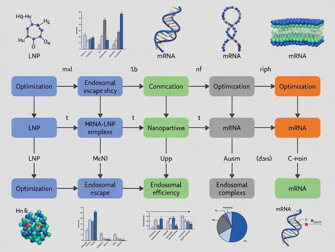
Breaking the Endosomal Barrier: Advanced Strategies to Enhance mRNA-LNP Escape Efficiency
The efficient escape of mRNA-loaded lipid nanoparticles (LNPs) from endosomal compartments is a pivotal yet inefficient step, with less than 2-5% of cargo typically reaching the cytosol.
Breaking the Endosomal Barrier: Advanced Strategies to Enhance mRNA-LNP Escape Efficiency
Abstract
The efficient escape of mRNA-loaded lipid nanoparticles (LNPs) from endosomal compartments is a pivotal yet inefficient step, with less than 2-5% of cargo typically reaching the cytosol. This article provides a comprehensive analysis for researchers and drug development professionals, covering the foundational mechanisms of the endo-lysosomal pathway, innovative methodological advances in lipid and LNP engineering, practical troubleshooting for optimizing delivery efficacy, and rigorous validation techniques. By synthesizing recent preclinical and clinical insights, this review serves as a strategic guide for overcoming this critical bottleneck to unlock the full therapeutic potential of mRNA-based medicines.
The Endosomal Bottleneck: Understanding the Intracellular Journey of mRNA-LNPs
Troubleshooting Guide: Common Experimental Issues in mRNA-LNP Research
Low Transfection Efficiency Despite High Cellular Uptake
Problem: Microscopy or flow cytometry shows LNPs are entering cells, but protein expression (e.g., from reporter genes like luciferase or GFP) remains low. This indicates the mRNA is not successfully reaching the ribosomes in the cytosol.
Solutions:
- Investigate Endosomal Escape: The issue is likely inefficient escape from endosomes. Focus on optimizing the ionizable lipid component of your LNPs. Consider incorporating branched-endosomal disruptor (BEND) lipids, which have been shown to enhance escape and improve mRNA delivery by as much as tenfold compared to linear-tail lipids [1].
- Analyze Endosomal Distribution: Use immunofluorescence to stain for different endosomal markers (e.g., EEA1 for early endosomes, Rab11 for recycling endosomes, LAMP1 for lysosomes). High co-localization with LAMP1 suggests the LNPs are being trafficked to degradative compartments instead of escaping [2].
- Modify Lipid Composition: Introduce unsaturated bonds or degradable groups (e.g., ester bonds) into the ionizable lipid's hydrophobic tail. This promotes a transition to a hexagonal phase structure in the acidic endosome, which destabilizes the endosomal membrane and facilitates escape [3].
High Cytotoxicity Observed with LNP Treatment
Problem: Cell viability decreases significantly after treatment with mRNA-LNPs, confounding experimental results.
Solutions:
- Evaluate Endosomal Damage Kinetics: Rapid and extensive endosomal disruption can trigger cytotoxic and pro-inflammatory pathways. Utilize galectin reporter systems (e.g., Gal8-GFP) to visualize endosomal damage. A high number of Gal8-positive endosomes shortly after LNP treatment correlates with increased toxicity [4] [5].
- Adjust LNP Dose and Serum Concentration: Cytotoxicity often follows a bell-shaped curve. High LNP doses in low-serum conditions can paradoxically reduce uptake and increase toxicity. Optimize the dose and ensure adequate serum concentration (e.g., 2-10% FBS) in your cell culture media to form a "protein corona" that can modulate LNP-cell interactions [5].
- Switch to Biodegradable Lipids: Replace persistent cationic lipids with modern, ionizable lipids that contain ester or disulfide bonds. These are degraded intracellularly, reducing long-term accumulation and cytotoxicity [3] [5].
Inconsistent LNP Performance Between Cell Types
Problem: An LNP formulation that works well in one cell line (e.g., HEK293) shows poor performance in a more therapeutically relevant primary cell line (e.g., adipocytes or fibroblasts).
Solutions:
- Profile Endosomal Subtypes: The endosomal compartments responsible for productive escape can vary. Super-resolution microscopy has revealed that in primary human cells, Rab11-positive recycling endosomes have the highest probability for mRNA escape, not late endosomes. Quantify LNP co-localization with Rab11 as a predictor of efficacy [2].
- Re-formulate with Cell-Type Specific Lipids: Consider helper lipid components. For example, substituting cholesterol with β-sitosterol in the LNP formulation can cause a 10-fold increase in detectable endosomal perturbation events in some cell types, significantly boosting efficacy [4].
Frequently Asked Questions (FAQs)
What is the single biggest bottleneck in the mRNA-LNP delivery pathway?
Endosomal escape is the most significant rate-limiting step. It is estimated that less than 2% of internalized LNPs successfully release their mRNA payload into the cytoplasm [4]. The majority of LNPs remain trapped in the endo-lysosomal pathway and are ultimately degraded.
Is high cellular uptake of LNPs a guarantee of successful protein expression?
No. Total cellular uptake is necessary but not a sufficient predictor of delivery efficacy [2]. Different LNP formulations can have vastly different distributions within the various endosomal compartments. Successful protein expression depends more on the LNP's ability to reach endosomal sub-compartments (like Rab11+ recycling endosomes) that are permissive for escape, rather than simply the total number of particles entering the cell.
How can I directly visualize and quantify endosomal escape in my experiments?
The Galectin 8-GFP (Gal8-GFP) reporter system is a highly sensitive and direct method. Gal8 is a cytosolic protein that rapidly binds to exposed β-galactosides on the inner surface of damaged endosomes. The recruitment of Gal8-GFP to LNP-containing endosomes serves as a clear indicator of endosomal membrane disruption, the initial step of escape [4]. You can quantify these events per cell using fluorescence microscopy.
What are the most promising new strategies to enhance endosomal escape?
Recent advances focus on engineering the lipid components themselves:
- BEND Lipids: Incorporating branching into the tails of ionizable lipids promotes greater endosomal disruption and significantly boosts mRNA delivery and gene-editing efficiency [1].
- Cyclic Disulfide Lipids (CDLs): Adding cyclic disulfide motifs to lipid headgroups can enhance endosomal escape, potentially through thiol-mediated exchange with cell membrane proteins, leading to a more than 2-fold increase in transfection efficiency in vivo [6].
- Metal-Ion Cores: Pre-condensing mRNA with manganese ions (Mn²⁺) to form a high-density core before lipid coating creates stiffer nanoparticles that demonstrate enhanced cellular uptake and efficacy [7].
Table 1: Efficacy and Properties of Novel Lipid Modifications for Endosomal Escape
| Lipid Modification | Key Experimental Finding | Reported Improvement | Proposed Primary Mechanism |
|---|---|---|---|
| BEND Lipids [1] | Enhanced mRNA delivery and gene-editing. | Up to 10-fold increase in efficacy. | Branched tails promote endosomal membrane disruption. |
| Cyclic Disulfide Lipids (CDLs) [6] | Increased protein expression in vitro and in vivo. | >2-fold higher transfection efficiency. | Thiol-mediated interactions enhance endosomal escape. |
| β-Sitosterol (vs. Cholesterol) [4] | Increased Gal8 recruitment, indicating endosomal damage. | 10-fold more endosomal perturbation events. | Altered sterol structure disrupts endosomal membrane integrity. |
| Mn²⁺-mRNA Core (L@Mn-mRNA) [7] | Improved cellular uptake and antigen-specific immunity. | ~2x higher mRNA loading & cellular uptake. | Stiffer nanoparticle core enhances internalization. |
Table 2: Correlation Between LNP Properties and Cellular Outcomes
| LNP Property / Behavior | Correlation with Protein Expression | Correlation with Cytotoxicity | Key Supporting Evidence |
|---|---|---|---|
| Accumulation in Rab11+ Endosomes [2] | Strong positive predictor | Not directly correlated | High fraction in Rab11+ compartments linked to higher efficacy. |
| Accumulation in LAMP1+ Lysosomes [2] | Negative or no correlation | Can be positive | Suggests trafficking to degradative, non-productive pathway. |
| Rapid & Extensive Gal8/Gal9 Recruitment [4] [5] | Positive at moderate levels | Strong positive at high levels | Indicates endosomal damage; excessive damage triggers cell death. |
| Ionizable Lipid pKa ~6.4 [5] | Strong positive | Lower than cationic lipids | Optimal for neutral charge in blood but protonation in endosomes. |
Essential Experimental Protocols
Protocol 1: Using the Gal8-GFP Reporter System to Quantify Endosomal Disruption
Purpose: To directly detect and quantify the endosomal escape capability of your LNP formulations in live cells [4].
Materials:
- Stable Gal8-GFP reporter cell line (e.g., HEK293T/17-Gal8-GFP)
- LNP formulations encapsulating mRNA (e.g., Cy5-labeled for tracking)
- Confocal or high-content fluorescence microscope
- Appropriate cell culture materials
Method:
- Seed and Transfect: Plate Gal8-GFP reporter cells in a multi-well imaging plate and allow them to adhere overnight.
- Treat with LNPs: Add your LNP-mRNA formulations to the cells. Include a positive control (e.g., an LNP known to cause strong escape) and a negative control (e.g., untreated cells or empty LNPs).
- Incubate and Fix: Incubate for a period that allows for robust LNP uptake (e.g., 2-4 hours). Then, fix the cells with paraformaldehyde.
- Image Acquisition: Use a confocal microscope to acquire z-stack images. Capture channels for:
- GFP: Gal8 recruitment (green).
- Cy5: LNP location (red).
- DAPI/Hoechst: Nuclei (blue).
- Image Analysis: Quantify the number of Gal8-GFP puncta that co-localize with Cy5-LNP signal per cell using image analysis software (e.g., ImageJ, CellProfiler). A higher number of co-localized puncta indicates greater endosomal membrane disruption.
Protocol 2: Analyzing LNP Co-localization with Endosomal Markers
Purpose: To determine the intracellular trafficking route and identify in which endosomal sub-compartments your LNPs are located [2].
Materials:
- Target cells (e.g., primary fibroblasts or adipocytes)
- LNP-mRNA (Cy5-labeled)
- Primary antibodies against endosomal markers: EEA1 (early endosomes), Rab11 (recycling endosomes), LAMP1 (late endosomes/lysosomes)
- Fluorescently-labeled secondary antibodies
- Confocal microscope
Method:
- Seed and Treat: Plate cells on glass coverslips and treat with Cy5-labeled LNPs for a desired time course.
- Fix and Permeabilize: Fix cells with 4% PFA and permeabilize with 0.1% Triton X-100.
- Immunostaining: Incubate with primary antibodies against the endosomal markers, followed by incubation with appropriate secondary antibodies.
- Mount and Image: Mount coverslips with antifade mounting medium and image using a super-resolution or confocal microscope.
- Quantitative Analysis: Use co-localization analysis software (e.g., JaCoP plugin for ImageJ) to calculate Pearson's or Manders' coefficients for LNP signal with each endosomal marker. This provides a quantitative measure of LNP distribution.
Critical Signaling Pathways and Experimental Workflows
Diagram 1: The mRNA-LNP endo-lysosomal pathway. The path to Rab11+ recycling endosomes is a key productive route for escape, while trafficking to LAMP1+ late endosomes and lysosomes typically leads to degradation [2] [3].
Diagram 2: Gal8-GFP endosomal escape assay workflow. This protocol allows for direct visualization of LNP-induced endosomal membrane damage [4].
The Scientist's Toolkit: Key Research Reagents
Table 3: Essential Reagents for Studying the Endo-Lysosomal Pathway of mRNA-LNPs
| Reagent / Tool | Function in Experiments | Example Use-Case |
|---|---|---|
| Gal8-GFP Reporter Cell Line [4] | Directly visualizes endosomal membrane damage/rupture. | Quantifying the endosomal escape efficiency of a new LNP formulation. |
| Antibodies for Endosomal Markers (EEA1, Rab11, LAMP1) [2] | Identifies the specific endosomal compartment where LNPs are located. | Determining if poor performance is due to trapping in degradative LAMP1+ lysosomes. |
| Ionizable Lipids (MC3, SM-102, novel BEND lipids) [6] [1] | The primary functional component of LNPs for encapsulating mRNA and facilitating endosomal escape. | Screening different lipids to find one with optimal efficacy and low cytotoxicity for a target cell type. |
| Sterols (Cholesterol, β-Sitosterol) [4] | Helper lipids that influence LNP structure, stability, and endosomal escape potential. | Testing if substituting cholesterol with β-sitosterol enhances endosomal disruption in a specific cell model. |
| Modified mRNAs (N1-methyl-pseudouridine) [5] | Reduces innate immune recognition and increases translational efficiency of the cargo. | Ensuring that low protein expression is a delivery problem, not an mRNA stability or immunogenicity problem. |
| Dynamic Light Scattering (DLS) / Zeta Potential Analyzer | Characterizes LNP physical properties: size (PdI), surface charge (Zeta Potential). | Quality control of LNP formulations before biological experiments; ensuring batch-to-batch consistency. |
Troubleshooting Guide: FAQs on mRNA-LNP Endosomal Escape
FAQ 1: Why is the endosomal escape of mRNA-LNPs so inefficient? The endosomal system is a major delivery bottleneck. After cellular uptake, mRNA-LNPs are trafficked through endosomes that mature into acidic lysosomes, where the mRNA risk degradation by enzymes. Quantitative studies indicate that only about 1–2% of internalized nucleic acid successfully escapes this compartment to reach the cytosol, with the rest being degraded or recycled [8].
FAQ 2: Which specific intracellular compartments are most permissive for mRNA escape? Research using super-resolution microscopy has shown that escape does not occur randomly. The highest probability for mRNA escape is from early endocytic and recycling compartments, particularly those marked by Rab11. In contrast, late endosomes and lysosomes (LAMP1+) are largely unproductive for delivery and are associated with mRNA accumulation and degradation [2].
FAQ 3: What are the consequences of inefficient endosomal escape? Low escape efficiency directly translates to reduced therapeutic protein expression. To compensate, researchers often use a higher LNP dose, but this can lead to increased cytotoxicity. This cytotoxicity can manifest as impaired endosomal acidification and disruption of normal cellular transport pathways [2].
FAQ 4: Does high cellular uptake of LNPs guarantee successful transfection? No. Total cellular uptake is a necessary but insufficient predictor of delivery efficacy. Studies comparing different LNP formulations found that some with moderate uptake levels showed high protein expression (e.g., L608), while others with high uptake (e.g., MC3) showed lower efficacy. The determining factor is the subcellular localization of the LNPs and their access to escape-competent compartments [2].
FAQ 5: How can I experimentally measure and visualize endosomal escape in my lab? Advanced imaging techniques are crucial for quantifying this process.
- Single-molecule Fluorescence In Situ Hybridization (smFISH): Allows for the precise detection and quantification of individual mRNA molecules within cells, helping to distinguish cytosolic mRNA from those still trapped in endosomes [2].
- Super-resolution Microscopy: This can visualize single LNP-mRNAs within subendosomal compartments and has been used to capture rare events of mRNA escape from endosomal recycling tubules [2].
- Immunofluorescence and Confocal Microscopy: By staining for specific endosomal markers (e.g., EEA1, Rab11, LAMP1), you can quantify the co-localization of your LNP-mRNA with different compartments to infer trafficking routes [2].
Table 1: Quantified Compartment Efficiency for LNP-mRNA Escape
| Endosomal Compartment | Marker | Correlation with Escape Efficacy | Key Quantitative Finding |
|---|---|---|---|
| Early Endosome | EEA1/APPL1 | Positive | LNP-mRNA must traverse these compartments, but they have a lower escape probability than recycling compartments [2]. |
| Recycling Endosome | Rab11 | Strongly Positive | Identified as the compartment with the highest probability for mRNA escape [2]. |
| Late Endosome/Lysosome | LAMP1/LBPA | Negative | Accumulation in these compartments is diagnostic of pathways unproductive for delivery and can be a sign of cytotoxicity [2]. |
Table 2: Analytical Methods for Characterizing mRNA-LNP Quality and Function
| Method Category | Specific Technique | Primary Application in mRNA-LNP Analysis |
|---|---|---|
| Electrophoresis | Capillary Gel Electrophoresis (CGE) | Assesses mRNA integrity, size distribution, and identifies truncated species or aggregates [9]. |
| Chromatography | Ion-Pair Reversed-Phase LC (IP-RP LC) | Separates mRNA from impurities based on hydrophobic interactions; used for purity analysis [9]. |
| Mass Spectrometry | Liquid Chromatography-Tandem MS (LC-MS/MS) | Provides detailed information on mRNA sequence and confirms chemical modifications [9]. |
| Functionality Assay | In Vitro Translation / Western Blot | Confirms the production of the full-length, functional target protein after mRNA transfection [9]. |
Experimental Protocol: Analyzing LNP-mRNA Endosomal Trafficking
This protocol outlines a methodology to quantify the uptake and endosomal distribution of LNP-mRNA in cultured cells, based on the work of Paramasivam et al. [2].
1. Cell Preparation and LNP Treatment
- Seed relevant cell types (e.g., primary human adipocytes, fibroblasts, or HeLa cells) onto glass-bottom imaging dishes.
- Once cells reach appropriate confluence, treat them with your LNP-mRNA formulation. It is critical to include multiple LNP formulations with varying chemical compositions for comparative analysis.
- Incubate for a set duration (e.g., 2-4 hours) to allow for uptake and trafficking.
2. Cell Fixation and Immunostaining
- Fix cells with paraformaldehyde.
- Permeabilize cells with a detergent like Triton X-100.
- Perform immunofluorescence staining using antibodies against key endosomal markers:
- Early Endosomes: Anti-EEA1 and Anti-APPL1
- Recycling Endosomes: Anti-Rab11
- Late Endosomes/Lysosomes: Anti-LAMP1
- Use fluorescently-labeled secondary antibodies to visualize these compartments.
3. mRNA Detection via smFISH
- After immunostaining, hybridize cells with fluorescent probes designed against the delivered mRNA (e.g., against eGFP sequence if using eGFP-encoding mRNA).
- This step allows for single-molecule resolution of the mRNA location.
4. Image Acquisition and Quantitative Analysis
- Acquire high-resolution z-stack images using a confocal or, preferably, a super-resolution microscope.
- Use image analysis software (e.g., ImageJ/Fiji with appropriate plugins) to:
- Quantify the total intracellular fluorescence of the LNP-mRNA signal to measure uptake.
- Perform co-localization analysis to determine the percentage of LNP-mRNA signal that overlaps with each specific endosomal marker.
- The fraction of LNP-mRNA in Rab11+ compartments can be a strong predictor of delivery efficacy [2].
The Scientist's Toolkit: Essential Research Reagents
Table 3: Key Reagents for Studying mRNA-LNP Endosomal Escape
| Reagent / Tool | Function / Explanation |
|---|---|
| Ionizable Lipids | The key functional component of LNPs; its structure dictates endosomal escape efficiency by promoting membrane disruption at acidic pH [8]. |
| smFISH Probes | Fluorescent DNA oligonucleotides that bind to specific mRNA sequences, enabling precise localization and quantification of delivered mRNA within cells [2]. |
| Endosomal Marker Antibodies | Antibodies against proteins like EEA1, Rab11, and LAMP1 are essential for identifying and quantifying the distribution of LNPs across the endosomal-lysosomal system [2]. |
| MOD5 / SM-102 Lipids | A class of ionizable lipids used in clinically relevant LNP formulations (e.g., Moderna's mRNA-1273 vaccine); useful as a benchmark in comparative studies [2]. |
| Cholesterol | A structural lipid in LNPs that enhances membrane fusion and stability, indirectly influencing endosomal escape [8]. |
Visualizing the mRNA-LNP Journey and Escape Pathways
LNP mRNA Endosomal Trafficking Pathway
Experimental Workflow for Trafficking Analysis
Core Concepts FAQ
Q1: Why are recycling endosomes, particularly Rab11+ compartments, considered critical for the endosomal escape of mRNA-LNPs?
Recent research indicates that the endocytic recycling compartment, marked by the small GTPase Rab11, is not just a waystation for receptors returning to the plasma membrane. It has been identified as a key site for the escape of delivered mRNA from Lipid Nanoparticles (LNPs). Quantitative analyses, including the use of directed acyclic graphs (DAG) to infer dependencies, have shown that among various endosomal compartments, Rab11+ endosomes have the highest positive differential correlation with mRNA escape. This means that the path of LNP-mRNA from uptake to escape sequentially traverses APPL1+, EEA1+, and Rab11+ compartments, with the Rab11+ compartment being the most probable site for successful release of mRNA into the cytoplasm [2].
Q2: What is the functional consequence of targeting TLR4 to Rab11a+ compartments in immune signaling?
This process is a precise mechanism for regulating specific immune signaling pathways. Research on Toll-like Receptor 4 (TLR4) has shown that it strongly colocalizes with Rab11a in the perinuclear endocytic recycling compartment (ERC). The Rab11a-mediated trafficking of TLR4 to E. coli phagosomes is crucial for activating the MyD88-independent signaling pathway. This pathway leads to the activation of the transcription factor IRF3 and the production of interferon-β (IFN-β), which is vital for anti-viral and broader anti-pathogen immunity. Importantly, suppressing Rab11a disrupts this specific transport, inhibiting IRF3 activation without affecting the NF-κB pathway, demonstrating the compartment's role in signaling specificity [10].
Q3: Are there practical strategies to enhance LNP delivery by targeting recycling endosomes?
Yes, emerging strategies focus on modulating the endosomal trafficking pathway. A significant finding is that targeting recycling endosomes with small molecules can vastly enhance mRNA delivery. A screening approach identified two compounds, NAV and ES5, which significantly increase the delivery efficiency of mRNA-LNPs. Mechanistic studies revealed that these compounds target recycling endosomes to promote mRNA release; NAV blocks the activation of the trafficking regulator ARF6, while ES5 suppresses the function of ANXA6 during early endosome biogenesis [11].
Troubleshooting Guide: Low Transfection Efficiency
| Problem Area | Possible Cause | Investigation Approach | Potential Solution |
|---|---|---|---|
| Cellular Uptake & Trafficking | LNP-mRNA accumulating in late endosomes/lysosomes (LAMP1+), which are unproductive for escape. | Use immunofluorescence to quantify colocalization of LNP-mRNA with markers like EEA1 (early), Rab11 (recycling), and LAMP1 (late). | Re-formulate LNPs to favor the Rab11+ trafficking route. Consider using novel ionizable lipids [2] [12]. |
| Endosomal Escape | Inefficient disruption of the recycling endosome membrane. | Use super-resolution microscopy to visualize mRNA escape events from Rab11+ tubules. Test small molecule enhancers like NAV-2729 [2] [11]. | Incorporate endosomolytic agents (e.g., chloroquine-like lipids) into LNP design. Optimize LNP composition to enhance fusogenicity [12]. |
| Experimental Conditions | Cytotoxicity from prolonged endosomal impairment, leading to defective compartments. | Measure cell viability and assess endosomal acidification status. | Titrate LNP dose to balance efficacy and toxicity. Use biodegradable lipid components to reduce toxicity [2] [8]. |
Detailed Experimental Protocols
Protocol 1: Quantifying LNP-mRNA Colocalization with Endosomal Compartments
This protocol is used to determine the distribution of internalized LNP-mRNA within various endosomal compartments, a key predictor of delivery efficacy [2].
Key Materials:
- Cells: Primary human adipocytes, fibroblasts, or HeLa cells.
- LNPs: Formulations of interest (e.g., MC3, L608, MOD5).
- Antibodies: For compartment markers: EEA1 (early endosomes), APPL1 (early endosomes), Rab11 (recycling endosomes), LAMP1 (late endosomes/lysosomes).
- Detection: smFISH (single-molecule Fluorescence In Situ Hybridization) probes for delivered mRNA.
Methodology:
- Cell Seeding and Transfection: Seed cells on glass-bottom imaging dishes. Transfert with Cy5-labeled LNP-mRNA at the desired dose and for a specific duration (e.g., 2 hours).
- Fixation and Permeabilization: Fix cells with 4% paraformaldehyde and permeabilize with 0.1% Triton X-100.
- Immunostaining: Incubate with primary antibodies against the endosomal markers, followed by appropriate fluorescently-labeled secondary antibodies.
- mRNA Detection: Perform smFISH to detect the delivered mRNA.
- Image Acquisition and Analysis: Acquire high-resolution confocal images. Use image analysis software (e.g., ImageJ) to calculate the Manders' colocalization coefficient between the LNP-mRNA signal (Cy5/smFISH) and each endosomal marker.
Protocol 2: Assessing Functional Endosomal Escape via Rab11 Targeting
This protocol uses small molecule modulators to functionally test the role of recycling endosomes in mRNA delivery [11].
Key Materials:
- Small Molecules: NAV-2729 (ARF6 inhibitor) and ES5 (ANXA6 suppressor).
- Reporter System: LNPs encapsulating mRNA for a reporter protein (e.g., eGFP or firefly luciferase).
Methodology:
- Cell Pretreatment: Pre-treat cells with NAV-2729 (e.g., 10 µM) or ES5 for a specified time (e.g., 1 hour) before LNP addition.
- LNP Transfection: Add LNP-mRNA to the culture medium and incubate.
- Efficiency Quantification:
- For Luciferase mRNA: Measure luminescence activity after 24-48 hours.
- For eGFP mRNA: Analyze the percentage of eGFP-positive cells and mean fluorescence intensity by flow cytometry or fluorescence microscopy 24-48 hours post-transfection.
- Validation: Compare the reporter protein expression in modulator-treated cells against untreated controls. A significant enhancement confirms the role of recycling endosomes as a productive escape route.
The Scientist's Toolkit: Key Research Reagents
| Item | Function / Relevance | Example / Source |
|---|---|---|
| Rab11 Antibodies | Marker for identifying and studying the endocytic recycling compartment (ERC). | Available from various commercial antibody vendors. Used in immunofluorescence and co-immunoprecipitation [10]. |
| smFISH Probes | Enable high-sensitivity, single-molecule detection and visualization of delivered mRNA within endosomal subcompartments. | Custom-designed probes for the target mRNA sequence [2]. |
| Small Molecule Modulators (NAV-2729, ES5) | Chemical tools to perturb and study recycling endosome function. Enhance LNP-mRNA delivery by promoting release from this compartment. | NAV-2729 (ARF6 inhibitor); ES5 (ANXA6 suppressor) [11]. |
| Chloroquine-like Lipids (e.g., CF3-2N6-UC18) | Novel ionizable lipids engineered with endosomolytic activity. Mimic the pH-buffering and membrane-disruptive properties of chloroquine to enhance escape. | Used in "ecoLNP" platforms. Synthesized via modular design combining quinoline scaffolds with hydrophobic tails [12]. |
| Directed Acyclic Graph (DAG) | A mathematical and computational tool used to infer the compartment with the highest probability of mRNA escape based on colocalization data. | Used for data analysis to rank compartments like Rab11+ endosomes as most favorable for escape [2]. |
Supporting Data
Table 1: Comparative Endosomal Distribution of Different LNP Formulations [2]
This table summarizes quantitative data on how different LNP formulations distribute internalized mRNA in various endosomal compartments, which correlates with their overall delivery efficacy.
| LNP Formulation | Relative eGFP Expression (Efficacy) | Colocalization with EEA1+ (%) | Colocalization with Rab11+ (%) | Colocalization with LAMP1+ (%) |
|---|---|---|---|---|
| L608 (High Efficacy) | High | High | Highest | Low |
| MC3 | Medium-High | High | High | Medium |
| MOD5 | Low | Medium | Low | Medium-High |
Pathway and Workflow Diagrams
LNP mRNA Endosomal Escape Pathway
Enhancing Escape Experimental Strategy
Frequently Asked Questions (FAQs)
FAQ 1: What are the primary mechanisms by which Lipid Nanoparticles (LNPs) facilitate endosomal escape? Two predominant theories explain endosomal escape for LNPs [13]. The first involves membrane fusion and the hexagonal HII phase. Ionizable lipids within LNPs become protonated in the acidic environment of the endosome (pH ~6-5). These positively charged lipids can interact with anionic lipids on the endosomal membrane, inducing a transition from a stable lamellar (bilayer) structure to a fusogenic hexagonal HII (HII) phase. This transition disrupts the endosomal membrane, facilitating the release of mRNA into the cytosol [13] [14]. The second is the proton sponge effect. Some materials, such as certain polymers, have buffering capacity in the endosomal pH range. This buffering leads to osmotic swelling and subsequent rupture of the endosome [13] [15].
FAQ 2: Why is endosomal escape considered a major bottleneck in LNP-mRNA delivery? Endosomal escape is highly inefficient. Studies show that while over 95% of LNPs are endocytosed by cells within minutes, less than 2% of the siRNA or mRNA payload successfully escapes the endosome to reach the cytoplasm [16] [13] [17]. The vast majority of LNPs are either degraded in the lysosome or recycled back to the extracellular space. This low efficiency is a primary limiting factor for the potency of RNA therapeutics [13] [17].
FAQ 3: Which intracellular compartments are most permissive for mRNA escape? Recent super-resolution microscopy studies have shifted the traditional view. While late endosomes were often thought to be the main site of escape, evidence now points to early endocytic and recycling compartments as having the highest probability for productive mRNA escape. In contrast, prolonged trapping in acidic compartments can impair endosomal function and is often unproductive for delivery, leading to cytotoxicity [18].
FAQ 4: How does the pKa of an ionizable lipid influence its escape efficiency? The pKa of the ionizable lipid is a critical parameter. An optimal pKa range of approximately 6.2 to 6.5 is generally required for high in vivo efficacy, with one study identifying a pKa of 6.44 as having the highest potency [13]. This range allows the lipid to be neutral at physiological pH (reducing toxicity) but to become positively charged in the mildly acidic environment of early and late endosomes, enabling the membrane interactions necessary for escape [13] [19].
Troubleshooting Guide
Problem: Low Protein Expression Despite High Cellular Uptake of LNP-mRNA This is a classic symptom of inefficient endosomal escape. The cells are taking up the LNPs, but the mRNA is not being released into the cytoplasm for translation.
| Possible Cause | Investigation Method | Proposed Solution |
|---|---|---|
| Suboptimal ionizable lipid pKa | Measure the pKa of the formulated LNPs via techniques like fluorescence-based assays. | Synthesize new ionizable lipids with pKa tuned to the 6.2-6.5 range or screen commercially available lipids with known pKa [13]. |
| Inefficient lipid mixing or membrane disruption | Perform a fluorometric assay to measure lipid mixing or membrane disruption. | Adjust the molar ratio of ionizable lipid to mRNA; a 1:1 molar ratio of mRNA nucleotides to ionizable lipid has been shown to be effective [16]. Incorporate helper lipids like DOPE that promote non-lamellar phase transitions [14]. |
| Trafficking to degradative pathways | Use confocal microscopy with endo-lysosomal markers (e.g., RAB5, RAB7, LAMP1) to track LNP localization. | Modify the LNP surface with targeting ligands to alter cellular uptake pathways and avoid degradative routes [20] [18]. |
Problem: High Cytotoxicity Associated with New LNP Formulation Strategies to enhance escape can sometimes compromise cell health.
| Possible Cause | Investigation Method | Proposed Solution |
|---|---|---|
| Endosomal rupture and lysis | Measure the release of endosomal proteases (e.g., cathepsins) into the cytosol or assess innate immune activation. | Shift from endolytic (rupture) strategies to methods that promote membrane fusion or permeation. Use biodegradable lipids (e.g., with ester linkages) that are cleared quickly to reduce toxicity [15] [21]. |
| Cationic lipid-induced toxicity | Perform cell viability assays (e.g., MTT, LDH) and check for activation of inflammatory pathways. | Ensure ionizable lipids are neutral at physiological pH. Consider incorporating polymers with only tertiary amines, which offer buffering capacity with reduced toxicity compared to those with primary amines [15]. |
Table 1: Key Quantitative Parameters in Endosomal Escape Research
| Parameter | Typical Value / Range | Significance | Measurement Technique |
|---|---|---|---|
| Endosomal Escape Efficiency | < 2% of internalized RNA [16] [13] [17] | Highlights the major delivery bottleneck; target for improvement. | Quantitative fluorescence microscopy, NanoSIMS [18] [17]. |
| Optimal Ionizable Lipid pKa | 6.2 - 6.5 [13] | Crucial for protonation and activity in endosomes without systemic toxicity. | Fluorescence-based TNS assay, potentiometric titration [13]. |
| Endosomal pH Gradient | Early: ~6.5, Late: ~6.0-5.0, Lysosome: ~5.0 [20] | Defines the pH window for triggerable escape mechanisms. | Ratiometric pH-sensitive dyes (e.g., LysoSensor) [20]. |
| Molar Ratio for Escape | ~1:1 (mRNA nucleotides : ionizable lipids) [16] | Suggests neutralization of charge is important for efficient escape. | Formulation screening and in vitro efficacy testing [16]. |
Detailed Experimental Protocols
Protocol 1: Assessing Endosomal Escape Using Co-localization Analysis This protocol uses confocal microscopy to quantify the localization of LNPs within the endo-lysosomal system, providing an indirect measure of escape potential.
- Cell Seeding: Seed appropriate cells (e.g., HeLa, HEK-293) on glass-bottom confocal dishes and culture until 60-80% confluent.
- Fluorescent Labeling: Label your LNP's mRNA with a fluorescent dye (e.g., Cy5) using a commercial labeling kit. The LNP itself can be tagged with a different fluorophore if needed.
- Treatment and Incubation: Add the fluorescently labeled LNP-mRNA to the cells at a predetermined optimal concentration. Incubate for a specific time (e.g., 2-6 hours).
- Staining: Stain the endo-lysosomal compartments. A common method is to use cell-permeable dyes like LysoTracker (for acidic compartments) or transfect cells with plasmids expressing fluorescently tagged markers (e.g., RAB5-GFP for early endosomes, RAB7-GFP for late endosomes, LAMP1-RFP for lysosomes).
- Imaging: Acquire high-resolution z-stack images using a confocal microscope.
- Image Analysis: Use image analysis software (e.g., ImageJ, Volocity) to perform a pixel-based co-localization analysis. Calculate metrics like Manders' Co-efficient or Pearson's Correlation Coefficient between the LNP-mRNA signal and the endosomal marker signals. A decrease in co-localization over time (especially in early/recycling compartments) can indicate successful escape [18].
Protocol 2: Measuring the Fusogenic Potential of Lipids via Fluorometric Assay This in vitro assay monitors the lipid phase transition from lamellar to hexagonal HII, which is linked to membrane fusion and disruption.
- Lipid Film Preparation: Prepare thin lipid films of your LNP formulation or individual ionizable lipids by mixing them in an organic solvent, then evaporating the solvent under a stream of nitrogen.
- Hydration and Probe Incorporation: Hydrate the lipid film with an appropriate buffer to create multilamellar vesicles (MLVs). Incorporate a fluorescence probe that is sensitive to membrane curvature and phase, such as N-NBD-PE.
- Sample Equilibration: Divide the lipid suspension into aliquots in a fluorometer cuvette.
- Temperature and pH Scan: Place the cuvette in a spectrofluorometer with temperature control. Monitor the fluorescence intensity (e.g., excitation at 450 nm, emission at 530 nm) while gradually increasing the temperature or lowering the pH.
- Data Interpretation: A sharp increase in fluorescence intensity is indicative of a transition from a lamellar to a hexagonal HII phase, as the probe moves to a different membrane environment [22] [14]. The temperature or pH at which this transition occurs can be used to compare the fusogenic potential of different lipid formulations.
Visualizing the Escape Pathways
The Scientist's Toolkit: Research Reagent Solutions
Table 2: Essential Reagents for Studying Endosomal Escape
| Reagent / Material | Function in Research | Key Considerations |
|---|---|---|
| Ionizable Lipids (e.g., DLin-MC3-DMA, ALC-0315, SM-102) | Core component of LNPs; protonates in endosomes to enable membrane disruption via the HII phase [19] [21]. | Optimize pKa (target ~6.2-6.5). Consider biodegradability (e.g., ester linkages) for reduced toxicity [13] [15]. |
| Helper Phospholipids (e.g., DOPE) | Aids the transition of lipids from a lamellar to a hexagonal HII phase, thereby enhancing membrane fusion and escape [14]. | More effective than other phospholipids like DSPC in promoting non-lamellar structures [14]. |
| Tertiary Amine-containing Polymers (e.g., custom PAMAM derivatives) | Incorporated into LNPs to provide additional buffering capacity via the "proton sponge" effect, promoting osmotic swelling and endosomal rupture [15]. | Polymers with only tertiary amines show enhanced buffering with lower cytotoxicity compared to those with primary amines [15]. |
| Endo-Lysosomal Trackers (e.g., LysoTracker, RAB GTPase plasmids) | Fluorescent markers to identify and track different endosomal compartments (early, late, lysosomal) during LNP trafficking [20] [18]. | Crucial for co-localization analysis to determine the site and efficiency of escape. |
| Fluorescent Dyes for Membrane Fusion (e.g., N-NBD-PE) | A fluorescent lipid probe used in in vitro assays to detect the lamellar-to-hexagonal (HII) phase transition, indicating fusogenic potential [22] [14]. | Allows for quantitative measurement of a lipid formulation's inherent ability to disrupt membranes. |
Engineering the Great Escape: Cutting-Edge Formulation and Material Strategies
Troubleshooting Guide: Common Ionizable Lipid Challenges
This section addresses frequent experimental hurdles in ionizable lipid-based LNP development and provides evidence-based solutions.
Table 1: Troubleshooting Ionizable Lipid Performance Issues
| Problem Phenomenon | Potential Root Cause | Recommended Solution |
|---|---|---|
| Low RNA Encapsulation Efficiency | Ionizable lipid pKa too high or too low; incorrect lipid ratio | Optimize ionizable lipid to 50 mol% in formulation; adjust lipid pKa to ~6.5 for optimal RNA binding at low pH [23] |
| High Cellular Toxicity | Use of permanently cationic lipids; incorrect lipid degradation profile | Replace cationic lipids with ionizable lipids that are neutral at physiological pH (pH 7.4) [23] |
| Poor Endosomal Escape | Ionizable lipid pKa mismatch; insufficient membrane destabilization | Design ionizable lipids with pKa between 6.0-6.5 to promote protonation and hexagonal phase transition in endosomes [8] [24] |
| Rapid Efficacy Loss During Storage | Generation of aldehyde impurities from lipid degradation | Utilize piperidine-based ionizable lipids (e.g., CL15F series) to limit aldehyde generation and mRNA adduct formation [25] |
| Inefficient Cytosolic RNA Release | Segregation of ionizable lipid from RNA payload in endosome; ESCRT machinery repair | Re-engineer lipid structure to maintain ionizable lipid-RNA complex integrity during endosomal sorting [24] |
Frequently Asked Questions (FAQs)
Q1: What is the fundamental role of an ionizable lipid in an LNP, and why is it superior to a cationic lipid?
Ionizable lipids are the cornerstone of modern LNPs, serving three critical functions: (1) enabling efficient RNA encapsulation by becoming positively charged at low pH, (2) facilitating endosomal escape by promoting a phase change in the endosomal membrane, and (3) reducing toxicity by remaining neutral at the physiological pH of the bloodstream [23]. Unlike permanently cationic lipids, which trigger significant immune responses and cytotoxicity, ionizable lipids are biocompatible, making them viable for therapeutic applications [23].
Q2: My LNPs show excellent encapsulation but poor functional protein expression. What is the most likely bottleneck?
This discrepancy strongly points to inefficient endosomal escape as the primary bottleneck. Quantitative studies indicate that only about 1-2% of nucleic acid cargo typically escapes the endosome [8]. Even when LNPs successfully cause endosomal membrane damage (marked by galectin recruitment), a large fraction of the damaged endosomes show no detectable RNA, and only a small fraction of the RNA is released from those that do [24]. This highlights endosomal escape as the critical rate-limiting step.
Q3: How does the pKa of an ionizable lipid influence its performance, and what is the ideal range?
The pKa is arguably the most critical parameter. An ionizable lipid must be neutral in the bloodstream but become positively charged in the acidic environment of the endosome (pH ~6.0-6.5). The ideal pKa range is typically between 6.0 and 6.8 [23] [8]. A pKa below 6.0 may not protonate sufficiently for effective endosomal disruption, while a pKa above ~6.8 can lead to positive charge at neutral pH, increasing toxicity and accelerating clearance from the blood.
Q4: What strategies can improve the storage stability of mRNA-LNPs in liquid form?
A key strategy is the rational design of the ionizable lipid's amine headgroup. Conventional lipids can generate aldehyde impurities through oxidation/hydrolysis, which react with and inactivate mRNA. Using piperidine-based ionizable lipids (e.g., CL15F series) has been shown to limit this aldehyde generation, allowing mRNA-LNPs to maintain efficacy for months even when stored as a liquid at 4°C [25].
Q5: Beyond pKa, what other lipid design factors affect endosomal escape efficiency?
The chemical structure of the lipid tail (chain length, degree of unsaturation, and branching) is crucial. These factors influence the lipid's ability to transition to an inverted hexagonal phase (HII) upon protonation in the endosome [8] [24]. This phase transition is key to destabilizing the endosomal membrane. Furthermore, recent evidence shows that the ionizable lipid and RNA payload can segregate during endosomal processing, so designing lipids to maintain this association is vital [24].
Key Experimental Data and Protocols
Critical Quality Attributes (CQAs) of LNPs
Table 2: Key Characterization Parameters for Ionizable Lipid Screening
| Parameter | Target Range | Measurement Technique | Functional Significance |
|---|---|---|---|
| Particle Size | 50 - 150 nm | Dynamic Light Scattering (DLS) | Affects biodistribution and cellular uptake [23] [26] |
| Polydispersity (PDI) | < 0.2 | Dynamic Light Scattering (DLS) | Indicates population homogeneity and formulation robustness [23] |
| Encapsulation Efficiency | > 90% | Ribogreen Assay | Measures % of RNA protected from degradation; critical for efficacy [23] |
| Zeta Potential | Near Neutral (slightly negative) at pH 7.4 | Electrophoretic Light Scattering | Predicts colloidal stability and interaction with biological components [26] |
| Apparent pKa | 6.0 - 6.8 | TNS Assay | Dictates pH-responsive behavior and endosomal escape potential [8] [25] |
Standardized Microfluidic Formulation Protocol
This protocol is the gold standard for producing uniform, reproducible LNPs for research [23].
Objective: To prepare mRNA-loaded LNPs with high encapsulation efficiency and controlled size using a microfluidic device.
Reagents:
- Lipid Stock Solution: Ionizable lipid, DSPC, Cholesterol, and DMG-PEG2k dissolved in ethanol at a predetermined molar ratio (e.g., 50:10:38.5:1.5 mol%).
- Aqueous Phase: mRNA diluted in citrate buffer (e.g., 10 mM, pH 4.0). The acidic pH enhances interaction with the ionizable lipid.
Procedure:
- Preparation: Load the lipid solution (organic phase) and the mRNA solution (aqueous phase) into separate syringes.
- Setup: Connect the syringes to a commercial microfluidic device (e.g., a staggered herringbone mixer).
- Mixing: Simultaneously pump the two phases into the device at a defined Total Flow Rate (TFR) and Flow Rate Ratio (FRR). A typical FRR (aqueous-to-organic) is 3:1. The rapid mixing in the micro-channels triggers nanoparticle self-assembly via nanoprecipitation.
- Collection: Collect the formed LNPs in a vial.
- Buffer Exchange/Dialysis: Purify the LNP formulation using tangential flow filtration (TFF) or dialysis against a standard buffer (e.g., PBS, pH 7.4) to remove residual ethanol and exchange the buffer. This step is critical for stability and reducing toxicity.
- Sterile Filtration: Pass the final formulation through a 0.22 µm sterile filter.
The Scientist's Toolkit: Essential Research Reagents
Table 3: Key Materials for LNP Formulation and Characterization
| Reagent / Material | Function / Role | Example |
|---|---|---|
| Ionizable Lipid | pH-responsive component for encapsulation & endosomal escape | ALC-0315, SM-102, DLin-MC3-DMA, CL15F series [23] [25] |
| Phospholipid | Structural component of the LNP bilayer; improves stability | DSPC, DOPE [23] [27] |
| Cholesterol | Enhances membrane integrity and stability; promotes fusion | Pharmaceutical grade cholesterol [23] [26] |
| PEG-lipid | Controls particle size, reduces aggregation, modulates PK | DMG-PEG2000, ALC-0159 [23] [25] |
| Microfluidic Device | Enables reproducible, scalable LNP formation with high EE | Staggered Herringbone Mixer (SHM) [23] |
| TNS Fluorescent Dye | Probe for measuring the apparent pKa of LNPs [25] | 6-(p-Toluidino)-2-naphthalenesulfonic acid (TNS) |
Visualizing the Intracellular Journey and Barriers of mRNA-LNPs
The following diagram illustrates the complex intracellular pathway of mRNA-LNPs, highlighting key barriers and the mechanism of pH-responsive membrane destabilization.
Diagram: Intracellular Journey and Escape of mRNA-LNPs
This workflow details the path from cellular uptake to functional protein expression. The critical event occurs in the late endosome, where the low pH (~5.5) triggers the protonation of the ionizable lipid (pKa ~6.0-6.5). This leads to membrane destabilization and the formation of pores or an inverted hexagonal phase, enabling a small fraction of the mRNA to escape into the cytosol for translation. Major barriers include the segregation of the ionizable lipid from its RNA payload and the activity of the ESCRT machinery, which can repair the membrane damage caused by the LNP [8] [24].
Frequently Asked Questions (FAQs)
Q1: What is the primary mechanistic role of DOPE in enhancing the endosomal escape of mRNA-LNPs?
A1: DOPE (1,2-dioleoyl-sn-glycero-3-phosphoethanolamine) is a cone-shaped phospholipid that promotes membrane fusion due to its propensity for forming inverted hexagonal (HII) phases. This negative curvature is crucial for facilitating the formation of hemifusion intermediates and fusion pores between the LNP and the endosomal membrane. During the endosomal escape process, the drop in pH within the endosome can trigger a phase transition in lipid membranes containing DOPE, destabilizing the endosomal membrane and promoting the release of mRNA into the cytosol [28] [23].
Q2: We are observing inconsistent transfection efficiency with our DOPE-containing LNPs. What are the key formulation factors we should troubleshoot?
A2: Inconsistent performance can often be traced to several critical formulation parameters:
- Lipid Ratios: The molar ratio of ionizable lipid to DOPE is critical. While a typical starting point is a 1:1 mol ratio of ionizable lipid to helper lipid, this requires optimization for your specific ionizable lipid and mRNA cargo [28] [29].
- PEG-lipid Content: There is a known "PEG dilemma." While PEG-lipids are necessary for stability and reducing immunogenicity, they can sterically hinder membrane fusion. A bell-shaped relationship exists between PEG-lipid content and transfection efficiency. You must empirically find the optimal balance; for instance, one study found 1.5% DMG-PEG2000 optimal for in vitro transfection, while 5% was better for in vivo performance [30].
- N/P Ratio: The ratio of amine groups (N) in the ionizable lipid to phosphate groups (P) in the mRNA affects encapsulation efficiency and particle stability. Typical N/P ratios range from 3 to 6, and deviation from the optimal range can lead to poor encapsulation or excessive surface charge, impacting cellular uptake and endosomal escape [29].
Q3: Are there alternatives to DOPE, and when should I consider them?
A3: Yes, DSPC (1,2-distearoyl-sn-glycero-3-phosphocholine) is a common alternative helper lipid. While DOPE is fusogenic and promotes hexagonal phase formation, DSPC forms a more rigid, stable bilayer structure. Your choice should be guided by the application:
- Use DOPE when maximizing endosomal escape and transfection efficiency is the top priority, especially for hard-to-transfect cells [29].
- Consider DSPC when enhanced particle stability and longer circulation half-life are critical, such as for systemic in vivo administration. Many clinically approved LNPs, including those in the Pfizer-BioNTech and Moderna COVID-19 vaccines, use DSPC [29].
Q4: How can I experimentally validate that DOPE is improving endosomal escape in my system?
A4: You can use several advanced microscopy and functional assays:
- Super-Resolution Microscopy: Techniques like TIRF-M (Total Internal Reflection Fluorescence Microscopy) and SIM (Structured Illumination Microscopy) can visualize the interaction of LNPs with model lipid bilayers or cellular membranes, allowing you to observe fusion events (e.g., lipid mixing and content release) directly [28] [24].
- Membrane Damage Assays: Employ galectin recruitment assays. Galectin-3 or -9 proteins bind to exposed glycans upon endosomal membrane damage, serving as a fluorescent marker for LNP-induced endosomal disruption, which is strongly correlated with functional mRNA delivery [24].
- Functional Gene Expression: The most definitive test is to measure the protein output from the delivered mRNA (e.g., luciferase or GFP) and correlate it with the LNP formulation. A significant increase in expression with DOPE-containing LNPs versus alternatives like DSPC is a strong indicator of improved escape [24].
Troubleshooting Guides
Table 1: Common Problems and Solutions for DOPE-containing LNPs
| Problem | Potential Causes | Recommended Solutions |
|---|---|---|
| Low Transfection Efficiency | • Suboptimal DOPE ratio• Excessive PEG-lipid content• Inefficient endosomal escape | • Titrate DOPE from 10% to 50% of neutral lipid content [29].• Systematically reduce PEG-lipid percentage (e.g., from 2.0% to 0.5-1.5%) [30].• Verify endosomal escape using a galectin-9 recruitment assay [24]. |
| High Cellular Toxicity | • Cationic lipid-mediated membrane disruption• Over-protonation in endosome | • Ensure ionizable lipid is used instead of permanently cationic lipids [23].• Optimize the pKa of the ionizable lipid mixture to be near 6.5 for a better efficiency-toxicity profile [31]. |
| Poor Particle Stability (Aggregation) | • Insufficient PEG-lipid• Incorrect total lipid concentration during synthesis | • Slightly increase the PEG-lipid content within the optimal range for stability (e.g., 1.5-5%) [30].• Optimize the total flow rate (TFR) and flow rate ratio (FRR) during microfluidic synthesis to control size and PDI [29]. |
| Low mRNA Encapsulation Efficiency | • Suboptimal N/P ratio• Incorrect buffer pH during formulation | • Increase the N/P ratio, typically between 3:1 and 6:1, to ensure complete mRNA complexation [29].• Formulate LNPs in an acidic buffer (pH 4-5) to protonate the ionizable lipid for better RNA binding [29]. |
Table 2: Quantitative Impact of Helper Lipid Choice on LNP Performance
| Performance Metric | DOPE (Fusogenic) | DSPC (Stabilizing) | Experimental Context & Notes |
|---|---|---|---|
| Endosomal Escape Efficiency | High | Moderate | Measured by galectin-9 recruitment; ~70% "hit rate" for siRNA-LNPs in damaged vesicles [24]. |
| Transfection Efficiency (in vitro) | High | Moderate to Low | DOPE promotes hexagonal phase formation, directly facilitating membrane fusion and content release [23]. |
| Particle Stability | Moderate | High | DSPC's saturated tails confer higher bilayer rigidity and stability in storage and circulation [29]. |
| Optimal Molar Ratio | 10-20% of total lipids | 10% of total lipids | Ratio relative to other components (Ionizable lipid: ~50%; Cholesterol: ~38.5%) [29]. |
Experimental Protocols
Protocol 1: Formulating DOPE-Containing LNPs via Microfluidics
This protocol details the synthesis of mRNA-LNPs using a microfluidic mixer, which provides superior control over particle size and dispersity.
Materials:
- Lipids: Ionizable lipid (e.g., DLin-MC3-DMA), DOPE, Cholesterol, PEG-lipid (e.g., DMG-PEG2000)
- Solvents: Anhydrous Ethanol, 200 mM Acetate Buffer (pH 4.0)
- mRNA: Purified mRNA of interest in nuclease-free water
- Equipment: Microfluidic mixer (e.g., with a herringbone or staggered herringbone mixer), Syringe pumps, Collection tube
Procedure:
- Prepare Lipid Stock Solution: Dissolve the lipids in ethanol at a predetermined molar ratio (a common starting point is 50:10:38.5:1.5 for Ionizable Lipid:DOPE:Cholesterol:PEG-lipid) to a total lipid concentration of 10-20 mg/mL [29].
- Prepare Aqueous Phase: Dilute the mRNA in 200 mM acetate buffer (pH 4.0) to a final concentration of 0.05-0.1 mg/mL. The acidic pH ensures the ionizable lipid is protonated for efficient mRNA encapsulation.
- Set Up Microfluidic System: Load the lipid solution (organic phase) and the mRNA solution (aqueous phase) into separate syringes. Connect them to the inlets of the microfluidic chip.
- Mix and Form LNPs: Set the syringe pumps to the desired Flow Rate Ratio (FRR). A FRR of 3:1 (Aqueous:Organic) is standard. Set the Total Flow Rate (TFR); a higher TFR (e.g., 12 mL/min) produces smaller particles (~80 nm), while a lower TFR (e.g., 4 mL/min) produces larger particles (~150 nm). Initiate simultaneous flow to form LNPs, collecting the effluent in a tube.
- Purification and Buffer Exchange: Use diafiltration or dialysis against PBS (pH 7.4) to remove ethanol, raise the pH to physiological levels, and exchange the buffer. Concentrate the LNPs if necessary.
- Characterization: Measure particle size, PDI, and zeta potential using Dynamic Light Scattering (DLS). Determine mRNA encapsulation efficiency using a Ribogreen assay [30].
Protocol 2: Assessing Endosomal Membrane Damage via Galectin-9 Recruitment
This assay uses the translocation of galectin-9 to damaged endosomes as a biomarker for LNP-induced endosomal escape.
Materials:
- Cells (e.g., HeLa, DC2.4)
- Galectin-9 fluorescent protein construct (e.g., Galectin-9-mRuby3)
- LNP formulations to test
- Live-cell imaging medium
- Confocal or TIRF microscope
Procedure:
- Cell Preparation: Seed cells in an imaging-compatible dish (e.g., a glass-bottom dish) and culture until 60-80% confluent.
- Transfection (Optional): If using an exogenous galectin sensor, transfect cells with the Galectin-9 fluorescent protein construct 24-48 hours before the experiment [24].
- Treat with LNPs: Dilute LNPs in pre-warmed imaging medium and add to the cells. Use a dose that saturates uptake (e.g., 50 nM for siRNA-LNPs or 0.75 µg/mL for mRNA-LNPs) [24].
- Live-Cell Imaging: Place the dish on a pre-warmed microscope stage. Image cells every 30-60 seconds for 1-2 hours after LNP addition.
- Data Analysis:
- Identify foci where Galectin-9 signal rapidly increases, indicating recruitment to a damaged endosome.
- Quantify the number of Galectin-9-positive foci per cell over time.
- Correlate the timing and location of Galectin-9 recruitment with the signal from fluorescently labeled LNPs to confirm the damaging entity.
Signaling Pathways and Workflows
Diagram 1: Molecular Mechanism of DOPE in Endosomal Escape
This diagram illustrates how the cone-shaped structure of DOPE promotes the lipid membrane rearrangements necessary for fusion and endosomal escape.
Diagram 2: Experimental Workflow for DOPE Optimization
This flowchart outlines a systematic experimental approach to optimize LNP formulations containing DOPE.
The Scientist's Toolkit: Research Reagent Solutions
Table 3: Essential Materials for Optimizing Fusogenic LNPs
| Item | Function/Role in Optimization | Example Products & Notes |
|---|---|---|
| Ionizable Lipids | Binds and encapsulates mRNA; protonates in endosome to promote membrane disruption. | DLin-MC3-DMA (MC3), ALC-0315, SM-102. The choice is critical and can be screened using libraries [19] [21]. |
| Fusogenic Helper Lipids | Promotes transition to hexagonal phase, facilitating membrane fusion and endosomal escape. | DOPE (most common). Alternative: DSPC (for stable bilayers) [29]. |
| PEGylated Lipids | Controls nanoparticle size, stability, and PK/PD; impacts cellular uptake and fusion. | DMG-PEG2000, ALC-0159, DSPE-PEG2000. The molar ratio is a key optimization parameter [30] [29]. |
| Sterol Lipids | Enhances membrane integrity and stability of the LNP; aids in fusion. | Cholesterol (universally used). Can constitute ~35-40% of total lipids [23] [29]. |
| mRNA Constructs | The therapeutic cargo; modified nucleotides can enhance stability and reduce immunogenicity. | CleanCap technology, N1-methylpseudouridine modification. Fluorophore-labeled (Cy5) mRNA for tracking [24]. |
| Membrane Damage Reporters | To visually confirm and quantify endosomal escape events. | Galectin-9-mRuby3, Galectin-3-GFP. Recruit to damaged endosomes as a direct readout of LNP activity [24]. |
FAQ: Core Concepts for Researchers
1. What is the primary limitation of PEG-lipids that PCB and BPL technologies aim to solve? The primary limitation is the "PEG dilemma," a trade-off where the PEG coating provides stealth properties and prolonged circulation but acts as a physical barrier that can limit cellular uptake and impair endosomal escape, ultimately reducing intracellular delivery of the therapeutic payload. Furthermore, PEG can be immunogenic; it can induce the formation of anti-PEG antibodies, leading to an Accelerated Blood Clearance (ABC) effect upon repeated dosing, which compromises efficacy and poses safety risks [32].
2. How do Zwitterionic PCB-Lipids fundamentally differ from PEG-lipids in their mechanism? PCB-lipids are zwitterionic, meaning they have both positive and negative charges while maintaining a net neutral charge. This fundamental difference leads to two key mechanistic advantages:
- Enhanced Endosomal Escape: The PCB headgroup can engage in electrostatic and dipole-dipole interactions with the endosomal membrane. This strengthens the LNP-membrane association and facilitates membrane fusion, promoting the release of mRNA into the cytoplasm [32] [33].
- Superior Stealth via Strong Hydration: PCB polymers exhibit stronger hydration than PEG through electrostatic interactions with water molecules, whereas PEG hydrates primarily via hydrogen bonding. This results in extremely low protein adsorption, reducing immunogenicity and mitigating the ABC effect [32] [34].
3. For a project requiring repeated dosing, which technology is more suitable? Both PCB- and BPL-based LNPs are designed to address repeated dosing. Preclinical studies show that both effectively mitigate the Accelerated Blood Clearance (ABC) effect that plagues PEGylated LNPs upon repeated administration [32] [35] [33]. PCB-LNPs achieve this through their low-immunogenicity zwitterionic surface [33], while BPL-LNPs reduce the binding of anti-PEG antibodies through their unique brush architecture [32]. Your choice may depend on secondary needs, such as prioritizing enhanced transfection efficiency (where PCB has strong data) or a closer structural mimic to PEG for easier formulation translation.
4. Are there any known trade-offs or challenges when adopting these next-generation polymers? While promising, these technologies are still relatively new. Key considerations include:
- Structure-Activity Complexity: The performance of BPLs is highly dependent on parameters like side-chain length, degree of polymerization, and lipid anchor length, requiring careful optimization [32].
- Formulation Stability: Removing traditional components like cholesterol (as in some PCB systems) requires compensatory strategies, such as using highly hydrophilic PyCB ILs, to maintain LNP structural integrity and high mRNA encapsulation efficiency [35].
- Clinical Validation: Widespread adoption awaits further testing in clinically relevant models and long-term safety studies to fully assess their therapeutic potential and regulatory pathway [32].
Troubleshooting Guide: Experimental Pitfalls and Solutions
| Problem | Potential Cause | Suggested Solution |
|---|---|---|
| Low Transfection Efficiency with new Polymer-Lipid | Suboptimal polymer-lipid structure or molar ratio leading to poor endosomal escape. | Systematically screen a library of polymer lipids with different molecular weights and acyl chain lengths. Fine-tune the molar percentage in the LNP formulation (e.g., PCB lipid percentage between 1.5-5 mol%) [33]. |
| Particle Aggregation | Inadequate steric stabilization after replacing PEG. | Ensure the polymer-lipid (e.g., PCB or BPL) provides sufficient hydrophilicity and surface coverage. For BPLs, optimize the formulation to achieve the "mushroom regime" conformation for an effective steric barrier [32]. |
| High Immunogenicity or ABC in vivo | The polymer-lipid itself is immunogenic, or pre-existing anti-PEG antibodies are causing cross-reactivity. | Characterize sera for polymer-specific antibodies. Both PCB and BPL have shown reduced immunogenicity and low anti-PEG antibody binding in preclinical models [36] [33]. |
| Inefficient Splenic Transfection | Strong hepatic tropism from traditional LNP composition. | Explore a three-component (ThrCo) LNP system that replaces both cholesterol and PEGylated lipids with a zwitterionic PyCB ionizable lipid, which has been shown to redirect LNPs to the spleen [35]. |
Table 1: Comparative Performance of PCB-LNPs vs. PEG-LNPs in Preclinical Studies
| Metric | PEG-LNPs (Control) | PCB-LNPs (Example: SM102-M2) | Notes / Source |
|---|---|---|---|
| In Vitro Transfection (MFI in Jurkat cells) | Baseline | Significantly higher at all dosages [33] | Consistent across multiple cell lines and LNP systems (SM102, MC3, ALC0315) [33]. |
| CAR+ Jurkat Cell Transfection | ~47% CAR+ cells [33] | >95% CAR+ cells [33] | Twofold increase in transfection percentage with PCB [33]. |
| Anti-Polymer Antibody Induction | Yes (Anti-PEG) [32] | Not detected [33] | PCB-LNPs mitigate the ABC effect [33]. |
| Efficacy in Repeated Dosing | Reduced (ABC effect) [32] [33] | Maintained [33] | PCB-LNPs avoid efficacy loss upon multiple administrations [33]. |
Table 2: Comparative Performance of BPL-LNPs vs. PEG-LNPs (DMG-PEG2000)
| Metric | DMG-PEG2000 LNPs | BPL-LNPs | Notes / Source |
|---|---|---|---|
| Anti-PEG Antibody Binding | High | Effectively blocked [32] [37] | Optimized BPLs adopt a conformation that reduces antibody binding [32]. |
| Performance in Repeated Dosing | Compromised | Superior in protein replacement & genome editing [37] | BPL-LNPs were not recognized by anti-PEG antibodies in mice [37]. |
| Circulation Pharmacokinetics | Standard PEG profile | Can be finely modulated [32] | Tunable by modifying BPL structure [32]. |
Experimental Protocols
Protocol 1: Formulating and Testing PCB-LNPs for Enhanced Transfection
This protocol outlines the key steps for synthesizing and evaluating PCB-containing Lipid Nanoparticles (LNPs) based on established methodologies [33].
1. PCB-Lipid Synthesis:
- Synthesize PCB-lipids via reversible addition–fragmentation chain-transfer (RAFT) polymerization using a 'graft-from' method.
- Generate a library of lipids by varying the polymer molecular weight (e.g., 2 kDa, 4 kDa, 7 kDa) and the lipid acyl chain (e.g., DMG: C14, DSG: C18) [33].
2. LNP Formulation via Pipette Mixing:
- Prepare the lipid mixture by dissolving the ionizable lipid (e.g., SM-102), phospholipid (DSPC), cholesterol, and the synthesized PCB-lipid in ethanol. A standard starting molar ratio is 50:10:38.5:1.5.
- Prepare the aqueous phase containing mRNA in a citrate buffer (e.g., 50 mM, pH 4.0).
- Rapidly mix the two phases using a pipette-based method. For example, combine equal volumes (e.g., 100 µL each) in a tube and pipette up and down vigorously for 10-20 seconds [38].
- Dialyze the resulting LNP formulation against a large volume of PBS (pH 7.4) to remove ethanol and buffer exchange.
3. Quality Control (QC) Assays:
- Size and PDI: Use Dynamic Light Scattering (DLS). Aim for a low Polydispersity Index (PDI < 0.2) indicating a monodisperse population [38].
- Encapsulation Efficiency: Use a Ribogreen assay. Target encapsulation efficiency >90% [38] [33].
- Zeta Potential: Measure surface charge. Typically falls within a range of ±20 mV [38].
4. In Vitro Transfection Efficiency:
- Transfert relevant cell lines (e.g., HeLa, THP-1, Jurkat) with LNPs encapsulating mRNA encoding a reporter gene like Firefly Luciferase (FLuc) or eGFP.
- Luciferase Assay: Quantify luminescence signal 24 hours post-transfection to measure protein production.
- Flow Cytometry: For eGFP mRNA, analyze both the percentage of transfected cells and the Mean Fluorescence Intensity (MFI), which indicates the amount of protein produced per cell. PCB-LNPs consistently show higher MFI than PEG-LNPs [33].
5. In Vivo Repeated Dosing Study:
- Adminulate LNPs systemically to mice (e.g., C57BL/6) on a set schedule (e.g., Day 0, Day 14).
- Monitor protein expression (e.g., luciferase bioluminescence) after each dose.
- Compare the expression levels after the first and subsequent doses. Effective PCB-LNPs will maintain high expression, unlike PEG-LNPs which typically show reduced expression (ABC effect) [33].
Protocol 2: Evaluating BPL-LNPs to Overcome Anti-PEG Immunity
This protocol focuses on screening and validating Brush-shaped Polymer–Lipid (BPL) conjugates for reduced immunogenicity [32] [37].
1. BPL Synthesis and Screening:
- Engineer brush-shaped polymer–lipids (BPLs) using atom transfer radical polymerization (ATRP) to create a library of candidates [32] [37].
- Key parameters to vary include: side-chain length, degree of polymerization, and alkyl anchor length of the lipid tails [32] [37].
- Formulate LNPs with each BPL candidate, replacing DMG-PEG2000.
2. In Vivo Screening for Transfection and Antibody Binding:
- Administer BPL-LNPs encapsulating reporter mRNA to mice.
- Assess transfection efficiency in target tissues (e.g., liver) via bioluminescence imaging or other relevant assays.
- In parallel, use ELISA or surface plasmon resonance (SPR) to test serum for the presence of antibodies that bind to the BPL structure or to PEG. The optimal BPL formulations will show high transfection and low anti-PEG antibody binding [32] [37].
3. Structure-Activity Relationship (SAR) Optimization:
- Correlate the performance data (transfection, antibody binding) with the structural parameters of the BPL library.
- Identify the optimal structure that provides a dense steric barrier (in a "mushroom regime" conformation) to effectively limit the approach and binding of anti-PEG antibodies while retaining favorable pharmacokinetics [32].
4. Validation in Repeated Dosing Models:
- Select the top-performing BPL formulation and subject it to a repeated dosing study, as described in Protocol 1, Section 5. Compare its performance directly against standard PEG-LNPs in models of protein replacement therapy or genome editing [37].
Research Reagent Solutions
Table 3: Essential Materials for Exploring PEG Alternatives
| Reagent / Material | Function | Example & Notes |
|---|---|---|
| PCB-Lipids | Zwitterionic PEG alternative for enhanced endosomal escape and reduced immunogenicity. | Synthesized via RAFT polymerization. Vary polymer MW (2-7 kDa) and acyl chains (DMG, DSG) for optimization [33]. |
| Brush Polymer Lipids (BPLs) | PEG-alternative with brush architecture to reduce anti-PEG antibody binding. | Synthesized via ATRP. Key parameters: side-chain length, degree of polymerization, alkyl anchor length [32] [37]. |
| Ionizable Lipids | Core structural component of LNPs, enables mRNA encapsulation and endosomal escape. | SM-102, ALC-0315. Often kept constant when testing new polymer-lipids [38] [33]. |
| Microfluidic Mixer | Equipment for reproducible, high-efficiency LNP formation. | NanoAssemblr Ignite. Alternative manual methods (T-mixer, pipette mixing) are also effective for small-scale R&D [38]. |
| DMG-PEG2000 | Standard PEG-lipid control for benchmark comparisons. | 1,2-dimyristoyl-rac-glycero-3-methoxypolyethylene glycol-2000; essential for establishing a baseline in head-to-head studies [38]. |
Experimental and Mechanistic Workflows
LNP Formulation and Screening Workflow
Mechanism of PCB Enhanced Endosomal Escape
Frequently Asked Questions (FAQs) & Troubleshooting Guides
Peptide Incorporation (pHLIP)
Q1: What is the primary mechanism by which pHLIP enhances the performance of mRNA-LNPs?
pHLIP (pH Low Insertion Peptide) enhances mRNA-LNP performance by significantly improving endosomal escape. It functions through a pH-dependent mechanism [39]:
- Conformational Change: In the acidic environment of the endosome (pH ~5.5–6.5), the pHLIP peptide undergoes a structural change.
- Membrane Insertion: This change enables the peptide to insert itself directly into the endosomal membrane.
- Facilitated Release: The membrane disruption created by pHLIP promotes the release of the mRNA payload from the endosome into the cytosol, preventing its degradation in lysosomes and making it available for translation [39].
- Efficiency: This process has been shown to increase mRNA expression three to fivefold across multiple cell lines in vitro and leads to sustained, higher protein expression in vivo [39].
Q2: During LNP formulation, at what step should pHLIP be incorporated, and what is a critical parameter to monitor?
pHLIP is typically incorporated during the LNP formulation process. A critical parameter to monitor is the integrity and functionality of the peptide after the encapsulation process. The LNP self-assembly conditions, which often involve solvents and mechanical forces, can potentially denature or inactivate sensitive biological molecules. It is crucial to conduct post-formulation assays to confirm that pHLIP retains its pH-responsive properties.
Q3: Our initial experiments with pHLIP-LNPs show inconsistent results in gene expression. What are potential causes and solutions?
| Potential Cause | Troubleshooting Strategy |
|---|---|
| Suboptimal pHLIP-to-LNP Ratio | Titrate the amount of pHLIP used in the formulation. Test a range of molar ratios to find the optimum for your specific LNP system, as too little may be ineffective and too much could cause premature LNP disruption or toxicity. |
| Inefficient Co-localization | The pHLIP may not be efficiently reaching the endosomal membranes at the same time as the LNPs. Ensure the peptide is stably associated with the LNP, for example, by conjugating it to a lipid anchor (e.g., DSPE-PEG) for incorporation into the LNP surface. |
| Loss of Peptide Activity | Verify the stability of the pHLIP peptide stock and confirm its activity after LNP synthesis using a functional assay. |
Surface Coatings & Functionalization
Q4: What are the key objectives when applying a surface coating or functionalization to mRNA-LNPs?
The primary objectives are [8] [40]:
- Enhanced Targeting: To improve the specificity of LNP delivery to particular cells or tissues, minimizing off-target effects and increasing therapeutic efficacy. This is often achieved by conjugating antibodies or other targeting ligands to the LNP surface [40].
- Improved Stability: To prolong circulation time by reducing nonspecific interactions, aggregation, and clearance by the immune system.
- Modulated Biodistribution: To alter the natural tropism of LNPs (which often accumulate in the liver) to reach other target organs like the spleen, lungs, or specific immune cells [8].
Q5: We are using traditional chemical conjugation (e.g., NHS/EDC) to attach antibodies to our LNPs, but observe poor cell targeting. What might be wrong?
This is a common issue with conventional conjugation chemistry. The problem likely stems from random antibody orientation [40]. Succinimidyl ester (NHS) chemistry reacts with lysine residues, which are scattered throughout the antibody. This random attachment can:
- Block the antigen-binding site (Fab region), rendering the antibody non-functional.
- Lead to uneven and suboptimal presentation on the LNP surface, reducing binding avidity.
Solution: Implement an optimized orientation strategy. Recent advances use an Fc-specific nanobody (TP1107) conjugated to the LNP surface to capture antibodies via their Fc region. This ensures the antigen-binding domains are pointed outward, freely available to engage target cells. This method has shown to increase protein expression by more than 8 times compared to conventional antibody modification techniques [40].
Q6: What are the main considerations when scaling up the production of surface-functionalized LNPs?
| Consideration | Description |
|---|---|
| Conjugation Purification | Complex purification steps to remove unreacted ligands can be a bottleneck. Strategies that allow for "capture" without complex purification (like the nanobody system) are advantageous for scale-up [40]. |
| Reproducibility & Quality Control | Rigorous characterization (size, surface charge, ligand density, functionality) is essential to ensure batch-to-batch consistency. Techniques like dynamic light scattering (DLS) and transmission electron microscopy (TEM) are critical [27]. |
| cGMP Compliance | Manufacturing must adhere to current Good Manufacturing Practices (cGMP), requiring closed-system operations and sterile conditions, especially for fill/finish steps [27]. |
The following table summarizes key quantitative findings from recent research on the enhancement strategies discussed above.
Table 1: Quantitative Efficacy of Ancillary Enhancement Strategies for mRNA-LNPs
| Enhancement Strategy | Key Parameter Measured | Reported Improvement | Experimental Context |
|---|---|---|---|
| pHLIP Incorporation [39] | mRNA Expression | 3 to 5-fold increase | In vitro (multiple cell lines) |
| Protein Expression | Sustained and higher expression | In vivo (mice) | |
| Immune Response | Stronger immune response | Monkeypox vaccine model (A35R & M1R antigens) | |
| Optimized Antibody Capture (TP1107 nanobody) [40] | Protein Expression | >1,000x vs. non-targeted LNPs; >8x vs. conventional antibody conjugation | In vivo targeted delivery |
| Targeting Specificity | Highly efficient T cell targeting; minimal delivery to other immune cells | In vivo systemic administration |
Experimental Protocols
Protocol 1: Incorporating pHLIP into mRNA-LNPs for Enhanced Endosomal Escape
This protocol outlines a method for formulating pHLIP-incorporated LNPs (mRNA@LNP-pHLIP) based on recent research [39].
Key Research Reagent Solutions:
- pHLIP Peptide: The pH-sensitive peptide is the core active ingredient.
- Lipid Mixture: Comprising an ionizable lipid (e.g., DLin-MC3-DMA or SM-102), a phospholipid (e.g., DSPC), cholesterol, and a PEGylated lipid (e.g., DMG-PEG2000).
- mRNA payload: The mRNA of interest, e.g., encoding a reporter protein or vaccine antigen.
- Microfluidic Device: For controlled and reproducible LNP formation.
Methodology:
- Lipid Solution Preparation: Dissolve the ionizable lipid, phospholipid, cholesterol, and PEGylated lipid in ethanol. The pHLIP peptide can be co-dissolved in this lipid mix or pre-conjugated to a PEG-lipid before mixture preparation.
- Aqueous Phase Preparation: Dissolve the mRNA in an acidic citrate buffer (e.g., pH 4.0), which facilitates efficient encapsulation and maintains mRNA integrity.
- LNP Formation: Use a microfluidic device to mix the lipid-ethanol solution and the mRNA aqueous solution at a controlled flow rate ratio (typically 3:1 aqueous-to-ethanol). The rapid mixing triggers nanoparticle self-assembly, encapsulating the mRNA.
- Buffer Exchange & Purification: Dialyze or use tangential flow filtration (TFF) against a phosphate-buffered saline (PBS) at neutral pH to remove ethanol and adjust the pH for storage and administration.
- Characterization: Measure the resulting LNPs for particle size (aim for 80-100 nm), polydispersity index (PDI), encapsulation efficiency (should be >90%), and zeta potential.
Protocol 2: Optimized Antibody Capture for Targeted LNP Delivery
This protocol describes a method for functionalizing LNP surfaces with an oriented antibody capture system using an Fc-specific nanobody [40].
Key Research Reagent Solutions:
- TP1107 Nanobody: An Fc-specific nanobody, engineered with a site-specific synthetic amino acid (e.g., p-azido-phenylalanine) for optimal conjugation.
- DBCO-PEG-Lipid: A lipid-PEG conjugate bearing a Dibenzocyclooctyne (DBCO) group for click chemistry with the azide-modified nanobody.
- Pre-formed "Blank" LNPs: LNPs lacking mRNA or containing your payload, formulated with standard components.
Methodology:
- Nanobody-Lipid Conjugation: Incubate the site-specifically modified TP1107 nanobody (TP1107optimal) with the DBCO-PEG-Lipid. This "click" reaction occurs efficiently without catalysts.
- LNP Functionalization: Incubate the nanobody-lipid conjugate with pre-formed LNPs. The lipophilic tail of the conjugate inserts into the LNP's lipid membrane, presenting the nanobody on the surface.
- Antibody Capture: Simply add the desired monoclonal antibody (e.g., against a T cell surface receptor) to the nanobody-functionalized LNPs. The nanobody will capture the antibody via its Fc region, ensuring optimal orientation. No further purification is needed.
- Validation: Confirm antibody binding and functionality using flow cytometry to demonstrate enhanced binding to target cells versus non-target cells.
Mechanism and Workflow Visualization
pHLIP-Mediated Endosomal Escape
Optimized Antibody Capture Workflow
Overcoming Hurdles: A Practical Guide to Improving LNP Efficacy and Reducing Toxicity
Core Mechanisms: Understanding the PEG Dilemma and Immunogenicity
What is the fundamental "PEG Dilemma" in mRNA-LNP design?
The "PEG dilemma" describes the critical trade-off where polyethylene glycol (PEG) lipids provide essential "stealth" properties to Lipid Nanoparticles (LNPs) but simultaneously impair intracellular delivery and can trigger undesirable immune responses [32] [30] [41].
PEG lipids consist of a hydrophobic lipid anchor and a hydrophilic PEG chain that extends into the surrounding environment. By forming hydrogen bonds with water molecules, PEG creates a hydration layer and generates steric hindrance on the LNP surface. This gives LNPs their stealth properties, helping them evade immune detection, reduce macrophage phagocytosis, and prolong circulation time in the bloodstream [32].
However, this beneficial dense PEG coating also acts as a physical barrier that can reduce interactions between the nanoparticle and biological membranes. This limitation can restrict cellular uptake and impair the release of the therapeutic payload (e.g., mRNA) into the cytoplasm, ultimately reducing therapeutic efficacy [32] [41]. Furthermore, PEG introduces immunogenicity concerns. Administration of PEGylated nanoparticles can trigger the formation of anti-PEG antibodies (APAs), which can bind to the PEG on LNPs and accelerate their clearance from the blood—a phenomenon known as the Accelerated Blood Clearance (ABC) effect. This poses potential risks for reduced efficacy and hypersensitivity reactions upon repeated dosing [32] [42].
How are anti-PEG antibodies generated?
Anti-PEG antibody (APA) generation occurs through two primary immunological pathways [42]:
Thymus-Dependent (TD) Response: This pathway is triggered by larger, more complex PEGylated therapeutics like PEGylated proteins. B-cell receptors (BCRs) bind to the PEG backbone, activating B cells. With signals from CD4+ T helper cells (specifically follicular helper T cells), these B cells differentiate into plasma cells that secrete anti-PEG IgG antibodies. This pathway typically generates high-affinity, class-switched antibodies.
Thymus-Independent (TI-2) Response: This pathway is triggered by the highly repetitive, multivalent structure of the linear PEG polymer itself, which can cross-link BCRs on the cell surface. This activation occurs without T-cell help, primarily involving marginal zone B cells and B1 cells, and leads to the production of anti-PEG IgM antibodies.
The immunogenicity of PEG is influenced by its physicochemical properties, such as molecular weight (higher molecular weight PEGs are more immunogenic), grafting density, and terminal functional groups [42].
Troubleshooting Guide & FAQs
How can I optimize PEG-lipid content to balance stability and intracellular delivery?
Optimizing PEG-lipid content is a critical formulation parameter that follows a bell-shaped relationship with transfection efficiency. Systematic studies reveal different optimal concentrations for in vitro versus in vivo performance, reflecting the classic PEG dilemma [30].
Recommended Protocol: PEG-Lipid Titration for mRNA-LNPs
- Formulate LNPs using a fixed base lipid composition (e.g., ionizable lipid, cholesterol, helper lipid) and systematically vary the molar ratio of your PEG-lipid (e.g., DMG-PEG2000) from 0.5% to 10%.
- Characterize the LNPs for each condition. Measure particle size, polydispersity index (PDI), zeta potential, and mRNA encapsulation efficiency using dynamic light scattering (DLS) and a fluorescence-based assay like the Quant-iT RiboGreen RNA Assay [30].
- Evaluate performance:
- In vitro: Transfert relevant cell lines (e.g., HeLa, DC2.4) with mRNA-LNPs encoding a reporter gene (e.g., GFP, Luciferase) and measure protein expression 24-48 hours post-transfection.
- In vivo: Administer formulations intravenously to animal models and measure reporter gene expression in target organs (e.g., liver) 6-24 hours post-injection.
The table below summarizes typical findings from such a titration experiment, illustrating the trade-offs [30]:
Table 1. Impact of PEG-Lipid Content on LNP Properties and Performance
| DMG-PEG2000 Molar Ratio | Particle Size | PDI | Encapsulation Efficiency | In Vitro Transfection | In Vivo Performance | Key Trade-off |
|---|---|---|---|---|---|---|
| Low (e.g., 0.5-1.0%) | Larger | Higher | Moderate | Optimal | Suboptimal | Stability vs. Uptake |
| Medium (e.g., 1.5%) | Intermediate | Lower | High | High | Good | Balanced |
| High (e.g., 5-10%) | Smaller | Lowest | Highest | Low | Optimal | Uptake vs. Circulation |
FAQ: Why are the optimal PEG levels different for in vitro and in vivo applications? The discrepancy arises from the different challenges faced in each setting. In vitro, the primary goal is efficient cellular internalization, which is hindered by high PEG levels. In vivo, LNPs must first navigate the bloodstream, avoid immune clearance, and reach target tissues. Higher PEG content enhances colloidal stability and prolongs circulation time, which is critical for bioavailability in vivo, even if it slightly compromises cellular uptake at the final destination [30].
What formulation strategies can mitigate anti-PEG immunogenicity?
Several advanced strategies are being developed to overcome the limitations imposed by anti-PEG antibodies.
1. PEG Structural Engineering:
- Branched or Y-shaped PEG: These structures can significantly reduce the production of anti-PEG antibodies compared to conventional linear PEG, as their complex architecture may hinder antibody recognition [32].
- Cleavable PEG: Incorporating acid-responsive or enzyme-responsive linkers between the PEG chain and the lipid anchor allows the PEG shell to be shed after the LNP reaches the target cell. This maintains the stealth function during circulation but removes the barrier before cellular uptake and endosomal escape, enhancing therapeutic efficacy [32].
- Brush-like Polymer–Lipid (BPL) Conjugates: Replacing linear PEG with a brush-shaped polymer, where multiple short ethylene glycol side chains branch from a single backbone, can create a dense steric barrier that effectively limits the approach and binding of anti-PEG antibodies while retaining pharmacokinetic benefits [32].
2. PEG Replacement with Alternative Stealth Lipids:
- Poly(carboxybetaine) (PCB) Lipids: PCB is a zwitterionic polymer (carrying both positive and negative charges with a net neutral charge). PCB-lipids offer superior stealth properties with extremely low protein adsorption, reducing immunogenicity and the ABC effect. A key advantage is that the PCB headgroup can engage in electrostatic interactions with the endosomal membrane, potentially strengthening LNP-membrane association and enhancing endosomal escape, a critical step for mRNA delivery [32].
- Ganglioside-Incorporating LNPs: Gangliosides are naturally occurring glycosphingolipids that can be incorporated into LNPs as a natural, PEG-free stealth alternative. Recent studies show that ganglioside-LNPs exhibit superior stability during storage and prevent serum protein adsorption. They also enable successful in vivo mRNA transfection, making them a promising candidate to replace immunogenic PEG [43].
3. Immunomodulation Strategies:
- Co-administration of High-Molecular-Weight PEG: Injecting free, high-mass PEG (≥30 kDa) can transiently occupy B-cell receptors and bind to circulating anti-PEG antibodies, reducing their ability to bind to and clear the administered PEGylated LNPs. However, high doses of free PEG may have their own safety concerns, such as organ accumulation [32].
- Use of Tolerogenic Nanoparticles: The administration of specially designed nanoparticles can induce immune tolerance, potentially suppressing the formation of new anti-PEG antibodies [32].
How does PEG-lipid structure (chain length, tail, linkage) influence LNP performance and immunogenicity?
The chemical structure of the PEG-lipid is a major determinant of LNP behavior. Key parameters include PEG chain length, lipid tail length and structure, and the chemical linkage between them [41].
Table 2. Impact of PEG-Lipid Structure on LNP Performance
| Structural Feature | Impact on LNP Properties & Performance | Considerations for Experimental Design |
|---|---|---|
| Lipid Tail Length | ||
| → Short Tail (e.g., C14, DMG-PEG) | Rapid detachment from LNP in vivo; enhances cellular uptake and endosomal escape. | Preferred for intracellular delivery (e.g., mRNA vaccines). Used in FDA-approved LNPs (ALC-0159, DMG-PEG) [41]. |
| → Long Tail (e.g., C18, DSPE-PEG) | Slow detachment; prolonged circulation time; increased liver accumulation; higher risk of anti-PEG antibody response. | More suitable for long-circulating liposomal drugs (e.g., Doxil) [41]. |
| Chemical Linkage | ||
| → Ester Bond | Hydrolytically cleavable. | Common, facilitates PEG shedding. |
| → Carbamate / Phosphate | More stable linkage. | Limited impact on physicochemical properties vs. ester bonds [41]. |
| PEG Chain Length | ||
| → ~2000 Da | Good balance of stealth and efficacy. | Most commonly used in clinical LNP formulations [41]. |
| → >2000 Da | Increased immunogenicity and allergic risk. | Generally avoided in next-generation designs [32] [41]. |
| Terminal Group | ||
| → Methoxy-PEG | Standard terminus. | - |
| → Hydroxyl-PEG (HO-PEG) | Lower immunogenicity; validated in Moderna's clinical formulations (e.g., OL-56) [32]. | Promising for reducing anti-PEG antibody responses. |
Detailed Experimental Protocols
Protocol: Evaluating Anti-PEG Antibody Generation in Preclinical Models
Objective: To assess the immunogenic potential of a novel LNP formulation by measuring the induction of anti-PEG IgM and IgG antibodies in a mouse model after single and repeated administration.
Materials:
- Test and control LNP formulations (e.g., novel PEG-LNP vs. standard PEG-LNP)
- Animal model (e.g., C57BL/6 mice)
- ELISA plates
- PEG-conjugated coating antigen (e.g., PEG-BSA)
- Blocking buffer (e.g., BSA or casein)
- Serum samples collected pre-injection and at various time points post-injection
- Detection antibodies: Horseradish peroxidase (HRP)-conjugated anti-mouse IgM and anti-mouse IgG
- ELISA substrate (e.g., TMB) and stop solution
- Plate reader
Method:
- Immunization and Serum Collection: Administer the LNP formulation to mice via the intended route (e.g., intravenous, intramuscular). Include a naive group as a negative control. Collect blood samples pre-injection (baseline), and post-injection (e.g., Day 7 for IgM peak, Day 14-28 for IgG response). For assessing the Accelerated Blood Clearance (ABC) effect, administer a second dose 1-2 weeks after the first and collect samples shortly thereafter [42].
- Anti-PEG IgM/IgG ELISA: a. Coating: Coat ELISA plate wells with a PEG-conjugated antigen (e.g., PEG-BSA) overnight. b. Blocking: Block plates with a suitable protein-based blocking buffer. c. Sample Incubation: Add diluted serum samples and incubate. Include a standard curve if using a quantitative ELISA. d. Detection: Add HRP-conjugated anti-mouse IgM or IgG detection antibodies. e. Development: Add substrate, stop the reaction, and measure absorbance.
- Data Analysis: Compare antibody levels in post-injection sera to baseline levels. A significant increase indicates an anti-PEG immune response. The ABC effect can be correlated with high pre-existing anti-PEG antibody titers [42].
Protocol: Testing Endosomal Escape Efficiency Using a Galectin-8 Reporter System
Objective: To quantitatively compare the endosomal escape efficiency of different LNP formulations.
Materials:
- Galectin-8 reporter cell line (e.g., HEK293T Gal8-YFP)
- LNP formulations to be tested
- Confocal microscope or high-content imaging system
- Positive control (e.g., a transfection reagent known to cause endosomal damage)
Method:
- Cell Seeding: Seed Gal8-YFP reporter cells in an appropriate multi-well plate.
- Transfection: Treat cells with the LNP formulations.
- Fixation and Imaging: At a predetermined time point post-transfection (e.g., 4-8 hours), fix the cells and image using a microscope. Galectin-8 recruits to ruptured endosomes; thus, endosomal escape is indicated by the formation of bright, punctate fluorescent signals (Gal8 foci) in the cytoplasm [44].
- Quantification: Quantify the number of Gal8 foci per cell or the percentage of cells with Gal8 foci. A higher number of foci indicates more efficient endosomal escape. As referenced in studies, LNPs designed for rapid hydrolysis (like acid-degradable ADP-LNPs) demonstrate superior endosomal escape compared to non-degradable PEG-LNPs (NDP-LNPs) in this assay [44].
The Scientist's Toolkit: Research Reagent Solutions
Table 3. Essential Reagents for Investigating the PEG Dilemma
| Reagent / Material | Function / Role | Key Examples & Notes |
|---|---|---|
| Ionizable Lipids | Core structural component; binds mRNA and enables endosomal escape via protonation in acidic endosomes. | ALC-0315 (Pfizer), SM-102 (Moderna), synthetic ionizable lipids (e.g., C12–200) [30] [41]. |
| PEG-Lipids | Provides stealth properties, controls particle size, reduces aggregation. Critical for studying the PEG dilemma. | DMG-PEG2000 (short tail, fast shedding) [30]. DSPE-PEG2000 (long tail, stable) [41]. HO-PEG lipids (e.g., OL-56; lower immunogenicity) [32]. Cleavable PEG-lipids (e.g., acid-degradable) [32] [44]. |
| Helper Lipids | Supports LNP structure and membrane integrity. | DSPC (Phospholipid) [41]. DOPE (can promote membrane fusion) [30]. |
| Cholesterol | Enhances membrane stability and fluidity, improves LNP integrity. | Plant-derived cholesterol for clinical applications [32]. |
| PEG Alternatives | Replaces PEG to circumvent immunogenicity. | Poly(carboxybetaine) (PCB) lipids [32]. Gangliosides (natural stealth lipids) [43]. Brush-shaped polymer–lipid (BPL) conjugates [32]. |
| Characterization Tools | Measures LNP physical properties. | DLS/ZetaSizer (size, PDI, zeta potential) [30]. TEM/cryo-TEM (morphology) [30] [44]. RiboGreen Assay (encapsulation efficiency) [30]. |
| Bioassays | Evaluates biological performance and immunogenicity. | Galectin-8 Reporter Assay (endosomal escape) [44]. Anti-PEG ELISA (antibody detection) [42]. In vivo imaging (biodistribution, protein expression) [30]. |
Frequently Asked Questions (FAQs)
FAQ 1: How does the degree of unsaturation in an ionizable lipid's tail influence mRNA delivery efficacy?
The degree and configuration of unsaturation in the hydrophobic tails are critical factors that influence membrane fluidity and the lipid's ability to promote endosomal escape, but they are not universally improving. Introducing unsaturation must be done strategically.
- Core Issue: Simply having unsaturation does not guarantee improved efficacy; the specific position, configuration (cis/trans), and the resulting "stiffness" of the tail are the determining factors [45].
- Solution & Data: A systematic study of a 91-lipid library revealed that an ionizable lipid with a citronellol-based unsaturated periphery (4A3-Cit) significantly outperformed its saturated counterparts and other unsaturated variants in vivo. A FRET-based fusion assay demonstrated that 4A3-Cit LNPs showed superior ability to fuse with and disrupt model endosomal membranes, directly linking its specific unsaturated structure to enhanced endosomal escape [45].
Table 1: Impact of Tail Unsaturation on LNP Efficacy (4A3 Lipid Series)
| Lipid Tail | Tail Description | Relative In Vivo Luciferase Expression | Key Finding |
|---|---|---|---|
| 4A3-Cit | Citronellol-based unsaturated tail | ~18-fold increase over saturated基准 | Best performer; promotes membrane fusion [45] |
| 4A3-SC8 | Saturated alkyl tail | Baseline | Lower efficacy than 4A3-Cit [45] |
| 4A3-8/2 | Unsaturated tail (cis/trans not specified) | Lower than saturated SC8 | Demonstrates unsaturation alone is insufficient [45] |
| 4A3-Far | Farnesol-based unsaturated tail | Lower than 4A3-Cit | Higher lysosomal accumulation; unproductive delivery [45] |
FAQ 2: What is the mechanism by which acid-degradable groups in lipids improve endosomal escape?
Acid-degradable groups are engineered to rapidly hydrolyze in the acidic environment of the endosome (pH ~5.0-6.0). This rapid degradation can enhance endosomal disruption by promoting lipid rearrangement and membrane fusion, releasing the mRNA cargo into the cytoplasm before degradation in lysosomes occurs [44].
- Core Issue: Conventional LNPs have low endosomal disruption rates, with less than 2% of cargo escaping. Many existing acid-degradable linkers hydrolyze too slowly (over hours or days) to be effective before lysosomal degradation [15] [44].
- Solution & Data: The "azido-acetal" linker is a next-generation technology that remains stable at physiological pH (half-life of 21 days at pH 7.4) but hydrolyzes rapidly after a reduction step (half-life of ~15 minutes at pH 6.0) [44]. LNPs formulated with an acid-degradable PEG lipid (ADP-LNPs) showed substantially higher luciferase activity and more efficient endosomal escape in Galectin-8 reporter assays compared to non-degradable PEG LNPs (NDP-LNPs) [44].
Table 2: Performance of Acid-Degradable vs. Standard LNPs
| LNP Type | Key Feature | Hydrolysis Half-Life (pH 6.0) | Relative Luciferase Expression |
|---|---|---|---|
| RD-LNP (Azido-Acetal) | Rapidly degradable in endosomes | ~15 minutes | Significantly higher in liver, lung, spleen, and brain vs. Std-LNP [44] |
| ADP-LNP | Acid-degradable PEG lipid | Rapid hydrolysis post-reduction | Superior to NDP-LNPs [44] |
| Std-LNP | Conventional, non-degradable lipids | Slow or non-degradable | Baseline [44] |
| NDP-LNP | Non-degradable PEG lipid | Non-degradable | Ineffective at transfecting cells with high PEG content [44] |
FAQ 3: Can incorporating tertiary amine-based polymers into LNPs enhance delivery, and how does this relate to lipid unsaturation?
Yes, incorporating biodegradable polymers containing only tertiary amines can significantly enhance mRNA delivery by introducing an additional proton sponge effect alongside the fusogenic activity of ionizable lipids. This strategy is complementary to optimizing tail unsaturation.
- Core Issue: The primary mechanism of endosomal escape for standard LNPs (membrane fusion via ionizable lipids) is inefficient [15].
- Solution & Data: Spiking polymers with tertiary amines (e.g., NS201, NS202) into standard LNP formulations enhances their pH-buffering capacity in the endosome. This works in concert with the ionizable lipid. The polymer absorbs protons, causing osmotic swelling and rupture of the endosome, while the ionizable lipid (whose unsaturation can be independently optimized for fusion) also promotes membrane disruption. This dual mechanism significantly improved endosomal escape and in vivo mRNA expression compared to standard LNPs [15].
FAQ 4: Why do my LNPs show good cellular uptake but poor protein expression?
This is a common problem often linked to the formation of a protein corona on the LNPs after administration, which can alter their intracellular trafficking route, diverting them to lysosomes for degradation instead of enabling endosomal escape [46].
- Core Issue: Increased cellular uptake does not correlate with increased mRNA expression if the protein corona reroutes the LNPs to lysosomes or otherwise impedes endosomal escape [46].
- Solution & Data: A quantitative proteomics study identified specific proteins (e.g., vitronectin) enriched in the LNP hard corona. When a pre-formed corona with these proteins was present, cellular uptake increased by up to five-fold, but this did not translate to increased mRNA expression. This suggests the corona compromises the LNP's endosomal escape function. Strategies to modulate the protein corona, such as tuning PEG density or surface composition, may help resolve this disconnect [46].
Troubleshooting Guides
Problem: Low Endosomal Escape Efficiency
Potential Cause 1: Suboptimal ionizable lipid structure, particularly in the hydrophobic tail region.
- Recommended Action: Systematically evaluate a series of ionizable lipids with varying degrees and types of unsaturation in their tails. Use a FRET-based membrane fusion assay to directly quantify the lipid's ability to fuse with and disrupt endosomal membranes [45].
- Experiment Protocol: FRET-based Membrane Fusion Assay
- Prepare Donor Vesicles: Formulate liposomes that mimic the endosomal membrane, incorporating the FRET pair NBD-PE (donor) and N-Rh-PE (acceptor). At close proximity, the NBD fluorescence is quenched by rhodamine.
- Incubate with LNPs: Mix the donor vesicles with your LNP formulations at a predetermined ratio in a buffer at pH 4.5-5.0 to simulate the endosomal environment.
- Measure Fluorescence: Monitor the increase in NBD fluorescence (de-quenching) over time using a fluorescence plate reader. The signal increase indicates membrane fusion and disruption, which separates the FRET pair.
- Analyze Data: Calculate the initial rate or total extent of fluorescence increase. A higher value indicates superior membrane fusion capability [45].
Potential Cause 2: Lack of a rapid, pH-dependent degradation mechanism.
- Recommended Action: Incorporate acid-degradable lipids into your formulation. Consider using linkers like the azido-acetal, which offers a unique combination of stability at pH 7.4 and rapid hydrolysis at endosomal pH after a reduction step [44].
- Experiment Protocol: Evaluating Acid-Degradable LNPs (ADP-LNPs)
- Synthesize Acid-Degradable Lipid: Synthesize a PEG-lipid or ionizable lipid using the azido-acetal linker.
- Formulate LNPs: Formulate LNPs using standard microfluidic methods, substituting the conventional PEG-lipid with your acid-degradable version (e.g., at 10-20 mol%).
- Pre-activate LNPs: Reduce the azido group to an amine by adding a thiol-containing agent (e.g., TCEP) to the LNP formulation shortly before administration. This step activates the rapid-hydrolysis function.
- In Vitro Transfection: Transfect HEK 293T or other relevant cells with ADP-LNPs and NDP-LNPs (control) encapsulating a reporter mRNA (e.g., Luciferase or GFP). Measure protein expression to confirm enhanced efficiency [44].
Problem: Inconsistent Results Due to LNP Instability
Potential Cause: Exposure to mechanical stress (e.g., shaking, pipetting) during handling, leading to particle fusion, payload leakage, and loss of efficacy [47].
- Recommended Action: Minimize exposure to liquid-air interfaces. Avoid vortexing or vigorous shaking of LNP samples. When handling is necessary, use gentle pipetting with wide-bore tips.
- Experiment Protocol: Assessing Mechanical Stress on LNPs
- Stress Samples: Aliquot your LNP formulation and subject it to controlled shaking on a platform shaker (e.g., 100 movements/minute) for set durations (e.g., 30 min, 240 min).
- Characterize Stressed LNPs:
- Use Nanoparticle Tracking Analysis (NTA) to monitor changes in particle size, concentration, and heterogeneity.
- Use Cryo-EM with RNA staining (e.g., thionine) to visualize morphological changes and the appearance of unencapsulated mRNA.
- Use NMR spectroscopy to detect changes in lipid mobility and surface structure, such as the dissociation of PEGylated lipids [47].
The Scientist's Toolkit: Essential Research Reagents
Table 3: Key Reagents for Investigating LNP Structure-Activity Relationships
| Reagent / Tool | Function in Research | Key Insight from Literature |
|---|---|---|
| Ionizable Lipids (4A3 series) | Systematic study of tail unsaturation. The 4A3 core with Citronellol tail (4A3-Cit) emerged as a top performer for in vivo mRNA delivery [45]. | |
| Azido-Acetal Linker | Enables synthesis of Rapidly Degradable LNPs (RD-LNPs). Stable at pH 7.4 but hydrolyzes within minutes at pH 6.0 post-reduction [44]. | |
| NS Polymers (e.g., NS201) | Biodegradable polymers with only tertiary amines. Spiked into LNPs to enhance the proton sponge effect and improve endosomal escape without high cytotoxicity [15]. | |
| FRET Probes (NBD-PE & N-Rh-PE) | Incorporated into model endosomal membranes to quantitatively measure LNP-membrane fusion, a key step for endosomal escape [45]. | |
| DMG-PEG5k | A PEGylated lipid identified through systematic screening to optimize intramuscular mRNA delivery formulations [48]. |
Mechanism and Workflow Diagrams
Optimizing Cholesterol and Its Analogs for Improved Membrane Integrity and Endosomal Release
Frequently Asked Questions & Troubleshooting Guides
How can cholesterol components be modified to prevent unwanted liver uptake in locally administered LNPs?
Problem: After intramuscular or intratumoral injection, conventional LNPs diffuse into systemic circulation and show significant liver transfection. This off-target effect can cause liver injury and reduce therapeutic efficacy at the intended site [49].
Solution: Incorporate novel cholesterol analogs like glutamate-cholesterol (GA-Chol) into LNP formulations.
- Mechanism: Replacing standard cholesterol with GA-Chol significantly alters LNP biodistribution. When administered locally, GA-Chol LNPs remain at the injection site and demonstrate robust localized transfection without significant liver transfection [49].
- Experimental Evidence: In studies, intramuscular or intratumoral injection of GA-Chol LNPs resulted in strong protein expression at the injection site with negligible expression in the liver, unlike conventional LNPs which showed high liver transfection [49].
- Protocol Implementation: Replace cholesterol with GA-Chol at 20-38.5 mol% in LNP formulations. The synthesis involves conjugating glutamic acid to cholesterol via a reaction with dimethyl L-glutamate hydrochloride and triphosgene, followed by saponification [49].
What strategies can enhance endosomal escape efficiency in LNP systems?
Problem: A major bottleneck in LNP-mediated mRNA delivery is inefficient endosomal escape, with most internalized LNPs failing to release their cargo into the cytosol [24].
Solution: Optimize cholesterol content and type to improve membrane fusion and disrupt endosomal membranes.
- Mechanism: Cholesterol regulates membrane fluidity and promotes the phase transition to inverted hexagonal structures necessary for endosomal membrane disruption [50]. Recent research shows that only a small fraction of endosomes containing LNPs experience membrane damage conducive to RNA release, and even fewer successfully release their RNA payload [24].
- Experimental Evidence: Studies using galectin-9 as a membrane damage marker revealed that only a subset of LNP-containing endosomes experience damage, and of those, only a fraction successfully release RNA cargo. Improving LNP composition can increase this efficiency [24].
- Protocol Implementation: Systematically vary cholesterol molar ratios (typically 25-45%) using Design of Experiments (DoE) approaches. Assess membrane integrity using Laurdan assay measuring generalized polarization (GP) ratio at pH 7.5 and 4.5. A lower GP7.5/GP4.5 ratio correlates with enhanced protein expression [51].
How can researchers balance LNP stability with endosomal release functionality?
Problem: Achieving sufficient LNP membrane integrity for protection and circulation while maintaining the ability to disrupt endosomal membranes represents a significant design challenge [51] [52].
Solution: Implement cholesterol derivatives and optimize lipid ratios to create "liposomal LNPs" with bilayer structures.
- Mechanism: High proportions of bilayer-forming lipids (equimolar sphingomyelin and cholesterol) create LNPs with liposomal morphology—a solid core surrounded by a lipid bilayer. These structures provide enhanced stability while remaining transfection-competent through ionizable lipid-dependent export mechanisms in acidic endosomal environments [52].
- Experimental Evidence: LNPs with bilayer lipid to ionizable lipid molar ratios of 4 exhibited excellent mRNA encapsulation (90-100%), prolonged circulation lifetime, enhanced extrahepatic transfection, and significantly improved long-term stability, maintaining mRNA encapsulation >80% after 63 weeks of storage [52].
- Protocol Implementation: Formulate LNPs with equimolar egg sphingomyelin (ESM) and cholesterol at ESM:cholesterol:ionizable lipid:PEG-lipid molar ratios of 40:40:20:1.5. Characterize using cryo-TEM to confirm liposomal morphology with solid core [52].
Table 1: Performance Comparison of Cholesterol and Its Analogs in LNP Formulations
| Cholesterol Component | LNP Morphology | Endosomal Escape Efficiency | Localized Transfection Efficiency | Liver Accumulation | Stability (63 weeks) |
|---|---|---|---|---|---|
| Conventional Cholesterol | Standard core-shell [52] | Baseline [24] | Low (significant systemic leakage) [49] | High [49] | Poor (40% encapsulation maintained) [52] |
| GA-Chol (20-38.5 mol%) | Modified core-shell [49] | ~10-20x improvement in vitro [49] | High (robust localized expression) [49] | Negligible [49] | Not reported |
| Liposomal LNP (High ESM/Chol) | Bilayer with solid core [52] | Competent (ionizable lipid-dependent) [52] | Enhanced extrahepatic transfection [52] | Reduced [52] | Excellent (>80% encapsulation maintained) [52] |
Table 2: Optimization Parameters for Cholesterol Components in LNPs
| Parameter | Optimal Range | Impact on LNP Function | Characterization Methods |
|---|---|---|---|
| Molar Ratio | 25-45% [51] [52] | Regulates membrane integrity and fluidity; affects endosomal escape | DoE, Laurdan assay, GP ratio [51] |
| Bilayer to Ionizable Lipid Ratio | 4:1 [52] | Determines morphology; enhances stability and extrahepatic delivery | Cryo-TEM, SAXS [52] |
| Chemical Modification | GA-Chol, SitoC7A, DC-Chol [49] [53] | Alters biodistribution, enhances targeting, reduces liver uptake | In vivo imaging, luciferase assays [49] |
Experimental Protocols
Protocol 1: Synthesis of Glutamate-Cholesterol (GA-Chol) Conjugate
Purpose: Create a cholesterol derivative that enables localized transfection for intramuscular or intratumoral applications [49].
Procedure:
- Dissolve 47.25 mmol dimethyl L-glutamate hydrochloride and 15.59 mmol triphosgene in 250 mL dichloromethane in a round-bottom flask.
- Cool mixture to -78°C under N₂ atmosphere.
- Add 103.95 mmol triethylamine dropwise with stirring for 1 hour at -78°C.
- Warm reaction to room temperature and stir for an additional hour.
- Remove solvent under reduced pressure and filter crude residue through a Celite plug to yield intermediate dimethyl (S)-2-isocyanatopentanedioate as a colorless oil.
- Dissolve 12.92 mmol of the intermediate and 14.22 mmol cholesterol in 50 mL toluene.
- Reflux at 130°C for 5 hours under N₂ atmosphere.
- Remove solvent and recrystallize product in methanol to obtain dimethyl glutamate-cholesterol conjugate.
- Saponify using potassium hydroxide in methanolic solution to yield glutamate-cholesterol (GA-Chol) conjugate [49].
Quality Control: Confirm structure using NMR and mass spectrometry.
Protocol 2: Formulation and Evaluation of Liposomal LNPs
Purpose: Create stable LNP formulations with enhanced extrahepatic delivery capabilities [52].
Procedure:
- Prepare lipid mixture with nor-MC3 (ionizable lipid), ESM, cholesterol, and PEG-lipid at molar ratios of 20:40:40:1.5.
- Combine ethanolic lipid solution with aqueous mRNA solution using microfluidic mixer (NanoAssemblr) at 1:3 volumetric ratio (ethanol:aqueous) and total flow rate of 9 mL/min.
- Dilute resulting LNPs 1:1 (v/v) with PBS (pH 7.4).
- Dialyze against pre-cooled PBS (pH 7.4) overnight at 4°C using 10 kDa MWCO membrane.
- Concentrate using 10 kDa Amicon Ultra centrifugal filters.
- Characterize particle size (target 40-60 nm), PDI, zeta potential, and encapsulation efficiency using RiboGreen assay [52].
Evaluation Methods:
- Cryo-TEM to confirm liposomal morphology with solid core
- In vitro transfection in Huh7 cells using nano-luciferase mRNA
- In vivo biodistribution and expression profiling
The Scientist's Toolkit: Essential Research Reagents
Table 3: Key Reagents for Cholesterol Optimization in LNP Research
| Reagent/Category | Specific Examples | Function in LNP Formulation | Commercial Sources |
|---|---|---|---|
| Cholesterol Analogs | GA-Chol, DC-Chol, SitoC7A, β-sitosterol [49] [53] | Alter biodistribution, enhance targeting, improve endosomal escape | Custom synthesis [49] |
| Ionizable Lipids | DLin-MC3-DMA, SM-102, ALC-0315, IC8, nor-MC3 [49] [53] [52] | Enable mRNA encapsulation and endosomal escape through pH-dependent ionization | BroadPharm, Avanti Polar Lipids [49] [53] |
| Helper Phospholipids | DSPC, DOPE, ESM, DMPE [51] [52] | Form bilayer structure, stabilize LNP, facilitate membrane fusion | Avanti Polar Lipids, NOF America [49] [51] |
| PEGylated Lipids | DMG-PEG2k, DMG-PEG5k, DPG-PEG2k, DSG-PEG2k [51] | Control particle size, prevent aggregation, modulate pharmacokinetics | NOF Corporation, Avanti Polar Lipids [51] |
| Analytical Tools | Cryo-TEM, Laurdan assay, SAXS, RiboGreen assay [51] [52] | Characterize LNP morphology, membrane integrity, encapsulation efficiency | Specialized equipment providers |
Visual Workflows and Signaling Pathways
LNP Intracellular Trafficking and Endosomal Escape
Cholesterol Analog Synthesis Workflow
LNP Formulation Optimization Workflow
Troubleshooting Guide: Common LNP Experimental Challenges
FAQ 1: Our LNP formulation shows high cellular uptake but low protein expression. What could be the cause and how can we resolve this?
This indicates a likely bottleneck in endosomal escape, where LNPs are internalized but fail to release their mRNA payload into the cytoplasm [13] [2]. Quantitative studies show that typically only 1-2% of internalized mRNA successfully escapes endosomes to reach the cytosol [16] [8].
Troubleshooting Steps:
- Verify Endosomal Escape Efficiency: Implement a galectin-8 reporter assay to detect endosomal membrane damage, or use confocal microscopy with endosomal markers (e.g., Rab5, Rab7, Rab11, LAMP1) to track LNP localization [2] [5].
- Optimize Ionizable Lipid pKa: Ensure your ionizable lipids have a pKa between 6.2-6.5 for optimal protonation in the acidic endosomal environment, which is crucial for membrane disruption [13] [54]. Use acid-degradable linkers (e.g., azido-acetal) in your lipid design to promote rapid endosomal disruption [44].
- Check Helper Lipid Composition: Incorporate cone-shaped phospholipids like DOPE that facilitate transition to hexagonal (HII) phase structures to enhance membrane fusion and disruption [54] [3].
FAQ 2: How does administration route influence optimal LNP design parameters?
The administration route determines physiological barriers and destination tissues, requiring tailored LNP properties [55]. The table below summarizes key considerations.
Table 1: LNP Optimization Across Different Administration Routes
| Administration Route | Target Organs/Tissues | Recommended LNP Size | Key Optimization Strategies |
|---|---|---|---|
| Intravenous (IV) | Liver (primarily), spleen, lungs | 50-200 nm [55] | Incorporate DMG-PEG₂₀ to enable ApoE binding and hepatocyte targeting via LDL receptors [55]. |
| Intramuscular (IM) | Local muscle tissue, lymphatic system | ~80 nm (similar to vaccine LNPs) [16] | Balance ionizable lipid pKa for endosomal escape in muscle cells; optimize for robust immune activation [13] [5]. |
| Subcutaneous (SC) | Adipose tissue, fibroblasts, lymphatic system | < 100-120 nm [2] | Ensure small size for efficient drainage from injection site and uptake by resident cells [2]. |
Experimental Protocols for LNP Characterization
Protocol 1: Quantifying Endosomal Escape Using a Galectin-8 Reporter Assay
Principle: Galectin-8, a cytosolic protein, binds to exposed β-galactosides on damaged endosomal membranes, serving as a quantitative marker for endosomal disruption [5].
Methodology:
- Cell Seeding: Seed Galectin-8-mGFP reporter cells (e.g., HeLa or primary fibroblasts) in imaging-compatible plates.
- LNP Treatment: Treat cells with LNP-mRNA formulations. Include a positive control (e.g., LNP with known high escape efficiency) and negative control (e.g., PBS).
- Fixation and Staining: At defined post-transfection intervals (e.g., 2-6 hours), fix cells and immunostain for endosomal markers (e.g., EEA1, Rab11, LAMP1) [2].
- Image Acquisition and Analysis: Use high-resolution confocal or super-resolution microscopy. Quantify the co-localization of Galectin-8 puncta with endosomal markers and the percentage of cells showing Galectin-8 recruitment [2] [5]. Higher Galectin-8 signal correlates with greater endosomal disruption and potential cytotoxicity, revealing a bell-shaped dose-response [5].
Protocol 2: Determining Optimal LNP Size and Zeta Potential
Principle: LNP size influences tissue penetration and cellular uptake routes, while zeta potential (surface charge) affects protein corona formation, biodistribution, and cellular interactions [55] [8].
Methodology:
- Sample Preparation: Dilute LNP formulations in a suitable buffer (e.g., 1mM KCl) to a standardized concentration.
- Dynamic Light Scattering (DLS): Use DLS to measure hydrodynamic diameter and polydispersity index (PDI). Acceptable PDI is typically <0.2-0.3 for monodisperse samples [55].
- Zeta Potential Measurement: Use laser Doppler microelectrophoresis to determine zeta potential. For in vivo applications, a near-neutral zeta potential is often desirable to reduce nonspecific interactions and clearance [54] [55].
The Scientist's Toolkit: Essential Research Reagents
Table 2: Key Reagents for LNP Formulation and Analysis
| Reagent / Material | Function / Role | Technical Notes |
|---|---|---|
| Ionizable Lipids | Core functional component; binds mRNA, enables endosomal escape via membrane disruption at low pH [13] [54]. | Optimal pKa: ~6.2-6.5 [13]. Examples: DLin-MC3-DMA, SM-102. New designs incorporate unsaturation or degradable esters [3] [5]. |
| Helper Phospholipids | Provide structural integrity to LNP bilayer; can promote hexagonal phase transition for escape [54] [3]. | DOPE is often preferred over DSPC for its fusogenic properties [54]. |
| Cholesterol | Stabilizes LNP structure, enhances cellular uptake, and facilitates membrane fusion [54] [55]. | Up to 50% of lipid composition. Analogs like β-sitosterol can further enhance escape [3]. |
| PEGylated Lipids | Shields LNP surface, improves stability, controls particle size, reduces clearance [54] [55]. | Typically 0.5-1.5 mol%. Higher mol% requires acid-degradable PEG (e.g., azido-acetal linker) to prevent inhibition of endosomal escape [44]. |
| Microfluidic Device | Enables reproducible, rapid mixing for forming monodisperse LNPs with high encapsulation efficiency [55]. | Standard for lab and clinical-scale synthesis. Allows precise tuning of LNP size by adjusting flow rates [55]. |
LNP Parameter Optimization Workflow
The following diagram illustrates the logical workflow and key relationships for troubleshooting and optimizing LNP parameters based on experimental observations.
Quantitative Data for LNP Parameter Fine-Tuning
Table 3: Impact of Key Physicochemical Properties on LNP Performance and Endosomal Escape
| Parameter | Optimal Range / Target | Impact on Biological Function | Evidence / Citation |
|---|---|---|---|
| Particle Size | 50-200 nm (IV) [55]~80-120 nm (SC/IM) [16] [2] | Influences cellular uptake pathway, tissue penetration, and biodistribution. Smaller particles (<100nm) often show better tissue diffusion. | LNPs of 54-73nm showed varied efficacy based on composition, indicating size alone isn't predictive [2]. |
| Surface Charge (Zeta Potential) | Near-neutral at physiological pH [54] [55] | Reduces nonspecific binding, opsonization, and rapid clearance by MPS; promotes longer circulation. | Ionizable lipids are neutral at pH 7.4, reducing toxicity and immune recognition [54]. |
| Ionizable Lipid pKa | 6.2 - 6.5 [13] | Critical for protonation in endosomes; enables interaction with anionic endosomal membranes, triggering escape. Optimal pKa may vary with administration route [13]. | pKa of 6.44 showed highest potency for in vivo silencing in hepatocytes [13]. |
| Endosomal Compartment for Escape | Early/Recycling Endosomes (Rab5+, Rab11+) [2] | Highest probability of mRNA escape occurs from Rab11+ recycling compartments, not late endosomes/lysosomes. | Super-resolution microscopy captured mRNA escape from endosomal recycling tubules [2]. |
| PEG Lipid Content | 0.5 - 1.5 mol% (Standard) [44]Up to 10-20 mol% (with acid-degradable PEG) [44] | Controls size, prevents aggregation, reduces MPS uptake. High % inhibits endosomal escape unless PEG is rapidly degradable in endosomes. | LNPs with 20 mol% non-degradable PEG failed to transfect cells, while acid-degradable versions worked [44]. |
Bench to Bedside: Analytical Techniques and Comparative Efficacy of Next-Generation LNPs
Frequently Asked Questions (FAQs)
FAQ 1: What are the primary experimental tools for quantifying endosomal escape? The main tools for quantifying endosomal escape fall into three categories: advanced live-cell imaging platforms, biological reporter assays, and mechanism-based pharmacokinetic-pharmacodynamic (PK/PD) modeling. Imaging techniques like FRET-FLIM directly visualize escape kinetics, reporter genes (e.g., luciferase, GFP) quantify functional protein output, and mathematical models integrate preclinical data to predict human outcomes and identify key rate-limiting steps [56] [57] [58].
FAQ 2: Why is endosomal escape a critical bottleneck in mRNA-LNP delivery? Endosomal escape is a major bottleneck because research indicates that only a very small fraction (<2%) of internalized mRNA-LNPs successfully escape the endosome to release their payload into the cytoplasm for translation. The majority of LNPs remain trapped and are eventually degraded within endolysosomal pathways, significantly limiting therapeutic efficacy [58] [24].
FAQ 3: How can I troubleshoot low protein expression from my mRNA-LNP formulation? Low protein expression can stem from multiple points in the delivery cascade. A systematic investigation should check:
- Cellular Uptake: Confirm the LNPs are being internalized by your target cells using flow cytometry or microscopy with fluorescently labeled LNPs [58].
- Endosomal Escape: Evaluate endosomal escape efficiency using a direct method like the FLINT platform or a functional Cre recombinase assay [56] [24].
- mRNA Integrity: Verify that the mRNA remains intact and is properly encapsulated within the LNPs, as unprotected mRNA is rapidly degraded [59] [58].
- Ionizable Lipid Selection: The choice of ionizable lipid (e.g., MC3, C12-200) profoundly influences endosomal escape efficiency due to differences in pKa and fusogenicity [56] [24].
FAQ 4: What is the "PEG dilemma" and how does it impact endosomal escape? The "PEG dilemma" refers to the dual role of polyethylene glycol (PEG)-lipids in LNPs. While PEG provides a "stealth" layer that enhances nanoparticle stability and circulation time, it can also act as a steric barrier that hinders cellular uptake and the fusion of LNPs with endosomal membranes, thereby impairing endosomal escape and cytosolic delivery of the mRNA payload [32].
Troubleshooting Guides
Issue 1: Poor Transfection Efficiency Despite Confirmed Cellular Uptake
Problem: Your LNPs are efficiently taken up by cells (confirmed via flow cytometry), but the resulting protein expression is low.
Solution: The issue likely lies in inefficient endosomal escape. Use the following workflow to diagnose and address the problem.
Detailed Actions:
- Quantify with FLINT: Implement the FRET-FLIM imaging platform for mRNA-LNP tracking (FLINT). This system uses disulfide-linked fluorophores conjugated to mRNA, which yield a 20-50x signal amplification upon encountering the reducing cytosolic environment (high GSH concentration). A rapid diffusion of the FRET signal indicates successful cytosolic release, allowing you to directly compare the escape kinetics of different LNP formulations [56].
- Test Ionizable Lipids: Screen ionizable lipids known for favorable escape properties, such as C12-200, against more uptake-driven lipids like MC3. FLINT has demonstrated that these compositional differences lead to markedly different release profiles [56].
- Optimize PEG-Lipids: If using a PEG-lipid, consider reducing its molar ratio or investigating structurally engineered alternatives. Replacing linear PEG with branched (Y-shaped) PEG or cleavable PEG-linkers can reduce the steric hindrance that limits endosomal escape. For a more advanced strategy, explore PEG replacements like poly(carboxybetaine) (PCB) lipids, which enhance LNP-membrane interactions and have shown superior mRNA transfection efficiency in preclinical models [32].
Issue 2: Translating Preclinical LNP Efficacy to Clinical Outcomes
Problem: Your mRNA-LNP candidate shows promising protein expression and efficacy in mouse models, but predicting the required human dose and potential toxicities (like cytokine release) is challenging.
Solution: Develop a multiscale mechanistic PK-TK (Toxicokinetic) model to bridge the translational gap.
- Model Structure: Build a model that integrates key biological processes:
- LNP delivery and LDL receptor-mediated endocytosis.
- Endosomal escape and mRNA release into the cytosol.
- Cytoplasmic translation of mRNA into the therapeutic protein (e.g., an antibody).
- Systemic PK of the encoded protein and its target engagement.
- Transient cytokine release triggered by the exogenous mRNA [57].
- Parameterization: Leverage all available in vitro and in vivo data from mice, rats, and non-human primates (NHPs). Critical parameters to estimate include the rate of endosomal escape (
k_escape), the rate of mRNA translation (k_translate), and the degradation rate of the synthesized protein (k_deg_mAb) [57]. - Sensitivity Analysis: Perform a global sensitivity analysis to identify the most influential parameters on the model output (e.g., protein exposure). This highlights which processes (e.g., endosomal escape efficiency vs. translation rate) are the most critical bottlenecks for your specific therapy [57].
The diagram below illustrates the core structure of such a translational model.
Quantitative Data Tables
Table 1: Key Parameters from a Mechanistic PK-TK Model for an mRNA-Encoded Antibody
This table provides quantitative estimates for critical parameters from a published multiscale model, offering a benchmark for researchers developing their own models [57].
| Parameter | Description | Unit | Estimate |
|---|---|---|---|
k_escape |
Rate of endosomal escape for mRNA | h⁻¹ | 145 |
k_translate |
Rate of translation from mRNA to antibody | h⁻¹ | 23.9 |
k_deg_mAb |
Rate of degradation for the synthesized antibody | h⁻¹ | 0.007 |
K_max |
Maximum tumor cell killing rate | - | 0.0128 |
Table 2: Comparison of LNP Formulation Impacts on Delivery Efficiency
This table summarizes how different LNP components and tools can be used to diagnose and address delivery inefficiencies.
| Component / Tool | Function / Purpose | Impact on Endosomal Escape & Efficacy |
|---|---|---|
| Ionizable Lipid (MC3) | Promotes endosomal membrane disruption at low pH | Uptake-driven; often leads to significant endosomal accumulation and limited release [56] [24]. |
| Ionizable Lipid (C12-200) | Promotes endosomal membrane disruption at low pH | Escape-favorable; demonstrates more rapid signal diffusion and release in FLINT assays [56]. |
| PEG-Lipid | Stabilizes LNP, reduces opsonization, prolongs circulation | The "PEG Dilemma": Can inhibit LNP-endosome membrane interaction, thereby limiting escape [32]. |
| PCB Lipid | Zwitterionic polymer used as a PEG alternative | Enhances LNP-membrane interaction, leading to superior endosomal escape and transfection vs. PEG [32]. |
| FLINT Platform | Live-cell imaging to track mRNA release kinetics | Quantifies cytosolic delivery directly; reveals differential performance of lipid compositions [56]. |
Research Reagent Solutions
This table lists key reagents and their functions for setting up critical experiments in mRNA-LNP research.
| Reagent / Material | Function in Experiment | Key Details / Examples |
|---|---|---|
| Ionizable Lipids | Core component for encapsulating mRNA and enabling endosomal escape. | SM-102 (used in COVID-19 vaccines), MC3 (patisiran), C12-200 (escape-favorable) [56] [58]. |
| PEG-Lipids & Alternatives | Stabilizes LNP surface; alternatives aim to reduce immunogenicity and improve escape. | C14-PEG-2000 (standard); Poly(carboxybetaine) or Brush-shaped Polymer Lipids as advanced alternatives [58] [32]. |
| Fluorescently Labeled mRNA | Enables tracking of LNP uptake and intracellular trafficking via microscopy/flow cytometry. | Cy5-labeled mRNA; For FLINT: mRNA conjugated with disulfide-linked fluorophores (e.g., Cy3/Cy5) [56] [58]. |
| Reporter mRNAs | Quantifies functional delivery and protein expression output. | Firefly Luciferase (FLuc) for bioluminescence assays; EGFP for fluorescence detection [58]. |
| Membrane Damage Sensors | Labels endosomes that have undergone LNP-induced membrane damage. | Recombinant galectin-9 (most sensitive); used to correlate damage with RNA release [24]. |
Troubleshooting Guide: Endosomal Escape Efficiency
Problem: Low Protein Expression Despite High mRNA Encapsulation
- Potential Cause 1: Inefficient Endosomal Escape. The majority (~98-99%) of internalized mRNA-LNPs are degraded in the lysosome and never reach the cytosol to be translated [8].
- Solution: Optimize the ionizable lipid component. The ionizable lipid is critical for the proton sponge effect and endosomal membrane disruption. Consider screening next-generation ionizable lipids designed to create smaller, reparable holes in the endosomal membrane, which can be repaired by the ESCRT machinery, reducing inflammation while maintaining high expression [60].
- Experimental Protocol: Prepare LNPs with different ionizable lipids (e.g., DLin-MC3-DMA, cKK-E12, or novel ESCRT-recruiting lipids) while keeping other components constant. Transfert cells and measure protein expression via luminescence (if using a reporter gene) or ELISA. In parallel, use a confocal microscopy assay with LysoTracker dyes to visualize co-localization of mRNA-LNPs with lysosomes; less co-localization indicates more successful escape.
Potential Cause 2: Suboptimal LNP Composition.
- Solution: Re-evaluate the molar ratios of the four lipid components. The helper lipid DOPE, which favors hexagonal phase formation, can promote membrane fusion and enhance escape more effectively than DSPC [61] [19]. Cholesterol derivatives (e.g., β-sitosterol) can also improve endosomal escape and transfection efficiency [61].
- Experimental Protocol: Create a Design of Experiment (DoE) where the ratios of ionizable lipid, phospholipid (DOPE vs. DSPC), cholesterol, and PEG-lipid are systematically varied. Screen these formulations for size, PDI, encapsulation efficiency, and ultimately, in vitro transfection efficiency to find the optimal balance.
Potential Cause 3: High Levels of LNP-Induced Inflammation.
- Solution: Inflammation can be a direct result of endosomal membrane damage from escape. To dissect the issue, test for the activation of galectin sensors, which recognize large endosomal damage [60]. Consider formulating with the previously mentioned ESCRT-recruiting ionizable lipids or co-administering galectin inhibitors.
- Experimental Protocol: Treat immune cells (e.g., RAW 264.7 macrophages) with your LNP formulation and measure the secretion of pro-inflammatory cytokines (IL-6, TNF-α) via ELISA. To specifically probe the mechanism, transfert cells where galectin genes have been knocked down and observe if transfection efficiency improves or inflammation decreases [60].
Problem: Inconsistent Results Between Batches
- Potential Cause: LNP Instability Due to Mechanical Stress.
- Solution: Avoid vortexing, vigorous shaking, or mixing that causes air entrainment during LNP handling and manufacturing. Air-liquid interfacial stress can cause LNP fusion, leading to increased particle size and a decrease in mRNA encapsulation [62].
- Experimental Protocol: Always characterize your LNP batch immediately before use. Perform Dynamic Light Scattering (DLS) to check for an increase in particle size and a shift from a unimodal to a bimodal distribution, which indicates aggregation. Use an assay like the Ribogreen to confirm encapsulation efficiency has not dropped [62].
Frequently Asked Questions (FAQs)
Q1: What is the primary mechanism behind the endosomal escape of mRNA-LNPs? The leading hypothesis is the "proton sponge" effect. In the acidic environment of the endosome (pH ~5.5-6.5), the ionizable lipids in the LNP become protonated (gain a positive charge). This buffering action triggers an influx of protons and chloride counterions, followed by water. The resulting osmotic pressure causes the endosome to swell and rupture, releasing the LNP's mRNA payload into the cytoplasm [61] [19]. Alternative mechanisms include direct fusion of the LNP with the endosomal membrane, facilitated by lipids like DOPE [61].
Q2: Our LNP formulation shows excellent protein expression but induces high inflammation. Is this a trade-off? Historically, yes, as endosomal escape necessarily involves endosomal membrane disruption, which is inherently inflammatory. However, recent research shows this is not an absolute trade-off. New ionizable lipid designs can create smaller, "reparable" holes in the endosome. These holes are large enough to allow mRNA escape but are recognized and repaired by the cell's ESCRT machinery, preventing sustained activation of inflammatory sensors like galectins. This allows for high protein expression with minimal inflammation [60].
Q3: Besides lipid composition, what other factors can I modify to improve LNP performance?
- mRNA Engineering: Chemically modifying nucleosides (e.g., N1-methylpseudouridine) and optimizing the 5' cap and 3' poly(A) tail can significantly enhance mRNA stability and translational efficiency, reducing the required dose and potential immunogenicity [63] [8].
- Administration Route: The route (e.g., Intravenous (IV), Intramuscular (IM), Subcutaneous (SC)) greatly influences LNP biodistribution. For example, IM and SC administration lead to primary drainage to the lymphatic system, while IV administration results in significant accumulation in the liver [59].
- PEG-lipid Choice: The structure and percentage of PEG-lipid impact particle size, stability, pharmacokinetics, and cellular uptake. However, be aware of the potential for anti-PEG antibodies [61] [19].
Table 1: Impact of Ionizable Lipid Structure on LNP Performance and Inflammation
| Ionizable Lipid | Key Feature | Endosomal Escape Efficiency | Protein Expression | Inflammation Profile |
|---|---|---|---|---|
| DLin-MC3-DMA | First-generation, high efficacy | High | High | Moderate to High [19] [60] |
| cKK-E12 | Top-performing for expression | High | Very High | Severe (induces large, irreparable endosomal holes) [60] |
| Novel ESCRT-Recruiting Lipids | Designed for reparable membrane damage | High | High | Low (avoids galectin sensing) [60] |
Table 2: Standardized Molar Ratios of LNP Components in Clinical Formulations
| LNP Component | Function | Typical Molar Ratio (%) | Common Examples |
|---|---|---|---|
| Ionizable Lipid | mRNA complexation, endosomal escape | 30-50 | DLin-MC3-DMA, SM-102, cKK-E12 [61] [19] |
| Phospholipid | LNP structure stability, membrane fusion | 10-20 | DOPE, DSPC [61] |
| Cholesterol | Membrane integrity and fluidity | 30-40 | Cholesterol, β-sitosterol [61] |
| PEG-lipid | Stability, size control, reduce clearance | 1.5-2.5 | DMG-PEG 2000, DSG-PEG 2000 [61] [62] |
Experimental Protocols
Protocol 1: Microfluidic Preparation of mRNA-LNPs This is the standard method for producing monodisperse, stable LNPs [61].
- Prepare Lipid Mix: Dissolve the ionizable lipid, phospholipid, cholesterol, and PEG-lipid in ethanol at the desired molar ratios. The total lipid concentration is typically 10-20 mM.
- Prepare Aqueous Phase: Dilute the mRNA in a citrate buffer (e.g., 50 mM, pH 4.0). This acidic environment helps the ionizable lipid bind to the mRNA.
- Mixing: Use a microfluidic device (e.g., NanoAssemblr, Precision NanoSystems) to rapidly mix the ethanol phase with the aqueous phase at a defined flow rate ratio (FRR), typically 3:1 (aqueous:ethanol). A total flow rate (TFR) of 12 mL/min is common.
- Dialyzing/Buffer Exchange: Immediately after mixing, dialyze the resulting LNP suspension against a large volume of PBS (pH 7.4) for several hours at 4°C to remove ethanol and raise the pH.
- Characterization: Measure particle size and PDI via DLS. Determine mRNA encapsulation efficiency using a dye-binding assay (e.g., Ribogreen) before and after disruption of the LNPs with a detergent.
Protocol 2: Assessing Endosomal Escape via Confocal Microscopy This protocol visualizes the intracellular location of mRNA and LNPs.
- Cell Seeding: Seed appropriate cells (e.g., HEK-293, HeLa) on glass-bottom confocal dishes.
- Staining: Incubate cells with LysoTracker Deep Red to stain acidic endolysosomal compartments.
- Transfection: Transfect cells with mRNA-LNPs where the mRNA is labeled with a fluorescent dye (e.g., Cy5). Use a low dose to avoid overcrowding signals.
- Imaging and Analysis: At various time points (e.g., 2, 4, 8, 24 hours), image live or fixed cells using a confocal microscope. Co-localization of the mRNA signal (Cy5) with the lysosome signal (LysoTracker) indicates trapped mRNA. Cytosolic mRNA, which appears as distinct spots from the lysosomal signal, indicates successful escape. Quantify the degree of co-localization using software like ImageJ.
Pathway and Workflow Visualizations
LNP Escape and Inflammation Pathway
LNP Development Workflow
The Scientist's Toolkit
Table 3: Essential Research Reagents and Materials for mRNA-LNP Research
| Category | Item / Reagent | Critical Function | Notes / Examples |
|---|---|---|---|
| Lipid Components | Ionizable Lipid | Core functionality for mRNA binding and endosomal escape. | DLin-MC3-DMA (benchmark), SM-102 (Moderna), novel lipids (e.g., ESCRT-recruiting) [19] [60]. |
| Helper Lipids | Structural stability and promotion of membrane fusion. | DOPE (favors endosomal escape), DSPC (enhances particle stability), Cholesterol (membrane integrity) [61] [19]. | |
| PEG-lipid | Controls particle size, reduces aggregation, and modulates PK. | DMG-PEG 2000, DSG-PEG 2000. Percentage impacts circulation time and uptake [61] [62]. | |
| mRNA & Buffers | Modified mRNA | The therapeutic payload; modifications enhance stability and reduce immunogenicity. | N1-methylpseudouridine-modified mRNA with optimized cap and tail [63] [8]. |
| Citrate Buffer (acidic) | Aqueous phase for microfluidic mixing; enables mRNA-lipid complexation. | Typically 50 mM, pH 4.0. | |
| Equipment & Kits | Microfluidic Device | Enables reproducible, scalable production of monodisperse LNPs. | NanoAssemblr (Precision NanoSystems), similar chips from Dolomite or Microfluidic Chipshop [61]. |
| Dynamic Light Scattering (DLS) | Measures LNP hydrodynamic diameter, size distribution, and PDI. | Malvern Zetasizer, Wyatt DynaPro. | |
| Ribogreen Assay | Quantifies the percentage of mRNA encapsulated within LNPs. | Uses fluorescent dye that only intercalates with RNA upon LNP disruption with detergent [62]. | |
| LysoTracker Dyes | Fluorescent probes for labeling and tracking acidic endolysosomal compartments. | Essential for confocal microscopy assays to visualize endosomal escape [8]. |
Frequently Asked Questions (FAQs)
Q1: What are the key properties of an ionizable lipid that determine its endosomal escape efficiency?
The endosomal escape efficiency of an ionizable lipid is primarily governed by two key properties: its acid dissociation constant (pKa) and its molecular shape.
- pKa: The pKa of the lipid nanoparticle (LNP) should ideally be between 6.2 and 7.4 for effective in vivo hepatic mRNA delivery [64]. This range allows the lipid to remain neutral at physiological pH (reducing toxicity) but become positively charged in the acidic environment of the endosome (pH ~4.5-6.5). This positive charge facilitates interaction with and disruption of the anionic endosomal membrane [65] [66].
- Molecular Shape: Lipids with a cone-like structure (small headgroup and broader tail region) are more effective than cylindrical ones. This shape promotes a transition from a lamellar structure to an inverted hexagonal phase (HII) in the endosome, which destabilizes the endosomal membrane and facilitates the release of the mRNA payload into the cytosol [66]. Increased branching in the lipid tail, as seen in clinically successful lipids like SM-102 and ALC-0315, accentuates this cone shape [65] [66].
Q2: Why is the pKa of the LNP different from the theoretical pKa of the ionizable lipid, and how is it measured?
The pKa of the formulated LNP is typically 2-3 units lower than the theoretical pKa of the isolated ionizable lipid molecule. This difference is due to the proton solvation energy required to transfer a proton from the aqueous medium into the hydrophobic lipid environment of the LNP [65].
Two common methods for measuring LNP pKa are:
- TNS Binding Assay: This fluorescent dye method primarily detects the surface charge of the LNP as the pH changes [65].
- Zeta Potential Titration: This method measures the net charge of the entire LNP across a pH range, resulting in a broader titration curve. It provides a ZP pKa that is about 0.7 units lower than the TNS pKa, as it captures protonation states across a wider pH range [65].
Q3: What is the "PEG Dilemma" and how does it affect endosomal escape?
The "PEG Dilemma" refers to the trade-off involved in using PEGylated lipids. While PEG-lipids are crucial for stabilizing LNPs, controlling their size, reducing aggregation, and prolonging circulation time, they also create a steric barrier that can impair cellular uptake and hinder endosomal escape [66] [30]. There is a bell-shaped relationship between PEG content and transfection efficiency. Finding the optimal PEG-lipid ratio is essential, as lower ratios enhance cellular internalization, while higher ratios improve stability and in vivo bioavailability [30].
Q4: What are the typical endosomal escape rates for LNPs, and how can they be improved?
Studies indicate that only about 1%-2% of siRNA encapsulated in commercially available LNPs successfully escapes the endosome and is released into the cytosol [66]. This highlights the critical need for optimization. Strategies to improve escape efficiency include:
- Rational Lipid Design: Synthesizing new ionizable lipids with optimized pKa and a pronounced cone-shaped structure to enhance membrane disruption [65] [64].
- PEG-Lipid Optimization: Systemically tuning the type and molar ratio of PEG-lipids to balance stability and cellular uptake/escape [30].
- Bio-Inspiration: Learning from natural extracellular vesicles (EVs), which demonstrate superior endosomal escape capabilities (more than 10-fold higher than some commercial LNPs), to inform next-generation LNP design [66].
Troubleshooting Guides
Problem: Low Transfection Efficiency Despite High Cellular Uptake
Potential Cause: The LNP is efficiently internalized by cells but fails to escape the endosomal compartment and is subsequently degraded in the lysosome.
Solutions:
- Verify LNP pKa: Measure the pKa of your LNP formulation. If it falls outside the optimal range of 6.2-7.4, the lipids may not protonate efficiently in the endosome. Consider using ionizable lipids with different theoretical pKas to formulate an LNP with the desired apparent pKa [65] [64].
- Evaluate Lipid Shape: If the pKa is optimal, the molecular shape of the ionizable lipid may not be fusogenic enough. Switch to or design lipids with more unsaturated or branched tails (e.g., DLin-MC3-DMA, SM-102, ALC-0315) to promote the phase transition necessary for membrane disruption [65] [66].
- Optimize PEG Content: High PEG-lipid content can inhibit the membrane fusion process. Titrate the PEG-lipid molar ratio downward (e.g., from 5% to 1.5%) to improve endosomal escape, but ensure that nanoparticle stability is not compromised [30].
Problem: High Cytotoxicity Observed
Potential Cause: The LNP may have a net positive surface charge at physiological pH due to an LNP pKa that is too high, leading to non-specific membrane disruption and immune activation.
Solutions:
- Check Surface Charge: Measure the zeta potential of your LNPs at pH 7.4. A neutral or slightly negative charge is desirable for reduced toxicity. A positive charge indicates that your ionizable lipids are not fully neutral, which can cause cytotoxicity [23].
- Adjust Ionizable Lipid pKa: Select an ionizable lipid with a lower molecular pKa so that the resulting LNP pKa is closer to 6.5, ensuring neutrality in the bloodstream [65] [23].
- Review Lipid Ratios: Ensure that the ionizable lipid is not present in excessively high molar ratios, and confirm that helper lipids (DOPE) and cholesterol are included to support membrane integrity and function [23].
Data Presentation
Table 1: Properties of Leading Ionizable Lipids and Their LNP Formulations
| Ionizable Lipid | Theoretical/NMR pKa (of lipid) | TNS pKa (of LNP) | Zeta Potential pKa (of LNP) | Key Structural Features | Demonstrated High Delivery Efficiency |
|---|---|---|---|---|---|
| DLin-MC3-DMA | ~8.0-9.4 (Theoretical) [65] | ~6.5-6.9 [65] | ~5.8-6.2 [65] | Two linoleyl chains (unsaturated) [66] | siRNA (Onpattro), high in vitro & in vivo [65] [66] |
| SM-102 | Information Missing | ~6.5-7.0 (inferred) [66] | Information Missing | Branched tails, cone-shaped [65] [66] | mRNA (Moderna COVID-19 vaccine) [65] [66] |
| ALC-0315 | Information Missing | ~6.5-7.0 (inferred) [66] | Information Missing | Branched tails, cone-shaped [65] [66] | mRNA (Pfizer-BioNTech COVID-19 vaccine) [65] [66] |
| DLin-KC2-DMA | ~8.0-9.4 (Theoretical) [65] | Information Missing | Information Missing | Information Missing | High in vitro & in vivo (IM, IV) [65] |
| Optimal LNP Property | N/A | 6.2 - 7.4 [64] | Information Missing | Cone-shaped over cylindrical [66] | N/A |
Table 2: Experimental Methods for Characterizing Ionizable Lipids and LNPs
| Method | Measures | Principle | Protocol Summary |
|---|---|---|---|
| NMR pKa Determination [65] | pKa of the water-soluble ionizable lipid molecule | pH-dependent chemical shift of protons near the ionizable amine group. | 1. Synthesize a water-soluble analog of the ionizable lipid. 2. Dissolve in a series of buffers (pH ~6-12). 3. Include internal standards (imidazole/piperazine). 4. Fit chemical shift data to the Henderson-Hasselbalch equation. |
| TNS Fluorescent Assay [65] | Apparent pKa of the LNP surface | Binding of anionic TNS dye to positively charged LNP surfaces, causing fluorescence. | 1. Incubate LNPs with TNS dye in buffers of varying pH (e.g., 3-10). 2. Measure fluorescence intensity (excitation 321 nm, emission 445 nm). 3. Plot fluorescence vs. pH; pKa is the pH at half-maximal fluorescence. |
| Zeta Potential Titration [65] | Apparent pKa of the net LNP charge | Electrophoretic mobility of LNPs as a function of pH. | 1. Dialyze LNPs against buffers of different pH. 2. Measure zeta potential at each pH. 3. Plot zeta potential vs. pH; pI is where potential is zero; fit with extended Henderson-Hasselbalch model for pKa. |
| RiboGreen Assay [7] [30] | mRNA Encapsulation Efficiency | Fluorescent dye intercalates with unencapsulated (free) RNA. | 1. Dilute LNP sample in TE buffer. 2. Add RiboGreen reagent. 3. Measure fluorescence (ex/em: 480/520 nm). 4. Compare to a control with detergent to measure total RNA. 5. EE % = (1 - (Fluorescence without detergent / Fluorescence with detergent)) * 100. |
Experimental Protocols
Protocol 1: Measuring LNP pKa via the TNS Binding Assay
Purpose: To determine the apparent pKa of a Lipid Nanoparticle formulation based on its surface charge. Reagents: LNP formulation, 10 mM TNS (2-(p-toluidino)-6-naphthalenesulfonic acid) stock solution, citrate-phosphate buffers (pH range 3.0 to 8.0), or a universal buffer system. Procedure:
- Buffer Preparation: Prepare a series of at least 10 buffers covering pH 3.0 to 8.0.
- Sample Preparation: In a black 96-well plate, add 100 µL of LNPs (diluted to a standard lipid concentration in the respective pH buffer). Include blank buffers for background subtraction.
- Dye Addition: Add 2 µL of TNS stock solution to each well for a final concentration of 100 µM.
- Incubation and Reading: Incubate the plate for 10-15 minutes at room temperature, protected from light. Measure the fluorescence (Excitation: ~321 nm, Emission: ~445 nm) using a plate reader.
- Data Analysis:
- Subtract the background fluorescence of the buffer + TNS at each pH.
- Normalize the fluorescence intensities from 0% to 100%.
- Plot the normalized fluorescence versus pH.
- Fit the data with a sigmoidal curve. The pKa is defined as the pH at which 50% of the maximal fluorescence is achieved [65].
Protocol 2: Evaluating Endosomal Escape Efficiency Indirectly via Transfection
Purpose: To assess the functional outcome of endosomal escape by measuring protein expression from delivered mRNA. Reagents: LNP encapsulating a reporter mRNA (e.g., Firefly Luciferase (FLuc) or Green Fluorescent Protein (GFP)), appropriate cell line (e.g., HeLa, DC2.4), cell culture media and reagents, luciferase assay kit or flow cytometer. Procedure:
- Cell Seeding: Seed cells in a 24- or 48-well plate to reach 70-80% confluency at the time of transfection.
- Transfection: Dilute the LNP-mRNA in serum-free medium. Replace the cell culture medium with the LNP-containing medium. A common N/P ratio (molar ratio of ionizable lipid amine groups to mRNA phosphate groups) to start with is 6:1 [65].
- Incubation: Incubate cells with LNPs for 4-6 hours, then replace with fresh complete medium.
- Analysis (24-48 hours post-transfection):
- For Luciferase: Lyse cells and measure luminescence activity using a commercial kit. Normalize the luminescence to total protein content.
- For GFP: Harvest cells and analyze the percentage of GFP-positive cells and mean fluorescence intensity using flow cytometry.
- Interpretation: Higher luciferase activity or a higher percentage of GFP-positive cells indicates more successful mRNA delivery and, by proxy, more efficient endosomal escape. Compare different LNP formulations head-to-head under identical conditions [65] [30].
Signaling Pathways and Workflows
LNP Endosomal Escape Mechanism
Ionizable Lipid Property Optimization Workflow
The Scientist's Toolkit: Research Reagent Solutions
Table 3: Essential Reagents for LNP Endosomal Escape Research
| Item | Function/Application in Research | Example(s) |
|---|---|---|
| Ionizable Lipids | The core functional component for mRNA binding and endosomal escape; different structures are screened for optimal pKa and fusogenicity. | DLin-MC3-DMA, SM-102, ALC-0315, proprietary synthetic lipids [65] [66] [64]. |
| PEGylated Lipids | Stabilizes LNPs, controls size, and modulates pharmacokinetics; concentration and type (e.g., chain length) must be optimized. | DMG-PEG2000, ALC-0159, DSPE-PEG [66] [30]. |
| Helper Lipids | Support LNP structure and membrane integrity; some can enhance fusogenicity. | DSPC (phospholipid), DOPE (phospholipid), Cholesterol [23] [30]. |
| Reporter mRNA | Allows quantitative and qualitative measurement of delivery and escape efficiency via encoded protein expression. | Firefly Luciferase (FLuc) mRNA, Green Fluorescent Protein (GFP) mRNA, Cy5-labeled mRNA [65] [7] [30]. |
| TNS Dye | A fluorescent probe used to determine the apparent pKa of LNPs based on surface charge [65]. | 2-(p-toluidino)-6-naphthalenesulfonic acid, sodium salt. |
| RiboGreen Assay Kit | A fluorescence-based method for accurately quantifying the percentage of mRNA successfully encapsulated within LNPs [7] [30]. | Quant-iT RiboGreen RNA Assay Kit (Thermo Fisher Scientific). |
| Microfluidic Mixer | Enables reproducible, scalable, and controlled formulation of LNPs with high encapsulation efficiency and low polydispersity [23]. | Commercial microfluidic instruments (e.g., NanoAssemblr) or T-junction mixers. |
Troubleshooting Common LNP Experimental Challenges
This section addresses frequent issues encountered by researchers when working with Lipid Nanoparticles (LNPs) for therapeutic applications, providing evidence-based solutions to improve experimental outcomes.
Table 1: Troubleshooting Common LNP Experimental Challenges
| Problem | Potential Root Cause | Recommended Solution | Supporting Evidence |
|---|---|---|---|
| Low Transfection Efficiency | Inefficient endosomal escape; suboptimal LNP composition [67] [63]. | Design ionizable lipids with cyclic structures and ester groups to enhance endosomal escape and biodegradability [21]. Screen helper lipids (e.g., DOPE, DOTAP) to bias transfection towards specific cell types like APCs [68]. | New LNPs (e.g., AMG1541) showed ~100x higher potency in murine models, attributed to superior endosomal escape [21]. |
| Poor Tissue Targeting / Off-Target Expression | Standard LNPs naturally accumulate in the liver; lack of targeting ligands [69] [63]. | Functionalize LNP surface with ligands (e.g., antibodies, peptides) for receptor-specific targeting on immune cells or other therapeutic cell types [69]. Adjust LNP composition (e.g., PEG-lipid content) to modulate surface properties and biodistribution [63]. | Incorporating ApoE protein during or post LNP synthesis can alter cellular uptake pathways, potentially enhancing delivery to specific cells [70]. |
| High Immunogenicity / Cytotoxicity | mRNA sequence triggering innate immune response; cytotoxic lipid components (e.g., some cationic polymers) [71] [63]. | Use nucleoside-modified mRNA (e.g., 1-methylpseudouridine) to reduce immune recognition [69] [72]. Employ biodegradable ionizable lipids to facilitate rapid clearance and reduce cellular toxicity [21] [63]. | Nucleoside-modified mRNA co-delivered with MPLA avoided type I interferon activation while promoting robust T-cell immunity [72]. |
| Inconsistent Results Between Batches | Variability in LNP physicochemical properties (size, PDI, EE%); unstable mRNA [70]. | Standardize manufacturing (e.g., microfluidic mixing) and storage conditions. Implement rigorous Quality Control (QC) of Critical Quality Attributes (CQAs): size, PDI, encapsulation efficiency, and in vitro potency [70]. | Studies emphasize that particle size, polydispersity index (PDI), and encapsulation efficiency are crucial CQAs for batch consistency [70]. |
| Low In Vivo Potency Despite High In Vitro Expression | Mismatch between in vitro cell models and in vivo environment; rapid LNP clearance [70]. | Use physiologically relevant cell lines (e.g., primary cells) for in vitro testing. Consider incorporating biomaterial scaffolds (e.g., NHC microgels) to recruit host immune cells and create a local immunostimulatory niche [68]. | A single dose of LNP-incorporated NHC microgel (LiNx) induced an immune response comparable to a conventional three-dose LNP regimen [68]. |
Frequently Asked Questions (FAQs)
Q1: What are the key formulation factors that most significantly impact endosomal escape efficiency?
The ionizable lipid is the most critical component. Its chemical structure dictates the LNP's ability to adopt a fusogenic, non-bilayer structure (e.g., hexagonal HII phase) at the acidic pH of the endosome, promoting membrane disruption and mRNA release [63] [72]. Key design strategies include:
- Incorporating unsaturated lipid chains: Linoleyl groups (with multiple double bonds) enhance fusogenicity compared to saturated chains [72].
- Using biodegradable linkers: Introducing ester groups into the lipid tail improves biodegradability and safety profile, and some of these new lipids show significantly higher potency than non-degradable predecessors [21] [72].
- Optimizing pKa: The ionizable lipid should have a pKa between 6.2-6.5 to be neutrally charged at physiological pH (reducing toxicity) but positively charged in the endosome to facilitate interaction with anionic endosomal membranes [72].
Q2: How can I bias LNP-mediated transfection towards antigen-presenting cells (APCs) for cancer immunotherapy?
Systematically tuning the LNP composition, particularly the helper lipid, can bias transfection profiles. A study screening 1,080 formulations identified that helper lipids with different charges (cationic like DOTAP, zwitterionic like DOPE, anionic like 14PA) significantly influenced transfection efficiency in primary bone marrow-derived dendritic cells (BMDCs) in vitro and the resulting Th1/Th2 immune response balance in vivo [68]. This allows for rational design of LNPs that preferentially transfert APCs and tailor the immune response.
Q3: What are the best practices for designing a robust in vitro potency assay for LNP-mRNA therapeutics?
A robust in vitro assay is essential for QC and batch-release. Key considerations are:
- Cell Line Selection: Choose a physiologically relevant cell line. For example, HEK-293 cells are often used for high transfection efficiency, while A549 cells may be more relevant for respiratory applications. The choice significantly impacts the measured protein expression levels [70].
- Standardized Protocols: Assays must be sensitive, quantitative, and reproducible. The European Pharmacopoeia emphasizes the need for functional protein expression assays [70].
- Monitor CQAs: The assay should be part of a broader strategy that monitors physicochemical CQAs like particle size, RNA content, and encapsulation efficiency [70].
Q4: Are there strategies to extend the duration of protein expression from LNP-mRNA in vivo?
Yes, strategies focus on both the mRNA molecule and the delivery system:
- mRNA Optimization: Optimizing 5' and 3' untranslated regions (UTRs) and using circular RNA (circRNA) can greatly enhance stability. circRNA lacks ends, making it resistant to exonucleases and conferring a longer half-life, which can increase total protein yield without peak amplification [71].
- Delivery Platform: Using sustained-release systems, such as incorporating LNPs into a nanofiber-hydrogel composite (NHC), can create a local depot that recruits host immune cells and prolongs antigen expression and immune activation from a single dose [68].
The Scientist's Toolkit: Essential Reagents and Materials
Table 2: Key Research Reagent Solutions for LNP Development
| Reagent / Material | Function / Application | Key Considerations |
|---|---|---|
| Ionizable Lipids | Core functional component of LNPs; enables mRNA encapsulation and endosomal escape. | Examples: DLin-MC3-DMA (first-generation), novel biodegradable lipids (e.g., with ester groups). Considerations: pKa (~6.2-6.5), biodegradability, fusogenicity [21] [63] [72]. |
| Helper Lipids | Modulate LNP structure, stability, and cell specificity. | Neutral/Structural: DSPC, Cholesterol (stability). Ionizable/Cationic: DOPE (promotes non-bilayer structure), DOTAP (enhances APC transfection). Choice influences tissue tropism and immune bias [68] [72]. |
| PEGylated Lipids | Stabilizes LNP during formation and in vivo; reduces nonspecific uptake; modulates pharmacokinetics. | Critical for controlling particle size and preventing fusion. High PEG content can inhibit cellular uptake; optimal balance is required (typically 1.5-2.5 mol%) [63] [70] [72]. |
| Nucleoside-Modified mRNA | The therapeutic payload; modifications enhance stability and reduce immunogenicity. | Common Modification: 1-methylpseudouridine (m1Ψ). Applications: Crucial for protein replacement and gene editing to dampen innate immune sensing [69] [72]. |
| Microfluidic Mixers | Standardized equipment for LNP self-assembly. | Enables reproducible, scalable production of LNPs with narrow size distribution (e.g., 70-80 nm). Essential for batch-to-batch consistency [63] [70]. |
| Nanofiber-Hydrogel Composite (NHC) | Biomaterial scaffold for sustained release and enhanced immunomodulation. | Creates an immunostimulatory niche in vivo. Recruits host immune cells, enhancing and prolonging the immune response to a co-delivered LNP-mRNA vaccine with a single injection [68]. |
Experimental Protocols & Workflows
Protocol: Screening LNP Formulations for Enhanced Potency and Cell-Type Specificity
This protocol outlines a step-wise method for screening a library of LNP formulations to identify leads with high potency and desired cell-type tropism, as demonstrated in recent studies [21] [68].
Objective: To identify top-performing LNP formulations from a library based on in vitro transfection efficiency in target cells (e.g., APCs) and subsequently validate their immune response profile in vivo.
Materials:
- Lipid components: Ionizable lipids, helper lipids (e.g., DOPE, DSPC, DOTAP), cholesterol, PEG-lipid.
- mRNA: Firefly luciferase (FLuc) mRNA for screening; antigen-encoding mRNA (e.g., OVA) for functional assays.
- Microfluidic mixing device.
- Cell culture reagents and relevant cell lines (e.g., HEK-293 for general efficiency, primary BMDCs for APC-specific screening).
- In vivo model (e.g., C57BL/6 mice).
Procedure:
- LNP Library Formulation: Prepare a library of LNP formulations by systematically varying the molar ratios of the lipid components, particularly the ionizable lipid and the helper lipid. The library described by [68] included 1,080 formulations.
- Primary In Vitro Screen: Transfert a easy-to-culture cell line (e.g., HEK-293) with each LNP formulation encapsulating FLuc mRNA. Quantify luminescence 24 hours post-transfection to identify the top ~1% of performers based on protein expression levels [21].
- Secondary In Vitro Validation: Formulate the top candidates from Step 2 individually. Test their transfection efficiency in physiologically relevant primary cells, such as Bone Marrow-Derived Dendritic Cells (BMDCs). Assess both protein expression (e.g., via luciferase) and functional maturation of DCs (e.g., via surface maturation markers like CD80, CD86) [68].
- In Vivo Immune Profiling: Select the top 3-5 formulations from Step 3. Administer them in vivo (e.g., subcutaneous injection in mice) with antigen-encoding mRNA. Evaluate the resulting immune response (e.g., by ELISpot for IFN-γ/Th1 and IL-4/Th2 cytokines) to identify formulations that induce the desired immune bias [68].
Protocol: Evaluating LNP-mRNA Potency Using a StandardizedIn VitroAssay
This protocol details how to assess the in vitro activity of LNP-mRNA formulations, a critical quality attribute for batch consistency and development [70].
Objective: To quantitatively measure the protein expression potency of an LNP-mRNA formulation in different cell lines.
Materials:
- LNP-mRNA test formulation(s).
- Control LNP-mRNA (e.g., a reference standard).
- Cell lines (e.g., HEK-293 and A549).
- Cell culture plates (96-well).
- Luminometer and luciferase assay kit.
Procedure:
- Cell Seeding: Seed the chosen cell lines in a 96-well plate at an appropriate density (e.g., 1x10^4 cells/well) and culture for 24 hours.
- Dose Preparation: Prepare a dilution series of the LNP-mRNA formulations in serum-free medium. Use at least three different mRNA concentrations to establish a dose-response curve [70].
- Transfection: Treat the cells with the LNP-mRNA dilutions. Include untreated cells as a negative control.
- Incubation: Incubate the cells for a defined period (typically 24 hours) under standard culture conditions.
- Luciferase Measurement: Lyse the cells and measure the firefly luciferase activity using a commercial kit and a luminometer, following the manufacturer's instructions.
- Data Analysis: Normalize the luminescence readings to the protein content or cell number in each well. Plot the dose-response curve and calculate the relative potency of the test formulation compared to the control.
Table 3: Summary of Key Quantitative Findings from Recent LNP Studies
| LNP Formulation / Platform | Key Performance Metric | Result | Context / Comparison |
|---|---|---|---|
| Novel LNP (AMG1541) [21] | Vaccine Dose Reduction | 1/100th the dose | Generated same antibody response in mice as FDA-approved SM-102 LNP at 100x higher dose. |
| Global LNP-mRNA Therapy Market [73] | Market Size & Growth | USD 543M (2025) to USD 719M (2032) | Projected CAGR of 6.1%, indicating expanding therapeutic application and investment. |
| LNP Library Screening [68] | Host Cell Recruitment In Vivo | ~4.2-fold increase | D6-mRNA LiNx recruited 4.2x more host cells into the scaffold vs. PBS control at day 3. |
| ApoE-Incorporated LNP (LNP-4) [70] | Encapsulation Efficiency (EE%) | ~13% EE | Co-encapsulating ApoE during synthesis led to significantly lower EE vs. standard LNPs (>90% EE). |
| LiNx Platform [68] | Immunization Regimen | Single dose efficacy | A single dose of LiNx elicited an immune response comparable to a conventional three-dose LNP regimen. |
Conclusion
Enhancing endosomal escape efficiency is not a singular challenge but requires a multi-faceted strategy integrating innovative lipid chemistry, intelligent nanoparticle design, and robust analytical validation. The convergence of next-generation ionizable lipids, PEG alternatives, and novel enhancement agents like pHLIP presents a powerful toolkit to significantly boost cytosolic mRNA delivery. Future directions must focus on developing clinically translatable, targeted LNP platforms that are both highly efficient and amenable to repeated dosing. Success in this endeavor will profoundly expand the therapeutic landscape, enabling more potent and reliable mRNA-based treatments for a wide spectrum of human diseases, from genetic disorders to oncology.
References
- 1. Escaping the Endosome: BEND Lipids Improve LNP mRNA ... [https://blog.seas.upenn.edu/escaping-the-endosome-bend-lipids-improve-lnp-mrna-delivery-and-gene-editing/]
- 2. Endosomal escape of delivered mRNA from ... - PubMed Central [https://pmc.ncbi.nlm.nih.gov/articles/PMC8666849/]
- 3. Endosomal Escape: Key Challenge in LNP Therapeutics [https://insidetx.com/resources/reviews/endosomal-escape-a-critical-challenge-in-lnp-mediated-therapeutics/]
- 4. Illuminating endosomal escape of polymorphic lipid ... [https://pmc.ncbi.nlm.nih.gov/articles/PMC8769212/]
- 5. The kinetics of endosomal disruption reveal differences in ... [https://www.sciencedirect.com/science/article/abs/pii/S0168365925006686]
- 6. In vivo demonstration of enhanced mRNA delivery by cyclic ... [https://pubs.rsc.org/en/content/articlehtml/2025/md/d5md00084j]
- 7. Engineering of mRNA vaccine platform with reduced lipids ... [https://www.nature.com/articles/s41467-025-63965-3]
- 8. Endo/Lysosomal-Escapable Lipid Nanoparticle Platforms for ... [https://pmc.ncbi.nlm.nih.gov/articles/PMC12300936/]
- 9. Current Analytical Strategies for mRNA-Based Therapeutics [https://pmc.ncbi.nlm.nih.gov/articles/PMC11990077/]
- 10. The Rab11a GTPase Controls Toll-like Receptor 4-Induced ... [https://pmc.ncbi.nlm.nih.gov/articles/PMC10733841/]
- 11. UVA Researchers Discover New Strategies to Enhance ... [https://news.med.virginia.edu/research/uva-researchers-discover-new-strategies-to-enhance-delivery-of-mrna-therapeutics-for-gene-therapy/]
- 12. Structure-guided design of endosomolytic chloroquine-like ... [https://www.nature.com/articles/s41467-025-59501-y]
- 13. Endosomal escape: A bottleneck for LNP-mediated ... [https://pmc.ncbi.nlm.nih.gov/articles/PMC10945858/]
- 14. Importance of the hexagonal lipid phase in biological ... [https://www.frontiersin.org/journals/plant-science/articles/10.3389/fpls.2013.00494/full]
- 15. Biodegradable polymers with tertiary amines enhance ... [https://www.sciencedirect.com/science/article/abs/pii/S0142961225004600]
- 16. Linkage between endosomal escape of LNP-mRNA and loading into EVs for transport to other cells | Nature Communications [https://www.nature.com/articles/s41467-019-12275-6]
- 17. Endosomal escape of RNA therapeutics: How do we solve ... [https://pmc.ncbi.nlm.nih.gov/articles/PMC10019367/]
- 18. Endosomal escape of delivered mRNA from endosomal recycling tubules visualized at the nanoscale - PubMed [https://pubmed.ncbi.nlm.nih.gov/34882187/]
- 19. Lipid nanoparticles for mRNA delivery [https://www.nature.com/articles/s41578-021-00358-0]
- 20. Advanced Strategies for Overcoming Endosomal/Lysosomal ... [https://pmc.ncbi.nlm.nih.gov/articles/PMC10208951/]
- 21. Particles that enhance mRNA delivery could reduce ... [https://news.mit.edu/2025/particles-enhance-mrna-delivery-could-reduce-vaccine-dosage-costs-1107]
- 22. Membrane fusion and the lamellar-to-inverted-hexagonal ... [https://pmc.ncbi.nlm.nih.gov/articles/PMC1300481/]
- 23. A complete guide to understanding Lipid nanoparticles (LNP) [https://insidetx.com/resources/reviews/complete-guide-to-understanding-lipid-nanoparticles-lnp/]
- 24. Cellular and biophysical barriers to lipid nanoparticle ... [https://www.nature.com/articles/s41467-025-60959-z]
- 25. Overcoming thermostability challenges in mRNA–lipid ... [https://www.nature.com/articles/s42003-024-06235-0]
- 26. Frequently Asked Questions [https://formulation.bocsci.com/resource/frequently-asked-questions.html]
- 27. Lipid nanoparticle (LNP) manufacturing: Challenges & ... [https://www.susupport.com/blogs/knowledge/lipid-nanoparticle-lnp-manufacturing-challenges-solutions]
- 28. Molecular Mechanisms of Cationic Fusogenic Liposome ... [https://pmc.ncbi.nlm.nih.gov/articles/PMC10755748/]
- 29. Optimization of lipid nanoparticle formulation [https://insidetx.com/resources/reviews/optimization-lipid-nanoparticle-formulation/]
- 30. Formulation-Driven Optimization of PEG-Lipid Content in ... [https://pmc.ncbi.nlm.nih.gov/articles/PMC12388858/]
- 31. Challenges and opportunities in computational studies for ... [https://www.nature.com/articles/s44386-025-00024-3]
- 32. Next-Generation mRNA-LNPs: Strategies to Reduce Anti ... [https://www.biochempeg.com/article/453.html]
- 33. Poly(carboxybetaine) lipids enhance mRNA therapeutics ... [https://pmc.ncbi.nlm.nih.gov/articles/PMC12354250/]
- 34. Zwitterionic polymers as PEG replacements for improved ... [https://www.nature.com/articles/s44222-025-00333-2]
- 35. Replacing cholesterol and PEGylated lipids with zwitterionic ... [https://pmc.ncbi.nlm.nih.gov/articles/PMC12506999/]
- 36. Polyethylene Glycol-Like Brush Polymer Conjugate of a ... [https://pubmed.ncbi.nlm.nih.gov/35133079/]
- 37. PEG alternatives for RNA therapeutics [https://www.nature.com/articles/s41563-025-02408-2]
- 38. Step-by-Step Protocol for mRNA & LNP Formulation [https://www.bocsci.com/blog/a-comprehensive-protocol-for-mrna-design-manufacture-and-lnp-formulation/?srsltid=AfmBOoprX9VaUlhQToMVbYIOvNBpv2FwNknqtuY23TKUhyBIho5qaCxE]
- 39. Boosting mRNA Therapeutics: pHLIP Enhances ... [https://pubmed.ncbi.nlm.nih.gov/41137408/]
- 40. A versatile antibody capture system drives specific in vivo ... [https://www.nature.com/articles/s41565-025-01954-9]
- 41. Role of PEGylated lipid in lipid nanoparticle formulation for ... [https://www.sciencedirect.com/science/article/abs/pii/S0168365925000811]
- 42. Anti-PEG Antibodies and Their Biological Impact on ... [https://pmc.ncbi.nlm.nih.gov/articles/PMC12388889/]
- 43. Ganglioside-incorporating lipid nanoparticles as a ... [https://pubmed.ncbi.nlm.nih.gov/39835476/]
- 44. Acid-degradable lipid nanoparticles enhance the delivery ... [https://pmc.ncbi.nlm.nih.gov/articles/PMC12479011/]
- 45. A Systematic Study of Unsaturation in Lipid Nanoparticles ... [https://pmc.ncbi.nlm.nih.gov/articles/PMC8100975/]
- 46. Protein corona formed on lipid nanoparticles compromises ... [https://www.nature.com/articles/s41467-025-63726-2]
- 47. Elucidating the underlying mechanism of ... [https://www.nature.com/articles/s41598-025-21880-z]
- 48. PEGylated lipid screening, composition optimization, and ... [https://pubs.rsc.org/en/content/articlelanding/2025/nr/d5nr00433k]
- 49. Improved localized mRNA delivery using lipid ... [https://pmc.ncbi.nlm.nih.gov/articles/PMC12606572/]
- 50. Rational Design of Lipid Nanoparticles [https://pmc.ncbi.nlm.nih.gov/articles/PMC11323194/]
- 51. PEGylated lipid screening, composition optimization, and ... [https://pubs.rsc.org/en/content/articlehtml/2025/nr/d5nr00433k]
- 52. Liposomal lipid nanoparticles for extrahepatic delivery of ... [https://www.nature.com/articles/s41467-025-58523-w]
- 53. SitoC7A-modified lipid nanoparticles for integrated mRNA ... [https://www.sciencedirect.com/science/article/abs/pii/S0168365925008508]
- 54. Recent Advances in Lipid Nanoparticles and Their Safety ... [https://pmc.ncbi.nlm.nih.gov/articles/PMC11510967/]
- 55. Versatility of LNPs across different administration routes for ... [https://pubs.rsc.org/en/content/articlehtml/2025/tb/d5tb00575b]
- 56. mRNA-Based FRET-FLIM Imaging Platform for Quantifying ... [https://pubmed.ncbi.nlm.nih.gov/40814995/]
- 57. Bench to Bedside Modeling of mRNA Encoding IgG Using ... [https://pmc.ncbi.nlm.nih.gov/articles/PMC12451478/]
- 58. mRNA lipid nanoparticle formulation, characterization and ... [https://pmc.ncbi.nlm.nih.gov/articles/PMC12312782/]
- 59. Pharmacometric modeling of lipid nanoparticle-encapsulated ... [https://pmc.ncbi.nlm.nih.gov/articles/PMC12418826/]
- 60. Limiting endosomal damage sensing reduces inflammation ... [https://pmc.ncbi.nlm.nih.gov/articles/PMC12352612/]
- 61. Nanoparticle technology for mRNA: Delivery strategy, ... [https://pmc.ncbi.nlm.nih.gov/articles/PMC10758055/]
- 62. Impact of mixing and shaking on mRNA-LNP drug product ... [https://www.nature.com/articles/s41598-024-70680-4]
- 63. Design Strategies for Novel Lipid Nanoparticle for mRNA ... [https://pmc.ncbi.nlm.nih.gov/articles/PMC12497691/]
- 64. Structure-Activity Relationship of Ionizable Lipids for siRNA ... [https://pubmed.ncbi.nlm.nih.gov/40644303/]
- 65. Ionization and structural properties of mRNA lipid ... [https://www.nature.com/articles/s42003-021-02441-2]
- 66. Endosomal escape mechanisms of extracellular vesicle ... [https://pmc.ncbi.nlm.nih.gov/articles/PMC11648457/]
- 67. Deciphering the biological fate of mRNA-LNP-based ... [https://www.sciencedirect.com/science/article/pii/S2211383525007737]
- 68. An mRNA lipid nanoparticle-incorporated nanofiber ... [https://www.nature.com/articles/s41467-025-61299-8]
- 69. mRNA Therapeutics Are Just Getting Started [https://www.pharmasalmanac.com/articles/mrna-therapeutics-are-just-getting-started-charting-a-post-pandemic-path-forward]
- 70. Challenges in Measuring In Vitro Activity of LNP-mRNA ... [https://www.mdpi.com/1422-0067/26/17/8152]
- 71. Unleashing the potential of mRNA: Overcoming delivery ... [https://pmc.ncbi.nlm.nih.gov/articles/PMC11883111/]
- 72. Lipid nanoparticles: a promising tool for nucleic acid ... [https://link.springer.com/article/10.1007/s12032-025-02939-3]
- 73. LNPmRNA Therapy Market Outlook 2025-2032 [https://www.intelmarketresearch.com/lnp-mrna-therapy-market-6809]