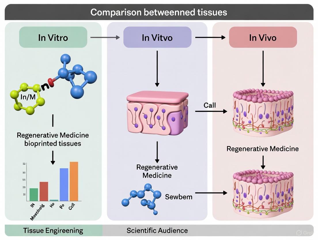
Bridging the Gap: A Comparative Analysis of In Vitro and In Vivo Performance in 3D Bioprinted Tissues
This article provides a comprehensive analysis of the performance and challenges of 3D bioprinted tissues in both laboratory (in vitro) and living organism (in vivo) environments.
Bridging the Gap: A Comparative Analysis of In Vitro and In Vivo Performance in 3D Bioprinted Tissues
Abstract
This article provides a comprehensive analysis of the performance and challenges of 3D bioprinted tissues in both laboratory (in vitro) and living organism (in vivo) environments. Aimed at researchers, scientists, and drug development professionals, it explores the foundational principles of bioprinting, including key technologies like extrusion-based and light-assisted printing, and the critical role of bioinks. The content delves into methodological strategies for specific tissues such as bone, skin, and vascularized constructs, and investigates the significant hurdles of scalability, vascularization, and long-term viability. Finally, it outlines advanced validation techniques, including AI-driven analysis and functional assays, that are crucial for translating promising in vitro results into successful clinical in vivo applications, thereby offering a roadmap for future research and clinical integration.
Core Principles of Bioprinting and the In Vitro/In Vivo Paradigm
Defining the Environments: Controlled In Vitro Systems vs. Complex In Vivo Milieus
The transition of 3D-bioprinted tissues from the laboratory bench to clinical applications hinges on a critical understanding of two distinct testing environments: the controlled in vitro system and the complex in vivo milieu. In vitro models provide a simplified, controlled platform for initial validation, while in vivo animal models offer a holistic physiological context but introduce host-specific variables [1] [2]. This guide objectively compares the performance of bioprinted tissues across these environments, framing the analysis within the broader thesis of understanding the translational pathway for bioprinted products. The comparative data and methodologies outlined herein are intended to assist researchers and drug development professionals in experimental planning and data interpretation.
Comparative Environmental Parameters
The fundamental differences between in vitro and in vivo environments dictate the design, execution, and outcome of bioprinted tissue evaluations. The table below summarizes the key parameters.
Table 1: Key Parameter Comparison: In Vitro vs. In Vivo Environments
| Parameter | Controlled In Vitro System | Complex In Vivo Milieu |
|---|---|---|
| Nutrient Supply | Defined, static culture media; manual changes often lead to gradients and limitations [3]. | Dynamic, convective delivery via perfused vascular networks; continuous supply and waste removal [4]. |
| Oxygen Tension | Atmospheric O₂ (~21%) or controlled incubators; diffusion-limited, creating necrotic cores >200μm [4]. | Physiological, graded O₂ levels (1-13%); maintained by functional vasculature and blood flow [4]. |
| Spatial Complexity | User-defined, often homogeneous cell distribution and scaffold porosity [2]. | Native, heterogeneous tissue architecture with intricate, patient-specific geometries [1] [2]. |
| Biochemical Cues | Isolated, predefined growth factors and cytokines in media [5]. | Complex, dynamic signaling from systemic hormones, immune cells, and neural inputs [5]. |
| Mechanical Forces | Minimal, sometimes simulated via bioreactors (e.g., shear, compression) [3]. | Native, multi-axial loads (e.g., shear stress from blood flow, mechanical stretching) [6]. |
| Immune System | Absent (acellular models) or simplified (co-cultures) [5]. | Fully active, including inflammatory responses, foreign body reactions, and immune cell recruitment [7]. |
| Vascularization | Engineered, often immature capillary networks; a major technical challenge [4] [6]. | Pre-existing, hierarchical, and perfusable vascular network that integrates with the host [4]. |
Performance Metrics and Experimental Data
The distinct environments lead to measurable differences in the performance and viability of bioprinted tissues. The following table consolidates quantitative and qualitative outcomes from representative studies.
Table 2: Comparative Performance of Bioprinted Tissues: In Vitro vs. In Vivo Data
| Performance Metric | Typical In Vitro Outcomes | Typical In Vivo Outcomes (Animal Models) | Supporting Evidence |
|---|---|---|---|
| Cell Viability | Viability can decrease post-printing (e.g., ~70-90% in extrusion) due to shear stress; may decline over time in thick constructs [1] [2]. | Can improve post-implantation due to vascular integration; viability >90% reported in vascularized constructs [4]. | High cell viability (up to 99.7%) achieved in vitro with advanced techniques like FRESH [2]. |
| Tissue Maturation & Function | Limited functionality; expression of tissue-specific markers (e.g., osteocalcin for bone) requires specific induction factors [1]. | Enhanced maturation and functionality observed; tissue integration, new matrix deposition, and marker expression in vivo [1] [7]. | Bioprinted muscle tissues implanted in rats showed tissue integration and partial or complete functionality [2]. |
| Vascular Network Formation & Perfusion | Formation of endothelial tubules in co-culture;但这些网络通常不灌注且不成熟 [4]. | Host-derived angiogenesis and perfusion of bioprinted vasculature; establishment of functional blood flow [4]. | SWIFT technique enables fabrication of organ-specific tissues with integrated, perfusable vascular channels [2]. |
| Mechanical Integrity | Scaffold mechanics are initially defined by bioink; long-term degradation can lead to loss of structure in culture [8]. | Scaffold remodels in response to host mechanical forces (e.g., Wolff's Law for bone); integration with native tissue enhances strength [3] [6]. | 3D-printed titanium mesh implants showed good bone integration and restored ambulation in a patient case study [6]. |
| Inflammatory Response | Not applicable or controlled via media composition. | Predictable foreign body response; can be modulated with bioactive coatings or immunomodulatory factors [7]. | The intracorporal environment imposes unique requirements for in vivo bioprinting modalities and bioink [7]. |
Detailed Experimental Protocols
To generate the comparative data discussed, standardized yet advanced protocols are essential. Below are detailed methodologies for key evaluations in both environments.
Protocol for In Vitro Assessment of Vascular Network Formation
This protocol assesses the innate ability of a bioprinted construct to form early vascular networks [4].
- Bioink Preparation: Formulate a bioink supporting both structural integrity and angiogenesis. A common blend includes Gelatin Methacryloyl (GelMA, 5-10%), hyaluronic acid, and endothelial cells (e.g., HUVECs) at a density of 1-5 million cells/mL. Include supporting mesenchymal stem cells (MSCs) or fibroblasts in a 1:1 to 1:5 ratio (Endothelial:Supporting cells) to stabilize tubules.
- Bioprinting: Utilize an extrusion-based bioprinter with a 22-27G nozzle. Maintain a sterile environment and temperature control (e.g., 18-22°C) during printing. Crosslink the structure post-printing using visible blue light (e.g., 405 nm, 5-15 mW/cm² for 30-60 seconds) if using GelMA.
- Culture: Transfer the construct to a dynamic bioreactor or maintain in static culture with endothelial growth medium (EGM-2). Supplement with pro-angiogenic factors such as VEGF (50 ng/mL).
- Analysis (Day 7-14): Fix and immunostain the constructs for CD31/PECAM-1 or VE-Cadherin. Image using confocal microscopy. Quantify the total tubule length, number of branch points, and network area per field of view using image analysis software (e.g., ImageJ with Angiogenesis Analyzer plugin).
Protocol for In Vivo Implantation and Evaluation
This protocol outlines the surgical implantation of a bioprinted bone construct in an immunodeficient rodent model, a common first step for in vivo validation [1] [7].
- Construct Maturation (Pre-implantation): Following in vitro bioprinting, culture the osteogenic construct (e.g., containing BMSCs or PDLSCs in a HAp/TCP-loaded bioink) in osteogenic medium (containing β-glycerophosphate, ascorbic acid, and dexamethasone) for 7-14 days to promote pre-differentiation.
- Animal Model and Surgery: Utilize a critical-sized calvarial defect model in athymic rats or mice. Anesthetize the animal and create a ~5mm full-thickness defect in the parietal bone. Irrigate the site with saline.
- Implantation: Gently place the pre-cultured bioprinted construct into the defect site. The fit should be snug. A positive control group may receive an autograft, while the defect may be left empty in a negative control group.
- Post-Op Monitoring and Analysis:
- Long-Term (6-12 weeks): Monitor animals for signs of infection or distress.
- Micro-Computed Tomography (μCT): At the endpoint, euthanize the animal and explant the defect site. Scan using μCT at a high resolution (~10-20 μm). Quantify new bone volume (BV), tissue volume (TV), and bone mineral density (BMD) within the defect.
- Histology: Decalcify the explanted samples, embed in paraffin, and section. Perform staining (e.g., Hematoxylin and Eosin (H&E) for general morphology, Masson's Trichrome for collagen, and immunohistochemistry for osteogenic markers like Osteocalcin). Assess tissue integration, vascularization, and evidence of scaffold degradation.
Signaling Pathways in Tissue Integration
The successful integration of bioprinted tissues in vivo is governed by a complex cascade of signaling events. The following diagram illustrates the key pathways involved, particularly in vascularized bone regeneration.
Diagram 1: Signaling in In Vivo Integration.
The Scientist's Toolkit: Research Reagent Solutions
Successful evaluation across in vitro and in vivo environments relies on a suite of specialized reagents and materials. The following table details essential components for experiments in bioprinted tissue engineering.
Table 3: Essential Research Reagents and Materials for Bioprinted Tissue Evaluation
| Reagent/Material Category | Specific Examples | Function & Rationale |
|---|---|---|
| Base Biomaterials (Hydrogels) | GelMA (Gelatin Methacryloyl), Alginate, Collagen, Fibrin, dECM (decellularized ECM) [1] [8]. | Provide a biomimetic 3D microenvironment that supports cell adhesion, proliferation, and differentiation. The primary component of the bioink. |
| Mechanical Reinforcements | Hydroxyapatite (HAp), β-Tricalcium Phosphate (β-TCP), Nanosilicates, Polylactic-co-glycolic acid (PLGA) [1] [6]. | Enhance the mechanical strength and structural integrity of bioinks, particularly for weight-bearing applications like bone tissue engineering. |
| Cells | Mesenchymal Stem Cells (BMSCs, DPSCs), Endothelial Cells (HUVECs), Tissue-specific Primary Cells [1] [4]. | The living component that defines tissue function. Co-cultures of multiple cell types are used to replicate tissue heterogeneity and enable vascularization. |
| Pro-Angiogenic Factors | Vascular Endothelial Growth Factor (VEGF), Basic Fibroblast Growth Factor (bFGF) [4]. | Crucial signaling molecules added to culture media or incorporated into bioinks to stimulate the formation of vascular networks. |
| Osteogenic Induction Cocktail | Dexamethasone, β-Glycerophosphate, Ascorbic Acid [1]. | A standard supplement for in vitro culture media to direct stem cells down an osteogenic lineage, promoting bone tissue formation. |
| Dynamic Culture Systems | Bioreactors (Perfusion, Compression) [3]. | Devices that provide mechanical stimulation (e.g., shear stress, compression) and enhance nutrient/waste exchange, promoting tissue maturation in vitro. |
| In Vivo Model | Immunodeficient Rodents (e.g., athymic mice/rats), Critical-sized Defect Models [1] [7]. | Provide a complex physiological environment for testing the functionality, integration, and safety of bioprinted constructs. |
Bioprinting technology represents a revolutionary approach in biomedical engineering, enabling the precise layer-by-layer deposition of biomaterials and living cells to create three-dimensional tissue constructs. This field has evolved significantly from initial resin-based systems to sophisticated aqueous systems capable of direct printing of biomaterials with living cells for potential transplantation applications [9]. The global market for 3D bioprinting has demonstrated substantial growth, valued at $1.7 billion USD in 2021 and projected to reach approximately $1.94 billion by 2025, reflecting a compound annual growth rate of 15.8% from 2022 to 2030 [9]. This expansion is driven by multiple factors including limited organ donor availability, increased R&D investments, and technological advancements that collectively push the boundaries of regenerative medicine, drug discovery, and personalized therapeutics [9].
Each bioprinting technology offers distinct advantages and limitations that must be carefully considered within the research context. This guide provides an objective comparison of extrusion-based, inkjet, and light-assisted bioprinting techniques, with particular emphasis on their performance in both in vitro and in vivo environments. Understanding these technologies' capabilities and constraints is essential for researchers and drug development professionals seeking to implement bioprinting strategies that effectively bridge the gap between laboratory constructs and clinically viable tissues.
Three primary bioprinting technologies have emerged as dominant approaches in the field, each with unique mechanisms, material requirements, and performance characteristics. Extrusion-based bioprinting, the most prevalent technology, operates by continuously depositing bioinks through a nozzle under controlled pressure [10]. Inkjet-based systems utilize thermal or piezoelectric actuators to eject precise droplets of bioink onto a substrate [11]. Light-assisted bioprinting, including stereolithography (SLA) and digital light processing (DLP), employs photopolymerization of light-sensitive bioinks to create solid structures layer by layer [9].
Table 1: Fundamental Characteristics of Major Bioprinting Technologies
| Technology | Mechanism | Resolution | Speed | Cell Viability | Key Strengths | Major Limitations |
|---|---|---|---|---|---|---|
| Extrusion-Based | Mechanical dispensing of continuous filaments | 50-500 μm [10] | Medium | 40-95% [10] | High cell density printing; structural stability for macroscopic constructs | Shear stress on cells; limited resolution; potential nozzle clogging |
| Inkjet | Thermal or piezoelectric droplet ejection | 10-50 μm [11] | High | >85% [12] | High speed; excellent resolution; cost-effectiveness | Low cell density; limited material viscosity range; droplet inconsistency |
| Light-Assisted | Photopolymerization of bioinks | 5-50 μm [9] | Medium-High | 75-95% [9] | Highest resolution; excellent structural complexity; no nozzle clogging | Limited bioink transparency requirement; potential UV damage; limited material options |
The selection of an appropriate bioprinting technology must align with both the target application and the required balance between structural fidelity and biological performance. Extrusion bioprinting has become the most popular platform, featuring in over half of bioprinting publications [10]. However, this prevalence doesn't necessarily reflect superiority for all applications, but rather its accessibility and versatility for creating macroscale structures. Inkjet and light-assisted systems offer superior resolution but face different constraints regarding material properties and biological compatibility.
Extrusion-Based Bioprinting
Technology Fundamentals
Extrusion-based bioprinting operates through a mechanically-driven dispensing system that pushes bioinks through a nozzle to create continuous filaments. This platform excels in depositing high-viscosity materials and achieving high cell densities, making it particularly valuable for creating tissue constructs with significant volumetric dimensions. The technology's capability to fabricate complex 3D structures by layering multiple materials has positioned it as a preferred method for engineering volumetric tissues and organ-like structures [10].
The fundamental process involves loading bioink into a cartridge or syringe, which is then pressurized either through piston-driven or screw-driven mechanisms. The material is extruded through a nozzle while the print head or build platform moves along predetermined paths generated from digital models. Despite its advantages in structural integrity and cell density, extrusion bioprinting faces challenges regarding resolution limitations and potential cell damage from shear stresses during the extrusion process [10].
Experimental Protocols and Performance Data
A critical aspect of extrusion bioprinting involves optimizing bioink formulations to balance printability with biocompatibility. Recent studies have employed machine learning approaches to predict and optimize bioink behavior. For instance, research on ALGEC bioinks (comprising alginate, gelatin, and TEMPO-oxidized nanofibrillated cellulose) utilized polynomial fit and multiple regression models to predict viscosity based on composition and shear rate, achieving an R² of 0.98 and mean absolute error of 0.12 [13]. This data-driven approach enables more efficient bioink development by reducing reliance on traditional trial-and-error methods.
Table 2: Extrusion Bioprinting Performance Metrics
| Parameter | In Vitro Performance | In Vivo Performance | Measurement Methods |
|---|---|---|---|
| Structural Fidelity | Filament diameter: 150-500 μm; Layer thickness: 50-500 μm [10] | Shape maintenance varies with degradation rate; ~50-80% initial structure retention | Microscopic imaging; micro-CT scanning |
| Mechanical Properties | Compressive modulus: 5-50 kPa (hydrogel-based) [14] | Progressive integration with host tissue; modulus changes ~30-60% | Rheology; uniaxial compression testing |
| Cell Viability | 40-95% post-printing [10] | Viability dependent on vascularization; ~30-80% at 2 weeks | Live/dead staining; metabolic assays |
| Vascularization Potential | Limited to ~200 μm diffusion limits [14] | Capillary invasion from host tissue: 100-500 μm at 2 weeks [14] | Histology; immunostaining for endothelial markers |
| Functional Duration | 2-8 weeks in culture | Integration with host tissue within 2-6 weeks [14] | Longitudinal tracking; functional assays |
The "bioink paradox" presents a fundamental challenge in extrusion bioprinting, where materials that extrude with high fidelity are often biologically inert, while biologically ideal materials are frequently mechanically weak and difficult to print [15]. This tension between printability and bio-functionality necessitates careful optimization for specific applications. Research has demonstrated that combining statistical and rheological methodologies can effectively develop bioinks tailored for specific tissue applications, as shown in studies utilizing design of experiment (DoE) approaches to optimize hyaluronic acid, sodium alginate, and dextran-based bioinks [16].
Figure 1: Extrusion Bioprinting Workflow from Bioink Development to Implementation
Inkjet-Based Bioprinting
Technology Fundamentals
Inkjet bioprinting operates through either thermal or piezoelectric actuation mechanisms to eject precise droplets of bioink onto a substrate. Thermal inkjet printers utilize heating elements to create vapor bubbles that generate pressure pulses, forcing bioink droplets through the nozzle. Piezoelectric systems employ mechanical deformation of piezoelectric materials to achieve similar droplet ejection. Both approaches enable non-contact printing with high resolution and speed, making them suitable for applications requiring precise cellular patterning [11].
The hardware architecture of modern inkjet bioprinters includes sophisticated print heads capable of ejecting tiny droplets of bioinks containing cells, growth factors, and supportive biomaterials. These systems maintain controlled environmental conditions with optimal temperature, humidity, and sterility to ensure cell viability throughout the printing process. Advanced software algorithms translate digital 3D models into precise movement commands while optimizing droplet placement, layer stacking, and material mixing. Many vendors integrate real-time monitoring systems, such as cameras and sensors, to detect and correct errors during printing [11].
Experimental Protocols and Performance Data
Inkjet bioprinting protocols require careful optimization of bioink properties, including viscosity, surface tension, and cell density. Typically, bioinks for inkjet printing have viscosities ranging from 3-15 mPa·s to ensure reliable droplet formation without clogging the print heads. The market for inkjet-based bioprinting is experiencing robust growth, with an estimated market size of approximately $500 million in 2025, projected to expand with a compound annual growth rate of 15% in the coming years [12].
A critical consideration for inkjet bioprinting is droplet formation dynamics, which directly impact printing resolution and cell viability. Studies have demonstrated that piezoelectric systems generally achieve higher cell viability (85-95%) compared to thermal systems (80-90%) due to reduced thermal stress on cells. However, both systems face challenges with high cell densities (>10 million cells/mL), which can lead to nozzle clogging and inconsistent droplet ejection [12].
Table 3: Inkjet Bioprinting Performance Metrics
| Parameter | In Vitro Performance | In Vivo Performance | Measurement Methods |
|---|---|---|---|
| Resolution | 10-50 μm droplet size [11] | Limited data; likely similar resolution maintenance | High-speed imaging; microscopic analysis |
| Printing Speed | 1-10,000 droplets/second [12] | N/A (pre-fabrication technique) | Timing analysis; throughput measurement |
| Cell Viability | >85% post-printing [12] | Dependent on construct maturity; challenging for thick tissues | Live/dead staining; flow cytometry |
| Multi-material Capability | High (multiple print heads) | Limited by integration complexity | Material characterization; imaging |
| Tissue Maturation | Limited to thin layers (<1 mm) | Rapid perfusion but limited structural integrity | Histology; mechanical testing |
The applications of inkjet bioprinting are predominantly found in research settings, particularly for developing tissue models for drug screening and disease modeling. The technology's high resolution enables precise patterning of multiple cell types, making it valuable for creating complex tissue interfaces. However, the translation to in vivo applications remains challenging due to limitations in creating thick, vascularized tissues capable of surviving implantation and supporting physiological functions [12].
Figure 2: Inkjet Bioprinting Process Workflow
Light-Assisted Bioprinting
Technology Fundamentals
Light-assisted bioprinting technologies, including stereolithography (SLA) and digital light processing (DLP), utilize photopolymerization mechanisms to create solid structures from liquid resin precursors. In these systems, specific wavelengths of light (typically UV or blue light) are projected onto the bioink surface in precise patterns, initiating a crosslinking reaction that solidifies the material in a layer-by-layer fashion. These technologies offer the highest resolution among bioprinting methods, with feature sizes ranging from 5-50 micrometers [9].
A significant advancement in light-assisted bioprinting is the development of biocompatible photoinitiators and photoreactive bioinks that maintain cell viability during the polymerization process. These bioinks typically contain photolabile groups that form covalent bonds when exposed to light, creating stable hydrogel networks that encapsulate living cells. The ability to pattern complex 3D structures with micron-scale precision has made light-assisted bioprinting particularly valuable for engineering tissues with intricate architectural features, such as vascular networks and porous scaffolds for enhanced nutrient diffusion [9].
Experimental Protocols and Performance Data
Light-assisted bioprinting protocols require careful optimization of multiple parameters, including photoinitiator concentration, light intensity, exposure time, and bioink composition. Studies have demonstrated that cell viability in light-assisted bioprinting typically ranges from 75-95%, with higher viability associated with visible light crosslinking systems compared to UV-based systems [9]. The mechanical properties of the resulting constructs can be precisely tuned by adjusting the degree of crosslinking, with compressive moduli generally ranging from 2-100 kPa depending on the bioink formulation and printing parameters.
Recent innovations in light-assisted bioprinting have focused on improving biological performance through the development of bioinstructive materials that actively direct cell behavior. These advanced biomaterials incorporate specific biochemical cues such as adhesion peptides, enzymatically degradable sequences, and growth factors that can be spatially patterned within the constructed tissues. This approach represents a paradigm shift from creating passively biocompatible structures to designing actively bio-instructive environments that guide tissue maturation and function [15].
Table 4: Light-Assisted Bioprinting Performance Metrics
| Parameter | In Vitro Performance | In Vivo Performance | Measurement Methods |
|---|---|---|---|
| Resolution | 5-50 μm [9] | Maintained with slow degradation | Scanning electron microscopy |
| Structural Complexity | Very high (free-form) | Maintained initially; remodeling over time | Micro-CT; confocal imaging |
| Cell Viability | 75-95% [9] | Varies with material degradation profile | Live/dead staining; metabolic assays |
| Mechanical Properties | Wide tunability (2-100 kPa) | Progressive changes with integration | Rheology; compression testing |
| Degradation Profile | Controllable via chemistry | Accelerated in physiological environment | Mass loss; GPC analysis |
While light-assisted bioprinting offers exceptional resolution and structural control, it faces challenges in creating large, vascularized tissues suitable for in vivo implantation. The technology's reliance on transparent bioinks can limit material selection, and the potential cytotoxicity of photoinitiators and reactive oxygen species generated during crosslinking requires careful management. Additionally, the sequential layer-by-layer process can be time-consuming for constructing large tissue volumes, though recent advances in volumetric printing are addressing this limitation [9].
Comparative Analysis: In Vitro vs. In Vivo Performance
A critical consideration in bioprinting research is the significant performance gap observed between in vitro constructs and their behavior following in vivo implantation. This disconnect presents a substantial challenge for clinical translation and necessitates careful evaluation of how bioprinted tissues transition from controlled laboratory environments to complex physiological systems.
Vascularization and Integration Capabilities
The ability to form functional vascular networks represents one of the most significant differentiators between in vitro and in vivo performance. While extrusion bioprinting can create channel structures that support limited nutrient diffusion in vitro, these constructs often fail to develop into perfusable vascular networks without surgical anastomosis to the host circulatory system. Research has demonstrated that bioprinted vascular constructs containing both endothelial and smooth muscle cells can achieve beneficial perfusability and in vivo autonomous connection within approximately two weeks, with significant vascular remodeling occurring over six weeks [14].
Light-assisted bioprinting offers superior resolution for creating intricate vascular-like channels, but these often lack the cellular complexity and functionality of native vasculature. Inkjet bioprinting can pattern endothelial cells with high precision but struggles to create the three-dimensional, multi-layered vessel structures necessary for withstanding hemodynamic forces. The in vivo performance of bioprinted vascular structures ultimately depends on their ability to recruit supporting cells from host tissues and establish stable endothelial linings that prevent thrombosis [14].
Structural Integrity and Remodeling
Bioprinted constructs frequently exhibit significant changes in mechanical properties and structural integrity following implantation. In vitro, constructs maintain their shape through the engineered biomaterial properties, but in vivo, they encounter dynamic mechanical forces and enzymatic environments that accelerate degradation and remodeling. Studies have shown that extruded constructs can retain approximately 50-80% of their initial structure after implantation, with the rate of degradation closely tied to the material composition and crosslinking density [10].
The concept of 4D bioprinting has emerged as a promising approach to bridge the in vitro-in vivo gap by creating structures that dynamically change their shape or functionality over time in response to physiological stimuli. These systems utilize smart materials that respond to temperature, pH, or enzymatic activity, enabling printed constructs to better adapt to the in vivo environment and more closely mimic native tissue behaviors [12].
Clinical Translation Status
The progression of bioprinting technologies from research tools to clinical applications remains limited. Currently, there are only 11 clinical trials that utilize bioprinting technology in any context, from a total of over 50,000 registered trials. Of these bioprinting trials, just four aim to implant tissues, with the majority focusing on developing in vitro models. Only one implant trial has been identified as using extrusion bioprinting, specifically for auricular reconstruction, leveraging the technology's capability to create complex 3D macroscale shapes that provide aesthetic and functional benefits [10].
This limited clinical translation highlights the significant challenges that remain in creating bioprinted tissues with the essential microscale organization and heterogeneity of native human anatomy. While all three bioprinting technologies show promise for specific applications, each faces unique hurdles in scaling up production to meet potential clinical demand while maintaining the biological complexity necessary for functional integration [10].
The Scientist's Toolkit: Essential Research Reagents and Materials
Successful bioprinting requires careful selection and optimization of materials, crosslinking strategies, and cellular components. The following table outlines key research reagents and their functions in bioprinting applications.
Table 5: Essential Research Reagents and Materials for Bioprinting
| Category | Specific Examples | Function | Technology Compatibility |
|---|---|---|---|
| Base Biomaterials | Alginate, Gelatin, Hyaluronic Acid, Collagen [16] | Provide structural support and mimic extracellular matrix | Extrusion, Inkjet, Light-Assisted |
| Functional Additives | TO-NFC (TEMPO-oxidized nanofibrillated cellulose) [13] | Enhance rheological properties and printability | Primarily Extrusion |
| Crosslinkers | Calcium chloride (alginate), NaIO₄ (GelMA/C), Photoinitiators (I2959, LAP) [14] | Induce hydrogel formation and stabilize printed structures | Extrusion (ionic), Light-Assisted (photo) |
| Bioactive Factors | RGD peptides, Growth factors (VEGF, BMP-2) [15] | Enhance cell adhesion, proliferation, differentiation | All technologies |
| Cell Sources | Mesenchymal stem cells, HUVECs, HCASMCs [14] | Provide living component for tissue formation | All technologies |
The selection of appropriate bioink components must consider both processing requirements and biological objectives. Alginate is widely used for its excellent printability and rapid gelation, while gelatin provides cell-adhesive motifs that support cellular activities. Hybrid approaches that combine multiple materials have gained prominence for their ability to balance printability with biofunctionality. For instance, research on ALGEC bioinks has demonstrated that combining alginate, gelatin, and TEMPO-oxidized nanofibrillated cellulose can create formulations with tunable rheological properties and enhanced structural integrity [13].
Design of Experiment (DoE) methodologies have emerged as valuable tools for systematically optimizing bioink formulations. Studies utilizing factorial and mixture DoE approaches have successfully identified critical component interactions and established optimized bioink compositions with targeted viscosities and shear-thinning properties [16]. These statistical approaches reduce development time and resource requirements while providing comprehensive understanding of formulation parameters.
Extrusion, inkjet, and light-assisted bioprinting technologies each offer distinct advantages and face specific limitations in the creation of functional tissues. Extrusion bioprinting provides versatility in processing various biomaterials and achieving high cell densities but struggles with resolution and potential cell damage. Inkjet systems enable high-speed printing with excellent resolution but are constrained by material viscosity requirements and limited structural integrity for thick tissues. Light-assisted technologies offer superior resolution and architectural control but face challenges in material selection and potential cytotoxicity from photoinitiators.
The disconnect between in vitro performance and in vivo functionality remains a significant challenge across all bioprinting platforms. While each technology can create impressive structures under controlled laboratory conditions, the transition to implantation environments reveals limitations in vascularization, integration, and long-term stability. Future advancements will likely focus on multi-modal approaches that combine technologies to leverage their respective strengths, development of more sophisticated bioinstructive materials, and improved understanding of tissue maturation processes.
As the field progresses, researchers must maintain realistic expectations about clinical translation timelines while continuing to innovate toward the ultimate goal of creating functional human tissues for therapeutic applications. By critically evaluating both capabilities and limitations of each bioprinting technology, the scientific community can more effectively direct research efforts toward applications where bioprinting offers genuine potential for clinical impact.
In the evolving landscape of tissue engineering, 3D bioprinting has emerged as a transformative technology that enables the precise fabrication of complex, living tissue constructs. Central to this technology are bioinks—advanced biomaterials that incorporate living cells and biochemical factors to create functional tissue architectures. Among the diverse array of biomaterials available, polysaccharide-based hydrogels have gained significant prominence due to their exceptional biocompatibility, structural similarity to the native extracellular matrix (ECM), and versatile mechanical properties [17] [18]. These natural polymers offer a unique combination of properties that make them particularly suitable for both in vitro tissue models and in vivo regenerative applications, though their performance can vary substantially between these environments.
The fundamental challenge in bioprinting lies in creating constructs that not only exhibit precise spatial organization in vitro but also maintain their structural and functional integrity when implanted in vivo. Polysaccharides address this challenge through their tunable chemical structure, which allows researchers to engineer specific degradation profiles, mechanical properties, and bioactivity. However, the transition from laboratory validation to clinical application requires a thorough understanding of how these materials perform across different biological contexts [8]. This review systematically compares the performance of various polysaccharide-based biomaterials, with particular emphasis on their in vitro characteristics versus their in vivo behavior, providing researchers with evidence-based guidance for material selection in specific applications.
Polysaccharide Biomaterials: Classification and Properties
Natural Origins and Structural Diversity
Polysaccharides are ubiquitously found in nature and can be systematically categorized based on their biological origins into three primary classes: plant-derived, animal-derived, and microbial-derived polysaccharides [17]. Each category possesses distinct structural characteristics and functional properties that determine their suitability for specific bioprinting applications. Plant-derived polysaccharides, including cellulose, alginate, and agarose, typically exhibit robust mechanical properties and readily available sources. Animal-derived variants such as hyaluronic acid and chitosan demonstrate enhanced cellular recognition and integration capabilities. Microbial-derived polysaccharides like dextran and pullulan offer high purity and reproducible quality [17] [19].
The inherent biocompatibility of polysaccharides stems from their structural similarity to glycosaminoglycans (GAGs) and proteoglycans present in the native extracellular matrix [17]. This biomimicry facilitates favorable cellular interactions, including adhesion, proliferation, and differentiation. Furthermore, natural polysaccharides exhibit excellent hydrophilicity, biodegradability, and minimal immunogenicity, making them ideal candidates for constructing bioinks that support cell survival and function [18] [19]. However, unmodified natural polysaccharides often present limitations such as inadequate mechanical strength, unpredictable degradation patterns, and insufficient bioactivity, necessitating strategic modifications to optimize their performance for bioprinting applications [17].
Modification Strategies for Enhanced Performance
To address the inherent limitations of native polysaccharides, researchers have developed sophisticated modification strategies that tailor their physicochemical and biological properties for specific bioprinting requirements. These approaches can be broadly classified into chemical functionalization, physical reinforcement, and biological hybridization [17].
Table 1: Polysaccharide Modification Strategies and Their Applications
| Modification Strategy | Key Approaches | Impact on Material Properties | Representative Applications |
|---|---|---|---|
| Chemical Functionalization | Methacrylation, oxidation, norbornene functionalization | Enhanced crosslinking efficiency, tunable mechanical properties, controlled degradation | Photocrosslinkable hydrogels for bone and cartilage tissue engineering [17] |
| Physical Reinforcement | Nanomaterial incorporation (nanoclays, cellulose nanocrystals), polymer blending | Improved shear-thinning behavior, structural fidelity, mechanical robustness | Nanocomposite bioinks for extrusion bioprinting [17] [20] |
| Biological Hybridization | Peptide conjugation, glycosaminoglycan incorporation, decellularized matrix integration | Enhanced cell-material interactions, bioactivity, tissue-specific differentiation | Functionalized hydrogels for neural and vascular tissue engineering [17] [18] |
Chemical modification introduces reactive functional groups such as methacrylate, norbornene, or aldehyde groups that enable controlled crosslinking under mild conditions. For instance, methacrylated hyaluronic acid (MeHA) allows photocrosslinking with visible light, preserving cell viability while providing mechanical tunability [21]. Physical reinforcement through the incorporation of nanomaterials such as cellulose nanocrystals or nanosilicates enhances the rheological properties of bioinks, enabling the fabrication of complex structures with high shape fidelity [17]. Biological hybridization involves the integration of bioactive motifs such as cell-adhesive peptides or growth factors to promote specific cellular responses and tissue regeneration [18].
Comparative Performance Analysis: In Vitro vs. In Vivo
Methodological Framework for Performance Assessment
Evaluating the performance of polysaccharide-based bioinks requires standardized methodologies that assess both their bioprinting capabilities (printability) and their biological performance. Key experimental protocols for characterizing these materials include:
Printability Assessment: Quantitative evaluation of printability involves measuring resolution, filament uniformity, and structural fidelity using mathematical models such as the printing accuracy index (PAI) and shape retention coefficients [17]. These parameters are typically analyzed using image processing software on macroscopic and microscopic images of printed structures.
Rheological Characterization: The viscoelastic properties of bioinks are determined using rotational rheometry to measure storage modulus (G'), loss modulus (G"), yield stress, and shear-thinning behavior. These properties directly influence extrudability, shape retention, and structural stability [17] [20].
Mechanical Testing: Compressive and tensile moduli are evaluated using universal mechanical testing systems according to standardized protocols (e.g., ASTM D695 for compression, ASTM D638 for tension). Both initial properties and time-dependent changes in physiological conditions are assessed [17].
Biological Performance: In vitro biological performance is evaluated through cell viability assays (Live/Dead staining, Alamar Blue), proliferation measurements (DNA quantification), and differentiation analysis (immunostaining, qPCR). In vivo performance is assessed through subcutaneous implantation in animal models, followed by histological analysis, immunohistochemistry, and tracking of degradation and vascularization over time [21] [20].
Degradation Profiling: Mass loss measurements, swelling behavior, and molecular weight changes are monitored in both simulated physiological conditions and in vivo environments to establish degradation kinetics [19].
Quantitative Comparison of Polysaccharide Performance
The transition from in vitro validation to in vivo functionality presents significant challenges for polysaccharide-based bioinks. The following table summarizes key performance metrics across different environmental contexts:
Table 2: In Vitro vs. In Vivo Performance of Polysaccharide-Based Bioinks
| Polysaccharide Type | In Vitro Cell Viability | In Vitro Mechanical Strength (kPa) | In Vivo Degradation Time | In Vivo Tissue Integration | In Vivo Immune Response |
|---|---|---|---|---|---|
| Alginate | 80-90% [20] | 5-50 [17] | 2-8 weeks [19] | Moderate (fibrous encapsulation) [8] | Mild to moderate [17] |
| Chitosan | 75-85% [18] | 10-100 [17] | 4-12 weeks [19] | Good (cellular infiltration) [18] | Minimal [18] |
| Hyaluronic Acid | 85-95% [20] | 2-30 [17] | 1-4 weeks [19] | Excellent (vascularization) [21] | Variable (depends on modification) [21] |
| Agarose | 70-80% [17] | 20-200 [17] | 8-16 weeks [19] | Poor (limited cellular infiltration) [17] | Minimal [17] |
| Cellulose Derivatives | 80-90% [17] | 50-500 [17] | 12-24 weeks [19] | Moderate (surface integration) [17] | Minimal [17] |
The data reveal significant disparities between in vitro and in vivo performance across all polysaccharide types. While cell viability remains generally high in controlled in vitro environments, the in vivo performance varies considerably based on the material's properties and the host response. Alginate, despite excellent printability and reasonable in vitro performance, often triggers fibrous encapsulation in vivo, limiting its integration with surrounding tissues [8]. Chitosan demonstrates better tissue integration but exhibits slower degradation that may not match the rate of new tissue formation. Hyaluronic acid supports excellent vascularization and tissue integration but degrades rapidly in vivo, potentially compromising mechanical support before new tissue matures [21] [19].
The mechanical properties of polysaccharide hydrogels also exhibit notable changes between environments. While initial in vitro measurements provide baseline data, the hydrogel mechanics evolve substantially in vivo due to swelling, degradation, and cell-mediated remodeling. For instance, alginate hydrogels typically experience an initial decrease in mechanical strength due to ion exchange in physiological fluids, followed by a more gradual decline as degradation progresses [17]. These dynamics highlight the importance of considering the temporal evolution of material properties when designing constructs for specific applications.
Diagram 1: Performance pathway illustrating the transition from in vitro to in vivo environments for polysaccharide bioinks, highlighting modification strategies that address critical properties.
Advanced Material Systems and Experimental Protocols
Hybrid and Composite Bioinks
To bridge the performance gap between in vitro and in vivo environments, researchers have developed advanced hybrid bioinks that combine multiple polysaccharides or integrate polysaccharides with other polymers and nanomaterials. These composite systems leverage the complementary properties of their constituents to achieve balanced performance characteristics [17] [20].
One prominent example is the combination of alginate with gelatin, which merges the excellent printability and mechanical stability of alginate with the cell-adhesive properties of gelatin. The experimental protocol for formulating and evaluating such hybrid systems typically involves:
Material Preparation: Dissolve alginate (2-4% w/v) in physiological buffer at 60°C with continuous stirring. Separately dissolve gelatin (5-10% w/v) in buffer at 37°C. Combine the solutions in varying ratios (e.g., 1:1, 1:2, 2:1 alginate:gelatin) and mix thoroughly.
Rheological Optimization: Characterize the viscoelastic properties of each formulation using rotational rheometry. Measure storage modulus (G'), loss modulus (G"), and complex viscosity across a shear rate range of 0.1-100 s⁻¹ to assess shear-thinning behavior.
Printability Assessment: Fabricate grid structures (10×10×2 mm) using a pneumatic extrusion bioprinting system. Quantify printing fidelity by comparing designed versus printed strand diameter, pore size, and overall structure geometry.
Crosslinking Optimization: Evaluate ionic crosslinking with calcium chloride (50-200 mM) and/or physical crosslinking through temperature control (4-37°C). Assess the effect of crosslinking conditions on mechanical properties and cell viability.
Biological Validation: Encapsulate human mesenchymal stem cells (hMSCs) at a density of 1-5×10⁶ cells/mL. Evaluate cell viability (Days 1, 3, 7), proliferation (Days 1, 7, 14), and tissue-specific differentiation (e.g., osteogenic, chondrogenic) over 2-4 weeks [17] [20].
Similar methodologies have been applied to other hybrid systems, such as chitosan-hyaluronic acid blends for cartilage tissue engineering and cellulose nanocrystal-reinforced agarose for mechanically robust constructs [17]. These composite approaches demonstrate significantly improved in vivo performance compared to single-component systems, with enhanced tissue integration and more appropriate degradation profiles.
Functionalization for Enhanced Bioactivity
Beyond mechanical considerations, the biological functionality of polysaccharide bioinks critically influences their performance in vivo. Biofunctionalization strategies aim to incorporate specific bioactive cues that direct cellular behavior and promote tissue regeneration [18] [8].
The experimental protocol for creating and evaluating biofunctionalized polysaccharide bioinks typically includes:
Chemical Modification: Introduce reactive groups (e.g., methacrylate, amine, carboxyl) onto the polysaccharide backbone using carbodiimide chemistry or other conjugation methods. Purify the modified polymer through dialysis and lyophilization.
Bioactive Molecule Conjugation: Covalently attach cell-adhesive peptides (e.g., RGD, IKVAV) or growth factors (e.g., BMP-2, VEGF) to the modified polysaccharide using appropriate crosslinkers (e.g., NHS/EDC, maleimide-thiol chemistry). Verify conjugation efficiency through spectrophotometric assays or HPLC.
Bioink Formulation: Prepare bioinks by dissolving the functionalized polymer in cell culture medium at concentrations tailored to the application (typically 2-5% w/v). Sterilize the solution through filtration (0.22 μm) before cell incorporation.
Biological Activity Assessment: Culture relevant cell types (e.g., fibroblasts, osteoblasts, neural cells) on 2D films or within 3D bioprinted constructs containing the biofunctionalized hydrogel. Evaluate cell adhesion, spreading, proliferation, and tissue-specific marker expression compared to non-functionalized controls.
In Vivo Validation: Implant bioprinted constructs subcutaneously in immunocompromised mice or in orthotopic locations relevant to the target tissue. Harvest implants at predetermined time points (2, 4, 8 weeks) for histological analysis (H&E, Masson's Trichrome), immunohistochemistry (collagen I/II, osteocalcin, etc.), and assessment of host tissue integration and vascularization [18] [8].
Functionalized polysaccharides consistently demonstrate superior performance in directing specific cellular responses and promoting functional tissue formation in vivo. For instance, RGD-modified alginate significantly enhances cell adhesion and survival post-implantation, while VEGF-conjugated hyaluronic acid promotes robust vascularization critical for the survival of thick tissue constructs [18].
The Scientist's Toolkit: Essential Research Reagents and Materials
Successful development and evaluation of polysaccharide-based bioinks requires access to specialized reagents and equipment. The following table summarizes key materials and their functions in bioink formulation and characterization:
Table 3: Essential Research Reagents for Polysaccharide Bioink Development
| Category | Specific Reagents/Materials | Function | Representative Examples |
|---|---|---|---|
| Base Polymers | Sodium alginate, chitosan, hyaluronic acid, cellulose derivatives, agarose | Structural backbone of bioink, provides basic mechanical properties and printability | PRONOVA UP MVG alginate (Novatrix), Chitosan (Sigma-Aldrich), Hyaluronic acid (Lifecore) [17] [19] |
| Crosslinking Agents | Calcium chloride, calcium sulfate, genipin, glutaraldehyde, photoinitiators (Irgacure 2959, LAP) | Induces hydrogel formation through ionic, covalent, or photochemical mechanisms | Calcium chloride (Sigma-Aldrich), Lithium phenyl-2,4,6-trimethylbenzoylphosphinate (LAP) [21] |
| Bioactive Additives | RGD peptides, IKVAV peptides, growth factors (BMP-2, VEGF, TGF-β), extracellular matrix proteins | Enhances cell-material interactions, promotes specific cellular responses | RGD peptide (Peptides International), Recombinant human BMP-2 (PeproTech) [18] [8] |
| Rheology Modifiers | Nanocellulose, nanosilicates, gelatin, glycerophosphate | Modifies viscoelastic properties, enhances printability and shape fidelity | Cellulose nanocrystals (CelluForce), Laponite nanoclay (BYK Additives) [17] [20] |
| Characterization Tools | Rotational rheometer, mechanical tester, confocal microscope, DNA quantification kits, live/dead assay kits | Evaluates physical properties and biological performance | Discovery HR rheometer (TA Instruments), Instron mechanical tester, Alamar Blue cell viability assay (Thermo Fisher) [17] [20] |
The field of polysaccharide-based bioinks continues to evolve rapidly, with several emerging trends poised to address current limitations in the in vitro to in vivo transition. The integration of dynamic crosslinking mechanisms that enable self-healing and stimuli-responsive behaviors represents a promising approach to creating more adaptive and resilient constructs [17]. Similarly, the development of multi-material bioprinting systems allows for the fabrication of spatially organized constructs with region-specific properties that better mimic native tissue heterogeneity [20].
Advancements in artificial intelligence and machine learning are beginning to impact bioink development through optimized design parameters and printing conditions [8]. These computational approaches can predict how specific material formulations will perform in vivo based on in vitro characterization data, potentially reducing the need for extensive animal testing and accelerating clinical translation.
The growing emphasis on clinical translation is driving research toward standardized, scalable manufacturing processes and addressing regulatory considerations [22]. As these efforts progress, polysaccharide-based bioinks are poised to play an increasingly important role in regenerative medicine, drug screening, and disease modeling, ultimately fulfilling their potential as the "heart of the construct" in tissue engineering applications.
The journey to create a functional, bioprinted tissue begins long before the bioink is deposited. The pre-bioprinting workflow—encompassing medical image acquisition, segmentation, and digital model refinement—is a critical determinant of the final construct's anatomical fidelity and, consequently, its performance in both laboratory and living systems. Anatomical accuracy, established during this digital phase, directly influences cellular behavior, nutrient diffusion, and functional integration. Deficiencies in the initial model can lead to a cascade of failures, manifesting as the well-documented disparity between in vitro promise and in vivo efficacy. This guide objectively compares the technologies, methodologies, and quantitative errors within the pre-bioprinting pipeline, providing researchers with the data needed to bridge this translational gap.
The Foundational Workflow: From DICOM to Printable Model
The creation of a patient-specific design follows a multi-stage digital process. This workflow translates clinical imaging data into a printable file, with each stage introducing specific, quantifiable errors that impact the final model's geometric truth.
A Standardized Digital Pathway
The transformation of a medical image into a 3D model is a three-step process [23]:
- Image Segmentation: The process of partitioning a volumetric medical image (e.g., from CT or MRI) to identify and label the structure of interest.
- Mesh Refinement: The segmented surface is converted into a mesh (typically an STL file) and repaired, smoothed, or appended to create a watertight, printable model.
- Slicing & Toolpath Generation: The refined 3D mesh is digitally sliced into layers and converted into machine-readable code (G-code) for the bioprinter.
Workflow Visualization
The following diagram illustrates the key stages of the pre-bioprinting workflow and the associated error sources that affect the final model's accuracy.
Quantitative Error Analysis in the Pre-Bioprinting Pipeline
A systematic understanding of errors in the medical 3D-printing process is essential for quality assurance. The total error of the final bioprinted construct is the culmination of partial errors from each step in the workflow [24].
Error Source Categorization
The major partial errors are defined as follows [24]:
- Segmentation Error (SegE): The deviation between the original anatomical structure (or its image data) and the direct result of the segmentation process.
- Digital Editing Error (DEE): The deviation introduced during the repair, smoothing, and manipulation of the segmented mesh.
- Printing Error (PrE): The deviation between the digitally edited model and the final physical 3D printed construct.
A 2024 systematic review of quality assurance studies provides median values for these errors, offering a benchmark for researchers [24].
Quantitative Error Comparison
Table 1: Median Partial Errors in Patient-Specific Model Production (AMMD) [24]*
| Error Type | Definition | Median AMMD (mm) | Key Influencing Factors |
|---|---|---|---|
| Segmentation Error (SegE) | Deviation between original structure and segmented model. | 0.80 mm | Image contrast, resolution, manual input, algorithm choice. |
| Printing Error (PrE) | Deviation between digital model and final printed object. | 0.26 mm | Printing technology, material, nozzle diameter/laser spot size. |
| Total Error | Combined deviation from original structure to final print. | 0.825 mm | Summation and interaction of SegE, DEE, and PrE. |
*AMMD (Absolute Maximum Mean Deviation): The largest linear deviation based on an average value from at least two measurements.
The data reveals that the segmentation step is the largest source of geometric inaccuracy in the entire workflow. Notably, the total error is not significantly higher than the SegE alone, suggesting that partial errors can sometimes compensate for each other [24]. This underscores the need for individual analysis of each error type rather than relying solely on total error assessment.
Comparative Analysis of Segmentation Software and Slicing Algorithms
The choice of digital tools directly impacts the fidelity of the resulting model and the feasibility of creating complex, biologically relevant structures.
Segmentation Software: Accessibility vs. Automation
Table 2: Comparison of Segmentation and Slicing Tools
| Tool Category | Examples | Key Characteristics | Impact on Model Fidelity |
|---|---|---|---|
| Free/Open-Source Segmentation Software | 3D Slicer [23], Seg3D [23] | High accessibility, capable of processing diverse data; often requires more manual input. | High accuracy is achievable but can be time-consuming; reproducibility may vary. |
| Commercial Segmentation Platforms | Mimics [23], Simpleware [23] | Integrated environments with advanced (semi-)automatic algorithms and simulation capabilities. | Generally high throughput and reproducibility; cost can be a barrier. |
| Planar Slicing Algorithms | Standard in most Cartesian printers [2] | Creates layers of uniform thickness; simple and computationally efficient. | Introduces "stair-step" artifacts on curved surfaces, reducing geometric fidelity [25]. |
| Non-Planar Slicing Algorithms | Used in multi-axis robotic systems [2] [25] | Allows deposition along curvilinear paths, conforming to surface topography. | Improves scaffold integrity and surface smoothness; reduces need for support structures [2]. |
Advanced Slicing for Enhanced Fidelity
The shift from planar to non-planar slicing is a significant advancement. Traditional layer-by-layer deposition is constrained by fixed axes, resulting in stair-step effects on sloped or curved surfaces. In contrast, multi-axis robotic bioprinting allows for dynamic nozzle orientation, enabling conformal printing on anatomically relevant surfaces. This approach can reduce the need for sacrificial support by over 60% and improve print-to-CAD fidelity by 15–25%, depending on surface curvature [25].
Experimental Protocols for Workflow Validation
To ensure reliability and reproducibility, rigorous validation of the pre-bioprinting workflow is essential. The following protocols are cited from the literature.
Protocol 1: Validation of Segmentation Accuracy
This protocol is adapted from methodologies used to quantify the segmentation error (SegE), a critical step for establishing a baseline model's accuracy [24].
- Objective: To quantify the geometric deviation between the original medical imaging data (DICOM) and the segmented 3D model (STL file).
- Materials: Volumetric CT or MRI dataset (DICOM format), segmentation software (e.g., 3D Slicer, Mimics), metrology software (e.g., CloudCompare, Geomagic Control).
- Method:
- Reference Measurement: Obtain the reference geometry. This can be done either by:
- Scanning a physical anatomical phantom (e.g., a cadaveric specimen) with a high-resolution micro-CT scanner to create a "gold standard" DICOM dataset [24].
- Using calibrated digital linear measurement tools directly on the patient's DICOM data, assuming tightly controlled image acquisition error [24].
- Segmentation: Perform segmentation of the structure of interest using the chosen software and export the model as an STL file.
- Comparison: Align the segmented STL model with the reference geometry (STL from micro-CT or the surface generated from DICOM) in the metrology software.
- Data Analysis: Perform a 3D deviation analysis. Calculate the mean deviation, root-mean-square error (RMSE), and report the absolute maximum mean deviation (AMMD) as a worst-case metric [24].
- Reference Measurement: Obtain the reference geometry. This can be done either by:
Protocol 2: Comparative Analysis of Slicing Algorithms
This protocol outlines a method to evaluate the impact of different slicing strategies on the final printed construct, crucial for selecting the right approach for a given anatomical geometry [2] [25].
- Objective: To compare the geometric fidelity and mechanical integrity of scaffolds prepared with planar versus non-planar slicing algorithms.
- Materials: A standardized, anatomically complex CAD model (e.g., a vascular bifurcation or a curved section of bone), a bioprinter capable of multi-axis motion (for non-planar) and standard Cartesian motion (for planar), a compatible bioink (e.g., GelMA-alginate composite) [25].
- Method:
- Model Preparation: Slice the same CAD model using two separate toolpaths: a standard planar slicer and a non-planar slicer designed for a multi-axis system.
- Printing: Fabricate the construct using both toolpaths on their respective systems, keeping bioink composition and cross-linking parameters constant.
- Evaluation:
- Geometric Fidelity: Scan the printed constructs with micro-CT. Compare the scanned data to the original CAD model to quantify surface roughness and dimensional accuracy, focusing on curved regions [25].
- Mechanical Integrity: Subject the constructs to uniaxial compression testing. Compare the compressive modulus and failure points to determine if the printing strategy influences structural integrity [2].
The Scientist's Toolkit: Essential Research Reagents & Materials
This table details key materials used in the pre-bioprinting workflow and in the subsequent fabrication of high-fidelity constructs, as featured in the cited experiments.
Table 3: Essential Reagents and Materials for the Bioprinting Workflow
| Item | Function/Application | Example from Literature |
|---|---|---|
| GelMA-Alginate Bioink | A composite bioink providing tunable mechanical properties, biocompatibility, and a cell-supportive environment. | Used in a multi-axis robotic printing study; crosslinked first with 405 nm light (GelMA) then with 1 mM CaCl₂ (alginate) for structural integrity [25]. |
| Carbopol Support Bath | A viscoplastic suspension for embedded bioprinting; solid-like at rest but flows under shear stress, enabling freeform fabrication of complex structures. | Employed at 0.4% w/v to support the printing of low-viscosity bioinks into complex, vascular-inspired geometries without collapse [25]. |
| DICOM Image Data | The raw data input from medical scanners (CT, MRI); the foundation for all patient-specific models. | Source data for segmenting structures like ribs, liver, and lung to create initial 3D models [23]. |
| Eosin Y-based Photoinitiator | A photoinitiator system used for crosslinking light-sensitive hydrogels like GelMA under visible light. | A stock solution of 2 mM Eosin Y with 20% w/v TEOA was used at 10% v/v in the bioink precursor to initiate crosslinking [25]. |
Future Directions: AI and 4D Bioprinting
The pre-bioprinting workflow is evolving beyond static geometry capture. Two disruptive technologies are set to enhance its predictive power and biological relevance.
- Artificial Intelligence (AI) and Machine Learning (ML): ML algorithms are being deployed to optimize bioprinting processes and extract insights from complex, multi-modal data. This data-driven approach can predict the printability of bioinks and optimize parameters for cell viability and mechanical properties, directly addressing reproducibility challenges in pharmacological research [26].
- 4D Bioprinting: This emerging paradigm uses cell-generated forces as an intrinsic stimulus to drive shape-morphing in bioprinted constructs over time. By patterning layers of cell-laden and cell-free bioinks, researchers can program structures to self-fold into complex shapes (e.g., tubes, spirals) post-printing. This more accurately mimics natural developmental processes and has potential for creating glandular tissues and native blood vessels [27].
The pre-bioprinting workflow is a critical determinant of success in tissue engineering. The data reveals that segmentation is the primary source of geometric error, while advanced non-planar slicing can significantly enhance anatomical fidelity. A rigorous, quantitatively-driven approach to medical image processing and model generation is fundamental for constructing tissues that not only survive in vitro but also function predictably and integrate seamlessly in vivo. As the field moves toward intelligent, dynamic systems powered by AI and 4D bioprinting, the precision established in this initial digital phase will become ever more crucial for closing the gap between laboratory models and clinical therapeutics.
Engineering Tissues for Function: From In Vitro Models to In Vivo Implants
The successful integration of functional vascular networks represents one of the most significant challenges in tissue engineering and regenerative medicine. Without adequate vascularization, engineered tissues lack the necessary nutrient delivery, gas exchange, and waste removal capabilities required for long-term survival and function. The diffusion limit of oxygen in biological tissues is generally accepted to be 100-200 micrometers, beyond which cell viability dramatically declines [4]. This fundamental physiological constraint has driven the development of advanced strategies to create perfusable vascular networks within engineered tissues, with three prominent approaches emerging: the FRESH bioprinting technique, the SWIFT method, and the use of angiogenic factors. Each strategy offers distinct mechanisms for creating vascular architectures, with varying implications for in vitro modeling and in vivo transplantation outcomes. This review comprehensively compares these three approaches, examining their technical methodologies, performance characteristics, and applications within the broader context of bioprinted tissue research.
Comparative Analysis of Vascularization Strategies
Table 1: Comprehensive Comparison of Vascularization Strategies
| Parameter | FRESH (Freeform Reversible Embedding of Suspended Hydrogels) | SWIFT (Sacrificial Writing into Functional Tissues) | Angiogenic Factor Delivery |
|---|---|---|---|
| Core Principle | Thermoreversible support bath enables printing of complex structures with low-viscosity bioinks | Printing sacrificial gelatin ink into dense tissue spheroids that liquefies upon heating | Controlled release of vascular growth factors to induce native vessel ingrowth |
| Vascular Architecture | Pre-designed vessel networks with high architectural fidelity | Perfusable vascular channels (∼400 μm diameter) within high-cell-density tissues | Endogenous, biologically formed capillaries through angiogenesis |
| Maximum Reported Cell Viability | ~99.7% [2] | Enables viable and functional constructs [2] | Dependent on host response and integration |
| Key Advantage | Ability to print delicate biomaterials (e.g., collagen) with high shape fidelity | Creation of organ-specific tissues with integrated vascular channels | Non-invasive approach leveraging body's natural vascularization mechanisms |
| Resolution Capabilities | High structural fidelity [2] | Sacrificial filament diameter ∼400 μm [2] | Forms natural capillary networks (diameter < 10 μm) |
| Tissue Integration Capacity | Excellent integration demonstrated in animal models [2] | Forms perfusable networks that confer tissue functionality [2] | Seamless integration with host vasculature when successful |
| Technical Complexity | Requires support bath preparation and removal [2] | Requires production of organ-building blocks (spheroids) [2] | Technically simpler but biologically unpredictable |
| Time to Perfusion | Immediate upon support removal | Immediate after sacrificial ink liquefaction | Days to weeks for capillary ingrowth |
| Primary Application Context | In vitro modeling and in vivo implantation [2] | In vitro modeling and in vivo implantation [2] | Primarily in vivo implantation [4] |
Table 2: Quantitative Performance Metrics Across Vascularization Approaches
| Performance Metric | FRESH | SWIFT | Angiogenic Factors |
|---|---|---|---|
| Scalability | High for complex 3D structures | Limited by spheroid production capacity | Highly scalable |
| Mechanical Stability | Good with crosslinked hydrogels | High due to dense tissue matrix | Dependent on host tissue |
| Multicellular Complexity | Moderate (sequential printing possible) | High (native tissue complexity in spheroids) | High (native cellular recruitment) |
| Regulatory Pathway | Emerging as advanced therapy medicinal product [2] | Emerging as advanced therapy medicinal product [2] | Established for some growth factors |
| Clinical Translation Status | Preclinical animal studies [2] | Preclinical development [2] | Some clinical applications (e.g., VEGF therapies) [28] |
| Cost Considerations | Moderate (specialized materials required) | High (specialized equipment and processes) | Low to moderate |
Technical Methodologies and Experimental Protocols
FRESH (Freeform Reversible Embedding of Suspended Hydrogels) Bioprinting Protocol
The FRESH bioprinting technique employs a thermoreversible support bath that enables the printing of complex 3D structures using low-viscosity bioinks that would otherwise collapse under gravitational forces. The methodology involves several critical steps:
Support Bath Preparation: A slurry of gelatin microparticles is prepared and packed into a printing chamber maintained at a temperature below the gelatin melting point (typically 4-10°C). This creates a solid-like support environment that can temporarily hold printed structures in place [2].
Bioink Formulation and Printing: Low-viscosity hydrogels such as collagen, fibrin, or customized bioink formulations are loaded into printing cartridges. The bioink is extruded through fine nozzles (ranging from 50-500 μm diameter) into the support bath, where it maintains its intended architecture due to the surrounding support medium. Printing parameters including pressure, speed, and layer height are optimized for specific bioink rheological properties [2].
Crosslinking and Support Removal: After printing completion, the entire construct is warmed to 37°C, causing the gelatin support bath to liquefy. The melted support material is gently rinsed away, leaving behind the intricately printed 3D structure. Additional chemical or photo-crosslinking may be applied to enhance mechanical stability of the printed construct [2].
Cell Seeding or Incorporation: Cells can be either directly incorporated into the bioink prior to printing (achieving high cell viability up to 99.7%) or seeded into the channels after printing and maturation [2].
SWIFT (Sacrificial Writing into Functional Tissues) Bioprinting Protocol
The SWIFT methodology focuses on creating vascular channels within living tissue spheroids of high cellular density, closely mimicking native tissue conditions:
Organ Building Block (OBB) Production: Stem cell-derived or tissue-specific cells are aggregated into multicellular spheroids using non-adherent molds or suspension culture techniques. These spheroids typically range from 100-300 μm in diameter and are composed of high cell densities (over 100 million cells/mL) [2].
Template Preparation: OBBs are mixed with a minimal volume of extracellular matrix solution (such as collagen or fibrin) to create a viscous, living tissue paste. This paste is transferred into a bioprinting chamber and compacted to form a continuous tissue construct [2].
Sacrificial Writing: A gelatin-based sacrificial bioink is printed at 4°C into the compacted tissue construct using precise extrusion systems. The printed vascular network pattern is designed using computational models to ensure adequate perfusion throughout the tissue volume. The sacrificial filament diameter is typically approximately 400 μm, about twice the size of the spheroids used [2].
Channel Formation: The entire construct is warmed to 37°C, causing the sacrificial gelatin ink to liquefy. The liquid gelatin is then evacuated from the channels, leaving behind perfusable vascular networks embedded within living tissue [2].
Perfusion and Maturation: The vascularized tissue constructs are connected to perfusion systems to provide nutrient delivery and remove waste products, enabling long-term culture and functional maturation [2].
Angiogenic Factor Delivery and In Vivo Assembly Protocol
This approach utilizes controlled release of vascular growth factors to stimulate the body's innate angiogenesis processes:
Bioactive Scaffold Fabrication: Porous biodegradable scaffolds are fabricated from polymers such as PLGA, collagen, or hyaluronic acid using techniques like solvent casting, particulate leaching, or 3D printing. The scaffold architecture is designed to facilitate cell infiltration and vascular ingrowth [4].
Growth Factor Incorporation: Angiogenic factors including VEGF, FGF, PDGF, or combinations are incorporated into the scaffold using various strategies: physical adsorption, encapsulation within microspheres, or covalent binding to the scaffold material. Release kinetics are controlled through material selection, encapsulation methods, or engineered delivery systems [4].
Implantation and Host Integration: The bioactive scaffolds are implanted into the target tissue site. The controlled release of angiogenic factors recruits host endothelial cells and stimulates the formation of new blood vessels through the processes of sprouting angiogenesis and vasculogenesis [4].
Vascular Maturation: Over time (typically 2-8 weeks), the developing vascular networks mature through recruitment of pericytes and smooth muscle cells, eventually establishing functional connections with the host circulatory system [4].
Signaling Pathways in Angiogenic Factor-Driven Vascularization
Visualization of VEGF Signaling to RNAPII Pause Release: This diagram illustrates the molecular pathway through which VEGF stimulation leads to enhanced gene expression in endothelial cells, a critical process in angiogenic factor-driven vascularization [29].
Experimental Workflow for Vascularized Tissue Construction
Vascularized Tissue Construction Workflow: This diagram outlines the comprehensive experimental workflow from strategy selection through functional analysis, highlighting the divergent paths for each vascularization approach and their convergence at the validation stage [2] [4].
The Scientist's Toolkit: Essential Research Reagents and Materials
Table 3: Essential Research Reagents for Vascularization Studies
| Reagent/Material | Function | Specific Examples | Application Context |
|---|---|---|---|
| Gelatin Microparticles | Thermoresponsive support material for FRESH bioprinting | Custom-prepared gelatin slurries | FRESH bioprinting protocol [2] |
| Sacrificial Gelatin Bioink | Temporary vascular template that liquefies at 37°C | Gelatin-based inks with specific melting properties | SWIFT bioprinting protocol [2] |
| Low-Viscosity Hydrogels | Bioinks for delicate structure printing | Collagen, fibrin, alginate, hyaluronic acid | FRESH bioprinting [2] |
| Organ Building Blocks (OBBs) | High-density tissue spheroids for biofabrication | Stem cell-derived spheroids, tissue-specific aggregates | SWIFT bioprinting [2] |
| Angiogenic Growth Factors | Stimulate blood vessel formation | VEGF, FGF, PDGF, angiopoietins | Angiogenic factor delivery [4] |
| Extracellular Matrix Components | Provide structural and biochemical support | Matrigel, collagen, fibrin matrices | All vascularization approaches [30] |
| Endothelial Cells | Form vascular lining | HUVECs, human microvascular endothelial cells | Tubule formation assays [31] [32] |
| Supporting Cells | Stabilize and mature blood vessels | Pericytes, smooth muscle cells, fibroblasts | Co-culture models [30] |
| Viability/Cytotoxicity Assays | Assess cell health and function | MTT, Calcein-AM/EthD-1 live/dead staining | Quality control for all approaches [32] |
The selection of an appropriate vascularization strategy depends critically on the specific research or clinical application, with each approach offering distinct advantages and limitations. FRESH bioprinting excels in creating complex, architecturally precise vascular networks with high cell viability, making it particularly suitable for engineering intricate tissue models where controlled geometry is essential. The SWIFT approach addresses the critical challenge of achieving high cell density while maintaining perfusability, offering significant potential for creating clinically relevant tissue volumes. Angiogenic factor delivery leverages the body's innate biological processes to create naturally integrated vascular networks, though with less control over precise architecture. The in vitro to in vivo translation of each method presents unique considerations: FRESH and SWIFT provide greater control and predictability for in vitro modeling, while angiogenic factors may offer advantages for in vivo integration. Future developments will likely focus on combining elements of these strategies to create hierarchical vascular networks that span from large perfusable channels to capillary-scale networks, ultimately enabling the fabrication of fully functional, clinically transplantable tissues and organs.
The field of drug development is currently undergoing a significant transformation, driven by the urgent need for preclinical models that can more accurately predict human physiological responses. Traditional drug development is a time-intensive and costly process, with an average journey from initial idea to regulatory approval taking 12 to 15 years and overall success rates of clinical drug development remaining below 10% [33]. A primary reason for this high failure rate is the substantial translational gap between conventional preclinical models and human pathophysiology [34]. This gap is characterized by considerable interspecies differences when using animal models and the oversimplified nature of traditional two-dimensional (2D) in vitro monolayer cell cultures, which fail to mimic the human body environment, including the heterogeneity of in vivo tumors and their aggressive behavior [33].
The emergence of 3D bioprinting represents a paradigm shift in constructing advanced in vitro platforms that bridge this translational divide. Bioprinting is defined as the simultaneous deposition of living cells and biomaterials in a precise layer-by-layer manner, utilizing computer-aided transfer processes to fabricate bioengineered constructs [35]. This additive manufacturing technology enables the creation of complex spatial structures with high accuracy, repeatability, and relatively short production times [33]. By incorporating key tissue constituents like the extracellular matrix (ECM), growth factors, and other biomolecules into the bioink, researchers can now develop structured and reproducible bioartificial organs that help replicate complex organ-drug interactions while adding critical properties such as vascularization [34].
This guide objectively compares the performance of 3D bioprinted models against traditional preclinical approaches, providing researchers and drug development professionals with experimental data and methodologies to evaluate these advanced in vitro platforms within the broader context of in vitro versus in vivo performance of bioprinted tissues.
Comparative Analysis of Preclinical Models
Performance Metrics Across Model Types
Table 1: Comparative performance of preclinical models for drug screening
| Model Characteristic | Traditional 2D Models | Animal Models | 3D Bioprinted Models |
|---|---|---|---|
| Physiological Relevance | Limited cell-ECM interactions, absent 3D architecture [33] | Presence of species-specific differences in genetics/metabolism [34] | Recapitulates human 3D tissue organization, cell-cell/cell-ECM interactions [33] |
| Predictive Value for Clinical Outcomes | Poor; >50% of drugs fail in clinical trials due to lack of efficacy [34] | Variable; one-third of drugs fail clinically due to safety concerns [34] | High potential; mimics human tissue microenvironment for better prediction [35] |
| Throughput & Scalability | High | Low to medium | Medium to high (adaptable to multi-well plates) [34] |
| Ethical Considerations | Low concern | Significant ethical concerns and regulations [33] | Reduced animal use; more ethical approach [34] |
| Cost & Timeline | Low cost, rapid | High cost, time-consuming (preclinical testing up to 6 years) [33] | Moderate cost; short production times with high accuracy [33] |
| Customization for Personalized Medicine | Limited | Not feasible | High; uses patient-specific cells for tailored models [35] |
Quantitative Assessment of Model Limitations
Table 2: Quantitative limitations of traditional drug development approaches
| Development Challenge | Metric | Impact |
|---|---|---|
| Overall Drug Development Success Rate | Below 10% [33] | High attrition rate increases costs and limits new treatments |
| Clinical Trial Failure Reasons | >50% due to lack of efficacy; ~33% due to safety concerns [34] | Highlights predictive limitations of current preclinical models |
| Financial Investment | $161 million to $4.54 billion per developed drug [33] | Contributes to high pharmaceutical costs and limited ROI |
| Preclinical Phase Duration | Up to 6 years (one-third of total cost) [33] | Extends time-to-market for new therapies |
| Transplantation Limitations | Severe global shortage of human organs [35] | Limits treatment options for organ failure patients |
Fundamental Principles of 3D Bioprinting Technology
Core Bioprinting Techniques and Applications
The foundation of advanced in vitro platform development rests on several core bioprinting technologies, each with distinct advantages for specific applications in drug screening and disease modeling. These techniques enable researchers to create increasingly complex tissue architectures that better mimic native human physiology compared to traditional models.
Inkjet Bioprinting operates through thermal or piezoelectric actuation to generate droplets of bioink containing living cells suspended in a low-viscosity liquid medium. This method offers advantages of high resolution and precision, making it particularly suitable for creating intricate biological constructs through bottom-up cell deposition. The technology has found diverse applications in intracellular delivery and transfection, gene expression modification, single cell sorting, cell microarrays, cell micropatterning, tissue engineering, and in vivo cell printing [36]. The printability of cells—how the printing process affects cellular mechanics, physiology, and subsequent survival—remains a critical consideration for this approach.
Extrusion-Based Bioprinting utilizes mechanical pressure to continuously deposit bioinks in filament form, enabling the creation of larger tissue constructs and supporting higher cell densities. This method excels in fabricating structures with considerable mechanical integrity, though the higher pressures involved require careful optimization to maintain cell viability. The technology has been successfully employed to create various cancer models, including those for lung, breast, and colorectal cancers, demonstrating particular utility in modeling the tumor microenvironment with its complex cell-matrix interactions [33].
Laser-Assisted Bioprinting applies laser pulses to transfer bioink from a donor layer to a substrate, offering nozzle-free operation that eliminates potential clogging issues and provides excellent cell viability. This approach enables high-resolution patterning of multiple cell types, making it valuable for creating complex tissue interfaces and vascular networks essential for nutrient and oxygen transport in thicker tissue constructs [33].
Figure 1: Classification of major 3D bioprinting techniques, their key characteristics, and primary applications in advanced in vitro platform development.
Bioink Composition and Material Considerations
The development of advanced in vitro platforms heavily depends on bioink composition, which serves as the fundamental building material for bioprinted constructs. Bioinks typically consist of living cells combined with biomaterials that provide structural support and biochemical cues. These materials must balance multiple requirements, including printability, biocompatibility, biodegradability, and the ability to support specific biological functions [37].
Natural polymers such as alginate, gelatin, chitosan, collagen, silk, hyaluronic acid (HA), fibrinogen, and agar are widely used either alone or in combination with other polymers or fillers [37]. These materials offer inherent biocompatibility and biological recognition but often lack the mechanical strength required for certain tissue applications. Synthetic polymers provide greater control over mechanical properties and degradation kinetics but may lack natural bioactive sites for cell interaction. Emerging approaches focus on composite bioinks and decellularized extracellular matrix (dECM) materials that combine the advantages of both natural and synthetic systems [37].
The selection of appropriate bioinks directly influences cellular behavior and function in the resulting constructs. Research has demonstrated that 3D spheroids exhibit improved biological functions compared to cells cultivated in two-dimensional (2D) monolayers, primarily because 3D culture systems replicate a native-like tissue microenvironment that enables direct cell-cell signaling and cell-matrix interactions [37]. This enhanced biological fidelity forms the basis for the improved predictive capability of 3D bioprinted models in drug screening applications.
Experimental Protocols for Bioprinted Model Development
Standardized Workflow for Creating Bioprinted Cancer Models
The development of physiologically relevant bioprinted models follows a systematic workflow that integrates computational design with biological fabrication. The process begins with model design and bioink preparation, where researchers select appropriate cell types—often patient-specific or cancer cell lines—and combine them with chosen biomaterials to create the bioink formulation. This is followed by the bioprinting process itself using one of the previously described techniques (extrusion, inkjet, or laser-assisted) based on the specific requirements of the target tissue architecture [33].
Following printing, constructs undergo a maturation phase in specialized bioreactors that provide appropriate physiological cues such as mechanical stimulation and nutrient perfusion. This stage is critical for the development of functional tissue properties and typically lasts from several days to weeks depending on the tissue type. The final validation and application phase involves comprehensive characterization of the structural and functional properties of the bioprinted tissues before their implementation in drug screening protocols [33].
Figure 2: Standardized experimental workflow for developing bioprinted tissue models for drug screening applications, from initial design to final implementation.
The Scientist's Toolkit: Essential Research Reagents and Materials
Table 3: Essential research reagents and materials for bioprinting experiments
| Reagent/Material | Function | Examples & Specifications |
|---|---|---|
| Bioink Base Materials | Provides structural support and biochemical cues | Natural polymers (alginate, gelatin, collagen, hyaluronic acid); Synthetic polymers (PEG, Pluronic) [37] |
| Cell Sources | Forms living component of printed constructs | Patient-specific cells, cancer cell lines, stem cells (induced pluripotent stem cells) [35] |
| Crosslinking Agents | Enables stabilization of printed structures | Ionic crosslinkers (Ca²⁺ for alginate), enzymatic crosslinkers (thrombin for fibrin), UV light for photopolymerizable inks [37] |
| Growth Factors & Signaling Molecules | Directs cell differentiation and tissue development | VEGF (vascularization), TGF-β (matrix production), FGF (cell proliferation) [34] |
| Decellularized Extracellular Matrix (dECM) | Provides tissue-specific biological cues | Organ-specific dECM bioinks that preserve native ECM composition [37] |
Comparative Performance Data: Bioprinted vs Traditional Models
Organ-Specific Bioprinted Cancer Models
The application of 3D bioprinting technology has produced significant advances in modeling various cancers, providing more physiologically relevant platforms for drug screening and development. These organ-specific models demonstrate the capability of bioprinting to recapitulate critical aspects of the tumor microenvironment that directly influence drug response and resistance mechanisms.
Bioprinted Breast Cancer Models have been developed using patient-derived cells and decellularized extracellular matrix to create structures that mimic the complex architecture of breast tumors. These models have shown particular utility in studying the tumor-stroma interactions that significantly influence drug penetration and efficacy. Research by Grolman et al. (2023) demonstrated that bioprinted breast tumor-stroma models could effectively evaluate drug responses in a more physiologically relevant context compared to traditional 2D cultures [33].
Bioprinted Lung Cancer Models have been created as organoids using 3D bioprinting techniques to better replicate the spatial organization of lung tumors. These models enable more accurate evaluation of chemotherapeutic agents by preserving critical cell-cell and cell-matrix interactions that influence drug sensitivity. A 2023 study by an unidentified research group successfully constructed lung cancer organoids via 3D bioprinting for improved drug evaluation, demonstrating enhanced predictive capability for clinical responses [33].
Bioprinted Brain Cancer Models represent one of the most technically challenging but impactful applications. Glioblastoma models have been fabricated using fibrin-based bioinks to create tumor models that mimic the aggressive nature and treatment resistance characteristics of this devastating cancer. These models have shown promise in screening potential therapeutic compounds that might overcome the blood-brain barrier and tumor microenvironment-mediated resistance mechanisms [33].
Quantitative Performance Comparison in Drug Screening
Table 4: Experimental data comparing model performance in drug development applications
| Application Context | Traditional Model Results | 3D Bioprinted Model Results | Reference |
|---|---|---|---|
| Cardiovascular Disease Research | Adult heart tissue unable to regenerate; damage leads to scar tissue [38] | Bioprinted cardiac patches potential to replace damaged tissue; BFF-Cardiac investigation ongoing [38] | NASA, 2023 |
| Meniscus Injury Treatment | Limited regenerative capacity; military musculoskeletal injuries common [38] | First successful bioprinting of human knee meniscus in orbit (BFF-Meniscus investigations) [38] | Redwire Corporation, 2023 |
| Personalized Medicine Approaches | Limited by model biological relevance | Uses patient-specific cells for tailored models with minimal immunogenicity risk [35] | PMC, 2023 |
| Drug Toxicity Screening | High failure rate in clinical trials due to safety concerns [34] | More accurate prediction of human responses; maintains tissue-specific functions [35] | Frontiers, 2025 |
Current Limitations and Future Perspectives
Technical and Translational Challenges
Despite significant advances, several substantial challenges remain in the widespread implementation of 3D bioprinted models for drug screening and development. Long-term stability data on the structural and functional integrity of bioprinted constructs remain sparse, limiting their validation and predictive value for chronic toxicity studies and long-term efficacy assessments [33]. The scalability of the bioprinting process for industrial applications, along with practical considerations regarding handling, transportation, and integration into preclinical workflows, present additional hurdles that must be addressed [33].
Vascularization represents perhaps the most significant technical challenge, particularly for larger tissue constructs that require intricate blood vessel networks for nutrient and oxygen delivery. Current research focuses on creating perfusable vascular channels within bioprinted tissues to enhance their survival and physiological relevance. Additionally, the innervation of bioprinted tissues and the replication of complex organ-level functions remain active areas of investigation that will determine the ultimate utility of these platforms for comprehensive drug evaluation [33].
Standardization and quality control present further challenges for the field. The development of universal standards for assessing the quality, safety, and efficacy of bioprinted tissues is essential for regulatory approval and widespread adoption in pharmaceutical development pipelines. This includes establishing benchmarks for cell viability, tissue functionality, and batch-to-batch consistency that can ensure reproducible and reliable results across different research facilities and applications [34].
Emerging Trends and Future Directions
The future of bioprinting for drug screening points toward increasingly sophisticated multi-organ platforms and body-on-a-chip systems that can model complex organ-organ interactions and systemic drug effects. These integrated systems aim to provide a more comprehensive understanding of drug pharmacokinetics and pharmacodynamics, potentially identifying tissue-specific toxicities and off-target effects that might be missed in single-organ models [35].
The integration of advanced imaging technologies and biosensors within bioprinted constructs represents another promising direction, enabling real-time monitoring of tissue responses and metabolic activities during drug exposure. These smart tissue platforms could provide unprecedented insight into dynamic cellular processes and drug mechanisms of action, potentially revealing new biomarkers of efficacy and toxicity [35].
The emerging field of 4D bioprinting, which incorporates time as the fourth dimension by using stimuli-responsive materials that can change shape or functionality after printing, offers exciting possibilities for creating tissue models that can evolve and mature in ways that more closely mimic natural developmental processes. This approach could lead to even more physiologically relevant models for studying disease progression and treatment responses [33].
As these technologies continue to mature, 3D bioprinted models are poised to fundamentally transform the drug development landscape, offering more human-relevant screening platforms that bridge the critical gap between traditional in vitro systems and clinical performance, ultimately leading to more efficacious and safer therapeutics with reduced development costs and timelines.
The transition from in vitro models to in vivo functionality represents a critical hurdle in the clinical translation of 3D-bioprinted tissues. While in vitro studies allow for controlled assessment of cell viability and tissue formation, they often fail to replicate the complex biochemical and mechanical cues of a living organism [39]. In vivo bioprinting, defined as the direct printing of bioinks into defect sites within a living body, has emerged as a transformative strategy [39] [7]. This approach leverages the body's native microenvironment as a natural bioreactor, providing dynamic stimuli and nutrients that are essential for proper tissue development and integration [39]. This review objectively compares the performance of bioprinted constructs for skin, bone, and cartilage regeneration within animal models, presenting key success stories and the experimental data that underpin them.
Comparative Success Stories in Animal Models
The following section summarizes quantitative outcomes from key in vivo studies, highlighting the efficacy of different bioprinting strategies across three tissue types.
Table 1: Comparative In Vivo Performance of Bioprinted Tissues in Animal Models
| Tissue Type | Animal Model | Bioprinting Strategy & Bioink | Key Experimental Outcomes | Reported Advantages over Control/Conventional Treatment |
|---|---|---|---|---|
| Skin [39] | Porcine (full-thickness wound) | In vivo bioprinting of GelMA + VEGF bioink, crosslinked with blue-violet light. | Significant improvement in wound contraction and enhanced quality of wound healing. | Rapid on-site management; improved healing quality. |
| Cartilage [39] | Sheep (chondral defect in stifle joint) | In vivo extrusion of coaxial filaments: core (stem cells), shell (GelMA+HAMA). | High cell viability (>97% in vitro); better macroscopic/microscopic characteristics; early formation of hyaline-like cartilage. | Superior to conventional treatments in structural and cellular characteristics. |
| Small-Diameter Vasculature [14] | Rodent (implantation model) | Coaxial extrusion of mussel-inspired bioink (GelMA/C) with "fugitive-migration" tactics. | Excellent perfusability & permeability; demonstrated vasculoactivity; in vivo autonomous connection (~2 weeks); vascular remodeling (~6 weeks). | Biomimetic functionality, proper biomechanics, high tissue affinity, and in vivo integration. |
Experimental Protocols for Key Studies
The promising results in the table above are underpinned by rigorous experimental methodologies. Below is a detailed breakdown of the protocols for two highlighted approaches:
In Vivo Bioprinting for Skin Wound Healing: The methodology involved the direct deposition of a bioink composed of Gelatin Methacryloyl (GelMA) and Vascular Endothelial Growth Factor (VEGF) directly into full-thickness skin wounds on a porcine model [39]. The bioink was deposited layer-by-layer into the defect site. Crosslinking of the hydrogel was achieved in situ through exposure to blue-violet light, solidifying the structure and enabling stable integration with the native wound bed. The experimental subjects were monitored over time, with key metrics like wound contraction area and histological quality of the healed tissue compared to controls [39].
Coaxial Bioprinting of Small-Diameter Vasculature: This protocol utilized a coaxial extrusion printing system to fabricate self-standing, hollow vascular constructs [14]. The bioink was a tailored, catechol-functionalized Gelatin Methacryloyl (GelMA/C), known for its rapid, oxidative crosslinking and high cell adhesion [14]. A "fugitive" crosslinking slurry was used to provide temporary support during printing. The printed vascular constructs, encapsulating human smooth muscle and endothelial cells, were then implanted into rodent models. The studies involved longitudinal tracking to assess functionality, including perfusion assays, measurement of vasculoactivity (contractile response), and histological analysis of tissue integration and remodeling at time points up to six weeks post-implantation [14].
The workflow for evaluating bioprinted tissues in animal models follows a logical progression from design to in vivo assessment, as illustrated below.
Diagram 1: Experimental workflow for in vivo evaluation of bioprinted tissues, showing progression from design to analysis.
The Scientist's Toolkit: Essential Research Reagents & Materials
The success of in vivo bioprinting relies on a suite of specialized materials and reagents. The table below details key components used in the featured studies and the broader field.
Table 2: Essential Research Reagents for In Vivo Bioprinting Applications
| Reagent/Material | Function in Bioprinting | Examples & Key Characteristics |
|---|---|---|
| Gelatin Methacryloyl (GelMA) [39] [14] | Photocrosslinkable hydrogel; primary bioink component. | Provides cell-adhesive motifs; allows tunable mechanical properties via crosslinking density; used in skin [39] and vascular [14] studies. |
| Methacrylated Hyaluronic Acid (HAMA) [39] | Photocrosslinkable hydrogel; often used in composite bioinks. | Enhances mechanical strength and protects encapsulated cells from UV radiation; used in coaxial cartilage printing [39]. |
| Vascular Endothelial Growth Factor (VEGF) [39] | Bioactive signaling molecule. | Promotes angiogenesis (blood vessel formation); incorporated into bioinks to enhance vascularization and healing in skin wounds [39]. |
| Catechol-Functionalized Polymers (e.g., GelMA/C) [14] | Advanced bioink for adhesion and crosslinking. | Mussel-inspired chemistry enables rapid, oxidative crosslinking; provides high tissue adhesion, elasticity, and excellent cell viability [14]. |
| Sodium Periodate (NaIO₄) [14] | Chemical crosslinker. | Oxidizing agent used to crosslink catechol-functionalized polymers like GelMA/C, leading to rapid hydrogel formation [14]. |
| Human Umbilical Vein Endothelial Cells (HUVECs) [14] | Cell source for vascular lining. | Forms the confluent endothelium (intima layer) of engineered blood vessels, crucial for thromboresistance and flow regulation [14]. |
| Human Coronary Artery Smooth Muscle Cells (HCASMCs) [14] | Cell source for vascular wall. | Forms the medial layer of blood vessels; provides contractile function and helps tolerate systemic pressure [14]. |
| Dental Pulp Stem Cells (DPSCs) [1] | Mesenchymal stem cell source. | Isolated from the oral cavity; used in dental and craniofacial tissue regeneration due to their multipotency. |
Discussion: Performance Under the Lens of the In Vivo Microenvironment
The data from these animal studies reveal critical insights into how bioprinted tissues perform under the rigorous conditions of a living organism. A primary advantage of in vivo bioprinting is the ability of the host's native microenvironment to act as a natural bioreactor [39]. This environment provides dynamic mechanical and biological cues that are difficult to replicate in vitro, facilitating enhanced cell proliferation, migration, and integration with host tissues.
Furthermore, the success of stratified tissues, such as the small-diameter vasculature, underscores the importance of recapitulating native tissue architecture. The incorporation of both endothelial cells (HUVECs) and smooth muscle cells (HCASMCs) was vital for achieving not just structural mimicry but also core functionality, such as vasculoactivity and hemodynamic regulation [14]. This highlights a key differentiator between in vitro and in vivo performance: the former can assess cell viability and basic formation, while the latter is essential for evaluating complex, integrated functionality.
The choice of bioink is equally critical. As demonstrated, advanced materials like catechol-functionalized GelMA offer benefits beyond simple structural support, including high tissue adhesion and controllable, rapid crosslinking, which are essential for withstanding the dynamic in vivo environment and achieving seamless integration [14]. The journey from in vitro validation to in vivo success is complex, as depicted in the following pathway analysis.
Diagram 2: Logical pathway from in vitro construct to in vivo success, highlighting critical factors that determine performance.
The in vivo success stories in skin, bone, and cartilage regeneration within animal models provide compelling evidence for the potential of 3D bioprinting to address complex tissue injuries. The comparative data indicates that the direct in vivo bioprinting approach, leveraging advanced bioinks like GelMA-based formulations and coaxial printing techniques, can yield outcomes superior to conventional treatments. Key to this success is the technology's ability to create structures that not only match the defect geometry but also actively integrate with the host's biology, facilitated by the native microenvironment. While challenges in scaling up and replicating the full complexity of human tissues remain, these findings from animal studies mark significant progress on the path to clinical application, demonstrating tangible functional recovery in living organisms. The objective data underscores that the future of bioprinting lies in designing constructs and protocols specifically tailored to thrive in the dynamic and demanding in vivo setting.
The transition of 3D-bioprinted tissues from research laboratories to clinical applications represents one of the most promising yet challenging frontiers in regenerative medicine. A critical hurdle in this pathway is navigating the complex global regulatory landscapes that govern these advanced therapy medicinal products (ATMPs). The regulatory approval of any bioprinted construct is inherently tied to demonstrating consistent and predictable performance, a task complicated by the dynamic interplay between its in vitro fabrication and its subsequent fate in vivo. As noted by Whitford and Hoying, the bioprinting process is conceptually a "4D" endeavor, where the printed 3D structure undergoes significant biological remodeling—including proliferation, differentiation, and matrix deposition—in response to environmental cues after printing [40]. This post-printing evolution means that a bioprinted tissue is never a static entity, and regulators must evaluate both its initial state and its anticipated biological trajectory within the human body. This guide provides a comparative analysis of the regulatory frameworks for bioprinted products across key global regions, with a specific focus on how in vitro characterization predicts in vivo performance and functionality.
Global Regulatory Landscape for Bioprinted Products
The regulatory classification of a bioprinted product significantly influences its developmental pathway. These products often fall into a hybrid category, combining aspects of biologics, medical devices, and sometimes drugs, leading to complex oversight mechanisms. The following table summarizes the primary regulatory pathways and agencies in major markets.
Table 1: Global Regulatory Frameworks for 3D-Bioprinted Human Tissues
| Region/Country | Primary Regulatory Agency | Key Regulatory Framework(s) | Classification | Core Considerations for Approval |
|---|---|---|---|---|
| United States | FDA (Center for Biologics Evaluation and Research - CBER) | 21 CFR Part 1271 (HCT/Ps); PHS Act; FD&C Act [41] | Human Cells, Tissues, and Cellular and Tissue-Based Products (HCT/Ps); higher risk as "drugs" or "devices" [41] [42] | Minimally manipulated, homologous use, no systemic effect; otherwise requires IND/NDA/BLA [41] [42] |
| European Union | European Medicines Agency (EMA) | Regulation on Advanced Therapy Medicinal Products (ATMPs) [41] | Advanced Therapy Medicinal Product (ATMP) [41] | Preclinical data, Good Manufacturing Practice (GMP) adherence, structured risk assessments [41] |
| Japan | Ministry of Health, Labour and Welfare (MHLW) | Act on the Safety of Regenerative Medicine (ASRM); Pharmaceuticals and Medical Devices (PMD) Act [41] | Regenerative Medicine Products [41] | Expedited pathways, pre-submission consultations [41] |
| China | National Medical Products Administration (NMPA) | Therapeutic product rules; Medical device rules [41] | Dual-path: Biologicals and Medical Devices [41] | Full approval and post-market surveillance requirements [41] |
| India | Central Drugs Standard Control Organisation (CDSCO) | Drugs and Cosmetics Act; Medical Devices Rules, 2017 [41] | Engineered tissues as "Drugs"; Bioprinters as "Medical Devices" [41] | Sterility, labeling, and validation protocols [41] |
A critical differentiator in these regulatory pathways is the distinction between low-risk and high-risk products. In the U.S., for example, products that are "minimally manipulated," intended for "homologous use," and not involving a "systemic effect" can be regulated solely under 21 CFR Part 1271 [41]. However, most complex bioprinted tissues, such as vascularized constructs, undergo more than minimal manipulation and are therefore subject to the more rigorous drug and device approval processes [14] [43]. This underscores the necessity for robust in vitro data that can accurately predict in vivo behavior to satisfy the requirements of these more stringent pathways.
In Vitro to In Vivo Translation: A Case Study on Vascularized Tissues
The fabrication of functional vasculature is a cornerstone for the clinical success of most bioprinted tissues, as diffusion alone cannot support cell viability beyond a few hundred micrometers [43]. The following experimental case study, based on seminal research, illustrates the type of comparative data required by regulators to bridge the in vitro-in vivo gap.
Experimental Protocol: Bioprinting and Assessing Biomimetic Vasculature
Objective: To directly 3D-bioprint self-standing, small-diameter vasculature with stratified architecture (endothelium and smooth muscle) and evaluate its biomimetic functionality in vitro and in vivo [14].
Methodology:
- Bioink Synthesis: Catechol-functionalized gelatin methacrylate (GelMA/C) was synthesized via a two-step reaction. Gelatin was first reacted with methacrylic anhydride to create GelMA, which was then functionalized with dopamine using EDC/NHS chemistry. The product was purified and lyophilized [14].
- Hydrogel Formation & Characterization: The GelMA/C bioink was dissolved in PBS and crosslinked using sodium periodate (NaIO₄). The mechanical properties (elasticity, stiffness), swelling ratio, and tissue adhesion strength of the resulting hydrogel were quantitatively assessed [14].
- Cell Culture & Bioprinting: Human Coronary Artery Smooth Muscle Cells (HCASMCs) and Human Umbilical Vein Endothelial Cells (HUVECs) were cultured in specialized media. A coaxial extrusion bioprinting technique, often combined with a "fugitive-migration" tactic, was employed to create stratified, perfusable vascular constructs [14].
- In Vitro Functional Assessment:
- Perfusability & Permeability: The ability of the printed vasculature to sustain medium flow and act as a permeability barrier was tested.
- Vasculoactivity: The contractile and dilatory responses of the smooth muscle layer were evaluated to confirm bioactivity [14].
- In Vivo Functional Assessment: The bioprinted vascular constructs were implanted in animal models. Key milestones included:
- Autonomous Connection: The ability of the construct's lumen to connect with the host's circulatory system (~2 weeks).
- Vascular Remodeling: Long-term structural integration and adaptation of the graft (~6 weeks) [14].
Comparative Performance Data: GelMA/C vs. Traditional Methods
The following table compares the key performance metrics of the novel GelMA/C bioink against traditional methods, highlighting its advantages for creating functional vasculature.
Table 2: In Vitro and In Vivo Performance of Bioprinted Vascular Constructs: A Comparative Analysis
| Performance Metric | Traditional Alginate/Calcium Hydrogel | Post-Perfusion of Endothelial Cells | GelMA/C Bioink (Mussel-Inspired) |
|---|---|---|---|
| Mechanical Strength | Limited elasticity, brittle [14] | Dependent on base scaffold | Tunable, elastic hydrogel with controllable mechanical strength [14] |
| Tissue Adhesion | Low | Dependent on base scaffold | High cell/tissue adhesion [14] |
| Bio-functionalization | Limited bioactivity | Limited to endothelium | Excellent, supports complex cell signaling [14] |
| Stratified Architecture | Difficult to achieve | Only endothelial layer | Yes, direct printing of endothelium and smooth muscle [14] |
| Vasculoactivity | Poor | Limited | Excellent, beneficial for hemodynamic regulation [14] |
| In Vivo Connection & Remodeling | Limited data | Limited data | Successful autonomous connection (~2 wks) and remodeling (~6 wks) [14] |
Visualizing the Pathway from Bioprinting to In Vivo Integration
The journey of a bioprinted tissue from fabrication to functional integration involves a series of critical steps and biological processes. The diagram below outlines this workflow and the key functional interactions with the host environment.
Diagram 1: Bioprinted Tissue Pathway
The Scientist's Toolkit: Essential Reagents for Vascular Tissue Bioprinting
The successful fabrication of functional tissues relies on a suite of specialized materials and reagents. The following table details key components used in the featured vascular bioprinting experiment and the broader field.
Table 3: Research Reagent Solutions for Bioprinting Biomimetic Vasculature
| Reagent/Material | Function in the Protocol | Experimental Consideration |
|---|---|---|
| Gelatin Methacrylate (GelMA) | Base polymer providing biocompatibility, cell-adhesive motifs, and tunable mechanical properties via photo-crosslinking. | Degree of functionalization affects stiffness and gelation kinetics; must be balanced for printability and cell support [14] [44]. |
| Dopamine (Catechol) | Functionalization agent that enables rapid oxidative crosslinking (with NaIO₄), enhances tissue adhesion, and provides antioxidant properties. | Mimics mussel-adhesive proteins; graft ratio must be optimized to avoid cytotoxicity while ensuring strong cohesion [14]. |
| Sodium Alginate | A natural polysaccharide used in many bioinks (e.g., with gelatin) to provide immediate ionic crosslinking (with CaCl₂), improving structural fidelity during printing. | Offers excellent printability but lacks innate cell adhesiveness and has limited long-term stability in vivo; often used in composites [44] [45]. |
| Sodium Periodate (NaIO₄) | Oxidizing agent used to crosslink catechol-functionalized bioinks (like GelMA/C) in situ, forming an elastic hydrogel. | Molar ratio to catechol groups is critical for controlling crosslinking density and resulting mechanical properties [14]. |
| Calcium Chloride (CaCl₂) | Crosslinking agent for ionic hydrogels like alginate, rapidly stabilizing printed filaments to maintain shape fidelity. | Concentration and crosslinking time determine initial construct stability and degradation rate [45]. |
| HUVECs & HCASMCs | Primary human cells representing the intimal (endothelial) and medial (smooth muscle) layers of native blood vessels, essential for creating biomimetic, stratified vasculature. | Require co-culture in optimized mixed media; cell viability and phenotype must be monitored post-printing [14]. |
The clinical translation of bioprinted tissues is an interdisciplinary endeavor, demanding not only technical excellence in biofabrication but also a strategic understanding of global regulatory requirements. As demonstrated by the vascular tissue case study, a successful regulatory submission hinges on comprehensive data that draws a clear, predictive line from in vitro characterization to in vivo performance and safety. The "4D" nature of bioprinted tissues—their post-printing evolution—must be a central focus of this data generation, whether for a simple skin graft or a complex organoid. As regulatory frameworks continue to evolve to meet the challenges posed by these advanced therapies, scientists and developers must prioritize rigorous, standardized in vitro testing and controlled in vivo validation to ensure that the path to the clinic is both efficient and safe.
Overcoming Critical Hurdles in Bioprinted Tissue Maturation and Integration
A fundamental obstacle in tissue engineering is the diffusion limit of oxygen and nutrients, typically ranging between 100 and 200 micrometers from a blood capillary [4] [46]. Beyond this critical distance, cells within engineered tissues experience hypoxia, nutrient deficiency, and eventual necrosis, creating a necrotic central region in any thick tissue construct [46]. This physical constraint severely limits the clinical application of bioprinted tissues, as it prevents the fabrication of large-volume, functional tissue structures that can survive implantation and integrate with the host.
Vascularization—the process of creating a network of blood vessels within engineered tissue—is therefore not merely an enhancement but a critical prerequisite for creating viable, clinically relevant tissue constructs [47] [4]. Successful vascularization ensures adequate nutrient perfusion, waste removal, and overall tissue viability, bridging the gap between in vitro fabrication and in vivo functionality [48]. This guide compares the performance of different vascularization strategies across in vitro and in vivo environments, providing researchers with experimental data and methodologies to advance the field of vascularized tissue engineering.
Comparative Performance of Vascularization Strategies
Table 1: Comparison of Vascularization Strategies for Bioprinted Tissues
| Strategy | Key Features | In Vitro Performance | In Vivo Performance | Limitations |
|---|---|---|---|---|
| Angiogenic Factor Induction | Controlled release of VEGF, BFGF; Co-culture with endothelial cells [49] | Slow formation of microvessels; Limited connectivity; Dependent on cell density and factors [49] | Enables host vessel invasion; Improved integration with native tissue [49] [48] | Unpredictable vessel organization; Slow process (weeks) [49] |
| Sacrificial Templating | Pluronic F127 or gelatin as sacrificial ink; Creates predefined microchannels [46] | Enables immediate perfusion; Precise control over channel architecture [46] | Rapid inosculation with host vasculature (~2 weeks); Supports remodeling (~6 weeks) [14] | Bulk hydrogel periphery may hinder cell migration; Channel resolution limitations [46] |
| Direct Cell Printing | Co-printing of endothelial and support cells; Fugitive-migration tactics [14] | Creates self-standing vasculature with stratified architecture; Excellent cell viability [14] | Functional blood vessel formation; Proper vasculoactivity and hemodynamic tolerance [14] | Requires specialized bioinks; Complex printing process [14] |
| Scaffold-Free Approaches | High cell density without artificial scaffolds; Mandrel-based fabrication [50] | High biocompatibility; Avoids foreign body response; No scaffold degradation concerns [50] | Excellent incorporation into native vasculature; Physiological behavior in animal models [50] | Limited structural complexity; Challenging for microvascular networks [50] |
| Microsphere-Based Scaffolds | GelMA microsphere suspension bath; Creates porous constructs with channels [46] | Enhanced nutrient diffusion; Promotes cell infiltration and migration [46] | Improved vascularized adipose tissue formation in nude mouse model [46] | Mechanical stability challenges; Complex fabrication process [46] |
Table 2: Quantitative Assessment of Bioprinted Vasculature Performance
| Evaluation Parameter | In Vitro Metrics | In Vivo Metrics | Assessment Methods |
|---|---|---|---|
| Network Formation | Vessel diameter: 5-100 μm; Branch length; Connectivity [49] [47] | Capillary density; Anastomosis with host vessels [47] | Immunofluorescence (CD31, VE-cadherin); Microscopy [49] |
| Functionality | Perfusability; Barrier function; Vasculoactivity [47] [14] | Blood perfusion; Oxygen delivery; Waste removal [4] | Permeability assays; Thrombotic response; NO production [49] [47] |
| Tissue Viability | Cell survival >200 μm from channels [46] | Graft survival; Integration; Absence of necrosis [46] | Live/dead staining; Histology; Metabolic assays [46] |
| Mechanical Properties | Elastic modulus; Burst pressure; Compliance [14] | Pressure tolerance; Pulse wave propagation [50] [14] | Rheology; Tensile testing; Pressure testing [14] |
| Maturation Markers | VE-cadherin; ZO-1 tight junctions [49] [47] | Pericyte recruitment; Basement membrane formation [47] | Immunofluorescence; Western blot; PCR [49] [47] |
Experimental Protocols for Vascularization Assessment
Sacrificial Templating for Microvascular Networks
The Scaffold Internal Perfusable Vascular Network Printing (SINP) technique represents an advanced embedded printing approach for creating complex, perfusable vascular networks within large-volume scaffolds [46].
Materials and Methods:
- Bioink Preparation: Prepare GelMA microspheres using a microfluidic system with a 32+21G coaxial needle. Use 10% (w/v) GelMA solution as the aqueous phase and 2% (v/v) Span80 mineral oil as the oil phase. Cryogenically crosslink droplets at the water-oil interface, then collect microspheres by centrifugation and washing [46].
- Suspension Bath Preparation: Suspend GelMA microspheres in culture media to create a supportive printing environment.
- Sacrificial Printing: Print vascular patterns using Pluronic F127 as sacrificial ink directly into the GelMA microsphere suspension bath.
- Crosslinking: Photocrosslink the entire construct using UV light (365 nm, 5-10 mW/cm² for 30-60 seconds) to stabilize the GelMA microsphere matrix.
- Sacrificial Removal: Wash the construct in cold PBS to liquefy and remove the sacrificial ink, creating hollow, perfusable channels.
- Cell Seeding: Perfuse human umbilical vein endothelial cells (HUVECs) into the lumen while encapsulating tissue-specific cells (e.g., ADSCs for adipose tissue) in the surrounding microsphere matrix [46].
Key Applications: This method has been successfully applied to engineer vascularized adipose tissue in vitro and demonstrated promising angiogenic potential and adipose regeneration effects in a nude mouse subcutaneous model in vivo [46].
Direct Bioprinting of Small-Diameter Vasculature
This protocol details the fabrication of self-standing, small-diameter vasculature with smooth muscle and endothelium using a fugitive-migration approach with specialized bioinks [14].
Materials and Methods:
- Bioink Synthesis: Prepare catechol-functionalized gelatin methacrylate (GelMA/C) through a two-step reaction. First, react gelatin with methacrylic anhydride (10% gelatin solution with 10 mL methacrylic anhydride at 60°C for 3 hours). Then functionalize with dopamine hydrochloride using EDC/NHS chemistry. Purify via dialysis and lyophilize for storage [14].
- Bioink Formulation: Dissolve GelMA/C in PBS and adjust to pH 7.8 using 1M NaOH. Mix with sodium periodate (NaIO₄) solution at an equal molar ratio to catechol groups for rapid oxidative crosslinking.
- Cell Culture: Maintain Human Coronary Artery Smooth Muscle Cells (HCASMCs) in smooth muscle growth medium (Medium 231 with SMGS) and Human Umbilical Vein Endothelial Cells (HUVECs) in endothelial growth medium (Medium 200 with LSGS) [14].
- Coaxial Printing: Use a coaxial printhead to simultaneously extrude the GelMA/C bioink containing HCASMCs (inner core) and a fugitive crosslinking slurry (outer shell).
- Maturation: Culture the bioprinted vascular constructs in a mixed medium (1:1 ratio of endothelial growth medium to SMC differentiation medium) to promote tissue maturation [14].
Performance Outcomes: This approach generates vasculature with proper biomechanics, higher tissue affinity, beneficial perfusability, excellent vasculoactivity, and in vivo autonomous connection within approximately 2 weeks, with significant vascular remodeling observed by 6 weeks [14].
Visualizing Vascularization Concepts and Workflows
Vascularization Strategy Workflow
Angiogenic Signaling Pathway
The Scientist's Toolkit: Essential Research Reagents
Table 3: Essential Research Reagents for Vascularization Studies
| Reagent Category | Specific Examples | Function & Application |
|---|---|---|
| Base Biomaterials | Gelatin Methacryloyl (GelMA), Methacrylated Hyaluronic Acid (MeHA), Alginate-Gelatin Hydrogels [39] [1] | Provides tunable mechanical properties, cell adhesion motifs, and photo-crosslinkability for structural support [39] |
| Specialized Bioinks | Catechol-functionalized GelMA (GelMA/C), Poly(ethylene glycol) diacrylate (PEGDA), HyStem-C Kit (HA-gelatin-PEGDA) [14] [51] | Enables rapid in situ crosslinking, enhanced tissue adhesion, and improved biomechanical properties [14] |
| Sacrificial Materials | Pluronic F127, Pure Gelatin, Carbohydrate Templates, Wax [46] [48] | Creates temporary channels that are later removed to form perfusable vascular networks [46] |
| Crosslinkers | Sodium Periodate (NaIO₄), Calcium Chloride, Photoinitiators (LAP, I2959) [14] | Initiates hydrogel solidification through ionic, enzymatic, or photochemical mechanisms [14] |
| Vascular Cells | HUVECs, HCASMCs, ADSCs, Mesenchymal Stem Cells (MSCs) [14] [48] | Forms endothelial lining, smooth muscle layers, and supportive stromal components [14] |
| Angiogenic Factors | VEGF, BFGF, PDGF-BB, S1P [49] [47] | Stimulates endothelial cell migration, proliferation, and vascular network formation [49] |
| Characterization Tools CD31/VE-cadherin antibodies, Live/Dead assays, Rheometers, Permeability assays [49] [47] [14] | Evaluates network formation, cell viability, mechanical properties, and barrier function [49] |
The successful translation of vascularized tissues from laboratory research to clinical applications hinges on addressing the critical 200-micrometer diffusion limit. Current strategies each present distinct advantages: sacrificial templating offers precise control over channel architecture; direct cell printing generates stratified, functional vasculature; and angiogenic induction leverages natural biological processes for integration [46] [14] [48]. The choice of strategy depends heavily on the target tissue application, required vessel hierarchy, and specific clinical needs.
Future advancements will likely focus on multi-modal approaches that combine the precision of bioprinting with the biological fidelity of in vivo maturation. Key challenges remain in achieving capillary-level resolution, ensuring long-term stability of engineered vasculature, and developing standardized validation protocols that accurately predict in vivo performance [47] [4]. As these technologies mature, the integration of patient-specific imaging data with advanced bioprinting modalities will enable the fabrication of personalized, functional vascular networks that truly bridge the gap between in vitro fabrication and in vivo functionality.
The transition from in vitro bioprinted constructs to functional in vivo tissues represents the foremost challenge in regenerative medicine. This translation hinges on maintaining high cell viability and functionality throughout the bioprinting process—a requirement where managing bioprinting-induced stresses becomes paramount. Among these stresses, shear stress in extrusion-based systems and phototoxicity in light-based bioprinting emerge as critical barriers to clinical translation [52] [2]. The fundamental dilemma centers on the "biofabrication window"—the delicate balance between achieving sufficient printability (structural fidelity) and preserving cell viability and function [53]. As 3D bioprinting advances toward creating more complex, clinically relevant tissues, understanding and mitigating these cellular stressors becomes essential for ensuring that in vitro bioprinting outcomes successfully predict in vivo performance.
Shear Stress in Extrusion Bioprinting: Mechanisms and Mitigation
Mechanisms of Shear-Induced Cell Damage
In extrusion-based bioprinting, cells experience significant shear stress primarily during their passage through the deposition nozzle. The magnitude of this stress depends on multiple parameters including nozzle diameter, printing pressure, printing speed, and the viscosity of the bioink [52]. As printing pressure increases and nozzle diameter decreases, shear stress correspondingly increases, subjecting cells near the nozzle walls to the highest velocity gradients and consequently the most significant stress [54]. This mechanical stress can trigger immediate cell membrane damage, apoptosis, and long-term impairments in cellular functions such as proliferation and differentiation—critical failures that compromise the in vitro to in vivo translation pipeline [52] [55].
Experimental Data on Shear Stress Impacts
Recent studies have quantified the relationship between printing parameters, shear stress, and resulting cell viability, providing crucial data for optimization.
Table 1: Comparative Cell Viability Under Different Extrusion Bioprinting Conditions
| Bioprinting Parameter | Shear Stress Level | Cell Viability (%) | Cell Type | Reference |
|---|---|---|---|---|
| High pressure (unspecified) | High | ~80% (20% loss) | HUVECs | [55] |
| Optimized pressure | Low | ~85% after 6 days | Human airway cells | [56] |
| Tapered nozzle | Reduced | Significant improvement | C2C12 myoblasts | [54] |
| Cylindrical needle | Standard | Baseline viability | C2C12 myoblasts | [54] |
The data reveal that HUVECs printed under high-pressure conditions (3 bar) not only suffered an immediate 20% reduction in viability but also demonstrated compromised long-term functionality, failing to form tubular structures essential for vascularization in 3D culture environments [55]. This finding highlights how shear stress during printing can undermine critical tissue functions that only manifest after implantation.
Shear Stress Mitigation Strategies
Several approaches have demonstrated efficacy in mitigating shear stress damage:
Nozzle Geometry Optimization: Research comparing cylindrical needles to tapered nozzles revealed that tapered nozzle geometry significantly improves cell viability by gradually accelerating cells rather than subjecting them to abrupt velocity changes [54].
Shear-Thinning Bioinks: Hydrogels with shear-thinning properties display reduced viscosity under high shear stress during extrusion, then rapidly recover their original viscosity post-deposition. This property protects encapsulated cells while maintaining structural fidelity after printing [54] [52].
Preconditioning Strategies: A novel approach demonstrated that subjecting C2C12 myoblasts to short-term shear stress preconditioning before bioprinting upregulated protective heat shock proteins (HSP70), enhancing their tolerance to subsequent printing-induced stress [54].
The following workflow illustrates the experimental protocol for shear stress preconditioning and assessment:
Phototoxicity in Light-Based Bioprinting: Mechanisms and Mitigation
Mechanisms of Photo-Induced Cell Damage
In vat photopolymerization techniques such as stereolithography (SLA) and digital light processing (DLP), the primary cell damage mechanisms differ significantly from extrusion-based approaches. Here, the concerns center on UV light exposure and photoinitiator toxicity [52] [2]. Ultraviolet radiation used to crosslink photosensitive bioinks can directly damage cellular DNA and generate reactive oxygen species that oxidative stress. Meanwhile, the photoinitiator compounds necessary for the photopolymerization process can produce cytotoxic free radicals or create toxic degradation products that compromise cell membrane integrity and metabolic activity [2]. These factors collectively contribute to phototoxicity, which diminishes cell viability and functionality in light-based bioprinting systems.
Experimental Data on Phototoxicity Impacts
While the search results provide less quantitative data on phototoxicity compared to shear stress, they clearly identify the primary concerns and emerging solutions:
Table 2: Phototoxicity Concerns and Mitigation Strategies in Light-Based Bioprinting
| Light-Based Technique | Primary Stressors | Impact on Cells | Mitigation Approaches | Reference |
|---|---|---|---|---|
| Stereolithography (SLA) | UV light, photoinitiators | DNA damage, oxidative stress | Visible-light photoinitiators | [2] |
| Digital Light Processing (DLP) | UV light, free radicals | Reduced viability, metabolic impairment | Biocompatible photoinitiators | [2] [57] |
| Laser-Assisted Bioprinting | Laser energy, thermal stress | Membrane damage, apoptosis | Optimized laser parameters | [52] |
Phototoxicity Mitigation Strategies
Several promising approaches have emerged to reduce phototoxicity in light-based bioprinting:
Visible-Light Photoinitiators: Recent advances have introduced visible-light photoinitiator systems that operate at less energetic, longer wavelengths compared to traditional UV light, significantly reducing DNA damage and oxidative stress while maintaining efficient crosslinking efficiency [2].
Biocompatible Photoinitiator Design: Developing photoinitiators with reduced cytotoxic profiles—through molecular modifications that minimize free radical release or create non-toxic degradation products—has shown promise in preserving cell viability during and after the printing process [2].
Dosage Optimization: Carefully controlling light exposure parameters—including intensity, duration, and wavelength—enables sufficient crosslinking while minimizing cumulative light exposure that contributes to phototoxicity [52].
The diagram below illustrates the cellular damage pathways in photopolymerization bioprinting and corresponding protective strategies:
The Scientist's Toolkit: Essential Research Reagents
Successful management of shear stress and phototoxicity requires carefully selected reagents and materials. The following table catalogues essential research solutions for bioprinting experimentation:
Table 3: Essential Research Reagents for Shear Stress and Phototoxicity Management
| Reagent/Material | Function | Application Context | Key Considerations |
|---|---|---|---|
| CELLINK Bioink | Cell encapsulation matrix | Extrusion bioprinting | Shear-thinning properties protect cells during extrusion [54] |
| Alginate-Based Bioinks | Versatile hydrogel base | Multiple bioprinting modalities | Tunable viscosity, biocompatible, requires crosslinking [55] |
| Collagen-Alginate Blends | Biomimetic bioink formulation | Extrusion bioprinting | Optimized 4:1 ratio showed 85% viability over 6 days [56] |
| Visible-Light Photoinitiators | Initiate crosslinking | Light-based bioprinting | Reduced phototoxicity compared to UV initiators [2] |
| Heat Shock Protein Assays | Stress response monitoring | Preconditioning studies | HSP70 expression indicates adaptive stress response [54] |
| Live/Dead Staining Kits | Viability assessment | Post-printing evaluation | Typically contain calcein-AM (live) and propidium iodide (dead) [55] |
Managing shear stress and phototoxicity represents a critical frontier in advancing bioprinting from research tool to clinical reality. The experimental data and methodologies reviewed demonstrate that both stressors induce not only immediate cell death but also potentially compromise long-term tissue functionality—a crucial consideration for in vivo performance. As the field progresses, emerging approaches such as mechanical preconditioning [54], advanced nozzle designs [54] [52], shear-thinning bioinks [54] [57], and visible-light polymerization systems [2] offer promising pathways to preserve cell viability and function. By systematically addressing these bioprinting-associated stresses through the detailed experimental frameworks and reagent solutions presented, researchers can narrow the gap between in vitro bioprinting outcomes and the functional requirements for successful in vivo integration and performance.
The successful development of bioprinted tissues hinges on selecting biomaterials that balance three critical properties: mechanical stability, biocompatibility, and controlled degradation. This balance is particularly challenging when transitioning from in vitro models to in vivo applications, where the biological environment introduces complex dynamics that can alter material performance and tissue integration. Advanced biomaterials, including natural, synthetic, and hybrid scaffolds, have emerged as promising platforms for replicating human-specific pathophysiology, enabling personalized medicine, and accelerating therapeutic discovery [58]. The emergence of four-dimensional bioprinting, which incorporates dynamic, stimuli-responsive materials, further complicates this balance while offering unprecedented opportunities for creating physiologically relevant tissues [59].
This comparison guide objectively evaluates leading biomaterial classes across these three fundamental properties, synthesizing experimental data from both in vitro and in vivo studies to highlight performance disparities and inform research decisions.
Comparative Analysis of Biomaterial Classes
Performance Comparison Across Material Classes
Table 1: Comparative performance of biomaterial classes across key properties
| Material Class | Key Composition | Mechanical Properties | Biocompatibility & Cell Response | Degradation Characteristics | In Vitro vs In Vivo Correlation |
|---|---|---|---|---|---|
| Magnesium Alloys | Mg-Y-Zn, Mg-Sc-Sr with bioactive glass-ceramic nanoparticles [60] | Elastic modulus: 41-45 GPa (similar to bone) [60]; Ultimate tensile strength: 250-270 MPa [61] | >80% cell viability with hBM-MSCs [60]; Good cytocompatibility with HUVECs [61]; Promotes osteogenesis via Wnt/β-catenin pathway [60] | 12-24 month target degradation; H₂ gas evolution risk; Parabolic degradation with protective surface layer [61] | Improved corrosion control in newer alloys; Gas evolution better managed in vivo with physiological clearance [60] |
| Thermoplastic Polymers | TPU/PLA blends (90/10) [62] | Flexible with high deformation capacity; Tunable mechanical properties based on soft/hard segment ratio [62] | Non-toxic extracts; Supports cell adhesion, migration, and proliferation; Promotes angiogenesis in host [62] | Biodegradable; Concerns about toxic degradation products over time [62] | Excellent correlation in biocompatibility; Stable pH maintenance in both systems [62] |
| Conductive Nanocomposites | POSS-PCL/graphene, piezoelectric PLLA, 3D-printed carbon nanoelectrodes [58] | Varies with composition; Conductive properties enable electrical stimulation [58] | Mimics neural extracellular matrix; Guides neural cell behavior [58] | Tailorable degradation profiles; Swelling behavior requires characterization [58] | Neural models show better pathophysiology recapitulation; Vascularization challenges persist [58] |
| Hydrogels & Smart Materials | Natural/synthetic/hybrid scaffolds; Stimuli-responsive polymers [58] [59] | Soft, tunable mechanics; Shape-changing capability in 4D systems [59] | Excellent for cell encapsulation; Accommodates cells and integrates upon implantation [59] | Degradable; Swelling, degradation rates must be characterized [58] | Dynamic shape changes in 4D systems perform better in physiological environments [59] |
Quantitative Degradation and Biocompatibility Data
Table 2: Experimental degradation and biocompatibility metrics from in vitro and in vivo studies
| Material System | In Vitro Degradation | In Vivo Degradation | In Vitro Biocompatibility | In Vivo Biocompatibility |
|---|---|---|---|---|
| Mg-Y-Zn Alloys (WZ21, ZW21) [61] | Slow, homogeneous degradation in SBF; Hydrogen evolution: <10 mL/cm² after 72 hours [61] | Minimal gas evolution in rat model; No tissue necrosis [60] | Good cytocompatibility with HUVECs via indirect testing [61] | No fibrotic body response; Osteointegration and new bone formation [60] |
| TPU/PLA Scaffolds [62] | Controlled degradation without significant pH changes; Non-toxic extracts [62] | Stable degradation with minimal inflammatory response in rat subcutaneous model [62] | No inhibition of cell proliferation and migration [62] | Facilitates cell adhesion, migration, proliferation; Promotes angiogenesis [62] |
| Mg Composites (Sc/Sr/BG) [60] | Cytocompatibility >80% with hBM-MSCs [60] | No or minimal hydrogen gas evolution in rat femoral defects [60] | Excellent cell-material interactions [60] | Local and systemic biocompatibility; New bone formation at 3 months [60] |
Experimental Protocols for Biomaterial Evaluation
Standardized Methodologies for Comparative Assessment
In Vitro Degradation and Cytocompatibility Testing
Immersion Testing Protocol [61]:
- Sample Preparation: Process materials into standardized discs or pins with documented surface area
- Immersion Medium: Use simulated body fluid (SBF) with ion composition similar to human plasma, maintaining pH at 7.4 with appropriate buffering
- Degradation Monitoring: Measure hydrogen evolution volume at regular intervals using inverted funnel systems or electrochemical impedance spectroscopy
- Data Analysis: Calculate degradation rates from hydrogen evolution data, with relative errors typically <20%
Indirect Cytocompatibility Testing [61]:
- Eluate Preparation: Immerse material samples in cell culture medium (α-MEM supplemented with 10% FBS) for 24-72 hours
- Cell Culture: Use relevant cell lines (HUVECs for vascular applications, hBM-MSCs for bone studies) cultured in standard conditions
- Viability Assessment: Perform MTT assays after 24-72 hours exposure to eluates; calculate cell viability percentage relative to controls
- Validation: Include positive and negative controls with each experiment; repeat with multiple cell passages
In Vivo Biocompatibility Assessment
Subcutaneous Implantation Model [62]:
- Animal Model: Rats (appropriate strain and age)
- Sample Implantation: Sterilize 3D-printed scaffolds and implant subcutaneously
- Time Points: Evaluate at multiple time points (e.g., 2, 4, 8 weeks)
- Histopathological Analysis: Harvest implants with surrounding tissue, process for H&E staining, and evaluate for inflammatory response, fibrotic capsule formation, and angiogenesis
Bone Defect Model [60]:
- Surgical Procedure: Create critical-sized femoral defects in rat models
- Implant Placement: Fix MMNC pins into defects using appropriate surgical techniques
- Monitoring Period: 3-month observation with periodic X-ray imaging
- Outcome Measures: Gas evolution via X-ray, histology for osteointegration, new bone formation, and local tissue response
Signaling Pathways in Material-Cell Interactions
Diagram 1: Magnesium-Induced Osteogenesis Pathway
This pathway illustrates how magnesium ions released during degradation activate intracellular signaling that promotes bone regeneration - a key advantage for orthopedic applications [60].
The Scientist's Toolkit: Essential Research Reagents
Table 3: Key reagents and materials for biomaterial evaluation experiments
| Reagent/Material | Function | Application Examples |
|---|---|---|
| Simulated Body Fluid (SBF) [61] | Provides ionic environment mimicking human plasma for in vitro degradation studies | Immersion testing of magnesium alloys and biodegradable polymers |
| hBM-MSCs (Human Bone Marrow-Derived Mesenchymal Stem Cells) [60] | Primary cells for evaluating osteogenic potential and overall cytocompatibility | Testing bone implant materials; Differentiation studies |
| HUVECs (Human Umbilical Vein Endothelial Cells) [61] | Endothelial cell model for vascular compatibility assessment | Evaluating materials for cardiovascular applications |
| MTT Assay Kit [62] | Colorimetric measurement of cell viability and proliferation | Quantitative cytocompatibility testing of material extracts |
| α-MEM with 10% FBS [62] | Standard cell culture medium for maintaining various cell types | Cell culture during indirect cytocompatibility testing |
| Bioactive Glass-Ceramic Nanoparticles [60] | Reinforcement material that enhances bioactivity and mechanical properties | Composite fabrication for improved bone integration |
Discussion: Bridging the In Vitro-In Vivo Divide
The comparative data reveals significant challenges in predicting in vivo performance from in vitro models. While magnesium alloys show promising mechanical compatibility with bone tissue and excellent osteogenic potential, their degradation behavior often differs substantially between controlled laboratory environments and complex physiological systems [60] [61]. The evolution of hydrogen gas during degradation presents a particularly challenging aspect where in vitro tests may overestimate the clinical risk, as physiological clearance mechanisms can mitigate this effect in vivo [60].
Thermoplastic polymer blends like TPU/PLA demonstrate more consistent performance across testing environments, with good correlation between in vitro cytocompatibility results and in vivo tissue response [62]. This reliability makes them particularly attractive for tracheal and soft tissue applications where mechanical flexibility is required.
The emergence of conductive nanocomposites and "smart" stimuli-responsive materials introduces additional complexity in performance prediction [58] [59]. These advanced materials offer unprecedented capabilities for creating physiological relevant models and dynamic tissue constructs, but their multi-functional nature requires more sophisticated testing protocols that can capture their behavior across different environmental conditions.
Key Discrepancies and Research Recommendations
Degradation Rate Mismatch: Most materials degrade faster in vitro than in vivo due to the absence of protein adsorption and cellular activity that can modify surface characteristics [60] [61]. Recommendation: Develop protein-containing media and dynamic flow systems for more predictive in vitro testing.
Biological Response Complexity: Simple cytocompatibility assays often fail to predict the intricate immune and tissue responses occurring in vivo [62] [60]. Recommendation: Incorporate immune cell co-culture systems and more sophisticated endpoint analyses in in vitro testing protocols.
Performance Optimization Workflow:
Diagram 2: Biomaterial Development Workflow
Achieving the optimal balance between mechanical stability, biocompatibility, and controlled degradation remains the central challenge in advancing bioprinted tissues from research tools to clinical applications. The comparative data presented reveals that while no single material class excels across all parameters, strategic selection and combination of materials based on their performance profiles can address specific tissue engineering needs. Magnesium alloys offer superior bone integration capabilities, thermoplastic polymers provide reliable mechanical flexibility, and conductive nanocomposites enable advanced neural and electrically responsive tissues.
The persistent disparities between in vitro and in vivo performance highlight the need for more sophisticated testing methodologies that better recapitulate physiological environments. Future research directions should focus on developing advanced composite materials, refining testing protocols to improve predictive value, and leveraging computational modeling to guide material selection and design. As the field progresses toward 4D bioprinting with dynamic, stimuli-responsive materials, the fundamental principles of balancing these three key properties will remain essential for successful clinical translation.
The transition from laboratory-scale bioprinted tissues to clinically relevant, implantable constructs represents one of the most significant challenges in tissue engineering. Laboratory-scale constructs typically measure a few millimeters in scale and suffice for basic research and drug screening applications. However, clinically relevant tissues require dimensions that often exceed centimeters in scale while maintaining full biological functionality [2]. This scalability gap is not merely a matter of increased size but encompasses fundamental biological, technological, and manufacturing hurdles that must be overcome to achieve clinical translation.
The core challenge lies in the multi-dimensional nature of scaling, which involves not only geometric expansion but also the preservation of structural integrity, vascularization, and physiological function. As construct dimensions increase, limitations in nutrient diffusion, waste removal, and mechanical stability become critically apparent. This comparative guide examines the current technological landscape, experimental approaches, and emerging solutions designed to bridge this divide, providing researchers and drug development professionals with a practical framework for advancing bioprinted tissues toward clinical applications.
Comparative Analysis of Bioprinting Modalities for Scaling
Different bioprinting technologies offer distinct advantages and limitations when applied to the fabrication of larger tissue constructs. The table below provides a systematic comparison of the primary bioprinting modalities for scalable tissue engineering.
Table 1: Comparison of Bioprinting Technologies for Scalable Tissue Fabrication
| Bioprinting Technology | Maximum Scalability | Resolution | Key Advantages for Scaling | Primary Limitations for Scaling |
|---|---|---|---|---|
| Extrusion-Based Bioprinting | High (capable of large, cell-dense constructs) [63] | 100-300 μm [63] | High cell density; broad bioink compatibility; multi-material deposition [63] [2] | Shear stress on cells; limited resolution for microfeatures [63] [2] |
| Inkjet Bioprinting | Moderate (limited by droplet formation) | High (picoliter droplets) [63] | High speed; cost-effectiveness; good resolution [64] [2] | Low viscosity bioinks only; thermal/mechanical stress concerns [2] |
| Laser-Assisted Bioprinting (LAB) | Low to Moderate | High (pico-to micrometers) [2] | No nozzle clogging; high cell density deposition [63] [2] | Low productivity; technical complexity [2] |
| Stereolithography (SLA/DLP) | Moderate (limited by vat size) | High (micrometer scale) [63] | Excellent resolution; fast printing of complex geometries [63] | UV light cytotoxicity; limited material options [2] |
Table 2: Advanced Techniques for Enhanced Scalability
| Advanced Technique | Mechanism | Impact on Scalability | Reported Outcomes |
|---|---|---|---|
| FRESH (Freeform Reversible Embedding of Suspended Hydrogels) | Thermoreversible support bath enables printing of low-viscosity bioinks [2] | Enables complex 3D structures with high structural fidelity [2] | Cell viability up to 99.7%; preservation of delicate architecture [2] |
| SWIFT (Sacrificial Writing Into Functional Tissue) | Printing sacrificial gelatin inks into dense cellular aggregates to create vascular networks [2] | Creates perfusable vascular channels within high-cell-density tissues [2] | Enables fabrication of organ-specific tissues with integrated vasculature [2] |
| AI-Guided Process Control | Microscope captures images during printing, compared to design via AI analysis [65] | Identifies print defects in real-time; enables parameter optimization [65] | Improves reproducibility; reduces material waste [65] |
Core Challenges in Achieving Clinically Relevant Dimensions
Technological and Structural Limitations
The journey toward clinically sized constructs encounters significant technological barriers. Print fidelity and structural integrity often compromise as construct size increases. Without adequate support, soft hydrogels frequently collapse under their own weight, necessitating innovative approaches like the FRESH technique that uses thermoreversible support baths to maintain complex 3D structures during printing [2]. The bioink properties themselves present another critical limitation—materials must balance printability with biocompatibility, providing immediate mechanical stability while supporting long-term tissue development [66].
Process control emerges as another formidable challenge. As noted by researchers, "A major drawback of current 3D bioprinting approaches is that they do not integrate process control methods that limit defects in printed tissues" [65]. This becomes increasingly problematic with larger constructs where small defects accumulate into significant functional deficiencies. Recent approaches address this through modular, low-cost monitoring techniques that integrate layer-by-layer imaging with AI-based analysis to identify print defects such as over- or under-deposition of bioinks [65].
Biological and Vascularization Hurdles
The biological challenges of scaling are perhaps more profound than the technological ones. Nutrient diffusion limitations restrict oxygen and nutrient penetration to approximately 100-200 μm from a nutrient source, creating necrotic cores in thicker tissues [2]. This fundamental biological constraint necessitates the development of functional vascular networks within engineered tissues, without which clinical-scale constructs simply cannot survive implantation.
Multiple strategies have emerged to address this vascularization bottleneck. The SWIFT technique represents a particularly promising approach by creating perfusable vascular channels through the printing and subsequent liquefaction of sacrificial gelatin inks within dense cellular aggregates [2]. Alternative approaches incorporate endothelial cells and vasculogenic growth factors like VEGF directly into bioinks to promote spontaneous vascular formation [63]. However, achieving hierarchical vascular networks that recapitulate the arteriole-capillary-venule structure of native tissues remains an elusive goal that requires further innovation.
Functional Maturation and Integration
Beyond mere survival, clinically relevant constructs must achieve functional maturation and host integration. Different organ systems present unique functional demands—cardiac tissues require synchronized electromechanical activity, liver constructs need metabolic zonation and detoxification capabilities, while renal tissues must replicate the intricate filtration function of nephrons [63]. Each of these specialized functions introduces distinct scaling parameters that extend beyond simple geometric dimensions.
The in vitro to in vivo transition presents additional challenges related to immune compatibility, surgical integration, and long-term functional stability. Current research focuses on enhancing biomimicry through improved bioink formulations containing decellularized extracellular matrix (dECM) and specialized culture environments provided by advanced bioreactor systems that provide appropriate mechanical and electrical stimulation [66].
Experimental Protocols for Assessing Scalability
Protocol 1: Vascular Network Formation Assessment
Objective: To quantify the formation and functionality of vascular networks in thick tissue constructs (>1 cm).
Methodology:
- Bioprinting: Fabricate constructs using a hybrid bioink containing hydrogel (e.g., gelatin methacryloyl), human umbilical vein endothelial cells (HUVECs), and mesenchymal stem cells (MSCs) at a 3:1 ratio in a supportive bath.
- Culture: Maintain constructs in a perfusion bioreactor system with continuous medium flow (0.5-2 mL/min).
- Analysis:
- Days 1-3: Image network formation daily via confocal microscopy (CD31 staining).
- Day 7: Assess perfusion capability by introducing fluorescent microbeads (1-5 μm diameter) and tracking distribution.
- Endpoint (Day 14): Quantify vascular density, network connectivity, and expression of maturation markers (α-SMA) via immunohistochemistry.
Validation Metric: Functional vessels are defined as CD31+ structures containing microbeads, indicating perfusability.
Protocol 2: Mechanical Integrity Testing for Large Constructs
Objective: To evaluate the structural stability of scaled-up constructs under physiological loading conditions.
Methodology:
- Sample Preparation: Bioprint cylindrical constructs (5mm, 10mm, and 15mm thickness) using the target bioink formulation.
- Unconfined Compression Testing:
- Utilize a bioreactor-equipped mechanical testing system.
- Apply cyclic compressive strains (5-15%) at 1 Hz for 24 hours to simulate physiological loading.
- Measure modulus changes, deformation, and energy dissipation.
- Structural Integrity Assessment:
- Pre- and post-loading micro-CT imaging to quantify internal architecture changes.
- Quantify cell viability in core vs. periphery regions post-loading via live/dead staining.
Validation Metric: Clinically viable constructs maintain >85% structural integrity and >90% core cell viability after 24 hours of cyclic loading.
Protocol 3: In Vivo Integration and Function Assessment
Objective: To evaluate the survival, integration, and functionality of scaled constructs in animal models.
Methodology:
- Implantation: Implant bioprinted constructs (with and without pre-formed vascular networks) in immunodeficient rodent models using dorsal skinfold chamber or subcutaneous implantation.
- Longitudinal Monitoring:
- Weekly intravital microscopy to assess vessel ingrowth and anastomosis.
- Non-invasive imaging (e.g., MRI, ultrasound) to track construct dimension and morphology.
- Endpoint Analysis:
- Histological assessment of host-construct interface (H&E, Masson's Trichrome).
- Immunofluorescence for human-specific markers to distinguish host vs. implant cells.
- Functional assays specific to tissue type (e.g., albumin production for liver, contraction measurements for cardiac).
Validation Metric: Successful constructs demonstrate >50% host vascular perfusion by week 4 and maintain >75% of original dimensions with evidence of functional integration.
Visualization of Scaling Workflows and Relationships
Workflow for Scaling Bioprinted Tissues
In Vitro to In Vivo Transition Challenges
Essential Research Reagents and Materials
Table 3: Research Reagent Solutions for Scalable Bioprinting
| Category | Specific Products/Materials | Function in Scaling Research |
|---|---|---|
| Bioinks | Gelatin Methacryloyl (GelMA) | Versatile hydrogel with tunable mechanical properties and cell adhesion motifs [2] |
| Decellularized ECM (dECM) Bioinks | Provides tissue-specific biochemical cues for enhanced functionality [63] | |
| Hybrid Polymer Systems (e.g., Alginate-Gelatin composites) | Balances printability with biological functionality [63] | |
| Cells | Induced Pluripotent Stem Cells (iPSCs) | Enables patient-specific tissue generation with expansion potential [63] |
| Endothelial Progenitor Cells (e.g., HUVECs) | Forms vascular networks within large constructs [63] [2] | |
| Mesenchymal Stem Cells | Supports vascular maturation and provides immunomodulatory benefits [2] | |
| Specialized Reagents | FRESH Support Bath (Gelatin microparticles) | Enables printing of complex structures with low-viscosity bioinks [2] |
| Sacrificial Inks (e.g., Pluronic F127, Gelatin) | Creates perfusable channel networks within constructs [2] | |
| Vasculogenic Growth Factors (VEGF, FGF-2) | Promotes vascular formation and maturation [63] | |
| Analysis Tools | Live/Dead Cell Viability Assays | Quantifies cell survival throughout thick constructs [67] |
| CD31/α-SMA Antibodies | Marks endothelial cells and mature vasculature [67] | |
| Micro-CT Contrast Agents | Enables 3D visualization of internal architecture [2] |
Bridging the scalability gap between laboratory-scale constructs and clinically relevant tissues requires a multi-faceted approach that addresses both technological and biological challenges. The integration of advanced bioprinting techniques like FRESH and SWIFT with intelligent process control systems represents a promising direction for creating structurally sound, large-scale constructs [65] [2]. Simultaneously, the development of vascularization strategies that enable rapid perfusion after implantation remains crucial for biological functionality.
Future progress will likely depend on the convergence of multiple disciplines—including materials science, developmental biology, and robotics—to create tissues that not only match the size but also the functional complexity of native organs. The emerging integration of artificial intelligence for process optimization and quality control shows particular promise for standardizing the fabrication of clinical-scale tissues [65] [66]. As these technologies mature, the field moves closer to realizing the ultimate goal of bioprinting: functional, implantable tissues that can address the critical shortage of donor organs and revolutionize regenerative medicine.
Benchmarking Success: Advanced Assays for Functional and Predictive Validation
The field of 3D bioprinting has progressed beyond merely keeping cells alive to creating complex, functional tissues that accurately mimic native physiology. While viability remains a fundamental metric, researchers now recognize its insufficiency for characterizing the quality and functionality of bioprinted constructs. Advanced imaging technologies coupled with artificial intelligence (AI) analysis are revolutionizing how we evaluate the morphological and phenotypic complexity of these tissues. This paradigm shift is particularly crucial for understanding the critical performance gap between in vitro models and in vivo implantation outcomes, enabling more predictive screening of tissue constructs before they reach clinical applications [68] [67].
This guide systematically compares current technologies and methodologies for advanced analysis of 3D bioprinted tissues, providing researchers with objective data to inform their experimental design and technology selection.
Advanced Imaging Modalities: From Basic Viability to 3D Phenotyping
Traditional viability assessment using live/dead staining provides limited snapshots of cellular health but fails to capture complex morphological changes, phenotypic alterations, and functional characteristics essential for predicting in vivo performance [68].
Table 1: Comparison of Advanced Imaging and Analysis Techniques for 3D Bioprinted Tissues
| Technique | Key Applications | Key Advantages | Key Limitations | Suitability for In Vitro/In Vivo |
|---|---|---|---|---|
| Immunofluorescence (IF) Staining | Cell identity verification, proliferation status (Ki67), cell death (caspases), cell junction formation [68] | High specificity, versatile for multiple cellular targets | dye penetration issues in 3D matrices, potential background from bioink [68] | Primarily in vitro |
| Cell Painting | High-content phenotypic profiling, cellular response to perturbations [68] | Multiplexed organelle staining provides comprehensive morphological profiling | Concanavalin A can bind extracellular matrix creating artifacts [68] | In vitro (adapted for 3D bioprints) |
| Fluorescent Lifetime Imaging (FLIM) | Metabolic state analysis via endogenous fluorophores (NAD(P)H, FAD) [68] | Label-free metabolic imaging, reveals oxygen/nutrient gradients | Specialized equipment requirements, complex data interpretation | Both in vitro and in vivo applications |
| Annexin-V/Propidium Iodide Assay | Differentiation between live, apoptotic, and necrotic cell death pathways [68] | Distinguishes apoptosis from necrosis, reveals response to bioprinting stress | Requires careful timing and controls | Primarily in vitro |
| AI-Enabled Image Segmentation | High-speed analysis of large 3D datasets, automated cell tracking and morphology quantification [68] [69] | Dramatically increases analysis throughput and consistency, handles complex 3D data | Requires training datasets and computational resources | Both in vitro and in vivo applications |
Experimental Protocols for Advanced Analysis
Protocol: Multiplexed Immunofluorescence for 3D Bioprinted Constructs
Purpose: To simultaneously evaluate multiple cellular characteristics (proliferation, apoptosis, cell-specific markers) within intact 3D bioprinted constructs [68].
Materials Required:
- Fixed 3D bioprinted constructs
- Permeabilization buffer (e.g., 0.1-0.5% Triton X-100)
- Blocking solution (e.g., 1-5% BSA in PBS)
- Primary antibodies (e.g., anti-Ki67 for proliferation, anti-caspase-3 for apoptosis, cell-specific markers)
- Fluorophore-conjugated secondary antibodies
- Nuclear counterstain (e.g., Hoechst, DAPI)
- Mounting medium for 3D imaging
- Confocal or light-sheet microscope
Procedure:
- Fixation: Fix constructs in 4% paraformaldehyde for 15-60 minutes depending on construct size.
- Permeabilization: Treat with permeabilization buffer for 30 minutes to allow antibody penetration.
- Blocking: Incubate in blocking solution for 2-4 hours to reduce non-specific binding.
- Primary Antibody Incubation: Apply primary antibodies diluted in blocking solution and incubate overnight at 4°C with gentle agitation.
- Washing: Perform 3-5 washes over 6-12 hours to remove unbound antibodies.
- Secondary Antibody Incubation: Apply fluorophore-conjugated secondary antibodies for 4-6 hours at room temperature.
- Final Washing: Perform 3-5 washes over 6-12 hours.
- Counterstaining: Apply nuclear stain for 30-60 minutes.
- Imaging: Mount constructs and image using confocal or light-sheet microscopy with z-stack acquisition [68].
Protocol: AI-Assisted Morphological Analysis of 3D Structures
Purpose: To automate the quantification of complex morphological features in 3D bioprinted tissues using AI-based segmentation [68] [69].
Materials Required:
- 3D image datasets (e.g., z-stacks from confocal microscopy)
- AI-enabled imaging platform (e.g., ImageXpress HCS.ai High-Content Screening System) [69]
- Computational resources for image analysis
- Training datasets of annotated images
Procedure:
- Image Acquisition: Acquire high-resolution 3D image stacks with sufficient signal-to-noise ratio.
- Data Preprocessing: Apply flat-field correction, background subtraction, and channel alignment if needed.
- Model Selection: Choose appropriate segmentation model (e.g., convolutional neural networks) for the specific structures of interest.
- Training: Train the model using manually annotated images if pre-trained models are insufficient.
- Segmentation: Apply the AI model to segment cells, organelles, or tissue structures in 3D space.
- Feature Extraction: Quantify morphological parameters (volume, surface area, sphericity, spatial distribution).
- Validation: Manually verify segmentation accuracy in a subset of images.
- Data Analysis: Perform statistical analysis on extracted features across experimental conditions [68] [69].
AI-Driven Image Analysis Workflow for 3D Bioprinted Tissues
The Scientist's Toolkit: Essential Research Reagents and Solutions
Table 2: Key Research Reagent Solutions for Advanced 3D Tissue Analysis
| Reagent Category | Specific Examples | Primary Function | Considerations for 3D Constructs |
|---|---|---|---|
| Viability/Cytotoxicity Assays | Calcein AM/EthD-1, DRAQ7, thiazol-orange DNA dyes [68] | Distinguish live/dead cells, viability quantification | Penetration depth issues, background from bioink binding |
| Apoptosis Detection Kits | Annexin-V/PI, caspase 3/7 DEVD peptide assays [68] | Differentiate apoptosis from necrosis, quantify cell death pathways | Critical for assessing bioprinting-induced stress |
| Cell Morphology Dyes | Phalloidin-rhodamine (actin), CellTracker dyes [68] | Visualize cytoskeleton, track cell morphology | Can provide insight into printing-induced morphological changes |
| Metabolic State Probes | NAD(P)H, FAD for FLIM [68] | Assess metabolic activity without labels | Reveals nutrient/oxygen gradients in 3D constructs |
| Genetically Encoded Fluorescent Proteins | H2B-GFP, cytoplasmic GFP [68] | Long-term cell tracking without dye interference | Prevents fluorophore-ECM interactions |
| Tissue Clearing Agents | SHIELD, SWITCH, 3DNFC, iDISCO [70] | Enhance imaging depth and resolution in thick tissues | Enables high-resolution 3D reconstruction of entire constructs |
In Vitro to In Vivo Translation: Bridging the Gap with Advanced Analytics
The transition from in vitro validation to successful in vivo performance represents the most significant challenge in bioprinting. Advanced imaging and AI analysis provide critical insights into why some constructs succeed while others fail after implantation.
Vascular Integration Analysis
A key predictor of in vivo success is the capacity for vascular integration. Research shows that bioprinted tissues exceeding 100-200 μm without vascular networks experience necrotic cores due to diffusion limitations [4]. Advanced imaging techniques enable tracking of vascular invasion and connectivity in implanted constructs.
Critical Transition from In Vitro to In Vivo Performance
Functional Assessment Pre- and Post-Implantation
Mere survival post-implantation does not constitute success. Researchers must assess functional integration using advanced modalities:
- Vasculoactivity assessment for bioprinted vascular tissues demonstrating contractile function [14]
- Metabolic profiling to ensure maintained physiological function
- Host-construct interface analysis to evaluate immune response and tissue integration
- Longitudinal tracking of phenotypic stability and remodeling over weeks to months [14]
Comparative Performance Data: Technology Benchmarking
Table 3: Quantitative Comparison of Analysis Methods for Vascularized Constructs
| Analysis Method | Resolution | Imaging Depth | Multiplexing Capacity | Throughput | Predictive Value for In Vivo Outcome |
|---|---|---|---|---|---|
| Traditional Histology | 1-2 μm (section-dependent) | Limited by sectioning | Low to moderate (sequential staining) | Low to moderate | Moderate (limited to endpoint analysis) |
| Standard Confocal Microscopy | 0.2-0.5 μm laterally, 0.5-1.0 μm axially | 100-200 μm | Moderate (3-5 channels typically) | Moderate | Moderate (limited by penetration depth) |
| Light-Sheet Microscopy | 1-5 μm laterally, 2-10 μm axially | Several mm | High with multi-view imaging | High | High (enables full construct imaging) |
| Micro-CT | 5-50 μm | Unlimited for tissue samples | None (label-free) | Moderate | Moderate (excellent for mineralization, limited for cells) |
| AI-Enhanced Analysis | Depends on base imaging modality | Depends on base imaging modality | High (automated multi-parameter analysis) | Very high | High (enables predictive modeling) |
The evolution from simple viability assessment to comprehensive morphological and phenotypic analysis represents a paradigm shift in 3D bioprinting validation. The integration of advanced imaging technologies with AI-driven analysis provides unprecedented insights into tissue quality, functionality, and predictive indicators of in vivo performance. As these technologies continue to advance, they will increasingly bridge the gap between in vitro fabrication and successful in vivo integration, accelerating the development of functional bioprinted tissues for regenerative medicine and drug development applications.
Researchers should prioritize implementing these advanced characterization methods throughout the development pipeline—from bioink optimization to final construct validation—to build robust predictive models of in vivo performance before proceeding to animal studies and clinical applications.
The transition from in vitro models to successful in vivo implantation represents the most significant hurdle in bioprinting research. While in vitro conditions allow for controlled assessment of tissue development, the true test of a bioprinted construct's functionality occurs within the complex, dynamic environment of a living host. The performance of bioprinted tissues is multidimensional, encompassing functional maturation, structural longevity, and successful integration with host systems including vascularization and innervation [71]. This comparative guide evaluates key performance metrics across different bioprinting approaches and tissue types, providing researchers with experimental frameworks and data-driven insights to advance the field toward clinical translation. Discrepancies between in vitro performance and in vivo outcomes often reveal critical gaps in our understanding of tissue development and host interactions, necessitating robust assessment protocols that bridge these two domains [39].
Comparative Performance of Bioprinting Modalities
The choice of bioprinting technology significantly influences the structural fidelity, cellular response, and ultimately the functional performance of engineered tissues. Each modality presents distinct advantages and limitations that must be considered in the context of the target tissue's physiological requirements.
Table 1: Comparative Analysis of Major Bioprinting Modalities
| Bioprinting Modality | Typical Resolution | Cell Viability | Printing Speed | Suitable Bioink Viscosity | Key Strengths | Primary Limitations |
|---|---|---|---|---|---|---|
| Extrusion-Based | 100 μm [71] | 40-97% [71] | Medium | High [71] | High structural integrity; Wide bioink compatibility [71] | Shear stress on cells; Limited resolution [71] |
| Droplet-Based (Inkjet) | <100 μm [72] | >85% [71] | High | Low [71] | High precision; Cost-effective [71] | Nozzle clogging; Limited bioink variety [71] |
| Laser-Assisted | <50 μm [71] | >95% [71] | Low | Medium | Highest resolution; No nozzle clogging [71] | High cost; Potential laser damage [71] |
| Stereolithography | 25-100 μm [72] | Variable (UV sensitive) [71] | High | Low | Excellent resolution; Fast printing [2] | Phototoxicity concerns [71] |
These performance characteristics directly impact the functionality and maturation potential of bioprinted constructs. For instance, the higher shear stress associated with extrusion bioprinting can not only reduce immediate cell viability but also induce longer-term changes in cell phenotype and function [67]. Conversely, the high resolution of laser-assisted bioprinting enables precise cellular patterning that may better mimic native tissue architecture, potentially enhancing subsequent functional integration in vivo [71].
Key Performance Metrics: Experimental Assessment Methodologies
Functionality and Maturation Metrics
Assessment of bioprinted tissue functionality requires multifaceted approaches that evaluate both structural and biochemical aspects of maturation.
Table 2: Functionality and Maturation Assessment Methodologies
| Performance Metric | Experimental Assessment Methods | Typical In Vitro Timeframe | Correlation with In Vivo Outcomes |
|---|---|---|---|
| Cell Viability & Proliferation | Live/dead assays (Calcein AM/EthD-1); Metabolic activity assays (MTT/Alamar Blue); Ki67 immunofluorescence for proliferation [67] | 1-7 days (short-term); Up to 28 days (long-term) [67] | Moderate (high in vitro viability necessary but insufficient for in vivo success) |
| Tissue-Specific ECM Deposition | Histology (H&E, Safranin O); Immunofluorescence (Collagen II, Aggrecan); Biochemical assays (GAG/DNA content) [73] | 14-56 days (dependent on tissue type) | Strong (predictive of mechanical integration) |
| Vascularization Potential | Tube formation assays; VEGF/angiogenic factor secretion; Perfusion models [71] | 7-28 days | Critical for thick constructs (>1-2mm) |
| Mechanical Properties | Compression testing; Tensile testing; Dynamic mechanical analysis [71] | 7-56 days (progressive maturation) | Tissue-dependent (critical for load-bearing tissues) |
Longevity and Host Integration Metrics
The ultimate success of bioprinted implants depends on their ability to maintain function over time and integrate with host tissues.
Table 3: Longevity and Host Integration Assessment Methodologies
| Performance Metric | In Vitro Predictive Models | In Vivo Validation Approaches | Key Challenges |
|---|---|---|---|
| Engraftment Efficiency | Limited predictive value | Histological integration scoring; Tracking labeled cells [71] | Immune rejection; Scaffold-tissue mismatch |
| Immunocompatibility | Macrophage polarization assays; Cytokine secretion profiles | Host immune response analysis; Foreign body reaction assessment [39] | Species-specific differences in immune responses |
| Functional Integration | Organ-on-chip models with physiological stimuli [72] | Functional recovery assessment (e.g., contractility, secretion); Electrophysiological mapping | Recreating native tissue complexity |
| Degradation & Remodeling | Enzymatic degradation studies; Mass loss tracking | Explant analysis; Non-invasive imaging [39] | Matching degradation rate with tissue formation |
Experimental Protocols for Key Assessments
Protocol: Multi-Timepoint Viability and Phenotype Assessment
Background: This protocol extends beyond simple viability assessments to capture phenotypic changes resulting from bioprinting-induced stress [67].
Materials:
- Live/Dead viability kit (Calcein AM/EthD-1)
- Annexin V/PI apoptosis detection kit
- Cell-specific differentiation markers (antibodies for immunofluorescence)
- 4% paraformaldehyde fixation solution
- Permeabilization buffer (0.1% Triton X-100)
- Blocking solution (5% BSA in PBS)
Methodology:
- Sample Preparation: Bioprint constructs according to optimized parameters for chosen modality.
- Short-term Assessment (Days 1-3):
- Apply Live/Dead staining according to manufacturer protocol
- Image using confocal microscopy at multiple depth levels
- Quantify viability using ImageJ or similar software
- Phenotypic Analysis (Days 7-14):
- Fix samples in 4% PFA for 15 minutes
- Permeabilize with 0.1% Triton X-100 for 10 minutes
- Apply cell-specific primary antibodies overnight at 4°C
- Apply fluorescent secondary antibodies for 2 hours at room temperature
- Image using confocal microscopy with z-stacking
- Data Analysis: Calculate viability percentage, apoptotic index, and phenotypic marker expression relative to non-printed controls.
Technical Notes: For 3D constructs, ensure adequate dye penetration by optimizing incubation times and considering matrix transparency issues [67].
Protocol: In Vivo Bioprinting and Integration Assessment
Background: This protocol outlines the direct in vivo bioprinting approach, which addresses challenges associated with in vitro maturation and implantation [39].
Materials:
- Photo-crosslinkable bioink (e.g., GelMA, HAMA)
- Handheld or robotic bioprinting device
- Blue-violet light source (405-450 nm) for crosslinking
- Surgical instruments for defect creation
- Animal model (porcine, murine, or ovine depending on application)
Methodology:
- Defect Preparation: Create critical-sized defect in target tissue (e.g., osteochondral defect, full-thickness skin wound).
- In Vivo Bioprinting:
- Directly deposit cell-laden bioink into defect site
- Simultaneously expose to visible light for crosslinking during deposition
- Apply layered approach mimicking native tissue architecture
- Post-Operative Monitoring:
- Track healing progression through macroscopic observation
- Perform non-invasive imaging at predetermined intervals
- Endpoint Analysis (4-12 weeks):
- Harvest implants with surrounding host tissue
- Process for histological analysis (H&E, Masson's Trichrome, tissue-specific stains)
- Assess integration quality through histological scoring systems
- Evaluate functional recovery through tissue-specific assays
Technical Notes: The in situ crosslinking strategy enhances filament fidelity and reduces shear stress on cells compared to post-printing crosslinking methods [39].
Essential Research Reagents and Materials
Table 4: Key Research Reagent Solutions for Bioprinting Assessment
| Reagent Category | Specific Examples | Function/Application | Technical Considerations |
|---|---|---|---|
| Bioink Materials | Alginate-gelatin blends; GelMA; dECM bioinks [39] | 3D structural support for cells; Mimics native ECM | Viscosity affects printability; Composition influences cell behavior |
| Crosslinking Agents | Calcium chloride; Photoinitiators (LAP, Irgacure 2959) [39] | Stabilizes printed structures; Enables shape retention | Cytotoxicity concerns with some photoinitiators; Crosslinking time affects cell viability |
| Cell Viability Assays | Calcein AM/EthD-1; PrestoBlue; MTT [67] | Quantifies live/dead cells; Measures metabolic activity | Penetration depth limitations in thick constructs; Signal interference from some biomaterials |
| Immunofluorescence Reagents | Cell-specific primary antibodies; Phalloidin (actin stain); Secondary antibodies with fluorophores [67] | Visualizes cellular organization and phenotype | Antibody penetration challenges in dense constructs; High background from some hydrogels |
| Vascularization Assessment | VEGF ELISA kits; Matrigel tube formation assays; CD31 antibodies [71] | Evaluates angiogenic potential; Identifies endothelial structures | Limited in vitro models for complex vasculature; Species-specific antibody compatibility |
Advanced Imaging and Analysis Workflows
The complexity of bioprinted tissues demands advanced imaging approaches that can capture 3D architecture and cellular organization throughout the construct depth.
This workflow enables researchers to move beyond simple viability assessment to capture complex cellular behaviors and tissue organization. The integration of AI-assisted segmentation is particularly valuable for handling the large datasets generated from 3D constructs and enables high-throughput analysis of critical parameters such as cell distribution, morphological changes, and extracellular matrix production [67].
Performance Discrepancies: In Vitro vs. In Vivo Outcomes
A critical analysis of current literature reveals consistent patterns in the performance gaps between in vitro models and in vivo implantation.
The vascularization gap remains one of the most significant challenges, as in vitro models typically lack the capacity to develop perfusable vascular networks that can rapidly anastomose with host circulation upon implantation [71]. This limitation restricts the thickness of functional tissues that can be achieved in vitro and often results in central necrosis upon implantation. Additionally, the host immune response represents a critical variable that is difficult to predict from in vitro studies alone, as even biocompatible materials can elicit foreign body reactions that compromise integration and function [39].
The comparative analysis presented herein reveals that successful translation of bioprinted tissues requires addressing multiple performance dimensions simultaneously. While individual bioprinting modalities have distinct strengths in specific metrics, the integration of complementary approaches—such as combining high-resolution patterning with volumetric deposition—may overcome current limitations. The growing emphasis on in vivo bioprinting represents a paradigm shift that addresses critical integration challenges by leveraging the body's native regenerative environment [39]. Future progress will depend on developing more sophisticated assessment protocols that better predict in vivo performance during in vitro maturation, particularly through advanced imaging and computational modeling. As the field advances, standardized performance metrics will enable more meaningful comparisons across studies and accelerate the clinical translation of bioprinted tissues for regenerative medicine and drug development applications.
The transition from in vitro testing to preclinical in vivo models represents a critical juncture in pharmaceutical development, with a significant number of candidates failing due to poor predictive accuracy of traditional models. This high attrition rate underscores a fundamental translational gap between conventional laboratory models and human physiological responses [34]. The emergence of 3D bioprinted tissues offers a promising platform to bridge this divide by providing more physiologically relevant human tissue models that better recapitulate the complexity of native organs [74] [34].
Unlike simple 2D cell cultures, 3D bioprinted constructs can mimic key aspects of the native tissue microenvironment, including cell-cell interactions, spatial organization, and tissue-specific architecture [74]. This technological advancement enables researchers to create more accurate in vitro-in vivo correlations (IVIVC), potentially improving the prediction of drug efficacy, toxicity, and metabolic behavior before proceeding to expensive animal studies and clinical trials [34]. This analysis examines the current state of 3D bioprinted tissue models in correlating drug responses across the preclinical development pipeline, highlighting methodological approaches, key findings, and persistent challenges.
Evolution of Preclinical Models: From 2D to 3D Bioprinted Systems
Limitations of Conventional Models
Traditional drug screening has relied heavily on two-dimensional (2D) monolayer cultures and animal models, both of which present significant limitations for predicting human responses. Two-dimensional cultures lack critical tissue-specific architecture, cell-ECM interactions, and gradient signaling found in native tissues, resulting in altered cell signaling, drug diffusion kinetics, and metabolic profiles [2] [34]. Consequently, drug responses observed in these simplified systems frequently show poor correlation with human clinical outcomes.
Animal models, while providing a whole-organism context, suffer from interspecies differences in physiology, metabolism, and disease pathogenesis that limit their predictive value for human drug responses [34]. These fundamental discrepancies contribute to the high failure rates observed in clinical trials, with more than half of drugs failing in Phase I and II trials due to lack of efficacy or safety concerns that were not predicted by preclinical models [34].
Advantages of 3D Bioprinted Tissue Models
Three-dimensional bioprinting technology addresses many limitations of traditional models through precise spatial control over multiple cell types and extracellular matrix (ECM) components in a layer-by-layer fabrication process [74] [75]. This capability enables the creation of complex tissue architectures with physiologically relevant geometry and micro-environmental cues that more closely mimic human tissues [76].
Key advantages of 3D bioprinted models include:
- Improved biological functionality due to native-like tissue microenvironments [37]
- Direct cell-cell signaling and cell-matrix interactions [37]
- Homogeneous cell distribution throughout the construct [2]
- Incorporation of vascular networks for nutrient transport [43]
- Patient-specific modeling using human-derived cells [34]
These characteristics enable bioprinted tissues to demonstrate drug responses that more closely mirror human physiological reactions, potentially improving the accuracy of in vitro-in vivo correlations [34].
Table 1: Evolution of Preclinical Drug Testing Models
| Model Type | Key Characteristics | Limitations | Predictive Value |
|---|---|---|---|
| 2D Monolayer Cultures | Simple, high-throughput, low-cost | Lack tissue context, altered cell signaling, limited transport barriers | Low to moderate |
| Animal Models | Whole-system physiology, pharmacokinetic data | Interspecies differences, ethical concerns, high cost | Moderate, species-dependent |
| 3D Spheroids/Organoids | 3D architecture, cell-cell interactions | Limited size control, heterogeneity, no vascularization | Moderate to high for specific pathways |
| 3D Bioprinted Tissues | Controlled architecture, multicellular, vascularization possible | Technical complexity, cost, standardization challenges | High (potential) |
Technical Foundations of 3D Bioprinted Tissue Models
Bioprinting Modalities and Methodologies
Several bioprinting technologies have been developed, each with distinct advantages for specific tissue modeling applications:
Extrusion-based bioprinting utilizes pneumatic or mechanical pressure to dispense continuous filaments of bioink, allowing for the fabrication of large structures with high cell densities. This method supports a wide range of bioink viscosities (30-60 kPa·s) and is particularly advantageous for creating vascularized tissue constructs [74]. However, it subjects cells to shear stress during extrusion, which can impact cell viability, and offers lower resolution compared to light-based systems [2].
Inkjet-based bioprinting employs thermal or piezoelectric actuators to eject small droplets of low-viscosity bioink, enabling high printing speeds with precise material deposition. This approach minimizes material waste and maintains high cell viability (except in thermal implementations), but is limited by potential nozzle clogging at high cell densities and restricted mechanical strength of resulting constructs [74].
Light-assisted bioprinting, including stereolithography (SLA) and digital light processing (DLP), uses patterned light to photopolymerize entire layers of photosensitive bioinks simultaneously. This approach achieves high resolution (as fine as 3-5 µm in XY plane) and rapid fabrication speeds, but requires careful optimization of photoinitiators and light exposure to minimize potential cell damage from UV radiation [74] [2].
Laser-assisted bioprinting (LAB) employs pulsed laser energy to transfer bioink from a donor layer to a substrate, enabling high-resolution deposition of high-density cell suspensions without nozzle clogging concerns. While LAB offers excellent precision, it faces limitations in productivity and sustained operation [2].
Advanced Bioprinting Strategies for Enhanced Functionality
Recent methodological advances have addressed specific challenges in tissue modeling:
The FRESH (Freeform Reversible Embedding of Suspended Hydrogels) technique uses a thermoreversible support bath that enables printing of low-viscosity bioinks without structural collapse, achieving high structural fidelity and cell viability (up to 99.7%) [2]. This approach is particularly valuable for printing delicate biomaterials like collagen while preserving native architecture.
The SWIFT (Sacrificial Writing Into Functional Tissue) method creates vascular channels within high-density tissue constructs by printing sacrificial gelatin inks into organ building blocks composed of stem cell-derived multicellular spheroids. When heated to 37°C, the gelatin liquefies, leaving behind perfusable vascular networks that support tissue viability and function [2].
Diagram 1: 3D Bioprinting Workflow for Drug Testing Models. This workflow outlines the key stages in creating bioprinted tissues for pharmaceutical applications.
Bioink Composition and Material Considerations
Bioinks represent a critical component of bioprinted tissue models, typically consisting of hydrogel-based materials that encapsulate cells and provide a supportive 3D microenvironment. These materials must balance printability with biological functionality to successfully replicate native tissue properties [76].
Natural polymers, including alginate, gelatin, collagen, hyaluronic acid, and fibrin, provide inherent biocompatibility and bioactive cues that support cell adhesion, proliferation, and differentiation. These materials are particularly valuable for creating biomimetic microenvironments, though they often lack sufficient mechanical strength for certain applications and may exhibit batch-to-batch variability [76].
Synthetic polymers, such as polyethylene glycol (PEG) and its derivatives (PEG-DA, GelMA), offer superior control over mechanical properties, degradation kinetics, and reproducible fabrication. However, they typically require modification with bioactive ligands to support cell adhesion and function [76].
Advanced bioink strategies incorporate decellularized extracellular matrix (dECM) components harvested from native tissues, which provide tissue-specific biochemical cues that guide cellular organization and function. These dECM bioinks have demonstrated enhanced capacity for supporting tissue-specific differentiation and function in bioprinted constructs [75].
Comparative Analysis: In Vitro Bioprinted Models vs. In Vivo Outcomes
Case Studies Across Tissue Types
Cardiovascular Tissue Models: Bioprinted cardiac tissues have shown promising correlation with in vivo drug responses, particularly in assessing cardiotoxicity and contractile function. Models incorporating human pluripotent stem cell (hPSC)-derived cardiomyocytes in spatially patterned arrangements have demonstrated more physiologically relevant responses to cardioactive compounds compared to traditional 2D cultures [76]. These constructs better replicate the aligned architecture and electromechanical coupling of native myocardium, enabling more accurate prediction of drug-induced QT prolongation and other cardiotoxic effects [76].
Hepatic Tissue Models: Bioprinted liver tissues have advanced the study of drug metabolism and hepatotoxicity, critical factors in pharmaceutical attrition. Models featuring primary human hepatocytes or stem cell-derived hepatocyte-like cells in combination with non-parenchymal cells (such as hepatic stellate cells and Kupffer cells) have demonstrated improved metabolic competence, including cytochrome P450 activity, phase II conjugation, and transporter-mediated uptake [74]. These systems more accurately predict drug-induced liver injury (DILI), a common cause of post-market drug withdrawal, by maintaining hepatocyte polarity and providing appropriate cell-cell interactions that influence metabolic function [74] [34].
Cancer Models: Bioprinted tumor models have enabled more physiologically relevant studies of drug penetration, therapeutic efficacy, and resistance mechanisms. By recapitulating key elements of the tumor microenvironment, including gradient signaling, hypoxic regions, and stromal interactions, these models provide insights into drug responses that more closely mirror in vivo outcomes [74]. The capacity to spatially organize cancer cells with stromal components (fibroblasts, immune cells) and vascular elements allows for high-throughput screening of immunotherapies and targeted therapies in a more representative context [34].
Table 2: Correlation of Drug Responses Between Bioprinted Models and In Vivo Outcomes
| Tissue Type | Drug Class | In Vitro Bioprinted Model Response | In Vivo Correlation | Key Parameters Measured |
|---|---|---|---|---|
| Cardiac | Cardiotoxicants (e.g., doxorubicin) | Dose-dependent reduction in contractility, structural damage | High correlation with myocardial dysfunction in animal models | Contractile force, beating frequency, cell viability, biomarker release |
| Hepatic | Hepatotoxicants (e.g., acetaminophen) | Metabolic activation, glutathione depletion, necrosis | Predictive of liver damage in preclinical species | ALT/AST release, urea production, albumin secretion, CYP activity |
| Tumor | Chemotherapeutics (e.g., doxorubicin) | Reduced penetration efficacy in dense tumor models | Correlates with limited drug distribution in solid tumors | Apoptosis markers, proliferation, invasion capacity, stroma interaction |
| Vascularized | Anti-angiogenics (e.g., bevacizumab) | Inhibition of vessel sprouting, reduced network complexity | Consistent with reduced microvessel density in vivo | Vessel length, branching points, perfusion capacity, permeability |
Vascularization: A Critical Factor for Predictive Accuracy
The incorporation of functional vascular networks represents a pivotal advancement in enhancing the predictive accuracy of bioprinted tissue models. Without adequate vascularization, oxygen and nutrient diffusion limits cell survival to approximately 100-200 µm from perfusion sources, restricting construct thickness and functionality [43] [4].
Multiple strategies have been developed to create vascularized tissues:
- Sacrificial printing involves depositing fugitive materials (such as gelatin or Pluronic F127) that are subsequently removed to create patent, perfusable channels that can be endothelialized to form vascular networks [2].
- Angiogenic induction utilizes controlled release of growth factors (VEGF, FGF) from the bioink or scaffold to promote invasion and tubulogenesis by endothelial cells [43].
- Direct printing of vascular structures using coaxial nozzles that simultaneously deposit multiple materials to create vessel-like structures with lumen and appropriate cellular composition [4].
Vascularization significantly improves in vitro-in vivo correlation by enabling:
- Realistic drug perfusion kinetics throughout the tissue
- Enhanced nutrient/waste exchange supporting thicker constructs
- Physiological barrier function for drug distribution studies
- Immune cell recruitment capabilities in immunocompetent models
Diagram 2: Vascularization Strategies in Bioprinted Tissues. Multiple approaches exist for creating vascular networks essential for nutrient transport and drug perfusion.
Experimental Protocols for Correlation Studies
Standardized Methodology for Comparative Drug Testing
To ensure reliable correlation between bioprinted tissue responses and in vivo outcomes, standardized experimental protocols must be implemented:
Tissue Fabrication Protocol:
- Bioink Preparation: Combine primary cells or stem cell-derived lineages with appropriate hydrogel base (e.g., GelMA, collagen, dECM) at optimized cell density (typically 5-20 million cells/mL)
- Rheological Modification: Adjust bioink viscosity with viscosity modifiers (e.g., nanocellulose, hyaluronic acid) to achieve optimal printability (typically 30-60 kPa·s for extrusion printing)
- Construct Design: Utilize CAD models based on medical imaging data or simplified anatomical geometries with integrated vascular channels where applicable
- Bioprinting Process: Employ sterile printing conditions with controlled temperature (20-37°C depending on crosslinking mechanism) and humidity (>80% to prevent dehydration)
- Post-printing Maturation: Culture constructs in appropriate differentiation/media conditions for 7-28 days to allow for tissue maturation and ECM deposition
Drug Testing Protocol:
- Dosing Regimen: Apply test compounds at clinically relevant concentrations accounting for protein binding and metabolic stability differences
- Exposure Duration: Implement acute (24-72 hour) and chronic (up to 4-week) exposure paradigms based on intended clinical application
- Assessment Timepoints: Evaluate functional and structural parameters at multiple timepoints (24h, 48h, 72h, 1 week) to capture dynamic responses
- Control Groups: Include appropriate vehicle controls, reference compounds with known in vivo effects, and viability controls
Analytical Methods for Response Correlation
Comprehensive characterization of drug responses in bioprinted tissues requires multimodal assessment:
Functional Assessments:
- Metabolic Activity: ATP-based viability assays (e.g., CellTiter-Glo), resazurin reduction, glucose consumption
- Tissue-specific Functions: Contractile force measurement (cardiac), albumin/urea production (hepatic), barrier integrity (epithelial)
- Electrophysiological Properties: Microelectrode array (MEA) recording for cardiac and neuronal tissues
- Mechanical Properties: Compression testing, tensile strength, elastic modulus measurement
Structural Assessments:
- Histological Analysis: H&E staining, tissue-specific markers (immunofluorescence), apoptosis/necrosis markers (TUNEL, PI staining)
- Ultrastructural Evaluation: SEM for surface topography, TEM for subcellular organization
- Molecular Profiling: qPCR for gene expression, Western blot for protein analysis, LC-MS for metabolite identification
Biomarker Release:
- Tissue-specific Enzymes: ALT/AST (liver), CK-MB/TnI (cardiac)
- Inflammatory Mediators: Cytokine profiling (IL-6, IL-8, TNF-α)
- ECM Remodeling Markers: MMP secretion, collagen deposition
The Scientist's Toolkit: Essential Research Reagents and Materials
Table 3: Key Research Reagents for 3D Bioprinting and Drug Testing Applications
| Reagent Category | Specific Examples | Function | Considerations |
|---|---|---|---|
| Hydrogel Base Materials | GelMA, alginate, collagen, fibrin, hyaluronic acid, dECM | Provide 3D scaffold for cell encapsulation, mechanical support | Biocompatibility, degradation rate, mechanical properties, cost |
| Crosslinking Agents | Calcium chloride (alginate), photoinitiators (LAP, Irgacure 2959), enzymes (transglutaminase) | Induce hydrogel solidification, provide structural integrity | Cytotoxicity, gelation kinetics, reversibility |
| Bioactive Additives | RGD peptides, growth factors (VEGF, FGF, TGF-β), matrix proteins (laminin, fibronectin) | Enhance cell-matrix interactions, direct tissue maturation | Stability, concentration optimization, controlled release |
| Cell Sources | Primary cells, immortalized lines, iPSC-derived lineages | Provide tissue-specific functionality and drug response | Availability, donor variability, differentiation efficiency |
| Vascularization Agents | VEGF, angiogenin, slit2, sacrificial materials (gelatin, Pluronic F127) | Promote blood vessel formation, enable perfusion | Timing, concentration, spatial patterning |
| Characterization Tools | Live-dead stains (calcein-AM/PI), metabolic assays, antibodies for tissue markers | Assess viability, function, and structural organization | Compatibility with 3D constructs, penetration depth, quantification methods |
Challenges and Future Perspectives
Current Limitations in Predictive Correlation
Despite significant advances, several challenges remain in perfecting the correlation between bioprinted tissue responses and in vivo outcomes:
Technical Limitations:
- Scalability of tissues to clinically relevant sizes while maintaining viability throughout the construct
- Long-term stability of tissue function beyond several weeks in culture
- Integration of immune components to model inflammatory responses and immunotoxicity
- Standardization across laboratories and platforms to enable data comparison and validation
Biological Complexities:
- Recapitulation of tissue-tissue interfaces (e.g., gut-blood barrier, renal filtration)
- Incorporation of neurological and endocrine signaling that modulates drug responses
- Replication of age-, sex-, and disease-specific phenotypes in standardized models
Analytical Challenges:
- Non-destructive monitoring of tissue function and drug response over time
- Multi-parameter assessment from limited tissue samples
- High-content imaging in thick, light-scattering 3D tissues
Emerging Trends and Future Directions
The field of 3D bioprinting for drug testing is rapidly evolving, with several promising developments on the horizon:
Advanced Biofabrication Technologies:
- In vivo bioprinting approaches that directly deposit tissues at the site of injury or regeneration [7] [39]
- 4D bioprinting systems that evolve over time in response to environmental cues
- Multi-material printing that better replicates the heterogeneous composition of native organs
Integration with Complementary Technologies:
- Organ-on-a-chip platforms that combine bioprinted tissues with microfluidic perfusion for enhanced physiological relevance [75]
- In-line biosensors embedded within bioprinted constructs for real-time monitoring of tissue responses
- AI-driven design of tissue architectures and predictive modeling of drug responses [39]
Personalized Medicine Applications:
- Patient-specific tissue models using iPSC technology for individualized drug screening
- Disease modeling using cells from patients with specific genetic backgrounds or pathological conditions
- Preclinical trial simulation using diverse tissue models representing population variability
Three-dimensional bioprinted tissue models represent a transformative technology for improving the correlation between in vitro drug responses and preclinical in vivo outcomes. By providing more physiologically relevant human tissue architectures with appropriate cellular composition, spatial organization, and functional capabilities, these advanced models address critical limitations of traditional 2D cultures and animal models. The integration of vascular networks, tissue-specific ECM components, and multiple cell types enables more accurate prediction of drug efficacy, metabolism, and toxicity before proceeding to clinical trials.
While challenges remain in standardization, scalability, and comprehensive validation, the rapid advancement of bioprinting technologies suggests a promising future where these models become integral components of the drug development pipeline. Continued refinement of bioink formulations, printing methodologies, and analytical approaches will further enhance the predictive accuracy of these systems, potentially reducing both the time and cost of pharmaceutical development while improving patient safety through more reliable preclinical assessment.
The clinical translation of bioprinted tissues represents one of the most significant challenges in regenerative medicine. While technological advancements have enabled the fabrication of increasingly complex 3D structures, the criteria for determining when a bioprinted construct is truly successful and ready for clinical application remain poorly defined. The transition from laboratory research to therapeutic implementation requires a paradigm shift from judging success primarily by structural resemblance to assessing functional performance and integration capacity within a living system [63]. This comprehensive analysis systematically compares the key performance metrics between in vitro and in vivo environments, providing researchers with validated experimental protocols and a standardized framework for evaluating bioprinted tissues across multiple dimensions including mechanical integrity, vascularization, immunological compatibility, and long-term functionality.
A critical barrier to clinical adoption is that many bioprinted tissues, despite their structural resemblance to native counterparts, fail to achieve adequate vascularization, maintain physiological activity, or integrate seamlessly with host tissues after implantation [63]. The definition of success must therefore evolve beyond architectural precision to encompass durability, integration, and functional performance in vivo. Despite impressive fabrication advances, challenges surrounding construct longevity, biomechanical fidelity, and reproducible manufacturing standards continue to impede therapeutic deployment [63].
Defining Comprehensive Success Criteria for Bioprinted Tissues
Multi-Dimensional Assessment Framework
A robust evaluation framework for bioprinted tissues must extend far beyond simple viability metrics to include functional, structural, and integration parameters across different testing environments.
Table 1: Comprehensive Success Criteria for Bioprinted Tissues
| Assessment Category | Specific Metrics | In Vitro Tools | In Vivo Validation |
|---|---|---|---|
| Cell Viability & Health | Short-term viability (>80%), Apoptosis/necrosis ratio, Metabolic activity, Proliferation capacity | Live/dead assays, Metabolic assays, Annexin-V/PI staining [67] | Longitudinal tracking, Explant histology [67] |
| Function & Maturation | Tissue-specific function (e.g., contraction, albumin production, filtration), Electromechanical coupling, Biochemical marker expression | Functional assays, ELISA, PCR, Electrophysiology [63] | Functional integration, Host tissue improvement, Physiological measurement [63] |
| Structural Integrity | Architectural fidelity, Mechanical properties (Young's modulus, compressive strength), Degradation rate | Mechanical testing, Microscopy, Micro-CT [2] | In vivo mechanical stability, Integration with host tissue [2] |
| Vascularization | Capillary network formation, Perfusion capacity, VEGF expression | Endothelial network assays, Tube formation assays [63] | Host anastomosis, Blood perfusion, Absence of necrosis [63] [2] |
| Immunological Response | Immune cell infiltration, Cytokine profile, Fibrosis formation | Macrophage polarization assays, Lymphocyte activation tests [63] | Capsule formation, Chronic inflammation, Foreign body response [63] |
The Critical Transition from In Vitro to In Vivo Environments
The performance gap between in vitro and in vivo environments represents one of the most significant challenges in clinical translation. Constructs that demonstrate excellent functionality in controlled laboratory conditions often fail to maintain this performance upon implantation due to a complex interplay of biological factors not present in vitro.
In vivo success requires not only survival of the implanted cells but also functional integration with host systems, particularly vascular networks and immune systems. The host environment presents challenges including inflammatory responses, mechanical stresses, and variable nutrient/oxygen supplies that are difficult to fully replicate in vitro [63] [2]. Research indicates that the cellular populations that should be included in engineered tissues for optimal in vivo performance are often material-dependent, highlighting the complex relationship between biomaterials and biological components in determining ultimate success [77].
Comparative Performance Analysis: In Vitro vs. In Vivo Environments
Vascularization Performance Gap
The development of functional vascular networks represents one of the most significant challenges in bioprinting, with a pronounced performance gap between in vitro and in vivo environments.
Table 2: Vascularization Performance Comparison
| Vascularization Aspect | In Vitro Performance | In Vivo Performance | Clinical Significance |
|---|---|---|---|
| Network Formation | Endothelial tube formation in permissive matrices; Limited complexity [63] | Host-derived angiogenesis; Graft-host inosculation potential [77] | Determines size limitations of viable constructs; Prevents core necrosis |
| Perfusion Capacity | Limited to diffusion-based nutrient exchange; Typically supports <200μm thickness [2] | Potential for anastomosis with host vasculature; Blood perfusion demonstrated in some models [77] | Essential for transport of nutrients, oxygen, and waste products |
| Maturation & Stability | Often transient; Lacks pericyte coverage and stability [63] | Can develop mature vessels with pericyte support; Stability depends on mechanical integration [63] | Determines long-term functionality and resistance to regression |
| Strategic Approaches | Incorporation of endothelial cells and angiogenic factors; Sacrificial printing (SWIFT) [2] | Pre-vascularization strategies; VEGF delivery; Material-dependent cell population optimization [77] | SWIFT enables fabrication of organ-specific tissues with integrated vascular channels [2] |
Immune Compatibility and Integration
The immune response represents a critical determinant of in vivo success that is difficult to predict from in vitro models alone.
Table 3: Immune Response Comparison
| Immune Parameter | In Vitro Predictors | In Vivo Reality | Translation Challenges |
|---|---|---|---|
| Acute Inflammation | Macrophage activation assays; Cytokine profiling [63] | Foreign body response; Fibrous capsule formation; Varies by implantation site [63] | In vitro models cannot fully replicate complexity of innate immune response |
| Chronic Response | Limited predictive value | Grafit rejection; Chronic inflammation; T-cell mediated responses [63] | Autologous cells reduce risk but have expansion challenges |
| Construct Integration | Limited models available | Variable based on immune compatibility; Fibrosis can isolate construct [63] | Determines long-term functionality and survival |
| Mitigation Strategies | Biocompatible materials; Immunomodulatory factors [63] | Patient-specific cells; Immunosuppressive protocols; Material surface optimization [63] | Balancing mechanical properties with immune compatibility |
Advanced Experimental Protocols for Comprehensive Assessment
Protocol 1: Multi-Timepoint Viability and Cell Health Assessment
Purpose: To evaluate both immediate and long-term effects of the bioprinting process on cell health beyond simple viability metrics.
Materials:
- Calcein-AM/EthD-1 live/dead staining kit
- Annexin-V/PI apoptosis detection kit
- Caspase 3/7 activity assay (for apoptosis tracking)
- Ki67 immunofluorescence staining (for proliferation)
- Phalloidin staining (for cytoskeletal organization)
- PrestoBlue or MTT metabolic assay
Methodology:
- Print constructs using standardized parameters with appropriate controls
- Assess immediate viability (0-24 hours) using live/dead staining to determine initial survival rate
- Evaluate apoptosis at 24, 48, and 72 hours using Annexin-V/PI staining and caspase activity assays
- Analyze proliferation at days 3, 7, and 14 using Ki67 staining
- Examine morphological changes using phalloidin staining for actin cytoskeleton at multiple timepoints
- Track metabolic activity longitudinally to monitor functional recovery
Interpretation: Successful constructs should maintain >80% viability initially with a return to >90% by 7-14 days, demonstrate low apoptosis rates (<10%), show evidence of proliferation by day 7, and exhibit normalized morphology and metabolic activity [67].
Multi-Timepoint Cell Assessment Workflow
Protocol 2: In Vivo Vascular Integration Screening Using PHAST Platform
Purpose: To enable high-throughput screening of multiple tissue formulations for vascular integration potential using the Parallelized Host Apposition for Screening Tissues in vivo (PHAST) platform.
Materials:
- PHAST device (3D-printed screening platform)
- 43 distinct microtissue formulations
- Immunodeficient mice for implantation
- CD31 immunohistochemistry staining reagents
- Isolectin perfusion markers
- Micro-CT imaging equipment
Methodology:
- Fabricate microtissues with systematic variation in cellular composition and material components
- Load microtissues into the PHAST device chambers (43 microtissues per device)
- Implant device subcutaneously or in other appropriate sites in animal models
- Harvest at multiple timepoints (1, 2, 3, and 4 weeks)
- Analyze vascular integration using:
- Perfusion with isolectin or other vascular markers
- CD31 immunohistochemistry to identify endothelial cells
- Micro-CT angiography for 3D vascular network reconstruction
- Histological assessment of host-graft interface
Interpretation: Successful formulations demonstrate rapid host-derived vascular infiltration (>50% vascularization by week 2), functional anastomosis between host and construct vasculature, and sustained tissue viability without central necrosis [77].
High-Throughput In Vivo Screening
Essential Research Reagent Solutions
Table 4: Critical Research Reagents for Translation Studies
| Reagent Category | Specific Examples | Function | Application Notes |
|---|---|---|---|
| Advanced Bioinks | Hybrid bioinks, dECM bioinks, GelMA, Matrigel [63] [78] | Provide biomimetic microenvironment; Balance printability and bioactivity | dECM bioinks offer tissue-specific cues; GelMA provides tunable mechanical properties [78] |
| Viability Assays | Calcein-AM/EthD-1, Annexin-V/PI, Caspase 3/7 assays, DRAQ7 [67] | Differentiate live, apoptotic, and necrotic cells; Track viability over time | Combine multiple assays for comprehensive cell health assessment [67] |
| Cell Lineage Markers | Cell-specific antibodies (CD31, albumin, cardiac troponin), CellTracker dyes, H2B-GFP cells [67] | Verify cell identity and differentiation status; Track cell fate | Fluorescent protein-expressing cells prevent ECM-dye interactions [67] |
| Vascularization Assays | CD31 antibodies, Isolectin, VEGF supplements, Endothelial cell media [63] [77] | Assess and promote blood vessel formation; Monitor perfusion | Critical for constructs >200μm thickness; Requires specific cellular components [63] |
| Mechanical Testing | Rheometers, Uniaxial testers, AFM, Micro-CT [2] | Characterize biomechanical properties; Monitor degradation | Match mechanical properties to native tissue requirements [2] |
The clinical translation of bioprinted tissues requires a fundamental shift from success criteria based primarily on structural resemblance to those focused on functional integration and long-term performance in biologically relevant environments. This comparison guide demonstrates that the most significant challenges occur at the interface between in vitro optimization and in vivo implementation, particularly in the domains of vascular integration and immune compatibility.
A standardized framework incorporating multi-timepoint viability assessment, detailed functional analysis, and systematic evaluation of host integration using platforms such as PHAST for accelerated screening will enable more predictive evaluation of bioprinted tissues. The experimental protocols and comparative data presented here provide researchers with validated methodologies for comprehensively evaluating their systems across the critical dimensions that determine clinical success.
Future advancements will depend on continued refinement of these assessment criteria, with particular emphasis on long-term functionality and integration in physiologically relevant models. By adopting these comprehensive evaluation standards, the field can accelerate the translation of bioprinted tissues from promising laboratory constructs to clinically impactful therapeutic solutions.
Conclusion
The journey of a bioprinted tissue from an in vitro model to a functional in vivo implant is fraught with challenges, yet the advancements in technology and biomaterials are steadily bridging this gap. The key takeaway is that success in vivo is predicated on robust in vitro design that incorporates dynamic vascular networks, maintains high cell viability and function, and utilizes biomaterials that mirror native tissue properties. The future of the field lies in the continued development of intelligent, responsive materials for 4D bioprinting, the standardization of validation protocols using AI and advanced imaging, and the establishment of clear regulatory pathways. By systematically addressing the performance disparities between laboratory and living systems, bioprinting is poised to revolutionize regenerative medicine, drug discovery, and ultimately, patient-specific therapies.
References
- 1. In Vitro and In Vivo Biological Assessments of 3D-Bioprinted ... [https://pmc.ncbi.nlm.nih.gov/articles/PMC10454183/]
- 2. From Medical Imaging to Bioprinted Tissues [https://pmc.ncbi.nlm.nih.gov/articles/PMC12558632/]
- 3. Narrative review of 3D bioprinting for the construction of in ... [https://pmc.ncbi.nlm.nih.gov/articles/PMC11912033/]
- 4. 3D Bioprinting of Vascularized Tissues for in vitro and in ... [https://www.frontiersin.org/journals/bioengineering-and-biotechnology/articles/10.3389/fbioe.2021.664188/full]
- 5. Advances and Challenges in 3D Bioprinted Cancer Models [https://pmc.ncbi.nlm.nih.gov/articles/PMC11991528/]
- 6. Recent breakthroughs in 3D bioprinting: A look at trends ... [https://pharmaphorum.com/rd/recent-breakthroughs-3d-bioprinting-look-trends-and-challenges]
- 7. In vivo bioprinting: Broadening the therapeutic horizon for ... [https://www.sciencedirect.com/science/article/pii/S2452199X23000300]
- 8. A comprehensive review in the advancements of ... [https://www.sciencedirect.com/science/article/abs/pii/S0141813025072241]
- 9. 3D bioprinting and its innovative approach for biomedical ... [https://pmc.ncbi.nlm.nih.gov/articles/PMC9790048/]
- 10. Extrusion bioprinting: meeting the promise of human tissue ... [https://pmc.ncbi.nlm.nih.gov/articles/PMC11894458/]
- 11. How Inkjet 3D Bioprinter Works — In One Simple Flow (2025) [https://www.linkedin.com/pulse/how-inkjet-3d-bioprinter-works-one-simple-flow-2025-1xife]
- 12. Inkjet-based 3D Bioprinting 2025-2033 Trends [https://www.archivemarketresearch.com/reports/inkjet-based-3d-bioprinting-329173]
- 13. Data-Driven Optimization of Bioink Formulations for ... [https://pmc.ncbi.nlm.nih.gov/articles/PMC12533942/]
- 14. In Vitro and In Vivo Evaluation of 3D Bioprinted Small ... [https://pmc.ncbi.nlm.nih.gov/articles/PMC6803062/]
- 15. Material extrusion of biomaterials for transformative ... [https://www.sciencedirect.com/science/article/abs/pii/S2405886625000478]
- 16. Utilising design of experiment to design an optimised ... [https://link.springer.com/article/10.1007/s10853-025-11076-1]
- 17. Modified Polysaccharides: Potential Biomaterials for ... [https://pmc.ncbi.nlm.nih.gov/articles/PMC12470936/]
- 18. Polysaccharides as functional biomaterials in 3D bioprinting [https://www.sciencedirect.com/science/article/pii/S2949822825007737]
- 19. Polysaccharide Hydrogels as Delivery Platforms for Natural ... [https://pmc.ncbi.nlm.nih.gov/articles/PMC11942403/]
- 20. Advances in polymeric nanoparticles and hydrogels in 3D ... [https://www.sciencedirect.com/science/article/abs/pii/S2405886625000545]
- 21. Bioprinting-By-Design of Hydrogel-Based Biomaterials for ... [https://pmc.ncbi.nlm.nih.gov/articles/PMC11854875/]
- 22. Insights in Biomaterials 2025 - Novel Developments ... [https://www.frontiersin.org/research-topics/62208/insights-in-biomaterials-2025---novel-developments-current-challenges-and-future-perspectivesundefined]
- 23. From medical imaging data to 3D printed anatomical models [https://journals.plos.org/plosone/article?id=10.1371/journal.pone.0178540]
- 24. Quality assurance of 3D-printed patient specific anatomical ... [https://threedmedprint.biomedcentral.com/articles/10.1186/s41205-024-00210-5]
- 25. High-Precision Multi-Axis Robotic Printing [https://pmc.ncbi.nlm.nih.gov/articles/PMC12466967/]
- 26. Artificial intelligence-augmented bioprinting systems: Data- ... [https://www.accscience.com/journal/IJB/articles/online_first/5676]
- 27. UIC engineers develop a new 4D platform to create artificial ... [https://today.uic.edu/uic-engineers-develop-a-new-4d-platform-to-create-artificial-tissues/]
- 28. Ashvattha Presents Positive Phase 2 Data for ... [https://avttx.com/ashvattha-presents-positive-phase-2-data-for-subcutaneous-migaldendranib-at-angiogenesis-2025-conference/]
- 29. VEGF amplifies transcription through ETS1 acetylation to ... [https://www.nature.com/articles/s41467-017-00405-x]
- 30. Comparison of three in vitro human 'angiogenesis' assays ... [https://pubmed.ncbi.nlm.nih.gov/11806243/]
- 31. A critical analysis of current in vitro and in vivo angiogenesis ... [https://pmc.ncbi.nlm.nih.gov/articles/PMC2697546/]
- 32. Human In Vitro and Ex Vivo Models in Angiogenesis ... [https://bmrat.org/index.php/BMRAT/article/view/972]
- 33. 3D bioprinting of cancer models: A game-changer in drug ... [https://www.sciencedirect.com/science/article/abs/pii/S0378517325007525]
- 34. 3D bioprinting for the construction of drug testing models- ... [https://www.frontiersin.org/journals/bioengineering-and-biotechnology/articles/10.3389/fbioe.2025.1457872/full]
- 35. The Progress in Bioprinting and Its Potential Impact on Health ... [https://pmc.ncbi.nlm.nih.gov/articles/PMC10451845/]
- 36. Inkjet printing of mammalian cells – Theory and applications [https://www.sciencedirect.com/science/article/pii/S2405886621000300]
- 37. Current Standing and Future Potential of 3D Bioprinting ... [https://scifiniti.com/3105-0387/2/2025.0015]
- 38. 3D Bioprinting [https://www.nasa.gov/missions/station/iss-research/3d-bioprinting/]
- 39. In vivo bioprinting: Broadening the therapeutic horizon for ... [https://pmc.ncbi.nlm.nih.gov/articles/PMC9932583/]
- 40. Mechanical Considerations of Bioprinted Tissue [https://www.frontiersin.org/journals/mechanical-engineering/articles/10.3389/fmech.2020.568726/full]
- 41. 3D Bioprinted Human Tissue Market Size, Demand & ... [https://www.futuremarketinsights.com/reports/3d-bioprinted-human-tissue-market]
- 42. 3D Printing of Medical Devices [https://www.fda.gov/medical-devices/products-and-medical-procedures/3d-printing-medical-devices]
- 43. 3D Bioprinting of Vascularized Tissues for in vitro and in vivo ... [https://pmc.ncbi.nlm.nih.gov/articles/PMC8158943/]
- 44. Biomechanical factors in three-dimensional tissue bioprinting [https://pmc.ncbi.nlm.nih.gov/articles/PMC7780402/]
- 45. Multilayer 3D bioprinting and complex mechanical ... [https://www.nature.com/articles/s41598-023-38323-2]
- 46. Scaffold internal network bioprinting for vascularized tissue ... [https://www.sciencedirect.com/science/article/abs/pii/S1359836825002938]
- 47. Bioprinting Vascularized Constructs for Clinical Relevance [https://www.mdpi.com/2310-2861/11/8/636]
- 48. Bioprinting vascularized skin analogs: a stepwise approach [https://pmc.ncbi.nlm.nih.gov/articles/PMC12291544/]
- 49. Bioprinting in Vascularization Strategies - PMC [https://pmc.ncbi.nlm.nih.gov/articles/PMC6305822/]
- 50. Development and deployment of a functional 3D-bioprinted ... [https://www.nature.com/articles/s41598-025-93276-y]
- 51. In situ bioprinting – Bioprinting from benchside to bedside? [https://www.sciencedirect.com/science/article/abs/pii/S1742706119306105]
- 52. A review on cell damage, viability, and functionality during 3D ... [https://pmc.ncbi.nlm.nih.gov/articles/PMC9756521/]
- 53. Characterization of Biocompatibility of Functional Bioinks ... [https://pmc.ncbi.nlm.nih.gov/articles/PMC10135811/]
- 54. Harnessing shear stress preconditioning to improve cell ... [https://www.sciencedirect.com/science/article/pii/S2405886621000579]
- 55. Bioprinting-Associated Shear Stress and Hydrostatic ... [https://pmc.ncbi.nlm.nih.gov/articles/PMC9668580/]
- 56. Building breath, layer by layer: 3D printing with living lung ... [https://stories.tamu.edu/news/2025/11/24/building-breath-layer-by-layer-3d-printing-with-living-lung-cells-in-extreme-environments/]
- 57. Bioprinted Scaffolds for Biomimetic Applications: A State-of ... [https://pmc.ncbi.nlm.nih.gov/articles/PMC12467169/]
- 58. Advanced 3D biomaterials and bioprinting strategies for in ... [https://www.sciencedirect.com/science/article/pii/S2949723X25000212]
- 59. Narrative Review and Guide: State of the Art and Emerging ... [https://pmc.ncbi.nlm.nih.gov/articles/PMC11760465/]
- 60. In vitro and in vivo biodegradation and biocompatibility ... [https://www.frontiersin.org/journals/bioengineering-and-biotechnology/articles/10.3389/fbioe.2025.1685918/full]
- 61. On the in vitro and in vivo degradation performance ... [https://www.sciencedirect.com/science/article/abs/pii/S1742706109004437]
- 62. Investigation of the In Vitro and In Vivo Biocompatibility ... [https://pmc.ncbi.nlm.nih.gov/articles/PMC10136332/]
- 63. Organ-Specific Strategies in Bioprinting - PubMed Central - NIH [https://pmc.ncbi.nlm.nih.gov/articles/PMC12565448/]
- 64. 3d Bioprinting Market Size, Trends & Forecast, 2025-2032 [https://www.coherentmarketinsights.com/market-insight/3d-bioprinting-market-5166]
- 65. New 3D bioprinting technique may improve production of ... [https://news.mit.edu/2025/new-3d-bioprinting-technique-may-improve-production-engineered-tissue-0917]
- 66. 3D bioprinting in tissue engineering: current state-of-the-art ... [https://pubmed.ncbi.nlm.nih.gov/40513614/]
- 67. advances in imaging and analysis for 3D-bioprinted tissues [https://www.frontiersin.org/journals/bioengineering-and-biotechnology/articles/10.3389/fbioe.2025.1600077/full]
- 68. advances in imaging and analysis for 3D-bioprinted tissues [https://pmc.ncbi.nlm.nih.gov/articles/PMC12187805/]
- 69. High speed phenotypic profiling with AI enabled imaging [https://www.news-medical.net/whitepaper/20250605/High-speed-phenotypic-profiling-with-AI-enabled-imaging.aspx]
- 70. From morphology to single-cell molecules: high-resolution... [https://molecular-cancer.biomedcentral.com/articles/10.1186/s12943-025-02240-x]
- 71. BIOPRINTING FUNCTIONAL TISSUES - PMC [https://pmc.ncbi.nlm.nih.gov/articles/PMC6625952/]
- 72. Bioprinting: From Tissue and Organ Development to in Vitro ... [https://pmc.ncbi.nlm.nih.gov/articles/PMC7564098/]
- 73. 3D Bioprinting of osteochondral tissue substitutes – in vitro- ... [https://www.nature.com/articles/s41598-020-65050-9]
- 74. 3D bioprinting of complex tissues in vitro: state-of-the-art and ... [https://pmc.ncbi.nlm.nih.gov/articles/PMC8850226/]
- 75. 3D Bioprinting in Tissue Engineering: Advancements, ... [https://www.jscimedcentral.com/jounal-article-info/JSM-Regenerative-Medicine-and-Bioengineering/3D-Bioprinting-in-Tissue-Engineering:-Advancements,-Challenges,-and-Pathways-to-Clinical-Translation-12103]
- 76. 3D Bioprinting and In Vitro Cardiovascular Tissue Modeling [https://www.mdpi.com/2306-5354/4/3/71]
- 77. Technology Bioprinted platform for parallelized screening ... [https://www.sciencedirect.com/science/article/abs/pii/S193459092500092X]
- 78. A review of 3D bioprinting for organoids - PMC [https://pmc.ncbi.nlm.nih.gov/articles/PMC12362060/]