Building a GMP-Compliant Autologous iPSC Manufacturing Pipeline: A Comprehensive Guide for Clinical Translation
This article provides a detailed, step-by-step guide for researchers, scientists, and drug development professionals aiming to establish a robust, Good Manufacturing Practice (GMP)-compliant workflow for autologous induced pluripotent stem cell...
Building a GMP-Compliant Autologous iPSC Manufacturing Pipeline: A Comprehensive Guide for Clinical Translation
Abstract
This article provides a detailed, step-by-step guide for researchers, scientists, and drug development professionals aiming to establish a robust, Good Manufacturing Practice (GMP)-compliant workflow for autologous induced pluripotent stem cell (iPSC) manufacturing. We explore the foundational principles, from defining critical quality attributes to facility design, then detail the methodological sequence from donor cell sourcing and reprogramming to clonal expansion and banking. The guide addresses common challenges in maintaining genetic integrity and sterility, and offers optimization strategies for scalability and efficiency. Finally, we outline the rigorous analytical and functional assays required for process validation, and compare autologous versus allogeneic approaches. This resource is essential for navigating the path from bench-scale iPSC research to clinically applicable, patient-specific cell therapies.
From Bench to Bedside: Laying the GMP Foundation for Autologous iPSC Therapies
The transition from research-grade to clinical-grade autologous induced pluripotent stem cells (iPSCs) is a critical step in developing patient-specific regenerative therapies. Good Manufacturing Practice (GMP) compliance is the foundational standard ensuring the safety, identity, purity, potency, and consistency of these cellular products. This document, framed within a broader thesis on GMP-compliant autologous iPSC manufacturing workflows, outlines the core rationale and provides detailed protocols and application notes for researchers and drug development professionals.
The Imperative for GMP Compliance: Quantitative Justification
The risks associated with non-GMP processes are quantifiable, spanning safety, efficacy, and regulatory success.
Table 1: Comparative Analysis of Research-Grade vs. GMP-Compliant Autologous iPSC Production
| Aspect | Research-Grade (Non-GMP) Process | GMP-Compliant Process | Impact / Risk Mitigation |
|---|---|---|---|
| Starting Material (Somatic Cells) | Variable donor screening, unvalidated collection kits. | Standardized donor medical screening, approved & traceable collection kits (e.g., leukapheresis). | Reduces risk of donor-derived infectious disease transmission. |
| Reprogramming Method | Integrative vectors (e.g., lentivirus), feeder-dependent. | Non-integrating methods (e.g., Sendai virus, episomal plasmids), xeno-free. | Eliminates risk of insertional mutagenesis and animal-derived contaminants. |
| Culture System | Animal-derived components (serum, Matrigel, mouse feeders). | Defined, xeno-free media & substrates (e.g., vitronectin, laminin-521). | Prevents immunogenicity and batch-to-batch variability. |
| Quality Control (QC) Testing | Ad-hoc, research-focused assays. | In-process and release testing per predefined specifications (e.g., sterility, mycoplasma, karyotype, pluripotency). | Ensures product safety (no adventitious agents) and functional potency. |
| Documentation & Traceability | Laboratory notebooks; limited batch records. | Full Chain of Identity (COI) and Chain of Custody (COC); electronic batch records. | Enables investigation of deviations and ensures patient-specific product fidelity. |
| Facility & Environment | Class II BSC in open lab (ISO 7/Class 10,000). | Closed systems in graded cleanrooms (ISO 5/Class 100 for critical operations). | Minimizes microbial and particulate contamination. |
| Regulatory Outcome | Not acceptable for clinical trials. | Prerequisite for Investigational New Drug (IND)/Clinical Trial Application (CTA) submission. | Enables progression to human clinical studies. |
Detailed Application Notes & Protocols
Protocol 1: GMP-Compliant Leukapheresis and Mononuclear Cell (MNC) Isolation
Objective: To obtain a sterile, traceable starting somatic cell population from a qualified donor under clinical standards. Materials: See "The Scientist's Toolkit" Table 2. Procedure:
- Donor Eligibility & Consent: Perform per FDA 21 CFR 1271 regulations. Document full medical history and infectious disease marker testing.
- Leukapheresis Collection: Collect cells using a closed-system, single-use apheresis kit. Anticoagulate with ACD-A.
- Transport: Ship collected cells in a validated, temperature-controlled container (20-24°C) within 8 hours.
- MNC Isolation: Process in ISO 5 cleanroom under a laminar flow hood. a. Dilute leukapheresis product 1:2 with DPBS + 2% human serum albumin (HSA). b. Layer over GMP-grade Ficoll-Paque density gradient medium in a sterile, closed centrifugation bag system. c. Centrifuge at 400 x g for 30 minutes at room temperature, with brake off. d. Aspirate the MNC layer interface using a sterile transfer set. e. Wash cells twice with DPBS + 2% HSA (300 x g, 10 min).
- Cell Counting & Viability: Determine using an automated cell counter with trypan blue exclusion. Target viability >95%.
- Cryopreservation: Cryopreserve in defined, protein-free cryomedium at a controlled rate. Store in vapor-phase liquid nitrogen under continuous monitoring.
Protocol 2: Xeno-Free, Footprint-Free Reprogramming Using Episomal Vectors
Objective: To generate integration-free, autologous iPSC clones under defined conditions. Materials: See "The Scientist's Toolkit" Table 2. Procedure:
- Thaw & Plate MNCs: Thaw a cryovial of MNCs and culture in GMP-grade T-cell expansion medium supplemented with IL-2 for 4-7 days to enrich for proliferating lymphocytes.
- Nucleofection: Harvest 1 x 10^6 cells. Resuspend in 100 µL of proprietary nucleofection solution. Add 1 µg each of GMP-grade, oriP/EBNA1-based episomal plasmids expressing OCT4, SOX2, KLF4, L-MYC, LIN28, and p53 shRNA.
- Electroporate using a 4D-Nucleofector with a validated program (e.g., EO-115). Immediately add pre-warmed medium.
- Culture & Expansion: Plate nucleofected cells on GMP-approved, recombinant human vitronectin-coated plates in defined, feeder-free medium. Refresh medium daily.
- Colony Picking: Between days 21-28, manually pick individual, ESC-like colonies using a sterile pipette tip under a microscope in a cleanroom. Transfer to a fresh vitronectin-coated well.
- Characterization & Banking: Expand clonal lines. Perform in-process QC (see Protocol 3). Bank master and working cell banks using defined cryopreservation protocols.
Protocol 3: Essential In-Process Quality Control Assays
Objective: To monitor critical quality attributes during iPSC manufacturing. Assay 1: Sterility & Mycoplasma (Pharmacopoeial Methods)
- Follow USP <71> and EP 2.6.7. Inoculate samples into BacT/ALERT aerobic and anaerobic culture bottles. Perform PCR-based mycoplasma testing. Results must be negative for release. Assay 2: Pluripotency Marker Analysis (Flow Cytometry)
- Detach iPSCs using gentle enzyme-free dissociation buffer.
- Fix and permeabilize cells. Stain with fluorescently conjugated antibodies against OCT4, SOX2, NANOG, and SSEA-4.
- Acquire on a flow cytometer. Acceptable criteria: >90% positive for all four markers. Assay 3: Trilineage Differentiation & Spontaneous Embryoid Body (EB) Formation
- Harvest iPSCs and form EBs in ultra-low attachment plates in differentiation medium.
- After 14 days, plate EBs on gelatin-coated plates for further differentiation (7 days).
- Fix and immunostain for ectoderm (β-III Tubulin), mesoderm (α-SMA), and endoderm (AFP) markers. Confirm multilineage potential.
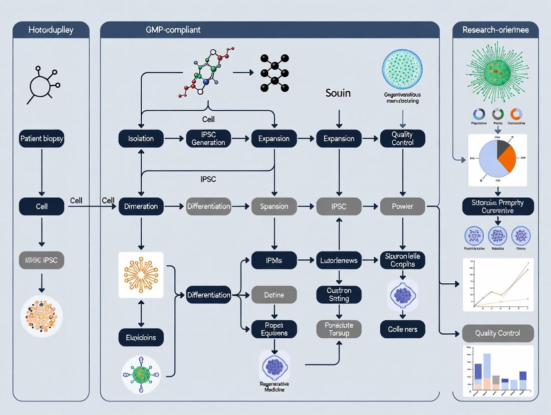
Visualizing the Workflow and Critical Pathways
Diagram 1: GMP-Compliant Autologous iPSC Manufacturing Workflow (76 chars)
Diagram 2: Core Signaling in iPSC Reprogramming (50 chars)
The Scientist's Toolkit: Research Reagent Solutions
Table 2: Essential Materials for GMP-Compliant Autologous iPSC Generation
| Item | Function & Rationale | Example (GMP-Grade or Equivalent) |
|---|---|---|
| Defined, Xeno-Free Basal Medium | Provides consistent nutrient supply without animal-derived components, reducing immunogenicity risk. | StemFlex Medium, TeSR-E8, mTeSR Plus. |
| Recombinant Human Matrix | Defined substrate for cell adhesion and signaling, replacing mouse embryonic fibroblasts (MEFs) or Matrigel. | Recombinant Human Vitronectin, Laminin-521. |
| Non-Integrating Reprogramming Vectors | Delivers reprogramming factors without genomic integration, critical for long-term safety. | Episomal plasmids (oriP/EBNA1), Sendai virus vectors, mRNA kits. |
| Closed System Cell Processing Set | Enables sterile cell manipulation (separation, washing) without open-container exposure. | Sepax C-Pro, Lovo, or equivalent closed tubing sets. |
| GMP-Grade Cytokines & Small Molecules | For directed differentiation or culture maintenance with defined activity and purity. | Recombinant Human FGF-basic, BMP4, CHIR99021, Y-27632. |
| Validated QC Assay Kits | For lot-to-lot consistent testing of safety (mycoplasma, endotoxin) and identity (pluripotency). | MycoAlert PLUS, hPSC Scorecard Panel, G-band karyotyping services. |
| Single-Use, Bioreactor Systems | Scalable expansion of iPSCs in a controlled, closed environment. | PBS MINI, StemCell Technologies Bioreactor. |
| Protein-Free Cryopreservation Medium | Prevents immunogenic reactions and ensures consistent post-thaw recovery. | CryoStor CS10, STEM-CELLBANKER GMP. |
The development of a robust, GMP-compliant workflow for manufacturing autologous induced pluripotent stem cell (iPSC)-derived therapies requires strict adherence to international regulatory guidelines. The U.S. Food and Drug Administration (FDA), the European Medicines Agency (EMA), and the International Council for Harmonisation of Technical Requirements for Pharmaceuticals for Human Use (ICH) provide the core frameworks. This document outlines critical application notes and protocols derived from these guidelines, contextualized for research aimed at establishing a scalable autologous iPSC manufacturing process.
Comparative Regulatory Framework Analysis
Table 1: Key Guideline Comparison for Cell Therapy Products
| Aspect | FDA (CBER) | EMA (ATMP) | ICH Harmonised |
|---|---|---|---|
| Primary Guideline | PHS Act 351; 21 CFR 1271 (HCT/Ps); Guidance for Human Cells, Tissues… (2020) | Regulation (EC) No 1394/2007; Guideline on Human Cell-based Medicinal Products (CHMP/410869/2006) | ICH Q5A(R2) - Q5E; ICH Q9 (Quality Risk Management); ICH Q10 (Pharmaceutical Quality System) |
| Marketing Authorization Pathway | Biologics License Application (BLA) | Marketing Authorisation Application (MAA) for Advanced Therapy Medicinal Product (ATMP) | Provides technical requirements for quality, safety, efficacy supporting BLA/MAA |
| GMP Standards | 21 CFR 210, 211; USP <1043>; Guidance for Industry: CGMP for Phase 1 Investigational Drugs | EudraLex, Volume 4, Part IV: ATMP GMP Guidelines | ICH Q7 (GMP for Active Pharmaceutical Ingredients) & Q10 provide foundation |
| Critical Quality Attributes (CQA) Focus | Identity, Potency, Purity, Viability, Safety (Sterility, Mycoplasma, Endotoxin) | Similar to FDA, with strong emphasis on product characterization and traceability | Framework for defining CQAs via ICH Q8(R2) Pharmaceutical Development & Q6B Specifications |
| Stability Data Requirements | Real-time, real-condition data recommended for Phase 3; program defined per ICH Q1A(R2) | Requires stability data per ICH Q1A(R2) and specific considerations for cell viability/function over time | ICH Q1A(R2) Stability Testing of New Drug Substances and Products |
| Donor Eligibility & Traceability | 21 CFR 1271 Subpart C (Donor Eligibility) required. Unique Donor Identifier. | Directive 2004/23/EC (Quality & Safety of Tissues & Cells). Full traceability from donor to patient and vice versa. | Supported by ICH Q5D Derivation and Characterization of Cell Substrates |
Table 2: Quantitative Testing Benchmarks for Autologous iPSC Banks
| Test Category | Specific Assay | Typical Acceptance Criteria (Example) | Guideline Reference |
|---|---|---|---|
| Identity | STR DNA Profiling | Match to donor somatic cell source (100% loci). Pluripotency marker expression (e.g., >90% Oct4+, Nanog+). | FDA CMC Guidance, ICH Q6B |
| Potency | In vitro trilineage differentiation (Embryoid Body) | ≥80% of cultures demonstrate ecto-, meso-, endodermal markers. | FDA Guidance: Potency Tests for Cellular and Gene Therapy Products |
| Purity & Safety | Sterility (BacT/Alert) | No growth after 14 days. | USP <71>, Ph. Eur. 2.6.27 |
| Safety | Mycoplasma (PCR & culture) | Negative. | USP <63>, Ph. Eur. 2.6.7 |
| Safety | Endotoxin (LAL) | <0.5 EU/mL (Intrathecal/<5.0 EU/mL (Systemic). | USP <85>, FDA Guideline |
| Safety | In vitro Adventitious Virus Assay | No Cytopathic Effect (CPE) or hemadsorption. | ICH Q5A(R2) |
| Genomic Stability | Karyotype (G-banding) | Normal diploid complement (46, XY/XX) for ≥20 metaphases. | FDA & EMA guidelines on cell therapy |
Detailed Application Notes & Protocols
Application Note 1: Protocol for Establishing a GMP-Compliant Master Cell Bank (MCB) from Autologous iPSCs
Objective: To generate a characterized MCB suitable for use in clinical manufacturing under FDA/EMA/ICH guidelines.
Materials & Reagents: See "Scientist's Toolkit" (Section 5).
Procedure:
- Starting Material: Obtain qualified somatic cells (e.g., dermal fibroblasts, PBMCs) from an apheresis product or biopsy under informed consent. Confirm donor eligibility per 21 CFR 1271/EC Directives.
- Reprogramming: Using a GMP-grade, integration-free method (e.g., Sendai virus vectors, episomal plasmids), reprogram somatic cells to iPSCs. Use xeno-free media and matrix.
- Clonal Selection & Expansion: Manually pick or use single-cell deposition to isolate clonal colonies. Expand promising clones in a controlled environment (5% CO2, 37°C).
- Banking: At passage 4-6, harvest cells at ~80% confluence using a gentle, enzyme-free dissociation reagent. Resuspend in GMP-grade cryopreservation medium (e.g., containing DMSO and albumin). Aliquot into cryovials (e.g., 1-5 x 10^6 cells/vial). Perform controlled-rate freezing to -80°C, then transfer to vapor phase liquid nitrogen for long-term storage.
- Full Characterization (Release Testing): Perform tests listed in Table 2 on at least 3 vials from the MCB. Additional tests may include:
- Residual Vector Clearance: qPCR for Sendai virus or episomal plasmid to demonstrate clearance by passage 10.
- Tumorigenicity Risk Assessment: Soft agar colony formation assay; in vivo teratoma formation in immunodeficient mice (for research phase).
- Bioenergetic Profile: Seahorse assay to confirm metabolic fitness.
Application Note 2: Protocol for Process Validation: Closed-System Cell Differentiation
Objective: To validate a critical unit operation (e.g., differentiation of iPSCs to cardiomyocyte progenitors) within a closed, automated system, ensuring consistency per ICH Q2(R1) and Q14.
Procedure:
- Risk Assessment (ICH Q9): Identify Critical Process Parameters (CPPs) for differentiation (e.g., seeding density, media exchange timing, agonist concentration).
- Design of Experiments (DoE): Set up a factorial DoE to model the relationship between CPPs and CQAs (e.g., % of cTnT+ cells, cell yield).
- Execution: Thaw one MCB vial per differentiation run. Seed cells in a closed, single-use bioreactor or cultureware. Initiate differentiation protocol using GMP-grade small molecules and growth factors. Monitor parameters (pH, DO, metabolites) inline if possible.
- Sampling & Analysis: Take daily samples for cell count, viability, and metabolite analysis (glucose, lactate). At endpoint (Day 10), analyze:
- Flow Cytometry: For cardiac-specific markers (cTnT, NKX2.5).
- Functional Assessment: Calcium transient imaging or MEA (Multi-Electrode Array) for beating cardiomyocytes.
- Data Analysis & Control Strategy: Establish proven acceptable ranges for each CPP. Define a control strategy including in-process controls (IPCs) for future manufacturing.
Visualization: Regulatory and Workflow Diagrams
Title: Regulatory Integration in iPSC Manufacturing Workflow
Title: Cardiac Differentiation Process with CPPs
The Scientist's Toolkit: Key Research Reagent Solutions
Table 3: Essential Materials for GMP-Compliant Autologous iPSC Research
| Item Category | Example Product/Supplier | Function & GMP Relevance |
|---|---|---|
| GMP-Grade Reprogramming | CytoTune-iPS 2.0 Sendai Kit (Thermo) | Non-integrating, GMP-manufactured vector system for footprint-free iPSC generation. |
| Xeno-Free Culture Medium | StemFlex Medium or E8 Medium (Thermo) / mTeSR Plus (STEMCELL) | Chemically defined, feeder-free media supporting robust pluripotent growth. Essential for regulatory compliance. |
| GMP-Grade Extracellular Matrix | Vitronectin (VTN-N) Recombinant Protein | Defined, animal-origin-free substrate for iPSC attachment and expansion, replacing Matrigel. |
| Cell Dissociation Reagent | Versene Solution or Recombinant Trypsin | Enzyme-free or recombinant enzymes for gentle passaging, minimizing genetic stress. |
| GMP-Grade Small Molecules | CHIR99021 (Tocris Bioscience, GMP grade) | Critical for directed differentiation protocols (e.g., cardiac, neural). Sourced with certificate of analysis. |
| Closed System Cultureware | Corning HYPERStack or PBS Biotech Bioreactors | Scalable, single-use vessels enabling aseptic processing and process control. |
| Mycoplasma Detection | MycoAlert Detection Kit (Lonza) | Rapid, sensitive bioluminescent assay for mycoplasma screening per USP/Ph. Eur. |
| Flow Cytometry Antibodies | SSEA-4, Oct-3/4, cTnT (BD Biosciences) | Validated antibodies for characterization of pluripotency and differentiated cell types (CQA assessment). |
Establishing Critical Quality Attributes CQAs and Critical Process Parameters CPPs for iPSC Lines
In a GMP-compliant autologous induced pluripotent stem cell (iPSC) manufacturing workflow, each patient-derived cell line is a unique drug substance. Establishing a robust control strategy is therefore paramount. This requires the systematic definition of Critical Quality Attributes (CQAs)—measurable properties that ensure product safety and efficacy—and Critical Process Parameters (CPPs)—key process variables that must be controlled to consistently meet CQAs. This application note details the experimental protocols and analytical frameworks for identifying and monitoring iPSC line CQAs and CPPs.
Defining iPSC Line CQAs: Categories and Quantitative Benchmarks
iPSC CQAs span identity, purity, potency, and safety. The following table summarizes core CQAs, their analytical methods, and typical target values or outcomes based on current literature and regulatory guidance.
Table 1: Core Critical Quality Attributes (CQAs) for iPSC Lines
| CQA Category | Specific Attribute | Analytical Method | Target / Acceptance Criteria |
|---|---|---|---|
| Identity | Pluripotency Marker Expression | Flow Cytometry, ICC | >90% positive for OCT4, SOX2, NANOG, SSEA-4 |
| Identity | Trilineage Differentiation Potential | In Vitro Spontaneous Differentiation & Analysis | Robust expression of ecto-, meso-, and endoderm markers |
| Purity | Residual Somatic Cell Contamination | Flow Cytometry (Lineage-specific markers) | < 1% contamination |
| Purity | Karyotypic Normalcy | G-band Karyotyping / mFISH / aCGH | 46, XX or XY, no major structural anomalies |
| Potency | Clonogenic Capacity / Colony Morphology | Bright-field Microscopy, AP Staining | >40% plating efficiency; defined, compact colonies |
| Safety | Genetic Stability (Point Mutations) | Whole Genome Sequencing (WGS) | No mutations in oncogenes/tumor suppressors vs. baseline |
| Safety | Microbial & Viral Contamination | Sterility Tests, Mycoplasma PCR, Viral Assays | No detection of adventitious agents |
Protocol 1: Quantitative Assessment of Pluripotency (Identity CQA)
Objective: To quantify the percentage of cells expressing core pluripotency transcription factors and surface markers. Materials: iPSC colonies, Essential 8 Flex Medium, Gentle Cell Dissociation Reagent, 4% Paraformaldehyde, Permeabilization Buffer (0.5% Triton X-100), Blocking Buffer (5% BSA/PBS), Primary Antibodies (OCT4, SOX2, NANOG, SSEA-4), Fluorochrome-conjugated Secondary Antibodies, Flow Cytometer. Procedure:
- Cell Preparation: Culture iPSCs to ~80% confluence. Dissociate into a single-cell suspension using Gentle Cell Dissociation Reagent.
- Fixation & Permeabilization: Aliquot 1x10^6 cells. Fix with 4% PFA for 15 min at RT. Wash with PBS. For intracellular markers (OCT4, SOX2, NANOG), permeabilize with 0.5% Triton X-100 for 15 min.
- Staining: Resuspend cells in Blocking Buffer for 30 min. Incubate with primary antibody (1:200 dilution) for 1 hour at RT or overnight at 4°C. Wash x3 with PBS/0.1% BSA. Incubate with appropriate secondary antibody (1:500) for 45 min in the dark. For surface markers (SSEA-4), stain live cells before fixation.
- Analysis: Resuspend in PBS + 1% BSA. Acquire data on a flow cytometer (collect ≥10,000 events). Use isotype controls to set gates. Calculate the percentage of positive cells.
Defining and Controlling CPPs for Reprogramming and Expansion
CPPs are process parameters whose variability impacts CQAs. They are identified via Design of Experiments (DoE). Key CPPs for the initial stages of an autologous workflow are listed below.
Table 2: Example Critical Process Parameters (CPPs) and Their Link to CQAs
| Process Step | Critical Process Parameter (CPP) | Linked CQA(s) | Control Strategy |
|---|---|---|---|
| Reprogramming | Vector Dose (if using non-integrating) | Genetic Stability, Purity | Optimized via DoE; in-process monitoring of copy number |
| Reprogramming | Oxygen Tension (%) | Pluripotency, Karyotypic Normalcy | Controlled incubator (5% O2 vs. 20% ambient) |
| Colony Picking | Colony Size (diameter in µm) at Pick | Clonogenicity, Pluripotency | Standardized SOP with microscopic calibration |
| Expansion | Seeding Density (cells/cm²) | Colony Morphology, Genetic Stability | Defined range (e.g., 15-25 x10^3 cells/cm²) |
| Expansion | Passaging Method (Enzymatic vs. EDTA) | Karyotypic Normalcy, Surface Marker Expression | Validated reagent and timing |
| Expansion | Days Between Passages | Pluripotency, Differentiation Purity | Fixed schedule (e.g., passage every 5-6 days) |
Protocol 2: Monitoring Genetic Stability (Safety CQA) via mFISH
Objective: To detect chromosomal rearrangements and aneuploidy in metaphase spreads. Materials: iPSCs in log-phase growth, Colcemid (10 µg/mL), Hypotonic Solution (0.075M KCl), Carnoy’s Fixative (3:1 Methanol:Glacial Acetic Acid), 24x60 mm glass slides, mFISH Probe Kit (e.g., 24XCyte), Formamide, SSC Buffer, DAPI, Fluorescence Microscope with appropriate filters. Procedure:
- Metaphase Arrest: Add Colcemid to culture medium (final 0.1 µg/mL). Incubate for 45-60 min at 37°C.
- Cell Harvest: Dissociate to single cells. Pellet and resuspend gently in pre-warmed 0.075M KCl. Incubate at 37°C for 20 min.
- Fixation: Slowly add 1 mL of fresh Carnoy’s fixative. Pellet cells. Wash 3x with fixative. Store pellet at -20°C.
- Slide Preparation: Drop fixed cell suspension onto clean, wet slides. Age slides overnight at RT.
- mFISH Hybridization: Denature slides in 70% formamide/2xSSC at 72°C for 2 min. Dehydrate in ethanol series. Apply denatured mFISH probe, cover with a coverslip, and seal with rubber cement. Hybridize in a humid chamber at 37°C for 24-48 hrs.
- Washing & Detection: Wash per kit protocol to remove unbound probe. Counterstain with DAPI.
- Analysis: Image ≥20 metaphase spreads per sample using a fluorescence microscope with dedicated mFISH software. Analyze for numerical and structural abnormalities.
Visualizing the Control Strategy Logic
Title: iPSC Manufacturing: CPPs Influence CQAs to Ensure Quality
Core Pluripotency Signaling Pathway
Title: Key Signaling Pathways Supporting iPSC Pluripotency
The Scientist's Toolkit: Key Research Reagent Solutions
Table 3: Essential Materials for iPSC CQA/CPP Analysis
| Reagent/Material | Function in CQA/CPP Work | Example/Note |
|---|---|---|
| Defined, Xeno-Free Culture Medium | Provides consistent base matrix for process control; eliminates lot variability. | Essential 8 Flex, StemFlex |
| Non-Integrating Reprogramming Kit | Critical for safety CQA; generates footprint-free iPSCs. | Sendai virus vectors (CytoTune), mRNA kits |
| Flow Cytometry Antibody Panel | Quantifies identity (pluripotency) and purity (contamination) CQAs. | Conjugated antibodies to OCT4, SSEA-4, TRA-1-60, somatic markers |
| G-band Karyotyping / mFISH Kits | Assesses genetic stability safety CQA. | MetaSystems mFISH probes |
| Whole Genome Sequencing Service | Provides ultimate depth for genetic stability CQA (oncogenic mutations). | Offered by core labs/service providers (e.g., Illumina). |
| Trilineage Differentiation Kit | Validates potency CQA via directed differentiation. | STEMdiff Trilineage Differentiation Kit |
| Automated Cell Counter (Imaging-based) | Standardizes CPP of seeding density measurement. | Countess 3, NC-200 |
| Mycoplasma Detection Kit | Essential for safety/purity CQA monitoring. | PCR-based detection (e.g., MycoAlert) |
Application Notes: GMP Cleanroom Design for Autologous iPSC Manufacturing
The successful clinical translation of autologous induced pluripotent stem cell (iPSC) therapies is critically dependent on the design and control of the manufacturing environment. Unlike allogeneic products, autologous workflows involve parallel processing of patient-specific batches, heightening the risk of cross-contamination and necessitating impeccable facility design. A GMP-compliant cleanroom suite must integrate architectural, engineering, and procedural controls to ensure aseptic processing and product integrity.
1. Zoning and Pressure Cascades: The facility must implement a logical flow of personnel, materials, and product, supported by defined air pressure differentials. This prevents ingress of contaminants from lower-grade to higher-grade areas.
Table 1: Cleanroom Classification & Environmental Monitoring Limits (Based on EU GMP Annex 1 & ISO 14644-1)
| Zone / Room Function | Target ISO Class (at-rest) | Equivalent EU GMP Grade (at-rest) | Maximum Particles (≥0.5 μm)/m³ | Typical Pressure Differential (Pa) |
|---|---|---|---|---|
| Cell Banking & Final Fill | ISO 5 | A | 3,520 | +10 to +15 (relative to background) |
| Background for Grade A (iPSC Manipulation) | ISO 7 | B | 352,000 | +10 to +15 (relative to corridor) |
| Corridor / Gowning Area | ISO 8 | C | 3,520,000 | +5 to +10 (relative to unclassified) |
| Support Areas (Buffer) | Unclassified | D | Not Defined | 0 to +5 |
2. Critical Design Parameters for iPSC Workflows:
- Unidirectional Flow: A strict "first in, first out" material flow for single-patient batches is mandatory. Dedicated incubators and bioreactors per patient may be required.
- Material Transfer: Use of validated double-door pass-through autoclaves and vaporized hydrogen peroxide (VHP) chambers for sterile transfer of supplies.
- Surfaces: Seamless, non-shedding, cleanable surfaces (e.g., epoxy resin floors, coved corners) to support sanitization regimes.
Protocols for Environmental Monitoring & Control
Protocol 1: Routine Viable Air and Surface Monitoring in an ISO 5 (Grade A) Laminar Flow Biosafety Cabinet (BSC)
Objective: To actively monitor the microbial contamination level within the primary aseptic processing zone during a simulated iPSC feeding or splitting operation.
Materials:
- Microbial air sampler (e.g., impaction-based sampler with 40-100 L/min flow rate).
- Soybean Casein Digest (TSA) contact plates (55mm) and settle plates.
- Sterile swabs and Neutralizing Broth (for recovery if disinfectant residues are present).
- Incubator (set at 20-25°C for 5-7 days, followed by 30-35°C for 2-3 days).
Methodology:
- Preparation: Sanitize the interior of the BSC with sporicidal agent. Start BSC blower 30 minutes prior. Place TSA settle plates at predefined, risk-assessed locations within the BSC (e.g., near manipulator, next to media bottle). Label plates with location, time, and date.
- Active Air Sampling: Place the microbial air sampler inside the BSC, away from direct laminar flow disruption. Run the sampler for the duration of a typical critical operation (e.g., 1 hour at 100 L/min).
- Surface Monitoring (Post-Operation): Using TSA contact plates, sample critical surfaces: interior BSC work surface, gloved fingertips of operator after the process, and the exterior of key reagent containers. Apply even pressure.
- Incubation & Analysis: Seal and invert plates. Incubate per the dual-temperature regimen. Count Colony Forming Units (CFUs). Compare results against established Alert (e.g., 1 CFU) and Action Limits (e.g., 1 CFU for air, 2 CFU for surface in Grade A).
- Action: Any result exceeding Action Limits must trigger an investigation per Deviation Management procedures, including identification of isolated organisms and review of aseptic technique, gowning, and sanitization efficacy.
Protocol 2: Qualification of Pressure Cascade and Airflow Visualization (Smoke Study)
Objective: To visually demonstrate unidirectional airflow and confirm pressure differentials between adjoining cleanrooms.
Materials: Portable differential pressure manometer, theatrical fog generator with non-toxic glycol-based fog, HEPA filter leak test kit (optional, for integrity test).
Methodology:
- Pressure Differential Verification: Using a calibrated manometer, record the pressure difference across all inter-room doors (e.g., ISO 7 to ISO 8, ISO 8 to unclassified). Ensure readings meet specifications from Table 1.
- Airflow Visualization:
- Introduce smoke/fog upstream of the HEPA filter in the room/device under test (e.g., at the return grille in an ISO 7 room).
- Visually observe smoke patterns. In an ISO 5 BSC or laminar flow unit, smoke should flow in a coherent, unidirectional stream with no reflux or stagnation over the critical working zone.
- Open an inter-room door slightly. Observe smoke movement; it must flow from the higher-grade (higher pressure) to the lower-grade room, confirming the pressure cascade.
- Documentation: Video record the smoke study. Any turbulence or flow reversal constitutes a test failure and requires HVAC system adjustment.
Diagrams
The Scientist's Toolkit: Critical Reagents & Materials for Aseptic Processing
Table 2: Essential Research Reagent Solutions for Cleanroom Operations
| Item | Function in GMP Cleanroom Context |
|---|---|
| Sporicidal Disinfectant (e.g., Hydrogen Peroxide-based, Peracetic Acid) | Validated for effective removal of bacterial spores from surfaces; used in rotation with other agents to prevent microbial resistance. |
| Sterile, Non-Pyrogenic Wipes | Low-linting wipes used with disinfectants for cleaning critical surfaces; must be compatible with sterilizing agents like VHP. |
| Viable Particle Growth Media (TSA & SDA Contact/Air Plates) | Soybean Casein Digest Agar (TSA) for bacteria, Sabouraud Dextrose Agar (SDA) for fungi/molds; used for routine environmental monitoring. |
| Neutralizing Broth / Rinse Fluid | Contains inactivators (e.g., lecithin, polysorbate) to neutralize residual disinfectants on sampled surfaces, ensuring accurate microbial recovery. |
| GMP-Grade Single-Use Systems (Bioreactor bags, tubing sets, connectors) | Pre-sterilized, closed systems that minimize manual aseptic connections and reduce contamination risk during iPSC expansion. |
| Particulate Matter Monitoring System (Laser particle counter) | Provides real-time, continuous monitoring of air quality per ISO classification standards, with alarms for excursions. |
| Biological Indicators (Geobacillus stearothermophilus spores) | Used for validation of sterilization cycles (autoclave, VHP) to prove a defined log-reduction of microbial load. |
Application Notes
The initiation of a GMP-compliant autologous iPSC manufacturing workflow hinges on the integrity and suitability of the starting biological material. Decisions made during donor screening and tissue acquisition directly impact downstream reprogramming efficiency, clonal selection, and the safety profile of the final cellular product. Key considerations are outlined below.
1. Donor Eligibility & Medical Screening A comprehensive health assessment is mandatory to mitigate risks of transmitting adventitious agents or introducing genetic predispositions into the cell line. Screening must align with regulatory guidelines for human cells, tissues, and cellular/tissue-based products (HCT/Ps).
2. Biopsy Type & Site Selection The choice of tissue source balances accessibility, proliferative capacity of isolated cells, and patient burden. Common sources include dermal fibroblasts and peripheral blood mononuclear cells (PBMCs).
3. Pre-Analytical Variables Biopsy collection, transport conditions, and initial processing are critical pre-analytical variables that must be standardized to ensure cell viability and prevent phenotypic drift prior to reprogramming.
Table 1: Quantitative Comparison of Common Tissue Sources for Autologous iPSC Generation
| Parameter | Skin Punch Biopsy (Fibroblasts) | Peripheral Blood Draw (PBMCs) | Urine (Renal Epithelial Cells) |
|---|---|---|---|
| Invasiveness | Moderate (local anesthetic) | Minimal | Non-invasive |
| Tissue Processing Complexity | High (requires explant culture & expansion) | Moderate (density separation, activation) | Low (centrifugation, plating) |
| Typical Time to Sufficient Cell Number | 3-5 weeks | 1-2 weeks (with expansion) | 2-3 weeks |
| Reprogramming Efficiency (Relative) | Baseline (1x) | Comparable to baseline | Slightly lower |
| Risk of Senescence | Higher (donor age-dependent) | Lower | Moderate |
| Primary Cell Culture Success Rate | >95% | >90% (for T-cell subsets) | ~70-80% |
Table 2: Core Donor Screening Criteria & Tests
| Screening Category | Specific Tests/Assessments | Rationale |
|---|---|---|
| Infectious Disease | HIV-1/2, HBV, HCV, HTLV-I/II, Syphilis, West Nile Virus, T. cruzi (as per FDA guidance) | Prevent introduction of pathogens into manufacturing facility and final product. |
| Genetic Risk | Family history of dominant monogenic diseases, karyotype analysis (if indicated) | Mitigate risk of propagating deleterious mutations; baseline genomic integrity. |
| General Health | Complete blood count (CBC), metabolic panel, physical exam | Assess donor fitness for procedure and identify underlying conditions affecting cell health. |
Detailed Protocols
Protocol 1: GMP-Compliant Skin Punch Biopsy for Dermal Fibroblast Isolation
Objective: To aseptically obtain a skin tissue sample and derive a primary fibroblast culture.
Materials:
- Disposable punch biopsy tool (3-4mm)
- Sterile drapes, gauze, local anesthetic (e.g., lidocaine)
- Antiseptic solution (e.g., chlorhexidine, iodine)
- Transport medium: DMEM + 2x Antibiotic-Antimycotic (e.g., Penicillin-Streptomycin-Amphotericin B)
- Biopsy container (leak-proof, sterile)
Methodology:
- Informed Consent & Site Selection: Obtain informed consent. Select hairless site (e.g., upper arm, forearm). Cleanse area with antiseptic in concentric circles.
- Biopsy: Administer local anesthetic. Stabilize skin. Use punch tool with rotating motion to penetrate through dermis. Use forceps to gently lift tissue, and dissect base with scissors.
- Transport: Immediately place tissue in pre-chilled transport medium. Store at 4°C for ≤24 hours.
- Processing (Class II Biosafety Cabinet): a. Rinse tissue 3x in PBS + 1x Antibiotic-Antimycotic. b. Mince tissue into ~1 mm³ fragments using sterile scalpels. c. Explain fragments onto a tissue culture plate pre-coated with a GMP-grade attachment matrix. d. Add fibroblast growth medium (DMEM, 10% Human Serum Albumin, bFGF). e. Incubate at 37°C, 5% CO₂. Change medium twice weekly.
- Expansion: Upon outgrowth (~10-14 days), passage cells using recombinant trypsin. Expand to required cell number (≥1x10⁶) for reprogramming, typically at passage 3-5.
Protocol 2: Isolation of CD34+ Hematopoietic Stem/Progenitor Cells from Peripheral Blood
Objective: To isolate a potent, proliferative cell population from a blood draw for reprogramming.
Materials:
- GMP-grade Lymphoprep or Ficoll-Paque density gradient medium
- Heparinized blood collection tubes
- Phosphate-Buffered Saline (PBS) without Ca²⁺/Mg²⁺
- GMP-grade CD34+ magnetic-activated cell sorting (MACS) kit
- MACS buffer (PBS + 0.5% HSA + 2mM EDTA)
Methodology:
- Collection: Draw 20-40mL of peripheral blood into heparin tubes.
- PBMC Isolation: Dilute blood 1:1 with PBS. Carefully layer over density gradient medium. Centrifuge at 400 x g for 30 minutes at room temperature (brake off).
- Harvest: Collect the mononuclear cell layer at the interface. Wash cells twice with PBS by centrifuging at 300 x g for 10 minutes.
- CD34+ Selection: Resuspend cell pellet in MACS buffer. Incubate with GMP-grade CD34 MicroBeads per manufacturer's instructions. Pass cell-bead mixture through a pre-wet LS MACS Column placed in a magnetic field. Wash column with buffer. Remove column from magnet and elute positively selected CD34+ cells.
- Culture & Expansion: Plate cells in serum-free hematopoietic stem cell expansion medium (e.g., containing SCF, TPO, Flt3-L). Expand for 5-7 days prior to reprogramming initiation.
Visualizations
Title: Donor to Cell Bank Workflow
Title: Core Reprogramming Signal Flow
The Scientist's Toolkit: Research Reagent Solutions
| Item | Function | Key Considerations for GMP |
|---|---|---|
| GMP-grade Reprogramming Vectors (e.g., Episomal plasmids, Sendai virus) | Deliver reprogramming factors (OCT4, SOX2, KLF4, c-MYC) without genomic integration. | Must be produced under GMP, with certificates of analysis for identity, purity, and safety (RCL testing for viral vectors). |
| Xeno-free & Chemically Defined Media | Supports cell growth without animal-derived components, ensuring consistency and reducing immunogenicity risk. | Formulation must be fully defined, with human-grade or recombinant supplements (e.g., Human Serum Albumin). |
| Recombinant Enzymes (e.g., Trypsin, TrypLE) | For gentle, standardized cell dissociation and passaging. | Animal-origin free, recombinant production, with validated activity and absence of contaminants. |
| Attachment Matrices (e.g., Recombinant Laminin-521, Vitronectin) | Provides defined extracellular matrix for cell adhesion, survival, and expansion. | Recombinant human proteins are preferred over mouse feeder cell-derived Matrigel for consistency and regulatory compliance. |
| Magnetic Cell Sorting Kits (e.g., CD34+ MACS) | Isolation of specific cell populations from a heterogeneous biopsy sample. | GMP-grade kits with clinical-grade magnetic beads and columns are essential for autologous production. |
Risk Management and Quality by Design (QbD) Principles in Process Development
Application Note: Integration of QbD and Risk Management in Autologous iPSC Process Development
The implementation of a GMP-compliant, autologous induced pluripotent stem cell (iPSC) manufacturing workflow requires a proactive approach to ensure product quality, safety, and efficacy. Quality by Design (QbD) is a systematic framework for developing processes with predefined objectives, emphasizing product and process understanding and control based on sound science and quality risk management. This application note outlines the integration of QbD principles with formal risk management tools within the context of autologous iPSC-derived therapies.
Core QbD Elements:
- Quality Target Product Profile (QTPP): A prospective summary of the quality characteristics of the drug product.
- Critical Quality Attributes (CQAs): Physical, chemical, biological, or microbiological properties or characteristics that should be within an appropriate limit, range, or distribution to ensure the desired product quality.
- Critical Process Parameters (CPPs): Process parameters whose variability impacts a CQA and therefore should be monitored or controlled to ensure the process produces the desired quality.
- Design Space: The multidimensional combination and interaction of input variables (e.g., material attributes) and process parameters that have been demonstrated to provide assurance of quality.
- Control Strategy: A planned set of controls, derived from current product and process understanding, that ensures process performance and product quality.
Risk Management Framework: Following ICH Q9 (Quality Risk Management), risks are identified, analyzed, evaluated, and controlled throughout the development lifecycle. Tools such as Failure Mode and Effects Analysis (FMEA) are employed to prioritize risks to CQAs.
Table 1: Example QTPP for an Autologous iPSC-Derived Cardiomyocyte Therapy
| QTPP Element | Target | Justification & Risk Level |
|---|---|---|
| Dosage Form | Suspension of viable cells | Parenteral administration; High Risk |
| Viability | ≥ 70% | Critical for engraftment efficacy; High Risk |
| Potency (In Vitro) | ≥ 80% beating cardiomyocytes | Direct link to therapeutic mechanism; High Risk |
| Purity | ≤ 5% undifferentiated iPSCs | Mitigates teratoma risk; High Risk |
| Identity (Cell Surface Markers) | cTnT+ ≥ 90%, SSEA-1- ≥ 95% | Confirms target phenotype and absence of pluripotency; Medium Risk |
| Sterility | Sterile (no microbial growth) | Patient safety; High Risk |
| Endotoxin | < 0.5 EU/mL | Patient safety; High Risk |
Table 2: Prioritized Risks to CQAs from an FMEA (Partial Example)
| Process Step | Potential Failure Mode | Effect on CQA(s) | Severity (S) | Occurrence (O) | Detectability (D) | RPN (SxOxD) | Mitigation Action |
|---|---|---|---|---|---|---|---|
| Reprogramming | Low efficiency | Delayed production, insufficient starting material | 6 | 4 | 3 | 72 | Optimize vector ratio; use integrated donor-matched reagents |
| 3D Differentiation | High variability in potency | Low % cardiomyocytes, lot failure | 9 | 5 | 4 | 180 | Implement controlled bioreactor with DO/pH monitoring; define CPPs (e.g., agitation speed) |
| Cell Dissociation | Low post-thaw viability | Reduced viable dose | 8 | 5 | 2 | 80 | Develop gentle enzymatic protocol; optimize cryoprotectant formulation |
Experimental Protocols
Protocol 1: Risk-Based Identification and Assessment of CQAs
Objective: To systematically define and prioritize Critical Quality Attributes (CQAs) for an autologous iPSC-derived product using a science- and risk-based approach.
Materials:
- QTPP document
- Multidisciplinary team (Process Development, Analytical, Regulatory, Clinical)
- Risk assessment tool (e.g., FMEA template)
Methodology:
- QTPP Review: Assemble the team to review and confirm the QTPP.
- Attribute Listing: List all potential quality attributes (e.g., viability, identity, purity, potency, sterility, genomic stability).
- Initial Risk Ranking: For each attribute, assess the severity of harm to the patient if the attribute is outside the target range. Use a scale (e.g., 1-10). Attributes with high severity scores are potential CQAs.
- Scientific Linkage Assessment: For each potential CQA, evaluate the link between the attribute and product safety/efficacy using available prior knowledge (literature, platform data) and experimental studies (see Protocol 2).
- Final CQA Designation: Designate as a CQA if a plausible mechanistic or empirical link exists between the attribute range and product safety/efficacy. Document justification.
- Risk Assessment Update: Incorporate CQA designation into the ongoing risk assessment (e.g., FMEA) to guide process development controls.
Protocol 2: Design of Experiments (DoE) for Defining the Design Space of Cardiomyocyte Differentiation
Objective: To determine the impact and interaction of Critical Process Parameters (CPPs) on the Critical Quality Attribute (CQA) "Potency (% cTnT+ cells)" and establish a proven acceptable range.
Materials:
- Master Cell Bank of iPSC line
- Defined differentiation basal medium
- Growth factors (e.g., CHIR99021, IWP-4)
- Controlled bioreactor system (e.g., DASbox Mini Bioreactor)
- Flow cytometer with anti-cTnT antibodies
- DoE software (e.g., JMP, Design-Expert)
Methodology:
- Risk-Based Parameter Selection: From an initial risk assessment (e.g., Fishbone diagram), select factors for study: e.g., A: CHIR99021 concentration (μM), B: Duration of Wnt activation (hours), C: Agitation speed (rpm).
- Experimental Design: Construct a Response Surface Methodology (RSM) design, such as a Central Composite Design (CCD), to efficiently model linear, interaction, and quadratic effects.
- Execution: Execute the randomized experimental runs in the bioreactor system. Differentiate iPSCs to cardiomyocytes according to the baseline protocol, varying the selected parameters per the DoE matrix.
- Analytical Testing: On day 12 of differentiation, sample cells and analyze the percentage of cTnT+ cardiomyocytes by flow cytometry as the response variable (Y).
- Data Analysis: Fit the data to a polynomial model using the DoE software. Perform statistical analysis (ANOVA) to identify significant model terms (factors A, B, C, interactions AB, AC, BC, quadratic terms A², etc.).
- Design Space Visualization: Generate contour plots and 3D response surfaces to visualize the combination of factor levels that yield the target potency (e.g., ≥80% cTnT+). The region meeting this criterion constitutes the proposed design space.
- Verification: Conduct verification runs at points within and at the edges of the design space to confirm predictability.
Visualizations
Diagram 1: QbD and Risk Management Workflow Integration
Diagram 2: Risk Prioritization via FMEA Process
The Scientist's Toolkit: Key Research Reagent Solutions
Table 3: Essential Materials for QbD-Driven iPSC Process Development
| Item | Function in QbD Context | Example/Note |
|---|---|---|
| GMP-Grade Reprogramming Vectors | Ensures consistent, traceable, and safe generation of starting iPSC lines. Critical for defining CMA (Critical Material Attribute). | Episomal vectors, mRNA kits, or integration-free viral systems. |
| Chemically Defined Media | Eliminates lot-to-lot variability of animal sera. Enables precise modeling of CPP impact on CQAs in DoE studies. | E8 medium for iPSC expansion, defined differentiation kits. |
| Controlled Bioreactor System | Allows precise control and monitoring of CPPs (pH, DO, agitation, feeding). Essential for scale-up and design space definition. | Ambr or DASbox systems for high-throughput process optimization. |
| Process Analytical Technology (PAT) | In-line or at-line monitoring of critical attributes (e.g., cell density, metabolites). Supports real-time control strategy. | Bioanalyzer, metabolite analyzers (Nova Bioprofile), in-line microscopy. |
| Multiplexed QC Assays | Enables simultaneous measurement of multiple CQAs (purity, identity, potency) from a single sample. Critical for control strategy. | Flow cytometry panels (e.g., for pluripotency & lineage markers), qPCR arrays. |
| Genomic Stability Assay | Monitors a key safety CQA (karyotype, CNVs) throughout the process to ensure control. | Karyotyping, SNP arrays, or next-generation sequencing (NGS) services. |
| DoE Software | Statistical platform for designing efficient experiments and modeling data to establish design spaces and identify CPPs. | JMP, Design-Expert, or MODDE. |
Step-by-Step GMP Protocol: The Autologous iPSC Manufacturing Workflow in Detail
Within a GMP-compliant autologous iPSC manufacturing workflow, the initial acquisition and qualification of patient-specific somatic cells is a critical foundational step. This phase ensures the provision of a high-quality, well-characterized starting cell population, which is essential for subsequent reprogramming, clonal selection, and banking. This application note details standardized protocols for the isolation, culture expansion, and quality control characterization of two common somatic cell sources: dermal fibroblasts and peripheral blood mononuclear cells (PBMCs). Adherence to these protocols under appropriate quality systems supports the traceability and regulatory compliance required for clinical-grade iPSC generation.
Key Research Reagent Solutions
The following table catalogs essential materials and their functions for somatic cell isolation and characterization.
| Reagent / Material | Function / Purpose | Key Considerations for GMP |
|---|---|---|
| GMP-Grade Collagenase, Type I | Enzymatic dissociation of dermal tissue to isolate fibroblasts. | Defined animal-free origin, endotoxin testing, certificate of analysis. |
| Ficoll-Paque PREMIUM | Density gradient medium for isolation of PBMCs from whole blood. | GMP-manufactured, sterile, endotoxin-controlled. |
| Xeno-Free Fibroblast Medium | Serum-free culture expansion of dermal fibroblasts. | Eliminates batch variability and immunogenic risks associated with fetal bovine serum. |
| Lymphocyte Expansion Medium | Supports the activation and proliferation of T-cells from PBMCs. | Contains defined cytokines, suitable for closed-system culture. |
| Flow Cytometry Antibody Panel | Characterization of cell surface markers for identity and purity. | Validated for specificity, conjugated with GMP-compatible fluorochromes. |
| Mycoplasma Detection Kit | Essential quality control test for absence of mycoplasma contamination. | PCR-based, with high sensitivity, compliant with pharmacopoeial guidelines. |
Experimental Protocols
Isolation and Expansion of Dermal Fibroblasts
Principle: A skin punch biopsy is enzymatically and mechanically dissociated to release fibroblasts, which are then cultured in a xeno-free medium to establish a primary cell stock.
Detailed Protocol:
- Biopsy Collection: After informed consent and under aseptic conditions, obtain a 3-4 mm skin punch biopsy. Place in sterile transport medium (e.g., DMEM with high antibiotics).
- Tissue Processing: In a biosafety cabinet, wash biopsy 3x in DPBS with 2x antibiotics/antimycotics. Remove subcutaneous fat and cut into ~1 mm² explants.
- Explant Culture: Place explants directly onto a GMP-grade tissue culture plate. Allow to semi-dry for 5-10 minutes. Gently overlay with pre-warmed xeno-free fibroblast medium. Incubate at 37°C, 5% CO₂.
- Fibroblast Outgrowth: Change medium every 2-3 days. Fibroblast migration from explants is typically observed within 5-7 days.
- Passaging: At ~80% confluence, passage cells using a recombinant trypsin substitute. Replate at a 1:2 to 1:3 split ratio. Expand cells to required numbers (e.g., 1-5 x 10⁶ cells) for characterization and cryopreservation.
Isolation of Peripheral Blood Mononuclear Cells (PBMCs)
Principle: Whole blood is layered over a density gradient medium. Upon centrifugation, PBMCs are separated based on density and collected from the plasma-Ficoll interface.
Detailed Protocol:
- Blood Draw: Collect peripheral blood (e.g., 20-40 mL) into heparin or EDTA vacutainers.
- Dilution: Dilute blood 1:1 with sterile, endotoxin-free DPBS or Hank's Balanced Salt Solution.
- Density Gradient Separation: Carefully layer the diluted blood over Ficoll-Paque in a centrifuge tube (e.g., 15 mL blood over 10 mL Ficoll). Centrifuge at 400 x g for 30-40 minutes at room temperature, with the brake OFF.
- PBMC Harvest: After centrifugation, aspirate the upper plasma layer. Carefully collect the mononuclear cell layer at the interface using a sterile pipette. Transfer to a new tube.
- Washing: Wash harvested cells with DPBS + 2% human serum albumin by centrifuging at 300 x g for 10 minutes. Repeat wash step. Resuspend cell pellet in appropriate culture medium or freezing medium.
Characterization and Quality Control
A panel of release criteria tests must be performed on the expanded somatic cell population prior to reprogramming.
Table 1: Minimum Characterization Panel for Patient-Specific Somatic Cells
| Test Category | Specific Assay | Acceptance Criteria (Example) | Purpose |
|---|---|---|---|
| Identity & Purity | Flow cytometry for cell-type specific markers (Fibroblasts: CD90+, CD73+, CD105+, CD45-; PBMCs: CD45+) | >95% positive for lineage markers, <5% for negative markers | Confirms target cell population and absence of significant contamination. |
| Viability | Trypan Blue Exclusion or Flow cytometry with 7-AAD | >90% viability post-thaw/at passage | Ensures a robust, healthy cell population for reprogramming. |
| Sterility | BacT/ALERT or equivalent microbial culture | No growth of aerobic/anaerobic bacteria/fungi | Confirms aseptic processing. |
| Mycoplasma | PCR-based detection (e.g., MycoSEQ) | Not Detected | Essential safety test. |
| Proliferative Capacity | Population Doubling Time (PDT) calculation | PDT within historical range for cell type/age | Indicates cellular health and expansion potential. |
| Karyotype | G-band karyotyping or SNP array | Normal 46, XX or XY | Assesses genomic stability after in vitro expansion. |
Experimental Workflow and Pathway Visualizations
Title: Workflow for Somatic Cell Isolation and Qualification
Title: PBMC Processing with Parallel QC Pathways
Within a GMP-compliant autologous iPSC manufacturing workflow, the reprogramming phase is critical. It determines the genetic integrity, safety profile, and regulatory acceptance of the final cell therapy product. This application note compares the three leading non-integrating reprogramming methods—Sendai Virus (SeV), Episomal Vectors, and mRNA—contrasting them with historical integrating methods. The focus is on practical protocol considerations, efficiency, and compliance for clinical-grade iPSC generation.
Comparative Analysis of Reprogramming Methods
Table 1: Quantitative Comparison of GMP-Compliant Reprogramming Methods
| Parameter | Integrating Methods (Retro/Lenti) | Sendai Virus (CytoTune) | Episomal Vectors (Epi5) | Synthetic mRNA (StemRNA) |
|---|---|---|---|---|
| Integration Risk | High (Random genomic integration) | None (Cytoplasmic, RNA virus) | Very Low (Episomal loss) | None |
| Footprint-Free iPSCs | No | Yes (Virus diluted out) | Yes (Vector lost) | Yes |
| Reprogramming Efficiency | 0.1% - 1% | 0.1% - 1% | 0.01% - 0.1% | 1% - 4% |
| Kinetics (Days to Colonies) | 14-21 | 14-24 | 21-30 | 7-14 |
| GMP-Grade Kit Availability | No | Yes (CytoTune iPS 2.1) | Yes (Epi5 Episomal iPSC Reprogramming Kit) | Yes (StemRNA 3rd Gen Reprogramming Kit) |
| Key Safety Concerns | Insertional mutagenesis, oncogene reactivation | Immune response, persistence testing required | Low, but plasmid DNA residue testing | High IFN response, requires daily transfection |
| Typical Cost per Reprogramming | Low | High | Medium | Medium-High |
Detailed Experimental Protocols
Protocol 3.1: GMP-Compliant iPSC Generation Using Sendai Virus (SeV) Vectors
- Primary Cells: Human dermal fibroblasts (HDFs) or PBMCs.
- Materials: CytoTune iPS 2.1 Sendai Reprogramming Kit (Thermo Fisher), GMP-grade 6-well plates, qualified FBS or xeno-free medium.
- Procedure:
- Seed HDFs at 5 x 10⁴ cells/well in a 6-well plate 24 hours before transduction.
- Prepare virus cocktail containing the four reprogramming vectors (KOS, c-Myc, Klf4) in appropriate medium supplemented with 6 µg/mL polybrene.
- Remove cell culture medium and add 1 mL of virus cocktail per well. Centrifuge plate at 1000 x g for 30 min at 32°C (spinfection).
- Incubate at 37°C, 5% CO₂ for 2 hours. Add 1 mL of complete medium and incubate overnight.
- Day 2: Replace with fresh complete medium.
- Days 3-7: Passage transduced cells onto vitronectin-coated plates with Essential 8 Medium.
- Monitor for SeV persistence via RT-PCR from passage 5 onward. Colonies appear between days 14-24 and are manually picked.
Protocol 3.2: GMP-Compliant iPSC Generation Using Episomal Vectors
- Primary Cells: Human CD34+ cells or fibroblasts.
- Materials: Epi5 Episomal iPSC Reprogramming Kit (Thermo Fisher), Nucleofector Device (Lonza), Human Stem Cell Nucleofector Kit 1.
- Procedure:
- Harvest and count 1 x 10⁶ target cells.
- Prepare Nucleofection solution: Mix 82 µL of Nucleofector Solution, 18 µL of Supplement, and 2 µg of Epi5 plasmid cocktail.
- Resuspend cell pellet in 100 µL of nucleofection solution. Transfer to cuvette and run the appropriate program (e.g., U-023 for fibroblasts).
- Immediately add pre-warmed recovery medium and transfer cells to a vitronectin-coated 6-well plate in Essential 8 Medium.
- Feed every other day. Early colonies may be visible around day 10, but mature, pickable colonies emerge around days 21-30.
- Confirm episomal loss via PCR on genomic DNA from passage 10+ iPSCs.
Protocol 3.3: GMP-Compliant iPSC Generation Using Synthetic mRNA
- Primary Cells: Fibroblasts.
- Materials: StemRNA 3rd Gen Reprogramming Kit (Reprocell), mRNA Transfection Kit (e.g., Lipofectamine MessengerMAX, Thermo Fisher), GMP-grade 24-well plates.
- Procedure:
- Day 0: Seed fibroblasts at 5 x 10⁴ cells/well of a 24-well plate.
- Days 1-5: Daily transfection.
- Dilute 0.5 µg of the 5-factor mRNA cocktail (OCT4, SOX2, KLF4, c-MYC, LIN28) in Opti-MEM.
- Dilute MessengerMAX reagent in a separate tube of Opti-MEM.
- Combine solutions, incubate 5 min, then add dropwise to cells.
- Days 6-18: Change to Essential 8 Medium, feed daily. Colonies become visible from day 7. Manually pick from day 14.
- Critical Note: Include 0.5 µg of IFN-γ suppressor mRNA (supplied in kit) in each transfection to enhance survival.
Visualizations
Title: Non-Integrating Reprogramming Workflow Comparison
Title: Core Pluripotency Network Activation Pathway
The Scientist's Toolkit: Key Reagent Solutions
Table 2: Essential Materials for GMP-Compliant Reprogramming
| Item | Function & GMP Relevance | Example Product |
|---|---|---|
| GMP-Grade Reprogramming Kit | Provides QC'd, validated vectors/mRNA with documented traceability. Essential for regulatory filings. | CytoTune iPS 2.1, Epi5 Kit, StemRNA Kit |
| Xeno-Free Basal Medium | Supports iPSC growth without animal-derived components, reducing immunogenicity and contamination risk. | Essential 8 Medium, StemFlex Medium |
| Defined, Synthetic Coating Matrix | Provides consistent, scalable substrate for cell attachment, replacing mouse embryonic fibroblasts (MEFs). | Vitronectin (VTN-N), Recombinant Laminin-521 |
| Large-Scale Nucleofector | Enables efficient, non-viral delivery of episomal vectors to clinically relevant cell types (e.g., PBMCs). | 4D-Nucleofector System (Lonza) |
| Anti-SeV Antibody | Used in immunofluorescence or flow cytometry to monitor clearance of residual Sendai virus particles. | Anti-SeV (MBL) |
| IFN-γ Suppressor mRNA | Co-transfected with reprogramming mRNA to dampen innate immune response and improve cell viability. | Included in StemRNA Kit |
| PCR Assay for Vector Clearance | Validated assay to confirm loss of episomal vectors or SeV genome, proving footprint-free status. | Epi5 Clearance Assay, SeV Detection Kit |
Within a GMP-compliant autologous induced pluripotent stem cell (iPSC) manufacturing workflow, the isolation and expansion of single-cell-derived clonal lines is a critical phase. This step ensures genetic and phenotypic uniformity, a prerequisite for downstream differentiation into therapeutic cell products. Phase 3 focuses on transitioning from initial reprogrammed colonies to stable, expanded clonal master cell banks, employing both manual and automated methodologies to balance precision with scalability.
Table 1: Comparison of Manual vs. Automated Picking Methods
| Parameter | Manual Picking | Automated Picking (e.g., CellCelector, CloneSelect) | GMP Consideration |
|---|---|---|---|
| Throughput (colonies/hour) | 10-30 | 50-300 | Automated systems enhance batch consistency and documentation. |
| Colony Selection Accuracy | High, subjective | Very High, objective criteria (size, circularity) | Automated, image-based logs provide essential traceability. |
| Post-Pick Viability (%) | 70-90% (operator-dependent) | 85-95% (consistent) | Critical for ensuring yield and minimizing clonal loss. |
| Cross-Contamination Risk | Moderate (mechanical) | Very Low (disposable tips/lasers) | Automated systems significantly reduce adventitious agent risk. |
| Initial Capital Cost | Low (~$1k for microscopes) | High ($150k - $500k) | Justified for high-volume autologous or allogeneic production. |
| Documentation & Traceability | Manual notes, photos | Automated, digital logs (time-stamped images, coordinates) | Paramount for GMP compliance and Investigational New Drug (IND) filings. |
Table 2: Typical Expansion Timeline & Yield for a Clonal iPSC Line
| Stage | Days Post-Pick | Vessel Format | Target Cell Yield | Key Quality Checkpoint |
|---|---|---|---|---|
| P0 (Initial Pick) | 0 | 96-well plate | 1 colony | Morphology assessment. |
| P1 Expansion | 7-10 | 48-well plate | ~50,000 cells | Karyotype (rapid, e.g., NGS-based). |
| P2 Expansion | 14-17 | 6-well plate | ~1-2 x 10^6 cells | Pluripotency marker confirmation (Flow cytometry >95% TRA-1-60+). |
| P3 Banking | 21-28 | T-25 flask | ~5-10 x 10^6 cells | Master Cell Bank creation, sterility, mycoplasma testing. |
Experimental Protocols
Protocol 3.1: Manual Clonal Colony Picking and Expansion
Objective: To aseptically isolate and expand a single iPSC colony using manual techniques. Reagents & Materials: See "Scientist's Toolkit" below.
- Preparation: Pre-coat a 96-well plate with GMP-grade vitronectin or equivalent. Equilibrate Essential 8 Flex medium at room temperature.
- Colony Identification: Using a phase-contrast microscope at 4x-10x, identify compact, dome-shaped colonies with defined borders and high nucleus-to-cytoplasm ratio. Mark candidate colonies.
- Picking: a. Aspirate medium from the source well. b. Under a sterile dissection microscope or using a microscope with a staged enclosure, use a 200 µL pipette tip or a P20 pipette tip to gently scrape under and circumscribe the chosen colony. c. Carefully aspirate the colony fragment into the pipette tip with a minimal volume (~10-20 µL).
- Transfer & Seeding: Dispense the fragment into the center of a pre-coated 96-well. Add 100 µL of Essential 8 Flex medium supplemented with 10 µM Y-27632 (ROCKi).
- Initial Culture: Incubate at 37°C, 5% CO2. Do not disturb for 48 hours. On day 3, perform a full medium change without ROCKi.
- Passaging & Expansion: Once the colony reaches ~70% confluence (typically day 7-10), dissociate with 0.5 mM EDTA in PBS for 5-7 min at 37°C. Gently pipette to create a single-cell suspension. Transfer the entire well to a vitronectin-coated 48-well plate with fresh medium + ROCKi. Scale up sequentially to 6-well plates and T-25 flasks.
Protocol 3.2: Automated Single-Cell Clone Picking and Expansion
Objective: To isolate and expand clonal lines using an automated cell selection and picking system.
- System Setup: Sterilize the enclosure of the automated picker (e.g., CellCelector). Load a GMP-grade, disposable tip cartridge. Preheat the stage to 37°C.
- Sample Plate Loading: Place the source plate (containing dispersed single iPSCs or micro-colonies) and destination 96-well assay plate (pre-coated and filled with 100 µL medium + ROCKi) onto the designated stage positions.
- Image Acquisition & Algorithm Definition: Acquire montage images of the source well. Define selection criteria in software: single-cell identification (size: 10-15 µm) or micro-colony (diameter: 50-100 µm, circularity >0.8).
- Automated Picking: The system identifies all objects meeting criteria. The user selects specific targets or allows random selection. The instrument uses a capillary tip with positive displacement to aspirate the target cell/colony and dispense it into the center of a destination well.
- Post-Pick Processing: Seal the destination plate, place in a standard 37°C, 5% CO2 incubator. Medium change is performed manually or via liquid handler on day 3.
- Clonal Confirmation & Expansion: After 10-14 days, image each well to confirm clonality (single colony origin). Expand clonally confirmed lines as per Protocol 3.1, Steps 5-6.
Diagrams
Clonal iPSC Line Expansion Workflow
ROCKi Role in Single-Cell Survival
The Scientist's Toolkit: Key Research Reagent Solutions
Table 3: Essential Materials for Clonal Picking & Expansion
| Item | Function | GMP-Compliant Example |
|---|---|---|
| GMP-Grade Basal Medium | Nutrient support for iPSC growth and maintenance. | Essential 8 Flex Medium (Thermo Fisher) |
| ROCK Inhibitor (Y-27632) | Enhances single-cell survival post-dissociation by inhibiting apoptosis. | RevitaCell Supplement (100x) |
| GMP-Grade Recombinant Matrix | Provides a defined, xeno-free substrate for cell attachment and growth. | Vitronectin (VTN-N) Recombinant Protein |
| Cell Dissociation Reagent | Gentle, enzyme-free solution for passaging as small clumps or single cells. | 0.5 mM EDTA Solution |
| Automated Cell Picking System | For image-based, high-throughput, traceable isolation of single cells/clones. | CellCelector (Sartorius) or CloneSelect (Molecular Devices) |
| Single-Use, Sterile Picking Tips | Eliminates cross-contamination; essential for automated systems. | CellCelector Capillary Tips |
| Pre-Coated Plates | Ready-to-use, quality-controlled vessels for consistent clonal outgrowth. | Laminin-521 Coated Plates |
Within a GMP-compliant autologous iPSC manufacturing workflow, the establishment of a Master Cell Bank (MCB) and subsequent Working Cell Banks (WCBs) is a critical control point. It ensures the provision of a consistent, characterized, and contaminant-free starting material for downstream differentiation into therapeutic cell types. This phase directly impacts product safety, identity, purity, and potency. Best practices require rigorous procedural controls, comprehensive characterization, and meticulous documentation to meet regulatory expectations for advanced therapy medicinal products (ATMPs).
Key Principles & Regulatory Framework
The creation of MCBs and WCBs must adhere to ICH Q5A(R2), Q5D, and Q7 guidelines, as well as regional regulations (e.g., FDA 21 CFR Part 1271, EudraLex Volume 4). For autologous iPSC therapies, the MCB is typically derived from a single clone following initial reprogramming and clonal selection. The WCB is then derived from one or more vials of the MCB, providing the immediate source for differentiation processes.
Table 1: Core Definitions and Scope for Autologous iPSC Banking
| Term | Definition in Autologous Context | Typical Scale (Vials) |
|---|---|---|
| Master Cell Bank (MCB) | A homogeneous collection of cryopreserved cells derived from a single, validated clonal iPSC line. It is the primary reference material for all production. | 10-50 |
| Working Cell Bank (WCB) | A bank of cells derived by expansion of one or more MCB vials. Each WCB vial serves as the starting material for a single patient-specific production batch. | 50-200+ |
| End of Production Cells (EoPC) | Cells harvested at the end of the manufacturing process (post-differentiation), used for comparability and stability studies. | N/A |
Detailed Protocols
Protocol A: Master Cell Bank Generation from a Selected Clone
Objective: To create a cryopreserved MCB from a single, characterized iPSC clone under GMP-compliant conditions.
Materials & Reagents:
- Selected iPSC clone (Passage X, pre-validated)
- GMP-grade culture medium, dissociation reagent, and Rho-associated kinase (ROCK) inhibitor
- GMP-grade, defined, animal-origin-free cryopreservation medium (e.g., containing DMSO)
- Controlled-rate freezer
- Cryogenic vials with unique identifiers
- Liquid nitrogen storage system (vapor phase)
- GMP cleanroom (ISO 7 or better)
Procedure:
- Initiate Expansion: Thaw the pre-selected clone and culture in a GMP-compliant system (e.g., on recombinant vitronectin in feeder-free medium). Expand cells through serial passaging, maintaining optimal colony morphology and confluency. Do not exceed a predefined maximum population doubling level (PDL) from the original clone.
- Bulk Culture: Scale-up culture simultaneously in multiple vessels (e.g., cell stacks or bioreactors) to generate sufficient biomass for banking. Maintain consistent culture conditions throughout.
- Harvesting: At the target passage (e.g., P+3 from the clone), dissociate cells using a gentle enzymatic method. Quench the enzyme, collect the single-cell suspension, and perform a viable cell count.
- Cryopreservation: Centrifuge cell suspension. Resuspend cells in pre-chilled cryopreservation medium at a predefined density (e.g., 1-5 x 10^6 cells/mL). Aliquot into cryovials. Transfer vials to a controlled-rate freezer, cooling at -1°C/min to -80°C.
- Storage: After 24 hours, transfer vials to the designated, validated liquid nitrogen vapor-phase storage unit. Assign a unique MCB lot number and log all vials in the cell bank inventory system.
- Documentation: Record all procedures, reagents (with lot numbers), equipment, and environmental monitoring data in the batch manufacturing record.
Protocol B: Working Cell Bank Generation from an MCB Vial
Objective: To generate a WCB by expanding cells from a single MCB vial to supply material for patient-specific manufacturing.
Procedure:
- MCB Retrieval: Retrieve one vial from the qualified MCB under controlled conditions. Perform a quick-thaw in a 37°C water bath.
- Initiation of Culture: Transfer thawed cell suspension to a tube containing warm culture medium. Centrifuge gently to remove cryoprotectant. Resuspend in fresh medium containing ROCK inhibitor and seed into a pre-coated culture vessel.
- Expansion: Culture and expand cells through a defined number of passages (typically 2-5) to generate the required number of cells for WCB creation. Maintain strict process parameters.
- Banking: Repeat the harvesting, cryopreservation, and storage steps as described in Protocol A (Section 3.1, steps 3-5). The cell density for WCB may be optimized for subsequent direct differentiation initiation. Assign a unique WCB lot number linked to the parent MCB.
- Release Testing: A predefined subset of WCB vials (e.g., 3-5 vials) must undergo lot-release testing as per stability and quality control plans.
Critical Characterization & Quality Control Testing
A comprehensive testing strategy is applied to both MCB and WCB to ensure safety, identity, and functionality.
Table 2: Mandatory Quality Control Tests for iPSC MCB and WCB
| Test Category | Specific Assay | MCB | WCB | Acceptance Criteria (Example) |
|---|---|---|---|---|
| Sterility & Mycoplasma | Sterility (BacT/Alert) | Mandatory | Mandatory | No microbial growth |
| Mycoplasma (PCR/culture) | Mandatory | Mandatory | Negative | |
| Viral Safety | Adventitious Virus Assay (in vitro) | Mandatory | For cause | Negative |
| Species-specific retroviruses | Mandatory | N/A | Negative | |
| Identity | Short Tandem Repeat (STR) Profiling | Mandatory | Mandatory | Match to donor tissue |
| Pluripotency Marker Flow Cytometry (OCT4, SOX2, TRA-1-60) | Mandatory | Mandatory | >90% positive | |
| Purity & Viability | Viability (Trypan Blue) | At banking | At banking | >90% pre-freeze |
| Karyotype (G-banding) | Mandatory | At least one vial | Normal diploid (46, XY/XX) | |
| High-Resolution CNV Array | Recommended | For cause | No clinically significant variants | |
| Potency In Vitro Differentiation (Embryoid Body formation) | Mandatory | Reference only | Tri-lineage marker expression (ecto-, meso-, endoderm) | |
| Potency Directed Differentiation | Capacity to form target cell type (e.g., cardiomyocytes) | Mandatory | Reference only | >70% cTnT+ cells |
The Scientist's Toolkit: Key Research Reagent Solutions
Table 3: Essential Materials for GMP-Compliant iPSC Banking
| Item | Function | Example (GMP-grade/Quality) |
|---|---|---|
| Defined, Xeno-Free Culture Medium | Supports robust, consistent expansion of pluripotent cells without animal-derived components. | TeSR-E8, StemFit Basic |
| Recombinant Attachment Matrix | Provides a defined substrate for feeder-free adhesion and growth. | Recombinant human Vitronectin, Laminin-521 |
| Gentle Dissociation Reagent | Enzymatically dissociates colonies into single cells or small clumps for passaging and banking. | Recombinant Trypsin substitute, EDTA-based solutions |
| Defined Cryopreservation Medium | Protects cell viability during freeze-thaw cycles with controlled DMSO concentration. | CryoStor CS10, mFreSR |
| ROCK Inhibitor (Y-27632) | Improves survival of single pluripotent stem cells post-dissociation and post-thaw. | GMP-produced Y-27632 dihydrochloride |
| Cell Counting & Viability System | Accurate enumeration and viability assessment for banking density standardization. | Automated cell counter with trypan blue |
Workflow & Process Diagrams
Diagram 1: Autologous iPSC MCB & WCB Creation Workflow
Diagram 2: MCB & WCB Testing Strategy
Within a GMP-compliant autologous induced pluripotent stem cell (iPSC) manufacturing workflow, Phase 5 represents the critical quality control checkpoint prior to release or downstream differentiation. This phase verifies the safety, functionality, and identity of the master cell bank (MCB). Pluripotency assays confirm the biological potential of the iPSCs, karyotyping ensures genomic stability, and identity testing (e.g., STR profiling, HLA typing) confirms patient-specific origin and rules out cross-contamination. This application note details the integrated protocols and analytical frameworks for this comprehensive characterization, essential for clinical translation.
Pluripotency Assessment: A Multi-Modal Approach
Pluripotency must be evaluated through a combination of methods assessing both molecular markers and functional capacity.
Molecular Marker Analysis (In Vitro)
Protocol: Immunocytochemistry (ICC) for Pluripotency-Associated Transcription Factors and Surface Markers
- Objective: To qualitatively assess the expression of key pluripotency proteins within fixed cell colonies.
- Materials: iPSC colonies cultured on GMP-grade Matrigel or recombinant laminin-coated plates, 4% paraformaldehyde (PFA), permeabilization buffer (0.1-0.5% Triton X-100), blocking buffer (3-10% BSA or serum), primary antibodies (OCT4, SOX2, NANOG, SSEA-4, TRA-1-60, TRA-1-81), fluorescently conjugated secondary antibodies, DAPI nuclear stain, mounting medium.
- Method:
- Fixation: Aspirate medium and wash with DPBS. Add 4% PFA for 15-20 min at RT.
- Permeabilization & Blocking: Wash with DPBS. Apply permeabilization buffer for 10-15 min (omit for surface markers). Wash, then apply blocking buffer for 30-60 min.
- Primary Antibody Incubation: Apply diluted primary antibodies in blocking buffer overnight at 4°C.
- Secondary Antibody Incubation: Wash 3x with DPBS. Apply fluorophore-conjugated secondary antibodies (and DAPI if not included) for 1 hour at RT in the dark.
- Imaging: Wash 3x with DPBS. Add mounting medium and image using a fluorescence or confocal microscope.
- Acceptance Criterion: >95% of nuclei positive for OCT4/SOX2/NANOG and cell surfaces positive for SSEA-4/TRA-1-60.
Protocol: Quantitative RT-PCR for Pluripotency Gene Expression
- Objective: To quantify mRNA expression levels of endogenous pluripotency genes relative to housekeeping genes and a reference sample (e.g., H9 hESC).
- Materials: RNA extraction kit (e.g., spin-column based), DNase I, reverse transcription kit, qPCR master mix, validated primer sets for POU5F1 (OCT4), SOX2, NANOG, REX1, and housekeeping genes (GAPDH, HPRT1).
- Method:
- Extract total RNA from ~1x10^6 iPSCs following kit instructions, including DNase step.
- Measure RNA concentration and integrity (A260/A280 ~2.0, RIN >9.5).
- Synthesize cDNA using a high-fidelity reverse transcription kit.
- Perform qPCR in triplicate using SYBR Green or TaqMan chemistry. Include a no-template control (NTC) and a calibrator sample.
- Analyze data using the ΔΔCt method to calculate relative expression levels.
- Acceptance Criterion: Cycle threshold (Ct) values for endogenous pluripotency genes within ±3 Ct of the reference pluripotent cell line; absence of amplification from the reprogramming transgenes (if applicable).
Functional Pluripotency Assays (In Vivo)
Protocol: Teratoma Formation Assay
- Objective: To demonstrate functional differentiation potential into cells of all three embryonic germ layers in vivo.
- Materials: 6-8 week-old immunodeficient mice (e.g., NSG, SCID), Matrigel, sterile PBS, surgical tools, 1mL syringe with 27G needle, formalin-fixed paraffin-embedding (FFPE) equipment, hematoxylin and eosin (H&E) stain.
- Method:
- Cell Preparation: Harvest a confluent 6-well plate of iPSCs using gentle dissociation reagent. Resuspend ~1-5 x 10^6 cells in 100-200 µL of a 1:1 mixture of PBS and Matrigel on ice.
- Injection: Anesthetize mouse. Inject cell suspension intramuscularly (hind limb) or subcutaneously (dorsal flank).
- Harvest: Monitor for teratoma formation (6-12 weeks). Excise tumor when it reaches ~1.5 cm diameter.
- Analysis: Fix teratoma in 4% PFA for 24-48h, process for FFPE. Section and stain with H&E. Examine histologically for tissues representative of ectoderm (e.g., neural rosettes, pigmented epithelium), mesoderm (e.g., cartilage, muscle, adipose), and endoderm (e.g., glandular epithelium, gut-like structures).
- Acceptance Criterion: Presence of at least one well-differentiated representative tissue from each of the three germ layers. The assay is the gold standard but is being supplemented or replaced by in vitro trilineage differentiation due to ethical and regulatory considerations.
Table 1: Summary of Pluripotency Assays
| Assay Category | Specific Assay | Output/Readout | Key Advantages | Key Limitations | Typical GMP Release Criteria |
|---|---|---|---|---|---|
| Molecular | Immunocytochemistry (ICC) | Protein localization & colony purity | Visual, semi-quantitative, standard | Subjective, not quantitative | >95% expression of core markers |
| Molecular | Flow Cytometry | Quantitative protein expression | High-throughput, quantitative | Requires single-cell suspension, loses spatial data | >90% positive for SSEA-4/TRA-1-60 |
| Molecular | qRT-PCR | mRNA expression levels | Highly sensitive, quantitative | Does not confirm protein function | Endogenous gene Ct values match reference; no transgene expression |
| Functional | In Vitro Trilineage Differentiation | Directed differentiation potential | Ethically uncontroversial, controlled | May not reflect in vivo complexity | Successful differentiation to βIII-tubulin (ecto), αSMA (meso), SOX17 (endo) |
| Functional | Teratoma Formation | In vivo differentiation potential | Gold standard, demonstrates in vivo function | Time-consuming, costly, ethical concerns, variable | Histological evidence of all 3 germ layers |
Karyotyping: Assessing Genomic Integrity
Maintaining a normal karyotype (46, XY or 46, XX) is crucial for safety, as genomic instability can lead to tumorigenicity.
Protocol: G-Banding Karyotype Analysis
- Objective: To visually assess the metaphase chromosome number and structure for gross abnormalities (>5-10 Mb).
- Materials: Log-phase iPSC culture, colcemid (or colchicine) solution, pre-warmed hypotonic solution (0.075M KCl), Carnoy's fixative (3:1 methanol:acetic acid), Giemsa stain, microscope with 100x oil immersion objective.
- Method:
- Metaphase Arrest: Add colcemid to culture medium (final conc. 0.1 µg/mL) for 45-90 min to arrest cells in metaphase.
- Cell Harvest: Dissociate cells to single-cell suspension. Centrifuge and resuspend pellet in pre-warmed hypotonic solution for 15-20 min at 37°C.
- Fixation: Centrifuge, carefully remove supernatant. Resuspend pellet in cold Carnoy's fixative. Repeat fixation 2-3 times.
- Slide Preparation: Drop fixed cell suspension onto clean, wet microscope slides. Air dry.
- G-Banding: Treat slides with trypsin-EDTA solution briefly, then stain with Giemsa.
- Analysis: Image 20-50 metaphase spreads under the microscope. Analyze chromosome number and banding patterns using specialized software. Results are reported according to ISCN (International System for Human Cytogenomic Nomenclature).
- Acceptance Criterion: A minimum of 20 metaphases analyzed, with all showing a normal diploid karyotype (46, XY/XX) with no structural abnormalities.
Emerging Protocol: High-Resolution Karyotyping with SNP Microarrays
- Objective: To detect submicroscopic copy number variations (CNVs) and loss of heterozygosity (LOH) with higher resolution than G-banding.
- Materials: iPSC genomic DNA (≥50 ng/µL, A260/A280 1.8-2.0), SNP microarray platform (e.g., Illumina Infinium, Affymetrix Cytoscan), relevant reagents and scanners.
- Method: Follow manufacturer's protocol for the specific platform. Generally involves DNA restriction digestion, amplification, fragmentation, hybridization to array, washing, and scanning. Data is analyzed using platform-specific software for CNV and LOH detection.
- Acceptance Criterion: No clinically significant CNVs or LOH (>100 kb typically reported) compared to the donor's baseline (if available).
Table 2: Karyotyping Method Comparison
| Method | Resolution | Detects | Turnaround Time | Throughput | Key Advantage | Key Disadvantage |
|---|---|---|---|---|---|---|
| G-Banding | ~5-10 Mb | Aneuploidy, large translocations, deletions, duplications | 1-2 weeks | Low | Inexpensive, visualizes all chromosomes, standard | Low resolution, requires dividing cells, subjective |
| SNP Microarray | ~10-100 kb | CNVs, LOH, uniparental disomy, pluripotency-specific CNVs (e.g., 20q11.21) | 3-7 days | High | High resolution, automated, detects consanguinity | Cannot detect balanced rearrangements, higher cost |
| Next-Generation Sequencing (NGS) | Single base (for targeted panels) | CNVs, point mutations in cancer/instability genes | 1-3 weeks | High | Highest resolution, can detect sequence variants | Costly, complex data analysis, potential VUS (Variants of Uncertain Significance) |
Identity Testing
Confirming that the iPSC line matches the original donor and is unique is mandatory for autologous therapies.
Protocol: Short Tandem Repeat (STR) Profiling
- Objective: To generate a unique genetic fingerprint for the iPSC line and compare it to the donor's baseline sample (e.g., somatic cells, blood).
- Materials: Genomic DNA from iPSC MCB and donor baseline, commercially available STR multiplex PCR kit (e.g., PowerPlex 16HS, AmpFLSTR Identifiler Plus), capillary electrophoresis system (e.g., ABI 3500).
- Method:
- PCR Amplification: Amplify 8-16 core STR loci plus Amelogenin (for sex determination) using the multiplex kit.
- Capillary Electrophoresis: Separate fluorescently labeled PCR fragments by size.
- Analysis: Software assigns allele calls for each locus. Compare the allelic profile of the iPSC line to the donor's baseline profile.
- Acceptance Criterion: A 100% match at all loci between the iPSC MCB and the donor's baseline sample. No match to any other cell line in an internal database.
Protocol: Human Leukocyte Antigen (HLA) Typing by PCR-Sequence-Specific Oligonucleotides (PCR-SSO) or NGS
- Objective: To confirm the identity of the iPSC line at the immunologically critical HLA loci (HLA-A, -B, -C, -DRB1, -DQB1) for autologous compatibility assessment.
- Materials: iPSC genomic DNA, HLA typing kit (based on PCR-SSO, Sanger sequencing, or NGS), compatible analyzer.
- Method: Follow manufacturer's protocol. Typically involves locus-specific PCR, hybridization of amplicons to sequence-specific probes (for SSO), and detection. NGS-based methods provide high-resolution sequence data for all loci simultaneously.
- Acceptance Criterion: A perfect match at the required resolution (at least intermediate) for relevant HLA loci between iPSCs and donor.
Table 3: Identity Testing Methods and Data
| Test Method | Loci/Regions Analyzed | Sample Comparison | Typical Output Data | Acceptance Criterion |
|---|---|---|---|---|
| STR Profiling | 8-16 core autosomal STR loci + Amelogenin | iPSC MCB vs. Donor Baseline (e.g., fibroblast) | Allele call table (e.g., D8S1179: 12,15) | 100% allele match. Power of discrimination >1 in 10^9. |
| HLA Typing | HLA-A, -B, -C, -DRB1, -DQB1, -DPB1 | iPSC MCB vs. Donor Baseline | HLA allele nomenclature (e.g., HLA-A*02:01:01:01) | Perfect allele match at required resolution for autologous use. |
| Whole Genome SNP | Genome-wide SNP loci (~300k-2M) | iPSC MCB vs. Donor Baseline | Concordance rate based on SNP genotypes | >99% concordance rate. Can also detect cross-contamination at low levels. |
Integrated Experimental Workflow
Diagram Title: Integrated iPSC Characterization Workflow for GMP Release
The Scientist's Toolkit: Key Research Reagent Solutions
Table 4: Essential Materials for Comprehensive iPSC Characterization
| Item Category | Specific Product/Reagent | Function in Characterization | Critical Quality Consideration for GMP |
|---|---|---|---|
| Cell Culture | GMP-grade Basal Medium (e.g., E8, mTeSR) | Expands iPSCs for assays while maintaining pluripotency | Defined, xeno-free, accompanied by C of A (Certificate of Analysis). |
| Cell Culture | Recombinant Laminin-521 or GMP Matrigel | Provides adhesion substrate for iPSC expansion. | Lot consistency, defined composition, tested for performance. |
| ICC/Flow | Validated Anti-Human Pluripotency Antibodies (e.g., anti-OCT4, anti-SSEA-4) | Detection of pluripotency-associated proteins. | Clone specificity, validated for human iPSC/ESC, suitable for ICC/Flow. |
| qPCR | DNase-treated RNA Extraction Kit | Isolates high-integrity RNA for gene expression analysis. | RNase-free, high yield/purity, suitable for sensitive qPCR. |
| qPCR | TaqMan Gene Expression Assays (for endogenous OCT4, NANOG, etc.) | Quantifies specific mRNA transcripts with high specificity. | Validated primer/probe sets, distinguishes endogenous from transgene. |
| Trilineage | Commercial Trilineage Differentiation Kit (e.g., STEMdiff) | Standardized in vitro differentiation to three germ layers. | GMP-manufactured or Research Use Only (RUO) with robust protocols. |
| Karyotyping | Colcemid Solution & Giemsa Stain | Arrests cells in metaphase and generates chromosome bands. | Consistent concentration/activity for reproducible arrest and staining. |
| Genomic | High-Quality DNA Extraction Kit (for STR/Array) | Produces high-molecular-weight, pure genomic DNA. | Suitable for STR/PCR and microarray applications, no inhibitors. |
| Identity | STR Multiplex PCR Kit (e.g., PowerPlex 16HS) | Amplifies core forensic STR loci for unique DNA fingerprint. | High sensitivity for low DNA input, robust against PCR inhibitors. |
| Identity | High-Resolution HLA Typing Kit (PCR-SSO or NGS) | Determines alleles at key HLA loci for identity and compatibility. | Covers required loci (A, B, C, DRB1, DQB1), provides unambiguous typing. |
Application Notes
The cryopreservation of autologous induced pluripotent stem cells (iPSCs) represents a critical control point in a GMP-compliant manufacturing workflow. This phase ensures the long-term viability, genetic stability, and functional potency of the final cellular product while maintaining an unbroken Chain of Identity (CoI) and Chain of Custody (CoC). CoI refers to the procedures that verify the identity of the human cells, tissues, and cellular and tissue-based products (HCT/Ps) from donor to final disposition. CoC is the chronological documentation that records the sequence of custody, control, transfer, and disposition of the product. For autologous therapies, where the product is patient-specific, a single break in this chain can render the batch unusable, posing a direct risk to patient safety and product efficacy.
Current guidance from the FDA (21 CFR Part 1271), EMA (Guideline on Human Cell-based Medicinal Products), and international standards (ISBER, FACT) forms the basis for these protocols. The integration of electronic systems, such as Laboratory Information Management Systems (LIMS) with barcode or RFID tracking, is now considered a best practice to minimize human error and ensure data integrity.
Critical Parameters for iPSC Cryopreservation
Successful cryopreservation of iPSC clumps or single cells requires optimization of several interlinked parameters to achieve high post-thaw viability and maintain pluripotency.
Table 1: Key Quantitative Parameters for iPSC Cryopreservation
| Parameter | Optimal Range | Impact / Rationale |
|---|---|---|
| Cell State | High-density colonies, 70-80% confluent, 3-5 days post-passage | Ensures cells are in log-phase growth and minimizes spontaneous differentiation. |
| Dissociation Method | Gentle cell dissociation reagent (e.g., EDTA-based); minimize enzymatic exposure. | Preserves cell surface proteins and reduces apoptosis post-thaw. |
| Cryoprotectant Agent (CPA) | 10% (v/v) DMSO in conjunction with a macromolecule (e.g., 90% FBS or serum-free commercial cryomedium). | DMSO permeates cells to prevent intracellular ice crystallization. Macromolecules provide extracellular protection and reduce osmotic shock. |
| CPA Equilibration Time | 15-30 minutes on ice. | Allows for sufficient cellular uptake of DMSO while minimizing its cytotoxic effects at higher temperatures. |
| Freezing Rate | -1°C/min from +4°C to at least -40°C, then rapid transfer to liquid nitrogen vapor phase. | Controlled-rate cooling minimizes lethal intracellular ice formation. |
| Final Storage Temperature | ≤ -150°C (liquid nitrogen vapor phase) or ≤ -135°C (mechanical freezer). | Halts all biochemical activity and ensures long-term stability. Ice crystal recrystallization can occur above -130°C. |
| Container | 2.0 ml internally threaded cryovials or cryobags validated for LN2 storage. | Prevents cross-contamination and withstands extreme temperatures. |
Table 2: Typical Post-Thaw Viability and Recovery Benchmarks
| Metric | Acceptable Standard (GMP) | Target (Optimized Protocol) |
|---|---|---|
| Viability (Trypan Blue) | ≥ 70% | ≥ 85% |
| Attachment Efficiency (24h) | ≥ 50% | ≥ 75% |
| Pluripotency Marker Retention (Tra-1-60/OCT4, 3 days post-thaw) | ≥ 90% positive | ≥ 95% positive |
| Karyotypically Normal Cells (Post-recovery expansion) | 100% (No aberrations) | 100% (No aberrations) |
Chain of Identity & Custody Documentation System
A robust CoI/CoC system integrates physical labeling with electronic tracking. Each unique patient/donor identifier generates a master number that labels all associated materials (source tissue, intermediates, final iPSC bank, QC samples).
Table 3: Essential Data Elements for CoI/CoC Documentation
| Document / Record | Required Data Fields | Purpose & GMP Principle |
|---|---|---|
| Cryopreservation Batch Record | Unique Batch/Lot #, Donor/Patient ID, Cell Type/Population, Passage #, Freeze Date, Vial ID(s), Cryomedium Lot #, Freezing Program ID, Operator, Witness. | Provides complete traceability of the manufacturing step (Principle: Documentation). |
| Cryostorage Inventory Log | Storage Unit ID (Tank/Freezer #), Canister/Shelf Location, Vial ID, Date In, Date Out, Recipient, Reason for Removal. | Tracks the physical location and movement of each unit (Principle: Control over Storage). |
| Chain of Custody Transfer Form | Product Description & ID, Release from (Name/Signature/Date/Time), Received by (Name/Signature/Date/Time), Transport Conditions, Interim Storage Location. | Legal record of accountability during handoffs between personnel or departments. |
| QC Sample Linkage Record | Links vial ID to all derived QC samples (e.g., mycoplasma test ID, sterility test ID, karyotyping slide ID). | Ensures QC results are definitively traceable to a specific product unit. |
Experimental Protocols
Protocol 1: Cryopreservation of iPSC Colonies in a GMP-Compliant Manner
Objective: To preserve master cell bank vials of human iPSCs with high viability and pluripotency retention for long-term storage.
Materials (Research Reagent Solutions):
- iPSC Culture: High-quality, characterized iPSC colonies on feeder-free matrix (e.g., Vitronectin, Laminin-521).
- Dissociation Solution: 0.5 mM EDTA in DPBS (Ca2+/Mg2+-free) or a GMP-grade enzyme-free dissociation buffer.
- Cryopreservation Medium: Pre-formulated, serum-free, GMP-grade cryomedium (e.g., CryoStor CS10) OR 10% DMSO in qualified human serum albumin (HSA) solution or FBS.
- Controlled-Rate Freezer: Validated freezing apparatus or isopropanol-based "Mr. Frosty" container.
- Cryogenic Vials: 2.0 ml, sterile, internally threaded, barcode-compatible vials.
- Liquid Nitrogen Storage System: Validated tank with vapor phase storage system and continuous temperature monitoring.
Procedure:
- Preparation (Day of Freeze): Pre-label all cryovials with at least two unique identifiers (e.g., Patient ID-Batch #-Vial #) using permanent, cryo-resistant labels or direct laser etching. Pre-cool a controlled-rate freezer or "Mr. Frosty" to 4°C.
- Cell Harvesting: Aspirate culture medium from iPSC cultures. Gently wash with DPBS. Add enough EDTA-based dissociation solution to cover the cells (e.g., 1 ml/well of a 6-well plate).
- Incubation & Collection: Incubate at 37°C for 5-7 minutes. Monitor edges of colonies under a microscope. Once colonies begin to detach at the periphery, gently aspirate the dissociation solution and add fresh, room-temperature complete culture medium. Gently pipette medium across the well surface to dislodge colonies into small clumps (ideally 50-200 cells/clump). Avoid creating a single-cell suspension.
- Centrifugation: Transfer the cell suspension to a conical tube. Centrifuge at 200 x g for 4 minutes at room temperature. Aspirate supernatant completely.
- Resuspension in Cryomedium: Gently tap the pellet to loosen. Slowly add pre-chilled (4°C) cryopreservation medium drop-wise while gently flicking the tube. Adjust the final cell concentration to 1-5 x 10^6 cells/ml (or 10-20 clumps/µl). Keep the suspension on ice.
- Aliquoting: Quickly aliquot 1.0 ml of the cell suspension into each pre-labeled cryovial. Tighten caps securely.
- Controlled-Rate Freezing: Immediately place vials in the pre-cooled freezing apparatus. Initiate the programmed freeze: Hold at 4°C for 10 min, then cool at -1°C/min to -40°C, then at -10°C/min to -100°C. If using "Mr. Frosty," place at -80°C for 18-24 hours.
- Long-Term Storage: Within 24 hours, rapidly transfer vials to their assigned, pre-chilled location in the liquid nitrogen vapor phase storage system (-150°C to -196°C). Immediately log the vial IDs, location, and date into the electronic inventory system (LIMS). Have a second qualified individual verify the entry.
Protocol 2: Thawing and Recovery of Cryopreserved iPSCs
Objective: To recover iPSCs from cryostorage with high efficiency while maintaining CoI.
Materials:
- Thawing Medium: Pre-warmed (37°C) complete iPSC culture medium supplemented with 10 µM Y-27632 (ROCK inhibitor).
- Centrifuge Tube: 15 ml conical tube containing 9 ml of warm complete medium.
- CoC Documentation: Electronic or paper-based form to record vial removal.
Procedure:
- Retrieval & Verification: Retrieve the required vial from liquid nitrogen storage using protective gear. Before leaving the storage area, scan or manually record the vial ID, storage location, date, time, and retrieving personnel in the inventory log. Verify the vial label against the work order or batch record.
- Rapid Thaw: Immediately transfer the vial to a 37°C water bath. Gently agitate until only a small ice crystal remains (approximately 1-2 minutes). Do not submerge the vial cap.
- Dilution & Washing: Wipe the vial with 70% ethanol. Gently transfer the thawed cell suspension drop-wise into the 15 ml tube containing 9 ml of warm medium + Y-27632. This gradual dilution minimizes osmotic shock from the DMSO.
- Centrifugation: Centrifuge at 200 x g for 4 minutes at room temperature.
- Reseeding: Aspirate the supernatant. Gently resuspend the pellet in an appropriate volume of pre-warmed complete medium + Y-27632. Seed the cells onto a pre-coated culture vessel at a density 1.5-2x higher than a standard passage.
- Post-Thaw Care: After 24 hours, replace the medium with fresh complete medium without Y-27632. Refresh medium daily. Monitor colony morphology and growth.
- Documentation: Complete the CoC form, noting the vial was used for recovery and the destination culture vessel ID. Initiate any required post-thaw quality control testing.
Protocol 3: Simulated Chain of Custody Audit
Objective: To periodically verify the integrity of the CoI/CoC system.
Procedure:
- Sample Selection: Randomly select 5-10% of vials from the inventory or select vials associated with a recently released batch.
- Traceability Exercise: For each selected vial, attempt to trace its history both forward and backward using only the documented records.
- Backward Trace: From the vial, trace to its cryopreservation batch record, to the cell expansion records, to the original donor tissue receipt record.
- Forward Trace: From the vial's storage record, trace all movements, any QC testing performed on it, and if used, its final disposition record.
- Physical Verification: Locate the physical vial in the storage tank. Verify that all identifiers on the vial exactly match the identifiers in the electronic inventory and batch record.
- Gap Analysis: Document any discrepancies, missing signatures, data entries, or time gaps in the custody trail. Calculate metrics such as documentation error rate and reconciliation success rate.
- Corrective and Preventive Action (CAPA): Institute CAPA for any identified breaches to correct the immediate issue and prevent recurrence.
Visualizations
Title: Chain of Identity & Custody in Autologous iPSC Therapy
Title: Post-Thaw iPSC Recovery & QC Workflow
The Scientist's Toolkit
Table 4: Essential Research Reagent Solutions for iPSC Cryopreservation & CoI/CoC
| Item / Reagent | Function & GMP Relevance |
|---|---|
| GMP-Grade, Serum-Free Cryomedium (e.g., CryoStor CS10) | Chemically defined, xeno-free formulation optimized for cell recovery. Eliminates lot-to-lot variability and safety risks associated with animal sera. Essential for regulatory filings. |
| Edetate Disodium (EDTA) Solution, 0.5 mM | Gentle, non-enzymatic method for dissociating iPSC colonies into optimal-sized clumps for freezing. Maintains high cell surface integrity compared to proteolytic enzymes. |
| ROCK Inhibitor (Y-27632 dihydrochloride) | Small molecule that inhibits apoptosis (anoikis) in dissociated and freshly thawed pluripotent stem cells. Critical for improving post-thaw plating efficiency and colony formation. |
| 2D Barcoded Cryogenic Vials | Allow for unique, scannable identification of each product unit. Facilitates automated tracking in a LIMS, reducing transcription errors inherent in manual logging. |
| Validated Controlled-Rate Freezer | Equipment that ensures reproducible, linear cooling rates as per the validated protocol. Provides consistent freezing profiles critical for process robustness and batch uniformity. |
| LIMS with Cryo-Inventory Module | Electronic system that manages donor ID, batch records, vial inventory, storage locations, and audit trails. Enforces data integrity (ALCOA+ principles) and is mandatory for large-scale GMP operations. |
| Temperature Monitoring System (24/7) | Continuous, often cloud-based, monitoring of storage unit temperatures with alarm notifications. Required for GMP to ensure products are maintained within specified storage conditions. |
Overcoming Critical Hurdles: Troubleshooting and Optimizing Your iPSC GMP Process
Within the development of a GMP-compliant autologous iPSC manufacturing workflow, a primary bottleneck is the inherent low efficiency and high variability of somatic cell reprogramming. This inconsistency poses significant challenges for robust, scalable, and cost-effective clinical-grade production. This Application Note details targeted solutions and essential process controls to mitigate these challenges, focusing on integrating mechanistic insights with standardized operational protocols.
Key Factors Contributing to Low Efficiency & Variability
The table below summarizes the core factors and their quantitative impact on reprogramming outcomes.
Table 1: Primary Factors Affecting Reprogramming Efficiency and Variability
| Factor | Typical Impact on Efficiency (Range) | Contribution to Variability | Primary Control Point |
|---|---|---|---|
| Donor Cell Type & Senescence | 0.01% - 5% (e.g., Fibroblasts vs. PBMCs) | High (Epigenetic memory, proliferative capacity) | Cell Source Qualification |
| Reprogramming Factor Delivery | 0.1% - 10% (Method-dependent) | Very High (Transduction efficiency, copy number) | Vector Selection & MOI Optimization |
| Culture Media & Supplements | Can improve efficiency 2-10 fold | Medium (Batch effects, growth factor activity) | Media Formulation & Batch Testing |
| Hypoxia vs. Normoxia | 1.5 - 3 fold improvement under hypoxia (1-5% O₂) | Low (If controlled) | Incubator O₂ Level Standardization |
| Metabolic Conditioning | 2 - 4 fold improvement with modulation | Medium | Timed Supplementation (e.g., Sodium Butyrate) |
Solutions and Optimized Protocols
Solution: Standardized Cell Source Pre-Conditioning
Objective: Reduce variability from donor cells by enhancing proliferative potential and epigenetic readiness.
Protocol: Pre-Reprogramming Cell Qualification and Expansion
- Starting Material: Obtain a GMP-grade tissue biopsy (e.g., skin punch) or apheresis product.
- Primary Culture:
- For fibroblasts: Plate in DMEM + 10% Human Platelet Lysate (hPL) + 1x Non-Essential Amino Acids.
- For PBMCs: Isivate CD34⁺ cells using a closed, GMP-compliant magnetic selection system.
- Senescence Assessment: At passage 2 (for fibroblasts) or after selection (for PBMCs), perform a β-galactosidase senescence assay. Acceptance Criterion: < 20% senescent cells.
- Pre-Conditioning (7 days): Culture qualified cells in a "priming" medium supplemented with 0.5mM Sodium Ascorbate (Vitamin C) and 0.5µM RepSox (TGF-β inhibitor) to reduce epigenetic barriers.
- Harvest: Lift cells at ~80% confluence. Perform a viability count (trypan blue). Acceptance Criterion: Viability > 95%. Aliquot and cryopreserve as a "Pre-Conditioned Master Cell Bank" for consistent reprogramming runs.
Solution: Optimized, Integration-Free Reprogramming
Objective: Maximize efficiency while minimizing genetic manipulation risks.
Protocol: mRNA-based Reprogramming in Defined Conditions This protocol uses a commercially available, GMP-compatible mRNA kit.
- Day 0: Plate 5 x 10⁴ pre-conditioned fibroblasts/well of a Matrigel-coated 24-well plate in fibroblast medium.
- Day 1-14: mRNA Transfection.
- Prepare mRNA cocktail containing modified mRNAs for OCT4, SOX2, KLF4, c-MYC, LIN28, and GFP (transfection marker) in a GMP-grade buffer.
- Transfect daily using a cationic vehicle. Critical Control: Include a GFP-only control well to monitor transfection efficiency. Target >80% GFP+ cells by Day 3.
- From Day 3, change to a defined, xeno-free iPSC induction medium (e.g., E8 basal).
- Day 7-21: Colony Monitoring. Change medium daily. Compact, ES-like colonies typically emerge between Days 10-18. Hypoxia (5% O₂) is maintained throughout.
- Day 21-28: Colony Picking. Manually pick and expand putative iPSC colonies based on morphology (high nucleus-to-cytoplasm ratio, defined borders) for downstream characterization.
Solution: Process Analytical Technology (PAT) for Real-Time Monitoring
Objective: Implement in-process controls to predict outcome and guide intervention.
Protocol: Monitoring Reprogramming Trajectory via Flow Cytometry
- Sampling: At Day 0 (baseline), Day 5, Day 10, and Day 15 post-transfection initiation, dissociate a representative well (or use a split sample) into a single-cell suspension.
- Staining: Stain cells with fluorescent antibodies against:
- SSEA-4 / TRA-1-60 (Pluripotency surface markers)
- Thy1 (CD90) (Somatic cell marker - should decrease)
- Analysis: Run on a flow cytometer. Gate on live, single cells.
- Process Control Metrics:
- Key Performance Indicator (KPI): By Day 10, >15% of live cells should be SSEA-4⁺/TRA-1-60⁺ and <40% should be Thy1⁺.
- Action: If KPIs are not met by Day 12, initiate a pre-defined corrective action (e.g., adjust transfection reagent ratio, verify growth factor lot).
Signaling Pathway Diagram: Core Reprogramming Network
Experimental Workflow: GMP-Compliant iPSC Generation
The Scientist's Toolkit: Key Research Reagent Solutions
Table 2: Essential Reagents for Optimized Reprogramming
| Reagent Category | Specific Example | Function in Reprogramming | Critical Quality Attribute for GMP |
|---|---|---|---|
| Reprogramming Factors | GMP-grade modified mRNA cocktail (OCT4, SOX2, KLF4, c-MYC, LIN28) | Non-integrating, transient expression of Yamanaka factors; high efficiency. | Purity, identity, absence of RNase, endotoxin level. |
| Transfection Reagent | Cationic lipid nanoparticle (LNP) formulation | Efficient, low-toxicity delivery of mRNA into cytoplasm. | Defined composition, batch consistency, low cytotoxicity. |
| Basal Medium | Defined, xeno-free medium (e.g., E8 / E8 Flex) | Supports iPSC growth without undefined components; reduces variability. | Full composition disclosure, human-derived recombinant proteins only. |
| Cell Matrix | Recombinant human Vitronectin or Laminin-521 | Defined substrate for cell adhesion and pluripotency support. | Animal-free production, consistent biological activity (lot-to-lot). |
| Senescence Inhibitor | Pharmaceutical-grade Sodium L-Ascorbate (Vitamin C) | Enhances proliferation, reduces p53-mediated senescence, promotes epigenetic remodeling. | Sterility, potency, endotoxin-free. |
| TGF-β Pathway Inhibitor | Small molecule (e.g., RepSox, A-83-01) | Blocks TGF-β signaling, facilitating MET and improving efficiency. | High chemical purity, >95% potency. |
| Process Monitoring | Fluorophore-conjugated antibodies (SSEA-4, TRA-1-60, Thy1) | Enables quantitative, in-process monitoring of reprogramming trajectory via flow cytometry. | Validated for specific application, consistent fluorochrome/protein ratio. |
Application Notes
Genomic stability is a critical quality attribute (CQA) for autologous induced pluripotent stem cell (iPSC) lines destined for clinical application. A comprehensive monitoring strategy must be implemented throughout the GMP-compliant manufacturing workflow to detect karyotypic abnormalities and oncogenic mutations that may arise during reprogramming or prolonged culture. The following notes detail the rationale and implementation of this monitoring.
1. Rationale and Risk Points: Genomic instability poses a significant risk to patient safety, primarily through potential tumorigenicity. Key risk points in the workflow include:
- Reprogramming: Use of integrating vectors (e.g., lentivirus) can cause insertional mutagenesis. Even non-integrating methods induce replicative and oxidative stress.
- Clonal Expansion: The selection and expansion of single-cell clones can allow minor variants to become dominant.
- Long-Term Culture: iPSCs acquire recurrent copy number variations (CNVs) and point mutations, often in genes associated with cancer (e.g., TP53, BCL2L1).
2. Tiered Monitoring Strategy: A multi-tiered, stage-gated approach is recommended:
- Tier 1 (Routine & Sensitive): High-resolution karyotyping (e.g., SNP microarray) and targeted next-generation sequencing (NGS) panels at Master Cell Bank (MCB) and End-of-Production (EOP) stages.
- Tier 2 (Investigative & Comprehensive): Whole genome sequencing (WGS) for deeper characterization of clonal lines or when Tier 1 assays indicate anomalies.
3. Acceptance Criteria: Establishing genomic stability specifications is essential. Common benchmarks include:
- A diploid karyotype with no recurrent aneuploidies (e.g., trisomy 12, 17, 20).
- Absence of pathogenic mutations in a defined set of high-risk genes (e.g., from the Cancer Gene Census).
- For CNVs, a size threshold (e.g., >5 Mb) and absence in known oncogenic loci.
Quantitative Data Summary
Table 1: Common Karyotypic Abnormalities in Human iPSCs
| Abnormality | Frequency in Long-Term Culture | Associated Genes/Loci | Primary Detection Method |
|---|---|---|---|
| Trisomy 12 | ~20-30% | NANOG, GDF3 | Karyotyping, SNP-array |
| Trisomy 20 | ~10% | BCL2L1, ID1 | Karyotyping, SNP-array |
| Trisomy 17 | ~5% | TP53 | Karyotyping, SNP-array |
| Trisomy X | ~5% | - | Karyotyping, SNP-array |
| 1q Duplication | ~10% | MDM4, BCL9 | SNP-array, WGS |
| 20q11.21 Amplification | ~10% | BCL2L1, HM13 | SNP-array, qPCR |
Table 2: Genomic Monitoring Assays Comparison
| Assay | Resolution | Detectable Variants | Approx. Cost per Sample | Time to Result | GMP-Readiness |
|---|---|---|---|---|---|
| G-Band Karyotyping | >5-10 Mb | Aneuploidy, large structural | Low | 7-10 days | High |
| SNP Microarray | 50 kb - 1 Mb | CNVs, UPD, LOH | Medium | 3-5 days | Medium-High |
| Targeted NGS Panel | Single Base | SNVs, Indels in selected genes | Medium | 2-4 weeks | Medium |
| Whole Genome Sequencing (WGS) | Single Base | SNVs, Indels, CNVs, Structural | High | 4-6 weeks | Low-Medium |
Experimental Protocols
Protocol 1: High-Resolution Karyotyping Using SNP Microarray
Objective: To detect copy number variations (CNVs) and loss of heterozygosity (LOH) with higher resolution than traditional G-banding.
Materials: See "The Scientist's Toolkit" below. Procedure:
- DNA Extraction: Isolate high-molecular-weight genomic DNA from ~1x10^6 iPSCs using a silica-membrane column kit. Elute in 50 µL elution buffer. Assess purity (A260/280 ~1.8) and concentration (>50 ng/µL) via spectrophotometry.
- DNA Quantification & Normalization: Precisely quantify DNA using a fluorometric assay (e.g., Qubit dsDNA HS Assay). Normalize 50-200 ng of DNA to a concentration of 50 ng/µL in nuclease-free water.
- Microarray Processing: a. Enzymatic Fragmentation: Combine 50 µL normalized DNA with 7 µL Fragmentation Buffer and 3 µL Fragmentation Enzyme. Incubate at 37°C for 30 minutes, then 95°C for 10 minutes to inactivate. b. Precipitation & Resuspension: Add 166 µL of cold 100% isopropanol, vortex, and centrifuge at 13,000 rpm for 10 minutes at 4°C. Carefully aspirate supernatant. Wash pellet with 500 µL 70% ethanol. Air dry for 5 minutes. Resuspend in 14 µL Resuspension Buffer. c. Amplification & Fragmentation: Add 41 µL Amplification Master Mix to the resuspended DNA. Thermocycle: 37°C for 3 hours, 95°C for 10 minutes. Hold at 4°C. d. Hybridization: Add 150 µL Hybridization Master Mix to the amplified product. Load 200 µL onto the microarray bead chip. Seal the chip and incubate in a hybridization oven at 48°C for 16-20 hours with rotation.
- Washing, Staining, & Imaging: Perform automated washing and staining with Cy3-streptavidin on a fluidics station according to the manufacturer's protocol. Image the array using a laser confocal scanner.
- Data Analysis: Use dedicated software (e.g., BlueFuse Multi, Nexus Copy Number) to analyze log R ratios and B allele frequencies. Call CNVs using a standardized algorithm (e.g., ADM-2) with a minimum of 25-50 probes and a threshold of ≥50 kb.
Protocol 2: Targeted Next-Generation Sequencing for Oncogenic Mutations
Objective: To screen for single nucleotide variants (SNVs) and small insertions/deletions (indels) in a defined panel of cancer-associated genes.
Materials: See "The Scientist's Toolkit" below. Procedure:
- Library Preparation: Using 50 ng of input gDNA, perform targeted amplification via a multiplex PCR-based approach using a pre-designed primer panel covering exons of 50-100 cancer-related genes (e.g., TP53, PIK3CA, CTNNB1). Incorporate unique dual indices (UDIs) during amplification to enable sample multiplexing.
- Library Clean-up & Quantification: Purify the amplified libraries using magnetic beads (0.8x ratio). Quantify the libraries using fluorometry and assess the size distribution (expected peak ~250-350 bp) via capillary electrophoresis (e.g., Bioanalyzer).
- Normalization & Pooling: Normalize all libraries to 4 nM based on fluorometric concentration. Pool equal volumes of each normalized library to create a final sequencing pool.
- Sequencing: Denature and dilute the pool according to the sequencer's specifications. Load onto a mid-output flow cell (e.g., Illumina NextSeq 550) to achieve a minimum mean coverage depth of 500x across all targets.
- Bioinformatic Analysis: a. Alignment: Demultiplex reads and align to the human reference genome (GRCh38) using a BWA-MEM algorithm. b. Variant Calling: Call SNVs and indels using a caller like GATK Mutect2 or DeepVariant, configured in a tumor-only mode with strict filtering against population databases (gnomAD) to exclude common germline polymorphisms. c. Annotation & Reporting: Annotate variants using databases like ClinVar and COSMIC. Report all pathogenic/likely pathogenic variants in cancer census genes, with a minimum variant allele frequency (VAF) threshold of 2-5%.
The Scientist's Toolkit
Table 3: Key Research Reagent Solutions for Genomic Stability Monitoring
| Item | Function | Example Product/Catalog # |
|---|---|---|
| Genomic DNA Isolation Kit | Purifies high-quality, inhibitor-free DNA from cell pellets. | QIAamp DNA Mini Kit (Qiagen 51304) |
| Fluorometric DNA Quantification Kit | Accurately quantifies double-stranded DNA for downstream assays. | Qubit dsDNA HS Assay Kit (Thermo Fisher Q32851) |
| SNP Microarray Kit | Integrated reagent kit for whole-genome CNV and LOH analysis. | Infinium Global Diversity Array-8 v1.0 (Illumina 20030622) |
| Targeted NGS Panel | Pre-designed primer pool for amplifying genes of interest. | SureSelectXT HS Custom Panel (Agilent) |
| NGS Library Prep Beads | Magnetic beads for size selection and clean-up of NGS libraries. | AMPure XP Beads (Beckman Coulter A63881) |
| NGS Library QC Kit | Analyzes library fragment size distribution and concentration. | Agilent High Sensitivity DNA Kit (5067-4626) |
| Positive Control DNA (CNV) | Reference DNA with known CNVs for assay validation. | Coriell Cell Repositories (e.g., NA12878 with known variants) |
| Positive Control DNA (SNV) | Reference DNA with known point mutations (e.g., Horizon HD701). | Multiplex I cfDNA Reference Standard (Horizon HD780) |
Visualizations
Genomic Stability Monitoring in iPSC Manufacturing
SNP Microarray Workflow for Karyotyping
Culture Stress Drives Mutation & Selection in iPSCs
In autologous induced pluripotent stem cell (iPSC) manufacturing for clinical applications, microbial contamination poses a critical risk to product safety, patient safety, and batch release. A GMP-compliant workflow necessitates robust, multi-barrier strategies for prevention and highly sensitive, rapid detection methods for mycoplasma, bacteria, and fungi. This application note details integrated protocols designed to meet regulatory standards (e.g., USP <71>, EP 2.6.27, 21 CFR 610.12) within a closed or functionally closed processing environment.
Prevention Strategies and Environmental Monitoring
Primary prevention relies on aseptic technique, sterile reagents, and environmental control. Key quantitative data for cleanroom standards are summarized below.
Table 1: Key Environmental Monitoring Limits for GMP Cell Processing (ISO 14644-1/ EU GMP Annex 1)
| Parameter | Grade A (Critical Zone) | Grade B (Background) | Test Method |
|---|---|---|---|
| Viable Air (CFU/m³) | <1 | ≤10 | Active air sampling (e.g., MAS-100) |
| Viable Surface (CFU/contact plate) | ≤1 (per contact plate) | ≤5 | Contact plates (e.g., TSA, SDA) |
| Non-Viable Particles (≥0.5µm/m³) | 3,520 | 352,000 | Light scattering particle counter |
| Mycoplasma Prevention | Use of 0.1 µm sterilizing-grade filters on all media/vents |
Protocol 1.1: Routine Environmental Monitoring of Processing Suite
- Objective: To actively monitor microbial bioburden in critical areas during manufacturing operations.
- Materials: Active air sampler (e.g., Sartorius MD8), Tryptic Soy Agar (TSA) strips (for bacteria), Sabouraud Dextrose Agar (SDA) strips (for fungi), contact plates.
- Procedure:
- Air Sampling: Place active air sampler at predefined critical locations (e.g., near biosafety cabinet air intake). Sample 1 m³ of air per location onto TSA and SDA strips.
- Surface Sampling: Use contact plates on critical surfaces (workbench, equipment) post-operation but before disinfection.
- Incubation: Incubate TSA plates at 30-35°C for 3-5 days (bacteria). Incubate SDA plates at 20-25°C for 5-7 days (fungi).
- Analysis: Count colony-forming units (CFU). Investigate any alert (>50% of limit) or action level (exceeds limit) deviations.
Detection and Assay Protocols
2.1 Rapid Mycoplasma Detection by Nucleic Acid Amplification Test (NAAT) Culture-based methods are the gold standard but require 28 days. For in-process testing, rapid NAAT is essential.
Table 2: Comparison of Mycoplasma Detection Methods
| Method | Principle | Time to Result | Sensitivity | Applicability |
|---|---|---|---|---|
| Indirect Culture (Broth/ Agar) | Growth promotion & visual colony detection | 28 days | 1-10 CFU/mL | Compendial, lot release |
| PCR/ qPCR (NAAT) | DNA amplification of 16S rRNA gene | 3-5 hours | ≤10 genome copies | In-process, cell bank testing |
| Enzyme-Linked Immunosorbent Assay (ELISA) | Detection of mycoplasma enzymes | 4-6 hours | 10^4-10^5 CFU/mL | Rapid screening, lower sensitivity |
Protocol 2.1.1: qPCR Detection of Mycoplasma in Cell Culture Supernatant
- Objective: To detect mycoplasma contamination with high sensitivity in under 5 hours.
- Materials: qPCR kit with mycoplasma-specific primers/probe (e.g., targeting 16S rRNA gene), DNA extraction kit, qPCR instrument, nuclease-free water.
- Procedure:
- Sample Collection: Collect ≥2 mL of conditioned supernatant from the iPSC culture vessel.
- DNA Extraction: Use a column-based or magnetic bead-based extraction kit. Include a positive control (mycoplasma DNA) and negative control (nuclease-free water).
- qPCR Setup: Prepare reaction mix per kit instructions. Use a validated primer/probe set that detects >60 mycoplasma species. Load samples in duplicate.
- Amplification: Run on qPCR instrument: 95°C for 2 min, then 45 cycles of 95°C for 15 sec and 60°C for 1 min.
- Analysis: A sample is positive if amplification occurs before the validated cycle threshold (Ct) cutoff (e.g., Ct < 40).
2.2 Broad-Spectrum Detection of Bacteria and Fungi
Protocol 2.2.1: BacT/ALERT Microbial Detection for Final Product Suspension
- Objective: To perform a rapid, automated sterility test on the final iPSC product formulation.
- Materials: BacT/ALERT culture bottles (iFA for fungi, iFA for anaerobes, SA for aerobes), sterile syringes.
- Procedure:
- In a Grade A biosafety cabinet, aseptically inject 5-10 mL of the final cell suspension product into each culture bottle type.
- Load bottles into the BacT/ALERT automated microbial detection system.
- The system continuously monitors for CO2 production (metabolic byproduct). Incubate for 5-7 days (as validated).
- Analysis: The system flags a bottle as positive upon detecting microbial growth. Any positive result necessitates batch rejection and root cause investigation.
The Scientist's Toolkit: Key Research Reagent Solutions
Table 3: Essential Materials for Contamination Control
| Item | Function & Explanation |
|---|---|
| 0.1 µm Sterilizing Grade Filters | Final point-of-use filtration of media and additives to physically remove mycoplasma and bacteria. |
| Validated PCR Mycoplasma Detection Kit | Provides optimized primers, probes, and controls for sensitive, regulatory-compliant in-process testing. |
| Automated Microbial Detection System (e.g., BacT/ALERT) | Enables rapid, automated, and continuous sterility testing with reduced hands-on time. |
| Ready-to-Use Environmental Monitoring Plates | Pre-poured TSA (bacteria) and SDA (fungi) contact plates/settle plates for viable monitoring. |
| Mycoplasma Removal Reagent (e.g., MRA) | Used prophylactically in non-clinical research cultures to suppress mycoplasma growth without antibiotics. |
| GMP-Grade Antibiotic/Antimycotic | For use in initial cell processing (e.g., dermal fibroblast isolation) but typically excluded from final formulation per GMP principles. |
Visualized Workflows and Pathways
Diagram Title: Contamination Control Decision Workflow in iPSC Manufacturing
Diagram Title: Mycoplasma qPCR Detection Protocol Flow
Within a GMP-compliant autologous induced pluripotent stem cell (iPSC) manufacturing workflow, the transition from manual to automated processes is a critical juncture. This shift is driven by the need to produce consistent, high-quality, patient-specific cell therapies at scale while adhering to stringent Good Manufacturing Practice (GMP) regulations. Manual protocols are inherently variable, labor-intensive, and difficult to validate, creating bottlenecks for clinical translation. Automated platforms offer a solution by standardizing complex procedures, enhancing traceability, and reducing contamination risks. This Application Note details the protocols, data, and tools necessary to execute and validate this transition effectively.
Application Notes: Key Considerations for Automation
A. Process Analysis and Unit Operation Identification Prior to automation, a detailed analysis of the existing manual workflow is required. Each unit operation must be defined for its suitability for automation.
- Suitable for Automation: Repetitive, precise tasks (e.g., media exchange, passaging, cell counting).
- Challenging to Automate: Subjective assessments (e.g., colony selection based on morphology) require advanced image analysis and machine learning integration.
B. Platform Selection Criteria Selection of an automated platform must align with GMP and autologous workflow requirements.
- Closed vs. Open Systems: Closed or functionally closed systems are preferred for GMP to reduce contamination.
- Scalability: The platform should handle multiple parallel autologous lines without cross-contamination.
- Process Flexibility: Ability to adapt protocols for different cell lines or process optimizations.
- Data Integrity: Built-in electronic batch records and audit trails are essential for GMP compliance.
C. Validation Strategy A staged validation approach is mandatory.
- Proof-of-Concept: Demonstrate the automated process yields equivalent or superior cell quality to the manual process on a research-grade line.
- Performance Qualification (PQ): Execute repeated runs under defined conditions to prove consistency.
- GMP Process Validation: Following ICH Q7 and relevant guidelines, using the final clinical-grade materials and environment.
Protocols
Protocol 1: Parallel Run for Comparative Analysis (Manual vs. Automated Expansion)
Objective: To quantitatively compare the growth, viability, and pluripotency marker expression of an iPSC line expanded manually versus on an automated platform (e.g., CompacT SelecT, Cytiva Cytomat, or similar).
Materials: See "The Scientist's Toolkit" below.
Methodology:
- Cell Line & Preparation: Thaw a single vial of a well-characterized iPSC master cell bank. Pre-culture manually for two passages to ensure recovery.
- Experimental Arm Setup:
- Manual Arm (Control): Passage cells using standard manual techniques (e.g., EDTA/versene dissociation) in a Class II BSC. Seed at a defined density (e.g., 15,000 cells/cm²). Perform daily media changes.
- Automated Arm: Program the automated platform to replicate the manual protocol as closely as possible, including dissociation method, seeding density, feeding schedule, and environmental conditions (37°C, 5% CO2).
- Monitoring & Sampling: Culture for three complete passages.
- Daily: Capture phase-contrast images of fixed fields for confluence analysis.
- At Each Passage: Perform cell count and viability assay (e.g., Trypan Blue exclusion). Record doubling time.
- Endpoint (Post-Passage 3): Harvest cells for analysis.
- Flow Cytometry: Assess expression of pluripotency markers (OCT4, SOX2, TRA-1-60).
- RT-qPCR: Quantify expression of pluripotency genes (POU5F1, NANOG, SOX2) and lineage-specific genes (ectoderm: PAX6, mesoderm: BRA, endoderm: SOX17) to assess spontaneous differentiation.
- Karyotyping/G-banding: Analyze genomic stability.
- Data Analysis: Compare all quantitative endpoints between arms using statistical tests (e.g., Student's t-test). Success criteria: The automated arm must demonstrate non-inferiority in key metrics (viability >85%, pluripotency marker expression >90%, normal karyotype).
Protocol 2: Automated Platform Operational Qualification (OQ) for Feeding
Objective: To qualify the automated liquid handling system's accuracy and precision for daily media exchange.
Methodology:
- Setup: Load test plates (6-well) with a known volume of sterile PBS (to simulate spent media). Prime the system with fresh, colored culture media (dye added for visualization).
- Programming: Program the robot to:
- Aspirate a defined volume (e.g., 2 mL) from each well.
- Dispense a defined volume (e.g., 2.5 mL) of fresh media into each well.
- Execution & Measurement: Run the protocol on n plates (n≥3). For each well post-dispense:
- Weigh the plate to determine the actual dispensed volume (assuming media density = 1 g/mL).
- Use a spectrophotometer to measure dye concentration, confirming aspiration completeness.
- Acceptance Criteria: Dispensing accuracy must be within ±5% of target volume. Aspiration must remove >95% of the simulated spent media. No cross-contamination between wells.
Data Presentation
Table 1: Comparative Outcomes of Manual vs. Automated iPSC Expansion (Hypothetical Data from Live Search)
| Parameter | Manual Process (Mean ± SD) | Automated Process (Mean ± SD) | P-value | Acceptance Criteria Met? |
|---|---|---|---|---|
| Viability (Trypan Blue, %) | 92.5 ± 3.1 | 94.2 ± 1.8 | >0.05 | Yes |
| Population Doubling Time (hours) | 22.4 ± 2.5 | 21.8 ± 1.2 | >0.05 | Yes |
| Pluripotency (Flow % OCT4+) | 95.1 ± 2.5 | 96.8 ± 1.1 | >0.05 | Yes |
| Spontaneous Differentiation (RT-qPCR, Fold Δ vs Manual) | 1.0 (Reference) | 0.9 ± 0.2 | >0.05 | Yes |
| Karyotypic Normality | 100% (20/20) | 100% (20/20) | N/A | Yes |
| Operator Hands-on Time (hr/passage) | 2.5 ± 0.5 | 0.5 ± 0.1 | <0.01 | N/A |
Table 2: Automated Liquid Handler OQ Results for Media Exchange
| Well ID | Target Volume (mL) | Actual Volume (mL) | Accuracy (%) | Aspiration Efficiency (%) |
|---|---|---|---|---|
| A1 | 2.50 | 2.53 | 101.2 | 98.7 |
| B1 | 2.50 | 2.48 | 99.2 | 99.1 |
| C1 | 2.50 | 2.52 | 100.8 | 97.9 |
| Mean ± SD | 2.50 | 2.51 ± 0.02 | 100.4 ± 0.8 | 98.6 ± 0.5 |
| Passes OQ? | Yes (Within ±5%) | Yes (>95%) |
Visualizations
Title: Automation Transition Workflow
Title: Automation Drives Scalability & Consistency
The Scientist's Toolkit
Table 3: Essential Research Reagents & Materials for Automation Transition Studies
| Item | Function/Description | Key Consideration for Automation |
|---|---|---|
| GMP-Grade iPSC Line | Well-characterized, karyotypically normal cell source for process development. | Essential for generating relevant data for eventual clinical filing. |
| Defined, Xeno-Free Culture Medium | Supports iPSC growth without animal components. | Formulation consistency is critical for reliable automated dispensing. |
| GMP-Grade Recombinant Enzymes (e.g., TrypLE) | For gentle, consistent cell dissociation. | Preferred over animal-derived trypsin for consistency and regulatory compliance. |
| Cell Culture Vessels (Matrix- Coated Plates) | Provides consistent surface for cell attachment and growth. | Plate dimensions and coating uniformity must be compatible with the automated platform. |
| Automation-Compatible Assay Kits | For cell counting, viability, and metabolite analysis (e.g., glucose/lactate). | Must be formatted for in-line or at-line analysis (e.g., 96-well plate format). |
| Validated Process Control Software | Programs and controls the automated bioreactor or liquid handler. | Must have 21 CFR Part 11 compliant features for GMP (electronic signatures, audit trail). |
| Single-Use, Closed Fluidic Paths | Tubing, bags, and connectors for media and cell handling. | Eliminates cleaning validation, reduces contamination risk in a closed system. |
| Flow Cytometry Antibodies (OCT4, SSEA-4, TRA-1-60) | For quantifying pluripotency marker expression. | Critical quality attribute (CQA) to monitor pre- and post-automation. |
Application Note AN-2024-02: Integrated Parallel Processing for iPSC Line Derivation and Expansion
Thesis Context: This protocol is developed as part of a GMP-compliant autologous iPSC manufacturing workflow to reduce the critical path timeline from somatic cell acquisition to master cell bank (MCB) release.
Objective: To implement a parallel processing strategy, decoupling patient cell reprogramming from downstream clone screening and characterization, thereby reducing total process time by approximately 40%.
Key Quantitative Findings:
Table 1: Timeline Comparison - Sequential vs. Parallel Processing
| Process Phase | Sequential Workflow (Days) | Parallel Workflow (Days) | Time Saved |
|---|---|---|---|
| Donor Cell Expansion & QC | 14 | 14 | 0 |
| Reprogramming & Colony Formation | 21 | 21 | 0 |
| Manual Picking & Expansion | 28 | 7 | 21 |
| QC Assays (Karyotype, Pluripotency) | 21 | 0* | 21 |
| Master Cell Bank Generation | 14 | 14 | 0 |
| MCB Release Testing | 28 | 28 | 0 |
| Total Timeline | 126 | 84 | 42 |
*QC assays are initiated in parallel using preliminary expansion cells from picked colonies.
Table 2: Cost-Benefit Analysis of Automated Colony Picking
| Parameter | Manual Picking | Automated Picking |
|---|---|---|
| Hands-on Time per 96-well Plate | 120 min | 20 min (setup) |
| Success Rate (Colony Survival) | 75% ± 10% | 92% ± 5% |
| Consumable Cost per Clone | $45 | $38 |
| Equipment Capital Cost | ~$1,000 | ~$150,000 |
| Process Time for 10 Clones | 7 days | 2 days |
Experimental Protocol P-01: Parallel Clone Expansion & Pre-Screening
1.0 Purpose: To rapidly expand and pre-screen iPSC clones in a 96-well format concurrent with pluripotency marker analysis, enabling early selection of candidate clones for full GMP release testing.
2.0 Materials:
- Reprogrammed iPSC colonies in 6-well plate.
- Essential 8 Flex Medium.
- RevitaCell Supplement.
- Gentle Cell Dissociation Reagent.
- Matrigel-coated 96-well plates.
- PCR plates and sealers.
- Alkaline Phosphatase Live Stain.
- Pre-made TaqMan hPSC Scorecard Panel plates.
3.0 Procedure: Day 0: Parallel Seeding for Expansion & Analysis
- Aspirate medium from donor 6-well plate containing ~20 colonies.
- Wash with DMEM/F-12, add 0.5 mL Gentle Cell Dissociation Reagent, incubate at 37°C for 6-8 min.
- Quench with 2 mL of Essential 8 Flex + 1x RevitaCell. Create a single-cell suspension by gentle pipetting.
- Split the suspension into two equal aliquots (A & B).
- Aliquot A (For Expansion): Seed cells into a Matrigel-coated 96-well plate at a density of 1,000 cells/well in Essential 8 Flex + 1x RevitaCell. Return to incubator. This will become the expansion plate.
- Aliquot B (For Pre-Screening): Seed cells into a separate Matrigel-coated 96-well plate at a density of 5,000 cells/well in 50 µL medium. This is the analysis plate.
Day 1: Analysis Plate Processing
- For the analysis plate, change medium 4 hours post-seeding to remove debris.
- Add Alkaline Phosphatase Live Stain (1:1000) directly to the medium. Incubate 30 min at 37°C.
- Image using a high-content imager. Score wells for AP-positive colony formation.
- Aspirate medium from AP-positive wells. Lyse cells directly in the well using 20 µL of RNA lysis buffer. Store lysates at -80°C for later Scorecard analysis.
Day 2-6: Expansion Plate Maintenance
- Feed the expansion plate (Aliquot A) daily with Essential 8 Flex medium.
- Monitor confluence. Once wells reach ~80% confluence (typically Day 5-6), passage 1:3 into 3 x 96-well plates for continued expansion, banking, or directed differentiation assays.
Day 3: Molecular Pre-Screening
- Perform reverse transcription and quantitative PCR on the stored lysates from the analysis plate using the hPSC Scorecard Panel.
- Analyze data using the associated software. Rank clones based on pluripotency gene expression and absence of differentiation markers.
4.0 Data Interpretation: Clones from the expansion plate corresponding to the top-ranked pre-screened clones can be prioritized for banking and full GMP release testing (karyotyping, mycoplasma, sterility, etc.), which is initiated immediately.
The Scientist's Toolkit: Key Research Reagent Solutions
Table 3: Essential Materials for Accelerated Workflow
| Reagent/Material | Function in Accelerated Workflow | Key Benefit |
|---|---|---|
| Essential 8 Flex Medium | Chemically defined, feeder-free cell culture medium. | Supports single-cell passaging, increases consistency, reduces adaptation time. |
| RevitaCell Supplement | Rho-associated kinase (ROCK) inhibitor and antioxidant. | Enhances single-cell survival post-dissociation, critical for 96-well cloning. |
| TaqMan hPSC Scorecard Panel | Pre-configured qPCR assay panel. | Enables rapid, quantitative pluripotency and lineage bias assessment in 96-well format. |
| Matrigel-coated 96-well Plates | Ready-to-use, ECM-coated plates. | Eliminates coating variability and preparation time, enabling immediate seeding. |
| Gentle Cell Dissociation Reagent | Enzyme-free dissociation solution. | Generates uniform single-cell suspensions with minimal damage to cell surface proteins. |
Visualization: Workflow Comparison
Title: Sequential vs. Parallel iPSC Manufacturing Workflow
Visualization: Signaling Pathway for Single-Cell Survival
Title: ROCK Inhibition Enhances Single-Cell Survival
Application Notes: Real-Time Analytics for iPSC Manufacturing
Within a GMP-compliant autologous iPSC manufacturing workflow, the transition from static, endpoint quality testing to dynamic, real-time process analytics is critical for ensuring product consistency and safety. This paradigm enables the shift from traditional Quality-by-Testing (QbT) to Quality-by-Design (QbD), where Critical Quality Attributes (CQAs) are maintained through continuous monitoring of Critical Process Parameters (CPPs).
Table 1: Key Performance Indicators for Real-Time iPSC Process Monitoring
| Process Unit | Monitored Parameter (CPP) | Analytical Technology | Target Range / CQA Link | Sampling Frequency |
|---|---|---|---|---|
| Cell Expansion | Dissolved Oxygen (DO) | Optical Sensor | 40-60% air saturation | Continuous |
| Cell Expansion | pH | Electrochemical Sensor | 7.2 - 7.4 | Continuous |
| Cell Expansion | Glucose Consumption | Bioanalyzer / Raman | 0.5 - 1.0 mmol/L/day | Daily / Continuous |
| Differentiation | Lactate Production | Bioanalyzer / Raman | < 2.0 mmol/L | Daily / Continuous |
| Harvest | Cell Viability | In-line Microscopy / Dielectric Spectroscopy | ≥ 90% | At Harvest |
| All Stages | Cell Density & Diameter | In-line Capacitance / Image Analysis | Stage-specific thresholds | Daily |
The implementation of In-Process Controls (IPCs) acts as decision gates, ensuring the process remains within a defined state of control. For autologous therapies, where batch size is one and patient material is irreplaceable, IPCs for pluripotency, karyotype, and microbiological status are non-negotiable release criteria performed at predetermined stages.
Table 2: Essential In-Process Control (IPC) Checkpoints
| IPC Checkpoint | Test Method | Specification | Action on Failure |
|---|---|---|---|
| Post-Reprogramming | Flow Cytometry (TRA-1-60, SSEA4) | ≥ 95% Positive | Fail batch; initiate investigation. |
| Master Cell Bank (MCB) | Karyotype (G-banding) | Normal (46, XX or XY) | Fail batch. |
| Post-Expansion (P3) | Mycoplasma PCR | Negative | Fail batch; quarantine system. |
| Pre-Differentiation | Pluripotency Marker PCR (OCT4, NANOG) | Ct value ≤ control + 2 | Hold process; assess recovery. |
| Final Product | Sterility (BACTEC) | No growth for 14 days | Conditional release per regulations. |
Experimental Protocols
Protocol 2.1: Real-Time Metabolite Monitoring using a Bioprocess Analyzer
Objective: To quantify key metabolic markers (glucose, lactate, glutamine) from spent media to calculate consumption/production rates and adjust feeding strategies. Materials: BioProfile FLEX2 or equivalent benchtop analyzer, spent culture medium, calibration standards. Procedure:
- Sample Collection: Aseptically remove 1.5 mL of spent culture medium from the bioreactor or culture vessel at 24-hour intervals.
- Analyzer Preparation: Power on the BioProfile FLEX2. Ensure the reagent packs and calibration fluids are loaded as per manufacturer instructions. Run a system prime.
- Calibration: Insert the provided multi-analyte calibration standard and execute the calibration protocol.
- Measurement: Load the spent media sample into a dedicated sample cup. Place it in the analyzer. Select the desired test panel (e.g., "Cell Culture").
- Data Acquisition: Initiate the analysis. The system will automatically report concentrations (in mM) for glucose, lactate, glutamine, ammonium, Na+, K+, pH, and pCO2.
- Data Analysis: Calculate daily consumption (glucose, glutamine) and production (lactate, ammonium) rates. Plot trends against cell density and viability. Adjust feed volume or composition if rates deviate from historical acceptable ranges.
Protocol 2.2: In-Process Pluripotency Assessment via Flow Cytometry
Objective: To quantitatively assess the percentage of cells expressing pluripotency surface markers as an IPC prior to differentiation. Materials: Accutase, DPBS, flow cytometry staining buffer, antibodies: anti-TRA-1-60-PE, anti-SSEA4-APC, isotype controls, 5 mL polystyrene round-bottom tubes, flow cytometer. Procedure:
- Cell Harvest: Wash adherent iPSC culture once with DPBS. Dissociate using Accutase to create a single-cell suspension. Neutralize with complete medium. Count cells.
- Cell Staining: Aliquot 1 x 10^6 cells per 5 mL tube. Centrifuge at 300 x g for 5 min. Aspirate supernatant.
- Antibody Incubation: Resuspend cell pellets in 100 µL of staining buffer. Add pre-titrated volumes of anti-TRA-1-60-PE and anti-SSEA4-APC antibodies (or corresponding isotype controls). Vortex gently. Incubate for 30 minutes at 4°C in the dark.
- Wash: Add 2 mL of staining buffer to each tube. Centrifuge at 300 x g for 5 min. Aspirate supernatant. Repeat once.
- Analysis: Resuspend cells in 300 µL of staining buffer. Analyze immediately on a flow cytometer equipped with 488 nm and 640 nm lasers. Collect a minimum of 20,000 events.
- Interpretation: Gate on live, single cells. Set positive gates using isotype controls. The IPC batch passes if ≥95% of the population is double-positive for TRA-1-60 and SSEA4.
Visualizations
Title: Autologous iPSC Manufacturing IPC Decision Workflow
Title: Real-Time Process Analytics Data Integration Flow
The Scientist's Toolkit: Key Research Reagent Solutions
Table 3: Essential Reagents for Process Analytics & IPC
| Item | Supplier Examples | Function in IPC/Analytics |
|---|---|---|
| Bioprocess Analyzer | Nova Biomedical (BioProfile FLEX2), Cedex Bio | Quantifies metabolic substrates, ions, and gases in spent media for nutrient consumption rate calculations. |
| In-line pH/DO Sensors | Hamilton, PreSens | Provides continuous, real-time monitoring of critical culture parameters within bioreactors. |
| Pluripotency Flow Antibodies | BD Biosciences, Thermo Fisher | Antibodies against TRA-1-60, SSEA4, OCT4 for quantitative assessment of pluripotent state. |
| Mycoplasma Detection Kit | Lonza (MycoAlert), Thermo Fisher (PCR) | Essential IPC to confirm culture absence of mycoplasma contamination. |
| G-band Karyotyping Kit | Gibco, Cytocell | Validates genomic stability of Master Cell Banks; a mandatory IPC for clinical iPSC lines. |
| Process Data Management Software | Siemens (SIMATIC), PI System | Aggregates data from sensors, analyzers, and manual inputs for multivariate analysis and trend reporting. |
| In-line Capacitance Probe | Aber Instruments, Hamilton | Measures biomass (viable cell density) in real-time without sampling, enabling growth curve modeling. |
Proving Safety and Efficacy: Validation Strategies and Model Comparisons for iPSC Products
This Application Note provides detailed protocols for the validation of critical quality attribute (CQA) assays within a GMP-compliant autologous induced pluripotent stem cell (iPSC) manufacturing workflow. The release of clinical-grade iPSC-derived cellular therapeutics mandates rigorous validation of assays measuring Potency, Purity, Identity, and Viability. This document, framed within broader thesis research on autologous iPSC manufacturing, outlines current standards and methodologies for researchers and drug development professionals.
Key Validated Assays and Acceptance Criteria
Table 1: Summary of Core Release Assays and Validation Parameters
| Assay Category | Specific Test | Key Validation Parameters (per ICH Q2(R2)) | Typical Acceptance Criteria for Release |
|---|---|---|---|
| Potency | Directed Differentiation & Functional Readout (e.g., Cardiomyocyte Contraction) | Accuracy, Precision (Repeatability/Intermediate Precision), Specificity, Linearity, Range, Robustness | Bioactivity ≥70% of reference standard; RSD ≤20% |
| Purity | Residual Undifferentiated iPSC Assay (e.g., Flow Cytometry for Tra-1-60) | Specificity, Detection Limit (DL), Quantitation Limit (QL), Precision | ≤0.1% Tra-1-60+ cells; DL validated at 0.05% |
| Identity | Short Tandem Repeat (STR) Profiling | Specificity, Precision | 100% match to donor source material |
| Viability | Membrane Integrity (e.g., Trypan Blue) | Accuracy, Precision, Linearity | Viability ≥80% pre-cryopreservation; ≥70% post-thaw |
Detailed Experimental Protocols
Protocol 1: Potency Assay via Directed Differentiation and Quantitative PCR
Objective: Validate a potency assay measuring the ability of master cell bank iPSCs to differentiate into target lineage (e.g., cardiomyocytes).
- Differentiation Induction: Plate validated iPSCs at 1.5x10^5 cells/cm² in Essential 8 Medium. At 90% confluence, switch to specified differentiation medium (e.g., RPMI/B27 minus insulin with 6-8 µM CHIR99021).
- Harvest: On day 10 of differentiation, harvest cells using gentle cell dissociation reagent.
- RNA Isolation & cDNA Synthesis: Isolate total RNA using a silica-membrane column kit. Synthesize cDNA using a high-capacity reverse transcription kit.
- qPCR Analysis: Perform triplicate qPCR reactions for lineage-specific markers (e.g., TNNT2, MYH6 for cardiomyocytes) and housekeeping gene (e.g., GAPDH). Use the 2^(-ΔΔCt) method to calculate fold-change relative to undifferentiated iPSCs.
- Data Analysis: Potency is expressed as a percentage relative to a pre-qualified reference batch. Validate per ICH Q2(R2) guidelines across three independent runs.
Protocol 2: Purity Assay for Residual Undifferentiated iPSCs
Objective: Quantify residual Tra-1-60 positive cells in a differentiated cell product via flow cytometry.
- Sample Preparation: Harvest ~1x10^6 cells. Create a spiked control by mixing known numbers of iPSCs into differentiated cells.
- Staining: Resuspend cell pellet in 100 µL buffer (PBS/2% FBS). Add anti-Tra-1-60-APC antibody (1:20 dilution) or isotype control. Incubate 30 min at 4°C in the dark.
- Analysis: Wash cells, resuspend in buffer, and analyze on a flow cytometer calibrated daily. Collect ≥100,000 events per sample.
- Validation: Establish DL/QL using serial dilutions of iPSCs in differentiated matrix. Determine specificity against negative cell types.
Protocol 3: Identity Testing by STR Profiling
Objective: Confirm product identity matches the donor source.
- DNA Extraction: Extract genomic DNA from donor tissue (reference) and final cell product using a validated kit.
- PCR Amplification: Amplify 16 loci (including the 13 CODIS core loci) using a commercial STR kit (e.g., PowerPlex 16 HS).
- Capillary Electrophoresis: Analyze PCR products on a genetic analyzer. Use allelic ladders for sizing.
- Interpretation: Compare allele calls between donor and product. A match at all loci confirms identity.
Protocol 4: Viability Assay via Automated Cell Counting
Objective: Determine percentage of viable cells pre- and post-cryopreservation.
- Sample Preparation: Thaw cryovial in a 37°C water bath. Transfer contents to pre-warmed medium. For pre-cryo samples, use a single-cell suspension.
- Staining: Mix 20 µL of cell suspension with 20 µL of 0.4% Trypan Blue dye. Incubate for 1-2 minutes at room temperature.
- Counting: Load 10 µL into an automated cell counter chamber. The system will count total and trypan blue-positive (non-viable) cells.
- Calculation: Viability (%) = [1.00 – (Number of blue cells / Total number of cells)] x 100%.
The Scientist's Toolkit
Table 2: Key Research Reagent Solutions for iPSC Release Assays
| Item | Function in Validation | Example Product/Catalog |
|---|---|---|
| G-Biosciences iPSC Cardiomyocyte Differentiation Kit | Provides standardized, serum-free medium for robust, reproducible differentiation for potency assays. | Cat# 786-451 |
| Anti-Tra-1-60 Antibody, APC conjugate | High-specificity monoclonal antibody for detection of undifferentiated iPSCs in flow cytometry purity assays. | Millipore Sigma C# MAB4360A4 |
| PowerPlex 16 HS System | Optimized multiplex STR PCR kit for human cell line identification with high sensitivity for degraded/low-quality DNA. | Promega C# DC2101 |
| ViaStain AOPI Staining Solution | Dual-fluorescence viability stain (Acridine Orange/Propidium Iodide) for automated cell counters, superior to trypan blue. | Nexcelom C# CS2-0106 |
| GAPDH TaqMan Gene Expression Assay | Pre-optimized, highly cited qPCR assay for reliable housekeeping gene normalization in potency assays. | Thermo Fisher C# 4333764F |
| LIVE/DEAD Viability/Cytotoxicity Kit | Calcein AM/ethidium homodimer-1 assay for fluorescent, quantitative viability measurement in 3D cultures. | Thermo Fisher C# L3224 |
| MycoAlert Mycoplasma Detection Kit | Essential adjunct purity/safety test based on luciferase reaction to detect mycoplasma contamination. | Lonza C# LT07-318 |
Visualized Workflows and Pathways
Title: Autologous iPSC Product Release Testing Workflow
Title: Key Validation Parameters for a Quantitative Assay
Within the broader thesis on establishing a robust, GMP-compliant autologous induced pluripotent stem cell (iPSC) manufacturing workflow, process validation is the critical bridge from process development to clinical application. For autologous therapies, where each batch originates from a unique donor, demonstrating process consistency is uniquely challenging. This document outlines application notes and protocols for validating that the reprogramming, expansion, and differentiation processes yield consistent, high-quality iPSCs and their derivatives across multiple, genetically diverse donor batches. The ultimate goal is to prove the process itself is the major determinant of product quality, minimizing donor-to-donor variability.
Critical Quality Attributes (CQAs) for iPSCs must be assessed across donor batches. Quantitative data from a hypothetical validation study (e.g., 10 donor batches) should be compiled as below.
Table 1: Reprogramming Efficiency & Pluripotency Marker Expression Across Donor Batches
| Donor Batch ID | Reprogramming Efficiency (%) | NANOG Positive Colonies (%) | OCT4 Positive Colonies (%) | SSEA-4 Positive Colonies (%) | Tra-1-60 Positive Colonies (%) |
|---|---|---|---|---|---|
| Donor 001 | 0.15 | 99.2 | 98.8 | 99.5 | 97.9 |
| Donor 002 | 0.18 | 98.9 | 99.1 | 98.7 | 98.5 |
| Donor 003 | 0.12 | 99.5 | 99.3 | 99.1 | 98.2 |
| ... | ... | ... | ... | ... | ... |
| Donor 010 | 0.16 | 99.1 | 98.9 | 99.0 | 98.8 |
| Mean ± SD | 0.15 ± 0.02 | 99.1 ± 0.3 | 99.0 ± 0.2 | 99.1 ± 0.3 | 98.5 ± 0.4 |
| Specification | ≥ 0.10% | ≥ 98.0% | ≥ 98.0% | ≥ 98.0% | ≥ 97.0% |
Table 2: Genomic Stability & Differentiation Potential Consistency
| Donor Batch ID | Karyotype (G-band) | Pluritest Score | Trilineage Differentiation Score (qPCR) | EB Formation Efficiency (%) |
|---|---|---|---|---|
| Donor 001 | 46, XY | 1.02 (Pluripotent) | 0.95 | 96.5 |
| Donor 002 | 46, XX | 1.10 (Pluripotent) | 1.05 | 95.8 |
| Donor 003 | 46, XY | 0.98 (Pluripotent) | 0.97 | 97.2 |
| ... | ... | ... | ... | ... |
| Donor 010 | 46, XX | 1.05 (Pluripotent) | 1.01 | 96.0 |
| Mean ± SD | All Normal | 1.04 ± 0.05 | 1.00 ± 0.04 | 96.4 ± 0.6 |
| Specification | Normal | Pluripotent Call | 0.80 - 1.20 | ≥ 95.0% |
Experimental Protocols
Protocol 3.1: Standardized iPSC Generation & Expansion Across Batches Objective: To generate and expand clonal iPSC lines from somatic cells of multiple donors using a consistent, integration-free method. Materials: See "Scientist's Toolkit" (Section 5.0). Procedure:
- Donor Cell Preparation: Plate cryopreserved donor peripheral blood mononuclear cells (PBMCs) or dermal fibroblasts from each batch in parallel. Expand to required cell number using standardized media.
- Reprogramming: Transfect cells with defined episomal vectors (e.g., OCT4, SOX2, KLF4, L-MYC, LIN28, p53 shRNA) using a GMP-grade electroporation system. Use identical voltage, pulse length, and DNA mass across all batches.
- Colony Picking: On day 21-28, pick a predefined number of colonies (e.g., 20 per donor) based on standardized morphological criteria (tight, flat colonies with defined edges).
- Expansion & Banking: Expand each clonal line in a standardized passaging protocol using EDTA or enzyme-free dissociation reagents. Create a Master Cell Bank (MCB) at passage 5 for each line from each donor. Record population doubling times at each passage.
Protocol 3.2: High-Content Analysis of Pluripotency Markers Objective: To quantify the expression of key pluripotency proteins in a high-throughput, unbiased manner. Procedure:
- Cell Seeding: Seed iPSCs from each donor's MCB (3 clones per donor) into 96-well imaging plates at a standardized density (e.g., 15,000 cells/well). Culture for 48 hours.
- Fixation & Staining: Fix with 4% PFA, permeabilize with 0.1% Triton X-100, and block. Incubate with primary antibody cocktails (e.g., OCT4/Alexa Fluor 488, NANOG/Alexa Fluor 555, SSEA-4/Alexa Fluor 647). Include isotype controls.
- Imaging & Analysis: Acquire 25 fields/well using a high-content confocal imager. Use analysis software to segment nuclei (DAPI) and cytoplasm, measure fluorescence intensity per cell, and calculate the percentage of cells positive for each marker (threshold set via control wells).
Protocol 3.3: Directed Differentiation to Target Lineage & Functional Assay Objective: To assess the differentiation competency and functional output consistency of iPSCs from different donors. Procedure (Example: Cardiomyocyte Differentiation):
- Differentiation Initiation: When iPSCs from each donor batch reach 90% confluence, switch to a defined cardiomyocyte differentiation medium (containing Activin A, BMP4, CHIR99021). Perform all medium changes on the same schedule.
- Harvesting: On day 12 of differentiation, dissociate cells and analyze by flow cytometry for cardiac Troponin T (cTnT) and NKX2.5 expression.
- Functional Assessment (MEA): Plate differentiated cells onto microelectrode array (MEA) plates. After 7 days, record spontaneous field potentials. Analyze beating rate, beat period regularity, and field potential duration (FPD) across 10+ wells per donor batch.
Visualizations
Diagram 1: Multi-donor iPSC process validation workflow.
Diagram 2: Key signaling in cardiac differentiation protocol.
The Scientist's Toolkit
Table 3: Essential Research Reagent Solutions for Cross-Donor Validation
| Item | Function & Role in Validation |
|---|---|
| GMP-Grade Episomal Reprogramming Vectors | Integration-free delivery of Yamanaka factors; ensures genetic safety and consistency in the starting genetic input across all donor batches. |
| Xeno-Free, Chemically Defined iPSC Medium | Provides a consistent, animal-component-free nutrient and signaling environment for cell growth, eliminating lot-to-lot variability from serum or conditioned media. |
| Flow Cytometry Antibody Panels (OCT4, SOX2, NANOG, SSEA-4, Tra-1-60) | Quantitative, high-throughput measurement of pluripotency marker expression. Essential for creating the quantitative data in Table 1. |
| Karyostation or comparable aCGH/SNP Array | High-resolution genomic analysis to detect copy number variations (CNVs) and ensure genomic stability across all donor-derived lines, a critical safety CQA. |
| Defined Differentiation Kits (e.g., Cardiomyocyte, Neural) | Standardized, lot-controlled media and supplements to direct differentiation. Crucial for testing the differentiation potency CQA in a reproducible manner. |
| Microelectrode Array (MEA) System | Functional assessment of electrically active cells (e.g., cardiomyocytes). Provides quantitative physiological data (beat rate, regularity) to demonstrate functional consistency. |
Within a thesis focusing on GMP-compliant autologous induced pluripotent stem cell (iPSC) manufacturing workflows, a comparative analysis of autologous and allogeneic approaches is essential. Autologous therapies use a patient's own somatic cells, while allogeneic therapies use cells from a universal donor. Each paradigm presents distinct advantages, challenges, and clinical applications, impacting manufacturing strategy, regulatory approval, and commercial viability.
Table 1: Core Comparative Analysis of iPSC Therapeutic Approaches
| Parameter | Autologous iPSC Approach | Allogeneic iPSC Approach |
|---|---|---|
| Source Material | Patient's own somatic cells (e.g., skin fibroblasts, blood cells). | Cells from a single, carefully selected healthy donor or HLA-engineered master cell line. |
| Immunogenicity Risk | Negligible. Perfect HLA match eliminates rejection, no immunosuppression needed. | Present. Requires HLA matching or immunosuppression. HLA engineering can reduce risk. |
| Manufacturing Model | Decentralized or "on-demand" per patient. | Centralized, large-scale bioprocessing of a single cell line for thousands of doses. |
| Manufacturing Cost per Dose | Very High ($100,000 - $1,000,000+). | Low ($10,000 - $100,000) at scale due to economies of scale. |
| Time to Treatment | Long (3-6 months). Includes cell reprogramming, expansion, differentiation, QC, and release testing for each batch. | Short (days to weeks). Off-the-shelf product, requiring only thaw and possibly minor preparation. |
| Product Consistency | Variable between patients (donor variability). | Highly consistent across all patients from the same master cell bank. |
| Regulatory Pathway | Complex (multiple individual biologics licenses). Streamlined for serious conditions. | More straightforward (single biologic license for a uniform product). |
| Key Clinical Risk | Batch failure for an individual patient; potential for undetected patient-specific mutations. | Immune rejection; potential for single batch failure affecting many patients. |
| Ideal Clinical Use Case | Chronic, non-fatal conditions where immunosuppression is undesirable; diseases with high HLA diversity. | Acute conditions (e.g., stroke, myocardial infarction), orphan diseases, or where rapid treatment is critical. |
Table 2: Current Clinical Trial Landscape (Representative Examples)
| Therapy / Indication | Company / Institute | Phase | iPSC Approach | Key Cell Type |
|---|---|---|---|---|
| Age-related Macular Degeneration (AMD) | Cynata Therapeutics | I/II | Allogeneic (CYP-001) | Mesenchymal stem cells (MSCs) |
| Parkinson's Disease | CiRA / Kyoto University | I/II | Allogeneic (HLA-haplobank) | Dopaminergic progenitors |
| Heart Failure | Osaka University | I | Autologous | Cardiomyocyte sheets |
| Solid Tumors | Fate Therapeutics | I | Allogeneic (FT825) | CAR-T cells |
| Platelet Transfusion | Megakaryon / Kyoto U. | I/II | Allogeneic (HLA-homozygous) | Platelets |
Detailed Application Notes and Protocols
Application Note: GMP-Compliant Autologous iPSC Generation Workflow
Thesis Context: This protocol is integral to the core thesis research on developing a scalable, cost-effective, and GMP-compliant workflow for autologous iPSC manufacturing.
Objective: To generate clinical-grade iPSCs from a patient's peripheral blood mononuclear cells (PBMCs) using a non-integrating, xeno-free reprogramming system.
Protocol 3.1.1: Patient PBMC Collection and CD34+ Enrichment
- Collection: Draw 50-100 mL of patient blood into heparinized tubes under sterile conditions.
- PBMC Isolation: Layer blood over Ficoll-Paque PLUS density gradient medium. Centrifuge at 400 × g for 30 min at room temperature (brake off). Aspirate the PBMC layer.
- Wash: Wash PBMCs twice with DPBS containing 2% human serum albumin (HSA).
- CD34+ Enrichment (Optional): Use a clinical-grade magnetic-activated cell sorting (MACS) kit for CD34+ progenitor cell enrichment per manufacturer's instructions to increase reprogramming efficiency.
- Cryopreservation: Resuspend cells in CryoStor CS10, freeze at a controlled rate, and store in liquid nitrogen vapor phase as the Master Cell Bank (MCB) for the patient.
Protocol 3.1.2: mRNA Reprogramming in Xeno-Free Conditions
- Thaw and Plate: Rapidly thaw PBMCs or CD34+ cells, wash, and seed at 1-2 x 10^5 cells/cm² on GMP-grade, vitronectin-coated plates in StemFit Basic02 medium supplemented with 100 ng/mL SCF and 100 ng/mL TPO. Culture for 48 hours.
- mRNA Transfection:
- Prepare mRNA cocktail: E2F, OCT4, SOX2, KLF4, LIN28, c-MYC, and B18R mRNAs (e.g., from a GMP-grade supplier like Stemgent) in RNase-free buffer.
- At day 3, transfect cells using a GMP-compliant mRNA transfection reagent (e.g., Lipofectamine MessengerMAX) according to optimized protocol. Perform transfections every 24 hours for 12-16 days.
- Colony Picking: Between days 18-25, manually pick morphologically distinct, compact iPSC colonies using a sterile pipette tip under a microscope in a Grade A biosafety cabinet.
- Expansion and Banking: Expand individual clones on vitronectin in StemFit Basic02. Characterize and bank clones meeting release criteria (karyotyping, pluripotency marker expression, sterility) as the Master iPSC Bank (MiB).
Application Note: Establishing an Allogeneic HLA-Engineered Master iPSC Line
Objective: To create a universal donor iPSC line by knocking out Beta-2-Microglobulin (B2M) to eliminate HLA class I expression, reducing immunogenicity.
Protocol 3.2.1: CRISPR-Cas9-Mediated B2M Knockout in a Reference iPSC Line
- Design: Design two gRNAs targeting early exons of the human B2M gene. Clone into a GMP-grade Cas9/gRNA expression plasmid or produce as synthetic crRNA/tracrRNA.
- Electroporation: Culture a well-characterized, GMP-derived iPSC line (e.g., from an HLA-homozygous donor). Harvest 1x10^6 cells, resuspend in nucleofector solution with 5 µg of Cas9 protein and 200 nM of each gRNA. Electroporate using a 4D-Nucleofector (pulse code CA-137).
- Recovery and Sorting: Recover cells in recovery medium for 48 hours, then re-plate at low density. At day 5, stain live cells with an anti-HLA-ABC antibody conjugated to APC.
- Clone Isolation: Using FACS, sort single HLA-ABC-negative cells into 96-well plates pre-coated with vitronectin and containing conditioned medium.
- Validation: Expand clones. Validate biallelic knockout via Sanger sequencing and Western blot for B2M loss. Perform off-target analysis (e.g., GUIDE-seq) on the lead clone. Bank the validated clone as the Universal Master iPSC Bank (UMiB).
Visualizations
Title: Autologous vs Allogeneic iPSC Manufacturing Workflows
Title: HLA Class I Knockout for Universal iPSCs
The Scientist's Toolkit: Research Reagent Solutions
Table 3: Essential Reagents for GMP-Compliant iPSC Workflows
| Item | Function & Rationale | Example Product (GMP-Grade) |
|---|---|---|
| Xeno-Free Basal Medium | Provides essential nutrients without animal-derived components, reducing immunogenicity and pathogen risk. | StemFit Basic02, TeSR-E8, mTeSR Plus |
| Recombinant Human Vitronectin | Defined, animal-free extracellular matrix for iPSC attachment and expansion, replacing Mouse Embryonic Fibroblasts (MEFs). | VTN-N, iMatrix-511 (silk) |
| Non-Integrating Reprogramming Kit | Generates footprint-free iPSCs using episomal vectors, mRNA, or Sendai virus, crucial for safety. | CytoTune-iPS 2.1 Sendai, mRNA Reprogramming Kit (Stemgent) |
| GMP-Grade Growth Factors | For directed differentiation (e.g., BMP4, Activin A, FGF2) and culture (bFGF). Must be carrier-free, high purity. | PeproTech GMP proteins, R&D Systems PrimeGrade |
| Clinical-Grade Cas9 Nuclease | For precise genome editing in creating allogeneic master cell lines. Requires high specificity and purity. | Alt-R S.p. Cas9 Nuclease V3 (GMP) |
| Cell Dissociation Agent | Gentle, enzyme-based solution for passaging iPSCs as clumps or single cells without damaging surface proteins. | ReLeSR, Accutase, Gentle Cell Dissociation Reagent |
| Mycoplasma Detection Kit | Essential for routine sterility testing. PCR-based methods are fast and sensitive for release testing. | MycoAlert PLUS (Lonza), VenorGeM Mycoplasma Detection Kit (Minerva Biolabs) |
| Human HLA Typing Kit | For donor screening and immunogenicity assessment of allogeneic lines. High-resolution NGS-based kits are preferred. | SeCore Locus Sequencing Kit (One Lambda), AllType NGS Kit (Immucor) |
Within the development of a GMP-compliant autologous iPSC manufacturing workflow, the selection of a culture system is a critical determinant of product safety, consistency, and regulatory approval. Two advanced paradigms dominate current research: Xeno-Free (XF) and Feeder-Free (FF) systems. While often conflated, these terms describe distinct, though sometimes overlapping, attributes. This application note provides a comparative analysis, detailing protocols and data to inform selection for clinical-grade iPSC derivation and expansion.
Definitions:
- Xeno-Free (XF): Culture systems devoid of any components derived from a species different from the cells being cultured (e.g., no bovine serum, mouse feeder cells). Components are human-derived or recombinant.
- Feeder-Free (FF): Culture systems where iPSCs are grown without a layer of supportive feeder cells (e.g., mouse embryonic fibroblasts - MEFs). Cells attach to a defined substrate.
A system can be Feeder-Free but not Xeno-Free (e.g., using Matrigel, a mouse sarcoma extract). The ideal for GMP is Xeno-Free and Feeder-Free (XF/FF).
Comparative Data Analysis
Table 1: Core Comparison of Culture System Attributes
| Attribute | Xeno-Free (XF) System | Feeder-Free (FF) System | XF/FF System (Gold Standard) |
|---|---|---|---|
| GMP Compliance | High (eliminates zoonotic risk) | Moderate (depends on substrate/xeno-components) | Very High |
| Defined Consistency | High | Moderate to High | Very High |
| Typical Substrate | Recombinant human Vitronectin, Laminin-521 | Matrigel, Geltrex, or defined substrates | Defined human recombinant substrates |
| Medium Composition | Defined, human-derived/recombinant factors | May contain BSA, animal-derived growth factors | Fully defined, recombinant human factors |
| Cost | High | Moderate | Very High |
| Scalability Potential | High (adaptable to bioreactors) | High | High |
| Key Risk Mitigated | Immunogenicity, pathogen transmission | Variability, undefined components | Both immunogenicity and variability |
Table 2: Quantitative Performance Metrics (Representative Data from Recent Studies)
| Metric | Feeder-Dependent (MEFs) | Feeder-Free (Matrigel) | Xeno-Free/Feeder-Free (Vitronectin/Laminin) |
|---|---|---|---|
| Plating Efficiency (%) | 65 - 80 | 70 - 85 | 75 - 90 |
| Population Doubling Time (hours) | ~24 | ~22 | ~20 - 22 |
| Karyotype Stability (Passages) | Stable to P15 | Stable to P20 | Stable to P25+ |
| OCT4+ Expression (%) | >90 | >95 | >98 |
| Cost per cm² ($) | Low | Medium | High |
Application Notes & Protocols
Protocol A: Transitioning from Feeder-Dependent to XF/FF Culture
- Objective: Adapt established human iPSC lines from mouse feeder (MEF) dependence to a fully defined XF/FF system.
- Materials: See "The Scientist's Toolkit" below.
- Method:
- Day -1: Coat culture vessel with recombinant human Laminin-521 (0.5 µg/cm²) in DPBS. Incubate at 37°C for 2 hours or overnight at 2-8°C.
- Day 0: Aspirate coating solution. Rinse once with DPBS.
- Harvest iPSCs from MEFs using gentle enzymatic dissociation (e.g., TrypLE Select). Inactivate enzyme with XF medium.
- Pellet cells, resuspend in fresh XF/FF medium (e.g., E8) supplemented with 10 µM Y-27632 (ROCK inhibitor).
- Seed cells onto coated vessel at optimal density (e.g., 15,000 cells/cm²).
- Daily Maintenance: Perform full medium change daily without ROCK inhibitor. Monitor morphology closely.
- Passaging: At ~80% confluence, dissociate with Versene or gentle enzyme. Passage ratio typically 1:6 to 1:12.
Protocol B: Characterization Assay for Genomic Stability in XF/FF Systems
- Objective: Assess karyotypic stability of iPSCs maintained long-term in XF/FF conditions.
- Method:
- Sample Collection: Harvest cells at designated passages (e.g., P5, P10, P15, P20) during routine passaging.
- Metaphase Spread: Treat subconfluent cultures with KaryoMAX Colcemid (0.1 µg/mL) for 45-60 min at 37°C. Harvest by trypsinization.
- Hypotonic treatment with 0.075 M KCl for 20 min at 37°C, followed by fixation in 3:1 methanol:acetic acid.
- G-Banding: Drop cells onto slides, age, and perform trypsin-Giemsa banding according to standard cytogenetic protocols.
- Analysis: Analyze a minimum of 20 metaphase spreads per sample via high-resolution microscopy for numerical and structural abnormalities. Confirm with array Comparative Genomic Hybridization (aCGH) if needed.
The Scientist's Toolkit: Key Research Reagent Solutions
| Reagent / Material | Function in XF/FF Workflow | Example Product(s) |
|---|---|---|
| Recombinant Human Laminin-521 | Defined, xeno-free substrate mimicking basement membrane. Supports robust iPSC adhesion and pluripotency. | iMatrix-511, Biolaminin 521 |
| Recombinant Human Vitronectin | Cost-effective, defined substrate for iPSC attachment and maintenance. | Vitronectin (VTN-N) |
| Defined, Xeno-Free Medium | Serum-free, fully formulated medium supplying essential nutrients and growth factors (e.g., bFGF, TGF-β1). | StemFlex, mTeSR Plus, TeSR-E8 |
| ROCK Inhibitor (Y-27632) | Enhances single-cell survival post-passage by inhibiting apoptosis. Essential for clonal expansion. | Y-27632 dihydrochloride |
| Gentle Cell Dissociation Enzyme | Xeno-free, trypsin-like enzyme for gentle detachment as clusters or single cells. | TrypLE Select, Accutase |
| hESC-Qualified Matrigel (Control) | Animal-derived, undefined basement membrane matrix. Common for feeder-free, non-xeno-free control experiments. | Corning Matrigel hESC-Qualified |
| Pluripotency Marker Antibodies | Validated antibodies for immunocytochemistry/flow cytometry (OCT4, SOX2, NANOG, SSEA-4). | Alexa Fluor-conjugated antibodies |
Visualizations
Within a GMP-compliant autologous iPSC manufacturing workflow, product consistency and patient safety are paramount. The inherent variability of patient-derived starting materials and the extended, multi-step differentiation protocols necessitate rigorous control over all inputs. Establishing scientifically justified shelf-lives for Critical Raw Materials (CRMs) like reprogramming factors, GMP-grade media, and small molecules, as well as for Final Cell Banks (FCBs) of master iPSC lines, is a regulatory and operational imperative. These stability studies ensure that materials and cells maintain their identity, purity, potency, and viability throughout their defined storage and use periods, directly impacting the robustness and success of the final therapeutic product.
Application Notes
Stability Study Design for Critical Raw Materials
CRMs are defined as materials that directly impact the quality, safety, or efficacy of the iPSC line. Stability protocols follow ICH Q1A(R2) and Q5C guidelines, adapted for cell therapy applications.
- Study Types: Real-time stability under recommended storage conditions is primary. Accelerated stability studies (e.g., -20°C for materials labeled at -80°C) can support provisional shelf-life.
- Test Points: For a proposed 24-month shelf-life: 0, 3, 6, 9, 12, 18, 24 months.
- Critical Quality Attributes (CQAs): Dependent on material but include physical appearance, pH, osmolality, functional activity (e.g., transduction efficiency for viruses), endotoxin levels, and sterility.
- Stability Commitment: A stability protocol must be initiated concurrently with the release of the material for GMP use.
Stability Study Design for Final Cell Banks
The FCB represents the homogeneous, characterized iPSC master stock from which all manufacturing runs for a patient line are initiated. Its stability underpins the entire process.
- Storage Condition: Vapor phase liquid nitrogen (≤ -150°C) is standard.
- Study Design: Stability is assessed post-thaw. Vials are pulled from the stability set at defined intervals and thawed according to a standard protocol.
- Critical Quality Attributes (CQAs): Post-thaw viability (≥ 80% is typically targeted), recovery (attachment efficiency at 24h), pluripotency marker expression (flow cytometry for TRA-1-60, SSEA4 >90%), genomic stability (karyotyping or SNP array at key intervals), trilineage differentiation potential (in vitro embryoid body assay), and sterility/mycoplasma.
Table 1: Proposed Stability Acceptance Criteria for Critical Raw Materials
| Material Class | Key Stability Indicating Attributes | Test Method | Proposed Acceptance Criterion |
|---|---|---|---|
| GMP-Grade Basal Media | pH, Osmolality, Growth Promotion | pH meter, Osmometer, Cell growth assay | Within ±0.5 of baseline, Within ±10% of baseline, ≥80% relative growth vs. fresh control |
| Defined Growth Factors | Concentration, Bioactivity, Purity | HPLC/ELISA, Cell-based proliferation assay, SDS-PAGE | ≥90% of label claim, ≥80% of reference standard activity, No new/degradant bands ≥1% |
| Small Molecule Inhibitors | Potency, Degradation Products | Cell-based IC50 assay, UPLC | IC50 within 2-fold of baseline, Total impurities ≤3.0% |
| CRISPR/Cas9 Components | Nuclease Activity, Guide RNA Integrity | In vitro cleavage assay, Bioanalyzer | ≥70% cleavage efficiency, Intact peak ≥85% |
Table 2: Proposed Stability Acceptance Criteria for Final iPSC Bank Vials
| Attribute Category | Test Attribute | Test Method | Proposed Acceptance Criterion |
|---|---|---|---|
| Viability & Recovery | Post-Thaw Viability, Attachment at 24h | Trypan Blue/Live-Dead stain, Microscopy | ≥80% viable cells, ≥50% attached colonies |
| Identity/Potency | Pluripotency Marker Expression, Trilineage Differentiation | Flow Cytometry (TRA-1-60, SSEA4), Embryoid Body Assay | ≥90% double-positive, Positive for ecto-, meso-, endodermal markers |
| Safety/Purity | Sterility, Mycoplasma, Endotoxin | USP <71>, PCR/NASBA, LAL | No growth, Not Detected, ≤1.0 EU/mL |
| Genomic Stability | Karyotype, Undesired genomic variants | G-banding (20 metaphases), SNP Array | Normal diploid complement (46,XY/XX) at ≥90% resolution, No recurrent aberrations in tumor suppressor genes |
Experimental Protocols
Protocol 1: Real-Time Stability Study for a GMP-Grade Small Molecule CRM
Objective: To establish the shelf-life of a GMP-grade ROCK inhibitor (Y-27632) aliquot stored at -80°C. Materials: Stability chamber (-80°C ± 5°C), HPLC system, qualified UPLC method, cell-based viability rescue assay, pre-labeled stability study vials. Procedure:
- Aliquotting: Under controlled conditions, dissolve the bulk material per instructions and aliquot into single-use vials. Designate vials for each stability time point (T0, T3, T6, etc.).
- Baseline Testing (T0): Analyze three vials immediately.
- UPLC Purity: Inject sample and reference standard. Calculate % purity and report any degradant peaks.
- Bioassay: Seed dissociated iPSCs at low density (1000 cells/cm²) with and without the T0 inhibitor. Calculate % viability rescue at 24h relative to a fresh commercial control.
- Stability Storage: Place all remaining vials in the designated -80°C stability chamber with continuous temperature monitoring.
- Pull and Test: At each pre-defined time point, remove three vials. Thaw rapidly and perform UPLC and bioassay analysis identical to T0.
- Data Analysis: Plot % purity and % bioactivity relative to T0 versus time. Shelf-life is the period during which all CQAs remain within acceptance criteria.
Protocol 2: Post-Thaw Stability Study for a Final iPSC Bank
Objective: To assess the stability of a GMP-characterized autologous iPSC master cell bank stored in LN₂. Materials: LN₂ storage tank, 37°C water bath, pre-warmed recovery medium, Matrigel-coated plates, flow cytometer, validated assay kits. Procedure:
- Stability Batch Definition: A representative number of vials (e.g., 5% of bank, minimum 5 vials) from the same filling run are designated for stability and stored in a separate LN₂ canister or location.
- Thawing and Recovery:
- Remove one vial from LN₂ at T0, T6, T12, T24 months.
- Thaw rapidly in a 37°C water bath until only a small ice crystal remains.
- Transfer contents to a tube with pre-warmed recovery medium + ROCK inhibitor.
- Centrifuge, resuspend, and seed onto a Matrigel-coated plate.
- CQA Assessment:
- Day 0: Calculate post-thaw viability (Trypan Blue).
- Day 1: Assess attachment morphology microscopically.
- Day 5-7: Upon confluence, dissociate.
- Perform flow cytometry for TRA-1-60/SSEA4.
- Perform sterility, mycoplasma, and endotoxin tests.
- Prepare samples for karyotyping/SNP array (at T0, T12, T24).
- Initiate embryoid body formation for differentiation potential.
- Stability Determination: Compare all results to T0 and to pre-defined specifications. The shelf-life is confirmed as long as all CQAs meet acceptance criteria.
Visualizations
CRM Stability Testing Workflow
Final Cell Bank Stability Assessment Flow
The Scientist's Toolkit: Key Research Reagent Solutions
Table 3: Essential Materials for iPSC Stability Studies
| Item | Function in Stability Studies |
|---|---|
| GMP-Grade, Chemically Defined Media (e.g., TeSR-E8, StemFlex) | Provides a consistent, xeno-free baseline for cell culture and bioassays, reducing variability in stability test outcomes. |
| Validated, High-Sensitivity Mycoplasma Detection Kit (PCR-based) | Critical for safety testing of cell banks. More sensitive and faster than culture methods, essential for lot release and stability. |
| Flow Cytometry Antibodies (Anti-TRA-1-60, SSEA4, Oct3/4) | Quantified potency assessment for iPSC banks. Must be validated for specificity and titer to ensure reproducible % positivity data. |
| LAL Endotoxin Assay Kit (Kinetic Chromogenic) | Quantifies bacterial endotoxin in raw materials and cell culture supernatants. A critical safety release test. |
| Controlled-Rate Freezer & LN₂ Storage System | Essential for reproducible bank creation. Stability of the storage condition (-150°C or below) must be continuously monitored. |
| UPLC/HPLC with Validated Method for Small Molecules | Measures purity and degradation products of critical small molecule CRMs (e.g., inhibitors, cytokines). |
| Cell-Based Potency Assay Kits (e.g., for Growth Factors) | Functional assessment of CRM activity (e.g., iPSC colony formation efficiency). Reflects the true stability of the bioactive component. |
Within a thesis on GMP-compliant autologous induced pluripotent stem cell (iPSC) manufacturing, the establishment of an unambiguous, end-to-end chain of identity (CoI) and chain of custody (CoC) is non-negotiable. This document details application notes and protocols for preclinical and clinical lot tracking systems designed to inextricably link the final therapeutic product to its originating donor and complete manufacturing history. This traceability is fundamental for patient safety, regulatory compliance (FDA 21 CFR Part 11, EU Annex 11), root-cause analysis of adverse events, and the validation of the entire manufacturing workflow.
Core Principles of a Comprehensive Traceability System
A robust lot tracking system for autologous iPSC products must integrate three core data domains:
- Donor-Product Linkage: Permanent, unique identifier linking somatic cell source to final dose.
- Process History: Complete record of all unit operations, reagents, equipment, environmental conditions, and personnel.
- Data Integrity: ALCOA+ principles (Attributable, Legible, Contemporaneous, Original, Accurate, plus Complete, Consistent, Enduring, Available).
Application Note: Implementing a Unique Identifier (UID) Schema
Objective: To create a fail-safe, non-repeating identifier that follows the product from biopsy to infusion.
Protocol: UID Generation and Application
- Donor Enrollment: Upon consent, generate a Master Donor ID (MDID). Format:
[INSTITUTION_CODE]-[STUDY_CODE]-[AUTO_INCREMENT_7DIGIT]. (e.g.,XYZ-CARDIAC-0000001). - Sample Collection: Label primary biopsy container with MDID, collection date/time, and sample type (e.g.,
XYZ-CARDIAC-0000001-SKINPUNCH-1). - Derivation of Process IDs: At each critical manufacturing step, generate a derivative Process ID, retaining the MDID as the root.
- Fibroblast Culture:
[MDID]-FB-[PASSAGE_NUMBER] - iPSC Master Cell Bank (MCB):
[MDID]-MCB-[CLONE_ID] - Working Cell Bank (WCB):
[MDID]-WCB-[BATCH] - Differentiated Product Lot:
[MDID]-DP-[DIFF_LOT]-[DOSAGE_UNIT]
- Fibroblast Culture:
- Labeling: Use both human-readable text and 2D data matrix barcodes (GS1-128 standard) on all vials, containers, and final product bags.
- Verification: Implement barcode scans at each process step transfer to confirm identity match against the electronic batch record (EBR).
Table 1: Example UID Traceability for a Single Autologous Product
| Stage | Unique ID (Example) | Linked Parent ID | Critical Metadata Linked |
|---|---|---|---|
| Donor/Source | XYZ-CARDIAC-0000001 | N/A | Demographics, Consent ID |
| Tissue Biopsy | XYZ-CARDIAC-0000001-SKINPUNCH-1 | XYZ-CARDIAC-0000001 | Biopsy site, time, media |
| Fibroblast P3 | XYZ-CARDIAC-0000001-FB-P3 | XYZ-CARDIAC-0000001-SKINPUNCH-1 | Culture vessels, serum lot |
| iPSC Clone A | XYZ-CARDIAC-0000001-MCB-CLA | XYZ-CARDIAC-0000001-FB-P3 | Reprog. vector/integr. data |
| WCB Vial 5 | XYZ-CARDIAC-0000001-WCB-B01-V05 | XYZ-CARDIAC-0000001-MCB-CLA | Cryopreservation medium lot |
| Final Product | XYZ-CARDIAC-0000001-DP-L01-U03 | XYZ-CARDIAC-0000001-WCB-B01-V05 | Final formulation, QC test ID |
Protocol: Integration of Manufacturing Execution Systems (MES) with Lot Tracking
Objective: To automate the collection of process history data into the product's lifecycle record.
Methodology:
- System Architecture: Deploy an MES or modular Electronic Batch Record (EBR) software compliant with 21 CFR Part 11.
- Process Mapping: Define each unit operation (e.g., "Passage iPSCs," "Initiate Cardiomyocyte Differentiation") as a discrete step in the EBR.
- Data Capture Configuration:
- Manual Entry: Pre-defined fields for operator ID, start/end time, comments.
- Automated Capture: Integrate with equipment via OPC-UA or similar interfaces to log parameters from bioreactors, incubators (pH, O2, temp), and flow cytometers.
- Material Scanning: Mandatory barcode scan for every reagent kit, medium bottle, or single-use component added. The system verifies correct material, lot, and expiration.
- Exception Management: The system enforces "hold points" where deviations (e.g., out-of-spec parameter, wrong reagent scan) must be documented and assessed before process continuation.
- Compilation: Upon process completion, the EBR auto-generates a comprehensive report linking all UIDs, process data, environmental monitoring data, and QC results into a single product lot history file (LHF).
Application Note: PreclinicalIn VivoStudy Tracking
Objective: To maintain donor-product linkage during animal studies, enabling correlation of efficacy/toxicity with specific manufacturing histories.
Protocol:
- Product Administration: Label each product aliquot for animal dosing with its full UID.
- Animal Subject Linkage: In the study database, link the animal subject ID to the product UID, dose, route, and administration date/time.
- Sample Tracking: All biosamples collected (blood, tissues at necropsy) are labeled with both the animal ID and the administered product UID.
- Data Integration: Analytical results (histopathology, PCR for cell persistence, biometrics) are stored in a study database queryable by the product's root MDID, allowing retrospective analysis of clones or processes with distinct outcomes.
Table 2: Key Data Points for Preclinical Lot Tracking
| Data Category | Specific Parameters Tracked | Linkage Purpose |
|---|---|---|
| Product Characterization | Viability, Potency (e.g., % cTnT+), Purity, Genomic Stability | Link safety/efficacy to CQA data. |
| Manufacturing Parameters | Split ratios, differentiation yields, bioreactor setpoints | Identify critical process parameters. |
| Animal Study Data | Survival, tumorigenesis, functional improvement, biodistribution | Correlate outcome to donor line or process lot. |
The Scientist's Toolkit: Essential Research Reagent Solutions
Table 3: Key Reagents & Materials for Traceable iPSC Manufacturing
| Item | Function in Traceability & GMP Workflow | Example/Note |
|---|---|---|
| Single-use, Barcoded Bioreactors | Scale-up vessel with unique 2D code linking it to serial number, material lot, and sterilization certificate. | PBS Mini, Mobius FlexReady. |
| GMP-grade, Lot-tested Media/Kits | Critical raw materials with full Certificate of Analysis (CoA). Lot number must be scanned into EBR. | mTeSR Plus, StemFit, GMP differentiation kits. |
| Unique Donor Sample Collection Kits | Pre-labeled with patient ID/visit, containing traceable, sterile containers and transport media. | Ensures chain of custody from clinic to lab. |
| Cryopreservation Bags with ISBT-128 Labels | Final product container with globally standardized labeling for human cells/tissues. | Allows seamless transition to clinical administration. |
| Electronic Lab Notebook (ELN) / MES Software | Centralized, validated system for recording and linking all data (procedures, results, inventories). | MasterControl, LabVantage, MODA-ES. |
| 2D Barcode Scanner & Label Printer | Hardware for implementing label-based tracking at every step. | Zebra, Honeywell scanners and printers. |
Protocol: Audit Trail Review and Data Reconciliation for Lot Release
Objective: To verify the integrity and completeness of the lot history before final product release.
Methodology:
- Automated Audit Trail Query: Run a system query for all data entries/modifications associated with the product's UID hierarchy.
- Review for Anomalies: Investigate any gaps in timestamps, unauthorized access, or deletions.
- Material Reconciliation: Verify that the quantities of all tracked raw materials (e.g., growth factor vials) used align with the theoretical usage per the batch record.
- Temporal Consistency Check: Confirm logical sequence and timing of process steps (e.g., incubation step cannot end before it starts).
- Final Report Generation: Compile the verified EBR, UID lineage, QC certificates, and audit trail review into the official Lot History File for Quality Unit release.
Visualizing the Traceability Workflow and Data Relationships
Diagram Title: End-to-End Traceability in Autologous iPSC Manufacturing
Diagram Title: Data Convergence in the Lot History File
Conclusion
Establishing a GMP-compliant autologous iPSC manufacturing workflow is a complex but essential endeavor for translating patient-specific stem cell therapies into clinical reality. This guide has systematically walked through the foundational principles, detailed methodologies, critical troubleshooting steps, and rigorous validation requirements. The key takeaway is that success hinges on integrating Quality by Design from the outset, with meticulous attention to donor sourcing, process control, and comprehensive analytics. While challenges in scalability, cost, and timeline remain significant, ongoing advancements in automation, closed-system processing, and genomic screening are steadily overcoming these barriers. The future points towards more streamlined, robust, and potentially hybrid autologous-allogeneic models. For researchers and developers, mastering this pipeline is not just a technical exercise but a fundamental step towards delivering safe, effective, and transformative personalized medicines.