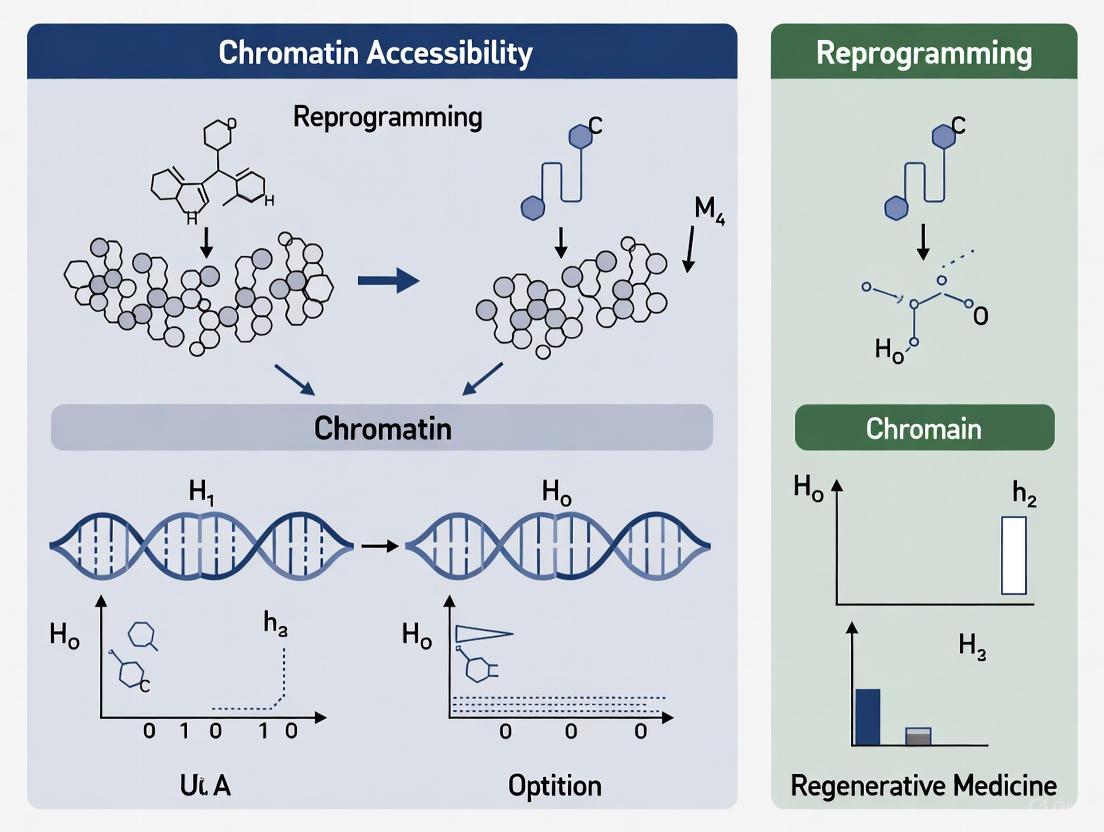
Comparative Chromatin Accessibility in Cellular Reprogramming: From Mechanisms to Clinical Applications
This comprehensive review explores how comparative analysis of chromatin accessibility provides critical insights into cellular reprogramming mechanisms, efficiency, and outcomes.
Comparative Chromatin Accessibility in Cellular Reprogramming: From Mechanisms to Clinical Applications
Abstract
This comprehensive review explores how comparative analysis of chromatin accessibility provides critical insights into cellular reprogramming mechanisms, efficiency, and outcomes. We examine the fundamental role of chromatin dynamics in establishing new cellular identities across diverse systems, from induced pluripotency to directed differentiation. The article evaluates cutting-edge methodological approaches for mapping and comparing accessibility landscapes, addresses key technical challenges and optimization strategies, and validates computational predictions against functional reprogramming outcomes. For researchers and drug development professionals, this synthesis offers a framework for leveraging chromatin accessibility data to enhance reprogramming protocols, develop disease models, and advance regenerative therapies.
Chromatin Accessibility Dynamics: The Foundation of Cellular Reprogramming
Chromatin accessibility refers to the physical permissibility of genomic DNA to nuclear macromolecules, a fundamental property governing essential cellular processes such as transcription, replication, DNA repair, and cell fate determination [1]. This accessibility is primarily determined by nucleosome distribution and occupancy, along with other DNA-binding factors that collectively shape the genome's structural landscape [1]. The eukaryotic genome exhibits a spectrum of accessibility states, ranging from hyper-accessible "open" chromatin to inaccessible "closed" chromatin, with nucleosomes serving as the primary structural units that regulate this dynamic [2].
The nucleosome, comprising approximately 147 base pairs of DNA wrapped around an octamer of histone proteins, forms the fundamental repeating unit of chromatin [3] [1]. Its strategic positioning and structural state act as a critical determinant of DNA accessibility. Recent research has revealed that nucleosomes exist in dynamic states of wrapping and unwrapping, with DNA spending approximately 2-10% of its time in an unwrapped "breathing" state [3]. Advanced mapping techniques have further demonstrated that genomic chromatin forms distinct Nucleosome Wrapping Domains (NRDs)—classified as tightly wrapped (TiNRDs) and loosely wrapped (LoNRDs)—which precisely correspond with higher-order chromatin organization, including Hi-C A and B compartments [3].
This guide provides a comprehensive comparison of the experimental frameworks, molecular mechanisms, and biological implications of chromatin accessibility, with particular emphasis on its role in cellular reprogramming and regenerative processes.
Methodological Comparison: Profiling Accessibility Landscapes
Diverse experimental approaches have been developed to map chromatin accessibility at genome-wide scale, each with distinct principles, advantages, and limitations. The core principle underlying most methods leverages the differential susceptibility of occupied versus free DNA to enzymatic cleavage, transposition, methylation, or solubility-based separation [2] [1].
Table 1: Comparison of Major Chromatin Accessibility Profiling Methods
| Method | Principle | Resolution | Key Advantages | Key Limitations |
|---|---|---|---|---|
| DNase-seq [2] [1] | DNase I enzyme cleaves hyper-accessible regions | ~150 bp | Well-established for mapping hypersensitive sites; rich historical data | Bias toward hyper-accessible regions; underrepresents moderately accessible regions |
| MNase-seq [2] [1] | Micrococcal nuclease digests linker DNA and accessible regions | Single nucleosome | Excellent for nucleosome positioning; can map both accessible and protected regions | Strong sequence cleavage bias; requires titration to distinguish accessibility from occupancy |
| ATAC-seq [2] [1] | Tn5 transposase inserts adapters into accessible DNA | ~100 bp | High signal-to-noise ratio; fast protocol; low cell input requirements (down to single cell) | Sensitive to mitochondrial DNA; complex data analysis |
| FAIRE-seq [2] [1] | Formaldehyde fixation followed by sonication and phenol-chloroform extraction | ~100-500 bp | No enzyme bias; simple conceptual approach | Lower resolution compared to nuclease-based methods |
| NOMe-seq [2] [1] | Methyltransferase accessibility profiling followed by bisulfite sequencing | Single molecule | Provides both accessibility and native DNA methylation information | Technically challenging; requires specialized expertise |
The emergence of single-cell and multimodal technologies represents a significant advancement, enabling researchers to simultaneously profile chromatin accessibility and gene expression within the same individual cells [4] [1]. For example, single-nuclei multiome ATAC + RNA sequencing was recently employed to investigate wound-induced reprogramming in moss, revealing that reprogramming leaf cells exhibit a partly relaxed chromatin landscape while specific transcription factors enhance accessibility at loci essential for stem cell formation [4].
Molecular Mechanisms Governing Chromatin Accessibility
Nucleosome Remodeling Complexes
ATP-dependent chromatin remodeling complexes constitute primary regulators of chromatin accessibility by controlling nucleosome positioning, composition, and stability. These multi-subunit complexes utilize ATP hydrolysis to mobilize nucleosomes, facilitating the transition between "closed" and "open" chromatin states [1]. They are categorized into four major families based on their distinct structural and functional characteristics.
Table 2: Major Chromatin Remodeling Complex Families and Their Functions
| Complex Family | Key Subunits | Primary Functions | Biological Roles in Reprogramming |
|---|---|---|---|
| SWI/SNF [1] | SMARCA2/4, ARID1A | Nucleosome sliding, eviction, histone variant exchange | Promotes accessibility at pluripotency loci; facilitates pioneer transcription factor activity |
| NuRD [1] | CHD3/4/5, HDAC1/2 | Nucleosome sliding, histone deacetylation | Suppresses somatic gene expression during reprogramming; interacts with Sall4 to reduce accessibility of anti-reprogramming genes |
| ISWI [1] | SMARCA5, BAZ1A/B | Nucleosome spacing, chromatin compaction | Maintains nucleosome periodicity; contributes to heterochromatin integrity |
| INO80 [3] [1] | INO80, YY1, actin-related proteins | Nucleosome sliding, histone variant exchange (H2A.Z) | Promotes DNA repair; facilitates transcriptional activation |
Structural studies have provided unprecedented insights into remodeling mechanisms. Recent cryo-EM structures of human CHD1 bound to nucleosomes revealed an "anchor element" that connects the ATPase motor to the nucleosome's acidic patch, alongside a "gating element" that undergoes conformational switching critical for remodeling activity [5]. These structural elements are conserved across remodeler families, suggesting a unified mechanism for nucleosome recognition and remodeling [5].
Pioneer Transcription Factors and Epigenetic Regulation
Pioneer transcription factors (PTFs) represent a specialized class of DNA-binding proteins capable of initiating chromatin opening by binding to nucleosomal DNA in closed chromatin regions [6]. Unlike conventional transcription factors that require pre-accessible DNA, PTFs can directly recognize their target sequences in compacted chromatin, subsequently recruiting additional chromatin remodelers and co-factors to establish stable accessible regions [6].
During cellular reprogramming, PTFs play instrumental roles in reshaping chromatin architecture. In wound-induced reprogramming in moss, STEMIN transcription factors selectively enhance accessibility at specific genomic loci essential for stem cell formation within a broadly relaxed chromatin environment established by wounding [4]. Similarly, in mammalian systems, the AP2/ERF transcription factor STEMIN homologs function as intrinsic mediators of reprogramming in response to injury [4].
Epigenetic modifications, including histone post-translational modifications and DNA methylation, further refine the chromatin accessibility landscape. Histone acetylation (e.g., H3K27ac) generally correlates with enhanced accessibility, while specific methylation patterns can either activate (H3K4me3) or repress (H3K27me3) chromatin states [6]. DNA methylation at promoter CpG islands typically associates with transcriptional silencing and reduced accessibility, with DNMT enzymes catalyzing methylation and TET enzymes facilitating demethylation [6].
Chromatin Accessibility in Reprogramming and Regeneration
Case Studies in Cellular Reprogramming
Chromatin accessibility dynamics play a pivotal role in cellular reprogramming across diverse biological contexts, from wound response to directed cell fate transitions. Several illuminating case studies highlight these principles:
Wound-Induced Reprogramming in Moss: Single-nuclei multiome analysis in Physcomitrium patens revealed that leaf cells undergoing reprogramming following wounding exhibit widespread chromatin relaxation, establishing a permissive environment for stem cell formation [4]. Within this broadly accessible landscape, STEMIN transcription factors selectively enhance accessibility at specific genomic loci essential for the leaf-to-stem-cell transition, demonstrating a hierarchical interplay between global chromatin changes and factor-directed local remodeling [4].
Hepatic Regeneration: Integrated RNA-seq and ATAC-seq analyses of liver regeneration identified ATF3 as an "Initiationon" transcription factor and ONECUT2 as an "Initiationoff" factor that reciprocally modulate target promoter occupancy to license hepatocytes for regeneration [7]. ATF3 binds to the Slc7a5 promoter to activate mTOR signaling, while the Hmgcs1 promoter loses ONECUT2 binding to facilitate regenerative initiation [7].
Leukemia Reprogramming: The GATA3 noncoding variant rs3824662 drives extensive chromatin reorganization in Ph-like acute lymphoblastic leukemia, resulting in increased accessibility of GATA3 binding regions and dysregulation of oncogenes like CRLF2 [8]. Enhancer RNAs (eRNAs), including eRNAG3 and eRNAC4, show coordinated upregulation and positive correlation with CRLF2 expression, suggesting their cooperative contribution to the regulatory mechanisms governing leukemogenic reprogramming [8].
Comparative Analysis of Reprogramming Systems
Table 3: Chromatin Accessibility Dynamics Across Reprogramming Models
| Reprogramming Context | Initial Chromatin State | Key Regulatory Factors | Accessibility Changes | Functional Outcomes |
|---|---|---|---|---|
| Wound-induced moss reprogramming [4] | Differentiated leaf cell | STEMIN transcription factors | Genome-wide relaxation with selective enhancement at stem cell loci | Direct conversion to chloronema apical stem cells |
| Hepatic regeneration [7] | Quiescent hepatocyte | ATF3 (on), ONECUT2 (off) | Transient, phase-restricted remodeling at promoters of regeneration genes | Hepatocyte proliferation and functional tissue repair |
| Oncogenic viral transformation [6] | Somatic cell | Viral oncoproteins, host pioneer factors | Viral integration into accessible regions; hijacking of host regulatory elements | Cellular transformation; persistent infection |
| Induced pluripotency [9] [1] | Differentiated somatic cell | Yamanaka factors (Oct4, Sox2, Klf4, c-Myc) | Sequential opening of pluripotency loci; closing of somatic genes | Pluripotent stem cells |
Table 4: Key Research Reagent Solutions for Chromatin Accessibility Studies
| Reagent/Resource | Function | Example Applications |
|---|---|---|
| Tn5 Transposase [2] [1] | Simultaneous fragmentation and tagging of accessible genomic DNA | ATAC-seq library preparation; compatibility with low-input and single-cell protocols |
| Micrococcal Nuclease (MNase) [3] [2] | Enzymatic digestion of linker DNA and accessible regions | MNase-seq for nucleosome positioning; mapping of nucleosome wrapping states |
| DNase I [2] [1] | Cleavage of hypersensitive genomic regions | DNase-seq for mapping canonical DHSs in regulatory elements |
| M.CviPI Methyltransferase [2] | In vitro methylation of accessible GpC sites | NOMe-seq for combined accessibility and native methylation profiling |
| 10x Genomics Single Cell Multiome ATAC + RNA [4] | Simultaneous profiling of chromatin accessibility and gene expression | Identification of cell type-specific regulatory dynamics during reprogramming |
| Spike-in Controls [2] | Normalization for technical variation in nuclease digestion | Quantitative MNase-seq (q-MNase) for accurate nucleosome occupancy measurements |
Conceptual Framework and Signaling Pathways
The following diagram illustrates the integrated molecular framework governing chromatin accessibility dynamics during cellular reprogramming:
Figure 1. Integrated Molecular Framework of Chromatin Accessibility in Reprogramming. This diagram illustrates the hierarchical regulatory network wherein external stimuli activate pioneer transcription factors that subsequently recruit chromatin remodeling complexes and epigenetic modifiers. These effectors collectively establish a permissive chromatin environment through relaxation and accessibility changes, enabling enhancer RNA production and gene expression alterations that ultimately drive cell fate reprogramming.
The following diagram details the experimental workflow for multimodal chromatin accessibility analysis:
Figure 2. Multimodal Experimental Workflow for Chromatin Accessibility Studies. This workflow diagram outlines the integrated experimental and computational pipeline for simultaneous profiling of chromatin accessibility and gene expression, enabling comprehensive characterization of regulatory dynamics during reprogramming processes.
The comparative analysis of chromatin accessibility across reprogramming models reveals both conserved principles and context-specific adaptations. A fundamental emerging paradigm is the hierarchical regulation wherein broad chromatin relaxation creates a permissive landscape that is subsequently refined by sequence-specific factors to establish new transcriptional programs [4]. This two-phase mechanism appears conserved from plant to mammalian systems, suggesting an evolutionarily ancient strategy for cellular plasticity.
Future research directions will likely focus on several key areas: First, the development of enhanced spatial chromatin accessibility methods will enable the mapping of regulatory landscapes within native tissue architecture, providing critical insights into microenvironmental influences on cell fate decisions [1]. Second, the integration of time-resolved multiomics with computational modeling promises to reveal the causal relationships between chromatin dynamics and functional outcomes [7] [10]. Finally, the therapeutic targeting of chromatin regulators—including ATP-dependent remodelers and pioneer factors—holds significant promise for regenerative medicine and cancer therapy, particularly for overcoming the epigenetic barriers that limit efficient reprogramming [9] [1].
The continuing refinement of chromatin accessibility mapping technologies, combined with innovative experimental models of reprogramming, will undoubtedly yield deeper insights into the fundamental principles of genome regulation and their translational applications in human health and disease.
Chromatin accessibility serves as a master regulator of cellular identity, governing gene expression by modulating DNA availability to transcriptional machinery. Within the nucleus, chromatin exists in a dynamic spectrum of states—open, permissive, and closed—each characterized by distinct structural features, histone modifications, and functional consequences. This guide systematically compares these chromatin states within the context of cellular reprogramming, examining how transcription factor binding and chromatin remodeling orchestrate cell fate transitions. We present quantitative comparisons of epigenetic features, detailed experimental methodologies for mapping accessibility, and analytical frameworks for interpreting chromatin dynamics during reprogramming events. Understanding these states provides critical insights for regenerative medicine and therapeutic development.
Chromatin, the complex of DNA and histone proteins, packages the eukaryotic genome within the nucleus while regulating access to genetic information. The term chromatin accessibility refers to the physical access that proteins have to DNA, which is profoundly influenced by local nucleosome positioning and higher-order chromatin structure [2]. Rather than existing in a binary open/closed state, chromatin occupies a continuum of accessibility that ranges from hyper-accessible ("open") to moderately accessible ("permissive") to inaccessible ("closed") states [2].
These chromatin states establish a fundamental regulatory layer for all DNA-templated processes, including transcription, replication, and repair. During cellular reprogramming—the process of converting differentiated cells into induced pluripotent stem cells (iPSCs)—the orchestrated remodeling of chromatin states enables the dramatic rewiring of gene regulatory networks necessary for identity change [11]. Transcription factors such as Oct4, Sox2, Klf4, and c-Myc (OSKM) must navigate and reshape this epigenetic landscape to activate pluripotency genes while silencing somatic programs.
Defining the Chromatin Spectrum
Molecular Definitions and Features
The chromatin accessibility spectrum comprises three principal states with distinct characteristics:
Open Chromatin: Characterized by nucleosome-depleted regions with maximal DNA accessibility, these regions are typically associated with active promoters, enhancers, and other regulatory elements. They exhibit DNase I hypersensitivity and are enriched for active histone marks such as H3K4me3 at promoters and H3K27ac at enhancers [2] [12]. During reprogramming, open chromatin sites in somatic cells represent the first class of targets bound by reprogramming factors, including genes involved in mesenchymal-to-epithelial transition (MET) [11].
Permissive Chromatin: This intermediate state features nucleosome-bound but dynamic regions that may carry both activating and repressive histone modifications. Permissive chromatin often includes bivalent domains marked by both H3K4me3 (activating) and H3K27me3 (repressive) modifications, which keep developmental genes in a transcriptionally poised state, ready for activation or silencing upon lineage commitment [11]. Enhancers in a permissive state (H3K4me1-positive but not fully open) can bind transcription factors but may require additional remodeling for full activation [11].
Closed Chromatin: Also termed heterochromatin, these regions are compacted and transcriptionally silent, presenting a significant barrier to factor binding. Closed chromatin is enriched for repressive marks such as H3K9me3 (constitutive heterochromatin) and H3K27me3 (facultative heterochromatin) [13]. During reprogramming, core pluripotency genes like Nanog often reside within this refractory chromatin in somatic cells, requiring extensive remodeling for activation [11].
The following diagram illustrates the continuum of chromatin states and their key characteristics:
Chromatin State Transitions. Chromatin exists along a dynamic continuum, with states interconverting through remodeling, activation, and repression processes.
Quantitative Comparison of Chromatin States
The table below summarizes the defining characteristics and functional associations of the three primary chromatin states:
Table 1: Comparative Features of Chromatin States
| Feature | Open Chromatin | Permissive Chromatin | Closed Chromatin |
|---|---|---|---|
| DNA Accessibility | High (Nucleosome-depleted) | Moderate (Nucleosome-bound) | Low (Nucleosome-occupied) |
| DNase I Sensitivity | Hypersensitive | Intermediate | Resistant |
| Representative Histone Modifications | H3K4me3, H3K27ac | H3K4me1, H3K27me3/H3K4me3 (bivalent) | H3K9me3, H3K27me3 |
| Transcriptional Activity | Active | Poised/Silent | Silent |
| Nuclear Compartment | Euchromatin (A) | Facultative Heterochromatin | Constitutive Heterochromatin (B) |
| Reprogramming Factor Binding | Immediate OKSM binding | Delayed binding requiring remodeling | Refractory to initial binding |
| Functional Associations | Active promoters, enhancers | Poised enhancers, bivalent promoters | Repetitive regions, silenced genes |
Chromatin State Dynamics in Cellular Reprogramming
Cellular reprogramming provides a powerful model for understanding how transcription factors orchestrate chromatin state transitions to enable cell fate changes. The OSKM factors target distinct chromatin environments with different kinetics and functional outcomes during iPSC generation.
Transcription Factor Engagement with Chromatin States
Reprogramming factors demonstrate hierarchical engagement with chromatin states based on accessibility:
Open Chromatin Targets: In both human and mouse fibroblasts, OSK factors initially target many closed chromatin sites, but their immediate binding occurs predominantly at already accessible regions containing active chromatin marks [14] [11]. These early targets include somatic genes that require downregulation and early MET-related genes [11].
Permissive Chromatin Engagement: A second class of targets includes distal regulatory elements with permissive features such as H3K4me1 marking [11]. These "permissive enhancers" can bind transcription factors prior to their associated promoters and before full transcriptional activation. Some factors, particularly Oct4 and Sox2, function as pioneer factors capable of binding partially accessible regions and initiating chromatin remodeling [11].
Closed Chromatin Remodeling: The most challenging targets are broad heterochromatic regions enriched for H3K9me3 that contain core pluripotency genes such as Nanog and Sox2 [11]. These regions are refractory to initial OKSM binding and require extensive, coordinated remodeling involving histone-modifying enzymes and chromatin remodelers for activation.
Comparative Analysis of Reprogramming Factors
Studies comparing OSKM binding in human and mouse reprogramming reveal both conserved and species-specific aspects of chromatin engagement:
Table 2: OSKM Binding in Early Human vs. Mouse Reprogramming
| Feature | Human System | Mouse System | Conservation |
|---|---|---|---|
| Time to Reprogramming | ~3-4 weeks | ~1-2 weeks | Not conserved |
| Number of OSKM Peaks | ~2x more for Sox2, Klf4, c-Myc | Fewer peaks for these factors | Partially conserved |
| c-Myc Binding Distribution | Preferentially distal to TSS | Preferentially proximal to TSS | Not conserved |
| Primary Binding Motifs | Similar with minor variations | Similar with minor variations | Highly conserved |
| Combinatorial Binding Patterns | Shared patterns | Shared patterns | Highly conserved |
| Syntenic Binding Conservation | Limited conservation in syntenic regions | Limited conservation in syntenic regions | Poorly conserved |
Despite these differences, both systems share significant overlap in target genes and gene ontology enrichments, particularly for processes like regulation of transcription, in utero embryonic development, and Wnt signaling pathway regulation [14].
Experimental Methods for Chromatin Accessibility Profiling
Multiple biochemical methods have been developed to profile chromatin accessibility genome-wide, each with distinct advantages and limitations. The selection of an appropriate method depends on research goals, sample availability, and desired resolution.
Core Methodologies and Protocols
ATAC-Seq (Assay for Transposase-Accessible Chromatin with Sequencing)
ATAC-Seq has become the most widely used method for chromatin accessibility profiling due to its simplicity, sensitivity, and low cell input requirements [12] [15].
Experimental Principle: The method utilizes a hyperactive Tn5 transposase that simultaneously fragments DNA and inserts sequencing adapters into accessible genomic regions in a process called "tagmentation." The preferential insertion of Tn5 into nucleosome-free regions enables mapping of open chromatin [15].
Key Protocol Steps:
- Cell Lysis: Isolate nuclei from fresh cells or tissues using detergent-based buffers.
- Tagmentation: Incubate nuclei with Tn5 transposase loaded with sequencing adapters.
- DNA Purification: Recover and purify fragmented DNA.
- Library Amplification: PCR amplification with index primers to create sequencing libraries.
- Sequencing: High-throughput sequencing (typically Illumina platforms).
Advantages: Rapid protocol (~3 hours), low cell input (500-50,000 cells), no crosslinking required, and compatibility with single-cell applications [15].
Recommended Sequencing Depth: ≥50 million paired-end reads for identifying open chromatin differences; >200 million paired-end reads for transcription factor footprinting [15].
DNase-Seq (DNase I Hypersensitive Sites Sequencing)
DNase-Seq was one of the first methods developed for genome-wide chromatin accessibility mapping and remains a gold standard for identifying hypersensitive sites [2] [12].
Experimental Principle: The method exploits the preference of DNase I endonuclease to cleave nucleosome-depleted, accessible DNA over compacted chromatin. Sequencing the resulting fragments reveals regions of hypersensitivity [2].
Key Protocol Steps:
- Nuclei Isolation: Purify intact nuclei from cells.
- DNase I Digestion: Titrate DNase I enzyme to achieve limited digestion.
- DNA Extraction: Purify and size-select fragmented DNA (typically 100-500 bp).
- Library Preparation: Ligate sequencing adapters to digested fragments.
- Sequencing: High-throughput sequencing.
Advantages: Well-established protocol, excellent for mapping hypersensitive sites, comprehensive annotation of regulatory elements.
Limitations: Requires millions of cells, optimization of digestion conditions is critical, and more complex protocol than ATAC-Seq.
Methyltransferase-Based Methods
These methods use bacterial DNA methyltransferases to label accessible DNA, providing single-molecule resolution of chromatin accessibility [2] [16].
Experimental Principle: Isolated nuclei are treated with methyltransferases (e.g., EcoGII) that preferentially modify accessible adenines (A→6mA) in the presence of the methyl donor SAM. Subsequent long-read sequencing detects 6mA incorporation as a proxy for accessibility [16].
Key Protocol Steps:
- Nuclei Isolation: Preserve nuclear integrity using detergent-based buffers.
- Methyltransferase Tagging: Incubate nuclei with EcoGII enzyme and SAM.
- gDNA Extraction: Recover high molecular weight genomic DNA.
- Library Preparation: Prepare libraries for long-read sequencing (e.g., Oxford Nanopore).
- Sequencing and Analysis: Detect 6mA incorporation to identify accessible regions.
Advantages: Single-molecule resolution, captures long-range chromatin information, compatible with variant phasing.
Limitations: Specialized equipment required, lower throughput, higher DNA input requirements.
The following diagram illustrates the core workflows for these key methodologies:
Chromatin Accessibility Method Workflows. Core experimental workflows for the three principal methods for profiling chromatin accessibility genome-wide.
Method Comparison and Selection Guidelines
Table 3: Comparative Performance of Chromatin Accessibility Methods
| Method | Sensitivity | Resolution | Cell Input | Primary Applications | Key Advantages |
|---|---|---|---|---|---|
| ATAC-Seq | High | ~100 bp | 500 - 50,000 cells | Nucleosome mapping, TF footprinting, enhancer identification | Fast, sensitive, low input, single-cell compatible |
| DNase-Seq | High | ~100 bp | 1 - 50 million cells | DNase hypersensitive site mapping, regulatory element annotation | Gold standard for hypersensitive sites, comprehensive |
| MNase-Seq | Moderate | Nucleosome-level | 1 - 10 million cells | Nucleosome positioning, occupancy mapping | Direct nucleosome mapping, both accessible and inaccessible regions |
| FAIRE-Seq | Moderate | ~100 bp | 1 - 10 million cells | Hyper-accessible region enrichment | No enzyme bias, simple protocol |
| Methyltransferase-Based | Variable | Single-molecule | 2 million cells | Single-molecule accessibility, long-range phasing | Single-molecule resolution, long-range information |
Successful chromatin accessibility studies require specialized reagents and computational tools. The following table outlines essential solutions for experimental and analytical workflows:
Table 4: Research Reagent Solutions for Chromatin Accessibility Studies
| Reagent/Resource | Function | Example Applications | Key Features |
|---|---|---|---|
| Tn5 Transposase | Simultaneous fragmentation and adapter insertion for ATAC-Seq | Bulk and single-cell ATAC-Seq | High efficiency, minimal sequence bias |
| DNase I | Enzymatic cleavage of accessible DNA | DNase-Seq, DNase I hypersensitivity mapping | Specific for nucleosome-free regions |
| EcoGII Methyltransferase | Adenine methylation (6mA) of accessible DNA | Long-read chromatin accessibility profiling | Non-native modification in mammals, single-molecule resolution |
| H3K27ac Antibody | Immunoprecipitation of active enhancers and promoters | ChIP-Seq for active regulatory elements | Marks active enhancers and promoters |
| H3K4me3 Antibody | Immunoprecipitation of active promoters | ChIP-Seq for active transcription start sites | Marks active promoters |
| H3K27me3 Antibody | Immunoprecipitation of Polycomb-repressed regions | ChIP-Seq for facultative heterochromatin | Marks Polycomb-repressed regions |
| Chromatin State Annotation Tools | Computational segmentation of chromatin states | Integrative analysis of multiple epigenetic marks | Defines regulatory elements from combined datasets |
| Hi-C Analysis Software | Mapping 3D chromatin interactions | 3D genome organization studies | Identifies chromatin loops, compartments, TADs |
The dynamic spectrum of chromatin states—open, permissive, and closed—forms an essential regulatory framework that governs cell identity and plasticity. Cellular reprogramming studies have been particularly illuminating, revealing how transcription factors hierarchically engage with these states to rewrite cellular programs. While significant progress has been made in mapping these states and understanding their transitions, several frontiers remain: achieving single-molecule resolution of chromatin dynamics, understanding the role of 3D genome organization in state maintenance, and developing therapeutic approaches to modulate chromatin states in disease contexts. The continued refinement of chromatin accessibility methods and analytical frameworks will undoubtedly yield deeper insights into the fundamental principles of epigenetic regulation across diverse biological systems.
Pioneer Transcription Factors as Architects of Chromatin Remodeling During Reprogramming
Pioneer Transcription Factors (PTFs) represent a unique class of proteins that serve as master regulators of cell fate by initiating chromatin remodeling events during cellular reprogramming. Unlike conventional transcription factors that require pre-existing chromatin accessibility, PTFs possess the remarkable ability to bind directly to closed chromatin regions, initiating a cascade of events that ultimately redefine cellular identity [17]. This capacity to engage nucleosome-wrapped DNA enables PTFs to function as initial "architects" of chromatin restructuring, making them indispensable tools in regenerative medicine and cellular reprogramming research [17] [18].
The fundamental property that distinguishes PTFs is their capacity to specifically recognize their DNA binding motifs on nucleosomal DNA, which is generally inaccessible to most transcription factors [19] [20]. Through this activity, PTFs can initiate local chromatin opening and facilitate subsequent binding of other transcription factors and co-factors in a cell-type-specific manner [20]. This review will comprehensively compare the mechanisms, experimental methodologies, and functional outcomes of major PTFs, with a specific focus on their roles in modulating chromatin accessibility during cellular reprogramming processes, including the generation of induced pluripotent stem cells (iPSCs).
Molecular Mechanisms of Pioneer Factor Action
Nucleosome Binding and Chromatin Opening Strategies
Pioneer Transcription Factors employ distinct structural strategies to engage with nucleosomal DNA and initiate chromatin remodeling. The molecular interactions between PTFs and nucleosomes have been elucidated through recent structural studies, revealing several key mechanisms:
Partial DNA Motif Recognition: PTFs target partial DNA motifs on nucleosomes to initiate reprogramming, often binding to suboptimal sites that would be ignored by conventional transcription factors [18]. This flexible binding mode allows initial engagement with chromatin before more stable complexes are formed.
Nucleosome Structure Modulation: Binding of PTFs like OCT4 induces significant changes to nucleosome structure, repositions nucleosomal DNA, and facilitates cooperative binding of additional factors [21]. Cryo-EM structures reveal that OCT4 binding stabilizes otherwise flexible nucleosome positioning, trapping the DNA in a specific conformation [21].
Histone Tail Interactions: The flexible activation domain of OCT4 contacts the N-terminal tail of histone H4, altering its conformation and promoting chromatin decompaction [21]. Additionally, the DNA-binding domain of OCT4 engages with the N-terminal tail of histone H3, and post-translational modifications at H3K27 modulate DNA positioning and affect transcription factor cooperativity [21].
Table 1: Chromatin Remodeling Capabilities of Key Pioneer Transcription Factors
| Pioneer Factor | Nucleosome Binding Mechanism | Chromatin Opening Effect | Cooperative Partners |
|---|---|---|---|
| OCT4 (POU5F1) | Binds linker DNA near nucleosome entry-exit site; both POUS and POUHD domains engage nucleosome | Repositions nucleosomal DNA; stabilizes DNA positioning; promotes H4 tail conformational changes | SOX2, KLF4, MYC [21] |
| SOX2 | Preferentially binds nucleosomes in presence of OCT4; recognizes internal sites | Facilitates nucleosome unwrapping; increases accessibility of adjacent sites | OCT4 (critical partnership) [21] |
| FoxA | Linker histone-like DNA binding domain; displaces linker histone H1 | Directly opens compacted chromatin; reduces dependency on nucleosome remodelers | Other hepatic transcription factors [19] |
| Klf4 | Binds partial motifs on nucleosomal DNA | Initiates local accessibility; facilitates binding of other reprogramming factors | OCT4, SOX2 [17] |
| Zelda (Zld) | Early embryonic engagement with closed chromatin | Increases DNA accessibility prior to zygotic genome activation | Bicoid, Dorsal [17] |
Epigenetic Interplay and Chromatin State Modulation
Pioneer Transcription Factors do not function in isolation but engage in dynamic interplay with the epigenetic landscape to reshape chromatin architecture:
Histone Modification Cross-Talk: PTF activity is regulated by existing histone modifications, while simultaneously inducing new epigenetic states. For example, OCT4 cooperativity with SOX2 is modulated by H3K27 modifications, with H3K27ac enhancing and H3K27me3 reducing their collaborative binding [21].
Recruitment of Chromatin Modifiers: PTFs recruit chromatin remodelers, histone modifiers, and DNA methylation machinery to establish active or poised transcriptional states [6] [18]. This includes interactions with complexes such as SWI/SNF, ISWI, INO80, Polycomb repressive complexes (PRCs), and nucleosome remodeling and deacetylase (NuRD) complexes [6] [18].
DNA Methylation Dynamics: PTFs interact with DNA methylation machinery, with OCT4 activity being both influenced by and influencing DNA methylation patterns during reprogramming [18]. The balance between DNA methyltransferases (DNMTs) and ten-eleven translocation (TET) enzymes is crucial for establishing new cell identities.
The following diagram illustrates the sequential mechanism of pioneer transcription factor action in chromatin remodeling:
Diagram 1: Sequential mechanism of pioneer transcription factor-mediated chromatin remodeling. The process begins with pioneer factor binding to closed chromatin through partial motif recognition, followed by nucleosome rearrangement, recruitment of cofactors, and ultimately establishing accessible chromatin for transcription activation.
Comparative Analysis of Pioneer Factor Activity in Reprogramming Contexts
Chromatin Accessibility Dynamics in Naïve versus Primed Pluripotency
Direct comparative studies of chromatin dynamics during reprogramming to different pluripotent states reveal distinct patterns of PTF activity. Research integrating ATAC-seq and RNA-seq data from naïve and primed reprogramming pathways demonstrates that chromatin accessibility changes precede transcriptional changes, with accessibility diverging around day 8 of reprogramming, while transcriptome differences become pronounced around day 14 [22].
Table 2: Chromatin Accessibility Dynamics During Naïve versus Primed Reprogramming
| Reprogramming Aspect | Naïve Pluripotency Path | Primed Pluripotency Path |
|---|---|---|
| Timeline of Chromatin Opening | Significant accessibility changes at day 6-8; major transcriptome shift at day 14 | Accessibility changes at day 6-8; transcriptome shift around day 8 |
| Closed-to-Open (CO) Regions | Progressive increase throughout reprogramming; peaks at iPSC stage | Progressive increase throughout reprogramming; peaks at iPSC stage |
| Open-to-Closed (OC) Regions | Outnumber CO regions until day 20; associated gene expression decreases from day 8 | Outnumber CO regions until day 20; associated gene expression slightly up-regulated |
| Permanently Open (PO) Regions | Minimal expression changes in associated genes | Significant up-regulation of associated genes |
| Functional Enrichment in CO Regions | Pluripotency and early embryonic development processes | Pluripotency and developmental processes |
| Key Regulatory Factors | PRDM1 isoforms (PRDM1α and PRDM1β) with distinct roles | Different factor requirements than naïve state |
During both naïve and primed reprogramming, regions transitioning from closed to open (CO) are associated with genes involved in pluripotency and early embryonic development, while regions transitioning from open to closed (OC) are linked to somatic cell lineages and differentiated state functions [22]. The divergent roles of PRDM1 isoforms (PRDM1α and PRDM1β) in naïve reprogramming highlight the complexity of PTF function, with different isoforms potentially targeting distinct genomic sites and exerting different effects on target genes [22].
Yamanaka Factor Cooperation and Hierarchical Actions
The classic reprogramming factors OCT4, SOX2, KLF4, and c-Myc (OSKM) display hierarchical and cooperative relationships in initiating chromatin reprogramming:
OCT4 as a Primary Pioneer: OCT4 expression is necessary and sufficient to initiate reprogramming in some contexts, and it enhances the nucleosome binding of SOX2, KLF4, and MYC [21]. OCT4 binding induces nucleosome structural changes that facilitate cooperative binding of additional factors.
SOX2 Cooperativity: SOX2 binding is significantly enhanced by prior OCT4 engagement, with the OCT4-SOX2 partnership being critical for pluripotency establishment [21]. Structural studies show that OCT4 binding creates favorable conditions for SOX2 recruitment to adjacent sites.
Differential Chromatin Engagement: During initial reprogramming stages, OCT4, SOX2, and KLF4 act as pioneer factors that access closed chromatin, while c-Myc preferentially binds to pre-existing open chromatin sites that are already DNase-hypersensitive and contain activating histone modifications [17].
Promiscuous Initial Binding: The initial binding events of OSKM factors in somatic cell reprogramming are quite promiscuous, distinct from definitive binding patterns in established pluripotent cells, with subsequent reorganization required to establish stable pluripotency networks [17].
Experimental Approaches for Assessing Pioneer Factor Activity
Methodologies for Mapping Chromatin Accessibility and Factor Binding
Several well-established experimental protocols enable the comprehensive assessment of PTF activity and chromatin dynamics:
Integrated Multi-Omics Workflow for Pioneer Factor Characterization
Diagram 2: Experimental workflow for identifying and characterizing pioneer transcription factors, combining chromatin accessibility mapping, nucleosome positioning analysis, transcription factor binding profiling, and computational integration.
ATAC-seq (Assay for Transposase-Accessible Chromatin with sequencing):
- Purpose: Maps genome-wide chromatin accessibility by measuring regions of open chromatin.
- Protocol: Cells are lysed, and the transposase Tn5 is added to simultaneously fragment and tag accessible DNA regions with sequencing adapters. The tagged DNA is then purified and prepared for sequencing [19] [22].
- Data Interpretation: Open chromatin regions appear as peaks in sequencing data; comparison across reprogramming timepoints identifies regions changing from closed to open (CO) or open to closed (OC) states [22].
ChIP-seq (Chromatin Immunoprecipitation followed by sequencing):
- Purpose: Identifies genomic binding sites for specific transcription factors or histone modifications.
- Protocol: Cells are cross-linked to preserve protein-DNA interactions, chromatin is sheared, and an antibody specific to the protein of interest is used to immunoprecipitate the protein-DNA complexes. After cross-link reversal and DNA purification, sequencing identifies bound regions [19].
- Data Interpretation: Binding peaks indicate direct or indirect association with genomic regions; when combined with nucleosome positioning data, can distinguish nucleosome-bound versus nucleosome-free binding [20].
MNase-seq (Micrococcal Nuclease sequencing):
- Purpose: Maps nucleosome positions and occupancy across the genome.
- Protocol: MNase enzyme digests linker DNA between nucleosomes, followed by sequencing of the protected nucleosomal DNA fragments [19].
- Data Interpretation: Protected regions indicate nucleosome occupancy; comparison with ATAC-seq and ChIP-seq data identifies transcription factors binding to nucleosomal versus nucleosome-free DNA [19] [20].
Computational Prediction of Pioneer Factors
Recent computational approaches have been developed to systematically identify PTFs based on their binding preferences for nucleosomal DNA:
Motif Enrichment Analysis: Calculates enrichment of transcription factor binding motifs in nucleosomal regions compared to nucleosome-depleted regions. True PTFs show enrichment in nucleosomal regions, while conventional factors show depletion [20].
Integrated Data Analysis: Combines ChIP-seq, MNase-seq, and DNase-seq data to assess cell-type-specific ability of transcription factors to bind nucleosomes [20].
Validation Benchmarks: Uses known PTF sets (e.g., factors involved in embryonic stem cell maintenance or reprogramming) as positive controls to validate prediction accuracy [20].
This approach has successfully discriminated pioneer from canonical transcription factors and predicted new potential cell-type-specific PTFs in H1, K562, HepG2, and HeLa-S3 cell lines [20].
Research Reagent Solutions for Pioneer Factor Studies
Table 3: Essential Research Reagents for Pioneer Transcription Factor Investigation
| Reagent Category | Specific Examples | Research Application | Technical Considerations |
|---|---|---|---|
| Antibodies for Chromatin Profiling | Anti-OCT4, Anti-SOX2, Anti-FoxA1 | ChIP-seq for mapping transcription factor binding sites | Quality critical for signal-to-noise ratio; validate with knockout controls |
| Chromatin Assay Kits | ATAC-seq kits, MNase digestion kits | Mapping chromatin accessibility and nucleosome positioning | ATAC-seq sensitivity requires careful titration of transposase; MNase requires optimization of digestion conditions |
| Reprogramming Systems | Doxycycline-inducible OSKM vectors, Secondary reprogramming systems | Controlled induction of pioneer factors in somatic cells | Secondary systems reduce heterogeneity and improve synchronization |
| Epigenetic Modulators | DNMT inhibitors (azacitidine, decitabine), HDAC inhibitors | Manipulating epigenetic landscape to study pioneer factor interplay | Dose optimization essential to avoid pleiotropic effects |
| Cell Line Models | H1, K562, HepG2, HeLa-S3, Mouse Embryonic Fibroblasts (MEFs) | Cell-type-specific pioneer factor activity assessment | Different cell lines exhibit distinct chromatin environments and pioneer factor responses |
| Structural Biology Tools | Cryo-EM platforms, Crosslinking reagents | Structural characterization of pioneer factor-nucleosome complexes | Technical expertise intensive; requires specialized equipment |
Pioneer Transcription Factors function as architectural specialists in chromatin remodeling, employing distinct but complementary mechanisms to initiate cell fate reprogramming. The comparative analysis of their activities reveals a spectrum of chromatin engagement strategies, from OCT4's nucleosome restructuring capabilities to FoxA's linker histone displacement. The hierarchical cooperation between factors like OCT4 and SOX2 demonstrates the sophisticated division of labor in chromatin opening processes.
The experimental frameworks for studying PTFs have evolved to integrate multi-omics approaches, with ATAC-seq, ChIP-seq, and MNase-seq providing complementary perspectives on chromatin dynamics. These methodologies consistently demonstrate that PTF binding precedes chromatin accessibility changes, with OCT4, SOX2, and Klf4 capable of initial engagement with closed chromatin during reprogramming.
Future research directions will likely focus on understanding how the epigenetic landscape regulates PTF activity, with histone modifications such as H3K27ac and H3K27me3 already shown to modulate OCT4 cooperativity [21]. Additionally, the development of more sophisticated computational prediction methods will enable systematic identification of novel PTFs across diverse cellular contexts. As our understanding of these architectural regulators deepens, so too will our ability to harness their potential for therapeutic reprogramming and regenerative medicine applications.
Comparative Analysis of Naïve versus Primed Pluripotency Chromatin Landscapes
Pluripotent stem cells possess the remarkable capacity to differentiate into any cell type of the adult body. Within this broad potential exist distinct pluripotent states, primarily categorized as naïve and primed, which correspond to pre- and post-implantation embryonic stages, respectively [23] [24]. These states are not merely defined by their transcriptomes but are fundamentally underpinned by distinct epigenetic landscapes. The chromatin architecture—its accessibility, histone modifications, and DNA methylation—varies significantly between these states, creating a unique regulatory environment that governs their developmental potential, signaling dependencies, and stability [23]. This guide provides a comparative analysis of the chromatin landscapes in naïve and primed pluripotency, synthesizing recent high-throughput sequencing data to objectively outline their defining features. Framed within the context of reprogramming and comparative chromatin accessibility research, this resource is designed to inform experimental design and interpretation for researchers and drug development professionals.
Fundamental Chromatin Landscape Differences
The chromatin of naïve and primed pluripotent states differs in its global organization, accessibility, and epigenetic modifications. These differences create a permissive environment for naïve-specific gene networks while progressively restricting developmental potential as cells transition to the primed state.
Global Chromatin Organization: Naïve pluripotent cells, such as mouse embryonic stem cells (mESCs) cultured in 2i/LIF conditions, exhibit a generally more open chromatin configuration with reduced levels of repressive histone marks like H3K27me3 at developmental genes [24]. In contrast, primed cells, such as mouse Epiblast Stem Cells (mEpiSCs) or conventional human pluripotent stem cells (hPSCs), display a chromatin state that is more condensed and lineage-restricted [23]. This is reflected in global DNA methylation levels, which are markedly hypomethylated in naïve cells cultured in 2i/LIF, whereas primed cells are hypermethylated, a distinction particularly evident in in vitro cultures [23].
Enhancer Reconfiguration: A hallmark of the state transition is the dynamic rewiring of enhancer elements. Naïve and primed cells utilize distinct enhancers for the same key pluripotency genes. A quintessential example is the OCT4 (POU5F1) locus, where the distal enhancer (DE) is active in the naïve state, and the proximal enhancer (PE) is favored in the primed state [23]. This switch in enhancer usage reflects a broader reorganization of the transcriptional regulatory network and is mediated by changes in the binding of core transcription factors like OCT4 and SOX2, whose genomic targets are re-directed during the exit from naïve pluripotency [25].
X-Chromosome Inactivation: In female cells, the status of the X chromosomes serves as a key epigenetic marker. Naïve pluripotent cells typically possess two active X chromosomes, while primed cells have undergone X-chromosome inactivation (Xi), a clear indicator of a more developmentally advanced and restricted state [23].
Table 1: Core Characteristics of Naïve and Primed Pluripotent States
| Feature | Naïve Pluripotency | Primed Pluripotency |
|---|---|---|
| Developmental Analogue | Pre-implantation epiblast | Post-implantation epiblast |
| Colony Morphology | Dome-shaped, three-dimensional | Flat, two-dimensional monolayer |
| Signaling Dependence | LIF/STAT3; BMP (mESCs in Serum/LIF); MEK/GSK3 inhibition (2i) | FGF/Activin A/TGF-β |
| X-Chromosome Status | Two active X chromosomes (XaXa) | Inactive X chromosome (Xi) |
| Global DNA Methylation | Hypomethylated | Hypermethylated |
| Prominent Chromatin State | More open, less repressive marks | More condensed, restricted accessibility |
Comparative Chromatin Accessibility Dynamics
Assay for Transposase-Accessible Chromatin using sequencing (ATAC-seq) has been instrumental in mapping the dynamic changes in chromatin architecture during the establishment of and transition between pluripotent states. These analyses reveal that chromatin remodeling is a pivotal early event in cell fate change.
Chromatin Dynamics During Reprogramming to Naïve and Primed States
Reprogramming of somatic cells towards pluripotency involves extensive chromatin remodeling. Studies using secondary human reprogramming systems have shown that while the overall number of chromatin accessibility changes is similar during naïve and primed reprogramming, the specific genomic loci affected are distinct [22]. During the early phases of reprogramming, there is a widespread closure of chromatin regions associated with somatic identity (Open-to-Closed regions), which outnumbers the opening of new regions until later stages [22]. The opening of chromatin at pluripotency-associated loci is a progressive process, with the number of Closed-to-Open (CO) regions increasing over time and peaking in established iPSCs.
Gene Ontology analysis of these dynamic regions reveals a clear functional separation: CO regions are enriched near genes involved in "cell fate commitment," "regulation of stem cell proliferation," and "regulation of embryonic development," while Open-to-Closed (OC) regions are associated with "neuron differentiation," "T cell activation," and "fibroblast migration" [22]. This indicates that the chromatin landscape is systematically cleared of somatic memory and reconfigured to support a pluripotent identity.
Discordance Between Accessibility and Transcription
A critical insight from recent studies is that an open chromatin state does not always equate to active transcription, highlighting the complexity of gene regulation. Research tracking the primed-to-naïve transition in human cells using a dual fluorescent reporter system found that chromatin remodeling precedes transcriptional activation [26]. Specifically, ATAC-seq signals indicative of naïve-specific chromatin—enriched with motifs for OCT, SOX, and KLF transcription factors—were detected in cells that did not yet express the corresponding naïve pluripotency genes [26]. This demonstrates that the opening of chromatin is a necessary but insufficient step for gene activation, which can be further modulated by additional layers of regulation, such as the specific activity of transcription factors and other epigenetic modifications like histone marks.
Distinct Trajectories Revealed by Integrated Analysis
When transcriptomic and chromatin accessibility data are integrated, the divergent trajectories of naïve and primed reprogramming become apparent. Principal Component Analysis (PCA) of such multi-omics data shows that chromatin accessibility differences between the two pathways emerge earlier than transcriptomic differences [22]. A significant shift in chromatin accessibility is observed around day 8 of reprogramming, preceding the major transcriptome divergence that occurs around day 14 [22]. This positions chromatin remodeling as a upstream driver of the transcriptional programs that define naïve and primed pluripotency.
Table 2: Key Chromatin Accessibility and Transcriptional Dynamics
| Dynamic Event | Naïve Reprogramming | Primed Reprogramming | Technical Notes |
|---|---|---|---|
| Onset of Chromatin Divergence | Day 8 [22] | Day 8 [22] | Based on ATAC-seq PCA |
| Major Transcriptome Shift | Day 14 [22] | Day 8 [22] | Based on RNA-seq PCA |
| Relationship at Naïve Loci | Chromatin opening can precede transcriptional activation [26] | Not Applicable | Observed during primed-to-naïve transition |
| Enhancer Usage (e.g., OCT4) | Distal Enhancer (DE) [23] | Proximal Enhancer (PE) [23] | Validated by ChIP-seq |
Key Regulatory Mechanisms and Molecular Players
The distinct chromatin landscapes of naïve and primed states are established and maintained by a network of transcription factors, chromatin remodelers, and signaling pathways.
Transcription Factor Networks
The core pluripotency factors OCT4, SOX2, and NANOG form the foundation of the regulatory network in both states, but their binding profiles and interaction partners differ.
- Naïve State Factors: In the naïve state, OCT4 and SOX2 co-occupy and activate most naïve-specific enhancers [25]. Factors like KLF4, KLF2, TFCP2L1, and ESRRB are highly expressed and help maintain the naïve gene regulatory network. ESRRB, for instance, has been suggested to guide core factors to new binding sites during the initial phase of differentiation [25].
- Primed State and Transition Factors: During the exit from naïve pluripotency, a cascade of new transcription factors is expressed. OTX2 acts as a critical interaction partner that redirects OCT4 binding from naïve-specific enhancers to those primed for differentiation [25]. Other factors like FOXD3, OCT6, and ZIC3 contribute to the active dismantling of the naïve state by repressing naïve-specific enhancers [25].
The following diagram summarizes the key regulators involved in the transition from naïve to primed pluripotency:
Chromatin Remodelers and Modifiers
ATP-dependent chromatin remodeling complexes are essential for manipulating nucleosome positions to open or close chromatin.
- SWI/SNF Complex: This complex, particularly through its subunit SMARCA4 (BRG1), promotes chromatin opening at key loci. For example, it is recruited to super-enhancers to facilitate the expression of genes critical for cell identity [1].
- NuRD Complex: In contrast to SWI/SNF, the NuRD complex often acts as a repressor. During somatic cell reprogramming, NuRD interacts with SALL4 to reduce the chromatin accessibility of anti-reprogramming genes, thereby facilitating the transition to pluripotency [1]. Another study showed that FOXAs and PRDM1 recruit NuRD to maintain an accessible nucleosome state during human endoderm differentiation [1].
- Non-Canonical Regulators: Recent research has identified IκBα, the inhibitor of NF-κB, as a chromatin-associated factor with a non-canonical role in naïve pluripotency. IκBα accumulates in the chromatin fraction of naïve mouse pluripotent stem cells, and its depletion causes profound epigenetic rewiring, including alterations in H3K27me3, and arrests cells in the naïve state, preventing their exit to primed pluripotency. This function is independent of its classical role in NF-κB signaling [27].
The Divergent Roles of PRDM1 Isoforms
The PRDM1 gene encodes two isoforms, PRDM1α and PRDM1β, which exhibit divergent functions during human naïve reprogramming. While both are involved in the process, they target distinct genomic loci and have different impacts on the transcriptome. Utilizing techniques like CUT&Tag, researchers discovered that these isoforms bind to different sites, suggesting a "yin-yang" regulatory model where they exert opposing effects on target genes, potentially mediated through interactions with SPRED2 and DDAH1, respectively [22]. This highlights the intricate specificity within the regulatory networks governing chromatin landscape dynamics.
Experimental Protocols for Chromatin Landscape Analysis
To generate the comparative data discussed in this guide, several key high-throughput methodologies are employed. Below is a detailed protocol for the central technique, ATAC-seq.
Detailed ATAC-Seq Protocol
The Assay for Transposase-Accessible Chromatin using sequencing (ATAC-seq) is a powerful and sensitive method for mapping genome-wide chromatin accessibility [1].
Principle: The hyperactive Tn5 transposase simultaneously fragments and tags accessible genomic DNA with sequencing adapters. Regions tightly bound by nucleosomes or other proteins are protected from cleavage, providing a footprint of in vivo chromatin accessibility [1].
Workflow Steps:
- Cell Preparation and Lysis: Harvest approximately 50,000–100,000 viable cells. Critical: Avoid over-crosslinking or using frozen nuclei for initial experiments, as this can reduce data quality. Wash cells in cold PBS and resuspend in cold lysis buffer (e.g., 10 mM Tris-HCl, pH 7.4, 10 mM NaCl, 3 mM MgCl₂, 0.1% Igepal CA-630) to isolate nuclei.
- Tagmentation Reaction: Immediately following lysis, pellet the nuclei and resuspend in the transposition reaction mix containing the Tn5 transposase. Incubate at 37°C for 30 minutes. The reaction volume and Tn5 concentration must be optimized for cell type and input amount.
- DNA Purification: Clean up the tagmented DNA using a standard DNA clean-up kit (e.g., MinElute PCR Purification Kit). Elute in a small volume of elution buffer or nuclease-free water.
- Library Amplification and Barcoding: Amplify the purified DNA by PCR (typically 10–14 cycles) using primers that add full Illumina sequencing adapters and sample-specific barcodes. Determine the optimal cycle number using a qPCR side reaction to avoid over-amplification.
- Library Purification and Quality Control: Purify the final library using SPRI beads. Assess library quality and fragment size distribution using an Agilent Bioanalyzer or TapeStation. A successful ATAC-seq library shows a characteristic periodicity of ~200 base pairs, corresponding to nucleosome-free regions, mononucleosomes, dinucleosomes, etc.
- Sequencing: Sequence the library on an Illumina platform. Paired-end sequencing (e.g., 2 x 50 bp or 2 x 75 bp) is recommended to better map nucleosome positions.
The experimental workflow for chromatin analysis, from cell state transition to data generation, can be visualized as follows:
Integrating ATAC-seq with Other Modalities
For a comprehensive understanding, ATAC-seq is often paired with other assays:
- RNA-seq: Provides correlative and discriminatory data between chromatin accessibility and transcriptional output [22] [26].
- CUT&Tag: For mapping histone modifications (e.g., H3K27me3, H3K27ac) and transcription factor binding (e.g., PRDM1 isoforms) in low-input samples [22] [27].
- Multiome-seq: Newer technologies allow for the simultaneous sequencing of chromatin accessibility and transcriptome from the same single cell, directly linking regulatory landscape to gene expression [1].
The Scientist's Toolkit: Essential Research Reagents
Table 3: Key Reagent Solutions for Naïve/Primed Chromatin Research
| Reagent / Solution | Function in Research | Example Application |
|---|---|---|
| 5iLAF / t2iLGo Naïve Media | Chemically defined culture medium to induce and maintain human naïve pluripotency. | Establishing naïve PSCs from primed hPSCs; maintaining ground state pluripotency for chromatin studies [26]. |
| Dual Fluorescent Reporter Cells | Cell lines with reporters (e.g., OCT4-ΔPE-GFP, ALPG-RFP) to track pluripotency state transitions via flow cytometry. | Isulating pure populations of intermediates during primed-to-naïve reprogramming for ATAC-seq and RNA-seq [26]. |
| Hyperactive Tn5 Transposase | Enzyme for ATAC-seq that fragments and tags accessible DNA. | Mapping genome-wide chromatin accessibility landscapes in naïve, primed, and transitioning cells [1]. |
| Mek Inhibitor (PD0325901) | Small molecule inhibitor used in 2i/LIF medium to maintain naïve pluripotency and induce global DNA hypomethylation. | Culturing mouse ESCs in a ground state; studying the effects of ERK signaling inhibition on chromatin architecture [24]. |
| GSK3 Inhibitor (CHIR99021) | Small molecule inhibitor used in 2i/LIF medium to support naïve self-renewal. | Working with PD0325901 to maintain a homogeneous, naïve pluripotent population [24]. |
| Leukemia Inhibitory Factor (LIF) | Cytokine that activates STAT3 signaling to support naïve pluripotency in mouse cells. | A key component of naïve (serum/LIF and 2i/LIF) culture conditions [24]. |
Cellular reprogramming, the process by which differentiated cells revert to a stem cell state, is a cornerstone of regenerative biology and a focal point for therapeutic development. A critical step in this process is the remodeling of chromatin architecture, which transitions from a tightly packed, transcriptionally repressive state (heterochromatin) to a more open, accessible one (euchromatin) [1]. This review will objectively compare the phenomenon of wounding-induced chromatin relaxation across different biological systems, with a specific emphasis on the moss Physcomitrium patens as a pioneering model. We will summarize key quantitative findings, detail experimental protocols, and visualize the core regulatory pathways, providing a structured resource for researchers and drug development professionals working in the field of comparative chromatin accessibility.
Model System Comparison: Wounding-Induced Reprogramming
The following table provides a comparative overview of wounding-induced chromatin relaxation and reprogramming across three distinct model organisms.
Table 1: Comparative Analysis of Wounding-Induced Chromatin Remodeling Across Model Systems
| Feature | Moss (Physcomitrium patens) | Mammalian Liver Regeneration | Planarian (Schmidtea mediterranea) |
|---|---|---|---|
| Inducing Stimulus | Leaf wounding [4] | Partial hepatectomy (PHx) or CCl4 treatment [7] | Tissue amputation [28] |
| Key Outcome | Reprogramming of leaf cells into chloronema apical stem cells [4] | Initiation of hepatocyte proliferation and liver regeneration [7] | Activation of neoblasts (stem cells) for tissue regeneration [28] |
| Chromatin Changes | Genome-wide chromatin relaxation; selective opening at STEMIN-target loci [4] [29] | Remodeling of transcriptional landscapes and chromatin accessibility [7] | BPTF-dependent maintenance of chromatin accessibility at gene promoters [28] |
| Key Transcription Factor(s) | AP2/ERF factors (STEMIN1/2/3) [4] | ATF3 ("Initiationon") and ONECUT2 ("Initiationoff") [7] | BPTF (subunit of the NuRF chromatin remodeling complex) [28] |
| Core Regulatory Mechanism | STEMIN factors selectively enhance accessibility within a permissive, relaxed chromatin environment [29] | ATF3 binds Slc7a5 promoter to activate mTOR signaling; ONECUT2 loses binding to Hmgcs1 promoter [7] | BPTF binds H3K4me3 marks to maintain promoter accessibility for stem cell genes [28] |
| Experimental Evidence | Multimodal single-nuclei RNA-seq and ATAC-seq on 20,883 nuclei [4] | Integrated analysis of RNA-seq and ATAC-seq [7] | ATAC-seq, ChIP-seq, and RNA-seq on isolated stem cells [28] |
Experimental Data and Protocols in Moss
The study of wounding-induced chromatin relaxation in Physcomitrium patens provides a robust quantitative dataset and a clear methodological workflow.
Key Quantitative Findings
The following table summarizes core experimental data from the seminal study on STEMIN-mediated reprogramming.
Table 2: Key Experimental Data from Moss Reprogramming Study [4]
| Experimental Parameter | Measurement / Finding |
|---|---|
| Total Nuclei Profiled | 20,883 high-quality nuclei |
| Identified Cell Clusters | 11 distinct cell types |
| Key Cell Population | Reprogramming leaf cells |
| Chromatin State in Reprogramming Cells | Partly relaxed, more permissive landscape |
| Genetic Requirement | Triple mutant ∆stemin (delayed stem cell formation) |
| Proposed Mechanism | Wounding causes broad relaxation; STEMIN factors drive selective, locus-specific opening |
Detailed Experimental Workflow
The protocol for investigating chromatin dynamics during wounding-induced reprogramming in moss involved a multi-omics approach [4].
- Sample Preparation and Nuclei Isolation: Gametophores, protonemata, and cut leaves from both wild-type and ∆stemin mutant plants were collected over specific time intervals (3–6 h, 10–14 h, and 24–36 h post-wounding). Nuclei were released from the tissues and isolated.
- Fluorescence-Activated Nuclei Sorting (FANS): Isolated nuclei were sorted based on fluorescence to ensure quality and to pool equal numbers of nuclei from different time windows, creating heterogeneous samples for analysis.
- Multimodal Single-Nuclei Sequencing: The sorted nuclei were processed using the 10x Genomics Chromium system to generate both single-nuclei RNA sequencing (snRNA-seq) and single-nuclei Assay for Transposase-Accessible Chromatin sequencing (snATAC-seq) libraries simultaneously.
- Bioinformatic Data Integration: The sequenced data from both modalities were processed using the Cellranger-ARC pipeline. Downstream analysis, including batch correction with Harmony and the construction of a multiomic atlas, was performed using Seurat v4 for RNA data and Signac for ATAC data, which were then merged using weighted nearest neighbors (WNN) analysis.
Diagram 1: Experimental workflow for multiomic analysis of chromatin relaxation in moss.
Core Signaling Pathways and Molecular Mechanisms
The molecular pathway from wounding to stem cell reprogramming involves a hierarchical series of events integrating broad chromatin changes with precise transcription factor activity.
The Hierarchical Pathway of Chromatin Reprogramming
Diagram 2: Hierarchical pathway from wounding to cellular reprogramming.
The Role of Pioneer and Tissue-Specific Transcription Factors
In many systems, including mammalian cells, the opening of chromatin is facilitated by pioneer transcription factors (PTFs). These are a unique class of transcription factors that can bind to closed, heterochromatic regions and initiate chromatin remodeling, "opening" it up to make these regions transcriptionally active [6]. They recruit chromatin remodelers and histone modifiers to establish active transcriptional states. Within this open landscape, tissue-specific or lineage-determining factors, such as the AP2/ERF family factors in plants (e.g., STEMIN) or FOXA1/FOXA2 in mammals, act to refine the regulatory output [30]. These factors work synergistically, with pioneer factors creating a permissive environment and specific factors activating the precise gene networks required for the new cell fate [30]. This two-step mechanism ensures both the plasticity and fidelity of cellular reprogramming.
The Scientist's Toolkit: Essential Research Reagents
The following table catalogs key reagents and methodologies essential for researching chromatin accessibility and reprogramming.
Table 3: Research Reagent Solutions for Chromatin Accessibility Studies
| Reagent / Method | Primary Function | Key Application in Field |
|---|---|---|
| ATAC-seq [1] | Profiles genome-wide chromatin accessibility by using a hyperactive Tn5 transposase to integrate adapters into open chromatin regions. | The gold-standard method for mapping accessible chromatin regions in bulk or single-cell samples. |
| Single-Cell/Nuclei Multiome [4] [31] | Allows for simultaneous measurement of chromatin accessibility (ATAC) and gene expression (RNA) from the same single cell/nucleus. | Enables direct correlation of epigenetic state with transcriptional output, defining cell-type-specific regulatory events. |
| P. patens ∆stemin mutant [4] | A triple knockout mutant lacking the STEMIN1, STEMIN2, and STEMIN3 genes. | Critical for establishing the necessity of STEMIN transcription factors in selective chromatin remodeling during reprogramming. |
| BPTF/NURF Complex [28] | An ISWI-containing ATP-dependent chromatin remodeling complex that slides nucleosomes. | Essential for maintaining promoter accessibility at H3K4me3-marked genes in stem cells, as shown in planarians. |
| Pioneer Transcription Factors (e.g., FOXA1, OCT4) [6] [30] | Bind to closed chromatin and initiate its opening, creating a permissive state for other factors. | Key drivers of chromatin remodeling and cell fate changes in development, reprogramming, and cancer. |
Comparative analysis across moss, mammalian liver, and planarian models reveals a conserved paradigm in wounding-induced cellular reprogramming: an initial broad relaxation of chromatin creates a permissive environment, which is subsequently refined by specific transcription factors that selectively open key genomic loci to drive new cell fates. The moss Physcomitrium patens, with its well-defined STEMIN pathway and the ability to profile reprogramming at single-cell resolution, provides a powerful, simplified model to dissect this hierarchy. Understanding these conserved mechanisms of chromatin relaxation offers profound insights for regenerative medicine and drug development, potentially informing strategies to manipulate cellular plasticity in human disease.
The central dogma of transcriptional regulation posits that changes in chromatin accessibility precede and enable gene expression changes. This comparative guide examines the predictive relationship between chromatin accessibility and transcription across biological models, including cancer metastasis, cellular reprogramming, and signal response. We objectively evaluate experimental data that both supports and challenges this paradigm, providing researchers with a critical analysis of methodological approaches and their appropriate applications. The evidence reveals that while chromatin accessibility often serves as a leading indicator in differentiation processes, its predictive value varies considerably across biological contexts and perturbation types.
Chromatin accessibility refers to the physical permissibility of genomic DNA to nuclear macromolecules, primarily determined by nucleosome distribution and occupancy of DNA-binding factors [1]. The prevailing model suggests that opening of chromatin creates a permissive environment for transcription factor binding and subsequent gene activation, positioning accessibility changes as upstream regulators of transcriptional programs. This guide systematically compares how this temporal relationship holds across different experimental systems, examining the strength of evidence and contextual limitations.
Advanced sequencing technologies, particularly ATAC-seq, have enabled genome-wide profiling of chromatin accessibility dynamics [1]. When combined with transcriptomic measurements, these tools allow researchers to establish causal and predictive relationships between chromatin state and gene expression. Understanding these dynamics is crucial for drug development professionals seeking to manipulate transcriptional programs in diseases like cancer, where epigenetic dysregulation is a therapeutic target.
Comparative Evidence: Support and Challenges Across Biological Systems
Supporting Evidence: Accessibility as a Predictor
Table 1: Systems Where Chromatin Accessibility Predicts Transcriptional Changes
| Biological System | Temporal Relationship | Key Findings | Experimental Evidence |
|---|---|---|---|
| Osteosarcoma Metastasis [32] | Accessibility changes define subsequent transcriptional states | Distinct chromatin states at 1 vs. 22 days post-injection correlated with metastatic programs | ATAC-seq/RNA-seq time course in mouse models |
| Naïve Pluripotency Reprogramming [22] | Chromatin changes precede transcriptome divergence | Accessibility differences emerged by day 8, preceding day 14 transcriptional divergence | Paired ATAC-seq/RNA-seq during reprogramming |
| Plant Symbiosis Establishment [33] | Predictive regulatory models from accessibility | Chromatin accessibility predicted transcriptome dynamics with identified regulators | Dynamic Regulatory Module Networks (DRMN) |
| Neural Progenitor Differentiation [34] | Early 5-hmC changes precede accessibility | Hydroxymethylation initiates before accessibility and TF occupancy | Time-course methylome and accessibility profiling |
Metastatic Progression Modeling
In osteosarcoma metastasis, temporal chromatin accessibility profiling revealed dynamic changes defining essential transcriptional states for lung colonization [32]. Researchers performed ATAC-seq and RNA-seq on metastatic human osteosarcoma cells harvested from mouse lungs at 1 and 22 days post-inoculation. Through k-means clustering of accessibility patterns, they identified distinct regulatory clusters (early, pan-in vivo, and late) whose accessibility patterns correlated with transcriptional outputs of associated genes. For example, IL32 showed early-specific accessibility increases correlated with expression changes, while MMP2 displayed late-specific accessibility and expression patterns [32].
Cellular Reprogramming Trajectories
In human induced pluripotent stem cell reprogramming, integrated ATAC-seq and RNA-seq analysis revealed that chromatin accessibility changes preceded major transcriptome divergence between naïve and primed reprogramming paths [22]. Accessibility differences emerged by day 8 post-reprogramming initiation, while significant transcriptional divergence wasn't apparent until day 14. This temporal advance of accessibility changes was observed despite both processes sharing similar overall chromatin dynamics, with regions transitioning from closed-to-open (CO) and open-to-closed (OC) states [22].
Figure 1: Reprogramming Timeline Showing Chromatin Accessibility Changes Preceding Transcriptional Divergence
Challenging Evidence: Contextual Limitations
Table 2: Systems Demonstrating Discordant Accessibility-Expression Relationships
| Biological System | Nature of Discordance | Key Findings | Experimental Approach |
|---|---|---|---|
| MCF-7 Signal Response [35] | Expression changes without accessibility alterations | Two gene classes: those with/without accessibility changes despite expression changes | Tandem bulk ATAC-seq/RNA-seq after RA/TGF-β |
| Glucocorticoid Signaling [35] | TF binding to pre-accessible sites | Glucocorticoid receptor binds pre-existing accessible chromatin without new accessibility | Combined ChIP-seq/accessibility profiling |
| Enhancer Regulation [34] | Temporal discordance with DNA methylation | DNA methylation changes unidirectional and temporally discordant with chromatin | Time-course multi-omic profiling |
Single-Factor Perturbation Models
In MCF-7 breast carcinoma cells exposed to retinoic acid or TGF-β, researchers observed significant discordance between chromatin accessibility and transcriptional changes [35]. Through tandem bulk ATAC-seq and RNA-seq measurements at 72 hours post-stimulation, they identified two distinct classes of differentially expressed genes: those with corresponding accessibility changes in nearby chromatin, and those with strong expression changes but virtually no accessibility alterations. This dissociation was particularly pronounced in response to these single-factor perturbations compared to the stronger concordance observed in multifactorial processes like hematopoietic differentiation [35].
Pre-established Accessibility Paradigms
Research on transcription factor binding reveals that many factors, including glucocorticoid receptor and Foxp3, bind predominantly to pre-accessible chromatin sites rather than initiating accessibility changes themselves [35]. The glucocorticoid receptor binds almost exclusively to chromatin accessible prior to stimulation, with AP-1 maintaining this accessibility [35]. Similarly, Foxp3 binds to preformed accessible sites established by Foxo1 during regulatory T cell specification [35]. These examples challenge the simple model where transcription factors always initiate accessibility changes.
Methodological Approaches and Experimental Design
Core Experimental Protocols
Temporal Multi-omic Profiling
The fundamental methodology for establishing predictive relationships involves paired chromatin accessibility and transcriptome measurements across a time series. The standard protocol involves:
- Experimental Design: Definition of appropriate timepoints covering the biological process of interest, with consideration of expected dynamics [32] [33]
- Sample Collection: Parallel harvesting of biological material for both ATAC-seq and RNA-seq library preparation
- Library Preparation:
- ATAC-seq: Uses hyperactive Tn5 transposase to simultaneously fragment and tag accessible genomic regions with sequencing adapters [1]
- RNA-seq: Captures the transcriptome through poly-A selection or ribosomal RNA depletion
- Sequencing and Data Integration: Joint analysis of accessibility and expression patterns to establish temporal relationships and predictive models
Computational Prediction Approaches
Machine learning approaches quantitatively model the relationship between chromatin features and accessibility. Support vector regression models have demonstrated that histone modification and transcription factor binding features can predict chromatin accessibility with high accuracy (R² = 0.58 for histone modifications alone) [36]. Random Forest models integrating multiple feature types show that transcription factor binding and histone modifications provide redundant predictive information for chromatin accessibility, with area under curve (AUC) values of 0.84 and 0.78 respectively in GM12878 cells [37].
Figure 2: Computational Framework for Predicting Chromatin Accessibility from Genomic Features
The Scientist's Toolkit: Essential Research Reagents
Table 3: Key Research Reagents for Chromatin Accessibility Studies
| Reagent/Technology | Primary Function | Applications in Temporal Studies |
|---|---|---|
| ATAC-seq [1] | Genome-wide profiling of accessible chromatin | Time-course mapping of accessibility dynamics |
| DNase-seq [1] | Identification of DNase I hypersensitive sites | Historical approach for accessibility mapping |
| Multi-ome Single-Cell Technologies [38] | Simultaneous measurement of accessibility and expression | Single-cell resolution of temporal relationships |
| Dynamic Regulatory Module Networks (DRMN) [33] | Predictive modeling from accessibility to expression | Identifying regulators of transcriptome dynamics |
| Support Vector Regression Models [36] | Quantitative accessibility prediction from chromatin features | Modeling feature contributions to accessibility |
| Convolutional Neural Networks [37] | Sequence-based accessibility prediction | Evaluating sequence determinants of accessibility |
Biological Context Determines Predictive Relationships
Differentiation vs. Acute Signaling
The predictive power of chromatin accessibility for transcriptional changes varies significantly between biological contexts. In differentiation processes like hematopoietic development or cellular reprogramming, accessibility changes typically show strong concordance with subsequent transcriptional programs [35] [22]. In contrast, acute signaling responses often display significant discordance, with many transcriptional changes occurring without detectable local accessibility alterations [35].
Timescale Considerations
The temporal relationship between accessibility and expression depends on measurement timescales. In neural progenitor differentiation, early accumulation of 5-hydroxymethylation demarcates future demethylation timing at lineage-specifying enhancers, creating apparent temporal discordance that reflects extended DNA modification timelines rather than true dissociation [34]. Machine learning models can actually predict past, present, and future chromatin accessibility from temporal methylation states [34].
The relationship between chromatin accessibility and transcriptional changes is context-dependent, with strong predictive value in differentiation processes but more variable relationships in acute signaling responses. For researchers studying cellular reprogramming, chromatin accessibility provides valuable predictive information about transcriptional trajectories, though careful experimental design with appropriate temporal resolution is essential. Drug development professionals should consider that epigenetic therapeutics targeting chromatin modifiers may have delayed effects due to the extended timelines of chromatin state changes, particularly those involving DNA methylation [34].
The evidence supports a model where chromatin accessibility generally precedes and predicts transcriptional changes in complex differentiation processes, while this relationship is less consistent in response to single-factor perturbations. This comparative analysis highlights the importance of selecting appropriate model systems and methodological approaches when investigating gene regulatory dynamics, with significant implications for both basic research and therapeutic development.
The manipulation of cellular identity through reprogramming represents a paradigm shift in regenerative medicine and developmental biology. Within this field, direct reprogramming and transdifferentiation have emerged as powerful strategies for cell fate conversion, both critically dependent on profound alterations to the chromatin landscape. Direct reprogramming encompasses processes where differentiated cells revert to a less differentiated state or pluripotency, while transdifferentiation describes the direct conversion of one differentiated cell type into another without traversing a pluripotent intermediate [39]. Both processes require a massive reconfiguration of the epigenetic architecture to enable new transcriptional programs, yet they demonstrate fundamentally distinct trajectories and mechanisms. This review provides a systematic comparison of chromatin remodeling dynamics in these two reprogramming modalities, synthesizing recent high-resolution multi-omics data to elucidate their unique characteristics. Understanding these divergent paths is essential for advancing therapeutic applications in disease modeling, drug discovery, and regenerative medicine.
Fundamental Concepts and Definitions
The terms "direct reprogramming" and "transdifferentiation" are often used interchangeably in literature, but they encompass distinct biological processes with different implications for chromatin dynamics. Direct reprogramming is a broader concept that includes reverting differentiated cells to a less differentiated state or pluripotency, allowing them to subsequently differentiate into various cell types [39]. This process can involve an intermediate pluripotent or progenitor state. In contrast, transdifferentiation (also called lineage switch) refers specifically to the direct conversion between differentiated cell types without passing through a pluripotent intermediate [39] [40]. For example, the conversion of fibroblasts into functional cardiomyocytes using transcription factors Gata4, Mef2c, and Tbx5 (GMT) represents transdifferentiation [40].
Both processes fundamentally rely on altering chromatin accessibility to enable new gene expression programs. Chromatin accessibility refers to the physical permissibility of genomic DNA to regulatory proteins such as transcription factors and polymerases, primarily determined by nucleosome positioning and density [1]. Accessible chromatin regions typically correspond to active regulatory elements including enhancers, promoters, and insulators. During reprogramming, pioneer factors play a crucial role as first responders capable of binding closed chromatin and initiating its opening, thereby enabling subsequent transcriptional changes [40]. The dynamics of this chromatin reorganization differ significantly between direct reprogramming and transdifferentiation, influencing the efficiency, fidelity, and functional outcomes of the process.
Comparative Analysis of Chromatin Accessibility Dynamics
Temporal Progression and Trajectory
The chromatin remodeling trajectories differ substantially between direct reprogramming and transdifferentiation. In direct reprogramming toward pluripotency, chromatin undergoes a progressive, coordinated opening at pluripotency loci while somatic program regions gradually close. This process typically follows a sequential, time-dependent trajectory with defined intermediate states [41]. Research has shown that synthetic reprogramming factors like OySyNyK (fusing YAP transactivation domain to reprogramming factors) can dramatically accelerate this process, with endogenous Oct4 activation initiating within 24 hours post-infection and resulting in up to 100-fold higher efficiency compared to traditional Yamanaka factors [41].
In contrast, transdifferentiation often employs a more direct route with simultaneous suppression of the original cell identity and activation of the target program. For instance, during neural transdifferentiation, the pioneer factor Ascl1 binds and opens chromatin of neural genes, while companion factors like Brn2 and Myt1l bind to these newly accessible regions to stabilize the neuronal fate [40]. This creates a more direct path without establishing a pluripotent intermediate. The phenomenon of "enhancer snatching" has been observed in transdifferentiation, where pre-established enhancers in the original cell lineage are co-opted by new lineage-specific transcription factors [42]. In myoblast-to-adipocyte transdifferentiation, 63.46% of distal open chromatin regions with increased accessibility were shared between myogenesis and adipogenesis, suggesting these pre-existing enhancers undergo "regulatory redirection" [42].
Chromatin Architecture and 3D Organization
The three-dimensional chromatin architecture reveals distinctive patterns between the two processes. In direct reprogramming to pluripotency, there is typically a global reconfiguration of topologically associating domains (TADs) and chromatin compartments to establish a pluripotent topology. This involves large-scale reorganization of enhancer-promoter interactions across the genome [43].
Transdifferentiation exhibits more targeted structural changes focused on loci specific to the starting and target lineages. In neuroendocrine transdifferentiation of prostate cancer, distinct 3D chromatin architectures emerge between castration-resistant prostate cancer (CRPC) and neuroendocrine prostate cancer (NEPC) tumors, with specific chromatin loops enriched for neuronal development processes in NEPC [43]. These lineage-specific loops show enrichment for transcription factor binding motifs relevant to the target cell type – FOXA2 motifs in NEPC-enriched loops anchoring at neuroendocrine-specific candidate regulatory elements [43].
Table 1: Comparative Features of Chromatin Remodeling in Direct Reprogramming vs. Transdifferentiation
| Feature | Direct Reprogramming | Transdifferentiation |
|---|---|---|
| Intermediate State | Often involves pluripotent or progenitor state | Bypasses pluripotent intermediate |
| Chromatin Opening Dynamics | Progressive, sequential opening of pluripotency loci | Direct, simultaneous suppression of original program and activation of target program |
| 3D Genome Reorganization | Global reconfiguration of TADs and compartments | Targeted changes at specific lineage loci |
| Enhancer Utilization | De novo establishment of pluripotency enhancers | "Enhancer snatching" – repurposing pre-existing enhancers |
| Pioneer Factor Requirement | OCT4, SOX2, KLF4, c-MYC | Lineage-specific factors (e.g., Ascl1 for neurons, MyoD for muscle) |
| Efficiency | Typically lower (can be enhanced with synthetic factors) | Variable; can be enhanced with signaling pathway modulation |
| Therapeutic Applications | Disease modeling, drug screening | Direct cell replacement therapies, in situ regeneration |
Epigenetic Barriers and Regulatory Mechanisms
Both reprogramming modalities face epigenetic barriers that restrict cell fate changes, though the nature of these barriers differs. In direct reprogramming, barriers include heterochromatinization of pluripotency loci and DNA methylation. The histone acetyltransferase HBO1 has been identified as a critical barrier in hepatocyte reprogramming, where it negatively modulates chromatin accessibility and DNA binding of the YAP/TEAD complex [44].
In transdifferentiation, barriers often involve maintenance of the original cell identity through epigenetic memory. The Nucleosome Remodeling and Deacetylase (NuRD) complex plays context-dependent roles – during somatic reprogramming, it interacts with Sall4 to reduce chromatin accessibility of anti-reprogramming genes [1], while in other contexts it maintains differentiated states. Metabolic maturation and inflammatory signaling also present barriers that can be addressed through pathway modulation [40].
Key Molecular Mechanisms and Regulatory Networks
Pioneer Factors and Transcription Factor Hierarchies
Pioneer factors initiate reprogramming by binding closed chromatin and enabling subsequent transcriptional changes. In direct reprogramming to pluripotency, OCT4, SOX2, KLF4, and c-MYC serve as core factors, with OCT3/4 and SOX2 exhibiting particularly strong pioneer capabilities [39] [41]. Alternative combinations including Sall4, Nanog, Esrrb and Lin28 can also generate high-quality iPSCs, with factor combination significantly influencing iPSC quality [41].
Transdifferentiation employs distinct, lineage-specific pioneer factors. For neuronal transdifferentiation, Ascl1 acts as a pioneer factor binding and opening chromatin of neural genes [40]. For myogenic transdifferentiation, MyoD serves as the pioneering factor, with its activity enhanced by fusing transcriptional activation domain VP-64 to its N-terminus [40]. These factors often operate in hierarchical networks; in neuroendocrine transdifferentiation, FOXA2 initiates binding at neuroendocrine enhancers, inducing neural transcription factor NKX2-1 expression, which then interacts with enhancer-bound FOXA2 through chromatin looping to stabilize the new fate [43].
Signaling Pathways in Chromatin Remodeling
Extracellular signaling pathways significantly influence chromatin remodeling in both processes by modulating transcription factor activity and epigenetic modifications. In direct reprogramming, pathways including TGF-β/activin, BMP, TNF-α, WNT, and IGF signaling can enhance efficiency [40]. Inhibition of the TGF-β/activin pathway has been shown to improve reprogramming outcomes across multiple systems.
In transdifferentiation, pathway modulation serves to enhance efficiency and maturation. For cardiac transdifferentiation, activation of FGF, WNT, NOTCH and IGF pathways increases efficiency and maturity [40]. For neuronal transdifferentiation, BMPR/TGFβR inhibition guides fibroblasts to neuronal fate, while WNT activation through GSK3β inhibition improves direct conversion to induced neurons [40]. These pathways ultimately influence chromatin by regulating the activity or expression of transcription factors and epigenetic modifiers.
Table 2: Experimentally Validated Factor Combinations for Specific Reprogramming Outcomes
| Reprogramming Type | Starting Cell | Target Cell | Key Factors/Cocktails | Efficiency Enhancements |
|---|---|---|---|---|
| Direct to Pluripotency | Fibroblasts | iPSCs | OCT4, SOX2, KLF4, c-MYC [41] | Nr5a2 replaces Oct4; Sall4, Nanog, Esrrb, Lin28 for high quality [41] |
| Direct to Pluripotency | Fibroblasts | iPSCs | OCT4, SOX2, Esrrb [41] | Synthetic factors (OySyNyK) 100-fold higher efficiency [41] |
| Transdifferentiation | Fibroblasts | Cardiomyocytes | Gata4, Mef2c, Tbx5 (GMT) [40] | For human cells: + MESP1, MYOCD; FGF, WNT, NOTCH, IGF signaling [40] |
| Transdifferentiation | Fibroblasts | Neurons | Brn2, Ascl1, Mytl1 (BAM) [40] | TGF-β/BMP inhibition; WNT activation [40] |
| Transdifferentiation | Fibroblasts | Skeletal muscle | MyoD [40] [41] | IGF activation; BMP4 inhibition [40] |
| Transdifferentiation | Myoblasts | Adipocytes | Cebps, Stats [42] | Regulatory redirection of enhancers |
Chromatin Modifiers and Remodeling Complexes
ATP-dependent chromatin remodeling complexes play crucial roles in both reprogramming processes. The SWI/SNF complex promotes chromatin opening through BRG1/BRM (SMARCA2/SMARCA4) ATPase activity [1]. In liver regeneration, ARID1A (a SWI/SNF subunit) deficiency remodels histone modification and decreases chromatin accessibility, blocking transcription factor binding [1].
The NuRD complex exhibits more complex, context-dependent functions. During somatic reprogramming, NuRD interacts with Sall4 to reduce chromatin accessibility of anti-reprogramming genes [1], while in other systems it maintains differentiation. Additionally, histone modifiers such as p300/CBP contribute to enhancer activation – in neuroendocrine transdifferentiation, NKX2-1 and FOXA2 recruit p300/CBP to activate neuroendocrine enhancers, with pharmacological inhibition of p300/CBP effectively blunting neuroendocrine gene expression and tumor growth [43].
Experimental Approaches and Methodologies
Assessing Chromatin Accessibility: Core Methodologies
Several high-throughput methods have been developed to map chromatin accessibility genome-wide. ATAC-seq (Assay for Transposase-Accessible Chromatin using sequencing) utilizes a hyperactive Tn5 transposase to simultaneously fragment and tag accessible genomic regions [1]. This method offers significant advantages including simplicity, low cell input requirements, and the ability to probe single cells [1]. DNase-seq employs DNase I enzyme to cleave accessible DNA, while MNase-seq uses micrococcal nuclease to digest linker DNA between nucleosomes, providing complementary information about nucleosome positioning [1]. FAIRE-seq (Formaldehyde-Assisted Isolation of Regulatory Elements) relies on differential crosslinking and solubility to isolate nucleosome-depleted regions [1].
Normalization of chromatin accessibility data requires specialized approaches, particularly when global changes occur during reprogramming. The IGN method addresses this by normalizing promoter chromatin accessibility signals for invariable genes, then extrapolating to normalize genome-wide accessibility profiles [45]. This approach outperforms conventional methods when global chromatin reprogramming is anticipated, such as during T cell activation.
Multi-Omics Integration and Single-Cell Approaches
Advanced analysis now integrates multiple data types to comprehensively capture reprogramming dynamics. Single-cell multi-omics enables simultaneous profiling of chromatin accessibility and gene expression in the same cell, revealing heterogeneity in reprogramming trajectories [42]. Hi-C methods map 3D genome organization by capturing chromatin interactions, identifying looping changes between enhancers and promoters during fate conversion [43]. Integration of ATAC-seq with RNA-seq data from the same samples enables correlation of accessibility changes with transcriptional outcomes, distinguishing drivers from bystander events [42].
Computational methods further enhance factor discovery. Tools like diffTF and AME utilize chromatin accessibility data to identify transcription factor motifs enriched in accessible regions of target cell types, successfully recovering an average of 50-60% of known reprogramming factors within top candidates [46]. These approaches facilitate the design of novel reprogramming protocols by systematically prioritizing transcription factor candidates.
The Scientist's Toolkit: Essential Research Reagents
Table 3: Essential Research Reagents for Studying Chromatin in Reprogramming
| Reagent Category | Specific Examples | Key Functions | Applications |
|---|---|---|---|
| Pioneer Factors | OCT4, SOX2, Ascl1, MyoD | Initiate reprogramming by binding closed chromatin | Both direct reprogramming and transdifferentiation |
| Chromatin Remodelers | SWI/SNF complex, NuRD complex | ATP-dependent nucleosome positioning | Modifying chromatin accessibility barriers |
| Histone Modifiers | p300/CBP inhibitors, EZH2 inhibitors | Alter histone acetylation/methylation | Enhancing reprogramming efficiency |
| Signaling Modulators | TGF-β inhibitors, WNT activators | Influence intracellular signaling cascades | Improving efficiency and maturation |
| Epigenetic Profiling Kits | ATAC-seq kits, DNase-seq kits | Map genome-wide chromatin accessibility | Assessing chromatin dynamics |
| Computational Tools | diffTF, AME, IGN normalization | Identify regulatory factors from accessibility data | Reprogramming factor discovery and data analysis |
The comparative analysis of chromatin remodeling in direct reprogramming versus transdifferentiation reveals distinct epigenetic trajectories underlying cell fate conversion. While both processes share common features including pioneer factor initiation and chromatin accessibility reorganization, they differ fundamentally in intermediate states, global versus localized chromatin changes, and enhancer utilization strategies. Direct reprogramming typically follows a progressive, sequential path with global chromatin reconfiguration, whereas transdifferentiation often employs more direct routes with targeted changes and enhancer repurposing.
Future research directions will likely focus on enhancing reprogramming efficiency through combinatorial approaches that address both transcriptional and epigenetic barriers. The development of synthetic reprogramming factors with enhanced activity, such as VP16 or YAP fusion proteins, already demonstrates significantly improved kinetics and efficiency [41]. Additionally, single-cell multi-omics approaches will continue to reveal heterogeneity in reprogramming trajectories, enabling more precise control of cell fate outcomes. As our understanding of chromatin dynamics in reprogramming advances, so too will therapeutic applications in regenerative medicine, disease modeling, and cancer therapy.
Chromatin Remodeling Pathways in Cell Fate Conversion. This diagram illustrates the distinct trajectories of chromatin remodeling in direct reprogramming (blue) versus transdifferentiation (red). Direct reprogramming typically progresses through a pluripotent intermediate state with global chromatin reorganization, while transdifferentiation employs a more direct route with targeted enhancer repurposing.
Advanced Methodologies for Mapping and Comparing Chromatin Landscapes
In the realm of epigenetics and gene regulation, chromatin accessibility represents a fundamental layer of control, determining how and when genetic information is accessed. The Assay for Transposase-Accessible Chromatin using sequencing (ATAC-seq) has emerged as the gold standard for profiling chromatin accessibility on a genome-wide scale. This revolutionary technique leverages a hyperactive Tn5 transposase to simultaneously fragment and tag accessible genomic regions with sequencing adapters, providing a direct window into the regulatory landscape of cells [47].
Unlike earlier methods such as DNase-seq (DNase I hypersensitive sites sequencing) and FAIRE-seq (Formaldehyde-Assisted Isolation of Regulatory Elements), ATAC-seq requires substantially fewer cells (as few as 500 to 50,000 cells), avoids cumbersome library preparation steps, and can be performed in a single day [47]. Its low input requirements, technical simplicity, and high reproducibility have positioned ATAC-seq as the preferred method for mapping open chromatin regions, enabling researchers to identify active regulatory elements—including promoters, enhancers, and insulators—across diverse biological contexts.
This guide provides a comprehensive comparison of ATAC-seq against alternative technologies, details recent methodological advancements, and presents standardized protocols for its application in comparative chromatin accessibility studies, particularly in the context of cellular reprogramming and disease research.
Technical Comparison of Chromatin Accessibility Profiling Methods
Key Methodologies and Their Characteristics
ATAC-seq's ascendancy as the preferred method becomes clear when compared to alternative technologies for profiling chromatin accessibility. The table below provides a systematic comparison of the primary techniques.
Table 1: Comparison of Major Chromatin Accessibility Profiling Methods
| Method | Principle | Cell Input | Resolution | Library Preparation Complexity | Primary Applications |
|---|---|---|---|---|---|
| ATAC-seq | Tn5 transposase inserts adapters into accessible DNA | 500 - 50,000 cells | Nucleosome (~200 bp) | Low (single-tube reaction) | Genome-wide regulatory element mapping, TF binding inference, nucleosome positioning |
| DNase-seq | DNase I enzyme cleaves accessible DNA | 100,000 - 225,000 cells | ~50-100 bp | High (multiple steps: digestion, end-repair, adapter ligation) | DNase I hypersensitive site mapping, histone modification analysis |
| FAIRE-seq | Phenol-chloroform extraction of nucleosome-depleted DNA after crosslinking | ~100,000 cells | Nucleosome (~200 bp) | Moderate (crosslinking, fragmentation, extraction) | Identification of nucleosome-depleted regions, active regulatory elements |
| MNase-seq | Micrococcal nuclease digests linker DNA between nucleosomes | ~1,000,000 cells | Single-nucleotide (protected regions) | High (digestion, size selection, library prep) | Nucleosome positioning, closed chromatin mapping |
Performance Metrics and Experimental Considerations
Beyond the fundamental characteristics outlined above, several performance metrics and practical considerations influence method selection for specific research applications.
Table 2: Performance Metrics and Practical Considerations of Chromatin Accessibility Methods
| Parameter | ATAC-seq | DNase-seq | FAIRE-seq | MNase-seq |
|---|---|---|---|---|
| Hands-on Time | 3-4 hours | 2-3 days | 2 days | 2-3 days |
| Sequencing Depth | 20-50 million reads | 30-100 million reads | 30-50 million reads | 20-40 million reads |
| Signal-to-Noise Ratio | High | Moderate | Variable | High (for protected regions) |
| Mitochondrial Reads | High (can be >50% without optimization) | Low | Low | Low |
| Cost per Sample | $$ | $$$ | $$ | $$$ |
| Batch Effect Potential | Low | Moderate | Moderate | High |
ATAC-seq consistently demonstrates advantages in efficiency, required cell input, and experimental simplicity. However, the optimal choice depends on specific research goals. DNase-seq may be preferable for historical data comparison, while MNase-seq remains valuable for detailed nucleosome positioning studies. For most applications, particularly those with limited starting material or requiring high-throughput processing, ATAC-seq represents the optimal balance of performance and practicality [47].
Recent Technological Advancements in ATAC-seq
Single-Cell and Multi-Omics Integration
The evolution of ATAC-seq has progressed toward single-cell resolution and multi-omics integration, enabling unprecedented dissection of cellular heterogeneity in developmental and disease contexts. Single-cell ATAC-seq (scATAC-seq) now allows mapping of chromatin accessibility landscapes across thousands of individual cells, revealing regulatory diversity within seemingly homogeneous populations [48].
Recent breakthroughs in single-cell multi-omics now enable simultaneous profiling of transcriptome and chromatin accessibility from the same cell, eliminating the need for computational inference of gene regulatory relationships. A landmark study on mouse secondary palate development successfully generated paired scRNA-seq and scATAC-seq libraries from the same cells across multiple embryonic stages (E12.5 to E14.5), identifying eight major cell types and mapping regulatory dynamics driving lineage specification [48]. This approach facilitated the identification of 15,018 regulatory element-gene pairs and revealed cell type-specific transcription factors such as Twist1 in CNC-derived mesenchymal cells [48].
Computational integration of scRNA-seq and scATAC-seq data presents distinct challenges due to differences in data sparsity, distribution, and feature spaces. A comprehensive benchmark evaluation of 16 integration algorithms revealed that in paired data scenarios, deep nonlinear models (scAI, DCCA) performed optimally for highly heterogeneous tissues (ARI=0.93), significantly outperforming linear methods like MOFA+ and Seurat v4 [49]. For unpaired data, transfer learning approaches (scJoint) and graph convolutional networks (scGCN) maintained high accuracy (ARI>0.90) at scale, while optimal transport methods (uniPort) demonstrated exceptional efficiency, processing 320,000 cells in 0.18 hours with ARI=0.88 [49].
Methodological Innovations for Challenging Samples
Recent advancements have extended ATAC-seq applications to previously challenging sample types, particularly formalin-fixed paraffin-embedded (FFPE) tissues, which represent the gold standard for clinical archiving.
scFFPE-ATAC, a recently developed high-throughput single-cell chromatin accessibility assay specifically designed for FFPE samples, integrates several innovative components: an FFPE-adapted Tn5 transposase, ultra-high-throughput DNA barcoding (>56 million barcodes per run), T7 promoter-mediated DNA damage repair, and in vitro transcription [50]. This methodological breakthrough enables chromatin accessibility profiling in long-term archived specimens, including human lymph node samples archived for 8-12 years and lung cancer FFPE tissues [50].
Conventional scATAC-seq fails in FFPE samples due to extensive DNA damage from formalin fixation and paraffin embedding. The scFFPE-ATAC wet lab workflow includes:
- Nuclei Isolation from FFPE Samples: Optimized density gradient centrifugation with finer density layers (25%-36%-48%) to separate pure nuclei from cellular debris in FFPE samples [50].
- FFPE-Tn5 Tagmentation: Uses a newly designed FFPE-adapted Tn5 transposase that accommodates damaged DNA.
- DNA Damage Rescue: Incorporates T7 promoter-mediated repair and in vitro transcription to rescue damaged fragments.
- Library Preparation and Sequencing: Utilizes high-throughput barcoding to profile thousands of nuclei per sample.
Application of scFFPE-ATAC to human lung cancer samples revealed distinct regulatory trajectories between tumor center and invasive edge epithelial cells, uncovering spatially distinct developmental paths with unique gene regulatory programs [50]. In follicular lymphoma samples, this technology identified relapse- and transformation-associated epigenetic dynamics in paired primary and transformed tumors [50].
Experimental Design and Protocol Optimization
Standard ATAC-seq Workflow and Critical Steps
A standardized, optimized ATAC-seq protocol is essential for generating high-quality, reproducible data. The following workflow details key steps from cell preparation to data analysis, with particular emphasis on critical optimization points for reliable results.
Diagram 1: ATAC-seq Experimental Workflow
Cell Preparation and Lysis
Begin with high-quality single-cell suspensions. For adherent cells:
- Cell Harvesting: Grow cells to <80% confluency in appropriate culture vessels. Wash with PBS, trypsinize for 2-3 minutes, neutralize with media, and transfer to conical tubes. Centrifuge at 300 × g for 3 minutes at 4°C [47].
- Cell Counting: Resuspend pellet in cold PBS at approximately 1×10⁶ cells/mL. Count using an automated cell counter or hemocytometer. Accurate counting is critical for optimal tagmentation.
- Cell Lysis: Transfer 25 μL cell suspension (containing ~25,000 cells) to a 1.7 mL microcentrifuge tube. Centrifuge at 500 × g for 5 minutes at 4°C and carefully remove supernatant. Resuspend pellet in 25 μL of ice-cold lysis buffer. Two common buffers should be tested for optimization:
- Incubate on ice for 5 minutes, then centrifuge at 500 × g for 5 minutes at 4°C. Carefully remove supernatant.
Tagmentation and Library Preparation
The tagmentation step represents the most critical phase of ATAC-seq:
- Tagmentation Reaction: Resuspend the nuclei pellet immediately in 25 μL of Tn5 reaction mixture containing: 12.5 μL of 2x Tagment DNA Buffer, 1.25-5 μL Tn5 transposase (standard concentration is 2.5 μL for 25,000 cells), and nuclease-free water to 25 μL total volume [47].
- Reaction Conditions: Incubate at 37°C for 30 minutes, mixing gently every 10 minutes.
- DNA Purification: Purify tagged DNA using a MinElute PCR Purification Kit or similar. Add 5 μL of 3 M sodium acetate (pH 5.2) to the reaction, then add 125 μL of Buffer PB, and follow manufacturer's instructions. Elute DNA in 10 μL of Buffer EB [47].
- PCR Amplification: Set up 50 μL PCR reaction using: 10 μL purified DNA, 2.5 μL of 25 mM Adapter 1, 2.5 μL of 25 mM Adapter 2, 25 μL of 2x PCR Master Mix, and 10 μL nuclease-free water. Amplify using optimal cycle number (typically 10-12 cycles) to avoid over-amplification [47].
Quality Control and Troubleshooting
Robust quality control measures are essential throughout the ATAC-seq workflow:
- Library QC: Assess library quality using Bioanalyzer or TapeStation. A successful ATAC-seq library shows a periodic pattern with peaks approximately 200 bp apart, reflecting nucleosomal positioning.
- Sequencing Metrics: Sequence libraries appropriately (typically 50-100 million paired-end reads per bulk sample). Include negative controls (no template) and positive controls (well-characterized cell lines) when possible.
- Troubleshooting Common Issues:
- High Mitochondrial DNA: Increase wash steps during nuclei preparation; optimize lysis conditions.
- Low Library Complexity: Verify cell viability and count accuracy; optimize tagmentation time and Tn5 concentration.
- PCR Artifacts: Reduce PCR cycles; optimize template input.
Essential Research Reagents and Computational Tools
Critical Laboratory Reagents
Successful ATAC-seq experiments require carefully selected and optimized reagents. The following table outlines essential solutions and their functions in the experimental workflow.
Table 3: Essential ATAC-seq Research Reagents and Their Functions
| Reagent/Solution | Composition/Type | Function in Protocol | Optimization Considerations |
|---|---|---|---|
| Cell Lysis Buffer | Hypotonic or CSK buffer with detergent | Nuclear membrane disruption while preserving nuclear integrity | Test both buffer types for specific cell types; detergent concentration critical |
| Tn5 Transposase | Hyperactive Tn5 transposase preloaded with adapters | Simultaneous DNA cleavage and adapter ligation in accessible regions | Titrate concentration (1.25-5 μL per reaction) for different cell types |
| Tagmentation Buffer | 2x TD Buffer (commercial) | Provides optimal reaction conditions for Tn5 activity | Standard component; typically used at 1x final concentration |
| PCR Purification Kit | Silica membrane-based columns | DNA cleanup after tagmentation | MinElute or similar kits recommended for efficient small fragment recovery |
| PCR Master Mix | High-fidelity polymerase with optimized buffer | Library amplification with minimal bias | Use kits specifically validated for ATAC-seq; determine optimal cycle number |
| Size Selection Beads | SPRI beads or similar | Library fragment size selection | Ratio optimization critical for nucleosomal fragment enrichment |
Bioinformatics Tools for Data Analysis
The computational analysis of ATAC-seq data involves multiple specialized tools for each processing step:
Table 4: Essential Bioinformatics Tools for ATAC-seq Data Analysis
| Analysis Step | Tool Options | Key Features | Considerations |
|---|---|---|---|
| Quality Control | FastQC, MultiQC | Read quality assessment, adapter contamination | Check for periodicity in insert size distribution |
| Read Alignment | BWA-MEM, Bowtie2 | Reference genome mapping | Optimize parameters for paired-end ATAC-seq data |
| Peak Calling | MACS2, HMMRATAC | Identification of accessible chromatin regions | Use narrow peaks setting for TF footprints; broad for chromatin domains |
| Differential Accessibility | DESeq2, diffReps | Statistical comparison between conditions | Account for technical variability; use appropriate normalization |
| Motif Analysis | HOMER, MEME-ChIP | Transcription factor binding site discovery | Integrate with expression data for regulatory network inference |
| Data Normalization | IGN (Invariable Gene Normalization) | Accounts for global chromatin accessibility changes | Particularly useful when comparing conditions with anticipated global reprogramming [45] |
For specialized applications, recent benchmarking studies have evaluated eight popular software tools for processing ATAC-seq and CUT&Tag data, providing comprehensive guidance for tool selection based on sensitivity, specificity, and peak width distribution for both narrow-type and broad-type peak calling [51].
Applications in Reprogramming and Disease Research
Insights from Integrated Transcriptome and Chromatin Accessibility Analysis
The integration of ATAC-seq with other genomic approaches has proven particularly powerful for understanding cellular reprogramming and response to environmental stimuli. A recent study on human adaptation to high-altitude hypoxia exemplifies this approach, combining RNA-seq and ATAC-seq to profile transcriptomic and epigenomic changes in peripheral blood following acute exposure to simulated altitudes of 3500m and 4500m [52].
Despite minimal global changes in transcriptional and chromatin accessibility profiles, integrated analysis identified key hub genes through protein-protein interaction networks, including CREBBP, TRAP1, TUB, and DNAJA3, which were shared across altitude adaptations and enriched for hypoxia response pathways [52]. This demonstrates ATAC-seq's sensitivity in detecting subtle but biologically relevant regulatory changes even when bulk transcriptional changes are modest.
Single-Cell Resolution of Developmental Trajectories
In developmental biology, single-cell ATAC-seq has enabled unprecedented resolution of regulatory dynamics during lineage specification. The study of mouse secondary palate development combined scATAC-seq with computational trajectory inference to reconstruct the epigenetic landscape underlying CNC-derived mesenchymal cell differentiation [48].
Application of Waddington Optimal Transport (WOT) analysis reconstructed five distinct developmental trajectories from multipotent cells to various terminal states, including anterior and posterior palatal mesenchyme [48]. This approach identified 556 driver genes along the anterior trajectory (including Shox2, Foxd2os, and Foxd2) and 586 along the posterior trajectory (including Col25a1, Meox2, and Inpp4b), with coordinated gene expression and chromatin accessibility dynamics observed in 7240 cells along these trajectories [48].
Computational perturbation using CellOracle further predicted the regulatory impact of key transcription factors, identifying SHOX2 and MEOX2 as critical regulators of anterior and posterior trajectories, respectively [48]. These predictions were experimentally validated through examination of Shox2 knockout mice, where 8 of 11 predicted SHOX2 targets showed significant expression changes, confirming the regulatory network inferences [48].
ATAC-seq has firmly established itself as the gold standard for chromatin accessibility profiling, combining technical accessibility with powerful genomic insights. Recent advancements in single-cell applications, FFPE compatibility, and multi-omics integration have further expanded its utility across diverse research contexts, from basic developmental biology to clinical translational studies.
The ongoing development of computational methods for data integration and normalization, particularly for handling global chromatin reprogramming events, continues to enhance the sensitivity and accuracy of ATAC-seq analyses [45]. As the technology evolves toward even higher throughput, spatial context preservation, and enhanced multimodal profiling, ATAC-seq is poised to remain at the forefront of epigenetic research, providing fundamental insights into the regulatory architecture of cellular identity and function.
For researchers embarking on chromatin accessibility studies, ATAC-seq offers the optimal balance of practical feasibility, data quality, and biological insight, particularly when complemented by appropriate computational analysis and validation strategies. The continued refinement of both wet-lab protocols and computational tools will further solidify ATAC-seq's position as an indispensable technology in the modern genomic toolkit.
In the field of cellular reprogramming and regenerative medicine, a fundamental challenge persists: how to systematically identify the key transcription factors (TFs) that can reprogram one cell type into another. Transcription factor over-expression has proven to be a powerful method for reprogramming cells to desired cell types for regenerative medicine and therapeutic discovery. However, a general method for identifying reprogramming factors to create an arbitrary cell type remains an open problem in the field [46]. The ability to efficiently discover these factors would significantly accelerate the development of cell-based therapies and disease models.
Computational methods have emerged to address this challenge by leveraging molecular data to predict candidate reprogramming factors. These methods utilize diverse data types including gene expression profiles, biological networks, and chromatin accessibility measurements. However, with the proliferation of these approaches—ranging from traditional motif enrichment tools to sophisticated deep learning models—there is a pressing need for comprehensive benchmarking to guide researchers in selecting appropriate methods for their specific applications. This review provides a systematic comparison of these computational methods, focusing on their performance in predicting reprogramming factors, with particular emphasis on their application in comparative chromatin accessibility studies after cellular reprogramming.
Methodologies and Experimental Designs for Benchmarking
Data Types and Computational Approaches
Computational methods for reprogramming factor discovery rely primarily on two types of genomic data: gene expression data (typically from RNA-seq) and chromatin accessibility data (from ATAC-seq or DNase-seq). Each data type offers distinct advantages and limitations for identifying key regulatory factors [46].
Gene expression methods, including EBSeq and CellNet, prioritize transcription factors based on their differential expression between starting and target cell types. Network-based approaches like CellNet extend this by incorporating regulatory network information, though they often require massive repositories of perturbation-based gene expression data that may limit their application to novel cell types [46].
Chromatin accessibility methods identify transcription factor binding motifs that are over-represented in accessible genomic regions of the target cell type. These include motif enrichment tools (AME, HOMER), de novo motif discovery algorithms (DREME, KMAC), and more complex models (diffTF, DeepAccess) that assess differential accessibility of transcription factor binding sites [46].
Recent deep learning approaches represent a third category, training on large-scale epigenetic datasets to predict chromatin accessibility directly from DNA sequence. Models like Enformer and Sei utilize multi-task learning across thousands of epigenetic tracks, though their performance varies significantly across different genomic contexts [53].
Standardized Benchmarking Frameworks
To enable fair comparison across methods, researchers have developed standardized evaluation frameworks. One comprehensive approach tested the ability of nine computational methods to discover and rank candidate factors for eight target cell types with known reprogramming solutions: induced pluripotent stem cells, skeletal muscle cells, cardiomyocytes, definitive endoderm cells, hepatocyte cells, pancreatic beta cells, dopaminergic midbrain neurons, and spinal motor neurons [46].
In this framework, performance was quantified by the method's ability to recover known reprogramming factors within its top-ranked candidates. For example, the metric "identification of an average of 50-60% of reprogramming factors within the top 10 candidates" provides a standardized way to compare method efficacy [46].
For deep learning models, evaluation has extended beyond genome-wide performance to focus on specific functional genomic regions. Since cell type-specific cis-regulatory elements (CREs) harbor a large proportion of complex disease heritability, benchmarking now often includes stratification by cell type specificity of accessible regions [53].
Table 1: Core Data Types Used in Reprogramming Factor Discovery
| Data Type | Example Methods | Key Principles | Limitations |
|---|---|---|---|
| Gene Expression | EBSeq, CellNet | Ranks TFs by differential expression between starting and target cell types | Does not indicate if proteins are actively binding DNA; subject to experimental confounders |
| Chromatin Accessibility | AME, DREME, HOMER, KMAC | Identifies over-represented TF binding motifs in accessible chromatin | Requires careful parameter selection for region selection and background sequences |
| Combined Approaches | GarNet | Integrates accessibility and expression to identify TFs controlling differential expression | Subject to confounders of both data types |
| Deep Learning | Enformer, Sei, DeepAccess | Predicts accessibility from sequence using models trained on massive epigenetic datasets | Performance drops in cell type-specific regions; requires substantial computational resources |
Performance Comparison of Computational Methods
Performance Metrics and Method Efficacy
Systematic benchmarking reveals significant variation in the performance of computational methods for reprogramming factor discovery. Studies evaluating nine computational methods (CellNet, GarNet, EBSeq, AME, DREME, HOMER, KMAC, diffTF, and DeepAccess) on their ability to recover known reprogramming factors for eight target cell types have yielded insightful performance patterns [46].
The most successful methods can identify approximately 50-60% of known reprogramming factors within their top 10 candidate factors. This performance metric highlights that while computational methods can significantly narrow the candidate pool, perfect prediction remains challenging. Among the methods evaluated, those utilizing chromatin accessibility data consistently outperform methods based solely on gene expression data [46].
When comparing specific approaches, complex chromatin accessibility methods like DeepAccess and diffTF demonstrate higher correlation with the ranked significance of transcription factor candidates within established reprogramming protocols. These methods excel by focusing on differential accessibility of transcription factor binding sites rather than simply motif presence or absence [46].
For deep learning models, performance stratification reveals an important pattern: while models like Enformer and Sei achieve high accuracy genome-wide (median Pearson R 0.76 for low-specificity regions), their performance drops significantly in cell type-specific accessible regions (median Pearson R 0.10 for high-specificity regions) [53]. This performance gap highlights a critical limitation in applying current deep learning approaches to identify cell type-specific regulators.
Table 2: Performance Comparison of Computational Methods
| Method | Primary Data Type | Key Strengths | Performance Summary |
|---|---|---|---|
| AME | Chromatin accessibility | Optimal for TF recovery; discriminative motif enrichment | Among best performers for identifying known reprogramming factors |
| diffTF | Chromatin accessibility | Measures differential accessibility of TF sites; robust performance | High correlation with ranked significance in reprogramming protocols |
| DeepAccess | Chromatin accessibility | Learns relationship between sequence and accessibility | Superior performance but complex implementation |
| HOMER | Chromatin accessibility | De novo motif discovery with local optimization | Reliable performance for motif enrichment |
| DREME | Chromatin accessibility | De novo discovery with beam search from enriched words | Moderate performance for TF identification |
| EBSeq | Gene expression | Differential expression analysis | Lower performance than accessibility-based methods |
| CellNet | Gene expression + networks | Incorporates regulatory network information | Limited to cell types with pre-existing networks |
| Enformer/Sei | Deep learning (sequence) | Predicts accessibility across many cell types | High genome-wide accuracy but reduced performance in cell type-specific regions |
Optimization Strategies for Method Application
Benchmarking studies have identified several strategies to optimize performance when applying these methods in practice. For motif enrichment methods, the selection of accessible regions from target cells and appropriate background sequences significantly impacts results. Methods that employ discriminative motif enrichment against carefully matched background sequences (AME) tend to outperform de novo discovery approaches in transcription factor recovery tasks [46].
For deep learning models, increasing model capacity to learn cell type-specific regulatory syntax—either through single-task learning or high-capacity multi-task models—can partially mitigate the performance drop in cell type-specific accessible regions [53]. This suggests that model architecture adjustments tailored to specific biological contexts can enhance performance where it matters most for reprogramming applications.
An important finding across studies is that improving reference sequence predictions does not consistently improve variant effect predictions. This indicates that novel strategies beyond current architectural paradigms are needed to enhance performance on genetic variants that might influence reprogramming efficiency [53].
Experimental Protocols for Method Validation
Standard Workflow for Method Evaluation
Rigorous validation of computational methods requires standardized experimental workflows. A proven protocol involves collecting paired RNA-seq and ATAC-seq data from the same cell types, with data processing uniformity being essential for fair method comparison [46]. For reprogramming studies, this typically involves data from both starting cell types (stem cells or fibroblasts) and target cell types across multiple biological replicates.
For chromatin accessibility methods, a critical step is peak calling from ATAC-seq data to identify accessible chromatin regions (ACRs). As demonstrated in maize studies, ACRs can be classified into three categories: genic ACRs (overlapping genes), proximal ACRs (within 2kb of genes), and distal ACRs (all others) [54]. This classification helps interpret the potential regulatory impact of identified factors.
Method performance is then evaluated by the recovery of known reprogramming factors in top-ranked candidates. For example, in the evaluation of eight target cell types with known reprogramming solutions, methods were ranked by their ability to place known factors in their top 10 predictions [46].
Advanced Techniques for Chromatin Profiling
Beyond standard ATAC-seq, emerging technologies offer enhanced resolution for chromatin profiling. Methods like CUT&Tag and CUT&RUN provide advantages for low-input samples and higher signal-to-noise ratios compared to traditional ChIP-seq [55]. Recent benchmarking reveals that CUT&Tag specifically stands out for its ability to identify novel CTCF peaks and generate high-resolution signals in accessible regions [55].
For three-dimensional chromatin organization analysis, advanced methods including Hi-C, Micro-C, and SPRITE enable mapping of chromatin interactions at multiple scales. When combined with super-resolution microscopy techniques like STORM and PALM, these approaches provide nanoscale resolution of chromatin architecture [56]. Deep learning methods are increasingly being applied to enhance the analysis of these complex datasets, particularly for image reconstruction, segmentation, and dynamic tracking in chromatin research [56].
Essential Research Reagents and Tools
Core Experimental Reagents
Successful reprogramming studies require carefully selected research reagents and methodologies. The following table outlines key experimental resources used in generating data for computational method evaluation and validation.
Table 3: Essential Research Reagents and Methodologies
| Category | Specific Methods/Reagents | Key Applications | Considerations |
|---|---|---|---|
| Chromatin Accessibility | ATAC-seq, DNase-seq | Genome-wide mapping of accessible regions | ATAC-seq requires fewer cells; suitable for rare cell types |
| Transcription Factor Binding | ChIP-seq, CUT&Tag, CUT&RUN | Mapping TF binding and histone modifications | CUT&Tag offers higher signal-to-noise; requires less input material |
| Gene Expression | RNA-seq, single-cell RNA-seq | Transcriptome profiling of starting and target cells | Reveals differentially expressed TFs |
| Chromatin Architecture | Hi-C, Micro-C, SPRITE | 3D genome organization analysis | Identifies topological domains and chromatin loops |
| Imaging | STORM, PALM, DNA-PAINT | Super-resolution chromatin visualization | Nanoscale resolution of nuclear organization |
| Computational Tools | AME, diffTF, DeepAccess | TF candidate prediction | Varying performance across cell type-specific regions |
Selection Guidelines for Method Implementation
Choosing appropriate computational methods requires consideration of multiple factors. For novel cell types with limited prior data, chromatin accessibility-based methods (AME, diffTF) generally provide the most robust performance [46]. When working with deep learning models, researchers should be aware of their reduced accuracy in cell type-specific accessible regions and consider supplementing with traditional motif enrichment approaches [53].
For method implementation, the MEME Suite (containing AME and DREME) provides a comprehensive toolkit for motif-based analysis, while specialized packages like diffTF offer more focused functionality for differential transcription factor analysis. Deep learning models like Enformer and Sei require significant computational resources but provide broad genomic context integration [53].
Implications for Future Research and Development
Current Limitations and Research Gaps
Despite considerable progress, important limitations persist in computational methods for reprogramming factor discovery. The performance gap in cell type-specific accessible regions for deep learning models represents a significant challenge, particularly as these regions harbor substantial disease heritability and likely contain key regulatory determinants of cell identity [53].
Another limitation is the disconnect between improved reference sequence predictions and variant effect predictions. This suggests that current model architectures may not fully capture the regulatory logic underlying cell type-specific gene expression, pointing to the need for novel approaches that better integrate functional genomic principles [53].
Additionally, methods that combine multiple data types (e.g., GarNet's integration of ATAC-seq and RNA-seq) have not consistently outperformed single-modality approaches, indicating that more sophisticated integration strategies are needed to fully leverage complementary data sources [46].
Emerging Directions and Future Outlook
The field is moving toward more sophisticated integration of multi-omics data and advanced modeling techniques. The demonstration that deep learning model performance can be improved in cell type-specific regions through increased model capacity suggests a path forward for more specialized architectures [53]. Similarly, the success of accessibility-based methods in reprogramming factor discovery supports increased focus on chromatin-based approaches rather than expression-based methods alone.
Future developments will likely include more specialized models trained on specific tissues or cell types, better incorporation of 3D chromatin architecture information, and improved methods for predicting the functional consequences of genetic variation in regulatory elements. As single-cell multi-omics technologies mature, computational methods that leverage these high-resolution datasets will provide unprecedented insights into the regulatory logic of cellular identity.
The systematic benchmarking of computational methods provides a foundation for these advances, enabling researchers to select appropriate tools for their specific applications and guiding method developers toward addressing current limitations. Through continued refinement and validation, computational approaches will play an increasingly central role in unlocking the therapeutic potential of cellular reprogramming.
Multimodal single-cell technologies represent a transformative approach in cellular reprogramming research by enabling the simultaneous profiling of multiple molecular layers within individual cells. The integration of single-nucleus RNA sequencing (snRNA-seq) and single-nucleus Assay for Transposase-Accessible Chromatin with sequencing (snATAC-seq) provides unprecedented resolution for investigating the relationship between chromatin dynamics and transcriptional outputs during cell fate transitions [1]. These technologies are particularly valuable for deciphering the regulatory logic of reprogramming, where coordinated changes in chromatin accessibility and gene expression drive the acquisition of new cellular identities.
Chromatin accessibility, which refers to the physical permissibility of genomic DNA to regulatory proteins, establishes the foundational landscape upon which reprogramming factors operate [1]. The dynamic regulation of this accessibility determines which genomic regions are available for transcription factor binding and subsequent gene activation or repression. During reprogramming, pioneer transcription factors can bind to closed chromatin regions and initiate chromatin remodeling, making previously inaccessible DNA sequences available for transcriptional activation [6]. This process is fundamental to understanding how somatic cells overcome epigenetic barriers to acquire pluripotent states or transdifferentiate into alternative lineages.
The integration of snRNA-seq and snATAC-seq data creates a powerful framework for connecting regulatory element activity with transcriptional outcomes, thereby revealing the causal relationships between chromatin state changes and gene expression patterns during reprogramming. This multimodal approach provides critical insights into the molecular mechanisms that underlie successful cell fate conversion and the barriers that limit reprogramming efficiency [9]. As such, these technologies are revolutionizing our understanding of epigenetic reprogramming and opening new avenues for regenerative medicine and therapeutic development.
Comparative Analysis of Multimodal Single-Cell Technologies
Technology Performance Metrics and Capabilities
The landscape of multimodal single-cell technologies has expanded rapidly, with several platforms now enabling coupled snRNA-seq and snATAC-seq profiling. The performance characteristics of these technologies vary significantly in terms of throughput, data quality, and analytical capabilities, making technology selection crucial for reprogramming studies.
Table 1: Performance Comparison of Multimodal Single-Cell Technologies
| Technology | Throughput (Cells) | Multiplexing Capacity | Key Strengths | Limitations | Reprogramming Applications |
|---|---|---|---|---|---|
| 10x Multiome | 10,000-20,000 per reaction | Limited | High data quality, commercial support | Lower throughput, higher cost | Time-course reprogramming studies |
| SUM-seq [57] | >1,000,000 cells | 100+ samples | Ultra-high throughput, cost-effective | Requires specialized expertise | Large-scale CRISPR screens, population studies |
| SHARE-seq [58] | 10,000-100,000 | Moderate | High sensitivity | Protocol complexity | Enhancer-gene linkage in reprogramming |
| ISSAAC-seq [57] | 10,000-50,000 | Limited | Robust performance | Lower throughput | Focused mechanistic studies |
SUM-seq represents a significant advancement in multimodal profiling, enabling RNA and ATAC co-assaying in single nuclei at unprecedented scale. This technology builds on a two-step combinatorial indexing approach that allows profiling of hundreds of samples at the million-cell scale, outperforming many current high-throughput single-cell methods [57]. For reprogramming studies that require large-scale profiling across multiple time points or conditions, SUM-seq offers a cost-effective solution without compromising data quality.
Quantitative Performance Benchmarks
Recent benchmarking efforts have systematically evaluated integration methods across multiple computational tasks relevant to reprogramming research. These evaluations provide critical guidance for selecting appropriate analytical approaches based on specific research objectives.
Table 2: Benchmarking of Vertical Integration Methods for snRNA-seq + snATAC-seq Data
| Method | Dimension Reduction | Clustering Performance | Batch Correction | Feature Selection | Recommended Use Cases |
|---|---|---|---|---|---|
| Seurat WNN [58] | High | High | High | Moderate | General reprogramming atlas construction |
| Multigrate [58] | High | High | High | High | Temporal trajectory analysis |
| Matilda [58] | Moderate | High | Moderate | High | Regulatory network inference |
| scMoMaT [58] | Moderate | High | Moderate | High | Cross-species reprogramming |
| MOFA+ [58] | High | Moderate | High | Low | Identifying global factors |
Performance evaluations across 12 paired RNA+ATAC datasets revealed that Seurat WNN, Multigrate, and Matilda generally achieved superior performance in preserving biological variation while effectively integrating multimodal data [58]. These methods demonstrated robust dimension reduction and clustering capabilities, essential for identifying distinct cellular states during reprogramming. For feature selection tasks specifically, Matilda and scMoMaT outperformed other methods in identifying cell-type-specific markers across modalities, which is particularly valuable for characterizing intermediate states during reprogramming.
The benchmarking analyses further indicated that method performance is both dataset-dependent and modality-dependent, highlighting the importance of selecting integration approaches that align with specific experimental designs and biological questions in reprogramming research [58].
Experimental Design and Methodological Considerations
Sample Processing and Nuclear Isolation
The foundation of successful multimodal single-cell experiments begins with optimized sample preparation protocols. For reprogramming studies, this often involves working with rare cell populations or delicate intermediate states, requiring careful preservation of nuclear integrity.
The standard protocol for snRNA-seq and snATAC-seq integration involves nuclei isolation from fresh or frozen tissue samples followed by simultaneous processing for both modalities [59]. For SUM-seq, nuclei are first isolated and fixed with glyoxal to preserve molecular information while allowing for subsequent processing steps [57]. Fixed nuclei can then be cryopreserved in glycerol-based solutions, enabling asynchronous sampling—a critical advantage for reprogramming time courses where sample collection may span multiple time points.
Quality control metrics for nuclear preparations include visual inspection of nuclear integrity, quantification of concentration using automated counters, and assessment of RNA integrity number (RIN) for snRNA-seq compatibility. For reprogramming studies specifically, it is essential to optimize dissociation protocols to minimize stress responses that could confound the identification of genuine reprogramming intermediates.
Library Preparation and Sequencing
Multimodal library preparation involves simultaneous capture of RNA and accessible chromatin regions from the same nuclei. The SUM-seq protocol exemplifies an advanced approach that incorporates unique sample indices for both ATAC and RNA modalities before pooling samples for microfluidic processing [57].
For the ATAC modality, accessible genomic regions are indexed by Tn5 transposase loaded with barcoded oligos. For RNA, mRNA molecules are indexed with barcoded oligo-dT primers via reverse transcription. The inclusion of polyethylene glycol (PEG) in the reverse transcription reaction has been shown to increase the number of unique molecular identifiers (UMIs) and genes detected per cell by approximately 2.5- and 2-fold respectively, with minimal impact on ATAC quality [57].
To mitigate barcode hopping in multinucleated droplets—a phenomenon that primarily affects the ATAC modality—SUM-seq implements two complementary strategies: (1) adding a blocking oligonucleotide in excess during the droplet barcoding step, and (2) reducing the number of linear amplification cycles during droplet barcoding from 12 to 4 [57]. These optimizations result in minimal collision rates (0.1% for UMIs and 3.8% for ATAC fragments), ensuring high-confidence cell identification.
Sequencing parameters typically involve balanced reads between modalities, with recommended coverage of 20,000-50,000 reads per cell distributed between RNA and ATAC libraries. For reprogramming studies that aim to detect rare transitional states, deeper sequencing may be required to resolve subtle molecular differences.
Diagram: Integrated Workflow for snRNA-seq and snATAC-seq Profiling. The experimental pipeline shows key steps from sample preparation through data analysis, highlighting critical optimization points for reprogramming studies.
Analytical Frameworks for Multimodal Data Integration
Computational Integration Strategies
The analysis of integrated snRNA-seq and snATAC-seq data requires specialized computational approaches that can leverage the complementary nature of these modalities. Current methods can be broadly categorized into four integration paradigms based on their input data structures and analytical objectives [58].
Vertical integration methods simultaneously analyze multiple modalities measured from the same cells, making them ideal for directly linking chromatin accessibility with gene expression patterns in reprogramming populations. Diagonal integration approaches leverage previously learned associations to analyze cells profiled with different modalities, enabling the integration of new datasets with existing references. Mosaic integration handles datasets where different modality combinations are available across cells, while cross integration analyzes different modalities collected from different cells [58].
For reprogramming studies, vertical integration methods such as Seurat WNN, Multigrate, and Matilda have demonstrated particularly strong performance [58]. These approaches enable the identification of coordinated changes in chromatin accessibility and gene expression across reprogramming trajectories, revealing the regulatory logic underlying cell fate transitions.
Visualization and Interpretation Tools
Effective visualization of multimodal single-cell data is essential for hypothesis generation and validation in reprogramming research. Vitessce represents an advanced framework for integrative visualization of multimodal and spatially resolved single-cell data [60]. This web-based tool supports simultaneous exploration of transcriptomics, epigenomics, and imaging modalities within a single interactive environment, facilitating the identification of patterns that span different data types.
Vitessce addresses the challenge of relational analysis across modalities through coordinated multiple views, enabling interactions such as gene and cell type selections to be reflected across multiple visualizations [60]. This capability is particularly valuable for reprogramming studies, where researchers need to connect regulatory element activity with transcriptional outcomes across temporal trajectories.
For more specialized analyses of chromatin accessibility dynamics, the SGS Genome Browser provides enhanced capabilities for integrative exploration of single-cell and spatial multimodal data [61]. These visualization tools complement analytical frameworks by enabling intuitive exploration of complex multimodal datasets.
Research Toolkit for Multimodal Reprogramming Studies
Essential Research Reagents and Platforms
Successful implementation of multimodal single-cell technologies in reprogramming research requires careful selection of reagents, platforms, and analytical tools. The following table summarizes key components of the research toolkit for integrated snRNA-seq and snATAC-seq studies.
Table 3: Essential Research Reagents and Platforms for Multimodal Reprogramming Studies
| Category | Specific Product/Platform | Function | Application in Reprogramming |
|---|---|---|---|
| Library Preparation | 10x Multiome Kit | Simultaneous RNA+ATAC library generation | Standardized workflow for coupled profiling |
| SUM-seq Reagents [57] | Combinatorial indexing for scale | Large-scale reprogramming screens | |
| Cell Processing | Chromium Controller (10x) | Microfluidic partitioning | Standard single-cell processing |
| Glyoxal Fixative [57] | Nuclear fixation | Sample preservation for time courses | |
| Analytical Tools | Seurat WNN [58] | Multimodal integration | General reprogramming atlas construction |
| Multigrate [58] | Deep learning integration | Temporal trajectory analysis | |
| Vitessce [60] | Multimodal visualization | Exploratory data analysis | |
| Specialized Reagents | PEG-enhanced RT Mix [57] | Improved cDNA synthesis | Enhanced RNA recovery from nuclei |
| Barcode Blocking Oligos [57] | Reduced index hopping | Improved data quality in high-throughput |
Quality Control and Validation Approaches
Rigorous quality control is essential for ensuring the reliability of multimodal single-cell data in reprogramming studies. Key quality metrics for snRNA-seq include the number of unique molecular identifiers (UMIs) and genes detected per cell, with typical targets of 500-2,000 UMIs and 300-1,000 genes per nucleus depending on the cell type and protocol [57]. For snATAC-seq, critical metrics include fragments in peaks per cell (typically >10,000), transcription start site (TSS) enrichment score (>5), and characteristic fragment size distribution [57].
In the SUM-seq protocol, performance metrics consistently outperform other ultra-high-throughput assays for both scRNA-seq and snATAC-seq modalities [57]. The data quality from nuclei in overloaded droplets maintains the same quality as those from single-nuclei droplets, enabling scalable profiling without compromising data integrity.
For reprogramming studies specifically, validation approaches should include orthogonal confirmation of key findings using methods such as RNA fluorescence in situ hybridization (FISH) for gene expression patterns and ATAC-qPCR for chromatin accessibility at selected loci. These validations are particularly important for confirming novel intermediate states identified through multimodal integration.
Applications in Reprogramming and Chromatin Dynamics Research
Insights into Reprogramming Trajectories and Mechanisms
Multimodal single-cell technologies have revealed fundamental insights into the molecular mechanisms governing cellular reprogramming. Studies leveraging integrated snRNA-seq and snATAC-seq have uncovered the dynamic reorganization of chromatin accessibility that precedes transcriptional changes during cell fate transitions, identifying critical pioneer factors that initiate reprogramming cascades.
Research in macrophage polarization exemplifies how these approaches can decipher temporal gene regulatory dynamics [57]. SUM-seq profiling of human induced pluripotent stem cell-derived macrophages across a polarization time course revealed coordinated changes in transcription factor activity and chromatin accessibility that drive distinct functional states. Similar approaches applied to direct reprogramming paradigms have identified barrier mechanisms that limit conversion efficiency and strategies to overcome them.
The ability to link noncoding genetic variants with gene regulatory networks through multimodal profiling has been particularly valuable for understanding how genetic background influences reprogramming efficiency [57]. This capability enables researchers to connect disease-associated variants with specific regulatory elements and target genes, providing mechanistic insights into individual-specific reprogramming capacities.
Advancing Therapeutic Applications
The insights gained from multimodal single-cell technologies are directly informing therapeutic innovation in regenerative medicine and disease modeling. By revealing the precise regulatory sequences that control cell identity, these approaches enable more targeted reprogramming strategies with reduced off-target effects and improved safety profiles.
In viral oncogenesis research, integrated snRNA-seq and snATAC-seq analyses have revealed how viruses manipulate host chromatin states to drive transformation [6]. Oncogenic viruses such as HPV and EBV exploit pioneer transcription factors to remodel condensed chromatin and establish persistent infections that can progress to cancer. Understanding these mechanisms provides new therapeutic targets for preventing virus-induced cellular transformations.
For regenerative applications, multimodal technologies are enabling the development of more precise reprogramming protocols that generate therapeutically relevant cell types with higher purity and functionality. The ability to simultaneously monitor chromatin accessibility and gene expression throughout reprogramming allows researchers to identify and isolate cells that have successfully navigated the desired trajectory while eliminating those that have acquired aberrant states.
Diagram: Chromatin Dynamics During Cellular Reprogramming. The stepwise process of cell fate conversion shows how pioneer factors initiate chromatin opening that enables transcriptional activation, with multimodal data providing key insights into each transition.
Future Perspectives and Concluding Remarks
The integration of snRNA-seq and snATAC-seq technologies represents a powerful approach for deciphering the regulatory logic of cellular reprogramming. As these methods continue to evolve, several emerging trends promise to further enhance their utility in both basic research and therapeutic development.
Future advancements will likely include increased scalability through enhanced combinatorial indexing strategies, reduced costs enabling larger screening experiments, and improved spatial context through integrated imaging modalities. The development of computational methods that can more effectively model temporal dynamics and causal relationships will be particularly valuable for predicting reprogramming outcomes and optimizing protocols.
For the reprogramming field specifically, multimodal technologies offer the potential to resolve long-standing questions about the molecular barriers that limit conversion efficiency and the checkpoints that ensure faithful cell identity acquisition. By connecting chromatin architecture with transcriptional outputs at single-cell resolution, these approaches are revealing the fundamental principles of cellular plasticity while providing practical strategies for controlling cell fate in therapeutic contexts.
As benchmarking studies continue to refine best practices for data integration and interpretation [58], the application of multimodal single-cell technologies will become increasingly standardized and accessible. This maturation will enable broader adoption across the reprogramming research community, accelerating progress toward regenerative therapies based on controlled cell fate manipulation.
In the evolving landscape of epigenetic research, chromatin accessibility profiling has emerged as a powerful approach for identifying key regulatory factors during cellular reprogramming. This comparative analysis examines how methods such as ATAC-seq, CUT&Tag, and CUT&RUN outperform traditional gene expression analysis in revealing the fundamental regulatory mechanisms driving cell fate transitions. Through evaluation of multiple experimental datasets across different biological systems—from mammalian cochlear development to plant cellular reprogramming—we demonstrate that chromatin accessibility profiling provides superior resolution of regulatory events, often preceding detectable transcriptional changes. This guide provides researchers with a comprehensive framework for selecting appropriate epigenetic profiling methods, with detailed protocols, performance metrics, and analytical considerations for designing effective studies in reprogramming research.
Cellular reprogramming involves profound changes in gene expression patterns that are governed by alterations in chromatin architecture. While gene expression analysis through RNA sequencing has been the traditional approach for studying these transitions, it primarily captures downstream transcriptional outcomes rather than the upstream regulatory events that initiate cell fate changes. Chromatin accessibility—the physical permissibility of genomic DNA to regulatory protein binding—has emerged as a more direct and sensitive indicator of regulatory potential [1]. The dynamic regulation of chromatin accessibility represents one of the most prominent characteristics of eukaryotic genomes, with inaccessible regions predominantly located in compressed heterochromatin and accessible loci typically found in euchromatin with less nucleosome occupancy and higher regulatory activity [1].
Recent advances in epigenetic profiling technologies have enabled researchers to map these regulatory landscapes with unprecedented resolution. Techniques such as ATAC-seq (Assay for Transposase-Accessible Chromatin using sequencing) and enzyme-based methods like CUT&Tag and CUT&RUN have revolutionized our ability to identify regulatory factors driving reprogramming processes [4] [1]. These methods provide critical advantages over gene expression analysis, including the ability to detect poised regulatory states before transcriptional activation, identify direct transcription factor binding sites, and reveal the cooperative networks of regulators that orchestrate cell fate decisions.
Fundamental Advantages of Chromatin Accessibility Profiling
Temporal Precision in Capturing Regulatory Events
Chromatin accessibility changes often represent the earliest detectable events in regulatory cascades, preceding measurable changes in gene expression. In a study of wound-induced reprogramming in moss, chromatin accessibility changes were observed to frequently precede transcriptional changes, creating a permissive environment for subsequent gene activation [4]. This temporal advantage allows researchers to identify initiating factors in reprogramming processes rather than secondary responders.
Direct Identification of Regulatory Elements
While gene expression analysis infers regulatory relationships indirectly, chromatin accessibility methods directly map functional regulatory elements across the genome. Research on mammalian cochlear hair cell development demonstrated that differential chromatin accessibility at promoters and enhancers directly accounted for transcriptomic differences between inner and outer hair cells [62]. This direct mapping enables more accurate reconstruction of gene regulatory networks.
Detection of Poised and Silent Regulatory States
Chromatin accessibility profiling can identify regulatory elements in poised or repressed states that may not be transcriptionally active but possess regulatory potential. In cellular reprogramming studies, these poised elements often represent critical targets for reprogramming factors that activate new transcriptional programs [4] [30].
Integration with Three-Dimensional Genome Architecture
Advanced chromatin accessibility methods can be combined with chromatin conformation capture techniques to reveal how accessibility changes within the context of three-dimensional genome organization. Studies on muscle fiber-type specification demonstrated that remodeling of enhancer-promoter interactions serves as a central driver of transcriptional reprogramming, with accessibility changes often occurring within specific chromatin loops [63].
Comparative Performance Analysis: Key Case Studies
Cellular Reprogramming in Moss: STEMIN-Mediated Chromatin Remodeling
A comprehensive study on wound-induced reprogramming in the moss Physcomitrium patens provides compelling evidence for the superiority of chromatin accessibility analysis in identifying key regulatory factors. Through multimodal single-nuclei RNA and ATAC sequencing, researchers investigated the interplay between gene expression and chromatin dynamics during STEMIN transcription factor-mediated reprogramming [4].
Experimental Protocol:
- Sample Preparation: Collected cut leaves from wild-type and triple STEMIN-deletion mutant plants at time intervals (3-6h, 10-14h, 24-36h after tissue disruption) along with untreated gametophores
- Nuclei Isolation: Performed fluorescence-activated nuclei sorting after tissue disruption
- Multimodal Sequencing: Generated paired scRNA-seq and scATAC-seq libraries using 10x Genomics Chromium system
- Data Integration: Processed data using Cellranger-ARC pipeline followed by Seurat v4 and Signac for RNA and ATAC modalities respectively
Key Findings: The study revealed that reprogramming leaf cells exhibited a partially relaxed chromatin landscape, with STEMIN transcription factors selectively enhancing accessibility at specific genomic loci essential for stem cell formation [4]. Notably, chromatin accessibility changes provided clearer identification of the direct targets of STEMIN factors compared to gene expression analysis alone. The correlation between chromatin accessibility and gene expression was significantly weaker in reprogramming cells, suggesting that accessibility measurements captured distinct biological information beyond what could be inferred from transcriptomics.
Mammalian Cochlear Development: Differential Regulatory Landscapes
Research on developing mouse inner and outer hair cells directly compared the effectiveness of chromatin accessibility versus gene expression analysis for identifying cell-type-specific regulators [62].
Experimental Protocol:
- Cell Collection: Separately collected developing mouse IHCs and OHCs from neonatal mice
- Multi-omics Profiling: Conducted bulk RNA-seq (six biological replicates each) and ATAC-seq (two biological replicates each)
- Data Analysis: Aligned reads to mm39 genome using STAR, performed differential analysis with edgeR
- Validation: Verified cell-type-specific expression patterns using RNA in situ hybridization
Performance Comparison: While RNA-seq identified 752 IHC-enriched and 531 OHC-enriched genes, ATAC-seq revealed differentially accessible promoters in many of these differentially expressed genes, including both functional genes maintained throughout life and developmental genes only expressed transiently [62]. Crucially, chromatin accessibility analysis provided mechanistic explanation for differential gene expression and identified unique promoters and mRNA isoforms absent in other cell types that were not apparent from transcriptomic data alone.
Methodological Benchmark: CUT&Tag vs. ChIP-seq vs. CUT&RUN
A systematic evaluation of chromatin-protein interaction methods in haploid round spermatids provides critical performance metrics for choosing between contemporary epigenetic profiling methods [64].
Experimental Protocol:
- Biological Model: Round spermatids separated from adult mouse testis using counterflow centrifugal elutriation
- Method Comparison: Parallel processing of samples for ChIP-seq, CUT&Tag, and CUT&RUN targeting H3K27me3, H3K4me3, and CTCF
- Quality Control: Assessed library size distributions using Agilent 2100 TapeStation
- Sequencing: Paired-end Illumina sequencing using NovaSeq 6000 PE150 strategy
Table 1: Performance Comparison of Chromatin Profiling Methods
| Method | Input Requirements | Signal-to-Noise Ratio | Resolution | Multiomics Compatibility | Identified Biases |
|---|---|---|---|---|---|
| ATAC-seq | 500 - 50,000 cells | Moderate | Nucleosome-level | High (with RNA-seq) | Bias toward accessible regions |
| CUT&Tag | ~100,000 cells | High | Transcription factor-level | Moderate | Bias toward accessible regions |
| CUT&RUN | ~100,000 cells | Moderate-High | Transcription factor-level | Moderate | Less bias than CUT&Tag |
| ChIP-seq | >1,000,000 cells | Low-Moderate | ~100-200 bp | Low | High background noise |
The benchmark study revealed that while all three methods reliably detect histone modifications and transcription factor enrichment, CUT&Tag stood out for its comparatively higher signal-to-noise ratio [64]. A strong correlation was observed between CUT&Tag signal intensity and chromatin accessibility, highlighting its ability to generate high-resolution signals in accessible regions. CUT&Tag also identified novel CTCF peaks not detected by the other methods, demonstrating superior sensitivity for certain transcription factors.
Figure 1: Experimental Design Guide for Chromatin Profiling Methods. The diagram illustrates input requirements and primary applications for major chromatin profiling technologies, highlighting ATAC-seq's unique capability for direct accessibility mapping.
Technical Implementation: Methodologies and Protocols
ATAC-seq Workflow for Reprogramming Studies
The ATAC-seq method has become the gold standard for chromatin accessibility profiling due to its simplicity, sensitivity, and low input requirements [1]. The following optimized protocol is specifically adapted for reprogramming studies:
Cell Preparation and Transposition:
- Harvest 50,000-100,000 viable cells with >90% viability
- Wash cells with cold PBS and lyse using ice-cold lysis buffer (10 mM Tris-Cl, pH 7.4, 10 mM NaCl, 3 mM MgCl2, 0.1% IGEPAL CA-630)
- Immediately proceed with transposition reaction using Tagment DNA Enzyme and Buffer (Illumina)
- Incubate at 37°C for 30 minutes with mild agitation
- Purify DNA using MinElute PCR Purification Kit
Library Preparation and Sequencing:
- Amplify transposed DNA using 1x NEBnext PCR master mix and custom primers
- Determine optimal cycle number using qPCR or library quantification kits
- Sequence on Illumina platform with paired-end reads (2×150 bp recommended)
- Include appropriate controls such as genomic DNA without transposition
Data Analysis Pipeline:
- Quality control: FastQC for read quality assessment
- Alignment: Bowtie2 or BWA against reference genome
- Peak calling: Genrich or MACS2 with appropriate parameters for ATAC-seq
- Downstream analysis: DiffBind for differential accessibility, HOMER for motif analysis
CUT&Tag for Transcription Factor Mapping
For identifying specific transcription factor binding events during reprogramming, CUT&Tag provides superior resolution with lower background compared to traditional ChIP-seq [64].
Key Protocol Steps:
- Cell Permeabilization: Incubate cells with Concanavalin A-coated magnetic beads
- Antibody Binding: Incubate with primary antibody (1-2 μg) overnight at 4°C
- pA-Tn5 Assembly: Load protein A-Tn5 transposase complex with sequencing adapters
- Tagmentation: Activate tagmentation with MgCl2 at 37°C for 1 hour
- DNA Extraction: Release and purify DNA fragments using DNA clean beads
Critical Considerations:
- Antibody validation is essential for successful CUT&Tag experiments
- Include negative controls (IgG) and positive controls (known factors)
- Optimize tagmentation time for different transcription factors
- Use spike-in controls if quantitative comparisons are needed
Multimodal Single-Cell Profiling
For comprehensive analysis of reprogramming processes, multimodal single-cell approaches that simultaneously measure chromatin accessibility and gene expression in the same cells provide the most powerful approach [4] [31].
Parallel-seq Protocol Overview:
- Cell Encapsulation: Combine combinatorial cell indexing and droplet overloading
- Simultaneous Profiling: Generate both scATAC-seq and scRNA-seq libraries from same cells
- Data Integration: Leverage weighted nearest neighbors analysis to connect accessibility and expression
Advantages for Reprogramming Studies:
- Directly couples regulatory element activity with transcriptional output
- Identifies cell-to-cell heterogeneity in regulatory states
- Reveals trajectory of regulatory changes during fate transitions
- Enables inference of gene regulatory networks
Data Interpretation and Integration Strategies
Identifying Functional Regulatory Elements
Not all accessible chromatin regions function as active regulatory elements. The following framework helps distinguish functional elements during data interpretation:
Promoter vs. Enhancer Classification:
- Active Promoters: High H3K4me3, medium H3K27ac, high chromatin accessibility
- Active Enhancers: High H3K4me1, high H3K27ac, medium chromatin accessibility
- Poised Enhancers: High H3K4me1, low H3K27ac, variable accessibility
- Inactive/Repressed: Low histone modifications, low accessibility
Validation Strategies:
- CRISPR-based perturbation of candidate elements
- Reporter assays testing enhancer activity
- Correlation with nearby gene expression
- Conservation across species
Integrating Chromatin Accessibility with Gene Expression Data
Table 2: Interpretation Framework for Multiomics Reprogramming Data
| Accessibility Pattern | Expression Pattern | Biological Interpretation | Validation Approach |
|---|---|---|---|
| Increased accessibility | Increased expression | Direct transcriptional activation | Motif analysis, TF perturbation |
| Increased accessibility | No expression change | Poised regulatory element | Time-course analysis, differentiation assay |
| No accessibility change | Increased expression | Post-transcriptional regulation | RNA stability measurements |
| Decreased accessibility | Decreased expression | Direct repression | Histone modification analysis |
| Tissue-specific accessibility | Tissue-specific expression | Lineage-determining factor | Lineage tracing, fate mapping |
Pathway and Network Analysis
Beyond individual factor identification, chromatin accessibility data enables reconstruction of regulatory networks driving reprogramming:
Transcription Factor Regulatory Networks:
- Identify co-accessible regions suggesting cooperative binding
- Construct regulatory hierarchies through motif enrichment timing
- Infer direct vs. indirect targets through integration with expression
Signaling Pathway Integration:
- Map transcription factor changes to upstream signaling pathways
- Identify pathway-specific regulatory elements through motif enrichment
- Connect chromatin dynamics to extracellular cues
Figure 2: Temporal Cascade of Regulatory Events During Reprogramming. The diagram illustrates how chromatin accessibility changes precede transcription factor binding and gene expression changes, highlighting the advantage of accessibility methods for early event detection.
Essential Research Reagent Solutions
Table 3: Essential Research Reagents for Chromatin Accessibility Studies
| Reagent Category | Specific Products | Application | Critical Function |
|---|---|---|---|
| Tagmentation Enzymes | TruePrep Tagment Enzyme (Vazyme), Nextera Tn5 (Illumina) | ATAC-seq library preparation | Simultaneous fragmentation and adapter tagging of accessible DNA |
| Chromatin Profiling Kits | Hyperactive Universal CUT&Tag Assay Kit (Vazyme), Hyperactive pG-MNase CUT&RUN Assay Kit (Vazyme) | CUT&Tag and CUT&RUN workflows | Enzyme-based chromatin profiling with high signal-to-noise |
| Library Preparation | TruePrep DNA Library Prep Kit (Vazyme), NEBNext Ultra II DNA | Library construction for sequencing | Efficient conversion of chromatin fragments to sequencing libraries |
| Cell Permeabilization | Digitonin, Triton X-100, Concanavalin A-coated beads | Cell preparation for epigenomic assays | Enables enzyme and antibody access to nuclear content |
| Quality Control | Agilent TapeStation, Qubit dsDNA HS Assay, AMPure XP beads | Quality assessment and size selection | Ensures library quality and appropriate fragment size distribution |
| Antibodies | Validated antibodies for specific transcription factors and histone modifications | CUT&Tag, CUT&RUN, ChIP-seq | Target-specific enrichment of chromatin regions |
Chromatin accessibility methods have unequivocally demonstrated superior performance over gene expression analysis for identifying key regulatory factors in reprogramming research. The direct mapping of regulatory elements, temporal precedence in capturing regulatory events, and ability to detect poised epigenetic states position these methods as essential tools for understanding cell fate transitions. As single-cell and multimodal technologies continue to advance, integrating chromatin accessibility with other omics dimensions will provide increasingly comprehensive views of the regulatory logic underlying cellular reprogramming.
For the research community, this comparative analysis highlights the critical importance of selecting appropriate epigenetic profiling methods based on specific biological questions. While RNA sequencing remains valuable for capturing transcriptional outputs, chromatin accessibility methods provide the essential link between regulatory inputs and phenotypic outcomes. As these technologies become more accessible and standardized, they will undoubtedly accelerate discoveries in regenerative medicine, disease modeling, and therapeutic development.
The comparative analyses presented in this guide reference publicly available datasets from key studies:
- Moss reprogramming single-nuclei multiome data [4]
- Mammalian cochlear hair cell RNA-seq and ATAC-seq data [62]
- Method benchmarking data for ChIP-seq, CUT&Tag, and CUT&RUN [64]
- Parallel-seq data for simultaneous chromatin accessibility and gene expression [31]
- Pig muscle fiber epigenomic datasets [63]
Researchers are encouraged to explore these original datasets for method validation and comparative analysis in their own work.
A major challenge in regenerative medicine is systematically identifying the transcription factors (TFs) needed to reprogram one cell type into another. A comprehensive benchmark study has revealed that the best computational methods can successfully identify 50-60% of known reprogramming factors within their top ten candidate predictions [46] [65]. This performance is crucial for designing efficient cellular reprogramming protocols for drug discovery and therapeutic applications.
The evaluation assessed nine methods on their ability to recover known reprogramming factors for eight target cell types. Performance varied significantly, with methods leveraging chromatin accessibility data consistently outperforming those based solely on gene expression [46].
Performance Comparison of Reprogramming Factor Identification Methods
The table below summarizes the performance and characteristics of the top-performing methods, highlighting their distinct approaches and data requirements.
| Method Name | Primary Data Type | Key Mechanism | Performance Summary |
|---|---|---|---|
| AME [46] | Chromatin Accessibility | Discriminative motif enrichment from pre-existing PWM databases. | Identified as an optimal method for robust transcription factor recovery [46]. |
| diffTF [46] | Chromatin Accessibility | Measures differential accessibility of transcription factor binding sites. | Optimal method with high correlation to ranked significance in reprogramming protocols [46]. |
| DeepAccess [46] | Chromatin Accessibility | Learns relationship between DNA sequence and chromatin accessibility. | Complex method with high correlation to ranked significance of factors [46]. |
| HOMER [46] | Chromatin Accessibility | De novo motif discovery followed by motif matching to known factors. | Widely adopted motif discovery tool [46]. |
| DREME [46] | Chromatin Accessibility | De novo motif discovery focused on finding short, core motifs. | Part of the MEME suite for motif analysis [46]. |
| KMAC [46] | Chromatin Accessibility | De novo discovery using k-mer based representation of motifs. | Alternative approach to represent DNA binding sites [46]. |
| GarNet [46] | Chromatin Accessibility & RNA-seq | Integrates TF binding sites with gene expression to predict key regulators. | Combines multiple data types to prioritize factors [46]. |
| CellNet [46] | Gene Expression | Uses cell-type-specific regulatory networks from perturbation data. | Relies on pre-existing network models; not universally applicable to new cell types [46]. |
| EBSeq [46] | Gene Expression | Ranks transcription factors by differential expression between cell types. | Simple, expression-based approach; outperformed by accessibility methods [46]. |
Experimental Protocols for Method Benchmarking
The high recovery rate of key factors was determined through a rigorous, standardized experimental framework.
Standardized Data Processing and Target Cell Types
To ensure a fair comparison, researchers uniformly processed RNA-seq and ATAC-seq data for all eight target cell types with known reprogramming solutions [46]. The target cells included induced pluripotent stem cells (iPSCs), skeletal muscle cells, cardiomyocytes, and dopaminergic neurons, among others [46]. The basis for evaluation was the ability of each computational method to rediscover the published reprogramming factors for these targets [46].
Chromatin Accessibility Site Selection Strategy
A critical finding was that the strategy for selecting genomic regions of accessible chromatin significantly impacts performance. The benchmark comprehensively tested parameters and pre-processing steps to determine the optimal accessible region selection strategy [46]. Using these optimized strategies, AME and diffTF delivered the most robust performance for TF recovery [46]. The study also found that using histone mark or EP300 annotations did not significantly improve recovery beyond using accessibility data alone [46].
Workflow for Identifying Reprogramming Factors
The following diagram illustrates the general workflow for using chromatin accessibility data to identify reprogramming factors, from data input to candidate prediction.
The Scientist's Toolkit: Key Research Reagents & Solutions
Successful identification and validation of reprogramming factors rely on specific experimental tools and reagents. The table below details essential components used in featured studies.
| Reagent / Solution | Primary Function in Reprogramming Research |
|---|---|
| ATAC-seq | Profiling genome-wide chromatin accessibility to identify open, regulatory regions [46]. |
| RNA-seq | Measuring global gene expression to compare starting and target cell types [46]. |
| Doxycycline-Inducible System | Enabling precise, temporal control over the expression of reprogramming factors in stable cell lines [66]. |
| CRISPR/Cas9 | Targeting genetic constructs to safe harbor loci (e.g., CLYBL) in stem cells for consistent expression [66]. |
| 2A Self-Cleaving Peptides | Co-expressing multiple reprogramming factors from a single transcript at comparable levels [66]. |
| Yamanaka Factors (OSKM) | The core set of transcription factors (OCT4, SOX2, KLF4, c-MYC) for inducing pluripotency [67]. |
This comparative guide demonstrates that chromatin accessibility is a superior data type for computational factor prediction. By applying optimized methods like AME and diffTF, researchers can systematically prioritize transcription factor candidates, accelerating the design of novel reprogramming protocols for regenerative medicine.
In the field of cellular reprogramming and regenerative medicine, a central challenge lies in efficiently identifying key transcription factors (TFs) that can drive cell fate transitions. Chromatin accessibility—the degree to which chromatin is physically accessible to regulatory proteins—has emerged as a powerful predictive biomarker for this purpose. The underlying premise is straightforward: TFs can only bind to their target genomic sequences if those regions are sufficiently accessible. This guide provides a comparative analysis of contemporary experimental and computational methods that leverage chromatin accessibility data to prioritize TF candidates, evaluating their performance, data requirements, and applicability for research and drug development.
Methodologies at a Glance: A Comparative Framework
The following table summarizes the core characteristics of key methods for prioritizing TF candidates, enabling researchers to select the most appropriate approach for their specific experimental constraints and goals.
Table 1: Comparison of Methods for Prioritizing Transcription Factor Candidates from Chromatin Accessibility
| Method Name | Core Principle | Primary Data Inputs | Key Output | Key Advantages |
|---|---|---|---|---|
| DGTAC [68] | Machine learning model trained on 3D chromatin conformation data to link regulatory elements to target genes. | ATAC-seq, RNA-seq | Sample-specific enhancer-gene connections and the TFs that regulate them. | Predicts functional connections from low-input biopsy material; distinguishes active vs. poised enhancer states. |
| CRISPRi Tiling Screens [69] | Functional perturbation of cis-regulatory elements (CREs) using CRISPR interference in primary cells. | sgRNA library tiling a locus of interest, protein expression data (e.g., FACS). | Context-specific, functional CREs and their essentiality for target gene expression. | Directly establishes causality and essentiality of CREs in relevant cellular contexts (e.g., T cell subsets). |
| ChIATAC [70] | Combines proximity ligation with transposase accessibility to map interactions between open chromatin loci. | Low numbers of input cells (1,000-50,000). | Simultaneous mapping of open chromatin loci and their 3D interactions. | Provides integrated 3D epigenomic data from very low cell inputs, revealing enhancer-promoter interactions. |
| Integrative Hierarchy Analysis [71] | Computational integration of Hi-C and ChIP-seq to identify hierarchically organized super-enhancers. | Hi-C, ChIP-seq (e.g., H3K27ac, CTCF). | Identification of "hub" enhancers within super-enhancers that are critical for chromatin organization. | Identifies the most structurally and functionally important enhancers, often linked to disease. |
Detailed Experimental Protocols and Data
This section elaborates on the experimental workflows and quantitative performance metrics of the featured methods, providing a deeper understanding of their implementation and reliability.
DGTAC: A Computational Machine Learning Pipeline
Experimental Protocol [68]:
- Data Input: Generate ATAC-seq and RNA-seq data from patient samples or cell populations.
- Regression Modeling: Use ElasticNet regression to model the relationship between the accessibility of all ATAC-seq peaks within 0.5 Mb of a gene's transcription start site and that gene's expression level across multiple samples. This yields a coefficient for each peak-gene pair.
- Error Term Calculation: Convert the across-sample coefficients into a sample-specific error term (e), which indicates the strength of the association between peak accessibility and target gene expression in that specific sample.
- Feature Integration & Prediction: For each peak-gene pair, construct a feature matrix that includes the error term, distance to TSS, ATAC-seq signal strength, genomic copy number, and target gene expression. Feed this matrix into a pre-trained random forest model (initially trained on CTCF/cohesin ChIA-PET or H3K27ac HiChIP data from cell lines) to predict the probability of a functional enhancer-gene connection.
Performance Data [68]: The DGTAC model demonstrated high accuracy in cross-validation and testing:
- Area Under the Curve (AUC): An average AUROC of 91.1% and AUPRC of 92% in 10-fold cross-validation.
- Cross-Cell-Line Validation: When the model trained on breast cancer (BRCA) data was applied to cervical cancer (CESC) data, it maintained high precision (0.971) and recall (0.886), and vice versa.
CRISPRi Tiling Screens for Functional Validation
Experimental Protocol [69]:
- Cell Engineering: Isolate primary human T cells (e.g., conventional T cells - Tconv, and regulatory T cells - Treg) and transduce them with a lentivirus expressing an engineered CRISPRi system (dCas9-ZIM3).
- sgRNA Library Transduction: Transduce the engineered cells with a second lentivirus containing a pooled sgRNA library tiling across a target genomic region (e.g., a 1.44 Mb topologically associating domain).
- Stimulation & Sorting: Culture cells under different stimulation conditions to induce dynamic gene expression. At the peak of target gene expression (e.g., CTLA4), use fluorescence-activated cell sorting (FACS) to separate cells with high versus low target protein expression.
- Sequencing & Analysis: Sequence the sgRNAs from both populations and statistically compare their abundances. sgRNAs that are significantly depleted in the high-expression group correspond to CRISPRi-responsive elements (CiREs)—CREs that are essential for target gene expression.
Performance Data [69]: This approach successfully identified gene-, cell type-, and stimulation-specific CREs. For instance, it pinpointed a stimulation-responsive enhancer ~40 kb upstream of the CTLA4 TSS that was critical in Tconv cells upon activation, and a distinct Treg-dominant enhancer 5 kb downstream that was essential for constitutive CTLA4 expression in Treg cells.
ChIATAC for Multi-omics Mapping from Low Inputs
Experimental Protocol [70]:
- Crosslinking & Digestion: Crosslink cells with formaldehyde and permeabilize the nuclei. Perform in situ restriction enzyme digestion (e.g., with AluI).
- Proximity Ligation: Subject the digested chromatin to in situ proximity ligation using a biotinylated bridge linker.
- Tagmentation: Process the nuclei for in situ tagmentation using hyperactive Tn5 transposase.
- Pull-down & Sequencing: Isolate the biotinylated ligation products (which represent interacting chromatin fragments) using streptavidin beads for PCR amplification and paired-end sequencing.
Performance Data [70]: ChIATAC robustly captured chromatin architecture and interactions from minimal cell inputs:
- Input Cell Reduction: Generated high-quality data from as few as 50,000 Drosophila cells and 1,000 human cells, a 200-fold reduction compared to standard ChIA-PET which requires 10 million cells.
- Data Quality: The contact matrices and loop calls from ChIATAC were highly reproducible and comparable to those generated by RNA polymerase II ChIA-PET.
The Scientist's Toolkit: Essential Research Reagents
The following table catalogues critical reagents and tools required to implement the discussed methodologies.
Table 2: Key Research Reagent Solutions for Chromatin Accessibility Studies
| Reagent / Solution | Function / Application | Example Context |
|---|---|---|
| dCas9-ZIM3 KRAB [69] | Engineered CRISPRi protein for potent transcriptional repression in primary cells. | Functional screening of CREs in primary human T cells. |
| Tn5 Transposase [70] | Hyperactive transposase that simultaneously fragments DNA and inserts sequencing adapters into open chromatin regions. | Library construction in ATAC-seq and ChIATAC. |
| Biotinylated Bridge Linker [70] | Facilitates proximity ligation in ChIA-PET and ChIATAC; enables streptavidin-based purification of ligation products. | Capturing chromatin interactions in ChIATAC protocol. |
| H3K27ac Antibody [71] [68] | Immunoprecipitation of DNA sequences associated with active enhancers and promoters. | Defining active enhancer landscapes via ChIP-seq or as a capture target in HiChIP. |
| CTCF/Cohesin Antibodies [71] [68] | Immunoprecipitation of architectural protein-bound DNA to map chromatin looping and domain boundaries. | ChIA-PET for defining insulated neighborhoods and loop structures. |
Visualizing Regulatory Hierarchies and Candidate Prioritization
The relationship between chromatin accessibility, 3D structure, and TF activity forms a logical hierarchy for candidate prioritization, as illustrated below.
The journey from chromatin accessibility data to predicted TF candidates has been significantly accelerated by integrated computational and functional genomics approaches. DGTAC offers a powerful predictive tool for biopsy-scale samples, while ChIATAC delivers comprehensive 3D epigenomic maps from limited cell numbers. Ultimately, computational predictions require functional validation, a need met by CRISPRi tiling screens that directly test the necessity of specific CREs in physiologically relevant contexts. Together, these methods provide researchers and drug developers with a robust, multi-tiered toolkit to identify key TFs driving cell fate decisions, thereby illuminating new targets for regenerative medicine and therapeutic intervention.
Cellular reprogramming, the process of converting one somatic cell type directly into another, holds immense promise for regenerative medicine, disease modeling, and drug development. This process fundamentally requires dramatic reshuffling of the epigenetic landscape, particularly changes to chromatin accessibility which determines which genomic regions are available for transcription. The core hypothesis driving this field posits that the overall similarity in pre-existing chromatin accessibility landscapes is a major determinant of reprogramming efficiency between different cell types [9]. When a cell changes its identity, pioneer transcription factors (PTFs) play a crucial role by binding to closed, heterochromatic regions and initiating chromatin remodelling, thereby "opening" these regions and making them transcriptionally active [6]. This case study examines how integrated computational and experimental analysis of chromatin accessibility can predict novel and more efficient cellular reprogramming protocols, with a specific focus on comparing the performance of different analytical approaches and their resulting reprogramming outcomes.
Comparative Analysis of Reprogramming Methodologies
Evolution of Reprogramming Strategies
The table below summarizes the key reprogramming methodologies, their mechanisms, and how they leverage chromatin accessibility to achieve cell fate conversion.
Table 1: Comparison of Major Cellular Reprogramming Strategies
| Method | Key Factors/Agents | Mechanism of Action | Role of Chromatin Accessibility | Efficiency/Outcome |
|---|---|---|---|---|
| Transcription Factor-Based Direct Reprogramming | MYOD, Brn2/Ascl1/Myt1l, Gata4/Mef2c/Tbx5 (GMT) [72] [73] | Ectopic expression of lineage-determining transcription factors | Pioneer factors bind condensed chromatin, initiating remodelling and opening of target sites [6] | Varies (e.g., ~20% for fibroblasts to iNs [73]); often incomplete genome-wide reprogramming [74] |
| Small-Molecule Mediated Reprogramming | VPA, CHIR99021, Repsox, Forskolin, various inhibitors [75] | Chemical modulation of signaling pathways and epigenetic enzymes (HDACs, DNMTs) | Alters chromatin accessibility through histone modification and DNA methylation changes without genetic integration [75] | Can be high (e.g., ~30% for fibroblasts to SmNSCs [75]); avoids integration risks |
| Computational Prediction (Mogrify) | Algorithmically-predicted transcription factor sets [73] | In silico prediction of optimal factor combinations based on transcriptomic and interactome data | Identifies TFs situated atop gene regulatory networks to overcome chromatin barriers between cell types [73] | Successfully predicted factors for fibroblast to keratinocyte conversion; reduces experimental screening [73] |
| CRISPR-Activation Screening | dCas9-transactivator with sgRNA libraries [73] | Unbiased, high-throughput activation of endogenous gene expression | Enables systematic identification of chromatin remodellers and TFs that enhance accessibility for specific lineages | High efficiency (83% for fibroblast to neuron with endogenous Brn2/Ngn1) [73] |
Quantitative Assessment of Reprogramming Efficiency via Chromatin Landscapes
Evaluating the success of reprogramming protocols requires moving beyond marker gene expression to a genome-wide assessment of chromatin accessibility. Analytical frameworks like those developed by Manandhar et al. enable quantitative measurement of how completely the starting cell's chromatin landscape is reprogrammed to resemble the target cell's landscape [74].
Table 2: Chromatin Accessibility and Gene Expression Metrics for Assessing Reprogramming Efficiency
| Analytical Metric | Method of Assessment | Finding in MyoD-Induced Myogenic Reprogramming | Implication for Protocol Efficacy |
|---|---|---|---|
| Reprogramming Continuum | Classification of chromatin sites as "fully," "partially," or "not reprogrammed" based on accessibility status [74] | MyoD induces a continuum of changes; only a fraction of myogenic sites become completely reprogrammed [74] | Incomplete chromatin remodelling is a major barrier to full cellular conversion |
| Off-Target Chromatin Opening | Identification of chromatin accessibility changes at non-lineage-specific genomic regions [74] | Exogenous MyoD is more "aggressive," causing more off-target opening vs. endogenous MyoD activation [74] | Highlights potential unintended consequences of some reprogramming factors |
| Gene Expression- Chromatin Accessibility Correlation | Correlation between successfully reprogrammed genes and chromatin sites [74] | Strong correlation found between chromatin-remodelling deficiencies and incomplete gene expression reprogramming [74] | Confirms chromatin accessibility as a primary determinant of transcriptional success |
| Cross-Cell Type Gene Expression Prediction (CPGex) | Modeling combinatorial effects of chromatin accessibility and TF expression on gene expression [74] | Framework can predict importance of regulatory sites/TFs for targeted gene reprogramming [74] | Enables hypothesis-driven (rather than screening-based) reprogramming protocol design |
Experimental Protocols for Integrated Accessibility Analysis
Single-Cell ATAC-seq Workflow for Reprogramming Populations
Objective: To characterize chromatin accessibility heterogeneity and dynamics during reprogramming at single-cell resolution. Methodology:
- Cell Collection: Harvest cells at multiple timepoints during reprogramming (e.g., days 0, 3, 7, 14).
- Tagmentation: Use hyperactive Tn5 transposase to insert adapters into accessible genomic regions.
- Library Preparation & Sequencing: Generate barcoded single-cell libraries and sequence on an Illumina platform.
- Computational Analysis:
- Preprocessing: Use ArchR or similar scalable software for quality control, doublet removal, and alignment [76].
- Dimensionality Reduction: Perform latent semantic indexing (LSI) to reduce dimensionality.
- Clustering & Visualization: Graph-based clustering and UMAP visualization to identify distinct chromatin accessibility states.
- Peak Calling & Integration: Create a unified peak set and compare with reference epigenomes from starting and target cell types.
- Trajectory Inference: Pseudotemporal ordering of cells to reconstruct the reprogramming trajectory.
Cross-Cell Type Gene Expression Prediction (CPGex)
Objective: To predict gene expression levels in reprogrammed cells based on chromatin accessibility features and transcription factor expression. Methodology:
- Data Integration: Compile genome-wide chromatin accessibility data (ATAC-seq) and transcriptomic data (RNA-seq) for both starting and target cell types.
- Feature Selection: Identify regulatory elements (potential enhancers, promoters) showing differential accessibility between cell types.
- Model Training: Train a machine learning model (e.g., random forest, neural network) to predict gene expression using:
- Chromatin accessibility at proximal and distal regulatory elements
- Expression levels of known transcription factors
- DNA sequence features at accessible regions
- Validation: Test model predictions on partially reprogrammed cells and iteratively refine.
- Hypothesis Generation: Identify which regulatory sites or transcription factors are most critical for reprogramming specific target genes, enabling prioritized experimental testing [74].
The Scientist's Toolkit: Essential Research Reagents and Platforms
Table 3: Key Research Reagent Solutions for Chromatin Accessibility and Reprogramming Studies
| Reagent/Platform | Category | Primary Function | Key Features | Applications in Reprogramming |
|---|---|---|---|---|
| ArchR [76] | Software Package | Scalable single-cell chromatin accessibility analysis | Integrative analysis; trajectory inference; DNA element-to-gene linkage | End-to-end analysis of scATAC-seq data from reprogramming experiments |
| Mogrify [73] | Algorithm | Prediction of reprogramming factor combinations | Uses transcriptomic and interactome data to predict TFs for fate conversion | Identifies novel factor sets for difficult reprogramming trajectories |
| CRISPR-Activation [73] | Screening Platform | Unbiased identification of reprogramming factors | High-throughput gain-of-function screens using dCas9-transactivator | Systematic discovery of chromatin regulators that enhance reprogramming |
| Tn5 Transposase | Enzyme | Tagmentation of accessible chromatin | Barcodes open genomic regions for sequencing | Core reagent for ATAC-seq in reprogramming time courses |
| HDAC Inhibitors (VPA) [75] | Small Molecule | Epigenetic modulator | Increases chromatin accessibility globally | Facilitates initial chromatin opening during reprogramming |
| GSK-3 Inhibitors (CHIR99021) [75] | Small Molecule | Signaling pathway modulator | Activates Wnt signaling pathway | Promotes metabolic reprogramming and fate specification |
| TGF-β Inhibitors (Repsox, A83-01) [75] | Small Molecule | Signaling pathway modulator | Inhibits mesodermal/endodermal pathways | Promotes neural fate in direct reprogramming to neurons |
Discussion: Implications for Predictive Reprogramming and Future Directions
The integration of chromatin accessibility analysis with reprogramming protocol development represents a paradigm shift from empirical screening to rational design. Quantitative assessment reveals that even successful reprogramming protocols, such as MyoD-induced myogenic conversion, achieve only partial genome-wide remodeling of the chromatin landscape [74]. This incomplete reprogramming manifests as a "continuum" of chromatin states rather than a binary switch, explaining why directly reprogrammed cells often retain residual molecular memory of their cell of origin and may lack full functionality [9] [74].
The emergence of computational frameworks like CPGex and Mogrify, which leverage both chromatin accessibility and gene expression data to predict optimal reprogramming factors, marks significant progress toward predictive reprogramming [73] [74]. These tools help identify the critical transcription factors and chromatin modifiers needed to overcome the specific epigenetic barriers between any two cell types. Furthermore, single-cell technologies now enable researchers to deconstruct the heterogeneity of reprogramming populations, identifying distinct epigenetic trajectories and potential roadblocks at unprecedented resolution [76] [73].
For regenerative medicine applications, particularly in drug screening and disease modeling, the consistency and completeness of chromatin reprogramming are paramount. Future efforts must focus on validating these predictive approaches across diverse cell lineages and developing combinatorial strategies that integrate transcription factors with small molecules to achieve more complete epigenetic resetting [73] [75]. The ultimate goal is a comprehensive computational platform that can design, in silico, the optimal combination of factors—whether transcriptional, epigenetic, or signaling-based—to safely and efficiently convert any human cell type into any other, with validation through integrated chromatin accessibility and functional analysis.
Overcoming Technical Challenges in Comparative Chromatin Accessibility Analysis
In the field of comparative chromatin accessibility research, particularly in studies of cellular reprogramming, the ability to generate reproducible and high-quality data is paramount. Batch effects—systematic technical biases introduced during experimental processing—represent a significant challenge, potentially obscuring genuine biological signals and compromising the validity of comparative findings. Among the various sources of these effects, the stoichiometric ratio of nuclei to Tn5 transposase has emerged as a critical, yet often overlooked, variable. This guide objectively compares how different experimental approaches manage this ratio, examining its profound impact on data quality and providing researchers with methodologies to enhance reproducibility in their chromatin accessibility studies.
The Nuclei-to-Tn5 Ratio: A Source of Technical Variability
The Tn5 transposase is a single-turnover enzyme, meaning the stoichiometric ratio of Tn5 to nuclei directly dictates the average number of fragments generated per nucleus in a reaction [77]. This phenomenon is well-established in bulk ATAC-seq workflows but has profound implications for single-cell ATAC-seq (scATAC-seq) where experiments often involve multiple samples processed in parallel.
Recent evidence from the re-analysis of 12 publicly available scATAC-seq datasets demonstrates that nuclei count variability between transposition reactions is an intrinsic feature of complex experiments [77]. The range of nuclei per sample within a single experiment varied dramatically, spanning from 2-fold to 66-fold differences [77]. This variability in nuclei input directly translates to variable nuclei-to-Tn5 ratios, which in turn introduces significant batch effects that can confound downstream biological interpretation.
Table 1: Evidence of Nuclei-to-Tn5 Ratio Impact from Published Studies
| Dataset Name | Method | Species | q99/q1 Count Ratio | Number of Tn5 Reactions | Impact of Variable Ratio |
|---|---|---|---|---|---|
| SNU_B | SNuBar | Human | 13 | 32 | Moderate batch effects |
| SCI3_B | sci-ATAC-seq3 | Human | 47 | 60 | Significant batch effects |
| DSCI | dsci-ATAC-seq | Human | 66 | 280 | Significantly impacted batch mixing |
| SCI | sci-ATAC-seq | Human | 34 | 8288 | Significant correlation with fragments/cell |
| PLEX | sciPlex-ATAC-seq2 | Human | 44 | 87 | Minimal batch effects |
The direction of correlation between transposition batch size and fragment yield depends on the transposome type. In datasets using standard transposomes, the median number of fragments per cell was negatively correlated with the number of transposed nuclei, while indexed transposome datasets exhibited the opposite trend [77]. This fundamental technical artifact impacts critical downstream analyses including dimensionality reduction, unsupervised clustering, and differential accessibility analysis.
Comparative Analysis of Experimental Approaches
Traditional Parallel scATAC-seq Methods
Most conventional scATAC-seq multiplexing methods require each sample to be transposed independently or even split across many individual reactions [77]. This approach inherently produces variable nuclei-to-Tn5 ratios because:
- Technical Limitations: Precisely controlling nuclei counts across many samples is challenging, especially with complex samples like primary tissues [77].
- Experimental Design Constraints: Experiments with high numbers of unique samples or those where nuclei are isolated from tissue samples exhibit greater nuclei count variability between transposition reactions [77].
- Downstream Consequences: This variability introduces batch effects that are not sufficiently corrected by standard bioinformatic approaches, such as simply excluding the first Latent Semantic Indexing (LSI) component [77].
The impact of these ratio variations is particularly pronounced in complex experimental designs involving heterogeneous primary samples such as bone marrow mononuclear cells or mixed tissue types [77].
MULTI-ATAC: A Pooled Transposition Approach
MULTI-ATAC is a recently developed scATAC-seq sample multiplexing technology specifically designed to address the nuclei-to-Tn5 ratio problem [77]. This method employs a fundamentally different approach:
- Pooled Transposition: Samples are pooled prior to transposition, ensuring identical nuclei-to-Tn5 ratios across all samples [77].
- Batch Effect Elimination: By eliminating the variable nucleus-to-Tn5 ratio, this method provides consistently accurate sample classification and doublet detection [77].
- Scalability: The approach enables batch-free and scalable scATAC-seq workflows, making it particularly valuable for large-scale perturbation studies [77].
The power of this approach was demonstrated in a 96-plex multiomic drug assay targeting epigenetic remodelers in a model of primary immune cell activation, which uncovered tens of thousands of drug-responsive chromatin regions and cell-type specific effects [77].
Plate-Based scATAC-seq with Upfront Tn5 Tagging
Another approach that addresses ratio variability involves performing upfront Tn5 tagging on a pool of cells (5000-50,000) followed by single-nuclei sorting [78]. This method:
- Standardizes Tagmentation: All nuclei experience identical tagmentation conditions in a single reaction [78].
- Enhances Efficiency: Population-level Tn5 tagging appears more efficient than tagging in individual microfluidic chambers [78].
- Maintains Quality: Produces data with higher library complexity, comparable mitochondrial DNA content, and higher signal-to-noise ratio compared to microfluidics-based approaches [78].
Table 2: Comparison of Experimental Approaches to Managing Nuclei-to-Tn5 Ratios
| Method Characteristic | Parallel Transposition | MULTI-ATAC | Plate-Based with Upfront Tagging |
|---|---|---|---|
| Tn5 Reaction Type | Independent per sample | Pooled before transposition | Single bulk reaction before sorting |
| Nuclei-to-Tn5 Ratio Control | Variable across samples | Identical across samples | Identical across cells |
| Scalability | Limited by individual reactions | High (demonstrated 96-plex) | Moderate (5000-50,000 cells) |
| Batch Effect Risk | High | Minimal | Minimal |
| Equipment Requirements | Standard | Standard | FACS sorter |
| Data Quality | Variable, ratio-dependent | High accuracy | High complexity, FRiP >0.5 |
Experimental Protocols for Managing Nuclei-to-Tn5 Ratios
Protocol 1: MULTI-ATAC for Pooled Transposition
The MULTI-ATAC method involves these key steps [77]:
- Sample Preparation: Isolate nuclei from all samples individually using standardized protocols.
- Nuclei Quantification: Precisely count nuclei using an automated cell counter or hemocytometer.
- Sample Pooling: Combine equal numbers of nuclei from each sample into a single tube.
- Tn5 Tagmentation: Add Tn5 transposase to the pooled nuclei at a standardized ratio.
- Library Preparation: Proceed with standard scATAC-seq library preparation protocols.
- Bioinformatic Demultiplexing: Use computational methods to assign reads to original samples based on barcodes.
This protocol is particularly valuable for complex perturbation studies where comparing chromatin accessibility responses across many conditions is essential [77].
Protocol 2: Plate-Based scATAC-seq with Upfront Tagging
This alternative protocol employs these key steps [78]:
- Bulk Tn5 Tagging: Incubate a population of 5,000-50,000 cells with Tn5 transposome in a bulk reaction.
- Single-Nuclei Sorting: Sort individual nuclei into plates containing lysis buffer using FACS.
- SDS Quenching: Add Tween-20 to quench the SDS in the lysis buffer, preventing interference with downstream reactions.
- Library Indexing and Amplification: Perform PCR in the same plate without intermediate purification steps.
- Pooling and Sequencing: Pool libraries, purify, and sequence.
This method's robustness has been validated across various systems, including fresh and cryopreserved cells from primary tissues [78].
Visualization of Experimental Workflows
The Scientist's Toolkit: Essential Research Reagents and Materials
Proper experimental execution requires specific reagents and tools to manage nuclei-to-Tn5 ratios effectively:
Table 3: Essential Research Reagent Solutions for scATAC-seq Studies
| Reagent/Equipment | Function | Considerations for Ratio Management |
|---|---|---|
| Automated Cell Counter | Precise nuclei quantification | Essential for accurate nuclei counting before pooling or reactions |
| Hyperactive Tn5 Transposase | Tagmentation of accessible chromatin | Quality and activity must be consistent across experiments |
| DNBelab C Series Single-Cell ATAC Library Prep Set | Library construction | Commercial kits provide standardized reagents [79] |
| Barcoded Adaptors | Sample multiplexing | Enable pooling before transposition in MULTI-ATAC [77] |
| FACS Sorter | Single-nuclei isolation | Required for plate-based methods with upfront tagging [78] |
| Homogenization Buffer Components | Nuclei isolation from tissues | Sucrose, Tris, MgCl2, KCl, DTT, protease inhibitors [79] |
| Quality Control Tools | Assess nuclei integrity | DAPI staining, Trypan Blue for morphological evaluation [80] |
Implications for Comparative Chromatin Accessibility Studies
In the context of comparative chromatin accessibility after reprogramming research, controlling for nuclei-to-Tn5 ratios is particularly critical. Reprogramming studies often involve:
- Multiple Time Points: Comparing partially reprogrammed cells across different stages requires technically comparable data.
- Multiple Reprogramming Methods: Comparing different reprogramming approaches (OSKM, chemical, etc.) demands minimal technical variation.
- Subtle Epigenetic Changes: Early epigenetic remodeling events may be masked by technical batch effects.
The implementation of pooled transposition methods like MULTI-ATAC or upfront tagging approaches ensures that observed differences in chromatin accessibility genuinely reflect biological reprogramming processes rather than technical artifacts from variable nuclei-to-Tn5 ratios.
The nuclei-to-Tn5 ratio represents a fundamental experimental variable that significantly impacts data quality and reproducibility in chromatin accessibility studies. Traditional parallel transposition approaches inherently introduce batch effects through variable ratios, while pooled methods like MULTI-ATAC and plate-based approaches with upfront tagging provide robust solutions. For comparative reprogramming research, where distinguishing subtle epigenetic changes is paramount, implementing these ratio-controlled methodologies is essential for generating biologically meaningful, reproducible results. As the field advances toward increasingly complex experimental designs, standardized approaches to managing this critical ratio will be indispensable for valid biological interpretation.
Single-cell Assay for Transposase Accessible Chromatin with sequencing (scATAC-seq) has revolutionized our ability to profile epigenetic landscapes at single-cell resolution, yet its application in complex studies faces significant logistical and technical hurdles. Large-scale experiments involving multiple samples, time points, or conditions are hampered by substantial costs, lengthy protocols, and confounding technical variation [77]. Particularly in comparative chromatin accessibility studies after reprogramming, where researchers seek to understand how epigenetic landscapes reshape during cellular identity changes, the ability to compare samples without technical artifacts is paramount.
A fundamental challenge originates from the transposition step itself, where sample-to-sample variability in nuclei-to-Tn5 ratios introduces substantial batch effects that can obscure biological signals [77]. This technical variation manifests as correlations between transposition batch size and fragment yield, creating artifacts that persist even after standard computational corrections such as excluding the first Latent Semantic Indexing (LSI) component [77]. These challenges are especially problematic in reprogramming studies where subtle, biologically meaningful chromatin changes must be distinguished from technical artifacts.
Multiplexing technologies—which enable pooling samples early in experimental workflows—have emerged as powerful solutions. By processing samples together through critical steps like transposition, these strategies simultaneously reduce costs and minimize technical variation. This guide comprehensively compares MULTI-ATAC with other multiplexing strategies, providing experimental data and protocols to inform researchers' experimental design in reprogramming and other chromatin accessibility studies.
Comparative Analysis of scATAC-seq Multiplexing Strategies
Multiplexing strategies for scATAC-seq can be broadly categorized by their fundamental approach: lipid-based barcoding, Tn5-based barcoding, and genetic variant-based demultiplexing. Each approach offers distinct advantages and limitations for different experimental scenarios.
Table 1: Comparison of scATAC-seq Multiplexing Technologies
| Technology | Multiplexing Principle | Typical Scale (Samples) | Key Advantage | Primary Limitation |
|---|---|---|---|---|
| MULTI-ATAC [77] | Early pooling before transposition | 96+ | Eliminates transposition batch effects | Requires specialized experimental design |
| Tn5 Barcoding [81] | Sample-specific Tn5 adapters | 10 | Compatible with standard workflows | Susceptible to barcode hopping |
| Cell Hashing [82] | Lipid-modified oligonucleotides | 8-16 | Preserves cell viability | Additional staining steps required |
| Nucleus Hashing [82] | DNA-barcoded nuclear antibodies | 8 | Works with frozen nuclei | Antibody-based cost and optimization |
| Genetic Demultiplexing [82] | Natural genetic variation | Limited by diversity | No experimental modification needed | Requires genotype data or reference |
Performance Metrics and Experimental Data
Quantitative benchmarking reveals critical differences in performance characteristics across multiplexing methods. The fragment ratio-based demultiplexing approach for Tn5 barcoding accurately assigns cell barcodes to samples when >60% of fragments originate from a specific sample [81]. However, this method faces challenges with "barcode hopping," where only 20% of cell barcodes remain unique to individual samples without computational correction [81].
MULTI-ATAC demonstrates superior performance in batch effect reduction, effectively eliminating technical artifacts caused by variable nuclei-to-Tn5 ratios [77]. In reanalysis of 12 published datasets, experiments with independent transposition reactions showed significant batch effects correlated with nuclei count variability (ranging from 2-fold to 66-fold differences between samples) [77]. MULTI-ATAC's early pooling approach circumvents this issue entirely, providing more reliable differential accessibility measurements crucial for detecting subtle chromatin changes in reprogramming studies.
For cell recovery and doublet identification, lipid-based methods like MULTI-seq achieve high accuracy in sample classification while maintaining cell viability [82]. However, these methods require additional staining and cleanup steps that can complicate workflows and potentially impact data quality.
Detailed Experimental Protocols
MULTI-ATAC Workflow and Implementation
The MULTI-ATAC protocol fundamentally reorganizes the scATAC-seq workflow to pool samples before the transposition reaction, thereby eliminating a major source of batch effects.
Key Protocol Steps:
Nuclei Isolation: Isolate nuclei from all samples individually using standard protocols. For frozen tissues, dounce homogenization followed by density gradient centrifugation yields high-quality nuclei.
Sample Barcoding: Label nuclei with sample-specific barcodes using lipid-modified oligonucleotides (LMOs). Incubate 1 million nuclei with 100-500nM LMOs in 1× PBS with 0.01% BSA for 30 minutes on ice.
Pooling: Combine barcoded samples into a single tube. The total nuclei count should be optimized for the target cell recovery (typically 10,000-100,000 nuclei per sample depending on scale).
Single Transposition Reaction: Perform tagmentation using a standardized nuclei-to-Tn5 ratio (typically 50,000 nuclei per 100μL reaction with Illumina Tagment DNA TDE1 Enzyme). Incubate at 37°C for 30 minutes with agitation.
Library Preparation and Sequencing: Proceed with standard 10x Genomics Chromium Single Cell ATAC-seq workflow using the pooled, tagmented nuclei.
Bioinformatic Demultiplexing: Use fragment ratio thresholds or classifier algorithms to assign cells to their sample of origin based on barcode abundance.
Tn5 Barcoding with Computational Demultiplexing
An alternative approach utilizes custom-barcoded Tn5 transposases for sample multiplexing, though this requires computational correction for barcode hopping artifacts.
Experimental Workflow:
Tn5 Pre-loading: Prepare sample-specific Tn5 transposases loaded with unique barcode adapters following the Hyperactive Tn5 production protocol.
Individual Tagmentation: Tagment each sample separately with its barcode-loaded Tn5, using consistent reaction conditions across samples.
Post-tagmentation Pooling: Combine tagmented samples before proceeding to single-cell partitioning on the 10x Genomics platform.
Computational Demultiplexing: Apply fragment ratio thresholding to overcome barcode hopping, where a cell barcode is assigned to sample s if:
Ncs / ΣNcs > 0.6 (where Ncs = fragment count for cellular barcode c in sample s) [81].
Sample Fixation and Preservation Protocols
For studies requiring sample archiving or temporal coordination, fixation methods enhance flexibility. A 0.1% formaldehyde fixation combined with cryopreservation maintains data quality comparable to fresh samples [81].
Fixation Protocol:
- Mild Fixation: Resuspend cell pellets in 1× PBS containing 0.1% formaldehyde.
- Incubation: Incubate for 10 minutes at room temperature.
- Quenching: Add 1.25M glycine to a final concentration of 0.125M to quench fixation.
- Cryopreservation: Centrifuge at 500 rcf for 5 minutes, resuspend in freezing medium (90% FBS, 10% DMSO), and store at -80°C.
This approach yields FRiP scores of approximately 35% and maintains nucleosomal patterning in single-cell data [81].
Quantitative Performance Comparison
Data Quality Metrics Across Methods
Table 2: Experimental Performance Metrics Across Multiplexing Strategies
| Method | Cell Recovery Efficiency | Doublet Rate | FRiP Score | Batch Effect Reduction | Cost per Sample |
|---|---|---|---|---|---|
| MULTI-ATAC [77] | High (70-85%) | 2-8% (detectable) | Comparable to fresh | Excellent | Low (<$50) |
| Tn5 Barcoding [81] | Moderate (50-70%) | 5-15% (with hopping) | ~35% | Moderate | Medium ($50-100) |
| Formaldehyde Fixation [81] | High (60-80%) | Standard levels | ~35% | Good with fixation | Low (<$20) |
| Cell Hashing [82] | High (70-90%) | 1-5% (detectable) | Standard | Good | Medium ($50-100) |
Batch Effect Quantification
Systematic assessment of transposition batch effects reveals substantial technical variation in conventional workflows. Analysis of 12 published datasets shows that nuclei count variability ranges from 2-fold to 66-fold between samples processed in parallel [77]. This variability directly impacts data quality, with standard transposome protocols showing negative correlation between nuclei count and fragments per cell, while indexed transposome protocols show the opposite trend [77].
The Local Inverse Simpson's Index (LISI) metric demonstrates that MULTI-ATAC achieves near-perfect batch mixing (LISI scores approaching permuted ideal), while methods with independent transposition reactions show significant batch clustering [77]. This technical advantage is particularly valuable for reprogramming studies, where distinguishing subtle chromatin accessibility changes requires minimal technical confounding.
The Scientist's Toolkit: Essential Research Reagents
Table 3: Key Research Reagents for scATAC-seq Multiplexing
| Reagent/Catalog Number | Function | Application Notes |
|---|---|---|
| Lipid-Modified Oligonucleotides (LMOs) | Sample-specific barcoding | MULTI-seq and MULTI-ATAC; compatible with live cells |
| Hyperactive Tn5 Transposase | Chromatin tagmentation | Can be pre-loaded with custom barcodes for Tn5 multiplexing |
| Formaldehyde (0.1%) | Mild fixation | Preserves chromatin architecture for delayed processing |
| Concanavalin A (ConA) Beads | Cell surface anchoring | CASB method; binds glycoproteins on plasma membrane |
| 10x Genomics Chromium Chip | Single-cell partitioning | Standardized single-cell workflow |
| Nuclei Isolation Kits | Quality nuclei preparation | Critical for data quality from frozen tissues |
| DNA Cleanup Beads | Post-tagmentation cleanup | SPRIselect beads at different ratios |
| Indexed PCR Primers | Library amplification | Dual index recommended to reduce index hopping |
Applications in Reprogramming and Disease Modeling
The integration of multiplexed scATAC-seq with other single-cell modalities provides unprecedented insights into regulatory network dynamics during reprogramming. Multiome approaches that combine ATAC-seq with transcriptomics in the same single cells enable direct linkage of chromatin accessibility changes with gene expression consequences [83] [84].
In cellular reprogramming studies, MULTI-ATAC can identify "primed" chromatin states where accessibility changes precede transcriptional activation, revealing cells committed to fate transitions before molecular manifestations in the transcriptome [84]. This capability is enhanced when mapping transcription factor motifs within accessible regions while simultaneously measuring transcription factor expression [84].
For disease modeling, particularly in neurodegenerative conditions like Alzheimer's disease, simultaneous profiling of chromatin accessibility and splicing patterns in the same cells has revealed that oligodendrocytes show high dysregulation in both chromatin and splicing, suggesting coordinated epigenetic and post-transcriptional dysregulation [83]. MULTI-ATAC's scalability enables comprehensive drug perturbation screens, as demonstrated in a 96-plex multiomic drug assay targeting epigenetic remodelers in primary immune cells [77].
MULTI-ATAC represents a significant advancement for large-scale scATAC-seq studies, effectively addressing the dual challenges of cost and batch effects through early sample pooling. Compared to alternative multiplexing strategies, it provides superior technical performance for complex experimental designs involving multiple conditions, time points, or patient samples.
For reprogramming research, where understanding the temporal dynamics of chromatin reorganization is essential, MULTI-ATAC enables robust comparison across samples without confounding technical variation. When combined with multiomic technologies, it offers a powerful platform for reconstructing regulatory networks driving cell identity changes.
Future developments will likely focus on increasing multiplexing scale while maintaining data quality, improving computational demultiplexing algorithms, and enhancing integration with spatial genomics technologies. As these methods mature, they will further accelerate our understanding of epigenetic regulation in development, disease, and cellular reprogramming.
In the field of comparative chromatin accessibility research, particularly in studies of cellular reprogramming, the quality and consistency of next-generation sequencing (NGS) libraries are foundational to data integrity. Consistent library complexity across samples ensures that observed differences in chromatin landscapes—such as the binary on/off switches in chromatin states during induced pluripotent stem cell (iPSC) reprogramming—accurately reflect biological reality rather than technical artifacts [85]. The global NGS library preparation market, valued at $1.79 billion in 2024 and projected to reach $4.83 billion by 2032, reflects the critical importance and widespread adoption of these techniques in genomics research [86]. This guide provides an objective comparison of leading library preparation methodologies and detailed protocols optimized for chromatin accessibility studies, enabling researchers to make informed decisions for their experimental designs.
Comparative Analysis of Library Preparation Methods
Performance Comparison of Major Platforms
Table 1: Comparative Analysis of Leading NGS Library Preparation Platforms
| Platform/Product | Input DNA Range | Hands-on Time | Key Applications in Chromatin Research | Multiplexing Capacity | Complexity Consistency Metrics |
|---|---|---|---|---|---|
| Illumina Nextera Flex | 1-1000 ng | ~90 minutes | ATAC-seq, ChIP-seq, Whole Genome Sequencing | 96-384 samples | CV < 5% in duplicate rates |
| QIAGEN QIAseq Multimodal | 1-100 ng DNA/RNA | ~2 hours | Simultaneous DNA/RNA from single sample [86] | 96 samples | CV < 8% in library yield |
| Thermo Fisher Scientific Ion Chef | 10-100 ng | ~3 hours (automated) | Targeted sequencing, Methylation studies | 16 samples per run | CV < 6% in coverage uniformity |
| Roche SBX Technology | 1-500 ng | ~45 minutes (ultra-fast) [86] | Whole genome, targeted panels | 96 samples | CV < 4% in unique fragments |
| PacBio SMRTbell Prep | 100-5000 ng | ~4 hours | Complex structural variation, Isoform sequencing | 96 samples | CV < 7% in read length |
Market Adoption and Application Focus
According to recent market analysis, targeted genome sequencing dominated the NGS library preparation segment in 2024 with a 63.2% market share due to its cost-effectiveness and sensitivity in identifying specific genetic variants [86]. The reagents and consumables segment led product categories with a 78.4% market share, reflecting their essential role in every sequencing process. For chromatin accessibility studies during reprogramming, the drug and biomarker discovery application segment held a dominant 65.12% market share in 2024, highlighting the importance of these methods in identifying epigenetic changes during cell fate transitions [86].
Experimental Protocols for Complexity Consistency
Standardized ATAC-seq Library Preparation Protocol
The Assay for Transposase-Accessible Chromatin using sequencing (ATAC-seq) has become a cornerstone technique in reprogramming epigenetics research, providing unique information about genome accessibility based on the ability of the Tn5 transposon to insert into open chromatin loci [85]. The following protocol is optimized for consistency across samples:
Day 1: Cell Preparation and Tagmentation (4 hours)
- Cell Harvesting: Isolate 50,000 viable cells per condition with viability >90% using trypan blue exclusion. Critical: Maintain consistent cell counting methodology across all samples.
- Cell Lysis: Resuspend cell pellet in 50 μL cold lysis buffer (10 mM Tris-Cl, pH 7.4, 10 mM NaCl, 3 mM MgCl₂, 0.1% IGEPAL CA-630). Incubate 10 minutes on ice.
- Nucleosome Recovery: Centrifuge at 500 RCF for 10 minutes at 4°C. Carefully remove supernatant without disturbing nucleus pellet.
- Tagmentation Reaction: Prepare tagmentation mix: 25 μL 2× TD Buffer, 2.5 μL Tn5 Transposase, 22.5 μL nuclease-free water. Add 50 μL tagmentation mix to nucleus pellet and incubate at 37°C for 30 minutes with 500 RPM shaking.
- DNA Purification: Clean up tagmented DNA using MinElute PCR Purification Kit. Elute in 21 μL elution buffer.
Day 2: Library Amplification and Cleanup (3 hours)
- PCR Setup: Combine 21 μL tagmented DNA with 25 μL NEBNext High-Fidelity 2× PCR Master Mix, and 4 μL appropriate barcoded primers (1.25 μM final).
- Amplification Profile:
- 72°C for 5 minutes (gap filling)
- 98°C for 30 seconds
- 12 cycles of: 98°C for 10 seconds, 63°C for 30 seconds, 72°C for 1 minute
- Hold at 4°C
- Size Selection: Use SPRIselect beads at 0.5× and 1.2× ratios to select fragments between 150-800 bp.
- Quality Control: Assess library quality using Bioanalyzer High Sensitivity DNA kit (look for nucleosomal laddering) and quantify via qPCR.
Figure 1: ATAC-seq Experimental Workflow for Reprogramming Studies
Monitoring Chromatin Dynamics During Reprogramming
In reprogramming studies, chromatin undergoes a binary off/on switch during iPSC reprogramming, closing and opening loci occupied by somatic and pluripotency transcription factors, respectively [85]. To capture these dynamics:
- Time Course Design: Collect samples at days 0, 3, 5, 7, 10, and 15 post-OSKM induction to capture early, intermediate, and late reprogramming transitions.
- Multimodal Integration: For comprehensive insights, combine ATAC-seq with RNA-seq and histone modification profiling (H3K27ac, H3K4me3) from the same biological samples.
- Complexity Metrics Tracking: Monitor library complexity through:
- PCR bottleneck coefficient (PBC1 > 0.9 indicates high complexity)
- Non-redundant fraction (>80% for quality libraries)
- Fragment size distribution (clear nucleosomal patterning)
Chromatin Accessibility Signaling in Reprogramming
Regulatory Axis in Cell Fate Transitions
Research has identified a c-Myc/Atoh8/Sfrp1 regulatory axis that constrains reprogramming, transformation and transdifferentiation [87]. During reprogramming, Atoh8 restrains cellular plasticity, independent of cellular identity, by binding a specific enhancer network [87]. Understanding these pathways is essential for designing appropriate library preparation strategies that can capture these critical transitions.
Figure 2: Chromatin Remodeling Pathway in Reprogramming
The Scientist's Toolkit: Essential Research Reagents
Table 2: Essential Research Reagents for Chromatin Accessibility Studies
| Reagent/Category | Specific Product Examples | Function in Library Preparation | Optimization Tips |
|---|---|---|---|
| Transposases | Illumina Tn5, Diagenode Tn5 | Simultaneous fragmentation and adapter tagging | Titrate enzyme:input ratio for optimal fragment distribution |
| Size Selection Beads | SPRIselect, AMPure XP | Removal of too short/long fragments | Use double-sided selection (0.5× & 1.2×) for ATAC-seq |
| Library Amplification | NEBNext Q5, KAPA HiFi | Limited-cycle PCR for library amplification | Determine optimal cycle number using qPCR to avoid overamplification |
| Quality Control Kits | Agilent High Sensitivity DNA, Qubit dsDNA HS | Quantification and quality assessment | Use both methods for cross-verification of library quality |
| Cell Preparation | TrypLE, DNasel, Nuclei Extraction Buffer | Viable single-cell suspension preparation | Maintain >90% viability for consistent tagmentation |
| Multimodal Prep Kits | QIAseq Multimodal DNA/RNA Library Kit [86] | Simultaneous DNA and RNA library prep from single sample | Enables integrated epigenomic and transcriptomic analysis |
Achieving consistent library complexity across samples in chromatin accessibility studies requires meticulous attention to experimental design and execution. Based on our comparative analysis, researchers should:
- Standardize Input Quality: Begin with consistent cell viability (>90%) and nuclei isolation procedures across all samples.
- Implement Robust Normalization: Use quantitative methods (qPCR) rather than spectrophotometry for precise library quantification before pooling.
- Monitor Complexity Metrics: Track PCR bottleneck coefficients and non-redundant fractions throughout the process to identify technical batch effects.
- Leverage Automation: Consider automated library preparation systems for high-throughput studies to minimize technical variability.
- Apply Appropriate Controls: Include reference samples across batches to normalize for technical variability in downstream analysis.
The continuous innovation in library preparation technologies, exemplified by recent developments like Roche's Sequencing by Expansion (SBX) technology and QIAGEN's multimodal kits, provides researchers with increasingly powerful tools to unravel the chromatin dynamics underlying cellular reprogramming [86]. By implementing these standardized protocols and carefully selecting appropriate methodologies, researchers can ensure that their library preparation yields consistent, high-quality data capable of capturing the subtle epigenetic changes that govern cell fate decisions.
In the analysis of high-throughput genomic data, particularly in chromatin accessibility studies after cellular reprogramming, technical bias represents a significant obstacle to extracting meaningful biological insights. Dimensionality reduction techniques, such as Latent Semantic Indexing (LSI), are fundamental for making these complex datasets tractable. However, standard implementations often conflate technical artifacts—most notoriously, sequencing depth—with genuine biological signal, potentially corrupting the first and most influential components of the reduction. Within the specific context of comparative chromatin accessibility research, such as in studies of induced pluripotent stem cell (iPSC) reprogramming, this bias can obscure the subtle epigenomic shifts that define successful cell fate conversion. Research has demonstrated that chromatin accessibility dynamics are crucial for understanding reprogramming efficiency, where open chromatin configurations at gene promoters in donor cells can predispose genes to successful reactivation [88]. When technical bias masks these relationships, it impedes our ability to decipher the regulatory logic of cell identity. This guide provides a objective comparison of contemporary strategies, with supporting experimental data, to empower researchers to overcome these limitations and achieve a more faithful representation of their data's biological structure.
Understanding LSI and Its Vulnerability to Technical Artifacts
Latent Semantic Indexing (LSI) is a mathematical technique, closely related to Latent Semantic Analysis (LSA), used to identify underlying relationships between terms and concepts in large datasets [89] [90]. In natural language processing, it uncovers latent topics by analyzing word co-occurrence patterns. In computational biology, it is repurposed to analyze genomic data matrices where "terms" are genomic features (e.g., peaks or tiles) and "documents" are individual cells or samples.
The core mechanism of LSI involves constructing a feature matrix—often a document-term matrix showing the frequency of each feature in each cell—and then applying Singular Value Decomposition (SVD). SVD decomposes this matrix into three special matrices that, when multiplied, approximate the original data. One of these is a diagonal matrix of singular values, which are used to identify the principal components of variation in the data [90]. The fundamental vulnerability of this method lies in the fact that the largest sources of variation in the data disproportionately influence the first components. In single-cell ATAC-seq data, the most dominant technical variable is frequently a cell's total sequencing depth, which can be so pronounced that it becomes the primary signal captured by the first LSI component [91]. This effectively creates a "technical axis" that can skew downstream analyses, such as clustering and trajectory inference, which are critical for interpreting the outcomes of reprogramming experiments where distinguishing true biological states is paramount.
Comparative Analysis of Bias-Mitigation Strategies
Multiple computational strategies have been developed to counteract technical bias in dimensionality reduction. The table below provides a high-level objective comparison of the most prominent approaches, detailing their core methodologies, applications, and performance outcomes as evidenced by experimental data.
Table 1: Comprehensive Comparison of Technical Bias Mitigation Strategies
| Strategy | Core Methodology | Application Context | Key Performance Findings |
|---|---|---|---|
| Iterative LSI (ArchR) | Multi-round feature selection. Initial LSI on high-accessibility features identifies broad clusters, whose consensus accessibility profiles inform a new, biologically-relevant variable feature set for a final LSI run [91]. | scATAC-seq data, especially for complex tissues or multi-sample integrations. | Minimizes batch effects and produces dimensionality reductions with features more analogous to the highly variable genes used in scRNA-seq [91]. Enables identification of major and minor cell types from peripheral blood mononuclear cells with high reproducibility. |
| Depth-Correlated Component Exclusion | Systematic identification and manual exclusion of LSI components that are highly correlated with technical metrics like sequencing depth or mitochondrial read percentage [91]. | A straightforward corrective measure for standard LSI outputs. | Prevents technical artifacts from dominating downstream analyses like clustering. The corCutOff parameter in ArchR automates this, but manual exclusion of specific dimensions (e.g., LSI dimension 1) based on biological intuition is also common and effective [91]. |
| Multi-Omic Integration (PECA2 Framework) | Constructs regulatory networks by jointly analyzing paired ATAC-seq and RNA-seq data from the same samples, linking distal open chromatin regions to target genes based on coordinated expression [92]. | Time-course reprogramming studies, identification of functional regulatory elements, and enhancer-target gene prediction. | Reveals conserved, time-resolved regulatory networks during fibroblast-to-iPSC reprogramming in human and mouse. Provides a more mechanistic understanding by connecting chromatin accessibility to transcriptional output, moving beyond correlation [92]. |
| Cross-Cell Type Gene Expression Prediction (CPGex) | Models the non-linear combinatorial effects of chromatin accessibility and expression levels of regulatory transcription factors to predict gene expression across cell types [74]. | Quantitative assessment of reprogramming efficiency and hypothesis generation for targeted gene reprogramming. | Weighs the importance of specific regulatory sites or transcription factors, providing a quantitative framework to assess why some genes resist reprogramming while others are successfully activated [74]. |
Detailed Experimental Protocols for Key Strategies
Protocol 1: Implementing the Iterative LSI Workflow
The iterative LSI approach, as implemented in the ArchR package, has become a benchmark for scATAC-seq analysis. Below is a detailed methodology based on the established protocol [91].
Research Reagent Solutions:
- Software Environment: R (≥ v4.0.0), ArchR package (Satpathy, Granja et al. Nature Biotechnology 2019).
- Input Data: An ArchRProject object containing a
TileMatrixorPeakMatrixof scATAC-seq data. - Key Parameters:
iterations(default=2),varFeatures(default=25000),clusterParams(e.g.,resolution = c(0.1, 0.2, 0.4)),dimsToUse(default=1:30).
Step-by-Step Procedure:
- Initialization: Begin with an ArchRProject containing your pre-processed scATAC-seq data.
- First Iteration - Broad Clustering: Execute the first LSI run using the most accessible features (e.g., top 25% tiles). This initial step is designed to identify lower-resolution, broad cell-type clusters (e.g., T-cells, B-cells, monocytes in PBMC data) that are not heavily confounded by batch effects.
- Consensus Profile Generation: Compute the average chromatin accessibility profile for each broad cluster identified in Step 2 across all genomic features.
- Biologically-Relevant Feature Selection: Identify the most variable features (e.g., 25,000 peaks) across the consensus cluster profiles from Step 3. These features are analogous to the highly variable genes used in scRNA-seq and are enriched for biologically informative loci.
- Final LSI Run: Perform a second LSI run using the variable features identified in Step 4. This final dimensionality reduction is based on a feature set that reflects cell-type-specific biology rather than sheer accessibility, thereby mitigating technical bias.
- (Optional) Further Iterations: For complex datasets, steps 3-5 can be repeated, using the clusters from the previous round to refine the variable features further.
Diagram 1: Iterative LSI workflow for scATAC-seq data.
Protocol 2: Multi-Omic Integration for Regulatory Network Inference
Integrating chromatin accessibility with gene expression data provides a powerful strategy to move beyond correlation and establish functional regulatory relationships, which is crucial for understanding reprogramming dynamics.
Research Reagent Solutions:
- Software/Tools: PECA2 software (v3.0.1), Bowtie2 (v2.3.2), SAMtools (v1.10), Picard (v2.24.1), Biomart (v2.46.3).
- Input Data: Paired ATAC-seq and RNA-seq data (e.g., time-series from a reprogramming experiment). Publicly available data can be sourced from GEO (e.g., accessions GSE101905, GSE147641) [92].
- Reference Genomes: Species-specific genomes (e.g., mm10 for mouse, hg19 for human).
Step-by-Step Procedure:
- Data Acquisition and Pre-processing:
- ATAC-seq: Download raw FASTQ files from SRA. Align to the appropriate reference genome using Bowtie2. Remove mitochondrial alignments, PCR duplicates, and blacklisted regions. Call peaks to define open chromatin regions.
- RNA-seq: Download processed or raw count files. If necessary, convert ensemble IDs to gene symbols and normalize raw counts to TPM or RPKM values for cross-comparability.
- Data Integration with PECA2: Input the processed and normalized RNA-seq (expression) and ATAC-seq (accessibility) data into the PECA2 framework.
- Regulatory Model Construction: PECA2 constructs a regulatory model by:
- Identifying active regulatory regions (promoters, enhancers) from ATAC-seq peaks.
- Linking distal regulatory regions to potential target genes.
- Inferring the activity of transcription factors based on the accessibility of their binding motifs and the expression of their target genes.
- Network Analysis: The output is a comprehensive regulatory network that reveals how changes in chromatin accessibility at specific loci (e.g., during reprogramming) are associated with changes in the expression of linked genes, providing testable hypotheses about key regulatory drivers.
Diagram 2: Multi-omic integration for regulatory network inference.
Discussion and Strategic Recommendations
The choice of an optimal bias-mitigation strategy is not one-size-fits-all; it is contingent upon the biological question, data modality, and available computational resources. Based on the comparative analysis and experimental data presented, we can derive the following objective recommendations:
- For scATAC-seq analysis of reprogramming systems: The Iterative LSI method implemented in ArchR is the de facto standard. Its proven ability to minimize batch effects and produce biologically coherent clusters from complex data, as demonstrated in PBMC and reprogramming studies, makes it the most robust choice for this specific data type [91].
- For functional validation of regulatory hypotheses: When paired multi-omic data is available, Multi-Omic Integration (PECA2) is unparalleled. It moves beyond simple correlation to construct testable, causal models of gene regulation. This is particularly valuable in reprogramming research to distinguish between mere chromatin opening and functionally consequential regulatory events that drive cell fate change [92].
- For predicting reprogramming outcomes: Frameworks like CPGex, which model the combinatorial logic of chromatin state and TF expression, offer a quantitative path forward for assessing and potentially improving reprogramming efficiency. By identifying the limiting factors for specific gene sets, they provide a targeted strategy for experimental intervention [74].
In conclusion, mitigating technical bias in dimensionality reduction is a critical step toward reliable biological discovery in chromatin accessibility research. By moving beyond the standard, single-round LSI and adopting these more comprehensive strategies—whether iterative, integrative, or predictive—researchers can ensure that the dominant signals in their data reflect the true biology of cellular reprogramming rather than technical artifacts.
Variable transposition efficiency represents a significant technical challenge in chromatin accessibility assays, directly impacting data quality, reproducibility, and biological interpretation. This challenge becomes particularly pronounced when comparing chromatin landscapes across different cellular states, such as in reprogramming research where epigenetic configurations are in flux. As single-cell epigenomic approaches enable unprecedented resolution of cellular heterogeneity, ensuring consistent transposase activity across samples is paramount for distinguishing technical artifacts from biologically meaningful variation. This guide systematically compares current methodologies for quantifying and normalizing transposition efficiency, providing researchers with practical frameworks for implementing robust quality control metrics in comparative studies of chromatin architecture.
Quantitative Metrics for Assessing Transposition Efficiency
The accurate evaluation of transposition efficiency requires multiple complementary metrics that collectively provide a comprehensive assessment of library quality. The table below summarizes key quality control parameters used in contemporary chromatin accessibility profiling.
Table 1: Essential Quality Control Metrics for Transposition Efficiency
| Metric Category | Specific Parameter | Target Value/Range | Impact on Data Interpretation |
|---|---|---|---|
| Library Complexity | Unique Non-Redundant PETs | ≥10 million for ChIA-PET [93] | Determines statistical power for loop detection; affects significance calling |
| Fragment Distribution | Proportion of Short Fragments (50-300bp) | Elevated in FFPE vs. fresh samples [94] | Indicator of DNA degradation; may bias accessibility measurements |
| Signal-to-Noise Ratio | Intra-/Inter-chromosomal PET Ratio | Ideally ≥1, preferably >2 [93] | Measures specificity of chromatin interactions; low values indicate excessive random ligation |
| Mapping Quality | Read Alignment Rate | ≥70% [93] | Affects mappability and valid pair recovery; platform-specific considerations |
| PCR Amplification | Duplication Rate | Minimized through deduplication [93] | Distinguishes biological repeats from technical artifacts; critical for quantitative comparisons |
These parameters establish baseline quality thresholds that enable meaningful cross-sample and cross-platform comparisons. Special considerations apply to specific sample types; for example, formalin-fixed paraffin-embedded (FFPE) tissues consistently demonstrate enriched short DNA fragments (50-300bp) due to formalin-induced DNA damage, requiring specialized normalization approaches [94].
Experimental Protocols for Transposition Efficiency Assessment
Standardized scFFPE-ATAC Workflow for Challenging Samples
The scFFPE-ATAC protocol represents a significant advancement for profiling chromatin accessibility in suboptimal samples such as FFPE archives. This method incorporates specific modifications to address variable transposition efficiency:
- Nuclei Isolation and Purification: Implement fine density gradient centrifugation (25%-36%-48%) to separate pure nuclei from cellular debris in FFPE samples, contrasting with the single-layer separation observed in fresh samples [94].
- FFPE-Adapted Tn5 Transposition: Utilize a specially engineered Tn5 transposase with enhanced activity on crosslinked, damaged DNA templates [94].
- DNA Damage Rescue: Incorporate T7 promoter-mediated DNA damage rescue and in vitro transcription to overcome formalin-induced fragmentation [94].
- High-Throughput Barcoding: Employ barcoding systems capable of distinguishing >56 million single cells per run to maintain single-cell resolution despite lower efficiency [94].
This integrated approach has demonstrated robust performance when benchmarked against fresh tissue controls, successfully applying to human samples archived for 8-12 years while maintaining cell-type-specific epigenetic profiles [94].
Figure 1: scFFPE-ATAC workflow diagram highlighting key steps for managing transposition efficiency in FFPE samples.
Computational Normalization Frameworks
Computational correction for variable transposition efficiency requires specialized frameworks that account for protocol-specific biases:
Bacon Benchmarking Framework: This comprehensive platform evaluates 12 computational pipelines using 22 experimental datasets and 6 simulations, providing standardized assessment of pre-processing effectiveness, loop calling reliability, and significant interaction detection [95].
Peak Co-occupancy Scoring: For peak-based methods, calculates transcription factor co-binding to evaluate anchor reliability, particularly important for HiChIP data where 58.9% of peaks overlap with restriction enzyme sites compared to 22.9% in ChIA-PET [95].
Enrichment Score Calculations: For cluster-based methods, uses read density and distance within paired-end tags to identify loops while normalizing for technical variation in tag recovery [95].
These computational approaches enable cross-platform normalization, allowing meaningful comparison between methods with fundamentally different bias profiles, such as the stronger restriction enzyme site bias in HiChIP (58.9% peak overlap) versus ChIA-PET (22.9% peak overlap) [95].
Comparative Performance Across Platform Architectures
Transposition efficiency varies significantly across chromatin profiling platforms, necessizing platform-specific quality thresholds and normalization strategies. The comparative data below highlights key performance differentiators.
Table 2: Platform-Specific Transposition Efficiency Characteristics
| Platform | Transposition Efficiency Factors | Optimal Application Context | Key Limitations |
|---|---|---|---|
| scFFPE-ATAC | FFPE-adapted Tn5; T7-mediated damage rescue | Archived clinical samples; retrospective studies | Specialized protocol required; lower fragment length distribution |
| ChIA-PET | Sonication-based fragmentation; ChIP-first specificity | Protein-specific chromatin interactions; promoter-enhancer looping | High input material (≥10 million cells); extensive sequencing depth required |
| HiChIP/PLAC-seq | Restriction enzyme fragmentation; transposase-mediated library construction | Higher sensitivity; lower input requirements | Strong restriction enzyme bias (58.9% peak overlap) [95] |
| Conventional scATAC-Seq | Standard Tn5 transposition; split-and-pool barcoding | Fresh/frozen samples; high-quality starting material | Fails on FFPE samples due to DNA damage [94] |
This comparative analysis reveals that platform selection should be guided by sample type and research question, with efficiency correction strategies tailored to platform-specific limitations.
The Scientist's Toolkit: Essential Research Reagents
Figure 2: Logical workflow for addressing variable transposition efficiency in experimental design.
Table 3: Essential Research Reagents for Transposition Efficiency Management
| Reagent Category | Specific Examples | Function in Quality Control |
|---|---|---|
| Specialized Transposases | FFPE-adapted Tn5 [94]; Hyperactive BZ transposase variants (BZ325, BZ326, BZ327) [96] | Enhanced activity on suboptimal templates; reduced batch effects |
| DNA Repair Systems | T7 promoter-mediated rescue [94]; In vitro transcription | Recovery of damaged DNA; improved library complexity |
| Barcoding Systems | High-throughput barcoding (>56 million barcodes/run) [94] | Multiplexing capacity; accurate single-cell resolution |
| Computational Tools | Bacon framework [95]; ChIA-PIPE; ChIA-PET2 | Normalization of protocol-specific biases; standardized benchmarking |
| Antibody Reagents | Validated antibodies for target proteins (CTCF, RNAPII) | Specific chromatin interaction capture; reduced background noise |
Managing variable transposition efficiency requires an integrated experimental and computational approach that acknowledges platform-specific limitations and sample-specific challenges. The development of FFPE-adapted transposase systems represents a significant advancement for profiling clinically relevant archival samples, while benchmarking frameworks like Bacon provide essential standardization for cross-platform comparisons. As reprogramming research continues to elucidate the dynamic nature of epigenetic states, implementing robust transposition efficiency controls will be essential for distinguishing technical variability from biologically meaningful chromatin reconfiguration. Future methodological developments will likely focus on further enhancing transposase activity on damaged templates and refining computational normalization approaches for increasingly complex multi-omics study designs.
Chromatin accessibility, which refers to the physical permissibility of nuclear macromolecules to interact with chromatinized DNA, is a fundamental regulator of gene expression, DNA replication, and cellular identity [1]. The dynamic regulation of chromatin accessibility plays a pivotal role in physiological and pathological processes, including cellular reprogramming, where it orchestrates the dramatic epigenetic transitions required for cell fate change [85] [97]. Over the past decade, technologies for measuring chromatin accessibility—particularly the widespread adoption of single-cell ATAC-seq (scATAC-seq)—have advanced rapidly, enabling the construction of genome-wide chromatin accessibility landscapes across diverse tissues and cell types [98] [1]. However, this rapid technological evolution has exposed a critical lack of consensus on analytical methodologies, potentially compromising the reproducibility and biological validity of research findings [98].
The absence of standardized practices is particularly problematic for differential accessibility (DA) analysis, the methodological framework that enables discovery of regulatory programs directing cell identity and perturbation responses [98]. Our comprehensive survey of the literature reveals that analytical workflows implemented in different laboratories bear little resemblance to one another, with no single DA method used in more than 15 published studies [98]. This methodological discordance raises fundamental questions about which DA methods are most accurate and whether widely used approaches are statistically valid or even prone to false discoveries. Within the specific context of reprogramming research, where chromatin undergoes precise binary on/off switches during the transition from somatic to pluripotent states, methodological inconsistencies can obscure critical regulatory mechanisms [85]. This guide provides an objective comparison of DA methods, supported by experimental data, to establish best practices that enhance reproducibility in chromatin accessibility studies, particularly those investigating comparative chromatin accessibility after reprogramming.
Comparative Performance of Differential Accessibility Analysis Methods
The Landscape of Statistical Methods in Single-Cell DA Analysis
The field of single-cell epigenomics lacks consensus on fundamental analytical principles, including whether chromatin accessibility should be treated as qualitative or quantitative measurements [98]. A comprehensive survey of 118 primary publications reporting single-cell epigenomic datasets identified 13 distinct statistical methods for DA analysis, with the Wilcoxon rank-sum test being the most frequently used (though employed in fewer than 15 studies) [98]. Many analytical packages default to markedly different approaches, reflecting deeper disagreements within the field [98]. This methodological fragmentation is particularly concerning given the fundamental differences between scATAC-seq and scRNA-seq data—scATAC-seq measures a larger number of features, each quantified by fewer reads and in fewer cells, suggesting that statistical methods developed for scRNA-seq may be ill-suited for DA analysis [98].
Systematic Evaluation of DA Methods Using Matched Bulk and Single-Cell Data
To objectively evaluate DA method performance, researchers have leveraged an epistemological framework based on real datasets with experimental ground truth, specifically using matched bulk and single-cell ATAC-seq data from the same populations of purified cells [98]. This approach assesses biological accuracy by measuring concordance between single-cell and bulk DA analyses using the area under the concordance curve (AUCC) metric [98]. The compendium included five studies with matching single-cell and bulk epigenomics data from the same laboratories, with between two to four scATAC-seq libraries per condition [98].
Table 1: Performance Comparison of Differential Accessibility Methods
| Method Category | Specific Methods | Performance Ranking | Key Strengths | Key Limitations |
|---|---|---|---|---|
| Pseudobulk Approaches | Various implementations | Consistently top-performing | High concordance with bulk data; Robust across sensitivity analyses | Requires sufficient biological replicates |
| Wilcoxon Rank-Sum | Standard implementation | Among top performers | Widely used; Good performance | |
| Negative Binomial Regression | Various implementations | Substantially lower performance | Low concordance with bulk data | |
| Permutation Test | Previously described approach [98] | Substantially lower performance | Low concordance with bulk data |
In primary analyses, most DA methods achieved comparable performance, with relatively small differences separating the ten top-performing methods [98]. Methods that aggregated cells within biological replicates to form 'pseudobulks' consistently ranked near the top, while negative binomial regression and a previously described permutation test were outliers that achieved substantially lower concordance with bulk data [98]. Sensitivity analyses confirmed the robustness of these observations across different analytical conditions [98].
Best Practices for Experimental Design and Quality Control
Improved Metrics for Assessing Data Quality and Reproducibility
Traditional correlation metrics often applied to chromatin accessibility data may be inappropriate due to the non-normal distribution of sequencing data with numerous zero values [99] [100]. Research demonstrates that conventional statistics like Pearson's R, Spearman's ρ, and Kendall's τ show insensitivity to increasing differences between ATAC-seq replicates [99] [100]. The presence of "co-zeros" (regions lacking mapped sequenced reads in both replicates being compared) significantly distorts correlation estimates, and their removal greatly improves accuracy [100]. After co-zero removal, the R² coefficient and normalized mutual information display the best performance, with mutual information emerging as a particularly promising statistic for predicting ATAC-seq replicate relationships in random forest models [99] [100].
Table 2: Essential Quality Control Metrics for Chromatin Accessibility Experiments
| QC Category | Specific Metric | Recommended Threshold/Best Practice | Purpose |
|---|---|---|---|
| Sequencing Depth | Total mapped reads | 15-43 million (cell line-dependent) [100] | Ensure sufficient coverage |
| Peak Characteristics | Number of significant peaks | 80,000-130,000 (cell line-dependent) [100] | Measure feature detection |
| Signal Enrichment | FrIP score (Fraction of reads in peaks) | >0.34 [100] | Assess signal-to-noise ratio |
| Replicate Concordance | Normalized Mutual Information | Preferred over correlation coefficients [100] | Quantify reproducibility |
| Spatial Correlation | Peak overlap | High spatial correlation between replicates [100] | Visual confirmation of reproducibility |
Experimental Protocols for Robust DA Analysis
The following protocol outlines a standardized workflow for differential accessibility analysis, incorporating best practices identified through systematic benchmarking:
Sample Preparation and Sequencing:
- Isolate cells of interest under consistent conditions, with appropriate biological replication (minimum 2-4 scATAC-seq libraries per condition) [98]
- Process samples using scATAC-seq protocol, ensuring sufficient sequencing depth (15-43 million mapped reads, depending on system) [100]
- Include matched bulk ATAC-seq or multi-ome assays (simultaneous scATAC-seq and scRNA-seq) where possible for validation [98]
Data Preprocessing:
- Perform standard quality control including FrIP score calculation (>0.34 recommended) and assessment of replicate concordance [100]
- Remove co-zeros (regions with zero reads in both samples) when comparing replicates to improve correlation estimates [100]
- Utilize normalized mutual information rather than traditional correlation coefficients for assessing reproducibility [100]
Differential Accessibility Analysis:
- Implement pseudobulk approaches that aggregate cells within biological replicates, as these consistently demonstrate high concordance with validation datasets [98]
- Avoid negative binomial regression and specific permutation tests that show substantially lower performance in benchmark studies [98]
- Conduct sensitivity analyses to test robustness of findings across different analytical conditions [98]
Validation and Interpretation:
- Validate findings using matched bulk ATAC-seq or multi-ome data where available [98]
- Leverage multi-ome assays to correlate DA with differential gene expression within the same individual cells [98]
- Interpret results in the context of chromatin state dynamics, particularly the binary on/off switching behavior observed during reprogramming [85]
Chromatin Accessibility Dynamics in Reprogramming Research
Binary Logic of Chromatin Transitions During Cellular Reprogramming
Research investigating chromatin accessibility dynamics during induced pluripotent stem cell (iPSC) reprogramming has revealed a fundamental principle of chromatin organization—the binary on/off switch [85]. During reprogramming, chromatin undergoes precisely coordinated transitions, closing loci occupied by somatic transcription factors while opening those bound by pluripotency factors [85]. This binary logic appears to operational in normal development and reprogramming, suggesting a fundamental mechanism for cell fate control [85]. The Yamanaka factors (OSKM) initiate this process by mediating chromatin remodeling through direct or indirect binding to silent genomic loci, subsequently promoting the expression of associated genes [85]. Pioneer factors like those in the OSKM combination can initiate binding to chromatin at silent loci and direct the binding of other transcription factors, with activation of pluripotency enhancers occurring in a stepwise fashion [85].
Conserved and Divergent Regulatory Elements in Evolution and Development
Comparative studies of chromatin accessibility landscapes during prefrontal cortex (PFC) development between rhesus macaques and humans reveal both conserved and divergent regulatory mechanisms [101]. While overall chromatin accessibility and gene expression patterns are conserved between species, many cis-elements with conserved DNA sequences show divergent chromatin accessibility states [101]. Research identifying 304,761 divergent DNase I-hypersensitive sites (DHSs) between rhesus monkeys and humans demonstrates that orthologous genes with conserved DHSs tend to be expressed in the PFC at earlier stages, while orthologous genes specifically expressed at later stages mainly harbor divergent DHSs [101]. This evolutionary perspective informs our understanding of how chromatin accessibility differences contribute to species-specific traits, including human cognitive capabilities, and provides insights into the conservation of regulatory principles across biological contexts, including reprogramming [101].
Essential Research Reagents and Computational Tools
The Scientist's Toolkit for Chromatin Accessibility Studies
Table 3: Essential Research Reagent Solutions for Chromatin Accessibility Studies
| Reagent/Tool Category | Specific Examples | Function/Application |
|---|---|---|
| Chromatin Accessibility Assays | scATAC-seq [98], DNase-seq [101], ATAC-seq [1] | Genome-wide mapping of accessible chromatin regions |
| Computational Packages | FigR [102], cisTopic [102], ChromVAR [102] | Differential analysis, topic modeling, TF motif analysis |
| Quality Control Metrics | Normalized Mutual Information [100], FrIP Score [100] | Assess data quality and reproducibility |
| Nucleosome Remodelers | SWI/SNF complex [1], NuRD complex [1] | Experimental manipulation of chromatin accessibility |
| Transcription Factor Resources | Human TF annotations [102] | TF-binding site analysis and regulatory network inference |
Integrated Analysis Workflow for Gene Regulatory Networks
Combining chromatin accessibility and gene expression data enables the inference of gene regulatory networks (GRNs) by leveraging the mechanistic relationship between chromatin accessibility and gene regulation [102]. Tools like FigR utilize both RNA and ATAC features to build correlation matrices between peaks and genes, summarizing strong peak-gene interactions within a defined genomic neighborhood (e.g., <200 Kbp) [102]. These approaches employ topic modeling (cisTopic) to generate peak clusters from ATAC-seq counts and map transcription factor motifs to peaks (ChromVAR) to infer regulatory relationships [102].
The establishment of standardized practices for chromatin accessibility analysis represents an urgent priority for the field, particularly as these methods are increasingly applied to investigate fundamental biological processes like cellular reprogramming. Based on current evidence, the adoption of pseudobulk approaches for differential accessibility analysis, implementation of improved quality metrics like normalized mutual information, and utilization of matched multi-ome datasets for validation emerge as key recommendations for enhancing reproducibility. Furthermore, interpreting results within the conceptual framework of binary chromatin switching provides a biologically meaningful context for understanding cell fate transitions during reprogramming. As the field continues to evolve, these best practices will require continual refinement, but currently provide a solid foundation for conducting robust and reproducible chromatin accessibility studies that yield biologically meaningful insights into gene regulatory mechanisms.
In comparative chromatin accessibility research, the integration of data from multiple experiments and platforms is essential for robust biological insight. However, technical variations introduced by different experimental conditions, sequencing platforms, and analysis methods can obscure true biological signals. Computational harmonization methods are therefore critical to correct for these batch effects and non-biological variability, enabling reliable cross-study comparisons and meta-analyses. This guide objectively compares the performance of major harmonization approaches, providing experimental data and detailed methodologies to inform researchers and drug development professionals.
Methodologies for Data Harmonization
Traditional Image Processing Approaches
Traditional methods utilize mathematical filtering and statistical correction without machine learning. Block-matching and 3D Filtering (BM3D) is a representative algorithm that reduces additive white Gaussian noise through a two-step process of thresholding and Wiener filtering, each involving grouping, collaborative filtering, and aggregation stages [103]. These methods provide a reliable benchmark and are valued for their simplicity and established use in harmonization tasks, though they may lack adaptability to complex, non-linear batch effects.
Convolutional Neural Networks (CNNs)
CNN-based harmonization methods learn hierarchical feature representations to map data from variable conditions to a reference condition. These models, such as residual encoder-decoder architectures, are particularly effective at capturing and reconstructing high-frequency details often lost in low-quality data [103]. Training typically involves a five-fold cross-validation approach with an 80-20 split for train and test sets per fold, optimizing for image similarity metrics [103].
Generative Adversarial Networks (GANs)
GANs employ a framework with generator and discriminator networks to produce realistic, harmonized data outputs. Conditional Pix2Pix-based GAN approaches have demonstrated significant success in harmonizing reconstruction differences, while cyclic GANs combined with Wasserstein frameworks improve data quality [103]. These methods are particularly noted for generating reproducible quantitative features needed for machine learning applications.
Emerging Architectures
Recent research explores transformer and diffusion-based models for harmonization. Lightweight convolutional encoders combined with transformer blocks with efficient patch-based self-attention modules have shown promise for improved noise suppression and structure preservation [103]. Denoising diffusion probabilistic models are also being applied to enhance diagnostic quality of data affected by technical artifacts [103].
Comparative Performance Analysis
Quantitative Evaluation Metrics
Harmonization methods are typically evaluated using multiple metrics:
- Image Similarity: Peak signal-to-noise ratio (PSNR), Structural Similarity Index Measure (SSIM), and Learned Perceptual Image Patch Similarity (LPIPS) [103]
- Feature Reproducibility: Concordance Correlation Coefficient (CCC) for radiomic and deep features [103]
Performance Comparison Table
Table 1: Comparative performance of harmonization methods across evaluation metrics
| Method Category | Image Similarity (PSNR/SSIM) | Feature Reproducibility (CCC) | Computational Efficiency | Best Application Context |
|---|---|---|---|---|
| Traditional Processing | Moderate improvement | Low to moderate (0.500 ± 0.332 for textures) [103] | High | Rapid preprocessing, established pipelines |
| CNN-Based | High (PSNR: 17.76 to 31.93; SSIM: 0.22 to 0.75) [103] | Moderate | Medium | Visual interpretation, high-frequency detail recovery |
| GAN-Based | Moderate | High (0.969 ± 0.009 for radiomic features) [103] | Low | Quantitative feature analysis, machine learning applications |
| Transformer-Based | Emerging evidence of high performance | Promising for deep features | Variable | Complex pattern recognition, context-aware harmonization |
Experimental Protocols for Harmonization Validation
Protocol 1: Cross-Platform Chromatin Accessibility Data Harmonization
Objective: To assess and correct for technical variability in ATAC-seq data from multiple sequencing platforms.
Methodology:
- Experimental Design: Process identical biological samples across different sequencing platforms (e.g., Illumina NovaSeq vs. MiSeq) and in multiple batches [85]
- Data Acquisition: Perform ATAC-seq library preparation and sequencing using standard protocols [85]
- Quality Control: Assess raw data quality using FastQC and related tools
- Harmonization Application:
- Apply selected harmonization methods (BM3D, CNN, GAN) to peak-calling results or read counts
- Map non-reference conditions to a designated reference condition
- Validation:
- Compare pre- and post-harmonization results using principal component analysis to visualize batch effect correction
- Evaluate biological feature preservation using positive control regions with known accessibility patterns
Protocol 2: Multi-Site Epigenomic Data Integration
Objective: To integrate chromatin accessibility datasets from multiple research institutions while preserving biological signals.
Methodology:
- Data Collection: Compile public ATAC-seq datasets from varied sources focusing on similar biological systems (e.g., iPSC reprogramming) [85]
- Reference Establishment: Designate one well-characterized dataset as the reference standard
- Harmonization Pipeline:
- Preprocess all datasets through a unified alignment and peak-calling workflow
- Apply harmonization methods to normalize signal distributions across datasets
- Performance Assessment:
- Quantify dataset integration using clustering metrics
- Validate with known biological truths (e.g., pluripotency factor binding sites in iPSCs) [85]
Visualizing Harmonization Workflows
Harmonization workflow diagram showing the pipeline from raw data to biological application.
The Scientist's Toolkit: Research Reagent Solutions
Table 2: Essential research reagents and computational tools for chromatin accessibility studies and data harmonization
| Resource | Type | Primary Function | Application Context |
|---|---|---|---|
| ATAC-seq | Experimental Assay | Genome-wide mapping of chromatin accessibility [85] | Identification of open chromatin regions in cell fate studies |
| ChIP-seq | Experimental Assay | Transcription factor binding site profiling [85] | Validation of factor binding in reprogramming |
| BM3D | Computational Algorithm | Noise reduction via collaborative filtering [103] | Preprocessing for batch effect correction |
| U-Net CNN | Deep Learning Architecture | Image-to-image translation for data harmonization [103] | Mapping between different technical conditions |
| Pix2Pix GAN | Generative Model | Conditional image generation for harmonization [103] | Feature-preserving cross-platform data correction |
| HiC-seq | Experimental Assay | 3D genome architecture mapping [85] | Higher-order chromatin structure analysis |
Computational harmonization methods are indispensable tools for robust integration of chromatin accessibility data across experiments and platforms. Traditional methods offer computational efficiency, CNN-based approaches excel at preserving visual data quality, and GAN-based techniques provide superior reproducibility of quantitative features for downstream analysis. Selection of appropriate harmonization strategies should be guided by the specific downstream application, considering trade-offs between computational demands and performance requirements. As chromatin accessibility research continues to evolve, developing standardized harmonization pipelines will be crucial for advancing our understanding of epigenetic regulation in development and disease.
Validating Reprogramming Outcomes Through Comparative Chromatin Analysis
The revolutionary technology of induced pluripotent stem cells (iPSCs) has fundamentally transformed regenerative medicine, disease modeling, and drug discovery by enabling the reprogramming of somatic cells into a pluripotent state. However, a significant challenge persists: the reprogramming process remains remarkably inefficient and heterogeneous. A growing body of evidence identifies the reestablishment of the native chromatin state as a pivotal determinant of reprogramming quality. Chromatin accessibility—the physical accessibility of DNA governed by nucleosome positioning and higher-order architecture—serves as a primary gatekeeper for transcriptional programs that define cell identity. Consequently, benchmarking how faithfully engineered cells recapitulate the chromatin landscapes of their native counterparts is not merely a technical exercise, but a fundamental requirement for assessing iPSC quality and their subsequent safe application in research and therapy. This guide provides a comparative analysis of the key metrics, methodologies, and experimental data used to evaluate chromatin state recapitulation in reprogrammed cells, offering a structured framework for researchers in the field.
Quantitative Benchmarks for Chromatin State Recapitulation
Key Metrics and Comparative Performance
The assessment of chromatin state fidelity relies on several quantitative metrics derived from genomic assays. The table below summarizes the core benchmarks used to evaluate reprogramming efficiency and output quality.
Table 1: Key Quantitative Benchmarks for Assessing Reprogrammed Cell Quality
| Metric Category | Specific Metric | Measurement Technique | Interpretation & Benchmark |
|---|---|---|---|
| Reprogramming Efficiency | Success Rate (Colony Formation) | Microscopy, Alkaline Phosphatase Staining | SeV method: ~0.5-1.5% [104]; Episomal: Generally lower than SeV [104] |
| Correlation with Native State | RNA-seq, ATAC-seq PCA | High-quality iPSCs cluster tightly with native ESCs in integrated PCA plots [22] | |
| Epigenetic Landscape | Global Chromatin Dynamics | ATAC-seq (PO, CO, OC regions) | High-quality reprogramming shows progressive increase in CO regions associated with pluripotency genes [22] |
| Epigenetic Memory | ATAC-seq, ChIP-seq on iPSCs | Persistence of open chromatin from donor cell type indicates incomplete resetting [22] | |
| Architectural Proteins | BRG1 (SMARCA4) Expression | Western Blot, qPCR | Higher BRG1 expression in donor fibroblasts correlates with increased reprogramming efficiency [105] |
| Donor Demographics Correlation | Statistical Modeling | Reprogramming efficiency is negatively correlated with donor age and positively correlated with African American ancestry [105] |
Impact of Donor Demographics and Reprogramming Methods
Independent variables, such as the source of the somatic cell and the reprogramming technique, significantly influence the resulting chromatin state.
Table 2: Impact of Donor and Method on Reprogramming Outcomes
| Factor | Impact on Reprogramming Efficiency/Quality | Supporting Evidence |
|---|---|---|
| Donor Ancestry | Positively correlated with efficiency in African American donors | Cohort of 80 healthy donors showed a statistically significant positive correlation [105] |
| Donor Age | Negatively correlated with reprogramming efficiency | Large cohort study identified a significant inverse correlation with donor age [105] |
| Reprogramming Method | Non-integrating methods (e.g., Sendai virus) yield higher success rates and fewer genomic alterations than integrating methods (e.g., lentivirus). Among non-integrating methods, Sendai virus outperforms episomal vectors [104]. | Comparative analysis shows SeV method provides higher success rates [104] |
| Starting Cell Type | Fibroblasts, LCLs, and PBMCs can all be reprogrammed with no significant difference in final success rates, though kinetics may vary [104]. | NIGMS Repository analysis comparing multiple source materials [104] |
Experimental Protocols for Chromatin State Benchmarking
Establishing a Diverse Donor Cohort and iPSC Lines
To systematically evaluate variables affecting reprogramming, a foundational step is the creation of a well-controlled cell line cohort.
- Donor Selection: A sex and ancestry-balanced cohort of 80 healthy donors was established under an institutional review board-approved protocol, with all participants providing written informed consent [105].
- Primary Cell Culture: Dermal fibroblasts (DFs) were derived from 4mm skin punch biopsies. The tissue was cut, allowed to adhere to a culture dish, and maintained in DMEM with 10% FBS and 1% Pen/Strep. Media was changed daily until a fibroblast layer grew out [105].
- Cell Line Expansion and Storage: Most fibroblast lines were used for reprogramming between passages 1 and 5, with all lines under passage 10 to minimize replicative senescence effects [105].
Reprogramming via Non-Integrating Methods
The field has largely moved towards non-integrating methods to minimize genomic instability.
- Sendai Virus (SeV) Reprogramming:
- Transduction: Fibroblasts or PBMCs are transduced with CytoTune Sendai Virus vectors expressing OCT4, SOX2, KLF4, c-MYC, and a reporter (e.g., EmGFP) [104].
- Media Change: After 24 hours, the virus-containing medium is replaced with fresh medium.
- Culture and Monitoring: Cells are cultured for approximately 6 more days, with medium exchanged every other day. Transduction efficiency is estimated via GFP-positive cells.
- Replating and Picking: About 7 days post-transduction, cells are replated. After 2-3 weeks, at least 24 individual colonies are manually picked for expansion [104].
- Episomal Reprogramming:
- Nucleofection: Lymphoblastoid cell lines (LCLs) or fibroblasts are nucleofected with OriP/EBNA1 episomal vectors expressing OCT4, SOX2, KLF4, L-MYC, LIN28, and sh-p53, using device-specific programs (e.g., U-023 for fibroblasts) [104].
- Culture Conditions: Post-nucleofection, cells are maintained in a 5% O2 incubator and fed every other day.
- Replating and Picking: On days 6-7, transfected cells are replated. After 1-2 weeks, at least 24 clones are manually selected for expansion [104].
Assessing Chromatin Accessibility and Architecture
The gold standard for profiling chromatin state involves sequencing-based assays that map open chromatin regions and 3D architecture.
- ATAC-Seq for Chromatin Dynamics:
- Cell Harvesting: Collect cells at key reprogramming timepoints (e.g., days 6, 8, 14, 20, 24) and the final iPSC stage, alongside the original fibroblasts [22].
- In Situ Transposition: Treat permeabilized cells or nuclei with the Tn5 transposase, which simultaneously fragments and tags accessible DNA with sequencing adapters [2] [106].
- Library Preparation and Sequencing: Purify the tagged DNA, amplify via PCR, and sequence on a high-throughput platform.
- Data Analysis: Map sequences to the reference genome, call peaks of accessibility, and perform differential analysis to define Permanent Open (PO), Closed-to-Open (CO), and Open-to-Closed (OC) chromatin regions [22].
- Spatial-ATAC-seq for Tissue Context:
- In Situ Transposition on Tissue: Perform Tn5 transposition directly on a fixed tissue section, inserting adapters with ligation linkers [106].
- Spatial Barcoding: Use a microfluidic device to deliver unique barcode combinations (e.g., A1-A50, B1-B50) to specific spatial coordinates on the tissue slide via successive ligation rounds, creating a mosaic of 2,500 uniquely barcoded tiles [106].
- Imaging and Library Prep: Image the tissue to correlate morphology with barcode location. Release DNA, amplify, and prepare libraries for sequencing.
- Data Integration: Reassign sequenced fragments to their spatial origin via barcodes, generating genome-wide accessibility maps with spatial coordinates [106].
- Hi-C for 3D Chromatin Architecture:
- Cross-linking and Digestion: Cross-link cells with formaldehyde to fix chromatin interactions. Digest DNA with a restriction enzyme (e.g., MboI) or MNase [107].
- Proximity Ligation and Purification: Dilute and re-ligate digested DNA ends, favoring junctions between spatially proximal fragments. Reverse cross-links and purify DNA.
- Sequencing and Analysis: Sequence the ligation products and use computational tools (e.g., Arrowhead, Armatus, HiCKey) to identify Topologically Associating Domains (TADs) and their hierarchical subTAD structures [107].
Visualizing Chromatin State Transitions During Reprogramming
The following diagram illustrates the dynamic changes in chromatin accessibility that occur during successful reprogramming, integrating key concepts from the experimental data.
Diagram Title: Chromatin Accessibility Dynamics in Cell Reprogramming
This diagram visually represents the data-driven model where somatic cells undergo a major chromatin reconfiguration. Key somatic regions close (OC), while the core pluripotency network opens (CO), against a backdrop of stable, permanently open regions (PO) [22].
The Scientist's Toolkit: Essential Reagents and Computational Tools
Successful benchmarking requires a suite of wet-lab reagents and sophisticated computational packages.
Table 3: Essential Research Reagents and Computational Tools for Chromatin Benchmarking
| Tool Name | Type | Primary Function | Key Feature/Best Use |
|---|---|---|---|
| CytoTune Sendai Virus | Reprogramming Kit | Delivers OSKM factors non-integratively | Higher success rates vs. episomal method; preferred for sensitive cells [104] |
| Tn5 Transposase | Enzyme | Fragments and tags accessible DNA for ATAC-seq | Core enzyme for all ATAC-seq protocols, including spatial-ATAC-seq [2] [106] |
| mTeSR1 | Cell Culture Medium | Maintains human iPSCs/ESCs in feeder-free conditions | Used for expansion and maintenance of established iPSC clones [104] |
| Signac | Computational Package | Analyzes single-cell chromatin data | Uses Latent Semantic Indexing (LSI) for dimensional reduction [108] |
| ArchR | Computational Package | Analyzes single-cell chromatin data | Employs iterative LSI; highly scalable for large datasets [108] |
| SnapATAC2 | Computational Package | Analyzes single-cell chromatin data | Uses graph-based Laplacian eigenmaps; performs well on complex cell types [108] |
| Arrowhead / HiCKey | Computational Tool | Calls TADs and subTADs from Hi-C data | Identifies hierarchical chromatin domains for 3D structure benchmarking [107] |
Benchmarking reprogramming efficiency against native chromatin states is a multifaceted process that requires integration of demographic, molecular, and computational data. The experimental evidence indicates that while current reprogramming methods can produce cells that closely approximate the chromatin landscapes of native pluripotent stem cells, significant variability exists. This variability is influenced by donor factors, the choice of reprogramming method, and the efficiency of epigenetic remodeling. The consistent observation that chromatin accessibility changes precede major transcriptional shifts underscores its role as a primary driver of cell fate change.
Future efforts to improve reprogramming fidelity will likely focus on a deeper understanding and targeted manipulation of chromatin regulators like the SWI/SNF complex, the development of more refined spatial epigenomic tools, and the adoption of unified computational benchmarks that can objectively score the "epigenetic distance" between engineered and native cells. By adopting the comprehensive benchmarking strategies outlined in this guide—from donor selection to advanced chromatin analytics—researchers can more rigorously assess and enhance the quality of iPSCs, thereby unlocking their full potential for regenerative medicine and therapeutic discovery.
The regulatory genome operates through complex mechanisms that govern gene expression without altering the underlying DNA sequence. Central to this regulation is chromatin accessibility—the dynamic packaging of DNA that determines the functional capacity of a cell by controlling transcription factor binding and transcriptional initiation. Recent technological advances, particularly in high-throughput sequencing methods, have enabled researchers to map chromatin accessibility landscapes across diverse biological contexts, revealing fundamental principles of cellular identity and plasticity [85]. The emergence of ATAC-seq has revolutionized this field by providing a rapid, sensitive method for profiling genome-wide chromatin accessibility using minimal cell numbers, thus enabling studies of rare cell populations and dynamic processes [88].
Within the context of cellular reprogramming—the forced transition from one cell fate to another—chromatin accessibility undergoes dramatic reorganization. Research across multiple reprogramming platforms, including induced pluripotency, nuclear transfer, and direct lineage conversion, has established that successful reprogramming requires precisely coordinated changes in chromatin architecture [85] [88] [22]. These changes follow a binary logic, with somatic cell-specific accessible regions closing as pluripotency- or target cell-specific regions open, effectively rewriting the epigenetic code to support a new transcriptional program [85]. This review provides a comprehensive comparison of experimental approaches for analyzing chromatin accessibility dynamics during reprogramming, evaluates computational tools for predicting key regulatory factors, and presents a framework for linking accessibility patterns to functional cellular phenotypes.
Comparative Analysis of Chromatin Accessibility Mapping Methodologies
Experimental Platforms for Accessibility Profiling
Multiple experimental approaches have been developed to map chromatin accessibility, each with distinct strengths, limitations, and optimal applications. The table below summarizes four principal methodologies used in reprogramming studies.
Table 1: Comparison of Chromatin Accessibility Profiling Methods
| Method | Principle | Cell Input | Resolution | Advantages | Limitations |
|---|---|---|---|---|---|
| ATAC-seq | Tn5 transposase integration into accessible DNA | 500 - 50,000 cells (as low as 1 cell with modifications) | Single-nucleotide | Fast protocol, low cell input, simultaneous nucleosome positioning | Mitochondrial DNA contamination, sensitive to transposase concentration |
| DNase-seq | DNase I enzyme cleavage of accessible DNA | 500,000 - 50 million cells | ~10-100 bp | Established gold standard, comprehensive coverage | High cell input, longer protocol |
| MNase-seq | Micrococcal nuclease digestion of unprotected DNA | 1-10 million cells | Nucleosome-level | Maps nucleosome positions precisely | Identifies protected rather than accessible regions |
| FAIRE-seq | Formaldehyde crosslinking and phenol-chloroform extraction | 1-10 million cells | 100-1000 bp | No enzyme bias | Lower signal-to-noise ratio, requires high sequencing depth |
ATAC-seq has become the predominant method for reprogramming studies due to its minimal cell requirements and simple protocol. Key modifications have expanded its utility, including transposase dilution for low-cell-number experiments and permeabilization optimization for challenging cell types like nuclear transfer-reprogrammed cells [88]. The method successfully captures characteristic open chromatin features, showing 15.9-fold enrichment at transcription start sites and 10.9-fold enrichment in H3K4me3-marked promoter elements [88].
For reprogramming studies, ATAC-seq has been successfully applied across diverse systems including induced pluripotent stem cell generation, nuclear transfer to Xenopus oocytes, and direct lineage conversion [88] [22] [4]. Its sensitivity enables profiling of intermediate reprogramming stages, capturing transient chromatin states that emerge during fate transitions.
Normalization Methods for Comparative Analysis
Accurate comparison of chromatin accessibility between samples requires appropriate normalization to account for technical variability and global accessibility differences. The table below compares normalization approaches for ATAC-seq data.
Table 2: Chromatin Accessibility Normalization Methods
| Method | Principle | Use Case | Advantages | Limitations |
|---|---|---|---|---|
| Standard Scaling | Assumes similar global accessibility | Technical replicates | Simple implementation | Fails with global accessibility changes |
| Spike-in Controls | Uses exogenous DNA added in constant amount | Cases with major chromatin reorganization | Controls for technical variability | Requires precise quantification, additional cost |
| IGN (Invariable Gene Normalization) | Normalizes using promoter accessibility of invariable genes | Systems with global reprogramming | Accounts for global changes, uses companion RNA-seq | Requires matched RNA-seq data |
The IGN method is particularly valuable for reprogramming studies where massive chromatin reorganization occurs. It normalizes promoter chromatin accessibility signals using a set of genes with unchanged expression, then extrapolates to normalize genome-wide accessibility profiles [45]. This approach has proven effective for analyzing systems like T cell activation with anticipated global chromatin reprogramming.
Chromatin Accessibility Dynamics in Reprogramming Systems
Comparative Trajectories Across Reprogramming Platforms
Chromatin accessibility changes follow distinct trajectories across different reprogramming systems. The table below compares three major reprogramming platforms based on accessibility dynamics.
Table 3: Chromatin Accessibility Dynamics Across Reprogramming Systems
| Reprogramming System | Key Accessibility Features | Timeline of Major Changes | Efficiency Correlates |
|---|---|---|---|
| iPSC Reprogramming | Binary on/off switch; somatic loci close, pluripotency loci open [85] | Day 6-8: Medium change-associated shifts; Day 14-20: Pluripotency locus stabilization [22] | H3K9me3 barrier removal; Accelerated by CAF-1 knockdown [85] |
| Nuclear Transfer | Replication-independent changes; Donor cell accessibility influences activation [88] | 48 hours: Closing of somatic enhancers; Opening of new regulatory regions [88] | Pre-existing open promoters more easily activated [88] |
| Direct Lineage Conversion | Selective opening of target cell loci; Partial retention of original accessibility [4] | 3-6h: Wounding-induced relaxation; 10-14h: Factor-driven selective opening [4] | Wounding-induced chromatin relaxation precedes factor binding [4] |
In iPSC reprogramming, chromatin undergoes a binary switch with simultaneous closing of somatic loci and opening of pluripotency loci. During both naïve and primed reprogramming, the number of regions transitioning from open to closed (OC) consistently outnumbers those transitioning from closed to open (CO) until day 20, when CO regions peak at the iPSC stage [22]. Genes associated with CO regions show significant upregulation and are enriched for pluripotency functions, while OC region-associated genes decrease expression and are linked to somatic lineages [22].
In nuclear transfer systems, chromatin accessibility changes occur without DNA replication. Interestingly, genes with pre-existing open transcription start sites in donor somatic cells are more prone to activation after nuclear transfer, suggesting that somatic chromatin signatures influence reprogramming outcomes [88]. After transfer to oocytes, somatic cells show closing of somatic enhancers and appearance of newly accessible regions enriched at embryonic stem cell super-enhancers [88].
In direct reprogramming systems like wound-induced reprogramming in moss, accessibility changes follow a two-step process: initial widespread chromatin relaxation in response to wounding, followed by transcription factor-driven selective opening of specific loci essential for stem cell formation [4]. This hierarchical model demonstrates how environmental cues create a permissive chromatin landscape that specific factors then refine to establish new cellular identities.
Experimental Workflow for Reprogramming Studies
The following diagram illustrates a generalized experimental workflow for analyzing chromatin accessibility during reprogramming:
Figure 1: Experimental workflow for chromatin accessibility analysis in reprogramming studies.
Computational Methods for Identifying Reprogramming Factors
Performance Comparison of Factor Identification Algorithms
Computational methods that predict key transcription factors from chromatin accessibility data provide valuable tools for designing reprogramming protocols. A comprehensive evaluation of nine computational methods for reprogramming factor discovery revealed significant variation in performance.
Table 4: Performance Comparison of Reprogramming Factor Identification Methods
| Method | Data Input | Basis for Prediction | Success Rate (Top 10) | Key Applications |
|---|---|---|---|---|
| DeepAccess | Chromatin accessibility | Sequence-based deep learning | 50-60% | Prediction of TF binding from sequence |
| diffTF | Chromatin accessibility | Differential TF activity | 50-60% | Direct measurement of differential TF activity |
| AME | Chromatin accessibility | Motif enrichment | 50-60% | Discriminative motif enrichment analysis |
| HOMER | Chromatin accessibility | De novo motif discovery | 40-50% | Comprehensive motif discovery and enrichment |
| DREME | Chromatin accessibility | De novo motif discovery | 40-50% | Rapid discovery of short motifs |
| KMAC | Chromatin accessibility | K-mer based motif discovery | 40-50% | Improved representation of DNA binding sites |
| GarNet | ATAC-seq + RNA-seq | Regression modeling of expression | 30-40% | Integration of accessibility and expression |
| CellNet | RNA-seq | Regulatory network analysis | 30-40% | Cell fate validation and network analysis |
| EBSeq | RNA-seq | Differential expression | 20-30% | Differential expression analysis |
Methods utilizing chromatin accessibility data consistently outperform those based solely on gene expression data, with the best methods identifying 50-60% of known reprogramming factors within their top 10 candidates [46]. Among accessibility-based methods, complex algorithms like DeepAccess and diffTF show higher correlation with the ranked significance of transcription factor candidates within reprogramming protocols [46].
The performance of motif enrichment methods depends critically on parameter selection, particularly the choice of differentially accessible regions from target cells and appropriate background sequences. Methods that combine multiple data types, such as GarNet's integration of ATAC-seq and RNA-seq, face challenges from biological and experimental confounders present in both data types [46].
Decision Framework for Method Selection
The following diagram illustrates a decision framework for selecting appropriate computational methods based on research goals and available data:
Figure 2: Decision framework for computational method selection.
Signaling Pathways and Molecular Mechanisms in Chromatin Remodeling
Key Pathways Governing Accessibility Dynamics
Several molecular pathways and mechanisms consistently emerge as critical regulators of chromatin accessibility during reprogramming:
Pioneer Transcription Factors demonstrate the unique ability to initiate binding to silent genomic loci and promote chromatin opening, subsequently directing the binding of other transcription factors [85]. During iPSC reprogramming, the Yamanaka factors (OSKM) function as pioneers that mediate chromatin remodeling by binding to silent genomic loci to promote expression of associated genes [85]. The winged-helix DNA-binding domain of factors like FOXA1 and FOXA2 enables them to penetrate nucleosomal DNA and expose chromatin regions, facilitating binding of downstream tissue-specific factors [30].
Histone Modification Machinery plays a crucial role in establishing accessible chromatin states. H3K9me3 constitutes a major barrier to reprogramming, while H3K27ac marks active enhancers [85] [30]. Enhancer activation involves coordinated action of histone acetyltransferases like p300/CBP, chromatin remodelers like SWI/SNF, and mediator complexes that bridge enhancer-promoter interactions [30].
Mechanical Signaling pathways have recently been identified as regulators of chromatin accessibility. Application of mechanical strain improves nuclear transfer reprogramming efficiency by enhancing chromatin accessibility, suggesting biomechanical forces can directly influence epigenetic states [109].
Molecular Regulation of Chromatin Accessibility
The following diagram illustrates key molecular mechanisms governing chromatin accessibility during reprogramming:
Figure 3: Molecular mechanisms regulating chromatin accessibility.
Table 5: Essential Research Reagents for Chromatin Accessibility Studies
| Category | Specific Reagents/Tools | Function | Application Notes |
|---|---|---|---|
| Library Preparation | Tn5 Transposase | Fragments and tags accessible DNA | Critical to optimize concentration for cell number [88] |
| Cell Surface Markers | CD326 (EpCAM) antibodies | Isolation of pluripotent intermediates | Enrich for reprogramming populations [22] |
| Chemical Inducers | Doxycycline | Induces Yamanaka factor expression | Enables synchronous reprogramming initiation [22] |
| Enzyme Inhibitors | HDAC3 inhibitors, CAF-1 knockdown | Enhance chromatin accessibility | Accelerate reprogramming [85] |
| Validation Reagents | H3K27ac, H3K4me3 antibodies | Mark active enhancers/promoters | Confirm functional state of accessible regions |
| Computational Tools | IGN normalization package | Normalizes ATAC-seq data | Essential for global chromatin changes [45] |
The comprehensive comparison of chromatin accessibility analysis methodologies reveals a rapidly advancing field with increasing predictive power for cellular phenotypes. Successful reprogramming correlates with specific accessibility patterns: early closing of somatic enhancers, progressive opening of pluripotency loci, and establishment of stable accessible regions at key developmental genes [85] [22]. The binary logic of chromatin switching appears conserved across reprogramming platforms, though timing and specific regulatory factors differ.
Computational methods have reached a sophistication where they can identify 50-60% of known reprogramming factors from accessibility data alone, providing powerful tools for designing novel differentiation protocols [46]. The integration of multi-modal data—particularly chromatin accessibility and transcriptomics—further enhances predictive power, though careful normalization is required to account for global chromatin changes during fate transitions [45].
Future advances will likely come from single-cell multi-omics approaches that simultaneously capture accessibility and expression in individual cells, revealing the heterogeneity of reprogramming trajectories and identifying rate-limiting steps in cell fate transitions. As these methods improve, predictive models of cellular phenotypes from chromatin accessibility patterns will become increasingly accurate, accelerating both basic research and therapeutic applications in regenerative medicine.
The integration of epigenetic data and machine learning is revolutionizing our understanding of cellular reprogramming. Chromatin accessibility, referring to the physical availability of DNA regions for transcription factor binding, is dynamically regulated during reprogramming processes such as wound healing, cellular differentiation, and disease development. While DNA methylation—the addition of methyl groups to cytosine bases—has long been recognized as a key epigenetic mark, its precise relationship with chromatin state changes remains complex. Recent advances in machine learning are now enabling researchers to decode these relationships, predicting chromatin accessibility changes from DNA methylation patterns alongside other genomic features. This capability provides critical insights into the regulatory logic governing cellular identity and plasticity, with profound implications for developmental biology, regenerative medicine, and cancer research.
Machine Learning Approaches for Chromatin State Prediction
Algorithm Selection and Performance Characteristics
Table 1: Comparison of Machine Learning Models for Epigenetic Prediction Tasks
| Model Category | Specific Algorithms | Best-performing Applications | Key Strengths | Notable Performance Metrics |
|---|---|---|---|---|
| Deep Learning | Multilayer Perceptron (MLP), Convolutional Neural Networks (CNN), Transformer models (MethylGPT, CpGPT) | CNS tumor classification [110], Large-scale methylome analysis [111] | Captures non-linear interactions, robust to noise, handles high-dimensional data | 99% accuracy for CNS tumor classification [110]; Enables cross-cohort generalization [111] |
| Ensemble Methods | Random Forest (RF) | Brain tumor classification in clinical settings [110], Tumor origin detection [112] | Handles missing data, provides feature importance, less prone to overfitting | 97.77% accuracy for tumor origin detection [112] |
| Conventional Supervised | LASSO, SVM, k-Nearest Neighbors (kNN) | Tumor origin detection [112], Feature selection | Effective with high-dimensional data, provides regularization | 95.7% accuracy for lncRNA-based tumor origin detection [112]; LASSO most predictive across profiles [112] |
| Single-cell Embedding | Higashi, Va3DE, SnapATAC2 | Single-cell Hi-C data embedding, Complex tissue analysis [113] | Overcomes data sparsity, captures multi-scale genome architecture | Top performers in scHi-C benchmark; Effective at compartment and loop scales [113] |
Model Performance in Biological Contexts
The performance of machine learning models varies significantly across biological contexts and data types. In direct comparative studies, neural network models have demonstrated superior performance for DNA methylation-based classification tasks. For central nervous system tumor classification, a multilayer perceptron neural network achieved 99% accuracy in cross-validation, outperforming both random forest (98%) and k-nearest neighbors (95%) models [110]. Similarly, for tumor origin detection, models utilizing DNA methylation profiles (97.77% accuracy) consistently outperformed those based on mRNA (88.01%), microRNA (91.03%), or lncRNA (95.7%) expression profiles [112].
The robustness of these models to challenging real-world conditions is particularly important for clinical applications. Neural networks maintain superior performance even with reduced tumor purity, showing good performance until tumor purity falls below 50%, a significant advantage over other models [110]. Furthermore, deep learning approaches demonstrate exceptional capability in capturing non-linear interactions between CpG sites and genomic context directly from data, enabling more physiologically interpretable focus on regulatory regions [111].
For single-cell epigenomics, embedding tools must overcome severe data sparsity while capturing state-specific genome architecture. In comprehensive benchmarking of single-cell Hi-C embedding tools, deep learning methods (Higashi and Va3DE) achieved the best scores, followed by SnapATAC2, with conventional methods like scHiCluster and InnerProduct showing solid performance in specific applications [113]. Notably, different tools excelled in different biological contexts—early embryogenesis versus complex tissues—highlighting the context-dependent nature of algorithm performance [113].
Experimental Protocols and Workflows
Data Generation Methodologies
Table 2: Key Experimental Protocols for Multi-Omics Data Generation
| Technique | Resolution | Key Applications | Advantages | Limitations |
|---|---|---|---|---|
| Whole-genome bisulfite sequencing (WGBS) | Single-base | Genome-wide methylation mapping [111] | Comprehensive coverage, high resolution | Higher cost, computational demands [111] |
| Illumina Infinium BeadChip arrays | Predefined CpG sites | Differential methylated regions identification, Clinical diagnostics [111] [110] | Cost-effective, rapid analysis, compatible with FFPE samples [110] | Limited to predefined sites, less comprehensive |
| Single-cell bisulfite sequencing (scBS-seq) | Single-cell, single-base | Cellular heterogeneity, Developmental processes [111] | Reveals epigenetic heterogeneity | Technical noise, sparsity [111] |
| Assay for Transposase-Accessible Chromatin using sequencing (ATAC-seq) | Nucleosome resolution | Chromatin accessibility profiling, Regulatory element identification [10] [4] | Reveals open chromatin regions, requires fewer cells | Complex data analysis |
| Single-nuclei multiome (snRNA-seq + snATAC-seq) | Single-cell | Cellular reprogramming, Cell type identification [4] | Simultaneous gene expression and chromatin accessibility | High computational complexity |
| Single-cell Hi-C | Single-cell, 3D genome | Chromatin architecture, Long-range interactions [113] | Captures 3D genome organization | Extreme data sparsity |
Integrated Analysis Workflow
Experimental Workflow for Predicting Chromatin Changes
Case Study: Wound-Induced Reprogramming in Moss
A compelling example of machine learning application comes from studies of wound-induced reprogramming in the moss Physcomitrium patens. When leaves are detached, cells facing the cut undergo reprogramming into chloronema apical stem cells, driven by STEMIN transcription factors [4]. Researchers employed single-nuclei multiome analysis (snRNA-seq + snATAC-seq) to profile 20,883 nuclei from gametophores, protonemata, and cut leaves, identifying 11 distinct cell types including reprogramming leaf cells [4].
The analytical workflow involved:
- Nuclei isolation and fluorescence-activated nuclei sorting from wild-type and STEMIN deletion mutants across multiple timepoints after cutting
- Library preparation using the 10x Genomics Chromium system for simultaneous RNA and ATAC sequencing
- Data processing with Cellranger-ARC pipeline, Seurat v4 for RNA, and Signac for ATAC modalities
- Multiomic integration using weighted nearest neighbors (WNN) with batch effect removal via Harmony
- Identification of reprogramming populations through clustering and differential analysis
This approach revealed that wounding induces widespread chromatin relaxation, creating a permissive environment, while STEMIN transcription factors subsequently enhance accessibility at specific genomic loci essential for stem cell formation [4]. The correlation between chromatin accessibility and gene expression was significantly weaker in reprogramming leaf cells compared to differentiated cells, suggesting a transitional epigenetic state during cellular identity change.
Table 3: Key Research Reagent Solutions for Chromatin Accessibility and Methylation Studies
| Category | Specific Tools/Reagents | Function/Application | Considerations for Use |
|---|---|---|---|
| Methylation Profiling | Illumina Infinium MethylationEPIC 850K array [110] | Genome-wide methylation analysis at predefined CpG sites | Cost-effective for large cohorts, compatible with FFPE samples |
| Bisulfite conversion reagents | Distinguishes methylated vs unmethylated cytosines | Critical step for most methylation detection methods | |
| Chromatin Accessibility | ATAC-seq kits and reagents [10] [4] | Identifies open chromatin regions through transposase accessibility | Suitable for small cell numbers, single-cell applications |
| Single-cell multiome kits (10x Genomics) [4] | Simultaneous profiling of gene expression and chromatin accessibility | Enables direct correlation of transcriptome and epigenome | |
| Computational Tools | Seurat v4, Signac [4] | Single-cell multi-omics data analysis | Integrated analysis of RNA+ATAC modalities |
| Higashi, Va3DE [113] | Single-cell Hi-C embedding | Deep learning approaches for 3D genome architecture | |
| MethylGPT, CpGPT [111] | Foundation models for methylation analysis | Pretrained on large methylome datasets for transfer learning |
Signaling Pathways and Regulatory Networks in Chromatin Reprogramming
Regulatory Network in Cellular Reprogramming
Machine learning approaches have been instrumental in deciphering complex regulatory networks governing chromatin dynamics during reprogramming. Studies across multiple biological systems reveal a conserved hierarchical organization:
Initial Chromatin Relaxation: Wounding or environmental stimuli trigger broad chromatin decondensation, creating a permissive epigenetic landscape. In Physcomitrium patens, wounding induces widespread chromatin relaxation in leaf cells, establishing a permissive state for subsequent reprogramming events [4].
Transcription Factor Deployment: Pioneer transcription factors, such as STEMIN in moss or GATA3 in leukemia, are activated and selectively enhance accessibility at specific genomic loci. In Ph-like B-ALL, the GATA3 rs3824662 variant is associated with extensive chromatin reorganization, resulting in the dysregulation of multiple genes including CRLF2 overexpression [8].
Enhancer-Promoter Rewiring: Accessible regions establish new regulatory connections, often mediated by enhancer RNAs (eRNAs). In leukemia, eRNA_G3 expression was significantly upregulated in Ph-like ALL cases carrying the GATA3 rs3824662 variant and positively correlated with CRLF2 expression, suggesting cooperative contribution to regulatory mechanisms [8].
Cell Fate Implementation: Sustained accessibility at key loci reinforces new transcriptional programs that establish and maintain cellular identity. The final output is the acquisition of new cell fates, such as the formation of chloronema apical stem cells in moss or the maintenance of leukemic states in cancer [4].
Machine learning models excel at identifying the most predictive features within these networks. For instance, in rice nitrogen response, integrative analysis of chromatin accessibility and gene expression revealed a redundant N-responsive regulatory network with OsLBD38, OsLBD39, and OsbZIP23 as key regulators [10]. Deep learning models can directly capture the non-linear interactions between transcription factor binding, chromatin accessibility, and DNA methylation states to predict functional outcomes.
The integration of machine learning with multi-omics data represents a paradigm shift in our ability to predict chromatin dynamics from DNA methylation and other epigenetic features. Current evidence demonstrates that DNA methylation profiles alone can achieve impressive accuracy (97.77%) in predicting tissue origin, outperforming expression-based markers [112]. For complex tasks like brain tumor classification, neural network models achieve 99% accuracy while maintaining robustness to challenging conditions like low tumor purity [110].
The field is rapidly advancing toward more sophisticated architectures, including transformer-based foundation models pretrained on large methylome datasets (MethylGPT, CpGPT) that enable cross-cohort generalization and contextually aware CpG embeddings [111]. Similarly, agentic AI systems are emerging that combine large language models with computational tools to perform activities like quality control, normalization, and report drafting with human oversight [111].
Key challenges remain, including batch effects, platform discrepancies, limited imbalanced cohorts, and population bias that can jeopardize generalizability [111]. Additionally, many deep learning models exhibit deficiencies in explainability, limiting confidence in regulated clinical environments. Nevertheless, the continuous refinement of machine learning approaches, coupled with increasingly comprehensive epigenetic datasets, promises to unlock deeper insights into the fundamental principles governing chromatin accessibility and cellular identity across diverse biological contexts and disease states.
The established dogma in epigenetics has long held that DNA demethylation and chromatin accessibility are co-requisite events that jointly activate lineage-specifying enhancers and regulatory elements to facilitate cell fate transitions. However, emerging research challenges this view, revealing a more complex, temporally discordant relationship between these fundamental epigenetic processes. Recent high-resolution timelines of epigenetic dynamics during both differentiation and reprogramming demonstrate that chromatin accessibility and DNA demethylation often occur on different timescales, creating transiently heterogeneous regulatory states [34] [114].
This temporal discordance is not merely experimental noise but appears to be a fundamental feature of epigenetic reprogramming, with significant implications for understanding both developmental biology and disease pathogenesis. The extended timeline of DNA demethylation, initiated by early 5-hydroxymethylation before appreciable chromatin accessibility, suggests distinct biological functions for these epigenetic modifications—with chromatin changes mediating immediate transcriptional responses and DNA methylation changes preserving long-term cellular memory [115]. This article provides a comprehensive comparison of experimental evidence quantifying this temporal relationship across different biological systems, with particular relevance for drug development targeting epigenetic machinery.
Comparative Analysis of Temporal Discordance Across Biological Systems
Key Experimental Models and Findings
Table 1: Temporal Discordance Observations Across Experimental Systems
| Experimental System | Cell Fate Transition | Key Finding on Temporal Relationship | Quantitative Evidence |
|---|---|---|---|
| Neural Progenitor Cell Differentiation [34] [114] | Differentiation | DNA demethylation appears delayed but initiates with 5-hmC before accessibility | 38,189 loci with temporally discordant patterns |
| Human Naïve-Primed Pluripotency Transition [26] | Primed-to-naïve pluripotency | Chromatin accessibility precedes transcriptional activation | Discordance observed in fluorescence-sorted subpopulations |
| iPSC Reprogramming [85] | Somatic cell to pluripotency | Binary chromatin on/off switches with complex DNA methylation dynamics | OC regions outnumber CO regions until day 20 |
| Somatic Cell Nuclear Transfer [116] | Nuclear reprogramming | DNA replication-independent chromatin accessibility changes | Demonstrated in SCNT model system |
Quantitative Dimensions of Temporal Discordance
Table 2: Quantitative Metrics of Temporal Discordance
| Metric | Neural Progenitor System [34] | Human Pluripotency Transition [22] | iPSC Reprogramming [85] |
|---|---|---|---|
| Loci with Discordant Patterns | 38,189 enhancers | Not quantified | Varying CO/OC regions by timepoint |
| 5-hmC Initiation Timing | Before chromatin accessibility | Not measured | Not primary focus |
| Chromatin Change Duration | Shorter-lived, transient | Precedes transcription by days | Binary switching pattern |
| DNA Methylation Persistence | Long after chromatin activities dissipate | Not measured | Stable hypomethylation |
| Predictive Capability | Machine learning predicts accessibility from methylation states | Not demonstrated | Not demonstrated |
Methodological Approaches for Studying Temporal Discordance
Core Experimental Technologies
ATAC-Me Sequencing: This integrated method combines Assay for Transposase-Accessible Chromatin with sequencing (ATAC-seq) with methylome analysis, enabling simultaneous profiling of both chromatin accessibility and DNA methylation states from the same sample [114]. The protocol involves tagmentation of accessible chromatin regions with a hyperactive Tn5 transposase, followed by bisulfite conversion and sequencing to capture methylation status. This approach is particularly valuable for detecting coordinated epigenetic changes and has been instrumental in revealing that active demethylation begins with 5-hydroxymethylation ahead of chromatin accessibility [34].
Single-Cell Multi-Omics Technologies: Advanced single-cell methodologies enable deconvolution of heterogeneous epigenetic states that bulk sequencing might mask. Techniques such as single-cell bisulfite sequencing (scBS-seq) and sci-MET leverage combinatorial indexing to profile DNA methylation heterogeneity at cellular resolution [111]. When correlated with chromatin accessibility data, these methods help resolve the transient discordant states observed during cell fate transitions.
Genome-Wide 5-Hydroxymethylcytosine Profiling: The genome-wide detection of 5hmC via methods such as oxidative bisulfite sequencing or antibody-based enrichment provides critical insights into the initiation of DNA demethylation [34] [114]. This approach revealed that demethylation begins with 5hmC formation before appreciable chromatin accessibility and transcription factor occupancy, challenging the perception of delayed demethylation.
Machine Learning Integration
Recent studies have successfully employed machine learning models to predict chromatin accessibility states based on DNA methylation patterns, demonstrating that timepoint-specific methylation status (mC/hmC/C) can forecast past, present, and future chromatin accessibility [34] [114] [111]. These models leverage the different timescales of these epigenetic modifications to infer regulatory histories and potential future states, with implications for predicting cellular behavior in development and disease.
Figure 1: Temporal Pathway of Epigenetic Regulation. This diagram illustrates the sequential relationship where 5-hydroxymethylation precedes chromatin accessibility, which in turn leads to transcriptional activation, while hypomethylation persists as a long-term regulatory memory.
Biological Implications of Temporal Discordance
Short-term versus Long-term Enhancer Regulation
The temporally discordant behavior of chromatin accessibility and DNA demethylation suggests distinct biological functions for these epigenetic mechanisms. Chromatin accessibility changes appear to facilitate short-term regulatory adaptability, enabling rapid responses to transcription factor activity and signaling cues during cell fate transitions [34] [26]. These changes are typically transient, with accessibility dynamics closely mirroring transcriptional requirements at specific developmental stages.
In contrast, DNA methylation changes appear to mediate long-term cellular memory, with hypomethylation states persisting long after chromatin accessibility and transcriptional activities have dissipated [34] [114]. This persistent hypomethylation may serve as a historical record of enhancer activity, potentially priming regulatory elements for future activation or maintaining lineage commitment through cellular divisions.
Implications for Cellular Heterogeneity
The non-synchronous nature of these epigenetic modifications creates transient heterogeneity in enhancer regulatory states during cell fate transitions [34] [26]. In the human primed-to-naïve pluripotency transition, for example, chromatin remodeling events including the opening of naïve-specific chromatin enriched with motifs for OCT/SOX/KLF families occurred in cells despite the absence of corresponding transcriptional activity [26]. This epigenetic heterogeneity may provide a substrate for developmental plasticity, allowing cells to maintain multiple potential fate trajectories during critical transition periods.
The Scientist's Toolkit: Essential Research Reagents and Solutions
Table 3: Key Research Reagents for Investigating Epigenetic Temporal Discordance
| Reagent Category | Specific Examples | Research Application | Considerations |
|---|---|---|---|
| Chromatin Accessibility Assays | ATAC-seq, DNase-seq, FAIRE-seq | Mapping open chromatin regions | ATAC-seq offers superior sensitivity with lower input requirements [26] |
| DNA Methylation Profiling | Whole-genome bisulfite sequencing (WGBS), RRBS, Infinium MethylationEPIC array | Genome-wide methylation mapping | Array methods cost-effective for large cohorts; sequencing provides single-base resolution [111] [117] |
| 5-Hydroxymethylation Detection | OxBS-seq, antibody-based enrichment, ELSA-seq | Discriminating 5hmC from 5mC | Critical for detecting active demethylation initiation [34] [111] |
| Multi-Omic Integration | ATAC-Me, nanoNOMe, single-cell multi-omics | Simultaneous profiling of accessibility and methylation | nanoNOMe enables profiling on native long DNA strands [111] |
| Computational Tools | Machine learning classifiers, RepliTali, MethylGPT | Predicting accessibility from methylation states | MethylGPT trained on >150,000 human methylomes [111] |
Future Perspectives and Applications
Diagnostic and Therapeutic Implications
The temporal discordance between DNA demethylation and chromatin accessibility offers new perspectives for clinical applications, particularly in cancer diagnostics and aging research. Machine learning approaches that leverage these epigenetic patterns show remarkable promise for precise patient stratification [111]. For instance, DNA methylation-based classifiers have already been implemented for central nervous system tumors, standardizing diagnoses across over 100 subtypes and altering histopathologic diagnosis in approximately 12% of prospective cases [111].
Furthermore, the discovery that cell division drives DNA methylation loss in late-replicating domains provides a mechanistic link between proliferation history and epigenetic changes [117]. This understanding has enabled the development of models like "RepliTali" (Replication Times Accumulated in Lifetime) to estimate cumulative replicative histories of human cells, with significant implications for understanding cancer progression and aging.
Technological Advancements
Emerging technologies continue to enhance our ability to resolve temporal discordance in epigenetic regulation. Long-read sequencing platforms from Oxford Nanopore Technologies and PacBio enable direct analysis of DNA modifications on native DNA strands, providing new opportunities to study epigenetic coordination [111]. The ongoing development of single-cell multi-omics approaches will further resolve the cellular heterogeneity that underpins developmental plasticity and disease progression.
Figure 2: Experimental Workflow for Studying Epigenetic Discordance. This workflow illustrates the iterative process from biological question through multi-omic profiling to computational integration and validation that characterizes research in this field.
The temporal discordance between DNA demethylation and chromatin accessibility represents a fundamental revision of traditional epigenetic paradigms. Rather than synchronized events, these processes operate on different timescales—with chromatin accessibility mediating short-term regulatory responses and DNA methylation states providing long-term cellular memory. This understanding not only reshapes our fundamental knowledge of epigenetic regulation but also opens new avenues for diagnostic and therapeutic development. The integration of advanced sequencing technologies with machine learning approaches continues to reveal the complexity of these relationships, providing researchers and drug development professionals with increasingly sophisticated tools to decipher the epigenetic code governing cell fate decisions.
Cellular reprogramming, the process of converting one somatic cell type directly into another, holds immense promise for regenerative medicine and disease modeling. A critical yet often inadequately characterized aspect of this process is the comprehensive remodeling of the epigenetic landscape, particularly chromatin accessibility, that must occur to establish a new cellular identity [118]. This case study examines the MyoD-induced transdifferentiation of human fibroblasts into myogenic cells as a model system to quantitatively assess the efficiency of chromatin reprogramming at a genome-wide scale. Current understanding of such systems remains limited, as it is often unknown how much transdifferentiated cells differ quantitatively from both starting and target cells across the entire genome [118] [119]. Forced expression of the myogenic transcription factor MyoD in non-muscle cells can induce transdifferentiation into cells with muscle-like characteristics [118]. However, emerging evidence suggests this process is frequently incomplete, with lingering epigenetic memory of the original cell type and failure to fully establish the chromatin architecture of the target lineage [118]. This investigation systematically analyzes the continuum of chromatin changes during MyoD-induced reprogramming, identifies incompletely reprogrammed sites, and correlates these chromatin remodeling deficiencies with incomplete gene expression reprogramming, providing a framework for improving reprogramming efficiency across multiple cellular systems.
Experimental Design and Methodologies
Cell Culture and Reprogramming System
The study utilized primary human dermal fibroblasts (GM03348) obtained from the Coriell Institute, maintained in DMEM supplemented with 10% FBS and 1% penicillin-streptomycin [118]. For myogenic reprogramming, fibroblasts were transduced with a Tet-ON lentiviral system expressing a 3xFlag-tagged human MYOD1 cDNA under the control of a tetracycline-responsive element (TRE) promoter [118]. This inducible system allowed precise temporal control of MyoD expression through the addition of doxycycline (3 μg/ml). The viral vector also constitutively expressed the reverse tetracycline transactivator (rtTA2s-M2) and puromycin resistance gene from the human phosphoglycerate kinase (hPGK) promoter, enabling selection of transduced cells with puromycin to obtain a pure population [118]. Transdifferentiation was induced by maintaining confluent, selected cells in standard growth medium with doxycycline for 10 days, with fresh medium supplemented every 2 days. These resulting cells are referred to as "MyoD-induced" throughout the study [118].
Comparative Cell Types and Experimental Replicates
To assess reprogramming efficiency, the experimental design included comprehensive comparisons across three critical cell states:
- Parental fibroblasts: The starting cell population (three biological replicates)
- MyoD-induced cells: Fibroblasts after 10 days of MyoD induction (three biological replicates)
- Primary human myoblasts: The target cell population representing authentic myogenic cells (four biological replicates from quadriceps biopsies, with 90-98% desmin-positive cells indicating high purity) [118]
This robust experimental design with multiple biological replicates for each cell type enabled statistically meaningful comparisons of chromatin accessibility and gene expression patterns between the starting population, the reprogrammed cells, and the target lineage.
Genomic Profiling Methods
Comprehensive genome-wide profiling was performed using three complementary high-throughput sequencing approaches:
RNA-seq: Global gene expression profiling was conducted to quantify transcriptome-wide changes during transdifferentiation and identify differentially expressed genes between cell types [118] [120].
DNase-seq: Chromatin accessibility was mapped genome-wide by identifying DNase I hypersensitive sites (DHS), which indicate open, regulatory active chromatin regions [118] [120]. This approach provides a direct readout of the chromatin landscape.
ChIP-seq: MyoD binding events were characterized using chromatin immunoprecipitation with an anti-FLAG antibody targeting the 3xFlag-tagged MyoD, followed by sequencing [118].
The integration of these three datasets enabled a systems-level analysis of the relationships between transcription factor binding, chromatin remodeling, and gene expression changes during cellular reprogramming.
Figure 1: Experimental workflow for MyoD-induced transdifferentiation and multi-omics profiling
Key Findings: Quantitative Assessment of Reprogramming Efficiency
Continuum of Chromatin Accessibility Changes
Analysis of DNase-seq data revealed that MyoD-induced chromatin remodeling does not follow a uniform, all-or-nothing pattern but instead occurs along a continuum of changes [118] [120]. When comparing chromatin accessibility profiles across the three cell types, the study identified three distinct categories of regulatory regions based on their reprogramming status:
Table 1: Classification of Chromatin Accessibility States During Myogenic Reprogramming
| Reprogramming Category | Definition | Proportion of Sites | Characteristics |
|---|---|---|---|
| Completely Reprogrammed | DHS sites in MyoD-induced cells closely resemble accessibility in primary myoblasts | Limited fraction | Successfully remodeled regulatory regions enabling proper myogenic gene expression |
| Partially Reprogrammed | Intermediate accessibility state between fibroblast and myoblast patterns | Substantial proportion | Incompletely remodeled sites potentially limiting full transcriptional reprogramming |
| Not Reprogrammed | DHS sites in MyoD-induced cells maintain fibroblast-like accessibility | Significant fraction | Resistant regulatory elements retaining original cell identity |
This continuum model demonstrates that while MyoD acts as a pioneer transcription factor capable of initiating chromatin remodeling, its ability to fully reconfigure the regulatory landscape is constrained at numerous genomic locations [118]. The persistence of fibroblast-specific accessible chromatin regions in MyoD-induced cells provides direct evidence of epigenetic memory, where aspects of the original cellular identity are retained despite the forced expression of a master regulator of a different lineage [118] [119].
Strong Correlation Between Chromatin and Transcriptional Reprogramming
Integrative analysis of chromatin accessibility and gene expression data revealed a strong correlation between chromatin remodeling deficiencies and incomplete gene expression reprogramming [118] [120]. While many early muscle marker genes were successfully activated in MyoD-induced cells, global gene expression profiles remained intermediate between fibroblasts and myoblasts, failing to achieve complete molecular conversion to the target lineage.
The study found that genes associated with incompletely reprogrammed chromatin sites showed expression patterns that deviated from authentic myoblasts, while genes linked to successfully remodeled regulatory elements exhibited appropriate, myoblast-like expression levels [118]. This relationship underscores the causal role of chromatin accessibility in regulating gene expression and highlights that deficiencies in epigenetic reprogramming represent a significant barrier to complete transdifferentiation.
Distinct Genetic and Epigenetic Features of Reprogrammed versus Non-reprogrammed Sites
Classification analysis comparing successfully reprogrammed and non-reprogrammed genomic regions identified distinctive molecular features that distinguish these two categories [118]. This approach revealed specific sequence characteristics, chromatin states, and regulatory factor binding patterns that potentially influence a region's susceptibility to MyoD-induced remodeling.
The identification of these discriminatory features enables testable hypotheses for improving reprogramming efficiency by targeting resistant regions with complementary epigenetic modifiers or additional transcription factors [118]. This finding has significant implications for developing strategies to overcome epigenetic barriers in cellular reprogramming for therapeutic applications.
Comparative Analysis: MyoD Reprogramming Versus Other Systems
Parallels with iPSC Reprogramming
The incomplete chromatin remodeling observed in MyoD-mediated transdifferentiation mirrors similar challenges in induced pluripotent stem cell (iPSC) generation. Studies comparing iPSCs with embryonic stem cells (ESCs) have identified persistent replication timing aberrations in specific heterochromatic regions of iPSCs, despite global replication timing profiles appearing largely similar [121]. These replication timing defects, enriched in centromere- and telomere-proximal regions marked by H3K9me3, are not observed in nuclear transfer ESCs (NT-ESCs), suggesting they represent reprogramming deficiencies specific to factor-based approaches [121].
Table 2: Comparison of Reprogramming Deficiencies Across Different Systems
| Reprogramming System | Type of Incomplete Reprogramming | Persistence | Functional Consequences |
|---|---|---|---|
| MyoD Transdifferentiation | Continuum of chromatin accessibility changes; Epigenetic memory | Maintained during culture | Incomplete gene expression reprogramming; Mixed cellular identity |
| iPSC Reprogramming | DNA replication timing defects at heterochromatic regions; Aberrant DNA methylation | Maintained through differentiation to neuronal precursors | Variable differentiation potential; Reduced developmental competence |
| Nuclear Transfer ESCs | Faithful replication timing; Minimal aberrant epigenetic marks | Not detected | Full developmental potential; Reliable differentiation |
Notably, these replication timing aberrations in iPSCs persist through differentiation into neuronal precursor cells, potentially affecting the functional properties of differentiated derivatives [121]. This parallels the finding in MyoD reprogramming that chromatin remodeling deficiencies have lasting consequences on gene expression programs and presumably cellular function.
Enhancer Dynamics in Lineage Conversion
Studies of myoblast-to-adipocyte transdifferentiation provide additional insights into chromatin dynamics during lineage conversion. Research on this system revealed that adipogenic transcription factors (particularly Cebps and Stats) can exploit pre-existing myogenic enhancers through a mechanism termed "enhancer snatching" [42]. In this phenomenon, approximately 63.46% of distal open chromatin regions with increased accessibility were shared between myogenesis and adipogenesis, suggesting that lineage-specific factors predominantly utilize pre-established enhancers rather than creating entirely new regulatory landscapes during transdifferentiation [42].
This enhancer repurposing mechanism demonstrates the plasticity of regulatory elements and suggests that the epigenetic memory observed in reprogrammed cells may stem partly from the persistent accessibility of a shared subset of enhancers that become redirected to different target genes. The "enhancer snatch" model was validated experimentally by knocking out a "snatched" enhancer (Enhancer-R/L), which impaired expression of both Rbl1 and Lbp genes [42].
Research Reagent Solutions for Reprogramming Studies
Table 3: Essential Research Reagents for Chromatin Reprogramming Studies
| Reagent / Method | Specific Application | Key Function in Reprogramming Studies |
|---|---|---|
| Tet-ON Inducible System | Controlled MYOD1 expression | Enables precise temporal control of transcription factor expression |
| Lentiviral Vectors | Delivery of reprogramming factors | Provides efficient gene delivery and stable integration |
| DNase-seq | Chromatin accessibility mapping | Identifies open regulatory regions genome-wide |
| ATAC-seq | Chromatin accessibility profiling | Alternative method requiring fewer cells for mapping open chromatin |
| RNA-seq | Transcriptome analysis | Quantifies gene expression changes during reprogramming |
| ChIP-seq | Transcription factor binding analysis | Maps genome-wide binding sites of reprogramming factors |
| Primary Human Myoblasts | Target cell reference | Provides authentic baseline for comparative analyses |
| Primary Human Dermal Fibroblasts | Starting cell population | Representative somatic cell source for reprogramming |
Implications and Future Directions
Methodological Framework for Assessing Reprogramming
This case study establishes a comprehensive methodological framework for quantitatively assessing reprogramming efficiency at the chromatin and gene expression levels that can be applied to any transdifferentiation system [118] [120]. The approach integrates three critical dimensions:
- Quantitative comparison of reprogrammed cells to both starting and target populations
- Classification of reprogramming status across a continuum rather than binary categories
- Identification of discriminatory features between reprogrammed and non-reprogrammed sites
This framework enables researchers to move beyond anecdotal assessment of a few marker genes to genome-wide evaluation of reprogramming efficiency, providing a more rigorous standard for the field [120].
Therapeutic Applications and Challenges
The findings from this study have significant implications for therapeutic applications of cellular reprogramming. The persistent epigenetic memory and incomplete chromatin remodeling observed suggest that current reprogramming methodologies may generate cells with unstable identities or residual characteristics of the starting population, potentially limiting their safety and efficacy for cell-based therapies [118] [121]. However, the identification of specific genetic and epigenetic features that distinguish reprogrammed from non-reprogrammed sites provides promising avenues for improving reprogramming strategies [118].
Future efforts to enhance reprogramming efficiency may involve:
- Combinatorial factor expression using additional transcription factors beyond single master regulators
- Pharmacological targeting of chromatin modifiers to facilitate remodeling of resistant regions
- Optimized delivery systems providing precise temporal control of reprogramming factors
- Mechanical and microenvironmental cues that support epigenetic reprogramming
As the field advances toward clinical applications, comprehensive assessment of chromatin landscape reprogramming will be essential for ensuring the safety, stability, and functionality of therapeutically relevant reprogrammed cells [122]. The methods and findings presented in this case study provide a foundation for these critical evaluations in diverse reprogramming contexts.
The field of cellular reprogramming has progressed beyond the reliance on a handful of marker genes to assess cell state transitions. Current validation frameworks now integrate genome-wide analyses that probe the foundational epigenetic and transcriptional landscapes reshaping cell identity. These advanced frameworks are essential for distinguishing between transient gene expression changes and stable, functionally reprogrammed states, particularly when comparing different reprogramming methodologies or assessing the fidelity of induced pluripotent stem cells (iPSCs) against their embryonic counterparts. This guide compares contemporary validation approaches, focusing on their capacity to provide a systems-level understanding of reprogramming efficacy, especially in the context of comparative chromatin accessibility research.
Comparative Analysis of Genome-wide Validation Approaches
The table below summarizes the core methodologies that constitute modern validation frameworks, detailing what each approach measures and its key applications.
Table 1: Core Methodologies for Genome-wide Reprogramming Validation
| Methodology | Primary Measurement | Key Applications in Reprogramming Validation |
|---|---|---|
| ChIP-Seq | Transcription factor binding genome-wide [14] | Mapping OSKM factor binding; identifying conserved vs. species-specific binding events; assessing enhancer engagement [14]. |
| snATAC-seq | Chromatin accessibility at single-cell resolution [4] | Identifying cell types in heterogeneous samples; tracking chromatin relaxation/compaction during reprogramming [4]. |
| Long-Read Transcriptome Sequencing | Full-length RNA transcripts [123] | Reassessing and discovering novel marker genes; detecting isoform-specific expression changes [123]. |
| Expression Forecasting | In silico prediction of perturbation outcomes [124] | Screening and ranking genetic perturbations; optimizing reprogramming protocols [124]. |
Experimental Protocols for Key Validation Assays
Multimodal Single-Nuclei RNA and ATAC Sequencing (snRNA-seq + snATAC-seq)
This protocol enables the simultaneous profiling of gene expression and chromatin accessibility from the same single nuclei, allowing for direct correlation of transcriptional and epigenetic states during dynamic processes like reprogramming [4].
- Nuclei Isolation and Sorting: Isolate nuclei from tissues or cells (e.g., wild-type and mutant reprogramming samples). Use fluorescence-activated nuclei sorting (FANS) to select high-quality nuclei.
- Library Preparation and Sequencing: Process sorted nuclei using a multimodal platform (e.g., 10x Genomics Chromium system) to generate both cDNA libraries (for snRNA-seq) and ATAC-seq libraries (for snATAC-seq) from the same nuclei pool.
- Bioinformatic Integration and Clustering: Process sequencing data through a dedicated pipeline (e.g., Cellranger-ARC). Use tools like Seurat for RNA data and Signac for ATAC data, then merge modalities using weighted nearest neighbors (WNN) to create a unified multiomic atlas. Apply clustering to identify distinct cell types, including intermediate reprogramming states [4].
Expression Forecasting with the GGRN/PEREGGRN Framework
This computational protocol predicts the transcriptome-wide effects of genetic perturbations, such as the overexpression of reprogramming factors [124].
- Data Preparation and Network Selection: Assemble a training dataset of transcriptomic profiles from genetic perturbation experiments (e.g., CRISPR knockouts, TF overexpression). Select a candidate gene regulatory network (GRN), which can be derived from motif analysis, co-expression, or other methods.
- Model Training: For each gene, train a supervised machine learning model (e.g., regression) to predict its expression based on the expression of its candidate regulatory TFs. Critically, omit samples where a gene is directly perturbed when training that gene's predictor to force the model to learn indirect, network-mediated effects.
- Prediction and Evaluation: To forecast the outcome of a novel perturbation, set the expression of the targeted gene to its expected post-intervention value and use the trained models to predict the expression of all other genes. Evaluate prediction accuracy against held-out experimental data using metrics like mean absolute error (MAE) or classification accuracy for cell fate changes [124].
Figure 1: A hierarchical model of reprogramming. An initial signal, like wounding or factor expression, induces broad chromatin relaxation. Pioneer transcription factors then bind within this permissive environment and selectively open specific loci essential for the new cell fate [4] [6].
The Scientist's Toolkit: Essential Reagents and Solutions
Table 2: Key Research Reagent Solutions for Reprogramming Validation
| Reagent / Solution | Function in Validation |
|---|---|
| Directed Trilineage Differentiation Kits | Standardized in vitro production of endoderm, ectoderm, and mesoderm cells from iPSCs for functional pluripotency assays [123]. |
| Validated Marker Gene Panels | qPCR-based gene sets (e.g., CNMD for pluripotency, CER1 for endoderm) for unambiguous, quantitative assessment of cell states following directed differentiation [123]. |
| Anti-Transcription Factor Antibodies | Antibodies validated for ChIP-seq to map the binding locations of reprogramming factors (OCT4, SOX2, KLF4) and assess their target engagement [14]. |
| 10x Genomics Chromium Single Cell Multiome ATAC + Gene Expression Kit | Commercial solution for generating paired snRNA-seq and snATAC-seq libraries from the same nuclei to correlate epigenetic and transcriptional states [4]. |
| GGRN/PEREGGRN Software | A modular computational framework for benchmarking and applying expression forecasting methods to predict outcomes of genetic perturbations [124]. |
Data Interpretation and Integration in a Validation Framework
A robust validation framework requires the integration of data from multiple genome-wide assays. For instance, ChIP-seq data revealing OSKM binding in closed chromatin regions in human but not mouse fibroblasts suggests species-specific mechanisms for initiating reprogramming [14]. These findings can be correlated with snATAC-seq data, which might show that such binding events are followed by local chromatin opening in successfully reprogrammed cells [4].
Furthermore, computational models like hiPSCore, which uses machine learning on validated marker gene panels, can provide a quantitative score for pluripotency and differentiation potential, offering a standardized metric for comparing the quality of different iPSC lines or reprogramming protocols [123]. The predictive power of these models is enhanced when GRNs derived from motif analysis or perturbation data are incorporated, as enabled by tools like GGRN [124].
Figure 2: Expression forecasting workflow. Models are trained on existing perturbation data. For a new perturbation, the target gene's expression is set, and the model predicts the downstream transcriptome-wide effects and potential cell fate change [124].
Chromatin accessibility dynamics are fundamental to cellular reprogramming across species, yet significant mechanistic differences exist between model organisms. This guide compares conserved and species-specific principles by synthesizing experimental data from key studies on mice, humans, and moss. The analysis reveals that while pioneer transcription factors consistently initiate chromatin opening, the timing, genomic targets, and regulatory networks exhibit substantial divergence, with important implications for experimental design and therapeutic application.
Quantitative Data Comparison: Conserved and Divergent Features
Table 1: Cross-Species Comparison of Chromatin Remodeling Features in Reprogramming
| Feature | Mouse | Human | Moss (Physcomitrium patens) |
|---|---|---|---|
| Key Reprogramming Factors | OSKM (Oct4, Sox2, Klf4, c-Myc) [125] | OSKM (Oct4, Sox2, Klf4, c-Myc) [125] | STEMIN (AP2/ERF family) [4] |
| Pioneer Factor Role | Binds closed chromatin to initiate opening [125] | Binds closed chromatin to initiate opening [125] | Selectively enhances accessibility at key loci post-wounding [4] |
| Initial OSKM Targets (48h) | ~2X fewer peaks for Sox2, Klf4, c-Myc vs. human [125] | ~2X more peaks for Sox2, Klf4, c-Myc vs. mouse [125] | Not Applicable |
| c-Myc Binding Preference | Proximal to Transcription Start Sites (TSS) [125] | Distal to Transcription Start Sites (TSS) [125] | Not Applicable |
| Conserved OSKM Co-targeted Genes | 3,919 shared orthologous genes with human (e.g., Wnt pathway) [125] | 3,919 shared orthologous genes with mouse (e.g., Wnt pathway) [125] | Not Applicable |
| Chromatin State Correlation | Strong synchrony between accessibility and gene expression in limb buds [126] | Precedes major transcriptome changes in primed reprogramming [22] | Weaker correlation in reprogramming leaf cells; widespread relaxation [4] |
| Global Change Pattern | Specific DACs (Differentially Accessible Chromatin) during development [126] | Progressive CO (Closed-to-Open) regions during reprogramming [22] | Wounding-induced, genome-wide chromatin relaxation [4] |
Table 2: Experimental Methodologies and Model Systems in Key Studies
| Study System | Species/Tissue | Core Methodologies | Key Readouts |
|---|---|---|---|
| iPSC Reprogramming [125] | Mouse Embryonic Fibroblasts (MEFs); Human Foreskin Fibroblasts (HFFs) | ChIP-seq for OSKM binding at 48h; DNaseI hypersensitivity; Motif discovery; Orthologous gene analysis | Transcription Factor binding sites; Target gene networks; Motif conservation |
| Limb Bud Development [126] | Mouse forelimb buds; Chicken wing buds | ATAC-seq; RNA-seq; TFBS enrichment; Computational footprinting | Temporal dynamics of chromatin accessibility; Gene expression modules; Species-specific enhancer activity |
| Wound-Induced Reprogramming [4] | Moss (Physcomitrium patens) leaf cells | Multimodal single-nuclei RNA-seq + ATAC-seq (snRNA-seq + snATAC-seq) | Identification of 11 cell types; Chromatin landscape changes in reprogramming cells |
| Naïve vs. Primed Pluripotency [22] | Human secondary reprogramming system | ATAC-seq; RNA-seq; CUT&Tag for PRDM1 isoforms | Chromatin state trajectories (PO, CO, OC regions); Isoform-specific transcription factor functions |
Detailed Experimental Protocols
Protocol: Cross-Species Analysis of Transcription Factor Binding
This protocol is derived from the comparative study of OSKM binding in early mouse and human iPSC reprogramming [125].
Cell Preparation and Reprogramming Induction:
- Mouse: Use transgenic Mouse Embryonic Fibroblasts (MEFs) with a polycistronic cassette ensuring comparable expression of all four OSKM factors.
- Human: Use human fetal foreskin fibroblasts transduced with individual lentiviral vectors for OSKM expression.
- Induce reprogramming and harvest cells at 48 hours post-induction for both species.
Chromatin Immunoprecipitation Sequencing (ChIP-seq):
- Crosslink proteins to DNA with formaldehyde.
- Sonicate chromatin to fragment DNA to 200-500 bp.
- Immunoprecipitate DNA-protein complexes using antibodies specific to Oct4, Sox2, Klf4, and c-Myc.
- Reverse crosslinks, purify DNA, and construct sequencing libraries.
Computational Analysis of Binding Sites:
- Map sequenced reads to the respective reference genomes (mm10 for mouse, hg38 for human).
- Call significant peaks for each factor using a consistent pipeline and statistical cutoff (e.g., q-value < 0.05) to enable cross-species comparison.
- Annotate peaks with genomic features (promoter, distal intergenic, etc.).
- Perform de novo motif discovery within bound regions to identify binding motifs.
Cross-Species Genomic Alignment:
- Map orthologous genes and syntenic genomic regions between human and mouse using databases like NCBI HomoloGene.
- Identify "conserved" binding events where peaks for the same factor are located in syntenic regions.
- Analyze the chromatin state (e.g., DNaseI hypersensitivity) of the binding sites in the starting cell types.
Protocol: Single-Nuclei Multiome Analysis of Reprogramming
This protocol is based on the study of wound-induced reprogramming in moss, which can be adapted to other model systems [4].
Sample Collection and Nuclei Isolation:
- Collect tissues at multiple time points during the reprogramming process (e.g., untreated, 3-6h, 10-14h, and 24-36h post-wounding).
- Homogenize tissue and isolate nuclei using a detergent-based lysis buffer and density gradient centrifugation.
Fluorescence-Activated Nuclei Sorting (FANS):
- Stain nuclei with a viability dye (e.g., DAPI) to sort intact, single nuclei.
- Pool equal numbers of nuclei from different time points to create heterogeneous samples for sequencing.
Single-Nuclei Multiome Sequencing:
- Process sorted nuclei using the 10x Genomics Chromium Single Cell Multiome ATAC + Gene Expression kit.
- This assay simultaneously generates both single-nucleus RNA-seq (snRNA-seq) and single-nucleus ATAC-seq (snATAC-seq) libraries from the same nucleus.
Bioinformatic Data Processing and Integration:
- Process raw sequencing data through the Cellranger-ARC pipeline (10x Genomics) to generate feature-barcode matrices.
- Perform quality control to remove low-quality nuclei.
- Use Seurat and Signac packages in R to cluster nuclei based on combined snRNA-seq and snATAC-seq data (Weighted Nearest Neighbors analysis).
- Identify distinct cell clusters (e.g., stem cells, leaf cells, reprogramming cells) and analyze cluster-specific changes in gene expression and chromatin accessibility.
Signaling Pathways and Workflow Visualizations
Figure 1: Hierarchical Chromatin Remodeling in Moss Reprogramming. Wounding triggers widespread chromatin relaxation, creating a permissive state. Subsequently, STEMIN transcription factors selectively open chromatin at specific stem cell loci, driving the transition to a stem cell fate [4].
Figure 2: Conserved and Divergent OSKM Binding in Early Reprogramming. While general binding features and some target genes are conserved between mouse and human, the number of binding sites, genomic location of c-Myc binding, and specific binding locations show significant divergence [125].
The Scientist's Toolkit: Essential Research Reagents and Solutions
Table 3: Key Reagents for Chromatin Reprogramming Research
| Reagent / Solution | Function in Research | Example Application |
|---|---|---|
| 10x Genomics Chromium Single Cell Multiome ATAC + Gene Expression Kit | Enables simultaneous profiling of chromatin accessibility (snATAC-seq) and transcriptome (snRNA-seq) from the same single nucleus. | Mapping coordinated gene expression and chromatin dynamics in heterogeneous reprogramming populations, as in moss leaf reprogramming [4]. |
| Doxycycline (Dox)-Inducible Gene Expression System | Allows precise temporal control over the expression of reprogramming factors (e.g., OSKM) by adding or removing Dox from the cell culture medium. | Controlling the onset of reprogramming in secondary fibroblast systems for studying early chromatin events [22]. |
| Chromatin Immunoprecipitation (ChIP)-grade Antibodies | High-specificity antibodies for immunoprecipitating transcription factors (e.g., anti-Oct4, anti-Sox2) crosslinked to their genomic DNA binding sites. | Identifying genome-wide binding locations of reprogramming factors in early mouse and human reprogramming [125]. |
| Assay for Transposase-Accessible Chromatin with high-throughput sequencing (ATAC-seq) Reagents | Identifies regions of open chromatin genome-wide by using a hyperactive Tn5 transposase to integrate sequencing adapters into accessible DNA. | Profiling chromatin accessibility dynamics during mouse limb bud development and iPSC reprogramming [126] [22]. |
| CUT&Tag Reagents | An alternative to ChIP-seq that uses a protein A-Tn5 fusion protein to target and tag sequencing adapters into DNA bound by a specific protein of interest. | Mapping the distinct genomic binding sites of PRDM1 isoforms during human naïve iPSC reprogramming [22]. |
Conclusion
Comparative chromatin accessibility analysis has emerged as a powerful paradigm for understanding and improving cellular reprogramming. The integration of advanced sequencing technologies with sophisticated computational methods now enables researchers to quantitatively assess reprogramming efficiency at unprecedented resolution, identify critical regulatory factors, and uncover the epigenetic roadblocks that limit complete cell fate conversion. Key takeaways include the superior performance of chromatin accessibility-based factor identification over gene expression methods, the importance of addressing technical artifacts in comparative analysis, and the recognition that chromatin remodeling often occurs in waves with distinct temporal dynamics. Future directions should focus on leveraging these insights to develop more precise reprogramming protocols, create better disease models, and ultimately advance regenerative medicine applications through enhanced control of cellular identity and function.
References
- 1. Chromatin accessibility: biological functions, molecular ... [https://www.nature.com/articles/s41392-024-02030-9]
- 2. Chromatin accessibility: methods, mechanisms, and biological ... [https://pmc.ncbi.nlm.nih.gov/articles/PMC9683059/]
- 3. Nucleosome wrapping states encode principles of 3D ... [https://www.nature.com/articles/s41467-024-54735-8]
- 4. STEMIN transcription factor drives selective chromatin ... [https://pmc.ncbi.nlm.nih.gov/articles/PMC12316476/]
- 5. Structural basis of human CHD1 nucleosome recruitment and ... [https://pubmed.ncbi.nlm.nih.gov/40334658/]
- 6. The role of pioneering transcription factors, chromatin ... [https://www.frontiersin.org/journals/microbiology/articles/10.3389/fmicb.2025.1602497/full]
- 7. Transcriptional landscape and chromatin accessibility ... [https://www.sciencedirect.com/science/article/pii/S2211124725004048]
- 8. Enhancer RNAs contribute to genome reprogramming ... [https://www.nature.com/articles/s41598-025-10262-0]
- 9. The Chromatin Accessibility Landscape in Cell Plasticity ... [https://pubmed.ncbi.nlm.nih.gov/40207579/]
- 10. Chromatin accessibility dynamics and transcriptional ... [https://www.sciencedirect.com/science/article/pii/S2590346225001543]
- 11. Chromatin Dynamics during Cellular Reprogramming - PMC [https://pmc.ncbi.nlm.nih.gov/articles/PMC4216318/]
- 12. Chromatin accessibility profiling methods [https://www.nature.com/articles/s43586-020-00008-9]
- 13. Chromatin regulation and dynamics in stem cells - PMC [https://pmc.ncbi.nlm.nih.gov/articles/PMC7333739/]
- 14. Comparison of reprogramming factor targets reveals both ... [https://pmc.ncbi.nlm.nih.gov/articles/PMC6303873/]
- 15. ATAC-Seq for Chromatin Accessibility Analysis [https://www.illumina.com/techniques/multiomics/epigenetics/atac-seq-chromatin-accessibility.html]
- 16. Chromatin accessibility know-how document [https://nanoporetech.com/document/requirements/chromatin-accessibility-know-how-document]
- 17. Pioneer transcription factors in cell reprogramming - PMC [https://pmc.ncbi.nlm.nih.gov/articles/PMC4265672/]
- 18. Interplay between pioneer transcription factors and epigenetic ... [https://pubmed.ncbi.nlm.nih.gov/39834592/]
- 19. Structural dynamics in chromatin unraveling by pioneer ... [https://pmc.ncbi.nlm.nih.gov/articles/PMC11297019/]
- 20. Detection of new pioneer transcription factors as cell-type- ... [https://elifesciences.org/articles/88936]
- 21. Histone modifications regulate pioneer transcription factor ... [https://www.nature.com/articles/s41586-023-06112-6]
- 22. Chromatin landscape dynamics during reprogramming ... [https://www.nature.com/articles/s41420-024-02230-w]
- 23. Epigenetic differences between naïve and primed ... [https://link.springer.com/article/10.1007/s00018-017-2703-x]
- 24. Chromatin and Epigenetic Rearrangements in Embryonic ... [https://www.frontiersin.org/journals/cell-and-developmental-biology/articles/10.3389/fcell.2021.637309/full]
- 25. The exit from naive pluripotency: a platform for the study of ... [https://pmc.ncbi.nlm.nih.gov/articles/PMC12493175/]
- 26. Discordance between chromatin accessibility and ... [https://cellregeneration.springeropen.com/articles/10.1186/s13619-023-00179-2]
- 27. Chromatin activity of IκBα mediates the exit from naïve ... [https://elifesciences.org/articles/102784]
- 28. Chromatin remodeling protein BPTF mediates chromatin ... [https://bmcgenomics.biomedcentral.com/articles/10.1186/s12864-025-11405-3]
- 29. STEMIN transcription factor drives selective chromatin ... [https://pubmed.ncbi.nlm.nih.gov/40749217/]
- 30. Enhancer reprogramming: critical roles in cancer and ... [https://pmc.ncbi.nlm.nih.gov/articles/PMC11876437/]
- 31. Joint analysis of chromatin accessibility and gene ... [https://www.sciencedirect.com/science/article/pii/S2405471225000997]
- 32. Temporal chromatin accessibility changes define ... [https://www.nature.com/articles/s41467-023-42656-x]
- 33. Temporal change in chromatin accessibility predicts ... [https://bmcbiol.biomedcentral.com/articles/10.1186/s12915-022-01450-9]
- 34. Temporally discordant chromatin accessibility and DNA ... [https://www.sciencedirect.com/science/article/pii/S2211124725004516]
- 35. Changes in chromatin accessibility are not concordant with ... [https://pmc.ncbi.nlm.nih.gov/articles/PMC9450098/]
- 36. A Quantitative Analysis of the Impact on Chromatin ... [https://pmc.ncbi.nlm.nih.gov/articles/PMC3819824/]
- 37. Computational modeling of chromatin accessibility identified ... [https://bmcgenomics.biomedcentral.com/articles/10.1186/s12864-021-08234-5]
- 38. Differential chromatin accessibility and transcriptional ... [https://www.nature.com/articles/s43018-024-00773-6]
- 39. Cell Reprogramming, Transdifferentiation, and ... [https://pmc.ncbi.nlm.nih.gov/articles/PMC11988544/]
- 40. Next-generation direct reprogramming [https://www.frontiersin.org/journals/cell-and-developmental-biology/articles/10.3389/fcell.2024.1343106/full]
- 41. Cell reprogramming: methods, mechanisms and applications [https://cellregeneration.springeropen.com/articles/10.1186/s13619-025-00229-x]
- 42. Myogenic enhancer snatching promotes adipogenic ... [https://link.springer.com/article/10.1007/s00018-025-05901-8]
- 43. NKX2-1 drives neuroendocrine transdifferentiation of ... [https://www.nature.com/articles/s41588-025-02265-4]
- 44. HBO1 functions as an epigenetic barrier to hepatocyte ... [https://www.sciencedirect.com/science/article/abs/pii/S1934590925001778]
- 45. IGN: Invariable gene set-based normalization for chromatin ... [https://www.sciencedirect.com/science/article/pii/S2001037025000170]
- 46. Ranking reprogramming factors for directed differentiation [https://pmc.ncbi.nlm.nih.gov/articles/PMC10460539/]
- 47. 用于癌症表观遗传学研究的ATAC-Seq优化 [https://www.jove.com/cn/t/64242/atac-seq-optimization-for-cancer-epigenetics-research]
- 48. NC|单细胞ATAC-seq揭示小鼠次级腭发育 - ByDrug - 医药魔方 [https://bydrug.pharmcube.com/news/detail/00b8dd16697bdd563ab7502306568ad8]
- 49. QB期刊| 单细胞多组学“双剑”合璧,scRNA-seq + ... - 论文- 科学网 [https://paper.sciencenet.cn/htmlpaper/2025/11/20251110133512779142316.shtm]
- 50. scFFPE-ATAC enables high-throughput single cell ... [https://www.nature.com/articles/s41467-025-66170-4]
- 51. Article Online [http://gpb.big.ac.cn/articles/22/3]
- 52. Characteristics of transcriptome and chromatin accessibility in ... [https://pmc.ncbi.nlm.nih.gov/articles/PMC11752951/]
- 53. Current genomic deep learning models display decreased ... [https://genomebiology.biomedcentral.com/articles/10.1186/s13059-024-03335-2]
- 54. Constraint of accessible chromatins maps regulatory loci ... [https://www.nature.com/articles/s41467-025-57932-1]
- 55. Benchmark of chromatin-protein interaction methods in ... [https://pubmed.ncbi.nlm.nih.gov/40433546/]
- 56. Deep learning in chromatin organization - PubMed Central - NIH [https://pmc.ncbi.nlm.nih.gov/articles/PMC12397471/]
- 57. Single-cell ultra-high-throughput multiplexed chromatin ... [https://www.nature.com/articles/s41592-025-02700-8]
- 58. Multitask benchmarking of single-cell multimodal omics ... [https://pmc.ncbi.nlm.nih.gov/articles/PMC12615258/]
- 59. Extensive differential gene expression and regulation by ... [https://pmc.ncbi.nlm.nih.gov/articles/PMC12366654/]
- 60. Vitessce: integrative visualization of multimodal and ... [https://www.nature.com/articles/s41592-024-02436-x]
- 61. Empowering integrative and collaborative exploration of ... [https://www.sciencedirect.com/science/article/pii/S2666979X25001041]
- 62. Differential Chromatin Accessibility, Gene Expression, and ... [https://pmc.ncbi.nlm.nih.gov/articles/PMC12528612/]
- 63. Rewiring of 3D chromatin topology orchestrates ... [https://www.nature.com/articles/s41467-025-60866-3]
- 64. Benchmark of chromatin–protein interaction methods in ... [https://www.frontiersin.org/journals/cell-and-developmental-biology/articles/10.3389/fcell.2025.1572405/full]
- 65. Ranking reprogramming factors for cell differentiation - PubMed [https://pubmed.ncbi.nlm.nih.gov/35710610/]
- 66. Inducible, virus-free direct lineage reprogramming ... [https://elifesciences.org/reviewed-preprints/106550]
- 67. Current advances and future prospects of cell ... [https://www.frontiersin.org/journals/cell-and-developmental-biology/articles/10.3389/fcell.2025.1546423/full]
- 68. Recapitulation of patient-specific 3D chromatin ... [https://pmc.ncbi.nlm.nih.gov/articles/PMC10545938/]
- 69. Systematic decoding of cis gene regulation defines context ... [https://www.nature.com/articles/s41588-024-01743-5]
- 70. ChIATAC is an efficient strategy for multi-omics mapping of ... [https://www.nature.com/articles/s41467-023-35879-5]
- 71. Dissecting super-enhancer hierarchy based on chromatin ... [https://pmc.ncbi.nlm.nih.gov/articles/PMC5838163/]
- 72. The novel tool of cell reprogramming for applications in ... [https://pmc.ncbi.nlm.nih.gov/articles/PMC5487694/]
- 73. Direct cell reprogramming: approaches, mechanisms and ... [https://pmc.ncbi.nlm.nih.gov/articles/PMC8161510/]
- 74. Methods for Comparative of Chromatin Analysis and... Accessibility [https://dukespace.lib.duke.edu/items/f04213f9-50c9-49d5-8acd-4d0798394465]
- 75. Small-molecule-mediated reprogramming: a silver lining ... [https://www.nature.com/articles/s12276-020-0383-3]
- 76. ArchR is a scalable software package for integrative single-cell... [https://pubmed.ncbi.nlm.nih.gov/33633365/]
- 77. Reducing batch effects in single cell chromatin accessibility ... [https://pmc.ncbi.nlm.nih.gov/articles/PMC11870453/]
- 78. A rapid and robust method for single cell chromatin ... [https://www.nature.com/articles/s41467-018-07771-0]
- 79. Single-cell chromatin accessibility landscape profiling ... [https://www.nature.com/articles/s41597-025-04432-y]
- 80. Comprehensive Guide to ATAC-seq Data Analysis [https://bioinformaticamente.com/2025/01/01/comprehensive-guide-to-atac-seq-data-analysis/]
- 81. Enhancing single-cell ATAC sequencing with formaldehyde fixation... [https://bmcresnotes.biomedcentral.com/articles/10.1186/s13104-025-07547-y]
- 82. Sample- multiplexing approaches for single-cell sequencing - PMC [https://pmc.ncbi.nlm.nih.gov/articles/PMC11073057/]
- 83. Combined single-cell profiling of chromatin–transcriptome ... [https://www.nature.com/articles/s41587-025-02734-5]
- 84. 10x Genomics Multiome, scRNA-seq and Comparing - scATAC seq [https://www.scdiscoveries.com/blog/10x-genomics-multiome-vs-scrna-seq-and-scatac-seq/]
- 85. Chromatin accessibility dynamics during cell fate ... [https://pmc.ncbi.nlm.nih.gov/articles/PMC7857421/]
- 86. Next Generation Sequencing Library Preparation Market to ... [https://sg.finance.yahoo.com/news/next-generation-sequencing-library-preparation-143000839.html]
- 87. Comparative roadmaps of reprogramming and oncogenic ... [https://www.nature.com/articles/s41556-022-00986-w]
- 88. Chromatin Accessibility Impacts Transcriptional ... [https://pmc.ncbi.nlm.nih.gov/articles/PMC6057489/]
- 89. Understanding LSI Keywords [https://www.authoritysolutions.com/articles/understanding-and-implementing-latent-semantic-indexing-lsi/]
- 90. What is latent semantic analysis? [https://www.ibm.com/think/topics/latent-semantic-analysis]
- 91. 6.2 Iterative Latent Semantic Indexing (LSI) [https://www.archrproject.com/bookdown/iterative-latent-semantic-indexing-lsi.html]
- 92. Integrative Meta-Analysis during Induced Pluripotent Stem ... [https://www.mdpi.com/2673-7426/3/4/61]
- 93. Long-read ChIA-PET Sequencing | Protein-DNA Interaction ... [https://www.cd-genomics.com/longseq/chia-pet-sequencing.html]
- 94. scFFPE-ATAC enables high-throughput single cell chromatin ... [https://pmc.ncbi.nlm.nih.gov/articles/PMC12618699/]
- 95. Bacon: a comprehensive computational benchmarking ... [https://genomebiology.biomedcentral.com/articles/10.1186/s13059-021-02597-4]
- 96. Engineering of BZ transposase and transposon donor vector ... [https://pmc.ncbi.nlm.nih.gov/articles/PMC12455594/]
- 97. Article Chromatin Accessibility Dynamics during iPSC ... [https://www.sciencedirect.com/science/article/pii/S1934590917304290]
- 98. Best practices for differential accessibility analysis in single ... [https://www.nature.com/articles/s41467-024-53089-5]
- 99. Improved quality metrics for association and reproducibility ... [https://pubmed.ncbi.nlm.nih.gov/37990143/]
- 100. Improved quality metrics for association and reproducibility in ... [https://bmcbioinformatics.biomedcentral.com/articles/10.1186/s12859-023-05553-0]
- 101. Comparison of chromatin accessibility landscapes during ... [https://pmc.ncbi.nlm.nih.gov/articles/PMC9259620/]
- 102. 25. Gene regulatory networks using chromatin accessibility [https://www.sc-best-practices.org/chromatin_accessibility/gene_regulatory_networks_atac.html]
- 103. A comparative analysis of image harmonization techniques ... [https://pmc.ncbi.nlm.nih.gov/articles/PMC11866762/]
- 104. Human iPSC Reprogramming Success: The Impact of ... [https://pmc.ncbi.nlm.nih.gov/articles/PMC11756937/]
- 105. Epigenetic Enzymes, Age, and Ancestry Regulate the ... [https://pmc.ncbi.nlm.nih.gov/articles/PMC6696928/]
- 106. Spatial profiling of chromatin accessibility in mouse and ... [https://www.nature.com/articles/s41586-022-05094-1]
- 107. A comprehensive benchmarking with interpretation and ... [https://www.nature.com/articles/s41467-024-48593-7]
- 108. Benchmarking computational methods for single-cell ... [https://genomebiology.biomedcentral.com/articles/10.1186/s13059-024-03356-x]
- 109. Mechanical strain treatment improves nuclear transfer ... [https://www.sciencedirect.com/science/article/pii/S2213671123000541]
- 110. Comparison of DNA methylation based classification models ... [https://pmc.ncbi.nlm.nih.gov/articles/PMC11447224/]
- 111. DNA methylation and machine learning - Clinical Epigenetics [https://clinicalepigeneticsjournal.biomedcentral.com/articles/10.1186/s13148-025-01967-0]
- 112. Comparison of the classifiers based on mRNA, microRNA ... [https://www.frontiersin.org/journals/genetics/articles/10.3389/fgene.2024.1383852/full]
- 113. A comprehensive benchmark of single-cell Hi-C ... [https://www.nature.com/articles/s41467-025-64186-4]
- 114. Temporally discordant chromatin accessibility and DNA ... [https://pubmed.ncbi.nlm.nih.gov/39253426/]
- 115. and long-term enhancer regulation during cell fate specification [https://www.tbel-research-consortium.org/post/temporally-discordant-chromatin-accessibility-and-dna-demethylation-define-short-and-long-term-enha]
- 116. Reprogramming of Chromatin Accessibility in Somatic Cell ... [https://www.sciencedirect.com/science/article/pii/S2211124718305801]
- 117. Cell division drives DNA methylation loss in late-replicating ... [https://www.nature.com/articles/s41467-022-34268-8]
- 118. Incomplete MyoD-induced transdifferentiation is associated ... [https://pmc.ncbi.nlm.nih.gov/articles/PMC5714206/]
- 119. Incomplete MyoD-induced transdifferentiation is associated ... [https://scholars.duke.edu/individual/pub1277793]
- 120. Methods for Comparative Analysis of Chromatin Accessibility and ... [https://dukespace.lib.duke.edu/dspace/handle/10161/16843?show=full]
- 121. Incomplete reprogramming of DNA replication timing in ... [https://pmc.ncbi.nlm.nih.gov/articles/PMC11231959/]
- 122. Cell and tissue reprogramming: Unlocking a new era in ... [https://pubmed.ncbi.nlm.nih.gov/40683107/]
- 123. Reassessment of marker genes in human induced ... [https://www.nature.com/articles/s41467-024-52922-1]
- 124. A comparison of computational methods for expression ... [https://genomebiology.biomedcentral.com/articles/10.1186/s13059-025-03840-y]
- 125. Comparison of reprogramming factor targets reveals both ... [https://bmcgenomics.biomedcentral.com/articles/10.1186/s12864-018-5326-1]
- 126. Conserved and species-specific chromatin remodeling ... [https://www.nature.com/articles/s41467-021-25935-3]