Controlled Ice Nucleation: A Critical Parameter for Optimizing Cryopreservation Protocols and Post-Thaw Viability
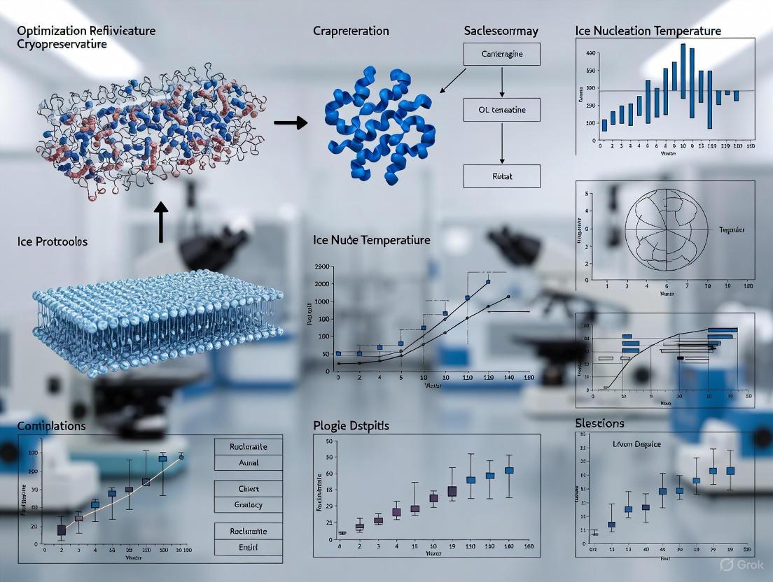
This article provides a comprehensive analysis of ice nucleation temperature as a pivotal yet often overlooked factor in cryopreservation protocol optimization.
Controlled Ice Nucleation: A Critical Parameter for Optimizing Cryopreservation Protocols and Post-Thaw Viability
Abstract
This article provides a comprehensive analysis of ice nucleation temperature as a pivotal yet often overlooked factor in cryopreservation protocol optimization. Tailored for researchers, scientists, and drug development professionals, we explore the fundamental biophysics of ice formation and its direct impact on cell viability, methodological approaches for controlling nucleation across different formats, practical troubleshooting for common challenges, and validation strategies for comparative analysis. By synthesizing current research and emerging technologies, this review establishes a framework for integrating controlled nucleation into standardized cryopreservation workflows to enhance reproducibility and cell recovery in biomedical applications.
The Science of Ice Nucleation: Understanding the Fundamental Principles of Ice Formation in Cryopreservation
Defining Ice Nucleation Temperature and Its Role in Cryopreservation Outcomes
Frequently Asked Questions (FAQs)
1. What is ice nucleation temperature and why is it a critical parameter in cryopreservation?
The ice nucleation temperature (TN) is the specific sub-zero temperature at which the first ice crystals form in a supercooled solution [1]. It is a critical controlled variable because it dictates the subsequent freezing process. When nucleation occurs at a warmer, defined temperature (e.g., -6°C to -10°C), it promotes gradual cellular dehydration, minimizing the risk of lethal intracellular ice formation (IIF) [2] [3]. Uncontrolled, stochastic nucleation leads to sample-to-sample variability and reduced cell recovery, viability, and function [4] [1].
2. My post-thaw cell viability is inconsistent, even with a controlled-rate freezer. Could uncontrolled nucleation be the cause?
Yes. Spontaneous (uncontrolled) ice nucleation is a significant source of variability [2]. Even within the same cooling protocol, different samples can supercool to different temperatures before nucleation occurs spontaneously. This results in containers having different thermal histories; some samples may experience high supercooling, leading to excessive IIF, while others nucleate warmer and survive better. Controlling the nucleation temperature standardizes this initial freezing step across all samples in a batch, enhancing consistency and overall post-thaw outcomes [1] [2].
3. What are the observable signs of damage from a sub-optimal nucleation temperature?
The type of damage correlates with the nucleation temperature:
- Nucleation at too low a temperature (excessive supercooling): This forces rapid ice growth, leaving insufficient time for water to leave the cell. The primary damage is intracellular ice formation (IIF), which is often fatal and can be observed as darkening of cells under cryomicroscopy [1] [2]. Post-thaw, this manifests as immediate cell rupture and early-stage necrosis [5].
- Nucleation at an appropriate temperature but with a slow cooling rate: While warm nucleation is beneficial, an overly slow cooling rate can cause prolonged exposure to high solute concentrations ("solution effects") and excessive cell shrinkage. This damage appears as delayed onset necrosis and apoptosis peaking 6-36 hours post-thaw [5].
Troubleshooting Guide
| Problem | Potential Cause | Recommended Solution |
|---|---|---|
| Low post-thaw viability with high immediate necrosis | Intracellular ice formation (IIF) due to low nucleation temperature and/or rapid cooling [5] [2]. | Actively control nucleation to a warmer temperature (e.g., -6°C to -10°C) [2]. Ensure cooling rate is optimized for your cell type (often ~1°C/min for many mammalian cells) [5]. |
| Low post-thaw viability with delayed apoptosis/necrosis | "Solution effect" toxicity from excessive cellular dehydration and prolonged exposure to high solute concentrations [5]. | Verify that the cooling rate is not too slow. Consider optimizing the cryoprotectant (CPA) type and concentration to provide better colloidal protection [5] [6]. |
| High variability in viability between identical samples | Stochastic, uncontrolled ice nucleation leading to different levels of supercooling in each sample [4] [1]. | Implement an active controlled nucleation method (e.g., ice seeding, chemical nucleants, pressure shift) to ensure all samples nucleate at the same defined temperature [1] [2]. |
| Poor recovery in small-volume formats (e.g., 96-well plates) | Small volumes have a greater propensity for deep supercooling, making uncontrolled nucleation and IIF more likely [3]. | Incorporate a soluble ice-nucleating agent like pollen washing water (PWW) into the cryomedium to reliably raise the nucleation temperature [3]. |
Experimental Protocols for Investigating Nucleation Temperature
Protocol 1: Active Control of Nucleation Temperature in Bulk Freezing
This protocol outlines a method to systematically test the effect of different nucleation temperatures on cell viability using a controlled-rate freezer capable of induced nucleation.
Materials:
- Cell Type: Multipotent mesenchymal stromal cells (MSCs) [1] or Jurkat T-cells [2].
- Cryoprotectant (CPA) Formulations: Plasma-Lyte A or standard culture medium with 5% and 10% (v/v) DMSO [1] [2].
- Equipment: Controlled-rate freezer with induced nucleation capability (e.g., via electrofreezing, pressure shift, or shock cooling) [4] [2].
- Vessels: Cryovials or custom freezing bags.
Methodology:
- Cell Preparation: Harvest and concentrate cells according to standard protocols.
- CPA Addition: Gently resuspend the cell pellet in pre-chilled CPA formulation. Incubate at 4°C for 10-30 minutes for equilibration [5].
- Loading: Transfer the cell suspension into cryovials.
- Freezing Program: Place vials in the controlled-rate freezer and initiate the cooling program.
- Continued Cooling: After nucleation, continue cooling at a defined rate (e.g., -1°C/min) to a terminal temperature (e.g., -80°C) before transferring to long-term storage in liquid nitrogen [2].
- Thawing and Assessment: Rapidly thaw samples in a 37°C water bath. Perform a step-wise dilution to remove CPA. Assess post-thaw viability (e.g., membrane integrity via flow cytometry) and metabolic activity (e.g., ATP-based assays) [1].
Protocol 2: Cryomicroscopy for Visualizing Intracellular Ice Formation
This protocol uses a cryomicroscope to directly observe cellular responses (dehydration and IIF) in real-time during freezing.
Materials:
- Equipment: Cryomicroscope stage with precise temperature control and video recording capability.
- Cell Sample: Jurkat cells or other adherent/suspension cells of interest [2].
- Staining (Optional): Fluorescent dyes like acridine orange (for viability) and propidium iodide (for membrane integrity) can be used [2].
Methodology:
- Sample Loading: Place a small droplet (~5-10 µL) of cell suspension in cryomedium on a specialized cryomicroscope slide.
- Cooling and Nucleation: Cool the stage at a controlled rate (e.g., -10°C/min or -1°C/min). Manually seed the sample at a specific supercooled temperature to induce controlled nucleation [2].
- Real-Time Observation: Monitor and record the freezing process.
- Observe cellular dehydration as a progressive shrinking of the cell volume.
- Identify intracellular ice formation (IIF) by a sudden darkening ("flashing") of the cytoplasm [2].
- Data Correlation: Correlate the incidence of IIF and the degree of dehydration with the nucleation temperature and cooling rate.
Data Presentation: Nucleation Temperature Impact on Cell Survival
Table 1: Effect of Controlled Nucleation Temperature on Post-Thaw Viability of Marmoset Monkey Mesenchymal Stromal Cells (cjmSCs) frozen with 5% DMSO. Data adapted from [1].
| Nucleation Temperature (°C) | Post-Thaw Viability | Metabolic Activity | Observed Ice Crystal Morphology |
|---|---|---|---|
| -4 | 64.5% | 75% | Large extracellular crystals |
| -6 | 71.5% | 85% | Large extracellular crystals |
| -8 | 75.5% | 90% | Intermediate crystals |
| -10 | 78% | 100% | Intermediate crystals |
| -12 | 71% | 85% | Small crystals, some IIF |
| -14 | 63% | 75% | Small crystals, increased IIF |
Table 2: Post-thaw recovery of Jurkat T-cells in different DMSO concentrations with controlled vs. spontaneous nucleation. Data synthesized from [2].
| Cryoformulation | Nucleation Condition | Membrane Integrity Post-Thaw | Notes |
|---|---|---|---|
| 2.5% DMSO | Spontaneous | Very Low | High IIF incidence |
| 2.5% DMSO | Controlled (-6°C) | Improved | Enhanced dehydration, less IIF |
| 5% DMSO | Spontaneous | Low | Variable outcomes |
| 5% DMSO | Controlled (-6°C) | High | Optimal dehydration |
| 10% DMSO (Control) | Spontaneous | High | Standard protocol |
Workflow and Pathway Visualization
The Scientist's Toolkit: Research Reagent Solutions
Table 3: Key reagents and materials for investigating and controlling ice nucleation.
| Item | Function & Application | Example Use Case |
|---|---|---|
| Chemical Nucleants | Soluble or insoluble agents that provide sites for heterogeneous ice nucleation at defined temperatures. | Snomax (derived from P. syringae) used to induce ice in microvasculature models at -6°C [7]. Pollen Washing Water (PWW) from Hornbeam, a soluble macromolecular nucleator, raised nucleation to ~-7°C in 96-well plates [3]. |
| Dimethyl Sulfoxide (DMSO) | Penetrating cryoprotectant that reduces IIF by hydrogen bonding with intracellular water and depressing the freezing point [6]. | Standard cryopreservation of MSCs and T-cells at 5-10% (v/v) concentration [1] [2]. |
| Polyethylene Glycol (PEG) | Non-penetrating cryoprotectant and cryoprotectant; helps protect cell membranes and modulates ice crystal growth [7] [6]. | Used at 2-5% to rescue endothelial cell membrane integrity during partial freezing protocols [7]. |
| 3-O-Methyl-D-Glucose (3-OMG) | Non-metabolizable glucose analog that can act as a penetrating cryoprotectant [7]. | Optimally used at 100 mM to improve endothelial cell attachment after high subzero storage [7]. |
| Controlled-Rate Freezer | Programmable freezer that provides a precise cooling rate and often includes a function for actively inducing nucleation. | For active control of nucleation temperature in bulk samples of MSCs or T-cells in cryovials [1] [2]. |
The Critical Relationship Between Supercooling and Intracellular Ice Formation
Troubleshooting Guide: Resolving Common Experimental Challenges
Q1: Our experiments show high intracellular ice formation (IIF) even at low supercooling degrees. What could be the cause?
- Potential Cause: Excessive Cell Volume. Research demonstrates that larger cells have a significantly higher probability of undergoing IIF at any given supercooling degree [8] [9] [10]. In a heterogeneous cell population, larger cells will be the most vulnerable.
- Solution:
- Control Cell Volume: Consider using mildly hypertonic solutions to induce controlled cellular dehydration before freezing, which decreases cell volume and can reduce the incidence of IIF [8].
- Analyze by Subpopulation: If possible, analyze IIF data by cell size to confirm this correlation in your experiments.
Q2: We observe inconsistent IIF results between experimental runs, making data unreliable. How can we improve reproducibility?
- Potential Cause: Uncontrolled Extracellular Ice Nucleation. The stochastic nature of extracellular ice nucleation can introduce significant variability in the degree of supercooling experienced by cells before IIF occurs [8] [11].
- Solution:
- Use a Controlled Cryostage: Employ a cryomicroscopy system with precise temperature control and a mechanism to initiate extracellular ice nucleation at a predetermined temperature [8] [10]. This standardizes the starting point for IIF observation across all trials.
- Document Supercooling Precisely: Record the exact temperature at which extracellular ice is nucleated, as the "degree of supercooling" is a key experimental variable [8].
Q3: According to our viability assays, cell survival is poor even when IIF appears limited. What other factors should we investigate?
- Potential Cause: Cryoprotectant Agent (CPA) Toxicity or Osmotic Damage. Cell death can occur through pathways independent of IIF, including toxicity from CPAs like DMSO or osmotic shock during the addition or removal of CPAs [12] [13].
- Solution:
- Optimize CPA Protocol: For DMSO, ensure the concentration is not excessively high (often 10% is standard) and that exposure time at room temperature is minimized before cooling [13].
- Review Thawing Procedure: Thaw cells rapidly but remove CPAs gently and gradually after thawing to prevent osmotic shock [13] [14].
Frequently Asked Questions (FAQs)
Q: Why is controlling supercooling so critical in cryopreservation protocols? A: The degree of supercooling is a primary driver for intracellular ice formation (IIF), which is lethal to cells [8]. By controlling the temperature at which extracellular ice nucleates, you directly influence the thermodynamic driving force for water to freeze inside the cell, allowing you to design protocols that minimize IIF.
Q: Is intracellular ice always lethal, and does the location of ice matter? A: Yes, for cells cryopreserved in suspension, the formation of intracellular ice is almost always linked to cell death [8] [15]. Ice crystals can mechanically disrupt organelles and the plasma membrane. Recent studies also highlight that ice formed during the warming phase (recrystallization) is a major cause of damage, even if little ice was present after initial cooling [15] [16].
Q: How does cell type influence its sensitivity to supercooling and IIF? A: Cell-specific properties, particularly the water permeability of the membrane and its surface area-to-volume ratio, determine how quickly water can exit the cell during cooling [12] [15]. Cells that dehydrate more readily can avoid IIF at slower cooling rates, while others are more susceptible. This is why protocols must be optimized for each cell type [12] [13].
Q: What are the most effective strategies to inhibit intracellular ice formation? A: A multi-pronged approach is most effective:
- Control Cooling/ Warming Rates: Use a slow cooling rate (e.g., -1°C/min) to allow time for cellular dehydration, or a very rapid cooling rate to achieve a glass-like, ice-free state (vitrification) [12] [13] [16].
- Use Cryoprotectants: Utilize permeating (e.g., DMSO, glycerol) and non-permeating (e.g., sucrose) CPAs to depress the freezing point and reduce the amount of "free" water available to form ice [15] [13].
- Manage Supercooling: Control the nucleation of extracellular ice to prevent excessive supercooling that can trigger IIF [8].
The following tables consolidate key experimental data on the relationship between supercooling, cell volume, and IIF.
Table 1: Effect of Supercooling and Cell Volume on Intracellular Ice Formation (IIF) in HUVECs
This data is derived from cryomicroscopy studies on Human Umbilical Veendothelial Cells (HUVECs) in the absence of cryoprotectants [8] [9].
| Degree of Supercooling Before Extracellular Ice Nucleation (°C) | Approximate Incidence of IIF in Isotonic Solution (~300 mOsm) | Approximate Incidence of IIF in Hypertonic Solution (~600-700 mOsm) |
|---|---|---|
| 2 °C | Low | Very Low |
| 5 °C | Moderate | Low |
| 10 °C | High | Moderate |
Table 2: Impact of Cooling and Warming Rates on Ice Formation in Bovine Oocytes
Data from synchrotron-based X-ray diffraction studies showing how protocol parameters influence ice formation [16].
| Protocol Parameter | Condition Description | Outcome on Ice Formation |
|---|---|---|
| Cooling Rate | ~30,000 °C/min (Current practice) | No ice after cooling, but large ice fractions form during warming. |
| Cooling Rate | ~600,000 °C/min (Using crystallography supports) | Ice formation is largely eliminated during both cooling and warming. |
| CPA Concentration | 100% strength vitrification solution | Required to achieve a vitrified state with standard cooling rates. |
| Warming Rate | Current convective warming (practice) | Allows devitrification and ice crystal growth. |
| Warming Rate | Optimized high convective warming (demonstrated) | ~20x faster than current practice; enables ice-free warming with lower CPA concentrations. |
Experimental Protocol: Quantifying IIF as a Function of Supercooling and Cell Volume
This detailed methodology is adapted from the cryomicroscopy study by Prickett et al. (2015) [8] [9] [10].
1. Objective: To examine how the incidence of intracellular ice formation (IIF) is affected by the degree of supercooling and cell volume in the absence of cryoprotectants.
2. Key Materials and Reagents
- Cells: Human Umbilical Vein Endothelial Cells (HUVECs) or other cell line of interest.
- Solutions: Isotonic culture medium (~300 mOsm) and hypertonic solution (~600-700 mOsm, adjusted with salts or sucrose).
- Equipment:
- Cryomicroscopy system (e.g., Linkam FDCS196 stage with T94 controller) [10].
- Inverted optical microscope (e.g., Nikon Eclipse 80i).
- Liquid nitrogen supply and pump.
- Viability Assay: Membrane integrity stain (e.g., fluorescent live/dead assay).
3. Step-by-Step Workflow
4. Detailed Procedures
- Step 1: Prepare Cell Suspensions. Harvest and suspend cells in both isotonic and hypertonic solutions. Allow time for osmotic equilibrium in the hypertonic solution, which will cause cell dehydration and volume decrease [8].
- Step 2: Load Sample. Place a small volume of the cell suspension on the cryostage, which is sealed with a coverslip.
- Step 3: Initial Cooling. Cool the sample at a constant rate (e.g., 10°C/min) from room temperature to a target supercooling temperature (e.g., 2, 5, or 10°C below the equilibrium freezing point) [8].
- Step 4: Nucleate Extracellular Ice. At the target supercooling temperature, initiate extracellular ice nucleation. This is often done by touching the edge of the sample with a cold needle or using a nucleation feature of the cryostage [8] [10].
- Step 5: Observe and Record IIF. Immediately after extracellular ice nucleation, observe cells under microscopy. The sudden darkening or "flashing" of a cell is the key visual indicator of intracellular ice formation [8] [10]. Record the number of cells that flash.
- Step 6: Post-Thaw Viability. Continue cooling the sample to a final temperature (e.g., -50°C), then thaw rapidly. Assess post-thaw membrane integrity using a viability stain to correlate IIF observations with cell death [8].
- Step 7: Data Correlation. Analyze the recorded video to correlate the incidence of IIF with the pre-nucleation cell volume (diameter) and the degree of supercooling.
The Scientist's Toolkit: Essential Research Reagents and Materials
Table 3: Key Reagents and Materials for Supercooling and IIF Research
| Item | Function/Application in Research |
|---|---|
| Controlled Cryostage (e.g., Linkam FDCS196) | Provides precise temperature control and visualization for directly observing intracellular ice formation ("flashing") in real-time [10]. |
| Cryoprotectant Agents (CPAs) | Penetrating (e.g., DMSO, Glycerol): Enter the cell, depress the freezing point, and reduce the amount of water available for ice formation. Non-penetrating (e.g., Sucrose): Remain outside the cell, inducing osmotic dehydration to reduce cell volume before freezing [12] [13]. |
| Membrane Integrity Assays | Fluorescent stains (e.g., SYTO 13/GelRed, Trypan Blue) used post-thaw to quantify cell viability and correlate it with IIF observations from cryomicroscopy [8] [12]. |
| Synchrotron X-ray Diffraction | A powerful analytical technique used to quantitatively detect and characterize the amount, structure, and grain size of ice within cryopreserved samples, even when optically invisible [16]. |
| Isochoric (Constant-Volume) Systems | A novel preservation platform that leverages thermodynamics to maintain biological matter in a stable, unfrozen state at subfreezing temperatures, thereby avoiding ice crystal damage entirely [17]. |
Fundamental Damage Mechanisms of Uncontrolled Nucleation
What are the primary ways uncontrolled nucleation damages cells? Uncontrolled ice nucleation inflicts damage through three primary, interconnected biophysical pathways: lethal intracellular ice formation, severe osmotic stress, and mechanical damage from ice crystals. The following table summarizes these core mechanisms and their consequences.
Table 1: Fundamental Damage Mechanisms from Uncontrolled Nucleation
| Damage Mechanism | Biophysical Process | Consequence for Cellular Structures |
|---|---|---|
| Intracellular Ice Formation (IIF) | Uncontrolled nucleation at low temperatures causes water to freeze inside the cell before it can escape [18] [19]. | Irreversible damage to membranes and organelles; fatal to cells [18] [20]. |
| Osmotic Stress | Extracellular ice formation concentrates solutes, creating a hypertonic environment that draws water out of cells, causing excessive dehydration [21] [22]. | Cell shrinkage, membrane damage, and prolonged exposure to toxic solute levels [20] [21]. |
| Mechanical Damage & Recrystallization | Ice crystals physically pierce and rupture cell membranes. During thawing, small ice crystals merge into larger, more destructive ones via Ostwald ripening [20] [22]. | Loss of membrane integrity and mechanical destruction of internal cell structures [20] [23]. |
Troubleshooting Guide: Common Experimental Problems & Solutions
FAQ: My post-thaw cell viability is consistently low and highly variable between samples. What could be wrong? This is a classic symptom of uncontrolled ice nucleation. When samples supercool deeply before freezing, the process becomes stochastic, leading to significant sample-to-sample variation in ice crystal structure and associated cellular damage [19]. Supercooling promotes the formation of numerous, small, and sharp intracellular ice crystals, which are lethal [18] [24].
Solution: Implement a controlled ice nucleation protocol. By actively triggering ice formation at a higher, defined temperature (e.g., -5°C to -7°C), you promote slower ice growth and allow time for cellular dehydration, thereby reducing IIF [18] [19]. The diagram below illustrates the protocol for controlled nucleation.
FAQ: Why are my 3D spheroids or monolayers particularly difficult to cryopreserve successfully? Cells in complex models like spheroids and monolayers have extensive cell-cell contacts. Uncontrolled ice nucleation facilitates the propagation of fatal intracellular ice between these adjacent cells, leading to widespread damage not typically seen in suspension cultures [18]. One study showed that controlled nucleation reduced IIF in A549 monolayers from 40-50% of cells to below 10% [18].
Solution: Use soluble ice-nucleating agents in your cryomedium. These materials, such as certain polysaccharides, function extracellularly and do not need to permeate the 3D structure. They induce warm-temperature ice nucleation, which protects cells beyond using DMSO alone by limiting ice propagation between connected cells [18] [25].
FAQ: How does the initial cooling rate influence cryopreservation outcomes? The cooling rate is critical because it determines the balance between dehydration damage and intracellular ice damage, as described by Mazur's two-factor hypothesis [21] [23]. Recent research directly links cooling rate to the morphology of the freeze-concentrated solution (FCS)—the channels where cells are sequestered during freezing.
Table 2: Impact of Cooling Rate on FCS Morphology and Cell Recovery
| Cooling Rate | FCS & Ice Crystal Morphology | Experimental Cell Recovery |
|---|---|---|
| Slow (e.g., 1°C/min) | Forms relatively large, well-defined FCS channels [23]. | Higher recovery (e.g., 65% for C2C12 myoblasts) [23]. |
| Fast (e.g., 10-30°C/min) | Results in fine ice crystals and narrow, constricted FCS channels [23]. | Lower recovery (e.g., 54-59% for C2C12 myoblasts) [23]. |
Detailed Experimental Protocol: Controlled Nucleation Using Soluble Ice Nucleators
This protocol outlines the use of sterile, soluble ice nucleators from Carpinus betulus (Hornbeam) pollen to improve the cryopreservation of adherent cell monolayers and spheroids in a 96-well plate format [18].
Methodology:
- Preparation of Cryopreservation Medium: Prepare a standard cryomedium containing 10% DMSO in your cell culture medium. Supplement this medium with the sterile-filtered pollen washing water (PWW) containing the soluble ice-nucleating polysaccharides. This creates the "+IN" (with induced nucleation) condition [18].
- Cell Preparation and Seeding:
- For monolayers: Seed cells in a 96-well plate and allow them to adhere and grow to the desired confluency.
- For spheroids: Generate spheroids in a 96-well ultra-low attachment plate.
- Freezing Procedure:
- Remove the standard culture medium and replace it with the pre-cooled "+IN" cryomedium.
- Place the 96-well plate into a controlled-rate freezer.
- Initiate the cooling program. The soluble ice nucleators will raise the nucleation temperature from approximately -15°C to a warmer temperature of around -8°C, ensuring ice forms extracellularly at a defined point [18].
- Continue the programmed cooling to the final storage temperature.
- Thawing and Assessment:
- Rapidly thaw the plate in a 37°C water bath.
- Remove the cryomedium, wash the cells, and add fresh culture medium.
- Assess post-thaw viability at 24 hours using a standard metabolic activity assay (e.g., MTT, Cell Counting Kit-8). Compared to 10% DMSO alone ("-IN" condition), the "+IN" condition typically shows statistically significant (p < 0.001) increases in post-thaw viability for monolayers [18].
The Scientist's Toolkit: Key Research Reagent Solutions
Table 3: Essential Reagents for Controlled Nucleation Research
| Reagent / Material | Function & Mechanism | Example Application |
|---|---|---|
| Soluble Ice Nucleating Polysaccharides (e.g., from PWW) | Sterilizable, soluble agents that induce extracellular ice nucleation at warm temperatures (-7°C to -8°C) [18]. | Cryopreservation of cell monolayers and 3D spheroids in multi-well plates to reduce IIF propagation [18]. |
| Synthetic Polyampholytes | Macromolecular cryoprotectants that improve post-thaw health by reducing intracellular ice formation and mitigating osmotic shock [25]. | Cryopreservation of sensitive immune cells (e.g., THP-1 monocytes), doubling post-thaw recovery compared to DMSO-alone [25]. |
| Low-Frequency Ultrasound | A physical method to induce precise ice seeding, reducing the degree of supercooling and improving the consistency of ice formation [24]. | Ultrasonic ice-seeding system for hepatocyte preservation, achieving over 90% cell survival rate at optimized intensities [24]. |
| Hydroxyethyl Starch (HES) | A non-permeating macromolecular cryoprotectant that increases solution viscosity and modulates ice crystal growth [21]. | Used as a component in cryopreservation solutions for red blood cells and other cell types [21]. |
Frequently Asked Questions (FAQs)
Q1: What are the primary mechanisms of cell damage during cryopreservation? The primary mechanisms are osmotic shock, solute damage (also known as solution effects), and ice crystal injury [26] [27]. Osmotic shock occurs as water freezes outside the cell, increasing the concentration of solutes in the unfrozen extracellular fluid; this draws water out of the cell, leading to harmful dehydration [26] [28]. Solute damage refers to the toxic increase in electrolyte concentration in the remaining unfrozen fluid, which can damage cellular membranes and proteins [26]. Ice crystal injury involves mechanical damage from the formation of sharp ice crystals, both outside (extracellular) and inside (intracellular) the cell, which can physically disrupt cellular structures [15] [27].
Q2: How does the cooling rate influence these damage mechanisms? The cooling rate is critical because it determines which damage mechanism is most dominant [26] [29]. The "two-factor hypothesis" explains this relationship [29]. Slow cooling rates allow time for water to leave the cell, minimizing deadly intracellular ice formation but potentially exposing the cell to prolonged solute damage and excessive dehydration [26] [15]. In contrast, rapid cooling rates do not allow sufficient time for water to exit the cell, leading to the formation of lethal intracellular ice [26] [15]. Therefore, each cell type has an optimal cooling rate that balances these two factors to maximize survival [26].
Q3: What is the role of cryoprotectants (CPAs) in mitigating these stressors? Cryoprotectants (CPAs) are chemicals that protect cells from freezing damage through several mechanisms [26] [27]. They depress the freezing point of water, reduce the extent of ice formation, and allow more water to vitrify (form a glassy state) instead of crystallizing [26] [21]. Permeating CPAs, like Dimethyl Sulfoxide (DMSO) and glycerol, enter the cell and help to reduce the concentration of harmful intracellular electrolytes and minimize dehydration [26] [27]. Non-permeating CPAs, such as sugars (sucrose, trehalose) and polymers, remain outside the cell and draw water out in a more controlled manner, further reducing the risk of intracellular ice formation [26]. They can also help stabilize cell membranes [26].
Q4: Why is intracellular ice considered so lethal? Intracellular ice is widely considered lethal because it mechanically disrupts delicate intracellular organelles and the plasma membrane, leading to immediate cell death upon thawing [28] [29]. The evidence for this is strong; as the cooling rate increases, the observed decrease in cell survival coincides directly with the formation of intracellular ice [28]. Unlike controlled dehydration, the physical presence of ice crystals inside the cell causes irreversible structural damage.
Q5: What is the specific risk of ice recrystallization during thawing? Ice recrystallization is a process during the thawing phase where small, less-damaging ice crystals merge to form larger, more destructive ones [15]. This growth occurs as the temperature rises through a "risky temperature zone" (approximately -15°C to -160°C) and can cause significant mechanical damage to cells that survived the initial freezing process [15]. Controlling recrystallization is therefore as important as controlling the initial freezing for ensuring high post-thaw viability.
Troubleshooting Common Cryopreservation Problems
Table of Common Issues and Solutions
| Problem Observed | Potential Cause | Recommended Solution |
|---|---|---|
| Low post-thaw viability | 1. Intracellular ice formation | 1. Optimize cooling rate; consider a slower rate to promote dehydration [26] [29]. |
| 2. High CPA toxicity | 2. Reduce CPA concentration or use a less toxic CPA/CPA cocktail [26] [21]. | |
| 3. Osmotic shock during CPA addition/removal | 3. Use a stepwise or controlled addition/removal protocol for CPAs [26]. | |
| Low post-thaw recovery & function | 1. Solute damage from slow cooling | 1. Optimize cooling rate; consider a faster rate to reduce exposure time [29]. |
| 2. Disruption of intracellular structures (e.g., granules) | 2. Optimize cooling protocol; for NK cells, a rate of 4-5°C/min was found effective [30] [31]. | |
| 3. Ice recrystallization during thawing | 3. Increase warming rate; use a 37°C water bath with gentle agitation [15]. | |
| High variability between samples | 1. Inconsistent ice nucleation | 1. Implement controlled ice nucleation (seeding) at a defined temperature [21]. |
| 2. Inconsistent cooling rates | 2. Use a controlled-rate freezer instead of a -80°C mechanical freezer [30] [32]. |
Quantitative Data for Protocol Optimization
Table 1: Optimal Cooling Rates for Different Cell Types
| Cell / Tissue Type | Optimal Cooling Rate | Key Reference |
|---|---|---|
| Natural Killer (NK) Cells (NK-92 cell line) | 4-5°C/min | [30] [31] |
| Hepatocytes, Hematopoietic Stem Cells, Mesenchymal Stem Cells | Slow cooling (~1°C/min) | [26] |
| Oocytes, Pancreatic Islets, Embryonic Stem Cells | Rapid cooling | [26] |
Table 2: Common Cryoprotectants and Their Properties
| Cryoprotectant | Type | Typical Conc. (v/v) | Notes |
|---|---|---|---|
| Dimethyl Sulfoxide (DMSO) | Permeating | 5-10% | Common; can be cytotoxic and induce differentiation; infusion-related side effects in patients [30] [32] [27]. |
| Glycerol (GLY) | Permeating | ~10% | First discovered CPA; widely used for microorganisms and sperm [26] [27]. |
| Ethylene Glycol (EG) | Permeating | Varies | Often used in vitrification mixtures [26] [21]. |
| Trehalose | Non-Permeating | 0.1-0.5 M | Naturally occurring disaccharide; stabilizes membranes; often used in combination [26]. |
| Sucrose | Non-Permeating | 0.1-0.5 M | Common osmotic buffer; used to dilute CPAs post-thaw to reduce osmotic shock [26]. |
Experimental Protocols for Investigating Stressors
Protocol: Determining the Optimal Cooling Rate for a New Cell Type
Objective: To systematically identify the cooling rate that maximizes post-thaw viability and function for a novel cell therapy candidate, balancing the risks of intracellular ice and solute damage.
Background: The survival of cryopreserved cells is highly dependent on the cooling rate, as described by the "two-factor hypothesis" [26] [29]. This protocol provides a methodology to empirically determine this critical parameter.
Materials:
- CELLBANKER 2 or a similar serum-free, DMSO-based cryopreservation medium [27].
- Controlled-rate freezer (e.g., Planer).
- Water bath (37°C).
- Cell viability analyzer (e.g., flow cytometer with Annexin V/PI staining).
- Culture materials for functional assay (e.g., cytotoxicity assay for immune cells).
Method:
- Cell Preparation: Harvest and concentrate the cells according to standard protocols. Ensure high viability (>95%) pre-freeze.
- CPA Addition: Resuspend the cell pellet in the pre-chilled (4°C) cryopreservation medium to the desired concentration.
- Cooling: Aliquot the cell suspension into cryovials and place them in the controlled-rate freezer. Program the freezer to initiate cooling from 4°C.
- Seeding: Induce ice nucleation (seeding) at -5°C to prevent supercooling and ensure consistent ice formation across samples [21].
- Rate Variation: Cool separate aliquots at different rates (e.g., 0.5, 1, 2, 5, 10, 20°C/min) down to a terminal temperature of -60°C to -80°C before transferring to liquid nitrogen for storage.
- Thawing: After storage (≥24 hours), rapidly thaw all samples in a 37°C water bath with gentle swirling until only a small ice crystal remains.
- CPA Removal & Analysis: Dilute the thawed cell suspension in a pre-warmed culture medium (or a medium containing a non-permeating osmolyte like sucrose for sensitive cells) [26]. Centrifuge, resuspend, and analyze for:
Protocol: Evaluating the Impact of Osmotic Stress During CPA Addition
Objective: To assess and minimize cell death caused by osmotic shock during the introduction and removal of cryoprotectants.
Background: The addition and removal of permeating CPAs like DMSO create large osmotic gradients that can cause cell swelling or shrinkage, leading to membrane damage and cell lysis [26]. A stepwise protocol can mitigate this.
Materials:
- Base culture medium.
- Permeating CPA (e.g., DMSO).
- Non-permeating osmotic buffer (e.g., 1M Sucrose solution).
Method:
- Stepwise Addition: a. Prepare CPA solutions in culture medium at 2x, 1.5x, and 1x the final target concentration (e.g., for 10% DMSO final, prepare 20%, 15%, and 10% solutions). Pre-cool to 4°C. b. Gently mix the cell pellet with an equal volume of the 2x CPA solution. Incubate for 5-10 minutes on ice. c. Add an equal volume of the 1.5x CPA solution, mix gently, and incubate for 5-10 minutes on ice. d. Finally, add the cell suspension to an equal volume of the 1x (final) CPA solution. The cells are now at the target CPA concentration.
- Stepwise Removal (Post-Thaw): a. Prepare dilution media with decreasing osmolarity. For example, Medium A (culture medium + 0.5M sucrose), Medium B (culture medium + 0.25M sucrose), and Medium C (culture medium only). Pre-warm to 37°C. b. Gently add the thawed cell suspension drop-wise to a larger volume (e.g., 10x volume) of Medium A. Incubate for 5-10 minutes. c. Centrifuge gently, remove the supernatant, and resuspend the cell pellet in Medium B. Incubate for 5-10 minutes. d. Centrifuge again, remove the supernatant, and resuspend in Medium C (final wash). The cells are now ready for culture or analysis.
Visualizing Stressors and Protective Strategies
The Scientist's Toolkit: Research Reagent Solutions
Table 3: Essential Reagents for Cryopreservation Research
| Reagent / Material | Function / Application | Key Considerations |
|---|---|---|
| Controlled-Rate Freezer | Precisely controls cooling rate, critical for protocol optimization and reproducibility [30]. | Essential for moving beyond simplistic -80°C freezing and for studying cooling rate effects. |
| Permeating CPAs (DMSO, Glycerol) | Penetrate cell, reduce intracellular ice formation, mitigate solute damage [26] [27]. | DMSO toxicity is concentration and time-dependent. Consider lower concentrations or alternatives for clinical applications [32]. |
| Non-Permeating CPAs (Trehalose, Sucrose) | Provide osmotic support, control cell dehydration, stabilize membranes, can reduce needed [DMSO] [26]. | Trehalose must be introduced into cells via specific techniques; sucrose is excellent for post-thaw dilution. |
| Ice Binding Agents (AFPs, Synthetic Polymers) | Inhibit ice recrystallization during thawing, can modulate ice nucleation shape [15] [21]. | A rapidly developing area; can reduce CPA concentration needed for vitrification. |
| Serum-Free, Xeno-Free Cryopreservation Media (e.g., CELLBANKER series) | Chemically defined, ready-to-use media for standardized, clinical-grade cell banking [27]. | Reduces batch-to-batch variability and safety concerns associated with serum. |
| Programmable Seeding Device | Initiates ice nucleation at a defined, consistent temperature in a controlled-rate freezer. | Prevents damaging supercooling, a key variable in optimizing ice nucleation temperature [21]. |
Troubleshooting Guides
Guide 1: Addressing Low Post-Thaw Viability
Problem: Cell viability and recovery rates are unacceptably low after thawing cryopreserved samples.
Solutions:
- Control Ice Nucleation: Implement a controlled ice nucleation protocol. Spontaneous, stochastic nucleation can lead to extensive undercooling (often ≥10°C below the freezing point), which is detrimental to sensitive cell types like embryos and oocytes. Using a controlled method standardizes the process and increases viability [19].
- Optimize Cooling Rate: Ensure the cooling rate is optimized for your cell type. A rate of -1 °C min⁻¹ or slower is often suitable for T cells and other mammalian cells. Rapid cooling (e.g., -10 °C min⁻¹) can lead to the formation of lethal intracellular ice, especially if followed by a slow warming rate [33].
- Prevent Transient Warming: Minimize temperature fluctuations during storage. Temperature rises from -196 °C to -80 °C over multiple cycles can significantly lower viability and metabolic activity, and increase apoptosis. Train staff to handle samples quickly and consider automated storage systems to eliminate human error [34] [35] [36].
Guide 2: Managing Sample-to-Sample Variability
Problem: Inconsistent results and high heterogeneity between samples frozen using the same protocol.
Solutions:
- Standardize Nucleation Temperature: The ice nucleation temperature is a primary determinant of the ice crystal structure and the primary drying rate in lyophilization. Its stochastic nature leads to variability. Use ice-nucleating additives or seeding techniques to control the nucleation temperature, thereby reducing lot-to-lot variability [19] [37].
- Account for Particulate Content: Be aware that the particulate content in your solution and the condition of your vials can influence the ice nucleation temperature. Carefully control these factors to ensure process consistency during development and scale-up [37].
- Verify Warming Rate Compatibility: If you are using a slow cooling rate (-1 °C min⁻¹), the warming rate may have a minimal impact on viable cell number. However, if your process involves rapid cooling, you must use a rapid warming rate (e.g., 113 °C min⁻¹) to prevent ice re-crystallization and a loss of viability [33].
Frequently Asked Questions (FAQs)
FAQ 1: What is the "glass transition" temperature and why is it critical for storage?
The glass transition temperature (approximately -135°C) is the point below which any remaining unfrozen water forms an amorphous, non-crystalline solid, or "glass." In this state, all biochemical reactions are effectively halted, allowing for indefinite stability. If the storage temperature rises above this point, even transiently, it can lead to microscopic melting and re-crystallization of ice, where larger, more damaging ice crystals grow at the expense of smaller ones. This process can mechanically damage cells and compromise their viability and function [34] [36].
FAQ 2: How sensitive are cells to temperature fluctuations during storage?
Cells are highly sensitive to the range and number of temperature fluctuations. Research on placental multipotent stromal cells (MSCs) showed that:
- Fewer than 20 cycles between -196°C and -100°C showed no significant difference in viability or metabolic activity.
- Fluctuations that reached higher temperatures (e.g., -80°C) led to a significant decrease in these parameters.
- Increasing the number of fluctuation cycles also led to a significant increase in apoptotic changes and compromised the cells' adhesive properties [35]. Another study on PBMCs confirmed that repeated temperature fluctuations during storage reduce cell recovery, viability, and T-cell function [36].
FAQ 3: Why is the rate of warming just as important as the rate of cooling?
The interaction between cooling and warming rates is critical. During slow cooling, cells dehydrate extensively. Slow warming can cause ice re-crystallization within the freeze-concentrated matrix, allowing small ice crystals to fuse into larger, damaging ones. Rapid warming minimizes the time for this re-crystallization to occur. However, for cells cooled slowly (-1 °C min⁻¹), the warming rate may have a minimal impact. The critical issue arises with rapid cooling; if cells are cooled rapidly, a slow warming rate can be particularly damaging, while a rapid warming rate is essential for recovery [33].
FAQ 4: What are the main mechanisms of cell damage during cryopreservation?
The three primary mechanisms are:
- Intracellular Ice Formation (IIF): The formation of ice crystals inside the cell, which is usually lethal as it can damage organelles and the cytoskeleton.
- Solution Effects Injury (Osmotic Stress): As extracellular water freezes, the concentration of solutes outside the cell increases. This hypertonic environment draws water out of the cell, causing severe dehydration and potentially denaturing proteins and disrupting membranes.
- Mechanical Forces from Extracellular Ice: Growing extracellular ice crystals can physically squeeze and crush cells trapped in the narrowing channels between them [34].
Data Presentation
Table 1: Impact of Temperature Fluctuations on Placental MSCs
| Number of Cycles | Temperature Range | Impact on Viability & Metabolism | Impact on Apoptosis | Impact on Adhesive Properties | Impact on Differentiation Potential |
|---|---|---|---|---|---|
| < 20 cycles | -196°C to -100°C | No significant difference | Not significant | Not significant | Not compromised [35] |
| Increased cycles | -196°C to -100°C | Significant lowering | Increases | Significantly compromised | Not compromised [35] |
| Increased cycles | -196°C to -80°C | Significant lowering | Increases | Significantly compromised | Not compromised [35] |
Table 2: Interaction of Cooling and Warming Rates on T-Cell Viability
| Cooling Rate | Warming Rate (Approx.) | Ice Structure Observed | Viable T-Cell Recovery |
|---|---|---|---|
| -1 °C min⁻¹ | Slow (1.6 °C min⁻¹) | Not specified | No significant impact [33] |
| -1 °C min⁻¹ | Rapid (113 °C min⁻¹) | Not specified | No significant impact [33] |
| -10 °C min⁻¹ | Slow (6.2 °C min⁻¹) | Highly amorphous; ice recrystallization during thaw | Significant reduction [33] |
| -10 °C min⁻¹ | Rapid (113 °C min⁻¹) | Not specified | No significant reduction [33] |
Experimental Protocols
Protocol: Investigating the Impact of Temperature Fluctuations on Stored Cells
Objective: To evaluate how temperature fluctuations during the storage of cryopreserved cells affect post-thaw viability, metabolic activity, and function.
Materials:
- Cryopreserved cells of interest (e.g., Placental MSCs, PBMCs)
- Liquid nitrogen vapor storage system
- Programmable freezer or controlled-rate freezer
- Cryovials
- Water bath or bead bath (set to 37°C for thawing)
- Cell culture reagents (growth medium, etc.)
- Hemocytometer or automated cell counter
- Apoptosis detection kit (e.g., Annexin V)
- Metabolic activity assay (e.g., MTT, ATP-based)
Methodology:
- Cell Preparation and Freezing: Harvest and cryopreserve the cells using your standard optimized protocol (e.g., slow cooling at -1 °C min⁻¹ in a cryoprotectant like CryoStor CS10) [33].
- Experimental Groups: Divide the frozen samples into different experimental groups:
- Control Group: Stored continuously at a stable cryogenic temperature (e.g., below -135°C).
- Fluctuation Group(s): Subjected to defined temperature cycles. For example, transfer samples from liquid nitrogen vapor to a -80°C freezer for a set period (e.g., 30 minutes) before returning them to vapor storage. Repeat this for a set number of cycles (e.g., 5, 10, 20 cycles) [35].
- Thawing and Assessment: After the storage period, rapidly thaw all samples in a 37°C water bath (or using a validated alternative method) [34].
- Post-Thaw Analysis:
- Viability and Recovery: Count the cells using trypan blue exclusion or an automated cell counter to determine total cell recovery and viability [35] [36].
- Apoptosis: Use a flow cytometry-based apoptosis detection kit to quantify early and late apoptotic cells [35].
- Metabolic Activity: Perform a metabolic activity assay (e.g., MTT) according to the manufacturer's instructions [35].
- Functionality: For immune cells like PBMCs, perform functional assays such as an ELISpot to measure antigen-specific immune responses [36].
Diagrams
Diagram: Temp Sensitivity in Cryopreservation
The Scientist's Toolkit
Research Reagent Solutions
| Item | Function & Application |
|---|---|
| Dimethyl Sulfoxide (DMSO) | A widely used cryoprotective agent (CPA) for mammalian cells. It penetrates the cell and reduces the formation of intracellular ice and mitigates osmotic stress during freezing [34]. |
| CryoStor / Commercial Freezing Media | A ready-to-use, GMP-compatible freezing medium containing DMSO. It is specifically formulated to provide enhanced protection against hypothermic and freezing-associated stress, improving post-thaw viability and function [33]. |
| Hydroxyethyl Starch | A non-penetrating cryoprotectant additive often used in combination with DMSO. It can help stabilize the cell membrane and modify the extracellular ice structure [37]. |
| Ice-Nucleating Agents (e.g., Pseudomonas syringae) | Used to control the ice nucleation temperature in a solution. By initiating freezing at a higher, defined temperature, they reduce the extent of supercooling and lead to a more consistent and less damaging ice structure [19] [37]. |
| Programmable Controlled-Rate Freezer | Essential equipment for implementing a reproducible cooling protocol. It allows for precise control over the cooling rate, which is critical for balancing the risks of intracellular ice formation and osmotic stress [34]. |
Practical Implementation: Methods and Tools for Controlling Ice Nucleation in Research and Development
Ice nucleation is a critical controlled step in cryopreservation protocols where the extracellular solution is deliberately triggered to freeze at a specific, warm temperature. This process initiates the controlled dehydration of cells, a vital mechanism for reducing fatal intracellular ice formation (IIF) during subsequent cooling. Optimizing this technique is essential for enhancing post-thaw viability and recovery, particularly for complex models like cell monolayers, spheroids, and organoids. This guide provides detailed methodologies and troubleshooting advice to help researchers master manual nucleation techniques.
Key Concepts and Importance
In cryopreservation, aqueous solutions tend to supercool substantially below their equilibrium freezing point before ice formation begins spontaneously, often at unpredictably low temperatures [18]. Manual nucleation (also called "seeding") intentionally initiates this freezing event at a higher, predetermined temperature (e.g., between -2°C and -10°C). This ensures that ice forms first in the extracellular space, creating a vapor pressure gradient that draws water out of the cells before the intracellular environment reaches its own supercooling limit. The avoidance of intracellular ice formation (IIF) is paramount, as IIF is almost universally fatal to cells [18]. For cells with extensive cell-cell contacts, such as those in monolayers and spheroids, uncontrolled ice nucleation can facilitate the lethal propagation of ice from one cell to its neighbors [18].
Manual Nucleation Protocols
Method 1: Chemical Induction Using Soluble Ice Nucleators
This protocol utilizes ice-nucleating agents derived from Carpinus betulus (Hornbeam) pollen to raise the nucleation temperature in a reliable and scalable manner [18].
- Objective: To achieve consistent extracellular ice nucleation at approximately -8°C for cryopreserving adherent cell monolayers and 3D spheroids.
- Materials:
- Pollen Wash Water (PWW) ice nucleator solution, sterilized by filtration [18].
- Standard cryopreservation medium (e.g., containing 10% DMSO).
- Cell culture(s) of interest (e.g., A549, SW480, or HepG2 cells) in suspension, as a monolayer, or as spheroids.
- Cryogenic vials or 96-well plates.
- Controlled-rate freezer or isopropanol freezing container.
- Procedure:
- Prepare Freezing Medium: Supplement your standard cryopreservation medium (e.g., 10% DMSO) with the sterile PWW ice nucleator solution. The final concentration should be optimized but is used as a direct supplement [18].
- Harvest and Resuspend Cells: Following standard protocols, harvest your cells and resuspend them in the prepared nucleation-supplemented freezing medium.
- Aliquot and Cool: Dispense the cell suspension into the chosen storage vessel (vials or plates). Begin cooling using a controlled-rate freezer or place in a -80°C freezer using an isopropanol chamber like "Mr. Frosty" to achieve a cooling rate of approximately -1°C/min [38] [39].
- Induce Nucleation (Passive): The inclusion of the PWW agent will passively raise the temperature at which ice nucleates in the solution. In the 96-well plate format, this has been shown to raise the nucleation temperature from about -15°C to -8°C, eliminating the need for manual intervention [18].
- Continue Cooling: After nucleation is confirmed (visually, by a sudden clouding or release of latent heat), continue the controlled cooling process to the final storage temperature (-80°C or below).
- Store: Transfer samples to long-term storage in liquid nitrogen vapor phase (< -135°C) [38] [39].
Method 2: Standard Manual Seeding for Cryovials
This traditional method is suitable for cryovials and relies on a physical trigger to initiate ice formation in a supercooled solution.
- Objective: To manually initiate extracellular ice formation in cryovials cooled to a specific seeding temperature.
- Materials:
- Cryopreservation medium (e.g., culture medium with 10% DMSO or glycerol).
- Cell suspension.
- Cryogenic vials.
- Controlled-rate freezer or alcohol bath pre-cooled to the seeding temperature.
- Liquid nitrogen-cooled forceps or a pre-chilled metal probe (e.g., a spatula).
- Procedure:
- Prepare and Aliquot: Resuspend cells in cryopreservation medium and aliquot into cryovials.
- Cool to Seeding Temperature: Place the vials in the cooling device and lower the temperature to the predetermined seeding temperature (typically between -2°C and -10°C). Hold at this temperature briefly to ensure thermal equilibrium.
- Induce Nucleation: Quickly open the freezer or bath and use the liquid nitrogen-chilled forceps to briefly touch the surface of the liquid in each vial, causing immediate ice crystallization. Alternatively, a pre-chilled metal probe can be used. Work rapidly to minimize temperature fluctuations.
- Hold for Crystallization: After seeding, hold the samples at the seeding temperature for 5-10 minutes to allow complete crystallization of the extracellular solution and initiate cellular dehydration.
- Resume Cooling: After the hold period, resume the controlled cooling cycle at a rate of -1°C/min to the final storage temperature.
- Store: Transfer to long-term storage in liquid nitrogen [38] [39].
The Scientist's Toolkit: Research Reagent Solutions
Table 1: Essential reagents and materials for manual nucleation experiments.
| Item | Function | Example & Notes |
|---|---|---|
| Soluble Ice Nucleators | Raises the temperature of extracellular ice formation passively; eliminates need for physical seeding. | Pollen Wash Water (PWW) from Carpinus betulus; sterile-filtered [18]. |
| Cryoprotectant Agents (CPAs) | Protects cells from ice damage and osmotic stress; enables vitrification at high concentrations. | DMSO, Glycerol, Ethylene Glycol. Note: CPA toxicity is concentration, time, and temperature-dependent [40]. |
| Hydroxyethyl Starch (HES) | Macromolecular cryoprotectant; can modify ice crystal growth during rewarming to improve viability. | Used at 6% (w/v); shown to protect Jurkat cells during transient warming events [41]. |
| Serum-Free Freezing Media | Chemically defined, protein-free cryopreservation medium; reduces lot-to-lot variability. | Gibco Synth-a-Freeze (contains 10% DMSO) [39]. |
| Cryogenic Vials | Secure, leak-proof containers for long-term sample storage. | Choose internally-threaded, medical-grade polypropylene vials for contamination prevention [42]. |
| Controlled-Rate Freezing Apparatus | Ensures reproducible, optimal cooling rate (~-1°C/min) for most cell types. | Controlled-rate freezer or isopropanol chambers (e.g., "Mr. Frosty," "CoolCell") [38] [39]. |
Troubleshooting FAQs
Q1: Why is manual nucleation particularly important for cryopreserving cells in 96-well plates or as 3D spheroids?
Small volume samples and 3D cell models present unique challenges. Small volumes in multiwell plates can supercool to as low as -15°C to -20°C before spontaneous nucleation occurs [18]. For 3D spheroids, extensive cell-cell contacts create pathways for fatal intracellular ice to propagate rapidly between adjacent cells once nucleation finally happens [18]. Chemically-induced extracellular nucleation at a warmer temperature (e.g., -8°C) dramatically reduces the incidence of IIF, leading to significantly higher post-thaw recovery and viability for these sensitive systems [18].
Q2: What are the consequences of a nucleation temperature that is too low?
Allowing deep supercooling (nucleation at very low temperatures) drastically increases the risk of intracellular ice formation. Because the intracellular solution is also supercooled, once ice finally forms externally, it can seed ice across the cell membrane before sufficient dehydration can occur. This is often lethal. Research has demonstrated that increasing the nucleation temperature from -11°C to -6°C reduced membrane damage in fibroblasts, which correlated with fewer cells exhibiting intracellular ice [18].
Q3: How does the choice of cryoprotectant interact with the nucleation process?
The cryoprotectant composition directly influences ice crystal kinetics. Studies show that optimizing the CPA recipe can bolster cellular resilience to temperature fluctuations during storage and transport. For example, adding 6% (w/v) Hydroxyethyl Starch (HES) to a DMSO-based medium altered ice crystal growth during rewarming events and improved Jurkat cell viability after a transient warming cycle [41]. Furthermore, the glass transition temperature (Tg) of a vitrification solution, which is determined by its chemical composition, has been linked to its propensity for thermal stress cracking; solutions with a higher Tg may exhibit less cracking [43].
Q4: A common issue is low post-thaw viability despite performing manual seeding. What are potential causes?
Low viability can stem from several factors related to the nucleation protocol:
- Incorrect Seeding Temperature: The temperature may be too low, leading to IIF, or the sample may not be held at the seeding temperature long enough for proper dehydration.
- Improper Cooling Rate: Even with correct nucleation, an overly rapid cooling rate can prevent sufficient water efflux, causing IIF. Ensure a controlled rate of ~-1°C/min after seeding [38].
- Cell Health and Concentration: Always freeze healthy, log-phase cells at the recommended concentration. High passage numbers or microbial contamination will result in poor recovery regardless of the nucleation technique [38] [44].
Experimental Data and Findings
Table 2: Quantitative impact of induced nucleation on post-thaw cell viability.
| Cell Type / Model | Cryopreservation Condition | Post-Thaw Viability / Recovery | Key Finding |
|---|---|---|---|
| A549 Monolayer | 10% DMSO alone (-IN) | Low | Induced nucleation drastically reduces intracellular ice formation (IIF) [18]. |
| A549 Monolayer | 10% DMSO + PWW (+IN) | >80% | IIF reduced from 40-50% of cells to below 10% [18]. |
| Jurkat Cells | Standard CPA | Viability decreases with TWE | Ice crystal area increases most when peak rewarming temp is -10°C [41]. |
| Jurkat Cells | CPA with 6% HES | Improved viability after TWE | HES addition enhances recrystallization during rewarming but is protective [41]. |
Mechanism of Manual Nucleation for Preventing Intracellular Ice
Troubleshooting Guides and FAQs
Frequently Asked Questions
Q1: Why is controlling ice nucleation critical for cryopreserving cells in small volumes, like in 96-well plates?
A1: Controlling ice nucleation is crucial because small liquid volumes (e.g., 100 µl) have a high propensity to deeply supercool, often reaching temperatures below -15°C before ice forms spontaneously [45]. This deep supercooling reduces the time available for cells to dehydrate, drastically increasing the likelihood of lethal intracellular ice formation (IIF) and resulting in poor and inconsistent post-thaw cell viability [45] [3]. By actively controlling nucleation at a warmer, specific temperature, you promote slower, controlled ice growth, allowing for proper cellular dehydration and significantly improving post-thaw survival rates [12] [24].
Q2: What are the main advantages of using a soluble ice-nucleating agent like Pollen Washing Water (PWW) over insoluble mineral-based agents?
A2: The primary advantages of soluble agents like PWW are ease of use and sterility. As a soluble and sterile filtrate, PWW can be directly added to the cryoprotectant solution, ensuring consistent distribution and integration into existing protocols without requiring removal after thawing [3]. In contrast, insoluble mineral-based agents, while highly effective, are particulate and may require a delivery system to separate them from the biological sample, adding complexity to the protocol [45].
Q3: My post-thaw cell viability is inconsistent even when using a controlled-rate freezer. Could uncontrolled ice nucleation be the cause?
A3: Yes. Controlled-rate freezers manage the cooling profile but do not always control the exact temperature at which ice nucleation occurs. If nucleation is stochastic, it can lead to significant well-to-well variability in supercooling and, consequently, in post-thaw viability [45]. Implementing a reliable ice nucleation method, such as using a mineral nucleator or ultrasonic seeding, ensures that ice formation begins at a consistent, warm temperature in every sample, thereby improving the reproducibility of your results [45] [24].
Troubleshooting Common Experimental Issues
| Problem | Possible Cause | Solution |
|---|---|---|
| Low and inconsistent post-thaw viability in multiwell plates. | Uncontrolled, deep supercooling leading to lethal intracellular ice formation (IIF). | Incorporate a consistent ice-nucleating agent. Use a mineral nucleator delivery system (e.g., IceStart arrays) or add a soluble nucleator like PWW to the cryoprotectant medium to raise the nucleation temperature [45] [3]. |
| High cytotoxicity in samples after cryopreservation. | The ice-nucleating agent itself may be cytotoxic, or the cryoprotectant (CPA) is not optimally formulated. | Switch to a biocompatible nucleator. Test alternative agents like mineral-based LDH1 or PWW, which have shown low cytotoxicity. Re-optimize CPA type and concentration for your specific cell type [45] [3]. |
| Failed ice nucleation during manual "seeding" with a cold probe. | The probe temperature is insufficiently cold, or the technique does not create a strong enough thermal shock. | Ensure the probe is pre-cooled in liquid nitrogen and that it makes contact with the supercooled liquid at the optimal temperature (typically between -2°C and -7°C). Standardize the contact time and location. Consider automated methods like ultrasound for greater reproducibility [24]. |
| Inability to induce nucleation at temperatures warmer than -10°C. | The ice-nucleating agent used is not active enough ("not hyperactive") for the application. | Source a "hyperactive" nucleator. Certain varieties of K-feldspar minerals (e.g., LDH1) or specific pollen extracts (e.g., from grey alder) can nucleate ice at temperatures as high as -2°C to -7°C [45] [3]. |
Quantitative Data on Ice-Nucleating Agents
The following table summarizes key performance data for various ice-nucleating agents from recent research, providing a basis for selection.
Table 1: Comparison of Ice-Nucleating Agents for Cryopreservation
| Agent Category | Specific Agent | Typical Nucleation Temperature (°C) | Post-Thaw Viability Improvement | Key Advantages |
|---|---|---|---|---|
| Mineral-Based | LDH1 (K-feldspar) | As high as -2°C to -7°C [45] | Dramatic increase for hepatocyte monolayers [45] | Hyperactive, highly effective, can be deployed via sterile arrays [45]. |
| Biological | Pollen Washing Water (PWW) | ≈ -7°C (in 100µl) [3] | T-cells: 63.9% to 97.4%; A549: 1.6% to 55.0% [3] | Soluble, sterile, low cytotoxicity, easy to integrate [3]. |
| Bacterial Derived | Snomax | Varies by batch and concentration | Improved consistency [45] | Very effective nucleator, but potential regulatory concerns for clinical use [45]. |
| Physical Method | Low-Frequency Ultrasound | User-defined (e.g., -4°C to -7°C) [24] | L-02 hepatocyte survival >90% [24] | Non-contact, precise timing, no chemical additives [24]. |
Detailed Experimental Protocols
Protocol 1: Cryopreservation of Cell Monolayers Using a Mineral-Based Ice Nucleator
This protocol is adapted from studies using LDH1 feldspar to cryopreserve immortalized human hepatocyte monolayers in 96-well plates [45].
Objective: To achieve high post-thaw viability and function of adherent cell monolayers by controlling ice nucleation at a warm, consistent temperature.
Materials:
- Confluent cell monolayers in a 96-well plate.
- Standard cryoprotectant agent (e.g., 10% DMSO in culture medium).
- Mineral nucleator delivery system (e.g., IceStart array with LDH1) [45].
- Controlled-rate freezer.
- Liquid nitrogen storage.
Methodology:
- Preparation: Aspirate the culture medium from the wells.
- CPA Addition: Gently add the pre-cooled cryoprotectant solution to each well.
- Nucleator Placement: Carefully place the IceStart array onto the plate, ensuring each mineral nucleator is aligned with a well.
- Cooling and Nucleation: Transfer the assembled plate to the controlled-rate freezer. Initiate a slow cooling ramp (e.g., -1°C/min). The mineral nucleators will induce ice formation consistently in each well at approximately -4°C to -7°C, minimizing supercooling.
- Plunge and Storage: Once the target temperature (e.g., -40°C to -80°C) is reached, promptly plunge the plate into liquid nitrogen for long-term storage.
- Thawing: Rapidly thaw the plate in a 37°C water bath with gentle agitation. Immediately remove the cryoprotectant and replace it with fresh culture medium.
Protocol 2: Applying Soluble Pollen Washing Water (PWW) for Cryopreservation in Suspension
This protocol details the use of PWW from European hornbeam to improve the cryopreservation of cells in suspension, such as Jurkat T-cells [3].
Objective: To reduce supercooling and increase post-thaw metabolic activity by incorporating a soluble, biological ice-nucleating agent.
Materials:
- Cells in suspension.
- Standard cryoprotectant medium.
- European hornbeam (Carpinus betulus) pollen.
- Sterile MilliQ water or culture medium.
- 0.22 µm sterile filter.
Methodology:
- PWW Preparation: Suspend 2% (wt/vol) European hornbeam pollen in sterile MilliQ water or culture medium. Incubate at 4°C for 24 hours. Pass the suspension through a 0.22 µm filter to remove pollen grains, collecting the sterile filtrate (PWW) [3].
- CPA-PWW Medium Formulation: Supplement your standard cryoprotectant solution (e.g., containing 10% DMSO) with the prepared PWW. The final concentration of PWW in the cryopreservation medium is typically the undiluted filtrate or a defined dilution [3].
- Freezing: Aliquot the cell suspension mixed with the CPA-PWW medium into cryovials or a 96-well plate. Use a controlled-rate freezer with a standard slow-freezing protocol (e.g., -1°C/min). The PWW will induce ice nucleation at a median temperature of around -7°C in 100µl volumes.
- Storage and Thaw: Continue cooling to the plunge temperature, then transfer to liquid nitrogen. Upon need, thaw rapidly and assay for viability and function.
Signaling Pathways and Experimental Workflows
Ice Nucleation Impact on Cell Survival
The following diagram illustrates the two primary pathways of cell damage during cryopreservation and how controlled ice nucleation promotes higher survival rates by mitigating intracellular ice formation.
Experimental Workflow for Protocol Optimization
This workflow outlines a systematic approach for researchers to optimize an ice nucleation protocol for a new cell type.
The Scientist's Toolkit: Key Research Reagent Solutions
The following table lists essential materials and their functions for integrating controlled ice nucleation into your cryopreservation research.
Table 2: Essential Reagents and Materials for Ice Nucleation Research
| Item | Function in Research | Key Characteristics |
|---|---|---|
| Hyperactive Mineral Powders (e.g., LDH1 K-feldspar) | Provides highly effective, solid-surface nucleation at warm temperatures (up to -2°C) [45]. | Mineral-based, often requires a delivery system (e.g., array) for sterile separation from sample [45]. |
| Pollen Washing Water (PWW) | A soluble biological extract that raises nucleation temperature, acting as a cryoprotectant [3]. | Sterile-filtered, soluble, low cytotoxicity, easily added to CPA solutions [3]. |
| Snomax | A commercial, freeze-dried preparation of ice-nucleating proteins from Pseudomonas syringae bacteria [45]. | Very effective nucleator, but potential regulatory and biocompatibility considerations for clinical use [45]. |
| Low-Frequency Ultrasonic System | A physical method to induce nucleation at a precise moment via acoustic cavitation, without chemical additives [24]. | Non-contact, allows precise timing; requires calibration of ultrasonic intensity for reproducibility [24]. |
| Infrared (IR) Thermography Camera | Critical for experimental validation, allowing direct measurement of the ice nucleation temperature (Tnuc) in individual wells[vibration:2]. | Enables correlation of Tnuc with post-thaw viability on a well-by-well basis [45]. |
Frequently Asked Questions (FAQs)
FAQ 1: What are ice nucleating materials (INMs) and why are they critical in cryopreservation? Ice nucleating materials are substances that provide sites for heterogeneous ice nucleation, raising the temperature at which ice forms in supercooled aqueous solutions [45]. In cryopreservation, they are critical for eliminating deep supercooling, which is a major cause of poor post-thaw cell viability. Uncontrolled ice nucleation can lead to intracellular ice formation (IIF), which is damaging or lethal to cells. By controlling the ice nucleation temperature (Tnuc), INMs allow for the formation of larger ice crystals outside the cells, promoting cell dehydration and minimizing IIF, thereby greatly enhancing cell recovery rates [45] [21].
FAQ 2: How do the novel INMs compare to traditional cryoprotectants like DMSO? Traditional permeating cryoprotectants like Dimethyl Sulfoxide (DMSO) function primarily by reducing ice formation and preventing osmotic shock. However, they do not control the location or onset of ice nucleation, leaving samples vulnerable to the damaging effects of supercooling [21]. In contrast, novel INMs like K-feldspar, Snomax, and cholesterol are non-permeating and function by actively promoting ice formation at higher, less damaging temperatures. They can be used alongside traditional CPAs to combine the benefits of both approaches: protection from osmotic stress and controlled ice crystal growth [45].
FAQ 3: My current cryopreservation protocol yields inconsistent post-thaw results. Could uncontrolled ice nucleation be the cause? Yes. In the absence of controlled ice nucleation, the temperature at which ice forms is unpredictable and often occurs at deeply supercooled states, especially in small volumes (e.g., less than 1 ml) [45]. This deep supercooling increases the likelihood of lethal intracellular ice formation and leads to inconsistent ice crystal structures, which is a common source of variable post-thaw viability [45]. Implementing a controlled nucleation strategy using the materials described here can significantly improve consistency.
FAQ 4: I am working with cell monolayers in multiwell plates. Why is manual ice nucleation not feasible, and what is the alternative? Manually inducing nucleation by touching each well with a cold object is impractical for multiwell plates, as it requires simultaneous induction in each individual well to ensure protocol consistency [45]. A passive and scalable alternative is the use of an ice nucleator delivery system, such as an IceStart array, which can be loaded with a mineral nucleator like LDH1 (a hyperactive K-feldspar) and placed in contact with the multiwell plate to induce controlled freezing across all wells simultaneously [45].
FAQ 5: Are there any biocompatibility or regulatory concerns with using these ice nucleating materials? Biocompatibility varies by material. Mineral-based nucleators like K-feldspar may offer advantages over biologically derived materials like Snomax in terms of biocompatibility and potential compliance with current good manufacturing practice (cGMP) for therapeutic applications [45]. It is crucial to select a material that does not interfere with your specimen and to use a delivery system that facilitates its removal after thawing if necessary [45]. Always validate biocompatibility for your specific cell type or tissue.
Troubleshooting Guides
Problem: Low or Inconsistent Post-Thaw Cell Viability
Potential Causes and Solutions:
Cause: Excessive Supercooling
- Solution: Integrate a highly active ice nucleator to raise the nucleation temperature. For instance, using a hyperactive K-feldspar (LDH1) has been shown to almost eliminate supercooling in 100 µl volumes [45].
- Action Plan:
- Implement a mineral nucleator delivery system (e.g., IceStart array).
- Confirm the elevated nucleation temperature using infrared thermography.
Cause: Suboptimal Cooling Rate
- Solution: The optimal cooling rate is dependent on the nucleation temperature. A higher Tnuc allows for the use of faster cooling rates while still avoiding IIF [45].
- Action Plan:
- If you have switched to controlled nucleation, re-optimize your cooling rate.
- Use a controlled-rate freezer for reproducible results.
Cause: Intracellular Ice Formation (IIF)
- Solution: This is often a consequence of deep supercooling. By nucleating ice at a warmer temperature, cells have more time to dehydrate before the temperature drops to a range where IIF is likely [45].
- Action Plan: Ensure your protocol uses both a controlled nucleator and a suitable permeating cryoprotectant.
Problem: Ice Nucleator is Not Functioning as Expected
Potential Causes and Solutions:
Cause: Ineffective Dispersion or Formulation
- Solution: The ice nucleating material must be properly prepared to expose its active sites.
- Action Plan:
- For K-feldspar (LDH1): Source material should be hand-ground into a fine powder and dry-sieved to remove particles above 63 µm, ensuring particles can be evenly suspended [45].
- For Cholesterol: Recrystallize reagent-grade cholesterol from ethanol, then grind the resultant plates into a fine, suspendable powder [45].
- For Snomax: Lyophilized pellets readily disperse into a suspension when mixed with water [45].
Cause: Low Concentration or Activity of Nucleating Sites
- Solution: Different materials and even different batches of the same material can have varying densities of ice-nucleating sites [46].
- Action Plan:
Problem: Challenges with Specific Sample Formats
Potential Causes and Solutions:
Cause: Working with Small Volumes (e.g., in Multiwell Plates)
- Solution: Small volumes are notoriously prone to deep supercooling because they contain fewer inherent nucleating impurities [45].
- Action Plan: The use of a passive ice nucleator is particularly critical for these formats. The IceStart array system was specifically developed to address this challenge for 96-well plates [45].
Cause: Working with Complex Tissues
- Solution: The dense matrix of tissues can impede the uniform penetration of ice crystals.
- Action Plan: While the use of INMs is beneficial, it may need to be combined with other strategies, such as improved permeation of cryoprotectants. Protocols for tissue preparation, like those optimized for ChIP-seq, emphasize meticulous mincing and homogenization under cold conditions to preserve sample integrity, which is a good practice for cryopreservation as well [47].
Research Reagent Solutions
Table: Key Ice Nucleating Materials for Cryopreservation
| Material Name | Type/Origin | Key Function | Sample Protocol / Application Notes |
|---|---|---|---|
| LDH1 (K-feldspar) [45] | Mineral (Hyperactive microcline) | Passive ice nucleator; raises nucleation temperature to as high as -2°C to -4°C [45] [46]. | Deliver via IceStart array for multiwell plates. Hand-grind and sieve (<63 µm) for suspension [45]. |
| Snomax [45] | Biological (Lyophilized P. syringae) | Proteinaceous ice nucleator; highly effective in aqueous solutions [45]. | Disperse lyophilized pellets in water to create a suspension. Note potential regulatory considerations for cGMP [45]. |
| Cholesterol [45] | Organic Crystal | Ice nucleating material; can be synthesized into a suspendable powder [45]. | Recrystallize from ethanol and grind into a fine powder for suspension [45]. |
| Glycerol / DMSO [21] | Permeating Cryoprotectant | Reduces ice formation and osmotic shock. Foundational chemicals in cryopreservation [21]. | Standard additive to cryopreservation medium. Concentration and addition/removal protocols are cell-type specific [21]. |
Quantitative Data on Ice Nucleation Performance
Table: Comparison of Ice Nucleating Material Performance
| Material | Reported Onset Nucleation Temperature | Ice Nucleating Activity / Notes | Key Application Findings |
|---|---|---|---|
| LDH1 (K-feldspar) [45] | As high as -2°C to -4°C [45] [46] | "Hyperactive" variety; nucleation site density varies with composition and microstructure [46]. | Almost eliminates supercooling in 100 µl volumes; greatly enhances post-thaw viability of human hepatocyte monolayers [45]. |
| Perthitic Feldspars [46] | Consistent onset between -2°C and -4°C | Activity correlates with K/Na composition and microtexture; microcline structures show continuous site activation [46]. | Highlights the importance of mineralogical properties in selecting an effective nucleator [46]. |
| Snomax [45] | Data not provided in results | Derived from ice nucleating bacteria; easily dispersible and highly active [45]. | Known as a highly active ice nucleating material; used for comparison in studies [45]. |
| Cholesterol [45] | Data not provided in results | Organic crystal that can be processed into a suspendable powder [45]. | Known as a highly active ice nucleating material; used for comparison in studies [45]. |
Experimental Workflow for Implementing Controlled Ice Nucleation
The following diagram outlines a general workflow for integrating a controlled ice nucleation strategy into a cryopreservation protocol.
Workflow for implementing controlled ice nucleation.
Mechanism of Action: How INMs Mitigate Cryo-Injury
This diagram illustrates the mechanism by which controlled ice nucleation protects cells during freezing.
Controlled vs. uncontrolled freezing mechanisms.
Frequently Asked Questions (FAQs)
FAQ 1: Why is controlling ice nucleation temperature critical in cryopreservation? Uncontrolled ice nucleation, or supercooling, leads to the formation of numerous small ice crystals. Upon warming, these crystals can recrystallize, forming larger, more damaging structures that compromise cell viability and product quality. Actively controlling the nucleation temperature ensures larger ice crystals form initially, which reduces recrystallization damage during thawing and improves post-preservation cell recovery and function [41] [45].
FAQ 2: How does the choice of culture vessel impact ice nucleation? The volume of the sample significantly influences the degree of supercooling. Smaller volumes, such as the 100 µL aliquots typical in 96-well plates, are prone to deep supercooling (by up to 25°C) because they contain fewer inherent, heterogeneous ice-nucleating sites. Larger formats like cryovials and bioreactor bags are less susceptible but still benefit from controlled nucleation for consistency [45].
FAQ 3: Can I use the same cryoprotective agent (CPA) formulation across different formats? While a base CPA formulation (e.g., containing DMSO) can be used, protocol optimization is required. For instance, adding 6% (w/v) Hydroxyethyl Starch (HES) can protect cells during transient warming events in vial-based storage. However, the specific CPA, its concentration, and the cooling/warming rates should be validated for each format and cell type to balance ice crystal growth and osmotic stress [41] [48].
FAQ 4: What is a major challenge in cryopreserving cells directly in multiwell plates? The primary challenge is inducing consistent, controlled ice nucleation simultaneously across all wells without compromising sterility. Manual methods are impractical, and without intervention, deep and variable supercooling leads to poor and inconsistent post-thaw cell recovery [45].
FAQ 5: How can bioreactors be used in cryopreservation workflows? Bioreactors are primarily used for the large-scale, reproducible expansion and differentiation of cells, such as iPSC-derived cardiomyocytes or macrophages, prior to preservation. Generating a high-quality, consistent cell product in a bioreactor is the first critical step before aliquoting into vials or plates for the actual freezing process [49] [50] [51].
Troubleshooting Guides
Problem: Low and Variable Post-Thaw Viability in Multiwell Plates
Potential Cause: Deep and uncontrolled supercooling within individual wells, leading to lethal intracellular ice formation [45].
Solution:
- Use a Passive Ice Nucleating Agent: Incorporate a highly active, mineral-based ice nucleating agent like LDH1 (a hyperactive variety of K-feldspar).
- Implementation: Deliver the LDH1 powder into each well using a sterile, disposable array like an "IceStart" array. This ensures simultaneous, controlled nucleation in all wells without breaking sterility.
- Outcome: This method almost eliminates supercooling, raising the nucleation temperature close to the solution's equilibrium freezing point. This results in larger ice crystal formation during cooling and significantly improves post-thaw cell viability and consistency across the plate [45].
Experimental Protocol: Cryopreservation of Cell Monolayers in 96-Well Plates
- Materials: Confluent cell monolayers, complete medium, CPA medium (e.g., 10% DMSO), LDH1 mineral powder, IceStart array or similar delivery system, controlled-rate freezer.
- Procedure:
- Prepare cell monolayers in a 96-well plate.
- Replace culture medium with a pre-cooled CPA medium.
- Place the plate on the IceStart array, which deposits a minute quantity of LDH1 into each well.
- Immediately transfer the plate to a controlled-rate freezer.
- Initiate a standard slow-cooling protocol (e.g., -1°C/min to -80°C).
- Transfer to a final storage vessel (e.g., liquid nitrogen vapor).
- Upon thawing, quickly remove the CPA medium and replace it with fresh culture medium.
Problem: Cell Damage During Temperature Fluctuations in Cryovials
Potential Cause: Ice recrystallization during transient warming events (e.g., during shipping or handling). Small ice crystals melt and re-form into larger, more damaging crystals [41] [48].
Solution:
- Optimize Cryoprotectant Cocktail: Supplement standard CPAs like DMSO with non-penetrating agents such as 6% (w/v) Hydroxyethyl Starch (HES).
- Control Nucleation Temperature: If possible, standardize the initial ice nucleation temperature during the freezing process. Studies show that lowering the nucleation temperature by 10°C can improve cell viability after warming events [41].
- Rationale: While HES may accelerate recrystallization during warming, it concurrently protects cells, likely by mitigating osmotic stresses. A optimized CPA cocktail bolsters cellular resilience to temperature excursions [41].
Experimental Protocol: Testing CPA Efficacy Against Transient Warming
- Materials: Cell suspension, CPA with DMSO, CPA with DMSO+6% HES, cryovials, controlled-rate freezer, cryomicroscope.
- Procedure:
- Aliquot cells into two CPA groups: DMSO only and DMSO + 6% HES.
- Use a controlled-rate freezer, triggering ice nucleation at a specific temperature (e.g., -5°C vs. -15°C).
- To simulate a transient warming event, transfer frozen vials to a storage freezer set to a sub-optimal peak temperature (e.g., -20°C or -10°C) for a set period, then return to -80°C.
- Assess ice crystal growth dynamically using a cryomicroscope and quantify post-thaw viability via flow cytometry or a similar method [41].
Problem: Inconsistent Differentiation Outcomes in Bioreactors Pre-Cryopreservation
Potential Cause: Variability in the quality and homogeneity of the input cell population, such as embryoid bodies (EBs) of inconsistent size and quality [50].
Solution:
- Standardize Input Cells: Use a quality-controlled master cell bank and monitor pluripotency markers (e.g., >70% SSEA4+) before initiating differentiation [50].
- Control Aggregation Size: In suspension bioreactors, monitor and control the size of EBs. Target an optimal diameter (e.g., 100 µm) before adding differentiation inducer CHIR99021. EBs smaller than 100 µm may disintegrate, while those larger than 300 µm differentiate less efficiently due to diffusion limits [50].
- Rationale: High-quality, uniform input cells and aggregates ensure synchronized differentiation, resulting in a more pure and consistent cell product (e.g., >90% TNNT2+ cardiomyocytes) before the cryopreservation step, which is critical for batch-to-batch reproducibility [50] [51].
The data below summarizes key experimental findings from the literature on optimizing cryopreservation protocols.
Table 1: Impact of Protocol Modifications on Ice Crystal Growth and Cell Viability
| Protocol Variable | Experimental Condition | Peak Warming Temperature | Impact on Ice Crystal Area | Impact on Cell Viability | Key Findings |
|---|---|---|---|---|---|
| CPA Formulation [41] | DMSO only | -10°C | Largest increase | Low | Recrystallization is a major cause of damage. |
| DMSO + 6% HES | -10°C | Accelerated increase | Improved | HES protects despite enhancing recrystallization. | |
| Nucleation Temperature [41] | Standard nucleation | -20°C | - | Low | Viability is compromised. |
| Lowered nucleation (-10°C) | -20°C | - | Improved | Lower nucleation temp bolsters resilience to warming. |
Table 2: Bioreactor Process Parameters for Consistent Pre-Cryopreservation Cell Production
| Cell Type | Bioreactor System | Critical Process Parameter | Target Value | Output Purity & Yield |
|---|---|---|---|---|
| iPSC-Derived Cardiomyocytes [50] | Stirred-Tank Bioreactor | Embryoid Body (EB) Diameter at CHIR addition | 100 µm | ~94% TNNT2+ cells; ~1.2 million cells/mL |
| iPSC-Derived Macrophages [49] | Stirred-Tank Bioreactor (120 mL) | Continuous harvest from Myeloid-Cell-Forming Complexes | Weekly for several months | Highly pure CD45+/CD11b+/CD14+ population |
Workflow and Strategy Diagrams
Exp. Workflow for Format Adaptation
Nucleation Control Strategy
The Scientist's Toolkit
Table 3: Essential Reagents and Materials for Cryopreservation Protocol Adaptation
| Item | Function & Rationale | Example Application / Note |
|---|---|---|
| Hydroxyethyl Starch (HES) | A non-penetrating cryoprotectant that helps mitigate osmotic stress and cell damage during ice recrystallization [41]. | Add at 6% (w/v) to DMSO-based CPA for cryovials subject to temperature fluctuations [41]. |
| LDH1 (Mineral Nucleator) | A "hyperactive" potassium feldspar powder that provides highly effective ice nucleation sites, eliminating supercooling in small volumes [45]. | For cryopreserving cell monolayers directly in 96-well plates; delivered via IceStart array [45]. |
| Stirred-Tank Bioreactor | Enables scalable, 3D suspension culture for consistent production of high-quality cells (e.g., iPSCs, cardiomyocytes) prior to preservation [50] [51]. | Critical for generating reproducible, clinically relevant cell numbers; allows monitoring of pH, O₂ [49] [50]. |
| Dimethyl Sulfoxide (DMSO) | A penetrating cryoprotectant that reduces intracellular ice formation by lowering the freezing point and moderating cell dehydration [41] [48]. | The most common CPA; often used at 10% concentration; can have toxicity concerns [48]. |
Integrating Nucleation Control with Cooling Rate Optimization and Cryoprotectant Selection
Frequently Asked Questions (FAQs)
1. Why is controlling ice nucleation temperature critical in slow freezing cryopreservation protocols?
Controlling the temperature at which extracellular ice nucleation occurs is fundamental to the success of slow freezing protocols. When a sample supercools (cools significantly below its freezing point before ice forms), the subsequent, spontaneous ice nucleation occurs rapidly at a colder temperature. This leaves cells insufficient time to dehydrate, dramatically increasing the probability of lethal intracellular ice formation (IIF) [12] [52]. By actively inducing nucleation at a warmer, higher sub-zero temperature (e.g., -5°C to -7°C), you provide cells with a longer timeframe to lose water osmotically, thereby reducing IIF and improving post-thaw viability [3].
2. My post-thaw cell viability in 96-well plates is consistently low, despite using standard cryoprotectants. What could be the issue?
This is a common problem directly linked to uncontrolled supercooling in small volumes. Smaller volumes of water have a much greater propensity to supercool deeply than larger volumes [3]. In a 96-well plate, each well is a small, isolated volume that may supercool to degrees below its freezing point before nucleating, leading to widespread intracellular ice formation. The solution is to incorporate a controllable ice nucleating agent into your cryopreservation medium to consistently initiate freezing at a warmer, less damaging temperature [3].
3. Is a specialized "rapid-cooling nucleation step" necessary when programming a controlled-rate freezer?
Experimental evidence suggests that a dedicated rapid-cooling step to induce nucleation is not necessary for all cell types. A comparative study freezing four different cell lines (including CHO, HepG2, and Jurkat cells) found no significant improvement in post-thaw viability or metabolic function when a rapid-cooling nucleation step was used, compared to a consistent, linear cooling rate [53]. For these cells, maintaining a stable, optimized cooling rate (e.g., -1°C to -2°C per minute) is sufficient for successful cryopreservation.
4. What are the alternatives to DMSO, and can they be used for nucleation control?
Yes, alternatives to DMSO are an active area of research and can be integrated with nucleation control. For instance, human T lymphocytes have been successfully cryopreserved using a Me2SO (DMSO)-free solution containing trehalose and human serum albumin [54]. The success of this protocol was dependent on combining the non-toxic cryoprotectant with both a controlled cooling rate and controlled ice nucleation (seeding). This demonstrates that nucleation control is a versatile strategy that can enhance protocols even when reducing the reliance on traditional penetrating cryoprotectants.
Troubleshooting Guides
Problem: Low Post-Thaw Viability in Multi-Well Plate Formats
Potential Cause: Deep supercooling of individual wells, leading to low, unpredictable nucleation temperatures and high intracellular ice formation [3].
Solutions:
- Incorporate a soluble ice nucleator: Add a sterile, soluble ice-nucleating agent like Pollen Washing Water (PWW) to your cryopreservation medium. PWW, derived from European hornbeam or silver birch pollen, contains ice-nucleating macromolecules that can raise the nucleation temperature in 96-well plates from approximately -13°C to -7°C, significantly improving post-thaw metabolic activity [3].
- Use a chemical nucleator: Snomax, a commercial product derived from ice-nucleating bacteria Pseudomonas syringae, can also be used to control nucleation [7]. Note that unlike PWW, Snomax is particulate and not soluble.
- Employ an external nucleation device: Some controlled-rate freezers have built-in features to induce nucleation, such as a brief blast of cold nitrogen vapor ("shock freezing") that creates a temperature gradient to trigger ice formation [3].
Problem: High Variability in Recovery Between Vials Frozen in the Same Batch
Potential Cause: Inconsistent ice nucleation events across samples. Without active control, the initiation of freezing is a stochastic process, meaning each vial in a batch may nucleate at a different temperature and time, leading to varied cellular experiences and outcomes [52].
Solutions:
- Implement active nucleation control: Use an ice seeding technique. For vials in an alcohol bath freezer, this can be done by briefly touching the vial with pre-cooled forceps at the target nucleation temperature (e.g., -5°C). In a controlled-rate freezer, use the device's nucleation feature if available [54].
- Standardize with a nucleating agent: Adding a consistent, low concentration of an ice-nucleating agent like PWW or Snomax to all vials ensures that nucleation occurs at a uniform, predictable temperature for every sample in your batch [7] [3].
Problem: Poor Recovery of Specific Sensitive Cell Types (e.g., T lymphocytes) with Low CPA Concentrations
Potential Cause: The cells are sufficiently protected from osmotic stress by low-CPA formulations but remain vulnerable to intracellular ice injury due to suboptimal freezing kinetics.
Solutions:
- Optimize the cooling profile with nucleation: Research on human T lymphocytes shows that combining low-CPA cocktails (e.g., 0.2 M trehalose) with a specific cooling rate (1.5°C/min) and controlled ice seeding can achieve high viability (~96%) without requiring DMSO [54]. The nucleation step is critical to this success.
- Systematically test cooling rates: The optimal cooling rate is cell-type dependent. Use a cryomicroscope to observe IIF events directly, or perform viability assays across a range of cooling rates (e.g., 1°C/min to 10°C/min) while keeping the nucleation temperature constant to find the best compromise between dehydration and IIF [55].
Experimental Protocols & Data
Detailed Protocol: Cryopreservation of Monolayer Cells with Controlled Nucleation
This protocol is designed for cryopreserving adherent cell monolayers, such as A549 lung carcinoma cells, in multi-well plates, leveraging controlled nucleation to enhance survival [3].
- Preparation of Cryoprotectant Medium (CPM): Prepare your standard CPM (e.g., culture medium with 10% DMSO). Centrifuge and filter-sterilize a PWW solution.
- Add Ice Nucleator: Supplement the CPM with 0.1-0.5% (v/v) PWW and mix thoroughly.
- Cell Seeding and Culture: Seed cells into a 96-well plate and culture until they form a confluent monolayer.
- Media Exchange: Prior to freezing, aspirate the culture medium from the wells and gently add the pre-cooled PWW-supplemented CPM.
- Controlled-Rate Freezing:
- Place the 96-well plate into a controlled-rate freezer pre-cooled to 4°C.
- Initiate a cooling rate of -1°C/min.
- The PWW will induce homogeneous ice nucleation across all wells at approximately -7°C.
- Continue cooling to a terminal temperature of at least -50°C to -60°C before transferring the plate to long-term storage in liquid nitrogen vapor phase.
- Thawing: Rapidly thaw the plate in a 37°C water bath. Immediately remove the CPM, gently wash with fresh culture medium, and add pre-warmed medium for culture.
Quantitative Data on Nucleation and Viability
The following table summarizes key experimental data from the literature on the impact of controlled nucleation.
Table 1: Impact of Controlled Ice Nucleation on Cryopreservation Outcomes
| Cell Type / System | Cryoprotectant Solution | Nucleation Method | Key Outcome | Source |
|---|---|---|---|---|
| Immortalized T-cells (Jurkat) in 96-well plate | Standard CPA (DMSO) | None (Supercooled) | Post-thaw metabolic activity: 63.9% | [3] |
| Immortalized T-cells (Jurkat) in 96-well plate | Standard CPA (DMSO) | PWW | Post-thaw metabolic activity: 97.4% | [3] |
| Immortalized Lung Carcinoma (A549) monolayers | Standard CPA (DMSO) | None (Supercooled) | Post-thaw metabolic activity: 1.6% | [3] |
| Immortalized Lung Carcinoma (A549) monolayers | Standard CPA (DMSO) | PWW | Post-thaw metabolic activity: 55.0% | [3] |
| Human T Lymphocytes | 1% DMSO, 0.1M Trehalose, 4% HSA | No Seeding | Relative Viability: 88.6% | [54] |
| Human T Lymphocytes | 1% DMSO, 0.1M Trehalose, 4% HSA | Ice Seeding | Relative Viability: 94.1% | [54] |
| Human T Lymphocytes | 0.2M Trehalose, 4% HSA (DMSO-free) | Ice Seeding | Relative Viability: 96.3% | [54] |
Table 2: Effect of Cooling Rate and Cryoprotectant on Intracellular Ice Formation (IIF) in Small Abalone Eggs [55]
| Cooling Rate (°C/min) | DMSO Concentration (M) | Observation on IIF Suppression |
|---|---|---|
| 1.5 to 12 | 2.0 to 4.0 | IIF was well-suppressed across this range when DMSO concentration was ≥ 2.0 M. |
| 1.5 | 2.0 | Optimal combination: Feasible protocol with 48.8% osmotically active eggs post-thaw and a 23.7% hatching rate. |
| > 1.5 | 2.0 | Higher cooling rates resulted in a lower chance of osmotically active eggs. |
Workflow and Conceptual Diagrams
Cryopreservation Protocol Optimization Logic
Experimental Workflow for Protocol Development
The Scientist's Toolkit: Essential Research Reagents & Materials
Table 3: Key Reagents for Integrating Nucleation Control in Cryopreservation
| Item | Function / Application | Example Use Case |
|---|---|---|
| Pollen Washing Water (PWW) | A soluble, sterile ice-nucleating agent derived from tree pollen (e.g., European Hornbeam) that raises nucleation temperature. | Prevents deep supercooling in 96-well plates and cryovials; improves monolayer cell survival [3]. |
| Snomax | A freeze-dried preparation of Pseudomonas syringae bacteria that provides highly active ice nucleation sites. | Used in research to induce controlled extracellular ice formation at high subzero temperatures [7]. |
| Trehalose | A non-permeating disaccharide sugar that acts as an extracellular cryoprotectant, stabilizing cell membranes. | Enables DMSO-free or DMSO-reduced cryopreservation protocols for T-cells when combined with nucleation control [54]. |
| 3-O-Methyl-D-Glucose (3-OMG) | A non-metabolizable glucose analog that can act as a permeating cryoprotectant. | Rescued cell attachment in a microvasculature model at -6°C [7]. |
| Polyethylene Glycol (PEG) | A non-permeating polymer that can protect cell membranes and modulate ice crystal growth. | Effective at maintaining membrane integrity in endothelial cells at -10°C [7]. |
| Controlled-Rate Freezer | A device that precisely controls the cooling rate of samples, often featuring programmable steps and nucleation functions. | Essential for implementing reproducible slow-freezing protocols with precise nucleation triggers [53]. |
| Cryomicroscope | A microscope with a temperature-controlled stage for directly observing ice formation and cell morphology during freezing. | Critical for experimental determination of IIF temperatures and optimal cooling rates for new cell types [55]. |
Solving Common Challenges: Troubleshooting and Optimizing Nucleation Protocols for Enhanced Recovery
Inconsistent post-thaw cell viability presents a significant challenge in research and drug development, often leading to variable experimental results and compromised cellular therapies. While factors like cryoprotectant concentration and cooling rate are routinely optimized, ice nucleation temperature remains a frequently overlooked critical parameter. Uncontrolled supercooling—where samples cool far below their freezing point before ice forms—results in unpredictable intracellular ice formation, causing physical damage to membranes and organelles [56] [57]. This technical guide provides troubleshooting resources and methodologies to help researchers identify, diagnose, and resolve nucleation-related issues in their cryopreservation workflows, enabling more reproducible and reliable post-thaw outcomes.
Troubleshooting Guides
Guide 1: Diagnosing Nucleation Temperature Problems
| Observed Symptom | Possible Root Cause | Recommended Diagnostic Action |
|---|---|---|
| High variability in viability between vials frozen simultaneously | Uncontrolled, stochastic ice nucleation resulting in different supercooling depths across samples [57] | Implement controlled ice nucleation; monitor nucleation temperature with thermocouples |
| Poor cell attachment post-thaw (monolayer cultures) | Extensive supercooling causing intracellular ice formation and membrane damage [57] | Compare post-thaw attachment with and without controlled nucleation at −3 to −5°C |
| Low membrane integrity despite optimized cryoprotectants | Intracellular ice formation from excessive supercooling below −10°C [56] [57] | Assess membrane integrity via Trypan blue exclusion immediately post-thaw |
| Reduced cellular function despite high viability | Sublethal damage from osmotic stress during uncontrolled freezing [41] | Perform functional assays (e.g., metabolic activity, secretion rates) post-thaw |
| Inconsistent results across different plate/vial types | Vessel-dependent supercooling behavior due to varying surface-to-volume ratios [57] | Characterize supercooling across different container formats |
Guide 2: Interpreting Ice Crystal Size and Post-Thaw Outcomes
| Observed Ice Crystal Pattern | Associated Nucleation Temperature | Expected Impact on Viability |
|---|---|---|
| Many small, uniform extracellular crystals | Controlled nucleation at high subzero temperatures (>−5°C) [41] [7] | High viability and function: Gradual dehydration minimizes intracellular ice |
| Large, variable extracellular crystals with some intracellular ice | Moderate supercooling (−5°C to −10°C) | Moderate viability: Some intracellular ice formation causes variable outcomes |
| Extensive intracellular ice with small crystals | Deep supercooling (<−10°C) with spontaneous nucleation [24] [57] | Low viability: Physical piercing of membranes and organelles |
| Very large recrystallized ice structures | Temperature fluctuations during storage/transport [41] | Progressive viability loss: Crystal growth during warming damages cells |
Frequently Asked Questions (FAQs)
Q: Why does nucleation temperature significantly impact my post-thaw viability when I've already optimized my cryoprotectant and cooling rate?
A: Even with optimal cryoprotectant and cooling rates, uncontrolled supercooling causes variable intracellular ice formation, which is directly lethal to cells. Ice nucleation temperature determines when extracellular ice begins to form, initiating the cellular dehydration process. Without controlled nucleation, samples experience random and often deep supercooling, leading to inconsistent ice formation patterns and consequently, variable cell damage across samples [57].
Q: My lab doesn't have specialized equipment for controlling ice nucleation. What practical methods can I implement?
A: Several accessible techniques can help control nucleation:
- Manual seeding: Use pre-chilled forceps or a cryopen to briefly touch the side of cryovials when the sample temperature reaches −3 to −5°C [58]
- Ice nucleating agents: Add sterile preparations like Snomax (from the ice-nucleating bacterium Pseudomonas syringae) to promote consistent ice formation at specific temperatures [57] [7]
- Ultrasonic nucleation: Apply brief, low-intensity ultrasound pulses to induce nucleation (requires calibration) [24]
Q: How does nucleation temperature specifically affect sensitive cell types like iPSCs and primary hepatocytes?
A: iPSCs are particularly vulnerable to intracellular ice formation due to their delicate membrane structure and typically form aggregates, which creates challenges for cryoprotectant penetration. Controlled nucleation at warmer temperatures (−3 to −5°C) promotes gradual dehydration, improving aggregate survival [56]. Primary hepatocytes experience significant functional loss post-thaw with uncontrolled nucleation due to damage to mitochondrial and endoplasmic reticulum membranes [13] [24].
Q: Can optimizing nucleation temperature help with temperature fluctuations during shipping?
A: Yes, samples nucleated at higher subzero temperatures demonstrate improved resilience to temperature fluctuations. Controlled nucleation produces smaller, more stable ice crystals that are less prone to destructive recrystallization during transient warming events [41]. Supplementing with cryoprotectants like hydroxyethyl starch (HES) can further stabilize the frozen matrix against temperature fluctuations [41].
Experimental Protocols
Protocol 1: Establishing Controlled Ice Nucleation for Cryovials
Principle: Manually induce ice formation at a defined, high subzero temperature to ensure consistent freezing kinetics across samples.
Materials:
- Controlled rate freezer or alcohol-based passive cooling device
- Pre-chilled forceps or cryopen
- Temperature data logger (optional but recommended)
- Cryovials containing cell suspension in cryoprotectant
Methodology:
- Prepare cell suspension according to established protocols, ensuring cryoprotectant is properly formulated.
- Transfer suspension to cryovials and place in controlled rate freezer.
- Initiate cooling protocol with a rate of −1°C/min from +4°C.
- When chamber temperature reaches −5°C, pause the cooling program.
- Using pre-chilled forceps or a cryopen, briefly touch the exterior wall of each vial at the meniscus level until ice formation is visually confirmed.
- Immediately resume cooling program at −1°C/min to the desired endpoint temperature (typically −40°C to −80°C).
- Transfer to long-term storage in liquid nitrogen vapor phase [58].
Troubleshooting Notes:
- If ice fails to form, ensure forceps are sufficiently chilled and contact the vial at the solution meniscus
- Excessive cooling pauses (>2 minutes) can affect cryoprotectant toxicity; work efficiently
- For high-throughput applications, consider commercial nucleators or ice-nucleating agents
Protocol 2: Quantifying Supercooling in Multiwell Plates
Principle: Characterize the supercooling behavior of specific plate formats to establish nucleation parameters.
Materials:
- Multiwell plates (format to be tested)
- Infrared thermography system or embedded microthermocouples
- Controlled rate freezer
- Cryoprotectant solution without cells
Methodology:
- Fill wells with cryoprotectant solution at volumes typically used in experiments.
- Place plates in controlled rate freezer equipped with IR camera or insert thermocouples in representative wells.
- Initiate cooling at standard rate (−1°C/min).
- Record temperature of each well continuously, noting the point of ice nucleation (indicated by sudden temperature jump due to latent heat release).
- Repeat for multiple plates to establish statistical distribution of nucleation temperatures.
- Analyze data to determine the average and range of supercooling across the plate format [57].
Application of Results:
- Use the maximum supercooling temperature observed to set nucleation parameters
- For plates showing extensive supercooling variability, consider ice-nucleating agents for more consistent results
Key Signaling Pathways and Workflows
Ice Nucleation Impact on Cell Survival
Research Reagent Solutions
Essential Materials for Nucleation Control Experiments
| Reagent/Equipment | Primary Function | Application Notes |
|---|---|---|
| Cryopen [58] | Precise manual ice nucleation | Delivers localized cooling to −50°C; ideal for vial-based protocols |
| Snomax [57] [7] | Biological ice nucleator | Provides consistent nucleation at −2° to −5°C; use at 0.1-0.01 mg/mL |
| Hydroxyethyl starch (HES) [41] | Extracellular cryoprotectant | Modifies ice crystal growth; improves stability during temperature fluctuations |
| Programmable controlled rate freezer [58] | Precise cooling rate control | Enables temperature pauses for nucleation steps |
| 3-O-methyl-D-glucose (3-OMG) [7] | Non-metabolizable sugar cryoprotectant | Provides osmotic protection without metabolic interference |
| Polyethylene glycol (PEG) [7] | Macromolecular cryoprotectant | Membrane stabilization; reduces osmotic stress |
| Infrared thermography [57] | Non-contact temperature monitoring | Essential for characterizing supercooling in multiwell formats |
Integrating controlled ice nucleation into cryopreservation protocols represents a critical step toward achieving reproducible post-thaw outcomes. By addressing this often-neglected variable, researchers can significantly reduce vial-to-vial variability and improve the predictive value of experiments utilizing cryopreserved cells. The methodologies presented here provide a pathway to diagnose nucleation-related issues and implement practical solutions across various experimental formats, from individual cryovials to high-throughput plate-based systems. As cellular models increase in complexity—from iPSCs to 3D organoids—precise control over the physical events of freezing becomes increasingly essential for successful preservation of functional attributes.
Addressing Volume-Dependent Supercooling in Small-Volume Applications
Troubleshooting Guides
Common Supercooling Challenges and Solutions
Problem: Inconsistent ice nucleation across small-volume samples
Explanation: Small volumes have a lower probability of containing effective nucleation sites, leading to deeper, more variable supercooling before spontaneous ice formation occurs [59]. This results in inconsistent temperature histories and sample quality.
Solutions:
- Implement Controlled Ice Nucleation: Use a cryopen, pre-chilled forceps, or a pressure shift technology to trigger ice formation at a defined temperature (e.g., -5°C to -6°C), close to the solution's equilibrium freezing point [2] [58].
- Use Chemical Nucleants: Introduce biocompatible ice-nucleating agents like Snomax (from the bacterium Pseudomonas syringae) to provide consistent nucleation sites [7].
- Optimize Container Surface: Smooth container surfaces can promote deeper supercooling. For more consistent results, consider the surface properties of your vials [59].
Problem: Intracellular Ice Formation (IIF) in small-volume suspensions
Explanation: When extracellular water supercools too deeply before nucleation, the subsequent rapid ice growth can trap water inside cells, leading to destructive intracellular freezing [8] [2].
Solutions:
- Control Nucleation Temperature: Induce extracellular ice formation at a higher subzero temperature (e.g., -6°C). This allows more time for water to leave the cell osmotically before the ice front advances, reducing IIF risk [2].
- Consider Cell Volume: Cells in a shrunken state (due to hypertonic conditions) before freezing have a significantly decreased probability of IIF. Optimize pre-freeze osmolality if compatible with cell health [8].
- Optimize Cooling Rate: Use a slow, controlled cooling rate (e.g., -1°C/min) to balance cellular dehydration and IIF risks [2] [58].
Problem: Stochastic ice nucleation disrupting experimental reproducibility
Explanation: The supercooled state is metastable. Ice nucleation is a stochastic event, meaning it occurs randomly in time and temperature, even between identical samples, due to slight, undetectable differences [59] [11].
Solutions:
- Standardize Protocols: Use controlled-rate freezers instead of passive cooling devices to ensure identical thermal history for all samples [58].
- Implement Sealing Methods: For applications where sample volume can be reduced, sealing the liquid-air interface with an immiscible fluid like paraffin oil can drastically reduce heterogeneous nucleation at that interface [60].
- Adopt Isochoric Systems: Consider using constant-volume (isochoric) systems, which have been shown to thermodynamically stabilize the supercooled state by suppressing cavitation and other nucleation triggers [17].
Quantitative Data for Small-Volume Supercooling
Table 1: Factors Influencing Supercooling Stability in Small Volumes
| Factor | Impact on Supercooling | Experimental Evidence |
|---|---|---|
| Sample Volume | Smaller volumes can achieve a higher degree of supercooling (ΔT) and are more stable against nucleation, while larger volumes nucleate more readily [60] [59]. | A study scaling supercooled RBC preservation showed 100 mL blood bags required engineered rigid supports, while 1 mL tubes could be stabilized with oil sealing alone [60]. |
| Cooling Rate | Faster cooling rates generally increase supercooling degree and the likelihood of intracellular ice formation (IIF) [2]. | Cryomicroscopy on HUVECs showed the incidence of IIF increases with the degree of supercooling prior to extracellular nucleation [8]. |
| Container Surface | Rough surfaces provide nucleation sites, reducing supercooling. Smooth, non-nucleating surfaces promote and maintain supercooling [59]. | Studies on aqueous solutions found that higher aluminum tube surface roughness (0.63 to 13.3 µm) correlated with a lower supercooling degree [59]. |
| Ice Nucleation Temperature | Controlling nucleation at a higher temperature (e.g., -6°C) promotes cellular dehydration and reduces IIF, compared to uncontrolled, deeper nucleation [2]. | Jurkat cells nucleated at -6°C showed less IIF and better post-thaw viability than those nucleated at -10°C [2]. |
Table 2: Impact of Cell Volume on Intracellular Ice Formation (IIF)
| Condition | Cell Volume Status | Incidence of IIF | Key Finding |
|---|---|---|---|
| Isotonic Solution | Normal | Higher | At any given degree of supercooling, cells with a larger volume were more prone to IIF [8]. |
| Hypertonic Solution | Shrunken | Lower | Cell shrinkage before freezing decreased the probability of IIF, suggesting volume plays a key role [8]. |
Frequently Asked Questions (FAQs)
Q1: Why is supercooling more pronounced and stable in my small-volume samples compared to larger batches?
Smaller volumes statistically have fewer impurities and favorable nucleation sites, raising the energy barrier for ice crystal initiation. This allows the liquid to remain in a metastable, supercooled state to a lower temperature and for a longer duration. As volume increases, the probability of a nucleation event occurring increases exponentially [60] [59].
Q2: I am using a standard cryovial. How can I actively trigger ice nucleation at a specific temperature?
The most common method is manual seeding. Using a "cryopen" or a pair of forceps pre-chilled in liquid nitrogen, briefly touch the exterior of the vial at the liquid meniscus when the sample temperature is just below its freezing point (e.g., -2°C to -5°C). The localized cooling will trigger an ice crystal, which should then propagate throughout the supercooled solution [58].
Q3: What is the relationship between extracellular ice nucleation temperature and intracellular ice formation?
A higher, controlled nucleation temperature (e.g., -6°C) is generally beneficial. It initiates the freezing process sooner, giving cells more time to dehydrate in response to the increasing extracellular solute concentration. This reduces the amount of supercooled water inside the cell at the time of ice front arrival, thereby minimizing the risk of lethal intracellular ice formation [2].
Q4: Are there materials I can add to my sample to make ice nucleation more predictable?
Yes, substances known as ice-nucleating agents can be used. A common research tool is Snomax, a commercial product containing proteins from Pseudomonas syringae that template ice formation at relatively high subzero temperatures [7]. The suitability of such agents depends on the specific application and regulatory constraints.
Experimental Protocols
Protocol: Controlled Ice Nucleation for Optimized Cell Cryopreservation
This protocol outlines a method to induce extracellular ice formation at a defined temperature to improve post-thaw viability in cell suspensions [2] [58].
Workflow Overview
Materials
- Cell suspension in appropriate cryoprotectant medium (e.g., 5-10% DMSO).
- Cryovials.
- Controlled-rate freezer (CRF) or a passive cooling device placed at -80°C.
- Cryopen or forceps for manual nucleation.
Step-by-Step Procedure
- Preparation: Prepare your cell sample in the chosen cryoprotectant medium. Keep the sample on ice or at 4°C to minimize cryoprotectant toxicity.
- Loading: Aliquot the cell suspension into cryovials and seal tightly.
- Initial Cooling: Place vials in the CRF and start the cooling program. A typical starting temperature is +4°C.
- Cooling to Nucleation Point: Cool the samples at a controlled rate (e.g., -1°C/min) to the target nucleation temperature, which should be just below the equilibrium freezing point of your solution (typically between -5°C and -6°C).
- Equilibration: Hold the samples at the target nucleation temperature for 1-2 minutes to ensure thermal equilibrium.
- Ice Nucleation: For manual nucleation: Quickly open the CRF chamber. Use the cryopen or pre-chilled forceps to touch the outside of the vial near the liquid meniscus. A visible ice crystal should form and propagate. Close the chamber promptly. Note: Some advanced CRFs have built-in pressure-shift or other automated nucleation features.
- Continued Cooling: After confirmation of nucleation, resume the controlled cooling program to the final temperature (e.g., -40°C to -80°C) before transferring to liquid nitrogen for long-term storage.
- Record Keeping: Document the precise nucleation temperature for each experiment.
The Scientist's Toolkit: Key Reagents and Materials
Table 3: Essential Materials for Supercooling and Ice Nucleation Research
| Item | Function/Application | Key Consideration |
|---|---|---|
| Controlled-Rate Freezer (CRF) | Provides precise, reproducible cooling profiles critical for optimizing ice nucleation protocols [58]. | LN2-free CRFs are suitable for cleanrooms. LN2-based CRFs can achieve higher cooling rates. |
| Cryopen | A handheld device that uses evaporative cooling (e.g., with liquid N2O) to locally freeze a small spot on a sample vial, triggering controlled ice nucleation [58]. | Allows for precise timing and temperature of nucleation. Essential for manual seeding protocols. |
| Dimethyl Sulfoxide (DMSO) | A penetrating cryoprotectant. Reduces ice crystal formation and mitigates freezing injury by replacing intracellular water [2]. | Concentrations of 5-10% are common. Has known cellular toxicity, which must be managed with temperature and exposure time. |
| Snomax | A commercial ice-nucleating agent containing proteins from P. syringae. Used to trigger consistent ice formation at high subzero temperatures [7]. | Useful for standardizing nucleation in experimental systems. Regulatory approval may be needed for clinical applications. |
| Polyethylene Glycol (PEG) | A non-penetrating polymer cryoprotectant. Can protect cell membranes and improve attachment post-thaw, as shown in endothelialized microchannels [7]. | Acts via mechanisms different from penetrating CPAs, often used in combination therapies. |
| Isochoric (Constant-Volume) Chamber | A specialized preservation system that thermodynamically stabilizes the supercooled state, preventing ice nucleation via pressure coupling [17]. | An emerging technology for preserving complex systems (e.g., tissues, organoids) without ice. |
Solution Pathway Diagram
The following diagram summarizes the logical decision-making process for selecting the appropriate strategy to address volume-dependent supercooling challenges, based on the core problems identified in this guide.
Optimizing Cryoprotectant Compositions to Bolster Cellular Resilience
Frequently Asked Questions (FAQs)
Q1: What are the primary mechanisms of cell damage during cryopreservation, and how do cryoprotectants counteract them? Cell damage primarily occurs through intracellular ice formation, which physically disrupts cellular structures, and osmotic stress, which causes harmful cellular dehydration and solute concentration [61] [62]. Cryoprotectants (CPAs) work through multiple mechanisms. Permeating CPAs like Dimethyl Sulfoxide (DMSO) replace intracellular water, reducing ice formation and helping to maintain cellular volume [61] [62]. All CPAs modify how water interacts with ice, suppressing ice crystal growth and the fraction of ice formed. They also mitigate "solution effects" by reducing the concentration of harmful solutes in the unfrozen liquid [62].
Q2: How does the ice nucleation temperature influence post-thaw cell viability? The initial ice nucleation temperature is a critical parameter. Research indicates that a lower nucleation temperature (achieved by supercooling) can significantly improve cell viability after thawing [41] [63]. This is because a lower nucleation temperature results in the formation of a larger number of smaller ice crystals. These smaller crystals are less damaging than larger ones, a benefit that becomes particularly important if the sample experiences temperature fluctuations, as it reduces detrimental ice recrystallization during rewarming events [41] [63].
Q3: What strategies can mitigate the known toxicity of DMSO? Several strategies can help mitigate DMSO toxicity. The most common is to use the lowest effective concentration, often achieved by combining DMSO with non-toxic extracellular CPAs like sugars (e.g., sucrose, trehalose) or polymers like Hydroxyethyl Starch (HES) [41] [62]. Optimizing protocol timing—minimizing the exposure time to liquid DMSO at room temperature before freezing and after thawing—is also crucial [62]. Furthermore, implementing a rapid, controlled thawing process and using post-thaw washing procedures to remove DMSO can enhance cell recovery and function [62].
Q4: Beyond DMSO, what are promising alternative or supplemental cryoprotectants? The field is actively investigating several alternatives. Hydroxyethyl Starch (HES) is an extracellular cryoprotectant that, even when it accelerates recrystallization, has been shown to protect Jurkat cells during transient warming events [41] [63]. Ice-Binding Proteins (IBPs), including antifreeze proteins (AFPs), are potent inhibitors of ice recrystallization [64] [48]. Natural cryoprotectants like trehalose, sucrose, and skim milk are widely used in lyophilizing bacteria and food products to stabilize cell membranes and form protective glasses [65] [66]. Novel materials such as nanoparticles and deep eutectic solvents are also being explored for their ice-recrystallization inhibition and warming capabilities [48] [67].
Troubleshooting Guides
Problem: Low Post-Thaw Viability
| Potential Cause | Diagnostic Steps | Recommended Solution |
|---|---|---|
| Suboptimal Cooling Rate | Review cooling protocol; use a controlled-rate freezer if possible. | Optimize the cooling rate for your specific cell type, typically between -0.3°C/min and -2°C/min [61]. |
| Intracellular Ice Formation | Check for rapid cooling; assess CPA permeability. | Ensure a slow enough cooling rate to allow water to exit cells; use a permeating CPA like DMSO [61] [62]. |
| CPA Toxicity | Check CPA concentration and cell exposure time at high temperatures. | Reduce DMSO concentration by supplementing with extracellular CPAs; minimize CPA exposure time before freezing and after thawing [62]. |
| Osmotic Damage | Review CPA addition/removal protocol. | Use a stepwise addition and removal of CPAs to avoid sudden volume changes [67] [62]. |
Problem: Cell Damage During Storage or Shipping (Temperature Fluctuations)
| Potential Cause | Diagnostic Steps | Recommended Solution |
|---|---|---|
| Ice Recrystallization | Monitor storage temperature logs for "transient warming events." | Optimize the initial nucleation temperature to create smaller ice crystals [41] [63]. Supplement with ice recrystallization inhibitors like HES or AFPs [41] [48]. |
| Inadequate Storage Temperature | Confirm storage is at or below -135°C in liquid nitrogen vapor [61]. | Ensure long-term storage in ultra-low temperature freezers (< -135°C) to halt all molecular activity [61]. |
The following table summarizes key experimental data from recent studies on cryoprotectant optimization and its impact on cellular resilience.
Table 1: Experimental Data on Cryoprotectant Optimization for Cellular Resilience
| Cell Type / Product | Key Cryoprotectant Intervention | Performance Outcome | Reference |
|---|---|---|---|
| Jurkat Cells (Model for cell therapy) | Addition of 6% (w/v) HES; Lowered nucleation temperature by 10°C | Improved cell viability after transient warming events peaking at -20°C; HES enhanced protection despite accelerating recrystallization in some conditions [41] [63]. | |
| Lactococcus lactis ZFM559 (Probiotic) | Optimized combination: 4.2% trehalose, 2.0% mannitol, 11.9% skim milk, 4.1% glutamic acid monosodium | Freeze-dried survival rate of 81.02%; Enhanced glass transition temperature (Tg) and Na+/K+-ATPase activity; High storage stability at -20°C [66]. | |
| Bacillus, Lactobacillus, Staphylococcus (Probiotics) | Formulation: 5% glucose, 5% sucrose, 7% skim milk powder, 2% glycine; Storage at -80°C | Optimal protection during 12-month storage; effectively reduced oxidative and gastrointestinal stress; preserved probiotic traits (adhesion, antimicrobial activity) [65]. | |
| Organoids (Complex 3D models) | Use of non-toxic CPAs (e.g., AFPs); Microfluidic CPA loading; Hydrogel encapsulation | Mitigated cryoinjury (ice crystals, osmotic stress, toxicity); Improved post-thaw viability and functional integrity [67]. |
Experimental Protocols
Protocol 1: Optimizing Nucleation Temperature and CPA Composition for Suspension Cells
This protocol is adapted from studies investigating ice crystal growth and cell viability under temperature fluctuations [41] [63].
1. Materials and Reagents
- Cell Suspension: Jurkat cells or other relevant cell line in exponential growth phase.
- Basal Freezing Medium: Standard medium (e.g., RPMI-1640) with 10-20% FBS.
- Cryoprotectants: Dimethyl Sulfoxide (DMSO), Hydroxyethyl Starch (HES).
- Equipment: Controlled-rate freezer, cryomicroscope (if available), cell viability analyzer (e.g., flow cytometer with PI/Annexin V staining).
2. Methodology
- Step 1: Sample Preparation
- Prepare cell suspensions and concentrate to a defined density (e.g., 1x10^7 cells/mL).
- Divide into several aliquots and resuspend in different CPA formulations:
- Control: Basal Freezing Medium + 10% DMSO.
- Test 1: Basal Freezing Medium + 10% DMSO + 6% (w/v) HES.
- Test 2: Basal Freezing Medium + 5% DMSO + 6% HES.
- Step 2: Controlled Freezing with Nucleation Temperature Control
- Load samples into a controlled-rate freezer.
- Cool samples from room temperature to -5°C to -10°C.
- Induce nucleation at different temperatures: For example, one set at -5°C (higher nucleation) and another supercooled to -10°C (lower nucleation). This can be done by manually seeding with forceps cooled in LN2 or using an automated trigger.
- After nucleation, continue cooling at a standard rate (e.g., -1°C/min) to -80°C before transferring to LN2 for storage.
- Step 3: Transient Warming Challenge & Viability Assessment
- Thaw samples rapidly in a 37°C water bath.
- To simulate shipping fluctuations, subject some samples to multiple freeze-thaw cycles between your storage temperature and a target peak temperature (e.g., -10°C, -20°C).
- Assess cell viability and function post-thaw using trypan blue exclusion and flow cytometry.
Protocol 2: Formulating and Testing a Complex, Defined CPA Mixture for Lyophilization
This protocol is based on optimization studies for probiotics and bacteria, which can be adapted for other sensitive cells [65] [66].
1. Materials and Reagents
- Cell Pellet: Harvested bacterial or mammalian cells at stationary phase.
- Cryoprotectants: Sucrose, trehalose, skim milk powder, glycine, mannitol, monosodium glutamate.
- Equipment: Freeze-dryer, moisture analyzer, vacuum desiccator.
2. Methodology
- Step 1: Formulate CPA Mixtures
- Test a baseline formulation from the literature, e.g., 5% glucose, 5% sucrose, 7% skim milk, 2% glycine [65].
- Use a statistical design (like Response Surface Methodology) to test variations in component concentrations.
- Step 2: Lyophilization Process
- Concentrate the cell pellet and resuspend in the different CPA formulations.
- Dispense into lyophilization vials.
- Freeze the samples at -80°C for several hours.
- Transfer to a pre-cooled freeze-dryer. Execute primary drying (e.g., -40°C shelf temperature, 0.1 mBar for 24-48 hours) and secondary drying (e.g., +25°C shelf temperature for 5-10 hours).
- Step 3: Storage Stability Testing
- Seal vials under vacuum and store at different temperatures (e.g., 4°C, -20°C, -80°C).
- At regular intervals (1, 3, 6, 12 months), rehydrate samples and determine the survival rate.
- Evaluate the retention of key functional properties, such as adhesion capacity or metabolic activity.
Experimental Workflow and Decision Pathways
Diagram 1: A strategic workflow for developing an optimized cryopreservation protocol, highlighting key decisions based on cell type and desired outcomes.
Diagram 2: A troubleshooting pathway for diagnosing and resolving the common issue of low post-thaw cell viability.
The Scientist's Toolkit: Key Research Reagent Solutions
Table 2: Essential Reagents and Materials for Cryopreservation Optimization
| Item | Function & Rationale | Example Applications |
|---|---|---|
| Dimethyl Sulfoxide (DMSO) | Penetrating CPA; reduces intracellular ice formation by replacing water; most common CPA for mammalian cells [61] [62]. | Cryopreservation of suspension cells (e.g., Jurkat, PBMCs), stem cells. Typically used at 5-10% [41] [62]. |
| Hydroxyethyl Starch (HES) | Non-penetrating polymer; modulates ice crystal growth and provides extracellular protection; can bolster resilience to temperature fluctuations [41] [63]. | Used as a supplement (e.g., 6% w/v) with DMSO for cell therapy products like cord blood [41] [63]. |
| Trehalose / Sucrose | Non-penetrating disaccharides; stabilize cell membranes during dehydration; form a glassy state to protect structure [65] [66]. | Key components in lyophilization protocols for bacteria/probiotics and as supplements in cell freezing media [65] [66]. |
| Skim Milk / Albumin | Protein-based protectant; forms a protective film around cells, buffering against osmotic shock and preventing membrane damage [65]. | Widely used in lyophilization of bacterial strains to enhance long-term stability [65]. |
| Antifreeze Proteins (AFPs) | Green Cryoprotective Agents (GCAs); potent inhibitors of ice recrystallization; reduce mechanical damage from ice crystals [64] [48]. | Emerging applications in preserving complex structures like organoids and in food preservation to maintain texture [48] [67]. |
| Controlled-Rate Freezer | Equipment that provides a precise, uniform cooling rate; critical for protocol reproducibility and optimizing the slow freezing process [64] [61]. | Essential for research and development of standardized cryopreservation protocols for clinical-grade cell products [64] [61]. |
Preventing Ice Recrystallization During Transient Warming Events
Troubleshooting Guides
Issue 1: Poor Post-Thaw Viability After Temperature Excursions
Problem: Cell viability and potency decrease significantly after samples experience transient warming events (TWEs) during storage or shipping.
Explanation: TWEs cause ice recrystallization, where ice crystals grow larger by consuming smaller ones, physically damaging cellular structures and membranes [41] [68]. This is particularly damaging when temperatures rise above the glass transition temperature (Tg, approximately -50°C intracellularly and -135°C extracellularly), where molecular mobility increases enabling crystal growth [68].
Solution:
- Optimize Cryoprotectant Composition: Supplement standard cryoprotectants with ice recrystallization inhibitors (IRIs). Research shows IRI supplementation protects hematopoietic stem and progenitor cells (HSPCs) from TWE-induced loss of function [68].
- Control Nucleation Temperature: Lowering the ice nucleation temperature by 10°C has been shown to improve cell viability in samples exposed to TWEs peaking at -20°C [41] [63].
- Implement Continuous Monitoring: Use temperature data loggers during all storage and transport phases to detect and quantify any TWEs [69].
Issue 2: Inconsistent Recovery of Sensitive Cell Types
Problem: Delicate primary cells (e.g., iPSCs, hepatocytes, photoreceptors) show variable recovery and function post-thaw, even with controlled-rate freezing.
Explanation: Different cell types have varying sensitivity to ice crystal formation and osmotic stress. Standard cryopreservation protocols may not adequately protect specialized cells during temperature fluctuations [70].
Solution:
- Cell-Specific Protocol Optimization: For iPSCs, ensure daily feeding before cryopreservation and freeze at 70-80% confluence. Dissolve cell clumps thoroughly to allow cryoprotectant penetration [13].
- Consider DMSO Reduction: Lowering DMSO concentration to 5% does not necessarily accentuate loss of function due to TWEs and may slightly increase recovery of CD34+ cells in cord blood [68].
- Evaluate Commercial Formulations: Test specialized cryopreservation media like DMSO/dextran-40 solutions (e.g., Cryosolve) which may offer superior protection against TWEs for specific cell types [68].
Frequently Asked Questions
Q1: What temperature threshold triggers damaging ice recrystallization? Ice recrystallization becomes significantly more damaging when temperatures exceed -135°C (the Tg of pure water), with far greater cell loss occurring above -50°C (the intracellular Tg) [68]. TWEs peaking at -10°C cause the greatest increase in ice crystal area across multiple warming events [41].
Q2: Can I modify existing cryopreservation protocols to better protect against TWEs? Yes, two effective modifications are:
- Lower nucleation temperature: Reducing nucleation temperature by 10°C improves viability in samples warmed to -20°C [41].
- Add hydroxyethyl starch (HES): 6% HES enhances recrystallization control during rewarming and improves viability despite accelerating recrystallization in some conditions [41] [63].
Q3: How do ice recrystallization inhibitors work, and are they compatible with my cell type? IRIs are small molecules that inhibit ice crystal growth through surface binding or alternative mechanisms not involving direct crystal binding [71]. They have demonstrated success with red blood cells [72], stem cells [68], and various mammalian cell lines [71], showing low cytotoxicity and improved engraftment in xenotransplant models [68].
Q4: What are the best practices for handling cryopreserved samples to minimize TWEs?
- Minimize exposure time during transfers between storage systems
- Use cold plates as working surfaces during sample handling [68]
- Employ controlled thawing devices rather than conventional water baths for more uniform warming [70]
- Implement real-time temperature monitoring throughout the cold chain [69]
Table 1: Impact of Peak Warming Temperature on Ice Crystal Growth After Multiple TWEs
| Peak Warming Temperature | Relative Ice Crystal Area Increase | Key Observations |
|---|---|---|
| -10°C | Greatest increase | Maximum crystal growth across five TWEs [41] |
| -20°C | Moderate increase | Lowering nucleation temperature improved viability [41] |
| -30°C | Least increase | Minimal ice crystal growth observed [41] |
Table 2: Effectiveness of Cryoprotectant Modifications Against TWE Damage
| Modification | Concentration | Protective Effect | Applicable Cell Types |
|---|---|---|---|
| Hydroxyethyl starch (HES) | 6% (w/v) | Enhanced recrystallization control, improved viability [41] | Jurkat cells, cord blood [41] |
| Lower nucleation temperature | -10°C reduction | Improved viability at -20°C warming [41] | Multiple cell types [41] |
| Ice recrystallization inhibitors | 5-110 mM (varies by compound) | 70-80% intact RBCs with only 15% glycerol; prevented TWE damage [72] | Red blood cells, stem cells [68] [72] |
| Reduced DMSO | 5% (v/v) | No accentuated loss of function; slight increase in CD34+ cell recovery [68] | Cord blood hematopoietic cells [68] |
Detailed Experimental Protocols
Protocol 1: Evaluating Ice Recrystallization Inhibitors
Objective: Assess the protective effect of small molecule IRIs during controlled TWEs.
Materials:
- Candidate IRI compounds (e.g., aryl-glycosides, aryl-aldonamides) [72]
- Control cryoprotectant (e.g., 15% glycerol) [72]
- Linkam Cryostage or equivalent controlled temperature system [72]
- Cell type of interest (e.g., red blood cells, Jurkat cells) [41] [72]
Method:
- Prepare cell suspensions in cryoprotectant solutions with and without IRIs
- Cool samples at 25°C/min to -40°C using controlled-rate freezer
- Induce TWE by warming to target temperature (-10°C to -30°C) at 10°C/min
- Hold at target temperature for 10 minutes to allow recrystallization
- Image ice crystals using cryomicroscopy and quantify mean grain size
- Continue to full thaw and assess cell viability using trypan exclusion or functional assays [72]
Analysis: Compare mean ice crystal size and post-thaw viability between IRI-supplemented and control samples.
Protocol 2: Optimizing Nucleation Temperature
Objective: Determine the effect of nucleation temperature on TWE resistance.
Materials:
- Controlled-rate freezer with nucleation control capability
- Temperature data loggers
- Cryomicroscope for ice crystal visualization [41]
Method:
- Divide samples into two groups: standard nucleation and lowered nucleation temperature (-10°C reduction)
- For each group, induce controlled nucleation at target temperature
- Apply multiple TWEs peaking at different temperatures (-10°C, -20°C, -30°C)
- Track ice crystal growth after each TWE using image analysis
- Assess cell viability after the TWE series [41]
Analysis: Correlate nucleation temperature with ice crystal growth kinetics and post-TWE viability.
The Scientist's Toolkit
Table 3: Essential Reagents and Materials for TWE Research
| Item | Function | Example Products/Compositions |
|---|---|---|
| Small Molecule IRIs | Inhibit ice crystal growth during warming | Aryl-glycosides (e.g., 3,4), aryl-aldonamide (5), lysine-based surfactants (6) [72] |
| Cryoprotectant Additives | Extracellular protection, osmotic balance | Hydroxyethyl starch (6% w/v), dextran-40, methylcellulose [41] [68] |
| Commercial Cryomedias | Optimized formulations for specific cells | Cryosolve (DMSO/dextran-40), Synth-a-Freeze, CellBanker [68] [73] |
| Temperature Monitoring | Detect and quantify TWEs | Real-time data loggers, wireless sensors [69] |
| Controlled Freezing | Reproducible cooling protocols | Controlled-rate freezers, CoolCell containers [13] |
Experimental Workflows
TWE Experimental Workflow
TWE Damage Mechanisms and Protection Strategies
In cryopreservation, the journey of cells from room temperature to cryogenic storage is a delicate dance with physics, where the precise control of cooling rates, nucleation temperature, and hold times directly determines cellular survival. These parameters are deeply interconnected, and optimizing one often requires careful adjustment of the others. For researchers and drug development professionals, understanding these relationships is crucial for developing robust protocols that maximize post-thaw viability and functionality, particularly for sensitive cell-based therapies. This technical resource center provides targeted guidance on navigating these complex parameter relationships in the context of ice nucleation optimization research.
Frequently Asked Questions (FAQs)
Q1: Why is controlling the ice nucleation temperature particularly important for T-cells and other sensitive cell types?
Controlling ice nucleation temperature is critical because it directly influences two competing injury mechanisms: intracellular ice formation and excessive dehydration. Research using Jurkat T-cells demonstrates that controlling nucleation at -6°C, closer to the solution's equilibrium freezing point, results in superior outcomes compared to lower nucleation temperatures [-10°C] or uncontrolled nucleation. This higher nucleation temperature promotes more extensive cellular dehydration during freezing, which reduces the incidence of lethal intracellular ice formation and correlates with significantly improved membrane integrity and post-thaw viability [2].
Q2: How does the cooling rate interact with the chosen nucleation temperature to affect cell survival?
The cooling rate and nucleation temperature work synergistically to determine the balance between dehydration and intracellular ice formation. A slow cooling rate (approximately -1°C/min) is typically optimal for mammalian cells as it allows sufficient time for water to exit cells before intracellular ice can form. However, the temperature at which nucleation occurs sets the starting point for this dehydration process. If nucleation occurs at a very low temperature (high supercooling), the rapid ice formation that follows may not allow adequate time for cellular dehydration, increasing the risk of intracellular ice formation even at slow cooling rates [74] [2].
Q3: What is the purpose of incorporating a "hold time" or "annealing step" after ice nucleation?
A hold time (or annealing step) immediately after ice nucleation provides additional time for cellular dehydration and cryoprotectant permeation before further cooling. This pause in the cooling process allows water to continue exiting cells and cryoprotectants like DMSO to better distribute across cell membranes. Research indicates that optimizing this hold time can significantly improve post-thaw recovery, particularly when using reduced concentrations of cryoprotectants, by ensuring sufficient dehydration has occurred before temperatures drop to levels where intracellular ice formation becomes likely [2].
Q4: What are the practical consequences of uncontrolled ice nucleation in research or manufacturing settings?
Uncontrolled ice nucleation introduces significant variability in freezing conditions across samples, even when using identical cooling protocols. This variability stems from the stochastic nature of spontaneous nucleation, which can occur at different temperatures in different samples. The result is product heterogeneity, where some samples may undergo extensive supercooling and consequently different dehydration histories, leading to inconsistent post-thaw viability and functionality. This is particularly problematic in manufacturing where batch consistency is critical for therapeutic applications [2].
Troubleshooting Guides
Problem: Low Post-Thaw Viability Despite Optimal Cooling Rates
Potential Cause: Suboptimal ice nucleation temperature leading to insufficient dehydration or intracellular ice formation.
Solutions:
- Implement controlled nucleation at -5°C to -7°C for T-cells rather than relying on spontaneous nucleation [2]
- For endothelial cells in microvasculature models, consider introducing ice-nucleating agents like Snomax to control nucleation temperature and improve cell attachment at high subzero temperatures [7]
- Increase hold time after nucleation to 5-10 minutes to allow for additional dehydration, particularly when using reduced DMSO concentrations [2]
Problem: Inconsistent Results Across Replicates
Potential Cause: Uncontrolled ice nucleation creating different thermal histories.
Solutions:
- Implement controlled nucleation methods (ice seeding, pressure shift, chemical nucleators) to ensure consistent nucleation temperature across all samples [2]
- Standardize sample volume and container type to minimize variation in supercooling behavior
- For vial-based freezing, consider the use of automated nucleation systems that provide consistent nucleation triggering
Problem: Excessive Intracellular Ice Formation Despite Slow Cooling
Potential Cause: Nucleation temperature set too low, resulting in insufficient time for dehydration.
Solutions:
- Raise nucleation temperature closer to the equilibrium freezing point of the cryoprotectant solution [-5°C to -7°C rather than -10°C or lower] [2]
- Consider slightly reducing cooling rate after nucleation (e.g., from -1°C/min to -0.5°C/min) to extend dehydration time
- Evaluate increased cryoprotectant concentration if nucleation temperature adjustment alone is insufficient
Experimental Data and Protocols
Quantitative Relationships Between Parameters
Table 1: Impact of Ice Nucleation Temperature on T-cell (Jurkat) Cryopreservation Outcomes
| Nucleation Temperature | DMSO Concentration | Intracellular Dehydration | Intracellular Ice Formation | Post-Thaw Viability |
|---|---|---|---|---|
| -6°C | 2.5% v/v | High | Low | Higher |
| -6°C | 5% v/v | High | Low | Higher |
| -10°C | 2.5% v/v | Moderate | Moderate | Lower |
| -10°C | 5% v/v | Moderate | Moderate | Lower |
| Uncontrolled | 2.5% v/v | Variable | Variable | Variable |
| Uncontrolled | 5% v/v | Variable | Variable | Variable |
Table 2: Comparison of Cryopreservation Methods and Their Parameter Control Capabilities
| Method | Cooling Rate Control | Nucleation Temperature Control | Hold Time Implementation | Typical Application Scale |
|---|---|---|---|---|
| Controlled Rate Freezer | Excellent | Possible with add-ons | Excellent | Large (multiple vials/bags) |
| Passive Cooling Devices | Limited | None | Limited | Small (vials) |
| Vitrification | Very high | Not applicable | Not typically used | Very small (μL volumes) |
Detailed Experimental Protocol: Investigating Nucleation Temperature Effects
Title: Protocol for Evaluating Ice Nucleation Temperature Impact on T-cell Viability
Background: This protocol describes a systematic approach to investigate the relationship between ice nucleation temperature, intracellular dehydration, and post-thaw recovery in T-cells, using a combination of cryomicroscopy and bulk freezing validation.
Materials:
- Jurkat cells (or primary T-cells)
- Cryoformulations: Plasma-Lyte A with 2.5%, 5%, and 10% DMSO
- Controlled rate freezer with nucleation control capability
- Cryomicroscopy setup with fluorescence capabilities
- Viability stains (acridine orange, propidium iodide)
Methodology:
- Cell Preparation: Culture Jurkat cells to log phase growth, harvest, and resuspend in respective cryoformulations at 10-20 × 10^6 cells/mL.
- Equilibrium Freezing Point Determination: Use Differential Scanning Calorimetry (DSC) to determine the equilibrium freezing point of each cryoformulation.
- Cryomicroscopy Analysis:
- Place cell suspension in thin-film cryomicroscopy stage
- Cool at -1°C/min to target nucleation temperatures (-6°C and -10°C)
- Trigger nucleation using controlled method (pressure shift or seeding)
- Continue cooling at -1°C/min to -40°C, then rapid cool to -80°C
- Monitor and record cell volume changes and intracellular ice formation
- Bulk Freezing Validation:
- Aliquot cells in cryovials (1 mL)
- Using controlled rate freezer, apply identical cooling profiles with varied nucleation temperatures
- Include uncontrolled nucleation condition for comparison
- After reaching -80°C, transfer to liquid nitrogen storage
- Thawing and Assessment:
- Rapid thaw in 37°C water bath
- Assess membrane integrity via fluorescence staining
- Measure post-thaw viability and functionality at 24 hours
Key Measurements:
- Degree of supercooling before nucleation
- Cell volume reduction during freezing phase
- Incidence of intracellular ice formation (darkening of cells)
- Post-thaw membrane integrity and recovery
Parameter Relationships and Experimental Workflow
Research Reagent Solutions
Table 3: Essential Materials for Ice Nucleation Optimization Studies
| Reagent/Equipment | Function/Purpose | Example Applications |
|---|---|---|
| Controlled Rate Freezer | Precisely controls cooling rates and enables programmed hold times | Bulk freezing of cell therapy products; protocol optimization studies |
| Cryomicroscopy System | Visualizes intracellular ice formation and cell volume changes in real-time | Mechanistic studies of nucleation temperature impact on cellular responses |
| DMSO (Dimethyl Sulfoxide) | Penetrating cryoprotectant that reduces intracellular ice formation | Standard cryopreservation at 5-10% concentration; toxicity studies at reduced concentrations |
| Plasma-Lyte A | Isotonic base solution for cryoprotectant formulations | Vehicle solution for DMSO in T-cell cryopreservation studies |
| Ice Nucleating Agents (Snomax) | Provides controlled nucleation sites for consistent ice formation | Endothelial cell studies at high subzero temperatures; nucleation temperature standardization |
| Non-penetrating Cryoprotectants (Trehalose, PEG) | Provides extracellular protection; modulates ice crystal growth | DMSO-free or reduced DMSO formulation development |
| Pressure Shift Nucleation Devices | Controls ice nucleation through precise pressure application | Standardized nucleation triggering in research and manufacturing settings |
Successfully balancing cooling rates, nucleation temperature, and hold times requires recognizing that these parameters cannot be optimized in isolation. The most effective approach involves first establishing a controlled nucleation temperature near the solution's equilibrium freezing point, then fine-tuning cooling rates and hold times to maximize dehydration while minimizing intracellular ice formation. For researchers developing cryopreservation protocols, particularly for sensitive cell therapies, implementing controlled nucleation methods represents a critical step toward achieving reproducible, high-quality post-thaw outcomes. As the field advances, continued investigation into the specific requirements of different cell types and the development of more accessible nucleation control technologies will further enhance our ability to preserve cellular function.
Measuring Success: Validation Techniques and Comparative Analysis of Nucleation Methods
Technical Support Center
Troubleshooting Guides
Guide 1: Addressing Poor Thermal Image Clarity in Subzero Environments
Problem: Thermal images appear noisy, lack contrast, or fail to clearly distinguish the nucleation front during cryopreservation experiments. Explanation: This is often caused by incorrect emissivity settings, reflective surfaces, or environmental factors disrupting accurate temperature measurement. Solution:
- Emissivity Calibration: Adjust the camera's emissivity setting to match the material of your sample container. Use a black electrical tape with a known, high emissivity (~0.96) on a small area of the sample for a reference reading [75].
- Minimize Reflectance: Shield the experimental setup from stray thermal radiation (e.g., from laboratory personnel or equipment). Using a cardboard baffle around the area of interest can be effective [76].
- Environmental Control: Perform calibrations and experiments in a stable environment, protected from drafts (wind), moisture (rain), or direct sunlight, as these can significantly alter surface temperatures and introduce errors [76].
Guide 2: Correcting for Temperature Measurement Errors in Insulating Setups
Problem: Temperature readings from the infrared camera differ significantly from those taken by a calibrated contact probe (e.g., thermocouple). Explanation: When measuring temperatures on or through insulating materials, errors can arise from phenomena like macro-constriction effects and thermal resistance at contact points [77]. Solution:
- Understand Error Sources:
- Apply A Posteriori Correction: For setups involving granular insulation (e.g., powders), a correction strategy can be applied based on dimensionless parameters
β = E_p / H_materialandθ = T_imp / T_amb, whereE_pis probe penetration depth,H_materialis material thickness,T_impis implementation temperature, andT_ambis ambient temperature. This method can reduce errors by up to 80% [77].
Guide 3: Ensuring Stable Supercooling for Nucleation Studies
Problem: Premature, uncontrolled ice nucleation occurs before thermal monitoring can begin, ruining the experiment. Explanation: Ice nucleation is a stochastic process favored by nucleation sites (e.g., impurities, rough container surfaces, liquid-air interfaces). The probability of nucleation increases with larger sample volumes and lower temperatures [17] [78]. Solution:
- Minimize Nucleation Sites:
- Control Thermal Dynamics: Implement a slow, controlled cooling rate to reduce thermal shock that can trigger nucleation [78].
- Stabilize Physically: For flexible containers (e.g., blood bags), secure the container to a rigid baseplate to prevent deformation that could disrupt the system and initiate freezing [78].
Frequently Asked Questions (FAQs)
Q1: What is the fundamental principle that allows infrared thermography to map nucleation temperature? A1: Infrared thermography detects the infrared radiation emitted by all objects based on their temperature. As a sample freezes, the phase change from liquid to ice releases latent heat, creating a distinct temperature signature at the freezing front. The thermal camera captures this in real-time, visualizing and quantifying the location and progression of the nucleation event [76] [79].
Q2: What type of infrared camera is best suited for monitoring nucleation in cryopreservation? A2: The choice depends on the required precision. Cooled infrared cameras offer high sensitivity (can detect differences as small as 0.02°C) and are ideal for capturing subtle thermal gradients in critical research. Uncooled thermal imagers are more cost-effective and can detect differences around 0.2°C, which may be sufficient for many applications. For continuous monitoring of a fixed process, a fixed infrared camera is optimal, while a handheld camera offers flexibility for various setups [76] [75].
Q3: Can infrared thermography detect subsurface nucleation events? A3: Standard infrared thermography is primarily a surface-temperature mapping technique. It can only detect subsurface or internal defects if they create a measurable temperature difference on the surface. For visualizing internal freezing fronts in a translucent gel or phantom, it has been used effectively by correlating surface thermal fields with sub-surface events [76] [79].
Q4: Our research involves freeze-drying (lyophilization). Can IR thermography be applied here? A4: Yes. IR thermography has been successfully integrated into process-scale freeze dryers. By installing a Germanium window, an IR camera can map temperature distribution across all vials (e.g., 30,000 data points) throughout the cycle. This reveals spatial temperature differences (e.g., over 10°C) during sublimation that are missed by traditional, invasive probes, providing critical data for process optimization [80].
Q5: What are the key standards or guidelines for performing reliable thermographic inspections? A5: Several organizations provide relevant standards:
- ASTM International: Offers standards like
E 1934 - Standard Guide for Examining Electrical and Mechanical Equipment with Infrared Thermography[81]. - International Organization for Standardization (ISO): Publishes
ISO 18434for machine condition monitoring and diagnostics via thermography [81]. - InterNational Electrical Testing Association (NETA): Includes thermography in its maintenance testing specifications for electrical systems [81].
The following table summarizes quantitative findings from relevant studies using IR thermography and related techniques in low-temperature processes.
Table 1: Summary of Experimental Data from Low-Temperature Process Monitoring
| Study Focus / Material | Key Measured Variable | Result / Observed Range | Significance for Nucleation Mapping |
|---|---|---|---|
| Freeze-Drying (Human Serum in 150 vials) [80] | Temperature uniformity during sublimation | Differences > 10°C across vials | IR thermography identified significant intra-batch variability critical for consistent product quality. |
| Freeze-Drying (Empty System with Tray) [80] | Shelf temperature uniformity | Differences > 8°C | Highlights that equipment itself can have thermal gradients, affecting nucleation and drying kinetics. |
| Supercooling Preservation of RBCs [78] | Optimal storage volume and temperature | 100 ml at -8°C | Identified that lower volumes and higher sub-zero temperatures maximize stability in a supercooled state. |
| Temperature Measurement in Insulation [77] | Measurement error | 40°C to 100°C | Demonstrates the potential for large errors in non-ideal setups, underscoring the need for correction methods. |
| Correction of Temperature Error [77] | Error reduction using new strategy | Up to 80% reduction | Validates a posteriori correction method to improve data accuracy in complex setups. |
Detailed Experimental Protocol: Mapping Nucleation in a Model Hydrogel System
This protocol is adapted from methodologies used in monitoring hydrogel freezing and is optimized for use with an infrared camera [79].
Objective: To visualize and quantify the nucleation temperature and the dynamics of the freezing front in a tissue-mimicking hydrogel phantom.
Materials:
- The Scientist's Toolkit table below lists essential materials.
- Additional items: Graphite spray, a vessel to simulate blood flow (e.g., small diameter tubing and a pump), data acquisition system.
Methodology:
Sample Preparation:
- Prepare a hydrogel phantom according to manufacturer specifications, ensuring it is bubble-free.
- Pour the hydrogel into a custom rectangular chamber with one thin, IR-transparent window (e.g., Germanium or specialized polyethylene).
- For enhanced imaging, lightly coat the surface of the hydrogel visible through the window with graphite spray to increase and homogenize emissivity [82].
- Optionally, embed a vessel model within the gel to study the effect of "blood flow" on the freezing front.
System Setup & Calibration:
- Position the IR camera perpendicular to the IR-transparent window of the sample chamber. Ensure the entire field of view is filled by the sample surface.
- Set the camera's emissivity to the value of the graphite-coated surface (~0.94-0.96).
- Shield the setup from drafts and reflective radiation.
- Place a high-accuracy thermocouple at the point where nucleation is expected to initiate (e.g., near a cooling element) for spot-validation of IR temperature readings.
Experimental Run & Data Acquisition:
- Initiate the cooling system (e.g., Peltier cooler or cryoprobe).
- Begin IR video recording and thermocouple data logging simultaneously.
- Continue the experiment until the entire field of view is frozen and temperatures have stabilized.
Data Analysis:
- Use the IR camera's software to analyze the video.
- Identify Nucleation Time and Temperature: Track the pixel where the freezing front initiates. The temperature of that pixel at the moment before the rapid temperature rise (due to latent heat release) is the nucleation temperature.
- Map Freezing Front Velocity: Use the software's time-stamp feature to track the progression of the freezing front (the distinct thermal boundary) across the sample over time.
- Analyze Thermal Field Distortion: If a vessel model was used, quantify how the flowing fluid alters the shape of the thermal isotherms (e.g., creating a "butterfly wing" shape) [79].
The workflow for this protocol is summarized in the following diagram:
The Scientist's Toolkit
Table 2: Essential Research Reagents and Materials for IR-based Nucleation Mapping
| Item | Function / Relevance in Experiment |
|---|---|
| High-Sensitivity IR Camera | The primary tool for non-contact, real-time temperature mapping. Cooled detectors are preferred for high-precision measurement of small temperature differences [76] [75]. |
| Germanium Window | Allows IR radiation to pass through into the camera while maintaining a sealed, controlled environment (e.g., on a freeze-dryer or custom chamber) [80]. |
| Hydrogel Phantom | A tissue-mimicking material used as a model system to study freezing dynamics without using biological samples initially [79]. |
| Graphite Spray | Increases and homogenizes the emissivity of sample surfaces, leading to more accurate temperature measurements and higher quality thermal images [82]. |
| Paraffin Oil | Used to seal the air-liquid interface in supercooling experiments, dramatically reducing heterogeneous nucleation at that surface [78]. |
| Blackbody Calibration Source | Provides a known temperature reference for calibrating the IR camera, ensuring measurement accuracy under specific experimental conditions [76]. |
| Rigid Baseplate | Used to stabilize flexible containers (e.g., blood bags) during supercooling experiments to prevent deformation-induced nucleation [78]. |
High-Speed Cryo-Microscopy for Direct Visualization of Ice Nucleation Events
FAQs: Troubleshooting High-Speed Cryo-Microscopy Experiments
Q1: My sample consistently freezes before the target temperature, providing no useful data. How can I improve control? This is often caused by uncontrolled, spontaneous ice nucleation. To gain greater experimental control:
- Implement Controlled Nucleation: Use methods to actively trigger ice formation at a specific, higher temperature (e.g., -6°C). Techniques include a pre-cooled metal probe (ice seeding), pressure shift, or chemical nucleants [2].
- Minimize Sample Contamination: Ensure all tools, slides, and the environment are clean. Trace impurities can act as unintended nucleation sites.
- Use Protein-Repellent Surfaces: Employ glass slides coated with a fluorinated polymer to minimize interactions between your sample and the slide surface, which can influence nucleation location [83].
Q2: I cannot reliably determine where ice nucleation starts in my droplets. What is the issue? Successfully locating the nucleation onset requires both a specific setup and a high-quality recording.
- Verify Temporal Resolution: Ensure your high-speed camera is capturing at a sufficient frame rate (e.g., >2000 frames per second). Slower rates may miss the initial crystal formation and propagation [83].
- Optimize Droplet Geometry: Use a "pancake-shaped" droplet sandwiched between two slides. This geometry allows a top-down view that clearly projects the air-water interface (AWI) around the droplet's circumference, making it easier to distinguish between AWI and bulk nucleation events [83].
- Check Illibration: Inadequate lighting or contrast can make the initial, fine ice crystals difficult to see. Optimize your illumination system to ensure the ice front is clearly visible upon formation.
Q3: My cellular samples are suffering high rates of intracellular ice formation (IIF), leading to low viability. How can I reduce this? IIF is a primary cause of cell death during cryopreservation and is heavily influenced by the freezing protocol.
- Control Cooling Rate and Nucleation: Use a slow cooling rate (e.g., 1°C/min) and initiate extracellular ice nucleation at a temperature close to the solution's equilibrium freezing point (e.g., -6°C). This promotes gradual cellular dehydration, reducing the supercooled state that drives deadly intracellular ice formation [2].
- Optimize Cryoprotectant Agent (CPA) Concentration: Ensure you are using an effective concentration of a permeating CPA like DMSO (e.g., 5-10% v/v). The CPA helps to moderate ice growth and protects the cell from osmotic shock [2] [6].
Q4: The nucleation behavior of my purified ice-nucleating proteins (INpro) is inconsistent. What factors should I check? The activity of INpro is highly sensitive to their aggregation state and the presence of interfaces.
- Check for Aggregates: Centrifuge or filter your protein sample to remove pre-existing aggregates that may have variable activity.
- Consider Interface Affinity: INpro often aggregate at hydrophobic interfaces. Be aware that the air-water interface (AWI) in your droplet can attract INpro and become the preferred site for nucleation. Adding a small amount of surfactant can modify this interface behavior [83].
- Control Solution Biochemistry: The aggregation and activity of INpro can be sensitive to pH fluctuations and salt concentrations [83].
Summarized Experimental Data
Key Parameters for Visualizing Ice Nucleation
The table below consolidates critical parameters from successful experimental setups for visualizing ice nucleation in droplets and cells.
Table 1: Experimental parameters for high-speed cryo-microscopy.
| Application | Sample Prep | Imaging Setup | Key Findings |
|---|---|---|---|
| INpro Location(Snomax, P. syringae) | - $10^{-3}$ wt% Snomax solution- Protein-repellent glass slides- 220 µm droplet height [83] | - High-speed camera (>2000 fps)- Pancake-shaped droplet- Top-down view [83] | - 75% of freezing events originated at the Air-Water Interface (AWI)- Filtration increased AWI nucleation frequency [83] |
| Intracellular Ice (IIF)(Jurkat T-cells) | - 2.5-5% DMSO in Plasma-Lyte A- Controlled nucleation at -6°C & -10°C [2] | - Cryomicroscopy with fluorescence capabilities- Monitoring of cell volume and IIF [2] | - Nucleation at -6°C resulted in more dehydration and less IIF vs. -10°C or uncontrolled nucleation [2] |
| IIF Mechanism(Endothelial Cells) | - Cells patterned on circular domains- Rapid freezing (130 °C/min) [84] | - High-speed video (8000-16000 fps)- Custom freeze-drying stage [84] | - 97% of IIF initiation sites were at the cell periphery or associated with paracellular ice dendrites [84] |
Impact of Ice Nucleation Temperature on Cell Survival
The temperature at which extracellular ice is nucleated has a direct and significant impact on cellular outcomes during cryopreservation.
Table 2: Effect of nucleation temperature on T-cell cryopreservation outcomes.
| Nucleation Temperature | Intracellular Dehydration | Intracellular Ice Formation (IIF) | Post-Thaw Membrane Integrity |
|---|---|---|---|
| -6°C (Controlled) | Increased, more gradual | Significantly reduced | Highest viability |
| -10°C (Controlled) | Less pronounced | More frequent than at -6°C | Reduced viability |
| Uncontrolled (Spontaneous) | Variable and insufficient | Most frequent | Lowest viability |
Experimental Protocols
Protocol 1: Visualizing Ice Nucleation Onset in Protein Solutions
This protocol is adapted from research visualizing the location of ice nucleation triggered by ice-nucleating proteins (INpro) [83].
1. Objective: To directly observe and statistically determine the preferred nucleation site (AWI vs. bulk) in aqueous droplets containing INpro.
2. Materials:
- High-speed camera system (capable of >2000 fps)
- Cryo-stage or temperature-controlled cold stage
- Microscope with top-down viewing capability
- Protein-repellent glass slides (e.g., coated with a fluorinated polymer)
- Sample solution (e.g., INpro from P. syringae at $10^{-3}$ wt%)
- Spacers (to create a ~220 µm gap)
3. Methodology:
- Setup Preparation: Place a spacer on a protein-repellent glass slide. Pipette a small volume (e.g., 3 µl) of your sample solution onto the slide. Carefully lower a second glass slide on top, creating a sealed, pancake-shaped droplet with a defined height [83].
- Mounting and Cooling: Place the assembled slide into the cryo-stage. Begin cooling the stage at a controlled rate to the desired supercooled temperature.
- Data Acquisition: Start the high-speed camera recording. Continue recording as the droplet freezes. The rapid frame rate will capture the initial propagation of the ice front, allowing you to trace its origin back to either the AWI or the bulk solution after the experiment.
- Data Analysis: Review the recordings in slow motion. For each freezing event, note the initiation point. A statistically significant number of replicates (e.g., n=32) is recommended to conclusively determine nucleation preference [83].
Protocol 2: Analyzing Intracellular Ice Formation in Adherent Cells
This protocol is adapted from studies investigating the kinetics and location of intracellular ice formation (IIF) in adherent cells [84] [2].
1. Objective: To monitor the dynamics of IIF, including initiation sites and ice crystal growth velocity, in real-time.
2. Materials:
- Inverted cryo-light microscope with a temperature-controlled stage
- High-speed video camera (capable of ≥8000 fps)
- Cultured adherent cells (e.g., endothelial cells, Jurkat cells)
- Micropatterned substrates (optional, for cell positioning)
- Cryoprotectant solution (e.g., specific % DMSO in culture medium)
- Fluorescent dyes (e.g., for membrane integrity: propidium iodide; for live cells: acridine orange) [2]
3. Methodology:
- Cell Preparation: Plate cells onto glass-bottomed dishes or micropatterned substrates. Allow cells to adhere and spread.
- Solution Exchange: Replace the culture medium with the experimental cryoprotectant solution.
- Mounting and Viewing: Place the dish on the cryo-stage. Use fluorescence microscopy to identify and focus on viable cells.
- Freezing Run: Initiate a controlled cooling protocol (e.g., -1°C/min to -10°C, then rapid cooling). For IIF studies, extracellular ice is often seeded at a specific temperature (e.g., -5°C) using a cold needle.
- High-Speed Recording: Activate the high-speed camera as the temperature approaches the expected IIF zone. The high temporal resolution will capture the rapid flash of ice formation and allow for analysis of the ice front velocity and initiation site.
- Data Analysis: Review footage frame-by-frame to determine the timing and spatial location of IIF initiation within the cell. Correlate these events with the recorded temperature profile.
Workflow Diagrams
High-Speed Cryo-Microscopy Workflow
Cellular Freezing Pathways
The Scientist's Toolkit
Table 3: Essential research reagents and materials for high-speed cryo-microscopy.
| Item | Function / Application | Examples / Notes |
|---|---|---|
| High-Speed Camera | Captures rapid ice crystallization events (microsecond timescale). | Essential for locating nucleation onset; requires >2000 fps [83]. |
| Temperature-Controlled Stage | Precisely cools sample at defined rates to supercooled states. | Enables controlled nucleation studies; requires stability at sub-zero temperatures [84] [2]. |
| Protein-Repellent Slides | Minimizes sample-surface interactions that can bias nucleation sites. | Fluorinated polymer-coated glass slides help isolate interface-driven nucleation [83]. |
| Cryoprotectant Agents (CPAs) | Modulate ice formation and protect cells from freeze-induced injury. | Permeating: DMSO, Glycerol.Non-Permeating: Sucrose, Trehalose [2] [6]. |
| Ice-Nucleating Proteins (INpro) | Model system for studying high-efficiency heterogeneous ice nucleation. | INpro from P. syringae (e.g., Snomax) nucleate ice at relatively warm temperatures (up to ~ -2°C) [83]. |
| Model Cell Lines | Standardized systems for studying cellular cryo-injury. | Jurkat cells (T-cells), Bovine Pulmonary Artery Endothelial Cells (BPAECs) [2] [84]. |
What is the fundamental difference between traditional (stochastic) and controlled ice nucleation?
In cryopreservation and lyophilization, the freezing step is critical. Traditional (stochastic) nucleation relies on the random, spontaneous formation of ice crystals within a supercooled liquid. This process results in a wide variation in nucleation temperatures from vial to vial, often occurring at significantly low temperatures (e.g., below -10°C to -15°C or even lower) [85] [86]. In contrast, controlled nucleation actively induces the ice formation process at a specific, predefined warmer temperature (e.g., -5°C). This ensures that all samples in a batch freeze simultaneously and under identical conditions, leading to a more homogeneous product [87] [88].
Why is controlling ice nucleation temperature a key focus in current research?
Optimizing the ice nucleation temperature is a major focus because it directly impacts process efficiency, scalability, and critical quality attributes of the final product. Uncontrolled, stochastic freezing leads to batch heterogeneity, which can cause variations in primary drying time, residual moisture, and cake structure during lyophilization [85] [88]. These inconsistencies pose significant challenges during process transfer from laboratory to pilot or production scale [85]. By implementing controlled nucleation, researchers can achieve more uniform ice crystal sizes, which translates to reduced primary drying times, improved product stability, and enhanced cake appearance [87] [88].
Key Comparative Data: Traditional vs. Controlled Nucleation
The following tables summarize core quantitative findings and process characteristics from comparative studies.
Table 1: Quantitative Comparison of Process and Product Attributes
| Attribute | Traditional (Stochastic) Nucleation | Controlled Nucleation (at -5°C) | Reference |
|---|---|---|---|
| Nucleation Temperature Range | Wide variation, often between -10°C and -15°C or lower | Defined, set temperature (e.g., -5°C) | [85] [86] |
| Primary Drying Time | Longer | Reduction achieved | [87] |
| Reconstitution Time | Standard | Reduction achieved | [87] |
| Batch Homogeneity | Low (varied nucleation temperatures) | High (simultaneous nucleation) | [85] [88] |
| Cake Appearance | Significant glass fogging reported | Absence of glass fogging, superior appearance | [87] |
Table 2: Characteristics of Common Controlled Nucleation Techniques
| Technique | Mechanism | Scale Demonstrated | Key Considerations |
|---|---|---|---|
| Ice Fog | Introduction of an ice-cooled vapor (ice fog) into the chamber to seed crystallization [85] [86] | Laboratory to Production [88] | Requires a mechanism to generate and introduce the ice fog. |
| Depressurization | Application and rapid release of an overpressure, presumably cooling the liquid surface via gas expansion to induce nucleation [85] [86] | Laboratory to GMP [88] | Relies on precise pressure control; scale-dependent adjustments in pressure sensors and degassing may be needed [88]. |
| Vacuum-Induced Surface Freezing (VISF) | Vacuum is applied, leading to local supercooling and the formation of an ice layer on the product surface [85] [88] | Laboratory to GMP [88] | Can be implemented on various freeze dryer scales without equipment adaptation [88]. |
Fig 1. Logical workflow comparing traditional and controlled nucleation paths and their outcomes.
Troubleshooting Guides & FAQs
Frequently Asked Questions
Q1: We are scaling up our lyophilization process from lab to GMP and face batch heterogeneity issues. Could the freezing step be the cause?
Yes, this is a common challenge. The stochastic nature of traditional nucleation means that the nucleation temperature distribution can vary significantly between scales, directly causing batch heterogeneity [85] [88]. Implementing a controlled nucleation technique, such as Vacuum-Induced Surface Freezing (VISF), has been successfully translated from laboratory to GMP scale to solve this exact issue. It ensures a defined nucleation temperature across the entire batch, resulting in superior cake appearance and comparable product quality [88].
Q2: Are products made with different controlled nucleation techniques (e.g., ice fog vs. depressurization) comparable?
Research indicates that the control of ice nucleation itself is more critical than the specific mechanism used. Studies comparing lyophilized products from different techniques, such as ice fog and depressurization, have found them to be comparable with respect to process performance, primary drying time, and key product quality attributes like solid state properties and protein stability [85] [86]. This suggests that process transfer between different controlled nucleation technologies is feasible.
Q3: Besides faster drying, what are some unexplored benefits of controlled nucleation?
Recent studies have highlighted additional advantages. One significant benefit is the reduction of glass fogging, a cosmetic defect in the final lyophilized cake, which is absent in vials processed with controlled nucleation [87]. Furthermore, samples produced with controlled nucleation may demonstrate superior physical stability, showing a lower propensity to generate particles when subjected to mechanical stresses (e.g., shaking) compared to those from stochastic nucleation [87].
Troubleshooting Common Experimental Issues
Problem: Inconsistent cake appearance and high residual moisture variation across a batch.
- Potential Cause: Stochastic ice nucleation leading to a wide range of ice crystal sizes and, consequently, different pore sizes in the dried product layer [85] [88].
- Solution: Implement a controlled nucleation technique. By nucleating all vials at the same warmer temperature (e.g., -5°C), you create a more uniform ice crystal structure. This results in consistent cake morphology and more homogeneous residual moisture levels after drying [87] [88].
Problem: Incomplete nucleation during a controlled nucleation run, where some vials remain unfrozen.
- Potential Cause: The chosen nucleation temperature is too warm for the specific conditions or technique.
- Solution: Systematically determine the robust nucleation temperature for your formulation and vial configuration. If not all vials nucleate at -5°C, attempt nucleation at a slightly lower temperature (e.g., -10°C) [86]. Ensure that the protocol for your specific technique (e.g., pressure drop rate for depressurization, ice fog density for ice fog) is optimized and reproducible.
Problem: Concerns about product stability when switching from traditional to controlled nucleation.
- Potential Cause: Anxiety regarding changes to a critical process parameter.
- Solution: Conduct a thorough comparability study. Multiple studies have confirmed that when nucleated at the same temperature, products from different controlled nucleation techniques show similar stability profiles to each other and to stochastically nucleated samples for attributes like high-performance size exclusion chromatography (HP-SEC) and turbidity [85] [86]. A well-designed stability study is recommended to validate your specific product.
The Scientist's Toolkit
Table 3: Essential Research Reagents and Materials
| Item | Function in Nucleation Research | Example / Note |
|---|---|---|
| Therapeutic Protein | Model drug substance for evaluating impact on quality attributes. | Monoclonal Antibody (IgG1) [85] [86] |
| Stabilizing Excipients | Protect the active ingredient during freezing and drying. | Sucrose, Trehalose (amorphous); Mannitol (crystalline) [85] |
| Buffer Systems | Maintain pH during the process. | Histidine-HCl buffer [85] [86] |
| Controlled Rate Freezer | Precisely control the cooling rate during freezing. | Critical for reproducible cryopreservation protocols [32] |
| Lab-Scale Lyophilizer | Enable small-scale process development. | Must be equipped with or adaptable for controlled nucleation technology [88] |
| Cryoprotectant (DMSO) | Protects cells from ice crystal damage during cryopreservation. | Commonly used at 5-10% concentration [32] [26] |
Fig 2. Experimental workflow for a comparative study of nucleation methods.
Detailed Experimental Protocols
Protocol 1: Comparative Lyophilization Study for mAb Formulation
This protocol is adapted from studies comparing controlled nucleation techniques for a monoclonal antibody [85] [86].
Formulation Preparation:
- Prepare the monoclonal antibody (e.g., IgG1) formulation. An example is 10 mg/mL or 100 mg/mL mAb in 240 mM sucrose with 20 mM histidine hydrochloride buffer at pH 5.8 [86].
- Filter the solution through a 0.22 µm polyethersulfone membrane filter.
Vial Filling and Loading:
- Fill pharmaceutical Type 1 borosilicate glass tubing vials (e.g., 20 cc vials) to the nominal fill volume.
- Partially stopper the vials with lyophilization stoppers.
- Load the vials onto the lyophilizer shelf pre-equilibrated at 20°C.
Freezing and Nucleation:
- For Traditional Nucleation (Control): Equilibrate vials at 5°C for 1 hour. Ramp the shelf temperature to -35°C at a controlled rate of 0.3°C/min. Ice nucleation will occur stochastically during this ramp [86].
- For Controlled Nucleation: Cool the shelves to the desired nucleation temperature (e.g., -5°C). Hold and execute the controlled nucleation method (see Protocol 2). Hold at the nucleation temperature for 1 hour after nucleation is confirmed. Then, ramp to -35°C at 0.3°C/min.
- Hold at -35°C for at least 3 hours to ensure complete solidification.
Drying Cycle:
- Primary Drying: Conduct at a shelf temperature of -10°C and a chamber pressure of 133 µbar. Continue until the Pirani pressure signal converges with the Capacitance Manometer (CM) signal.
- Secondary Drying: Ramp the shelf temperature to 25°C at 0.2°C/min and hold for 8 hours at 133 µbar.
Stoppering and Sealing: Stopper the vials under nitrogen and partial vacuum (760 mbar) and seal with aluminum crimp caps.
Protocol 2: Executing Controlled Nucleation Techniques
This protocol outlines the core steps for three common techniques, to be performed during the freezing step at the set point (e.g., -5°C) [86].
Ice Fog Method:
Depressurization Method:
Vacuum-Induced Surface Freezing (VISF):
Why is a single viability measurement immediately after thawing insufficient?
Relying solely on a single viability measurement, especially immediately after thawing, is a common pitfall that can lead to false positives and an overestimation of your cryopreservation success [89]. Cells can appear viable based on dye exclusion (e.g., Trypan Blue) right after thawing but may undergo apoptosis or fail to adhere and proliferate in culture over the following 24-48 hours [89] [13]. One study observed that cell survival can peak 1-2 hours post-thaw but significantly decrease after 24 hours of incubation [89]. Therefore, a comprehensive assessment should include both immediate viability and post-thaw culture performance to confirm true recovery.
What are the essential metrics for a complete post-thaw assessment?
A complete assessment of post-thaw outcomes requires tracking multiple, complementary metrics over time. The core principle is to move beyond simple viability and evaluate both the quality of the surviving cells and the quantity of the total cell population you successfully recover.
The table below summarizes the key metrics and their significance.
Table: Essential Metrics for Comprehensive Post-Thaw Assessment
| Metric | Description | Significance | Common Assays |
|---|---|---|---|
| Viability | The ratio of live to dead cells in the recovered sample. | Measures cell membrane integrity immediately after thaw; can give false positives if used alone [89]. | Trypan Blue exclusion, flow cytometry using LIVE/DEAD stains (e.g., Calcein AM/Propidium Iodide) [89]. |
| Total Cell Recovery | The ratio of the total number of live cells post-thaw to the total number of cells frozen. | A crucial metric for practical applications; indicates the actual yield of usable cells [89]. | Cell counting with a hemocytometer or automated cell counter. |
| Attachment Efficiency | The ability of adherent cells to attach to a culture surface after thawing. | A strong indicator of healthy, functional cells; failure suggests underlying damage [89]. | Microscopic observation of adhered cells at 24-48 hours post-thaw. |
| Proliferation/Growth | The rate at which cells divide and expand in culture after thawing. | Confirms long-term viability and functionality; essential for experiments requiring cell expansion. | Growth curve analysis, metabolic activity assays (e.g., MTS) over several days [89]. |
| Apoptosis Activation | Measurement of programmed cell death pathways that are activated post-thaw. | Identifies cells that are destined to die even if they appear viable initially [89]. | Caspase-3/7 detection assays [89]. |
| Cell-Specific Function | Assessment of specialized functions (e.g., secretion, contraction, differentiation). | The ultimate test for functional recovery, especially for primary cells and stem cells. | ELISA (e.g., for albumin from hepatocytes), differentiation assays, glucose uptake tests [90]. |
Our cells show high initial viability but poor growth. What could be wrong?
This discrepancy is a classic sign of a suboptimal cryopreservation protocol. High initial viability with poor subsequent growth indicates that while the cryopreservation process was sufficient to maintain membrane integrity, it caused subtle damage that prevents cells from functioning normally. Key troubleshooting areas include:
- Post-Thaw Apoptosis: The freezing process can trigger apoptosis, which may not be evident until 24-48 hours later [89]. Implement an apoptosis assay, such as Caspase-3/7 detection, in your post-thaw analysis to confirm this [89].
- Cryoprotectant Agent (CPA) Toxicity or Osmotic Shock: Excessive exposure to CPAs like DMSO or improper removal during thawing can be detrimental.
- Ice Recrystallization Damage: During thawing, small ice crystals can recrystallize into larger, more damaging ones, causing physical harm [41]. The use of ice recrystallization inhibition (IRI)-active agents, such as hydroxyethyl starch (HES) or other macromolecules, can mitigate this damage [89] [41].
Experimental Protocol: Assessing Post-Thaw Outcomes
This protocol provides a detailed methodology for a comprehensive assessment of cell recovery after thawing, incorporating key metrics from the table above.
1. Thawing and Initial Processing
- Rapidly thaw cryovials in a 37°C water bath until only a small ice crystal remains [91] [13].
- Gently transfer the cell suspension to a tube containing pre-warmed culture medium. To minimize osmotic shock, add the medium drop-by-drop while gently agitating the tube [13].
- Centrifuge the cells at 200-300 x g for 2-5 minutes, then carefully aspirate the supernatant containing the cryoprotectant [13].
- Resuspend the cell pellet in fresh, pre-warmed complete culture medium.
2. Immediate Post-Thaw Assessment (Time = 0 hours)
- Viability and Total Cell Recovery: Mix a sample of the cell suspension 1:1 with 0.4% Trypan Blue. Count the total number of cells and the number of viable (unstained) cells using a hemocytometer [89].
- Calculate Viability = (Number of Viable Cells / Total Number of Cells Counted) x 100%.
- Calculate Total Cell Recovery = (Total Number of Live Cells Recovered Post-Thaw / Total Number of Cells Frozen) x 100%.
3. Short-Term Culture Assessment (Time = 24-72 hours)
- Attachment Efficiency: Seed the cells at a recommended density on an appropriate culture vessel. After 24-48 hours, observe the cells under a microscope to assess attachment and morphology. Poor attachment is a clear sign of malfunction [89].
- Apoptosis Assay: At 24 hours post-thaw, use a Caspase-3/7 Green Detection Reagent according to the manufacturer's instructions to identify cells undergoing apoptosis [89].
4. Long-Term Functional Assessment (Time = 3-7 days)
- Proliferation/Growth: Perform a growth curve analysis by seeding cells at a known density and counting them every 24 hours for several days. Alternatively, use a metabolic assay like MTS at 72 hours post-thaw to assess metabolic activity [89].
- Cell-Specific Function: For specialized cells like hepatocytes, measure functional markers such as albumin or urea secretion using ELISA over 7 days to confirm functional recovery is not impaired [90].
The following workflow diagram illustrates the key stages of this post-thaw assessment protocol.
The Critical Link to Ice Nucleation Temperature
Optimizing ice nucleation temperature is a key strategy to improve the post-thaw metrics described above. Uncontrolled, spontaneous ice nucleation at low temperatures leads to the formation of numerous small, sharp intracellular ice crystals, which are highly damaging [90]. By actively triggering nucleation at a higher, controlled temperature (e.g., -5°C to -2°C), you promote the growth of larger, more stable extracellular ice crystals. This allows more time for intracellular water to exit the cell osmotically, minimizing lethal intracellular ice formation and reducing osmotic shock [90]. This directly translates to better cell membrane integrity (improved viability), reduced apoptosis, and enhanced functional recovery.
Advanced Ice Nucleation Technique: Ultrasonic Seeding Recent research has demonstrated effective controlled nucleation using ultrasound. One study on hepatocytes used an ultrasonic ice-seeding system to precisely control nucleation, achieving a cell survival rate of over 90% [90]. The key was optimizing the ultrasonic intensity, with the best results obtained at an intensity of 0.0329 W/cm² to 0.4316 W/cm² [90]. This method ensures uniform ice crystal formation, overcoming the limitations of manual seeding with pre-cooled metal probes.
Table: Research Reagent Solutions for Cryopreservation & Post-Thaw Assessment
| Reagent / Material | Function / Application |
|---|---|
| DMSO (Dimethyl Sulfoxide) | A permeating cryoprotectant that depresses the freezing point and helps prevent intracellular ice formation [26] [61]. |
| Hydroxyethyl Starch (HES) | A non-permeating polymer cryoprotectant that provides extracellular protection and exhibits Ice Recrystallization Inhibition (IRI) activity [41]. |
| Polyampholytes | A class of macromolecular cryoprotectants that may work through membrane stabilization and IRI, showing promise for DMSO-free cryopreservation [89]. |
| CryoStor CS10 | A commercially available, serum-free freezing medium containing 10% DMSO, designed to provide a defined and optimized environment during freezing [38]. |
| Caspase-3/7 Detection Reagent | A fluorescent assay for detecting activated caspase enzymes, used to identify cells undergoing apoptosis 24-48 hours post-thaw [89]. |
| LIVE/DEAD Viability/Cytotoxicity Kit | A common two-color fluorescence assay for simultaneously determining viability and total cell count via flow cytometry or microscopy [89]. |
Key Takeaways for Your Research
- Never Rely on a Single Metric: Always combine viability with total cell recovery and at least one longer-term culture metric (attachment or proliferation).
- Time is Critical: Assess cells immediately after thaw and again after 24-48 hours in culture to capture delayed apoptosis and functional failure.
- Optimize the Entire Workflow: Improving post-thaw function isn't just about the freeze. Thawing technique, cryoprotectant removal, and ice nucleation control are equally important.
- Validate Functionally: For critical applications, ensure your cells not only survive but also perform their intended biological task post-thaw.
Technical Troubleshooting Guides
FAQ: Addressing Common Ice Nucleation Challenges in Cryopreservation
Q1: Why does my post-thaw cell viability show high variability, even when using the same cooling protocol?
A: High variability is frequently caused by uncontrolled (spontaneous) ice nucleation. When ice nucleation is spontaneous, it occurs at a variable and unpredictable supercooling temperature, often much lower than the solution's equilibrium freezing point. This leads to different temperature histories across samples, resulting in heterogeneous product quality [2]. To resolve this, implement Controlled Ice Nucleation (CIN). By actively triggering nucleation at a defined temperature (e.g., -4°C to -6°C), you ensure consistent process parameters across all samples, which dramatically improves the reproducibility of post-thaw viability [2].
Q2: How does ice nucleation temperature specifically impact intracellular events and cell survival?
A: The ice nucleation temperature directly controls the balance between intracellular dehydration and intracellular ice formation (IIF), which is critical to cell survival [2].
- Higher Nucleation Temperature (e.g., -6°C): When ice forms at a temperature closer to the equilibrium freezing point, ice crystals grow more slowly. This provides sufficient time for intracellular water to osmotically leave the cell, leading to protective cell dehydration and minimizing the lethal formation of intracellular ice [2].
- Lower Nucleation Temperature (e.g., -10°C): Deep supercooling followed by nucleation at a lower temperature causes rapid ice formation. This does not allow enough time for water to exit the cell, drastically increasing the chance of IIF, which is fatal to cells [2] [24].
Q3: For sensitive primary cells like hepatocytes, what is the optimal strategy to minimize ice-related damage?
A: For hepatocytes, a combination of optimized cryopreservation solutions and controlled nucleation is most effective. Research on primary rat hepatocyte (PRH) monolayers has shown that supercooled preservation at -2°C in a specialized solution like Hypothermosol-FRS supplemented with polyethylene glycol (PEG) and 3-O-Methyl-D-Glucopyranose (3-OMG) can maintain high viability and functionality for up to 3 days, outperforming conventional cold storage at +4°C [92]. Furthermore, using low-frequency ultrasonic ice seeding has been proven to reduce supercooling in L-02 hepatocyte cryopreservation, significantly improving cell survival rates and maintaining post-thaw hepatic function (urea, albumin, and glucose secretion) [24].
Q4: Can controlled ice nucleation reduce the required concentration of cytotoxic cryoprotectants like DMSO?
A: Yes. Studies indicate that CIN can improve post-thaw recovery even with reduced DMSO concentrations. For Jurkat T-cells, controlled nucleation at -6°C enabled better viability in formulations with only 2.5% and 5% DMSO compared to uncontrolled nucleation. This suggests CIN is a viable strategy to mitigate DMSO-related toxicity concerns in cell therapy products [2].
Key Experimental Parameters from Case Studies
Table 1: Summary of Optimized Ice Nucleation Parameters from Key Studies
| Cell Type / Model | Optimal Nucleation Temperature / Method | Preservation Solution | Key Finding | Reference |
|---|---|---|---|---|
| Jurkat T-cells | -6°C (Controlled) | 2.5% & 5% DMSO in Plasma-Lyte A | Enhanced dehydration, reduced IIF, and higher membrane integrity vs. -10°C or uncontrolled nucleation. | [2] |
| L-02 Hepatocytes | Ultrasonic Ice Seeding (Intensity: 0.0329 - 0.4316 W/cm²) | Standard Cryomedium | Survival rate >90%; minimal impact on post-thaw secretory function (albumin, urea). | [24] |
| Primary Rat Hepatocyte (PRH) Monolayers | Supercooled Preservation at -2°C | Hypothermosol-FRS + PEG & 3-OMG | Superior viability & function vs. cold storage (+4°C) for up to 3 days. | [92] |
Table 2: Troubleshooting Common Ice Nucleation Issues
| Problem | Potential Cause | Solution | |
|---|---|---|---|
| Low and variable post-thaw viability | Uncontrolled spontaneous ice nucleation | Implement a controlled nucleation method (e.g., ultrasound, pre-cooled probe). | |
| Low viability despite controlled nucleation | Nucleation temperature is set too low | Adjust the nucleation trigger to a higher temperature (closer to the solution's freezing point). | |
| Poor recovery of specific cell functions (e.g., enzyme induction) | Cryoinjury from IIF and/or osmotic stress | Optimize the cryomedium and combine with CIN. For hepatocytes, consider supercooled preservation. | [92] [93] |
| Low survival rate in suspension cells | Excessive intracellular ice formation | Use a slow cooling rate (e.g., -1°C/min) and trigger nucleation at a higher subzero temperature to promote dehydration. | [2] |
The Scientist's Toolkit: Essential Research Reagents & Materials
Table 3: Key Reagent Solutions for Cryopreservation Research
| Reagent / Material | Function / Application | Example Use-Case |
|---|---|---|
| Hypothermosol-FRS | A hypothermic, organ preservation solution designed to counteract cold-induced injury. | Base solution for the supercooled preservation of PRH monolayers, often supplemented with PEG and 3-OMG [92]. |
| University of Wisconsin (UW) Solution | A widely used intracellular-type solution for organ and cell preservation. | Used as a comparative solution in studies of hepatocyte monolayer preservation [92]. |
| DMSO (Dimethyl Sulfoxide) | A penetrating cryoprotectant that depresses the freezing point and reduces ice crystal formation. | Standard cryoprotectant used at 2.5-10% v/v for freezing T-cells, hepatocytes, and other mammalian cells [39] [2]. |
| Polyethylene Glycol (PEG) | A non-penetrating polymer that can modulate ice crystal growth and suppress cold-induced apoptosis. | Used as a supplement in Hypothermosol-FRS to improve the preservation outcome of PRH monolayers [92]. |
| 3-O-Methyl-D-Glucopyranose (3-OMG) | A non-metabolizable sugar that can act as an osmotic agent and membrane stabilizer. | Supplement used in conjunction with PEG to enhance the integrity of hepatocyte monolayers during preservation [92]. |
| Plasma-Lyte A | A balanced, pH-stable, isotonic electrolyte solution. | Used as a base for formulating DMSO-based cryopreservation solutions for Jurkat T-cells [2]. |
| Percoll / Density Gradient Media | Used for the purification of specific cell types (e.g., viable hepatocytes) after isolation. | Purification of primary rat hepatocytes after collagenase perfusion to obtain a highly viable cell population [94]. |
Experimental Protocol: Controlled Ice Nucleation for T-Cell Cryopreservation
This protocol is adapted from a study investigating intracellular dehydration and ice formation in Jurkat cells [2].
Aim: To freeze Jurkat T-cells using controlled ice nucleation to improve post-thaw viability and reduce intracellular ice formation.
Materials:
- Log-phase Jurkat cells (Clone E6-1)
- Cryopreservation solution: E.g., 5% v/v DMSO in Plasma-Lyte A
- Controlled Rate Freezer (CRF) with controlled nucleation capability (e.g., using pressurization/depressurization)
- Cryogenic vials
- Liquid nitrogen storage tank
Method:
- Cell Harvest & Preparation: Harvest log-phase Jurkat cells and determine viability and total count. Pellet cells via centrifugation (e.g., 100-400 x g for 5-10 minutes).
- Resuspension: Resuspend the cell pellet in cold cryopreservation solution (5% DMSO in Plasma-Lyte A) to a final concentration of 1-10 x 10^6 cells/mL. Aliquot into cryovials.
- Loading & Program Setup: Place cryovials in the CRF. Program the following freeze profile:
- Start temperature: +4°C
- Cool at -1°C/min to -6°C.
- Hold at -6°C and trigger controlled ice nucleation.
- Hold (Annealing): Maintain at -6°C for 5-10 minutes after nucleation to allow for cellular dehydration and CPA equilibration.
- Continue cooling at -1°C/min to -40°C or lower.
- Rapidly cool to below -100°C before transferring to liquid nitrogen for storage.
- Thawing: Rapidly thaw cells in a 37°C water bath with gentle agitation. Dilute thawed cells drop-wise with pre-warmed culture medium and assess viability and functionality.
Visualization of Workflows and Relationships
Ice Nucleation Impact on Cells
Diagram 1: The critical impact of ice nucleation temperature on cellular outcomes during cryopreservation.
Controlled Nucleation Workflow
Diagram 2: A standardized workflow for cryopreservation using controlled ice nucleation.
Ice Nucleation Methods Comparison
Diagram 3: A comparison of common techniques available for implementing controlled ice nucleation.
Conclusion
Controlled ice nucleation emerges as a critical, controllable parameter that significantly enhances cryopreservation outcomes by reducing supercooling and minimizing intracellular ice formation. The integration of reliable nucleation methods—ranging from simple seeding techniques to advanced ice-nucleating agents—provides a pathway to unprecedented protocol consistency and cell recovery rates. Future directions point toward standardized, high-throughput applications in drug screening, regenerative medicine, and biobanking, where precise nucleation control will be essential for clinical translation and manufacturing scale-up. As cryopreservation science advances, the deliberate optimization of nucleation temperature will transition from specialized technique to fundamental best practice, ensuring both biological integrity and experimental reproducibility across biomedical research.
References
- 1. Active control of the nucleation temperature enhances ... [https://www.sciencedirect.com/science/article/abs/pii/S0011224015003958]
- 2. Impact of controlled ice nucleation on intracellular ... [https://www.sciencedirect.com/science/article/abs/pii/S0378517324009281]
- 3. Pollen derived macromolecules serve as a new class of ice ... [https://www.nature.com/articles/s41598-022-15545-4]
- 4. The Physics of Ice: It All Begins with Nucleation [https://www.thermofisher.com/blog/biobanking/the-physics-of-ice-it-all-begins-with-nucleation/]
- 5. Cryopreservation: An emerging paradigm change - PMC [https://pmc.ncbi.nlm.nih.gov/articles/PMC2781087/]
- 6. Applications and optimization of cryopreservation ... [https://www.insights.bio/cell-and-gene-therapy-insights/journal/article/415/Applications-and-optimization-of-cryopreservation-technologies-to-cellular-therapeutics]
- 7. Effect of Ice Nucleation and Cryoprotectants during High ... [https://pmc.ncbi.nlm.nih.gov/articles/PMC6753837/]
- 8. Effect of supercooling and cell volume on intracellular ice ... [https://www.sciencedirect.com/science/article/pii/S0011224015000309]
- 9. Effect of Supercooling and Cell Volume on Intracellular Ice ... [https://pubmed.ncbi.nlm.nih.gov/25707695/]
- 10. The Effect of Supercooling on Cryopreservation of Cells ... [https://www.linkam.co.uk/notes-and-articles/2020/5/29/the-effect-of-supercooling-on-cryopreservation-of-cells]
- 11. Supercooling preservation technology in food and ... [https://pmc.ncbi.nlm.nih.gov/articles/PMC7105587/]
- 12. Interrupted cooling protocols in cryopreservation: A review ... [https://www.sciencedirect.com/science/article/abs/pii/S0011224025001452]
- 13. Cryopreservation of Cells: Dos and Don'ts [https://www.corning.com/worldwide/en/products/life-sciences/resources/stories/at-the-bench/the-dos-and-donts-of-cryopreservation.html]
- 14. Comprehensive Guide of Dos and Don'ts for Freezing Cell [https://www.coleparmer.com/blog/cell-cryopreservation-dos-and-donts/]
- 15. Ice Inhibition for Cryopreservation: Materials, Strategies, ... [https://pmc.ncbi.nlm.nih.gov/articles/PMC7967093/]
- 16. Ice formation and its elimination in cryopreservation of ... [https://www.nature.com/articles/s41598-024-69528-8]
- 17. Combined isochoric processes of freezing and supercooling [https://www.nature.com/articles/s41538-025-00542-4]
- 18. Chemically Induced Extracellular Ice Nucleation Reduces ... [https://pmc.ncbi.nlm.nih.gov/articles/PMC10207112/]
- 19. Controlled ice nucleation in cryopreservation – A review [https://www.sciencedirect.com/science/article/abs/pii/S0011224012002660]
- 20. Principles and Protocols For Post-Cryopreservation Quality ... [https://pmc.ncbi.nlm.nih.gov/articles/PMC9113563/]
- 21. Chemical approaches to cryopreservation [https://www.nature.com/articles/s41570-022-00407-4]
- 22. Overcoming ice: cutting-edge materials and advanced ... [https://jnanobiotechnology.biomedcentral.com/articles/10.1186/s12951-025-03265-6]
- 23. The impact of initial cooling rates on cell preservation in frozen ... [https://pmc.ncbi.nlm.nih.gov/articles/PMC12408769/]
- 24. Effect of low-frequency assisted ultrasonic on ... [https://www.frontiersin.org/journals/cell-and-developmental-biology/articles/10.3389/fcell.2025.1571198/full]
- 25. Cryopreservation and post-thaw differentiation of monocytes ... [https://pubs.rsc.org/it-it/content/articlehtml/2025/lp/d5lp00131e]
- 26. Cryopreservation: An Overview of Principles and Cell- ... [https://pmc.ncbi.nlm.nih.gov/articles/PMC7995302/]
- 27. Cryopreservation: A Review Article - PMC [https://pmc.ncbi.nlm.nih.gov/articles/PMC9756256/]
- 28. The relevance of ice crystal formation for ... [https://www.sciencedirect.com/science/article/abs/pii/S0011224010000222]
- 29. Control strategies of ice nucleation, growth, and ... [https://www.sciencedirect.com/science/article/abs/pii/S1742706122007140]
- 30. Probing mechanisms of cryopreservation damage to ... [https://pubmed.ncbi.nlm.nih.gov/39918490/]
- 31. Probing mechanisms of cryopreservation damage to ... [https://www.sciencedirect.com/science/article/abs/pii/S1465324925000350]
- 32. Optimizing cryopreservation strategies for scalable cell therapies [https://pmc.ncbi.nlm.nih.gov/articles/PMC11659802/]
- 33. The Impact of Varying Cooling and Thawing Rates on ... [https://www.nature.com/articles/s41598-019-39957-x]
- 34. Protecting Cell Viability During Cryopreservation [https://www.azenta.com/learning-center/blog/considerations-protecting-cell-viability-during-cryopreservation]
- 35. Influence of temperature fluctuations during cryopreservation ... [https://pmc.ncbi.nlm.nih.gov/articles/PMC5346212/]
- 36. Temperature fluctuations during deep ... [https://www.sciencedirect.com/science/article/pii/S0011224013001909]
- 37. The ice nucleation temperature determines the primary ... [https://pubmed.ncbi.nlm.nih.gov/11458335/]
- 38. Protocols and Best Practices for Freezing Cells [https://www.stemcell.com/cryopreservation-basics-protocols-and-best-practices-for-freezing-cells]
- 39. Cell Freezing Protocols | Thermo Fisher Scientific - US [https://www.thermofisher.com/us/en/home/references/gibco-cell-culture-basics/cell-culture-protocols/freezing-cells.html]
- 40. Cryoprotectant Toxicity: Facts, Issues, and Questions - PMC [https://pmc.ncbi.nlm.nih.gov/articles/PMC4620521/]
- 41. Impact of Nucleation Temperature and Hydroxyethyl Starch ... [https://www.sciencedirect.com/science/article/pii/S0306456525001913]
- 42. Guide to Cryopreservation (2023) [https://www.accumaxlab.com/guide-to-cryopreservation/]
- 43. Higher glass transition temperatures reduce thermal stress ... [https://www.nature.com/articles/s41598-025-13295-7]
- 44. How To Overcome The Challenges In Cryopreservation? [https://www.kosheeka.com/overcome-the-challenges-in-cryopreservation/]
- 45. A highly active mineral-based ice nucleating agent ... [https://pmc.ncbi.nlm.nih.gov/articles/PMC9905984/]
- 46. Ice nucleation ability of perthite feldspar powder [https://egusphere.copernicus.org/preprints/2025/egusphere-2025-5014/]
- 47. Refined ChIP‐Seq Protocol for High‐Quality Chromatin ... [https://pmc.ncbi.nlm.nih.gov/articles/PMC12628287/]
- 48. Advancing Future Food Preservation with Green ... [https://link.springer.com/article/10.1007/s12393-025-09411-y]
- 49. Continuous human iPSC-macrophage production by ... [https://pmc.ncbi.nlm.nih.gov/articles/PMC7612500/]
- 50. Efficient and reproducible generation of human iPSC ... [https://www.nature.com/articles/s41467-024-50224-0]
- 51. Scalable expansion of iPSC and their derivatives across ... [https://www.sciencedirect.com/science/article/pii/S0890623822000685]
- 52. Chemical approaches to cryopreservation - PMC [https://pmc.ncbi.nlm.nih.gov/articles/PMC9294745/]
- 53. Is A Rapid Cooling Step Needed When Freezing Cells For ... [https://www.advancingrna.com/doc/is-a-rapid-cooling-step-needed-when-freezing-cells-for-cryopreservation-0001]
- 54. Slow Cooling and Controlled Ice Nucleation Enabling the ... [https://pubmed.ncbi.nlm.nih.gov/36001824/]
- 55. Effect of cooling rate and cryoprotectant concentration on ... [https://www.sciencedirect.com/science/article/abs/pii/S0011224013001211]
- 56. Improving Cell Recovery: Freezing and Thawing Optimization of Induced Pluripotent Stem Cells - PMC [https://pmc.ncbi.nlm.nih.gov/articles/PMC8909336/]
- 57. Cryopreservation of primary cultures of mammalian somatic cells in 96-well plates benefits from control of ice nucleation - PMC [https://pmc.ncbi.nlm.nih.gov/articles/PMC7191264/]
- 58. Freezing Technology: Control of Freezing, Thawing, and Ice Nucleation | SpringerLink [https://link.springer.com/protocol/10.1007/978-1-0716-0783-1_6]
- 59. Supercooling - an overview [https://www.sciencedirect.com/topics/chemical-engineering/supercooling]
- 60. Engineered supercooling systems for enhanced long-term ... [https://jbioleng.biomedcentral.com/articles/10.1186/s13036-025-00510-2]
- 61. FAQ Series: The Science Behind Cryopreservation [https://allcells.com/faq-series-the-science-behind-cryopreservation/]
- 62. Cryoprotection: ask the experts [https://www.regmednet.com/cryoprotection-ask-the-experts/]
- 63. Impact of nucleation temperature and hydroxyethyl starch ... [https://pubmed.ncbi.nlm.nih.gov/40912070/]
- 64. Strategies to enhance stability of cryopreservation ... [https://www.sciencedirect.com/science/article/abs/pii/S0734975025002496]
- 65. Optimization of cryoprotectants and storage temperatures for ... [https://journals.plos.org/plosone/article?id=10.1371/journal.pone.0328216]
- 66. Optimization of cryoprotectants for improving the freeze ... [https://www.sciencedirect.com/science/article/pii/S0023643824003311]
- 67. Cryopreservation as a versatile strategy for the construc... [https://www.degruyterbrill.com/document/doi/10.1515/fzm-2025-0013/html?srsltid=AfmBOorQMzU0zjXmvlZI0mxE7iSsVEE2UlWXqzX8T5RN15S4BZLegnJK]
- 68. Very cold but still too warm; functional characterization of ... [https://www.sciencedirect.com/science/article/abs/pii/S146532492500903X]
- 69. The Silent Threat in Cell Therapy Development: Transient ... [https://www.biolifesolutions.com/resource-center/insights-blog/the-silent-threat-in-cell-therapy-development-transient-warming-events/]
- 70. Cryopreservation - Key Survey Findings from the ISCT ... [https://www.isctglobal.org/telegrafthub/blogs/ken-ip1/2025/05/28/cryopreservation-key-survey-findings-from-the-isct]
- 71. Data-driven discovery of potent small molecule ice ... [https://pmc.ncbi.nlm.nih.gov/articles/PMC11402961/]
- 72. Small molecule ice recrystallization inhibitors mitigate red ... [https://www.nature.com/articles/srep23619]
- 73. Synth-a-Freeze™ Cryopreservation Medium - FAQs [https://www.thermofisher.com/order/catalog/product/A1254201/faqs]
- 74. Cryopreservation - an overview | ScienceDirect Topics [https://www.sciencedirect.com/topics/chemistry/cryopreservation]
- 75. The Best Guide to Understanding Infrared Thermography [https://irispdm.com/the-best-guide-to-understanding-infrared-thermography/]
- 76. Thermographic Inspection: An In-Depth Guide [New for 2025] [https://mfe-is.com/thermographic-inspection/]
- 77. Error correction for temperature measurement in insulating ... [https://www.sciencedirect.com/science/article/abs/pii/S1290072923005434]
- 78. Engineered supercooling systems for enhanced long-term ... [https://pmc.ncbi.nlm.nih.gov/articles/PMC12054195/]
- 79. An infrared camera monitors hydrogel freezing and ... [https://pubmed.ncbi.nlm.nih.gov/40494251/]
- 80. Infrared Thermography for Monitoring of Freeze-Drying ... [https://www.sciencedirect.com/science/article/pii/S0022354915305220]
- 81. Infrared Standards & Applications | IRINFO Resources [https://irinfo.org/infrared-standards-application-standards]
- 82. Thermal Mapping Using Infrared Thermography [https://www.sciencedirect.com/science/article/abs/pii/B9780128097311000083]
- 83. High-speed cryo-microscopy reveals that ice-nucleating ... [https://pmc.ncbi.nlm.nih.gov/articles/PMC11221516/]
- 84. Visualization of intracellular ice formation using high-speed ... [https://www.sciencedirect.com/science/article/abs/pii/S0011224008002770]
- 85. A Comparison of Controlled Ice Nucleation Techniques for ... [https://www.sciencedirect.com/science/article/abs/pii/S0022354918304544]
- 86. Comparison of Techniques to Control Ice Nucleation ... [https://www.mdpi.com/2227-9717/8/11/1439]
- 87. Unexplored benefits of controlled ice nucleation [https://pubmed.ncbi.nlm.nih.gov/30261214/]
- 88. Scaling up controlled nucleation in freeze drying [https://www.sciencedirect.com/science/article/pii/S0928098724002811]
- 89. Post-Thaw Culture and Measurement of Total Cell ... [https://pmc.ncbi.nlm.nih.gov/articles/PMC7362331/]
- 90. Effect of low-frequency assisted ultrasonic on ... [https://pmc.ncbi.nlm.nih.gov/articles/PMC12066654/]
- 91. Why Aren't My Cells Surviving Cell Thawing ?: Cryopreservation Best... [https://www.corning.com/worldwide/en/products/life-sciences/resources/stories/at-the-bench/why-arent-my-cells-surviving-cell-thawing.html]
- 92. Supercooled preservation of cultured primary rat ... [https://www.frontiersin.org/journals/bioengineering-and-biotechnology/articles/10.3389/fbioe.2024.1429412/full]
- 93. Effect of Cryopreservation on Cytochrome P-450 Enzyme ... [https://www.sciencedirect.com/science/article/abs/pii/S0090955624149070]
- 94. Optimized Protocol for Primary Rat Hepatocyte Isolation and a ... [https://pmc.ncbi.nlm.nih.gov/articles/PMC12452565/]