Controlled-Rate vs. Passive Freezing: A Strategic Guide for Cell Therapy Cryopreservation
This article provides a comprehensive analysis of controlled-rate and passive freezing methodologies for cell therapy intermediates.
Controlled-Rate vs. Passive Freezing: A Strategic Guide for Cell Therapy Cryopreservation
Abstract
This article provides a comprehensive analysis of controlled-rate and passive freezing methodologies for cell therapy intermediates. Tailored for researchers and drug development professionals, it explores the foundational principles, practical applications, and comparative performance of each technique. The content addresses critical challenges in scalability, reproducibility, and post-thaw viability, offering evidence-based insights for process optimization and regulatory compliance in advanced therapy development.
Understanding Cryopreservation Fundamentals: Principles of Cell Freezing and Their Impact on Therapy Quality
The Critical Role of Cryopreservation in Cell Therapy Supply Chains
In the rapidly advancing field of cell and gene therapy, cryopreservation serves as a critical enabling technology that ensures the viability and functionality of therapeutic products from manufacturing to patient administration [1]. These groundbreaking treatments, often tailored to individual patients, rely on complex biological materials that are extremely sensitive to environmental factors [1]. The process of preserving cells and tissues at very low temperatures (-80°C to -196°C) effectively suspends cellular metabolism, allowing for long-term storage and transportation of cellular therapies [2]. This capability is particularly vital for maintaining product quality and potency across complex supply chains, where temperature stability and timing are crucial for treatment success [3].
The choice of cryopreservation method—particularly between controlled-rate freezing (CRF) and passive freezing (PF)—represents a significant decision point in therapy development with implications for product quality, manufacturing logistics, and clinical outcomes [4] [5]. As the industry moves toward more centralized manufacturing models for these personalized therapies, the ability to reliably cryopreserve both starting materials and final products becomes indispensable for enabling viable commercialization pathways and ensuring global access to these transformative treatments [1] [3].
Comparative Analysis of Freezing Methodologies
Technical Principles and Mechanisms
Controlled-rate freezing (CRF) employs specialized equipment to precisely lower product temperature according to predefined protocols. These systems typically cool products at a rate of approximately 1°C/min until freezing initiation, manage the release of latent heat during phase transition, and then continue gradual cooling until reaching the final storage temperature [5]. This method provides precise thermal management, detailed process monitoring, and comprehensive documentation of the freezing profile, which is valuable for regulatory compliance and process validation [5] [2].
In contrast, passive freezing (PF) utilizes insulated containers placed in standard -80°C mechanical freezers to achieve gradual cooling through thermal mass principles. While this method doesn't offer active control or monitoring, properly validated protocols can approximate the optimal cooling rate of 1-2°C/min necessary for many cell types [5] [2]. The simplicity and lower capital investment of passive freezing make it an attractive option for facilities with limited resources or as a backup method when controlled-rate freezer capacity is constrained [5].
Experimental Comparison: CRF vs. PF for Hematopoietic Progenitor Cells
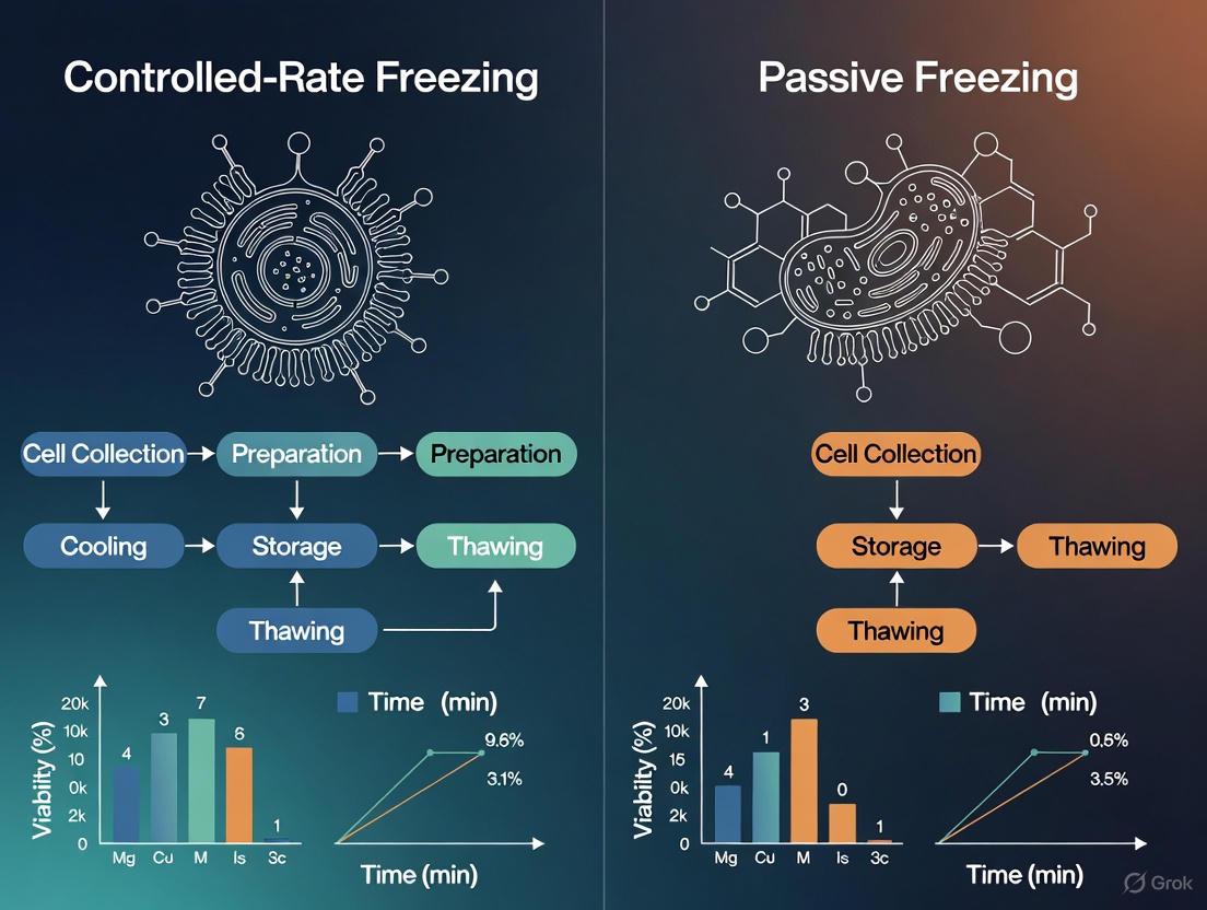
A 2025 retrospective study directly compared these freezing methodologies for hematopoietic progenitor cells (HPCs), analyzing 50 products from 29 donors [4] [5]. The investigation measured multiple parameters to assess cryopreservation outcomes, with results summarized in the table below.
Table 1: Experimental Comparison of Controlled-Rate vs. Passive Freezing for Hematopoietic Progenitor Cells
| Parameter Assessed | Controlled-Rate Freezing (CRF) | Passive Freezing (PF) | Statistical Significance (P-value) |
|---|---|---|---|
| Total Nucleated Cell (TNC) Viability | 74.2% ± 9.9% | 68.4% ± 9.4% | 0.038 |
| CD34+ Cell Viability | 77.1% ± 11.3% | 78.5% ± 8.0% | 0.664 |
| Days to Neutrophil Engraftment | 12.4 ± 5.0 | 15.0 ± 7.7 | 0.324 |
| Days to Platelet Engraftment | 21.5 ± 9.1 | 22.3 ± 22.8 | 0.915 |
| Time from Collection to Cryopreservation | 18.0 ± 6.2 hours | 22.6 ± 11.6 hours | 0.09 |
Despite the statistically significant difference in TNC viability favoring CRF, the clinical endpoints of neutrophil and platelet engraftment showed no significant difference between the two methods [4] [5]. The researchers concluded that while CRF demonstrated a slight advantage in TNC preservation, both methods produced comparable engraftment outcomes, establishing PF as an acceptable alternative to CRF for initial cryopreservation before long-term storage in liquid nitrogen [4].
Detailed Experimental Protocol
The study employed a standardized protocol for both freezing methods [5]:
- Cryoprotectant Preparation: A solution containing 15% DMSO and 9% albumin in Plasmalyte-A was prepared.
- Cell Processing: HPC products were concentrated or diluted to reach an optimal cell concentration of 600-800 × 10⁶ TNC/mL.
- Cryoprotectant Addition: The cryoprotectant solution was added to the HPC products in aliquots at 2-8°C.
- Freezing Process:
- CRF Group: Products transferred to a controlled-rate freezer following a standardized program.
- PF Group: Products placed in metal cassettes, wrapped in absorbent pads for insulation, and stored in a -80°C mechanical freezer.
- Storage: All products were transferred to vapor phase liquid nitrogen for long-term storage at temperatures below -135°C.
- Assessment: Post-thaw viability was measured via flow cytometry, and engraftment data were collected from patient records.
Supporting Diagrams and Workflows
Experimental Workflow for Freezing Method Comparison
The following diagram illustrates the experimental workflow used in the comparative study of controlled-rate versus passive freezing methods:
Cellular Stress Pathways in Cryopreservation
The cryopreservation process induces multiple stress pathways that can impact cell viability and function. The following diagram illustrates key mechanisms of cryoinjury and cellular stress responses:
The Scientist's Toolkit: Essential Research Reagents and Materials
Successful implementation of cryopreservation protocols requires specific reagents and equipment optimized for different cell types and applications. The table below details key solutions used in contemporary cell therapy research and development:
Table 2: Essential Research Reagents for Cell Therapy Cryopreservation
| Reagent/Material | Composition & Key Features | Primary Applications | Functional Rationale |
|---|---|---|---|
| CryoStor CS10 [2] [3] | Serum-free, animal component-free, contains 10% DMSO with sugars and macromolecules | T cells, MSCs, general cell therapy products | Provides defined, GMP-compliant formulation; reduces ice crystal formation and osmotic stress |
| mFreSR [2] | Serum-free, chemically defined, compatible with mTeSR media | Human ES and iPS cells | Maintains pluripotency and high viability post-thaw for sensitive stem cell types |
| Intracellular-like Media [3] | Mimics intracellular ionic balance, reduces ion gradient across membranes | Lymphocytes, NK cells, HPCs | Minimizes cold-induced membrane permeabilization and ionic stress during freezing |
| DMSO (10% v/v) [5] | Standard cryoprotectant in electrolyte solution with albumin | Hematopoietic progenitor cells | Penetrates cells, reduces intracellular ice formation, established safety profile |
| CELLBANKER Series [6] | 10% DMSO with glucose, polymers, and pH buffers | Mammalian cells, stem cells (serum-free options available) | Proprietary polymer formulation enhances membrane protection during freeze-thaw cycle |
Impact on Supply Chain Logistics and Clinical Applications
Supply Chain Integration and Stability Considerations
The integration of cryopreservation into cell therapy supply chains enables decentralized treatment models where products manufactured at centralized facilities can be shipped globally while maintaining viability [1] [3]. Specialized shippers that maintain temperatures of -130°C or below for extended periods provide the necessary infrastructure for reliable transport of frozen cellular products [3]. This capability is particularly crucial for autologous therapies, where patient-specific products must be manufactured, stored, and transported to align with patient-specific treatment timelines [1].
The post-thaw stability of cellular products represents a critical consideration in supply chain design. Studies evaluating human CD3 T cells have demonstrated that cryopreservation media formulation significantly impacts post-thaw stability windows, which in turn determines the allowable time between thaw and patient administration [3]. Intracellular-like media formulations such as CryoStor have shown advantages in maintaining cell functionality after thaw compared to traditional extracellular-like solutions, potentially extending the viable administration window [3].
Clinical Workflow Integration and Regulatory Considerations
In clinical practice, the choice between freezing methods impacts workflow efficiency and resource allocation. Passive freezing methods offer operational flexibility, as products can be processed without immediate transfer to long-term storage, potentially accommodating after-hours collections or reducing staffing requirements [5]. This practical advantage must be balanced against the slightly superior TNC viability demonstrated with controlled-rate freezing in some applications [4] [5].
Regulatory considerations increasingly favor defined cryopreservation media over traditional "home-brew" formulations containing serum or undefined components [2] [3]. The move toward serum-free, GMP-manufactured cryopreservation solutions supports better process control and reduces risks associated with lot-to-lot variability and potential adventitious agents [2]. Additionally, formulating products to eliminate post-thaw washing steps simplifies clinical administration and reduces processing at the bedside, contributing to more robust and reproducible treatment outcomes [3].
The comparison between controlled-rate and passive freezing methods reveals a nuanced landscape where clinical outcomes may be equivalent despite differences in specific viability metrics [4] [5]. For hematopoietic progenitor cells, both methods successfully support engraftment, providing flexibility in process design based on available resources and scale requirements [4]. The selection of appropriate cryopreservation media—particularly the movement toward defined, intracellular-like formulations—demonstrates growing sophistication in addressing fundamental cellular stress mechanisms during freezing and thawing [3].
As cell therapies continue to evolve toward commercial reality, cryopreservation will remain an indispensable component of the global supply chain, enabling centralized manufacturing models while ensuring product viability and potency during distribution [1] [3]. Future developments will likely focus on further optimization of cryoprotectant formulations, standardization of freezing protocols across different cell types, and enhanced understanding of the molecular mechanisms underlying cryopreservation-induced stress responses. Through continued refinement of these critical preservation technologies, the field can advance toward more reliable, accessible, and effective cellular therapies for patients worldwide.
In the field of cell and gene therapy, the cryopreservation of cell-based intermediates is a critical step, enabling flexibility in manufacturing, quality control testing, and transportation within the supply chain [7]. The process of cooling cells to cryogenic temperatures for storage is not a one-size-fits-all procedure; the rate at which cells are cooled profoundly influences their post-thaw viability, recovery, and functionality [8] [9]. The central thesis in modern cryopreservation strategy hinges on the choice between two fundamental approaches: controlled-rate freezing (CRF), which offers precise manipulation of cooling parameters, and passive freezing (PF), a simpler, uncontrolled method. For researchers and drug development professionals, understanding the scientific principles and data underlying this choice is essential for designing robust and reproducible therapy protocols. This guide objectively compares these methods by examining the core relationship between cooling kinetics and cell survival, supported by experimental data and detailed methodologies.
The fundamental challenge during freezing is the formation of ice. When an aqueous solution freezes, it undergoes phase separation, generating pure ice crystals and a concentrated liquid phase known as the freeze-concentrated solution (FCS) [10]. The morphology of this FCS, specifically the size and connectivity of its channels, is directly governed by the cooling rate. Slow cooling rates (e.g., -1°C/min) promote the formation of larger FCS channels, which can effectively accommodate cells and reduce mechanical stress [10]. Conversely, rapid cooling results in fine ice crystals and narrower FCS channels, increasing the risk of intracellular ice formation and mechanical damage to cell membranes [10] [11]. This physical phenomenon forms the basis for the observed impact of cooling rates on cell viability.
Fundamental Principles of Cell Damage During Freezing
The damage inflicted upon cells during freezing is primarily attributed to two interconnected mechanisms: intracellular ice formation and osmotic stress.
- Intracellular Ice Formation (IIF): During rapid cooling, water within the cell does not have sufficient time to exit and equilibrate with the extracellular environment. Consequently, it supercools and freezes internally, forming deadly ice crystals that can mechanically disrupt organelles and the plasma membrane [11] [9]. This is a primary reason for low viability following fast, uncontrolled freezing.
- Osmotic Stress: As extracellular ice forms, solutes are excluded from the crystal lattice, leading to a dramatic increase in the solute concentration in the remaining liquid FCS. This creates a hypertonic extracellular environment, driving water out of the cell and causing severe cell dehydration and shrinkage. This "solute effect" can damage cellular proteins and membranes [9]. Subsequent thawing can impose additional osmotic stress as water rushes back into the dehydrated cells too quickly.
The relationship between cooling rate and these damaging mechanisms is elegantly summarized by the "inverted U-shaped" survival curve observed for most cell types [8]. At excessively slow cooling rates, cells are exposed to prolonged hypertonic conditions, leading to "solution effects" injury. At excessively rapid cooling rates, lethal intracellular ice formation dominates. An optimal cooling rate exists that minimizes both types of damage, and this rate is cell-type specific [9].
The following diagram illustrates the logical relationship between cooling rate, physical changes in the cell, and the resulting viability outcomes.
Comparative Analysis: Controlled-Rate vs. Passive Freezing
The choice between controlled-rate and passive freezing represents a fundamental trade-off between process control and operational simplicity. The following table summarizes the core characteristics of each method.
Table 1: Method Comparison: Controlled-Rate Freezing vs. Passive Freezing
| Feature | Controlled-Rate Freezing (CRF) | Passive Freezing (PF) |
|---|---|---|
| Principle | Programmable, precise control of temperature decline over the entire cooling curve [12]. | Uncontrolled cooling by placing vials in a pre-cooled mechanical freezer (e.g., -80°C) [4]. |
| Key Process Control | Control over critical parameters: cooling rate before/after nucleation, nucleation temperature itself, and final temperature [7]. | No active control over any cooling parameters; rate is determined by freezer and vial characteristics. |
| Cooling Rate | Typically optimized at -1°C/min for many mammalian cells [13] [12]. | Variable and non-reproducible; often averages -1°C/min but with unpredictable fluctuations [12]. |
| Typical Cell Viability/Recovery | Higher and more consistent. One study showed 65% viability for C2C12 myoblasts at -1°C/min vs. 54% at -30°C/min [10]. | Can be sufficient for robust cells, but generally lower and more variable than CRF [10]. |
| Consistency & Reproducibility | High; process is automated, documented, and repeatable, which is critical for cGMP manufacturing [7] [12]. | Lower; process is manual and subject to variability in freezer performance and vial location. |
| Cost & Infrastructure | High-cost equipment and consumables (e.g., liquid nitrogen); requires specialized expertise [7]. | Low-cost; relies on standard laboratory -80°C freezers, with a low technical barrier to adoption [7]. |
| Best Suited For | Late-stage clinical trials and commercial products; sensitive and therapeutically relevant cells (e.g., T-cells, iPSCs, MSCs) [7]. | Early-stage R&D and clinical development; robust cell types where high consistency is less critical [7]. |
The impact of these methods is reflected directly in experimental outcomes. A 2025 morphological study on frozen dimethyl sulfoxide (DMSO) solutions provided a clear visual and quantitative explanation for the superiority of slow, controlled cooling. The research demonstrated that at a slow cooling rate of 1°C/min, large and interconnected FCS channels formed, providing ample space to accommodate cells safely. In contrast, rapid cooling resulted in fine ice crystals and narrow FCS channels, increasing the mechanical confinement and stress on cells [10]. This morphological difference directly correlated with cell recovery, where slow cooling (1°C/min) yielded a 65% viability for C2C12 myoblasts, compared to only 54% at a fast cooling rate of 30°C/min [10].
Furthermore, a 2025 retrospective clinical study on Hematopoietic Progenitor Cells (HPCs) provides a critical, direct comparison. While Total Nucleated Cell (TNC) viability post-thaw was significantly higher in the CRF group, the more clinically relevant measure of CD34+ cell viability showed no significant difference between CRF and PF. Most importantly, the engraftment outcomes—the ultimate measure of cell function—for both neutrophils and platelets were statistically similar between the two groups [4]. This indicates that for certain cell types and clinical applications, passive freezing can be an acceptable alternative, though CRF may still offer advantages in process control and consistency for regulatory purposes.
Table 2: Quantitative Data from Key Comparative Studies
| Cell Type | Freezing Method / Cooling Rate | Key Metric | Result | Source |
|---|---|---|---|---|
| C2C12 Mouse Myoblasts | 1°C/min (Slow) | Cell Viability | 65% | [10] |
| 10°C/min (Medium) | Cell Viability | 59% | [10] | |
| 30°C/min (Fast) | Cell Viability | 54% | [10] | |
| Hematopoietic Progenitor Cells (HPCs) | Controlled-Rate | TNC Viability | 74.2% ± 9.9% | [4] |
| Passive Freezing | TNC Viability | 68.4% ± 9.4% | [4] | |
| Controlled-Rate | CD34+ Viability | 77.1% ± 11.3% | [4] | |
| Passive Freezing | CD34+ Viability | 78.5% ± 8.0% | [4] | |
| HPCs (Engraftment) | Controlled-Rate | Days to Neutrophil Engraftment | 12.4 ± 5.0 | [4] |
| Passive Freezing | Days to Neutrophil Engraftment | 15.0 ± 7.7 | [4] |
Detailed Experimental Protocols
To enable replication and critical evaluation, this section outlines the core methodologies used in the key studies cited.
Protocol 1: Microscopic Analysis of Freeze-Concentrated Solution (FCS) Morphology
This protocol is used to visually correlate cooling rate with the physical structure of the frozen medium [10].
- Objective: To investigate the effects of cooling rates and initial DMSO concentrations on the morphological features of the FCS.
- Materials:
- Upright fluorescent microscope with a cooling stage.
- CMOS camera.
- Slide glasses.
- Aqueous DMSO solutions (e.g., 5, 10, 20 wt%).
- Sodium fluorescein (fluorescent dye).
- Methodology:
- A 10 μL aliquot of a sodium fluorescein solution in DMSO is sandwiched between two slide glasses.
- The sample is placed on the temperature-controlled cooling stage.
- The solution is cooled at a defined rate (e.g., 1°C/min, 10°C/min, 30°C/min) to a terminal temperature (e.g., -60°C).
- Morphological observations of the FCS channels are made using fluorescence microscopy.
- The width of the FCS channels and ice particle size are quantitatively analyzed using image analysis software (e.g., ImageJ).
- Key Measurements: FCS channel width, ice crystal size, and qualitative channel morphology.
Protocol 2: Evaluating Cell Viability Post-Cryopreservation
This is a standard protocol for determining the success of a cryopreservation cycle, applicable to both controlled-rate and passive freezing studies [10] [13].
- Objective: To determine the viability and recovery rate of cells after a freeze-thaw cycle.
- Materials:
- Log-phase cultured cells.
- Cryoprotective agent (e.g., DMSO).
- Complete growth medium.
- Cryogenic vials.
- Controlled-rate freezer or -80°C mechanical freezer (for PF).
- Liquid nitrogen storage dewar.
- Trypan blue stain.
- Hemocytometer or automated cell counter.
- Methodology:
- Cells are harvested and resuspended in freezing medium (e.g., culture medium with 10% DMSO) at a high concentration (e.g., 1x10^7 cells/mL) [13].
- The cell suspension is aliquoted into cryovials.
- Freezing: Vials are frozen either using a CRF (at a defined rate like -1°C/min) or placed in a -80°C freezer (Passive Freezing).
- Frozen vials are transferred to long-term storage in liquid nitrogen.
- Thawing: For analysis, vials are rapidly thawed in a 37°C water bath.
- Cells are diluted in pre-warmed growth medium.
- A sample is mixed with trypan blue, and viable (unstained) and dead (blue) cells are counted.
- Key Measurements: Percent viability (Viable Cell Count / Total Cell Count × 100), and total cell recovery.
The workflow for a comprehensive cryopreservation study, from cell preparation to data analysis, is summarized below.
The Scientist's Toolkit: Essential Research Reagents and Materials
A successful cryopreservation experiment relies on a suite of critical reagents and equipment. The following table details key solutions and materials, their functions, and relevant examples from the search results.
Table 3: Essential Reagents and Materials for Cryopreservation Research
| Item | Function & Role in Cryopreservation | Examples & Notes |
|---|---|---|
| Cryoprotective Agents (CPAs) | Protect cells from freezing damage by depressing the freezing point, reducing ice crystal formation, and promoting vitrification [9]. | DMSO (10%): Permeating agent, industry standard [13] [9].Glycerol: Permeating agent, often used for spermatozoa [9].Trehalose: Non-permeating disaccharide, stabilizes membranes [9]. |
| Cryopreservation Media | Formulated solutions containing CPAs, a base medium, and a protein source to protect cells during freeze-thaw stress. | Pre-formulated Media: e.g., Gibco Synth-a-Freeze (protein-free) or Recovery Cell Culture Freezing Medium [13].In-house Formulation: e.g., 50% cell-conditioned medium + 50% fresh medium with 10% DMSO [13]. |
| Programmable Controlled-Rate Freezer | Equipment that provides precise, reproducible control over the cooling rate, often with multiple program segments. Essential for process standardization [7] [12]. | e.g., Thermo Scientific CryoMed CRF [12]. |
| Passive Freezing Container | An insulated container (often filled with isopropanol) that creates an approximate -1°C/min cooling rate when placed in a -80°C freezer. A low-cost alternative to CRF. | e.g., "Mr. Frosty" from Thermo Scientific Nalgene [13]. |
| Liquid Nitrogen Storage | Provides long-term storage at temperatures below -130°C (typically in vapor phase) to ensure ultimate cell stability and viability over years [12]. | Vapor phase storage is recommended to reduce contamination and explosion risks associated with liquid phase storage [13] [12]. |
| Viability Assay Kits | Used to quantify the percentage of live cells after thawing. | Trypan Blue Exclusion: Standard dye exclusion method [10] [13].Cell Counting Kit-8 (CCK-8): Metabolic assay for viability [10]. |
The principle that cooling rate is a critical process parameter determining cell viability and function is unequivocally supported by scientific evidence. Controlled-rate freezing, typically at a standard rate of -1°C/min, offers superior control, consistency, and often higher post-thaw recovery for sensitive cell types, making it the preferred method for late-stage clinical and commercial cell therapy products [10] [7]. Its documented performance and alignment with cGMP requirements underpin its status as the gold standard.
However, the data also demonstrates that passive freezing is a valid and practical alternative, particularly in early-stage research and for certain cell types. Evidence showing equivalent CD34+ cell viability and engraftment outcomes between PF and CRF challenges the notion that CRF is universally indispensable [4]. The choice between these methods ultimately involves a strategic balance between the need for process control and consistency (favoring CRF) and considerations of cost, simplicity, and scalability (where PF may be adequate). For researchers in cell therapy, the decision must be informed by the specific cell type's sensitivity, the stage of product development, and the critical quality attributes that define a successful therapy.
In the field of cell and gene therapy (CGT), cryopreservation is a critical step for ensuring the stability and availability of cellular starting materials, intermediates, and final products. The process of freezing these biological materials is not a one-size-fits-all procedure; the choice of method can significantly impact cell viability, functionality, and ultimately, the success of therapeutic applications. Two primary methods dominate current practice: controlled-rate freezing and passive freezing. Understanding their mechanisms, advantages, and limitations is essential for researchers and drug development professionals aiming to optimize their manufacturing processes. This guide provides an objective comparison of these two methods, supported by experimental data and detailed protocols, to inform decision-making within cell therapy research and development.
Core Principles of Cryopreservation
Before comparing methods, it is vital to understand the common goal of cryopreservation: to transition aqueous solutions within cells to a solid state with minimal damage. Suboptimal freezing can lead to batch-to-batch variation, lowered cellular functionality, and reduced cell yield [14]. The main damaging mechanisms are:
- Intracellular Ice Crystallization (IIF): The formation of ice crystals inside the cell, which can puncture organelles and the cell membrane.
- Solution Effects: As water freezes, dissolved solutes become concentrated in the remaining liquid, creating a hypertonic environment that can cause osmotic stress and dehydration [15].
Cryoprotective agents (CPAs), like dimethyl sulfoxide (DMSO), are used to mitigate these effects. They work by reducing ice crystal formation and protecting cells from osmotic damage during the freeze-thaw cycle [14] [15].
Controlled-Rate Freezing Explained
Controlled-rate freezing (CRF) is an active process where a dedicated instrument precisely lowers the temperature of a biological sample according to a predefined, programmable profile [16]. This method allows users to define and control critical process parameters, making it a standard in current Good Manufacturing Practice (cGMP) environments [7].
Mechanism and Workflow
The typical CRF protocol involves several key stages designed to manage the release of the latent heat of fusion—the heat released when water changes from a liquid to a solid state [5] [12].
A critical, often optional, step is manual ice nucleation or "seeding." This involves briefly supercooling a small section of the sample container to induce ice formation at a specific, relatively high temperature (e.g., -5°C). This controlled initiation prevents the sample from supercooling excessively, which can lead to uncontrolled, rapid ice crystal formation and propagation later [16] [17].
Key Equipment
The primary piece of equipment is a programmable controlled-rate freezer. These devices use liquid nitrogen (LN2) or are LN2-free and regulate the sample's cooling via sophisticated controllers. They provide a thermal profile of the entire process, which is part of the manufacturing controls and documentation [7] [16] [12].
Passive Freezing Explained
Passive freezing (PF), also known as uncontrolled-rate freezing, is a simpler method where samples are placed in an insulated container and stored in a ultra-low temperature mechanical freezer, typically at -80°C [4] [5]. The cooling rate is not directly controlled by a programmable device but is determined by the insulation properties of the container and the environment of the mechanical freezer [15].
Mechanism and Workflow
The PF workflow is less complex and does not involve active temperature monitoring or programming for individual samples.
Key Equipment
The setup for passive freezing is low-cost and includes:
- A -80°C mechanical freezer.
- A passive cooling device (e.g., "Mr. Frosty," "CellCool," or insulated containers), which uses a specific volume of isopropanol or other coolant to achieve an approximate cooling rate of -1°C per minute [16] [15].
Head-to-Head Comparison: Performance and Experimental Data
The theoretical differences between CRF and PF lead to a critical, practical question: How do they compare in preserving cell viability and function? Recent clinical studies provide direct, quantitative comparisons.
Comparative Analysis Table
The table below summarizes the key characteristics of both methods based on industry surveys and scientific literature [7] [5].
| Feature | Controlled-Rate Freezing (CRF) | Passive Freezing (PF) |
|---|---|---|
| Control over Process | Full control over critical parameters (e.g., cooling rate) [7] | Uncontrolled cooling rate; relies on container insulation [5] |
| Infrastructure & Cost | High-cost, high-consumable infrastructure; requires LN2 or specialized equipment [7] | Low-cost, low-consumable infrastructure [7] |
| Technical Expertise | Specialized expertise required for use and optimization [7] | Low technical barrier to adoption [7] |
| Scalability | Can be a bottleneck for batch scale-up [7] | Simple, one-step operation; ease of scaling [7] |
| Documentation & GMP | Provides automated solutions for documentation and process monitoring [7] | Lacks detailed process data for documentation [5] |
| Industry Adoption | High prevalence, especially for late-stage and commercial products [7] | Used primarily in early stages of clinical development (up to phase II) [7] |
Experimental Data: Hematopoietic Progenitor Cell (HPC) Engraftment
A 2025 retrospective study of 50 HPC products directly compared CRF and PF outcomes, measuring total nucleated cell (TNC) viability, CD34+ cell viability, and most importantly, engraftment in patients [4] [5].
Experimental Protocol
- Cell Type: Apheresis-derived and marrow-derived HPCs.
- Cryoprotectant: 15% DMSO, 9% albumin in Plasmalyte-A.
- Freezing Methods:
- CRF Group (n=25): Using a controlled-rate freezer.
- PF Group (n=25): Using a -80°C mechanical freezer with samples wrapped for insulation.
- Analysis: Post-thaw viability was assessed, and patient engraftment (days to neutrophil and platelet recovery) was tracked after transplantation [5].
Results and Data Comparison
The following table summarizes the key quantitative findings from the study, demonstrating comparable clinical outcomes despite minor differences in viability [4] [5].
| Metric | Controlled-Rate Freezing (CRF) | Passive Freezing (PF) | P-value |
|---|---|---|---|
| TNC Viability (Post-thaw) | 74.2% ± 9.9% | 68.4% ± 9.4% | 0.038 |
| CD34+ Cell Viability (Post-thaw) | 77.1% ± 11.3% | 78.5% ± 8.0% | 0.664 |
| Days to Neutrophil Engraftment | 12.4 ± 5.0 | 15.0 ± 7.7 | 0.324 |
| Days to Platelet Engraftment | 21.5 ± 9.1 | 22.3 ± 22.8 | 0.915 |
The study concluded that while TNC viability was statistically higher in the CRF group, there was no significant difference in the critical metrics of CD34+ cell viability or engraftment times. This led the authors to state that "cryopreservation outcomes using CRF or PF are comparable so PF is an acceptable alternative to CRF" for these cell types [4] [5].
The Scientist's Toolkit: Essential Research Reagents and Materials
Successful cryopreservation, regardless of the method, relies on a set of key materials and reagents.
| Item | Function | Key Considerations |
|---|---|---|
| Cryoprotective Agent (CPA) | Protects cells from ice crystal damage and osmotic stress [14]. | DMSO is most common. Concentration and exposure time must be optimized to minimize toxicity [14] [17]. |
| Cryocontainers | Vessels for freezing and storing cells. | Choices include cryovials, cryobags, and straws. Must be sterile and suitable for ultra-low temperatures to prevent breakage and ensure closure integrity [14] [16]. |
| Controlled-Rate Freezer | Actively controls sample cooling rate. | Programmable devices (LN2 or LN2-free) for precise, reproducible freezing. Critical for cGMP manufacturing [7] [16]. |
| Passive Cooling Device | Provides insulation for samples in a -80°C freezer. | Devices like "Mr. Frosty" use coolant to achieve an approximate -1°C/min cooling rate. A low-cost alternative to CRF [16] [15]. |
| Long-Term Storage System | Preserves frozen samples. | Liquid nitrogen (vapor phase, <-150°C) or ultra-low mechanical freezers (<-130°C) are required for long-term stability [14] [12]. |
Both controlled-rate and passive freezing are viable methods for the cryopreservation of cell therapy products, each with distinct profiles. Controlled-rate freezing offers precision, control, and extensive documentation support, making it the preferred choice for late-stage clinical and commercial applications where process robustness is paramount. In contrast, passive freezing is a simple, cost-effective, and scalable alternative that has been proven to produce clinically equivalent engraftment results for specific cell types like HPCs.
The choice between methods is not a matter of which is universally superior, but which is most appropriate for a given context. Researchers must consider the cell type, stage of clinical development, regulatory requirements, and available resources. For early-stage research or with robust cell types, passive freezing presents a compelling option. As programs advance toward commercialization, the controlled environment and detailed data provided by controlled-rate freezing may become necessary to meet regulatory standards and ensure consistent product quality.
Key Physical and Biochemical Stresses During Freezing and Thawing
For researchers and scientists in cell therapy, the choice between Controlled-Rate Freezing (CRF) and Passive Freezing (PF) is critical. The following table summarizes key comparative data from recent studies, particularly for hematopoietic progenitor cells (HPCs), which are central to many therapeutic applications.
| Parameter | Controlled-Rate Freezing (CRF) | Passive Freezing (PF) | Research Implications |
|---|---|---|---|
| Post-Thaw TNC Viability | 74.2% ± 9.9% [4] [5] | 68.4% ± 9.4% [4] [5] | CRF shows a statistically significant advantage for total nucleated cell (TNC) recovery. |
| Post-Thaw CD34+ Viability | 77.1% ± 11.3% [4] [5] | 78.5% ± 8.0% [4] [5] | No significant difference (p=0.664); both methods effectively preserve critical progenitor cells [4] [5]. |
| Neutrophil Engraftment (Days) | 12.4 ± 5.0 [4] [5] | 15.0 ± 7.7 [4] [5] | No statistically significant difference in a clinical setting (p=0.324) [4] [5]. |
| Platelet Engraftment (Days) | 21.5 ± 9.1 [4] [5] | 22.3 ± 22.8 [4] [5] | No statistically significant difference (p=0.915) [4] [5]. |
| Process Control | High; fully programmable cooling profile [5]. | Low; uncontrolled nucleation and variable cooling rates [5]. | CRF ensures protocol standardization; PF requires validation for consistency. |
| Cost & Complexity | High capital cost and more time-consuming [5]. | Low cost, simple, and convenient [5]. | PF is a cost-effective alternative, especially for high-volume or backup freezing. |
Cryopreservation is an enabling technology for the cell therapy supply chain, allowing for coordination between cell processing and patient care [18] [19]. The process aims to stabilize cells by reducing molecular mobility and halting degradative enzymatic activity [19]. However, traversing the temperature range from +37°C to -196°C and back subjects cells to a series of severe physical and biochemical stresses. The post-thaw recovery and function of a cell product are the cumulative result of every step in the process, from pre-freeze handling to post-thaw assessment [19]. Understanding these stresses is paramount for developing robust protocols for cell therapy intermediates. This guide delves into these key stresses, framing them within the practical comparison of CRF and PF methodologies.
Detailed Analysis of Physical Stresses
Intracellular Ice Formation: The Mechanical Threat
Mechanism: As the sample is cooled below its freezing point, extracellular water freezes first. This increases the solute concentration in the unfrozen extracellular solution, creating an osmotic gradient that draws water out of the cell. If the cooling rate is too rapid, water does not have sufficient time to exit the cell, becoming supercooled and eventually forming lethal intracellular ice crystals [20]. These crystals can mechanically disrupt organelles and the plasma membrane, leading to immediate cell lysis [20].
Experimental Data on Cooling Rates: The optimal cooling rate is cell-type specific, balancing dehydration and intracellular ice formation [20]. For human iPSCs, which are highly vulnerable, rates between -0.3°C/min and -1.8°C/min are often optimal [20]. A common standard for many cell types is -1°C/min [18] [20]. CRF precisely maintains this preset rate, while PF relies on insulation in a -80°C freezer to approximate a slow cool, which can be less consistent [5].
Cellular Dehydration: The Osmotic and Volume Stress
Mechanism: If the cooling rate is too slow, the prolonged exposure to a hypertonic extracellular environment causes excessive cellular dehydration [20]. This leads to harmful volumetric contraction, increased intracellular solute concentration, and potential damage to the plasma membrane and cytoskeleton [19]. The membrane itself is susceptible to damage from osmotic stress during both the addition and removal of cryoprotectants [18] [19].
Solution Effects & Cold Shock: Beyond ice formation, cold stress itself can damage the plasma membrane. In plants, low temperatures reduce membrane fluidity, while high temperatures increase fluidity and cause lipid peroxidation [21]. A 2024 study found that cold stress can induce ferroptosis in adherent cancer cells, a type of iron-dependent cell death characterized by lethal lipid peroxide accumulation [22].
Mechanical Stresses from the Extracellular Environment
Mechanism: The expansion of water during ice formation and the physical presence of growing ice crystals can crush or compress cells trapped in the extracellular matrix. Furthermore, research on frozen sucrose solutions has revealed a counter-intuitive mechanical stressor: when held at temperatures between two critical glass transitions (Tg" and Tg', around -45°C), microstrain within the ice crystal lattice increases, while crystalline domain size decreases [23]. This suggests that specific temperature zones during freezing can impose significant mechanical stress on suspended biologics.
Biochemical and Molecular Stresses
Cryoprotectant Toxicity and Osmotic Shock
Mechanism: Dimethyl sulfoxide (DMSO) is the most common cryoprotective agent (CPA), but it is a biochemical stressor. DMSO is toxic to cells, and its effects are time- and concentration-dependent [18] [19]. It can alter cytoskeleton organization, shift cell metabolism, and change membrane fluidity [19]. The introduction and post-thaw removal of hypertonic CPA solutions also cause major osmotic stress, leading to damaging cell swelling or shrinkage [18] [20] [19].
Experimental Protocol for Mitigation: To minimize combined biochemical and osmotic stress, standardized protocols are essential.
- Introduction of CPA: The cryopreservation solution (e.g., 5-10% DMSO in plasma or albumin solution) is typically added dropwise to the cell suspension to allow for gradual osmotic equilibration [18].
- Post-Thaw Removal: A common method is to dilute the thawed cell suspension dropwise into a large volume of pre-warmed isotonic culture medium. This slowly reduces the DMSO concentration outside the cell, preventing a rapid influx of water and catastrophic swelling. The cells are then centrifuged to remove the DMSO-containing supernatant before resuspension in fresh medium for infusion or culture [18] [24]. The use of intracellular-like cryopreservation media (e.g., CryoStor, Unisol) is designed to buffer these stresses and improve recovery [25].
Delayed-Onset Cell Death: The Apoptotic Pathway
Mechanism: A critical discovery in cryobiology is that a significant portion of cell death occurs hours to days after thawing, a phenomenon termed Cryopreservation-Induced Delayed-Onset Cell Death (CIDOCD) [25]. Cells that appear viable immediately post-thaw can activate programmed cell death pathways, primarily apoptosis, due to stresses experienced during the freeze-thaw cycle [25]. Other stress pathways, including oxidative stress and the unfolded protein response, are also activated [25].
Supporting Experimental Data: A 2022 study on human hematopoietic progenitor cells (hHPCs) demonstrated that modulating stress response pathways during the post-thaw recovery phase can significantly improve survival. Specifically, the use of oxidative stress inhibitors in the recovery medium increased overall viability by an average of 20% [25]. This highlights that cell survival is not solely determined by the freezing process itself, but also by the biochemical environment during the first 24 hours of recovery.
The following diagram illustrates the key stress pathways activated during the freeze-thaw cycle and their interactions.
Experimental Protocols for Stress Analysis
Protocol: Comparing Post-Thaw Viability and Function
This protocol is adapted from retrospective studies comparing CRF and PF for HPCs [4] [5].
- Cell Preparation: Obtain HPCs via apheresis or from bone marrow. Concentrate or dilute the product to a target concentration of 600–800 x 10^6 total nucleated cells (TNC)/mL.
- Cryoprotectant Addition: Prepare a cryoprotectant solution of 15% DMSO and 9% albumin in Plasmalyte-A. Mix the cryoprotectant with the HPC product in a 1:1 ratio, achieving a final DMSO concentration of approximately 7.5%. Perform this step at controlled, cool temperatures.
- Freezing Process:
- CRF Group: Transfer the product to a controlled-rate freezer. Cool at a rate of -1°C/min until the release of the latent heat of fusion is complete. Resume cooling at -1°C/min until the product reaches a target of ≤-60°C before transferring to long-term storage in the vapor phase of liquid nitrogen.
- PF Group: Place the product in a -80°C mechanical freezer using an insulated container (e.g., a styrofoam box) to approximate a slow cooling rate. Store for 18-24 hours before transferring to long-term liquid nitrogen storage.
- Thawing and Assessment: Rapidly thaw the products in a 37°C water bath. Perform the following assessments:
- Viability: Measure Total Nucleated Cell (TNC) viability and CD34+ cell viability using flow cytometry with 7-AAD staining.
- Functionality (Engraftment): Transplant the thawed HPCs into suitable models (e.g., immunodeficient mice) and monitor the number of days to neutrophil and platelet engraftment.
Protocol: Assessing Delayed-Onset Cell Death Pathways
This protocol is based on studies investigating molecular stress post-thaw [25].
- Cell Culture and Cryopreservation: Culture human hematopoietic progenitor cells (hHPCs). Cryopreserve cells using both traditional extracellular-type media (e.g., culture medium with 10% DMSO) and intracellular-type media (e.g., Unisol).
- Post-Thaw Recovery with Modulators: Upon thawing, resuspend the cells in recovery medium supplemented with specific stress-pathway inhibitors. Key experimental groups include:
- Group 1: Apoptotic caspase inhibitor (e.g., Z-VAD-FMK).
- Group 2: Oxidative stress inhibitor.
- Group 3: Unfolded protein response modulator.
- Control Group: Recovery medium without additional inhibitors.
- Incubation and Analysis: Incubate the cells for 24 hours under standard culture conditions (37°C, 5% CO2). After incubation, measure overall cell survival and viability using a flow cytometry-based assay (e.g., Annexin V/7-AAD) to quantify live, early apoptotic, and late apoptotic/necrotic populations.
The workflow for this molecular analysis is detailed below.
The Scientist's Toolkit: Key Research Reagents and Materials
The following table lists essential materials used in the cited experiments for studying freeze-thaw stresses.
| Reagent / Material | Function in Research | Example from Literature |
|---|---|---|
| Controlled-Rate Freezer (CRF) | Provides precise, programmable control of cooling rate to optimize ice formation dynamics. | Used to maintain a standard -1°C/min cooling rate for HPCs and iPSCs [18] [20] [5]. |
| Passive Freezing Container | Provides an uncontrolled, passive cooling rate in a -80°C freezer as a cost-effective alternative. | Insulated containers (e.g., styrofoam) used for freezing HPCs where CRF was not available [4] [5]. |
| DMSO-based Cryomedium | Serves as the conventional extracellular-type cryopreservation solution. | 5-10% DMSO in plasma, serum, or human serum albumin used for T cells, DCs, and NK cells [18]. |
| Intracellular-type Cryomedium | Multi-component, biochemically defined medium designed to buffer molecular stress. | CryoStor CS10 or Unisol; shown to improve recovery and reduce CIDOCD in HPCs and other cells [18] [25]. |
| Oxidative Stress Inhibitors | Tool compound to probe and mitigate post-thaw oxidative stress pathways. | Use in post-thaw recovery medium increased HPC viability by an average of 20% [25]. |
| Annexin V / 7-AAD Assay | Flow cytometry-based kit to distinguish live, early apoptotic, and dead cells. | Critical for quantifying not just immediate viability but also delayed-onset apoptosis (CIDOCD) [25]. |
| Synchrotron X-ray Diffraction | Analyzes microstrain and crystalline domain size in ice to probe physical stresses. | Used to detect increasing mechanical stress in frozen sucrose solutions held at -45°C [23]. |
The Importance of the Glass Transition Temperature in Long-Term Storage
For researchers and drug development professionals working with cell therapy intermediates, ensuring the long-term stability of biological materials is a fundamental challenge. At the heart of this challenge lies a critical material property: the glass transition temperature (Tg). This is the temperature at which an amorphous material transitions from a brittle, glassy state to a more viscous, rubbery state. For cell therapies, this often refers to the complex mixture of water, salts, cryoprotective agents (CPAs), and cellular components in the preserved solution. Storing a product below its Tg effectively halts molecular mobility and biochemical reactions, locking the material in a state of suspended animation that is essential for viable long-term storage. This guide objectively compares the performance of controlled-rate freezing and passive freezing within the crucial context of the glass transition temperature, providing the experimental data and protocols necessary for informed process development.
Tg and Storage Stability: The Fundamental Relationship
The physical stability of amorphous materials, including the vitrified solutions used in cell therapy cryopreservation, is intrinsically linked to their Tg. When stored at temperatures below their Tg, materials exist in a glassy state where molecular mobility is vastly reduced. This kinetic stabilization is the key to long-term stability.
A comprehensive study on the physical stability of amorphous drugs provides compelling experimental evidence for this principle. The research found that when 52 different amorphous drug compounds were stored at temperatures 20°C below their individual Tg, 100% of them (all 52 compounds) maintained their amorphous structure over a 12-hour storage period. In stark contrast, when stored at temperatures 20°C above their Tg, the majority of a specific class of compounds (14 out of 18 Class II compounds) crystallized. This study conclusively demonstrates that storage below Tg is a reliable predictor of physical stability, preventing crystallization and degradation [26].
For cell therapies, the implication is clear: the efficacy of a cryopreservation protocol is fundamentally dependent on achieving and maintaining a storage temperature below the Tg of the system. Failure to do so risks increased molecular mobility, leading to ice crystal formation, cell membrane damage, and loss of viability and functionality.
Comparative Analysis: Controlled-Rate vs. Passive Freezing
The method used to achieve and traverse the glass transition is critical. The table below summarizes a performance comparison between controlled-rate freezing and passive freezing, based on current industry practices and research.
Table 1: Performance Comparison of Freezing Methods for Cell Therapy Intermediates
| Performance Characteristic | Controlled-Rate Freezing | Passive Freezing (e.g., in a -80°C freezer) |
|---|---|---|
| Freezing Rate Control | Precise, programmable control (e.g., -1°C/min) [27] | Uncontrolled, variable, and dependent on equipment and volume |
| Likelihood of Achieving Uniform Vitrification | High | Low to Moderate |
| Post-Thaw Viability (General) | Typically higher and more consistent [28] | Often variable and generally lower |
| Process Standardization | High; easily validated and scaled | Low; difficult to control and reproduce |
| Typical CPA Requirement | Often enables the use of lower CPA concentrations [28] | Often requires higher CPA concentrations for equal protection |
| Capital Cost | High | Low |
| Operational Complexity | High | Low |
The primary advantage of controlled-rate freezing is its ability to dictate the thermodynamic pathway through the phase transition. By slowly lowering the temperature at a rate of approximately -1°C per minute—a standard in many protocols—this method allows for controlled dehydration of cells, minimizing intracellular ice formation which is lethal to cells [27]. Passive freezing in a -80°C freezer, while simple and inexpensive, results in an unpredictable freezing rate. This can lead to a heterogeneously frozen product where different vials, or even different parts of the same vial, experience different thermal histories, compromising the consistency of the final glass and leading to variable post-thaw outcomes.
Experimental Protocols for Evaluating Cryopreservation Outcomes
To objectively compare freezing methods or optimize a protocol, researchers must employ standardized experimental evaluations. Below are detailed methodologies for key assays.
Protocol 1: Measuring Post-Thaw Cell Viability and Recovery
This is a fundamental first-pass assessment for any cryopreservation experiment.
- Method: Cells are cryopreserved using the test method (e.g., controlled-rate vs. passive freezing). Post-thaw, cells are stained with a viability dye like Trypan Blue and analyzed using an automated cell counter or by flow cytometry with dyes like propidium iodide (PI) and fluorescein diacetate (FDA).
- Data Analysis: Cell recovery (%) is calculated as (Total viable cells post-thaw / Total viable cells pre-freeze) x 100. Viability (%) is calculated as (Viable cells / Total cells) x 100. A comparison of the test methods should report both metrics [28].
Protocol 2: Assessing Cellular Functionality
Viability alone is insufficient; cells must also retain their therapeutic function.
- Method: The specific functional assay is dependent on the cell type. For mesenchymal stromal cells (MSCs), this may involve an in vitro differentiation assay (osteogenic, adipogenic, chondrogenic). For immune cells like T-cells or NK cells, a cytokine release assay or target cell killing assay (e.g., using flow cytometry) would be appropriate [28].
- Data Analysis: Results from the test group (e.g., passive freezing) are compared quantitatively to the control group (e.g., controlled-rate freezing) to determine if functionality is compromised.
Protocol 3: Stability Study at Different Storage Temperatures
This protocol directly tests the principle of storage below Tg.
- Method: Aliquots of the cryopreserved cell product are stored at different temperatures, for example, in the vapor phase of liquid nitrogen (< -130°C), a -80°C mechanical freezer, and a -20°C freezer. Samples are retrieved at predetermined time points (e.g., 1 month, 3 months, 12 months) and analyzed for viability and functionality as described above [26].
- Data Analysis: The stability of the product is plotted over time for each storage temperature. A significant drop in viability or functionality at higher storage temperatures (e.g., -80°C vs. -150°C) suggests that the storage temperature may be at or above the effective Tg of the system, leading to gradual degradation.
The Scientist's Toolkit: Essential Research Reagents and Materials
The following table details key materials and reagents essential for conducting cryopreservation research in cell therapies.
Table 2: Essential Research Reagents and Materials for Cryopreservation Studies
| Item | Function/Description |
|---|---|
| Dimethyl Sulfoxide (Me2SO) | The most common cryoprotective agent (CPA). It penetrates cells and reduces ice crystal formation but exhibits cytotoxicity above 0°C [27]. |
| Serum-Free Cryopreservation Media | Chemically defined media formulated to support cell stability during freezing, often containing lower, safer levels of Me2SO or alternative CPAs. |
| Programmable Controlled-Rate Freezer | Equipment that ensures a consistent, reproducible, and optimal freezing profile (e.g., -1°C/min) to maximize cell viability [27]. |
| Liquid Nitrogen Storage Tank | Provides long-term storage at <-130°C (vapor phase) or <-196°C (liquid phase), ensuring the material is stored well below the Tg of most cryopreservation formulations [27]. |
| Automated Cell Counter or Flow Cytometer | Essential equipment for the quantitative assessment of post-thaw cell recovery and viability. |
| Specialized Cryovials | Sterile, leak-proof containers designed for ultra-low temperatures. Advanced systems like the Limbo device feature dual compartments to separate cells/CPA from diluent, allowing for automated washing and Me2SO reduction post-thaw [28]. |
Decision Workflow and Implementation Strategy
The following diagram illustrates the logical decision-making process for selecting and optimizing a long-term storage strategy based on the principles of Tg.
For researchers implementing a new protocol, the following steps are recommended:
- Characterize the System: Use techniques like Differential Scanning Calorimetry (DSC) to estimate the Tg of the chosen cryopreservation formulation [29]. Be aware that traditional DSC can sometimes be misleading for complex biological systems, and emerging techniques like sealed-cavity rheology may offer higher sensitivity [30].
- Validate the Freezing Profile: Use a controlled-rate freezer to apply a standard freezing profile (e.g., -1°C/min) and compare post-thaw outcomes against passive freezing for your specific cell type.
- Confirm Storage Safety: Ensure that your long-term storage solution (e.g., liquid nitrogen vapor phase) maintains a temperature securely below the characterized Tg of your product.
- Prioritize Patient Safety: For therapies with novel administration routes (e.g., intracerebral, epicardial), actively investigate Me2SO-free cryopreservation media to eliminate the cytotoxicity risks associated with Me2SO and the need for risky post-thaw washing steps [27].
The glass transition temperature is not merely a theoretical material property but a practical cornerstone for ensuring the long-term stability of cell therapy intermediates. While passive freezing offers simplicity, the experimental data and comparative analysis presented herein strongly favor controlled-rate freezing for achieving a uniform, stable glassy state and superior, consistent post-thaw outcomes. The path forward for the field involves a deeper understanding of the Tg of specific cellular formulations, the continued development of safer, Me2SO-free cryoprotectants, and the rigorous application of the experimental protocols detailed in this guide to drive process optimization and ensure the delivery of viable, potent cell therapies to patients.
Implementation in Practice: Protocols, Equipment, and Workflow Integration
Standard Operating Procedures for Controlled-Rate Freezing
For researchers and drug development professionals in the field of cell therapy, establishing a robust and reproducible cryopreservation process is a critical manufacturing step. The choice of freezing method directly impacts critical quality attributes of cell-based intermediates, including viability, potency, and engraftment potential. This guide provides an objective comparison between two principal techniques: controlled-rate freezing (CRF), often considered the gold standard, and passive freezing (PF), a simpler alternative. We summarize recent experimental data and provide detailed methodologies to inform your process development decisions.
The following table summarizes the core characteristics, advantages, and limitations of controlled-rate and passive freezing methods based on current industry practices and research [7].
| Feature | Controlled-Rate Freezing (CRF) | Passive Freezing (PF) |
|---|---|---|
| Core Principle | Precisely controls cooling rate via programmable freezer [5] | Relies on placement in a -80°C mechanical freezer; cooling rate is not actively controlled [5] |
| Process Control | High control over critical parameters like cooling rate and nucleation temperature [7] | Low control over critical process parameters [7] |
| Typical Cooling Rate | Approximately -1°C/min [27] [5] | Varies, but often aimed at -1°C/min to -2°C/min using insulation [5] |
| Infrastructure & Cost | High-cost equipment and consumables (e.g., liquid nitrogen) [7] | Low-cost, low-consumable infrastructure [7] |
| Technical Barrier | Specialized expertise required for use and optimization [7] | Simple, one-step operation; low technical barrier [7] |
| Scalability | Can be a bottleneck for batch scale-up [7] | Ease of scaling [7] |
| Documentation & GMP | Enables extensive process data recording (e.g., freeze curves) for GMP controls [7] | Limited process data; reliance on post-thaw analytics [7] |
Experimental Data Comparison
A recent 2025 retrospective study directly compared CRF and PF for hematopoietic progenitor cells (HPCs), providing key quantitative data on post-thaw viability and engraftment [4] [5].
Post-Thaw Viability and Engraftment Outcomes
The table below summarizes the key findings from the study, which analyzed 50 HPC products [4] [5].
| Parameter | Controlled-Rate Freezing (CRF) | Passive Freezing (PF) | P-value |
|---|---|---|---|
| Total Nucleated Cell (TNC) Viability | 74.2% ± 9.9% (N=25) | 68.4% ± 9.4% (N=25) | 0.038 |
| CD34+ Cell Viability | 77.1% ± 11.3% (N=13) | 78.5% ± 8.0% (N=25) | 0.664 |
| Days to Neutrophil Engraftment | 12.4 ± 5.0 (N=12) | 15.0 ± 7.7 (N=16) | 0.324 |
| Days to Platelet Engraftment | 21.5 ± 9.1 (N=12) | 22.3 ± 22.8 (N=16) | 0.915 |
Conclusion: While TNC viability was statistically higher in the CRF group, the most critical metrics—CD34+ cell viability and time to engraftment—were not significantly different between the two methods. This led the authors to conclude that "cryopreservation outcomes using CRF or PF are comparable," establishing PF as an acceptable alternative for initial cryopreservation [4].
Industry Adoption and Trends
A 2025 survey from the ISCT Cold Chain Management & Logistics Working Group provides insight into real-world usage [7]:
- 87% of survey participants use controlled-rate freezing.
- Of the 13% using passive freezing, 86% have products in early clinical development (up to Phase II).
- 60% of CRF users rely on the equipment's default freezing profile.
- Scaling (the "ability to process at a large scale") was identified as the biggest hurdle for cryopreservation by 22% of respondents, the highest-rated challenge [7].
Detailed Experimental Protocols
Protocol for Controlled-Rate Freezing of HPCs
This protocol is adapted from the 2025 study by Pinto et al. [5].
Methodology:
- Cryoprotectant Preparation: Prepare a cryoprotectant solution containing 15% DMSO and 9% albumin in Plasmalyte-A [5].
- Cell Preparation: Concentrate or dilute the HPC product to a target concentration of 600 to 800 x 10^6 TNC/mL [5].
- Mixing: Combine the cell product with the cryoprotectant solution in a 4:1 ratio, achieving a final DMSO concentration of approximately 10% [5].
- Aliquoting: Dispense the mixture into appropriate cryogenic containers.
- Freezing Program:
- Cool at a rate of -1°C/min until the product begins to freeze.
- Upon the release of the latent heat of fusion, the program is set to rapidly cool the product.
- Once solidified, cooling resumes at -1°C/min until the product reaches a set temperature (e.g., -60°C to -100°C) before transfer to long-term storage in the vapor phase of liquid nitrogen (<-150°C) [5].
- Documentation: The CRF generates a thermal profile of the process, which should be reviewed as part of manufacturing controls [7].
Protocol for Passive Freezing of HPCs
This protocol is adapted from the 2025 study by Pinto et al. [5].
Methodology:
- Preparation: Steps 1-4 are identical to the CRF protocol for cryoprotectant preparation, cell preparation, mixing, and aliquoting [5].
- Insulation: To approximate a controlled cooling rate, place the cryogenic containers (e.g., in metal cassettes) wrapped in disposable absorbent pads or styrofoam insulation [5].
- Freezing: Place the insulated containers directly into a -80°C mechanical freezer.
- Storage: The products can be kept in the -80°C freezer for a period (e.g., overnight) before transfer to a long-term liquid nitrogen storage freezer [5].
Experimental Workflow Diagram
The following diagram illustrates the key decision points and steps in the experimental protocols for comparing CRF and PF.
Technical and Operational Considerations
The Critical Interaction Between Cooling and Thawing Rates
The rate of warming is not an independent variable; its importance is dictated by the initial cooling rate. A landmark 2019 study on T cells revealed this critical interaction [31]:
- Scenario A (Slow Cooling): When cells were cooled at -1°C/min or slower, the post-thaw viable cell number was not impacted by the warming rate, even when thawing was slow (1.6°C/min) [31].
- Scenario B (Rapid Cooling): When cells were cooled rapidly (-10°C/min), a significant reduction in viable cell number was observed only following slow rates of warming [31].
Scientific Explanation: Cryomicroscopy correlated the viability loss in Scenario B with ice recrystallization during slow warming. Rapid cooling creates a highly amorphous ice structure, and slow warming provides a window for small ice crystals to merge and grow, causing mechanical damage to the cells. Rapid warming avoids this destructive phenomenon [31].
Practical Implication: For processes using a slow, controlled-rate freeze (≈ -1°C/min), the requirement for an extremely rapid (and logistically challenging) thaw at the clinical site may be relaxed. This provides flexibility in designing bedside thawing procedures.
Qualification and Process Monitoring
Qualifying your freezing process is essential for robustness and regulatory compliance.
- CRF Qualification: A vendor factory acceptance test is often insufficient. Performance Qualification (PQ) should assess real-world conditions, including [7]:
- Temperature mapping across a grid of locations within the chamber.
- Freeze curve analysis using different container types and fill volumes.
- Testing with mixed loads to understand performance limits.
- Using Freeze Curves: While many facilities rely solely on post-thaw analytics, freeze curves are a valuable process analytical technology (PAT) tool. Monitoring these curves can identify deviations in CRF performance before they lead to a critical batch failure [7].
Decision Framework for Method Selection
The choice between CRF and PF depends on your product's stage, cell type, and resources. The following diagram outlines a logical decision-making framework.
The Scientist's Toolkit: Essential Research Reagents and Materials
The table below details key materials required for implementing the cryopreservation protocols discussed in this guide.
| Item | Function / Application | Examples / Specifications |
|---|---|---|
| Cryoprotective Agent (CPA) | Protects cells from ice crystal formation and osmotic stress during freeze-thaw [5] [13]. | DMSO (final conc. ~10%); Commercial Serum-Free Media (e.g., CryoStor CS10, Synth-a-Freeze) [27] [13]. |
| Base Solution | Vehicle solution for CPA and cells. | Plasmalyte-A [5] or serum-free culture medium [13]. |
| Protein Additive | Helps protect cells from membrane damage during freezing. | Human Serum Albumin (e.g., 9% final conc.) [5]. |
| Cryogenic Container | Holds the cell product during freezing and storage. | Cryovials (internal-threaded, gasketed recommended [14]); Cryobags for larger volumes. Must be sterile and suitable for low temperatures. |
| Controlled-Rate Freezer | Precisely controls the cooling rate per a defined program. | Various commercial brands. Requires qualification for intended use [7]. |
| Mechanical Freezer | Provides the low-temperature environment for passive freezing. | -80°C mechanical freezer [4] [5]. |
| Long-Term Storage | Archives frozen cells at stable, ultra-low temperatures. | Liquid Nitrogen freezer (storage in vapor phase, typically < -135°C to < -150°C) [4] [32] [33]. |
| Insulation Material | Used in PF to slow the cooling rate to the desired -1°C to -2°C/min. | Disposable absorbent pads, styrofoam boxes, or specialized containers (e.g., "Mr. Frosty") [5] [13]. |
Both controlled-rate and passive freezing are viable methods for the cryopreservation of cell therapy intermediates. The decision is not one of absolute superiority but of strategic fit.
- Choose Controlled-Rate Freezing when developing late-stage or commercial products, working with sensitive or complex cell types (e.g., iPSC-derived cells), or when a high degree of process control and documentation is required [7].
- Choose Passive Freezing for early-stage clinical trials, for well-characterized cell types like HPCs where data supports its use, or when infrastructure costs and process scalability are primary drivers [4] [7].
The most critical factor is a thorough, evidence-based approach. Process development should include robust qualification and a thorough analysis of the interaction between cooling and thawing rates to ensure a final product that maintains its critical quality attributes from manufacture to patient administration.
Passive Freezing Protocols Using -80°C Mechanical Freezers
In the rapidly advancing field of cell therapy, the cryopreservation of cellular intermediates represents a critical juncture that can significantly influence both research outcomes and therapeutic efficacy. The stability of these living products during frozen storage directly impacts experimental reproducibility, clinical lot consistency, and ultimately, patient safety. For decades, controlled-rate freezing (CRF) has been regarded as the gold standard method, utilizing specialized, programmable equipment to precisely lower sample temperature at a defined rate, typically 1°C per minute. However, the emergence of passive freezing (PF) protocols using standard -80°C mechanical freezers offers a compelling alternative that promises accessibility, scalability, and cost-effectiveness without necessarily compromising cell quality.
This guide provides an objective comparison of these two methodologies, framing them within the broader thesis of optimizing cryopreservation strategies for cell therapy research and development. The content is structured to equip researchers and drug development professionals with experimental data, detailed protocols, and practical tools to evaluate the most appropriate freezing method for their specific application. As we will demonstrate through comparative studies, passive freezing is establishing itself not merely as a convenient substitute, but as a scientifically validated equivalent for preserving key cellular attributes in various hematopoietic and progenitor cells [4].
Comparative Experimental Data: Passive Freezing vs. Controlled-Rate Freezing
A direct, retrospective comparison of 50 hematopoietic progenitor cell (HPC) products provides robust, head-to-head data on the performance of passive freezing versus controlled-rate freezing. The study evaluated critical quality attributes including post-thaw cell viability and, most importantly, in vivo engraftment potential [4].
The table below summarizes the key quantitative findings from this comparative analysis:
| Performance Metric | Controlled-Rate Freezing (CRF) | Passive Freezing (PF) | P-value |
|---|---|---|---|
| Total Nucleated Cell (TNC) Viability | 74.2% ± 9.9% (N=25) | 68.4% ± 9.4% (N=25) | 0.038 |
| CD34+ Cell Viability | 77.1% ± 11.3% (N=13) | 78.5% ± 8.0% (N=25) | 0.664 |
| Days to Neutrophil Engraftment | 12.4 ± 5.0 (N=12) | 15.0 ± 7.7 (N=16) | 0.324 |
| Days to Platelet Engraftment | 21.5 ± 9.1 (N=12) | 22.3 ± 22.8 (N=16) | 0.915 |
Analysis of Comparative Data
Viability Outcomes: While a statistically significant difference was observed in total nucleated cell (TNC) viability, this metric is often considered less critical than the viability of specific therapeutic populations. The viability of CD34+ hematopoietic progenitor cells—the functional units in many HPC therapies—showed no significant difference between the two methods. This suggests that passive freezing is equally effective at preserving the key therapeutic cell fraction [4].
Functional Potency: The most crucial finding lies in the engraftment data. The time to both neutrophil and platelet engraftment post-transplantation was statistically equivalent between the CRF and PF groups. Since successful engraftment is the ultimate indicator of functional potency for HPC products, this data strongly supports the conclusion that passive freezing preserves the critical biological function of the cells [4].
The study's authors concluded that "cryopreservation outcomes using CRF or PF are comparable so PF is an acceptable alternative to CRF for initial cryopreservation before long-term storage in a liquid nitrogen freezer" [4].
Detailed Experimental Protocols
To ensure reproducibility and provide a clear technical foundation, this section outlines the standard methodologies for both passive and controlled-rate freezing protocols as applied in comparative studies.
Passive Freezing Protocol Using -80°C Freezers
The passive freezing method relies on insulated containers to slow the cooling rate when placed directly in a -80°C mechanical freezer.
- Principle: An insulating device creates a predictable, non-linear cooling profile that approximates an optimal cooling rate for many cell types during the critical phase change period.
- Equipment:
- -80°C Mechanical Freezer: A standard laboratory freezer.
- Passive Freezing Device: Commercial devices (e.g., CoolCell, Mr. Frosty) filled with isopropyl alcohol. These are engineered to provide a cooling rate of approximately -1°C/minute when placed in a -80°C freezer.
- Cryogenic Vials: Containers suitable for low-temperature storage.
- Cryopreservation Medium: Typically containing a cryoprotectant like 10% Dimethyl Sulfoxide (Me₂SO) in a suitable base medium [27] [12].
- Step-by-Step Workflow:
- Preparation: Suspend the cell pellet in pre-chilled cryopreservation medium. Aliquot the cell suspension into cryogenic vials.
- Loading: Place the sealed vials into the chamber of the passive freezing device at room temperature.
- Freezing: Immediately transfer the entire loaded device into the -80°C mechanical freezer.
- Duration: Leave the vials undisturbed within the freezing device for a minimum of 2-4 hours (or overnight) to ensure complete freezing.
- Transfer: After 24 hours, promptly transfer the vials to long-term storage in the vapor phase of a liquid nitrogen freezer (< -150°C) [4] [12].
Controlled-Rate Freezing Protocol
Controlled-rate freezing uses a programmable freezer to precisely dictate the temperature drop during the freezing process.
- Principle: A programmable unit actively removes heat according to a user-defined protocol, often featuring multiple segments to manage the release of the latent heat of fusion [12].
- Equipment:
- Programmable Controlled-Rate Freezer: (e.g., Thermo Scientific CryoMed).
- Cryogenic Vials.
- Cryopreservation Medium (e.g., with 10% Me₂SO).
- Step-by-Step Workflow:
- Preparation: Suspend cells in cryopreservation medium and aliquot into vials. Load vials into the freezing chamber.
- Protocol Programming: Initiate a standard freezing protocol. A common program is:
- Segment 1: Hold at +4°C for 5-10 minutes.
- Segment 2: Cool from +4°C to -40°C to -50°C at a controlled rate of -1°C per minute.
- Segment 3: Rapid cool from -40°C/-50°C to -100°C or below at a faster rate (e.g., -10°C/minute).
- Seeding (Optional but Recommended): For some critical applications, manually induce ice crystallization (seeding) at approximately -5°C to -7°C to minimize supercooling effects.
- Transfer: Once the program is complete, immediately transfer the vials to long-term storage in a liquid nitrogen freezer [12].
Workflow and Logical Diagrams
The following diagram illustrates the key decision points and procedural steps for implementing both passive and controlled-rate freezing protocols in a research setting.
The Scientist's Toolkit: Essential Research Reagents and Materials
Successful implementation of either freezing protocol requires specific laboratory materials and reagents. The table below details the essential components of a cryopreservation toolkit, with notes on their application across both methods.
| Item | Function & Importance | Application Notes |
|---|---|---|
| -80°C Mechanical Freezer | Provides the cold environment for passive freezing; stores frozen samples short-term. | Critical for PF; ensure consistent temperature and monitor performance. Power loss can cause rapid temperature rise [34]. |
| Controlled-Rate Freezer | Precisely controls cooling rate via a programmed protocol. | Gold standard for CRF; allows validation and complex multi-segment protocols [12]. |
| Passive Freezing Device | Insulating container to achieve ~-1°C/min in a -80°C freezer. | Core component of PF protocol; uses isopropyl alcohol for thermal conductivity [12]. |
| Cryoprotectant (e.g., DMSO) | Penetrates cells, reduces ice crystal formation, and mitigates osmotic stress. | Used in both PF and CRF (typically 5-10%). Can be cytotoxic; may require post-thaw washing [27] [19]. |
| Cryogenic Vials | Secure, leak-proof containers for storage at ultra-low temperatures. | Used in both methods; must be suitable for liquid nitrogen storage. |
| Liquid Nitrogen Storage | Provides long-term storage below -150°C for ultimate cell stability. | Mandatory for both PF and CRF for long-term viability [12]. Vapor phase storage minimizes contamination risks [12]. |
Discussion and Research Implications
The experimental data and protocols presented herein build a compelling case for the equivalence of passive freezing and controlled-rate freezing for specific cell types, notably hematopoietic progenitor cells. The critical finding is that while minor differences may exist in post-thaw viability metrics for some cell populations, the functional potency—as measured by successful engraftment—remains intact with passive freezing [4]. This functional preservation is the ultimate validation for any cryopreservation method intended for cell therapy applications.
The choice between PF and CRF should be guided by specific research and development goals. Passive freezing offers significant advantages in cost-effectiveness, scalability, and accessibility, making it an excellent choice for labs without access to expensive controlled-rate freezers or for processing a high volume of samples. Its simplicity also reduces operational complexity. Conversely, controlled-rate freezing may still be preferable for novel, sensitive, or difficult-to-preserve cell types where initial protocol development requires maximum control over the freezing curve, including the ability to manage the latent heat of fusion through precise programming [12].
A critical consideration for both methods is the move towards reducing or eliminating DMSO from cryopreservation formulations. While currently standard, DMSO is cytotoxic and its administration to patients is associated with adverse events [27]. Future protocol development for both PF and CRF will likely focus on optimizing freezing profiles for DMSO-free or reduced-DMSO media, further enhancing the safety profile of cryopreserved cell therapy products [27].
In conclusion, passive freezing using -80°C mechanical freezers is a validated and acceptable alternative to controlled-rate freezing for hematopoietic progenitor cells and potentially other cell therapy intermediates. By providing comparable engraftment outcomes with greater accessibility, it represents a powerful tool for advancing the field of cell and gene therapy.
The successful development and commercialization of cell and gene therapies are intrinsically linked to the robust cryopreservation of cellular intermediates and final products. The choice of freezing technology—ranging from sophisticated controlled-rate freezers (CRFs) to simple passive cooling devices—is a critical decision point that impacts cell viability, recovery, process consistency, and scalability [8] [7]. This guide provides an objective comparison of these technologies, framing them within the broader thesis of optimizing cell therapy research and development. It consolidates performance data, detailed experimental protocols, and industry insights to aid researchers, scientists, and drug development professionals in selecting the appropriate freezing platform for their specific applications.
Technology Comparison: Performance and Practical Data
The following tables summarize the key characteristics, performance metrics, and practical considerations for controlled-rate and passive freezing technologies, based on current industry data and research findings.
Table 1: Performance and Characteristic Comparison
| Feature | Controlled-Rate Freezers (CRFs) | Passive Cooling Devices |
|---|---|---|
| Control Over Process | High; user-defined cooling rates and nucleation control [35] [16] | Low; relies on the thermal properties of the device and freezer [16] |
| Cooling Rate | Adjustable (e.g., from 1°C/min to 23°C/min) [36] | Fixed, typically around -1°C/min [37] |
| Typical Capital Cost | High [7] | Low-cost [7] [16] |
| Operational Cost & Infrastructure | High (requires liquid nitrogen or high electricity); requires more space [7] | Very low (no consumables for alcohol-free versions) [37] |
| Scalability for Manufacturing | Can be a bottleneck for batch scale-up [7] | Perceived as easier to scale [7] |
| Sample Temperature Uniformity | High (e.g., <1°C deviation in advanced systems) [35] | Varies; generally good within a dedicated device [37] |
| Best Suited For | Sensitive cells (iPSCs, cardiomyocytes), late-stage clinical & commercial products, GMP manufacturing [7] | Cryo-resistant cell lines, research use, early-stage clinical development [7] [16] |
Table 2: Experimental Outcome Data from Comparative Studies
| Cell Type / Tissue | Key Outcome Metrics | Controlled-Rate Freezing | Passive Freezing | Citation |
|---|---|---|---|---|
| Hematopoietic Progenitor Cells (HPCs) | Total Nucleated Cell (TNC) Viability | 74.2% ± 9.9% | 68.4% ± 9.4% | [4] |
| CD34+ Cell Viability | 77.1% ± 11.3% | 78.5% ± 8.0% | [4] | |
| Neutrophil Engraftment (Days) | 12.4 ± 5.0 | 15.0 ± 7.7 | [4] | |
| Platelet Engraftment (Days) | 21.5 ± 9.1 | 22.3 ± 22.8 | [4] | |
| Bovine Ovarian Tissue | Tissue Viability (Fluorescent Intensity) | 33.04 ± 1.26 | 25.07 ± 2.18 | [38] |
| Follicular Morphology | Well-preserved | Significant damage | [38] | |
| Normal Human Dermal Fibroblasts (NHDF) | Cell Recovery | Tunable by optimizing pre-nucleation temperature [35] | Not Applicable (N/A) | [35] |
Detailed Experimental Protocols
To ensure reproducibility and provide a deeper understanding of the methodologies behind the data, this section outlines standard operating procedures for both freezing technologies and associated viability assessments.
Protocol for Passive Freezing with a CoolCell Device
The Scientist's Toolkit: Research Reagent Solutions
| Item | Function & Brief Explanation |
|---|---|
| CoolCell Alcohol-Free Freezing Container | Provides standardized, passive controlled-rate freezing at -1°C/minute via a thermo-conductive alloy core, eliminating the need for isopropanol [37]. |
| Cryopreservation Medium (e.g., CryoStor CS5) | A serum-free, pre-mixed solution containing 5% DMSO. Protects cells from ice crystal damage and biochemical toxicity during freezing [35]. |
| Cryogenic Vials | Specially designed plastic vials capable of withstanding ultra-low temperatures without becoming brittle and shattering [16]. |
| -80°C Mechanical Freezer | Provides the stable, cold environment required for the passive cooler to function correctly. Chest freezers are recommended for better thermal stability [16]. |
Methodology:
- Preparation: Store the CoolCell device in a 4°C refrigerator for at least one hour before use to equilibrate the temperature [16].
- Cell Preparation: Suspend the cell therapy intermediate in an appropriate cryopreservation medium (e.g., CryoStor CS5) at the recommended density (e.g., 1 x 10^6 cells/mL) [35]. Aliquot the cell suspension into cryogenic vials.
- Loading: Promptly transfer the filled cryovials to the pre-chilled CoolCell device. Fill any unused spaces in the cooler with vials containing cryoprotectant medium only to ensure consistent cooling [16].
- Freezing: Place the entire CoolCell unit into a -80°C mechanical freezer. For best results, place it toward the back or bottom of the freezer where thermal variations are minimal. Avoid opening the freezer door during the cooling process [16].
- Transfer to Long-Term Storage: After the cooling process is complete (typically 24 hours), quickly transfer the cryovials to a long-term storage system, such as a liquid nitrogen vapor-phase tank or a -150°C ultra-low mechanical freezer. Use dry ice or leave vials in the CoolCell during brief transfers to prevent warming [16].
Protocol for Controlled-Rate Freezing
Methodology:
- Freezer Preparation: Pre-cool the chamber of the controlled-rate freezer to the desired start temperature, typically between +4°C and -2°C, ensuring it is above the freezing point of the cryoprotective medium [16].
- Cell Preparation: Prepare the cell suspension in cryopreservation medium on ice to minimize cryoprotectant toxicity. Load the suspension into the chosen cryocontainers (vials or bags) [35] [16].
- Loading and Program Initiation: Quickly transfer the loaded cryocontainers to the pre-cooled CRF and start the predefined freezing program immediately [16].
- Ice Nucleation (Seeding): If the protocol includes it, initiate ice nucleation (seeding) when the sample temperature is just below its freezing point (e.g., -5°C). This can be done manually using a cryopen or automatically via a "cold spike" feature in advanced CRFs [35] [16].
- Controlled Cooling: The CRF will execute the cooling profile, which may include a hold step after nucleation and a controlled cooling rate (e.g., -1.15°C/min to -2.5°C/min) down to a final end temperature, often between -80°C and -100°C [36] [16].
- Transfer to Storage: Once the program is complete, swiftly transfer the samples to long-term cryogenic storage. Using dry ice during this transfer is recommended to prevent significant warming [16].
Protocol for Post-Thaw Viability Assessment
Methodology:
- Thawing: Rapidly thaw cryopreserved samples by immersing the vial or bag in a 37°C water bath or using a controlled-thawing device [7].
- Dilution and Washing: Immediately upon thawing, dilute the cell suspension 1:10 in pre-warmed culture media to reduce the concentration of cytotoxic DMSO. Gently mix [35].
- Cell Recovery: Centrifuge the cell suspension to remove the cryopreservation medium, resuspend the cell pellet in fresh culture media, and allow the cells to recover in a culture incubator for a defined period (e.g., 3-24 hours) [35].
- Viability Quantification:
- Trypan Blue Exclusion: Use an automated cell counter (e.g., CEDEX analyzer) or manual hemocytometer to count the number of viable (unstained) versus non-viable (blue) cells [35].
- Metabolic Assay (AlamarBlue): Plate the recovered cells and after 24 hours, incubate with AlamarBlue reagent. Measure fluorescence; the signal is proportional to the number of metabolically active (viable) cells [35].
Industry Insights and Decision Workflow
A 2025 survey by the ISCT Cold Chain Management & Logistics Working Group reveals that 87% of respondents use controlled-rate freezing for cell-based products, while the remaining 13% using passive freezing are predominantly in early clinical phases (up to Phase II) [7]. This indicates a strong industry preference for CRFs as products mature toward commercialization, likely due to the need for greater process control and documentation.
The primary challenge identified for scaling cryopreservation is the "ability to process at a large scale," cited by 22% of survey respondents [7]. While passive freezing is often seen as easier to scale logistically, CRFs can become a bottleneck for batch scale-up.
The following workflow diagram synthesizes the key decision factors discussed in this guide to help select the appropriate freezing method.
The choice between controlled-rate and passive freezing is not a matter of one technology being universally superior. Instead, it is a strategic decision based on the cell type, clinical development stage, required level of process control, and operational constraints [7]. While CRFs offer unparalleled control and are the cornerstone of late-stage and commercial manufacturing, passive freezing devices provide a cost-effective and scalable alternative suitable for robust cell types and early-phase development. As the cell therapy field evolves, the ongoing standardization of qualification practices and the development of more scalable freezing technologies will be crucial to overcoming current hurdles and fully realizing the potential of these transformative medicines.
Integrating Cryopreservation into Upstream and Downstream Processes
Cryopreservation serves as a critical bridge technology in biopharmaceutical manufacturing, seamlessly connecting upstream and downstream processes to enable advanced cell therapies. In biomanufacturing, upstream processing encompasses initial stages including cell line development, media preparation, and cell cultivation in bioreactors, while downstream processing involves the recovery, separation, and purification of the final biological product [39] [40]. The integration of cryopreservation at strategic points within this workflow provides essential stabilization, allowing for the decoupling of manufacturing steps from "just-in-time" delivery systems [17] [41]. This capability is particularly valuable for cell-based therapies, where cryopreservation extends product shelf life, facilitates quality control testing, and enables global distribution of cellular products [41].
The growing importance of cryopreservation coincides with the rapid expansion of allogeneic (donor-derived) cell therapies, which can be mass-produced and potentially treat millions of patients, unlike patient-specific autologous therapies [27]. For these off-the-shelf therapies to become clinically and commercially viable, robust cryopreservation protocols are essential to maintain cell viability and functionality throughout the supply chain. This review examines the integration of cryopreservation within bioprocessing workflows, with a specific focus on comparing controlled-rate freezing versus passive freezing methodologies for cell therapy intermediates, providing researchers with experimental data and protocols to inform their process development decisions.
Cryopreservation Fundamentals and Current Challenges
Principles of Cryopreservation
Cryopreservation protocols for living cells have been developed over several decades and typically involve using 5%-10% dimethyl sulfoxide (Me₂SO) as a cryoprotective agent (CPA) [27]. The standard slow-freezing approach cools cell suspensions at approximately 1°C per minute to a temperature of -80°C, after which samples are transferred to the vapor phase of liquid nitrogen for long-term storage at approximately -130°C, where metabolic processes effectively cease [27] [41]. The fundamental principles governing successful cryopreservation include controlling cooling rates to minimize intracellular ice formation, utilizing cryoprotectants to mitigate osmotic stress and ice crystal damage, and managing thawing processes to ensure optimal cell recovery [17] [41].
The cryoprotective mechanism of Me₂SO involves penetrating cell membranes and reducing ice crystal formation both inside and outside cells during the freezing process [5]. However, Me₂SO presents significant challenges for therapeutic applications; it is cytotoxic at temperatures above 0°C and has been associated with adverse patient events, including nausea, headaches, and in rare cases, more severe reactions [27]. This toxicity concern is particularly problematic for novel administration routes such as direct injections into the brain, spine, or heart, where limited safety data exists for Me₂SO exposure [27].
Emerging Challenges in Therapeutic Applications
Current cryopreservation protocols face several significant challenges in the context of cell therapy manufacturing. The almost universal reliance on Me₂SO necessitates post-thaw washing procedures to remove the cryoprotectant before administration, introducing risks of contamination through open processes and potential product damage from pipetting-induced shear stress [27]. Additionally, emerging issues include:
- Cryopreservation-induced delayed-onset cell death: A phenomenon where cells appear viable immediately post-thaw but subsequently undergo apoptosis, potentially affecting product efficacy [17].
- Transient warming events during storage: Temperature fluctuations during routine low-temperature storage that can compromise cell viability, an issue with both regulatory and commercial implications [17].
- Control of ice nucleation/propagation: Uncontrolled supercooling can lead to excessive ice nucleation and variable outcomes, requiring sophisticated monitoring and control systems [17].
These challenges are compounded by the diversity of cell types used in advanced therapies, each with unique cryopreservation requirements, and the need for protocols that maintain not only cell viability but also critical therapeutic functions post-thaw [27] [41].
Technology Comparison: Controlled-Rate Freezing vs. Passive Freezing
Two primary methodologies dominate cryopreservation practices for cellular therapeutics: controlled-rate freezing (CRF) and passive freezing (PF). Controlled-rate freezing utilizes specialized equipment to decrease product temperature incrementally according to a preset program, typically cooling at a rate of 1°C/min until freezing occurs, followed by a rapid cooling phase to counteract the release of latent heat of fusion, then resuming cooling at 1°C/min until reaching the target temperature (usually below -100°C) [5]. This method provides precise thermal profiles throughout the process and is often regarded as the gold standard for critical applications [5] [4].
In contrast, passive freezing employs non-programmed freezing using a -80°C mechanical freezer, often with insulation materials like disposable absorbent pads or styrofoam to approximate the desired 1-2°C/min cooling rate [5]. While this method offers simplicity and cost advantages, nucleation is uncontrolled and cooling rates are not easily or consistently achievable, with limited temperature monitoring capabilities during the process [5] [4].
Comparative Experimental Data
Recent clinical studies have directly compared these freezing methodologies for hematopoietic progenitor cells (HPCs), providing valuable quantitative data for researchers evaluating preservation strategies for cell therapy intermediates. The following table summarizes key findings from a retrospective analysis of 50 HPC products:
Table 1: Comparison of Cryopreservation Outcomes for Hematopoietic Progenitor Cells [5] [4]
| Parameter | Controlled-Rate Freezing (CRF) | Passive Freezing (PF) | P-value |
|---|---|---|---|
| TNC Viability Post-thaw | 74.2% ± 9.9% | 68.4% ± 9.4% | 0.038 |
| CD34+ Cell Viability Post-thaw | 77.1% ± 11.3% | 78.5% ± 8.0% | 0.664 |
| Days to Neutrophil Engraftment | 12.4 ± 5.0 | 15.0 ± 7.7 | 0.324 |
| Days to Platelet Engraftment | 21.5 ± 9.1 | 22.3 ± 22.8 | 0.915 |
| Time from Collection to Cryopreservation | 18.0 ± 6.2 hours | 22.6 ± 11.6 hours | 0.09 |
The data reveals that while CRF demonstrates a statistically significant advantage in total nucleated cell (TNC) viability post-thaw, both methods yield comparable results for the clinically critical parameters of CD34+ cell viability and engraftment times [5] [4]. This equivalence in functional outcomes supports the consideration of passive freezing as a viable alternative for certain applications, particularly where cost-effectiveness and operational simplicity are prioritized.
Operational and Economic Considerations
Beyond technical performance, several practical factors influence the selection of cryopreservation methodologies:
Table 2: Operational Comparison of Cryopreservation Methods [5] [27]
| Characteristic | Controlled-Rate Freezing | Passive Freezing |
|---|---|---|
| Equipment Cost | High (specialized equipment) | Low (standard -80°C freezer) |
| Process Control | Fully programmable with thermal monitoring | Uncontrolled nucleation, limited monitoring |
| Operational Complexity | Requires technical expertise | Simple implementation |
| Capacity Limitations | Limited chamber capacity | Higher throughput potential |
| Staff Time Requirements | Requires staff availability for product transfer | Flexible transfer timing |
Controlled-rate freezers represent a complicated, expensive, and time-consuming procedure that demands technical expertise and requires staff availability at the end of the freeze cycle to transfer products to long-term storage [5]. Passive freezing with a -80°C mechanical freezer offers a simple, convenient, cost-effective method that provides flexibility in processing timing, as products can be maintained in the mechanical freezer until transfer to long-term storage is convenient [5]. This operational advantage became particularly evident during the COVID-19 pandemic, when increased numbers of HPC products necessitated more frequent use of passive freezing due to limited CRF capacity [5].
Experimental Protocols and Methodologies
Standardized Cryopreservation Protocol for HPCs
The following detailed methodology was employed in the comparative study of CRF versus PF for hematopoietic progenitor cells [5]:
Materials and Reagents:
- Cryoprotectant solution: 15% DMSO + 9% albumin in Plasmalyte-A
- -80°C mechanical freezer (for PF)
- Controlled-rate freezer (for CRF)
- Liquid nitrogen storage system
- Cell concentration/dilution equipment
Procedure:
- Prepare HPC products by concentration or dilution to reach an optimal cell concentration of 600-800 × 10⁶ TNC/mL (maximum 1000 × 10⁶ TNC/mL).
- Mix the cell product with an equal volume of cryoprotectant solution in a freezing bag or vial.
- For controlled-rate freezing: Place samples in CRF and cool at 1°C/min to -60°C, then transfer to liquid nitrogen vapor phase storage (-150°C to -196°C).
- For passive freezing: Place samples in a -80°C mechanical freezer using an insulating container (e.g., metal cassettes wrapped in absorbent pads or styrofoam) to approximate 1-2°C/min cooling rate, then transfer to liquid nitrogen vapor phase storage.
- Maintain all products in long-term storage in liquid nitrogen vapor phase.
- For thawing, rapidly warm products in a 37°C water bath until just ice-free.
This protocol highlights the methodological similarities between approaches, with the primary differentiation being the freezing equipment rather than fundamental composition or handling procedures.
Assessment Methodologies for Cryopreservation Outcomes
Viability Assessment:
- Total nucleated cell (TNC) viability measured using standard viability stains (e.g., 7-AAD) and automated cell counting systems [5] [4].
- CD34+ cell viability determined via flow cytometry with specific antibody markers [5].
Functional Assessment:
- Engraftment monitoring through daily blood counts following transplantation [5].
- Neutrophil engraftment defined as the first of three consecutive days with absolute neutrophil count ≥0.5 × 10⁹/L [5].
- Platelet engraftment defined as the first of three consecutive days with platelet count ≥20 × 10⁹/L without transfusion support [5].
- Colony-forming assays for potency assessment (referenced in similar studies) [5].
These standardized assessment methodologies provide comprehensive evaluation of both immediate post-thaw viability and long-term functional capacity, offering researchers validated approaches for characterizing cryopreserved cell products.
Process Integration Strategies
Integration Points in Bioprocessing Workflows
Cryopreservation can be strategically integrated at multiple points within bioprocessing workflows, creating a complete "cryochain" that stabilizes critical intermediates [41]. The following diagram illustrates key integration points:
Cryopreservation Integration in Bioprocessing
This workflow demonstrates two critical cryopreservation integration points: (1) following master cell bank (MCB) creation to stabilize starting materials, and (2) after harvest of bulk cell culture to enable flexible downstream processing scheduling [41]. This approach provides significant operational advantages, including the ability to conduct quality control testing at critical stages and decouple interconnected process steps.
Cryopreservation in Upstream Processing
In upstream bioprocessing, cryopreservation primarily serves to stabilize cell banks and ensure consistent starting materials for production campaigns [39] [40]. The establishment of master cell banks (MCB) and working cell banks (WCB) represents a fundamental quality control tool, guaranteeing that final products are manufactured from cells with consistent genetic makeup and properties [39]. These cryopreserved banks require rigorous testing to ensure freedom from contamination and adherence to quality standards, with challenges including contamination control and maintaining viability throughout the freeze-thaw cycle [39].
For allogeneic cell therapies, cryopreservation enables the creation of "off-the-shelf" products that can be manufactured at scale and stored until patient need arises [27]. This approach contrasts with autologous therapies, which are patient-specific and typically manufactured on demand. The scalability of allogeneic approaches depends heavily on robust cryopreservation protocols that maintain product potency throughout storage and distribution.
Cryopreservation in Downstream Processing
Downstream processing benefits from cryopreservation through the stabilization of process intermediates, allowing for hold points between unit operations and enabling more flexible production scheduling [39] [40]. This is particularly valuable for processes requiring extensive quality control testing between steps or for facilities with shared equipment resources. The traditional downstream processing bottleneck, where purification capacity limits overall throughput, can be mitigated through strategic cryopreservation of harvest intermediates [42].
Additionally, cryopreservation of final products before fill-finish operations enables comprehensive quality control testing and release procedures, ensuring that only products meeting specifications are administered to patients [41]. This final cryopreservation step is especially critical for cell therapies with limited shelf lives, as it extends product availability and facilitates distribution to clinical sites.
Essential Research Reagents and Materials
Successful implementation of cryopreservation protocols requires specific reagents and materials optimized for cellular therapeutics. The following table details key components of a comprehensive cryopreservation toolkit:
Table 3: Essential Research Reagents for Cell Therapy Cryopreservation
| Category | Specific Examples | Function & Application Notes |
|---|---|---|
| Cryoprotectants | Dimethyl sulfoxide (Me₂SO) | Penetrating cryoprotectant; reduces ice crystal formation; typically used at 5-10% concentration [5] [27] |
| Cryoprotectant Diluents | Albumin solutions (e.g., 9% albumin in Plasmalyte-A) | Provides protein stabilization and osmotic support in cryoprotectant cocktails [5] |
| Basal Solutions | Plasmalyte-A, Normal Saline | Isotonic solutions for cryoprotectant preparation and product dilution [5] |
| Storage Containers | Cryogenic bags, Vials | Specialized containers capable of withstanding ultra-low temperatures; critical for maintaining sterility [41] |
| Equipment | Controlled-rate freezers, -80°C mechanical freezers, Liquid nitrogen storage systems | CRF provides precise cooling control; passive freezing offers cost-effective alternative [5] [4] |
| Quality Assessment | 7-AAD viability stain, CD34+ antibody markers, Flow cytometry systems | Standardized assessment of post-thaw viability and potency [5] |
The selection of appropriate reagents represents a critical factor in cryopreservation success, particularly as the field moves toward reduced DMSO concentrations and serum-free formulations to enhance product safety and regulatory compliance [27] [17].
Decision Framework for Technology Selection
The choice between controlled-rate freezing and passive freezing depends on multiple factors, including cell type, application, and resource constraints. The following decision diagram provides a structured approach to technology selection:
Cryopreservation Technology Selection Framework
This decision framework incorporates both technical and practical considerations, recognizing that while CRF may offer advantages for sensitive cell types and regulated applications, PF provides a validated alternative for robust cell types and resource-constrained environments [5] [4]. For many applications, a hybrid approach utilizing both technologies based on specific product characteristics and stage of development may represent the optimal strategy.
The integration of cryopreservation within upstream and downstream bioprocessing represents an essential enabling technology for the advancing field of cell therapy. Experimental evidence demonstrates that both controlled-rate freezing and passive freezing methodologies can effectively support hematopoietic progenitor cell preservation, with comparable outcomes in critical clinical parameters despite differences in post-thaw TNC viability [5] [4]. This equivalence provides researchers and process developers with flexibility in selecting cryopreservation strategies based on specific application requirements and resource constraints.
Future developments in cryopreservation technology will likely focus on reducing or eliminating DMSO from cryopreservation formulations, improving standardization through controlled nucleation technologies, and enhancing monitoring capabilities throughout the cryochain [27] [17]. Additionally, the growing emphasis on allogeneic cell therapies will drive innovation in cryopreservation approaches optimized for off-the-shelf products, potentially incorporating advanced biopreservation strategies beyond traditional slow freezing [27] [41]. As these technologies evolve, strategic integration of cryopreservation within bioprocessing workflows will continue to enable more flexible, robust, and commercially viable manufacturing platforms for advanced therapies.
For researchers developing cryopreservation protocols, the experimental methodologies and comparative data presented provide a foundation for evidence-based process design, emphasizing the importance of evaluating both immediate viability and long-term functional outcomes when selecting preservation technologies for specific cell therapy applications.
The selection of cryopreservation methods—controlled-rate freezing (CRF) versus passive freezing (PF)—represents a critical strategic decision in cell therapy development that directly correlates with clinical stage and commercial readiness. Current industry data reveals that 87% of surveyed organizations utilize controlled-rate freezing for cell-based products, while passive freezing remains predominantly confined to early clinical development stages [7]. This analysis examines the technological considerations, experimental outcomes, and strategic factors driving these adoption patterns, providing researchers and development professionals with evidence-based guidance for process selection.
Industry Adoption Landscape by Clinical Stage
Quantitative Analysis of Method Distribution
Data from a comprehensive industry survey conducted by the ISCT Cold Chain Management and Logistics Working Group reveals distinct correlations between cryopreservation methods and clinical development stages [7]:
Table 1: Cryopreservation Method Adoption by Clinical Stage
| Clinical Stage | Controlled-Rate Freezing Adoption | Passive Freezing Adoption | Primary Rationale |
|---|---|---|---|
| Preclinical/Research | ~60% | ~40% | Cost containment, protocol simplicity |
| Phase I-II | High | 86% of passive freezing users are in early stages [7] | Balance of control and resource constraints |
| Phase III-Commercial | Near-universal | Minimal | Regulatory compliance, process robustness, comparability |
The data demonstrates a clear trajectory: as products advance toward commercialization, adoption of controlled-rate freezing increases significantly. Notably, 86% of organizations using passive freezing have products exclusively in early clinical stages (up to Phase II), indicating that this method is largely abandoned as programs progress [7].
Strategic Drivers by Organization Type
Adoption patterns further diverge based on organizational experience and resources:
Large Biopharma: These organizations predominantly treat frozen cellular materials as the only scalable option for commercialization, proactively screening donors and freezing aliquots for development and manufacturing activities [43].
Medium-sized Companies: Segmentation occurs between experienced teams who recognize frozen material advantages and those prioritizing short-term cost savings [43].
Startups/Early-stage: Typically default to fresh cells or passive freezing to minimize initial costs, often deferring the transition to more robust cryopreservation methods [43].
Technical and Performance Comparison
Post-Thaw Cell Quality and Functional Outcomes
Experimental data from published studies enables direct comparison of CRF and PF outcomes across critical quality attributes:
Table 2: Experimental Outcomes for Hematopoietic Progenitor Cells [4]
| Performance Metric | Controlled-Rate Freezing | Passive Freezing | Statistical Significance |
|---|---|---|---|
| Total Nucleated Cell (TNC) Viability | 74.2% ± 9.9% | 68.4% ± 9.4% | P = 0.038 |
| CD34+ Cell Viability | 77.1% ± 11.3% | 78.5% ± 8.0% | P = 0.664 (NS) |
| Neutrophil Engraftment (days) | 12.4 ± 5.0 | 15.0 ± 7.7 | P = 0.324 (NS) |
| Platelet Engraftment (days) | 21.5 ± 9.1 | 22.3 ± 22.8 | P = 0.915 (NS) |
This retrospective study of 50 HPC products demonstrates that while TNC viability was statistically higher in the CRF group, the most critical clinical outcome—engraftment time—showed no significant difference between methods [4]. This suggests that for certain cell types, passive freezing may deliver equivalent therapeutic efficacy despite differences in some viability metrics.
Method-Specific Advantages and Limitations
Each cryopreservation approach presents distinct technical characteristics that influence their suitability across development stages:
Table 3: Strategic Comparison of Cryopreservation Methods [7]
| Parameter | Controlled-Rate Freezing | Passive Freezing |
|---|---|---|
| Process Control | High control over critical parameters (cooling rate, nucleation temperature) | Limited control over critical process parameters |
| Infrastructure Cost | High (equipment, consumables, liquid nitrogen) | Low-cost, minimal infrastructure |
| Operational Complexity | Specialized expertise required | Low technical barrier, simple operation |
| Scalability | Potential bottleneck for batch scale-up | Easier scaling, one-step operation |
| Regulatory Documentation | Comprehensive process data and documentation | Limited process data generation |
| Cell Type Flexibility | Default profiles work for many cells; optimization possible for challenging types | Limited optimization capabilities for sensitive cells |
Experimental Protocols and Methodologies
Controlled-Rate Freezing Qualification Protocol
The ISCT survey identifies significant variability in CRF qualification approaches, with nearly 30% of respondents relying on vendor qualification [7]. A comprehensive qualification methodology should include:
Temperature Mapping Strategy:
- Full versus empty chamber mapping
- Three-dimensional grid mapping across chamber locations
- Mixed load freeze curve mapping with different container types
- Container-specific freeze curve analysis [7]
Critical Process Parameters:
- Cooling rate before nucleation (typically -1°C/min to mitigate chilling injury)
- Ice nucleation temperature (controls osmotic stress and intracellular ice formation)
- Cooling rate after nucleation (balances dehydration and intracellular ice)
- Final temperature before transfer to storage [7]
Performance Monitoring:
- Establishment of action/alert limits for freeze curves
- Correlation of process parameters with post-thaw analytics
- Ongoing monitoring of CRF system performance [7]
Passive Freezing Protocol for Hematopoietic Progenitor Cells
The protocol demonstrating equivalent engraftment outcomes between methods [4] utilized:
Freezing Apparatus: Mechanical freezer maintained at -80°C Cryoprotectant Formulation: DMSO-containing cryopreservation medium Container System: Cryobags or vials appropriate for -80°C storage Freezing Protocol:
- Precise formulation of cell concentration in cryomedium
- Transfer to primary container system
- Direct placement in -80°C mechanical freezer
- Hold for prescribed duration
- Transfer to long-term storage in liquid nitrogen vapor phase (<-150°C)
Quality Assessment:
- Post-thaw TNC viability assessment via membrane integrity staining
- CD34+ viability flow cytometry
- Clinical engraftment monitoring through standard hematologic recovery parameters
Decision Framework and Implementation Pathway
Process Selection Algorithm
Technology Implementation Workflow
Essential Research Reagent Solutions
Table 4: Critical Materials for Cryopreservation Process Development
| Reagent Category | Specific Examples | Function & Application Notes |
|---|---|---|
| Cryoprotective Agents | DMSO (10%), Glycerol, Dextran | Protect against ice crystal formation; DMSO requires toxicity management [44] [8] |
| Cryopreservation Media | Commercial serum-free formulations, HypoThermosol | Maintain cell integrity; serum-free options address regulatory concerns [45] [44] |
| Controlled-Rate Freezers | Programmable units with profile documentation | Enable precise cooling rate control (-1°C/min typical); critical for sensitive cells [7] |
| Passive Freezing Devices | -80°C mechanical freezers, freezing containers | Provide uncontrolled cooling; suitable for robust cell types [7] [4] |
| Temperature Monitoring | Electronic data loggers, wireless sensors | Document temperature profiles; critical for chain of custody [32] [44] |
| Primary Containers | Cryogenic vials, cryobags | Maintain integrity at cryogenic temperatures; require validation [43] [32] |
Commercialization and Scaling Considerations
Regulatory and Quality Management
As cell therapies advance toward commercialization, regulatory expectations evolve significantly:
Process Documentation: Controlled-rate freezing provides comprehensive documentation of critical process parameters that can be incorporated into manufacturing controls and process monitoring [7].
Comparability Protocols: Transitioning from passive to controlled-rate freezing requires extensive comparability studies, with the demonstration bar increasing as clinical trials progress [43].
Quality Systems: Implementation of rigorous annotation systems following SPREC standards or similar frameworks becomes essential for tracking critical processing parameters [8].
Scaling Challenges and Solutions
Survey respondents identified the "ability to process at a large scale" as the biggest hurdle for cryopreservation (22% of responses) [7]. Scaling considerations include:
Batch Processing: 75% of respondents cryopreserve all units from an entire manufacturing batch together, while 25% divide batches to accommodate scale [7].
Technology Transfer: Frozen cellular materials significantly reduce risks during technology transfer to CDMOs by eliminating shipment timing variability [43].
Supply Chain Integration: Cryopreservation enables decoupling of manufacturing from patient treatment schedules, a critical factor for commercial supply chain robustness [32].
Industry adoption patterns for cryopreservation methods demonstrate a clear correlation with clinical stage and commercial strategy. While passive freezing provides a cost-effective solution for early-stage development with demonstrated equivalence for certain cell types like hematopoietic progenitors [4], controlled-rate freezing emerges as the dominant approach for late-stage and commercial applications due to superior process control, documentation, and regulatory alignment. The strategic transition from passive to controlled-rate freezing requires careful planning, with method selection dependent on cell type sensitivity, available infrastructure, and target clinical and commercial objectives.
Overcoming Technical Challenges: Strategies for Enhanced Viability and Scalability
In the development of cell and gene therapies, cryopreservation is a critical unit operation that can significantly influence product quality, efficacy, and patient safety. The choice between controlled-rate freezing (CRF) and passive freezing (PF) is central to managing variability in these living drugs. This guide provides an objective comparison of these techniques, underpinned by experimental data and current industry practices, to inform robust process development.
Core Techniques Comparison: Controlled-Rate vs. Passive Freezing
The fundamental difference between these methods lies in the precision of temperature control during the critical freezing phase.
- Controlled-Rate Freezing (CRF) uses specialized equipment to precisely lower the sample temperature at a user-defined, constant rate (commonly -1°C/min). This control allows management of the physical and chemical stresses of freezing, such as ice crystal formation and osmotic shock [7] [46].
- Passive Freezing (PF), also called uncontrolled-rate freezing, involves placing samples in an insulated container within a -80°C mechanical freezer. The cooling rate is not uniform and depends on the insulating properties of the container and the freezer's characteristics [4].
The following table summarizes their key characteristics based on industry surveys and research.
Table 1: A direct comparison of controlled-rate and passive freezing methods.
| Characteristic | Controlled-Rate Freezing (CRF) | Passive Freezing (PF) |
|---|---|---|
| Principle | Programmable, precise cooling rate | Uncontrolled, variable cooling rate in a -80°C freezer [4] |
| Control Over Process Parameters | High. Direct control over cooling rate, nucleation temperature, and other critical parameters [7] | Low. Lacks direct control over critical process parameters [7] |
| Process Documentation | Extensive, automated documentation supports cGMP manufacturing [7] | Limited |
| Infrastructure & Expertise | High-cost equipment; requires specialized expertise for use and optimization [7] | Low-cost, low-consumable infrastructure; low technical barrier [7] |
| Scalability | Can be a bottleneck for batch scale-up [7] | Simple to scale [7] |
| Prevalence in Industry | High (87% of survey respondents); dominant in late-stage and commercial products [7] | Low (13% of survey respondents); primarily in early-phase clinical development [7] |
Performance Data: Quantitative Comparison of Post-Thaw Outcomes
A direct, retrospective study of 50 hematopoietic progenitor cell (HPC) products provides quantitative data comparing the outcomes of these two methods.
Table 2: Comparison of post-thaw cell viability and engraftment outcomes from a study of 50 HPC products [4].
| Performance Metric | Controlled-Rate Freezing (CRF) | Passive Freezing (PF) | P-value |
|---|---|---|---|
| Total Nucleated Cell (TNC) Viability | 74.2% ± 9.9% (N=25) | 68.4% ± 9.4% (N=25) | 0.038 |
| CD34+ Cell Viability | 77.1% ± 11.3% (N=13) | 78.5% ± 8.0% (N=25) | 0.664 |
| Days to Neutrophil Engraftment | 12.4 ± 5.0 (N=12) | 15.0 ± 7.7 (N=16) | 0.324 |
| Days to Platelet Engraftment | 21.5 ± 9.1 (N=12) | 22.3 ± 22.8 (N=16) | 0.915 |
Experimental Protocol for Comparative Study [4]:
- Study Design: Retrospective analysis of 50 cryopreserved HPC products.
- Sample Processing: Products were cryopreserved using either CRF or PF methods before long-term storage in liquid nitrogen below -150°C.
- Post-Thaw Analysis: Viability was assessed post-thaw for total nucleated cells (TNC) and CD34+ cells, likely using dye exclusion methods (e.g., Trypan Blue) and flow cytometry, respectively.
- Clinical Outcome Tracking: Engraftment was tracked in patients by monitoring neutrophil and platelet counts following transplantation.
Key Conclusion: While TNC viability was statistically higher in the CRF group, the most critical metrics—CD34+ cell viability and time to neutrophil and platelet engraftment—were not significantly different. This led the study authors to conclude that passive freezing is an acceptable alternative to controlled-rate freezing for the initial cryopreservation of HPCs [4].
Strategic Decision Framework: Choosing a Freezing Method
The choice between CRF and PF is not one-size-fits-all and depends on the stage of development, cell type, and required level of process control. The following workflow diagrams the key decision points.
Decision workflow for cryopreservation method selection
The Scientist's Toolkit: Essential Reagents and Materials
Successful cryopreservation relies on a suite of specialized reagents and materials. The table below lists key solutions used in the field.
Table 3: Essential research reagents and materials for cell therapy cryopreservation.
| Item | Function & Importance |
|---|---|
| Cryoprotective Agents (CPAs) | Protect cells from intra- and extracellular ice crystal formation. Dimethyl sulfoxide (DMSO) is the most common CPA, though its cytotoxicity is a concern [27] [46]. |
| Serum-Free Freezing Media | Pre-formulated, chemically defined media that often includes DMSO and other additives like sugars or proteins to enhance post-thaw viability and consistency [47]. |
| Programmable Controlled-Rate Freezer | Equipment that ensures a consistent, reproducible cooling profile, a key factor in reducing process variability for sensitive cell types [7] [48]. |
| Insulated Freezing Containers | Devices like "Mr. Frosty" that provide an approximate, uncontrolled cooling rate when placed in a -80°C freezer, enabling simple passive freezing [4]. |
| Liquid Nitrogen Storage System | Provides long-term storage of cryopreserved samples at temperatures below -130°C (typically -150°C to -196°C) to halt all biochemical activity [4] [46]. |
Advanced Considerations: Thawing and Future Directions
The process does not end at freezing; the thawing phase is equally critical. Non-controlled thawing can cause osmotic stress, intracellular ice crystal formation, and prolonged exposure to cytotoxic DMSO, leading to poor cell viability [7]. The established good practice for thawing includes a high warming rate, with recent publications suggesting that the optimal rate may depend on the cell type and the original cooling rate [7].
A significant challenge in the field is the reliance on DMSO. While effective, its cytotoxicity and association with adverse patient events drive innovation in DMSO-free cryopreservation media [27]. These novel media aim to be safe for direct administration post-thaw, eliminating the need for a risky washing step, especially for therapies administered via novel routes like intracerebral or intraocular injection [27] [28].
Both controlled-rate and passive freezing are viable cryopreservation methods, each with distinct advantages. The decision between them should be strategic. Passive freezing offers a simple, cost-effective solution suitable for early development and is proven effective for specific cell types like HPCs. Controlled-rate freezing provides the precision, control, and documentation required for late-stage clinical and commercial manufacturing, making it indispensable for managing variability in sensitive cell products. As the industry moves toward more complex allogeneic therapies, optimizing both freezing and thawing parameters with a focus on DMSO-free solutions will be paramount to ensuring the consistent quality, safety, and efficacy of cell-based medicines.
Cryopreservation is a critical enabling technology for the cell and gene therapy industry, ensuring the stability and viability of cellular products from manufacturing to patient administration. Traditional cryopreservation protocols heavily rely on dimethyl sulfoxide (DMSO) as a cryoprotective agent (CPA), typically at concentrations of 5-10% [27]. While effective, DMSO presents significant clinical challenges, including cytotoxicity at temperatures above 0°C and reported adverse events in patients, ranging from nausea and headaches to rare severe reactions [27]. These concerns are particularly pressing for novel administration routes such as direct injections into the brain, spine, or eye, where limited safety data exists for DMSO administration [27].
The industry is consequently shifting toward two key objectives: reducing or eliminating DMSO, and replacing serum-containing formulations with serum-free, chemically defined alternatives. This transition is driven by both safety concerns and practical manufacturing considerations, including the need for reproducible, scalable processes with minimal lot-to-lot variability [49] [50]. Furthermore, the choice between controlled-rate freezing (CRF) and passive freezing (PF) represents another critical variable in cryopreservation protocol optimization, with implications for product quality, consistency, and scalability [7]. This guide objectively compares emerging DMSO-reduced and serum-free cryoprotectant formulations, providing experimental data and methodologies to inform protocol development for cell therapy intermediates.
Experimental Approaches for Evaluating Novel Cryoprotectants
DMSO-Free Platelet Cryopreservation with Deep Eutectic Solvents
A 2025 study investigated a DMSO-free approach for platelet cryopreservation using controlled-rate freezing with only NaCl, with and without a choline chloride-glycerol deep eutectic solvent (DES) additive [51].
Methodology: Ten double-dose buffy coat platelet units were divided into test (DES-treated) and control (NaCl-only) groups. After DES exposure (10% for 20 minutes), all units were prepared using the NaCl protocol and frozen at -80°C with CRF equipment, then stored for over 90 days. Upon thawing and reconstitution in AB plasma, extensive quality assessments were performed [51].
Key Assessments:
- Post-thaw Recovery: Calculated as (post-thaw platelet content / pre-freeze platelet content) × 100%
- Mitochondrial Membrane Potential (MMP): Assessed using JC-1 staining and flow cytometry
- Cell Integrity: Measured via lactate dehydrogenase (LDH) release
- Activation Markers: CD62P (α-granule), CD63 (dense granule), and PAC-1 (activated fibrinogen receptor) expression via flow cytometry
- Surface Receptor Expression: GPIb (CD42b), fibrinogen-binding integrin complex (CD61/CD41a), GPVI, and PECAM-1
- Functional Assessment: Rotational thromboelastometry (ROTEM) to evaluate clotting function [51]
Cryoprotectant Efficacy for Bacterial Strains
A 2024 study systematically evaluated different cryoprotectant compositions for preserving Enterobacterales bacterial strains at -20°C, providing insights into CPA synergies [52].
Methodology: Fifteen Enterobacterales bacterial strains were preserved in four different cryoprotectant solutions with varying compositions of DMSO, glycerin, and nutrient supplements. Survival rates were evaluated after 12 months of storage at -20°C [52].
Key Assessments:
- Survival Rate: Determined using the standard plate counting (SPC) method after rapid thawing at 37°C for 3-5 minutes
- Viable Cell Counting: Serial dilutions streaked onto Nutrient agar plates, incubated for 18-22 hours at 37°C
- Biochemical Properties: Profiling to detect cryopreservation-induced alterations [52]
Controlled-Rate vs. Passive Freezing for Hematopoietic Progenitor Cells
A 2025 retrospective study directly compared CRF and PF methods for hematopoietic progenitor cell (HPC) cryopreservation, focusing on engraftment outcomes [4] [5].
Methodology: Fifty HPC products (apheresis-derived and marrow-derived) were cryopreserved using either CRF (n=25) or PF (n=25). Both methods used a cryoprotectant solution containing 15% DMSO and 9% albumin in Plasmalyte-A. Products were stored in the vapor phase of liquid nitrogen (<-150°C) after freezing [5].
Key Assessments:
- Cell Viability: Total nucleated cell (TNC) and CD34+ cell viability post-thaw via flow cytometry with 7-AAD staining
- Engraftment Metrics: Days to neutrophil engraftment (absolute neutrophil count ≥500/μL for 3 consecutive days) and platelet engraftment (platelet count ≥20,000/μL without transfusion for 7 days) [4] [5]
Comparative Performance Data of Cryoprotectant Formulations
Platelet Cryopreservation: NaCl with DES vs. DMSO-Based Methods
Table 1: Post-thaw Platelet Quality Parameters in DMSO-Free Cryopreservation
| Parameter | Control (NaCl-only) | DES-Treated | Traditional DMSO (Reference) |
|---|---|---|---|
| Post-thaw Recovery | 86.9 ± 0.1% | 88.2 ± 0.1% | Typically 80-90% |
| Platelet Content (×10⁹/unit) | 219.7 ± 28.1 | 225.9 ± 36.9 | Variable |
| Mitochondrial Function (JC-1+ %) | 63 ± 15 | 68 ± 17 | >70% (target) |
| Cell Integrity (LDH Release %) | 10.1 ± 6.1 | 8.8 ± 4.1 | <10% (target) |
| Activation Marker CD62P (%) | 72 ± 15 | 76 ± 11 | Typically elevated post-thaw |
| Surface Receptor CD42b (%) | 78 ± 9 | 80 ± 9 | >80% (target) |
| Clot Function (ROTEM MCF) | 35 ± 6 | 36 ± 6 | >35 mm (target) |
Data adapted from [51]
The DES-based, DMSO-free approach demonstrated comparable performance to traditional DMSO methods across multiple parameters, with no significant differences observed between DES-treated and NaCl-only control units [51]. This suggests that CRF with NaCl alone provides sufficient cryoprotection, with DES offering minimal additional benefit for platelet preservation.
Bacterial Strain Preservation: Cryoprotectant Formulation Comparison
Table 2: Enterobacterales Survival Rates After 12 Months at -20°C
| Cryoprotectant Composition | Survival Rate | Key Components |
|---|---|---|
| Cryoprotectant 1 | 88.87% | 70% glycerin + peptone + yeast extract + 8% glucose |
| Cryoprotectant 2 | 84.85% | 70% glycerin + 10% DMSO + peptone + yeast extract + 8% glucose |
| Cryoprotectant 3 | 83.50% | 10% DMSO + 8% glucose |
| Cryoprotectant 4 | 44.81% | 70% glycerin + 8% glucose |
Data adapted from [52]
Cryoprotectant 1, containing 70% glycerin with nutrient supplements but no DMSO, demonstrated the highest survival rate after 12 months, outperforming even the formulation containing both glycerin and DMSO [52]. This highlights the importance of nutrient supplements (peptone and yeast extract) in long-term bacterial preservation and suggests that DMSO-free formulations can achieve excellent results when properly optimized.
Freezing Method Comparison: HPC Engraftment Outcomes
Table 3: Hematopoietic Progenitor Cell Cryopreservation Outcomes: CRF vs. PF
| Outcome Measure | Controlled-Rate Freezing (CRF) | Passive Freezing (PF) | P-value |
|---|---|---|---|
| TNC Viability Post-thaw | 74.2% ± 9.9% | 68.4% ± 9.4% | 0.038 |
| CD34+ Viability Post-thaw | 77.1% ± 11.3% | 78.5% ± 8.0% | 0.664 |
| Days to Neutrophil Engraftment | 12.4 ± 5.0 | 15.0 ± 7.7 | 0.324 |
| Days to Platelet Engraftment | 21.5 ± 9.1 | 22.3 ± 22.8 | 0.915 |
Although TNC viability was significantly higher in the CRF group, this difference did not translate to superior engraftment outcomes, with no significant differences in CD34+ cell viability or time to neutrophil and platelet engraftment [4] [5]. This suggests that for HPCs, both freezing methods produce clinically equivalent results despite differences in initial viability metrics.
Implications for Freezing Method Selection
The choice between controlled-rate freezing and passive freezing involves balancing multiple factors beyond simple efficacy:
Process Control vs. Practical Considerations: CRF enables precise control over critical process parameters, including cooling rate before and after nucleation, nucleation temperature, and final sample temperature [7]. This control supports manufacturing consistency and quality assurance, particularly for late-stage clinical and commercial products. However, CRF requires significant infrastructure investment, specialized expertise, and presents scaling challenges for large batch sizes [7].
Clinical Development Stage: Industry surveys indicate that 87% of respondents use CRF, while those using PF (13%) predominantly have products in earlier clinical development stages (up to Phase II) [7]. Adopting CRF early in development can avoid challenging manufacturing changes later, but PF offers a simpler, more cost-effective approach for initial research and early-phase trials [7].
Cell-Type Specific Considerations: Most cell types (60%) can be successfully preserved using default CRF profiles, but specialized or sensitive cells (iPSCs, hepatocytes, cardiomyocytes, certain neural cells) often require optimized freezing profiles [7].
Workflow Visualization
Cryopreservation Optimization Workflow - This diagram illustrates the iterative process of developing optimized cryopreservation protocols, encompassing cryoprotectant formulation, freezing method selection, and comprehensive post-thaw assessment to guide further refinement.
Research Reagent Solutions
Table 4: Essential Reagents for Cryopreservation Research
| Reagent Category | Specific Examples | Function & Application Notes |
|---|---|---|
| Permeable CPAs | DMSO, Glycerin, Choline Chloride-Glycerol DES | Penetrate cells to prevent intracellular ice formation; DES offers low-toxicity alternative to DMSO [51] [52] |
| Non-Permeable CPAs | Glucose, Trehalose, Hydroxyethyl Starch, Polymers | Increase extracellular viscosity, modulate osmotic pressure, inhibit ice crystal growth [52] |
| Nutrient Supplements | Peptone, Yeast Extract, Amino Acids, Vitamins | Enhance cell viability during storage; critical for long-term preservation in bacterial systems [52] |
| Serum-Free Media Components | Chemically Defined Formulations, Albumin, Growth Factors | Provide consistent, reproducible cryopreservation without animal-derived components [49] [50] |
| Ice-Binding Molecules | Anti-freeze Proteins, Peptoids, Biomimetic Polymers | Inhibit ice recrystallization, reduce freezing damage; enable higher sub-zero storage temperatures [53] [54] |
| Viability Assessment Tools | 7-AAD, JC-1 for MMP, LDH Release Assays, CD Marker Panels | Quantify post-thaw recovery, function, and activation state; flow cytometry is standard [51] [4] |
The landscape of cryoprotectant formulation is rapidly evolving toward safer, more defined compositions without compromising preservation efficacy. DMSO-free approaches utilizing alternative CPAs like deep eutectic solvents and optimized glycerin formulations demonstrate comparable performance to traditional methods in specific applications [51] [52]. The concurrent shift toward serum-free, chemically defined media addresses both regulatory concerns and manufacturing consistency requirements [49] [50].
The choice between controlled-rate and passive freezing remains context-dependent, with CRF offering greater process control for sensitive cell types and late-stage clinical products, while PF provides a practical, cost-effective alternative particularly suitable for early research and development [4] [7]. Critically, engraftment outcomes for hematopoietic progenitor cells appear equivalent between methods despite differences in post-thaw viability metrics [4] [5].
Future developments in biomimetic ice-binding molecules, advanced thermal profiling, and optimized warming techniques will further enhance the viability of DMSO-reduced and serum-free cryopreservation platforms, ultimately supporting the scalable manufacturing and broad accessibility of off-the-shelf cell therapies [53] [54].
The introduction of cryoprotective agents (CPAs) is a critical, yet hazardous, step in the cryopreservation of cell therapy intermediates. This process inherently subjects cells to two primary mechanisms of damage: osmotic stress and biochemical toxicity [8]. Osmotic stress results from rapid volumetric changes as water and CPAs move across the cell membrane in response to non-physiological solute concentrations. Concurrently, biochemical toxicity refers to the innate toxic properties of the CPA compounds themselves, which can disrupt cellular structures and functions, even during short-term exposure [55] [8]. Effectively managing these intertwined stresses is paramount for achieving high post-thaw viability and functionality, which are Critical Quality Attributes (CQAs) for cell-based therapies [7] [48]. This guide objectively compares methodologies and technologies for stress mitigation within the broader framework of controlled-rate freezing versus passive freezing strategies.
Osmotic Stress: Mechanisms and Management Strategies
Understanding Osmotic Dynamics
When a cell is suspended in a CPA solution, the difference in chemical potential inside and outside the cell drives the transport of water and solutes. If a penetrating CPA like DMSO or glycerol is introduced, the cell first undergoes abrupt shrinkage as water rapidly exits to balance the initial osmotic gradient. This is followed by a slower swelling phase as the CPA and water diffuse back into the cell [8] [56]. The extent of these volume changes is governed by the cell's membrane properties, its osmotically inactive volume, and the permeability of the specific CPA [56]. Rapid and extreme volume excursions can cause irreversible damage, including membrane rupture at high volumes or the collapse of intracellular structures at low volumes [56].
Comparative Analysis of Osmotic Stress Mitigation Protocols
The following table summarizes key approaches for minimizing osmotic injury during CPA introduction.
Table 1: Comparison of Osmotic Stress Mitigation Protocols
| Methodology | Core Principle | Typical Workflow | Key Performance Data & Advantages | Limitations & Considerations |
|---|---|---|---|---|
| Step-Wise Loading [8] | Incrementally increases CPA concentration in several steps, allowing gradual cell volume adjustment. | 1. Prepare CPA solutions at intermediate concentrations (e.g., 5%, 10%).2. Add each step volume to cell suspension, with incubation (5-10 min) between additions. | • Reduces peak volumetric stress by ~50-70% compared to single-step addition [8].• Simple to implement with standard lab equipment. | • Process is time-consuming.• Multiple manual handling steps increase contamination risk. |
| Continuous/Controlled-Rate Addition [8] | Uses a syringe or peristaltic pump to add CPA slowly and linearly to the cell suspension. | 1. Place cell suspension on a gentle mixer.2. Set pump for slow addition (e.g., over 10-15 minutes).3. Ensure homogeneous mixing during addition. | • Provides a smoother volume transition than step-wise.• Easier to standardize and document for GMP. | • Requires specialized equipment (pump).• Must optimize addition rate for each cell type. |
| Mathematically Optimized Loading [56] | Employs analytic solutions to the Kedem-Katchalsky equations to design a protocol that maintains constant cell volume. | 1. Determine cell-specific parameters (e.g., Lp, Ps).2. Calculate required extracellular CPA concentrations over time.3. Execute using a precisely controlled system. |
• Can theoretically eliminate osmotic stress by holding volume constant [56].• Represents the most scientifically rigorous approach. | • Requires advanced computational expertise.• Dependent on accurate cell permeability data. |
Protocol: Step-Wise CPA Loading
A generalized protocol for step-wise loading of a 10% DMSO solution is outlined below.
- Step 1: Solution Preparation. Prepare base freezing medium and a 2X concentrated CPA solution (e.g., 20% DMSO in culture medium or saline). Ensure all solutions are pre-cooled to a defined temperature (e.g., 4°C) to mitigate biochemical toxicity.
- Step 2: Initial Dilution. Gently mix an equal volume of the 2X CPA solution with the cell suspension. This creates a 1X intermediate concentration (e.g., 10% DMSO).
- Step 3: Equilibration. Allow the cell-CPA mixture to incubate for 5-10 minutes at 4°C. This grants time for osmotic equilibrium.
- Step 4: Final Formulation. Aseptically dispense the stabilized cell suspension into the final cryocontainers (vials or bags) for subsequent freezing.
- Controls: Always include a viability and cell count assessment pre- and post-CPA addition to quantify processing losses.
Biochemical Toxicity: Mechanisms and Management Strategies
The Nature of CPA Toxicity
Biochemical toxicity is distinct from osmotic stress and refers to the specific chemical damage CPAs inflict on cells. This damage is concentration- and time-dependent [55] [8]. Dimethyl sulfoxide (DMSO), the most common penetrating CPA, is associated with a range of cellular alterations, including changes to the cytoskeleton, shifts in metabolism, and even epigenetic alterations at sufficient concentrations and exposure times [8]. The "specific toxicity" of a CPA is an innate property of its chemical structure and its interaction with cellular components [55]. For cell therapies, this is critical, as residual DMSO infused with the product can cause adverse patient reactions, and CPA exposure can compromise the therapeutic function of the cells [8].
Comparative Analysis of Biochemical Toxicity Mitigation
Table 2: Comparison of Biochemical Toxicity Mitigation Strategies
| Strategy | Core Principle | Implementation | Impact on Toxicity | Trade-offs |
|---|---|---|---|---|
| Temperature Control [8] | Reduces the rate of toxic chemical reactions and cellular metabolism. | Perform all CPA addition and incubation steps on ice or in a refrigerated environment (2-8°C). | Can reduce toxicity-induced cell death by 20-50% compared to room temperature exposure. | Chilling sensitivity of some cell types must be considered. |
| Exposure Time Limitation [8] | Minimizes the duration of contact between cells and concentrated, toxic CPAs. | Strictly control and document the "hold time" between CPA addition and initiation of the freezing process. | For DMSO, exposure should be limited to 30-60 minutes pre-freeze where possible [8]. | Requires precise process scheduling, which can be a bottleneck. |
| CPA Formulation [57] [58] | Uses less-toxic CPA cocktails or specialized commercial media. | Incorporate non-penetrating agents (e.g., sugars, polymers) to allow reduction of DMSO concentration. | Serum-free, GMP-compliant media are engineered to lower toxicity while maintaining efficacy [57]. | Cost of specialized media can be high; may require re-optimization of freezing profiles. |
| Post-Thaw Washing [44] | Removes CPA immediately after thawing before administration. | Thaw cells and dilute in a washing solution, followed by centrifugation and resuspension. | Critical for reducing DMSO infusion-related toxicity in patients [44]. | Introduces additional osmotic stress and cell loss during processing. |
Protocol: Determining Maximum CPA Exposure Time
A key experiment for process development is to determine the maximum safe exposure time for a specific cell type and CPA formulation.
- Step 1: Incubation Setup. After adding the complete cryopreservation medium to the cells, hold the final suspension at the target process temperature (e.g., 4°C). Do not freeze the samples.
- Step 2: Time-Point Sampling. Remove aliquots at defined time intervals (e.g., 0, 15, 30, 60, 90 minutes).
- Step 3: Viability Assessment. Quantify cell viability for each aliquot using a robust method like flow cytometry-based membrane integrity staining (e.g., Annexin V/PI).
- Step 4: Data Analysis. Plot viability versus exposure time. The point where viability begins to decline significantly defines the maximum acceptable incubation time for your process.
The Interaction with Freezing Methodologies
The choice between controlled-rate freezing (CRF) and passive freezing (PF) can influence how CPA introduction stresses are managed and perceived.
- Controlled-Rate Freezing: Adopting CRF early in development locks in a high degree of process control over multiple parameters, including cooling rate and nucleation temperature [7]. This integrated approach makes it easier to attribute post-thaw outcomes to specific variables, including CPA addition protocols. While resource-intensive, CRF is the dominant method for late-stage and commercial products, with 87% of survey respondents using it [7].
- Passive Freezing: PF is a simpler, lower-cost alternative. Recent evidence indicates that for some cell types, like hematopoietic progenitor cells (HPCs), post-thaw CD34+ viability and engraftment outcomes can be equivalent between PF and CRF, despite differences in total nucleated cell viability [4]. It is critical to note that PF's success may be highly dependent on optimized pre-freeze processing, including meticulous CPA introduction, to compensate for the lack of control during the freeze itself [7]. The majority of users of passive freezing (86%) are in early-phase clinical trials [7].
The Scientist's Toolkit: Essential Reagents and Solutions
Table 3: Key Research Reagent Solutions for CPA Introduction
| Reagent / Solution | Core Function | Example Use-Case |
|---|---|---|
| Penetrating CPAs (DMSO, Glycerol) | Permeate the cell membrane, depress freezing point, and reduce intracellular ice formation. | DMSO at 5-10% is the gold-standard penetrating agent for most mammalian cells [57] [8]. |
| Non-Penetrating CPAs (Sucrose, Dextran, HES) | Remain outside the cell, inducing protective dehydration and modulating extracellular ice structure. | Used in combination with DMSO to allow reduction of its concentration, thereby lowering toxicity [8] [56]. |
| Serum-Free Freezing Media | Commercially available, GMP-compliant formulations designed to maximize viability and minimize variability. | Pre-formulated, animal-origin-free media (e.g., from BioLife, Thermo Fisher) for clinical-grade cell therapies [57] [58]. |
| HypoThermosol or Other Intracellular-like Saline | A base solution designed to stabilize cell membranes at low temperatures, reducing chilling injury. | Used as the diluent for creating in-house CPA solutions instead of standard culture medium [44]. |
Visualizing Workflows and Relationships
Osmotic Stress Mitigation Logic
The following diagram illustrates the decision-making pathway for selecting an appropriate osmotic stress mitigation strategy based on cell type characteristics and process requirements.
Experimental Workflow for Toxicity & Osmotic Stress
This diagram outlines a comprehensive experimental workflow to systematically evaluate and optimize both osmotic stress and biochemical toxicity during CPA introduction.
Managing osmotic stress and biochemical toxicity during CPA introduction is a foundational step that significantly impacts the success of downstream cryopreservation, regardless of whether controlled-rate or passive freezing is employed. Step-wise and controlled-rate addition provide practical, effective strategies for mitigating osmotic injury, while strict time and temperature control are essential for limiting biochemical toxicity. The emerging field of mathematically optimized loading promises a path toward eliminating osmotic stress entirely for critical applications [56].
For developers, the choice is strategic: Controlled-rate freezing offers integrated process control that is valuable for late-stage and commercial products, making it easier to deconvolute the effects of CPA introduction from freezing itself [7]. In contrast, passive freezing can be a viable, cost-effective option, particularly in early R&D, but its success is often contingent upon exceptionally robust and optimized pre-freeze protocols, including CPA introduction, to compensate for the uncontrolled freezing rate [4] [7]. A systematic, data-driven approach to optimizing CPA introduction, as outlined in this guide, is indispensable for ensuring the consistent quality, safety, and efficacy of cell therapy intermediates.
The transition from patient-specific (autologous) to off-the-shelf (allogeneic) cell therapies represents a paradigm shift in regenerative medicine, offering the potential to treat millions of patients from standardized cell stocks. However, this scalability faces a critical bottleneck: effective cryopreservation of cell therapy intermediates and final products. Current cryopreservation protocols, largely unchanged for decades, struggle to meet the demands of mass production, where consistency, viability, and cost-efficiency are paramount [27]. The choice between controlled-rate freezing (CRF) and passive freezing (PF) methods carries significant implications for manufacturing workflow, cell viability, and ultimately, therapeutic success. This comparison guide examines these two cryopreservation methodologies within the context of scalable cell therapy manufacturing, providing experimental data and protocols to inform research and development decisions.
Fundamental Principles of Cell Cryopreservation
Cryopreservation preserves cells at ultra-low temperatures (-80°C to -196°C), effectively suspending cellular metabolism. The process hinges on controlling ice crystal formation, which can cause fatal solute imbalances and physical damage to cellular structures [2]. Dimethyl sulfoxide (DMSO) remains the gold-standard cryoprotectant agent (CPA), typically used at concentrations of 5-10% to prevent intracellular ice formation [27] [44]. The fundamental rule of "slow freezing and rapid thawing" maximizes post-thaw viability by minimizing ice crystal damage during freezing and reducing exposure to cryoprotectant toxicity during thawing [2].
For cell therapies, particularly allogeneic products, cryopreservation presents additional challenges. These products often require DMSO-free formulations or post-thaw washing to remove this cytotoxic agent before administration, especially with novel delivery routes like direct injection into the brain, eye, or heart [27]. This introduces complexity and contamination risk at the point-of-care, complicating the off-the-shelf model. The following sections compare how controlled-rate and passive freezing approaches address these challenges in scalable manufacturing environments.
Methodology: Comparing Controlled-Rate and Passive Freezing
Controlled-Rate Freezing (CRF) Protocol
Controlled-rate freezing employs specialized equipment to precisely regulate temperature decline according to predefined programs. A standard protocol for hematopoietic progenitor cells (HPCs) involves:
- Equipment: Controlled-rate freezer [5]
- Freezing Medium: Cryoprotectant solution containing 15% DMSO and 9% albumin in Plasmalyte-A [5]
- Cell Concentration: Optimal cell concentration of 600-800 × 10⁶ total nucleated cells (TNC)/mL [5]
- Cooling Rate: Initial cooling at 1°C/min until freezing occurs [5]
- Latent Heat Phase: Rapid cooling to counteract temperature rise from latent heat of fusion release [5]
- Final Cooling: Resumed cooling at 1°C/min until reaching -80°C to -100°C [5]
- Storage: Transfer to liquid nitrogen freezer for long-term storage below -135°C [5] [44]
Passive Freezing (PF) Protocol
Passive freezing achieves controlled cooling through insulation rather than programmed equipment:
- Equipment: -80°C mechanical freezer with insulating containers [4] [5]
- Freezing Medium: Identical CRF formulation (15% DMSO, 9% albumin in Plasmalyte-A) [5]
- Cell Concentration: Same optimal target as CRF (600-800 × 10⁶ TNC/mL) [5]
- Cooling Method: Placement in isopropanol-containing (e.g., Nalgene Mr. Frosty) or isopropanol-free (e.g., Corning CoolCell) containers [2]
- Cooling Rate: Approximately -1°C/minute achieved through insulation [2]
- Storage: Temporary storage at -80°C before transfer to liquid nitrogen for long-term preservation [5]
Experimental Workflow Comparison
The diagram below illustrates the procedural differences between CRF and PF methodologies:
Results: Quantitative Comparison of Freezing Methods
Cell Viability and Recovery Metrics
Direct comparison of HPC products cryopreserved using CRF versus PF reveals critical differences in post-thaw cell recovery:
Table 1: Post-Thaw Cell Viability Comparison Between CRF and PF
| Viability Parameter | Controlled-Rate Freezing (CRF) | Passive Freezing (PF) | P-value | Significance |
|---|---|---|---|---|
| TNC Viability | 74.2% ± 9.9% (N=25) | 68.4% ± 9.4% (N=25) | 0.038 | Statistically significant |
| CD34+ Cell Viability | 77.1% ± 11.3% (N=13) | 78.5% ± 8.0% (N=25) | 0.664 | Not significant |
| Time Collection to Cryopreservation | 18.0 ± 6.2 hours | 22.6 ± 11.6 hours | 0.09 | Not significant |
Data derived from retrospective study of 50 HPC products [4] [5].
The data reveals a statistically significant advantage for CRF in total nucleated cell (TNC) viability. However, this difference does not extend to CD34+ hematopoietic progenitor cells, which showed comparable viability between methods. This suggests that the biologically critical cell population may be equally preserved using either method, despite differential effects on the overall cell population.
Clinical Engraftment Outcomes
For cell therapies, functional recovery post-transplantation represents the ultimate validation of cryopreservation efficacy. Engraftment metrics provide the most clinically relevant comparison:
Table 2: Engraftment Outcomes Following CRF vs. PF Cryopreservation
| Engraftment Parameter | Controlled-Rate Freezing (CRF) | Passive Freezing (PF) | P-value | Significance |
|---|---|---|---|---|
| Days to Neutrophil Engraftment | 12.4 ± 5.0 days (N=12) | 15.0 ± 7.7 days (N=16) | 0.324 | Not significant |
| Days to Platelet Engraftment | 21.5 ± 9.1 days (N=12) | 22.3 ± 22.8 days (N=16) | 0.915 | Not significant |
Data from retrospective analysis of HPC transplantation [4].
The equivalence in engraftment times despite differences in TNC viability suggests that PF adequately preserves the functional stem cell compartment. This demonstrates that the critical quality attributes for therapeutic efficacy may be maintained with both methods.
Operational and Economic Considerations
Beyond biological outcomes, practical implementation factors significantly impact scalability:
Table 3: Operational Characteristics of Cryopreservation Methods
| Parameter | Controlled-Rate Freezing | Passive Freezing |
|---|---|---|
| Equipment Cost | High (specialized equipment) | Low (standard -80°C freezer) |
| Protocol Complexity | High (programming required) | Low (simple protocol) |
| Staff Requirements | High (attendance at cycle end) | Low (flexible transfer timing) |
| Process Monitoring | Comprehensive (thermal profile) | Limited (no real-time monitoring) |
| Scalability | Limited by equipment capacity | Highly scalable |
| Batch Processing Capacity | Limited by chamber size | High (multiple containers) |
| Throughput Time | Fixed by program duration | Flexible overnight freezing |
Based on comparative analysis of cryopreservation methodologies [5].
The Scientist's Toolkit: Essential Reagents and Materials
Successful implementation of either cryopreservation method requires specific reagents and materials:
Table 4: Essential Research Reagents for Cell Cryopreservation
| Reagent/Material | Function | Example Products | Application Notes |
|---|---|---|---|
| DMSO (Dimethyl Sulfoxide) | Penetrating cryoprotectant prevents intracellular ice formation | CryoStor CS10, lab-made formulations | Typically 5-10%; can be cytotoxic above 0°C [27] [2] |
| Serum-Free Freezing Media | Defined composition avoids batch variability | CryoStor, mFreSR for stem cells | Preferred for regulated applications; GMP-grade available [2] [57] |
| Serum-Containing Media | Provides proteins, growth factors for membrane protection | FBS-supplemented formulations | 38.2% market share; established efficacy but undefined components [57] |
| Insulated Containers | Achieve controlled cooling rate in passive freezing | Nalgene Mr. Frosty, Corning CoolCell | Enable ~1°C/min cooling in -80°C freezer [2] |
| Cryogenic Vials | Secure storage at ultra-low temperatures | Corning Cryogenic Vials | Internal-threaded designs prevent contamination [2] |
| Controlled-Rate Freezer | Programmable temperature decline for CRF | BioLife Solutions HCRF | Enables precise thermal profiles [57] |
Automation Solutions for Scalable Cryopreservation
The integration of cryopreservation into automated cell therapy manufacturing platforms addresses key scalability challenges:
Integrated Automated Systems
Advanced manufacturing platforms now incorporate cryopreservation as part of end-to-end automated workflows:
- Cellares Cell Shuttle: Implements fully automated, high-throughput cell processing with capacity for 16 parallel batches and integrated aseptic handling, reducing labor requirements by 90% compared to manual processes [59]
- Automated Cryopreservation Systems: Purpose-built equipment like the BioLife Solutions High-Capacity Controlled Rate Freezer (HCRF) enable standardized, large-scale freezing with minimal operator intervention [57]
- Closed-System Platforms: Maintain sterility through automated liquid handling, reducing contamination risk during cryoprotectant addition and fill-finish operations [60]
Automated Monitoring and Quality Control
Modern automated systems incorporate real-time monitoring critical for regulatory compliance:
- Digital Audit Trails: Automated documentation of critical process parameters (cooling rates, hold times) [44]
- Real-Time Sensor Integration: Continuous monitoring of temperature, viability markers, and process metrics [59]
- AI-Driven Process Optimization: Machine learning algorithms analyze manufacturing data to optimize cryopreservation protocols and predict outcomes [60]
Discussion: Strategic Implementation in Scaling Cell Therapies
Method Selection Guidelines
The choice between CRF and PF depends on application-specific requirements:
CRF Recommended For:
- Cell types with demonstrated sensitivity to freezing rate variations
- Regulatory applications requiring comprehensive process documentation
- Processes with established CRF protocols and available equipment
- Early-stage process development where optimization is ongoing
PF Recommended For:
- Scalable manufacturing where equipment costs limit throughput
- Hematopoietic progenitor cells and other cell types with proven PF efficacy
- Distributed manufacturing models requiring method standardization across multiple sites
- Backup systems when CRF capacity is exceeded
Addressing DMSO Limitations in Scalable Therapies
Both CRF and PF typically require DMSO, creating challenges for allogeneic therapies:
- Cytotoxicity Concerns: DMSO concentrations as low as 0.5-1% cause significant viability loss in sensitive cells like neurons [27]
- Administration Safety: Novel delivery routes (intracerebral, intraocular) lack safety data for DMSO administration [27]
- Process Complexity: Post-thaw washing to remove DMSO introduces contamination risk and processing complexity [27]
Innovative approaches include DMSO-free cryopreservation media and optimized freezing profiles that maintain cell viability without toxic cryoprotectants [27]. These solutions are particularly critical for off-the-shelf therapies requiring direct administration without post-thaw processing.
Regulatory and Compliance Considerations
Scalable cryopreservation must address evolving regulatory expectations:
- Chain of Custody: Updated USP <1079.2> and FDA guidance require documented control at every transfer point [44]
- Contamination Control: EU GMP Annex 1 (2022) extends rigorous standards to storage zones connected to aseptic processes [44]
- Data Integrity: Regulators require validated, secure data systems for monitoring and logging storage conditions [44]
The comparison between controlled-rate and passive freezing methods reveals a nuanced landscape for cell therapy scaling. While CRF offers marginally superior TNC recovery, PF demonstrates equivalent performance for clinically critical CD34+ cell viability and engraftment outcomes. This functional equivalence, combined with significant advantages in cost, scalability, and operational flexibility, positions passive freezing as a viable alternative for scalable manufacturing of specific cell types.
For the cell therapy industry to overcome current scalability challenges, a strategic approach to cryopreservation must consider:
- Cell-Type Specific Validation: Comprehensive studies comparing freezing methods for emerging cell therapy products
- Integrated Automation: Implementation of automated platforms that standardize cryopreservation across manufacturing networks
- DMSO-Reduction Strategies: Development of safer cryoprotectant formulations compatible with direct administration
- Regulatory Alignment: Early adoption of quality-by-design principles and data integrity standards
As the cell freezing media market grows to an anticipated $3.68 billion by 2032 [57], continued innovation in both freezing technologies and cryoprotectant formulations will be essential to realizing the promise of accessible, scalable cell therapies for global patient populations.
In the field of cell therapy manufacturing, cryopreservation is a critical unit operation that can significantly impact product quality, efficacy, and consistency. As the industry advances toward commercialization, implementing advanced monitoring techniques becomes essential for maintaining control over critical process parameters. Among these techniques, freeze curve analysis provides a powerful tool for process understanding and validation. A freeze curve is a temperature-time profile recorded during the freezing process, capturing the thermal dynamics as a cellular product transitions from liquid to solid state [7].
The application of freeze curves must be understood within the broader context of cryopreservation methodologies, primarily the comparison between controlled-rate freezing (CRF) and passive freezing (PF). CRF utilizes programmable equipment to maintain a specified cooling rate throughout the freezing process, typically around -1°C/min for many cell types [12]. This method provides precise control over the freezing trajectory, allowing optimization of ice crystal formation and minimization of cellular damage from osmotic stress or intracellular ice formation. In contrast, passive freezing relies on placing samples in a static -80°C mechanical freezer, resulting in a non-linear, uncontrolled cooling profile that varies based on sample volume, container type, and freezer characteristics [5] [4].
Recent industry surveys indicate that 87% of cell therapy manufacturers utilize controlled-rate freezing, particularly for late-stage clinical and commercial products, while passive freezing remains primarily in early development stages [7]. This distribution reflects the increasing regulatory expectations for process control and documentation as products advance toward commercialization. Within this framework, freeze curve monitoring emerges as a critical tool for process characterization, validation, and ongoing control.
The Science of Freeze Curve Analysis
Key Thermal Signatures in Freeze Curves
Freeze curves contain distinctive thermal events that provide insight into the physical processes occurring during cryopreservation. The most significant of these is the latent heat of fusion release, which occurs when water undergoes a phase change from liquid to solid [12]. During this exothermic process, the temperature plateaus or even increases temporarily despite continued cooling, creating a characteristic "shoulder" in the freeze curve. The timing, magnitude, and duration of this thermal signature directly impact cell viability and recovery.
The cooling rate before and after ice nucleation represents another critical parameter captured in freeze curves. Prior to nucleation, the rate of cooling influences chilling injury and cryoprotectant agent (CPA) toxicity. After nucleation, the cooling rate determines the extent of cellular dehydration versus intracellular ice formation [7]. Slow cooling promotes cellular dehydration as water exits cells to join extracellular ice crystals, while rapid cooling increases the likelihood of lethal intracellular ice formation. Different cell types exhibit varying optimal cooling rates based on their membrane permeability and surface-to-volume ratios [14].
Consequences of Suboptimal Freezing Profiles
Suboptimal freezing profiles can lead to substantial cell damage through multiple mechanisms. Inadequate control during the latent heat release phase can result in incomplete or delayed ice nucleation, causing supercooling followed by rapid, uncontrolled ice crystallization that generates mechanical damage to cellular structures [12]. Excessive cooling rates can cause intracellular ice formation, while insufficient cooling rates prolong exposure to hypertonic conditions, leading to solution effects damage [14].
The impact of cryopreservation extends beyond immediate viability loss. Studies have demonstrated that suboptimal freezing can induce genetic and epigenetic changes in cell populations, potentially selecting for subpopulations with altered characteristics [14]. Furthermore, the presence of apoptotic and necrotic cells in the final product may invoke inflammatory responses or abnormal immunological reactions in patients [14]. These findings underscore the importance of precise process control through techniques like freeze curve monitoring.
Experimental Protocols for Freeze Curve Implementation
Freeze Curve Mapping Methodology
Comprehensive freeze curve mapping requires a systematic approach to capture the thermal profile across varying conditions. The following protocol outlines a standardized methodology for freeze curve characterization:
Equipment Setup: Utilize a controlled-rate freezer with programmable cooling rates and multiple independent temperature monitoring channels. Calibrate all temperature sensors against a NIST-traceable reference prior to experimentation. For passive freezing studies, employ -80°C mechanical freezers with validated temperature uniformity across the storage volume [7].
Sample Configuration: Prepare representative samples using the actual cell therapy product or placebo material with similar thermal properties. Evaluate multiple container types and fill volumes that reflect manufacturing scale. For mixed-load studies, include various container configurations that may be frozen simultaneously in production [7].
Data Acquisition: Position temperature probes at critical locations, including the geometric center of representative containers and multiple locations within the freezing chamber. Record temperature at frequent intervals (recommended every 5-10 seconds) throughout the freezing process, from initial cooling through transfer to final storage [7].
Analysis Parameters: Identify key thermal events in the freeze curves, including (1) initial supercooling before nucleation, (2) latent heat release magnitude and duration, (3) cooling rates before and after nucleation, and (4) final temperature before transfer to storage. Establish acceptable ranges for each parameter based on correlation with product quality attributes [7].
Correlation with Post-Thaw Analytics
To establish meaningful freeze curve specifications, thermal profiles must be correlated with critical quality attributes (CQAs) through structured experimental designs:
Viability Assessment: Measure cell viability using multiple complementary assays (e.g., trypan blue exclusion, flow cytometry with viability dyes, metabolic activity assays) at specified intervals post-thaw. Correlate viability metrics with specific freeze curve parameters [61].
Functionality Testing: Implement cell-type specific potency assays that reflect the intended mechanism of action. For hematopoietic progenitor cells, this includes CD34+ viability and colony-forming unit assays [5] [4]. For immunotherapeutic cells, include cytokine secretion, cytotoxicity, or proliferation assays as appropriate [61].
Engraftment Studies: For cells intended for transplantation, correlate freeze curve parameters with in vivo engraftment potential. In hematopoietic stem cell transplantation, this includes time to neutrophil and platelet engraftment [5] [4].
Process Capability Analysis: Establish statistical correlations between freeze curve parameters and CQAs, determining process capability indices (Cp/Cpk) for critical thermal parameters [7].
The experimental workflow below illustrates the complete process from system qualification through to process monitoring:
Comparative Performance Data: CRF vs. Passive Freezing
Quantitative Comparison of Freezing Methods
The table below summarizes comparative data between controlled-rate freezing and passive freezing methods, highlighting key performance indicators relevant to cell therapy manufacturing:
Table 1: Performance Comparison of Controlled-Rate Freezing vs. Passive Freezing
| Parameter | Controlled-Rate Freezing | Passive Freezing | Experimental Reference |
|---|---|---|---|
| Cooling Rate Control | Precise programming (typically -1°C/min) | Uncontrolled, variable | [12] |
| Ice Nucleation | Can be controlled via seeding | Spontaneous, unpredictable | [7] |
| Latent Heat Management | Active compensation through programmed cycles | Passive dissipation | [12] |
| Process Documentation | Complete thermal profile recording | Limited or no recording | [7] |
| TNC Viability Post-Thaw | 74.2% ± 9.9% | 68.4% ± 9.4% | [5] [4] |
| CD34+ Viability Post-Thaw | 77.1% ± 11.3% | 78.5% ± 8.0% | [5] [4] |
| Neutrophil Engraftment (days) | 12.4 ± 5.0 | 15.0 ± 7.7 | [5] [4] |
| Platelet Engraftment (days) | 21.5 ± 9.1 | 22.3 ± 22.8 | [5] [4] |
| Batch-to-Batch Consistency | High (validated process) | Variable (equipment and load dependent) | [7] |
| Regulatory Acceptance | Preferred for late-stage and commercial products | Typically limited to early development | [7] |
Freeze Curve Characteristics Across Methods
The fundamental differences in freezing methodologies produce distinct freeze curve signatures that directly impact product quality:
Table 2: Freeze Curve Characteristics and Their Impact on Product Quality
| Freeze Curve Characteristic | Controlled-Rate Freezing Profile | Passive Freezing Profile | Impact on Product Quality |
|---|---|---|---|
| Supercooling Degree | Minimal (with active nucleation) | Variable, often significant | Affects ice crystal size and uniformity |
| Latent Heat Release Profile | Sharp, well-defined peak | Broad, prolonged dissipation | Impacts solution effects damage |
| Post-Nucleation Cooling Rate | Consistent, programmable | Rapid initial, slowing progressively | Influences intracellular ice formation |
| Thermal Uniformity | High across batch | Variable by location | Affects batch homogeneity |
| Process Reproducibility | High (validated equipment) | Moderate to low | Impacts manufacturing consistency |
Implementation Strategy for Freeze Curve Monitoring
Qualification Protocol for Freezing Systems
Implementing freeze curve monitoring begins with comprehensive system qualification. The ISCT Cold Chain Management and Logistics Working Group recommends a multi-faceted approach that includes empty and loaded temperature mapping, freeze curve collection across different container types and locations, and evaluation of mixed loads [7]. This qualification should assess the entire working envelope of the equipment, establishing boundaries for acceptable operation.
A critical aspect of qualification involves understanding the limitations of vendor-provided system qualifications. Nearly 30% of survey respondents rely solely on vendor qualifications, which may not represent actual use conditions [7]. Manufacturers should supplement vendor documentation with installation qualification (IQ), operational qualification (OQ), and performance qualification (PQ) protocols that specifically address their unique process requirements, including container types, fill volumes, and thermal profiles.
Integration with Quality Systems
For regulatory compliance and effective quality management, freeze curve data should be integrated into the pharmaceutical quality system. This includes establishing alert and action limits for critical freeze curve parameters, with appropriate investigation and corrective actions when deviations occur [7]. The data can serve as evidence of process control during regulatory inspections and support comparability assessments following manufacturing changes.
Despite the value of freeze curve monitoring, current industry surveys indicate limited use of freeze curves in product release decisions, with most organizations relying solely on post-thaw analytics [7]. This represents a missed opportunity for proactive process control. Organizations should consider implementing freeze curve monitoring as part of a holistic process analytical technology (PAT) framework, where thermal profiles serve as early indicators of potential product quality issues.
Essential Research Reagent Solutions
Successful implementation of freeze curve monitoring requires specific reagents and materials designed for cryopreservation process development and control. The following table outlines essential research solutions:
Table 3: Essential Research Reagent Solutions for Cryopreservation Process Development
| Reagent/Material | Function | Application Notes |
|---|---|---|
| Cryoprotectant Solutions | Mitigate freezing damage; DMSO-based formulations common | Standardized, GMP-grade formulations reduce variability; concentration optimization required per cell type [14] |
| Validated Container Systems | Maintain sterility; prevent leachables; ensure thermal transfer | CE-marked cryovials with sterility assurance; integrity testing required for storage [14] |
| Temperature Monitoring Systems | Capture thermal profiles; NIST-traceable calibration | Multiple probe systems recommended for mapping spatial variation; high sampling frequency needed [7] |
| Cryopreservation Media | Optimized formulations for specific cell types | May include intracellular and extracellular CPAs; serum-free formulations preferred for clinical applications [61] |
| Viability Assay Kits | Assess post-thaw cell quality and functionality | Multiple complementary assays recommended; timing standardized relative to thaw [61] |
Decision Framework for Implementation
The following decision pathway provides guidance for implementing freeze curve monitoring based on product stage and resource constraints:
Freeze curve monitoring represents a sophisticated approach to process control in cell therapy cryopreservation, enabling manufacturers to move beyond empirical methods toward scientifically-driven, predictable processes. The technique provides critical insights into the thermal dynamics that directly impact product quality attributes, particularly in the context of controlled-rate freezing versus passive freezing methodologies.
While passive freezing can produce acceptable results for some cell types in early development, controlled-rate freezing with comprehensive freeze curve monitoring offers superior process control, consistency, and regulatory alignment for advanced-stage clinical and commercial products. The implementation strategy should be staged according to product development phase, with increasing rigor and control as products advance toward commercialization.
As the cell therapy industry continues to mature, adopting advanced monitoring techniques like freeze curve analysis will be essential for ensuring product quality, manufacturing efficiency, and ultimately, patient outcomes. Organizations that invest in these capabilities position themselves for success in an increasingly competitive and regulated landscape.
Data-Driven Decisions: Comparative Analysis and Performance Validation
This guide provides an objective comparison of controlled-rate freezing (CRF) and passive freezing (PF) for preserving cell therapy intermediates. Quantitative data from recent studies indicate that while CRF can yield higher total nucleated cell (TNC) viability post-thaw, both methods demonstrate equivalent performance in critical outcomes such as CD34+ cell viability and engraftment success [4]. The selection between freezing methods depends on specific cell types, available infrastructure, and the balance between process control and operational simplicity.
Quantitative Data Comparison
The following tables summarize key post-thaw performance metrics from comparative studies.
Table 1: Post-Thaw Cell Viability and Recovery Metrics
| Metric | Controlled-Rate Freezing (CRF) | Passive Freezing (PF) | Significance (P-value) | Source |
|---|---|---|---|---|
| TNC Viability (Mean ± SD) | 74.2% ± 9.9% | 68.4% ± 9.4% | P = 0.038 | [4] |
| CD34+ Viability (Mean ± SD) | 77.1% ± 11.3% | 78.5% ± 8.0% | P = 0.664 (NS) | [4] |
| Days to Neutrophil Engraftment | 12.4 ± 5.0 | 15.0 ± 7.7 | P = 0.324 (NS) | [4] |
| Days to Platelet Engraftment | 21.5 ± 9.1 | 22.3 ± 22.8 | P = 0.915 (NS) | [4] |
Table 2: Methodological and Practical Considerations
| Parameter | Controlled-Rate Freezing | Passive Freezing |
|---|---|---|
| Cooling Rate | Programmable, typically -1°C/min [12] | Uncontrolled, averages ~-1°C/min but with variability [12] |
| Primary Equipment | Programmable controlled-rate freezer [12] | -80°C mechanical freezer; isopropanol-filled container (e.g., Mr. Frosty) [62] |
| Process Control | High; allows for variable rates and ice seeding [12] | Low to moderate; performance is non-repeatable and difficult to validate [12] |
| Key Advantage | Consistent, repeatable, and validatable performance [12] | Lower equipment cost and operational simplicity [4] |
Experimental Protocols for Assessment
Robust experimental design is critical for generating reliable comparative data. Key methodological considerations are detailed below.
Cryopreservation Protocol
- Cell Preparation: Cells (e.g., Hematopoietic Progenitor Cells - HPCs) are suspended in a cryoprotectant solution, typically containing 10% Dimethyl Sulfoxide (DMSO). Work quickly and efficiently to minimize prolonged DMSO exposure, which is toxic to sensitive cell types and causes declines in viability and recovery [62].
- Controlled-Rate Freezing: Use a programmable freezer to cool cells at a defined rate, commonly -1°C/minute, from ambient temperature to below -40°C before transfer to long-term storage in liquid nitrogen [12] [62].
- Passive Freezing: Place vials in an isopropanol-filled chamber (e.g., Mr. Frosty) and then directly into a -80°C freezer. The isopropanol ensures an approximate cooling rate of -1°C/minute. After a minimum of 2 hours (or overnight), transfer vials to long-term liquid nitrogen storage [62].
Post-Thaw Assessment Methodology
A comprehensive assessment strategy is required to avoid false positives and fully understand cell recovery [63].
- Thawing: Rapidly thaw cryovials in a 37°C water bath until only a small ice crystal remains [63].
- Immediate Viability Analysis:
- Extended Post-Thaw Culture:
- Rationale: Measuring viability immediately post-thaw can severely overestimate true cryoprotective function. Cells can undergo apoptosis hours after thawing [63]. One study observed that cell survival peaked at 1–2 hours post-thaw but decreased after 24 hours of incubation [63].
- Procedure: Culture thawed cells for at least 24-48 hours before re-assessing viability and adherence [63].
- Functional and Metabolic Assays:
- Metabolic Activity: Use assays like alamarBlue (containing resazurin) to measure the metabolic activity of cells after a period in culture. Healthy, proliferating cells reduce resazurin to fluorescent resorufin, providing a quantitative measure of viability and proliferation [64].
- Apoptosis Detection: Use reagents like CellEvent Caspase-3/7 to detect activated caspases, key markers of apoptosis, which can identify early cell death pathways activated by cryopreservation stress [63].
Post-Thaw Assessment Workflow
The Scientist's Toolkit: Essential Reagents and Materials
Table 3: Key Research Reagents and Solutions
| Reagent/Material | Function/Application | Key Considerations |
|---|---|---|
| DMSO (Dimethyl Sulfoxide) | Penetrating cryoprotectant; reduces intracellular ice formation. | Biochemical toxicity requires limited (<30 min) pre-freeze exposure; use at <10% concentration [62] [8]. |
| Synth-a-Freeze / Pre-formulated Media | Serum-free, ready-to-use cryopreservation medium. | Redves variability; offers comparable performance to serum-containing media for many cell types [64]. |
| Trypan Blue Solution | Cell-impermeant dye for immediate post-thaw viability estimation via dye exclusion. | Stains dead cells blue; can bind to serum proteins, causing background—resuspend in protein-free buffer if needed [64]. |
| alamarBlue Cell Viability Reagent | Fluorometric indicator of metabolic activity and proliferation during post-thaw culture. | Measures the reducing potential of living cells; incubation time (1-24h) must be optimized for each cell type [64]. |
| CellEvent Caspase-3/7 Detection Reagent | Fluorescent-based detection of apoptosis activation in cultured cells. | Identifies early-stage cell death not detectable by membrane integrity stains like Trypan Blue [63]. |
| Isopropanol Freezing Container (e.g., Mr. Frosty) | Passive freezing device to achieve an approximate cooling rate of -1°C/min in a -80°C freezer. | Provides a low-cost alternative to CRF equipment; performance is non-repeatable and cannot be validated [12] [62]. |
The data demonstrate that for critical therapeutic applications like hematopoietic progenitor cell transplantation, passive freezing produces clinically equivalent outcomes to controlled-rate freezing, despite a modest reduction in overall TNC viability [4]. This equivalence in engraftment is the most critical metric for many cell therapies.
The choice between methods should be guided by the application's specific needs. Controlled-rate freezing is recommended for processes requiring high consistency, validation, and minimal variability, such as in large-scale biomanufacturing or for sensitive cell types [12]. Passive freezing presents a scientifically valid and cost-effective alternative for many research settings and clinical applications where operational simplicity is paramount and the minor difference in TNC viability is not a limiting factor [4].
Ultimately, a comprehensive assessment strategy that includes extended post-thaw culture and functional assays is more critical for predicting clinical success than the choice of freezing method alone [63].
In the development of cell and gene therapies, the cryopreservation of cellular intermediates is a critical step that directly impacts the viability, functionality, and ultimate therapeutic success of the final product. The choice between controlled-rate freezing (CRF) and passive freezing (PF) represents a significant technical and strategic decision for researchers and developers. This guide provides an objective comparison of these two methods, focusing on their impact on clinical outcomes, particularly engraftment success and therapeutic efficacy, by synthesizing current experimental data and industry practices. [7]
Controlled-rate freezing utilizes specialized equipment to precisely lower sample temperature at a defined, programmable rate (typically ~1°C/min). In contrast, passive freezing relies on placing samples in insulated containers housed in a standard -80°C mechanical freezer, where the cooling rate is not directly controlled. [65] [66] While CRF has often been considered the gold standard, recent evidence and industry surveys reveal a more nuanced picture, prompting a re-evaluation of both technologies. [4] [7]
Comparative Performance Data
Direct comparisons of engraftment outcomes and cell viability provide the most relevant data for assessing these cryopreservation methods.
Engraftment and Viability: CRF vs. PF
A 2025 retrospective study of 50 hematopoietic progenitor cell (HPC) products directly compared clinical outcomes between CRF and PF methods. The key findings are summarized in the table below. [4]
Table 1: Clinical Outcomes for HPCs: CRF vs. Passive Freezing [4]
| Outcome Measure | Controlled-Rate Freezing (CRF) | Passive Freezing (PF) | P-value |
|---|---|---|---|
| Total Nucleated Cell (TNC) Viability (post-thaw) | 74.2% ± 9.9% (N=25) | 68.4% ± 9.4% (N=25) | 0.038 |
| CD34+ Cell Viability (post-thaw) | 77.1% ± 11.3% (N=13) | 78.5% ± 8.0% (N=25) | 0.664 |
| Days to Neutrophil Engraftment | 12.4 ± 5.0 (N=12) | 15.0 ± 7.7 (N=16) | 0.324 |
| Days to Platelet Engraftment | 21.5 ± 9.1 (N=12) | 22.3 ± 22.8 (N=16) | 0.915 |
This study concluded that despite a statistically significant difference in TNC viability, the more critical metrics of CD34+ cell viability and time to engraftment were equivalent, establishing PF as an acceptable alternative to CRF for initial cryopreservation. [4]
Industry Adoption and Methodological Challenges
A 2025 survey from the ISCT Cold Chain Management & Logistics Working Group offers insight into prevailing industry practices and challenges. [7]
Table 2: Industry Practices in Cryopreservation (ISCT Survey 2025) [7]
| Aspect | Survey Finding | Implication |
|---|---|---|
| Primary Method Used | 87% use Controlled-Rate Freezing; 13% use Passive Freezing. | CRF is the prevalent method, particularly for late-stage clinical products. |
| Clinical Stage of PF Users | 86% of PF users are in early stages (up to Phase II). | PF is more common in research and early development. |
| Use of Default Freezer Profiles | 60% use the CRF equipment's default freezing profiles. | Many processes may not be fully optimized for specific cell types. |
| Biggest Hurdle for Industry | "Ability to process at a large scale" identified by 22% of respondents. | Scaling cryopreservation is a major bottleneck for commercialization. |
Detailed Experimental Protocols
To ensure valid and reproducible comparisons between freezing methods, rigorous experimental protocols must be followed. Below are detailed methodologies for comparative studies and for implementing each freezing technique.
Protocol for a Comparative Freezing Study
This protocol outlines a direct comparison of CRF and PF, using functional assays to assess post-thaw cell quality. [65]
1. Cell Preparation:
- Culture and expand the cell line or primary cells of interest (e.g., HepG2 cells).
- Harvest cells during the logarithmic growth phase and ensure viability >95% before freezing.
- Centrifuge and resuspend cells in a standardized cryopreservation medium (e.g., culture medium supplemented with 10% FBS and 10% DMSO) at a target concentration (e.g., 1 × 10^6 cells/mL). [65]
2. Instrumentation and Freezing:
- Controlled-Rate Freezing Arm: Program the CRF to follow a standard profile, such as -1°C/min. Place cryovials in a metallic holder within the CRF chamber and initiate the run.
- Passive Freezing Arm: Place cryovials in an alcohol-filled passive freezing container (e.g., "Mr. Frosty") that has been pre-equilibrated to room temperature. Immediately transfer the sealed container to a -80°C mechanical freezer.
- Critical Step: For both arms, insert a thin thermocouple temperature probe into at least one control cryovial through a modified cap to record the actual temperature profile of the cell suspension every second. [65]
3. Storage and Thawing:
- After 24 hours at -80°C, transfer all cryovials to long-term storage in the vapor phase of liquid nitrogen (<-150°C).
- For assessment, rapidly thaw all vials using a consistent method (e.g., 37°C water bath with gentle agitation) and immediately transfer to pre-warmed culture medium.
4. Post-Thaw Assessment:
- Viability and Recovery: Measure cell viability using dye exclusion (e.g., Trypan Blue). Use real-time cell analysis (e.g., impedance-based systems like xCELLigence) to monitor plating efficiency and cell proliferation over 24-48 hours. [65]
- Functional Potency Assay: At a predetermined time post-thaw, expose cells to a toxic challenge. For HepG2 cells, this could involve a 24-hour exposure to methotrexate at its EC50 concentration. Monitor cell death in real-time to determine if the freezing method alters functional sensitivity. [65]
Standardized Protocols for Clinical HPC Cryopreservation
For hematopoietic progenitor cells, established clinical protocols exist.
Controlled-Rate Freezing Protocol:
- Cryoprotectant: Use 5–10% DMSO, diluted in human albumin or plasma. [66]
- Freezing Profile: Concentrated HPCs are frozen at a controlled rate of 1–2°C per minute until reaching approximately -40°C. The rate is then accelerated to 3–5°C per minute down to the target temperature (e.g., -100°C to -150°C) before transfer to liquid nitrogen storage. [66]
Passive Freezing Protocol:
- Cryoprotectant: Similar formulation as CRF (e.g., 10% DMSO).
- Freezing Profile: The cell product, suspended in the cryoprotectant solution, is placed in a passive freezing device and directly transferred to a -80°C freezer for a period of 24 hours or overnight, after which it is transferred to long-term liquid nitrogen storage. [4] [66]
The Scientist's Toolkit: Essential Reagents and Materials
Successful cryopreservation relies on a suite of specialized reagents and materials. The following table details key solutions and tools used in the featured experiments and the field at large.
Table 3: Key Research Reagent Solutions for Cell Cryopreservation
| Item | Function and Critical Attributes | Example Use-Cases |
|---|---|---|
| Cryoprotective Agents (CPAs) | Protect cells from ice crystal damage and osmotic stress. Includes permeating (DMSO, glycerol) and non-permeating (sugars, HES) agents. DMSO is the gold standard for HPCs but has associated toxicity. [8] [66] | Hematopoietic Stem Cells (HSCs), T-cells, MSCs |
| Pre-formulated GMP Cryomedium | A ready-to-use, GMP-compliant solution containing CPAs, buffers, and nutrients. Ensures reagent consistency, purity, and reduces preparation variability. [8] [67] | CAR-T therapies, iPSCs, late-stage clinical products |
| Controlled-Rate Freezer (CRF) | Programmable freezer that precisely controls cooling rate. Enables definition of critical process parameters like cooling rate and nucleation temperature. [7] [66] | Process development; sensitive cells (iPSC-derived); commercial manufacturing |
| Passive Freezing Device | Insulated container (e.g., filled with isopropanol) to moderate cooling rate in a -80°C freezer. A low-cost, simple alternative to CRF. [4] [65] | Research-scale banking, early clinical stages |
| Controlled Thawing Device | Provides a rapid, consistent, and GMP-compliant thawing process (~45°C/min), minimizing osmotic stress and DMSO exposure post-thaw. [7] | Bedside administration, QC sample thawing |
Process Workflow and Decision Logic
The cryopreservation process is a multi-step sequence where each stage can introduce variability. The diagram below illustrates a generalized workflow applicable to both CRF and PF methods.
Figure 1: Generalized Cell Cryopreservation Workflow. Each step, from pre-freeze processing to post-thaw assessment, cumulatively impacts the final product's quality and requires careful control to minimize variability. [8] [19]
Given the two primary freezing methods, researchers must choose based on cell type, development stage, and resources. The following decision logic can guide this selection.
Figure 2: Freezing Method Selection Logic. This flowchart summarizes key decision criteria based on product stage, cell type, and operational constraints. [7] [48] [66]
The prevailing evidence indicates that both controlled-rate and passive freezing are capable of achieving successful clinical engraftment and therapeutic efficacy for a range of cell types, most notably hematopoietic progenitor cells. [4] The choice between methods is no longer a simple question of superiority but rather one of strategic fit.
For late-stage clinical development and commercial manufacturing, where process control, documentation, and regulatory compliance are paramount, controlled-rate freezing is the established and more widely adopted standard. [7] [48] However, for research, early-phase clinical trials, and certain cell products, passive freezing presents a cost-effective and technically accessible alternative without necessarily compromising critical clinical outcomes like engraftment. [4] As the cell therapy industry grapples with the challenge of scale, the operational simplicity and ease of scaling passive freezing may make it an increasingly attractive option for specific applications. [7]
This guide provides an objective economic and performance comparison between controlled-rate freezing (CRF) and passive freezing (PF) for preserving cell therapy intermediates. While CRF offers superior process control and is the established method for late-stage clinical and commercial products, PF presents a lower-cost alternative suitable for early research and specific cell types. Performance data indicate that for some applications, such as hematopoietic progenitor cell (HPC) cryopreservation, outcomes can be comparable between the two methods. The decision hinges on a trade-off between initial capital expenditure, operational complexity, and the critical need for process consistency and scalability.
Cryopreservation is a critical step in the cell therapy supply chain, ensuring the stability and viability of cellular products from manufacturing to patient administration [7]. The two primary methods are:
- Controlled-Rate Freezing (CRF): An active process that uses specialized equipment to precisely control the cooling rate according to a predefined profile. This method is characterized by high infrastructure costs and complex operation but offers superior control over critical process parameters [7].
- Passive Freezing (PF): A simpler method that involves placing samples in a pre-cooled mechanical freezer (e.g., -80°C). This "uncontrolled-rate" process is low-cost and simple to operate but provides limited control over the freezing kinetics, which can impact cell viability and recovery for sensitive cell types [7] [4].
The choice between these methods has significant economic and operational implications for therapy developers, influencing capital investment, process development strategy, and long-term scalability.
Economic and Infrastructure Comparison
A comprehensive cost analysis must consider both direct capital investment and ongoing operational expenses. The table below summarizes the key economic and infrastructure considerations for both methods.
Table 1: Economic and Infrastructure Analysis of Controlled-Rate vs. Passive Freezing
| Consideration | Controlled-Rate Freezing (CRF) | Passive Freezing (PF) |
|---|---|---|
| Initial Equipment Cost | High-cost; requires significant capital investment [7] | Low-cost; utilizes standard laboratory freezers [7] |
| Operational Costs | High; includes liquid nitrogen consumables and specialized staffing [7] | Low; low-consumable infrastructure [7] |
| Process Development Resources | High; requires specialized expertise for optimization [7] | Low; low technical barrier to adoption [7] |
| Scalability for Commercial Manufacturing | Identified as a major hurdle; can be a bottleneck for batch scale-up [7] | High ease of scaling [7] |
| Typical Use Case | Predominant for late-stage clinical and commercial products [7] | Often used in early stages of clinical development (up to phase II) [7] |
Strategic Cost Implications
- Adoption by Company Size: Large biopharma companies with approved cell therapies widely acknowledge frozen cells, often processed with CRF, as the only scalable option for commercialization. In contrast, small startups often default to PF to minimize initial costs, a decision that can incur significant long-term expenses when transitioning methods later [68].
- Cost of Transition: Switching from PF to CRF later in clinical development requires complex and costly comparability studies. The bar for demonstrating comparability to regulatory agencies increases as clinical trials progress, making early strategic investment in CRF a way to de-risk future development [7] [68].
Performance Data and Experimental Comparison
The ultimate value of a freezing method is determined by its impact on cell quality and therapeutic efficacy. The following experimental data and protocols provide a basis for comparison.
Key Performance Metrics
The table below summarizes quantitative post-thaw outcomes from a comparative study, alongside key process attributes.
Table 2: Performance Comparison for Hematopoietic Progenitor Cells (HPCs)
| Parameter | Controlled-Rate Freezing (CRF) | Passive Freezing (PF) | Significance (P-value) |
|---|---|---|---|
| Total Nucleated Cell (TNC) Viability | 74.2% ± 9.9% | 68.4% ± 9.4% | P = 0.038 |
| CD34+ Cell Viability | 77.1% ± 11.3% | 78.5% ± 8.0% | P = 0.664 (Not Significant) |
| Days to Neutrophil Engraftment | 12.4 ± 5.0 | 15.0 ± 7.7 | P = 0.324 (Not Significant) |
| Days to Platelet Engraftment | 21.5 ± 9.1 | 22.3 ± 22.8 | P = 0.915 (Not Significant) |
| Control Over Process Parameters | High [7] | Lack of control [7] | — |
Experimental Conclusion: For HPCs, while TNC viability was statistically higher for CRF, the more critical metrics of CD34+ cell viability and engraftment times were equivalent, leading the authors to conclude that "PF is an acceptable alternative to CRF for initial cryopreservation" of these cells [4].
Detailed Experimental Protocol
The following workflow details the key steps for a comparative freezing study, incorporating best practices for cell handling and cryopreservation.
Diagram 1: Experimental Workflow for Freezing Method Comparison
Key Protocol Steps:
- Pre-freeze Processing: Annotate the starting material thoroughly using systems like SPREC. Monitor cells for sub-lethal stress or phenotypic shifts post-processing, as these can impact post-thaw recovery [8].
- Formulation and Introduction: Suspend cells in a validated, GMP-quality cryopreservation medium. To reduce osmotic stress, slowly add the cryoprotectant solution to the cell suspension in a step-wise manner or using a syringe pump [8].
- Incubation: Limit the incubation time of cells with cryoprotectants like DMSO to a defined window (e.g., ≤ 30 minutes) prior to freezing to minimize biochemical toxicity [8].
- Freezing Process:
- CRF Protocol: Use an optimized freezing profile. While 60% of survey respondents use manufacturer default profiles, optimized profiles are often necessary for sensitive cells like iPSCs, cardiomyocytes, and certain T-cells [7].
- PF Protocol: Place aliquoted samples directly into a -80°C mechanical freezer.
- Thawing and Analysis: Rapidly thaw samples in a 37°C water bath. Use a slightly hypertonic washing solution for the initial dilution step to reduce osmotic stress during cryoprotectant removal [8]. Perform post-thaw analytics, including cell viability, recovery, and functional assays.
The Scientist's Toolkit: Essential Research Reagent Solutions
Successful cryopreservation relies on several key reagents and materials. The following table details these essential components and their functions.
Table 3: Essential Materials and Reagents for Cell Cryopreservation
| Item | Function & Importance |
|---|---|
| Cryopreservation Media | Formulations containing cryoprotective agents (CPAs) that protect cells from freezing damage. DMSO-based media are the industry gold standard, accounting for ~71% of the market [69]. |
| Dimethyl Sulfoxide (DMSO) | A penetrating CPA that prevents intracellular ice crystal formation. It is the dominant cryoprotectant due to its proven efficacy and broad applicability [69] [57]. |
| Controlled-Rate Freezer (CRF) | Equipment that precisely controls cooling rate. CRFs allow definition of critical process parameters like cooling rate and nucleation temperature, impacting final product quality [7]. |
| Mechanical Freezer (-80°C) | Standard laboratory equipment for passive freezing. Provides a simple, low-cost freezing environment but offers no control over cooling rate [7] [4]. |
| Cryogenic Storage Dewar | Vessel for long-term storage of frozen products at temperatures below -150°C, typically in the vapor phase of liquid nitrogen [70]. |
| Primary Containers (Vials, Bags) | Containers holding the final product. Integrity and labeling are critical for traceability. Different container types and configurations can impact freezing profiles and must be qualified [7] [48]. |
Discussion and Strategic Implementation
Interpreting the Data: When is Passive Freezing Viable?
The performance data indicates that PF is a technically viable and cost-effective option for specific use cases, primarily early-stage research and for certain robust cell types like HPCs, where engraftment potential is preserved [4]. However, the industry survey data reveals that the majority (87%) of cell therapy developers use CRF in their current practice, with PF use concentrated in early clinical phases [7]. This suggests that as products advance toward commercialization, the need for rigorous process control and documentation often drives a transition to CRF.
Logical Decision Framework for Method Selection
The following diagram outlines a logical decision pathway to guide researchers in selecting the appropriate freezing method based on their project's stage, cell type, and resources.
Diagram 2: Decision Framework for Freezing Method Selection
The Scalability Challenge and Future Outlook
Scaling cryopreservation was identified as the single biggest hurdle for the cell and gene therapy industry by survey respondents (22%) [7]. This scaling challenge has significant economic implications. While PF offers ease of scaling from a throughput perspective, CRF is often viewed as necessary for ensuring quality and consistency at commercial scale. The industry is responding with technological innovations, including the development of high-capacity controlled-rate freezers and the integration of automated cryopreservation systems, which require compatible, standardized media formulations [7] [69]. The expanding cell freezing media market, projected to grow from USD 1.3 billion in 2025 to USD 2.9 billion by 2035, underscores the critical and growing role of optimized cryopreservation in the future of cell therapy [69].
In the development of cell and gene therapies, cryopreservation serves as a pivotal process step that enables the storage and stability of vital cellular material. The choice between controlled-rate freezing (CRF) and passive freezing (PF) extends beyond a simple technical decision—it represents a critical strategic consideration with profound implications for Good Manufacturing Practice (GMP) compliance, regulatory documentation, and ultimately, patient safety. For researchers and drug development professionals, understanding the regulatory expectations surrounding these technologies is essential for designing robust manufacturing processes that can successfully navigate the approval pathway from early clinical trials to commercial marketing authorization. This guide provides a comprehensive comparison of the regulatory and documentation requirements for these two approaches, contextualized within the current regulatory frameworks governing advanced therapy medicinal products (ATMPs).
Cell and gene therapy products fall under stringent regulatory oversight to ensure their safety, quality, and efficacy. Regulatory authorities including the U.S. Food and Drug Administration (FDA) and the European Medicines Agency (EMA) have established specific guidelines for these advanced therapies, with particular emphasis on manufacturing controls and documentation.
FDA Guidance Landscape
The FDA has issued numerous guidance documents specifically addressing cellular and gene therapy products. Recent documents include:
- Considerations for the Development of Chimeric Antigen Receptor (CAR) T Cell Products (January 2024) [71]
- Human Gene Therapy Products Incorporating Human Genome Editing (January 2024) [71]
- Potency Assurance for Cellular and Gene Therapy Products (Draft, December 2023) [71]
- Manufacturing Changes and Comparability for Human Cellular and Gene Therapy Products (Draft, July 2023) [71]
These documents underscore the FDA's focus on ensuring product quality through controlled manufacturing processes, including cryopreservation, and establish expectations for comprehensive documentation throughout the product lifecycle.
EMA Advanced Therapy Medicinal Products Framework
The EMA's Committee for Medicinal Products for Human Use (CHMP) adopted a revised guideline on quality, non-clinical, and clinical requirements for investigational ATMPs in clinical trials, effective July 1, 2025 [72]. This multidisciplinary document consolidates information from over 40 separate guidelines and reflection papers, serving as a primary reference for ATMP development in the European Union. The guideline emphasizes that immature quality systems may compromise the use of clinical trial data to support marketing authorization and could even prevent trial authorization if deficiencies pose risks to participant safety [72].
GMP Compliance Requirements Comparison
GMP compliance forms the foundation of quality assurance for cell therapy manufacturing. The choice between controlled-rate and passive freezing significantly impacts the ability to meet these requirements.
Process Control and Documentation
Diagram 1: Documentation Workflow Comparison
Controlled-Rate Freezing offers superior process control through monitoring of critical process parameters including cooling rates, nucleation temperatures, and end temperatures before transfer to final storage [7]. This generates comprehensive data for inclusion in electronic batch records, providing regulators with demonstrable evidence of process consistency. The ISCT Cold Chain Management survey notes that freeze curves can provide information about ongoing CRF system performance and identify why a sample did not perform as expected in post-thaw analytics [7].
Passive Freezing provides limited process control, typically relying on time-based protocols and fixed container placement in mechanical freezers. Documentation is often limited to manual log entries and time-stamped events, with quality assessment primarily dependent on post-thaw analytics rather than in-process controls [7]. This approach offers less insight into the process and may raise more questions during regulatory review.
Process Validation and Qualification
The qualification approach for freezing systems differs significantly between the two methods:
Controlled-Rate Freezer Qualification should include a range of mass, container configurations, and temperature profiles to understand system performance across intended use cases [7]. A comprehensive qualification strategy includes:
- Full versus empty temperature mapping
- Temperature mapping across a grid of locations
- Freeze curve mapping across different container types
- Mixed load freeze curve mapping
Notably, nearly 30% of organizations rely on vendors for system qualification, though users should recognize that vendor qualifications may not represent final use cases [7].
Passive Freezing Systems typically undergo simpler equipment qualification focused on temperature uniformity and stability within the mechanical freezer. The process itself is less characterized, with greater reliance on demonstrated post-thaw quality attributes rather than validated process parameters.
Experimental Data and Performance Comparison
Cell Viability and Engraftment Outcomes
Recent comparative studies provide quantitative data on the performance differences between controlled-rate and passive freezing methodologies.
Table 1: Post-Thaw Cell Viability Comparison Between CRF and PF [4]
| Cell Type | Freezing Method | Viability (%) | P-value | Sample Size (N) |
|---|---|---|---|---|
| Total Nucleated Cells (TNC) | Controlled-Rate | 74.2 ± 9.9 | 0.038 | 25 |
| Total Nucleated Cells (TNC) | Passive | 68.4 ± 9.4 | 25 | |
| CD34+ Cells | Controlled-Rate | 77.1 ± 11.3 | 0.664 | 13 |
| CD34+ Cells | Passive | 78.5 ± 8.0 | 25 |
Table 2: Engraftment Outcomes Comparison [4]
| Engraftment Metric | Freezing Method | Days to Engraftment | P-value | Sample Size (N) |
|---|---|---|---|---|
| Neutrophil Engraftment | Controlled-Rate | 12.4 ± 5.0 | 0.324 | 12 |
| Neutrophil Engraftment | Passive | 15.0 ± 7.7 | 16 | |
| Platelet Engraftment | Controlled-Rate | 21.5 ± 9.1 | 0.915 | 12 |
| Platelet Engraftment | Passive | 22.3 ± 22.8 | 16 |
The research concluded that while TNC viability was statistically higher in the CRF group, the more clinically relevant CD34+ cell viability and both neutrophil and platelet engraftment times showed no significant difference between the methods [4]. This suggests that for hematopoietic progenitor cells, passive freezing represents an acceptable alternative to controlled-rate freezing when considering the ultimate therapeutic endpoint.
Industry Adoption and Application Trends
Current industry practice reflects a strong preference for controlled-rate freezing, particularly for later-stage clinical development and commercial products.
Table 3: Industry Cryopreservation Practice Survey Data [7]
| Parameter | Controlled-Rate Freezing | Passive Freezing |
|---|---|---|
| Overall Adoption | 87% of survey participants | 13% of survey participants |
| Clinical Stage Usage | All stages, predominance in late-stage and commercial | 86% exclusively in early stages (up to Phase II) |
| Default Profile Usage | 60% use default freezer profiles | Not applicable |
| Profile Optimization | 33% dedicate significant resources to freezing process development | Not applicable |
The survey data indicates that passive freezing is predominantly used in early clinical development, with organizations typically transitioning to controlled-rate freezing as products advance toward marketing authorization [7]. This transition pattern reflects both the increased regulatory expectations for process control and the greater resources available for later-stage programs.
Regulatory Documentation Requirements
Chemistry, Manufacturing, and Controls (CMC) Documentation
The EMA's ATMP guideline emphasizes that CMC information should be organized according to Common Technical Document (CTD) section headings for Module 3 [72]. This provides a roadmap for organizing manufacturing information in both investigational and marketing applications.
For Controlled-Rate Freezing, comprehensive documentation should include:
- Validated freezing parameters with established ranges for critical process parameters
- Freeze curve data for engineering batches and commercial batches
- Equipment qualification records demonstrating performance across validated ranges
- Container closure qualification data for the specific freezing process
- Process validation data demonstrating consistency across multiple batches
For Passive Freezing, documentation typically focuses on:
- Time and temperature records demonstrating adherence to established procedures
- Equipment performance records for mechanical freezers
- Post-thaw quality data demonstrating consistent product quality
- Container compatibility data for the freezing conditions
Change Management and Comparability
Regulatory agencies recognize that manufacturing processes evolve throughout development. The FDA's draft guidance on "Manufacturing Changes and Comparability for Human Cellular and Gene Therapy Products" (July 2023) outlines expectations for assessing the impact of process changes [71].
Transitioning from Passive to Controlled-Rate Freezing represents a significant manufacturing change that requires rigorous comparability studies. These studies should assess:
- Critical quality attributes (CQAs) pre- and post-change
- Product functionality and potency
- Process performance and consistency
- Stability profile
The ISCT survey notes that adopting controlled-rate freezing early in clinical development can avoid the challenging effort of making a significant manufacturing change and establishing comparability subsequently [7].
Implementation Considerations
Risk-Based Approach to Method Selection
FDA guidance advocates for a risk-based approach to GMP implementation, particularly for Phase I clinical trials [73]. This approach allows organizations to focus resources on the most critical aspects of their processes.
Table 4: Risk-Based Decision Framework for Cryopreservation Method Selection
| Factor | Controlled-Rate Freezing | Passive Freezing |
|---|---|---|
| Stage of Development | Preferred for late-stage and commercial products | More common in early research and Phase I/II trials |
| Cell Type Sensitivity | Required for sensitive cells (iPSCs, differentiated cells, engineered cells) [7] | May be sufficient for robust primary cells |
| Scale Requirements | Potential bottleneck for batch scale-up [7] | Easier scaling for large batch sizes |
| Resource Considerations | High infrastructure and expertise requirements [7] | Lower cost, lower technical barrier |
| Regulatory Strategy | Global development with alignment to FDA/EMA expectations | Regional development with potential path-dependency |
The Scientist's Toolkit: Essential Research Reagent Solutions
Implementing a robust cryopreservation process requires specific materials and equipment to ensure compliance and reproducibility.
Table 5: Essential Research Reagent Solutions for Cryopreservation
| Item Category | Specific Examples | Function in Cryopreservation | GMP Considerations |
|---|---|---|---|
| Programmable Freezing Equipment | Thermo Scientific CryoMed CRF [12], other controlled-rate freezers | Precisely controls cooling rate through critical temperature zones | Requires installation/operational/performance qualification (IQ/OQ/PQ) |
| Cryoprotective Agents | DMSO-based cryomedium formulations | Prevents intracellular ice crystal formation and solution effects | GMP-manufactured, qualified for use with specific cell types |
| Primary Containers | Cryogenic vials, bags | Maintains integrity at cryogenic temperatures, prevents contamination | Validated for compatibility with freezing process and storage conditions |
| Temperature Monitoring Systems | Temperature data loggers, monitoring systems | Provides documentation of temperature conditions throughout process | Calibrated, validated systems with data integrity controls |
| Identification Technologies | Barcode labels, electronic tracking systems | Maintains chain of identity through freezing and storage | FDA recommends at least two unique identifiers [73] |
The selection between controlled-rate freezing and passive freezing represents a strategic decision with significant implications for regulatory compliance and documentation requirements. While controlled-rate freezing provides greater process control, comprehensive data generation, and alignment with regulatory expectations for commercial products, recent evidence demonstrates that passive freezing can produce equivalent engraftment outcomes for certain cell types, particularly in early development stages.
The evolving regulatory landscape for advanced therapies emphasizes a risk-based, phase-appropriate approach to GMP compliance, allowing developers to match their cryopreservation strategy with product stage and patient needs. By understanding the specific documentation requirements, comparative performance data, and implementation considerations for each method, researchers and drug development professionals can make informed decisions that balance regulatory expectations with practical development constraints while maintaining focus on the ultimate goal: delivering safe and effective cell therapies to patients in need.
The choice between controlled-rate freezing (CRF) and passive freezing (PF) is a critical decision in the development of cell therapies. While CRF is often considered the gold standard for its precision, modern research reveals that the optimal freezing strategy is highly dependent on the specific cell type and its intended therapeutic application. This guide objectively compares the performance of these two methods across different cell types, supported by recent experimental data, to inform researchers and drug development professionals in selecting the most appropriate protocol for their cell therapy intermediates.
Comparative Performance Analysis
The following case studies summarize quantitative data on post-thaw cell viability, recovery, and functional outcomes for various cell types frozen using controlled-rate and passive freezing methods.
Table 1: Performance Comparison of Controlled-Rate vs. Passive Freezing Across Cell Types
| Cell Type | Freezing Method | Post-Thaw Viability | Key Functional Outcomes | Clinical/Experimental Context |
|---|---|---|---|---|
| Hematopoietic Progenitor Cells (HPCs) | Controlled-Rate | 74.2% ± 9.9% (TNC) | Neutrophil engraftment: 12.4 ± 5.0 days; Platelet engraftment: 21.5 ± 9.1 days | Clinical transplantation; No significant difference in engraftment [4]. |
| Hematopoietic Progenitor Cells (HPCs) | Passive | 68.4% ± 9.4% (TNC) | Neutrophil engraftment: 15.0 ± 7.7 days; Platelet engraftment: 22.3 ± 22.8 days | Clinical transplantation; CD34+ viability was equivalent to CRF [4]. |
| HepG2 Hepatic Cell Line | Controlled-Rate | >95% (pre-freeze) | Superior Recovery: Higher plating efficiency and proliferation post-thaw [65]. | In vitro toxicity screening; Used in real-time cell electronic sensing (RT-CES) [65]. |
| HepG2 Hepatic Cell Line | Passive (Alcohol-filled container) | >95% (pre-freeze) | Impaired Recovery: Lower plating efficiency and growth rate post-thaw; Increased susceptibility to methotrexate toxicity [65]. | In vitro toxicity screening; Shows that freezing profile affects assay results [65]. |
| iPSCs, Cardiomyocytes, Engineered Cells | Controlled-Rate (Default Profile) | Variable | Challenges Reported: Default CRF profiles may be suboptimal, requiring dedicated process development [7]. | Pre-clinical and clinical development; Sensitive cells often need optimized freezing profiles [7]. |
| T-cells, NK-cells, MSCs | Controlled-Rate (Optimized) | Variable | Industry Standard for Late Stages: 87% of survey participants use CRF, especially for late-stage and commercial products [7]. | Cell and gene therapy manufacturing; High prevalence in GMP manufacturing [7]. |
Detailed Experimental Protocols
Protocol 1: Hematopoietic Progenitor Cell (HPC) Cryopreservation and Engraftment Study
This retrospective study compared the clinical outcomes of HPCs frozen using CRF and PF methods [4].
- Cell Preparation: 50 HPC products were analyzed. The cryopreservation medium consisted of culture medium supplemented with 10% DMSO [4].
- Controlled-Rate Freezing Protocol: Cells were frozen using a programmable controlled-rate freezer. The specific cooling rate was not detailed in the summary, but standard protocols often target -1°C/min [12].
- Passive Freezing Protocol: Cells were frozen using a -80°C mechanical freezer. The study confirmed its acceptability as an alternative to CRF for initial cryopreservation prior to long-term storage in liquid nitrogen [4].
- Assessment Methods:
- Viability Analysis: Total nucleated cell (TNC) viability and CD34+ cell viability were measured post-thaw using flow cytometry.
- Functional Engraftment: Days to neutrophil engraftment (absolute neutrophil count >0.5 × 10^9/L) and platelet engraftment (platelet count >20 × 10^9/L) were tracked in patients [4].
Protocol 2: HepG2 Cell Cryopreservation and Functional Toxicity Assay
This study investigated how freezing methods affect cell recovery and performance in a drug sensitivity assay [65].
- Cell Line and Culture: HepG2 cells (human hepatocellular carcinoma line) were cultured in standard medium.
- Cryopreservation Medium: Culture medium containing 10% fetal bovine serum (FBS) and 10% DMSO [65].
- Freezing Methods:
- Controlled-Rate Freezing: A Kryo freezer (Planer) was programmed to target a cooling rate of -1°C/min. Temperature was monitored in the sample via a thermocouple probe inserted into a cryovial.
- Passive Freezing: A "Mr. Frosty" (Nalgene) alcohol-filled container was placed directly into a -80°C mechanical freezer. Temperature profiling revealed the actual cooling rate was not a uniform -1°C/min but instead was variable and accelerated after the phase change [65].
- Post-Thaw Functional Assessment:
- Recovery Monitoring: Cells were plated in real-time cell electronic sensing (RT-CES) plates immediately after thawing. Cell index (CI), a measure of cell adhesion and proliferation, was monitored for 24 hours.
- Toxicity Assay: Cells were exposed to methotrexate (MTX) at the EC50 concentration. Viability of MTX-treated cells was monitored using the RT-CES system to assess if freezing method influenced drug sensitivity [65].
Experimental Workflow and Decision Pathway
The following diagram illustrates the logical workflow for designing a cryopreservation protocol and selecting a freezing method based on cell type and application, as derived from the case studies.
The Scientist's Toolkit: Essential Research Reagents and Materials
The following table lists key reagents, instruments, and materials used in the cryopreservation protocols cited in the case studies.
Table 2: Key Research Reagent Solutions for Cell Cryopreservation
| Item | Function/Application | Example Use Case |
|---|---|---|
| Programmable Controlled-Rate Freezer | Provides precise, reproducible control over cooling rates; critical for sensitive cell types. | Freezing iPSCs, engineered cells, and late-stage therapy products [7] [12]. |
| Passive Freezing Container | Provides an approximate, non-uniform cooling rate in a -80°C freezer; cost-effective for robust cells. | Freezing hematopoietic progenitor cells for transplantation [4] [65]. |
| Dimethyl Sulfoxide (DMSO) | Penetrating cryoprotectant; reduces intracellular ice crystal formation. | Standard component (10%) in cryopreservation medium for HPCs and HepG2 cells [4] [65]. |
| Liquid Nitrogen Storage | Long-term storage of cryopreserved cells at temperatures below -130°C for maximum stability. | Ultimate storage for HPCs and other sensitive therapeutics after controlled-rate or passive freezing [4] [12]. |
| Real-Time Cell Electronic Sensing (RT-CES) | Label-free monitoring of cell proliferation, viability, and functional response post-thaw. | Assessing the recovery and drug sensitivity of HepG2 cells after different freezing methods [65]. |
| Temperature Profiling Thermocouple | Monitors the actual temperature profile within a cryovial during freezing. | Documenting the non-uniform cooling rate in a passive freezing container [65]. |
The case studies demonstrate that there is no one-size-fits-all answer in the choice between controlled-rate and passive freezing. For robust cell types like hematopoietic progenitors, where clinical outcomes like engraftment are equivalent, passive freezing presents a valid, cost-effective alternative [4]. However, for sensitive cells such as iPSCs and engineered products, or in research contexts where post-thaw function and reproducibility are paramount, the controlled environment of a CRF is indispensable [7] [65]. The decision must be guided by the specific biological characteristics of the cell type, the required critical quality attributes, and the stage of therapeutic development.
Conclusion
The choice between controlled-rate and passive freezing is not a one-size-fits-all decision but rather a strategic consideration balancing control, cost, and clinical outcomes. While controlled-rate freezing offers superior process parameter control and is predominant in late-stage clinical development, evidence demonstrates passive freezing can achieve equivalent engraftment success for certain cell types like hematopoietic progenitors. Future directions will focus on developing novel cryoprotectant formulations, standardizing qualification processes, and creating scalable, automated systems to support the growing allogeneic therapy market. Success in cell therapy development will increasingly depend on cryopreservation strategies that ensure both product quality and manufacturing feasibility.
References
- 1. How Cell & Gene Therapy is Changing the Future of ... [https://www.biocair.com/en/blog/cell-and-gene-therapy-future-of-medicine-and-logistics]
- 2. Basics: Protocols and Best Practices for Freezing... Cryopreservation [https://www.stemcell.com/cryopreservation-basics-protocols-and-best-practices-for-freezing-cells]
- 3. Process development considerations for cryopreservation ... [https://www.insights.bio/cell-and-gene-therapy-insights/journal/article/64/Process-development-considerations-for-cryopreservation-of-cellular-therapies]
- 4. Passive freezing is equivalent to controlled-rate ... [https://pubmed.ncbi.nlm.nih.gov/40323255/]
- 5. Passive freezing is equivalent to controlled-rate ... [https://www.sciencedirect.com/science/article/abs/pii/S1465324925006796]
- 6. Cryopreservation: A Review Article - PMC [https://pmc.ncbi.nlm.nih.gov/articles/PMC9756256/]
- 7. Cryopreservation - Key Survey Findings from the ISCT ... [https://www.isctglobal.org/telegrafthub/blogs/ken-ip1/2025/05/28/cryopreservation-key-survey-findings-from-the-isct]
- 8. The role of preservation in the variability of regenerative ... [https://pmc.ncbi.nlm.nih.gov/articles/PMC7677879/]
- 9. Cryopreservation: An Overview of Principles and Cell- ... [https://pmc.ncbi.nlm.nih.gov/articles/PMC7995302/]
- 10. The impact of initial cooling rates on cell preservation in frozen ... [https://pmc.ncbi.nlm.nih.gov/articles/PMC12408769/]
- 11. Cryopreservation & Cell Viability: Unlocking Preservation [https://strexcell.com/blog/cell-viability-after-cryopreservation/]
- 12. The Importance of Controlling the Rate of Freezing [https://www.thermofisher.com/blog/life-in-the-lab/the-importance-of-controlling-the-rate-of-freezing/]
- 13. Cell Freezing Protocols | Thermo Fisher Scientific - US [https://www.thermofisher.com/us/en/home/references/gibco-cell-culture-basics/cell-culture-protocols/freezing-cells.html]
- 14. Technical Considerations in the Freezing, Low-Temperature Storage and Thawing of Stem Cells for Cellular Therapies - PMC [https://pmc.ncbi.nlm.nih.gov/articles/PMC6558338/]
- 15. Cryopreservation Freezing Methods and Equipment | Technology Networks [https://www.technologynetworks.com/cell-science/articles/cryopreservation-freezing-methods-and-equipment-292855]
- 16. Freezing Technology: Control of Freezing, Thawing, and Ice Nucleation | SpringerLink [https://link.springer.com/protocol/10.1007/978-1-0716-0783-1_6]
- 17. Barriers to Effective Cryopreservation of Cell Therapies [https://pubmed.ncbi.nlm.nih.gov/41136846/?utm_source=FeedFetcher&utm_medium=rss&utm_campaign=None&utm_content=1pSbSzklLaREgqCOci_fXvUSH1I3RNQUmrls55CL_iyROxxEU-&fc=None&ff=20251028044018&v=2.18.0.post22+67771e2]
- 18. Preservation of cell-based immunotherapies for clinical trials [https://pmc.ncbi.nlm.nih.gov/articles/PMC6746578/]
- 19. Impact of Freeze–Thaw Processes on the Quality of Cells [https://www.insights.bio/cell-and-gene-therapy-insights/journal/article/365/Impact-of-FreezeThaw-Processes-on-the-Quality-of-Cells]
- 20. Improving Cell Recovery: Freezing and Thawing ... [https://pmc.ncbi.nlm.nih.gov/articles/PMC8909336/]
- 21. Mechanisms for cell survival during abiotic stress [https://pmc.ncbi.nlm.nih.gov/articles/PMC11693639/]
- 22. Cell attachment defines sensitivity to cold stress via the ... [https://www.sciencedirect.com/science/article/abs/pii/S0006291X24009094]
- 23. Freeze-thaw of pharmaceutical solutions: counter-intuitive ... [https://www.sciencedirect.com/science/article/abs/pii/S0022354925002576]
- 24. Thawing Cells | Thermo Fisher Scientific - US [https://www.thermofisher.com/us/en/home/references/gibco-cell-culture-basics/cell-culture-protocols/thawing-cells.html]
- 25. Assessment of the Impact of Post-Thaw Stress Pathway ... [https://pmc.ncbi.nlm.nih.gov/articles/PMC8773610/]
- 26. Physical stability of drugs after storage above and below ... [https://pmc.ncbi.nlm.nih.gov/articles/PMC4622963/]
- 27. Optimizing cryopreservation strategies for scalable cell therapies [https://pmc.ncbi.nlm.nih.gov/articles/PMC11659802/]
- 28. Evaluation of the effectiveness of a new cryopreservation ... [https://www.sciencedirect.com/science/article/pii/S1465324920309841]
- 29. pet玻璃化转变温度怎么测量?用什么检测仪器 [https://www.dzjcyq.com/articleshow_509.html]
- 30. 为什么您的聚丙烯玻璃化转变温度(Tg)可能不准确 [https://www.alphatechnologies.cn/%E6%94%BE%E6%9D%BE%E6%97%B6%E4%BB%A3%E5%8D%9A%E5%AE%A2/%E4%B8%BA%E4%BB%80%E4%B9%88%E6%82%A8%E7%9A%84%E8%81%9A%E4%B8%99%E7%83%AF%E7%8E%BB%E7%92%83%E5%8C%96%E8%BD%AC%E5%8F%98%E6%B8%A9%E5%BA%A6%EF%BC%88tg%EF%BC%89%E5%8F%AF%E8%83%BD%E4%B8%8D%E5%87%86%E7%A1%AE/]
- 31. The Impact of Varying Cooling and Thawing Rates on ... [https://www.nature.com/articles/s41598-019-39957-x]
- 32. Key considerations of cell and gene therapy cold chain ... [https://www.insights.bio/cell-and-gene-therapy-insights/journal/article/501/Key-considerations-of-cell-and-gene-therapy-cold-chain-logistics]
- 33. A comprehensive report of long-term stability data for a ... [https://www.sciencedirect.com/science/article/pii/S146532492200007X]
- 34. The reality of freezer performance during power outages [https://rudolphlab.com/blog/the-reality-of-freezer-thermal-performance-during-power-outages-empirical-data-from-equipment-failure]
- 35. Improve Process Uniformity and Cell Viability in ... [https://www.bioprocessintl.com/cell-therapies/improve-process-uniformity-and-cell-viability-in-cryopreservation]
- 36. Cryogenic freezer | RoSS.LN2F [https://www.susupport.com/products/cryogenic-freezer]
- 37. CoolCell® Cell Freezing Containers [https://www.azenta.com/consumables-instruments/sample-cooling-heating-labware/coolcell-cell-freezing-containers]
- 38. Comparison of liquid nitrogen-free slow freezing protocols ... [https://www.sciencedirect.com/science/article/abs/pii/S0011224023003358]
- 39. Understanding Upstream and Downstream Bioprocessing [https://cobaltcommunications.com/cobalt-60/understanding-upstream-and-downstream-bioprocessing/]
- 40. What is upstream and downstream processing? [https://www.susupport.com/blogs/knowledge/what-is-upstream-and-downstream-processing]
- 41. Cryopreservation as a Key Element in the Successful ... [https://www.frontiersin.org/journals/medicine/articles/10.3389/fmed.2020.592242/full]
- 42. Trends in Upstream and Downstream Process ... [https://www.mdpi.com/2306-5354/1/4/188]
- 43. Frozen Cellular Materials for CGT in 2025 [https://www.organabio.com/from-research-to-clinical-trials-to-commercialization-advancing-cgt-with-frozen-cellular-starting-materials/?srsltid=AfmBOopZFGXxyAjPlI6O8aTskXzhmZF_lwhI4-fZQBx3Yg_GEELjs7U0]
- 44. sample storage and stability for the next generation of cell ... [https://www.astoriom.com/insight/cell-and-gene-therapy-sample-storage-best-practices/]
- 45. Cell Freezing Media For Cell Therapy Market Technology ... [https://www.linkedin.com/pulse/cell-freezing-media-therapy-market-technology-wux4e/]
- 46. Cryopreservation of biological materials: applications and ... [https://link.springer.com/article/10.1007/s11626-025-01027-0]
- 47. Cell Freezing Medium in Cellular Therapy 2025-2033 [https://www.datainsightsmarket.com/reports/cell-freezing-medium-in-cellular-therapy-1751708]
- 48. Why Reproducibility and Controlled Freezing Are Non- ... [https://www.grantinstruments.com/posts/why-reproducibility-and-controlled-freezing-are-non-negotiable-in-stem-cell-therapy-development]
- 49. Cell Cryoprotectant Strategic Roadmap: Analysis and Forecasts ... [https://www.archivemarketresearch.com/reports/cell-cryoprotectant-328193]
- 50. North America Serum-Free Cell Cryopreservation Medium ... [https://www.linkedin.com/pulse/north-america-serum-free-cell-cryopreservation-y2lff/]
- 51. Innovations in Platelet Cryopreservation: Evaluation of DMSO ... [https://pmc.ncbi.nlm.nih.gov/articles/PMC12563202/]
- 52. Efficacy assessment of different cryoprotectants for ... [https://www.nature.com/articles/s41598-024-71529-6]
- 53. Next-Generation DMSO-Free & Serum ... [https://www.cellandgene.com/doc/next-generation-dmso-free-serum-free-cryopreservation-for-cell-and-gene-therapy-manufacturing-and-processing-0002]
- 54. 过冷保存技术在移植物保存中的应用研究进展 - 器官移植 [https://organtranspl.com/cn/article/doi/10.12464/j.issn.1674-7445.2025030]
- 55. Comprehensive overview of the toxicities of small-molecule ... [https://pmc.ncbi.nlm.nih.gov/articles/PMC11897285/]
- 56. Eliminating osmotic stress during cryoprotectant loading [https://www.sciencedirect.com/science/article/abs/pii/S0011224025000045]
- 57. Cell Freezing Media Market Size & Opportunities, 2025-2032 [https://www.coherentmarketinsights.com/industry-reports/cell-freezing-media-market]
- 58. Cell Freezing Medium for Cell Therapy [https://www.archivemarketresearch.com/reports/cell-freezing-medium-for-cell-therapy-326486]
- 59. Fully Automated, Scalable, Cell Therapy Manufacturing [https://www.cellares.com/technology/]
- 60. Cell Therapy Manufacturing Services: Reducing Time to ... [https://www.ascentialtech.com/blog/cell-therapy-manufacturing-services-reducing-time-to-market-with-automation/]
- 61. Impact of Cryopreservation and Freeze-Thawing on ... [https://pmc.ncbi.nlm.nih.gov/articles/PMC9045030/]
- 62. Optimizing PBMC Viability and Recovery: Updated Best ... [https://cgt.global/pbmc-viability-and-recovery/]
- 63. Post-Thaw Culture and Measurement of Total Cell ... [https://pmc.ncbi.nlm.nih.gov/articles/PMC7362331/]
- 64. Cell Viability, Proliferation, Cryopreservation, and ... [https://www.thermofisher.com/us/en/home/technical-resources/technical-reference-library/cell-analysis-support-center/cell-viability-proliferation-cryopreservation-apoptosis-support/cell-viability-proliferation-cryopreservation-apoptosis-support-getting-started.html]
- 65. Standardizing Cell-Based Assays By Reducing ... [https://www.bioprocessintl.com/cell-therapies/variables-in-passive-cryopreservative-methods-standardizing-cell-based-assays-by-reducing-cryopreservation-induced-variability]
- 66. Pros and Cons of Cryopreserving Allogeneic Stem Cell ... [https://www.mdpi.com/2073-4409/13/6/552]
- 67. Cell Freezing Medium For Cell Therapy in the Real World [https://www.linkedin.com/pulse/cell-freezing-medium-therapy-real-world-5-iqeoc/]
- 68. Frozen Cellular Materials for CGT in 2025 [https://www.organabio.com/from-research-to-clinical-trials-to-commercialization-advancing-cgt-with-frozen-cellular-starting-materials/?srsltid=AfmBOoppYbXx1s2qvG_omdvD8Wj2gIYlD_0B5P9EvNZ3_Zax8jZJBIlz]
- 69. Cell Freezing Media Market | Global Market Analysis Report [https://www.futuremarketinsights.com/reports/cell-freezing-media-market]
- 70. Controlled rate freezer: the cryogenic infrastructure [https://demaco-cryogenics.com/blog/controlled-rate-freezer-the-cryogenic-infrastructure/]
- 71. Cellular & Gene Therapy Guidances [https://www.fda.gov/vaccines-blood-biologics/biologics-guidances/cellular-gene-therapy-guidances]
- 72. EMA Guideline On Clinical-Stage ATMPs Comes Into Effect [https://www.cellandgene.com/doc/ema-guideline-on-clinical-stage-atmps-comes-into-effect-on-the-verge-of-convergence-0001]
- 73. GMP Compliance Guide: Implementing COI/COC in Cell ... [https://www.linkedin.com/pulse/gmp-compliance-cell-therapy-practical-implementation-guide-balaji-lo3fc]