Cracking the Skin's Code: Tracking Down the Body's Master Healers
How flow cytometry helps identify and isolate the epidermal stem cells responsible for skin regeneration
Your skin is a marvel of regeneration. From the minor paper cut that vanishes in days to the sunburn that peels and renews, your body is constantly orchestrating a silent, precise repair operation. But how does it do this? The secret lies not in the skin cells you see, but in a rare and powerful group of cells hidden beneath the surface: epidermal stem cells.
For decades, these master healers were like ghosts—scientists knew they had to exist, but couldn't distinguish them from their more ordinary neighbors. Isolating them was the holy grail, promising breakthroughs in burn treatment, chronic wound healing, and understanding skin cancer. The turning point came when researchers learned to speak the stem cells' language, using a powerful technology to ask a simple question: "Which of you has the potential to heal forever?" This is the story of how we learned to find and isolate these cellular superstars.
The Basement Battleground: A Primer on Skin Biology
To appreciate the hunt, you first need to understand the battlefield: the basal layer of the epidermis. This single layer of cells sits atop a structure called the basement membrane, and it's the factory floor for all new skin cells.
Within this layer, there are two main types of cells:
Transit-Amplifying Cells (TACs)
The "workhorse" cells. They divide rapidly but a limited number of times to produce a burst of new cells before they themselves mature, move upward, and eventually die, becoming the protective outer layer of your skin.
Epidermal Stem Cells (ESCs)
The "reserve commanders." These are the true stem cells. They are slow-cycling (they divide infrequently to avoid exhaustion), have a immense capacity for self-renewal, and are responsible for maintaining the skin population throughout your life.
The Central Challenge
The central problem was that, under a standard microscope, stem cells and TACs look identical. The difference isn't in their shape, but in their molecular identity card.
Skin cells under microscope - visually identical but functionally different
The Discovery of a Molecular "Badge": Beta-1 Integrin
The breakthrough came when scientists discovered that these cells carry different proteins on their surfaces. The most crucial marker identified was Beta-1 Integrin.
The "Grip" Protein
Think of Beta-1 Integrin as a molecular "grip." It's a protein that allows a cell to cling tightly to the basement membrane. The hypothesis was simple: stem cells, which need to stay put for a lifetime, would have a much stronger grip than their short-lived TAC counterparts. In other words, the stem cells should be covered in a high density of this "grip" protein.
This theory provided the key. Now, all researchers needed was a machine that could "see" this molecular badge and sort the cells based on it. Enter the flow cytometer.
The Great Skin Cell Sort: A Landmark Experiment
Let's dive into a classic experiment that laid the foundation for modern epidermal stem cell research. The goal was clear: use Beta-1 Integrin levels to separate basal layer cells and prove that the "high-grip" population contained the true stem cells.
Methodology: A Step-by-Step Hunt
The Harvest
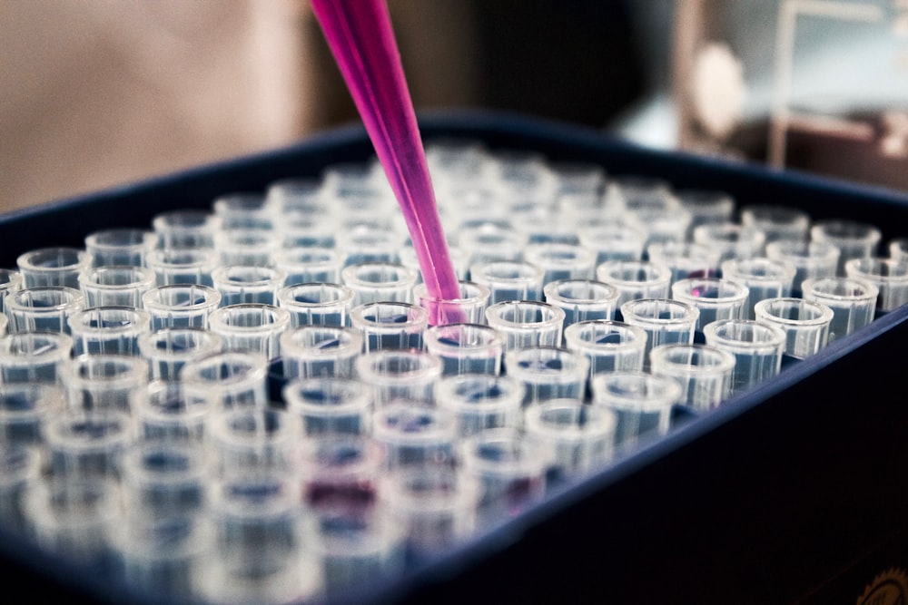
A small sample of human skin (e.g., from a breast reduction surgery) is collected. Enzymes are used to carefully break down the tissue, creating a soup of individual cells .
The Tagging
This cell soup is incubated with a fluorescent antibody designed to seek out and stick specifically to the Beta-1 Integrin protein on the cell surface. Think of this as tying a tiny, glowing nametag to every cell that has the "grip" protein. The more "grip" a cell has, the more nametags it collects, and the brighter it glows .
The Race (Flow Cytometry)
The tagged cell suspension is forced single-file through a thin tube. As each cell passes in front of a laser beam, it lights up. A detector measures its fluorescence intensity—a direct readout of its Beta-1 Integrin level .
The Sort
This is the magic. Using a technology called electrostatic cell sorting, the machine charges and then deflects the droplets containing the cells into different collection tubes based on their brightness.
- Bright (Beta-1-bright): Collected as the stem cell candidate population.
- Dim (Beta-1-dim): Collected as the transit-amplifying cell candidate population.
Visualizing the Process
The diagram below illustrates how flow cytometry separates cells based on their Beta-1 Integrin levels:
Mixed Cell Population
Cells with varying levels of Beta-1 Integrin
Fluorescent Tagging
Antibodies attach to Beta-1 Integrin
Sorted Populations
Cells separated by fluorescence intensity
Results and Analysis: Proving Their Potential
So, what happened once these sorted cells were put to the test? The results were striking.
The Beta-1-bright population demonstrated all the hallmarks of true stem cells:
Proliferative Power
In lab dishes, they formed large, robust colonies, demonstrating a high capacity for division.
Self-Renewal
They could be passaged multiple times, proving they weren't just a one-time wonder.
Clonogenicity
A single Beta-1-bright cell could give rise to a whole colony, proving its "stemness."
The Beta-1-dim population, in contrast, formed only small, abortive colonies and quickly stopped dividing.
Scientific Importance
This experiment provided the first clear, functional link between a surface marker (Beta-1 Integrin) and stem cell behavior in human epidermis. It transformed the field from one of speculation to one of precise isolation and study. It proved that stem cells weren't a mythical concept but a tangible, isolatable population with a unique molecular signature .
Data from the Hunt
Marker Profile
| Cell Population | Beta-1 Integrin |
|---|---|
| Beta-1-bright | High |
| Beta-1-dim | Low |
This table shows the molecular "fingerprint" used to identify stem cell candidates. High levels of adhesion proteins (Integrins) are characteristic of the quiescent stem cell population.
Functional Assays
| Cell Population | CFE % | Self-Renewal |
|---|---|---|
| Beta-1-bright | ~20% | >5 passages |
| Beta-1-dim | ~1% | 1-2 passages |
After sorting, cells are grown in culture to test their functional ability. The Beta-1-bright population is vastly superior in every measure of stem cell potential.
In Vivo Regeneration
| Cell Population | Regeneration |
|---|---|
| Beta-1-bright | Yes |
| Beta-1-dim | Limited |
The ultimate test. When grafted onto immunodeficient mice, only the Beta-1-bright population can regenerate a fully structured, self-renewing human epidermis, the gold standard proof of stem cell function .
Comparative Performance Visualization
The Scientist's Toolkit: Essential Research Reagents
Here are the key tools that made this experiment—and ongoing stem cell research—possible.
| Research Reagent / Tool | Function in the Experiment |
|---|---|
| Fluorescent Antibodies | These are the "glowing tags." They are engineered proteins that bind with high specificity to a target (like Beta-1 Integrin) and light up when hit by a laser, allowing the machine to identify the cell. |
| Collagenase / Trypsin | These are the "biological scissors." They are enzymes used to carefully digest the tough structural proteins (like collagen) in the skin sample, breaking it down into a suspension of individual living cells for analysis. |
| Basal Cell Culture Media | A specialized, nutrient-rich "soup" designed to mimic the ideal environment for skin stem cells to survive, divide, and form colonies outside the human body after they are sorted. |
| Flow Cytometer / Cell Sorter | The heart of the operation. This sophisticated machine analyzes thousands of cells per second based on their fluorescence and physical properties, and can physically separate them with incredible precision. |
Conclusion: A New Era for Skin Science
The ability to sort and analyze human epidermal stem cells using flow cytometry didn't just solve a biological mystery; it opened a new frontier in medicine. Today, this knowledge is being directly applied to grow sheets of new skin for burn victims in the lab, to develop advanced models for testing drugs and cosmetics without animal testing, and to understand the fundamental errors that occur when these master healer cells go rogue and cause cancer.
Key Takeaway
By learning to see the invisible, scientists have not only cracked the skin's code for self-renewal but have also handed us the tools to harness that power for healing. The silent repair operation is silent no more.