Cryopreservation in Allogeneic Off-the-Shelf Cell Therapies: Enabling Scalability and Clinical Translation
This article explores the critical, multi-faceted role of cryopreservation in the development and commercialization of allogeneic, off-the-shelf cell therapies.
Cryopreservation in Allogeneic Off-the-Shelf Cell Therapies: Enabling Scalability and Clinical Translation
Abstract
This article explores the critical, multi-faceted role of cryopreservation in the development and commercialization of allogeneic, off-the-shelf cell therapies. Aimed at researchers, scientists, and drug development professionals, it provides a comprehensive analysis spanning foundational principles, current methodological applications, key challenges in optimization, and clinical validation. The content synthesizes recent clinical data, market trends, and technical advancements to outline how robust cryopreservation strategies are indispensable for overcoming logistical hurdles, ensuring product consistency, mitigating immune rejection risks, and ultimately fulfilling the promise of scalable, accessible cell therapies. Discussions on donor variability, cryoinjury, regulatory considerations, and comparative analyses with autologous models are included to offer a holistic perspective for the field.
The Indispensable Bridge: How Cryopreservation Enables the Allogeneic Therapy Model
The field of advanced cell therapies is undergoing a transformative shift from patient-specific (autologous) treatments towards scalable, donor-derived (allogeneic) "off-the-shelf" paradigms. This transition is driven by the need to overcome the profound logistical and economic challenges of autologous therapies, which are characterized by high costs, extended manufacturing timelines, and significant variability [1]. Autologous cell therapy can cost up to $1 million per patient and suffers from a manufacturing failure rate of 2% to 10%, presenting substantial barriers to widespread accessibility [1]. In contrast, allogeneic therapies promise to treat millions of patients from a single manufactured batch, potentially reducing costs and simplifying supply chains [2]. The global allogeneic cell therapy market is projected to grow from $0.4 billion in 2024 to $2.4 billion by 2031, reflecting a compound annual growth rate (CAGR) of 24.1% [3]. Central to realizing this "off-the-shelf" vision is robust cryopreservation, a process that extends product shelf life, decouples manufacturing from clinical administration, and enables global distribution [4] [5]. This technical guide examines the core of the off-the-shelf paradigm, detailing the role of cryopreservation in facilitating this scalable model.
The Autologous-Allogeneic Transition: A Comparative Analysis
The fundamental differences between autologous and allogeneic cell therapies are compared in the table below, highlighting the logistical and manufacturing challenges that the off-the-shelf model seeks to address.
Table 1: Comparative Analysis of Autologous vs. Allogeneic Cell Therapy Models
| Feature | Autologous Model | Allogeneic ('Off-the-Shelf') Model |
|---|---|---|
| Cell Source | Patient's own cells (e.g., from leukapheresis) [1] | Healthy donor(s), iPSCs, or umbilical cord blood [1] |
| Manufacturing | Individualized batch per patient; process begins anew for each treatment [1] | Single large batch from one source can produce doses for multiple patients [1] |
| Typical Cost | Often exceeds \$400,000 and can reach \$1 million per patient [2] | Aims for significant cost reduction through scaled production [2] |
| Production Timeline | ~3 weeks, unsuitable for rapidly progressing diseases [1] | Immediate availability of cryopreserved doses [1] |
| Scalability | Inherently limited and complex [3] | Highly scalable and standardized [3] |
| Key Challenges | Variable starting material quality, manufacturing failures, high cost, time delays [1] | Managing immunogenicity (GvHD, host rejection), optimizing cryopreservation [3] [1] |
The shift to allogeneic models introduces new technical hurdles, primarily concerning immune compatibility. A major risk is Graft-versus-Host Disease (GvHD), where donor T-cells attack recipient tissues [1]. Conversely, Host-versus-Graft (HvG) reactions can lead to the immune-mediated rejection of the therapeutic cells [1]. Strategies to overcome these challenges include gene-editing technologies (e.g., CRISPR/Cas9, TALEN) to disrupt the T-cell receptor (TCR) and ablate HLA expression, thereby creating hypoimmunogenic cells [1].
The Central Role of Cryopreservation in the Off-the-Shelf Ecosystem
Cryopreservation is the critical enabling technology for the off-the-shelf paradigm, acting as a "pause button" that halts biological time. It allows for the creation of centralized cell banks, long-term storage, and just-in-time delivery to the point of care, effectively decoupling manufacturing from treatment [4] [5]. However, the process is far from simple and poses significant risks to cell viability and functionality if not properly optimized [4].
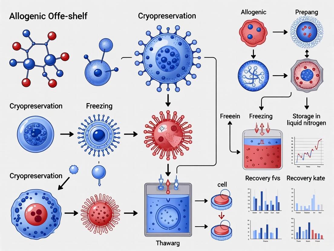
The following workflow illustrates the integrated role of cryopreservation within the end-to-end development and manufacturing process for an allogeneic cell therapy.
Critical Challenges in Therapy Cryopreservation
Several advanced challenges in cryopreservation can compromise the quality of off-the-shelf products:
- The DMSO Debate and Cryoprotectant Toxicity: Dimethyl sulfoxide (Me₂SO) is the most common cryoprotective agent (CPA), but it is cytotoxic at temperatures above 0°C [2]. Its administration, particularly via novel routes like intracerebral or intraocular injection, is associated with safety risks and potential adverse events [2]. Furthermore, controlling CPA exposure times and temperature is critical to avoid damaging excursions in cell volume during addition and removal [4].
- Transient Warming Events: Temperature fluctuations during routine storage or transport can lead to partial thawing and re-crystallization, causing intracellular ice formation and loss of viability. This is an emerging issue with significant regulatory and commercial implications [4].
- Cryopreservation-Induced Delayed-Onset Cell Death: Cells can appear viable immediately post-thaw but undergo apoptosis days later due to stress incurred during the freezing process. This phenomenon complicates accurate viability assessments for regulatory and quality-control purposes [4].
- The Scaling Hurdle: A recent industry survey identified the "ability to process at a large scale" as the single biggest hurdle (cited by 22% of respondents) for cryopreservation in cell and gene therapy [6]. Scaling techniques and technologies are required to ensure therapies are manufactured and cryopreserved efficiently while maintaining critical quality attributes [6].
Optimizing Cryopreservation: Methodologies and Protocols
Overcoming the challenges outlined above requires meticulous process development. The table below summarizes key quantitative data and parameters from recent research and industry surveys.
Table 2: Key Parameters in Cryopreservation Protocols and Industry Practices
| Parameter | Common Practice / Value | Significance & Emerging Trends |
|---|---|---|
| Primary Cryoprotectant | 5-10% Dimethyl Sulfoxide (Me₂SO) [2] | Cytotoxicity drives R&D into Me₂SO-free media [2]. |
| Standard Cooling Rate | 1°C/min [2] [6] | A default rate; optimized rates are cell-type specific [6]. |
| Target Warming Rate | ~45°C/min [6] | Crucial for minimizing ice crystal damage and DMSO exposure; controlled-thawing devices are recommended over manual water baths [6]. |
| Controlled-Rate Freezer (CRF) Use | 87% of industry survey respondents [6] | Provides control over critical process parameters; passive freezing (13%) is more common in early-stage clinical development [6]. |
| Use of Default CRF Profiles | 60% of industry survey respondents [6] | Default profiles work for many cells, but optimized profiles are needed for iPSCs, cardiomyocytes, and other sensitive types [6]. |
Detailed Experimental Protocol: Scalable γδ T Cell Expansion and Cryopreservation
A recent study demonstrates an integrated workflow for scaling allogeneic cell therapy, from expansion to cryopreservation [7]. This protocol is a practical example of implementing the off-the-shelf paradigm.
- 1. Starting Material: Peripheral Blood Mononuclear Cells (PBMCs) are used as the seed cells [7].
- 2. Expansion Medium: Cells are cultured in a GMP-grade, serum-free γδ T Cell Expansion Medium [7].
- 3. Bioreactor Process:
- Initial seed: 3 million cryopreserved PBMCs in a 3 mL initial volume.
- Vessel: Scale-out static culture reactor with a gas-permeable membrane at the bottom.
- Duration: 14 days of fed-batch culture.
- Final volume: 2000 mL.
- 4. Output and QC:
- Total cell harvest: >60 billion cells.
- Purity: >93% γδ T cells at harvest.
- Viability: Demonstrated high cell viability compared to other commercial media [7].
- 5. Cryopreservation: The harvested cells are cryopreserved using a controlled-rate freezer to create an "off-the-shelf" product bank.
The Scientist's Toolkit: Essential Reagents for Cell Therapy Development
Table 3: Key Research Reagent Solutions for Allogeneic Cell Therapy Development
| Reagent / Material | Function & Application |
|---|---|
| GMP-Grade Cell Expansion Medium | A xeno-free, serum-free basal medium formulated for specific cell types (e.g., γδ T cells, iPSCs); supports high-density expansion and maintains cell functionality [7]. |
| GMP-Grade Cytokines (e.g., IL-2, IL-15) | Recombinant proteins that provide critical signals for T-cell and NK-cell survival, proliferation, and activation during expansion culture [7]. |
| GMP-Grade Immune Modulators (e.g., 4-1BB Ligand) | Recombinant proteins used to co-stimulate immune cells, enhancing their persistence and anti-tumor potency [7]. |
| GMP-Grade Anti-Human CD3 Antibody | Used as an activation signal for T-cell expansion via the TCR/CD3 complex [7]. |
| Cryopreservation Media | Formulations containing CPAs (like Me₂SO or alternatives), salts, and excipients designed to protect cells during freeze-thaw. The move towards Me₂SO-free, clinically administrable formulations is a key trend [2]. |
Advanced Concepts: Engineering Immune Evasion for Allogeneic Therapies
For an allogeneic cell to become a truly effective "off-the-shelf" therapeutic, it must not only survive cryopreservation but also evade the host immune system. Engineering such cells involves sophisticated genetic modifications, as illustrated in the following signaling pathway diagram for an engineered CAR-NK cell.
This engineering approach creates a "hypoimmunogenic" cell. By knocking down HLA-I, the cell becomes invisible to host T-cells. The expression of PD-L1 directly suppresses any engaged T-cells, and the expression of SCE inhibits attack from the host's own NK cells, which would otherwise target cells lacking HLA-I [8]. All these modifications can be incorporated into a single DNA construct, streamlining production for a standardized, off-the-shelf product [8].
The transition to an off-the-shelf paradigm is essential for the sustainable and equitable future of cell therapy. While allogeneic approaches inherently offer scalability, their success is critically dependent on overcoming dual challenges: managing immune compatibility and mastering the cryopreservation process. Advances in gene-editing are systematically addressing the first challenge, creating a new class of universal donor cells [1] [8]. Concurrently, innovations in cryopreservation—including the development of administrable, DMSO-free media, sophisticated controlled-rate freeze-thaw protocols, and robust quality control measures—are solving the logistical complexities of storage and distribution [4] [2] [6]. The convergence of these technologies will ultimately enable the creation of truly "off-the-shelf" cellular therapeutics, transforming them from bespoke, high-cost interventions into standardized, accessible, and potentially first-line treatment options for a global patient population.
Cryopreservation serves as the critical technological bridge that enables the decoupling of cell therapy manufacturing from patient treatment, a foundational principle for allogeneic "off-the-shelf" therapies. By stabilizing living cellular material at ultra-low temperatures, cryopreservation creates a pause between production and clinical use, thereby overcoming fundamental challenges of scalability, logistics, and quality control that plague autologous approaches. This whitepaper examines the core cryopreservation protocols, agents, and temperature parameters that maintain cell viability and functionality during long-term storage. For researchers and drug development professionals, we provide detailed methodological frameworks and technical specifications essential for implementing robust cryopreservation strategies within advanced therapy medicinal product (ATMP) development pipelines.
The field of cell therapy is undergoing a fundamental transformation from autologous to allogeneic models. Autologous cell therapies, such as approved CAR-T treatments, are manufactured from a patient's own cells. While clinically effective, this model presents significant limitations: personalized manufacturing for each patient creates complex, time-consuming, and costly production; logistical constraints in coordinating cell collection, modification, and reinfusion limit patient access; and quality variability based on patient cell health impacts product consistency and clinical outcomes [9].
Allogeneic "off-the-shelf" therapies flip this model by leveraging healthy donor cells engineered in bulk to create standardized, quality-controlled batches for broader use. The concept is akin to biologics or vaccines—a single production run serving many patients, reducing both cost and time to treatment [9]. This paradigm shift is accelerating rapidly, with multiple biotech and pharma companies advancing allogeneic pipelines, including Allogene Therapeutics, Fate Therapeutics, and CRISPR Therapeutics [9].
Cryopreservation represents the essential enabling technology that makes this decoupling possible. By allowing long-term storage of cellular products at ultra-low temperatures, it separates the manufacturing process from the treatment timeline, creating what industry leaders often compare to the trajectory of monoclonal antibodies—initially complex and expensive to produce, but now manufactured at industrial scale with standardized processes [9].
Fundamental Principles of Cryopreservation
Historical Context and Mechanical Challenges
The origins of low-temperature tissue storage research date back to the late 1800s, but significant understanding emerged in the 1950s when James Lovelock discovered that cryopreservation caused osmotic stress by instantly freezing liquid, contributing directly to ice crystal formation in red blood cells [10]. In 1963, Mazur characterized that the rate of temperature change controls water movement across cell membranes and thus the degree of intracellular freezing [10].
Exposing cells to temperatures below 0°C without cryoprotectants is typically lethal because water constitutes approximately 80% of tissue mass. The freezing of water, both intra- and extracellularly, imposes harmful biochemical and structural changes through two primary mechanisms: (1) ice crystals mechanically disrupt cellular membranes, making it impossible to obtain structurally-intact cells after thawing; and (2) deadly increases in solute concentration occur in the remaining liquid phase as ice crystals form during cooling [10].
Cryoprotective Agents (CPAs): Mechanisms and Classifications
To mitigate freezing damage, cryoprotective agents (CPAs) are essential. These compounds are categorized based on their membrane permeability characteristics:
Permeating Agents (PAs) are relatively small molecules (typically less than 100 daltons) with somewhat amphiphilic nature that allows them to easily penetrate cell membranes. Common examples include dimethyl sulfoxide (DMSO), glycerol (GLY), ethylene glycol (EG), and propylene glycol (PG) [10]. These agents share several key properties: high water solubility at low temperatures, ability to cross biological membranes, and ideally, minimal toxicity [10]. Their protective effect comes primarily through hydrogen bonding with water molecules, which depresses the freezing point of water and reduces available water molecules for crystal formation [10].
Table 1: Common Permeating Cryoprotective Agents
| Cryoprotectant | Typical Concentration | Key Properties | Applications |
|---|---|---|---|
| Dimethyl sulfoxide (DMSO) | 10% (2M) | Increases membrane porosity, strong hydrogen bonding | Hematopoietic stem cells, CAR-T cells, mesenchymal stem cells |
| Glycerol (GLY) | 10-20% | First discovered CPA, less toxic than DMSO | Spermatozoa, red blood cells |
| Ethylene Glycol (EG) | 6-8M (in mixtures) | Rapid permeability, lower toxicity | Islet cells, in combination with DMSO |
| Propylene Glycol (PG) | 5-10% | Similar to GLY, different permeability | Specific cell lines, germ cells |
Non-Permeating Agents (NPAs) are typically larger molecules that do not penetrate intracellularly and exert protective influence outside the cell. Common agents include polyethylene glycol (PEG), polyvinylpyrrolidone (PVP), raffinose, sucrose, and trehalose [10]. These agents induce vitrification extracellularly but to a lesser extent than permeating agents. Trehalose, a disaccharide CPA produced by various organisms including bacteria, fungi, and plants, has unique stability due to its acetal link that prevents reduction of C-1 in each glucose monomer, increasing stability under extreme temperatures [10].
Vitrification Strategies
Both permeating and non-permeating agents can prove toxic to cells at higher concentrations. To minimize toxicity while maintaining effectiveness, vitrification mixtures combine both agent types, allowing successful cryobanking with lower concentrations of permeating agents [10]. This approach reduces PA-induced toxicity and increases cellular viability and yields post-thaw. A demonstrated method using multi-molar combinations of reduced concentrations of EG and DMSO successfully cryopreserved both human and murine islet cells with reduced adverse effects [10].
Technical Protocols and Methodologies
Standard Cryopreservation Workflow
The following workflow diagram illustrates the core process for cryopreserving allogeneic cell therapies:
Cooling Rate Optimization
Cooling rate represents a critical parameter in cryopreservation success. In general, successful low-temperature cell preservation utilizes cooling rates of approximately 1°C/minute [10]. However, optimal rates vary significantly by cell type:
- Slow cooling (approximately 1°C/min) is recommended for hepatocytes, hematopoietic stem cells, and mesenchymal stem cells [10]
- Rapid cooling is associated with better outcomes for oocytes, pancreatic islets, and embryonic stem cells [10]
The following protocol details a standardized approach for slow-freeze cryopreservation, which currently dominates the market with a 50% share [11]:
Materials Required:
- Cryoprotective agent (typically 10% DMSO)
- Programmable rate-controlled freezing system
- Cryogenic storage vials
- Isopropanol freezing container (if controlled-rate freezer unavailable)
- Liquid nitrogen storage system
Methodology:
- Cell Preparation: Harvest cells during optimal growth phase and ensure >95% viability pre-freeze
- CPA Addition: Slowly add pre-chilled CPA solution to cell suspension to achieve final concentration of 5-10% DMSO, mixing gently during addition
- Aliquoting: Dispense 1.0-1.5mL volumes into cryogenic vials
- Equilibration: Incubate vials on ice for 15-30 minutes to permit CPA permeation
- Cooling Phase: Place vials in rate-controlled freezer programmed for -1°C/minute to -40°C, then -10°C/minute to -90°C
- Transfer: Immediately transfer to long-term storage vapor phase liquid nitrogen (-150°C to -196°C)
Advanced 3D Culture Cryopreservation
Recent advances in cryopreservation methodology address the challenges of complex 3D cultures, such as organoids and tissue constructs. The following protocol was specifically developed for spaceflight experiments but has terrestrial applications [12]:
Materials Required:
- VitroGel Hydrogel Matrix
- PDMS-based 3D culture chambers
- CryoStor CS10 freeze media
- Y-27632 Rho kinase inhibitor
Methodology:
- 3D Culture Establishment: Encapsulate hiPSCs in VitroGel Hydrogel Matrix within PDMS culture chambers
- Pre-cryopreservation Treatment: Culture cells for 12 days to form well-developed 3D clusters
- CPA Formulation: Replace culture medium with CryoStor CS10 supplemented with 10µM Y-27632 Rho kinase inhibitor
- Cooling Protocol: Implement controlled-rate freezing at -1°C/minute to -80°C
- Storage: Maintain at -80°C for long-term preservation (note: this protocol achieves high viability without liquid nitrogen)
This integrated system demonstrated no significant difference in viability between 96-well plates and specialized culture chambers, with post-thaw viability exceeding 85% and maintained trilineage differentiation potential [12].
Quantitative Analysis of Cryopreservation Impact
Market Growth and Segment Analysis
The cell line cryopreservation market demonstrates substantial growth driven by expanding allogeneic therapy development. Current projections indicate the market will grow from USD 5.39 billion in 2025 to USD 13.97 billion by 2034, representing a compound annual growth rate (CAGR) of 11.16% [11].
Table 2: Cell Line Cryopreservation Market Analysis by Segment
| Segment | Market Share (2024) | Projected Growth | Key Drivers |
|---|---|---|---|
| By Product/Offering | |||
| Cell Freezing Media & Cryoprotectants | 35% | Steady | Essential for ice crystal protection, viability maintenance |
| Automated Cryogenic Biobanking | <10% | Fastest Growing | Sample integrity, workflow streamlining, space efficiency |
| By Preservation Technology | |||
| Slow-Freezing | 50% | Steady | Preferred for high intracellular content cells (oocytes) |
| Vitrification/Ultra-Rapid Freezing | <30% | Fastest Growing | Faster procedures, improved IVF outcomes |
| By Service Model | |||
| In-House | 42% | Steady | Direct control, immediate access |
| Outsourced Biobanking & CRO | <30% | Fastest Growing | Infrastructure cost reduction, specialized expertise |
| By End-User | |||
| Biopharmaceutical & Cell Therapy | 35% | Steady | Scalability needs, quality control requirements |
| CROs & Contract Cell-line Developers | <30% | Fastest Growing | Outsourcing trend, specialized service demand |
Regional Adoption Patterns
Regional analysis reveals North America as the dominant market with 38% share, attributed to advances in reproductive technology and technological innovation [11]. The Asia Pacific region represents the fastest-growing market, driven by increasing prevalence of chronic diseases and expanding biobanking services [11].
The Scientist's Toolkit: Essential Research Reagents
Implementation of robust cryopreservation protocols requires specific reagents and materials optimized for allogeneic cell therapy applications:
Table 3: Essential Research Reagents for Allogeneic Therapy Cryopreservation
| Reagent/Material | Function | Application Notes |
|---|---|---|
| Cryoprotective Agents | ||
| DMSO (Cell Culture Grade) | Permeating CPA | Standard 10% concentration; increases membrane porosity [10] |
| Trehalose | Non-permeating CPA | Natural disaccharide; exceptional stability; extracellular protection [10] |
| CryoStor CS10 | Commercial CPA formulation | Xeno-free, serum-free; optimized for stem cells [12] |
| Specialized Additives | ||
| Y-27632 (Rho kinase inhibitor) | Enhances post-thaw viability | Reduces apoptosis in sensitive cell types; 10µM concentration [12] |
| Hydrogel Systems | ||
| VitroGel Hydrogel Matrix | 3D culture support | Animal-free, ligand-functionalized ECM mimic [12] |
| Matrigel | Basement membrane matrix | For feeder-free pluripotent stem cell culture [12] |
| Equipment | ||
| Programmable Freezer | Controlled-rate cooling | Essential for slow-freeze protocols (-1°C/min) [10] |
| PDMS Culture Chambers | 3D culture platform | Tunable mechanical properties; superior gas exchange [12] |
Technological Innovations and Future Directions
Emerging Cryopreservation Technologies
The field of cryopreservation continues to evolve with several emerging technologies enhancing the feasibility of allogeneic therapies:
Vitrification/Ultra-Rapid Freezing represents the fastest-growing preservation technology segment, offering faster procedures and improved outcomes, particularly for in vitro fertilization applications [11]. This approach uses higher CPA concentrations with ultra-rapid cooling to achieve a glassy state without ice crystal formation.
AI-Driven Monitoring systems are transforming cryopreservation quality control. AI-based monitoring in cryopreservation services significantly reduces damage compared to outdated processes by ensuring every sample is continuously monitored [11]. These systems automatically log all applicable data, providing a complete audit trail in real-time that streamlines compliance and reduces regulatory risk [11].
Integration with Allogeneic Therapy Platforms
The successful integration of cryopreservation within allogeneic therapy platforms requires addressing several technical challenges:
Immune Rejection Risks: Donor cells carry the risk of graft-versus-host disease (GvHD) or immune-mediated clearance. Researchers are developing "universal" cells through HLA editing or gene knockouts to reduce immunogenicity [9]. CAR-NK cell therapies are particularly attractive for allogeneic applications because, unlike T cells, they do not cause GvHD [13].
Manufacturing Scale-Up: Advanced manufacturing technologies are critical for enabling allogeneic therapies to reach industrial-scale production. Innovations include bioreactor systems that minimize contamination risk while allowing high-volume production, and gene-editing tools like CRISPR that create universal donor cells [9].
The following diagram illustrates how cryopreservation enables the decoupling of manufacturing from treatment in allogeneic therapy development:
Cryopreservation serves as the fundamental enabler for decoupling manufacturing from treatment in allogeneic cell therapies, transforming what would otherwise be a continuous process into a segmented, scalable workflow. By providing a "pause button" for cellular viability, it allows for quality control testing, centralized manufacturing, global distribution, and on-demand treatment—key requirements for commercially viable off-the-shelf therapies. The technical protocols, reagent systems, and preservation strategies detailed in this whitepaper provide researchers and drug development professionals with the foundational knowledge to implement robust cryopreservation within their allogeneic therapy development pipelines. As the field advances, continued optimization of CPA formulations, cooling rates, and thawing protocols will further enhance the viability, potency, and clinical effectiveness of allogeneic cellular medicines, ultimately expanding patient access to these transformative therapies.
Cell cryopreservation has emerged as a critical enabling technology for the advancement of regenerative medicine, particularly for allogeneic "off-the-shelf" cell therapies. This complex process of preserving cells at ultra-low temperatures (typically below -130°C to -196°C) allows for the long-term storage and transportation of biological samples while maintaining their viability and functionality [14]. The growing prominence of allogeneic cell therapies—those derived from healthy donors rather than patients themselves—has fundamentally transformed the landscape of cellular treatment modalities. Unlike autologous approaches, allogeneic therapies offer the significant advantage of being readily available as "off-the-shelf" products, eliminating the need for individualized production for every patient and enabling treatment of a broader patient population [15] [16].
The synergy between cryopreservation technologies and allogeneic cell therapy development has created a rapidly expanding market segment with substantial implications for researchers, scientists, and drug development professionals. This technical guide examines the current market dynamics, growth drivers, experimental methodologies, and future perspectives that define the cell cryopreservation landscape within the context of allogeneic therapy development.
The cell cryopreservation market is experiencing robust growth, driven primarily by the expanding applications in cell-based therapies, regenerative medicine, and biobanking. Current market analyses project the global cell line cryopreservation market to grow from USD 5.39 billion in 2025 to approximately USD 13.97 billion by 2034, representing a compound annual growth rate (CAGR) of 11.16% [11]. This growth trajectory significantly outpaces many other pharmaceutical sectors and reflects the increasing investment and commercial interest in advanced cell therapy platforms.
The broader allogeneic cell therapy market, which heavily relies on cryopreservation technologies, was valued at USD 1.08 billion in 2024 and is expected to reach USD 1.81 billion by 2029, growing at a CAGR of 10.8% [17]. Another analysis projects the global allogeneic cell therapy market to grow from USD 1.55 billion in 2025 to USD 2.74 billion by 2035 [16]. This growth is further supported by the expanding market for allogeneic cell therapy devices, which is projected to grow at an impressive CAGR of 25.8% to 26.4% from 2025 to 2035, reaching USD 3.42 billion to USD 4.91 billion [18] [19].
Table 1: Cell Cryopreservation and Allogeneic Therapy Market Overview
| Market Segment | 2024/2025 Market Size | Projected Market Size | CAGR | Time Period |
|---|---|---|---|---|
| Cell Line Cryopreservation | USD 5.39 billion [11] | USD 13.97 billion [11] | 11.16% [11] | 2024-2034 |
| Allogeneic Cell Therapy | USD 1.08 billion [17] | USD 1.81 billion [17] | 10.8% [17] | 2024-2029 |
| Allogeneic Cell Therapy (Alternate Analysis) | USD 1.55 billion [16] | USD 2.74 billion [16] | 5.9% [16] | 2025-2035 |
| Allogeneic Cell Therapy Devices | USD 328.8 million [18] | USD 3.42 billion [18] | 26.4% [18] | 2023-2033 |
Regional analysis reveals that North America currently dominates the market, accounting for approximately 38% of the cell line cryopreservation market share and more than 60% of the global allogeneic cell therapies market [11] [16]. This dominance is attributed to strong healthcare infrastructure, regulatory clarity, significant research and development investments, and higher adoption rates of advanced therapies. However, the Asia-Pacific region is expected to witness the fastest growth rate during the forecast period, driven by increasing healthcare spending, rising prevalence of chronic diseases, and growing investments in biotechnology and regenerative medicine [11] [20].
Key Growth Drivers and Market Dynamics
Rising Demand for Allogeneic Cell Therapies
The increasing prevalence of chronic diseases represents a significant driver for the cell cryopreservation market. With over 19.2 million cancer cases reported globally in 2020 and hematological malignancies driving demand for hematopoietic stem cell transplantation, the need for accessible and scalable cell therapies continues to grow [21]. Allogeneic stem cell transplantation has become a well-established treatment modality, with over 23,000 unrelated donor hematopoietic stem cell transplants processed globally in 2023 alone [21].
The clinical pipeline for allogeneic therapies has expanded dramatically, with current estimates indicating over 470 allogeneic cell therapies in various preclinical and clinical development stages [16]. This robust pipeline necessitates advanced cryopreservation solutions to support both clinical development and eventual commercial distribution. The therapeutic application of these therapies is also broadening beyond hematological malignancies to include autoimmune disorders, neurological conditions, musculoskeletal disorders, and infectious diseases [18] [16].
Technological Advancements in Cryopreservation
Several technological innovations are driving improvements in cryopreservation efficacy and reliability:
Advanced Cryoprotectant Formulations: Development of specialized cryopreservation media containing optimized concentrations of dimethyl sulfoxide (DMSO) supplemented with sugars and albumin has demonstrated improved post-thaw viability and functionality of sensitive cell types like Natural Killer (NK) cells [22].
Automated and Controlled-Rate Systems: Automated cryogenic biobanking and robotic storage solutions represent the fastest-growing product segment, enhancing sample integrity, streamlining workflows, and optimizing space and energy utilization [11].
Novel Preservation Methods: While slow-freezing protocols currently dominate the market (holding approximately 50% share in 2024), vitrification or ultra-rapid freezing methods are emerging as the fastest-growing segment due to their superior preservation of cellular structures and higher survival rates for certain cell types [11].
AI-Integrated Monitoring: Artificial intelligence is being incorporated into cryopreservation monitoring systems, significantly reducing sample damage compared to conventional processes and providing comprehensive, real-time audit trails that streamline compliance [11].
Strategic Investments and Regulatory Support
Substantial financial investments have flowed into the allogeneic cell therapy space, with nearly USD 8.8 billion invested in companies developing allogeneic cell therapies over the past four years [16]. This funding has accelerated technology development and clinical translation.
Regulatory agencies have also developed clearer pathways for advanced therapy approvals. The U.S. Food and Drug Administration (FDA) and European Medicines Agency (EMA) have established specific frameworks for cell-based products, providing guidance on handling, storage, and transport requirements for biological materials [14]. These regulatory advancements have increased confidence among investors and manufacturers, facilitating product commercialization.
Table 2: Key Market Drivers and Their Impact
| Driver Category | Specific Factors | Impact on Market |
|---|---|---|
| Clinical Demand | Rising prevalence of chronic diseases [21]; Expansion into non-hematologic indications [21]; Over 470 allogeneic therapies in pipeline [16] | Increases need for scalable, reliable cryopreservation solutions across multiple therapeutic areas |
| Technology Advancement | Advanced cryoprotectant media [22]; Automated biobanking systems [11]; AI-integrated monitoring [11] | Improves post-thaw viability, enhances process efficiency, reduces operational costs |
| Strategic Factors | USD 8.8 billion in investments [16]; Supportive regulatory frameworks [14]; Over 90 strategic partnerships [16] | Accelerates clinical translation, facilitates commercialization, encourages innovation |
Experimental Protocols in Cell Cryopreservation
Feeder-Free NK Cell Expansion and Cryopreservation
Recent research has demonstrated optimized protocols for the expansion and cryopreservation of NK cells, which are particularly sensitive to freeze-thaw processes. The following methodology, adapted from a 2025 study, outlines an effective approach for feeder-free NK cell expansion and cryopreservation [22]:
NK Cell Expansion Protocol:
- Initial Setup: Seed peripheral blood mononuclear cells (PBMCs) directly onto γ-globulin and anti-NKp46-coated flasks without prior T- or B-cell depletion.
- Culture Conditions: Use serum-free medium (Alys505NK) supplemented with 1,000 IU/ml recombinant human IL-2, 50 ng/ml recombinant human IL-18, and 5% heat-inactivated autologous plasma.
- Feeding Schedule: Add recombinant human IL-18 at culture initiation and again on day 5. Add fresh culture medium every 1-3 days depending on cell density (maintaining approximately 2 × 10^6 cells/ml).
- Scale-Up: On day 6, transfer cells to culture bags for continued expansion over 14 days.
Cryopreservation Protocol:
- Cryomedium Preparation: Prepare cryopreservation formulation containing 5% DMSO, supplemented with sugars (e.g., pentastarch) and albumin [22].
- Cell Processing: Resuspend expanded NK cells in cryopreservation medium at appropriate concentration.
- Freezing Process: Use controlled-rate freezing systems to gradually lower temperature to -80°C before transfer to long-term storage in liquid nitrogen vapor phase (-196°C).
- Quality Assessment: Post-thaw viability and functionality assessment via flow cytometry (CD107a degranulation assay) and in vivo antitumor efficacy studies.
This protocol has demonstrated success in preserving NK cell functionality, with studies showing that cryopreserved NK cells maintain antitumor efficacy comparable to freshly expanded cells, a critical requirement for off-the-shelf allogeneic therapies [22].
Cryopreservation Workflow for Allogeneic Therapies
The following diagram illustrates the comprehensive workflow for cryopreserving allogeneic cell therapies, from donor selection to final product storage and distribution:
Cryopreservation Workflow for Allogeneic Therapies
The Scientist's Toolkit: Essential Research Reagents and Materials
Successful cryopreservation of cells for allogeneic therapies requires carefully selected reagents and materials optimized for specific cell types and applications. The following table details key components of the cryopreservation toolkit:
Table 3: Essential Research Reagents for Cell Cryopreservation
| Reagent/Material | Function | Application Notes |
|---|---|---|
| DMSO (5% concentration) [22] | Penetrating cryoprotectant that reduces ice crystal formation | Standard concentration for NK cells; may vary for other cell types; requires gradual addition and removal to minimize osmotic stress |
| Sugars (e.g., pentastarch) [22] | Non-penetrating cryoprotectant that provides extracellular protection | Helps stabilize cell membranes during freezing; often used in combination with DMSO |
| Albumin [22] | Provides colloidal protection and stabilizes cell membranes | Often derived from human or bovine sources; must be screened for pathogens |
| Serum-Free Cryomedium [22] | Base solution for cryoprotectant formulation | Eliminates batch-to-batch variability of serum; enhances regulatory compliance |
| Recombinant Human IL-2 [22] | Enhances post-thaw recovery and functionality | Critical for immune cells like NK cells; concentration typically 1,000 IU/ml |
| Recombinant Human IL-18 [22] | Promotes NK cell activation and expansion | Added at initiation and during culture; concentration typically 50 ng/ml |
| Controlled-Rate Freezing Apparatus [11] | Enables reproducible cooling rates | Critical for minimizing ice crystal damage; typically 1°C/minute cooling rate |
| Liquid Nitrogen Storage Systems [11] | Provides long-term storage at -196°C | Vapor phase storage reduces contamination risk; requires continuous monitoring |
Technological Challenges and Future Perspectives
Current Challenges in Cell Cryopreservation
Despite significant advancements, several challenges persist in the cell cryopreservation landscape:
Cell Sensitivity and Viability Loss: Certain cell types, particularly NK cells and other immune effector cells, remain highly sensitive to cryopreservation, often experiencing diminished cytotoxic activity, reduced viability, and functional impairment post-thaw [22]. Overcoming these limitations requires cell-type-specific optimization of cryopreservation formulations and protocols.
Manufacturing Complexity and Cost: The manufacturing of allogeneic cell therapies involves highly controlled conditions with specialized equipment, cleanroom environments, and strict monitoring [18]. Small variations in temperature, handling, or timing can affect product stability and quality, increasing operational difficulty and production costs.
Standardization and Regulatory Hurdles: The industry currently lacks well-defined global standards for cell handling, storage, and delivery devices [18]. Different regions follow varying guidelines, leading to inconsistencies in product design, performance, and safety, which complicates global development and distribution strategies.
Immune Compatibility Risks: While allogeneic therapies offer scalability advantages, they carry risks of immune rejection and graft-versus-host disease (GvHD) [20]. Advanced gene editing technologies like CRISPR/Cas9 are being employed to modify donor cells to enhance their therapeutic properties and reduce immune rejection risks [16].
Emerging Trends and Future Directions
The field of cell cryopreservation is evolving rapidly, with several emerging trends shaping its future trajectory:
Advanced Gene Editing Integration: The combination of cryopreservation with CRISPR/Cas9 and other gene editing technologies enables the development of enhanced allogeneic cell products with improved persistence, functionality, and immune compatibility [20] [16].
Automation and Closed Systems: Increased adoption of automated, closed-system technologies for cell processing, expansion, and fill-finish operations enhances process control, reduces contamination risks, and improves manufacturing consistency [18].
Advanced Analytics and AI: Integration of artificial intelligence and machine learning for predictive modeling of cell behavior post-thaw, optimization of cryoprotectant formulations, and real-time monitoring of storage conditions [11] [20].
Novel Cryoprotectant Development: Research continues into less toxic cryoprotectant alternatives and combination formulations that provide enhanced protection for sensitive cell types while minimizing potential side effects in clinical applications.
The future convergence of these technologies promises to address current limitations and further enhance the viability, functionality, and clinical efficacy of cryopreserved allogeneic cell therapies, ultimately expanding their therapeutic applications and improving patient accessibility.
Cell cryopreservation represents a fundamental enabling technology for the rapidly expanding field of allogeneic cell therapies. The market momentum is undeniable, with significant growth projected across cryopreservation media, devices, and related technologies. As research continues to address current challenges in cell sensitivity, process standardization, and immune compatibility, and as emerging technologies like AI, automation, and advanced gene editing are increasingly integrated into cryopreservation workflows, the potential for cryopreserved allogeneic therapies to revolutionize treatment paradigms across a broad spectrum of diseases will continue to expand. For researchers, scientists, and drug development professionals, understanding these dynamics and technological advancements is essential for leveraging the full potential of cryopreservation in the development of effective, accessible, and scalable allogeneic cell therapies.
This technical guide explores the core principles of cryopreservation within the context of allogeneic "off-the-shelf" cell therapy development. For researchers and drug development professionals, mastering these principles is paramount for transitioning from autologous, patient-specific treatments to scalable, commercially viable allogeneic therapies. We examine the biological mechanisms of cryoinjury, the protective role of various cryoprotectants, and the critical importance of controlled-rate freezing protocols. By integrating current research data, experimental methodologies, and industry trends, this whitepaper provides a comprehensive framework for optimizing cryopreservation strategies to maintain cell viability, potency, and functionality throughout the therapeutic product lifecycle.
The emergence of allogeneic cell therapies represents a paradigm shift in regenerative medicine and oncology treatment. Unlike autologous therapies derived from a patient's own cells, allogeneic therapies are manufactured from healthy donor cells and engineered in bulk to create standardized, quality-controlled batches for multiple patients [9]. This "off-the-shelf" model promises to revolutionize treatment accessibility but introduces significant manufacturing and logistics challenges, particularly in product stability and distribution.
Cryopreservation serves as the foundational technology enabling the allogeneic approach by providing:
- Manufacturing Flexibility: Decouples production from treatment timing
- Quality Assurance: Allows comprehensive product testing before release
- Global Distribution: Facilitates shipment to treatment centers worldwide
- Inventory Management: Enables maintenance of therapeutic product banks
The scalability advantage of allogeneic therapies depends entirely on robust cryopreservation protocols that maintain cell viability and functionality post-thaw [9]. Without effective cryopreservation, the off-the-shelf model becomes clinically and commercially unviable.
Fundamental Principles of Cryopreservation
Biological and Physical Mechanisms of Cryoinjury
Cryopreservation exposes cells to extreme physical and chemical stresses. Understanding cryoinjury mechanisms is essential for developing effective preservation strategies. The primary mechanisms of freezing damage include:
Intracellular Ice Formation (IIF): At rapid cooling rates, water within the cell does not have sufficient time to exit and forms destructive ice crystals that mechanically damage cellular membranes and organelles [23]. IIF is typically lethal to cells.
Solution Effects Injury: During slow cooling, ice formation in the extracellular solution concentrates solutes to toxic levels, leading to osmotic dehydration, membrane damage, and protein denaturation [10] [23].
Osmotic Stress: As water freezes extracellularly, the unfrozen fraction becomes hypertonic, drawing water out of cells and causing excessive cell shrinkage and membrane damage [10].
The relationship between cooling rate and cell survival follows a characteristic inverted U-shape curve, where optimal rates balance intracellular ice formation against solution effects injury [23].
The Role of Cryoprotectant Agents (CPAs)
Cryoprotectant Agents (CPAs) are chemical compounds that protect cells from freezing damage through multiple mechanisms:
Colligative Action: CPAs reduce the concentration of electrolytes and other solutes in the residual unfrozen fraction at any given subzero temperature [10].
Ice Crystallization Inhibition: They modify ice crystal structure and growth kinetics, preventing mechanical damage to cellular structures [23].
Glass Formation Promotion: At sufficient concentrations, CPAs enable vitrification—the transition of water to an amorphous glassy state rather than crystalline ice [10].
Table 1: Classification and Properties of Common Cryoprotectant Agents
| CPA Type | Examples | Mechanism of Action | Relative Toxicity | Common Applications |
|---|---|---|---|---|
| Permeating Agents | DMSO, Glycerol, Ethylene Glycol, Propylene Glycol | Penetrate cell membrane; depress freezing point; reduce electrolyte concentration | Moderate to High [10] | Most mammalian cells; DMSO standard for many cell therapies [24] |
| Non-Permeating Agents | Trehalose, Sucrose, Raffinose, HES, PVP | Remain extracellular; create osmotic gradient for controlled dehydration; promote vitrification | Low [10] | Often used in combination with permeating agents; red blood cells [10] |
| Biomimetic Agents | Antifreeze Peptoids [25] | Mimic natural antifreeze proteins; inhibit ice recrystallization | Low (emerging data) [25] | Emerging for sensitive cell types; stem cells |
Controlled-Rate Freezing: Principles and Applications
Physical Basis and Technical Implementation
Controlled-rate freezing (CRF) precisely manages the thermal environment during cryopreservation to optimize cell survival. Unlike passive freezing methods that rely on static cold environments, CRF systems actively control cooling rates according to predetermined profiles [6].
The biophysical basis for CRF involves:
- Controlled Water Transport: Managing the efflux of intracellular water to prevent intracellular ice formation
- Optimal Cooling Rates: Typically -1°C/min for many cell types, though this varies significantly [26]
- Nucleation Control: Initiating ice formation at consistent, slightly supercooled temperatures
Table 2: Comparison of Cryopreservation Methodologies
| Characteristic | Controlled-Rate Freezing | Passive Freezing | Vitrification |
|---|---|---|---|
| Cooling Rate Control | Precise, programmable profiles | Uncontrolled, variable | Ultra-rapid (>20,000°C/min) |
| CPA Concentration | Low to moderate (e.g., 10% DMSO) [10] | Low to moderate | Very high (4-8M total) [23] |
| Sample Volume | Large (100-250μL) [23] | Small to medium | Very small (1-2μL) [23] |
| Implementation Cost | High (specialized equipment) [6] | Low | Low to moderate |
| Process Consistency | High | Low to moderate | Moderate |
| Primary Applications | Industrial-scale cell therapy manufacturing [6] | Early R&D, academic studies | Oocytes, embryos, sensitive primary cells |
Industry Adoption and Technical Challenges
Recent industry surveys reveal that 87% of cell therapy developers use controlled-rate freezing for cryopreservation, with adoption nearing 100% for late-stage and commercial products [6]. This high adoption rate reflects the critical need for process control and documentation in regulated environments.
Despite widespread adoption, significant challenges remain:
- System Qualification: 30% of organizations rely solely on vendor qualification, which may not represent actual use conditions [6]
- Profile Optimization: 60% use default freezer profiles, while others require cell-specific optimization [6]
- Scale-Up Limitations: CRF presents bottlenecks for batch scale-up due to chamber size limitations [6]
The controlled-rate freezer market, valued at $34 million in 2025, is projected to grow at a CAGR of 6.1% through 2033, driven by increasing demand for biopharmaceutical products and stringent regulatory requirements [27].
Experimental Protocols and Methodologies
Standardized Controlled-Rate Freezing Protocol
The following methodology represents a robust starting point for cryopreserving allogeneic cell therapy products:
Pre-Freezing Preparation:
- Cell Harvest and Formulation: Harvest cells at optimal viability and density (typically 5-20 × 10^6 cells/mL in final cryomedium)
- Cryomedium Preparation: Prepare freezing medium containing base medium (e.g., Plasma-Lyte A, Normosol) supplemented with:
- Container Filling: Aseptically fill cryogenic containers (bags or vials) with cell suspension, ensuring appropriate headspace for expansion
Controlled-Rate Freezing Process:
- Loading: Place containers in CRF chamber pre-cooled to 4°C
- Program Initiation: Implement freezing profile:
- Segment 1: Cool from 4°C to -5°C at -2°C/min
- Segment 2: Hold at -5°C for 10-15 minutes; initiate seeding if system allows
- Segment 3: Cool from -5°C to -40°C at -1°C/min [26]
- Segment 4: Cool from -40°C to -100°C at -3°C/min
- Segment 5: Hold at -100°C for 10 minutes before transfer to liquid nitrogen storage
- Process Monitoring: Record actual temperature profiles; establish alert limits for critical parameters
Post-Freezing Handling:
- Rapid Transfer: Immediately transfer samples to long-term storage in liquid nitrogen vapor phase (-135°C to -196°C)
- Documentation: Record all process parameters and container locations
System Qualification and Performance Verification
Comprehensive CRF qualification should evaluate multiple parameters beyond vendor factory testing:
- Temperature Mapping: Profile temperature distribution across empty and full chamber using calibrated thermocouples [6]
- Load Configuration Studies: Test various container types and fill volumes
- Freeze Curve Reproducibility: Verify consistency across multiple runs
- Failure Mode Analysis: Document system behavior during simulated failures
Controlled-Rate Freezing Workflow for Cell Therapies
Advanced Concepts and Emerging Technologies
Innovations in Cryoprotectant Formulations
Traditional cryoprotectants like DMSO face increasing scrutiny due to potential toxicity concerns, including effects on cell differentiation, DNA methylation patterns, and clinical side effects upon administration [23]. Emerging alternatives include:
- DMSO-Free Formulations: Combinations of permeating (e.g., ethylene glycol) and non-permeating agents (e.g., trehalose, sucrose) that reduce toxicity while maintaining efficacy [24]
- Biomimetic Peptoids: Synthetic polymers mimicking natural antifreeze proteins that inhibit ice recrystallization at low concentrations [25]
- Intracellular CPAs: Development of novel permeating agents with improved toxicity profiles
The cell freezing media market is projected to grow at 8.6% CAGR, reaching $2.97 billion by 2035, with DMSO-free alternatives representing the fastest-growing segment [24].
Process Analytical Technologies and Quality by Design
Implementing Quality by Design (QbD) principles in cryopreservation requires thorough understanding of Critical Process Parameters (CPPs) and their relationship to Critical Quality Attributes (CQAs):
- Freeze Curve Analysis: Only 35% of manufacturers currently use freeze curves for product release, despite their value in process monitoring [6]
- Critical Parameters: Cooling rate, nucleation temperature, final temperature, and hold times
- Quality Attributes: Post-thaw viability, recovery, potency, and functionality
Cryopreservation Parameter Impact on Cell Quality
The Scientist's Toolkit: Essential Research Reagents and Materials
Table 3: Essential Materials for Cryopreservation Research
| Category | Specific Examples | Function & Application Notes |
|---|---|---|
| Cryoprotectants | DMSO (USP grade), Ethylene Glycol, Glycerol, Trehalose | Protect against freezing injury; consider grade, purity, and concentration optimization [10] |
| Base Media Components | Plasma-Lyte A, Normosol, Dextran, HES | Provide isotonic foundation for cryomedium; buffer pH changes during freezing |
| Protein Stabilizers | Human Serum Albumin (HSA), Fetal Bovine Serum (FBS), Platelet Lysate | Protect cell membranes; reduce mechanical stress; consider xenogeneic vs. human-source |
| Cryogenic Containers | Cryobags (1-100mL), Cryovials (1-5mL), Straws | Sample containment; ensure compatibility with CRF systems and sterilization methods |
| Controlled-Rate Freezers | Planer, CryoMed, CBS | Precise temperature control; require qualification and regular calibration [27] |
| Temperature Monitoring | Thermocouples, Data Loggers (CFR 21 Part 11 compliant) | Process verification; critical for quality systems and regulatory compliance [6] |
| Viability Assays | Flow Cytometry (7-AAD), Automated Cell Counters, Functional Assays | Post-thaw quality assessment; correlate with potency and clinical efficacy |
The successful development of allogeneic off-the-shelf cell therapies depends fundamentally on robust cryopreservation protocols that balance biological preservation with practical manufacturing constraints. As the industry advances toward commercial-scale production, implementing scientifically rigorous, well-characterized freezing processes becomes increasingly critical. Future developments will likely focus on DMSO-reduced formulations, improved controlled-rate freezing technologies, and advanced analytical methods for predicting and monitoring product stability. By mastering the key biological and physical principles outlined in this guide, researchers and therapy developers can significantly advance the field of regenerative medicine and improve patient access to these transformative therapies.
From Donor to Dose: Technical Strategies for Cryopreserving Allogeneic Cell Products
The development of "off-the-shelf" allogeneic cell therapies represents a transformative shift in regenerative medicine, offering the potential to treat multiple patients from a single donor source. Unlike autologous therapies that use a patient's own cells, allogeneic therapies are manufactured from healthy donor cells, creating a complex interplay between donor sourcing, variability, and cryopreservation strategies [28]. The quality and consistency of these starting materials directly impact manufacturing success, therapeutic efficacy, and eventual clinical outcomes. Effective cryobanking practices serve as the foundational infrastructure that enables this entire ecosystem, allowing for the preservation of critical cellular characteristics while providing flexibility across the development timeline [29] [30].
Within this context, managing donor variability emerges as perhaps the most significant challenge in developing reproducible, scalable allogeneic therapies. The inherent biological differences between donors introduce substantial variability that can affect every aspect of therapy development, from manufacturing consistency to therapeutic potency [31] [28]. This technical guide examines the key considerations, evidence-based mitigation strategies, and practical methodologies for managing donor variability and optimizing sourcing strategies to advance the field of allogeneic cell therapies.
Donor variability in allogeneic cell therapy manifests across multiple dimensions, each contributing to the challenge of producing standardized therapeutic products. The biological source of this variability can be categorized into several key areas:
- Donor Demographics and Physiology: Age, sex, and genetic background naturally influence cellular characteristics and function [31]. These fixed factors create fundamental biological differences that must be accounted for in screening strategies.
- Health and Disease Status: Underlying health conditions, infectious disease status, and immune profiles significantly impact cell quality and suitability for manufacturing [32]. The growing recognition that healthy donor cells may not accurately represent disease pathophysiology has prompted increased interest in diseased donor material for certain applications [32].
- Collection Procedures: Variables such as vascular access quality, apheresis duration, and patient tolerance during collection can introduce technical variability in the starting material [31]. The apheresis process itself has limitations in cell type resolution, potentially resulting in products containing non-target cell populations that can negatively impact downstream manufacturing [31].
- Temporal Factors: Circadian rhythms, seasonal variations, and the donor's recent health history can create variability even within collections from the same individual over time [30].
Impact on Manufacturing and Therapeutic Outcomes
The consequences of unmanaged donor variability significantly challenge the development of consistent allogeneic therapies:
- Manufacturing Inconsistency: Variability in starting materials leads to unpredictable performance in expansion rates, transduction efficiency, and final product composition [31] [28]. This is particularly evident in CAR-T manufacturing, where the mononuclear cell product always reflects the donor population at the time of collection, directly driving manufacturing variability [31].
- Therapeutic Potency: Functional characteristics of the final product, including potency, persistence, and differentiation capacity, can be significantly influenced by donor-specific factors [31] [32].
- Process Development Challenges: The inherent noise introduced by variable starting materials makes process optimization and validation considerably more complex [28] [30]. This variability complicates the definition of critical quality attributes and critical process parameters essential for regulatory approval.
Table 1: Key Variability Parameters and Their Potential Impact on Allogeneic Cell Therapy Manufacturing
| Variability Parameter | Source | Impact on Manufacturing | Downstream Consequences |
|---|---|---|---|
| Lymphocyte count & subsets | Donor physiology, disease state | Affects initial T-cell yield, activation potential | Variable expansion, transduction efficiency |
| Non-T cell contaminants | Apheresis resolution limitations | Inhibits T-cell proliferation, induces apoptosis | Reduced manufacturing success, variable product purity |
| Donor immunophenotype | HLA type, viral exposure history | Impacts editing efficiency, expansion characteristics | Altered product potency, potential immunogenicity |
| Cell health & metabolic state | Donor age, prior treatments | Influences freeze-thaw recovery, growth kinetics | Variable batch consistency, post-thaw viability |
Strategic Approaches to Donor Variability Mitigation
Donor Screening and Selection Strategies
Implementing rigorous, targeted donor screening represents the first line of defense against excessive variability. Effective approaches include:
- Comprehensive Donor Management Systems: Proprietary systems that manage donor recruitment, screening, monitoring, and retention have proven effective in providing consistent, high-quality starting material [33]. These systems employ targeted recruitment campaigns based on specific donor attributes needed for particular programs, ensuring a high conversion rate from identification to successful collection [33].
- Health and Viral Screening: Extensive viral and health screening protocols qualify donors for research or clinical-grade donations, with the possibility of client-specific testing requirements for specialized applications [33].
- Dedicated Donor Pools: Maintaining dedicated donor pools with active monitoring and engagement fosters reliability and recallability, minimizing variability and ensuring supply chain continuity [33]. This approach enables proactive donor management rather than reactive sourcing.
Cryopreservation as a Variability Management Tool
Cryopreservation serves as a powerful strategy for managing donor variability by providing temporal flexibility and standardization opportunities:
- Process Standardization: Frozen cellular materials enable better process control and standardization by allowing manufacturing to occur independently of donor availability [30]. This decoupling of collection from manufacturing creates opportunities for batch consistency and operational flexibility.
- Quality Control Windows: Cryopreservation provides the necessary time for comprehensive quality testing, including infectious disease marker testing, immunophenotyping, and other characterization, before manufacturing commitment [30].
- Donor Comparability Studies: Frozen materials enable critical side-by-side comparisons of different donors or collection timepoints, facilitating informed donor selection and process optimization [30].
The transition to frozen materials requires careful planning, as demonstrating comparability to regulatory agencies becomes increasingly challenging as clinical trials progress. Therefore, early adoption of frozen starting materials is recommended to avoid costly transitions later in development [30].
Methodologies for Donor Characterization and Cryopreservation
Comprehensive Donor Characterization Protocols
Robust characterization of starting materials provides essential data for managing variability and building predictive models for manufacturing success. Key methodologies include:
- Advanced Flow Cytometry: Comprehensive immunophenotyping using multi-parameter flow cytometry enables detailed characterization of cellular subpopulations in starting materials [31]. Standardized automated methods can help overcome variability in characterization assays due to inter- and intra-observer variation [31].
- Cell Population Monitoring: Understanding both target cell and contaminant non-T cell populations is essential, as different cellular contaminants can inhibit T-cell proliferation or selectively induce apoptosis of activated T cells [31].
- Functional Potency Assays: Development of mechanism-based potency assays that assess critical quality attributes provides insights into functional, not just phenotypic, variability between donors [28].
Optimized Cryopreservation and Post-Thaw Assessment
Effective cryopreservation protocols are essential for maintaining cell viability and function across diverse donor sources:
- Cryoprotectant Optimization: Systematic evaluation of cryoprotective agents (CPAs) and concentrations for different cell types is essential. Research demonstrates that different cell populations may require specific CPA formulations – for example, DMSO may be suitable for some cell types, while others require methanol or ethylene glycol [34].
- Controlled-Rate Freezing: Implementation of controlled freezing rates optimized for specific cell types preserves viability and function. Both slow freezing (approximately 1.5°C/min) and rapid freezing (direct exposure to nitrogen vapors) approaches have applications depending on cell type and volume [35].
- Post-Thaw Viability Assessment: Rigorous evaluation of post-thaw recovery is critical, including assessments of viability, metabolic activity, and functional capacity [31] [36]. Accounting for cells lost to lysis during freezing provides a more accurate picture of recovery efficiency [31].
Table 2: Essential Research Reagents for Donor Variability Management and Cryopreservation
| Reagent Category | Specific Examples | Function in Variability Management | Application Notes |
|---|---|---|---|
| Cryoprotective Agents | DMSO, ethylene glycol, methanol | Protect cells from freezing damage | Concentration and combination must be optimized for specific cell types [34] |
| Cell Separation Media | Ficoll density gradient | Enrich target cell populations, remove contaminants | Effective for granulocyte removal; limited separation of monocytes from lymphocytes [31] |
| Cell Culture Media | Dulbecco's Modified Eagle's Medium | Support cell growth and maintenance during culture periods | Often supplemented with antibiotic-antimycotics; conditions vary by cell type [34] |
| Viability Assessment Tools | Flow cytometry dyes, metabolic activity assays | Quantify post-thaw recovery and functionality | Should account for cells lost to lysis; multiple assessment methods provide complementary data [31] [36] |
| Cell Activation Reagents | Cytokines, activator beads | Standardize stimulation across donor samples | Enables functional comparison between donors by controlling activation conditions |
Donor Management and Cryobanking Workflow
Implementation Framework for Effective Cryobanking
Quality Systems and Regulatory Considerations
Establishing robust quality systems ensures consistent cryobanking practices that withstand regulatory scrutiny:
- Rigorous Annotation: Comprehensive documentation of biospecimens is essential for managing the many sources of variability associated with donor cell and tissue preservation [31]. This includes detailed donor history, collection parameters, processing conditions, and storage history.
- Quality Control Testing: Implementing rigorous quality control testing, including sterility, mycoplasma, and adventitious agent testing, ensures the safety of cryobanked materials [33]. Each batch should be characterized and issued a certificate of analysis to provide confidence in product performance [30].
- Stability Monitoring: Continuous monitoring of cryopreserved materials ensures maintained viability and functionality throughout storage. This includes monitoring storage conditions, particularly transient warming events during storage that can impact post-thaw recovery [31].
Operational Considerations for Scalability
As allogeneic therapies advance toward commercialization, operational aspects of cryobanking become increasingly critical:
- Inventory Management: Implementing sophisticated inventory management systems enables efficient tracking and retrieval of cryobanked materials while maintaining chain of identity and chain of custody [29].
- Distribution Logistics: Developing robust logistics strategies for frozen materials, including specialized shipping containers and monitoring systems, ensures materials maintain viability and quality during transport [29] [30]. Unlike fresh cells, frozen cells allow for shipment and delivery days before use, providing critical scheduling flexibility [30].
- Scalable Infrastructure: Planning for scalable cryobanking infrastructure that can accommodate increasing numbers of donors and cell doses is essential for commercial success [29]. This includes considering storage formats (straws, vials, bags) appropriate for different stages of development and manufacturing scales [30].
Donor Variability Management Framework
Effective management of donor variability through strategic sourcing and optimized cryobanking practices is fundamental to realizing the promise of allogeneic "off-the-shelf" cell therapies. By implementing comprehensive donor screening, standardized cryopreservation protocols, and robust quality systems, developers can transform the challenge of biological variability into a manageable component of therapeutic development. The strategic integration of frozen cellular starting materials provides the flexibility, consistency, and control necessary to advance from research to commercial-scale manufacturing. As the field continues to evolve, continued refinement of these approaches will be essential for expanding patient access to these transformative therapies while maintaining the highest standards of quality, safety, and efficacy.
The development of "off-the-shelf" allogeneic cell therapies represents a transformative shift in regenerative medicine and oncology treatment. Unlike autologous therapies that use a patient's own cells, allogeneic therapies are derived from healthy donors and manufactured into standardized, scalable products capable of treating multiple patients from a single manufacturing batch [37] [3]. This approach offers significant advantages in terms of production standardization, reduced costs, and immediate treatment availability, eliminating the time-consuming process of collecting and processing patient-specific cells [37]. Within this paradigm, cryopreservation emerges as a cornerstone technology enabling the entire allogeneic model by providing extended shelf-life and facilitating global distribution of cell products [38].
The successful implementation of cryopreservation within Good Manufacturing Practice (GMP) workflows is not merely a technical consideration but a critical determinant of product safety, efficacy, and commercial viability. Effective cryopreservation protocols must maintain not only cell viability but also therapeutic potency, phenotypic identity, and functional characteristics post-thaw [38]. As the field advances toward more complex cell types and combination products, optimizing these protocols within a GMP-compliant framework becomes increasingly essential for realizing the full potential of allogeneic cell therapies across diverse clinical applications including oncology, autoimmune diseases, and regenerative medicine [37] [2].
GMP Compliance Framework for Allogeneic Products
Foundational Principles and Regulatory Considerations
Good Manufacturing Practice (GMP) compliance provides the essential quality framework for ensuring allogeneic cell therapies are consistently produced and controlled according to appropriate quality standards. For allogeneic products, where a single batch may treat numerous patients, the imperative for rigorous quality control is magnified compared to autologous approaches [39]. The GMP framework encompasses all aspects of production, from starting material collection through final product formulation and cryopreservation, with particular emphasis on documentation, traceability, and validation of all critical processes [38].
A fundamental principle in GMP-compliant allogeneic manufacturing is the implementation of closed systems and automation to minimize contamination risks and process variability [39] [40]. As noted by industry experts, "Establishing a reliable and scalable manufacturing system is critical to producing allogeneic therapies at scale. Integrating automation and closed systems can enhance efficiency and maintain quality during scale-up" [3]. Additionally, GMP requirements mandate stringent raw material controls, with preference for xeno-free, chemically-defined media and reagents manufactured under GMP standards to ensure batch-to-batch consistency and reduce the risk of adventitious agent introduction [38] [41].
The Importance of Early GMP Integration
For developers of allogeneic therapies, engaging with GMP considerations should begin early in the development lifecycle. Experts recommend that "researchers developing allogeneic cell therapies should engage with regulatory experts as early as possible in the development lifecycle, ideally during or just after proof-of-concept" [38]. This early integration allows for the design of scalable, compliant processes from the outset and prevents costly re-validation or process changes later in development. Key considerations include selecting appropriate raw materials with proper documentation, establishing qualified cell banking systems, and implementing comprehensive testing strategies for identity, purity, potency, and safety [38].
The transition from research-grade to clinical-grade production requires careful planning and execution. According to PromoCell experts, "The key is to map out GMP requirements early and align raw material specifications, documentation, and production processes with them before the switch" [38]. Utilizing Excipient GMP-grade cell culture media and reagents manufactured according to standards such as EXCiPACT GMP certification provides assurance of consistent quality and supports regulatory compliance throughout scale-up activities [38].
Core GMP Workflow: From Isolation to Cryopreservation
Donor Screening and Cell Isolation
The allogeneic manufacturing workflow begins with the careful selection and screening of donor material, establishing the foundation for product quality and consistency. Donor variability represents a significant challenge in allogeneic manufacturing, necessitating rigorous donor screening procedures and standardized isolation protocols to ensure reproducible starting material [3]. Isolation methods vary by cell type but must be designed for scalability and compliance from the outset.
For mesenchymal stem cells (MSCs), which represent a prominent allogeneic platform, isolation typically involves tissue digestion with GMP-compliant enzymes followed by sequential filtration and centrifugation steps. A recent study demonstrating GMP-compliant isolation of infrapatellar fat pad-derived MSCs (FPMSCs) detailed a protocol involving tissue digestion with 0.1% collagenase in serum-free media for 2 hours at 37°C, followed by centrifugation at 300 ×g for 10 minutes and filtration through a 100μm filter [41]. This approach yielded cells that maintained viability and sterility standards throughout subsequent expansion and cryopreservation steps.
Cell Expansion and Culture Optimization
Expansion phases must balance the need for substantial cell numbers with the maintenance of critical quality attributes. The selection of culture media profoundly influences both cell growth and therapeutic properties, with a clear industry trend toward animal component-free formulations that reduce batch-to-batch variability and eliminate risks associated with animal-derived components [38] [41].
Comparative studies have demonstrated that media selection significantly impacts expansion efficiency and cell characteristics. Research on FPMSCs showed that cells cultured in MSC-Brew GMP Medium exhibited enhanced proliferation rates compared to standard MSC media, with lower doubling times across passages indicating increased proliferation capacity [41]. Similarly, the adoption of modular automation platforms for expansion processes, such as the Gibco CTS series, enables more consistent cell production while reducing manual handling and contamination risks [39].
Table 1: Comparative Performance of GMP-Compliant Culture Media for MSC Expansion
| Media Formulation | Doubling Time | Colony Forming Efficiency | Post-Thaw Viability | Marker Expression |
|---|---|---|---|---|
| MSC-Brew GMP Medium | Lower doubling times across passages | Higher colony formation | >95% | Maintained stem cell marker expression |
| MesenCult-ACF Plus Medium | Comparable to standard media | Improved over standard media | >95% | Maintained stem cell marker expression |
| Standard MSC Media (with FBS) | Reference value | Reference value | Variable, typically >70% | Maintained but with serum-related variability |
Cryopreservation and Formulation
Cryopreservation represents a critical juncture in the allogeneic workflow, where cell products are transitioned into stable, "off-the-shelf" formats. Conventional approaches typically utilize controlled-rate freezing at approximately -1°C/minute with cryoprotectants such as dimethyl sulfoxide (DMSO) at concentrations of 5-10% [42] [43]. However, growing recognition of DMSO-related cytotoxicity and adverse effects in patients has driven development of alternative approaches [42] [2].
The cryopreservation process initiates a complex cascade of molecular stress responses that can culminate in cryopreservation-induced delayed-onset cell death (CIDOCD), a phenomenon observed hours or days after thawing [42]. Addressing CIDOCD requires integrated approaches that combine traditional ice control strategies with molecular modulation of cellular stress pathways. Studies have demonstrated that "targeting apoptotic caspase activation, oxidative stress, unfolded protein response, and free radical damage in the initial 24 h post-thaw, resulted in increased overall cell survival of human hematopoietic progenitor cells" [42].
For clinical applications, particularly those involving novel administration routes such as direct injection into the brain, spine, or eye, the presence of DMSO in the final product presents significant safety concerns [2]. In vitro studies indicate potential cytotoxicity, with "Me2SO concentrations as low as 1% hav[ing] decreased viability in rat retinal ganglion neurons, and 0.5% concentrations result[ing] in a 50% viability loss in rat hippocampal neurons" [2]. These concerns have prompted development of DMSO-free cryopreservation media and optimization of freezing profiles to maintain cell viability while eliminating potentially toxic cryoprotectants from the final formulation.
Table 2: Analysis of Cryopreservation Practices in Preclinical iPSC-Derived Cell Therapies
| Cryopreservation Parameter | Implementation Rate | Common Protocols | Clinical Considerations |
|---|---|---|---|
| DMSO Usage | 100% (12/12 studies) | 10% DMSO standard | Cytotoxicity concerns for certain administration routes |
| Controlled Rate Freezing | 67% (8/12 studies) | 1°C/minute to -80°C | Maintains viability but requires specialized equipment |
| Post-Thaw Washing | 100% (12/12 studies) | Centrifugation and resuspension | Adds complexity and contamination risk at point-of-care |
| DMSO-Free Formulations | 0% (0/12 studies) | Not implemented in reviewed studies | Needed for direct administration without washing |
Allogeneic Cell Therapy Workflow
Essential Analytical and Quality Control Measures
Critical Quality Attributes and Release Criteria
Throughout the allogeneic workflow, comprehensive quality control testing ensures products meet predetermined specifications for safety, identity, purity, and potency. Cell viability assessments, typically requiring >70-95% post-thaw viability, represent a fundamental release criterion [41]. Sterility testing using methods such as BacT/Alert systems, along with mycoplasma and endotoxin testing, provides essential safety assurance [41].
Identity and purity assessments often involve flow cytometric analysis of characteristic marker profiles. For MSCs, this includes positive expression of CD73, CD90, and CD105, along with absence of hematopoietic markers such as CD45, CD34, and HLA-DR [41]. Potency assays, tailored to the specific mechanism of action of each cell product, provide functional confirmation of therapeutic potential and represent one of the most challenging aspects of allogeneic product characterization [37].
Stability and Shelf-Life Determination
For cryopreserved allogeneic products, stability studies establish appropriate shelf-life and storage conditions. These studies monitor critical quality attributes over extended periods under proposed storage conditions (typically vapor phase nitrogen at ≤-135°C) [42] [43]. Robust stability data demonstrates that, under appropriate conditions, cells can maintain viability and functionality over extended periods, with cord blood units stored over 29 years still exhibiting high quality in terms of viability of total nucleated cells and CD34+ cells [42].
Protocol Implementation: Detailed Methodologies
GMP-Compliant Cryopreservation Protocol
A standardized protocol for cryopreserving allogeneic cell products under GMP conditions involves multiple critical steps:
Cell Harvest and Preparation: Harvest cells during maximum growth phase (typically >80% confluency). For adherent cells, use GMP-grade dissociation reagents. Perform cell counting and viability assessment via trypan blue exclusion [43] [41].
Cryoprotectant Addition: Resuspend cells in appropriate freezing medium at concentrations typically ranging from 1×10^3 to 1×10^6 cells/mL [43]. For DMSO-containing formulations, gradual addition of cryoprotectant with gentle mixing minimizes osmotic stress.
Cryovial Filling: Aliquot cell suspension into cryogenic vials using aseptic technique. Internal-threaded vials are preferred to prevent contamination during filling or storage in liquid nitrogen [43].
Controlled-Rate Freezing: Implement controlled cooling at approximately -1°C/minute to -40°C, followed by increased rate of 3-5°C/minute to target temperature [42]. This can be achieved using controlled-rate freezers or passive freezing containers such as CoolCell or Mr. Frosty placed at -80°C [43].
Long-Term Storage: Transfer vials to vapor phase liquid nitrogen storage at ≤-135°C for long-term preservation [42] [43]. Continuous temperature monitoring is essential, as transient warming events can compromise product stability.
Thawing and Post-Thaw Processing: Rapidly thaw cryovials in a 37°C water bath or automated thawing device until only a small ice crystal remains [42] [43]. For DMSO-containing products, post-thaw washing may be required via dilution and centrifugation to remove cryoprotectant before administration [2].
Process Optimization Strategies
Optimizing cryopreservation protocols for specific cell types requires systematic investigation of multiple parameters. A meta-analysis of iPSC-based therapy cryopreservation revealed that 100% of preclinical studies (12/12) utilized DMSO with post-thaw washing, while 32% of clinical trials (18/57) disclosed DMSO use, and only 9% (5/57) described post-thaw wash steps [2]. This discrepancy highlights both the current reliance on conventional approaches and the growing recognition of their limitations.
Alternative strategies include vitrification approaches that utilize ultra-rapid cooling to form an amorphous glass state, avoiding ice crystallization entirely. However, vitrification typically requires high cryoprotectant concentrations and presents technical challenges for larger sample volumes [42]. Emerging technologies such as super flash freezing using inkjet cell printing enable ultrarapid cooling in CPA-free medium, while nanowarming employs magnetic nanoparticles for homogeneous, rapid rewarming to improve recovery of sensitive cell types [42].
Process Parameters and Quality Attributes
The Scientist's Toolkit: Essential Research Reagents and Materials
Table 3: Key Research Reagent Solutions for Allogeneic Cell Therapy Manufacturing
| Reagent/Material | Function | GMP Considerations | Example Products |
|---|---|---|---|
| GMP-Grade Cell Culture Media | Supports cell expansion while maintaining phenotype and functionality | Xeno-free, chemically-defined formulations with documented traceability | MSC-Brew GMP Medium, MesenCult-ACF Plus Medium |
| GMP-Grade Cryopreservation Media | Protects cells during freezing, storage, and thawing | Defined composition, manufactured under GMP, DMSO-free options preferred | CryoStor series, mFreSR for pluripotent cells |
| Cell Separation Reagents | Isolation of specific cell populations from starting material | GMP-grade enzymes, antibodies, and separation columns | GMP-compliant collagenase, immunomagnetic separation kits |
| Cryogenic Storage Vials | Containment for frozen cell products | Internal-threaded design prevents contamination, compatible with liquid nitrogen | Corning Cryogenic Vials |
| Quality Control Assays | Characterization of identity, purity, potency, and safety | Validated methods, reference standards, regulatory compliance | Flow cytometry kits, sterility tests, potency assays |
The successful implementation of GMP workflows for allogeneic products requires integrated optimization of isolation, expansion, and cryopreservation protocols within a comprehensive quality framework. As the field advances, several emerging trends are poised to shape future development: the adoption of DMSO-free cryopreservation methods that enable direct administration without post-thaw washing; implementation of advanced analytical technologies for real-time quality monitoring; and development of closed, automated systems that enhance reproducibility while reducing contamination risks [2] [40].
The ongoing industrialization of allogeneic cell therapy manufacturing will depend on continued refinement of these protocols to balance scalability with maintenance of critical quality attributes. By addressing current challenges in cryopreservation efficiency, product consistency, and analytical characterization, the field can realize the full potential of "off-the-shelf" cell therapies to provide standardized, accessible treatments for diverse patient populations across multiple therapeutic areas.
Cryopreservation serves as the fundamental enabler for the "off-the-shelf" model of allogeneic cell therapies, allowing for the long-term storage and immediate availability of therapeutic cells for patients. Unlike autologous therapies, which are manufactured for a single patient and administered fresh, allogeneic therapies are produced from a single donor source for multiple patients, necessitating robust preservation to maintain cell viability, phenotype, and function from manufacturing through storage, transport, and eventual administration [2] [3]. The cryopreservation medium is not merely a freezing solution; it is a critical formulation that determines the ultimate success of the therapy by protecting cells from the lethal physical and chemical stresses of the freezing and thawing process.
The core challenge in cryopreservation lies in mitigating the damage caused by intracellular ice crystal formation and osmotic stress during the phase change of water [10]. Without protective agents, this process is almost always lethal to cells. The landscape of cryopreservation is evolving rapidly, driven by the growth of the cell and gene therapy sector. The global cell freezing media market, valued at an estimated USD 1.30 billion in 2025, is projected to grow at a CAGR of 8.6% to reach USD 2.97 billion by 2035 [24]. Concurrently, the broader cell cryopreservation market is projected to expand from USD 13.89 billion in 2025 to USD 77.52 billion by 2034, reflecting a striking CAGR of 21.05% [44]. This growth is largely fueled by the advancement of allogeneic therapies, where optimized cryopreservation is not a convenience but a strict requirement for commercialization.
Cryoprotectant Mechanisms and Formulation Strategies
Cryoprotective Agents (CPAs) are classified based on their ability to cross the cell membrane, which defines their mechanism of action and their role in formulation.
Permeating Cryoprotectants
Permeating agents are low-molecular-weight compounds that freely diffuse across cell membranes. They function primarily by depressing the freezing point of water and facilitating vitrification—a process where water solidifies into a glassy, non-crystalline state—thereby reducing intracellular ice crystal formation [10]. Their amphiphilic nature allows them to interact with both water and lipid bilayers, and in some cases, modulate membrane permeability.
- Dimethyl Sulfoxide (Me2SO or DMSO): The current gold-standard permeating CPA, typically used at concentrations of 5-10% (v/v). At low concentrations, it increases membrane permeability, aiding in dehydration before freezing. However, it is cytotoxic at higher temperatures and concentrations, which drives the need for post-thaw washing in many protocols [2] [10] [45].
- Glycerol (GLY): The first discovered CPA, it is effective but permeates cells more slowly than DMSO.
- Ethylene Glycol (EG) and Propylene Glycol (PG): Smaller molecules that permeate cells more rapidly than DMSO and are often used in vitrification mixtures for sensitive cells like oocytes [10].
Non-Permeating Cryoprotectants
These agents remain in the extracellular space and protect cells by inducing osmotic dehydration, which reduces the amount of freezable intracellular water. They also increase the solution viscosity, which slows ice crystal growth.
- Sugars (e.g., Sucrose, Trehalose, Raffinose): Act as osmotic buffers and stabilize cell membranes and proteins during dehydration. Trehalose is notably used by many organisms that naturally survive freezing [10].
- Polymers (e.g., Polyethylene Glycol (PEG), Polyvinylpyrrolidone (PVP)): These macromolecules help to modify ice crystal structure and growth and can reduce the required concentration of toxic permeating CPAs [10].
Advanced Formulation Strategies
To balance efficacy with cytotoxicity, advanced cryomedium formulations often employ a combination of agents:
- Vitrification Mixtures: Use multi-molar combinations of reduced concentrations of permeating CPAs (e.g., DMSO and EG together) to achieve vitrification with less overall toxicity [10].
- Serum-Free and Chemically Defined Media: The industry is moving away from serum-containing media (which has batch-to-batch variability and risk of adventitious agents) towards serum-free, xeno-free, and chemically defined formulations to ensure consistency and regulatory compliance [24] [38].
- DMSO-Free Formulations: Due to toxicity concerns with DMSO, especially for novel administration routes (e.g., intracerebral, intraocular) and the desire to eliminate post-thaw washing, significant R&D effort is focused on DMSO-free alternatives. These often rely on synergistic combinations of non-permeating agents and less toxic permeating agents [2] [24].
Table 1: Common Cryoprotective Agents (CPAs) and Their Properties
| CPA Name | Type | Common Concentration | Key Mechanism | Advantages | Disadvantages |
|---|---|---|---|---|---|
| DMSO | Permeating | 5-10% (v/v) | Depresses freezing point, promotes vitrification, increases membrane permeability [10]. | High efficacy, widely used and understood. | Cytotoxic above 0°C; associated with adverse patient reactions (nausea, headaches) [2] [45]. |
| Glycerol | Permeating | 10-20% (v/v) | Depresses freezing point, promotes vitrification [10]. | Lower toxicity than DMSO for some cell types. | Slower cellular permeation. |
| Ethylene Glycol | Permeating | 6-8 M (in mixtures) | Rapid cell permeation, depresses freezing point [10]. | Fast penetration. | Can be toxic at high concentrations. |
| Trehalose | Non-Permeating | 0.2-0.5 M | Osmotic dehydration, membrane stabilization [10]. | Naturally occurring, high stability, non-toxic. | Does not penetrate cells, requiring specific loading strategies. |
| Sucrose | Non-Permeating | 0.2-0.5 M | Osmotic dehydration, osmotic buffer during thawing [10]. | Inexpensive, non-toxic. | Does not penetrate cells. |
| HES | Non-Permeating | 3-6% (w/v) | Extracellular colloid, modifies ice crystal growth. | High molecular weight, non-toxic. | Can increase solution viscosity. |
Quantitative Analysis of Cryopreservation Media Formulations
The composition of cryopreservation media varies significantly depending on the cell type and application. The table below summarizes example formulations documented in recent preclinical and clinical studies, highlighting the dominance of DMSO-based media and the emerging trend towards DMSO-free and serum-free alternatives.
Table 2: Quantitative Analysis of Cryopreservation Media Compositions by Cell Type and Application
| Cell Type / Therapy | Application / Administration Route | Basal Medium | Permeating CPA | Non-Permeating CPA | Other Key Additives | Reference |
|---|---|---|---|---|---|---|
| iPSC-Derived Therapies (General Preclinical) | Various (Intravenous, Intraocular, Intracerebral) | Not Specified | 10% DMSO (100% of 12 studies) [2] | Not Specified | Serum (common in older formulations) | [2] |
| Mesenchymal Stromal Cells (MSCs) | Intravenous Infusion | Not Specified | 10% DMSO [45] | Human Serum Albumin | Dextran, Glucose | [45] |
| Advanced DMSO-Free Media (e.g., NB-KUL DF) | Cell Therapy (General) | Proprietary | N/A | Sugars (e.g., Trehalose, Sucrose) | Polymers, Defined Proteins | [44] |
| Hematopoietic Stem Cells (HSCs) | Intravenous Transfusion | Saline | 10% DMSO | Hydroxyethyl starch (HES) | Dextran | [10] |
| Vitrification Solution (e.g., for Oocytes/Islets) | High-Efficiency Vitrification | Proprietary | 6.2 M EG + 1.0 M DMSO [10] | Sucrose | Ficoll, other polymers | [10] |
Experimental Protocols for Cryopreservation
A standardized, optimized protocol is critical for ensuring consistent post-thaw recovery and maintaining the critical quality attributes (CQAs) of the cell therapy product.
Standard Slow-Freeze Protocol for Mononuclear Cells
This protocol is commonly used for a wide range of suspension and adherent cell types, including MSCs and immune cells [2] [10].
- Harvesting and Preparation: Harvest cells using standard methods (e.g., enzymatic detachment for adherent cells). Perform a cell count and viability assessment.
- Formulation with Cryomedium: Centrifuge the cell suspension and resuspend the cell pellet in pre-chilled (2-8°C) cryopreservation medium at a high density (e.g., 5-20 x 10^6 cells/mL). The medium typically consists of a basal salt solution (e.g., PlasmaLyte A), a protein source (e.g., 5-10% Human Serum Albumin for GMP compliance), and 10% DMSO.
- Aliquoting: Quickly aliquot the cell suspension into sterile cryogenic vials or cryobags. The fill volume should be optimized for the container to ensure uniform cooling.
- Controlled-Rate Freezing:
- Place the containers in a controlled-rate freezer (CRF) pre-cooled to 4°C.
- Initiate the freeze program: A standard profile cools at -1°C/min from 4°C to -40°C to -50°C, then at a faster rate (e.g., -5°C to -10°C/min) down to -90°C [2] [6].
- After the program completes, immediately transfer the vials to the vapor phase of a liquid nitrogen freezer (below -130°C) for long-term storage.
- Thawing and Post-Thaw Processing:
- Rapidly thaw the vial in a 37°C water bath with gentle agitation until only a small ice crystal remains (~2-3 minutes).
- Decontaminate the vial exterior with 70% ethanol before opening.
- Immediately transfer the cell suspension to a larger volume (e.g., 10-fold dilution) of pre-warmed wash medium (e.g., basal medium with protein) to dilute the cytotoxic DMSO.
- Centrifuge gently to pellet cells, aspirate the supernatant containing DMSO, and resuspend in the final administration buffer or culture medium for viability assessment.
Protocol for DMSO-Free Formulations
The protocol for DMSO-free media may require optimization of the freezing profile, as these formulations often have different thermal properties [2].
- Cell Preparation and Formulation: The harvesting and formulation steps are identical, but using the specific DMSO-free cryomedium.
- Freezing Profile Optimization:
- The standard -1°C/min rate may not be optimal. It is critical to experimentally determine the optimal cooling rate for the specific cell type and DMSO-free medium combination.
- This may involve testing a range of cooling rates (e.g., -0.5°C/min, -1.5°C/min, -2.0°C/min) and assessing post-thaw viability and functionality.
- Thawing and Use:
- A key advantage of many DMSO-free media is the potential for direct administration without post-thaw washing, as the components are non-toxic.
- Thaw as described above and either administer directly or, if required, perform a simple dilution in the final formulation buffer without the need for centrifugation [2].
Diagram 1: Cryopreservation and Thawing Workflow - This diagram outlines the core steps for cryopreserving and thawing cell therapy products, highlighting the key decision point for post-thaw washing based on the type of cryomedium used.
The Scientist's Toolkit: Essential Reagents and Materials
Successful cryopreservation relies on a suite of specialized reagents and consumables. For GMP-compliant manufacturing of allogeneic therapies, the quality and regulatory status of these materials are paramount [38].
Table 3: Essential Research Reagents and Consumables for Cell Therapy Cryopreservation
| Item Category | Specific Examples | Function & Importance |
|---|---|---|
| Cryopreservation Media | DMSO-based Media (e.g., CryoStor CS10), Serum-Free Media, Defined DMSO-Free Media (e.g., NB-KUL DF) [24] [44] | Protects cells from freezing damage. The formulation is critical for post-thaw viability and function. GMP-grade, serum-free media are essential for clinical applications [38]. |
| Permeating CPAs | GMP-Grade DMSO, Glycerol, Ethylene Glycol | The active cryoprotective ingredients. Must be high purity and GMP-grade for clinical use to minimize batch-to-batch variability and ensure safety. |
| Non-Permeating CPAs | Sucrose, Trehalose, Hydroxyethyl Starch (HES), Dextran | Provide osmotic support and can reduce the required concentration of toxic permeating CPAs. |
| Protein Supplements | Human Serum Albumin (HSA) | Provides a defined protein source to stabilize cell membranes and replace animal serum. A key component for GMP-compliant, xeno-free media [38]. |
| Cryogenic Containers | Cryovials, Cryobags (e.g., with integrated tubing) | For containing the cell product during freezing and storage. Must be sterile and validated for cryogenic performance and compatibility with the cell product. |
| Controlled-Rate Freezer (CRF) | Programmable freezer units | Precisely controls the cooling rate, a critical process parameter (CPP) that is essential for high cell recovery. 87% of industry professionals use CRFs [6]. |
| Liquid Nitrogen Storage | Cryogenic tanks (liquid or vapor phase) | Provides long-term storage at temperatures below -130°C to halt all biochemical activity and ensure product stability. |
| Post-Thaw Assessment Kits | Flow cytometry kits (Annexin V/7-AAD), Cell counters, Functional assay kits (e.g., CFU, potency assays) | Used to quantify post-thaw cell viability, recovery, and critical quality attributes (CQAs) like phenotype and function. |
Current Challenges and Future Outlook
Despite established protocols, significant challenges remain in the cryopreservation of allogeneic cell therapies. The cytotoxicity of DMSO is a primary concern, particularly with novel administration routes like direct injection into the brain, spine, or eye, where even low concentrations can be harmful [2]. This necessitates a post-thaw wash step, which introduces risks of contamination, cell loss, and product variability, complicating the "off-the-shelf" model [2] [3]. Furthermore, scaling cryopreservation processes for commercial-scale batches is a major hurdle, with 22% of industry professionals citing the "ability to process at a large scale" as the biggest challenge [6].
The future of cryopreservation media is focused on overcoming these limitations. Key trends include:
- DMSO-Free Formulations: Intensive R&D is aimed at developing safe and effective DMSO-free media that eliminate the need for post-thaw washing, enabling true direct administration [2] [24].
- Process Standardization and Optimization: There is a strong industry push to standardize cryopreservation processes and move beyond default freezing profiles. This involves optimizing cooling rates for specific cell types and advanced DMSO-free formulations to improve post-thaw outcomes [2] [6].
- Automation and AI: The integration of automation in freezing/thawing systems and AI-driven data analytics is improving precision, monitoring, and quality control, leading to better consistency and scalability [24] [46].
- GMP and Scalability Focus: As therapies approach commercialization, the use of GMP-compliant, serum-free, and chemically defined raw materials is becoming standard to ensure product consistency, regulatory compliance, and scalable manufacturing [38] [3].
Diagram 2: Challenges and Future Trends in Cell Therapy Cryopreservation - This diagram maps the primary industry challenges to the key innovation trends that are shaping the future of cryopreservation, leading to improved outcomes for allogeneic therapies.
The advancement of allogeneic "off-the-shelf" cell therapies is intrinsically linked to the development of robust, scalable, and standardized platform technologies. Unlike autologous therapies, which are manufactured on a patient-specific basis, allogeneic products aim to treat multiple patients from a single manufacturing run, necessitating technologies that ensure consistency, safety, and cost-effectiveness [15]. This paradigm shift requires an integrated approach combining automated bioreactors, closed processing systems, and optimized cryopreservation protocols to create a seamless pipeline from donor cell to frozen, distributable drug product.
The convergence of these technologies addresses critical bottlenecks in the cell therapy industry. Automated and closed systems mitigate contamination risks and reduce labor-intensive manual handling, while also improving batch-to-batch reproducibility—a vital requirement for commercial-scale allogeneic products [47] [48]. Furthermore, cryopreservation is not merely a final step but an enabling technology that allows for the generation of cell banks, provides time for comprehensive quality control testing, and ultimately facilitates the global distribution of off-the-shelf therapies [49]. This technical guide explores the core platform technologies that underpin the manufacturing and preservation of allogeneic cell therapies, providing detailed methodologies and quantitative data to inform research and development strategies.
Automated and Closed Processing Systems
Automated and closed processing systems are designed to perform aseptic cell manipulations—including washing, expansion, harvesting, and formulation—within a sterile, self-contained environment. Their adoption is driven by the need to standardize complex manufacturing processes, minimize human error, and control contamination, which is paramount for complying with Good Manufacturing Practice (GMP) regulations [47] [48]. The global market for these systems is experiencing rapid growth, reflecting their critical role in the industry's evolution.
Table 1: Global Market Forecast for Automated and Closed Cell Therapy Processing Systems (2024-2030)
| Region | 2024 Market Value (USD Billion) | 2030 Forecast (USD Billion) | CAGR (2024-2030) | Primary Growth Drivers |
|---|---|---|---|---|
| United States | 0.54 | 1.28 | ~17.5% | FDA CMC push, CDMO scale-up, digital twins |
| Europe | 0.44 | 1.00 | ~16.2% | EMA Annex 1 compliance, Horizon Europe funding |
| Asia-Pacific | 0.31 | 0.94 | ~20.5% | PMDA fast-track, China ATMP parks, India DBT schemes |
Source: Adapted from [48]
The market segmentation by product type is dominated by Integrated Modular Platforms (33% share in 2025), which combine multiple unit operations into a single, closed system, underscoring the industry's preference for end-to-end solutions [48].
Representative Experimental Protocol: Closed-System NK Cell Manufacturing
The following detailed protocol, derived from a study utilizing the CliniMACS Prodigy system, exemplifies the application of automated closed systems in the manufacturing of allogeneic Natural Killer (NK) cells from umbilical cord blood (UCB)-derived CD34+ hematopoietic stem cells [47].
Objective: To reliably enrich CD34+ cells from UCB and subsequently harvest and concentrate the final NK cell product within a closed, automated system.
Materials and Reagents:
- Fresh Umbilical Cord Blood (UCB) Units: Containing ≥2.0E06 CD34+ cells for R&D or ≥3.5E06 for GMP batches.
- Equipment: CliniMACS Prodigy system with integrated centrifuge and fluid handling modules.
- Disposable Set: TS310 tubing set (Miltenyi Biotech).
- Buffers and Media: CliniMACS PBS/EDTA Buffer with 0.5% Human Serum Albumin (HSA) as a washing buffer; proprietary Glycostem Basal Growth Medium (GBGM) for cell elution and culture.
- Reagents: CliniMACS CD34 Reagent; 5% IgG solution for Fc receptor blocking.
Methodology:
- CD34+ Cell Enrichment:
- The fresh UCB unit is connected to the pre-installed TS310 disposable set within the CliniMACS Prodigy.
- The system automatically performs a series of washing and centrifugation steps to reduce platelet and red blood cell content.
- Cells are incubated with the CliniMACS CD34 Reagent, and Fc receptor blocking is performed using the IgG solution.
- The system then passes the cell suspension over a magnetic column, retaining CD34+ cells. The target cells are eluted in approximately 80 mL of GBGM medium.
- A 1 mL sample is taken for quality control (QC) and flow cytometry analysis to determine purity and recovery.
NK Cell Expansion and Differentiation:
- The entire positive fraction of enriched CD34+ cells is seeded into gas-permeable bags or bioreactors.
- Cells are cultured in GBGM medium supplemented with 5-10% human serum for a period of 28-41 days. The process involves an initial static expansion phase followed by a differentiation phase in agitated culture using a bioreactor system.
Final Harvest and Concentration:
- After the expansion and differentiation culture, the NK cell product is transferred back into the CliniMACS Prodigy system for the final harvest and concentration step.
- The system performs a washing and concentration process, resulting in a final, cryopreservation-ready NK cell pellet.
Performance Data: The robustness of this closed-system approach was validated across 36 manufacturing runs. The CD34+ cell enrichment process demonstrated an average cell recovery of 68.18% to 71.94% across UCB units with low, medium, and high initial CD34+ cell content. The final harvest and concentration step reported cell losses of approximately 20%, yielding final cell products with NK cell purity stable at over 80% [47].
Diagram 1: Automated Closed-System Workflow for Allogeneic NK Cell Manufacturing.
Cryopreservation in Allogeneic Cell Therapy
The Impact of Cryopreservation on Cell Quality Attributes
Cryopreservation is a critical enabling technology for off-the-shelf allogeneic therapies, allowing for long-term storage, quality control testing, and flexible treatment scheduling [49]. However, the freezing and thawing process itself can significantly impact cell quality and function. A quantitative study on human bone marrow-derived mesenchymal stem cells (hBM-MSCs) meticulously characterized these effects over a 24-hour post-thaw recovery period and beyond [49].
Table 2: Quantitative Impact of Cryopreservation on hBM-MSCs at Various Post-Thaw Time Points
| Cell Attribute | Immediately Post-Thaw (0h) | 4 Hours Post-Thaw | 24 Hours Post-Thaw | Long-Term ( >24h) Impact |
|---|---|---|---|---|
| Viability | Reduced | Recovering | Recovered to pre-freeze levels | No significant difference observed |
| Apoptosis Level | Increased | Elevated | Dropped, but still higher than fresh | Variable across cell lines |
| Metabolic Activity | Impaired | Impaired | Remained lower than fresh cells | Not specified in study |
| Adhesion Potential | Impaired | Impaired | Remained lower than fresh cells | Not specified in study |
| Proliferation Rate | Not measured | Not measured | Not measured | No difference observed |
| CFU-F Ability | Not measured | Not measured | Not measured | Reduced in 2 of 3 cell lines |
| Differentiation Potential | Not measured | Not measured | Not measured | Variably affected |
Source: Data compiled from [49]
The data clearly demonstrates that a 24-hour period is insufficient for a full functional recovery of hBM-MSCs after thawing. While viability can normalize, critical attributes like metabolic activity and adhesion potential remain compromised, which could directly influence the therapeutic efficacy of the infused product [49].
Detailed Cryopreservation and Thawing Protocol
The following protocol details the quantitative study's methodology for cryopreserving and thawing hBM-MSCs, which can be adapted as a baseline for process optimization [49].
Objective: To quantitatively assess the impact of a standard cryopreservation procedure on the viability, function, and long-term attributes of hBM-MSCs.
Materials and Reagents (The Scientist's Toolkit):
- Table 3: Key Research Reagent Solutions for Cryopreservation
Methodology:
- Cell Preparation and Freezing:
- At the end of the culture period (e.g., passage 4), detach, centrifuge (200g for 5 min), and count the hBM-MSCs.
- Re-suspend the cell pellet at a concentration of 1 × 10^6 cells per milliliter in a cryoprotectant solution consisting of FBS supplemented with 10% (v/v) DMSO.
- Transfer 1 mL of the cell suspension into each cryogenic vial.
- Place the vials in a "Mr Frosty" freezing container and transfer it immediately to a -80°C freezer for 24 hours to achieve a controlled cooling rate of -1°C per minute.
- After 24 hours, promptly transfer the vials to long-term storage in the vapor or liquid phase of liquid nitrogen.
- Thawing and Post-Thaw Assessment:
- Retrieve vials from liquid nitrogen and immediately place them in a water bath at 40°C for exactly 1 minute to ensure consistent thawing.
- Gently transfer the cell suspension to a tube containing 9 mL of pre-warmed, fresh complete medium to dilute the DMSO.
- Centrifuge the suspension at 200g for 5 minutes at room temperature to pellet the cells.
- Discard the supernatant and re-suspend the cell pellet in a fresh complete medium.
- Perform a cell count and viability assessment (e.g., using trypan blue exclusion).
- For functional assays, use the cells immediately (0h) or incubate them for 2, 4, or 24 hours in a humidified incubator before analysis to assess recovery over time.
Diagram 2: Cryopreservation Protocol and Post-Thaw Assessment Workflow.
The integration of automated bioreactors, closed processing systems, and advanced cryopreservation protocols forms the technological backbone of the allogeneic off-the-shelf cell therapy industry. These platform technologies are not standalone solutions but are deeply interconnected. The consistency achieved through automated manufacturing is preserved and extended globally via robust cryopreservation, directly addressing the challenges of scalability, cost, and product availability [15] [48].
Future innovation will focus on further closing the loop between these systems. Emerging trends include the integration of AI-assisted culture optimization, the use of digital twins for in-silico process qualification, and the development of sensorized flow paths for real-time metabolite tracking [48]. Concurrently, cryopreservation research must move beyond simple viability metrics to develop "function-sparing" protocols that preserve critical potency attributes, such as metabolic activity and adhesion potential, post-thaw [49]. As the industry matures, the harmonization of these advanced platform technologies will be the key to delivering on the promise of effective, safe, and accessible off-the-shelf cell therapies for a diverse patient population.
Navigating Critical Challenges: Cryoinjury, Viability, and Process Optimization
The emergence of allogeneic "off-the-shelf" cell therapies represents a paradigm shift in regenerative medicine and oncology treatment. Unlike autologous therapies, which are derived from a patient's own cells, allogeneic therapies are manufactured from healthy donor cells, offering the advantages of overcoming high costs, labor-intensive manufacturing, and stringent patient selection, thereby providing a more universally applicable option for a diverse patient population [15]. A critical enabler for this innovative treatment model is cryopreservation—the process of preserving cells, tissues, and other biological constructs at ultra-low temperatures (typically -80°C to -196°C) [50] [51]. By effectively suspending biochemical activity, cryopreservation provides the essential stability that allows these "living medicines" to be stored and transported globally, enabling their immediate availability for treatment when needed [52] [53].
However, the freezing and thawing processes introduce significant challenges that can compromise product efficacy. The formation, growth, and recrystallization of ice crystals during cryopreservation are major limitations causing fatal cryoinjury to biological samples [50] [51]. These physical stresses are compounded by biochemical insults, particularly oxidative stress that triggers apoptotic pathways, leading to reduced post-thaw viability and function [52] [51]. For allogeneic therapies, where consistent product potency is paramount for clinical success and regulatory approval, mitigating these cryoinjuries is not merely a technical improvement but a fundamental requirement. This technical guide examines the mechanisms of cryoinjury and outlines evidence-based strategies to minimize ice crystal formation and post-thaw apoptosis, specifically framed within the context of advancing allogeneic, off-the-shelf cell therapy development.
Fundamental Mechanisms of Cryoinjury
Ice Crystal Formation and Growth
The primary challenge in cryopreservation stems from the phase change of water, the most abundant molecule in cells, constituting 70% or more of total cell mass [54]. When biological systems are cooled below freezing temperatures, water molecules transition from a disordered liquid state to an organized crystalline solid structure. This process occurs through three distinct phases: nucleation, growth, and recrystallization [55].
Ice nucleation is the initial step where water molecules form stable clusters that serve as templates for crystal formation. This process can be homogeneous (occurring spontaneously in pure water) or heterogeneous (catalyzed by surfaces or impurities) [55]. Ice growth follows nucleation, where additional water molecules deposit onto existing nuclei, expanding the crystalline structure. The pattern of this growth is critically dependent on cooling rates, as described by Mazur's "two-factor hypothesis" [50] [55]. At slow cooling rates, extracellular water freezes first, increasing solute concentration in the unfrozen fraction and creating an osmotic gradient that draws water out of cells, leading to excessive dehydration and solute damage. At rapid cooling rates, intracellular water cannot exit quickly enough, resulting in lethal intracellular ice formation [50] [55].
Ice recrystallization occurs during the warming process, particularly in the risky temperature zone (-15°C to -60°C) [50]. This is an Ostwald ripening process where larger ice crystals grow at the expense of smaller ones, driven by the thermodynamic tendency to reduce surface free energy [55] [56]. Recrystallization causes mechanical damage to cellular structures and is a significant source of cell death during thawing.
Oxidative Stress and Apoptotic Pathways
Beyond physical ice damage, cryopreservation induces complex biochemical injuries, primarily through oxidative stress. During freezing and thawing, the disruption of cellular metabolism and electron transport chains leads to excessive generation of reactive oxygen species (ROS), including superoxide radicals (O₂⁻), hydrogen peroxide (H₂O₂), and hydroxyl radicals (OH⁻) [51]. These reactive molecules attack cellular components through lipid peroxidation, protein oxidation, and DNA damage [51].
Simultaneously, the cryopreservation process impairs endogenous antioxidant defense systems, including enzymes like superoxide dismutase (SOD) and catalase [51]. The resulting oxidative stress activates mitochondrial apoptotic pathways, leading to caspase activation and programmed cell death. This apoptosis may not be immediately apparent post-thaw but can manifest hours or days later, significantly reducing the effective dose of therapeutic cells [52]. For allogeneic therapies, where cells must remain functional after thawing, controlling this apoptotic cascade is essential for maintaining product potency.
Strategic Approaches to Mitigate Ice Formation
Chemical Cryoprotection: Traditional and Novel Agents
Cryoprotective agents (CPAs) are essential for successful cryopreservation, acting through both colligative and non-colligative mechanisms to suppress ice formation and mitigate its damaging effects.
Table 1: Classification and Properties of Cryoprotective Agents
| CPA Category | Representative Examples | Mechanism of Action | Clinical Considerations |
|---|---|---|---|
| Permeating CPAs | Dimethyl sulfoxide (DMSO), Glycerol, Ethylene glycol | Penetrate cell membranes; depress freezing point through hydrogen bonding with water molecules; reduce intracellular ice formation [54] [57] | DMSO cytotoxicity concerns; post-infusion side effects (neurological, gastrointestinal); typically used at 5-10% concentration [54] |
| Non-Permeating CPAs | Sucrose, Trehalose, Hydroxyethyl starch (HES) | Remain extracellular; promote controlled dehydration; stabilize membrane structures [54] [57] | Lower toxicity profile; often combined with permeating CPAs to reduce required concentrations |
| Macromolecular CPAs | Antifreeze Proteins (AFPs), Synthetic polymers (PVP, PEG) | Modify ice crystal structure; inhibit recrystallization; some interact with membrane structures [50] [56] | AFPs can induce problematic ice morphologies; manufacturing challenges; increasing use of synthetic mimics |
While DMSO remains the gold standard CPA for many cell types, its cytotoxicity has driven research into reduced DMSO formulations and alternative agents [54] [56]. For allogeneic therapies, where patient safety is paramount, this is particularly relevant. Strategies include using lower DMSO concentrations (e.g., 5% instead of 10%) in combination with non-permeating CPAs like sucrose or trehalose [54]. Natural AFPs from freeze-tolerant organisms and their synthetic mimics show promise in specifically targeting ice recrystallization without the toxicity concerns of traditional CPAs [56].
Engineering and Physical Interventions
Beyond chemical approaches, physical strategies offer complementary pathways to control ice formation:
Controlled Rate Freezing: Systematic programming of cooling rates allows optimization of the balance between dehydration and intracellular ice formation. Different cell types require specific cooling profiles, typically around -1°C/min for many nucleated cells [50] [53].
Vitrification: This approach uses high CPA concentrations and ultra-rapid cooling to transition water directly into an amorphous glassy state without ice crystal formation [55]. While highly effective, challenges include CPA toxicity and difficulties in achieving sufficiently rapid warming to prevent devitrification (ice formation during rewarming) [55] [51].
Ice Nucleation Control: Strategic initiation of ice formation at modest supercooling (-5°C to -10°C) prevents the massive, destructive ice growth associated with deep supercooling [53]. This can be achieved through thermal, mechanical, or chemical nucleating agents.
Nanotechnology and Material Solutions: Emerging approaches include hydrogel encapsulation that provides physical barriers to ice propagation and nanoparticles that function as artificial ice nucleators or recrystallization inhibitors [50] [51].
Targeting Apoptotic Pathways and Oxidative Stress
Biochemical Modulation of Cell Death Pathways
Mitigating apoptosis requires a multi-faceted approach targeting both the initiation and execution phases of cell death:
Antioxidant Supplementation: Adding antioxidants to freezing media directly counteracts ROS generation. Compounds like ascorbic acid, α-tocopherol, and glutathione scavenge free radicals, while enzyme mimetics like catalase and superoxide dismutase enhance endogenous defense systems [51].
Caspase Inhibition: Small molecule caspase inhibitors can be added to cryopreservation solutions to temporarily block apoptosis execution. These are particularly valuable when included in post-thaw culture media, allowing cells to recover metabolic function before the inhibitors are diluted out [52].
Metabolic Preconditioning: Pre-freezing culture conditions can be optimized to enhance cellular stress resistance. This includes manipulating energy metabolism, upregulating anti-apoptotic proteins (e.g., Bcl-2), and inducing heat shock proteins that function as molecular chaperones during stress recovery [52] [51].
Specialized Media Formulations: Commercially available, serum-free, defined cryopreservation media often incorporate multiple anti-apoptotic components in optimized ratios, providing a convenient solution for therapeutic cell manufacturing [56].
Engineering Solutions for Improved Cryorecovery
The thawing process is equally critical as freezing for maintaining cell viability:
Rapid Thawing: Warming rates of 100-200°C/min are typically employed to minimize the temperature window where recrystallization occurs (-15°C to -60°C) [50]. Careful optimization is required as excessively rapid warming can cause osmotic shock.
Controlled Post-Thaw Processing: Implementing defined rest periods after thawing allows cellular repair mechanisms to reverse stress-induced damage before the cells are subjected to functional demands [52]. For allogeneic therapies, this might involve a brief incubation in nutrient-rich, antioxidant-supplemented media before administration to patients.
Experimental Protocols for Cryopreservation Optimization
Protocol for Evaluating Ice Recrystallization Inhibitors
Objective: Assess the efficacy of novel IRIs in improving post-thaw recovery of therapeutic cells.
Materials:
- Test cell line (e.g., CAR-T cells, MSCs, or iPSCs)
- Base freezing medium (e.g., Culture medium + 10% DMSO)
- IRI compounds (e.g., PanTHERA CryoSolutions IRI or research compounds)
- Controlled rate freezer
- Hemocytometer or automated cell counter
- Flow cytometer with apoptosis detection kit (Annexin V/PI)
- Functional assay kit (e.g., potency assay for specific cell type)
Methodology:
- Cell Preparation: Culture cells under standard conditions and harvest during exponential growth phase. Ensure high viability (>95%) before cryopreservation.
- Formulation Preparation: Prepare experimental freezing media by adding IRI compounds at determined concentrations (typically 0.1-1.0 mg/mL) to base freezing medium. Include control without IRI.
- Cryopreservation: Resuspend cells at optimal density (e.g., 5-10 × 10^6 cells/mL) in test formulations. Aliquot into cryovials and process using optimized controlled-rate freezing protocol.
- Storage: Store vials in liquid nitrogen vapor phase for minimum 24 hours to ensure thermal equilibrium.
- Thawing and Assessment: Rapidly thaw vials in 37°C water bath with gentle agitation. Immediately dilute cells dropwise with pre-warmed culture medium.
- Post-Thaw Analysis:
- Viability Assessment: Perform at 0 hours and 24 hours post-thaw using trypan blue exclusion and flow cytometry with Annexin V/PI staining.
- Functional Potency: Assess cell-specific functions (e.g., cytokine production, cytotoxicity, differentiation potential) using validated assays.
- Ice Recrystallization Analysis: Use splat cooling assay to directly visualize ice crystal morphology and size distribution in the presence of IRIs [56].
Data Interpretation: Compare test formulations against controls for statistically significant improvements in viability, reduced apoptosis, and maintained functionality. Successful IRI candidates will show enhanced recovery without impairing cellular functions.
Protocol for Apoptosis Signaling Pathway Analysis
Objective: Characterize the activation of apoptotic pathways during cryopreservation and evaluate intervention strategies.
Materials:
- Therapeutic cell line of interest
- Caspase activity assays (fluorometric or colorimetric)
- Mitochondrial membrane potential dye (JC-1 or TMRM)
- ROS detection probes (DCFDA, MitoSOX)
- Western blot equipment and antibodies for apoptotic markers (Bax, Bcl-2, cleaved caspase-3)
- Small molecule caspase inhibitors (e.g., Z-VAD-FMK)
Methodology:
- Experimental Groups: Divide cells into (1) fresh control, (2) standard cryopreservation, (3) cryopreservation with antioxidant supplementation, (4) cryopreservation with caspase inhibition.
- Cryopreservation: Process cells according to established protocols for each experimental condition.
- Post-Thaw Analysis:
- ROS Measurement: At 0, 2, 6, and 24 hours post-thaw, incubate cells with ROS-sensitive fluorescent probes and quantify by flow cytometry or plate reader.
- Mitochondrial Membrane Potential: Assess using potential-sensitive dyes at key time points post-thaw.
- Caspase Activity: Measure executioner caspase (caspase-3/7) activity using commercial assays.
- Apoptotic Marker Expression: Analyze by Western blotting at 6 and 24 hours post-thaw.
- Functional Correlation: Correlate apoptotic marker expression with functional assays specific to the therapeutic cell type.
Data Interpretation: Identify critical time points for apoptotic initiation and determine the most effective intervention strategy for maintaining viable cell numbers and functionality.
The Scientist's Toolkit: Essential Research Reagents and Materials
Table 2: Key Research Reagents for Cryopreservation Studies
| Category | Specific Examples | Function/Purpose | Application Notes |
|---|---|---|---|
| Cryoprotectants | DMSO, Glycerol, Ethylene glycol, Trehalose, Sucrose | Suppress ice formation through colligative action; stabilize membranes [54] [57] | DMSO concentration typically 5-10%; combination approaches often superior to single agents |
| Ice Recrystallization Inhibitors | PanTHERA CryoSolutions IRI, Synthetic polymers (PVA, PVP) | Specifically inhibit ice crystal growth during warming; reduce mechanical damage [56] | Enable reduced DMSO concentrations; particularly valuable for temperature-sensitive cells |
| Apoptosis Inhibitors | Z-VAD-FMK (pan-caspase inhibitor), Cyclosporin A, Bcl-2 overexpression | Block execution of apoptotic pathways; improve delayed recovery [52] | Often most effective when applied post-thaw rather than during freezing |
| Antioxidants | Ascorbic acid, α-Tocopherol, N-Acetylcysteine, Glutathione | Scavenge reactive oxygen species; reduce oxidative stress [51] | Combination approaches often more effective than single antioxidants |
| Viability Assays | Trypan blue exclusion, Annexin V/PI staining, MTT/XTT assays, Calcein-AM | Quantify cell survival and distinguish apoptosis from necrosis [52] | Multiple time point assessment recommended (immediate and 24h post-thaw) |
| Specialized Equipment | Controlled rate freezer, Cryomicroscopy system, Liquid nitrogen storage systems | Enable precise cooling rate control; direct ice visualization; stable long-term storage [53] | Cryomicroscopy provides direct observation of ice formation kinetics |
The successful development of allogeneic off-the-shelf cell therapies depends fundamentally on robust cryopreservation protocols that minimize both ice crystal formation and post-thaw apoptosis. The integrated approach outlined in this guide—combining advanced cryoprotectant strategies, targeted apoptosis inhibition, and optimized physical parameters—provides a roadmap for enhancing the post-thaw viability, potency, and consistency of therapeutic cell products.
Future advancements will likely emerge from several promising frontiers. The rational design of synthetic ice-binding polymers offers potential for highly specific ice control without the limitations of natural AFPs [56]. The integration of nanotechnology and biomaterials science may yield novel solutions for physical protection during freezing [51]. Additionally, deeper understanding of cell-specific stress response mechanisms will enable more precise targeting of apoptotic pathways [52]. As the allogeneic therapy field continues to expand, cryopreservation science must evolve in parallel, ensuring that these revolutionary living medicines can deliver their full therapeutic potential to patients worldwide.
The emergence of allogeneic "off-the-shelf" cell therapies represents a paradigm shift in regenerative medicine and cancer treatment. Unlike autologous approaches that require custom manufacturing for each patient, allogeneic therapies are derived from healthy donors and manufactured in large, cryopreserved batches for immediate availability to patients. This model offers significant advantages in terms of accessibility, cost-effectiveness, and treatment timeliness, particularly for patients with rapidly progressing diseases [58]. Cryopreservation serves as the fundamental enabler of this therapeutic model, allowing for quality control testing, logistics management, and eventual on-demand deployment of cellular products.
However, the freeze-thaw process introduces substantial challenges to maintaining Critical Quality Attributes (CQAs), particularly post-thaw viability (the percentage of live cells after thawing) and potency (the biological functionality capable of elicing a specific therapeutic effect). The preservation of these attributes is not merely a technical concern but a fundamental determinant of clinical success. This technical guide examines the current understanding of cryopreservation-induced damage, outlines robust methodologies for assessing post-thaw CQAs, and presents emerging strategies to enhance cell recovery and function, all within the critical context of advancing allogeneic off-the-shelf cell therapy products.
The Impact of Cryopreservation on Cellular Integrity and Function
Fundamental Mechanisms of Cryopreservation Damage
The process of cryopreservation subjects cells to multiple physical and biochemical stressors. During freezing, the formation of extracellular ice crystals increases the solute concentration in the remaining liquid, creating an osmotic gradient that draws water out of cells, potentially leading to excessive dehydration [42]. If cooling rates are too rapid, intracellular ice forms, causing irreversible damage to membranes and organelles. Conversely, slow cooling may expose cells to prolonged "solution effects" from concentrated solutes.
The thawing process presents additional challenges, including ice recrystallization and osmotic swelling as the extracellular environment rapidly dilutes [42]. Perhaps most significantly, researchers have identified Cryopreservation-Induced Delayed-Onset Cell Death (CIDOCD), a phenomenon where cells that appear viable immediately post-thaw undergo apoptosis hours or days later due to the activation of stress-induced cell death pathways [42].
Clinical Consequences in Allogeneic Transplantation
Substantial clinical evidence demonstrates how cryopreservation impacts allogeneic hematopoietic stem cell transplantation (allo-HSCT) outcomes, serving as a key model for understanding broader implications for cell therapies.
Table 1: Clinical Outcomes of Cryopreserved vs. Fresh Allogeneic Transplants
| Outcome Measure | Cryopreserved Grafts | Fresh Grafts | Significance | Source |
|---|---|---|---|---|
| Neutrophil Engraftment | Delayed (median 20 days) | Faster (median 17 days) | P < 0.001 | [59] |
| Platelet Engraftment | Delayed (median 21 days) | Faster (median 17 days) | P < 0.001 | [59] |
| Primary Graft Failure | 4% | 5% | P = 0.337 | [60] |
| Grade II-IV Acute GVHD | Comparable incidence | Comparable incidence | P = 0.194 | [60] |
| Overall Survival | Slightly inferior | Reference | Adjusted HR=1.2, P=0.038 | [60] |
| CD34+ Cell Recovery | ~42% median recovery | 100% (reference) | Highly variable | [61] |
Beyond hematopoietic recovery, cryopreservation can alter immune cell function within allografts. The GITMO registry study found that cryopreservation had age-dependent effects on chronic GVHD incidence, increasing it in patients aged <18 years (adjusted sHR=3.9, p=0.002) while decreasing it in those aged 18-55 years (adjusted sHR=0.7, p=0.008) [60]. This suggests that cryopreservation may differentially affect immune cell subsets, potentially influencing the graft-versus-leukemia effect and immune reconstitution.
Assessing Critical Quality Attributes: Viability and Potency
Viability Assessment Methodologies
Membrane Integrity Assays The trypan blue exclusion assay represents the most fundamental viability assessment, where non-viable cells with compromised membranes uptake the blue dye [62]. While simple and rapid, this method primarily detects late-stage apoptosis and necrosis, potentially missing earlier damage. Flow cytometric approaches using vital dyes like 7-AAD provide more quantitative data on viability within specific cell populations, such as CD34+ hematopoietic stem cells [61].
Functional Viability Assessment More advanced methods evaluate cellular functions beyond mere membrane integrity. Apoptosis assays detecting phosphatidylserine externalization (Annexin V staining) or caspase activation provide earlier indicators of CIDOCD [42]. Metabolic assays measuring ATP production or mitochondrial membrane potential can identify cells that are technically "viable" but functionally compromised.
Potency Assay Development and Validation
Potency assays must be scientifically rigorous and quantitatively measure the biological activity linked to the product's mechanism of action. The validation of such assays follows international guidelines (ICH Q2(R2), EMA, and FDA recommendations) [63].
Case Study: VEGF Potency Assay for CD34+ Cell Therapy For ProtheraCytes (expanded autologous CD34+ cells) used in cardiac regeneration, a potency assay was developed based on vascular endothelial growth factor (VEGF) secretion, which drives the therapeutic mechanism of angiogenesis [63]. The validated method employed an automated ELISA system (ELLA) with the following performance characteristics:
Table 2: Validation Parameters for VEGF Potency Assay
| Validation Parameter | Result | Acceptance Criteria |
|---|---|---|
| Linearity Range | 20 pg/mL - 2800 pg/mL | R² = 0.9972 |
| Repeatability Precision | CV ≤ 10% | Met |
| Intermediate Precision | CV ≤ 20% | Met |
| Accuracy (Mean Recovery) | 85% - 105% | Met |
| Specificity | VEGF in unspiked medium < LLOQ | 2 pg/mL vs LLOQ of 20 pg/mL |
| Robustness | Demonstrated across variables | Met |
This assay successfully evaluated 38 clinical batches, with VEGF concentrations in patient samples ranging from 185.6 pg/mL to 1032.4 pg/mL, demonstrating batch-to-batch consistency and providing a quantitative measure of biological activity for product release [63].
High-Throughput Potency Assessment for Viral Vaccines For live-attenuated viral vaccines like HCMV, the Imaging of Relative Viral Expression (IRVE) assay represents an advanced high-throughput potency method. This automated 384-well plate assay measures infection in ARPE-19 epithelial cells by immunostaining Immediate Early 1 (IE1) protein and enumerating infected cells against total cell counts [64]. The method significantly improves upon traditional plaque assays by reducing infection time from weeks to 1-2 days while increasing throughput and precision, enabling rapid screening of vaccine process and formulation conditions.
Correlation of Viability with Clinical Outcomes
Post-thaw viability measurements must reach certain thresholds to ensure clinical efficacy. In allo-HSCT, products with viability <90% upon arrival at the infusion center were associated with significantly worse outcomes, including increased primary graft failure (OR 36.3, P<0.01) and inferior overall survival (HR 5.3, P<0.01) [59]. This evidence supports using viability as a critical release criterion, with products falling below thresholds potentially warranting recollection.
Experimental Protocols for CQA Assessment
Comprehensive Post-Thaw Cell Recovery Assessment
Materials and Reagents:
- Cryopreserved cell product
- Water bath or dry warming system (37°C)
- Thawing medium (e.g., RPMI 1640 with 20% FBS, 2 mM L-glutamine)
- Trypan blue solution (0.4%)
- Automated cell counter or hemocytometer
- Flow cytometer with viability dyes (7-AAD, propidium iodide)
- Apoptosis detection kit (Annexin V/7-AAD)
- Metabolic assay kit (e.g., ATP quantification, MTT)
Procedure:
- Rapid Thawing: Thaw cryovials in a 37°C water bath for approximately 2 minutes with gentle agitation until only a small ice crystal remains [62].
- Controlled Dilution: Immediately transfer contents to a pre-warmed tube and slowly dilute 1:10 with thawing medium to minimize osmotic shock.
- Centrifugation: Pellet cells at 100-300 RCF for 5 minutes and resuspend in appropriate culture medium.
- Viability Assessment:
- Perform trypan blue exclusion counting for initial viability assessment.
- For flow cytometric analysis, stain 1×10^5 cells with 7-AAD or propidium iodide according to manufacturer protocols.
- For apoptosis assessment, stain with Annexin V/7-AAD and analyze within 1 hour post-thaw.
- Functional Assessment:
- Plate cells at appropriate density for metabolic assays.
- Measure ATP content or mitochondrial membrane potential according to kit specifications.
- For hematopoietic progenitors, perform clonogenic assays (CFU assays) to quantify functional stem cells.
Automated High-Throughput Potency Assay
Materials for IRVE Assay [64]:
- ARPE-19 epithelial cells (ATCC CRL-2302)
- 384-well microplates
- Automated cell culture and liquid handling systems
- Fixation buffer (4% paraformaldehyde)
- Permeabilization buffer (0.1% Triton X-100)
- Primary antibody: anti-IE1 protein
- Fluorescently-labeled secondary antibody
- Nuclear stain (e.g., DAPI, Hoechst)
- High-content imaging system
Procedure:
- Cell Seeding: Seed ARPE-19 cells in 384-well plates at optimized density (e.g., 8,000-12,000 cells/well) using automated systems.
- Infection: Infect cells with serial dilutions of HCMV vaccine candidate, including reference standard.
- Incubation: Incubate for 24-48 hours to allow viral entry and IE1 expression.
- Immunostaining:
- Fix cells with 4% PFA for 15 minutes.
- Permeabilize with 0.1% Triton X-100 for 10 minutes.
- Block with 1% BSA for 30 minutes.
- Incubate with primary anti-IE1 antibody (1-2 hours).
- Incubate with fluorescent secondary antibody (1 hour).
- Counterstain nuclei with DAPI.
- Imaging and Analysis:
- Automatically image entire wells using high-content imager.
- Count total nuclei (DAPI-positive) and infected cells (IE1-positive).
- Calculate relative potency by comparing ED50 values of test samples to reference.
Figure 1: IRVE Potency Assay Workflow. This automated high-throughput process enables quantitative potency assessment of viral vaccines through immunostaining and automated image analysis.
Advanced Strategies for Improving Post-Thaw CQAs
Novel Cryoprotectant Formulations
Traditional cryopreservation relies heavily on dimethyl sulfoxide (DMSO) at concentrations of 5-10%, which is effective but associated with toxicity to both cells during freezing and patients upon infusion [42]. Emerging approaches focus on supplementing DMSO with macromolecular cryoprotectants that act through different mechanisms:
Polyampholytes: Synthetic polymers with mixed cationic and anionic side chains that significantly improve post-thaw recovery. In THP-1 monocyte cryopreservation, adding polyampholytes to 5% DMSO doubled post-thaw recovery compared to DMSO-alone controls and improved macrophage differentiation capacity post-thaw [62]. Cryo-Raman microscopy demonstrated that the polyampholytes reduce intracellular ice formation, providing a biophysical mechanism for their protective effect.
Ice Nucleating Agents: Macromolecules that control ice formation by inducing nucleation at higher temperatures (−7°C versus spontaneous nucleation at −20°C). This controlled nucleation minimizes supercooling, reduces intracellular ice formation, and decreases well-to-well variability in multi-well plate formats [62].
Ice Recrystallization Inhibitors: Polymers that inhibit the growth of ice crystals during thawing, minimizing mechanical damage to cells. These have shown benefit for both monolayer and suspension cell cryopreservation [62].
Optimized Freezing and Thawing Parameters
The freezing rate significantly impacts cell recovery. For most hematopoietic cells, a controlled rate freezing at 1-2°C/minute to −40°C, followed by more rapid cooling (3-5°C/minute) to −100°C or below, maximizes survival by balancing dehydration and intracellular ice formation [42]. Storage temperature should remain below −130°C (typically in vapor phase nitrogen) to prevent recrystallization.
Thawing should be rapid (∼2 minutes in a 37°C water bath) to minimize devitrification and ice recrystallization [62]. The subsequent dilution step is critical—adding cryoprotectant-free medium too rapidly can cause osmotic swelling and cell lysis. A slow, dropwise dilution over several minutes followed by gentle centrifugation and resuspension improves recovery.
Apoptosis Inhibition Strategies
Since CIDOCD is primarily mediated by apoptosis, incorporating caspase inhibitors or Rho-associated protein kinase (ROCK) inhibitors in the post-thaw culture medium can significantly improve functional recovery. For T-cells, post-thaw application of ROCK inhibitors reduced membrane expression of Fas death receptor, increasing cryopreserved cell yield [42]. Similarly, targeting oxidative stress, unfolded protein response, and free radical damage in the initial 24 hours post-thaw increases overall survival of human hematopoietic progenitor cells.
The Scientist's Toolkit: Essential Research Reagents and Solutions
Table 3: Key Research Reagents for Post-Thaw CQA Assessment
| Reagent/Solution | Function/Application | Example Specifications |
|---|---|---|
| DMSO (Medicinal Grade) | Penetrating cryoprotectant | 5-10% final concentration in freezing medium [42] |
| Polyampholyte Cryoprotectants | Macromolecular additives reducing intracellular ice | 40 mg/mL in freezing medium [62] |
| Hydroxyethyl Starch (HES) | Non-penetrating cryoprotectant | Often combined with DMSO [42] |
| Trypan Blue Solution | Viability staining via membrane exclusion | 0.4% solution for manual counting [62] |
| 7-AAD Viability Stain | Flow cytometric viability assessment | Used in SCE kit for CD34+ cell viability [61] |
| Annexin V Apoptosis Kit | Early apoptosis detection | Combined with 7-AAD for viability status [42] |
| ELLA Automated Immunoassay | Quantitative potency assessment | VEGF cartridge for angiogenesis potency [63] |
| Anti-IE1 Antibody | HCMV potency detection | Primary antibody for IRVE assay [64] |
| Caspase Inhibitors | Reduction of CIDOCD | Added post-thaw to inhibit apoptosis [42] |
As allogeneic off-the-shelf cell therapies continue to advance, robust assessment and enhancement of post-thaw viability and potency will remain critical to their clinical and commercial success. The methodologies outlined in this guide provide a framework for comprehensive CQA assessment, from basic viability measurements to mechanistically relevant potency assays. The emerging strategies in cryoprotectant development, apoptosis inhibition, and process optimization offer promising avenues for improving post-thaw cell quality. By implementing rigorous, quantitative approaches to CQA assessment and continuously refining cryopreservation protocols, researchers and therapy developers can ensure that these transformative therapies deliver their full potential to patients worldwide.
Managing Infusion-Related Adverse Events Linked to Cryoprotectants like DMSO
The advancement of allogeneic "off-the-shelf" cell therapies is intrinsically linked to the development of robust cryopreservation protocols. Cryopreservation decouples manufacturing from treatment, enabling rigorous quality control, long-term storage, and global distribution of cellular products [65]. Dimethyl sulfoxide (DMSO) is the predominant cryoprotectant used for mesenchymal stromal cells (MSCs) and hematopoietic stem cells (HSCs), facilitating their viability upon thawing [65] [66]. However, the administration of DMSO-cryopreserved products is associated with a spectrum of infusion-related adverse events, posing a significant challenge in clinical translation. As the field moves towards allogeneic products that may be administered to multiple patients from a single manufactured batch, managing the safety profile, including DMSO-related toxicity, becomes paramount for both patient safety and regulatory approval. This guide provides a technical overview of the mechanisms, management, and mitigation of these adverse events for researchers and drug development professionals.
Pathophysiology and Spectrum of DMSO-Related Toxicity
DMSO-related adverse events can be broadly categorized into non-immune-mediated infusion reactions and dose-dependent toxicities. The mechanisms are multifaceted, often involving direct toxic effects on cellular function and histamine release.
- Non-Immune-Mediated Reactions: DMSO can induce histamine release from basophils and mast cells, leading to symptoms such as flushing, pruritus, urticaria, hypotension, and tachycardia [67]. These reactions are often classified as cytokine-release syndrome or anaphylactoid reactions, as they mimic true allergy but are not IgE-mediated [68].
- Dose-Dependent Toxicities: The severity of reactions is frequently correlated with the total dose and concentration of DMSO administered [67] [69]. Neurological toxicity, including generalized tonic-clonic seizures, is a rare but serious concern. The exact mechanism is not fully elucidated but may involve alterations in neuronal membrane stability or interference with neurotransmitter function [70] [69]. Case reports document seizures and transient global amnesia associated with DMSO-infused products, with magnetic resonance imaging sometimes showing focal cortical T2-signal intensity increases [70]. Other common dose-related symptoms include nausea, vomiting, diarrhea, headache, fever, chills, and a characteristic malodorous breath and body odor due to the excretion of dimethyl sulfide [67].
The diagram below illustrates the primary pathways through which DMSO is thought to trigger adverse events.
Quantitative Analysis of Adverse Events and Risk Factors
Understanding the incidence and contributing factors of DMSO-related adverse events is critical for risk assessment and study design. The data, synthesized from clinical studies, reveals several key trends.
Table 1: Incidence and Characteristics of Adverse Events Linked to Cryopreserved Products
| Adverse Event Type | Reported Incidence | Common Clinical Manifestations | Postulated Primary Cause |
|---|---|---|---|
| General Infusion Reactions [67] | Up to 50% in some cohorts (often mild-moderate) | Nausea, vomiting, flushing, hypertension, hypotension, chills, fever, rash, cardiac arrhythmias, abdominal pain | DMSO toxicity, cell debris, granulocyte content in product |
| Serious Adverse Reactions (SAR) [67] | ~5% (e.g., 16/315 patients in one study) | Severe cardiovascular/respiratory compromise, neurological symptoms | DMSO dose; significantly higher incidence with frozen (5.1%) vs. unfrozen (0%) products |
| Neurological Toxicity [70] [69] | Rare (<1%, based on case reports) | Generalized tonic-clonic seizures, transient global amnesia, encephalopathy | DMSO neurotoxicity, often dose-dependent |
| Infusion-Related Reactions (IRRs) in Cancer Therapy [68] | Wide range (e.g., 0%-71% for chemo; up to 77% for 1st rituximab infusion) | Pruritus, urticaria, wheezing, dyspnea, hypotension, abdominal cramps | Drug-specific (e.g., platinum agents, taxanes, monoclonal antibodies); DMSO can be a contributory factor in cell therapies |
A prospective study of 315 hematopoietic stem cell transplant (HSCT) recipients highlighted the central role of DMSO and cryopreservation itself. This study found that the incidence of Serious Adverse Reactions (SAR) was significantly higher in patients receiving frozen stem cell products (5.1%) compared to those receiving unfrozen products (0%) [67]. Multivariate analysis confirmed that the cryopreservation status of the product was an independent predictor for SAR, underscoring that the freezing process and associated cryoprotectants are key risk factors [67].
Preclinical and Clinical Strategies for Risk Mitigation
DMSO Reduction and Removal Strategies
A primary strategy for mitigating DMSO-related toxicity is to reduce the quantity administered to the patient.
- Lowering DMSO Concentration: A systematic review and meta-analysis concluded that reducing DMSO concentration from the standard 10% to 5% in autologous HSC cryopreservation is feasible. This reduction did not significantly impact the median time to neutrophil engraftment (Mean Difference: -0.13 days) or platelet engraftment (Mean Difference: -0.31 days), while potentially improving safety [66].
- Post-Thaw Washing: Both manual and automated (e.g., Sepax) washing methods effectively remove DMSO and cell debris prior to infusion. Studies have reported IRR rates of less than 20% with this approach [67]. However, washing introduces additional steps that can lead to cell loss, clumping, or contamination [66].
- Product Concentration: Minimizing the total volume of the cryopreserved product reduces the absolute amount of DMSO infused. This must be balanced against the risk of cell clumping and reduced post-thaw viability at high cell concentrations [66].
Pharmacological Prophylaxis and Clinical Management
Premedication is a cornerstone for preventing and managing non-immune-mediated IRRs.
- Standard Premedication: A dual histamine blockade is commonly employed, consisting of an H1 antagonist (e.g., diphenhydramine or cetirizine) and an H2 antagonist (e.g., famotidine), often combined with corticosteroids (e.g., dexamethasone) [71]. Cetirizine is preferred over diphenhydramine for patients susceptible to anticholinergic effects [71].
- Management of Acute Reactions: The immediate management of an IRR involves stopping the infusion, providing supportive care, and administering additional medications like lorazepam and levetiracetam for seizures [69]. Once symptoms resolve, the infusion can often be restarted at a reduced rate with additional premedication [68].
- Novel Premedication Protocols: Research into optimized premedication is ongoing. The SKIPPirr trial for amivantamab demonstrated that a enhanced protocol with home dexamethasone (8 mg twice daily for 2 days prior) reduced IRR incidence from 67% to 22.5% [71].
Table 2: Experimental Reagents and Solutions for Cryopreservation Research
| Research Reagent / Solution | Function / Application | Key Features / Rationale |
|---|---|---|
| CryoStor CS10 [72] | GMP-grade, serum-free cryopreservation medium | Contains 10% DMSO in a balanced, optimized solution; improves post-thaw viability and reduces DMSO-induced stress. |
| HypoThermosol FRS [72] | Hypothermic storage and shipment medium | Extends shelf life of apheresis material; can be used prior to cryopreservation to stabilize cells. |
| Controlled-Rate Freezer [72] | Programmable freezing equipment | Enables standardized cooling rates (e.g., -1°C/min) with "seeding" steps to minimize supercooling and improve consistency. |
| Automated Thawing Systems (e.g., ThawSTAR) [72] | Standardized thawing of cryopreserved bags | Provides reproducible thawing, superior to water baths, to minimize site variability and cell damage during recovery. |
| Non-Penetrating CPAs (e.g., Trehalose, Sucrose) [65] | Extracellular cryoprotectants in DMSO-free formulations | Work synergistically with penetrating CPAs or in vitrification protocols; require advanced delivery (electroporation, nanoparticles). |
Investigation of DMSO-Free Cryopreservation
The development of DMSO-free formulations is an active area of research, though no alternative has yet matched the efficacy of DMSO for broad clinical application [65]. Experimental strategies include:
- Sugar Alcohols and Sugars: Combinations of non-penetrating cryoprotectants like trehalose, sucrose, and glycerol have been tested. For example, a formulation of 300 mM trehalose + 10% glycerol + 0.001% ectoine achieved 92% viability and 88% recovery in embryonic stem cell-derived MSCs [65].
- Advanced Delivery Methods: Since many sugars do not readily penetrate cell membranes, techniques like electroporation and nanoparticle-mediated delivery are being investigated to facilitate intracellular uptake [65].
- Polymers and Vitrification: Polymers like polyvinyl pyrrolidone and carboxylated poly-l-lysine are being explored, alongside vitrification techniques that use high CPA concentrations and ultra-rapid cooling to form a glassy state [65].
The following workflow summarizes the key decision points and strategies for managing DMSO-related risks from product formulation to patient infusion.
The management of infusion-related adverse events linked to DMSO is a critical component in the successful clinical implementation of off-the-shelf allogeneic cell therapies. While DMSO remains an indispensable cryoprotectant, its toxicity profile necessitates a multi-faceted strategy. Current evidence supports reducing DMSO concentration, implementing post-thaw washing where feasible, and employing rigorous premedication protocols to enhance patient safety. For the future, the field must continue to invest in the development of effective, clinically viable DMSO-free cryopreservation solutions and standardized, data-driven management guidelines. By systematically addressing the challenge of cryoprotectant toxicity, researchers and clinicians can unlock the full potential of durable, accessible, and safe allogeneic cell therapies.
Allogeneic cell therapies represent a transformative shift in regenerative medicine and oncology, offering “off-the-shelf” options to treat multiple patients from a single cell source [3]. Unlike autologous therapies, which are individualized, allogeneic therapies are inherently more scalable, making them a promising pathway to more accessible treatments at a sustainable price [3]. The global market for allogeneic cell therapy is projected to grow from $0.4 billion in 2024 to $2.4 billion by 2031, reflecting a compound annual growth rate (CAGR) of 24.1% [3]. This rapid expansion underscores the critical need to address manufacturing challenges, particularly batch variability and production scaling, which remain significant barriers to commercial viability.
The core promise of allogeneic therapies lies in their one-to-many manufacturing paradigm. While autologous products are patient-specific, allogeneic therapies can potentially treat hundreds of patients from a single manufacturing run [28]. However, this advantage introduces unique challenges in maintaining consistent quality across large batches. Donor variability in starting materials, process inconsistencies, and immunogenic responses can significantly impact product safety and efficacy [3] [37]. Standardization and automation emerge as essential strategies to overcome these hurdles, enabling robust, reproducible manufacturing processes that meet regulatory standards and support commercial-scale production.
Technical Challenges in Allogeneic Therapy Manufacturing
Multiple factors contribute to batch variability in allogeneic cell therapy production. Understanding these sources is crucial for developing effective mitigation strategies.
Starting Material Variability: The quality and consistency of donor-derived starting materials can vary significantly between donors, introducing challenges in standardizing production processes [3]. Even cells collected from the same donor can differ across collections due to factors such as health status, lifestyle, or timing of collection [73]. This variability affects critical quality attributes including infectious disease marker status, cell counts, and frequency of key cell sub-populations [73].
Process-Related Variability: Manual processing methods introduce operator-dependent variability, while differences in culture conditions, media composition, and feeding schedules can impact cell expansion and differentiation outcomes. Minor deviations in cell density, shear exposure, or media composition can affect cell viability and function, making it challenging to maintain a consistent product [74].
Analytical and Assessment Variability: Inconsistent quality control measures and potency assays further complicate batch consistency. The lack of standardized, validated analytical methods across manufacturing sites contributes to variability in product characterization and release criteria [37].
Scaling Limitations
Traditional two-dimensional (2D) culture systems, which rely heavily on manual operations, present significant barriers to commercial-scale production of allogeneic therapies. Estimates suggest commercial manufacturing will need to accommodate production of 10¹¹-10¹⁴ cells per year for a single product [75]. Scaling through two-dimensional culture stackers requires substantial space, labor, and logistical coordination, making it economically challenging for large-market indications [75]. Furthermore, downstream processing solutions from the biopharmaceutical field are often not compatible with the unique requirements of cell therapy products, particularly the need to recover functional, viable cells [75].
Table 1: Key Challenges in Allogeneic Cell Therapy Manufacturing
| Challenge Category | Specific Challenges | Impact on Manufacturing |
|---|---|---|
| Starting Materials | Donor-to-donor variability; Limited donor availability; Inconsistent cell quality | Impacts process standardization; Affects product consistency and quality |
| Process Operations | Manual, open processes; 2D culture limitations; Inconsistent differentiation | Limits scale-up potential; Increases contamination risk; Reduces reproducibility |
| Scale-Up | Limited scalable bioreactor platforms; Downstream processing constraints; Cell recovery challenges | Restricts commercial viability; Increases cost of goods |
| Quality Control | Lack of validated potency assays; In-process monitoring limitations; Product characterization complexity | Impedes regulatory approval; Challenges lot release consistency |
Standardization Strategies for Reduced Variability
Cell Source Standardization
Standardizing cellular starting materials represents a foundational strategy for reducing batch variability in allogeneic therapies.
Master Cell Banks: Establishing well-characterized master cell banks from induced pluripotent stem cells (iPSCs) provides a renewable, consistent source of starting material [74]. iPSCs can proliferate indefinitely while retaining pluripotency, enabling the generation of diverse, genetically modified cells with consistent quality [1]. Companies like Cellistic have developed platforms (Echo-NK) that leverage iPSC technology for standardized production of NK cell therapies [76].
Donor Screening and Characterization: Implementing rigorous donor screening protocols ensures consistent quality of donor-derived materials. This includes comprehensive infectious disease testing, immunophenotyping, and functional characterization before manufacturing [73]. Some manufacturers screen hundreds of donors to identify ideal candidates, reserving and freezing their cellular material in appropriately sized aliquots to support ongoing development and manufacturing [73].
Cryopreserved Starting Materials: Transitioning from fresh to frozen cellular starting materials enhances consistency and flexibility. Frozen cells are characterized and issued with a certificate of analysis, providing confidence in product performance and enabling better planning of manufacturing campaigns [73]. While frozen cells carry a higher upfront cost, this expense is minimal compared to the cumulative costs of clinical trials and commercial manufacturing [73].
Process Standardization
Standardizing manufacturing processes is essential for achieving consistent product quality across batches and manufacturing sites.
Closed Processing Systems: Implementing closed, single-use systems reduces contamination risk and minimizes operator-dependent variability [3]. These systems simplify validation and reduce turnaround times between runs, supporting more reproducible manufacturing [74].
Structured Differentiation Protocols: Developing standardized, chemically-defined differentiation protocols ensures consistent conversion of stem cells to target phenotypes. This includes optimizing timing, growth factor combinations, and culture conditions for each differentiation stage [74].
Platform Processes: Creating modular platform processes that can be applied across multiple product candidates enhances standardization. For example, a platform designed to produce iPSC-NK cells for B-cell lymphoma can be adapted to target different cancers by modifying the CAR to recognize new antigens, dramatically shortening development timelines and reducing costs [74].
Table 2: Standardization Approaches for Allogeneic Cell Therapy Manufacturing
| Standardization Area | Approaches | Benefits |
|---|---|---|
| Cell Source | iPSC master cell banks; Rigorous donor screening; Cryopreserved aliquots | Renewable source; Reduced donor variability; Improved consistency |
| Manufacturing Process | Closed processing systems; Defined differentiation protocols; Platform processes | Reduced contamination risk; Consistent cell product; Faster development |
| Quality Systems | Quality by Design (QbD) approach; Validated analytical methods; Real-time process monitoring | Proactive quality control; Robust manufacturing; Regulatory compliance |
Automation Technologies for Scalable Manufacturing
Automated Expansion and Differentiation Systems
Automation technologies play a pivotal role in enabling scalable, consistent manufacturing of allogeneic cell therapies.
Bioreactor Systems: Transitioning from conventional 2D culture to stirred-tank bioreactors (STRs) enables large-scale cell expansion. Single-use STRs offer significant advantages for allogeneic therapy production, including reduced contamination risk, faster turnaround between batches, and well-characterized scale-up pathways [75]. Commercially available systems from vendors like GE Healthcare (Xcellerex), Pall (Allegro), and Sartorius (BIOSTAT) provide manufacturing platforms capable of scaling to 2,000 L volumes, supporting commercial-scale production [75].
Suspension Culture Platforms: Implementing aggregate suspension culture systems for pluripotent stem cell expansion eliminates the need for microcarriers, offering cost-efficient scale-up and downstream purification advantages [75]. These systems enable PSC expansion in STRs without the need for microcarriers, supporting large-scale production [75].
Integrated Automation Systems: Incorporating automated liquid handling, media exchange, and monitoring systems reduces manual operations and enhances process consistency. Automation is particularly valuable during clone selection and early screening phases, where it reduces operator-related variability and streamlines workflows [74].
Automated Manufacturing Workflow for Allogeneic Cell Therapies
Process Monitoring and Control
Advanced monitoring and control systems enable real-time process adjustment and enhance batch consistency.
Real-Time Analytics: Implementing in-line sensors for critical process parameters (pH, dissolved oxygen, metabolites) allows continuous monitoring and control of culture conditions [74]. These insights help flag deviations before they impact cell function, supporting more consistent manufacturing outcomes [74].
Automated Quality Control: Integrating automated flow cytometry, cell counting, and metabolic analysis systems standardizes product characterization and reduces analytical variability. Imaging systems that screen morphology and track growth support consistent clone selection and quality assessment [74].
Digital Orchestration Platforms: Implementing electronic quality management systems and manufacturing execution systems supports traceability, minimizes contamination risk, and improves confidence in lot release across distributed manufacturing sites [74]. These digital systems are essential for a flexible, globalized manufacturing model.
Integration of Cryopreservation in Standardized Manufacturing
Cryopreservation as an Enabler of Manufacturing Flexibility
Cryopreservation plays a critical role in creating flexible, efficient manufacturing processes for allogeneic therapies.
Process Intermediates Cryopreservation: Integrating cryopreservation steps after intermediate differentiation stages introduces process flexibility for scheduling and inventory control [74]. This enables developers to create modular workflows that more closely align manufacturing with clinical needs when scaling [74].
Final Product Cryopreservation: Optimizing cryopreservation and post-thaw recovery processes is crucial for maintaining therapeutic properties over time [3]. Unlike autologous therapies, which are administered quickly after production, allogeneic products must be stored for longer periods, which can affect cell viability and functionality [3].
Cryopreservation Protocol Standardization: Developing standardized, controlled-rate freezing protocols and optimizing cryopreservation media formulations enhances post-thaw recovery and consistency [3]. Selecting the right cryopreservation media and conducting thorough post-thaw assessments help ensure cell viability and functionality, preserving therapeutic integrity for "off-the-shelf" treatments [3].
Experimental Protocol: Cryopreservation Optimization
Objective: Evaluate and optimize cryopreservation protocols for allogeneic iPSC-NK cells to maintain viability, phenotype, and function post-thaw.
Materials:
- iPSC-NK cells at >90% viability pre-cryopreservation
- Cryopreservation media (commercial formulations or internally developed)
- Controlled-rate freezer
- Liquid nitrogen storage system
- Flow cytometry system with appropriate antibodies
- Cytotoxicity assay components
Methodology:
- Cell Preparation: Harvest iPSC-NK cells during logarithmic growth phase. Concentrate to 10-20 × 10⁶ cells/mL in appropriate base medium.
- Cryopreservation Media Testing: Prepare aliquots with different cryoprotectant formulations:
- Formula A: 10% DMSO + serum-containing medium
- Formula B: 5% DMSO + 5% dextran + serum-free cryopreservation medium
- Formula C: Commercial serum-free cryopreservation medium
- Freezing Protocol: Use controlled-rate freezing at -1°C/minute to -40°C, then -10°C/minute to -100°C before transfer to liquid nitrogen vapor phase.
- Storage: Store frozen cells for 1 week, 1 month, and 3 months before assessment.
- Post-Thaw Assessment:
- Viability: Measure using flow cytometry with viability dyes at 0, 6, and 24 hours post-thaw.
- Phenotype: Characterize surface markers (e.g., CD56, CD16, activating receptors) via flow cytometry.
- Function: Assess cytotoxicity against K562 target cells at various effector-to-target ratios.
- Recovery: Calculate percentage of recovered viable cells compared to pre-freeze counts.
Expected Outcomes: Identification of optimal cryopreservation formulation that maintains >80% viability, preserves phenotypic markers, and retains cytotoxic function comparable to pre-freeze levels.
The Scientist's Toolkit: Essential Research Reagents and Solutions
Table 3: Key Research Reagent Solutions for Allogeneic Therapy Development
| Reagent/Solution | Function | Application Notes |
|---|---|---|
| Serum-Free Media Formulations | Supports consistent cell expansion and differentiation; Reduces variability from serum components | Essential for cGMP manufacturing; Enables standardized processes across batches |
| CRISPR-Cas9 Gene Editing Systems | Enables precise genetic modifications to enhance therapeutic properties and reduce immunogenicity | Tools like STAR-CRISPR technology improve editing efficiency and reduce off-target effects [76] |
| cGMP-Grade Cytokines and Growth Factors | Directs stem cell differentiation toward target phenotypes; Supports cell expansion | Quality and consistency critical for reproducible differentiation outcomes |
| Cryopreservation Media | Maintains cell viability and function during frozen storage | Formulation optimization essential for post-thaw recovery; Serum-free options preferred for clinical use |
| Characterized Antibody Panels | Quality control assessment of cell phenotype and purity | Multiparametric flow cytometry panels enable comprehensive product characterization |
Analytical Methods for Quality Assurance
Robust analytical methods are essential for characterizing allogeneic therapy products and ensuring batch consistency.
Flow Cytometry: Multiparametric flow cytometry enables comprehensive characterization of cell surface markers, intracellular proteins, and functional states. Standardized antibody panels and protocols ensure consistent product profiling across batches [74].
Molecular Analysis: Digital droplet PCR (ddPCR) and next-generation sequencing provide sensitive assessment of genetic modifications, vector copy numbers, and potential off-target effects [74]. These methods are crucial for validating gene-edited allogeneic products.
Potency Assays: Developing validated, mechanism-based potency assays is essential for demonstrating product consistency and biological activity. These assays inform release criteria and serve as the foundation for comparability protocols when process changes occur [74] [28].
Quality Control Framework for Allogeneic Therapies
Standardization and automation represent foundational pillars for realizing the commercial potential of allogeneic cell therapies. By implementing standardized processes, automated platforms, and robust analytical methods, manufacturers can overcome the challenges of batch variability and production scaling. The integration of cryopreservation throughout the manufacturing workflow further enhances flexibility and enables true "off-the-shelf" availability. As the field advances, continued focus on platform processes, closed automation systems, and quality-by-design approaches will be essential for delivering consistent, cost-effective allogeneic therapies to patients worldwide.
Clinical and Commercial Validation: Assessing Impact on Safety, Efficacy, and Accessibility
The development of allogeneic off-the-shelf cell therapies represents a paradigm shift in the treatment of cancer and autoimmune diseases, offering a promising alternative to autologous approaches that face challenges with manufacturing complexity, high costs, and lengthy production timelines [15]. Cryopreservation serves as a critical enabling technology for these allogeneic products, allowing for banked inventories of cellular therapies that are readily available for immediate patient use. However, the impact of cryopreservation on cellular efficacy and engraftment kinetics remains a subject of intensive clinical investigation. This technical analysis synthesizes current evidence regarding the clinical outcomes of cryopreserved versus fresh allografts, with particular focus on engraftment dynamics, survival endpoints, and methodological considerations for therapy development. Understanding these relationships is essential for optimizing next-generation cellular therapeutics and their clinical application.
Clinical Outcomes of Cryopreserved versus Fresh Allografts
Engraftment Kinetics
The tempo of hematopoietic recovery following allogeneic transplantation is a critical determinant of clinical success. Multiple studies have demonstrated that cryopreservation significantly impacts engraftment kinetics, particularly in the hematopoietic stem cell transplant (HSCT) setting.
A retrospective cohort study of 55 patients with hematologic malignancies revealed substantially delayed engraftment in recipients of cryopreserved allografts compared to fresh allografts. The cryopreserved group experienced significantly prolonged time to both neutrophil engraftment (p=0.01) and platelet engraftment (p<0.0001) [77]. This delayed hematopoietic recovery translated to increased transfusion dependence, with 67.6% of cryopreserved allograft recipients requiring red blood cell transfusions beyond day +60 compared to only 28.6% in the fresh allograft group (p=0.01) [77].
A prospective observational study in pediatric patients (n=60) further elucidated the relationship between transplant type and engraftment kinetics. The research demonstrated that T cell-repleted transplants with PTCy exhibited maximal neutrophil engraftment between days 15-18, while TCRα/β depleted transplants engrafted more rapidly between day 10-14 [78]. This suggests that both cryopreservation and graft composition interact to determine engraftment dynamics.
Table 1: Engraftment Kinetics in Cryopreserved vs. Fresh Allografts
| Transplant Type | Neutrophil Engraftment (Days) | Platelet Engraftment (Days) | Transfusion Dependence >Day +60 | Study Population |
|---|---|---|---|---|
| Cryopreserved Allograft | Significantly delayed (p=0.01) | Significantly delayed (p<0.0001) | 67.6% | Adult hematologic malignancies (n=34) [77] |
| Fresh Allograft | Reference timeframe | Reference timeframe | 28.6% | Adult hematologic malignancies (n=21) [77] |
| T cell-repleted with PTCy | 15-18 days | Not specified | Not specified | Pediatric HSCT (n=30) [78] |
| TCRα/β depleted | 10-14 days | Not specified | Not specified | Pediatric HSCT (n=6) [78] |
Survival and Disease Control
The impact of cryopreservation on long-term survival outcomes presents a complex clinical picture with apparently divergent results across studies, likely reflecting differences in patient populations, graft manipulation, and supportive care protocols.
A retrospective analysis with median follow-up of 15 months demonstrated significantly lower overall survival (OS) in recipients of cryopreserved allografts compared to fresh allografts (p=0.02) [77]. Similarly, the requirement for donor lymphocyte infusion (DLI) was significantly higher in the cryopreserved cohort (35.3% vs. 4.8%, p=0.01), suggesting potential impairments in immune reconstitution or graft function [77].
In contrast, a larger Center for International Blood and Marrow Transplant Research (CIBMTR) analysis encompassing 1,543 recipients of cryopreserved allografts during the COVID-19 pandemic found no significant difference in OS (p=0.09) or non-relapse mortality (NRM) (p=0.89) compared to 2,499 recipients of fresh allografts [79]. However, this study did identify significantly lower disease-free survival (DFS) in cryopreserved allograft recipients (p=0.006) attributable to a higher risk of relapse (p=0.01) [79].
Table 2: Survival Outcomes in Cryopreserved vs. Fresh Allografts
| Outcome Measure | Cryopreserved Allografts | Fresh Allografts | P-value | Study |
|---|---|---|---|---|
| Overall Survival | Significantly lower | Reference | 0.02 | [77] |
| Overall Survival | No significant difference | Reference | 0.09 | [79] |
| Disease-Free Survival | Significantly lower | Reference | 0.006 | [79] |
| Relapse Risk | Higher | Reference | 0.01 | [79] |
| Non-Relapse Mortality | No significant difference | Reference | 0.89 | [79] |
| Donor Lymphocyte Infusion | 35.3% | 4.8% | 0.01 | [77] |
Graft Failure and Function
Primary graft failure has been observed at significantly higher rates in cryopreserved allograft recipients (p=0.01) [79]. Despite this concerning finding, the same study noted a compensatory benefit in the form of lower incidence of chronic graft-versus-host disease (GVHD) in the cryopreserved cohort (p=0.001) [79]. This intriguing trade-off between engraftment efficiency and GVHD protection warrants further mechanistic investigation.
Efficacy in Allogeneic CAR-Engineered Cell Therapies
Emerging Clinical Evidence in Liquid Tumors
The allogeneic CAR-T and CAR-NK cell therapy platform has emerged as a promising approach for relapsed/refractory malignancies, with cryopreservation enabling off-the-shelf availability. A recent systematic review and meta-analysis encompassing 19 studies and 334 patients with relapsed/refractory large B-cell lymphoma demonstrated a pooled best overall response rate (ORR) of 52.5% and complete response rate (CRR) of 32.8% with allogeneic CAR-engineered cells [80].
The safety profile of these cryopreserved allogeneic products appears remarkably favorable, with very low incidences of severe cytokine release syndrome (grade 3+ CRS: 0.04%), severe neurotoxicity (grade 3+ ICANS: 0.64%), and only one occurrence of graft-versus-host-like reaction across all 334 infused patients [80]. This promising safety and efficacy profile underscores the potential of cryopreserved allogeneic approaches to broaden access to cellular therapy while maintaining therapeutic potency.
Advances in Solid Tumors
Recent early-phase trials demonstrate that cryopreserved allogeneic CAR therapies are now showing unprecedented activity in solid tumors, which have historically been resistant to cellular immunotherapy approaches. In the phase 1 TRAVERSE study of ALLO-316, an allogeneic CAR-T targeting CD70-positive renal cell carcinoma, heavily pretreated patients achieved a confirmed objective response rate of 25%, with 44% of CD70-high patients experiencing reduction in target lesions [13].
Notably, the off-the-shelf nature of the cryopreserved product enabled rapid treatment initiation, with some patients starting lymphodepletion the day after eligibility determination and receiving the CAR-T product within days [13]. This rapid turnaround is particularly crucial for patients with aggressively progressing solid tumors who cannot endure the 4-6 week manufacturing timeline typical of autologous products.
Methodologies for Evaluating Cryopreservation Impact
CAR-T Cell Manufacturing and Cryopreservation Protocols
The methodology for manufacturing allogeneic CAR-T cells typically begins with peripheral blood mononuclear cells (PBMCs) isolated from healthy donor leukapheresis products using Ficoll-Hypaque density gradient centrifugation [81]. For activation, 400×10^6 PBMCs are resuspended at 1×10^6 cells/mL in complete medium containing 10% human AB serum, L-Glutamine, Pen/Strep in AIM-V medium, supplemented with 300 IU/mL interleukin-2 (IL-2) and 50 ng/mL anti-CD3 monoclonal antibody OKT-3 [81].
On day 2, 60×10^6 cells are typically transduced with CAR-containing retroviral vectors using Retronectin-coated plates. The transduction process involves centrifuging diluted vector onto coated plates at 2000×g for 0.5-2 hours at 32°C [81]. Following transduction, cells are transferred to expansion platforms such as T175 flasks or GRex100 flasks and maintained at 0.5-2.0×10^6 cells/mL until harvest, typically around day 10 [81].
For cryopreservation, cells are mixed with cryoprotectant medium containing DMSO at a final concentration of 10%, then frozen in a controlled-rate freezer to approximately -90°C before transfer to liquid nitrogen storage below -165°C [77]. It is critical that cryopreserved products are thawed rapidly in a 37°C water bath with gentle agitation and infused within 60 minutes of thawing [77].
Analytical Methods for Assessing Product Potency
Comprehensive evaluation of cryopreserved cellular products requires multi-parameter assessment. Flow cytometry serves as the primary method for quantifying CD34+ and CD3+ cell doses, typically using the Stem Cell Enumeration kit in a single-platform approach [77]. Cell viability is assessed using 7-amino-actinomycin D (7-AAD) exclusion [77].
For functional potency assessments, in vitro cytotoxicity assays against tumor cell lines provide critical data on anti-tumor activity. Additionally, broad phenotype analysis including T cell subtypes, chemokine receptors, and co-inhibitory/stimulatory molecules (e.g., TIM-3) offers insights into the functional state of the cellular product [81].
The use of freeze curve analysis during the cryopreservation process represents an important monitoring tool, though survey data indicate that most facilities (75%) currently cryopreserve all units from an entire manufacturing batch together, with limited use of freeze curves for release criteria [6].
Visualizing Experimental Workflows and Outcomes
Experimental Workflow for Comparative Studies
The diagram below illustrates a standardized experimental workflow for comparing cryopreserved versus fresh allografts:
Relationship Between Cryopreservation and Clinical Outcomes
This diagram illustrates the complex relationships between cryopreservation and key clinical outcomes identified in the literature:
The Scientist's Toolkit: Essential Research Reagents and Materials
Table 3: Essential Research Reagents for Cryopreservation Studies
| Reagent/Material | Function/Application | Example Specifications |
|---|---|---|
| DMSO (Dimethyl Sulfoxide) | Cryoprotectant agent preventing intracellular ice crystal formation | Final concentration of 10% in freezing medium [77] |
| Human Serum Albumin | Protein component of cryopreservation medium, provides cellular protection | 5% concentration in Plasmalyte base solution [77] |
| Controlled-Rate Freezer | Programmable freezing equipment ensuring reproducible temperature decline | Planar Kryo 560; freezing to -90°C before LN2 transfer [77] |
| Liquid Nitrogen Storage | Long-term preservation of cryopreserved cellular products | Vapor phase below -165°C [77] |
| RetroNectin | Enhoves viral transduction efficiency during CAR-T manufacturing | 10 µg/mL coating concentration, 2-hour room temperature incubation [81] |
| IL-2 (Interleukin-2) | T-cell expansion and viability maintenance during manufacturing | 300 IU/mL in complete medium [81] |
| Anti-CD3 Antibody (OKT-3) | T-cell activation prior to transduction | 50 ng/mL in complete medium [81] |
| Stem Cell Enumeration Kit | Quantification of CD34+ and CD3+ cell doses | BD SCE kit with 7-AAD viability staining [77] |
| Ficoll-Hypaque | Density gradient medium for PBMC isolation from leukapheresis | Lymphocyte Separation Medium [81] |
The cryopreservation of allogeneic cellular therapies presents a complex trade-off between logistical advantages and biological consequences. While cryopreservation enables the off-the-shelf availability crucial for broadening patient access to advanced cellular therapies, the evidence indicates measurable impacts on engraftment kinetics and potentially relapse risk in the HSCT setting. However, in the emerging field of allogeneic CAR-engineered cell therapies, cryopreserved products demonstrate encouraging efficacy and remarkable safety profiles despite the functional alterations observed in in vitro assays. The optimal integration of cryopreservation into allogeneic therapy development will require further refinement of cryopreservation protocols, including controlled-rate freezing parameters, cryoprotectant formulations, and thawing procedures. As the field advances, the application of novel technologies such as genome-wide CRISPR screening to identify cryo-resilience pathways may further enhance the efficacy of cryopreserved allogeneic products, ultimately fulfilling their potential to transform the therapeutic landscape for cancer and autoimmune diseases.
This technical guide provides a comprehensive comparative analysis of the logistics and Cost of Goods (CoGS) for allogeneic and autologous cell therapies. For researchers and drug development professionals, understanding these distinctions is critical for strategic planning in therapy development. Allogeneic, or "off-the-shelf," therapies, derived from healthy donors, offer significant advantages in scalability and potentially lower CoGS due to centralized, large-batch manufacturing. In contrast, autologous therapies, which use a patient's own cells, are patient-specific and require a decentralized, complex logistics network. Cryopreservation is a cornerstone technology for allogeneic therapies, enabling their off-the-shelf model by ensuring long-term stability and decoupling manufacturing from treatment schedules. This analysis delves into the quantitative costs, operational workflows, and key logistical considerations that define these two paradigms.
Cell-based therapies are broadly classified into two categories based on the source of the therapeutic cells. Autologous cell therapies involve harvesting a patient's own cells, which are then manipulated (e.g., expanded, genetically modified) ex vivo and subsequently reinfused into the same patient [82]. This personalized approach ensures perfect immune compatibility but creates a complex, patient-specific logistics chain. Allogeneic cell therapies, conversely, are derived from a single healthy donor or a master cell bank and are used to treat multiple patients [16]. These "off-the-shelf" products are manufactured in advance, often cryopreserved, and are readily available for treatment, offering advantages in scalability and immediacy [16].
The choice between autologous and allogeneic approaches is not merely scientific but also deeply logistical and economic. The role of cryopreservation is particularly pivotal for allogeneic therapies. By allowing long-term storage of finished doses at cryogenic temperatures (typically in the vapor phase of liquid nitrogen, below -150°C), it decouples the manufacturing schedule from the clinical administration timeline [83] [84]. This enables the creation of a scalable, distributable inventory, which is the defining characteristic of the allogeneic off-the-shelf model.
Quantitative Cost of Goods (CoGS) Analysis
A detailed understanding of CoGS is essential for evaluating the commercial viability of cell therapies. The cost structures for autologous and allogeneic therapies differ fundamentally due to their manufacturing and testing paradigms.
Manufacturing Cost Breakdown
A foundational analysis comparing the manufacturing costs of allogeneic versus autologous stem cell therapies reveals a significant cost differential, largely attributed to donor screening and release testing requirements [82].
Table 1: Manufacturing Cost per Dose (USD)
| Cost Component | Allogeneic Therapy | Autologous Therapy |
|---|---|---|
| Donor Screening & Testing | $16–32 | $1,590–2,110 |
| Release Testing | $4.80–8 | $480–800 |
| Total Manufacturing Cost per Dose | $1,490–1,830 | $3,630–4,890 |
Source: Adapted from [82]. Assumptions: 2,500 doses manufactured annually; costs include facility setup.
The data demonstrates that the cost to manufacture one dose of autologous therapy is more than double that of allogeneic therapy. For autologous therapies, each patient undergoes costly donor screening, and each individual product batch (one patient) requires full release testing. In contrast, for allogeneic therapies, donor screening costs are amortized across thousands of doses from a single donor, and release testing is performed on a large batch that yields ~100 doses, drastically reducing the cost per dose [82].
Total Treatment Costs
Beyond manufacturing, the total direct medical costs, including hospitalization and outpatient care, further highlight the cost intensity of these treatments. A multi-center study of hematopoietic cell transplantation (HCT) provides real-world cost data.
Table 2: Median 100-Day Total Medical Costs (USD)
| Transplant Type | Median 100-Day Total Cost | Interquartile Range (IQR) |
|---|---|---|
| Autologous HCT | $99,899 | $73,914–$140,555 |
| Allogeneic HCT | $203,026 | $141,742–$316,426 |
Source: Data derived from [85]. Costs reflect payor payments and include initial hospitalization and subsequent care within 100 days.
While allogeneic HCT is shown to be more expensive in this specific clinical context, it is crucial to note that newer, non-HCT allogeneic therapies (e.g., CAR-T) are being developed with the explicit goal of leveraging the scalable allogeneic model to reduce overall costs compared to their autologous counterparts [86].
Logistics and Supply Chain Workflows
The supply chains for autologous and allogeneic therapies are fundamentally distinct, with cryopreservation serving as a critical differentiator.
Autologous Therapy Logistics
The autologous process is a patient-centric, "just-in-time" model that is logistically cumbersome and requires exquisite coordination.
The autologous workflow is a linear, sequential process with critical path dependencies. Any disruption in the chain—such as a manufacturing failure, shipment delay, or a change in the patient's health status—can result in the loss of the product and significant clinical consequences for the patient [84] [86]. The requirement for just-in-time delivery means the product has a limited shelf life, putting immense pressure on manufacturing and scheduling.
Allogeneic Therapy Logistics
The allogeneic workflow, enabled by cryopreservation, is a centralized, inventory-based model that is more robust and scalable.
Cryopreservation allows the allogeneic supply chain to be decoupled. Manufacturing can occur independently of patient treatment schedules [83]. The creation of a frozen product inventory simplifies distribution, as doses can be shipped from a central repository to treatment centers on demand. This model reduces the risk of product loss due to patient unavailability and provides greater treatment flexibility [83] [86].
The Scientist's Toolkit: Key Research Reagents and Materials
The development and execution of robust cell therapy processes rely on a suite of specialized reagents and materials. The following toolkit outlines essentials for core activities, particularly those involving cryopreservation.
Table 3: Essential Research Reagents and Materials for Cell Therapy Development
| Item | Function & Application | Critical Considerations |
|---|---|---|
| Cryoprotectant Agents (CPAs) | Protect cells from ice crystal formation damage during freeze-thaw cycles. Typically used at 5-10% concentration (e.g., DMSO) [84]. | DMSO toxicity to cells and patients requires post-thaw washing. Research into less toxic alternatives is ongoing. |
| Cryopreservation Media | Formulated media containing CPAs, buffers, and proteins to maximize post-thaw viability and recovery [84]. | Serum-free, xeno-free formulations are critical for clinical compliance and lot-to-lot consistency. |
| Controlled-Rate Freezer | Provides a reproducible, linear cooling rate to ensure consistent ice crystal formation and high cell viability [84]. | Critical for process standardization. Optimized freeze profiles are cell-type specific. |
| Liquid Nitrogen Storage | Provides long-term storage at -150°C to -196°C (vapor phase) for cryopreserved cell products [83] [84]. | Requires robust inventory management and continuous monitoring to prevent temperature deviations. |
| Cryoshippers | Specialized dry shippers maintain cryogenic temperatures during transit for days, enabling global distribution [83] [84]. | Must be qualified for hold time and performance. LN2 vapor phase is used to prevent cross-contamination. |
| Closed System Processing | Automated, closed systems (e.g., bioreactors, separators) reduce manual open steps, contamination risk, and operator-to-operator variability [86]. | Essential for scaling up (allogeneic) and scaling out (autologous) while maintaining GMP compliance. |
Strategic Implications and Future Directions
The choice between autologous and allogeneic models involves strategic trade-offs. Autologous therapies have a proven clinical track record and avoid immune rejection, but face challenges in cost, scalability, and logistical complexity [86]. Allogeneic therapies promise lower costs, greater scalability, and immediate patient access, but must overcome hurdles of immune rejection (Graft-versus-Host Disease) and potentially shorter persistence in vivo [86].
The future will likely see a coexistence of both modalities, tailored to specific disease indications. Autologous therapies may dominate in areas where personalized, persistent immune cells are paramount. Allogeneic therapies are poised to treat larger patient populations, particularly where rapid intervention is critical or as a backup for patients who cannot yield quality autologous cells [86].
Key trends shaping the future include:
- Advanced Gene Editing: Technologies like CRISPR/Cas9 are being used to engineer allogeneic cells to evade host immune detection, a key step in mitigating rejection [16].
- Supply Chain Digitalization: Digital platforms and "digital twins" are being deployed to provide real-time supply chain oversight, manage chain of identity/chain of custody, and de-risk material supply [86].
- Process Automation: The adoption of closed, automated processing systems is a universal best practice, reducing touchpoints, improving consistency, and controlling CoGS for both autologous and allogeneic platforms [86].
In conclusion, a deep understanding of the comparative logistics and CoGS for allogeneic and autologous therapies is indispensable for effective R&D and commercial strategy. While allogeneic therapies hold significant economic and scalability potential, their successful implementation is intrinsically tied to robust cryopreservation protocols and a resilient, decentralized cold chain.
The development of allogeneic "off-the-shelf" cell therapies represents a transformative shift in regenerative medicine, promising increased patient accessibility and scalable manufacturing. Cryopreservation is the foundational technology enabling this paradigm, allowing for long-term storage and distribution of cell therapy products. However, this same technology introduces significant Chemistry, Manufacturing, and Controls (CMC) challenges that directly impact product quality, regulatory strategy, and ultimately, therapeutic success. The stability of frozen products is not merely a logistical convenience but a critical quality attribute that must be rigorously demonstrated through validated potency assays and stability testing protocols [3] [87].
The regulatory landscape for these advanced therapies is rapidly evolving. The U.S. Food and Drug Administration (FDA) has issued specific guidance documents addressing cellular therapies, including draft guidance on potency assurance and postapproval safety monitoring for cell and gene therapy products [88]. For allogeneic therapies specifically, the FDA has emphasized the importance of controlling for biological variation and establishing robust manufacturing processes that ensure consistent product quality across batches [87]. Within this framework, demonstrating consistent potency and stability after cryopreservation, storage, and thawing remains one of the most significant hurdles in clinical development and commercial approval.
Regulatory Framework and Key Guidance Documents
Navigating the regulatory requirements for allogeneic cell therapies requires a thorough understanding of both general biological product regulations and therapy-specific guidance. The FDA's Center for Biologics Evaluation and Research (CBER) oversees cellular therapies and has developed a comprehensive set of guidances to assist sponsors.
Foundational Regulatory Documents
The table below summarizes key FDA guidance documents relevant to potency and stability testing for allogeneic cell therapies.
Table 1: Key FDA Guidance Documents for Cell and Gene Therapy Products
| Guidance Document Title | Date | Relevance to Potency & Stability |
|---|---|---|
| Potency Assurance for Cellular and Gene Therapy Products; Draft Guidance for Industry | 12/2023 | Provides critical framework for developing potency assays, including for cryopreserved products [88]. |
| Postapproval Methods to Capture Safety and Efficacy Data for Cell and Gene Therapy Products; Draft Guidance for Industry | 09/2025 | Addresses long-term monitoring, which includes stability tracking [88]. |
| Considerations for the Development of Chimeric Antigen Receptor (CAR) T Cell Products; Guidance for Industry | 01/2024 | Includes considerations for potency and stability of engineered cell products [88]. |
| Manufacturing Changes and Comparability for Human Cellular and Gene Therapy Products; Draft Guidance for Industry | 07/2023 | Critical for demonstrating consistency after process changes, including cryopreservation method optimization [88]. |
| Human Gene Therapy Products Incorporating Human Genome Editing; Guidance for Industry | 01/2024 | Relevant for engineered allogeneic products, with implications for characterizing edited cells post-thaw [88]. |
Beyond these product-specific guidances, current Good Manufacturing Practice (cGMP) and Good Tissue Practice (cGTP) regulations apply. A critical regulatory consideration is the classification of starting materials. For allogeneic therapies, the collection and initial cryopreservation of donor cells are typically governed by cGTP requirements (21 CFR Part 1271), while the subsequent manufacturing steps for the final Advanced Therapy Medicinal Product (ATMP) must adhere to both cGMP and cGTP regulations [89]. This distinction is vital for designing appropriate control strategies for raw materials that may impact final product stability and potency.
Regulatory Hurdles in CMC Strategy
Sponsors often face specific regulatory hurdles related to potency and stability during Investigational New Drug (IND) and Biologics License Application (BLA) submissions:
- Defining the Starting Material: The demarcation between tissue collection (cGTP) and manufacturing initiation (cGMP) can be unclear. Regulators expect a justified definition of your starting material and its impact on final product quality [89].
- Potency Assay Development: A significant CMC challenge is the development of validated potency assays that are quantitatively able to measure the biological function of the product. The FDA has placed clinical holds on allogeneic therapy trials pending the resolution of issues related to insufficient potency assays [28].
- Demonstrating Stability: Stability protocols must demonstrate that the product maintains its identity, purity, quality, potency, and viability throughout the proposed shelf life. For cryopreserved products, this includes a thorough investigation of the impact of transient warming events (TWEs) during storage and transport, which can compromise product quality [90].
CMC Considerations for Frozen Allogeneic Products
The CMC strategy for a frozen allogeneic cell therapy must be designed to control for variability and ensure product consistency from donor to patient.
Controlling Donor Variability
A primary challenge in allogeneic manufacturing is the inherent variability of biological starting material. The quality and consistency of donor cells can vary significantly, impacting both manufacturing success and final therapeutic effectiveness [3]. To control this variability, sponsors should:
- Implement Rigorous Donor Screening: Establish stringent donor selection criteria based on health status, genetics, and cellular characteristics [28].
- Characterize Donor Material Thoroughly: Perform extensive testing on donor samples to determine their suitability for GMP manufacturing. This includes evaluating how a sample responds to expansion and cryopreservation processes [87].
- Platform Manufacturing Processes: Where possible, standardize and platform manufacturing and cryopreservation processes early in development to support preclinical robustness and regulatory success [87].
The Stability Challenge of Transient Warming Events
A often underestimated stability challenge for frozen cell therapies is the occurrence of Transient Warming Events (TWEs). TWEs are short, unintentional exposures of a cryopreserved sample to warmer-than-intended temperatures during storage, shipping, or handling [90]. These events are a "silent threat" because the damage they cause may not be detected by standard post-thaw viability assays but can significantly impact cell function and potency through several mechanisms:
- Ice Recrystallization: Ice crystals grow during warming phases, physically damaging intracellular organelles and membranes [90].
- Osmotic Stress: Fluctuations in temperature cause unbalanced water movement into and out of cells, leading to structural instability [90].
- Cryoprotectant Toxicity: Cryoprotectants like DMSO become more toxic as temperatures rise [90].
- Delayed Onset Cell Death (DOCD): Cells subjected to TWE-induced stress may undergo apoptosis days after thawing, even if initial viability appears acceptable [90].
Preventing TWEs requires a comprehensive strategy involving continuous temperature monitoring, standardized handling procedures, and the potential use of protective agents like Ice Recrystallization Inhibitors (IRIs) to mitigate damage when excursions occur [90].
Potency Assay Development for Cryopreserved Cell Therapies
Potency testing is arguably the most critical analytical method for a cell therapy product, as it measures the specific biological activity responsible for the product's therapeutic effect.
Regulatory Definitions and Requirements
According to FDA guidance and regulations, a potency assay must be a quantitative measure of the biological activity of the product. It is considered a critical quality attribute (CQA) that must be monitored for batch release and stability testing. The assay should be identity-linked, meaning it measures a function specific to the product, and should be stability-indicating, capable of detecting degradation in product quality over time [88] [28].
Designing a Potency Assay Strategy
Given the complexity of cell therapies, a matrix approach using multiple assays is often necessary to fully characterize potency. The strategy should be developed early in the product lifecycle.
Table 2: Potency Assay Strategies for Cell Therapies
| Assay Category | Description | Examples for Allogeneic T-Cell Therapies | Considerations for Cryopreserved Products |
|---|---|---|---|
| Mechanism-Based Functional Assays | Measures a specific biological function directly linked to the proposed mechanism of action. | - Cytokine secretion (e.g., IFN-γ, IL-10) upon stimulation.- Cytotoxic killing of target cells.- Suppression of effector T-cell proliferation (for Tregs). | Must demonstrate function is retained post-thaw. Assess activity at multiple timepoints after thaw to detect DOCD. |
| Biomarker-Based Assays | Quantifies expression of surface or intracellular markers associated with function. | - Flow cytometry for activation markers (e.g., CD25, CD69).- Expression of homing receptors (e.g., CCR4, L-selectin). | Validate that marker expression is correlated with function and is not altered by the freeze-thaw process. |
| Physical & Biochemical Assays | Measures physical attributes that correlate with biological activity. | - Cell viability and proliferation capacity.- Metabolic activity assays (e.g., ATP content).- Motility or adhesion characteristics. | Often used as supplemental data. Crucial to show these correlate with functional potency post-thaw. |
For allogeneic therapies, the potency assay must be robust enough to account for donor-to-donor variability and sensitive enough to detect product degradation, including that induced by suboptimal cryopreservation or TWEs [91]. The assay should be validated for accuracy, precision, specificity, and linearity, and it must be demonstrated to be stability-indicating over the proposed shelf life of the product.
Stability Testing for Frozen Cell Therapy Products
Stability testing for allogeneic cell therapies goes beyond traditional pharmaceuticals, requiring a multifaceted approach to confirm that the living product maintains its critical quality attributes throughout its shelf life.
Key Elements of a Stability Study Protocol
A comprehensive stability protocol for a frozen cell therapy should include, but not be limited to, the following elements:
- Storage Conditions: Real-time stability at the proposed long-term storage temperature (e.g., -135°C to -150°C in the vapor phase of liquid nitrogen) and accelerated stability at higher temperatures (e.g., -80°C) to model stress conditions [3] [90].
- Test Intervals: Testing at initial, 3, 6, 9, 12, 18, and 24 months, and annually thereafter, is typical for real-time studies. Accelerated studies may have more frequent timepoints.
- In-Use Stability: Assessment of post-thaw stability, defining the permissible hold time and conditions between thawing and administration to the patient [91].
Critical Quality Attributes for Stability Testing
The stability of a cell therapy product is a function of multiple interdependent quality attributes. The table below outlines the key attributes to monitor and the analytical methods used to assess them.
Table 3: Critical Quality Attributes and Analytical Methods for Stability Testing
| Critical Quality Attribute (CQA) | Analytical Methods | Acceptance Criteria Considerations |
|---|---|---|
| Viability and Cell Number | - Flow cytometry with viability dyes (e.g., 7-AAD, PI).- Automated cell counters.- ATP-based assays. | Set minimum viability and total viable cell count for release. Monitor for trends indicating decline over time. |
| Potency | As described in Section 4.2. | A statistically significant loss of potency from the initial value typically defines the end of shelf life. |
| Purity/Identity | - Flow cytometry for cell surface markers (identity and impurity populations).- Purity of the target cell population. | Ensure consistent identity and control for impurities (e.g., residual unwanted cell types). |
| Sterility | - Sterility tests (e.g., BacT/ALERT).- Mycoplasma testing.- Endotoxin testing (LAL). | Must remain sterile and with endotoxin below the specified limit throughout shelf life. |
| Genetic Stability | - Karyotyping.- Copy number variation assays for genetically modified products. | Particularly critical for engineered allogeneic products to ensure no genetic drift during storage. |
| Morphology | - Microscopic evaluation. | Visual assessment of cell health and confirmation of absence of abnormal morphology. |
The stability protocol must be designed to capture the impact of cryopreservation and storage on all these CQAs. Special attention should be paid to the potential for Delayed Onset Cell Death, where cells appear viable immediately post-thaw but lose function or die days later. This necessitates functional testing at extended timepoints post-thaw [90].
Experimental Protocols and Methodologies
This section provides detailed methodologies for key experiments cited in this guide, offering a practical toolkit for researchers.
Protocol for Investigating Transient Warming Events
Objective: To evaluate the impact of simulated TWEs on cell viability, potency, and function.
Materials:
- Cryopreserved allogeneic cell therapy product vials.
- Controlled rate freezer.
- Temperature-controlled storage units (e.g., -80°C, -150°C).
- Temperature data loggers with high precision.
- Water bath or automated thawing device.
- Cell culture reagents and functional assay materials.
Procedure:
- Group Preparation: Aliquot cells and cryopreserve them using a standardized controlled-rate freezing protocol.
- TWE Simulation: After stabilization at the final storage temperature, subject test groups to defined TWE profiles (e.g., moving vials from -150°C to -80°C for 15 minutes, then returning to -150°C; repeat for multiple cycles). Maintain a control group at constant temperature.
- Monitoring: Use temperature data loggers with each vial to confirm the exact temperature profile experienced.
- Assessment: After the final TWE cycle and a subsequent equilibration period, thaw the control and test vials using a standardized protocol.
- Analysis: Perform immediate post-thaw analysis (viability, cell count) and then plate cells for extended culture. Assess at 24, 48, and 72 hours post-thaw for:
- Viability (using flow cytometry).
- Potency/Function (using the mechanism-based assay from Section 4.2).
- Apoptosis markers (e.g., Annexin V staining).
- Data Analysis: Compare the results of the TWE groups to the control group using statistical analysis to determine significant degradation [90].
Protocol for a Stability-Indicating Potency Assay
Objective: To validate that a potency assay can detect loss of biological function in cell therapy products subjected to stressed storage conditions.
Materials:
- Allogeneic cell therapy product batches.
- Target cells or stimulatory reagents for the functional assay.
- ELISA kits or other cytokine detection methods.
- Flow cytometer.
Procedure:
- Stress Conditions: Place product vials (in duplicate or triplicate) under accelerated stability conditions (e.g., -80°C) for defined periods (1, 3, 6 months). Maintain a real-time stability set at the recommended storage temperature.
- Potency Testing: At each timepoint, thaw a vial from each condition and the corresponding baseline control.
- Assay Execution:
- Stimulation: Stimulate a fixed number of viable cells with the appropriate antigen or mitogen.
- Incubation: Culture cells for a predefined period (e.g., 24 hours).
- Measurement: Harvest supernatant and quantify the release of a key cytokine (e.g., IFN-γ for effector cells, IL-10 for Tregs) via ELISA. Alternatively, measure direct cytotoxic killing of labeled target cells.
- Data Normalization: Express the potency of the stressed and real-time samples as a percentage of the baseline control's activity.
- Analysis: Establish a stability-indicating profile. A significant reduction (e.g., >20%) in potency under accelerated conditions indicates the assay is capable of detecting product degradation [88] [91].
Essential Research Reagent Solutions
The following table details key reagents and materials critical for successful potency and stability studies of frozen allogeneic cell therapies.
Table 4: Research Reagent Solutions for Potency and Stability Testing
| Reagent/Material | Function | Key Considerations |
|---|---|---|
| cGMP-Grade Cryoprotectants (e.g., DMSO) | Penetrating agent that reduces ice crystal formation during freezing. | Concentration and quality must be standardized. Associated with toxicity and osmotic shock during thawing [92]. |
| Cryopreservation Media | Formulated solutions containing cryoprotectants, buffers, and protein stabilizers. | Serum-free, xeno-free formulations are preferred for regulatory compliance. Lot-to-lot consistency is critical [3]. |
| Ice Recrystallization Inhibitors (IRIs) | Molecules that inhibit the growth of ice crystals during TWE. | Nature-inspired molecules that can be added to cryomedium to protect cells from damage during temperature excursions [90]. |
| Defined Cell Culture Media & Supplements | For post-thaw expansion and functional assays. | Required to maintain cell phenotype and function. Must be serum-free and well-characterized for cGMP work [91]. |
| cGMP-Grade Cytokines & Activation Reagents | To stimulate cells in potency assays (e.g., IL-2, anti-CD3/CD28 beads). | Essential for triggering the biological response measured in the potency assay. Quality and activity must be consistent [91]. |
| Viability & Apoptosis Detection Kits | To assess cell health immediately and days post-thaw. | Should include measures for early and late apoptosis (e.g., Annexin V/7-AAD) to detect DOCD [90]. |
| Flow Cytometry Antibody Panels | For identity, purity, and biomarker-based potency. | Panels must be validated for specificity and reproducibility. Should include markers for target cell population and critical functional markers [91]. |
Visualizing Workflows and Relationships
Allogeneic Therapy Stability Testing Workflow
The following diagram outlines the logical workflow for designing and executing a comprehensive stability study for a frozen allogeneic cell therapy product.
Impact of Transient Warming on Cell Quality
This diagram illustrates the causal relationship between transient warming events and the subsequent biological processes that lead to a loss of critical quality attributes in a cell therapy product.
The advent of allogeneic "off-the-shelf" (OTS) cell therapies represents a paradigm shift in regenerative medicine and oncology treatment, offering the potential for standardized, readily available treatments as an alternative to patient-specific (autologous) therapies [93] [9]. However, the fragility of living cellular products creates an absolute dependency on cryopreservation and an unbroken ultra-cold supply chain—the "cold chain imperative"—to maintain product viability, efficacy, and safety from manufacturer to patient [54] [84]. This technical guide examines the distribution models and complex logistics required for the global accessibility of these transformative therapies. While OTS therapies alleviate some autologous challenges like patient-specific manufacturing, they introduce distinct logistical hurdles in scale, including large-batch cryopreservation, long-term stability validation, and managing host immune responses without compromising product quality during storage and transport [93] [6]. Success hinges on integrating advanced cryopreservation protocols, robust temperature-controlled logistics, and digital tracking systems to ensure that these life-changing therapies can reliably reach a global patient population.
Allogeneic OTS cell therapies are manufactured from healthy donor cells, engineered, and expanded into standardized, quality-controlled batches for multiple patients [9]. This model contrasts with autologous therapies, where each treatment is custom-made from a single patient's cells, a process that is logistically complex, time-consuming, and costly [84] [9]. The fundamental advantage of OTS therapies is their potential for immediate ("off-the-shelf") availability, which eliminates manufacturing delays for the patient and allows for treatment of a broader population [93].
Cryopreservation is the cornerstone that enables the OTS model. It provides the necessary shelf-life to decouple the timing of manufacturing from clinical application, facilitating centralised large-batch production, rigorous quality control and testing, and complex global distribution [54]. For allogeneic products, ensuring consistent product quality and potency post-thaw is a critical challenge, as the inherent variability of cellular starting material can persist into the final product, making it difficult to distinguish the effects of the manufacturing process from the cellular source [94]. Furthermore, the allogeneic nature of these cells requires additional measures to prevent graft-versus-host disease (GvHD) and host immune rejection [93]. The cryopreservation process and the maintenance of a continuous cold chain are thus not merely logistical concerns but are integral to preserving the engineered functionalities—such as the absence of T-cell receptor (TCR) expression to prevent GvHD—that are designed into these universal cell products [93].
Fundamental Principles of Cryopreservation for Cellular Therapeutics
Cryopreservation is a preservative process that safeguards the viability and functionality of cells for long-term storage and transport by cooling them to sub-zero temperatures [54]. The primary challenge is managing the phase change of water, which constitutes over 70% of cell mass. Intracellular and extracellular ice crystal formation during freezing can cause severe mechanical damage to cell membranes and organelles, leading to osmotic stress and cell death [54] [95].
Cryoprotective Agents (CPAs) and Their Mechanisms
To mitigate freezing injury, CPAs are employed. They are categorized as permeating or non-permeating based on their ability to cross cell membranes [54].
- Permeating CPAs: These low-molecular-weight compounds, such as Dimethyl Sulfoxide (DMSO), glycerol, and ethylene glycol, easily penetrate cells. They depress the freezing point of water and reduce ice crystal formation by interacting with water molecules via hydrogen bonding [54]. However, they pose a risk of cytotoxicity. DMSO, the most widely used CPA, is known to cause reduced cell viability, changes in morphology, increased reactive oxygen species (ROS), and apoptotic events. In a clinical setting, DMSO infusion has been associated with post-transplantation complications (e.g., neurological, gastrointestinal) [54].
- Non-Permeating CPAs: Compounds like sucrose, trehalose, and low-molecular-weight hyaluronic acid act by accelerating cell dehydration in the extracellular environment. They are often used in combination with permeating CPAs to reduce the required concentration of the latter, thereby lowering overall toxicity [54].
Primary Cryopreservation Methodologies
Two main cooling methods are used in clinical cryopreservation:
- Slow Freezing: This controlled-rate method typically cools cell samples at approximately -1°C/min using moderate concentrations of CPAs. It allows for controlled dehydration of cells, minimizing intracellular ice formation but not eliminating extracellular ice crystals [54] [6].
- Vitrification: This method involves ultra-rapid cooling (less than 10 minutes) using very high CPA concentrations (40% w/v or more). The solution solidifies into a glass-like, amorphous state without ice crystal formation. The primary limitation is the high toxicity of the required CPA concentrations [54].
The choice of method involves a trade-off between the physical damage of ice crystals and the chemical toxicity of CPAs. For sensitive cell therapies, controlled-rate freezing is often preferred as it provides control over critical process parameters, which is essential for maintaining consistent product quality [6].
Cold Chain Logistics: From Manufacturing to Patient
The supply chain for cell and gene therapies is fundamentally different from that of traditional pharmaceuticals. These "patient-centric" supply chains involve multiple stakeholders—patients, providers, collection centers, specialized couriers, and manufacturers—all orchestrated for a successful outcome [84]. The logistics alone can account for roughly 25% of total commercialization costs [84].
The Shipping Environment: Technologies and Risks
Maintaining a continuous ultra-cold temperature during transport is logistically demanding and typically relies on one of two approaches [54] [84]:
- Dry Ice: Sublimates at -78.5°C. It is more economical than liquid nitrogen but can only maintain temperature for 24-72 hours. It is classified as a hazardous material (UN 1845) due to explosion and suffocation risks, and its import is prohibited in many countries [54].
- Liquid Nitrogen (LN2): Maintains a temperature of -196°C. It is subject to international dangerous goods regulations (UN 1977), is prohibitively expensive for some routes, and requires extensive documentation and specific courier approval [54] [84].
Any deviation from the required temperature range can render the therapy non-viable, creating a "zero-margin-for-error" environment [84]. The logistical implications of these shipping methods are summarized in the table below.
Table 1: Comparison of Cryogenic Shipping Modalities
| Parameter | Dry Ice | Liquid Nitrogen |
|---|---|---|
| Temperature | -78.5°C | -196°C |
| Hazard Class | Class 9 (UN 1845) | Cryogenic Liquid (UN 1977) |
| Duration | 24-72 hours | Longer-term storage possible |
| Key Logistical Challenge | Rapid sublimation; import restrictions in >50% of countries [54] | High cost; extensive documentation; courier-specific approvals [54] [84] |
| Primary Risk | Explosion, suffocation | Hazardous material, high cost |
The Critical Thawing Process
The thawing process is often underestimated but is critical to maintaining the critical quality attributes (CQAs) of the cell product. Non-controlled thawing can cause osmotic stress, intracellular ice re-crystallization, and prolonged exposure to cytotoxic DMSO, leading to poor cell viability and recovery [6].
Conventional water baths are not GMP-compliant and present a contamination risk. The established good practice involves using controlled-thawing devices that provide a rapid and consistent warming rate. Evidence suggests that for T-cells frozen at a slow rate (-1°C/min), a warming rate of 45°C/min or higher is crucial for reproducible post-thaw recovery and function [6]. This final step in the cold chain, frequently performed at the clinical bedside, requires well-trained staff and standardized procedures to ensure the therapy's potency upon administration.
Quantitative Data and Experimental Protocols
Impact of Cryopreservation on Key Cell Therapy Products
The following table summarizes quantitative data on the effects of cryopreservation and standard practices for key cell types relevant to OTS therapies.
Table 2: Cryopreservation Impact on Clinically Relevant Cell Types
| Cell Type / Therapy | Common CPA & Concentration | Reported Impact / Challenge | Common Freezing Method |
|---|---|---|---|
| CAR-T Cells | DMSO (5-10% v/v); e.g., tisagenlecleucel (7.5%), axicabtagene ciloleucel (5%) [54] | High variability in post-thaw potency; DMSO-associated toxicity upon infusion [54] [6] | Controlled-rate freezing [6] |
| iPSCs | Not specified in results | High sensitivity to cryoinjury; requires optimized freezing profiles beyond CRF defaults [6] | Controlled-rate freezing (optimized profile) [6] |
| Vγ9Vδ2 T-cells | Not specified in results | Limited survival/proliferation post-thaw; high sensitivity to activation-induced cell death [93] | Not specified in results |
| Heart Valves (Allograft) | Not specified in results | Standard biobanking procedure; 53% 1-year survival rate in one clinical study [95] | Controlled slow freezing [95] |
A Standardized Protocol for Controlled-Rate Freezing
A generalized, effective protocol for cryopreserving sensitive cell types, based on methodologies described for ovarian tissue and others, involves the following steps [95]:
- CPA Addition and Equilibration: Mix the cell product with the chosen CPA formulation (e.g., containing DMSO and sucrose) in an ice environment. Gently mix for a defined period (e.g., 30-90 minutes) to allow for CPA penetration and equilibration [95].
- Primary Cooling: Transfer the product to a controlled-rate freezer (CRF). Initiate cooling to an intermediate temperature, such as 0°C to -8°C, and hold for a set duration (e.g., 5-16 minutes) [95].
- Seeding (Ice Nucleation): Manually or automatically induce ice nucleation (seeding) at the intermediate temperature. This controlled initiation of freezing prevents supercooling and ensures reproducible ice crystal formation across samples.
- Secondary Cooling: Resume controlled cooling at a defined rate (e.g., -1°C/min) to a target temperature below the glass transition (e.g., -40°C to -150°C) [95].
- Final Transfer and Storage: Transfer the cryopreserved product to long-term storage in the vapor or liquid phase of liquid nitrogen (-150°C to -196°C).
Critical Considerations: A key challenge is the qualification of the CRF system. A robust qualification should include temperature mapping across a grid of locations and using different container types and load masses, rather than relying solely on vendor-provided default profiles [6]. Furthermore, using freeze curves as part of process monitoring, rather than relying solely on post-thaw analytics, is recommended to identify deviations in CRF performance and ensure process consistency [6].
Visualizing the Cold Chain Workflow
The end-to-end cold chain for an allogeneic OTS therapy is a multi-stage, interconnected workflow. The diagram below maps this complex process, highlighting critical decision points and potential failure modes.
The Scientist's Toolkit: Essential Reagents and Materials
Successful cryopreservation and cold chain management for OTS therapies depend on a suite of specialized reagents and equipment.
Table 3: Essential Research Reagents and Materials for Cryopreservation
| Tool / Material | Function / Application | Key Considerations |
|---|---|---|
| Dimethyl Sulfoxide (DMSO) | Permeating cryoprotectant; reduces ice crystal formation. | Cytotoxicity requires careful washing or lower concentration use; associated with patient side effects [54]. |
| Sucrose / Trehalose | Non-permeating cryoprotectant; accelerates dehydration, reduces osmotic shock. | Used to lower required DMSO concentration, thereby reducing overall CPA toxicity [54]. |
| Controlled-Rate Freezer (CRF) | Equipment that provides precise, programmable cooling rates. | Critical for process consistency; requires rigorous qualification with actual product loads, not just default profiles [6]. |
| Cryogenic Storage Bags/ Vials | Primary containers for freezing and storing cell products. | Material and design must withstand ultra-low temperatures and optimize cooling/warming rates. |
| Temperature Data Logger | IoT device for monitoring shipment temperature in real-time. | Essential for verifying cold chain integrity and product stability during transport [84]. |
| Validated Thawing Device | Provides controlled, rapid warming at the point of care. | Mitigates risks of water bath contamination and ensures consistent, high-viability thaw [6]. |
Emerging Alternatives and Future Directions
The field is actively exploring innovations to overcome the limitations of current cryopreservation and cold chain models.
- Ambient Temperature Transport: Research is investigating the potential of hydrogel encapsulation and other technologies that provide nutrient, oxygen, and structural support to cells, potentially enabling short-term transport at ambient temperatures. This could circumvent cryopreservation-induced cell damage and simplify logistics [54] [96].
- Decentralized and Point-of-Care Manufacturing: To alleviate pressure on the centralized cold chain, concepts like regional manufacturing hubs or point-of-care production are gaining traction. This would drastically reduce or eliminate the need for long-distance cryogenic transport [84] [96].
- Advanced Cryoprotectants and Protocols: Ongoing research focuses on developing less toxic CPA cocktails and optimizing protocols like directional freezing to improve post-thaw recovery of complex cell types and tissues [95].
The promise of allogeneic off-the-shelf cell therapies to revolutionize patient treatment is inextricably linked to the mastery of the cold chain. The imperative to maintain an unbroken, ultra-cold environment from the manufacturing facility to the patient's bedside is a monumental challenge that encompasses cryobiology, logistics, and regulatory science. Success will depend on the widespread adoption of robust, qualified cryopreservation processes, the integration of digital monitoring and orchestration platforms, and the development of novel technologies that simplify distribution. As the industry moves towards commercializing these therapies, collaboration between developers, logistics experts, and regulators will be essential to build the resilient and accessible supply chains required to deliver on the full potential of OTS medicines for a global population.
Conclusion
Cryopreservation is not merely a storage step but a fundamental enabling technology for the allogeneic off-the-shelf cell therapy revolution. It is the linchpin that allows for the decoupling of complex manufacturing from clinical administration, facilitating essential quality control, robust clinical trial design, and scalable commercialization. While challenges such as cryoinjury, donor variability, and complex supply chains persist, the convergence of advanced cryoprotectant formulations, automation, and improved analytical methods is steadily overcoming these hurdles. The future will see cryopreservation strategies become even more integrated with gene-editing and manufacturing platform technologies, ultimately solidifying their role in making safe, effective, and globally accessible off-the-shelf cell therapies a clinical and commercial reality.
References
- 1. Allogeneic CART progress: platforms, current ... [https://pmc.ncbi.nlm.nih.gov/articles/PMC12198129/]
- 2. Optimizing cryopreservation strategies for scalable cell therapies [https://pmc.ncbi.nlm.nih.gov/articles/PMC11659802/]
- 3. Scaling Allogeneic Cell Therapy Manufacturing [https://www.patheon.com/us/en/insights-resources/blog/scaling-allogeneic-cell-therapy-manufacturing.html]
- 4. Barriers to Effective Cryopreservation of Cell Therapies [https://pubmed.ncbi.nlm.nih.gov/41136846/?utm_source=FeedFetcher&utm_medium=rss&utm_campaign=None&utm_content=1pSbSzklLaREgqCOci_fXvUSH1I3RNQUmrls55CL_iyROxxEU-&fc=None&ff=20251028044018&v=2.18.0.post22+67771e2]
- 5. Cryopreservation: Freezing Time in Cell and Gene [https://www.organabio.com/cryopreservation-freezing-time-in-cell-and-gene/?srsltid=AfmBOopJP7m3qtGCHrHM5tDM4mktC8hmr8pag9QtUE9-cTROlS4dXOM6]
- 6. Cryopreservation - Key Survey Findings from the ISCT ... [https://www.isctglobal.org/telegrafthub/blogs/ken-ip1/2025/05/28/cryopreservation-key-survey-findings-from-the-isct]
- 7. 2L规模化静置培养新突破,GMP级γδT细胞培养基高效收获超 ... [https://acrobiosystems.cn/insights/5742]
- 8. CAR-NK细胞疗法:即用型癌症与自身免疫疾病治疗的新纪元 [https://www.forwardpathway.com/264961]
- 9. Allogeneic "Off-the-Shelf" Cell Therapies: [https://www.hyperec.com/blog/allogeneic-off-the-shelf-cell-therapies/]
- 10. Cryopreservation: An Overview of Principles and Cell- ... [https://pmc.ncbi.nlm.nih.gov/articles/PMC7995302/]
- 11. Cell Line Cryopreservation Market Size 2024 to 2034 [https://www.towardshealthcare.com/insights/cell-line-cryopreservation-market-sizing]
- 12. 3D culture and cryopreservation/thawing strategy of human ... [https://www.sciencedirect.com/science/article/abs/pii/S2214552425000951]
- 13. Allogeneic cell therapies offer 'transformative potential' [https://www.healio.com/news/hematology-oncology/20251016/allogeneic-cell-therapies-offer-transformative-potential-to-treat-solid-tumors]
- 14. Cell Cryopreservation: Unlocking Regenerative Medicine [https://www.bioprocessintl.com/cell-therapies/unlocking-regenerative-medicine-through-cell-cryopreservation]
- 15. Emerging trends in clinical allogeneic CAR cell therapy [https://www.sciencedirect.com/science/article/pii/S2666634025001047]
- 16. Allogeneic Cell Therapy Market Size, Share & Trends 2035 [https://www.rootsanalysis.com/reports/allogeneic-cell-therapy-market.html]
- 17. Report Allogeneic , Overview And Analysis Cell Therapy Market 2025 [https://www.thebusinessresearchcompany.com/report/allogeneic-cell-therapy-global-market-report]
- 18. Allogeneic Cell Therapy Devices Market Set for 26.4% CAGR [https://media.market.us/allogeneic-cell-therapy-devices-market-news-2025/]
- 19. Devices Allogeneic Size, Trends & Forecast 2035 Cell Therapy Market [https://www.researchnester.com/reports/allogeneic-cell-therapy-devices-market/7161]
- 20. T Allogeneic Size & Forecast 2035 Cell Therapies Market [https://www.futuremarketinsights.com/reports/allogeneic-t-cell-therapies-market]
- 21. Allogeneic Stem Cells Market Size, Share & Trends ... [https://www.marketgrowthreports.com/market-reports/allogeneic-stem-cells-market-117351]
- 22. Feeder-Free Expanded and Cryopreserved NK Cells ... [https://pmc.ncbi.nlm.nih.gov/articles/PMC12603374/]
- 23. Cryopreservation and its clinical applications - PMC [https://pmc.ncbi.nlm.nih.gov/articles/PMC5395684/]
- 24. Cell Freezing Media Market to Grow 8.6% CAGR till 2035 [https://www.towardshealthcare.com/insights/cell-freezing-media-market-sizing]
- 25. Taking the Toxins Out of Cell Therapy Storage: A New Cryopreservation Molecule | Today's Clinical Lab [https://www.clinicallab.com/taking-the-toxins-out-of-cell-therapy-storage-a-new-cryopreservation-molecule-26533]
- 26. Cell & Gene Therapy Cold Chain: Key Requirements, Challenges, and Innovations [https://www.cellandgene.com/topic/cell-gene-therapy-cold-chain]
- 27. Innovations Driving Controlled-Rate Freezer Market 2025- ... [https://www.datainsightsmarket.com/reports/controlled-rate-freezer-228891]
- 28. Allogeneic Cell Therapies: Opportunities, Challenges & ... [https://www.phacilitate.com/insights-resources/allogeneic-therapies-silver-bullet-future-cell-therapy-developers]
- 29. Top 10 Industry Predictions for 2025 [https://www.cryoport.com/bio-blog-top-10-industry-predictions-for-2025/]
- 30. Frozen Cellular Materials for CGT in 2025 [https://www.organabio.com/from-research-to-clinical-trials-to-commercialization-advancing-cgt-with-frozen-cellular-starting-materials/?srsltid=AfmBOoqr0C5aNipNiQJPjxATplmpeudbhwo0ugPpoR_vW-bIFEDo_-6y]
- 31. Addressing Variability in Donor Tissues and Cells - NCBI - NIH [https://www.ncbi.nlm.nih.gov/books/NBK544028/]
- 32. OrganaBio: Risks of Healthy Donor Material in Cell Therapy [https://www.organabio.com/the-hidden-risks-of-over-reliance-on-healthy-donor-material/?srsltid=AfmBOopdgsABwQaQUbXO7IcDGuJxkV2OnqqLojCbRbtlSP7DWGkR928R]
- 33. A Donor-Centric Approach to Cell and Gene Therapy [https://allcells.com/a-donor-centric-approach-to-cell-and-gene-therapy/]
- 34. Cryopreservation and Cryobanking of Cells from 100 Coral ... [https://pmc.ncbi.nlm.nih.gov/articles/PMC9454506/]
- 35. Cryobanking of germplasm, sperm | PPTX [https://www.slideshare.net/slideshow/cryobanking-of-germplasm-sperm/232998617]
- 36. Quantitative and qualitative histological evaluation of ... [https://www.sciencedirect.com/science/article/abs/pii/S0011224024003456]
- 37. Allogeneic Cellular Therapy – Diagnostic Challenges and ... [https://www.cap.org/member-resources/articles/allogeneic-cellular-therapy-diagnostic-challenges-and-opportunities-for-laboratory-practice]
- 38. Establishing GMP-compliant manufacturing frameworks for ... [https://www.regmednet.com/pc_if_act_establishing-gmp-compliant-manufacturing-frameworks-for-scalable-allogeneic-cell-therapies_promocell/]
- 39. Guide to Cell Therapy GMP Manufacturing for Autologous ... [https://www.thermofisher.com/blog/behindthebench/guide-to-cell-therapy-gmp-manufacturing-for-autologous-car-t-and-beyond/]
- 40. The Next Frontier: Industrializing Cell Therapy Manufacturing [https://www.isctglobal.org/telegrafthub/blogs/isct-head-office1/2025/10/07/the-next-frontier-industrializing-cell-therapy-man]
- 41. Optimizing mesenchymal stem cell therapy: from isolation ... [https://pmc.ncbi.nlm.nih.gov/articles/PMC12054297/]
- 42. Pros and Cons of Cryopreserving Allogeneic Stem Cell Products [https://pmc.ncbi.nlm.nih.gov/articles/PMC10968795/]
- 43. Protocols and Best Practices for Freezing Cells [https://www.stemcell.com/cryopreservation-basics-protocols-and-best-practices-for-freezing-cells]
- 44. Cell Cryopreservation Market Size to Expand USD 77.52 ... [https://www.globenewswire.com/news-release/2025/08/25/3138584/0/en/Cell-Cryopreservation-Market-Size-to-Expand-USD-77-52-Bn-by-21-05-CAGR-till-2034.html]
- 45. Dimethyl sulfoxide in cryopreserved mesenchymal stromal cell ... [https://translational-medicine.biomedcentral.com/articles/10.1186/s12967-025-06807-6]
- 46. Cell Cryopreservation Market Size and Forecast – 2025-2032 [https://www.coherentmarketinsights.com/market-insight/cell-cryopreservation-market-1698]
- 47. Closed-system manufacturing of therapeutic NK cells using ... [https://pmc.ncbi.nlm.nih.gov/articles/PMC12238007/]
- 48. Automated and Closed Cell Therapy Processing Systems ... [https://www.strategicmarketresearch.com/market-report/automated-closed-cell-therapy-processing-systems-market]
- 49. Quantitative assessment of the impact of cryopreservation ... [https://pmc.ncbi.nlm.nih.gov/articles/PMC7734731/]
- 50. Ice Inhibition for Cryopreservation: Materials, Strategies, ... [https://pmc.ncbi.nlm.nih.gov/articles/PMC7967093/]
- 51. Overcoming ice: cutting-edge materials and advanced ... [https://jnanobiotechnology.biomedcentral.com/articles/10.1186/s12951-025-03265-6]
- 52. Impact of Cryopreservation and Freeze-Thawing on ... [https://pmc.ncbi.nlm.nih.gov/articles/PMC9045030/]
- 53. Cryopreservation as a Key Element in the Successful Delivery ... [https://pmc.ncbi.nlm.nih.gov/articles/PMC7727450/]
- 54. From cold chain to ambient: Benefits, risks, and evidence ... [https://pmc.ncbi.nlm.nih.gov/articles/PMC12593556/]
- 55. Control strategies of ice nucleation, growth, and ... [https://www.sciencedirect.com/science/article/abs/pii/S1742706122007140]
- 56. Targeting Ice Recrystallization to Improve Cryopreservation ... [https://www.genengnews.com/topics/translational-medicine/targeting-ice-recrystallization-to-improve-cryopreservation-consistency/]
- 57. Applications and optimization of cryopreservation ... [https://www.insights.bio/cell-and-gene-therapy-insights/journal/article/415/Applications-and-optimization-of-cryopreservation-technologies-to-cellular-therapeutics]
- 58. “Off-The-Shelf” allogeneic chimeric antigen receptor T-cell ... [https://pmc.ncbi.nlm.nih.gov/articles/PMC11269961/]
- 59. Outcomes with allogeneic stem cell transplant using ... [https://www.sciencedirect.com/science/article/pii/S1465324924007114]
- 60. Clinical Impact of Graft Cryopreservation on Allogeneic Stem ... [https://pmc.ncbi.nlm.nih.gov/articles/PMC12232522/]
- 61. Allogeneic transplant procurement in the times of COVID-19 [https://pmc.ncbi.nlm.nih.gov/articles/PMC8027980/]
- 62. Cryopreservation and post-thaw differentiation of monocytes ... [https://pubs.rsc.org/en/content/articlehtml/2025/lp/d5lp00131e]
- 63. Validation of a potency assay for CD34 + cell based therapy [https://www.nature.com/articles/s41598-025-14828-w]
- 64. A High-Throughput and Robust Relative Potency Assay ... [https://pmc.ncbi.nlm.nih.gov/articles/PMC12197749/]
- 65. Dimethyl sulfoxide in cryopreserved mesenchymal stromal cell ... [https://pmc.ncbi.nlm.nih.gov/articles/PMC12362920/]
- 66. Impact of lower concentrations of dimethyl sulfoxide on ... [https://www.sciencedirect.com/science/article/abs/pii/S1465324924000537]
- 67. Patients experienced serious adverse reactions within one ... [https://www.sciencedirect.com/science/article/abs/pii/S1246782022002348]
- 68. Management of infusion-related reactions in cancer therapy [https://pmc.ncbi.nlm.nih.gov/articles/PMC10937241/]
- 69. A case of dimethyl sulfoxide-induced seizure in multiple ... [https://link.springer.com/article/10.1007/s44313-025-00063-9]
- 70. Neurotoxicity Associated With Dimethyl Sulfoxide Used in ... [https://pubmed.ncbi.nlm.nih.gov/28121745/]
- 71. Infusion-Related Reactions: Clinical Trials and Guideline ... [https://www.pharmacytimes.com/view/infusion-related-reactions-clinical-trials-and-guideline-driven-prophylaxis-and-management-strategies-for-patients-undergoing-nsclc-treatment]
- 72. Overcoming Cryopreservation Challenges [https://www.biolifesolutions.com/resource-center/insights-blog/overcoming-cryopreservation-challenges/]
- 73. Frozen Cellular Materials for CGT in 2025 [https://www.organabio.com/from-research-to-clinical-trials-to-commercialization-advancing-cgt-with-frozen-cellular-starting-materials/?srsltid=AfmBOooRxipwWZKR1HKRjKIwDcGi2kUp7fF8Zk72S6Vme9jmdhxSi670]
- 74. Building the future of cell therapy [https://pharmaceuticalmanufacturer.media/pharmaceutical-industry-insights/latest-pharmaceutical-manufacturing-industry-insights/building-the-future-of-cell-therapy/]
- 75. Commercial Scale Manufacturing of Allogeneic Cell Therapy [https://www.frontiersin.org/journals/medicine/articles/10.3389/fmed.2018.00233/full]
- 76. Cellistic Launches Echo™ – NK Platform, a Scalable ... [https://www.cellistic.com/insights/launch-echo-nk-platform-a-scalable-solution-for-off-the-shelf-allogeneic-immune-cell-therapies]
- 77. Cryopreserved versus fresh peripheral blood allogeneic stem ... [https://pmc.ncbi.nlm.nih.gov/articles/PMC9664429/]
- 78. Engraftment Kinetics of Neutrophils and Platelets in ... [https://pmc.ncbi.nlm.nih.gov/articles/PMC12630374/]
- 79. Clinical impact of cryopreservation of allogeneic ... - PubMed [https://pubmed.ncbi.nlm.nih.gov/37036959/]
- 80. Allogeneic CAR-engineered cellular therapy for relapsed and ... [https://pmc.ncbi.nlm.nih.gov/articles/PMC12279871/]
- 81. Impact of cryopreservation on CAR T production and clinical ... [https://pmc.ncbi.nlm.nih.gov/articles/PMC9582437/]
- 82. Allogeneic Versus Autologous Stem-Cell Therapy [https://www.biopharminternational.com/view/allogeneic-versus-autologous-stem-cell-therapy]
- 83. Key considerations of cell and gene therapy cold chain ... [https://www.insights.bio/cell-and-gene-therapy-insights/journal/article/501/Key-considerations-of-cell-and-gene-therapy-cold-chain-logistics]
- 84. Cell & Gene Therapy Logistics: Clinical Trial Supply Chain ... [https://intuitionlabs.ai/articles/cell-gene-therapy-logistics-supply-chain]
- 85. Costs of Autologous and Allogeneic Hematopoietic Cell ... [https://pmc.ncbi.nlm.nih.gov/articles/PMC3469749/]
- 86. Autologous versus allogeneic: the future of manufacturing ... [https://www.insights.bio/cell-and-gene-therapy-insights/journal/article/2599/Autologous-versus-allogeneic-the-future-of-manufacturing-and-standardization-in-cell-therapies]
- 87. Navigating Allogeneic Cell Therapy Supply Chain ... [https://www.cellandgene.com/doc/navigating-allogeneic-cell-therapy-supply-chain-management-0001]
- 88. Cellular & Gene Therapy Guidances [https://www.fda.gov/vaccines-blood-biologics/biologics-guidances/cellular-gene-therapy-guidances]
- 89. Regulatory Requirements For CAR-T Starting Materials [https://www.cellandgene.com/doc/regulatory-requirements-for-car-t-starting-materials-0001]
- 90. The Silent Threat in Cell Therapy Development: Transient ... [https://www.biolifesolutions.com/resource-center/insights-blog/the-silent-threat-in-cell-therapy-development-transient-warming-events/]
- 91. Navigating the manufacturing, testing and regulatory ... - PMC [https://pmc.ncbi.nlm.nih.gov/articles/PMC12307197/]
- 92. A Review on the Stability Challenges of Advanced Biologic ... [https://pmc.ncbi.nlm.nih.gov/articles/PMC12114724/]
- 93. Off-the-shelf CAR T-cells: how do we get there? - PMC [https://pmc.ncbi.nlm.nih.gov/articles/PMC8118392/]
- 94. Cell and Gene Therapy Product Comparability Strategies [https://www.bioprocessintl.com/cell-therapies/comparability-for-cell-and-gene-therapy-products-a-challenge-and-an-opportunity]
- 95. Current State and Challenges of Tissue and Organ ... [https://www.mdpi.com/1422-0067/25/20/11124]
- 96. 2025 cell and gene challenges: Scalability, supply chain ... [https://www.cellgenetherapyreview.com/3975-Featured-Articles/617533-2025-cell-and-gene-challenges-Scalability-supply-chain-and-manufacturing/]