Ensuring Long-Term Stability of Cryopreserved Cell Therapy Intermediates: A Guide to Studies, Strategies, and Success
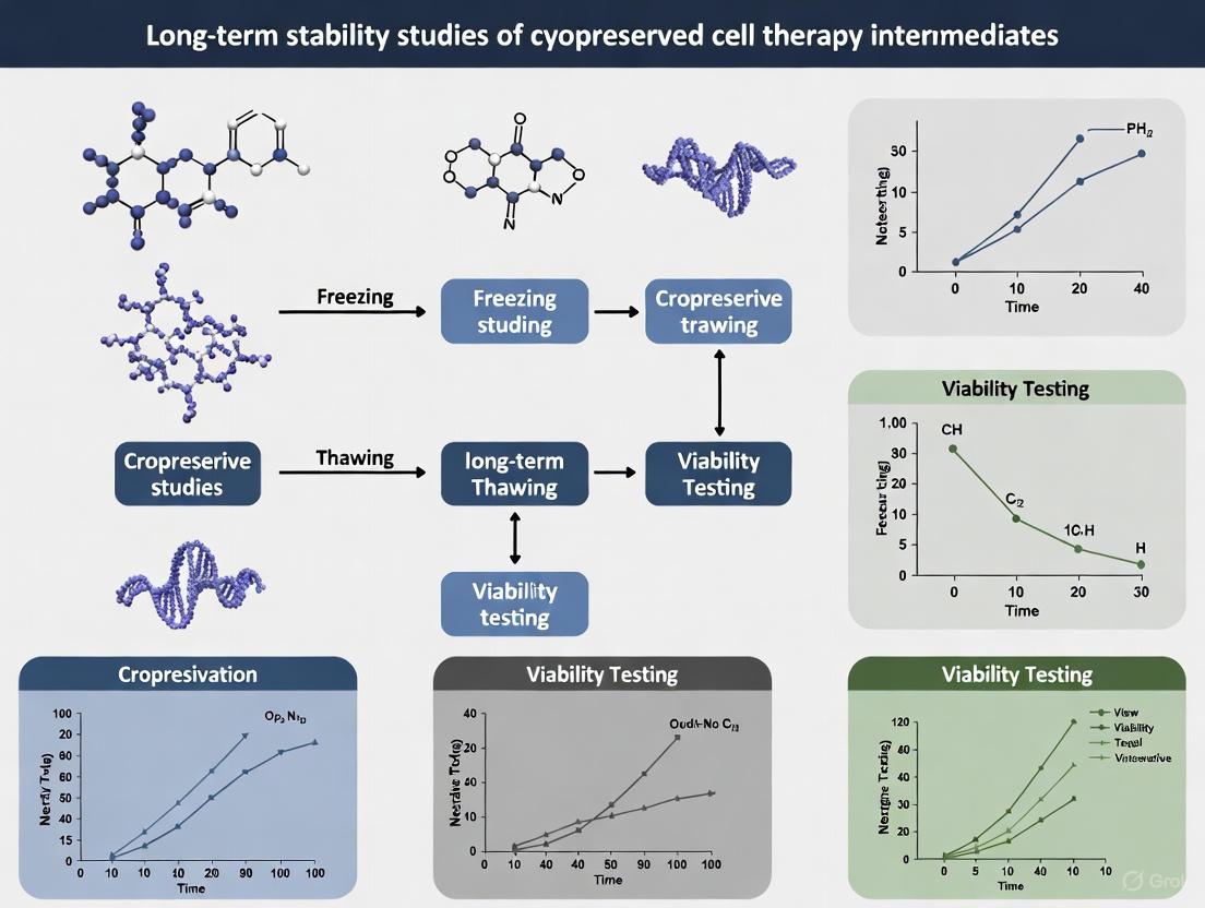
This article provides a comprehensive guide for researchers and drug development professionals on designing and executing long-term stability studies for cryopreserved cell therapy intermediates.
Ensuring Long-Term Stability of Cryopreserved Cell Therapy Intermediates: A Guide to Studies, Strategies, and Success
Abstract
This article provides a comprehensive guide for researchers and drug development professionals on designing and executing long-term stability studies for cryopreserved cell therapy intermediates. It covers the foundational science of cryopreservation, methodological best practices for process design, strategies for troubleshooting common challenges like transient warming events and scalability, and the essential frameworks for product validation and regulatory compliance. By integrating the latest industry survey data, scientific evidence, and expert insights, this resource aims to support the development of robust, scalable, and compliant cryopreservation processes essential for the advancement of cell and gene therapies.
The Science and Critical Importance of Long-Term Cryostability
In the development of cryopreserved cell therapy intermediates, defining and monitoring Critical Quality Attributes (CQAs) is paramount for ensuring product safety, efficacy, and consistency. CQAs are physical, chemical, biological, or microbiological properties that must be within appropriate limits to ensure desired product quality [1]. For cell-based therapies, these attributes must capture both the intended genetic modifications and the overall cellular functionality, providing crucial insights into how product quality varies over time under different environmental conditions [2]. As the cell therapy market progresses toward commercial scale—projected to reach $97 billion by 2033—establishing robust CQA frameworks becomes increasingly vital for managing manufacturing variability and reducing production costs while maintaining therapeutic efficacy [1].
The transition from research to commercial-scale manufacturing necessitates a paradigm shift in how cell therapy intermediates are processed and preserved. While fresh starting materials may seem advantageous in early development, they introduce significant variability that complicates late-stage development and commercialization [3]. Cryopreserved cellular materials offer consistency, flexibility, and predictability that are critical for clinical and commercial manufacturing, though they introduce specific challenges in maintaining CQAs throughout freezing, storage, and thawing processes [3] [4]. This guide systematically compares how different preservation approaches impact the four fundamental CQAs—viability, phenotype, potency, and genomic stability—providing researchers with experimental frameworks for comprehensive long-term stability assessment.
Comparative Analysis of CQA Performance in Preservation Methodologies
Table 1: Comparative Impact of Preservation Methods on Critical Quality Attributes
| CQA Category | Fresh Materials | Cryopreserved Materials | Key Measurement Techniques |
|---|---|---|---|
| Viability | High initial viability (>95%) but rapid decline within 24-72 hours [3] [5] | ≥90% post-thaw viability achievable with optimized protocols; delayed-onset apoptosis possible [5] [2] | 7-AAD/Annexin V staining, metabolic activity assays [2] |
| Phenotype | Donor-to-donor variability; same-donor collection inconsistencies [3] | Preserved lymphocyte proportions (66.59% vs 52.20% in PBMCs); maintained CD3+ T-cell populations (42.01-51.21%) [5] | Flow cytometry, surface marker characterization [5] [2] |
| Potency | Logistically challenging to assess consistently due to perishability [3] | Comparable cytokine production, cytotoxicity, and expansion capabilities to fresh materials [6] [5] | Cytokine release assays, target cell killing, proliferation capacity [6] [1] [2] |
| Genomic Stability | Limited assessment windows; vulnerable to transit delays [3] | Maintained genetic integrity post-thaw; monitoring required for vector integration sites [6] [1] | Vector copy number analysis, integration site profiling (INSPIIRED pipeline) [6] |
Table 2: Cryopreserved Leukapheresis Quality Metrics Across Manufacturing Platforms
| Manufacturing Platform | Post-Thaw Viability | CD3+ T-cell Proportion | Functional Recovery | Cytotoxic Potency |
|---|---|---|---|---|
| Non-viral CAR-T | Comparable to fresh | Comparable to fresh | Post-electroporation recovery confirmed | Equivalent to fresh leukapheresis |
| Lentiviral CAR-T | Comparable to fresh | Comparable to fresh | Comparable transduction efficiency | Equivalent target cell killing |
| Fast CAR-T | Comparable to fresh | Comparable to fresh | Normal expansion kinetics | Preserved cytotoxic function [5] |
Experimental Protocols for CQA Assessment
Viability Assessment Protocol
Comprehensive viability assessment extends beyond simple live/dead counts to detect delayed-onset cell death occurring hours or days after thawing [2]. The following protocol provides a standardized approach:
Materials and Reagents:
- Cryopreserved cell therapy intermediate samples
- 7-AAD and Annexin V staining solutions
- Flow cytometry staining buffer
- Metabolic activity assay kit
- programmed cell death detection kit
Procedure:
- Thawing: Rapidly thaw cryopreserved vials in a 37°C water bath until only a small ice crystal remains.
- Washing: Dilute thawed cells 1:10 in pre-warmed complete medium and centrifuge at 300 × g for 10 minutes to remove cryoprotectants.
- Staining: Resuspend cells in flow cytometry buffer and divide into aliquots for 7-AAD and Annexin V staining according to manufacturer protocols.
- Incubation: Incubate stained cells for 15 minutes at room temperature in the dark.
- Analysis: Analyze samples using flow cytometry within 1 hour of staining, collecting a minimum of 10,000 events per sample.
- Metabolic Assessment: Parallel samples should be assessed using metabolic activity assays per manufacturer instructions.
Interpretation: Viability ≥90% is considered acceptable for most applications, though optimal thresholds are cell-type specific. Simultaneous assessment of apoptosis markers provides insight into potential delayed-onset cell death [5] [2].
Phenotypic Characterization Protocol
Phenotypic stability ensures consistent manufacturing outcomes and therapeutic performance. This protocol evaluates surface marker expression and cell population distribution:
Materials and Reagents:
- Antibody panels for target cell populations
- Flow cytometry staining buffer
- Fc receptor blocking solution
- Fixation buffer
Procedure:
- Cell Preparation: Wash thawed cells twice in staining buffer and count to adjust concentration to 1×10^7 cells/mL.
- Fc Blocking: Incubate cells with Fc receptor blocking solution for 10 minutes on ice.
- Antibody Staining: Add fluorescently-conjugated antibodies according to predetermined optimal concentrations and incubate for 30 minutes in the dark at 4°C.
- Washing: Wash cells twice with staining buffer to remove unbound antibody.
- Fixation: If not analyzing immediately, fix cells with 1% paraformaldehyde.
- Acquisition: Analyze samples using flow cytometry with appropriate compensation controls.
- Analysis: Use forward and side scatter to gate on live cells and analyze marker expression compared to isotype controls.
Key Applications: For CAR-T cell products, focus on T-cell subsets (naïve, stem-cell memory, central memory, effector memory) and activation markers. For leukapheresis products, assess lymphocyte populations and differential counts [6] [5].
Potency Assay Protocol
Potency measurements evaluate biological activity that links to clinical outcomes, serving as stability-indicating assays that detect degradation or loss of product function [2]. For CAR-T products, this multifactorial assessment includes:
Materials and Reagents:
- Target cells expressing appropriate antigen
- Cytokine detection antibodies or ELISA kits
- CFSE or other cell proliferation dyes
- Real-time cell analysis system or chromium-51 release assay components
Procedure: Cytokine Release Assay:
- Co-culture CAR-T cells with target cells at various effector:target ratios (e.g., 1:1, 5:1, 10:1) in triplicate.
- Incubate for 24 hours at 37°C, 5% CO2.
- Collect supernatant and analyze for IFN-γ, IL-2, and TNF-α production using ELISA or multiplex immunoassays.
- Compare cytokine release to reference standards and establish acceptance criteria.
Cytotoxic Activity Assay:
- Label target cells with CFSE or chromium-51 according to manufacturer protocols.
- Co-culture with CAR-T cells at defined effector:target ratios for 4-6 hours.
- Measure target cell killing using flow cytometry (for CFSE) or gamma counter (for chromium-51).
- Calculate specific lysis using appropriate positive and negative controls.
Proliferation Capacity:
- Label CAR-T cells with CFSE and culture with target cells or activating beads.
- Monitor proliferation over 3-5 days using flow cytometry to quantify division cycles.
- Calculate proliferation index compared to unstimulated controls.
Interpretation: Establish a potency profile combining multiple functional readouts that correlate with clinical response [6] [2].
Genomic Stability Assessment Protocol
Genomic stability ensures consistent expression of therapeutic transgenes and minimizes risks associated with insertional mutagenesis. This protocol employs advanced genomic techniques:
Materials and Reagents:
- DNA extraction kit
- Droplet digital PCR reagents
- Next-generation sequencing library preparation kit
- T-cell receptor sequencing reagents
Procedure: Vector Copy Number (VCN) Analysis:
- Extract genomic DNA from approximately 1×10^6 cells using validated methods.
- Perform droplet digital PCR using primers specific to the transgene and a reference gene.
- Calculate VCN using the formula: VCN = (transgene concentration)/(reference gene concentration).
- Establish acceptance criteria (typically 1-5 copies per cell for most applications).
Integration Site Analysis:
- Prepare sequencing libraries using the INSPIIRED or EpiVIA pipeline [6].
- Sequence using Illumina platforms with sufficient depth (>10 million reads).
- Analyze integration sites for enrichment in genomic regions associated with oncogenes or tumor suppressors.
- Monitor for clonal expansion patterns that may indicate selective growth advantages.
TCR Repertoire Profiling:
- Extract RNA and prepare libraries for TCR sequencing.
- Sequence using immune profiling platforms.
- Analyze clonal diversity and distribution, focusing on oligoclonality as a potential indicator of restricted functionality.
Interpretation: Monitor for changes in VCN, emergence of dominant integration sites, and reduction in TCR diversity that may indicate genomic instability [6].
The Scientist's Toolkit: Essential Research Reagents and Platforms
Table 3: Essential Research Reagents for CQA Assessment
| Reagent Category | Specific Examples | Function in CQA Assessment | Application Notes |
|---|---|---|---|
| Cryopreservation Media | CS10 (10% DMSO), DMSO-free cryomedium | Maintain viability and functionality post-thaw | Clinical-grade CS10 minimizes erythrocyte interference [5] |
| Viability Assay Reagents | 7-AAD, Annexin V, metabolic dyes | Distinguish live, apoptotic, and dead cells | Combined approach detects delayed-onset apoptosis [2] |
| Phenotypic Characterization | Fluorochrome-conjugated antibodies for T-cell subsets | Identify memory, naïve, and effector populations | Essential for correlating phenotype with persistence [6] |
| Potency Assay Components | Target cell lines, cytokine detection antibodies | Measure biological function and cytotoxic potential | Use same target cells across experiments for consistency [6] [2] |
| Genomic Analysis Kits | ddPCR reagents, NGS library prep kits | Quantify vector copy number, integration sites | Digital PCR provides precise quantification for VCN [6] |
Technological Frameworks for CQA Monitoring
Advanced technological platforms enable comprehensive CQA monitoring throughout product development:
Multi-omics Integration: Combined genomic, epigenomic, transcriptomic, proteomic, and metabolomic approaches provide unprecedented resolution in characterizing cell therapy products [6]. DNA methylation profiling of CD19 CAR T-cell products has identified distinct epigenetic loci associated with complete response and survival outcomes post-infusion [6]. Single-cell RNA sequencing with TCR repertoire profiling enables correlation of transcriptional phenotypes with clonal expansion patterns.
Process Analytical Technologies: The implementation of controlled-rate freezing with detailed process monitoring represents a significant advancement in cryopreservation quality control. Industry surveys indicate that 87% of respondents use controlled-rate freezing, with 60% utilizing default profiles while others optimize conditions for specific cell types [4]. Freeze curve monitoring during controlled-rate freezing provides critical process data that can predict post-thaw quality attributes.
Stability-Indicating Methodologies: Unlike traditional chemical assays, functional potency assays serve as stability-indicating methods for cell therapies, detecting subtle changes in biological activity that may not be apparent through viability assessments alone [2]. These include real-time calcium imaging, cytokine release profiling, and cytotoxic function assays that collectively provide a comprehensive stability profile.
The systematic assessment of viability, phenotype, potency, and genomic stability provides a critical framework for ensuring the quality of cryopreserved cell therapy intermediates throughout development and commercialization. As the field advances toward distributed manufacturing models, cryopreserved leukapheresis has demonstrated particular promise as a universal raw material that maintains critical quality attributes across multiple manufacturing platforms while decoupling production from fresh material logistics [5].
Successful implementation of CQA monitoring requires careful selection of stability-indicating assays that reflect the mechanism of action and link to clinical outcomes. The experimental protocols outlined provide researchers with standardized methodologies for comprehensive product characterization. By establishing robust correlation between CQAs and therapeutic performance during long-term stability studies, developers can ensure consistent product quality throughout the shelf life, ultimately delivering safe and effective cell therapies to patients.
This guide provides a comparative analysis of the long-term stability of human induced pluripotent stem cells (hiPSCs) following extended cryopreservation, a critical factor for clinical and commercial manufacturing of cell therapy products. The data, derived from a pivotal five-year stability study, objectively compares the post-thaw performance of cGMP-compliant iPSC lines against key quality benchmarks required for therapeutic applications. The findings demonstrate that with appropriate manufacturing and banking strategies, iPSCs can maintain genomic stability, pluripotency, and differentiation capacity over extended periods, supporting their use as a reliable starting material for regenerative medicine.
For induced pluripotent stem cells (iPSCs) to transition from research tools to reliable clinical therapeutics, demonstrating long-term stability after cryopreservation is non-negotiable. Cell banks intended for commercial therapies may need to remain in storage for many years before use in manufacturing, during which time they must retain their critical quality attributes (CQAs) [7]. These CQAs include genomic stability, post-thaw viability, pluripotency, and differentiation potential—all essential for ensuring the safety, potency, and consistency of final cell therapy products [8]. This guide examines experimental evidence from a five-year stability study on cGMP-compliant human iPSC lines, providing a benchmark for comparing the performance of clinical-grade stem cell banks after long-term storage.
Experimental Protocols & Workflow
The foundational evidence for this guide comes from a study that assessed the stability of iPSC master cell banks (MCBs) and working cell banks (WCBs) manufactured and cryopreserved under current Good Manufacturing Practices (cGMP) five years prior to analysis [7] [9].
Cell Lines and cGMP Banking
Three iPSC lines (LiPSC-GR1.1, LiPSC-18R, and LiPSC-ER2.2) generated from healthy donors were used. These lines were originally manufactured under cGMP-compliant conditions and cryopreserved in the vapor phase of liquid nitrogen [7].
Post-Thaw Analysis Workflow
The following flowchart illustrates the comprehensive experimental workflow used to evaluate the thawed cells.
Key Analytical Assays
- Viability and Recovery: Cell count and viability (CCV) were measured post-thaw. Percent recovery was calculated as (total viable cells post-thaw / total viable cells frozen) × 100 [7].
- Pluripotency Markers: Immunofluorescence staining and flow cytometry were performed for standard pluripotency markers (SSEA4, Tra-1-81, Tra-1-60, Oct4). Alkaline phosphatase (ALP) staining assessed plating efficiency [7].
- Genomic Stability: Karyotyping was conducted to detect chromosomal abnormalities. Telomerase activity and telomere length were also evaluated [7] [9].
- Differentiation Potential: Spontaneous differentiation via embryoid body (EB) formation and directed differentiation into lineages of the three germ layers were performed:
- Sterility Testing: Tests for mycoplasma, bacteria, and fungi were conducted post-thaw and at multiple passages [7].
Comparative Performance Data After 5 Years
The quantitative data below summarize the post-thaw performance of the iPSC lines, demonstrating their retention of critical quality attributes after five years of cryopreservation.
Table 1: Post-Thaw Recovery and Pluripotency of iPSCs After 5-Year Cryopreservation
| iPSC Line | Post-Thaw Viability (%) | Percent Recovery (%) | Pluripotency Marker Expression (>95%) | Normal Karyotype |
|---|---|---|---|---|
| LiPSC-18R | 83.3 | 81.5 | Confirmed (SSEA4, Tra-1-81, Tra-1-60, Oct4) | Maintained |
| LiPSC-TR1.1 | 75.2 | 82.0 | Confirmed (SSEA4, Tra-1-81, Tra-1-60, Oct4) | Maintained |
| LiPSC-ER2.2 | 81.2 | 57.5 | Confirmed (SSEA4, Tra-1-81, Tra-1-60, Oct4) | Maintained |
Table 2: Differentiation Potential and Expansion Capability Post-Thaw
| Quality Attribute | Pre-Cryopreservation Performance | Post-Thaw Performance (5 Years) | Assessment Method |
|---|---|---|---|
| Spontaneous Differentiation | Demonstrated (3 germ layers) | Maintained | EB formation; Immunostaining for β-Tubulin (ectoderm), AFP (endoderm), SMA (mesoderm) |
| Directed Differentiation to NSCs | High efficiency | >90% Pax6+ cells at P3 | Flow cytometry, Immunofluorescence (Nestin, Pax6) |
| Directed Differentiation to DE | High efficiency | Maintained | Commercial kit with Activin A |
| 2D Expansion Potential | Robust over 15+ passages | Maintained over 15 passages | Serial passaging, morphology observation |
| 3D Expansion Potential | Robust in spinner flasks | Maintained | Culture in feeder-free, matrix-dependent 3D environment |
| Sterility | Negative (mycoplasma, bacteria) | Negative at all test points | Microbiological testing |
The Scientist's Toolkit: Essential Research Reagents
The following reagents and materials were critical to the success of the long-term stability study and are essential for replicating this work.
Table 3: Key Research Reagent Solutions for iPSC Banking and Quality Control
| Reagent/Material | Function in Protocol | Example Product/Citation |
|---|---|---|
| cGMP-Compliant Reprogramming System | Generating clinical-grade iPSCs; minimizes genomic integration risk | Episomal vectors [8]; Sendai virus vectors [11] |
| Cryopreservation Medium | Protects cell viability during freeze-thaw cycles; often contains DMSO | 90% KOSR + 10% DMSO [11]; CryoStor CS10 [12] |
| ROCK Inhibitor (Y-27632) | Enhances post-thaw survival by inhibiting apoptosis | Added to culture medium 20-24 hours post-thaw [11] [12] |
| L7TM Matrix | Feeder-free substrate for iPSC attachment and growth post-thaw | Used for plating thawed iPSC aggregates [7] |
| Synthemax II-coated Microcarriers | Enables scalable 3D expansion in bioreactors | Used in stirred tank bioreactor systems [13] |
| Small Molecule Differentiation Inducers | Directs lineage-specific differentiation for potency testing | CHIR99021 (Wnt activator), SB431542 (TGF-β inhibitor) [7] [10] |
| VitroGel Hydrogel Matrix | Supports 3D culture and organoid formation for complex differentiation assays | Animal-free hydrogel for 3D cell culture [12] |
Signaling Pathways in Directed Differentiation
The functional validation of thawed iPSCs relied heavily on directed differentiation, which activates conserved developmental signaling pathways. The following diagram illustrates the key pathways involved in generating neural and endodermal lineages.
Implications for Cell Therapy Development
The consistent performance of iPSCs after five years of cryopreservation has profound implications for the cell therapy industry. It validates the feasibility of establishing cGMP-compliant master and working cell banks as a long-term, reliable source of starting materials for clinical and commercial manufacturing [7]. This reliability is crucial for allogeneic therapies, where a single banked cell line is used to produce treatments for multiple patients [13].
Furthermore, the retention of differentiation potential means that banked iPSCs can yield functional, therapeutic cell types after long-term storage. This is demonstrated not only by the differentiation into NSCs and DE but also by other studies showing that cryopreserved iPSC-derived neurons successfully engraft in animal models and maintain functionality [14]. Such reproducibility is essential for meeting regulatory requirements and ensuring consistent product quality in clinical trials and beyond.
The five-year stability data provide compelling evidence that cGMP-compliant human iPSCs retain their critical quality attributes after long-term cryopreservation. The quantitative comparisons presented in this guide demonstrate maintained genomic integrity, high viability, robust pluripotency marker expression, and multilineage differentiation potential across multiple cell lines. For researchers and therapy developers, these findings underscore the importance of rigorous manufacturing and banking protocols and offer a benchmark for evaluating the long-term stability of iPSC-based products. This evidence significantly de-risks the use of banked iPSCs as a starting material for the scalable and consistent manufacturing of regenerative medicine products.
The Role of Cryopreservation in Scalable Allogeneic Therapy Supply Chains
Allogeneic cell therapies represent a transformative shift in regenerative medicine, offering "off-the-shelf" options to treat multiple patients from a single cell source [15]. Unlike autologous therapies, which are individualized, allogeneic therapies are inherently more scalable, making them a promising pathway to more accessible treatments at a sustainable price [15]. Cryopreservation serves as the critical enabler of this model by allowing batch production, stockpiling, and global distribution of cell therapies, effectively decoupling manufacturing from administration [16]. The ability to bank frozen cells is particularly essential for allogeneic therapies, where thousands of doses are manufactured in single large batches before patients are ready for treatment [17]. This logistical linchpin addresses one of the most significant bottlenecks in cell therapy – the need for scalable, cost-effective distribution systems that maintain product quality and efficacy throughout the supply chain.
The growing importance of cryopreservation is reflected in market projections, with the global allogeneic cell therapy market expected to reach $2.4 billion by 2031, up from $0.4 billion in 2024, representing a compound annual growth rate of 24.1% [15]. However, realizing this potential requires overcoming substantial challenges in cryopreservation science, including maintaining cell viability, potency, and functionality post-thaw, while ensuring compliance with increasingly stringent regulatory requirements [18] [16]. This review examines the current state of cryopreservation technologies, comparative performance data, and methodological approaches that support the development of robust, scalable supply chains for allogeneic cell therapies.
Current Cryopreservation Methodologies and Comparative Analysis
Fundamental Principles and Standard Practices
Cryopreservation protocols for living cells have been developed over several decades and typically involve using 5%-10% dimethyl sulfoxide (DMSO) as a cryoprotective agent (CPA) [18]. The standard process involves freezing cell suspensions at a controlled rate of approximately 1°C per minute to a temperature of -80°C, followed by storage in the vapor phase of liquid nitrogen at approximately -130°C [18]. When needed, cells are rapidly thawed in a 37°C water bath or with an automated thawing device, with subsequent removal of DMSO due to its cytotoxicity at temperatures above 0°C [18].
The underlying science of cryopreservation revolves around managing the physical and chemical stresses that cells experience during freezing and thawing. As temperatures decrease, extracellular ice formation concentrates solutes in the unfrozen fraction, creating osmotic stress that can lead to cell shrinkage and damage. Intracellular ice formation, which typically occurs at rapid cooling rates, can be mechanically destructive to cellular structures. Cryoprotective agents like DMSO mitigate these effects by penetrating cells and reducing ice crystal formation, but they introduce their own challenges including toxicity and the need for post-thaw removal [18].
Comparative Performance of Cryopreservation Systems
Table 1: Comparison of Controlled-Rate Freezing vs. Passive Freezing Methods
| Parameter | Controlled-Rate Freezing (CRF) | Passive Freezing |
|---|---|---|
| Control over process parameters | High control over cooling rate, nucleation temperature, and other critical parameters | Limited control, dependent on equipment and environment |
| Impact on Critical Quality Attributes | Enables defined parameters for cytokine release, viability, and functionality | Limited control over CQAs, potential for inconsistent results |
| Infrastructure requirements | High-cost equipment, specialized expertise, liquid nitrogen consumption | Low-cost, simple operation, minimal technical barrier |
| Scalability | Potential bottleneck for batch scale-up | Easier scaling, simple operation |
| Regulatory compliance | Preferred for late-stage and commercial products, better documentation | Mainly used in early clinical development (up to phase II) |
| Adoption rate | 87% of survey participants [4] | 13% of survey participants [4] |
Table 2: Post-Thaw Viability and Recovery Across Cell Types
| Cell Type | Standard Protocol Viability | Optimized Protocol Viability | Key Optimization Parameters |
|---|---|---|---|
| T-cells (CAR-T) | Varies; challenges with default CRF profiles reported [4] | Improved with optimized warming rates (~45°C/min) [4] | Cooling rate, warming rate, CPA composition |
| iPSC-derived cells | Suboptimal with conventional slow-freeze protocols [18] | Enhanced with optimized freezing profiles [18] | Cell-specific freezing profiles, DMSO-free media |
| Glioblastoma cells | Poor recovery after long-term cryostorage [19] | Significant improvement with Matrigel + 20% FBS [19] | Extracellular matrix, serum concentration |
| Regulatory T-cells (Tregs) | Viability decrease post-cryopreservation [20] | Preserved immunosuppressive function [20] | Cryopreservation medium composition |
| MSCs, NK cells | Default CRF profiles often adequate [4] | May require optimization for specific applications | Cell harvest condition, primary container |
Industry surveys reveal that 87% of respondents use controlled-rate freezing for cryopreservation of cell-based products, while only 13% rely on passive freezing methods [4]. Notably, 86% of those using passive freezing have products exclusively in earlier stages of clinical development (up to phase II), suggesting a transition to controlled-rate freezing as products approach commercialization [4]. This trend reflects the greater process control and documentation capabilities of controlled-rate systems, which are essential for regulatory compliance and product consistency at commercial scale.
Experimental Approaches for Cryopreservation Optimization
Standardized Freezing and Thawing Protocols
Controlled-Rate Freezing Methodology: The qualification of controlled-rate freezers should include comprehensive evaluation of multiple parameters rather than relying solely on vendor qualifications. A robust qualification protocol includes: (1) temperature mapping across a grid of locations within the chamber; (2) freeze curve mapping across different container types; (3) evaluation of mixed loads with varying masses and container configurations; and (4) comparison of full versus empty chamber performance [4]. This approach ensures understanding of both standard operating conditions and performance limits.
For protocol development, the freezing process should document and control several critical parameters: the rate of cooling before nucleation (affecting chilling injury and CPA toxicity), the temperature of ice nucleation (impacting osmotic stress and intracellular ice formation), the rate of cooling after nucleation (influencing dehydration and intracellular ice), and the final sample temperature before transfer to cryogenic storage [4]. These parameters collectively determine the cryoinjury extent and post-thaw recovery.
Thawing Optimization Protocol: Controlled thawing methodologies are equally critical for maintaining cell viability and function. The established good practice for thawing includes a warming rate of 45°C/min, though recent evidence indicates that different warming rates may be optimal for specific cell types like T cells, particularly when cooling rates are slow (-1°C/min or slower) [4]. The standard protocol involves: (1) retrieving cryopreserved cells from liquid nitrogen storage; (2) adding a small volume (100μL) of pre-warmed culture medium to the top of frozen cells; (3) rapid thawing in a 37°C water bath with swirling for maximum 1 minute; (4) transfer to a larger volume of medium with incremental dilution to minimize osmotic shock; and (5) centrifugation at 300-400×g for 10 minutes followed by resuspension in fresh medium [20] [18]. Controlled thawing devices are increasingly preferred over conventional water baths due to better GMP compliance, reduced contamination risk, and more consistent performance [4].
DMSO Reduction and Elimination Strategies
The development of DMSO-free cryopreservation media represents a significant advancement in allogeneic therapy supply chains, particularly for novel administration routes such as direct injections into the brain, spine, or eye [18]. The standard approach involves systematic screening of alternative cryoprotectants, including permeating agents (e.g., ethylene glycol, propylene glycol) and non-permeating agents (e.g., sucrose, trehalose, hydroxyethyl starch), often used in combination [18]. Optimization requires careful adjustment of freezing profiles, as DMSO-free methods typically yield suboptimal post-thaw viability with conventional slow-freeze protocols [18].
The following workflow diagram illustrates the key decision points in developing optimized cryopreservation protocols:
A critical consideration in DMSO-free formulation is the safety profile for direct administration. While intravenous DMSO administration is established for certain therapies, novel administration routes lack sufficient safety data. In vitro studies indicate significant cytotoxicity concerns, with DMSO concentrations as low as 0.5-1% causing substantial viability loss in sensitive neuronal cells [18]. These findings underscore the importance of DMSO removal or elimination for therapies administered via sensitive routes.
Enhanced Recovery Protocols for Challenging Cell Types
Glioblastoma Cell Recovery Protocol: Research on patient-derived glioblastoma cells cryopreserved for over ten years demonstrates that optimized recovery conditions can significantly improve viability and expansion. The enhanced protocol includes: (1) thawing cells and plating on tissue culture surfaces coated with 0.3 mg/ml Matrigel; (2) culturing in high glucose DMEM supplemented with 20% FBS (doubled from standard 10%); (3) maintaining cultures in normoxic conditions (21% O2) with 5% CO2 at 37°C; and (4) medium replacement every 3 days with subculture at 80% confluence using 0.05% trypsin-EDTA [19]. This optimized approach significantly increased viability and proliferative capacity compared to standard procedures, associated with increased expression of YAP and TLR4, key regulators of cell proliferation [19].
Functional Preservation Assessment: For regulatory T-cells (Tregs), a specialized protocol demonstrates preservation of immunosuppressive function post-cryopreservation. The methodology includes: (1) cryopreservation in PBS containing 10% human serum albumin and 10% DMSO; (2) controlled-rate freezing in Corning CoolCell containers at -80°C for 20h to 1 week; (3) transfer to liquid nitrogen tanks for long-term storage; and (4) post-thaw functional assessment via suppression assays [20]. These assays involve co-culture of Tregs with CellTrace Violet-labeled responder PBMCs stimulated with anti-CD3/CD28 antibodies at varying ratios (1:1, 1:0.5, 1:0.25 responder:Treg ratios), with suppression measured after 5 days of incubation [20]. This approach confirms that cryopreserved Tregs maintain their critical immunosuppressive capacity despite decreases in overall viability and CD4+ T-cell populations [20].
Essential Research Reagents and Solutions
Table 3: Key Research Reagents for Cryopreservation Studies
| Reagent/Solution | Function | Application Notes |
|---|---|---|
| DMSO (Dimethyl sulfoxide) | Penetrating cryoprotectant reduces intracellular ice formation | Standard concentration 5-10%; requires post-thaw removal due to cytotoxicity [18] |
| Human Serum Albumin | Protein stabilizer, reduces membrane damage during freezing | Used at 10% concentration in clinical-grade freezing media [20] |
| Matrigel | Extracellular matrix providing structural support and signaling | 0.3 mg/ml concentration significantly improves recovery of challenging cells [19] |
| Fetal Bovine Serum (FBS) | Source of growth factors and nutrients | Increased to 20% (from standard 10%) enhances recovery of cryopreserved cells [19] |
| Hydroxyethyl Starch (Hespan) | Non-penetrating cryoprotectant, promotes RBC agglutination | Subject to supply chain shortages; requires mitigation strategies [21] |
| Lymphoprep | Density gradient medium for cell separation | Used in PBMC isolation prior to cryopreservation [20] |
| ACK Lysing Buffer | Red blood cell lysis for purification | Improves antigen sensitivity of memory T cells post-thaw [20] |
| CellTrace Violet | Fluorescent cell labeling for proliferation assays | Enables assessment of functional immunosuppression in Tregs [20] |
The selection of appropriate reagents is further complicated by supply chain vulnerabilities, as illustrated by the Hespan shortage in 2023 following a manufacturer decision to discontinue production [21]. This highlights the importance of dual-sourcing strategies and qualification of alternative reagents for critical process components.
Supply Chain Integration and Regulatory Considerations
Cold Chain Management and Logistics
Effective integration of cryopreservation into allogeneic therapy supply chains requires robust cold chain management systems. The process flow encompasses multiple critical steps: (1) controlled-rate freezing of final product; (2) transfer to cryogenic storage systems; (3) inventory management and monitoring; (4) coordinated shipping with temperature-maintained packaging; (5) receipt and storage at clinical sites; and (6) final thawing and administration to patients [4]. Each step introduces potential failure points that must be carefully controlled through standardized procedures and continuous monitoring.
Logistical challenges are compounded by global supply chain dependencies, with critical materials often sourced across continents. Serum may originate from New Zealand, carbon nutrients from China, and growth factors from the United States, creating vulnerabilities to global health crises, geopolitical conflicts, natural disasters, and trade disputes [22]. These dependencies impact availability, delivery timelines, and ultimately costs, necessitating robust risk mitigation strategies including strategic stockpiling, alternative sourcing, and formulation flexibility.
Regulatory and GMP Compliance Framework
Establishing GMP-compliant manufacturing frameworks requires careful planning from early development stages. Key considerations include: (1) selection of excipient GMP-grade cell culture media with proper documentation; (2) implementation of defined, animal component-free formulations; (3) comprehensive raw material traceability covering viral safety and supplier qualifications; and (4) validated freezing protocols with controlled cryogenic storage and continuous monitoring [16]. Early engagement with regulatory experts is strongly recommended, ideally during or just after proof-of-concept development, to align development strategies with regulatory expectations and ensure smooth transition from bench to clinic [16].
The diagram below illustrates the interconnected challenges in developing scalable cryopreservation processes:
Industry surveys identify scaling as the predominant hurdle for cryopreservation in cell and gene therapy, with 22% of respondents citing "Ability to process at a large scale" as the biggest challenge to overcome [4]. Additionally, 75% of respondents cryopreserve all units from an entire manufacturing batch together, reflecting current small-batch manufacturing scales and the technical challenges of dividing batches for cryopreservation while maintaining consistency [4].
Cryopreservation remains an indispensable technology for enabling scalable allogeneic cell therapy supply chains, though significant challenges persist. The field is evolving toward more sophisticated approaches including DMSO-free cryopreservation media, cell-type specific optimization protocols, and improved temperature control systems throughout the cold chain. Future developments will likely focus on standardized, chemically-defined cryopreservation formulations, advanced temperature monitoring technologies, and integrated closed-system processing to enhance both product quality and supply chain efficiency.
As the allogeneic therapy market continues its rapid growth, with projections of 24.1% CAGR through 2031 [15], robust cryopreservation strategies will become increasingly critical for realizing the potential of off-the-shelf cell therapies. Success will require interdisciplinary collaboration between cryobiologists, process engineers, supply chain specialists, and regulatory experts to develop integrated solutions that maintain cell quality and function from manufacturing to patient administration. Through continued optimization and innovation in cryopreservation science, the field can overcome current scalability limitations and fulfill the promise of accessible, effective allogeneic cell therapies for broad patient populations.
For researchers and drug development professionals in the cell and gene therapy (CGT) space, establishing a scientifically justified and regulatory-compliant shelf life is a critical milestone. This guide objectively compares the performance of different stability approaches and storage modalities for cryopreserved cell therapy intermediates, supported by experimental data from recent studies.
Stability Profiles of Cryopreserved Cell Therapy Intermediates
The following table summarizes key quantitative findings from recent, comprehensive stability studies on various cryopreserved Advanced Therapy Medicinal Products (ATMPs).
Table 1: Long-Term Stability Data for Cryopreserved Cell Therapy Intermediates
| Product Type | Storage Condition | Maximum Storage Duration Studied | Key Stability Findings | Experimental Assays Used | Source |
|---|---|---|---|---|---|
| Various Cell-Based ATMPs (19 products) | Vapor phase of liquid nitrogen (< -150°C) | 13.5 years | No diminished viability or efficacy; "very stable long term" [23]. | Cell viability, immunophenotype, potency assays (immunosuppression, cytotoxicity), sterility, endotoxin [23]. | Lombardy Plagencell Network |
| Clinical-Grade Lentiviral Vectors | -80°C | 8 years (99 months) | No statistically significant change in titer over time; functional transduction efficiency maintained [24]. | SupT1 titer assay, transduction efficiency in clinical T-cells, flow cytometry for CAR expression, IFN-γ ELISA for potency [24]. | University of Pennsylvania |
| Cell Therapy Products | Vapor phase of liquid nitrogen (-135°C to -196°C) | Industry standard for long-term storage | Maintains viability and critical quality attributes (CQAs); requires controlled-rate freezing and validated systems [25] [4]. | Post-thaw viability, flow cytometry, metabolic activity, potency assays [25] [26]. | Industry Best Practices |
Experimental Protocols for Stability Studies
Generating regulatory-grade stability data requires rigorous, standardized protocols. The methodologies below are derived from the cited studies and can serve as templates for your own stability study design.
1. Protocol for Long-Term Stability of Cell-Based Drug Products This methodology is adapted from the multi-year study on 19 ATMPs [23].
- Objective: To define the shelf-life of cryopreserved cell-based drug products by monitoring Critical Quality Attributes (CQAs) over time.
- Materials: Cryopreserved drug products in primary containers (cryobags/vials), vapor-phase liquid nitrogen storage system, controlled-rate freezer.
- Cryopreservation Media: Various excipients containing 10% DMSO [23].
- Procedure:
- Sample Withdrawal: At pre-defined intervals (e.g., 3, 6, 12 months, then annually), withdraw replicate samples from storage.
- Thawing: Rapidly thaw samples using a controlled method (e.g., 37°C water bath) to ensure consistency.
- Post-Thaw Analysis: Immediately test samples for the following attributes:
- Cell Viability: Using dye exclusion methods (e.g., Trypan Blue) or automated cell counters.
- Immunophenotype: Flow cytometry analysis of surface markers to confirm cell identity and purity.
- Potency: Perform functional assays relevant to the mechanism of action (e.g., cytotoxicity against target cells, cytokine release upon stimulation, proliferation/differentiation capacity).
- Microbiological Safety: Test for sterility, endotoxin, and mycoplasma per pharmacopoeial methods.
- Data Analysis: Plot CQA data over time to establish degradation kinetics and justify the proposed shelf-life.
2. Protocol for Stability of Lentiviral Vectors This protocol is based on the comprehensive analysis of 13 clinical-grade LV lots [24].
- Objective: To assess the long-term stability of LV vector titer and functionality.
- Materials: Aliquots of GMP-grade LV lots, -80°C freezer with continuous temperature monitoring, target cells (e.g., SupT1 cells for titer, human T-cells for transduction).
- Procedure:
- Storage: Maintain vector lots at -80°C. The freezer must be qualified and monitored [24].
- Titer Testing:
- At initial release and at subsequent time points (e.g., annually), perform titer assays.
- Infect SupT1 cells with serial dilutions of the vector.
- Quantify transgene expression (e.g., by flow cytometry for a surface marker or reporter gene) to calculate Transducing Units (TU)/mL.
- Functional Transduction Efficiency:
- Use stored vectors to transduce clinical T-cell products at a fixed Multiplicity of Infection (MOI).
- After a standard culture period, measure the percentage of transduced cells (e.g., CAR+ cells) by flow cytometry.
- Potency Assessment: Perform a co-culture assay where transduced T-cells are incubated with target cells. Measure effector function output, such as IFN-γ release via ELISA [24].
- Data Analysis: Use statistical models (e.g., linear mixed-effects model) to analyze titer and transduction efficiency over time for significance.
Stability Testing Workflow and CQA Assessment
The diagrams below outline the logical workflow for designing a stability study and the multi-parametric nature of assessing cell product stability.
Stability Study Design Workflow
Assessing Critical Quality Attributes (CQAs)
The Scientist's Toolkit: Essential Reagents & Materials
A successful stability study relies on high-quality, GMP-compatible reagents and materials. The following table details key solutions and their functions in the context of cryopreservation and stability testing.
Table 2: Essential Research Reagent Solutions for Cryopreservation & Stability Studies
| Reagent / Material | Function & Importance in Stability Studies | Key Considerations |
|---|---|---|
| Cryoprotectants (e.g., DMSO) | Prevents intracellular ice crystal formation, a primary cause of cell death during freezing [25]. | Typically used at 10% concentration. Can be toxic; post-thaw washing may be required for clinical-grade materials [25]. |
| Optimized Cryopreservation Media | Provides a protective matrix for cells during freeze-thaw. Enhanced formulations can improve post-thaw recovery [25] [26]. | May include additives like HypoThermosol. Move towards GMP-compliant, serum-free, and xeno-free media [26]. |
| Controlled-Rate Freezer (CRF) | Precisely controls cooling rate (e.g., ~1°C/min), which is critical for maintaining cell viability and CQAs [25] [4]. | Superior to passive freezing for process control. 87% of industry survey respondents use CRFs [4]. |
| Validated Primary Containers | Cryogenic vials or bags that hold the product. Integrity is critical to prevent contamination and maintain sterility [26]. | Must be validated for cryogenic temperatures and compatible with cell products. Impacts container closure integrity (CCI) studies [27]. |
| Stability-Indicating Assays | Analytical procedures that detect changes in CQAs (viability, phenotype, potency) over time [26]. | FDA expects these methods in stability programs. Go beyond simple viability to include functional potency assays [26]. |
Key Regulatory and Industry Considerations
Navigating the regulatory landscape is paramount. Here are essential insights derived from current industry practices and guidelines:
- Adopt a Proactive, Lifecycle Approach: A compliant, globally consistent storage strategy that protects samples from discovery to commercialization is no longer an afterthought but a strategic pillar of drug development [25].
- Focus on Contamination Control: The revised EU GMP Annex 1 implicates storage zones associated with aseptic processes. Your Contamination Control Strategy (CCS) must extend to cryopreservation and storage activities [25].
- Prioritize Data Integrity: Regulators (FDA, MHRA, EMA) are increasingly focused on real-time monitoring, validated storage systems, and digital, data-driven chain-of-custody evidence. Manual records are insufficient [25].
- Address Scaling Early: Scaling cryopreservation was identified as the biggest hurdle for 22% of industry professionals [4]. Consider batch sizing and freezer capacity early in process development to avoid future bottlenecks.
- Justify Freezing Protocols: While 60% of survey respondents use default CRF profiles, sensitive cell types (e.g., iPSCs, differentiated cells) often require optimized, product-specific freezing parameters [4].
Designing Robust Cryopreservation and Stability Testing Protocols
For researchers and drug development professionals working with cell therapy intermediates, the choice of a cryopreservation strategy is a critical decision point that significantly impacts long-term product stability, quality, and therapeutic efficacy. The fundamental goal of cryopreservation is to enable long-term storage of biological materials by halting biochemical activity while preserving viability and function upon thawing. [28] Two primary methods have emerged for this purpose: controlled-rate freezing (CRF) and passive freezing.
Controlled-rate freezing utilizes specialized equipment to precisely lower sample temperature at a predetermined rate, typically around -1°C per minute. [29] In contrast, passive freezing (also called uncontrolled-rate freezing) involves placing samples in insulated containers (e.g., isopropanol freezing containers) held at -80°C, resulting in a non-linear, uncontrolled cooling profile. [30] As the cell and gene therapy field advances toward commercialization, understanding the technical nuances, advantages, and limitations of each method becomes essential for maintaining manufacturing standards and regulatory compliance. [4] This guide provides an objective comparison of these technologies with supporting experimental data to inform strategic decision-making for cryopreservation protocol development.
Fundamental Principles and Comparative Analysis
Mechanism of Action
Successful cryopreservation hinges on managing the physical stresses cells experience during freezing, primarily osmotic stress and intracellular ice crystal formation, which can mechanically disrupt cellular membranes. [31] Both freezing methods utilize cryoprotective agents (CPAs) like dimethyl sulfoxide (DMSO) to mitigate these effects. DMSO reduces ice crystal formation by depressing the freezing point of water and facilitating vitrification—the formation of an amorphous, glass-like solid instead of crystalline ice. [31] [29]
Controlled-Rate Freezing: This method actively manages the phase change process. It allows precise control over critical parameters: the cooling rate before nucleation, the temperature of ice nucleation itself, and the cooling rate after nucleation. [4] This level of control helps manage cellular dehydration and minimizes lethal intracellular ice formation. Advanced CRF systems can even initiate controlled nucleation ("cold-spike") at a consistent temperature across all samples, significantly improving process uniformity. [32]
Passive Freezing: This method relies on passive heat transfer from the sample to the cold environment. The cooling rate is not controlled or monitored in real-time and can vary significantly based on factors including sample volume, vial type, and the number of samples in the freezing container. [30] This results in an unpredictable freezing profile, making it difficult to consistently replicate optimal conditions for sensitive cell types.
Direct Comparison of Advantages and Limitations
The choice between CRF and passive freezing involves balancing control, consistency, cost, and operational complexity. The table below summarizes the core advantages and limitations of each method.
Table 1: Comprehensive comparison of controlled-rate and passive freezing methods
| Aspect | Controlled-Rate Freezing (CRF) | Passive Freezing |
|---|---|---|
| Process Control | High precision over cooling rate and nucleation temperature; user-defined profiles. [4] | Minimal control; cooling rate is variable and unpredictable. [30] |
| Reproducibility | High batch-to-batch consistency; essential for cGMP manufacturing. [4] [30] | Lower consistency; potential for significant sample-to-sample variability. [30] |
| Infrastructure Cost | High initial investment and ongoing liquid nitrogen/operational costs. [4] [30] | Low-cost; requires only a -80°C freezer and inexpensive consumables. [30] |
| Technical Expertise | Requires specialized expertise for operation, optimization, and qualification. [4] | Low technical barrier; simple, one-step operation. [4] |
| Scalability | Can be a bottleneck for batch scale-up; limited chamber capacity. [4] | Easy to scale by adding more containers; suitable for high-throughput vial freezing. [4] |
| Regulatory & Documentation | Built-in data logging supports traceability and is often required for late-stage clinical products. [4] [30] | Limited inherent data recording; relies on manual documentation of process parameters. |
| Suitable Cell Types | Essential for sensitive cells (iPSCs, cardiomyocytes, CAR-T cells); used in 87% of industry. [4] | Suitable for robust cell lines (e.g., hematopoietic progenitors) in early R&D. [4] [33] |
Experimental Data and Performance Comparison
Quantitative Data on Post-Thaw Outcomes
Industry surveys and clinical studies provide quantitative insights into the performance of both methods. A key industry survey revealed that 87% of respondents use controlled-rate freezing for cell-based products, while the remaining 13% use passive freezing, with the latter predominantly for products in early clinical stages (up to Phase II). [4] This indicates a clear industry preference for CRF, especially for late-stage and commercial products.
A 2025 retrospective study comparing the two methods for hematopoietic progenitor cells (HPCs) yielded critical, cell-specific data:
Table 2: Comparative post-thaw performance data for HPCs from a clinical study [33]
| Performance Metric | Controlled-Rate Freezing | Passive Freezing | P-value |
|---|---|---|---|
| Total Nucleated Cell (TNC) Viability | 74.2% ± 9.9% | 68.4% ± 9.4% | 0.038 |
| CD34+ Cell Viability | 77.1% ± 11.3% | 78.5% ± 8.0% | 0.664 |
| Days to Neutrophil Engraftment | 12.4 ± 5.0 | 15.0 ± 7.7 | 0.324 |
| Days to Platelet Engraftment | 21.5 ± 9.1 | 22.3 ± 22.8 | 0.915 |
This study demonstrates that while CRF showed a statistically significant higher TNC viability, the more critical metrics of CD34+ cell viability and engraftment times were equivalent. The authors concluded that passive freezing is an acceptable alternative to CRF for the cryopreservation of HPCs. [33]
Impact on Process Uniformity
Experimental data highlights the superior temperature uniformity of advanced CRF systems. In a temperature control study, a traditional commercial CRF showed maximum temperature deviations of ~4°C before nucleation and a dramatic 25°C after nucleation across samples. In contrast, an advanced CRF system with a novel gas-distribution design maintained deviations to <1°C before nucleation and only 5°C after nucleation. [32] This enhanced uniformity directly impacts cell recovery by ensuring every sample in a batch experiences nearly identical freezing conditions.
Experimental Protocols for Method Evaluation
Protocol for Evaluating Freezing Method Efficacy
Researchers can use the following protocol to compare freezing strategies for a specific cell type. This workflow is adapted from established methodologies used in cryopreservation studies. [32]
Diagram 1: Experimental workflow for comparing freezing methods
Detailed Methodologies:
- Cell Preparation: Use cultures in the log phase of growth with high viability (>90%). [29] For the example of Normal Human Dermal Fibroblasts (NHDFs), culture cells and supplement with fresh media one day before experimentation. [32]
- Cryomedium Formulation: Use a defined cryopreservation medium. In studies, an intracellular-like solution such as CryoStor CS5 (containing 5% DMSO) is used for NHDFs. For Jurkat cells, a medium with 5% DMSO and 20% serum is prepared. [32]
- Freezing Protocols:
- CRF Profile: A typical program includes equilibrating samples at -5°C, inducing ice formation with a "cold spike," holding at -35°C for 10 minutes, then cooling to -80°C at 2.5°C/min. [32] The cooling rate of -1°C/min is also a widely used standard. [29]
- Passive Freezing: Place vials in an isopropanol freezing container and directly transfer it to a -80°C mechanical freezer for 24 hours. [30]
- Storage and Thawing: After freezing, transfer all samples to liquid nitrogen for storage. Thaw samples rapidly in a 37°C water bath for 3-5 minutes. [29] [32]
- Post-Thaw Analysis:
- Viability Assessment: Use Trypan Blue exclusion staining with an automated cell counter immediately post-thaw and after a 24-hour recovery period. [32] Flow cytometry can provide more precise viability measurements.
- Functional Assays: Perform cell-type-specific functional assays. For NHDFs, metabolic activity can be measured 24 hours post-thaw using assays like AlamarBlue. [32]
The Scientist's Toolkit: Key Reagents and Materials
Table 3: Essential research reagents and materials for cryopreservation studies
| Item | Function & Importance | Examples / Notes |
|---|---|---|
| Controlled-Rate Freezer | Precisely lowers temperature at a defined rate; critical for process control. | Advanced CRF systems offer superior temperature uniformity via gas-distribution design. [32] |
| Passive Freezing Container | Insulates samples to achieve a quasi-controlled, slow cooling rate in a -80°C freezer. | Isopropanol (IPA) containers like "Mr. Frosty." [30] |
| Cryoprotective Agent (CPA) | Protects cells from ice crystal damage and osmotic stress. | DMSO (most common), Glycerol, Ethylene Glycol. Use at 5-10% concentration. [31] [29] |
| Serum / Protein Additives | Can further protect cells from membrane damage during freezing. | Often used at >20% concentration in cryomedium. [29] |
| Defined Cryopreservation Media | Serum-free, pre-mixed formulations ensure consistency and reduce variability. | e.g., CryoStor CS5; ready-to-use, contains DMSO. [32] |
| Liquid Nitrogen Storage System | Provides long-term storage at <-135°C to halt all metabolic activity. | Use vapor-phase nitrogen to minimize contamination risk. [29] |
Strategic Implementation and Decision Framework
Selection Criteria Based on Cell Type and Development Stage
The optimal freezing method depends on multiple factors. The following decision pathway synthesizes industry findings to guide researchers:
Diagram 2: Decision pathway for freezing method selection
Sensitive Cell Types: For challenging cells like induced pluripotent stem cells (iPSCs), hepatocytes, cardiomyocytes, and engineered cells (e.g., CAR-T), the survey data indicates that controlled-rate freezing is often necessary. These cells frequently require optimized, non-default freezing profiles to maintain critical quality attributes. [4]
Robust Cell Types: For more robust cells, such as hematopoietic progenitor cells (HPCs), recent clinical evidence suggests that passive freezing can be equivalent to CRF for key clinical outcomes like engraftment, making it a valid and cost-effective choice. [33]
Development Stage: The survey found that 86% of those using passive freezing had products in early clinical development (up to phase II). [4] Adopting CRF early in development can avoid the significant challenge of making a major manufacturing change later, but passive freezing may be suitable for proof-of-concept and early-phase studies where resources are limited. [4]
Addressing Scale-Up and Regulatory Challenges
Scaling cryopreservation was identified by 22% of survey respondents as the single biggest hurdle for the cell and gene therapy industry. [4]
- Scaling with CRF: While CRF provides control, it can become a bottleneck for large batch sizes. Advanced CRF systems with improved thermal uniformity are being developed to address this scaling challenge. [32]
- Scaling with Passive Freezing: Passive freezing is inherently easier to scale for vial-based workflows by simply adding more containers, but it carries a greater risk of process variability between batches. [4]
From a regulatory perspective, CRF provides a significant advantage with its built-in data logging capabilities, which support traceability, reproducibility, and compliance with cGMP standards required for late-stage clinical and commercial products. [4] [30] Proper qualification of the freezing system, which includes temperature mapping across a grid of locations and with different container types, is critical, though the industry currently lacks a consensus on how this should be performed. [4]
Both controlled-rate and passive freezing are viable cryopreservation strategies with distinct profiles of advantages and limitations.
- Controlled-rate freezing is the benchmark for sensitive cell types and advanced clinical applications, offering superior process control, reproducibility, and regulatory compliance at a higher operational cost and complexity.
- Passive freezing serves as a cost-effective and simple alternative for robust cell types in early-stage research and development, with recent clinical data confirming its equivalence for specific applications like HPC cryopreservation.
The selection between these methods should be a strategic decision based on cell type sensitivity, clinical development stage, available resources, and scalability requirements. As the industry moves toward standardized and qualified cryopreservation processes, the underlying principles and experimental data outlined in this guide provide a foundation for making an informed, evidence-based choice to ensure the long-term stability and therapeutic efficacy of cell therapy intermediates.
The successful cryopreservation of cell therapy intermediates is a critical determinant of their efficacy and safety upon administration. The selection of an appropriate cryoprotective agent (CPA) is paramount, influencing not only immediate post-thaw cell viability but also long-term functional stability. For decades, dimethyl sulfoxide (DMSO) has been the predominant CPA in clinical cryopreservation, prized for its effective penetration and ice-crystal inhibition. However, concerns regarding its cellular toxicity and patient side effects have accelerated the development of DMSO-free formulations. This guide provides an objective comparison of DMSO-based and DMSO-free CPAs, framing the analysis within the context of long-term stability requirements for cell therapy products. It synthesizes current experimental data to aid researchers and drug development professionals in making informed, evidence-based decisions for their cryopreservation protocols.
Composition and Mechanism of Action
The fundamental difference between CPA classes lies in their composition and how they interact with cells during the freeze-thaw cycle.
DMSO-Based CPAs: DMSO (typically used at 5-10% v/v) is a penetrating cryoprotectant. It freely crosses cell membranes, reducing intracellular ice formation by colligatively depressing the freezing point and altering the ice crystallization process. Its presence inside the cell mitigates excessive dehydration during slow cooling. However, its pharmacological activity can cause membrane thinning and pore formation at high concentrations, contributing to its toxicity profile [34]. Traditional formulations often combine DMSO with proteins like fetal bovine serum (FBS) or human serum albumin (HSA) to provide additional extracellular stabilization.
DMSO-Free CPAs: These formulations aim to replicate the cryoprotective effects of DMSO without its drawbacks. They typically employ one of two strategies:
- Non-Penetrating AgENTs: Sugars (e.g., trehalose, sucrose) and sugar alcohols (e.g., mannitol) function extracellularly. They promote vitrification (the formation of a glassy state) and stabilize cell membranes by the "water replacement" hypothesis, where they hydrogen-bond to membrane phospholipids in the absence of water [34].
- Alternative Penetrating AgENTs: Compounds like glycerol and ethylene glycol can penetrate cells but are often less effective or more toxic than DMSO for many therapeutic cell types. Newer approaches include intracellular delivery of trehalose, a non-penetrating sugar, using techniques like electroporation or nanoparticle endocytosis to achieve intracellular stabilization [35] [34].
Table 1: Key Components and Their Functions in CPA Formulations
| Component | Class | Primary Function | Common Concentrations |
|---|---|---|---|
| Dimethyl Sulfoxide (DMSO) | Penetrating CPA | Depresses freezing point, prevents intracellular ice formation | 2% - 10% (v/v) |
| Trehalose | Non-Penetrating CPA | Promotes vitrification, stabilizes membranes via water replacement | 0.1 - 0.4 M |
| Sucrose | Non-Penetrating CPA | Extracellular stabilizer, osmotic balancer | 0.2 - 0.3 M |
| Fetal Bovine Serum (FBS) | Extracellular Additive | Provides undefined proteins and growth factors | 20-90% (v/v) |
| Human Serum Albumin (HSA) | Extracellular Additive | Defined protein source, reduces membrane stress | 2.5-5% (w/v) |
| Polymers (e.g., PVP) | Non-Penetrating CPA | Modifies ice crystal growth, increases solution viscosity | Varies by polymer |
Comparative Performance Data
Recent studies provide a quantitative basis for comparing the performance of DMSO-based and DMSO-free formulations across various cell types critical to cell therapy.
Post-Thaw Viability and Recovery
A primary metric for CPA efficacy is the recovery of viable cells after thawing. Data show that performance is highly dependent on cell type.
Table 2: Comparison of Post-Thaw Viability and Recovery Across Cell Types
| Cell Type | CPA Formulation | Post-Thaw Viability | Post-Thaw Recovery/Function | Reference |
|---|---|---|---|---|
| Peripheral Blood Hematopoietic Stem Cells (PBHSCs) | Novel CPA (2% DMSO) | 91.29% | Superior viability (89.38% vs 79.55%); Enhanced mitochondrial activity and colony-forming capacity | [36] |
| Traditional CPA (10% DMSO) | 90.07% | Viability 79.55%; Lower mitochondrial activity | [36] | |
| Peripheral Blood Mononuclear Cells (PBMCs) - 2 years storage | Serum-free + 10% DMSO (e.g., CryoStor CS10) | High, comparable to FBS+10% DMSO | Maintained phenotype and T/B cell functionality comparable to reference | [37] |
| Media with <7.5% DMSO | Significant viability loss | Eliminated from study after initial assessment due to poor performance | [37] | |
| Mesenchymal Stromal Cells (MSCs) | DMSO-free (e.g., NB-KUL DF) | Comparable to 5% DMSO (CryoStor CS5) | Performance comparable for MSCs, PBMCs, and T cells; slightly less effective for NK cells | [38] |
| Various (T cells, iPSCs, Hepatocytes) | Standard 10% DMSO | High viability (when optimized) | Functional, but may require extensive optimization of cooling profiles | [4] |
Long-Term Stability and Functional Integrity
Long-term storage stability is non-negotiable for off-the-shelf cell therapies. A two-year study on PBMCs compared a traditional FBS+10% DMSO medium against several serum-free, animal-protein-free alternatives [37]. The key findings were:
- Serum-free media containing 10% DMSO (CryoStor CS10 and NutriFreez D10) effectively preserved cell viability, recovery, phenotype, and functional immune responses (assessed by cytokine secretion and FluoroSpot assays) over 24 months, performing on par with the FBS-based reference.
- Formulations with DMSO concentrations below 7.5% showed significant viability loss and were excluded from long-term assessment, indicating that a critical threshold of penetrating CPA is necessary for multi-year stability of PBMCs.
- This underscores that for sensitive primary cells like PBMCs, completely removing DMSO can compromise long-term stability, whereas removing serum is a viable and preferable option.
Experimental Protocols for Evaluation
To objectively compare CPA formulations, standardized experimental protocols are essential. Below is a detailed methodology adapted from recent studies on PBHSCs and PBMCs [36] [37].
Protocol for Evaluating CPA on PBHSCs
Objective: To validate the efficacy of a novel low-DMSO CPA against a traditional 10% DMSO formulation for cryopreserving hematopoietic stem cells.
Materials:
- Novel CPA: Contains 2% DMSO in a proprietary base solution.
- Traditional CPA (TCPA): 10% DMSO + 5% human albumin.
- Cells: Peripheral blood hematopoietic stem cells (PBHSCs).
- Equipment: Programmable controlled-rate freezer, -80°C freezer, liquid nitrogen storage tank, 37°C water bath, centrifuge, automated cell counter, flow cytometer, fluorescence microscope.
Methodology:
- Preparation and Mixing: Pre-cool an ice platform to 2-8°C. Mix the PBHSC sample with the test CPA or TCPA at a 1:1 (vol/vol) ratio on the ice platform.
- Cryopreservation:
- For Novel CPA (2% DMSO): Directly transfer the cryovials to a -80°C freezer for storage.
- For TCPA (10% DMSO): Use a controlled-rate freezer to cool the samples at 1°C/min to -80°C before transferring to liquid nitrogen for storage.
- Storage and Thawing: Store samples for the desired duration (e.g., 1 month). Rapidly thaw cells in a 37°C water bath with gentle agitation for ≤5 minutes.
- Post-Thaw Analysis: Centrifuge to remove CPA and resuspend in culture medium. Perform:
- Viability Assay: Use acridine orange (AO)/propidium iodide (PI) staining with an automated cell counter or Annexin V/PI staining by flow cytometry.
- Cytoskeletal Integrity: Stain for F-actin (microfilaments) and tubulin (microtubules) and visualize via fluorescence microscopy.
- Mitochondrial Activity: Measure using a JC-1 or MitoTracker probe via flow cytometry.
- Functional Assay: Perform a colony-forming unit (CFU) assay to assess clonogenic potential.
Workflow Diagram: CPA Comparison Experiment
The following diagram visualizes the key steps of the comparative experimental protocol.
The Scientist's Toolkit: Essential Research Reagents
Selecting the right tools is critical for cryopreservation research. The following table details key reagents and their applications in evaluating CPAs.
Table 3: Key Reagents for Cryopreservation Research
| Reagent / Material | Function & Application in CPA Testing |
|---|---|
| Controlled-Rate Freezer (CRF) | Provides precise control over cooling rate, a critical process parameter for optimizing cryopreservation, especially for sensitive cells [4]. |
| Cryopreservation Media | Commercially available media (e.g., CryoStor, NutriFreez, Bambanker) provide standardized, GMP-compatible formulations for benchmarking against proprietary CPA mixes [37]. |
| Viability Stains (AO/PI, Annexin V) | AO/PI allows rapid live/dead cell counting. Annexin V/PI by flow cytometry distinguishes early apoptotic (Annexin V+/PI-) from late apoptotic/necrotic (Annexin V+/PI+) cells, providing a more nuanced view of cryo-damage [36]. |
| Functional Assay Kits | Kits for CFU, FluoroSpot, or intracellular cytokine staining are essential for moving beyond viability to confirm the retained functional capacity of the thawed cell product [37]. |
| Serum-Free Media Bases | Chemically defined, xeno-free base media are crucial for developing clinically compliant, DMSO-free CPA formulations and avoiding the variability and risks of FBS [38] [37]. |
Decision Framework for CPA Selection
Choosing between DMSO-based and DMSO-free formulations involves weighing multiple factors. The following decision tree provides a structured approach for selection based on cell type, stage of development, and key requirements.
The choice between DMSO-based and DMSO-free cryoprotectants is a nuanced decision that balances efficacy, safety, and practicality. DMSO-based formulations, particularly at standardized concentrations (e.g., 10%), currently offer proven reliability and long-term stability for a wide range of cell types, including PBMCs and HSCs, making them a robust choice for late-stage clinical products [37]. However, the associated toxicity risks necessitate careful consideration.
DMSO-free and low-DMSO formulations represent the advancing frontier, offering a safer profile and demonstrating comparable performance for specific cells like MSCs and PBHSCs [38] [36]. Their adoption is encouraged for DMSO-sensitive cells or earlier development phases. The successful transition to DMSO-free platforms may rely on advanced technologies that facilitate the intracellular delivery of non-penetrating cryoprotectants like trehalose [34].
Ultimately, the selection must be guided by a fit-for-purpose strategy, prioritizing systematic experimental data on the specific cell therapy product's stability, functionality, and therapeutic requirements. As the field evolves, standardized, serum-free, and low-toxicity cryoprotectants will be crucial for the scalable and safe commercialization of cell therapies.
In the development of cell-based therapies, cryopreservation serves as a pivotal process that enables the storage and distribution of living cellular products. The stability and functional potency of these advanced therapeutic medicinal products (ATMPs) depend fundamentally on the freezing protocols employed. While standardized, "one-size-fits-all" freezing curves offer operational convenience, a growing body of evidence demonstrates that they frequently fail to account for the unique biological characteristics of different cell types, potentially compromising product quality and therapeutic efficacy.
The freezing process itself presents multiple stressors that can induce cellular damage, including ice crystal formation, osmotic stress, and solute concentration effects [39]. The classical "two-factor" hypothesis of cryoinjury describes how slow cooling primarily causes solute damage (due to excessive cell dehydration), while rapid cooling promotes intracellular ice formation [39]. For any given cell type, an optimal cooling rate exists that minimizes both damage mechanisms, highlighting the necessity of cell-specific protocol optimization. This comparative guide examines the quantitative evidence supporting customized freezing approaches and provides methodologies for implementing optimized cryopreservation strategies for cell therapy intermediates.
Comparative Analysis of Default vs. Optimized Freezing Protocols
Performance Metrics for Cryopreserved Cell Products
Table 1: Comparative Outcomes of Default vs. Optimized Freezing Protocols for Different Cell Types
| Cell Type / Product | Freezing Protocol | Post-Thaw Viability | Key Functional Markers | Phenotypic Stability | Reference |
|---|---|---|---|---|---|
| PBMCs for CAR-T manufacturing | Standard slow freezing | ~90-95% (stable over 2 years) [40] | Comparable cytotoxicity (91-100% vs fresh) [40] | Stable T-cell proportions, no significant changes in Tn/Tcm [40] | Scientific Reports (2025) |
| Ovarian tissue | Default protocol | Not specified | Reduced folliculogenesis during culture | Structural damage observed | [41] |
| Ovarian tissue | Thermodynamically-optimized | Similar to fresh tissue | Resumed folliculogenesis during organotypic culture | Similar quality to fresh tissue | [41] |
| Mammalian biospecimens (general) | Suboptimal cooling rates | Variable survival based on cell type | Impaired function due to solute effects or intracellular ice [39] | Altered differentiation potential | [39] |
The data reveal that while some cell types like PBMCs maintain reasonable viability with standard freezing approaches, their critical functional attributes and therapeutic potential may benefit significantly from optimization. The optimal cooling rate varies substantially between cell types, influenced by factors including membrane permeability, surface-to-volume ratio, and intrinsic sensitivity to osmotic stress [39].
Thermodynamic Parameters in Freezing Protocol Optimization
Table 2: Thermodynamic Properties and Parameters in Optimized Freezing Protocols
| Parameter | Standard Value/ Range | Optimized Protocol Application | Impact on Cell Quality |
|---|---|---|---|
| Glass Transition Temperature (Tg') | -120.49°C (for Leibovitz L-15 medium with CPA) [41] | Storage below Tg' to maintain amorphous state | Prevents devitrification and ice crystal growth during storage |
| Crystallization Temperature (Tc) | -20°C (cooling at 2.5°C/min) [41] | Controlled cooling through crystallization phase | Minimizes mechanical damage from ice formation |
| Melting Temperature (Tm) | -4.11°C (for characterized medium) [41] | Rapid warming through melting phase | Reduces recrystallization damage during thawing |
| Cooling Rate | ~1°C/min (standard slow freezing) [39] | Cell-specific optimization (0.3-10°C/min in ovarian protocol) [41] | Balances solute effects against intracellular ice formation |
Experimental Approaches for Freezing Curve Optimization
Methodologies for Protocol Development
The optimization of freezing protocols requires systematic characterization of both the cellular material and the cryoprotective solutions. Key methodological approaches include:
Thermodynamic Characterization of Cryoprotectant Solutions
- Differential Scanning Calorimetry (DSC): Used to determine critical thermodynamic parameters including glass transition temperature (Tg'), crystallization temperature (Tc), and melting temperature (Tm) of cryoprotectant solutions [41]. These parameters inform the development of tailored freezing and thawing protocols.
- Protocol: (1) Prepare cryoprotectant solution with target composition; (2) Load sample into DSC instrument; (3) Run controlled cooling and heating cycles; (4) Analyze thermal events to identify key transition temperatures.
Controlled Rate Freezing with Programmable Equipment
- Equipment: Programmable freezer (e.g., Nano-Digitcool, Cryo Bio System) [41]
- Optimized Ovarian Tissue Protocol:
- 5 minutes at 4°C (initial equilibration)
- 1°C/min to -7°C (controlled cooling)
- 60°C/min to -32°C (seeding phase)
- 10°C/min to -15°C (post-seeding adjustment)
- 0.3°C/min to -40°C (slow phase transition)
- 10°C/min to -140°C (final cooling) [41]
- Validation: Tissue quality assessment after thawing compared to fresh controls, including histological evaluation and functional assessment (e.g., folliculogenesis during organotypic culture).
Cell-Specific Optimization Using Membrane Transport Properties
- Theoretical Basis: The "two-factor theory" of cryoinjury recognizes that different cell types have unique optimal cooling rates based on their membrane permeability to water and cryoprotectants [39].
- Methodology: Dynamic measurement of membrane transport properties to quantify water and CPA permeability, enabling mathematical modeling of optimal cooling rates for specific cell types [39].
Validation Methods for Optimized Protocols
- Viability Assessment: Post-thaw cell viability measurements using standardized methods (e.g., flow cytometry with viability dyes, trypan blue exclusion).
- Functional Potency Testing:
- For CAR-T cells: Cytotoxicity assays against target cells (e.g., SKOV-3 ovarian cancer cells), cytokine release profiling (IFN-γ, IL-2, TNF-α), and expansion potential in long-term culture [40].
- Phenotypic Characterization: Multicolor flow cytometry to evaluate differentiation markers (e.g., CD45RO, CCR7 for T-cell memory subsets) and exhaustion markers (e.g., PD-1, LAG-3) [40].
- Structural Integrity Evaluation: Microscopic examination of ice crystal size and distribution, assessment of tissue architecture preservation.
The Scientist's Toolkit: Essential Reagents and Equipment
Table 3: Key Research Reagent Solutions for Freezing Protocol Optimization
| Category | Specific Examples | Function/Application | Considerations |
|---|---|---|---|
| Cryoprotectants | DMSO (1.5-2M), Sucrose (0.1M) [41] | Penetrating (DMSO) and non-penetrating (sucrose) CPA combination | Concentration, loading/unloading kinetics, toxicity at high temperatures |
| Basal Media | Leibovitz L-15 medium with HSA [41] | Provides ionic and nutrient environment during freezing | Compatibility with CPAs, buffering capacity at low temperatures |
| Characterization Tools | Differential Scanning Calorimeter [41] | Determines thermodynamic properties of CPA solutions | Sample preparation, heating/cooling rate calibration |
| Programmable Equipment | Controlled-rate freezers (e.g., Nano-Digitcool) [41] | Precise implementation of complex freezing curves | Cooling rate uniformity, capacity, and reproducibility |
| Assessment Reagents | Viability dyes, Antibody panels for flow cytometry [40] | Post-thaw quality assessment | Validation in frozen-thawed samples, multiparameter capability |
The evidence from current research unequivocally demonstrates that default freezing profiles, while convenient, frequently fail to preserve the critical functional attributes of cell therapy products. The optimization of freezing curves through thermodynamic characterization of cryoprotectant solutions and implementation of cell-specific cooling parameters represents a necessary investment in product quality. As cell therapies continue to advance toward broader clinical application, systematic optimization of cryopreservation protocols will play an increasingly vital role in ensuring product consistency, potency, and ultimately, therapeutic success.
Researchers should prioritize the allocation of resources toward freezing protocol development, recognizing that optimized cryopreservation represents not merely a technical detail, but a fundamental determinant of product quality in the rapidly evolving field of cell-based therapeutics. The methodologies and comparative data presented herein provide a framework for implementing these optimized approaches across diverse cell types and therapeutic applications.
In the development of cell and gene therapies, demonstrating product stability over its shelf life is a fundamental regulatory requirement. Traditional stability assessments have heavily relied on simple cell viability measures. However, the complex functional nature of advanced therapy medicinal products (ATMPs) demands a more sophisticated approach. Modern stability-indicating assays are now moving beyond viability to incorporate functional potency and metabolomic profiling, providing a comprehensive understanding of how critical quality attributes are maintained during storage. This evolution is particularly crucial for cryopreserved cell therapy intermediates, where long-term stability directly impacts clinical efficacy and commercial viability. This guide compares traditional and emerging analytical approaches, providing researchers with experimental frameworks and data to advance stability assessment protocols.
The Analytical Evolution: From Viability to Functional Metabolomics
Comparative Analysis of Stability-Indicating Methods
Table 1: Comparison of Stability-Indicating Analytical Approaches
| Analytical Method | Measured Parameters | Applications in Stability Testing | Key Advantages | Technical Limitations |
|---|---|---|---|---|
| Viability Assays | Cell count, membrane integrity, metabolic activity | Baseline quality assessment, release criteria | Simple, rapid, standardized | Does not measure functional capacity |
| Functional Potency Assays | Immunosuppression, cytotoxicity, cytokine release, differentiation capacity | Lot release, stability indication, correlation with clinical efficacy | Measures biological activity relevant to mechanism of action | Complex, cell-specific, requires validation |
| Metabolomic Profiling | 220,000+ metabolites (HMDB database), metabolic fluxes | Mechanism understanding, biomarker identification, prediction of long-term stability | Direct molecular phenotype signature, pathway analysis | Technical complexity, data interpretation challenges |
| Stability-Indicating Methods (SIM) | Drug substance, degradation products, impurities | Forced degradation studies, shelf-life determination | Specificity for active ingredient and degradants | Must be validated for each product |
Experimental Approaches for Comprehensive Stability Assessment
Metabolomic Profiling Protocols
Metabolomic approaches have revolutionized stability assessment by providing direct insight into the molecular phenotype of cell products. The technological workflow incorporates both targeted and untargeted strategies to achieve comprehensive metabolome coverage.
Table 2: Key Methodologies in Metabolomic Analysis for Stability Assessment
| Methodology | Resolution | Throughput | Key Applications in Stability Testing |
|---|---|---|---|
| Stable Isotope-Tracer Direct-Infusion Mass Spectrometry (SIT-DIMS) | Multivariate metabolic fingerprint | High (96-well format) | Drug combination synergy evaluation, metabolic flux analysis |
| Liquid Chromatography-Mass Spectrometry (LC-MS) | High (targeted and untargeted) | Medium to high | Biomarker identification, degradation product characterization |
| Nuclear Magnetic Resonance (NMR) Spectroscopy | Structural information | Lower than MS | Molecular structure elucidation, quantitative analysis |
| Principal Component Analysis-Based Euclidean Distance Synergy Quantification (PEDS) | Data mining algorithm | High | Quantifying synergy from multivariate phenomics data |
The critical step in any metabolomics workflow is the fast quenching of all metabolic pathways to generate a stable extract that accurately reflects metabolite levels at the time of sampling. Proper collection, storage, and extraction protocols are essential to maintain original analyte concentrations and minimize matrix effects [42]. For stability testing, this is particularly important when comparing samples across different time points.
Functional Potency Assessment Frameworks
Functional assays must be designed to measure the specific biological activity that correlates with the therapeutic mechanism of action. Based on comprehensive stability studies across 19 different cell-based ATMPs, the following parameters have proven most valuable for long-term stability assessment:
- Immunophenotype characterization using flow cytometry
- Immunosuppression capacity relevant to immunomodulatory therapies
- Cytotoxic activity for cell therapies targeting malignancies
- Cytokine release profiles under stimulated conditions
- Proliferation and differentiation capacity [23]
These functional attributes should be measured alongside standard viability and microbiological safety testing (sterility, endotoxin, mycoplasma) to build a comprehensive stability profile.
Cryopreservation Media Performance: Comparative Experimental Data
Long-term stability of cell therapies depends significantly on cryopreservation conditions. Recent research has systematically evaluated various freezing media to identify optimal formulations for preserving both viability and functionality.
Table 3: Experimental Comparison of Cryopreservation Media for PBMCs Over 2 Years
| Freezing Medium | DMSO Concentration | Viability Maintenance | Functional Preservation | Long-Term Stability (24 Months) |
|---|---|---|---|---|
| FBS + 10% DMSO (Reference) | 10% | High baseline | Reference standard | Maintained viability and functionality |
| CryoStor CS10 | 10% | Comparable to FBS reference | High (T cell and B cell function) | Stable across all timepoints |
| NutriFreez D10 | 10% | Comparable to FBS reference | High (immune response preservation) | Stable across all timepoints |
| Bambanker D10 | 10% | Comparable viability | Divergent T cell functionality | Viability maintained with functional concerns |
| Media with <7.5% DMSO | 2-5% | Significant viability loss | Not assessable due to viability issues | Eliminated from long-term study |
The experimental protocol for this comparison involved PBMCs from 11 healthy volunteers cryopreserved in nine alternative serum-free media compared to the FBS-based reference. Assessments were conducted at 3 weeks (M0), 3 months (M3), 6 months (M6), 1 year (M12), and 2 years (M24) post-freezing, providing comprehensive long-term data [37]. Cell functionality was assessed using cytokine secretion profiles, T and B cell FluoroSpot, and intracellular cytokine staining, moving beyond simple viability to true functional potency measures.
Visualizing the Integrated Stability Assessment Workflow
Stability Assessment Workflow
Research Reagent Solutions for Stability Studies
Table 4: Essential Research Materials for Comprehensive Stability Assessment
| Reagent/Category | Specific Examples | Function in Stability Assessment |
|---|---|---|
| Cryopreservation Media | CryoStor CS10, NutriFreez D10, Bambanker | Maintain cell viability and function during frozen storage |
| Metabolomics Standards | Stable isotope tracers (¹³C, ¹⁵N), Internal standards | Enable metabolic flux analysis and quantification |
| Cell Function Assays | Lymphoprep density gradient, FluoroSpot kits, Cytokine panels | Measure therapeutic-relevant biological activities |
| Chromatography Columns | Phenomenex HyperClone C18, UHPLC columns with small particles | Separate and quantify metabolites, degradation products |
| Viability Assay Kits | Flow cytometry stains, metabolic activity assays | Assess basic cell health and membrane integrity |
Long-Term Stability Evidence and Implications
Comprehensive stability studies across 19 different cell-based ATMPs cryopreserved in liquid nitrogen vapors for 1 to 13 years have demonstrated remarkable long-term stability. These products, preserved in various excipients containing 10% DMSO and different primary containers, showed no diminished viability or efficacy for up to 13.5 years [23]. This evidence suggests that current stability testing requirements may be overly conservative for certain cell products and that risk-based approaches could reduce costs without compromising safety.
The integration of metabolomic profiling provides unprecedented insight into the molecular changes occurring during storage. By tracking 276+ metabolites across different biological states [43], researchers can identify predictive biomarkers of stability loss and optimize formulation strategies to enhance long-term product quality.
The field of stability assessment for cell therapies is undergoing a fundamental transformation from simple viability counting toward multidimensional functional and metabolic characterization. The experimental data and protocols presented in this guide demonstrate that integrated approaches combining functional potency assays with metabolomic profiling provide superior predictive value for long-term stability. As the industry moves toward commercial-scale production of cell therapies, these comprehensive stability-indicating assays will be essential for ensuring product consistency, efficacy, and patient safety. Researchers should prioritize the implementation of these advanced analytical approaches to build robust stability databases that support both regulatory submissions and clinical implementation.
For researchers and drug development professionals in the cell and gene therapy sector, the selection of a primary container is a critical decision that extends far beyond simple storage. It is a fundamental parameter that directly influences the long-term stability, viability, and therapeutic efficacy of cryopreserved cell therapy intermediates. As these living products progress from clinical trials to commercial therapeutics, understanding the impact of container closure systems on stability becomes paramount for ensuring product quality and patient safety. This guide provides an objective comparison of the two predominant platforms—cryobags and cryovials—synthesizing performance data and experimental methodologies to support informed decision-making within the context of long-term stability studies.
The inherent complexity of cell therapies, which are often patient-specific and administered as a single dose, leaves no room for product retesting after release [44]. The container closure system must therefore maintain not only sterility but also critical quality attributes (CQAs) like cell viability, phenotype, and potency throughout the entire shelf life, which can extend for years [23] [27]. This discussion is framed by evidence that cryopreserved cell products can remain stable for extended periods, with one study reporting no diminished viability or efficacy for up to 13.5 years when stored at < -150°C [23].
Technical Comparison of Cryobags and Cryovials
The choice between cryobags and cryovials involves a trade-off between clinical convenience and manufacturing, stability, and scalability considerations. The following table summarizes their core characteristics.
Table 1: Fundamental Characteristics of Cryobags and Cryovials
| Feature | Cryobags | Cryovials |
|---|---|---|
| Primary Material | Ethylene Vinyl Acetate (EVA), Polyvinyl Chloride (PVC) [45] | Cyclic Olefin Polymer (COP) / Cyclic Olefin Copolymer (COC) [45] [46] |
| Typical Fill Volumes | Larger volumes (e.g., 30-500 mL) [47] [48] | Smaller volumes (e.g., 2-5 mL) [45] |
| Common Applications | Final drug product for infusion; bulk storage [46] [48] | Cell therapy intermediates; drug substances; small-volume doses [45] [48] |
| Clinical Administration | Designed for direct IV infusion, offering high convenience [46] | Requires transfer to an infusion bag prior to administration, adding an processing step [45] |
| Automation & Scaling | Flexible, non-rigid geometry can complicate automated handling; scaling requires multiple cryofreezers [46] | Standardized, rigid geometry facilitates automated filling and handling; allows high-density processing in a single freezer run [49] [46] |
Critical Performance Parameters for Long-Term Stability
Beyond their basic attributes, the impact of these containers on long-term stability is governed by several key performance parameters. The following table compares cryobags and cryovials based on experimental data.
Table 2: Comparative Performance Data from Experimental Studies
| Performance Parameter | Cryobags | Cryovials |
|---|---|---|
| Container Closure Integrity (CCI) at Cryogenic Temperatures | Integrity is highly dependent on seam quality and material properties. Some EVA-based bags become brittle below glass transition (~ -15°C) [45]. Studies show certain bags (e.g., CryoMACS, MacoPharma) maintained integrity after 5 freeze-thaw cycles and mechanical drop tests [47]. | Hermetic sealing is achievable. COP/COC materials remain stable and break-resistant at cryogenic temperatures [45] [46]. Systems with rubber stoppers and aluminum caps have demonstrated maintained CCI for at least 1-2 years at -80°C in validation studies [50]. |
| Durability & Risk of Failure | Higher risk of leaks and fractures due to material brittleness at ultra-low temperatures, posing a contamination risk [45] [46]. | Superior break resistance at cryogenic temperatures. CellSeal vials withstood 1-meter drop tests without failure [45]. |
| Thermal Transfer & Cryopreservation Consistency | Variable bag thickness and non-uniform geometry can lead to inconsistent cooling rates across the product, affecting cell viability and batch uniformity [47] [46]. | Uniform, rigid walls and consistent fill volumes enable highly reproducible cooling profiles across thousands of vials in a single run, ensuring batch consistency [46]. |
| Long-Term Stability Data | Proven for long-term storage; cells stored in vapor phase liquid nitrogen for 1-13 years showed no decline in viability or efficacy [23]. | Suitable for long-term storage. Materials (COP/COC) are stable indefinitely at cryogenic temperatures, preserving viability and function [45]. |
Experimental Protocols for Container Evaluation
To objectively select and qualify a container closure system, researchers should implement the following experimental protocols, which are derived from industry and regulatory practices.
Protocol 1: Container Closure Integrity (CCI) Testing
Objective: To verify the system maintains a hermetic seal against microbial ingress and external contaminants during long-term storage at cryogenic temperatures [44] [27].
Methodology:
- Dye Penetration Test (for Defect Challenge): Submerge sealed containers filled with solution under a dye bath (e.g., 0.1% methylene blue) and apply a pressure differential. After incubation, rinse the containers externally and inspect internally for dye ingress, which indicates a breach [27].
- Headspace Analysis (for Non-Destructive Monitoring): For vial systems, use a laser-based headspace analyzer. Purge vials with nitrogen to establish a low-oxygen baseline. Monitor oxygen ingress into the vial headspace over time during storage at the target temperature (e.g., -80°C or -196°C). A significant increase in oxygen level indicates a loss of integrity [50]. This method was used to validate CCI for two years at -80°C for various vial/stopper combinations [50].
Protocol 2: Durability and Thermal Shock Resistance
Objective: To assess the physical robustness of the container system under simulated storage and transportation stresses.
Methodology:
- Drop Test: Fill containers with a cryopreservation solution like 10% DMSO in PBS, seal, and freeze to the intended storage temperature (e.g., -196°C in liquid nitrogen). While frozen, drop the containers from a specified height (e.g., 1 meter) onto a hard surface. Subsequently, thaw and inspect for macroscopic breaks and micro-fractures using dye penetration or sterility tests [45]. This test is crucial for bags, which are susceptible to brittle fracture [47] [45].
- Simulated Distribution Testing: Subject the packaged container to standardized transit stress tests per ASTM D4169, which includes a series of drops, vibrations, and compression tests. Post-testing, evaluate containers for damage and perform CCI testing to ensure integrity was not compromised [27].
Protocol 3: Stability-Indicating Cell-Based Assays
Objective: To determine the biological impact of the container on the cell therapy intermediate over time.
Methodology: Fill containers with the cell product and store them under the intended long-term storage condition. At predetermined time points (e.g., 0, 6, 12, 24 months), thaw the samples and assess CQAs using a panel of assays [23] [44]:
- Viability and Recovery: Use dye exclusion (e.g., Trypan Blue) or flow cytometry with Annexin V/7-AAD to quantify live, apoptotic, and dead cell populations [44].
- Phenotype and Identity: Employ flow cytometry for surface and intracellular markers to confirm cell identity and purity.
- Potency and Function: Perform assays relevant to the product's mechanism of action, such as:
The experimental workflow for a comprehensive container evaluation strategy integrates these protocols as shown in the following diagram:
The Scientist's Toolkit: Essential Research Reagents and Materials
The following table details key materials and instruments essential for conducting the experiments described in this guide.
Table 3: Key Reagents and Materials for Container Closure System Evaluation
| Item | Function / Application | Examples / Specifications |
|---|---|---|
| Cryopreservation Bags | Primary container for large-volume cell storage and direct infusion. | CryoMACS bag, MacoPharma bag [47]; made from EVA or PVC [45]. |
| Cryovials | Primary container for small-volume cell storage and intermediates. | CellSeal Cryovial (COP/COC material) [45]; standard screw-cap vials [45]. |
| Dimethyl Sulfoxide (DMSO) | Cryoprotective Agent (CPA) that penetrates cells to prevent ice crystal formation. | Typically used at 10% concentration in final formulation [23] [51]. |
| Controlled-Rate Freezer | Equipment that provides a reproducible, optimized cooling rate to maximize cell viability during freezing. | Critical for process consistency; capacity varies (e.g., 45-50L) [46]. |
| Headspace Analyzer | Non-destructive CCI test instrument that detects oxygen ingress through leaks. | Used in CCI validation studies for vials [50]. |
| Annexin V / 7-AAD | Flow cytometry reagents for stability-indicating viability assay, distinguishing live, apoptotic, and dead cells. | Post-thaw viability and cell death mechanism analysis [44]. |
| CFSE | Cell proliferation dye; a stability-indicating reagent for tracking cell division capacity post-thaw. | Assesses proliferative potency, a key CQA [44]. |
The choice between cryobags and cryovials is not a matter of declaring a universal winner but of aligning the container's properties with the product's stability needs and commercial strategy. Cryobags offer clear advantages for clinical administration of final drug products but present challenges in ensuring consistent freezing and scalability. Cryovials, particularly those made with advanced polymers like COP, excel in providing consistent CCI, durability, and seamless integration with automated, large-scale manufacturing processes, making them highly suitable for cell therapy intermediates and drug substances.
The path to a robust long-term stability program is empirical. It requires a science-driven approach, leveraging structured experimental protocols to generate definitive data on how a container closure system impacts critical quality attributes. By rigorously applying these comparison metrics and testing methodologies, scientists and developers can make informed decisions that ultimately safeguard the stability, efficacy, and safety of transformative cell therapies for patients.
Solving Common Challenges and Mitigating Risks in the Cold Chain
Identifying and Preventing Transient Warming Events (TWEs) During Storage and Transport
In the critical pathway of cell and gene therapy (CGT) development, cryopreservation ensures the stability of cellular intermediates from manufacturing to patient administration. However, a subtle yet significant challenge—Transient Warming Events (TWEs)—persistently threatens product quality and consistency. TWEs are brief, unintended exposures of cryopreserved products to temperatures above their intended storage range, often occurring during storage handling, shipping, or transfer processes [52]. While the immediate post-thaw viability may appear unaffected, these events can trigger a cascade of cellular stresses that compromise long-term cell functionality and therapeutic potency [52] [53]. For researchers and drug development professionals, understanding, identifying, and preventing TWEs is not merely an operational concern but a fundamental requirement for ensuring product efficacy and regulatory compliance in long-term stability studies.
The Cellular Impact of Transient Warming Events
Primary Damage Mechanisms
The detrimental effects of TWEs stem from multiple interconnected biological and physical processes that jeopardize cellular integrity:
Ice Recrystallization: During transient warming, intracellular ice crystals melt and refreeze upon re-cooling, growing larger and causing mechanical damage to cell membranes and organelles [52]. This process is particularly destructive during the repeated temperature cycles that can occur during storage management.
Mitochondrial Dysfunction: Recent research on human induced pluripotent stem cells (hiPSCs) reveals that temperature cycling above the cryoprotectant's glass transition temperature (approximately -120°C) triggers oxidation of mitochondrial cytochrome c, leading to loss of membrane potential and activation of caspase-mediated apoptosis [53]. Raman spectroscopy studies demonstrate the disappearance of cytochrome signals following temperature excursions, correlating with reduced cell attachment efficiency [53].
Cryoprotectant Toxicity: As temperatures rise during TWEs, dimethyl sulfoxide (DMSO) and other cryoprotective agents become increasingly toxic to cells [52]. Research shows that temperature fluctuations above the glass transition temperature prompt increased intracellular DMSO movement, exacerbating cytotoxic effects [53].
Osmotic Stress: Fluctuating temperatures disrupt the balanced movement of water across cell membranes, leading to structural instability and dehydration-related damage [52].
Delayed Onset Cell Death (DOCD): Perhaps most insidiously, cells subjected to TWEs may exhibit apparently normal viability immediately post-thaw only to undergo apoptosis hours or days later due to accumulated stress, creating false confidence in product quality [52].
Evidence of Functional Consequences
The cumulative impact of these mechanisms manifests in measurable declines in critical quality attributes:
| Cell Type | TWE Conditions | Impact on Quality Attributes | Source |
|---|---|---|---|
| Human iPSCs | 30 cycles (-150°C to -80°C) | Decreased mitochondrial membrane potential; 20-30% reduction in attachment efficiency | [53] |
| Cord Blood Units | Exposure to -120°C (176 seconds) | Significant decreases in CD45+ viability (p=0.0469) and CFU-GM growth (p=0.0388) | [54] |
| Various Cell Therapies | Multiple excursions (-135°C to -60°C) | Reduced potency; delayed apoptosis; compromised therapeutic functionality | [52] |
| Cryopreserved hiPSCs | Temperature fluctuations above Tg (-120°C) | Cytochrome c oxidation; activation of apoptotic pathways | [53] |
Detection and Monitoring Strategies
Effective identification of TWEs requires comprehensive monitoring throughout the cold chain. Industry surveys indicate that nearly 30% of organizations still rely on vendors for system qualification, potentially creating gaps in temperature excursion detection [4].
Advanced Monitoring Technologies
Continuous Temperature Logging: Deploying real-time data loggers and sensors in freezers, storage units, and transport systems provides immediate detection of warming events [52]. These systems should have sufficient sensitivity to detect even brief excursions.
Raman Spectroscopy: This label-free analytical technique can detect subtle biochemical changes in cryopreserved cells, including cytochrome redox state alterations that indicate TWE-induced stress before viability loss becomes apparent through conventional assays [53].
Freeze Curve Monitoring: Implementing detailed temperature mapping during controlled-rate freezing processes can identify deviations from established profiles that might predispose products to TWE damage [4].
Qualification Protocols
Robust system qualification should encompass:
- Full versus empty chamber temperature mapping
- Three-dimensional mapping across a grid of locations
- Freeze curve assessment across different container types
- Mixed load freeze curve validation [4]
Prevention Strategies and Comparative Analysis
Technical Solutions for TWE Mitigation
Multiple strategies have demonstrated effectiveness in preventing or reducing the impact of TWEs:
| Prevention Strategy | Mechanism of Action | Experimental Evidence | Limitations |
|---|---|---|---|
| Ice Recrystallization Inhibitors (IRIs) | Nature-inspired molecules that inhibit ice crystal growth during warming phases | Data presented at Cell Summit '25 showed preserved post-thaw potency even after multiple warming cycles [52] | Requires formulation optimization; additional component in cryopreservation medium |
| High-Thermal Mass Containers | Extends safe handling windows by minimizing heat transfer during brief exposures | Cryogenic containers like CellSeal CryoCase demonstrated reduced heat transfer rates [52] | Increased storage footprint; potential handling challenges |
| Controlled-Rate Freezing | Maintains consistent cooling rates; prevents initial crystal formation that exacerbates TWEs | 87% of survey respondents use CRF; associated with better post-thaw outcomes [4] | High-cost infrastructure; specialized expertise required |
| Optimized Cryopreservation Media | Advanced formulations that support cell metabolism during temperature fluctuations | PluriFreeze medium demonstrated enhanced post-thaw viability for pluripotent stem cells [55] | Formulation-specific benefits; compatibility considerations |
Controlled-Rate vs. Passive Freezing: A Critical Comparison
The ISCT Cold Chain Management survey revealed that 87% of respondents utilize controlled-rate freezing, while only 13% rely on passive methods, predominantly in early development stages [4]. The comparative analysis reveals significant differences:
Controlled-Rate Freezing Advantages:
- Precise control over critical process parameters including cooling rate before and after nucleation
- Comprehensive documentation capabilities for regulatory compliance
- Superior consistency for sensitive cell types (iPSCs, CAR-T cells, engineered cells)
- Reduced impact of temperature excursions through optimized crystal formation [4]
Passive Freezing Considerations:
- Lower infrastructure costs and technical barriers
- Simplified operation and easier scaling
- Potential utility when combined with advanced cryopreservation technologies [4]
Notably, 60% of survey respondents use default controlled-rate freezer profiles, while 40% working with specialized cells (iPSCs, hepatocytes, cardiomyocytes) require optimized profiles for acceptable outcomes [4].
Experimental Approaches for TWE Assessment
Protocol for Simulating and Evaluating TWEs
A robust methodology for assessing TWE impact should incorporate the following elements:
Temperature Cycling Procedure:
- Utilize a commercial controlled-rate freezer (e.g., CryoMed, Thermo Fisher Scientific) for precise temperature control
- Program specific cycling parameters (e.g., from -150°C to -80°C) with defined warming (4.0°C/min) and cooling rates (40.0°C/min)
- Implement multiple cycle counts (10, 20, 30, 50, 70) to assess cumulative impact [53]
Post-THAW Assessment Metrics:
- Cell viability and recovery rates via flow cytometry with membrane integrity markers
- Mitochondrial membrane potential assessment using fluorescent probes (e.g., JC-1, TMRM)
- Attachment efficiency and colony-forming unit (CFU) assays for functional capacity
- Phenotypic characterization via immunostaining for cell-specific markers
- Apoptosis markers detection (caspase activation, Annexin V binding) [53] [54]
The Scientist's Toolkit: Essential Reagents and Equipment
| Tool Category | Specific Examples | Function in TWE Research |
|---|---|---|
| Cryopreservation Media | STEM-CELLBANKER GMP grade; PluriFreeze | Provides base cryoprotection; specialized formulations can mitigate TWE effects |
| Temperature Monitoring | Real-time data loggers; Wireless sensors | Detects and records temperature excursions during storage and transport |
| Controlled-Rate Freezers | CryoMed (Thermo Fisher Scientific) | Enables precise temperature cycling for TWE simulation studies |
| Analytical Instruments | Flow cytometers; Raman microscopes | Assesses viability, mitochondrial function, and biochemical changes |
| Ice Recrystallization Inhibitors | Novel synthetic IRIs | Reduces ice crystal growth during warming phases |
| Specialized Containers | CellSeal CryoCase; Hermetic vial systems | Provides thermal buffering against brief warming exposures |
Visualizing the Mitochondrial Damage Pathway
The molecular mechanisms underlying TWE-induced cell damage involve specific pathways that can be visualized systematically:
The identification and prevention of Transient Warming Events represents a critical component of comprehensive long-term stability studies for cryopreserved cell therapy intermediates. As the field advances toward commercial-scale manufacturing, the implementation of robust monitoring systems, preventive technologies, and standardized assessment protocols becomes increasingly essential. The evidence clearly indicates that even brief temperature excursions can compromise product quality through multiple mechanisms, particularly affecting mitochondrial integrity and triggering delayed apoptosis.
For researchers and therapy developers, prioritizing TWE mitigation requires a multifaceted approach: investment in continuous temperature monitoring infrastructure, adoption of protective technologies such as ice recrystallization inhibitors, implementation of controlled-rate freezing with optimized profiles for sensitive cell types, and incorporation of detailed temperature mapping into stability protocols. Furthermore, regulatory expectations increasingly emphasize understanding and controlling these events throughout the product lifecycle.
By addressing the hidden challenge of TWEs through systematic research and technological innovation, the cell therapy field can enhance product consistency, improve clinical outcomes, and fulfill the promise of regenerative medicine for patients worldwide.
Addressing Scalability Hurdles in Cryopreservation for Large-Batch Manufacturing
The cell and gene therapy (CGT) landscape is expanding rapidly, with the market projected to reach $25.37 billion in 2025, representing a notable 19% year-over-year growth from 2023 [56]. This accelerating momentum underscores the transformative potential of the CGT market as it tackles industry challenges and advances patient care worldwide. However, as more CGT products progress toward commercialization, scaling manufacturing processes while maintaining quality and consistency has emerged as a defining challenge [57] [56]. The scalability of advanced manufacturing techniques is crucial to meet growing global demand, yet the field faces significant hurdles in cryopreservation strategies suitable for large-batch manufacturing.
Cryopreservation is a critical component in the storage of cell-based therapies, ensuring product stability, cell viability, and efficacy throughout the complex supply chain [4]. For off-the-shelf allogeneic therapies in particular, effective cryopreservation represents one of the key bottlenecks in realizing their potential to treat millions of patients, as current protocols used in preclinical and clinical candidates are often not conducive to an off-the-shelf approach [18]. This limitation hinders the translation of these therapies from the clinic to the market, despite their potential to significantly reduce costs compared to patient-specific autologous therapies, which can exceed $400,000 per patient and reach as high as $1 million USD [18].
The high variability of cell types and gene-editing techniques further complicates the streamlining of production at scale [57]. Legacy manufacturing processes, which are often complex, resource-intensive, and difficult to scale, create bottlenecks that inflate costs and limit patient access [57]. As the industry moves toward commercial-scale production, addressing these cryopreservation challenges becomes paramount for enabling the widespread adoption of transformative cell therapies.
Critical Scalability Challenges in Cryopreservation
Current Limitations in Large-Scale Cryopreservation
Scaling cryopreservation processes presents multiple interconnected challenges that impact both product quality and commercial viability. A recent survey by the ISCT Cold Chain Management and Logistics Working Group identified that 22% of respondents cited the "Ability to process at a large scale" as the biggest hurdle to overcome for cryopreservation in cell and gene therapy [4]. This challenge is further compounded by the fact that 75% of manufacturers cryopreserve all units from an entire manufacturing batch together, indicating that manufacturing scale is commonly small in the industry, where dividing a manufacturing batch into sub-batches for cryopreservation remains a less common practice (25%) [4].
The fundamental challenge lies in developing a scalable, sustainable, and repeatable vein-to-vein process, particularly for autologous CAR-T therapies [57]. Several specific challenges include:
- Shortage of specialized professionals with expertise in cryopreservation process scale-up
- High manufacturing costs driven by therapy complexity, labor inputs, and QC testing
- High variability in donor cells, resulting in unpredictable drug product performance
- Time-sensitive cold chain transport logistics adding complexity to the overall process [57]
Additionally, processes often require intensive labor and the use of expensive raw materials, further increasing manufacturing costs while the ability to quickly release products is limited by constraints in methods, processes, and available personnel [57].
Impact of Cryopreservation Methods on Scalability
The choice between controlled-rate freezing and passive freezing methods has significant implications for scalability. Survey data indicates that 87% of respondents use controlled-rate freezing for cryopreservation of cell-based products, while only 13% use passive freezing – with 86% of passive freezing users having products exclusively in early clinical development (up to phase II) [4]. This distribution suggests a high prevalence of controlled-rate freezing for late-stage and commercial products, although it may reflect selection bias in a voluntary cryopreservation survey.
The table below compares the advantages and limitations of each method from a scalability perspective:
Table 1: Scalability Comparison of Cryopreservation Methods
| Aspect | Controlled-Rate Freezing | Passive Freezing |
|---|---|---|
| Process Control | High control over critical process parameters (e.g., cooling rate) and their impacted critical quality attributes | Lack of control over critical process parameters and their impacted critical quality attributes |
| Scalability Limitations | Bottleneck for batch scale-up; High-cost, high-consumable infrastructure; Specialized expertise required | Simple, one-step operation; Low-cost, low-consumable infrastructure; Ease of scaling |
| Infrastructure Requirements | Requires significant capital investment in equipment and specialized expertise | Lower technical barrier to adoption; May require advanced pre-freeze or thawing technology to mitigate freezing damage |
| Batch Consistency | Enables better consistency through parameter control | Higher risk of variability between batches |
| Regulatory Documentation | Provides comprehensive documentation for manufacturing controls and process monitoring | Less extensive documentation capabilities [4] |
For large-scale manufacturing, controlled-rate freezing presents challenges as a potential bottleneck for batch scale-up due to equipment limitations and scheduling constraints. However, its superior process control capabilities make it preferable for commercial-stage products where consistency and regulatory compliance are paramount [4].
Comparative Analysis of Cryopreservation Media and Methods
Cryoprotectant Formulations: DMSO vs. Alternatives
The composition of cryopreservation media significantly impacts both post-thaw viability and scalability of the manufacturing process. Dimethyl sulfoxide (DMSO) remains the most widely used cryoprotective agent (CPA), with concentrations typically ranging from 5% to 10% in clinical trials [58]. However, DMSO presents significant challenges for scalable manufacturing and direct administration, particularly for allogeneic therapies.
Recent research has focused on DMSO-free cryopreservation methods to address cytotoxicity concerns and simplify manufacturing processes. A comprehensive analysis of clinical trials involving induced pluripotent stem cell (iPSC)-based therapies revealed that 100% of preclinical iPSC-based cell therapy candidates still use DMSO as a cryoprotectant, and 100% employ a post-thaw wash to remove DMSO prior to administration [18]. This additional processing step introduces significant risks, including contamination through adventitious agents and potential product damage through pipetting-induced shear stress [18].
The table below compares cryopreservation media formulations based on recent long-term stability studies:
Table 2: Performance Comparison of Cryopreservation Media in Long-Term Stability Studies
| Media Formulation | DMSO Concentration | Post-Thaw Viability | Functionality Maintenance | Scalability Advantages | Limitations |
|---|---|---|---|---|---|
| CryoStor CS10 | 10% | High viability maintained over 24 months | Preserved immune response in PBMCs; Minimal transcriptomic changes | GMP-manufactured, fully-defined formulation; Reduced lot-to-lot variability | DMSO cytotoxicity requires post-thaw washing |
| NutriFreez D10 | 10% | Comparable to FBS+10% DMSO control over 24 months | Maintained T-cell and B-cell functionality | Serum-free, animal component-free; Reduces ethical concerns and import restrictions | Similar DMSO-related toxicity issues |
| Bambanker D10 | 10% | Comparable viability | Tended to diverge in T-cell functionality | Ready-to-use formulation simplifies process | Functionality variations may impact consistency |
| Media with <7.5% DMSO | 2-7.5% | Significant viability loss after 3 months | Not maintained long-term | Potential for direct administration without washing | Inadequate protection for long-term storage |
| Traditional FBS+10% DMSO | 10% | Baseline for comparison | Established functionality profile | Well-established protocol | Batch-to-batch variability; Ethical concerns [59] |
Notably, media with DMSO concentrations below 7.5% showed significant viability loss in long-term studies and were eliminated after initial assessments, indicating that simply reducing DMSO concentration without optimizing other formulation components and freezing protocols yields suboptimal results [59].
Cooling Rate Optimization for Different Cell Types
The cooling rate during cryopreservation significantly impacts post-thaw recovery and functionality, particularly when scaling processes for different cell types. While a uniform freeze rate of 1°C per minute is employed by 67% of preclinical iPSC-based therapy candidates [18], different cell types often require optimized freezing profiles.
Recent survey data indicates that 60% of manufacturers use default controlled-rate freezer (CRF) profiles, while 33% have dedicated the most resources and R&D efforts toward freezing process development [4]. Those using optimized CRF profiles or experiencing challenges with default profiles are typically working with more sensitive cell types, including:
- iPSCs, hepatocytes, cardiomyocytes
- Photoreceptor and other solid tissue cell types
- Macrophages, B cells
- Specific cases of T-cells, NK-cells, HSCs, and MSCs [4]
The development of cell-type specific freezing protocols represents a significant scalability challenge, as it requires extensive experimentation and validation for each product. Whether each CRF parameter is critical to the quality of the cryopreserved product must be determined on a case-by-case basis for each product and may change depending on cell type, cell harvest condition, CPA formulation, primary container, and critical quality attribute of interest [4].
Experimental Protocols for Scalable Cryopreservation
Standardized Cryopreservation Protocol for PBMCs
Peripheral blood mononuclear cells (PBMCs) serve as a critical model for evaluating cryopreservation protocols due to their heterogeneity and relevance to immune cell therapies. The following optimized protocol has been validated for long-term stability:
Materials and Reagents:
- Cryopreservation media (e.g., CryoStor CS10, NutriFreez D10)
- Cryogenic vials (internal-threaded recommended for contamination prevention)
- Controlled-rate freezing container (e.g., Corning CoolCell) or programmable freezer
- Liquid nitrogen storage system
Methodology:
- Cell Harvesting: Isolate PBMCs using density gradient centrifugation (e.g., Lymphoprep) at 700 × g for 30 minutes with brake off [60].
- Cell Concentration: Resuspend cell pellet at optimal concentration (1-2 × 10^6 cells/mL) in cryopreservation media [59] [61]. Avoid excessive cell density to prevent nutrient and CPA insufficiency.
- Aliquoting: Dispense 1 mL cell suspension into pre-cooled cryogenic vials [59].
- Controlled-Rate Freezing:
- Long-Term Storage: Transfer vials to vapor-phase liquid nitrogen (-135°C to -196°C) within 24 hours [62] [60].
- Thawing Process: Rapidly thaw in 37°C water bath until small ice crystal remains, then transfer to pre-warmed culture medium with gentle mixing [60].
This protocol has demonstrated maintenance of cell viability, population composition, and transcriptomic profiles with minimal perturbation after 12 months of storage [60].
Scalability Assessment Workflow
The following workflow diagram illustrates the critical decision points in developing scalable cryopreservation protocols:
Research Reagent Solutions for Scalable Cryopreservation
The following table details essential materials and reagents used in scalable cryopreservation protocols, with specific functions and scalability considerations:
Table 3: Essential Research Reagents for Scalable Cryopreservation
| Reagent Category | Specific Examples | Function | Scalability Considerations |
|---|---|---|---|
| Cryoprotective Agents | DMSO, glycerol, ethylene glycol, propylene glycol | Penetrate cell membrane, reduce ice crystal formation | DMSO toxicity requires post-thaw washing; Emerging DMSO-free alternatives show promise but need optimization |
| Extracellular CPAs | Sucrose, dextrose, methylcellulose, PVP | Provide extracellular protection, modulate osmotic pressure | Enable reduced DMSO concentrations; More defined composition improves lot-to-lot consistency |
| Serum-Free Freezing Media | CryoStor CS10, NutriFreez D10, Bambanker D10 | Defined formulation without animal components | Reduce ethical concerns, batch variability, and import restrictions; Better regulatory compliance |
| Controlled-Rate Freezers | Programmable freezing systems | Precise control of cooling rates | Critical for process consistency but represent bottleneck for scale-up; High capital and operational costs |
| Passive Freezing Containers | Corning CoolCell, Nalgene Mr. Frosty | Provide approximately -1°C/minute cooling in standard freezers | Lower cost and technical barrier; Suitable for early development but less control for commercial scale |
| Cryogenic Storage Vessels | Liquid nitrogen freezers, mechanical freezers | Long-term storage at <-135°C | Vapor phase storage reduces contamination risk; Scalability requires significant footprint and monitoring |
| Automated Thawing Systems | ThawSTAR CFT2, water bath alternatives | Standardize thawing process, improve consistency | Reduce operator variability; Essential for multi-site operations and clinical administration |
The scalability of cryopreservation processes represents a critical pathway toward enabling widespread adoption of cell and gene therapies. Current challenges in large-batch manufacturing, including the high costs of manufacturing doses particularly with autologous products, legacy manufacturing processes, and variable donor cell starting materials, continue to limit patient access to these transformative therapies [57].
The industry is increasingly adopting standardized manufacturing and cryopreservation processes to address variability challenges and improve scalability [56]. This move toward consistency streamlines operations and enhances product reliability, particularly for therapies nearing commercialization where there is little room for fluctuation or error. Furthermore, technological advancements in the use of AI for therapy design and target selection, coupled with greater automation in manufacturing, will collectively enhance CGT development and cryopreservation scalability [56].
Future directions for addressing scalability hurdles include:
- Development of DMSO-free formulations that maintain efficacy while enabling direct administration without post-thaw washing [18]
- Advanced process monitoring using freeze curves as part of the release process rather than relying solely on post-thaw analytics [4]
- Optimized freezing profiles for sensitive cell types including iPSCs, hepatocytes, and cardiomyocytes that currently challenge default freezing parameters [4]
- Integration of risk management strategies and digital tools to predict disruptions throughout the cryopreservation supply chain [56]
As the field advances, collaboration among biotech companies, academic institutions, and supply chain providers will be essential to strengthen innovation pipelines and accelerate the development of next-generation cryopreservation strategies capable of supporting commercial-scale manufacturing of cell and gene therapies [56].
In the field of cell and gene therapy, cryopreservation serves as a critical enabling technology for the long-term storage of cell-based therapeutics. However, the thawing process represents a significant vulnerability where inconsistent practices can lead to substantial cell death and compromised product quality. Cryopreservation-induced delayed-onset cell death (CIDOCD) continues to impact cell survival for hours to days post-thaw, presenting a major challenge for clinical applications requiring high viability and functionality [63] [64]. Unlike immediate ice crystal damage, CIDOCD manifests through the activation of programmed cell death pathways hours after thawing, significantly reducing overall cell recovery and potentially impacting therapeutic efficacy [63].
As the cell therapy market continues to expand toward a projected value of USD $97 billion by 2033, standardized and optimized thawing protocols become increasingly critical for maintaining product consistency across manufacturing and clinical settings [65]. This guide systematically compares thawing technologies and methodologies, providing experimental data and protocols to support researchers and therapy developers in optimizing post-thaw cell recovery.
Thawing Technologies: Comparative Performance Analysis
Controlled Thawing Devices vs. Conventional Methods
Traditional water bath thawing presents several limitations for critical cell therapy products, including contamination risks, lack of process control, and reliance on operator technique [4]. Controlled thawing devices offer standardized alternatives designed to improve reproducibility and cell recovery.
Table 1: Comparison of Thawing Methodologies and Performance Outcomes
| Thawing Method | Key Features | Reported Viability Improvement | Consistency | Suitable Applications |
|---|---|---|---|---|
| SmartThaw Device | Controlled, dry-thawing with real-time thermal monitoring | Equivalent or improved vs. water bath (cell-dependent) [64] | High (automated process) | Research to clinical-scale thawing |
| Water Bath | Manual, variable heat transfer, contamination risk | Baseline | Low (operator-dependent) | Research settings only |
| Bead Bath | Reduced contamination risk vs. water bath | Comparable to water bath | Moderate | Research and development |
| Hand-warming | Least controlled, high variability | Not recommended for sensitive cells | Very Low | Emergency use only |
Impact of Thawing Rate on Cell Recovery
The rate of thawing significantly impacts cell recovery by influencing ice crystal formation and osmotic stress. Rapid thawing at approximately 45°C/min is generally recommended to minimize these damaging effects, though optimal rates may vary by cell type [4]. For T cells cooled at slow rates (-1°C/min or slower), evidence suggests that different warming rates may be necessary for optimal recovery [4].
Research demonstrates that controlled thawing systems can achieve post-thaw viability approaching 80% of non-frozen controls when combined with optimized cryopreservation media, significantly surpassing conventional methods for sensitive cell types [63].
Experimental Evidence: Thawing Process Optimization
Quantitative Assessment of Thawing Method Performance
Table 2: Experimental Viability Data Across Thawing Conditions
| Cell Type | Thawing Method | Freezing Media | Post-Thaw Viability | Reference |
|---|---|---|---|---|
| Human PBMCs | 37°C water bath | CryoStor CS10 | High, maintained over 2 years [37] | M24 timepoint |
| hMSCs | SmartThaw | Intracellular-type media | Improvements approaching 80% of non-frozen controls [63] | 24h post-thaw |
| Human PBMCs | 37°C water bath | FBS + 10% DMSO | Comparable to CS10 at M24 [37] | M24 timepoint |
| hMSCs | SmartThaw | Standard cryomedium | Equivalent to water bath [64] | Immediate post-thaw |
| HDFs | Direct (water bath) | FBS + 10% DMSO | >80% [66] | 1-3 months storage |
Post-Thaw Processing: Direct vs. Indirect Revival Methods
The method of handling cells immediately after thawing significantly impacts recovery. Studies comparing direct seeding (without centrifugation) versus indirect seeding (with centrifugation) demonstrate method-dependent outcomes:
- Fibroblasts cryopreserved in FBS + 10% DMSO for 3 months showed significantly higher expression of proliferation marker Ki67 (97.3% ± 4.62) with the indirect revival method, while maintaining high viability (>80%) with both methods [66].
- DMSO removal requires careful consideration, as washing steps introduce additional manipulation stress but reduce continued cryoprotectant exposure [67] [68].
Molecular Mechanisms: Understanding Delayed-Onset Cell Death
CIDOCD involves the activation of multiple stress response pathways during the post-thaw recovery period. Research has identified several key pathways that can be modulated to improve cell survival:
Diagram: Molecular Pathways in Delayed-Onset Cell Death and Intervention Strategies
Key Stress Pathways and Modulation Strategies
Oxidative Stress: Generation of reactive oxygen species (ROS) during thawing activates damage pathways. Oxidative stress inhibitors have demonstrated an average 20% improvement in overall viability [63].
Apoptotic Activation: Caspase-mediated apoptosis triggered by cryopreservation stress. Modulation of apoptotic caspase activation in the initial 24 hours post-thaw improves recovery [63].
Unfolded Protein Response: Cellular stress from protein denaturation during freezing/thawing. Monitoring and controlling thawing rates helps mitigate this response [63].
Standardized Thawing Protocol for Cell Therapy Products
Materials and Equipment
Table 3: Essential Research Reagents and Equipment for Controlled Thawing
| Item | Function | Examples/Alternatives |
|---|---|---|
| Controlled-Rate Thawing Device | Provides consistent, reproducible warming | SmartThaw, ThawSTAR CFT2 [64] |
| Water Bath (if using conventional method) | Rapid heating at 37°C | Standard laboratory water bath [62] |
| Centrifuge | DMSO removal post-thaw | Swing-bucket preferred for cell pellets [66] |
| Post-Thaw Recovery Media | Supports cell recovery, reduces stress | RevitalICE [63] |
| Cell Culture Vessels | For post-thaw culture | T-flasks, plates pre-coated if needed |
| Viability Assessment Tools | Post-thaw quality control | Trypan blue, automated cell counters [66] |
Step-by-Step Thawing Procedure
Preparation: Pre-warm complete culture medium to 37°C. Prepare dilution medium (e.g., base medium with 10% FBS) for DMSO dilution [67].
Thawing Process:
DMSO Dilution and Removal:
Post-Thaw Recovery:
As cell therapies advance through clinical development to commercialization, standardized thawing processes become increasingly critical for maintaining product consistency and efficacy. The evidence presented demonstrates that controlled thawing methodologies, combined with optimized post-thaw recovery protocols, can significantly reduce delayed-onset cell death and improve overall cell recovery.
Future directions in thawing process control include the integration of AI-driven predictive modeling to determine ideal warming rates for different cell types, and automated systems that adjust thawing parameters in real-time based on sample characteristics [67]. These advancements, coupled with improved understanding of the molecular mechanisms underlying cryopreservation-induced cell death, will continue to enhance the consistency and efficacy of cell-based therapeutics.
For researchers and therapy developers, implementing controlled thawing processes early in product development provides a foundation for maintaining comparability across clinical stages and ultimately delivering more consistent therapeutic outcomes to patients.
The successful clinical application of cell therapies hinges on the ability to reliably preserve and store cellular starting materials, intermediates, and final products. For challenging cell types such as induced pluripotent stem cells (iPSCs), CAR-T cells, and their differentiated progeny, cryopreservation strategies must maintain critical quality attributes (CQAs) including viability, identity, potency, and genomic stability over extended periods. Long-term stability of these cryopreserved cell therapy intermediates ensures a consistent supply of high-quality materials for clinical and commercial manufacturing, directly impacting therapeutic efficacy and scalability. This guide objectively compares cryopreservation performance across these challenging cell types, drawing upon current research data to inform selection and optimization of preservation strategies for advanced therapy medicinal products (ATMPs).
Comparative Performance Analysis of Cryopreservation Strategies
The table below summarizes quantitative recovery data and key stability findings for major therapeutic cell types after long-term cryopreservation, providing a direct comparison of their preservation resilience.
Table 1: Long-Term Cryopreservation Stability Across Therapeutic Cell Types
| Cell Type | Storage Duration | Post-Thaw Viability | Key Stability Findings | Functional Assessment |
|---|---|---|---|---|
| iPSCs (cGMP-compliant) | 5 years | 75.2%-83.3% [7] [69] | Maintained pluripotency markers (>95%), normal karyotype, differentiation potential to three germ layers [7] [69] | Successful directed differentiation to neural stem cells (>90% Pax6+), definitive endoderm (>80% Sox17+/FoxA2+), and cardiomyocytes [7] |
| CAR-T Cells (from cryopreserved leukapheresis) | Short-term (days-weeks) | ≥90% [5] | Preserved T-cell fitness, CAR functionality, phenotypic profiles comparable to fresh products [70] [5] | High anti-tumor potency and specificity; clinical complete remissions achieved [70] |
| HSPCs (CD34+) | Up to 34 years | Significant decrease after 20 years (p=0.015) [71] | Viability and functionality maintained through second decade; gradual decline thereafter [71] | Colony-forming units significantly decreased after 20 years (p=0.005) but retained some capacity [71] |
| iPSC-Derived Progeny (Neural, cardiac, hepatic) | Short-term (clinical dosing) | Variable (protocol-dependent) | Limited long-term data; current protocols rely on post-thaw washing and processing [18] | Functionality preserved but highly dependent on differentiation protocol and cryopreservation method [18] |
Experimental Protocols for Stability Assessment
iPSC Long-Term Stability Protocol
The assessment of iPSC stability after long-term cryopreservation involves comprehensive characterization of recovery, pluripotency, and differentiation capacity. In a pivotal five-year stability study, researchers employed the following methodology [7] [69]:
Thawing and Recovery Analysis: Cryopreserved iPSC lines (LiPSC-TR1.1, LiPSC-18R, and LiPSC-ER2.2) stored in vapor phase liquid nitrogen were rapidly thawed and centrifuged to remove cryoprotectant. Cells were resuspended in complete medium containing Y-27632 Rho kinase inhibitor and plated on L7TM matrix-coated vessels. Cell count and viability (CCV) measurements were performed post-thaw using automated cell counting systems, with percent recovery calculated relative to the originally frozen cell count [7].
Pluripotency Verification: Colonies were analyzed 6-8 days post-plating for morphological assessment. Immunofluorescence staining was performed for pluripotency markers SSEA4, Tra-1-81, Tra-1-60, and Oct4. Flow cytometry analysis quantified the percentage of cells expressing these markers, with acceptability criteria set at >95% positive population. Alkaline phosphatase (ALP) staining further confirmed pluripotent status [7] [69].
Genomic Stability Assessment: G-band karyotyping was performed at passage 0 (post-thaw) and after 15 consecutive passages to evaluate chromosomal integrity. Telomerase activity and telomere length analyses were conducted using quantitative PCR methods. Microbiological testing included mycoplasma detection and sterility testing per cGMP standards [7].
Differentiation Potential: Spontaneous differentiation via embryoid body (EB) formation and directed differentiation to neural stem cells (NSCs), definitive endoderm (DE), and cardiomyocytes (CMs) was performed using established protocols. Resulting cells were analyzed by immunofluorescence and flow cytometry for lineage-specific markers (TuJ1 for ectoderm, SMA for mesoderm, AFP for endoderm, Pax6 for NSCs, FoxA2/Sox17 for DE) [7] [69].
CAR-T Cell Functional Potency Protocol
The evaluation of CAR-T cells generated from cryopreserved starting materials requires assessment of expansion capacity, phenotype, and anti-tumor functionality [70] [5]:
CAR-T Manufacturing from Cryopreserved Leukapheresis: Cryopreserved leukapheresis products were thawed in a 37°C water bath, and PBMCs were isolated using Ficoll density gradient centrifugation. After washing, 400×10⁶ PBMCs were resuspended in complete medium (AIM-V with 10% human AB serum) and activated with 50 ng/ml anti-CD3 monoclonal antibody (OKT-3) and 300 IU/ml IL-2 [70].
Transduction and Expansion: On day 2, 60×10⁶ cells were transduced with CD19 CAR retroviral vector on RetroNectin-coated plates. Transduced cells were transferred to expansion vessels (T175 flasks or GRex100) and maintained at 0.5-2.0×10⁶ cells/ml with medium exchanges until day 10 [70].
Phenotypic Characterization: Flow cytometry analysis was performed for T-cell subsets (CD4/CD8), activation markers (TIM-3, PD-1), memory phenotypes, and CAR expression. Intracellular cytokine staining measured production of IFN-γ, IL-2, and TNF-α upon stimulation [70].
In Vitro Potency Assay: Cytotoxic activity was evaluated using co-culture assays with luciferase-expressing target cells (CD19+ tumor cell lines). Specific lysis was calculated based on luminescence measurement after 24-48 hours of co-culture. Cytokine secretion in supernatant was quantified by ELISA [70] [5].
Critical Workflows in Cell Therapy Cryopreservation
The diagram below illustrates the complete experimental workflow for assessing long-term cryopreservation stability of iPSCs, from cell banking through post-thaw characterization.
Figure 1: iPSC Long-Term Stability Assessment Workflow. This comprehensive workflow outlines the key stages in evaluating the stability of cryopreserved iPSCs over extended periods, from initial cell banking through functional characterization post-thaw.
Essential Research Reagent Solutions
The table below catalogues critical reagents and materials employed in cryopreservation and characterization protocols for challenging cell types, providing researchers with a consolidated resource for experimental design.
Table 2: Essential Research Reagents for Cell Therapy Cryopreservation Studies
| Reagent/Material | Function/Application | Example Specifications |
|---|---|---|
| L7TM Matrix | Feeder-free culture substrate for iPSCs | Coating concentration as manufacturer recommendations [7] |
| Dimethyl Sulfoxide (Me2SO) | Cryoprotective agent | 10% concentration in final formulation; clinical grade [18] |
| CS10 Cryopreservation Medium | Commercial cryoprotectant | 10% DMSO formulation; serum-free [5] |
| Y-27632 | Rho kinase inhibitor | Enhances post-thaw survival of iPSCs; used at recommended concentrations [7] |
| RetroNectin | Recombinant fibronectin fragment | Enhances retroviral transduction; coating at 10 µg/ml [70] |
| Anti-CD3 Monoclonal Antibody (OKT-3) | T-cell activation | Used at 50 ng/ml for CAR-T cell activation [70] |
| Recombinant IL-2 | T-cell expansion and survival | 300 IU/ml in CAR-T culture medium [70] |
| Flow Cytometry Antibodies | Phenotypic characterization | Specific clones for SSEA4, Tra-1-81, Tra-1-60 (iPSCs); CD3, CD4, CD8, CAR detection (CAR-T) [7] [70] |
| Cryogenic Storage Containers | Long-term storage | Cryobags or vials suitable for vapor phase liquid nitrogen [5] |
Discussion and Future Perspectives
The comparative data reveals distinct stability profiles across therapeutic cell types. cGMP-compliant iPSCs demonstrate remarkable resilience to long-term cryopreservation, maintaining pluripotency, genomic stability, and differentiation capacity after five years of storage [7] [69]. This stability enables the establishment of master and working cell banks as reliable starting materials for clinical manufacturing. In contrast, CAR-T cells exhibit adequate functionality when derived from cryopreserved leukapheresis material, though direct comparison of long-term cryopreservation on final CAR-T products remains limited [70] [5]. HSPCs show exceptional longevity, maintaining some viability and functional capacity after more than two decades of storage, though significant declines occur beyond 20 years [71].
Current challenges include the near-universal reliance on DMSO-based cryopreservation, with 100% of preclinical iPSC-derived therapy candidates utilizing DMSO despite its known cytotoxicity [18]. The requirement for post-thaw washing to remove DMSO introduces processing complexity and contamination risk, particularly problematic for novel administration routes such as direct CNS injection [18]. Future innovation should focus on DMSO-free cryopreservation media optimized for specific cell types and administration routes, standardized protocols for cryopreserved leukapheresis materials, and enhanced understanding of how cryopreservation impacts the functional attributes of differentiated iPSC progeny. As cell therapies advance toward distributed manufacturing models, robust cryopreservation strategies will be increasingly critical for ensuring consistent product quality and extending therapeutic reach.
For researchers and drug development professionals working with cryopreserved cell therapy intermediates, maintaining post-thaw viability, potency, and functionality remains a critical hurdle. The freezing and thawing processes introduce significant stressors, primarily through ice recrystallization—a phenomenon where small, initially formed ice crystals merge into larger, more destructive structures during warming. This recrystallization causes mechanical damage to cellular structures, compromises membrane integrity, and ultimately diminishes therapeutic efficacy. Two technological approaches have emerged to address these challenges: advanced chemical cryoprotection through ice recrystallization inhibitors (IRIs) and precision physical control via automated thawing systems. This guide provides an objective comparison of these technologies, their performance characteristics, and practical implementation strategies to enhance the long-term stability of cell-based therapeutics.
Ice Recrystallization Inhibitors (IRIs): Next-Generation Cryoprotection
Mechanism of Action and Compound Classes
Ice Recrystallization Inhibitors function by modulating the growth and recrystallization of ice crystals, thereby reducing the mechanical damage inflicted upon cells during the freeze-thaw cycle. Unlike traditional cryoprotectants like DMSO, which primarily work colligatively to depress the freezing point and promote vitrification, IRIs act through a surface-binding mechanism. They adsorb to specific crystal planes of ice, preventing Ostwald ripening—the process where larger ice crystals grow at the expense of smaller ones [72] [73].
The following diagram illustrates the comparative mechanism of action between traditional cryoprotectants and IRIs:
Currently, several classes of IRIs are under investigation:
- Small Molecule IRIs: Recent advances employ machine learning to discover potent small molecule inhibitors. These compounds, including specific amino acids and derivatives, demonstrate significant IRI activity at millimolar concentrations [72].
- Antifreeze Proteins (AFPs) and Mimetics: Naturally derived AFPs from polar fish and insects, along with synthetic polymers mimicking their ice-binding domains, represent a potent class of IRIs [73].
- Bio-Inspired Materials: Sugars, polymers, and self-assembling compounds that mimic natural cryoprotective strategies are being developed as biocompatible alternatives [73].
Experimental Protocols for IRI Evaluation
Splat Cooling Assay (SCA) Protocol
The SCA is a standard method for quantitative assessment of IRI activity [72]:
- Sample Preparation: Prepare aqueous solutions of test compounds at desired concentrations (typically 1-40 mM). Include a negative control (phosphate-buffered saline) and positive control (known IRI like alpha-1-antitrypsin).
- Splat Formation: Pipette 10 µL droplets of each solution onto a pre-chilled (-80°C) aluminum foil surface from a height of 1.5 meters to create uniform thin wafers.
- Incubation: Immediately transfer the wafers to a cryostage maintained at -6°C to -8°C and incubate for 30-90 minutes to allow ice crystal growth and recrystallization.
- Imaging: Capture photomicrographs of the ice crystals under polarized light at 10x magnification.
- Image Analysis: Use software (e.g., ImageJ) to determine the Mean Grain Size (MGS) of ice crystals for each sample. Calculate relative IRI activity as: % MGS = (MGSsample / MGScontrol) × 100. Lower % MGS indicates stronger IRI activity.
Cell-Based Viability Assay Protocol
- Cell Culture: Culture relevant cell therapy intermediates (e.g., T-cells, iPSCs, MSCs) under standard conditions.
- Cryopreservation with IRIs: Add test IRIs to cryopreservation medium (often in combination with reduced DMSO concentrations). Freeze cells using controlled-rate freezing protocols (e.g., -1°C/min to -80°C, then transfer to liquid nitrogen).
- Thawing: Rapidly thaw cryovials in a 37°C water bath for 2-3 minutes.
- Post-Thaw Analysis:
- Viability: Assess using flow cytometry with Annexin V/PI staining or automated cell counters with dye exclusion.
- Functionality: Perform cell-type specific potency assays (e.g., cytokine release for T-cells, differentiation potential for stem cells).
- Recovery: Calculate the percentage of cells recovered relative to pre-freeze counts.
Performance Data and Comparison of IRIs
The table below summarizes experimental data for representative IRI compounds from recent studies:
Table 1: Performance Comparison of Selected Ice Recrystallization Inhibitors
| Compound Class | Example Compound | Concentration | IRI Activity (% MGS) | Cell Viability Improvement | Key Findings |
|---|---|---|---|---|---|
| Small Molecule | L-Proline derivative [72] | 10 mM | ~60% | +15% (RBCs) | Identified via ML screening; mitigates warming damage |
| Antifreeze Glycoprotein | AFGP from polar fish [73] | 1 mg/mL | ~40% | +20% (iPSCs) | High potency but limited scalability |
| Synthetic Polymer | Poly(vinyl alcohol) [73] | 0.1% w/v | ~55% | +12% (MSCs) | Cost-effective, tunable structure |
| Amino Acid | L-Lysine [72] | 20 mM | ~70% | +8% (T-cells) | Low cytotoxicity, commercially available |
| Positive Control | Alpha-1-antitrypsin | 1 mg/mL | ~50% | +18% (multiple) | Well-characterized biological IRI |
| Negative Control | PBS | N/A | 100% | Baseline | Reference for no IRI activity |
Automated Thawing Systems: Precision-Controlled Warming
Automated thawing systems replace the traditional, variable water bath method with precisely controlled warming profiles. These systems ensure reproducible thawing rates, critical for minimizing the damaging effects of ice recrystallization that occur between -15°C and -60°C [4]. The core principle involves rapidly moving samples through this dangerous temperature zone while maintaining a uniform thermal environment.
These systems are characterized by several key features:
- Programmable Warming Profiles: Allow customization of thawing rates specific to cell type and container format.
- Temperature Monitoring: Integrated probes provide real-time sample temperature tracking, eliminating guesswork.
- Closed-System Design: Maintain sterility and prevent contamination risk associated with water baths.
- Data Logging: Document critical process parameters for regulatory compliance (GMP requirements) [74] [4].
The following diagram illustrates the operational workflow of a typical automated thawing system:
Thawing Protocol for Cell Therapy Intermediates
A standardized protocol for thawing cell therapy products using automated systems:
- System Preparation: Power on the automated thawing system and allow it to complete self-checks. Select or create a validated thawing profile appropriate for the cell type and cryobag/cryovial configuration.
- Sample Retrieval: Remove the frozen sample from liquid nitrogen storage, ensuring the container is de-iced if necessary.
- Loading: Place the sample into the thawing chamber according to manufacturer's instructions. Ensure good contact between the container and the heating surface or medium.
- Initiate Thawing: Start the programmed protocol. A typical profile may include:
- Rapid warming from storage temperature to near the melting point.
- A controlled ramp through the critical -60°C to -15°C zone at a rate of 45-100°C/min [4].
- Slower warming from 0°C to a final hold temperature of 2-4°C.
- Sample Removal: Once the cycle completes, immediately remove the sample for further processing (e.g., dilution, washing, infusion).
Performance Comparison of Thawing Technologies
The table below objectively compares different thawing methods based on key performance metrics:
Table 2: Quantitative Comparison of Thawing System Performance
| Thawing Method | Warming Rate Consistency | Post-Thaw Viability (Example Cell Types) | Contamination Risk | GMP/Data Logging | Throughput (Samples/Batch) |
|---|---|---|---|---|---|
| Automated Thaw System | High (CV <5%) | 88-92% (T-cells) [4]; 85-90% (MSCs) | Very Low | Full compliance | 1-6 (benchtop) |
| Manual Water Bath | Low (CV 15-30%) | 80-85% (T-cells); 75-82% (MSCs) | Moderate to High | Limited/Manual | 1-20 (varies) |
| Ultrasonic Rewarming | Moderate (under development) | ~89% (Liver Spheroids, 20W) [75] | Very Low (closed) | Prototype stage | 1 (current systems) |
| Bead Bath | Moderate (CV 10-15%) | 84-88% (multiple) | Low | Limited | 1-4 |
CV = Coefficient of Variation
Integrated Workflow and The Scientist's Toolkit
Research Reagent Solutions and Essential Materials
Successful implementation of advanced cryopreservation strategies requires specific reagents and tools. The following table details key solutions for researchers in this field:
Table 3: Essential Research Reagents and Materials for Advanced Cryopreservation Studies
| Category | Product/Reagent | Key Function | Example Suppliers/Catalog |
|---|---|---|---|
| IRI Compounds | Small Molecule IRIs (e.g., L-Proline derivatives) | Inhibit ice recrystallization; improve post-thaw viability | Sigma-Aldrich, Tocris Bioscience |
| Antifreeze Proteins (AFGP) | High-potency natural IRIs for research benchmarking | A/F Protein Inc., Biotech startups | |
| Cryopreservation Media | Defined Cryopreservation Base Media | DMSO-free base for formulating custom IRI cocktails | Thermo Fisher Scientific, BioLife Solutions |
| Controlled-Centric Cryomedium | Optimized commercial media with reduced DMSO | CryoStor [76], STEMCELL Technologies | |
| Assessment Tools | Splat Cooling Assay Kit | Standardized tools for quantitative IRI screening | Custom assemblies from research cores |
| Annexin V/Propidium Iodide Apoptosis Kit | Flow cytometry-based viability/necrosis assessment | BD Biosciences, BioLegend | |
| Automated Systems | Bench-top Automated Thawers | Precision-controlled warming for cryobags/vials | Sartorius, Cytiva, Thermo Fisher Scientific |
| Controlled-Rate Freezers | Optimized freezing prior to thaw studies | Chart Industries, Custom Biogenic Systems |
The integration of IRIs and automated thawing systems presents a synergistic opportunity to significantly enhance the stability and recovery of cell therapy intermediates. IRIs address the fundamental chemical challenge of ice crystal damage, while automated thawing provides the physical control necessary to consistently avoid damaging temperature transitions. For researchers embarking on long-term stability studies, a phased approach is recommended:
- Begin with IRI Screening: Utilize the SCA and cell-based viability assays to identify promising IRI candidates for your specific cell type, focusing on compounds that allow for reduced DMSO concentrations.
- Optimize Thawing Parameters: Employ an automated thawing system to establish the optimal warming rate profile for your cell therapy intermediate, paying particular attention to the rate through the -60°C to -15°C danger zone.
- Validate Combined Workflow: Integrate the most effective IRI with the optimized thawing protocol and conduct comprehensive stability studies assessing not only viability but also critical quality attributes like potency, phenotype, and long-term functionality.
The convergence of novel bio-inspired chemistry represented by IRIs and precision engineering in automated thawing systems provides researchers with an powerful toolkit to overcome the persistent challenge of cryoinjury. By objectively evaluating and implementing these technologies as detailed in this guide, scientists can significantly advance the reliability and efficacy of cryopreserved cell therapies, ultimately accelerating their translation from research to clinical application.
Validating Process Performance and Demonstrating Comparability
Qualification of controlled-rate freezers (CRFs) is a critical, yet often inconsistently implemented, process in the development of cryopreserved cell therapies. While vendor-supplied testing provides a baseline, it frequently fails to represent the final process conditions, leaving gaps in understanding how critical process parameters impact product quality [4]. This guide objectively compares different qualification approaches and provides a detailed protocol for implementing a comprehensive, science-driven qualification strategy. By moving beyond vendor testing to process-specific mapping, researchers can ensure not only equipment function but also the long-term stability and viability of sensitive cell therapy intermediates.
The Critical Gap in CRF Qualification
The foundational step in CRF qualification involves recognizing the limitations of standard practices. A key survey from the ISCT Cold Chain Management and Logistics Working Group reveals a significant industry challenge: there is little consensus on how to qualify controlled-rate freezers, and nearly 30% of respondents rely solely on vendors for system qualification [4].
Vendor-performed Factory Acceptance Tests (FAT) or Site Acceptance Tests (SAT) are typically designed to validate the freezer's mechanical and control system performance under a standardized, often idealized, set of conditions. This approach leaves critical gaps for cell therapy developers:
- Non-Representative Loads: Vendor qualifications often use simplified or empty chamber profiles that do not account for the thermal mass and heat transfer characteristics of the actual cell products, primary containers (vials, bags), and full-batch loading configurations [4].
- Uncharacterized Process Parameters: A qualification based on a generic profile may not reveal how different cooling rates or nucleation triggers impact the critical quality attributes (CQAs) of a specific, sensitive cell type [4].
- Insufficient Data for Release: Post-thaw analytics alone are insufficient for root-cause analysis. Without in-process data from the freeze curve, it is difficult to determine if a product failure was due to the freezing process or another variable [4].
Table 1: Vendor Testing vs. Comprehensive Process Qualification
| Qualification Aspect | Vendor Testing Approach | Process-Specific Mapping Approach |
|---|---|---|
| Primary Goal | Verify equipment meets basic functional specs | Ensure process reproducibility and product quality |
| Load Configuration | Often empty or standardized load | Represents actual product, container, and full batch size |
| Temperature Mapping | May not be performed or is limited | Extensive mapping across a grid of locations and container types [4] |
| Profile Used | Standard, generic cooling profile | Process-optimized profile for specific cell type and CQAs |
| Output | Equipment is functional | Defined, validated process parameters and proven batch uniformity |
Comparative Performance Data: The Impact of Advanced Qualification
The ultimate value of a rigorous qualification protocol is demonstrated through enhanced process control and product quality. Performance data from both commercial and academic studies highlight the tangible benefits of advanced CRF technology and precise mapping.
One study demonstrated that an advanced CRF system with a novel gas-distribution design could drastically improve temperature uniformity compared to a traditional commercial CRF. When processing 100 vials, the maximum temperature deviation between samples in the advanced system was less than 1°C before nucleation and only 5°C after nucleation. In contrast, the traditional CRF showed deviations of up to ~4°C before nucleation and 25°C after nucleation [32]. This level of non-uniformity can lead to significant variations in cell viability within a single batch.
Furthermore, the choice of cryopreservation protocol, which is informed by qualification data, directly impacts cell yield and function. Research on generating dendritic cells (DC) from cryopreserved peripheral blood mononuclear cells (PBMCs) found that using a controlled-rate freezer resulted in approximately 50% higher yields of both immature and mature DCs compared to standard isopropyl alcohol (IPA) "Mr. Frosty" freezing containers [77]. The CRF-cryopreserved PBMCs also induced a significantly higher antigen-specific IFN-γ release from autologous effector T cells, indicating superior functional potency [77].
Table 2: Impact of Freezing Method on Cell Yield and Function
| Freezing Method | Immature DC Yield (Relative to Fresh PBMC) | Mature DC Yield (Relative to Fresh PBMC) | Key Functional Outcome |
|---|---|---|---|
| Controlled-Rate Freezer (CRF) | Comparable [77] | Comparable [77] | Significantly higher antigen-specific T-cell response [77] |
| Passive Freezing (IPA) | ~50% lower [77] | ~50% lower [77] | Standard T-cell response [77] |
Experimental Protocol for Process-Specific Temperature Mapping
A comprehensive qualification protocol bridges the gap between vendor testing and a fully validated process. The following methodology provides a template for conducting process-specific temperature mapping, a core component of CRF qualification.
Objective
To qualify a controlled-rate freezer by mapping and documenting temperature profiles across a fully representative product load, establishing process uniformity and identifying the impact of critical parameters on the freezing curve.
Methodology
The protocol outlined below synthesizes industry best practices and insights from validation case studies [4] [78].
Step 1: Define the Qualification Scope and Load Configuration
- Identify Critical Variables: Determine the product format (vials, bags), primary container type and volume (e.g., 2 mL cryovials), and the range of batch sizes to be qualified [78].
- Create a Worst-Case Load: Configure the freezer chamber with the maximum number of vials/bags that represents the highest thermal mass and most challenging geometry for uniform cooling. Include empty spaces or partially filled racks if they are part of normal operations [4].
Step 2: Instrument the Load for Mapping
- Placement of Sensors: Use calibrated thermocouples or resistance temperature detectors (RTDs). Sensors should be placed:
- Grid Mapping: For a thorough understanding, perform a mapping study across a grid of locations within the chamber, using vials filled with a placebo solution that mimics the thermal properties of the actual cell product [4].
Step 3: Execute Freezing Runs with Data Collection
- Select Freezing Profiles: Test both the default profile supplied by the vendor and any optimized profiles developed for specific cell types (e.g., a profile with a controlled nucleation step, or "cold spike") [32].
- Monitor and Record: Execute the freezing program and record temperature data from all sensors at frequent intervals (e.g., every 5-10 seconds). Pay close attention to the supercooling point (the temperature at which ice spontaneously nucleates) and the heat of fusion release during the phase change, which appears as a temperature plateau on the freeze curve [78].
Step 4: Data Analysis and Acceptance Criteria
- Analyze Freeze Curves: Compare the freeze curves from all monitored locations. Key parameters to assess include: the cooling rate before and after nucleation, the duration of the phase change plateau, and the final temperature uniformity [4] [32].
- Set Acceptance Limits: Define acceptable ranges for critical parameters. For example, a key acceptance criterion could be that all monitored vials within the load nucleate within a 20-second window and maintain a temperature deviation of no more than ±5°C from each other during the active phase change [32].
The following workflow summarizes the key stages of this experimental protocol:
The Scientist's Toolkit: Essential Materials for CRF Qualification
Implementing a robust qualification program requires specific tools and reagents. The following table details key solutions and equipment.
Table 3: Essential Research Reagents and Materials for CRF Qualification
| Item | Function/Description | Example Application in Qualification |
|---|---|---|
| Controlled-Rate Freezer (CRF) | A programmable freezer that cools samples at a defined, controlled rate. | The core equipment being qualified. Enables control over critical process parameters like cooling rate and nucleation [4] [32]. |
| Calibrated Temperature Sensors (e.g., Thermocouples, RTDs) | High-precision sensors for measuring temperature inside sample containers and the chamber ambient. | Placed inside vials/bags to directly measure the product temperature profile during mapping studies [78]. |
| Data Logging System | A device to record temperature data from multiple sensors at high frequency. | Captures time-temperature data for all monitored points during a freezing run for subsequent analysis [78]. |
| Cryopreservation Media | Formulations containing cryoprotectants (e.g., DMSO) and buffers to protect cells from freeze-thaw damage. | Used as a placebo solution in qualification vials to mimic the thermal properties of the actual cell product [32] [79]. |
| Primary Containers (Cryovials, Bags) | The containers that hold the cell product during freezing. | Different container types and sizes (e.g., 2 mL cryovials) must be included in the qualification load as they impact heat transfer [4] [78]. |
The qualification of a controlled-rate freezer cannot be a one-time event checked off during equipment installation. For cell therapy developers focused on long-term stability, it must be an integral, science-driven component of process development and validation. A comprehensive qualification strategy that employs process-specific temperature mapping provides the data needed to establish a controlled and robust manufacturing process. This approach directly supports long-term stability studies by ensuring that the initial quality of cryopreserved intermediates is consistently and reproducibly achieved, laying a reliable foundation for evaluating their shelf life. As the industry moves toward commercial-scale production, adopting these rigorous qualification practices will be paramount to ensuring that the critical step of cryopreservation does not become a bottleneck or a source of product failure.
Stability testing provides critical evidence on how the quality of a drug substance or drug product varies over time under the influence of environmental factors such as temperature, humidity, and light [80]. The primary objectives are to establish a re-test period for drug substances or a shelf life for drug products and to recommend appropriate storage conditions [80] [81]. For cryopreserved cell therapy intermediates, stability takes on additional complexity due to the living nature of these biological products, requiring specialized adaptations to standard protocols while maintaining regulatory compliance.
The International Council for Harmonisation (ICH) recently overhauled its stability testing guideline, consolidating previous documents (Q1A-F and Q5C) into a single comprehensive framework that is five times longer than the original 2003 version [82]. This updated guideline, currently in draft form for consultation as of June 2025, now explicitly includes advanced therapy medicinal products (ATMPs) like cell and gene therapies, acknowledging their unique requirements compared to traditional pharmaceuticals [82] [83]. For cell-based therapies, stability studies must guarantee not only product safety but also efficacy upon infusion, requiring carefully designed protocols that capture viability, phenotype, and functional potency [23].
ICH Stability Guidelines: Core Principles and Recent Updates
Fundamental Framework and Requirements
The ICH Q1 guideline outlines the stability data expectations for drug substances and drug products, providing a harmonized approach for the industry [83]. The foundational principle involves systematic testing under defined conditions to establish shelf life and storage recommendations. Traditional stability protocols require long-term testing at recommended storage conditions with monitoring at specified intervals (typically every three months in the first year, every six months in the second year, and annually thereafter) [81]. Accelerated studies are conducted at elevated stress conditions (e.g., higher temperature and humidity) to support drug development and predict degradation pathways [84] [81].
The updated ICH guideline significantly expands its scope to include product categories not previously covered, such as advanced therapy medicinal products, vaccines, and other complex biologicals including combination products with devices [82] [83]. This expansion reflects the growing diversity of pharmaceutical modalities and recognizes that traditional small molecule approaches may not sufficiently address the stability challenges of biological systems.
Key Updates in the Revised ICH Guideline
The revised ICH stability guideline introduces several important updates that impact study design:
Consolidated Framework: The new guideline combines five existing stability guidelines into a single 108-page document organized into 18 sections plus three annexes, providing a more streamlined approach to stability testing [82].
Advanced Therapy Focus: Annex 3 provides stability guidance specifically for Advanced Therapy Medicinal Products (ATMPs), representing the first-time cell and gene therapies have been explicitly addressed in ICH stability guidelines [82].
Stability Modeling: Annex 2 includes guidance on stability modeling, building on the concept of using predictive approaches to supplement traditional stability studies [82].
Risk-Based Approaches: The updated guideline encourages science- and risk-based approaches aligned with Quality by Design principles from other ICH guidelines, allowing for more flexible and efficient study designs when scientifically justified [82].
Table 1: Key Sections in the Updated ICH Q1 Guideline
| Section | Focus Area | Key Advancement |
|---|---|---|
| Section 2 | Development stability under stress/forced conditions | Distinguishes between stress conditions and forced degradation studies |
| Section 12 | Novel excipients, adjuvants, reference standards | New section addressing impactful components previously not covered |
| Section 15 | Stability lifecycle management | Introduces continuous stability approaches post-approval |
| Annex 3 | Advanced Therapy Medicinal Products (ATMPs) | First-time specific guidance for cell and gene therapies |
| Annex 2 | Stability modeling | Formal recognition of predictive stability approaches |
Stability Study Methodologies: Real-Time vs. Accelerated Approaches
Real-Time Stability Testing
Real-time stability testing involves storing products at recommended storage conditions and monitoring them until they fail specifications [84]. This approach remains the gold standard for shelf-life determination because it directly measures stability under actual storage conditions. In practice, products are considered stable as long as their critical quality attributes remain within predefined specifications, with the shelf life representing the duration this standard is maintained [84].
For real-time studies, regulatory standards require testing at least three primary batches to capture lot-to-lot variation, with testing intervals designed to encompass the target shelf life [84]. The testing should continue beyond the point where the product degrades below specification to properly characterize the degradation pattern. The shelf life is statistically determined using the lower confidence limit of the estimated time when the product remains within specifications, prioritizing public safety through a conservative approach [84].
Accelerated Stability Testing
Accelerated stability testing exposes products to elevated stress conditions (typically higher temperatures) to rapidly predict degradation rates at recommended storage conditions [84]. This approach is particularly valuable during product development to establish temporary shelf lives while real-time data are being collected. The Arrhenius equation commonly serves as the foundation for these predictions, describing the relationship between temperature and degradation rate for chemical systems [84].
The accelerated stability assessment program (ASAP) represents an advanced implementation of this principle, using a moisture-modified Arrhenius equation and isoconversional model-free approach to provide practical predictive stability modeling [81]. ASAP studies involve subjecting products to multiple stress conditions with different temperatures and humidities, then monitoring degradation product formation to build predictive models. For parenteral medications, research has demonstrated that reduced models (e.g., three-temperature designs) can provide reliable predictions while optimizing resource utilization [81].
Statistical Modeling and Shelf Life Estimation
Both real-time and accelerated approaches rely on statistical modeling of degradation patterns, which typically follow zero-, first-, or second-order reaction kinetics [84]. For cell therapies, degradation is often multidimensional, encompassing viability, phenotype, and functional attributes, requiring more complex modeling approaches. The estimated shelf life is statistically derived from the lower confidence limit of the time when the product remains above the critical quality threshold, ensuring a conservative approach that prioritizes product safety and efficacy [84].
Diagram 1: Stability Study Workflow Integrating Real-Time and Accelerated Approaches
Cell-Specific Adaptations for Cryopreserved Cell Therapy Intermediates
Unique Stability Challenges for Cellular Products
Cryopreserved cell therapies present distinctive stability challenges that necessitate adaptations to traditional pharmaceutical stability frameworks. Unlike small molecule drugs, cell therapies are living biological systems whose critical quality attributes encompass viability, phenotype, and functional potency [23] [58]. The stability of these products depends not only on chemical degradation but also on biological responses to preservation stresses, including cryoprotectant toxicity, ice crystal formation, and osmotic shock during freezing and thawing [58].
Long-term stability data for advanced therapy medicinal products (ATMPs) demonstrate that cryopreserved cell products can remain stable for extended periods when properly preserved. Studies of 19 different cell-based ATMPs cryopreserved in liquid nitrogen vapors showed no diminished viability or efficacy for up to 13.5 years, indicating remarkable stability potential under appropriate conditions [23]. This stability, however, is highly dependent on optimized cryopreservation protocols and consistent storage conditions.
Critical Process Parameters in Cell Therapy Cryopreservation
Multiple factors throughout the cryopreservation workflow significantly impact the stability of cell therapy intermediates:
Cryoprotectant Formulation: Dimethyl sulfoxide (DMSO) at concentrations of 5-10% remains the most common cryoprotective agent, typically combined with plasma, serum, or human serum albumin [58]. Emerging DMSO-free formulations utilizing saccharides or other osmocytes show promise for reducing toxicity while maintaining efficacy [58].
Cooling Rate: Controlled-rate freezing at approximately -1°C/min is widely employed, using either insulated freezing containers or programmable freezing devices [58]. The cooling rate must be optimized for specific cell types to minimize intracellular ice formation while avoiding solution effects injury.
Storage Conditions: Storage at temperatures below -150°C (typically in liquid nitrogen vapor phase) provides optimal long-term stability for most cell therapies [23] [58]. Transient warming events during storage access can compromise stability through ice recrystallization [58].
Thawing Process: Rapid thawing in a 37°C water bath is standard practice, though controlled-thawing devices are emerging to improve consistency and reduce contamination risk [58].
Post-Thaw Processing: Procedures vary significantly, ranging from direct infusion to dilution, washing, or even reculture to recover viability and functionality [58].
Table 2: Comparison of Cryopreservation Methods for Cell Therapy Products
| Parameter | Conventional Approach | Emerging Approaches | Impact on Stability |
|---|---|---|---|
| Cryoprotectant | 5-10% DMSO with serum/proteins | DMSO-free multi-osmolyte solutions | Reduced biochemical toxicity, extended processing windows |
| Cooling Rate | Controlled-rate at -1°C/min | Customized profiles for specific cell types | Optimized recovery by balancing ice formation and solute effects |
| Storage Temperature | Liquid nitrogen vapor (<-150°C) | Mechanical freezers (-80°C) with stabilizers | LN2 provides superior stability; -80°C possible with stabilizers like Ficoll 70 |
| Storage Duration | Up to 13.5 years demonstrated | Optimized for distribution logistics | Properly preserved cells show exceptional long-term stability |
| Post-Thaw Processing | Washing or dilution before infusion | Direct infusion or brief recovery culture | Varies product composition and potency at time of administration |
Experimental Protocols for Stability Assessment of Cell Therapies
Real-Time Stability Protocol for Cryopreserved Cells
A comprehensive real-time stability protocol for cryopreserved cell therapy intermediates should include the following elements:
Batch Selection: Minimum of three batches representing production material, with at least two at pilot scale for synthetic entities or demonstrating comparability for biologicals [82].
Storage Conditions: Consistent storage below -150°C in liquid nitrogen vapor phase with continuous temperature monitoring and alarm systems [23] [58].
Test Intervals: Testing at minimum at 0, 3, 6, 12, and 24 months, continuing annually throughout the proposed shelf life [81].
Critical Quality Attributes:
- Cell Viability: Measured via dye exclusion assays (e.g., trypan blue) or flow cytometry with viability markers.
- Immunophenotype: Characterized by flow cytometry for cell-specific surface markers.
- Potency Assays: Functional tests including immunosuppression, cytotoxicity, cytokine release, and proliferation/differentiation capacity [23].
- Microbiological Attributes: Sterility, endotoxin levels, and mycoplasma testing [23].
Acceptance Criteria: Product-specific specifications for all CQAs with stability demonstrated when all attributes remain within predefined limits.
Accelerated Stability Protocol Using ASAP Principles
The Accelerated Stability Assessment Program (ASAP) provides a scientifically rigorous approach to predict stability:
Stress Conditions: Expose samples to multiple elevated temperatures (e.g., 30°C, 40°C, 50°C, 60°C) with controlled humidity for varying durations [81].
Testing Frequency: Intensive monitoring at multiple timepoints under each condition (e.g., 1, 7, 14, 21 days) to capture degradation kinetics [81].
Model Development: Apply moisture-modified Arrhenius equation to predict degradation rates at recommended storage conditions [81].
Model Validation: Compare predictions with available real-time data to confirm model accuracy, using statistical parameters R² (coefficient of determination) and Q² (predictive relevance) to evaluate performance [81].
For cell therapies, accelerated approaches may include elevated storage temperatures (e.g., -80°C instead of -150°C) or repeated freeze-thaw cycles to stress the cryopreservation system, though these must be carefully designed to avoid introducing irrelevant failure modes.
The Scientist's Toolkit: Essential Research Reagents and Materials
Table 3: Essential Materials for Cell Therapy Stability Studies
| Category | Specific Examples | Function in Stability Assessment |
|---|---|---|
| Cryoprotectants | DMSO, glycerol, Ficoll 70, sucrose | Protect cells from freezing damage, stabilize during frozen storage |
| Cell Culture Media | RPMI-1640, DMEM, X-VIVO, serum-free formulations | Maintain cell viability and function during pre-freeze and post-thaw assessment |
| Viability Assays | Trypan blue, 7-AAD, annexin V/PI | Quantify live, dead, and apoptotic cell populations |
| Phenotyping Reagents | Fluorescently-labeled antibodies, isotype controls | Characterize surface marker expression and identity |
| Functional Assay Kits | Cytotoxicity, cytokine ELISA/ELISpot, proliferation dyes | Measure potency and biological activity |
| Microbiology Tests | Sterility culture, endotoxin LAL, mycoplasma PCR | Ensure microbiological safety throughout shelf life |
| Cryogenic Containers | Cryovials, cryobags, controlled-rate freezing containers | Standardize freezing and storage conditions |
| Storage Equipment | Liquid nitrogen tanks, mechanical freezers (-80°C) | Provide defined storage conditions with temperature monitoring |
Comparative Analysis: ICH Framework vs. Cell-Specific Requirements
The stability assessment framework for cell therapies must balance compliance with ICH principles while addressing unique biological complexities. The traditional ICH approach focuses primarily on chemical degradation and physical stability, while cell therapies require multidimensional assessment of viability, phenotype, and functional potency [23] [82]. The updated ICH guideline acknowledges these differences through the inclusion of ATMP-specific considerations in Annex 3 [82].
For real-time stability, cell therapies follow the same fundamental principles as traditional pharmaceuticals but with expanded critical quality attributes. Lot-to-lot variability assessment requires special attention due to biological variability in starting materials and manufacturing processes [84] [23]. The statistical approaches for shelf-life determination may require adaptation to address the multiple correlated stability-indicating attributes characteristic of cell products.
Accelerated stability approaches for cell therapies face particular challenges in applying traditional Arrhenius modeling, as biological systems often exhibit non-linear responses to temperature stress and may have different degradation pathways at elevated temperatures [84]. Despite these limitations, accelerated studies provide valuable supporting data for establishing initial shelf lives while real-time data are collected, particularly for innovative therapies addressing unmet medical needs.
Diagram 2: Adaptation of ICH Stability Principles for Cell Therapy Products
Stability study design for cryopreserved cell therapy intermediates requires sophisticated integration of ICH principles with cell-specific adaptations. The recently updated ICH guideline provides a more comprehensive framework that explicitly addresses advanced therapy medicinal products, facilitating scientifically rigorous yet practical stability protocols [82] [83]. Real-time stability studies remain essential for definitive shelf-life determination, while accelerated approaches, including ASAP methodologies, offer valuable predictive capabilities to support product development and initial regulatory submissions [84] [81].
Future developments in cell therapy stability will likely include increased implementation of stability modeling and predictive approaches as recognized in the updated ICH guideline [82] [81]. Risk-based stability protocols tailored to product-specific degradation pathways may reduce unnecessary testing while focusing resources on critical quality attributes. Continued research into cryopreservation science, including DMSO-free cryoprotectant formulations and optimized thermal cycling protocols, will further enhance the long-term stability of cell therapy intermediates [58] [85].
For researchers and drug development professionals, successfully navigating this evolving landscape requires maintaining awareness of regulatory updates while developing deep expertise in the unique stability challenges of living cellular products. By combining regulatory compliance with scientific innovation, the field can advance the development of stable, effective cell therapies that reach patients with guaranteed safety and efficacy throughout their shelf life.
Establishing Acceptance Criteria for Post-Thaw Recovery and Functional Potency
For cell-based advanced therapy medicinal products (ATMPs), the establishment of robust acceptance criteria for post-thaw recovery and functional potency represents a fundamental requirement in ensuring clinical efficacy and manufacturing consistency [86]. These criteria are particularly crucial within the context of long-term stability studies for cryopreserved cell therapy intermediates, where variables in pre-cryopreservation processing, storage conditions, and post-thaw handling can significantly impact critical quality attributes (CQAs) [87] [7]. The complex relationship between a product's mechanism of action (MOA), its measurable potency, and its ultimate clinical efficacy necessitates a comprehensive framework for quality assessment that extends beyond simple viability measurements [88]. This guide objectively compares current methodologies and performance metrics for evaluating post-thaw recovery and potency across different cell therapy platforms, providing researchers with experimentally-derived data to establish scientifically sound and clinically relevant acceptance criteria.
Quantitative Comparison of Post-Thaw Processing Methods
Impact of Processing Methods on Cord Blood Mononuclear Cell Recovery
Recent investigations have systematically evaluated the impact of various post-thaw processing methods on the recovery and fitness of cord blood mononuclear cells (CBMCs), a critical starting material for cell therapy applications. A 2025 study compared four distinct post-thaw processing methods—Wash-only, Density Gradient, Beads (CD15/CD235 depletion), and EasySep Direct Human PBMC Isolation Kit—revealing significant trade-offs between cell purity, recovery yield, and functional fitness [87].
Table 1: Comparison of Post-Thaw Processing Methods for Cord Blood Mononuclear Cells
| Processing Method | CBMC Recovery | Purity (Depletion Efficiency) | Day 0 Viability (Live, Apoptosis-Negative Cells) | 5-Day Culture Viability Preservation | Impact on T-cell Proliferation |
|---|---|---|---|---|---|
| Wash-Only | Highest yield retained | Lowest purity levels | Moderate | Moderate | Not significantly depleted |
| Density Gradient | Moderate | Moderate | Moderate | Moderate | Not significantly depleted |
| Beads (CD15/CD235 depletion) | High | Highest depletion achieved | Good | Best preserved | Not significantly depleted |
| EasySep Direct Human PBMC Isolation Kit | Moderate | Highest depletion achieved | Highest percentage | Good | Significantly reduced (correlated with CD14+ depletion) |
The data demonstrates that method selection must be application-specific, with the Beads method offering advantages for applications requiring long-term viability, while the PBMC Isolation Kit provides superior initial viability at the cost of potentially reduced T-cell proliferation capacity due to CD14+ monocyte depletion [87].
Long-Term Stability of Cryopreserved iPSCs
The stability of induced pluripotent stem cells (iPSCs) following extended cryopreservation represents another critical aspect of cell therapy intermediate quality. A comprehensive evaluation of cGMP-compliant iPSC lines after five years of cryopreservation demonstrated remarkable retention of cellular and genomic stability [7].
Table 2: Post-Thaw Recovery and Quality Metrics of Long-Term Cryopreserved iPSCs
| iPSC Line | Post-Thaw Viability (%) | Percent Recovery (vs. Frozen Viable Cell Count) | Pluripotency Marker Expression (>95%) | Genomic Stability (Normal Karyotype) | Differentiation Potential Maintained |
|---|---|---|---|---|---|
| LiPSC-18R-P22 | 83.3 | 81.5% | Yes | Yes | Yes (ectoderm, mesoderm, endoderm) |
| LiPSC-TR1.1-P19 | 75.2 | 82.0% | Yes | Yes | Yes (ectoderm, mesoderm, endoderm) |
| LiPSC-ER2.2-P15 | 81.2 | 57.5% | Yes | Yes | Yes (ectoderm, mesoderm, endoderm) |
Notably, all lines maintained their differentiation potential into lineages of all three germ layers and showed normal karyotype after 15 passages post-thaw, supporting the feasibility of long-term cryopreservation for clinical-grade iPSC banking [7].
Experimental Protocols for Assessing Recovery and Potency
Protocol: Monolayer Cell Cryopreservation and Post-Thaw Assessment
The cryopreservation of cells in monolayer format presents unique challenges, typically yielding far lower recovery rates (<20%) compared to suspension freezing [89]. The following protocol has been demonstrated to significantly improve post-thaw recovery through proline pre-conditioning:
- Cell Seeding: Seed cells at 0.4-0.5 × 10^6 cells per well in 500 μL of cell culture medium in 24-well plates.
- Attachment Period: Allow cells to attach for 2 hours in a humidified atmosphere of 5% CO₂ at 37°C.
- Proline Pre-conditioning: Exchange medium against medium supplemented with 10-20 mM L-proline and incubate for 24 hours prior to freezing.
- Cryopreservation: Replace medium with standard freezing medium containing DMSO and supplement with polymeric ice recrystallization inhibitors (e.g., PVA) for enhanced protection.
- Controlled-Rate Freezing: Utilize a controlled-rate freezer to minimize intracellular ice formation.
- Post-Thaw Assessment: Thaw cells rapidly and quantify recovery using:
- Cell Count and Viability: Manual counting with trypan blue exclusion or automated cell counters.
- Metabolic Activity Assays: MTT, PrestoBlue, or other metabolic dyes.
- Live, Apoptosis-Negative (LAN) Assay: Flow cytometry using Annexin V/PI staining to distinguish live, apoptotic, and necrotic populations [87].
- Growth Kinetics: Monitor population doubling time and confluence post-thaw.
This approach has demonstrated a two-fold increase in post-thaw cell yields compared to DMSO alone, with recovered cells exhibiting faster growth profiles [89].
Protocol: Potency Assay for Anti-inflammatory Capacity of MSCs
For mesenchymal stromal cells (MSCs) with immunomodulatory functions, a therapeutically relevant potency assay can be established using the following protocol:
Macrophage Differentiation and Polarization:
- Differentiate THP-1 monocytes into macrophages using PMA (phorbol 12-myristate 13-acetate).
- Polarize macrophages toward M1 phenotype using IFN-γ and LPS.
Coculture Establishment:
- Establish cocultures of ABCB5+ MSCs with M1-polarized macrophages at optimized ratios (determined empirically for each cell type).
- Include controls for MSCs alone and macrophages alone.
Response Measurement:
- Quantify interleukin-1 receptor antagonist (IL-1RA) secretion in coculture supernatants using ELISA.
- Validate macrophage polarization status through flow cytometry analysis of CD36 and CD80 expression.
- Confirm inflammatory environment by measuring proinflammatory tumor necrosis factor α (TNF-α) release.
Assay Validation:
- Establish selectivity, accuracy, and precision across relevant concentration ranges.
- Determine intra- and inter-assay variability.
- Define acceptance criteria for potency based on clinical batch correlations [90].
This assay design models pathophysiological conditions more closely than simple cytokine stimulation and provides a direct measurement of MSC immunomodulatory capacity.
Visualizing Potency and Efficacy Relationships
Conceptual Framework for Potency and Efficacy
The following diagram illustrates the critical relationships between mechanism of action, potency, and efficacy in cell therapy products, highlighting the distinct but interconnected concepts that must be considered when establishing acceptance criteria.
Experimental Workflow for Post-Thaw Assessment
This workflow diagrams the comprehensive assessment of post-thaw recovery and functional potency, integrating both quantitative recovery metrics and qualitative functional assessments.
The Scientist's Toolkit: Essential Research Reagents
Table 3: Key Reagent Solutions for Post-Thaw Recovery and Potency Assessment
| Reagent Category | Specific Examples | Function and Application |
|---|---|---|
| Cryoprotective Agents | DMSO, L-proline, Betaine, Poly(vinyl alcohol) | Protect cells from freezing-induced damage through osmotic regulation, membrane stabilization, and ice recrystallization inhibition [89]. |
| Viability and Apoptosis Assays | Trypan blue, Annexin V/Propidium iodide, Live/Dead assays, LAN (Live, Apoptosis-Negative) assay | Distinguish between live, apoptotic, and necrotic cell populations for accurate recovery assessment [87]. |
| Cell Recovery and Isolation Kits | EasySep Direct Human PBMC Isolation Kit, CD15/CD235 depletion beads, Density gradient media (Ficoll) | Post-thaw processing to improve purity and function of specific cell populations [87]. |
| Potency Assay Components | Polarized macrophages (THP-1 derived), Cytokine-specific ELISAs (IL-1RA, IFN-γ), Flow cytometry antibodies | Model therapeutic mechanisms and quantify functional responses for potency determination [86] [90]. |
| Cell Culture Supplements | Y-27632 (ROCK inhibitor), Recombinant cytokines (IL-2, IL-7, IL-15), Serum-free media formulations | Enhance post-thaw recovery, maintain phenotype, and support functional assays [86] [7]. |
Establishing scientifically sound acceptance criteria for post-thaw recovery and functional potency requires a balanced consideration of both quantitative recovery metrics and qualitative functional assessments. The experimental data presented demonstrates that optimal outcomes depend on selecting processing methods aligned with specific therapeutic applications, whether prioritizing purity, recovery yield, or functional preservation. For cell therapy developers, implementing the framework and methodologies outlined here provides a pathway to robust quality control that can withstand regulatory scrutiny while ensuring consistent manufacturing of clinically efficacious products. As the field advances, continued refinement of potency assays that better predict clinical outcomes remains essential for the successful development of cryopreserved cell therapy intermediates.
Using Stability Data to Support Manufacturing Changes and Prove Product Comparability
For researchers and drug development professionals in the cell and gene therapy (CGT) field, demonstrating product comparability after a manufacturing change is a critical, yet complex, regulatory requirement. A well-designed comparability study is essential for showing that pre-change and post-change products are highly similar and that no adverse effects on product quality, safety, and efficacy are introduced [91]. Within this framework, stability data serves as a cornerstone, providing vital evidence of product consistency and helping to derisk process improvements throughout the product lifecycle.
The Regulatory and Scientific Framework for Comparability
Foundational Principles
The overarching principles for comparability assessments are guided by ICH Q5E, which should be applied to CGT products using a risk-based approach [91]. A key conceptual foundation is that comparability does not demand identical quality attributes between pre- and post-change materials. Instead, it requires that they are highly similar and that existing knowledge sufficiently predicts that any differences will not adversely affect safety or efficacy [91].
This exercise is inevitable during clinical development as sponsors scale up production, optimize processes for productivity, and transition to commercial manufacturing [91]. The core goal is to ensure that a manufacturing change does not adversely impact the Critical Quality Attributes (CQAs) of the drug product, thereby maintaining its quality, safety, and efficacy [92].
Unique Challenges in Cell and Gene Therapy
CGT products present distinct obstacles for comparability assessments:
- Inherent Variability: Cell-based products, particularly autologous therapies, have variable cellular starting material that affects final product quality, making it difficult to distinguish the source of differences [91].
- Limited Material: Autologous products and therapies for rare diseases often have very limited material available for analytical testing, which constrains study design [91].
- Product Complexity: A limited understanding of clinically relevant Product Quality Attributes (PQAs) and the complexity of cells and their mechanisms of action (MoA) pose significant challenges [91] [92].
- Cryopreservation Workflow: Cryopreservation is a critical unit operation for most cell-based therapies. The transition from passive freezing to controlled-rate freezing is a common, yet significant, process change that must be carefully managed to avoid a full comparability study later in development [4].
The Role of Stability Studies in Comparability
Stability data is a powerful tool in the comparability arsenal, used to demonstrate that a post-change product maintains its identity, strength, quality, and purity throughout its shelf-life under the recommended storage conditions.
Stability Protocols as Part of a Holistic Strategy
A comprehensive comparability strategy is multifaceted. For major changes, such as switching a viral vector process from an adherent to a suspension cell culture system, a matrixed approach is often employed. This typically includes [91]:
- Analytical comparisons (release, characterization, and stability testing).
- Nonclinical studies (e.g., toxicology studies to address safety).
- In some cases, clinical data.
Within this strategy, stability studies provide direct evidence of the product's behavior over time. As noted in the FDA draft guidance, Drug Product (DP) stability should be thoroughly assessed after changes to the container closure system, formulation, product concentration, or storage conditions [93].
Statistical Approaches for Comparing Stability Profiles
A critical aspect of using stability data is the statistical comparison of degradation rates (slopes) between pre-change and post-change products. The slopes do not need to be identical, but must be highly similar, with scientific justification that any observed differences will not impact safety or efficacy [94].
When real-time stability data at recommended storage conditions is limited, accelerated or stressed stability studies under non-recommended conditions (e.g., higher temperature) can quickly induce degradation. For comparing slopes from such studies, a quality range test is a practical statistical heuristic. This method, referenced in FDA guidance for biosimilars, involves the following steps [94]:
- Estimate the slope for each lot from both the pre-change and post-change processes.
- Compute the mean (
Xpre) and standard deviation (SDpre) of the pre-change slopes. - Define the quality range as
Xpre ± k * SDpre, wherekis a coverage factor (oftenk=3is used). - Compare each of the post-change slope values to this quality range.
If all the post-change slopes fall within the pre-change quality range, the stability profiles can be considered comparable. This approach is particularly useful for typical study designs with a limited number of lots (e.g., L=3 from each process) and time points (e.g., T=3), where more powerful statistical equivalence tests may lack sufficient power [94].
Figure 1: Stability Comparability Workflow Using Quality Range Test
Real-Time vs. Accelerated Stability Data
For post-licensure manufacturing changes, there may be a need to generate real-time stability data with the post-change product to demonstrate a lack of adverse effect on shelf life [93]. While accelerated stability studies under stress conditions are useful for identifying stability-indicating attributes, the definitive shelf life for a licensed product should be based on real-time stability data obtained at the long-term storage condition [93].
For investigational products, initial shelf life is often provisional and can be supported by accelerated or other stability data until long-term data becomes available [93].
Experimental Design and Data Interpretation
Key Stability-Indicating Attributes and Parameters
When designing a stability study for comparability, the focus should be on attributes that are most likely to detect a change in product quality over time. The table below summarizes key parameters for cryopreserved cell therapy intermediates.
Table 1: Key Stability-Indicating Attributes for Cryopreserved Cell Therapy Intermediates
| Attribute Category | Specific Parameter | Measurement Technique | Significance in Comparability |
|---|---|---|---|
| Potency & Viability | Cell Viability & Recovery | Flow cytometry (e.g., 7-AAD), automated cell counters | Primary indicator of product strength and fitness for purpose [4]. |
| Potency / Mechanism of Action | Activity-based bioassays (e.g., cytokine release, cytotoxicity) | Most critical for assessing functional comparability; must reflect clinical MoA [91] [92]. | |
| Physical Properties | Immunophenotype / Identity | Flow cytometry for surface marker expression | Ensures identity and purity of the cell product is maintained. |
| Morphology | Microscopy | Visual indicator of cell health and stress. | |
| Biochemical & Molecular | Metabolite Analysis | Bioanalytical methods (e.g., HPLC) | Detects changes in metabolism or release of factors (e.g., cytokines) [4]. |
| Genetic Stability | Karyotyping, SNP analysis | Critical for pluripotent stem cell-derived products to ensure safety. | |
| Process-Related | Cryopreservation Profile | Controlled-rate freezer data loggers (time/temperature) | Critical process data; deviations can explain post-thaw analytic results [4]. |
A Protocol for Stressed Stability Comparability
The following detailed protocol is adapted from current industry and regulatory practices for conducting a stressed stability study to support comparability [94].
Objective: To determine if the degradation rates of pre-change and post-change drug product are comparable under accelerated stress conditions.
- Materials:
n= 3 lots of pre-change Drug Product.n= 3 lots of post-change Drug Product.- Appropriate stability chambers to maintain accelerated condition (e.g., -20°C or higher for a product intended for -80°C or colder storage).
- Method:
- Sample Preparation: Store all lots under identical accelerated stress conditions.
- Sampling Time Points: Pull samples from stability storage at a minimum of three time points (e.g., t=0, t=14, t=28 days). Time points should be selected to induce measurable degradation.
- Testing: At each time point, test all samples for the key stability-indicating attributes listed in Table 1. Testing should be performed using qualified, stability-indicating methods.
- Data Analysis:
- For each quantitative attribute (e.g., % CEX Main, % viability), perform a linear regression of the results versus time for each individual lot.
- Record the slope of the regression line for each lot.
- Apply the quality range test described in Section 2.2 to the slopes of the pre-change and post-change groups.
- Acceptance Criteria: Comparability is demonstrated for the stability profile if all post-change slopes for a given attribute fall within the pre-defined quality range (e.g.,
Xpre ± 3 * SDpre) of the pre-change slopes.
The Scientist's Toolkit: Essential Reagents and Materials
Successful execution of comparability studies relies on a suite of specialized tools and materials. The following table details key solutions used in the field.
Table 2: Essential Research Reagent Solutions for Comparability & Stability Studies
| Research Reagent / Material | Function & Role in Comparability |
|---|---|
| Controlled-Rate Freezer (CRF) | Critical for ensuring consistent and reproducible freezing processes. Controls cooling rate, nucleation temperature, and final transfer temperature, directly impacting cell viability and recovery [4]. |
| Cryopreservation Media | Formulated solutions containing cryoprotective agents (e.g., DMSO) and base media. Their composition is a key process parameter; changes require stability and comparability testing [4]. |
| Activity-Based Potency Assay | An in vitro bioassay that measures a product's biological function linked to its Mechanism of Action (MoA). Considered the most powerful tool for establishing a correlation between product quality and clinical outcomes [91]. |
| Droplet Digital PCR (ddPCR) | Advanced analytical technology for precise quantification of vector copy number or specific nucleic acids. Regulatory agencies encourage moving from qPCR to more precise technologies like ddPCR for product testing [91]. |
| Controlled Thawing Devices | Provide a consistent, GMP-compliant thawing process, mitigating risks of osmotic stress and poor cell recovery associated with non-controlled methods like water baths [4]. |
Figure 2: Overall Comparability Strategy Following a Manufacturing Change
In the fast-paced field of cell and gene therapy, manufacturing evolution is a necessity for scaling and optimizing production. A robust, risk-based comparability strategy, with stability data as a central pillar, is essential for successfully implementing these changes. By employing well-designed stability protocols, such as accelerated studies analyzed with statistical methods like the quality range test, developers can generate compelling evidence that product quality is maintained. This approach, integrated within a holistic framework that includes advanced analytical tools and a deep understanding of product MoA, helps derisk process improvements, supports regulatory submissions, and ultimately facilitates the delivery of transformative therapies to patients.
This guide provides a comparative overview of key regulatory frameworks from the U.S. Food and Drug Administration (FDA) and international standards-setting bodies, with a specific focus on requirements relevant to long-term stability studies for cryopreserved cell therapy intermediates.
The table below summarizes the scope and focus of major regulatory guidelines and standards applicable to cell and gene therapy products.
| Issuing Body | Document/Standard Name | Release/Update Date | Primary Scope & Relevance to Cryopreservation |
|---|---|---|---|
| U.S. FDA | Cellular & Gene Therapy Guidances [95] | Ongoing (2025 drafts) | Comprehensive oversight; includes post-approval safety monitoring and manufacturing controls for CGT products [95]. |
| U.S. FDA | Expedited Programs for Regenerative Medicine Therapies for Serious Conditions (Draft) [96] | September 2025 | Details expedited pathways (RMAT); emphasizes CMC readiness and long-term safety monitoring, often involving cryopreserved products [96]. |
| U.S. FDA | Innovative Designs for Clinical Trials of CGT Products in Small Populations (Draft) [97] | September 2025 | Recommends trial designs for rare diseases; supports use of innovative endpoints which may rely on stable cryopreserved intermediates [98] [97]. |
| International Society for Stem Cell Research (ISSCR) | Guidelines for Stem Cell Research and Clinical Translation [99] | Version 1.2 (August 2025) | International ethical and practical standards; emphasizes rigor, oversight, and transparency in all research, including storage and stability [99]. |
| Foundation for the Accreditation of Cellular Therapy (FACT) | Common Standards for Cellular Therapies; Immune Effector Cells Standards [100] | Not Specified | Detailed international technical standards for cell processing, cryopreservation, storage, and release, ensuring product quality from collection to administration [100]. |
Experimental Protocols for Stability Studies
Adhering to regulatory expectations requires robust, standardized experimental protocols for evaluating the long-term stability of cryopreserved cell therapy intermediates.
Protocol for Stability and Post-Thaw Recovery
This methodology is critical for defining a product's shelf life and establishing expiry dates, directly supporting Chemistry, Manufacturing, and Controls (CMC) information required in regulatory submissions [101] [96].
1. Objective: To determine the long-term stability of cryopreserved cell therapy intermediates by periodically assessing critical quality attributes (CQAs) throughout a defined storage period.
2. Materials:
- Cryopreserved Cell Intermediates: Aliquots of the final drug product or key intermediate (e.g., leukopak, engineered cells).
- Storage Equipment: Liquid nitrogen freezer (vapor phase, -150°C or lower) that is continuously monitored and validated [101] [4].
- Cryopreservation Medium: A defined, serum-free formulation, such as CryoStor, containing a cryoprotectant like DMSO (typically 5-10%) [101].
- Controlled-Rate Freezer (CRF): For standardized, reproducible freezing with documented profiles [4].
- Controlled Thawing Device: Water bath or automated thawer set to 37°C for consistent thawing [4].
- Cell Viability Assay: Trypan Blue exclusion or automated cell counters.
- Flow Cytometry: For immunophenotyping and characterization of cell populations.
- Cell Functionality Assay: Assays specific to the cell type, such as cytokine release for immune effector cells or differentiation potential for stem cells.
3. Procedure:
- Sample Preparation and Cryopreservation: Cells are processed and cryopreserved using a validated, controlled-rate freezing profile. Aliquots are stored in the vapor phase of liquid nitrogen [101] [4].
- Stability Time Points: Remove replicate aliquots from storage at predefined intervals (e.g., 0, 3, 6, 12, 24, 36 months). Current studies support stability out to 24-48 months [101].
- Thawing and Post-Thaw Processing: Rapidly thaw samples in a 37°C water bath until only a small ice crystal remains. Dilute the cell suspension gradually to minimize osmotic shock from DMSO.
- Assessment of CQAs:
- Viability and Recovery: Calculate post-thaw viability (e.g., via Trypan Blue) and percentage cell recovery compared to pre-freeze counts. A recovery of >80% is a common target [101].
- Phenotype and Potency: Perform flow cytometry to confirm identity and purity. Conduct a relevant potency assay to ensure biological function is retained.
- Sterility: Test for microbial contamination throughout the shelf life.
4. Data Analysis:
- Plot CQAs (viability, recovery, potency) against storage time.
- Establish a stability profile and shelf life based on the time point at which CQAs fall below pre-defined acceptance criteria.
Protocol for Cryopreservation Process Qualification
This protocol ensures that the freezing process itself is robust and reproducible, a key expectation of both FDA guidance and FACT standards [4] [100].
1. Objective: To qualify the controlled-rate freezing process by demonstrating consistent temperature profiles and their impact on final product quality.
2. Materials:
- Controlled-Rate Freezer (CRF) with data logging capabilities.
- Thermocouples for temperature mapping.
- Placebo or representative product in the final container closure system.
3. Procedure:
- Temperature Mapping: Place thermocouples in multiple locations within the CRF chamber, including the geometric center and corners, to map the temperature gradient during a freeze cycle [4].
- Freeze Cycle Execution: Run the intended freeze profile with a representative load. Record the temperature profile ("freeze curve") from each thermocouple.
- Correlation with Quality: For critical runs, correlate the freeze curve data with post-thaw CQAs of the product.
4. Data Analysis:
- Establish acceptable ranges for critical freezing parameters, such as cooling rate and supercooling point.
- Use this data to set action/alert limits for routine manufacturing to proactively identify process drift before it impacts product quality [4].
Regulatory Pathways and Stability Data Flow
The following diagram illustrates how long-term stability data integrates into the regulatory and development pathway for a cell therapy product.
The Scientist's Toolkit: Essential Reagents and Materials
The table below lists key materials used in cryopreservation and stability studies, along with their critical functions.
| Research Reagent / Material | Function in Cryopreservation & Stability Studies |
|---|---|
| Defined Cryopreservation Medium (e.g., CryoStor) | Serum-free, protein-free medium containing DMSO; protects cells from ice crystal damage and osmotic stress during freeze-thaw, improving viability and consistency [101]. |
| Controlled-Rate Freezer (CRF) | Precisely controls cooling rate (e.g., -1°C/min); critical for reproducible freezing process and minimizing variability in post-thaw recovery [4]. |
| Cryogenic Storage Vials & LN2 Freezers | Provides sterile, secure containment for cells during long-term storage at <-150°C in vapor phase LN2, ensuring stability [101] [4]. |
| Validated Dry Shipper | Maintains cryogenic temperatures during transport; essential for distributing intermediates and ensuring chain of identity/chain of custody [101]. |
| Controlled Thawing Device | Ensures rapid, consistent, and sterile thawing at 37°C; critical for standardizing the final step before product administration or testing [4]. |
| Viability/Potency Assay Kits | Measures Critical Quality Attributes (CQAs) like cell viability (Trypan Blue), phenotype (Flow Cytometry), and biological function, required for stability protocol endpoints [101] [4]. |
Experimental Workflow for Stability Testing
This workflow outlines the key steps from sample preparation to data analysis in a long-term stability study.
Conclusion
The successful long-term cryopreservation of cell therapy intermediates is a multifaceted challenge that hinges on a deep understanding of cryobiology, the implementation of rigorously controlled and validated processes, and proactive risk mitigation throughout the cold chain. By integrating foundational science with robust methodological design, diligent troubleshooting, and comprehensive validation, developers can ensure that critical quality attributes are maintained from manufacturing to patient administration. Future progress will be driven by innovations such as DMSO-free cryoprotectants, AI-optimized freezing protocols, and enhanced real-time monitoring technologies, ultimately enabling more reliable, scalable, and accessible advanced therapies for patients worldwide.
References
- 1. Establishing critical quality attributes (CQAs) for gene ... [https://eureka.patsnap.com/report-establishing-critical-quality-attributes-cqas-for-gene-edited-cell-therapies-framework-and-case-studies]
- 2. Global Guide to Stability Testing for Cell Therapy Products ... [https://www.atlantisbioscience.com/blog/global-guide-to-stability-testing-for-cell-therapy-products-regulatory-compliance/?srsltid=AfmBOopwhPvBq19TJR22JR8M1HQP_Je4kghTSjNDu-B5sAvN0rHLmRMk]
- 3. Frozen Cellular Materials for CGT in 2025 [https://www.organabio.com/from-research-to-clinical-trials-to-commercialization-advancing-cgt-with-frozen-cellular-starting-materials/?srsltid=AfmBOoq08C7O12dUOx1mcS2WvDNh8f99oPT-gmj1Q9WMWV4-bgfag_P2]
- 4. Cryopreservation - Key Survey Findings from the ISCT ... [https://www.isctglobal.org/telegrafthub/blogs/ken-ip1/2025/05/28/cryopreservation-key-survey-findings-from-the-isct]
- 5. a multi-platform comparative study | Scientific Reports [https://www.nature.com/articles/s41598-025-14865-5]
- 6. New insights on potency assays from recent advances and ... [https://www.frontiersin.org/journals/immunology/articles/10.3389/fimmu.2025.1597888/full]
- 7. Long-Term Stability and Differentiation Potential of ... - PMC [https://pmc.ncbi.nlm.nih.gov/articles/PMC6982271/]
- 8. The Challenges to Advancing Induced Pluripotent Stem ... [https://pmc.ncbi.nlm.nih.gov/articles/PMC10768945/]
- 9. Long-Term Stability and Differentiation Potential of ... [https://pubmed.ncbi.nlm.nih.gov/31877913/]
- 10. Efficient and reproducible generation of human iPSC ... [https://www.nature.com/articles/s41467-024-50224-0]
- 11. Human iPSC Reprogramming Success: The Impact of ... [https://pmc.ncbi.nlm.nih.gov/articles/PMC11756937/]
- 12. 3D culture and cryopreservation/thawing strategy of human ... [https://www.sciencedirect.com/science/article/abs/pii/S2214552425000951]
- 13. Expansion of induced pluripotent stem cells under ... [https://pmc.ncbi.nlm.nih.gov/articles/PMC11802619/]
- 14. Long-Term Engraftment of Cryopreserved Human Neurons ... [https://www.mdpi.com/2079-7737/14/2/217]
- 15. Scaling Allogeneic Cell Therapy Manufacturing [https://www.patheon.com/us/en/insights-resources/blog/scaling-allogeneic-cell-therapy-manufacturing.html]
- 16. Establishing GMP-compliant manufacturing frameworks for ... [https://www.regmednet.com/pc_if_act_establishing-gmp-compliant-manufacturing-frameworks-for-scalable-allogeneic-cell-therapies_promocell/]
- 17. In cell therapy, cost is a hot issue. The industry might need ... [https://www.pharmavoice.com/news/cell-therapy-cryopreservation-cost-biolife/758018/]
- 18. Optimizing cryopreservation strategies for scalable cell therapies [https://pmc.ncbi.nlm.nih.gov/articles/PMC11659802/]
- 19. Recovery of glioblastoma cells derived from patients after a ... [https://www.nature.com/articles/s41598-025-22908-0]
- 20. Phenotypic and functional comparisons between ... [https://pmc.ncbi.nlm.nih.gov/articles/PMC12576105/]
- 21. Mitigation of supply chain challenges in cell therapy ... [https://pmc.ncbi.nlm.nih.gov/articles/PMC11386209/]
- 22. 3 Key Bottlenecks in Cell Therapy Supply Chains [https://excellos.com/blog/3-key-bottlenecks-in-cell-therapy-supply-chains/]
- 23. A comprehensive report of long-term stability data for a ... [https://www.sciencedirect.com/science/article/pii/S146532492200007X]
- 24. Long-term stability of clinical-grade lentiviral vectors for cell ... [https://pmc.ncbi.nlm.nih.gov/articles/PMC10811425/]
- 25. sample storage and stability for the next generation of cell ... [https://www.astoriom.com/insight/cell-and-gene-therapy-sample-storage-best-practices/]
- 26. Global Guide to Stability Testing for Cell Therapy Products ... [https://www.atlantisbioscience.com/blog/global-guide-to-stability-testing-for-cell-therapy-products-regulatory-compliance/?srsltid=AfmBOoo_jPgyhG-tvczFFQFUDMJJfGlVEKy-jg8EFxDjYY_13XG3BgFR]
- 27. Key considerations of cell and gene therapy cold chain ... [https://www.insights.bio/cell-and-gene-therapy-insights/journal/article/501/Key-considerations-of-cell-and-gene-therapy-cold-chain-logistics]
- 28. Cryopreservation as a Key Element in the Successful Delivery ... [https://pmc.ncbi.nlm.nih.gov/articles/PMC7727450/]
- 29. Cell Culture Fundamentals: Cryopreservation and Storage ... [https://www.sigmaaldrich.com/US/en/technical-documents/technical-article/cell-culture-and-cell-culture-analysis/mammalian-cell-culture/cryopreservation-storage-cells?srsltid=AfmBOorkldJxlg52qJwUb-C4cpHVHiOytmzY6GsVLMbG4OqxMn2CyeJR]
- 30. Freeze Down Methods featuring Controlled Rate Freezers [https://strexcell.com/blog/freeze-down-methods-for-cryopreservation/]
- 31. Cryopreservation: An Overview of Principles and Cell- ... [https://pmc.ncbi.nlm.nih.gov/articles/PMC7995302/]
- 32. Improve Process Uniformity and Cell Viability in ... [https://www.bioprocessintl.com/cell-therapies/improve-process-uniformity-and-cell-viability-in-cryopreservation]
- 33. Passive freezing is equivalent to controlled-rate ... [https://pubmed.ncbi.nlm.nih.gov/40323255/]
- 34. Dimethyl sulfoxide-free cryopreservation for cell therapy [https://www.sciencedirect.com/science/article/abs/pii/S0011224020300936]
- 35. Dimethyl sulfoxide in cryopreserved mesenchymal stromal cell ... [https://pmc.ncbi.nlm.nih.gov/articles/PMC12362920/]
- 36. Development of Novel Low-DMSO Cryoprotectant for ... [https://www.jove.com/t/68275/development-novel-low-dmso-cryoprotectant-for-peripheral-blood-stem]
- 37. Evaluation of the viability and functionality of human ... [https://www.frontiersin.org/journals/immunology/articles/10.3389/fimmu.2025.1627973/full]
- 38. Performance Of A Customizable DMSO-Free ... [https://www.cellandgene.com/doc/performance-of-a-customizable-dmso-free-cryopreservation-media-formulation-0001]
- 39. Advanced technologies for the preservation of mammalian ... [https://pmc.ncbi.nlm.nih.gov/articles/PMC8765766/]
- 40. Comparative analysis and process optimization for ... [https://www.nature.com/articles/s41598-025-89686-7]
- 41. Optimization of freezing and thawing protocols for human ... [https://www.sciencedirect.com/science/article/pii/S0011224025000513]
- 42. To metabolomics and beyond: a technological portfolio to ... [https://www.nature.com/articles/s41392-023-01380-0]
- 43. Comparative metabolomic analysis of unreleased and ... [https://www.sciencedirect.com/science/article/pii/S0254629922003805]
- 44. Global Guide to Stability Testing for Cell Therapy Products ... [https://www.atlantisbioscience.com/blog/global-guide-to-stability-testing-for-cell-therapy-products-regulatory-compliance/?srsltid=AfmBOop7UnVmKYvLmgKxm42nEWzlzjr6U8NwsIWUXkez3d1WFDpzD2ES]
- 45. Packaging Considerations for Biopreservation - PMC [https://pmc.ncbi.nlm.nih.gov/articles/PMC3088737/]
- 46. Product Launch | Prelude: A Brief Discussion on Packaging ... [https://en.cytoniche.com/News_detail/10.html]
- 47. Comparison of cryopreservation bags for hematopoietic ... [https://www.sciencedirect.com/science/article/abs/pii/S1473050214002602]
- 48. Formulation Considerations for Autologous T Cell Drug Products [https://pmc.ncbi.nlm.nih.gov/articles/PMC8400304/]
- 49. Automate & Close CGT Processes [https://www.biolifesolutions.com/handling/]
- 50. Cryogenic Storage Challenges for Container-Closure ... [https://www.pda.org/pda-letter-portal/home/full-article/cryogenic-storage-challenges-for-container-closure-systems]
- 51. Cryopreservation of biological materials: applications and ... [https://link.springer.com/article/10.1007/s11626-025-01027-0]
- 52. The Silent Threat in Cell Therapy Development: Transient ... [https://www.biolifesolutions.com/resource-center/insights-blog/the-silent-threat-in-cell-therapy-development-transient-warming-events/]
- 53. The impact of repeated temperature cycling on ... [https://www.frontiersin.org/journals/bioengineering-and-biotechnology/articles/10.3389/fbioe.2024.1443795/full]
- 54. Transient Warming Events in Cord Blood Units and the ... [https://pmc.ncbi.nlm.nih.gov/articles/PMC6123768/]
- 55. Stem Cell Cryopreservation Market Size & Growth to 2030 [https://www.mordorintelligence.com/industry-reports/stem-cell-cryopreservation-market]
- 56. Top 10 Industry Predictions for 2025 [https://www.cryoport.com/bio-blog-top-10-industry-predictions-for-2025/]
- 57. 2025 cell and gene challenges: Scalability, supply chain ... [https://www.cellgenetherapyreview.com/3975-Featured-Articles/617533-2025-cell-and-gene-challenges-Scalability-supply-chain-and-manufacturing/]
- 58. Preservation of cell-based immunotherapies for clinical trials [https://pmc.ncbi.nlm.nih.gov/articles/PMC6746578/]
- 59. Evaluation of the viability and functionality of human ... [https://pmc.ncbi.nlm.nih.gov/articles/PMC12355051/]
- 60. The effects of cryopreservation on PBMCs transcriptome ... [https://www.frontiersin.org/journals/immunology/articles/10.3389/fimmu.2025.1690316/full]
- 61. Cryopreservation of Cells: Dos and Don'ts [https://www.corning.com/worldwide/en/products/life-sciences/resources/stories/at-the-bench/the-dos-and-donts-of-cryopreservation.html]
- 62. Protocols and Best Practices for Freezing Cells [https://www.stemcell.com/cryopreservation-basics-protocols-and-best-practices-for-freezing-cells]
- 63. Assessment of the Impact of Post-Thaw Stress Pathway ... [https://pubmed.ncbi.nlm.nih.gov/35053394/]
- 64. Development and Assessment of a Novel Device for ... [https://bioprocessingjournal.com/development-and-assessment-of-a-novel-device-for-the-controlled-dry-thawing-of-cryopreserved-cell-products/]
- 65. From cold chain to ambient: Benefits, risks, and evidence ... [https://pmc.ncbi.nlm.nih.gov/articles/PMC12593556/]
- 66. Optimisation of cryopreservation conditions, including storage ... [https://bmcmolcellbiol.biomedcentral.com/articles/10.1186/s12860-024-00516-6]
- 67. Cryopreservation in Cell and Gene Therapy [https://cgt.global/cryopreservation-in-cell-and-gene-therapy-ensuring-high-viability-cells-for-research-manufacturing/]
- 68. Dimethyl sulfoxide in cryopreserved mesenchymal stromal cell ... [https://translational-medicine.biomedcentral.com/articles/10.1186/s12967-025-06807-6]
- 69. Long-Term Stability and Differentiation Potential of ... [https://www.mdpi.com/1422-0067/21/1/108]
- 70. Impact of cryopreservation on CAR T production and clinical ... [https://pmc.ncbi.nlm.nih.gov/articles/PMC9582437/]
- 71. Long-Term Cryopreservation of Hematopoietic Stem and ... [https://www.sciencedirect.com/science/article/abs/pii/S1465324925008369]
- 72. Data-driven discovery of potent small molecule ice ... [https://www.nature.com/articles/s41467-024-52266-w]
- 73. Overcoming ice: cutting-edge materials and advanced ... [https://pmc.ncbi.nlm.nih.gov/articles/PMC11887326/]
- 74. Cell Thawing Device 2025 Trends and Forecasts 2033 [https://www.archivemarketresearch.com/reports/cell-thawing-device-772191]
- 75. Ultrasonic rewarming of cryopreserved alginate ... [https://www.nature.com/articles/s41598-025-21464-x]
- 76. Ice recrystallization inhibitors enable efficient ... [https://www.sciencedirect.com/science/article/pii/S1873506124002812]
- 77. Controlled-rate freezer cryopreservation of highly ... [https://pmc.ncbi.nlm.nih.gov/articles/PMC3493671/]
- 78. Controlled Rate Freezer Qualification of a Cell Culture ... [https://perfval.com/case-study/controlled-rate-freezer-qualification-of-a-cell-culture-laboratory/]
- 79. Cell Freezing Media Market to Grow 8.6% CAGR till 2035 [https://www.towardshealthcare.com/insights/cell-freezing-media-market-sizing]
- 80. ICH Q1 guideline on stability testing of drug substances and ... [https://www.ema.europa.eu/en/ich-q1-guideline-stability-testing-drug-substances-drug-products]
- 81. Accelerated Predictive Stability Testing [https://pmc.ncbi.nlm.nih.gov/articles/PMC11858995/]
- 82. The ICH Just Released Its Overhauled Stability Guideline ... [https://insider.thefdagroup.com/p/ich-q1]
- 83. Q1 Stability Testing of Drug Substances and Drug Products [https://www.fda.gov/regulatory-information/search-fda-guidance-documents/q1-stability-testing-drug-substances-and-drug-products]
- 84. Assessing Shelf Life Using Real-Time and Accelerated ... [https://www.biopharminternational.com/view/assessing-shelf-life-using-real-time-and-accelerated-stability-tests]
- 85. Efficient long-term cryopreservation of pluripotent stem ... [https://www.nature.com/articles/srep34476]
- 86. Potency assays and biomarkers for cell-based advanced ... [https://pmc.ncbi.nlm.nih.gov/articles/PMC10288881/]
- 87. and post-thaw processing on recovery and fitness of cord ... [https://pubmed.ncbi.nlm.nih.gov/41026081/]
- 88. Mechanism of action, potency and efficacy: considerations for ... [https://translational-medicine.biomedcentral.com/articles/10.1186/s12967-024-05179-7]
- 89. Proline pre-conditioning of cell monolayers increases post - thaw ... [https://pubs.rsc.org/en/content/articlehtml/2021/md/d1md00078k]
- 90. Potency assay to predict the anti-inflammatory capacity of a ... [https://www.sciencedirect.com/science/article/pii/S1465324924000501]
- 91. Cell and Gene Therapy Product Comparability Strategies [https://www.bioprocessintl.com/cell-therapies/comparability-for-cell-and-gene-therapy-products-a-challenge-and-an-opportunity]
- 92. Comparability: manufacturing, characterization and controls ... [https://pmc.ncbi.nlm.nih.gov/articles/PMC5422032/]
- 93. Manufacturing Changes and Comparability for Human ... [https://www.asgct.org/advocacy/policy-statements/manufacturing-changes-and-comparability-for-human-human-cellular-and-gene-therapy-products]
- 94. Comparability in Accelerated or Stressed Stability Studies ... [https://www.pharmtech.com/view/comparability-in-accelerated-or-stressed-stability-studies-using-a-quality-range]
- 95. Cellular & Gene Therapy Guidances [https://www.fda.gov/vaccines-blood-biologics/biologics-guidances/cellular-gene-therapy-guidances]
- 96. FDA Publishes New Draft Guidance on Regenerative ... [https://www.hklaw.com/en/insights/publications/2025/10/fda-publishes-new-draft-guidance-on-regenerative-medicine-therapies]
- 97. Innovative Designs for Clinical Trials of Cellular and Gene ... [https://www.fda.gov/regulatory-information/search-fda-guidance-documents/innovative-designs-clinical-trials-cellular-and-gene-therapy-products-small-populations]
- 98. From the Editors — Cell Therapy Regulation in 2025 [https://www.isctglobal.org/telegrafthub/blogs/isct-head-office1/2025/10/07/from-the-editors-cell-therapy-regulation-in-2025-a]
- 99. Guidelines — International Society for Stem Cell Research [https://www.isscr.org/guidelines]
- 100. Standards | FACT [https://www.factglobal.org/standards/]
- 101. Advancing Cell Therapy with Cryopreserved Leukopaks [https://www.isctglobal.org/telegrafthub/blogs/isct-head-office1/2025/09/10/advancing-cell-therapy-with-cryopreserved-leuk]