Ensuring Safety in Cell Therapies: GMP-Compliant Differentiation Strategies to Eliminate Residual Human Pluripotent Stem Cells
This article provides a comprehensive guide for researchers and drug development professionals on designing and implementing Good Manufacturing Practice (GMP)-compliant differentiation protocols for human pluripotent stem cells (hPSCs).
Ensuring Safety in Cell Therapies: GMP-Compliant Differentiation Strategies to Eliminate Residual Human Pluripotent Stem Cells
Abstract
This article provides a comprehensive guide for researchers and drug development professionals on designing and implementing Good Manufacturing Practice (GMP)-compliant differentiation protocols for human pluripotent stem cells (hPSCs). We explore the critical risks posed by residual undifferentiated cells, establish foundational principles for GMP-grade differentiation, detail current methodological approaches for robust lineage commitment, and offer troubleshooting frameworks for protocol optimization. Furthermore, we compare and validate key analytical techniques for detecting residual hPSCs, presenting a holistic view of safety-focused process development essential for advancing clinical-grade regenerative medicines.
Why Residual hPSCs Are a Critical Roadblock: Risks, Regulations, and GMP Fundamentals
The clinical translation of human pluripotent stem cell (hPSC)-derived therapies is predicated on the safety of the final cell product. A primary safety concern within GMP-compliant manufacturing is the potential presence of residual, undifferentiated hPSCs. These cells retain their capacity for unlimited self-renewal and can form teratomas or teratocarcinomas upon in vivo transplantation. This Application Note details the quantitative risks, key detection methodologies, and in vivo validation protocols essential for characterizing the tumorigenic potential of residual hPSCs, framed within the critical goal of refining GMP differentiation protocols to minimize this risk.
Quantitative Risk Assessment: Residual hPSCs and Tumor Incidence
The correlation between the number of residual undifferentiated hPSCs and tumor formation in vivo has been empirically defined. The table below summarizes key quantitative findings from recent literature.
Table 1: Relationship Between Injected hPSC Number and Tumorigenicity In Vivo
| hPSC Type | Animal Model | Minimum Tumorigenic Dose | Time to Tumor Detection | Tumor Incidence at High Dose (>10^6 cells) | Key Reference |
|---|---|---|---|---|---|
| Human iPSCs | NOD/SCID mouse, kidney capsule | 1,000 cells | 8-12 weeks | 100% | Tanaka et al., 2024 |
| Human ESCs | NOG mouse, intramuscular | 100 cells | 6-10 weeks | 100% | Chen et al., 2023 |
| Human iPSCs (GMP-clone) | NSG mouse, subcutaneous | 10,000 cells | >20 weeks | ~80% | Lee et al., 2023 |
Core Experimental Protocols
Protocol 1:In VitroSurrogate Assay for Residual Pluripotency (Tra-1-60 Live Staining & Flow Cytometry)
Objective: To quantify the percentage of undifferentiated hPSCs in a differentiated cell product. Materials: Single-cell suspension of differentiated hPSCs, anti-Tra-1-60 Alexa Fluor 488 antibody, flow cytometry buffer (PBS + 2% FBS), flow cytometer. Procedure:
- Cell Preparation: Harvest differentiated culture to create a single-cell suspension. Count and aliquot 1x10^6 cells per test.
- Staining: Centrifuge cells (300 x g, 5 min). Resuspend pellet in 100 µL buffer containing anti-Tra-1-60 antibody (1:100 dilution). Incubate for 30 minutes at 4°C in the dark.
- Wash & Analysis: Add 2 mL buffer, centrifuge, and resuspend in 300 µL buffer. Pass through a cell strainer. Analyze using a flow cytometer with a 488 nm laser. Use an isotype control to set the negative gate.
- Calculation: The percentage of Tra-1-60 positive cells is reported as the surrogate measure of residual undifferentiated hPSCs.
Protocol 2:In VivoTumorigenicity Assay in Immunodeficient Mice
Objective: To empirically assess the tumor-forming potential of a final cell product. Materials: NSG (NOD.Cg-Prkdcscid Il2rgtm1Wjl/SzJ) mice, 6-8 weeks old; test cell product (differentiated hPSCs); Matrigel (optional); insulin syringe; anesthetic; animal imaging system (IVIS, MRI). Procedure:
- Cell Preparation: Prepare the final cell product for injection. Include a positive control (purified hPSCs) and a negative control (vehicle/matrix only).
- Injection: Anesthetize mice. For subcutaneous assay, inject up to 1x10^7 test cells in 100-200 µL PBS/Matrigel (1:1) into the dorsal flank. For intramuscular or kidney capsule assays, use appropriate surgical procedures.
- Monitoring: Palpate injection sites weekly. Monitor animals for up to 6 months. Measure any masses with calipers.
- Endpoint Analysis: Euthanize animals upon reaching a pre-defined tumor volume (e.g., 1.5 cm^3) or at study end. Excise tumors, weigh, and process for histology (H&E staining) to confirm teratoma/teratocarcinoma pathology.
Signaling Pathways and Pluripotency
Pluripotency Core Regulatory Network
Experimental Workflow for Tumorigenic Risk Assessment
Tumorigenic Risk Assessment Workflow
The Scientist's Toolkit: Key Research Reagent Solutions
Table 2: Essential Reagents for hPSC Residual Risk Analysis
| Reagent/Material | Provider Examples | Function in Risk Assessment |
|---|---|---|
| Anti-Tra-1-60 (Live Stain) | Thermo Fisher, Miltenyi Biotec | Primary antibody for flow cytometric quantification of residual undifferentiated hPSCs. |
| Anti-SSEA-4 Antibody | BD Biosciences, Cell Signaling Tech | Complementary surface marker for pluripotency used in ICC or flow cytometry. |
| hPSC Scorecard Panel | Thermo Fisher | qPCR or NGS-based panel to assess pluripotency gene expression vs. lineage-specific genes. |
| Liquid NOD/SCID/IL2Rγ-/- (NSG) Mice | The Jackson Laboratory | In vivo gold-standard model for assessing human cell tumorigenicity due to superior engraftment. |
| Pathway-Specific Small Molecules (e.g., IWR-1, SB431542) | Tocris, STEMCELL Tech | Used in differentiation protocols to direct lineage specification and suppress pluripotency. |
| Gelatin-Coated Plates | Sigma-Aldrich, Corning | Used for negative selection, as differentiated cells attach while hPSCs remain in suspension. |
| LIVE/DEAD Viability Dyes | Thermo Fisher | Critical for distinguishing true positive staining from dead cell artifact in flow cytometry. |
The safety of human pluripotent stem cell (hPSC)-derived therapies is paramount. Regulatory agencies like the U.S. Food and Drug Administration (FDA), the European Medicines Agency (EMA), and the International Council for Harmonisation of Technical Requirements for Pharmaceuticals for Human Use (ICH) provide frameworks to ensure product quality and patient safety. A central concern is the tumorigenic risk posed by residual, undifferentiated hPSCs in final cell therapy products. This document provides application notes and detailed protocols to support GMP-compliant differentiation processes aimed at minimizing residual hPSCs, framed within the requirements of key guidelines: FDA’s Guidance for Human Somatic Cell Therapy and Gene Therapy, EMA’s Guideline on Human Cell-based Medicinal Products (CHMP/410869/2006), and relevant ICH guidelines, primarily ICH Q5A(R2) on viral safety and ICH Q9 on Quality Risk Management.
| Regulatory Body | Key Guideline | Primary Safety Concern for hPSC Therapies | Typical Acceptability Threshold for Residual hPSCs |
|---|---|---|---|
| FDA (U.S.) | Guidance for Human Somatic Cell Therapy and Gene Therapy | Tumorigenicity, adventitious agents | No universal numeric threshold; risk-based, process-controlled. Often <0.001% (1 in 100,000 cells) is targeted in development. |
| EMA (EU) | Guideline on Human Cell-based Medicinal Products (CHMP/410869/2006) | Tumorigenicity, inappropriate in vivo differentiation | Case-by-case, justified by sensitive detection methods. Similar to FDA, often <0.001% is considered. |
| ICH | ICH Q5A(R2): Viral Safety | Adventitious viral contaminants | Not directly applicable, but principles apply to viral testing of master cell banks. |
| ICH | ICH Q9: Quality Risk Management | Systematic risk assessment of tumorigenicity | Framework for identifying, analyzing, and controlling risks like residual hPSCs. |
Quantitative Safety Assessment & Data Presentation
Robust, validated assays are required to quantify residual hPSCs and demonstrate process capability. Data must be presented for lot release and regulatory submissions.
Table 1: Example Residual hPSC Risk Assessment & Control Strategy (ICH Q9 Framework)
| Risk Factor | Potential Harm | Detection Method | Control Strategy | Acceptance Criteria |
|---|---|---|---|---|
| Undifferentiated hPSCs in Final Product | Teratoma/Tumor formation | Flow cytometry (SSEA-4, TRA-1-60), qRT-PCR (e.g., NANOG, POU5F1), in vivo tumorigenicity assay | Optimized differentiation protocol, Purification step (e.g., MACS), Process validation | Residual hPSCs < 0.001% by flow cytometry. No tumor formation in a sensitive animal model (e.g., 10^6 cells/site, 12 weeks). |
| Oncogenic Genetic Mutations | Tumor formation | Whole genome sequencing, Karyotype/G-banding | Extensive characterization of Master Cell Bank, in-process testing | No known oncogenic mutations. Normal karyotype. |
| Adventitious Agents (Viral) | Patient infection | In vitro/vivo virus assays, PCR, TEM | Use of GMP-grade reagents, testing of MCB/WCB and bulk harvest | Complies with ICH Q5A(R2); tests for relevant viruses negative. |
Table 2: Example Validation Data for a Residual hPSC Flow Cytometry Assay
| Validation Parameter | Target (ICH/FDA/EMA Reference) | Experimental Result | Conclusion |
|---|---|---|---|
| Accuracy/Recovery | Demonstrate recovery of spiked hPSCs in product matrix. | Recovery: 85-110% across spike levels (0.001% to 1%). | Meets acceptance criteria (70-130%). |
| Precision (Repeatability) | %CV of replicates. | Intra-assay %CV < 10% at 0.001% level. | Method is precise at detection limit. |
| Limit of Detection (LOD) | Lowest level reliably detected. | LOD = 0.0005% (5 hPSCs in 1M cells). | Sufficiently sensitive for safety threshold. |
| Specificity | No interference from differentiated cells. | Differentiated cells show <0.0001% false positive for hPSC markers. | Method is specific. |
Detailed Experimental Protocols
Protocol 3.1: Optimized Directed Differentiation with Metabolic Selection Against hPSCs
Objective: Differentiate hPSCs to target lineage (e.g., cardiomyocytes) while minimizing residual undifferentiated cells via lactate-based metabolic selection. Basis: hPSCs rely on glycolysis and are susceptible to lactate toxicity, while many differentiated cells (e.g., cardiomyocytes) can metabolize lactate.
Materials:
- GMP-grade hPSCs (Master Cell Bank derived)
- GMP-grade differentiation basal medium (e.g., RPMI 1640)
- GMP-grade growth factors/ small molecules (e.g., CHIR99021, Activin A, BMP4)
- Lactate-containing selection medium: Glucose-free medium with 4 mM sodium lactate.
- Cell dissociation reagent
- Sterile phosphate-buffered saline (PBS)
Procedure:
- Culture hPSCs: Maintain hPSCs in GMP-compliant conditions on defined matrix. Passage at ~80% confluence.
- Initiate Differentiation (Day 0): Dissociate to single cells. Seed at optimized density (e.g., 2x10^5 cells/cm²) in differentiation medium with appropriate inducer (e.g., CHIR99021 for cardiomyocytes).
- Differentiation Protocol (Days 1-10): Follow a GMP-adapted, published protocol with precise timing of media changes and factor additions. Document all reagent lot numbers.
- Metabolic Selection (Days 10-14): Replace medium with lactate-containing selection medium. Change medium every 2 days. Monitor cell health. Undifferentiated hPSCs will be negatively selected.
- Harvest & Analyze (Day 14): Dissociate cells. Perform residual hPSC analysis (Protocol 3.2) and functional characterization of target cells.
Protocol 3.2: Multi-Method Residual hPSC Detection & Quantification
Objective: Quantify residual undifferentiated hPSCs in a final cell product using orthogonal methods.
Part A: Flow Cytometry Analysis (SSEA-4/TRA-1-60)
- Sample Preparation: Create a single-cell suspension from the final product. Include a positive control (known % of hPSCs) and negative control (fully differentiated cells).
- Staining: Aliquot 1x10^6 cells per tube. Stain with directly conjugated antibodies against SSEA-4 and TRA-1-60 (or appropriate markers for your hPSC line). Use isotype controls.
- Acquisition & Analysis: Acquire at least 1x10^6 events per sample on a flow cytometer. Set gate on viable, single cells. The percentage of dual-positive cells is reported as residual hPSCs.
Part B: qRT-PCR Analysis (Pluripotency Genes)
- RNA Extraction: Isolate total RNA from 1x10^6 cells of the test sample and controls (positive control: 0.001% hPSC spike; negative control: differentiated cells).
- cDNA Synthesis: Perform reverse transcription using a GMP-grade kit.
- qPCR: Run triplicate reactions for pluripotency genes (e.g., POU5F1, NANOG) and a housekeeping gene (e.g., GAPDH). Use a standard curve from serially diluted hPSC cDNA.
- Calculation: Use the standard curve to interpolate the equivalent number of hPSC genomes in the test sample. Report as hPSC equivalents per 10^5 cells.
The Scientist's Toolkit: Key Research Reagent Solutions
| Item | Function | GMP-Compatible Consideration |
|---|---|---|
| Defined, xeno-free hPSC culture medium | Supports expansion of undifferentiated hPSCs prior to differentiation. Essential for starting material quality. | Must be sourced from suppliers offering GMP-grade, lot-tested materials with regulatory support files (e.g., DMF). |
| GMP-grade small molecule inducers (e.g., CHIR99021) | Precisely activates or inhibits key signaling pathways (e.g., Wnt) to direct differentiation. | Purity >98%, certified for absence of specific impurities. Traceability and vendor audits are critical. |
| Recombinant human growth factors (e.g., Activin A, BMP4) | Provides specific signals to guide cell fate decisions during differentiation. | Animal-origin free, carrier protein-free, produced under GMP. Full characterization (identity, potency, purity) required. |
| Validated antibody clones for flow cytometry (e.g., anti-SSEA-4) | Enables detection and quantification of residual hPSCs for in-process and release testing. | Critical reagents require extensive validation. Seek clinical/commercial grade conjugates with consistent performance. |
| qRT-PCR assay kits for pluripotency markers | Highly sensitive, orthogonal method for detecting residual hPSC RNA. | Components should be RUO for development but transition to validated, GMP-manufactured kits for clinical release. |
| Lactate selection medium | Enriches for differentiated cells by exploiting metabolic differences, reducing residual hPSCs. | Must be formulated under GMP conditions with defined, pharmaceutical-grade components. |
Visualizations
Title: Workflow for Minimizing Residual hPSCs in GMP Differentiation
Title: Regulatory Influence on hPSC Therapy Safety Strategy
Within the thesis on developing robust, GMP-compliant differentiation protocols to minimize residual human pluripotent stem cells (hPSCs), defining product-specific Critical Quality Attributes (CQAs) is paramount. CQAs are physical, chemical, biological, or microbiological properties that must be within an appropriate limit, range, or distribution to ensure product quality. For hPSC-derived therapies, three core CQAs for the final cell product are Purity (freedom from unwanted cell types, especially residual pluripotent cells), Potency (therapeutic biological activity), and Identity (verification of the desired cell type). This document details application notes and protocols for their assessment.
Table 1: Core CQAs, Associated Risks, and Key Analytical Methods
| CQA | Definition | Risk if Out-of-Specification | Primary Analytical Methods |
|---|---|---|---|
| Purity | Proportion of desired cell type and level of process-related impurities (e.g., residual hPSCs, off-target cells). | Teratoma formation (from hPSCs), impaired function, adverse reactions. | Flow Cytometry, qPCR for pluripotency genes, Single-Cell RNA-seq. |
| Potency | The specific ability or capacity of the product to achieve its intended biological effect. | Lack of clinical efficacy. | Functional Assays (e.g., glucose-stimulated insulin secretion for beta cells), Cytokine Secretion, Gene Expression Panels. |
| Identity | Confirmation of the presence of defined characteristics of the target cell type. | Administration of an incorrect or incompletely differentiated product. | Immunophenotyping (Surface/Intracellular Markers), Morphology Assessment, Key Gene Expression. |
Table 2: Example Specifications for a Hypothetical hPSC-Derived Pancreatic Progenitor Product
| CQA | Analytical Method | Target Attribute | Proposed Release Specification |
|---|---|---|---|
| Purity | Flow Cytometry | Residual OCT4+ hPSCs | ≤ 0.1% of total live cells |
| Purity | Flow Cytometry | Desired PDX1+/NKX6.1+ progenitors | ≥ 70% of total live cells |
| Potency | In Vitro Glucose Challenge | Stimulation Index (High/Low Glucose) | ≥ 2.0 |
| Identity | Flow Cytometry | Co-expression of PDX1 and NKX6.1 | ≥ 70% of population positive |
| Identity | qRT-PCR | Expression of NGN3 (endocrine precursor gene) | Ct value ≤ [Internal Control + Δ] |
Detailed Experimental Protocols
Protocol 3.1: Quantification of Residual hPSCs via Flow Cytometry
Objective: Determine the percentage of residual pluripotent stem cells in the final product using intracellular staining for OCT4. Materials: See "Scientist's Toolkit" (Table 3). Procedure:
- Cell Harvest & Fixation: Harvest ~1x10^6 cells. Wash with PBS and fix using 4% paraformaldehyde (PFA) for 15 min at RT.
- Permeabilization: Wash cells and permeabilize with ice-cold 90% methanol for 30 min on ice.
- Staining: Centrifuge, block with 3% BSA/PBS for 30 min. Incubate with anti-OCT4 primary antibody (1:100 in blocking buffer) for 1 hr at RT. Use an isotype control for gating.
- Detection: Wash x3, incubate with fluorophore-conjugated secondary antibody (1:500) for 45 min in the dark.
- Analysis: Wash, resuspend in PBS + 1% BSA. Analyze on a flow cytometer. Gate on single, live cells. The percentage of OCT4+ cells in the isotype control-corrected population is reported.
Protocol 3.2: Potency Assay for hPSC-Derived Beta Cells
Objective: Measure glucose-stimulated insulin secretion (GSIS) as a key potency metric. Procedure:
- Cell Preparation: Plate differentiated beta-cell clusters in a 24-well plate (~100 clusters/well). Culture overnight in low-glucose (2.8 mM) media.
- Low Glucose Incubation: Wash and incubate in 1 mL of Krebs buffer with 2.8 mM glucose for 1 hr at 37°C.
- High Glucose Challenge: Carefully collect supernatant (Low Glucose fraction). Add 1 mL of Krebs buffer with 20 mM glucose for 1 hr at 37°C. Collect supernatant (High Glucose fraction).
- Insulin Quantification: Measure insulin concentration in both fractions using a validated Human Insulin ELISA kit.
- Calculation: Calculate the Stimulation Index = [Insulin] in High Glucose / [Insulin] in Low Glucose.
The Scientist's Toolkit
Table 3: Essential Research Reagent Solutions for CQA Assessment
| Item | Function/Application | Example |
|---|---|---|
| Flow Cytometry Antibody Panel | Multiplexed immunophenotyping for Identity and Purity. | Anti-OCT4 (Pluripotency), Anti-PDX1 (Pancreatic Progenitor), Anti-NKX6.1 (Pancreatic Progenitor), Live/Dead stain. |
| Pluripotency Marker qPCR Kit | Ultra-sensitive detection of residual hPSC mRNA. | TaqMan assays for OCT4 (POU5F1), NANOG. |
| Human Insulin ELISA Kit | Quantification of secreted insulin for Potency assays. | High-sensitivity, chemiluminescent ELISA. |
| Single-Cell RNA-seq Library Kit | Deep profiling of population heterogeneity and impurity detection. | 10x Genomics Chromium Next GEM. |
| GMP-Grade Differentiation Media | Defined, xeno-free media for consistent cell fate specification. | Commercially available kits or formulated media with TGF-β, Wnt, etc., inhibitors/activators. |
Signaling Pathway & Workflow Visualizations
Title: CQA Assessment Workflow for Cell Product Release
Title: Key Signaling Pathways Sustaining hPSC Pluripotency
Transitioning human pluripotent stem cell (hPSC) differentiation protocols from research to clinical manufacturing under Good Manufacturing Practice (GMP) requires stringent controls to ensure product safety, identity, potency, and purity. A core challenge is minimizing residual undifferentiated hPSCs, which pose a teratoma risk. This Application Note details principles and protocols framed within a thesis on GMP-compliant differentiation, focusing on reducing residual pluripotent cells.
GMP Principles for Cell Therapy Products
Adherence to GMP ensures products are consistently produced and controlled to quality standards. Key principles include:
- Quality Risk Management (QRM): Systematic process for assessment, control, communication, and review of risks.
- Controlled Sourcing: Use of qualified, GMP-grade raw materials (e.g., matrices, growth factors, media) with defined Certificate of Analysis (CoA).
- Process Validation: Demonstration that the differentiation process consistently yields product meeting pre-determined specifications.
- Process Controls & Monitoring: In-process controls (IPCs) for critical quality attributes (CQAs).
- Traceability & Documentation: Complete record-keeping from donor to final product.
Application Notes: Minimizing Residual hPSCs
Critical Quality Attribute (CQA) Definition
Residual undifferentiated hPSCs are a key CQA. Specifications for clearance (e.g., <0.001% or 1 in 100,000 cells) must be justified and validated.
Risk Mitigation Strategies
Strategies target the dual approach of preventing persistence and removing residual hPSCs.
Table 1: Strategies for Residual hPSC Minimization
| Strategy Category | Method | Mechanism | GMP Consideration |
|---|---|---|---|
| Process Design | Optimized differentiation kinetics | Maximizes lineage commitment, reducing window for persistence. | Requires robust, validated protocol with defined checkpoints. |
| Positive Selection | Magnetic/fluorescent sorting for target cells | Isolates desired progeny, indirectly depleting non-target hPSCs. | Must use closed, GMP-compatible systems; antibody reagents must be GMP-grade. |
| Negative Depletion | Antibody-mediated removal (e.g., SSEA-5, CD30) | Direct targeting and elimination of hPSCs from heterogeneous culture. | Target antigen specificity and efficiency must be validated; reagent GMP grade. |
| Pharmacological | Selective cytotoxic inhibitors (e.g., Thiazovivin in low dose) | Exploits heightened sensitivity of hPSCs to certain small molecules. | Cytotoxin must be fully removed post-treatment; safety profile critical. |
Analytical Testing for Residuals
Sensitive, validated assays are required for lot release.
Table 2: Analytical Methods for Residual hPSC Detection
| Method | Detection Limit | Throughput | Key Advantage | Key Disadvantage |
|---|---|---|---|---|
| Flow Cytometry | ~0.01% - 0.1% | Medium-High | Quantitative, live cell analysis. | Sensitivity may be insufficient for release criteria. |
| qRT-PCR (Pluripotency genes) | ~0.001% - 0.01% | High | Highly sensitive, scalable. | Measures expression, not viable cell presence. |
| Teratoma Assay (In Vivo) | <0.0001% | Very Low | Functional "gold standard" for tumorigenicity. | 6-12 month duration, not suitable for lot release. |
| Laser Scanning Cytometry (LSC) / Imaging | ~0.001% | Low-Medium | Sensitive, visual confirmation. | Lower throughput, complex analysis. |
Detailed Protocols
Protocol 1: GMP-Compliant Directed Differentiation to Definitive Endoderm (DE)
A critical first step to minimize residual pluripotent cells by committing cells to a specific lineage.
Objective: Differentiate hPSCs to DE using GMP-grade reagents with high efficiency (>90% SOX17+/FOXA2+ cells).
Materials: See "The Scientist's Toolkit" below.
Procedure:
- Cell Preparation: Dissociate GMP-grade hPSCs (e.g., master cell bank vial) using Gentle Cell Dissociation Reagent. Count and assess viability (>90%).
- Day 0 - Seeding: Seed cells at 0.5-1.0 x 10^5 cells/cm² on GMP-grade, recombinant laminin-521-coated vessels in TeSR-E8 medium. Allow attachment for 24h.
- Day 1 - Induction: Replace medium with GMP-grade DE differentiation medium: RPMM 1640 + 1X GlutaMAX, supplemented with 100 ng/mL GMP-grade Activin A, 3 µM CHIR99021 (GSK-3 inhibitor), and 0.5% GMP Human Serum Albumin (HSA).
- Days 2-3: Feed daily with RPMM 1640 + 1X GlutaMAX, 100 ng/mL Activin A, 0.5% HSA.
- Day 4 - Analysis: Harvest a sample for IPC. Assess DE purity via flow cytometry for SOX17 and FOXA2 (target >90%). Proceed to next differentiation stage or cryopreserve.
IPC: On Day 4, >90% SOX17+/FOXA2+ by flow cytometry.
Protocol 2: Negative Depletion of Residual hPSCs Using Magnetic-Activated Cell Sorting (MACS)
Objective: Remove SSEA-5+ undifferentiated hPSCs from a differentiated cell population.
Procedure:
- Cell Preparation: Harvest differentiated cell pool (e.g., from Protocol 1, Day 4) using a gentle enzyme-free dissociation buffer. Wash cells in PBS + 0.5% HSA (wash buffer).
- Labeling: Resuspend cell pellet in cold wash buffer. Add GMP-grade, Fc-block reagent. Add biotin-conjugated anti-SSEA-5 monoclonal antibody. Incubate 10-15 min at 2-8°C.
- Wash: Add excess wash buffer, centrifuge, decant supernatant.
- Magnetic Labeling: Resuspend cells in wash buffer. Add anti-biotin MicroBeads. Incubate 15 min at 2-8°C.
- Separation: Pass cell suspension through a pre-rinsed MS Column placed in a suitable MACS separator. Collect flow-through (SSEA-5- fraction, depleted of hPSCs).
- Wash & Analyze: Wash column, collect total effluent. Perform cell count and viability on the flow-through fraction. Analyze depletion efficiency via flow cytometry for SSEA-5 and an alternative pluripotency marker (e.g., TRA-1-60).
Visualizations
Diagram Title: GMP Differentiation Workflow with Critical Controls
Diagram Title: Strategies to Mitigate Residual hPSC Risk
The Scientist's Toolkit: Research Reagent Solutions
Table 3: Essential Materials for GMP-Compliant Differentiation
| Item | Function | Example (GMP-grade if available) |
|---|---|---|
| GMP-hPSC Line | Starting biological material. Must be fully characterized (identity, karyotype, sterility). | Master Cell Bank derived under GMP. |
| Defined Matrix | Provides extracellular scaffold for cell attachment/signaling. Xeno-free, defined. | Recombinant Human Laminin-521. |
| Basal Medium | Chemically defined, xeno-free nutrient base. | RPMI 1640, DMEM/F-12. |
| Growth Factors/Cytokines | Direct cell fate decisions. Must be recombinant, animal-component free. | Activin A, BMP4, GMP-grade. |
| Small Molecules | Precise modulation of signaling pathways. | CHIR99021 (Wnt activator), GMP-sourced. |
| Dissociation Reagents | For gentle, reproducible cell passaging and harvesting. | Enzyme-free, defined recombinant enzymes. |
| Cell Separation System | For positive/negative selection based on surface markers. | Closed-system magnetic sorter (e.g., CliniMACS). |
| Analytical Antibodies | Characterization and residual detection via flow cytometry. | Conjugated antibodies against SSEA-5, TRA-1-60, SOX17, etc. |
| qPCR Assays | Sensitive detection of pluripotency gene expression (OCT4, NANOG). | Validated, GMP-compliant assay kits. |
Application Notes
Undifferentiated human pluripotent stem cells (hPSCs), including both embryonic (hESCs) and induced pluripotent (hiPSCs) stem cells, pose a significant risk in cell therapy due to their tumorigenic potential. Their persistence in differentiated cell products can lead to teratoma or teratocarcinoma formation. The following notes detail the primary molecular targets for detection and elimination, critical for developing safe, GMP-compliant differentiation protocols.
Core Pluripotency Transcription Factors
The triad of OCT4 (POU5F1), SOX2, and NANOG forms the core transcriptional regulatory network (TRN) maintaining self-renewal and pluripotency. These factors auto-regulate and co-regulate each other’s expression, while suppressing differentiation genes.
Cell Surface Markers
Specific glycoproteins and receptors are highly expressed on undifferentiated hPSCs. These provide actionable targets for antibody-based detection and separation.
- TRA-1-60, TRA-1-81: Glycosylated epitopes on the podocalyxin protein.
- SSEA-3 & SSEA-4: Stage-specific embryonic antigens (globo-series glycosphingolipids).
- SSEA-1 (CD15): Expressed upon differentiation in humans (unlike mice).
- CD9: Tetraspanin protein involved in cell adhesion and signaling.
Signaling Pathway Dependencies
Undifferentiated hPSCs rely on specific signaling pathways for survival and self-renewal. Inhibiting these pathways induces selective apoptosis.
- PI3K/AKT/mTOR Pathway: A central survival and proliferation pathway. Its inhibition leads to hPSC death.
- BMP Signaling: Supports pluripotency in hPSCs in combination with FGF2/Activin-Nodal signaling.
- Hypermethylated State: hPSCs maintain a distinct, hypermethylated chromatin landscape compared to differentiated progeny.
Table 1: Key Molecular Hallmarks of Undifferentiated hPSCs
| Hallmark Category | Specific Target | Expression/Activity in hPSCs | Quantitative Detection Method (Example) | Typical Fold-Change vs. Differentiated Cells |
|---|---|---|---|---|
| Transcription Factors | OCT4 (POU5F1) | High Nuclear | qRT-PCR, Immunocytochemistry | >100x |
| SOX2 | High Nuclear | qRT-PCR, Flow Cytometry | >50x | |
| NANOG | High Nuclear | qRT-PCR, Western Blot | >75x | |
| Cell Surface Markers | TRA-1-60 | High Membrane | Flow Cytometry, Live-Cell Imaging | >95% positive |
| SSEA-4 | High Membrane | Flow Cytometry | >90% positive | |
| CD9 | High Membrane | Flow Cytometry, Mass Cytometry | >80% positive | |
| Signaling Activity | p-AKT (Ser473) | High | Phospho-Flow Cytometry, Western Blot | >10x |
| p-S6 (S240/244) | High | Phospho-Flow Cytometry | >8x | |
| Metabolic State | Glycolytic Rate | High | Seahorse Analyzer (ECAR) | ~2x higher |
| Epigenetic State | H3K27me3 Level | Low (Permissive) | ChIP-seq, Immunofluorescence | Context-dependent |
Table 2: Efficacy of Selective Elimination Agents
| Agent Name/Target | Mechanism of Action | Reported Efficacy (Residual hPSC Elimination) | Working Concentration (in vitro) | Selectivity Window (vs. Differentiated Cells) |
|---|---|---|---|---|
| Lapatinib | Tyrosine Kinase Inhibitor (EGFR/ErbB2) | >99.9% | 1 - 5 µM | >10-fold |
| Staurosporine Analog (UCN-01) | PKC, Chk1 Inhibitor | >99% | 50 - 100 nM | >5-fold |
| Blebbistatin | Myosin II ATPase Inhibitor | >99.5% | 10 - 25 µM | High (via metabolic stress) |
| Anti-human PODXL mAb | Antibody-Dependent Cellular Cytotoxicity (ADCC) | >99.99% | 1 - 10 µg/mL | Absolute (target absent) |
| mTOR Inhibitor (Rapamycin) | mTORC1 Complex Inhibition | >95% | 10 - 100 nM | ~3-5 fold |
Detailed Experimental Protocols
Protocol 3.1: Flow Cytometric Detection of Residual Undifferentiated hPSCs
Purpose: Quantify the percentage of residual TRA-1-60+/SSEA-4+ cells in a differentiated hPSC-derived population. Materials: Differentiated cell sample, undifferentiated hPSCs (positive control), fibroblast cells (negative control), PBS, EDTA, FBS, anti-TRA-1-60-Alexa Fluor 488, anti-SSEA-4-PE, isotype control antibodies, flow cytometer. Procedure:
- Harvesting: Gently dissociate cells to a single-cell suspension using EDTA or a gentle enzyme. Centrifuge at 300g for 5 min.
- Washing: Resuspend cell pellet in cold Flow Cytometry Staining Buffer (PBS + 2% FBS). Count cells.
- Staining: Aliquot 1x10^5 - 5x10^5 cells per tube. Centrifuge and resuspend pellet in 100 µL of buffer.
- Add fluorophore-conjugated antibodies (or isotype controls) at manufacturer-recommended dilution. Incubate for 30 min at 4°C in the dark.
- Wash & Resuspend: Add 2 mL buffer, centrifuge. Repeat wash once. Resuspend final pellet in 300 µL buffer + 1 µg/mL DAPI for viability gating.
- Analysis: Acquire data on a flow cytometer. First, gate on single, live (DAPI-negative) cells. Plot TRA-1-60 vs. SSEA-4. The double-positive population represents residual undifferentiated hPSCs. Calculate percentage.
Protocol 3.2: Selective Pharmacological Elimination Using Lapatinib
Purpose: Eliminate residual undifferentiated hPSCs from a mixed culture post-differentiation. Materials: Differentiated cell culture, Lapatinib (Selleckchem, #S1028), DMSO, cell culture medium appropriate for the differentiated cell type. Procedure:
- Preparation: Prepare a 10 mM stock solution of Lapatinib in DMSO. Aliquot and store at -20°C.
- Treatment: At the end of the differentiation protocol, replace the medium with fresh medium containing 2 µM Lapatinib. Include a vehicle control (0.02% DMSO). Ensure the medium supports the survival of the desired differentiated cell type.
- Incubation: Incubate cells for 48-72 hours under standard culture conditions (37°C, 5% CO2).
- Assessment: After treatment, harvest cells and analyze by flow cytometry (Protocol 3.1) to quantify remaining TRA-1-60+/SSEA-4+ cells. Perform a functional assay (e.g., ATP-based viability assay) on the bulk culture to confirm differentiated cell survival.
- Optional Re-plating: To confirm functional elimination, plate treated and untreated cells at low density on Matrigel in hPSC medium. Monitor for 2-3 weeks for the formation of undifferentiated, alkaline phosphatase-positive colonies.
Visualizations
Diagram 1: Key signaling and transcription network in hPSCs.
Diagram 2: Workflow for residual hPSC detection.
The Scientist's Toolkit: Research Reagent Solutions
Table 3: Essential Reagents for Detection and Elimination Studies
| Reagent/Category | Example Product (Supplier) | Function in Context |
|---|---|---|
| Pluripotency Surface Marker Antibodies | Anti-TRA-1-60-Alexa Fluor 488 (BioLegend, 330610) | High-affinity antibody for flow cytometric or microscopic detection of undifferentiated hPSCs. |
| Pluripotency Transcription Factor Antibodies | Anti-OCT4 (POU5F1) Recombinant Rabbit mAb (CST, 2890S) | Immunocytochemistry or Western Blot to confirm nuclear expression of core pluripotency factors. |
| Selective Small Molecule Inhibitors | Lapatinib Ditosylate (Selleckchem, S1028) | Tyrosine kinase inhibitor used for selective elimination of residual hPSCs based on EGFR/ErbB2 dependency. |
| Viability/Proliferation Assay * | CellTiter-Glo 3D (Promega, G9683) | Luminescent ATP assay to measure cell viability after elimination treatment, confirming differentiated cell survival. |
| Flow Cytometry Buffer | Brilliant Stain Buffer (BD Biosciences, 566349) | Buffer designed to minimize fluorochrome interactions in polychromatic flow cytometry panels (e.g., for TRA-1-60, SSEA-4, CD9). |
| Teratoma Assay Matrix * | Matrigel (Corning, 356231) | Basement membrane extract used for in vitro colony-forming assays or in vivo teratoma formation tests to validate elimination. |
| GMP-Grade Growth Factors | Recombinant Human FGF-basic (GMP) (PeproTech, 300-112) | Essential for hPSC culture; using GMP-grade ensures traceability and reduces risk in pre-clinical safety studies. |
*Reagents marked with an asterisk are critical for functional validation of elimination protocols.
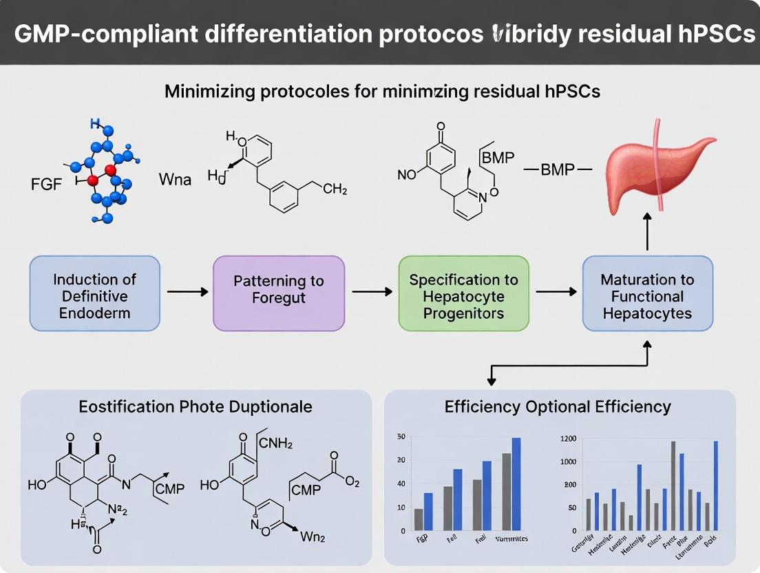
Blueprint for Safety: Step-by-Step GMP Differentiation Protocols and Strategic Elimination Methods
Designing a Closed, Scalable, and Xeno-Free Differentiation Process from the Start
This application note details the development of a closed, scalable, and xeno-free differentiation process for human pluripotent stem cells (hPSCs), aligned with the thesis of establishing GMP-compliant protocols to minimize residual pluripotent cells in final products. The core challenge is transitioning from open, research-grade, undefined culture systems to a robust manufacturing process that mitigates risks of contamination, variability, and tumorigenicity from the outset. This protocol focuses on directed differentiation to definitive endoderm (DE), a critical first step for many lineage-specific programs (e.g., pancreatic beta cells, hepatocytes), given its high regulatory requirement for purity.
Foundational Principles & Rationale
- Closed System: All cell handling, feeding, and differentiation occurs within functionally closed, sterile tubing pathways connected to bioreactors or closed culture plates, eliminating exposure to the open laboratory environment.
- Scalability: The process is designed from small-scale (multi-well plates) to large-scale (controlled bioreactors) using equivalent core parameters (media composition, feeding schedules, oxygenation, agitation) to ensure process consistency.
- Xeno-Free: All components—basal media, supplements, growth factors, matrices, and dissociation enzymes—are of non-animal (recombinant or synthetic) origin, reducing immunogenicity and pathogen risk.
- Minimizing Residual hPSCs: The differentiation protocol is engineered for high efficiency and synchrony, followed by targeted purification strategies to deplete any persisting undifferentiated cells.
Table 1: Comparison of Xeno-Free Media & Matrix Systems for hPSC Maintenance Pre-Differentiation
| Product Name/System | Key Components | Function | Reported hPSC Viability & Pluripotency Markers (%) | Reference (Example) |
|---|---|---|---|---|
| mTeSR Plus | Defined, xeno-free basal medium & supplements | Maintains pluripotency in feeder-free culture | >95% viability, >90% OCT4+/NANOG+ | (Live Search: Current vendor datasheets) |
| E8 Flex Medium | Minimal, recombinant protein-based medium | Supports growth on diverse xeno-free matrices | >90% viability, >85% TRA-1-60+ | (Live Search: Current vendor datasheets) |
| Vitronectin (VTN-N) | Recombinant human protein | Xeno-free adhesion matrix for hPSCs | Confluency achieved in 3-4 days, supports high-density seeding for differentiation | (Live Search: Current vendor datasheets) |
| Synthemax II | Synthetic, peptide-acrylate surface | Defined, animal-free substrate for hPSC adhesion | Comparable to Matrigel for key marker expression | (Live Search: Current vendor datasheets) |
Table 2: Efficacy of Xeno-Free DE Differentiation Protocols
| Differentiation Strategy (Xeno-Free) | Basal Medium | Key Inductive Factors (Xeno-Free) | DE Efficiency (SOX17+/FOXA2+ by Flow Cytometry) | Reported Residual hPSCs (OCT4+ by Flow Cytometry) | Scale Demonstrated |
|---|---|---|---|---|---|
| Activin A Monophasic | RPMI 1640 | Activin A (rh), CHIR99021, PI3K inhibitor (e.g., LY294002) | 85-92% | 1-3% | 6-well to 50mL bioreactor |
| WNT3A/Activin A | DMEM/F-12 + GlutaMAX | WNT3A (rh), Activin A (rh) | 88-90% | 2-4% | 96-well to 100mL spinner flask |
| Staged Induction | MCDB 131-based | CHIR99021 (Stage 1), Activin A (rh) + NaB (Stage 2) | 90-95% | <1% | 12-well to 1L bioreactor |
Detailed Experimental Protocol: Xeno-Free Definitive Endoderm Differentiation in a Closed System
Materials: The Scientist's Toolkit
Table 3: Essential Research Reagent Solutions
| Item | Function | Xeno-Free Specification/Example |
|---|---|---|
| hPSCs | Starting cell source | GMP-grade, karyotypically normal, banked hPSC line. |
| mTeSR Plus Medium | Maintenance medium for pre-expansion | Defined, xeno-free, feeder-free culture medium. |
| Recombinant Vitronectin | Attachment matrix for hPSCs | Truncated recombinant human vitronectin (VTN-N). |
| PBS, without Ca2+/Mg2+ | Washing buffer | DPBS, formulated with non-animal-derived components. |
| ReLeSR or EDTA | Passaging reagent | Enzyme-free or recombinant enzyme-based dissociation solution. |
| RPMI 1640 Basal Medium | Differentiation basal medium | Chemically defined, no animal components. |
| Recombinant Human Activin A | Primary differentiation morphogen | GMP-grade, >95% purity, carrier-free. |
| CHIR99021 | GSK3β inhibitor (WNT activator) | Small molecule, synthetic. |
| PI3K Inhibitor (LY294002) | Enhances DE commitment | Small molecule, synthetic. |
| B-27 Supplement XF | Serum-free cell culture supplement | Defined, xeno-free formulation. |
| Closed Culture Vessel | Scale-up platform | Functionally closed, single-use bioreactor (e.g., stirred-tank or vertical-wheel). |
| Peristaltic Pump System | Enables closed fluidics | Tubing welded to culture vessel for sterile media exchange/additions. |
Protocol: Closed, Scalable DE Differentiation
Part A: Pre-Differentiation hPSC Expansion in Closed Culture
- Thawing & Seeding: Rapidly thaw a vial of GMP-hPSCs in a 37°C water bath. Transfer cells to a closed centrifuge tube prefilled with warm, xeno-free medium via sterile tubing weld. Centrifuge.
- Reseed: Resuspend pellet in mTeSR Plus medium supplemented with 10µM ROCK inhibitor (Y-27632, synthetic). Pump cell suspension into a closed, VTN-N-coated single-use bioreactor pre-filled with medium.
- Expansion: Maintain culture at 37°C, 5% CO2, with controlled, low-shear agitation. Perform daily 80% medium exchanges via sterile pump-driven perfusion from a closed media bag. Monitor cell density and glucose consumption.
- Harvest for Differentiation: At ~80% confluence, drain medium. Pump in recombinant protease (e.g., TrypLE Select) for dissociation. Quench with xeno-free medium. Pump cell suspension out to a harvest bag. Count cells via an in-line automated cell counter.
Part B: Xeno-Free Definitive Endoderm Differentiation
- Day 0: Initiation
- Prepare Xeno-Free DE Induction Medium I: RPMI 1640 + 100ng/mL recombinant human Activin A + 3µM CHIR99021 + 1µM LY294002.
- Pump the harvested hPSCs into a new, coated, closed bioreactor at a high density (e.g., 1-2 x 10^6 cells/cm²) in Induction Medium I.
- Set bioreactor parameters: 37°C, 5% CO2, appropriate agitation for cell aggregate formation (if desired).
- Day 1-2: Commitment
- At 24h, perform a 100% medium exchange via closed perfusion, switching to Xeno-Free DE Induction Medium II: RPMI 1640 + 100ng/mL Activin A + 0.2% v/v Xeno-Free B-27 Supplement.
- Repeat the same 100% medium exchange at 48h.
- Day 3: Analysis & Harvest
- Assess differentiation efficiency. Pump out a small sample via a sterile sample port for in-process control (IPC) testing.
- For IPC: Fix cells and stain for DE markers (SOX17, FOXA2) and hPSC marker (OCT4) via intracellular flow cytometry (see Protocol 4.3).
- Harvest the final DE population by pumping in a recombinant protease, followed by quenching and collection into a harvest bag.
Supporting Protocol: Flow Cytometry for Residual hPSC Detection
- Fixation & Permeabilization: Resuspend ~1x10^6 cells in 4% paraformaldehyde (PFA) for 15 min at RT. Wash twice with DPBS. Permeabilize with 90% ice-cold methanol for 30 min on ice.
- Staining: Wash with FACS buffer (DPBS + 2% FBS or human serum albumin). Incubate with primary antibodies (mouse anti-OCT3/4, goat anti-SOX17) for 1h at RT. Wash. Incubate with compatible fluorophore-conjugated secondary antibodies (e.g., anti-mouse IgG-AF488, anti-goat IgG-AF647) for 45 min in the dark.
- Analysis: Resuspend in DPBS + DAPI (for viability). Acquire on a flow cytometer. Analyze ≥10,000 events. Gate on single, live (DAPI-negative) cells. Determine the percentage of SOX17+/FOXA2+ (DE) and the critical percentage of residual OCT4+ cells.
Signaling Pathways & Process Workflow Diagrams
Diagram 1: Xeno-Free DE Differentiation Signaling Pathway (Max width: 760px)
Diagram 2: Closed Scalable Xeno-Free DE Process Workflow (Max width: 760px)
Strategic Leverage of Small Molecules and Defined Media for Directed Lineage Commitment
Application Notes
The development of robust, GMP-compliant differentiation protocols from human pluripotent stem cells (hPSCs) is paramount for clinical applications in regenerative medicine and drug screening. A primary challenge is the elimination of residual, undifferentiated hPSCs that pose a teratoma risk. The strategic use of small molecules in a defined, xeno-free culture medium presents a powerful solution. This approach enables precise, temporal control over key signaling pathways governing cell fate, promoting efficient and synchronized lineage commitment while selectively inhibiting the self-renewal of hPSCs.
Small molecules offer advantages over protein growth factors, including cost-effectiveness, batch-to-batch consistency, stability, and cell permeability. By targeting specific nodes in pathways like WNT, BMP, TGF-β/Activin/Nodal, and FGF, differentiation can be directed toward definitive endoderm, mesoderm, or neuroectoderm with high purity. Furthermore, certain small molecules can be used as positive or negative selection agents against undifferentiated cells. For instance, exploiting the differential sensitivity of hPSCs versus committed progenitors to compounds like staurosporine derivatives or inhibitors of survival pathways can selectively ablate residual pluripotent cells.
Defined media eliminate variability introduced by serum or undefined components, enhancing reproducibility and safety. When combined with small molecules, they create a fully controllable environment essential for developing standardized, scalable, and GMP-ready differentiation protocols. The integration of these tools minimizes lot-to-last heterogeneity and provides a clear path for regulatory approval.
Protocols
Protocol 1: Directed Differentiation of hPSCs to Definitive Endoderm with Residual Cell Selection
Objective: Generate pancreatic or hepatic progenitors while minimizing residual hPSCs via metabolic selection.
Key Principle: hPSCs have high glycolytic activity. Transition to definitive endoderm involves a shift toward oxidative phosphorylation. Treatment with 2-Deoxy-D-glucose (2-DG) selectively targets residual hPSCs.
Materials:
- Base Medium: RPMI 1640.
- Small Molecules:
- CHIR99021 (GSK-3β inhibitor): Activates WNT signaling for primitive streak induction.
- Activin A (or small molecule IDE1/IDE2 as alternative): Induces Nodal signaling for endoderm specification.
- 2-Deoxy-D-glucose (2-DG): Glycolysis inhibitor for selective pressure.
- Defined Supplements: B-27 Serum-Free Supplement (minus insulin).
Method:
- Culture hPSCs: Maintain hPSCs in a defined, feeder-free culture system (e.g., on Geltrex in mTeSR or E8 medium).
- Initiation of Differentiation (Day 0): At ~80% confluence, aspirate pluripotency medium. Wash once with PBS.
- Definitive Endoderm Induction (Days 0-3):
- Add definitive endoderm induction medium: RPMI 1640 + 100 ng/mL Activin A + 3 µM CHIR99021 + 2% B-27 (minus insulin).
- On Days 1 and 2, replace with medium containing only Activin A (100 ng/mL) and 2% B-27 (minus insulin).
- Metabolic Selection (Day 3-4):
- On Day 3, replace medium with selection medium: RPMI 1640 + 2% B-27 (minus insulin) + 5 mM 2-DG.
- Culture for 24-48 hours. Observe increased death in any residual, poorly differentiated colonies.
- Assessment: On Day 5, analyze cells by flow cytometry for co-expression of definitive endoderm markers SOX17 and CXCR4 (>85% purity expected). Quantify residual OCT4+ cells.
Protocol 2: Small Molecule-Based Neural Induction with Dual-SMAD Inhibition
Objective: Efficiently generate neural progenitor cells (NPCs) using a fully small-molecule, dual-SMAD inhibition protocol.
Key Principle: Simultaneous inhibition of SMAD-dependent BMP and TGF-β signaling drives default neural ectoderm differentiation from hPSCs.
Materials:
- Base Medium: DMEM/F-12 with GlutaMAX.
- Small Molecules:
- SB431542 (TGF-β/Activin/Nodal receptor inhibitor): Blocks SMAD2/3 signaling.
- LDN-193189 (BMP type I receptor inhibitor): Blocks SMAD1/5/8 signaling.
- CHIR99021: Optional, for anterior-posterior patterning after induction.
- Defined Supplements: N-2 Supplement.
Method:
- Preparation: Pre-coat culture plates with poly-L-ornithine and laminin.
- Neural Induction (Days 0-7):
- Dissociate hPSCs to single cells using enzyme-free dissociation buffer.
- Plate cells at 1.5 x 10^5 cells/cm² in neural induction medium: DMEM/F-12 + 10 µM SB431542 + 100 nM LDN-193189 + 1% N-2 Supplement.
- Change medium daily. By Day 5, compact neural rosette structures should be visible.
- NPC Expansion (Day 7+):
- Mechanically pick or enzymatically passage rosettes.
- Plate NPCs in expansion medium: DMEM/F-12 + 1% N-2 Supplement + 20 ng/mL bFGF. Optional: Add 3 µM CHIR99021 for posteriorization (e.g., motor neuron progenitors).
- Assessment: Analyze by immunocytochemistry for PAX6 (neuroectoderm) and SOX1/NESTIN (NPC) expression. Purity should exceed 90%.
Data Presentation
Table 1: Efficacy of Small Molecule-Based Differentiation Protocols
| Target Lineage | Key Small Molecules & Conc. | Base Media | Purity Marker (% Positive) | Residual OCT4+ (%) | Protocol Duration | Reference Cell Line |
|---|---|---|---|---|---|---|
| Definitive Endoderm | CHIR99021 (3 µM), Activin A (100 ng/mL) | RPMI 1640 | SOX17/CXCR4: 85-92% | < 0.5% (post 2-DG) | 5 days | H9 (WA09) |
| Neural Ectoderm | SB431542 (10 µM), LDN-193189 (100 nM) | DMEM/F-12 | PAX6: 90-95% | < 0.1% | 7-10 days | H1 (WA01) |
| Cardiac Mesoderm | CHIR99021 (6 µM), IWP-4 (5 µM) | RPMI 1640 | TBXT (Brachyury): 70-80% | ~2% (Day 3) | 3 days | iPS cell line |
Table 2: Research Reagent Solutions Toolkit
| Item | Function in Lineage Commitment | Example Product/Catalog # |
|---|---|---|
| CHIR99021 | GSK-3β inhibitor; activates canonical WNT signaling for mesendoderm/definitive endoderm induction. | Tocris, #4423 |
| LDN-193189 | BMP type I receptor (ALK2/3) inhibitor; critical for neural induction via dual-SMAD inhibition. | Stemgent, #04-0074 |
| SB431542 | TGF-β/Activin/Nodal type I receptor (ALK4/5/7) inhibitor; blocks pluripotency and aids neural induction. | Tocris, #1614 |
| IDE1 | Small molecule inducer of definitive endoderm; mimics Activin/Nodal signaling. | Tocris, #5854 |
| Y-27632 (ROCKi) | Rho-associated kinase inhibitor; enhances survival of dissociated hPSCs and progenitors. | STEMCELL Tech., #72304 |
| 2-Deoxy-D-Glucose | Glycolysis inhibitor; selectively targets metabolically active residual hPSCs. | Sigma-Aldrich, D8375 |
| B-27 Supplement (minus insulin) | Defined serum-free supplement; supports endoderm and other lineages, insulin-free version allows stage-specific control. | Gibco, #A1895601 |
| N-2 Supplement | Defined serum-free supplement; essential for neural cell growth and maintenance. | Gibco, #17502048 |
| mTeSR1 / E8 Medium | Defined, feeder-free culture medium for maintenance of undifferentiated hPSCs. | STEMCELL Tech. / Gibco |
| Geltrex / Matrigel | Defined extracellular matrix for attachment and growth of hPSCs and progenitors. | Gibco, #A1413302 |
Visualizations
Title: Small Molecule Control of Early Lineage Commitment from hPSCs
Title: Definitive Endoderm Differentiation with Metabolic Purging Workflow
Within the development of GMP-compliant differentiation protocols for human pluripotent stem cell (hPSC)-derived therapeutics, a critical challenge is the minimization of residual, undifferentiated hPSCs. These cells pose a significant tumorigenic risk. Physical separation techniques, which exploit intrinsic biophysical properties without relying on biological labels, offer a compelling, potentially GMP-friendly strategy to deplete these residual cells. This Application Note details the use of two key physical methods—Size-Based Filtration and Density Gradient Centrifugation—as orthogonal purification steps in downstream processing.
Table 1: Comparison of Physical Separation Techniques for hPSC Depletion
| Technique | Principle | Target Cell Property | Typical Purity Yield (Differentiated Cells) | Log Reduction of hPSCs | Typical Processing Time | Scalability |
|---|---|---|---|---|---|---|
| Size-Based Filtration | Selective passage through pores | Cell diameter & deformability | 85-95% | 1.5 - 2.5 log | 30-60 min | High (mL to L scale) |
| Density Gradient Centrifugation | Sedimentation in a density medium | Buoyant density | 70-90%* | 2.0 - 3.0+ log | 90-120 min (incl. centrifuge time) | Moderate (mL scale per tube) |
*Purity highly dependent on the resolution of distinct cell bands and careful collection.
Table 2: Biophysical Properties of hPSCs vs. Differentiated Progeny
| Cell Type | Average Diameter (µm) | Buoyant Density (g/mL in Percoll) | Deformability | Notes |
|---|---|---|---|---|
| Undifferentiated hPSCs | 12 - 16 | ~1.058 - 1.062 | Lower | Form compact, tight colonies; smaller single cells. |
| Early Differentiated Progenitors | 14 - 18 | ~1.065 - 1.070 | Moderate | Begin to spread and enlarge. |
| Mature Differentiated Cells (e.g., Cardiomyocytes) | 18 - 25+ | ~1.075 - 1.085 | Higher/Variable | Larger, more complex morphology. |
Application Notes
Size-Based Filtration (Microfiltration)
Undifferentiated hPSCs, particularly in single-cell suspensions, are often smaller and less deformable than many differentiated cell types (e.g., neurons, cardiomyocytes). This allows for selective retention or passage through precisely sized membrane pores.
- Application: Best used as an initial, rapid depletion step to remove a significant fraction of single hPSCs and small debris following enzymatic dissociation of differentiated cultures.
- Advantage in GMP: Closed-system, sterile, single-use filter units are available, minimizing contamination risk.
- Limitation: May not effectively remove hPSCs present in small clumps. Filter fouling can reduce yield.
Density Gradient Centrifugation
hPSCs exhibit a distinct buoyant density compared to many differentiated cells due to differences in cytoplasmic composition, organelle content, and nuclear-to-cytoplasmic ratio.
- Application: An excellent orthogonal method to filtration. It can separate cells based on intrinsic density, potentially resolving hPSCs into a distinct band. Common media include Percoll and iodixanol-based solutions (e.g., OptiPrep), which are low-osmolarity and non-cytotoxic.
- Advantage in GMP: The separation is based on a physical constant, potentially offering more consistent results across batches.
- Limitation: Requires optimization of gradient formation and fraction collection. Scale-up can be challenging.
Detailed Experimental Protocols
Protocol 1: Depletion of Residual hPSCs by Serial Microfiltration
Objective: To reduce residual hPSC load from a differentiated cell suspension using sequential filtration through decreasing pore sizes.
Materials & Reagents:
- Differentiated hPSC culture (e.g., day 10-15 differentiation).
- Appropriate cell dissociation enzyme (e.g., Accutase, TrypLE).
- Basal medium or buffer with protein (e.g., 0.5% BSA in PBS) to reduce cell adhesion.
- Pre-sterilized, low-protein-binding syringe filters or filter units: 40 µm, 20 µm, 12 µm, and 5 µm pores.
- Syringes (10-50 mL).
- Centrifuge tubes.
Procedure:
- Harvest Cells: Enzymatically dissociate the differentiated culture to a single-cell or small-clump suspension. Neutralize enzyme, wash once, and resuspend in 10-20 mL of cold buffer+BSA at a concentration of 1-5 x 10^6 cells/mL.
- Sequential Filtration: a. Pass the cell suspension gently through a 40 µm filter into a fresh tube. This removes large aggregates and debris. b. Take the flow-through and pass it gently through a 20 µm filter. c. Pass the 20 µm flow-through through a 12 µm filter. The majority of differentiated cells (larger) will be retained on this filter. d. Collect the Retentate: Reverse-flush the 12 µm filter with 5-10 mL of buffer to recover the retained cell population (enriched for differentiated cells). e. Pass the 12 µm flow-through (enriched for smaller cells/hPSCs) through a 5 µm filter. Analyze this final retentate for putative hPSCs if desired.
- Recovery: Centrifuge the collected retentate from step 2d (and other fractions if needed) at 300 x g for 5 min. Resuspend in appropriate medium for counting, analysis (e.g., flow cytometry for hPSC markers like TRA-1-60), or further culture.
Protocol 2: Purification via Continuous Density Gradient Centrifugation
Objective: To separate residual hPSCs from a differentiated cell population based on buoyant density using a pre-formed continuous gradient.
Materials & Reagents:
- Cell suspension as in Protocol 1.
- Stock isotonic Percoll solution (SIP): Mix 9 parts Percoll with 1 part 10X PBS.
- 1X PBS or basal medium (for dilution).
- Ultracentrifuge or high-speed centrifuge with a swinging-bucket rotor.
- 15 mL or 50 mL conical centrifuge tubes.
- Gradient fractionator or manual pipetting system.
Procedure:
- Prepare Continuous Gradient: In a centrifuge tube, create a continuous gradient from 1.045 g/mL to 1.085 g/mL. A standard method is to gently layer decreasing densities from bottom to top. For simplicity, use a two-step layering and allow diffusion to form a gradient: Carefully layer 5 mL of 1.075 g/mL SIP/medium mix over 5 mL of 1.055 g/mL SIP/medium mix. Let stand upright for 20-30 min at 4°C to diffuse.
- Load Sample: Carefully layer 2-3 mL of washed, concentrated cell suspension (in basal medium) on top of the gradient.
- Centrifugation: Centrifuge at 800 x g for 20 minutes at 4°C with the brake OFF. This allows cells to migrate to their isopycnic point (where sample density = gradient density).
- Fraction Collection: After centrifugation, carefully collect sequential 1 mL fractions from the top of the gradient using a fractionator or a pipette.
- Analysis & Recovery: Measure the density of each fraction (refractometer). Wash each fraction twice with excess medium (300 x g, 5 min) to remove Percoll. Resuspend cells and perform viability counting and characterization (e.g., qPCR for pluripotency genes, immunostaining) to identify fractions enriched for differentiated cells (higher density) and depleted of hPSCs (lower density).
The Scientist's Toolkit: Essential Research Reagent Solutions
Table 3: Key Materials for Physical Separation Protocols
| Item | Function & Relevance |
|---|---|
| Low-Protein-Binding Sterile Filters (5-40 µm) | Minimizes cell loss due to adhesion during microfiltration; essential for maintaining yield. |
| Percoll or Iodixanol (OptiPrep) | Inert, low-viscosity density gradient media. Allow formation of iso-osmotic gradients critical for maintaining cell viability. |
| Programmable Masterflex or Peristaltic Pumps | Enables controlled, gentle loading and collection from gradients or filters, improving reproducibility and cell health. |
| Refractometer | For quick, accurate measurement of density gradient fractions post-centrifugation. |
| hPSC-Specific Live Cell Marker Antibodies (e.g., anti-TRA-1-60-AF488) | For rapid flow cytometric assessment of hPSC depletion efficiency in separated fractions without fixation. |
| Closed System Cell Processing Bag with Filters | GMP-compatible alternative to open filtration steps, maintaining sterility for larger-scale processing. |
Visualization Diagrams
Title: Size-Based Filtration Workflow for hPSC Depletion
Title: Isopycnic Density Gradient Centrifugation Process
Introduction Within the development of GMP-compliant differentiation protocols from human pluripotent stem cells (hPSCs), a critical challenge is the complete removal of residual, undifferentiated cells due to their tumorigenic risk. Metabolic selection strategies exploit the fundamental differences in nutrient utilization between rapidly proliferating hPSCs and their differentiating progeny. Specifically, hPSCs exhibit a strong reliance on aerobic glycolysis and glutaminolysis, while differentiated cells often shift towards oxidative phosphorylation. This application note details protocols leveraging these differential requirements to selectively eliminate residual hPSCs from differentiated cultures.
Metabolic Basis for Selection Quantitative metabolic flux analyses highlight key differences. The following table summarizes core nutrient dependencies:
Table 1: Differential Metabolic Requirements of hPSCs vs. Differentiated Cells
| Metabolic Parameter | hPSCs (Glycolytic State) | Differentiated Cells (Oxidative State) | Selection Opportunity |
|---|---|---|---|
| Glucose Dependency | High (≈90% lactate production) | Moderate (Variable) | Glucose-deprivation sensitive |
| Glutamine Dependency | Very High (Essential for biosynthesis & anaplerosis) | Moderate (TCA cycle fuel) | Glutaminase inhibition lethal |
| Lactate Production | High (>15 mmol/10^6 cells/day) | Low (<5 mmol/10^6 cells/day) | Environment acidification |
| ATP from OXPHOS | Low (<20%) | High (>60%) | Resistant to mitochondrial toxins |
Research Reagent Solutions Toolkit Table 2: Essential Reagents for Metabolic Selection Experiments
| Reagent/Category | Example Product | Function in Selection Strategy |
|---|---|---|
| Glutaminase Inhibitor | BPTES, CB-839 | Selectively targets glutamine-dependent hPSCs. |
| Glycolysis Inhibitor | 2-Deoxy-D-glucose (2-DG) | Competes with glucose, stressing glycolytic cells. |
| Galactose Media | Glucose-free, Galactose-supplemented Media | Forces ATP production via OXPHOS, disadvantaging hPSCs. |
| Lactate Dehydrogenase Inhibitor | GSK2837808A | Modulates lactate production, altering niche pH. |
| Fluorinated Glucose Analog | 2-NBDG | Flow cytometry probe for glucose uptake. |
| Mitochondrial Dye | TMRM, MitoTracker Red CMXRos | Stains active mitochondria; labels OXPHOS-competent cells. |
| hPSC-Specific Live-Cell Dye | SSEA-4 Alexa Fluor 488-conjugated Antibody | Labels residual undifferentiated cells for quantification. |
Protocol 1: Glutamine Starvation & Glutaminase Inhibition for hPSC Depletion Objective: Eliminate residual hPSCs from a differentiating culture of pancreatic progenitors.
- Day -1: Initiate differentiation protocol from confluent hPSCs toward pancreatic lineage using established GMP-compliant medium.
- Day 5 (Pancreatic Progenitor Stage): Prepare selection medium: Base differentiation medium lacking L-glutamine, supplemented with 2 mM GlutaMAX (a non-metabolizable alternative) and 5 µM BPTES.
- Treatment: Aspirate standard medium and add selection medium. Culture cells for 48 hours.
- Recovery: Replace medium with standard differentiation medium (with GlutaMAX) for 24 hours.
- Analysis: Harvest cells. Perform flow cytometry for SSEA-4/OCT4 (hPSCs) and PDX1/NKX6.1 (pancreatic progenitors). Compare to untreated control.
Protocol 2: Galactose Substitution for Metabolic Purging Objective: Enrich for cardiomyocytes from a heterogeneous differentiation.
- Day 7 (Cardiomyocyte Differentiation): Cells should be beating. Prepare purging medium: RPMI 1640 without glucose, supplemented with 10 mM galactose, 5 mM sodium pyruvate, and B-27 supplement.
- Treatment: Wash cells once with PBS. Apply galactose purging medium for 96 hours, with medium change at 48 hours.
- Assessment: Post-treatment, switch back to standard maintenance medium (e.g., RPMI/B-27 with glucose). Monitor viability. Quantify hPSC marker (TRA-1-60) decrease and cardiac troponin T increase via immunocytochemistry or flow cytometry.
Visualization of Pathways and Workflows
Diagram 1: Metabolic Selection Principle (78 chars)
Diagram 2: Generic Metabolic Purging Workflow (55 chars)
Conclusion Metabolic selection provides a potent, non-genetic, and potentially GMP-compliant method to minimize residual hPSCs. The protocols outlined exploit the intrinsic vulnerability of hPSCs to perturbations in glycolysis and glutaminolysis. Integrating these strategies at optimal timepoints during differentiation protocols can significantly enhance the safety profile of hPSC-derived products for clinical applications and drug screening.
The Role of Surface-Specific Antibodies and Cell Sorting in Final Product Purification.
1. Introduction and Context
Within the framework of developing robust, Good Manufacturing Practice (GMP)-compliant differentiation protocols from human pluripotent stem cells (hPSCs), a critical challenge is the elimination of residual, undifferentiated cells. These residual hPSCs pose a significant tumorigenic risk in clinical applications. This application note details the pivotal role of surface-specific antibodies and subsequent cell sorting (both positive selection for target cells and, more critically, negative depletion of hPSCs) as a final purification step to achieve a therapeutically viable cell product. The integration of this method is essential for validating the safety profile of any differentiation protocol under GMP guidelines.
2. Application Notes: Key Principles and Data
The strategy relies on high-affinity monoclonal antibodies targeting cell surface markers uniquely or highly expressed on undifferentiated hPSCs, such as SSEA-5, TRA-1-60, and TRA-1-81. Magnetic-Activated Cell Sorting (MACS) and Fluorescence-Activated Cell Sorting (FACS) are employed for scalable depletion. Recent studies quantify the efficiency of this approach.
Table 1: Efficacy of Surface-Marker Based Depletion of Residual hPSCs
| Target Marker | Sorting Platform | Starting hPSC % | Post-Sort hPSC % | Key Validation Assay | Reference Year |
|---|---|---|---|---|---|
| SSEA-5 | MACS (Negative Depletion) | 1.0% | 0.05% | Teratoma Formation in NOG mice | 2022 |
| TRA-1-60 | FACS (Negative Depletion) | 0.5% | <0.01% | Pluripotency Gene (OCT4) qPCR | 2023 |
| SSEA-5/TRA-1-60 Dual | FACS | 5.0% | 0.02% | In Vitro Colony Formation | 2024 |
| EpCAM (Positive Selection of Differentiated Cells) | MACS | 10.0% (hPSCs) | 0.1%* | Flow Cytometry for Pluripotency Markers | 2023 |
Note: This method positively selects for differentiated cells expressing EpCAM, indirectly depleting hPSCs which have lower EpCAM expression.
Table 2: Comparison of Sorting Platforms for Final Product Purification
| Parameter | MACS (Depletion) | FACS (Depletion) |
|---|---|---|
| Throughput | High | Moderate to Low |
| Sort Speed | Very Fast (bulk separation) | Slower (single-cell) |
| Sterility | Closed systems available (GMP) | Complex but achievable (GMP) |
| Purity Yield Trade-off | High yield, good purity | Highest purity, potentially lower yield |
| Multiparameter Capability | Limited (1-2 markers) | High (4+ markers simultaneously) |
| Critical GMP Concern | Potential for non-specific binding/retention | Aerosol generation; stream stability |
3. Detailed Protocols
Protocol A: GMP-Compliant MACS Depletion of SSEA-5+ hPSCs Objective: To deplete residual SSEA-5+ hPSCs from a differentiated cell suspension using clinical-grade magnetic beads. Materials: See "Scientist's Toolkit" below. Procedure:
- Cell Preparation: Harvest differentiated cell culture using a gentle dissociation reagent (e.g., enzyme-free). Quench activity with DPBS++ (with Ca2+/Mg2+). Pass through a 40μm strainer to obtain a single-cell suspension.
- Cell Counting and Viability: Perform using Trypan Blue exclusion. Target a concentration of 1x10^7 cells/mL.
- Antibody Labeling: Resuspend cell pellet in 80μL of cold, sterile sorting buffer (DPBS, 2mM EDTA, 0.5% HSA) per 1x10^7 cells. Add 20μL of clinical-grade anti-SSEA-5-biotin conjugate per 1x10^7 cells. Mix well and incubate for 15 minutes at 2-8°C.
- Magnetic Bead Incubation: Wash cells with 10-20x labeling volume of buffer. Centrifuge (300 x g, 5 min). Resuspend in 80μL buffer per 1x10^7 cells. Add 20μL of anti-biotin MicroBeads per 1x10^7 cells. Mix, incubate for 10 minutes at 2-8°C.
- Magnetic Separation: Place a pre-rinsed LS Column in a strong magnetic separator. Apply cell suspension to the column. Collect the flow-through—this is the depleted fraction (SSEA-5- target cells). Wash column 3x with buffer, collecting all wash effluent with the flow-through.
- Analysis: Take a pre-sort sample and a post-depletion flow-through sample. Stain with a viability dye and a fluorescently-labeled anti-SSEA-5 antibody (different clone or fluorochrome than used for sorting). Analyze purity depletion efficiency via flow cytometry.
Protocol B: Analytical FACS Validation of Depletion Efficiency Objective: To quantify the percentage of residual TRA-1-60+/TRA-1-81+ hPSCs pre- and post-MACS depletion. Procedure:
- Staining: Aliquot 1x10^5 cells from the pre-sort and post-MACS product into separate tubes. Stain with viability dye (e.g., 7-AAD) for 10 min. Wash.
- Surface Marker Staining: Resuspend in buffer containing directly conjugated antibodies: anti-TRA-1-60-FITC, anti-TRA-1-81-PE, and a lineage-specific marker for the target cell type (e.g., anti-CD44-APC). Include appropriate isotype controls.
- Acquisition: Analyze on a calibrated flow cytometer. Gate on single, live cells.
- Quantification: Determine the percentage of cells dual-positive for TRA-1-60 and TRA-1-81. Report the log depletion value.
4. Visualizations
Title: Workflow for MACS-Based Depletion of Residual hPSCs
Title: Antibody Targeting of hPSC Surface Markers
5. The Scientist's Toolkit: Key Research Reagent Solutions
Table 3: Essential Materials for Antibody-Based Cell Sorting Purification
| Reagent/Material | Function | Example (GMP-Grade if Available) |
|---|---|---|
| Clinical-Grade Dissociation Reagent | Generates single-cell suspension while maintaining target cell viability and function. | Recombinant Trypsin, Enzyme-free cell dissociation buffers. |
| Cell Sorting Buffer (DPBS++, 0.5% HSA, 2mM EDTA) | Maintains cell viability, prevents clumping, and provides protein background for optimal antibody binding. | Must be prepared under aseptic conditions with endotoxin-free components. |
| Surface-Specific Primary Antibody (Biotin or Fluorochrome conjugated) | Key reagent for identifying target (hPSC) population. Must be validated for specificity and efficiency. | Anti-SSEA-5-biotin, Anti-TRA-1-60-FITC. |
| Magnetic MicroBeads | For MACS: Provides a magnetic handle for column-based separation. | Anti-biotin MicroBeads (for use with biotinylated primary Ab). |
| Magnetic Separator & Columns | For MACS: Physical system for performing the high-throughput separation. | LS Columns and MACS Separator (available in closed, sterile systems). |
| High-Speed Cell Sorter (FACS) | For analytical validation and high-purity sorting. Must be housed in a controlled, clean environment. | Instruments with large nozzle sizes (e.g., 100μm) for viability preservation. |
| Viability Stain | Critical for excluding dead cells from analysis and sorting, which can non-specifically bind antibodies. | 7-Aminoactinomycin D (7-AAD), Propidium Iodide (PI), or viability dye conjugates. |
Overcoming Common Pitfalls: Optimizing Protocol Efficiency and Robustness for Manufacturing
Within the critical pursuit of developing robust, GMP-compliant differentiation protocols for human pluripotent stem cells (hPSCs), two persistent challenges undermine reproducibility and safety: batch-to-batch variability of input materials and the persistence of stubborn residual undifferentiated cells. This Application Note provides a diagnostic framework and detailed protocols to identify, quantify, and mitigate these sources of inefficiency, directly supporting the broader thesis of achieving minimal residual hPSC risk in therapeutic cell products.
Key Quantitative Challenges & Diagnostic Metrics
The following table summarizes core quantitative metrics essential for diagnosing differentiation inefficiency.
Table 1: Key Metrics for Diagnosing Differentiation Inefficiency
| Metric Category | Specific Assay/Target | Typical Acceptable Range (Directed Differentiation) | Level Indicative of Problem |
|---|---|---|---|
| Residual Pluripotency | Flow Cytometry (OCT4+/NANOG+) | < 0.1% | > 0.5% |
| qPCR (Pluripotency Gene Expression) | >10-fold decrease vs. hPSCs | <5-fold decrease | |
| Batch Variability Indicator | CV of Final Target Cell Yield (%) | < 15% | > 25% |
| CV of Purity (Lineage-Specific Marker) (%) | < 10% | > 20% | |
| Differentiation Efficiency | % Target Lineage Marker (e.g., TUJ1, cTnT) | > 70% | < 50% |
| Karyotypic/Genomic Stability | Karyotype Abnormality Rate | < 5% | > 15% |
Diagnostic Protocols
Protocol 1: High-Sensitivity Flow Cytometry for Residual hPSC Detection
Objective: Quantify rare residual pluripotent cells post-differentiation with high sensitivity and specificity. Materials: See Scientist's Toolkit below. Workflow:
- Cell Harvest: Dissociate differentiated culture to single cells using gentle, non-enzymatic reagent (e.g., EDTA-based) to preserve surface epitopes.
- Staining: Aliquot 1x10^6 cells per test. Use LIVE/DEAD fixable dye first. For surface staining, incubate with anti-TRA-1-60 and anti-SSEA4 (1:100) in FACS buffer for 30 min on ice. Include fluorescence-minus-one (FMO) controls.
- Intracellular Staining (if required): Fix and permeabilize cells using a commercial kit. Incubate with anti-OCT4 antibody (1:50) for 1 hr at room temp.
- Acquisition & Analysis: Acquire on a high-sensitivity flow cytometer. Collect at least 1x10^6 events per sample. Gate sequentially on single, live cells, then plot TRA-1-60 vs. SSEA4 (or OCT4). The positive population in both channels indicates residual hPSCs.
- Sensitivity: This protocol can reliably detect 0.01% residual cells.
Protocol 2: Batch-to-Batch Comparability Testing of Critical Reagents
Objective: Systematically evaluate the impact of new lots of basal media, growth factors, or matrices on differentiation outcomes. Materials: Test lots of the reagent in question (e.g., BMP4, Matrigel), control (established) lot. Experimental Design:
- Parallel Differentiation: Using a single, well-characterized hPSC master bank, initiate identical differentiation runs (n=3 biological replicates) in parallel. The only variable should be the test lot of the critical reagent.
- Multi-Endpoint Analysis: Assess cultures at early, mid, and terminal stages.
- Day 3-5: qPCR for early lineage specifiers (e.g., BRACHYURY, SOX17).
- Terminal Stage: Analyze for Table 1 metrics: yield, purity (flow cytometry), and residual hPSCs.
- Statistical Analysis: Perform a Student's t-test or ANOVA comparing test and control lots for each key endpoint. A lot fails comparability if any critical metric shows a statistically significant (p<0.05) deviation beyond the acceptable CV ranges in Table 1.
The Scientist's Toolkit
Table 2: Essential Research Reagent Solutions
| Item | Function & Rationale |
|---|---|
| GMP-Grade Recombinant Growth Factors (e.g., BMP4, Activin A) | Direct lineage specification. Batch variability in specific activity is a major source of differentiation inconsistency. |
| Defined, Xeno-Free Extracellular Matrix (e.g., Synthemax, Laminin-521) | Provides consistent adhesion and signaling cues, replacing variable animal-derived matrices like Matrigel. |
| Flow Cytometry Antibodies: SSEA4, TRA-1-60, OCT4 | Gold-standard markers for sensitive detection of residual pluripotent stem cells. |
| LIVE/DEAD Fixable Viability Dye | Critical for excluding dead cells during flow analysis, preventing false positives from non-specific antibody binding. |
| Sensitive qPCR Master Mix & TaqMan Assays | For quantifying expression dynamics of pluripotency and early lineage genes with high reproducibility. |
| Small Molecule Inhibitors (e.g., Rock Inhibitor Y-27632) | Improves survival of dissociated single cells, increasing assay accuracy and enabling clonal analysis. |
Visualizing the Diagnostic Workflow & Key Pathways
Title: Diagnostic Decision Tree for Differentiation Problems
Title: Signaling Pathways and Blocks in Differentiation
Optimizing Seeding Density, Media Exchange Schedules, and Agitation Parameters
Within the development of robust, GMP-compliant differentiation protocols from human pluripotent stem cells (hPSCs), minimizing residual undifferentiated cells is a critical quality and safety imperative. Residual hPSCs possess tumorigenic potential, posing a significant risk for cell therapy applications. This application note details the optimization of three fundamental bioprocess parameters—seeding density, media exchange schedules, and agitation parameters—to enhance differentiation efficiency and purity, thereby reducing residual hPSC populations.
Key Parameters & Rationale
Seeding Density: Determines initial cell-cell contact and paracrine signaling, directly impacting differentiation trajectory and homogeneity. Media Exchange Schedules: Controls the temporal availability of differentiation factors, metabolites, and waste product accumulation. Agitation Parameters: In suspension culture, agitation influences mass transfer (nutrients, O₂, factors), shear stress, and aggregate size/distribution.
Summarized Quantitative Data from Current Literature
Table 1: Optimized Seeding Densities for Key Differentiation Pathways
| Differentiation Target | Initial Cell Format | Optimal Seeding Density | Key Outcome on Residual PSCs | Reference (Type) |
|---|---|---|---|---|
| Definitive Endoderm | Monolayer | 1.5 - 2.0 x 10⁵ cells/cm² | < 0.5% OCT4⁺ cells | Peer-reviewed Study |
| Cardiomyocytes | Aggregates in Suspension | 3 - 5 x 10⁵ cells/ml | < 1.0% TRA-1-60⁺ cells | Conference Proceeding |
| Midbrain Dopaminergic Neurons | Aggregates in Suspension | 1 - 2 x 10⁶ cells/ml | ~0.2% NANOG⁺ cells | Preprint (2024) |
| Hepatocyte-like Cells | Monolayer | 1.0 x 10⁵ cells/cm² | ~0.8% SSEA4⁺ cells | Patent Application |
Table 2: Impact of Media Exchange Regimens on Differentiation Yield and Purity
| Protocol Phase | Standard Schedule (Control) | Optimized Schedule | Effect on Differentiation Efficiency | Impact on Residual PSCs |
|---|---|---|---|---|
| Induction (Days 0-3) | Full exchange every 24h | 50% exchange every 12h | ↑ Expression of early markers (SOX17, Brachyury) | ↓ 40% in PSC marker expression |
| Specification (Days 3-7) | Full exchange every 48h | Continuous perfusion (0.5 vol/day) | ↑ Target cell yield by ~25% | Maintained low PSC baseline |
| Maturation (Days 7-14) | Full exchange every 72h | Full exchange every 48h + metabolite feedback | Enhanced functional maturity | No re-emergence detected |
Table 3: Agitation Parameters in Suspension Bioreactors for Aggregate Culture
| Bioreactor Type | Agitation Method | Optimal Speed | Resultant Aggregate Size (Diameter) | Correlation with Residual PSCs |
|---|---|---|---|---|
| Spinner Flask | Paddle Impeller | 40-60 rpm | 200 - 300 µm | Larger, necrotic cores↑ PSC niches |
| Orbital Shaker | Orbital Shaking | 90-110 rpm | 150 - 200 µm | More uniform size, ↓ PSCs |
| Controlled Bioreactor | Pitch Blade Impeller | 30-45 rpm (tip speed) | 250 ± 50 µm | Scalable, consistent, <1% PSCs |
Detailed Experimental Protocols
Protocol 4.1: Systematic Optimization of Monolayer Seeding Density for Definitive Endoderm Differentiation
Objective: To identify the seeding density that minimizes residual hPSCs while maximizing SOX17⁺ definitive endoderm yield. Materials:
- hPSCs (single-cell passaged, >95% viability)
- Recombinant Vitronectin-coated 6-well plates
- Definitive Endoderm Basal Medium
- Growth factors: Activin A, CHIR99021, PI-103
- PBS without Ca²⁺/Mg²⁺, EDTA, Enzyme-free dissociation buffer
- Flow cytometry antibodies: OCT4-APC, SOX17-PE
Method:
- Cell Preparation: Harvest hPSCs to a single-cell suspension. Determine viability via trypan blue exclusion.
- Seeding Matrix: Seed cells at five densities: 0.5, 1.0, 1.5, 2.0, and 2.5 x 10⁵ cells/cm² in triplicate wells. Use medium containing 10µM ROCK inhibitor.
- Differentiation Initiation: At 24h post-seeding (≈70% confluence), replace medium with definitive endoderm induction medium (Activin A 100ng/mL, CHIR99021 3µM, PI-103 20nM in basal medium).
- Media Management: Perform a 50% medium exchange every 12 hours for 72 hours.
- Analysis: On day 3, dissociate cells and stain for intracellular OCT4 and SOX17. Analyze via flow cytometry. Calculate the ratio of SOX17⁺/OCT4⁺ cells for each density.
Protocol 4.2: Evaluating Media Exchange Frequency on Cardiomyocyte Differentiation Purity
Objective: To determine the effect of medium exchange frequency on metabolic stress and residual pluripotency in a directed cardiomyogenesis protocol. Materials:
- hPSC-derived aggregates (day 0 cardiomesoderm induction)
- Cardiomyocyte Differentiation Medium (RPMI/B27 with supplements)
- Small molecule modulators: CHIR99021, IWP-4
- Lactate/Glucose analyzer or bioanalyzer
- Metabolomic assay kit
- Immunocytochemistry: antibodies for cTnT and SSEA4.
Method:
- Aggregate Formation: Form aggregates at 3x10⁵ cells/mL in ultra-low attachment plates.
- Experimental Arms: Distribute aggregates equally across three conditions:
- A: Full medium exchange every 24h.
- B: 75% medium exchange every 12h.
- C: Continuous fed-batch (25% fresh medium added every 12h, 50% removed every 48h).
- Differentiation: Follow a standard Wnt modulation protocol (CHIR99021 day 0-2, IWP-4 day 3-5).
- Monitoring: Sample spent medium daily for glucose/lactate measurement.
- Endpoint Analysis: On day 10, analyze aggregates via immunocytochemistry for cardiac Troponin T (cTnT) and SSEA4. Quantify the percentage of SSEA4⁺ areas per aggregate section.
Protocol 4.3: Agitation Parameter Titration in a Spinner Flask System
Objective: To establish agitation rates that minimize shear stress while preventing aggregate agglomeration and hypoxia. Materials:
- 125mL spinner flasks with paddle impeller
- hPSCs for aggregate culture
- Programmable stir plate
- Dissolved oxygen (DO) probe (optional)
- Sieve sets (100µm, 200µm, 300µm)
- Live/Dead viability stain (Calcein AM/EthD-1)
Method:
- Setup: Calibrate DO probe if used. Place 100mL of medium in each spinner flask. Pre-equilibrate to 37°C, 5% CO₂.
- Inoculation: Seed single hPSCs at 2x10⁵ cells/mL in each flask. Initiate stirring immediately at the designated speed: 30, 50, 70, or 90 rpm (n=2 per condition).
- Aggregate Culture: Culture for 96 hours, maintaining standard conditions.
- Daily Sampling: Each day, aseptically sample 2mL of suspension.
- Assess aggregate size distribution by microscopy and sieve filtration.
- Measure viability via live/dead staining and flow cytometry or image analysis.
- Record DO levels (if monitored).
- Analysis: Correlate agitation speed with: a) median aggregate size, b) size distribution uniformity (coefficient of variation), c) viability on day 4. The optimal speed balances size control (150-250µm) with viability >85%.
Diagrams
Diagram 1: Parameter Optimization Logic Flow for PSC Clearance
Diagram 2: Key Signaling Pathways Affected by Parameters
Diagram 3: Experimental Workflow for Integrated Parameter Screening
The Scientist's Toolkit: Essential Reagent Solutions
| Research Reagent / Material | Primary Function in Optimization Context | Key Consideration for GMP-Compliance |
|---|---|---|
| Chemically Defined, Xeno-Free Basal Medium | Provides a consistent, animal-component-free foundation for differentiation protocols, reducing variability and safety risks. | Ensure full traceability, TSE/BSE-free status, and availability of Drug Master File (DMF). |
| GMP-Grade Recombinant Human Growth Factors (e.g., Activin A, BMP4, FGF2) | Precisely drives differentiation signaling pathways. Crucial for batch-to-batch consistency. | Source from vendors providing Certificate of Analysis (CoA) with purity, potency, endotoxin, and sterility testing. |
| Small Molecule Pathway Modulators (e.g., CHIR99021, IWP-4, SB431542) | Enables efficient, cost-effective, and scalable modulation of key pathways (Wnt, TGF-β). | Purity (>98%) and stability data are critical. Documented solvent origins (DMSO, etc.). |
| Synthetic, Defined Extracellular Matrix (e.g., recombinant Vitronectin, Laminin-521) | Provides a consistent substrate for monolayer differentiation, replacing variable animal-derived matrices. | Must be GMP-manufactured, characterized for consistency in supporting specific lineages. |
| Liquid Handling Automation (Peristaltic pumps, automated bioreactors) | Enables precise, reproducible media exchange and feeding schedules, especially for fed-batch/perfusion. | Systems should be calibratable, cleanable, and compatible with single-use fluidic paths. |
| In-Line Process Analytical Technology (PAT) (pH, DO, metabolite probes) | Allows real-time monitoring of culture health, enabling feedback control of media exchange/agitation. | Probes must be sterilizable and suitable for long-term immersion in GMP bioreactors. |
| Validated, Isoform-Specific Flow Cytometry Antibodies | Critical for accurate quantification of residual PSCs (e.g., anti-TRA-1-60, anti-SSEA4) and target lineage cells. | Antibody clones should recognize specific, relevant epitopes. Validation for intracellular vs. surface markers is required. |
| Metabolomic Profiling Kits | Identifies metabolic bottlenecks (lactate/ammonia build-up) that can stress cells and impair differentiation purity. | Assays should be rapid and compatible with spent culture medium samples. |
Application Notes
The transition from research-grade to GMP-grade raw materials is a critical milestone in the development of clinically applicable cell therapies. This is particularly true for differentiation protocols aimed at minimizing residual human pluripotent stem cells (hPSCs), where the consistency, purity, and defined composition of growth factors and matrices directly impact safety and efficacy. The use of non-GMP reagents introduces significant lot-to-lot variability and risks, including immunogenicity from animal-derived components and uncontrolled differentiation due to undefined factors. Implementing GMP-grade materials establishes a controlled foundation, reducing process variability and enhancing the ability to meet regulatory requirements for purity, potency, and identity of the final cellular product.
Key Quantitative Impacts on Differentiation Protocols
The following table summarizes critical quality attributes influenced by raw material grade, based on current industry and research data.
Table 1: Impact of Raw Material Grade on Key Differentiation Outcomes
| Quality Attribute | Research-Grade Materials | GMP-Grade Materials | Quantitative Impact & Source |
|---|---|---|---|
| Residual hPSC Percentage | Higher, more variable (0.5% - 5%) | Lower, more consistent (<0.1% - 1%) | GMP-grade TGF-β inhibitors can reduce residual PSCs to <0.1% in neural lineages (Industry benchmark). |
| Differentiation Efficiency Variance (Lot-to-Lot) | High (Coefficient of Variance: 15-30%) | Low (Coefficient of Variance: 5-10%) | Study shows GMP FGF2 reduces CV in definitive endoderm yield from ~25% to ~8% (Cell Therapy Insights, 2023). |
| Growth Factor Concentration Accuracy | Variable (± 20-50% of label) | Tightly controlled (± <10% of label) | ELISA data shows GMP-grade BMP4 consistently within 8% of specified concentration (Supplier QC data). |
| Endotoxin Level | Often >1 EU/mg | Typically <0.1 EU/mg | Critical for in vivo applications; GMP standards enforce <0.1 EU/mg for injectables. |
| Animal Origin / Xeno-Free Status | Often contains BSA or animal sera | Defined, xeno-free, recombinant human proteins | Eliminates risk of zoonotic pathogen transmission (Regulatory requirement for Phase III/approved therapies). |
Detailed Protocols
Protocol 1: Qualification of a New GMP-Grade Growth Factor for Directed Differentiation
Objective: To assess the performance equivalence and superior consistency of a new GMP-grade growth factor against a research-grade benchmark in a defined differentiation protocol.
Materials:
- Test Articles: GMP-grade recombinant human protein (e.g., Activin A, BMP4, FGF2).
- Control: Research-grade equivalent from a standard commercial supplier.
- Cells: hPSC line maintained under defined, xeno-free conditions.
- Basal Medium: Chemically defined, serum-free differentiation medium.
- QC Assays: Flow cytometry for marker analysis, qPCR, cell viability assay.
Procedure:
- Experimental Design: Set up differentiation towards a target lineage (e.g., definitive endoderm using Activin A). Use three separate lots each of the GMP-grade and research-grade factor.
- Dose-Response Calibration: Perform a preliminary dose-response (e.g., 10-100 ng/mL) with one lot of each grade to identify the optimal concentration for maximum marker expression (e.g., SOX17/CXCR4).
- Main Differentiation: Initiate differentiation at the optimal dose using three biological replicates per lot. Include a no-growth-factor control.
- Sampling and Analysis:
- At protocol endpoint (e.g., Day 3-5), dissociate cells.
- Flow Cytometry: Stain for lineage-specific surface/intracellular markers. Analyze percentage of positive cells.
- qPCR: Isolve RNA and perform qPCR for pluripotency (OCT4, NANOG) and early lineage markers.
- Viability: Perform trypan blue or dye-exclusion count.
- Data Analysis: Calculate mean differentiation efficiency, viability, and residual pluripotency marker expression for each lot. Statistically compare inter-lot variability (standard deviation, CV) between the two material grades using an F-test.
Protocol 2: Assessing GMP-Grade Extracellular Matrix Consistency in hPSC Differentiation
Objective: To evaluate the impact of GMP-grade recombinant matrix proteins on the homogeneity of differentiation and the minimization of residual undifferentiated cell colonies.
Materials:
- Matrices: GMP-grade recombinant human Laminin-521 (LN-521) or Vitronectin. Research-grade Matrigel or recombinant proteins as control.
- Cells: hPSC line.
- Differentiation Media: For targeted lineage (e.g., cardiomyocyte differentiation using a Wnt modulation protocol).
- Imaging: Automated fluorescence microscope.
- Assays: Immunocytochemistry, flow cytometry.
Procedure:
- Plate Coating: Coat plates with GMP-grade LN-521 (0.5 µg/cm²) and control matrix according to manufacturer specifications. Use three lots per matrix type.
- Cell Seeding and Differentiation: Seed dissociated hPSCs uniformly at a defined density. Upon confluence, initiate differentiation using a standardized GMP-compliant medium protocol.
- Monitoring: Use daily brightfield imaging to monitor colony morphology and uniformity.
- Endpoint Analysis (Day 10-14):
- Immunocytochemistry: Fix cells and stain for pluripotency (OCT4) and differentiation (e.g., cardiac Troponin T) markers. Use automated imaging to quantify the number and size of OCT4+ colonies versus the total differentiated area.
- Flow Cytometry: Dissociate cells and analyze for cardiac-specific markers (cTnT, NKX2.5) and pluripotency markers (TRA-1-60). Determine the correlation between matrix lot and the percentage of residual TRA-1-60+ cells.
- Consistency Metric: Calculate the variance in the percentage of target differentiated cells and the size/number of residual pluripotent colonies across the different lots of each matrix grade.
Visualizations
Title: GMP Materials Drive Consistent Differentiation & Safety
Title: Material Grade Determines hPSC Fate in Differentiation
The Scientist's Toolkit: Key Research Reagent Solutions
Table 2: Essential Materials for GMP-Compliant Differentiation QC
| Reagent Category | Specific Example(s) | Function in Protocol | Critical Quality Attribute for GMP |
|---|---|---|---|
| Defined Culture Matrix | Recombinant human Laminin-521 (LN-521), Vitronectin XF | Provides a consistent, xeno-free substrate for hPSC adhesion and survival, directing initial differentiation signals. | Certificate of Analysis (CoA) for identity, purity, sterility, endotoxin (<1 EU/mL), and absence of human/animal pathogens. |
| GMP-Growth Factors & Cytokines | Recombinant human Activin A, BMP4, FGF2, TGF-β inhibitors | Drives lineage-specific differentiation at defined pathway nodes (e.g., Activin A for definitive endoderm). | Defined concentration (µg/vial), >95% purity (SDS-PAGE), biological activity (cell-based assay), low endotoxin (<0.1 EU/µg), carrier protein identity. |
| Chemically Defined Medium | Essential 8 Flex, StemLine, custom GMP medium formulations | Provides basal nutrients without undefined components like BSA or serum, reducing batch variability. | Formulation details, raw material sourcing (DMF), endotoxin testing, stability data, and osmolality/pH specifications. |
| Cell Dissociation Reagents | Recombinant Trypsin (TrypLE Select), Gentle Cell Dissociation Reagent | Enables gentle, enzymatic passaging or harvest while maintaining cell surface epitopes for analysis. | Xeno-free formulation, defined enzyme activity units, absence of animal contaminants, consistent performance profile. |
| QC Assay Kits | Flow cytometry antibodies (CFR*), hPSC residual detection kits (e.g., PluriTest by qPCR), endotoxin detection kits (LAL) | Quantifies differentiation efficiency and specifically detects residual undifferentiated cells for final product release. | Validated for sensitivity and specificity. For antibodies, clone specificity and conjugate brightness are key. |
This application note details the implementation of Process Analytical Technology (PAT) for real-time monitoring and control of human pluripotent stem cell (hPSC) differentiation processes. The primary objective is to enable GMP-compliant manufacturing of differentiated cell therapies with minimized residual undifferentiated hPSCs, a critical safety parameter. PAT integrates advanced analytical tools to design, analyze, and control manufacturing through timely measurements of critical quality attributes (CQAs), moving from offline batch testing to continuous quality assurance.
Key PAT Tools for Differentiation Monitoring
The following table summarizes core PAT tools applicable to hPSC differentiation.
Table 1: PAT Tools for Real-Time Differentiation Monitoring
| Tool Category | Specific Technology | Measured Parameter / CQA | Typical Sampling Frequency | Advantage for PSC Differentiation |
|---|---|---|---|---|
| In-line Sensors | pH, Dissolved Oxygen (DO), Metabolite Probes (e.g., Glucose, Lactate) | Culture microenvironment | Continuous | Maintains optimal differentiation conditions; prevents metabolic stress. |
| On-line Analytics | Automated Microscopy + Image Analysis | Cell morphology, Confluency, Colony formation | Every 30 min - 2 hours | Non-invasive detection of undifferentiated colony emergence. |
| On-line Analytics | Flow Cytometry (e.g., encapsulated) | Surface marker expression (e.g., SSEA-4, Tra-1-60 for hPSCs) | Every 12 - 24 hours | Near-real-time quantification of residual pluripotent cells. |
| At-line Analytics | Raman Spectroscopy | Biochemical fingerprint (e.g., DNA/RNA, protein, lipid profiles) | Every 1 - 4 hours | Label-free, can predict differentiation efficiency and purity. |
| At-line Analytics | qPCR or ddPCR (automated sample prep) | Pluripotency gene expression (e.g., OCT4, NANOG) | Every 24 - 48 hours | Highly sensitive detection of residual pluripotency signals. |
Detailed Protocol: Integrated PAT for Cardiomyocyte Differentiation
Objective: To direct hPSC differentiation to cardiomyocytes using real-time monitoring of metabolic and morphological CQAs to guide feeding and inhibit pluripotency.
Materials & Reagents (Scientist's Toolkit): Table 2: Key Research Reagent Solutions
| Item | Function in PAT Context |
|---|---|
| Bioreactor with integrated pH/DO probes | Provides continuous, in-line data on culture health and metabolism. |
| In-line Lactate/Glucose Analyzer (e.g., Bioprofile FLEX) | Enables real-time feeding strategies (perfusion/bolus) based on metabolic consumption. |
| Live-cell imaging system (e.g., Incucyte) | Tracks confluence and morphological shifts from pluripotent colonies to differentiating cells. |
| Raman Spectrometer with bioreactor flow cell | Provides label-free, biochemical data to create predictive models of cardiomyocyte yield. |
| hPSC-specific cell line engineering (e.g., NKX2-5eGFP reporter) | Serves as a direct, functional CQA for on-line fluorescence monitoring of cardiac commitment. |
| Automated sampling system | Interfaces bioreactor with at-line analytical equipment (e.g., flow cytometer) while maintaining sterility. |
| GMP-compliant differentiation media (e.g., based on Wnt modulation) | Defined, xeno-free reagents essential for robust process control and regulatory compliance. |
Protocol:
- hPSC Expansion & Bioreactor Seeding:
- Expand hPSCs in a GMP-compliant, feeder-free system.
- Dissociate to single cells and seed into a stirred-tank bioreactor equipped with pH and DO control at a target density of 2-3 x 10^6 cells/mL in mTeSR Plus medium with 10 µM Y-27632.
- Set initial parameters: pH 7.2, DO 40%, agitation 40-60 rpm.
PAT-Enabled Differentiation Initiation (Day 0):
- Switch to cardiomyocyte differentiation medium (e.g., containing CHIR99021).
- Real-time monitoring begins: Set probes to log pH, DO, and temperature every minute. Initiate automated microscopy scans every 2 hours.
Process Monitoring & Control During Specification (Day 0-5):
- Metabolic Control: Use glucose consumption and lactate production rates (from in-line analyzer) to trigger automated medium exchange or perfusion. A sustained spike in lactate may indicate stress.
- Morphological Feedback: Use image analysis algorithms to quantify the loss of compact hPSC colonies and the emergence of elongated, differentiating cells. Persistence of compact colonies beyond Day 2 triggers a review of Wnt modulator activity.
- Raman Spectral Analysis: Acquire spectra every 4 hours. Use a pre-validated Partial Least Squares (PLS) regression model to predict the expression of early cardiac mesoderm markers (e.g., MESP1). Deviations from the model trajectory prompt an assessment.
Critical Checkpoint for Pluripotency Reduction (Day 5-7):
- At-line Analysis: Using an automated sampler, withdraw a 3 mL sample for analysis.
- Perform rapid, at-line flow cytometry for pluripotency markers (SSEA-5, Tra-1-60). Action Limit: If >0.5% SSEA-5+ cells are detected, consider a targeted intervention (e.g., addition of metabolic inhibitors selective for hPSCs).
- Perform ddPCR for OCT4 expression. Alert Limit: >0.01% expression relative to a housekeeping gene.
Termination and Harvest (Day 12-15):
- PAT-based Harvest Decision: Continue Raman and microscopy monitoring. The process is terminated when the Raman model predicts >70% cardiac Troponin T+ cells and imaging shows >85% of cells exhibiting rhythmic contraction.
- Harvest cardiomyocytes using a GMP-compliant, enzymatic method.
Signaling Pathway & PAT Control Logic
Diagram 1: PAT feedback in cardiac differentiation.
Experimental Workflow for PAT Implementation
Diagram 2: PAT development and implementation workflow.
Within the context of developing robust, GMP-compliant differentiation protocols for human pluripotent stem cell (hPSC)-derived therapeutics, scaling from multi-well plates to controlled bioreactors presents significant challenges. This document outlines application notes and protocols for mitigating these challenges, with a specific focus on minimizing residual undifferentiated hPSCs—a critical safety parameter for clinical translation. Key obstacles include maintaining signal homogeneity, ensuring consistent cell microenvironment, and achieving reproducible differentiation efficiency at scale.
Key Scalability Parameters and Comparative Data
The transition from static culture to dynamic bioreactor systems alters critical process parameters. The following table summarizes quantitative data from recent studies comparing lab-scale and bioreactor outcomes for definitive endoderm differentiation, a common initial step in many lineages.
Table 1: Comparison of Lab-Scale vs. Bioreactor Performance for hPSC Differentiation
| Parameter | Lab-Scale (6-Well Plate) | Stirred-Tank Bioreactor (1 L) | Scaling Factor/Consideration |
|---|---|---|---|
| Cell Yield (per mL volume) | 0.5–1.0 x 10^6 cells/mL | 1.5–3.0 x 10^6 cells/mL | Increased yield due to improved nutrient mixing. |
| Differentiation Efficiency (SOX17+ %) | 70–85% | 65–80% | Slight decrease possible; requires optimization of agitation & feeding. |
| Residual hPSC (OCT4+ %) | 0.5–2.0% | 0.8–3.5% | Can increase due to shear stress or nutrient gradients; requires monitoring. |
| Growth Factor Consumption (ng/10^6 cells) | 40–50 ng Activin A | 55–70 ng Activin A | Increased consumption per cell often observed at scale. |
| Dissolved Oxygen (DO) Control | Atmospheric (∼20%) | Controlled (e.g., 5–10%) | Precise low-O2 control enhances definitive endoderm differentiation. |
| pH Fluctuation | High (in standard incubator) | Low (via automated CO₂/base) | Tight pH control (7.2–7.4) improves reproducibility. |
| Sampling Ease | Simple, but destructive | Automated/peristaltic pumps enable non-destructive monitoring. | Enables real-time process analytics. |
Detailed Protocol: Scalable Definitive Endoderm Differentiation for Residual hPSC Minimization
Objective: To differentiate hPSCs to definitive endoderm in a stirred-tank bioreactor, achieving >75% SOX17+ cells while maintaining residual OCT4+ cells below 0.5%.
Pre-Bioreactor Steps: Lab-Scale Preparation
- hPSC Expansion: Culture GMP-grade hPSCs in a feeder-free system (e.g., on vitronectin-coated vessels) using defined mTeSR Plus medium. Passage as clumps using EDTA. Ensure cells are in log-phase growth.
- Aggregate Formation:
- Harvest cells as single cells using a gentle dissociation reagent containing a Rho-associated kinase (ROCK) inhibitor (10 µM).
- Seed cells into ultra-low attachment 6-well plates at 1.0 x 10^6 cells/mL in mTeSR Plus with ROCK inhibitor.
- Place plate on an orbital shaker (85 rpm, 19 mm throw) in a 37°C, 5% CO₂ incubator for 24h to form uniform aggregates (∼150–200 µm diameter).
Bioreactor Setup & Inoculation
- Bioreactor: Use a 1L glass stirred-tank bioreactor with marine impeller, integrated pH and dissolved oxygen (DO) probes, and temperature control.
- Coating: Coat vessel with 10 µg/mL GMP-grade polyvinyl alcohol (PVA) for 2h at 37°C to prevent cell adhesion. Rinse twice with PBS.
- Initial Medium: Fill with 500 mL of pre-warmed, serum-free differentiation base medium (e.g., RPMI 1640 + 1x GlutaMAX + 0.5% defined lipid concentrate).
- Parameters: Set temperature to 37°C, pH to 7.4 (controlled via CO₂ sparging and base addition), DO to 50% (controlled via N₂/air/O₂ gas blending), and agitation to 40 rpm.
- Inoculation: Transfer pre-formed aggregates into the bioreactor using a sterile wide-bore pipette. Target initial viable cell density of 0.5 x 10^6 cells/mL.
Differentiation Protocol in Bioreactor
- Day 0-1 (Specification): Add GMP-grade Activin A (100 ng/mL) and CHIR99021 (3 µM) to the vessel. Maintain DO at 50%.
- Day 1-3 (Definitive Endoderm Induction):
- Perform a 50% medium exchange daily. Replace with fresh base medium containing 100 ng/mL Activin A and 0.5% v/v GMP-grade B-27 Supplement (minus insulin).
- Gradually reduce DO from 50% to 5% over 48h to mimic hypoxia and improve differentiation efficiency.
- Maintain agitation at 40 rpm, monitoring aggregate size daily. Adjust agitation ±10 rpm if aggregates exceed 250 µm (increase) or fall below 150 µm (decrease) to control shear stress.
- Day 3 (Harvest):
- Sample 20 mL for analysis. Assess viability (trypan blue), aggregate size, and differentiation efficiency (flow cytometry for SOX17 and OCT4).
- Harvest cells by letting aggregates settle, siphoning off medium, and gently dissociating with a validated enzyme-free dissociation buffer for downstream processing or cryopreservation.
Signaling Pathway Diagram: Key Pathways in Definitive Endoderm Specification
Diagram Title: Wnt/TGF-β Signaling in Definitive Endoderm Differentiation
Experimental Workflow Diagram: From Lab-Scale to Bioreactor
Diagram Title: Scalable Workflow for hPSC Differentiation
The Scientist's Toolkit: Key Reagent Solutions
Table 2: Essential Materials for Scalable hPSC Differentiation
| Item & Example | Function & Criticality for Scale-Up |
|---|---|
| GMP-Grade hPSC Line (e.g., Master Cell Bank) | Function: Starting material. Ensures genetic stability and traceability. Scale-Up Note: Essential for regulatory filing; requires extensive characterization. |
| Defined, Xeno-Free Medium (e.g., mTeSR Plus, E8) | Function: Maintains pluripotency during expansion. Scale-Up Note: Lot consistency and high-volume availability are critical. |
| GMP-Growth Factors (e.g., Activin A, BMP4) | Function: Directs lineage-specific differentiation. Scale-Up Note: High quantities needed; cost becomes major factor; require certificate of analysis for potency. |
| Matrix/Coating Reagent (e.g., Vitronectin, Laminin-521) | Function: Supports 2D attachment and survival. Scale-Up Note: For 3D bioreactors, soluble polymers (e.g., PVA) are used for anti-fouling instead. |
| Cell Dissociation Agent (e.g., enzyme-free, gentle solution) | Function: Harvests cells as single cells or small clusters. Scale-Up Note: Must be scalable, defined, and leave no residue affecting differentiation. |
| Process Control Buffers (e.g., NaHCO3, NaOH for pH control) | Function: Maintains physiological pH in bioreactor. Scale-Up Note: Must be sterile-filtered and integrated into automated control loops. |
| Metabolite & Gas Sensors (pH, DO, glucose/lactate probes) | Function: Provides real-time process data. Scale-Up Note: Enable Process Analytical Technology (PAT) for quality-by-design approaches. |
| Anti-Apoptotic Agent (e.g., ROCK Inhibitor, Y-27632) | Function: Improves single-cell survival post-dissociation. Scale-Up Note: Used transiently; cost and specificity are considerations at scale. |
Proving Purity: Validating and Comparing Analytical Methods for Residual hPSC Detection
In the development of GMP-compliant differentiation protocols for human pluripotent stem cell (hPSC)-derived therapies, the accurate detection and quantification of residual undifferentiated hPSCs is a critical safety requirement. This application note benchmarks three core analytical technologies—Flow Cytometry, quantitative Reverse Transcription PolymeraseChain Reaction (qRT-PCR), and digital PCR (dPCR)—for their sensitivity, specificity, and applicability in a regulated manufacturing environment. The goal is to inform the selection of a fit-for-purpose assay for lot-release testing and process validation.
Quantitative Benchmark Data
Table 1: Comparative Analytical Performance of Residual hPSC Detection Methods
| Parameter | Flow Cytometry (Intracellular Pluripotency Marker) | qRT-PCR (Pluripotency Gene Expression) | Digital PCR (Pluripotency Gene Expression) |
|---|---|---|---|
| Theoretical Limit of Detection (LOD) | ~0.01% - 0.1% (1 in 10^4 - 10^3) | ~0.001% - 0.01% (1 in 10^5 - 10^4) | ~0.0001% - 0.001% (1 in 10^6 - 10^5) |
| Practical LOD in Complex Samples | 0.05% - 0.1% | 0.01% - 0.05% | 0.001% - 0.01% |
| Specificity | High (Protein/epitope-based) | Moderate-High (Sequence-based, risk of genomic DNA) | Very High (Sequence-based, endpoint detection) |
| Precision (CV) | 10-25% | 5-15% | <10% |
| Dynamic Range | 2-3 logs | 5-7 logs | 4-5 logs |
| Throughput | High | Very High | Moderate |
| Ability to Distribute Viability | Yes (with live-cell staining) | No (lysed cells) | No (lysed cells) |
| Key Advantage | Single-cell, multi-parameter analysis | High throughput, established | Absolute quantification, high sensitivity & precision |
| Key Limitation for hPSC Detection | Sensitivity limited by antibody noise & background | Relative quantification, requires standard curve, inhibitor sensitive | Higher cost, more complex workflow |
Table 2: Common Targets for Residual hPSC Detection
| Technology | Typical Target(s) | Purpose |
|---|---|---|
| Flow Cytometry | TRA-1-60, SSEA-4, OCT4 (intracellular) | Surface/intracellular pluripotency protein detection |
| qRT-PCR / dPCR | POU5F1 (OCT4), NANOG, DNMT3B | Pluripotency-associated gene expression |
Detailed Experimental Protocols
Protocol 1: Flow Cytometry for Intracellular Pluripotency Markers
Objective: To detect and quantify residual hPSCs in a differentiated cell product via intracellular staining for OCT4. Materials: See "The Scientist's Toolkit" below. Procedure:
- Sample Preparation: Harvest differentiated cell aggregate. Dissociate into single-cell suspension using a gentle enzyme (e.g., Accutase). Pass through a 35-40 µm cell strainer. Perform a viable cell count.
- Fixation: Pellet 1x10^6 cells. Resuspend in 100 µL of 4% paraformaldehyde (PFA) in PBS. Incubate for 15 minutes at room temperature (RT), protected from light.
- Permeabilization: Wash twice with 2 mL PBS. Resuspend pellet in 100 µL of ice-cold 90% methanol. Vortex gently and incubate for 30 minutes on ice or at -20°C.
- Staining: Wash twice with 2 mL Flow Cytometry Staining Buffer (PBS + 2% FBS). Resuspend pellet in 100 µL of staining buffer containing a pre-titrated concentration of fluorochrome-conjugated anti-OCT4 antibody or the relevant isotype control. Incubate for 45-60 minutes at RT in the dark.
- Analysis: Wash twice with staining buffer. Resuspend in 300 µL PBS. Acquire data on a flow cytometer calibrated with compensation beads. Analyze a minimum of 100,000 events. Use isotype control to set the positive gate. Sensitivity is validated using spiked samples with known ratios of hPSCs.
Protocol 2: qRT-PCR for Pluripotency Gene Expression
Objective: To detect residual hPSCs via quantification of POU5F1 (OCT4) mRNA expression relative to a reference gene. Materials: See "The Scientist's Toolkit." Procedure:
- RNA Extraction: Pellet 1x10^6 cells from the test sample. Lyse and extract total RNA using a silica-membrane column kit with on-column DNase I digestion. Elute in 30-50 µL RNase-free water. Quantify RNA concentration and purity (A260/A280 ~2.0).
- Reverse Transcription: Use 100 ng – 1 µg total RNA in a 20 µL reaction with a high-capacity cDNA reverse transcription kit, including random hexamers and RNase inhibitor.
- qPCR Setup: Prepare reactions in triplicate. Use 2-5 µL of 1:10 diluted cDNA per 20 µL reaction containing TaqMan Universal PCR Master Mix, POU5F1 (OCT4) TaqMan Assay (FAM-labeled), and a reference gene (e.g., GAPDH, VIC-labeled) assay.
- Run & Analyze: Use the following cycling parameters: 95°C for 10 min, followed by 40 cycles of 95°C for 15 sec and 60°C for 1 min. Record Cq values. Use a standard curve generated from serial dilutions of hPSC cDNA (e.g., 100% to 0.001%) to interpolate the percentage of residual hPSCs in the unknown sample.
Protocol 3: Digital PCR for Absolute Quantification of Pluripotency Transcripts
Objective: To achieve absolute, standard-free quantification of NANOG transcript copy number in a differentiated cell product sample. Materials: See "The Scientist's Toolkit." Procedure:
- cDNA Synthesis: Follow steps 1 and 2 from Protocol 2.
- dPCR Reaction Assembly: Prepare a 20-40 µL reaction mix containing ddPCR Supermix for Probes (no dUTP), NANOG and reference gene (e.g., HPRT1) TaqMan probe assays, and 2-10 µL of diluted cDNA.
- Droplet Generation: Load the reaction mix into a droplet generator cartridge along with droplet generation oil. Generate 20,000 nanoliter-sized droplets per sample.
- PCR Amplification: Transfer droplets to a 96-well PCR plate. Seal and run a thermal cycler with standard TaqMan cycling conditions (95°C for 10 min, 40 cycles of 94°C for 30 sec and 60°C for 1 min, with a final 98°C step for 10 min).
- Droplet Reading & Analysis: Place plate in a droplet reader. The software assigns each droplet as positive or negative for the target(s) based on fluorescence amplitude. Use Poisson statistics to calculate the absolute concentration (copies/µL) of NANOG and the reference gene in the original reaction. Report as copies/µg RNA or normalize to cell number.
Visualizations
Diagram 1: Residual hPSC Detection Method Workflow Comparison (100 chars)
Diagram 2: Thesis Context: Assay Selection Logic for hPSC Safety (99 chars)
The Scientist's Toolkit: Key Research Reagent Solutions
| Item | Function & Relevance to hPSC Detection |
|---|---|
| Anti-Human OCT4 (OCT3) Antibody, Alexa Fluor 647 | Conjugated primary antibody for specific detection of intracellular OCT4 protein in flow cytometry. Critical for specificity. |
| TaqMan Gene Expression Assay for POU5F1 (Hs04260367_gH) | FAM-MGB probe/primer set for specific detection of OCT4 mRNA splice variants with high efficiency in qRT-PCR/dPCR. |
| MagMAX-96 Total RNA Isolation Kit | Magnetic bead-based RNA extraction for 96-well plates. Enables high-throughput, consistent RNA yield with DNase treatment, reducing genomic DNA contamination. |
| SuperScript IV VILO Master Mix | Robust reverse transcription master mix with high sensitivity and fast processing, ideal for converting low-abundance pluripotency transcripts. |
| ddPCR Evagreen Supermix | dPCR master mix for droplet-based digital PCR using EvaGreen dye. Enables multiplexing and highly sensitive detection of amplicons without probes. |
| Counting Beads for Flow Cytometry (e.g., AccuCount Beads) | Precisely known concentration of fluorescent beads. Added to samples to enable absolute cell counting per volume via flow cytometry, improving quantification. |
| PCR Inhibitor Removal Reagent (e.g., OneStep PCR Inhibitor Removal Kit) | Critical for processing complex biological samples (e.g., differentiated cell lysates) that may contain polysaccharides or melanin which inhibit PCR, ensuring assay accuracy. |
| Glycerol Stocks of Characterized hPSCs | Used as positive control and for creating serial dilution spikes in a "negative" differentiated cell background to empirically determine LOD/LOQ for each assay. |
Within the critical objective of developing robust, GMP-compliant differentiation protocols from human pluripotent stem cells (hPSCs), a paramount safety concern is the elimination of residual, undifferentiated hPSCs. These cells pose a teratoma risk in clinical applications. This document provides application notes and protocols for validating the analytical assays used to detect and quantify these residual cells, focusing on establishing the Limit of Detection (LOD) and Limit of Quantification (LOQ) as per ICH Q2(R1) and USP guidelines. This validation is a cornerstone of process control and final product release testing.
Core Definitions and Calculations
LOD and LOQ are method-specific parameters, not biological limits. They describe the lowest levels at which an assay can reliably detect or quantify an analyte—here, residual hPSCs.
Typical Calculations from Validation Data:
- Standard Deviation Method: Requires a blank sample (0% hPSCs) and a low-concentration sample. LOD = mean(blank) + 3.3σ; LOQ = mean(blank) + 10σ, where σ is the standard deviation of the response.
- Signal-to-Noise Ratio: LOD requires S/N ≥ 3, LOQ requires S/N ≥ 10.
- Linear Regression Method: LOD = 3.3 * (S{y/x})/slope; LOQ = 10 * (S{y/x})/slope, where (S_{y/x}) is the residual standard error of the regression line from a calibration curve.
Table 1: Example LOD/LOQ Data for Common Residual Cell Assays
| Assay Type | Target | Measured LOD (hPSCs in somatic cells) | Measured LOQ (hPSCs in somatic cells) | Key Assumption for Calculation |
|---|---|---|---|---|
| qRT-PCR | Pluripotency Gene (e.g., NANOG) | 0.001% | 0.003% | Target gene expression is specific and linear over the range. |
| Flow Cytometry | Cell Surface Marker (e.g., SSEA-4) | 0.01% | 0.05% | Marker specificity and minimal background in negative population. |
| Droplet Digital PCR (ddPCR) | Pluripotency Gene (e.g., POU5F1) | 0.0005% | 0.001% | Absolute copy number detection without a standard curve. |
| Intact Cell Immunoassay | Intracellular Marker (e.g., OCT4) | 0.005% | 0.02% | Assay sensitivity and specificity for rare event detection. |
Experimental Protocols
Protocol 1: Spiking Experiment for LOD/LOQ Determination via Flow Cytometry
This protocol outlines the generation of a calibration curve using spiked samples.
1. Materials Preparation:
- Negative Matrix: Fully differentiated somatic cell population (e.g., cardiomyocytes, neural progenitors) confirmed to be >99.9% negative for hPSC markers.
- Positive Cells: Actively cultured, pluripotent hPSCs (e.g., H9 or an iPSC line).
- Key Reagent: Validated, fluorochrome-conjugated antibody against a hPSC-specific surface marker (e.g., SSEA-4, TRA-1-60).
- Buffer: PBS with 2% FBS or BSA.
- Equipment: Flow cytometer (calibrated daily), cell counter, centrifuge.
2. Spiked Sample Preparation:
- Prepare a high-purity suspension of hPSCs and count accurately.
- Prepare a suspension of the negative matrix cells at a known concentration (e.g., 1 x 10^6 cells/mL).
- Serially spike the hPSCs into the negative matrix to create the following approximate % mixtures: 1%, 0.5%, 0.1%, 0.05%, 0.01%, 0.005%, 0.001%, and a 0% (blank/matrix-only) control. Prepare a minimum of n=5 independent replicates per level.
- For low percentages (e.g., <0.1%), prepare a master mix of the desired low concentration and then aliquot to ensure precision.
3. Staining and Acquisition:
- For each sample, stain 1 x 10^6 total cells with the target antibody and appropriate isotype control according to manufacturer instructions.
- Resuspend in a fixed volume (e.g., 300 µL). Acquire a minimum of 500,000 total events per sample on the flow cytometer to ensure sufficient statistical power for rare event detection.
- Record the absolute count of positive events for each replicate.
4. Data Analysis and Calculation:
- Plot the mean measured positive cell count (or %) vs. the expected (spiked) percentage.
- Perform linear regression analysis on the linear range of the curve.
- Calculate (S_{y/x}) (standard error of the y-estimate).
- LOD = 3.3 * (S{y/x}) / slope. LOQ = 10 * (S{y/x}) / slope.
- Confirm the LOQ by demonstrating ≤20% CV for accuracy and precision at that concentration level.
Protocol 2: LOD/LOQ Determination for qPCR/ddPCR Assays
This protocol uses genomic DNA (gDNA) spiking to simulate residual cell detection.
1. Materials Preparation:
- gDNA Matrix: Extracted from the fully differentiated somatic cell population.
- gDNA Spike: Extracted from hPSCs. Quantify accurately using fluorometry.
- Primers/Probes: Validated, hydrolysis probe-based assay targeting a pluripotency-specific gene (e.g., NANOG, POU5F1) and a reference gene.
- Master Mix: For qRT-PCR or ddPCR.
- Equipment: Real-time PCR system or droplet reader/generator.
2. Spiked Calibrant Preparation:
- Determine the haploid genome mass per cell (e.g., ~3.3 pg for human).
- Calculate the amount of hPSC gDNA needed to represent specific fractions (e.g., 0.1%, 0.01%, 0.001%, 0.0001%) in a constant background of 100 ng somatic cell gDNA.
- Prepare serial dilutions of the hPSC gDNA in the somatic gDNA background. Include a 100% somatic gDNA (0% spike) negative control and a 100% hPSC gDNA positive control. Prepare n=6 replicates per level.
3. PCR Setup and Run:
- For qPCR: Run all samples in triplicate PCR reactions. Use a minimum of 40 cycles. Record the Cq (Ct) values.
- For ddPCR: Follow manufacturer's protocol for droplet generation and PCR. After reading, record the copies/µL of the target gene.
4. Data Analysis and Calculation:
- For qPCR: Plot Cq values against the log10 of the expected target fraction. Perform linear regression. LOD/LOQ are derived from the standard error of the regression line as described above, often converted back to a percentage. A practical LOD is often defined as the lowest spike level where ≥95% of replicates are detected (Cq < 40).
- For ddPCR: Plot measured copies/20µL reaction vs. expected copies. Perform linear regression through the origin. LOD can be statistically determined from the negative control (mean + 3.3 SD) or as 3 positive droplets per well. LOQ is the lowest concentration with ≤25% CV.
Visualizations
Title: Workflow for Validating Residual hPSC Assays in GMP Differentiation.
Title: Key Signaling Pathways Targeted to Reduce Residual hPSCs.
The Scientist's Toolkit: Essential Research Reagent Solutions
Table 2: Key Materials for Residual Cell Assay Validation
| Item | Function in Validation | Example/Notes |
|---|---|---|
| Validated Antibody Panels | Specific detection of hPSC surface/intracellular markers (SSEA-4, TRA-1-60, OCT4) by flow/imaging. | Critical for specificity; must be titrated and matched with correct isotype controls. |
| gDNA Extraction Kit | High-quality, reproducible isolation of genomic DNA from spiked cell mixtures for PCR-based assays. | Use kits with consistent yield and minimal inhibitor carryover. |
| qPCR/ddPCR Master Mix | Enzymatic amplification and detection of pluripotency gene targets with high sensitivity and precision. | ddPCR master mix is preferred for absolute quantification without a standard curve. |
| Synthetic DNA Standards | For generating precise calibration curves in qPCR assays when gDNA spiking is impractical. | Must be well-characterized and sequence-verified. |
| Reference RNA/DNA | To control for extraction efficiency and PCR inhibition in sample-based assays. | Can be exogenous (spike-in) or endogenous (housekeeping gene). |
| Cell Line-Specific hPSCs | Source of 'positive' material for spiking experiments. Must be well-characterized and pluripotent. | Use the same hPSC line employed in the differentiation process development. |
| Defined Differentiation Media | To generate the 'negative matrix' somatic cell population free of hPSCs. | Essential for creating a biologically relevant background for spiking. |
| Flow Cytometry Counting Beads | For obtaining absolute cell counts during flow cytometry, improving accuracy for rare events. | Allows calculation of absolute number of residual hPSCs per million cells. |
Application Notes: Context and Significance
Within the development of GMP-compliant differentiation protocols for human pluripotent stem cells (hPSCs), a paramount concern is the elimination of residual undifferentiated cells. These cells pose a significant tumorigenic risk due to their proliferative capacity and potential to form teratomas. Functional safety tests are therefore critical release assays for clinical-grade cell products. The in vivo teratoma assay has been the historical gold standard for assessing pluripotency and, by extension, tumorigenic risk. However, its limitations have driven the search for complementary and alternative in vitro assays. This document details the current state of these safety tests, providing protocols and comparisons to support rigorous, GMP-aligned safety profiling.
The In Vivo Teratoma Assay: Protocol and Considerations
Principle: Injected residual hPSCs proliferate and differentiate into cells of the three embryonic germ layers (ectoderm, mesoderm, endoderm) within an immunodeficient host, forming a teratoma. The assay confirms the pluripotent potential of any residual cells.
Detailed Protocol:
- Sample Preparation: Harvest the final hPSC-derived cell product (e.g., differentiated progenitors). Include a positive control (e.g., 1x10^6 undifferentiated hPSCs) and a negative control (e.g., non-tumorigenic human fibroblasts).
- Host Animal: Immunodeficient mice (e.g., NOD/SCID, NSG). Use animals aged 6-8 weeks.
- Injection:
- Resuspend test and control cells in a 1:1 mixture of culture medium and Matrigel/Basement Membrane Matrix (kept on ice) to a final volume of 100-200 µl.
- Using a 1ml syringe with a 27-gauge needle, perform subcutaneous injection into the dorsal flank or intramuscular injection into the hind limb. Multiple test sites per animal can be used if scientifically justified.
- Monitoring: Palpate weekly for tumor formation. Teratomas from positive controls typically appear within 6-12 weeks.
- Endpoint & Histology:
- Tumors are harvested when they reach ~1.5 cm in diameter or at a pre-defined study endpoint (e.g., 12-16 weeks).
- Fix in 4% paraformaldehyde or 10% neutral buffered formalin for 24-48 hours.
- Process, paraffin-embed, section (5-7 µm), and stain with Hematoxylin and Eosin (H&E).
- Perform immunohistochemistry (IHC) for lineage-specific markers to confirm trilineage differentiation (e.g., βIII-tubulin [ectoderm], α-smooth muscle actin [mesoderm], AFP [endoderm]).
Emerging In Vitro Alternatives and Adjuncts
Given the teratoma assay's cost, duration, and ethical burden, several in vitro assays have been developed.
2.1. Flow Cytometry-Based Residual hPSC Detection Principle: Direct quantification of undifferentiated cells using cell surface pluripotency markers. Protocol: Dissociate the cell product to a single-cell suspension. Stain with fluorochrome-conjugated antibodies against hPSC-specific surface markers (e.g., SSEA-5, TRA-1-60, TRA-1-81). Include a viability dye. Acquire data on a flow cytometer using an isotype control and positive control (undifferentiated hPSCs) to set gating thresholds. Detection sensitivity is typically 0.1-0.5%.
2.2. Nanog/Luciferase Reporter Assay Principle: A genetically engineered hPSC line with a luciferase reporter gene driven by the pluripotency-specific Nanog promoter is mixed with the cell product. Luciferase activity correlates with residual pluripotent cell number. Protocol: Spiking Experiment. Mix a known number of reporter hPSCs (e.g., 100, 500, 1000) with a fixed number of the test cell product (e.g., 1x10^6 cells). Perform the assay in parallel with the unspiked product.
- Lyse cells and measure luciferase activity using a bioluminescence plate reader.
- Generate a standard curve from spiked samples to estimate the lower limit of detection (LLOD), which can reach <10 pluripotent cells in 1x10^6.
2.3. qRT-PCR for Pluripotency Genes Principle: Sensitive detection of mRNA transcripts specific to undifferentiated cells. Protocol: Extract total RNA from the cell product. Perform reverse transcription followed by quantitative PCR using TaqMan probes or SYBR Green for pluripotency genes (e.g., NANOG, POUSF1 [OCT4], DNMT3B). Normalize to housekeeping genes. The LLOD must be established using spiked samples, as the presence of transcripts in differentiating cells can complicate interpretation.
Quantitative Data Comparison of Safety Assays
Table 1: Comparison of Key Functional Safety Test Modalities
| Assay Parameter | In Vivo Teratoma Assay | Flow Cytometry | Nanog/Luciferase Reporter | qRT-PCR |
|---|---|---|---|---|
| Assay Readout | Histological confirmation of trilineage differentiation | Direct cell count of marker-positive cells | Bioluminescence from residual reporter hPSCs | mRNA expression levels |
| Duration | 8-20 weeks | 1-2 days | 2-3 days | 1-2 days |
| Approx. Cost | Very High (>$10k) | Low-Medium | Medium | Low |
| Throughput | Very Low | High | Medium | High |
| Sensitivity (LLOD) | ~1x10^4 - 1x10^5 cells * | ~0.1% (1 in 1,000) | ~0.001% (10 in 1x10^6) | ~0.0001% (1 in 1x10^6) |
| Functional Context | Yes (in vivo environment) | No (marker presence only) | Semi-Functional (reporter activity) | No (transcript presence only) |
| Regulatory Acceptance | Historical gold standard | Accepted adjunct | Emerging, requires validation | Accepted adjunct |
Estimated minimum number of hPSCs required to form a teratoma. *Highly dependent on gene target and assay design.*
Experimental Workflow and Pathway Diagrams
Title: Safety Testing Strategy Workflow for hPSC Products
Title: Pluripotency Pathways to Teratoma or Detection
The Scientist's Toolkit: Key Research Reagent Solutions
Table 2: Essential Materials for Functional Safety Testing
| Reagent / Material | Function / Application | Key Considerations |
|---|---|---|
| Matrigel / Basement Membrane Matrix | Provides an extracellular matrix scaffold for cell injection in the teratoma assay, enhancing cell survival and engraftment. | Lot-to-lot variability; must be kept on ice to prevent polymerization. |
| Immunodeficient Mice (e.g., NSG) | Host organism for teratoma assay lacking adaptive immunity, allowing engraftment of human cells. | Choice impacts tumor latency and study cost. NSG mice are most permissive. |
| Anti-human TRA-1-60 / SSEA-5 Antibodies | Primary detection tools for flow cytometric quantification of residual hPSCs via surface markers. | Conjugate choice (e.g., APC, PE) depends on flow cytometer configuration. |
| Nanog-Luciferase Reporter hPSC Line | Engineered tool cell line for highly sensitive, functional in vitro detection of residual pluripotency. | Requires careful maintenance to ensure stable reporter expression. |
| TaqMan Probes for POUSF1 / NANOG | Fluorogenic probes for specific, quantitative RT-PCR detection of pluripotency gene transcripts. | Provides superior specificity over SYBR Green for complex cell products. |
| Lineage-Specific IHC Antibodies (βIII-tubulin, α-SMA, AFP) | Used for histological validation of trilineage differentiation within excised teratomas. | Confirms the functional pluripotency of initiating cells. |
Comparative Analysis of Commercially Available Residual Detection Kits
Within the framework of developing robust, GMP-compliant differentiation protocols for human pluripotent stem cell (hPSC)-derived therapeutics, the sensitive and reliable detection of residual undifferentiated cells is a critical safety checkpoint. This analysis compares the performance characteristics of leading commercially available kits designed for this purpose, providing application notes and standardized protocols to aid in quality control for researchers and drug development professionals.
The following kits were evaluated based on current manufacturer specifications and published literature.
Table 1: Comparative Analysis of Key Residual Detection Kits
| Kit Name (Manufacturer) | Target Antigen(s) | Technology | Reported Sensitivity | Assay Time | Throughput | GMP-Ready Documentation |
|---|---|---|---|---|---|---|
| PlurITest (STEMCELL Technologies) | Multiple (qPCR-based gene panel) | Quantitative RT-PCR | 0.01% - 0.1% | ~4 hours | Medium | Yes (Certificate of Analysis) |
| hPSC Residual Detection Kit (Thermo Fisher Scientific) | TRA-1-60, SSEA-4, SSEA-5 | Flow Cytometry | 0.02% - 0.1% | ~3 hours | Low-Medium | Yes (Regulatory Support File) |
| LiquiChrom hPSC Assay (Bio-Techne) | Intracellular Pluripotency Markers (OCT4, NANOG) | ELISA / Colorimetric | 0.05% - 0.2% | ~2.5 hours | High | Under Development |
| Residual hPSC Detection Kit (ATCC) | TRA-1-60 | Immunofluorescence / Microscopy | 0.1% - 0.5% | ~5 hours (inc. imaging) | Low | Limited |
Table 2: Practical Considerations for Kit Selection
| Parameter | PlurITest | hPSC Residual Detection (Flow) | LiquiChrom Assay |
|---|---|---|---|
| Key Advantage | High sensitivity, genomic DNA target stability | Direct cell counting, visual confirmation | High throughput, simple workflow |
| Key Limitation | Requires nucleic acid isolation, indirect measure | Requires flow cytometer expertise & equipment | Detects intracellular targets only |
| Sample Type | Cell lysate (RNA/DNA) | Single-cell suspension | Cell lysate (protein) |
| Cost per Sample | $$$ | $$ | $ |
| Best Suited For | Final product release testing | Process development & in-process monitoring | Screening of multiple differentiation batches |
Detailed Experimental Protocols
Protocol 3.1: Sample Preparation Standardization for Comparative Analysis
Objective: To generate a consistent sample matrix containing a known percentage of residual H9 hPSCs in differentiated cortical neurons for kit benchmarking.
Materials:
- H9 hPSC culture (≥85% confluent, feeder-free)
- Differentiated cortical neuron population (Day 35)
- Accutase
- Defined Trypsin Inhibitor (DTI)
- PBS (Ca2+/Mg2+-free)
- Automated cell counter or hemocytometer
Procedure:
- Harvest Cells: Dissociate H9 hPSCs and differentiated neuron cultures separately using Accutase. Neutralize with DTI, wash with PBS, and pellet cells.
- Quantify & Mix: Count both cell populations using a standardized method. Prepare spiked samples with hPSC percentages of 0.01%, 0.05%, 0.1%, 0.5%, and 1.0% (v/v) in a background of 1 x 10^6 total differentiated cells.
- Aliquot & Process: For each kit, aliquot 2 x 10^5 cells from each spiked sample into separate tubes. Immediately proceed with the specific kit's sample preparation steps (lysis for PCR/ELISA, fixation for flow/IF).
Protocol 3.2: Workflow for hPSC Residual Detection by Flow Cytometry (Thermo Fisher Kit)
Objective: To quantify the percentage of TRA-1-60/SSEA-4/SSEA-5 positive cells in a differentiated cell product.
Materials:
- hPSC Residual Detection Kit (Cat. No. A25526, Thermo Fisher)
- Prepared single-cell suspension (from Protocol 3.1)
- Flow cytometry staining buffer (PBS + 2% FBS)
- Fixable Viability Dye (e.g., Zombie NIR)
- Flow cytometer with 488 nm and 633/640 nm lasers.
Procedure:
- Viability Staining: Resuspend cell pellet in 100 µL PBS. Add 1 µL of Fixable Viability Dye, incubate 15 min at RT in the dark. Wash with 2 mL staining buffer.
- Surface Marker Staining: Resuspend cell pellet in 100 µL staining buffer. Add the pre-mixed antibody cocktail (anti-TRA-1-60-FITC, anti-SSEA-4-Alexa Fluor 647, anti-SSEA-5-eFluor 660) per kit instructions. Incubate 30 min at 4°C in the dark.
- Fixation: Wash cells twice with staining buffer. Fix cells with the provided fixative (or 4% PFA) for 20 min at 4°C.
- Acquisition & Analysis: Wash cells, resuspend in staining buffer, and acquire on a flow cytometer. Use unstained and single-stained controls for compensation. Gate on single, live cells. The residual hPSC population is identified as triple-positive for all three markers.
Protocol 3.3: Workflow for Genomic DNA-based Residual Detection (PlurITest Principle)
Objective: To detect pluripotency-associated gene expression via qPCR from cell lysates.
Materials:
- PlurITest Kit or equivalent qPCR reagents & validated primer/probe sets for POUSF1 (OCT4), NANOG, and reference genes (GAPDH, HPRT1).
- Cell lysate (≥ 2 x 10^4 cells) in RNA/DNA stabilization buffer.
- Nucleic acid extraction kit (RNA/DNA co-purification).
- Reverse transcription kit.
- Real-Time PCR system (96- or 384-well).
Procedure:
- Nucleic Acid Extraction: Extract total nucleic acid from the stabilized cell lysate according to the manufacturer's protocol. Elute in 50 µL nuclease-free water.
- cDNA Synthesis: Perform reverse transcription on an aliquot of the eluate using a high-efficiency RT kit. Include a no-RT control.
- qPCR Setup & Run: Prepare master mixes for each target (POUSF1, NANOG) and reference gene. Load samples (including a standard curve of known hPSC genomic DNA dilutions in differentiated cell DNA) in triplicate. Run the qPCR program: 50°C for 2 min, 95°C for 10 min, followed by 40 cycles of 95°C for 15 sec and 60°C for 1 min.
- Data Analysis: Calculate the ΔΔCq values relative to the reference genes and the standard curve to determine the equivalent percentage of residual hPSCs.
Diagrams & Visualizations
Diagram 1: Residual hPSC Detection Workflow Comparison (86 chars)
Diagram 2: Molecular Targets of Detection Kits (78 chars)
The Scientist's Toolkit: Research Reagent Solutions
Table 3: Essential Materials for Residual hPSC Detection Studies
| Item | Function & Relevance | Example Product/Catalog |
|---|---|---|
| Geltrex or Matrigel | Provides a defined, xeno-free substrate for culturing the reference hPSC line, ensuring consistent marker expression. | Thermo Fisher, A1413302 |
| mTeSR Plus Medium | Chemically defined, feeder-free culture medium for maintaining pluripotency of reference hPSCs prior to spiking experiments. | STEMCELL Technologies, 100-0276 |
| Accutase | Gentle cell dissociation reagent for generating high-viability single-cell suspensions from both hPSC and differentiated cultures. | Sigma-Aldrich, A6964 |
| Countess Cell Counting Slides | Enables accurate and rapid determination of cell concentration and viability for precise sample spiking. | Thermo Fisher, C10228 |
| Fixable Viability Dye (e.g., Zombie NIR) | Distinguishes live from dead cells in flow cytometry protocols, preventing false-positive signals from non-viable cells. | BioLegend, 423105 |
| DNA/RNA Shield | Stabilization buffer for immediate inactivation of nucleases in cell lysates, preserving targets for qPCR-based kits. | Zymo Research, R1100 |
| Multi-Mode Microplate Reader | Required for reading absorbance/fluorescence in high-throughput ELISA/colorimetric assays like the LiquiChrom kit. | BioTek Synergy H1 |
| Flow Cytometry Compensation Beads | Critical for setting accurate fluorescence compensation in multicolor flow cytometry experiments. | Thermo Fisher, 01-2222-42 |
Within the thesis on developing robust, GMP-compliant differentiation protocols to minimize residual human pluripotent stem cells (hPSCs), validation of purity is paramount. Residual undifferentiated hPSCs pose a teratoma risk in cell therapy products. This document presents application notes and protocols derived from published clinical trial data, detailing the experimental strategies used to validate the absence of residual hPSCs in final products.
Case Study Data & Analytical Methods
Key clinical-stage programs have employed multi-faceted approaches. Quantitative data from representative studies are summarized below.
Table 1: Summary of Sensitivity Limits for Residual hPSC Detection in Clinical Trials
| Cell Therapy Product (Indication) | Stage | Primary Detection Method | Assay Sensitivity (Limit of Detection) | In Vivo Teratoma Assay Duration & Model | Key Outcome (Residual hPSCs) | Reference |
|---|---|---|---|---|---|---|
| hESC-Derived Retinal Pigment Epithelium (Macular Degeneration) | Phase I/IIa | qRT-PCR for POU5F1 (OCT4) | 0.001% (1 in 100,000 cells) | 6 months; Immunodeficient mice | Not detected in final product batches | Schwartz et al., 2015 |
| hPSC-Derived Cardiomyocytes (Heart Failure) | Preclinical/Phase I | Flow Cytometry (SSEA-5/TRA-1-60) | 0.01% (1 in 10,000 cells) | 3-4 months; Immunodeficient mice | Below detection limit in purified product | Liu et al., 2018 |
| hiPSC-Derived Dopaminergic Progenitors (Parkinson's Disease) | Phase I/II | qPCR for LIN28A & DNMT3B | 0.0001% (1 in 1,000,000 cells) | 9 months; NOG mice | Not detected in released doses | Kikuchi et al., 2017; Takahashi et al., 2024 |
| hESC-Derived Pancreatic Progenitors (Type 1 Diabetes) | Phase I/II | Digital PCR (dPCR) for NANOG | 0.00025% (1 in 400,000 cells) | 6 months; Kidney capsule, mice | Undetectable in final encapsulated product | Ramzy et al., 2021 |
Detailed Experimental Protocols
Protocol 3.1: Highly Sensitive qRT-PCR/dPCR for Pluripotency Markers
Purpose: To detect trace levels of residual hPSCs in a differentiated cell product. Reagents: Cell lysate or RNA from final product; TaqMan assays for OCT4, NANOG, LIN28A; dPCR/qPCR master mix; Digital PCR chip/plate (if using dPCR). Procedure:
- Sample Preparation: Lyse a minimum of 1x10^6 to 1x10^7 final product cells. Extract total RNA, ensuring high purity (A260/A280 >1.9).
- cDNA Synthesis: Use a high-efficiency reverse transcription kit with random hexamers.
- Assay Setup:
- qPCR: Prepare reactions in triplicate. Include a standard curve generated from a 10-fold serial dilution of hPSC RNA spiked into differentiated cell RNA (range: 100% to 0.001% hPSCs). Set cycle threshold (Ct) limit to 40.
- dPCR: Partition the cDNA sample and PCR master mix into 20,000+ nanoreactions. Amplify.
- Data Analysis: For qPCR, use the standard curve to interpolate the percentage of hPSC-specific transcript in the test sample. For dPCR, use Poisson statistics to calculate the absolute copy number of the target transcript. Relate this to cell number using housekeeping genes to determine sensitivity.
Protocol 3.2: Flow Cytometry for Cell Surface Pluripotency Markers
Purpose: To directly identify and quantify intact residual hPSCs. Reagents: Single-cell suspension of final product; Anti-SSEA-5 and TRA-1-60 antibodies (directly conjugated); Viability dye; Flow buffer (PBS + 2% FBS). Procedure:
- Cell Staining: Aliquot 5x10^5 - 1x10^6 cells per tube. Stain with viability dye. Fix and permeabilize if using intracellular markers (e.g., OCT4).
- Antibody Incubation: Incubate with antibody cocktail (e.g., SSEA-5-FITC, TRA-1-60-PE) for 30 min on ice in the dark. Include isotype controls and positive control (hPSCs).
- Acquisition & Analysis: Acquire a minimum of 1x10^6 events on a high-sensitivity flow cytometer. Use sequential gating: single cells > live cells > positive population. Sensitivity is determined by spiking experiments.
Protocol 3.3: The In Vivo Teratoma Assay as a Gold Standard Bioassay
Purpose: To assess the tumorigenic potential of the final cell product in an animal model. Reagents: Immunodeficient mice (e.g., NOG, NSG); Matrigel; Final cell product (high dose). Procedure:
- Cell Preparation: Harvest the final product. Prepare a high-dose test article (e.g., 1x10^7 cells) and a positive control (1x10^6 hPSCs).
- Transplantation: Mix cells with Matrigel. Inject subcutaneously or under the kidney capsule of mice (n≥6 per group).
- Monitoring: Palpate weekly for tumor formation over 9-12 months.
- Endpoint Analysis: Weigh and perform histopathology (H&E staining) on any masses. Confirm trilineage differentiation (ectoderm, mesoderm, endoderm) for positive controls. Absence of teratoma in the test article group confirms product safety.
Visualizations
Title: Multi-Tiered Strategy for Residual hPSC Detection
Title: Key Signaling Pathways Maintaining hPSC Pluripotency
The Scientist's Toolkit
Table 2: Essential Research Reagents for Residual hPSC Analysis
| Reagent/Material | Function in Validation | Example Target/Use |
|---|---|---|
| TaqMan dPCR/qPCR Assays | Ultra-sensitive nucleic acid detection of pluripotency transcripts. | OCT4 (POU5F1), NANOG, LIN28A, DNMT3B |
| High-Sensitivity Flow Antibodies | Direct detection and quantification of hPSC surface markers. | SSEA-5, TRA-1-60, TRA-1-81 |
| Immunodeficient Mouse Models | Host for the in vivo teratoma formation bioassay. | NOD-scid IL2Rγnull (NSG), NOG mice |
| Matrigel / Basement Membrane Matrix | Enhances cell survival and engraftment for teratoma assay. | Subcutaneous or kidney capsule injection vehicle |
| RNA Extraction Kit (Magnetic Bead-Based) | High-yield, pure RNA isolation from rare cell populations. | Sample prep for transcriptomic or qPCR analysis |
| Viability Stain (e.g., 7-AAD) | Distinguishes live from dead cells in flow cytometry. | Critical for accurate quantification of intact hPSCs |
Conclusion
The development of robust, GMP-compliant differentiation protocols is non-negotiable for the safe translation of hPSC-derived therapies to the clinic. As outlined, success requires a multi-faceted strategy that integrates a deep understanding of regulatory and biological risks (Intent 1) with meticulously designed and scalable elimination methods (Intent 2). This process must be continuously refined through systematic troubleshooting (Intent 3) and underpinned by rigorous, validated analytics that definitively prove product purity (Intent 4). Future directions will involve the adoption of machine learning for process optimization, the development of more sensitive, non-destructive real-time monitoring tools, and the standardization of potency assays that correlate with safety. By prioritizing these elements, the field can confidently advance next-generation cell therapies with an uncompromising commitment to patient safety.