Epigenetic Remodeling in mRNA Reprogramming: Mechanisms, Therapeutics, and Clinical Translation
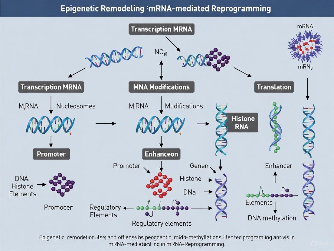
This article explores the convergence of epigenetic remodeling and mRNA technology for cellular reprogramming, a frontier in regenerative medicine and drug development.
Epigenetic Remodeling in mRNA Reprogramming: Mechanisms, Therapeutics, and Clinical Translation
Abstract
This article explores the convergence of epigenetic remodeling and mRNA technology for cellular reprogramming, a frontier in regenerative medicine and drug development. We examine the foundational epigenetic mechanisms—DNA methylation, histone modifications, and RNA epitranscriptomics—that underpin cell identity and are targeted by mRNA-delivered factors. The scope extends to methodological advances in mRNA-mediated delivery of reprogramming factors, their application in generating induced pluripotent stem cells (iPSCs) and direct transdifferentiation, and the resultant therapeutic potential for tissue repair and aging. The content also addresses critical challenges in efficacy, safety, and specificity, while evaluating current validation strategies and comparative outcomes against other reprogramming techniques. This synthesis provides researchers and drug developers with a comprehensive overview of the current landscape and future trajectory of mRNA-based epigenetic reprogramming.
The Epigenetic Code: Foundations of Cell Identity and Plasticity
Epigenetic mechanisms represent a reversible layer of gene regulation that controls cellular identity and function without altering the underlying DNA sequence. These mechanisms—DNA methylation, histone modifications, and chromatin remodeling—collectively establish and maintain gene expression patterns that define cell states [1]. In the context of mRNA-mediated reprogramming, understanding these core epigenetic mechanisms is paramount for developing precise interventions that can redirect cell fate for therapeutic purposes. The emerging field of epigenetic editing demonstrates how targeted rewriting of epigenetic signatures can reprogram gene expression without genomic editing, offering promising avenues for therapeutic intervention across various diseases [2]. This technical guide examines the fundamental epigenetic mechanisms and their interplay within mRNA-based reprogramming strategies, providing researchers with essential knowledge and methodologies for advancing epigenetic research and therapeutic development.
Core Mechanism I: DNA Methylation
Molecular Basis and Enzymatic Machinery
DNA methylation involves the covalent addition of a methyl group to the fifth carbon of cytosine bases, primarily within CpG dinucleotides, forming 5-methylcytosine (5mC) [3]. This modification is dynamically regulated by writer, eraser, and reader proteins that establish, remove, and interpret methylation marks, respectively.
Table 1: DNA Methylation Enzymatic Machinery and Functions
| Enzyme/Protein | Classification | Primary Function | Consequence of Loss-of-Function |
|---|---|---|---|
| DNMT1 | Writer - Maintenance | Maintains methylation patterns during DNA replication | Apoptosis of germline stem cells; hypogonadism and meiotic arrest [3] |
| DNMT3A/B | Writer - De Novo | Establishes new methylation patterns during development | Abnormal spermatogonial function; fertility issues [3] |
| DNMT3L | Writer Cofactor | Enhances DNMT3A/B activity; targets de novo methylation | Decrease in quiescent spermatogonial stem cells [3] |
| TET Family | Eraser | Initiates DNA demethylation via 5mC oxidation | Fertile phenotype observed in TET1/2 mutants [3] |
| MBD Family | Reader | Recognizes and binds methylated DNA; recruits repressive complexes | / |
The distribution of DNA methylation is precisely controlled, with approximately 70-90% of CpG sites methylated in mammalian genomes. CpG islands—genomic regions with high G+C content (>50%) and dense CpG clustering—typically remain unmethylated, particularly when located near promoter regions or transcription start sites (TSS) [3]. DNA methylation generally correlates with transcriptional repression by altering chromatin accessibility and impeding transcription factor binding, though it can also stabilize RNA polymerase II elongation and activate transcription in specific contexts [3].
Dynamics in Cellular Programming and Reprogramming
During embryonic development and cellular reprogramming, the genome undergoes waves of global demethylation followed by de novo methylation [3]. In primordial germ cells (PGCs), the precursors to spermatogonial stem cells (SSCs), genome-wide DNA demethylation reduces 5mC levels to approximately 16.3% during migration to the gonads, significantly lower than the 75% abundance in embryonic stem cells (ESCs) [3]. This hypomethylation is driven by repression of de novo methyltransferases DNMT3A/B and elevated activity of DNA demethylation factors like TET1 [3]. Subsequently, from embryonic day 13.5 to 16.5, de novo DNA methylation is gradually reestablished [3].
In the context of aging, DNA methylation patterns undergo significant shifts. The genome is generally hypomethylated during aging, while specific genes become hypermethylated at CpG islands [1]. These predictable changes have enabled the development of epigenetic clocks that use machine learning methods based on CpG methylation states to predict chronological age and assess biological aging [1]. The first-generation epigenetic clocks, including Horvath's clock (a multi-tissue predictor based on 353 CpG sites) and Hannum's clock, have become essential tools in aging and cancer research [1].
Experimental Methodologies for DNA Methylation Analysis
Bisulfite Sequencing Protocol:
- DNA Treatment: Treat 500ng-1μg of genomic DNA with sodium bisulfite using commercial kits (e.g., EZ DNA Methylation Kit from Zymo Research), which converts unmethylated cytosines to uracils while leaving methylated cytosines unchanged.
- Library Preparation: Prepare sequencing libraries from bisulfite-converted DNA using appropriate adapters with minimal bias.
- Sequencing: Perform next-generation sequencing on Illumina platforms to achieve >10X coverage for whole-genome bisulfite sequencing (WGBS) or targeted approaches.
- Data Analysis: Map sequenced reads to a bisulfite-converted reference genome using tools like Bismark or BSMAP. Calculate methylation percentages as mC/(mC+uC) at each cytosine position.
- Differential Analysis: Identify differentially methylated regions (DMRs) using statistical packages such as methylKit or DSS, with multiple testing correction.
Alternative Method: Methylated DNA Immunoprecipitation (MeDIP)
- Principle: Immunoprecipitation of methylated DNA fragments using antibodies specific for 5-methylcytosine.
- Procedure: Shear genomic DNA to 200-500bp fragments; incubate with anti-5mC antibody; capture antibody-DNA complexes with magnetic beads; wash and elute methylated DNA; analyze via qPCR, microarray, or sequencing.
- Applications: Cost-effective for methylome profiling when combined with sequencing (MeDIP-seq); ideal for large sample cohorts.
Core Mechanism II: Histone Modifications
Major Histone Modification Types and Functional Consequences
Histone modifications represent post-translational alterations to histone proteins that regulate chromatin structure and gene accessibility. These modifications occur primarily on the N-terminal tails of histones and include methylation, acetylation, phosphorylation, ubiquitination, and others [3]. Each modification type has distinct effects on chromatin state and transcriptional activity.
Table 2: Major Histone Modifications and Their Functional Roles
| Modification Type | Histone Residues | Enzymes (Writers/Erasers) | Chromatin State Association | Functional Outcome |
|---|---|---|---|---|
| H3K4me3 | H3 Lysine 4 | Writers: SET1, MLL; Erasers: LSD1, KDM5 | Euchromatin | Transcriptional activation; marks active promoters |
| H3K27me3 | H3 Lysine 27 | Writer: EZH2; Eraser: KDM6 | Facultative heterochromatin | Transcriptional repression; Polycomb-mediated silencing |
| H3K9me3 | H3 Lysine 9 | Writers: SUV39H; Erasers: KDM4 | Constitutive heterochromatin | Transcriptional repression; heterochromatin formation |
| H3K36me3 | H3 Lysine 36 | Writers: SETD2; Erasers: KDM4 | Euchromatin | Transcriptional elongation; prevents spurious initiation |
| H3K79me | H3 Lysine 79 | Writer: DOT1L | Euchromatin | Transcriptional activation; DNA damage response |
| H3/H4 Acetylation | Multiple lysines | Writers: HATs; Erasers: HDACs | Euchromatin | Chromatin relaxation; TF recruitment; transcriptional activation |
Histone methyltransferases (HMTs) and demethylases (KDMs) precisely control the methylation states of specific lysine and arginine residues, while histone acetyltransferases (HATs) and deacetylases (HDACs) regulate acetylation dynamics [1]. The recent development of CRISPR-based chromatin kinases enables programmable human histone phosphorylation and gene activation, demonstrating the potential for targeted epigenetic editing [2].
Histone Modification Dynamics in Cellular Identity and Aging
During cellular reprogramming, histone modifications undergo dramatic reorganization to establish new transcriptional programs. In the context of aging, there is a general loss of histones as well as global chromatin remodeling across multiple model systems [1]. Specific age-related changes include reduction of activating marks such as H3K4me3 and H4K16ac, and alterations in the distribution of repressive marks like H3K9me3 and H3K27me3 [1].
Histone modification patterns serve as critical barriers to reprogramming. For example, the histone methyltransferase Suv39h null mice exhibit spermatogenic failure with nonhomologous chromosome association, demonstrating the essential role of H3K9 methylation in maintaining cellular function [3]. Similarly, PRMT5 deficiency increases H3K9me2 and H3K27me2 levels and alters chromatin state of PLZF, leading to SSC developmental defects and spermatogenesis disorders [3].
Experimental Methodologies for Histone Modification Analysis
Chromatin Immunoprecipitation Sequencing (ChIP-seq) Protocol:
- Crosslinking: Treat cells with 1% formaldehyde for 10 minutes at room temperature to crosslink proteins to DNA.
- Chromatin Shearing: Lyse cells and sonicate chromatin to 200-500bp fragments using Covaris or Bioruptor systems.
- Immunoprecipitation: Incubate chromatin with 2-5μg of validated, modification-specific antibody (e.g., anti-H3K4me3, anti-H3K27ac) overnight at 4°C with rotation.
- Complex Capture: Add protein A/G magnetic beads and incubate for 2 hours; wash extensively with low- and high-salt buffers.
- Decrosslinking and Purification: Reverse crosslinks at 65°C overnight; treat with RNase A and proteinase K; purify DNA using silica membrane columns.
- Library Preparation and Sequencing: Construct sequencing libraries using NEBNext Ultra II DNA Library Prep Kit; sequence on Illumina platforms (minimum 10 million reads per sample).
- Data Analysis: Align sequences to reference genome using Bowtie2 or BWA; call peaks with MACS2; perform differential binding analysis with DiffBind.
Alternative Method: CUT&Tag for Low-Input Samples
- Principle: Uses protein A-Tn5 transposase fusion to tagmentation of antibody-bound chromatin in situ.
- Advantages: Requires significantly fewer cells (500-50,000 cells); lower background noise; can be multiplexed.
- Procedure: Permeabilize cells; incubate with primary antibody; add protein A-Tn5 adapter complex; activate tagmentation with magnesium; extract and purify DNA for library preparation.
Core Mechanism III: Chromatin Remodeling
Chromatin Remodeling Complexes and Mechanisms
Chromatin remodeling complexes (CRCs) are multi-protein machines that utilize ATP hydrolysis to slide, evict, or restructure nucleosomes, thereby regulating DNA accessibility [3]. These complexes play pivotal roles in various cellular processes including cell proliferation, differentiation, and apoptosis, with their dysfunction contributing to developmental disorders and diseases [3].
Table 3: Major Chromatin Remodeling Complexes and Functions
| Complex | Core ATPase | Additional Subunits | Primary Mechanism | Biological Functions |
|---|---|---|---|---|
| SWI/SNF | BRG1/SMARCA4 or BRM/SMARCA2 | 10-15 subunits (BAF complex) | Nucleosome sliding, eviction | Transcriptional activation, differentiation, tumor suppression |
| ISWI | SMARCA5/SMARCA1 | 2-4 subunits | Nucleosome spacing, assembly | Chromatin compaction, transcription regulation, replication |
| CHD | CHD1-CHD9 | Variable | Nucleosome sliding, spacing | Transcriptional regulation, development, DNA repair |
| INO80 | INO80 | 15+ subunits | Nucleosome sliding, histone variant exchange | Transcriptional regulation, DNA repair, telomere maintenance |
Chromatin remodeling complexes function through several mechanistic approaches: (1) nucleosome sliding along DNA; (2) histone variant exchange (e.g., H3.3 for H3); (3) nucleosome eviction to create accessible regions; and (4) nucleosome assembly and spacing to regulate compaction [3]. The SWI/SNF (BAF) complex, in particular, has been implicated in cellular reprogramming, with specific subunit compositions determining permissiveness for pluripotency acquisition.
Chromatin Architecture in Cell Fate Decisions
During cellular differentiation and reprogramming, the chromatin landscape undergoes dramatic reorganization from a generally open configuration in pluripotent cells to a more restricted architecture in differentiated states. In spermatogenesis, chromatin remodeling complexes mediate fate determinations of spermatogonial stem cells (SSCs) to ensure normal development [3]. The precise regulation of spermatogenesis relies on synergistic interactions between genetic and epigenetic factors, with spermatogenesis failure resulting from epigenetic dysregulation [3].
In the context of aging, global chromatin reorganization represents a hallmark of cellular senescence. Aged cells typically exhibit loss of heterochromatin, particularly at repetitive elements and telomeres, leading to genomic instability and aberrant gene expression [1]. These age-related chromatin changes create barriers to reprogramming that must be overcome for efficient cell fate conversion.
Experimental Methodologies for Chromatin Architecture Analysis
Assay for Transposase-Accessible Chromatin with Sequencing (ATAC-seq) Protocol:
- Cell Preparation: Isolate 50,000-100,000 viable cells with >90% viability; avoid overfixation.
- Cell Lysis: Resuspend cells in cold lysis buffer (10mM Tris-Cl pH 7.4, 10mM NaCl, 3mM MgCl2, 0.1% IGEPAL CA-630) for 10 minutes on ice.
- Tagmentation Reaction: Incubate nuclei with Tn5 transposase (Nextera DNA Library Prep Kit) at 37°C for 30 minutes with shaking.
- DNA Purification: Clean up tagmented DNA using Zymo DNA Clean and Concentrator columns.
- Library Amplification: Amplify libraries with 10-12 cycles of PCR using barcoded primers; incorporate dual index primers for multiplexing.
- Size Selection and QC: Purify libraries using SPRIselect beads (0.5X-1.5X ratio); assess quality on Bioanalyzer or Tapestation.
- Sequencing and Analysis: Sequence on Illumina platforms (2×50bp or 2×75bp); align reads to reference genome using BWA; call peaks with MACS2; visualize in IGV.
Alternative Method: MNase-seq for Nucleosome Positioning
- Principle: Uses micrococcal nuclease to digest linker DNA between nucleosomes.
- Procedure: Isolate nuclei; digest with MNase; stop reaction with EGTA; purify mononucleosomal DNA; construct sequencing libraries.
- Applications: Maps nucleosome positions and occupancy; identifies stable versus dynamic nucleosomes.
Interplay of Epigenetic Mechanisms in mRNA-Mediated Reprogramming
Epigenetic Coordination in Cell Fate Transitions
The core epigenetic mechanisms do not function in isolation but rather form an integrated regulatory network that controls cellular identity. During mRNA-mediated reprogramming, these epigenetic layers must be coordinately remodeled to establish new gene expression programs. DNA methylation and histone modifications exhibit extensive crosstalk, with DNA methylation readers (MBD proteins) recruiting histone modifiers such as HDACs and HMTs to reinforce repressive chromatin states [3]. Similarly, specific histone modifications can influence DNA methylation patterns, creating self-reinforcing epigenetic cycles that maintain cellular states.
Recent advances in single-cell multi-omics now enable simultaneous profiling of multiple epigenetic layers, revealing the coordinated dynamics of DNA methylation, histone modifications, and chromatin accessibility during reprogramming [3] [1]. These approaches have identified epigenetic roadblocks to reprogramming and strategies to overcome them, such as transient inhibition of DNA methylation or histone modifications to enhance plasticity.
mRNA-Based Epigenetic Editing Technologies
Emerging mRNA-based technologies enable precise epigenetic editing by delivering mRNAs encoding engineered epigenetic editors. These include:
- ZF-EDs: Zinc finger domains fused to epigenetic catalytic domains (e.g., DNMT3A for targeted methylation) [2]
- TALEs: Transcription activator-like effectors coupled to TET1 for targeted demethylation [2]
- dCas9-Epigenetic Editors: Catalytically dead Cas9 fused to writers/erasers of histone modifications [2]
These tools enable locus-specific epigenetic rewriting without altering DNA sequences, offering potential for therapeutic reprogramming in disease contexts. For instance, targeted DNA methylation of the VEGF-A promoter using engineered zinc finger-DNA methyltransferase fusions has demonstrated sustained gene silencing in cancer models [2]. Similarly, TALE-TET1 fusion proteins have enabled targeted demethylation and activation of endogenous genes [2].
Research Reagent Solutions for Epigenetic Reprogramming
Table 4: Essential Research Reagents for Epigenetic Reprogramming Studies
| Reagent Category | Specific Examples | Primary Function | Application Notes |
|---|---|---|---|
| DNA Methylation Modulators | 5-Azacytidine (DNMT inhibitor); DAC (Decitabine); RG108 | Global DNA demethylation; enhances reprogramming efficiency | Use at 0.5-5μM for 48-72h; monitor cytotoxicity |
| Histone Deacetylase Inhibitors | Valproic Acid (VPA); Trichostatin A (TSA); SAHA (Vorinostat) | Increases histone acetylation; chromatin relaxation | VPA at 0.5-2mM; TSA at 50-500nM; optimize for cell type |
| Histone Methyltransferase Inhibitors | DZNep (EZH2 inhibitor); GSK126 (EZH2 inhibitor); UNC0638 (G9a inhibitor) | Reduces repressive H3K27me3 and H3K9me2 marks | Critical for overcoming epigenetic barriers to reprogramming |
| mRNA Synthesis Kits | MEGAscript T7 Transcription Kit; CleanCap mRNA Kit | Produces modified mRNAs for epigenetic editor expression | Incorporate 5-methoxyuridine to reduce immunogenicity |
| Epigenetic Editor Systems | dCas9-DNMT3A; dCas9-TET1; SunTag-based systems | Locus-specific epigenetic editing | Co-express with guide RNAs targeting specific genomic loci |
| Delivery Vehicles | Lipid nanoparticles (LNPs); Electroporation systems | Efficient mRNA delivery to target cells | Optimize lipid:mRNA ratios for specific cell types |
The core epigenetic mechanisms—DNA methylation, histone modifications, and chromatin remodeling—form an integrated regulatory network that governs cellular identity and plasticity. Understanding the dynamics and interplay of these mechanisms is essential for advancing mRNA-mediated reprogramming strategies for therapeutic applications. Recent technological advances in epigenetic editing, single-cell multi-omics, and targeted delivery systems are accelerating our ability to precisely manipulate the epigenetic landscape to direct cell fate transitions.
Future directions in epigenetic reprogramming research will likely focus on achieving greater specificity in epigenetic editing, improving the persistence of epigenetic modifications, and developing strategies to overcome the epigenetic barriers of aging and disease [2] [4]. As the field progresses, the integration of epigenetic therapies with mRNA-based delivery platforms holds significant promise for developing transformative treatments for degenerative diseases, aging-related conditions, and genetic disorders.
The Role of TET Proteins and Vitamin C in Active DNA Demethylation
In mammalian genomes, DNA methylation in the form of 5-methylcytosine (5mC) represents a fundamental epigenetic mechanism regulating gene expression, genomic stability, and cellular identity [5] [6]. Historically considered a stable epigenetic mark, 5mC is now understood to undergo active reversal through enzymatic processes, providing dynamic regulation of the epigenome [5] [6]. The Ten-Eleven Translocation (TET) family of enzymes—TET1, TET2, and TET3—catalyze the sequential oxidation of 5mC, initiating the major pathway for active DNA demethylation in mammalian cells [5] [6] [7].
Vitamin C (ascorbate) serves as a critical cofactor for TET enzymes, enhancing their catalytic activity and promoting DNA demethylation [8] [9] [7]. This biochemical relationship positions vitamin C as a key environmental factor capable of modulating the epigenome, with significant implications for cellular reprogramming, differentiation, and disease treatment [8] [9] [7]. Within mRNA-mediated reprogramming research, understanding the TET-vitamin C axis provides crucial insights into epigenetic remodeling mechanisms that can enhance reprogramming efficiency and fidelity.
This technical review examines the molecular mechanisms of TET-mediated active DNA demethylation, vitamin C's role as an enzymatic cofactor, experimental evidence across biological systems, and practical methodologies for researchers investigating epigenetic reprogramming.
Molecular Mechanisms of TET-Mediated Active DNA Demethylation
The TET Enzyme Family: Structure and Catalytic Function
TET proteins constitute a family of iron (Fe²⁺) and α-ketoglutarate (α-KG)-dependent dioxygenases that share a conserved C-terminal catalytic domain [6]. This domain contains a double-stranded β-helix (DSBH) fold, a cysteine-rich region, and binding sites for both Fe²⁺ and α-KG, which are essential for catalytic activity [6]. TET1 and TET3 additionally possess a CXXC zinc finger domain at their N-terminus that enables binding to unmethylated CpG-rich DNA [6]. Structural studies reveal that the TET catalytic core preferentially binds cytosines in CpG contexts but shows minimal sequence specificity for flanking DNA regions [5] [6].
The catalytic mechanism of TET enzymes involves three sequential oxidation steps [5] [6] [7]:
- Hydroxylation: 5-methylcytosine (5mC) → 5-hydroxymethylcytosine (5hmC)
- Formylation: 5hmC → 5-formylcytosine (5fC)
- Carboxylation: 5fC → 5-carboxylcytosine (5caC)
This stepwise oxidation generates intermediates with distinct properties and functions in the demethylation pathway [5].
Pathways to Unmodified Cytosine
TET oxidation products can revert to unmodified cytosine through two primary mechanisms:
* Passive Demethylation: 5hmC cannot be effectively recognized by the maintenance DNA methyltransferase DNMT1 during DNA replication. Consequently, TET-mediated oxidation leads to replication-dependent dilution of DNA methylation without enzymatic removal [6] [7]. *In vitro studies demonstrate DNMT1 activity is reduced up to 60-fold on DNA substrates containing 5hmC [6].
Active Demethylation: The TET-generated bases 5fC and 5caC are recognized and excised by thymine DNA glycosylase (TDG), which initiates the base excision repair (BER) pathway [5] [6] [7]. TDG exhibits robust activity toward 5fC and 5caC but cannot excise 5hmC [5]. Following base excision, the BER pathway completes the process by replacing the excised base with an unmodified cytosine [5] [7].
Figure 1: TET-Mediated Active DNA Demethylation Pathway. TET enzymes catalyze sequential oxidation of 5mC to 5hmC, 5fC, and 5caC. Demethylation is completed via TDG-initiated base excision repair or passive replication-dependent dilution. [5] [6] [7]
Vitamin C as a Critical Cofactor for TET Enzymes
Biochemical Mechanism of Action
Vitamin C (L-ascorbic acid) enhances TET catalytic activity by maintaining the iron cofactor in its reduced state (Fe²⁺) within the enzyme's active site [8] [7]. The TET catalytic cycle involves Fe²⁺ oxidation to Fe³⁺ during the hydroxylation reaction. Vitamin C serves as an electron donor to regenerate Fe²⁺ from Fe³⁺, enabling multiple catalytic turnovers and sustained enzymatic activity [8] [10].
This cofactor function is distinct from vitamin C's antioxidant properties. While other antioxidants like vitamin E and glutathione show minimal effects on TET activity, vitamin C specifically promotes TET-mediated DNA demethylation through its action as an enzymatic cofactor [8] [9].
Concentration-Dependent Effects on TET Activity
Vitamin C enhances TET activity at physiological concentrations (0.1-1.0 mM) comparable to those transported from bloodstream into tissues [11] [10]. These concentrations significantly increase global 5hmC levels and promote locus-specific DNA demethylation in various cellular models [11] [9] [10].
Table 1: Vitamin C Effects on TET Activity and Functional Outcomes Across Biological Systems
| Biological System | VC Concentration | Key Effects on TET/Demethylation | Functional Outcomes | Reference |
|---|---|---|---|---|
| Human Epidermal Equivalents | 0.1-1.0 mM | Increased global 5hmC; hypomethylation of 10,138 genomic regions; upregulation of 12 proliferation genes | Enhanced keratinocyte proliferation; increased epidermal thickness | [11] [12] [10] |
| Mouse B Cells | Physiological (unspecified) | Increased TET2/3-mediated demethylation at Prdm1 locus; enhanced STAT3 binding | Promoted plasma cell differentiation; enhanced antibody response | [9] |
| Embryonic Stem Cells/Reprogramming | 50-100 μM | Enhanced TET-mediated demethylation at pluripotency loci; prevented hypermethylation | Improved iPSC generation efficiency and quality; erased epigenetic memory | [8] |
Experimental Evidence: Vitamin C and TET in Biological Systems
Epidermal Regeneration and Proliferation
In human epidermal equivalent models, vitamin C treatment at physiologically relevant concentrations (0.1-1.0 mM) significantly increased epidermal thickness by promoting keratinocyte proliferation [11] [10]. This effect was mediated through TET-dependent DNA demethylation, as demonstrated by:
- Global 5hmC Increase: Vitamin C treatment elevated global 5-hydroxymethylcytosine levels, indicating enhanced TET activity [11] [10].
- Genome-Wide Hypomethylation: Whole-genome bisulfite sequencing identified 10,138 differentially methylated regions showing hypomethylation in vitamin C-treated samples [11] [10].
- Proliferation Gene Activation: Twelve key proliferation-related genes demonstrated 1.6- to 75.2-fold upregulation associated with promoter/enhancer hypomethylation [11] [12] [10].
- TET Dependence: A TET enzyme inhibitor completely abolished vitamin C-induced epidermal thickening and gene expression changes, confirming TET-mediated mechanism [11] [10].
Plasma Cell Differentiation and Immune Function
Vitamin C potently enhances IL-21/STAT3-dependent plasma cell differentiation in both mouse and human B cells [9]. The mechanistic insights include:
- Early Activation Criticality: Vitamin C is required during early B cell activation to prime cells for subsequent differentiation [9].
- Prdm1 Locus Demethylation: Vitamin C facilitates TET2/3-mediated demethylation of multiple regulatory elements at the Prdm1 locus, which encodes BLIMP1, the master transcription factor for plasma cell differentiation [9].
- Enhanced Transcription Factor Binding: DNA demethylation enables increased STAT3 association with the Prdm1 promoter and downstream enhancer (E27), ensuring efficient gene expression [9].
This mechanism explains the historical observations of impaired antibody responses during vitamin C deficiency and highlights how micronutrients regulate epigenetic enzymes to influence cell fate decisions [9].
Cellular Reprogramming and Pluripotency
Vitamin C significantly improves the efficiency and quality of induced pluripotent stem cell (iPSC) generation [8]. In reprogramming somatic cells to pluripotency, vitamin C:
- Enhances TET-mediated demethylation at pluripotency gene loci (Nanog, Oct4) [8]
- Prevents aberrant hypermethylation at imprinted gene clusters (Dlk1-Dio3) [8]
- Facilitates erasure of epigenetic memory from donor cell lineages [8]
- Promotes a more naive "ground state" of pluripotency resembling blastocyst-derived ESCs [8]
These effects are specifically mediated through TET enzyme enhancement, as iron chelators or α-KG analogs that inhibit α-KGDDs impair iPSC formation [8].
Methodologies for Investigating TET and Vitamin C in Demethylation
DNA Methylation Profiling Techniques
Table 2: DNA Methylation Detection Methods for TET-Vitamin C Studies
| Method | Resolution | Key Applications | Advantages | Limitations | Reference |
|---|---|---|---|---|---|
| Whole-Genome Bisulfite Sequencing (WGBS) | Single-base | Genome-wide methylation mapping; identifies differentially methylated regions | Comprehensive coverage; absolute methylation quantification | DNA degradation; high cost; computational intensity | [13] [14] |
| Enzymatic Methyl-Sequencing (EM-seq) | Single-base | Genome-wide methylation profiling without bisulfite | Preserves DNA integrity; reduced bias; improved CpG detection | Newer method with less established protocols | [14] |
| Illumina MethylationEPIC BeadChip | Single-CpG (850K+ sites) | Population studies; biomarker discovery | Cost-effective; standardized analysis; high throughput | Limited to predefined CpG sites; no non-CpG context | [13] [14] |
| Oxford Nanopore Sequencing | Single-base (direct detection) | Long-range methylation profiling; challenging genomic regions | No conversion needed; long reads detect haplotypes; detects 5mC/5hmC | High DNA input; lower accuracy for single CpGs | [14] |
| Reduced Representation Bisulfite Sequencing (RRBS) | Single-base (CpG-rich regions) | Cost-effective targeted methylation analysis | Focuses on informative CpG-rich regions; reduced sequencing cost | Limited genome coverage (~10-15%) | [13] |
Experimental Protocol: Assessing Vitamin C Effects on TET-Mediated Demethylation
Objective: Evaluate vitamin C-dependent DNA demethylation at specific genomic loci in cellular models.
Cell Culture and Treatment:
- Culture target cells (e.g., primary keratinocytes, B cells, or reprogramming fibroblasts) in standard medium.
- Establish experimental conditions:
- Control: Standard culture medium
- Vitamin C treatment: Medium supplemented with physiological vitamin C (0.1-1.0 mM fresh L-ascorbic acid)
- Inhibition control: Vitamin C medium + TET inhibitor (e.g, Bobcat339 or dimethyloxalylglycine)
- Maintain cultures for 7-14 days with medium changes every 48-72 hours.
Sample Collection and DNA Extraction:
- Harvest cells at appropriate time points using trypsinization or mechanical dissociation.
- Extract genomic DNA using phenol-chloroform or commercial kits with RNAse treatment.
- Quantify DNA purity and concentration (A260/280 ~1.8).
DNA Methylation Analysis:
- Perform bisulfite conversion using EZ DNA Methylation Kit (Zymo Research) or equivalent.
- Conduct whole-genome bisulfite sequencing (WGBS):
- Library preparation with bisulfite-converted DNA
- Sequencing on Illumina platform (minimum 30x coverage)
- Alignment to reference genome using Bismark or BS-Seeker
- Methylation calling and differential methylation analysis
- Validate key findings with pyrosequencing or targeted bisulfite sequencing.
Functional Validation:
- Measure global 5hmC levels with ELISA-based hydroxymethylation assay.
- Analyze gene expression of demethylated targets by RT-qPCR.
- Assess functional outcomes (proliferation, differentiation, reprogramming efficiency).
Figure 2: Experimental Workflow for Vitamin C Demethylation Studies. Key steps from cell culture to data integration for investigating TET-vitamin C mediated DNA demethylation. [11] [9] [14]
Table 3: Research Reagent Solutions for TET-Vitamin C Studies
| Reagent/Category | Specific Examples | Function/Application | Key Considerations |
|---|---|---|---|
| Vitamin C Formulations | L-ascorbic acid (fresh), Sodium ascorbate, Ascorbic acid 2-phosphate | TET enzyme cofactor; maintain physiological concentrations (0.1-1.0 mM) | Fresh preparation required; ascorbic acid 2-phosphate more stable in culture |
| TET Inhibitors | Bobcat339, Dimethyloxalylglycine (DMOG), 2-HG (2-hydroxyglutarate) | Inhibit TET enzymatic activity; establish TET-dependent mechanisms | DMOG inhibits broad α-KGDD family; Bobcat339 more TET-specific |
| DNA Methylation Kits | EZ DNA Methylation Kit (Zymo Research), MagMeDIP Kit | Bisulfite conversion; methylated DNA immunoprecipitation | Bisulfite conversion efficiency critical; validate with controls |
| 5hmC Detection | Hydroxymethylated DNA Immunoprecipitation (hMeDIP), 5hmC ELISA, GLIB-seq | Quantify global and locus-specific 5hmC levels | 5hmC-specific antibodies required; distinguish from 5mC |
| Cell Culture Models | Primary keratinocytes, B cells, reprogramming fibroblasts, epidermal equivalents | Relevant biological systems for vitamin C demethylation studies | Human epidermal equivalents for skin studies; primary B cells for immunity |
| Sequencing Platforms | Illumina (WGBS), Oxford Nanopore (direct detection), EPIC BeadChip | DNA methylation profiling | Choice depends on resolution, coverage, and budget requirements |
The TET-vitamin C axis represents a fundamental mechanism for dynamic regulation of the DNA methylome, with far-reaching implications for epigenetic remodeling in mRNA-mediated reprogramming research. The experimental evidence across biological systems demonstrates that vitamin C, through its enhancement of TET enzymatic activity, promotes targeted DNA demethylation that drives cellular differentiation, proliferation, and reprogramming.
For researchers in the field, key considerations include:
- Utilizing physiological vitamin C concentrations (0.1-1.0 mM) in culture systems
- Selecting appropriate DNA methylation profiling methods based on research questions
- Implementing proper TET inhibition controls to establish mechanism
- Considering tissue-specific and context-dependent effects of vitamin C
The integration of vitamin C into reprogramming protocols offers promising avenues for improving iPSC generation efficiency and quality, while the broader understanding of nutrient-epigenome interactions opens new therapeutic possibilities for regenerative medicine, cancer treatment, and immune modulation.
Histone Modifications as Bivalent Poised States in Pluripotency
Bivalent chromatin domains, characterized by the simultaneous presence of activating H3K4me3 and repressive H3K27me3 histone modifications, constitute a fundamental epigenetic mechanism for maintaining pluripotency in embryonic stem cells (ESCs). These poised promoter states enable developmental genes to remain transcriptionally silent yet primed for rapid activation upon differentiation signals. Recent advances in single-cell epigenomic profiling and epigenetic engineering have elucidated the mechanistic principles of bivalency, including nucleosomal asymmetry and dedicated reader complexes that interpret these combinatorial marks. This technical review synthesizes current understanding of bivalent chromatin architecture, its functional role in pluripotency regulation, and emerging experimental approaches for investigating and manipulating these poised states in reprogramming and disease contexts. The integration of bivalent chromatin control with mRNA-mediated reprogramming platforms represents a promising frontier for regenerative medicine and therapeutic development.
Bivalent chromatin domains represent a specialized epigenetic state first described in embryonic stem cells (ESCs) where key developmental gene promoters simultaneously harbor both activating histone H3 lysine 4 trimethylation (H3K4me3) and repressive histone H3 lysine 27 trimethylation (H3K27me3) marks [15] [16]. This unique configuration maintains genes in a transcriptionally poised state—neither fully active nor permanently silenced—allowing pluripotent cells to rapidly initiate lineage-specific differentiation programs in response to developmental cues. The balance between these antagonistic marks is dynamically regulated by opposing chromatin-modifying complexes: the COMPASS/Trithorax complex for H3K4 methylation and the Polycomb Repressive Complex 2 (PRC2) for H3K27 methylation [15].
The functional significance of bivalency extends beyond developmental gene regulation to cellular reprogramming and cancer biology. During induced pluripotent stem cell (iPSC) generation, somatic cells undergo extensive epigenetic remodeling to reestablish bivalent domains at critical developmental loci [15]. Similarly, cancer stem cells (CSCs) often exploit bivalent chromatin to maintain plasticity and therapeutic resistance [15]. Recent research has revealed that bivalent nucleosomes frequently exhibit an asymmetric conformation with each sister histone H3 carrying only one of the two modifications, creating specialized platforms for recruitment of distinct reader proteins [17]. This architectural principle enables precise control of gene expression dynamics during cell fate transitions.
Core Mechanisms of Bivalent Domain Establishment and Maintenance
Chromatin Modifying Complexes and Their Regulatory Dynamics
Table 1: Key Chromatin-Modifying Complexes Regulating Bivalent Domains
| Complex/Enzyme | Catalytic Function | Histone Modification | Role in Bivalency |
|---|---|---|---|
| PRC2 (EZH2) | H3K27 methyltransferase | H3K27me3 | Establishes repressive component of bivalency |
| COMPASS/TrxG | H3K4 methyltransferase | H3K4me3 | Establishes active component of bivalency |
| KDM6B (JMJD3) | H3K27 demethylase | Removes H3K27me3 | Resolves bivalency toward activation |
| KDM5B (JARID1B) | H3K4 demethylase | Removes H3K4me3 | Resolves bivalency toward repression |
| KAT6B | Histone acetyltransferase | H3K23ac? | Promotes activation of bivalent genes during differentiation |
The establishment and maintenance of bivalent domains require precisely coordinated activities of opposing chromatin-modifying complexes. PRC2, containing the catalytic subunit EZH2, deposits H3K27me3 marks at developmental gene promoters in ESCs [15]. Concurrently, COMPASS-family complexes catalyze H3K4me3 at these same loci. This counteracting modification system creates a metastable chromatin state that is resolved during differentiation through the recruitment of tissue-specific transcription factors and chromatin regulators that tip the balance toward either activation or stable repression [16].
Recent structural studies have revealed that bivalent nucleosomes exhibit nucleosomal asymmetry, with each sister histone H3 carrying only one of the two modifications [17]. This asymmetric configuration preferentially recruits repressive H3K27me3 readers while failing to enrich activating H3K4me3 binders, thereby promoting a transcriptionally poised state. Surprisingly, the bivalent mark combination also promotes recruitment of specific chromatin proteins not recruited by each mark individually, including the lysine acetyltransferase KAT6B, which is critical for proper activation of bivalent genes during differentiation [17].
Functional Consequences of Bivalent Chromatin Manipulation
Table 2: Experimental Manipulation of Bivalent Marks and Functional Outcomes
| Experimental Approach | Biological System | Key Findings | Reference |
|---|---|---|---|
| H3K4M mutation | Mouse hematopoietic system | H3K4 methylation loss blocked progenitor maturation; HSC maintenance unaffected | [16] |
| H3K4me3 inhibition | Chicken germ cell differentiation | Enhanced PGCLC induction by blocking BMP antagonists | [18] |
| EZH2 inhibition | Multiple cancer models | Reduced CSC populations, restored differentiation capacity | [15] |
| KAT6B knockout | Mouse ESCs | Impaired neuronal differentiation, defective bivalent gene activation | [17] |
Functional studies manipulating bivalent marks have demonstrated their essential role in developmental transitions. In murine hematopoiesis, global depletion of H3K4 methylation via a dominant histone H3-lysine-4-to-methionine (H3K4M) mutation caused lethal depletion of all mature blood cell types, despite normal numbers of hematopoietic stem cells (HSCs) and committed progenitors [16]. This indicates that H3K4 methylation is dispensable for HSC maintenance but essential for progenitor cell maturation. Mechanistically, H3K4 methylation opposes the deposition of repressive H3K27 methylation at differentiation-associated genes enriched for bivalent chromatin in HSCs and progenitors. Concomitant suppression of H3K27 methylation in H3K4-methylation-depleted mice rescued the acute lethality and hematopoietic failure, demonstrating the functional interaction between these two crucial chromatin marks [16].
In avian germ cell development, diminished H3K4me3 facilitates the specification of the germ cell lineage by regulating transitions of bivalent states into repressive configurations. Selective erasure of H3K4me3 modifications was shown to block expression of BMP signaling antagonists, thereby enhancing the creation of primordial germ cell-like cells (PGCLCs) in chicken [18]. This research provides epigenetic strategies to enhance the production of germ cells for agricultural and conservation applications.
Advanced Methodologies for Bivalent Chromatin Analysis
Single-Cell Epigenomic Profiling Technologies
Recent advances in single-cell epigenomic technologies have revolutionized our ability to study bivalent chromatin dynamics during cellular reprogramming and differentiation. The Target Chromatin Indexing and Tagmentation (TACIT) method enables genome-coverage single-cell profiling of multiple histone modifications across individual cells [19]. This approach has been applied to mouse early embryos, generating maps of seven histone modifications (H3K4me1, H3K4me3, H3K27ac, H3K27me3, H3K36me3, H3K9me3, and H2A.Z) across 3,749 cells from zygote to blastocyst stages.
For investigating coordinated regulation, CoTACIT enables simultaneous profiling of multiple histone modifications in the same single cell through sequential rounds of antibody binding, protein A-Tn5 transposon (PAT) incubation, and tagmentation [19]. This multi-modal profiling reveals that H3K27ac profiles exhibit marked heterogeneity as early as the two-cell stage, preceding substantial heterogeneity in H3K4me3 and other marks, suggesting that cells may begin to display functional heterogeneity through establishment of H3K27ac at this early developmental timepoint.
Figure 1: CoTACIT workflow for simultaneous profiling of multiple histone modifications in single cells.
Epigenome Editing Approaches
Programmable epigenetic engineering has emerged as a powerful approach for directly manipulating bivalent domains and assessing functional outcomes. The CRISPRoff/CRISPRon system enables stable epigenetic silencing or activation without permanent DNA alterations [20]. CRISPRoff consists of dCas9 fused to DNMT3A, DNMT3L, and ZNF10 KRAB protein domains, writing heritable silencing programs that persist through numerous cell divisions. Conversely, CRISPRon uses dCas9 fused to a TET1 catalytic domain for targeted erasure of DNA methylation.
In primary human T cells, optimized mRNA-based delivery of CRISPRoff achieves durable gene silencing comparable to Cas9 knockout but without genotoxic double-strand breaks [20]. This platform has been successfully combined with genetic engineering approaches, enabling targeted chimeric antigen receptor (CAR) knock-in at the TRAC locus with simultaneous CRISPRoff silencing of therapeutically relevant genes to improve preclinical CAR-T cell function.
For mRNA-mediated reprogramming, tissue nanotransfection (TNT) provides a non-viral nanotechnology platform for in vivo gene delivery through localized nanoelectroporation [21]. This approach enables direct cellular reprogramming via transcriptional activation and epigenetic remodeling, offering advantages of high specificity, non-integrative delivery, and minimal cytotoxicity compared to viral vectors.
Experimental Protocols for Bivalent Chromatin Investigation
Protocol: CRISPRoff-Mediated Epigenetic Silencing in Primary Human T Cells
Principle: This protocol enables durable, heritable gene silencing without DNA damage by targeting epigenetic editors to specific genomic loci, maintaining silencing through multiple cell divisions and restimulation cycles [20].
Step-by-Step Methodology:
- sgRNA Design: Select 3-6 sgRNAs targeting within a 250-bp region immediately downstream of the transcription start site (TSS) of the target gene using optimized prediction algorithms.
- mRNA Preparation: Generate CRISPRoff mRNA (CRISPRoff 7 design) incorporating "design 1" codon optimization, Cap1 mRNA cap, and 1-Me-ps-UTP base modifications for enhanced potency and stability.
- Cell Preparation: Isolate primary human T cells and activate using anti-CD2/CD3/CD28 soluble antibodies for 24 hours prior to electroporation.
- Electroporation: Co-electroporated CRISPRoff mRNA (1-2 µg) and pooled sgRNAs (0.5-1 µg each) using Lonza 4D Nucleofector system with pulse code DS-137.
- Culture and Validation: Maintain cells in complete T-cell media with IL-2 (100 U/mL), restimulating every 9-10 days with soluble antibodies. Assess silencing efficiency by flow cytometry at days 7, 14, 21, and 28 post-electroporation.
- Validation: Confirm target specificity through RNA-seq and whole-genome bisulfite sequencing (WGBS) to verify DNA methylation patterns specifically at the target locus.
Key Applications: Multiplexed epigenetic programming of therapeutic cells, silencing immune checkpoints (PD-1, CTLA-4), or enhancing stemness factors without genomic alterations.
Protocol: Single-Cell Histone Modification Profiling with TACIT
Principle: TACIT enables genome-wide mapping of histone modifications at single-cell resolution with high coverage, allowing characterization of epigenetic heterogeneity during cellular reprogramming [19].
Step-by-Step Methodology:
- Cell Preparation: Fix single-cell suspensions from ESCs or reprogramming cultures with 1% formaldehyde for 10 minutes at room temperature, then quench with 125 mM glycine.
- Chromatin Extraction: Lyse cells in RIPA buffer and digest chromatin with 0.5 µL MNase for 5 minutes at 37°C to generate mononucleosomes.
- Immunoprecipitation: Incubate with histone modification-specific antibodies (e.g., anti-H3K4me3, anti-H3K27me3) overnight at 4°C with rotation.
- PAT Complex Assembly: Prepare protein A-Tn5 transposome (PAT) by mixing equal volumes of protein A (0.2 µg/µL) and Tn5 transposase (0.2 µg/µL), incubate 30 minutes at room temperature.
- Tagmentation: Add PAT complex to immunoprecipitated chromatin, incubate 1 hour at 37°C with shaking to simultaneously capture histone-marked nucleosomes and fragment DNA.
- Library Preparation: Reverse crosslinks, purify DNA, and amplify libraries with 8-12 cycles of PCR using indexed primers.
- Sequencing and Analysis: Sequence on Illumina platforms (minimum 50,000 non-duplicated reads per cell) and process data using dedicated pipelines for single-cell histone modification analysis.
Key Applications: Mapping epigenetic heterogeneity during iPSC reprogramming, identifying subpopulations with distinct differentiation potentials, characterizing aberrant bivalent domains in disease models.
The Scientist's Toolkit: Essential Research Reagents
Table 3: Essential Research Reagents for Investigating Bivalent Chromatin
| Reagent/Category | Specific Examples | Function/Application | Technical Notes |
|---|---|---|---|
| Epigenetic Editors | CRISPRoff-V2.3, CRISPRon | Targeted gene silencing/activation without DNA damage | Optimized mRNA design with 1-Me-ps-UTP enhances stability [20] |
| Histone Mutants | H3K4M, H3K27M | Dominant inhibition of specific histone methylation | Acts as hypomorph without disrupting methyltransferases [16] |
| Small Molecule Inhibitors | EZH2 inhibitors (GSK126), HDAC inhibitors (VPA) | Modulate histone modification levels | VPA enhances reprogramming efficiency [15] [22] |
| Reprogramming Factors | OCT4, SOX2, KLF4, c-Myc (OSKM) | Induce pluripotency in somatic cells | L-Myc reduces tumorigenic risk vs c-Myc [22] |
| Delivery Systems | Tissue Nanotransfection (TNT), mRNA electroporation | Non-viral delivery of reprogramming factors | Enables in vivo reprogramming with minimal cytotoxicity [21] |
| Detection Antibodies | H3K4me3, H3K27me3, H3K27ac | Chromatin immunoprecipitation, immunostaining | Validate specificity with knockout controls |
Integration with mRNA-Mediated Reprogramming Research
The intersection of bivalent chromatin biology with mRNA-mediated reprogramming represents a frontier in regenerative medicine. mRNA-based delivery of reprogramming factors (OCT4, SOX2, KLF4, c-Myc) offers transient, tunable expression without genomic integration, overcoming key safety concerns associated with viral vectors [22]. However, efficient reprogramming requires the establishment of appropriate bivalent domains at developmental genes, which can be enhanced through epigenetic modulation.
Strategies to improve mRNA reprogramming efficiency include:
- Combination with epigenetic modifiers: Co-delivery of mRNA encoding chromatin regulators (KDM5B, KDM6A) or small molecule inhibitors (VPA, 5-aza-cytidine) to facilitate epigenetic remodeling [22].
- Sequential delivery protocols: Staged introduction of reprogramming factors and epigenetic modifiers to mimic natural developmental transitions.
- Enhanced mRNA design: Incorporation of modified nucleotides (1-Me-ps-UTP) and optimized codon usage to increase stability and translational efficiency while reducing immunogenicity [20].
Figure 2: Integration of epigenetic modulation with mRNA-mediated reprogramming.
Recent advances demonstrate that targeted manipulation of bivalent domains can enhance reprogramming outcomes. In chicken germ cell differentiation, selective inhibition of H3K4me3 deposition enhanced primordial germ cell-like cell induction by modulating BMP signaling [18]. Similarly, in murine systems, balanced modulation of H3K4 and H3K27 methylation states improved the quality and functionality of iPSC-derived cells [16]. These approaches highlight the potential of epigenetic engineering to overcome current limitations in cellular reprogramming for therapeutic applications.
Bivalent chromatin represents a fundamental epigenetic mechanism for maintaining cellular plasticity during development and reprogramming. The dynamic balance between H3K4me3 and H3K27me3 at key developmental genes creates a poised state that enables rapid transcriptional responses to differentiation signals. Recent technical advances in single-cell epigenomic profiling, epigenetic editing, and mRNA-mediated reprogramming have provided powerful tools to investigate and manipulate these domains with unprecedented precision.
Future research directions include developing more precise epigenetic editors capable of writing specific combinatorial histone modifications, advancing single-cell multi-omics to simultaneously capture histone modifications, chromatin accessibility, and gene expression in the same cell, and optimizing delivery systems for clinical translation of epigenetic therapies. The integration of bivalent chromatin control with mRNA reprogramming platforms holds particular promise for generating clinically relevant cell types for regenerative medicine, disease modeling, and cancer immunotherapy. As our understanding of bivalent chromatin mechanisms deepens, so too will our ability to harness this knowledge for therapeutic innovation.
The pursuit of cellular rejuvenation and tissue regeneration increasingly converges on the principles of embryonic development. This technical guide explores the paradigm that transient expression of embryonic transcription factors can orchestrate extensive epigenetic remodeling, effectively reversing aged or damaged cellular states to a more plastic, progenitor-like condition. We detail how key developmental cues, particularly the Yamanaka factors (OCT4, SOX2, KLF4, c-MYC), are harnessed in mRNA-mediated reprogramming to reset the epigenetic landscape. Within the broader context of regenerative medicine, this review synthesizes cutting-edge protocols, quantitative data on reprogramming efficacy, and the underlying mechanisms that recapitulate developmental plasticity for therapeutic applications, providing a foundational resource for researchers and drug development professionals.
Embryonic development is characterized by precise, temporally regulated gene expression patterns that guide cellular differentiation and tissue formation. Central to this process is epigenetic remodeling—the dynamic alteration of chromatin architecture and DNA methylation patterns that stabilize cell fate decisions without changing the underlying genetic code [23]. The groundbreaking discovery that forced expression of the embryonic transcription factors OCT4, SOX2, KLF4, and c-MYC (OSKM) could induce pluripotency in somatic cells revealed that developmental pathways can be reactivated to reverse cellular aging and injury [24].
The emerging frontier of mRNA-mediated reprogramming leverages these embryonic cues while addressing critical safety concerns associated with traditional gene therapy. Unlike integrating viral vectors, mRNA-based delivery offers a transient, non-integrative strategy for expressing reprogramming factors, significantly reducing the risk of insertional mutagenesis and genomic instability [25] [26]. This approach provides precise temporal control over factor expression, enabling the partial reprogramming strategies necessary to reverse aging phenotypes or enhance regeneration without inducing teratoma formation [24] [27].
The core thesis of this review posits that embryonic cues guide reprogramming primarily through epigenetic resetting, reversing age-associated heterochromatin loss and DNA methylation patterns to restore cellular plasticity. As demonstrated in progeria mouse models, cyclic induction of OSKM factors restores youthful epigenetic markers like H3K9me3 and promotes tissue regeneration, effectively extending healthspan and lifespan [24]. The following sections detail the molecular mechanisms, experimental protocols, and therapeutic applications of this transformative technology.
Molecular Mechanisms: Embryonic Factors as Epigenetic Regulators
The Yamanaka Factors and Their Developmental Roles
The core embryonic factors used in reprogramming each play distinct yet interconnected roles in establishing pluripotency:
- OCT4 (POU5F1): A pioneer transcription factor critical for maintaining pluripotency in the inner cell mass. It binds to compacted chromatin and initiates opening of pluripotency-associated loci, working in concert with SOX2 to activate key target genes [24].
- SOX2: Partners with OCT4 in a precise stoichiometric ratio to regulate hundreds of pluripotency-associated genes. It promotes dedifferentiation by binding to enhancer elements and facilitating chromatin accessibility [24] [26].
- KLF4: Contributes to epigenetic reprogramming through multiple mechanisms, including regulation of p53 signaling and modulation of histone acetylation patterns. It promotes a permissive chromatin state for reprogramming [27].
- c-MYC: Enhances global transcriptional activity and promotes chromatin accessibility at pluripotency loci, though its use requires careful regulation due to oncogenic potential [24].
Epigenetic Remodeling During Reprogramming
The OSKM factors collaboratively orchestrate extensive epigenetic restructuring that mirrors embryonic epigenetic resetting:
- DNA Methylation Reprogramming: OSKM induction reverses age-associated hypermethylation and hypomethylation patterns. This includes erasure of differential methylation at promoter regions of developmental genes, restoring a more embryonic methylation landscape [24] [23].
- Histone Modification Reset: Partial reprogramming restores youthful patterns of histone modifications, including reduction of age-associated H4K20me3 and restoration of H3K9me3 heterochromatin marks [27]. These changes correlate with suppressed expression of senescence markers p16INK4a and p21CIP1.
- Chromatin Accessibility: OSKM factors function as pioneer factors that bind condensed chromatin and initiate localized decompaction, enabling activation of previously silenced developmental gene networks [24] [28].
Table 1: Key Epigenetic Modifications in Cellular Reprogramming
| Epigenetic Mark | Aging/Senescent State | Post-Reprogramming State | Functional Consequence |
|---|---|---|---|
| H3K9me3 | Decreased | Restored | Regains heterochromatin integrity, reduced genomic instability |
| H4K20me3 | Increased | Decreased | Attenuated senescence-associated secretory phenotype (SASP) |
| Promoter DNA Methylation | Hypermethylation of developmental genes | Demethylation | Reactivation of pluripotency and progenitor gene networks |
| Chromatin Accessibility | Reduced at pluripotency loci | Increased | Activation of embryonic gene expression programs |
Experimental Platforms and Methodologies
mRNA-Based Reprogramming Systems
mRNA technology has emerged as a leading platform for reprogramming due to its transient nature and high efficiency [25]. Key methodological considerations include:
- mRNA Design and Modification: Incorporation of modified nucleotides (e.g., pseudouridine) reduces innate immune recognition and enhances translational efficiency [25]. Optimized 5' and 3' UTRs further increase protein expression levels and duration.
- Delivery Systems: Lipid nanoparticles (LNPs) and exosome-based vehicles protect mRNA from degradation and facilitate cellular uptake. For instance, Cavin2-modified exosomes (M-Exo) demonstrate enhanced delivery efficiency to senescent nucleus pulposus cells [27].
- Dosing Regimens: Cyclic induction protocols (e.g., 2 days ON/5 days OFF) prevent complete reprogramming to pluripotency while achieving epigenetic resetting, thereby minimizing teratoma risk [24].
In Vivo Reprogramming Models
Transgenic mouse models enable controlled investigation of OSKM-mediated reprogramming in living organisms:
- Inducible Systems: The well-established 4F models (4Fj, 4Fk, 4F-A, 4F-B) feature OSKM cassettes inserted at specific genomic loci (Col1a1, Neto2, Pparg) under control of a Tet-O promoter system [24]. Administration of doxycycline (Dox) enables precise temporal control of factor expression.
- Tissue-Specific Variations: OSKM expression patterns show striking tissue dependence, with robust induction in intestine, liver, and skin, and comparatively lower activation in brain, heart, and skeletal muscle [24].
- Safety Considerations: Continuous OSKM induction over weeks produces teratomas in multiple organs, while transient induction (7 days) can initiate dysplastic changes. However, cyclic induction protocols have demonstrated safety in multiple tissue contexts [24].
Table 2: In Vivo Reprogramming Models and Their Applications
| Model System | Genetic Features | Induction Method | Key Applications | Safety Considerations |
|---|---|---|---|---|
| 4Fj/4Fk Mice | OSKM at Col1a1 locus | Doxycycline | Lifespan extension in progeria models [24] | Teratomas with continuous induction |
| OKS@M-Exo | Plasmid in modified exosomes | Direct injection | IVDD treatment [27] | No teratoma formation reported |
| SeV Reprogramming | Non-integrating RNA virus | Transduction | hiPSC generation from fibroblasts/PBMCs [26] | Lower genomic alteration risk vs. episomal |
The Scientist's Toolkit: Essential Research Reagents
Table 3: Key Research Reagents for mRNA-Mediated Reprogramming
| Reagent/Category | Specific Examples | Function in Reprogramming | Experimental Notes |
|---|---|---|---|
| Reprogramming Factors | OCT4, SOX2, KLF4, c-MYC (OSKM) | Induce pluripotency and epigenetic resetting | c-MYC optional for partial reprogramming [27] |
| Delivery Vectors | LNPs, Cavin2-modified exosomes, Sendai virus | Protect mRNA and enhance cellular uptake | Exosomes show superior tissue penetration [27] |
| Small Molecule Enhancers | Y-27632 (ROCK inhibitor) | Improves cell viability post-transfection | Critical for clonal expansion [26] |
| Quality Control Assays | Alkaline phosphatase staining, karyotyping, STR analysis | Validate reprogramming success and genomic integrity | Essential for biobanking applications [26] |
| Senescence Assays | SA-β-Gal staining, p16/p21 quantification | Assess age-reversal effects | Key for evaluating rejuvenation [27] |
Analytical Approaches and Validation Methods
Assessing Reprogramming Efficacy
Rigorous quality control measures are essential for validating reprogramming outcomes:
- Pluripotency Marker Analysis: Immunostaining for canonical markers (NANOG, SSEA-1, TRA-1-60) confirms acquisition of pluripotent state in fully reprogrammed cells [26].
- Epigenetic Landscape Mapping: Genome-wide profiling of DNA methylation (Whole Genome Bisulfite Sequencing) and histone modifications (ChIP-seq) provides comprehensive assessment of epigenetic remodeling [24] [27].
- Transcriptomic Analysis: RNA sequencing reveals expression changes in age-related pathways and pluripotency networks, demonstrating reversal of aging signatures [27].
- Functional Assays: In vivo teratoma formation assays test pluripotency, while transplantation models assess functional integration and tissue repair capacity [24].
Safety and Tumorigenicity Assessment
The potential for neoplastic transformation represents the primary safety concern in reprogramming approaches:
- Dysmplasia Monitoring: Histopathological examination of liver, pancreas, and kidney tissues following OSKM induction identifies pre-neoplastic changes [24].
- Long-Term Fate Tracing: Lineage tracing models track the fate of reprogrammed cells over extended durations to assess stability and transformation risk.
- Alternative Factor Combinations: The OKS combination (omitting c-MYC) demonstrates reduced tumorigenic potential while maintaining rejuvenation capacity in multiple tissue contexts [27].
The following diagram illustrates the core experimental workflow for mRNA-mediated reprogramming and epigenetic validation:
Diagram 1: Experimental workflow for mRNA-mediated reprogramming, showing key steps from cell preparation through validation, with critical decision points influencing therapeutic outcomes.
Therapeutic Applications and Tissue-Specific Outcomes
Regeneration in Tissues with Limited Native Capacity
The therapeutic potential of reprogramming is particularly promising for tissues with restricted innate regenerative capacity:
- Intervertebral Disc Regeneration: In rat IVDD models, OKS@M-Exo treatment ameliorated senescence markers (p16INK4a, p21CIP1), reduced DNA damage, restored proliferation, and maintained disc height and hydration through epigenetic remodeling [27].
- Cardiac Repair: Partial reprogramming enhances regenerative competence in cardiomyocytes, promoting functional recovery following injury through dedifferentiation and epigenetic resetting [24].
- Retinal and Neural Regeneration: OSKM induction restores plasticity in retinal cells and certain neuronal populations, enabling repair of age-related degeneration and injury [24].
Enhancement of Endogenous Regenerative Programs
In tissues with inherent regenerative capacity, reprogramming augments natural repair mechanisms:
- Liver Regeneration: OSKM-mediated reprogramming parallels injury-induced dedifferentiation of hepatocytes, amplifying their natural plasticity to enhance regeneration without complete loss of cellular identity [24].
- Intestinal Epithelial Renewal: The intestine exhibits robust OSKM activation in inducible models, suggesting particular susceptibility to reprogramming-based enhancement of its naturally high turnover rate [24].
- Skeletal Muscle Repair: Partial reprogramming improves muscle regeneration following injury in aged mice, indicating reversal of age-related epigenetic barriers to satellite cell function [24].
The following diagram illustrates the key molecular pathways activated during reprogramming-mediated tissue regeneration:
Diagram 2: Molecular pathways in reprogramming, showing how OSKM expression triggers epigenetic and senescence changes that ultimately restore cellular plasticity and tissue function.
The strategic application of embryonic cues through mRNA-mediated reprogramming represents a paradigm shift in regenerative medicine. By harnessing developmental pathways to remodel the epigenetic landscape, this approach offers unprecedented potential for reversing age-associated cellular decline and enhancing tissue repair. The critical advance lies in achieving partial rather than complete reprogramming—a transient reset that rejuvenates cellular function without inducing tumorigenicity.
Future research directions should focus on optimizing factor combinations and delivery systems for specific tissue contexts, developing more precise temporal control over reprogramming duration, and establishing comprehensive safety profiles for clinical translation. As the field progresses, integration of embryonic guidance principles with mRNA technology promises to unlock novel therapeutic strategies for degenerative diseases, age-related decline, and injury repair, ultimately fulfilling the regenerative potential hinted at in our earliest developmental stages.
Cellular reprogramming represents a paradigm shift in regenerative medicine, enabling the conversion of a specific somatic cell identity into another. This process is fundamentally governed by epigenetic remodeling, which involves the erasure of the existing somatic epigenetic memory and the active establishment of a new epigenetic identity [29]. Within the context of modern therapeutic development, mRNA-based technology has emerged as a transformative tool for directing this process, offering a non-integrative, controllable, and transient expression of reprogramming factors [25] [30]. The precision and safety profile of mRNA-mediated delivery make it particularly suited for applications in engineered regenerative medicine, from cardiac repair to in vivo transdifferentiation [25]. This technical guide delves into the core hallmarks of reprogramming, framing the discussion within the mechanisms of epigenetic resetting and the innovative methodologies that are pushing the field toward clinical translation.
Molecular Mechanisms of Epigenetic Erasure
The initial phase of reprogramming involves dismantling the somatic cell's epigenetic landscape, a process critical for erasing cellular memory and enabling identity conversion.
DNA Demethylation and the Role of TET Enzymes
Genome-wide DNA demethylation is a cornerstone of epigenetic erasure. This process can occur passively, through the inhibition of maintenance DNA methyltransferases during cell division, or actively, mediated by enzymes such as TET (Ten-eleven translocation) dioxygenases [31] [32]. The functional importance of active demethylation is highlighted by studies showing that TET1-deficient human primordial germ cell-like cells (hPGCLCs) fail to fully activate genes critical for gametogenesis and instead aberrantly differentiate into extraembryonic lineages like amnion [31]. This demonstrates that TET1 is essential for directing proper epigenetic reprogramming and lineage fate. Furthermore, in vivo studies on mouse retinal ganglion cells have shown that the beneficial effects of OSK (Oct4, Sox2, Klf4) reprogramming—including axon regeneration and vision restoration—are dependent on the presence of TET1 and TET2 [32].
Histone Modification Remodeling
Concurrent with DNA demethylation, the reprogramming process involves a comprehensive resetting of histone post-translational modifications [29]. This includes changes to marks such as histone H3 lysine 4 dimethylation/trimethylation (H3K4me2/me3) and histone H3 lysine 27 acetylation (H3K27ac), which are associated with active promoters and enhancers. The removal of repressive marks and the installation of activating ones facilitate the opening of chromatin and the activation of pluripotency or new lineage-specific gene networks. The role of reprogramming factors is to recruit or co-opt these epigenetic modifiers to initiate this genome-wide remodeling [29].
Attenuation of Somatic Signatures
A key step in erasing somatic memory is the downregulation of somatic cell-type-specific genes and the silencing of their associated regulatory elements. This involves the decommissioning of somatic enhancers, which are often enriched for transcription factor motifs like AP-1 (Jun, Fos) [33]. During the early phases of reprogramming, the ectopic expression of factors such as OKSM (Oct4, Klf4, Sox2, c-Myc) can sequester these somatic transcription factors, leading to the loss of activating marks and the eventual silencing of these regulatory domains [33].
Table 1: Key Molecules in Epigenetic Erasure and Their Functions
| Molecule/Process | Primary Function | Experimental Outcome of Disruption |
|---|---|---|
| TET1/TET2 Enzymes | Active DNA demethylation | Failure to activate germline genes; aberrant differentiation into amnion [31]; impaired axon regeneration in OSK-treated RGCs [32] |
| Passive Demethylation | Replication-dependent loss of 5mC | Promoted by BMP signaling and inhibition of maintenance DNMTs in hPGCLC differentiation [31] |
| H3K9me3 Demethylation | Removal of repressive chromatin mark | Concentrated in regions of epigenetic memory; reconfiguration is essential for full reprogramming [33] |
| Somatic Enhancer Decommissioning | Silencing of cell-of-origin gene networks | Associated with AP-1 factor sequestration and loss of H3K27ac [33] |
Establishing a New Cellular Identity
Following the erasure of somatic memory, the cell must activate a new transcriptional program to establish a stable identity, whether pluripotent or another somatic cell type.
Activation of Pluripotency and Lineage-Specific Networks
The establishment of a new identity is driven by the activation of key transcription factors that define the target cell state. In reprogramming to pluripotency, the core factors OCT4, SOX2, and KLF4 (OSK) are sufficient to initiate this process without inducing tumorigenesis when delivered transiently [32]. These factors bind to and activate promoters and enhancers of pluripotency-associated genes, establishing a self-reinforcing regulatory network. Similarly, in direct lineage conversion (transdifferentiation), the expression of a specific set of transcription factors can directly activate the gene regulatory network of the target cell type, bypassing a pluripotent intermediate [30].
The Role of Signaling Pathways
Extracellular signaling cues are critical for stabilizing the new cellular identity. In the differentiation of hPGCLCs into pro-spermatogonia or oogonia, Bone Morphogenetic Protein (BMP) signaling is a master regulator [31]. BMP signaling stabilizes the germ cell fate and promotes epigenetic reprogramming by attenuating the MAPK (ERK) pathway and modulating DNA methyltransferase activities. Furthermore, other pathways like WNT and NODAL require precise modulation, as their inhibition can help minimize de-differentiation and maintain the trajectory toward the target cell fate [31].
Phases of Reprogramming
Reprogramming is not an instantaneous event but a multi-stage process:
- Initiation: A rapid, stochastic phase characterized by widespread transcriptional changes, proliferation upregulation, and a mesenchymal-to-epithelial transition (MET) [29].
- Maturation: A slower, more deterministic phase where the core regulatory network of the new cell identity is consolidated and begins to stabilize [29].
- Stabilization: The final phase, where the expression of exogenous reprogramming factors can be silenced, and the cell autonomously maintains its new identity through its endogenous gene regulatory network [29].
Figure 1: The Multi-Phase Progression of Cellular Reprogramming.
mRNA-Mediated Reprogramming: A Modern Therapeutic Platform
The delivery method of reprogramming factors is crucial for safety and efficacy. mRNA-based technology offers a compelling approach for transient, non-integrative reprogramming.
Advantages of mRNA Technology
Unlike viral vectors that can lead to permanent genomic integration, mRNA transfection allows for transient expression of reprogramming factors, significantly reducing the risk of insertional mutagenesis and tumorigenesis [25] [30]. mRNA is translated in the cytoplasm, bypassing the need for nuclear entry, which results in faster onset of protein expression compared to plasmid DNA [30]. Advances in mRNA chemistry, such as nucleoside modifications and optimized codons, have greatly enhanced stability and reduced immunogenicity, making it a viable platform for in vivo applications [25].
In Vivo Delivery and Tissue Nanotransfection (TNT)
A groundbreaking application of mRNA technology is Tissue Nanotransfection (TNT). This non-viral, nanoelectroporation-based platform enables localized in vivo delivery of genetic cargo, including mRNA, directly into tissue cells [30]. The TNT device uses a nanochannel chip to create transient pores in cell membranes, allowing for the efficient uptake of mRNA. This method is highly specific, has minimal cytotoxicity, and avoids the off-target effects associated with viral vectors [30]. TNT has demonstrated success in various therapeutic contexts, including direct in vivo reprogramming for wound healing, ischemia repair, and antimicrobial therapy [30].
Figure 2: mRNA-Based Reprogramming via Tissue Nanotransfection (TNT).
Advanced Methodologies and Experimental Protocols
This section details key experimental approaches for studying and applying cellular reprogramming, with a focus on quantitative outcomes.
In Vitro Reconstitution of Epigenetic Reprogramming
A recent protocol for modeling human germ cell epigenetic reprogramming involves the differentiation of pluripotent stem cells into human PGCLCs (hPGCLCs) [31].
- hPGCLC Induction: Incipient mesoderm-like cells (iMeLCs) are first induced from human iPSCs and subsequently differentiated into hPGCLCs using cytokine cocktails.
- BMP-Driven Differentiation and Amplification: hPGCLCs are cultured in advanced RPMI medium supplemented with a BMP2 signal (e.g., 25-100 ng/mL) and a WNT signaling inhibitor (e.g., IWR1). This culture can be passaged approximately every 10 days, leading to extensive amplification (>10^10-fold) and differentiation into DAZL/DDX4-positive pro-spermatogonia or oogonia-like cells.
- Monitoring: The process is tracked using reporters for germ cell markers (e.g., TFAP2C-eGFP, DAZL-tdTomato) and analysis of DNA demethylation at key loci.
Transient Naive Reprogramming (TNT) for Epigenetic Correction
To address epigenetic aberrations and memory in human iPS cells, a novel protocol termed Transient Naive-Treatment (TNT) reprogramming has been developed [33].
- Naive Conversion: Conventional primed hiPS cells or reprogramming intermediates are transitioned into a naive pluripotent state culture condition for a short, defined period.
- Emulating Embryonic Reset: This transient exposure to a naive environment promotes a more complete epigenome reset, effectively reducing epigenetic memory and correcting aberrant DNA methylation patterns, particularly at H3K9me3-marked and lamin-B1-associated domains.
- Outcome: TNT-reprogrammed hiPS cells are molecularly and functionally more similar to hES cells, showing corrected gene expression and improved differentiation efficiency.
In Vivo Partial Reprogramming for Tissue Regeneration
For therapeutic in vivo applications, a safe and controllable method for expressing reprogramming factors is essential [32].
- Viral Delivery: A dual adeno-associated virus (AAV) system is used. One AAV encodes a transactivator (e.g., rtTA for Tet-On), and the other carries the polycistronic OSK transgene under a TRE promoter.
- Induction: Reprogramming is induced by administering doxycycline (Dox) in the drinking water. This allows for transient, cyclical expression of OSK.
- Validation: In a mouse model of optic nerve crush, this method demonstrated robust axon regeneration and reversal of vision loss in aged mice and a glaucoma model, without inducing teratomas.
Table 2: Quantitative Outcomes of Key Reprogramming Protocols
| Protocol / Study | Reprogramming Factors | Key Quantitative Result | Epigenetic Change Measured |
|---|---|---|---|
| BMP-driven hPGCLC Differentiation [31] | BMP2 Signaling | >10^10-fold cell expansion; differentiation to DAZL+ cells | DNA demethylation at ER-activated gene promoters (e.g., GTSF1, PRAME) |
| In Vivo OSK Reprogramming [32] | OSK (Oct4, Sox2, Klf4) | Axon regeneration >5mm; reversal of vision loss in glaucoma model | Restoration of youthful DNA methylation patterns and transcriptomes |
| Transient Naive Treatment (TNT) [33] | OKSM + Naive Culture | Corrected 2,727 CG-DMRs vs. hES cells; improved differentiation | Reduction of epigenetic memory and aberrant CpH methylation |
The Scientist's Toolkit: Essential Reagents for Reprogramming
Table 3: Key Research Reagent Solutions for Cellular Reprogramming
| Reagent / Tool | Function / Purpose | Example Use Case |
|---|---|---|
| Modified mRNA | Transient, non-integrating expression of reprogramming factors (e.g., OSK, OKSM) | Direct in vivo reprogramming via TNT; protein supplementation therapy [25] [30] |
| Tissue Nanotransfection (TNT) Device | Physical nanoelectroporation for in vivo delivery of genetic cargo | Localized reprogramming in skin for wound healing or ischemia repair [30] |
| BMP2 Ligand | Key signaling molecule for differentiation and stabilization of germ cell fate | Drives hPGCLC differentiation into pro-spermatogonia/oogonia [31] |
| AAV Vectors (e.g., AAV9, AAV2) | In vivo gene delivery for sustained, controllable transgene expression | Safe, long-term expression of OSK in retinal ganglion cells for vision restoration [32] |
| Naive Pluripotency Media | Culture conditions to establish or maintain pre-implantation-like pluripotency | Transient Naive-Treatment (TNT) to reset epigenome in hiPS cells [33] |
| DNMT & TET Modulators | Small molecules to inhibit DNA methyltransferases or activate TET enzymes | Experimentally manipulate active/passive DNA demethylation pathways [31] |
mRNA as a Therapeutic Tool: Delivering the Epigenetic Blueprint
Messenger RNA (mRNA) technology represents a paradigm shift in molecular medicine, offering a versatile platform for therapeutic development. Its core advantages—transience, safety, and precision—make it uniquely suited for applications ranging from infectious disease vaccines to regenerative medicine and cancer immunotherapy [34]. Unlike traditional gene therapies that permanently alter host DNA, mRNA therapeutics function as transient informational molecules that instruct cellular machinery to produce specific proteins without genomic integration [35]. This non-integrating mechanism eliminates the risk of insertional mutagenesis while providing precise temporal control over protein expression [25]. The technology's potential is further amplified within the context of epigenetic remodeling, where transient protein expression can induce lasting phenotypic changes through modified chromatin states rather than permanent genetic alterations [36]. This technical guide examines the fundamental properties of mRNA technology through the lens of its core advantages, providing researchers with a framework for leveraging these characteristics in therapeutic development.
The Transient Nature of mRNA Therapeutics
Mechanisms of Transience and Kinetic Profiles
The transient nature of mRNA therapeutics stems from inherent biological properties of RNA molecules and their regulatory pathways within cells. Following delivery, mRNA exhibits predictable kinetics: rapid onset within 2-6 hours, peak expression at 24-48 hours, and exponential decline over 7-14 days [37]. This transient expression profile is governed by several key factors:
- Ribosomal Translation Efficiency: Optimized mRNA constructs achieve translation rates of 10-100 proteins per mRNA per minute [37]
- mRNA Half-Life: Chemical modifications and optimized UTR sequences extend mRNA half-life from minutes for unmodified constructs to 24-72 hours for optimized versions [37]
- Amplification Effect: A single mRNA molecule can produce 10³-10⁶ protein copies depending on construct optimization and cellular context [37]
This controlled transience is particularly valuable in genome editing applications, where limiting nuclease expression duration helps minimize off-target effects [35].
Comparative Analysis: mRNA vs. DNA Transfection
Table 1: Kinetic and Operational Differences Between DNA and mRNA Transfection
| Parameter | Plasmid DNA | mRNA |
|---|---|---|
| Cell Cycle Dependence | Requires nuclear entry; best in dividing cells | Works in dividing and non-dividing cells |
| Onset of Expression | 12-24 hours | 2-6 hours |
| Duration | Days to weeks; can generate stable lines | Hours to days; transient expression |
| Expression Uniformity | Often mosaic | More even across cells |
| Titratability | Indirect (promoter strength) | Direct (mRNA dose) |
| Integration Risk | Possible | None |
The direct titratability of mRNA enables precise control over protein output, as expression levels correlate directly with the amount of mRNA delivered [35]. This contrasts with DNA transfection, where expression depends on promoter strength and chromatin accessibility, creating indirect and less predictable dose-response relationships.
Safety Profile of mRNA Platforms
Non-Integrating Mechanism and Immunological Safety
The safety advantages of mRNA technology primarily derive from its cytoplasmic mechanism of action, which bypasses the nuclear membrane and eliminates genomic integration risks [35]. This non-integrating nature is particularly advantageous for therapeutic applications where permanent genetic alterations are undesirable. Additional safety considerations include:
- Eliminated Insertional Mutagenesis Risk: Unlike DNA-based approaches, mRNA does not integrate into the host genome, preventing potential disruption of tumor suppressor genes or activation of oncogenes [35] [34]
- Controlled Immunogenicity: While unmodified mRNA can activate pattern recognition receptors, nucleoside modifications (pseudouridine, 5-methylcytidine) significantly reduce innate immune recognition without compromising translation efficiency [38]
- Metabolic Clearance: mRNA undergoes natural degradation through normal cellular processes, with half-life modifiable through sequence optimization [34]
The lipid nanoparticle (LNP) delivery systems used for mRNA therapeutics have demonstrated favorable safety profiles in clinical applications, though component-related inflammatory responses can occur and are more manageable due to the transient nature of expression [37].
Epigenetic Safety and Regulatory Considerations
Within epigenetic reprogramming research, mRNA technology offers a unique safety profile by enabling transient expression of reprogramming factors that can induce lasting epigenetic changes without permanent genetic alteration [36]. Research has demonstrated that SARS-CoV-2 mRNA vaccination establishes persistent histone H3 lysine 27 acetylation (H3K27ac) at promoters of human monocyte-derived macrophages, indicating epigenetic memory that persists for at least six months [36]. This suggests mRNA platforms can achieve lasting functional effects through epigenetic remodeling rather than permanent genetic changes, potentially offering superior safety profiles for regenerative medicine applications.
Precision Control in mRNA Design and Delivery
Molecular Engineering for Precision Expression
Precision in mRNA technology is achieved through sophisticated molecular engineering approaches that control the stability, translation efficiency, and subcellular targeting of mRNA molecules. Key design elements include:
- 5' Cap Modifications: Cap 1 structures enhance ribosome binding and translation initiation while reducing immunogenicity [38]
- Codon Optimization: Strategic selection of synonymous codons maximizes translation efficiency and protein yield [34]
- Untranslated Regions (UTRs): Engineering of 5' and 3' UTRs regulates stability, translation rates, and subcellular localization [37]
- Nucleoside Modifications: Incorporation of modified nucleosides (pseudouridine, 5-methylcytidine) enhances stability and reduces pattern recognition receptor activation [38]
- Poly(A) Tail Length Optimization: Tail length directly correlates with translation efficiency and mRNA longevity [38]
Table 2: Key Quality Attributes for Precision mRNA Therapeutics
| Quality Attribute | Analytical Methods | Impact on Precision |
|---|---|---|
| Identity/Sequence | RT-PCR-Sanger Sequencing, LC-MS/MS, Direct RNA Sequencing | Ensures correct encoded protein |
| Integrity | Capillary Gel Electrophoresis, Bioanalyzer | Confirms full-length mRNA for accurate translation |
| Capping Efficiency | HPLC-UV/MS | Ensures efficient translation initiation |
| Poly(A) Tail Length | HPLC-UV/MS | Determines translation longevity and stability |
| Impurity Profile | Gel Electrophoresis, ELISA, qPCR | Reduces off-target immune activation |
| Functionality | In Vitro Translation, Cell-Based Assays | Confirms biological activity |
Delivery Systems for Spatial Control
Advanced delivery systems enable spatial precision for mRNA therapeutics, directing expression to specific tissues and cell types. Lipid nanoparticles (LNPs) represent the most clinically advanced delivery platform, with standard composition including:
- Ionizable Lipids (35-50%): Enable pH-dependent membrane fusion and endosomal escape [37]
- Phospholipids (10-15%): Provide membrane stability and biocompatibility [37]
- Cholesterol (25-40%): Modulates membrane fluidity [37]
- PEG-Lipids (1-3%): Influence steric stabilization and biodistribution [37]
Emerging targeting strategies include SORT nanoparticles that tune mRNA release based on internal charge modulation, CD117-LNP systems for hematopoietic stem cell targeting, and antibody-conjugated LNPs for specific tissue and immune cell targeting [37]. Local administration routes (intratumoral, intramuscular) further enhance spatial precision by reducing systemic exposure [37].
Experimental Framework for mRNA-Mediated Epigenetic Reprogramming
Protocol: Assessing mRNA-Induced Epigenetic Memory in Macrophages
This protocol outlines methodology for evaluating mRNA vaccine-induced epigenetic remodeling based on published research [36].
Objective: To determine whether mRNA vaccination induces persistent epigenetic marks in monocyte-derived macrophages.
Materials and Reagents:
- Primary Human Monocytes: Isolated from peripheral blood of vaccinated subjects
- Differentiation Media: RPMI-1640 supplemented with M-CSF (50 ng/mL) for 7-day macrophage differentiation
- Stimulants: Affinity-purified spike protein (1-5 μg/mL), ssRNA (TLR7/8 agonist), zymosan (TLR2/Dectin-1 agonist)
- Epigenetic Analysis: ChIP-seq antibodies (H3K27ac, H3K4me3, H3K9me3)
- Cytokine Detection: Multiplex cytokine arrays for IL-1β, TNF-α, IL-36, CCL3, CCL20, CCL4, CXCL1
Procedure:
- Subject Enrollment: Collect blood samples before vaccination (t0), 2 weeks after first vaccination (t1), 2 weeks after second vaccination (t2), and 10 weeks after second vaccination (t3)
- Macrophage Differentiation: Isolate CD14+ monocytes by magnetic separation and differentiate in M-CSF-containing media for 7 days
- Ex Vivo Stimulation: Challenge macrophages with spike protein or PAMPs for 24 hours
- Epigenetic Analysis: Perform ChIP-seq for H3K27ac marks; identify differentially acetylated regions
- Transcriptional Profiling: Conduct RNA-sequencing of stimulated and unstimulated macrophages
- Functional Validation: Measure cytokine secretion via multiplex immunoassays; validate NF-κB dependence using KINK-1 inhibitor (5 μM) and NLRP3 dependence using MCC950 inhibitor (1 μM)
Data Analysis:
- Integrate H3K27ac ChIP-seq peaks with transcriptomic data
- Correlate promoter acetylation with cytokine secretion magnitude
- Assess persistence of epigenetic marks over time (up to 6 months)
Diagram Title: mRNA Epigenetic Memory Workflow
The Scientist's Toolkit: Essential Reagents for mRNA Research
Table 3: Key Research Reagents for mRNA Transfection and Analysis
| Reagent/Category | Function | Application Notes |
|---|---|---|
| ViaScript mRNA Transfection Reagent | Optimized lipid formulation for mRNA delivery | Higher efficiency than DNA-optimized reagents [35] |
| Modified Nucleotides (Ψ, 5mC) | Enhance stability, reduce immunogenicity | Critical for in vivo applications [38] |
| RNase Inhibitors | Prevent mRNA degradation during handling | Essential for maintaining integrity [35] |
| Cap Analogs | Ensure proper 5' capping | ARCA or CleanCap improves translation [38] |
| Poly(A) Polymerase | Add poly(A) tails of defined length | Enables tail length optimization [38] |
| dsRNA-Specific Antibodies | Detect immunogenic impurities | Critical for quality control [38] |
| LNP Formulation Components | In vivo delivery | Ionizable lipids, phospholipids, cholesterol, PEG-lipids [37] |
The convergence of mRNA technology with epigenetic research represents a frontier in precision medicine. The inherent advantages of mRNA—transience, safety, and precision—position it as an ideal platform for therapeutic applications requiring controlled, temporary protein expression that can induce lasting functional changes through epigenetic remodeling rather than permanent genetic alteration. Future developments will likely focus on enhancing spatial and temporal control through advanced delivery systems, optimizing epigenetic modification capabilities, and expanding applications in regenerative medicine and oncology. As analytical methods continue to improve in sensitivity and throughput, researchers will gain unprecedented ability to characterize and optimize mRNA therapeutics for increasingly precise interventions across diverse disease contexts.
The discovery that somatic cell identity could be reprogrammed using specific transcription factor combinations represents a paradigm shift in regenerative medicine and aging research. Cocktail-based reprogramming utilizes defined sets of transcription factors to orchestrate dramatic changes in cellular fate and function through epigenetic remodeling. Two primary approaches have emerged: the Yamanaka factors (OSKM) induce pluripotency, while lineage-specific transcription factors enable direct transdifferentiation between somatic cell types. Both strategies operate through sophisticated mechanisms that rewire the epigenetic landscape, including DNA methylation patterns, histone modifications, and chromatin accessibility, ultimately determining cellular identity and function. This technical guide examines the composition, mechanisms, and applications of these reprogramming cocktails within the broader context of mRNA-mediated reprogramming research, providing researchers with comprehensive methodologies and current experimental paradigms.
Foundational Cocktails: The Yamanaka Factors
Composition and Mechanism
The canonical Yamanaka cocktail consists of four transcription factors: OCT4 (POU5F1), SOX2, KLF4, and c-MYC (OSKM). These factors collectively initiate and maintain pluripotency by binding to genomic regulatory regions and activating a network of pluripotency-associated genes while suppressing somatic cell programs.
- OCT4: A POU-family transcription factor that functions as a master regulator of pluripotency. It recognizes an octameric DNA sequence and recruits chromatin-modifying complexes to open pluripotency gene loci.
- SOX2: A high-mobility group (HMG) box transcription factor that frequently partners with OCT4, binding adjacent DNA sites and stabilizing the pluripotency network.
- KLF4: A Krüppel-like factor that contributes to pluripotency maintenance and can modulate cell cycle progression.
- c-MYC: A proto-oncogene that enhances reprogramming efficiency by promoting widespread chromatin accessibility and regulating cellular metabolism and proliferation.
The reprogramming process involves profound epigenetic remodeling, including global DNA demethylation followed by remethylation at pluripotency loci, redistribution of histone modifications such as H3K27ac and H3K4me3 at enhancers and promoters, and large-scale chromatin reorganization. These changes collectively enable the erasure of somatic memory and establishment of a pluripotent state.
Quantitative Data on Yamanaka Factor Performance
Table 1: Yamanaka Factor Cocktail Variations and Outcomes
| Cocktail Composition | Delivery Method | Reprogramming Efficiency | Key Outcomes | Safety Concerns |
|---|---|---|---|---|
| OSKM (full cocktail) | Retroviral vector | ~0.1-1% (human fibroblasts) | Generation of fully pluripotent iPSCs | High tumorigenicity risk; genomic integration |
| OSK (c-MYC excluded) | Retroviral vector | ~0.001-0.01% | Reduced iPSC formation; partial reprogramming | Lower tumorigenic potential |
| OSKM mRNA | Modified mRNA/LNP | ~1-2% (with repeated transfections) | Footprint-free iPSCs; no genomic integration | Transient immune response |
| Cyclic OSKM (in vivo) | Dox-inducible transgenic | N/A (partial reprogramming) | Extended lifespan in progeria mice by 33%; improved tissue function | Teratoma formation with prolonged induction [24] |
| OSK (in vivo) | AAV9 delivery | N/A (partial reprogramming) | 109% lifespan extension in aged wild-type mice; reduced frailty [39] | Excluded c-MYC to reduce oncogenic risk |
Technical Protocols for Yamanaka Factor Implementation
mRNA-Based Delivery Protocol
The mRNA-based delivery method offers a non-integrating approach for expressing Yamanaka factors, crucial for therapeutic applications.
Key Reagents:
- Modified mRNA: Incorporates pseudouridine (Ψ) or N1-methylpseudouridine (m1Ψ) to reduce innate immune recognition [40]
- Lipid Nanoparticles (LNPs): Composed of phospholipids, cholesterol, ionized lipids, and PEG lipids for mRNA encapsulation and delivery [40]
- Transfection reagents: For in vitro delivery (e.g., lipofectamine, electroporation systems)
Detailed Workflow:
- mRNA Synthesis: Generate individual OSKM mRNAs using co-transcriptional capping (e.g., CleanCap) or post-transcriptional capping (e.g., VCE) to ensure proper 5' cap structures. The PureCap method can produce completely capped mRNA with Cap2 structure, significantly reducing immunogenicity [40].
- Nucleoside Modification: Incorporate m1Ψ and 5-methylcytidine during mRNA synthesis to minimize TLR-mediated immune responses [40].
- LNP Formulation: Encapsulate OSKM mRNAs in LNPs at optimal ratios using microfluidic mixing devices.
- In Vitro Transfection: Deliver mRNA-LNPs to target cells (e.g., human fibroblasts) via repeated transfections over 2-3 weeks.
- iPSC Characterization: Monitor emerging iPSC colonies for pluripotency markers (OCT4, NANOG, TRA-1-60) and perform functional validation (teratoma formation, differentiation capacity).
In Vivo Partial Reprogramming Protocol
Partial reprogramming through transient Yamanaka factor expression enables rejuvenation without complete dedifferentiation.
Key Reagents:
- TRE-OSKM transgenic mice (e.g., 4Fj, 4Fk, 4F-A, 4F-B strains) with Dox-inducible OSKM expression [24]
- Doxycycline chow or drinking water for induction
- AAV9-OSK vectors for gene therapy approach [39]
Detailed Workflow:
- Induction Regimen: Administer Dox cyclically (e.g., 2 days ON/5 days OFF) to induce transient OSKM expression [24] [39].
- Monitoring: Regularly assess animals for teratoma formation and tissue dysfunction.
- Efficacy Assessment: Evaluate rejuvenation through epigenetic clocks, transcriptomic profiling, functional tissue assays, and lifespan analysis.
- Alternative Approach: For non-transgenic models, administer AAV9-OSK vectors intravenously followed by cyclic Dox treatment [39].
Diagram: OSKM-Induced Epigenetic Remodeling Pathways - This diagram illustrates how Yamanaka factors trigger epigenetic changes that lead to distinct cellular outcomes, highlighting the balance between therapeutic potential and safety risks.
Lineage-Specific Transcription Factor Cocktails
Principles of Direct Reprogramming
Lineage-specific transcription factor cocktails enable direct reprogramming (transdifferentiation), converting one somatic cell type directly into another without passing through a pluripotent intermediate. This approach typically utilizes pioneer factors that can bind closed chromatin and initiate lineage-specific gene expression programs, followed by secondary transcription factors that stabilize the new cellular identity. Direct reprogramming offers advantages including reduced tumorigenicity risk and potentially faster conversion compared to iPSC-based approaches.
Key Lineage-Specific Cocktails and Applications
Table 2: Lineage-Specific Transcription Factor Cocktails for Direct Reprogramming
| Target Cell Type | Key Transcription Factors | Additional Components | Efficiency | Functional Outcomes |
|---|---|---|---|---|
| Cardiomyocytes | Gata4, Mef2c, Tbx5 (GMT) | MESP1, MYOCD (for human cells) [41] | ~1-10% (mouse fibroblasts) | Electrically coupled, spontaneously beating cells; improves cardiac function post-MI |
| Neurons (iN) | Brn2, Ascl1, Myt1l (BAM) | NeuroD1, miR-9/124 [41] | ~2-10% (human fibroblasts) | Action potentials; synaptic formation; neurotransmitter specification |
| Skeletal Muscle | MyoD | E47 heterodimerization [41] | ~20-40% (mouse fibroblasts) | Multinucleated myotubes; contractile activity |
| Hepatocytes | Hnf4α, Foxa1, Foxa2, Foxa3 | Gata4 [41] | ~5-15% (human fibroblasts) | Albumin production; CYP450 activity; glycogen storage |
| Neuroblastoma Differentiation | ATRA-induced endogenous factors | Sequential GATA2, SOX4 waves [42] [43] | High (KCNR cell model) | Neurite extension; cell cycle exit; loss of self-renewal |
Signaling Pathway Modulation in Direct Reprogramming
The efficiency and maturity of directly reprogrammed cells can be significantly enhanced by modulating key signaling pathways during the process.
Critical Pathway Modulations:
- TGF-β/Activin Inhibition: Enhances neuronal and cardiac reprogramming by reducing epithelial-mesenchymal transition signals [41].
- BMP Inhibition: Promotes myogenic reprogramming by preventing Id protein-mediated inhibition of MyoD-E47 heterodimers [41].
- WNT Activation: Augments cardiac and neuronal conversion during specific temporal windows.
- IGF Signaling Activation: Improves skeletal muscle transdifferentiation efficiency and maturation [41].
Diagram: Direct Reprogramming Mechanism - This diagram shows how pioneer factors initiate cellular conversion through chromatin remodeling, while signaling pathway modulation enhances the efficiency and maturation of the target cell type.
Epigenetic Remodeling Mechanisms
Epigenetic Dynamics in Reprogramming
Both Yamanaka factor-mediated and direct reprogramming approaches fundamentally operate through epigenetic remodeling that reshapes the cellular identity at the chromatin level. Key epigenetic mechanisms include:
- DNA Methylation Reprogramming: Global erasure of DNA methylation marks followed by lineage-specific remethylation, mediated by TET enzymes and DNMTs [44] [45].
- Histone Modification Changes: Redistribution of activating (H3K27ac, H3K4me3) and repressive (H3K9me3, H3K27me3) marks at critical regulatory elements [42] [44].
- Super-Enhancer Reconfiguration: Dynamic reorganization of super-enhancers that drive expression of core regulatory circuits in both pluripotency and lineage specification [42].
- Chromatin Accessibility: ATP-dependent chromatin remodeling complexes that increase nucleosome mobility and accessibility at key loci [44].
Temporal Dynamics of Epigenetic Remodeling
The neuroblastoma differentiation model using ATRA treatment reveals sophisticated temporal dynamics in epigenetic remodeling. Research demonstrates sequential transcription factor waves during reprogramming:
- Lost SE Cluster: Early decrease in super-enhancers associated with stem cell development (MYCN, GATA3, SOX11) [42] [43].
- First Wave (2 days): Activation of SEs regulating response to hormones and biomolecules.
- Second Wave (4 days): Emergence of SEs controlling neural development and differentiation.
- Third Wave (8 days): Establishment of SEs driving axonogenesis and mature neuronal functions [42].
This temporal progression underscores that successful reprogramming requires precisely coordinated epigenetic changes rather than simultaneous global remodeling.
The Scientist's Toolkit: Research Reagent Solutions
Table 3: Essential Research Reagents for Reprogramming Studies
| Reagent Category | Specific Products/Systems | Key Functions | Technical Considerations |
|---|---|---|---|
| mRNA Synthesis | CleanCap, PureCap, VCE system | Production of high-purity, capped mRNA | PureCap enables complete Cap2 structure; reduces immunogenicity [40] |
| Delivery Systems | LNPs, electroporation, AAV vectors | Efficient intracellular mRNA/Delivery | LNPs protect mRNA but have limited endosomal escape (~2%) [40] |
| Epigenetic Editors | dCAS9-KRAB, dCAS9-p300 | Targeted enhancer silencing/activation | dCAS9-KRAB silences SOX11 SE and inhibits NB cell growth [42] |
| Inducible Systems | Tet-On/Off, Dox-inducible transgenic mice | Temporal control of transgene expression | Enables partial reprogramming protocols (e.g., 2 days ON/5 days OFF) [24] [39] |
| Small Molecules | 7c chemical cocktail, VPA, CHIR99021 | Epigenetic modulation, pathway inhibition | 7c enables chemical reprogramming without oncogene activation [39] |
| Epigenetic Assays | H3K27ac ChIP-seq, ATAC-seq, WGBS | Mapping enhancer activity, chromatin accessibility, DNA methylation | ROSE algorithm identifies super-enhancers; time-course reveals dynamics [42] |
Emerging Frontiers and Safety Considerations
Next-Generation Approaches
The field continues to evolve with several promising frontiers:
- Chemical Reprogramming: The 7c small molecule cocktail can rejuvenate cells without genetic manipulation, operating through distinct pathways from OSKM (upregulates p53 rather than suppressing it) [39].
- Safer Alternatives: SB000, a novel single-gene target, demonstrates rejuvenation effects without activating pluripotency pathways, potentially offering a safer profile than OSKM [46].
- AI-Designed Minibinders: Computational protein design creates novel regulators of signaling receptors and epigenetic enzymes to enhance reprogramming precision [41].
- Enhanced mRNA Technologies: Complete Cap2 mRNA synthesis and novel nucleoside modifications further reduce immunogenicity and improve translational efficiency [40].
Safety Optimization Strategies
Safety remains paramount in therapeutic reprogramming applications. Key strategies include:
- Oncogene Exclusion: Eliminating c-MYC from cocktails significantly reduces tumorigenic risk while retaining partial efficacy [39].
- Transient Induction: Cyclic, short-term expression prevents complete dedifferentiation and teratoma formation [24] [39].
- Non-Integrating Delivery: mRNA-based delivery avoids genomic integration risks [40].
- Lineage-Restricted Reprogramming: Direct transdifferentiation bypasses pluripotent intermediates and associated tumor risks [41].
- Tissue-Specific Targeting: Utilizing tissue-specific promoters and delivery systems to limit reprogramming to target tissues [39].
The progressive refinement of cocktail formulations from the original Yamanaka factors to increasingly sophisticated lineage-specific combinations represents a cornerstone of epigenetic reprogramming research. As mechanistic understanding deepens and technologies advance, these approaches continue to offer transformative potential for regenerative medicine, disease modeling, and therapeutic development.
Direct Reprogramming (Transdifferentiation) for Regenerative Repair
Direct reprogramming, also referred to as transdifferentiation, is an innovative regenerative medicine strategy that involves the conversion of one somatic cell type directly into another without passing through a pluripotent intermediate state [47] [30]. This approach stands in contrast to induced pluripotent stem cell (iPSC) technology, which first reverts cells to a pluripotent state before differentiation. The fundamental principle underlying direct reprogramming is the forced expression of specific transcription factors, epigenetic regulators, or non-coding RNAs that orchestrate a dramatic shift in cellular identity [30]. This process leverages the inherent plasticity of differentiated cells, manipulating their transcriptional and epigenetic landscapes to achieve lineage conversion.
The field has gained significant momentum due to its potential therapeutic applications in regenerating damaged tissues and organs [25]. From a clinical perspective, direct reprogramming offers distinct advantages, including reduced risk of tumorigenesis (as the pluripotent state is bypassed), potentially faster conversion times, and the possibility of in vivo application where reprogramming factors can be delivered directly to damaged organs to generate new functional cells [48] [30]. The technology harnesses the body's own cellular resources, potentially enabling the repair of cardiac tissue after myocardial infarction, neuronal circuits in neurodegenerative diseases, or pancreatic beta cells in diabetes.
Fundamental Mechanisms: The Epigenetic Core of Reprogramming
Epigenetic Remodeling in Cell Fate Conversion
At its core, direct reprogramming is a process of profound epigenetic remodeling that enables the switch from one cellular identity to another [49] [50]. Epigenetic modifications—heritable changes in gene expression that do not alter the DNA sequence—serve as the critical regulatory layer that stabilizes cell identity and, when manipulated, enables cell fate conversion.
The process involves a coordinated interplay of multiple epigenetic mechanisms:
- DNA Methylation: This process involves the addition of a methyl group to cytosine bases, primarily in CpG islands, leading to transcriptional repression [50]. During reprogramming, the methylation patterns of the starting cell must be erased and replaced with those characteristic of the target cell type. For example, promoter regions of key target cell-specific genes undergo demethylation to allow their expression, while genes specific to the original cell type are often silenced through hypermethylation [49].
- Histone Modifications: Post-translational modifications to histone tails—including acetylation, methylation, phosphorylation, and ubiquitination—create a dynamic "histone code" that regulates chromatin accessibility [50]. Reprogramming factors recruit histone-modifying enzymes to alter this code, opening chromatin at target gene loci and closing it at genes specific to the original cell fate.
- RNA Modifications: Over 170 chemical modifications have been identified on RNA molecules that influence their stability, translation efficiency, and function [51]. The most prevalent mRNA modification, N6-methyladenosine (m6A), has been implicated in stem cell differentiation and may play a role in facilitating the transcriptome transitions during reprogramming.
- Non-Coding RNA Regulation: MicroRNAs (miRNAs), long non-coding RNAs (lncRNAs), and circular RNAs (circRNAs) participate in reprogramming by fine-tuning the expression of key transcription factors and epigenetic regulators [52]. For instance, specific miRNAs can target the mRNA of epigenetic enzymes like DNMTs and HDACs, thereby influencing the overall epigenetic landscape [52].
This multifaceted epigenetic reprogramming creates a permissive environment for the establishment of a new gene expression program that defines the target cell identity.
Key Signaling Pathways and Molecular Mechanisms
The following diagram illustrates the core signaling pathways and molecular mechanisms that govern the process of direct cellular reprogramming.
Figure 1: Molecular Pathways of Direct Reprogramming. This diagram illustrates the key signaling pathways and epigenetic mechanisms that facilitate the conversion of a somatic cell to a target cell fate through direct reprogramming.
Transcription Factor-Mediated Reprogramming
The most established approach to direct reprogramming involves the introduction of cell-type-specific transcription factors that function as "pioneer factors" capable of binding to closed chromatin and initiating the reprogramming cascade [47] [48]. These master regulators recruit epigenetic modifiers to reshape the chromatin landscape, making key genes accessible for expression while silencing others. For example, the classic GMT (GATA4, Mef2C, Tbx5) or GHMT (GATA4, Hand2, Mef2C, Tbx5) cocktails can reprogram cardiac fibroblasts into induced cardiomyocyte-like cells (iCMs) [48]. Similarly, combinations of neural transcription factors such Ascl1, Brn2, and Myt1l have been used to generate induced neuronal cells from fibroblasts.
The effectiveness of transcription factor-mediated reprogramming depends on several factors:
- Factor Selection: The choice of transcription factors is critical and typically includes "pioneer factors" that can access closed chromatin regions.
- Expression Levels: Balanced expression of multiple factors is often necessary, as supraphysiological levels of some factors can induce aberrant programming or cell death.
- Temporal Control: Some factors need to be expressed transiently to initiate the process, while others may be required for stabilization of the new cell fate.
Enabling Technologies and Delivery Systems
Nucleic Acid Delivery Platforms
The effective delivery of reprogramming factors represents a significant technical challenge in direct reprogramming. The following table summarizes the primary delivery systems used in reprogramming protocols, each with distinct advantages and limitations.
Table 1: Comparison of Nucleic Acid Delivery Systems for Direct Reprogramming
| Delivery System | Mechanism | Advantages | Limitations | Reprogramming Applications |
|---|---|---|---|---|
| Viral Vectors (Retrovirus, Lentivirus, Adenovirus) | Integration into host genome or episomal persistence | High transduction efficiency; Stable long-term expression | Immunogenicity; Insertional mutagenesis risk; Limited payload capacity | Early proof-of-concept studies; In vitro applications [30] |
| mRNA-Based Delivery | Non-viral translation of reprogramming factors in cytoplasm | Transient expression; No genomic integration; High efficiency; Tunable dosing | Immunogenicity concerns; Requires multiple transfections; Rapid degradation | Clinical translation; In vivo reprogramming [25] [53] |
| CRISPR/dCas9 Systems | Targeted epigenetic editing without DNA cleavage | Precise genomic targeting; Multiplexing capability; Modular design | Off-target effects; Delivery challenges; Complex vector design | Epigenetic remodeling; Gene activation/repression [30] |
| Tissue Nanotransfection (TNT) | Nanoelectroporation using silicon chip | Highly efficient in vivo delivery; Minimal cytotoxicity; Non-integrative | Specialized equipment required; Localized delivery area | In vivo reprogramming; Wound healing; Tissue regeneration [30] |
mRNA-Based Reprogramming Technology
mRNA-based technology has emerged as a particularly promising approach for direct reprogramming due to its favorable safety profile and high efficiency [25] [53]. The structure and modifications of synthetic mRNA are critical for its functionality and represent a key area of optimization.
Key Structural Elements of Reprogramming mRNA:
- 5' Cap Structure: The 7-methylguanosine (m7G) cap is essential for mRNA stability, nuclear export, and translation initiation by recruiting eukaryotic translation initiation factors [51]. Modified cap analogs have been developed to enhance translational efficiency and reduce immunogenicity.
- 5' and 3' Untranslated Regions (UTRs): These regulatory sequences significantly influence mRNA stability, subcellular localization, and translational efficiency. Optimal UTRs can be selected from highly expressed endogenous genes or engineered for enhanced performance.
- Open Reading Frame (ORF): The coding sequence for the reprogramming factor can be codon-optimized to enhance translational efficiency and protein expression levels.
- Nucleotide Modifications: Incorporation of modified nucleosides such as pseudouridine (Ψ), 5-methylcytidine (m5C), or N6-methyladenosine (m6A) reduces recognition by pattern recognition receptors, thereby diminishing innate immune activation and enhancing protein expression [53] [51].
- Poly(A) Tail: A 100-150 nucleotide polyadenosine tail at the 3' end protects against exonucleolytic degradation and enhances translation. The length and homogeneity of the poly(A) tail are critical parameters for mRNA stability.
The manufacturing process for reprogramming mRNA involves in vitro transcription (IVT) from a DNA template, followed by purification to remove double-stranded RNA contaminants that potently activate innate immune responses [53]. Advances in mRNA synthesis and modification have significantly improved the utility of mRNA for direct reprogramming applications.
Experimental Protocols for Direct Reprogramming
In Vitro Direct Reprogramming to Induced Cardiomyocytes (iCMs)
Objective: Convert mouse embryonic fibroblasts (MEFs) into functional induced cardiomyocyte-like cells (iCMs) using a combination of transcription factors and epigenetic modifiers.
Materials and Reagents:
- Cells: Primary mouse embryonic fibroblasts (MEFs) from E13.5 embryos
- Reprogramming Factors: GMT (GATA4, Mef2c, Tbx5) or GHMT (GATA4, Hand2, Mef2c, Tbx5) cocktail delivered via mRNA or lentiviral vectors [48]
- Culture Medium: DMEM supplemented with 10% FBS, 1% non-essential amino acids, 1% L-glutamine, and 0.1% β-mercaptoethanol
- Epigenetic Modulators: Valproic acid (HDAC inhibitor) or 5-azacytidine (DNMT inhibitor) to enhance reprogramming efficiency
- Supplements: Ascorbic acid to promote maturation and sarcomere organization
Procedure:
- Cell Preparation: Plate MEFs at a density of 20,000 cells/cm² on gelatin-coated dishes in fibroblast culture medium.
- Factor Delivery:
- For mRNA delivery: Transfect cells with modified mRNA encoding GMT/GHMT factors using lipid nanoparticles. Repeat transfections every other day for 2-3 weeks.
- For viral delivery: Infect cells with lentiviruses expressing GMT/GHMT factors at MOI 10-20 in the presence of 8 μg/mL polybrene.
- Medium Transition: Gradually transition culture medium to cardiac induction medium (containing reduced serum) starting at day 7 post-transduction.
- Epigenetic Enhancement: Treat cells with 0.5-1 mM valproic acid or 1 μM 5-azacytidine for the first week to facilitate epigenetic remodeling.
- Maturation: After 2-3 weeks, switch cells to a defined cardiac maturation medium containing ascorbic acid and other cardiogenic factors.
- Characterization: Assess reprogramming efficiency by immunostaining for cardiac troponin T, α-actinin, and other cardiac markers; evaluate functional properties by calcium imaging and electrophysiological measurements.
Expected Outcomes: After 3-4 weeks, 5-15% of fibroblasts should exhibit cardiac-specific markers and demonstrate spontaneous contraction and electrophysiological properties reminiscent of mature cardiomyocytes [48].
In Vivo Direct Neuronal Reprogramming
Objective: Convert endogenous astrocytes into functional neurons in the mouse brain using localized delivery of neural reprogramming factors.
Materials and Reagents:
- Animal Model: Adult mice (8-12 weeks) for in vivo reprogramming studies
- Reprogramming Factors: Neural transcription factors (Ascl1, Brn2, Myt1l, NeuroD1) delivered via AAV vectors or tissue nanotransfection
- Delivery System: Adeno-associated virus (AAV) serotype 9 for neural tropism or TNT device for localized electroporation [30]
- Surgical Equipment: Stereotaxic apparatus for precise intracranial injections
Procedure:
- Stereotaxic Surgery: Anesthetize mouse and position in stereotaxic frame. Identify coordinates for target brain region (e.g., striatum or cortex).
- Factor Delivery:
- For AAV delivery: Inject 1-2 μL of AAV particles (≥10¹² GC/mL) expressing neural reprogramming factors at a rate of 0.2 μL/min.
- For TNT delivery: Apply silicon nanochip loaded with plasmid DNA or mRNA to exposed brain surface and apply electrical pulses (10V, 20ms pulses) [30].
- Post-operative Care: Monitor animals for recovery and provide analgesic treatment as needed.
- Tissue Analysis: After 4-8 weeks, perfuse animals and harvest brain tissue for analysis.
- Characterization: Assess reprogramming efficiency by immunostaining for neuronal markers (Tuj1, NeuN, MAP2) and absence of astrocytic markers (GFAP). Evaluate functional integration by synaptic marker staining (synapsin, PSD95) and electrophysiological recordings.
Expected Outcomes: Successful reprogramming should yield 10-30% conversion of targeted astrocytes into neurons with appropriate morphological and electrophysiological properties of mature neurons.
Research Reagent Solutions
Table 2: Essential Research Reagents for Direct Reprogramming Studies
| Reagent Category | Specific Examples | Function in Reprogramming | Application Notes |
|---|---|---|---|
| Transcription Factors | GMT (GATA4, Mef2C, Tbx5); NeuroD1; Ascl1; Ngn2; Nkx2-1 | Master regulators that initiate cell fate conversion; Pioneer factors that bind condensed chromatin | Optimal combinations and stoichiometry are cell-type dependent [47] [48] |
| Epigenetic Modulators | Valproic acid (HDAC inhibitor); 5-Azacytidine (DNMT inhibitor); EPZ-6438 (EZH2 inhibitor) | Facilitate epigenetic remodeling by reducing repressive marks; Enhance chromatin accessibility at target genes | Use at optimized concentrations to avoid cytotoxicity; Typically applied during early reprogramming phase [50] |
| mRNA Constructs | Modified mRNAs with Ψ or m5C; Cap-1 structure; Optimized UTRs | Enable transient expression of reprogramming factors without genomic integration; Reduced immunogenicity | Require specialized delivery systems (LNPs); Multiple doses needed for sustained expression [25] [53] |
| Delivery Systems | Lipid nanoparticles (LNPs); AAV vectors; Tissue nanotransfection (TNT) | Facilitate intracellular delivery of reprogramming factors; Enable in vivo applications | Choice depends on application (in vitro vs. in vivo), target cell type, and duration of expression required [30] |
| Small Molecules | CHIR99021 (GSK-3β inhibitor); SB431542 (TGF-β inhibitor); Forskolin (cAMP activator) | Enhance reprogramming efficiency by modulating signaling pathways; Replace some transcription factors | Can significantly improve efficiency and kinetics; Enable more homogeneous reprogramming outcomes |
Therapeutic Applications and Clinical Translation
Cardiac Regeneration
Myocardial infarction leads to the loss of approximately one billion cardiomyocytes, resulting in irreversible damage and heart failure [48]. Direct reprogramming offers a promising strategy to regenerate cardiac tissue by converting cardiac fibroblasts into functional induced cardiomyocyte-like cells (iCMs). Studies have demonstrated that in vivo delivery of GMT factors via viral vectors or modified mRNA can improve cardiac function, reduce scar size, and enhance contractility in mouse models of myocardial infarction [48]. The reprogrammed iCMs exhibit molecular and electrophysiological characteristics of mature cardiomyocytes and integrate functionally with the existing myocardial tissue.
The clinical translation of cardiac reprogramming faces several challenges, including optimizing delivery methods to ensure precise targeting of cardiac fibroblasts, achieving sufficient reprogramming efficiency to replace the massive cell loss, and ensuring the electrophysiological integration of new cells without provoking arrhythmias [48]. Recent approaches have explored the use of modified mRNA and nanoparticles for transient, non-integrating factor delivery, moving toward clinically applicable strategies.
Neural Repair
Direct reprogramming holds tremendous potential for treating neurological disorders and injuries by generating new neurons from endogenous glial cells. This approach is particularly valuable in the central nervous system, where regenerative capacity is limited. Successful conversion of astrocytes to functional neurons has been demonstrated in mouse models of Parkinson's disease, stroke, and spinal cord injury [47].
The emerging field of in vivo neural reprogramming aims to bypass the need for cell transplantation by directly converting resident glial cells into functional neurons at the site of injury. This strategy leverages the abundance of astrocytes and NG2 glia that become reactive after neural injury, effectively harnessing them as a cellular substrate for repair. The recent development of non-viral delivery methods, including tissue nanotransfection, offers promising avenues for clinical translation of neural reprogramming therapies [30].
Emerging Applications
Beyond cardiac and neural applications, direct reprogramming is being explored for a wide range of therapeutic purposes:
- Respiratory Regeneration: Direct conversion of fibroblasts into alveolar epithelial-like cells using transcription factors (Nkx2-1, Foxa1, Foxa2, Gata6) offers potential for treating pulmonary fibrosis and other lung diseases [47].
- Hepatic Regeneration: Generation of hepatocyte-like cells from fibroblasts could provide new treatments for liver failure and metabolic diseases.
- Pancreatic Beta Cell Generation: Conversion of pancreatic exocrine cells to insulin-producing beta cells represents a promising approach for diabetes treatment.
- Wound Healing and Tissue Repair: Tissue nanotransfection technology has shown efficacy in promoting vascularization and healing in ischemic tissue and wounds through direct reprogramming of resident cells [30].
The following diagram illustrates the workflow for developing direct reprogramming therapies from preclinical research to clinical application.
Figure 2: Therapeutic Development Workflow. This diagram outlines the key stages in translating direct reprogramming research from basic science to clinical applications, highlighting critical assessment points.
Quantitative Assessment of Reprogramming Efficiency
Table 3: Quantitative Metrics for Evaluating Direct Reprogramming Outcomes
| Assessment Parameter | Analytical Methods | Benchmark Values | Technical Considerations |
|---|---|---|---|
| Reprogramming Efficiency | Flow cytometry for cell-specific markers; Immunostaining quantification | 5-30% depending on cell type and method; Higher with combinatorial approaches [48] | Critical to use multiple markers; Account for partially reprogrammed cells |
| Epigenetic Remodeling | Whole-genome bisulfite sequencing (DNA methylation); ChIP-seq (histone modifications); ATAC-seq (chromatin accessibility) | Establishment of target cell-specific epigenetic landscape; Erasure of donor cell epigenetic memory [49] [50] | Requires comparison to both starting and native target cells as references |
| Gene Expression Profiling | RNA-seq; Single-cell RNA-seq; qRT-PCR for lineage-specific genes | High correlation with native target cells (Pearson r > 0.8); Suppression of donor cell genes | Single-cell analysis reveals heterogeneity in reprogrammed populations |
| Functional Maturation | Electrophysiology; Calcium imaging; Contraction analysis; Secretory function | Comparable to 70-90% of native cell function depending on maturation protocol [48] | Functional assessment is the ultimate validation of successful reprogramming |
| In Vivo Integration | Histological analysis; Electrophysiological mapping; Behavioral recovery | Improved functional recovery in disease models; Stable graft maintenance > 6 months | Highly model-dependent; Requires appropriate controls and blinded assessment |
Direct reprogramming represents a paradigm shift in regenerative medicine, offering strategies to regenerate functional tissues without stem cell transplantation. The field has progressed from initial proof-of-concept studies to increasingly sophisticated approaches that leverage our growing understanding of epigenetic mechanisms. The development of mRNA-based reprogramming and novel delivery systems like tissue nanotransfection has addressed critical safety concerns associated with viral vectors and genomic integration [25] [30] [53].
The future of direct reprogramming will likely focus on several key areas:
- Enhancing Efficiency and Specificity: Current reprogramming approaches typically achieve 5-30% efficiency. Future efforts will focus on identifying additional factors and small molecules that enhance efficiency while reducing heterogeneity in reprogrammed populations.
- Improving Functional Maturity: Many directly reprogrammed cells exhibit immature characteristics compared to their native counterparts. Developing strategies to promote full functional maturation remains a critical challenge.
- Spatiotemporal Control: Achieving precise control over the timing and location of reprogramming, particularly for in vivo applications, will be essential for clinical translation.
- Personalized Approaches: As with many emerging therapies, direct reprogramming may benefit from personalized approaches that account for individual variations in age, genetic background, and disease status [53].
- Combination Therapies: Integrating direct reprogramming with other regenerative approaches, such as biomaterial scaffolds or tissue engineering, may enhance functional outcomes.
As the field continues to evolve, direct reprogramming holds exceptional promise for addressing currently untreatable degenerative diseases and tissue injuries, potentially offering functional restoration through harnessing the body's own cellular plasticity and regenerative capacity.
Partial Reprogramming for Rejuvenation and Age-Related Diseases
Partial reprogramming represents a groundbreaking therapeutic strategy that applies transient expression of reprogramming factors to reverse age-related cellular alterations without completely altering cell identity. This technical guide explores the mechanisms, methodologies, and applications of partial cellular reprogramming within the broader context of epigenetic remodeling in mRNA-mediated reprogramming research. We provide a comprehensive analysis of current protocols, quantitative data, and visualization tools to facilitate research and development in this rapidly advancing field, with particular relevance for age-related disease intervention and regenerative medicine.
Partial reprogramming refers to the controlled, transient induction of pluripotency factors that reverses age-associated epigenetic and transcriptional changes while maintaining the original cellular identity [54]. Unlike complete reprogramming to induced pluripotent stem cells (iPSCs), which involves stable dedifferentiation, partial reprogramming applies short-term exposure to reprogramming factors—typically the Yamanaka factors (OCT4, SOX2, KLF4, C-MYC, collectively termed OSKM)—to achieve "rejuvenation" without pushing cells through a pluripotent state [30]. This approach has demonstrated remarkable potential in resetting epigenetic aging clocks, restoring mitochondrial function, and reversing cellular senescence markers across multiple model systems [54] [30].
The theoretical foundation of partial reprogramming lies in the understanding that aging is characterized by progressive epigenetic drift, including dysregulated DNA methylation patterns, loss of histone modifications, and erosion of telomeric regions [55]. By applying transient reprogramming stimuli, researchers can effectively remodel these epigenetic landscapes toward more youthful configurations while avoiding the risks of teratoma formation and complete loss of cellular identity associated with full pluripotency induction [54]. The technology has shown particular promise for treating progeroid syndromes such as Hutchinson-Gilford Progeria Syndrome (HGPS), which recapitulate many aspects of accelerated aging, as well as more common age-related pathologies [54].
Molecular Mechanisms and Epigenetic Remodeling
Key Reprogramming Factors and Their Functions
The core reprogramming factors function as master regulators of cellular plasticity through their coordinated effects on gene expression and chromatin architecture:
- OCT4: A POU-family transcription factor that activates pluripotency genes while suppressing differentiation pathways. OCT4 plays a non-negotiable role in initiating reprogramming and maintains an open chromatin configuration at embryonic gene loci [54] [55].
- SOX2: Collaborates with OCT4 to regulate target gene expression through binding to similar genomic sites. SOX2 is essential for establishing the pluripotency network and facilitates epigenetic activation of silenced embryonic genes [54] [56].
- KLF4: Functions as both transcriptional activator and repressor depending on cellular context. KLF4 promotes mesenchymal-to-epithelial transition (MET) during reprogramming and activates NANOG expression [54] [56].
- C-MYC: Enhances reprogramming efficiency through global chromatin modification and activation of up to 15% of human genes. C-MYC primarily functions to promote cell cycle progression and metabolic shifts necessary for reprogramming [54] [56].
During partial reprogramming, these factors are expressed transiently, triggering early epigenetic remodeling events without completing the full transition to pluripotency. This partial activation leads to resetting of age-related epigenetic marks while preserving lineage-specific gene expression patterns that maintain cellular identity [30].
Epigenetic Alterations in Aging and Reversal through Reprogramming
Aging cells accumulate specific epigenetic alterations that partial reprogramming seeks to reverse:
- DNA Methylation Changes: Global hypomethylation and promoter-specific hypermethylation represent hallmark features of epigenetic aging. Partial reprogramming with OSKM factors has been shown to reset DNA methylation patterns, including at critical age-associated genomic regions [55]. The TET family of dioxygenases, which catalyze the oxidation of 5-methylcytosine, play a crucial role in this demethylation process and have been shown to enhance reprogramming efficiency when supplemented with vitamin C [55].
- Histone Modifications: Aged cells exhibit altered patterns of histone methylation and acetylation. Partial reprogramming reverses age-related depletion of H3K27me3 at specific loci and reestablishes more youthful histone modification landscapes [55]. These changes are mediated through interactions between reprogramming factors and chromatin-modifying complexes such as the Polycomb repressive complex (PRC2) [55].
- Telomere Attrition: Cellular aging is characterized by progressive telomere shortening. Partial reprogramming through transient OSKM expression activates telomerase and promotes telomere elongation through epigenetic modifications that create a more open chromatin state at telomeric regions [30].
- Nuclear Architecture: In progeroid models such as HGPS, accumulation of progerin causes nuclear envelope abnormalities and chromatin disorganization. Partial reprogramming can reverse these structural defects by normalizing lamin A processing and restoring nuclear integrity [54].
Table 1: Epigenetic Alterations in Aging and Their Response to Partial Reprogramming
| Epigenetic Feature | Aging-Associated Change | Response to Partial Reprogramming | Key Mediators |
|---|---|---|---|
| DNA Methylation | Global hypomethylation, promoter-specific hypermethylation | Resetting of methylation clocks, demethylation of pluripotency genes | TET proteins, DNMTs |
| Histone Modifications | Loss of H3K27me3, altered acetylation patterns | Restoration of youthful modification patterns | PRC2, histone acetyltransferases/deacetylases |
| Telomere Length | Progressive shortening | Telomerase activation, elongation | TERT, telomere-associated proteins |
| Chromatin Organization | Heterochromatin loss, nuclear envelope defects | Improved nuclear architecture, chromatin reorganization | Lamin A, heterochromatin protein 1 |
Figure 1: Molecular Mechanisms of Partial Reprogramming in Cellular Rejuvenation. This diagram illustrates how partial reprogramming interventions reverse key molecular hallmarks of aging through epigenetic remodeling, nuclear restoration, telomere elongation, and gene expression rescue.
Experimental Protocols and Methodologies
Delivery Systems for Reprogramming Factors
Effective partial reprogramming requires precise delivery of reprogramming factors with controlled timing and dosage. Current delivery systems can be categorized into three main approaches:
- Viral Delivery Systems: Lentiviral and retroviral vectors have been widely used for reprogramming factor delivery due to their high transduction efficiency. For partial reprogramming, inducible systems (e.g., doxycycline-inducible promoters) allow precise temporal control of transgene expression [56] [55]. Recent advances include excisable lentiviral systems that enable removal of transgenes after reprogramming is complete. However, viral approaches raise concerns about insertional mutagenesis and immunogenicity, prompting development of non-viral alternatives [30].
- mRNA-Based Delivery: Synthetic modified mRNA represents a promising non-integrative approach for transient reprogramming factor expression. mRNA delivery avoids genomic integration and allows precise control over dosing and timing [25]. Key advancements include nucleoside modifications (e.g., pseudouridine) that reduce immunogenicity and enhance stability, combined with lipid nanoparticles (LNPs) for efficient delivery [25]. mRNA-based approaches are particularly suitable for partial reprogramming due to their inherently transient nature.
- Tissue Nanotransfection (TNT): This innovative physical delivery system uses nanochannel electroporation to introduce reprogramming factors directly into tissues in vivo [30]. TNT devices consist of a hollow-needle silicon chip mounted beneath a cargo reservoir containing genetic material. When electrical pulses are applied, the hollow needles concentrate the electric field at their tips, temporarily porating nearby cell membranes and enabling targeted delivery of charged genetic material without viral vectors [30]. This approach shows particular promise for clinical applications due to its high specificity and minimal cytotoxicity.
Table 2: Comparison of Delivery Systems for Partial Reprogramming
| Delivery System | Mechanism | Reprogramming Efficiency | Advantages | Limitations |
|---|---|---|---|---|
| Retroviral/Lentiviral Vectors | Integration into host genome | High | Stable expression, well-established protocol | Insertional mutagenesis risk, immunogenicity concerns |
| Inducible Viral Systems | Doxycycline-controlled transgene expression | Moderate to High | Temporal control, excisable systems available | Complex vector design, potential leaky expression |
| mRNA Transfection | Cytoplasmic delivery of modified mRNA | Moderate | Non-integrative, precise dosing control, transient expression | Requires repeated administration, potential innate immune activation |
| Tissue Nanotransfection (TNT) | Nanoelectroporation-based delivery | Variable (tissue-dependent) | High specificity, non-viral, suitable for in vivo applications | Specialized equipment required, optimization needed for different tissues |
Protocol for Partial Reprogramming of Human Fibroblasts
The following detailed protocol has been optimized for partial reprogramming of human dermal fibroblasts to achieve rejuvenation without complete dedifferentiation:
Cell Preparation and Culture:
- Isolate human dermal fibroblasts from biopsy or obtain commercially available lines (e.g., HDF from ATCC)
- Culture in fibroblast growth medium (DMEM supplemented with 10% FBS, 1% GlutaMAX, 1% non-essential amino acids) at 37°C with 5% CO₂
- Plate cells at 5×10⁴ cells per well in 6-well plates 24 hours before reprogramming initiation
Reprogramming Factor Delivery:
- Prepare mRNA cocktail containing modified mRNAs for OCT4, SOX2, KLF4, and C-MYC with pseudouridine substitutions and 5' cap analog
- Complex mRNAs with lipid nanoparticles (LNPs) according to manufacturer's protocol
- Replace culture medium with mRNA-LNP complex-containing medium (1-2 µg mRNA per factor per well)
- Incubate for 6-8 hours, then replace with fresh fibroblast growth medium
Cyclical Induction Protocol:
- Repeat mRNA transfection every 48 hours for 6-10 days (3-5 cycles)
- Monitor morphological changes daily; expect slight enlargement and reduced granularity without emergence of ES-like colonies
- Include parallel controls with empty LNPs and individual factor omissions
Validation and Characterization:
- Assess epigenetic age using DNA methylation clocks (e.g., Horvath clock) at days 0, 7, and 14
- Evaluate senescence-associated β-galactosidase activity
- Analyze transcriptomic changes by RNA-seq for established aging signatures
- Verify maintenance of fibroblast identity through immunocytochemistry (vimentin positivity) and absence of pluripotency markers (NANOG, SSEA4 negativity)
This protocol typically achieves significant reduction in epigenetic age (20-30% by Horvath clock measurements) and 40-60% reduction in senescent cells while maintaining lineage identity and without inducing teratoma formation potential [54] [30] [55].
Figure 2: Experimental Workflow for Partial Reprogramming of Human Fibroblasts. This diagram outlines the key steps in a cyclical mRNA-mediated partial reprogramming protocol designed to achieve cellular rejuvenation while maintaining lineage identity.
Quantitative Assessment of Reprogramming Outcomes
Metrics for Evaluating Partial Reprogramming Efficacy
Rigorous assessment of partial reprogramming outcomes requires multiple complementary approaches to quantify rejuvenation while confirming maintenance of cellular identity:
- Epigenetic Aging Clocks: DNA methylation-based algorithms (e.g., Horvath clock, Hannum clock) provide quantitative measures of biological age reversal. Successful partial reprogramming typically achieves 20-40% reduction in epigenetic age estimates without the reset to fetal levels seen in complete reprogramming [54] [30].
- Transcriptomic Aging Signatures: RNA sequencing analyses reveal reversal of age-associated gene expression patterns. Key metrics include reduced expression of senescence-associated secretory phenotype (SASP) factors (IL-6, MMP3), inflammation-related genes (NF-κB targets), and restoration of youthful metabolic and mitochondrial gene expression profiles [54] [55].
- Cellular Senescence Markers: Quantitative imaging and flow cytometry for senescence-associated β-galactosidase (SA-β-gal) activity show 40-80% reduction in positively stained cells following successful partial reprogramming. Additional senescence markers include decreased p16INK4a and p21 expression, and reduced γH2AX foci indicating improved DNA damage response [54].
- Functional Assessments: Measurement of mitochondrial function through OCR (oxygen consumption rate) and ECAR (extracellular acidification rate) analyses typically shows improved oxidative phosphorylation and reduced glycolytic dependency. Restoration of autophagic flux and improved proteostasis further confirm functional rejuvenation [30].
- Lineage Identity Verification: Immunostaining and single-cell RNA sequencing confirm maintenance of original cell type markers (e.g., vimentin for fibroblasts) while demonstrating absence of pluripotency markers (OCT4, NANOG, SSEA4). This distinguishes partial reprogramming from complete dedifferentiation [54] [55].
Table 3: Quantitative Metrics for Assessing Partial Reprogramming Outcomes
| Assessment Category | Specific Metrics | Measurement Techniques | Expected Outcomes |
|---|---|---|---|
| Epigenetic Age | DNA methylation age | Bisulfite sequencing, Horvath clock algorithm | 20-40% reduction in epigenetic age estimate |
| Cellular Senescence | SA-β-gal activity, p16/p21 expression | Fluorescence microscopy, flow cytometry, qPCR | 40-80% reduction in senescent cells |
| Transcriptomic Profile | Age-associated gene signatures | RNA sequencing, nanostring analysis | Normalization toward youthful expression patterns |
| Mitochondrial Function | OCR, ECAR, ATP production | Seahorse analyzer, luminescent assays | Improved oxidative phosphorylation, enhanced spare respiratory capacity |
| Genomic Stability | Telomere length, DNA damage foci | Q-FISH, γH2AX staining | Telomere elongation, reduced DNA damage markers |
| Lineage Fidelity | Cell-type specific markers, pluripotency factors | Immunocytochemistry, scRNA-seq | Maintained lineage identity, absence of pluripotency |
Applications in Age-Related Diseases
Progeroid Syndromes
Hutchinson-Gilford Progeria Syndrome (HGPS) represents a primary application for partial reprogramming approaches. HGPS is caused by a mutation in the LMNA gene that leads to accumulation of progerin, a truncated form of prelamin A that causes nuclear envelope abnormalities, genomic instability, and accelerated aging [54]. Partial reprogramming interventions have demonstrated remarkable efficacy in cellular and animal models of HGPS:
- Nuclear Architecture Restoration: Transient OSKM expression reverses nuclear blebbing and improves lamin A/C organization in HGPS fibroblasts by approximately 60-80% compared to untreated controls [54].
- Transcriptomic Correction: RNA sequencing analyses show that partial reprogramming normalizes approximately 70% of the dysregulated genes in HGPS models, particularly those involved in DNA repair, mitochondrial function, and chromatin organization [54].
- Functional Improvement: Treated HGPS models demonstrate improved vascular function, reduced cardiovascular pathology, and extended healthspan and lifespan in mouse models [54].
The therapeutic window for partial reprogramming in progeria appears favorable, as short-term factor expression achieves significant phenotypic correction without inducing teratoma formation or complete loss of cellular identity seen in full reprogramming approaches.
Common Age-Related Pathologies
Beyond rare progeroid syndromes, partial reprogramming holds promise for more common age-related conditions:
- Cardiovascular Aging: Partial reprogramming improves cardiac function in aged mice, with echocardiography showing enhanced ejection fraction (15-25% improvement) and reduced myocardial fibrosis. Direct reprogramming approaches using Gata4, Mef2c, and Tbx5 have also successfully converted fibroblasts to cardiomyocytes for cardiac repair [55].
- Neurodegenerative Conditions: In Alzheimer's disease models, partial reprogramming reduces amyloid burden and improves cognitive function. Direct neuronal reprogramming using Ascl1, Brn2, and Myt1l converts astrocytes to functional neurons, potentially compensating for neuronal loss in age-related neurodegeneration [57] [55].
- Musculoskeletal Decline: Partial reprogramming enhances muscle regeneration in aged mice, with significant improvements in grip strength (30-40% increase) and exercise capacity. Restoration of satellite cell function and reduced fibrotic deposition contribute to these functional benefits [30].
- Metabolic Dysfunction: Age-related metabolic decline improves following partial reprogramming interventions, with restored insulin sensitivity, enhanced mitochondrial function in hepatocytes, and normalized glucose homeostasis [30] [55].
The Scientist's Toolkit: Essential Research Reagents
Table 4: Key Research Reagents for Partial Reprogramming Studies
| Reagent Category | Specific Examples | Function | Application Notes |
|---|---|---|---|
| Reprogramming Factors | OCT4, SOX2, KLF4, C-MYC mRNA | Induction of pluripotency network | Modified mRNAs with pseudouridine reduce immunogenicity; optimal ratio 1:1:1:1 |
| Delivery Vehicles | Lipid nanoparticles (LNPs), Electroporation systems | Nucleic acid delivery | LNPs optimized for mRNA delivery; tissue nanotransfection for in vivo applications |
| Epigenetic Modulators | Vitamin C, VPA, 5-azacytidine | Enhancement of reprogramming efficiency | Vitamin C promotes TET-mediated DNA demethylation; use at 50-100µM |
| Senescence Assays | SA-β-gal staining, p16/p21 antibodies | Detection of senescent cells | Commercial kits available (Cell Signaling, Abcam); combine with flow cytometry for quantification |
| Aging Biomarkers | DNA methylation clocks, SASP panels | Assessment of biological age | Commercial services for epigenetic clock analysis; SASP arrays from R&D Systems |
| Cell Identity Markers | Pluripotency antibodies (NANOG, SSEA4), Lineage-specific markers | Verification of cellular identity | Essential for distinguishing partial vs. complete reprogramming; use multiplex immunofluorescence |
| Functional Assays | Seahorse XF Analyzer kits, ROS detection probes | Assessment of mitochondrial function | Measure OCR/ECAR for metabolic profiling; MitoSOX for mitochondrial ROS |
Technical Challenges and Future Directions
Despite promising results, several significant challenges must be addressed before clinical translation of partial reprogramming therapies:
- Precise Dosage Control: The optimal duration and intensity of reprogramming factor expression remains incompletely characterized. Excessive exposure risks teratoma formation or complete dedifferentiation, while insufficient exposure may not achieve meaningful rejuvenation [54] [30]. Future work should focus on developing more precise regulatory systems, including self-regulating circuits that automatically terminate reprogramming once specific epigenetic milestones are achieved.
- Delivery Optimization: Efficient, targeted delivery of reprogramming factors to specific tissues in vivo represents a major technical hurdle [30] [25]. Tissue nanotransfection and advanced LNP formulations with tissue-specific targeting ligands show promise for improving delivery precision while minimizing off-target effects.
- Heterogeneous Responses: Not all cells within a population respond identically to reprogramming stimuli, creating mosaicism in therapeutic outcomes [54]. Single-cell analyses reveal distinct epigenetic states that correlate with reprogramming permissiveness, suggesting that preconditioning with epigenetic modulators may enhance response uniformity.
- Long-Term Stability: The durability of rejuvenation effects following partial reprogramming interventions requires further investigation [54]. Current evidence suggests that epigenetic rejuvenation may be stable for multiple cell divisions, but the potential for gradual reversion to aged states remains poorly characterized.
- Safety Considerations: While partial reprogramming appears safer than complete reprogramming, comprehensive toxicology studies are needed to identify potential risks, including the possible emergence of pre-malignant changes in partially reprogrammed cells [54] [30]. Development of suicide genes or other safety switches represents an important direction for clinical translation.
Future research directions should prioritize the development of more refined reprogramming cocktails that minimize oncogenic factors (particularly c-MYC), tissue-specific delivery systems, and combinatorial approaches that integrate partial reprogramming with other rejuvenation strategies such as senolytics or mitochondrial uncouplers. The integration of mRNA technology with advanced delivery platforms offers particularly promising avenues for clinical translation, potentially enabling safe, controllable rejuvenation therapies for age-related diseases [25].
The field of cellular reprogramming is advancing beyond the simple overexpression of transcription factors to the precise manipulation of the epigenetic landscape. The delivery of nuclease-deactivated Cas9 (dCas9) fused to epigenetic effector domains represents a paradigm shift, enabling targeted gene activation or repression without altering the underlying DNA sequence. This whitepaper provides an in-depth technical guide to the delivery of these epigenetic editors, framing the discussion within the broader context of mRNA-mediated reprogramming research. We detail the core tools, delivery methodologies—with a focus on lipid nanoparticles (LNPs) for RNA delivery—and provide structured quantitative data and experimental protocols to aid researchers and drug development professionals in implementing these cutting-edge techniques.
Epigenetic mechanisms, including DNA methylation, histone modifications, and chromatin remodeling, are fundamental regulators of cell identity and function [58] [55]. The ability to rewrite these epigenetic codes holds immense therapeutic potential for regenerative medicine, disease modeling, and oncology. While initial reprogramming strategies relied on the viral delivery of transcription factors like OCT4, SOX2, KLF4, and c-MYC (OSKM) to induce pluripotency, these methods often face challenges related to genomic integration, insertional mutagenesis, and inconsistent expression levels [59] [55].
The repurposing of the CRISPR-Cas9 system into an epigenetic editing platform marks a significant evolution. By inactivating the catalytic domains of Cas9 (creating dCas9) and fusing it to epigenetic "effector" domains, researchers can guide the complex to specific genomic loci via a guide RNA (gRNA) to enact precise modifications [59]. This targeted approach allows for the direct modulation of endogenous gene expression, which can more closely recapitulate the nuanced expression patterns observed during natural differentiation in vivo [59]. The subsequent shift towards delivering these tools as mRNA, co-encapsulated with gRNAs in non-viral vectors like lipid nanoparticles (LNPs), offers a transient, efficient, and potentially safer alternative for in vivo applications, including the treatment of solid tumors and neurodegenerative diseases [60] [61].
The Core Toolset: dCas9-Effector Fusions and Their Functions
The design of the dCas9-effector fusion protein is critical for determining the outcome of the epigenetic intervention. The choice of effector domain dictates whether a target gene will be transcriptionally activated or repressed.
Key Effector Domains and Their Applications
| Effector Domain | Fusion Construct | Targeted Epigenetic Mark | Primary Function | Exemplary Application |
|---|---|---|---|---|
| DNMT3A/3L | dCas9-DNMT3A | DNA Methylation (5-methylcytosine) | Targeted gene silencing | Sustained repression of oncogenes; modeling age-associated hypermethylation [59] [62]. |
| TET1 Catalytic Domain | dCas9-TET1 | DNA Hydroxymethylation (5-hydroxymethylcytosine) | Targeted DNA demethylation | Reactivation of silenced tumor suppressor genes [61]. |
| p300 Core | dCas9-p300 | Histone Acetylation (H3K27ac) | Targeted gene activation | Enhancer-driven activation of pluripotency or differentiation genes [59]. |
| KRAB (Krüppel-Associated Box) | dCas9-KRAB (e.g., CRISPRoff) | Histone Methylation (H3K9me3) | Targeted gene repression | Stable, heritable gene silencing through heterochromatin formation [62]. |
| VP64, p65, Rta (VPR) | dCas9-VPR | - | Robust transcriptional activation | Direct conversion of fibroblasts to neuronal cells [59]. |
The versatility of this system is enhanced by its capacity for multiplexing. By co-delivering multiple gRNAs targeting different genomic loci, or even different effector proteins, it is possible to orchestrate complex reprogramming events, such as the simultaneous activation of a neuronal lineage program and repression of a fibroblast program during direct conversion [59].
The Delivery Challenge: From Viral Vectors to LNPs
A critical barrier to the clinical translation of dCas9 systems is efficient and safe delivery. The large size of dCas9 fusion proteins and the necessity for co-delivery with one or more gRNAs present significant obstacles [60].
Comparison of Delivery Modalities for Epigenetic Editors
| Delivery Method | Mechanism | Packaging Capacity | Immunogenicity | Risk of Genomic Integration | Key Challenges |
|---|---|---|---|---|---|
| Viral Vectors (Lentivirus, Adenovirus) | Cellular infection and transgene expression. | Limited for large dCas9 fusions [60]. | High [60]. | Yes (lentivirus); No (adenovirus) | Safety concerns, pre-existing immunity, scalable production [60]. |
| Lipid Nanoparticles (LNPs) | Encapsulation and delivery of RNA molecules. | High, suitable for dCas9 mRNA and gRNAs [60]. | Low to Moderate [60]. | No | Optimization of formulation for organ/cell targeting, transient expression. |
| Non-Viral Physical/Chemical Methods (Electroporation, Polymers) | Direct nucleic acid transfer or complexation. | Varies | Low | No | Low efficiency in vivo, potential cytotoxicity. |
Viral methods face limitations due to their restricted packaging capacity, potential for immunogenicity, and the risk of integrative mutagenesis [60]. Consequently, non-viral methods, particularly LNPs, have gained prominence. LNPs offer high loading capacity, scalability, customizable surface modifications for cell-specific targeting, and low immunogenicity, making them ideal for in vivo therapeutic applications [60]. The successful deployment of mRNA-LNP vaccines has further validated this platform, reigniting interest in its use for delivering CRISPR/dCas9 components as mRNA, which provides transient expression and minimizes off-target risks [60].
Experimental Protocol: LNP-Mediated Delivery of dCas9-Effector Systems
The following protocol, adapted from Woodward et al. (2024), details the formulation of LNPs co-encapsulating dCas9-effector mRNA and gRNA for epigenetic editing in solid tumors [60].
Materials and Reagent Preparation
The Scientist's Toolkit: Essential Reagents for LNP Delivery
| Reagent/Solution | Function | Specification/Notes |
|---|---|---|
| dCas9-Effector mRNA | Template for in vivo translation of the epigenetic editor. | Modified nucleotides (e.g., N1-methylpseudouridine) to reduce immunogenicity and enhance stability. Poly-A tail >120 nt [60]. |
| sgRNA or crRNA:tracrRNA complex | Guides the dCas9-effector to the specific genomic target. | Chemically modified for nuclease resistance. Designed using validated CRISPR design platforms [59] [60]. |
| Ionizable Lipid | Key component for LNP self-assembly and endosomal escape. | e.g., DLin-MC3-DMA, SM-102. Critical for efficiency [60]. |
| Helper Lipids (DSPC, Cholesterol) | Stabilize the LNP bilayer structure. | 1,2-distearoyl-sn-glycero-3-phosphocholine (DSPC) and cholesterol in specific molar ratios [60]. |
| PEGylated Lipid | Controls LNP size and improves colloidal stability. | e.g., DMG-PEG 2000. Prevents aggregation [60]. |
| Ethanol and Acetate Buffer (pH 4.0) | Solvents for the microfluidic mixing process. | Ensure high purity for reproducible formulation. |
Step-by-Step Methodology
LNP Formulation via Microfluidic Mixing:
- Prepare the organic phase by dissolving the ionizable lipid, DSPC, cholesterol, and PEG-lipid in ethanol. The molar ratio should be optimized, but a common starting point is 50:10:38.5:1.5.
- Prepare the aqueous phase by combining the dCas9-effector mRNA and sgRNA in a sodium acetate buffer (pH 4.0). The ratio of mRNA to gRNA should be determined empirically.
- Use a microfluidic device to rapidly mix the organic and aqueous phases at a controlled flow rate (e.g., 1:3 ratio, total flow rate of 12 mL/min). This process results in the spontaneous formation of LNPs encapsulating the RNA payload.
LNP Purification and Characterization:
- Dialysis and Concentration: Dialyze the freshly prepared LNP suspension against a phosphate-buffered saline (PBS) solution (pH 7.4) for several hours at 4°C to remove residual ethanol and exchange the buffer. Subsequently, concentrate the LNPs using centrifugal filter units.
- Quality Control:
- Size and Polydispersity (PDI): Measure using Dynamic Light Scattering (DLS). Aim for a particle size of 70-100 nm with a PDI < 0.2.
- Encapsulation Efficiency: Quantify using a Ribogreen assay. Typically, >90% encapsulation efficiency can be achieved.
- Zeta Potential: Measure the surface charge, which should be near neutral for in vivo applications.
In Vitro and In Vivo Delivery:
- In Vitro Transfection: Incubate the LNPs with target cell lines (e.g., triple-negative breast cancer cells) in a serum-free medium. After 4-6 hours, replace the medium with a complete growth medium. Analyze epigenetic and transcriptional changes 48-72 hours post-transfection.
- In Vivo Administration for Solid Tumors: For mouse models, administer the LNP formulation via intratumoral or systemic injection. Dosing is typically in the range of 1-5 mg RNA per kg body weight. Monitor tumor growth and harvest tumors for analysis of epigenetic modifications, target gene expression, and phenotypic effects.
Visualization of Workflows and Pathways
LNP Delivery and Epigenetic Editing Workflow
The following diagram illustrates the complete workflow from LNP formulation to epigenetic reprogramming within a target cell.
dCas9-Effector Mechanism of Action
This diagram details the molecular mechanism by which different dCas9-effector fusion proteins regulate gene expression after entering the cell nucleus.
The delivery of dCas9-based epigenetic editors via mRNA and LNPs represents a powerful and evolving frontier in reprogramming research. This approach combines the precision of CRISPR targeting with the transient and safe profile of non-viral RNA delivery. As the toolset of effector domains expands and LNP technology becomes more sophisticated, enabling cell-type-specific targeting, the therapeutic potential of this strategy will grow exponentially.
Future directions will focus on enhancing the stability and specificity of epigenetic modifications, understanding and controlling genome-wide "bystander effects" [62], and developing sophisticated multi-targeting approaches to orcherate complex reprogramming events for regenerative medicine and cancer therapy. The integration of AI-driven epigenomic analysis will further enable predictive modeling and personalized treatment strategies, paving the way for a new class of epigenetic medicines [61].
Navigating the Hurdles: Safety, Efficacy, and Delivery Challenges
The c-MYC oncogene represents one of the most formidable challenges in modern cancer biology. As a master transcription factor deregulated in a majority of human cancers, c-MYC sits at the nexus of tumorigenesis, controlling diverse cellular processes from proliferation and metabolism to apoptosis and immune evasion. This whitepaper examines the c-MYC conundrum through the lens of epigenetic remodeling and emerging mRNA-mediated reprogramming strategies. We synthesize current understanding of c-MYC's mechanistic roles in oncogenesis, detail experimental methodologies for its study, and explore innovative therapeutic approaches that leverage epigenetic and mRNA technologies to target this historically "undruggable" oncoprotein. The integration of c-MYC biology with cutting-edge reprogramming platforms offers promising avenues for overcoming therapeutic resistance in MYC-driven cancers.
The MYC oncogene family consists of three principal members: C-MYC, MYCN, and MYCL. These genes encode transcription factors that belong to the "super-transcription factor" category, potentially regulating at least 15% of the entire genome [63]. While early understanding suggested tissue-specific expression patterns, genomic studies have revealed a broader role for all MYC family members across diverse cancer types [64].
c-MYC deregulation occurs through multiple mechanisms, including gene amplification, chromosomal translocations, upstream signaling pathway mutations, and enhanced protein stability. A pan-cancer analysis from The Cancer Genome Atlas revealed that MYC or its paralogues are amplified in approximately 28% of human tumors [64]. This frequent activation, coupled with MYC's ability to influence nearly all hallmarks of cancer, establishes it as a central orchestrator of tumorigenesis and a compelling therapeutic target.
Table 1: Mechanisms of MYC Activation in Human Cancers
| Activation Mechanism | Frequency/Occurrence | Cancer Types |
|---|---|---|
| Gene amplification | 28% of tumors (pan-cancer) | Breast cancer, liver cancer, hematological malignancies |
| Chromosomal translocations | Common | B-cell and T-cell leukemias, lymphomas |
| Upstream pathway activation | Very common | Cancers with WNT-β-catenin, SRC, RTK, or Notch signaling |
| Enhanced protein stability | Common | Cancers with FBW7 inactivation, USP28/36 overexpression |
| Super-enhancer activation | Emerging mechanism | Multiple cancer types |
Molecular Mechanisms of c-MYC Function
Transcriptional Regulation by c-MYC
c-MYC exerts its biological effects primarily as a transcription factor that dimerizes with MAX through C-terminal basic-region/helix-loop-helix/leucine-zipper (BR/HLH/LZ) domains. This heterodimer recognizes E-box sequences (CACGTG) in target gene promoters, recruiting chromatin-modifying complexes to activate transcription [63]. The traditional view of c-MYC as a specifier of gene expression has been complemented by findings that it also functions as a transcriptional amplifier, increasing output of already active genes rather than exclusively activating silent genes [65].
Metabolic Reprogramming by c-MYC
c-MYC serves as a master regulator of cancer cell metabolism, coordinating the rewiring of energetic and biosynthetic pathways to support rapid proliferation. It directly activates transcription of glycolytic genes, including glucose transporters (SLC2A1), hexokinase II (HK2), enolase 1 (ENO1), and lactate dehydrogenase A (LDHA) [66]. Through regulation of splicing factors, c-MYC promotes expression of the PKM2 isoform over PKM1, favoring aerobic glycolysis (Warburg effect) [66].
Beyond glucose metabolism, c-MYC regulates amino acid uptake and catabolism through transporters like SLC7A5 and SLC43A1, creating a feed-forward loop that sustains MYC protein synthesis and mTORC1 activation [66]. Lipid synthesis is similarly enhanced through MYC-mediated induction of ATP-citrate lyase (ACLY) and fatty acid synthase (FASN).
Table 2: Key Metabolic Pathways Regulated by c-MYC
| Metabolic Pathway | Key c-MYC Target Genes | Functional Consequences |
|---|---|---|
| Glycolysis | SLC2A1, HK2, LDHA, PKM2 | Enhanced glucose uptake, lactate production (Warburg effect) |
| Glutaminolysis | SLC1A5, GLS | TCA cycle anaplerosis, nucleotide biosynthesis |
| Amino Acid Transport | SLC7A5, SLC43A1 | mTORC1 activation, protein synthesis |
| Lipid Synthesis | ACLY, FASN | Membrane biogenesis, signaling molecules |
| Mitochondrial Biogenesis | TFAM, MRPs | Increased energy production, metabolic flexibility |
Epigenetic Interactions and c-MYC
The relationship between c-MYC and the epigenetic landscape is bidirectional. c-MYC recruits chromatin-modifying complexes containing TRRAP, GCN5, TIP60, and TIP48 to target gene promoters, facilitating histone acetylation and transcriptional activation [63]. Concurrently, epigenetic regulators control MYC expression through super-enhancers occupied by BRD4 and transcriptional CDKs (CDK7, CDK9) [63] [44].
Epigenetic mechanisms—including DNA methylation, histone modifications, and chromatin remodeling—establish permissive or restrictive states for MYC binding and function [44]. This epigenetic regulation creates a dynamic feedback system wherein MYC both influences and is influenced by the chromatin environment, contributing to the plasticity of MYC-driven tumorigenesis and the challenges of therapeutic targeting.
Experimental Approaches for c-MYC Research
Identification of c-MYC Target Genes
Serial Analysis of Gene Expression (SAGE) following adenoviral c-MYC expression in primary human umbilical vein endothelial cells (HUVECs) has identified hundreds of significantly regulated genes [67]. This approach revealed 216 induced tags and 260 repressed tags, providing a comprehensive landscape of MYC-responsive transcripts.
Chromatin Immunoprecipitation (ChIP) protocols allow determination of in vivo promoter occupancy by c-MYC. The standard methodology involves:
- Crosslinking proteins to DNA with formaldehyde
- Sonication to shear chromatin to ~500 bp fragments
- Immunoprecipitation with c-MYC-specific antibodies (e.g., sc-764, Santa Cruz Biotechnology)
- Reversal of cross-links and quantification of precipitated DNA fragments by quantitative PCR [67]
This technique has confirmed direct binding of c-MYC to promoters of cell cycle regulators (CDK4, Cyclin B1) and transcriptional antagonists (MNT) [67].
Functional Validation of c-MYC Dependence
Conditional transgenic mouse models, particularly those using the Tet system, have demonstrated that MYC-induced tumors regress rapidly upon MYC inactivation—a phenomenon termed "oncogene addiction" [64]. The experimental paradigm involves:
- Generation of transgenic mice with MYC expression under tetracycline-responsive promoter
- Tumor development induction upon MYC activation
- MYC inactivation via doxycycline administration
- Assessment of tumor regression and cellular responses
Complementary approaches using the Omomyc synthetic inhibitor have further validated the therapeutic potential of MYC inhibition, showing sustained tumor regression without affecting physiological MYC function [64].
Therapeutic Targeting Strategies
Indirect c-MYC Inhibition Approaches
Direct targeting of c-MYC has proven challenging due to its nuclear localization and lack of conventional druggable pockets. Alternative strategies focus on disrupting MYC expression, stability, or function:
Transcriptional Inhibition:
- BET inhibitors (JQ1, GSK525762) displace BRD4 from MYC super-enhancers
- CDK7/9 inhibitors (THZ1, PC585) impair transcriptional initiation and elongation
Translational Control:
- mTOR inhibitors block eIF4E activation and reduce MYC synthesis
- CPEB reactivation promotes MYC mRNA deadenylation and decay
Protein Destabilization:
- AURKA inhibitors (MLN8237) disrupt MYC-AURKA protective interaction
- USP inhibitors (P22077) enhance FBW7-mediated MYC degradation [63]
Epigenetic and mRNA-Based Approaches
The emergence of epigenetic reprogramming and mRNA technology offers novel avenues for targeting MYC-driven cancers. These approaches leverage the reversible nature of epigenetic modifications and the precision of mRNA therapeutics:
Epigenetic Modulators: DNA methyltransferase inhibitors (decitabine) and histone deacetylase inhibitors can reverse repressive epigenetic states and restore expression of tumor suppressors that constrain MYC activity [44].
CRISPR-dCas9 Epigenetic Editing: Catalytically dead Cas9 fused to epigenetic effector domains enables precise modification of MYC regulatory regions or MYC target genes [68].
mRNA-Based Interventions:
- Protein supplementation mRNA to restore tumor suppressor function
- Cell reprogramming mRNA to alter differentiation states and reduce MYC dependence
- Interfering mRNA to disrupt MYC regulatory networks [25]
These strategies represent the cutting edge of MYC-targeted therapeutics, moving beyond conventional small molecules to leverage endogenous regulatory mechanisms.
The Scientist's Toolkit: Essential Research Reagents
Table 3: Key Research Reagents for c-MYC Investigations
| Reagent/Category | Specific Examples | Application/Function |
|---|---|---|
| Cell Models | P493-6 cells (conditional MYC), HO15.19 (Myc-/- Rat1a) | Study of MYC-dependent phenotypes |
| Antibodies | sc-764 (c-MYC ChIP), phospho-specific MYC antibodies | Detection, localization, functional analysis |
| Inhibitors | JQ1 (BRD4), THZ1 (CDK7), MLN8237 (AURKA) | Pathway inhibition, therapeutic studies |
| Expression Systems | Adenoviral-MYC, Tet-system vectors | Controlled MYC expression |
| Transgenic Models | Tet-MYC mice, Eμ-MYC mice | In vivo tumorigenesis, therapy testing |
| Epigenetic Tools | DNMT/HDAC inhibitors, dCas9-epigenetic effectors | Epigenetic modification studies |
The c-MYC conundrum represents both a formidable challenge and extraordinary opportunity in cancer therapeutics. While direct targeting remains elusive, integrated approaches combining epigenetic reprogramming, mRNA-based interventions, and indirect pathway inhibition offer promising avenues. The bidirectional relationship between MYC and the epigenetic landscape suggests that epigenetic therapies may sensitize MYC-driven cancers to conventional treatments.
Future research directions should prioritize:
- Understanding context-specific MYC dependencies in different tumor types
- Developing biomarker strategies to identify MYC-addicted cancers
- Optimizing combination therapies that leverage synthetic lethal interactions
- Advancing delivery systems for epigenetic and mRNA-based modalities
- Exploring cellular reprogramming approaches to reverse MYC-driven transformation
The integration of MYC biology with emerging reprogramming technologies represents a frontier in cancer research, potentially unlocking novel therapeutic paradigms for one of oncology's most challenging oncoproteins.
Tissue-Specific Variability and the Risk of Identity Loss
Cellular identity, defined by a stable tissue-specific gene expression profile, is essential for organismal function. This whitepaper examines the phenomenon of identity loss, wherein cells undergo a suppression of tissue-specific genes and a concomitant activation of non-native gene programs. Framed within the context of epigenetic remodeling in mRNA-mediated reprogramming, this document synthesizes evidence from aging and carcinogenesis, detailing the underlying mechanisms, quantitative assessments, and experimental methodologies. The core thesis posits that mRNA-based technologies, while revolutionary for regenerative medicine, must be meticulously controlled to avoid inadvertent identity loss, ensuring the safety and efficacy of therapeutic applications [25] [69].
The precise transcriptional and epigenetic signatures that define a cell's type and function are not immutable. While evolution has invested heavily in maintaining these identities, cellular reprogramming demonstrates that they can be engineered to change [69]. Induced pluripotent stem (iPS) cells exemplify the complete erasure of a somatic cell's identity and its reversion to pluripotency [70] [71]. Conversely, transdifferentiation directly converts one differentiated cell type into another [25] [69]. These processes are underpinned by profound epigenetic remodeling, including changes in DNA methylation, histone modifications, and RNA methylation, which open new chromatin regions while closing others [49] [69] [72].
The therapeutic promise of mRNA-mediated reprogramming is immense, offering a non-integrative and controllable strategy for regenerative medicine [25]. However, this very potency introduces a significant risk: the unintended suppression of tissue-specific genes and a loss of cellular identity. This whitepaper explores the evidence for this risk and the frameworks for its mitigation.
Evidence of Identity Loss in Aging and Cancer
Transcriptomic analyses across human tissues provide clear evidence that identity loss is a feature of both aging and cancer.
Identity Loss in Human Aging
A multi-tissue transcriptomic study using data from the GTEx project found that aging is associated with a systematic downregulation of tissue-specific genes. This suggests a progressive loss of tissue identity with advancing age, potentially contributing to functional decline [73] [74].
Table 1: Transcriptomic Evidence of Identity Loss in Aging (GTEx Data Analysis)
| Analysis Aspect | Finding | Implication |
|---|---|---|
| Tissues Affected | 40% of analyzed tissues showed significant downregulation of tissue-specific genes [73]. | Identity loss is a widespread, but not universal, feature of human aging. |
| Gene Specificity | "High Tissue Specificity genes" (Tau > 0.8) were particularly vulnerable to downregulation [73]. | The most defining genes of a tissue's identity are those most susceptible to loss during aging. |
| Inter-tissue Coordination | Aging disrupts coordinated gene-expression patterns across tissues (e.g., Adipose, Muscle, Brain) [74]. | Aging affects not just gene levels within a tissue, but the functional harmony between tissues. |
This loss of identity may represent a convergence of gene expression patterns across different tissues, as observed in aging mice, where gene expression that diverges during development subsequently converges in later life [74].
Identity Loss in Carcinogenesis
In cancer, the loss of original tissue identity is coupled with the aberrant activation of genes from other tissues, a hallmark of increased cellular plasticity [73].
Table 2: Identity Loss Patterns in Cancer (TCGA Data Analysis)
| Pattern | Description | Clinical Relevance |
|---|---|---|
| Suppression of Native Identity | Consistent downregulation of genes specific to the tumor's tissue of origin [73]. | Cancer cells actively suppress their original differentiation program. |
| Activation of Foreign Programs | Upregulation of genes not normally expressed in the tissue of origin, including genes specific to other tissues [73]. | This ectopic expression contributes to the malignant phenotype and cellular de-differentiation. |
| Association with Prognosis | The observed patterns of identity loss are significantly associated with patient survival rates [73]. | The degree of identity loss can serve as a prognostic biomarker. |
The Epigenetic Bridge
The link between environmental cues, reprogramming factors, and identity loss is forged by epigenetics. Key mechanisms include:
- DNA Methylation: Altered patterns of DNA methylation, catalyzed by enzymes like DNMT3a, can directly silence critical tissue-specific genes. For example, nerve injury-induced increases in DNMT3a silence the potassium channel gene Kcna2 in dorsal root ganglia, contributing to neuropathic pain [49].
- Histone Modifications: Changes in histone acetylation and methylation alter chromatin accessibility, enabling the re-wiring of gene expression networks during reprogramming and aging [49] [72].
- mRNA Modifications (m6A): As a key regulator of mRNA stability, transport, and translation, m6A modification dynamically influences the proteomic landscape that defines cell identity. Dysregulation of m6A is implicated in neurological diseases and aging [49].
These epigenetic mechanisms do not operate in isolation but form a highly integrated regulatory network that acts synergistically to modulate gene expression [49].
mRNA-Mediated Reprogramming: Mechanisms and Risks
mRNA-based technology has emerged as a transformative tool for engineered cell fate due to its precision, safety, and transience [25]. The core strategy involves the delivery of synthetic modified mRNAs (mod-mRNAs) encoding transcription factors to direct cellular reprogramming.
Key Technological Innovations
The efficiency and safety of mRNA reprogramming hinge on specific chemical and delivery innovations.
Table 3: Research Reagent Solutions for mRNA Reprogramming
| Reagent / Solution | Function | Rationale |
|---|---|---|
| Nucleoside-Modified mRNA | Synthetic mRNA incorporating 5-methylcytidine (5mC) and pseudouridine (ψ) [70] [71]. | Dramatically attenuates innate immune recognition by avoiding activation of RNA sensors (PKR, RIG-I, TLRs), enabling sustained protein expression [70]. |
| Phosphatase Treatment | Removal of 5' triphosphates from in vitro transcribed RNA [70]. | Further reduces immunogenicity by eliminating a key ligand for the RIG-I sensor [70]. |
| B18R Protein | A recombinant decoy receptor for Type I interferons [70]. | Supplements culture media to dampen the residual interferon response and improve cell viability during repeated transfections. |
| Lipofectamine RNAiMAX | A cationic vehicle for RNA delivery [71]. | Facilitates efficient cellular uptake of mRNA via endocytosis, allowing for repeated transfections essential for prolonged factor expression. |
| Optimized Transfection Buffer | Using Opti-MEM adjusted to pH 8.2 [71]. | Critical for achieving high transfection efficiency in primary human fibroblasts, a common cell source for reprogramming. |
The following diagram illustrates the workflow for high-efficiency mRNA reprogramming, highlighting steps that require precise optimization to maximize efficiency and minimize stress on cells, which could predispose to identity loss.
The Risk of Incomplete or Aberrant Reprogramming
The high efficiency of modern mRNA protocols, capable of reprogramming over 90% of individually plated cells [71], does not eliminate the risk of identity loss. Incomplete reprogramming can result in cells that have lost their original somatic identity but failed to acquire a stable new one, entering a state of transcriptional ambiguity. This state mirrors the loss of tissue-specific gene expression observed in aging and cancer [73]. The powerful epigenetic remodeling driven by exogenous transcription factors can lead to collateral silencing of critical tissue-specific genes, even when the intended target is pluripotency or transdifferentiation [25] [69].
Experimental Protocols for Assessing Identity Loss
Rigorous assessment of cellular identity is paramount in reprogramming research. Below are key methodologies for its evaluation.
Transcriptomic Analysis of Identity
Objective: To quantitatively evaluate the loss of tissue-specific genes and gain of non-native gene programs. Workflow:
- RNA Sequencing: Perform bulk or single-cell RNA-seq on control and experimentally manipulated cells.
- Tissue-Specific Gene Sets: Utilize pre-defined gene sets from databases like GTEx [73] [74]. Genes are classified by specificity using metrics like the Tau index (ranging from 0, ubiquitously expressed, to 1, tissue-specific) [73].
- Differential Expression Analysis: Compare expression profiles to identify:
- Downregulated Genes: Significant suppression of "Tissue-Specific genes" (Tau > 0.95) from the cell's tissue of origin.
- Upregulated Genes: Significant activation of "Tissue-Unexpressed genes" or genes specific to other tissues.
- Validation: Confirm key findings using qRT-PCR.
Multi-Tissue Network Analysis
Objective: To understand how aging or intervention disrupts the coordinated gene expression between tissues [74]. Workflow:
- Data Acquisition: Obtain multi-tissue transcriptomic data (e.g., from GTEx or experimental models).
- Construct Multi-Tissue Co-expression Network: Use algorithms like adjusted multi-tissue WGCNA to build networks where genes are nodes, and connections represent co-expression within and across tissues.
- Differential Connectivity Analysis: Identify gene-gene interactions whose strength significantly changes between age groups or experimental conditions.
- Machine Learning Modeling: Use pathways or genes with altered inter-tissue connectivity to train classifiers (e.g., Random Forest) to predict age or disease state, validating their biological importance.
The following diagram maps the logical sequence of this computational framework, from data integration to the identification of key genes and pathways involved in inter-tissue coordination.
Discussion and Future Directions
The evidence is compelling that loss of tissue identity is a tangible risk in both natural processes and therapeutic interventions. For the field of mRNA-mediated regenerative medicine, this necessitates the development of more precise control systems. Future directions should include:
- Improved mRNA Formulations: Designing mRNAs with built-in kinetic controls to precisely fine-tune the duration and level of reprogramming factor expression, minimizing off-target epigenetic effects [25].
- Epigenetic Monitoring: Integrating epigenetic assays (e.g., ChIP-seq, ATAC-seq, m6A-seq) as standard practice during reprogramming protocols to track the fidelity of chromatin remodeling in real-time [49] [72].
- Targeted Epigenetic Editing: Coupling mRNA delivery with technologies like CRISPR-based activators to directly reinforce the expression of key tissue-specific genes, creating a "safety net" that counteracts identity loss [25].
In conclusion, understanding and mitigating the risk of identity loss is not a barrier but a critical milestone on the path to safe and effective epigenetic and mRNA-based therapies. By leveraging quantitative transcriptomic frameworks and robust experimental protocols, researchers can harness the power of cellular reprogramming while ensuring the stability and function of the resulting cells.
Optimizing Reprogramming Efficiency with HDAC Inhibitors and Small Molecules
The reprogramming of somatic cells into induced pluripotent stem cells (iPSCs) represents a transformative technology for regenerative medicine, disease modeling, and drug discovery. Central to this process is epigenetic remodeling, the systematic rewriting of epigenetic marks that lock cells into a differentiated state [15]. Histone modifications, particularly acetylation, serve as critical regulators of chromatin accessibility and gene expression during reprogramming [15]. Histone deacetylase inhibitors (HDACis) and other small molecule compounds have emerged as powerful tools to enhance reprogramming efficiency by modulating this epigenetic landscape, facilitating the transition from somatic to pluripotent states [22] [75]. Within mRNA-mediated reprogramming research, these compounds address key bottlenecks by opening chromatin structure, thereby improving the access and integration of exogenous transcription factors [15] [76]. This technical guide provides a comprehensive framework for optimizing reprogramming protocols through the strategic application of HDAC inhibitors and small molecules, with a specific focus on their mechanisms within the context of epigenetic reprogramming.
HDAC Inhibitors: Mechanisms and Key Compounds
Histone deacetylases (HDACs) remove acetyl groups from lysine residues on histones, leading to chromatin condensation and transcriptional repression [77] [78]. HDAC inhibitors counteract this process by promoting a more open, transcriptionally permissive chromatin state conducive to reprogramming.
Molecular Mechanisms of HDAC Inhibitors
HDAC inhibitors facilitate reprogramming through multiple interconnected mechanisms. They increase global histone acetylation, particularly at H3K9ac and H3K27ac marks, which are associated with active enhancers and promoters of pluripotency genes such as OCT4 and MYC [15]. This open chromatin configuration improves the binding efficiency of reprogramming transcription factors. Additionally, HDAC inhibitors affect the acetylation status of non-histone proteins, including transcription factors and chaperones, further stabilizing the reprogramming process [77]. Notably, they can modulate the expression of p53, a key barrier to reprogramming, though the effect varies between different reprogramming methodologies [39].
Clinically Significant HDAC Inhibitors and Their Applications
Table 1: Key HDAC Inhibitors in Cellular Reprogramming
| HDAC Inhibitor | Class Specificity | Reported Efficiency Enhancement | Key Mechanisms in Reprogramming |
|---|---|---|---|
| Valproic Acid (VPA) | Class I/IIa HDACs | Up to 6.5-fold (with 8-Br-cAMP) [22] | Enables Oct4/Sox2-mediated reprogramming; reduces required transcription factors [75] [76]. |
| Trichostatin A (TSA) | Pan-HDAC inhibitor | Promotes MEF reprogramming efficiency [75] | Potent inhibitor increasing global histone acetylation [77]. |
| Sodium Butyrate (NaB) | Pan-HDAC inhibitor | Enhances human fibroblast reprogramming [75] | Facilitates Oct4-only mediated reprogramming in combination with other molecules [75]. |
| Vorinostat (SAHA) | Pan-HDAC inhibitor | Promotes MEF reprogramming efficiency [75] | FDA-approved for cancer; investigated for reprogramming [77] [78]. |
Small Molecule Categories and Their Roles in Reprogramming
Beyond HDAC inhibitors, a diverse array of small molecules targets distinct cellular pathways to overcome reprogramming barriers. Systems biology analysis categorizes these molecules into three primary functional groups: epigenetic modifiers, signaling pathway modifiers, and metabolic switchers [79]. The coordinated application of molecules from each category is often essential for efficient reprogramming.
Comprehensive Small Molecule Classification
Table 2: Functional Categories of Reprogramming Small Molecules
| Functional Category | Representative Compounds | Molecular Targets | Primary Function in Reprogramming |
|---|---|---|---|
| Epigenetic Modifiers | VPA, TSA, NaB, 5-Azacytidine, BIX-01294, Parnate [75] [79] | HDACs, DNMTs, G9a HMT, LSD1 [22] [75] | Open chromatin structure, reduce repressive marks (H3K9me3, H3K27me3), activate pluripotency genes [15]. |
| Signaling Pathway Modifiers | RepSox, A-83-01, SB431542, CHIR99021, PD0325901 [75] [79] | TGF-β receptor, GSK3, MEK [75] [76] | Replace Sox2 (TGF-β inhibitors) or Oct4; support self-renewal and inhibit differentiation [22] [75]. |
| Metabolic Switchers | CHIR99021, Forskolin, PS48 [75] [79] | GSK3, cAMP, PDK1 [75] [79] | Shift metabolism from oxidative phosphorylation to glycolysis, promoting pluripotency [79]. |
Analysis of Experimentally Validated Cocktails
Research has identified several effective small molecule cocktails that support reprogramming. A systems biology analysis of ten established cocktails revealed that each contains at least one compound from the epigenetic, signaling, and metabolic categories [79]. The most frequently used components include CHIR99021 (GSK3 inhibitor) and RepSox (TGF-β inhibitor), both appearing in 7 out of 10 cocktails [79]. This pattern underscores the necessity of a multi-targeted approach. Furthermore, specific small molecules can functionally replace key reprogramming transcription factors; for instance, RepSox can substitute for Sox2, and Forskolin can replace Oct4 in certain contexts [75] [79].
Experimental Design and Workflow for Optimization
Mechanistic Pathway of Small Molecule Action
The following diagram illustrates the coordinated mechanistic pathways through which different categories of small molecules enhance reprogramming efficiency.
Quantitative Optimization Workflow
A systematic, multi-phase workflow is recommended for optimizing reprogramming protocols. The process begins with somatic cell preparation and proceeds through sequential testing of small molecule combinations.
The Scientist's Toolkit: Essential Research Reagents
Table 3: Essential Reagents for Reprogramming Optimization
| Reagent/Category | Specific Examples | Function & Application Note |
|---|---|---|
| Core HDAC Inhibitors | Valproic Acid (VPA), Trichostatin A (TSA), Sodium Butyrate (NaB) [22] [75] | Used in early/middle reprogramming phases (Days 2-12); optimal concentrations range from 0.5-2 mM for VPA [75]. |
| Signaling Pathway Modulators | RepSox (TGF-βi), CHIR99021 (GSK3i), PD0325901 (MEKi), A-83-01 (TGF-βi) [75] [79] | Replace transcription factors (e.g., RepSox for Sox2); maintain throughout reprogramming process [22] [79]. |
| Epigenetic Modulators | BIX-01294 (G9a HMTi), Parnate (LSD1i), 5-Azacytidine (DNMTi) [75] [76] | Target repressive marks (H3K9me, H3K27me); timing is critical (often Days 5-10) [15]. |
| Reprogramming Reporters | OCT4-EGFP/NANOG-tdTomato dual reporter cell lines [80] | Enable real-time monitoring and High-Content Screening (HCS) of reprogramming efficiency [80]. |
| Cell Culture Supplements | 8-Br-cAMP, Forskolin, L-Ascorbic Acid [22] [75] | Enhance efficiency synergistically with HDAC inhibitors (e.g., 8-Br-cAMP with VPA) [22]. |
The strategic integration of HDAC inhibitors and small molecules represents a cornerstone of modern reprogramming protocols, directly addressing the epigenetic barriers that limit efficiency. The evidence demonstrates that a multi-targeted approach—concurrently modulating epigenetic states, signaling pathways, and cellular metabolism—yields the most significant improvements in iPSC generation [79]. Future research directions will likely focus on developing more refined isoform-selective HDAC inhibitors to minimize off-target effects, and optimizing temporal application windows for each molecule class [78]. Furthermore, the combination of these small molecules with non-integrating mRNA reprogramming techniques promises to generate clinical-grade iPSCs with enhanced safety profiles [76]. As the field advances, the systematic application of the principles and protocols outlined in this guide will be instrumental in realizing the full potential of reprogramming technologies for regenerative medicine and therapeutic development.
Lipid Nanoparticles (LNPs) have emerged as a revolutionary delivery platform for messenger RNA (mRNA) therapeutics, enabling recent clinical successes such as the COVID-19 mRNA vaccines [81]. Their application now extends into the cutting-edge field of epigenetic remodeling, offering the potential for transient, targeted modulation of gene expression without altering the underlying DNA sequence [82]. This paradigm is particularly relevant for treating complex diseases driven by imbalanced inflammatory responses, such as septicemia, and for overcoming immune responses that hamper gene therapies [82].
Unlike viral vectors, LNP-mediated delivery of mRNA-encoded epigenetic modulators provides a transient expression profile, reducing the risk of long-term off-target effects and circumventing the immunogenicity concerns associated with viral components [82]. The biodegradable nature of both the mRNA and the lipid constituents allows for precise temporal control over the therapeutic intervention, making LNPs an ideal vehicle for safe and efficient in vivo reprogramming of cellular function [82] [83].
LNP Composition and Mechanism of Action
The functional efficacy of LNPs is dictated by a precisely engineered, multi-component system. Each lipid plays a distinct and critical role in the encapsulation, delivery, and release of the mRNA payload.
Core LNP Components and Functions
Table 1: Key Components of Lipid Nanoparticles and Their Functions
| Component | Category | Function | Examples |
|---|---|---|---|
| Ionizable Lipid | Structurally optimized lipid | Complexes with mRNA; enables endosomal escape via membrane fusion at low pH [83]. | ALC-0315, SM-102, DLin-MC3-DMA [81] [83] |
| Phospholipid | Helper Lipid | Provides structural stability and complex support; can promote endosomal destabilization [83]. | DSPC, DOPE [83] |
| Cholesterol | Sterol | Enhances membrane integrity and rigidity; facilitates endosomal release [83]. | Cholesterol, C-24 alkyl phytosterol analogs [83] |
| PEGylated Lipid | Stabilizing lipid | Provides hydrophilic surface, reduces aggregation, confers "stealth" properties, and modulates pharmacokinetics [83]. | ALC-0159, PEG-DMG [83] |
The mechanism of action for LNP-mRNA delivery is a multi-stage process. Following administration and cellular uptake via endocytosis, the LNP is trapped within an endosome. As the endosome acidifies during maturation, the ionizable lipids become protonated, disrupting the endosomal membrane and facilitating the release of the mRNA into the cytosol [83]. It is noteworthy that despite this efficient design, studies indicate that less than 2-3% of the encapsulated nucleic acids successfully escape into the cytosol, highlighting a key area for future optimization [83]. Once in the cytosol, the mRNA is translated into the functional protein, such as an epigenetic editor, which then traffics to the nucleus to exert its therapeutic effect.
Quantitative Data and Experimental Protocols in Epigenetic Modulation
The application of LNPs for targeted epigenetic modulation has been demonstrated in pioneering preclinical studies, yielding robust quantitative data on efficacy and outlining reproducible experimental workflows.
In Vivo Efficacy of an Epigenetic Repressor
A seminal study demonstrated the use of an mRNA-encoded zinc finger transcriptional repressor (ZFR11) targeting the Myd88 gene, delivered via 306O10 LNPs [82]. Myd88 is a critical adaptor protein in Toll-like receptor signaling and a promising target for controlling hyperinflammatory conditions and immune responses to viral vectors like AAV [82].
Table 2: In Vivo Repression of Myd88 and Downstream Mediators via ZFR11-LNP
| Experimental Condition | Tissue | Myd88 Repression | Key Downstream Mediators Downregulated |
|---|---|---|---|
| Homeostatic (Wild-type mice) | Spleen | 50% [82] | Icam-1, Tnf-α, Ncf, Il6, Ifn-α, Ifn-β, Ifn-γ, Il-1β, Stat4 [82] |
| Homeostatic (Wild-type mice) | Blood | 46% [82] | Icam-1, Tnf-α, Ncf, Il6, Ifn-α, Ifn-β, Ifn-γ, Il-1β, Stat4 [82] |
| Homeostatic (Wild-type mice) | Lung | 16% [82] | Icam-1, Tnf-α, Ncf, Il6, Ifn-α, Ifn-β, Ifn-γ, Il-1β, Stat4 [82] |
| LPS-Induced Septicemia | Blood | 79% [82] | Icam-1, Tnf-α, Ncf, Il6, Ifn-α, Ifn-β, Ifn-γ, Stat4 [82] |
| LPS-Induced Septicemia | Lung | 58% [82] | Icam-1, Tnf-α, Ncf, Il6, Ifn-α, Ifn-β, Ifn-γ, Stat4 [82] |
| LPS-Induced Septicemia | Liver | 22% [82] | Icam-1, Tnf-α, Ncf, Il6, Ifn-α, Ifn-β, Ifn-γ, Stat4 [82] |
This robust repression of Myd88 and its inflammatory network underscores the power of LNP-delivered epigenetic modulators to intervene in dysregulated immune pathways.
Detailed Experimental Protocol: LNP-mRNA Epigenetic Repression
The following methodology outlines the key experiment for evaluating the ZFR11-LNP system in an inflammatory disease model [82]:
- LNP Formulation: The mRNA encoding the zinc finger repressor fused to the HP1α-KRAB effector domain (ZFR11-mCherry) is encapsulated in 306O10 ionizable LNPs using a microfluidic mixing device.
- Animal Model and Inflammatory Challenge: Wild-type C57BL/6J mice are administered a lipopolysaccharide (LPS) injection intraperitoneally to induce septicemia, a model of hyperinflammation.
- Therapeutic Intervention: Two hours post-LPS challenge, mice receive a single intravenous injection (via retro-orbital or tail vein) of either ZFR11-mCherry-306O10 LNPs or a control (e.g., mCherry-306O10 LNPs or PBS).
- Tissue Harvest and Analysis:
- Time Point: 24 hours post-LNP administration.
- Procedure: Animals are euthanized, and target tissues (blood, lung, liver, spleen) are collected.
- Molecular Analysis:
- qRT-PCR: To quantify mRNA levels of Myd88 and downstream inflammatory mediators (e.g., Icam-1, Tnf-α, Il6).
- ELISA: To measure plasma concentrations of corresponding cytokines.
- Data Validation: Statistical analysis (e.g., t-test) is performed to confirm the significance of gene repression in the treatment group compared to controls.
LNP-Mediated Epigenetic and Transcriptomic Impact on Macrophages
Separate research corroborates the significant immunomodulatory potential of mRNA-LNPs, demonstrating their ability to alter both transcriptomic and epigenetic profiles in immune cells. A 2025 study investigated the effect of intramuscularly administered ovalbumin (OVA) mRNA/LNPs on F4/80+ liver-associated macrophages in mice [84].
- Transcriptomic Changes: RNA sequencing of macrophages isolated 7 days post-immunization revealed 554 differentially expressed genes (DEGs) compared to the PBS control. This included upregulation of key pro-inflammatory genes (e.g., Tnf, Il6, Marco) and downregulation of extracellular matrix genes (e.g., Col3a1, Col1a1). Gene ontology analysis showed significant enrichment for IL-6/JAK/STAT3 and TNFα/NF-κB signaling hallmarks, characterizing the macrophages as M1-like [84].
- Epigenetic Remodeling: Analysis of the active histone mark H3K4me3 via CUT&RUN sequencing showed distinct clusters of loci with highly enriched methylation in the mRNA/LNP group. Specifically, the cis-regulatory regions of Tnf, Il6, and Marco showed increased H3K4me3 enrichment, which correlated directly with their elevated transcription. This provides direct evidence that mRNA/LNP administration can induce lasting epigenetic changes that shape the functional response of innate immune cells [84].
The Scientist's Toolkit: Key Research Reagents
Table 3: Essential Reagents for LNP-mRNA Epigenetic Editing Research
| Reagent / Solution | Function / Role | Technical Notes |
|---|---|---|
| Ionizable Lipids (e.g., 306O10) | Critical for mRNA encapsulation and endosomal escape; determines in vivo tropism and efficacy [82] [83]. | 306O10 shows tropism for immune cells in spleen and liver [82]. |
| In Vitro-Transcribed (IVT) mRNA | Encodes the epigenetic effector (e.g., ZF-Repressor); the transient therapeutic agent [82]. | Requires 5' capping, UTR optimization, and nucleoside modification to enhance stability and reduce immunogenicity [81]. |
| HP1α-KRAB Epigenetic Effector | Transcriptional repression domain fused to DNA-binding protein; recruits machinery to silence target gene [82]. | Fused to Zinc Finger protein for targeted recruitment to the Myd88 promoter [82]. |
| Lipid Nanoparticle Formulation System | Assembles lipids and mRNA into nanoparticles; typically uses microfluidic mixing for reproducible size and PDI [83]. | Enables scalable production under good manufacturing practice (GMP) [81]. |
| Zinc Finger (ZF) DNA-Binding Domain | Provides high-specificity targeting to a unique genomic locus (e.g., Myd88 promoter) [82]. | A panel of 16 ZFs was screened to identify ZFR11 as the most potent for Myd88 [82]. |
Signaling Pathways in LNP-mediated Epigenetic Reprogramming
The therapeutic mechanism of LNP-delivered mRNA for epigenetic modulation involves a coordinated sequence of signaling events, from cellular uptake to functional phenotypic changes. The pathway can be summarized as follows: The LNP carrying the mRNA encoding the epigenetic editor (e.g., ZF-Repressor) is internalized by the target cell via endocytosis. The acidic environment of the maturing endosome triggers a change in the LNP's ionizable lipids, leading to endosomal disruption and the release of the mRNA into the cytosol. The mRNA is then translated into the functional ZF-Repressor protein. This protein translocates to the nucleus, where its zinc finger domain binds with high specificity to the promoter region of the target gene (e.g., Myd88). The fused HP1α-KRAB effector domain then recruits histone methyltransferases and other chromatin-modifying complexes, leading to the establishment of repressive histone marks (e.g., H3K9me3). This epigenetic silencing makes the chromatin less accessible, leading to sustained transcriptional repression of the target gene. The downregulation of this key signaling node (e.g., Myd88) results in the dampening of its entire downstream inflammatory pathway (e.g., NF-κB), ultimately producing the desired therapeutic phenotype, such as reduced cytokine storm and protection against septicemia.
In the evolving field of regenerative medicine and genetic engineering, the precise control over gene expression dynamics represents a critical frontier. The capacity to direct cellular behavior through transient and cyclic expression of therapeutic factors enables researchers to mimic natural biological processes with high fidelity. Within the broader context of epigenetic remodeling, mRNA-mediated reprogramming has emerged as a transformative platform that offers unprecedented temporal control without the risks associated with genomic integration [25] [85]. Unlike traditional gene therapy approaches that rely on DNA-based systems and result in long-lasting, potentially permanent genetic alterations, mRNA technology provides a transient presence that directs epigenetic changes through a non-integrating, "footprint-free" mechanism [85].
The fundamental principle underlying this approach centers on leveraging the cell's native machinery. Introduced mRNA enters the cytoplasm, where it is immediately translated into functional proteins without nuclear entry or genome integration. This transient nature allows for precise regulation of protein expression levels, timing, and duration—critical parameters for controlling cell fate decisions, including reprogramming somatic cells into induced pluripotent stem cells (iPSCs) and directing subsequent differentiation processes [25]. The success of this methodology hinges on sophisticated strategies for cyclic delivery and expression control, which will be explored in this technical guide for researchers, scientists, and drug development professionals working at the intersection of genetic reprogramming and epigenetic modulation.
mRNA Technology: Foundations for Transient Expression
Core Advantages for Temporal Control
The theoretical foundation for using mRNA in temporal control strategies rests upon several key biochemical characteristics that differentiate it from DNA-based systems. Synthetic mRNA functions as a transient genetic vector that utilizes the host's translational machinery while remaining episomal, thereby eliminating the risk of insertional mutagenesis and providing a fundamentally safe profile for clinical applications [25] [85]. This non-integrating characteristic is particularly valuable in regenerative medicine, where the goal is to achieve temporary expression of reprogramming factors sufficient to induce endogenous epigenetic remodeling that becomes self-sustaining without continuous transgene presence.
From a temporal control perspective, mRNA technology offers distinct pharmacokinetic advantages. Following transfection, protein expression typically initiates within minutes to hours, peaks within 5-24 hours, and declines rapidly due to natural mRNA degradation processes [85]. This creates a defined window of therapeutic protein activity that can be precisely manipulated through delivery timing, frequency, and dosage. The transient expression profile significantly reduces the risk of off-target effects and persistent expression that might inhibit subsequent differentiation processes—a common challenge with DNA-based systems where silencing is often incomplete and unpredictable [85].
Table 1: Comparative Analysis of Expression Systems for Temporal Control
| System | Mechanism of Action | Expression Duration | Onset of Expression | Risk of Genomic Integration | Suitability for Cyclic Delivery |
|---|---|---|---|---|---|
| Synthetic mRNA | Cytoplasmic translation | Transient (hours to days) | 30 minutes - 3 hours | None | Excellent |
| DNA Plasmids | Nuclear transcription & translation | Days to weeks | 6-24 hours | Low but present | Moderate |
| CRISPR-Cas9 mRNA | Cytoplasmic translation, nuclear activity | Transient (days) | 5-7 hours | None | Good |
| CRISPR-Cas9 Protein | Direct nuclear activity | Very transient (24-48 hours) | ~3 hours | None | Excellent |
| Integrating Viral Vectors | Chromosomal integration | Permanent | Days | High | Poor |
Molecular Engineering of mRNA Constructs
The engineering of synthetic mRNA constructs represents a critical component for achieving optimal temporal control. Several key modifications have been developed to enhance stability, translational efficiency, and immunocompatibility while maintaining the desired transient expression profile:
5' Cap Analogues: Modified 5' cap structures (e.g., CleanCap, ARCA) enhance translational initiation and protect against decapping enzymes, extending functional half-life while preserving transient kinetics [25].
Nucleotide Modifications: Incorporation of modified nucleosides such as pseudouridine (Ψ), N1-methylpseudouridine (m1Ψ), and 5-methylcytidine reduces innate immune recognition through Toll-like receptors (TLR3, TLR7, TLR8) while enhancing translational efficiency and extending functional half-life [25].
Untranslated Regions (UTRs): Engineering of 5' and 3' UTRs with regulatory elements from highly expressed genes (e.g., α-globin, β-globin) optimizes ribosome loading, translational efficiency, and subcellular localization. These UTRs can be designed to include miRNA binding sites for targeted degradation in specific cell types, adding another layer of temporal and spatial control [25].
Poly(A) Tail Optimization: The length and composition of the 3' poly(A) tail significantly impact mRNA stability and translational efficiency. Tail lengths of 100-150 nucleotides typically provide optimal expression profiles, with precise length affecting degradation kinetics [25].
Table 2: mRNA Modifications and Their Impact on Expression Kinetics
| Modification Type | Functional Purpose | Impact on Expression Duration | Effect on Protein Yield |
|---|---|---|---|
| 5' Cap Analogues | Ribosome binding, nuclear export, stability | Moderate extension (1.5-2x) | Increases 3-5x |
| Nucleotide Modification | Reduce immune recognition, decrease degradation | Moderate extension (2-3x) | Increases 5-10x |
| Optimized UTRs | Enhance translation, control localization | Variable (can be targeted) | Increases 2-8x |
| Poly(A) Tail Length | Stabilize against exonucleases | Significant extension (3-5x) | Increases 3-7x |
Strategic Implementation of Cyclic and Transient Expression
Delivery Methodologies for Controlled Expression Kinetics
The method of mRNA delivery significantly impacts the temporal profile of expression, with each platform offering distinct advantages for specific application requirements. The selection of an appropriate delivery system must balance transfection efficiency, cellular toxicity, and the desired kinetics of protein expression.
Biochemical Delivery Systems:
- Lipid Nanoparticles (LNPs): These represent the gold standard for in vivo delivery, providing high encapsulation efficiency and protection from ribonucleases. LNPs facilitate endosomal escape and can be engineered with targeting ligands for cell-specific delivery. The pharmacokinetics can be modulated by adjusting lipid composition, with ionizable cationic lipids enabling endosomal disruption through charge inversion at acidic pH [25].
- Polymeric Nanoparticles: Cationic polymers such as polyethylenimine (PEI) form polyplexes with mRNA through electrostatic interactions. While generally more cytotoxic than LNPs, they offer larger payload capacity and potential for enhanced endosomal escape through the "proton sponge" effect.
Physical Delivery Methods:
- Electroporation: This method utilizes electrical pulses to create transient pores in the cell membrane, enabling direct cytoplasmic entry of mRNA. While offering high transfection efficiency, particularly in hard-to-transfect cells like primary lymphocytes and stem cells, electroporation can induce significant cellular stress and requires specialized equipment [85].
- Microfluidics-Based Transfection: Emerging platforms utilizing constrictive channels or mechanical deformation enable high-throughput mRNA delivery with improved viability compared to traditional electroporation.
The temporal expression profile following a single mRNA delivery typically follows a predictable pattern: minimal protein detection within the first 30-60 minutes, exponential increase to peak concentrations between 5-12 hours, followed by a decline phase with a functional half-life of approximately 12-48 hours depending on the specific mRNA construct, cell type, and delivery method [85].
Cyclic Delivery Strategies for Sustained Epigenetic Remodeling
Many applications in cellular reprogramming and differentiation require sustained protein expression beyond the duration achievable with a single mRNA dose. For instance, complete reprogramming of somatic cells to iPSCs typically requires 2-4 weeks of continuous expression of Yamanaka factors (Oct4, Sox2, Klf4, c-Myc) [85]. To address this challenge, researchers have developed sophisticated cyclic delivery strategies that maintain therapeutic protein levels within a defined therapeutic window.
Optimized Cycling Parameters:
- Transfection Interval: Based on mRNA half-life and protein turnover, most systems require repeated transfections every 24-72 hours to maintain protein levels within the therapeutic range. The specific interval must be empirically determined for each application and can be monitored using fluorescent reporter proteins.
- Dose Escalation: Initial cycles may require higher doses to initiate epigenetic changes, followed by reduced maintenance doses. For example, iPSC reprogramming protocols often begin with higher concentrations (0.5-1.0 μg/10^6 cells) for the first 5-7 cycles, followed by lower doses (0.1-0.3 μg/10^6 cells) for subsequent cycles [85].
- Combinatorial Cycling: Different factors may benefit from distinct cycling regimens. In direct reprogramming approaches, pioneer factors may require continuous expression while supporting factors might need only intermittent presence.
Cyclic Workflow for mRNA-Mediated Reprogramming:
- Day 0: Plate target cells at optimized density
- Day 1: Initial mRNA transfection (often with enhanced dose)
- Day 2: Media change to remove transfection complexes
- Day 3: Second transfection at standard maintenance dose
- Days 4-7: Continue 48-72 hour transfection cycles
- Day 7+: Monitor for morphological and molecular markers of reprogramming
- Terminal differentiation: Cease transfection and switch to differentiation media
The following diagram illustrates the experimental workflow for implementing cyclic mRNA delivery in cellular reprogramming applications:
Integration with Epigenetic Remodeling Mechanisms
Molecular Interplay Between mRNA Delivery and Epigenetic Regulation
The therapeutic application of mRNA technology in cellular reprogramming fundamentally depends on its ability to induce enduring epigenetic changes during its transient presence. This process represents a sophisticated handoff from exogenous mRNA-driven expression to endogenous epigenetic maintenance of cell identity. The successful implementation requires a deep understanding of how transcription factors delivered via mRNA interface with the epigenetic machinery.
Transcription factors encoded by synthetic mRNA, such as the Yamanaka factors (Oct4, Sox2, Klf4, c-Myc), function as pioneer factors capable of binding to condensed chromatin and initiating its remodeling [85]. These factors recruit chromatin-modifying complexes that establish permissive chromatin states, including histone acetyltransferases (HATs) that promote open chromatin configurations and ten-eleven translocation (TET) enzymes that catalyze DNA demethylation. The cyclic expression of these factors through repeated mRNA delivery maintains this remodeling activity until critical thresholds are reached and endogenous self-sustaining networks are established.
Key epigenetic modifications observed during successful mRNA-mediated reprogramming include:
DNA Methylation Changes: Global hypomethylation accompanied by targeted hypermethylation at specific loci, particularly at pluripotency promoter regions such as OCT4 and NANOG [49].
Histone Modifications: Increased H3K4me3 (activation mark) at pluripotency loci and decreased H3K27me3 (repression mark) at developmental regulator genes [49].
Chromatin Accessibility: Remodeling of nucleosome positioning at enhancer and promoter regions, creating accessible chromatin configurations for transcription factor binding.
The following diagram illustrates the molecular transition from mRNA-driven expression to sustained epigenetic reprogramming:
Experimental Monitoring and Validation
The effectiveness of temporal control strategies must be validated through rigorous assessment of both expression kinetics and epigenetic outcomes. A multi-modal approach is essential for comprehensive characterization:
Expression Kinetics Monitoring:
- Fluorescent Reporter Systems: Co-delivery of mRNA encoding fluorescent proteins (e.g., GFP, RFP) enables real-time tracking of expression dynamics through flow cytometry or live-cell imaging.
- Luminescence Assays: NanoLuc or firefly luciferase reporters provide quantitative, high-sensitivity measurement of expression kinetics, particularly useful for in vivo applications.
- Western Blotting: Direct measurement of protein levels at specific time points validates functional expression of therapeutic factors.
- Mass Spectrometry: Absolute quantification of protein expression levels and turnover rates.
Epigenetic Remodeling Assessment:
- Bisulfite Sequencing: Genome-wide or targeted analysis of DNA methylation patterns to confirm establishment of desired epigenetic states.
- ChIP-Seq: Mapping of histone modifications and transcription factor binding across the genome.
- ATAC-Seq: Assessment of chromatin accessibility changes resulting from reprogramming factors.
- RNA-Seq: Transcriptomic analysis to validate activation of endogenous gene networks.
Table 3: Key Research Reagents for Temporal Control Experiments
| Reagent Category | Specific Examples | Functional Application | Validation Methods |
|---|---|---|---|
| mRNA Constructs | Modified mRNA (Ψ, m1Ψ), CAP1, Poly(A) | Transient expression of target factors | HPLC, gel electrophoresis, sequencing |
| Delivery Vehicles | Lipid nanoparticles, electroporation systems | Cellular delivery of mRNA | Dynamic light scattering, encapsulation efficiency |
| Reporter Systems | eGFP, Luciferase, Secreted NanLuc | Expression kinetics monitoring | Flow cytometry, luminescence, fluorescence |
| Epigenetic Tools | ATAC-Seq kits, bisulfite conversion kits | Chromatin state assessment | Next-generation sequencing, PCR |
| Cell Fate Markers | Antibodies to pluripotency factors, surface markers | Reprogramming validation | Immunofluorescence, flow cytometry |
Advanced Applications and Technical Implementation
Quantitative Framework for Protocol Optimization
The successful implementation of temporal control strategies requires systematic optimization of multiple parameters. The following quantitative framework provides guidance for establishing effective protocols:
Critical Optimization Parameters:
- mRNA Dose Response: Typical effective doses range from 0.1-1.0 μg/10^6 cells, with precise optimization required for each application. Dose-response curves should establish the minimum effective concentration and potential toxicity thresholds.
- Transfection Intervals: Based on protein half-lives and the persistence of biological effects, most applications require transfections every 24-72 hours. Shorter intervals may be necessary for factors with rapid turnover.
- Cycle Duration: The total number of cycles varies significantly by application: iPSC generation (10-20 cycles), direct neuronal reprogramming (5-10 cycles), cardiac differentiation (7-14 cycles).
- Combinatorial Ratios: The stoichiometry of multiple factors significantly impacts outcomes. For example, optimal iPSC reprogramming typically utilizes Oct4/Sox2/Klf4/c-Myc in molar ratios of 3:2:1:1.
Technical Protocol: mRNA-Mediated iPSC Reprogramming:
- Cell Preparation: Plate human dermal fibroblasts at 20,000 cells/cm² in fibroblast medium 24 hours before transfection.
- mRNA Complexation: For each transfection, prepare 0.5 μg mRNA per 10^6 cells in serum-free medium. Combine with lipid-based transfection reagent at 3:1 ratio (reagent: mRNA, v:w) and incubate 15-20 minutes.
- Transfection: Replace cell culture medium with mRNA-lipid complexes diluted in complete medium. Incubate cells for 4-6 hours at 37°C.
- Recovery: Replace transfection medium with fresh fibroblast medium supplemented with 0.5 mM B18R protein to enhance mRNA translation.
- Cycling: Repeat transfection every 48 hours for 14-18 cycles, transitioning cells to iPSC medium after 7 cycles.
- Colony Isolation: Monitor for emergent iPSC colonies between days 18-30, picking and expanding compact colonies with defined borders.
Troubleshooting and Technical Considerations
Common Challenges and Solutions:
- Cellular Toxicity: Reduce mRNA dose, optimize lipid: mRNA ratios, include B18R protein to suppress immune responses, or extend intervals between transfections.
- Incomplete Reprogramming: Optimize factor stoichiometry, increase cycle number, incorporate small molecule enhancers (e.g., valproic acid, CHIR99021), or pre-treat cells with epigenetic priming agents.
- Heterogeneous Response: Implement cell synchronization strategies prior to reprogramming, utilize single-cell cloning to isolate homogeneous populations, or incorporate fluorescence-activated cell sorting (FACS) based on surface markers.
- Off-Target Effects: Include appropriate controls (e.g., non-targeting mRNA), perform RNA-seq to assess transcriptomic changes, and utilize targeted delivery systems to enhance specificity.
Scalability and Clinical Translation: For clinical applications, current Good Manufacturing Practice (cGMP)-compliant mRNA production systems have been established, with several therapies advancing to clinical trials. The transient nature of mRNA expression presents distinct regulatory advantages, including reduced long-term safety concerns. Scaling considerations include:
- Automated cell culture and transfection systems for large-scale production
- Closed-system processing to maintain sterility
- Quality control assays for mRNA integrity, purity, and potency
- Cryopreservation protocols for final cell products
Temporal control through cyclic and transient mRNA expression represents a powerful paradigm in genetic engineering and regenerative medicine. The strategies outlined in this technical guide provide researchers with a framework for implementing precise control over gene expression dynamics, enabling sophisticated manipulation of cell fate through epigenetic remodeling. As the field advances, emerging technologies including self-replicating RNA vectors, optogenetic control systems, and synthetic biology circuits will further enhance our capacity to mimic natural biological processes with increasing fidelity. The integration of these approaches with improved delivery platforms and deeper understanding of epigenetic mechanisms promises to accelerate the development of next-generation therapies for degenerative diseases, genetic disorders, and age-related conditions.
Bench to Bedside: Validating and Comparing Reprogramming Outcomes
The pursuit of mRNA-mediated cellular reprogramming represents a frontier in regenerative medicine and therapeutic development. This approach aims to reverse cellular aging, alter cell fate, and restore function through precise epigenetic remodeling. Multi-omics validation has emerged as an indispensable paradigm for comprehensively assessing the depth and stability of these induced changes across molecular layers. By integrating data from epigenomic, transcriptomic, proteomic, and functional analyses, researchers can move beyond superficial markers to demonstrate genuine cellular reset and identify potential mechanisms underlying observed phenotypes.
The validation process is particularly critical within the context of epigenetic remodeling, where transient mRNA expression must trigger lasting molecular and functional changes. As demonstrated in studies of partial chemical reprogramming, comprehensive multi-omics profiling—including epigenome, transcriptome, proteome, phosphoproteome, and metabolome data—enables researchers to correlate reversal of aging signatures across multiple biological layers [86]. This integrated approach provides evidence of cell rejuvenation that transcends any single metric, offering a more robust assessment of reprogramming efficacy.
Core Multi-Omics Assays and Their Applications
Multi-omics validation employs complementary analytical techniques to capture different aspects of cellular reprogramming. The table below summarizes the key assays, their applications, and their specific roles in validating reprogramming outcomes.
Table 1: Core Multi-Omics Assays for Validation of Cellular Reprogramming
| Assay Type | Molecular Layer Assessed | Key Applications in Validation | Technical Considerations |
|---|---|---|---|
| ATAC-seq | Chromatin Accessibility | Mapping open/closed chromatin regions; identifying accessible cis-regulatory elements | Requires fresh nuclei; sensitive to cell state perturbations |
| DNA Methylation Arrays/Sequencing | DNA Methylation (5mC, 5hmC) | Assessing epigenetic age (epigenetic clocks); promoter methylation status; global methylation patterns | Distinguish between different methylation forms (5mC vs 5hmC) |
| RNA Sequencing | Transcriptome | Differential gene expression; pathway analysis; splicing variants; transcriptome age prediction | Bulk vs single-cell resolution choices affect interpretation |
| Single-Cell Multi-omics | Multiple (RNA, Chromatin, Methylation) | Deconvoluting cellular heterogeneity; mapping lineage trajectories; cell-type specific effects | Lower coverage per cell; computational integration challenges |
| Proteomics/Phosphoproteomics | Protein Expression & Signaling | Verifying functional protein changes; assessing signaling pathway activation; post-translational modifications | Limited coverage of full proteome; dynamic range limitations |
| Metabolomics | Metabolite Profiles | Assessing functional metabolic rewiring; mitochondrial function; nutrient utilization | Snapshot of cellular state; high technical variability |
The power of multi-omics validation lies in the integration of these complementary data types. For instance, in validating partial chemical reprogramming, researchers found that transcriptomic and epigenetic clock-based analyses collectively demonstrated a reduction in biological age, while parallel metabolomic and proteomic data confirmed functional rejuvenation at the biochemical level [86]. Similarly, in cutaneous squamous cell carcinoma research, integrated analysis of RNA m6A sequencing, DNA methylation arrays, whole transcriptome sequencing, and ATAC-seq chromatin accessibility profiling revealed how multi-dimensional regulatory networks reshape the epigenetic landscape [87].
Experimental Design and Workflow Integration
Strategic Experimental Design
Robust multi-omics validation requires careful experimental design with appropriate controls, replicates, and timing. Key considerations include:
- Longitudinal Sampling: Capturing dynamic changes throughout the reprogramming process rather than just endpoint measurements enables distinction between transient and stable molecular changes.
- Matched Samples: Collecting multiple omics data types from the same biological samples reduces noise and enables direct correlation across molecular layers.
- Appropriate Controls: Including both positive controls (known reprogramming factors) and negative controls (untransfected cells, scrambled sequences) accounts for technical variability and non-specific effects.
Recent studies of partial chemical reprogramming exemplify this approach, where paired pre- and post-treatment analyses of young and old mouse fibroblasts across multiple omics layers enabled researchers to distinguish age-reversal signatures from general treatment effects [86].
Integrated Analytical Workflow
The conceptual workflow for multi-omics validation spans from experimental design through data integration and interpretation, as illustrated below:
Diagram 1: Multi-Omics Validation Workflow
This workflow emphasizes the iterative nature of validation, where findings at each stage inform subsequent analyses. Quality control is particularly critical at the sample collection and data processing stages, as data quality directly impacts validation robustness [88].
Epigenetic Reset Assessment Methodologies
DNA Methylation Reprogramming Analysis
DNA methylation represents a stable epigenetic mark that is frequently assessed in reprogramming studies. Key methodologies include:
- Epigenetic Clock Analysis: Utilizing established epigenetic clocks (e.g., Horvath, Hannum clocks) to quantify biological age reversal. In partial reprogramming studies, DNA methylation profiling has demonstrated rejuvenation effects, with old fibroblasts showing epigenetic ages closer to young fibroblasts post-treatment [86].
- Whole Genome Bisulfite Sequencing (WGBS): Providing base-resolution methylation maps across the entire genome, enabling detection of reprogramming-associated changes at specific genomic features.
- Targeted Methylation Analysis: Assessing methylation status at specific loci known to be associated with cellular identity, aging, or disease.
The analysis of DNA methylation remodeling in mouse preimplantation development illustrates how genome-wide methylation patterns can reveal reprogramming efficiency, showing that global methylation levels sharply decrease at the blastocyst stage during natural reprogramming processes [89].
Chromatin State and Accessibility Mapping
Chromatin organization represents another critical layer of epigenetic information assessed in reprogramming validation:
- ATAC-seq (Assay for Transposase-Accessible Chromatin using Sequencing): Identifies genome-wide regions of open chromatin, indicative of active regulatory elements. In multi-omics studies of cutaneous squamous cell carcinoma, ATAC-seq profiling revealed how chromatin accessibility changes contribute to disease progression [87].
- ChIP-seq (Chromatin Immunoprecipitation followed by Sequencing): Maps specific histone modifications or transcription factor binding sites genome-wide.
- Multi-omics Single-Cell Approaches: Newer technologies like scNOMeRe-seq enable simultaneous profiling of chromatin accessibility, DNA methylation, and RNA expression in the same individual cell, providing unprecedented resolution for correlating epigenetic changes with transcriptional outcomes [89].
These approaches collectively enable researchers to determine whether mRNA-mediated reprogramming induces appropriate chromatin reorganization at key developmental, aging, or disease-related loci.
Transcriptomic and Proteomic Reset Validation
RNA Expression Profiling
Transcriptomic analysis provides a direct readout of cellular response to reprogramming factors:
- Bulk RNA-seq: Assesses overall expression changes, pathway activation, and biological process engagement. Studies of partial chemical reprogramming have used transcriptome profiling to show downregulation of aging-associated genes and pathways [86].
- Single-Cell RNA-seq: Resolves cellular heterogeneity in response to reprogramming, identifying distinct subpopulations and transitional states. This is particularly important for detecting partial reprogramming outcomes and understanding population dynamics.
- Alternative Splicing Analysis: Identifies changes in RNA processing that may contribute to cellular identity and function.
In neurodegenerative disease research, transcriptome analysis has revealed how epigenetic reprogramming can restore youthful expression patterns of genes involved in mitochondrial function, stress response, and inflammation resolution [61].
Proteomic and Metabolomic Verification
Proteomic and metabolomic analyses provide critical functional validation of transcriptional changes:
- Mass Spectrometry-Based Proteomics: Verifies that transcriptomic changes translate to corresponding protein expression alterations. In chemical reprogramming studies, proteomic profiling confirmed changes in proteins involved in mitochondrial function and stress response pathways [86].
- Phosphoproteomics: Identifies changes in signaling pathways through phosphorylation status of key proteins.
- Metabolomics: Assesses functional metabolic rewiring, providing insights into mitochondrial function, nutrient utilization, and metabolic pathway activity.
The integration of transcriptomic, proteomic, and metabolomic data creates a comprehensive picture of functional reset, moving beyond correlation to demonstrate causal relationships between molecular changes.
Functional Reset Assessment Techniques
Cellular Functional Assays
Functional validation provides the ultimate test of successful reprogramming by assessing whether molecular changes translate to phenotypic improvements:
- Proliferation and Senescence Assays: Measuring changes in proliferative capacity and senescence-associated β-galactosidase activity to assess rejuvenation.
- Metabolic Function Assessment: Utilizing Seahorse analyzers or similar systems to quantify mitochondrial respiration, glycolytic function, and metabolic flexibility.
- Stress Response Challenges: Evaluating enhanced resistance to oxidative, genotoxic, or metabolic stressors as evidence of functional improvement.
- Migration and Wound Healing: Assessing repair capacity in relevant cell types.
In cutaneous squamous cell carcinoma research, functional assays confirmed that epigenetically upregulated candidate genes (IDO1, IFI6, OAS2) played key roles in regulating processes of cell proliferation, migration, and invasion [87].
In Vivo Functional Validation
For therapeutic applications, in vivo functional assessment remains crucial:
- Tissue Regeneration Models: Testing enhanced repair capacity in injury models relevant to the target cell type.
- Physiological Function Restoration: Assessing improvement in organ-level function in disease models.
- Long-term Stability Studies: Evaluating persistence of functional improvements after cessation of reprogramming treatment.
Tissue nanotransfection studies have demonstrated how in vivo reprogramming can improve functional outcomes in tissue regeneration, ischemic repair, and wound healing models [21].
Data Integration and Computational Approaches
Multi-Omics Integration Strategies
Computational integration of multi-omics data presents both challenges and opportunities for validation:
- Concatenation-Based Integration: Combining multiple omics datasets into a single matrix for simultaneous analysis.
- Network-Based Approaches: Constructing molecular interaction networks that span multiple omics layers. Tools like OmicsNet 2.0 enable visualization and analysis of multi-omics networks, incorporating data from genes, proteins, metabolites, miRNAs, and microbial taxa [90].
- Similarity-Based Integration: Identifying shared patterns across omics datasets using methods like multiple kernel learning.
- Model-Based Approaches: Employing statistical models that account for the hierarchical relationships between different molecular layers.
In studies of inflammatory bowel disease, multi-omics integration using OmicsNet 2.0 successfully created meaningful biological context from heterogeneous datasets, facilitating hypothesis generation and mechanistic insights [90].
Specialized Multi-Omics Tools
Several computational tools have been developed specifically for multi-omics analysis:
- MiBiOmics: A web-based application that provides interactive protocols for multi-omics data visualization, exploration, and integration. It implements ordination techniques and network inference methods to identify robust biomarkers linked to biological states [91].
- Weighted Gene Co-expression Network Analysis (WGCNA): Used to identify modules of highly correlated features within omics datasets, with extensions for cross-omics integration.
- Multiple Co-inertia Analysis: A multivariate method that identifies shared patterns across multiple omics datasets.
These tools enable researchers to move beyond single-omics perspectives to identify cross-omics signatures that more robustly predict functional outcomes.
Research Reagent Solutions
Successful multi-omics validation requires carefully selected reagents and tools. The table below summarizes key solutions for comprehensive validation:
Table 2: Essential Research Reagents for Multi-Omics Validation
| Reagent Category | Specific Examples | Function in Validation | Technical Notes |
|---|---|---|---|
| Epigenetic Profiling Kits | ATAC-seq kits, Bisulfite conversion kits, ChIP-seq kits | Mapping chromatin accessibility, DNA methylation, histone modifications | Critical for library preparation from limited input material |
| Single-Cell Multi-omics Platforms | 10x Genomics Multiome, scNOMeRe-seq | Simultaneous profiling of epigenome and transcriptome in single cells | Enables deconvolution of cellular heterogeneity in reprogramming |
| CRISPR Epigenetic Editors | dCas9-DNMT3A, dCas9-TET1, dCas9-p300 | Targeted epigenetic manipulation for causal validation | Enables testing necessity of specific epigenetic changes |
| Pathway Reporters | Luciferase-based pathway reporters, FRET biosensors | Functional validation of specific signaling pathway activation | Provides dynamic readouts of pathway activity |
| Metabolic Probes | MitoTracker, ROS sensors, glucose uptake probes | Assessment of metabolic rewiring and mitochondrial function | Live-cell monitoring of functional changes |
| Bioinformatics Tools | MiBiOmics, OmicsNet 2.0, WGCNA | Integrated analysis of multi-omics datasets | Essential for extracting biological insights from complex data |
The selection of appropriate research reagents should be guided by the specific reprogramming context and validation objectives. For instance, in studies of neuropathic pain, researchers have utilized DNA methylation modulators and epigenetic editing tools to establish causal relationships between specific epigenetic changes and functional outcomes [49] [61].
Signaling Pathways and Molecular Networks
Reprogramming-induced reset involves coordinated changes across multiple signaling pathways and molecular networks. The diagram below illustrates key pathways frequently altered during successful reprogramming:
Diagram 2: Key Pathways in Cellular Reprogramming
This network highlights how epigenetic remodeling serves as a central mechanism in reprogramming, with changes in DNA methylation and chromatin organization leading to transcriptional alterations that ultimately manifest as functional reset. In neurodegenerative disease models, successful epigenetic reprogramming has been shown to modulate neuroinflammatory pathways, mitochondrial function, and protein aggregation processes, leading to functional improvements [61].
Multi-omics validation provides an essential framework for rigorously assessing epigenetic, transcriptomic, and functional reset in mRNA-mediated reprogramming. By integrating complementary data types across molecular layers, researchers can distinguish genuine reprogramming from superficial changes, identify mechanisms underlying observed phenotypes, and build confidence in therapeutic applications.
The field continues to evolve with emerging technologies offering new validation opportunities. Single-cell multi-omics approaches are revealing cellular heterogeneity in reprogramming outcomes, while spatial omics technologies are beginning to contextualize reprogramming within tissue environments. CRISPR-based epigenetic editing tools enable causal validation of specific epigenetic changes, moving beyond correlation to establish mechanism.
As these technologies mature, standardized multi-omics validation frameworks will become increasingly important for comparing reprogramming approaches across studies and advancing the most promising strategies toward clinical application. Through comprehensive molecular and functional assessment, researchers can ensure that epigenetic remodeling achieves genuine, stable reset rather than transient alteration—ultimately fulfilling the therapeutic promise of cellular reprogramming.
Cellular reprogramming, the conversion of one cell type to another, holds transformative potential for regenerative medicine, disease modeling, and drug discovery. A cornerstone of this field is epigenetic remodeling—the alteration of a cell's chromatin landscape without changing its DNA sequence—which is essential for rewriting cellular identity. Among the various strategies to achieve this, messenger RNA (mRNA), viral vectors, and small molecules represent three leading technological platforms. Each employs distinct mechanisms to manipulate the epigenetic code, with consequent trade-offs in efficiency, safety, and clinical applicability. This whitepaper provides a comparative technical analysis of these three modalities, focusing on their underlying mechanisms, experimental protocols, and applications within the framework of epigenetic reprogramming.
Core Mechanisms and Comparative Profiles
The three technologies facilitate reprogramming through fundamentally different routes of action, as summarized in the table below.
Table 1: Core Characteristics of Reprogramming Modalities
| Feature | mRNA Reprogramming | Viral Vector Reprogramming | Small Molecule Reprogramming |
|---|---|---|---|
| Mechanism of Action | Direct delivery of mRNA encoding reprogramming factors; transient protein expression in the cytoplasm [40] [25]. | Genomic integration (e.g., retrovirus) or episomal persistence (e.g., AAV, lentivirus) of transgenes for sustained factor expression [92] [40]. | Direct modulation of epigenetic enzyme activity (e.g., HDACs, DNMTs) and signaling pathways to induce pluripotency [93] [94] [44]. |
| Primary Target | Translational machinery; produces proteins like Oct4, Sox2, Klf4, c-Myc (OSKM) without nuclear entry [40]. | Host cell genome; introduces and expresses OSKM or other factor genes [40]. | Epigenetic writers, erasers, and readers; signaling pathway components (e.g., TGF-β, WNT) [94]. |
| Key Advantage | Non-integrating; no risk of insertional mutagenesis. Transient, dosage-controllable expression [25]. | High efficiency; stable, long-term expression suitable for difficult-to-reprogram cells [92]. | Non-genetic; cost-effective, scalable, and allows for fine-tuning of signaling states [93] [94]. |
| Key Limitation | Potential immunogenicity; transient expression may require repeated transfections; lower efficiency in some cell types [40]. | Risk of insertional mutagenesis and oncogenesis; persistent transgene expression may impede differentiation [40]. | Can require extensive optimization; lower efficiency in human cells; potential off-target effects [94]. |
| Impact on Epigenetics | Indirect; the expressed transcription factors recruit and modulate endogenous epigenetic machinery to remodel chromatin [95]. | Indirect; forced expression of transcription factors drives widespread epigenetic changes as a consequence of cell fate change. | Direct; small molecules directly inhibit or activate epigenetic enzymes (e.g., HDACs, HMTs), initiating reprogramming [93] [94]. |
Experimental Protocols for Reprogramming
The implementation of each technology requires distinct methodological workflows. Below are detailed protocols for generating induced pluripotent stem cells (iPSCs) from somatic fibroblasts.
mRNA-Based Reprogramming Protocol
This protocol leverages modified mRNA to avoid innate immune activation and ensure efficient protein translation [40].
mRNA Preparation:
- Synthesis: Generate mRNAs encoding human OCT4, SOX2, KLF4, c-MYC (OSKM) using an in vitro transcription (IVT) system. To reduce immunogenicity, replace uridine with N1-methylpseudouridine [40].
- Capping: Employ the PureCap method or similar to ensure a 100% 5' cap structure (e.g., Cap 1), which is critical for high translation efficiency and reduced immune sensing [40].
- Purification: Use reverse-phase HPLC or affinity chromatography to remove aberrant RNA species like double-stranded RNA (dsRNA) contaminants.
Cell Culture and Transfection:
- Cell Seeding: Plate human dermal fibroblasts at a density of 20,000 cells per well in a 24-well plate.
- Lipid Nanoparticle (LNP) Formulation: Complex the cocktail of purified OSKM mRNAs with a commercial transfection reagent (e.g., Lipofectamine MessengerMAX) or a custom LNP formulation. LNPs protect mRNA and enhance endosomal escape [40] [25].
- Transfection: Treat fibroblasts with the mRNA-LNP complex. Repeat transfections daily for 16-18 days, refreshing the medium 4-6 hours post-transfection each time.
iPSC Colony Picking and Expansion:
- Between days 18-25, pick emerging iPSC colonies based on embryonic stem cell-like morphology.
- Transfer colonies onto feeder layers (e.g., mouse embryonic fibroblasts) or in feeder-free conditions for expansion and characterization.
Viral Vector-Based Reprogramming Protocol
This method uses integrating viruses for stable expression of reprogramming factors, based on the original Yamanaka factor method [40].
Viral Production:
- Plasmid Design: Clone the cDNAs of OSKM factors into separate lentiviral or retroviral backbone vectors under the control of a strong promoter (e.g., CMV or EF1α).
- Virus Packaging: Co-transfect a packaging cell line (e.g., HEK293T cells) with the transfer vector and packaging plasmids (e.g., psPAX2, pMD2.G for lentivirus).
- Harvesting and Concentration: Collect virus-containing supernatant at 48 and 72 hours post-transfection. Concentrate the virus by ultracentrifugation or PEG precipitation. Titrate the viral stock.
Cell Transduction and Culture:
- Cell Seeding: Plate fibroblasts and pre-treat with polybrane (8 µg/mL) to enhance viral infection efficiency.
- Transduction: Infect cells with the viral cocktail containing all four factors at a pre-optimized Multiplicity of Infection (MOI). Centrifuge the plate (e.g., 1000 × g for 30-60 minutes) to enhance spinfection.
- Culture and Observation: Replace the virus-containing medium with fresh fibroblast medium after 24 hours. Continue culture for 3-4 weeks, with medium changes every other day, until iPSC colonies appear.
Small Molecule-Based Reprogramming Protocol
This approach uses defined chemical cocktails to replace some or all reprogramming transcription factors, as pioneered by Deng and others [93] [94].
Chemical Cocktail Preparation:
- Stock Solutions: Prepare concentrated stock solutions of small molecules in DMSO or water. Key molecules often used include:
- Valproic Acid (VPA): A histone deacetylase (HDAC) inhibitor that opens chromatin structure (1-2 mM) [94].
- CHIR99021: A GSK-3β inhibitor that activates WNT signaling (3-6 µM) [94].
- 616452: A TGF-β receptor inhibitor that promotes mesenchymal-to-epithelial transition (2-5 µM) [94].
- Forskolin: An adenylate cyclase activator that raises cAMP levels (5-10 µM) [94].
- Tranylcypromine: An LSD1 histone demethylase inhibitor (5-10 µM) [94].
- Stock Solutions: Prepare concentrated stock solutions of small molecules in DMSO or water. Key molecules often used include:
Cell Treatment and Reprogramming:
- Cell Seeding: Plate fibroblasts at a specific density.
- Chemical Induction: Treat cells with a combination of the above small molecules, typically in a specific, temporally defined sequence. The cocktail is refreshed every 2-3 days for a period of 4-8 weeks.
- Colony Picking and Expansion: Manually pick emerging chemically induced pluripotent stem cell (CiPSC) colonies and expand them in standard human pluripotent stem cell medium.
Visualizing the Epigenetic Reprogramming Pathway
The following diagram illustrates the convergent epigenetic remodeling pathway activated by the three different technologies, highlighting their distinct initiation points.
The Scientist's Toolkit: Key Research Reagents
Successful reprogramming experiments rely on a suite of essential reagents and tools, as cataloged below.
Table 2: Essential Reagents for Reprogramming Research
| Reagent/Tool | Function | Example Products & Notes |
|---|---|---|
| mRNA Synthesis Kit | In vitro transcription of capped, modified mRNA. | MEGAscript T7 Kit; use with N1-methylpseudouridine-5'-TP and CleanCap AG co-transcriptional capping reagent [40]. |
| Lipid Nanoparticles (LNPs) | Encapsulate and deliver mRNA into cells, facilitating endosomal escape. | Commercial transfection reagents (e.g., Lipofectamine MessengerMAX); custom formulations with ionizable lipids, DSPC, cholesterol, and PEG-lipid [40] [25]. |
| Viral Packaging System | Produces replication-incompetent viral particles. | Lentiviral (e.g., psPAX2, pMD2.G) or retroviral packaging plasmids; use with caution in BSL-2 facilities [40]. |
| Epigenetic Small Molecules | Inhibit or activate enzymes to modify chromatin state. | VPA (HDACi), Tranylcypromine (LSD1i), DZNep (EZH2i), CHIR99021 (GSK-3βi), SB431542 (TGF-βRi) [94]. |
| Reprogramming Media | Supports the survival and proliferation of reprogramming cells and nascent iPSCs. | DMEM/F12 with B27/N2 supplements for initial stages; mTeSR1 or E8 medium for established iPSCs. |
| Characterization Antibodies | Confirm pluripotency and epigenetic state via immunostaining or flow cytometry. | Antibodies against OCT4, SOX2, NANOG, SSEA-4; histone modifications (H3K4me3, H3K27me3, H3K9me3) [93] [44]. |
Discussion and Future Perspectives
The choice between mRNA, viral, and small molecule reprogramming is application-dependent. Viral vectors remain a powerful research tool for their high efficiency but are hampered by safety concerns for clinical use. mRNA technology offers a promising balance of safety and efficacy, with ongoing research focused on optimizing LNP delivery to non-hepatic tissues and further reducing immunogenicity [40] [25]. Small molecules represent the future of purely chemical manipulation of cell fate, with current research aiming to achieve robust, factor-free human cell reprogramming and to develop more specific epigenetic modulators [93] [44].
A key emerging trend is the use of hybrid approaches. For instance, mRNA can be used to initiate reprogramming, followed by small molecules to enhance efficiency and stabilize the pluripotent state. As our understanding of epigenetic mechanisms deepens, these technologies will continue to converge, enabling unprecedented precision in controlling cell identity for therapeutic and research applications.
The convergence of epigenetic remodeling and mRNA-based cellular reprogramming represents a paradigm shift in regenerative medicine. This approach leverages the body's own machinery to express therapeutic proteins, offering a powerful strategy for reversing cellular damage in neurological and cardiac tissues. The core premise of mRNA-mediated reprogramming is its ability to transiently alter cell fate and function by modulating the epigenetic landscape without integrating into the host genome, thereby reducing long-term safety concerns [49] [53]. This technical guide synthesizes current advances in mRNA therapeutics, focusing on quantitative in vivo efficacy metrics and detailed methodologies for achieving functional recovery in neurodegenerative and cardiac disease models, framed within the context of epigenetic reprogramming mechanisms.
The therapeutic application of mRNA extends beyond simple protein replacement to include direct cellular reprogramming, where one somatic cell type is converted into another through the expression of specific transcription factors [21]. This process involves profound epigenetic remodeling, including changes in DNA methylation patterns, histone modifications, and chromatin reorganization, which collectively enable the reacquisition of youthful cellular functions and the reversal of age-related degenerative processes [49] [27]. The following sections provide a comprehensive analysis of the in vivo evidence, molecular mechanisms, and technical protocols underlying this emerging therapeutic modality.
Molecular Mechanisms of mRNA-Mediated Epigenetic Remodeling
mRNA-based therapeutics facilitate functional recovery through multiple interconnected mechanisms that ultimately converge on epigenetic reprogramming. The process begins with the delivery of modified mRNA molecules encoding key transcription factors or therapeutic proteins into target cells. Following translation, these proteins initiate a cascade of events that reshape the cellular epigenome, reversing disease-associated patterns and restoring youthful gene expression profiles.
Key Epigenetic Modifications in Cellular Reprogramming
DNA methylation represents a critical mechanism in mRNA-mediated reprogramming. Studies have demonstrated that controlled expression of pluripotency factors such as Oct4, Sox2, and Klf4 can reverse age-associated hypermethylation in promoter regions of genes essential for cellular identity and function [49] [27]. In models of intervertebral disc degeneration, delivery of OKS-encoding plasmids via modified exosomes significantly reduced senescence markers (p16INK4a, p21CIP1, p53) and decreased global DNA damage, as evidenced by reduced γ-H2A.X foci [27]. This demethylation effect reactivates silenced genetic programs necessary for tissue regeneration and functional recovery.
Histone modifications constitute another vital component of the reprogramming process. Successful mRNA-based interventions have been shown to modulate key histone marks associated with aging and degeneration, particularly reducing H4K20 trimethylation (H4K20me3) while promoting H3K9 trimethylation (H3K9me3) [27]. These changes in the histone code facilitate a more open chromatin configuration at loci critical for maintaining cellular identity and function, enabling the re-establishment of youthful transcriptional programs. Additionally, the restoration of nuclear envelope architecture through normalized lamin A/C expression further stabilizes the epigenetic landscape in reprogrammed cells [27].
Table 1: Key Epigenetic Modifications in mRNA-Mediated Reprogramming
| Modification Type | Specific Target | Functional Outcome | Disease Model |
|---|---|---|---|
| DNA Methylation | Promoter demethylation of pluripotency genes | Reactivation of developmental programs | Intervertebral disc degeneration [27] |
| Histone Modification | Reduced H4K20me3, increased H3K9me3 | Chromatin relaxation, enhanced transcription | Aging nucleus pulposus cells [27] |
| Non-coding RNA Regulation | miRNA-mediated silencing of senescence genes | Delayed cellular aging, improved function | Neuropathic pain models [49] |
| mRNA Modification | m6A methylation dynamics | Enhanced mRNA stability and translation | Stroke, Parkinson's disease [49] |
Signaling Pathways in mRNA-Mediated Reprogramming
The following diagram illustrates the core signaling pathways through which mRNA-based therapeutics induce epigenetic remodeling and functional recovery:
Diagram 1: Core signaling pathway of mRNA-mediated epigenetic reprogramming. TF: Transcription Factors; DNA/Histone mods: DNA and histone modifications.
In Vivo Efficacy in Neurodegenerative Disease Models
Quantitative Functional Recovery Metrics
mRNA-based interventions have demonstrated significant efficacy across multiple neurodegenerative disease models, with functional recovery observed at molecular, cellular, and behavioral levels. The therapeutic approach leverages epigenetic remodeling to reverse age-associated transcriptional changes and restore neuronal function.
In Alzheimer's disease models, mRNA therapies targeting amyloid-beta pathology have shown considerable promise. Interventions utilizing exosomes engineered to carry Cavin2-modified plasmids expressing OKS (Oct4, Klf4, Sox2) factors demonstrated enhanced spatial learning and alleviated memory decline in rodent models [27]. These improvements correlated with elevated anti-inflammatory cytokines, reduced pro-inflammatory cytokines, upregulated insulin-degrading enzyme (IDE) and neprilysin (NEP), significantly reduced Aβ deposition, increased neuron viability, and reduced apoptosis [96]. The epigenetic component was evident in restored DNA methylation patterns and histone modifications that favored a more youthful transcriptional profile in treated neurons.
For Parkinson's disease, mRNA approaches targeting dopaminergic neuron preservation and function have yielded encouraging results. Therapeutic strategies employing non-replicating mRNA encoding neurotrophic factors or transcription factors necessary for dopaminergic neuron survival demonstrated enhanced motor performance, reduced rotation defects, and delayed onset of symptoms in rodent models [96]. These functional improvements were associated with lowered apoptotic rates, boosted cell survival, encouraged cellular autophagy, and activated mitochondrial function through epigenetic mechanisms that counteracted age-related metabolic decline [96].
Table 2: Functional Recovery in Neurodegenerative Disease Models
| Disease Model | Intervention | Key Efficacy Metrics | Epigenetic Correlates |
|---|---|---|---|
| Alzheimer's Disease | OKS@M-Exo plasmid delivery | Enhanced spatial learning; Reduced memory decline; ↓ Aβ deposition; ↑ neuron viability | DNA methylation changes; Histone modification (H3K9me3, H4K20me3) [27] |
| Parkinson's Disease | mRNA-encoded neurotrophic factors | Improved motor performance; Reduced rotation defects; Delayed symptom onset | Mitochondrial epigenetic reprogramming; Autophagy gene activation [96] |
| Amyotrophic Lateral Sclerosis (ALS) | mRNA-mediated protein supplementation | Slowed disease progression; Improved motor function; Extended survival | Altered DNA methylation in motor neuron genes [49] |
| Spinal Cord Injury | MSC-derived EVs with reprogramming factors | Improved locomotor recovery; Axonal regeneration; Reduced glial scarring | Chromatin remodeling in glial cells; Neurogenic gene activation [96] |
Experimental Protocols for Neurodegenerative Disease Models
Protocol 1: mRNA-Based Intervention in Alzheimer's Disease Model
Animal Model: Utilize transgenic mice expressing mutant human APP and PS1 genes (e.g., APPswe/PS1dE9) that develop amyloid pathology starting at 6-7 months.
mRNA Preparation: Design and produce IVT mRNA encoding OKS factors (Oct4, Klf4, Sox2) with nucleotide modifications (pseudouridine or m1Ψ) to reduce immunogenicity. Incorporate a 5' cap analog (CleanCap) and optimize codon usage for enhanced translation in neuronal cells [53] [97].
Delivery Method: Employ tissue nanotransfection (TNT) devices or modified exosomes for intracranial delivery. For exosomal delivery, transfert mesenchymal stem cells with Cavin2-modified plasmids, isolate exosomes via ultracentrifugation, and load with OKS-encoding mRNA using electroporation [27].
Dosing Regimen: Administer 50 μg mRNA in 100 μL exosomal formulation via intracranial injection monthly for 3 months. Include control groups receiving empty exosomes or scrambled mRNA.
Functional Assessment:
- Cognitive function: Morris water maze test at baseline and monthly post-treatment
- Pathological analysis: Immunohistochemistry for Aβ plaques, tau pathology, and synaptic markers
- Epigenetic analysis: ChIP-seq for H3K9me3 and H4K20me3 marks; whole-genome bisulfite sequencing
- Molecular analysis: RNA-seq of hippocampal tissue; Western blot for senescence markers (p16, p21, p53)
In Vivo Efficacy in Cardiac Disease Models
Heart-Brain Crosstalk in Age-Related Diseases
The interconnectedness of cardiac and neurological function through heart-brain crosstalk provides a critical framework for understanding the systemic effects of mRNA-based epigenetic reprogramming. Aging disrupts this bidirectional communication, leading to simultaneous deterioration in both organ systems [98]. Therapeutic interventions targeting this axis have demonstrated that improvements in cardiac function can positively impact neurological outcomes and vice versa.
In models of heart failure, mRNA therapies have shown capacity to improve both cardiac parameters and cognitive function. Studies indicate that patients with heart failure had a higher risk for all-cause dementia (HR: 1.18), Alzheimer's disease (HR: 1.64), and vascular dementia (HR: 1.27), with earlier age of onset of heart failure correlating to higher hazard ratio (age < 65 years, HR: 1.67) [98]. Conversely, interventions that ameliorate cardiac dysfunction through mRNA-mediated reprogramming have demonstrated secondary benefits in cognitive measures, likely through improved cerebral perfusion and reduced neuroinflammation.
The epigenetic basis of this heart-brain crosstalk involves shared mechanisms of age-related epigenetic drift, including conserved patterns of DNA methylation and histone modification in both cardiac and neural tissues. mRNA-based interventions expressing rejuvenation factors can simultaneously reset these epigenetic marks in both organ systems, creating a positive feedback loop that enhances functional recovery [98] [27].
Quantitative Cardiac Functional Recovery Metrics
In myocardial infarction models, mRNA-based approaches have demonstrated significant improvements in cardiac function and remodeling. Delivery of modified mRNA encoding vascular endothelial growth factor (VEGF) or other angiogenic factors promoted neovascularization and improved left ventricular ejection fraction by 15-25% in rodent and porcine models [25] [53]. These functional improvements were associated with reduced fibrosis, decreased cardiomyocyte apoptosis, and improved electrical stability through epigenetic mechanisms that enhanced the expression of cardioprotective genes.
For age-related cardiac dysfunction, mRNA-mediated partial reprogramming has shown promise in reversing hallmarks of cardiac aging. Interventions utilizing modified mRNA encoding OSKM factors in transient, cyclic regimens demonstrated improved myocardial thickness, reduced arterial stiffness, and enhanced diastolic function in aged murine models [27]. These structural and functional improvements correlated with reduced senescence markers (p16INK4a, p21CIP1), decreased DNA damage response activation, and restoration of youthful DNA methylation patterns in cardiac tissue.
Table 3: Functional Recovery in Cardiac Disease Models
| Disease Model | Intervention | Key Efficacy Metrics | Epigenetic Correlates |
|---|---|---|---|
| Myocardial Infarction | mRNA-encoding VEGF | ↑ LVEF (15-25%); Improved diastolic function; ↓ fibrosis; ↑ angiogenesis | Promoter demethylation of angiogenic genes; Histone acetylation changes [25] |
| Age-related Cardiac Dysfunction | Cyclic OSKM mRNA | Improved myocardial thickness; ↓ arterial stiffness; Enhanced diastolic function | Reduced p16INK4a methylation; DNA methylation age reversal [27] |
| Heart Failure with Preserved EF | mRNA-based protein supplementation | Improved ventricular compliance; ↓ filling pressures; Enhanced exercise capacity | Chromatin remodeling of extracellular matrix genes [98] |
| Stroke-induced Cardiac Dysfunction | MSC-derived EVs with mRNA | Reduced troponin elevation; Improved systolic/diastolic function | Modulation of neuro-cardiac epigenetic signaling [98] [96] |
Integrated Workflow for mRNA-Based Reprogramming
The following diagram illustrates the comprehensive experimental workflow for developing and evaluating mRNA-based epigenetic reprogramming therapies:
Diagram 2: Comprehensive workflow for mRNA-based epigenetic reprogramming research. LNP: Lipid Nanoparticles; TNT: Tissue Nanotransfection; WGBS: Whole Genome Bisulfite Sequencing; ChIP-seq: Chromatin Immunoprecipitation Sequencing; ATAC-seq: Assay for Transposase-Accessible Chromatin with high-throughput sequencing.
The Scientist's Toolkit: Essential Research Reagents
Table 4: Key Research Reagent Solutions for mRNA-Mediated Reprogramming
| Reagent Category | Specific Product/Technology | Function and Application |
|---|---|---|
| mRNA Synthesis | CleanCap cap analog | Enables co-transcriptional capping for enhanced translation efficiency and reduced immunogenicity [53] |
| Nucleotide Modifications | N1-methylpseudouridine (m1Ψ) | Decreases innate immune recognition while enhancing translation efficiency and mRNA stability [53] |
| Delivery Systems | Tissue Nanotransfection (TNT) Device | Enables localized, non-viral delivery of genetic cargo through nanoelectroporation for in vivo reprogramming [21] |
| Exosome Engineering | Cavin2-modified exosomes | Enhances cellular uptake of therapeutic mRNA in senescent cells through improved membrane fusion [27] |
| Epigenetic Analysis | Whole Genome Bisulfite Sequencing (WGBS) Kit | Provides comprehensive DNA methylation profiling at single-base resolution to assess epigenetic remodeling [27] |
| Cellular Senescence Assays | SA-β-Galactosidase Staining Kit | Detects senescence-associated β-galactosidase activity as a marker of cellular aging and rejuvenation [27] |
| In Vivo Imaging | Luciferase-tagged mRNA constructs | Enables real-time monitoring of mRNA delivery, distribution, and duration of protein expression [53] |
| Pluripotency Factors | OKS (Oct4, Klf4, Sox2) mRNA cocktail | Enables partial cellular reprogramming to reverse age-related epigenetic changes without inducing pluripotency [27] |
The integration of mRNA technology with epigenetic reprogramming represents a transformative approach for achieving functional recovery in neurodegenerative and cardiac diseases. The in vivo efficacy data summarized in this technical guide demonstrate consistent functional improvements across multiple disease models, with epigenetic remodeling serving as the fundamental mechanism underlying these therapeutic benefits. As the field advances, key challenges remain in optimizing delivery efficiency, ensuring precise temporal control of reprogramming factors, and validating long-term safety profiles. Future research directions should focus on developing next-generation mRNA constructs with enhanced epigenetic-modifying capabilities, refining delivery platforms for improved tissue specificity, and establishing standardized metrics for quantifying epigenetic age reversal in target tissues. The convergence of these technologies holds exceptional promise for addressing the fundamental mechanisms of age-related functional decline across organ systems.
The clinical translation of cellular reprogramming technologies represents a paradigm shift in regenerative medicine, hinging on our ability to controllably remodel the epigenetic landscape of somatic cells. Induced pluripotent stem cells (iPSCs), generated by reprogramming adult cells into a pluripotent state, provide a versatile platform for modeling human disorders, testing pharmacological agents, and developing personalized regenerative treatments [99]. The foundation of this technology was established by Takahashi and Yamanaka, who demonstrated that enforced ectopic expression of four transcription factors—OCT4, SOX2, KLF4, and c-MYC (OSKM)—could reprogram mouse fibroblasts into pluripotent cells [99] [56]. The critical insight for clinical translation is that reprogramming requires widespread epigenetic remodeling to erase the somatic cell epigenetic profile and establish an embryonic stem cell-like state [56] [100] [55]. This process involves comprehensive changes in DNA methylation patterns, histone modifications, chromatin accessibility, and non-coding RNA expression [100] [55]. Recent advances using mRNA-based delivery of reprogramming factors have significantly improved the safety profile by avoiding genomic integration, thereby addressing a major translational bottleneck [101]. This technical guide examines the current clinical trial landscape, regulatory pathways, and methodological protocols underpinning the translation of epigenetic reprogramming technologies from bench to bedside.
Current Clinical Trial Landscape
The clinical application of iPSC technologies is being explored across diverse therapeutic areas, including retinal disorders, neurodegenerative diseases, cardiac conditions, and cancer immunotherapy [99]. Early findings from ongoing trials suggest these therapies may be both feasible and safe, though widespread adoption will require rigorous, long-term evaluation [99] [102]. The table below summarizes key clinical trials and their outcomes in iPSC-based therapies.
Table 1: Selected Clinical Trials of iPSC-Based Therapies
| Therapeutic Area | Cell Product/Type | Trial Identifier / Status | Key Findings/Outcomes |
|---|---|---|---|
| Parkinson's Disease | Allogeneic iPSC-derived dopaminergic progenitors | jRCT2090220384 (Phase I/II) | Cells survived transplantation, produced dopamine, and no tumor formation [99]. |
| Parkinson's Disease | Autologous iPSC-derived dopamine neurons | HPSC (2024); Mass General Brigham | Pioneering use of patient's own blood-derived iPSCs; eliminates immune suppression [99]. |
| Retinal Disease | Eyecyte-RPE (iPSC-derived RPE product) | IND approval in India (2024) | For geographic atrophy associated with AMD; step toward scalable therapy [99]. |
| Cardiac Disease | iPSC-derived cardiomyocyte patches | Preclinical (non-human primates) | Improved cardiac performance but induced transient arrhythmias [99]. |
Beyond fully reprogrammed iPSCs, a promising translational avenue is partial reprogramming, which aims to rejuvenate aged or diseased cells without complete reversion to pluripotency, thereby reducing tumorigenic risks [39]. In vivo studies in mouse models have demonstrated that cyclic induction of Yamanaka factors can ameliorate age-related transcriptomic, lipidomic, and metabolomic changes without reported teratoma formation [39]. Notably, a gene therapy approach delivering OSK (excluding c-Myc) via AAV9 capsid in 124-week-old wild-type mice extended remaining lifespan by 109% and improved frailty index scores [39]. Alternative chemical reprogramming using non-integrating small molecule cocktails is also under investigation, with one study reporting a 42.1% lifespan extension in C. elegans and multi-omic rejuvenation in mouse fibroblasts [39].
Regulatory Pathways for Cell-Based Therapies
Navigating the regulatory landscape is a critical component of clinical translation for iPSC-based and mRNA-based therapies. Regulatory agencies like the FDA require rigorous demonstration of safety, purity, potency, and efficacy throughout product development [99] [103] [53]. A major focus is on addressing the specific safety concerns associated with cellular reprogramming, which include tumorigenicity (due to residual undifferentiated cells or genetic abnormalities), variability in differentiation outcomes, immune responses to allogeneic cells, and genetic and epigenetic abnormalities acquired during reprogramming [99] [102].
Table 2: Key Regulatory Considerations for iPSC and mRNA-Based Therapies
| Regulatory Aspect | Specific Challenges | Current Mitigation Strategies |
|---|---|---|
| Product Safety | Risk of tumor formation (teratomas); genomic instability. | Use of non-integrating delivery methods (mRNA, Sendai virus); rigorous purification of differentiated cells; genomic and epigenetic profiling [99] [101]. |
| Manufacturing Quality | Reliable scale-up under GMP conditions; batch-to-batch variability. | Standardized, chemically defined culture conditions; AI-guided differentiation and quality control; establishment of master cell banks [99]. |
| Characterization & Potency | Defining critical quality attributes (CQAs) for complex cell products. | Multi-omic profiling (transcriptomic, epigenetic); functional potency assays; identification of release criteria [99] [53]. |
| Preclinical Models | Limitations of animal models for predicting human-specific outcomes. | Use of humanized models; thorough teratoma formation assays in immunodeficient mice; long-term engraftment and safety studies [99]. |
For mRNA-based therapeutics themselves, which include both vaccines and reprogramming modalities, the regulatory framework must assure clear guidelines for safe clinical translation [103] [53]. Critical considerations include mRNA stability, delivery efficiency, immunogenicity, and regulatory standardization [103]. The established pathway for mRNA vaccines during the COVID-19 pandemic has created a precedent that can be adapted for mRNA-based reprogramming strategies [53]. A clear and consistent regulatory framework is essential to facilitate the clinical translation of these innovative therapies [103].
Experimental Protocols for mRNA-Mediated Reprogramming
mRNA Reprogramming to Generate iPSCs
The protocol below details the generation of integration-free iPSCs using synthetic mRNA, based on the breakthrough method developed by Rossi and colleagues [101].
Key Materials:
- Source Cells: Human dermal fibroblasts or other somatic cells.
- Reprogramming Factors: Synthetic, modified mRNA encoding human OCT4, SOX2, KLF4, c-MYC, and LIN28.
- Modification: Nucleoside modifications (e.g., pseudouridine-Ψ) to evade innate immune recognition [101].
- Transfection Reagent: A reagent suitable for efficient mRNA delivery (e.g., lipid-based).
- Cell Culture Media: Pluripotent stem cell culture medium.
Detailed Workflow:
- Cell Seeding: Plate human fibroblasts at an appropriate density in culture vessels.
- Daily Transfection:
- Prepare a complex of the modified mRNA cocktail with the transfection reagent.
- Replace the cell culture medium with the transfection mixture.
- Incubate cells for several hours before replacing with fresh growth medium.
- Repeat this transfection process daily for approximately 17-20 days [101].
- Colony Picking: Monitor for the emergence of compact, ES-like colonies. Mechanically pick or enzymatically dissociate individual colonies between days 20-30.
- Expansion and Characterization: Expand clonal lines and characterize them for pluripotency markers (e.g., via immunocytochemistry for OCT4, NANOG) and functional potential (e.g., embryoid body formation).
Critical Steps and Troubleshooting:
- Overcoming Immune Response: The use of modified mRNA is crucial to prevent the activation of antiviral pathways (e.g., interferon response), which would otherwise halt the reprogramming process [101].
- Efficiency: This method has been reported to achieve reprogramming efficiencies of 1-4%, which is orders of magnitude higher than early viral methods [101].
Direct Neuronal Transdifferentiation via mRNA
This protocol describes direct conversion of fibroblasts into functional neurons using mRNA delivery of proneural factors, bypassing the pluripotent stage.
Key Materials:
- Source Cells: Human fibroblasts.
- Reprogramming Factors: Modified mRNA encoding transcription factors such as ASCL1, BRN2 (also known as POU3F2), and MYT1L (the "BAM" factors) [55].
- Neuronal Maturation Media: Media containing neurotrophic factors (BDNF, GDNF, NT-3).
Detailed Workflow:
- Cell Seeding: Plate fibroblasts on a substrate suitable for neuronal culture (e.g., poly-L-ornithine/laminin).
- Transfection Phase: Transfect cells with the BAM factor mRNA cocktail daily for the first 7-10 days.
- Maturation Phase: After the initial transfection phase, switch to neuronal maturation media to support the development and networking of the induced neurons (iNs). This phase can last several weeks.
- Characterization: Assess neuronal identity by immunostaining for Tuj1 (neuron-specific class III β-tubulin), MAP2, and synapsin. Functional properties can be confirmed by patch-clamp electrophysiology to detect action potentials and synaptic currents.
Critical Steps and Troubleshooting:
- Epigenetic Barrier: The transcription factor ASCL1 acts as a "pioneer" factor to initiate widespread chromatin remodeling, making fibroblast-specific genes inaccessible and neuronal genes accessible [55].
- Efficiency: The combination of multiple factors is typically required for efficient and complete conversion to a stable neuronal identity.
The Scientist's Toolkit: Essential Research Reagents
Table 3: Key Reagents for mRNA-Mediated Reprogramming Research
| Reagent / Tool | Function / Role | Specific Examples |
|---|---|---|
| Modified mRNA | Delivers genetic instructions for reprogramming factors without genomic integration. Avoids immune activation. | OCT4, SOX2, KLF4, c-MYC mRNA; ASCL1, BRN2, MYT1L mRNA; pseudouridine (Ψ) modified nucleosides [55] [101]. |
| Lipid Nanoparticles (LNPs) | Efficient delivery vehicle for mRNA, protecting it from degradation and facilitating cellular uptake. | Commercial transfection reagents; GMP-grade LNP formulations [104] [53]. |
| Small Molecule Enhancers | Improve reprogramming efficiency by modulating epigenetic barriers and signaling pathways. | Valproic acid (HDAC inhibitor); CHIR99021 (GSK3β inhibitor); Vitamin C (enhances TET enzyme activity, promotes DNA demethylation) [99] [55]. |
| Epigenetic Assays | Characterize the epigenetic remodeling during reprogramming and ensure the quality of the final product. | Bisulfite sequencing (DNA methylation); ChIP-seq (histone modifications); ATAC-seq (chromatin accessibility) [100] [55]. |
| CRISPR-Cas9 Tools | Used for genetic correction in patient-specific iPSCs and for studying gene function in reprogramming. | CRISPR-Cas9; Isogenic cell line generation [99]. |
Signaling Pathways and Epigenetic Mechanisms
The reprogramming of somatic cells to pluripotency is a stepwise process orchestrated by key signaling pathways and profound epigenetic remodeling. The core transcriptional network of OCT4, SOX2, and NANOG activates pluripotency genes while repressing somatic genes. A critical early event is the Mesenchymal-to-Epithelial Transition (MET), driven by BMP signaling and suppression of mesenchymal genes like SNAI1 [99] [55]. Subsequently, Wnt and TGF-β signaling pathways further stabilize the pluripotent state.
Epigenetically, the process involves a massive reconfiguration of the chromatin landscape. Somatic repressive marks (e.g., H3K9me3) are removed, while activating marks (e.g., H3K4me3) are established at pluripotency gene promoters. The TET family of enzymes, stimulated by Vitamin C, plays a crucial role in active DNA demethylation by converting 5-methylcytosine (5mC) to 5-hydroxymethylcytosine (5hmC) and beyond, thereby reactivating key genes like OCT4 [55]. The diagram below illustrates this core workflow and the accompanying epigenetic changes.
Diagram 1: The reprogramming workflow involves defined phases with associated epigenetic changes.
The final stabilization of the pluripotent state depends on the silencing of the externally supplied reprogramming transgenes and the activation of the endogenous pluripotency network. This is accompanied by the establishment of a specific epigenetic state, including the presence of bivalent chromatin domains (poised with both activating H3K4me3 and repressive H3K27me3 marks) at key developmental genes, which allows these cells to maintain pluripotency while being primed for future lineage specification [56] [100].
Epigenetic clocks are powerful computational tools that predict biological age based on DNA methylation patterns at specific CpG sites across the genome. These clocks have established themselves as the most promising biomarkers for quantifying biological aging, capable of estimating biological age and assessing aging rates across diverse tissues with remarkable precision [105]. Unlike chronological age, epigenetic clocks provide insights into an individual's physiological state, reflecting how genetic and environmental factors have shaped the aging process [105]. In the context of mRNA-mediated reprogramming, these clocks serve as essential quantitative metrics for evaluating the efficacy and success of rejuvenation interventions.
The foundation for using epigenetic clocks in reprogramming research stems from the understanding that epigenetic modifications, particularly DNA methylation, undergo significant and predictable shifts with age [105]. These changes disrupt biological states in a clock-like manner that can be measured and quantified. The potential to reverse these age-related epigenetic alterations offers promising avenues for decelerating aging and possibly extending healthspan [105]. As the field of mRNA-based regenerative medicine advances [25], robust biomarkers are increasingly critical for distinguishing between truly rejuvenated cells and those that have merely undergone dedifferentiation, ensuring that therapeutic applications achieve their intended physiological outcomes [39].
Epigenetic Clock Technologies: Principles and Evolution
Fundamental Mechanisms and Development
Epigenetic clocks are biological tools based on DNA methylation patterns that estimate an individual's biological age, offering deeper insights into the aging process than chronological age alone [105]. These clocks are developed using large-scale DNA methylation datasets that reveal dynamic changes with age, particularly at specific CpG sites. Through regression and machine learning algorithms, researchers identify age-related CpG sites and construct models that serve as accurate markers of biological age [105]. The underlying principle is that DNA methylation levels shift progressively in specific genomic regions, exhibiting predictable, clock-like behavior that correlates strongly with aging processes and age-related functional decline.
The predictive accuracy of epigenetic clocks is influenced by multiple factors including genetic background, lifestyle, environmental exposures, and technical variability [105]. The clocks are broadly categorized into two generations: first-generation clocks (often called "epigenetic age estimators") focus primarily on estimating biological age, while second-generation clocks (known as "phenotypic age" clocks) incorporate additional risk factors to enhance predictions of health status, physiological changes, and aging rate [105]. This evolution in clock design reflects growing understanding of the complex relationship between epigenetic changes and functional aging outcomes.
Key Epigenetic Clocks and Their Applications
Table 1: Comparison of Major Epigenetic Clocks Used in Reprogramming Research
| Clock Name | CpG Sites | Tissue Specificity | Primary Applications | Strengths | Limitations |
|---|---|---|---|---|---|
| Horvath's Clock [105] | 353 | Pan-tissue | Multi-tissue age prediction, cross-species comparisons | High accuracy across diverse tissues and organs | Lower predictive consistency compared to newer models |
| Hannum's Clock [105] | 71 | Blood-specific | Blood-based aging studies, clinical marker association | Optimized for blood samples; strong clinical correlations | Limited applicability to non-blood tissues |
| PhenoAge [105] | Not specified | Multi-tissue | Phenotypic aging, disease risk prediction | Strong associations with morbidity and mortality | Complex two-step construction methodology |
| GrimAge [105] | Not specified | Multi-tissue | Mortality risk prediction, smoking exposure effects | Superior mortality prediction compared to earlier clocks | Complex interpretation of underlying biology |
| IntrinClock [106] | Not specified | Immune cell-invariant | Cell-intrinsic aging, immune cell reprogramming | Resistant to changes in immune cell composition | Limited validation across diverse cell types |
Recent advancements in epigenetic clock technology have addressed specific challenges in reprogramming research. The IntrinClock represents a significant innovation as it was specifically designed to be resistant to changes in immune cell composition [106]. This addresses a critical confounding factor in aging studies, as human naive CD8+ T cells decrease in frequency during aging and exhibit an epigenetic age 15-20 years younger than effector memory CD8+ T cells from the same individual [106]. By controlling for this composition effect, IntrinClock enables more accurate measurement of cell-intrinsic aging processes, making it particularly valuable for assessing reprogramming interventions at the cellular level.
Integration of Epigenetic Clocks in mRNA-Mediated Reprogramming
mRNA Reprogramming Platforms and Delivery Systems
mRNA-based technology has emerged as a transformative tool in regenerative medicine, providing precision, safety, and transience in directing cellular behavior [25]. Unlike traditional gene therapy approaches that risk permanent genomic integration, mRNA therapeutics offer a non-integrative and controllable strategy for expressing therapeutic proteins [25]. Through advancements in mRNA chemistry, transcript engineering, and delivery platforms, mRNA therapeutics enable efficient protein supplementation, cell reprogramming, and cell transdifferentiation, allowing precise modulation of cell fate and function [25]. These characteristics make mRNA platforms ideally suited for epigenetic reprogramming applications where controlled, transient expression of reprogramming factors is essential for achieving rejuvenation without complete dedifferentiation.
The delivery of reprogramming factors via mRNA represents a significant safety advancement over viral delivery methods. Tissue Nanotransfection (TNT) has emerged as a novel non-viral nanotechnology platform that enables in vivo gene delivery and direct cellular reprogramming through localized nanoelectroporation [21]. This technology employs a highly localized and transient electroporation stimulus through nanochannel interfaces designed to create reversible nanopores in the plasma membrane, typically resealing within milliseconds [21]. The short duration of pore opening limits cellular damage while enabling efficient delivery of reprogramming factors. Compared to viral vectors that present immunotoxicity concerns and unintended gene expression in non-target tissues, mRNA-based delivery offers reduced immunogenicity and no risk of genomic integration [21].
Table 2: Research Reagent Solutions for Epigenetic Reprogramming Studies
| Reagent Category | Specific Examples | Function in Reprogramming | Application Notes |
|---|---|---|---|
| Reprogramming Factors | OCT4, SOX2, KLF4, c-Myc (OSKM) [39] | Induction of pluripotency and rejuvenation | c-Myc can be excluded to reduce tumorigenic risk [39] |
| Delivery Systems | Lipid Nanoparticles (LNPs), Tissue Nanotransfection (TNT) [21] | Efficient, non-viral delivery of genetic material | Enables transient expression; avoids genomic integration |
| Epigenetic Modulators | Valproic acid, Sodium butyrate, 5-aza-cytidine [22] | Enhance reprogramming efficiency via chromatin remodeling | DNA methyltransferase and histone deacetylase inhibitors |
| Senescence Assays | β-galactosidase staining, p16/p21 quantification [39] | Detection and quantification of cellular senescence | Essential for safety profiling of reprogramming approaches |
| Methylation Arrays | Illumina Infinium MethylationEPIC, 450K array [106] | Genome-wide DNA methylation profiling | Standard platform for epigenetic clock calculations |
Molecular Pathways in Reprogramming-Induced Rejuvenation
The process of reprogramming-induced rejuvenation (RIR) involves complex molecular pathways that reset epigenetic aging signatures. Partial reprogramming through transient OSKM activation has demonstrated the ability to reverse aging-related changes in senescent cells without altering cell identity [21]. This approach resets epigenetic markers including DNA methylation clocks, reduces aging-associated transcriptional dysregulation, and restores serum metabolites to youthful levels [21]. The molecular mechanisms underlying this rejuvenation include transcriptional activation, epigenetic remodeling, and metabolic shifts that collectively restore more youthful cellular function [21].
The following diagram illustrates the core workflow and molecular relationships in mRNA-mediated reprogramming and its assessment through epigenetic clocks:
Diagram 1: mRNA Reprogramming Workflow and Assessment. This diagram illustrates the sequential process from mRNA delivery to functional improvement, highlighting how epigenetic clock reversal serves as a key biomarker connecting molecular changes to functional outcomes.
Evidence from in vivo studies demonstrates that partial reprogramming can reverse epigenetic age and restore physiological function. Administering doxycycline cyclically to progeric mice carrying a Tet-inducible polycistronic OSKM cassette led to a median lifespan increase of 33% compared to untreated controls [39]. Critically, these partially reprogrammed mice exhibited rejuvenation of cellular phenotypes, including reduction of mitochondrial ROS and restoration of H3K9me levels, without teratoma formation [39]. Similarly, in wild-type mice, cyclic partial reprogramming restored the transcriptome, lipidome, and metabolome of multiple tissues to a younger state and enhanced skin regeneration capacity [39]. These findings validate epigenetic and functional biomarkers as meaningful indicators of successful rejuvenation.
Functional Assays for Validation of Reprogramming Efficacy
Multimodal Assessment Framework
While epigenetic clocks provide quantitative measures of biological age reversal, comprehensive validation of reprogramming efficacy requires integration with functional assays that measure physiological improvements. A robust assessment framework incorporates multiple dimensions of cellular and tissue function, including transcriptomic profiling, metabolomic analysis, mitochondrial function assessment, and tissue regeneration capacity evaluation [39]. This multimodal approach ensures that epigenetic rejuvenation corresponds to meaningful functional improvements rather than merely reflecting changes in methylation patterns without physiological relevance.
Transcriptomic and epigenomic analyses form the foundation for assessing reprogramming outcomes at the molecular level. Single-cell RNA sequencing and ATAC-seq enable researchers to evaluate whether rejuvenated cells maintain their lineage identity while resetting age-associated transcriptional networks [39]. These techniques can distinguish between partial reprogramming (which preserves cellular identity) and dedifferentiation (which erases functional characteristics), a critical distinction for therapeutic safety and efficacy [39]. Additionally, assessment of mitochondrial function through measures of oxidative phosphorylation, ROS production, and metabolic flux provides functional validation of rejuvenation, as mitochondrial dysfunction is a hallmark of aging that is reversed during successful reprogramming [39].
In Vivo Functional Validation Models
The transition from in vitro reprogramming to in vivo validation requires specialized models that can assess functional recovery in living organisms. Progeroid mouse models, which exhibit accelerated aging phenotypes, provide valuable platforms for evaluating the functional benefits of reprogramming interventions [39]. These models allow researchers to monitor improvements in age-sensitive traits such as frailty index scores, neuromuscular function, cognitive performance, and tissue regeneration capacity following reprogramming treatments [39]. The concordance between epigenetic clock reversal and functional improvement in these models strengthens the validity of both biomarker types.
For tissue-specific applications, functional assays must be tailored to the target organ system. In cardiac reprogramming, functional assessment includes echocardiography to measure contractile function, histological analysis of fibrosis reduction, and electrophysiological studies to ensure proper electrical conduction [21]. In neuronal reprogramming, cognitive testing, motor function assessment, and synaptic density quantification serve as critical functional endpoints [21]. These tissue-specific functional measures, when correlated with epigenetic clock data, provide comprehensive validation of reprogramming efficacy and ensure that molecular rejuvenation translates to meaningful physiological improvement.
Experimental Protocols for Epigenetic Clock Validation
Sample Preparation and Methylation Profiling
Accurate epigenetic clock analysis requires meticulous sample preparation and processing. The following protocol outlines the key steps for generating high-quality DNA methylation data from cellular reprogramming experiments:
Cell Isolation and Sorting: For studies involving heterogeneous tissues, fluorescence-activated cell sorting (FACS) is essential to isolate specific cell populations. For immune cell studies, use a negative bead-based selection method to isolate total T cells, followed by FACS sorting for specific subsets (e.g., CD8+ naive, central memory, effector memory) using surface markers (CD8+, CD28+, CD45RO-) [106]. This step controls for cell composition effects that can confound epigenetic clock measurements.
DNA Extraction and Quality Control: Extract genomic DNA using silica-column based methods optimized for methylation analysis. Assess DNA quality and quantity using spectrophotometry (A260/A280 ratio ~1.8-2.0) and fluorometry. Ensure DNA integrity through gel electrophoresis or fragment analyzers, with high-molecular-weight DNA preferred for methylation analysis.
Bisulfite Conversion and Methylation Array Processing: Convert 500ng of genomic DNA using the EZ DNA Methylation Kit or equivalent, following manufacturer protocols with conversion efficiency >99%. Process converted DNA on Illumina Infinium MethylationEPIC v2.0 arrays or equivalent platforms, which interrogate over 935,000 methylation sites across the genome. Follow standard hybridization, extension, and staining protocols with appropriate controls.
Quality Control and Data Preprocessing: Process raw intensity data (IDAT files) using R packages such as minfi or sesame. Exclude samples with low-quality signals (>10% missing CpG sites) [106]. Perform background correction, dye bias correction, and probe-type normalization. Filter out probes with detection p-value >0.01, cross-reactive probes, and probes containing single nucleotide polymorphisms.
Epigenetic Clock Calculation and Statistical Analysis
Following data preprocessing, epigenetic age calculation and statistical analysis proceed through these methodical steps:
Beta-value Calculation and Normalization: Calculate beta-values (β = Methylated/(Methylated + Unmethylated + 100)) for each CpG site. Apply functional normalization or quantile normalization to remove technical variation between arrays. Correct for batch effects using ComBat or remove unwanted variation (RUV) methods, specifying "dataset origin" as the batch variable when combining multiple datasets [106].
Epigenetic Age Prediction: Apply pre-trained epigenetic clock algorithms (Horvath, Hannum, PhenoAge, GrimAge) to the normalized beta-values. For the Horvath clock, use the 353 CpG sites with specific coefficients (193 positively and 160 negatively correlated with age) [105]. For blood-specific studies, apply Hannum's clock using the 71 CpG sites optimized for blood samples [105]. Calculate epigenetic age acceleration as the residual from regressing epigenetic age on chronological age.
Differential Methylation Analysis: Perform genome-wide differential methylation analysis between experimental conditions using R package limma with methylation M-values. Identify differentially methylated positions (DMPs) with |log2FoldChange| > 1.0 and adjusted p-value < 0.05 using the Benjamini-Hochberg method. Conduct gene ontology and pathway enrichment analysis on significant DMPs using clusterProfiler.
Validation and Integration: Validate key findings in external datasets when available. Integrate methylation data with transcriptomic and functional data to establish concordance between epigenetic rejuvenation and functional improvement. For in vivo studies, correlate epigenetic age acceleration with functional outcomes such as frailty index, tissue regeneration capacity, and cognitive or motor performance.
The integration of epigenetic clocks with functional assays provides a robust framework for evaluating the success of mRNA-mediated reprogramming interventions. As the field advances, several key areas require further development to enhance the precision and clinical applicability of these biomarkers. First, the development of cell-type-specific epigenetic clocks will improve the resolution of rejuvenation assessments in complex tissues [106]. Second, the integration of multi-omic biomarkers—including transcriptomic, proteomic, and metabolomic signatures—with epigenetic clocks will provide a more comprehensive picture of biological aging and its reversal [39]. Finally, standardized protocols for epigenetic clock analysis across laboratories will facilitate comparison of results and accelerate clinical translation.
The transformative potential of mRNA-mediated reprogramming is increasingly evident, with applications ranging from tissue regeneration and ischemic repair to the treatment of age-related degenerative diseases [25] [21]. As these technologies advance toward clinical application, epigenetic clocks and functional assays will play an indispensable role in validating efficacy, ensuring safety, and guiding therapeutic development. By providing quantitative, biologically meaningful measures of rejuvenation success, these biomarkers bridge the gap between molecular interventions and physiological outcomes, bringing the promise of epigenetic rejuvenation closer to therapeutic reality.
Conclusion
The integration of mRNA technology with epigenetic reprogramming represents a paradigm shift in regenerative medicine. This approach offers a precise, transient, and potentially safe method to rewrite cellular identity for therapeutic ends, from reversing age-related epigenetic changes to regenerating damaged tissues. Key takeaways confirm that successful reprogramming requires coordinated modulation of multiple epigenetic layers—DNA methylation, histone modifications, and chromatin accessibility. While significant challenges remain, particularly regarding spatiotemporal control and long-term safety, emerging strategies in targeted delivery, partial reprogramming, and combinatorial small-molecule treatments are paving a viable path toward clinical application. The future of this field lies in developing next-generation, cell-type-specific mRNA cocktails and integrating AI-driven epigenomic analyses to create personalized reprogramming therapies for a range of human diseases.
References
- 1. Epigenetic regulation of aging: implications for ... [https://www.nature.com/articles/s41392-022-01211-8]
- 2. Epigenetic editing: from concept to clinic [https://www.nature.com/articles/s41573-025-01323-0]
- 3. Epigenetic regulation by DNA methylation, histone ... [https://pmc.ncbi.nlm.nih.gov/articles/PMC12500515/]
- 4. Epigenetic reprogramming as a key to reverse ageing and ... [https://www.sciencedirect.com/science/article/pii/S1568163724000229]
- 5. TET-mediated active DNA demethylation: mechanism ... [https://www.nature.com/articles/nrg.2017.33]
- 6. Role of TET enzymes in DNA methylation, development, ... [https://pmc.ncbi.nlm.nih.gov/articles/PMC4826392/]
- 7. Epigenetic Regulation of Genomic Stability by Vitamin C [https://www.frontiersin.org/journals/genetics/articles/10.3389/fgene.2021.675780/full]
- 8. Reprogramming the Epigenome With Vitamin C - PMC [https://pmc.ncbi.nlm.nih.gov/articles/PMC6646595/]
- 9. Epigenetic remodeling by vitamin C potentiates plasma cell ... [https://elifesciences.org/articles/73754]
- 10. Uncovering the Role of Vitamin C in Skin Regeneration [https://www.tmghig.jp/research/en/etopics/archives/016632/]
- 11. Vitamin C Promotes Epidermal Proliferation by ... [https://www.sciencedirect.com/science/article/pii/S0022202X25004166]
- 12. Vitamin C Promotes Epidermal Proliferation by ... [https://pubmed.ncbi.nlm.nih.gov/40262671/]
- 13. DNA methylation and machine learning - PubMed Central - NIH [https://pmc.ncbi.nlm.nih.gov/articles/PMC12512482/]
- 14. Comparison of current methods for genome-wide DNA ... [https://epigeneticsandchromatin.biomedcentral.com/articles/10.1186/s13072-025-00616-3]
- 15. Epigenetic regulation of reprogramming and pluripotency [https://www.frontiersin.org/journals/cell-and-developmental-biology/articles/10.3389/fcell.2025.1559183/full]
- 16. Bivalent chromatin instructs lineage specification during ... [https://www.sciencedirect.com/science/article/abs/pii/S0092867425005616]
- 17. Nucleosomal asymmetry shapes histone mark binding and ... [https://www.sciencedirect.com/science/article/pii/S1097276524009973]
- 18. Chromatin remodeling and H3K4me3 depletion regulate ... [https://pmc.ncbi.nlm.nih.gov/articles/PMC12382137/]
- 19. Genome-coverage single-cell histone modifications for ... [https://www.nature.com/articles/s41586-025-08656-1]
- 20. Integrated epigenetic and genetic programming of primary ... [https://www.nature.com/articles/s41587-025-02856-w]
- 21. Tissue nanotransfection and cellular reprogramming in ... [https://www.frontiersin.org/journals/bioengineering-and-biotechnology/articles/10.3389/fbioe.2025.1558735/full]
- 22. Advances in Induced Pluripotent Stem Cell Reprogramming ... [https://pmc.ncbi.nlm.nih.gov/articles/PMC12577567/]
- 23. Regulation of Bone Epigenetic and Bone Metastasis... Remodeling [https://pmc.ncbi.nlm.nih.gov/articles/PMC10175516/]
- 24. Organ‐Specific Dedifferentiation and Epigenetic Remodeling ... [https://pmc.ncbi.nlm.nih.gov/articles/PMC12610414/]
- 25. mRNA-based technology for engineered regenerative ... [https://www.sciencedirect.com/science/article/pii/S305056232500176X]
- 26. Human iPSC Reprogramming Success: The Impact of ... [https://pmc.ncbi.nlm.nih.gov/articles/PMC11756937/]
- 27. Reprogramming to restore youthful epigenetics of ... [https://www.nature.com/articles/s41413-025-00416-1]
- 28. of the immune landscape in cancer... Epigenetic remodeling [https://jbiomedsci.biomedcentral.com/articles/10.1186/s12929-022-00893-0]
- 29. Mechanisms and models of somatic cell reprogramming - PMC [https://pmc.ncbi.nlm.nih.gov/articles/PMC4060150/]
- 30. Tissue nanotransfection and cellular reprogramming in ... [https://pmc.ncbi.nlm.nih.gov/articles/PMC12213499/]
- 31. In vitro reconstitution of epigenetic reprogramming in the ... [https://www.nature.com/articles/s41586-024-07526-6]
- 32. Reprogramming to recover youthful epigenetic information ... [https://pmc.ncbi.nlm.nih.gov/articles/PMC7752134/]
- 33. Transient naive reprogramming corrects hiPS cells ... [https://www.nature.com/articles/s41586-023-06424-7]
- 34. mRNA Vaccines: Current Applications and Future Directions [https://pmc.ncbi.nlm.nih.gov/articles/PMC12572956/]
- 35. Why mRNA Transfection Is Transforming Transient ... [https://www.promegaconnections.com/why-mrna-transfection-is-transforming-transient-expression-workflows/]
- 36. Persistent epigenetic memory of SARS-CoV-2 mRNA ... [https://pmc.ncbi.nlm.nih.gov/articles/PMC11965535/]
- 37. mRNA therapeutics beyond vaccines: dosing precision ... [https://pubs.rsc.org/en/content/articlehtml/2026/pm/d5pm00159e]
- 38. Current Analytical Strategies for mRNA-Based Therapeutics [https://pmc.ncbi.nlm.nih.gov/articles/PMC11990077/]
- 39. The long and winding road of reprogramming-induced ... [https://www.nature.com/articles/s41467-024-46020-5]
- 40. Cell Reprogramming and Differentiation Utilizing ... [https://www.mdpi.com/2221-3759/12/1/1]
- 41. Next-generation direct reprogramming [https://www.frontiersin.org/journals/cell-and-developmental-biology/articles/10.3389/fcell.2024.1343106/full]
- 42. Lineage specific transcription factor waves reprogram ... [https://www.nature.com/articles/s41467-024-47166-y]
- 43. Lineage specific transcription factor waves reprogram ... [https://pubmed.ncbi.nlm.nih.gov/38653778/]
- 44. Epigenetics-targeted drugs: current paradigms and future ... [https://www.nature.com/articles/s41392-024-02039-0]
- 45. The Advances in the Development of Epigenetic Modifications ... [https://pmc.ncbi.nlm.nih.gov/articles/PMC11498046/]
- 46. SB000: a safer path to anti-aging therapies [https://www.drugtargetreview.com/news/165239/sb000-a-safer-path-to-anti-aging-therapies/]
- 47. Recommended reads: direct reprogramming, the road to ... [https://ipscell.com/2025/08/recommended-reads-direct-reprogramming-the-road-to-human-embryos-from-stem-cells-gameto-church-crispr-interview/]
- 48. Cardiac repair and regeneration: cell therapy, in vivo ... [https://www.nature.com/articles/s12276-025-01549-3]
- 49. Epigenetic remodeling: unveiling the potential endogenous ... [https://pmc.ncbi.nlm.nih.gov/articles/PMC12446320/]
- 50. Targeting epigenetic regulators as a promising avenue to ... [https://www.nature.com/articles/s41392-025-02266-z]
- 51. RNA epigenetic modifications as dynamic biomarkers in cancer [https://biomarkerres.biomedcentral.com/articles/10.1186/s40364-025-00794-y]
- 52. Non-coding RNAs at the intersection of epigenetics and ... [https://www.frontiersin.org/journals/epigenetics-and-epigenomics/articles/10.3389/freae.2025.1699969/full]
- 53. Progress and prospects of mRNA-based drugs in pre- ... [https://www.nature.com/articles/s41392-024-02002-z]
- 54. Frontiers | Current advances and future prospects of cell reprogramming in progeroid syndromes [https://www.frontiersin.org/journals/cell-and-developmental-biology/articles/10.3389/fcell.2025.1546423/full]
- 55. Epigenetic reprogramming of cell identity: lessons from ... [https://clinicalepigeneticsjournal.biomedcentral.com/articles/10.1186/s13148-021-01131-4]
- 56. Transcriptional and epigenetic mechanisms of cellular ... [https://pmc.ncbi.nlm.nih.gov/articles/PMC5514980/]
- 57. A quantitative model of cellular decision making in direct ... [https://pmc.ncbi.nlm.nih.gov/articles/PMC7810861/]
- 58. Role of Epigenetics in Cellular Reprogramming [https://pmc.ncbi.nlm.nih.gov/articles/PMC8860854/]
- 59. CRISPR/dCas9-mediated cell differentiation - ScienceDirect [https://www.sciencedirect.com/science/article/abs/pii/S2468451118300266]
- 60. Protocol for Delivery of CRISPR/dCas9 Systems for Epigenetic Editing into Solid Tumors Using Lipid Nanoparticles Encapsulating RNA - PubMed [https://pubmed.ncbi.nlm.nih.gov/39012601/]
- 61. Epigenetic reprogramming as a therapeutic strategy for ... [https://www.sciencedirect.com/science/article/abs/pii/S0014299925006077]
- 62. Epigenetic editing at individual age-associated CpGs ... [https://www.nature.com/articles/s43587-025-00841-1]
- 63. Targeting oncogenic Myc as a strategy for cancer treatment [https://www.nature.com/articles/s41392-018-0008-7]
- 64. The MYC oncogene — the grand orchestrator of cancer ... [https://pmc.ncbi.nlm.nih.gov/articles/PMC9083341/]
- 65. Transcriptional Amplification in Tumor Cells with Elevated c ... [https://www.sciencedirect.com/science/article/pii/S0092867412010574]
- 66. Regulation of cancer cell metabolism: oncogenic MYC in ... [https://www.nature.com/articles/s41392-020-00235-2]
- 67. Identification and analysis of c-MYC target genes - PMC [https://pmc.ncbi.nlm.nih.gov/articles/PMC122939/]
- 68. Epigenetic reprogramming as a therapeutic strategy for ... [https://pubmed.ncbi.nlm.nih.gov/40541595/]
- 69. Reprogramming cellular identity for regenerative medicine [https://pmc.ncbi.nlm.nih.gov/articles/PMC3354575/]
- 70. Highly efficient reprogramming to pluripotency and directed ... [https://pmc.ncbi.nlm.nih.gov/articles/PMC3656821/]
- 71. High-efficiency RNA-based reprogramming of human ... [https://www.nature.com/articles/s41467-018-03190-3]
- 72. Resetting the aging clock through epigenetic reprogramming [https://www.sciencedirect.com/science/article/abs/pii/S0163725825000622]
- 73. Transcriptomic analysis reveals a tissue-specific loss of ... [https://pmc.ncbi.nlm.nih.gov/articles/PMC10604446/]
- 74. A computational framework for detecting inter-tissue gene ... [https://www.nature.com/articles/s41598-025-94043-9]
- 75. Small molecules, big roles – the chemical manipulation of ... [https://pmc.ncbi.nlm.nih.gov/articles/PMC4067267/]
- 76. Role of small molecules as drug candidates for ... [https://www.sciencedirect.com/science/article/abs/pii/S0010482524007467]
- 77. Histone deacetylase inhibitors: molecular mechanisms of ... [https://pmc.ncbi.nlm.nih.gov/articles/PMC3056563/]
- 78. Harnessing the HDAC–histone deacetylase enzymes ... [https://www.nature.com/articles/s41368-019-0053-2]
- 79. Small molecules for cell reprogramming: a systems biology ... [https://www.aging-us.com/article/203791/text]
- 80. Potential use of human pluripotency-related gene ... [https://www.bmbreports.org/journal/view.html?uid=2120&vmd=Full&]
- 81. Lipid nanoparticles for mRNA delivery [https://www.nature.com/articles/s41578-021-00358-0]
- 82. Enhanced epigenetic modulation via mRNA-encapsulated ... [https://pmc.ncbi.nlm.nih.gov/articles/PMC11888353/]
- 83. Lipid nanoparticles for delivery of RNA therapeutics [https://pmc.ncbi.nlm.nih.gov/articles/PMC9691360/]
- 84. Effect of mRNA formulated with lipid nanoparticles on the ... [https://www.nature.com/articles/s41598-025-85234-5]
- 85. mRNA-Based Genetic Reprogramming - PMC [https://pmc.ncbi.nlm.nih.gov/articles/PMC6453511/]
- 86. Multi-omics characterization of partial chemical ... [https://elifesciences.org/reviewed-preprints/90579/reviews]
- 87. Multi-omics analysis reveals the crosstalk of epigenetic ... [https://pmc.ncbi.nlm.nih.gov/articles/PMC12613370/]
- 88. Comprehensive guide for epigenetics and transcriptomics ... [https://www.sciencedirect.com/science/article/pii/S2666166725000139]
- 89. Single-cell multiomics sequencing reveals the functional ... [https://www.nature.com/articles/s41467-021-21409-8]
- 90. OmicsNet 2.0: a web-based platform for multi-omics ... [https://pmc.ncbi.nlm.nih.gov/articles/PMC9252810/]
- 91. MiBiOmics: an interactive web application for multi-omics data ... [https://bmcbioinformatics.biomedcentral.com/articles/10.1186/s12859-020-03921-8]
- 92. Viral Vector Production (Research-use) Market [https://www.factmr.com/report/viral-vector-production-research-use-market]
- 93. Chemical-based epigenetic reprogramming to advance ... [https://pubmed.ncbi.nlm.nih.gov/40251434/]
- 94. Reprogramming with Small Molecules instead of ... [https://pmc.ncbi.nlm.nih.gov/articles/PMC4397468/]
- 95. Quantitative modeling of mRNA degradation reveals tempo ... [https://pmc.ncbi.nlm.nih.gov/articles/PMC12319536/]
- 96. Boosting the Therapeutic Potential of Extracellular Vesicles ... [https://pmc.ncbi.nlm.nih.gov/articles/PMC12393943/]
- 97. Recent advances in mRNA-based therapeutics for ... [https://pubs.rsc.org/en/content/articlehtml/2025/nr/d4nr04394d]
- 98. The heart-brain crosstalk in age related cardiovascular and ... [https://pmc.ncbi.nlm.nih.gov/articles/PMC12379454/]
- 99. Clinical translation of human iPSC technologies [https://pmc.ncbi.nlm.nih.gov/articles/PMC12498231/]
- 100. Progress in understanding epigenetic remodeling during ... [https://www.sciencedirect.com/science/article/abs/pii/S2095927316302912]
- 101. HSCI researchers achieve major breakthrough in cell ... [https://www.hsci.harvard.edu/hsci-researchers-achieve-major-breakthrough-cell-reprogramming]
- 102. Clinical translation of human iPSC technologies [https://pubmed.ncbi.nlm.nih.gov/41059336/]
- 103. Messenger RNA (mRNA) based therapeutics in ... [https://www.sciencedirect.com/science/article/pii/S1043661825002725]
- 104. Advances in RNA-based therapeutics: current breakthroughs ... [https://pmc.ncbi.nlm.nih.gov/articles/PMC12592931/]
- 105. Epigenetic Clocks: Beyond Biological Age, Using the Past to ... [https://pmc.ncbi.nlm.nih.gov/articles/PMC12539533/]
- 106. Development of an epigenetic clock resistant to changes in ... [https://www.nature.com/articles/s42003-024-06609-4]