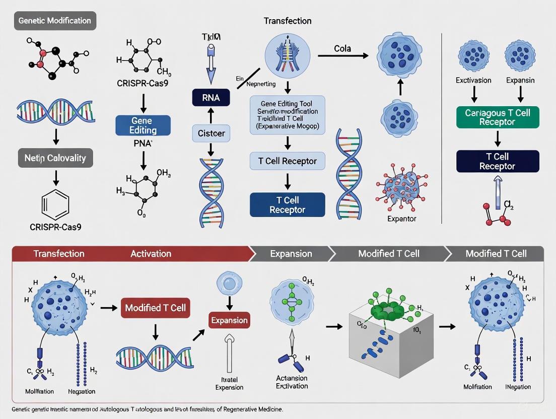
Genetic Engineering of Autologous T Cells: From Foundational Concepts to Clinical Applications in Cancer Immunotherapy
This article provides a comprehensive overview of the genetic modification of autologous T cells for cancer immunotherapy, with a focus on Chimeric Antigen Receptor (CAR)-T cell technology.
Genetic Engineering of Autologous T Cells: From Foundational Concepts to Clinical Applications in Cancer Immunotherapy
Abstract
This article provides a comprehensive overview of the genetic modification of autologous T cells for cancer immunotherapy, with a focus on Chimeric Antigen Receptor (CAR)-T cell technology. It explores the foundational biology of T cells and the engineering of synthetic receptors, detailing key methodological approaches including viral vector transduction and advanced genome-editing tools like CRISPR-Cas9. The content addresses major challenges such as cytokine release syndrome (CRS), neurotoxicity (ICANS), and limited efficacy in solid tumors, while offering troubleshooting and optimization strategies. A comparative analysis of autologous versus allogeneic approaches and different gene-editing platforms is included to guide therapeutic development. Aimed at researchers, scientists, and drug development professionals, this review synthesizes current advancements and future directions in creating potent, safe, and accessible engineered T-cell therapies.
The Foundation of Living Drugs: Understanding Autologous T Cell Biology and CAR Engineering
Cell therapy represents a transformative advancement in modern medicine, harnessing living cells to treat a range of diseases, particularly cancers that are refractory to conventional treatments [1] [2]. The field is predominantly divided into two distinct paradigms: autologous therapies, which utilize a patient's own cells, and allogeneic therapies, which employ cells from healthy donors [1] [2]. This application note delineates the critical differences between these approaches, with a specific focus on their application in genetically modified T-cell research for drug development professionals and scientists. We provide a detailed examination of their inherent challenges and advantages, supported by structured quantitative data, experimental protocols for genetic modification, and visualization of key workflows. The content is framed within the broader thesis that genetic engineering, particularly CRISPR-based technologies, is pivotal for overcoming the biological and manufacturing hurdles associated with both therapeutic paradigms, thereby accelerating the development of next-generation cell-based immunotherapies [3] [4] [5].
Comparative Analysis: Autologous vs. Allogeneic Cell Therapies
The fundamental distinction between autologous and allogeneic cell therapies lies in the cell source, which subsequently dictates manufacturing complexity, logistical requirements, immunological risks, and scalability [1] [2]. The following tables provide a structured comparison of these two paradigms.
Table 1: Core Characteristics and Manufacturing Logistics
| Feature | Autologous Therapy | Allogeneic Therapy |
|---|---|---|
| Cell Source | Patient's own cells [1] | Healthy donor (related or unrelated) [1] |
| Manufacturing Model | Customized, patient-specific batch [1] | Standardized, off-the-shelf batch [6] [1] |
| Supply Chain | Complex, circular logistics [2] | More linear, bulk processing [2] |
| Key Manufacturing Challenge | Time-sensitive; limited starting cell quality/quantity [4] [2] | Managing donor variability and immunogenicity [1] [5] |
| Scalability | Scale-out (multiple parallel lines) [2] | Scale-up (large-volume batches) [2] |
Table 2: Therapeutic Profile and Economic Considerations
| Aspect | Autologous Therapy | Allogeneic Therapy |
|---|---|---|
| Immune Compatibility | High; minimal risk of rejection or GvHD [2] | Low; requires HLA matching and/or immunosuppression to mitigate GvHD and HVGR [1] [5] |
| Primary Safety Concern | Uncontrolled proliferation leading to toxicity [4] | Graft-versus-Host Disease (GvHD) and Host-versus-Graft Reaction (HVGR) [5] |
| Therapeutic Persistence | Potential for long-term persistence [2] | May be limited by immune rejection [2] |
| Production Cost | High (service-based model) [2] | Potentially lower (mass production model) [2] |
| Treatment Timeline | Several weeks (cell harvest to infusion) [2] | Immediate, on-demand availability [6] [2] |
Genetic Modification Strategies for Enhanced T-Cell Therapies
Genetic engineering is central to modern T-cell therapies, with CRISPR-Cas9 emerging as a powerful tool for enhancing both autologous and allogeneic products. Its precision and simplicity enable complex multi-gene edits that were previously challenging with older technologies like ZFNs and TALENs [4] [5].
Engineering Allogeneic "Off-the-Shelf" CAR-T Cells
The primary strategy for creating universal allogeneic CAR-T cells involves the disruption of the T-cell receptor (TCR) to prevent GvHD. The most common and efficient method is the knockout of the T-cell receptor alpha constant (TRAC) locus, as this single gene edit effectively prevents the surface expression of the TCRαβ complex [5]. An alternative approach involves knocking out genes encoding the CD3 proteins, which are essential for TCR assembly and signaling [5]. These edited, TCR-deficient T cells are then transduced with a Chimeric Antigen Receptor (CAR) to redirect their specificity toward tumor cells [5].
Beyond preventing GvHD, a second major engineering challenge is overcoming host-mediated rejection. Research efforts focus on additional gene knockouts (e.g., HLA class I/II) to evade the host immune system, or the knock-in of immunomodulatory proteins to suppress HVGR and enhance engraftment [5].
Enhancing Autologous CAR-T Cell Function
For autologous therapies, the focus of genetic modification shifts from overcoming alloreactivity to boosting intrinsic T-cell fitness and anti-tumor efficacy. Key strategies include:
- Knockout of Immune Checkpoints: Disrupting genes such as PD-1 prevents T-cell exhaustion and enhances cytotoxicity, particularly in solid tumors [4].
- Knockout of Exhaustion-Related Genes: Genome-wide CRISPR screens have identified novel targets like RASA2 and PRDM1, whose knockout can enhance CAR-T cell persistence and expansion [3].
- Knockout of Fratricide-Related Genes: In CAR-T cells targeting antigens that can be acquired by trogocytosis, knocking out genes like RHOG and FAS can prevent fratricidal killing and improve tumor clearance [3].
Experimental Protocol: Genome-Wide CRISPR Screening in Primary Human CAR-T Cells
The following protocol is adapted from the CELLFIE platform, designed for systematic discovery of gene knockouts that enhance CAR-T cell function [3].
The diagram below illustrates the key stages of the genome-wide CRISPR screening workflow in primary human CAR-T cells.
Key Reagents and Materials
Table 3: Research Reagent Solutions for CRISPR/CAR-T Screening
| Research Reagent | Function/Description |
|---|---|
| CROP-seq-CAR Lentiviral Vector | Co-delivers the CAR transgene and the guide RNA (gRNA) library in a single vector for traceable perturbations [3]. |
| Brunello gRNA Library | A genome-wide human CRISPR knockout library containing 4-5 gRNAs per gene for loss-of-function screens [3]. |
| Cas9 mRNA | The CRISPR nuclease, delivered via electroporation as mRNA for high editing efficiency in primary T cells [3]. |
| Anti-CD3/CD28 Beads | For stimulation and expansion of T cells via the endogenous T-cell receptor [3]. |
| CD19+ K562 Cells | Target cancer cell line for stimulating CAR-T cells via their engineered chimeric antigen receptor [3]. |
Detailed Procedural Steps
- T Cell Isolation and Activation: Isolate peripheral blood mononuclear cells (PBMCs) from healthy donor leukapheresis samples. Stimulate T cells using anti-CD3/CD28 antibody-coated beads and culture in appropriate media with IL-2 for 7-10 days to achieve robust expansion [3].
- Genome Engineering and CAR Transduction: On day of transduction, concentrate the pre-expanded T cells. Co-transduce the cells with the CROP-seq-CAR lentiviral library (MOI < 1 to ensure single gRNA integration) and electroporate with Cas9 mRNA using a optimized nucleofection protocol. Include a selection marker (e.g., blasticidin resistance) in the mRNA construct to later enrich for successfully edited cells [3].
- Selection and Expansion: After 48 hours, add the appropriate antibiotic (e.g., blasticidin) to the culture media to select for T cells that have successfully received both the gRNA/CAR vector and the Cas9 mRNA. Culture the selected cells for a further 5-7 days to allow for genome editing and CAR expression.
- Functional Screening with Relevant Stimuli: Split the edited CAR-T cell pool into different stimulation conditions:
- TCR Stimulation: Use anti-CD3/CD28 beads.
- CAR Stimulation: Co-culture with irradiated CD19+ K562 cells at a defined effector-to-target ratio. Perform repeated stimulations over 1-2 weeks to apply selective pressure. Monitor key phenotypes via flow cytometry, including proliferation dyes, apoptosis markers (Annexin V), and exhaustion markers (PD-1, LAG3, TIM3) [3].
- Genomic DNA Extraction and Next-Generation Sequencing (NGS): Harvest a minimum of 50 million cells per condition at the end of the screen. Extract genomic DNA and perform PCR amplification of the integrated gRNA sequences using primers specific to the CROP-seq-CAR vector. Subject the amplicons to high-depth NGS.
- Bioinformatic Analysis and Hit Identification: Process the NGS data to count gRNA reads for each sample. Compare the relative abundance of each gRNA between the initial library and the final post-selection populations using specialized analysis packages (e.g., MAGeCK). Significantly enriched or depleted gRNAs identify candidate genes whose knockout enhances or impairs CAR-T cell fitness, respectively [3].
The choice between autologous and allogeneic cell therapy paradigms involves a complex trade-off between personalized safety and scalable, off-the-shelf availability. Autologous therapies minimize immunological risks but face significant logistical and cost challenges [2]. Allogeneic therapies offer a path to broader patient access but require sophisticated genetic engineering to mitigate GvHD and HVGR [5]. The integration of advanced genome editing technologies, particularly CRISPR-based screening platforms like CELLFIE, is accelerating the discovery of novel genetic modifications that enhance T-cell function for both paradigms [3]. The experimental protocol outlined herein provides a robust framework for researchers to systematically identify and validate gene targets, driving the development of more potent, persistent, and accessible cell-based immunotherapies.
Chimeric Antigen Receptor (CAR) T-cell therapy represents a paradigm shift in cancer treatment, embodying the concept of a "living drug" [7]. This individualized immunotherapy involves genetically reprogramming a patient's own T cells to target and eliminate cancer cells [7]. The manufacturing journey is an intricate, multi-step process beginning with leukapheresis and culminating in the infusion of a therapeutic product back into the patient. For researchers and drug development professionals, understanding and optimizing each manufacturing step is crucial, as the choices made during production significantly impact the final product's phenotypic characteristics, functional capabilities, and ultimately, its clinical safety and efficacy profile [8]. This application note details the technical protocols and critical parameters for manufacturing autologous CAR T-cells within the broader context of genetic modification research.
Leukocytapheresis and Initial Cell Processing
The manufacturing journey begins with leukocytapheresis, the process of obtaining mononuclear cells from a patient's peripheral blood. An optimal leukapheresis product is the foundational first step for successful CAR T-cell manufacturing [7].
Key Challenges and Pre-Apheresis Considerations
Collecting T cells from patients, particularly those with relapsed/refractory hematologic malignancies, presents unique challenges. These include difficulties in establishing the buffy coat due to low white blood cell counts, the compromised clinical status of patients, and the effects of previous lymphotoxic treatments [9] [10]. Key patient factors that influence collection efficiency include the pre-apheresis CD3+ lymphocyte count and the patient's total blood volume [10].
Apheresis Protocol and Collection Efficiency
Objective: To obtain a sufficient number of CD3+ lymphocytes for CAR T-cell manufacturing while ensuring patient safety.
Materials:
- Apheresis System: Terumo Spectra Optia Apheresis System (or equivalent) with continuous Mononuclear Cell Collection (cMNC) protocol [10].
- Anticoagulant: Citrate-based (e.g., ACD-A).
Method:
- Patient Assessment: Determine patient eligibility and perform a complete blood count, including a differential with CD3+ lymphocyte count.
- Vascular Access: Establish appropriate venous access.
- Parameter Setup: Program the apheresis device using the cMNC protocol. The target CD3+ cell yield dictates the total blood volume (TBV) to be processed [9].
- Processing: Initiate the apheresis procedure with continuous monitoring for adverse events, particularly citrate-related toxicity [9].
- Product Handling: Upon completion, aseptically collect the leukapheresis product bag and transport it to the processing facility at ambient temperature.
Technical Notes:
- Blood Volume Processed: The total blood volume processed is a critical parameter. Published series report a median processed volume of 4.4–11.6 L [9]. Processing excessively large volumes leads to prolonged apheresis time, wastage of resources, and increased patient risk [10].
- Predictive Modeling: A logarithmic model can predict the CD3+ yield and guide the required blood volume, enhancing efficiency. The model is expressed as:
- T = a * ln(C~pre~ * V~T~) + b
- Where T is the total CD3+ yield (10^9^ cells), C~pre~ is the pre-apheresis CD3+ count (10^9^ cells/L), V~T~ is the total blood volume processed (L), and a and b are constants derived from historical data at each apheresis center [10].
Table 1: Commercial CAR T-Cell Products and Their Starting Material Requirements
| Product Name (Commercial Name) | Target Antigen | Cell Dose Target to Collect | Blood Volume to Process |
|---|---|---|---|
| Axicabtagene ciloleucel (Yescarta) | CD19 | 5–10 × 10^9^ MNCs^a^ | 12–15 L |
| Tisagenlecleucel (Kymriah) | CD19 | 1–4 × 10^9^ CD3+ cells | 6–10 L |
| Lisocabtagene maraleucel (Breyanzi) | CD19 | A 450 mL collection bag | 7 L (if lymphocytes ≥1,000/µL); 12 L (if lymphocytes <1,000/µL) |
| Brexucabtagene autoleucel (Tecartus) | CD19 | 5–10 × 10^9^ MNCs | 12–15 L |
^a^ MNCs: Mononuclear Cells. Adapted from [9].
Starting Cell Population Selection
The leukapheresis product contains a heterogeneous mix of cells. A critical early decision is choosing the starting population for manufacturing. This can be peripheral blood mononuclear cells (PBMCs) or a T cell-enriched population. Enrichment is typically achieved via magnetic bead-based cell sorting, either by negative selection (depleting non-T cells) or positive selection [8]. The choice between using a mixed T-cell population or manufacturing CD4+ and CD8+ T cells separately affects the complexity, cost, and characteristics of the final product [8].
T Cell Activation and Genetic Modification
The isolated T cells must be activated and genetically modified to express the chimeric antigen receptor, enabling them to recognize and kill tumor cells.
T Cell Activation Protocol
Objective: To activate isolated T cells ex vivo, initiating proliferation and making them permissive to genetic modification.
Materials:
- Culture Medium: RPMI 1640 or specialized serum-free T cell expansion medium (e.g., ImmunoCult-XF T Cell Expansion Medium) [11] [12].
- Cytokines: Recombinant Human IL-2 (e.g., 10 ng/mL) [11].
- T Cell Activators: Anti-CD3/CD28 antibodies, often conjugated to magnetic beads or presented on artificial antigen-presenting cells (e.g., ImmunoCult Human CD3/CD28/CD2 T Cell Activator) [11].
- Culture Vessels: Flasks, G-Rex bioreactors, or closed-system bioreactors like the Xuri Cell Expansion System W25 [11].
Method:
- Seeding: Resuspend isolated T cells in pre-warmed expansion medium at a density of 1 × 10^6^ cells/mL [11].
- Activation: Add the T cell activator (e.g., at 25 µL/mL) and cytokines to the culture [11].
- Incubation: Culture cells at 37°C in a 5% CO~2~ incubator [12].
Technical Notes:
- Cell Density is Critical: Maintaining T cells at lower densities during early expansion is key to optimizing growth and viability. On day 3 post-activation, increase the total culture volume by 4- to 8-fold to dilute cells to a lower density [11].
- Recommended Expansion Protocol:
- Day 0: Seed at 1 × 10^6^ cells/mL with activator and IL-2.
- Day 3: Increase culture volume 8-fold (or maintain density at 1-2.5 × 10^5^ cells/mL).
- Days 5 and 7: Increase culture volume 4-fold with fresh medium supplemented with IL-2 [11].
- This optimized protocol can achieve up to 800-fold expansion over 10-14 days with >85% viability [11].
Genetic Modification using Viral Vectors and CRISPR
The primary methods for stably introducing the CAR gene involve viral vectors and, increasingly, gene-editing technologies like CRISPR.
A. Viral Transduction
Objective: To integrate the CAR transgene into the T cell genome using viral vectors.
Materials:
- Viral Vector: Lentiviral or gamma-retroviral vector encoding the CAR construct.
- Enhancement Reagents: Retronectin, protamine sulfate, or other transduction enhancers.
- Culture Medium: As in 3.1, without activators that may interfere with transduction.
Method:
- Timing: Transduce activated T cells 24-72 hours post-activation, during peak proliferation.
- Setup: "Spinoculation" - centrifuge the vector onto the cells (e.g., 2000 × g for 60-90 minutes at 32°C) in the presence of a transduction enhancer.
- Incubation: Following spinoculation, return cells to the 37°C incubator.
- Removal: 12-24 hours post-transduction, replace the medium to remove residual vector.
Technical Notes:
- Vector Copy Number (VCN): A critical quality attribute, typically measured by ddPCR, with a regulatory cutoff usually between 1-5 copies per cell [13].
- Integration Site Analysis: Viral integration can lead to clonal expansion and poses a theoretical risk of insertional mutagenesis. Pipelines like INSPIIRED or EpiVIA can be used to monitor integration sites for safety and to understand their impact on potency [13].
B. CRISPR/Cas9-mediated Gene Editing
Objective: To precisely knock-in the CAR construct into a specific genomic locus or knock out endogenous genes to enhance CAR T-cell function.
Materials:
- CRISPR System: Cas9 protein (or mRNA) and single-guide RNA (sgRNA) targeting the desired locus (e.g., TRAC for knock-in). Cas12a is an alternative nuclease with high specificity and distinct PAM requirements [14].
- Donor Template: A DNA template (viral or plasmid) containing the CAR transgene flanked by homology arms for HDR.
- Delivery Method: Electroporation is the most common method for delivering CRISPR components and donor templates to T cells.
Method:
- Preparation: Pre-complex the Cas9 ribonucleoprotein (RNP) with the sgRNA.
- Electroporation: Mix activated T cells with the RNP complex and donor template, and electroporate using optimized T cell settings.
- Recovery: Immediately transfer cells to pre-warmed medium and return to the incubator.
- Analysis: Assess editing efficiency and CAR expression via flow cytometry and sequencing.
Technical Notes:
- Advantages of CRISPR: Enables precise TRAC locus integration for uniform CAR expression, knockout of endogenous genes (e.g., PD-1 to reduce exhaustion, TCR to create allogeneic cells), and development of "off-the-shelf" allogeneic CAR T-cells [14] [15].
- Multi-Gene Editing: CRISPR allows for simultaneous knockout of multiple genes (e.g., TCR, β2M, PD-1, Regnase-1) to enhance persistence, avoid host rejection, and reduce toxicity [14] [15].
Table 2: Research Reagent Solutions for T Cell Activation and Genetic Modification
| Category | Item | Function | Example Products/Codes |
|---|---|---|---|
| Cell Culture | T Cell Expansion Medium | Provides nutrients, buffers, and supports for T cell growth and viability | ImmunoCult-XF T Cell Expansion Medium [11], RPMI 1640 + FBS [12] |
| Recombinant Human IL-2 | Cytokine that promotes T cell proliferation and survival | Various manufacturers | |
| Activation | Anti-CD3/CD28 T Cell Activator | Provides Signal 1 (CD3) and Signal 2 (CD28) for robust T cell activation and expansion | ImmunoCult Human CD3/CD28 T Cell Activator [11] |
| Genetic Modification | Lentiviral Vector | Stably integrates CAR transgene into host T cell genome | Various custom or pre-made constructs |
| CRISPR-Cas9 System | Enables precise gene knockout and targeted transgene knock-in | SpCas9, Cas12a (Cpf1) proteins and sgRNAs [14] | |
| Cell Isolation | Microbubble Isolation Kit | Gently isolates T cells via buoyancy; preserves cell viability and function | Akadeum BACS Human T Cell Depletion Kit [16] |
Cell Expansion and Final Product Formulation
Following genetic modification, CAR T-cells undergo massive ex vivo expansion to generate a clinically relevant dose, followed by critical purification and formulation steps.
Large-Scale Expansion and Phenotype Monitoring
The expansion protocol detailed in Section 3.1 is continued through days 10-14. For large-scale production required for therapy, the process can be adapted to closed-system bioreactors like the Xuri Cell Expansion System, which allows for perfusion of fresh medium and better environmental control [11]. Maintaining a central memory T cell (T~CM~) phenotype (CD62L+CD45RO+) is desirable, as these subsets are associated with superior persistence and antitumor activity in vivo [11] [8]. The optimized dilution protocol promotes the expansion of T cells with a T~CM~ phenotype [11].
Post-Expansion Cleanup and Formulation
Objective: To harvest, purify, and formulate the final CAR T-cell product for infusion, ensuring safety, purity, and potency.
Materials:
- Wash Buffer: Phosphate-buffered saline (PBS) or other isotonic, serum-free buffer.
- Formulation Medium: An isotonic solution matching physiological pH (∼7.4) and osmolarity. May include human serum albumin.
- Cryoprotectant: Dimethyl sulfoxide (DMSO) is commonly used.
- Cell Separation Technology: Buoyancy-Activated Cell Sorting (BACS) microbubbles or other methods for gentle dead cell and impurity removal [16].
Method:
- Harvesting: Collect cells from bioreactors or flasks.
- Washing and Concentration: Wash cells to remove debris, dead cells, cytokines, and residual activation/transduction agents. Concentrate to the target cell density.
- Final Formulation: Resuspend the cell pellet in the final formulation medium at the prescribed CAR T-cell dose. Key formulation parameters include:
- Cryopreservation (if applicable): For products that are not infused fresh, mix with cryoprotectant (e.g., 10% DMSO), control-rate freeze, and store in the vapor phase of liquid nitrogen.
Technical Notes:
- Post-Expansion Cleanup: This is a critical step to remove impurities that could cause adverse events upon infusion. Using gentle technologies like microbubbles for dead cell removal helps preserve the viability and function of the final product [16].
- Quality Control (QC): The final product undergoes rigorous QC testing before release. This includes assessments of:
- Identity and Purity: Percentage of CAR+ T cells.
- Potency: Functional assays like cytokine release (IFN-γ) and cytotoxicity in response to target cells [13].
- Viability: Typically required to be >70-80%.
- Safety: Sterility, mycoplasma, and endotoxin testing.
The journey of a 'living drug' from leukapheresis to infusion is a complex, highly regulated process where each parameter—from the initial CD3+ count to the final formulation osmolarity—can influence therapeutic success. For researchers, a deep understanding of these protocols is essential for developing and manufacturing the next generation of CAR T-cell therapies. The integration of predictive modeling for apheresis, optimized culture protocols to maintain favorable T cell phenotypes, and advanced gene-editing techniques like CRISPR are at the forefront of efforts to enhance the efficacy, safety, and accessibility of this revolutionary cancer treatment.
Chimeric Antigen Receptor (CAR)-T cell therapy represents a paradigm shift in cancer treatment, leveraging the power of a patient's own immune system to eradicate malignant cells. This therapeutic approach involves genetically engineering autologous T cells to express synthetic receptors that redirect them against tumor-specific antigens. The efficacy and safety of these "living drugs" are fundamentally governed by the intricate design of their constituent domains. This application note provides a detailed structural and functional decomposition of the CAR receptor, focusing on the single-chain variable fragment (scFv), hinge, transmembrane, and signaling domains. Within the critical context of autologous T-cell research, we present standardized protocols for evaluating domain functionality and summarize quantitative data on how specific domain choices impact CAR expression, stability, and T-cell effector functions. The insights herein are intended to guide researchers and drug development professionals in the rational design and optimization of next-generation CAR-T cell therapies.
CAR-T cell therapy is a form of adoptive cell transfer that has demonstrated remarkable success, particularly in treating hematological malignancies. The process involves harvesting T cells from a patient's blood, genetically modifying them ex vivo to express a CAR, expanding the engineered cells, and reinfusing them back into the patient [17]. These reprogrammed T cells function as a "living drug," capable of recognizing and eliminating tumor cells with high specificity [18]. The genetic modification of autologous T cells is central to this process, as it ensures that the resulting CAR-T cells are patient-specific, thereby circumventing issues of allorejection while simultaneously presenting challenges related to manufacturing scalability and cost [19].
The CAR itself is a synthetic transmembrane receptor that artificially confers a novel antigen specificity upon the T cell. Its modular architecture allows for the independent engineering of its components to fine-tune critical properties such as antigen recognition, stability, signaling intensity, and persistence. A deep understanding of the structure-function relationship of each domain is therefore paramount for advancing the clinical application of this technology, especially for overcoming current hurdles in treating solid tumors [20] [17].
Anatomical Deconstruction of the CAR
Antigen Recognition Domain: The Single-Chain Variable Fragment (scFv)
The single-chain variable fragment (scFv) is the most common antigen-binding module, providing the CAR with its specificity. It is typically derived from the variable regions of a monoclonal antibody's heavy (VH) and light (VL) chains, connected by a short, flexible peptide linker [21] [18]. This configuration allows the scFv to bind to a specific cell-surface antigen on tumor cells in a non-MHC-restricted manner, a significant advantage over native T-cell receptors [22].
Key Considerations for scFv Engineering:
- Affinity and Specificity: The scFv's affinity must be carefully balanced. While high affinity promotes strong binding, it can also lead to on-target, off-tumor toxicity if the target antigen is expressed at low levels on healthy tissues [21]. The scFv's specificity is the primary determinant of the therapy's safety profile.
- Immunogenicity: Murine-derived scFvs can elicit immune responses against the CAR-T cells themselves. Strategies to humanize these scFvs are critical for improving persistence.
- Structural Orientation: The positioning of the VH and VL chains and the design of the linker can influence both the stability and the binding characteristics of the scFv.
Table 1: Alternative Antigen-Binding Domains Beyond scFv
| Domain Type | Description | Potential Application |
|---|---|---|
| Nanobodies | Single-domain antibodies derived from camelids (VHH domains) lacking light chains. | Smaller size may improve tissue penetration in solid tumors [21] [23]. |
| Ligands | Natural ligands for receptors overexpressed on tumors (e.g., APRIL). | Can target multiple isoforms or promote trogocytosis [21]. |
| Cytokines | Engineered cytokine domains (e.g., IL-13 zetakine). | Targets receptors in the tumor microenvironment [21]. |
| Peptides | Designed ankyrin repeat proteins (DARPins) or other scaffold proteins. | Offers high stability and tunable binding properties [21]. |
Hinge Domain: The Structural Spacer
The hinge domain, or spacer, is an extracellular segment that connects the scFv to the transmembrane domain. It provides steric freedom, allowing the scFv to access the target antigen effectively [21]. The choice of hinge is not merely structural; it directly influences the CAR's signaling threshold and functional potency [20].
Functional Characteristics:
- Length and Flexibility: A hinge that is too short may restrict antigen binding, while one that is too long can promote tonic signaling and spontaneous T-cell activation. The optimal length is often antigen- and scFv-dependent [20].
- Origin and Composition: Common hinges are derived from proteins such as CD8α, CD28, or IgG (e.g., IgG1 or IgG4) [21]. Recent biophysical studies reveal that hinges like CD28 exhibit intrinsic disorder and local structural motifs, including conformational switches and proline isomerization, which contribute to their dynamic flexibility and functional plasticity [23].
Transmembrane Domain: The Membrane Anchor
The transmembrane (TM) domain is a hydrophobic alpha helix that spans the cell membrane, anchoring the CAR to the T cell surface. It is crucial for the stability and expression level of the CAR complex [20] [22].
Functional Characteristics:
- Stability and Expression: The origin of the TM domain significantly affects the stability and surface expression of the CAR. For instance, CARs incorporating a CD28 transmembrane domain have been reported to be more stable than those using a CD3ζ transmembrane domain [21].
- Oligomerization and Interaction: The TM domain can mediate homo-dimerization or interact with endogenous signaling proteins within the T cell membrane (e.g., native TCR complexes), which can inadvertently alter the signaling properties of the CAR [22] [23].
Intracellular Signaling Domains: The Activation Engine
The intracellular signaling domain is responsible for transducing the activation signal upon antigen binding, initiating T-cell effector functions. The evolution of this domain defines the generations of CARs.
Generations of CARs:
- First Generation: Contains the CD3ζ chain alone, which includes three Immunoreceptor Tyrosine-based Activation Motifs (ITAMs). These CARs initiated T-cell activation but provided inadequate co-stimulation, leading to poor persistence in vivo [22] [18].
- Second Generation: Incorporates one co-stimulatory domain (e.g., CD28 or 4-1BB) in tandem with the CD3ζ domain. This addition markedly enhances T-cell proliferation, cytokine production, cytotoxicity, and in vivo persistence [22] [17].
- Third Generation: Combines two co-stimulatory domains (e.g., CD28 together with 4-1BB or OX40) with CD3ζ, aiming to further augment potency and persistence [22].
- Fourth Generation (TRUCKs): Built upon second-generation CARs, these T cells are "redirected for universal cytokine-mediated killing." They are engineered to inducibly express transgenic immune modulators, such as IL-12, upon CAR signaling to alter the tumor microenvironment and recruit innate immune cells [22] [21].
- Fifth Generation: Incorporates a co-stimulatory domain with a truncated cytokine receptor domain (e.g., IL-2Rβ). This seeks to activate the JAK-STAT pathway in conjunction with TCR and co-stimulatory signals, promoting further T-cell expansion and survival [21].
Table 2: Comparison of Common Co-stimulatory Domains in Second-Generation CARs
| Co-stimulatory Domain | Primary Signaling Pathway | Functional Impact on CAR-T Cells |
|---|---|---|
| CD28 | PI3K/Akt | Induces potent, rapid activation and cytokine production. Promotes effector T-cell metabolism [22]. |
| 4-1BB (CD137) | TRAF/NF-κB | Enhances long-term persistence and memory formation. May promote a less exhausted phenotype compared to CD28 [22] [17]. |
| OX40 (CD134) | TRAF/NF-κB | Sustains T-cell proliferation and enhances IL-2 production [22]. |
The logical relationships and workflow from T cell isolation to a functional CAR-T cell product are summarized in the diagram below.
Quantitative Analysis of Hinge and Transmembrane Domain Impact
The functional impact of hinge and transmembrane domain selection is quantifiable. A systematic study analyzing CAR variants with different hinge/TM domains revealed critical insights into their roles in CAR expression and T-cell function [20].
Table 3: Impact of Hinge and Transmembrane Domains on CAR Expression and Function [20]
| Hinge Domain | Transmembrane Domain | CAR Expression Level | CAR Stability | Antigen-Specific T-cell Function |
|---|---|---|---|---|
| CD3ζ | CD3ζ | Baseline | Lower | Dependent on expression level |
| CD8α | CD8α | High | High | High, despite equal expression to CD28-HD CARs |
| CD28 | CD28 | High | High | Significantly different from CD8α-HD, despite equal expression |
| CD4 | CD4 | Moderate | Moderate | Correlated with expression level |
| CD8α | CD3ζ | Lower (vs. CD8α-TM) | Lower (vs. CD8α-TM) | Reduced (vs. matched Hinge/TM) |
| CD28 | CD3ζ | Lower (vs. CD28-TM) | Lower (vs. CD28-TM) | Reduced (vs. matched Hinge/TM) |
Key Findings from Data:
- The transmembrane domain is a primary regulator of CAR surface expression and stability. Mismatching the hinge and TM domains (e.g., CD8α hinge with CD3ζ TM) resulted in lower expression and stability compared to matched pairs [20].
- The hinge domain directly modulates the CAR signaling threshold. CARs with CD8α-derived and CD28-derived hinges showed significant functional differences despite being expressed at equal levels, indicating that the hinge directly influences signal transduction intensity independent of expression quantity [20].
- In summary, the transmembrane domain primarily regulates the amount of CAR signaling by controlling expression level, while the hinge domain regulates the intensity of CAR signaling per receptor [20].
Experimental Protocols for Domain Evaluation
Protocol: Evaluating CAR Surface Expression and Stability by Flow Cytometry
This protocol outlines the steps to quantify the surface expression and stability of engineered CARs on primary human T cells, a critical quality control assay [20].
Research Reagent Solutions:
- Primary Human T Cells: Isolated from peripheral blood mononuclear cells (PBMCs) of healthy donors or patients.
- Activation Reagents: Anti-CD3ε and anti-CD28 monoclonal antibodies.
- Genetic Modification Tools: Lentiviral or retroviral vectors encoding the CAR construct, or in vitro transcribed CAR mRNA.
- Cell Culture Media: RPMI 1640 supplemented with 10% FBS, IL-2 (10 U/mL), and other necessary cytokines.
- Staining Antibodies: Fluorescently-conjugated anti-HA tag antibody (for detecting HA-tagged CAR), viability dye (e.g., Zombie Aqua), and fluorescently-conjugated anti-CD8α antibody.
Methodology:
- T Cell Activation and Transduction: Isolate PBMCs and activate T cells using plate-bound or soluble anti-CD3ε and anti-CD28 antibodies in the presence of IL-2 for 24-48 hours. Transduce activated T cells with viral vectors (e.g., via spinfection in the presence of retronectin) or electroporate with CAR mRNA.
- Cell Culture and Expansion: Culture transduced T cells in complete media with IL-2. For stability assessment, culture cells for an extended period (e.g., 7-28 days), possibly with periodic restimulation.
- Flow Cytometry Staining:
- Harvest CAR-T cells and wash with FACS buffer (PBS with 2% FBS). Incubate cells with an Fc receptor blocking antibody.
- Stain cells with a viability dye according to manufacturer instructions.
- Resuspend cells in FACS buffer containing fluorescently-labeled anti-HA antibody (for CAR detection) and anti-CD8α antibody. Include appropriate isotype controls.
- Incubate for 30 minutes at 4°C in the dark.
- Wash cells twice with FACS buffer and resuspend in fixation buffer if needed.
- Data Acquisition and Analysis: Analyze cells on a flow cytometer. Gate on live, CD8+ (or CD4+) cells and quantify the mean fluorescence intensity (MFI) and percentage of HA-positive cells to determine CAR expression level and stability over time.
Protocol: Assessing Antigen-Specific Cytotoxic Activity (Cytotoxicity Assay)
This protocol measures the ability of CAR-T cells to specifically lyse target cells expressing the cognate antigen [20].
Research Reagent Solutions:
- Effector Cells: Engineered CAR-T cells.
- Target Cells: Antigen-positive tumor cell lines (e.g., hVEGFR2+ L1.2 cells) and antigen-negative control cell lines.
- Assay Plate: 96-well plate.
- Detection Reagent: Lactate Dehydrogenase (LDH) release assay kit or a fluorescent-based dye (e.g., Calcein-AM).
Methodology (using LDH release):
- Prepare Effector and Target Cells: Harvest and count CAR-T cells (effectors) and target cells. Wash and resuspend in assay medium.
- Coat Plate (Optional): If using a spontaneous LDH release control, coat additional wells with lysis solution.
- Coculture: Seed target cells in the 96-well plate. Add effector cells at various Effector:Target (E:T) ratios (e.g., 40:1, 20:1, 10:1, 5:1). Include controls for target cells alone (spontaneous release) and target cells with lysis solution (maximum release).
- Incubate: Incubate the plate for 4-24 hours in a humidified CO2 incubator at 37°C.
- Measure LDH Release: Centrifuge the plate and carefully transfer supernatant to a new plate. Add the LDH reaction mixture and incubate for 30 minutes. Measure absorbance at 490 nm.
- Calculate Specific Cytolysis: Specific Lysis (%) = (Experimental LDH - Spontaneous LDH) / (Maximum LDH - Spontaneous LDH) × 100
The structure of a generic second-generation CAR and its interaction with a target cell is depicted in the following diagram.
Advanced Engineering Strategies in Autologous T Cells
Epigenetic Engineering for Enhanced CAR-T Cell Function
Beyond permanent genetic knockout, advanced epigenetic editing tools offer reversible control over gene expression. A recent platform utilizes all-RNA delivery of CRISPRoff and CRISPRon editors for stable epigenetic programming in primary human T cells without double-strand breaks [24].
Methodology:
- CRISPRoff is an epigenetic editor comprising a catalytically dead Cas9 (dCas9) fused to DNMT3A and KRAB domains. Transient expression of CRISPRoff induces durable DNA methylation and stable gene silencing that persists through numerous T-cell divisions and in vivo adoptive transfer.
- CRISPRon, based on dCas9 fused to the TET1 demethylase, can reverse this silencing.
- Application: This system has been successfully combined with standard CAR genetic engineering. Researchers generated epi-edited TRAC CAR-T cells by performing a targeted CAR knock-in at the TRAC locus while simultaneously using CRISPRoff to silence therapeutically relevant genes (e.g., immunosuppressive receptors), resulting in enhanced tumor control in preclinical models [24].
The Scientist's Toolkit: Key Reagents for CAR-T Cell Research
Table 4: Essential Research Reagents for CAR-T Cell Development and Analysis
| Reagent / Tool | Function / Description | Application in Protocol |
|---|---|---|
| Lentiviral/Retroviral Vectors | Engineered viruses for stable integration of CAR gene into T cell genome. | Primary method for durable CAR expression in T cells [22] [18]. |
| mRNA In Vitro Transcription Kits | Generates CAR-encoding mRNA for transient expression. | For rapid testing, electroporation, and to avoid genomic integration [20] [24]. |
| Anti-CD3/CD28 Antibodies | Synthetic activation signals mimicking natural T-cell activation. | Essential for in vitro T-cell activation and expansion prior to genetic modification [20] [17]. |
| Recombinant Human IL-2 | A key T-cell growth cytokine. | Promotes survival and expansion of activated and transduced T cells in culture [18]. |
| Flow Cytometry Antibodies | Anti-tag antibodies (e.g., anti-HA) and cell phenotyping antibodies (anti-CD4, CD8). | Quantifying CAR surface expression, stability, and characterizing T-cell populations [20]. |
| De Novo Protein Sequencing | Mass spectrometry-based sequencing of protein sequences (e.g., scFvs) without prior knowledge. | Critical for characterizing and patenting novel scFvs used in CAR design [21]. |
The rational design of chimeric antigen receptors is a cornerstone of effective autologous CAR-T cell therapy. As deconstructed in this application note, each domain—scFv, hinge, transmembrane, and signaling—plays a discrete yet interconnected role in determining the safety, efficacy, and persistence of the final cellular product. Quantitative evidence underscores that the hinge domain regulates signaling intensity, while the transmembrane domain governs receptor stability and expression levels. The integration of advanced techniques, such as epigenetic editing with CRISPRoff, alongside traditional genetic engineering, provides a powerful toolkit for creating next-generation CAR-T cells capable of overcoming the immunosuppressive barriers of solid tumors and achieving durable remissions. As the field progresses, a deep and nuanced understanding of CAR anatomy will continue to drive the innovation necessary to expand the reach of this transformative therapy.
Chimeric Antigen Receptor (CAR) T-cell therapy represents a paradigm shift in cancer treatment, leveraging engineered immunity to target malignant cells. This application note traces the architectural evolution of CAR constructs from first to fifth-generation designs, detailing the synergistic integration of signaling domains that enhance T-cell potency, persistence, and functionality. Framed within autologous T-cell research, the document provides detailed protocols for the evaluation of next-generation CARs and a curated toolkit of essential reagents, serving as a comprehensive resource for researchers and therapeutic developers.
CAR-T cell therapy involves the genetic modification of a patient's own (autologous) T lymphocytes to express synthetic receptors that redirect them to selectively target and eliminate tumor cells [4]. The standard CAR is a modular synthetic receptor typically consisting of an extracellular antigen-recognition domain—most often a single-chain variable fragment (scFv) derived from an antibody—a hinge or spacer region, a transmembrane domain, and one or more intracellular signaling domains [25] [26]. A critical advantage of CARs over native T-cell receptors (TCRs) is their ability to recognize surface antigens on target cells independently of major histocompatibility complex (MHC) presentation, thereby bypassing a common mechanism of tumor immune evasion [26] [27].
The development of CAR-T cells has been marked by iterative enhancements in their design, classified into "generations" based on the number and combination of intracellular signaling modules. Each generation has sought to overcome specific clinical challenges, including lack of sustained anti-tumor activity, T-cell exhaustion, and poor persistence in the hostile tumor microenvironment [25] [5].
The Evolution of CAR Generations
First-Generation CARs: Proof of Concept
First-generation CARs featured a simple structure, incorporating only the CD3ζ chain from the T-cell receptor complex as an intracellular signaling domain. While this design proved that T cells could be redirected to kill target cells upon antigen binding, these CARs exhibited limited expansion and persistence in vivo, leading to suboptimal anti-tumor efficacy in clinical settings [25] [26]. The absence of co-stimulatory signals resulted in T-cell anergy and failed to induce long-term immunological memory.
Second-Generation CARs: Incorporating Co-stimulation
A major breakthrough came with second-generation CARs, which incorporate one additional co-stimulatory signaling domain, such as CD28 or 4-1BB (CD137), fused to the CD3ζ chain [25] [26]. This addition provides a "signal 2" that mimics the natural co-stimulation required for full T-cell activation.
- CD28-based CARs: Promote robust initial expansion and potent effector functions but may be associated with a more terminal differentiation phenotype.
- 4-1BB-based CARs: Enhance T-cell persistence and promote a memory-like phenotype, contributing to longer-lasting clinical responses, as seen in the approved therapy Kymriah [26].
The choice between CD28 and 4-1BB significantly impacts the metabolic fitness, differentiation state, and long-term durability of CAR-T cells [26].
Third-Generation CARs: Multiple Co-stimulatory Signals
Third-generation CARs combine two co-stimulatory domains (e.g., CD28 and 4-1BB) in tandem with the CD3ζ chain [25]. The goal is to synergize the potent signaling of CD28 with the persistence afforded by 4-1BB, potentially leading to superior T-cell function and anti-tumor activity against more refractory malignancies.
Fourth and Fifth-Generation CARs: armored CARs and Beyond
Fourth and fifth-generation CARs, often termed "armored" or "next-generation" CARs, are engineered to overcome the immunosuppressive tumor microenvironment (TME).
- Fourth-Generation CARs (TRUCKs): These are second-generation CARs further engineered to secrete transgenic proteins, such as cytokines (e.g., IL-12), upon antigen recognition. These cytokines can modulate the local TME, recruit and activate other immune cells, and enhance the overall anti-tumor response [26].
- Fifth-Generation CARs: These builds incorporate a truncated cytoplasmic domain from cytokine receptors (e.g., IL-2 receptor β chain) in addition to the CD3ζ and co-stimulatory domains. This allows the CAR to activate not only TCR-mimetic signaling but also cytokine-induced JAK/STAT signaling upon antigen binding, creating a fully orthogonal and potent activation signal that can further drive T-cell proliferation and survival [5].
Table 1: Evolution of CAR-T Cell Generations
| Generation | Intracellular Signaling Domains | Key Features | Advantages | Limitations |
|---|---|---|---|---|
| First | CD3ζ | Proof-of-concept | MHC-independent recognition | Limited persistence & efficacy; no costimulation |
| Second | CD3ζ + 1 Costimulatory (CD28 or 4-1BB) | Enhanced T cell activation | Improved expansion & persistence | Susceptible to immunosuppressive TME |
| Third | CD3ζ + 2 Costimulatory (CD28 and 4-1BB) | Synergistic signaling | Potentially superior potency | Increased complexity; potential for exhaustion |
| Fourth | CD3ζ + 1 Costimulatory + Transgenic cytokine (e.g., IL-12) | "Armored"; modulates TME | Recruits innate immunity; counters TME | Risk of cytokine-related toxicity |
| Fifth | CD3ζ + 1 Costimulatory + Cytokine receptor domain (e.g., IL-2Rβ) | Activates JAK/STAT pathway | Fully orthogonal signaling; enhances proliferation | Highly complex design; safety profiling needed |
The following diagram illustrates the progressive structural complexity and key signaling pathways activated across the different CAR generations.
Critical CAR Modules and Design Parameters
Beyond the intracellular signaling domains, the other structural modules of a CAR are critical determinants of its function, specificity, and safety.
Extracellular Ligand-Binding Domain
The most common ligand-binding domain is a single-chain variable fragment (scFv). Its affinity and avidity for the target antigen are crucial design parameters [25].
- Affinity Tuning: Using a lower-affinity scFv can improve the specificity of CAR-T cells for tumor cells that highly express the target antigen, while sparing healthy tissues with lower antigen density (e.g., as demonstrated with ErbB2 and EGFR CARs) [25]. This strategy enhances the therapeutic window.
- Epitope Location: The location of the epitope bound by the scFv (membrane-distal vs. membrane-proximal) influences the need for spacer flexibility [25].
Spacer and Transmembrane Domains
The spacer (or hinge) connects the binding domain to the transmembrane domain and provides flexibility to access the target epitope.
- Length Optimization: A spacer that is too short may hinder access to membrane-proximal epitopes, while one that is too long may promote spontaneous clustering and "tonic signaling" even in the absence of antigen, leading to premature T-cell exhaustion [26].
- Transmomain Origin: The choice of transmembrane domain (e.g., derived from CD28, CD8, or CD3ζ) can influence the stability of CAR expression and its interaction with endogenous signaling molecules within the T cell [25].
Advanced Protocols for Next-Generation CAR Evaluation
Protocol 1: CRISPR Screening for CAR-T Cell Enhancement (CELLFIE Platform)
Purpose: To systematically discover gene knockouts that enhance CAR-T cell fitness and anti-tumor efficacy [3].
Workflow:
- Primary T Cell Isolation and Activation: Isolate T cells from human peripheral blood and activate them with anti-CD3/CD28 antibodies for 7-10 days [3].
- Co-delivery of Engineering Components: Co-transduce T cells with a lentiviral CROP-seq-CAR vector (which encodes both the CAR and a guide RNA library) and electroporate with Cas9 mRNA to enable genome-wide CRISPR knockout screening [3].
- Selection and Phenotypic Screening: Culture edited CAR-T cells under selective pressure (e.g., blasticidin) and perform functional screens with readouts for proliferation, activation, exhaustion (PD-1, LAG3 expression), and fratricide (via trogocytosis) [3].
- In Vivo Validation with CROP-seq: Transfer pooled, edited CAR-T cells into a xenograft model of human leukemia. Use CROP-seq to track individual gRNA clonotypes and their enrichment or depletion in vivo, identifying hits that confer a survival advantage [3].
- Hit Validation: Validate top candidate genes (e.g., RHOG and FAS knockout were identified as potent enhancers) individually and in combination across multiple models and donors [3].
The following diagram visualizes this integrated screening and validation platform.
Protocol 2: Quantitative Cellular Kinetics by Volume-Based qPCR
Purpose: To accurately quantify the in vivo expansion and persistence of CAR-T cells, overcoming the limitations of conventional gDNA-based normalization, especially in lymphodepleted patients [28].
Methodology:
- Spike-in Calibration Curve: Spike a known quantity of the CAR transgene into control blood samples to create a calibration curve. This accounts for DNA extraction efficiency [28].
- DNA Extraction with External Control: Co-extract DNA from a fixed volume of patient blood samples alongside an external control gene (e.g., dog MC1R) [28].
- qPCR and Analysis: Perform qPCR for the CAR transgene and the external control. Use the spike-in calibration curve and the external control to normalize extraction efficiency variability and calculate the absolute CAR transgene copy number per microliter of blood (copies/μL) [28].
- Data Interpretation: This volume-based unit provides a more accurate reflection of true cellular kinetics, as it is not skewed by dramatic fluctuations in total white blood cell counts following lymphodepleting chemotherapy [28].
The Scientist's Toolkit: Research Reagent Solutions
The development and evaluation of advanced CAR constructs rely on a suite of specialized reagents and tools.
Table 2: Essential Research Reagents for CAR-T Cell Development
| Reagent / Tool | Function | Example Application |
|---|---|---|
| CRISPR Guide RNA (gRNA) | Directs Cas nuclease to specific genomic loci for knockout or knock-in. | Knockout of endogenous TRAC locus to reduce GvHD and enable targeted CAR insertion [27]. Knockout of PD-1 (PDCD1) to prevent exhaustion [27]. |
| CRISPR Nucleases & Editors | Enzymes that perform genetic modifications. | Cas9 for gene knockout. Adenine/cytosine Base Editors (ABE/CBE) for precise single-base changes without double-strand breaks, enhancing safety [27]. |
| CROP-seq-CAR Vector | Combines CAR expression with gRNA barcoding in a single lentiviral vector. | Enables pooled CRISPR screens in CAR-T cells with tracking of gRNA clonality via sequencing [3]. |
| Lentiviral Vectors | Efficient delivery of CAR transgenes and gRNA libraries into T cells. | Stable transduction of primary human T cells for CAR expression [3] [4]. |
| mRNA for CRISPR Editors | Transient delivery of gene-editing machinery. | Electroporation of Cas9 mRNA for efficient knockout with reduced off-target risks compared to stable expression [3]. |
| Synthego Research sgRNA | High-performance synthetic guide RNA. | Achieves high knockout efficiency and cell viability in primary T cells, including for multiplexed editing [27]. |
The evolution of CAR designs from simple first-generation constructs to sophisticated fifth-generation receptors illustrates a concerted effort to engineer more potent and durable living drugs. This progression, underpinned by a deeper understanding of T-cell biology, has been facilitated by advances in genetic engineering, particularly CRISPR-based screening and editing technologies. The future of autologous CAR-T cell research lies in the continued rational design of receptors and the precise editing of the T-cell genome to overcome the remaining barriers in solid tumors and enhance safety, ultimately making these powerful therapies applicable to a broader range of patients.
The genetic modification of autologous T cells to express chimeric antigen receptors (CARs) represents a paradigm shift in cancer immunotherapy. The clinical success of this approach is profoundly illustrated by targeting two key antigens in hematologic malignancies: CD19 in B-cell leukemias and lymphomas, and B-cell maturation antigen (BCMA) in multiple myeloma. These "success stories" are built upon a foundation of precise antigen selection, innovative CAR design, and an understanding of the tumor microenvironment. CD19 and BCMA serve as ideal targets because they are highly expressed on malignant cells and play crucial roles in the development and survival of the tumor lineage. This application note details the experimental frameworks, clinical outcomes, and standardized protocols that have established CAR-T therapy as a cornerstone in the treatment of relapsed/refractory hematologic malignancies, providing a model for future T-cell engineering efforts.
Target Antigen Profiles and Rationale
The foundational principle of successful CAR-T cell therapy is the identification of target antigens that are highly and uniformly expressed on tumor cells with limited expression on vital healthy tissues. CD19 and BCMA exemplify this principle, each with distinct biological roles and expression patterns that make them ideal for targeted immunotherapy.
CD19: A Pan-B-cell Surface Antigen
CD19 is a transmembrane glycoprotein that functions as a critical regulator of intrinsic and extrinsic B-cell receptor signaling. It is expressed from the early pro-B-cell stage through terminal B-cell differentiation but is lost upon plasma cell differentiation. This expression pattern makes it an excellent target for B-cell malignancies, as its targeting spares plasma cells and allows for partial humoral immunity preservation. From an experimental standpoint, its rapid internalization rate upon antibody binding posed an initial challenge for CAR design, which was overcome by developing scFv domains that trigger effective T-cell activation despite this property.
BCMA: A Plasma Cell Survival Factor
BCMA is a tumor necrosis factor receptor superfamily member that binds two ligands, APRIL and BAFF, promoting plasma cell survival and maturation. Its expression is predominantly restricted to late-stage B cells and long-lived plasma cells, with minimal expression on other tissues. This restricted expression is crucial for managing on-target, off-tumor toxicity. BCMA is notably shed from the cell surface as a soluble fragment (sBCMA), which can act as a decoy for CAR-T cells; this necessitates CAR designs with high affinity or strategies to overcome antigen sequestration.
Table 1: Biological Characteristics of CD19 and BCMA
| Characteristic | CD19 | BCMA |
|---|---|---|
| Gene Family | Immunoglobulin superfamily | TNF receptor superfamily |
| Expression Pattern | Pan-B-cell lineage (from pro-B to mature B-cells) | Differentiated plasma cells |
| Biological Function | Coreceptor for B-cell receptor signaling; modulates signaling thresholds | Binds BAFF/APRIL; promotes plasma cell survival and differentiation |
| Ligands | Unknown (interacts with CD21 and CD81) | BAFF (BLyS) and APRIL |
| Soluble Form | No | Yes (sBCMA) |
| Ideal Properties for Targeting | High surface density, lineage specificity, not expressed on hematopoietic stem cells | Restricted expression, essential for malignant plasma cell survival |
Clinical Success and Comparative Outcomes
CAR-T products targeting CD19 and BCMA have demonstrated remarkable efficacy in patients with heavily pretreated, relapsed/refractory hematologic malignancies. The summarized outcomes in Table 2 below, compiled from pivotal clinical trials and real-world analyses, highlight their transformative potential. A recent large-scale analysis of the 2021–2022 National Readmission Database provides a direct comparison of clinical outcomes, revealing distinct toxicity and resource utilization profiles between these two therapeutic classes [29].
BCMA-directed CAR-T recipients were generally older (median age 62.5 vs. 55.9 years; P = 0.002) and had higher rates of comorbidities such as chronic kidney disease (15.2% vs. 7.8%; P < 0.001) and obesity (21.1% vs. 11.5%; P < 0.001) compared to the CD19-directed cohort [29]. Despite this, BCMA CAR-T therapy was associated with a significantly lower incidence of encephalopathy (9.7% vs. 17.0%; P = 0.002) and sepsis (4.6% vs. 8.2%; P = 0.042), though it had higher rates of transaminitis (9.5% vs. 6.1%; P = 0.048) [29]. Mortality during the initial hospitalization was comparable between the two groups (4.4% vs. 3.6%; P = 0.5) [29].
Table 2: Comparative Clinical Outcomes of CD19 and BCMA CAR-T Therapies
| Parameter | CD19-Directed CAR-T (n=2,731) | BCMA-Directed CAR-T (n=789) | P-value |
|---|---|---|---|
| Median Age (years) | 55.9 | 62.5 | 0.002 |
| Chronic Kidney Disease (%) | 7.8 | 15.2 | <0.001 |
| Encephalopathy (%) | 17.0 | 9.7 | 0.002 |
| Sepsis (%) | 8.2 | 4.6 | 0.042 |
| Transaminitis (%) | 6.1 | 9.5 | 0.048 |
| In-hospital Mortality (%) | 3.6 | 4.4 | 0.5 |
| Length of Stay (days) | 18.8 | 15.8 | 0.2 |
| Total Hospital Charges ($) | 991,000 | 627,000 | <0.001 |
| 30-day Readmission Rate (%) | 20 | 15 | 0.13 |
Despite these successes, managing disease progression post-CAR-T remains a significant challenge. In multiple myeloma, the presence of extramedullary disease (EMD) at the time of CAR-T infusion or progression is a particularly adverse prognostic factor. A 2025 single-center retrospective study found that baseline EMD was associated with significantly inferior progression-free survival (3.6 vs. 7.0 months, p=0.0076) and overall survival (4.8 vs. 21.0 months, p=0.00086) compared to cases without EMD [30].
CAR-T Cell Engineering and Signaling Pathways
All currently approved CAR-T cell products for hematologic malignancies are based on second-generation CARs, which incorporate a single costimulatory domain (either CD28 or 4-1BB) in tandem with the CD3ζ activation domain [31] [32]. The choice of costimulatory domain significantly influences the phenotype, functionality, and persistence of the engineered T cells.
- CD28-based CARs (e.g., in axicabtagene ciloleucel and brexucabtagene autoleucel) are associated with robust, rapid effector function and potent initial tumor killing, characterized by enhanced IL-2 production and metabolic reprogramming towards glycolysis [31].
- 4-1BB-based CARs (e.g., in tisagenlecleucel and cilta-cabtagene autoleucel) promote T-cell persistence and the formation of memory subsets, supported by increased mitochondrial biogenesis and oxidative metabolism, which may contribute to longer-term disease control [31] [32].
The basic structure of these second-generation CARs consists of an extracellular antigen-recognition domain (typically a single-chain variable fragment, scFv), a hinge region, a transmembrane domain, and the intracellular signaling domains (costimulatory + CD3ζ). The signaling cascade initiated upon antigen binding is critical for T-cell activation and cytotoxicity, as illustrated in the following pathway diagram.
Diagram 1: Second-generation CAR-T cell signaling pathway (Width: 760px)
Experimental Protocols for Preclinical Assessment
Protocol: In Vitro Cytotoxicity Assay (Standard Calcein-AM Release)
Objective: To quantitatively measure the specific lysis of target cancer cells by CD19 or BCMA-targeting CAR-T cells.
Materials:
- Effector Cells: Generated CD19- or BCMA-CAR-T cells and untransduced (UTD) T-cell controls.
- Target Cells: CD19⁺/BCMA⁺ tumor cell lines (e.g., Nalm-6 for CD19, MM.1S for BCMA) and antigen-negative lines as controls.
- Key Reagent: Calcein-AM fluorescent dye (Thermo Fisher, C3099).
- Equipment: Cell culture incubator, fluorescence plate reader, round-bottom 96-well plates.
Procedure:
- Label Target Cells: Harvest and wash target cells. Resuspend at 1 × 10⁷ cells/mL in pre-warmed serum-free medium. Add Calcein-AM to a final concentration of 10 µM and incubate for 30 minutes at 37°C in the dark.
- Wash and Plate: Wash labeled cells three times with complete medium to remove excess dye. Resuspend and plate 1 × 10⁴ target cells per well in a 96-well round-bottom plate.
- Coculture: Add effector cells to the target cells at varying Effector:Target (E:T) ratios (e.g., 40:1, 20:1, 10:1, 5:1). Include wells for:
- Spontaneous Release: Target cells + medium only.
- Maximum Release: Target cells + 2% Triton X-100.
- Incubate: Centrifuge the plate briefly and incubate for 4 hours at 37°C, 5% CO₂ in the dark.
- Measure Fluorescence: After incubation, centrifuge the plate and transfer 100 µL of supernatant from each well to a black-walled 96-well plate. Measure fluorescence (Excitation: 485 nm, Emission: 535 nm).
- Calculate Specific Lysis:
Specific Lysis (%) = [(Experimental Release - Spontaneous Release) / (Maximum Release - Spontaneous Release)] × 100
Protocol: In Vivo Assessment Using a Xenograft Mouse Model
Objective: To evaluate the antitumor efficacy and persistence of CAR-T cells in a living organism.
Materials:
- Mice: Immunodeficient NSG (NOD.Cg-Prkdcscid Il2rgtm1Wjl/SzJ) mice, 8-10 weeks old.
- Tumor Cells: Luciferase-expressing target cell line (e.g., Nalm-6-luc for CD19).
- CAR-T Cells: CD19- or BCMA-CAR-T cells, UTD T-cell controls.
- Key Reagent: D-Luciferin potassium salt (GoldBio, LUCK-1G).
- Equipment: In vivo bioluminescence imaging system (IVIS), flow cytometer for peripheral blood analysis.
Procedure:
- Tumor Engraftment: Inject 5 × 10⁵ tumor cells intravenously via the tail vein on Day 0.
- T-cell Administration: On Day 4 (or when tumor engraftment is confirmed via bioluminescence), inject 5 × 10⁶ CAR-T or UTD T cells intravenously.
- Tumor Monitoring: Monitor tumor burden twice weekly by IVIS imaging.
- Inject D-Luciferin (150 mg/kg) intraperitoneally.
- Anesthetize mice with isoflurane and acquire images 10 minutes post-injection.
- Quantify total flux (photons/second) in a defined region of interest.
- CAR-T Cell Persistence:
- Collect peripheral blood from retro-orbital bleeding at weekly intervals.
- Stain with anti-human CD3 and anti-human CD4/CD8 antibodies.
- For detection of CAR-positive cells, use a protein L-based staining method or target antigen-Fc fusion protein.
- Analyze by flow cytometry to quantify the frequency of circulating human T cells and CAR⁺ T cells.
- Endpoint: Monitor mouse survival and body weight daily. Euthanize mice when they exhibit signs of distress or a predetermined tumor burden threshold is reached. Analyze bone marrow, spleen, and other organs for tumor infiltration and CAR-T cell presence post-mortem.
The Scientist's Toolkit: Research Reagent Solutions
Table 3: Essential Reagents for CAR-T Cell Research and Development
| Reagent / Material | Function / Application | Example Product / Identifier |
|---|---|---|
| Lentiviral Vector (2nd Gen) | Stable gene delivery of CAR construct into T cells; allows for semi-random genomic integration and long-term expression. | pLenti-CAR-EF1α (Addgene, various) |
| Retronectin | Enhances retroviral/lentiviral transduction efficiency by colocalizing viral particles and target cells. | Takara Bio, T100B |
| Human T Cell Activator | Provides signal 1 (CD3) and signal 2 (CD28) for T cell activation prior to transduction. | Gibco, Dynabeads CD3/CD28 CTS |
| Recombinant Human IL-2 | Promotes T cell expansion and survival during and after the manufacturing process. | PeproTech, 200-02 |
| Flow Cytometry Antibodies | Validation of CAR surface expression and immunophenotyping (e.g., memory subsets, exhaustion markers). | Anti-human CD3, CD4, CD8, CD45RA, CD62L, PD-1, LAG-3; Protein L for CAR detection |
| Cytokine Detection Assay | Multiplex quantification of cytokine release (e.g., IFN-γ, IL-2, IL-6) in supernatant to assess T cell activity. | Luminex Performance High Sensitivity Cytokine Panel |
| Genomic DNA Isolation Kit | Isolation of high-quality gDNA for analysis of CAR vector integration sites and copy number. | QIAamp DNA Blood Mini Kit (QIAGEN, 51104) |
Future Directions and Next-Generation Engineering
The success of CD19 and BCMA CAR-T cells provides a blueprint for future development. Key research directions focus on overcoming limitations such as antigen escape, T-cell exhaustion, and the challenges of solid tumors [32]. Next-generation engineering strategies include:
- Dual-Targeting CARs: To mitigate antigen escape, constructs targeting two antigens (e.g., CD19/CD20 or CD19/CD22 for B-cell malignancies) are in clinical development [33]. A similar approach using a dual-targeted CAR (CTA313) targeting CD19 and BCMA is also being explored for autoimmune diseases like lupus [34].
- Allogeneic "Off-the-Shelf" CARs: Derived from healthy donors or induced pluripotent stem cells (iPSCs), these aim to overcome the manufacturing and cost barriers of autologous products. This requires gene editing (e.g., CRISPR-Cas9, TALENs) to knock out the T-cell receptor (TCR) to prevent graft-versus-host disease (GvHD) and often the knockout of HLA class I to evade host rejection [33] [5].
- Armored CARs: These CARs are engineered to secrete cytokines (e.g., IL-15) or express dominant-negative receptors (e.g., for TGF-β) to resist the immunosuppressive tumor microenvironment, a significant barrier in solid tumors [31] [32].
- In Vivo CAR-T Engineering: An emerging frontier that involves directly delivering CAR genes to T cells within the patient's body using advanced viral vectors or lipid nanoparticles (LNPs), potentially enabling off-the-shelf, patient-specific therapy without complex ex vivo manufacturing [35] [5].
The continued genetic modification of autologous T cells, building on the foundational success of CD19 and BCMA targeting, promises to expand the reach of cellular immunotherapy to a broader range of diseases, including autoimmune conditions and solid tumors.
Methodologies in Action: Genetic Engineering Tools and Clinical Workflows for T Cell Modification
Retroviral and lentiviral vectors represent cornerstone technologies in the genetic modification of autologous T cells for therapeutic applications, including chimeric antigen receptor (CAR) T-cell therapies. These integrating viral vectors enable stable genomic integration and long-term transgene expression, which is essential for durable therapeutic effects. Retroviral vectors, particularly those derived from gamma-retroviruses (gRV), and lentiviral vectors (LV), derived from HIV-1, have undergone significant evolution to enhance their safety and efficacy profiles. The key distinction lies in their ability to transduce different cell types; while gRVs can only infect dividing cells, LVs can transduce both dividing and non-dividing cells, making them particularly suitable for hard-to-transduce primary cells like hematopoietic stem cells [36] [37].
The field has witnessed a substantial shift from early gamma-retroviral vectors to self-inactivating (SIN) lentiviral configurations. This evolution has been driven by safety concerns identified in early clinical trials, where genotoxic events were observed with gRV vectors. Modern vector design incorporates SIN deletions, which remove enhancer-promoter sequences from the long terminal repeats (LTRs), significantly reducing the risk of insertional mutagenesis by minimizing the potential for transactivation of neighboring proto-oncogenes [38] [37]. As of 2025, the commercial and clinical landscape reflects this transition, with eight market-approved ex vivo gene therapies using lentiviruses and two using gamma-retroviruses [39].
Current Commercial & Clinical Landscape
The efficacy of viral vector systems is demonstrated by their central role in approved CAR-T therapies. The design choices—including the type of vector, promoter, and envelope used for pseudotyping—directly impact the safety and performance of the final therapeutic product.
Table 1: Viral Vector Systems in Approved CAR-T Cell Therapies
| Product (Example) | Vector Type | Key Features | Promoter | Envelope | First Approval |
|---|---|---|---|---|---|
| Kymriah(Tisagenlecleucel) | Lentiviral, SIN | HIV-1-derived, third-generation, replication-incompetent | EF-1α | VSV-G | USA, 2017 |
| Yescarta(Axicabtagene ciloleucel) | Gamma-retroviral | MSCV-based, non-self-inactivating | MSCV | GaLV | USA, 2017 |
| Tecartus(Brexucabtagene autoleucel) | Lentiviral, SIN | HIV-1-derived, third-generation, replication-incompetent | Unknown | VSV-G | USA, 2020 |
| Abecma(Idecabtagene vicleucel) | Lentiviral, SIN | HIV-1-derived, third-generation, replication-incompetent | EF-1α | VSV-G | USA, 2021 |
The selection between vector systems involves a critical balance. Gamma-retroviral vectors, such as those used in Yescarta, are often based on the Murine Stem Cell Virus (MSCV) backbone and pseudotyped with the Gibbon Ape Leukemia Virus (GaLV) envelope. In contrast, most modern lentiviral vectors are third-generation, SIN designs that are pseudotyped with the Vesicular Stomatitis Virus G-protein (VSV-G), which confers a very broad tropism due to its interaction with the ubiquitous LDL receptor family on human cells [38]. The human Elongation Factor-1 alpha (EF-1α) promoter is a common choice in LVs for driving consistent transgene expression in T cells [40].
Key Experimental Protocols for T-Cell Transduction
Protocol: Transduction of Human Autologous T Cells with Lentiviral Vectors
This protocol outlines a standard procedure for generating clinical-grade CAR-T cells using lentiviral vectors, based on methods used for approved products like CTL019.
I. Materials and Reagents
- Source T Cells: Leukapheresis product from patient.
- Culture Medium: X-VIVO 15 or TexMACS medium, supplemented with serum-free supplements or human AB serum.
- Activation Reagents: CD3/CD28 Dynabeads or TransAct.
- Cytokines: Recombinant human IL-2 (100-300 IU/mL) or IL-7/IL-15.
- Lentiviral Vector: VSV-G pseudotyped, third-generation SIN vector, titer ≥ 1x10^8 TU/mL.
- Enhancers: Retronectin (10 µg/mL) or other transduction enhancers.
II. Procedure
- T Cell Isolation and Activation:
- Isolate PBMCs from leukapheresis product via density gradient centrifugation.
- Isolate T cells negatively or positively using magnetic bead selection kits.
- Resuspend T cells in complete medium at 1-2x10^6 cells/mL.
- Add CD3/CD28 activation beads at a bead-to-cell ratio of 3:1.
- Add recombinant human IL-2 to a final concentration of 100 IU/mL.
- Incubate cells at 37°C, 5% CO2 for 24-48 hours.
Retronectin Coating (Optional but Recommended):
- Dilute Retronectin to 10 µg/mL in PBS.
- Add solution to non-tissue culture treated plates.
- Incubate at 4°C overnight or room temperature for 2 hours.
- Before use, block the plate with 2% Human Serum Albumin for 30 minutes.
Viral Transduction:
- After activation, count cells and resuspend at 1x10^6 cells/mL in fresh medium containing IL-2.
- Add the lentiviral vector at a Multiplicity of Infection (MOI) of 3-5. Higher MOI may be required for low-titer batches.
- If using a Retronectin-coated plate, "spinoculate" by centrifuging the plate at 2000 x g for 90 minutes at 32°C.
- If not using spinoculation, incubate the cell-vector mixture at 37°C, 5% CO2.
Post-Transduction Culture and Expansion:
- 24 hours post-transduction, add fresh pre-warmed medium to dilute residual vector and cells.
- Maintain cell density between 0.5-2x10^6 cells/mL, supplementing with fresh medium and IL-2 as needed.
- Expand cells for 7-14 days, monitoring for transduction efficiency and cell growth.
Harvest and Formulation:
- When target cell numbers are achieved and activation beads have been removed, harvest cells.
- Wash cells and formulate in infusion buffer (e.g., CryoStor CS10) for cryopreservation or immediate infusion.
III. Quality Control Assays
- Transduction Efficiency: Assess by flow cytometry for surface CAR expression or intracellular reporter (e.g., GFP) 3-5 days post-transduction.
- Vector Copy Number (VCN): Determine average number of vector integrations per cell using digital droplet PCR on genomic DNA.
- Sterility: Test for mycoplasma, bacteria, and fungi.
- Replication-Competent Lentivirus (RCL): Test the final cell product per regulatory guidelines [40].
Quantitative Analysis of Transduction Parameters
The success of T-cell transduction is influenced by multiple physical and biological parameters. The following table summarizes key variables that require optimization for each specific T-cell subset and vector batch.
Table 2: Critical Parameters for Optimizing T-Cell Transduction
| Parameter | Typical Range | Impact on Efficiency | Considerations |
|---|---|---|---|
| Cell Activation Status | 24-48 hours pre-stimulation | Critical | Cells must be actively dividing for gRV; activated state improves LV uptake. |
| Multiplicity of Infection (MOI) | 3 - 10 | High | High MOI can increase yield but also risk of multiple integrations. |
| Spinoculation | 2000 x g, 90-120 min, 32°C | High (2-5 fold increase) | Increases vector-cell contact; crucial for low-titer batches. |
| Transduction Enhancer | Retronectin (10 µg/mL), Proteasome inhibitors | Moderate | Retronectin co-localizes vectors and cells; inhibitors prevent vector degradation. |
| Cytokine Support | IL-2 (100 IU/mL), or IL-7/IL-15 (10-20 ng/mL) | Moderate | Maintains T-cell proliferation and viability during culture. |
Safety and Genotoxicity Considerations
The integration of viral vectors into the host genome carries a theoretical risk of insertional mutagenesis, which could lead to the activation of proto-oncogenes or disruption of tumor suppressor genes. Early clinical trials using gRV vectors with intact LTRs for hematopoietic stem cell (HSC) gene therapy resulted in genotoxic events, including leukemogenesis, due to vector integration near proto-oncogenes like LMO2 and MDS1-EVI1 [37]. This risk is significantly mitigated in modern SIN vectors, where the enhancer-promoter sequences in the LTRs are deleted, and transgene expression is driven by an internal promoter [38].
While the risk is lower for modified mature T cells compared to HSCs, clonal expansions and a small number of malignancy cases have been reported post SIN-LV gene therapy. Investigations into these events often reveal contributing factors such as the use of strong heterologous viral promoters and potentially the specific nature of the insulator elements used [37]. Rigorous lot-release testing and long-term patient monitoring are mandated by regulatory agencies. Monitoring data from 308 patients infused with lentiviral-modified T cells across 194.8 post-infusion person-years showed no evidence of replication-competent lentivirus (RCL), with statistical models estimating over 52 years of patient follow-up would be needed to observe a single RCL event [40].
Manufacturing and Production Challenges
Producing high-titer, high-quality retroviral and lentiviral vectors remains a significant bottleneck in the widespread application of T-cell therapies. A major production challenge is retro-transduction (or self-transduction), where producer cells are transduced by the vectors they are producing. This phenomenon is particularly pronounced for VSV-G pseudotyped LVs, as the VSV-G envelope uses the ubiquitous LDL receptor for entry. Studies show that 60-90% of infectious vector can be lost through retro-transduction of the producer cells themselves, severely impacting yield and increasing production costs [41].
Strategies to mitigate this include genetically engineering producer cell lines (e.g., HEK293T) to knock out the LDLR gene, though this can impair cellular lipid metabolism [41]. The manufacturing process itself also presents hurdles. Most LV production relies on transient transfection of HEK293T cells, which is difficult to scale and leads to significant batch-to-batch variability. The development of stable producer cell lines is a key advancement to overcome this, offering more consistent and scalable production, though current titers from such systems are often lower [39]. Furthermore, LVs are notoriously shear-sensitive, necessitating careful bioprocess design in stirred-tank bioreactors to avoid product loss during aeration and agitation [39].
The Scientist's Toolkit: Essential Research Reagents
Table 3: Key Research Reagent Solutions for Viral Transduction
| Reagent / Material | Function | Example Use Case |
|---|---|---|
| VSV-G Pseudotyped Lentiviral Vectors | Broad tropism gene delivery; transduces most mammalian cells, including non-dividing T cells. | Standard for CAR gene delivery in autologous T cells. |
| Retronectin | A recombinant fibronectin fragment that co-localizes viral particles and target cells, enhancing transduction efficiency. | Used to coat plates prior to spinoculation, improving low-MOI transduction. |
| CD3/CD28 Activator Beads | Mimics antigen-presenting cell stimulation, activating T cells and inducing proliferation required for transduction. | Pre-stimulation of patient T cells for 24-48 hours before vector exposure. |
| Lentiviral Titer Kits | Quantifies functional vector concentration (Transducing Units/mL) prior to use. | Essential for calculating correct MOI for experimental or clinical batches. |
| Digital Droplet PCR (ddPCR) | Precisely quantifies Vector Copy Number (VCN) in transduced cell populations. | Quality control and safety monitoring to ensure average VCN is within safe limits. |
The genetic modification of autologous T cells for adoptive cell therapy represents a paradigm shift in cancer treatment. Chimeric Antigen Receptor (CAR) T cell therapy has demonstrated remarkable efficacy, particularly against B-cell malignancies [42]. However, the field has been heavily reliant on viral vectors for gene delivery, which introduces challenges related to cost, manufacturing complexity, cargo capacity, and safety concerns regarding insertional mutagenesis [43] [42]. These limitations have accelerated the development of non-viral engineering strategies, primarily employing transposon systems and mRNA electroporation, which offer streamlined manufacturing, enhanced safety profiles, and greater flexibility for complex genetic modifications [43] [42].
Transposon systems facilitate stable genomic integration of large transgenes through a cut-and-paste mechanism, enabling permanent CAR expression ideal for durable anti-tumor responses [43] [44]. Meanwhile, mRNA electroporation provides transient but highly efficient gene transfer, suitable for applications requiring controlled CAR expression or rapid screening of receptor designs [45] [46]. This application note details the protocols, quantitative performance, and practical implementation of these non-viral technologies within the context of autologous T cell research and therapy development.
Transposon Systems for Stable Gene Transfer
DNA transposons are natural genetic elements that can move within a genome via a cut-and-paste mechanism mediated by their corresponding transposase enzyme. For gene therapy applications, the system is split into two components: a transposase (delivered as DNA, mRNA, or protein) and a transposon plasmid containing the gene of interest (e.g., a CAR) flanked by terminal inverted repeats (TIRs) that are recognized by the transposase [43] [47]. The two most advanced systems are Sleeping Beauty (SB) and PiggyBac (PB), including its derivative, piggyBat [48] [43] [44].
The Sleeping Beauty transposase binds to inverted repeats flanking the transposon, excises the cargo, and integrates it into a TA dinucleotide site in the genome [43] [49]. PiggyBac operates similarly but integrates at TTAA sites and can excise without leaving a footprint [43]. The recently described piggyBat transposase, derived from the little brown bat, demonstrates efficient CAR transgene delivery with a relatively low variability in integration copy number [48].
Detailed Protocol for T Cell Engineering
Key Research Reagent Solutions:
- T Cell Source: Primary human T cells isolated from PBMCs or leukapheresis products.
- Activation Reagent: Anti-CD3/CD28 antibodies (immobilized on beads or soluble).
- Transposon Vector: Plasmid DNA containing the CAR transgene flanked by specific TIRs (SB or PB).
- Transposase Vector: Plasmid DNA or in vitro transcribed (IVT) mRNA encoding the hyperactive transposase (e.g., SB100X, piggyBac, piggyBat).
- Electroporation System: Cuvette-based or continuous-flow electroporation device.
- Cell Culture Media: X-VIVO 15 or RPMI 1640, supplemented with serum (e.g., 10% FBS) or serum-free formulations, and human recombinant IL-2 (100-300 IU/mL).
Step-by-Step Workflow:
T Cell Isolation and Activation:
- Isolate PBMCs from whole blood or leukapheresis product via density gradient centrifugation.
- Isolate untouched T cells using a negative selection magnetic bead kit.
- Resuspend T cells at 1-2 × 10^6 cells/mL in complete media supplemented with IL-2.
- Activate T cells using human CD3/CD28 TransAct or similar Dynabeads at a 1:1 bead-to-cell ratio.
- Incubate for 24-48 hours at 37°C, 5% CO₂.
Electroporation Preparation:
- After activation, harvest T cells and wash once with PBS.
- Resuspend cells in electroporation buffer at a high concentration (e.g., 1-2 × 10^7 cells/mL).
- Mix the cell suspension with the transposon plasmid (5 µg) and transposase plasmid or mRNA (e.g., 1 µg DNA or 2 µg mRNA for a ~5:1 ratio). The optimal ratio must be determined empirically to avoid overproduction inhibition [48] [49].
Electroporation:
- Transfer the cell-DNA mixture to an electroporation cuvette.
- Electroporate using optimized parameters. For the Sleeping Beauty system, a protocol using the Nucleofector device (e.g., program T-023 or U-014) is common [44] [49].
- Immediately after pulsing, add pre-warmed culture media and transfer cells to a culture plate.
- Incubate at 37°C, 5% CO₂.
Expansion and Culture:
- Approximately 24 hours post-electroporation, begin feeding cells with fresh complete media + IL-2.
- Expand cells for 10-14 days, maintaining a cell density between 0.5-2 × 10^6 cells/mL.
- Monitor CAR expression by flow cytometry from day 4-5 onwards.
- For SB-engineered cells, specific enrichment with artificial antigen-presenting cells (AaPC) can be performed to selectively expand CAR-positive T cells, yielding a product with an average of ~1 integration copy per cell [49].
Performance and Quantitative Data
Table 1: Quantitative Comparison of Transposon Systems in T Cell Engineering
| Parameter | Sleeping Beauty (SB) | PiggyBac (PB) | piggyBat | Notes |
|---|---|---|---|---|
| Integration Site | TA dinucleotide [43] | TTAA site [43] | Similar to PB and viral vectors [48] | SB shows a safer, more random profile [47] |
| Theoretical Cargo Capacity | Up to ~8 kb [43] | Up to 200 kb [47] | Not specified, likely similar to PB | Cargo size impacts efficiency |
| Typical Integration Copy Number | ~1.0 (with AaPC enrichment) [49] | Can be high (>20) [49] | Low variability, controlled [48] | Copy number can be influenced by culture conditions |
| CAR Expression Efficiency | Feasible and efficacious in clinical trials [49] | High (e.g., 77.8% with super-piggyBac) [48] | Moderate (e.g., 57.4%) [48] | Efficiency depends on delivery and culture methods |
| Transfection Efficiency | Highly dependent on electroporation method | Highly dependent on electroporation method | Highly dependent on electroporation method | Continuous-flow electroporation can achieve >95% viability [45] |
mRNA Electroporation for Transient Expression
mRNA electroporation enables transient, high-level expression of a CAR without genomic integration. This method eliminates the risk of insertional mutagenesis and allows for rapid testing of CAR constructs. The expressed CAR protein has a finite lifespan, typically resulting in functional activity for 5-7 days, which can be advantageous for managing potential toxicities [46] [42]. The process involves in vitro transcription of mRNA encoding the CAR, followed by its delivery into activated T cells via electroporation.
Detailed Protocol for mRNA Electroporation
Key Research Reagent Solutions:
- mRNA: CAR-encoding mRNA, synthesized in vitro, capped, and polyadenylated. Purification to remove double-stranded RNA contaminants is critical for reducing innate immune responses.
- Electroporation Buffer: Low conductivity buffer optimized for mRNA delivery.
Step-by-Step Workflow:
- T Cell Activation: Follow the same T cell isolation and activation protocol as described in Section 2.2.
- mRNA Preparation: Dilute the CAR-encoding mRNA to a working concentration in a nuclease-free buffer. Typical amounts range from 20-40 µg mRNA per 1-2 million cells [45].
- Electroporation:
- Wash activated T cells and resuspend in a specialized mRNA electroporation buffer at a high density (e.g., 5 × 10^6 to 1 × 10^7 cells/mL) [45].
- Mix the cell suspension with the mRNA.
- For continuous-flow electroporation, load the mixture into a syringe and flow through the device. Optimal parameters using a bipolar rectangular waveform (e.g., 100 µs duration, 100 Hz frequency, ~125 kV/m field strength) can achieve >95% transfection efficiency with minimal impact on viability [45].
- Collect electroporated cells directly into pre-warmed complete culture media.
- Recovery and Analysis:
- Incubate cells for 4-24 hours to allow for robust CAR protein expression.
- Analyze CAR expression and cell viability by flow cytometry before proceeding to functional assays or in vivo administration.
The Scientist's Toolkit: Essential Research Reagents
Table 2: Key Reagents for Non-Viral T Cell Engineering
| Reagent Category | Specific Examples | Function | Considerations |
|---|---|---|---|
| Transposase System | Sleeping Beauty (SB100X), PiggyBac, piggyBat [48] [44] [49] | Mediates stable integration of the CAR transgene from the transposon plasmid into the host genome. | SB has a potentially safer integration profile. PB can carry larger cargo. |
| Nucleic Acid Vector | Transposon plasmid (with ITRs), Minicircle DNA, IVT mRNA [43] [49] | Carries the genetic payload (CAR) for delivery. Minicircles (lacking bacterial backbone) can improve efficiency and safety [43]. | mRNA provides transient expression; transposon DNA provides stable expression. |
| Delivery Platform | Cuvette-based Nucleofector, Continuous-flow electroporation [45] [43] | Creates transient pores in the cell membrane to allow nucleic acids to enter the cell. | Continuous-flow systems offer high throughput, reproducibility, and scalability [45]. |
| T Cell Activator | Anti-CD3/CD28 beads/antibodies | Provides Signal 1 (TCR) and Signal 2 (co-stimulation) for initial T cell activation and proliferation. | Critical for achieving high transfection efficiency and subsequent expansion. |
| Culture Supplement | Recombinant Human IL-2, IL-7, IL-15 | Supports T cell survival, expansion, and can influence memory phenotype during ex vivo culture. | Cytokine combination affects the final product phenotype and persistence. |
Non-viral engineering of T cells using transposon systems and mRNA electroporation presents a transformative approach for the development of next-generation cell therapies. These methods address critical limitations of viral vectors by offering a more flexible, cost-effective, and potentially safer manufacturing platform [42]. Transposons like Sleeping Beauty and PiggyBac facilitate stable CAR expression suitable for long-term therapeutic responses, while mRNA electroporation offers a rapid and controlled system for transient expression. The protocols and data outlined in this application note provide a foundation for researchers to implement these technologies, paving the way for more accessible and sophisticated T cell therapies for cancer and other diseases.
The genetic modification of autologous T cells represents a cornerstone of modern cell therapy, enabling the creation of potent treatments for cancer, autoimmune diseases, and infectious diseases. Precision genome editing technologies—including CRISPR-Cas9, Transcription Activator-Like Effector Nucleases (TALENs), and Zinc-Finger Nucleases (ZFNs)—have revolutionized this field by enabling targeted genomic alterations with unprecedented accuracy and efficiency. These systems facilitate the development of enhanced chimeric antigen receptor (CAR) T cells by improving efficacy, safety, and scalability while overcoming fundamental biological barriers. This application note provides a comprehensive technical resource for researchers and drug development professionals, detailing current methodologies, quantitative performance metrics, and standardized protocols for implementing these technologies in T cell engineering workflows.
The three primary genome editing platforms function as molecular scissors that create double-strand breaks (DSBs) at predetermined genomic loci, harnessing endogenous DNA repair mechanisms to achieve gene knockout, correction, or insertion.
Table 1: Comparative Analysis of Major Genome Editing Technologies
| Feature | CRISPR-Cas9 | TALENs | ZFNs |
|---|---|---|---|
| Molecular Composition | Cas9 protein + guide RNA (gRNA) [14] | FokI nuclease dimer + TALE DNA-binding domains [14] | FokI nuclease dimer + zinc-finger protein domains [14] |
| Target Recognition | RNA-DNA complementarity (∼20 nt gRNA) [14] | Protein-DNA interaction (1 TALE domain ∼1 bp) [14] | Protein-DNA interaction (1 ZF domain ∼3 bp) [14] |
| Protospacer Adjacent Motif (PAM) Requirement | Yes (SpCas9: NGG) [14] | No | No |
| Editing Efficiency (Typical Range) | 40-90% [14] | 10-50% | 5-30% |
| Multiplexing Capacity | High (via multiple gRNAs) [14] | Moderate | Low |
| Development & Cloning | Simplified (RNA-based targeting) [14] | Complex (protein engineering) [14] | Highly complex (protein engineering) [14] |
| Relative Cost | Low | High | High |
| Primary T Cell Toxicity | Moderate | Lower | Higher |
The following workflow diagram illustrates the generalized experimental process for T cell genome editing, from design to validation, applicable to all three technologies.
Detailed Methodologies and Protocols
CRISPR-Cas9 Workflow for CAR-T Cell Engineering
CRISPR-Cas9 has emerged as the most versatile platform due to its RNA-programmable simplicity and high efficiency. The following protocol outlines the generation of allogeneic UCAR-T cells, a key application that highlights the power of multiplexed editing.
Protocol 3.1: Multiplex Gene Editing for Allogeneic CAR-T Cells
- Objective: Simultaneously knock out endogenous T-cell receptor (TCR) and human leukocyte antigen (HLA) genes to generate allogeneic, "off-the-shelf" CAR-T cells capable of evading host immune rejection [50].
- Key Applications: Development of universal CAR-T products; enhancement of T cell persistence; reduction of alloreactivity [50] [14].
Step-by-Step Procedure:
gRNA Design and Complex Formation:
- Design gRNAs with high on-target efficiency and low off-target potential. Target the TRAC locus to eliminate TCR expression and prevent GvHD, and the B2M locus to disrupt HLA class I expression and mitigate HvGR [50] [14].
- Reagent: Synthesize gRNAs with chemical modifications (e.g., 5' chemical modifications on the crRNA) to enhance stability and editing efficiency [14].
- Form ribonucleoprotein (RNP) complexes by pre-incubating purified Cas9 protein (e.g., SpCas9) with synthetic gRNAs at a molar ratio of 1:2 (Cas9:gRNA) for 10-20 minutes at room temperature [14].
T Cell Activation and Preparation:
- Isolate PBMCs from healthy donor leukapheresis product. Isolate T cells using negative selection magnetic beads.
- Activate T cells using anti-CD3/CD28 antibodies or beads. Culture in X-VIVO 15 or RPMI 1640 media supplemented with 10% FBS and 100 IU/mL IL-2.
- Perform editing 24-48 hours post-activation, when T cells are maximally receptive to nucleofection.
Delivery via Electroporation:
- Use a 4D-Nucleofector system (Lonza) for RNP delivery.
- Mix the pre-formed RNP complexes for TRAC and B2M targeting. For a 100 µL reaction, use 5 µg of each RNP complex.
- Resuspend 1-2 million T cells in 100 µL of P3 Primary Cell Solution. Combine with the RNP mix and transfer to a nucleofection cuvette.
- Electroporate using the pulse code DS-137, which has been optimized for high efficiency and cell viability in primary T cells [24].
- Immediately post-nucleofection, add pre-warmed culture medium and transfer cells to a culture plate.
CAR Gene Integration (Optional Co-delivery):
- To simultaneously introduce a CAR transgene, include a dsDNA or AAV6 donor template encoding the CAR construct during or immediately after electroporation.
- For HDR-mediated knock-in, design the donor template with homology arms (∼800 bp) flanking the Cas9 cut site in a safe harbor locus (e.g., TRAC, AAVS1) or a therapeutically beneficial locus like PDCD1 [31].
Post-Editing Culture and Expansion:
- Culture cells at a density of 0.5-1 x 10^6 cells/mL in complete media with IL-2.
- Expand cells for 7-14 days, monitoring cell density and viability. Perform medium exchanges or cell splits every 2-3 days.
Analysis and Validation:
- Efficiency: Assess editing efficiency 3-5 days post-electroporation. Use flow cytometry to check for loss of TCR and HLA surface expression. Use T7E1 assay or next-generation sequencing to quantify indel percentages at the genomic level.
- Function: Perform cytotoxicity assays (e.g., against CD19+ target cells for anti-CD19 CAR-T cells) and cytokine release assays to validate functionality.
TALENs Workflow for Clinical-Grade Editing
TALENs offer high specificity due to their unique protein-DNA recognition, making them suitable for clinical applications where minimizing off-target effects is paramount.
Protocol 3.2: TALEN-Mediated Gene Knockout for Autologous CAR-T Cells
- Objective: Achieve high-specificity knockout of an immune checkpoint gene (e.g., PD-1) in autologous CAR-T cells to enhance anti-tumor potency [51] [14].
- Key Applications: Reducing T cell exhaustion; improving persistence in immunosuppressive tumor microenvironments; clinical manufacturing.
Step-by-Step Procedure:
TALEN Design and mRNA Production:
- Design TALEN pairs to target sequences flanking the start codon of the PDCD1 gene. The DNA-binding domains are engineered to recognize 15-20 bp sequences on each DNA strand, separated by a 12-20 bp spacer.
- Reagent: Clone TALEN sequences into plasmids suitable for in vitro transcription (IVT). Generate capped and polyadenylated TALEN mRNA using an IVT kit.
T Cell Activation:
- Follow the same T cell isolation and activation steps as in Protocol 3.1.
mRNA Delivery:
- Use electroporation to deliver TALEN mRNA. For a 100 µL reaction, 5-10 µg of each TALEN mRNA is typical.
- Electroporate using the same system and pulse code as for CRISPR-Cas9 (e.g., DS-137).
CAR Transduction:
- 24 hours after electroporation, transduce the T cells with a lentiviral vector encoding the CAR transgene.
- Centrifuge the T cells with the lentiviral supernatant (MOI of 5-10) in the presence of polybrene (8 µg/mL) at 1000 x g for 90 minutes to enhance transduction efficiency.
Expansion and Validation:
- Expand cells for 10-14 days. Validate PD-1 knockout via flow cytometry and sequencing. Confirm that PD-1 knockout T cells show enhanced cytokine production and tumor-killing capacity upon repeated antigen exposure.
Advanced Technique: Epigenetic Editing with CRISPRoff
Beyond traditional gene editing, newer epigenetic tools like CRISPRoff allow for stable gene silencing without cutting DNA, offering a safer alternative for modulating gene expression.
Protocol 3.3: Multiplexed Gene Silencing with CRISPRoff for Enhanced CAR-T Function
- Objective: Simultaneously silence multiple therapeutic target genes (e.g., RASA2, PTPN2) in CAR-T cells to enhance effector function and persistence without inducing DNA DSBs [52] [24].
- Key Applications: Reducing tonic signaling; improving resistance to immunosuppression; multiplexed engineering with low toxicity.
Step-by-Step Procedure:
CRISPRoff/gRNA Complex Formation:
- Design sgRNAs targeting the promoter CpG islands of the genes of interest.
- Form RNP complexes by pre-incubating CRISPRoff mRNA (the most potent design incorporating Cap1 and base modifications) with a pool of 3 synthetic sgRNAs per target gene [24].
Delivery and Culture:
- Electroporate the RNP complexes into activated primary human T cells using the DS-137 pulse code.
- Immediately after nucleofection, also introduce mRNA encoding the CAR construct via electroporation.
- Culture the cells as described previously. The silencing program is stable through numerous cell divisions and T cell stimulations without the need for sustained editor expression [24].
Validation:
- Assess durable gene silencing over 28+ days using flow cytometry and RNA-seq.
- Evaluate functional enhancement using repetitive tumor challenge assays. CRISPRoff-edited CAR-T cells with RASA2 silencing demonstrate improved tumor control and survival in preclinical models compared to standard CAR-T cells [24].
The Scientist's Toolkit: Essential Research Reagents
Table 2: Key Research Reagent Solutions for T Cell Genome Editing
| Reagent / Material | Function / Application | Example & Notes |
|---|---|---|
| Nucleofector System | High-efficiency delivery of editors (RNP, mRNA) into primary T cells. | Lonza 4D-Nucleofector; Pulse Code DS-137 is highly effective [24]. |
| CRISPR-Cas9 RNP | Complex for gene knockout/knock-in; reduces off-targets vs. plasmid DNA. | Alt-R S.p. Cas9 Nuclease V3; complex with chemically modified Alt-R crRNA and tracrRNA [14]. |
| TALEN mRNA | For high-specificity gene knockout; requires in vitro transcription. | System from companies like Cellectis; capped and polyadenylated mRNA enhances expression [51]. |
| AAV6 Donor Vector | High-efficiency delivery of donor template for HDR-mediated knock-in. | Used for CAR insertion into TRAC or PDCD1 locus; high MOI required [31]. |
| Cytokines | T cell activation, survival, and expansion post-editing. | Recombinant Human IL-2 (100 IU/mL); IL-7/IL-15 can promote stem cell memory phenotypes. |
| Activation Beads | Robust T cell activation prior to editing. | Gibco Dynabeads CD3/CD28; magnetic removal post-activation. |
| Editing Assessment Kit | Quantify editing efficiency and specificity. | IDT Alt-R Genome Editing Detection Kit (T7E1); NGS for indel analysis and off-target profiling. |
Pathway and Workflow Visualization
The signaling pathways within engineered T cells are critical to their function. The following diagram illustrates the key intracellular signaling domains in second-generation CAR constructs, which are foundational to all approved CAR-T therapies.
The strategic application of CRISPR-Cas9, TALENs, and ZFNs has profoundly advanced the genetic modification of autologous T cells for therapeutic purposes. While CRISPR-Cas9 offers unparalleled flexibility and ease of use for multiplexed editing and novel applications like epigenetic control, TALENs remain a valuable tool for high-specificity clinical applications. The choice of platform depends on the specific research or development goals, weighing factors such as efficiency, specificity, multiplexing needs, and regulatory considerations. As these technologies continue to mature, they will undoubtedly unlock next-generation T cell therapies with enhanced efficacy and broader applicability against a wide spectrum of human diseases.
The genetic modification of a patient's own T cells to express chimeric antigen receptors (CARs) represents a paradigm shift in cancer immunotherapy. While CAR design is crucial, the clinical success of autologous CAR-T cell therapies is equally dependent on a robust, reproducible, and scalable manufacturing process. This workflow directly impacts critical quality attributes (CQAs) of the final product, including its safety, identity, potency, purity, and ultimately, its efficacy in patients [8] [53]. The autologous nature of these therapies means that each product is a unique batch, starting from a patient's leukapheresis material and culminating in a personalized, living drug. This application note provides a detailed, step-by-step protocol for the clinical manufacturing of autologous CAR-T cells, contextualized within the broader research on T cell genetic modification. It is designed to support researchers, scientists, and drug development professionals in navigating the complexities of translating a CAR-T cell therapy from the research bench to the clinic.
Materials and Reagents
Research Reagent Solutions
The following table details key reagents and their functions essential for autologous CAR-T cell manufacturing.
Table 1: Essential Reagents for Autologous CAR-T Cell Manufacturing
| Reagent/Material | Function/Application | Key Considerations |
|---|---|---|
| Leukapheresis Product | Starting material containing patient T cells. | Patient clinical history and prior treatments can impact T cell fitness [8]. |
| Cell Separation Reagents | Isolation of T cells or specific subsets (e.g., CD4+/CD8+) from PBMCs. | Magnetic bead-based sorting (e.g., anti-CD3/CD28 beads) is common; allows for simultaneous activation [8]. |
| Cell Culture Media | Supports T cell activation, transduction, and expansion. | Formulation (e.g., X-VIVO, TexMACS) influences T cell differentiation and final product phenotype [53]. |
| Serum Supplements | Provides growth factors and cytokines. | Serum-free alternatives are often preferred to reduce variability and ensure regulatory compliance [53]. |
| Genetic Modification Vectors | Introduces the CAR transgene into T cells. | Gamma-retroviral or lentiviral vectors are most common in approved products [8]. |
| Transduction Enhancers | Increases efficiency of genetic modification. | Reagents like RetroNectin improve viral vector attachment to target cells. |
| Cryopreservation Media | Preserves final cell product for storage and transport. | Must contain a cryoprotectant like DMSO and be compatible with infusion specifications. |
The manufacturing of autologous CAR-T cells is a multi-step process that must be conducted in a current Good Manufacturing Practice (cGMP)-compliant facility. The overarching workflow, from cell collection to final formulation, is illustrated below.
Step-by-Step Protocol
Step 1: Cell Collection and Starting Material Preparation
- Leukapheresis: Perform leukapheresis on the patient to collect peripheral blood mononuclear cells (PBMCs). The collected product should be processed fresh or cryopreserved according to established protocols.
- Shipment and Storage: If the manufacturing site is remote, transport the leukapheresis product in a temperature-controlled shipping container. Document all chain-of-custody and environmental conditions. Storage conditions (fresh vs. frozen) vary by commercial product [8].
Step 2: Cell Isolation and Selection
- PBMC Isolation: If necessary, isolate PBMCs from the leukapheresis product using density gradient centrifugation (e.g., Ficoll-Paque).
- T Cell Enrichment: Isulate the T cell population. This can be achieved by:
- Negative Selection: Depleting non-T cells (e.g., monocytes, B cells, NK cells) using magnetic-activated cell sorting (MACS). This method avoids antibody binding to T cells.
- Positive Selection: Isolating T cells by targeting CD3, or separately selecting CD4+ and CD8+ subsets. Some protocols use CD19-depletion to minimize malignant cell carryover [8].
- Note: The choice of starting population (bulk T cells vs. defined subsets) significantly impacts the final product's composition and requires careful process development [8].
Step 3: T Cell Activation
- Stimulation: Activate the isolated T cells to induce proliferation and make them permissive to genetic modification. The most common method is co-stimulation with magnetic beads conjugated to anti-CD3 and anti-CD28 antibodies.
- Culture Initiation: Seed the activated T cells in pre-warmed, serum-free culture medium supplemented with cytokines, typically recombinant human IL-2 and/or IL-7/IL-15, at a defined cell density.
Step 4: Genetic Modification
- Transduction: Within 24-48 hours post-activation, transduce the activated T cells with the viral vector (gamma-retroviral or lentiviral) encoding the CAR transgene.
- Protocol Variation: Centrifuge the cell-vector mixture (spinoculation) to enhance transduction efficiency. The use of retronectin or other transduction enhancers is recommended.
- Automated Integration: Automated bioreactor systems, such as the Terumo Quantum Flex, can seamlessly integrate the activation, transduction, and expansion steps into a single closed system, enhancing consistency [54].
Step 5: Ex Vivo Expansion
- Culture Maintenance: Culture the transduced T cells for a period typically ranging from 7 to 14 days. Maintain the culture at a controlled cell density by periodically diluting with fresh medium and cytokines.
- Process Monitoring: Monitor cell growth, viability, and metabolism (e.g., glucose consumption) daily. The goal is to achieve a target cell number (often in the billions) while steering the differentiation state towards favorable memory phenotypes (e.g., TSCM, TCM) [53].
- Scale-Up: The expansion can be performed in static culture flasks, gas-permeable bags, or automated bioreactors (e.g., hollow-fiber systems like Quantum Flex or wave-type bioreactors) [54] [55]. These closed systems improve scalability and reduce contamination risk.
Step 6: Final Formulation and Cryopreservation
- Harvesting: Once expansion is complete and release criteria are met, harvest the cells.
- Wash and Formulate: Wash the cell product to remove debris, cytokines, and any residual activation agents. Formulate the final product in an infusion-ready cryopreservation medium.
- Cryopreservation: Fill the final product into cryobags, controlled-rate freeze, and store in the vapor phase of liquid nitrogen.
Step 7: Product Release and Quality Control
- Testing: Perform rigorous quality control testing on the final product before release. This includes, but is not limited to:
- Sterility: Tests for bacterial and fungal contamination.
- Mycoplasma: Absence of mycoplasma.
- Potency: In vitro cytotoxicity assay against antigen-positive target cells.
- Identity: Flow cytometry confirming CAR expression and CD3+ T cell identity.
- Purity: Percentage of CAR-positive T cells and absence of undesirable cell populations.
- Viability: Minimum viability threshold (e.g., >70%).
- Release: The product is released for infusion only after meeting all pre-defined specifications.
Results and Data Analysis
Expected Manufacturing Outcomes
A successful manufacturing run should yield the following quantitative and qualitative outcomes.
Table 2: Key Performance Indicators for a CAR-T Cell Manufacturing Run
| Parameter | Typical Outcome | Measurement Method | Clinical Significance |
|---|---|---|---|
| Expansion Fold | 50 to 500-fold increase [8] | Cell counting | Ensures sufficient cell dose for therapeutic effect. |
| Final Cell Number | Varies by target dose; can reach 1-9 billion cells [54] | Cell counting | Determines the number of patient doses available. |
| Final Viability | >70% (often >90%) | Automated cell counter or flow cytometry | Indicator of overall cell health and product quality. |
| Transduction Efficiency | 20% - 70% | Flow cytometry for CAR expression | Determines the proportion of effector cells in the product. |
| CD4:CD8 Ratio | Varies (e.g., 1:1 in liso-cel) | Flow cytometry | Impacts efficacy and persistence; can be controlled during isolation [8]. |
| T Cell Phenotype | Enriched for TN/TSCM/TCM phenotypes | Flow cytometry for CD45RO, CD62L, CCR7 | Correlates with in vivo persistence and long-term efficacy [53]. |
Automation in Manufacturing
The transition from manual, open processes to automated, closed systems is a key industry trend aimed at improving robustness and scalability. The following diagram illustrates the integrated workflow of an automated bioreactor system.
A study using the Terumo Quantum Flex system demonstrated the feasibility of this integrated approach, successfully producing up to 9 billion TCR-T cells (a closely related modality) from 10 million PBMCs in 10 days while maintaining high viability [54]. This highlights the potential of automation to streamline the entire production process within a single, closed, GMP-friendly system.
Discussion
Technical and Regulatory Hurdles
The decentralized or point-of-care manufacturing model is being explored to improve patient access and reduce "vein-to-vein" time. However, a recent global survey of academic institutions highlighted major barriers, including cost constraints (70%), regulatory complexities (70%), and facility requirements (57%) [56]. Furthermore, variability in product quality was cited by 73% of institutions as a significant challenge, underscoring the need for standardized protocols and robust quality control systems [56]. The choice between fresh and frozen leukapheresis starting material, the type of activation, the vector used, and the expansion duration all contribute to this variability and can influence the final product's phenotypic composition, which is closely linked to clinical persistence and efficacy [8] [53].
Protocol Adaptations and Optimization
- Media Optimization: The choice of basal media and cytokine supplements (e.g., IL-7/IL-15 vs. IL-2) can be manipulated to favor the expansion of less-differentiated T cell subsets (TN and TCM), which are associated with superior in vivo persistence [53].
- Process Shortening: Research into accelerated manufacturing processes is ongoing. One study demonstrated a fully automated 24-hour CAR-T manufacturing process using the Gibco CTS DynaClect and Rotea systems, though this remains experimental [57]. Such approaches could significantly reduce the ex vivo culture time, potentially preserving a more favorable T cell phenotype.
The clinical workflow for manufacturing autologous CAR-T cells is a complex but standardized process where each step—from cell selection to final formulation—is critical to the safety and efficacy of the final product. The field is rapidly evolving towards greater automation and closed-system processing to enhance reproducibility, reduce variability, and scale up production. As research into T cell biology and genetic modification advances, further optimization of manufacturing protocols will be pivotal in expanding the reach of these transformative therapies to a wider range of cancers and patients.
Chimeric Antigen Receptor T (CAR-T) cell therapy represents a groundbreaking advancement in cancer immunotherapy, leveraging the power of a patient's own immune system to combat hematological malignancies. This approach involves genetically engineering a patient's T cells to express synthetic receptors that redirect them to recognize and eliminate cancer cells. The unique structure of CARs enables T cells to identify and bind to tumor-associated antigens (TAAs) on the surface of cancer cells in a major histocompatibility complex (MHC)-independent manner, enhancing their ability to target a broad range of tumor types [58]. As of 2025, the U.S. Food and Drug Administration (FDA) has approved multiple CAR-T cell products, all designed for relapsed or refractory blood cancers, marking a significant shift in treatment paradigms for patients with limited options [59] [17].
The clinical success of CAR-T therapy stems from its status as a "living drug" – once infused back into the patient, these engineered cells can expand, persist, and continue their surveillance and cytotoxic functions, potentially producing lasting anticancer results [17] [60]. All commercially available CAR-T cell products are based on autologous approaches, where the patient's own T cells are collected, genetically modified ex vivo, and then reinfused. They predominantly utilize second-generation CAR constructs, which incorporate a costimulatory domain (such as CD28 or 4-1BB) alongside the CD3ζ activation domain to enhance potency and persistence [31] [5].
Comprehensive Table of FDA-Approved CAR-T Cell Therapies
The table below summarizes all FDA-approved CAR-T cell therapies, their molecular targets, and their specific clinical indications as of 2025. This information is synthesized from the FDA's official biological products listing [59] and detailed clinical prescribing information [61] [60].
Table 1: FDA-Approved CAR-T Cell Therapies and Their indications
| Product & Trade Name | Generic Name (abbreviation) | Molecular Target | Approved Indications |
|---|---|---|---|
| ABECMA | idecabtagene vicleucel (ide-cel) | BCMA | Relapsed/refractory multiple myeloma after ≥2 prior lines of therapy [61]. |
| AUCATZYL | obecabtagene autoleucel (obe-cel) | CD19 | Adult patients with relapsed/refractory B-cell acute lymphoblastic leukemia (ALL) [59] [60]. |
| BREYANZI | lisocabtagene maraleucel (liso-cel) | CD19 | Relapsed/refractory large B-cell lymphoma, chronic lymphocytic leukemia (CLL), follicular lymphoma, and mantle cell lymphoma (MCL) after ≥2 prior lines of therapy [61]. |
| CARVYKTI | ciltacabtagene autoleucel (cilta-cel) | BCMA | Relapsed/refractory multiple myeloma after ≥1 prior line of therapy [61]. |
| KYMRIAH | tisagenlecleucel (tisa-cel) | CD19 | Pediatric and young adult patients (up to age 25) with relapsed/refractory B-cell ALL; adult patients with relapsed/refractory large B-cell lymphoma and follicular lymphoma [61] [60]. |
| TECARTUS | brexucabtagene autoleucel (brexu-cel) | CD19 | Adult patients with relapsed/refractory MCL and B-cell ALL [61] [60]. |
| YESCARTA | axicabtagene ciloleucel (axi-cel) | CD19 | Adult patients with relapsed/refractory large B-cell lymphoma and follicular lymphoma [61] [60]. |
Core Protocol for Autologous CAR-T Cell Manufacturing and Administration
The production of clinical-grade autologous CAR-T cells is a multi-step process that requires stringent adherence to Good Manufacturing Practices (GMP). The following protocol details the standard methodology from leukapheresis to patient infusion [17] [61] [60].
Patient T-Cell Collection (Leukapheresis)
- Objective: To collect a sufficient number of peripheral blood mononuclear cells (PBMCs), including T cells, from the patient.
- Procedure:
- Patient Evaluation: Confirm diagnosis and eligibility for leukapheresis. Assess venous access; a central venous catheter may be required.
- Leukapheresis: Process the patient's blood through an apheresis machine to separate and collect white blood cells, typically processing 2-3 total blood volumes over 3-4 hours.
- Cell Handling: Collect the leukapheresis product in a sterile, anticoagulated bag. Ship the product at controlled temperatures (e.g., room temperature) to the manufacturing facility via overnight courier.
T-Cell Activation and Genetic Modification
- Objective: To activate the collected T cells and introduce the CAR transgene to confer specificity for the target tumor antigen.
- Procedural Steps:
- T-Cell Isolation and Activation: Isolate T cells from the leukapheresis product using density gradient centrifugation or magnetic bead-based selection (e.g., CD3+ or CD4+/CD8+ selection). Activate T cells using anti-CD3/CD28 antibodies, often conjugated to magnetic beads or presented on artificial antigen-presenting cells (aAPCs).
- Genetic Transduction: Transduce the activated T cells with a viral vector (most commonly a lentivirus or gamma-retrovirus) encoding the CAR construct.
- Vector Preparation: Use GMP-grade viral supernatant.
- Transduction: Incubate cells with the viral vector at a specific Multiplicity of Infection (MOI, e.g., 5-20) in the presence of enhancers like polybrane or protamine sulfate. Use spinoculation (centrifugation during transduction) to increase efficiency.
- Culture Conditions: Maintain cells in culture bags or flasks in a humidified incubator at 37°C with 5% CO2, using serum-free media supplemented with cytokines (e.g., IL-2, IL-7, IL-15).
- Expansion (Culture): Allow transduced cells to expand ex vivo for 7-10 days, monitoring cell density, viability, and phenotype. Perform media exchanges or fed-batch processes to maintain nutrient levels.
Formulation and Release
- Objective: To harvest, formulate, and perform quality control testing on the final CAR-T cell product before patient infusion.
- Procedure:
- Harvest and Formulation: Collect cells, wash to remove debris and cytokines, and resuspend in a cryopreservation medium (e.g., containing human serum albumin and DMSO).
- Cryopreservation: Freeze the cell product in cryobags using a controlled-rate freezer. Store in the vapor phase of liquid nitrogen for shipment.
- Quality Control (QC) Testing: The final product must pass strict release criteria before infusion, including:
- Sterility: Tests for bacteria, fungi, and mycoplasma.
- Potency: Measure of cytotoxic activity in a co-culture assay with target cells.
- Identity: Confirmation of CAR expression via flow cytometry.
- Purity and Viability: Percentage of viable T cells and absence of significant contaminants.
- Vector Copy Number (VCN): qPCR to ensure appropriate vector integration.
Patient Lymphodepletion and CAR-T Cell Infusion
- Objective: To prepare the patient's immune system to accept and promote the expansion of the infused CAR-T cells.
- Procedure:
- Lymphodepleting Chemotherapy: Administer to the patient 3-7 days prior to CAR-T cell infusion. Common regimens include:
- Flu/Cy: Fludarabine (25-30 mg/m²/day) and Cyclophosphamide (250-500 mg/m²/day) for 3 days.
- CAR-T Cell Infusion:
- Thawing: Rapidly thaw the cryopreserved CAR-T product in a 37°C water bath.
- Pre-medication: Administer premedications (e.g., acetaminophen, diphenhydramine) to minimize infusion-related reactions. Note: Avoid corticosteroids as they may suppress T-cell function.
- Infusion: Administer the CAR-T cells intravenously, typically over 20-30 minutes, without a leukodepleting filter. Dose is based on the viable CAR-positive T cell count (e.g., 2.0 x 10^6 CAR-T cells/kg for ALL, or a fixed dose of 0.6-1.0 x 10^8 CAR-T cells for lymphoma).
- Lymphodepleting Chemotherapy: Administer to the patient 3-7 days prior to CAR-T cell infusion. Common regimens include:
Visualizing CAR-T Cell Signaling and Manufacturing
Second-Generation CAR Signaling Pathway
Diagram Title: Second-Generation CAR Signaling Pathway
Autologous CAR-T Cell Manufacturing Workflow
Diagram Title: Autologous CAR-T Cell Manufacturing Workflow
The Scientist's Toolkit: Key Research Reagents
Table 2: Essential Reagents for CAR-T Cell Research & Development
| Reagent / Material | Function in CAR-T Cell Development |
|---|---|
| Viral Vectors(Lentivirus, Retrovirus) | Delivery of the CAR transgene into the T-cell genome for stable, long-term expression [17]. |
| CRISPR-Cas9 System(Nuclease, gRNA) | Precision genome editing for creating allogeneic CAR-T cells (e.g., knocking out endogenous TCR to prevent GvHD) or enhancing function (e.g., knocking out PD-1) [27]. |
| Cell Activation Reagents(anti-CD3/CD28 beads) | Polyclonal activation of T cells, a critical step that primes them for successful genetic modification and expansion [17]. |
| Cytokines(IL-2, IL-7, IL-15) | Added to culture media to promote T-cell survival, growth, and influence differentiation into memory or effector phenotypes during expansion [31]. |
| Flow Cytometry Antibodies(Anti-CAR, CD3, CD4, CD8) | Critical for analyzing transduction efficiency (CAR+%), characterizing cell populations, and monitoring persistence in vivo [61]. |
| Cell Culture Media(Serum-free, X-VIVO, TexMACS) | Defined, serum-free media optimized for human T-cell growth, ensuring consistency and compliance with regulatory standards for clinical production [61]. |
Future Directions and Protocol Evolution
The field of CAR-T cell therapy is rapidly advancing beyond the currently approved autologous products. A major focus of current research is the development of universal allogeneic CAR-T cells, derived from healthy donors. These "off-the-shelf" therapies aim to overcome the high costs, complex logistics, and manufacturing delays associated with patient-specific products [19] [5]. The core protocol for creating these next-generation therapies involves additional genome editing steps, primarily using CRISPR-Cas9 or base editors to knock out genes like the T-cell receptor alpha constant (TRAC) to prevent graft-versus-host disease (GvHD) and beta-2-microglobulin (B2M) to evade host immune rejection [5] [27].
Furthermore, the application of CAR-T cells to solid tumors remains a significant challenge due to factors like the immunosuppressive tumor microenvironment (TME), antigen heterogeneity, and poor T-cell trafficking. Investigational protocols are now exploring the engineering of next-generation CARs (fourth and fifth generation) that incorporate features to resist exhaustion, secrete cytokines to modify the TME, or target multiple antigens simultaneously [31]. As these innovations move from the bench to the bedside, the standard protocols for CAR-T cell manufacturing will continue to evolve, offering hope for broader clinical applications.
Overcoming Clinical Hurdles: Managing Toxicity and Enhancing Efficacy in Solid Tumors
Chimeric Antigen Receptor (CAR) T-cell therapy represents a paradigm shift in the treatment of hematological malignancies, leveraging genetically modified autologous T-cells to target cancer cells. Despite remarkable clinical success, this innovative immunotherapy is associated with significant toxicities, primarily Cytokine Release Syndrome (CRS) and Immune Effector Cell-Associated Neurotoxicity Syndrome (ICANS). These adverse events are driven by complex inflammatory cascades initiated by activated CAR-T cells and other immune components. Their management is a critical aspect of the therapeutic protocol, requiring precise identification and timely intervention to mitigate life-threatening complications. This document provides a detailed protocol for researchers and clinicians, framing these toxicities within the context of advanced T-cell engineering and the resultant systemic and neurological inflammatory responses.
Clinical Presentation and Incidence
CAR-T cell infusion can trigger a rapid immune activation, leading to CRS and ICANS with varying incidence and severity across different patient populations and CAR-T products.
Table 1: Incidence of CRS and ICANS Across Hematologic Malignancies
| Malignancy | Target Antigen | CRS Incidence (%) | Severe CRS* (%) | ICANS Incidence (%) | Severe ICANS* (%) | Reference |
|---|---|---|---|---|---|---|
| B-cell Acute Lymphoblastic Leukemia (ALL) | CD19 | 90 | 28 | 43 | 21 | [62] |
| Non-Hodgkin Lymphoma (NHL) | CD19 | 63 | 14 | 36 | 18 | [62] |
| Multiple Myeloma (MM) | BCMA | 86 | 18 | 24 | 5 | [62] |
| Acute Myeloid Leukemia (AML) | Various | 53 | Not Reported | Not Reported | Not Reported | [63] |
*Severe typically refers to grades ≥ 2 or 3, based on ASTCT consensus grading.
A meta-analysis of CAR-T therapy for Acute Myeloid Leukemia (AML) reported a pooled complete remission rate of 48%, underscoring the therapy's efficacy, which is counterbalanced by a 53% incidence of CRS [63]. The severity of these toxicities is influenced by factors including the CAR's costimulatory domain (e.g., CD28-based products are associated with more rapid onset and potentially higher ICANS risk than 4-1BB-based products), tumor burden, and patient-specific factors [64].
Pathophysiological Mechanisms
Understanding the underlying mechanisms is crucial for developing targeted interventions. CRS and ICANS, while often linked, involve distinct pathogenic pathways.
Cytokine Release Syndrome (CRS)
CRS is a systemic inflammatory response initiated by the engagement of CAR-T cells with their target antigen. This activation leads to massive T-cell proliferation and the release of inflammatory cytokines such as IFN-γ, which in turn activates secondary immune cells like macrophages and monocytes. These cells then release a cascade of key effector cytokines, most notably IL-6, as well as IL-1, IL-10, and GM-CSF, which are primary mediators of the clinical symptoms of CRS, including fever, hypotension, and hypoxia [65] [66].
Immune Effector Cell-Associated Neurotoxicity Syndrome (ICANS)
The pathophysiology of ICANS is more complex and not solely dependent on cytokine overflow. Recent research has highlighted the critical role of the CXCL16/CXCR6 axis. Myeloid cells in the choroid plexus and brain parenchyma upregulate CXCL16, which recruits CXCR6-expressing cytotoxic CD4+ CAR-T cells into the central nervous system (CNS) [67]. This recruitment, coupled with endothelial activation and increased blood-brain barrier (BBB) permeability, facilitates the entry of inflammatory cytokines and immune cells into the CNS. The resulting neuroinflammation causes astrocyte dysfunction and leads to the diverse neurological symptoms of ICANS, from confusion and aphasia to cerebral edema [67] [64]. Notably, IL-6 blockade with tocilizumab is effective for CRS but often has limited impact on established ICANS, suggesting a more complex and cytokine-diverse pathogenesis [67].
The following diagram illustrates the core pathways involved in the development of ICANS.
Risk Stratification and Predictive Biomarkers
Early identification of high-risk patients is paramount for proactive management. Predictive models integrate clinical factors with biomarker profiling.
Table 2: Risk Factors and Predictive Biomarkers for CRS and ICANS
| Parameter | Association with CRS | Association with ICANS | Application |
|---|---|---|---|
| Clinical Factors | |||
| High Tumor Burden | Strongly Associated | Strongly Associated | Pre-infusion assessment [62] [64] |
| CAR-T Product (CD28 vs. 4-1BB) | Influences kinetics | Higher risk with CD28-domain (e.g., axi-cel) [64] | Product selection & monitoring |
| History of Autoimmune Disease | Not Specified | Increased Risk [64] | Pre-infusion risk stratification |
| Serum Biomarkers | |||
| IL-6 | Central Mediator | Correlates with severity | Monitoring & therapeutic target [66] [64] |
| IL-15 | Associated | Strongly Associated | Predictive biomarker (Day 0 levels) [64] |
| GM-CSF | Contributor | Associated | Predictive biomarker [64] |
| CSF Biomarkers | |||
| CXCL16 | Not Associated | Strongly Associated with Severity | Pathogenic driver & potential biomarker [67] |
| Elevated Protein/IL-6 | Not Primary | Correlates with active neurotoxicity | Diagnostic confirmation [64] |
A validated multivariate risk model for any-grade ICANS includes the CAR-T product, time to CRS onset, IL-6 at day 3, and pre-infusion D-dimer (AUC = 0.83). For grade 2-4 ICANS, a model incorporating number of prior therapies, grade ≥2 CRS, autoimmune disease history, IL-15 at day 0, and GM-CSF shows high predictive value (AUC = 0.80) [64].
Detailed Experimental and Clinical Protocols
Protocol 1: Cytokine Profiling for Risk Assessment
Objective: To quantify serum cytokine levels pre- and post-CAR-T infusion for predicting and monitoring CRS/ICANS.
- Sample Collection: Collect peripheral blood serum at baseline (Day 0) and daily for the first week post-infusion. Collect CSF via lumbar puncture upon onset of ICANS symptoms.
- Reagent Solutions:
- Procedure:
- Process blood samples to isolate serum within 2 hours of collection.
- Load samples and reagents onto the Ella cartridge.
- Run the instrument and analyze data using the proprietary software.
- For CSF analysis, centrifuge samples and use supernatant for cytokine measurement.
- Data Interpretation: Elevations in IL-6 and IL-15 in serum are strongly indicative of impending severe toxicity. Elevated CXCL16 in CSF is a specific biomarker for ICANS pathogenesis and severity [67] [64].
Protocol 2: Single-Cell RNA Sequencing of CSF Cells
Objective: To characterize the immune cell populations and transcriptional profiles in the cerebrospinal fluid of patients with ICANS.
- Sample Preparation: CSF cells are collected via lumbar puncture, centrifuged, and the cell pellet is cryopreserved in freezing medium (RPMI-1640 with 40% FBS and 20% DMSO) for later batch analysis [67].
- Reagent Solutions:
- Chromium Next GEM Single Cell 5' Reagent Kits v2 (10x Genomics): For generating scRNA-seq libraries.
- Chromium Single-Cell V(D)J Enrichment Kit: For simultaneous T-cell receptor sequencing.
- Custom Reference Genome: A GRCh38 reference genome modified to include the specific CAR transgene sequence for accurate alignment of CAR-T cell reads [67].
- Procedure:
- Thaw CSF cells and count viable cells.
- Load up to 10,000 cells onto the Chromium Controller to create single-cell gel bead-in-emulsions (GEMs).
- Perform reverse transcription, cDNA amplification, and library construction according to the 10x Genomics protocol.
- Sequence libraries on a platform such as Illumina NextSeq 2000.
- Bioinformatic Analysis:
- Demultiplex data using
cellranger mkfastq. - Align reads to the custom CAR-containing reference genome using
cellranger count. - Perform downstream analysis (clustering, differential expression) in R using the
Seuratpackage [67].
- Demultiplex data using
- Key Output: Identification of a distinct, proliferating, cytotoxic CD4+ CAR-T cell population characterized by high CXCR6 expression, which is enriched in the CSF of ICANS patients [67].
The following diagram outlines the core workflow for this analysis.
Protocol 3: Clinical Management of CRS and ICANS
Objective: To provide a stepwise clinical intervention protocol based on ASTCT consensus grading.
- Grading: Grade CRS and ICANS according to the ASTCT consensus criteria [66] [64].
- CRS Management:
- Grade 1 (Fever only): Supportive care (antipyretics, IV fluids).
- Grade 2 (Mild hypotension/organ toxicity): Administer Tocilizumab (anti-IL-6R), 8 mg/kg (max 800 mg) IV. Repeat every 8 hours if no improvement (max 3 doses) [66] [64].
- Grade 3/4 (Severe hypotension/organ toxicity): Administer Tocilizumab and add corticosteroids (e.g., methylprednisolone 1-2 mg/kg/day) [64].
- ICANS Management:
- Assessment of Efficacy: Monitor temperature, hemodynamics, and oxygen requirements for CRS. For ICANS, serial neurological assessments (e.g., ICE score) and cytokine levels (IL-6, CXCL16) are used to gauge response. Tocilizumab has been shown to reduce the duration of severe CRS without impairing CAR-T cell expansion or anti-tumor efficacy [66].
The Scientist's Toolkit: Research Reagent Solutions
Table 3: Essential Research Reagents for Investigating CRS/ICANS
| Reagent / Tool | Function | Application Example |
|---|---|---|
| Ella ProteinSimple Platform | Automated, high-sensitivity multiplex immunoassay | Quantifying serum cytokines (IL-6, IL-15, GM-CSF) for risk prediction [64] |
| 10x Genomics Single Cell 5' Kit | Single-cell RNA sequencing library preparation | Profiling transcriptional landscape of immune cells in patient CSF [67] |
| Custom CAR Reference Genome | Bioinformatic tool for accurate sequence alignment | Identifying and analyzing CAR-T cell transcripts in scRNA-seq data from CSF [67] |
| Recombinant Anti-IL-6R (Tocilizumab) | Humanized monoclonal antibody blocking IL-6 receptor | Gold-standard treatment for moderate-to-severe CRS in clinical protocols [66] |
| Recombinant IL-1RA (Anakinra) | Interleukin-1 receptor antagonist | Investigating therapy for refractory ICANS in preclinical and clinical settings [64] |
The effective management of CRS and ICANS is a cornerstone for the safe and successful application of genetically modified autologous T-cell therapies. Moving beyond reactive symptom control, the field is advancing towards a precision medicine approach. This involves using predictive risk models that integrate clinical data and cytokine profiles, and employing advanced single-cell technologies to decipher the cellular and molecular underpinnings of these toxicities. The identification of novel pathways, such as the CXCL16/CXCR6 axis in ICANS, opens doors for the development of next-generation CAR-T cells engineered for enhanced safety and the discovery of novel therapeutic targets. Continued research into these mechanisms and the validation of robust biomarkers are essential to fully unlock the potential of CAR-T cell therapy.
The solid tumor microenvironment (TME) presents a formidable challenge to the efficacy of genetically modified autologous T cell therapies. For engineered T cells to successfully eliminate malignant cells, they must complete a multi-step journey: first trafficking to the tumor site via the circulation, then infiltrating through physical and chemical barriers, and finally executing their cytotoxic functions in a hostile metabolic milieu [68] [69]. While chimeric antigen receptor (CAR) T cell therapy has demonstrated remarkable success in hematological malignancies, its clinical translation to solid tumors remains limited due to the complex, immunosuppressive nature of the TME [68] [69]. This Application Note delineates the principal barriers within the solid TME that impede T cell trafficking and infiltration, providing researchers with quantitative data, standardized protocols, and strategic approaches to overcome these challenges within the context of genetic T cell modification research.
Principal Barriers to T Cell Trafficking and Infiltration
The solid TME employs multiple, interconnected defense mechanisms that collectively hinder therapeutic T cell ingress and function. These can be broadly categorized into physical, immunosuppressive, and metabolic barriers.
Table 1: Key Barriers in the Solid Tumor Microenvironment
| Barrier Category | Specific Component | Mechanism of Inhibition | Impact on T Cells |
|---|---|---|---|
| Physical Barriers | Dense Extracellular Matrix (ECM) | High collagen, fibronectin, and hyaluronan density physically blocks cell movement [68]. | Impedes infiltration, reduces tumor core penetration [68] [69]. |
| Abnormal Vasculature | Disorganized, leaky blood vessels with poor perfusion [68]. | Limits extravasation and trafficking from circulation into tumor tissue [68]. | |
| Immunosuppressive Cells | Regulatory T Cells (Tregs) | Secrete inhibitory cytokines (IL-10, TGF-β), consume IL-2 [68]. | Suppress T cell activation and effector functions [68] [70]. |
| Myeloid-Derived Suppressor Cells (MDSCs) | Produce arginase, reactive oxygen species, and immunosuppressive cytokines [68] [70]. | Inhibit T cell proliferation and cytotoxicity [68]. | |
| Tumor-Associated Macrophages (TAMs; M2 phenotype) | Promote tissue remodeling, angiogenesis, and express PD-L1 [68]. | Create an overall immunosuppressive milieu [68]. | |
| Metabolic Barriers | Hypoxia | Poor oxygen supply due to dysfunctional vasculature [68]. | Reduces T cell metabolic fitness, survival, and effector functions [68]. |
| Nutrient Depletion & Acidosis | Tumor cells consume glucose, leading to lactate production and acidification [68]. | Suppresses TCR signaling, cytokine production, and promotes T cell exhaustion [68]. |
The Deterministic Role of TCR Signaling in T Cell Fate
It is critical to recognize that T cell functionality upon encountering the TME is intrinsically linked to T cell receptor (TCR) signaling dynamics. The strength, duration, and quality of Signal 1 (TCR-pMHC interaction) are deterministic for T cell fate [71]. Persistent high-affinity antigen stimulation, as often found in the TME, drives T cells toward an exhausted state (Tpex), characterized by upregulation of inhibitory receptors like PD-1 and impaired effector function [71] [70]. This exhaustion program can be initiated even before full infiltration, underscoring the need for genetic modifications that not only enhance trafficking but also resist this functional devaluation.
Quantitative Analysis of TME Barrier Components
A thorough understanding of barrier composition is a prerequisite for developing effective countermeasures. The following table summarizes quantitative data on key TME components that correlate with poor T cell infiltration.
Table 2: Quantitative Metrics of Major TME Barriers
| TME Parameter | Measurement Technique | Typical Range in Solid Tumors | Correlation with T Cell Infiltration |
|---|---|---|---|
| Hyaluronan (HA) Level | Immunohistochemistry (IHC), ELISA | High HA in >50% of pancreatic tumors [68] | Strong negative correlation (r ~ -0.7) [68] |
| Collagen Density | Second Harmonic Generation (SHG) imaging, Masson's Trichrome staining | 1.5 to 3-fold increase over normal tissue [68] | High density associated with immune exclusion |
| Hypoxic Fraction | pO₂ probe, HIF-1α IHC | 10-50% of tumor volume [68] | Inverse correlation with CD8+ T cell density [68] |
| Lactate Concentration | Microdialysis, LC-MS | 10-30 mM (vs. <3 mM in blood) [68] | Concentrations >15 mM suppress IFN-γ production [68] |
| Treg Density | Flow cytometry, FoxP3 IHC | 15-60% of CD4+ TILs [68] | High ratio of Treg:CD8+ TILs predicts poor prognosis [68] |
Experimental Protocols for Assessing T Cell Infiltration
Protocol 1: In Vitro T Cell Migration through an ECM Barrier
Objective: To quantitatively evaluate the ability of genetically modified T cells to migrate through a dense extracellular matrix.
Materials:
- Transwell inserts (5.0 µm pore size)
- Cultrex or Matrigel ECM extract (High Concentration)
- Recombinant human CXCL9, CXCL10, or CCL5 chemokines
- Genetically modified (test) and control (unmodified) human T cells
- Serum-free RPMI-1640 medium
- Calcein-AM fluorescent dye or similar for cell labeling
Procedure:
- ECM Coating: Thaw ECM extract on ice. Dilute to 1-2 mg/mL in cold serum-free medium. Add 100 µL to the top chamber of each Transwell insert and incubate at 37°C for 4 hours to allow polymerization.
- Chemokine Gradient: Prepare a chemoattractant solution by adding recombinant chemokine (e.g., CXCL10 at 100 ng/mL) to the bottom chamber. Use serum-free medium without chemokine as a negative control.
- T Cell Preparation: Harvest and wash test and control T cells. Resuspend at 5 × 10^6 cells/mL in serum-free medium. Label cells with 1 µM Calcein-AM for 30 minutes at 37°C, then wash twice.
- Migration Assay: Add 100 µL of cell suspension (5 × 10^5 cells) to the top chamber of the coated inserts. Incubate the plate for 6-24 hours at 37°C, 5% CO₂.
- Quantification: Carefully remove the Transwell inserts. Collect cells from the bottom chamber and measure fluorescence using a plate reader (Ex/Em ~494/517 nm). Calculate the percentage of migrated cells relative to a standard curve of known cell numbers.
Data Analysis: Compare the migration percentage of genetically modified T cells against control cells. Statistical significance is determined using a paired t-test (n≥3 independent experiments).
Protocol 2: In Vivo Analysis of T Cell Trafficking and Intratumoral Distribution
Objective: To track the spatial localization and persistence of infused T cells within solid tumor models.
Materials:
- Immunodeficient mice (e.g., NSG) engrafted with human solid tumors
- Luciferase-expressing genetically modified (test) and control T cells
- In vivo imaging system (IVIS) or similar
- D-Luciferin potassium salt (150 mg/kg)
- Tissue digestion kit (e.g., Miltenyi Tumor Dissociation Kit)
- Antibodies for flow cytometry: anti-human CD45, CD3, CD8, PD-1, Tim-3
Procedure:
- T Cell Administration: Once tumors reach 100-150 mm³, inject 5-10 × 10^6 luciferase-expressing T cells via tail vein.
- Longitudinal Bioluminescence Imaging:
- At days 1, 3, 7, 14, and 21 post-injection, administer D-Luciferin i.p.
- Anesthetize mice and acquire bioluminescent images 10-15 minutes post-injection.
- Quantify total flux (photons/sec) within a fixed region of interest (ROI) over the tumor site.
- Endpoint Immunophenotyping:
- At terminal timepoints (e.g., day 7 and 21), harvest tumors, spleen, and non-draining lymph nodes.
- Process tumors into single-cell suspensions using the dissociation kit.
- Stain cells with surface antibodies for 30 minutes at 4°C.
- Analyze by flow cytometry to quantify the percentage of human CD45⁺CD3⁺ T cells among total live cells and assess exhaustion markers (PD-1, Tim-3) on infiltrated T cells.
Data Analysis: Plot bioluminescence signal over time to generate trafficking and persistence curves. Use flow cytometry data to calculate the absolute number of T cells per gram of tumor tissue and determine the ratio of effector T cells to Tregs within the TME.
Signaling Pathways Governing T Cell Recruitment and Exhaustion
The following diagram illustrates the critical signaling pathways that are activated during T cell recruitment to the TME and the subsequent exhaustion programs that can be initiated, highlighting potential nodes for genetic intervention.
Diagram 1: T cell activation and exhaustion signaling in the TME. Successful activation requires three signals. Persistent stimulation and metabolic stress in the TME drive T cells toward an exhausted state (Tpex), characterized by upregulation of inhibitory receptors and loss of effector function [71] [70].
Strategic Approaches to Overcome Barriers
Research strategies to enhance T cell infiltration focus on remodeling the TME and engineering more robust T cells. The diagram below outlines a logical workflow for developing and testing such strategies.
Diagram 2: Strategic workflow for enhancing T cell infiltration. This iterative research workflow begins with identifying a dominant barrier, then designing and testing interventions—either through direct T cell engineering or external TME modulation—before advancing to combination therapies and in vivo validation.
Table 3: Research Reagent Solutions for TME and T Cell Research
| Reagent / Tool | Vendor Examples | Function in Research |
|---|---|---|
| Recombinant Hyaluronidase (PEGPH20) | Halozyme (used in clinical trials) [68] | Enzymatic degradation of hyaluronan in the ECM to improve T cell penetration [68]. |
| VEGF Inhibitors (e.g., Bevacizumab) | Roche/Genentech | Promotes vascular normalization, improving T cell extravasation and tumor perfusion [68]. |
| CRISPR-Cas9 Gene Editing Systems | Various (e.g., Integrated DNA Technologies) | Knocking out intrinsic inhibitory genes (e.g., RHOG, FAS) in CAR-T cells to enhance fitness and anti-tumor activity [3]. |
| Immune Checkpoint Inhibitors (anti-PD-1, anti-CTLA-4) | Bristol Myers Squibb, AstraZeneca | Blockade of co-inhibitory signals on T cells to reverse exhaustion and restore effector function within the TME [72] [70]. |
| Hypoxia-Inducible Factor (HIF-1α) Inhibitors | Selleck Chemicals | Alleviates tumor hypoxia, a key metabolic barrier that impairs T cell function and survival [68]. |
| Chemokine Receptor Plasmid (e.g., CXCR2) | GeneCopoeia, VectorBuilder | Engineered overexpression of specific homing receptors on T cells to enhance directed migration toward tumor-derived chemokines. |
Concluding Remarks
The journey of genetically modified autologous T cells from the infusion bag to the core of a solid tumor is fraught with physical, immunosuppressive, and metabolic obstacles. A reductionist approach targeting a single barrier is unlikely to yield transformative results. Instead, the future lies in combinatorial strategies that concurrently engineer T cells for enhanced fitness and homing (e.g., via CRISPR-based gene editing [3]) while employing pharmacological agents to remodel the TME (e.g., via vascular normalization and ECM disruption [68]). The protocols and analytical frameworks provided herein are designed to empower researchers to systematically deconstruct these barriers and develop the next generation of T cell therapies capable of navigating the complex terrain of solid tumors.
Addressing Tumor Heterogeneity and Antigen Escape Mechanisms
Tumor antigen escape remains a significant barrier to the long-term success of chimeric antigen receptor (CAR)-T cell therapies, particularly for solid tumors. This phenomenon occurs when tumor cells evade immune detection through various mechanisms, including antigen loss, modulation, and heterogeneity. The genetic modification of autologous T cells to overcome these challenges represents a critical frontier in cancer immunotherapy. This Application Note outlines the primary mechanisms of tumor antigen escape and provides detailed protocols for implementing innovative strategies to address them, with a focus on tandem CAR designs and tumor microenvironment (TME) remodeling approaches.
Table 1: Clinical Efficacy and Antigen Escape Rates of FDA-Approved CAR-T Therapies
| CAR-T Therapy | Target | Indication | Approval Year | Efficacy (ORR/CR) | Antigen Escape Rate |
|---|---|---|---|---|---|
| Tisagenlecleucel (Kymriah) | CD19 | B-ALL and DLBCL | 2017 | ORR: 50%, CR: 32% | 10-20% (CD19 loss) |
| Axicabtagene Ciloleucel (Yescarta) | CD19 | R/R LBCL | 2017 | ORR: 72%, CR: 51% | 10-15% |
| Brexucabtagene Autoleucel (Tecartus) | CD19 | MCL | 2020 | ORR: 87%, CR: 62% | 10-15% |
| Idecabtagene Vicleucel (Abecma) | BCMA | RRMM | 2021 | ORR: 72%, CR: 28% | 5-10% (BCMA loss) |
| Ciltacabtagene Autoleucel (Carvykti) | BCMA | RRMM | 2022 | ORR: 97.9% | 5-10% |
Data adapted from [32]
Table 2: Comparative Performance of CAR-T Strategies Against Heterogeneous Tumors
| CAR-T Strategy | Target Antigens | Experimental Model | Key Findings | Limitations |
|---|---|---|---|---|
| Monospecific CAR-T | Single antigen (e.g., CD19) | Hematologic malignancies | High initial response but significant relapse due to antigen escape | Limited efficacy against heterogeneous tumors |
| Tandem CAR (TanCAR1) | Mesothelin and MUC16 | Ovarian and pancreatic cancer models | Superior tumor control in mixed tumor models; antigen-driven killing based on antigen density | Optimal scFv arrangement and linker length critical for function |
| Dual-targeted CAR-T | GD2/B7-H3 | Diffuse midline glioma (DMG) | Extended median survival to 19.8 months | Requires validation in larger cohorts |
| TME-gated inducible CAR-T | Tumor antigen + TME signals | Solid tumor models | Activation restricted to tumor site; reduced off-tumor toxicity | Complex engineering and manufacturing |
Data compiled from [73] [32] [74]
Mechanisms of Tumor Antigen Escape
Tumor cells employ six primary strategies to evade CAR-T cell recognition:
Genetic Alterations and Alternative Splicing
Point mutations, deletions, and alternative splicing of tumor antigen genes can disrupt immune recognition. In B-cell acute lymphoblastic leukemia (B-ALL), CD19-targeted CAR-T cell therapy selects for tumor cells expressing CD19 splice variants (Δexon-2, Δexon-5,6) that lack critical epitopes or transmembrane domains, significantly reducing surface presentation of CD19 [75].
Deficits in Antigen Processing
Loss of chaperone proteins like CD81, which forms a complex with CD19, CD21, and CD225 to govern maturation and transport of CD19, can result in impaired antigen presentation. Elevated expression of NUDT21, a protein regulating polyadenosine tailing and stability of CD19 mRNA, has been associated with CD19-negative relapses in B-ALL patients [75].
Lineage Plasticity
Lineage switching occurs when leukemic cells transition from lymphoid to myeloid phenotype in response to selective pressure, largely facilitated by mixed-lineage leukemia (MLL) rearrangement. In MLL-rearranged B-ALL, CAR19 therapy can induce a lineage switch to acute myeloid leukemia (AML), leading to loss of CD19 expression and therapy resistance [75].
Antigen Redistribution
Live microscopy reveals that CD19 clusters at the immune synapse when B-ALL cells are co-cultured with CAR19, leading to subsequent internalization. Similar antibody-induced internalization has been observed for HER2, CD20, FLT3, EGFR, CD10, CD22, and prostate-specific membrane antigen (PSMA) [75].
Trogocytosis and Epitope Masking
Trogocytosis involves bidirectional transfer of membrane components from tumor cells onto therapeutic cells at the immunological synapse. This process depletes antigen density on tumor cells and decorates therapeutic cells with the same antigen, leading to fratricide (CAR-T cells killing each other) and T cell exhaustion [75].
Manufacturing-Related Escape
During autologous CAR-T manufacturing, inadvertent contamination with tumor cells can occur. These malignant cells may be transduced with the CAR construct, leading to cis-binding of the CAR to the CD19 epitope on their surface, effectively masking CD19 from immune surveillance [75].
Protocol: Design and Implementation of Tandem CAR-T Cells
Background and Principle
Tandem CARs are engineered to target multiple antigens simultaneously, overcoming tumor heterogeneity by recognizing one antigen at a time rather than requiring simultaneous binding. The TanCAR1 design targeting mesothelin and MUC16 has demonstrated superior tumor control in heterogeneous models of ovarian and pancreatic cancer [73].
Materials and Reagents
Table 3: Research Reagent Solutions for Tandem CAR Construction
| Reagent/Category | Specific Examples | Function/Application |
|---|---|---|
| scFv Source | SS1 (anti-mesothelin), 4H11 (anti-MUC16 ectodomain) | Provides antigen specificity for CAR construct |
| Linker | G4S repeats (1x G4S optimal for TanCAR1) | Connects scFv domains; length affects binding and steric hindrance |
| Signaling Domains | CD3ζ, CD28 or 4-1BB | Provides T-cell activation and costimulatory signals |
| Vector System | Lentiviral or retroviral vectors | Enables stable integration of CAR construct into T cells |
| Cell Culture Media | X-VIVO 15, TexMACS, AIM V | Supports T-cell expansion and maintenance |
| Cytokines | IL-2, IL-7, IL-15 | Promotes T-cell growth and persistence |
| Validation Tools | Acoustic force microscopy | Measures binding properties and avidity |
Step-by-Step Protocol
Tandem CAR Construct Design
- scFv Arrangement: Position SS1 scFv (anti-mesothelin) distally in the tandem construct with 4H11 scFv (anti-MUC16) located proximally to the membrane.
- Linker Optimization: Use one G4S repeat as the linker between scFv domains, as this configuration demonstrated optimal binding and activation profiles.
- Signaling Domain Selection: Incorporate CD3ζ signaling domain with 4-1BB costimulatory domain for balanced persistence and effector function.
Viral Vector Production
- Clone the tandem CAR construct into a lentiviral transfer plasmid under control of EF-1α or similar promoter.
- Generate third-generation lentiviral particles using HEK293T packaging cells.
- Concentrate viral supernatant by ultracentrifugation to achieve titer >1×10^8 IU/mL.
T Cell Transduction and Expansion
- Isolate peripheral blood mononuclear cells (PBMCs) from leukapheresis product using Ficoll density gradient centrifugation.
- Activate T cells with CD3/CD28 beads at 1:1 bead-to-cell ratio in X-VIVO 15 media supplemented with 5% human AB serum and 100 IU/mL IL-2.
- Transduce activated T cells 24-48 hours post-activation using lentiviral vectors at MOI of 5-10 in the presence of 8 μg/mL polybrene.
- Expand CAR-T cells for 10-14 days, maintaining cell density at 0.5-2×10^6 cells/mL with regular media changes and cytokine supplementation.
Functional Validation
- Binding Assessment: Evaluate CAR-T cell binding to soluble antigens and tumor cells expressing single or both antigens via flow cytometry.
- Cytotoxicity Assay: Measure specific lysis of target cells expressing individual or both antigens using real-time cell analysis or chromium-51 release.
- Cytokine Production: Quantify IFN-γ, IL-2, and TNF-α secretion upon co-culture with antigen-positive target cells.
- Avidity Measurement: Utilize acoustic force microscopy to confirm one-antigen-at-a-time binding properties.
Expected Results
The optimal TanCAR1 configuration should demonstrate:
- Equivalent activation against tumor cells expressing either mesothelin or MUC16 alone
- Superior tumor control in mixed antigen expression models compared to monospecific CAR-T cells
- Antigen-driven killing based on antigen density rather than requirement for simultaneous antigen engagement
- Significant tumor regression in heterogeneous xenograft models of ovarian and pancreatic cancer
Protocol: Engineering TME-Gated Inducible CAR-T Cells
Background and Principle
The tumor microenvironment (TME) presents distinctive features that can be leveraged to enhance tumor specificity. This protocol describes the engineering of TME-gated inducible CAR-T (TME-iCAR) cells that require combinatorial inputs (tumor antigen + TME signal + small molecule inducer) for activation, thereby minimizing on-target, off-tumor toxicity [74].
Materials and Reagents
- Split CAR Components: ABI and PYL/PYR plant-derived adaptor proteins
- Hypoxia-Activated Prodrugs: Nitroimidazole- or nitrobenzyl-conjugated abscisic acid (ABA)
- Tumor Cell Lines: Appropriate models for solid tumors (e.g., pancreatic, ovarian)
- Hypoxia Chamber: For maintaining 1% O₂ conditions
- HPLC System: For prodrug stability and reactivity analysis
Step-by-Step Protocol
TME-iCAR Construct Design
- Design split CAR system where key structural elements are separated into two subunits:
- Split CAR part 1 (p1): Fused to one dimerizing partner (ABI)
- Split CAR part 2 (p2): Fused to complementary dimerizing partner (PYL/PYR)
- Clone constructs into lentiviral vectors with EF-1α promoter.
Synthesis of Hypoxia-Activated ABA Prodrugs
- Conjugate nitroimidazole or nitrobenzyl derivatives to the carboxylic acid group of ABA via cleavable linkers.
- Validate prodrug stability in normoxic conditions (PBS, pH 7.4, 37°C) using HPLC.
- Confirm hypoxia-dependent activation by incubating in 1% O₂ and measuring free ABA release.
T Cell Engineering and Validation
- Transduce human T cells with split CAR constructs as described in Section 4.3.3.
- Validate CAR surface expression via flow cytometry using detection antibodies against extracellular tags.
- Assess functionality through:
- Antigen-specific activation in normoxia vs. hypoxia
- Prodrug-dependent cytokine production
- Combinatorial requirement for full activation (antigen + prodrug + hypoxia)
In Vivo Evaluation
- Establish solid tumor xenograft models in immunodeficient mice.
- Administer TME-iCAR-T cells intravenously (5-10×10^6 cells/mouse).
- Treat with ABA prodrugs (10 mg/kg, i.p.) when tumors reach 100-200 mm³.
- Monitor tumor growth, CAR-T cell persistence, and systemic toxicity.
Expected Results
- TME-iCAR-T cells should remain inactive in normal tissues lacking the complete set of activating signals
- Significant tumor regression should be observed only in the presence of tumor antigen, hypoxia-activated prodrug, and hypoxic TME
- Reduced systemic toxicity compared to conventional CAR-T cells
Visualization of Key Concepts
Mechanisms of Tumor Antigen Escape
Diagram 1: Primary mechanisms of tumor antigen escape from CAR-T cell therapy. Based on [75].
Tandem CAR vs. Conventional CAR Strategies
Diagram 2: Comparative schematic of conventional versus tandem CAR approaches to address tumor heterogeneity. Based on [73] [76].
Troubleshooting and Optimization
Tandem CAR Implementation
- Poor CAR Expression: Optimize scFv arrangement and linker length; test alternative configurations
- Reduced Functionality: Validate individual scFv binding affinity before tandem construction
- Tonic Signaling: Screen for autonomous CAR activation in absence of antigen
TME-iCAR Implementation
- Insufficient Activation: Titrate prodrug concentration and optimize hypoxia exposure duration
- Leaky Activation: Implement additional regulation layers or optimize split CAR design
- Prodrug Instability: Modify linker chemistry or explore alternative TME-sensitive moieties
Addressing tumor heterogeneity and antigen escape requires sophisticated engineering approaches for autologous T cells. The tandem CAR strategy provides a solution for heterogeneous antigen expression, while TME-gated systems enhance tumor specificity. Implementation of these protocols requires careful optimization of design parameters and rigorous functional validation. As the field advances, combination strategies integrating multiple innovative approaches will likely yield the most durable responses against solid tumors and hematological malignancies with high antigen escape rates.
Strategies for Improving T-cell Persistence and Overcoming Exhaustion
T cell-based immunotherapies, particularly chimeric antigen receptor (CAR)-T cell therapy, have demonstrated remarkable efficacy in treating hematological malignancies. However, their broader application is significantly limited by two major biological challenges: limited persistence of therapeutic T cells in vivo and functional T cell exhaustion. Within the context of genetic modification of autologous T cells, these phenomena are primarily driven by persistent antigen stimulation and an immunosuppressive tumor microenvironment, leading to a hypofunctional T cell state that ultimately results in disease relapse [77] [78]. This document details advanced strategies and standardized protocols designed to overcome these hurdles by leveraging molecular design, epigenetic engineering, and optimized clinical management.
Key Strategies and Molecular Targets
Current research focuses on a multi-faceted approach to enhance T cell durability, spanning from initial CAR construct design to post-infusion clinical management [77]. The table below summarizes the primary strategic domains and their specific targets.
Table 1: Strategic Approaches to Enhance T-cell Persistence and Overcome Exhaustion
| Strategic Domain | Specific Approach | Molecular Target / Method | Intended Outcome |
|---|---|---|---|
| CAR Molecular Design | Affinity Tuning | Low-affinity CD19 CAR [77] | Enhanced expansion & prolonged persistence |
| Incorporation of Co-stimulatory Domains | 4-1BB (CD137), CD28 [77] [79] | Improved metabolic fitness & sustained function | |
| Tonic Signaling Optimization | Tuning charge density of CAR [77] | Improved T-cell fitness & reduced exhaustion | |
| Genetic & Epigenetic Engineering | CRISPR-based Gene Knockout (KO) | Cas9-mediated KO of exhaustion genes (e.g., RASA2) [24] | Increased resistance to exhaustion |
| Epigenetic Reprogramming (Epi-editing) | CRISPRoff (silencing) / CRISPRon (activation) [24] | Durable gene regulation without genomic DNA breaks | |
| Combined CAR KI & Epi-editing | CAR knock-in at TRAC locus with FAS silencing [24] | Enhanced in vivo tumor control & survival | |
| Metabolic & Microenvironmental Modulation | Metabolic Reprogramming | Herpes virus entry mediator (HVEM) costimulation [77] | Enhanced efficacy against solid tumors |
| Targeting Immunosuppression | Combinatorial targeting of TME components [79] | Reversal of suppressive signals |
Detailed Experimental Protocols
Protocol: Epigenetic Silencing via CRISPRoff in Primary Human T Cells
This protocol enables durable, heritable gene silencing without double-strand DNA breaks, mitigating the risks of chromosomal abnormalities associated with traditional CRISPR-Cas9 editing [24].
Workflow Overview:
Materials and Reagents:
- Primary Human T Cells: Isolated from leukapheresis product of healthy donor.
- Activation Reagents: Soluble anti-CD2/CD3/CD28 antibodies.
- CRISPRoff mRNA: Codon-optimized mRNA with Cap1 structure and 1-Me-ps-UTP base modifications (e.g., CRISPRoff 7 design) [24].
- sgRNAs: A pool of 3-6 sgRNAs targeting within 250 bp downstream of the transcription start site (TSS) of the gene of interest.
- Electroporation System: Lonza 4D-Nucleofector with optimized pulse code (e.g., DS-137).
- Cell Culture Media: X-VIVO 15 serum-free medium supplemented with IL-7 and IL-15.
Procedure:
- T Cell Isolation and Activation: Isolate CD3+ T cells and activate using soluble anti-CD2/CD3/CD28 antibodies for 24-48 hours.
- Nucleofection Preparation: For each reaction, combine 1-2 µg of CRISPRoff mRNA with a total of 1-2 µg of the pooled sgRNAs.
- Electroporation: Resuspend 1-2 million activated T cells in the provided nucleofection solution, mix with the RNA, and electroporate using the pre-optimized pulse code.
- Post-Transfection Culture: Immediately transfer cells to pre-warmed culture medium supplemented with IL-7 (5 ng/mL) and IL-15 (10 ng/mL). Maintain cell density between 0.5-2 x 10^6 cells/mL.
- Restimulation: Every 9-10 days, re-stimulate cells with fresh soluble anti-CD2/CD3/CD28 antibodies to promote expansion and assess the durability of silencing.
- Validation and Analysis:
- Flow Cytometry: Monitor cell surface protein loss at days 7, 14, 21, and 28 post-electroporation.
- RNA-seq: Confirm on-target silencing and transcriptome-wide specificity at day 28.
- Whole-Genome Bisulfite Sequencing (WGBS): Verify targeted DNA methylation at the TSS of the silenced gene.
Protocol: Simultaneous Detection of Antigen-Specific T Cells and Cytokine Profile
This flow cytometry-based protocol allows for the comprehensive identification of functional, antigen-responsive T cells by combining activation-induced markers (AIM) and intracellular cytokine staining (ICS) [80].
Workflow Overview:
Materials and Reagents:
- Peripheral Blood Mononuclear Cells (PBMCs): Fresh or thawed, rested overnight.
- Antigen: Peptide pools (e.g., SARS-CoV-2 Spike, CMV pp65).
- Co-stimulatory Antibody: Anti-human CD28 (1 µg/mL).
- Inhibitors: Protein Transport Inhibitor (e.g., GolgiPlug containing Brefeldin A).
- Flow Cytometry Antibodies:
- Surface: Anti-CD4, CD8, CD45RA, CCR7.
- Intracellular: Anti-CD137 (4-1BB), CD69, IFN-γ, TNF-α, IL-2.
- Staining Buffers: Fluorescence-activated cell sorting (FACS) buffer, cell fixation/permeabilization solution.
Procedure:
- Stimulation Setup: Seed 0.5-1 million PBMCs per well in a 96-well round-bottom plate. Stimulate with the relevant peptide pool (e.g., 1 µg/mL per peptide). Include an unstimulated control (no peptide) and a positive control (e.g., SEB).
- Co-stimulation and Inhibition: Add anti-CD28 antibody and incubate for 4 hours at 37°C, 5% CO2. Then, add Brefeldin A (1 µL/mL) and continue incubation for a total of 20-24 hours.
- Cell Surface Staining: Transfer cells to a V-bottom plate, wash, and stain with surface antibody cocktail for 20-30 minutes at 4°C in the dark.
- Fixation and Permeabilization: Wash cells and resuspend in fixation/permeabilization solution for 20-30 minutes at 4°C.
- Intracellular Staining: Wash cells with permeabilization buffer and stain with the intracellular antibody cocktail (anti-CD137, CD69, IFN-γ, TNF-α, IL-2) for 30 minutes at 4°C in the dark.
- Data Acquisition and Analysis: Wash cells and resuspend in FACS buffer for acquisition on a flow cytometer. Analyze data to identify the antigen-specific T cell population as ic-CD137+ / ic-CD69+ and characterize their cytokine profile and maturation stage (based on CD45RA and CCR7) [80].
The Scientist's Toolkit: Essential Research Reagents
Table 2: Key Reagents for T Cell Engineering and Functional Analysis
| Reagent / Tool | Function / Application | Key Characteristics & Examples |
|---|---|---|
| CRISPRoff/CRISPRon Systems | Targeted epigenetic silencing/activation of endogenous genes. | All-RNA platform; durable effects through cell divisions; avoids DSBs [24]. |
| CAR Constructs with Costimulatory Domains | Engineering antigen specificity and enhancing T cell signaling. | Domains like 4-1BB enhance persistence; CD28 enhances effector function [77] [79]. |
| Activation-Induced Markers (AIM) | Identification of antigen-specific T cells without HLA-multimers. | Intracellular CD137 (4-1BB) and CD69 are reliable markers for both CD4+ and CD8+ T cells when combined with ICS [80]. |
| Cytokine Cocktails | Ex vivo T cell culture and expansion. | IL-7 and IL-15 promote the development of central memory-like T cells, favoring persistence. |
| Lonza 4D-Nucleofector | High-efficiency delivery of nucleic acids into primary T cells. | Clinically relevant system; requires optimization of pulse codes (e.g., DS-137) for mRNA delivery [24]. |
Quantitative Analysis of T Cell Responses
Robust quantitative analysis is critical for evaluating the success of T cell engineering strategies. The following parameters should be systematically measured.
Table 3: Key Quantitative Metrics for Assessing Enhanced T Cell Function
| Analysis Method | Parameter Measured | Significance in Persistence/Exhaustion |
|---|---|---|
| Flow Cytometry (AIM/ICS) | Frequency of ic-CD137+/ic-CD69+ T cells [80] | Quantifies the pool of functional, antigen-responsive T cells. |
| Longitudinal Cell Surface Marker Tracking | % of target protein loss (e.g., CD55) over 28+ days [24] | Confirms durability of epigenetic silencing or genetic knockout. |
| RNA Sequencing (RNA-seq) | Transcriptome-wide gene expression & differential expression [24] | Validates on-target effect and identifies off-target transcriptomic changes. |
| Whole-Genome Bisulfite Sequencing (WGBS) | DNA methylation levels at target gene TSS [24] | Mechanistic confirmation of CRISPRoff-mediated epigenetic silencing. |
| In Vivo Persistence Tracking | CAR-T cell counts in peripheral blood over time [77] | Correlates improved persistence with enhanced clinical outcomes. |
Application Note: Epigenetic Armored CAR-T Cells
Background and Principle
Conventional genetic engineering of autologous T cells using CRISPR-Cas9 introduces double-stranded DNA breaks, leading to significant challenges including cell toxicity, DNA damage, and reduced cell survival, particularly when multiplexed gene editing is attempted [52]. Epigenetic reprogramming presents a novel solution by enabling stable gene expression control without altering the underlying DNA sequence. The CRISPRoff and CRISPRon platform allows for the programmable silencing or activation of genes through the deposition or removal of epigenetic methylation marks on gene promoters. This approach facilitates the simultaneous modification of multiple genes to create armored CAR-T cells with enhanced fitness and functionality, while maintaining high cell viability [52].
Key Experimental Data and Findings
The table below summarizes quantitative data from a study utilizing CRISPRoff to engineer armored CAR-T cells.
Table 1: Performance of Epigenetically Enhanced CAR-T Cells
| Parameter | Standard CAR-T | Epigenetically Enhanced CAR-T (with RASA2 silencing) | Measurement/Method |
|---|---|---|---|
| Cell Survival Post-Editing | Variable; decreases with multiple edits | High cell survival with simultaneous modification of up to 5 genes [52] | Cell counting/viability assays |
| Stability of Gene Silencing | N/A | Stable through dozens of cell divisions and multiple immune activations [52] | Long-term flow cytometry, functional assays |
| Exhaustion upon Repeated Antigen Challenge | Became exhausted | Maintained cancer-killing ability [52] | Repeated co-culture with tumor cells, cytokine measurement |
| In Vivo Tumor Control (Leukemia Model) | Standard control | Significantly better tumor control and improved survival [52] | Mouse model, bioluminescent imaging, survival tracking |
Detailed Experimental Protocol
Protocol 1.1: Integrated Epigenetic and Genetic Programming of Primary Human T Cells
- Objective: To generate epigenetically armored CAR-T cells that persist and function effectively in hostile tumor microenvironments.
Materials:
- Primary human T cells from leukapheresis.
- CRISPRoff and CRISPRon ribonucleoproteins (RNPs) or mRNA.
- Delivery system (e.g., electroporation).
- CAR transgene (e.g., targeting a tumor antigen).
- Cell culture media and reagents for T cell expansion.
- Flow cytometry antibodies for target protein detection (e.g., RASA2).
- In vitro cytotoxicity assay components (e.g., tumor cell lines, luciferase-based kits).
- Mouse model of cancer (e.g., leukemia) for in vivo validation.
Methodology:
- T Cell Activation: Isolate and activate primary human T cells using anti-CD3/CD28 beads.
- Co-Delivery of Editors and CAR Transgene: Within 24-48 hours of activation, co-deliver the following via electroporation:
- CRISPRoff constructs targeting inhibitory genes (e.g., RASA2, a known brake on T cell activation).
- A CAR transgene to confer tumor-targeting specificity.
- (Optional) CRISPRon constructs for activating beneficial genes.
- Cell Expansion and Manufacturing: Remove activation beads and expand the engineered T cells in culture using IL-2 and IL-15, following standard clinical-grade CAR-T cell manufacturing protocols [52].
- Validation and Quality Control:
- Efficiency Assessment: 3-5 days post-editing, assay for target gene silencing (e.g., RASA2 protein levels via flow cytometry) and CAR expression.
- Functional Potency Assay: Co-culture engineered CAR-T cells with target tumor cells at various Effector:Target (E:T) ratios. Measure cytokine production (IFN-γ, IL-2) and tumor cell killing (e.g., via luciferase-based cytotoxicity assay) over multiple challenges to assess resistance to exhaustion.
- In Vivo Evaluation: Transplant the armored CAR-T cells into immunodeficient mice bearing human tumor xenografts. Monitor tumor volume (via calipers or imaging) and mouse survival over time. Compare to mice treated with standard CAR-T cells.
Application Note: Logic-Gated CAR-T Circuits for Solid Tumors
Background and Principle
A primary obstacle in treating solid tumors with CAR-T cells is the lack of uniquely expressed tumor antigens, leading to on-target, off-tumor toxicity against healthy tissues [74] [81]. Logic-gated CAR-T circuits address this by requiring T cells to integrate multiple environmental signals before activation. These circuits employ Boolean logic—such as AND, OR, and NOT gates—to drastically improve the specificity of tumor recognition. Computational approaches like the LogiCAR designer leverage single-cell transcriptomics from patient tumors and healthy tissues to systematically identify optimal, patient-specific antigen combinations for these circuits, maximizing tumor targeting while sparing normal cells [81].
Key Experimental Data and Findings
The table below summarizes data on different logic-gating approaches from recent studies.
Table 2: Comparison of Logic-Gated CAR-T Cell Strategies
| Logic Gate Type | Mechanism | Key Findings / Clinical Status |
|---|---|---|
| AND Gate | Requires co-engagement of two antigens for full T cell activation. Often uses a split CAR system where one subunit provides CD3ζ signaling and the other costimulation [82]. | Prevents activation on single-positive healthy cells. IMPT-314 (CD19xCD20) is in Phase 1/2 for lymphoma [82]. |
| TME-iCAR (AND/NOT) | Integrates tumor antigen recognition with a Tumor Microenvironment (TME) signal (e.g., hypoxia). A small-molecule inducer (ABA) is caged and only activated by the TME signal [74]. | In vitro and in vivo data show strict dependence on all three inputs (antigen, drug, TME signal) for T cell activation and cytokine production [74]. |
| NOT Gate (iCAR) | Uses an inhibitory CAR (iCAR) recognizing a healthy tissue antigen. iCAR signaling overrides the activating CAR signal [82]. | In preclinical models, iCARs effectively divert off-target responses, protecting healthy tissues [82]. |
| Computational Design (LogiCAR) | Uses single-cell RNA-seq data to design patient-specific circuits of up to 5 antigens with AND/OR/NOT logic [81]. | In a breast cancer cohort, personalized circuits were estimated to provide complete response in 76% of patients, outperforming shared circuits [81]. |
Detailed Experimental Protocol
Protocol 2.1: Engineering a TME-Gated Inducible CAR-T Cell
- Objective: To create a CAR-T cell whose activation is strictly dependent on the co-presence of a tumor antigen and a hypoxic tumor microenvironment signal.
Materials:
- Primary human T cells.
- Lentiviral vectors for a split CAR system (e.g., part 1 with CD3ζ, part 2 with CD28).
- Plant-derived heterodimerizing proteins (ABI and PYL) fused to the split CAR parts.
- Synthesized hypoxia-activated prodrug of Abscisic Acid (ABA) [74].
- Normoxic and hypoxic (1% O₂) cell culture chambers.
- Target tumor cell lines expressing the antigen of interest.
- HPLC system for prodrug stability testing.
Methodology:
- Genetic Engineering: Transduce T cells with lentiviral vectors to express the two subunits of the split CAR, each fused to one of the ABI/PYL dimerizing partners.
- Prodrug Synthesis and Validation:
- Synthesize the ABA prodrug by conjugating ABA to a nitroaromatic group (e.g., nitroimidazole) via a cleavable linker [74].
- Validate prodrug stability in PBS (pH 7.4) at 37°C using HPLC over 48 hours.
- Confirm hypoxia-specific activation by incubating the prodrug in a hypoxic chamber and measuring the release of active ABA via HPLC [74].
- In Vitro Functional Assay:
- Co-culture the engineered TME-iCAR-T cells with target tumor cells or control cells under both normoxic and hypoxic conditions.
- Add the hypoxia-activated ABA prodrug to the cultures.
- After 24-48 hours, measure T cell activation by flow cytometry (CD69, CD107a) and cytokine production (IFN-γ ELISA).
- In Vivo Evaluation: Administer TME-iCAR-T cells and the prodrug to mouse models with established hypoxic solid tumors. Monitor tumor growth and systemic toxicity compared to control groups.
Application Note: Combination Therapy with Autologous Stem Cell Transplantation
Background and Principle
For patients with refractory or relapsed B-cell non-Hodgkin lymphoma (R/R B-NHL), both autologous stem cell transplantation (ASCT) and CAR-T cell therapy are used as consolidation therapies. However, both monotherapies face limitations: ASCT may not fully eradicate minimal residual disease (MRD), and CAR-T cells can face challenges with T-cell exhaustion and long-term persistence [83] [84]. A synergistic combination therapy, where ASCT is immediately followed by CAR-T cell infusion, leverages the lymphodepletive and immune-resetting effects of the transplant conditioning regimen to create a favorable environment for the expansion and function of the subsequently infused CAR-T cells [83].
Key Clinical Data
A recent single-arm study (n=47) investigated the combination of ASCT and CAR-T therapy for R/R B-NHL [83] [84].
Table 3: Clinical Outcomes of ASCT + CAR-T Combination Therapy
| Clinical Endpoint | Result | Notes |
|---|---|---|
| 3-Year Progression-Free Survival (PFS) | 66.04% (95% CI: 48.311 - 78.928) [83] | Suggests a substantial reduction in relapse risk. |
| 3-Year Overall Survival (OS) | 72.442% (95% CI: 53.46 - 84.708) [83] | Indicates a significant survival benefit. |
| Safety Profile | No serious adverse events reported [83] | The combination was well-tolerated in this cohort. |
| Neutrophil Engraftment Time | Median 14 days (range 10-62) [83] | Similar to expected timelines for ASCT alone. |
| CAR-T Cell Expansion | Peak at median 11 days (range 6-60) [83] | Robust expansion in the post-transplant environment. |
Detailed Clinical Protocol
Protocol 3.1: Administration of Combined ASCT and CAR-T Therapy
- Objective: To safely and effectively administer autologous CD19-CAR-T cells immediately following ASCT to improve long-term survival in R/R B-NHL patients.
Materials:
- Patient apheresis products: CD34+ hematopoietic stem cells (HSCs) and peripheral lymphocytes.
- GMP facility for manufacturing CD19-41BB-CAR-T cells from autologous lymphocytes.
- Lymphodepleting chemotherapy regimen (e.g., BEAM: carmustine, etoposide, cytarabine, melphalan).
- Supportive care medications (antimicrobials, growth factors).
Methodology:
- Cell Harvesting and Manufacturing:
- Collect autologous HSCs and peripheral blood mononuclear cells (PBMCs) via apheresis.
- Isolate CD3+ T cells from PBMCs and genetically engineer them to express a CD19-targeting CAR with a 4-1BB costimulatory domain.
- Expand and formulate CAR-T cells per protocol. Cryopreserve both HSCs and CAR-T products.
- Pre-conditioning and Transplantation:
- Admit patient and administer high-dose lymphodepleting chemotherapy (e.g., BEAM regimen).
- Thaw and infuse the autologous CD34+ HSCs.
- CAR-T Cell Infusion:
- Post-Infusion Monitoring and Support:
- Monitor for and manage expected adverse events: Cytokine Release Syndrome (CRS) using the Penn scale, neurotoxicity, and cytopenias.
- Administer granulocyte colony-stimulating factor (G-CSF) as needed.
- Track neutrophil and platelet engraftment.
- Assess treatment response using PET/CT scans per Lugano criteria at predefined timepoints.
- Cell Harvesting and Manufacturing:
The Scientist's Toolkit
Table 4: Essential Research Reagent Solutions for Next-Generation CAR-T Development
| Research Tool | Function/Application | Example Use in Next-Gen CAR-T Optimization |
|---|---|---|
| CRISPRoff/CRISPRon Systems | Epigenetic silencing or activation of endogenous genes without DNA double-strand breaks. | Armoring CAR-T cells by multiplexed silencing of checkpoint genes (e.g., RASA2) or activating pro-survival genes [52]. |
| Hypoxia-Activated Prodrugs | Small molecules designed to release an active compound (e.g., ABA) specifically in low-oxygen environments. | Serving as a trigger for TME-gated CAR-T circuits, providing spatial control over T cell activity [74]. |
| Computational Pipeline (e.g., LogiCAR Designer) | Analyzes single-cell RNA-seq data from tumors and healthy tissues to identify optimal logic-gated antigen combinations. | De novo design of patient-specific CAR circuits for solid tumors, maximizing efficacy and safety [81]. |
| Split CAR Systems with Inducible Dimerizers | A CAR divided into two parts, each fused to proteins that dimerize only in the presence of a small molecule. | Constructing AND-gated or TME-gated CAR systems where full assembly and activation require a secondary signal [74] [82]. |
| Inhibitory CAR (iCAR) Domains | Intracellular signaling domains from immune checkpoints (e.g., PD-1, CTLA-4) that suppress T cell activation. | Engineering NOT-gated CAR-T cells to actively inhibit responses against healthy tissues expressing the iCAR target [82]. |
Comparative Analysis and Future Directions: Autologous, Allogeneic, and Editing Platforms
Chimeric Antigen Receptor T-cell (CAR-T) therapy represents a paradigm shift in cancer treatment, demonstrating remarkable efficacy in hematological malignancies. The genetic modification of T cells to express CARs redirects them against tumor cells, forming the core of this adoptive cell therapy. Currently, two primary manufacturing paradigms exist: autologous approaches, which use a patient's own T cells, and allogeneic approaches, which use T cells from healthy donors to create "off-the-shelf" products [6] [1]. This application note provides a comparative analysis of these platforms, focusing on scalability, cost, and logistical considerations for researchers and drug development professionals. The content is framed within the broader context of advancing genetic modification strategies for T cells, highlighting how allogeneic products aim to overcome inherent limitations of autologous models.
Comparative Analysis: Key Distinctions Between Autologous and Allogeneic CAR-T Platforms
The choice between autologous and allogeneic CAR-T therapies entails a series of trade-offs. Autologous therapies, derived from the patient's own cells, minimize immunogenic risks but face challenges related to manufacturing time and product consistency. In contrast, allogeneic therapies, sourced from healthy donors, offer the potential for immediate, off-the-shelf availability but require sophisticated genetic engineering to mitigate immune-mediated rejection [85] [5] [1].
Table 1: High-Level Comparative Analysis of Autologous and Allogeneic CAR-T Therapies.
| Feature | Autologous CAR-T Therapy | Allogeneic CAR-T Therapy |
|---|---|---|
| Cell Source | Patient's own T cells [1] | Healthy donor T cells (PBMCs, cord blood, iPSCs) [6] [85] [86] |
| Key Genetic Modifications | Introduction of CAR gene via viral or non-viral vectors [85] | CAR gene introduction plus TCR disruption (e.g., TRAC locus) to prevent GvHD [5] |
| Manufacturing Paradigm | Personalized, patient-specific batch [1] | Standardized, large batch for multiple patients [1] |
| Scalability | Scale-out (multiple parallel lines) [1] | Scale-up (large-volume bioreactors) [1] |
| Typical Vein-to-Vein Time | 2 to 5 weeks [87] | Aim for "off-the-shelf," immediate availability [6] [85] |
| Key Advantages | - No risk of GvHD- Lower risk of host immune rejection [85] | - Shorter wait time- Use of healthy, potent T cells- Scalable, cost-effective potential [6] [85] [1] |
| Key Challenges | - Manufacturing delays/failures- High cost- Variable T-cell quality from pre-treated patients [85] [87] [86] | - Risk of GvHD and Host-vs-Graft Reaction (HVGR)- Need for advanced gene editing- Potential for reduced persistence [85] [5] [86] |
Quantitative Data Analysis: Scalability, Cost, and Logistics
A detailed examination of quantitative metrics reveals the profound implications of choosing an autologous or allogeneic platform. The data underscore the logistical and economic drivers behind the industry's investment in allogeneic technologies.
Table 2: Quantitative and Economic Comparison of Autologous and Allogeneic CAR-T Therapies.
| Aspect | Autologous CAR-T Therapy | Allogeneic CAR-T Therapy |
|---|---|---|
| Manufacturing Failure Rate | 2% to 10% [85] [86] | Expected to be lower due to use of healthy donor cells [85] |
| Patient Drop-Off Rate | ~20% between apheresis and infusion [87] | Aims to eliminate this issue via pre-made inventory |
| Reported Product List Price | €307,000 to €350,000 [88] | Anticipated to be lower due to mass production [85] [1] |
| Supply Chain Model | Complex, circular supply chain for each patient [1] | Linear, bulk supply chain akin to traditional biologics [1] |
| Production Capacity | One batch treats one patient [1] | One manufacturing run produces doses for multiple patients [85] [1] |
Experimental Protocols for Allogeneic CAR-T Cell Development
The development of allogeneic CAR-T cells requires specific protocols to address unique challenges like GvHD and HVGR. The following section outlines key methodologies for creating universal CAR-T products.
Protocol: Disruption of the T-Cell Receptor (TCR) to Prevent GvHD
A critical step in allogeneic CAR-T development is eliminating TCR functionality to prevent GvHD. Knocking out the T-cell receptor alpha constant (TRAC) locus is a highly efficient method as it prevents the surface expression of the entire TCRαβ complex [5].
Materials:
- T Cells: Healthy donor PBMCs or isolated T cells.
- Gene Editing System: CRISPR/Cas9 system (e.g., ribonucleoprotein complex with gRNA targeting TRAC).
- Delivery Method: Electroporation (e.g., Neon or Nucleofector System).
- Culture Media: X-VIVO 15 or TexMACS Medium, supplemented with IL-2 and IL-15.
- Magnetic Bead Depletion Kit: e.g., TCRα/β Depletion Kit, for removing residual TCR+ cells post-editing.
Procedure:
- T Cell Activation: Isolate T cells from PBMCs and activate them using anti-CD3/CD28 magnetic beads.
- RNP Complex Formation: Complex the Cas9 protein with TRAC-targeting gRNA.
- Electroporation: Introduce the RNP complex into activated T cells via electroporation. Include an untransfected control.
- CAR Transduction: Transduce the cells with a lentiviral or retroviral vector encoding the CAR, typically 24 hours post-electroporation.
- Cell Expansion: Culture the cells in IL-2 and IL-15 supplemented media for 7-14 days to allow for expansion.
- TCR Depletion: Using magnetic beads, deplete any remaining TCRα/β+ cells from the final product.
- Quality Control: Validate TCR knockout via flow cytometry using anti-TCRα/β antibodies. Assess editing efficiency via T7 Endonuclease I assay or next-generation sequencing.
Protocol: Sourcing and Differentiating Allogeneic CAR-T Cells from Induced Pluripotent Stem Cells (iPSCs)
iPSCs offer a scalable, renewable source for generating standardized allogeneic CAR-T cells [85] [86].
Materials:
- iPSC Line: A clinically qualified, footprint-free human iPSC line.
- Reprogramming Factors: Vectors or mRNA for Oct4, Sox2, Klf4, c-Myc.
- Differentiation Cytokines: Recombinant human SCF, FLT-3L, IL-3, IL-6, IL-7, IL-15.
- Gene Delivery Vector: Lentiviral or transposon system (e.g., PiggyBac) for CAR integration.
- Stem Cell Culture Reagents: Matrigel, mTeSR Plus Medium, Essential 8 Medium.
Procedure:
- CAR Engineering: Introduce the CAR construct into the iPSCs using a lentiviral vector or a non-viral transposon system. A stable, master cell bank is then created.
- T Cell Differentiation: Differentiate the CAR-iPSCs into hematopoietic progenitor cells using a co-culture system with OP9 stromal cells expressing the Notch ligand Delta-like 1 (OP9-DL1).
- T Cell Maturation: Harvest the progenitor cells and mature them in media supplemented with IL-7 and IL-15 on recombinant Notch ligand-coated plates.
- Cell Expansion: Expand the resulting CAR-T cells in gas-permeable flasks (e.g., G-Rex) with IL-2 and IL-21.
- Characterization: Validate the final product via flow cytometry for T-cell markers (CD3, CD8, CD4) and CAR expression. Functionality is confirmed through cytotoxicity assays (e.g., against NALM-6 GFP-luciferase cells) and cytokine release assays (IFN-γ, IL-2).
The Scientist's Toolkit: Key Research Reagent Solutions
Successful development of allogeneic CAR-T cells relies on a specific toolkit of reagents and technologies.
Table 3: Essential Research Reagents for Allogeneic CAR-T Cell Development.
| Research Reagent | Function in Experimental Protocol |
|---|---|
| CRISPR/Cas9 System | Gene editing tool for precise knockout of endogenous genes like TRAC to prevent GvHD [5]. |
| Lentiviral Vectors | Efficient delivery of large CAR transgenes into the genome of primary T cells or iPSCs for stable expression [85]. |
| Anti-CD3/CD28 Beads | Polyclonal activation and expansion of T cells, a critical step post-isolation to initiate growth and enable genetic modification [85]. |
| Recombinant Human IL-2 & IL-15 | Cytokines used in culture media to promote T-cell survival, proliferation, and the development of memory phenotypes during ex vivo expansion [5]. |
| TCRα/β Depletion Kit | Magnetic bead-based system for the negative selection and removal of residual TCR-positive cells after gene editing, ensuring a pure TCR-negative product [5]. |
| OP9-DL1 Stromal Cell Line | A co-culture system used to direct the differentiation of iPSCs or hematopoietic progenitors into T-cell lineages in vitro [86]. |
Visualizing Workflows and Signaling
The following diagrams illustrate the core manufacturing workflows and a key genetic modification strategy for allogeneic CAR-T cells.
Figure 1. Comparative manufacturing workflows for Autologous and Allogeneic CAR-T cell therapies. The allogeneic process includes the critical additional step of T-cell receptor (TCR) knockout to prevent graft-versus-host disease (GvHD) and enables the creation of a cryopreserved cell bank for on-demand use [85] [5] [1].
Figure 2. Engineering strategy for creating universal allogeneic CAR-T cells. The process involves genetic disruption of the endogenous T-cell receptor (TCR) followed by the introduction of the chimeric antigen receptor (CAR) to re-direct T-cell specificity, thereby minimizing the risk of GvHD [5] [86].
The evolution of CAR-T therapy from a personalized autologous treatment to a scalable allogeneic "off-the-shelf" modality is a primary focus in advanced T-cell genetic modification research. While autologous therapies have established a strong efficacy benchmark, their limitations in logistics, cost, and scalability are significant. Allogeneic CAR-T cells present a compelling alternative, promising greater accessibility and standardization. However, this promise is contingent upon overcoming hurdles related to GvHD, host rejection, and cell persistence through advanced genome engineering. The choice between platforms involves a complex trade-off between the proven success of autologous systems and the transformative potential of allogeneic approaches. Future research directions, including in vivo CAR-T generation [89] [90] and enhanced gene-editing techniques like base and prime editing [5], are poised to further redefine the landscape, potentially offering solutions that combine the best attributes of both platforms.
Application Notes and Protocols for the Genetic Modification of Autologous T Cells
The genetic modification of autologous T cells has emerged as a cornerstone of modern immunotherapy, enabling the development of chimeric antigen receptor (CAR) T cells and transgenic T-cell receptor (TCR) T cells. The selection of an appropriate gene-editing technology is critical for balancing editing efficiency, specificity, and practical feasibility in a clinical manufacturing context. This application note provides a structured comparison of the three primary nuclease-based gene-editing platforms—CRISPR-Cas9, TALENs, and ZFNs—framed within the specific requirements of autologous T-cell research and therapy development. We include quantitative data summaries, detailed experimental protocols for key edits, and a catalog of essential reagents to guide researchers in selecting and implementing the optimal technology for their programs.
The following tables provide a consolidated overview of the key characteristics and performance metrics of the three major gene-editing platforms, with a focus on data relevant to T-cell engineering.
Table 1: Fundamental Characteristics of Gene-Editing Nucleases [91] [92] [93]
| Feature | CRISPR-Cas9 | TALENs | ZFNs |
|---|---|---|---|
| DNA Recognition Mechanism | RNA-DNA (guide RNA) | Protein-DNA (TALE repeats) | Protein-DNA (Zinc finger domains) |
| Nuclease Component | Cas9 | FokI dimer | FokI dimer |
| Target Specificity Length | 20 bp + PAM sequence (e.g., NGG) | 30-40 bp (typically 14-20 bp per monomer) | 9-18 bp (typically 9 bp per monomer) |
| Ease of Design & Cloning | Simple; requires only gRNA design | Moderate; requires assembly of TALE repeats | Complex; challenging protein engineering |
| Multiplexing Capacity | High (multiple gRNAs) | Low | Low |
| Time to Develop Reagents | Days | Days to weeks | Weeks to months |
| Relative Cost | Low | Moderate | High |
Table 2: Comparative Performance Metrics in Biological Systems
| Metric | CRISPR-Cas9 | TALENs | ZFNs | Context & Citation |
|---|---|---|---|---|
| Knock-in Efficiency | 77.02% (eGFP in bovine cells) [94] | 32.35% (eGFP in goat cells) [94] | 13.68% (eGFP in bovine cells) [94] | Gene knock-in into fetal fibroblasts. |
| On-Target Efficiency | High [91] [95] | High [95] | High [95] | All platforms can achieve high efficiency at the intended target. |
| Off-Target Effects (Representative Data) | Low to Moderate (e.g., n=4 in HPV16 E7) [91] | Moderate (e.g., n=36 in HPV16 E7) [91] | High (e.g., n=287 in HPV16 URR) [91] | Assessed via GUIDE-seq in vivo. Specificity can vary with design. |
| Cytotoxicity / Cell Toxicity | Generally low | Generally low [92] | Can be higher in some cases [92] | Related to nuclease delivery and off-target activity. |
| Clinical Trial Prevalence | High (e.g., 42 trials by 2020) [91] | Moderate (e.g., 6 trials by 2020) [91] | Moderate (e.g., 13 trials by 2020) [91] | Indicates adoption rate and regulatory experience. |
Experimental Protocols for T-Cell Engineering
The following protocol outlines a generalized workflow for achieving gene knockout in human autologous T cells, adaptable for each nuclease platform. A key application is the knockout of the endogenous T-cell receptor alpha constant (TRAC) locus to prevent graft-versus-host disease in allogeneic CAR-T products and to enhance the efficacy of transgenic TCR therapies.
Protocol: TRAC Locus Knockout in Human T Cells
A. Experimental Workflow
The following diagram illustrates the key stages of the gene-editing process in T cells.
B. Detailed Methodological Steps
Step 1: T Cell Isolation and Activation
- Isolate primary human T cells from leukapheresis material using density gradient centrifugation or negative selection kits.
- Activate T cells using CD3/CD28 activation beads or antibodies. Culture cells in appropriate media (e.g., X-VIVO 15) supplemented with IL-2 (e.g., 100 IU/mL) for 24-48 hours prior to editing. Optimal editing is typically achieved in actively dividing cells.
Step 2: Delivery of Editing Machinery The method of delivery varies significantly by platform.
- For CRISPR-Cas9: Deliver as a ribonucleoprotein (RNP) complex via electroporation for highest efficiency and reduced off-target effects [96]. The RNP complex is formed by pre-complexing recombinant Cas9 protein with synthetic, chemically modified sgRNA targeting the TRAC locus (e.g., target sequence within exon 1).
- For TALENs/ZFNs: Deliver in vitro transcribed mRNA encoding the TALEN or ZFN pair via electroporation. The mRNA must be designed to encode proteins that bind sequences flanking the desired cut site in the TRAC locus.
Step 3: Double-Strand Break Induction and Repair
- Upon successful delivery, the nuclease(s) will bind the target DNA sequence and induce a DSB.
- The cell's intrinsic repair machinery will attempt to repair the break. In the absence of a repair template, the error-prone Non-Homologous End Joining (NHEJ) pathway is utilized. This often results in small insertions or deletions (indels) at the cut site, leading to a frameshift mutation and a functional gene knockout.
Step 4: Post-Editing Culture and Analysis
- After electroporation, immediately transfer cells to pre-warmed culture medium with IL-2.
- Assess editing efficiency 3-5 days post-electroporation.
- Flow Cytometry: Stain cells with an antibody against the constant region of the TCR (CD3). A successful knockout will show a population of CD3-negative cells.
- Molecular Analysis: Use T7 Endonuclease I (T7E1) or Surveyor assays on PCR-amplified target genomic DNA to detect indels. Alternatively, perform next-generation sequencing (NGS) of the target locus for a quantitative measure of knockout efficiency and to analyze the spectrum of insertion/deletion mutations.
The Scientist's Toolkit: Essential Research Reagents
Table 3: Key Reagents for Gene Editing in T Cells [96] [93]
| Reagent / Solution | Function | Platform Specificity |
|---|---|---|
| Recombinant Cas9 Protein | The nuclease enzyme that creates the DSB; used in RNP delivery. | CRISPR-Cas9 |
| Synthetic sgRNA | Chemically modified guide RNA for enhanced stability; directs Cas9 to the target DNA. | CRISPR-Cas9 |
| TALEN/ZFN mRNA | In vitro transcribed mRNA that is translated into the TALEN or ZFN proteins within the cell. | TALENs, ZFNs |
| Electroporation System | Instrument for delivering nucleic acids or proteins into cells via electrical pulses. | All |
| CD3/CD28 Activator | Beads or antibodies used to activate T cells, priming them for proliferation and gene editing. | All |
| Recombinant IL-2 | Cytokine essential for T-cell survival, expansion, and persistence post-editing. | All |
| Genomic DNA Extraction Kit | For isolating high-quality DNA from edited cells to assess editing efficiency. | All |
| NGS Library Prep Kit | For preparing sequencing libraries to conduct deep, quantitative analysis of on-target and off-target edits. | All |
Decision Framework and Concluding Remarks
The choice between CRISPR-Cas9, TALENs, and ZFNs is not one-size-fits-all and depends on the specific goals and constraints of the T-cell engineering project. The following decision diagram can help guide the selection process.
In conclusion, while TALENs and ZFNs paved the way for targeted genome engineering and remain valuable for specific applications requiring their high protein-DNA mediated specificity, CRISPR-Cas9 is generally the preferred platform for most autologous T-cell research and development due to its superior efficiency, straightforward design, capacity for multiplexing, and lower cost [91] [94]. The continuous development of high-fidelity Cas variants and base editors is further mitigating concerns around off-target effects, solidifying CRISPR's role as the foundational technology for the next generation of engineered T-cell therapies.
Chimeric Antigen Receptor T-cell (CAR-T) therapy has revolutionized cancer treatment, particularly for hematological malignancies. However, the predominant autologous approach—using a patient's own T cells—faces significant challenges including high costs, lengthy manufacturing times, and substantial patient variability [5] [97]. These limitations have spurred the development of allogeneic "off-the-shelf" CAR-T therapies derived from healthy donors, which promise greater scalability, immediate availability, and reduced production costs [5] [6].
The fundamental obstacle to allogeneic CAR-T therapy remains immune compatibility. Without genetic modification, donor T cells pose dual risks: initiating Graft-versus-Host Disease (GVHD) by attacking recipient tissues, and undergoing Host-versus-Graft Reaction (HVGR), where the recipient's immune system eliminates the therapeutic cells [5] [98]. This application note details the core protocols and strategic approaches utilizing TRAC and HLA gene editing to overcome these barriers, enabling the development of effective universal CAR-T products.
Core Engineering Strategies: Disrupting Allorecognition
TRAC Editing to Prevent Graft-versus-Host Disease (GVHD)
GVHD occurs when the T-cell receptor (TCR) on donor T cells recognizes the recipient's allogeneic HLA molecules as foreign. The most effective strategy to prevent this is disrupting the TCRαβ complex through knockout of the T-cell receptor alpha constant (TRAC) gene [5] [99].
- Biological Rationale: The TCRαβ complex requires both α and β chains for surface expression and function. The TRAC locus is particularly vulnerable to knockout strategies because it is a single-copy gene, unlike the TCRβ chain which has two constant regions (TRBC1 and TRBC2) [5]. Eliminating TCR expression prevents alloreactive recognition of host tissues while preserving CAR-mediated antitumor activity.
- Technical Considerations: Some studies indicate that complete TCR ablation may impair T-cell persistence and function by disrupting IL-7/IL-15-dependent survival signaling [5]. Furthermore, TRAC-knockout CAR-T cells may exhibit reduced cytokine secretion and increased functional exhaustion after repeated antigen stimulation [5].
HLA Editing to Mitigate Host-versus-Graft Reaction (HVGR)
HVGR is mediated by the recipient's immune system recognizing allogeneic HLA molecules on donor CAR-T cells. Multiplexed HLA editing creates "hypoimmunogenic" T cells that evade immune detection [99] [100].
- HLA Class I Disruption: Knockout of β2-microglobulin (B2M), a essential subunit for HLA-I surface expression, prevents recognition by host CD8+ T cells [98] [100].
- HLA Class II Disruption: Knockout of the Class II Major Histocompatibility Complex Transactivator (CIITA), a master regulator of HLA-II expression, prevents recognition by host CD4+ T cells [99] [100].
- Addressing the "Missing-Self" Response: B2M knockout renders cells vulnerable to elimination by host Natural Killer (NK) cells, which detect absent self-HLA. A sophisticated solution involves knocking an HLA-E-B2M fusion gene into the endogenous B2M locus. HLA-E is a non-polymorphic molecule that engages the inhibitory receptor NKG2A on NK cells, effectively providing a "don't eat me" signal and protecting the edited cells from NK-mediated clearance [100].
The table below summarizes the key gene targets for creating universal CAR-T cells.
Table 1: Key Gene Targets for Allogeneic CAR-T Engineering
| Gene Target | Function | Editing Purpose | Result of Editing |
|---|---|---|---|
| TRAC | T-cell receptor α constant chain | Prevent GVHD | Eliminates surface TCR expression, abrogating alloreactivity [5] |
| B2M | β2-microglobulin (HLA-I subunit) | Mitigate HVGR (T cell) | Removes HLA Class I, preventing CD8+ T cell recognition [98] [100] |
| CIITA | MHC Class II transactivator | Mitigate HVGR (T cell) | Removes HLA Class II, preventing CD4+ T cell recognition [99] [100] |
| HLA-E | Non-polymorphic MHC class Ib | Mitigate HVGR (NK cell) | Suppresses NK cell activation via NKG2A when expressed as a B2M fusion [100] |
| PD-1 | Programmed cell death protein 1 | Enhance Persistence | Potentially reduces T-cell exhaustion and improves long-term activity [101] |
Quantitative Clinical Outcomes
Recent clinical trials demonstrate the promising efficacy and safety of edited allogeneic CAR-T products. The following table compiles key outcomes from selected studies.
Table 2: Clinical Outcomes of Select Genome-Edited Allogeneic CAR-T Cell Therapies
| Product / Study | Target | Key Genetic Modifications | Patient Population | Efficacy Outcomes | Safety (GVHD) |
|---|---|---|---|---|---|
| CB-010 (Vispa-cel) [101] | CD19 | TRAC KO, PD-1 KO | r/r B-NHL (2L+) | ORR 82%, CR 64%, 12-mo PFS 51% | No GVHD reported |
| CRISPR-edited CAR-T [102] | CD19 | TRAC KO, CD52 KO | r/r B-ALL (Pediatric) | CR 67% (4/6 patients) | 1 case of skin GVHD |
| CRISPR-edited CAR-T [102] | CD19/CD22 | TRAC KO, CD52 KO | r/r B-ALL (Adult) | CR/CRi 83.3% (5/6 patients) | No GVHD reported |
| Allogeneic CD19 CAR-T [102] | CD19 | TRAC KO, B2M KO | r/r LBCL | ORR 67%, CR 41% | No GVHD reported |
| CB-011 [101] | BCMA | Not fully specified | r/r Multiple Myeloma | ORR 92%, CR 75% | Not specified |
Detailed Experimental Protocols
Protocol 1: CRISPR-Cas9 Mediated TRAC Knockout for GVHD Prevention
This protocol outlines the generation of TCR-deficient allogeneic CAR-T cells through knockout of the TRAC locus using CRISPR-Cas9 ribonucleoprotein (RNP) electroporation.
- Primary Cells: Healthy donor-derived peripheral blood mononuclear cells (PBMCs) isolated via leukapheresis and density gradient centrifugation.
- Activation: Culture PBMCs in X-VIVO 15 or TexMACS medium supplemented with IL-2 (100-300 U/mL). Activate T cells using anti-CD3/CD28 magnetic beads (e.g., Dynabeads) at a 1:1 bead-to-cell ratio for 2-3 days.
- CRISPR-Cas9 RNP Complex Formation:
- sgRNA: Use a chemically modified synthetic sgRNA targeting a conserved sequence in the first exon of the TRAC gene (e.g., 5'-GAGCAGGCTGAGACCTGAGAAGG-3').
- Complexation: Incubate 10 µg of high-fidelity Cas9 protein with a 1.2x molar ratio of sgRNA in nuclease-free duplex buffer. Incubate at 25°C for 10-20 minutes to form the RNP complex.
- Electroporation: Harvest activated T cells, wash, and resuspend in electroporation buffer at a concentration of 1x10^8 cells/mL. Mix 100 µL of cell suspension with the pre-formed RNP complex and electroporate using a 4D-Nucleofector system (program: EO-115). Immediately after electroporation, add pre-warmed culture medium.
- CAR Transduction: 24 hours post-electroporation, transduce cells with a lentiviral or retroviral vector encoding the CAR of interest at a pre-optimized multiplicity of infection (MOI). Centrifuge at 1000-2000 x g for 60-120 minutes (spinoculation) to enhance transduction efficiency.
- Expansion and Validation:
- Expand cells for 7-14 days in culture medium with IL-2.
- Validate knockout efficiency 3-5 days post-electroporation via flow cytometry using antibodies against TCRαβ and CD3. Aim for >95% knockout efficiency.
- Perform functional validation using a mixed lymphocyte reaction (MLR) to confirm abrogated alloreactivity.
Protocol 2: Multiplex HLA Engineering for Hypoimmunogenicity
This advanced protocol describes the sequential disruption of B2M and CIITA, coupled with the knock-in of an HLA-E-B2M fusion gene to evade both T and NK cell responses.
- T Cell Activation and Culture: Follow the same initial steps as Protocol 1.
- Step 1: B2M Knockout and HLA-E Knock-in:
- Editing Components: Prepare two RNP complexes:
- B2M KO: Cas9 protein with sgRNA targeting the B2M gene (e.g., 5'-GUACUCUGAAAAGACAAGUA-3').
- HLA-E KI: A single-stranded DNA (ssDNA) donor template containing the HLA-E-B2M fusion sequence, flanked by homology arms complementary to the B2M locus.
- Co-delivery: Electroporate activated T cells with both the B2M-targeting RNP complex and the ssDNA donor template (e.g., 2 µg of RNP and 1-2 µM ssDNA per 10^5 cells).
- Editing Components: Prepare two RNP complexes:
- Step 2: CIITA Disruption:
- After 3-5 days of recovery, perform a second electroporation using an RNP complex targeting the CIITA gene (e.g., 5'-GACUUUGCUGUCCUCAUCCU-3') or, for higher specificity, use a CRISPR-based Adenine Base Editor (ABE) to introduce a premature stop codon.
- Validation of HLA-Engineered Cells:
- Flow Cytometry: Confirm loss of HLA-I (using pan-HLA-I antibody W6/32) and HLA-II (e.g., HLA-DR) surface expression.
- NK Cell Cytotoxicity Assay: Co-culture edited T cells with IL-2-activated allogeneic NK cells at various effector-to-target ratios. The HLA-E knock-in should significantly protect edited cells from NK-mediated lysis compared to B2M KO-only cells.
- In Vivo Persistence Assay: Utilize a humanized mouse model. Inject CFSE-labeled, engineered T cells intravenously and track their persistence in peripheral blood and spleen over 2-4 weeks using flow cytometry. HLA-E/CIITA-edited cells should demonstrate significantly improved persistence compared to control edited cells.
Strategic Workflows and Pathway Diagrams
The following diagrams illustrate the core scientific and manufacturing workflows for creating off-the-shelf CAR-T therapies.
Allogeneic CAR-T Engineering Strategy
Experimental Workflow for Hypoimmunogenic CAR-T Generation
The Scientist's Toolkit: Essential Research Reagents
The table below lists key reagents and tools required for the development of TRAC- and HLA-edited allogeneic CAR-T cells.
Table 3: Essential Research Reagents for Allogeneic CAR-T Development
| Reagent / Tool Category | Specific Example | Function / Application |
|---|---|---|
| Gene Editing Machinery | High-fidelity Cas9 protein (e.g., HiFi Cas9) | Core nuclease for precise DNA cleavage [102] |
| TRAC-specific sgRNA (chemically modified) | Guides Cas9 to the TRAC locus for knockout [102] | |
| B2M-specific sgRNA | Guides Cas9 to the B2M locus for HLA-I disruption [100] | |
| Donor Template | ssDNA donor for HLA-E-B2M fusion | Homology-directed repair template for knock-in [100] |
| Cell Culture & Activation | Anti-CD3/CD28 magnetic beads | Polyclonal T cell activation and expansion [102] |
| Recombinant human IL-2 | Promotes T-cell growth and survival in culture [102] | |
| Vector for CAR Delivery | Lentiviral vector (e.g., CD19-CAR) | Stable genomic integration of the CAR construct [102] |
| Analytical & QC Tools | Anti-TCRαβ antibody (Flow Cytometry) | Validation of TRAC knockout efficiency [5] [102] |
| Anti-HLA-I (W6/32) antibody | Confirmation of B2M knockout and HLA-I loss [100] | |
| Activated NK cells (in vitro assay) | Functional validation of "missing-self" protection by HLA-E [100] |
The strategic application of TRAC and HLA gene editing represents a cornerstone in the development of viable allogeneic CAR-T therapies. The protocols and data outlined herein demonstrate that disrupting TCR function effectively mitigates GVHD, while comprehensive HLA engineering can significantly reduce HVGR and overcome the critical "missing-self" barrier. Clinical outcomes to date are promising, showing encouraging response rates and a manageable safety profile, particularly with regard to the controlled incidence of GVHD [102] [101].
Future advancements will focus on enhancing the persistence and durability of allogeneic products, potentially through further genetic modifications such as PD-1 knockout [101] or the expression of supportive cytokines. The field is also moving towards more complex multi-targeting strategies and improved manufacturing scalability. The integration of these sophisticated gene editing protocols paves the way for truly "off-the-shelf," effective, and accessible cell therapies, not only for oncology but also for autoimmune diseases and transplant medicine [99] [100].
The genetic modification of autologous T cells has emerged as a cornerstone of cancer immunotherapy. While chimeric antigen receptor T-cell (CAR-T) therapy has revolutionized the treatment of hematological malignancies, its application to solid tumors remains limited. T-cell receptor engineered T-cell (TCR-T) therapy represents a sophisticated alternative that leverages the natural biology of T-cell recognition to target a broader repertoire of tumor antigens [103]. Unlike CAR-T cells, which recognize surface antigens in an major histocompatibility complex (MHC)-independent manner, TCR-T cells recognize intracellular tumor-derived peptides presented by MHC molecules, enabling them to target a vastly expanded range of tumor-associated antigens [103] [104]. This capability is particularly valuable for solid tumors, where many cancer-driving proteins reside inside the cell.
The fundamental distinction between these approaches lies in their recognition mechanisms. TCR-T therapy capitalizes on the body's natural antigen processing and presentation system, allowing engineered T cells to detect cancer-specific mutations from virtually any intracellular protein [103]. This review examines the development, application, and technical protocols for TCR-T cell therapy, with a specific focus on targeting shared neoantigens in solid tumors, presented within the broader context of genetic modification of autologous T-cell research.
TCR-T vs. CAR-T: Key Mechanistic Differences and Implications
Table 1: Comparative Analysis of CAR-T and TCR-T Cell Therapies
| Feature | CAR-T Cell Therapy | TCR-T Cell Therapy |
|---|---|---|
| Target Antigens | Surface antigens (e.g., CD19, BCMA) [103] | Intracellular peptide antigens presented on MHC [103] |
| MHC Dependency | MHC-independent [103] | MHC-dependent [103] |
| Antigen Spectrum | Limited to cell surface proteins (~10% of proteome) | Potentially any intracellular protein (~90% of proteome) [104] |
| Therapeutic Range | Hematological malignancies (all approved therapies) [105] | Solid tumors and hematological cancers [103] |
| Approved Therapies | Multiple for hematologic malignancies [103] | Afamitresgene autoleucel for synovial sarcoma (FDA, 2024) [103] |
| On-target/Off-tumor Risk | Higher for shared surface antigens | Lower when targeting mutation-derived neoantigens [106] |
| Technical Challenge | Overcoming immunosuppressive TME [105] | HLA restriction requiring patient matching [103] |
The structural basis for TCR-T function lies in the natural TCR-CD3 complex. A recent high-resolution cryo-electron microscopy structure revealed that the human TCR-CD3 complex consists of an antigen-recognition module of disulfide-bonded TCRα/β heterodimers and three CD3 dimers, including CD3γε and CD3δε heterodimers, and a CD3ζζ homodimer, with a stoichiometry of 1:1:1:1 [104]. This complex contains 10 immunoreceptor tyrosine-based activation motifs (ITAMs) with 20 tyrosine phosphorylation sites, allowing for sensitive responses to various antigenic stimuli [104].
Figure 1: TCR-CD3 Complex Structure and Antigen Recognition. The TCRα/β heterodimer recognizes peptide-MHC complexes, while the associated CD3 dimers transmit activation signals through multiple ITAM domains.
Case Study: Targeting a Shared β-catenin Mutation in Solid Tumors
CTNNB1S37F as a Promising Public Neoantigen
A compelling illustration of TCR-T therapy potential comes from recent research targeting CTNNB1S37F, a shared driver mutation in β-catenin. This mutation leads to a gain of function in β-catenin and is estimated to occur in >7,000 new cancer cases annually in the United States [107]. The mutation affects phosphorylation sites responsible for regulating β-catenin degradation, resulting in constitutive oncogenic signaling and elevated β-catenin levels [107].
Researchers identified two neopeptides encoded by the CTNNB1S37F mutation presented on the frequent HLA-A*02:01 and HLA-A*24:02 molecules [107]. Using immunopeptidomics, they confirmed endogenous processing and presentation of these peptides in cell lines naturally expressing the mutation: a 10-mer presented on HLA-A*02:01 (YLDSGIHFGA) and a 9-mer presented on HLA-A*24:02 (SYLDSGIHF) [107].
Table 2: Quantitative Analysis of CTNNB1S37F Neopeptide Presentation
| Cell Line | Naturally Expressing HLA | Introduced HLA | HLA-A*02:01 Peptide (copies/cell) | HLA-A*24:02 Peptide (copies/cell) |
|---|---|---|---|---|
| Mel888 | HLA-A*24:02 | HLA-A*02:01 | 101 | 17 |
| Hutu80 | HLA-A*02:01 | HLA-A*24:02 | 16 | 20 |
Experimental Protocol: Neoantigen Identification and Validation
Protocol 1: Identification of Naturally Processed Neopeptides via Immunopeptidomics
Objective: To identify endogenously processed and presented neopeptides from candidate mutations.
Materials:
- HLA class I monoallelic cell lines (e.g., B721.221 with stable HLA expression)
- Minigene construct containing the mutation flanked by wild-type sequence
- Immunoaffinity purification columns for HLA complexes
- Mass spectrometry system (LC-MS/MS)
- Isotope-labeled peptides for quantification
Procedure:
- Generate minigene containing the CTNNB1S37F hotspot mutation with flanking wild-type sequence.
- Transduce minigene into Epstein-Barr virus-transformed B-LCL 721.221 cell lines with stable HLA class I monoallelic expression.
- Expand transduced cells and harvest 1-5×10^8 cells for immunopeptidomics.
- Isolate HLA-peptide complexes by immunoaffinity purification using HLA-specific antibodies.
- Elute bound peptides by acid treatment (0.2% trifluoroacetic acid).
- Analyze eluted peptides by liquid chromatography coupled to tandem mass spectrometry (LC-MS/MS).
- Identify mutant peptides by searching spectra against custom databases containing wild-type and mutant protein sequences.
- Confirm endogenous presentation in naturally mutated cell lines (e.g., Mel888, Hutu80) using targeted MS with isotope-labeled peptides for absolute quantification [107].
Experimental Protocol: TCR Isolation and Functional Validation
Protocol 2: TCR Isolation from Naive Repertoire and Engineering
Objective: To isolate mutation-specific TCRs from healthy donors and engineer T cells for therapeutic application.
Materials:
- Healthy donor PBMCs
- Peptide-MHC tetramers for target neoantigens
- Retroviral or lentiviral vectors for TCR expression
- Artificial antigen-presenting cells (aAPCs)
- Flow cytometer with cell sorting capability
- Cytokine release assays (IFN-γ, IL-2 ELISA/EliSpot)
- Patient-derived organoids or tumor cell lines
Procedure:
- Isate T cells from healthy donor peripheral blood mononuclear cells (PBMCs).
- Screen naive T-cell repertoire for reactivity against target neopeptides using peptide-MHC tetramers.
- Sort antigen-reactive T cells and clone their TCR α and β chains.
- Engineer retroviral or lentiviral vectors to express identified TCRs.
- Transduce autologous T cells from patients using viral vectors to generate TCR-T products.
- Validate specificity and functionality through: a) Cytokine release assays (IFN-γ, IL-2) upon co-culture with peptide-pulsed or endogenously expressing target cells b) Cytotoxicity assays using luciferase-based killing assays or flow cytometric methods c) Reactivity assessment against patient-derived organoids in 3D culture
- Evaluate in vivo efficacy using established tumor models in immunodeficient mice [107].
Figure 2: TCR-T Development Workflow from Neoantigen Discovery to Therapeutic Validation. The process begins with antigen identification and proceeds through TCR isolation, genetic engineering, and functional validation.
The Scientist's Toolkit: Essential Research Reagents
Table 3: Key Research Reagent Solutions for TCR-T Development
| Reagent Category | Specific Examples | Function | Application Notes |
|---|---|---|---|
| HLA Reagents | HLA-A*02:01 and HLA-A*24:02 tetramers | Detection and isolation of antigen-specific T cells | Critical for screening T-cell repertoires; requires matching to patient HLA type |
| Gene Delivery Systems | Retroviral/Lentiviral TCR vectors; CRISPR-Cas9 systems | Stable integration of TCR genes into T cells | Retroviral vectors provide consistent expression; CRISPR enables precise genomic integration |
| Artificial APC Systems | K562-based aAPCs expressing HLA and co-stimulatory molecules | T cell expansion and functional assays | Can be engineered to express specific HLA alleles and co-stimulatory ligands |
| Cytokine Assays | IFN-γ/IL-2 ELISA; EliSpot kits | Quantification of T-cell activation | Essential for measuring functional responses to antigen stimulation |
| Target Cells | Patient-derived organoids; Endogenously expressing cell lines (Mel888, Hutu80) | Specificity and cytotoxicity testing | Patient-derived organoids provide clinically relevant antigen presentation |
| In Vivo Models | PDX models with native HLA expression; Immunodeficient mice (NSG) | Preclinical efficacy assessment | PDX models maintain original tumor microenvironment and antigen presentation |
Technical Challenges and Innovative Solutions
Overcoming the Immunosuppressive Tumor Microenvironment
The tumor microenvironment (TME) represents a significant barrier to TCR-T efficacy in solid tumors. Key strategies to enhance persistence and function include:
Gene Editing Approaches:
- Disrupt TGF-β receptor pathway to resist immunosuppressive cytokine signaling [108]
- Knock out CBLB gene to enhance T-cell function [108]
- Knock in CD8 co-receptor to improve CD4 TCR-T cell activity [108]
Armoring Strategies:
- Engineer TCR-T cells to express cytokines (e.g., IL-7, IL-15) to promote survival
- Incorporate chemokine receptors matching tumor secretion profiles (e.g., CXCR2, CCR4)
- Express dominant-negative receptors for inhibitory molecules (e.g., PD-1)
Addressing TCR-Specific Limitations
TCR-T therapy faces unique challenges beyond those encountered with CAR-T approaches:
HLA Restriction: Each TCR construct must be tailored to specific HLA types, posing challenges for broad clinical application [103]. Solutions include developing TCR libraries covering common HLA alleles and mutation targets.
Tumor Immune Evasion: Tumors frequently downregulate MHC expression to evade T-cell recognition. Strategies to counter this include engineering TCRs with enhanced affinity or developing TCR-like CARs that recognize peptide-MHC complexes with antibody-like affinity.
On-target/Off-tumor Toxicity: While neoantigen targeting reduces this risk, comprehensive screening against healthy tissues remains essential. This can be addressed through sophisticated in vitro toxicity models using organoids or tissue sections from multiple organs.
TCR-T cell therapy represents a promising advancement in the genetic modification of autologous T cells for solid tumor treatment. By targeting intracellular neoantigens, this approach expands the range of targetable cancers beyond the limitations of CAR-T therapy. The successful targeting of shared mutations like CTNNB1S37F demonstrates the potential for developing off-the-shelf TCR-T products that could benefit multiple patients [107].
Future development will focus on optimizing TCR affinity, enhancing persistence in the immunosuppressive TME, and combining TCR-T therapy with other treatment modalities. The integration of AI-powered tools for neoantigen prediction and TCR discovery, along with advances in gene editing technologies for armoring strategies, will further accelerate the clinical translation of TCR-T therapies [108]. As the field progresses, TCR-T therapy is poised to become an increasingly important modality in the precision immunotherapy of solid tumors.
Application Note: Clinical Efficacy of CAR-T Cell Therapies Across Malignancies
Chimeric antigen receptor T (CAR-T) cell therapy represents a groundbreaking advancement in cancer immunotherapy, demonstrating remarkable clinical success particularly in hematologic malignancies [31]. This application note synthesizes recent clinical trial data on CAR-T cell therapies, focusing on response rates and long-term outcomes across various malignancies, framed within the broader context of genetic modification of autologous T-cells. CAR-T cells are engineered receptors that combine an antigen-binding single-chain variable fragment (scFv) with intracellular T-cell signaling domains, typically including CD3ζ and co-stimulatory domains such as CD28 or 4-1BB [31] [109]. This unique design enables direct, MHC-independent recognition of target antigens on cancer cells, triggering potent T-cell cytotoxic activity.
Clinical Trial Data in Hematologic Malignancies
Table 1: Recent CAR-T Clinical Trial Outcomes in Hematologic Malignancies
| Therapy (Target) | Malignancy | Trial Phase | Patients (n) | ORR (%) | CR (%) | Survival Data | Reference |
|---|---|---|---|---|---|---|---|
| LV20.19 (CD20/CD19) | R/R Mantle Cell Lymphoma | Phase 1/2 | 100 | 88 (Best) | Median PFS/OS not reached at 15.8 months | [110] | |
| Vispa-cel (CB-010) (CD19) | R/R Large B-cell Lymphoma | Phase 1 | 22 (confirmatory) | 82 | 64 | 51% PFS at 12 months | [111] |
| Vispa-cel (CB-010) (CD19) | R/R Large B-cell Lymphoma | Phase 1 | 35 (optimized) | 86 | 63 | 53% PFS at 12 months | [111] |
| Obe-cel (CD19) | R/R B-ALL | Phase 1b/2 | 94 | 76.6 (CR/CRi) | Median DoR: 21.2 months; Median EFS: 11.9 months | [110] |
ORR: Overall Response Rate; CR: Complete Response; PFS: Progression-Free Survival; OS: Overall Survival; DoR: Duration of Response; EFS: Event-Free Survival; R/R: Relapsed/Refractory; B-ALL: B-cell Acute Lymphoblastic Leukemia
Recent clinical trials continue to demonstrate the remarkable efficacy of CAR-T therapies in hematologic malignancies. The dual-targeted LV20.19 CAR-T therapy achieved a 100% overall response rate in patients with relapsed/refractory mantle cell lymphoma, with 88% achieving complete response and median progression-free survival not reached at 15.8 months [110]. Allogeneic approaches also show promising results, with vispa-cel demonstrating efficacy and durability comparable to autologous CAR-T therapies in large B-cell lymphoma, achieving 86% ORR and 63% complete response rate in optimized patient cohorts [111].
Emerging Applications in Solid Tumors and Autoimmune Diseases
Table 2: CAR-T Clinical Trial Outcomes in Solid Tumors and Autoimmune Diseases
| Therapy (Target) | Indication | Trial Phase | Patients (n) | Key Efficacy Outcomes | Reference |
|---|---|---|---|---|---|
| Satri-cel (Claudin18.2) | Gastric/GEJ Adenocarcinoma | Phase 2 (NDA Stage) | World's first CAR-T for solid tumors to reach NDA stage | [110] | |
| Rese-cel (CABA-201) (CD19) | Autoimmune Myositis | Phase 1/2 | 6 (DM/ASyS) | 100% achieved immunomodulatory-free TIS responses | [112] |
| Rese-cel (CABA-201) (CD19) | Systemic Sclerosis | Phase 1/2 | 4 (with follow-up) | 100% achieved rCRISS-25 response off immunomodulators and steroids | [112] |
| Rese-cel (CABA-201) (CD19) | Systemic Lupus Erythematosus | Phase 1/2 | 8 (with follow-up) | 7 of 8 achieved DORIS or renal response | [112] |
| KYV-101 (CD19) | Generalized Myasthenia Gravis | Phase 2 | 6 | 100% response rate; 8-point improvement in daily living activities | [113] |
GEJ: Gastroesophageal Junction; NDA: New Drug Application; TIS: Total Improvement Score; DORIS: Definition of Remission in SLE
The application of CAR-T therapy has expanded beyond hematologic malignancies into solid tumors and autoimmune diseases. Satricabtagene autoleucel (satri-cel) has become the world's first CAR-T product for solid tumors to reach the NDA stage, targeting Claudin18.2-positive advanced gastric/gastroesophageal junction adenocarcinoma [110]. In autoimmune diseases, CD19-targeted CAR-T therapies have demonstrated remarkable efficacy, with rese-cel (CABA-201) achieving drug-free clinical responses across multiple autoimmune diseases including myositis, systemic sclerosis, and lupus [112]. Similarly, KYV-101 showed a 100% response rate in patients with generalized myasthenia gravis, enabling discontinuation of other immunosuppressive medications [113].
Safety Profiles and Adverse Event Management
The safety profile of CAR-T therapies remains a critical consideration for clinical application. Cytokine release syndrome (CRS) and immune effector cell-associated neurotoxicity syndrome (ICANS) represent the most significant adverse events. In the LV20.19 trial, CRS occurred in 94% of patients, though all cases were grade 1 or 2, while ICANS was reported in 18% of patients with two cases of reversible grade 3 toxicities [110]. Obe-cel demonstrated a favorable safety profile with grade 3 or higher CRS in only 2.4% of patients and grade 3 or higher ICANS in 7% of patients [110]. Allogeneic approaches like vispa-cel have shown manageable safety profiles, with no cases of graft-versus-host disease (GvHD) or ≥grade 3 ICANS in confirmatory and optimized profile cohorts [111].
Experimental Protocols
Protocol 1: Manufacturing of Autologous CAR-T Cells
Background and Principle
Autologous CAR-T cell manufacturing involves genetically modifying a patient's own T-cells to express chimeric antigen receptors specific for tumor-associated antigens. The canonical CAR architecture consists of three essential components: an extracellular antigen-binding single-chain variable fragment (scFv), a transmembrane domain, and intracellular activation/co-stimulatory signaling domains (e.g., CD28, 4-1BB) [109]. This protocol outlines the standard procedure for manufacturing autologous CAR-T cells for clinical applications, based on established methodologies from recent clinical trials.
Materials and Equipment
- Leukapheresis kit and collection bags
- Ficoll-Paque PLUS density gradient medium
- Cell culture media (X-VIVO 15 or TexMACS)
- Magnetic beads for T-cell isolation (CD3/CD28)
- Lentiviral vector encoding CAR construct
- Cytokines (IL-2, IL-7, IL-15)
- Flow cytometer for quality control
- Sterile culture bags or flasks
- CO2 incubator
Step-by-Step Procedure
Step 1: Leukapheresis and T-cell Collection
- Perform leukapheresis to collect peripheral blood mononuclear cells (PBMCs) from the patient.
- Process within 24-48 hours of collection.
- Isate PBMCs using Ficoll density gradient centrifugation.
- Wash cells twice with PBS containing 1% human serum albumin.
Step 2: T-cell Activation and Selection
- Resuspend PBMCs in complete media.
- Add anti-CD3/CD28 magnetic beads at a 3:1 bead-to-cell ratio.
- Incubate at 37°C, 5% CO2 for 24-48 hours.
- Monitor T-cell activation by CD25 and CD69 expression via flow cytometry.
Step 3: Viral Transduction
- On day 2-3 post-activation, transduce T-cells with lentiviral vector encoding CAR construct.
- Use a multiplicity of infection (MOI) of 5-10.
- Add protamine sulfate (4μg/mL) or similar transduction enhancer.
- Centrifuge at 2000xg for 90 minutes at 32°C (spinoculation).
- Incubate overnight at 37°C, 5% CO2.
Step 4: Expansion and Culture
- Remove transduction medium and resuspend cells in fresh complete media.
- Supplement with IL-2 (50-100 IU/mL) or IL-7/IL-15 (10ng/mL each).
- Expand cells for 7-14 days, maintaining cell density at 0.5-2×10^6 cells/mL.
- Perform medium exchange or cell splitting every 2-3 days.
Step 5: Harvest and Formulation
- Harvest cells when viability >90% and expansion criteria are met.
- Remove activation beads magnetically.
- Wash cells and formulate in infusion buffer containing human serum albumin.
- Perform quality control testing including sterility, mycoplasma, and endotoxin.
- Cryopreserve in controlled-rate freezer.
Quality Control Parameters
- Viability: >90% by trypan blue exclusion
- CAR expression: >30% by flow cytometry
- Sterility: Negative for bacteria, fungi, mycoplasma
- Endotoxin: <5 EU/kg
- Vector copy number: <5 copies per cell
- Potency: Specific cytotoxicity against target cells
Protocol 2: Clinical Response Monitoring and Toxicity Management
Background and Principle
Comprehensive monitoring of treatment response and timely management of toxicities are essential for successful CAR-T therapy. This protocol outlines standardized procedures for assessing therapeutic efficacy and managing adverse events based on recent clinical trial experiences and consensus guidelines.
Materials and Equipment
- Flow cytometer with appropriate antibodies
- qPCR equipment for cytokine analysis
- ELISA kits for cytokine detection
- Imaging equipment (PET-CT, MRI)
- Neurological assessment tools
- Cytokine analysis platform
Step-by-Step Procedure
Step 1: Baseline Assessment (Pre-infusion)
- Perform complete tumor staging with PET-CT or appropriate imaging.
- Establish baseline laboratory values (CBC, differential, chemistry panel).
- Conduct neurological baseline assessment.
- Measure baseline cytokine levels (IL-6, IFN-γ, others).
- Confirm adequate organ function.
Step 2: Response Assessment Schedule
- Day 28: Initial response assessment with imaging and laboratory studies.
- Month 3: Comprehensive response assessment.
- Months 6, 12, 18, 24: Long-term follow-up assessments.
- At each timepoint, evaluate:
- Radiographic response per Lugano criteria (lymphoma) or appropriate standards
- Bone marrow biopsy (for leukemia)
- Minimal residual disease (MRD) assessment
- CAR-T cell persistence (qPCR or flow cytometry)
- B-cell aplasia (CD19+ B-cells in peripheral blood)
Step 3: Toxicity Monitoring and Grading
- Monitor for CRS daily for at least 7 days post-infusion:
- Temperature every 4-6 hours
- Blood pressure, heart rate, respiratory rate
- Oxygen saturation
- Use ASTCT consensus grading for CRS
- Perform neurological assessments twice daily for 10 days:
- Use ICE assessment for ICANS grading
- Monitor for speech changes, tremor, cognitive issues
- Laboratory monitoring:
- CBC with differential daily for 14 days
- Comprehensive metabolic panel daily for 14 days
- CRP, ferritin daily during hospitalization
- Cytokine levels (IL-6, others) as clinically indicated
Step 4: Toxicity Management
- For CRS:
- Grade 1: Supportive care with antipyretics
- Grade 2: Tocilizumab 8mg/kg IV (max 800mg)
- Grade 3 or higher: Tocilizumab + consider corticosteroids
- For ICANS:
- Grade 1: Neurological monitoring
- Grade 2: Dexamethasone 10mg IV every 6 hours
- Grade 3 or 4: Dexamethasone 10mg IV every 6 hours + consider additional support
Data Collection Parameters
- Efficacy endpoints: ORR, CR, duration of response, PFS, OS
- Pharmacokinetics: CAR-T expansion (Cmax), persistence (area under curve)
- Safety endpoints: Incidence and severity of CRS, ICANS, other AEs
- Exploratory: Cytokine profiles, immunophenotyping, biomarker analysis
Signaling Pathways and Experimental Workflows
The Scientist's Toolkit: Research Reagent Solutions
Table 3: Essential Research Reagents for CAR-T Cell Development
| Reagent Category | Specific Examples | Function/Application | Key Characteristics |
|---|---|---|---|
| Viral Vectors | Lentiviral vectors, Retroviral vectors | CAR gene delivery | High transduction efficiency, stable integration |
| Gene Editing Tools | CRISPR/Cas9, TALENs, ZFNs | TCR knockout for allogeneic CAR-T | Precision editing, reduced alloreactivity |
| Cell Separation | CD3/CD28 magnetic beads, Ficoll-Paque | T-cell isolation and activation | High purity, maintained cell viability |
| Culture Media | X-VIVO 15, TexMACS, RPMI-1640 | T-cell expansion | Serum-free, optimized for T-cell growth |
| Cytokines | IL-2, IL-7, IL-15, IL-21 | T-cell expansion and persistence | Enhanced memory formation, reduced exhaustion |
| Detection Reagents | Flow cytometry antibodies, qPCR assays | CAR expression and persistence analysis | Specific detection, quantitative measurement |
| Target Antigens | Recombinant proteins, antigen-positive cell lines | Functional validation of CAR-T cells | Specificity testing, potency assays |
Critical Reagent Specifications and Applications
Viral Vectors for CAR Transduction: Lentiviral vectors remain the gold standard for CAR transduction due to their ability to transduce non-dividing cells and provide stable genomic integration. Current clinical approaches predominantly use second-generation CAR constructs incorporating CD28 or 4-1BB co-stimulatory domains fused to CD3ζ activation domains [31] [109]. The choice of vector significantly impacts CAR expression levels and subsequent therapeutic efficacy.
Gene Editing Tools for Allogeneic Approaches: CRISPR/Cas9 systems have revolutionized allogeneic CAR-T development by enabling efficient knockout of endogenous T-cell receptor (TCR) genes to prevent graft-versus-host disease (GvHD) [50] [5]. Additional edits to disrupt HLA expression help mitigate host-versus-graft rejection (HvGR). These technologies enable creation of "off-the-shelf" CAR-T products from healthy donor cells, addressing manufacturing limitations of autologous approaches [50].
Advanced Cytokine Formulations: Cytokine selection during expansion critically determines CAR-T cell phenotype and functionality. While IL-2 promotes expansion, combinations of IL-7 and IL-15 preferentially generate stem cell memory-like T-cells with enhanced persistence potential [110] [109]. Optimized cytokine cocktails can reduce terminal differentiation and exhaustion, improving long-term therapeutic outcomes.
The continued evolution of CAR-T cell therapy demonstrates remarkable progress across multiple malignancy types. Recent clinical trial data confirm high response rates in hematologic malignancies, with emerging success in solid tumors and autoimmune diseases. The field is rapidly advancing through dual approaches: optimizing autologous products while developing innovative allogeneic platforms. Critical challenges remain in managing toxicities, overcoming immunosuppressive tumor microenvironments, and expanding targetable antigens. Future directions include combination therapies, armoring strategies, and in vivo CAR-T approaches that may further enhance efficacy and accessibility. The integration of advanced gene editing technologies and manufacturing innovations continues to propel the field toward broader clinical applications and improved patient outcomes.
Conclusion
The genetic modification of autologous T cells has unequivocally revolutionized cancer treatment, particularly for hematologic malignancies, establishing a powerful new pillar in the immunotherapy arsenal. The journey from foundational CAR biology to sophisticated clinical applications demonstrates the remarkable progress in engineering living drugs. However, significant challenges remain, including managing toxicities, overcoming the immunosuppressive solid tumor microenvironment, and improving manufacturing scalability. The comparative landscape reveals a dynamic field where autologous therapies provide proven efficacy, while allogeneic 'off-the-shelf' approaches promise greater accessibility. Future directions will be shaped by advances in precision gene editing, multi-targeting strategies, and combinatorial regimens that reprogram the tumor microenvironment. The ongoing evolution of these technologies promises to extend the curative potential of engineered T cells to a broader range of cancers, ultimately transforming the standard of care for patients with refractory disease.
References
- 1. Differences Between Autologous and Allogeneic Cell ... [https://www.patheon.com/us/en/insights-resources/blog/differences-between-autologous-and-allogeneic-cell-therapies.html]
- 2. Autologous vs. Allogeneic Cell Therapies: Promises & ... [https://www.genedata.com/resources/learn/details/blog/autologous-allogeneic-cell-therapies]
- 3. Systematic discovery of CRISPR-boosted CAR T cell ... [https://www.nature.com/articles/s41586-025-09507-9]
- 4. CRISPR CAR-T cells: Edited T Cells Are Revolutionizing ... [https://www.synthego.com/blog/car-t-crispr-cancer/]
- 5. Genome-edited allogeneic CAR-T cells: the next generation of ... [https://jhoonline.biomedcentral.com/articles/10.1186/s13045-025-01745-8]
- 6. Emerging trends in clinical allogeneic CAR cell therapy [https://www.sciencedirect.com/science/article/pii/S2666634025001047]
- 7. Leukapheresis guidance and best practices for optimal ... [https://www.sciencedirect.com/science/article/pii/S1465324922006417]
- 8. CAR-T cell manufacturing: Major process parameters and next ... [https://pmc.ncbi.nlm.nih.gov/articles/PMC10791545/]
- 9. How to Plan an Efficient Leukocytapheresis to Collect T Cells ... [https://pmc.ncbi.nlm.nih.gov/articles/PMC10098271/]
- 10. A proposed predictive mathematical model for efficient T ... [https://haematologica.org/article/view/haematol.2022.282350]
- 11. Optimization of Human T Cell Expansion Protocol [https://www.stemcell.com/optimization-of-human-t-cell-expansion-protocol-effects-of-early-cell-dilution.html]
- 12. Primary T Cell Culture and Expansion [https://www.akadeum.com/blog/culturing-t-cells/?srsltid=AfmBOoqq-IzrpaCfsXUyMphL-bJWUGBfCmu3ZCIps8YEV8utukr5HOTX]
- 13. New insights on potency assays from recent advances and ... [https://www.frontiersin.org/journals/immunology/articles/10.3389/fimmu.2025.1597888/full]
- 14. Leveraging CRISPR gene editing technology to optimize ... [https://www.nature.com/articles/s41375-024-02444-y]
- 15. Immuno-Oncology [https://crisprtx.com/focus-areas/immuno-oncology]
- 16. Formula for Success: Preparing CAR T Cells for Infusion [https://www.akadeum.com/blog/formulation-of-car-t-cells/?srsltid=AfmBOoo1tHZQAQOs8KhmGjy5YzzZMAUNqNonwPEmAfoA6aeHy3ap9Twa]
- 17. CAR T Cells: Engineering Immune Cells to Treat Cancer [https://www.cancer.gov/about-cancer/treatment/research/car-t-cells]
- 18. CAR T cell [https://en.wikipedia.org/wiki/CAR_T_cell]
- 19. Genome-edited allogeneic CAR-T cells [https://pubmed.ncbi.nlm.nih.gov/41137066/]
- 20. Hinge and Transmembrane Domains of Chimeric Antigen ... [https://pmc.ncbi.nlm.nih.gov/articles/PMC7291079/]
- 21. Chimeric Antigen Receptors and T cells – CAR-T [https://www.rapidnovor.com/chimeric-antigen-receptors-and-t-cells-car-t/]
- 22. Engineering CAR-T cells - PMC [https://pmc.ncbi.nlm.nih.gov/articles/PMC5482931/]
- 23. CD28 hinge used in chimeric antigen receptor (CAR) T ... [https://www.nature.com/articles/s42003-024-06770-w]
- 24. Integrated epigenetic and genetic programming of primary ... [https://www.nature.com/articles/s41587-025-02856-w]
- 25. CAR-T design: Elements and their synergistic function - PMC [https://pmc.ncbi.nlm.nih.gov/articles/PMC7393540/]
- 26. Engineering and Design of Chimeric Antigen Receptors [https://pmc.ncbi.nlm.nih.gov/articles/PMC6330382/]
- 27. CAR-T Guide RNA for High-Efficiency CRISPR T Cell Editing [https://www.synthego.com/crispr-car-t-guide-rna/]
- 28. Quantitative PCR methodology with a volume-based unit ... [https://www.nature.com/articles/s41598-020-74927-8]
- 29. CT-320: Comparative Clinical Outcomes of BCMA-Directed ... [https://www.sciencedirect.com/science/article/abs/pii/S2152265025027739]
- 30. Post-progression outcomes following BCMA-directed CAR ... [https://www.frontiersin.org/journals/oncology/articles/10.3389/fonc.2025.1663814/full]
- 31. CAR-T cell therapy for cancer: current challenges and ... [https://www.nature.com/articles/s41392-025-02269-w]
- 32. Challenges and limitations of chimeric antigen receptor T-cell ... [https://pmc.ncbi.nlm.nih.gov/articles/PMC12557894/]
- 33. Engineering the next generation of allogeneic CAR cells [https://pmc.ncbi.nlm.nih.gov/articles/PMC12277817/]
- 34. CAR-T Cell Therapies Show Promise for Autoimmune ... [https://rheumatology.org/press-releases/car-t-cell-therapies-show-promise-for-autoimmune-disease-at-acr-convergence-2025]
- 35. Advancing gene editing therapeutics: Clinical trials and ... [https://www.sciencedirect.com/science/article/pii/S2162253125002203]
- 36. Retroviral Vector Technology for Gene Therapy [https://www.sciencedirect.com/science/article/pii/S002228362500539X]
- 37. Current landscape of vector safety and genotoxicity after ... [https://www.nature.com/articles/s41375-025-02585-8]
- 38. The design of retroviral vectors used in the CAR‐T ... [https://pmc.ncbi.nlm.nih.gov/articles/PMC11758153/]
- 39. Key considerations for scalable retroviral vector production ... [https://www.insights.bio/cell-and-gene-therapy-insights/journal/article/3525/key-considerations-for-scalable-retroviral-vector-productionwith-a-focus-on-lentivirus]
- 40. Retroviral and Lentiviral Safety Analysis of Gene-Modified ... [https://pmc.ncbi.nlm.nih.gov/articles/PMC5763152/]
- 41. Challenges in lentiviral vector production: retro- ... [https://pmc.ncbi.nlm.nih.gov/articles/PMC12159608/]
- 42. The Past, Present, and Future of Non-Viral CAR T Cells [https://www.frontiersin.org/journals/immunology/articles/10.3389/fimmu.2022.867013/full]
- 43. Current Non-Viral-Based Strategies to Manufacture CAR-T ... [https://www.mdpi.com/1422-0067/25/24/13685]
- 44. Non-viral therapeutic cell engineering with the Sleeping ... [https://www.sciencedirect.com/science/article/abs/pii/S0959437X17301296]
- 45. Scalable continuous-flow electroporation platform enabling ... [https://www.nature.com/articles/s41598-023-33941-2]
- 46. Preclinical efficacy of multi-targeting mRNA-based CAR T ... [https://www.sciencedirect.com/science/article/pii/S2162253125002306]
- 47. Wake up the Sleeping Beauty - Transposons in gene therapy [https://www.biopharma-excellence.com/2021-5-4-wake-up-the-sleeping-beauty-transposons-in-gene-therapy-how-do-they-compare-to-other-genome-modifying-tools/]
- 48. Characterizing piggyBat—a transposase for genetic ... [https://pmc.ncbi.nlm.nih.gov/articles/PMC9018555/]
- 49. Non-viral gene transfer with Sleeping Beauty system to ... [https://www.insights.bio/cell-and-gene-therapy-insights/journal/article/417/nonviral-gene-transfer-with-sleeping-beauty-system-to-engineer-t-cells-for-hematologic-malignancies-and-solid-tumors]
- 50. Recent advances in universal chimeric antigen receptor T cell ... [https://pmc.ncbi.nlm.nih.gov/articles/PMC12398156/]
- 51. CRISPR Therapeutics Highlights Strategic Priorities and ... [https://ir.crisprtx.com/news-releases/news-release-details/crispr-therapeutics-highlights-strategic-priorities-and]
- 52. Epigenetic Reprogramming Safely Modifies Multiple Genes ... [https://arcinstitute.org/news/epigenetic-reprogramming-CRISPRoff]
- 53. Current advances in experimental and computational ... [https://www.frontiersin.org/journals/molecular-medicine/articles/10.3389/fmmed.2024.1310002/full]
- 54. Terumo BCT Achieves Breakthrough in Automated T Cell ... [https://biopharmaapac.com/news/97/7193/terumo-bct-achieves-breakthrough-in-automated-t-cell-therapy-manufacturing.html]
- 55. Transition from manual to automated processes for ... [https://www.nature.com/articles/s41598-025-00015-4]
- 56. Surveying local CAR T-cell manufacturing processes to ... [https://pmc.ncbi.nlm.nih.gov/articles/PMC12057077/]
- 57. Enhancing Speed in CAR-T Cell Manufacturing with Cell ... [https://www.thermofisher.com/blog/behindthebench/enhancing-speed-in-car-t-cell-manufacturing-with-cell-therapy-solutions/]
- 58. CAR-T cell therapy clinical trials: global progress, challenges ... [https://pmc.ncbi.nlm.nih.gov/articles/PMC12129935/]
- 59. Approved Cellular and Gene Therapy Products [https://www.fda.gov/vaccines-blood-biologics/cellular-gene-therapy-products/approved-cellular-and-gene-therapy-products]
- 60. CAR-T cell therapy [https://www.mayoclinic.org/tests-procedures/car-t-cell-therapy/about/pac-20585020]
- 61. FDA-approved CAR T-cell Therapies [https://hillman.upmc.com/mario-lemieux-center/treatment/car-t-cell-therapy/fda-approved-therapies]
- 62. Biomarkers for prediction of CAR T therapy outcomes [https://pmc.ncbi.nlm.nih.gov/articles/PMC10979304/]
- 63. Safety and efficacy of chimeric antigen receptor T-cell ... [https://www.sciencedirect.com/science/article/pii/S014521262400064X]
- 64. ICANS risk model in CD19 CAR-T therapy: insights from ... [https://www.nature.com/articles/s41409-025-02679-y]
- 65. Latest updates on pathogenesis mechanisms and ... [https://pubmed.ncbi.nlm.nih.gov/40536724/]
- 66. Efficacy and safety of tocilizumab in managing cytokine ... [https://www.frontiersin.org/journals/immunology/articles/10.3389/fimmu.2025.1530623/full]
- 67. The CXCL16/CXCR6 axis is linked to immune effector cell ... [https://genomemedicine.biomedcentral.com/articles/10.1186/s13073-025-01498-6]
- 68. Targeting tumor microenvironmental barriers to enhance ... [https://pmc.ncbi.nlm.nih.gov/articles/PMC12460465/]
- 69. Breaking barriers: enhancing CAR-armored T cell therapy ... [https://www.frontiersin.org/journals/immunology/articles/10.3389/fimmu.2025.1638186/full]
- 70. New dimensions of PD-1/PD-L1 inhibitor combination ... [https://pmc.ncbi.nlm.nih.gov/articles/PMC12420611/]
- 71. Deciphering the deterministic role of TCR signaling in T cell ... [https://pmc.ncbi.nlm.nih.gov/articles/PMC12133819/]
- 72. Combination of PD-1/PD-L1 and CTLA-4 inhibitors in the ... [https://www.frontiersin.org/journals/immunology/articles/10.3389/fimmu.2025.1680838/full]
- 73. Tandem CAR-T cells targeting mesothelin and MUC16 ... [https://pubmed.ncbi.nlm.nih.gov/40930746]
- 74. Engineering TME-gated inducible CAR-T cell therapy for ... [https://www.sciencedirect.com/science/article/pii/S1525001625003168]
- 75. Innovative gene engineering strategies to address tumor ... [https://translational-medicine.biomedcentral.com/articles/10.1186/s12967-025-07259-8]
- 76. Strategies to Overcome Antigen Heterogeneity in CAR-T ... [https://www.mdpi.com/2073-4409/14/5/320]
- 77. Strategies to Enhance CAR-T Cell Persistence in ... [https://pubmed.ncbi.nlm.nih.gov/41204883/]
- 78. Overcoming the challenges of T cell exhaustion [https://www.sciencedirect.com/science/article/pii/S2352396422001256]
- 79. Next-generation T cell immunotherapy - PubMed Central - NIH [https://pmc.ncbi.nlm.nih.gov/articles/PMC12554686/]
- 80. An optimized flow cytometry protocol for simultaneous ... [https://pmc.ncbi.nlm.nih.gov/articles/PMC9957341/]
- 81. Single-cell-guided identification of logic-gated antigen ... [https://pmc.ncbi.nlm.nih.gov/articles/PMC12191104/]
- 82. Advancing CAR T Cell Therapy with Logic Gate Engineering [https://www.cellandgene.com/doc/advancing-car-cell-therapy-logic-gate-engineering-0001]
- 83. Combination autologous stem cell transplantation with ... [https://www.frontiersin.org/journals/immunology/articles/10.3389/fimmu.2025.1532460/full]
- 84. Combination autologous stem cell transplantation with ... [https://pmc.ncbi.nlm.nih.gov/articles/PMC11897563/]
- 85. Allogeneic CART progress: platforms, current ... [https://pmc.ncbi.nlm.nih.gov/articles/PMC12198129/]
- 86. Allogeneic CART progress: platforms, current progress and ... [https://www.frontiersin.org/journals/immunology/articles/10.3389/fimmu.2025.1557157/full]
- 87. Full speed ahead: how rapid CAR-T manufacturing can ... [https://www.insights.bio/cell-and-gene-therapy-insights/journal/article/3513/full-speed-ahead-how-rapid-cart-manufacturing-can-shape-the-cell-therapy-landscape]
- 88. CAR-T cell therapies: patient access and affordability solutions [https://pmc.ncbi.nlm.nih.gov/articles/PMC11959894/]
- 89. In vivo CAR-T Therapy Challenges the Cancer Treatment ... [https://www.isctglobal.org/telegrafthub/blogs/isct-head-office1/2025/10/07/scientific-spotlight-in-vivo-car-t-therapy-challen]
- 90. Review Article In vivo production of CAR T cell [https://www.sciencedirect.com/science/article/pii/S2352304225001011]
- 91. The comparison of ZFNs, TALENs, and SpCas9 by GUIDE ... [https://pmc.ncbi.nlm.nih.gov/articles/PMC8655392/]
- 92. Expanding the genetic editing tool kit: ZFNs, TALENs, and ... [https://pmc.ncbi.nlm.nih.gov/articles/PMC4191047/]
- 93. CRISPR-Cas9, TALENs and ZFNs - the battle in gene editing [https://www.ptglab.com/news/blog/crispr-cas9-talens-and-zfns-the-battle-in-gene-editing/?srsltid=AfmBOoodg63qAwbaWOU8uMBwjP3BEhrVPawLp4GDbRUIGcRtEwxJkm_J]
- 94. Comparing successful gene knock-in efficiencies of ... [https://www.sciencedirect.com/science/article/pii/S2095311917617489]
- 95. Comparing Gene Editing Platforms: CRISPR vs. Traditional ... [https://blog.crownbio.com/comparing-gene-editing-platforms-crispr-vs-traditional-methods]
- 96. Multiplex engineering and multifunction T cells for precise and ... [https://pmc.ncbi.nlm.nih.gov/articles/PMC12571749/]
- 97. 三优十周年|CAR-T细胞治疗:现状、挑战与未来方向 - ByDrug [https://bydrug.pharmcube.com/news/detail/b1a088c5f953c0cd3bd7f34d0b47b767]
- 98. Allogeneic NKG2D CAR-T Cell Therapy - PubMed Central - NIH [https://pmc.ncbi.nlm.nih.gov/articles/PMC12467792/]
- 99. 异基因CAR-T的进展、挑战与未来方向 [https://www.medsci.cn/article/show_article.do?id=06f190864372]
- 100. HLA matching or CRISPR editing of HLA class I/II enables ... [https://www.nature.com/articles/s41467-025-64945-3]
- 101. 美国驯鹿(Caribou)解决通用CAR-T持久性 [https://www.phirda.com/artilce_40630.html?module=trackingCodeGenerator]
- 102. Advances in the application strategies of CRISPR/Cas9 ... [https://pmc.ncbi.nlm.nih.gov/articles/PMC12268297/]
- 103. Next-Generation CAR-T and TCR-T Cell Therapies for Solid ... [https://pmc.ncbi.nlm.nih.gov/articles/PMC12191048/]
- 104. TCR-T cell therapy for solid tumors: challenges and ... [https://www.frontiersin.org/journals/pharmacology/articles/10.3389/fphar.2025.1493346/full]
- 105. Challenges and limitations of chimeric antigen receptor T-cell ... [https://jhoonline.biomedcentral.com/articles/10.1186/s13045-025-01744-9]
- 106. Neoantigen identification and TCR-T therapy development ... [https://pubmed.ncbi.nlm.nih.gov/41111930/]
- 107. TCR-engineered T cells targeting a shared β-catenin ... [https://www.nature.com/articles/s41590-025-02252-1]
- 108. T cell receptor therapy: A new era for cell therapy in cancer ... [https://www.astrazeneca.com/content/astraz/what-science-can-do/topics/next-generation-therapeutics/t-cell-receptor-therapies.html]
- 109. CAR-T cell therapy clinical trials: global progress ... [https://www.frontiersin.org/journals/immunology/articles/10.3389/fimmu.2025.1583116/full]
- 110. News in Cell and Gene Therapy for Rare Cancer Day 2025 [https://www.cgtlive.com/view/news-cell-therapy-rare-cancer-day-2025]
- 111. Release details [https://investor.cariboubio.com/news-releases/news-release-details/caribou-biosciences-announces-positive-data-antler-phase-1-trial]
- 112. Cabaletta Bio Presents Positive Clinical Data and ... [https://www.cabalettabio.com/news-media/press-releases/detail/137/cabaletta-bio-presents-positive-clinical-data-and]
- 113. positive progress made in autoimmune CAR-T trial [https://www.regmednet.com/cell-therapy-weekly-positive-progress-made-in-autoimmune-car-t-trial/]