GMP Protocol for Lentiviral Transduction of Hematopoietic Stem Cells: Optimization, Validation, and Clinical Translation
This article provides a comprehensive guide for researchers and drug development professionals on establishing a robust, Good Manufacturing Practice (GMP)-compliant protocol for lentiviral transduction of hematopoietic stem cells (HSCs).
GMP Protocol for Lentiviral Transduction of Hematopoietic Stem Cells: Optimization, Validation, and Clinical Translation
Abstract
This article provides a comprehensive guide for researchers and drug development professionals on establishing a robust, Good Manufacturing Practice (GMP)-compliant protocol for lentiviral transduction of hematopoietic stem cells (HSCs). Covering the full scope from foundational principles to clinical application, it details the critical importance of a formal Data Governance System and ALCOA++ principles for data integrity, as mandated by the 2025 EU GMP revisions. The content explores advanced methodological strategies, including the use of transduction enhancers like LentiBOOST and protamine sulfate, and innovative approaches such as controlled hypoxia during viral packaging. It further delivers systematic troubleshooting and optimization techniques to overcome efficiency barriers, and concludes with rigorous validation frameworks for ensuring product quality, scalability, and regulatory compliance in clinical-grade manufacturing.
Establishing the Bedrock: GMP Principles and Lentiviral Biology for HSC Gene Therapy
Core GMP Requirements for Advanced Therapy Medicinal Products (ATMPs)
Good Manufacturing Practice (GMP) for Advanced Therapy Medicinal Products (ATMPs) constitutes a specialized quality assurance framework ensuring these complex biological products are consistently produced and controlled according to stringent quality standards. Under European Union law, GMP is defined as "the part of the quality assurance which ensures that medicinal products are consistently produced, imported and controlled in accordance with the quality standards appropriate to their intended use" [1]. The European Commission has published dedicated Guidelines on GMP specific to ATMPs in accordance with Article 5 of Regulation (EC) No 1394/2007, recognizing the unique challenges posed by these therapies compared to traditional pharmaceuticals [1]. These guidelines adapt fundamental GMP principles to the specific characteristics of ATMPs, addressing novel manufacturing scenarios utilizing substances of human origin such as blood, tissues, and cells [2].
For hematopoietic stem cell gene therapy (HSCGT) products, GMP compliance spans the entire manufacturing continuum—from donor screening and cell collection through genetic modification, final product formulation, and quality control testing. The regulatory framework emphasizes a risk-based approach (RBA) that scientifically identifies process-specific risks based on a holistic understanding of the product, materials, equipment, and process closure [3]. This approach is particularly crucial for autologous ATMPs, which present unique challenges in scalability, sterility assurance, and starting material variability [3].
Core GMP Principles and Regulatory Landscape
Foundational GMP Requirements
The pharmaceutical quality system forms the backbone of GMP compliance, defined as "the total sum of the organised arrangements made with the objective of ensuring that medicinal products are of the quality required for their intended use" [1]. For ATMP manufacturers, this system must demonstrate control across several critical areas:
- Starting and raw materials: Materials from non-traditional sources (human, animal) typically not covered in national pharmacopeias nor manufactured for GMP applications [4]
- Personnel qualifications and training: Appropriate education, training, and experience for all staff engaged in ATMP manufacturing
- Facility and equipment controls: Appropriate cleanroom classifications, environmental monitoring, and equipment qualification
- Process validation: Evidence that manufacturing processes consistently yield products meeting predetermined quality attributes
- Quality control testing: Comprehensive in-process, release, and stability testing programs
- Documentation practices: Complete and accurate batch records, standard operating procedures, and traceability systems
The European Medicines Agency (EMA) maintains a comprehensive database known as EudraGMDP, which contains manufacturing and import authorizations, GMP certificates, and non-compliance statements issued after inspections [1]. This database facilitates information exchange between Member State inspectors and supports harmonized GMP enforcement across the European Economic Area.
ATMP-Specific Regulatory Documents
The regulatory landscape for ATMP GMP has evolved significantly, with several key documents providing specialized guidance:
Table 1: Key GMP Regulatory Documents for ATMPs
| Document | Issuing Authority | Focus Areas | Status |
|---|---|---|---|
| EudraLex Volume 4, Part IV | European Commission | GMP specific to ATMPs; risk-based approach | Mandatory |
| PIC/S Annex 2A | Pharmaceutical Inspection Co-operation Scheme | Manufacture of ATMPs for human use | Guidance |
| Annex 1 (Manufacture of Sterile Medicinal Products) | European Commission & PIC/S | Sterile manufacturing; applicable to ATMPs requiring aseptic processing | Mandatory for sterile products |
| Points to Consider No. 13: Materials in ATMP Manufacturing | Parenteral Drug Association (PDA) | Raw material management in ATMP production | Industry guidance |
Manufacturers must note that regulatory philosophies may differ between documents. For example, Part IV allows for multiple laminar airflow units in certain low-risk scenarios, while Annex 1 sets stricter segregation standards [3]. This divergence underscores the importance of understanding the intent behind regulations rather than merely applying them literally.
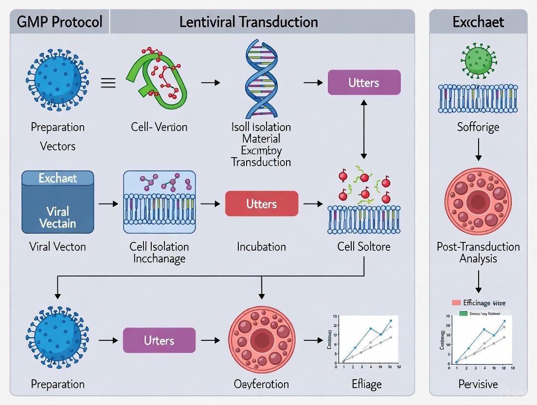
GMP Manufacturing Protocol for Lentiviral Transduction of HSCs
The manufacturing of genetically modified HSCs for clinical applications requires a meticulously controlled process that maintains product quality, safety, and potency. The following diagram illustrates the complete workflow from cell collection to final product release:
Critical Raw Materials and Reagents
The quality of raw materials directly impacts the safety and efficacy of the final HSCGT product. Materials of human or animal origin pose particular challenges as they are often not covered in national pharmacopeias and typically come from single-source specialized suppliers [4]. The following table details essential reagents and their GMP compliance requirements:
Table 2: Research Reagent Solutions for HSC Lentiviral Transduction
| Reagent Category | Specific Examples | Function in Process | GMP Compliance Requirements |
|---|---|---|---|
| Cell Selection Reagents | CliniMACS CD34 Reagent | Immunomagnetic selection of CD34+ HPSCs | CE-marked medical device; used in accordance with manufacturer's instructions |
| Cell Culture Cytokines | SCF, TPO, Flt3-L, IL-3 | HPSC pre-stimulation and activation | Pharmaceutical-grade; qualified for identity, purity, and potency |
| Transduction Enhancers | LentiBOOST, Protamine Sulfate | Improve lentiviral transduction efficiency | Pharmaceutical-grade; demonstration of non-toxicity at working concentrations |
| Lentiviral Vector | IDS.ApoEII LV Vector | Gene delivery vehicle | Clinical-grade; full characterization (titer, identity, purity, sterility, adventitious agents) |
| Cell Culture Media | X-VIVO, StemSpan | Support HPSC growth and maintenance | Pharmaceutical-grade; endotoxin testing; performance qualification |
Quantitative Process Parameters and Acceptance Criteria
Robust process validation establishes measurable critical process parameters (CPPs) and critical quality attributes (CQAs) to ensure consistent product quality. The following table summarizes key quantitative benchmarks for HSCGT manufacturing:
Table 3: Critical Process Parameters and Quality Attributes for HSCGT
| Process Stage | Critical Parameter | Target Range | Acceptance Criterion |
|---|---|---|---|
| HPSC Collection | CD34+ cell yield | ≥5 × 10^6 cells/kg | Minimum 2 × 10^6 cells/kg patient body weight [5] |
| CD34+ Selection | Purity | ≥90% CD34+ cells | Medium purity of 97% with 0.04% residual CD3+ cells [5] |
| CD34+ Selection | Recovery | ≥70% | Medium recovery of 71% [5] |
| Pre-stimulation | Culture duration | 24-40 hours | Optimized for cell cycle induction without differentiation |
| Lentiviral Transduction | Vector quantity | Optimized concentration | Minimum 3-fold improvement with transduction enhancers [6] |
| Final Product | Vector copy number (VCN) | 0.25-2.92 | Product-specific validated range [5] |
| Final Product | Viability | ≥70% | Confirmed by dye exclusion methods |
| Final Product | Sterility | No growth | Sterile with no microbial contamination |
Detailed Experimental Protocols
CD34+ Hematopoietic Progenitor Cell Selection
Principle: Immunomagnetic positive selection of CD34+ cells from leukapheresis or bone marrow harvest using clinical-grade closed system technology.
Materials:
- Leukapheresis product or bone marrow harvest
- CliniMACS Plus or CliniMACS Prodigy system
- CliniMACS CD34 Reagent
- Human gamma globulin
- PBS/EDTA buffer
- Collection bags
Procedure:
- Bone Marrow Processing: For BM starting material, isolate bone marrow mononuclear cells (BMMCs) by Ficoll gradient density centrifugation using COBE 2991 cell washer or equivalent [5].
- Cell Preparation: Wash leukapheresis product or BMMCs with PBS/EDTA buffer and resuspend at appropriate concentration.
- Fc Receptor Blocking: Incubate cells with human gamma globulin to prevent nonspecific antibody binding.
- CD34 Labeling: Incubate with CliniMACS CD34 Reagent according to manufacturer's instructions.
- Magnetic Separation: Process labeled cells through CliniMACS Plus or CliniMACS Prodigy system using appropriate tubing sets and collection bags.
- Product Collection: Collect CD34+ fraction into transfer pack containing appropriate medium.
- Quality Sampling: Aseptically remove samples for cell count, viability, and flow cytometry analysis.
Acceptance Criteria:
- CD34+ purity: ≥90% by flow cytometry
- CD34+ recovery: ≥70% of initial CD34+ cell count
- Viability: ≥90% by trypan blue exclusion
Lentiviral Transduction with Enhanced Efficiency
Principle: Ex vivo genetic modification of CD34+ HPSCs using lentiviral vector with transduction enhancers to achieve high gene transfer efficiency while maintaining cell viability and potency.
Materials:
- Purified CD34+ HPSCs
- Clinical-grade lentiviral vector (e.g., IDS.ApoEII)
- Transduction enhancers: LentiBOOST and protamine sulfate
- Cytokines: SCF, TPO, Flt3-L, IL-3
- Serum-free medium: X-VIVO or StemSpan
- RetroNectin-coated bags or plates
Procedure:
- Pre-stimulation: Culture CD34+ cells in serum-free medium supplemented with cytokines (300 ng/mL SCF, 100 ng/mL TPO, 20 ng/mL IL-3, and 300 ng/mL Flt3-L) for 24-40 hours at 37°C, 5% CO₂ [5].
- Transduction Setup: Pre-load retroNectin-coated culture vessels with appropriate vector volume if using static transduction.
- Transduction Enhancer Preparation: Prepare fresh working solutions of LentiBOOST and protamine sulfate in accordance with manufacturer's instructions.
- Transduction Mixture: Resuspend pre-stimulated cells in fresh medium containing lentiviral vector at optimized MOI and transduction enhancers.
- Transduction Incubation: Culture cells for 16-24 hours at 37°C, 5% CO₂ with optional agitation for suspension cultures.
- Vector Removal: Wash cells twice with PBS/EDTA buffer to remove residual vector and transduction enhancers.
- Post-transduction Culture: Continue culture in cytokine-supplemented medium for additional 24 hours if required.
Optimization Notes:
- Inclusion of transduction enhancers LentiBOOST and protamine sulfate improves transduction efficiency by at least 3-fold, thereby reducing vector quantity required [6].
- Multiple rounds of transduction may be performed with fresh vector and enhancers for challenging-to-transduce cell populations.
- Process should be optimized to minimize ex vivo culture time while achieving target VCN.
Critical Quality Control Testing
Principle: Comprehensive characterization of final drug product to ensure safety, identity, purity, potency, and viability.
Materials:
- Final cell product samples
- Flow cytometry reagents
- qPCR reagents for VCN determination
- Sterility culture media
- Endotoxin testing reagents
- Potency assay components
Procedure:
- Vector Copy Number (VCN) Analysis:
- Extract genomic DNA from transduced cells using validated method
- Perform qPCR with vector-specific and reference gene (e.g., RNase P) primers/probes
- Calculate VCN using standard curve method with vector-specific plasmid standards
Cell Phenotype and Viability:
- Stain cells with fluorochrome-conjugated antibodies against CD34, CD45, and viability dye
- Analyze by flow cytometry using validated method
- Report percentage of viable CD34+ cells
Sterility Testing:
- Inoculate samples into aerobic and anaerobic culture media
- Incubate for 14 days according to Ph. Eur. 2.6.1
- Monitor daily for microbial growth
Mycoplasma Testing:
- Perform by PCR or culture method according to Ph. Eur. 2.6.7
Endotoxin Testing:
- Perform kinetic chromogenic LAL test according to Ph. Eur. 2.6.14
Potency Assay:
- Perform colony-forming unit (CFU) assay to assess hematopoietic progenitor function
- Alternatively, measure functional enzyme activity (e.g., IDS activity for MPSII)
Acceptance Criteria:
- VCN: Within validated range (typically 0.25-2.92 for HSCGT products) [5]
- Viability: ≥70%
- Sterility: No growth after 14 days
- Mycoplasma: Negative
- Endotoxin: ≤5 EU/kg/hr
- Potency: Product-specific based on clinical data
Implementation Considerations for GMP Compliance
Facility and Equipment Controls
The manufacturing facility must be designed and maintained to appropriate cleanroom classifications based on process requirements. For open processing steps, a Grade A environment with Grade B background is typically required. Closed systems may allow for lower classification (Grade D) when justified by risk assessment [3]. Key considerations include:
- Environmental Monitoring: Comprehensive program for viable and non-viable particulates in critical areas
- Equipment Qualification: Installation, operational, and performance qualification for all critical equipment
- Process Validation: Evidence that manufacturing process consistently yields product meeting predetermined quality attributes
- Cleaning Validation: For reusable equipment, validated cleaning procedures to prevent cross-contamination
Risk-Based Approach to Manufacturing
The unique characteristics of ATMPs necessitate a flexible, science-based approach to GMP implementation. A robust risk-based approach (RBA) should:
- Scientifically identify risks inherent to the specific ATMP manufacturing process
- Focus control measures on critical process parameters and quality attributes
- Justify any deviations from established guidelines through scientific data
- Implement continuous improvement based on process performance data
As stated in EudraLex Volume 4, Part IV: "These Guidelines do not intend to place any restrain on the development of new concepts of new technologies. While this document describes the standard expectations, alternative approaches may be implemented by manufacturers if it is demonstrated that the alternative approach is capable of meeting the same objective" [3].
Regulatory Interactions and Compliance Strategy
Successful navigation of the ATMP regulatory landscape requires proactive engagement with regulatory authorities:
- Scientific Advice: EMA offers fee reductions (65% standard, 90% for SMEs) for scientific advice on ATMP development [2]
- Certification Procedure: 90% fee reduction for the certification procedure for micro, small, and medium-sized enterprises [2]
- Innovation Task Force: Forum for informal dialogue with EMA in early development stages [2]
- Briefing Meetings: SME office provides briefing meetings to discuss regulatory strategy [2]
Manufacturers should maintain awareness of evolving regulatory guidance and participate in industry forums to share best practices and promote harmonization of GMP standards for ATMPs.
Understanding Lentiviral Vector Systems and HSC Biology
Lentiviral vector (LV) systems have emerged as a cornerstone technology for the genetic modification of hematopoietic stem cells (HSCs), enabling groundbreaking advances in gene therapy for monogenic blood disorders, immunodeficiencies, and cancers. These systems facilitate the stable integration of therapeutic genes into the host genome of both dividing and non-dividing cells, making them ideally suited for long-term hematopoietic reconstitution [7] [8]. The clinical utility of any LV gene therapy depends critically on the efficient high-level transduction of patient HSCs capable of long-term hematopoietic repopulation [7]. Recent advances have focused on optimizing Good Manufacturing Practice (GMP)-compliant protocols to enhance transduction efficiency while maintaining cell viability and function, thereby supporting the transition from research to clinical applications [9].
The promise of this approach is exemplified in hematopoietic stem cell gene therapy (HSCGT) for conditions like Mucopolysaccharidosis type II (Hunter syndrome), where lentiviral transduction of a patient's own CD34+ cells with a functional iduronate-2-sulphatase (IDS) gene has demonstrated normalization of brain pathology and behavior in MPSII mice [9]. Similarly, clinical trials for X-linked severe combined immunodeficiency (X-SCID) have shown successful T cell and natural killer cell recovery in treated patients [7]. However, achieving consistent, high-efficiency transduction of repopulating HSCs remains technically challenging, driving continued optimization of protocols for clinical manufacturing.
Critical Process Parameters for Optimization
Optimizing lentiviral transduction of HSCs requires careful consideration of multiple interdependent parameters. The following factors have been identified as crucial determinants of transduction success.
Technical Optimization Parameters
Table 1: Key Technical Parameters for HSC Transduction Optimization
| Parameter | Impact on Transduction | Optimal Range/Approach | Supporting Evidence |
|---|---|---|---|
| Multiplicity of Infection (MOI) | Determines viral particle-to-cell ratio; higher MOI can increase efficiency but risks toxicity & higher VCN | 25-100 (varies by protocol) | Lower MOI reduces multiple integration events [8] |
| Cell Concentration During Transduction | Affects vector consumption & cell-vector contact | 2-4 × 10⁶ cells/mL (single-step) | Single-step at higher density conserved LV without compromising VCN [7] |
| Transduction Enhancers | Improves transduction efficiency by various mechanisms | LentiBOOST, protamine sulfate, cyclosporine H | LentiBOOST increased HSC VCN by 2- to 3-fold [9] [7] |
| Vector RNA Size | Impacts both production yield and transduction efficiency | <6 kb for primary HSCs | Efficiency decreased significantly with vectors >6 kb [10] |
| Serum Conditions | Affects vector stability and cell health | Low serum or serum-free conditions | Serum-free production minimizes HSC differentiation [10] |
Biological and Vector Considerations
Beyond technical parameters, several biological factors significantly influence transduction outcomes. Viral vector design plays a crucial role, with self-inactivating (SIN) configurations now standard for enhanced safety [8]. The pseudotype of the viral envelope, most commonly VSV-G, determines tropism and transduction efficiency across different cell types [8]. Additionally, the promoter driving transgene expression must be carefully selected to achieve appropriate, sustained expression in hematopoietic lineages [7].
Donor variability represents another significant consideration, as the same HSC transduction protocol can produce markedly different results between donors [7]. The activation state of target cells also critically influences susceptibility to transduction, with pre-stimulation using cytokine combinations (SCF, TPO, Flt3-L) typically required to prime HSCs for efficient lentiviral entry and integration [7] [10].
GMP-Compliant Experimental Protocols
Optimized Transduction Protocol for CD34+ HSCs
Materials and Reagents:
- Human CD34+ HSCs (from mobilized peripheral blood or umbilical cord blood)
- Lentiviral vector (clinical grade)
- X-VIVO 10 or similar serum-free medium
- Recombinant cytokines: SCF (100 ng/mL), TPO (100 ng/mL), Flt3-L (50 ng/mL)
- Transduction enhancers: LentiBOOST (1 mg/mL), protamine sulfate (4-8 μg/mL)
- Retronectin (optional, for pre-coating)
- Cell culture plates and appropriate incubator
Procedure:
- Cell Thawing and Pre-stimulation:
- Thaw cryopreserved CD34+ cells rapidly and wash to remove cryoprotectant.
- Resuspend cells at 1-2 × 10⁶ cells/mL in X-VIVO 10 medium supplemented with SCF (100 ng/mL), TPO (100 ng/mL), and Flt3-L (50 ng/mL).
- Incubate for 24-48 hours at 37°C, 5% CO₂ to activate cells [7].
Transduction Setup:
- Pre-coat culture vessels with Retronectin (50 μg/mL) if using, following manufacturer's instructions.
- After pre-stimulation, collect cells and adjust concentration to 2-4 × 10⁶ cells/mL in fresh medium with cytokines.
- Add protamine sulfate to final concentration of 4-8 μg/mL and LentiBOOST to 1 mg/mL.
- Add lentiviral vector at predetermined MOI (typically 25-100).
Transduction Process:
- Transfer cell-vector mixture to coated or uncoated culture vessels.
- Perform spinoculation by centrifuging at 1800 rpm, 32°C for 1 hour [10].
- Incubate at 37°C, 5% CO₂ for 16-24 hours.
Post-transduction Processing:
- Wash cells twice with PBS or appropriate buffer to remove residual vector.
- Either proceed to transplantation or continue in culture for analysis.
- For clinical applications, formulate final product in appropriate infusion medium [7].
Quality Control Assessments:
- Determine transduction efficiency by flow cytometry (for reporter genes) or ddPCR for VCN [11].
- Assess cell viability via trypan blue exclusion or flow cytometry with Annexin V/7-AAD [8].
- For xenotransplantation studies, transplant 1 × 10⁶ cells into immunodeficient mice (e.g., NSG) conditioned with busulfan (35 mg/kg) [7].
Quantitative Data on Protocol Optimization
Table 2: Comparative Analysis of Transduction Protocols
| Protocol Variable | Standard Two-Step Protocol | Optimized Single-Step Protocol | Improvement/Change |
|---|---|---|---|
| Cell Concentration | 1 × 10⁶ cells/mL [7] | 2-4 × 10⁶ cells/mL [7] | 2-4 fold higher density |
| Transduction Steps | Two successive incubations [7] | Single incubation [7] | Simplified manipulation |
| LV Consumption | Higher (2-2.7 × 10⁸ TU/mL) [7] | Reduced (2 × 10⁸ TU/mL) [7] | Conservation of vector |
| Enhancers | Polybrene or RetroNectin [7] | LentiBOOST + protamine sulfate [9] [7] | 3-fold increase in TD efficiency [9] |
| Resulting HSC VCN | 0.16-1.13 (clinical trial data) [7] | 2- to 3-fold increase with LentiBOOST [7] | Significant improvement |
The Scientist's Toolkit: Essential Research Reagents
Table 3: Key Research Reagent Solutions for HSC Transduction
| Reagent/Category | Specific Examples | Function/Application | Notes for GMP Compliance |
|---|---|---|---|
| Transduction Enhancers | LentiBOOST, protamine sulfate, cyclosporine H | Increase transduction efficiency by facilitating vector-cell interaction | LentiBOOST with protamine sulfate improved TD efficiency 3-fold [9] |
| Cell Culture Matrix | RetroNectin, recombinant fibronectin fragment (CH-296) | Enhoves transduction by colocalizing cells and viral particles | Use GMP-grade for clinical applications [7] |
| Cytokine Combinations | SCF, TPO, Flt3-L | Pre-stimulation of HSCs to increase susceptibility to transduction | Essential for quiescent HSC activation [7] [10] |
| Vector Quantitation | ddPCR, p24 ELISA, flow cytometry-based functional titration | Determines functional titer (TU/mL) for MOI calculation | ddPCR is gold standard for VCN [8] [11] |
| Specialized Systems | Viromicst Stem with Magnetofection | Magnetic nanoparticle-based transduction enhancement | Specifically designed for stem cells [12] |
Workflow and Pathway Visualizations
The optimization of lentiviral vector systems for HSC transduction represents a rapidly advancing field with significant clinical implications. The protocols and parameters detailed in this application note provide a foundation for achieving efficient, reproducible genetic modification of HSCs while maintaining GMP compliance. Key advances include the simplification of transduction protocols through single-step processes, the identification of effective transduction enhancers like LentiBOOST, and refined understanding of critical parameters such as cell concentration and vector design.
Future developments in this field will likely focus on further improving the safety profile of lentiviral vectors through advanced design features, enhancing manufacturing scalability to meet clinical demand, and standardizing quality control metrics across production batches. The integration of novel technologies such as droplet digital PCR for precise VCN quantification and magnetic nanoparticle-based transduction systems will continue to push the boundaries of what is achievable in HSC gene therapy [12] [11]. As these technologies mature, they will undoubtedly expand the therapeutic potential of genetically modified HSCs for an increasingly broad spectrum of hematologic, immunologic, and metabolic disorders.
In the field of advanced therapies, the development of Good Manufacturing Practice (GMP) protocols for lentiviral transduction of hematopoietic stem cells represents a cutting-edge frontier for treating monogenic disorders. As these therapies approach first-in-human studies, they enter a regulatory environment where data integrity is as critical as biological efficacy. The ALCOA++ framework has evolved from a set of guiding principles to a mandatory standard under revised 2025 regulations, including the EU GMP Chapter 4 and Annex 11 updates [13] [14]. This application note details the practical implementation of ALCOA++ within a lentiviral stem cell research protocol, providing a structured approach for researchers and drug development professionals to align their methodologies with the current regulatory expectations for data governance, traceability, and integrity throughout the therapeutic development lifecycle.
ALCOA++ in Principle and Regulation
The Expanded ALCOA++ Framework
ALCOA++, as codified in the 2025 draft EU GMP Chapter 4, comprises ten fundamental principles for data integrity [15] [13] [16]. These principles provide a comprehensive framework for ensuring data reliability across both paper and electronic systems in GMP environments.
Table 1: The ALCOA++ Principles and Their Definitions
| Principle | Definition | GMP Application Context |
|---|---|---|
| Attributable | Links each datum to the person and/or system that created or modified it [15]. | Unique user IDs for all electronic system access; signature logs for paper records. |
| Legible | Data must be readable and reviewable in its original context [15]. | Permanent recording; reversible encoding; no data loss from format changes. |
| Contemporaneous | Recorded at the time of the activity with accurate, automatically captured date/time [15]. | Time-stamped by an external standard (e.g., UTC); no manual time zone conversions. |
| Original | The first capture or a certified copy created under controlled procedures [15]. | Preservation of source data; dynamic data (e.g., waveforms) remains available. |
| Accurate | Faithfully represents what occurred; error-free with documented amendments [15] [16]. | Validated systems and transfers; calibrated devices; amendments capture original. |
| Complete | All data, including metadata and audit trails, is present for event reconstruction [15]. | No data omissions; deletions do not obscure what happened. |
| Consistent | Data is standardized and sequential; time/date stamps align across the lifecycle [15]. | Chronological order; standardized definitions and units; no contradictions. |
| Enduring | Recorded in permanent media and retained for the specified period [15] [16]. | Lasting format; secure backups; archiving; independent of specific hardware. |
| Available | Readily retrievable for review, audit, or inspection throughout the retention period [15]. | Searchable, indexed storage; timely retrieval for authorized personnel. |
| Traceable | Data is traceable end-to-end with a clear history of changes and transformations [17]. | Robust audit trails for data and metadata; reconstruction of history. |
The 2025 Regulatory Shift
The regulatory landscape in 2025 is characterized by a significant elevation of data integrity expectations. The European Commission's draft update to EudraLex Volume 4, Chapter 4 formally mandates the ALCOA++ principles, moving them from best practice to a legally binding requirement [13] [14]. This revision introduces the concept of the data lifecycle, requiring comprehensive data governance and metadata control integrated within the Pharmaceutical Quality System (PQS) [13]. Simultaneously, Annex 11 has been revised to reflect today's digital, cloud-integrated environment, with stricter controls for identity and access management, IT security, and mandatory audit logging [13].
The FDA similarly emphasizes risk-based audit trail review and heightened scrutiny of supplier and Contract Manufacturing Organization (CMO) oversight [13]. For researchers, this means that data integrity must be built into the foundational design of experimental and manufacturing processes for hematopoietic stem cell gene therapy (HSCGT), with documentation systems that are inspection-ready at all times [15] [13].
Application in Lentiviral Transduction GMP Protocol
Mapping ALCOA++ to a Critical Process
The following diagram illustrates the data flow and key control points for ensuring data integrity within a lentiviral transduction process, from vector receipt to final cell product.
Experimental Protocol: GMP-Compliant Lentiviral Transduction with Integrated Data Integrity Controls
This protocol outlines an optimized lentiviral transduction process for hematopoietic stem cells, based on published GMP development work, with embedded ALCOA++-compliant data recording practices [9].
Materials and Reagents
Table 2: Essential Research Reagent Solutions for Lentiviral Transduction
| Reagent / Material | Function / Purpose | ALCOA++ Data Recording Consideration |
|---|---|---|
| Lentiviral Vector | Gene delivery vehicle encoding therapeutic transgene (e.g., IDS.ApoEII for MPSII) [9]. | Record unique batch number, certificate of analysis, and storage conditions. Traceable from receipt to use. |
| CD34+ Hematopoietic Stem Cells | Patient-specific cell starting material. | Document donor/patient ID, cell count, and viability at receipt. Attributable to a specific source. |
| LentiBOOST & Protamine Sulfate | Transduction enhancers [9]. | Record lot numbers and preparation time. Accurate volumetric measurements. |
| X-VIVO 15 Serum-free Medium | Cell culture medium supporting HSC growth. | Document lot number and expiration date. Original pH and osmolarity QC records. |
| Puromycin | Selective agent for transduced cell population [18]. | Record preparation date and concentration verification. Contemporaneous recording of selection timeline. |
| qPCR Assay for VCN | Quantifies vector copy number per cell - critical quality attribute. | Document assay calibration, raw data, and analysis method. Complete dataset including all replicates. |
Step-by-Step Methodology
1. Pre-Transduction: Cell Preparation and Vector Thawing
- Procedure: Isolate and qualify CD34+ cells. Thaw lentiviral vector rapidly in a 37°C water bath and keep on ice.
- Data Integrity Controls:
- Attributable: Log operator ID performing the cell count and vector thaw.
- Original & Accurate: Record the original cell viability and count from an automated cell counter, linking the data file to the batch record. Capture vector lot number and thaw time.
- Contemporaneous: Record the start time of the process immediately.
2. Transduction: Vector Application
- Procedure: Resuspend cells at a density of 1 × 10^6 cells/mL in medium containing LentiBOOST (1:100 dilution) and protamine sulfate (4-8 µg/mL) [9]. Add lentiviral vector at the predetermined Multiplicity of Infection (MOI of 30-50 for research-grade, optimized for clinical vector) [9] [18]. Incubate for 16-24 hours.
- Data Integrity Controls:
- Accurate: Document exact volumes of vector and supplements used. Use calibrated pipettes.
- Consistent: Follow a standardized sequence for reagent addition as defined in the SOP.
- Contemporaneous: Record the exact start and end times of transduction in the electronic Batch Record (eBR).
3. Post-Transduction: Cell Expansion and Selection
- Procedure: After 24 hours, wash cells to remove vector and place in fresh medium. For stable pools, begin puromycin selection (e.g., 1-2 µg/mL) for 10 days [18].
- Data Integrity Controls:
- Complete: Document all medium changes and the puromycin selection schedule. Include any observations.
- Enduring: Save cell culture images and growth curve data in a secure, archived format.
4. Quality Control and Analytics
- Procedure: Harvest cells for QC testing. Key tests include Vector Copy Number (VCN) by qPCR, cell viability, flow cytometry for transgene expression, and functional assays (e.g., IDS enzyme activity for MPSII [9]).
- Data Integrity Controls:
- Traceable: Ensure a clear audit trail links the final QC result back to the raw data (e.g., qPCR plate setup, FACS gating strategy). All changes to electronic data must be tracked with a reason.
- Legible: Save data in non-proprietary or widely accepted formats where possible. Ensure metadata is embedded and readable.
- Complete: Include all repeat or reanalysis performed. The data set must be reconstructable.
The Scientist's Toolkit: Essential Materials for Compliance
Beyond biological reagents, a modern GMP research environment requires specific systems and controls to operationalize ALCOA++.
Table 3: The Data Integrity Toolkit for GMP Research
| Tool / System | Function | Role in Ensuring ALCOA++ |
|---|---|---|
| Electronic Lab Notebook (ELN) / eBR | Centralized platform for protocol execution and data capture. | Enforces Attributable (login), Contemporaneous (timestamps), and Legible entries. |
| LIMS (Laboratory Information Management System) | Manages sample lifecycle and associated analytical data. | Maintains Complete sample history and ensures data Availability. |
| Validated Automated Cell Counter | Provides accurate, reproducible cell counts and viability. | Delivers Accurate and Original data with digital output. |
| Calibrated Pipettes & Balances | Precise volumetric and mass measurements. | Foundation for Accurate data generation; requires regular calibration records. |
| Centralized Time Server (NTP) | Synchronizes time across all computer systems and instruments. | Critical for Contemporaneous and Consistent timestamps across data sources. |
| Electronic Quality Management System (eQMS) | Manages documents, deviations, CAPA, and training records. | Provides a Traceable and Enduring record of the quality system. |
| Access Control Systems (Badge access) | Restricts physical access to labs and critical equipment. | Supports Attributable actions by ensuring only trained personnel are present. |
Integrating the ALCOA++ principles into the fabric of GMP protocol development for lentiviral stem cell research is no longer optional. The 2025 regulatory landscape demands a proactive, risk-based approach to data governance where integrity is assured throughout the entire data lifecycle. By implementing the structured protocols, controls, and tools outlined in this application note, researchers and developers can build a robust framework that not only meets stringent regulatory scrutiny but also underpins the scientific credibility and ultimate success of their advanced therapy medicinal products.
Building a GMP-Compliant Quality Management System (QMS)
This document provides detailed application notes and protocols for establishing a Good Manufacturing Practice (GMP)-compliant Quality Management System (QMS) specifically for the development of hematopoietic stem cell gene therapy (HSCGT) products using lentiviral transduction. Adherence to GMP principles is a regulatory requirement for Advanced Therapy Medicinal Products (ATMPs) to ensure identity, purity, potency, and safety for human administration [19] [20] [21]. This framework is built upon foundational GMP principles outlined by the FDA, WHO, and the European Union's EudraLex Volume 4, integrating recent regulatory guidance on in-process controls and documentation effective in 2025 [22] [23] [24].
The protocols herein are designed for the GMP-grade manufacturing of lentivirally transduced CD34+ cells, detailing optimized processes, critical quality control parameters, and a robust QMS structure. This system ensures that the investigational medicinal product (IMP) is consistently produced and controlled to quality standards appropriate for clinical trials, providing researchers and drug development professionals with a actionable roadmap for clinical translation.
A GMP-compliant QMS, often referred to as a Pharmaceutical Quality System (PQS), is the cornerstone of ATMP manufacturing. It is an integrated system of processes, procedures, and responsibilities that ensures a product is consistently produced and controlled to the quality standards appropriate for its intended use [24] [25] [21]. For lentiviral-based HSCGT, this system mitigates the unique risks associated with the use of viral vectors and the ex vivo manipulation of human cells.
The core objective of the QMS is to build quality into every stage of the product lifecycle, from raw material selection to final product administration, rather than relying solely on end-product testing. This is critical because many critical quality attributes cannot be verified through final testing alone [21]. The principle of "current" GMP requires that companies employ modern technologies and innovative approaches to achieve higher quality through continuous improvement [21].
Regulatory Framework and Key Guidelines
A robust QMS must be aligned with the regulatory requirements of the target market. The following key regulations and guidelines form the basis for GMP compliance.
Table 1: Key GMP Regulations and Guidelines for HSCGT Products
| Region/Body | Guideline/Regulation | Key Focus Areas | Relevance to HSCGT |
|---|---|---|---|
| U.S. FDA | 21 CFR Parts 210, 211, 600 [22] | Minimum requirements for methods, facilities, and controls for drug & biological products. | Foundational regulations for ensuring safety, identity, strength, quality, and purity. |
| European Union | EudraLex Volume 4, Annex 13 & Part IV [20] | GMP for Investigational Medicinal Products & Advanced Therapy Medicinal Products. | Specific guidelines for manufacturing cell & gene therapy products in clinical trials. |
| World Health Org. (WHO) | WHO GMP for biological products [24] | General principles and quality control of biological medicines, including cell therapies. | Internationally recognized standard, incorporated into the national laws of >100 countries. |
Recent regulatory developments emphasize data integrity, advanced manufacturing technologies, and detailed documentation practices. The FDA's 2025 draft guidance on in-process controls (21 C.F.R. § 211.110) clarifies the use of real-time monitoring and process models, while the EMA's 2025 draft of Chapter 4 introduces enhanced requirements for documentation lifecycles and data governance [23] [26].
Experimental Protocols: GMP-Compliant Lentiviral Transduction of hCD34+ Cells
The manufacturing process for lentivirally transduced HSCs involves a series of interconnected and tightly controlled steps. The following workflow diagram outlines the entire process from cell collection to final product release.
Detailed Protocol: Transduction Optimization with Enhancers
Objective: To efficiently transduce human CD34+ cells with a lentiviral vector encoding the therapeutic transgene while maintaining cell viability and potency, using transduction enhancers to reduce vector load.
Materials and Reagents: Table 2: Research Reagent Solutions for GMP Transduction
| Reagent/Solution | Function/Purpose | GMP-Grade Specification |
|---|---|---|
| X-VIVO-15 Medium | Serum-free basal medium for cell culture. | Formulated for human clinical use, with 1% Human Albumin Serum (HAS). |
| Cytokine Cocktail (SCF, TPO, Flt3-L, IL-3) | Pre-stimulation to activate HSCs and promote lentiviral integration. | Recombinant, GMP-grade, sourced from qualified vendors. |
| LentiBOOST | Transduction enhancer; increases viral attachment/fusion. | GMP-grade, compliant with regulatory standards for IMP manufacturing. |
| Protamine Sulfate | Transduction enhancer; neutralizes charge repulsion between vector and cell. | Pharmaceutical-grade, sterile, endotoxin-free. |
| IDS.ApoEII Lentiviral Vector | Delivers therapeutic gene (e.g., IDS enzyme for MPSII). | GMP-grade, produced under GMP (e.g., IU Vector Production Facility), with defined MOI and titer. |
Methodology:
- Cell Preparation: Thaw cryopreserved, GMP-grade hCD34+ cells and place in pre-stimulation medium (X-VIVO-15, 1% HAS, SCF, TPO, Flt3-L, IL-3) for 24 hours [19].
- Transduction Setup: After pre-stimulation, pellet cells and resuspend in fresh transduction medium. Divide cells into aliquots for different test conditions.
- Enhancer and Vector Addition:
- Prepare conditions with a range of vector concentrations (e.g., MOI of 12.5 to 100).
- Add GMP-grade LentiBOOST and protamine sulfate at pre-optimized, non-toxic concentrations to the respective test groups [19].
- Perform two rounds of transduction, typically 8-24 hours each, with gentle agitation.
- Post-Transduction Culture: After transduction, wash cells to remove residual vector and enhancers. Culture cells in fresh, cytokine-supplemented medium for a defined period before harvest and formulation.
- Quality Control Sampling: Remove samples for critical in-process controls, including:
- Cell Viability and Count: Using trypan blue exclusion.
- Vector Copy Number (VCN): By qPCR on genomic DNA from liquid culture and colony-forming unit (CFU) assays.
- Transduction Efficiency: By flow cytometry (if reporter gene is present) or by % vector-positive colonies in CFU assays.
- Potency Assay: Measure intracellular therapeutic enzyme activity (e.g., IDS activity for MPSII) [19].
- Sterility: BacT/ALERT or direct inoculation for mycoplasma and microbiological testing.
Data Analysis and Acceptance Criteria: The success of the optimization is determined by key quantitative metrics. The following table summarizes expected outcomes from a successfully optimized protocol.
Table 3: Quantitative Metrics for Transduction Efficiency and Cell Quality [19]
| Parameter | Condition (MOI) | Without Enhancers | With LentiBOOST & Protamine Sulfate | Acceptance Criteria |
|---|---|---|---|---|
| BFU-E Transduction | 12.5 | 33.3% | 94.1% | >70% |
| CFU-GM Transduction | 12.5 | 55.6% | 94.1% | >70% |
| Average Vector Copy Number (VCN) | 12.5 - 100 | Baseline | 2.5 - 2.9 fold increase | 1.0 - 5.0 (product-specific) |
| Intracellular Enzyme Activity | 12.5 - 100 | Baseline | 4.8 fold increase | >3x over mock-transduced |
| Cell Viability | 50 | >80% | >80% | >70% |
| CFU Colony Numbers | 50 | Comparable to non-transduced | Comparable to non-transduced | No significant toxicity |
Building the GMP-Compliant QMS: Core Components
A successful QMS is built on several interconnected pillars. The following diagram illustrates the logical relationship between these core components, showing how they integrate to ensure final product quality.
Documentation and Data Integrity
Documentation is the foundation of traceability and proof of control. The QMS must ensure all data, whether paper-based or electronic, is recorded and maintained in compliance with ALCOA++ principles (Attributable, Legible, Contemporaneous, Original, Accurate, Complete, Consistent, Enduring, and Available) [26].
Application Notes:
- Lifecycle Approach: Implement a document lifecycle management system for all Standard Operating Procedures (SOPs), batch records, and test protocols, from creation through to archiving [26].
- Electronic Systems: Utilize electronic batch records (EBR) and Manufacturing Execution Systems (MES) to enhance traceability and efficiency. Ensure strict access controls and validated audit trails for all computerized systems [25].
- Raw Data Definition: Clearly define and control "raw data" within the quality system. The 2025 draft of EU GMP Chapter 4 provides updated definitions for True Copy and Certified Copy, which are critical for data integrity during technology transfer and audits [26].
Personnel, Training, and Organizational Structure
Protocol:
- Organizational Chart: Define a clear structure with unambiguous lines of authority and responsibility. Key roles include the Head of Production, Head of Quality Control, and the Qualified Person (QP) responsible for batch certification and release in the EU [20].
- Training Program: Implement a comprehensive, role-specific, and documented training program. This includes initial GMP training, ongoing refresher courses, and assessments to certify competency for all tasks performed [25].
Process and Equipment Controls
Application Notes:
- Process Validation: Conduct rigorous process validation studies to demonstrate that the lentiviral transduction and cell culture processes can consistently yield a product meeting its pre-defined quality attributes [25].
- Equipment Qualification: All critical equipment (e.g., bioreactors, CliniMACS separator, cryopreservation units) must undergo Installation (IQ), Operational (OQ), and Performance Qualification (PQ) [25].
- In-Process Controls (IPCs): As per FDA's 2025 guidance, implement a scientific, risk-based strategy for IPCs. Define what, where, when, and how to sample and test in-process materials. For continuous manufacturing or advanced process models, pair the model with physical testing or examination [23].
Material and Supply Chain Management
Protocol:
- Supplier Qualification: Conduct thorough audits and quality assessments of all critical material suppliers, including those for cytokines, media, and single-use bioprocess containers [25].
- Traceability: Implement a system that ensures full traceability of all materials and products throughout the supply chain, from receipt to final product administration.
- Raw Material Testing: Establish specifications and test methods for all raw materials. Only release materials for GMP use after approval by the Quality Control unit.
Quality Control (QC) and Batch Release
The QC laboratory is responsible for testing and releasing raw materials, in-process samples, and the final drug product against pre-defined specifications.
Table 4: Essential Quality Control Tests for Lentivirally Transduced HSCs
| Test Category | Specific Test | Method | Frequency / Stage |
|---|---|---|---|
| Safety | Sterility | BacT/ALERT, Culture | In-process, Final Product |
| Mycoplasma | PCR and/or Culture | Final Product | |
| Endotoxin | LAL Test | In-process, Final Product | |
| Replication-Competent Lentivirus (RCL) | PCR or Co-culture Assay | Final Product | |
| Identity | CD34+ Cell Count | Flow Cytometry | Pre- and Post-Transduction |
| Vector-Specific Identity | PCR for transgene | Final Product | |
| Potency | Vector Copy Number (VCN) | qPCR/ddPCR | Final Product |
| Transduction Efficiency | CFU Assay / Flow Cytometry | Final Product | |
| Functional Enzyme Activity | Cell-based or biochemical assay | Final Product | |
| Purity | Viability | Trypan Blue / Flow Cytometry | Throughout Process |
| Cell Number and Dose | Viable cell count | Final Product |
Batch Release Protocol: The final IMP batch can only be released after a full review of the manufacturing documentation and QC data by the Quality unit and, in the EU, certification by a Qualified Person (QP) [20].
Implementing a GMP-compliant QMS for HSCGT products is a complex but essential endeavor for successful clinical translation. The system must be proactive, science-based, and integrated into every aspect of development and manufacturing. By adhering to the structured protocols for transduction optimization and establishing the core QMS components outlined in this document—robust documentation, trained personnel, validated processes, controlled materials, and rigorous quality control—research teams can build a foundation that not only meets regulatory expectations but, more importantly, ensures the consistent production of a safe and effective therapy for patients.
Facility and Equipment Requirements for HSC Processing
The manufacturing of hematopoietic stem cells (HSCs) for lentiviral gene therapy requires strict adherence to Current Good Manufacturing Practice (CGMP) regulations to ensure product safety, identity, strength, quality, and purity [22]. CGMP regulations provide the minimum requirements for the methods, facilities, and controls used in manufacturing, processing, and packing of a drug product, ensuring that products are consistently produced and controlled according to quality standards [22] [27]. For hematopoietic stem cell gene therapy (HSCGT), this is particularly critical as it involves ex vivo introduction of a missing gene into patients' own stem cells via lentiviral-mediated transduction, with the modified HSCs subsequently transplanted back into conditioned patients to repopulate the blood system and produce functional protein [6]. The approval process for such therapies includes a thorough review of the manufacturer's compliance with CGMPs, where FDA assessors determine whether the firm has the necessary facilities, equipment, and ability to manufacture the drug it intends to market [22].
Facility Design and Control Systems
Cleanroom and Environmental Controls
HSC processing for lentiviral transduction requires classified cleanroom environments that meet specified airborne particulate cleanliness limits. The facility must maintain appropriate pressure cascades, temperature, humidity, and ventilation (HVAC systems) to prevent cross-contamination and ensure aseptic processing conditions. Cleanroom validation studies are essential, particularly for the aseptic processing steps involving open manipulations of cell products [6]. The facility design should include:
- Grade A/ISO 5 environments for critical open processing steps
- Grade B/ISO 7 background environments for adjacent areas
- Grade C/ISO 8 and Grade D/ISO 9 support areas
- Material and personnel flow controls to prevent cross-contamination
Facility Zoning and Material Management
Table 1: Color-Coding System for HSC Processing Facilities
| Color Code | Designated Zone | Application in HSC Processing |
|---|---|---|
| Blue | Food contact/Critical processing areas | Tools and equipment for direct contact with cell cultures or critical reagents |
| Green | Non-food contact/General areas | Equipment for environmental cleaning of floors and non-critical surfaces |
| Red | High-risk/Allergen control | Designated for specific critical reagents or processes to prevent cross-contamination |
| Yellow | Equipment and non-contact surfaces | Utensils for handling non-critical materials |
| White | Quality control and testing areas | Equipment dedicated to analytical testing and quality control procedures |
| Pink/Orange/Purple | High-risk/Specialized applications | Reserved for specific critical processing steps or reagent handling |
| Black | Drains and heavily soiled areas | Cleaning equipment for drains and waste handling |
| Brown/Gray | Hallways and corridors | Equipment for non-processing areas with high visibility needs |
Implementation of a comprehensive color-coding system minimizes the risk of cross-contamination by visually separating equipment and tools used in different processing zones [28] [29]. This system should be consistently applied throughout the facility, with tools stored in color-coded shadow boards or wall brackets in their respective use areas [29]. The color-coding plan must be clearly communicated to all staff and maintained through regular monitoring and review [28].
Essential Equipment Specifications
HSC processing for lentiviral gene therapy requires specialized equipment throughout the manufacturing workflow, from cell collection and processing to transduction, expansion, and final product formulation.
Table 2: Critical Equipment for GMP-Compliant HSC Processing
| Equipment Category | Specific Examples | Technical Specifications | GMP Application in HSC Processing |
|---|---|---|---|
| Cell Separation Systems | BD FACSJazz cell sorter, BD Accuri C6 flow cytometer [30] | Multi-parameter cell sorting and analysis | Isolation of CD34+ hematopoietic stem cells from apheresis products |
| Cell Culture Systems | Oxygen Controlled CO2 Cell Culture Incubator [31] | Precise CO2/O2 control for in vivo environment replication | Maintenance of HSCs during expansion and transduction phases |
| Transduction Apparatus | 4D-Nucleofector Core X/Y (Lonza) [30] | Electroporation-based transfection/transduction | Lentiviral vector delivery to HSCs |
| Process Monitoring Systems | BioLector MP2 [30] | Continuous monitoring of growth mass, fluorescence, acid production, oxygen consumption | Real-time monitoring of cell growth parameters during manufacturing |
| Analytical Instruments | Infinite M1000 PRO microplate reader (Tecan) [30] | UV, VIS absorption, fluorescence with high spectral resolution | Assessment of transduction efficiency, viability assays, metabolic measurements |
| Molecular Analytics | Quant Studio 12K real time PCR [30] | High-throughput gene expression analysis | Vector copy number analysis, sterility testing, potency assays |
| Protein Analytics | Meso Scale Discover Sector Imager [30] | Multi-array biomarker detection in multiplex formats | IDS enzyme activity measurement (for MPSII applications) [6] |
| Single-Cell Analysis | C1 Single Cell auto preparation system (Fluidigm) [30] | Gene expression and mRNA analysis in 96-well format | Clonal analysis of transduced HSCs, vector integration site analysis |
Research Reagent Solutions for HSC Transduction
Table 3: Essential Reagents for GMP-Compliant HSC Transduction
| Reagent Category | Specific Examples | Function in HSC Transduction | Quality Requirements |
|---|---|---|---|
| Transduction Enhancers | LentiBOOST, protamine sulfate [6] | Improve transduction efficiency by at least 3-fold without adverse toxicity | GMP-grade, endotoxin-tested, with certificate of analysis |
| Cytokines and Growth Factors | SCF, TPO, FLT3-L, IL-3, IL-6 | Promote HSC expansion and maintenance during transduction | Pharmaceutical-grade, recombinant human, carrier-free formulations |
| Lentiviral Vectors | IDS.ApoEII lentiviral vector [6] | Delivery of therapeutic gene to HSCs | Clinical-grade, produced under GMP, with appropriate titer and purity specifications |
| Cell Culture Media | Serum-free, xeno-free media formulations | Support HSC growth and maintenance during processing | GMP-manufactured, composition-defined, with lot-to-lot consistency |
| Cryopreservation Solutions | DMSO-based cryoprotectants | Preservation of transduced HSC products prior to infusion | Clinical-grade, sterile-filtered, with controlled endotoxin levels |
Experimental Protocol: GMP-Compliant HSC Transduction
Materials and Reagents Preparation
- HSC Source: Mobilized peripheral blood CD34+ cells or bone marrow-derived HSCs
- Base Medium: Serum-free hematopoietic cell culture medium
- Cytokine Cocktail: Recombinant human SCF (100 ng/mL), TPO (100 ng/mL), FLT3-L (100 ng/mL)
- Transduction Enhancers: LentiBOOST (0.5-1.0 μL/mL) and protamine sulfate (4-8 μg/mL) [6]
- Lentiviral Vector: IDS.ApoEII lentiviral vector at appropriate multiplicity of infection (MOI)
- Equipment: Class II biological safety cabinet, CO2 incubator, 4D-Nucleofector system [30]
Step-by-Step Transduction Protocol
CD34+ Cell Isolation and Pre-stimulation
- Isolate CD34+ cells from source material using clinical-grade immunomagnetic separation
- Resuspend cells at 1×10^6 cells/mL in serum-free medium supplemented with cytokine cocktail
- Incubate cells for 24-48 hours at 37°C, 5% CO2 in a controlled-rate CO2 incubator [31]
Transduction Enhancement Preparation
- Prepare fresh transduction medium containing LentiBOOST (0.5-1.0 μL/mL) and protamine sulfate (4-8 μg/mL) [6]
- Filter-sterilize enhancement solution through 0.22μm membrane
Lentiviral Transduction
- Combine pre-stimulated cells with lentiviral vector at optimal MOI in transduction enhancement medium
- Perform transduction in non-tissue culture treated plates to minimize cell adhesion
- Incubate for 16-24 hours at 37°C, 5% CO2
Post-transduction Processing
- Remove transduction medium and wash cells twice with PBS/EDTA buffer
- Resuspend transduced cells in fresh expansion medium with cytokines
- Continue culture for additional 48-72 hours for transgene expression analysis
Quality Control Assessments
Process Validation and Quality Control
Critical Process Parameters and Validation
The optimized GMP manufacturing protocol for HSCGT requires validation of multiple critical process parameters to ensure consistent product quality [6]. Key validation activities include:
- Transduction Efficiency Optimization: Validation of LentiBOOST and protamine sulfate concentrations to achieve at least 3-fold improvement in transduction without adverse toxicity [6]
- Vector Quantity Justification: Determination of minimal effective vector quantity based on validated transduction enhancers [6]
- Cleanroom Validation: Comprehensive testing of cleanroom performance under dynamic conditions with personnel present [6]
- Equipment Qualification: Installation Qualification (IQ), Operational Qualification (OQ), and Performance Qualification (PQ) for all critical equipment
In-process and Release Testing
Table 4: Quality Control Testing for HSC Products
| Test Category | Specific Assays | Acceptance Criteria | Testing Frequency |
|---|---|---|---|
| Identity | Flow cytometry for CD34+ expression [30] | >90% CD34+ purity | Each manufacturing run |
| Potency | Vector copy number (qPCR) [30], IDS enzyme activity (for MPSII) [6] | VCN 1-5 copies/cell, specific enzyme activity | Each manufacturing run |
| Viability | Trypan blue exclusion, flow cytometry with viability dyes | >70% post-thaw viability | Each manufacturing run |
| Sterility | BacT/ALERT, Gram stain, mycoplasma testing | No microbial growth | Each manufacturing run |
| Purity | Endotoxin testing (LAL), residual reagent testing | Endotoxin <5 EU/kg, residual levels per specifications | Each manufacturing run |
| Safety | Replication-competent lentivirus (RCL) assay | No detectable RCL | Each manufacturing run and lot of vector |
Documentation and Regulatory Compliance
CGMP compliance requires comprehensive documentation practices throughout the HSC manufacturing process [22] [27]. Essential documentation includes:
- Batch Manufacturing Records: Detailed, step-by-step documentation of each manufacturing run
- Equipment Logs: Usage, cleaning, calibration, and maintenance records for all equipment
- Environmental Monitoring Data: Continuous documentation of cleanroom conditions
- Personnel Training Records: Documentation of GMP training and specific technical competencies
- Quality Control Test Results: Complete records of all in-process and release testing
- Chain of Identity/Chain of Custody: Procedures ensuring traceability of patient-specific products
The facility and equipment must comply with relevant portions of 21 CFR including Part 210 (Current Good Manufacturing Practice in Manufacturing), Part 211 (Current Good Manufacturing Practice for Finished Pharmaceuticals), and Part 600 (Biological Products) [22]. For first-in-human studies, the manufacturing protocol must be designed and validated under GMP standards as demonstrated in the MPSII HSCGT approach [6].
From Theory to Practice: A Step-by-Step GMP Transduction Protocol
The development of advanced therapies, particularly those involving lentivirally transduced hematopoietic stem cells (HSCs), represents a frontier in modern medicine for treating genetic disorders, hematologic malignancies, and immunodeficiencies. The therapeutic success of these products is fundamentally dependent on the quality and consistency of the two critical starting materials: the HSCs themselves and the lentiviral vectors (LVs) used for their genetic modification [6] [32]. Operating within a Good Manufacturing Practice (GMP) framework is not merely a regulatory obligation but a critical prerequisite to ensure the safety, identity, purity, and potency of the final Investigational Medicinal Product (IMPs) [33] [34].
This application note provides a detailed guide to the sourcing and qualification of HSCs and viral vectors. It outlines the governing regulatory principles, defines critical quality attributes (CQAs), and presents standardized protocols for quality control (QC) testing, forming a foundational strategy for robust GMP-compliant manufacturing of advanced therapy medicinal products (ATMPs).
Regulatory Framework and Quality Management
Guiding GMP Principles
According to FDA and EMA guidelines, a comprehensive quality management system (QMS) must be established, documenting all quality-related activities [33] [35]. The system requires an independent quality unit (QU) with responsibilities that include establishing systems to release or reject raw materials and starting materials, approving all specifications and master production instructions, and ensuring critical deviations are investigated and resolved [33]. The principle of Quality by Design (QbD) encourages a deep understanding of the process and risk-based controls to ensure consistent product quality [36].
All starting materials must be sourced from approved suppliers, and the entire supply chain must be established and verified periodically based on risk [35]. A formal vendor qualification process is mandatory, requiring clear specifications, vendor audits for verification, and ongoing performance monitoring [34].
Defining Starting Materials
For a GMP process, a starting material is a raw material, intermediate, or an API used in production that is incorporated as a significant structural fragment into the structure of the final API [33]. In the context of HSC-based therapies, this definition applies to:
- HSCs: The cellular raw material that constitutes the active substance.
- Lentiviral Vectors: The biological tool that introduces the genetic material (the transgene) that becomes an integral part of the modified HSC.
The manufacturer must designate and document the rationale for the point at which GMP controls begin. From that point onward, appropriate GMP must be applied to all intermediate and/or API manufacturing steps [33].
Hematopoietic Stem Cells (HSCs): Sourcing and Qualification
Sourcing and Collection
HSCs can be sourced from bone marrow, peripheral blood (after mobilization), or umbilical cord blood. Autologous cells are obtained from the patient, while allogeneic cells are collected from a healthy donor. Each source has specific GMP considerations for collection, including the need for informed consent, donor screening, and testing for infectious diseases.
Table 1: Key Specifications for Hematopoietic Stem Cell Starting Material
| Critical Quality Attribute (CQA) | Target Specification | Testing Method/Frequency |
|---|---|---|
| Identity | ≥ 90% CD34+ cell population | Flow cytometry (pre-release) |
| Viability | ≥ 90% viable cells by membrane integrity | Automated cell counter (e.g., XcytoMatic) [36] |
| Purity | Minimal contamination from non-target cells (e.g., T-cells, RBCs) | Flow cytometry |
| Potency | Colony-Forming Unit (CFU) assay; specific benchmarks for your product | CFU assay (at least 3 replicates per batch) |
| Safety (Sterility) | No microbial growth detected | Sterility test (e.g., BacT/ALERT) |
| Safety (Mycoplasma) | No mycoplasma contamination detected | PCR-based or culture-based testing |
| Safety (Endotoxin) | Endotoxin levels < 5.0 EU/kg patient body weight | Limulus Amebocyte Lysate (LAL) test |
Protocol: Colony-Forming Unit (CFU) Assay for HSC Potency
Principle: This assay quantifies the clonogenic potential of HSCs by measuring their ability to form progenitor colonies in a semi-solid medium, serving as a key indicator of functional potency.
Materials:
- MethoCult or equivalent semi-solid culture medium specific for human HSCs.
- 35mm cell culture dishes.
- Humidified CO2 incubator (37°C, 5% CO2).
- Inverted microscope for colony counting.
Procedure:
- Cell Preparation: Thaw and wash the HSC sample if frozen. Perform a viable cell count using an automated cell counter (e.g., XcytoMatic 30/40) and trypan blue exclusion [36].
- Plating: Resuspend cells in the recommended medium at a concentration of 1.0-2.0 x 10^3 cells/mL (CD34+ cell count is preferred). Pipette 1.1 mL of the cell suspension into each 35mm dish. Perform in triplicate.
- Incubation: Place dishes in a humidified incubator at 37°C with 5% CO2 for 14-16 days.
- Scoring: After incubation, score colonies (defined as aggregates of >40 cells) under an inverted microscope. Identify and count colony types (e.g., CFU-GEMM, CFU-GM, BFU-E).
- Acceptance Criteria: The results should meet pre-defined release criteria for your product, such as a minimum number of total CFUs per 1,000 cells plated. The data should be recorded in batch records and reviewed by the QU before release [33].
Lentiviral Vectors: Sourcing and Qualification
Sourcing and Manufacturing Strategies
Lentiviral vectors are typically produced by transient transfection of HEK293T cells with multiple plasmids or by using stable producer cell lines [37]. The trend is moving towards stable producer lines and fixed-bed bioreactors to improve scalability, consistency, and cost-effectiveness [37]. A significant innovation is the use of synthetic DNA produced enzymatically, which avoids bacterial fermentation, eliminates associated impurities, and shortens production timelines [37].
Table 2: Key Specifications for Lentiviral Vector Starting Material
| Critical Quality Attribute (CQA) | Target Specification | Testing Method |
|---|---|---|
| Identity | Detection of specific transgene (e.g., IDS.ApoEII [6]) by PCR | Quantitative PCR (qPCR) |
| Titer (Functional) | ≥ 1 x 10^8 Transducing Units (TU)/mL (target-dependent) | Transduction on permissive cell line (e.g., HEK293) + qPCR |
| Purity (Ratio of Functional:Physical Particles) | ≥ 1:1000 (functional:total particles) | Functional titer / p24 ELISA |
| Safety (Replication-Competent Lentivirus - RCL) | No detectable RCL in minimum sample volume (e.g., 5% of vector lot) | RCL assay (e.g., by ELISA for p24) |
| Safety (Sterility) | No microbial growth detected | Sterility test |
| Safety (Mycoplasma) | No mycoplasma contamination detected | PCR-based or culture-based testing |
| Safety (Endotoxin) | Endotoxin levels < 5.0 EU/kg patient body weight | LAL test |
| Residual Plasmid/Host Cell DNA | < 10 ng/dose (or per relevant volume) | qPCR |
Protocol: Functional Titer Determination by Transduction
Principle: This protocol measures the functional titer of an LV preparation by quantifying its ability to transduce a permissive cell line and express the transgene.
Materials:
- HEK293T cells or other relevant cell line.
- Growth medium (DMEM + 10% FBS).
- LV sample, serially diluted.
- Transduction enhancers (e.g., LentiBOOST, protamine sulfate) as optimized [6].
- Polybrene (if enhancers not used).
- qPCR reagents for the transgene and a housekeeping gene.
- Flow cytometer (if transgene encodes a surface marker).
Procedure (qPCR method):
- Cell Seeding: Seed 1 x 10^5 HEK293T cells per well in a 24-well plate 24 hours before transduction.
- Transduction: Prepare serial dilutions of the LV stock in fresh medium containing 4-8 µg/mL polybrene or the optimized concentration of transduction enhancers [6]. Add the dilution series to the cells.
- Harvest: 48-72 hours post-transduction, harvest the cells and extract genomic DNA.
- qPCR Analysis: Perform qPCR on the genomic DNA using primers/probes specific for the transgene (e.g., IDS) and a reference gene (e.g., RNase P). Include a standard curve of a plasmid with the transgene for absolute quantification.
- Calculation: The functional titer (TU/mL) is calculated based on the number of vector copies per cell (from qPCR) and the number of cells transduced, adjusted for the dilution factor. The QU must review the completed batch production and laboratory control records before release [33].
The Scientist's Toolkit: Research Reagent Solutions
Table 3: Essential Materials for HSC Transduction Processes
| Reagent/Material | Function/Description | GMP-Grade Sourcing Consideration |
|---|---|---|
| Lentiviral Vector | Delivers genetic payload (e.g., CAR, corrective gene) into HSCs. | Must be produced under GMP. Prefer stable producer cell lines or synthetic DNA inputs [37]. |
| CD34+ Human HSCs | The patient/donor-derived cellular starting material. | Sourced from apheresis/marrow under controlled procedures. Donor screening is critical. |
| Transduction Enhancers (e.g., LentiBOOST, Protamine Sulfate) | Improves transduction efficiency, reducing vector quantity required [6]. | Qualify as a raw material; assess risk regarding safety and performance [34]. |
| Cell Culture Media & Supplements (e.g., Serum-free Media, Cytokines SCF, TPO, FLT-3L) | Supports ex vivo HSC survival, maintenance, and expansion. | Sourced as GMP-grade, with animal-origin-free components preferred to mitigate viral risk [34]. |
| Automated Cell Counter (e.g., XcytoMatic) | Provides precise, consistent cell count and viability measurements for process control [36]. | Part of PAT strategy; equipment must be qualified. |
| Chromatography Systems (for Vector Purification) | Captures and purifies the LV from cell culture supernatant (e.g., affinity, ion exchange) [36] [37]. | Systems should be validated and operated under a QMS. |
Process Integration and Workflow
The following diagram illustrates the logical workflow and decision points for the sourcing, qualification, and application of starting materials in a GMP-compliant HSC transduction process.
GMP HSC and Lentiviral Vector Qualification Workflow
The path to successful clinical translation of HSC-based gene therapies is paved with rigorous attention to the quality of starting materials. A systematic, GMP-guided approach to sourcing and qualifying HSCs and lentiviral vectors—incorporating well-defined CQAs, robust QC protocols, and a modern toolkit of reagents and technologies—is indispensable. By adhering to these principles and implementing the detailed protocols outlined in this document, researchers and drug developers can build a solid foundation for manufacturing safe, potent, and consistent investigational products, thereby accelerating the delivery of transformative therapies to patients.
cGMP-Compliant Culture Media and Supplement Selection
This application note provides a detailed framework for the selection of cGMP-compliant culture media and supplements specifically for the lentiviral transduction of hematopoietic stem cells (HSCs) in clinical manufacturing. It outlines the critical quality attributes of raw materials, presents a validated protocol incorporating novel transduction enhancers, and visualizes the entire workflow from cell collection to final product. Adherence to the principles detailed herein is essential for developing a robust, scalable, and regulatory-friendly process for advanced therapeutic medicinal products (ATMPs).
The success of ex vivo hematopoietic stem cell gene therapy (HSCGT) is profoundly dependent on the quality and composition of the culture media system used during the crucial lentiviral transduction phase. Moving away from research-grade, undefined components like fetal bovine serum (FBS) to chemically defined, animal-origin-free (AOF) media is a foundational requirement for cGMP compliance. This transition mitigates the risks of pathogen introduction, lot-to-lot variability, and unintended immune responses, thereby ensuring the safety, efficacy, and consistency of the final investigational medicinal product [38] [39]. This document delineates a optimized and validated protocol for HSC transduction, emphasizing the selection of cGMP-compliant media and supplements.
Application Note: Quantitative Analysis of a cGMP Media System
Key Components of a cGMP Media System for LV-HSC Transduction
A typical cGMP-compliant media system for lentiviral transduction of HSCs is composed of a basal medium supplemented with critical recombinant factors that promote cell survival, maintenance of stemness, and enable high transduction efficiency.
Table 1: Essential cGMP-Compliant Supplements for HSC Lentiviral Transduction
| Supplement | Function | cGMP-Compliant Example | Key Benefit |
|---|---|---|---|
| Recombinant Insulin | Activates signaling pathways for cell growth, survival, and protein synthesis [39]. | Animal-free Recombinant Insulin [39] | High-purity, microbial expression; ensures batch-to-batch consistency and regulatory compliance. |
| Recombinant Transferrin | Iron carrier; reduces toxic levels of oxygen radicals and peroxide [40]. | ITS-G Select Supplement [40] | Animal-origin-free formulation; part of a complete supplement solution. |
| Recombinant Selenium | Co-factor for glutathione peroxidase; acts as an anti-oxidant [40]. | ITS-G Select Supplement [40] | Animal-origin-free formulation; part of a complete supplement solution. |
| Transduction Enhancers | Increases viral vector attachment and/or entry into target cells. | LentiBOOST + Protamine Sulfate [6] | Significantly improves transduction efficiency, reducing the required vector quantity. |
| Chemical Defined Base Media | Provides nutritional foundation, buffers, and salts. | OptiVERO or similar SFM [38] [41] | Formulated for virus production; serum-free, chemically defined, and scalable. |
Quantitative Data from Protocol Validation
Validation of the supplemented media system is critical. The following table summarizes key performance metrics from a published GMP manufacturing protocol for MPSII HSC gene therapy [6].
Table 2: Validated Performance Metrics of an Optimized HSC Transduction Protocol
| Parameter | Research-Grade Process (Typical) | Optimized cGMP Protocol | Impact |
|---|---|---|---|
| Transduction Efficiency | Variable, often lower | Increased by at least 3-fold [6] | Higher percentage of genetically corrected HSCs. |
| Lentiviral Vector Consumption | High | Significantly reduced [6] | Lowers Cost of Goods (CoG) and simplifies downstream purification. |
| Cell Viability & Toxicity | Potential cytotoxicity from enhancers | No adverse toxicity reported [6] | Maintains critical cell quality attributes post-transduction. |
Experimental Protocol: cGMP-Complaint Lentiviral Transduction of HSCs
The following protocol is adapted from a validated GMP manufacturing process for Mucopolysaccharidosis type II (MPSII) hematopoietic stem cell gene therapy [6].
Materials and Reagents
- Cells: CD34+ hematopoietic stem cells, mobilized from patient apheresis and purified.
- Master Cell Bank: cGMP-grade HEK 293T cells for lentiviral vector production [42] [43].
- Basal Medium: Chemically defined, serum-free medium (e.g., OptiVERO or equivalent) [38] [41].
- Supplement Kit: cGMP-grade Insulin-Transferrin-Selenium (ITS) supplement, animal-origin-free (e.g., ITS-G Select) [40].
- Transduction Enhancers: LentiBOOST and Protamine Sulfate, both of cGMP-grade [6].
- Lentiviral Vector: cGMP-produced, self-inactivating (SIN) lentiviral vector, titrated and aliquoted.
- Cytokines: cGMP-grade recombinant human SCF, TPO, FLT-3 Ligand.
- Buffers: cGMP-grade Phosphate Buffered Saline (PBS), HEPES-buffered saline.
Step-by-Step Procedure
Day 0: Pre-stimulation of HSCs
- Resuspend purified CD34+ cells at a concentration of 1-2 x 10^6 cells/mL in pre-warmed serum-free basal medium.
- Supplement the medium with ITS-G Select supplement and cytokines (SCF, TPO, FLT-3L at 100 ng/mL each).
- Incubate cells for 24 hours at 37°C, 5% CO2.
Day 1: Lentiviral Transduction
- Prepare Transduction Medium: Pre-warm the required volume of basal medium. Add ITS-G Select supplement, cytokines (at the same concentration as Day 0), and the transduction enhancers (LentiBOOST and Protamine Sulfate).
- Mix with Lentiviral Vector: Thaw the lentiviral vector rapidly and add it to the pre-warmed transduction medium. The required multiplicity of infection (MOI) will be process-dependent but is typically lower when using enhancers [6].
- Perform Transduction: Pellet the pre-stimulated CD34+ cells by gentle centrifugation. Carefully aspirate the pre-stimulation medium and resuspend the cell pellet in the vector-containing transduction medium.
- Incubate: Transfer the cell-vector mixture to a culture plate or bag and incubate for 16-24 hours at 37°C, 5% CO2.
Day 2: Termination of Transduction and Harvest
- Pellet the transduced cells by centrifugation.
- Wash the cell pellet twice with a large volume of PBS supplemented with 1% Human Serum Albumin (HSA) to remove any residual free vector particles.
- Resuspend the cells in the appropriate infusion medium or cryopreservation medium for subsequent product release testing and administration.
Quality Control and Critical Process Parameters
- Cell Viability: Must be >80% post-transduction and wash, as assessed by Trypan Blue exclusion.
- Transduction Efficiency: Quantified by flow cytometry (for reporter genes) or qPCR (for vector copy number) on the final product.
- Sterility: The process must be conducted under aseptic conditions. Final product is tested for sterility, mycoplasma, and endotoxin per pharmacopeial standards.
Process Visualization: HSC Gene Therapy Workflow
The following diagram illustrates the logical workflow and decision points for the cGMP-compliant manufacturing of lentiviral-transduced HSCs.
The Scientist's Toolkit: Research Reagent Solutions
Table 3: Essential Materials for cGMP-Compliant HSC Transduction
| Category | Product Example | Function in Protocol |
|---|---|---|
| cGMP Cell Culture Media | OptiVERO [38] | Chemically defined, AOF basal medium for scalable virus production and cell culture. |
| cGMP Media Supplements | ITS-G Select [40] | Animal-origin-free supplement providing insulin, transferrin, and selenium for cell growth. |
| cGMP Recombinant Proteins | Recombinant Insulin [39] | High-purity, animal-free insulin to drive cell proliferation and metabolism. |
| Transduction Enhancers | LentiBOOST & Protamine Sulfate [6] | Critical additives that boost transduction efficiency, reducing vector needs. |
| cGMP Buffers | HEPES Buffer [40] | Provides extra buffering capacity for pH stability during extended cell manipulations. |
| Packaging Cell Line | HEK 293T MCB [42] [43] | A fully tested and characterized Master Cell Bank for consistent LV vector production. |
Within the framework of developing a robust Good Manufacturing Practice (GMP) protocol for lentiviral transduction of hematopoietic stem cells (HSCs), this application note provides a detailed, step-by-step procedure. The ex vivo genetic modification of CD34+ HSCs is a cornerstone of advanced therapies for monogenic hematologic, immunologic, and metabolic disorders [44]. A successful clinical outcome hinges on achieving high transduction efficiency to ensure adequate therapeutic transgene expression, while rigorously maintaining the stem cell properties of long-term repopulation and multipotency [8] [7]. This protocol is designed to address the unique challenges of HSC transduction, including their relative quiescence and low expression of viral receptors, by integrating optimized pre-activation, efficient infection, and supportive post-transduction culture strategies. The methods described herein aim to ensure the consistent production of a safe and efficacious cell therapy product.
Materials
Research Reagent Solutions
The following table lists critical reagents and their functions essential for the HSC transduction workflow.
Table 1: Essential Reagents for HSC Transduction
| Reagent Category | Specific Example(s) | Function in the Workflow |
|---|---|---|
| Cell Culture Media | X-VIVO 10, Serum-free Media Basal | Provides a defined, serum-free environment for HSC culture and transduction [7]. |
| Cytokines for Pre-stimulation | Recombinant Human SCF, TPO, Flt-3 Ligand | Promotes cell cycle entry of quiescent HSCs, a prerequisite for efficient lentiviral integration [7] [44]. |
| Transduction Enhancers | LentiBOOST (Poloxamer F108), Protamine Sulfate | Enhances viral entry by increasing cell membrane permeability and neutralizing charge repulsion [9] [7]. |
| Pharmacologic Agents | Prostaglandin E2 (PGE2), Cyclosporine H (CsH) | Modifies cellular pathways to improve transduction efficiency; CsH blocks innate viral restriction factors [7]. |
| Selection Agent | Puromycin | Allows for the selection and enrichment of stably transduced cells post-transduction [18]. |
Critical Process Parameters and Performance Data
Optimizing key process parameters is essential for balancing high transduction efficiency with the preservation of stem cell functionality. The following table summarizes quantitative findings from recent studies.
Table 2: Optimization Data for HSC Transduction Parameters
| Process Parameter | Tested Conditions | Key Performance Outcomes | Citation |
|---|---|---|---|
| Cell Concentration During Transduction | 1x10⁶, 2x10⁶, 4x10⁶ cells/mL | Single-step transduction at 2-4x10⁶ cells/mL conserved lentiviral vector use without compromising vector copy number (VCN) in repopulating HSCs in vivo. | [7] |
| Transduction Enhancer (LentiBOOST) | 1 mg/mL | Increased HSC VCN by 2- to 3-fold in mouse xenotransplantation assays. | [7] |
| Pharmacologic Enhancement (Cyclosporine H) | 8 µM | Increased HSC VCN to a similar or greater extent than LentiBOOST in vivo. | [7] |
| Selection Agent (Puromycin) | 1 µg/mL vs 5 µg/mL | Higher puromycin concentration (5 µg/mL) increased GCase enzymatic activity by 1.4-fold in transduced cells, indicating effective enrichment. | [18] |
| Multiplicity of Infection (MOI) | 10 - 50 | An MOI of 10 achieved a VCN of up to 4 in CD34+ cells in a clinical manufacturing study for Wiskott-Aldrich syndrome. | [45] |
Step-by-Step Protocol
HSC Pre-Activation
Objective: To induce cell cycle progression in quiescent HSCs, making them permissive to lentiviral integration.
Detailed Methodology:
- CD34+ Cell Isolation and Thawing: Isolate CD34+ HSCs from a G-CSF-mobilized peripheral blood leukapak using a clinical-grade system (e.g., CliniMACS). Thaw cryopreserved cells rapidly at 37°C and dilute drop-wise in pre-warmed culture medium.
- Cytokine Pre-stimulation: Resuspend the CD34+ cells at a density of 0.5-2x10⁶ cells/mL in pre-warmed, serum-free medium (e.g., X-VIVO 10) supplemented with the following cytokine cocktail [7]:
- Stem Cell Factor (SCF): 100 ng/mL
- Thrombopoietin (TPO): 100 ng/mL
- Flt-3 Ligand (Flt-3L): 100 ng/mL
- Culture Conditions: Incubate the cells for 24 to 48 hours in a humidified incubator at 37°C and 5% CO₂.
Lentiviral Infection
Objective: To achieve efficient delivery and genomic integration of the therapeutic transgene.
Detailed Methodology:
- Preparation of Transduction Mixture: After the pre-stimulation period, harvest the cells and resuspend them at a high density of 2-4x10⁶ cells/mL in fresh culture medium containing the same cytokine cocktail [7].
- Add Enhancers and Vector:
- Add protamine sulfate to a final concentration of 8 µg/mL [7].
- Add LentiBOOST at a concentration of 1 mg/mL [7].
- Add the clinical-grade lentiviral vector at the predetermined Multiplicity of Infection (MOI). For a therapeutic vector, this typically ranges from 10 to 100, requiring prior titer validation [45] [7].
- Incubation for Infection: Transfer the cell-virus mixture to RetroNectin-coated plates or bags, or use a novel transduction device like the TransB hollow fiber system, which can enhance efficiency and reduce vector consumption [46]. Incubate for 16-24 hours in a 37°C, 5% CO₂ incubator.
Post-Transduction Culture and Analysis
Objective: To support cell viability, allow for transgene expression, and enable quality control before product infusion.
Detailed Methodology:
- Cell Washing and Expansion: Carefully remove the virus-containing medium. Wash the cells once with PBS or fresh medium to remove residual vector and inhibitors. Resuspend the transduced cells in fresh cytokine-supplemented medium and culture for an additional 48-72 hours to allow for cell recovery and transgene expression [46].
- Selection (Optional): If the vector contains a selection marker (e.g., puromycin resistance), begin antibiotic selection. Determine the minimum inhibitory concentration (MIC) for your cell population beforehand. For example, add puromycin at 1-5 µg/mL to the culture medium for several days to enrich for successfully transduced cells [18].
- Quality Control and Product Harvest:
- Transduction Efficiency: Analyze by flow cytometry for reporter gene expression (e.g., GFP) or surface marker expression.
- Vector Copy Number (VCN): Quantify using droplet digital PCR (ddPCR) on genomic DNA. The target VCN for clinical products is generally below 5 copies per cell [8] [45].
- Cell Viability and Phenotype: Assess viability using trypan blue exclusion or flow cytometry with Annexin V/7-AAD staining. Confirm the CD34+ cell phenotype via flow cytometry [8].
- Potency Assay: Perform a functional assay relevant to the transgene, such as an enzyme activity assay [18].
- Final Formulation: Once quality attributes are confirmed, harvest and formulate the cell product in the appropriate infusion medium for cryopreservation or immediate administration.
Workflow and Pathway Visualization
The following diagram illustrates the core experimental workflow and the biological pathways targeted for enhancement.
Figure 1: HSC Transduction Workflow. This diagram outlines the key stages of the optimized protocol from cell pre-activation to final product formulation.
Figure 2: Key Enhancement Pathways. This diagram shows how specific reagents (green) target biological processes (yellow) to overcome barriers to efficient lentiviral transduction in HSCs.
Application of Clinical-Grade Transduction Enhancers (e.g., LentiBOOST, Protamine Sulfate)
The development of robust Good Manufacturing Practice (GMP) protocols is critical for the clinical translation of hematopoietic stem cell gene therapy (HSCGT). A key bottleneck in this process is achieving efficient lentiviral transduction of therapeutic genes into target cells without compromising cell viability, function, or safety. Clinical-grade transduction enhancers (TEs), such as LentiBOOST and protamine sulfate, have emerged as crucial tools to overcome this challenge by significantly improving transduction efficiency while maintaining product quality. Their application enables more reliable and cost-effective manufacturing of advanced therapy medicinal products (ATMPs), directly supporting the broader thesis that optimized GMP protocols are foundational to successful clinical outcomes in HSCGT research. This document provides detailed application notes and protocols for implementing these reagents in a GMP-compliant framework.
Transduction enhancers are manufacturing aids that facilitate the internalization of viral vectors into target cells. In clinical-grade manufacturing, they must be non-cytotoxic, functionally consistent, and supplied with appropriate documentation for regulatory filings.
- LentiBOOST is a highly effective, non-cytotoxic, universal receptor-independent transduction enhancer. Its primary mechanism of action is to facilitate the fusion of the lentivirus with the cell membrane, thereby increasing the vector copy number (VCN) and improving transduction efficiency even at lower multiplicities of infection (MOI) [47]. It is supplied at a concentration of 100 mg/ml and is typically applied at dilutions ranging from 1:100 to 1:400 [47].
- Protamine Sulfate is a cationic polymer that neutralizes the electrostatic repulsion between the negatively charged surfaces of viral particles and cell membranes. This promotes closer contact and enhances viral uptake [9] [48].
Notably, combinatorial use of LentiBOOST and protamine sulfate has demonstrated synergistic effects, yielding more potent increases in transduction efficiency and VCN compared to either agent used alone [48].
Quantitative Performance Data
The following tables summarize key performance metrics for LentiBOOST and protamine sulfate derived from published studies and manufacturer data, providing a basis for protocol design and expectation setting.
Table 1: Performance Metrics of LentiBOOST in Various Cell Types
| Cell Type | Baseline Efficiency | Efficiency with LentiBOOST | Fold Increase | Key Findings |
|---|---|---|---|---|
| Human CD34+ HSCs (PBSC) | Varies by protocol | Up to 80% GFP+ [47] | ~5-fold [47] | Dose-dependent response; no cytotoxicity observed [47]. |
| Human CD34+ HSCs (MPSII Protocol) | Not specified | Significant improvement [9] | At least 3-fold [9] [6] | Achieved with combinatorial use of LentiBOOST & protamine sulfate; reduced vector requirement [9]. |
| Primary Human T-cells | Varies by protocol | Significant improvement [47] | ~5-fold vs. no enhancer [47] | Strongest effect observed at highest MOI [47]. |
| Hard-to-transduce murine T-cells | Low | Significant improvement [47] | Not specified | Effective in a wide range of clinically relevant cell types [47]. |
Table 2: Comparative Analysis of Transduction Enhancers
| Parameter | LentiBOOST | Protamine Sulfate | LentiBOOST + Protamine Sulfate (Combinatorial) |
|---|---|---|---|
| Mechanism | Facilitates virus-cell membrane fusion [47] | Neutralizes charge repulsion [9] | Dual-action, synergistic effect [48] |
| Reported Fold-Increase in VCN | Up to 5-fold [47] | Data not fully specified in results | Over 6-fold [48] |
| Impact on Cell Health | No cytotoxicity observed; differentiation potential of HSCs maintained [47] | No adverse toxicity reported in validated protocol [9] | No major changes in global gene expression or loss of CD34+CD90+ HSPCs [48] |
| GMP Grade Availability | Yes, for clinical use [47] | Sourcing requires verification of GMP-compliant suppliers [49] | Protocol is GMP-compliant [9] |
Detailed GMP-Compliant Experimental Protocols
Protocol: Transduction of CD34+ Hematopoietic Stem Cells using LentiBOOST and Protamine Sulfate
This protocol is adapted from a validated GMP manufacturing process for MPSII gene therapy [9] [6] and systematic research for clinical application [48].
4.1.1 Pre-requisites and Materials
- Cells: Mobilized human CD34+ hematopoietic stem cells (HSCs), purified and pre-stimulated if required by the vector system.
- Vector: Clinical-grade lentiviral vector, titrated and stored appropriately.
- Reagents:
- Equipment: CO₂ incubator, biosafety cabinet, centrifuge.
4.1.2 Step-by-Step Procedure
- Cell Preparation: After isolation and pre-stimulation, resuspend CD34+ HSCs in pre-warmed culture medium supplemented with cytokines at the recommended concentration (e.g., 1-5 x 10⁵ cells/mL).
- Transduction Enhancer Preparation:
- Dilute LentiBOOST in the cell culture medium to achieve the final working concentration. The standard dilution range is 1:100 to 1:400 (final concentration ~0.25 to 1.0 mg/mL) [47]. Note: Optimal concentration must be determined empirically.
- Add Protamine Sulfate to the same medium to achieve a final concentration typically between 4-8 µg/mL [9] [48].
- Virus-Enhancer Mixture: Combine the pre-diluted lentiviral vector with the medium containing both transduction enhancers. Mix gently.
- Transduction: Add the virus-enhancer-medium mixture to the cell pellet. Mix gently to ensure even distribution.
- Incubation: Culture the cells in a CO₂ incubator at 37°C for approximately 12-24 hours. The use of spinoculation (centrifugation at 800-1000 x g for 30-120 minutes at 32°C) at the start of the incubation is recommended to further enhance transduction [48] [50].
- Post-Transduction Wash: After the incubation period, wash the cells to remove the vector, LentiBOOST, and protamine sulfate. Pellet cells by centrifugation and resuspend in fresh cytokine-supplemented medium for continued culture or immediate infusion.
The following workflow diagram illustrates the key steps of this protocol:
Protocol: T-Cell Transduction for CAR-T Therapy using LentiBOOST
This protocol is optimized for generating chimeric antigen receptor (CAR) T-cells [47] [8].
4.2.1 Pre-requisites and Materials
- Cells: Activated human primary T-cells.
- Vector: Lentiviral vector encoding the CAR transgene.
- Reagents: LentiBOOST, T-cell culture medium (e.g., TexMACS), IL-2.
4.2.2 Step-by-Step Procedure
- Cell Preparation: Use pre-activated T-cells.
- Setup: Resuspend cells at 1x10⁶ cells/mL in medium with IL-2 (e.g., 100 IU/mL).
- Transduction: Add lentiviral vector and LentiBOOST (at a 1:100 dilution) directly to the cell suspension.
- Spinoculation: Centrifuge the plate/tube at 800 x g for 1 hour at 32°C [51].
- Incubation: Transfer the plate/tube to a 37°C CO₂ incubator for a further 4 hours to overnight.
- Harvest: After incubation, wash cells to remove vector and LentiBOOST. Resuspend in fresh medium for expansion.
Critical Quality Attribute (CQA) Assessment
Post-transduction, the cell product must be evaluated against key CQAs to ensure safety and efficacy [8].
- Transduction Efficiency: Measured as the percentage of cells expressing the transgene, typically via flow cytometry. Clinical CAR-T manufacturing aims for 30-70% efficiency [8].
- Vector Copy Number (VCN): Quantified using digital PCR (ddPCR) to ensure it remains within safe limits (generally below 5 copies per cell) to mitigate genotoxic risk [8]. LentiBOOST allows for adjustable and controllable VCN [47].
- Cell Viability and Function: Assessed via trypan blue exclusion or flow cytometry with Annexin V/7-AAD. Function is validated through cytokine release (e.g., IFN-γ ELISpot) and cytotoxicity assays [8].
- Cell Phenotype and Differentiation Potential: For HSCs, it is critical to confirm that transduction does not impair their ability to differentiate into multiple hematopoietic lineages, which can be verified in vitro (colony-forming unit assays) and in vivo (mouse reconstitution assays) [47].
Regulatory and Commercial Considerations
Integrating TEs into a clinical manufacturing process requires careful planning regarding regulatory and supply chain aspects.
- GMP Grade Reagents: For clinical trials and commercial therapy, ensure that both LentiBOOST and protamine sulfate are sourced as GMP-grade materials, which are supplied with a Certificate of Analysis (CoA) and necessary documentation for regulatory submissions to agencies like the FDA and EMA [47].
- Licensing: Commercial and academic licenses for the LentiBOOST technology are required for clinical programs and can be obtained from Revvity [47].
- Supply Chain Security: A reliable and secure GMP supply chain is critical for commercial-phase therapies. This includes an automated fill-and-finish process under GMP conditions to ensure batch-to-batch consistency [47].
The Scientist's Toolkit
Table 3: Essential Research Reagent Solutions for GMP Transduction
| Item | Function/Description | Example Use Case/Note |
|---|---|---|
| LentiBOOST (GMP Grade) | Non-cytotoxic transduction enhancer that facilitates virus-cell membrane fusion [47]. | Core enhancer; available under commercial or academic license for clinical use [47]. |
| Protamine Sulfate (GMP Grade) | Cationic compound that neutralizes charge repulsion between cells and viral vectors [9]. | Often used combinatorially with LentiBOOST; verify GMP status with supplier [49]. |
| Clinical-Grade LV Vector | Self-inactivating (SIN) lentiviral vector carrying the therapeutic transgene. | The backbone of the therapy; requires high titer and full safety testing. |
| Serum-Free Cell Culture Medium | Chemically defined medium for GMP-compliant cell culture. | Supports cell health and expansion during transduction (e.g., StemSpan, X-VIVO) [50]. |
| Clinical-Grade Cytokines | Recombinant human proteins for cell stimulation and maintenance (e.g., SCF, TPO, IL-2). | Critical for pre-stimulation and post-transduction culture [8]. |
| ddPCR Instrument/Assays | For precise quantification of Vector Copy Number (VCN) as a critical safety attribute [8]. | Essential release assay for clinical batch testing. |
The integration of clinical-grade transduction enhancers like LentiBOOST and protamine sulfate into GMP protocols for lentiviral transduction represents a significant advancement in HSCGT research and manufacturing. The detailed protocols and data presented herein provide a framework for researchers and drug development professionals to enhance transduction efficiency reliably and safely. By adhering to these optimized processes and rigorously monitoring CQAs, the field can accelerate the development of robust, scalable, and cost-effective cell and gene therapies for a wide range of debilitating diseases.
Process Monitoring and In-Process Controls (IPCs)
In the context of Good Manufacturing Practice (GMP) for lentiviral transduction of hematopoietic stem cells (HSCs), In-Process Controls (IPCs) are systematic checks performed during the manufacturing process to monitor critical parameters and ensure production remains consistent and within predefined specifications [52] [53]. Their primary goal is to detect deviations or variations in the manufacturing process proactively, before they affect the safety, identity, purity, potency, or quality of the final Advanced Therapy Medicinal Product (ATMP) [52] [53]. For autologous HSC gene therapies, where the product is patient-specific and cannot be re-made, robust IPCs are indispensable for risk mitigation, helping to avoid product failure, ensure patient safety, and prevent costly losses [53].
Regulatory frameworks like the FDA's 21 CFR Part 211 mandate the establishment of written procedures for the sampling and testing of in-process materials and drug products to ensure batch uniformity and integrity [53] [54]. The European Medicines Agency (EMA) has similar requirements under its GMP guidelines. The FDA's recent draft guidance on complying with 21 CFR 211.110, part of the FRAME initiative, modernizes these concepts, providing flexibility for manufacturers to design scientifically sound, risk-based control strategies tailored to their specific process and product [54].
Foundations of an IPC Strategy for Lentiviral HSC Transduction
A comprehensive IPC strategy is built on a deep understanding of the product and process, developed through robust design and development studies. The strategy must define the what, when and where, and how of process monitoring [54].
Critical Quality Attributes (CQAs) – The "What"
CQAs are physical, chemical, biological, or microbiological properties or characteristics that must be controlled within predetermined limits to ensure the product attains its desired quality. For HSC lentiviral transduction, CQAs are derived from risk assessments and process knowledge.
Table 1: Potential Critical Quality Attributes (CQAs) in HSC Lentiviral Transduction
| Process Stage | Critical Quality Attribute (CQA) | Justification |
|---|---|---|
| Starting Material (HSC Source) | Viability, CD34+ cell purity, Cell number/dose, Sterility | Impacts engraftment potential, final product composition, and patient safety. |
| Lentiviral Vector | Vector titer (TU/mL), Potency, Sterility, Endotoxin level | Directly influences transduction efficiency and product safety. |
| Post-Transduction Cell Product | Transduction efficiency (e.g., % vector-positive cells), Viability, Cell number and recovery, Phenotype (CD34+ expression) | Key indicators of successful genetic modification and product quality. |
| Final Drug Product | Viability, Potency (e.g., functional assay), Sterility, Mycoplasma, Purity (residual reagents), Identity (genotypic) | Final assessment of product safety, purity, and potency prior to infusion. |
Significant Phases and Control Points – The "When and Where"
The FDA requires IPCs at the "commencement or completion of significant phases" of the process [54]. A "significant phase" can be a single unit operation or multiple linked operations. For a typical HSC transduction process, key phases include:
Control Methods – The "How"
IPCs can involve traditional physical sample removal and testing or enhanced process monitoring using Process Analytical Technology (PAT), such as in-line, at-line, or on-line measurements [54]. The choice of method must be justified.
Table 2: Examples of IPC Methods in HSC Lentiviral Transduction
| IPC Parameter / CQA | Typical IPC Method & Frequency | Acceptance Criterion Example |
|---|---|---|
| Cell Viability | Trypan Blue exclusion (at-line), post-thaw, post-transduction | Viability ≥ X% (e.g., 80%) at all stages |
| Cell Count & Dose | Automated cell counter (at-line), pre- and post-processing | Target cell dose of Y million CD34+ cells |
| Transduction Efficiency | Flow cytometry (at-line), post-transduction, final product | Transduction efficiency ≥ Z% |
| Vector Concentration | qPCR for vector copy number (VCN) (off-line), final product | Average VCN between A-B |
| Environmental Controls | In-line sensors for temperature, CO₂, O₂ in incubators | 37°C ± 0.5°C, CO₂ 5% ± 0.5% |
| Media & Reagent pH | pH meter (at-line), during preparation | pH 7.2 - 7.4 |
Diagram 1: IPC Workflow for HSC Transduction. IPC checkpoints are embedded at significant phases of the manufacturing process.
Application Note: IPC in a GMP Protocol for MPSII HSC-GT
The following application note summarizes the IPC data and strategy from a published GMP-compliant manufacturing protocol for a lentiviral HSC gene therapy for Mucopolysaccharidosis type II (MPSII) [6].
Experimental Protocol: Optimized HSC Transduction
Objective: To efficiently transduce human CD34+ hematopoietic stem and progenitor cells (HSPCs) with a lentiviral vector encoding the IDS.ApoEII transgene under GMP conditions.
Materials:
- HSPCs: Mobilized peripheral blood or bone marrow-derived CD34+ cells, clinically sourced.
- Lentiviral Vector: LV.IDS.ApoEII, produced under GMP, titered and released.
- Transduction Enhancers: LentiBOOST and protamine sulfate.
- Media: Serum-free expansion media (e.g., StemSpan) supplemented with cytokines (SCF, TPO, FLT-3L).
- Equipment: Class A/B cleanroom, CO₂ incubator, centrifuge, biosafety cabinet.
Detailed Methodology:
- Cell Preparation: Thaw CD34+ cells and pre-stimulate in cytokine-supplemented media for 24-48 hours in a controlled incubator (37°C, 5% CO₂). IPC: Perform cell count and viability assessment post-thaw and pre-transduction.
- Transduction Setup: Seed pre-stimulated cells in retronectin-coated bags or plates. IPC: Verify retronectin coating concentration and stability.
- Vector and Enhancer Addition: Add the LV.IDS.ApoEII vector at the predetermined Multiplicity of Infection (MOI) along with the transduction enhancer cocktail (LentiBOOST and protamine sulfate). The study by B.W. Bigger et al. demonstrated that this combination improved transduction efficiency by at least 3-fold without adverse toxicity, thereby reducing the vector quantity required [6]. IPC: Confirm vector titer, MOI calculation, and enhancer concentrations.
- Incubation: Incubate cells with the vector for 18-24 hours.
- Cell Washing and Harvest: Remove the vector supernatant and wash cells to remove residual vector and enhancers. IPC: Assess post-transduction cell viability and yield.
- Final Formulation: Formulate the transduced cells in the final infusion medium or cryopreservation medium. IPC: Perform final product sampling for critical quality attributes.
The following table consolidates the key IPC findings and their impact on process performance from the referenced study [6].
Table 3: Quantitative IPC Data from MPSII HSC-GT Protocol Optimization
| Optimized Process Parameter | Control or Baseline Condition | Optimized Condition & Result | Impact on CQAs |
|---|---|---|---|
| Transduction Enhancers | Transduction without enhancers | LentiBOOST & Protamine Sulfate: Increased transduction efficiency ≥3-fold. | Directly improved Transduction Efficiency, a key CQA. Reduced required vector load. |
| Vector Quantity | Higher vector load required for baseline efficiency | Reduced vector quantity possible without compromising efficiency. | Improved product safety profile by lowering potential VCN and insertional risk. |
| Cell Viability & Toxicity | N/A | No adverse toxicity reported with optimized enhancer cocktail. | Maintained high Cell Viability and Potency post-transduction. |
The Scientist's Toolkit: Research Reagent Solutions
Table 4: Essential Materials for HSC Lentiviral Transduction
| Reagent/Material | Function/Application | Example & Notes |
|---|---|---|
| Serum-Free Media | Supports ex vivo culture and maintenance of HSCs. | StemSpan SFEM, X-VIVO 10. Formulated without serum to ensure consistency and compliance. |
| Cytokine Cocktail | Promotes HSC activation and division, required for efficient lentiviral transduction. | Recombinant human SCF, TPO, FLT-3L. Quality is critical; use GMP-grade for clinical production. |
| Lentiviral Vector | Vehicle for stable integration of the therapeutic transgene into the HSC genome. | Self-inactivating (SIN) lentiviral vector, produced under GMP. Must be titered and tested for sterility, potency, and absence of RCL. |
| Transduction Enhancers | Increases the efficiency of viral entry into target cells, allowing for lower vector doses. | LentiBOOST and protamine sulfate [6]. Retronectin is also commonly used. |
| Cell Separation Reagents | Isolation of the target CD34+ HSPC population from starting material. | Clinical-grade CD34+ microbeads and magnetic-activated cell sorting (MACS) systems. |
| Quality Control Assays | Testing for CQAs throughout the process and for final product release. | Flow cytometry (viability, CD34%, transduction%), qPCR (VCN), CFU assays (potency), Sterility tests (BacT/ALERT, mycoplasma). |
Diagram 2: Lentiviral Transduction Mechanism. Key reagents and their functional role in the mechanism of successful HSC genetic modification.
Overcoming Hurdles: Strategies for Enhancing Efficiency and Managing Variables
Addressing Low Transduction Efficiency in Primary CD34+ Cells
Within the development of Good Manufacturing Practice (GMP)-compliant hematopoietic stem cell (HSC) gene therapies, achieving high transduction efficiency in primary CD34+ cells remains a pivotal challenge. Low efficiency directly compromises therapeutic potency and can jeopardize the economic viability of advanced medicinal products. This Application Note delineates a optimized, systematic framework for enhancing lentiviral transduction of CD34+ HSCs, integrating evidence-based process parameters and critical quality attribute controls tailored for pre-clinical and clinical manufacturing.
The core challenges stem from the biological nature of CD34+ cells, including low viral receptor expression and innate antiviral defenses, coupled with process-related inefficiencies in vector delivery and cell-vector interaction. The following sections provide detailed protocols and quantitative data to overcome these barriers, ensuring the consistent production of a high-quality cellular product.
Core Challenges and Key Optimization Strategies
Analysis of Primary Challenges
The low baseline transduction efficiency of CD34+ cells is multifactorial. Key contributors include the quiescent nature of primitive HSCs, which limits transduction by vectors requiring active cell division; variable receptor expression for viral envelopes; and the activation of intrinsic antiviral defense mechanisms that degrade foreign genetic material [8]. Furthermore, suboptimal ex vivo culture conditions can induce differentiation, leading to a rapid loss of engrafting stem cells during the manufacturing process.
A combination of vector engineering and process optimization has been shown to significantly improve outcomes. The table below summarizes the most effective strategies and their demonstrated impacts.
Table 1: Key Strategies for Enhancing Transduction Efficiency
| Strategy Category | Specific Method | Reported Impact | Key Considerations |
|---|---|---|---|
| Transduction Enhancers | LentiBOOST & Protamine Sulfate [9] | ≥3-fold increase in efficiency; Reduces vector quantity required | Minimizes cellular toxicity; GMP-compliant reagents |
| Vector Engineering | VSV-G pseudotyped Lentiviral Vectors [8] | Broad tropism; Efficient transduction of non-dividing cells | Standard for most clinical LV applications |
| Tyrosine-mutant AAV6 serotype vectors [55] | Significant increase in transgene expression in HSCs | Alternative to LV for specific applications | |
| Process Parameters | Spinoculation (Centrifugation during transduction) [8] | Enhances cell-vector contact; Increases efficiency | Must be optimized for speed & force to maintain viability |
| Optimization of Multiplicity of Infection (MOI) [8] | Balances high efficiency with safety (VCN control) | Titration is crucial for each new vector batch | |
| Cell Culture & Activation | Cytokine Pre-stimulation (SCF, IL-3, IL-6) [55] | Upregulates viral receptor expression | Shorter culture (24-48h) helps maintain stemness |
| Small Molecule Supplements (e.g., UM729) [56] | Supports maintenance of primitive HSPC subsets | Used in addition to cytokine supplements |
Detailed Experimental Protocols
GMP-Compliant Lentiviral Transduction Protocol for CD34+ Cells
This protocol is adapted from established GMP manufacturing processes and incorporates enhancers for high efficiency [9] [42].
Materials:
- Cells: Isolated human CD34+ cells (e.g., from cord blood, mobilized peripheral blood).
- Vector: VSV-G pseudotyped lentiviral vector, produced under GMP-grade conditions.
- Media: Serum-free expansion medium (e.g., StemSpan SFEM II).
- Cytokines: Recombinant human SCF, TPO, FLT-3 Ligand, and IL-3.
- Transduction Enhancers: GMP-grade LentiBOOST and protamine sulfate.
- Equipment: CO2 incubator, centrifuge with plate-spinning capability.
Procedure:
- Cell Pre-stimulation: Thaw and isolate CD34+ cells, then resuspend them in pre-warmed StemSpan SFEM II supplemented with cytokines (e.g., 100 ng/mL SCF, 100 ng/mL TPO, 100 ng/mL FLT-3L). Culture cells at a density of 0.5-1 x 10^6 cells/mL for 24-48 hours in a 37°C, 5% CO2 incubator.
- Transduction Mixture Preparation: Pre-warm the requisite volume of lentiviral vector to room temperature. Add LentiBOOST (according to manufacturer's instructions) and protamine sulfate to a final concentration of 4-8 µg/mL to the vector-containing medium.
- Transduction: Harvest pre-stimulated cells, count, and assess viability. Resuspend cells in the transduction mixture at a density of 1-2 x 10^6 cells/mL. The optimal Multiplicity of Infection (MOI) must be determined empirically for each vector lot but often ranges from 10 to 100.
- Spinoculation: Transfer the cell-vector suspension to non-tissue culture treated plates or tubes. Centrifuge at 800-1200 x g for 60-120 minutes at 32-37°C.
- Incubation: Following spinoculation, incubate the cells for a further 4-16 hours at 37°C, 5% CO2.
- Post-Transduction Wash & Culture: After transduction, wash cells twice with PBS or culture medium to remove residual vector and enhancers. The transduced cells can now be used for immediate analysis, expanded further, or harvested for infusion or cryopreservation.
Protocol for CD34+ Cell Isolation from Human Cord Blood
A critical pre-requisite for high efficiency transduction is the isolation of high-purity, viable CD34+ cells [57].
Materials:
- EasySep Human Cord Blood CD34 Positive Selection Kit II or III.
- SepMate tubes.
- Density gradient medium (e.g., Lymphoprep).
- EasySep Magnet.
- EasySep Buffer (PBS with 2% FBS and 1 mM EDTA).
Procedure:
- Pre-Enrichment: Collect fresh human cord blood. Add the appropriate RosetteSep antibody cocktail from the selection kit to the whole blood sample and incubate for 20 minutes. This cocktail cross-links unwanted cells to red blood cells.
- Density Gradient Centrifugation: Dilute the sample with EasySep Buffer and carefully layer it over the density gradient medium in a SepMate tube. Centrifuge for 10 minutes at 1200 x g with the brake on. The enriched CD34+ cell fraction will be found in the plasma-gradient interface.
- Positive Selection: Collect the pre-enriched cell fraction and incubate with the EasySep isolation cocktail and RapidSpheres. Place the tube in the EasySep magnet for 5-10 minutes.
- Cell Collection: Invert the magnet- tube combination to pour off the unwanted CD34- cells. Remove the tube from the magnet and resuspend the purified CD34+ cells in the appropriate buffer. This method typically yields purities >90% [57].
The Scientist's Toolkit: Essential Research Reagents
Successful transduction requires a suite of specialized reagents. The following table outlines key materials and their functions.
Table 2: Essential Reagents for CD34+ Cell Transduction Workflow
| Reagent / Kit Name | Function / Application | Key Features |
|---|---|---|
| StemSpan SFEM II [56] | Serum-free medium for culture & transduction of CD34+ cells | Defined formulation; Supports HSPC maintenance |
| StemSpan CD34+ Expansion Supplement [56] | Cytokine cocktail for HSPC expansion & pre-stimulation | Contains recombinant human cytokines (SCF, TPO, FLT-3L, IL-3, IL-6) |
| UM729 [56] | Small molecule for maintenance of primitive HSPCs | Can be used with expansion supplement to prevent differentiation |
| EasySep CD34+ Selection Kits [57] | Immunomagnetic isolation of CD34+ cells from various sources | High purity (>90%) and yield; Multiple kit options for different sample types |
| LentiBOOST [9] | GMP-grade transduction enhancer for lentiviral vectors | Significantly increases efficiency; Reduces vector consumption |
| ArciTect CRISPR-Cas9 System [56] | Genome editing of CD34+ cells | Optimized for sensitive primary cells; Enables knock-in/knock-out studies |
Process Monitoring and Quality Control
Rigorous in-process monitoring is essential for a robust GMP protocol. The following Critical Quality Attributes (CQAs) must be evaluated post-transduction [8]:
- Transduction Efficiency: Measured 48-72 hours post-transduction by flow cytometry for a reporter gene (e.g., GFP) or a surface marker (e.g., CAR expression). Efficiencies in clinical CAR-T manufacturing typically range from 30–70% [8].
- Cell Viability: Assessed post-transduction using trypan blue exclusion or more sensitive flow cytometry methods with Annexin V/7-AAD. High viability (>80%) is critical for therapeutic efficacy.
- Vector Copy Number (VCN): Quantified using droplet digital PCR (ddPCR) to ensure the average number of viral integrations per cell genome is within a safe range (generally below 5 copies per cell for clinical programs) [8].
- Cell Phenotype and Potency: Monitoring the retention of stem cell markers (e.g., CD34, CD90, CD45RA) via flow cytometry and performing functional assays like colony-forming unit (CFU) assays are necessary to confirm the product has not terminally differentiated and retains its therapeutic potential.
Visualizing the Workflow
The following diagram illustrates the integrated workflow from cell isolation to final product, highlighting key steps and quality control checkpoints.
Achieving high transduction efficiency in primary CD34+ cells is a cornerstone of successful HSC gene therapy. By implementing the integrated strategies outlined in this Application Note—including the use of GMP-grade transduction enhancers, optimized cell culture systems, and rigorous process and quality control—researchers and therapy developers can significantly improve product yield and quality. This structured approach provides a reliable pathway from research to clinical manufacturing, ensuring the development of safe and effective gene therapies for patients.
Optimizing Multiplicity of Infection (MOI) and Vector Quantity
The following tables consolidate key quantitative findings for optimizing MOI and vector quantity in the transduction of Human Hematopoietic Stem and Progenitor Cells (HSPCs).
Table 1: Impact of Transduction Parameters on HSC VCN and Vector Consumption [58] [7]
| Parameter | Condition A (2-Step Transduction) | Condition B (Single-Step, High Cell Density) | Notes / Impact |
|---|---|---|---|
| Cell Concentration | (1.0 \times 10^6) /mL | (2-4 \times 10^6) /mL | Higher cell density conserved LV without compromising VCN in vivo. |
| Transduction Steps | 2 successive steps | 1 single step | Single-step simplifies manipulation, reduces process time and complexity. |
| Typical MOI | 200-270 | 50-100 | Higher MOI used in 2-step protocol to compensate for lower cell density. |
| Vector Consumption | (2-2.7 \times 10^8) TU/mL per step | (2 \times 10^8) TU/mL total | Single-step at high density significantly reduces total vector consumption. |
| Resulting HSC VCN (in vivo) | 0.16 - 1.13 (clinical range) | Increased compared to 2-step protocol | Simplified protocol achieved greater VCN in repopulating HSCs in mouse models. |
Table 2: Effect of Transduction Enhancers on HSC Vector Copy Number (VCN) [58] [7]
| Transduction Enhancer | Concentration | Approximate Fold-Increase in HSC VCN | Additional Context |
|---|---|---|---|
| Poloxamer F108 (LentiBOOST) | 1 mg/mL | 2- to 3-fold (average across donors) | Effective in both single-step and 2-step protocols. |
| Cyclosporine H (CsH) | 8 µM | Similar or greater than LentiBOOST (in vivo) | Acts by countering innate immune defenses in target cells. |
| Prostaglandin E2 (PGE2) | 10 µM | Further increased VCN in vitro | In vivo VCN similar to LentiBOOST alone; combination not additive. |
| LentiBOOST + PGE2 | 1 mg/mL + 10 µM | Increased in vitro, but not in vivo | In vivo results suggest that the combination does not provide an additional significant benefit. |
Detailed Experimental Protocol: Single-Step High-Density Transduction with Enhancers
This protocol describes an improved method for LV transduction of human CD34+ HSPCs, optimized for high Vector Copy Number (VCN) in repopulating HSCs while conserving viral vector [58] [7].
Pre-Transduction: Cell Thawing and Prestimulation
- Thawing: Rapidly thaw cryopresened G-CSF-mobilized human CD34+ cells in a 37°C water bath. Use pre-warmed medium (e.g., X-VIVO 10) for dilution.
- Prestimulation Culture: Resuspend cells at a density of (0.5-1.0 \times 10^6)/mL in prestimulation medium.
- Culture Vessel: Use non-tissue culture treated plates or flasks.
- Medium: X-VIVO 10 supplemented with:
- 100 ng/mL recombinant human Stem Cell Factor (SCF)
- 100 ng/mL recombinant human Fms-related tyrosine kinase 3 ligand (Flt3-L)
- 100 ng/mL recombinant human Thrombopoietin (TPO)
- Duration: Incubate for 24-48 hours at 37°C, 5% CO₂.
Transduction Setup
- Cell Preparation: After prestimulation, collect and count the cells. Resuspend the cell pellet at a high density of (2-4 \times 10^6) cells/mL in fresh, cytokine-supplemented X-VIVO 10 medium.
- Vector and Enhancer Preparation:
- Add lentiviral vector to the cell suspension at an MOI of 25-100. The required MOI should be determined empirically for each specific LV lot and cell donor.
- Add the transduction enhancer LentiBOOST to a final concentration of 1 mg/mL.
- Add protamine sulfate to a final concentration of 8 µg/mL.
- Incubation:
- Seed the cell-vector-enhancer mixture into a non-tissue culture treated vessel.
- Incubate for 16-24 hours at 37°C, 5% CO₂.
Post-Transduction Processing
- Wash: After the transduction period, collect the cells and wash them with an appropriate buffer (e.g., PBS + 1% HSA) to remove residual vector and enhancers.
- Analysis or Transplantation: Cells can now be used for:
- In vitro analysis: Culture for an additional 48-72 hours before analyzing transduction efficiency (e.g., by flow cytometry for reporter genes) and determining VCN by ddPCR.
- In vivo transplantation: Proceed immediately to transplantation into immunodeficient mouse models (e.g., NSG or NBSGW mice). Note that the VCN measured in vitro shortly after transduction may not correlate directly with the VCN in long-term repopulating HSCs, which is the critical metric [7].
Process Optimization Workflow
The following diagram illustrates the critical decision points and parameters for optimizing the HSPC transduction process.
The Scientist's Toolkit: Essential Research Reagents
Table 3: Key Reagents for Lentiviral Transduction of HSPCs
| Reagent / Material | Function / Role | Example / Notes |
|---|---|---|
| Lentiviral Vectors | Delivery of therapeutic gene to target HSPCs. | VSV-G-pseudotyped, self-inactivating (SIN) design for safety [59]. |
| Cytokine Cocktail | Promotes cell cycle entry and viability; essential for transduction. | Combination of SCF, Flt3-L, TPO [7]. |
| Serum-Free Medium | Defined medium for cell culture and transduction. | X-VIVO 10/15 [60] [7]. |
| Transduction Enhancers | Increases transduction efficiency, allows for lower MOI. | LentiBOOST (Poloxamer F108) [58] [7]; Cyclosporine H [7]. |
| Cation Source | Counteracts charge repulsion between cells and viral particles. | Protamine Sulfate (8 µg/mL) [7]. |
| Formulation Buffer | Maintains viral vector stability during cryostorage. | 50 mM HEPES with 10% Trehalose and 20 mM MgCl₂ [60]. |
| ddPCR System | Gold standard for accurate Vector Copy Number (VCN) quantification [59]. | Critical for pre-clinical and clinical product release. |
Leveraging Hypoxic Conditioning and HIF-1 Inhibition to Boost Transduction
Application Notes
Optimizing lentiviral transduction efficiency is critical for the successful development of genetically modified cell therapies, including those using hematopoietic stem cells (HSCs). Standard cell culture incubators maintain oxygen concentrations at 18-21% (normoxia), which is significantly higher than the physiological oxygen levels (2-10%, physioxia) found in most human tissues, including bone marrow niches where HSCs reside [61]. This application note details a novel, synergistic strategy that leverages phase-specific oxygen modulation and HIF-1 inhibition to significantly enhance lentiviral transduction efficiency, providing a robust method suitable for integration into Good Manufacturing Practice (GMP) protocols.
Research demonstrates that oxygen tension plays a critical yet phase-dependent role in lentiviral transduction. Packaging lentivirus under hypoxic conditions (10% O₂) significantly increases viral titers and transduction efficiency by approximately 10% [61] [62]. Conversely, exposing target cells to hypoxia during the viral entry phase impairs infection efficiency, likely due to HIF-1α-mediated cellular protective mechanisms [61]. This barrier can be overcome by pretreating target cells with the HIF-1 inhibitor PX-478, which enhances viral entry and genome integration in a dose-dependent manner [61]. The combination of hypoxic virus packaging and PX-478 pretreatment of target cells has a synergistic effect, improving overall transduction efficiency by 20% [61] [62]. This approach is particularly valuable for transducing difficult-to-transduce primary cells like HSCs and activated T lymphocytes, which are crucial for advanced therapeutic medicinal products (ATMPs) [61] [63].
The following tables summarize key quantitative findings from the research.
Table 1: Effect of Oxygen Tension and HIF-1 Inhibition on Transduction Efficiency
| Experimental Condition | Phase Applied | Effect on Transduction Efficiency | Key Mechanistic Insight |
|---|---|---|---|
| Hypoxia (10% O₂) | Viral Packaging | ~10% increase [61] | Enhances viral titer production [61] |
| Hypoxia (10% O₂) | Viral Entry/Infection | Decrease [61] | HIF-1α-mediated protective mechanisms [61] |
| PX-478 (20 μM) | Target Cell Pretreatment | Dose-dependent increase [61] | Enhances viral entry and genome integration [61] |
| Hypoxic Packaging + PX-478 Pretreatment | Combined | 20% synergistic increase [61] | Maximizes titer and overcomes entry barriers [61] |
Table 2: Stability of Leukapheresis Starting Material for GMP Manufacturing
| Storage Condition | Maximum Hold Time for Stability | Key Cell Viability Metrics |
|---|---|---|
| Cool Temperature (2-8°C) | Up to 73 hours [63] | CD45+ leukocytes, CD3+, CD4+, and CD8+ T cells maintain ≥90% viability [63] |
| Room Temperature (15-25°C) | Up to 25 hours [63] | Monocyte frequency and viability decline rapidly after 49 hours [63] |
Experimental Protocols
Protocol 1: Hypoxic Lentiviral Packaging
Objective: To produce high-titer lentiviral vectors under physioxic conditions.
Materials:
- HEK-293T packaging cells
- Lentiviral transfer and packaging plasmids (e.g., pCDH-EF1, pPAX2, VSV-G)
- Polyethylenimine (PEI) transfection reagent
- Sealed hypoxia chamber (e.g., Billups-Rothenberg module)
- Gas mixture: 10% O₂, 5% CO₂, balanced N₂
- Portable oxygen detector (e.g., ADKS-1)
Method:
- Seed HEK-293T cells in 10 cm dishes and incubate for 24 h to reach 70-80% confluency.
- Prepare PEI/DNA complex at an N/P ratio of 18 using packaging, envelope, and transfer plasmids in a 4:4:5 ratio (w/w) [61].
- Incubate the PEI/DNA mixture for 20 min and add to cells.
- After 8-12 h, replace the medium with fresh pre-equilibrated medium.
- Place the culture dishes into the sealed hypoxia chamber. Flush the chamber with the 10% O₂ gas mixture for 10-15 minutes and seal.
- Incubate the chamber at 37°C for 48-72 hours. Monitor oxygen concentration at the beginning, middle, and end of the incubation [61].
- Collect viral supernatants at 24-, 48-, and 72-hours post-transfection, pool, and concentrate by ultracentrifugation (50,000 × g for 2 h at 4°C) [61].
- Resuspend the viral pellet in PBS, aliquot, and store at -80°C.
Protocol 2: PX-478 Pretreatment and Transduction of Target Cells
Objective: To enhance lentiviral entry and integration into target HSCs or T cells by inhibiting HIF-1.
Materials:
- Target cells (e.g., HSCs, K562, or primary T cells)
- HIF-1 inhibitor PX-478
- Lentiviral vector (preferably packaged under hypoxia)
- Retronectin
- Non-tissue culture treated 24-well plates
Method:
- Pretreatment: Resuspend target cells at 5 × 10⁵ cells/mL and treat with 20 μM PX-478 (or a dose from a titration series) for 16 hours under standard normoxic culture conditions [61].
- Cell Viability Check: Assess cell viability using a method like CCK-8 assay post-treatment to ensure health before transduction [61].
- Plate Coating: Coat non-tissue culture treated 24-well plates with Retronectin (e.g., at saturating conditions) and incubate for 40-50 h at 2-8°C. On the day of transduction, block plates with 0.4% Human Albumin in NaCl [64].
- Viral Load: Thaw and dilute viral supernatant 1:1 with appropriate medium (e.g., X-VIVO 15). Coat the pre-blocked RetroNectin plates with 2.0 ml/well of the diluted supernatant by centrifugation (spinoculation) at 500 × g for 90 min at room temperature [64].
- Transduction: Aspirate the viral supernatant from the plates. Add 1 × 10⁶ PX-478-pretreated, viable cells per well and incubate for 16-24 h at 37°C/5% CO₂ under normoxic conditions [61] [64].
- Post-Transduction: After incubation, harvest the transduced cells, wash, and resuspend in fresh culture medium. Continue culture for expansion or analysis.
Protocol 3: GMP-Compliant CD4/CD8 Enrichment for T-Cell Therapy
Objective: To isolate target T-cell populations from leukapheresis material for efficient engineering.
Materials:
- Leukapheresis product (LP)
- GMP-grade CD4/CD8 enrichment kit (e.g., CliniMACS system)
- X-VIVO 15 medium
- GMP-grade cytokines (IL-7, IL-15)
Method:
- LP Thawing and Preparation: Thaw cryopreserved LP material at 37°C and mix with five volumes of thaw medium. Wash and resuspend cells in culture medium (X-VIVO 15 with 5% human serum) supplemented with cytokines (e.g., 1.7 × 10³ IU/ml IL-7 and 1.5 × 10² IU/ml IL-15) [64].
- Enrichment: Use a closed-system automated cell processor (e.g., CliniMACS Prodigy) or manual CliniMACS system for CD4/CD8-positive selection according to manufacturer's instructions.
- Process Control: Determine cell count, viability, and frequency of CD4+/CD8+ cells in the enriched positive fraction. A typical process yields a positive fraction with ~90% purity and ~61% recovery of CD4/CD8-labeled cells [63].
- The enriched cells are now ready for activation and transduction as described in Protocol 2.
Signaling Pathway and Workflow Diagrams
Research Reagent Solutions
Table 3: Essential Materials for Hypoxia/HIF-1 Inhibition Transduction Protocol
| Reagent/Equipment | Function/Application | Example/Notes |
|---|---|---|
| PX-478 | HIF-1α inhibitor that enhances viral entry and integration [61] | Use at 20 μM for 16-hour pretreatment; perform dose-response titration [61]. |
| Hypoxia Chamber | Provides controlled physioxic (10% O₂) environment for viral packaging [61] | Sealed chamber (e.g., Billups-Rothenberg) flushed with 10% O₂, 5% CO₂, balanced N₂ [61]. |
| Lentiviral Plasmids | Second-generation system for safety and efficient gene delivery [61] | Transfer (pCDH-EF1), Packaging (pPAX2), Envelope (VSV-G) plasmids [61]. |
| Polyethylenimine (PEI) | Transfection reagent for plasmid delivery into packaging cells [61] | Use linear PEI 40K at an N/P ratio of 18 [61]. |
| Retronectin | Enhoves viral transduction efficiency by co-localizing virus and cell [64] | Coat non-TC plates under saturating conditions; use spinoculation [64]. |
| CD3/CD28 Dynabeads | Activates T cells for improved transduction and expansion [64] | Use at a bead-to-cell ratio of 1:5 [64]. |
| CliniMACS System | GMP-grade cell selection and purification system [64] [63] | For CD4/CD8 enrichment or αβTCR depletion to purify final product [64]. |
Mitigating Cell Toxicity and Preserving Stemness
In the development of Good Manufacturing Practice (GMP) protocols for lentiviral transduction of hematopoietic stem cells (HSCs), two interdependent challenges are paramount: mitigating cell toxicity and preserving stemness. HSC gene therapy is a promising strategy for treating neurodegenerative and metabolic disorders [9]. However, the ex vivo transduction process can induce cellular stress, impair viability, and trigger differentiation, ultimately compromising therapeutic efficacy and the long-term repopulation potential of the stem cell product [8] [65]. This Application Note outlines evidence-based strategies and detailed protocols to address these critical issues, ensuring the manufacturing of high-quality HSC products for clinical applications.
Quantitative Analysis of Toxicity and Stemness
Optimizing the transduction process requires careful control of several parameters. The tables below summarize key quantitative data and target ranges for Critical Process Parameters (CPPs) and Critical Quality Attributes (CQAs) essential for mitigating toxicity and preserving stemness.
Table 1: Critical Process Parameters (CPPs) for Toxicity Mitigation
| Process Parameter | Impact on Toxicity & Stemness | Recommended Range | Key Findings |
|---|---|---|---|
| Multiplicity of Infection (MOI) | High MOI can increase Vector Copy Number (VCN) and genotoxic risk; Lower MOI reduces multiple integrations [8]. | Titrated to balance efficiency and safety [8] | Careful MOI titration prevents toxicity from excessive viral load [8]. |
| Transduction Enhancers | Can significantly improve transduction efficiency, allowing for reduced vector quantity and shorter incubation times [9]. | e.g., LentiBOOST, protamine sulfate [9] | Inclusion of LentiBOOST and protamine sulfate improved transduction efficiency at least 3-fold without adverse toxicity [9]. |
| Culture Supplementation | Preserves cell viability and function post-transduction [8]. | Cytokine cocktails (e.g., IL-2, IL-7, IL-15) [8] | Cytokines support expansion, survival, and function, helping to maintain stemness [8]. |
| Transduction Duration | Minimizing duration reduces cell stress and preserves viability [8]. | Optimized to the shortest effective period | Reduced transduction duration is a key strategy to minimize cell stress [8]. |
Table 2: Critical Quality Attributes (CQAs) to Monitor
| Quality Attribute | Definition & Significance | Target Range | Analytical Method |
|---|---|---|---|
| Transduction Efficiency | Percentage of cells expressing the transgene; directly correlates with therapeutic potency [8]. | Typically 30-70% for clinical CAR-T cell manufacturing [8] | Flow cytometry, qPCR for Vector Copy Number (VCN) [8]. |
| Cell Viability | Indicator of product quality and therapeutic potential post-transduction [8]. | As high as possible, minimal loss | Trypan blue exclusion, Annexin V/7-AAD staining by flow cytometry [8]. |
| Vector Copy Number (VCN) | Average number of viral integrations per cell genome; indicates genotoxic risk [8]. | Generally below 5 copies/cell [8] | Droplet digital PCR (ddPCR) [8]. |
| Functional Potency | Capacity of transduced cells to perform their intended therapeutic action (e.g., cytotoxicity). | Confirmed functionality | Cytotoxicity assays, IFN-γ ELISpot assays [8]. |
| Stemness Marker Expression | Expression of genes (e.g., HOX family, OCT4, SOX2) associated with multipotency and self-renewal [65]. | Maintained during ex vivo expansion | qRT-PCR, immunostaining [65]. |
Detailed Experimental Protocols
Protocol: Optimized Lentiviral Transduction of HSCs
This protocol is designed to maximize transduction efficiency while minimizing cellular stress and preserving the stem cell population.
I. Pre-transduction: Cell Isolation and Activation
- Isolate CD34+ hematopoietic stem cells from source material (e.g., mobilized peripheral blood, bone marrow) using clinical-grade immunomagnetic beads.
- Activate cells in serum-free medium supplemented with a cytokine cocktail (e.g., SCF, TPO, FLT3-L). Cell activation upregulates viral receptor expression, enhancing transduction efficiency [8].
- Culture cells for 24-48 hours pre-transduction under controlled conditions (37°C, 5% CO₂).
II. Transduction
- Prepare the lentiviral vector (LV) supernatant. The use of VSV-G-pseudotyped vectors is common due to broad tropism [8].
- Add transduction enhancers (e.g., LentiBOOST, protamine sulfate) to the cell-vector mixture. These agents can improve efficiency by at least 3-fold, allowing for a reduction in the required vector quantity and associated toxicity [9].
- Use a carefully titrated Multiplicity of Infection (MOI) to balance high efficiency with safety, aiming to keep the VCN below 5 [8].
- Consider spinoculation (centrifugation of plates at approximately 2000 × g for 30-120 minutes at 32°C) to enhance cell-vector contact and improve transduction rates [8].
- Incubate for the shortest effective duration (often 8-24 hours) to minimize cell stress.
III. Post-transduction: Recovery and Expansion
- Remove the viral supernatant and wash cells.
- Resuspend transduced cells in fresh medium supplemented with cytokines (e.g., IL-6, IL-3, SCF). Culture supplementation with specific cytokines is critical for maintaining cell viability and function post-transduction [8].
- Expand cells for a limited duration (e.g., 3-7 days) before harvest or analysis.
Protocol: Assessing Stemness and Cellular Toxicity
I. Monitoring Cell Viability and Apoptosis
- Assay: Use Annexin V/7-AAD staining analyzed by flow cytometry to quantify early and late apoptosis/necrosis post-transduction [8].
- Frequency: Perform this assay immediately after transduction and during the expansion phase.
II. Evaluating Stemness Marker Expression
- qRT-PCR Analysis: Isolate total RNA from transduced and control cells. Quantify the expression of key stemness-associated transcription factors, including:
- OCT4: Crucial for maintaining a stem-like, undifferentiated state; its knockdown reduces proliferation [65].
- SOX2: Important for maintaining stemness and suppressing senescence [65].
- TWIST1: Promotes proliferation and helps maintain stemness by silencing senescence genes like p16 [65].
- HOX genes (e.g., HOXB7): Their expression is linked to MSC stemness and is resistant to external factors; HOXB7 enhances proliferation and reduces aging markers [65].
- Functional Assays: Perform colony-forming unit (CFU) assays to assess the self-renewal and multilineage differentiation potential of transduced HSCs.
III. Determining Vector Copy Number (VCN)
- Method: Use droplet digital PCR (ddPCR) as the gold standard for precise VCN quantification due to its superior precision [8].
- Goal: Ensure the average VCN is within the target safety range (e.g., <5 copies per cell) [8].
The Scientist's Toolkit: Essential Reagents
Table 3: Key Research Reagent Solutions
| Reagent / Solution | Function in Protocol |
|---|---|
| LentiBOOST / Protamine Sulfate | Transduction enhancers that increase lentiviral transduction efficiency, allowing for reduced viral load and shorter incubation times [9]. |
| Cytokine Cocktails (SCF, TPO, FLT3-L, IL-6, IL-3) | Used for pre-activation and post-transduction culture to maintain cell viability, promote expansion, and help preserve stemness properties [8]. |
| Annexin V / 7-AAD Apoptosis Kit | A flow cytometry-based kit for assessing cell viability and quantifying apoptosis, a key indicator of cellular toxicity [8]. |
| ddPCR VCN Assay Kit | Provides a highly precise method for quantifying the average number of viral vector integrations per cell genome, a critical safety attribute [8]. |
| Antibodies for Stemness Markers | Used in flow cytometry or immunostaining to detect and quantify proteins like OCT4, SOX2, and others to confirm stemness is preserved [65]. |
Signaling Pathways and Workflow Diagrams
The following diagrams illustrate the molecular regulation of stemness and the integrated experimental workflow for a GMP-compliant transduction process.
Diagram 1: Molecular Regulation of Stemness
Diagram 2: GMP Transduction Workflow
The transition from small-scale laboratory processes to large-scale commercial manufacturing represents a critical bottleneck in the development of cell and gene therapies, particularly those utilizing lentiviral vectors (LVs) for hematopoietic stem cell (HSC) transduction. This scale-up process requires careful consideration of various factors, including process optimization, equipment design, and regulatory compliance to maintain consistent product quality and yield [66]. For clinical applications requiring genetically modified HSCs, the production of sufficient quantities of high-titer, clinical-grade lentiviral vectors remains a formidable challenge due to the inherent limitations of traditional adherent cell culture systems and the sensitivity of lentiviral vectors to process conditions [67]. The bioprocess economics of LV manufacturing significantly impact the overall cost of therapies, making the adoption of scalable bioreactor technologies essential for commercial viability [68].
This application note details the scalability challenges encountered during the transition from bench-scale lentiviral vector production to GMP-compliant bioreactor systems, with specific consideration for the transduction of hematopoietic stem cells. We provide quantitative comparisons of bioreactor technologies, detailed experimental protocols, and strategic guidance for process scale-up within the framework of GMP manufacturing.
Key Scalability Challenges in LV Production
Scaling up lentiviral vector bioprocessing presents multiple interconnected challenges that can impact both the yield and quality of the final vector product.
Upstream Processing Challenges
The most significant challenge for upstream processing is overcoming low product titers [69]. LV manufacturing predominantly relies on the transient transfection of HEK 293T cells using multiple plasmids, a method that offers flexibility but introduces scalability limitations and batch-to-batch variability [67]. Furthermore, the cytotoxic effects of certain LV components, such as the VSV-G envelope, can lead to lower cell culture titers, complicating large-scale production [67] [69].
A fundamental challenge lies in the cell culture system itself. While adherent HEK293T cells have been widely used, they necessitate scale-out (adding more identical units) rather than true scale-up (increasing unit size) when using traditional flasks or cell factories [67] [69]. This approach is labor-intensive, requires considerable cleanroom space, and increases the risk of contamination due to numerous aseptic manipulations [67]. Transitioning to suspension culture systems facilitates scale-up but requires adaptation of cell lines and processes, with few reports of GMP-manufactured LVs from suspension systems in clinical trials [67].
Downstream Processing and Vector Stability
Downstream processing is hampered by the inherent instability of lentiviral vectors. LVs are sensitive to physicochemical conditions such as temperature, pH, and shear stress, leading to significant losses in infectivity during purification and concentration steps [67]. The absence of targeted, scalable affinity purification methods for LVs further complicates downstream processing, resulting in low and variable recovery yields [67]. Additionally, culture media components and host cell-derived impurities can inhibit transduction efficiency and must be thoroughly removed, a process that requires careful optimization to avoid damaging the viral particles [67].
Comparative Analysis of Bioreactor Technologies
Selecting an appropriate bioreactor system is paramount for successful scale-up. The table below summarizes the key characteristics of different culture technologies used for lentiviral vector production.
Table 1: Comparison of Bioreactor Technologies for Lentiviral Vector Production
| Technology | Culture Mode | Max Scale/Area | Key Advantages | Key Limitations | Relative Cost of Goods |
|---|---|---|---|---|---|
| Multilayer Flasks (e.g., Cell Factory) | Adherent | ~ 6,360 cm² per unit (10-layer) | - Simplified setup and rapid implementation [42]- Familiar technology for GMP facilities | - Scale-out only; highly laborious [67] [69]- High open manipulation risk [67]- Poor control and monitoring | High [68] |
| Fixed-Bed Bioreactor (e.g., iCELLis, scale-X) | Adherent | 500 m² (iCELLis 500) [70] [67] | - Large surface area in a single unit [70]- Good process control (pH, DO, perfusion) [70]- Single-use, GMP-compliant options [70] | - Complex inoculum strategy [67]- Potential for nutrient/oxygen gradients [70] | Lower than flasks (≥90% reduction) [68] |
| Stirred-Tank Bioreactor (STR) | Suspension | Thousands of liters | - True, linear scale-up [67] [69]- Excellent process control and monitoring [69]- High cell densities, serum-free media compatible [69] | - Requires suspension-adapted cells [69]- Shear stress can damage LVs [67]- Complex operation, requires skilled staff [67] | Lowest; most cost-effective for suspension [68] |
Technology Benchmarking Data
Recent studies have directly compared the performance of next-generation bioreactor systems. A 2020 study benchmarking the Scale-X hydro (2.4 m²) against the iCELLis Nano (2.67 m²) demonstrated that both systems performed well for LV and adenoviral vector production using similar protocols optimized for the iCELLis system [70]. The study found that cell distribution was quite homogeneous in the Scale-X bioreactor, and it was proven to be at least equally efficient or even improved in viral vector production compared to the iCELLis Nano system [70]. This confirms that process parameters can be successfully translated between these fixed-bed systems.
Economic modeling highlights the profound cost implications of technology selection. Switching from traditional 10-layer vessels to a single-use stirred-tank bioreactor (SUB) or a fixed-bed bioreactor (FB) can achieve at least a 90% reduction in the cost of goods (COG) per LV dose at large scale [68]. The STR is generally the most cost-effective technology across most scenarios, provided a suspension-adapted cell line is available [68].
Experimental Protocols for Scalable LV Production
Protocol 1: Lentiviral Vector Production in Fixed-Bed Bioreactors
This protocol is adapted from a published study comparing iCELLis Nano and Scale-X hydro bioreactors [70].
4.1.1 Materials and Reagents Table 2: Key Research Reagent Solutions
| Reagent/Solution | Function | Example Product/Note |
|---|---|---|
| HEK 293T Cells | Host cell for LV production | Sourced from a fully characterized Master Cell Bank (MCB) [42] |
| Third-Generation LV Plasmids | Encoding gag/pol, rev, VSV-G, and transfer vector (e.g., GFP) | GMP-grade plasmids; ratio optimization is critical [70] [42] |
| Polyethylenimine (PEIpro) | Transfection reagent | PEI is economically feasible for large-scale use and works in serum-free conditions [70] [69] |
| DMEM-based Media | Cell culture and production | Supplemented with FBS during expansion; may be serum-free post-transfection [70] |
| Glucose/Lactate Analyzer | Metabolic monitoring | e.g., Cedex-Bio; used to guide perfusion rates [70] |
4.1.2 Method
- Cell Inoculation: Expand HEK 293T cells in T-flasks. Inoculate the fixed-bed bioreactor (e.g., iCELLis Nano or Scale-X hydro) at a density of 7,000–9,000 cells/cm² [70].
- Cell Growth: Operate the bioreactor in perfusion mode, targeting a glucose concentration of 0.5 g/L. Monitor cell growth by counting nuclei from sampling strips/carriers daily [70].
- Transfection: At the desired confluence, perform a complete medium change. Transfect cells using PEIpro with a DNA:PEI ratio of 1:1 and a total plasmid DNA amount of 200 ng/cm². The typical plasmid ratio for a third-generation system can be 20:13:5:20 (gag/pol:rev:VSV-G:transfer plasmid) [70] [42].
- Harvest: Between 24 and 72 hours post-transfection (PT), collect the perfused media containing the lentiviral vector continuously. Maintain a temperature of +4°C during harvest collection [70].
The following workflow diagram summarizes this process and the critical scale-up parameters.
Protocol 2: CGMP-Compliant Semiclosed Manufacturing
This protocol outlines a scalable, semi-closed system using multiplate vessels for clinical-grade LV production [42].
4.2.1 Materials and Reagents
- Corning CELLbind CellStack trays (10-layer)
- Manifold system for closed fluid transfer between bags and trays
- GMP-grade plasmids: pCgp (gag/pol), pCMV-Rev2, pCMV-G (VSV-G), and gene-specific transfer plasmid
- Transfection solutions: 2 M CaCl₂, 2x HEPES-buffered Saline (HBS), Tris-EDTA (TE) buffer
- Sodium butyrate solution
4.2.2 Method
- Cell Plating: Plate 293T cells from an expanded MCB into 10-layer CellStack trays at a density of 1.0 × 10⁵ cells/cm² [42].
- Transfection Mix Preparation: Two days post-plating, prepare the transfection mixture. Mix the four plasmids in TE buffer. Add 2 M CaCl₂ and 2x HBS sequentially (0.018 mL/cm²) and mix well [42].
- Semiclosed Transfection: Using the manifold system, remove the existing medium from the trays. Add the transfection mixture to the cells via the manifold. Incubate for 3–5 hours at 37°C, 5% CO₂ [42].
- Post-Transfection Media Change: Remove the transfection medium via the manifold and replace it with growth media containing 2% FBS and 6 µM sodium butyrate [42].
- Harvest: On day 3 post-transfection, collect the crude lentiviral supernatant through the manifold system for subsequent clarification and concentration [42].
Strategic Framework for Successful Bioprocess Scale-Up
Navigating the path from laboratory research to commercial manufacturing requires a deliberate strategy. The following diagram and points outline a structured approach for successful scale-up.
5.1 Early-Stage Scalability Assessment
- Consider Scalability Early: Choose cell lines, media, and equipment amenable to scaling during process development. Utilize small-scale systems that mimic large-scale conditions for preliminary optimization [71].
- Define Critical Quality Attributes (CQAs): For lentiviral vectors used in HSC transduction, CQAs typically include biological infectivity (TU/mL), potency, and the particle-to-infectivity ratio [67].
5.2 Technology Selection and Process Optimization
- Adherent vs. Suspension: Base the choice on cell line availability, process knowledge, and clinical scale. Suspension processes in STRs offer the lowest COG, while fixed-bed bioreactors are the most cost-effective adherent option [68].
- Employ Advanced Tools: Implement Process Analytical Technology (PAT) for real-time monitoring and apply Design of Experiments (DoE) to identify and optimize Critical Process Parameters (CPPs) such as dissolved oxygen, pH, and agitation speed [71].
5.3 Risk Management and GMP Compliance
- Implement Single-Use Technologies: This reduces cross-contamination risk, simplifies validation, and allows for quick changeovers in multi-product facilities [71].
- Robust Documentation: Maintain detailed documentation and a rigorous Quality Management System (QMS) from the outset. Conduct thorough equipment qualification (IQ/OQ/PQ) to ensure regulatory compliance [66] [71].
The scalability of lentiviral vector production from bench-scale to bioreactor systems is a multifaceted challenge that demands a strategic and integrated approach. Success hinges on the early selection of scalable technologies like fixed-bed or stirred-tank bioreactors, systematic process optimization focused on vector yield and quality, and an unwavering commitment to GMP principles throughout development. By adopting the detailed protocols and strategic framework outlined in this application note, researchers and process developers can significantly de-risk the scale-up pathway. This will ensure the reliable production of high-quality lentiviral vectors necessary to meet the clinical demand for hematopoietic stem cell gene therapies.
Ensuring Product Quality: Analytical Methods and Process Comparability
In the development and manufacture of lentiviral-based hematopoietic stem cell (HSC) therapies under Good Manufacturing Practice (GMP) standards, rigorous characterization of Critical Quality Attributes (CQAs) is essential to ensure product safety, efficacy, and quality [8] [72]. Vector Copy Number (VCN) and cell viability represent two pivotal CQAs that require precise monitoring and control throughout the manufacturing process [8] [63]. VCN, defined as the average number of vector integrations per cell genome, must be carefully controlled to balance therapeutic transgene expression against potential genotoxic risks, such as insertional mutagenesis [73] [8]. Cell viability serves as a crucial indicator of product quality and therapeutic potential, with poor viability potentially leading to manufacturing failures or ineffective therapy [8].
Traditional population-level VCN (pVCN) analysis, which utilizes bulk extracted genomic DNA, fails to capture underlying cell-to-cell heterogeneity in vector distribution [73]. This limitation is particularly relevant for HSC gene therapies, where the presence of cell clones with high integration numbers could persist and expand following transplantation [73]. This application note details advanced analytical methods for quantifying VCN and viability, providing researchers with robust protocols to enhance characterization of lentiviral-transduced HSC products.
Accepted Ranges and Stability Data
Table 1: Established Ranges and Stability for Key CQAs
| CQA | Target Range | Stability Conditions | Key Influencing Factors |
|---|---|---|---|
| Vector Copy Number (VCN) | Typically <5 copies/cell [8] [74] | N/A | Multiplicity of Infection (MOI), vector design, transduction enhancers [8] [9] |
| Cell Viability | >90% post-transduction [63] | Leukapheresis products: 25h at RT; 73h at 2-8°C [63] | Transduction duration, viral load, culture supplements (e.g., cytokines) [8] |
| Transduction Efficiency | 30-70% (Clinical CAR-T) [8] | N/A | Cell activation state, vector pseudotype, spinoculation, transduction enhancers [8] |
Experimental VCN and Viability Outcomes
Table 2: Experimental Data from Lentiviral Transduction Studies
| Study Context | MOI | Mean pVCN (±SD) | Transduction Efficiency | Viability Observations |
|---|---|---|---|---|
| T-cell Model [73] | 0.3 | 1.43 ± 0.10 | 30-50% | Not specified |
| T-cell Model [73] | 1.0 | 2.45 ± 0.05 | 30-50% | Not specified |
| HSCGT for MPSII [9] | Not specified | Significant increase with enhancers | ≥3-fold improvement with TEs | No adverse toxicity |
| 19-FiCART Manufacturing [63] | Not specified | Not specified | Efficient transduction | High viability in final product |
Experimental Protocols
Protocol: Single-Cell Vector Copy Number (scVCN) Analysis using ddPCR
This protocol enables precise measurement of VCN distribution at the single-cell level, overcoming the limitations of population-level analysis [73].
3.1.1 Principles Single-cell VCN analysis provides a high-resolution understanding of product heterogeneity by discriminating transduced (VCN ≥1) from non-transduced (VCN=0) cells and identifying cells with potentially unsafe VCN levels [73]. The limited genomic DNA in a single cell necessitates a preamplification step prior to absolute quantification by droplet digital PCR (ddPCR) [73].
3.1.2 Materials and Reagents
- Single-Cell Suspension: Lentivirally transduced HSCs in phosphate-buffered saline (PBS).
- Lysis Buffer: Suitable for genomic DNA release.
- Preamplification Master Mix: Targeted preamplification kit (e.g., Fluidigm, Applied Biosystems). Note: Whole genome amplification (WGA) kits like PicoPlex, GenomePlex, and MALBAC demonstrate significant amplification bias and are not recommended [73].
- ddPCR Supermix: For probe-based digital PCR (e.g., Bio-Rad ddPCR Supermix for Probes).
- Primer/Probe Assays:
- Droplet Generation Oil and ddPCR Plates.
- Droplet Reader and Analyzer (e.g., QX200 Droplet Reader).
3.1.3 Procedure
- Single-Cell Isolation and Lysis: Isolate live single cells into individual wells of a PCR plate. Lyse cells using an appropriate lysis buffer to release genomic DNA.
- Targeted Preamplification: Perform a multiplexed preamplification PCR reaction on the single-cell lysates using a targeted amplification kit and a pool of primers for the selected vector and reference gene targets. This step generates sufficient template for reliable ddPCR quantification [73].
- Droplet Digital PCR (ddPCR) Setup:
- Prepare duplex ddPCR reactions by combining preamplified products, ddPCR supermix, and primer/probe sets for one vector target and one reference gene target [73].
- Generate droplets using a droplet generator.
- End-Point PCR Amplification: Transfer the emulsion to a PCR plate and run end-point PCR amplification with the following representative thermal cycling conditions:
- Step 1: 95°C for 10 minutes (enzyme activation)
- Step 2: 40 cycles of:
- 94°C for 30 seconds (denaturation)
- 60°C for 60 seconds (annealing/extension)
- Step 3: 98°C for 10 minutes (enzyme deactivation)
- Step 4: 4°C hold
- Droplet Reading and Analysis: Read the plate using a droplet reader. Analyze the data to determine the number of positive and negative droplets for each target in every single-cell reaction.
- VCN Calculation with Bayesian Framework: Estimate VCN integers for each single cell using a bespoke probability framework based on Bayesian statistics, which calculates maximum likelihood scores from the ddPCR data [73].
Figure 1: Single-Cell VCN Analysis Workflow. The process from single-cell isolation to final VCN assignment using Bayesian statistics.
Protocol: Post-Transduction Viability Assessment
This protocol outlines methods for assessing cell viability following lentiviral transduction of HSCs, a critical indicator of process gentleness and product quality [8] [63].
3.2.1 Principles Maintaining high viability after transduction is essential for ensuring sufficient yield of therapeutic cells and indicates that the manufacturing process has not introduced excessive cellular stress [8]. Viability can be assessed using simple dye exclusion methods or more sensitive flow cytometry-based assays [8] [63].
3.2.2 Materials and Reagents
- Cell Sample: Aliquots of HSC culture post-transduction.
- Trypan Blue Solution (0.4%) or Automated Cell Counter with viability staining (e.g., ViaStain slides).
- Flow Cytometry Staining Reagents:
3.2.3 Procedure Method A: Dye Exclusion and Automated Counting
- Sample Collection: Collect a representative sample of the cell culture.
- Staining and Loading: Mix the cell sample with an equal volume of Trypan Blue or load into an automated cell counter slide according to the manufacturer's instructions.
- Counting and Calculation: Count the cells. Viable cells will exclude the dye and appear bright, while non-viable cells will incorporate it and appear blue. Calculate viability as follows:
- Viability (%) = (Number of viable cells / Total number of cells) × 100% [63]
Method B: Flow Cytometry-Based Viability and Phenotyping
- Sample Staining:
- Transfer up to 1×10^6 cells to a flow cytometry tube.
- Wash cells with flow cytometry buffer and centrifuge.
- Resuspend the cell pellet in buffer containing the viability dye. Incubate for 10-20 minutes in the dark.
- Wash cells to remove unbound dye.
- Add surface marker antibodies (e.g., CD34-APC) and incubate for 20-30 minutes in the dark.
- Wash cells, resuspend in buffer, and analyze immediately on a flow cytometer.
- Flow Cytometry Analysis:
- Use unstained and single-stained controls for compensation.
- Gate on the target cell population (e.g., CD34+ HSCs).
- Within this gate, analyze the viability dye signal to determine the proportion of viable cells.
The Scientist's Toolkit
Table 3: Essential Research Reagent Solutions for CQA Analysis
| Reagent / Solution | Function / Application | Examples & Notes |
|---|---|---|
| Targeted Preamplification Kits | Unbiased amplification of selected vector and reference gene targets from single-cell gDNA for scVCN. | Fluidigm, Applied Biosystems kits. Preferred over WGA kits which introduce significant bias [73]. |
| ddPCR Reagents | Absolute quantification of vector and reference gene copies without standard curves. | Probe-based ddPCR supermix, droplet generation oil, primer/probe assays for RRE, WPRE, RPPH1, TERT [73] [74]. |
| Transduction Enhancers (TEs) | Improve transduction efficiency, allowing lower MOI and reduced vector consumption. | LentiBOOST, protamine sulfate. Can improve TD efficiency ≥3-fold [9]. |
| Cell Activation Reagents | Prime target cells (HSCs, T-cells) for transduction by upregulating viral receptors. | ImmunoCult CD3/CD28/CD2 T Cell Activator [63]. Critical for efficient lentiviral transduction. |
| Cytokine Supplements | Support cell survival, expansion, and function during and after transduction. | IL-2, IL-7, IL-15. Added to culture medium to maintain viability and potency [8] [63]. |
| Viability Staining Dyes | Distinguish live/dead cells for post-transduction viability assessment. | Trypan Blue, Viobility Fixable Dye, Annexin V/7-AAD [8] [63]. |
Robust monitoring of VCN and viability is non-negotiable for the GMP-compliant development of safe and effective lentiviral-transduced HSC therapies. The protocols detailed herein, particularly the novel scVCN method, provide researchers with advanced tools to deeply characterize these critical quality attributes, enabling tighter control over product quality and enhanced patient safety [73]. As the field progresses, adherence to evolving regulatory guidances [75] and the implementation of sophisticated analytics will be paramount to the successful clinical translation of these transformative medicines.
Potency Assays and Demonstration of Biological Function
Within current Good Manufacturing Practice (cGMP) protocols for hematopoietic stem cell (HSC) research, demonstrating product potency is a regulatory requirement that confirms the biological function critical to therapeutic efficacy [76]. Potency tests serve as essential quality attributes, ensuring that the cell therapy product can achieve its intended mechanism of action and that manufacturing is consistent [76]. For lentivirally transduced HSCs, this involves a multi-faceted assessment of quantitative transduction metrics, functional capacity, and differentiation potential. This application note details the experimental methodologies for establishing a comprehensive potency assay framework, aligned with the standards demonstrated in FDA-approved cell therapies [76].
Critical Quality Attributes (CQAs) for Transduced HSCs
For lentivirally transduced HSCs, rigorous monitoring of Critical Quality Attributes (CQAs) post-transduction is paramount to ensure safety, efficacy, and compliance [8]. The following CQAs require careful evaluation and control. The quantitative targets for these CQAs are summarized in Table 1.
- Transduction Efficiency: This is the percentage of cells successfully expressing the transgene and serves as the primary indicator of transduction success. It directly correlates with therapeutic potency [8]. Low efficiency may compromise therapeutic effect, while excessively high rates could indicate process instability. Measurement is typically via flow cytometry for surface marker detection or quantitative PCR for Vector Copy Number (VCN) analysis [8].
- Vector Copy Number (VCN): The average number of viral integrations per cell genome must be precisely controlled to balance therapeutic transgene expression against potential genotoxic risks. Clinical programs generally maintain VCN below 5 copies per cell, with accurate quantification using droplet digital PCR (ddPCR) as the gold standard [8].
- Cell Viability and Function: Post-transduction cell viability is a critical indicator of product quality and therapeutic potential. Poor viability can lead to manufacturing failures or ineffective therapy [8]. Furthermore, the modified cells must retain their critical biological functions, including their capacity to proliferate, undergo multilineage differentiation, and, where relevant, exhibit specific cytotoxic activity.
- Cell Composition and Phenotype: As evidenced by a 2025 reference model of circulating HSPCs, the physiological composition of cell populations, including the frequency of key progenitor states, can be a diagnostic indicator of hematopoietic health and is subject to age- and sex-related variation [77]. Monitoring the phenotype of the final product ensures it contains the necessary cellular components to drive therapeutic efficacy.
Table 1: Target Ranges for Key CQAs of Lentivirally Transduced HSCs
| Critical Quality Attribute (CQA) | Measurement Technique | Target Range / Acceptance Criterion |
|---|---|---|
| Transduction Efficiency | Flow Cytometry (for reporter genes) [8] | >30% (Product-specific; to be established during process development) |
| Vector Copy Number (VCN) | Droplet Digital PCR (ddPCR) [8] | <5 copies per cell [8] |
| Cell Viability | Trypan Blue Exclusion; Flow Cytometry (Annexin V/7-AAD) [8] | >70-80% (Product-specific) |
| Cell Composition/Phenotype | Flow Cytometry (e.g., CD34+ viability) [76] | Based on product specifications |
Experimental Protocols for Potency Assays
This section provides detailed methodologies for key experiments used to assess the potency of lentivirally transduced HSCs.
Protocol: Transduction Efficiency and Vector Copy Number (VCN) Analysis
This protocol outlines the simultaneous assessment of transduction efficiency and VCN to ensure successful genetic modification while monitoring for safety.
- Principle: Transduction efficiency is determined by measuring the percentage of cells expressing the transgene, while VCN is quantified by measuring the number of integrated lentiviral genomes per cell using ddPCR.
- Materials:
- Transduced HSC sample
- Phosphate Buffered Saline (PBS)
- Flow cytometry staining buffer
- Antibodies for target antigen or reporter protein (e.g., anti-GFP)
- DNeasy Blood & Tissue Kit (or equivalent)
- Droplet Digital PCR (ddPCR) system
- ddPCR supermix and assays/probes for the lentiviral sequence (e.g., WPRE) and a reference single-copy host gene (e.g., RPP30)
- Method:
- Sample Preparation: Split the transduced cell sample into two aliquots for flow cytometry and genomic DNA extraction.
- Flow Cytometry: a. Wash ~1x10^5 cells with PBS and resuspend in staining buffer. b. Stain cells with an antibody specific for the transgene product or reporter protein for 30 minutes in the dark. c. Wash cells and resuspend in buffer for analysis on a flow cytometer. d. Calculate transduction efficiency as: (Number of positive cells / Total number of cells analyzed) x 100%.
- Genomic DNA Extraction: a. Extract genomic DNA from ~1x10^6 cells using the DNeasy kit according to the manufacturer's instructions. b. Elute DNA and quantify using a spectrophotometer.
- Droplet Digital PCR (ddPCR): a. Prepare a ddPCR reaction mix containing supermix, primers/probes for the lentiviral sequence, and primers/probes for the reference gene. b. Generate droplets using the droplet generator. c. Perform PCR amplification on the thermal cycler. d. Read the plate on a droplet reader to count the positive and negative droplets for each target.
- VCN Calculation: a. The VCN is calculated automatically by the analysis software using the formula: VCN = (Concentration of viral target) / (Concentration of reference gene target).
Protocol: Colony-Forming Unit (CFU) Assay
The CFU assay is a cornerstone functional potency test for HSCs, demonstrating their capacity for proliferation and multi-lineage differentiation, a direct measure of biological function.
- Principle: Transduced HSCs are plated in a semi-solid medium that supports the growth and differentiation of hematopoietic progenitors. The number and type of colonies formed (e.g., CFU-GEMM, CFU-GM, BFU-E) are quantified after 14 days.
- Materials:
- MethoCult or equivalent semi-solid methylcellulose medium
- 35 mm petri dishes
- Humidified incubator at 37°C, 5% CO2
- Inverted microscope
- Method:
- Cell Preparation: Harvest transduced HSCs and perform a viable cell count.
- Plating: a. Suspend a predefined number of cells (e.g., 1x10^3 to 5x10^3) in 1.1 mL of complete methylcellulose medium according to the manufacturer's protocol. b. Vortex the mixture thoroughly. c. Using a blunt-end needle and syringe, dispense 1.1 mL of the cell-methylcellulose mixture into a 35 mm petri dish. Tilt and rotate the dish to ensure even distribution. d. Place two dishes per sample into a larger 100 mm dish along with a third, open 35 mm dish containing sterile water to maintain humidity. e. Incubate for 14-16 days.
- Scoring and Analysis: a. After incubation, score colonies (clusters >40 cells) using an inverted microscope. b. Identify and count different colony types based on morphological characteristics to assess multilineage potential. c. The result is expressed as the total number of CFUs per number of cells plated.
Protocol: In Vivo Repopulation Assay (NSG Mouse Model)
This assay provides a gold-standard functional assessment of HSC potency by measuring the long-term multi-lineage repopulating capacity of transduced cells in an immunodeficient mouse model.
- Principle: Transduced human HSCs are transplanted into sublethally irradiated NOD-scid IL2Rγ[null] (NSG) mice. After several months, human cell engraftment and multi-lineage differentiation in the mouse bone marrow are quantified.
- Materials:
- 8-12 week old NOD-scid IL2Rγ[null] (NSG) mice
- Irradiator
- Transduced CD34+ HSCs
- Flow cytometry antibodies: anti-human CD45, CD33, CD19, CD34
- Method:
- Mouse Preparation: Irradiate NSG mice with a sublethal dose (e.g., 1 Gy).
- Transplantation: Within 24 hours of irradiation, inject a defined number of transduced CD34+ HSCs (e.g., 1x10^5 to 5x10^5) via the tail vein or intra-femurally.
- Monitoring: Monitor mice for 12-16 weeks.
- Analysis: a. Sacrifice mice and harvest bone marrow from femurs and tibias. b. Prepare a single-cell suspension and stain with antibodies against human CD45 (pan-leukocyte), CD33 (myeloid), CD19 (B-lymphoid), and CD34 (progenitor). c. Analyze by flow cytometry to determine the percentage of human CD45+ cells and the distribution of lymphoid and myeloid lineages. d. High-level multi-lineage engraftment confirms the functional potency of the transduced HSCs.
Workflow Visualization
The following diagram illustrates the logical progression of the potency assay cascade, from in-process testing to final product release, incorporating critical decision points.
Potency Assay Cascade for Product Release
The Scientist's Toolkit: Research Reagent Solutions
The successful manufacturing and potency testing of lentivirally transduced HSCs relies on several key reagents. Their function and application are detailed in the table below.
Table 2: Essential Research Reagents for HSC Transduction and Potency Assessment
| Reagent / Solution | Function & Role in Potency | Application Notes |
|---|---|---|
| Lentiviral Transduction Enhancers (e.g., LentiBOOST) | Non-cytotoxic polymer that enhances transduction efficiency by facilitating viral fusion to the cell membrane. Allows for lower MOI use, reducing cost and risk of high VCN [47]. | Used at a dilution of 1:100 to 1:400. Shown to improve transduction in CD34+ HSCs without affecting differentiation potential [47]. |
| RetroNectin / Fibronectin Fragment | A recombinant fragment of human fibronectin that co-localizes viral particles and cells, enhancing transduction by mimicking the bone marrow environment [78]. | Critical for achieving high transduction efficiency in hard-to-transduce primary cells like HSCs. Coating of culture vessels is required prior to transduction. |
| Serum-Free, Chemically Defined Media | Provides a consistent, xeno-free environment for HSC culture and transduction, supporting cell viability and function while reducing batch-to-batch variability [79]. | Essential for cGMP compliance. Formulations often include cytokines (SCF, TPO, FLT-3L) critical for HSC maintenance and expansion. |
| Defined Cytokine Cocktails | Supports HSC survival, activation, and proliferation post-thaw and during transduction, directly impacting cell health and functional potency [8]. | Typical cocktails for HSCs include SCF, TPO, and FLT-3L. Concentrations and combinations must be optimized for specific protocols. |
| cGMP-Grade Lentiviral Packaging Systems | For production of clinical-grade lentiviral vectors. Systems with optimized transfection reagents and enhancers can yield significantly higher titers (>1x10^8 TU/mL) [79]. | High-titer, consistent vector production is a critical starting material attribute that directly impacts the CQAs of the final cell product. |
| Methylcellulose-based CFU Assay Media | Semi-solid medium for the clonal culture of hematopoietic progenitors. Enables quantification of proliferation and multi-lineage differentiation potential, a key potency assay [76]. | Formulations are available with specific cytokine cocktails to support the growth of various lineage-specific colonies (e.g., erythroid, myeloid, granulocyte). |
Within the framework of developing a robust Good Manufacturing Practice (GMP) protocol for lentiviral transduction of hematopoietic stem cells (HSCs), the selection of an appropriate production system is paramount. The manufacturing of lentiviral vectors (LVs), which are crucial tools for ex-vivo gene therapy, has traditionally relied on two-dimensional static culture systems, often referred to in the industry as flatware (e.g., T-flasks, CellSTACKs) [80]. However, the need for scalable, reproducible, and cost-effective production for clinical applications has driven the adoption of three-dimensional fixed-bed bioreactors (FBBs) such as the iCELLis and scale-X systems [81] [80] [82]. This application note provides a detailed comparative analysis of these two production systems, presenting structured quantitative data, experimental protocols, and key reagent solutions to inform process development for research and drug development professionals.
Fixed-bed bioreactors are compact systems where cells are immobilized within a porous macro-carrier matrix, creating a high-density, low-shear environment suitable for both adherent and suspension cells [81]. Media is continuously circulated through this fixed bed, providing nutrients and removing waste. In contrast, flatware systems involve the scale-out of two-dimensional vessels, requiring extensive manual handling and offering limited process control [80] [83].
The table below summarizes a direct quantitative comparison of the two systems for LV production, based on recent studies.
Table 1: Quantitative Comparison of Flatware vs. Fixed-Bed Bioreactors for Lentiviral Vector Production
| Parameter | 2D Flatware (CellSTACKs) | Fixed-Bed Bioreactor (iCELLis/scale-X) | Reference / Context |
|---|---|---|---|
| Scalable Surface Area | Scale-out required (e.g., CS1 to CS10) [80] | Scale-up possible; 0.53 m² to 500 m² (iCELLis) or 2.4 m² to 600 m² (scale-X) [81] [82] | System Design Specifications |
| Process Control | Limited monitoring and control of pH, DO, and metabolites [82] | Integrated real-time sensors and automated control of pH, DO, and temperature [81] [84] | Process Robustness |
| Upstream Cost of Goods (CoG) Impact | High labor, facility footprint, and contamination risk [83] | Estimated 24% CoG reduction vs. stirred-tank reactor; lower labor and footprint [81] | Economic Modeling |
| Lentiviral Productivity | Baseline for comparison | 7.87x10⁴ TU/cm² (iCELLis Nano, optimized perfusion) [82] | Experimental Result (Optimized Process) |
| Production Modality | Batch-wise medium exchanges | Continuous perfusion harvesting possible, extending production periods [82] | Process Flexibility |
| Cell Density | Limited by surface area and gas exchange | High volumetric cell concentration due to immobilization [81] | Process Intensification |
| Harvest Volume | Large volumes, low product concentration | Concentrated harvest, simplifying downstream processing [81] | Downstream Impact |
| Batch-to-Batch Variability | Higher risk due to manual operations | Low variability; reported CV of 6.4% for infectious titer [82] | Process Consistency |
Experimental Protocols for Lentiviral Production
This section outlines detailed methodologies for the production of lentiviral vectors in both systems, providing a foundation for process development and GMP translation.
Protocol for Fixed-Bed Bioreactor (iCELLis Nano)
This protocol is adapted from studies demonstrating the production of LVs using stable producer cell lines in a perfusion mode [82].
- Objective: To establish a continuous, scalable process for producing high-titer lentiviral vectors in an iCELLis Nano fixed-bed bioreactor.
Key Reagent Solutions:
- Cell Line: WinPac-RDpro-GFP stable producer cell line (constitutive expression of third-generation LVs) [82].
- Bioreactor System: iCELLis Nano with a 4 m² surface area (high or low compaction fixed-bed) [85].
- Culture Medium: DMEM or IMDM, supplemented as per cell line requirements.
- Perfusion System: Integrated pump system for continuous medium exchange.
Methodology:
- Bioreactor Seeding: Inoculate the bioreactor at a density of 3.0 x 10⁴ cells/cm² [82]. Use a high linear flow speed (e.g., 2 cm/s) during inoculation to ensure even cell distribution across the fixed-bed.
- Cell Expansion: Reduce the linear speed to 1 cm/s after 80% cell attachment is achieved (typically 4-6 hours). Maintain culture parameters at pH 7.2, 37°C, and dissolved oxygen as per standard conditions for 2 days [82].
- Initiation of Perfusion and Production: On day 2, commence continuous perfusion. For process optimization, a perfusion rate of 1.5 Vessel Volumes per Day (VVD) is effective [82].
- Process Optimization:
- Harvest: Continuously collect the harvest from the perfusion outlet for up to 8 days. The product can be purified and concentrated daily or pooled for a larger batch.
The following workflow diagram illustrates this optimized process.
Protocol for Flatware (CellSTACKs)
This protocol describes a traditional batch production method, serving as a baseline for comparison [80].
- Objective: To produce a batch of lentiviral vectors using a stable producer cell line in a multi-layered CellSTACK system.
Key Reagent Solutions:
- Cell Line: GPRTG polyclonal stable producer cell line expressing the gene of interest (e.g., WAS transgene) [80].
- Culture Vessels: CellSTACK-10 (CS10) with a 10-layer growth surface.
- Culture Medium: DMEM, supplemented as per cell line requirements.
Methodology:
- Seeding: Seed cells at an appropriate density to reach near-confluency for production, typically within a few days. This is often calculated based on the growth area of the vessel (e.g., cells/cm²).
- Cell Expansion: Incubate at 37°C and 5% CO₂. Perform complete medium exchanges every 2-3 days to replenish nutrients and remove waste.
- Virus Production: For stable cell lines, production is often constitutive or induced. Continue incubation with regular medium exchanges.
- Harvest: Batch harvest the supernatant containing the lentiviral vectors at predetermined time points (e.g., every 24 hours over several days). The harvests can be pooled and then clarified and concentrated for downstream applications.
The Scientist's Toolkit: Essential Research Reagent Solutions
The successful implementation of the protocols above relies on several key reagents and materials. The following table details these essential components.
Table 2: Key Research Reagent Solutions for Lentiviral Vector Production
| Reagent / Material | Function & Role in Production | Application Context |
|---|---|---|
| Stable Producer Cell Line (e.g., GPRTG, WinPac) | Constitutively expresses all LV components (gag-pol, rev, VSV-G, transgene), eliminating need for transient transfection and its variability [80] [82]. | Critical for scalable, cost-effective, and reproducible GMP-compliant processes in both flatware and bioreactors. |
| Fixed-Bed Bioreactor System (e.g., iCELLis, scale-X) | Provides a controlled, scalable 3D environment for adherent cell culture with integrated perfusion and real-time monitoring [81] [82]. | Enables process intensification, higher cell densities, and continuous production compared to flatware. |
| Polyethylenimine (PEI) | A chemical transfection reagent used to introduce plasmid DNA into packaging cells for transient LV production or during stable cell line development [85]. | Common in R&D and early-stage production; being superseded by stable cell lines for commercial manufacturing. |
| LentiBOOST / Protamine Sulfate | Transduction enhancers (TEs); significantly improve the efficiency of lentiviral transduction of target cells (e.g., HSCs) by reducing viral particle aggregation and enhancing cellular uptake [6]. | Used ex vivo during the transduction step of hematopoietic stem cells, reducing the required vector quantity. |
| Perfusion Culture Medium | Supports continuous cell culture and vector production by providing a constant supply of nutrients while removing inhibitory metabolites [82]. | Essential for operating fixed-bed bioreactors in continuous or intensified fed-batch modes. |
Discussion and GMP Translation
The transition from flatware to fixed-bed bioreactors represents a significant advancement in process robustness and scalability, which are foundational to GMP compliance. FBBs offer an integrated system with automated process control, reduced contamination risk, and extensive validation support (CIP/SIP, data integrity) that aligns with GMP requirements for documented, reproducible processes [81] [84]. The ability to produce a more concentrated harvest with lower batch-to-batch variability directly impacts the reliability and cost-effectiveness of manufacturing clinical-grade vectors [82].
For HSC gene therapy specifically, the higher and more consistent vector titers obtained from optimized bioreactor processes can enhance transduction efficiency. This is critical, as HSCs often require high multiplicities of infection (MOI) for effective gene modification [82] [6]. The data and protocols provided herein form a technical basis for developing a closed, automated, and scalable GMP manufacturing process for lentiviral vectors, ultimately supporting the advancement of gene therapies for a wide range of diseases.
Stability Studies and Shelf-Life Determination for the Final Cell Product
For researchers and drug development professionals working on lentiviral transduction of hematopoietic stem cells (HSCs), establishing a scientifically justified shelf life for the final cell product is a critical component of GMP compliance and clinical success. The inherent biological complexity of these living therapies introduces unique stability challenges that surpass those of traditional biologics. Unlike recombinant proteins, cell therapies are characterized by dynamic quality attributes where viability, phenotype, and potency can change irreversibly post-manufacture. This application note provides a detailed framework for designing and executing stability studies, complete with protocols and data analysis methods tailored to HSC-based products, ensuring that patient-administered materials retain their therapeutic efficacy from bench to bedside.
Stability Testing Protocol for HSC Products
Defining the Stability Study Design
A well-designed stability study must reflect the entire product journey, from final formulation to patient administration. The study design should incorporate real-time stability testing under the exact intended storage conditions.
- Storage Conditions: For fresh (non-cryopreserved) HSC products, standard storage is at 2–8°C in a temperature-monitored refrigerator. For cryopreserved products, storage is typically in the vapor phase of liquid nitrogen (-140°C to -195°C) [86] [87].
- Study Duration and Test Frequency: The study should extend slightly beyond the proposed shelf life. For a proposed 72-hour shelf life for a fresh product, testing might be conducted at 0, 24, 48, 72, and 96 hours. For cryopreserved products with potential multi-year shelf lives, a long-term study is essential, with testing intervals such as 0, 3, 6, 12 months, and then annually [87] [88].
- Number of Batches: To account for product and process variability, stability studies should be performed on at least three independent batches manufactured from different donors or production runs.
- Container-Closure System: Stability must be assessed in the final container-closure system intended for clinical use (e.g., cryogenic vials or infusion bags), as material interactions can impact cell viability and function [87].
Critical Quality Attributes (CQAs) and Test Methods
Stability testing must monitor a panel of CQAs that collectively reflect the product's safety, identity, purity, and potency. The table below outlines the essential attributes and corresponding stability-indicating assays.
Table 1: Critical Quality Attributes and Stability-Indicating Assays for HSC Products
| Quality Attribute | Specific Parameter | Recommended Assay | Stability-Indicating Function |
|---|---|---|---|
| Viability | Membrane integrity, cell death | 7-AAD/Annexin V staining by flow cytometry [86] [89] | Detects apoptotic and dead cells; more sensitive than trypan blue. |
| Identity | CD34+ cell surface marker | Flow cytometry (CD34/CD45) [86] | Confirms presence of target HSC population. |
| Potency | Clonogenic potential | Colony-Forming Unit (CFU) assay [88] [89] | Measures functional capacity of HSPCs; correlates with engraftment potential. |
| Long-term repopulation capacity | In vivo mouse engraftment models [90] [89] | The gold-standard for assessing self-renewal and multi-lineage potential. | |
| Purity | Total nucleated cell (TNC) count | Automated cell counter (e.g., Alinity HQ) [86] | Tracks overall cell recovery and population changes. |
| Safety | Sterility | BACTEC blood culture system [86] | Ensures product is free from bacterial and fungal contamination. |
The following workflow diagram outlines the key decision points and steps in a comprehensive stability study program.
Quantitative Stability Profiles
Stability of Fresh Hematopoietic Stem Cell Products
Data from a study on fresh autologous peripheral blood HSCs (PBHSCs) stored at 2–6°C provides critical insight into the degradation kinetics of non-cryopreserved products. The data below summarize the recovery of key quality attributes over a 120-hour (5-day) period.
Table 2: Stability Profile of Fresh HSC Products Under Hypothermal Storage (2-6°C)
| Storage Duration | Viable CD34+ Cell Recovery (%) | Total Nucleated Cell (TNC) Recovery (%) | Cell Viability (%) | Sterility |
|---|---|---|---|---|
| 0 hours (Baseline) | 100.0 | 100.0 | 100.0 | No growth detected [86] |
| 72 hours | 92.6 | 89.9 | 97.8 | Not tested at this interval [86] |
| 120 hours | 83.8 | 76.2 | 92.6 | No growth detected [86] |
This data supports a 72-hour shelf life for fresh HSC products when stored at 2–6°C, as all CQAs remain within acceptable limits. While a 120-hour storage period may be feasible from a sterility standpoint, the significant decline in viable CD34+ and TNC recovery renders it suboptimal for clinical use [86].
Long-Term Stability of Cryopreserved Lentiviral Vectors and HSCs
The shelf life of a genetically modified HSC product is also contingent on the stability of the lentiviral vector used in its manufacture.
Table 3: Long-Term Stability of Cryopreserved Biologics
| Material | Storage Condition | Maximum Data Supported Duration | Key Stability Findings |
|---|---|---|---|
| Clinical-Grade Lentiviral Vectors [91] | -80°C | Up to 8 years | No statistically significant change in vector titer, transduction efficiency, or potency over time. |
| CD34+ HSPC Grafts [88] | Liquid Nitrogen Vapor Phase | Up to 20 years | No significant difference in viability, phenotype, or CFU capacity between first and second decade. After 20+ years, declines in viability and CFU are observed, but functional capacity is retained. |
The remarkable stability of lentiviral vectors at -80°C justifies investment in large GMP vector lots, facilitating long-term development programs [91]. Similarly, the resilience of cryopreserved CD34+ cells supports the maintenance of cell banks for decades, which is crucial for both autologous and allogeneic therapy models [88].
Detailed Experimental Protocols
Protocol: Colony-Forming Unit (CFU) Assay for Potency
The CFU assay is a cornerstone in vitro method for quantifying the functional potency of hematopoietic stem and progenitor cells (HSPCs) and is a required test by accrediting bodies like AABB and FACT [89].
Procedure:
- Sample Preparation: Thaw cryopreserved cells rapidly at 37°C or use fresh HSPCs. Wash cells twice in a balanced salt solution to remove cryoprotectant and resuspend in a serum-free medium. Perform a viable cell count using 7-AAD or Trypan Blue.
- Culture Setup: Prepare a working concentration of cells. For human HSPCs, a standard plating density is 1.0–2.0 x 10^4 CD34+ cells per 35 mm dish. Add the appropriate volume of cell suspension to pre-aliquoted tubes of semi-solid, cytokine-enriched methylcellulose medium (e.g., MethoCult).
- Plating and Incubation: Vortex the cell-medium mixture thoroughly. Using a blunt-end needle and syringe, dispense exactly 1.1 mL of the mixture into duplicate 35 mm culture dishes. Tip and rotate the dishes to distribute the mixture evenly. Place the dishes in a 100 mm petri dish alongside a third, open dish containing sterile water to maintain humidity. Incubate the cultures for 14-16 days in a fully humidified incubator at 37°C with 5% CO2.
- Scoring and Analysis: After incubation, score colonies manually using an inverted microscope at 10x magnification. Identify colony types based on size and morphology:
- BFU-E/CFU-E (Erythroid): Large, dense clusters of small, red or orange hemoglobinized cells.
- CFU-G/M/GM (Myeloid): Large, diffuse colonies of large, translucent cells (granulocytes and/or macrophages).
- CFU-GEMM (Multipotent): Large colonies containing multiple cell types, including erythroid and myeloid cells.
- Calculation: Calculate the total CFU frequency per unit of input (e.g., CFUs per 10^4 CD34+ cells plated). A minimum CFU-GM dose of 2 x 10^5 per kg patient weight has been correlated with successful neutrophil recovery in patients [89].
Protocol: Flow Cytometric Analysis of Viability and Phenotype
A standardized flow cytometry protocol is essential for monitoring viability and CD34+ identity.
Procedure:
- Staining: For each stability time point, aliquot a pre-determined number of cells (e.g., 1 x 10^5) into flow cytometry tubes. Wash cells with PBS containing 1% FBS (FACS Buffer). Resuspend the cell pellet in FACS Buffer containing a pre-titrated cocktail of fluorescently labeled antibodies, including anti-CD34 and anti-CD45. Include a viability dye such as 7-AAD. Incubate for 15-20 minutes at 4°C in the dark.
- Erythrocyte Lysis: Add 2 mL of ammonium chloride-based lysing solution to the tube, vortex gently, and incubate for 10 minutes at room temperature in the dark. Centrifuge and wash the cells twice with FACS Buffer.
- Acquisition and Analysis: Resuspend the final cell pellet in a fixative solution like 1% paraformaldehyde or FACS Buffer. Acquire data on a flow cytometer (e.g., FACSCanto II) and analyze using appropriate software (e.g., FACSDiva). The viable CD34+ HSC population is identified as CD45+ / CD34+ / 7-AAD- [86] [89].
The Scientist's Toolkit: Essential Research Reagents
The following table catalogs key reagents and materials critical for executing the stability studies and protocols described in this document.
Table 4: Essential Reagents and Materials for HSC Stability Studies
| Item | Function / Application | Examples / Specifications |
|---|---|---|
| Semi-Solid Culture Medium | Supports the growth and differentiation of HSPCs in the CFU assay for potency testing. | MethoCult or equivalent media, containing methylcellulose, cytokines (SCF, GM-CSF, IL-3, EPO). |
| Viability Stain | Distinguishes live from dead/apoptotic cells in flow cytometry. Critical for assessing cell health over time. | 7-Aminoactinomycin D (7-AAD); Annexin V / Propidium Iodide (PI) kits. |
| Fluorochrome-Labeled Antibodies | Identifies and quantifies specific cell populations (e.g., HSCs) by flow cytometry. | Anti-human CD34, Anti-human CD45. |
| Controlled-Rate Freezer | Enables standardized, reproducible cryopreservation of cell products and viral vectors for long-term stability studies. | Programmable freezer with a standard cooling rate of -1°C/min to at least -40°C. |
| Cryopreservation Containers | Safely contains the final cell therapy product during cryogenic storage. Material compatibility is critical. | Cryogenic vials (e.g., 2 mL) or cryobags qualified for liquid nitrogen storage. |
| Cell Culture Vessels | Used for the expansion of producer cells (e.g., HEK 293T) during lentiviral vector production. | Scalable systems such as CellSTACKs, roller bottles, or bioreactors [42] [87]. |
A critical challenge in the development of lentiviral vector (LV)-based gene therapies for hematopoietic stem cells (HSCs) is the seamless translation of research-grade protocols into robust, Good Manufacturing Practice (GMP)-compliant clinical manufacturing processes. Protocol comparability—the direct, evidence-based comparison of these processes—ensures that preclinical efficacy and safety data generated with research-grade viral vectors are predictive of clinical product performance. This application note provides a structured framework and detailed methodologies for establishing comparability between preclinical and clinical-grade lentiviral transduction protocols. We summarize critical quality attributes (CQAs), present standardized production and transduction assays, and outline a risk-based statistical approach for data analysis to support successful regulatory filings and the advancement of HSC gene therapies.
The journey from a promising preclinical proof-of-concept to an approved gene therapy is fraught with technical and regulatory hurdles. A central obstacle is the inherent difference between the research-grade lentiviral vectors used in early-stage discovery and the GMP-manufactured vectors required for human clinical trials [92]. Preclinical vectors are often produced via transient transfection in adherent cell cultures like HEK293T, a method prized for its flexibility but noted for batch-to-batch variability and challenges in scaling up [92] [45]. In contrast, clinical manufacturing must prioritize consistency, purity, and safety, often employing more controlled systems such as stable producer cell lines and scalable bioreactors like the iCELLis or scale-X systems [45].
Protocol comparability is the multi-faceted exercise of demonstrating that these different manufacturing processes result in a lentiviral product that is sufficiently similar to not adversely impact the safety or efficacy of the final cell therapy product. As an Advanced Therapy Medicinal Product (ATMP), genetically modified HSCs must be manufactured under strict GMP guidelines, making a well-documented and justified comparability study a cornerstone of the regulatory submission [93]. This document details the experimental and analytical strategies to build a robust comparability bridge.
Establishing a Comparability Framework: CQAs and CPPs
A successful comparability study is built upon the identification and measurement of Critical Quality Attributes (CQAs)—measurable properties that define the safety, identity, potency, and purity of the viral vector [59]. The table below summarizes the key CQAs for lentiviral vectors used in HSC transduction.
Table 1: Critical Quality Attributes (CQAs) for Lentiviral Vectors in HSC Transduction
| CQA Category | Specific Attribute | Impact on Product & Rationale | Target / Acceptable Range |
|---|---|---|---|
| Safety | Replication-Competent Lentivirus (RCL) | Ensures patient safety; mandatory for clinical release. | Absent in tested sample [92]. |
| Safety | Endotoxin & Sterility | Ensures patient safety and product purity. | Meets Ph. Eur./USP limits [93]. |
| Identity & Potency | Functional Titer (Transducing Units/mL) | Directly impacts transduction efficiency and therapeutic dose. | Preclinical and clinical lots should have comparable titers to ensure equivalent dosing [45] [59]. |
| Identity & Potency | Particle-to-Infectivity Ratio | Indicator of vector quality; a low ratio signifies a high proportion of functional particles. | Should be consistent between batches. |
| Potency | Transduction Efficiency in Target HSCs | Ultimate measure of functional potency. Clinical programs typically aim for 30-70% efficiency in T cells [59]. | >50% in CD34+ HSCs (example target). |
| Safety & Potency | Vector Copy Number (VCN) | Balances therapeutic transgene expression with genotoxic risk. | Generally maintained below 5 copies per cell [59]. |
| Cell Product Viability | Post-Transduction Viability | Indicates health and therapeutic potential of the final cell product. | >80% (example target) [59]. |
These CQAs are directly influenced by Critical Process Parameters (CPPs) during vector manufacturing and transduction. Key CPPs include the multiplicity of infection (MOI), the use of transduction enhancers like polybrene, the method of vector-cell contact (e.g., spinoculation), and the cell activation state [59] [94].
The following workflow diagrams the overarching process for conducting a comprehensive comparability study, from vector production to final analysis.
Detailed Experimental Protocols
Lentiviral Vector Production Protocol
This section provides a side-by-side comparison of a standard research-grade production method and a scalable, GMP-oriented method.
Table 2: Detailed Comparison of LV Production Protocols
| Protocol Step | Preclinical Protocol (Research-Grade) [92] [94] | Clinical Protocol (GMP-Oriented) [92] [45] [79] | Rationale for Clinical Adaptation |
|---|---|---|---|
| Producer Cells | Adherent HEK293T/FT cells. | HEK293T-derived suspension cells (e.g., Gibco Viral Production Cells). | Enables scalable, serum-free culture in bioreactors; improves consistency and reduces animal-derived components. |
| Culture System | Multi-layered flasks (CellSTACKs). | Fixed-bed (iCELLis, scale-X) or suspension bioreactors in perfusion mode. | Provides a controlled, closed system for enhanced scalability, monitoring, and reproducibility [45]. |
| Genetic Delivery | Transient Transfection using PEI or liposomal reagents (e.g., Lipofectamine 3000). | Stable Producer Cell Lines (e.g., GPRTG line) or optimized CTS transfection kits. | Reduces plasmid-related impurities and batch variability; a more defined and consistent process [45]. |
| Plasmid System | Third-generation, split-packaging system (e.g., pMUH-based transfer vector). | Clinical-grade plasmids, with antibiotic resistance genes removed per regulatory guidance [92]. | Enhances biosafety (self-inactivating LTR, deleted tat gene) and meets regulatory requirements for human use. |
| Harvest & Concentration | Ultracentrifugation. | Tangential Flow Filtration (TFF) or anion-exchange chromatography. | TFF is a more scalable, closed-system process that is less harsh on viral particles, improving yield and quality. |
| Media & Reagents | Media may contain serum; research-grade reagents. | Chemically defined, xeno-free media (e.g., LV-MAX Production Medium); GMP-grade raw materials. | Ensures product consistency, reduces risk of adventitious agents, and supports regulatory filing. |
Standardized HSC Transduction & Analytical Assay Protocol
To ensure meaningful comparability data, the transduction of target CD34+ HSCs and subsequent analyses must be performed under a standardized, controlled protocol.
Day 1: Cell Seeding and Pre-stimulation
- Isolate and purify CD34+ cells from human mobilized peripheral blood or cord blood using standard Ficoll density gradient and CD34+ magnetic-activated cell sorting (MACS).
- Seed cells at a density of 0.5-1.0 x 10^6 cells/mL in pre-warmed serum-free medium supplemented with cytokines (e.g., SCF, TPO, FLT3-L).
- Pre-stimulate cells for 24 hours at 37°C in a humidified 5% CO2 incubator.
Day 2: Lentiviral Transduction
- Calculate MOI: Based on the functional titer of the LV batch and the cell count from Day 1.
- Prepare Transduction Mix: Thaw LV vectors on ice. Prepare the transduction mixture containing the LV vector at the desired MOI, fresh medium with cytokines, and a transduction enhancer. For cells tolerant to polybrane, use hexadimethrine bromide at a final concentration of 8 µg/mL [94]. For sensitive cells (e.g., some primary HSCs), use modern alternatives like RetroNectin.
- Perform Transduction: Remove the pre-stimulation medium from the cells and add the transduction mix.
- Spinoculation (Optional but Recommended): Centrifuge the culture plates at 800-1000 x g for 30-60 minutes at 32°C to enhance vector-cell contact [59].
- Incubate for 6-24 hours at 37°C.
Day 3: Medium Exchange
- Carefully remove the transduction mixture and replace it with fresh, cytokine-supplemented medium.
- Continue culture for subsequent analysis.
Day 5-7: Analytical Harvest
- Harvest cells for analysis of CQAs 72-96 hours post-transduction.
- Transduction Efficiency: Analyze by flow cytometry for reporter gene expression (e.g., GFP) or by intracellular staining for the therapeutic transgene.
- Vector Copy Number (VCN): Extract genomic DNA and perform droplet digital PCR (ddPCR) as the gold standard method for precise quantification [59].
- Cell Viability: Assess using trypan blue exclusion or flow cytometry with Annexin V/7-AAD staining [59].
- Potency Assays: Conduct in vitro colony-forming unit (CFU) assays to assess HSC differentiation potential or in vivo repopulation assays in immunodeficient mice (NSG) for long-term engraftment potential.
The relationship between key process parameters and their impact on CQAs is summarized below.
Statistical Approaches for Determining Comparability
Following data collection, a risk-based, tiered statistical approach is recommended to formally demonstrate comparability, as applied in biosimilar development [95].
Tier 1: Equivalence Testing for Critical CQAs This most rigorous tier is for CQAs with the highest impact on safety and efficacy (e.g., functional titer, VCN, transduction efficiency). An equivalence test, such as the Two One-Sided T-test (TOST), is used. The means of the preclinical and clinical groups are considered comparable if the confidence interval for their difference falls entirely within a pre-defined "equivalence margin" (e.g., ±1.5 for a given CQA). This margin must be justified based on process capability and clinical relevance [95].
Tier 2: Quality Range Test for Major Attributes For important but less critical attributes (e.g., specific metabolite levels in culture), a quality range test is appropriate. A range (e.g., mean ± 2.576σ or 3σ) is established from multiple reference (preclinical) batches. A high percentage (e.g., ≥90%) of the test (clinical) batch measurements must fall within this pre-set range [95].
Tier 3: Descriptive Comparison for Other Attributes For in-process monitors or attributes where quantitative analysis is not feasible, visual comparisons (e.g., overlays of growth curves, phenotypic marker distributions) are used to demonstrate similarity [95].
The Scientist's Toolkit: Essential Research Reagents & Materials
The table below lists key reagents and platforms critical for developing and executing a robust comparability study.
Table 3: Essential Toolkit for Lentiviral Process Development and Comparability
| Tool / Reagent | Function / Description | Example(s) |
|---|---|---|
| HEK293T-derived Cells | Producer cell line for LV generation. | Adherent HEK293T (research), Gibco Viral Production Cells (suspension, GMP-oriented) [79]. |
| Stable Producer Cell Lines | Packaging cell line expressing viral genes for consistent, scalable LV production. | GPRG and GPRTG PCLs (Tet-off inducible) [45]. |
| Scalable Bioreactor Systems | Controlled, closed systems for adherent or suspension-based LV production at clinical scale. | iCELLis Nano & Plus, scale-X Hydro & Carbo bioreactors [45]. |
| GMP-Oriented Transfection System | Optimized, chemically defined system for high-titer LV production. | Gibco LV-MAX Lentiviral Production System [79]. |
| Transduction Enhancer | A cationic polymer that increases transduction efficiency by neutralizing charge repulsion. | Polybrene (Hexadimethrine bromide) [94]. |
| CQA Analysis Platform | Gold-standard method for precise quantification of VCN for safety assessment. | Droplet Digital PCR (ddPCR) [59]. |
| Functional Potency Assay | In vitro assay to assess the clonogenic potential of transduced CD34+ cells. | Colony-Forming Unit (CFU) assay [93]. |
Establishing a robust bridge between preclinical and clinical lentiviral manufacturing is not merely a regulatory checkbox but a fundamental scientific exercise that de-risks therapy development. By systematically identifying CQAs, implementing detailed and standardized protocols for production and analysis, and applying a rigorous, tiered statistical strategy for comparability assessment, developers can build a compelling data package. This disciplined approach ensures that the promising therapeutic outcomes observed in research settings are faithfully and safely translated into effective treatments for patients, ultimately accelerating the delivery of transformative HSC gene therapies.
Conclusion
The development of a robust GMP protocol for lentiviral transduction of HSCs is a multi-faceted endeavor that successfully merges deep biological understanding with stringent quality and regulatory frameworks. The integration of a proactive Data Governance System, as outlined in the 2025 guidelines, is no longer optional but a foundational element for ensuring data integrity and product traceability. Methodological advancements, particularly the use of synergistic transduction enhancers and optimized culture conditions, have dramatically improved efficiency and reduced the cost of goods. Furthermore, innovative strategies like controlled hypoxia and HIF-1 inhibition present promising avenues for further optimization. Successful clinical translation hinges on a thorough validation strategy that demonstrates consistent production of a high-quality product meeting all predefined CQAs. The future of HSC gene therapy will be shaped by the continued adoption of scalable bioreactor platforms, the careful integration of novel AI and digital tools under new annexes like Annex 22, and the execution of well-controlled comparability studies to enable process improvements. By adhering to these consolidated principles, researchers can accelerate the delivery of safe and effective gene therapies to patients.
References
- 1. Good Manufacturing Practice for ATMPs [https://www.eurogct.org/research-pathways/manufacturing/good-manufacturing-practice-atmps]
- 2. Support for advanced-therapy developers [https://www.ema.europa.eu/en/human-regulatory-overview/advanced-therapy-medicinal-products-overview/support-advanced-therapy-developers]
- 3. GMP guidelines for ATMP manufacturing: A practical guide to ... [https://www.crbgroup.com/insights/biotechnology/gmp-guidelines-for-atmp-manufacturing]
- 4. New: Points to Consider on Materials in ATMP Manufacturing [https://www.pda.org/about-pda/press-releases/press-release-detail/new-points-to-consider-on-materials-in-atmp-manufacturing-available-now]
- 5. Genetic Engineering and Manufacturing of Hematopoietic ... [https://pmc.ncbi.nlm.nih.gov/articles/PMC5415326/]
- 6. Design and validation of a GMP stem cell manufacturing ... [https://pubmed.ncbi.nlm.nih.gov/38946936/]
- 7. Optimizing Lentiviral Vector Transduction of Hematopoietic ... [https://pmc.ncbi.nlm.nih.gov/articles/PMC7606410/]
- 8. Optimizing viral transduction in immune cell therapy ... [https://pmc.ncbi.nlm.nih.gov/articles/PMC12046913/]
- 9. Design and validation of a GMP stem cell manufacturing ... [https://www.sciencedirect.com/science/article/pii/S2329050124000871]
- 10. Lentiviral gene transfer into human and murine hematopoietic ... [https://bmcresnotes.biomedcentral.com/articles/10.1186/s13104-016-2118-z]
- 11. A Comprehensive Guide to Lentiviral Vector Titration [https://www.genscript.com/learning-center/a-comprehensive-guide-to-lentiviral-vector-titration.html]
- 12. How to enhance Lentiviral transduction of CD34+ ... [https://ozbiosciences.com/blog/how-to-enhance-lentiviral-transduction-of-cd34-hematopoietic-stem-cells-n50]
- 13. How Mature Are Your Data Integrity Practices? New FDA ... [https://www.propharmagroup.com/thought-leadership/how-mature-is-your-data-integrity-new-fda-and-eu-regulatory-focus-areas]
- 14. The Evolution of ALCOA: From Inspector's Tool to Global ... [https://investigationsquality.com/2025/08/03/the-evolution-of-alcoa-from-inspectors-tool-to-global-standard-e/]
- 15. The ALCOA++ Principles for Data Integrity in Clinical Trials [https://www.quanticate.com/blog/alcoa-principles]
- 16. Pharma Data Integrity Updates: EU GMP Chapter 4 [https://qt9software.com/blog/pharmaceutical-data-integrity-eu-gmp]
- 17. ALCOA++: What's New, What's Important, and What You Need ... [https://blog.pqegroup.com/csv-data-integrity/alcoa-what-you-need-to-know]
- 18. Development of a Lentiviral Vector for High-Yield ... [https://www.mdpi.com/1422-0067/26/15/7089]
- 19. Design and validation of a GMP stem cell manufacturing ... [https://pmc.ncbi.nlm.nih.gov/articles/PMC11214401/]
- 20. EudraLex - Volume 4 - Public Health - European Commission [https://health.ec.europa.eu/medicinal-products/eudralex/eudralex-volume-4_en]
- 21. Facts About the Current Good Manufacturing Practice ... [https://www.fda.gov/drugs/pharmaceutical-quality-resources/facts-about-current-good-manufacturing-practice-cgmp]
- 22. Current Good Manufacturing Practice (CGMP) Regulations [https://www.fda.gov/drugs/pharmaceutical-quality-resources/current-good-manufacturing-practice-cgmp-regulations]
- 23. FDA Issues New Guidance on Current Good Manufacturing ... [https://www.arnoldporter.com/en/perspectives/advisories/2025/01/fda-guidance-good-manufacturing-practices-for-drugs]
- 24. Good Manufacturing Practice (GMP) standards [https://www.who.int/teams/health-product-policy-and-standards/standards-and-specifications/norms-and-standards/gmp]
- 25. Top GMP Guidelines and Best Practices for 2025 [https://jafconsulting.com/top-gmp-guidelines-and-best-practices-for-2025/]
- 26. Chapter 4 - new Requirements for GMP Documentation? [https://www.gmp-compliance.org/gmp-news/ema-chapter-4-new-requirements-for-gmp-documentation]
- 27. Hsc Jim Kelly's Post [https://www.linkedin.com/posts/hsc-jim-kelly-980233171_principles-of-good-manufacturing-practices-activity-7286661709858004992-_vHv]
- 28. The Ultimate Guide to Color-Coding in Food Manufacturing [https://ww2.nelsonjameson.com/blog/the-ultimate-guide-to-color-coding-in-food-manufacturing]
- 29. 3 New Color-Coding Options to Enhance Your GMPs ... [https://unionjacktools.com/blogs/learning-center/3-new-colorcoding-options-to-enhance-your-gmpshaccp-plan?srsltid=AfmBOoprLWIiC4EPOUvtD2Y7KQeybGYul267s4VD7FgYfGUqtz41Sz7s]
- 30. Molecular Core (4th floor Lubbock HSC) | Texas Tech ... [https://www.ttuhsc.edu/research/divisions/molecular-core-facility.aspx]
- 31. Shared Equipment - UNM Health Sciences Center [https://hsc.unm.edu/research/facilities/shared-equipment.html]
- 32. Leveraging Vector-Based Gene Disruptions to Enhance CAR ... [https://pmc.ncbi.nlm.nih.gov/articles/PMC11815729/]
- 33. Q7A Good Manufacturing Practice Guidance for Active ... [https://www.fda.gov/regulatory-information/search-fda-guidance-documents/q7a-good-manufacturing-practice-guidance-active-pharmaceutical-ingredients]
- 34. Research Use Only to Good Manufacturing Practices [https://www.thermofisher.com/us/en/home/biotech-lab-solutions/biotech-learning-center/ruo-gmp-compliant-biologics.html]
- 35. Guidance on good manufacturing practice and good ... [https://www.ema.europa.eu/en/human-regulatory-overview/research-development/compliance-research-development/good-manufacturing-practice/guidance-good-manufacturing-practice-good-distribution-practice-questions-answers]
- 36. Upstream vs Downstream Bioprocessing: Everything You ... [https://chemometec.com/upstream-vs-downstream-bioprocessing-everything-you-need-to-know/]
- 37. Transformative Advances in Viral Vector Manufacturing [https://www.pharmasalmanac.com/articles/transformative-advances-in-viral-vector-manufacturing-unlocking-commercial-scalability-consistency-and-cost-effectiveness]
- 38. OptiVERO: Serum-free Chemically Defined VERO Cell ... [https://invitria.com/products/optivero-vero-cell-culture-media/]
- 39. Cell culture media supplement [https://novonordiskpharmatech.com/solutions/cell-culture-media-supplement/]
- 40. Cell Culture Media Supplements [https://www.thermofisher.com/us/en/home/life-science/cell-culture/mammalian-cell-culture/media-supplements.html]
- 41. Vector Production Cell Culture Media [https://www.fishersci.com/us/en/browse/90217072/vector-production-cell-culture-media]
- 42. Production of CGMP-Grade Lentiviral Vectors - PMC [https://pmc.ncbi.nlm.nih.gov/articles/PMC3374843/]
- 43. Guide To Viral Vector Production & Development [https://bataviabiosciences.com/guide-to-viral-vector-production-from-bench-to-bedside-faster/]
- 44. Advancements in Hematopoietic Stem Cell Gene Therapy [https://www.mdpi.com/2073-4409/13/12/1039]
- 45. Enhancing lentiviral production for WAS gene therapy [https://www.frontiersin.org/journals/bioengineering-and-biotechnology/articles/10.3389/fbioe.2025.1648028/full]
- 46. a novel transduction device for efficient and scalable gene ... [https://translational-medicine.biomedcentral.com/articles/10.1186/s12967-025-06836-1]
- 47. LentiBOOST™: Lentivirus Transduction Enhancer [https://www.revvity.com/category/lentiboost-transduction-enhancer]
- 48. Enhancing Lentiviral and Alpharetroviral Transduction of ... [https://www.sciencedirect.com/science/article/pii/S2329050119300592]
- 49. CEP Suspensions and Withdrawal due to GMP Deviations [https://www.gmp-compliance.org/gmp-news/cep-suspensions-and-withdrawal-due-to-gmp-deviations]
- 50. Semi-automated closed system manufacturing of lentivirus ... [https://www.nature.com/articles/ncomms13173]
- 51. LentiBOOST™ Lentivirus Transduction Enhancer, Pharma- ... [https://horizondiscovery.com/en/transfection-and-ancillary-reagents/products/lentiboost-lentivirus-transduction-enhancer]
- 52. In-Process Controls (IPC) in Drug Manufacturing [https://pharmuni.com/glossary/in-process-control/]
- 53. In-Process Controls in a GMP Production Environment [https://instantgmp.com/in-process-controls-in-a-gmp-production-environment-navigating-cfr-part-211/]
- 54. What FDA Draft Guidance Tells Us About In-Process ... [https://www.bioprocessonline.com/doc/what-fda-draft-guidance-tells-us-about-in-process-control-strategies-0001]
- 55. Optimizing the transduction efficiency of human hematopoietic ... [https://pmc.ncbi.nlm.nih.gov/articles/PMC3711144/]
- 56. Optimizing CD34+ Cultures and Gene Editing For Efficiency [https://www.stemcell.com/optimizing-cd34-cultures-and-gene-editing-for-efficiency.html]
- 57. Isolation of CD34+ Cells from Human Cord Blood [https://www.stemcell.com/isolation-of-cd34-cells-from-human-cord-blood.html]
- 58. Optimizing lentiviral vector transduction of hematopoietic stem ... [https://pubmed.ncbi.nlm.nih.gov/32341484/]
- 59. Optimizing viral transduction in immune cell therapy ... [https://translational-medicine.biomedcentral.com/articles/10.1186/s12967-025-06524-0]
- 60. Optimizing lentiviral vector formulation conditions for ... [https://www.sciencedirect.com/science/article/pii/S1465324924006005]
- 61. Hypoxia and HIF-1 inhibition enhance lentiviral ... [https://pmc.ncbi.nlm.nih.gov/articles/PMC12065270/]
- 62. Hypoxia and HIF-1 inhibition enhance lentiviral ... [https://pubmed.ncbi.nlm.nih.gov/40346583/]
- 63. Establishing a GMP-compliant manufacturing process and ... [https://www.nature.com/articles/s41598-025-92736-9]
- 64. GMP-Grade Manufacturing of T Cells Engineered to Express a ... [https://pmc.ncbi.nlm.nih.gov/articles/PMC5988845/]
- 65. Understanding the molecular basis of mesenchymal stem ... [https://www.nature.com/articles/s41419-025-08094-x]
- 66. bioprocess scaling, bioreactor design, process optimization ... [https://www.leadventgrp.com/blog/scaling-up-bioprocesses-from-lab-to-commercial-manufacturing]
- 67. Challenges and progress in lentiviral vector bioprocessing [https://www.insights.bio/cell-and-gene-therapy-insights/journal/article/269/Challenges-and-progress-in-lentiviral-vector-bioprocessing]
- 68. Lentiviral vector bioprocess economics for cell and gene ... [https://www.sciencedirect.com/science/article/pii/S1369703X20304228]
- 69. Scale-up of lentiviral vectors for gene therapy [https://www.insights.bio/cell-and-gene-therapy-insights/journal/article/375/scaleup-of-lentiviral-vectors-for-gene-therapy-advances-and-challenges]
- 70. Benchmarking of Scale-X Bioreactor System in Lentiviral ... [https://pmc.ncbi.nlm.nih.gov/articles/PMC7087403/]
- 71. Essential Strategies to Assist with Biotech Scale-Up Success [https://www.thermofisher.com/blog/life-in-the-lab/bioprocessing-biotech-scale-up-strategies/]
- 72. Quality assessment strategy development and analytical ... [https://www.sciencedirect.com/science/article/pii/S2667160325000146]
- 73. Vector Copy Distribution at a Single-Cell Level Enhances ... [https://pmc.ncbi.nlm.nih.gov/articles/PMC7217927/]
- 74. Vector Copy Number (VCN) Analysis - GLP [https://tataa.com/service/vector-copy-number/]
- 75. Cellular & Gene Therapy Guidances [https://www.fda.gov/vaccines-blood-biologics/biologics-guidances/cellular-gene-therapy-guidances]
- 76. Analysis of the measurements used as potency tests ... [https://pmc.ncbi.nlm.nih.gov/articles/PMC11877884/]
- 77. A reference model of circulating hematopoietic stem cells ... [https://www.nature.com/articles/s41591-025-03716-5]
- 78. Lentiviral workflow [https://www.takarabio.com/learning-centers/gene-function/viral-transduction/lentivirus/lentiviral-workflow?srsltid=AfmBOoo5BgOUAayGFK092x4x6zTDuFnmdbcLf33HwJawLKXsCLnYC_VU]
- 79. Lentivirus Production for Research [https://www.thermofisher.com/us/en/home/clinical/cell-gene-therapy/gene-therapy/lv-production-workflow/lentivirus-production-research.html]
- 80. Enhancing lentiviral production for WAS gene therapy [https://pmc.ncbi.nlm.nih.gov/articles/PMC12446366/]
- 81. Exploring the Impact of Fixed-Bed Bioreactors in Upstream ... [https://www.biopharminternational.com/view/exploring-the-impact-of-fixed-bed-bioreactors-in-upstream-processing]
- 82. Continuous manufacturing of lentiviral vectors using a ... [https://pmc.ncbi.nlm.nih.gov/articles/PMC10907162/]
- 83. Cost Modeling Comparison Of 2D Flatware And iCELLis ... [https://www.bioprocessonline.com/doc/cost-modeling-comparison-of-d-flatware-and-icellis-bioreactors-to-produce-adherent-cell-products-0001]
- 84. Analysis of the characteristics of GMP standard bioreactors [https://www.linkedin.com/pulse/analysis-characteristics-gmp-standard-bioreactors-meixian-xu-m0juc]
- 85. Optimization of lentiviral vector production for scale-up in ... [https://www.nature.com/articles/gt201791]
- 86. Validation of long-term handling and storage conditions for ... [https://pmc.ncbi.nlm.nih.gov/articles/PMC10251375/]
- 87. Global Guide to Stability Testing for Cell Therapy Products ... [https://www.atlantisbioscience.com/blog/global-guide-to-stability-testing-for-cell-therapy-products-regulatory-compliance/?srsltid=AfmBOoo3FJx9RV6CFDqZGDnx5KLwBCm0h7UVdSlFA1UDchEAHD0dyUq6]
- 88. Long-Term Cryopreservation of Hematopoietic Stem and ... [https://www.sciencedirect.com/science/article/abs/pii/S1465324925008369]
- 89. Evaluating Hematopoietic Cell Therapy Product Potency [https://www.stemcell.com/cfu-cell-therapy-questions]
- 90. Predicting clonal self-renewal and extinction of ... [https://pmc.ncbi.nlm.nih.gov/articles/PMC3060234/]
- 91. Long-term stability of clinical-grade lentiviral vectors for cell ... [https://pmc.ncbi.nlm.nih.gov/articles/PMC10811425/]
- 92. of Third-Generation Lentivirus for Preclinical Use, with... Manufacture [https://pmc.ncbi.nlm.nih.gov/articles/PMC5806069/]
- 93. Development and Validation of a Fully GMP-Compliant ... [https://pmc.ncbi.nlm.nih.gov/articles/PMC7598595/]
- 94. | OriGene Technologies Inc. Lentiviral Transduction Protocol [https://www.origene.com/support/learning-resources/protocols/lentiviral-transduction-protocol]
- 95. Statistical Approaches for Determining Comparability of ... [https://www.biopharminternational.com/view/statistical-approaches-for-determining-comparability-of-biosimilars]