GMP-Compliant Validation of MSC Surface Markers by Flow Cytometry: A Guide from Foundational Principles to Clinical Release
This article provides a comprehensive guide for researchers and drug development professionals on validating mesenchymal stromal cell (MSC) surface marker expression using flow cytometry within a Good Manufacturing Practice (GMP)...
GMP-Compliant Validation of MSC Surface Markers by Flow Cytometry: A Guide from Foundational Principles to Clinical Release
Abstract
This article provides a comprehensive guide for researchers and drug development professionals on validating mesenchymal stromal cell (MSC) surface marker expression using flow cytometry within a Good Manufacturing Practice (GMP) framework. It covers the foundational principles of MSC characterization as defined by the International Society for Cell & Gene Therapy (ISCT), detailing the specific positive (CD73, CD90, CD105) and negative (CD34, CD45, HLA-DR) marker panels. The content explores optimized, GMP-compliant methodological workflows, from sample preparation and antibody titration to instrument calibration. A dedicated section addresses common troubleshooting and optimization strategies to ensure data accuracy and reproducibility. Finally, the article discusses rigorous validation and comparative approaches for qualifying MSC-based products, emphasizing the critical link between assay validation and clinical lot release for regenerative medicine applications.
Defining MSC Identity: Core Markers and the Imperative for GMP Standardization
The International Society for Cell and Gene Therapy (ISCT) established minimal criteria for defining mesenchymal stromal cells (MSCs) to standardize research and clinical applications across a rapidly diversifying field. These criteria serve as the foundational phenotype that any cell population must exhibit to be classified as an MSC, providing essential benchmarks for identity, purity, and functional capacity. The ISCT standards require that MSCs must be plastic-adherent under standard culture conditions, possess tri-lineage differentiation potential (osteogenic, adipogenic, and chondrogenic), and express a specific set of cell surface markers while lacking others [1]. As MSC therapeutics have expanded into clinical applications, these minimal criteria have become increasingly vital for ensuring product consistency, manufacturing quality, and ultimately, patient safety.
The Core ISCT Marker Criteria: A Detailed Analysis
Positive and Negative Marker Profiles
The ISCT minimal criteria specify that ≥95% of the MSC population must express specific cell surface markers while ≤2% must lack expression of hematopoietic and endothelial markers. This precise quantification requires validation through standardized flow cytometry protocols.
Table 1: Core ISCT-Defined MSC Surface Markers
| Marker Classification | Marker Examples | Expression Requirement | Functional Significance |
|---|---|---|---|
| Positive Markers | CD73, CD90, CD105 | ≥95% of population must express | Mesenchymal lineage commitment; ectoenzyme activity (CD73, CD105) |
| Negative Markers | CD11b, CD14, CD19, CD34, CD45, CD79α, HLA-DR | ≤2% of population may express | Exclusion of hematopoietic, endothelial, and immune cell populations |
The consistent expression of positive markers CD73, CD90, and CD105 across MSC populations from various tissues provides a unifying phenotypic signature that transcends tissue-specific variations [2]. These markers are not merely descriptive but reflect functional attributes: CD73 (ecto-5'-nucleotidase) and CD105 (endoglin) participate in extracellular nucleotide metabolism and TGF-β signaling, respectively, while CD90 (Thy-1) functions in cell-cell and cell-matrix interactions.
Tissue-Specific Variations and Marker Stability
While the core ISCT criteria establish a universal MSC definition, research has revealed that marker expression can exhibit tissue-specific patterns and may be influenced by culture conditions. Studies comparing MSCs from different sources including bone marrow, adipose tissue, umbilical cord, and dental pulp have confirmed consistent expression of CD73, CD90, and CD105 across all sources, meeting ISCT standards [1]. However, investigations into freshly isolated versus cultured cells demonstrate that marker expression is dynamic, with CD73 and CD90 being acquired during in vitro culture rather than representing native in vivo phenotypes [2].
This phenotypic convergence in vitro has significant implications for manufacturing and quality control. Research shows that primary cultures universally express CD73 and CD90 regardless of their ex vivo expression patterns, suggesting culture conditions drive standardization of these key markers [2]. Additionally, when MSCs undergo differentiation, they may retain core markers while losing others; osteogenic differentiation leads to loss of CD106 and CD146 expression while CD73 and CD90 are retained in >90% of cells [2].
Beyond the Minimum: Expanding Marker Panels for Enhanced Characterization
Novel Markers for Refined Characterization
While the ISCT minimal criteria provide essential baseline characterization, researchers have identified additional markers that offer more granular information about MSC source, function, and therapeutic potential. These expanded panels facilitate quality control during manufacturing and may correlate with specific functional attributes.
Table 2: Non-Classical MSC Markers for Enhanced Characterization
| Marker Category | Specific Markers | Significance and Applications |
|---|---|---|
| Tissue-Specific Markers | CD36, CD163, CD271, CD200, CD273, CD274, CD146, CD248, CD140B | Potential to discriminate between MSCs from different tissue sources; variability among donors [3] |
| Safety Markers | CD142 (Tissue Factor) | Critical for intravascular delivery; predicts thrombogenic risk [4] |
| Functional Marker Panels | Combinations of CD34, CD73, CD90, CD26 | Used for prospective isolation of specific progenitor populations [2] |
The identification of CD142 (tissue factor) as a safety marker represents a significant advancement in MSC characterization, particularly for intravascular delivery. Expression levels of this highly procoagulant molecule vary significantly depending on MSC source and manufacturing process, with higher expression associated with increased risk of thromboembolic events upon infusion [4]. This has led to proposals for supplementing the minimal criteria to include CD142 assessment when MSCs are intended for intravascular administration.
Functional Heterogeneity Reflected in Marker Profiles
Recent research has demonstrated that marker expression profiles may reflect functional differences between MSC populations from different tissue sources. In comparative studies of umbilical cord-derived MSCs (UCMSCs) and adipose-derived MSCs (ADMSCs), transcriptome sequencing revealed differences in gene expression related to angiogenesis and apoptosis pathways, which translated to varied therapeutic performance in disease models [5]. While both populations met core ISCT criteria, their differential expression of non-core markers correlated with distinct functional capabilities—UCMSCs exhibited greater pro-angiogenesis activity while ADMSCs demonstrated stronger anti-apoptotic effects in myocardial infarction models [5].
Methodological Framework: Flow Cytometry Validation in GMP Environments
Standardized Flow Cytometry Protocols
Validated flow cytometry methods are essential for accurate MSC characterization in Good Manufacturing Practice (GMP)-compliant production. The technical requirements for these methods must comply with multiple regulatory standards, including Foundation for the Accreditation of Cellular Therapy (FACT) standards, International Council for Harmonisation (ICH) guidelines, and International Organization for Standardization (ISO) standards [6].
A properly validated method must demonstrate:
- Accuracy and precision with intra-assay and intermediate precision of ≤10% coefficient of variation
- Appropriate sensitivity with established lower limits of quantification
- Robustness to accommodate variability in starting materials
- Minimal sample contamination during processing
The flow cytometry panel typically includes antibodies against CD73, CD90, CD105, and negative markers (CD34, CD45, CD14, CD19, CD11b, HLA-DR), with viability staining to exclude dead cells [6]. For clinical-grade MSCs, these analyses are performed at multiple stages of manufacturing—from initial isolation through final product release—to ensure consistent quality.
GMP-Compliant Experimental Workflow
The following diagram illustrates the comprehensive workflow for MSC characterization under GMP standards, integrating ISCT minimal criteria with additional safety and potency assessments:
Research Reagent Solutions for MSC Characterization
Table 3: Essential Reagents for MSC Phenotypic Characterization
| Reagent Category | Specific Examples | Application and Function |
|---|---|---|
| Cell Culture Media | α-MEM, DMEM, MSC-Brew GMP Medium, MesenCult-ACF Plus Medium | Maintenance of MSC phenotype and proliferation capacity [7] [8] |
| Flow Cytometry Antibodies | CD73, CD90, CD105, CD34, CD45, CD14, CD19, CD11b, HLA-DR, CD142 | Surface marker detection and quantification [3] [4] |
| Viability Stains | Propidium iodide, 7-AAD, LIVE/DEAD fixable stains | Discrimination of viable cells for accurate phenotyping [6] |
| Differentiation Kits | Osteogenic, adipogenic, chondrogenic induction media | Functional validation of tri-lineage potential [1] |
| Analysis Kits | Human MSC Analysis Kit (BD Stemflow) | Standardized multiparameter flow cytometry panels [8] |
Implications for Clinical Translation and Safety
Evolving Standards for Therapeutic Applications
As MSC applications have diversified, the ISCT minimal criteria have proven necessary but insufficient for ensuring product safety and efficacy in specific clinical contexts. The case of CD142 (tissue factor) exemplifies this evolution—while not part of the original criteria, its expression varies significantly between MSC sources and has been directly linked to thromboembolic complications following intravascular administration [4]. Multiple case reports document adverse thrombotic events associated with infusion of highly procoagulant MSC products, leading to proposals for incorporating hemocompatibility testing when MSCs are destined for intravascular delivery [4].
Manufacturing Considerations and GMP Compliance
The translation of MSC characterization from research to clinical applications requires rigorous adherence to GMP standards throughout the manufacturing process. Current approaches emphasize:
- Prospective validation of manufacturing processes before routine use
- Comprehensive documentation including protocols, SOPs, and validation reports
- Rigorous change control procedures for any process modifications
- Periodic revalidation to ensure ongoing process consistency [9]
GMP-compliant production must demonstrate that MSC products maintain consistent marker expression profiles across manufacturing batches and donor variations while meeting all release specifications including viability (>70%, though typically >95% is achieved), sterility, and endotoxin levels [8].
The ISCT minimal criteria continue to provide an essential foundation for MSC identification nearly two decades after their introduction. The core markers CD73, CD90, and CD105 remain reliable indicators of mesenchymal lineage, enabling consistent characterization across tissue sources and manufacturing platforms. However, the evolving landscape of MSC therapeutics demands expansion of these criteria to address tissue-specific functional attributes, safety considerations for specific administration routes, and manufacturing quality controls. As research continues to reveal connections between surface marker profiles and therapeutic functionality, the field moves toward increasingly refined characterization frameworks that build upon—rather than replace—the fundamental ISCT criteria. This balanced approach ensures both the standardized nomenclature necessary for scientific communication and the nuanced characterization required for clinical translation.
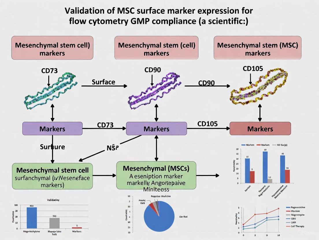
In the field of regenerative medicine, the identification and characterization of Mesenchymal Stromal Cells (MSCs) rely on a well-established set of surface markers as defined by the International Society for Cell & Gene Therapy (ISCT). According to these criteria, ≥95% of the MSC population must express the positive markers CD73, CD90, and CD105 while lacking expression of hematopoietic markers (CD45, CD34, CD14 or CD11b, CD79a or CD19, and HLA-DR) [10] [11]. This triad of surface proteins forms the cornerstone for defining MSCs in both research and clinical applications, providing a foundation for comparing MSC populations from diverse tissue sources and ensuring product consistency in Good Manufacturing Practice (GMP)-compliant production [12] [11]. CD105 (endoglin) is a type I membrane glycoprotein essential for cell migration and angiogenesis. CD90 (Thy-1) is an N-glycosylated glycosylphosphatidylinositol-anchored protein that mediates cell–cell and cell–extracellular matrix interactions. CD73 functions as a 5'-exonuclease, catalyzing the hydrolysis of adenosine monophosphate to adenosine and inorganic phosphate, playing a role in cell signaling within the bone marrow and modulating cellular interactions [10].
While the expression of CD73, CD90, and CD105 is a universal requirement for defining MSCs, their expression levels can vary depending on the tissue source, culture conditions, and specific donor characteristics. The following table summarizes quantitative expression data for these markers from various experimental studies.
Table 1: Quantitative Expression of Essential MSC Markers Across Different Sources and Conditions
| MSC Source / Experimental Condition | CD73 Expression | CD90 Expression | CD105 Expression | Citation |
|---|---|---|---|---|
| Bone Marrow-Derived MSCs (BM-MSCs) | >95% (Flow Cytometry) | >95% (Flow Cytometry) | >95% (Flow Cytometry) | [10] [11] |
| Umbilical Cord Tissue (UCT) MSCs (Fresh) | 0.09±0.07-fold (mRNA) | 0.17±0.11-fold (mRNA) | 0.04±0.06-fold (mRNA) | [13] |
| Adipose-Derived MSCs (AMSCs) in hPL | >95% (Flow Cytometry) | >95% (Flow Cytometry) | >95% (Flow Cytometry) | [3] |
| Infrapatellar Fat Pad (FPMSCs) in GMP Media | >95% (Flow Cytometry) | >95% (Flow Cytometry) | >95% (Flow Cytometry) | [8] |
| Periosteum & Cartilage Cultures | >95% (Flow Cytometry) | >95% (Flow Cytometry) | Not Specified | [2] |
The data confirms that adherence to the ISCT criteria is consistently achieved for in vitro-expanded MSCs from major somatic sources like bone marrow and adipose tissue when cultured under standard conditions [3] [11]. It is crucial to note, however, that the expression of these markers is often acquired and stabilized during in vitro culture, and may not accurately reflect the phenotype of native, uncultured progenitor cells in their tissue of origin [2].
Performance in Non-Human Species
The panel of CD73, CD90, and CD105, defined using human-specific antibodies, does not always directly translate to other species commonly used in preclinical research. A comparative study highlighted significant differences in marker expression across species. While human and mouse BM-MSCs were positive for CD90 and CD105, ovine and caprine BM-MSCs showed low or negative expression for these same markers, despite their capacity for tri-lineage differentiation [14]. This finding underscores the necessity of validating antibody cross-reactivity and establishing species-specific panels for preclinical animal studies.
Experimental Protocols for Marker Validation
Standard Flow Cytometry Protocol for MSC Phenotyping
Flow cytometry is the gold-standard technique for quantifying the expression of CD73, CD90, and CD105 on MSCs. The following methodology is widely used in GMP-compliant settings [8] [11].
Key Materials:
- Antibody Panel: Fluorescently conjugated monoclonal antibodies against CD73, CD90, CD105, and a cocktail of negative markers (CD45, CD34, CD11b, CD19, HLA-DR).
- Staining Buffer: Phosphate-buffered saline (PBS) supplemented with 1-2% fetal bovine serum (FBS).
- Cell Dissociation Reagent: Trypsin-EDTA or a GMP-compliant alternative like TrypLE.
- Flow Cytometer: Calibrated instrument capable of detecting the chosen fluorochromes.
Step-by-Step Workflow:
- Cell Harvesting: Culture MSCs to 70-80% confluence. Wash with PBS and harvest using a suitable cell dissociation reagent [8] [11].
- Cell Washing: Pellet the cells by centrifugation (e.g., 300 × g for 5 min) and resuspend in staining buffer. Pass the cell suspension through a 35-70 μm nylon mesh filter to remove clumps [13].
- Antibody Staining: Dispense cell aliquots (approximately 1x10^5 cells per tube). Add pre-titrated antibodies to the cell pellets. Incubate for 20-30 minutes at room temperature, shielded from light [13] [8].
- Washing and Resuspension: Wash the cells twice with staining buffer to remove unbound antibody. Finally, resuspend the stained cells in a fixed volume of buffer (e.g., 500 μL) for analysis.
- Flow Cytometric Analysis: Acquire data on the flow cytometer, collecting a minimum of 20,000 events per sample. Use unstained and single-stained controls for compensation and gating strategy setup [13].
- Data Interpretation: The MSC population is defined as being ≥95% positive for CD73, CD90, and CD105, and ≤2% positive for the hematopoietic lineage markers [10] [11].
Diagram 1: Flow cytometry workflow for MSC surface marker validation.
Alternative Detection Methods
While flow cytometry is the standard, other techniques can be employed to detect these markers.
- RT-PCR: Can be used to screen for the mRNA expression of CD73, CD90, and CD105. This method is particularly useful for analyzing cryopreserved tissue samples prior to MSC expansion, as demonstrated in umbilical cord tissue studies [13].
- Western Blotting: Confirms the presence of the protein itself and can be used to validate flow cytometry results [13].
- Extracellular Vesicle (EV) Characterization: CD73, CD90, and CD105 have also been identified on the surface of extracellular vesicles released by MSCs. Their presence, detectable by flow cytometry of purified EVs, can serve as a signature to distinguish MSC-derived vesicles from those of other origins [15].
The Scientist's Toolkit: Essential Research Reagents
Successful and reproducible characterization of MSC markers depends on using defined reagents and protocols. The table below lists key materials and their functions for standard flow cytometry analysis.
Table 2: Key Research Reagents for MSC Surface Marker Analysis
| Reagent / Material | Function / Description | GMP-Compliant Example / Note |
|---|---|---|
| CD73, CD90, CD105 Antibodies | Fluorescently-conjugated monoclonal antibodies for direct staining and flow cytometric detection. | BD Stemflow Human MSC Analysis Kit [8] |
| Human Platelet Lysate (hPL) | Animal-free supplement for MSC culture media, reducing risks of xenogeneic contamination. | Used as a 5-10% supplement to replace FBS [3] [11] |
| TrypLE / Trypsin-EDTA | Enzyme solution for detaching adherent MSCs from culture plastic prior to analysis. | GMP-grade, animal-component-free versions available [8] [11] |
| Defined Culture Media (e.g., MSC-Brew) | Animal component-free, xeno-free media formulations for clinical-grade MSC expansion. | Supports enhanced proliferation and maintains marker expression [8] |
| Flow Cytometer | Instrument for quantifying fluorescence intensity of cell surface markers on individual cells. | Standard clinical or research instruments (e.g., BD FACS Fortessa) [8] |
The triad of CD73, CD90, and CD105 remains the essential and non-negotiable panel for defining Mesenchymal Stromal Cells according to international standards. As demonstrated across multiple studies and tissue sources, consistent high expression (≥95%) of these markers is a hallmark of in vitro-expanded MSCs. The robustness of this panel is evidenced by its application in GMP-compliant production pipelines, from bone marrow and adipose tissue to umbilical cord-derived cells. For researchers and drug development professionals, rigorous validation of these markers using standardized flow cytometry protocols is not merely a regulatory checkbox but a critical step in ensuring the identity, purity, and quality of MSC-based products, thereby laying a reliable foundation for their therapeutic application in regenerative medicine.
In the field of regenerative medicine, Mesenchymal Stromal Cells (MSCs) represent a highly promising therapeutic tool for a wide range of clinical applications, from autoimmune diseases to orthopedic injuries [10]. The translation of MSC-based therapies from research to clinical application, however, necessitates rigorous characterization and quality control to ensure product safety and efficacy. Central to this characterization is the accurate assessment of cell surface markers, which serves as a fundamental release criterion for clinical-grade MSCs produced under Good Manufacturing Practice (GMP) guidelines [11]. While positive markers like CD73, CD90, and CD105 confirm mesenchymal lineage, the critical negative markers—CD34, CD45, and HLA-DR—play an equally vital role in excluding hematopoietic contamination [10] [3]. This function is paramount, as hematopoietic contaminants can alter the biological properties of MSC products, potentially triggering adverse immune reactions or confounding therapeutic outcomes [11].
The International Society for Cell and Gene Therapy (ISCT) has established minimal criteria for defining MSCs, including the absence of hematopoietic and endothelial markers. According to these standards, MSCs must lack expression of CD45 (a pan-leukocyte marker), CD34 (a marker for hematopoietic stem cells and endothelial cells), and HLA-DR (a major histocompatibility complex class II molecule with strong immunogenic properties) [10]. Furthermore, the absence of CD14, CD11b, CD79α, and CD19 is also required to exclude contamination by monocytes, macrophages, and B cells [10]. Adherence to these criteria is not merely academic; it is a foundational requirement for GMP-compliant production, ensuring that MSC products are phenotypically homogeneous, reproducible, and safe for human administration [11] [16]. This guide provides a detailed comparison of these critical negative markers, supported by experimental data and standardized protocols, to aid researchers in validating MSC purity and excluding hematopoietic contamination effectively.
Marker-Specific Profiles and Experimental Detection
CD45: The Pan-Hematopoietic Lineage Marker
CD45 (also known as leukocyte common antigen) is a protein tyrosine phosphatase expressed on all nucleated cells of hematopoietic origin, including lymphocytes, monocytes, and granulocytes [10]. Its absence on MSCs is a primary indicator that the cell population is free from hematopoietic lineage cells [3]. In flow cytometric analysis, CD45 is typically used in a "dump channel" to exclude all hematopoietic events from further analysis, thereby enriching for pure MSC populations [17]. Detection protocols recommend using anti-human CD45 antibodies conjugated to bright fluorophores (e.g., FITC or PE) and including fluorescence-minus-one (FMO) controls to accurately set negative-positive boundaries, as even weak expression can indicate significant contamination [18].
CD34: Hematopoietic Stem and Progenitor Cell Marker
CD34 is a cell surface glycoprotein traditionally used to identify and isolate hematopoietic stem and progenitor cells (HSPCs) and endothelial cells [10]. Its absence is a key criterion for distinguishing MSCs from HSPCs [3]. However, interpretation requires nuance. While bone marrow-derived MSCs (BM-MSCs) are consistently CD34-negative, some adipose-derived MSC (AD-MSC) populations, particularly in early passages or when cultured in human platelet lysate (hPL), may show transient CD34 expression [3]. This underscores the importance of context and source material when applying this negative marker. For reliable quantification of blast cells in myelodysplastic syndromes, research indicates that relying solely on CD34 positivity is insufficient; a combination with other markers like CD117 and HLA-DR provides more accurate blast identification [19].
HLA-DR: Immunogenicity and Activation Marker
HLA-DR is a Major Histocompatibility Complex (MHC) Class II molecule constitutively expressed on antigen-presenting cells like B lymphocytes, monocytes, macrophages, and dendritic cells [10]. Its absence on MSCs under standard culture conditions is critical as it confers low immunogenicity, enabling allogeneic transplantation [10]. It is important to note that HLA-DR expression can be induced in MSCs by inflammatory stimuli such as interferon-gamma (IFN-γ) [10]. Therefore, its presence may indicate an activated state rather than pure hematopoietic contamination, necessitating careful experimental interpretation. In clinical settings, the absence of HLA-DR on the final product is a key safety specification to prevent immunogenic reactions in recipients [11].
Table 1: Critical Negative Markers for MSC Characterization
| Marker | Full Name | Primary Cellular Expression | Significance of Absence in MSCs |
|---|---|---|---|
| CD45 | Leukocyte Common Antigen | All nucleated hematopoietic cells (lymphocytes, monocytes, granulocytes) | Excludes contamination by cells of the entire hematopoietic lineage [10] |
| CD34 | Hematopoietic Progenitor Cell Antigen | Hematopoietic stem/progenitor cells, endothelial cells [10] | Distinguishes MSCs from hematopoietic stem cells and endothelial cells [3] |
| HLA-DR | Human Leukocyte Antigen - DR isotype | B cells, monocytes, macrophages, dendritic cells, activated T/NK cells [10] | Indicates low immunogenic potential, suitable for allogeneic use [10] |
| CD14/CD11b | - | Monocytes and Macrophages [10] | Excludes monocytic and macrophage contamination |
| CD19/CD79α | - | B lymphocytes [10] | Excludes B cell contamination |
Comparative Data and GMP Compliance
The expression of negative markers is consistently low across MSCs derived from different tissues when the cells are manufactured under defined conditions. A study on clinical-grade adipose-derived MSCs (AMSCs) expanded in GMP-grade human platelet lysate (hPL) demonstrated a homogenous cell population with minimal expression of hematopoietic markers [3]. The data confirmed high expression of positive markers (CD90, CD73, CD105, CD44 ≥ 95%) and minimal expression of negative markers, including CD45 and CD34, meeting the ISCT release criteria [3]. Similarly, in GMP-compliant production of bone marrow-derived MSCs (BM-MSCs) using platelet lysate, the absence of CD14, CD34, and CD45 is a standard quality control checkpoint before product release [11].
Table 2: Typical Marker Expression Profile of Clinical-Grade MSCs
| Marker Type | Marker | Required Expression (ISCT) | Typical Profile in BM-MSCs [11] | Typical Profile in AMSCs [3] |
|---|---|---|---|---|
| Positive | CD73, CD90, CD105 | ≥ 95% Positive | ≥ 95% Positive | ≥ 95% Positive |
| Negative | CD45 | ≤ 2% Positive | ≤ 2% Positive | ≤ 2% Positive |
| Negative | CD34 | ≤ 2% Positive | ≤ 2% Positive | ≤ 2% Positive (Note: Can be variable in early passage AMSCs) |
| Negative | HLA-DR | ≤ 2% Positive | ≤ 2% Positive | ≤ 2% Positive |
| Negative | CD14/CD11b, CD19/CD79α | ≤ 2% Positive | ≤ 2% Positive | ≤ 2% Positive |
The Role of Negative Markers in GMP Compliance
In GMP-compliant production, tracking negative markers is not a one-time check but an integral part of the entire manufacturing process, from isolation to final product release [11] [16]. Regulatory bodies like the FDA and EMA classify MSCs as Advanced Therapy Medicinal Products, mandating that their production ensures reproducibility, efficacy, and safety [11]. The use of defined, animal component-free media supplements like platelet lysate instead of fetal bovine serum (FBS) reduces the risk of introducing xenogeneic contaminants and improves the consistency of marker expression profiles [11] [8]. Furthermore, the stability of the marker profile, including the absence of hematopoietic contaminants, must be validated post-cryopreservation and throughout the product's shelf-life to ensure that the critical quality attributes are maintained until the point of administration [16].
Diagram 1: GMP Validation Workflow for MSC Negative Markers
This workflow illustrates the critical path for validating the absence of hematopoietic contamination in clinical-grade MSC products, highlighting the essential role of flow cytometry and specific gating strategies.
Essential Protocols for Flow Cytometric Detection
Sample Preparation and Staining Protocol
A robust staining protocol is foundational for accurate detection of negative markers. The following methodology is adapted from established GMP-compliant workflows [11] [16]:
- Cell Harvesting: Harvest MSCs at 70-80% confluency using a non-enzymatic dissociation agent or a GMP-compliant enzyme like TRYPZEAN. Wash cells twice in PBS without Ca2+/Mg2+.
- Cell Counting and Viability Assessment: Count cells using an automated cell counter or hemacytometer. Assess viability via Trypan Blue exclusion, ensuring viability exceeds >95% for high-quality analysis [16].
- Fc Receptor Blocking: Resuspend up to 1x10^6 cells in 100 µL of flow cytometry buffer (PBS with 1% BSA or FBS). Add an FcR blocking reagent (e.g., human IgG) and incubate for 10-15 minutes at 2-8°C to reduce non-specific antibody binding [18].
- Antibody Staining: Add fluorochrome-conjugated antibodies against CD45, CD34, HLA-DR, and positive markers (CD73, CD90, CD105) at pre-titrated concentrations. Include viability dyes (e.g., 7-AAD or DRAQ7) to exclude dead cells. Vortex gently and incubate for 30 minutes at 2-8°C in the dark.
- Washing and Fixation: Wash cells twice with flow cytometry buffer to remove unbound antibody. Resuspend the final cell pellet in 200-300 µL of buffer, optionally adding a low concentration of paraformaldehyde (e.g., 1%) for fixation if not analyzing immediately.
- Data Acquisition: Acquire data on a flow cytometer calibrated with appropriate compensation controls. Acquire a minimum of 10,000 events in the live, singlet cell gate for reliable analysis of populations present at low frequency [17].
Gating Strategy and Controls for Rare-Event Detection
Precise gating is critical for identifying small levels of contamination, which can be considered "rare events" [17].
- Singlets and Viability: First, gate on cells based on forward scatter area (FSC-A) versus height (FSC-H) to exclude cell doublets and aggregates. Next, gate on viable cells that are negative for the viability dye [18].
- The "Dump" Channel for Negative Selection: A powerful strategy for excluding hematopoietic contaminants is to use a "dump channel." Combine antibodies for lineage-negative markers (CD45, CD34, HLA-DR) conjugated to the same fluorochrome. Cells that are positive in this channel are excluded from further analysis as contaminants [17].
- Critical Controls:
- Fluorescence Minus One Controls: FMO controls are essential for accurately setting the boundaries between positive and negative populations, especially for markers with low-level expression or high background [18].
- Isotype Controls: Use matched isotype controls to account for non-specific antibody binding, though these should not be used for setting positivity gates [18].
- Compensation Controls: Use compensation beads or single-stained cell samples to correct for spectral spillover between fluorochrome channels in multicolor panels [18].
Diagram 2: Flow Cytometry Gating Hierarchy
This sequential gating strategy is crucial for isolating a pure MSC population, effectively excluding debris, doublets, dead cells, and hematopoietic contaminants before finally confirming positivity for standard MSC markers.
The Scientist's Toolkit: Essential Research Reagents
Table 3: Key Reagent Solutions for Negative Marker Analysis
| Reagent / Material | Function / Application | GMP-Compliant / Clinical-Grade Examples |
|---|---|---|
| Animal-Free Culture Media | Provides a defined, xeno-free environment for MSC expansion, minimizing batch variability and safety risks. | MSC-Brew GMP Medium [8], MesenCult-ACF Plus Medium [16] |
| Human Platelet Lysate (hPL) | A human-derived serum alternative for GMP-compliant cell culture, reducing immunogenicity risks compared to FBS. | Produced under GMP standards [11] |
| Flow Cytometry Antibody Panels | Multiplexed antibody kits for simultaneous detection of positive and negative MSC markers. | BD Stemflow Human MSC Analysis Kit [16] |
| Viability Dyes | Distinguishes live from dead cells during flow analysis to prevent false positives from non-specifically staining dead cells. | 7-AAD, DRAQ7 [18], Propidium Iodide [18] |
| FcR Blocking Reagent | Blocks non-specific binding of antibodies to Fc receptors on immune cells, reducing background signal. | Human IgG, commercial FcR blocking buffers [18] |
| Compensation Beads | Synthetic beads used to set accurate compensation for spectral overlap in multicolor flow panels. | Commercial anti-mouse/anti-rat Ig compensation beads [18] |
The rigorous assessment of CD45, CD34, and HLA-DR as critical negative markers is a non-negotiable standard in the characterization of clinically relevant MSCs. Their absence reliably excludes hematopoietic and endothelial contamination, ensuring the phenotypic purity and safety of the cellular product. As the field progresses towards more complex therapeutic applications, adherence to these defined criteria, combined with GMP-compliant manufacturing and robust flow cytometric protocols, will be instrumental in translating the promise of MSC therapies into safe and effective clinical realities. Future directions will likely involve the identification and validation of additional non-classical markers that can provide deeper insights into MSC potency and functional heterogeneity beyond basic purity [3].
Why GMP? Ensuring Patient Safety and Product Consistency in Clinical Trials
In the development of advanced therapies like those based on mesenchymal stem cells (MSCs), Good Manufacturing Practice (GMP) provides the essential framework that bridges laboratory research with clinical application. GMP represents a system of quality assurance that ensures products are consistently produced and controlled according to quality standards appropriate for their intended use [20]. In the context of clinical trials, where investigational products are administered to human subjects for the first time, GMP compliance is not merely a regulatory hurdle but a fundamental ethical obligation to protect patient safety and ensure product integrity. Without the rigorous controls mandated by GMP, therapies entering clinical trials could vary unpredictably between batches, potentially compromising patient safety and clinical trial outcomes.
The transition from research-grade materials to clinically administrable products demands a paradigm shift from simple protocol adherence to comprehensive quality systems. This is particularly critical for cell therapies like MSCs, where product quality and functionality are intrinsically linked to complex manufacturing processes. This guide examines how GMP implementation directly safeguards patients and ensures reliable outcomes in clinical research, with specific focus on validating MSC surface marker expression through flow cytometry.
GMP vs. Non-GMP: A Comparative Framework for Clinical Research
Core Principles and Regulatory Implications
The table below contrasts key aspects of GMP and non-GMP approaches, highlighting why GMP is indispensable for clinical trials:
| Aspect | GMP Environment (Clinical Trials) | Non-GMP Environment (Research Only) |
|---|---|---|
| Primary Objective | Patient safety and product consistency [21] | Data generation and discovery |
| Quality Focus | Quality built into every manufacturing step [21] | Final result often prioritized over process |
| Documentation | Comprehensive, traceable records for all processes [20] [22] | Sufficient for personal or publication reference |
| Facility & Equipment | Validated, calibrated, and maintained under strict controls [21] | Maintenance often reactionary; calibration irregular |
| Personnel | Formal training and qualification records required [22] | Skill-based with variable documentation |
| Raw Materials | Rigorously qualified with traceable lineage [21] | Often selected for cost or convenience |
| Process Controls | Validated, monitored, and controlled at each step [21] | Frequently adapted without formal validation |
| Product Testing | Required lot-to-lot testing with predefined specifications [20] | Occasional testing as needed for experiments |
The "C" in cGMP: Embracing Current Standards
Regulatory bodies emphasize Current Good Manufacturing Practices (cGMP), where the "current" underscores the necessity of employing up-to-date technologies and systems [21] [22]. What was considered adequate a decade ago may be insufficient today. This evolutionary aspect of cGMP requires researchers to continuously evaluate and improve their manufacturing and testing approaches, particularly when preparing products for human administration in clinical trials.
GMP Implementation: Flow Cytometry Validation for MSC Surface Markers
The Critical Role of Flow Cytometry in Cell Therapy
Flow cytometry serves as a cornerstone analytical technology throughout the cell therapy development pipeline, providing essential data on product characterization, purity, potency, and safety [23]. In MSC-based therapies, validated flow cytometry assays are particularly crucial for confirming identity through surface marker expression (e.g., CD73, CD90, CD105) and verifying the absence of undesirable cell populations.
Key Considerations for GMP-Compliant Flow Cytometry
Implementing flow cytometry in a GMP environment presents unique challenges that extend beyond analytical performance:
- Instrument Qualification: GMP-compliant flow cytometers require rigorous qualification (installation, operational, and performance qualification) and regular calibration to ensure data reliability [24] [25].
- Standardized Protocols: The inherent variability of flow cytometry due to instrument differences, antibody clones, and protocol implementation must be controlled through standardized procedures [24].
- Reagent Control: All antibodies and staining reagents must be qualified for their intended use, with particular attention to lot-to-lot consistency [23].
- Operator Training: Technical staff must receive comprehensive training on validated methods, with performance documented [22].
- Data Integrity: Complete documentation of all procedures, results, and interpretations must be maintained according to GMP principles [20].
The following workflow diagram illustrates the comprehensive validation process for GMP-compliant flow cytometry assays:
Experimental Protocol: Validation of MSC Surface Marker Staining
For GMP-compliant characterization of MSC surface markers, the following validation protocol ensures reliable results:
1. Panel Design and Antibody Qualification
- Select antibodies against positive markers (CD73, CD90, CD105) and negative markers (CD34, CD45, HLA-DR) as per ISCT standards [23].
- Perform antibody titration using reference MSC samples to determine optimal staining concentration.
- Document antibody source, clone, lot number, and expiration date.
2. Sample Preparation and Staining Validation
- Establish acceptance criteria for cell viability (>90% recommended pre-fixation).
- Validate staining volume, incubation time (20-30 minutes at 4°C), and wash conditions.
- Include appropriate controls: unstained, fluorescence minus one (FMO), and isotype controls.
3. Instrument Setup and Standardization
- Implement daily quality control using calibration beads to ensure instrument performance.
- Define photomultiplier tube (PMT) voltages and compensation settings in standardized templates.
- For multi-site trials, harmonize instrument settings across all flow cytometers to ensure comparable results [24].
4. Analysis and Gating Strategy
- Establish standardized gating hierarchy using forward scatter/side scatter to exclude debris.
- Define positive populations based on FMO controls with set positivity thresholds.
- Implement manual or automated analysis protocols to minimize operator-induced variability.
Essential Research Reagent Solutions for GMP-Compliant Flow Cytometry
The table below details key reagents and materials required for implementing GMP-compliant flow cytometry, with quality considerations for clinical trial applications:
| Reagent/Material | Function in GMP Environment | Quality Requirements |
|---|---|---|
| Fluorochrome-Conjugated Antibodies | Detection of MSC surface markers and impurities | Qualified lots with Certificate of Analysis; purity verification [23] |
| Viability Stains | Distinguish live/dead cells for accurate phenotyping | Validated for consistency; minimal spectral overlap with panel [23] |
| Cell Staining Buffers | Maintain cell viability and enable specific binding | Formulation consistency; endotoxin testing; sterile filtration |
| Calibration Beads | Daily instrument performance verification | Traceable standards; stable fluorescence intensity [24] |
| Compensation Beads | Calculate spectral overlap between channels | Consistent binding capacity; low background fluorescence |
| Reference MSC Cells | Assay performance monitoring and qualification | Well-characterized bank; stable marker expression |
| Sample Tubes | Hold samples during acquisition | Certified sterile; low cellular adhesion |
Building Quality into the Manufacturing Process
The fundamental principle of GMP is that quality cannot be tested into a product but must be built into every manufacturing step [21]. This proactive approach is particularly critical for cellular therapies, where the manufacturing process defines the product characteristics. While comprehensive testing of final products is essential, relying solely on end-product testing is insufficient for several reasons:
- Sample Limitations: Testing often uses small samples from a batch (e.g., 100 tablets from 2 million), making it statistically impossible to test every unit [21].
- Destructive Testing: Many quality tests consume the product, limiting the amount available for patients.
- Process Understanding: GMP emphasizes process control and monitoring to prevent errors rather than detecting them after they occur.
The following diagram illustrates how GMP systems integrate to ensure product quality and patient safety throughout the therapy development lifecycle:
Implementation of GMP principles represents a fundamental requirement rather than an optional enhancement for clinical trials. The rigorous framework of controls, documentation, and validation ensures that investigational therapies are manufactured consistently, contain the intended components in the specified amounts, and maintain acceptable purity profiles. For MSC-based therapies and other advanced medicinal products, this translates to reliable characterization through validated methods like flow cytometry, enabling researchers to make confident decisions about product safety and efficacy.
The transition to GMP compliance requires significant investment in quality systems, personnel training, and process controls. However, this investment is essential not only for regulatory approval but also for generating clinically meaningful data and, most importantly, for protecting the patients who volunteer to participate in clinical trials. By building quality into every aspect of product development and manufacturing, researchers honor their ethical obligation to prioritize patient welfare while advancing scientific knowledge.
Mesenchymal Stromal Cells (MSCs) represent a cornerstone of regenerative medicine and therapeutic development due to their multipotent differentiation potential, immunomodulatory properties, and capacity for tissue repair [26]. The International Society for Cell Therapy (ISCT) has established minimal criteria for defining MSCs, including plastic adherence, specific surface marker expression, and in vitro trilineage differentiation potential [26] [12]. However, MSCs isolated from different tissue sources exhibit significant biological variations that impact their marker expression profiles and functional capabilities [27] [28].
This comparative guide objectively analyzes MSC surface marker expression across three prominent sources: bone marrow (BM-MSCs), adipose tissue (A-MSCs), and umbilical cord (UC-MSCs). Understanding these distinctions is crucial for researchers and drug development professionals working under Good Manufacturing Practice (GMP) standards to select appropriate cell sources for specific therapeutic applications, establish robust quality control measures, and ensure reproducible research and clinical outcomes.
The ISCT minimal criteria specify that MSCs must express CD73, CD90, and CD105 (≥95% positive), while lacking expression of hematopoietic markers CD34, CD45, CD14/CD11b, CD79α/CD19, and HLA-DR (≤2% positive) [26]. While MSCs from all major sources generally meet these criteria, research reveals important quantitative and qualitative differences in their marker expression profiles.
Table 1: Comparative Surface Marker Expression Across MSC Sources
| Surface Marker | Bone Marrow MSCs | Adipose Tissue MSCs | Umbilical Cord MSCs | Biological Function |
|---|---|---|---|---|
| CD73 | Positive [28] | Positive [28] | Positive [28] | 5'-nucleotidase, ectoenzyme |
| CD90 | Positive [28] | Positive [28] | Positive [28] | Thy-1, cell adhesion |
| CD105 | Positive [28] | Positive [28] | Positive [28] | Endoglin, TGF-β receptor |
| CD44 | Positive [27] | Positive [27] | Positive [27] | Hyaluronic acid receptor |
| CD34 | Negative [28] | Early passage positive [26] | Negative [28] | Hematopoietic progenitor cell marker |
| CD45 | Negative [28] | Negative [28] | Negative [28] | Pan-leukocyte marker |
| CD14 | Negative [28] | Negative [28] | Negative [28] | Monocyte/macrophage marker |
| HLA-DR | Generally negative [26] | Generally negative [26] | Generally negative [26] | MHC Class II |
| CD106 (VCAM-1) | Highly expressed [27] | Low/absent [27] | Variable [27] | Cell adhesion, homing |
| CD271 | Low/Variable | Low/Variable | Expressed [29] | Nerve growth factor receptor |
| CD200 | Variable | Variable | Expressed [29] | Immunoregulatory function |
A comprehensive 2015 study comparing molecular profiles of MSCs from different tissues confirmed that while BM-MSCs and A-MSCs share similar gene expression profiles, UC-MSCs exhibit distinct characteristics [27]. CD106 (VCAM-1) expression is notably higher in BM-MSCs compared to other sources, potentially influencing their hematopoietic support capabilities [27]. The CD34 status remains controversial, particularly for A-MSCs, which may express this marker initially but lose it during culture expansion [26].
Non-Classical and Functional Marker Variations
Beyond the classical ISCT markers, several non-classical markers exhibit source-dependent expression patterns that may influence MSC functionality for specific applications.
Table 2: Non-Classical and Functional Marker Expression
| Marker Category | Specific Marker | Bone Marrow MSCs | Adipose Tissue MSCs | Umbilical Cord MSCs | Functional Significance |
|---|---|---|---|---|---|
| Immunomodulatory | CD274 (PD-L1) | Variable | Present [30] | Variable | T-cell inhibition |
| Perivascular | CD146 | Variable | Present [30] | Variable | Pericyte marker, migration |
| Matrix-Related | CD248 | Variable | Present [30] | Variable | Extracellular matrix interaction |
| Growth Factor Receptor | CD140B (PDGFR-β) | Variable | Present [30] | Variable | Proliferation, migration |
| Stemness | SSEA-4 | Variable | Variable | Highly expressed [29] | Pluripotency association |
Research indicates that A-MSCs express several non-classical markers (CD36, CD163, CD271, CD200, CD273, CD274, CD146, CD248, and CD140B) that may distinguish them from other MSC types and provide information about their functional state [30]. UC-MSCs often express markers associated with a more primitive state, including SSEA-4, reflecting their perinatal origin [29]. These variations in marker profiles correspond to functional differences in immunomodulation, differentiation potential, and trophic factor secretion.
Functional Differences and Differentiation Potential
Marker expression patterns directly correlate with functional capabilities across MSC sources. Research demonstrates that the tissue of origin significantly influences the differential potential and immunomodulatory strength of MSCs.
Table 3: Functional Characteristics Across MSC Sources
| Functional Attribute | Bone Marrow MSCs | Adipose Tissue MSCs | Umbilical Cord MSCs |
|---|---|---|---|
| Osteogenic Potential | High [27] | High [27] | Moderate [27] |
| Adipogenic Potential | High [27] | High [27] | Moderate [27] |
| Chondrogenic Potential | High | High | High |
| Neural Differentiation | Moderate [31] | Moderate [31] | High [31] |
| Proliferation Capacity | Moderate [31] | Moderate [31] | High [31] |
| T-cell Inhibition | Strong [27] [28] | Strong [27] [28] | Moderate [28] |
| B-cell Inhibition | Present [28] | Present [28] | Absent/Weak [28] |
| NK-cell Inhibition | Present [28] | Strong [28] | Moderate [28] |
A critical 2013 study revealed that while MSCs from all three sources inhibit CD4+ and CD8+ T-cell activation, A-MSCs demonstrated a stronger inhibitory effect and blocked T-cell activation earlier than other sources [28]. Furthermore, UC-MSCs showed no inhibitory effect on B-cell activation under experimental conditions, unlike BM-MSCs and A-MSCs [28]. For neural differentiation applications, UC-MSCs exhibited superior potential, showing higher expression of early neural markers Nestin and PAX6 compared to other sources [31].
Experimental Protocols for Marker Validation
Standardized Flow Cytometry for MSC Validation
Flow cytometry represents the gold standard for validating MSC surface marker expression according to ISCT guidelines and GMP requirements. The following protocol ensures reproducible results across different MSC sources:
Protocol Steps:
- Cell Preparation: Harvest MSCs at 70-80% confluence (passage 3-5) using non-enzymatic dissociation agents or mild trypsinization to preserve surface epitopes [27] [12].
- Antibody Staining: Resuspend 1×10⁶ cells in FACS buffer (PBS with 1% FBS). Incubate with fluorochrome-conjugated antibodies against target markers (CD73, CD90, CD105, CD34, CD45, CD14, HLA-DR) and appropriate isotype controls for 30 minutes at 4°C in the dark [27] [26].
- Analysis: Wash cells, resuspend in fixation buffer (1% paraformaldehyde), and analyze using a flow cytometer (e.g., Beckman Coulter Cytomics) [27]. Collect a minimum of 10,000 events per sample.
- Gating Strategy: Exclude debris based on forward and side scatter, then analyze marker expression on the viable cell population. Use isotype controls to establish positive/negative thresholds [26].
- Interpretation: ≥95% of cells must express CD73, CD90, and CD105; ≤2% may express hematopoietic markers [26].
Trilineage Differentiation Assessment
Confirming multilineage differentiation potential remains essential for MSC validation alongside surface marker expression:
Osteogenic Differentiation:
- Culture MSCs in commercial osteogenic differentiation media (e.g., StemPro Osteogenesis Kit) for 21 days [31].
- Fix cells with 4% paraformaldehyde and stain with 2% Alizarin Red S (pH 4.1-4.3) to detect calcium deposits [31] [32].
- Quantify mineralization through spectrophotometric analysis of extracted dye or image analysis.
Adipogenic Differentiation:
- Induce adipogenesis using specialized media (e.g., StemPro Adipogenesis Kit) for 14-21 days [31].
- Fix cells and stain lipid vacuoles with filtered Oil Red O working solution (0.5% in isopropanol) [31] [32].
- Quantify lipid accumulation through extracted dye measurement at 510nm.
Chondrogenic Differentiation:
- Pellet 2.5×10⁵ MSCs by centrifugation and culture in chondrogenic induction media for 21-28 days.
- Assess chondrogenesis through histological staining of pellet sections with Toluidine Blue or Alcian Blue for proteoglycan detection.
Signaling Pathways and Molecular Mechanisms
The differential marker expression across MSC sources originates from distinct molecular signatures and signaling pathway activities. Microarray analyses reveal that MSCs from different ontogenetic sources exhibit differentially expressed genes involved in extracellular matrix organization, morphogenesis, and development [33].
Pathway Regulation of MSC Characteristics
Key pathway differences include Wnt signaling inhibitors (DKK1, DKK3, SFRP1) being highly expressed in fibroblasts compared to MSCs [33]. UC-MSCs exhibit distinct HOX gene expression profiles (HOXA5, HOXB6) [33], while A-MSCs demonstrate alterations in JAK2/STAT3 signaling during adipogenic differentiation [32]. These molecular differences underlie the functional variations observed across MSC sources and provide potential targets for quality control and potency assessment.
Research Reagent Solutions for MSC Characterization
Table 4: Essential Research Reagents for MSC Marker Validation
| Reagent Category | Specific Product Examples | Research Application | Considerations for Source-Specific Variations |
|---|---|---|---|
| Flow Cytometry Antibodies | Anti-human CD73, CD90, CD105, CD34, CD45, CD14 [27] [26] | ISCT phenotype validation | CD34 may require source-specific thresholds; CD106 useful for BM-MSC identification |
| Dissociation Reagents | TrypLE Select, Accutase [28] | Cell harvesting | Preserve surface epitopes; enzymatic sensitivity may vary by source |
| Culture Media | DMEM-low glucose with 10% FBS or 5% human platelet lysate [27] [30] | MSC expansion | Media composition affects marker expression; hPL enhances proliferation |
| Differentiation Kits | StemPro Osteogenesis/Adipogenesis Kits [31] | Multilineage potential assessment | Differentiation efficiency varies by MSC source |
| Matrix Proteins | Matrigel, gelatin [31] [32] | Specialized differentiation | Enhances neural differentiation of UC-MSCs |
| Analysis Kits | MTT assay kits [31] | Proliferation assessment | UC-MSCs typically show higher proliferative rates |
The selection of MSC source significantly impacts surface marker expression profiles and functional capabilities, with important implications for research and therapeutic applications. BM-MSCs demonstrate robust immunomodulatory properties and classical marker expression, making them suitable for immune-mediated applications. A-MSCs show strong adipogenic potential and consistent marker profiles with additional non-classical markers. UC-MSCs exhibit primitive marker expression, superior proliferation, and enhanced neural differentiation capability.
For researchers operating under GMP standards, these source-specific characteristics necessitate tailored quality control approaches. Flow cytometry validation should incorporate both classical ISCT markers and source-specific additional markers to ensure comprehensive characterization. The selection of optimal MSC source should align with the intended therapeutic application, considering the distinct functional advantages of each tissue origin. Standardized protocols across research institutions will enhance comparability and advance the field of MSC-based therapies.
From Protocol to Practice: Implementing GMP-Compliant Flow Cytometry for MSC Analysis
For researchers and drug development professionals advancing Mesenchymal Stromal Cell (MSC)-based therapies, rigorous characterization of surface marker expression is not merely a research exercise but a fundamental regulatory requirement. The International Society for Cell & Gene Therapy (ISCT) has established minimum criteria for defining MSCs, including specific surface marker expression (≥95% positive for CD105, CD73, and CD90, and ≤2% positive for CD45, CD34, and HLA-DR) [12]. Adherence to these standards requires workflows that are not only scientifically valid but also compliant with Good Manufacturing Practices (GMP) to ensure product safety, identity, and purity from cell harvest through final data acquisition.
The transition from research-grade flow cytometry to GMP-compliant processes introduces significant complexities, requiring carefully controlled and documented procedures at every stage. This guide objectively compares traditional, research-grade flow cytometry with emerging automated platforms, providing a detailed, step-by-step framework for implementing GMP-compliant workflows for MSC surface marker analysis. As the field progresses toward more automated systems, understanding both conventional and innovative approaches is essential for maintaining quality control throughout therapeutic development.
Comparative Platform Analysis: Traditional vs. Automated Flow Cytometry
The selection of an appropriate flow cytometry platform is a critical decision that impacts workflow efficiency, data reproducibility, and regulatory compliance. The following section provides a technical comparison of established methodologies.
Table 1: Comparison of Flow Cytometry Platforms for GMP-Compliant MSC Analysis
| Feature | Traditional Benchtop Flow Cytometry | Automated Platform (e.g., Accellix) |
|---|---|---|
| Sample Preparation | Largely manual, requiring trained technicians for staining and washing steps [23] | Automated within single-use, microfluidic cartridges with dried-down reagents [34] [35] |
| Process Time | Several hours, varying with protocol complexity and cell number | Approximately 30 minutes total assay running time [34] [35] |
| Reagent Handling | Liquid reagents requiring refrigeration and preparation [23] | Room-temperature stable, unitized dried-down reagents [35] |
| Key Advantage | High flexibility for panel customization and multiparameter analysis (up to 40 parameters) [23] | Standardized workflow that increases reproducibility and reduces manual handling error [34] |
| Data Analysis | Often requires separate, expert-driven software analysis [23] | Fully automated data analysis and auto-classification [34] |
| Ideal Use Context | Research and development, complex panel development | In-process and release testing in a GMP manufacturing suite [34] |
Workflow Visualization: Traditional vs. Automated Pathways
The fundamental differences between traditional and automated systems are best understood by visualizing their respective workflows, from cell preparation to data acquisition.
Detailed Experimental Protocols for GMP-Compliant MSC Characterization
This section outlines the specific, step-by-step laboratory procedures required for the accurate and reproducible phenotyping of MSCs, with a focus on GMP compliance.
GMP-Compliant MSC Culture and Harvest
The foundation of reliable flow cytometry data is a well-characterized and consistently handled cell source.
- Cell Source and Culture: MSCs can be isolated from various somatic tissues like bone marrow, adipose tissue, dental pulp, or perinatal tissues such as umbilical cord [12]. For GMP compliance, culture must use animal component-free, GMP-grade media such as MSC-Brew GMP Medium (Miltenyi Biotec). Studies demonstrate that MSCs cultured in MSC-Brew exhibit enhanced proliferation rates and maintained characteristic surface marker expression compared to standard media [8] [36].
- Cell Harvest and Washing:
- Wash adherent cells with phosphate-buffered saline (PBS).
- Detach cells using a GMP-compliant dissociation agent like TrypLE Select.
- Neutralize the enzyme with an appropriate volume of GMP-grade culture medium.
- Centrifuge the cell suspension at 300 ×g for 10 minutes and carefully decant the supernatant [8].
- Resuspend the cell pellet in PBS or a suitable staining buffer.
- Cell Counting and Viability Assessment:
- Perform cell counting using a hemacytometer or automated cell counter.
- Assess viability using Trypan Blue exclusion or similar methods. GMP release specifications often require >95% viability [8].
Staining and Acquisition for Surface Marker Expression
This core protocol details the process of labeling cells with fluorescent antibodies for analysis.
- Antibody Panel Configuration: Design panels based on ISCT criteria. A typical panel includes positive markers (CD105, CD73, CD90) and negative markers (CD45, CD34, HLA-DR) [12]. Kits like the BD Stemflow Human MSC Analysis Kit are commercially available for this purpose [8].
- Staining Protocol:
- Aliquot a cell suspension containing approximately 1 × 10^5 to 1 × 10^6 cells into a FACS tube.
- Centrifuge and decant the supernatant.
- Resuspend the cell pellet in a recommended volume of FACS buffer (PBS with 0.3% BSA).
- Add fluorochrome-conjugated antibodies according to manufacturer's instructions.
- Incubate for 30 minutes on ice, protected from light.
- Wash cells twice by adding 2-3 mL of FACS buffer, centrifuging at 300 ×g for 5-10 minutes, and decanting the supernatant.
- Finally, resuspend the fixed cells in a suitable volume (e.g., 200-500 µL) of FACS buffer or a fixation buffer for acquisition [8] [36].
- Instrument Acquisition and Quality Control:
- Use a flow cytometer (e.g., BD FACS Fortessa) calibrated with appropriate compensation beads to account for spectral overlap.
- Establish a gating strategy to first select cells based on forward and side scatter properties, then exclude dead cells, and finally analyze the fluorescence of the target markers [8] [23].
- Acquire a statistically significant number of events (e.g., 10,000-50,000 events in the live cell gate).
Essential Reagents for the GMP-Compliant Workflow
The quality and traceability of every reagent are critical in a GMP environment.
Table 2: Research Reagent Solutions for GMP MSC Flow Cytometry
| Reagent / Material | Function in Workflow | GMP-Compliant Example |
|---|---|---|
| Serum-Free / Xeno-Free Media | Supports expansion of MSCs without animal-derived components, reducing immunogenicity and batch variability. | MSC-Brew GMP Medium (Miltenyi) [8], MesenCult-ACF Plus Medium (StemCell) [8] |
| GMP-Grade Enzymes | Detaches adherent MSCs for harvesting and passaging while maintaining cell viability and phenotype. | TrypLE Select (Gibco) [36] |
| Fluorochrome-Conjugated Antibodies | Tags specific cell surface markers (CD105, CD73, CD90, etc.) for detection and quantification. | BD Stemflow Human MSC Analysis Kit (BD Biosciences) [8] |
| FACS Buffer | Provides a suitable medium for antibody dilution and cell washing steps while maintaining cell integrity. | PBS with 0.3% BSA and 0.1% NaN₃ (Sigma-Aldrich) [36] |
| Viability Stain | Distinguishes live cells from dead cells during flow cytometric analysis for accurate gating. | 7-AAD, Propidium Iodide, or Live/Dead Fixable Stains [23] |
Supporting Data and Validation for Clinical Translation
Robust experimental data is essential for validating both the MSC product and the analytical process itself.
Quantitative Performance of GMP-Grade Media
Validation studies consistently demonstrate that GMP-optimized media formulations not only support cell growth but also critically maintain the essential phenotypic identity of MSCs.
Table 3: Experimental Performance Data of MSCs in GMP-Grade Culture Conditions
| Culture Condition | Cell Doubling Time | Post-Thaw Viability | Key Phenotypic Findings |
|---|---|---|---|
| MSC-Brew GMP Medium | Lower doubling times across passages, indicating enhanced proliferation [8] | >95% viability maintained even after 180 days of storage [8] | Maintained expression of MSC markers (CD73, CD90, CD105) and absence of hematopoietic markers [8] [36] |
| Standard Media (DMEM + 10% FBS) | Higher doubling times compared to MSC-Brew [8] | Data not specifically highlighted in search results | Maintains marker expression but introduces risks of batch variability and immunogenicity from animal serum [8] |
Comprehensive Quality Control Testing Regimen
A successful GMP workflow for an MSC-based Investigational Medicinal Product (IMP) extends far beyond flow cytometry. It requires a battery of quality control tests to ensure safety and identity, forming a holistic release strategy [37].
The choice between a traditional benchtop flow cytometer and an automated, integrated system like Accellix hinges on the specific phase of therapeutic development and the operational priorities of the facility.
- For R&D and Complex Panel Development, traditional flow cytometry remains indispensable due to its flexibility and high-parameter capabilities [23].
- For GMP Manufacturing Suite Deployment, automated platforms offer a compelling advantage. They embed quality control directly into the production process by standardizing the entire workflow from sample to answer, dramatically reducing hands-on time, operator-dependent variability, and the risk of contamination [34] [35]. This aligns perfectly with the stringent requirements of in-process and release testing for cell therapies.
Ultimately, a hybrid approach is often most effective. Traditional systems can be used for method development and deep investigative characterization, while automated systems are deployed for routine, high-frequency quality control tests in the GMP environment. This strategy ensures both scientific depth and manufacturing efficiency, accelerating the translation of promising MSC therapies from the research bench to the clinic.
For researchers and scientists working in drug development, robust flow cytometry data is paramount, especially when characterizing Mesenchymal Stem Cells (MSCs) under Good Manufacturing Practice (GMP) standards. A critical pre-analytical challenge lies in sample preparation, where preserving cell viability and preventing cell aggregates are essential for accurate results. This guide compares established and alternative methods to optimize this process, providing supporting experimental data and protocols.
Comparative Analysis of Viability Stains and Aggregate Prevention Methods
The tables below provide a quantitative and qualitative comparison of common reagents used to address viability and aggregation in flow cytometry sample preparation.
Table 1: Comparison of Viability Stains for Flow Cytometry
| Viability Dye | Mechanism of Action | Compatible with Intracellular Staining? | Stability Post-Staining | Key Considerations |
|---|---|---|---|---|
| Propidium Iodide (PI) [38] | Membrane-impermeant, intercalates into ds-DNA/RNA of dead cells. | No [38] | Must be present during acquisition; analyze within 4 hours [38] | Cost-effective; not suitable for fixed cells. |
| 7-AAD [38] | Membrane-impermeant, intercalates into ds-DNA of dead cells. | No [38] | Must be present during acquisition; analyze within 4 hours [38] | Preferential DNA binding can offer cleaner DNA content analysis. |
| Fixable Viability Dyes (FVDs) [38] | Covalently bind amines in dead cells; stain is irreversible. | Yes (compatible with fixation/permeabilization) [38] | Stable; samples can be fixed, permeabilized, and cryopreserved [38] | Essential for intracellular staining protocols; requires titration. |
| Calcein AM [38] | Cell-permeant, converted to fluorescent calcein by live cell esterases. | No [38] | Cells can be fixed with PFA after staining [18] | Labels live cells; not retained in dead cells. |
Table 2: Comparison of Reagents and Methods to Prevent Cell Aggregates
| Method/Reagent | Concentration / Method | Mechanism of Action | Key Considerations |
|---|---|---|---|
| Physical Filtration [39] | Use of strainer caps or 30-50 micron nylon mesh. | Physically removes clumps by size exclusion. | Highly effective; first-line defense against fluidic clogs. |
| EDTA [39] | 0.5 mM in wash buffer. | Chelates calcium, reducing cell adhesion. | Easy to incorporate into any wash buffer. |
| DNase [39] | 100-200 U/ml in buffer. | Degrades free DNA released from dead cells that glues cells together. | Particularly useful for samples with high levels of apoptosis or necrosis. |
| Serum/Protein [39] | 2-10% FBS or BSA in buffer. | Blocks non-specific binding sites to reduce stickiness. | Standard component of flow cytometry staining buffers. |
Detailed Experimental Protocols for Validated Methods
Standard Protocol for Fixable Viability Dyes (FVD)
Fixable Viability Dyes are the preferred choice for GMP-compliant MSC characterization as they are compatible with subsequent intracellular staining and cryopreservation, allowing for batch testing and regulatory QC [38].
Materials:
- Phosphate-buffered saline (PBS), azide- and protein-free [38]
- Fixable Viability Dye (e.g., eFluor 780) [38]
- Flow Cytometry Staining Buffer (PBS with protein and azide) [38]
Method (in 12 x 75 mm tubes):
- Prepare Cells: Harvest and wash cells twice in azide-free and protein-free PBS [38].
- Resuspend Cells: Resuspend cell pellet at a concentration of 1–10 x 10^6 cells/mL in azide-free and protein-free PBS. A minimum volume of 0.5 mL is recommended for consistent staining [38].
- Stain with FVD: Add 1 µL of FVD per 1 mL of cell suspension and vortex immediately [38].
- Incubate: Incubate for 30 minutes at 2–8°C. Protect from light [38].
- Wash: Wash cells 1–2 times with Flow Cytometry Staining Buffer to remove unbound dye [38].
- Continue Staining: Proceed with surface or intracellular antibody staining protocols [38].
Protocol for Cell Surface Staining of MSC Markers with Aggregate Prevention
This protocol integrates steps to minimize aggregate formation during the immunophenotyping of MSCs, which is critical for achieving the >95% positivity for CD73, CD90, and CD105 required by ISCT criteria [2] [26].
Materials:
- Flow Cytometry Staining Buffer (e.g., PBS with 2% FBS, 0.1% BSA, 0.1% NaN3) [39]
- EDTA solution
- DNase I
- Pre-separation filters (e.g., 70 µm) or strainer-capped tubes [39]
- Antibody panel (e.g., CD73, CD90, CD105, and hematopoietic exclusion markers CD45, CD34, CD11b, CD19, HLA-DR) [26]
Method:
- Harvest Cells: Use gentle dissociation reagents like Accutase to create a single-cell suspension from culture. Pass cells through a pre-separation filter [2] [39].
- Wash Cells: Wash cells in staining buffer. For problematic samples, supplement the buffer with 0.5 mM EDTA or 100-200 U/mL DNase [39].
- Fc Receptor Blocking: Incubate cells with an FcR blocking reagent to reduce non-specific antibody binding [18].
- Antibody Staining: Resuspend cell pellet in antibody cocktail prepared in staining buffer. Incubate for 30 minutes at 4°C in the dark [2].
- Wash and Filter: Wash cells twice with staining buffer containing EDTA/DNase if needed. Pass the final suspension through a strainer cap into a clean 12 x 75 mm tube for analysis [39].
- Data Acquisition: Analyze samples promptly. If using a viability dye like PI or 7-AAD, add it just prior to acquisition and do not wash out [38] [39].
Experimental Workflow for MSC Sample Preparation
The following diagram illustrates the critical decision points and steps in preparing high-quality MSC samples for flow cytometry.
The Scientist's Toolkit: Essential Research Reagents
This table details key materials required for implementing the optimized protocols described above.
Table 3: Essential Reagents for MSC Flow Cytometry Preparation
| Item | Function in Protocol | GMP-Grade Consideration |
|---|---|---|
| Fixable Viability Dyes (FVDs) [38] | Irreversibly labels dead cells for exclusion during analysis; compatible with fixation. | Available in GMP-compliant formulations for clinical-grade cell products. |
| Staining Buffer with Protein [38] [39] | Provides protein to block non-specific binding and maintain cell health during staining. | Should be animal-component-free for GMP translation [8]. |
| EDTA / DNase I [39] | Additives to wash buffer that prevent cell clumping by chelating calcium or digesting DNA. | Sourcing of GMP-grade enzymes is critical for manufacturing. |
| Cell Strainers / Filter Caps [39] | Essential final step to remove aggregates immediately before sample acquisition. | Sterile, single-use filters prevent cross-contamination. |
| Antibody Panels (CD73, CD90, CD105) [26] | Defines MSC population per ISCT criteria (>95% expression). | Validated, GMP-compliant antibody clones are necessary for release testing. |
| Animal Component-Free Media [8] | For cell culture and washing; eliminates variability and safety risks of animal sera. | Mandatory for clinical-grade MSC manufacturing [8] [40]. |
Supporting Experimental Data and GMP Context
The selection of a viability stain has profound implications on data quality and protocol flexibility. Research shows that while classic dyes like PI are simple, FVDs are superior for complex characterization. One study on clinical-grade adipose-derived MSCs highlighted the need for robust staining protocols that survive fixation and permeabilization when validating novel surface markers beyond the classical set [3].
Regarding aggregate prevention, the use of DNase is strongly supported by its efficacy in samples prone to cell death. A study on whole blood cryopreservation for immunophenotyping noted that methods using fixatives often prevented the detection of key markers, while DMSO-based methods better preserved cell integrity, reducing aggregation and background issues [41]. Furthermore, research on human skeletal cells emphasizes that enzymatic passaging and filtration are critical steps to ensure a single-cell suspension for accurate flow cytometric analysis of surface markers [2].
In a GMP context, the entire workflow must be standardized and documented. A 2024 study on infrapatellar fat pad-derived MSCs (FPMSCs) demonstrated the feasibility of GMP-compliant isolation and expansion, where post-thaw viability of >95% was a key release specification [8]. This underscores the necessity of viability staining and aggregate-free samples in quality control. The consistent expression of CD73 and CD90 in vitro, even upon differentiation, confirms these markers as reliable quality attributes, provided the sample preparation is optimized to avoid technical artifacts [2].
Antibody Panel Design and Titration for Reproducible Staining
In Good Manufacturing Practice (GMP)-compliant research and drug development, the therapeutic potential of Mesenchymal Stromal Cells (MSCs) is fundamentally dependent on accurate and reproducible characterization. Central to this characterization is flow cytometry, the primary tool for verifying MSC identity and purity through cell surface marker expression. The International Society for Cellular Therapy (ISCT) has established minimal criteria defining MSCs, including positivity for CD73, CD90, and CD105, and negativity for hematopoietic markers like CD45 [2] [11]. However, achieving reproducible staining that consistently meets these criteria across different production batches and laboratories requires rigorously validated antibody panel design and titration protocols. This guide objectively compares critical aspects of these protocols, providing supporting experimental data to empower researchers and scientists in developing robust, GMP-compliant analytical methods.
Core Principles of Flow Cytometry Panel Design for MSCs
The Foundation: Knowing Your Instrument and Biological Question
Successful panel design is a deliberate process that balances biological inquiry with the physical constraints of the flow cytometer. The initial step requires a clearly defined experimental hypothesis, which dictates the specific cell populations and markers to be interrogated [42] [43]. Simultaneously, researchers must have a thorough understanding of their instrument configuration, including the available lasers and the optical filters in front of each detector [42]. This knowledge is non-negotiable, as it determines which fluorochromes can be excited and detected effectively.
Strategic Fluorochrome Selection and Assignment
The assignment of fluorochromes to specific antibodies is a critical step that directly impacts data quality. The core strategy is to pair bright fluorochromes with antibodies for low-abundance antigens and dimmer fluorochromes with antibodies for highly expressed antigens [44]. For MSCs, classical markers like CD90 and CD73 are typically highly expressed and can often be paired with less bright fluorochromes [2] [3]. Conversely, markers with lower or more variable expression, or novel non-classical markers like CD200 or CD146, often require brighter fluorochromes for clear resolution [3].
A significant challenge in multicolor flow cytometry is spillover spreading, a phenomenon where the signal from one fluorophore is detected in multiple channels due to broad emission spectra [44]. This spreading error can reduce the ability to distinguish dim positive populations from negative ones, particularly for markers co-expressed on the same cells. Tools like a spillover spread matrix are invaluable for visualizing and quantifying this error, allowing designers to minimize its impact during the panel assembly phase [44]. The use of laser-specific fluorochromes can also reduce spectral overlap complexity [42].
Table 1: Key Surface Markers for Human MSC Characterization via Flow Cytometry
| Marker | Classification | Typical Expression on Cultured MSCs | Notes on Biological Relevance |
|---|---|---|---|
| CD73 | Classical / ISCT | >95% Positive [2] | Ecto-5'-nucleotidase; defines MSC immunophenotype. |
| CD90 | Classical / ISCT | >95% Positive [2] | Highly expressed; often acquired during in vitro culture [2]. |
| CD105 | Classical / ISCT | >95% Positive | Defining marker for ISCT criteria. |
| CD44 | Classical | >95% Positive [3] | Hyaluronic acid receptor; associated with homing. |
| CD34 | Negative / Hematopoietic | Negative [2] [3] | Lost during transition to in vitro culture [2]. |
| CD45 | Negative / Hematopoietic | Negative [3] [11] | Pan-leukocyte marker; confirms absence of hematopoietic cells. |
| CD146 | Non-Classical | Variable [3] | May be associated with perivascular location; can be lost upon differentiation [2]. |
| CD200 | Non-Classical | Variable [3] | Immunomodulatory role; potential functional marker. |
| CD271 | Non-Classical | Variable [3] | Nerve growth factor receptor; used for prospective isolation. |
Experimental Protocols for Panel Optimization and Validation
Antibody Titration for Optimal Signal-to-Noise Ratio
Antibody titration is not merely a recommendation but a fundamental requirement for generating high-quality, reproducible data. Using an antibody at its optimal concentration maximizes the stain index (SI), a measure of the separation between positive and negative populations, while minimizing nonspecific binding and spillover spreading [44].
Protocol: Antibody Titration
- Prepare Dilutions: Begin with the manufacturer's recommended concentration and perform a series of serial 2-fold dilutions [44].
- Stain Cells: Stain a fixed number of cells (e.g., 0.5-1 million) with each antibody dilution under standard staining conditions. Include an unstained control.
- Acquire Data: Run the samples on a flow cytometer whose detectors have been optimized via a voltage walk (see Section 3.2).
- Calculate Stain Index: For each dilution, calculate the SI using the formula:
SI = (Mean Fluorescence Intensity (MFI) of Positive Population - MFI of Negative Population) / (2 × Standard Deviation of Negative Population)[44]. - Determine Optimal Concentration: Plot the SI against the antibody dilution. The separating concentration provides the best population resolution and is typically chosen for immunophenotyping. A saturating concentration may be used for low-abundance antigens but increases spillover [44].
Detector Optimization via Voltage Walk
The voltage (or gain) applied to each detector on the flow cytometer must be optimized to ensure dim signals are resolved from background noise without the brightest signals exceeding the detector's linear range.
Protocol: Voltage Walk for Minimum Voltage Requirement (MVR)
- Use Reference Beads: Run dimly fluorescent hard-dyed beads at a series of increasing voltage settings for the detector being optimized [44].
- Calculate Metrics: At each voltage, record the percent robust Coefficient of Variation (%rCV) and the robust Standard Deviation (rSD) of the bead population.
- Plot and Identify MVR: Plot the %rCV and rSD against the voltage. The MVR is the lowest voltage on the %rCV curve before a significant increase in rSD is observed. This voltage ensures resolution of dim signals without pushing bright signals off-scale [44].
Essential Experimental and Gating Controls
Robust validation of any flow cytometry panel is impossible without the correct controls.
- Viability Control: Dead cells nonspecifically bind antibodies, complicating analysis. A fluorescent viability dye must be included to identify and exclude dead cells from analysis [44].
- Fluorescence Minus One (FMO) Controls: These controls contain all antibodies in the panel except one. They are critical for accurately setting positive gates, especially for markers expressed on a continuum or when spread from other fluorochromes impacts the channel being gated [44].
- Compensation Controls: Single-stained controls are essential for calculating spillover compensation between different fluorochromes in the panel.
Figure 1: A sequential workflow for designing and validating a flow cytometry antibody panel, from initial concept to final implementation.
Comparative Analysis: MSC Marker Expression in Research vs. GMP Contexts
A critical consideration in GMP-compliant production is that the expression of many surface markers is not static but is influenced by the in vitro environment. Research shows that universal expression of markers like CD73 and CD90 in cultured cells is often acquired during in vitro culture and may not reflect their in vivo state [2]. This phenotypic convergence in culture underscores the importance of using marker expression as a release criterion for a specific manufactured product, rather than as an absolute indicator of in vivo origin.
Furthermore, the process of differentiation can alter marker profiles. For example, osteogenic differentiation can lead to the loss of CD106 and CD146 expression, while CD73 and CD90 are typically retained [2]. This dynamic nature of marker expression must be accounted for when designing panels for potency assays or monitoring differentiated MSC products.
Table 2: Comparison of Antibody Validation Approaches for Flow Cytometry
| Validation Method | Key Principle | Typical Experimental Steps | Key Advantages | Key Limitations / Considerations |
|---|---|---|---|---|
| Genetic Knockout/Knockdown | Eliminate target protein to confirm loss of antibody signal. | CRISPR/Cas9-mediated knockout or siRNA/shRNA-mediated knockdown in cell lines [45]. | High specificity confirmation; considered a gold standard [46]. | Technically challenging; not feasible for all cell types; protein turnover rates affect knockdown [45]. |
| Correlation with Orthogonal Data | Compare flow data with independent protein or gene expression data. | Compare antibody staining intensity with RNA-seq or proteomic data across multiple cell lines/tissues [45]. | Useful for complex samples (e.g., blood); leverages existing datasets. | Correlative, not proof of specificity; requires high-quality orthogonal data [45]. |
| Use of Validated Clones (HLDA) | Rely on antibodies characterized by independent workshops. | Select antibodies and clones listed in the Human Cell Differentiation Molecules (HCDM) database [45]. | Saves time and resources; leverages community expertise. | Epitope recognized may not be specified; may not be validated for your specific sample type. |
| Overexpression | Increase target protein to confirm gain of antibody signal. | Transient transfection of target protein, often with a tag (e.g., GFP), in a null or low-expression cell line [45]. | Confirms antibody can bind the target. | Does not confirm performance at endogenous levels; overexpression can cause artifactual localization [45]. |
Leveraging established tools and resources can significantly streamline the panel design and validation process, enhancing reproducibility.
Table 3: Key Research Reagent Solutions for MSC Flow Cytometry
| Tool / Reagent Category | Specific Examples | Function and Application in MSC Workflow |
|---|---|---|
| Pre-Designed & Validated Panels | BD Stemflow Human MSC Analysis Kit [8]; Miltenyi Biotec Pre-tested Panels [46] | Provides a pre-optimized combination of antibodies for characterizing standard MSC markers, saving validation time and ensuring reliability. |
| Automated Panel Design Tools | Invitrogen Flow Cytometry Panel Builder [44]; FluoroFinder's Panel Builder [46] | Online platforms that help researchers select compatible fluorochromes and antibodies based on their instrument configuration, simplifying complex multicolor panel assembly. |
| Animal Component-Free Media | MSC-Brew GMP Medium [8]; MesenCult-ACF Plus Medium [8] | GMP-compliant media formulations that eliminate risks associated with animal-derived components (e.g., FBS), ensuring safety and batch-to-batch consistency for clinical-grade MSC expansion. |
| Recombinant Antibodies | Miltenyi Biotec recombinant antibody portfolio [46] | Antibodies produced from a defined genetic sequence, offering superior lot-to-lot consistency and reduced off-target binding, which is critical for experimental reproducibility. |
| Reference Databases | Optimized Multicolor Immunophenotyping Panels (OMIPs) [43]; HCDM (HLDA) Database [45] | Published, peer-reviewed panel designs and databases of characterized CD markers that serve as excellent starting points for designing and validating custom panels. |
Figure 2: Diagram illustrating the dynamic nature of MSC surface marker expression, highlighting the phenotypic convergence that occurs during transition from in vivo to in vitro conditions.
The path to reproducible staining in MSC research, particularly within a GMP framework, is built on a foundation of meticulous antibody panel design, rigorous optimization through titration and voltage setting, and comprehensive validation using appropriate controls. A deep understanding of the dynamic nature of MSC surface markers—which can change with in vitro culture and differentiation—is essential for accurate data interpretation. By adhering to these structured protocols and leveraging available tools and resources, scientists and drug development professionals can generate reliable, high-quality flow cytometry data. This robustness is paramount for ensuring the accurate characterization of MSC-based products, ultimately supporting their safe and effective translation from the research bench to the clinic.
The transition from research-grade to Good Manufacturing Practice (GMP)-grade reagents is a critical step in the clinical translation of cell-based therapies, including those utilizing Mesenchymal Stromal Cells (MSCs). This shift, coupled with the move away from serum-containing media, is driven by the imperative for defined, consistent, and safe manufacturing processes. For MSC therapies, where product quality and function are paramount, the choice of reagents and media directly impacts critical quality attributes, including the cell surface marker expression profile used for product characterization and release. This guide objectively compares reagent grades and media supplements, providing experimental data to inform selection strategies for researchers and drug development professionals.
Part 1: Understanding Reagent Grades and Media Supplements
GMP Grade vs. Reagent Grade: A Fundamental Distinction
The core difference between reagent (or research) grade and GMP grade lies not in the chemical composition of the reagent itself, but in the stringency of the manufacturing environment and documentation.
- Reagent Grade: These products are intended for research use only and are labeled "not for human use." They are produced using good laboratory practices but are not compliant with GMP regulations. They are readily available for purchase in any quantity [47].
- GMP Grade (cGMP): Production under current Good Manufacturing Practices (cGMP) is a more costly process governed by regulations (e.g., 21 CFR 211). Compliance encompasses all production aspects: documented training, QA-issued production records, dedicated production suites, raw material testing, analytical method qualification, and validated cleaning methods. A Drug Master File (DMF) may be submitted to the FDA, which GMP customers can reference [47]. This ensures the product is suitable for use in manufacturing active pharmaceutical ingredients for clinical trials [48].
For example, while a chemist may not distinguish between reagent-grade and cGMP-grade E. coli LPS, the cGMP material is manufactured under a validated, compliant system that allows for its application in FDA-approved human therapies [47].
The HQ-Grade: A Strategic Intermediate
Recognizing the challenges of a direct jump to GMP, some suppliers offer High-Quality (HQ) Grade reagents. These are manufactured to a higher standard than research-grade reagents and meet many GMP requirements, serving as an ideal intermediary for process development [48].
The table below summarizes the key differences between these grades.
Table 1: Comparison of Reagent Grades for Therapeutic Development
| Quality Feature | Research Grade | HQ Grade | GMP Grade |
|---|---|---|---|
| Intended Use | Research only; "Not for human use" [47] | Non-clinical tests, process development [48] | Manufacturing active pharmaceutical ingredients for clinical trials [48] |
| Purity Testing (Endotoxin, bioburden) | Not required | Final formulation [48] | Final formulation [47] [48] |
| Animal-Origin Free (AOF) | Not required | Final formulation [48] | Final formulation & Raw materials [48] |
| Raw Materials Compliance | ISO 9001:2015 [48] | ISO 9001:2015 [48] | PIC/S GMP [48] |
| Manufacturing Process Compliance | ISO 9001:2015 [48] | ISO 9001:2015 [48] | PIC/S GMP [48] |
| Lot-to-Lot Consistency | Variable | High | Highest (formally guaranteed) |
| Cost | Low | Intermediate | High [47] |
Serum-Free vs. Serum-Containing Media
The shift to serum-free media (SFM) is a key aspect of modern cell therapy process design.
- Serum-Containing Media (e.g., with FBS): Fetal Bovine Serum (FBS) is a complex, undefined mixture of growth factors and hormones that supports robust cell growth [49]. However, it introduces significant challenges, including high batch-to-batch variability, risk of viral contamination, ethical concerns, and an undefined composition that complicates regulatory approval [50] [51] [49].
- Serum-Free Media (SFM): SFM provides a more defined and consistent environment. It mitigates contamination risks and ethical concerns, simplifies downstream processing, offers regulatory advantages, and increases speed-to-market [51] [49]. However, SFM can have a higher initial cost, and cells may require an adaptation period [49]. It is crucial to note that "serum-free" does not always mean "animal-component free," as some SFM may still contain purified blood-derived components like albumin [50].
A strategic approach is to use reagents manufactured under ISO 13485 standards during research and process development. This standard, designed for medical devices, emphasizes risk management, traceability, and change control, providing a smoother pathway to the eventual transition to GMP-grade materials [52].
Diagram: A strategic workflow for transitioning from research-grade to GMP-grade reagents and adopting serum-free media throughout the therapeutic development lifecycle.
Part 2: Experimental Comparisons & Impact on MSC Characterization
Performance Comparison of Media Supplements
A 2025 study directly compared seven commercial Serum-Free Media (SFM) against five human platelet lysate (hPL) preparations and FBS for expanding Mesenchymal Stem Cells (MSCs) [50]. The findings provide critical, data-driven insights for media selection.
Table 2: Experimental Comparison of Media Supplements for MSC Expansion [50]
| Media Supplement | Growth Factor Content | Presence of Human Proteins* (MPO, Glycocalicin, Fibrinogen) | Support of MSC Growth | Cost Relative to hPL |
|---|---|---|---|---|
| Fetal Bovine Serum (FBS) | High (bovine origin) | Not detected | Reference condition [50] | Lower [50] |
| Human Platelet Lysate (hPL) | Significant differences between categories [50] | Detected (as expected, human source) | Supported growth in all tested preparations [50] | Baseline |
| Serum-Free Media (SFM) | Chemically defined or purified | Detected in 2 out of 7 tested SFM [50] | Most supported growth well, but some did not [50] | Significantly higher [50] |
The detection of human-derived proteins like myeloperoxidase (MPO, from leukocytes) and glycocalicin (from platelets) in some SFM indicates the presence of human blood-derived components, meaning the terminology "serum-free" can sometimes be misleading [50].
Key Conclusion: The study concluded that the cost-performance balance was most favorable for hPL at the time, though specific, well-performing SFM offer a defined, xeno-free path for clinical production [50].
Impact on MSC Surface Marker Expression
The characterization of MSCs via flow cytometry relies heavily on the consistent expression of cell surface markers like CD73, CD90, and CD105. However, it is crucial to understand that culture conditions can profoundly influence this expression profile.
- Marker Acquisition In Vitro: A 2024 study demonstrated that markers such as CD73 and CD90 are largely acquired in vitro by plastic-adherent cells capable of expansion. The expression of these markers is more related to the culture environment than their presence on the original tissue-specific progenitor cells in vivo [2]. This "phenotypic convergence" means that cultures from different tissues (e.g., periosteum and cartilage) universally express CD73 and CD90 in vitro, regardless of their ex vivo expression [2].
- Effect of Differentiation: During osteogenic differentiation, MSCs retain expression of CD73 and CD90 in >90% of cells but lose expression of other markers like CD106 and CD146 [2]. This highlights that surface marker expression is dynamic and tied to the cell's state.
- Identification of Novel Markers: Beyond the classical markers, research on clinical-grade Adipose-derived MSCs (AMSCs) expanded in hPL has identified nine non-classical markers (CD36, CD163, CD271, CD200, CD273, CD274, CD146, CD248, CD140b) that may provide more nuanced information about the functional state of the cell product and serve as novel release criteria [3].
Flow Cytometry Validation for GMP Compliance
Using flow cytometry to characterize MSCs in a GMP environment requires a validated method to ensure data are credible and reproducible. The validation approach should be fit-for-purpose, based on the intended use of the assay [53].
- Basic Validation (for research/non-regulated labs): Focuses on core parameters to ensure basic reproducibility [53].
- Fit-for-Purpose Validation (for biopharma/research): A more reasonable, risk-based approach appropriate for preclinical development [53].
- Clinical Risk Validation (for laboratory-developed tests): Applied to moderate-risk tests used in a clinical setting, requiring more stringent evidence [53].
- Method Transfer Validation: Used when a flow cytometry method is transferred from one facility to another, ensuring consistency across sites [53].
Key validation parameters typically include precision (repeatability and reproducibility), sensitivity, specificity, and stability of the specimen during testing [53].
Part 3: Essential Tools & Protocols
The Scientist's Toolkit: Key Reagent Solutions
Table 3: Essential Materials for Serum-Free MSC Culture and Characterization
| Reagent / Material | Function & Importance | Example |
|---|---|---|
| Serum-Free, Xeno-Free Basal Media | Provides a defined base nutrient solution free of animal components, ensuring consistency and reducing regulatory hurdles. | ExCellerate iPSC Expansion Medium [51] |
| Chemically Defined Supplement | Replaces the growth factors and carrier proteins typically provided by serum. A core supplement for many custom SFM formulations. | ITS Animal-Free (Insulin, Transferrin, Selenium) [54] |
| Human Platelet Lysate (hPL) | A xeno-free, human-derived supplement rich in growth factors, often used as an FBS alternative in clinical-grade MSC manufacturing [50] [3]. | Pathogen-inactivated GMP-grade hPL [50] |
| GMP-Grade Recombinant Proteins | Defined, consistent, and safe sources of specific growth factors (e.g., FGF, TGF-β3) for directing differentiation or maintaining proliferation. | GMP-grade recombinant cytokines and growth factors [51] |
| Validated Antibody Panels | Antibodies specifically validated for flow cytometry to characterize classical (CD73, CD90, CD105) and non-classical (CD200, CD146) MSC markers [2] [3]. | CD marker antibody panels for ISCT criteria [2] [3] |
| Cell Dissociation Reagent | A non-animal origin enzyme for detaching adherent cells (like MSCs) for passaging or analysis without damaging surface markers. | Accutase [2] [54] |
Detailed Experimental Protocol: Assessing Media Performance via MSC Expansion
The following methodology, adapted from recent literature, outlines how to generate comparative data on media supplements, as referenced in Table 2 [50] [2].
Objective: To evaluate the ability of different media supplements (e.g., SFM vs. hPL) to support the expansion and maintain the phenotype of human MSCs.
Materials:
- Human MSCs (e.g., from bone marrow or adipose tissue, at a defined passage)
- Test media: Selected commercial SFM and hPL preparations (e.g., 5% and 10% vol/vol in a basal medium like αMEM)
- Control media: αMEM with 10% FBS
- Culture vessels (e.g., 6-well plates)
- Heparin (if required for specific hPLs to prevent gelation)
- Flow cytometry antibodies (e.g., against CD73, CD90, CD105, CD44, CD34, CD45)
Method:
- Cell Seeding: Seed MSCs at a standardized density (e.g., 1.5–2.0 x 10^4 cells/cm²) in the different test and control media. Include replicates for each condition.
- Cell Culture: Culture cells in a 37°C humidified incubator with 5% CO₂. Perform partial or full media changes according to a set schedule (e.g., on days 4 and 7).
- Harvesting and Analysis:
- Kinetics & Max Yield: Monitor cell proliferation. Harvest cells upon confluence using a dissociation reagent like Accutase. Count cells using an automated cell counter or hemocytometer to determine population doubling time and maximal cell yield.
- Viability Assessment: Use a trypan blue exclusion assay or a fixable viability dye (e.g., Zombie Aqua) during flow cytometry preparation.
- Surface Marker Phenotyping: Prepare a single-cell suspension. Stain with a panel of fluorescently conjugated antibodies against key MSC markers and appropriate isotype controls. Analyze using a flow cytometer. The percentage of positive cells for each marker should be determined from the live cell population.
- Pluripotency/Differentiation Assay (Optional): For studies involving pluripotent stem cells, intracellular staining for markers like Oct4, Sox2, and Nanog can be performed after fixation and permeabilization to assess pluripotency maintenance [54].
Diagram: An experimental workflow for comparing the performance of different media supplements on MSC expansion and phenotype.
Selecting GMP-grade reagents and implementing serum-free media are non-negotiable steps for the clinical translation of MSC therapies. The evidence shows that:
- Reagent Grade Defines Applicability: GMP-grade materials provide the traceability, quality, and documentation required for human use, with HQ-grade offering a valuable stepping stone.
- Media Choice Impacts Product: The shift to serum-free media is advantageous for regulatory and consistency reasons, but performance must be empirically verified for each cell line and process, as not all SFM are created equal.
- Characterization is Context-Dependent: MSC surface marker expression, a critical release criterion, is dynamic and influenced by culture conditions. A robust, validated flow cytometry method is essential for accurate GMP-compliant characterization.
A strategic approach that plans for this transition early—by selecting high-quality RUO reagents, rigorously testing media supplements, and implementing validated analytical methods—will streamline development, mitigate risks, and ultimately accelerate the delivery of effective cell therapies to patients.
Instrument Qualification and Standardization for Reliable Data
The transition of mesenchymal stem cell (MSC)-based therapies from research to clinical application necessitates rigorous standardization of flow cytometry instrumentation and methods. This is particularly critical for characterizing MSC surface markers within Good Manufacturing Practice (GMP) environments, where data reliability directly impacts product quality and patient safety. Variability in instrument platforms, reagent lots, and analytical procedures can significantly compromise the reproducibility of immunophenotyping data, potentially hindering the development of effective cell therapies. This guide objectively compares flow cytometry performance across systems and experimental conditions, providing standardized protocols and validation approaches essential for generating reliable, GMP-compliant data on MSC surface marker expression.
Quantitative Comparison of Instrument and Reagent Performance
Performance of MSC Culture Media in GMP-Compliant Expansion
The selection of culture media significantly impacts the proliferation and potency of MSCs, which in turn can influence the detection of surface markers. The following table summarizes experimental data comparing different media formulations for expanding infrapatellar fat pad-derived MSCs (FPMSCs).
Table 1: Performance of Animal Component-Free Media in GMP-Compliant FPMSC Expansion [16]
| Performance Metric | Standard MSC Media (with FBS) | MesenCult-ACF Plus Medium | MSC-Brew GMP Medium |
|---|---|---|---|
| Cell Doubling Time | Baseline (highest) | Intermediate | Lowest across passages |
| Colony Forming Unit (CFU) Capacity | Baseline | Intermediate | Highest |
| Post-Thaw Viability (after 180 days storage) | Not reported in study | Not reported in study | >95% |
| GMP Compliance | No (contains animal-derived components) | Yes (Animal Component-Free) | Yes (Animal Component-Free) |
Inter-Instrument Variability in Bead-Based Assays
Flow cytometers from different manufacturers produce varying fluorescence signals for identical samples due to differences in laser power, optical filters, and detector sensitivity. The data below, generated from identical suspensions of fluorescent polystyrene beads run on four commercial cytometers, highlights this intrinsic variability.
Table 2: Inter-Instrument Comparison of Fluorescence Signal Intensity for Standardized Bead Suspensions [55]
| Flow Cytometer Model | Manufacturer | Relative Signal Intensity (0.2 µm Beads) | Relative Signal Intensity (0.5 µm Beads) | Relative Signal Intensity (0.8 µm Beads) |
|---|---|---|---|---|
| Accuri C6 | BD Biosciences | Baseline | Baseline | Baseline |
| NovoCyte 2070V | ACEA Biosciences | Data shows significant variation | Data shows significant variation | Data shows significant variation |
| Attune NxT | Thermo Fisher Scientific | Data shows significant variation | Data shows significant variation | Data shows significant variation |
| MACSQuant 10 | Miltenyi Biotec | Data shows significant variation | Data shows significant variation | Data shows significant variation |
Note: The original source provides the underlying FCS files (DOI: 10.17632/c7nh26z8p3.1) which graphically demonstrate that the same bead suspension produces distinct fluorescence fingerprints on each instrument [55].
Standardized Experimental Protocols
Protocol: Standardized Flow Cytometry Staining for Cell Surface Markers
The following step-by-step protocol is optimized for the staining of MSC surface markers (e.g., CD44, CD73, CD90, CD105) and is adaptable for GMP-compliant workflows [56].
- Sample Preparation: Harvest and wash cells in an isotonic PBS buffer supplemented with 0.5% BSA to remove residual serum components. For adherent MSCs, use non-enzymatic methods like 0.5 mM EDTA to detach cells when possible to preserve surface epitopes. If trypsinization is necessary, allow a recovery period of 6-10 hours for surface marker regeneration. Aliquot up to (1 \times 10^6) cells per tube [56].
- Fc Receptor Blocking: Resuspend the cell pellet in a suitable Fc receptor blocking reagent (approximately 1 µg IgG per (10^6) cells). Incubate for 15 minutes at room temperature. Do not wash after this step [56].
- Antibody Staining: Add a pre-titrated volume (typically 5-10 µL per (10^6) cells) of fluorochrome-conjugated primary antibody directly to the cells. Vortex gently and incubate for 30 minutes at room temperature in the dark [56].
- Washing and Resuspension: Wash the cells twice with 2 mL of flow cytometry staining buffer, centrifuging at 350-500 x g for 5 minutes between washes. After the final wash, resuspend the cell pellet in 200–400 µL of staining buffer for analysis [56].
- Controls: Always include a negative control stained with an appropriate isotype control antibody, processed in parallel using the same protocol [56].
Protocol: Multicenter Flow Cytometry Data Harmonization
Prospective multicenter trials require a rigorous standardization procedure to ensure data comparability. The following workflow has been validated in a 4-year study involving 11 different flow cytometers [57].
Diagram 1: Multicenter data harmonization workflow.
- Instrument Harmonization (Initial Calibration): Before the study begins, standardize all instruments using reference beads (e.g., VersaComp Capture Beads) to align their performance and achieve similar Mean Fluorescence Intensity (MFI) readings for identical samples. The goal is an inter-instrument coefficient of variation (CV) of less than 5% [57].
- Intra-Center Normalization (Daily QC): Perform daily quality control using 8-peak beads. Use an R script to normalize the MFIs of these beads against the targets established during the initial harmonization. This corrects for any instrument drift over time, ensuring longitudinal stability [57].
- Manual Compensation Adjustment: Following data acquisition, a single, trained operator should verify and, if necessary, manually adjust the compensation matrix for all data files using standardized software (e.g., Kaluza) to minimize variability in file preparation [57].
- Automated Gating: Apply a validated, supervised Machine Learning-based algorithm to gate the compensated files. This ensures a consistent, reproducible analysis strategy across all samples and centers, eliminating inter-operator gating variability [57].
- Data Correction for Center/Batch Effects: Finally, apply a Python script to correct for residual "center effects" and variations introduced by different lots of staining reagents used over the long duration of the study [57].
Validation Framework and Key Reagents
Validation Parameters for Flow Cytometry Assays
To ensure a flow cytometry method is "fit-for-purpose" in a GMP context, key validation parameters must be assessed. The following table outlines these parameters and typical acceptance criteria, derived from GLP/GCLP principles [58].
Table 3: Key Validation Parameters for Clinical Flow Cytometry Assays [58]
| Validation Parameter | Description | Example Acceptance Criteria |
|---|---|---|
| Linearity | Assesses the proportionality of the measured signal across a range of analyte concentrations. | Coefficient of determination (R²) ≥ 0.95 from dilution series [58]. |
| Relative Accuracy | Measures the closeness of agreement between the test result and an expected value. | Determined from linearity data; specific criteria are assay-dependent [58]. |
| Repeatability (Intra-assay Precision) | Evaluates precision under the same operating conditions over a short interval. | CV of replicates within a pre-defined limit (e.g., <15-20%) [58]. |
| Intermediate Precision | Assesses the impact of variations within a laboratory (e.g., different operators, days). | CV between runs meets pre-defined limits [58]. |
| Specificity | The ability to unequivocally assess the analyte in the presence of other components. | Clear resolution of positive and negative populations [58]. |
The Scientist's Toolkit: Essential Research Reagent Solutions
Successful and standardized flow cytometry relies on a core set of reagents and materials.
Table 4: Essential Reagents and Materials for Flow Cytometry [56]
| Reagent / Material | Function | Example |
|---|---|---|
| Fc Receptor Blocking Reagent | Blocks non-specific antibody binding via Fc receptors, reducing background signal. | Purified IgG or specific blocking antibodies [56]. |
| Flow Cytometry Staining Buffer | A buffer for washing, diluting, and resuspending cells; typically contains BSA and sodium azide to stabilize cells and prevent microbial growth. | PBS with 0.5-1% BSA [56]. |
| Fluorochrome-Conjugated Antibodies | Primary reagents that specifically bind to target surface markers (e.g., CD73, CD90, CD105). | Antibodies certified for flow cytometry, pre-titrated for optimal performance [16] [59]. |
| Isotype Control Antibodies | Matched controls that account for non-specific antibody binding, essential for setting positive/negative boundaries. | Antibodies of the same isotype and conjugate with no known specificity to the target cell [56]. |
| Lysing Buffer | For whole blood samples, lyses red blood cells without damaging nucleated cells of interest. | Ammonium chloride-based or commercial lyse buffers (e.g., BD FACS Lyse) [56]. |
| Calibration Beads | Microspheres with defined fluorescence and size for instrument calibration, performance tracking, and harmonization. | 8-peak beads, VersaComp beads [57] [55]. |
The path to reliable MSC surface marker data is underpinned by a commitment to instrument qualification and procedural standardization. Based on the comparative data and protocols presented, the following recommendations are made for researchers and drug development professionals:
- Adopt GMP-Compliant, Animal Component-Free Reagents: Transition to media like MSC-Brew GMP Medium and human platelet lysate to enhance cell proliferation, maintain potency, and eliminate risks associated with animal-derived components [16] [30].
- Implement a Rigorous Standardization Workflow: For single-center and multicenter studies, adopt a comprehensive harmonization pipeline. This should include initial instrument calibration, daily QC with reference beads, and standardized data analysis protocols to ensure data consistency and reproducibility over time [57] [60].
- Formalize Assay Validation: Before generating critical data, formally validate flow cytometry assays against key parameters such as linearity, precision, and specificity to demonstrate they are "fit-for-purpose" within the GMP framework [58].
- Acknowledge and Mitigate Instrument Bias: Recognize that identical samples will yield different fluorescence intensities on different cytometers. Use standardized beads and reference materials to calibrate instruments and enable cross-platform data comparison [55] [60].
By integrating these practices, the field can strengthen the quality and reliability of flow cytometry data, thereby accelerating the clinical translation of MSC-based therapies.
Establishing Assay Acceptance Criteria for Product Release
In the field of advanced therapy medicinal products (ATMPs), the transition of Mesenchymal Stromal Cell (MSC) therapies from research to clinical application hinges on robust, validated analytical methods for product release. Variability in biological sources and manufacturing processes significantly impacts therapeutic outcomes, making basic characterization of the cell product not just beneficial but essential for clinical translation [30]. Establishing assay acceptance criteria represents a fundamental pillar in Good Manufacturing Practice (GMP) compliance, ensuring that each MSC batch meets stringent specifications for identity, purity, potency, and safety before patient administration.
The International Society for Cell & Gene Therapy (ISCT) has established minimal criteria for defining MSCs, including plastic adherence, specific surface marker expression (CD73, CD90, CD105), absence of hematopoietic markers (CD45, CD34, CD14, CD19, HLA-DR), and tri-lineage differentiation potential [12]. However, these classical markers primarily serve for identification rather than providing comprehensive information on functional potency or differentiation capacity [30]. As the field advances, researchers recognize the necessity of incorporating non-classical markers and functional assays that offer deeper insights into product quality and consistency, moving beyond basic characterization toward functionally relevant release criteria.
MSC Surface Marker Profiles: From Classical to Novel Characterization
Established Marker Panels for MSC Identification
The ISCT guidelines provide a foundational framework for MSC characterization through surface marker profiling. These classical markers represent the minimum criteria for defining MSC populations across different tissue sources and manufacturing processes. The consistent expression of these markers helps establish product identity and purity, serving as primary release criteria for GMP-compliant manufacturing.
Table 1: Classical Surface Markers for MSC Characterization
| Marker | Expression | Cellular Function | Role in Release Criteria |
|---|---|---|---|
| CD73 | Positive | Ecto-5'-nucleotidase enzyme | Identity confirmation [12] |
| CD90 | Positive | Cell-cell and cell-matrix interactions | Identity confirmation [12] |
| CD105 | Positive | TGF-β receptor complex | Identity confirmation [12] |
| CD44 | Positive | Hyaluronic acid receptor | Adhesion and migration [30] |
| CD45 | Negative | Protein tyrosine phosphatase | Exclusion of hematopoietic cells [12] |
| CD34 | Negative | Cell-cell adhesion factor | Exclusion of hematopoietic progenitors [12] |
| CD14 | Negative | Lipopolysaccharide receptor | Exclusion of monocyte lineage [12] |
| CD19 | Negative | B cell receptor complex | Exclusion of B lymphocytes [12] |
| HLA-DR | Negative | MHC Class II molecule | Exclusion of activated immune cells [12] |
Flow cytometry has emerged as the predominant analytical technology for assessing these markers due to its quantitative capabilities, multiparameter analysis, and compatibility with quality control workflows. The validation of these assays follows regulatory guidance for quantitative methods used to test therapeutic drugs, ensuring results are accurate, precise, and reproducible [61].
Emerging and Non-Classical Markers for Enhanced Characterization
While classical markers establish basic MSC identity, they offer limited insight into functional potency, tissue origin differences, or manufacturing-induced variations. Recent research has identified numerous non-classical markers that may provide additional layers of product characterization, potentially discriminating MSCs from different sources or with varying functional capacities.
Table 2: Non-Classical Markers for Enhanced MSC Characterization
| Marker | Potential Functional Significance | Utility in Product Characterization |
|---|---|---|
| CD271 | Nerve growth factor receptor | May identify primitive MSC subsets [30] |
| CD146 | Cell adhesion molecule | Perivascular marker, potentially indicates differentiation capacity [30] |
| CD200 | Immunoregulatory function | May correlate with immunomodulatory potency [30] |
| CD273 | Immunoregulatory function (B7 family) | Potential indicator of immunosuppressive capacity [30] |
| CD274 | Immunoregulatory function (PD-L1) | Potential indicator of immunosuppressive capacity [30] |
| CD36 | Scavenger receptor | Metabolic activity, fatty acid uptake [30] |
| CD163 | Scavenger receptor | Hemoglobin-haptoglobin complex uptake [30] |
| CD248 | Endosialin | Stromal marker, potential role in tumor stroma interaction [30] |
| CD140B | Platelet-derived growth factor receptor | Growth factor signaling, proliferation potential [30] |
Research on clinical-grade adipose-derived MSCs (AMSCs) expanded in human platelet lysate (hPL) has demonstrated that these non-classical markers exhibit variability in cell surface expression among different cell isolates from a diverse cohort of donors [30]. This variability may be informative during manufacturing, potentially serving as indicators of product consistency, differentiation potential, or immunomodulatory capacity. The incorporation of such markers into extended characterization panels represents an advancement toward more comprehensive product understanding.
Methodologies for Surface Marker Assay Validation
Flow Cytometry Assay Design and Development
Quantitative flow cytometry assays for MSC surface marker expression require meticulous design and optimization to ensure reliability in GMP environments. The assay format must be rigorously controlled, with specific attention to antibody selection, sample preparation, instrument calibration, and data analysis protocols. A validated method for monitoring HER-2/neu expression in cell-based cancer immunotherapy products provides a valuable template for MSC assay development, demonstrating the application of quantitative flow cytometry in GMP settings [61].
Essential components of assay design include:
- Antibody Validation: Specificity, concentration, and fluorochrome-to-protein ratio optimization
- Sample Preparation: Standardized cell harvesting, washing, staining procedures, and fixation methods
- Instrument Calibration: Regular performance tracking using standardized beads and laser alignment
- Controls Implementation: Appropriate positive, negative, and isotype controls for background subtraction
- Quantification Approach: Use of antibody binding capacity (ABC) with reference standards for quantitative measurements
The validation of a flow cytometry-based potency assay for NK cell products further illustrates the systematic approach required for cell therapy characterization, highlighting parameters such as accuracy, precision, linearity, range, specificity, and robustness [62].
Experimental Protocol: Quantitative Flow Cytometry for Surface Marker Expression
Materials and Reagents:
- MSC product (washed single-cell suspension)
- Fluorochrome-conjugated antibodies against target markers and appropriate isotype controls
- Phosphate buffered saline (PBS) with protein stabilizer
- Viability dye (e.g., 7-AAD or propidium iodide)
- Quantitative calibration beads (e.g., Quantum Simply Cellular beads)
- Flow cytometer with calibrated lasers and detectors
Procedure:
- Sample Preparation: Harvest MSCs using gentle enzymatic detachment, wash twice with PBS, and resuspend at 1×10^7 cells/mL in staining buffer.
- Viability Assessment: Incubate cell aliquot with viability dye for 5-10 minutes, analyze to ensure >90% viability.
- Antibody Staining: Distribute 100μL cell suspension per tube, add optimized antibody concentrations, incubate 30 minutes protected from light at 4°C.
- Washing and Fixation: Wash cells twice with cold buffer, resuspend in 300-500μL stabilization buffer.
- Instrument Calibration: Run calibration beads according to manufacturer protocol to establish fluorescence intensity standard curve.
- Sample Acquisition: Acquire sample data using calibrated instrument settings, collecting minimum of 10,000 viable cell events.
- Data Analysis: Calculate percentage positive cells and determine ABC values using standard curve from calibration beads.
This protocol must be optimized for specific MSC products, accounting for tissue source, culture conditions, and passage number, which may all influence marker expression levels [30].
Establishing Acceptance Criteria and Specifications
The definition of appropriate acceptance criteria represents a critical decision point in assay validation. For classical MSC markers, specifications typically require high percentage positivity (>90-95%) for positive markers and minimal expression (<5%) for negative markers, based on historical data from qualified MSC batches [12]. However, these thresholds must be established based on comprehensive testing of multiple manufacturing lots, considering both process capability and clinical relevance.
For the HER-2/neu monitoring assay, specifications were established based on the lower end of three standard deviations from data obtained from approximately 10 manufactured lots [61]. This statistical approach provides a scientifically justified method for setting initial release criteria, which can be refined as additional manufacturing experience accumulates.
Figure 1: Flow cytometry assay validation workflow for GMP compliance
Comparative Analysis of Methodologies and Technologies
Alternative Analytical Approaches
While flow cytometry represents the gold standard for surface marker analysis, complementary technologies provide orthogonal verification and additional dimensions of product characterization. The integration of multiple analytical approaches strengthens overall product understanding and control strategy.
Table 3: Comparison of Analytical Methods for MSC Characterization
| Method | Key Applications | Advantages | Limitations |
|---|---|---|---|
| Flow Cytometry | Surface marker quantification, viability, cell counting | Multiparameter analysis, high throughput, quantitative | Limited spatial information, requires single cells |
| RNA-sequencing | Comprehensive transcriptome analysis, marker discovery | Unbiased approach, high information content | May not correlate with protein expression [30] |
| qPCR | Gene expression analysis of specific markers | Highly sensitive, quantitative | RNA-level only, indirect protein measurement [30] |
| Microscopy | Morphological assessment, spatial distribution | Visual confirmation, structural context | Lower throughput, semi-quantitative |
| Functional Assays | Potency assessment (differentiation, immunomodulation) | Direct measurement of biological activity | Complex standardization, longer duration [62] |
Each methodology offers distinct advantages, with flow cytometry providing the optimal balance of quantitative capability, multiparameter analysis, and practical implementation in quality control environments. The validation of a quantitative flow cytometer assay for monitoring HER-2/neu expression demonstrates how this technology can be effectively applied to cell therapy products in regulated environments [61].
Impact of Manufacturing Processes on Marker Expression
MSC surface marker profiles demonstrate sensitivity to manufacturing conditions, including culture media composition, supplement sources, and isolation methods. Research comparing production methods has revealed that MSC characteristics and subsequent extracellular vesicle production vary based on culture conditions [7]. For instance, bone marrow-derived MSCs (BM-MSCs) cultured in α-MEM exhibited different morphologic and proliferative characteristics compared to those cultured in DMEM, though not statistically significant in the study [7].
The source of MSCs also significantly influences marker expression patterns. Adipose-derived MSCs (AMSCs) grown in human platelet lysate displayed distinct surface marker profiles compared to bone marrow-derived counterparts, underscoring the importance of source-specific characterization [30]. Furthermore, the expression of certain markers like HER-2/neu has been shown to be sensitive to manufacturing process conditions, making them suitable for process control and validation purposes [61].
Figure 2: Relationship between manufacturing process and quality attributes
The Scientist's Toolkit: Essential Research Reagent Solutions
Table 4: Essential Reagents and Materials for MSC Surface Marker Analysis
| Reagent/Material | Specific Function | Application Notes |
|---|---|---|
| Fluorochrome-conjugated Antibodies | Specific binding to target surface markers | Validate lot-to-lot consistency; optimize concentration [61] |
| Quantitative Calibration Beads | Conversion of fluorescence to ABC values | Essential for quantitative assays; regular calibration required [61] |
| Viability Dyes (7-AAD, PI) | Discrimination of live/dead cells | Critical for accurate interpretation of marker expression [62] |
| Cell Staining Buffer | Maintain cell viability during processing | Protein stabilizers prevent non-specific binding |
| Human Platelet Lysate | Xeno-free culture supplement | Reduces immunogenicity compared to FBS [30] [7] |
| Flow Cytometry Setup Beads | Instrument performance tracking | Daily quality control ensures measurement consistency [61] |
| Enzymatic Detachment Reagents | Single-cell suspension preparation | Optimize to maintain surface epitope integrity |
The establishment of robust assay acceptance criteria for MSC product release represents an evolving discipline that balances regulatory requirements with scientific advancement. While classical surface markers provide a necessary foundation for product identity, the incorporation of non-classical markers and functional assays offers a path toward more comprehensive product characterization that better reflects clinical potency. The validation of quantitative flow cytometry methods following regulatory guidance enables the implementation of these assays in GMP environments, providing the precision, accuracy, and robustness required for batch release decisions.
As the field progresses, acceptance criteria will likely expand beyond basic surface marker expression to include quantitative ranges for non-classical markers, functional potency measurements, and assessments of product consistency through manufacturing. This evolution will support the development of safer, more efficacious MSC therapies with predictable clinical performance, ultimately advancing the field of regenerative medicine.
Solving Common Challenges: A Troubleshooting Guide for Robust MSC Flow Cytometry
In Good Manufacturing Practice (GMP) research, the accurate validation of Mesenchymal Stromal Cell (MSC) surface marker expression via flow cytometry is critical for product characterization, quality control, and regulatory compliance. A weak or absent fluorescence signal can compromise data integrity, leading to false negative results or an incorrect assessment of cell population purity. This guide systematically analyzes the common causes of suboptimal fluorescence in MSC analysis and provides evidence-based, practical solutions, comparing various methodological approaches to ensure reliable, reproducible data in a regulated environment.
Diagnosing the Problem: A Systematic Approach
A weak or absent signal can originate from multiple points in the experimental workflow. The table below summarizes the primary causes and their direct solutions.
| Cause Category | Specific Cause | Proposed Solution | Key Experimental Consideration |
|---|---|---|---|
| Sample & Staining | Low antigen abundance/accessibility | Pre-test cell stimulation (37°C staining for chemokine receptors); optimize permeabilization [63]. | Validate with known positive control cell line. |
| Suboptimal antibody concentration | Perform antibody titration; use predesigned panels for validated ratios [64] [63]. | Determine optimal concentration via stain index [64]. | |
| Instrument & Detection | Poor detector sensitivity | Adjust PMT voltage to distinguish autofluorescence from background noise, not to minimize it [64]. | Use FMO controls to set gate boundaries accurately [64]. |
| High cellular autofluorescence | Design panels using fluorochromes excited by green/red lasers where autofluorescence is lower [64]. | Acknowledge autofluorescence as a native cell property [64]. | |
| Experimental Design | Inappropriate controls | Use Fluorescence Minus One (FMO) controls instead of isotype controls for accurate gating [64]. | Implement Fc receptor blockade to prevent nonspecific binding [64]. |
| Fluorochame-related issues | Use buffer additives like BD Horizon Brilliant Stain Buffer to manage dye interactions [63]. | Titrate all reagents, including viability dyes, to prevent overly bright signals [64]. |
Essential Experimental Protocols for Signal Optimization
Antibody Titration for Optimal Signal-to-Noise Ratio
Using antibodies at a saturating but not excessive concentration is critical for sensitivity and minimizing background [64].
- Preparation: Start with the vendor's recommended concentration or a standard starting point (e.g., 0.5 µg/mL for many antibodies).
- Dilution Series: Prepare a series of antibody dilutions (e.g., 1:50, 1:100, 1:200, 1:500) in a suitable buffer.
- Staining: Stain a fixed number of MSC cells (e.g., 5 × 10^5) with each dilution. Include an unstained control and an FMO control.
- Analysis: Acquire data on a flow cytometer with consistent instrument settings. For each dilution, calculate the Stain Index (SI) using the formula: ( \text{Stain Index} = \frac{\text{Median Fluorescence Intensity (MFI) of positive population} - \text{MFI of negative population}}{2 \times \text{Standard Deviation of the negative population}} )
- Selection: The dilution that yields the highest Stain Index represents the optimal antibody concentration for your specific application [64].
Viability Staining to Exclude Artifacts from Dead Cells
Dead cells exhibit nonspecific antibody binding and high autofluorescence, which can obscure weak positive signals [63].
- Staining: Resuspend your single-cell MSC suspension in protein-free PBS. Add a Fixable Viability Stain (FVS) and incubate for the recommended time (typically 10-30 minutes at 4°C).
- Quenching: Wash the cells thoroughly with a protein-containing buffer (e.g., PBS with 1% BSA or FBS) to remove any unbound dye and reduce background.
- Proceeding: Continue with your standard surface or intracellular antibody staining protocol. During analysis, gate on the viable cell population (FVS-negative) for a cleaner analysis [63].
The Scientist's Toolkit: Key Research Reagent Solutions
The following reagents are essential for robust flow cytometry in a GMP-compliant MSC research environment.
| Reagent / Material | Function in Experiment | Application Notes for MSC Research |
|---|---|---|
| BD Horizon Brilliant Stain Buffer | Mitigates fluorescence spillover between dyes like Brilliant Violet dyes by preventing aggregation [63]. | Crucial for high-dimensional panels (>8 colors) characterizing MSC immunophenotype (CD73, CD90, CD105). |
| BD GolgiStop / BD GolgiPlug | Protein transport inhibitors for intracellular cytokine staining; trap proteins within the cell [63]. | Used when assessing MSC immunomodulatory function (e.g., IDO1 production) requires intracellular staining [65]. |
| Fixable Viability Dyes (FVS) | Distinguishes live from dead cells; excludes dead cells that cause high background [63]. | Essential for tissue-derived MSCs or cells post-thawing/enzymatic digestion, which can have variable viability [12]. |
| BD Trucount Tubes | Enables absolute cell counting by providing a known number of reference beads [63]. | Critical for GMP workflows requiring precise cell dosing, such as final MSC product formulation for transplantation [12]. |
| Fc Receptor Blocking Reagent | Reduces nonspecific, Fc-mediated antibody binding [64]. | Recommended for human MSC analysis to minimize false positives, especially when using antibodies with human Fc regions. |
| Human Platelet Lysate (hPL) | Serum substitute for MSC culture medium; xeno-free for clinical applications [65]. | Preferred over FBS in GMP-compliant manufacturing to avoid xenogeneic immune reactions and comply with regulatory standards [12] [65]. |
Troubleshooting Workflow: From Cause to Solution
The following diagram outlines a logical pathway for diagnosing and resolving weak fluorescence signals.
Comparative Analysis of Flow Cytometry Technologies
The choice of flow cytometer can influence the complexity of panel design and the ease of resolving weak signals, especially in high-dimensional panels for detecting rare MSC subpopulations.
| Technology | Principle | Advantages for Weak Signals | Limitations |
|---|---|---|---|
| Conventional Flow Cytometry (e.g., BD FACSymphony A5) | Uses optical filters and photomultiplier tubes (PMTs) to detect specific wavelength ranges [64]. | Well-established; automatic setup and quality control; capable of >20-color analysis [64]. | Spectral overlap requires compensation; trade-off between narrow bandpass filters and signal intensity [64]. |
| Spectral Flow Cytometry (e.g., Cytek Aurora) | Captures the full emission spectrum of each fluorophore; uses algorithms for "unmixing" [64]. | Superior resolution of fluorophores with overlapping spectra; can measure and account for cellular autofluorescence [64]. | Requires complex unmixing algorithms; potentially higher computational demand; fluorophore selection still relies on distinct spectra [64]. |
GMP Compliance: Documenting Validation and Troubleshooting
In a GMP environment, the process is as important as the result. All optimization and troubleshooting steps must be thoroughly documented in standard operating procedures (SOPs). This includes:
- Records of Reagent Qualification: Lot numbers, certificates of analysis, and titration data for all antibodies and critical reagents (e.g., viability stains, fixation buffers) [12].
- Instrument Validation and QC: Evidence of daily performance checks using standardized beads to ensure laser power, fluidics, and optical detection are within specified parameters.
- Control Strategies: Consistent application of biological controls (positive, negative, FMO) in every experiment to demonstrate assay performance and specificity [64].
- Data Integrity: Maintaining complete and traceable records of all raw data, analysis gating strategies, and final results.
Adhering to these principles ensures that the solutions to weak fluorescence signals are not only effective but also compliant with the rigorous standards required for the clinical development of MSC-based therapies [12].
Strategies for Reducing Background and Non-Specific Staining
In Good Manufacturing Practice (GMP) research, the accurate validation of mesenchymal stromal cell (MSC) surface marker expression is critical for product characterization and release. Flow cytometry is a cornerstone technique for this purpose, but its reliability is often compromised by background and non-specific staining. These artifacts can obscure true positive signals, lead to inaccurate quantification of marker expression, and ultimately jeopardize product quality and regulatory approval. This guide objectively compares established and emerging strategies to mitigate these issues, providing GMP researchers with a structured framework to enhance data integrity and assay robustness.
Comparison of Background Reduction Strategies
The table below summarizes the root causes of non-specific staining and the efficacy of corresponding mitigation strategies, which are critical for generating reliable data in a GMP environment.
Table 1: Comparison of Strategies to Mitigate Non-Specific Staining
| Strategy | Primary Cause Addressed | Mechanism of Action | Experimental Consideration & Efficacy |
|---|---|---|---|
| Fc Receptor Blocking [66] [18] [67] | Binding of antibody Fc region to cellular Fc receptors. | Uses normal serum, purified IgG, or specific FcR blocking reagents to saturate Fc receptors before antibody staining. | Highly effective for immune cells and MSC preparations containing monocytes/macrophages. Use serum from the same species as the staining antibodies [66]. |
| Antibody Titration [63] [18] [68] | Excess antibody concentration leading to low-affinity binding. | Determines the optimal antibody concentration that provides the best signal-to-noise ratio. | A critical, non-negotiable step. Reduces background while preserving specific signal. The optimal titer is at the plateau of the stain index [68]. |
| Viability Staining [63] [18] [67] | Non-specific antibody binding to dead/damaged cells. | Uses cell-impermeant dyes (e.g., 7-AAD, PI) or fixable viability stains (FVS) to identify and exclude dead cells from analysis. | Essential for assays involving cultured, activated, or tissue-derived cells. FVS must be performed before fixation in a protein-free buffer [63]. |
| Protein Buffer Supplementation [69] [67] | Non-specific interactions between antibodies and cell surfaces. | Adds proteins (e.g., BSA, FBS) to staining and wash buffers to occupy non-specific binding sites. | A simple and highly effective method to lower background fluorescence. Lack of protein is a common contributor to high background [67]. |
| Use of Specialized Buffers [63] [66] | Dye-dye interactions and polymer-induced non-specific binding. | Buffers like BD Horizon Brilliant Stain Buffer contain polymers to minimize interactions between fluorescent dyes. | Directly Comparative Data: Essential for panels using Brilliant Blue, Violet, or UV dyes. BD Horizon Brilliant Stain Buffer Plus allows for a 4x reduction in volume [63]. PEG in these buffers also reduces non-specific binding from other fluorophores [66]. |
| Tandem Dye Stabilization [66] | Breakdown of tandem dyes, causing erroneous signal detection. | Adds a commercial tandem stabilizer to staining and storage buffers to prevent dye degradation. | Crucial for panels using tandem fluorophores (e.g., PE-Cy7). Prevents misassignment of signals to other channels [66]. |
Experimental Protocols for GMP-Compliant Validation
For a method to be adopted in a GMP workflow, it must be detailed, reproducible, and include all necessary controls. The following protocols are adapted from current literature and can form the basis of a validated Standard Operating Procedure (SOP).
Detailed Protocol: Optimized Surface Staining with Blocking
This protocol provides a robust, general-use approach for high-parameter flow cytometry, incorporating multiple blocking strategies simultaneously [66].
- Sample Preparation: Dispense cells into a V-bottom 96-well plate. Centrifuge for 5 minutes at 300 × g and discard the supernatant.
- Blocking Step: Resuspend the cell pellet in 20 µL of a pre-prepared blocking solution. A recommended formulation is:
- 300 µL Mouse Serum
- 300 µL Rat Serum
- 1 µL Tandem Stabilizer
- 10 µL 10% Sodium Azide (optional for short-term assays)
- 389 µL FACS Buffer (PBS with 1-5% BSA or FBS)
- Incubate for 15 minutes at room temperature in the dark.
- Surface Staining: Without washing, add 100 µL of the surface staining master mix directly to the blocking solution. The master mix should contain pre-titrated antibodies diluted in FACS buffer supplemented with 30% (v/v) Brilliant Stain Buffer to prevent dye-dye interactions [66].
- Incubation: Incubate for 60 minutes at room temperature in the dark.
- Washing: Wash the cells by adding 120 µL of FACS buffer, centrifuging, and discarding the supernatant. Repeat this wash with a larger volume (200 µL) to ensure removal of unbound antibody.
- Acquisition: Resuspend the final cell pellet in FACS buffer containing a 1:1000 dilution of tandem stabilizer and acquire on the flow cytometer.
Essential Controls for Data Interpretation
Robust data interpretation in a GMP context relies on a comprehensive set of controls to define boundaries and validate results [18] [68].
- Unstained Cells: Determines the level of cellular autofluorescence.
- Fluorescence Minus One (FMO) Controls: Samples stained with all antibodies in the panel except one. These are critical for accurately setting gates for markers with low or continuous expression, as they account for fluorescence spillover from all other dyes [68].
- Isotype Controls: Matched to the host species, isotype, and fluorophore of the primary antibody. While not a sole basis for gating, they can help demonstrate the level of non-specific background staining [18].
- Biological Controls: Include both positive controls (cells known to express the target antigen) and negative controls (cells known not to express the antigen) to validate the staining protocol itself [68].
Workflow and Material Toolkit
Experimental Workflow for Background Reduction
The following diagram illustrates the logical sequence of key steps for minimizing non-specific staining in a flow cytometry experiment.
Research Reagent Solutions
The table below lists essential reagents and their specific functions in reducing background and non-specific staining.
Table 2: Essential Reagent Toolkit for Background Reduction
| Reagent / Solution | Function in Background Reduction |
|---|---|
| Normal Serum (e.g., Rat, Mouse) [66] [69] | Blocks Fc receptors to prevent non-specific antibody binding via Fc regions. |
| FcR Blocking Reagent (Purified) [18] [67] | Recombinant protein that specifically binds to and blocks Fc receptors. |
| Bovine Serum Albumin (BSA) / Fetal Bovine Serum (FBS) [69] [67] | Added to buffers to occupy non-specific protein-binding sites on cells and plastic. |
| Brilliant Stain Buffer / Plus [63] [66] | Prevents aggregation and non-specific interactions between polymer-based "Brilliant" dyes. |
| Tandem Stabilizer [66] | Protects susceptible tandem dyes (e.g., PE-Cy7) from degradation, preventing signal misassignment. |
| Fixable Viability Dyes (FVS) [63] [18] | Distinguishes live from dead cells, allowing for the exclusion of sticky, non-specifically staining dead cells. |
The path to reliable MSC surface marker validation in a GMP framework is paved with meticulous attention to staining specificity. No single strategy is sufficient; rather, a synergistic combination of Fc receptor blocking, rigorous antibody titration, viability staining, and the use of protein-supplemented and dye-stabilizing buffers is required. Furthermore, the implementation of a comprehensive control strategy, particularly including FMO controls, is non-negotiable for accurate data interpretation. By systematically adopting and validating these comparative strategies, researchers and drug development professionals can significantly enhance the quality and regulatory compliance of their flow cytometry data, ensuring that MSC-based therapies are characterized with the highest level of precision and accuracy.
In the rigorous field of Good Manufacturing Practice (GMP) research, the validation of mesenchymal stromal cell (MSC) surface marker expression relies fundamentally on the integrity of flow cytometry data. A critical yet often underestimated threat to this integrity is the misidentification of cellular events during analysis. Inaccurate gating—the process of selecting specific cell populations for analysis—can lead to the inclusion of non-cellular debris and cell aggregates known as doublets. These artifacts compromise data quality, potentially leading to incorrect conclusions about marker expression levels and population purity. This guide objectively compares established and novel methodologies for identifying and excluding these pitfalls, providing scientists with the experimental data and protocols necessary to uphold the highest standards of data validity in GMP-compliant research.
The Critical Role of Accurate Gating in GMP Research
For MSC-based therapeutics, flow cytometry is indispensable for confirming identity (via positive markers like CD73, CD90, CD105) and ensuring safety (via negative markers like CD34, CD45). The presence of doublets and debris directly obstructs these goals. Doublets, or cell aggregates, occur when two or more cells pass through the laser intercept simultaneously. They can cause false-positive readings for markers not co-expressed on a single cell and distort quantification of fluorescence intensity [70] [71]. Debris, consisting of cellular fragments and other non-cellular particles, contributes to background noise and can be mistakenly counted as events, leading to an overestimation of total cell count and an underestimation of marker expression percentages.
In a GMP context, where assays must be validated, reproducible, and reliable, implementing a robust, standardized gating strategy is not merely a best practice but a fundamental requirement. The following sections detail the core techniques and provide experimental protocols to address these challenges.
Core Techniques for Identifying and Excluding Debris and Doublets
Standard Gating Workflow for Single-Cell Analysis
A hierarchical gating strategy is the cornerstone of accurate flow cytometry analysis. The diagram below outlines the sequential steps to refine the population of interest.
Step 1: Excluding Debris and Dead Cells
The initial and most crucial step is to eliminate non-cellular debris and dead cells, which exhibit autofluorescence and non-specific antibody binding that can obscure specific marker signals [71] [72].
- Primary Method: Use a plot of Forward Scatter-Area (FSC-A) versus Side Scatter-Area (SSC-A). Viable, intact cells typically exhibit higher FSC (indicating size) and SSC (indicating granularity/complexity) compared to debris. Draw a gate (often labeled P1) around the main cell population, deliberately excluding events with low FSC and SSC signals [71].
- Viability Staining: For greater accuracy, use a fluorescent viability dye like Propidium Iodide (PI) or 7-AAD. These dyes are excluded by live cells but penetrate dead cells with compromised membranes. Plotting FSC versus the viability dye allows for clear gating to exclude dead (dye-positive) populations [73].
- Considerations for Challenging Samples: Tissues with high lipid content, like the brain, present significant autofluorescence. Using a viability dye is particularly critical in these cases. Furthermore, protocols may require additional steps, such as density gradient centrifugation with ~24% isotonic Percoll, to effectively remove myelin debris [72].
Step 2: Excluding Doublets
After gating for intact cells, the next step is to ensure the analysis is performed on single cells. Doublets can be identified using pulse geometry analysis.
- Primary Method: Plot FSC-Area (FSC-A) against FSC-Width (FSC-W). A single cell passing through the laser generates a signal with proportional height, width, and area. Doublets, being larger, take longer to pass through the laser, resulting in a greater signal width for a given area. On an FSC-A vs. FSC-W plot, single cells form a distinct linear cluster, while doublets appear as a separate population with increased width [71] [73].
- Alternative Method for DNA Staining: In assays involving DNA content (e.g., cell cycle), a plot of PI-Width (PI-W) versus PI-Area (PI-A) can provide a more refined exclusion of aggregates [71].
The effectiveness of this standard workflow is well-established, but novel computational methods are emerging as powerful alternatives.
Comparative Analysis of Doublet Detection Methodologies
The following table summarizes the key features, supporting data, and applicability of both traditional and novel doublet detection approaches.
Table 1: Objective Comparison of Doublet Detection Methodologies
| Methodology | Underlying Principle | Supported by Experimental Data | Key Advantages | Key Limitations | Suitability for GMP MSC Research |
|---|---|---|---|---|---|
| Traditional Pulse Geometry Gating [71] [73] | Physical light scattering properties (FSC-W vs FSC-A) | Widely documented; considered a standard best practice. | Rapid, real-time analysis No specialized reagents needed Simple to implement | Relies on user expertise for gate placement May mistake highly granular singlets for doublets [70] | High. Essential for routine, daily analysis. Must be standardized and documented for assay validation. |
| Cleanet (Computational Approach) [70] | In silico doublet simulation & protein expression pattern matching | Validation with imaging flow cytometry confirmed ~90% of predicted doublets were true multi-cell events. | Automated, reducing operator-dependent bias Detects both homotypic and heterotypic doublets Can classify doublets by component cell types | Requires computational infrastructure and expertise Newer method with less established use in GMP | Emerging. High potential for augmenting traditional methods, especially for complex heterotypic aggregation analysis. |
Experimental Protocols for Gating Validation
Protocol 1: Standardized Single-Cell Gating for MSC Analysis
This protocol is adapted from established flow cytometry staining procedures and gating strategies [56] [71].
- Sample Preparation: Harvest and wash MSC samples in a buffer like PBS supplemented with 0.5% BSA. Aliquot up to 1x10^6 cells per tube [56].
- Viability Staining: Resuspend cells in a viability dye (e.g., 7-AAD) according to the manufacturer's instructions. Incubate for 5-10 minutes in the dark.
- Immunostaining: Add fluorochrome-conjugated antibodies against MSC markers (e.g., CD73, CD90, CD105) and appropriate isotype controls. Incubate for 30 minutes in the dark.
- Wash and Resuspend: Remove unbound antibody by washing cells twice with 2 mL of staining buffer. Centrifuge at 350-500 x g for 5 minutes between washes. Finally, resuspend in 200-400 µL of buffer for analysis [56].
- Data Acquisition and Gating:
- Create a FSC-A vs. SSC-A plot: Gate (P1) to exclude debris (events with low FSC and SSC).
- Create a Viability Dye vs. SSC-A plot: From P1, gate (P2) on the viability dye-negative population (live cells).
- Create a FSC-A vs. FSC-W plot: From P2, gate (P3) on the tight, linear population to select single cells.
- Analyze Marker Expression: From the final single-cell gate (P3), create plots to analyze the fluorescence of your target MSC markers using the established controls to set positive/negative thresholds [71].
Protocol 2: Utilizing Fluorescence Minus One (FMO) Controls
In multicolor panels, spectral overlap can cause "spillover" spreading, making gate placement ambiguous. FMO controls are essential for accurately defining positive populations [73].
- Procedure: For a panel containing CD73-BV421, CD90-PE, and CD105-APC, the FMO control for CD90-PE would contain all antibodies except CD90-PE. This control reveals the background fluorescence in the PE channel caused by the other antibodies alone.
- Application: When analyzing your fully stained sample, set the boundary for CD90 positivity based on the maximum spread of the FMO control, not the isotype control. This ensures that dimly positive populations are correctly identified and not lost due to overly conservative gating [73].
The Scientist's Toolkit: Essential Research Reagent Solutions
The table below lists key materials required for implementing the protocols described above.
Table 2: Essential Reagents and Materials for Flow Cytometry Gating Validation
| Item | Function / Rationale | Example Citations |
|---|---|---|
| Viability Dyes (PI, 7-AAD) | Distinguishes live from dead cells based on membrane integrity; critical for reducing false positives from dead cell autofluorescence. | [71] [72] |
| Fc Receptor Blocking Reagent | Blocks non-specific antibody binding to Fc receptors on cells, reducing background noise and improving signal clarity. | [56] |
| Flow Cytometry Staining Buffer (with BSA & Azide) | Preserves cell viability and prevents non-specific antibody binding during staining and wash steps. | [56] |
| Titrated Antibody Panels | Using pre-optimized antibody concentrations maximizes the signal-to-noise ratio, a key factor in accurate gating and population resolution. | [74] |
| Fluorescence Minus One (FMO) Controls | The gold standard for correctly setting positive/negative gates in multicolor experiments by accounting for spectral spillover. | [73] |
| Standardized Beads | Used for instrument calibration (laser delay, PMT voltage) to ensure day-to-day and instrument-to-instrument reproducibility. | N/A |
Accurately identifying and excluding doublets and debris is not a single step but a validated process integral to GMP flow cytometry. While traditional pulse geometry gating remains a fundamental, indispensable practice for MSC characterization, the emergence of automated tools like Cleanet points toward a future of enhanced objectivity and depth in data analysis. For the GMP researcher, the most robust strategy involves a commitment to standardized, documented protocols that incorporate sequential hierarchical gating, rigorous viability staining, and the strategic use of FMO controls. By systematically addressing these gating pitfalls, scientists can ensure the generation of reliable, high-quality data that faithfully represents true MSC surface marker expression, thereby upholding the safety and efficacy standards required for cellular therapeutics.
Managing High-Throughput Workflows Without Sacrificing Data Quality
In the field of mesenchymal stromal cell (MSC) therapeutics, the transition from research to clinical application demands rigorous Good Manufacturing Practice (GMP) compliance. A critical component of this process is the precise characterization of MSC surface markers via flow cytometry to confirm cell identity, purity, and potency. However, incorporating high-throughput workflows to manage the large sample volumes inherent in process development and quality control presents a significant challenge: maintaining the impeccable data quality required for regulatory approval. High-throughput flow cytometry (HTFC) has emerged as a solution, enabling the rapid, multiparameter analysis necessary for clinical-grade MSC production. This guide objectively compares current approaches and technologies, framing them within the essential context of validating MSC surface marker expression for GMP-compliant research and manufacturing.
High-Throughput Flow Cytometry Platforms: A Comparative Analysis
The core of managing high-throughput workflows lies in selecting appropriate instrumentation. The table below compares the general capabilities of traditional flow cytometers with specialized high-throughput screening (HTS) systems, which are engineered for microplate-based processing.
Table 1: Comparison of Flow Cytometry System Capabilities for High-Throughput Workflows
| Feature | Traditional Flow Cytometer | Specialized HTS Cytometry System |
|---|---|---|
| Sample Throughput | Manual tube loading; typically slower for large batches | Automated plate-based loading; up to 40 wells/minute [75] |
| Sample Format | Individual tubes or limited automation for plates | 96-, 384-, or 1536-well plates [76] |
| Data File Management | One file per sample | Integrated plate-based data structure |
| Ideal Use Case | Low-to-medium throughput research, complex panel development | High-volume screening campaigns, clinical trial sample processing, large-scale product characterization [77] [75] |
Specialized HTS cytometers, such as the iQue HTS Platform, address throughput bottlenecks by integrating patented sampling technology that can process a 96-well plate in approximately 5 minutes and a 384-well plate in 20 minutes [76]. This integrated hardware and software approach is critical for labs processing hundreds of samples, such as those validating MSC batches from multiple donors or conducting stability studies.
Experimental Protocols for High-Throughput MSC Characterization
GMP-Compliant MSC Isolation and Expansion
A foundational step is the establishment of a robust, animal-free cell source. The following protocol has been validated under GMP-like conditions for infrapatellar fat pad-derived MSCs (FPMSCs) [8]:
- Tissue Digestion: Minced infrapatellar fat pad tissue is digested with 0.1% collagenase in serum-free media for 2 hours at 37°C.
- Cell Isolation: The digested tissue is centrifuged, and the cell pellet is washed with PBS and filtered through a 100 µm filter.
- Animal-Free Expansion: Cells are cultured in GMP-compliant, animal component-free media, such as MSC-Brew GMP Medium, which has been shown to enhance proliferation rates and maintain stem cell characteristics compared to standard media [8].
- Cell Seeding and Passaging: Cells are passaged at 80-90% confluency and seeded at a density of 5 × 10³ cells/cm² for consistent expansion.
High-Throughput Automated Gating for Flow Cytometry Data
Traditional manual gating is subjective and time-prohibitive for large datasets. The following automated pipeline emulates the manual process while enhancing reproducibility and speed for clinical data analysis [77]:
- Sample Staining & Controls: Stain samples using validated panels. Include Fluorescence Minus One (FMO) controls for each fluorophore used to accurately define positive populations and set gates.
- Data Transformation & Compensation: Use software (e.g., R package
flowCore) to apply bi-exponential transformation and spectral compensation using a spillover matrix. - Pre-defined Gating Template: Implement a hierarchical gating strategy within an open-source framework (e.g.,
OpenCyto). - Gate Application: Transfer cut-off points determined from FMO control analysis to fully stained samples for population gating.
- Quality Control Filtering: Apply two-stage QC:
- Flag samples where populations cannot be gated based on FMO controls.
- Monitor if target populations (e.g., CD3+ or CD90+) are correctly identified by clustering algorithms; re-gate with alternative parameters if failed.
- Data Output & Reformating: Automatically reformat results into analysis-friendly formats (e.g., FlowJo workspace files) for further statistical analysis.
This automated approach has been successfully applied to analyze thousands of clinical samples, demonstrating precision and accuracy comparable to manual gating but with vastly improved efficiency [77].
Diagram: Automated Gating Pipeline for High-Throughput Flow Cytometry Data Analysis.
Quantitative Data: Supporting Evidence from Recent studies
Performance of GMP-Compliant Culture Media
The choice of culture medium directly impacts MSC expansion and quality. A 2025 study compared two animal-free media for expanding FPMSCs [8].
Table 2: Performance of Animal Component-Free Media in MSC Culture [8]
| Culture Medium | Impact on Doubling Time | Impact on Colony Forming Units (CFU) | Post-Thaw Viability (after 180 days) |
|---|---|---|---|
| MSC-Brew GMP Medium | Lower doubling times across passages, indicating enhanced proliferation | Higher colony formation, indicating enhanced potency | >95% (Meeting release specification of >70%) |
| MesenCult-ACF Plus Medium | Not specified | Not specified | >95% (Meeting release specification of >70%) |
Stability of GMP-Compliant MSC Products
Long-term stability is a critical release criterion for clinical-grade MSC products. Validation data from the same study confirms the robustness of the GMP-compliant production process [8]:
Table 3: Stability of Cryopreserved GMP-FPMSC Product [8]
| Parameter | Target Release Specification | Validated Stability Results (up to 180 days) |
|---|---|---|
| Viability | >70% | >95% |
| Sterility | Sterile | Maintained sterility |
| Surface Marker Expression | Consistent profile | Maintained expression of standard MSC markers |
Essential Toolkit for High-Throughput MSC Flow Cytometry
Successful implementation requires a suite of specialized reagents and materials.
Table 4: Research Reagent Solutions for High-Throughput MSC Workflows
| Item | Function | GMP-Compliant Example |
|---|---|---|
| Animal-Free Culture Medium | Provides nutrients for MSC expansion without zoonotic risks | MSC-Brew GMP Medium [8] |
| Platelet Lysate | Human-derived serum alternative for cell culture medium supplement | Produced under GMP standards [11] |
| Trypsin Replacement | Animal-free enzyme for cell detachment during passaging | TRYPZEAN [11] |
| Flow Cytometry Antibody Panels | Antibody kits for detecting MSC surface markers (e.g., CD73, CD90, CD105) | BD Stemflow Human MSC Analysis Kit [8] |
| Viability Dye | Distinguishes live from dead cells during flow analysis | Trypan Blue [8] |
| Compensation Beads | Ultra-bright particles for accurate fluorescence compensation | Various commercial sources |
Managing high-throughput workflows without compromising data quality is an achievable goal with the right integration of technologies and protocols. The combination of GMP-compliant, animal-free cell culture systems, specialized HTS cytometry instruments, and automated data analysis pipelines creates a robust framework for the rigorous characterization of MSC surface markers. This synergy ensures that the pace of analysis required for clinical translation and commercial production does not come at the cost of the accuracy, precision, and reliability mandated by regulatory authorities. By adopting these advanced tools and standardized methods, researchers and drug development professionals can confidently advance MSC-based therapies from the bench to the clinic.
The Role of Viability Stains and How to Use Them Correctly
In the field of cellular therapy and Good Manufacturing Practice (GMP) research, the accurate assessment of cell viability is a fundamental quality control step. For Mesenchymal Stem Cell (MSC) products, ensuring high viability is critical for both product efficacy and patient safety. Viability stains enable researchers to identify and exclude dead cells from flow cytometry analysis, which is essential for obtaining accurate data on surface marker expression. Dead cells can compromise data quality through increased autofluorescence and non-specific antibody binding, potentially leading to false positives and misinterpretation of results [78] [18]. This guide provides a comprehensive comparison of viability staining alternatives and detailed protocols for their correct implementation in GMP-compliant MSC research.
Comparative Analysis of Viability Stains
Viability dyes are categorized based on their mechanism of action. The table below summarizes the key characteristics, applications, and GMP compatibility of the main types of viability stains.
Table 1: Comparison of Major Viability Stain Categories
| Stain Category | Mechanism of Action | Fixable? | Compatible with Intracellular Staining? | Key Examples | Best Use Cases |
|---|---|---|---|---|---|
| DNA-Binding Dyes [78] [38] | Enter cells with compromised membranes and intercalate into nucleic acids. | No [38] | No [78] [38] | Propidium Iodide (PI), 7-AAD [38] [79] | Simple, cost-effective viability assessment for live-cell surface staining only. |
| Amine-Reactive Dyes (Fixable Viability Dyes) [78] [38] | Covalently bind to intracellular and surface amine groups on dead cells. | Yes [78] [38] | Yes [38] | eFluor viability dyes, Zombie dyes [78] [38] | Multicolor panels requiring fixation/permeabilization; essential for validating intracellular markers. |
| Enzyme-Activated Viability Dyes [78] | Converted to fluorescent, membrane-impermeant products by intracellular esterases in live cells. | No (Calcein AM) [38] | No [38] | Calcein AM, CellTracker dyes [78] [38] | Tracking live cell location, movement, and proliferation over time. |
Quantitative data from a 2023 study highlights the performance of different assays. In fresh cellular products, methods including trypan blue exclusion, flow cytometry with 7-AAD/PI, and automated image-based systems (Cellometer AO/PI, Vi-Cell BLU) all provided accurate and reproducible viability measurements [79]. However, for cryopreserved products—a common state for MSC banks—the assays showed greater variability, underscoring the need for careful assay selection and validation for specific product types [79].
Table 2: Experimental Data from Viability Assay Comparison on Cellular Therapy Products
| Viability Assay Method | Principle | Performance on Fresh Products | Performance on Cryopreserved Products | Notes / Applicability to MSC Research |
|---|---|---|---|---|
| Manual Trypan Blue [79] | Membrane integrity; dead cells stain blue. | Accurate and reproducible. | Shows variability; accuracy can be affected by debris. | Simple and cost-effective, but subjective and lacks audit-proof documentation. |
| Flow Cytometry (7-AAD/PI) [79] | DNA-binding; dead cells are fluorescent. | Accurate, reproducible, and objective. | Reliable but requires debris discrimination in gating. | High-throughput; allows simultaneous analysis of viability and surface markers (e.g., MSC phenotyping). |
| Image-Based (e.g., Cellometer AO/PI) [79] | AO stains live cells (green), PI stains dead cells (red). | Accurate and consistent. | Provides consistent data. | Automated, provides rapid viability and concentration measurements. |
| Fixable Viability Dyes (FVDs) [38] | Protein amine-binding; dead cells are covalently labeled. | Highly accurate and compatible with complex staining. | Remains stable through freeze/thaw/fixation cycles. | The gold standard for multicolor flow cytometry panels involving fixation and intracellular staining. |
Detailed Experimental Protocols for Flow Cytometry
Protocol A: Staining Dead Cells with Propidium Iodide (PI) or 7-AAD
This protocol is for viability assessment in live, unfixed cells and is incompatible with intracellular staining or fixation [38].
Materials:
- Propidium Iodide (PI) or 7-AAD Staining Solution
- Flow Cytometry Staining Buffer
- Single-cell suspension of MSC samples
Procedure:
- After staining cells for surface antigens, wash cells 1-2 times with Flow Cytometry Staining Buffer.
- Resuspend the cell pellet in an appropriate volume of buffer.
- Add 5 µL of PI or 7-AAD Staining Solution per 100 µL of cell suspension.
- Incubate for 5–15 minutes on ice or at room temperature. Do not wash cells after staining.
- Analyze samples by flow cytometry immediately (within 4 hours) [38].
Protocol B: Staining with Fixable Viability Dyes (FVDs)
This is the recommended protocol for experiments involving fixation, permeabilization, or intracellular staining for markers like Nanog [80] [38].
Materials:
- Fixable Viability Dye (e.g., eFluor 450, 780), store at ≤ –70°C
- Phosphate-buffered saline (PBS), azide- and protein-free
- Flow Cytometry Staining Buffer
Procedure:
- Prepare a single-cell suspension of MSCs. Wash cells twice in azide- and protein-free PBS.
- Resuspend cells at a concentration of 1–10 x 10^6 cells/mL in the same PBS.
- Add 1 µL of Fixable Viability Dye stock solution per 1 mL of cells and vortex immediately.
- Incubate for 30 minutes at 2–8°C, protected from light.
- Wash cells 1-2 times with Flow Cytometry Staining Buffer to remove unbound dye.
- Proceed with surface and/or intracellular antibody staining, fixation, and permeabilization [38].
Experimental Workflow for Validating MSC Surface Markers
The following diagram illustrates the integrated workflow for assessing MSC viability and surface marker expression, a critical process in GMP-compliant characterization.
The Scientist's Toolkit: Essential Research Reagent Solutions
The table below lists key reagents and their functions for successfully performing viability staining and MSC characterization.
Table 3: Essential Reagents for Viability and Surface Marker Staining
| Reagent / Material | Function / Purpose | Example Products / Notes |
|---|---|---|
| Fixable Viability Dyes [38] | Covalently labels dead cells; allows for subsequent fixation and permeabilization without loss of signal. | eFluor series, Zombie dyes [78] [38]. Critical for GMP panels. |
| DNA-Binding Viability Dyes [81] [78] | Labels dead cells via nucleic acid intercalation; for simple, live-cell assays. | Propidium Iodide (PI), 7-AAD [38] [79]. Incompatible with fixation. |
| Flow Cytometry Staining Buffer [38] | Provides an optimized medium for antibody staining and cell washing. | PBS-based buffers with protein (e.g., BSA) and azide. |
| Fc Receptor Blocking Reagent [18] [82] | Reduces non-specific antibody binding to Fc receptors on immune cells, lowering background. | Human Fc Block, species-specific serum. Improves signal-to-noise ratio. |
| Compensation Beads [18] | Used with single-stained controls to calculate spectral overlap compensation in multicolor panels. | Anti-antibody coated beads; must be used with the same antibodies and dyes as the experiment. |
| Fluorescence Minus One (FMO) Controls [18] [83] | Samples stained with all antibodies except one; critical for accurate gating in multicolor experiments. | Prepared from the same cell sample and antibody cocktail as test samples. |
Maintaining Sterility and Contamination Control in a GMP Environment
In the field of advanced therapeutic medicinal products, maintaining sterility and controlling contamination is paramount for ensuring product safety and efficacy. For researchers validating Mesenchymal Stromal Cell (MSC) surface marker expression via flow cytometry in Good Manufacturing Practice (GMP) environments, contamination control represents both a regulatory requirement and a scientific necessity. The revised EU GMP Annex 1 underscores this priority by mandating a documented Contamination Control Strategy (CCS) that outlines comprehensive controls for microorganisms, endotoxins, and particles. This guide examines current approaches, technologies, and protocols that support aseptic manufacturing while ensuring the integrity of critical MSC characterization data.
Foundations of a Contamination Control Strategy
A robust CCS is a planned set of controls derived from current product and process understanding that assures process performance and product quality [84]. According to regulatory guidelines, the CCS should be holistic, covering all potential sources of contamination including facility design, equipment, personnel, utilities, raw materials, and manufacturing processes [84]. For MSC research and manufacturing, this strategy must be particularly rigorous as cellular products cannot be terminally sterilized and are highly susceptible to microbial contamination.
The fundamental categories of contamination include:
- Microbial contamination: Bacteria, viruses, yeasts, or molds that can compromise both product safety and experimental validity
- Particulate contamination: Foreign inorganic or organic particles that can interfere with manufacturing processes and analytical measurements
- Chemical contamination: Cross-contamination between chemical or biological substances [84]
Modern Microbial Methods for Enhanced Detection
Traditional culture-based methods, while reliable, have significant limitations for modern GMP environments including extended time-to-results and inability to detect viable but non-culturable (VBNC) organisms. Modern Microbial Methods (MMMs) counter these deficiencies with rapid, sensitive, and often automated alternatives [85].
Table 1: Comparison of Modern Microbial Method Technologies
| Technology | Mode of Action | Application in MSC Research | Time to Result |
|---|---|---|---|
| Flow Cytometry | Measurement of intrinsic or extrinsic fluorescence to enumerate viable counts | Environmental monitoring, raw material testing | Minutes to hours |
| Solid Phase Cytometry | Viability or species-specific stains with fluorescence detection | Process monitoring, final product release testing | Hours |
| Bioluminescence | Measurement of viable organisms through ATP detection | Sterility testing, rapid microbial enumeration | Minutes |
| Polymerase Chain Reaction | Detection of specific species DNA sequences | Water testing, raw material screening, identity testing | Hours |
| Automated Colony Detection | Colony-forming unit enumeration through auto-fluorescence and growth imaging | Alternative to traditional sterility testing | Days (faster than manual counting) |
These MMMs offer significant advantages for MSC research, including faster detection of contamination events, higher sensitivity, and reduced risk of false negatives [85]. For flow cytometry specifically, which is already central to MSC characterization, applying FCM for contamination monitoring creates synergistic efficiencies in the quality control laboratory.
GMP-Compliant MSC Culture and Processing
Maintaining sterility begins with appropriate culture systems and processing methods. Recent studies have demonstrated the feasibility of GMP-compliant protocols for MSC isolation, expansion, and characterization.
Serum-Free Media Systems
The transition from fetal bovine serum (FBS) to human platelet lysate (hPL) and fully defined, xeno-free media represents a critical advancement in contamination control. Studies show that MSC-Brew GMP Medium effectively supports MSC expansion while maintaining cell characteristics and reducing contamination risks associated with animal-derived components [8] [36]. One comparative study demonstrated that cells cultured in MSC-Brew GMP Medium exhibited enhanced proliferation rates with lower doubling times across passages while maintaining appropriate surface marker expression and differentiation capacity [8].
MSC Characterization and Release Criteria
Comprehensive characterization is essential for confirming MSC identity and detecting potential contamination-related alterations. The International Society for Cell Therapy (ISCT) establishes minimal criteria for MSCs, including:
- Adherence to plastic
- Expression of CD73, CD90, and CD105 (≥95%)
- Lack of expression of hematopoietic markers CD34, CD45, CD11b, CD14, CD19, and HLA-DR (≤2%)
- Multipotent differentiation potential [26]
Table 2: Experimental Data Comparison of MSC Culture Systems in GMP Environment
| Parameter | MSC-Brew GMP Medium | Standard Media with FBS | Significance for Contamination Control |
|---|---|---|---|
| Doubling Time | Lower across passages [8] | Higher | Faster expansion reduces opportunity for contamination |
| Viability Post-Thaw | >95% (exceeding 70% requirement) [8] | Variable | Consistent quality reduces batch failures |
| Sterility Maintenance | Maintained up to 180 days [8] | Variable | Extended stability supports testing timelines |
| Particle-to-Protein Ratio (EVs) | Significantly higher [36] | Lower | Indicates improved purity of extracellular vesicle preparations |
Research has identified additional markers that may provide more nuanced quality control, including CD36, CD163, CD271, CD200, CD273, CD274, CD146, CD248, and CD140B [3]. Implementing these expanded characterization panels enhances the ability to detect subtle changes in MSC populations that might indicate processing issues or contamination effects.
Integrated Contamination Control Workflow
The diagram below illustrates the interconnected elements of a comprehensive contamination control strategy for MSC processing:
Implementation Approaches for CCS
Multiple frameworks exist for developing and implementing a CCS. Three prominent approaches include:
ECA Foundation Framework
This methodology employs a three-phase approach parallel to process validation stages: CCS development/review, documentation compilation, and ongoing assessment. For new facilities, this begins with process mapping to identify contamination sources, while existing facilities compile preexisting controls and analyze discrepancies [84].
PDA Technical Report 90
This model establishes three interdependent quality system levels: fundamental control elements (facility design, materials, personnel), quality processes for validation, and continuous monitoring systems [84].
5M (Ishikawa) Approach
Structured around five categories - Raw Material, Machine, Manpower, Medium, and Method - this approach systematically identifies potential contamination sources across all aspects of production [84].
GMP-Compliant Flow Cytometry for MSC Characterization
Flow cytometry represents both an essential tool for MSC characterization and a potential contamination control method. In GMP environments, flow cytometry systems must meet specific requirements including 21 CFR Part 11 compliance for electronic records, automated sample preparation to reduce operator error, and standardized reagents [86].
Automated flow cytometry systems with integrated sample preparation reduce hands-on time and minimize operator-dependent variability while maintaining traceability through barcode tracking of samples and reagents [86]. These systems support critical MSC release tests, including:
- Surface marker expression verification (CD73, CD90, CD105)
- Purity assessment (absence of hematopoietic markers)
- Viability testing
The diagram below illustrates the implementation path for modern microbial methods:
Research Reagent Solutions for Contamination Control
Table 3: Essential Materials for GMP-Compliant MSC Research
| Reagent/Equipment | Function | GMP Consideration |
|---|---|---|
| MSC-Brew GMP Medium | Serum-free, xeno-free cell culture | Eliminates animal-derived component risks [8] |
| Human Platelet Lysate | Serum alternative for MSC expansion | Reduced immunogenicity compared to FBS [3] |
| GMP-Grade Enzymes (Collagenase) | Tissue dissociation for MSC isolation | Traceability, purity, endotoxin testing [87] [12] |
| BD FACSLyric Flow Cytometer | Automated cell analysis | 21 CFR Part 11 compliance, standardized protocols [86] |
| GMP-Grade Antibody Panels | MSC surface marker characterization | Lot-to-lot consistency, documentation [86] |
| Rapid Microbiology Systems | Contamination detection | Faster results than traditional methods [85] |
Maintaining sterility in GMP environments for MSC research requires an integrated approach combining modern detection technologies, robust processes, and comprehensive characterization methods. By implementing a holistic Contamination Control Strategy that addresses all potential contamination sources—from facility design to final product release—researchers can ensure the integrity of MSC surface marker data while complying with regulatory standards. The continuing evolution of rapid microbial methods and GMP-compliant reagents provides increasingly powerful tools to achieve these goals, ultimately supporting the development of safe and effective MSC-based therapies.
Beyond the Plot: Validation, Comparability, and Potency Assessment for Clinical Release
In the development of Mesenchymal Stromal Cell (MSC)-based therapies under Good Manufacturing Practice (GMP) guidelines, validating flow cytometry methods for surface marker analysis is not merely a regulatory formality—it is a fundamental requirement for ensuring product safety, identity, and potency. The International Society for Cell & Gene Therapy (ISCT) has established minimal criteria for defining MSCs, including specific surface marker expression (>95% positive for CD73, CD90, and CD105, and <2% positive for CD45, CD34, and other hematopoietic markers) [12]. However, recent research reveals significant challenges in this characterization, demonstrating that in vitro marker expression does not necessarily reflect the ex vivo phenotype of primary cells [88]. This discrepancy underscores the critical need for rigorously validated analytical methods that demonstrate accuracy, precision, and specificity to generate reliable data for critical decision-making throughout the therapeutic development process.
The convergence of several factors makes method validation particularly crucial: the inherent biological variability of MSCs from different tissue sources, the potential for phenotype changes during in vitro expansion, and the stringent regulatory requirements for clinical-grade cell products. This guide provides a structured framework for designing validation plans specifically for MSC surface marker analysis by flow cytometry, with direct comparisons of experimental approaches and their outcomes in GMP-compliant research settings.
Core Validation Parameters: Definitions and Experimental Approaches
Accuracy: Proximity to True Marker Expression
Accuracy measures how close your flow cytometry results are to the true value of MSC surface marker expression. For MSC products, this typically involves comparison to established reference materials or methods.
Experimental Protocol for Assessing Accuracy:
- Reference Materials: Use commercially available standardized cells (e.g., well-characterized MSC lines from recognized repositories) or internally qualified primary MSC batches as reference standards.
- Parallel Testing: Analyze the reference standard simultaneously with test samples using your validated flow cytometry panel.
- Comparison to Expected Values: Calculate the percentage difference between measured expression and the expected value for each marker (CD73, CD90, CD105, etc.).
- Acceptance Criterion: A common GMP requirement is accuracy within ±10% of the expected value for each marker.
Table: Accuracy Assessment of MSC Surface Marker Analysis
| Marker | Expected Value (%) | Measured Value (%) | Difference (%) | Acceptance Criterion (±%) | Status |
|---|---|---|---|---|---|
| CD73 | 98.5 | 97.8 | -0.7 | 10 | Pass |
| CD90 | 99.0 | 98.2 | -0.8 | 10 | Pass |
| CD105 | 97.5 | 95.5 | -2.0 | 10 | Pass |
| CD45 | 0.5 | 1.8 | +1.3 | 10 | Pass |
Precision: Reproducibility of Expression Results
Precision evaluates the reproducibility of your flow cytometry measurements under defined conditions and has three components: repeatability, intermediate precision, and reproducibility.
Experimental Protocol for Assessing Precision:
- Repeatability (Intra-assay): Prepare a single homogenous MSC sample. Aliquot and stain multiple samples (n≥5) from the same culture flask. Analyze all samples in one run by the same operator using the same instrument.
- Intermediate Precision (Inter-assay): Using the same homogenous MSC batch, aliquot and stain samples to be analyzed over different days (n≥3) by different qualified operators using the same instrument model.
- Reproducibility (Inter-laboratory): If applicable, conduct collaborative studies where the same MSC sample is analyzed at different sites using the same validated protocol.
- Statistical Analysis: Calculate the coefficient of variation (%CV) for each marker's expression across all measurements.
Table: Precision Evaluation for MSC Marker Expression (n=6)
| Marker | Mean Expression (%) | Standard Deviation (SD) | Coefficient of Variation (%CV) | Acceptance Criterion (%CV) | Status |
|---|---|---|---|---|---|
| CD73 | 97.8 | 1.2 | 1.2 | ≤5 | Pass |
| CD90 | 98.2 | 0.9 | 0.9 | ≤5 | Pass |
| CD105 | 95.5 | 2.1 | 2.2 | ≤5 | Pass |
| CD45 | 1.8 | 0.3 | 16.7* | ≤20* | Pass |
Note: For low-abundance markers (<5%), a higher %CV acceptance criterion (e.g., ≤20%) is typically applied.
Specificity: Confidence in Marker Identification
Specificity confirms that the measured signal originates from binding to the target epitope and not from non-specific interactions. This is particularly crucial for MSC characterization where definitive marker combinations establish product identity.
Experimental Protocol for Assessing Specificity:
- Isotype Controls: Use matched isotype control antibodies at the same concentration as the test antibody to account for non-specific antibody binding and cellular autofluorescence.
- Competition Assays: Pre-incubate MSC samples with unlabeled (blocking) antibody against the target epitope before adding the fluorescently-labeled antibody. A significant reduction (typically >80%) in fluorescence confirms specificity.
- Validation of Negative Markers: Confirm the absence of hematopoietic markers (CD45, CD34) using the same specificity controls.
- Cell Line Verification: Test positive and negative control cell lines when available to verify antibody performance.
Experimental Comparison: Culture Conditions Impact on MSC Phenotype
Recent studies directly comparing culture media formulations provide critical insights into how culture conditions affect MSC marker expression—a key consideration for validation plans. The comparison below summarizes experimental data from studies investigating GMP-compliant, serum-free media versus conventional culture supplements.
Table: Media Comparison Study Impact on MSC Marker Expression and Function
| Parameter | Conventional Media (FBS-supplemented) | GMP-compliant, Serum-free Media (MSC-Brew) | Experimental Basis |
|---|---|---|---|
| Proliferation Capacity | Standard doubling time | Enhanced proliferation with lower doubling times [8] | Cell doubling time calculations over multiple passages [8] |
| Clonogenic Potential | Standard CFU formation | Higher colony formation indicating enhanced potency [8] | Colony-forming unit (CFU) assays with crystal violet staining [8] |
| Surface Marker Expression | Maintains ISCT phenotype | Maintains ISCT phenotype (>95% CD73, CD90, CD105) [8] [36] | Flow cytometry with standardized antibody panels [8] [36] |
| Therapeutic Potential | Conventional EV production | Improved EV purity and anti-fibrotic miRNA content [36] | EV characterization, proteomics, and functional assays in disease models [36] |
| GMP Compliance | Requires validation for clinical use | Inherently compliant with xeno-free, standardized formulation [8] [36] | Validation studies following GMP guidelines [8] |
Detailed Experimental Protocols for MSC Flow Cytometry
Sample Preparation and Staining Protocol
The following protocol has been validated for MSC surface marker analysis in GMP-compliant research [8] [36]:
Reagents and Materials:
- Adherent MSC cultures (70-80% confluency, passage 3-6)
- Appropriate dissociation reagent (e.g., Accutase or TrypLE Select)
- Staining buffer (PBS with 0.5-2% BSA or FBS and optional 0.1% NaN₃)
- Fluorescently-conjugated antibodies against target markers and corresponding isotype controls
- Viability dye (e.g., 7-AAD, DAPI, or propidium iodide)
- Flow cytometry tubes with cell strainer caps
Procedure:
- Cell Harvesting: Wash adherent MSC cultures with PBS and dissociate using appropriate enzyme-free dissociation reagent. Incubate at 37°C for 3-5 minutes until cells detach.
- Cell Counting: Neutralize dissociation reagent with complete media, centrifuge (300×g for 5 minutes), and resuspend in staining buffer. Count cells using hemocytometer or automated cell counter.
- Viability Staining (Optional): Resuspend cell pellet in appropriate dilution of viability dye in PBS and incubate for 5-10 minutes at room temperature protected from light.
- Surface Staining: Aliquot 1×10⁵ to 5×10⁵ cells per tube. Centrifuge and resuspend in 100μL staining buffer containing predetermined optimal antibody concentration. Incubate for 30 minutes at 4°C protected from light.
- Washing and Resuspension: Add 2mL staining buffer, centrifuge (300×g for 5 minutes), and carefully decant supernatant. Repeat wash step.
- Acquisition: Resuspend cells in 200-500μL staining buffer (optionally with added viability dye if not previously stained). Filter through cell strainer cap and analyze on flow cytometer within 2-4 hours.
Gating Strategy and Data Analysis
MSC Flow Cytometry Gating Strategy
The Scientist's Toolkit: Essential Reagents and Materials
Table: Key Research Reagent Solutions for MSC Flow Cytometry Validation
| Reagent/Material | Function in Validation | GMP-Compliant Considerations |
|---|---|---|
| GMP-compliant Media (e.g., MSC-Brew) | Provides standardized, xeno-free culture conditions minimizing variability in marker expression [8] [36] | Commercially available GMP-grade formulations with documented composition and quality controls |
| Validated Antibody Panels | Ensures specific detection of ISCT-defined markers (CD73, CD90, CD105) and hematopoietic markers (CD45, CD34, HLA-DR) [89] [12] | Select clones with demonstrated performance; require certificate of analysis and validation data |
| Compensation Beads | Correct for spectral overlap between fluorochromes in multicolor panels | Use consistent lot numbers throughout validation to minimize variability |
| Standardized Cells | Serve as reference material for accuracy determination and assay qualification | Well-characterized MSC lines or qualified primary MSC batches with established marker expression profiles |
| Viability Dyes (7-AAD, DAPI) | Distinguish live from dead cells to ensure analysis of viable population only | Critical for accurate marker quantification as dead cells show nonspecific antibody binding |
Implementation Roadmap for Validation Studies
Validation Plan Implementation Workflow
Robust validation of flow cytometry methods for MSC surface marker analysis requires a systematic approach addressing accuracy, precision, and specificity within the framework of GMP-compliant research. The experimental data and protocols presented herein demonstrate that standardization of culture conditions, reagent selection, and analytical methods is achievable and necessary for generating reliable data. By implementing this comprehensive validation framework, researchers and drug development professionals can ensure that MSC characterization data meets the rigorous standards required for therapeutic development, ultimately supporting the advancement of safe and effective MSC-based therapies.
The ex vivo expansion of Mesenchymal Stem Cells (MSCs) is a fundamental step in both basic research and clinical applications for regenerative medicine and cell-based therapies. The choice of culture medium is among the most critical factors in this process, directly influencing cellular phenotype, functionality, and ultimately, the safety and efficacy of the final cell product. For decades, fetal bovine serum (FBS) has been the standard supplement, providing essential nutrients, growth factors, and adhesion molecules that support MSC attachment and proliferation [90]. However, growing concerns regarding FBS's undefined composition, batch-to-batch variability, and clinical risks have driven the development of serum-free media (SFM) as a superior alternative for manufacturing clinical-grade MSCs [91] [92].
This guide provides an objective comparison of FBS and SFM, focusing on their impact on critical MSC characteristics defined by the International Society for Cellular Therapy (ISCT), with a specific emphasis on phenotype stability as assessed through flow cytometry. The data presented is contextualized within the framework of Good Manufacturing Practice (GMP) research, underscoring the importance of reproducible and well-defined culture conditions for the reliable validation of MSC surface markers.
Key Challenges with Fetal Bovine Serum (FBS) in GMP-Compliant Research
The use of FBS in MSC culture presents several significant challenges that complicate its use in clinical-grade cell manufacturing and reproducible research.
- Batch-to-Batch Variability: FBS is a biologically derived product with an undefined composition that varies considerably between production lots [90]. This variability can lead to inconsistent MSC growth rates, differentiation potential, and surface marker expression, directly impacting the reproducibility of experimental data and complicating the standardization of manufacturing processes [93] [94].
- Xenogeneic Contamination Risks: FBS introduces a risk of transmitting zoonotic pathogens, such as viruses and prions, to the cell culture [91] [92]. Furthermore, MSCs cultured in FBS can internalize bovine proteins and express xenoantigens like N-glycolylneuraminic acid (Neu5Gc) [92] [90]. Upon transplantation, these cells may trigger immune responses in patients, leading to reduced cell viability and compromised therapeutic efficacy [92] [90].
- Ethical and Supply Concerns: The production of FBS raises substantial animal welfare issues, and its fluctuating supply can impact both cost and availability [91] [90].
Advantages of Serum-Free Media (SFM) for Clinical-Grade MSC Expansion
Serum-free media are specifically formulated to eliminate the drawbacks associated with FBS, offering a more controlled and secure environment for MSC expansion.
- Defined Composition and Consistency: SFM formulations contain defined quantities of proteins, growth factors, hormones, and lipids, which drastically reduce lot-to-lot variation and enhance experimental reproducibility [91] [95].
- Enhanced Biosafety Profile: By removing animal-derived components, SFM eliminates the risk of xenogeneic contamination and immune reactions, aligning with regulatory guidelines for clinical-grade cell production [92] [95].
- Preservation of MSC Functionality: Studies indicate that MSCs expanded in certain SFM can exhibit lower cellular senescence, higher genetic stability, and reduced immunogenicity compared to their FBS-cultured counterparts, making them potentially safer for repeated administrations [92].
Experimental Comparison: Methodologies for Evaluating Media Impact on MSCs
A standardized experimental approach is crucial for a direct and unbiased comparison of culture media. The following workflow outlines a comprehensive characterization of MSCs expanded in FBS and SFM.
Figure 1. Experimental workflow for comparative analysis of MSC culture media.
Cell Culture and Expansion Protocol
- Cell Source: Human MSCs should be isolated from consistent sources (e.g., adipose tissue, bone marrow, umbilical cord) from consented donors under approved ethical guidelines [92] [95]. Using cells from the same donor across different media conditions is critical to minimize donor-to-donor variability.
- Culture Conditions: Isolated MSCs are expanded in parallel using a FBS-containing control medium (e.g., DMEM with 10% FBS) and the SFM under investigation. Cells are passaged upon reaching 80% confluency, and the population doubling time (PDT) is calculated at each passage using the formula: PDT = T * [lg2/(lgNt − lgN0)], where T is culture time, N0 is the initial cell number, and Nt is the cell number at harvest [92] [95].
- Cryopreservation and Recovery: To assess post-thaw viability and functionality, cells from specific passages (e.g., P4 and P9) are cryopreserved in a defined cryoprotectant like CryoPur-D. After 3-6 months, cells are thawed and assessed for viability and growth kinetics to simulate clinical usage [95].
Flow Cytometry Analysis of Surface Markers
The immunophenotype of MSCs, as defined by ISCT, is a cornerstone of cellular identity and a key metric for GMP validation.
- Staining Procedure: Harvested MSCs (at Passage 3 or higher) are incubated with fluorophore-conjugated monoclonal antibodies against standard positive (CD73, CD90, CD105) and negative (CD34, CD45, HLA-DR) markers [96] [92]. An unstained sample and isotype controls are used for gating and background subtraction.
- Data Acquisition and Analysis: Cell fluorescence is analyzed using a flow cytometer (e.g., CytoFlex, Beckman Coulter). A minimum of 10,000 events per sample are recorded, and positive expression is typically defined as fluorescence greater than 99% of the unstained control [92]. Data analysis is performed with software such as FlowJo.
Comparative Experimental Data: FBS vs. Serum-Free Media
Impact on Proliferation and Cellular Health
Table 1. Comparative analysis of MSC proliferation and senescence in FBS and SFM.
| Parameter | FBS-Based Media | Serum-Free Media (SFM) | References |
|---|---|---|---|
| Population Doubling Time (PDT) | Longer, increases at later passages | Shorter, more stable through passages | [92] [95] |
| Cumulative Cell Yield | Lower | Higher, more cells produced in shorter time | [92] |
| Cellular Senescence | Higher levels of senescence-associated markers | Lower levels of senescence | [92] |
| Post-Thaw Viability | Good, but can be variable | High and consistent | [95] |
| Genetic Stability | Standard | Higher, as measured by karyotyping | [92] |
Impact on MSC Phenotype and Differentiation Potential
Table 2. Comparison of MSC phenotype and functional characteristics.
| Characteristic | FBS-Based Media | Serum-Free Media (SFM) | References |
|---|---|---|---|
| Surface Marker Expression (ISCT) | Meets minimum criteria (≥95% CD73, CD90, CD105; ≤2% negative) | Meets minimum criteria, with some media showing enhanced marker expression | [92] [95] |
| Trilineage Differentiation | Adipogenic, osteogenic, chondrogenic potential present | Potential is maintained, but efficiency can be medium-dependent | [93] [92] |
| Immunogenicity | Higher risk due to Neu5Gc xenoantigen and bovine proteins | Lower immunogenicity, suitable for allogeneic therapy | [92] |
| Paracrine Function | Functional, but secretome can be influenced by serum components | Can be enhanced or modulated; varies with SFM formulation | [95] |
A critical finding from recent research is that not all SFM are equivalent. The choice of a specific SFM can lead to significant differences in MSC characteristics. For instance, one study reported that while SFM generally supported high proliferation, the chondrogenic differentiation potential of MSCs expanded in some SFM was inferior, leading to poor cartilage repair in a rat model [93]. This highlights that a medium optimized for rapid expansion does not necessarily support all therapeutic functions, and selection should be guided by the intended application.
Furthermore, a 2025 study revealed that some commercially available SFM unexpectedly contained human platelet lysate (hPL) components, which reclassified their nature and impacted the resulting MSC phenotype, such as inducing a CD44-negative population [97]. This underscores the necessity for researchers to conduct their own thorough characterization of media rather than relying solely on manufacturer descriptions.
The Scientist's Toolkit: Essential Reagents for MSC Phenotype Validation
Table 3. Key research reagents for flow cytometric validation of MSCs.
| Reagent / Material | Function / Application | Examples / Notes |
|---|---|---|
| Flow Cytometer | Analytical instrument for quantifying cell surface and intracellular markers. | CytoFlex (Beckman Coulter), BD FACS systems. Critical for GMP-compliant phenotyping. |
| Fluorochrome-Conjugated Antibodies | Tag specific cell surface antigens for detection by flow cytometry. | Antibodies against CD73, CD90, CD105 (positive); CD34, CD45, HLA-DR (negative). |
| Serum-Free Media (SFM) | Xeno-free, defined medium for clinical-grade MSC expansion. | NB-MSC (Nucleus Biologics), StemPro MSC SFM (Thermo Fisher), MesenCult-ACF (STEMCELL). |
| Cell Dissociation Reagent | Detach adherent MSCs from culture flasks for subculturing and analysis. | Trypsin-EDTA, TrypLE Select Enzyme (a animal-origin free alternative). |
| Cell Counting System | Accurately determine cell concentration and viability. | NucleoCounter NC-250 (ChemoMetec), automated hemocytometers. |
| Programmable Freezer | Controlled-rate freezing of cell stocks for long-term storage and biobanking. | Essential for maintaining cell viability and genetic stability in GMP workflows. |
The transition from FBS to serum-free media is no longer merely a trend but a necessity for robust, reproducible, and clinically relevant MSC research and therapy development. While FBS has historically been the workhorse of cell culture, its inherent variability and safety concerns make it unsuitable for advanced GMP-compliant applications.
Evidence consistently demonstrates that well-formulated SFM can support robust MSC expansion while maintaining critical phenotypic markers, enhancing genetic stability, and reducing immunogenic risks. However, the scientific community must be aware that "serum-free" is not a monolithic category. The functional properties of MSCs—including their differentiation potential and paracrine activity—are deeply influenced by the specific formulation of the SFM used.
Therefore, the selection of a culture medium should be a deliberate decision based on comprehensive in-house validation data aligned with the specific therapeutic goals of the MSC product. For researchers embarking on the path of clinical translation, investing in a defined, consistent, and high-performing SFM is a critical step toward ensuring regulatory compliance and ultimately, clinical success.
In the development of Mesenchymal Stromal Cell (MSC)-based Advanced Therapy Medicinal Products (ATMPs), a significant gap exists between standard characterization methods and the need to demonstrate therapeutic potency. While the International Society for Cellular Therapy (ISCT) recommends minimal criteria to define MSCs—including adherence to plastic and expression of specific surface markers (CD73, CD90, CD105)—these parameters alone are insufficient to guarantee biological activity or clinical efficacy [2] [98]. The critical challenge lies in bridging the analytical gap between surface marker phenotyping, which confirms cell identity, and functional potency assays, which measure therapeutic capability. This guide objectively compares current methodologies and presents integrated approaches for linking surface marker profiles to functional outcomes, providing researchers with a framework for comprehensive MSC characterization within Good Manufacturing Practice (GMP) environments.
Limitations of Surface Markers as Standalone Criteria
The Phenomenon of Phenotypic Convergence In Vitro
Surface marker expression remains a foundational element of MSC characterization, yet substantial evidence reveals its limitations as a standalone potency indicator. Recent research demonstrates that marker expression can be significantly altered by in vitro culture conditions, leading to a phenomenon of "phenotypic convergence" where cells from different tissue sources adopt similar surface characteristics regardless of their original in vivo identity [2]. Studies on primary cultured cells from periosteum and cartilage revealed universal and consistent expression (>95%) of putative stem cell markers CD73, CD90, and PDPN in both culture types, despite their different tissue origins [2]. This convergence indicates that standard marker panels confirm mesenchymal lineage but provide limited information about functional capacity or therapeutic potential.
Marker Dynamics During Differentiation and Culture
Further complicating the picture, surface marker expression demonstrates variable stability during differentiation and under different culture conditions. Osteogenic differentiation studies show that while CD73 and CD90 expression remains stable in >90% of cells, other markers like CD106 and CD146 are lost during differentiation [2]. This selective retention pattern suggests that certain markers may be more indicative of differentiation state than others. Additionally, the use of different culture media formulations significantly impacts both proliferation rates and marker expression profiles. Studies comparing animal component-free media formulations found that MSC-Brew GMP Medium resulted in enhanced proliferation rates and lower doubling times compared to standard MSC media [8], highlighting how culture conditions can indirectly influence the surface phenotype.
Comprehensive Marker Panels: Beyond Classical Definitions
Expanding the Marker Repertoire for Enhanced Characterization
To address the limitations of classical marker panels, researchers have identified non-classical markers that may provide additional functional information and discriminate between MSC sources. Studies on clinical-grade adipose-derived MSCs (AMSCs) grown in human platelet lysate (hPL) have validated nine non-classical markers that exhibit variability among donors and may inform manufacturing consistency [3]. The table below compares classical and non-classical markers and their potential significance:
Table 1: Comparison of Classical and Non-Classical MSC Surface Markers
| Category | Markers | Expression Pattern | Functional Significance |
|---|---|---|---|
| Classical ISCT Markers | CD73, CD90, CD105 | >95% expression in most cultures; stable during expansion | Confirms basic mesenchymal phenotype; necessary but insufficient for potency prediction |
| Non-Classical Markers | CD36, CD163, CD271 | Variable expression among donors | May indicate tissue source heterogeneity; CD271 associated with primitive populations |
| Differentiation-Sensitive Markers | CD106, CD146 | Lost during osteogenic differentiation | Potential indicators of differentiation state; may correlate with functional capacity |
| Immunomodulatory Markers | CD200, CD273, CD274 | Variable expression in different isolates | Possible correlation with immunomodulatory potency; requires functional validation |
Tissue-Specific Marker Variations
Different MSC sources demonstrate distinct marker profiles that may influence their functional capabilities. Research comparing adipose-derived MSCs (AMSCs) and bone marrow-derived MSCs (BMSCs) has identified differences in marker expression patterns that persist through multiple passages [3]. These tissue-specific signatures highlight the importance of extending characterization beyond the minimal ISCT criteria when developing MSC-based therapies for specific clinical applications. For example, infrapatellar fat pad-derived MSCs (FPMSCs) maintained expression of standard MSC markers while also showing unique characteristics when cultured under GMP-compliant conditions [8].
Functional Potency Assays: Measuring Biological Activity
Regulatory Framework for Potency Assessment
According to regulatory guidelines, potency represents a critical quality attribute that must be measured for all biological medicinal products, including ATMPs [98] [99]. The US Federal Regulation (21 CFR Part 600.3) defines potency as "the specific ability or capacity of the product, as indicated by appropriate laboratory tests or by adequately controlled clinical data obtained through the administration of the product in the manner intended, to effect a given result" [99]. The European Medicines Agency (EMA) similarly emphasizes the need for potency assays that reflect the mechanism of action (MoA) of the medicinal product [99]. These assays must be quantitative, functional, and ideally correlated with clinical response.
Categories of Potency Assays
Potency assays for MSC products generally fall into several categories based on the intended mechanism of action:
Table 2: Categories of Functional Potency Assays for MSCs
| Assay Category | Examples | Measured Parameters | Applications |
|---|---|---|---|
| Differentiation Capacity | Osteogenic, chondrogenic, adipogenic differentiation | Mineral deposition (von Kossa), proteoglycan production (Alcian blue), lipid accumulation (Oil Red O) | Multilineage differentiation potential; crucial for regenerative applications |
| Immunomodulatory Function | T-cell suppression, IDO activity, prostaglandin E2 (PGE2) production | Inhibition of T-cell proliferation, kynurenine production, PGE2 secretion | MSCs for inflammatory conditions (GvHD, Crohn's disease) |
| Secretory Profile | Cytokine array, angiogenic factor production | VEGF, HGF, IGF-1 quantification; tube formation assays | Paracrine-mediated regeneration; angiogenic potential |
| Cell-Based Cytotoxicity | Direct and indirect cytotoxicity assays | Target cell death measurement, CD107a degranulation, inflammatory cytokine production | Critical for MSC products with direct target engagement |
Each category addresses different aspects of MSC function, and multiple assay types may be required to fully characterize a product's potency, particularly when multiple mechanisms of action contribute to the therapeutic effect [98] [99].
Integrated Approaches: Correlating Markers with Function
Experimental Framework for Establishing Correlation
Establishing meaningful correlations between surface marker profiles and functional potency requires systematic experimental approaches. The following workflow provides a methodology for investigating these relationships:
Diagram 1: Experimental workflow for correlating markers with function
This workflow begins with comprehensive surface marker screening using flow cytometry to characterize both classical and non-classical markers across multiple donor populations [3]. Subsequent functional assessment should include assays relevant to the intended mechanism of action, such as immunomodulation, differentiation potential, or secretory profile [98] [99]. Statistical analysis then identifies correlations between specific marker expression patterns (either individual markers or combinations) and functional outcomes. Promising marker candidates must be validated in GMP-compliant environments to ensure robustness before implementation in release criteria [8].
Case Study: Correlating Marker Expression with Immunomodulatory Potency
Research demonstrates that specific surface markers may correlate with immunomodulatory function. For instance, CD274 (PD-L1) expression has been investigated as a potential predictor of immunomodulatory capacity in MSCs [3]. The experimental protocol for establishing such correlations involves:
- Donor Variation Assessment: Analyze marker expression across multiple donors (e.g., 15 clinical-grade donors as in cited research) to establish natural variation [3].
- Functional Potency Measurement: Assess immunomodulatory function through T-cell suppression assays using peripheral blood mononuclear cells (PBMCs) co-cultured with MSCs at varying ratios [98].
- Multi-Parameter Flow Cytometry: Utilize spectral flow cytometry panels capable of evaluating 15+ putative MSC markers simultaneously to build comprehensive phenotypic profiles [2].
- Correlation Analysis: Employ statistical methods to identify relationships between marker expression levels (MFI) and functional outcomes (percentage of T-cell suppression).
This approach moves beyond simple marker presence/absence toward quantitative relationships that can inform potency prediction.
Advanced Technologies: Enhancing Correlation Capabilities
High-Throughput Imaging Flow Cytometry
Imaging flow cytometry represents a powerful technological advancement that combines the high-throughput capabilities of conventional flow cytometry with single-cell image acquisition [100]. This technology enables simultaneous quantification of marker expression and morphological analysis, providing additional parameters that may correlate with functional potency. For example, subcellular localization of markers—such as nuclear translocation of transcription factors—can be quantified alongside surface marker expression [100]. Recent developments in optical time-stretch (OTS) imaging flow cytometry have pushed throughput capabilities beyond 1,000,000 events per second while maintaining sub-micron resolution [101], enabling rare population analysis with statistical significance.
Machine Learning Approaches for Pattern Recognition
The multivariate data generated from comprehensive surface marker profiling and functional assessment creates an ideal scenario for machine learning applications. These approaches can identify complex patterns in surface marker profiles that correlate with functional outcomes, even when individual markers show weak correlations [100]. The implementation workflow involves:
- Feature Extraction: Derive quantitative features from flow cytometry data (expression intensity, coefficient of variation) and imaging data (morphological parameters, texture analysis).
- Model Training: Use functional potency measurements as training labels for supervised learning algorithms.
- Validation: Test model performance on independent donor populations to ensure generalizability.
- Implementation: Deploy validated models as potency prediction tools for quality control.
GMP Implementation: From Research to Regulatory Compliance
Essential Reagents and Materials for GMP-Compliant Characterization
Transitioning from research-grade correlation to GMP-compliant potency assessment requires carefully selected reagents and materials:
Table 3: Essential Research Reagent Solutions for MSC Characterization
| Reagent Category | Specific Examples | Function | GMP Considerations |
|---|---|---|---|
| Cell Culture Media | MSC-Brew GMP Medium, MesenCult-ACF Plus Medium | Animal component-free expansion while maintaining marker expression and function | Must have documented sourcing and qualification; absence of animal components reduces contamination risk |
| Flow Cytometry Reagents | BD Stemflow Human MSC Analysis Kit | Standardized antibody panels for classical marker analysis | Lot-to-lot consistency validation; documentation for regulatory submissions |
| Functional Assay Kits | T-cell suppression assay reagents, cytokine detection arrays | Quantification of immunomodulatory potency | Qualification for accuracy, precision, and robustness under GMP conditions |
| Characterization Antibodies | CD36, CD163, CD271, CD200, CD273, CD274 antibodies | Analysis of non-classical markers with potential functional correlations | Requires extensive validation for specificity and reproducibility in GMP environment |
Integration into Quality Control Systems
For successful regulatory approval, correlated marker-potency relationships must be integrated into formal quality control systems. This requires:
- Assay Validation: According to ICH guidelines for accuracy, precision, specificity, and robustness [99].
- Stability Studies: Demonstrating that marker expression and functional potency remain stable throughout product shelf-life [8].
- Reference Standards: Establishment of qualified reference materials for assay calibration and comparison across production batches [99].
- Documentation: Comprehensive documentation of correlation studies, validation data, and ongoing quality control results.
Recent studies demonstrate the feasibility of this approach, with GMP-validated FPMSCs maintaining marker expression and viability specifications (>95% viability, >70% required) even after extended storage (up to 180 days) [8].
The critical step of linking surface marker profiles to functional potency represents an essential evolution in MSC-based therapeutic development. While classical markers remain necessary for identity confirmation, they are insufficient alone for predicting therapeutic efficacy. The integration of expanded marker panels with functionally relevant potency assays—supported by advanced technologies like imaging flow cytometry and machine learning—provides a path toward more predictive quality control metrics. Successful implementation requires systematic correlation studies, GMP-compliant validation, and integration into regulatory strategies. As the field advances, the development of product-specific marker panels that reliably predict potency will significantly enhance the consistency, efficacy, and regulatory approval of MSC-based therapies.
The translation of Mesenchymal Stromal Cells (MSCs) from research tools to clinical therapeutics necessitates rigorous validation under Good Manufacturing Practice (GMP) standards. While bone marrow has traditionally been the most common MSC source, its invasive harvesting procedure and the low yield of MSCs have prompted the search for alternative tissues [16] [3]. The infrapatellar fat pad (IFP) of the knee has emerged as a promising source of MSCs, often available as surgical waste material during knee procedures, thus offering a less invasive harvesting route [16] [102].
A significant challenge in clinical translation is the lack of standardized GMP-compliant protocols, with many existing methods relying on animal-derived components that carry risks of immunogenicity and batch-to-batch variability [16]. This case study details the successful development and validation of a GMP-compliant protocol for the isolation, expansion, and storage of IFP-derived MSCs (FPMSCs), providing a crucial framework for their use in clinical trials and beyond [16] [8].
Experimental Data & Comparative Performance
Proliferation and Potency of FPMSCs in Different Media
A core aspect of the GMP-validation was the identification of a culture medium that supports robust cell growth while maintaining potency and complying with animal-component-free standards. The following table summarizes the performance of FPMSCs in different media formulations, highlighting the superiority of the GMP-compliant MSC-Brew GMP Medium.
Table 1: Comparative Performance of FPMSCs in Different Culture Media [16]
| Media Formulation | Average Doubling Time | Colony Forming Unit (CFU) Capacity | Animal-Component-Free | GMP-Compliant |
|---|---|---|---|---|
| Standard MSC Media (with FBS) | Higher | Standard | No | No |
| MesenCult-ACF Plus Medium | Intermediate | Intermediate | Yes | Yes |
| MSC-Brew GMP Medium | Lower (indicating faster proliferation) | Higher (indicating enhanced potency) | Yes | Yes |
Final Cell Product Specifications
The FPMSC product manufactured under the validated GMP protocol consistently met all critical quality and release specifications across multiple donors, demonstrating the robustness of the process.
Table 2: Release Specifications for the GMP-Compliant FPMSC Product [16]
| Quality Attribute | Test Method | Release Specification | Validated Result |
|---|---|---|---|
| Viability (Post-Thaw) | Trypan Blue Staining | >70% | >95% |
| Sterility | Bact/Alert & Mycoplasma Assay | No contamination | No contamination detected |
| Identity (MSC Marker Expression) | Flow Cytometry | Expression of CD73, CD90, CD105; Lack of CD45 | >95% positive for MSC markers |
| Stability | Extended Storage at Cryogenic Temperatures | Maintain specifications for shelf-life | Stable for up to 180 days |
Detailed Experimental Protocols
GMP-Compliant Isolation of FPMSCs
The isolation process was designed to be efficient and minimize external contaminants [16].
- Tissue Harvesting: IFP tissue (10-20 g) was acquired as waste material from patients undergoing anterior cruciate ligament (ACL) reconstructive surgery using an arthroscopic shaver with a sterile collection chamber.
- Digestion: The tissue was minced into approximately 1 mm³ pieces and digested with 0.1% collagenase in serum-free media for 2 hours at 37°C.
- Cell Recovery: The digested tissue was centrifuged at 300 ×g for 10 minutes. The resulting cell pellet was washed with PBS, filtered through a 100 μm filter, and subjected to erythrocyte lysis using a buffered ammonium chloride solution [16] [3].
- Initial Culture: The isolated cells were resuspended in standard MSC media and cultured. Cells were frozen at the end of the first passage [16].
Flow Cytometry for MSC Characterization
The identity of the FPMSCs was confirmed through immunophenotyping, a critical release criterion per International Society for Cell & Gene Therapy (ISCT) guidelines [12] [3].
- Cell Preparation: FPMSCs at the third passage were harvested and resuspended as a single-cell suspension.
- Antibody Staining: Cells were stained with fluorescently conjugated antibodies against classic MSC markers (CD73, CD90, CD105) and hematopoietic lineage markers (e.g., CD45). A commercial MSC analysis kit was used for consistency [16].
- Data Acquisition and Analysis: Stained cells were analyzed using a flow cytometer (e.g., BD FACS Fortessa). The population was gated to exclude debris and doublets, and the percentage of cells positive for the MSC markers and negative for CD45 was quantified [16] [2]. A purity of >95% for the MSC markers was consistently achieved [16].
Diagram 1: GMP-Compliant FPMSC Manufacturing Workflow
Diagram 2: Cell Identity Confirmation via Flow Cytometry
The Scientist's Toolkit: Key Research Reagents
The following table lists essential reagents and their functions for establishing a GMP-compliant FPMSC process, as validated in this case study.
Table 3: Essential Reagents for GMP-Compliant FPMSC Processing
| Reagent / Material | Function in the Protocol | GMP-Compliant Role |
|---|---|---|
| MSC-Brew GMP Medium | Expansion medium for FPMSCs | Critical. Animal-component-free formulation that supports high proliferation and potency [16]. |
| Collagenase (Type I/II) | Enzymatic digestion of harvested IFP tissue | Essential for initial cell isolation. Must be sourced as a GMP-grade material [16] [3]. |
| Human Platelet Lysate (hPL) | Serum alternative for cell culture | Replaces fetal bovine serum (FBS) to eliminate animal-derived components and reduce immunogenicity risk [3]. |
| BD Stemflow MSC Analysis Kit | Flow cytometry panel for immunophenotyping | Provides standardized antibodies for consistent identity testing against ISCT criteria [16]. |
| Trypan Blue | Viability staining for cell counts | Simple, critical method for assessing cell viability pre- and post-cryopreservation [16]. |
Clinical Correlation and Broader Implications
The successful GMP-validation of FPMSCs paves the way for their clinical application. A first-in-human study published in Cytotherapy demonstrated that a single intra-articular injection of autologous FPMSCs was safe and led to significant improvements in pain, function, and MRI scores in patients with knee osteoarthritis over a 48-week period [103]. This study also identified Indoleamine-2,3-dioxygenase (IDO) expression in interferon-gamma-primed FPMSCs as a potential potency marker, correlating with positive clinical outcomes [103]. The integration of such functional potency assays, alongside standard quality controls, represents the future of comprehensive MSC product validation [104].
This case study aligns with the broader thesis that the clinical success of MSC therapies is inextricably linked to rigorous GMP processes. It demonstrates that FPMSCs are not just a viable alternative to bone marrow-derived MSCs but offer distinct advantages for clinical translation, including a less invasive harvesting procedure and robust expansion capabilities under fully defined, animal-component-free conditions [16] [102]. The provided data, protocols, and tools form a foundational framework that researchers and drug development professionals can build upon to advance cellular medicines from the bench to the bedside.
The clinical translation of Mesenchymal Stromal Cells (MSCs) necessitates rigorous characterization of the cell product, as variability in biological source and processing may significantly impact therapeutic efficacy and safety [3]. Batch-to-batch and donor-to-donor variability present substantial challenges in manufacturing reproducible, clinical-grade MSC products. This variability stems from multiple factors, including the natural heterogeneity of biological source materials, differences in donor demographics and health status, and inconsistencies in manufacturing and processing techniques [105] [106]. For MSCs specifically, this variability manifests in differences in cell surface marker expression, differentiation potential, proliferation rates, and ultimately, therapeutic function [96] [107] [3].
Within the context of Good Manufacturing Practice (GMP) compliant production, managing this variability requires a systematic approach that encompasses robust assessment methodologies and strategic control interventions throughout the manufacturing process [108] [109]. Flow cytometric characterization of cell surface markers serves as a critical tool for authenticating cell identity and evaluating product consistency, forming an essential component of the quality control framework for MSC-based therapies [96] [107] [3].
Assessment of Variability: Analytical Approaches
Flow Cytometry as a Primary Assessment Tool
Flow cytometry provides rapid multi-parametric analysis of single cells in solution, making it an indispensable technology for characterizing MSC populations [110]. Flow cytometers utilize lasers as light sources to produce both scattered and fluorescent light signals that are read by detectors such as photodiodes or photomultiplier tubes. These signals are converted into electronic signals that are analyzed by a computer, enabling detailed characterization of heterogeneous cell populations [110].
The technology's power lies in its ability to simultaneously measure multiple parameters on individual cells, providing both quantitative and qualitative data on cell populations. For MSC characterization, this typically involves analyzing visible light scatter (Forward Scatter for relative cell size and Side Scatter for internal complexity) along with multiple fluorescence parameters to detect specific cell surface markers [110].
Critical Surface Markers for MSC Characterization
The International Society for Cellular Therapy (ISCT) has proposed that MSCs should express CD105, CD73, and CD90 and lack expression of hematopoietic markers including CD45, CD34, CD14 or CD11b, CD79α or CD19, and HLA-DR surface molecules [96] [107]. However, research has revealed limitations in these classical markers, as their expression patterns may not fully distinguish MSCs from contaminating cell types like fibroblasts, nor do they necessarily correlate with therapeutic function [96] [107] [3].
Table 1: Surface Markers for Differentiating MSCs from Fibroblasts
| MSC Source | Discriminatory Positive Markers | Discriminatory Negative Markers |
|---|---|---|
| Adipose Tissue | CD79a, CD105, CD106, CD146, CD271 | - |
| Bone Marrow | CD105, CD106, CD146 | - |
| Wharton's Jelly | CD14, CD56, CD105 | - |
| Placental Tissue | CD14, CD105, CD146 | - |
| Fibroblasts | - | CD26 (contradictory findings) |
Recent investigations have identified several non-classical markers that may provide additional discrimination power, including CD36, CD163, CD271, CD200, CD273, CD274, CD146, CD248, and CD140B [3]. These markers exhibit variability in cell surface expression among different cell isolates from a diverse cohort of donors and may be particularly informative for manufacturing quality control [3].
Experimental Protocol: Flow Cytometric Characterization
A standardized protocol for flow cytometric characterization of MSCs involves several critical steps:
Cell Preparation: Subconfluent cells (≤80% confluence) at Passage 3 are harvested using 0.25% trypsin and washed using PBS containing 1% Penicillin/Streptomycin [96] [107].
Antibody Staining: Fluorophore-conjugated monoclonal antibodies are added in predetermined combinations using manufacturer-recommended quantities. Cells are incubated with antibodies for 20 minutes in the dark [96] [107].
Washing and Resuspension: Following incubation, cells are centrifuged at 350g for 5 minutes and resuspended in PBS [96] [107].
Flow Cytometric Analysis: Cells are analyzed using appropriate flow cytometer configurations. Instrument settings should be standardized and validated regularly using calibration beads [110].
Data Analysis: Results are typically expressed as percentage of positive cells compared to appropriate isotype controls. Advanced statistical analysis, including principal component analysis, may be applied to evaluate batch-to-batch consistency [111].
Donor-to-Donor Variability
Donor-related factors represent a primary source of variability in MSC products. In the context of CAR T-cell manufacturing (which shares similarities with MSC production), the mononuclear cell product "will always be a direct reflection of the cell populations circulating in the donor at the time of collection" [106]. This donor-driven variability manifests in several dimensions:
- Demographic Factors: Age, sex, and ethnicity can influence cellular characteristics [105].
- Health Status: Underlying medical conditions significantly impact cell populations. For example, patients with chronic lymphocytic leukemia (CLL) tend to have lymphocytosis, while those with lymphoma often exhibit lymphopenia [106].
- Tissue Source: MSCs derived from different anatomical locations (bone marrow, adipose tissue, Wharton's jelly, placental tissue) demonstrate distinct surface marker profiles [96] [107].
- Obstetric Factors: In cord blood collections, high-stress births have been associated with increased total nucleated cell counts and CD34+ cells compared to low-stress births such as elective caesarean sections [105].
Process-Induced Variability
Manufacturing processes introduce additional variability through multiple mechanisms:
- Isolation Techniques: Variations in enzymatic digestion (collagenase concentration, incubation time) can selectively enrich or deplete specific cell subpopulations [3].
- Culture Conditions: Media composition (fetal bovine serum vs. human platelet lysate), seeding density, and passage number significantly impact MSC characteristics [3].
- Cryopreservation and Thawing: Post-thaw recovery is influenced by pre-freeze processing and storage conditions, with cumulative impact from transient warming events during storage [106].
- Apheresis Parameters: For starting materials collected via apheresis, factors such as vascular access quality, procedure duration, and instrument settings affect product composition [106].
Table 2: Impact of Clinical Indication on Mononuclear Cell Products
| Clinical Indication | Total MNC Count | CD3+ T Cell Percentage | Manufacturing Success Rate |
|---|---|---|---|
| Chronic Lymphocytic Leukemia (CLL) | High | Variable | Moderate |
| Acute Lymphocytic Leukemia (ALL) | High | Variable | Moderate |
| Lymphoma | Low | Wide variability | Lowest |
| Multiple Myeloma | Moderate | Moderate | High |
| Solid Tumors | Moderate | Moderate | High |
Control Strategies for Managing Variability
Quality by Design (QbD) Framework
Implementing a Quality by Design (QbD) approach provides a systematic framework for managing variability throughout the manufacturing process [105]. This involves:
- Identifying Critical Quality Attributes (CQAs): Determining which product characteristics critically impact biological function and clinical efficacy [105].
- Establishing Target Quality Product Profile (TQPP): Defining the optimal quality characteristics for the final MSC product [105].
- Design Space Exploration: Using Design of Experiments (DoE) to systematically understand how process parameters impact CQAs [108].
- Control Strategy Implementation: Defining appropriate controls for raw materials, process parameters, and in-process testing to ensure consistent product quality [108] [109].
Raw Material and Donor Selection Control
Implementing rigorous donor screening and selection criteria represents a primary strategy for managing donor-derived variability [105]. This includes:
- Comprehensive Donor Assessment: Evaluating donor health status, medical history, and demographic factors that may impact cell quality [106].
- Tissue Source Evaluation: Establishing criteria for acceptable tissue sources based on intended therapeutic application [3].
- Incoming Material Testing: Implementing robust quality control measures for all incoming biological materials, including detailed characterization of cell surface markers [96] [107].
- Donor Stratification: Developing criteria for categorizing donors based on predicted cell quality and functionality [105].
Process Automation and Standardization
Automation of critical process steps reduces variability introduced by manual operations and improves reproducibility [105]. Benefits include:
- Reduced Operator Dependency: Minimizing inter-operator and intra-operator variability in technique [105] [106].
- Enhanced Process Control: Enabling more precise control of critical process parameters such as centrifugation speeds, incubation times, and temperature profiles [105].
- Improved Traceability: Creating detailed, automated records of process parameters for improved investigation of deviations [105].
- Scalability: Facilitating translation from research-scale to clinical-scale manufacturing while maintaining product consistency [105].
Analytical Control Strategies
Implementing robust analytical methods for in-process and release testing provides essential data for quality control decisions:
- Multivariate Statistical Analysis: Applying statistical process control methods, such as Hotelling T2 and DModX control charts, to monitor batch-to-batch consistency [111].
- Stability-Indicating Methods: Developing assays that can detect product degradation or changes in critical quality attributes [106].
- Reference Standard Qualification: Establishing well-characterized reference materials for assay qualification and comparison [111].
- Method Transfer and Validation: Ensuring analytical methods are properly validated and transferred between laboratories when necessary [109].
The Scientist's Toolkit: Essential Research Reagents and Materials
Table 3: Essential Research Reagent Solutions for MSC Characterization
| Reagent/Material | Function | Application Notes |
|---|---|---|
| Fluorophore-Conjugated Antibodies | Detection of specific cell surface markers | Critical for flow cytometric characterization; must include both classical (CD105, CD73, CD90) and non-classical markers (CD146, CD271, CD200) [96] [3] |
| Human Platelet Lysate (hPL) | Serum alternative for cell culture | GMP-grade hPL provides growth advantage over FBS while reducing zoonotic risks [3] |
| Collagenase Type I | Tissue dissociation reagent | Concentration (0.075%) and incubation time (1.5h) must be controlled for consistent cell isolation [3] |
| Flow Cytometry Compensation Beads | Instrument calibration | Essential for accurate multicolor flow cytometry; required for proper spectral overlap correction [110] |
| Cryopreservation Medium | Cell preservation | Composition and freezing rate critically impact post-thaw viability and function [106] |
| DNase Processing Enzyme | Prevention of cell clumping | Important for processing tissues with high DNA content (e.g., placental tissue) [96] |
| Magnetic-Activated Cell Sorting (MACS) Reagents | Cell separation | Can be used for enrichment or depletion of specific cell populations; availability of GMP-grade reagents may be limited [106] |
Effective management of batch-to-batch and donor-to-donor variability is essential for developing reproducible, safe, and efficacious MSC-based therapies. A comprehensive control strategy should integrate multiple approaches, including rigorous donor screening, process automation, QbD principles, and advanced analytical methods [108] [105] [106]. Flow cytometric characterization of cell surface markers provides a powerful tool for assessing variability and authenticating cell identity, but should be supplemented with additional functional assays to fully characterize product quality [96] [107] [3].
The field would benefit from continued identification and validation of functionally relevant cell surface markers that correlate with therapeutic potency, moving beyond the current minimal criteria for MSC definition [3]. Additionally, implementation of multivariate statistical process control methods, already successfully applied in other industries, could significantly enhance the ability to monitor and control batch-to-batch consistency of MSC products [111]. Through systematic application of these assessment and control strategies, manufacturers can advance the clinical translation of MSC-based therapies with enhanced confidence in product quality and consistency.
For researchers and drug development professionals working with Mesenchymal Stromal Cells (MSCs), rigorous data documentation and standardized reporting are not merely best practices—they are fundamental requirements for regulatory compliance and scientific credibility. The unique challenges of validating flow cytometry assays for cellular analytes, which do not rely on traditional calibration curves and often lack true reference standards, necessitate specialized guidance [112]. The Clinical and Laboratory Standards Institute (CLSI) guideline H62 provides critical validation strategies specifically designed for fluorescence cell-based testing, addressing a significant gap in official guidance documents [112]. This framework is particularly vital in Good Manufacturing Practice (GMP) environments where MSC-based therapies must demonstrate consistent product characterization through well-defined surface markers to meet regulatory expectations from agencies like the U.S. Food and Drug Administration (FDA), which has formally recognized CLSI H62 for use in satisfying regulatory requirements [112].
Adherence to established standards ensures that data generated from different experiments, instruments, and laboratories can be integrated, shared, and reliably reproduced—a essential foundation for successful regulatory submissions [113]. For MSC researchers, this translates to implementing comprehensive documentation practices throughout the entire flow cytometry workflow, from pre-examination sample handling to post-examination data storage, all while navigating the complexities of characterizing a cell product known for its phenotypic variability across tissue sources and culture conditions [3] [96].
Foundational Documentation Frameworks
Core Regulatory Standards and Guidelines
The transition of MSC therapies from research to clinical application demands rigorous assay validation following recognized standards. The CLSI H62 guideline provides the principal framework for validating flow cytometry assays in regulated environments.
Scope and Application of CLSI H62: This standard offers comprehensive strategies for the analytical validation of cell-based flow cytometry assays, which present unique challenges compared to biochemical methods for soluble analytes [112]. Its recommendations cover all phases of testing:
- Preexamination activities: Sample requirements, reagent optimization, instrument qualification, and assay validation
- Examination phase activities: Instrument monitoring and quality control
- Postexamination activities: Data review, reporting, storage, and retention [112]
The guideline is designed to assist diverse flow cytometry laboratories—from basic research facilities to biopharmaceutical companies—in developing, validating, and implementing fluorescence cell-based assays that meet regulatory standards [112].
Minimum Information Standards: Complementing CLSI H62, the MIFlowCyt (Minimum Information About a Flow Cytometry Experiment) checklist provides a standardized framework for reporting critical experiment details to ensure reproducibility and data quality [113]. This checklist encompasses:
- Experiment information: Title, date, and investigators
- Sample information: Source, treatment, and cell type
- Instrument information: Manufacturer, model, and laser configuration
- Reagent information: Antibody specifics including manufacturer, clone number, and fluorochrome
- Data analysis information: Gating strategy and compensation methods [113]
Data Storage and Format Standards
Proper data management begins with standardized file formats and storage practices that preserve both raw data and associated metadata.
FCS (Flow Cytometry Standard) Format: This specialized data file format is specifically designed for flow cytometry data and is essential for regulatory compliance [113]. The FCS format:
- Standardizes data storage across different software platforms
- Preserves critical metadata about instrument settings and sample preparation
- Has evolved through versions (FCS 2.0, 3.0, 3.1) with improved capabilities [113]
Listmode Data Storage: Storing data in listmode format, where each cell event is recorded as a list of values for each parameter, preserves the raw data and allows for flexible reanalysis using different gating strategies—a crucial capability for addressing regulatory queries [113].
Long-Term Archiving Considerations: For GMP-compliant research, data must be stored on reliable media (hard drives, solid-state drives, or cloud storage) with regular backups to prevent loss. The FCS format is recommended for long-term archiving due to its standardized structure and widespread adoption [113].
MSC Surface Marker Characterization & Experimental Documentation
Standard vs. Novel Marker Panels for MSC Characterization
Current regulatory standards for defining MSCs primarily rely on classical surface markers established by the International Society for Cellular Therapy (ISCT), but recent research has identified additional markers that may provide more robust characterization and quality control measures for clinical-grade cell products.
Table 1: Classical and Novel Surface Markers for MSC Characterization
| Marker Category | Specific Markers | Expression Characteristics | Regulatory/Functional Significance |
|---|---|---|---|
| Classical ISCT Positive Markers | CD73, CD90, CD105, CD44 | >95% expression in cultured MSCs from various sources [2] | Minimum criteria for defining MSCs; often acquired in vitro rather than representing native state [2] |
| Classical Negative Markers | CD45, CD34, CD14/CD11b, CD79α/CD19, HLA-DR | Lack of expression in cultured MSCs [96] | Used to exclude hematopoietic contamination |
| Non-Classical Markers (Adipose-Derived MSCs) | CD36, CD163, CD271, CD200, CD273, CD274, CD146, CD248, CD140B | Variable expression among donors; potentially discriminatory [3] | May provide novel release criteria; inform manufacturing consistency |
| Source-Specific Markers | CD106, CD146, CD271 | Variable expression across bone marrow, adipose, Wharton's jelly, placental sources [96] | May help authenticate tissue origin and functional properties |
| Differentiation-Sensitive Markers | CD106, CD146 | Lost during osteogenic differentiation while CD73/CD90 retained [2] | Monitor differentiation status and culture stability |
Experimental Protocols for MSC Surface Marker Analysis
Sample Preparation and Staining Protocol
A standardized flow cytometry protocol for membrane-associated proteins in suspended cells ensures consistent results across experiments [56]:
Cell Harvesting:
- For adherent MSC cultures, use 0.5 mM EDTA or trypsinization followed by 6-10 hours of recovery on a rocker platform to enable receptor regeneration [56]
- Centrifuge cells at 350-500 × g for 5 minutes and wash three times in isotonic PBS buffer supplemented with 0.5% BSA to remove residual culture components [56]
Fc Receptor Blocking:
- Aliquot up to 1 × 10^6 cells/100 μL into FACS tubes
- Block cells with Fc receptor blocking antibodies or IgG solution (1 μg IgG/10^6 cells) for 15 minutes at room temperature without washing [56]
Antibody Staining:
- Add conjugated primary antibody (5-10 μL/10^6 cells or previously titrated amount) and vortex
- Incubate cells for 30 minutes at room temperature in the dark [56]
Washing and Analysis:
- Wash cells three times in 2 mL flow cytometry staining buffer (containing BSA and sodium azide)
- Centrifuge at 350-500 × g for 5 minutes between washes
- Resuspend final cell pellet in 200-400 μL staining buffer for analysis [56]
Essential Controls and Reagents:
- Include isotype control antibodies to establish background fluorescence and define positive populations [56]
- Implement compensation controls for multicolor panels to address spectral overlap
- Use viability dyes to exclude dead cells from analysis
Diagram: MSC Surface Marker Analysis Workflow. This standardized workflow ensures consistent sample processing from collection through data archiving, with specific attention to critical steps like receptor recovery and proper documentation.
Analytical Method Validation & Quality Control
Validation Approaches for Qualitative and Semiquantitative Assays
For MSC surface marker analysis, validation strategies must address both qualitative identification (presence or absence of markers) and semiquantitative assessment (expression levels), particularly when assays are modified or transferred between laboratories.
Assay-Specific Validation Components:
- Precision and Reproducibility: Determine inter-assay and intra-assay variability for marker expression percentages
- Specificity: Verify marker specificity using appropriate isotype controls and cell lines with known expression patterns
- Stability: Evaluate sample stability over time to establish acceptable processing windows [114]
Method Modification Requirements: When implementing changes such as new panel tubes, different antibody clones, alternative fluorochromes, or transitioning from IVD to LDT (Laboratory Developed Test) formats, re-validation should address [114]:
- Comparative studies against the existing method
- Verification of resolution and sensitivity
- Documentation of any impact on data interpretation
Instrument Qualification and Standardization
Robust instrument qualification is fundamental to generating reliable flow cytometry data for regulatory submissions. CLSI H62 provides detailed procedures for instrument qualification, standardization, and ongoing quality control to ensure analytical accuracy [112]. Daily quality control should include:
- Performance Tracking: Monitor key instrument parameters (laser power, fluorescence sensitivity, background)
- Standardization Beads: Use calibration beads to track instrument performance over time
- QC Documentation: Maintain records of all quality control activities and any corrective actions
The Scientist's Toolkit: Essential Research Reagents & Materials
Table 2: Essential Research Reagent Solutions for MSC Flow Cytometry
| Reagent/Material | Function/Purpose | Application Notes |
|---|---|---|
| Fc Receptor Blocking Reagents | Blocks non-specific antibody binding via Fc receptors | Critical for reducing background staining; use species-specific blockers [56] |
| Flow Cytometry Staining Buffer | Provides optimal pH and protein content for antibody staining | Should contain BSA and sodium azide; isotonic to maintain cell viability [56] |
| Viability Dyes | Distinguishes live/dead cells | Essential for excluding dead cells which show non-specific antibody binding |
| Antibody Panels | Detection of specific surface markers | Include classical (CD73, CD90, CD105) and novel markers based on MSC source [3] [96] |
| Isotype Controls | Determines non-specific background binding | Must match host species, isotope, and fluorochrome of primary antibodies [56] |
| Compensation Beads | Corrects for spectral overlap in multicolor panels | Required for multicolor panels >3 colors; should be used with same antibodies as experimental samples |
| Calibration/Standardization Beads | Instrument performance tracking and standardization | Enables quantitative comparison across experiments and instruments |
| Cell Dissociation Reagents | Harvesting adherent MSC cultures | Use enzyme-free options (e.g., EDTA) when possible; allow receptor recovery time if trypsin is used [56] |
Data Reporting & Regulatory Submission Framework
Comprehensive Documentation for Regulatory Compliance
Meeting regulatory expectations requires systematic documentation throughout the entire flow cytometry workflow. The following elements should be included in regulatory submissions:
Sample Information Documentation:
- Tissue Source: Specific anatomical location of MSC isolation (e.g., periosteum, cartilage, adipose) [2]
- Culture Conditions: Medium composition, supplements (e.g., fetal bovine serum vs. human platelet lysate), passage number, and confluence state [3]
- Donor Characteristics: Age, sex, health status, and any relevant clinical history
Instrument and Acquisition Details:
- Flow Cytometer Specifications: Manufacturer, model, laser configurations, and detector specifications
- Software Information: Version numbers and compliance with 21 CFR Part 11 guidelines where applicable [112]
- Acquisition Parameters: Number of events collected, gating strategies, and compensation matrices
Analysis Methodology:
- Gating Hierarchy: Detailed description of sequential gating steps with justification for each population identification
- Positive/Negative Definition: Clear criteria for determining positive expression (e.g., fluorescence minus one controls, isotype controls)
- Data Transformation: Explanation of any data normalization or transformation methods applied
Diagram: Flow Cytometry Documentation Pathway. This documentation pathway illustrates the sequential components required for regulatory compliance, emphasizing the connectivity between experimental design, data generation, and comprehensive reporting.
Addressing MSC-Specific Documentation Challenges
MSC researchers face unique documentation challenges that require special attention in regulatory submissions:
Culture-Induced Phenotypic Changes:
- Document the temporal aspects of marker acquisition (e.g., CD73 and CD90 are acquired in vitro in most 'mesenchymal' cells capable of expansion) [2]
- Note any changes in marker expression between early and late passages
- Report the effects of differentiation induction on marker profiles (e.g., loss of CD106 and CD146 during osteogenic differentiation) [2]
Source-Specific Variations:
- Clearly identify the tissue origin of MSCs as marker expression profiles differ between bone marrow, adipose, Wharton's jelly, and placental sources [96]
- Document any unique markers specific to certain sources (e.g., CD79a, CD163 for adipose-derived MSCs) [96]
- Report the percentage of positive cells for each marker across multiple donors to establish expected ranges
By implementing these comprehensive documentation and reporting standards, MSC researchers can build robust regulatory submissions that demonstrate analytical validity, reproducibility, and compliance with current regulatory expectations for flow cytometry-based characterization of cellular therapies.
Conclusion
The successful clinical translation of MSC therapies is fundamentally dependent on robust, GMP-compliant validation of cellular identity through flow cytometry. This process is not merely a technical checkbox but a critical pillar of product quality, ensuring that administered cells are consistent, pure, and well-characterized. As the field advances, future directions will involve greater harmonization of release criteria across different MSC sources, deeper integration of surface marker data with functional potency assays, and the adoption of advanced, automated platforms to enhance throughput and reproducibility. By adhering to the rigorous principles outlined—from foundational knowledge and optimized methods to systematic troubleshooting and comprehensive validation—researchers can confidently generate data that meets the stringent demands of regulators and, most importantly, ensures the safety and efficacy of MSC-based treatments for patients.
References
- 1. Mesenchymal Stem Cell (MSC) Markers [https://www.novusbio.com/research-areas/stem-cells/mesenchymal-stem-cell-markers?srsltid=AfmBOopOGNZjsoz9UXUKcBmTafHH_wbaKNKAY5YWPlOvgmaD4XJA3Jx8]
- 2. In Vitro Cell Surface Marker Expression on Mesenchymal ... [https://pmc.ncbi.nlm.nih.gov/articles/PMC11319515/]
- 3. Identification and validation of multiple cell surface markers of ... [https://stemcellres.biomedcentral.com/articles/10.1186/s13287-016-0370-8]
- 4. Improved MSC Minimal Criteria to Maximize Patient Safety [https://pmc.ncbi.nlm.nih.gov/articles/PMC8895495/]
- 5. The therapeutic efficacy comparison of MSCs derived ... [https://pmc.ncbi.nlm.nih.gov/articles/PMC12077008/]
- 6. Validation of a flow cytometry-based method to quantify ... [https://pubmed.ncbi.nlm.nih.gov/32718876/]
- 7. Comparison of production methods for mesenchymal stem ... [https://www.nature.com/articles/s41598-025-21218-9]
- 8. Optimizing mesenchymal stem cell therapy: from isolation ... [https://pmc.ncbi.nlm.nih.gov/articles/PMC12054297/]
- 9. A Comprehensive Guide About GMP Validation [https://safetyculture.com/topics/gmp/gmp-validation]
- 10. Mesenchymal stem cells in treating human diseases [https://www.nature.com/articles/s41392-025-02313-9]
- 11. GMP-Compliant Isolation and Large-Scale Expansion of Bone ... [https://journals.plos.org/plosone/article?id=10.1371/journal.pone.0043255]
- 12. Validated methods for isolation and qualification of ... [https://translational-medicine.biomedcentral.com/articles/10.1186/s12967-025-06972-8]
- 13. CD73, CD90, CD105 and Cadherin-11 RT-PCR Screening ... [https://pmc.ncbi.nlm.nih.gov/articles/PMC5984056/]
- 14. Characterization and Classification of Mesenchymal Stem ... [https://pmc.ncbi.nlm.nih.gov/articles/PMC5766460/]
- 15. MSC surface markers (CD44, CD73, and CD90) can identify ... [https://biosignaling.biomedcentral.com/articles/10.1186/s12964-015-0124-8]
- 16. Optimizing mesenchymal stem cell therapy: from isolation to ... [https://bmcmolcellbiol.biomedcentral.com/articles/10.1186/s12860-025-00539-7]
- 17. Rare-Event Detection Using Flow Cytometry [https://www.thermofisher.com/us/en/home/references/newsletters-and-journals/bioprobes-journal-of-cell-biology-applications/bioprobes-71/bioprobes-71-flow-cytometry-rare-event-detection.html]
- 18. Recommended controls for flow cytometry [https://www.abcam.com/en-us/technical-resources/guides/flow-cytometry-guide/recommended-controls-for-flow-cytometry]
- 19. Combined flow assessment of cytometric , CD - 45 , HLA , and... DR CD 34 [https://pubmed.ncbi.nlm.nih.gov/23475532/]
- 20. What Is Clinical Good Manufacturing Practice (GMP)? [https://www.akadeum.com/blog/gmp-vs-clinical-gmp/?srsltid=AfmBOorBGf08pz0loGWMapzYMrl0mKPh3n6ltozaBhZuoy9i3zhK5UQr]
- 21. Facts About the Current Good Manufacturing Practice ... [https://www.fda.gov/drugs/pharmaceutical-quality-resources/facts-about-current-good-manufacturing-practice-cgmp]
- 22. GMP vs cGMP: What Are The Differences? [https://www.scilife.io/blog/gmp-vs-cgmp-differences]
- 23. Review of Flow Cytometry as a Tool for Cell and Gene Therapy [https://pmc.ncbi.nlm.nih.gov/articles/PMC10872958/]
- 24. Validating Flow Cytometry Assays for Cell Therapy [https://www.bdbiosciences.com/en-us/learn/science-thought-leadership/events/validating-flow-cytometry-assays-cell-therapy]
- 25. GMP Flow Cytometry: Applications, Considerations and ... [https://www.bioprocessintl.com/sponsored-content/gmp-flow-cytometry-applications-considerations-and-challenges]
- 26. Methods to Validate Mesenchymal Stem Cell Quality [https://www.rndsystems.com/resources/articles/markers-and-methods-verify-mesenchymal-stem-cell-identity-potency-and-quality]
- 27. Comparison of molecular profiles of human mesenchymal ... [https://pmc.ncbi.nlm.nih.gov/articles/PMC4687432/]
- 28. Mesenchymal stem cells from umbilical cord matrix, adipose ... [https://stemcellres.biomedcentral.com/articles/10.1186/scrt336]
- 29. New Frontiers: Umbilical Cord Mesenchymal Stem Cells ... [https://pmc.ncbi.nlm.nih.gov/articles/PMC11735562/]
- 30. Identification and validation of multiple cell surface markers ... [https://pmc.ncbi.nlm.nih.gov/articles/PMC4982273/]
- 31. A comparative study of the capability of MSCs isolated from ... [https://pmc.ncbi.nlm.nih.gov/articles/PMC8944334/]
- 32. Human umbilical cord mesenchymal stem cells improve ... [https://link.springer.com/article/10.1007/s11010-024-05156-0]
- 33. Comparative characteristics of mesenchymal stem cells ... [https://www.sciencedirect.com/science/article/pii/S0301472X05003644]
- 34. Accellix | Benchtop Flow Cytometry for GMP Manufacturing [https://www.accellix.com/]
- 35. ACCELLIX | Pioneering Diagnostics [https://www.biomerieux.com/corp/en/our-offer/industry-products/accellix-flow-cytometry-for-GMP-manufacturing.html]
- 36. GMP-compliant, serum-free cultures preserve therapeutic ... [https://www.frontiersin.org/journals/cell-and-developmental-biology/articles/10.3389/fcell.2025.1633912/full]
- 37. GMP‐Compliant Process for the Manufacturing of an ... [https://pmc.ncbi.nlm.nih.gov/articles/PMC12365392/]
- 38. BestProtocols: Viability Staining Protocol for Flow Cytometry [https://www.thermofisher.com/us/en/home/references/protocols/cell-and-tissue-analysis/protocols/viability-staining-flow-cytometry.html]
- 39. Sample Preparation - USF Health - University of South Florida [https://health.usf.edu/medicine/corefacilities/flowcytometry/sample-preparation]
- 40. Ready-to-use Human GMP Mesenchymal Stem Cells (MSCs) [https://www.reprocell.com/clinical-stem-cell-services/ready-to-use-gmp-mscs]
- 41. Comparison of Whole Blood Cryopreservation Methods for ... [https://pmc.ncbi.nlm.nih.gov/articles/PMC9103885/]
- 42. Multicolor Flow Cytometry Panel Design [https://www.bdbiosciences.com/en-ca/resources/panel-design]
- 43. How To Design Accurate & Effective Flow Antibody Panels (or ... [https://expertcytometry.com/how-to-design-accurate-effective-flow-antibody-panels-or-whats-an-omip/]
- 44. Best Practices for Multiparametric Flow Cytometry [https://www.thermofisher.com/us/en/home/references/newsletters-and-journals/bioprobes-journal-of-cell-biology-applications/bioprobes-79/best-practices-multiparametric-flow-cytometry.html]
- 45. Antibody Validation for Flow Cytometry [https://blog.addgene.org/antibody-validation-for-flow-cytometry]
- 46. Validation of Antibodies for Flow Cytometry [https://fluorofinder.com/antibody-validation/]
- 47. GMP Grade vs. Reagent Grade | List Labs [https://listlabs.com/gmp-explained-gmp-vs-reagent/]
- 48. HQ (high-quality) grade reagents [https://www.takarabio.com/about/hq-grade-reagents]
- 49. Serum-Free vs. Serum-Containing Media: Which Works ... [https://synapse.patsnap.com/article/serum-free-vs-serum-containing-media-which-works-better]
- 50. Comparison of culture media supplements identifies serum ... [https://pmc.ncbi.nlm.nih.gov/articles/PMC12382287/]
- 51. Serum- & animal component-free media for stem cell- ... [https://www.regmednet.com/technology-digest-the-importance-of-serum-and-animal-component-free-media-for-stem-cell-based-therapeutics/]
- 52. Avoiding the GMP Bottleneck | BioPharm International [https://www.biopharminternational.com/view/avoiding-the-gmp-bottleneck]
- 53. Flow Cytometry Method Validation Protocols [https://pubmed.ncbi.nlm.nih.gov/37606503/]
- 54. ITS AF for iPSC Expansion | Serum-Free Application Note [https://invitria.com/resources/its-af-ipsc-expansion-pluripotency/]
- 55. Performance comparison of four commercially available ... [https://pmc.ncbi.nlm.nih.gov/articles/PMC6451693/]
- 56. Flow Cytometry Protocol for Cell Surface Markers [https://www.rndsystems.com/resources/protocols/flow-cytometry-protocol-staining-membrane-associated-proteins-suspended-cells]
- 57. Standardization procedure for flow cytometry data ... [https://www.nature.com/articles/s41598-020-68468-3]
- 58. Flow cytometry as an integrative method for the evaluation ... [https://pmc.ncbi.nlm.nih.gov/articles/PMC10160688/]
- 59. (CD44, CD73, and CD90) can identify human... MSC surface markers [https://pubmed.ncbi.nlm.nih.gov/26754424/]
- 60. Quantitative Flow Cytometry Measurements | NIST [https://www.nist.gov/programs-projects/quantitative-flow-cytometry-measurements]
- 61. Validation of a quantitative flow cytometer assay for ... [https://www.sciencedirect.com/science/article/abs/pii/S1045105609001833]
- 62. Qualification of a flow cytometry-based method for the ... [https://pmc.ncbi.nlm.nih.gov/articles/PMC10830575/]
- 63. 7 Tips for Optimizing Your Flow Cytometry Experiments [https://www.bdbiosciences.com/en-us/learn/science-thought-leadership/blogs/optimizing-tips-flow-cytometry-experiments]
- 64. Principles of Advanced Flow Cytometry: A Practical Guide [https://pmc.ncbi.nlm.nih.gov/articles/PMC10802916/]
- 65. Mesenchymal stromal cells conditioned by peripheral blood ... [https://pmc.ncbi.nlm.nih.gov/articles/PMC12333171/]
- 66. Optimization of the Blocking and Signal Preservation ... [https://pmc.ncbi.nlm.nih.gov/articles/PMC12462741/]
- 67. What causes non-specific antibody binding and how can it ... [https://www.cytometry.org/web/q_view.php?id=153&filter=Analysis%20Techniques]
- 68. Spectral Flow Cytometry Panel Controls and Sample ... [https://www.thermofisher.com/us/en/home/life-science/cell-analysis/flow-cytometry/flow-cytometry-learning-center/flow-cytometry-resource-library/flow-cytometry-methods/spectral-flow-cytometry-panel-controls-sample-preparation.html]
- 69. Minimizing Background Staining [https://cyto.med.ucla.edu/lab-information/lab-protocols/minimizing-background-staining]
- 70. Cleanet: Robust Doublet Detection in Cytometry Data Based ... [https://pubmed.ncbi.nlm.nih.gov/41065042/]
- 71. Comprehensive Guide to Gating Strategies in Flow Cytometry [https://www.bosterbio.com/blog/post/comprehensive-guide-to-gating-strategies-in-flow-cytometry?srsltid=AfmBOoqYtnPpbSc0a7RHzatwXKuZlV8Z2pfznrG8tCiPlqQtxSYUwir2]
- 72. Critical factors for flow cytometry analysis of the brain - PMC [https://pmc.ncbi.nlm.nih.gov/articles/PMC12220843/]
- 73. 6 Flow Cytometry Gating Tips That Most Scientists Forget [https://expertcytometry.com/6-flow-cytometry-gating-tips-that-most-scientists-forget/]
- 74. Design and validation of novel flow cytometry panels to ... [https://pmc.ncbi.nlm.nih.gov/articles/PMC11350558/]
- 75. Cell-Based Screening Using High-Throughput Flow Cytometry [https://pmc.ncbi.nlm.nih.gov/articles/PMC3045571/]
- 76. High Throughput Screening by Cytometry [https://www.sartorius.com/en/products/high-throughput-cytometry]
- 77. High-Throughput Analysis of Clinical Flow Cytometry Data by ... [https://pmc.ncbi.nlm.nih.gov/articles/PMC6448119/]
- 78. Viability Dye Selection Guide [https://fluorofinder.com/guide-to-viability-dyes/]
- 79. Assessment and Comparison of Viability Assays for ... [https://pmc.ncbi.nlm.nih.gov/articles/PMC10872314/]
- 80. Optimized, Efficient Measurement of the Expression ... [https://pubmed.ncbi.nlm.nih.gov/40028708/]
- 81. Cellular Stains Selection Guides [https://biotium.com/technology/cellular-stains/cellular-stains-comparison-guides/]
- 82. Experimental Controls in Flow Cytometry: Optimization Guide [https://www.bosterbio.com/protocol-and-troubleshooting/flow-cytometry-optimization/experimental-controls?srsltid=AfmBOoofNXEyAgmtRWc1yTrTrdvpxAE3nE0qrU8dfvJZqgOiWCpXFkwh]
- 83. Flow Cytometry in Cell Therapy [https://www.precisionformedicine.com/specialty-lab-services/flow-cytometry/flow-cytometry-in-cell-therapy/]
- 84. EU GMP Annex 1. Implementation of Contamination ... [https://www.pda.org/pda-letter-portal/home/full-article/eu-gmp-annex-1.-implementation-of-contamination-control-strategy]
- 85. Modern Microbial Methods Supporting a Contamination ... [https://www.pda.org/pda-letter-portal/home/full-article/modern-microbial-methods-support-of-a-contamination-control-strategy]
- 86. GMP Manufacturing and Quality Control [https://www.bdbiosciences.com/en-us/solutions/drug-discovery-development/gmp-manufacturing-quality-control]
- 87. Extracellular vesicles isolated from adipose tissue-derived ... [https://pmc.ncbi.nlm.nih.gov/articles/PMC12168270/]
- 88. In Vitro Cell Surface Marker Expression on Mesenchymal ... [https://link.springer.com/article/10.1007/s12015-024-10743-1]
- 89. An Update on Flow Cytometry Analysis of Hematological ... [https://pmc.ncbi.nlm.nih.gov/articles/PMC12190783/]
- 90. A Review of Fetal Bovine Serum in the Culture ... [https://pmc.ncbi.nlm.nih.gov/articles/PMC9111178/]
- 91. Serum‐free media for the production of human ... [https://pmc.ncbi.nlm.nih.gov/articles/PMC6496935/]
- 92. Comparative analysis of mesenchymal stem cells ... [https://www.nature.com/articles/s41598-022-12467-z]
- 93. Comparative Analysis of Serum and Serum-Free Medium ... [https://pmc.ncbi.nlm.nih.gov/articles/PMC11476526/]
- 94. Redefining Mesenchymal Stem Cell Culture For Advanced ... [https://nucleusbiologics.com/blog/redefining-mesenchymal-stem-cell-culture-with-nb-msc/]
- 95. Evaluation of the impact of customized serum-free culture ... [https://stemcellres.biomedcentral.com/articles/10.1186/s13287-024-03949-0]
- 96. Flow cytometric characterization of cell surface markers to ... [https://pmc.ncbi.nlm.nih.gov/articles/PMC10507789/]
- 97. Comparison of culture media supplements identifies serum ... [https://stemcellres.biomedcentral.com/articles/10.1186/s13287-025-04561-6]
- 98. Potency assays and biomarkers for cell-based advanced ... [https://pmc.ncbi.nlm.nih.gov/articles/PMC10288881/]
- 99. Potency testing of cell and gene therapy products [https://www.frontiersin.org/journals/medicine/articles/10.3389/fmed.2023.1190016/full]
- 100. Imaging flow cytometry: a primer - PMC - PubMed Central - NIH [https://pmc.ncbi.nlm.nih.gov/articles/PMC10468826/]
- 101. Imaging flow cytometry with a real-time throughput beyond ... [https://www.nature.com/articles/s41377-025-01754-9]
- 102. Comment on “Recent Advance in Source, Property, Differentiation, and Applications of Infrapatellar Fat Pad Adipose-Derived Stem Cells” - PMC [https://pmc.ncbi.nlm.nih.gov/articles/PMC8060084/]
- 103. Infrapatellar fat pad-derived mesenchymal stromal cell product for treatment of knee osteoarthritis: a first-in-human study with evaluation of the potency marker - PubMed [https://pubmed.ncbi.nlm.nih.gov/34696962/]
- 104. Validation of an ICH Q2 Compliant Flow Cytometry-Based ... [https://www.mdpi.com/2073-4409/12/6/850]
- 105. Strategies for Dealing with Donor Variability [https://www.insights.bio/cell-and-gene-therapy-insights/journal/article/278/Strategies-for-Dealing-with-Donor-Variability]
- 106. Addressing Variability in Donor Tissues and Cells - NCBI - NIH [https://www.ncbi.nlm.nih.gov/books/NBK544028/]
- 107. Flow cytometric characterization of cell surface markers to ... [https://www.archivesofmedicalscience.com/Flow-cytometric-characterization-of-cell-surface-markers-to-differentiate-between,131088,0,2.html]
- 108. Navigating Batch-to-Batch Variability with a Data-Centric ... [https://www.zaether.com/resources/navigating-batch-to-batch-variability-with-a-data-centric-approach/]
- 109. How Equipment Validation Supports GMP Compliance [https://www.ellab.com/blog/ensuring-quality-how-equipment-validation-supports-gmp-compliance/]
- 110. Flow Cytometry: An Overview - PMC [https://pmc.ncbi.nlm.nih.gov/articles/PMC5939936/]
- 111. Batch-to-Batch Quality Consistency Evaluation of Botanical ... [https://pmc.ncbi.nlm.nih.gov/articles/PMC3665986/]
- 112. H62 | Validation of Assays Performed by Flow Cytometry [https://clsi.org/shop/standards/h62/]
- 113. Standards | Cytometry [https://cytometry.mlsascp.com/standards.html]
- 114. ICCS 2025 - Assay Validation [https://cytometry.org/public-admin/mobile/show_presentation.php?abstractno=25]