Master Cell Bank Production in GMP Stem Cell Biomanufacturing: A Comprehensive Guide to Scalability, Compliance, and Clinical Translation
This article provides a detailed overview of the entire workflow for establishing a GMP-compliant Master Cell Bank (MCB) for stem cell therapies.
Master Cell Bank Production in GMP Stem Cell Biomanufacturing: A Comprehensive Guide to Scalability, Compliance, and Clinical Translation
Abstract
This article provides a detailed overview of the entire workflow for establishing a GMP-compliant Master Cell Bank (MCB) for stem cell therapies. Aimed at researchers, scientists, and drug development professionals, it covers foundational principles, advanced methodologies, common challenges with optimization strategies, and rigorous validation frameworks. It synthesizes current market trends, including the drive towards automation and outsourcing, and addresses critical issues such as process scalability, quality control standardization, and navigating the complex regulatory landscape to ensure the production of safe, potent, and consistent cell-based therapeutics.
The Bedrock of Biomanufacturing: Understanding Master Cell Banks and the GMP Framework
A Master Cell Bank (MCB) is a cryopreserved stock of cells of uniform composition derived from a single selected clone, serving as the primary source for all future production batches in biopharmaceutical manufacturing [1] [2]. The establishment of an MCB represents a critical milestone in the bioproduction workflow, providing a thoroughly characterized and quality-controlled foundation for the manufacturing of biologics, cell and gene therapies, and vaccines [3]. The MCB system ensures long-term genetic stability, reduces inter-batch variability, and provides a reliable cell source throughout the product lifecycle, which is essential for maintaining consistent product quality and safety profiles [1] [4].
The cell banking system follows a hierarchical structure to ensure traceability and control. The Research Cell Bank (RCB), typically used during early process development and optimization, serves as the precursor to the MCB [2]. Once an optimal cell line is selected, it is expanded under controlled conditions to create the MCB. The Working Cell Bank (WCB) is then derived directly from the MCB and serves as the immediate source for production runs [1] [5]. This systematic approach minimizes the number of population doublings between the original clone and the production cells, reducing the risk of genetic drift and maintaining consistent product quality [2].
Table: Hierarchy of Cell Banking Systems
| Bank Type | Purpose | Characteristics | GMP Compliance |
|---|---|---|---|
| Research Cell Bank (RCB) | Early process development and optimization | Limited characterization; small-scale frozen stock | Typically non-GMP for internal development [2] |
| Master Cell Bank (MCB) | Primary characterized stock for all future production | Extensive characterization and testing; long-term storage | Full GMP compliance [6] [2] |
| Working Cell Bank (WCB) | Direct source for manufacturing campaigns | Derived from MCB; used for routine production | Full GMP compliance [1] [5] |
The Critical Function and Composition of MCBs
Core Functions in Biologics Production
The MCB serves multiple critical functions in biopharmaceutical manufacturing. It acts as the genetic reference material for the production cell line, ensuring that all manufactured products maintain consistent quality attributes throughout the product lifecycle [3]. By providing a single, well-characterized source, the MCB system enables manufacturing consistency across multiple production facilities and geographical locations [1]. This is particularly crucial for products with extended lifespans that may span decades, as the MCB ensures continuity of supply and consistent product performance [4].
Furthermore, the MCB plays a vital role in regulatory compliance and risk mitigation. Regulatory agencies including the FDA and EMA require thorough characterization and testing of MCBs to ensure product safety [1] [7]. The extensive characterization data generated for the MCB provides assurance that the production cell line is free from adventitious agents and maintains genetic stability, thereby protecting patients from potential contaminants or inconsistent product quality [3] [5].
Common Cell Types for MCB Development
MCBs can be developed from various cell types depending on the intended application. Mammalian cell lines predominate for complex biologics, while microbial systems are often employed for simpler recombinant proteins [1] [7].
Table: Common Cell Types Used in MCB Development
| Cell Type | Species/Lineage | Common Applications | Notable Characteristics |
|---|---|---|---|
| CHO (Chinese Hamster Ovary) | Mammalian | Monoclonal antibodies, recombinant proteins | Industry workhorse; well-characterized; human-like glycosylation patterns [3] [7] |
| HEK293 (Human Embryonic Kidney) | Mammalian | Viral vectors, vaccines, recombinant proteins | High transfectivity; adherent and suspension adaptations [3] [7] |
| Vero | Mammalian | Vaccine production | Continuous line; used for viral vaccine manufacturing [3] [7] |
| Stem Cells (MSCs, iPSCs) | Human | Cell therapies, regenerative medicine | Multipotent/pluripotent; regenerative properties [8] [9] |
| E. coli | Microbial | Recombinant proteins, plasmids | Rapid growth; well-established genetics; simpler protein processing [3] |
MCB Manufacturing Workflow: A Comprehensive Protocol
Cell Line Development and Selection
The MCB manufacturing process begins with cell line development from a single progenitor cell. A host cell line is selected based on the desired product characteristics and productivity requirements [5]. For recombinant protein production, host cells (typically CHO or HEK293 for mammalian systems) are transfected with plasmids containing the gene of interest and selectable markers [5]. The transfected cells are then subjected to single-cell cloning to ensure monoclonality, a regulatory expectation for most therapeutic products. Multiple clones are screened for critical quality attributes including product titer, product quality, and genetic stability over multiple generations [6] [5].
The selected clone is expanded under defined culture conditions to create the Research Cell Bank (RCB), which serves as the immediate precursor to the MCB [2]. The RCB undergoes preliminary characterization to confirm identity, functionality, and absence of microbial contaminants before proceeding to MCB generation [2]. This step is crucial for identifying the most suitable clone before committing resources to full GMP-compliant MCB manufacturing.
MCB Generation and Cryopreservation Protocol
The following workflow details the complete MCB generation process:
Materials and Reagents:
- Basal Medium: Optimized for specific cell line (e.g., CD-CHO for CHO cells)
- Supplementation: Growth factors, hormones, and proprietary supplements
- Cryoprotectant: Dimethyl sulfoxide (DMSO) at optimized concentration (typically 5-10%)
- Bioreactors: Scalable culture systems from spinner flasks to single-use bioreactors
- Cryopreservation Containers: Certified cryogenic vials or single-use bags
Step-by-Step Procedure:
Cell Expansion: Thaw one vial from the RCB and expand cells through sequential passages in optimized culture medium. Maintain cultures in controlled environmental conditions (37°C, 5% CO₂, constant humidity) with continuous monitoring of viability, growth rate, and metabolic parameters [5].
Harvest: When cells reach target density (typically late logarithmic growth phase) with viability >90%, harvest cells using appropriate methods (centrifugation or filtration). Determine total cell count and viability using automated cell counters (Vi-CELL or NucleoCounter) [7].
Cryopreservation Preparation: Resuspend cell pellet in cryoprotectant solution at pre-optimized density (typically 5-20 × 10⁶ cells/mL). Maintain homogeneous suspension throughout aliquoting process using gentle agitation [1] [5].
Aseptic Filling: Aliquot cell suspension into pre-labeled cryogenic containers under aseptic conditions using automated filling systems (e.g., RoSS.FILL) to ensure accuracy and prevent cross-contamination [5].
Controlled-Rate Freezing: Transfer aliquots to controlled-rate freezers programmed with optimized cooling profiles (typically -1°C/min to -40°C, then rapid cooling to -100°C). This gradual cooling minimizes ice crystal formation and maintains cell viability [5].
Long-Term Storage: Transfer cryopreserved vials to long-term storage in vapor-phase liquid nitrogen freezers (-135°C to -190°C) with continuous temperature monitoring and inventory management systems [6] [4].
MCB Characterization and Quality Control Testing
Comprehensive Testing Strategy
Thorough characterization and testing of the MCB are essential for regulatory compliance and product safety. The testing strategy must demonstrate identity, purity, potency, and genetic stability of the cell bank [6] [3]. The following testing protocol should be implemented:
Table: Essential MCB Quality Control Tests
| Test Category | Specific Assays | Acceptance Criteria | Regulatory Reference |
|---|---|---|---|
| Identity | STR Profiling (human/mouse), Isoenzyme analysis, DNA barcoding (CO1) | Match to expected profile; species confirmation [6] | ICH Q5A, Q5B, Q5D [7] |
| Purity/Sterility | Sterility testing (direct inoculation), Mycoplasma (PCR and culture), Adventitious virus testing | No detectable contaminants [6] [3] | USP <71>, EP 2.6.7 |
| Viability | Post-thaw viability, Growth curve analysis, Doubling time | >70% post-thaw viability; consistent growth kinetics [6] | In-house specifications |
| Genetic Stability | Karyotyping, Copy number analysis, Sequence verification | Consistent with RCB; no major genetic alterations [3] | ICH Q5D [7] |
| Safety | Endotoxin testing (LAL), In vivo virus testing (in suckling and adult mice), Retrovirus testing (PER.C6 assays) | Endotoxin < threshold; no detectable viral agents [3] | FDA Points to Consider |
Testing Protocols and Methodologies
Identity Testing Protocol (STR Profiling):
- Extract genomic DNA from MCB sample using validated methods
- Amplify 8-16 core STR loci using PCR with fluorescently labeled primers
- Analyze fragment sizes using capillary electrophoresis
- Compare resulting profile to reference RCB profile
- Acceptance Criteria: ≥80% match to reference profile with no unexplained alleles [6]
Mycoplasma Testing Protocol:
- Inoculate MCB sample into both liquid and solid mycoplasma media
- Incubate for 14-28 days with regular observation for color change (metabolic activity)
- Perform indicator cell culture method (DNA staining with Hoechst 33258)
- Conduct PCR-based mycoplasma testing targeting 16S rRNA genes
- Acceptance Criteria: No detectable mycoplasma in any assay [6] [3]
Genetic Stability Assessment:
- Culture MCB-derived cells to end-of-production (approximately 60-70 population doublings)
- Assess product quality attributes (glycosylation patterns, charge variants, potency) at multiple timepoints
- Perform copy number analysis using qPCR or ddPCR
- Sequence transgene to confirm absence of mutations
- Acceptance Criteria: Consistent product quality throughout lifespan; maintenance of transgene copy number; no consequential sequence variations [3]
The Scientist's Toolkit: Essential Research Reagent Solutions
Successful MCB development requires carefully selected reagents and systems designed to maintain genetic stability and ensure reproducible performance.
Table: Essential Research Reagents for MCB Development
| Reagent Category | Specific Examples | Function | Selection Criteria |
|---|---|---|---|
| Cell Culture Media | CD-CHO, DMEM/F12, FreeStyle 293 | Support cell growth and productivity | Chemical definition; scalability; regulatory compliance [5] |
| Cryoprotectants | DMSO, Trehalose, Serum-free cryopreservation media | Protect cells during freezing and thawing | Low toxicity; effectiveness; compatibility [5] |
| Detection Assays | Mycoplasma PCR kits, Sterility test kits, Adventitious agent PCR panels | Detect potential contaminants | Sensitivity; specificity; regulatory acceptance [6] [3] |
| Characterization Kits | STR profiling kits, Karyotyping systems, Endotoxin detection assays | Confirm identity and safety | Reproducibility; standardization; validation data [6] |
| Cell Separation Systems | FACS systems, Limiting dilution apparatus, Cloning instruments | Ensure monoclonality | Single-cell assurance; documentation capability; viability maintenance [5] |
Storage, Stability, and Regulatory Considerations
MCB Storage and Distribution
Proper storage conditions are critical for maintaining MCB viability and genetic stability over extended periods. MCBs should be stored in the vapor phase of liquid nitrogen (-135°C to -190°C) to prevent potential contamination that could occur in the liquid phase [6] [4]. The storage facility must maintain continuous temperature monitoring with alarm systems and backup power to prevent storage failures [4]. To mitigate risk of loss, MCB aliquots should be stored in multiple geographically separate locations with identical storage conditions [2].
Inventory management systems must track vial usage, maintain chain of identity, and ensure only properly released materials are used in production. For each MCB vial used in WCB generation, detailed records should document the vial identification number, date of removal, and purpose of use [6].
Regulatory Framework and Compliance
MCB manufacturing must comply with stringent regulatory requirements outlined in various guidance documents:
- ICH Q5A: Viral safety evaluation of biotechnology products
- ICH Q5B: Genetic stability of cell substrates
- ICH Q5D: Derivation and characterization of cell substrates [7]
- FDA Points to Consider: Supplemental guidance for specific product categories
- EMA Guidelines: Regional requirements for European markets
The regulatory strategy should incorporate quality by design principles, identifying critical quality attributes early in development and establishing appropriate control strategies [3]. Preparation for regulatory submissions requires comprehensive documentation of the entire MCB generation process, including traceability from the original cell source, validation of critical process parameters, and justification of testing strategies [3] [7].
The Master Cell Bank represents the fundamental foundation upon which safe, effective, and consistent biologics manufacturing is built. Through rigorous characterization, comprehensive testing, and careful storage, the MCB system provides genetic consistency, operational flexibility, and regulatory control throughout the product lifecycle. The implementation of robust MCB generation protocols, as detailed in this application note, enables manufacturers to mitigate risks associated with cell substrate variability and contamination while ensuring a continuous supply of high-quality biological products. As cell and gene therapies continue to advance, the principles of MCB development and characterization remain essential for the responsible translation of innovative technologies into approved therapies for patients in need.
Current Good Manufacturing Practice (cGMP) regulations form the foundational framework for ensuring the safety, identity, purity, potency, and quality of stem cell-based investigational products. For master cell bank production in stem cell biomanufacturing, cGMP compliance is not merely a regulatory hurdle but a scientific imperative that ensures cellular therapies are consistently manufactured to the highest standards. The U.S. Food and Drug Administration (FDA) and the European Medicines Agency (EMA) have established comprehensive regulatory frameworks that govern the methods, facilities, and controls used in the manufacturing, processing, and packing of these innovative therapeutic products [10] [11]. These regulations guarantee that a biological product is safe for clinical use and possesses the biological characteristics and potency it claims to have, thereby protecting patients enrolled in clinical trials and ensuring the integrity of scientific data.
The dynamic nature of cGMP—emphasized by the "C" for "Current"—requires manufacturers to employ the most up-to-date technologies and systems that comply with evolving regulatory expectations [11]. For stem cell biomanufacturing professionals, this means implementing robust quality management systems that span from donor selection and cell banking through to final product release. The complexity of living cellular products introduces unique challenges in cGMP implementation, requiring specialized approaches to control manufacturing processes, validate critical procedures, and maintain product consistency across batches. This document provides detailed application notes and protocols structured within the context of master cell bank production to guide researchers, scientists, and drug development professionals in navigating the intricate regulatory landscape governing stem cell-based therapies.
Regulatory Framework and Governing Bodies
US FDA cGMP Regulations
The FDA's cGMP requirements for biological products are primarily codified in Title 21 of the Code of Federal Regulations (CFR). The following key sections are particularly relevant to stem cell-based products:
- 21 CFR Part 210: Current Good Manufacturing Practice in Manufacturing, Processing, Packing, or Holding of Drugs (General) [10]
- 21 CFR Part 211: Current Good Manufacturing Practice for Finished Pharmaceuticals [10] [11]
- 21 CFR Part 600: Biological Products: General [10]
- 21 CFR Part 1271: Human Cells, Tissues, and Cellular and Tissue-Based Products (HCT/Ps) [12]
The FDA's approval process for investigational new drug applications (INDs) for cellular therapies includes a comprehensive review of the manufacturer's compliance with cGMP regulations. FDA assessors and investigators evaluate whether a firm possesses the necessary facilities, equipment, and technical expertise to manufacture the stem cell product it intends to investigate clinically [10]. Furthermore, the FDA has issued specific guidance documents addressing the unique aspects of cellular and gene therapy products, including "Preclinical Assessment of Investigational Cellular and Gene Therapy Products," "Potency Assurance for Cellular and Gene Therapy Products," and "Considerations for the Development of Chimeric Antigen Receptor (CAR) T Cell Products" [12].
European Medicines Agency GMP Framework
The EMA regulates stem cell products through the European Commission's EudraLex volume 4 guidelines for Good Manufacturing Practice for medicinal products for human and veterinary use. The EU GMP guidelines are structured into eleven primary sections that share similarities with, but also exhibit important distinctions from, their US counterparts [11]:
- Pharmaceutical Quality System (PQS): Emphasizing a comprehensive quality risk management approach
- Personnel: Qualifications and responsibilities of key personnel
- Premises and Equipment: Facility design and control measures
- Documentation: Requirements for batch documentation and records
- Production: Specific principles for manufacturing activities
- Quality Control: Independent department responsibilities
- Contract Manufacture and Analysis: Quality contract requirements
- Complaints and Product Recall: Recall procedures
- Self Inspection: Internal audit requirements
- Annexes: Specific guidance for advanced therapy medicinal products (ATMPs)
The EU framework for Advanced Therapy Medicinal Products (ATMPs), which encompasses stem cell-based therapies, requires manufacturers to adhere to these GMP principles while also addressing the specific challenges of cell-based manufacturing through specialized annexes and guidelines.
International Standards and Harmonization
Beyond the FDA and EMA frameworks, several international standards and guidelines contribute to the global regulatory landscape for stem cell biomanufacturing:
- International Society for Stem Cell Research (ISSCR): The ISSCR regularly updates its "Guidelines for Stem Cell Research and Clinical Translation," with the most recent update (Version 1.2) published in August 2025. These guidelines address the international diversity of cultural, political, legal, and ethical issues while maintaining principles of scientific rigor, oversight, and transparency [8].
- International Conference on Harmonisation (ICH): ICH quality guidelines (Q-series) provide standardized technical requirements for pharmaceutical registration across the EU, Japan, and the United States [11].
- Pharmaceutical Inspection Co-operation Scheme (PIC/S): This informal cooperative arrangement between more than 50 regulatory authorities provides harmonized GMP standards and training for inspectors worldwide [11].
Table 1: Key Regulatory Documents for Stem Cell Biomanufacturing
| Regulatory Body | Key Document/Regulation | Focus Area | Release/Update |
|---|---|---|---|
| US FDA | 21 CFR Part 211 | Current Good Manufacturing Practice for Finished Pharmaceuticals | 1978 (Amended) |
| US FDA | Chemistry, Manufacturing, and Control (CMC) Information for Human Gene Therapy INDs | Investigational Cellular & Gene Therapy Products | January 2020 |
| EMA | EudraLex Volume 4, Part IV | GMP Requirements for Advanced Therapy Medicinal Products | Ongoing Updates |
| ISSCR | Guidelines for Stem Cell Research and Clinical Translation | Ethical & Practical Standards for Stem Cell Research | August 2025 (v1.2) |
cGMP Requirements for Master Cell Bank Production
Facility Design and Environmental Controls
The production of master cell banks under cGMP requires meticulously designed and controlled manufacturing environments to prevent contamination, cross-contamination, and to ensure cellular product integrity. Cleanroom classification and environmental monitoring programs must meet stringent standards, particularly for aseptic processing of stem cell products [13]. Key requirements include:
- Classification of Cleanrooms: Cleanrooms must be classified according to ISO standards (ISO 14644-1) with appropriate air cleanliness levels, pressure differentials, temperature, and humidity controls [13].
- Environmental Monitoring: Comprehensive programs for viable and non-viable particle monitoring must be established, including air, surface, and personnel monitoring. Quality Control (QC) conducts regular sampling to detect particles and microbes, using trend analysis to identify potential issues before they affect product quality [13].
- Process Simulations: Media fills or process simulations must be conducted to validate the aseptic processing capabilities, including all manual aseptic manipulations involved in cell banking procedures.
For stem cell biomanufacturing, closed-system processing is strongly recommended where feasible to reduce contamination risk. When open manipulations are necessary, these must be performed in Class A biosafety cabinets within at least a Class C background environment.
Equipment Qualification and Validation
All equipment used in master cell bank production must undergo appropriate qualification to demonstrate suitability for intended use. The qualification process follows a systematic approach:
- Installation Qualification (IQ): Confirms equipment installation aligns with manufacturer specifications and GMP facility design guidelines [13].
- Operational Qualification (OQ): Verifies that equipment functions according to specifications across all anticipated operating ranges [13].
- Performance Qualification (PQ): Demonstrates consistent performance over time with actual materials and processes [13].
Critical equipment for master cell bank production includes controlled-rate freezers, cryogenic storage systems, biosafety cabinets, bioreactors, and various monitoring devices. Each must have established calibration schedules, preventive maintenance programs, and change control procedures to maintain validated states.
Documentation Systems
Comprehensive documentation is a cornerstone of cGMP compliance, providing evidence that all manufacturing activities are performed consistently according to established procedures. Essential documents for master cell bank production include:
- Standard Operating Procedures (SOPs): Detailed, step-by-step instructions for all manufacturing, testing, and facility operations [13].
- Batch Manufacturing Records: Document every step of the cell banking process, including all materials, equipment, and critical process parameters [13].
- Validation Protocols and Reports: Document the validation of processes, methods, cleaning, and equipment [13].
- Deviations and Investigations: Thorough documentation of any process deviations, non-conformances, and their investigations [13].
Good Documentation Practices (GDP) must be rigorously enforced, requiring that all entries be made in indelible ink, dated and signed by the person performing the activity, and any errors must be corrected without obscuring the original entry.
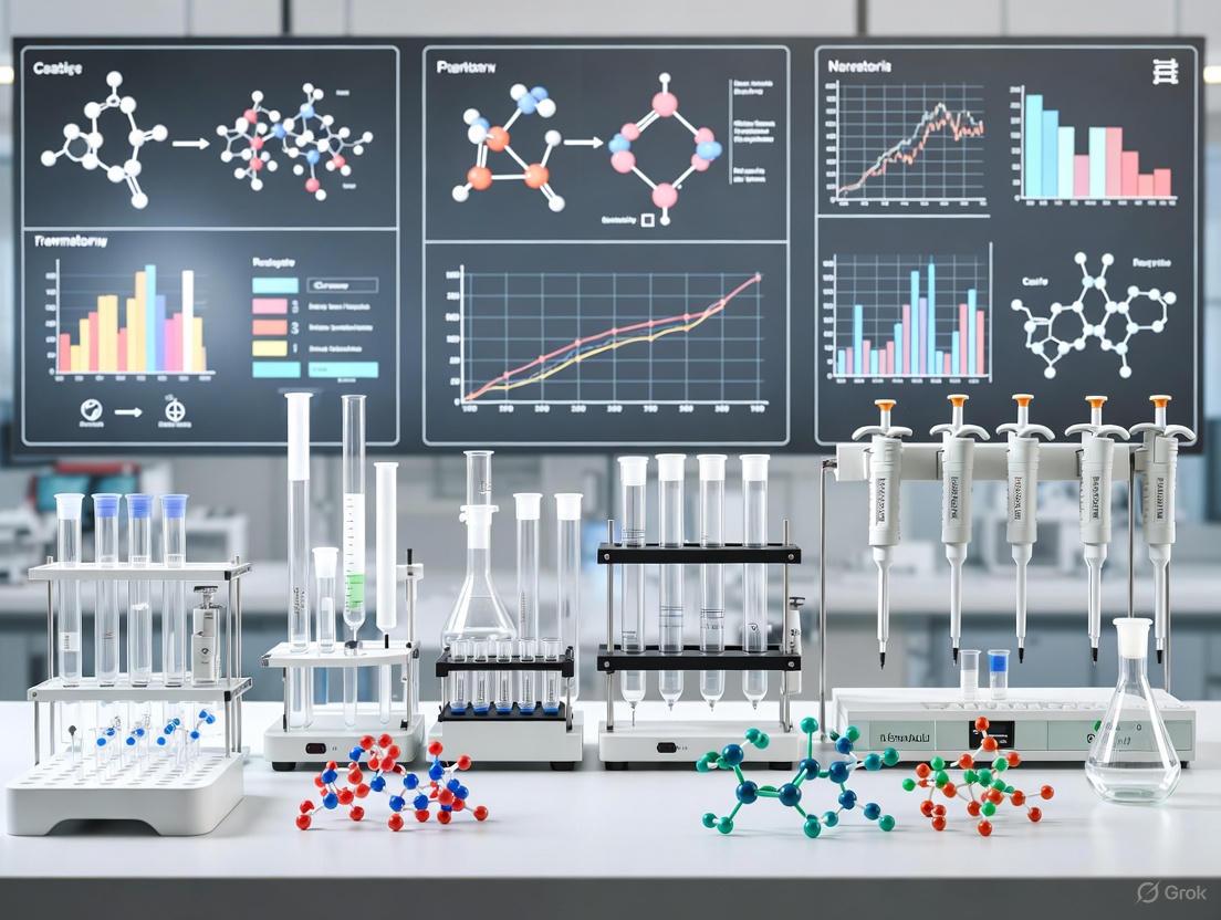
Diagram 1: Master Cell Bank Production Workflow
Quality Management Systems: QA and QC Functions
A robust Pharmaceutical Quality System (PQS) is essential for cGMP compliance in stem cell biomanufacturing. The PQS integrates both Quality Assurance (QA) and Quality Control (QC) functions in a complementary relationship that spans the entire product lifecycle.
Quality Assurance: Prevention-Focused Systems
Quality Assurance is fundamentally process-oriented, focusing on preventing defects through the establishment of robust systems and protocols [13]. Key QA responsibilities in master cell bank production include:
- System Establishment: Developing and implementing comprehensive quality systems, including SOPs, training programs, and document control systems [13].
- Risk Management: Conducting systematic risk assessments to identify and control potential product quality risks throughout the manufacturing process.
- Audit Program: Performing regular internal audits and managing external audits of suppliers and contract testing laboratories [13].
- Change Control: Managing and documenting all changes to equipment, processes, testing methods, and suppliers to evaluate potential impact on product quality [13].
- Review and Approval: Reviewing and approving all critical GMP documents, including batch manufacturing records, validation protocols, and specifications [13].
Quality Control: Detection-Focused Activities
Quality Control is product-oriented, focusing on detecting defects through testing and monitoring activities [13]. Essential QC functions for master cell bank production include:
- Environmental Monitoring: Regular testing of cleanrooms, biosafety cabinets, and other controlled environments to ensure they remain within specified parameters [13].
- Product Testing: Performing physical, chemical, and biological testing on master cell banks to ensure they meet pre-defined specifications for identity, purity, potency, and viability [13].
- Batch Record Review: Meticulous review of complete batch manufacturing documentation to ensure all steps were performed correctly and any deviations were properly documented and investigated [13].
- Stability Testing: Conducting real-time and accelerated stability studies to establish shelf-life and storage conditions for master cell banks [13].
- Method Verification: Verifying that analytical methods are suitable for their intended use in testing cellular products [13].
Table 2: QA vs. QC in Master Cell Bank Production
| Aspect | Quality Assurance (QA) | Quality Control (QC) |
|---|---|---|
| Focus | Process-oriented | Product-oriented |
| Objective | Prevent defects | Detect defects |
| Key Activities | SOP development, training, audits, change control | Product testing, environmental monitoring, batch record review |
| Documentation Responsibility | Validation Master Plan, risk assessments, quality agreements | Test reports, sampling records, certificates of analysis |
| Timing | Proactive, throughout process | Reactive, at defined control points |
| cGMP Emphasis | Ensures cGMP guidelines are embedded in systems | Verifies outputs meet cGMP requirements |
Essential Materials and Research Reagent Solutions
The selection and qualification of raw materials, reagents, and components are critical aspects of cGMP compliance for master cell bank production. All materials must meet appropriate quality standards and be sourced from qualified suppliers.
Critical Reagents and Materials
Table 3: Essential Research Reagents for cGMP-Compliant Master Cell Bank Production
| Material Category | Specific Examples | Function | Quality Requirements |
|---|---|---|---|
| Cell Culture Media | Serum-free media formulations, defined supplements | Supports cell growth and maintenance while maintaining phenotype | cGMP-grade, certificate of analysis, endotoxin testing |
| Cell Separation Reagents | cGMP-grade antibodies, separation columns | Isolation and purification of target cell populations | cGMP-grade, validated for efficiency and purity |
| Cryopreservation Solutions | Defined cryoprotectants (DMSO), formulation buffers | Maintains cell viability and function during freezing and storage | cGMP-grade, sterile, endotoxin-tested |
| Cell Culture Substrates | Recombinant adhesion molecules, cGMP-grade matrix proteins | Provides surface for cell attachment and expansion | cGMP-grade, defined composition, mycoplasma-free |
| Quality Control Reagents | Flow cytometry antibodies, PCR reagents, sterility testing kits | Characterization and release testing of cell banks | Validated for intended use, appropriate controls |
| Processing Containers | Single-use bioprocess containers, cryogenic vials | Closed-system processing and storage of cell products | USP Class VI certified, sterilized, non-pyrogenic |
Material Qualification and Supplier Management
All critical reagents and materials used in master cell bank production must undergo formal qualification procedures:
- Supplier Qualification: Comprehensive assessment of supplier quality systems, manufacturing processes, and testing capabilities.
- Material Specification: Establishment of detailed specifications for each material, including critical quality attributes.
- Incoming Testing: Verification that materials meet specifications through testing or review of certificates of analysis.
- Storage and Handling: Defined storage conditions, expiration dating, and handling procedures to maintain material integrity.
For cellular starting materials, additional considerations apply, including donor eligibility determination, infectious disease testing, and traceability requirements in accordance with 21 CFR Part 1271 [12].
Analytical Methods and Characterization Protocols
Master Cell Bank Testing Requirements
Comprehensive testing of master cell banks is essential to demonstrate identity, purity, potency, and safety. The following testing paradigm should be applied to each master cell bank:
- Identity: Confirmation of cell lineage and donor origin through methods such as STR DNA profiling, karyotyping, and assessment of cell-specific markers.
- Purity: Freedom from contaminating microorganisms (sterility, mycoplasma) and unintended cell types (flow cytometry for cell population homogeneity).
- Potency: Quantitative measure of biological activity through functional assays relevant to the intended clinical application.
- Safety: Testing for adventitious agents (in vitro and in vivo virus assays), endotoxin, and replication-competent viruses where applicable.
The FDA's guidance document "Potency Assurance for Cellular and Gene Therapy Products" provides detailed recommendations for developing and validating potency assays for cell-based products [12].
Method Validation Protocols
Analytical methods used for master cell bank testing must be appropriately validated to demonstrate they are suitable for their intended purpose. Validation characteristics should include:
- Accuracy and Precision: Demonstration of method trueness and reproducibility through repeated analysis of reference standards.
- Specificity: Ability to measure the analyte unequivocally in the presence of other components.
- Linearity and Range: Establishment of the relationship between analyte concentration and response across the validated range.
- Robustness: Capacity to remain unaffected by small, deliberate variations in method parameters.
For stem cell characterization, flow cytometry methods require particular attention to validation, including antibody titration, compensation controls, and instrument standardization.
Diagram 2: Master Cell Bank Testing Framework
Process Validation and Change Control
Master Cell Bank Manufacturing Process Validation
Process validation provides documented evidence that the master cell bank manufacturing process consistently produces a cellular product meeting its predetermined quality attributes. The validation approach should follow a lifecycle model encompassing three stages:
- Stage 1: Process Design: Establishing knowledge space and defining critical process parameters (CPPs) and critical quality attributes (CQAs) through development studies.
- Stage 2: Process Qualification: Demonstrating that the manufacturing process performs as designed under cGMP conditions, including equipment qualification, facility qualification, and process performance qualification.
- Stage 3: Continued Process Verification: Ongoing monitoring to ensure the process remains in a state of control during routine production.
For master cell bank production, process validation should include studies demonstrating consistency across multiple manufacturing runs, robustness to acceptable process variation, and comparability after planned process changes.
Change Control Protocol
A formal change control system is essential for managing modifications to validated processes, equipment, materials, or testing methods. The change control procedure should include:
- Change Proposal: Documented description of the proposed change with scientific justification.
- Impact Assessment: Evaluation of potential effects on product quality, safety, and efficacy.
- Action Plan: Definition of activities required to implement the change, including any additional studies or validation.
- Review and Approval: Multidisciplinary review by quality unit, manufacturing, and regulatory affairs.
- Implementation and Follow-up: Controlled implementation with appropriate documentation and verification of effectiveness.
For master cell bank processes, even seemingly minor changes (e.g., reagent supplier changes, equipment upgrades) may require extensive comparability studies to demonstrate equivalence of the resulting cellular product.
Compliance Monitoring and Regulatory Interactions
Inspection Preparedness and Management
Maintaining continuous inspection readiness is critical for facilities engaged in master cell bank production. Key elements of an effective inspection readiness program include:
- Documentation Management: Ensuring all GMP documents are complete, accurate, and readily retrievable.
- Personnel Training: Comprehensive training programs with documented evidence of employee competency.
- Internal Audit Program: Regular self-inspections to identify and address compliance gaps proactively.
- Inspection Management Protocol: Defined roles and responsibilities for hosting regulatory inspections.
FDA inspections of cGMP compliance may result in Form 483 observations if significant deviations are identified. Subsequent corrective actions to address these observations can be extensive and must be comprehensive and well-documented [11].
Regulatory Submission Requirements
Regulatory submissions for stem cell-based products must include comprehensive information demonstrating cGMP compliance for master cell bank production:
- Chemistry, Manufacturing, and Controls (CMC) Section: Detailed description of cell bank manufacturing process, characterization data, and quality control strategies.
- Environmental Assessment: Evaluation of environmental impact as required for certain biotechnology-derived products.
- Comparability Protocols: Plans for managing anticipated manufacturing changes during product development.
The FDA's guidance document "Chemistry, Manufacturing, and Control (CMC) Information for Human Gene Therapy Investigational New Drug Applications (INDs)" provides specific recommendations for cellular therapy products, though many principles apply broadly to stem cell-based products [12].
cGMP compliance for master cell bank production extends beyond mere regulatory adherence—it represents a fundamental commitment to manufacturing quality and patient safety. Successful implementation requires integration of quality systems throughout the organization, with strong leadership commitment and technical expertise. The complementary functions of Quality Assurance and Quality Control create a comprehensive framework for preventing and detecting quality issues, while robust process validation provides scientific evidence that manufacturing processes consistently produce cellular products of required quality.
As the field of stem cell biomanufacturing continues to evolve, regulatory expectations will similarly advance. Manufacturers should embrace a lifecycle approach to quality management, incorporating emerging technologies and scientific understanding while maintaining compliance with current good manufacturing practices. By establishing a strong foundation of cGMP compliance at the master cell bank stage, manufacturers create a solid platform for developing safe, efficacious, and consistent stem cell-based therapies that can ultimately benefit patients with serious medical conditions.
The fields of cell banking outsourcing and stem cell biomanufacturing are experiencing unprecedented growth, driven by the accelerating transition of advanced therapies from research to clinical and commercial applications. This expansion is fundamentally reshaping the biopharmaceutical landscape, creating a critical dependency on robust, scalable, and regulatory-compliant production infrastructures. For researchers, scientists, and drug development professionals, understanding these market dynamics is not merely an academic exercise but a strategic necessity for navigating the complexities of master cell bank (MCB) production and Good Manufacturing Practice (GMP)-compliant processes.
The global cell banking outsourcing market, valued at approximately USD 14.37 billion in 2024, is projected to grow at a compound annual growth rate (CAGR) of 16.37% from 2025 to 2034, potentially reaching USD 63.49 billion [14]. This remarkable growth is symbiotic with the broader biologics manufacturing market, which itself is predicted to increase from USD 39.25 billion in 2025 to nearly USD 162.51 billion by 2034, at a CAGR of 17.1% [15]. This parallel acceleration underscores a fundamental industrial shift: the move from small-scale, in-house research cell banking to outsourced, industrialized biomanufacturing capable of supporting the rigorous demands of clinical trials and commercial-scale production for cell and gene therapies, vaccines, and other biologics [14] [15] [16].
Market Size and Growth Projections
Quantitative market analysis reveals the scale and velocity of expansion in the cell banking and biomanufacturing ecosystem. The data presented below, synthesized from multiple industry reports, provides a clear financial landscape for strategic planning and investment.
Table 1: Cell Banking Outsourcing Market Size and Growth Projections
| Metric | 2024 Value | 2025 Value | Projected 2034 Value | CAGR (2025-2034) |
|---|---|---|---|---|
| Global Market Size | USD 14.37 Billion [14] | USD 16.72 Billion [14] | USD 63.49 Billion [14] | 16.37% [14] |
| Alternative Estimate | USD 14.2 Billion [16] | - | USD 65.5 Billion [16] | 16.8% [16] |
Table 2: Segmental Dominance in the Cell Banking Outsourcing Market (2024)
| Segmentation Basis | Dominant Segment | Approximate Market Share | Fastest-Growing Segment |
|---|---|---|---|
| Bank Type | Master Cell Bank (MCB) [14] [17] | 36-40% [14] | Research Cell Bank (RCB) [14] |
| Cell Type | Mammalian Cells [14] | 32-35% [14] | Stem Cells [14] |
| Phase | Clinical [14] | 30-33% [14] | Commercial [14] |
| Application | Biologics Manufacturing [14] | 34-38% [14] | Cell & Gene Therapy [14] |
| End User | Biopharmaceutical Companies [14] | 38-42% [14] | Cell Therapy Companies [14] |
| Service Type | Cryopreservation & Storage [14] | 27-30% [14] | Cell Line Characterization & Biosafety Testing [14] |
The dominance of the Master Cell Bank (MCB) segment is logical, as the MCB serves as the foundational, well-characterized source for all production cells, and its quality is paramount for regulatory approval and product consistency [14] [17]. Similarly, the rapid growth of the stem cells segment highlights the increasing therapeutic and research application of these cells, particularly mesenchymal stem cells (MSCs) and induced pluripotent stem cells (iPSCs), in regenerative medicine and disease modeling [14] [16].
Key Growth Drivers and Industry Trends
The powerful expansion of this market is not serendipitous but is propelled by a confluence of scientific, economic, and technological factors.
Increasing Demand for Advanced Therapies
The clinical and commercial success of biologics, including monoclonal antibodies (mAbs), and the burgeoning pipeline of Advanced Therapeutic Medicinal Products (ATMPs), such as cell and gene therapies, are primary drivers [15] [18]. The rising prevalence of chronic diseases, including cancer and neurodegenerative disorders, is creating a sustained demand for these targeted, high-efficacy treatments [14] [15]. The cell and gene therapy segment, in particular, is expected to register the fastest growth within the cell banking outsourcing market [14].
Strategic Outsourcing and Partnership Models
Biopharmaceutical companies are increasingly adopting strategic outsourcing to access specialized expertise, advanced technologies, and scalable GMP capacity without the massive capital expenditure required to build and maintain in-house facilities [14] [19]. This model offers cost-effectiveness, operational flexibility, and risk mitigation [14]. The current funding environment, characterized by venture capital favoring fewer, larger bets, is pushing smaller biotech firms toward partnerships and alliances with established Contract Development and Manufacturing Organizations (CDMOs) to de-risk their development pathways [19]. In 2024, the value of such biotech alliances reached a decade high of USD 144 billion in "biobucks" (potential future value) [19].
Technological and Process Innovations
Innovation is a critical enabler of market growth. Key trends include:
- Automation and Single-Use Technologies: The widespread adoption of closed, automated bioreactor systems and single-use technologies minimizes contamination risks, improves process reproducibility, and increases facility flexibility [20] [21].
- Artificial Intelligence (AI): AI and machine learning are being integrated to optimize donor-recipient compatibility, accelerate drug discovery, streamline biomanufacturing processes, and enhance quality control [14] [18].
- Advanced Cryopreservation: Innovations in cryopreservation techniques are essential for maintaining high cell viability and functionality in high-density cell banks, which is critical for the logistics of autologous and allogeneic therapies [21].
- Chemically Defined Media: The development of xeno-free and chemically defined media formulations ensures consistent and safe pluripotent stem cell culture, a cornerstone of reproducible stem cell biomanufacturing [21].
Application Notes: GMP-Compliant Master Cell Bank Production
The production of a GMP-compliant Master Cell Bank is a critical, foundational step in the biomanufacturing pipeline for stem cell therapies. The following protocol outlines the key stages and considerations.
Table 3: Research Reagent Solutions for MCB Production
| Reagent/Material | Function | Application Context |
|---|---|---|
| Cell Culture Media | Supports cell growth, proliferation, and maintenance. | Chemically defined, xeno-free media are essential for GMP-compliant MSC and iPSC culture [21]. |
| Cryopreservation Medium | Protects cells from ice-crystal damage during freezing and thawing. | Typically contains a cryoprotectant like DMSO and a base medium; formulation is cell-type specific [22]. |
| Cell Dissociation Reagents | Detaches adherent cells from culture surfaces for sub-culturing and banking. | Enzymatic (e.g., trypsin) or non-enzymatic reagents; selection impacts cell viability and function [20]. |
| Characterization Antibodies | Identifies specific cell surface markers for phenotype confirmation. | Used in flow cytometry to characterize MSCs (e.g., CD73+, CD90+, CD105+) or pluripotency markers for iPSCs [6]. |
| Microcarriers | Provides a surface for adherent cell growth in scalable bioreactor systems. | Essential for large-scale expansion of anchorage-dependent cells like MSCs in stirred-tank bioreactors [21]. |
Protocol: Master Cell Bank Generation for Stem Cells
Objective: To generate a GMP-compliant, well-characterized MCB from a validated stem cell line (e.g., iPSC or MSC) for use in therapeutic production.
Workflow Overview:
Materials:
- Validated parental cell stock (e.g., iPSC clone, MSC isolate)
- GMP-grade, xeno-free cell culture media and supplements [21]
- T-flasks, cell factories, or single-use bioreactors [20]
- GMP-grade cryovials and controlled-rate freezer
- Liquid nitrogen storage system (vapor phase, -196°C) [6] [22]
Methodology:
- Cell Line Authentication and Pre-banking Quality Control (QC):
- Authentication: Perform Short Tandem Repeat (STR) analysis to confirm cell line identity and rule out cross-contamination [6] [22]. For human cells, compare the profile to a reference database.
- Biosafety Testing: Conduct comprehensive testing for adventitious agents, including mycoplasma, and sterility testing per pharmacopoeial guidelines (e.g., USP, Ph. Eur.) [6] [16]. Document all results in a pre-banking QC report.
Cell Line Expansion under GMP Conditions:
- Thaw the validated parental cell stock and expand in a GMP-compliant facility using aseptic techniques [22].
- Use predefined culture conditions (media, gas, temperature) and monitor growth kinetics, morphology, and viability closely.
- For scalable production, transition cells from static culture to single-use bioreactors with microcarriers for adherent cells [20] [21]. Monitor key parameters like pH, dissolved oxygen, and metabolite levels.
Cell Harvest and Cryopreservation:
- Harvest cells during the logarithmic growth phase at a predefined passage number.
- Wash and resuspend the cell pellet at a specified concentration in a GMP-grade cryopreservation medium [6] [22].
- Aliquot the cell suspension into cryovials. Use a controlled-rate freezer to cool the vials at approximately -1°C per minute to -80°C before transferring to long-term storage in the vapor phase of liquid nitrogen (-150°C to -196°C) [6] [22].
Post-Banking Characterization and Stability Testing:
- Characterization: Perform a full battery of tests on representative vials from the MCB. This includes:
- Stability: Establish a stability testing program to assess the MCB's ability to maintain viability and genetic stability over its intended storage period. This involves periodic thawing and testing of stability study vials [22].
Documentation and Release:
- Compile a comprehensive Cell Bank Dossier. This document includes all procedures, batch records, QC data, certificates of analysis, and a summary report [6].
- The MCB is released for use in production (e.g., to generate a Working Cell Bank) only after all specifications are met and the dossier is approved by Quality Assurance.
Regional Dynamics and Future Outlook
The market for cell banking outsourcing and biomanufacturing exhibits distinct regional patterns that influence global strategy.
- North America dominated the market in 2024, holding a 40-45% share [14] [16]. This leadership is attributed to a well-established biopharma ecosystem, robust regulatory frameworks (FDA), significant R&D funding, and a high concentration of cell and gene therapy companies [14] [15].
- Asia-Pacific is projected to be the fastest-growing region from 2025 to 2035 [14] [17]. Growth is fueled by increasing healthcare investment, supportive government policies (e.g., China's "Made in China 2025"), improving infrastructure, and the presence of leading CDMOs like WuXi AppTec and Samsung Biologics [15] [17].
Looking ahead, the integration of AI-driven real-time monitoring, further process intensification, and regulatory harmonization initiatives will continue to streamline manufacturing and accelerate global approval timelines for stem cell-based therapeutics [21]. Furthermore, the industry's focus on sustainability will push the adoption of greener biomanufacturing practices, including continuous manufacturing and reduced environmental footprint [18].
The expansion of cell banking outsourcing and biomanufacturing is a direct response to the paradigm shift in medicine toward advanced, cell-based therapies. For research scientists and drug developers, success in this landscape hinges on a deep understanding of both the underlying market forces and the intricate, GMP-compliant protocols required to produce high-quality master cell banks. As the industry evolves, strategic partnerships with specialized CDMOs, coupled with the adoption of innovative technologies, will be paramount in bridging the gap between pioneering research and the successful commercialization of transformative stem cell therapies.
The establishment of Master Cell Banks (MCBs) is a critical step in ensuring the long-term success and regulatory compliance of stem cell-based medicinal products. Under the framework of Good Manufacturing Practice (GMP), a MCB represents a collection of cryopreserved cells of uniform composition, derived from a single tissue or cell source, intended to be used in production. The choice of starting cellular material—whether pluripotent stem cells (PSCs) like induced Pluripotent Stem Cells (iPSCs) and human Embryonic Stem Cells (hESCs), or multipotent adult stem cells such as Mesenchymal Stem/Stromal Cells (MSCs)—profoundly impacts the manufacturing process, quality control, and therapeutic application. This application note provides a detailed comparative analysis of these stem cell sources within the context of GMP-compliant MCB biomanufacturing, supported by standardized protocols for their generation and characterization.
Selecting an appropriate stem cell source is a foundational decision that dictates subsequent manufacturing strategies, regulatory pathways, and clinical applications. The following analysis contrasts the core attributes of PSCs and tissue-derived MSCs.
Table 1: Core Characteristics of Stem Cell Sources for MCB Development
| Feature | iPSCs | hESCs | MSCs (Tissue-Derived) |
|---|---|---|---|
| Origin | Reprogrammed adult somatic cells (e.g., skin fibroblasts, blood cells) [23] [24] | Inner cell mass of the blastocyst [23] | Adult tissues (e.g., bone marrow, adipose tissue, umbilical cord) [23] [25] |
| Pluripotency/Multipotency | Pluripotent (can differentiate into any cell type) [24] | Pluripotent (can differentiate into any cell type) [23] | Multipotent (limited to bone, cartilage, fat, and other stromal lineages) [23] [24] |
| Key Surface Markers | OCT4+, NANOG+, SOX2+, SSEA4+ [24] | OCT4+, NANOG+, SOX2+, SSEA4+ | CD73+, CD90+, CD105+; CD14-, CD19-, CD34-, CD45- [23] [24] [25] |
| Ethical Considerations | Minimal; bypasses embryo destruction [23] [24] | Significant; involves destruction of human embryos [23] | Minimal; obtained from consented adult tissue sources [24] |
| Risk of Teratoma/Tumor Formation | High if undifferentiated cells remain [23] | High if undifferentiated cells remain [23] | Very Low; limited self-renewal capacity [24] |
| Donor & Batch Variability | Can be standardized through reprogramming; but clonal variation exists [26] | Variation between cell lines | High; dependent on donor age, tissue source, and isolation method [25] [26] |
| Scalability for Manufacturing | Potentially unlimited via self-renewal; requires differentiation into target cell [27] | Potentially unlimited via self-renewal; requires differentiation into target cell | Limited by donor tissue availability and replicative senescence [26] |
| Established GMP Banking Examples | Yes (e.g., REPROCELL's StemRNA Clinical iPSCs) [28] | Yes, but with ethical constraints | Yes (e.g., Bone Marrow-MSCs, Adipose-derived MSCs) [25] |
A critical advancement in the field is the derivation of MSCs from iPSCs (iMSCs), which aims to combine the scalability of PSCs with the safety profile and functionality of MSCs. Studies show that iMSCs exhibit similar morphology, surface marker expression, and differentiation capacity to their tissue-derived counterparts (UMSCs), while demonstrating enhanced proliferative capacity and improved immunomodulatory properties, such as upregulation of anti-inflammatory factors like TGFB [26].
Table 2: Key Manufacturing Considerations for GMP-Compliant MCBs
| Consideration | iPSCs | MSCs |
|---|---|---|
| Starter Material | Somatic cells from qualified donor; requires rigorous testing as a raw material [27] | Tissue (e.g., fat pad, bone marrow) from qualified donor; requires processing to isolate cells [25] |
| Reprogramming/Isolation Method | Integration-free methods preferred (e.g., mRNA, episomal vectors) [28] | Explant culture or enzymatic digestion (e.g., collagenase) [23] [25] |
| Culture Medium | Defined, xeno-free media essential [27] [28] | Movement towards animal component-free, GMP-formulated media (e.g., MSC-Brew GMP Medium) [25] |
| Process Scalability | 2D culture on feeders or in feeder-free conditions; moving towards 3D bioreactors [27] | 2D multilayer flasks; more efficient 3D closed-system bioreactors (e.g., hollow fiber) for scale-up [29] |
| Critical Quality Attributes (CQAs) | Pluripotency marker expression, karyotypic stability, vector clearance, trilineage differentiation potential, absence of residual undifferentiated cells [27] | Surface marker profile (CD73/90/105+; CD34/45-), viability, differentiation potential, immunomodulatory function, absence of senescence [25] |
The following workflow outlines the core stages in the development and qualification of a GMP-compliant MCB, applicable to both pluripotent and adult stem cell sources.
Diagram 1: GMP Master Cell Bank Development Workflow
Application Notes and Protocols
Protocol: Generation and Characterization of GMP-Compliant iPSC-MCBs
This protocol is adapted from current best practices for the manufacture of clinical-grade iPSC master cell banks, aligning with perspectives from the EU (EMA) and USA (FDA) regulatory agencies [27] [28].
3.1.1 Donor Screening and Somatic Cell Collection
- Donor Eligibility: Perform comprehensive donor medical and genetic history screening. Test for relevant communicable diseases as per regional regulatory requirements.
- Tissue Collection: Obtain dermal fibroblasts or peripheral blood mononuclear cells (PBMCs) from a qualified donor under informed consent. The collection process must be performed in a controlled, GMP-compliant manner.
- Somatic Cell Bank: Establish a qualified somatic cell bank from the collected tissue. This bank serves as the starting material for reprogramming and must undergo full identity and microbiological testing.
3.1.2 mRNA-Based Reprogramming to Clinical-Grade iPSCs
- Principle: Use a non-integrating, footprint-free method such as mRNA reprogramming (e.g., StemRNA Technology) to generate iPSCs under defined, xeno-free conditions, minimizing risks associated with genomic integration [28].
- Procedure:
- Transfection: Transfect qualified somatic cells with synthetic mRNA encoding the pluripotency factors (OCT4, SOX2, KLF4, c-MYC).
- Culture: Maintain transfected cells in defined, xeno-free medium with daily medium changes to supply reprogramming mRNAs.
- Colony Picking: Approximately 3-4 weeks post-transfection, manually pick emerging iPSC colonies based on characteristic hESC-like morphology (tightly packed, high nucleus-to-cytoplasm ratio, distinct borders).
- Clonal Expansion: Expand individual clones in a separate well of a GMP-qualified, feeder-free culture system (e.g., on recombinant laminin-521 coating).
3.1.3 Master Cell Bank Production
- Clonal Selection: Select 3-5 candidate clones based on robust growth, stable karyotype, and high expression of pluripotency markers. A lead clone is chosen for MCB generation.
- Expansion: Expand the selected clone in multiple parallel vessels to achieve the target cell number for banking, using defined culture conditions and enzymatic passaging.
- Cryopreservation: Harvest cells at a specific passage, concentrate, and resuspend in a defined, GMP-grade cryopreservation medium (e.g., containing human serum albumin and DMSO). Fill, label, and cryopreserve a minimum of 200 vials in the vapor phase of liquid nitrogen. The entire process must be documented in a Batch Manufacturing Record.
3.1.4 Quality Control Testing for iPSC-MCB Release The following tests are considered the minimum requirements for the characterization of a clinical-grade iPSC-MCB [27]:
- Identity: Short Tandem Repeat (STR) profiling to match the donor somatic cell bank.
- Viability and Potency: Post-thaw viability (>70%), and demonstration of pluripotency via:
- Flow Cytometry: Quantitative analysis of pluripotency markers (OCT3/4, NANOG, SOX2, TRA-1-60, SSEA4).
- Trilineage Differentiation: Directed in vitro differentiation into ectoderm, mesoderm, and endoderm lineages, confirmed by immunocytochemistry for lineage-specific markers.
- Purity and Safety:
- Sterility: Tests for bacterial and fungal contamination.
- Mycoplasma: PCR or culture-based assay.
- Adventitious Viruses: In vitro and in vivo virus tests.
- Karyotyping: G-banding analysis to confirm genomic stability at a resolution of 400-500 bands.
- Other Tests: Endotoxin levels (< threshold), and demonstration of vector clearance for the reprogramming method used.
Protocol: Establishment and Validation of GMP-Compliant MSC-MCBs
This protocol outlines the GMP-compliant isolation, expansion, and banking of MSCs, using Infrapatellar Fat Pad-derived MSCs (FPMSCs) as a model system with reduced patient morbidity [25].
3.2.1 Tissue Acquisition and Processing
- Tissue Source: Obtain infrapatellar fat pad (IFP) tissue as surgical waste during orthopedic procedures (e.g., ACL reconstruction) with patient informed consent and ethical approval [25].
- Transport: Transfer the tissue to the GMP facility in a sterile, validated transport medium.
3.2.2 Enzymatic Isolation and Primary Culture of FPMSCs
- Tissue Digestion:
- Aseptically mince the IFP tissue into fragments of approximately 1 mm³.
- Digest the tissue fragments with 0.1% collagenase in serum-free media for 2 hours at 37°C with gentle agitation [25].
- Centrifuge the digest at 300 ×g for 10 minutes to pellet the stromal vascular fraction (SVF).
- Cell Seeding:
- Resuspend the cell pellet in a GMP-compliant, animal component-free expansion medium (e.g., MSC-Brew GMP Medium), which has been shown to enhance proliferation and maintain stemness compared to standard media [25].
- Filter the cell suspension through a 100 μm sterile filter to remove debris.
- Seed the cells in culture vessels at a density of 5 × 10³ cells/cm² [25].
3.2.3 Cell Expansion and Process Optimization
- Culture Conditions: Maintain cultures at 37°C in a humidified 5% CO₂ incubator.
- Passaging: Passage cells at 80-90% confluency using a GMP-grade dissociation reagent. Consistently use a seeding density of 5 × 10³ cells/cm² for expansion.
- Process Monitoring: Calculate population doubling time at each passage. Perform a Colony Forming Unit (CFU) assay at passage 3 to assess clonogenic potency [25].
3.2.4 Master Cell Bank Production and Stability
- Harvesting: Harvest cells at the predetermined optimal passage (e.g., P3-P4) for banking.
- Cryopreservation: Wash, count, and resuspend cells in a GMP-grade cryopreservation medium. Fill vials and cryopreserialize using a controlled-rate freezer before transfer to liquid nitrogen storage.
- Stability and Shelf-Life: Perform stability testing by assessing post-thaw viability and potency after storage for predefined intervals (e.g., 30, 90, 180 days). The protocol by N/A et al. demonstrated >95% viability and maintained marker expression after 180 days of storage [25].
3.2.5 Quality Control Testing for MSC-MCB Release
- Identity and Purity: Flow cytometric analysis for positive (CD73, CD90, CD105 ≥95%) and negative (CD34, CD45, CD11b, CD19, HLA-DR ≤5%) marker profiles [25].
- Viability and Potency: Post-thaw viability (>70%, ideally >95%), differentiation potential into osteocytes, chondrocytes, and adipocytes confirmed by specific staining, and immunomodulatory function assays (e.g., T-cell suppression assay) [25] [26].
- Safety: Sterility (bacteria/fungi), mycoplasma, and endotoxin testing.
The Scientist's Toolkit: Essential Reagents for GMP Cell Banking
The following table details key materials and reagents critical for implementing the protocols described above under GMP standards.
Table 3: Essential Research Reagent Solutions for GMP MCB Development
| Reagent / Material | Function / Application | GMP Considerations |
|---|---|---|
| StemRNA Clinical iPSC Kit | Footprint-free mRNA reprogramming of somatic cells to clinical-grade iPSCs [28] | Defined, xeno-free; compliant with FDA/EMA/PMDA standards [28] |
| MSC-Brew GMP Medium | Animal component-free medium for the expansion of MSCs for clinical use [25] | Promotes enhanced proliferation and maintains MSC potency; reduces batch-to-batch variability [25] |
| Recombinant Laminin-521 | Defined, xeno-free substrate for feeder-free culture of pluripotent stem cells | Eliminates need for mouse feeder layers, enhancing product consistency and safety |
| GMP-Grade Collagenase | Enzymatic digestion of tissues (e.g., fat pad) for isolation of primary MSCs [25] | Animal-free recombinant versions are preferred to minimize contamination risk |
| Human Serum Albumin (HSA) | Component of cryopreservation and culture media; acts as a carrier protein and stabilizer | Sourced from human plasma under strict pharmacopoeial standards; preferred over FBS |
| BD Stemflow Human MSC Analysis Kit | Standardized flow cytometry panel for characterization of MSC surface markers [25] | Provides a validated, consistent method for assessing cell identity and purity |
Regulatory and Manufacturing Framework
Adherence to a robust Quality Management Program (QMP) is non-negotiable for GMP cell processing. This program must address all critical factors impacting each step of the product lifecycle, from donor screening to final administration [30]. The choice between autologous and allogeneic processes is fundamental. Allogeneic therapies, particularly those based on iPSCs, are increasingly the preferred manufacturing alternative as they enable the creation of a single, extensively characterized MCB that can supply countless doses, ensuring product consistency and cost-effectiveness [27].
The manufacturing landscape is evolving from traditional 2D culture systems to automated 3D bioreactors (e.g., hollow fiber, stirred-tank). These closed-system bioreactors offer significant advantages for MCB production and subsequent scaling, including improved scalability, reduced hands-on time, lower risk of contamination, and enhanced economic efficiency [29]. For instance, using a hollow fiber bioreactor for MSC expansion can reduce hands-on manufacturing time by hundreds of hours and lower the cost per dose significantly compared to 2D cell stacks [29].
The following diagram illustrates a streamlined, scalable manufacturing process for an allogeneic cell therapy product derived from an iPSC-MCB.
Diagram 2: Allogeneic Therapy Manufacturing from MCB
Regulatory guidance from the FDA and EMA is continuously evolving. Manufacturers of iPSC banks are advised to consult ICH guidelines, particularly those for biotechnological products (e.g., Q5A(R2) on viral safety, Q5D on cell substrates), and adapt their requirements for cell therapy applications. Key areas requiring further harmonization include acceptable expression vectors for reprogramming, minimum identity and purity testing, and stability testing protocols for cell banks [27].
From Donor to Vial: A Step-by-Step Guide to GMP MCB Generation and Process Intensification
In the context of master cell bank production for Good Manufacturing Practice (GMP) stem cell biomanufacturing, the integrity of the starting biological material is the foundational pillar upon which all subsequent product quality and patient safety are built. The International Society for Stem Cell Research (ISSCR) underscores that all stem cell research, including clinical translation, must be conducted with scientific and ethical integrity, relying on principles of rigor, oversight, and transparency [8]. This Application Note provides a detailed framework for the sourcing and qualification of these critical starting materials, aligning with the stringent biosafety and regulatory expectations for cell therapy products [31]. A failure in this initial stage can introduce risks that compromise the entire manufacturing process and ultimately, patient safety, making a robust, verifiable system for donor safety and material traceability not just beneficial, but essential [32].
Sourcing and Donor Safety Protocols
The process of sourcing starting materials demands a rigorous ethical and safety-focused protocol to ensure the integrity of the cell bank and the safety of the eventual recipient.
Fundamental Ethical Principles and Donor Eligibility
Adherence to established ethical guidelines is paramount. The ISSCR Guidelines stress the primacy of participant welfare and the necessity of valid informed consent. Potential donors must be empowered with accurate information to make an autonomous decision, and for those lacking capacity, consent must be obtained from a lawfully authorized representative [8]. Furthermore, the principles of social and distributive justice should be considered to ensure the fair distribution of both the benefits and burdens of research [8].
- Experimental Protocol: Donor Screening and Consent
- Objective: To ensure the ethical procurement of starting materials from eligible, fully-informed donors.
- Materials: Donor health questionnaire, informed consent forms, phlebotomy kit, sample collection tubes, and secure sample labels with unique donor identifier.
- Procedure:
- Pre-Screening Review: Conduct an initial review of the potential donor's medical history and health questionnaire to assess preliminary eligibility against predefined inclusion/exclusion criteria.
- Informed Consent Process: A qualified healthcare professional must conduct the informed consent session. The discussion must cover:
- The purpose of the donation and its use in biomanufacturing.
- The procedures involved, including any foreseeable risks or discomforts.
- The voluntary nature of participation and the right to withdraw.
- Confidentiality of records and the coding of donor identity.
- Eligibility Testing: Collect donor specimens (e.g., blood) for mandatory testing in a certified laboratory. Tests must screen for relevant communicable diseases as per regulatory standards (e.g., FDA, EMA).
- Documentation: Complete the donor eligibility record, attaching all test results and the signed consent form to the unique donor identifier. This record must be securely archived.
Material Traceability from Source
Creating an unbroken chain of custody is critical. Relying solely on documentation, such as a Mill Test Report (MTR) in traditional manufacturing, is a high-risk strategy, as the document is a claim about a piece of paper, not a guarantee about the physical material [32]. A robust system integrates documentation with a physically verifiable chain of custody.
- Experimental Protocol: Establishing a Digital Chain of Custody
- Objective: To implement a physically verifiable, unbroken chain of custody from donor to final cell bank vial.
- Materials: Biocompatible labels, barcode system, Laboratory Information Management System (LIMS) or ERP system, and dedicated sample storage containers.
- Procedure:
- Unique Identification: At the point of collection, assign a unique identifier to the donor and the collected sample. This identifier should be linked to all subsequent data and materials.
- Digital Twinning: Upon receipt at the manufacturing facility, create a digital record in the LIMS that links the physical sample (via a scannable barcode) to its unique identifier, the donor eligibility record, and the associated informed consent documentation.
- Flow-Down Traceability: As the sample moves through processing (e.g., cell isolation, culture, cryopreservation), the unique identifier must be physically transferred or digitally scanned at each stage. The LIMS should automatically track the location and processing history of the material.
- Reconciliation: At critical steps, perform a physical reconciliation between the samples in process and the digital records in the LIMS to investigate and resolve any discrepancies immediately [33].
The following workflow diagram illustrates the integrated process of donor qualification and material traceability.
Qualification of Starting Materials
Qualification is the process of verifying that starting materials meet all specified requirements for identity, purity, potency, and safety before use in manufacturing.
Control of Starting Materials under GMP
GMP requirements mandate that starting materials must be purchased only from approved suppliers to written specifications. Furthermore, all incoming materials must be inspected and/or tested to verify their suitability for use before being released for production, not after [33]. The quality unit has the sole authority to approve or reject materials.
- Experimental Protocol: Incoming Material Receipt and Sampling
- Objective: To ensure that all received starting materials (e.g., raw materials, culture media, reagents) are correctly identified, sampled, and tested before use.
- Materials: Quarantine storage area, personal protective equipment (PPE), clean sampling tools, sterile sample containers, and testing equipment.
- Procedure:
- Receipt and Verification: Upon receipt, verify that the supplier's information on the container matches the accompanying paperwork (e.g., purchase order, certificate of analysis).
- Physical Inspection: Examine each container for damage, seal integrity, and cleanliness. Raise a non-conformance for any issues before acceptance.
- Quarantine: Move accepted materials to a designated quarantine storage area.
- Sampling: Only trained operators should perform sampling following a written procedure. Sample in a clean, controlled environment to prevent contamination.
- Testing: Test samples according to approved methods and specifications for identity, purity, potency, and quality. The Certificate of Analysis is helpful but does not substitute for actual testing [33].
Biosafety and Quality Assessment
A comprehensive biosafety assessment for cell therapy products must address specific risks, including toxicity, tumorigenicity, and immunogenicity [31]. The quality of the cellular product itself must be confirmed, verifying that cells are sterile, authentic, and functionally active [31].
Table 1: Key Biosafety and Quality Assays for Cellular Starting Materials
| Assessment Category | Specific Assay/Parameter | Typical Method(s) | Quantitative Benchmark Example |
|---|---|---|---|
| Identity | Cell surface marker expression | Flow Cytometry | >95% positive for expected markers |
| Viability & Purity | Cell viability, microbiological sterility | Trypan Blue exclusion, BacT/Alert | >90% viability; No growth of microorganisms |
| Potency | Differentiation potential, Metabolic activity | Directed differentiation assays, ELISA | e.g., >50% differentiation to target lineage |
| Safety | Endotoxin, Mycoplasma | LAL assay, PCR | Endotoxin <0.5 EU/mL; Mycoplasma not detected |
| Tumorigenic Potential | Karyotype, In vivo tumor formation | G-banding, Soft agar assay in immunocompromised mice | Normal karyotype; No tumor formation at test site |
- Experimental Protocol: In-process Quality Control and Labeling
- Objective: To maintain the identity, purity, and quality of materials during all manufacturing steps.
- Materials: In-process storage containers, status labels (e.g., Quarantine, Approved, Rejected), and manufacturing batch records.
- Procedure:
- Clear Labeling: All materials, including in-process items and bulk containers, must be clearly labeled with the name, strength, and batch number.
- Status Identification: Use a system of physical labels and electronic fields to clearly identify the status of all materials (e.g., Quarantine, Approved, Rejected).
- Storage: Store materials in designated areas with monitored environmental controls. Operate on a First-In, First-Out (FIFO) basis.
- Record Completion: Accurately and completely fill out all manufacturing records, including all information relating to processing and testing [33].
The following flowchart summarizes the core decision-making process for the qualification and release of starting materials.
The Scientist's Toolkit: Research Reagent Solutions
The following table details essential materials and reagents critical for executing the protocols described in this application note.
Table 2: Key Research Reagent Solutions for Starting Material Qualification
| Item | Function/Application |
|---|---|
| Donor Screening Assay Kits | Serological and molecular diagnostic test kits for mandatory donor infectious disease marker testing. |
| Cell Isolation Kits | Immunomagnetic or density gradient-based kits for the specific and gentle isolation of target cell populations from heterogeneous starting material. |
| Validated Cell Culture Media | GMP-grade media formulations, supplemented with growth factors and cytokines, designed to maintain cell viability and function without inducing differentiation. |
| Flow Cytometry Antibody Panels | Pre-configured, validated antibody panels for the quantitative analysis of cell surface and intracellular markers to confirm cell identity and purity. |
| Mycoplasma Detection Kit | PCR- or culture-based kits for the highly sensitive detection of mycoplasma contamination in cell cultures. |
| LAL Endotoxin Test Kit | A kit utilizing Limulus Amebocyte Lysate for the quantitative determination of bacterial endotoxins in samples and reagents. |
| Handheld XRF Analyzer | A non-destructive Positive Material Identification (PMI) tool used to verify the chemical composition of raw materials, acting as a critical backstop to documentation [32]. |
This document details a core process workflow for the isolation, expansion, and cryopreservation of human cells under current Good Manufacturing Practice (cGMP) guidelines, a critical component in the production of Master Cell Banks (MCBs) for stem cell biomanufacturing. The establishment of well-characterized MCBs is a foundational step in ensuring the consistent production of safe and efficacious advanced therapy medicinal products (ATMPs), such as those derived from induced pluripotent stem cells (iPSCs) and mesenchymal stromal cells (MSCs) [27] [34]. Adherence to cGMP standards, as outlined in regulations such as 21 CFR 210, 211, and 600, is mandatory to guarantee the identity, purity, potency, and safety of these biological products throughout their lifecycle [10] [35]. The protocols herein are designed to provide a robust, reproducible, and scalable framework for generating high-quality cell banks suitable for clinical development.
Cell Isolation under cGMP
The initial isolation of cells from starting material is a critical step that significantly impacts the viability, functionality, and overall quality of the final cell bank. The choice of isolation method must preserve these characteristics while minimizing mechanical or enzymatic stress.
Isolation Method Comparison
A primary consideration is the selection between positive selection, negative selection, or physical sorting methods. Each approach has distinct advantages and limitations concerning purity, yield, and impact on cell functionality [36].
Table 1: Comparison of Cell Isolation Methods
| Isolation Method | Principle | Purity Achieved (%) | Key Advantages | Key Considerations |
|---|---|---|---|---|
| Immunomagnetic Negative Selection | Depletion of unwanted cell populations from a mixed sample (e.g., PBMCs). | 89 - >95 [36] | High purity; preserves unmanipulated cell surface receptors; suitable for diverse cell types. | Potential for non-specific cell loss. |
| Immunomagnetic Positive Selection | Direct capture of target cells using surface-specific antibodies. | Not explicitly stated | High specificity for the target population. | Antibody binding may activate cells or interfere with subsequent functional assays. |
| Flow Cytometric Cell Sorting | Physical separation based on light scattering and fluorescent labeling. | ≥99 [36] | Exceptionally high purity and specificity; multi-parameter sorting. | Exposes cells to high mechanical stress and prolonged handling, potentially compromising viability and function [36]. |
| Automated Centrifugal Microfluidics | Label-free separation based on biophysical properties. | Not explicitly stated | Reduces cell stress; improves post-isolation proliferation and cytolytic function [36]. | High operational cost can be prohibitive. |
cGMP-Compliant Isolation Protocol for NK Cells from PBMCs
The following protocol, adapted from a validated procedure, outlines a method for obtaining highly pure natural killer (NK) cells using negative selection [36].
- Principle: This method uses a cocktail of antibodies against non-NK cells (e.g., CD3, CD4, CD14, CD19, CD20, CD36, CD66b, CD123, CD235a) coupled to magnetic particles. The labeled non-target cells are removed using a magnet, leaving an untouched, enriched NK cell (CD56+/CD3-) population in the supernatant.
- Materials:
- Source Material: Peripheral Blood Mononuclear Cells (PBMCs), fresh or thawed.
- Reagent: cGMP-grade NK Cell Isolation Kit (negative selection).
- Equipment: cGMP-grade magnetic separation device, biosafety cabinet, refrigerated centrifuge.
- Buffers: Phosphate-Buffered Saline (PBS) without Ca2+/Mg2+, supplemented with cGMP-grade 1-2% Human Serum Albumin (HSA) or EDTA.
- Procedure:
- Cell Preparation: Ensure PBMCs are a single-cell suspension in a suitable buffer. Determine total cell count and viability.
- Antibody Incubation: Add the provided antibody cocktail to the cell suspension. Mix thoroughly and incubate for 10-20 minutes at 2-8°C.
- Magnetic Particle Incubation: Add cGMP-grade magnetic particles to the cell suspension. Mix and incubate for 10-15 minutes at 2-8°C.
- Magnetic Separation: Place the tube in the magnetic separator for 5-10 minutes. Without disturbing the tube, carefully decant or pipette the supernatant containing the enriched NK cells into a new tube.
- Washing: Wash the isolated NK cells with buffer and centrifuge. Resuspend the cell pellet in appropriate expansion medium.
- Quality Control: Assess purity (e.g., % CD56+/CD3- by flow cytometry, typically 97-99% [36]), viability (e.g., >95% by Trypan Blue exclusion), and yield.
Cell Expansion under cGMP
Ex vivo expansion is necessary to achieve clinically relevant cell doses. The use of cGMP-grade media, cytokines, and feeder cells is mandatory to ensure a consistent and safe manufacturing process.
Expansion Strategies and Performance
Expansion protocols can be broadly categorized into feeder-based and non-feeder-based systems, with the former often yielding higher fold expansions suitable for off-the-shelf therapy production [36].
Table 2: Comparison of Cell Expansion Methods and Performance
| Expansion Method | Key Components | Average Fold Expansion | Time Frame | Applications |
|---|---|---|---|---|
| Non-Feeder Based | IL-2 (500-1000 IU/mL) [36] | 7.5 - 45.9-fold [36] | 2 weeks | Research-scale NK cell expansion. |
| Non-Feeder Based | Sequential IL-15 (10 ng/mL) and IL-21 (25 ng/mL) stimulation [36] | 4.5-fold [36] | 10 days | Priming of NK cell function. |
| Feeder Based | Irradiated EBV-LCL feeder cells + IL-2 (500 IU/mL) [36] | 1344 ± 1135-fold [36] | 2 weeks | High-yield NK cell expansion for therapy. |
| Feeder Based | K562.mbIL21.4-1BBL feeder cells + IL-2 (50 IU/mL) [36] | 47,967 ± 42,230-fold [36] | 3 weeks | Large-scale, potent NK cell production. |
| Feeder Based | OCI-AML3.mbIL-21 feeder cells + IL-2 (200 IU/mL) [36] | ~700 ± 245-fold [36] | 3 weeks | AML-specific NK cell expansion. |
| Current Protocol | EBV-LCL feeders (10:1) + IL-2 (100 IU/mL) + IL-21 (20 ng/mL) [36] | 289 ± 70-fold (2 weeks)\n10,460 ± 4972-fold (3 weeks) [36] | 2-3 weeks | Clinically relevant NK cell dosages. |
| MSC Expansion | MSC-Brew GMP Medium (animal component-free) [37] | Enhanced proliferation rates and lower doubling times vs. standard media [37] | Multiple passages | Clinical-grade MSC manufacturing. |
cGMP-Compliant Expansion Protocol for NK Cells using Feeder Cells
This protocol describes the robust expansion of NK cells using irradiated feeder cells and cGMP-grade cytokines [36].
- Principle: Genetically modified feeder cells (e.g., K562 expressing membrane-bound IL-21 and 4-1BBL) or naturally expressing feeders (e.g., EBV-LCL) provide critical activation signals and co-stimulation that, combined with soluble cytokines (e.g., IL-2, IL-15, IL-21), drive NK cell proliferation and maintain cytotoxic potency.
- Materials:
- Starter Cells: Isolated NK cells (from Section 2.2).
- Feeder Cells: Irradiated (e.g., 100 Gy) cGMP-grade K562.mbIL21.4-1BBL or EBV-LCL cells.
- Media: cGMP-grade basal medium (e.g., RPMI-1640).
- Cytokines: cGMP-grade IL-2, IL-15, and/or IL-21.
- Equipment: CO2 incubator, biosafety cabinet, inverted microscope.
- Procedure:
- Co-culture Initiation: Seed isolated NK cells at a density of 0.5-1 x 10^6 cells/mL. Irradiate feeder cells and add them to the NK cell culture at an optimized ratio (e.g., 2:1 or 10:1, feeder:NK cell) [36].
- Cytokine Supplementation: Add cGMP-grade cytokines to the culture (e.g., 100 IU/mL IL-2 and 20 ng/mL IL-21) [36].
- Culture Maintenance: Culture cells at 37°C, 5% CO2. Replenish cytokines (e.g., IL-2 every 2-3 days) and perform partial medium changes as needed to maintain cell health and nutrient levels.
- Feeder Cell Re-stimulation: Every 7-14 days, re-irradiate and add fresh feeder cells at an appropriate ratio (e.g., 1:1) to maintain expansion [36].
- Harvest: Cells are typically harvested after 2-3 weeks of culture, when expansion peaks and viability is high.
- Quality Control: Monitor cell count, viability, and fold expansion. Assess immunophenotype (e.g., CD56, CD16, NKG2D, NKp46) and functionality (e.g., cytotoxicity against K562 target cells) at harvest.
Controlled Cryopreservation under cGMP
Controlled-rate freezing and optimized cryopreservation media are essential for maintaining high post-thaw viability and functionality, ensuring the long-term stability of MCBs and WCBs.
Fundamentals of Cryopreservation
Cryopreservation halts cellular metabolism by storing cells at ultra-low temperatures (-135°C to -196°C) [38]. The key to success is mitigating the damage caused by intracellular ice crystal formation and osmotic stress. This is achieved using cryoprotectants like Dimethyl Sulfoxide (DMSO) and a controlled freezing rate, typically -1°C/minute, which allows water to gradually exit the cell before freezing [38]. Rapid thawing is critical to minimize damage from ice recrystallization during the recovery phase.
cGMP-Compliant Cryopreservation Protocol
This protocol is applicable to a wide range of cell types, including iPSCs, MSCs, and immune cells, for the creation of MCBs and WCBs.
- Principle: Cells are resuspended in a defined, cGMP-grade freezing medium containing a cryoprotectant. They are then cooled at a controlled rate to -80°C before transfer to long-term storage in the vapor or liquid phase of liquid nitrogen.
- Materials:
- Cells: Expanded cell product, counted and with high viability (>95%).
- Freezing Medium: cGMP-grade, serum-free, defined cryopreservation medium (e.g., CryoStor CS10) [38]. Avoids the variability and safety concerns of animal serum.
- Containers: cGMP-grade cryogenic vials (preferably internal-threaded).
- Freezing Device: Controlled-rate freezer or isopropanol-based (e.g., Nalgene Mr. Frosty) or passive freezing container (e.g., Corning CoolCell) [38].
- Storage: Liquid nitrogen tank.
- Procedure:
- Harvest and Centrifuge: Harvest cells aseptically and pellet by centrifugation. Remove supernatant completely.
- Resuspend in Freezing Medium: Resuspend the cell pellet in cold (2-8°C) freezing medium to achieve the optimal cell concentration (e.g., 1x10^6 to 1x10^7 cells/mL, depending on cell type) [38].
- Aliquot: Aseptically aliquot the cell suspension into pre-labeled cryogenic vials.
- Controlled-Rate Freezing:
- Option A (Passive Container): Place vials in a freezing container and immediately transfer to a -80°C freezer for 18-24 hours.
- Option B (Controlled-Rate Freezer): Use a programmed freezer to cool at -1°C/minute to at least -40°C before transferring to -80°C or liquid nitrogen vapor.
- Long-Term Storage: Transfer vials to a liquid nitrogen storage tank (-135°C to -196°C) for long-term preservation. Do not store at -80°C for more than one month [38].
- Quality Control: Post-thaw, assess viability (should be >70%, ideally >95% for release [37]), recovery, sterility, and functionality. Stability testing should be performed on the banked cells to establish expiration dates.
The Scientist's Toolkit: Essential Reagents and Materials
The following table lists critical reagents and materials used in the featured cGMP workflows.
Table 3: Essential Research Reagent Solutions for cGMP Cell Biomanufacturing
| Product Name / Category | Function | Example Application |
|---|---|---|
| cGMP-Grade NK Cell Isolation Kit | Immunomagnetic negative selection for high-purity isolation of untouched NK cells from PBMCs. | Initial cell isolation for NK cell therapy production [36]. |
| MSC-Brew GMP Medium | Animal component-free, cGMP-compliant medium optimized for the expansion and maintenance of mesenchymal stromal cells. | Clinical-scale manufacturing of MSCs for regenerative medicine [37]. |
| CryoStor CS10 | A defined, serum-free, cGMP-manufactured cryopreservation medium containing 10% DMSO. | Protects cells during freezing, storage, and thawing; ensures lot-to-lot consistency for MCB creation [38]. |
| cGMP-Grade Cytokines (IL-2, IL-15, IL-21) | Soluble signaling proteins that drive cell proliferation, survival, and functional maturation during ex vivo expansion. | Critical components in NK cell and T cell expansion protocols [36] [34]. |
| K562.mbIL21.4-1BBL Feeder Cells | Genetically engineered, irradiated cell line expressing membrane-bound cytokines and co-stimulatory ligands. | Provides essential signals for massive, clinically relevant ex vivo expansion of NK cells [36]. |
| Automated Cell Counter (e.g., NucleoCounter NC-100) | Fluorescence-based imaging system for accurate and reproducible cell counting and viability assessment. | Validated for precise cell counting in cGMP manufacturing of hiPSCs, overcoming operator-dependent variability of hemocytometers [39]. |
Leveraging Advanced Bioreactors and Automation for Scalable and Reproducible Manufacturing
The transition from laboratory-scale stem cell culture to large-scale, robust manufacturing is a critical challenge in the development of advanced therapy medicinal products (ATMPs). For therapies requiring up to 10^9 cells per patient, traditional flask-based expansion methods are insufficient, being labor-intensive, variable, and prone to contamination [40] [41]. The implementation of advanced bioreactors and automated systems within a Good Manufacturing Practice (GMP) framework is therefore essential to produce the necessary cell quantities while ensuring safety, potency, and reproducibility [40]. This document provides detailed application notes and experimental protocols for leveraging these technologies in the context of master cell bank production for GMP stem cell biomanufacturing.
Advanced Bioreactor Platforms for Clinical-Grade MSC Expansion
Several automated, closed-system bioreactors have been developed specifically to address the challenges of large-scale human mesenchymal stem/stromal cell (MSC) production. The table below summarizes the performance characteristics of key platforms.
Table 1: Performance Characteristics of Automated MSC Expansion Systems
| Bioreactor Platform | Scale/Equivalent | Reported Yield (Cell Number) | Key Features | Documented Functional Outcomes |
|---|---|---|---|---|
| Quantum Cell Expansion System (Terumo BCT) [40] | 21,000 cm² (≈120 T-175 flasks) | 100–276 × 10^6 BM-MSCs (7-day expansion from 20 × 10^6 seed) | Hollow fiber bioreactor; continuous medium exchange; enables hypoxic culture. | Suppression of T-cell activation in vitro; therapeutic efficacy in rat models of ischemic stroke and joint surface defects [40]. |
| CliniMACS Prodigy (Miltenyi Biotec) [40] | 1-layer CellSTACK | 29–50 × 10^6 MSCs (P0, 10-day procedure from equine peripheral blood) | Fully automated from isolation to harvest; integrated tubing set (TS730) for adherent cells. | Fibroblast-like morphology and phenotypic MSC markers maintained; higher P0 yield compared to manual protocols [40]. |
| Xuri W25 (Cytiva) [40] | N/A | N/A | Wave-mixed bioreactor; closed and scalable system. | N/A |
| NANT 001/XL (VivaBioCell) [40] | N/A | N/A | N/A | N/A |
The Quantum System has been validated in over 25 studies for expanding adult human MSCs. A critical operational note is that its hollow fibers must be coated with an adhesive substrate (e.g., fibronectin or cryoprecipitate) prior to cell seeding. Furthermore, substituting fetal bovine serum (FBS) with human platelet lysate (hPL) as a growth supplement significantly enhances the expansion of adipose tissue-derived MSCs (AT-MSCs) within this system while sustaining cell quality [40]. The CliniMACS Prodigy platform demonstrates the potential for complete automation of the manufacturing process, from initial tissue isolation to final harvest, thereby minimizing manual open steps and improving lot-to-lot consistency [40].
Protocol: Aeration and Agitation Optimization for Bioreactor Scale-Up
Conventional scale-up strategies based solely on constant power input per unit volume (P/V) or volumetric oxygen mass transfer coefficient (kLa) are often insufficient as they fail to account for the mass transfer efficiency differences caused by varying sparger pore sizes across different single-use bioreactor brands [42]. The following protocol outlines a systematic approach using Design of Experiments (DoE) to establish a quantitative relationship between aeration pore size, agitation, and gas flow.
Materials and Equipment
- Parallel Bioreactor System (e.g., CloudReady 500 mL × 24, T&J Bio-Engineering) with unified control software (D2MS) [42].
- Marine-type impeller (28 mm diameter).
- Variable Pore Size Spargers: Drilled-hole spargers (DHS) with pore sizes of 0.3, 0.5, 0.8, and 1.0 mm.
- Cell Line: A stable, monoclonal cell line (e.g., CHO cell line DS003 for monoclonal antibody production) [42].
- Basal and Feed Media (e.g., QuaCell CHO CD04 and Feed02).
Experimental Procedure
Define Parameter Ranges: Based on common specifications of commercial single-use bioreactors, define the experimental ranges for the critical parameters [42]:
- P/V: 8.8, 18.8, 23.8, and 28.8 W/m³ (corresponding to impeller speeds of 240, 320, 350, and 375 rpm, respectively).
- Aeration Pore Size (DHS): 0.3, 0.5, 0.8, and 1.0 mm.
- Volumetric Gas Flow (vvm): 0.003, 0.0075, and 0.012 m³/min (proportional to the initial culture volume).
Design Experiment: Utilize an Orthogonal Test Method (DoE) to efficiently explore the multi-factorial design space. The working volume for the 500 mL parallel bioreactors should be set at 300 mL [42].
Execute Cultures: Run the parallel bioreactor experiments according to the DoE matrix. Monitor critical process parameters and record final cell density, viability, and product expression (e.g., antibody titer for CHO cells).
Model and Analyze: Analyze the data to build a quantitative model. The referenced study found a quantitative relationship in the P/V range of 20 ± 5 W/m³, where the appropriate initial aeration rate was between 0.01 and 0.005 m³/min for aeration pore sizes between 1 and 0.3 mm [42].
Validation: Validate the optimized model in larger, geometrically similar bioreactors (e.g., a 15 L glass bioreactor and a 500 L single-use bioreactor) to confirm scalability [42].
Workflow Diagram
The following diagram illustrates the logical workflow for the bioreactor scale-up optimization protocol.
Facility and Process Design for GMP Compliance
The design of a manufacturing facility for allogeneic stem cell therapies, which may require batch sizes of 200 to 2,000 liters to produce 10^11 to 10^14 cells annually, must prioritize contamination control [41].
Open vs. Closed Processing
A foundational design choice is between open and closed processes. Closed processes, where the product is not exposed to the surrounding environment, are strongly recommended as they inherently reduce contamination risk. This is achieved using single-use systems, isolators, and sterile tubing assemblies. While an entire process may not be closed, a block flow diagram should be created to identify each step and assign an "open" or "closed" label. Open processing requires, at a minimum, a Grade A biosafety cabinet within a Grade B background room, which significantly increases facility footprint and operational complexity [41].
Risk Assessment and Facility Zoning
A preliminary risk assessment is crucial for defining the facility layout. The approach varies significantly based on the product strategy [41]:
- Single-Product Facilities: Focus on aseptic operation and batch segregation. Multiple closed functions can often be consolidated into a single room.
- Multiproduct Facilities: Require strategies to manage cross-contamination risk, such as product campaigning, physical segregation, and unidirectional HVAC flow.
- Contract Manufacturing Organizations (CMOs): Often require a compartmentalized suite approach to manage the high risk from multiple clients and products.
Table 2: Essential GMP and Safety Regulations for Facility Design
| Region | GMP Regulations / Guidelines | Key Biosafety Regulations / Guidelines |
|---|---|---|
| United States | 21 CFR Part 1271 (HCT/Ps), 210, 211, 610 [41]; FDA Guidance on Aseptic Processing [41] | CDC/NIH Biosafety in Microbiological and Biomedical Laboratories (BMBL) [41]; 29 CFR 1910.1030 (Bloodborne Pathogens) [41] |
| European Union | EudraLex Vol 4, GMP for ATMPs [41]; Annex 1, Manufacture of Sterile Medicinal Products [41] | Directive 2009/41/EC (Contained Use of GMOs) [41] |
The Scientist's Toolkit: Research Reagent Solutions
The following table details essential materials and their functions for establishing a GMP-compliant stem cell manufacturing process.
Table 3: Key Reagents and Materials for GMP Stem Cell Biomanufacturing
| Item | Function / Application | GMP-Compliant Consideration |
|---|---|---|
| Serum-Free / Xeno-Free Media [40] [43] | Provides defined nutrients for cell growth; eliminates variability and immunogenic risks of animal sera. | Essential for clinical production. Formulations must be optimized for specific cell types (e.g., MSCs, iPSCs). |
| Human Platelet Lysate (hPL) [40] | Growth supplement for MSC expansion; replaces FBS to enhance cell proliferation and align with clinical standards. | Must be sourced and tested as a raw material for human use. |
| CellSTACK Chambers [40] | Multi-layer vessels for scaling up adherent 2D cell culture in a compact footprint. | Often used in semi-automated or automated systems (e.g., CliniMACS Prodigy). |
| GMP-Compliant Dissociation Agents | Enzymatic (e.g., trypsin analogs) or non-enzymatic reagents for cell detachment during passaging. | Must be well-characterized, sourced, and tested per GMP guidelines. |
| Master/Working Cell Bank | Provides a consistent, well-characterized, and tested starting source of cells for all production batches. | Critical for regulatory approval. Can be developed in-house or sourced from GMP-compliant providers [44]. |
| Closed System Tubing Sets (e.g., TS730 for CliniMACS Prodigy) [40] | Enable aseptic fluid transfer and cell culture processing within automated, closed systems. | Single-use, pre-sterilized, and integrated into the platform's fluid path. |
| Hollow Fiber Bioreactor Cartridges (for Quantum system) [40] | Provide a high surface area for adherent cell growth in a closed, automated system with continuous medium perfusion. | Require pre-coating with GMP-grade substrates (e.g., fibronectin). |
Integrating Automation and Industry 4.0
Automation is a powerful tool for reducing human-derived variability in manual handling tasks, such as pipetting, media changes, and cell passaging. Liquid-handling robots can achieve microliter precision, while automated image analysis can provide objective, quantitative assessment of cell confluence, replacing subjective visual estimates [45]. The philosophy of Industry 4.0 offers a framework for advanced automation through [45]:
- Digital Twins: Creating a virtual model of each product batch, integrating all process data for complete traceability.
- Service-Oriented Architecture: Organizing the production platform so that autonomous devices offer "services" (e.g., "media change," "measure confluence"). The cell culture process can then dynamically request these services based on real-time sensor data (e.g., pH, glucose), creating an adaptive and flexible manufacturing process.
The following diagram illustrates this adaptive, data-driven automation concept.
The field of stem cell biomanufacturing is undergoing a significant transformation, driven by the increasing complexity of advanced therapy medicinal products (ATMPs) and the stringent requirements of Good Manufacturing Practice (GMP). Master Cell Bank (MCB) production represents a critical foundational step in the development of cell-based therapies, where consistency, purity, and safety are paramount. The traditional model of in-house MCB development presents substantial challenges for biopharmaceutical companies, including massive capital investment in specialized facilities, requirement for highly specialized expertise, and significant timeline extensions. In response to these challenges, Contract Development and Manufacturing Organizations (CDMOs) have emerged as strategic partners, offering specialized capabilities in GMP-compliant MCB production that accelerate therapeutic development while managing risk and cost.
The global CDMO market, valued at $238.92 billion in 2024, is projected to grow at a compound annual growth rate (CAGR) of 9.0% through 2032, reaching $465.24 billion [46]. This growth is particularly pronounced in the biologics sector, where CDMO service demand is growing at approximately 15% annually—nearly three times higher than for pharmaceuticals overall [47]. For researchers and drug development professionals, understanding how to strategically leverage these partnerships has become an essential competency in advancing stem cell therapies from laboratory research to clinical application.
Market Landscape: The Expanding Role of CDMOs in MCB Production
Quantitative Market Analysis
The CDMO sector has experienced robust growth, particularly in services supporting biologic and cell therapy development. The table below summarizes key market data points relevant to MCB production:
Table 1: CDMO Market Size and Growth Projections
| Market Segment | 2024 Market Size | Projected 2032 Market Size | CAGR | Primary Growth Drivers |
|---|---|---|---|---|
| Global CDMO Market | $238.92 billion [46] | $465.24 billion [46] | 9.0% [46] | Biologics expansion, outsourcing trends |
| U.S. Pharmaceutical CDMO Market | $40.52 billion [48] | $83.25 billion [48] | 7.47% [48] | Biosimilars, biologics, complex APIs |
| Biologics CDMO Services | N/A | N/A | ~15% [47] | Antibody drugs, ADC therapies |
The U.S. pharmaceutical CDMO market demonstrates particularly strong growth dynamics, expected to expand from $40.52 billion in 2024 to $83.25 billion by 2034 [48]. Within this market, the active pharmaceutical ingredient (API) manufacturing segment dominated in 2024 with a 64% share, while finished dosage formulation development and manufacturing is projected to grow at a significant CAGR of 8.2% during the forecast period [48].
Key CDMO Players in Cell Banking Services
Several major CDMOs have developed specialized capabilities in MCB production for cell therapies. The top CDMOs by revenue include:
Table 2: Leading CDMOs with MCB and Cell Therapy Expertise
| CDMO | 2024 Revenue (CDMO Business) | Specialized MCB/Cell Therapy Capabilities |
|---|---|---|
| Lonza Group | CHF 6.574 billion ($8.138 billion) [47] | Integrated Biologics, Advanced Synthesis, and Specialized Modalities platforms |
| Thermo Fisher Scientific | $7 billion [47] | Accelerator Drug Development platform for reduced timelines |
| Catalent | $4.43 billion [47] | GPEx Lightning cell-line technology |
| Fujifilm Biotechnologies | ¥219.5 billion ($1.496 billion) [47] | Gene therapy and vaccine manufacturing expansion |
| REPROCELL | Not disclosed | GMP-grade iMSC & MSC MCB generation services [49] |
These organizations offer comprehensive MCB services including cell line development, banking, full characterization, and storage, with specific expertise in relevant cell types such as mesenchymal stromal cells (MSCs), iPSC-derived MSCs (iMSCs), HEK293, CHO, and others [49] [3].
Strategic Advantages: Why Outsource MCB Production?
Cost and Timeline Efficiency
Establishing GMP-compliant MCB production in-house requires substantial investment and extended timelines. CDMO partnerships offer significant efficiencies:
Table 3: Cost and Timeline Comparison: In-House vs. CDMO MCB Production
| Parameter | In-House MCB Production | CDMO Partnership |
|---|---|---|
| Timeline to MCB Release | 1-2 years [44] | Months [44] |
| Direct Costs | $1.15 - $1.95 million+ [44] | Reduced capital investment |
| Infrastructure Costs | Facility construction, validation | Leveraged existing infrastructure |
| Expertise Development | Recruitment, training | Immediate access to specialists |
| Opportunity Costs | High (diluted focus on core competencies) [44] | Low (maintained focus on therapeutic development) |
A detailed breakdown of the traditional approach reveals that MCB process development alone requires 3-6 months and $200,000-$400,000, followed by donor tissue sourcing and regulatory testing (1-3 months; $50,000-$150,000), tech transfer to a CDMO (2-4 months; $200,000-$400,000), and finally MCB manufacturing and release testing (2-4 months; $250,000-$500,000) [44]. Parallel tracks for Working Cell Bank (WCB) production and analytical method development add further complexity, time, and expense.
Access to Specialized Expertise and Infrastructure
CDMOs provide access to specialized knowledge and facilities that would be prohibitively expensive to develop in-house. This includes:
- GMP-compliant cleanrooms (Class 100/ISO 5) with monitored environments [49]
- Closed, modular systems like the Cytocentric Xvivo system that reduce contamination risk [49]
- Experienced personnel with expertise in regulatory compliance and quality systems
- Advanced technologies such as automated fill/finish capabilities and multiple QC labs [49]
CDMOs with existing Type II Biologics Master Files that have been reviewed by regulators offer an additional advantage, as developers can reference these files directly in their IND submissions, removing layers of redundant documentation and sharply reducing CMC-related risk [44].
Risk Mitigation and Quality Assurance
The quality of the MCB is critical to the entire therapeutic program's success. CDMOs mitigate risk through:
- Robust quality systems with adherence to FDA, EMA, and PMDA standards [49]
- Comprehensive testing panels for identity, purity, viability, genetic stability, sterility, mycoplasma, endotoxins, and extraneous agents [3]
- Structured banking systems (initial, master, working, and end of product cell banks) that ensure standardization [50]
- Regulatory expertise to navigate complex submission requirements across different regions
CDMO Selection Framework: Criteria for Strategic Partnership
Selecting the right CDMO partner requires careful evaluation of multiple factors. Key considerations include:
- Technical Capabilities: Experience with specific cell types (MSCs, iMSCs, HEK293, CHO, etc.) and relevant technologies [3]
- Quality and Regulatory Track Record: Successful regulatory inspections (FDA, EMA, etc.) and quality systems [47] [49]
- Capacity and Timeline Alignment: Ability to meet project timelines with appropriate capacity [3]
- End-to-End Service Potential: Capability to support beyond MCB to drug substance and drug product manufacturing [3]
- Geographic Considerations: Presence in key regulatory regions and supply chain logistics
The emerging trend is toward equal partnership models rather than transactional relationships, with both CDMOs and pharmaceutical companies seen as integral collaborators bringing essential competencies, knowledge, and capabilities to the table [51].
Experimental Protocols: Methodologies for GMP-Compliant MCB Generation
MSC Isolation and Clonal Selection Protocol
The following protocol, adapted from established methodologies for GMP-compatible clonal MSC production, provides a framework for generating homogeneous MSC populations [50]:
Objective: To isolate, expand, and bank a homogeneous population of human bone marrow-derived clonal MSCs (cMSCs) under GMP-compatible conditions.
Materials:
- Bone marrow aspirate from qualified donor
- Isolation culture medium: α-MEM supplemented with 20% defined FBS, 1% MEM NEAA, 1% GlutaMAX [50]
- Expansion culture medium: α-MEM supplemented with 5% human platelet lysate (hPL), 1% MEM NEAA, 1% GlutaMAX [50]
- TrypLE enzyme for cell detachment [50]
- Cryopreservation medium: 90% FBS + 10% DMSO (USP grade) [50]
- Tissue culture vessels (35-mm dishes to T175 flasks)
- Cloning cylinders
Procedure:
- Donor Screening and Bone Marrow Aspiration:
- Screen healthy volunteers through comprehensive medical history, physical examination, serological testing for viral infections, chest X-ray, and EKG [50]
- Aspirate bone marrow (approximately 40 mL) from iliac crest under sterile conditions in an operating room [50]
- Transport in sterile heparinized tubes in a cool box (4°C to 8°C) with temperature data logger within 12 hours to GMP cleanroom [50]
Clonal Isolation via Subfractionation Culturing Method:
- Directly culture 2.5 mL BM aspirate in isolation medium in 100-mm tissue culture dishes [50]
- Daily transfer supernatant containing suspended cells to new dishes for 5 days (D1-D5) [50]
- Monitor dishes for colony appearance (approximately 2 weeks) [50]
- Isolate separate well-grown colonies using cloning cylinders and TrypLE detachment [50]
- Transfer cell suspension to 35-mm tissue culture dishes for initial expansion
Seed Stock Establishment:
- Expand clones through three subsequent passages (to 6-well plate, T25, then T75 flask) [50]
- Cryopreserve clones that demonstrate adequate expansion capability at passage 3 (P3) as seed stock
- Freeze at least three cryovials per clone at density of 0.5 ± 0.1 × 10^6 cells/500 μL freezing medium [50]
- Store in vapor phase of liquid nitrogen [50]
Clone Screening and Selection:
- Culture 120-150 × 10^3 cells from each clone at P4 in T25 flask at 5 × 10^3 cells/cm² seeding density [50]
- Implement cost-effective screening strategy based on lengthy serial passaging to identify proliferative clones [50]
- Select top-performing clones based on expansion capability, morphology, and preliminary characterization
Four-Tiered Cell Banking System:
- Initial Cell Bank (ICB): Established from selected clones
- Master Cell Bank (MCB): Expanded from ICB under GMP conditions
- Working Cell Bank (WCB): Derived from MCB for routine manufacturing use
- End of Product Cell Bank (EoPCB): Final product for clinical lot release [50]
Diagram 1: GMP MCB Banking Workflow
Quality Control and Release Testing Protocol
Objective: To perform comprehensive characterization and release testing of MCB according to regulatory standards.
Table 4: MCB Release Testing Panel and Standards
| Test Category | Specific Assays | Regulatory Standard |
|---|---|---|
| Identity | Surface marker expression (CD73, CD90, CD105, CD45, CD34), Morphology assessment | FDA/EMA guidelines [50] [3] |
| Purity | Sterility testing, Mycoplasma testing, Endotoxin testing | USP <71>, EP 2.6.7, USP <85> [3] |
| Viability | Cell count and viability (trypan blue exclusion or equivalent) | FDA guidance [3] |
| Genetic Stability | Karyotype analysis, STR profiling | FDA guidance [50] [3] |
| Safety | Extraneous agent testing, In vitro and in vivo adventitious agent assays | FDA/EMA guidelines [3] |
| Potency | Trilineage differentiation potential (osteogenic, adipogenic, chondrogenic), Immunomodulation assays | FDA/EMA guidelines [50] [49] |
Procedure:
- Identity Testing:
Purity and Safety Testing:
- Conduct sterility testing per USP <71> using direct inoculation or membrane filtration methods [3]
- Perform mycoplasma testing per EP 2.6.7 using both culture and indicator cell culture methods [3]
- Measure endotoxin levels per USP <85> using Limulus Amebocyte Lysate (LAL) test with acceptance criteria of ≤0.5 EU/mL [3]
- Complete in vitro and in vivo adventitious agent testing per FDA guidelines [3]
Viability and Genetic Stability:
Potency Assay Development:
The Scientist's Toolkit: Essential Materials for MCB Production
Table 5: Key Research Reagent Solutions for GMP-Compliant MCB Production
| Reagent/Material | Function | GMP-Grade Considerations |
|---|---|---|
| Cell Culture Media (α-MEM, DMEM) | Base nutrient source for cell growth | Defined formulation, endotoxin testing, documentation of origin [50] |
| Serum Supplements (FBS, hPL) | Provides growth factors and attachment factors | Virus-inactivated, extensively screened, traceable donor history [50] |
| Dissociation Reagents (TrypLE, Trypsin) | Cell detachment from culture surfaces | Recombinant origin preferred, animal-component free, documented purity [50] |
| Cryopreservation Medium (DMSO + base medium) | Long-term storage of cell banks | USP-grade DMSO, sterile filtration, compatibility testing [50] |
| Quality Control Assay Kits (Sterility, Mycoplasma, Endotoxin) | Safety testing for release | Validated methods, compendial standards (USP, EP), inclusion of controls [3] |
| Characterization Reagents (Flow cytometry antibodies, Differentiation kits) | Identity and potency assessment | Clone-specific validation, lot-to-lot consistency, stability data [50] [49] |
Implementation Framework: Strategic Partnership Models
Successful implementation of CDMO partnerships for MCB production requires careful planning and relationship management. Current industry trends favor equal partnership models where both CDMOs and pharmaceutical companies function as integral collaborators, each bringing essential competencies to the relationship [51]. Three primary partnership approaches have emerged:
Diagram 2: CDMO Partnership Models
Technology Transfer and Knowledge Management
Effective technology transfer to CDMO partners follows a structured approach:
- Pre-Transfer Assessment: Comprehensive documentation of cell line history, characterization data, and cultivation parameters
- Knowledge Transfer: Structured meetings, laboratory demonstrations, and documentation review
- Process Qualification: Side-by-side testing, comparability studies, and establishment of equivalence
- Ongoing Management: Regular communication, joint governance committees, and defined decision-making processes
Platforms like the AJILITY framework exemplify how standardized approaches can streamline tech transfer through predefined components, templated documentation, and established quality systems that reduce variables and accelerate timelines [52].
The rise of CDMOs as strategic partners for MCB production represents a fundamental shift in stem cell biomanufacturing. By leveraging specialized expertise, established infrastructure, and regulatory knowledge, therapeutic developers can accelerate timelines from years to months while maintaining the rigorous quality standards required for clinical applications [44]. The comprehensive protocols and frameworks presented in this application note provide researchers and drug development professionals with practical methodologies for implementing successful CDMO partnerships.
As the field continues to evolve toward more complex therapies and personalized medicine approaches, strategic outsourcing of MCB production will increasingly become the standard rather than the exception. Companies that effectively navigate this landscape—selecting the right partners, establishing collaborative relationships, and maintaining scientific oversight—will be best positioned to advance innovative stem cell therapies from concept to clinic, ultimately delivering new treatment options to patients in need.
Navigating Production Hurdles: Strategies for Overcoming Scalability, Consistency, and Cost Challenges
Addressing High Costs and Scalability Bottlenecks in Autologous and Allogeneic Models
The development of cell-based therapies represents a frontier in modern medicine, yet their commercialization is heavily constrained by manufacturing challenges. A core thesis within stem cell biomanufacturing research is that master cell bank production under Good Manufacturing Practice (GMP) is the foundational element determining the success and scalability of both autologous (patient-specific) and allogeneic (off-the-shelf) models [53] [54]. The autologous model, which creates a unique batch for each patient, faces inherent scalability limitations and high per-unit costs [55] [56]. While the allogeneic model offers the potential for scalable, off-the-shelf production from a single donor source, it requires a massive initial investment in rigorously characterized and tested cell banks to ensure a consistent and safe starting material for thousands of doses [53] [54]. This application note details protocols and strategic approaches to mitigate these bottlenecks, focusing on GMP-compliant cell bank systems as the critical control point.
Quantitative Analysis of Manufacturing Bottlenecks
The economic and operational disparities between autologous and allogeneic manufacturing are significant. The table below summarizes a quantitative model for scaling an allogeneic MSC therapy, illustrating the massive cell production requirements for commercial-scale applications [53].
Table 1: Scale-Up Production Model for an Allogeneic MSC Therapy
| Clinical Phase | Target Patient Number | Estimated Cell Requirement (Billion Viable Cells) | Example Production Platform | Estimated GMP Manufacturing Cost |
|---|---|---|---|---|
| Phase I | 25 | >9.3 | 40-cell stack / 35L Bioreactor | <$1 Million |
| Phase II | 50 | >19 | 60-cell stack / 35L Bioreactor | <$1 Million |
| Phase III | 150 | ~57 | Scalable Bioreactor Systems | Model-Dependent |
| Commercial | 10,000/Year | ~2,000 (Annual) | 200-2000L "3D" Bioreactors | Model-Dependent |
The high costs are driven by complex logistics and resource-intensive processes. For autologous therapies, the entire process—from cell collection, transport, and patient-specific manufacturing to final reinfusion—must be replicated for every single patient [55]. Complete safety and characterization testing for a single Master Cell Bank (MCB) can approach $200,000, a necessary investment to ensure purity, safety, and functionality [57]. Furthermore, building a GMP-grade cell bank from scratch is a complex process that can take 12 to 24 months and cost between $1.5 to $3 million, diverting critical resources from drug product development [53].
Strategic Framework and Experimental Protocols
Core Strategy: Implementing a Robust Cell Bank System
The foundation of a scalable and cost-effective manufacturing process is a well-designed, two-tiered cell bank system [53] [54]. This system consists of a Master Cell Bank (MCB), created from a selected cell clone and fully characterized to meet all quality and safety standards, and a Working Cell Bank (WCB), derived from the MCB and used for production [53] [54]. Adherence to this system ensures a consistent, characterized, and reliable source of cells, which is paramount for product quality and regulatory compliance.
Key design questions for establishing a cell bank include [53]:
- Will it last through the product's lifecycle?
- Does it meet geographical and logistical needs?
- Will it meet future scalable manufacturing demands?
- Is functional variability within Critical Quality Attribute (CQA) tolerances?
Protocol 1: GMP-Compliant Isolation and Expansion of Mesenchymal Stem Cells
This protocol, adapted from a 2025 study, outlines a method for isolating and expanding MSCs from the infrapatellar fat pad (IFP) under GMP-compliant, animal component-free conditions [25].
Objective: To establish a reproducible and scalable protocol for generating clinical-grade MSCs. Starting Material: Human IFP tissue acquired as surgical waste from reconstructive surgery, with informed consent [25].
Procedure:
- Tissue Processing: Minced IFP tissue is digested with 0.1% collagenase in serum-free media for 2 hours at 37°C [25].
- Cell Isolation: The digested tissue is centrifuged (300 ×g, 10 min). The pellet is washed with PBS and filtered through a 100 µm filter. After a final centrifugation, the cell pellet is resuspended in culture media [25].
- GMP-Grade Cell Culture:
- Seed cells at a density of 5 × 10³ cells/cm² in animal component-free media (e.g., MSC-Brew GMP Medium) [25].
- Maintain cultures in a CO₂ incubator at 37°C and subculture at 80-90% confluency.
- Using defined, GMP-grade media eliminates batch-to-batch variability and reduces risks associated with animal-derived components [25].
- Cryopreservation: For bank creation, freeze cells in a cryopreservation solution containing 10% DMSO at passage 1 [25].
Quality Control Assays:
- Viability: Assess post-thaw viability using Trypan Blue exclusion. The protocol consistently achieved >95% viability [25].
- Purity and Identity: Confirm MSC phenotype using flow cytometry for standard positive (CD73, CD90, CD105) and negative markers [25].
- Potency: Evaluate colony-forming unit (CFU) capacity by seeding low densities (20-500 cells) and counting colonies after 10 days [25].
- Sterility: Perform tests for mycoplasma, endotoxin, and bacteriological contamination (e.g., using Bact/Alert system) [25].
Protocol 2: Risk Mitigation through Pre-Validated Working Cell Banks
For many developers, building an MCB from scratch is prohibitively expensive and time-consuming. A strategic alternative is to leverage pre-made, GMP-grade Working Cell Banks [53].
Objective: To accelerate entry into Phase I clinical trials by reducing upfront costs and timelines. Procedure:
- Source Pre-Qualified WCBs: Acquire GMP-ready WCBs from a trusted supplier (e.g., RoosterBio's CliniControl cells). These banks are typically supported by USFDA Master Files, simplifying regulatory submissions [53].
- Technology Transfer: Transfer the vialed WCBs to your GMP manufacturing facility or partner.
- Phase-Appropriate Manufacturing: Use the WCBs to produce material for early clinical trials (Phase I/II). This defers the large investment in a program-specific MCB until later clinical stages [53].
- Bridging Studies: When ready, generate your MCB and perform comparability studies against the pre-qualified WCB to ensure a seamless transition for later-phase trials and commercialization [53].
Advantages:
- Time Savings: Reduces the timeline to first-in-human trials by 12-24 months [53].
- Cost Savings: Cuts initial investment by ~$1M, allowing resources to be focused on drug product development and preclinical studies [53].
- Risk Reduction: Mitigates common cell banking failures related to donor variability, contamination, and inadequate characterization [53].
The Scientist's Toolkit: Essential Research Reagent Solutions
The following reagents and equipment are critical for implementing the described GMP protocols.
Table 2: Key Reagent Solutions for GMP Stem Cell Biomanufacturing
| Item | Function & Application | Example Product |
|---|---|---|
| Animal Component-Free Medium | Provides a defined, xeno-free environment for cell expansion, eliminating immunogenicity risks and batch variability. | MSC-Brew GMP Medium [25] |
| GMP-Grade Signaling Protein | A recombinant protein used to direct stem cell differentiation or maintain stemness in culture; crucial for process consistency. | GMP-grade DLL4 Protein [58] |
| Cell Dissociation Reagent | A non-animal-derived enzyme for detaching adherent cells during passaging, essential for scalable subculture. | Trypsin replacement enzymes |
| Flow Cytometry Kit | Standardized antibody panel for confirming MSC identity and purity (CD73, CD90, CD105) per ISCT criteria. | BD Stemflow Human MSC Analysis Kit [25] |
| Liquid Nitrogen Storage System | For the long-term cryogenic preservation of MCB and WCB aliquots in vapor-phase LN₂ to ensure genetic stability. | Vapor-phase LN₂ tanks [54] |
Workflow Visualization: From Cell Bank to Clinical Application
The following diagram illustrates the parallel development pathways for autologous and allogeneic therapies, highlighting the central role of the cell bank system.
Diagram: Therapy Development Paths. The allogeneic path leverages a central Cell Bank System to enable scalable, off-the-shelf production, while the autologous path requires a separate manufacturing batch for each patient.
Concluding Recommendations
Mitigating the high costs and scalability bottlenecks in cell therapy requires a strategic focus on the initial stages of process development. The following integrated approach is recommended:
- Invest in a Robust MCB: The MCB is the cornerstone of product quality and scalability. Under-investing here leads to higher costs and delays later [53] [57].
- Adopt GMP-Grade, Defined Materials Early: Transitioning to animal component-free media and reagents as early as possible ensures consistency, reduces contamination risks, and simplifies regulatory approval [25] [57].
- Leverage Pre-Validated Systems for Speed: For early-stage developers, using pre-made, regulatory-ready WCBs can significantly accelerate timelines to the clinic and conserve capital [53].
- Design for Scalability from the Outset: Process development should prioritize platform technologies that can transition from 2D culture to automated, closed-system bioreactors to meet commercial-scale demand [53] [59].
By anchoring biomanufacturing strategy in a well-characterized and scalable cell bank system, developers can navigate the complex economic and regulatory landscape, ultimately accelerating the delivery of transformative therapies to patients.
In the context of master cell bank (MCB) production for GMP stem cell biomanufacturing, ensuring the consistency, safety, and efficacy of the final cell-based product is paramount. A critical, yet often variable, factor in the production process is the composition of the cell culture media and reagents. These components directly influence cell phenotype, genetic stability, and functional integrity, thereby impacting the reproducibility and reliability of both research data and clinical outcomes [60] [61]. Adherence to stringent regulatory guidelines for cell bank characterization is essential for mitigating these risks and ensuring the production of high-quality biologics [62] [12]. This application note details the sources of variability introduced by culture media and provides standardized protocols to control these factors within a GMP-compliant framework.
The Impact of Culture Media on Cell Phenotype: Quantitative Evidence
Variations in culture media composition can lead to significant alterations in critical cellular parameters. The following tables summarize quantitative findings from key studies investigating this phenomenon.
Table 1: Impact of Culture Media on Cell Growth and Redox State in A549 and HepG2 Cells [60]
| Cell Line | Culture Medium | Growth Rate (Fold Change) | Intracellular Thiol Level | Sensitivity to Selenium Cytotoxicity (IC₅₀) |
|---|---|---|---|---|
| A549 | RPMI 1640 | Data Shown | Data Shown | Varies by medium |
| Ham's F-12 | Data Shown | Data Shown | Varies by medium | |
| DMEM | Data Shown | Data Shown | Varies by medium | |
| MEM | Data Shown | Data Shown | Varies by medium | |
| HepG2 | RPMI 1640 | Data Shown | Data Shown | Varies by medium |
| Ham's F-12 | Data Shown | Data Shown | Varies by medium | |
| DMEM | Data Shown | Data Shown | Varies by medium | |
| MEM | Data Shown | Data Shown | Varies by medium |
Note: The original study [60] confirmed that the composition of the cell culture media greatly affected cell growth and sensitivity to selenium cytotoxicity, with specific quantitative data presented for each parameter and medium. The exact numerical values can be found in the source publication.
Table 2: Effect of Culture Conditions on THP-1 Monocyte Differentiation and Cytokine Secretion [63]
| Culture Condition | Basal CD14+ Cells | Response to PMA | Key Cytokine/Chemokine Profile (vs. Other Condition) |
|---|---|---|---|
| Condition I (High Density) | 20.9% | Significant increase in CD14+ cells (to 37.9%) | Significantly enhanced IL-8, MIP-1α, MIP-1β |
| Condition II (Low Density) | 2.6% | No change in CD14 expression | Higher VEGF and IL-12p70 |
The variability introduced by media is not limited to established cell lines. The use of fetal bovine serum (FBS), a complex and undefined mixture of hundreds of components, is a major source of inconsistency. Lot-to-lot variations in FBS can significantly alter cellular outcomes, as demonstrated in cultures of mesenchymal stem cells (MSCs), where different serum lots impacted the expression of 785 genes and critical upstream signals like Tp53 and TGFB1, ultimately affecting the MSCs' ability to support hematopoietic stem cell generation [61].
Experimental Protocols for Assessing Media-Driven Variability
Implementing standardized testing protocols is critical for characterizing the impact of media and reagents on cell phenotype. Below are detailed methodologies for key experiments.
Protocol: Evaluating Media-Dependent Cytotoxicity and Cellular Health
This protocol is adapted from a study investigating selenium cytotoxicity in different media [60].
3.1.1 Materials and Reagents
- Cell lines of interest (e.g., A549, HepG2)
- Four different cell culture media (e.g., RPMI 1640, Ham's F-12, DMEM, MEM)
- Fetal Bovine Serum (FBS), single batch
- Phosphate-Buffered Saline (PBS)
- Test compounds (e.g., sodium selenite, selenomethylselenocysteine)
- CellTiter-Glo Luminescent Cell Viability Assay
- 96-well tissue culture plates
- CO₂ incubator (37°C, 5% CO₂)
- Luminescence plate reader
3.1.2 Procedure
- Cell Culture Standardization: Maintain cells in each of the four test media for a minimum of two passages before initiating experiments to allow for acclimation.
- Seeding: Harvest and count cells. Seed cells in 96-well plates at an optimized density (e.g., 80,000 cells/mL for A549) to achieve approximately 70% confluence at the time of treatment.
- Treatment: Incubate plates for 24 hours. Wash cells with PBS and then treat with a concentration series of the test compound, prepared in the respective culture media. Include control wells with media only.
- Incubation: Incubate cells with the treatment for 48 hours.
- Viability Assay: Following incubation, equilibrate plates to room temperature. Add a volume of CellTiter-Glo Reagent equal to the volume of media in each well. Mix on an orbital shaker for 2 minutes to induce cell lysis, and then incubate for 10 minutes to stabilize the luminescent signal.
- Data Acquisition: Record luminescence using a plate reader.
- Data Analysis: Calculate cell viability relative to untreated controls. Generate dose-response curves and determine IC₅₀ values for each compound in each media type.
Protocol: Assessing Phenotypic Drift via Surface Marker Expression
This protocol, based on THP-1 monocyte research, can be adapted for stem cell characterization [63].
3.2.1 Materials and Reagents
- Cells under investigation
- Culture media to be tested
- Differentiation stimuli (if applicable)
- FACS Buffer (1X PBS, 0.05% BSA, 1% sodium azide)
- Fluorescently conjugated antibody against target surface marker (e.g., anti-CD14)
- Isotype control antibody
- Flow cytometry tubes
3.2.2 Procedure
- Culture Conditions: Culture cells under the different conditions to be tested (e.g., different media, serum lots, or confluence levels).
- Harvesting: Harvest cells and wash twice with FACS buffer.
- Staining: Resuspend cell pellets in FACS buffer containing the fluorochrome-conjugated antibody or isotype control. Incubate for 30-60 minutes in the dark at 4°C.
- Washing: Wash cells twice with FACS buffer to remove unbound antibody.
- Flow Cytometry: Resuspend cells in FACS buffer and analyze immediately on a flow cytometer. Acquire a sufficient number of events for statistical analysis.
- Data Analysis: Compare the fluorescence intensity and percentage of positive cells between different culture conditions and appropriate controls to identify phenotypic shifts.
Visualizing Workflows for GMP Cell Banking and Media Qualification
The following diagrams outline standardized processes for cell banking and testing media variability to ensure phenotypic stability.
GMP Cell Bank Creation Workflow
Media and Reagent Qualification Process
The Scientist's Toolkit: Essential Reagents for Mitigating Variability
Table 3: Key Research Reagent Solutions for Controlled Cell Culture Systems
| Reagent / Material | Function & Rationale | GMP/Guidance Considerations |
|---|---|---|
| Defined, Serum-Free Media | Eliminates lot-to-lot variability from animal serum; provides a consistent nutrient base. Essential for stem cell maintenance and differentiation. | Formulations should be well-characterized. ICH Q5A, Q5D, and FDA guidance on animal-derived materials apply [62] [12] [61]. |
| Cell Line Authentication Services | Verifies cell line origin and identity, preventing cross-contamination and misidentification, a major source of irreproducible data. | A mandatory step in cell bank characterization. Services are provided by ATCC and others [64] [62]. |
| Mycoplasma Detection Kits | Tests for this common, hard-to-detect contaminant that can alter cell phenotype and metabolism without causing turbidity. | Routine testing is required for MCBs and WCBs per FDA/EU regulations [64] [62]. |
| Cell Bank Characterization Kits | Integrated kits for sterility, viability, identity, and genetic stability testing (e.g., karyotyping, sequencing). | Required to meet identity, purity, and safety parameters for MCB release as per ICH Q5A and Q5D [62]. |
| Fluorescence-Activated Cell Sorter (FACS) | Enables phenotypic characterization and isolation of specific cell populations based on surface markers, ensuring population purity. | Used for phenotypic characterization of cell banks and monitoring of critical quality attributes [62]. |
Mitigating process variability originating from culture media and reagents is a non-negotiable aspect of robust MCB production and stem cell biomanufacturing. The evidence demonstrates that media composition directly influences critical quality attributes of cells. To ensure product consistency and regulatory compliance, the following best practices are recommended:
- Standardize and Define: Move towards serum-free, chemically defined media wherever possible and qualify all raw materials against a panel of phenotypic and functional assays [65] [61].
- Implement Rigorous Oversight: Follow ISSCR Guidelines for stem cell research and FDA/CBER guidances for cellular therapies, ensuring independent review and thorough documentation [8] [12].
- Control the Cell Source: Establish a fully characterized Master Cell Bank from an authenticated source and use cryopreserved cells from a pre-qualified WCB for production runs to minimize phenotypic drift [64] [62].
- Prioritize Transparency: Maintain meticulous records of all culture conditions, reagent lots, and characterization data to ensure full traceability and facilitate the investigation of any process deviations [62].
The production of Master Cell Banks (MCBs) under Good Manufacturing Practice (GMP) represents a critical foundation for the entire biomanufacturing pipeline in advanced therapies. Traditional quality control (QC) practices, which primarily rely on destructive endpoint assays and fixed time-point sampling, are increasingly inadequate for ensuring the consistent quality of stem cell-based products [66]. These conventional methods create significant limitations in scalability and predictability, as they offer only static snapshots of cellular status and fail to capture dynamic process variations that can compromise product quality [66]. The inherent complexity of stem cell cultures—including their sensitivity to environmental conditions, variability in differentiation behavior, and dependence on precise handling—demands a paradigm shift toward more robust, predictive quality monitoring systems [66].
This application note outlines a framework for implementing advanced quality control strategies that extend beyond standard release criteria. By integrating real-time monitoring, artificial intelligence (AI)-driven analytics, and multi-omics integration, manufacturers can achieve unprecedented levels of process predictability and product quality assurance. These approaches are particularly vital for stem cell biomanufacturing, where maintaining consistent safety and culture quality is critical for both reproducibility and therapeutic success [66]. The transition toward these advanced QC methodologies enables a more proactive approach to quality management, allowing for early intervention and process adjustment before critical quality attributes are compromised.
Beyond Standard Testing: A Framework for Enhanced Predictability
Critical Quality Attributes (CQAs) for Stem Cell MCBs
Establishing comprehensive CQAs is fundamental to robust quality control in stem cell biomanufacturing. CQAs refer to the physical, chemical, biological, or microbiological properties that must be maintained within specific limits to ensure the safety, efficacy, and quality of stem cell-derived products [66]. Unlike critical process parameters (CPPs), which are operational variables such as pH or oxygen levels, CQAs directly influence cell fate and function [66]. A systematic approach to CQA monitoring should encompass the following key attributes:
Cellular Characteristics: Cell morphology, viability, and proliferation rate serve as primary indicators of stem cell quality. Traditional assessment methods—such as manual microscopy and flow cytometry—offer only static snapshots and are highly dependent on human expertise [66]. Advanced monitoring approaches employ convolutional neural networks (CNNs) to enable continuous, noninvasive tracking of morphological changes with over 90% accuracy in predicting colony formation without labeling or destructive sampling [66].
Genetic and Molecular Stability: Maintaining genetic and epigenetic integrity is crucial for the safety and reproducibility of stem cell-based therapies [66]. Extended passaging often leads to genetic drift, chromosomal abnormalities, and epigenetic reprogramming, threatening clinical viability [66]. Advanced QC systems implement multi-omics integration—fusing genomics, transcriptomics, and epigenomic data—to model patterns of instability and detect latent instability trajectories.
Differentiation Potential and Lineage Fidelity: The ability of stem cells to commit to target lineages while avoiding off-target differentiation is central to their therapeutic utility [66]. Monitoring this transition in real time has remained a challenge with traditional methods, which rely on endpoint marker expression or immunostaining. AI approaches have shifted toward trajectory-based modeling, with classifiers trained on time-series imaging and gene expression data achieving over 88% accuracy in forecasting differentiation outcomes [66].
Table 1: Critical Quality Attributes and Advanced Monitoring Approaches
| CQA Category | Specific Parameters | Traditional Methods | Advanced Monitoring Approaches |
|---|---|---|---|
| Cellular Characteristics | Morphology, viability, proliferation rate | Manual microscopy, flow cytometry | CNN-based image analysis, automated time-lapse tracking [66] |
| Genetic & Molecular Stability | Genetic integrity, epigenetic status, chromosomal abnormalities | Karyotyping, microarrays | Multi-omics data fusion using deep learning, attention-based models [66] |
| Differentiation Potential | Lineage commitment, off-target differentiation | Endpoint immunostaining, marker expression | SVM classifiers for lineage classification, trajectory-based modeling [66] |
| Environmental Conditions | pH, dissolved oxygen, nutrient levels | Offline sampling, threshold-based control | Predictive modeling from IoT sensor data, reinforcement learning for feedback control [66] |
| Contamination Risks | Microbial, viral, mycoplasma contamination | Visual inspection, microbial assays | Anomaly detection via sensor data and random forest classifiers, CNNs on microscopy images [66] |
AI-Driven Monitoring and Control Systems
Artificial intelligence has emerged as a transformative enabler in stem cell biomanufacturing, offering capabilities for real-time data analysis, predictive modeling, anomaly detection, and automated feedback control [66]. By integrating heterogeneous data streams—including high-resolution imaging, environmental sensor data, and multi-omics profiles—AI systems can dynamically track CQAs, forecast culture trajectories, and proactively guide process interventions [66]. Several AI technologies are particularly relevant for enhancing QC predictability:
Machine Vision and Convolutional Neural Networks: CNNs enable continuous, noninvasive tracking of morphological changes in stem cell cultures. For instance, studies have demonstrated over 90% accuracy in predicting iPSC colony formation without labeling or destructive sampling [66]. These systems can analyze high-resolution imaging data to dynamically track critical quality attributes, including cell morphology, proliferation rate, and contamination risks.
Predictive Environmental Monitoring: Stem cells are acutely sensitive to their microenvironment, including nutrient availability, gas exchange, pH, and shear forces [66]. AI-powered real-time monitoring systems use predictive models trained on historical sensor data to detect subtle anomalies. For example, predictive models can forecast oxygen saturation dips hours in advance based on high-frequency input from dissolved oxygen and lactate sensors [66].
Reinforcement Learning for Process Control: Reinforcement learning (RL) algorithms can dynamically adjust environmental parameters to optimize culture conditions. Research has shown that gas composition adjustments guided by an RL algorithm improved expansion efficiency of stem cell cultures by 15% [66]. These systems enable adaptive culture optimization based on real-time feedback of quality attributes.
The following workflow diagram illustrates the integrated AI-driven quality monitoring system for stem cell biomanufacturing:
Diagram 1: AI-driven quality monitoring workflow for stem cell biomanufacturing. Critical quality attributes (CQAs) are dynamically tracked using advanced AI techniques that integrate sensor data, imaging, and multi-omics information to enable real-time feedback and process control.
Application Note: Implementing Advanced QC in GMP MCB Production
Scalable Biomanufacturing Platform for Stem Cell Expansion
The transition from conventional 2D culture systems to scalable 3D biomanufacturing platforms represents a critical advancement in GMP-compliant MCB production. Conventional 2D culture systems offer limited surface-area-to-volume ratio, which restricts cell expansion and differentiation to the quantities necessary for therapeutic applications [67]. For example, it has been estimated that hPSC-based cell therapies will require 1–10 × 10⁹ cells per treatment [67]. A fully defined microcarrier (MC)-based suspension culture system addresses these limitations by enabling large-scale expansion while maintaining consistent quality attributes.
The implementation of a scalable biomanufacturing platform should adhere to several important design criteria [67]:
Fully-defined, xeno-free culture conditions: Current culture systems often employ undefined substrates, such as Matrigel, or animal-derived proteins, such as laminin, that can lead to variability in cell expansion and differentiation [67]. A fully defined peptide-based substrate, such as vitronectin-derived peptide (VDP), allows for long-term expansion and directed neuronal differentiation of multiple hNPC lines in completely defined medium conditions [67].
Integrated expansion and differentiation capabilities: It remains uncertain whether neural progenitor cells or their differentiated neuronal progeny represent the best therapeutic target for transplantation strategies, as both cell populations have shown efficacy in pre-clinical models [67]. An adaptable biomanufacturing platform that allows for integrated cell expansion and subsequent differentiation is highly desirable for maintaining flexibility in therapeutic development.
Low-shear bioreactor systems: The use of specialized bioreactors such as rotating wall vessel (RWV) bioreactors minimizes shear forces that can damage cells and alter their critical quality attributes [67]. These systems maintain optimal microenvironment conditions while enabling significant scale-up of production capacity.
Table 2: Quantitative Comparison of Traditional vs. Advanced QC Methods in Stem Cell Biomanufacturing
| QC Parameter | Traditional Methods | Advanced AI-Driven Methods | Improvement Factor |
|---|---|---|---|
| Morphology Assessment | Manual microscopy: 40-60% accuracy, subjective | CNN-based analysis: >90% accuracy, objective [66] | 1.5-2x accuracy |
| Differentiation Prediction | Endpoint immunostaining: 7-14 day delay | Real-time classification: <24h, 88% accuracy [66] | 7-14x faster |
| Contamination Detection | Microbial culture: 3-7 day delay | Real-time anomaly detection: <1h [66] | 24-168x faster |
| Genetic Stability | Karyotyping: Limited resolution | Multi-omics integration: Comprehensive assessment [66] | Higher resolution |
| Process Control | Threshold-based: Reactive | Predictive modeling: Proactive adjustment [66] | 15% efficiency gain [66] |
Electronic Quality Management Systems (eQMS) for Enhanced Traceability
Manufacturing cell and gene therapies involves complex processes that require robust quality management, especially within academic current Good Manufacturing Practice (cGMP) facilities where resources are often limited [68]. Traditional paper-based quality management systems (QMSs), while initially convenient, often become burdensome, leading to errors, poor traceability, and compliance risks [68]. The adoption of electronic QMSs (eQMSs) centralizes and automates key quality processes, significantly enhancing operational efficiency and regulatory readiness.
Implementation of an eQMS provides several critical advantages for robust QC in MCB production:
Enhanced Data Integrity and Traceability: Blockchain-backed audit trails enhance data integrity, satisfying regulators focused on immutable chain-of-custody documentation [69]. This is particularly important for MCB production, where comprehensive documentation from cell line development through to characterization and storage is essential for regulatory compliance.
Automated Quality Control Processes: AI-driven cell-line authentication approaches now distinguish pluripotent stem cells from differentiated progeny with accuracy exceeding 95%, reducing labor-intensive manual review [69]. Machine-learning algorithms analyze next-generation sequencing data to flag genomic instability or viral sequences within hours, compared to industry averages of three weeks using traditional methods [69].
Regulatory Compliance Framework: The 2024 updates to GMP guidelines by both the European Commission and the U.S. FDA have tightened release specifications, expanded Qualified Person oversight, and codified risk-based viral testing [69]. An eQMS provides the framework to systematically address these requirements through automated documentation, electronic batch records, and integrated quality control workflows.
Experimental Protocols for Advanced QC Implementation
Protocol 1: AI-Driven Morphological Analysis for Real-Time Quality Assessment
Purpose: This protocol describes the implementation of an AI-driven system for real-time morphological analysis of stem cell cultures, enabling non-destructive quality assessment and early anomaly detection.
Materials:
- High-content imaging system with time-lapse capability
- Computational resources for AI model deployment (GPU-enhanced)
- Training dataset of annotated stem cell images
- Convolutional Neural Network (CNN) architecture
Procedure:
- System Setup and Calibration
- Configure high-content imaging system for periodic acquisition (e.g., every 30 minutes)
- Establish optimal imaging parameters to minimize phototoxicity while maintaining resolution
- Implement automated focus maintenance system
Model Training and Validation
- Curate training dataset with expert-annotated images representing various quality states
- Train CNN architecture using transfer learning approaches
- Validate model performance against held-out test set, targeting >90% accuracy [66]
- Establish confidence thresholds for automated classification
Real-Time Monitoring Implementation
- Deploy trained model for inference on incoming image streams
- Implement dashboard for visualization of morphological metrics and trends
- Set alert thresholds for automated notification of quality deviations
Continuous Model Refinement
- Incorporate expert feedback on model predictions for continuous learning
- Periodically retrain model with new data to maintain performance
- Validate model performance quarterly against reference standards
Quality Control Considerations: Model performance should be monitored continuously, with manual validation of a subset of predictions to ensure ongoing accuracy. The system should include safeguards against imaging artifacts and technical variations.
Protocol 2: Multi-Omics Integration for Genetic Stability Assessment
Purpose: This protocol outlines a comprehensive approach to genetic stability assessment through integrated analysis of genomic, transcriptomic, and epigenomic data using AI-driven methodologies.
Materials:
- Next-generation sequencing platform
- Bioinformatic pipelines for data processing
- Multi-omics data integration framework
- High-performance computing infrastructure
Procedure:
- Sample Collection and Processing
- Collect cells at predetermined intervals (e.g., every 5 passages)
- Extract genomic DNA, total RNA, and perform epigenomic analysis
- Process samples for next-generation sequencing
Data Generation and Quality Control
- Perform whole genome sequencing at appropriate coverage (≥30x)
- Conduct transcriptomic analysis (RNA-seq)
- Implement appropriate epigenomic profiling (ATAC-seq, methylation analysis)
- Perform rigorous quality control on all sequencing data
AI-Driven Data Integration and Analysis
Predictive Modeling and Trend Analysis
- Develop longitudinal models to predict stability trajectories
- Identify early markers of genetic instability
- Correlate genetic stability with other CQAs and process parameters
Quality Control Considerations: Implement reference standards and controls in each sequencing run to ensure technical reproducibility. Establish clear thresholds for genetic stability based on clinical requirements and regulatory guidelines.
The following diagram illustrates the multi-omics integration workflow for comprehensive genetic stability assessment:
Diagram 2: Multi-omics integration workflow for genetic stability assessment. Comprehensive sampling across multiple modalities enables AI-driven detection of instability patterns and prediction of genetic drift trajectories.
The Scientist's Toolkit: Essential Research Reagents and Solutions
Implementation of robust QC protocols requires specific reagents and specialized materials. The following table details key research reagent solutions essential for advanced quality control in GMP stem cell biomanufacturing:
Table 3: Essential Research Reagent Solutions for Advanced QC in Stem Cell Biomanufacturing
| Reagent/Material | Function | Application Notes | Quality Considerations |
|---|---|---|---|
| Vitronectin-Derived Peptide (VDP) | Defined substrate for cell attachment and growth | Enables fully defined, xeno-free culture conditions; supports long-term expansion and differentiation [67] | Peptide purity >95%; confirm mass by ESI-MS; sterile filtration |
| Laminin (LN) | Extracellular matrix protein for cell attachment | Common substrate for hNPC growth and differentiation [67]; use at 4μg/mL concentration | Animal-free recombinant form preferred; batch-to-b consistency testing |
| Rho Kinase Inhibitor (Y-27632) | Enhances cell survival after passaging | Use at 5μM concentration in media during cell seeding [67] | ≥98% purity; prepare fresh solutions to maintain activity |
| Neural Induction Media (NIM) | Directs pluripotent stem cells toward neural lineage | Contains DMEM-F12, N2/B27 supplements, Noggin, Dorsomorphin [67] | Use defined, xeno-free components; test differentiation efficiency |
| LentiBOOST & Protamine Sulfate | Transduction enhancers for gene therapy applications | Improves transduction efficiency by at least 3-fold without adverse toxicity [70] | GMP-grade materials; validate performance with specific vector |
| AI-Assisted Image Analysis Software | Automated morphology assessment and quality prediction | CNN-based analysis achieves >90% accuracy in predicting colony formation [66] | Validate against manual annotation; establish confidence thresholds |
The implementation of robust quality control systems that extend beyond standard release criteria represents a paradigm shift in GMP stem cell biomanufacturing. By integrating AI-driven monitoring, multi-omics characterization, and predictive quality analytics, manufacturers can achieve unprecedented levels of process control and product quality assurance. These advanced approaches enable a proactive quality management strategy that can predict and prevent quality deviations before they compromise MCB integrity.
The future of QC in stem cell biomanufacturing will likely be shaped by several emerging technologies, including digital twins for process simulation and optimization, federated learning approaches for collaborative model improvement while maintaining data privacy, and autonomous biomanufacturing systems that can self-optimize based on real-time quality metrics [66]. Furthermore, the increasing adoption of electronic quality management systems (eQMS) will enhance traceability and regulatory compliance while reducing the documentation burden associated with traditional paper-based systems [68].
As the field advances toward more personalized therapies and increasingly complex products, the implementation of these robust QC frameworks will be essential for ensuring the consistent production of safe and effective stem cell-based therapeutics. The protocols and approaches outlined in this application note provide a roadmap for manufacturers seeking to enhance their quality systems and move beyond conventional release testing toward truly predictive quality management.
Integrating AI and Automation for Predictive Maintenance, Process Control, and Error Reduction
The production of Master Cell Banks (MCBs) for stem cell therapies represents a critical juncture in the biopharmaceutical pipeline, where consistency, purity, and genetic stability are paramount for regulatory approval and therapeutic efficacy. Traditional Good Manufacturing Practice (GMP) approaches often rely on fixed time-point sampling and endpoint assays, which are labor-intensive, destructive, and lack real-time monitoring capabilities for scale-up applications [66]. The integration of Artificial Intelligence (AI) and automation technologies is now revolutionizing this field by enabling predictive maintenance, intelligent process control, and significant reduction of human error. These advancements are particularly crucial for stem cell biomanufacturing, where the inherent complexity and sensitivity of living cells to environmental conditions demand rigorous and scalable quality control measures [66] [71].
AI-driven systems leverage machine vision, predictive modeling, and sensor-based monitoring to dynamically track Critical Quality Attributes (CQAs) throughout the MCB production process. These attributes include cell morphology, proliferation rate, differentiation potential, environmental stability (pH, oxygen, nutrient levels), genetic integrity, and contamination risks [66]. By implementing real-time feedback systems and multi-omics integration, AI-driven techniques enhance scalability, reproducibility, and process automation in stem cell biomanufacturing, moving the industry toward fully automated, clinically compliant production systems [66] [72]. Furthermore, in GMP environments, human error accounts for nearly one-fourth of all unplanned downtime and can cost manufacturers millions while impeding quality and compliance [73]. Automation addresses these system weaknesses directly by digitizing and reducing manual work, capturing more data, and applying high-performance techniques that provide operators with contextualized information [73].
AI for Predictive Maintenance in Bioreactor and Critical Equipment Systems
Fundamentals of Predictive Maintenance in GMP Environments
Predictive maintenance (PdM) in GMP biomanufacturing represents a significant advancement over traditional calendar-based or reactive maintenance approaches. By leveraging AI and machine learning (ML) models trained on historical equipment sensor data, manufacturers can anticipate failures in critical systems such as bioreactors, centrifuges, and cryopreservation units before they occur [74]. This proactive approach is particularly vital for MCB production, where equipment failure during a production run could compromise years of research and development and result in the loss of invaluable cell lines. Modern PdM systems must operate within strict GMP compliance frameworks, requiring documented model versioning, explainable outputs, and comprehensive audit trails to satisfy regulatory scrutiny [74].
The implementation of AI-driven PdM follows a structured methodology that integrates with existing GMP quality systems. As discussed at the ISPE Biotechnology Conference 2025, successful deployments treat AI models as "calibrated instruments" that require formal qualification and re-qualification steps [74]. This involves maintaining raw sensor data, derived features, and model decisions in a linked manner with cross-references to ensure complete traceability. For stem cell biomanufacturing, where processes often involve sensitive cell cultures that cannot tolerate interruptions, the transition from manual to automated analytics has proven particularly beneficial. Digitizing the test process improves data integrity by automatically transcribing results to Laboratory Information Management Systems (LIMS), thereby eliminating documentation errors that commonly occur with manual record-keeping [75].
Experimental Protocol: Implementing a Predictive Maintenance System for Bioreactor Operations
Objective: To establish a validated AI-driven predictive maintenance protocol for bioreactor systems used in stem cell culture expansion for MCB production.
Materials and Equipment:
- Bioreactor system with integrated IoT sensors (vibration, temperature, pressure, dissolved oxygen, pH)
- Data historian platform compliant with 21 CFR Part 11
- Machine learning software with model version control
- Asset management system for work order generation
Methodology:
Data Acquisition and Feature Engineering:
- Install complementary IoT sensors on bioreactor components most prone to failure: agitator motor, bearings, seals, gas mixing system, and heating/cooling elements [73] [74].
- Collect high-frequency time-series data (≥100 Hz for vibration) over a minimum of 6 complete production cycles to capture normal operational variability and early failure signatures.
- Engineer predictive features from raw sensor data, including statistical features (root mean square, kurtosis, crest factor), spectral features from Fast Fourier Transform, and temporal patterns [74].
Model Development and Training:
- Train an ensemble ML model (e.g., Random Forest or Gradient Boosting) using historical sensor data labeled with maintenance events and failure records.
- Validate model performance on held-out test data using precision (>0.85), recall (>0.80), and F1-score (>0.82) metrics to minimize both false alarms and missed detections.
- Establish threshold values for anomaly detection that provide sufficient early warning while minimizing false positives that could disrupt production.
Integration and Response Workflow:
- Integrate the validated model with the facility's asset management system to automatically generate maintenance work orders when failure probabilities exceed predefined thresholds (e.g., >85% probability of failure within 7 days).
- Implement a tiered alert system that notifies relevant personnel (operators, maintenance team, quality assurance) based on criticality.
- Document all model predictions, actions taken, and outcomes in a dedicated register for periodic performance review and model retraining.
Validation and Compliance Considerations: The model must undergo rigorous validation under the site's change control procedure, with clearly defined intended use, training data lineage, drift detection, and Standard Operating Procedures (SOPs) for retraining [74]. All data must be maintained with complete audit trails to satisfy regulatory requirements for data integrity.
Table 1: Key Performance Indicators for Predictive Maintenance in MCB Production
| Parameter | Target Performance Metric | Measurement Frequency | Reporting Method |
|---|---|---|---|
| Model Accuracy | >90% correct failure predictions | Quarterly | Model performance review |
| Early Warning Lead Time | 5-10 days prior to failure | Per event | Maintenance log |
| False Positive Rate | <5% | Monthly | Quality metrics review |
| Downtime Reduction | >25% compared to preventive maintenance | Annually | Overall Equipment Effectiveness (OEE) |
| Maintenance Cost Savings | >15% reduction | Annually | Financial review |
The following diagram illustrates the automated workflow for predictive maintenance, from data capture to corrective action:
Research Reagent Solutions for Predictive Maintenance
Table 2: Essential Research Reagents and Solutions for Predictive Maintenance Systems
| Item | Function | Application Notes |
|---|---|---|
| IoT Vibration Sensors | Captures high-frequency mechanical oscillations from rotating equipment | Requires calibration certificates traceable to national standards; mounting position critical for signal quality [73] |
| Multi-parameter Bioreactor Probes | Monitors process variables (pH, DO, temperature) correlated with equipment health | In-situ calibration against reference standards required; drift detection algorithms can predict sensor failure [66] |
| Data Historian Software | Stores time-series sensor data with complete metadata | Must be 21 CFR Part 11 compliant with automated backup and audit trail functionality [74] |
| Machine Learning Platform | Develops and deploys predictive failure models | Requires version control, model registry, and validation framework for GMP compliance [74] |
AI for Real-Time Process Control of Critical Quality Attributes
Monitoring and Controlling Stem Cell Quality Attributes
AI-driven process control represents a paradigm shift from traditional endpoint testing to continuous, real-time quality monitoring throughout MCB production. For stem cell cultures, this involves tracking multiple Critical Quality Attributes (CQAs) simultaneously, including cell morphology, proliferation rate, differentiation potential, and genetic stability [66]. Convolutional Neural Networks (CNNs) can analyze high-resolution imaging data to dynamically track morphological changes with over 90% accuracy in predicting iPSC colony formation without labeling or destructive sampling [66]. This non-invasive approach enables continuous quality assessment throughout the production process, a significant advantage over traditional methods that offer only static snapshots and are highly dependent on human expertise [66].
Reinforcement Learning (RL) algorithms have demonstrated remarkable capabilities in dynamically adjusting environmental parameters to optimize culture conditions. For example, research has shown that gas composition adjustments guided by an RL algorithm improved expansion efficiency of stem cell cultures by 15% [66]. Similarly, predictive models trained on historical sensor data can forecast future oxygen saturation dips hours in advance based on high-frequency input from dissolved oxygen and lactate sensors, allowing for preemptive corrections [66]. These AI-powered systems leverage heterogeneous data streams—including high-resolution imaging, environmental sensor data, and multi-omics profiles—to dynamically track CQAs, forecast culture trajectories, and proactively guide process interventions [66]. The implementation of Process Analytical Technology (PAT) tools allows for this real-time monitoring and control of bioprocesses, enabling manufacturers to obtain immediate feedback on CQAs and adjust processes dynamically [71].
Experimental Protocol: AI-Driven Monitoring of Stem Cell Morphology and Differentiation
Objective: To implement a non-invasive, real-time monitoring system for stem cell morphology and early differentiation detection using CNN-based image analysis.
Materials and Equipment:
- Automated live-cell imaging system with time-lapse capability
- High-resolution microscopy (phase-contrast or label-free)
- GPU computing cluster for model training and inference
- Cloud or local server for data storage and model deployment
- Environmental sensors for pH, O₂, and metabolite monitoring
Methodology:
Data Acquisition and Annotation:
- Capture high-resolution images (≥4MP) of stem cell cultures every 30 minutes over complete culture cycles (typically 7-14 days for MCB expansion).
- Manually annotate a training set of images (N≥5,000) for key morphological features: colony compactness, cell-cell boundaries, nuclear-to-cytoplasmic ratio, and presence of differentiation markers.
- Correlate morphological features with endpoint quality measurements (flow cytometry, qPCR) to establish ground truth labels.
Model Development and Training:
- Implement a Convolutional Neural Network (CNN) architecture (e.g., ResNet-50 or Inception-v3) pre-trained on biological images and fine-tuned on the annotated stem cell dataset.
- Apply data augmentation techniques (rotation, flipping, brightness adjustment) to improve model robustness and prevent overfitting.
- Train the model to classify multiple quality attributes simultaneously: undifferentiated state, early differentiation initiation, apoptosis, and contamination indicators.
Real-Time Deployment and Feedback:
- Integrate the trained model with the live-cell imaging system for real-time inference during MCB production runs.
- Establish alert thresholds that trigger notifications to operators when differentiation probabilities exceed acceptable limits (>5% for most MCB applications).
- Implement a feedback loop where morphological predictions can automatically adjust culture parameters (e.g., nutrient feed, growth factor supplementation) via connected bioreactor control systems.
Validation Approach: Validate model performance against held-out test sets and through prospective validation during actual MCB production runs. Compare AI-based predictions with standard endpoint assays (flow cytometry for surface markers, PCR for lineage-specific genes) to establish correlation coefficients (>0.9) [66]. Document all model versions, training data, and performance metrics for regulatory submissions.
Table 3: AI Models for Monitoring Critical Quality Attributes in Stem Cell MCB Production
| Critical Quality Attribute (CQA) | AI-Based Monitoring Strategy | Reported Performance | Reference Application |
|---|---|---|---|
| Cell Morphology and Viability | CNN-based image analysis | >90% accuracy in predicting iPSC colony formation | [66] |
| Differentiation Potential | Support Vector Machines (SVMs) for lineage classification | 88% accuracy in forecasting outcomes | [66] |
| Environmental Conditions | Predictive modeling from IoT sensor data | Predicts O₂ saturation dips hours in advance | [66] |
| Genetic Stability | Multi-omics data fusion using deep learning | Detects latent instability trajectories from RNA-seq/SNP | [66] |
| Contamination Risk | Anomaly detection via random forest classifiers | Early detection of microbial contamination | [66] |
The following diagram illustrates the closed-loop control system for maintaining stem cell quality:
Automation for Human Error Reduction in GMP Workflows
Human error remains a significant challenge in GMP environments, accounting for nearly one-fourth of all unplanned downtime and potentially costing manufacturers millions while impeding quality and compliance [73]. Rather than representing individual failures, human errors typically exceed a system's tolerance and are often caused by underlying system weaknesses rather than lack of skill or understanding [73]. Automation addresses these fundamental system weaknesses by digitizing manual processes, eliminating transcription errors, and providing operators with contextualized information for decision-making [73].
Successful error reduction requires a systematic approach that examines multiple organizational systems, including management systems (documentation control, investigation management, risk management), procedures (accuracy, human-engineered design, availability), human factors engineering (work area design, excessive monitoring), training (on-the-job qualification), immediate supervision (pre-job briefs, floor presence), and communication between groups and shifts [73]. Modern approaches to aseptic processing, for example, leverage automation and robotics to minimize human intervention in critical areas. Isolator-based filling systems now incorporate automated systems that heat lid adhesives and remove lids with rollers, significantly reducing human contact and potential particle generation [75]. Similarly, automated visual inspection systems can inspect 400 vials per minute with incredible accuracy, far exceeding human capabilities [75].
Experimental Protocol: Automated Cell Culture Handling and Media Exchange
Objective: To implement a robotic cell culture system that reduces human error in routine MCB expansion operations while ensuring consistency and documentation.
Materials and Equipment:
- Robotic liquid handling system with biosafety cabinet integration
- Automated cell culture platform with incubator stacking
- Barcode/RFID tracking for all labware and reagents
- Electronic batch record system with LES (Laboratory Execution System)
- Vision system for confluency assessment
Methodology:
System Design and Integration:
- Configure a robotic cell culture platform within a Grade A biosafety cabinet with integrated CO₂ incubator stacking.
- Implement barcode or RFID tracking for all culture vessels, media bottles, and reagent containers to ensure absolute traceability.
- Integrate the robotic platform with an electronic batch record system to automatically document all process parameters and interventions.
Protocol Programming and Validation:
- Program standardized protocols for routine MCB expansion tasks: media exchange, cell passaging, cell counting, and cryopreservation.
- Incorporate vision-based confluency assessment to determine optimal passaging timing and ratios, eliminating subjective human assessment.
- Validate each protocol for consistency by comparing automated vs. manual operations across multiple runs (N≥3) using metrics for cell viability, growth rate, and phenotype stability.
Error-Proofing Measures:
- Implement barcode verification at each process step to prevent mix-ups of cell lines or reagents.
- Configure weight verification after liquid dispensing steps to confirm successful media exchanges and reagent additions.
- Establish electronic process gates that prevent progression to subsequent steps until quality checkpoints are satisfied.
Operational Implementation:
- Train operators on system operation, exception handling, and manual override procedures.
- Establish a preventive maintenance schedule for the robotic system with calibrated performance verification.
- Implement a continuous monitoring system that tracks success rates, error frequencies, and protocol deviation trends for ongoing improvement.
Validation and Compliance: Validate the automated system according to GMP guidelines for computerized systems, including Installation Qualification (IQ), Operational Qualification (OQ), and Performance Qualification (PQ). Document all protocol parameters, validation results, and acceptance criteria in validation reports. Ensure the electronic batch record system complies with 21 CFR Part 11 requirements for electronic signatures and audit trails.
Table 4: Automation-Driven Error Reduction in Key MCB Production Processes
| Process Step | Traditional Manual Approach | Automated Approach | Error Reduction Impact |
|---|---|---|---|
| Cell Line Identification | Visual checking of handwritten labels | Barcode/RFID scanning with electronic verification | Eliminates cell line mix-ups [54] |
| Media Formulation | Manual calculation and weighing | Automated dispensing with gravimetric verification | Prevents calculation and weighing errors [73] |
| Cryopreservation | Manual filling and documentation | Automated filling with fill volume verification | Ensures consistent fill volumes and complete documentation [54] |
| Data Recording | Paper-based batch records | Automated data capture to LIMS | Eliminates transcription errors [75] |
| Environmental Monitoring | Manual sampling and visual inspection | Continuous real-time sensor monitoring | Enables immediate deviation response [66] |
Research Reagent Solutions for Automated Workflows
Table 5: Essential Research Reagents and Solutions for Automated MCB Production
| Item | Function | Application Notes |
|---|---|---|
| Barcoded Culture Vessels | Unique identification of each culture container | Requires pre-printed 2D barcodes resistant to LN₂ temperatures and condensation [54] |
| Ready-to-Use GMP Media | Pre-formulated, qualified cell culture media | Eliminates manual formulation errors; requires vendor qualification [73] |
| Electronic Batch Record System | Documents all manufacturing steps electronically | Must be 21 CFR Part 11 compliant with electronic signatures [75] |
| Robotic Liquid Handling | Automated pipetting and media exchange | Requires regular calibration and performance verification [75] |
| RFID-Enabled Cryovials | Tracks individual vials in MCB/WCB | Enables complete chain of identity and inventory management [54] |
Integration Framework and Regulatory Considerations
Implementing AI and Automation with GMP Compliance
The integration of AI and automation technologies into GMP-compliant MCB production requires careful planning and execution to meet regulatory expectations while delivering operational benefits. According to discussions at the ISPE Biotechnology Conference 2025, the focus should be on "AI that behaves like a GxP system—documented, versioned, and explainable to a quality reviewer" [74]. This necessitates robust model risk management frameworks that address intended use, training data lineage, drift detection, retraining SOPs, and formal periodic review with appropriate quality signatories [74]. For stem cell therapies specifically, AI systems must dynamically track Critical Quality Attributes (CQAs) while maintaining complete audit trails of all process adjustments and decisions [66].
A key consideration in implementation is the concept of "explainability & auditability" – where any algorithm proposing a batch hold or maintenance deferral must provide a human-readable rationale preserved in an immutable format [74]. The winning pattern for predictive maintenance in GMP environments involves keeping raw sensor data, derived features, and model decisions separately with cross-references, while treating the model as a "calibrated instrument" with formal qualification and re-qualification steps [74]. This approach aligns with the industry's transition toward more proactive quality systems, where real-time monitoring enables manufacturers to "detect contamination risks much earlier and respond immediately" [75], ultimately reducing the likelihood of recurring contamination and enhancing root cause identification.
Protocol: Validation Strategy for AI-Enabled Equipment in GMP Environments
Objective: To establish a comprehensive validation framework for AI-driven systems in MCB production that satisfies regulatory requirements while maintaining model performance and explainability.
Materials and Equipment:
- Document management system with version control
- Model development and deployment platform
- Testing environment that mirrors production systems
- Audit trail review tools
Methodology:
Intended Use Specification:
- Document the precise intended use of the AI system, including all input data sources, output decisions, and operating boundaries.
- Define the "model card" specifying performance characteristics, limitations, and known failure modes.
- Establish the traceability matrix linking system requirements to testing protocols.
Data Integrity and Lineage:
- Document the origin, preprocessing, and characteristics of all training data, including any biases or limitations.
- Implement data versioning to ensure reproducibility of model training runs.
- Validate the data pipeline from source systems to model inputs, including all transformations.
Model Validation:
- Establish performance benchmarks against traditional methods or expert judgment.
- Test model robustness across expected operational variability and edge cases.
- Validate model explainability outputs to ensure they provide meaningful insights to operators and quality reviewers.
Change Control and Lifecycle Management:
- Implement a version control system for model artifacts with clear promotion criteria from development to production.
- Establish criteria and procedures for model retraining based on performance drift or new data availability.
- Define the periodic review process including performance monitoring, business process changes, and regulatory updates.
Ongoing Monitoring: Implement continuous monitoring of model performance with statistical process control to detect performance drift. Establish alert thresholds that trigger model review and potential retraining. Maintain all model interventions and performance data in a dedicated model registry available for regulatory inspection.
The integration of AI and automation technologies within GMP stem cell biomanufacturing represents a transformative opportunity to enhance product quality, increase manufacturing efficiency, and reduce risks associated with human error. By implementing the protocols and frameworks outlined in these application notes, manufacturers can advance toward more autonomous, reliable, and compliant MCB production systems ready to meet the demands of regenerative medicine.
Ensuring Product Safety and Efficacy: Comprehensive Characterization and Lot-Release Testing
In Good Manufacturing Practice (GMP) stem cell biomanufacturing, the production of a Master Cell Bank (MCB) is a foundational step. The MCB serves as the source of all cells for production, and any undetected contaminant introduced at this stage can compromise every subsequent product batch, leading to catastrophic clinical and financial outcomes. Therefore, establishing a robust testing battery for sterility, mycoplasma, and adventitious agents is not merely a regulatory formality but a critical cornerstone of product quality and patient safety. This application note details the latest protocols and methodologies for executing this essential testing battery, providing a framework for researchers and drug development professionals to ensure the integrity of their cell therapy products.
The testing regimen for a Master Cell Bank is designed to detect three primary categories of contaminants, each requiring specific and sensitive detection methods. The table below summarizes the key aspects of this testing triad.
Table 1: Core Testing Requirements for Master Cell Banks
| Test Category | Target Contaminants | Traditional Methods | Advanced / Rapid Methods |
|---|---|---|---|
| Sterility Testing | Bacteria, Fungi | USP <71> Culture-Based Methods (7-14 days) [76] | Automated Systems (e.g., BacT/ALERT), Nanopore Sequencing with ML [76] |
| Mycoplasma Testing | Mycoplasma spp., Acholeplasma laidlawii | Culture (1-2 weeks), Hoechst Staining [77] | Universal PCR/qPCR (Hours to 1-2 days) [77] |
| Adventitious Agents | Viruses (endogenous and exogenous) | In vivo and in vitro assays | PCR-based assays, Next-Generation Sequencing (NGS) |
Detailed Experimental Protocols
Protocol 1: Rapid Sterility Testing Using Machine Learning-Coupled Nanopore Sequencing
Traditional compendial sterility testing, while standardized, is slow, taking 7-14 days, which is incompatible with the rapid turnaround needed for many autologous cell therapies [76]. This protocol outlines a rapid, sensitive, and specific alternative.
1. Principle: Utilize long-read Oxford Nanopore Technologies MinION sequencing of 16S (bacterial) and 18S (fungal) ribosomal RNA gene amplicons to detect microbial contaminants. An extreme gradient boosting machine learning (XGBoost) algorithm then analyzes the sequencing data to determine the sterility status of the sample and identify the contaminant species [76].
2. Reagents and Equipment:
- Oxford Nanopore Technologies MinION sequencer and flow cells.
- Specific primers for 16S and 18S rRNA genes.
- DNA extraction kit suitable for low-biomass samples.
- PCR thermocycler.
- High-performance computing cluster for bioinformatic analysis.
3. Procedure: - Sample Preparation: Extract total DNA from a low-volume sample (e.g., 1-5 mL) of the cell therapy product or culture supernatant. - Amplicon Generation: Perform PCR amplification of the 16S and 18S rRNA gene regions using the specific primers. - Library Preparation & Sequencing: Prepare the amplified DNA for sequencing according to the Nanopore protocol and load onto the MinION flow cell. Sequencing runs typically take less than 24 hours. - Bioinformatic Analysis: Process the raw sequencing reads through a metagenomic classification pipeline to identify the microbial species present. - Machine Learning Decision: The XGBoost model analyzes the classified reads to first assess if the sample is contaminated and second, to confirm the identity of the contaminant, providing a final sterility decision.
4. Key Performance Data: - Limit of Detection (LOD): Can detect microbial contamination at levels as low as 10 colony-forming units (CFU)/mL [76]. - Time to Result: Less than 24-48 hours from sample to answer, significantly faster than traditional methods. - Specificity: Capable of detecting the full panel of USP <71> organisms and other non-compendial microbes with high taxonomic resolution.
The following workflow diagram illustrates the key steps of this rapid sterility testing method.
Protocol 2: Universal PCR for Mycoplasma Detection
Mycoplasma contamination is a pervasive risk in cell culture, affecting between 10-35% of cell lines, and can alter cell physiology and compromise product quality [77]. While culture is the historical gold standard, it is slow and has limited sensitivity.
1. Principle: A single-tube, four-primer PCR reaction that simultaneously amplifies a highly conserved region of the mycoplasma 16S rRNA gene and a ubiquitous eukaryotic gene. This design allows for the detection of a broad spectrum of mycoplasma species while using the eukaryotic amplicon as an internal positive control to confirm PCR functionality [77].
2. Reagents and Equipment:
- Primers:
- Mycoplasma-specific primers: Designed against ultra-conserved 16S rRNA regions.
- Universal eukaryotic (Uc48) primers: Target a conserved eukaryotic sequence.
- Thermostable DNA polymerase and PCR reagents.
- DNA extraction reagents.
- Thermal cycler and equipment for gel or capillary electrophoresis.
3. Procedure: - Sample Preparation: Extract genomic DNA from the cell bank sample. The presence of eukaryotic host cells in the sample provides the template for the internal control. - PCR Setup: Prepare the PCR master mix containing both the mycoplasma-specific and eukaryotic control primer pairs. - Amplification: Run the PCR using optimized cycling conditions. - Analysis: Separate the PCR products by electrophoresis. A positive result for mycoplasma is indicated by the presence of the mycoplasma-specific band (166-191 bp), while the eukaryotic control band (105 bp) should be present in all valid reactions.
4. Key Performance Data: - Coverage: The described primer pair covers 92% of all species across the six orders of the class Mollicutes (mycoplasmas) [77]. - Limit of Detection (LOD): As sensitive as 6.3 pg of M. orale DNA, equivalent to approximately 8.21 x 10³ genomic copies [77]. - Time to Result: A few hours, compared to weeks for cultural methods.
The logical relationship of the PCR test components and results is shown below.
The Scientist's Toolkit: Key Research Reagent Solutions
The successful implementation of these testing protocols relies on a set of critical reagents and tools. The following table details these essential components.
Table 2: Essential Reagents and Materials for the Testing Battery
| Reagent / Material | Function / Application | Key Considerations |
|---|---|---|
| 16S & 18S rRNA Primers | Amplification of bacterial and fungal DNA for nanopore sequencing [76]. | Specificity and breadth of coverage for target microbes; must be optimized for long-read sequencing. |
| Universal Mycoplasma Primers | Detection of a wide spectrum of mycoplasma species via PCR [77]. | Coverage of >90% of Mollicutes species; bioinformatic validation against databases is critical. |
| Control Genomic DNA | Positive and negative controls for both sterility and mycoplasma assays. | Includes DNA from USP <71> organisms, common mycoplasma species (e.g., M. orale), and mycoplasma-free eukaryotic cells. |
| DNA Polymerase for PCR | Enzymatic amplification of target DNA sequences. | High specificity and robustness for complex samples; should be suitable for multiplex PCR (e.g., the four-primer mycoplasma assay). |
| Nanopore Sequencing Flow Cells | The consumable device where nucleic acid sequencing occurs. | Requires proper storage and handling; throughput must be matched to the number of samples. |
| Bioinformatic Classifiers & ML Models | Taxonomic classification of sequencing reads and final sterility decision-making [76]. | Relies on curated, high-quality reference databases; machine learning models (e.g., XGBoost) require training and validation. |
The landscape of safety testing for GMP stem cell biomanufacturing is rapidly evolving, moving from slow, culture-based methods toward faster, more sensitive, and information-rich molecular techniques. The integration of nanopore sequencing and machine learning for sterility testing can reduce wait times from weeks to days, while universal PCR assays for mycoplasma offer both comprehensive coverage and rapid results. For researchers and drug development professionals, adopting these advanced protocols is paramount to ensuring the safety of master cell banks, accelerating the development timeline for critical cell therapies, and ultimately, delivering safe and effective products to patients. A rigorous, modern testing battery is not just a regulatory requirement—it is a fundamental component of mastering GMP stem cell biomanufacturing.
In the field of Good Manufacturing Practice (GMP) stem cell biomanufacturing, the production of a Master Cell Bank (MCB) represents a critical foundational step. All subsequent manufacturing processes for cell therapies depend on the quality, safety, and consistency of the MCB [62] [27]. For clinical-grade induced pluripotent stem cells (iPSCs), rigorous characterization is not merely a best practice but a regulatory requirement from authorities like the FDA and EMA [27]. This document provides detailed application notes and protocols for the essential characterization assays of a clinical-grade MCB, focusing on genetic identity, purity, karyology, and tumorigenicity. Adherence to these protocols ensures that cell lines maintain their genetic integrity, are free from contaminants, and are safe for further development into clinical products, thereby supporting the overall integrity of the stem cell research and therapy pipeline [8] [62].
Core Characterization Pillars for Master Cell Banks
A comprehensive characterization strategy for a Master Cell Bank is built upon multiple interdependent pillars that collectively assure identity, purity, potency, and safety. The following workflow outlines the core components and their logical sequence in the biomanufacturing process.
Figure 1: MCB Characterization Workflow in Stem Cell Biomanufacturing. This diagram outlines the sequential process from MCB creation through critical quality control assessments and onward to downstream manufacturing, highlighting the gatekeeper function of characterization tests.
Genetic Identity and Stability Assessment
Verifying the unique genetic identity of a cell line and ensuring its stability throughout the manufacturing process is paramount to product consistency. Misidentification or genetic drift can compromise research validity and patient safety [78] [62].
Key Analytical Methods
Table 1: Genetic Identity and Stability Testing Methods
| Method | Purpose | Key Performance Metrics | Regulatory Reference |
|---|---|---|---|
| Short Tandem Repeat (STR) Profiling | Cell line unique fingerprinting, authentication, and cross-contamination detection | ≥80% match to donor or reference sample; 100% purity of profile | ICH Q5D, FDA Guidance |
| Next-Generation Sequencing (NGS) | Comprehensive genomic profiling, detection of single nucleotide variants (SNVs), insertions/deletions (indels) | Coverage depth ≥30x; >95% of target regions covered | ICH Q5A, Q5B |
| Single Nucleotide Polymorphism (SNP) Array | Genotyping, copy number variation (CNV) analysis, loss of heterozygosity (LOH) | Call rate >99%; concordance with known genotypes | EMA Guideline on Human Cell-Based Products |
Detailed Protocol: STR Profiling for Cell Line Authentication
Principle: Amplification of highly polymorphic short tandem repeat loci via polymerase chain reaction (PCR) to generate a unique genetic fingerprint for each cell line.
Materials:
- Cell Pellet: >1x10^6 cells, freshly harvested or from thawed MCB vial.
- DNA Extraction Kit: (e.g., QIAamp DNA Mini Kit).
- STR Profiling Kit: Commercially available multiplex STR kit (e.g., Promega PowerPlex 16 HS).
- Thermal Cycler: Standard PCR machine.
- Genetic Analyzer: Capillary electrophoresis system (e.g., ABI 3500).
Procedure:
- DNA Extraction: Isolate genomic DNA from the cell pellet according to the manufacturer's instructions. Quantify DNA concentration using a fluorometric method and assess purity (A260/A280 ratio ~1.8).
- PCR Amplification: Set up the PCR reaction as per the STR kit protocol. Typically, this includes:
- 1-2 ng of genomic DNA template.
- PCR Master Mix (containing primers, polymerase, dNTPs).
- Nuclease-free water to the final volume.
- Cycling conditions: Initial denaturation (96°C, 2 min); followed by 30 cycles of [denaturation (94°C, 30s), annealing (60°C, 2 min), extension (70°C, 1 min)]; final extension (60°C, 30 min).
- Capillary Electrophoresis: Denature the PCR product and load onto the genetic analyzer with an appropriate internal size standard.
- Data Analysis: Use specialized software to analyze the electrophoretogram. Compare the resulting STR profile to a reference database (e.g., ATCC, DSMZ) or the donor sample. A match of ≥80% is typically required for authentication.
Acceptance Criteria: The STR profile must be unique and match the reference profile with high confidence. The sample must show no evidence of contamination from other cell lines.
Purity and Contamination Testing
Purity testing ensures the cell bank is free from adventitious agents and microbial contaminants, which is critical for product safety [62].
Key Contaminant Types and Detection Methods
Table 2: Purity and Contamination Testing Profile
| Contaminant Type | Detection Methods | Acceptance Criteria |
|---|---|---|
| Mycoplasma | Agar/broth culture, indicator cell culture, PCR-based assays | Negative by all methods |
| Bacteria and Fungi | Sterility testing (direct inoculation, BacT/ALERT) | No growth in 14 days |
| Viruses | In vitro assays (co-culture with permissive cell lines), in vivo inoculation (embryonated eggs, suckling mice), PCR | Negative for adventitious viruses |
| Endotoxins | Limulus Amebocyte Lysate (LAL) test | <0.25 EU/mL |
Detailed Protocol: Mycoplasma Detection by PCR
Principle: Amplification of highly conserved regions of the mycoplasma genome using genus-specific primers.
Materials:
- Supernatant: Cell culture supernatant harvested from cells cultured for at least 3 days without antibiotics.
- DNA Extraction Kit: As in section 3.2.
- Mycoplasma PCR Kit: Commercially available kit containing primers, controls, and master mix.
- Agarose Gel Electrophoresis or real-time PCR system.
Procedure:
- Sample Preparation: Centrifuge 1 mL of cell culture supernatant at 13,000 x g for 5 minutes. Use the pellet for DNA extraction.
- DNA Extraction: Extract DNA from the pellet and from the provided positive and negative controls.
- PCR Setup: Prepare the reaction mix on ice. Include a no-template control (NTC).
- PCR Master Mix: 12.5 µL
- Forward/Reverse Primer Mix: 2.5 µL
- Template DNA: 10 µL (or water for NTC)
- Total Volume: 25 µL
- PCR Amplification: Run on a thermal cycler with the following parameters:
- Initial Denaturation: 95°C for 5 min
- 35-40 cycles of: Denaturation (95°C, 30s), Annealing (55°C, 30s), Extension (72°C, 1 min)
- Final Extension: 72°C for 7 min
- Analysis: Resolve PCR products by agarose gel electrophoresis. A positive result is indicated by a band of the expected size (e.g., ~500 bp). For real-time PCR, a cycle threshold (Ct) value below a specified limit indicates contamination.
Acceptance Criteria: The test sample must be negative for mycoplasma. The positive control must show amplification, and the negative control must show no amplification.
Karyology and Genomic Stability
Maintaining genomic integrity is crucial for the safety of stem cell-based products. Karyology assesses the chromosomal complement for abnormalities that may arise during cell culture [62].
Methodologies for Genomic Integrity Assessment
Table 3: Karyology and Genomic Stability Methods
| Method | Resolution | Applications | Throughput |
|---|---|---|---|
| G-banding Karyotyping | ~5-10 Mb | Detection of aneuploidy, large translocations, deletions | Low |
| Fluorescence In Situ Hybridization (FISH) | 50 kb - 2 Mb | Targeted analysis of specific chromosomes or regions | Medium |
| Comparative Genomic Hybridization (CGH) Array | ~10-100 kb | Genome-wide detection of copy number variations (CNVs) | High |
Detailed Protocol: G-banding Karyotyping
Principle: Metaphase chromosomes are treated with trypsin and stained with Giemsa to produce a unique banding pattern, allowing for the identification of chromosomal abnormalities.
Materials:
- Cell Culture: Actively dividing cell culture (≥70% confluence).
- Coleemid: To arrest cells in metaphase.
- Hypotonic Solution: (0.075 M KCl).
- Fixative: Freshly prepared 3:1 methanol:glacial acetic acid.
- Trypsin-EDTA, Giemsa stain, microscope slides.
Procedure:
- Metaphase Arrest: Add coleemid to the culture medium to a final concentration of 0.1 µg/mL. Incubate for 1-4 hours at 37°C.
- Cell Harvesting: Trypsinize cells and collect by centrifugation.
- Hypotonic Treatment: Resuspend the cell pellet gently in pre-warmed 0.075 M KCl and incubate for 15-20 minutes at 37°C.
- Fixation: Centrifuge cells and carefully remove the supernatant. Gently resuspend the pellet in cold fixative. Repeat this fixation step 2-3 times.
- Slide Preparation: Drop the fixed cell suspension onto clean, wet microscope slides. Allow to air-dry.
- G-banding:
- Age slides at 60°C overnight or at 90°C for 30 minutes.
- Treat slides with a dilute trypsin solution (0.025%) for 10-60 seconds.
- Rinse and stain with 4% Giemsa solution for 5-10 minutes.
- Rinse slides with distilled water and air-dry.
- Microscopy and Analysis: Analyze at least 20 metaphase spreads under an oil immersion lens (100x objective). Capture images and arrange the chromosomes into a karyogram using specialized software.
Acceptance Criteria: The cell line should exhibit a normal, diploid karyotype (46, XX or 46, XY) for human cells. A minimum of 80% of analyzed metaphases should have a normal karyotype, with no clonal abnormalities.
Tumorigenicity Assessment
For stem cell-based products, assessing the potential to form tumors in vivo is a critical safety assessment, particularly for cell types with proliferative capacity [62].
In Vivo and In Vitro Tumorigenicity Assays
Table 4: Tumorigenicity Assessment Approaches
| Assay Type | Model System | Endpoint | Duration |
|---|---|---|---|
| In Vivo Tumorigenicity | Immunodeficient mice (e.g., NOD-scid gamma) | Palpable tumor formation, histopathology | 12-16 weeks |
| Soft Agar Colony Formation | In vitro semi-solid medium | Anchorage-independent growth (transformation marker) | 3-4 weeks |
| Teratoma Formation | Immunodeficient mice (for pluripotent stem cells) | Formation of differentiated tissues from three germ layers | 8-12 weeks |
Detailed Protocol: In Vivo Tumorigenicity Assay in Immunodeficient Mice
Principle: The test cells are implanted into immunocompromised mice, which are monitored for the formation of tumors over an extended period.
Materials:
- Test Cells: >1x10^7 viable cells from the MCB.
- Control Cells: A known tumorigenic cell line (positive control) and a non-tumorigenic cell line (negative control).
- Animals: 6-8 week old NOD.Cg-Prkdcscid Il2rgtm1Wjl/SzJ (NSG) mice, n=10 per group.
- Matrigel, 1 mL syringes, 27-gauge needles.
Procedure:
- Cell Preparation: Harvest and resuspend test and control cells in an appropriate, sterile vehicle (e.g., PBS with 50% Matrigel). Keep on ice.
- Cell Implantation: Using a 27-gauge needle, subcutaneously inject 100 µL of the cell suspension (containing 1x10^7 cells) into the right flank of each mouse.
- Post-Implantation Monitoring:
- Weigh animals twice weekly.
- Palpate injection sites twice weekly for tumor formation.
- Measure tumors with calipers twice weekly once palpable. Calculate tumor volume: Volume = (Length x Width²) / 2.
- Necropsy and Histopathology: The study is terminated at 16 weeks or if a tumor reaches a predefined volume (e.g., 1500 mm³). Perform a gross necropsy. Excise the injection site and any suspicious masses. Preserve tissues in 10% neutral buffered formalin, process, embed in paraffin, section, and stain with Hematoxylin and Eosin (H&E) for histopathological evaluation.
Acceptance Criteria: The test article is considered non-tumorigenic if no palpable tumors are formed at the injection site, and histopathological analysis shows no evidence of neoplastic growth, in contrast to the positive control group.
The Scientist's Toolkit: Essential Research Reagents and Materials
Successful characterization relies on high-quality, well-defined reagents. The following table details key solutions and materials essential for executing the protocols described.
Table 5: Essential Research Reagents and Materials for Cell Line Characterization
| Reagent/Material | Function/Application | Example Product Types |
|---|---|---|
| STR Multiplex PCR Kit | Simultaneous amplification of multiple short tandem repeat loci for genetic fingerprinting. | PowerPlex 16 HS System (Promega), Investigator ESSplex SE QS Kit (QIAGEN) |
| NGS Library Prep Kit | Preparation of sequencing libraries for whole genome or targeted sequencing to assess genetic stability. | Illumina DNA Prep, TruSeq DNA PCR-Free Library Prep |
| Mycoplasma Detection Kit | Highly sensitive detection of mycoplasma contamination via PCR or luminescence-based assays. | MycoAlert Mycoplasma Detection Kit (Lonza), VenorGeM Mycoplasma Detection Kit (Minerva Biolabs) |
| Sterility Test Kits | Detection of aerobic and anaerobic bacteria and fungi in cell culture samples. | BacT/ALERT Culture Media (bioMérieux) |
| Endotoxin Detection Assay | Quantification of bacterial endotoxins using the Limulus Amebocyte Lysate (LAL) method. | Kinetic-QCL Kit (Lonza), PyroGene Recombinant Factor C Assay (Lonza) |
| Karyotyping System | A complete set of reagents for metaphase arrest, chromosome spreading, and G-banding. | Giemsa Stain, Trypsin-EDTA, Coleemid (e.g., from Gibco) |
| Immunodeficient Mice | In vivo model for assessing the tumorigenic potential of cell lines. | NSG (NOD-scid gamma) mice, NOG mice |
The comprehensive characterization of a Master Cell Bank is a non-negotiable prerequisite for the successful and compliant development of GMP-grade stem cell therapies. The integrated application of the genetic identity, purity, karyology, and tumorigenicity assessments detailed in these application notes creates a robust framework for ensuring that cell substrates are authentic, stable, pure, and safe. This foundational work directly supports the integrity of the entire manufacturing process, from the Working Cell Bank to the final clinical product, and is essential for building the evidence required for regulatory submissions and, ultimately, for ensuring patient safety in clinical trials [62] [27]. As the field evolves, adherence to these rigorous standards and the implementation of quality-by-design principles will be critical to realizing the full therapeutic potential of stem cell biomanufacturing.
Within the framework of Good Manufacturing Practice (GMP) compliant master cell bank production for stem cell biomanufacturing, demonstrating therapeutic potency is a critical and regulatory requirement. Potency is defined as the specific ability or capacity of a cellular product to effect a given therapeutic result, based on the intended mechanism of action [27]. For stem cell-based therapies, this is fundamentally evaluated through rigorous functional assays and a detailed assessment of differentiation potential. These tests move beyond simple characterization; they are essential quality attributes that confirm the biological functionality of the cell product and provide a direct link to its clinical efficacy. This document provides detailed application notes and protocols for establishing a potency assay portfolio within a GMP-aligned research and development setting.
The following tables summarize key quantitative benchmarks and market data relevant to the stem cell assay landscape, providing context for the development of potency assays.
Table 1: Global Stem Cell Assay Market Projections (2024-2034)
| Metric | Value | Citation |
|---|---|---|
| Market Size (2024) | USD 2.68 Billion | [79] |
| Market Size (2025) | USD 3.15 Billion | [79] |
| Projected Market Size (2034) | USD 13.5 Billion | [79] |
| Compound Annual Growth Rate (CAGR, 2025-2034) | 17.55% | [79] |
| Leading Segment by Assay Type (2024) | Cell Viability & Proliferation (40% share) | [79] |
| Fastest Growing Segment by Assay Type | Differentiation Assays | [79] |
| Leading Segment by Stem Cell Type (2024) | Adult Stem Cells (50% share) | [79] |
| Fastest Growing Segment by Stem Cell Type | Induced Pluripotent Stem Cells (iPSCs) | [79] |
Table 2: Key Specifications from a Recent SC-Islet Manufacturing Study
| Parameter | Result / Specification | Citation |
|---|---|---|
| Bioreactor Technology | Vertical Wheel (VW) | [80] |
| Scale-up | 0.1 L to 0.5 L | [80] |
| Increase in Islet Equivalent Count (IEQ) | 12-fold (from 15,005 to 183,002) | [80] |
| β-cell Composition (CPPT+NKX6.1+ISL1+) | ~63% | [80] |
| Glucose-Responsive Insulin Release | 3.9 to 6.1-fold increase | [80] |
| iPSC Cluster Size during Expansion | Average 250 µm (IQR: 125–324 µm) | [80] |
| iPSC Yield in 0.5 L VW Bioreactor (per cycle) | 997.1 million cells (IQR: 850–1050) | [80] |
Experimental Protocols
Protocol: In Vitro Trilineage Differentiation Assay for Pluripotency
This protocol assesses the differentiation potential of pluripotent stem cells (PSCs) by directing their differentiation towards the three primary germ layers: ectoderm, mesoderm, and endoderm. This serves as a fundamental potency assay for master cell banks of embryonic stem cells (ESCs) or induced pluripotent stem cells (iPSCs).
1.0 Objective: To qualitatively and quantitatively demonstrate the capacity of PSCs to differentiate into ectoderm, mesoderm, and endoderm lineages in a two-dimensional culture system.
2.0 Materials:
- Cell Line: High-quality, undifferentiated PSCs from a qualified master cell bank.
- Basal Medium: DMEM/F-12 or equivalent.
- Key Reagents:
- Rho-associated protein kinase (ROCK) inhibitor: For enhancing single-cell survival.
- Growth Factor Reduced Matrigel: For coating culture vessels.
- Specified small molecule inhibitors and growth factors for each germ layer (see Table 3).
3.0 Methodology: 3.1 Pre-differentiation Cell Preparation:
- Harvest PSCs as single cells using a gentle cell dissociation reagent.
- Neutralize the reagent and centrifuge to pellet cells. Resuspend in basal medium containing 10 µM ROCK inhibitor.
- Seed cells onto Matrigel-coated 12-well plates at a density of 1.5 x 10^5 cells per well in mTeSR1 or equivalent medium with ROCK inhibitor. Allow cells to attach and reach 70-80% confluence (approximately 24 hours).
3.2 Directed Differentiation:
- Replace the maintenance medium with the respective differentiation media for each germ layer. Run each condition in triplicate.
- For Endoderm Differentiation: Use a protocol based on activin A and Wnt signaling activation for definitive endoderm induction [80].
- For Mesoderm Differentiation: Use a medium supplemented with BMP4 and activin A.
- For Ectoderm Differentiation: Use a serum-free medium supplemented with Noggin and SB431542 (a TGF-β receptor inhibitor).
- Refresh the differentiation media every 24-48 hours for 5-7 days.
4.0 Analysis and Readout:
- Immunocytochemistry (ICC): Fix cells and stain for germ layer-specific markers.
- Endoderm: SOX17, FOXA2.
- Mesoderm: Brachyury (T), TBX6.
- Ectoderm: SOX1, PAX6.
- Flow Cytometry: Quantify the percentage of cells positive for specific markers (e.g., SOX17 for endoderm) to provide a quantitative potency score.
- qPCR: Analyze the expression of marker genes for each germ layer relative to undifferentiated PSCs.
5.0 Acceptance Criteria: A successful assay should demonstrate >70% of cells in the respective differentiation condition expressing the key definitive markers (e.g., SOX17 for endoderm) via flow cytometry, with minimal spontaneous differentiation to other lineages.
Protocol: Functional Assessment of iPSC-Derived Islets (SC-Islets)
This protocol details the generation and functional testing of SC-islets in a scalable bioreactor system, providing a model for quantifying the therapeutic potency of a cell product designed for diabetes treatment.
1.0 Objective: To generate functional SC-islets from a master iPSC bank and quantitatively assess their in vitro glucose-responsive insulin secretion, a key measure of therapeutic potency [80].
2.0 Materials:
- Bioreactor: PBS mini-Vertical-Wheel (VW) Bioreactor system (0.1 L to 0.5 L) [80].
- Cell Line: Clinical-grade iPSCs from a GMP-compliant master cell bank [28].
- Basal Media: DMEM, F12, or other specified basal media.
- Key Reagents: A defined cocktail of growth factors and small molecules for pancreatic differentiation, including activin A, CHIR99021, KGF, LDN193189, and aphidicolin (APH) to reduce off-target cell populations [80].
3.0 Methodology: 3.1 iPSC Expansion and Cluster Formation:
- Seed single iPSCs into the VW bioreactor.
- Expand cells to generate uniform 3D clusters with an average target size of 250 µm.
- Maintain cultures in a closed system under controlled conditions (pH, dissolved oxygen, temperature).
3.2 Directed Differentiation to SC-Islets:
- Initiate a multi-stage, 27-day differentiation protocol entirely within the VW bioreactor.
- Stages:
- Definitive Endoderm: Using activin A and a WNT pathway activator.
- Primitive Gut Tube: Induced using KGF and other factors.
- Pancreatic Progenitors: Using LDN193189, SANT-1, and other regulators to generate PDX1+/NKX6.1+ cells [80].
- Endocrine Progenitors & SC-Islet Maturation: Using a combination of factors including ALK5i, T3, and gamma-secretase inhibitor.
- Maintain cultures, performing medium exchanges and monitoring cell morphology and cluster size.
4.0 Analysis and Readout:
- Islet Equivalent Count (IEQ): Quantify the total yield of SC-islets post-differentiation [80].
- Flow Cytometry: Assess the purity and composition of the final product. Target: ~63% C-peptide (CPPT)+, NKX6.1+, ISL1+ β-cells [80].
- Glucose-Stimulated Insulin Secretion (GSIS) Assay:
- Harvest SC-islets and wash.
- Pre-incubate in low-glucose (2.8 mM) media for 1 hour.
- Incubate in low-glucose media for 1 hour and collect supernatant (Low-G sample).
- Incubate in high-glucose (20 mM) media for 1 hour and collect supernatant (High-G sample).
- Measure insulin concentration in all samples via ELISA.
- Calculate the Stimulation Index (SI) = [Insulin]~High-G~ / [Insulin]~Low-G~. A successful batch should show an SI of 3.9-6.1 [80].
5.0 Acceptance Criteria:
- Differentiation Efficiency: >90% PDX1+/NKX6.1+ pancreatic progenitors at the progenitor stage.
- Final Product Purity: >60% CPPT+ β-cells.
- Functional Potency: A Stimulation Index (SI) of ≥ 3.5 in the GSIS assay.
Signaling Pathways and Workflows
Key Signaling Pathways in Pancreatic Differentiation
SC-Islet Potency Assessment Workflow
The Scientist's Toolkit: Research Reagent Solutions
Table 3: Essential Reagents for Stem Cell Potency Assays
| Item | Function / Application in Potency Testing | Example / Specification |
|---|---|---|
| StemRNA Clinical iPSCs | GMP-compliant, footprint-free starting material for generating Master Cell Banks. | Reprocell's proprietary, xeno-free reprogramming technology [28]. |
| Vertical-Wheel (VW) Bioreactors | Scalable 3D suspension culture for consistent iPSC expansion and differentiation. | Enables a single-vessel, single-batch process from expansion to mature SC-islets [80]. |
| Aphidicolin (APH) | Cell growth inhibitor used during differentiation to mitigate off-target cell proliferation and enhance endocrine cell maturation. | Reduces cellular heterogeneity in final SC-islet product [80]. |
| GMP-Grade Growth Factors | Direct differentiation towards specific lineages (e.g., Activin A, BMP4, KGF). | Essential for definitive, reproducible germ layer and tissue-specific differentiation. |
| Characterization Antibodies | Identity and purity testing via Flow Cytometry/ICC (e.g., SOX17, PDX1, NKX6.1, C-Peptide). | Critical for quantifying differentiation efficiency and final product composition [80]. |
| ICH Guidelines | Framework for quality testing of biotechnological products, adapted for cell banks. | Guides critical areas: identity, purity (adventitious agents), and stability testing [27]. |
This case study details the successful design and validation of a Good Manufacturing Practice (GMP)-compliant Master Cell Bank (MCB) for a hematopoietic stem cell gene therapy (HSCGT) targeting Mucopolysaccharidosis type II (MPSII or Hunter syndrome). The protocol centers on the ex vivo lentiviral transduction of a patient's own CD34+ hematopoietic stem cells with a functional copy of the iduronate-2-sulphatase (IDS) gene fused to a brain-targeting ApoEII peptide. Comprehensive validation studies demonstrated that the inclusion of transduction enhancers LentiBOOST and protamine sulfate resulted in a three-fold improvement in transduction efficiency without adverse toxicity, thereby reducing the required vector quantity. The established MCB meets all regulatory requirements for identity, purity, potency, and safety, forming a robust foundation for first-in-human clinical studies [81].
Hematopoietic stem cell gene therapy represents a transformative therapeutic strategy for monogenic disorders, leveraging the patient's own stem cells to produce a lifelong supply of the functional protein. The cornerstone of this approach is a well-characterized and validated Master Cell Bank, which serves as the production source for all clinical-grade material. The MCB must be manufactured under current Good Manufacturing Practice (cGMP) regulations to ensure the identity, purity, potency, and safety of the final investigational medicinal product [82] [10]. This case study outlines the complete protocol and validation process for a cGMP-compliant MCB, providing a template for similar advanced therapy medicinal products (ATMPs) [82].
Materials and Reagents
Research Reagent Solutions
The following table lists the critical reagents and their functions in the MCB production and transduction workflow [81] [83].
Table 1: Essential Reagents for HSC Gene Therapy Manufacturing
| Reagent/Cell Line | Function/Description | Source/Example |
|---|---|---|
| hCD34+ Cells | Target cell population for genetic modification; isolated from patient. | Patient-derived [81] |
| Lentiviral Vector | Gene delivery vehicle carrying the therapeutic gene (IDS.ApoEII). | Engineered construct [81] |
| LentiBOOST | Transduction enhancer; increases viral vector uptake efficiency. | Commercial reagent [81] |
| Protamine Sulfate | Transduction enhancer; neutralizes charge repulsion between vector and cell membrane. | Commercial reagent [81] |
| Tryple Select | Enzyme solution for dissociating adherent cells. | Invitrogen [83] |
| GMP-Grade FGF2 | Growth factor used in culture medium to support stem cell maintenance. | Invitrogen [83] |
| X-VIVO 15 Medium | Serum-free medium for the culture of hematopoietic cells. | Commercial medium [81] |
| NclFed1A Feeder Line | GMP-grade human fibroblast cell line used as a supportive feeder layer. | Human foreskin-derived [83] |
Experimental Protocol
GMP Manufacturing Workflow
The entire process, from cell collection to MCB cryopreservation, was conducted in a cGMP facility with strict environmental controls, including HEPA-filtered cleanrooms and unidirectional material and staff flows to prevent contamination [82]. All activities were documented under a comprehensive Quality Management System [83].
Figure 1: GMP MCB Manufacturing Workflow. The process illustrates the key stages from cell source to final cryopreserved MCB, with critical unit operations highlighted.
Step 1: Cell Collection and Pre-culture
- CD34+ Cell Isolation: Obtain leukapheresis product from a consented patient. Isolate CD34+ hematopoietic stem cells using clinical-grade immunomagnetic separation kits.
- Pre-stimulation: Culture the isolated CD34+ cells in X-VIVO 15 medium supplemented with cytokines (e.g., SCF, TPO, FLT-3 ligand) for 24-48 hours to activate the cells and enhance their susceptibility to lentiviral transduction [81].
Step 2: Lentiviral Transduction
- Transduction Setup: Resuspend the pre-stimulated cells in fresh, cytokine-supplemented X-VIVO 15 medium at a density of (1 \times 10^6) cells/mL.
- Enhancer Addition: Add the transduction enhancers LentiBOOST (at a predetermined optimal concentration) and protamine sulfate (typically 4-8 µg/mL) to the cell suspension [81].
- Vector Addition: Add the lentiviral vector encoding IDS.ApoEII at the required multiplicity of infection (MOI). The use of enhancers reduces the MOI needed, improving process economy.
- Incubation: Incubate the culture for 16-24 hours at 37°C, 5% CO₂.
Step 3: Post-Transduction Culture and Harvest
- Media Exchange: After transduction, wash the cells to remove residual vector and enhancers. Resuspend the transduced cells in fresh growth medium and continue culture for 48-72 hours to allow for transgene expression.
- Cell Counting and Viability Assessment: Determine total cell count and viability using a validated automated cell counter (e.g., Vi-Cell) [83].
Step 4: MCB Formulation and Cryopreservation
- Formulation: Resuspend the harvested cell product in a chilled, GMP-grade cryopreservation medium, typically containing 10% DMSO and a human serum albumin solution [83].
- Filling: Aseptically fill the cell suspension into cryogenic vials or bags.
- Controlled-Rate Freezing: Cryopreserve the vials using a controlled-rate freezer, employing a standard cooling rate of -1°C/min until reaching at least -80°C [84].
- Storage: Transfer the cryopreserved vials to a vapor-phase liquid nitrogen freezer (-135°C to -190°C) for long-term storage of the MCB [84].
Analytical and Validation Methods
Transduction Efficiency (qPCR and FACS)
- Objective: To quantify the efficiency of gene transfer and the average vector copy number (VCN) per cell.
- DNA Extraction: Isolate genomic DNA from a representative sample of the transduced cell population using a commercial GMP-grade kit.
- qPCR Assay: Perform quantitative PCR (qPCR) using primers specific to the lentiviral vector sequence (e.g., WPRE element) and a reference human gene (e.g., RPPH1). Calculate the VCN using a standard curve generated from a certified reference material.
- Acceptance Criterion: VCN should be <5 to minimize the risk of insertional mutagenesis.
Cell Bank Characterization and Release Testing
A comprehensive quality control panel must be performed on the MCB to ensure it meets regulatory standards for identity, safety, purity, and potency [62] [82].
Table 2: Master Cell Bank (MCB) Release Tests and Specifications
| Test Category | Specific Assay | Method/Standard | Acceptance Criteria |
|---|---|---|---|
| Safety & Purity | Sterility Test | USP <71>, Ph. Eur. 2.6.27 | No microbial growth [62] |
| Mycoplasma Test | PCR or Culture | Negative [62] | |
| Endotoxin Test | LAL Assay | < Threshold (e.g., 5 EU/kg) [62] | |
| Replication-Competent Lentivirus (RCL) | PCR/In vitro Assay | Negative [12] | |
| Identity | Cell Line Identity | STR Profiling | Matches donor profile [62] |
| Surface Marker Expression | Flow Cytometry (CD34, CD45, CD133) | >95% positive for CD34 [81] | |
| Potency | Vector Copy Number (VCN) | qPCR | Specification-dependent (e.g., 1-5) [81] |
| IDS Enzyme Activity | In vitro functional assay | Significant increase over untransduced cells [81] | |
| Viability & Stability | Cell Viability | Trypan Blue Exclusion | >80% post-thaw [62] |
| Genetic Stability | Karyotyping/G-banding | Normal diploid karyotype [62] |
Figure 2: MCB Quality Control Framework. The diagram outlines the four pillars of comprehensive cell bank characterization required for product release.
Results and Discussion
Validation Outcomes
The optimized GMP protocol was successfully validated in a cleanroom environment. Key outcomes are summarized below [81]:
Table 3: Summary of Process and Product Validation Data
| Parameter | Result with Standard Protocol | Result with Optimized Protocol (LentiBOOST + Protamine) |
|---|---|---|
| Transduction Efficiency (VCN) | Baseline (1x) | ~3-fold increase |
| Cell Viability Post-Transduction | >80% | Maintained >80% (No added toxicity) |
| Required Vector Quantity | Baseline (1x) | Significantly Reduced |
| IDS Enzyme Activity | Confirmed | Confirmed, with higher yield |
| MCB Sterility | - | No growth of aerobic/anaerobic bacteria or fungi |
| Mycoplasma Testing | - | Negative by PCR and culture |
The data confirm that the inclusion of the two transduction enhancers was critical to process optimization, substantially increasing the yield of genetically modified cells without compromising cell viability or product safety. This enhancement is a major factor in reducing the cost of goods, a significant consideration for scalable manufacturing [81].
Regulatory and Quality Considerations
The manufacturing process adhered to cGMP principles as outlined in 21 CFR 211 and 21 CFR 600 [10]. A risk-based Quality System approach was implemented, covering all aspects from facility design and environmental monitoring (e.g., particle counts in cleanrooms) to supplier qualification of raw materials and comprehensive documentation [82]. The MCB was fully characterized according to FDA and international guidelines for cell substrates, ensuring it is fit for its intended use in clinical trials [12] [62]. All critical procedures were governed by approved Standard Operating Procedures (SOPs), and the manufacturing chain was fully traceable, a key requirement of GMP [82].
This case study presents a validated and scalable cGMP protocol for manufacturing a master cell bank for hematopoietic stem cell gene therapy. The successful 3-fold enhancement of transduction efficiency via LentiBOOST and protamine sulfate, without inducing toxicity, demonstrates a robust and economically favorable process. The comprehensive validation strategy, aligned with regulatory standards, ensures the production of a high-quality, well-characterized MCB, paving the way for first-in-human clinical trials for MPSII and serving as a model for the development of other HSC-based gene therapies.
Conclusion
The successful production of a GMP-compliant Master Cell Bank is a cornerstone for the entire development pathway of a stem cell therapy, directly impacting its safety, efficacy, and commercial viability. The key takeaways underscore the necessity of standardized protocols to combat manufacturing variability, the strategic adoption of automation and AI to enhance scalability and reduce costs, and the critical role of rigorous, fit-for-purpose validation strategies. Future progress hinges on the development of more predictive potency assays, greater regulatory harmonization internationally, and the exploration of novel manufacturing paradigms, including point-of-care and decentralized models, to truly democratize access to these transformative treatments for a global patient population.
References
- 1. The differences between master cell bank (MCB) and ... [https://www.susupport.com/blogs/knowledge/the-differences-between-master-cell-bank-mcb-and-working-cell-bank-wcb]
- 2. Differences Between Research, Master and Working Cell ... [https://53biologics.com/blog/differences-between-research-master-and-working-cell-banks/]
- 3. Master Cell Banks: Laying the Foundation to Final Product ... [https://resilience.com/blog/master-cell-banks-faq]
- 4. What exactly is cell banking? [https://www.nxbio.com/resource/what-exactly-is-cell-banking/]
- 5. Cell bank manufacturing: From master to working cell bank [https://www.susupport.com/blogs/knowledge/cell-bank-manufacturing-from-master-to-working-cell-bank]
- 6. Master Cell Bank Manufacturing [https://www.atcc.org/services/cell-bank-production/master-cell-bank-manufacturing]
- 7. Cell banking [https://clean-cells.com/our-services/biologics-biomanufacturing/cell-banking/]
- 8. Guidelines — International Society for Stem Cell Research [https://www.isscr.org/guidelines]
- 9. What Is Stem Cell Banking? [https://celltexbank.com/blog-stem-cell-banking/]
- 10. Current Good Manufacturing Practice (CGMP) Regulations [https://www.fda.gov/drugs/pharmaceutical-quality-resources/current-good-manufacturing-practice-cgmp-regulations]
- 11. Current Good Manufacturing Practice (cGMP): An Overview ... [https://www.chromatographyonline.com/view/current-good-manufacturing-practice-cgmp-an-overview-for-the-analytical-chemist]
- 12. Cellular & Gene Therapy Guidances [https://www.fda.gov/vaccines-blood-biologics/biologics-guidances/cellular-gene-therapy-guidances]
- 13. How QA and QC Ensure cGMP Compliance Effectively - [https://pharmuni.com/2025/04/09/how-qa-and-qc-ensure-cgmp-compliance-effectively/]
- 14. Cell Banking Outsourcing Market 2025 Driven by AI, ... [https://www.towardshealthcare.com/insights/cell-banking-outsourcing-market-sizing]
- 15. Biologics Manufacturing Market Size to Hit USD 162.51 ... [https://www.biospace.com/press-releases/biologics-manufacturing-market-size-to-hit-usd-162-51-billion-by-2034-growing-at-an-extraordinary-17-1-cagr]
- 16. Cell Banking Outsourcing Market Research Report 2025 [https://www.insightaceanalytic.com/report/cell-banking-outsourcing-market/3170]
- 17. Cell Banking Outsourcing Market Size Projected to Hit USD [https://www.globenewswire.com/news-release/2025/10/24/3172877/0/en/Cell-Banking-Outsourcing-Market-Size-Projected-to-Hit-USD-49-9-Billion-by-2032-SNS-Insider.html]
- 18. Global Biotechnology Market Outlook 2025: Driving Growth ... [https://www.prnewswire.com/news-releases/global-biotechnology-market-outlook-2025-driving-growth-at-13-0-cagr--marketsandmarkets-302346776.html]
- 19. The Biotech Landscape in 2025 and Beyond: Is a Rebound ... [https://www.dcatvci.org/features/the-biotech-landscape-in-2025-and-beyond-is-a-rebound-in-the-making-or-not/]
- 20. Stem Cell Biomanufacturing 2025-2033 Trends [https://www.archivemarketresearch.com/reports/stem-cell-biomanufacturing-137066]
- 21. Stem Cell Biomanufacturing Market - Global Forecast 2025 ... [https://www.researchandmarkets.com/reports/6134844/stem-cell-biomanufacturing-market-global?srsltid=AfmBOooAPn1Tj43sF3nQ5j6KJ-QzN05VWOpS8A-_49bUXCyXNZGEGxMk]
- 22. Cell Banking Process: Key Steps and Considerations [https://cellculturecompany.com/cell-banking-process-key-steps-and-considerations/]
- 23. MSCs vs. iPSCs: Potential in therapeutic applications - PMC [https://pmc.ncbi.nlm.nih.gov/articles/PMC9666898/]
- 24. MSCs vs iPSCs: Choosing the Right Stem Cell for Therapy [https://www.atlantisbioscience.com/blog/mscs-vs-ipscs-navigating-the-landscape-of-stem-cell-therapies/?srsltid=AfmBOorbfs8ZhVikmJ3RGkR2Cj9whcd1imOuU0H8g1AItUgBpGI7IlAP]
- 25. Optimizing mesenchymal stem cell therapy: from isolation ... [https://pmc.ncbi.nlm.nih.gov/articles/PMC12054297/]
- 26. Comparative analysis of mesenchymal stem/stromal cells ... [https://www.sciencedirect.com/science/article/pii/S2405844022039718]
- 27. Quality and Regulatory Requirements for the Manufacture ... [https://pubmed.ncbi.nlm.nih.gov/39821060/]
- 28. REPROCELL and Histocell Achieve GMP Certification for ... [https://www.reprocell.com/news/reprocell-and-histocell-achieve-gmp-certification-for-manufacturing-process-of-clinical-grade-ipsc-master-and-working-cell-banks-using-stemrna-clinical-ipscs]
- 29. Generation of Current Good Manufacturing Practices ... [https://pmc.ncbi.nlm.nih.gov/articles/PMC11940689/]
- 30. Creating a GMP cell processing program [https://www.sciencedirect.com/science/article/abs/pii/S1521692625000192]
- 31. Safety Assessment of Stem Cell-Based Therapies [https://pmc.ncbi.nlm.nih.gov/articles/PMC12609812/]
- 32. The Risk of MTRs: A Guide to Material Traceability & PMI [https://northernmfg.com/the-risk-of-mtrs-a-guide-to-material-traceability-pmi/]
- 33. Control of starting Materials in Good Manufacturing Practice [https://www.gmpsop.com/control-of-starting-materials-in-good-manufacturing-practice/]
- 34. GMP-compliant iPS cell lines show widespread plasticity in ... [https://pmc.ncbi.nlm.nih.gov/articles/PMC11386223/]
- 35. Current Good Manufacturing Practice [https://www.aabb.org/regulatory-and-advocacy/regulatory-affairs/regulatory-for-cellular-therapies/biological-products/current-good-manufacturing-practice]
- 36. for Protocol and isolation of natural killer expansion from... cells [https://pmc.ncbi.nlm.nih.gov/articles/PMC11889455/]
- 37. Optimizing mesenchymal stem cell therapy: from isolation to... [https://bmcmolcellbiol.biomedcentral.com/articles/10.1186/s12860-025-00539-7]
- 38. Basics: Cryopreservation and Protocols for Freezing... Best Practices [https://www.stemcell.com/cryopreservation-basics-protocols-and-best-practices-for-freezing-cells]
- 39. Validation of an automated cell counting method for cGMP ... [https://www.sciencedirect.com/science/article/pii/S2215017X22000091]
- 40. Automated Manufacturing Processes and Platforms for ... [https://pmc.ncbi.nlm.nih.gov/articles/PMC11872983/]
- 41. Design Considerations for Large-Scale Stem Cell ... [https://ispe.org/pharmaceutical-engineering/november-december-2023/design-considerations-large-scale-stem-cell]
- 42. A new strategy in bioreactor scale-up and process transfer ... [https://pmc.ncbi.nlm.nih.gov/articles/PMC11420042/]
- 43. Stem Cell Media Market Insights & Growth Outlook 2025– ... [https://www.congruencemarketinsights.com/report/stem-cell-media-market]
- 44. Get Your Ducks in a Row for GMP Cell Manufacture [https://www.roosterbio.com/blog/get-your-ducks-in-a-row-for-gmp-cell-manufacture/]
- 45. Automation in the context of stem cell production [https://www.insights.bio/cell-and-gene-therapy-insights/journal/article/473/automation-in-the-context-of-stem-cell-production-where-are-we-heading-with-industry-40]
- 46. CDMO Market Size, Share & Trends | Growth Analysis [2032] [https://www.fortunebusinessinsights.com/contract-development-and-manufacturing-organization-cdmo-outsourcing-market-102502]
- 47. Top 10 Contract Development and Manufacturing ... [https://www.genengnews.com/topics/bioprocessing/top-10-contract-development-and-manufacturing-organizations-2025/]
- 48. US Pharmaceutical CDMO Market Size 2025 to 2034 [https://www.precedenceresearch.com/us-pharmaceutical-cdmo-market]
- 49. MCB Generation Services for iMSCs and MSCs: GMP ... [https://www.reprocell.com/clinical-stem-cell-services/gmp-imsc-and-msc-production]
- 50. A Roadmap for the Production of a GMP-Compatible Cell ... [https://pmc.ncbi.nlm.nih.gov/articles/PMC8852915/]
- 51. Top 10 Biggest CDMO Companies and Market Trends [https://pharmaoffer.com/blog/top-10-biggest-cdmo-companies-and-market-trends/]
- 52. CDMO CASE STUDY - AJILITY: Streamlining Drug Product ... [https://drug-dev.com/cdmo-case-study-ajility-streamlining-drug-product-manufacturing/]
- 53. Balancing the Scale from the Cellular Bank [https://www.roosterbio.com/blog/balancing-the-scale-from-the-cellular-bank/]
- 54. Cell bank system, establishment, and application in the ... [https://pmc.ncbi.nlm.nih.gov/articles/PMC12409320/]
- 55. Manufacturing autologous cell therapies [https://www.patheon.com/us/en/insights-resources/blog/autologous-cell-therapy-manufacturing-challenges-and-best-practices.html]
- 56. Autologous vs. Allogeneic Cell Therapies: Promises & ... [https://www.genedata.com/resources/learn/details/blog/autologous-allogeneic-cell-therapies]
- 57. Cell Bank Preparation and Characterization [https://www.bioprocessintl.com/cell-line-development/cell-banking-in-the-spotlight-advising-biologics-developers-about-cell-bank-preparation-and-characterization]
- 58. Stem Cell Manufacturing Global Market Report 2025 [https://www.thebusinessresearchcompany.com/report/stem-cell-manufacturing-global-market-report]
- 59. Stem Cell Manufacturing Market Trends & Forecast, 2025- ... [https://www.coherentmarketinsights.com/market-insight/stem-cell-manufacturing-market-5381]
- 60. The Cell Culture Medium Affects Growth, Phenotype ... [https://www.mdpi.com/2076-3921/8/5/130]
- 61. Cell Culture Conditions and Media in Cellular Studies [https://bpsbioscience.com/impact-of-cell-culture-conditions-and-media-in-cellular-studies]
- 62. Cell Bank Characterization Guideline: Testing & Its Protocol [https://lifesciences.danaher.com/us/en/library/cell-bank-characterization.html]
- 63. Effect of culture conditions on the phenotype of THP-1 ... [https://pmc.ncbi.nlm.nih.gov/articles/PMC3703650/]
- 64. How to Reduce Cell Culture Variability [https://www.promegaconnections.com/how-to-reduce-cell-culture-variability/]
- 65. Mitigating Cell Culture Variability With Robust Screening of ... [https://www.sartorius.com/en/article-technology-digest-mitigating-cell-culture-variability-1505820]
- 66. AI‐Driven Quality Monitoring and Control in Stem Cell Cultures [https://pmc.ncbi.nlm.nih.gov/articles/PMC12336434/]
- 67. An integrated biomanufacturing platform for the large-scale ... [https://pmc.ncbi.nlm.nih.gov/articles/PMC8171278/]
- 68. Redefining quality in cell and gene therapies: Lessons ... [https://www.sciencedirect.com/science/article/pii/S1525001625002588]
- 69. Cell Banking Outsourcing Market Size, Growth Report ... [https://www.mordorintelligence.com/industry-reports/cell-banking-outsourcing-market]
- 70. Design and validation of a GMP stem cell manufacturing ... [https://www.sciencedirect.com/science/article/pii/S2329050124000871]
- 71. Engineering Optimization in GMP Biopharmaceutical ... [https://www.bothwellengineering.com/Engineering-Optimization-GMP-Manufacturing]
- 72. AI-Driven Quality Monitoring and Control in Stem Cell Cultures: A Comprehensive Review - PubMed [https://pubmed.ncbi.nlm.nih.gov/40785233/]
- 73. How Automation Reduces Human Error in GMP [https://safetychain.com/blog/reducing-human-error-gmp-manufacturing]
- 74. ISPE Biotechnology Conference 2025 (Boston): AI meets GMP—with a reality check – Agora Media [https://theagoramedia.com/ispe-biotech-2025-boston-ai-gmp-biomanufacturing/]
- 75. Advances in Digitalization and Automation of Aseptic ... [https://www.biopharminternational.com/view/advances-in-digitalization-and-automation-of-aseptic-processing]
- 76. Machine learning-based detection of adventitious microbes ... [https://pmc.ncbi.nlm.nih.gov/articles/PMC10580871/]
- 77. A PCR protocol to establish standards for routine mycoplasma ... [https://pmc.ncbi.nlm.nih.gov/articles/PMC10192841/]
- 78. A bioinformatics analysis of the cell line nomenclature - PMC [https://pmc.ncbi.nlm.nih.gov/articles/PMC2639272/]
- 79. Stem Cell Assay Market 2025 Rapid Growth with 17.55% ... [https://www.towardshealthcare.com/insights/stem-cell-assay-market]
- 80. Scale up manufacturing approach for production of human ... [https://www.nature.com/articles/s41536-025-00409-y]
- 81. Design and validation of a GMP stem cell manufacturing ... [https://pubmed.ncbi.nlm.nih.gov/38946936/]
- 82. Cell therapy: cGMP facilities and manufacturing - PMC [https://pmc.ncbi.nlm.nih.gov/articles/PMC3666518/]
- 83. Production and validation of a good manufacturing practice ... [https://pmc.ncbi.nlm.nih.gov/articles/PMC3392772/]
- 84. cGMP Cell Banking Services [https://www.gbibio.com/cgmp-cell-banking-services/]