Mastering MSC Cryopreservation: A Comprehensive Protocol Guide for Consistent Cell Therapy Products
This article provides researchers, scientists, and drug development professionals with a complete guide to cryopreserving mesenchymal stromal cells (MSCs).
Mastering MSC Cryopreservation: A Comprehensive Protocol Guide for Consistent Cell Therapy Products
Abstract
This article provides researchers, scientists, and drug development professionals with a complete guide to cryopreserving mesenchymal stromal cells (MSCs). It covers the fundamental principles of cryobiology, details step-by-step methodological protocols for both slow freezing and vitrification, and offers troubleshooting strategies for common issues. Furthermore, it presents a critical analysis of current research, comparing the performance of traditional DMSO-containing cryoprotectants with novel DMSO-free solutions, and validates the post-thaw viability, phenotypic stability, and functional potency of cryopreserved MSCs for clinical applications. The goal is to equip readers with the knowledge to establish robust, reproducible, and effective cryopreservation processes that ensure high-quality MSC-based therapies.
The Science of Survival: Understanding Cryobiology and Cryoprotectants for MSCs
Why Cryopopreservation is Non-Negotiable in MSC Therapy and Biobanking
Cryopreservation serves as a critical enabling technology for the clinical translation and commercialization of mesenchymal stromal cell (MSC)-based therapies. This in-depth technical guide examines the fundamental role of cryopreservation in MSC biobanking, detailing the molecular mechanisms of cryoprotection, standardized protocols for preservation and thawing, and quantitative assessments of post-thaw cell viability and functionality. Within the broader context of establishing robust cryopreservation protocols for MSC research, we present comprehensive experimental data comparing cryoprotectant solutions, practical methodologies for viability testing, and essential reagent solutions. The evidence synthesized herein demonstrates that cryopreservation is indispensable for maintaining consistent, readily available, and therapeutically competent MSC products for regenerative medicine applications, ensuring that cells remain viable and functionally potent throughout the cellular therapy supply chain.
The advancement of mesenchymal stromal cell (MSC) therapies from research tools to clinical commodities hinges on the ability to reliably preserve, store, and distribute cell products without compromising their therapeutic potential. Cryopreservation, the process of using low temperatures to preserve cells and tissues, suspends cellular metabolism and biological reactions, enabling long-term storage [1]. For MSCs, which demonstrate significant promise in treating conditions ranging from graft-versus-host disease and cardiovascular disease to osteoarthritis and acute respiratory distress syndrome, cryopreservation addresses fundamental challenges in clinical translation [2] [3] [4].
The non-negotiable status of cryopreservation stems from several operational and biological necessities. Firstly, it provides the temporal window required for rigorous quality control testing, ensuring that only safe and potent products reach patients [5]. Secondly, it enables the creation of "off-the-shelf" cell banks that facilitate treatment scalability, dose consistency, and logistical coordination between manufacturing sites and clinical locations [2] [5]. Without cryopreservation, MSC therapies would be geographically constrained, prohibitively expensive, and subject to significant batch-to-batch variability.
Perhaps most critically, continuous passaging of MSCs as an alternative to cryopreservation presents substantial risks, including reduced DNA methylation levels, altered epigenetic modifications such as telomere shortening, and random loss of genomic regions [2]. Cryopreservation in liquid nitrogen at -196°C effectively arrests these degenerative processes, preserving cells with specific genetic traits and functionalities for future therapeutic use [2]. The following sections delve into the technical foundations, methodological considerations, and functional outcomes that cement cryopreservation's indispensable role in MSC biobanking and therapy development.
Fundamental Mechanisms and Methods of MSC Cryopreservation
Principles of Cryopreservation and Cryoprotective Agents
Cryopreservation efficacy depends on mitigating ice crystal formation that can mechanically damage cellular structures. Two primary mechanisms—slow freezing and vitrification—achieve this protection through different physical approaches [2]. Slow freezing involves gradual cooling at approximately -1°C/minute, allowing sufficient time for cellular dehydration and minimizing intracellular ice formation [2] [1]. Conversely, vitrification uses high concentrations of cryoprotective agents (CPAs) and ultra-rapid cooling to transform the cellular environment into a glass-like state without ice crystal formation [2].
Both methods require CPAs to protect cells from freezing-induced damage. CPAs are categorized as penetrating (e.g., dimethyl sulfoxide [DMSO]) or non-penetrating (e.g., sucrose, trehalose) [6]. Penetrating CPAs like DMSO cross the cell membrane, reducing the freezing point of water and minimizing osmotic shock during dehydration [2]. Non-penetrating CPAs remain extracellular, creating an osmotic gradient that draws water out of cells while stabilizing the cell membrane [2]. The composition and concentration of CPA formulations significantly impact post-thaw cell recovery, viability, and functionality.
Methodological Comparison: Slow Freezing Versus Vitrification
The following table summarizes the key characteristics, mechanisms, and applications of the two primary cryopreservation methods for MSCs:
Table 1: Comparison of Slow Freezing and Vitrification Methods for MSC Cryopreservation
| Parameter | Slow Freezing | Vitrification |
|---|---|---|
| Cooling Rate | Controlled, approximately -1°C/minute [1] | Ultra-rapid, >20,000°C/minute [2] |
| CPA Concentration | Low (typically 5-10% DMSO) [2] | High (often 40-60% total CPA concentration) [2] |
| Primary Mechanism | Cellular dehydration minimizing intracellular ice [2] | Glassy solidification without ice formation [2] |
| Technical Complexity | Low to moderate [2] | High [2] |
| Risk of Ice Crystallization | Moderate (with proper rate control) [2] | Low (with proper technique) [2] |
| Cell Survival Rates | Typically 70-80% [2] | Highly variable; can exceed 90% with optimization [2] |
| Implementation in Clinical Settings | Widely adopted [2] | Limited, primarily research [2] |
| Advantages | Simple operation, minimal contamination risk, compatible with large volumes [2] | Avoids mechanical ice damage, potentially higher survival rates [2] |
| Limitations | Requires controlled-rate equipment, potential for solution effects injury [2] | CPA toxicity concerns, challenging scale-up, requires specialized training [2] |
For most clinical and research applications involving MSCs, slow freezing remains the recommended method due to its operational simplicity, minimal contamination risk, and compatibility with standardized biobanking workflows [2]. The typical slow freezing protocol involves harvesting MSCs during their maximum growth phase (typically >80% confluency), resuspending them in freezing media containing CPAs, aliquoting into cryogenic vials, and cooling in a controlled-rate freezer or isopropanol-containing container (e.g., Nalgene Mr. Frosty) at approximately -1°C/minute to -80°C before transfer to long-term storage in liquid nitrogen at -135°C to -196°C [1].
Quantitative Analysis of Cryopreservation Outcomes
Comparative Performance of Cryoprotectant Solutions
The development of effective CPA formulations represents an active area of research, particularly regarding the reduction or elimination of DMSO due to its potential toxicity concerns [6] [5]. Recent multicenter studies have compared traditional DMSO-containing solutions with novel DMSO-free alternatives, generating valuable quantitative data on post-thaw cell recovery and viability.
Table 2: Viability and Recovery of MSCs Cryopreserved with Different Cryoprotectant Solutions
| Cryoprotectant Solution | Average Post-Thaw Viability (%) | Average Recovery of Viable MSCs (%) | Notable Characteristics | Study Reference |
|---|---|---|---|---|
| 10% DMSO (Standard Control) | 89.8 (range: 82.9-96.7) [7] | 87.3 (range: 80.1-94.5) [7] | Current clinical standard; known toxicity concerns [7] | Multicenter Study [7] |
| SGI Solution (DMSO-free) | 82.9 (range: 75.8-90.0) [7] | 92.9 (range: 85.7-100.0) [7] | Sucrose, glycerol, isoleucine in Plasmalyte A; reduced toxicity [7] | Multicenter Study [7] |
| 5% DMSO | 90.2 (range: 84.1-96.3) [7] | 85.1 (range: 78.2-92.0) [7] | Lower DMSO concentration; reduced toxicity potential [7] | Multicenter Study [7] |
| BMAC with 10% DMSO (Short-term -80°C) | Similar to fresh controls [8] | Preserved differentiation capacity [8] | 4-week storage at -80°C; maintained cartilage repair in vivo [8] | Patient Study [8] |
The data from these comparative studies indicate that while DMSO-free solutions like SGI (containing sucrose, glycerol, and isoleucine) may result in slightly lower post-thaw viability compared to standard DMSO formulations, they demonstrate excellent recovery of viable MSCs and comparable immunophenotype and global gene expression profiles [7]. This suggests that carefully formulated DMSO-free alternatives may provide clinically acceptable outcomes while mitigating potential DMSO-related toxicity.
Impact of Cryopreservation on MSC Functionality
Beyond simple viability metrics, preserving MSC therapeutic functionality post-thaw is paramount. Recent investigations have assessed the retention of critical MSC properties following cryopreservation:
Table 3: Functional Properties of MSCs After Cryopreservation
| Functional Attribute | Impact of Cryopreservation | Experimental Evidence |
|---|---|---|
| Proliferation Capacity | Preserved [8] | Similar colony-forming unit (CFU-f) assays for fresh and frozen BMAC-MSCs [8] |
| Multilineage Differentiation | Maintained [8] | Osteogenic, chondrogenic, and adipogenic differentiation potential preserved after 4 weeks at -80°C [8] |
| Immunomodulatory Properties | Potentially affected | Cryopreservation may impair immunosuppressive properties due to heat-shock response [6] |
| In Vivo Tissue Repair | Preserved [8] | Both fresh and frozen BMAC significantly improved cartilage repair in OA rat model with no significant difference between groups [8] |
| Cell Surface Marker Expression | Largely unchanged [7] | Expected expression levels for CD73, CD90, CD105 with minimal CD45 expression after thawing [7] |
These findings demonstrate that with proper cryopreservation protocols, MSCs can retain their critical functional attributes, including differentiation potential and in vivo therapeutic efficacy. This functional preservation underscores the viability of cryopreservation as a key enabler for clinical MSC applications.
Standardized Protocols for MSC Cryopreservation and Thawing
Comprehensive Cryopreservation Workflow
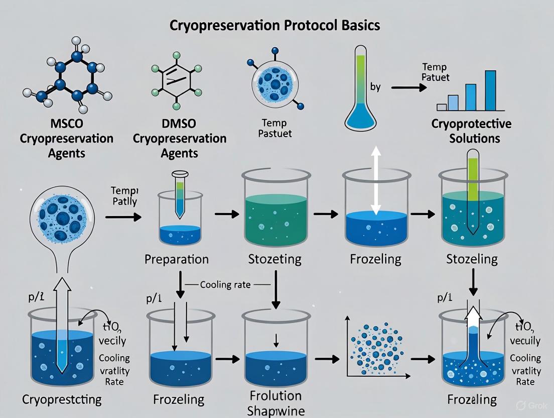
The following diagram illustrates the standardized slow-freezing protocol for MSCs, incorporating critical quality control checkpoints:
Diagram 1: MSC Cryopreservation Workflow. This standardized protocol ensures consistent cooling rates and incorporates essential quality control checkpoints for reliable MSC biobanking.
The freezing media composition varies based on application and regulatory requirements. Research-grade formulations often consist of culture medium with 10% fetal bovine serum (FBS) and 10% DMSO, while clinical applications increasingly use defined, serum-free formulations like CryoStor CS10 or specialized media such as MesenCult-ACF Freezing Medium specifically designed for MSCs [1]. Optimal cell concentration for freezing typically ranges from 1×10^6 to 1×10^7 cells/mL to balance viability and practical storage considerations [1].
Critical Thawing and Post-Thaw Processing Protocol
Proper thawing procedures are equally crucial for maintaining MSC viability and function. The recommended protocol involves:
Rapid Thawing: Remove vials from liquid nitrogen and immediately place in a 37°C water bath or dry heating device with gentle agitation until only a small ice crystal remains [2] [1]. Rapid thawing minimizes damage from ice recrystallization.
Controlled CPA Removal: Immediately after thawing, transfer cell suspension to a centrifuge tube containing pre-warmed culture medium and centrifuge at 300-400 × g for 5-10 minutes to remove CPAs [2] [8]. Gradual dilution is critical to prevent osmotic shock.
Viability Assessment: Resuspend the cell pellet in fresh culture medium and assess viability using trypan blue exclusion or automated cell counting systems [1].
Culture or Administration: Plate cells at appropriate density for expansion or prepare for immediate administration to patients, depending on the application.
For clinical applications where DMSO toxicity is a concern, additional washing steps may be incorporated, though this must be balanced against potential cell loss during processing [5]. Studies indicate that careful post-thaw processing can recover >90% of viable MSCs from DMSO-free cryopreservation systems [7].
Essential Research Reagents and Materials
Successful implementation of MSC cryopreservation protocols requires specific reagents and equipment designed to maintain cell viability and functionality throughout the freezing and thawing processes. The following table details key solutions and their applications:
Table 4: Essential Reagents for MSC Cryopreservation Research
| Reagent/Category | Specific Examples | Function and Application |
|---|---|---|
| Cryoprotectant Agents | DMSO, glycerol, ethylene glycol, sucrose, trehalose [2] [7] | Protect cells from freezing damage; DMSO remains clinical standard despite toxicity concerns [2] |
| Commercial Freezing Media | CryoStor CS10, MesenCult-ACF Freezing Medium, mFreSR [1] | Pre-formulated, standardized media optimized for specific cell types; often serum-free and GMP-manufactured [1] |
| Cryogenic Containers | Nalgene Mr. Frosty, Corning CoolCell [1] | Provide controlled cooling rate (~-1°C/minute) when placed in -80°C freezer [1] |
| Cryogenic Storage Vials | Corning Cryogenic Vials (internal-threaded recommended) [1] | Sterile, leak-resistant containers designed for liquid nitrogen storage; prevent contamination [1] |
| Quality Control Assays | Flow cytometry (CD73, CD90, CD105), CFU-f assays, differentiation kits [8] [7] | Verify MSC identity, viability, and functional potency pre- and post-cryopreservation [8] |
| Liquid Nitrogen Storage Systems | Liquid nitrogen tanks, automated monitoring systems [1] | Maintain long-term storage at -135°C to -196°C; ensure sample integrity and traceability [1] |
The selection of appropriate reagents should consider the specific MSC source (bone marrow, adipose tissue, umbilical cord), intended application (research vs. clinical use), and regulatory requirements. For clinical development, cGMP-manufactured, fully-defined cryopreservation media are recommended to ensure consistent production and quality control [1].
Cryopreservation stands as a non-negotiable component in the translational pathway of MSC therapies from laboratory research to clinical application. The technical evidence presented in this guide substantiates that properly executed cryopreservation protocols effectively maintain MSC viability, recovery, and critical therapeutic functions, including multilineage differentiation capacity and in vivo tissue repair capabilities. While methodological refinements continue to emerge—particularly in the development of DMSO-free cryoprotectant solutions and standardized freezing/thawing workflows—the fundamental necessity of cryopreservation for ensuring the off-the-shelf availability, quality control verification, and practical distribution of MSC products remains unchallenged.
As the field advances toward more widespread clinical implementation of MSC therapies, further optimization of cryopreservation techniques will be essential for maximizing post-thaw cell functionality and minimizing procedural variations. The protocols, data, and methodologies detailed herein provide a foundational framework for researchers and therapy developers establishing robust MSC biobanking operations. Through continued refinement and standardization of these critical preservation protocols, the field can accelerate the delivery of consistent, potent, and accessible MSC-based treatments to patients worldwide.
The cryopreservation of Mesenchymal Stromal Cells (MSCs) represents a cornerstone technology for regenerative medicine, enabling the biobanking of cellular products for research and clinical applications. The fundamental challenge in cryopreservation lies in navigating the physical processes of water phase change and solute redistribution that occur during freezing and thawing. These processes can inflict severe damage on cells through two primary, interconnected mechanisms: intracellular ice formation (IIF) and solute-effect injury [9]. Intracellular ice formation is perhaps the most critical cause of cell injury during cryopreservation, as ice crystals can mechanically disrupt membranes and subcellular structures [9]. Simultaneously, the concentration of solutes, both biological and from cryoprotective agents (CPAs), can lead to deleterious osmotic shifts and chemical toxicity [9]. The "two-factor hypothesis of cryoinjury," first proposed in 1970s, formally delineates these two distinct pathways of damage [9]. A sophisticated understanding of these thermodynamic and physical processes is therefore essential for developing optimized cryopreservation protocols that maintain MSC viability, functionality, and therapeutic potential post-thaw.
Physical Mechanisms of Cell Damage During Freezing
Intracellular Ice Formation (IIF)
Intracellular ice formation is a catastrophic event for a cell during freezing. The formation of ice crystals within the cell's cytoplasm can puncture and rupture vital organelles and the plasma membrane, leading to immediate cell death [9]. The mechanism of IIF is complex and involves nucleation and propagation.
Nucleation and Propagation: Ice typically first forms in the extracellular space. This extracellular ice can then trigger IIF through two debated mechanisms: surface-catalyzed nucleation, where the extracellular ice surface promotes ice formation in the adjacent cytoplasm, or ice growth through membrane pores, where extracellular ice physically grows into the cell through imperfections or pores in the membrane [9]. The role of intercellular connections is also critical. Research using high-speed videomicroscopy has shown that in confluent cell layers, the presence of gap junctions can enhance the cell-to-cell propagation of intracellular ice [9]. Paradoxically, a more recent study on mouse insulinoma cells revealed that cells lacking specific junction proteins (gap, adherens, and tight junctions) actually froze at higher temperatures than wild-type cells. This suggests that junction proteins may influence ice formation by affecting the penetration of extracellular ice into the paracellular space, indicating the phenomenon is more complex than previously appreciated [9].
Consequences of IIF: The physical damage is direct and often irreversible. Ice crystals compromise the structural integrity of the cytoskeleton, nuclear envelope, and other membrane-bound systems, leading to necrotic cell death.
Solute-Effect Injury and Osmotic Imbalance
In parallel to the mechanical threat of ice, cells face a more insidious, chemical form of damage known as solute-effect injury. As extracellular water freezes, it forms pure ice crystals, effectively removing water from the solution. This consequently concentrates the remaining dissolved solutes—including electrolytes like sodium and potassium, as well as any CPAs like Dimethyl Sulfoxide (DMSO)—in the unfrozen fraction [9] [10]. This creates a severe osmotic imbalance across the cell membrane.
Cell Volume Excursions: In response to the hypertonic extracellular environment, water rapidly exits the cell through osmosis in an attempt to equilibrate chemical potentials. This causes cell dehydration and severe shrinkage [10] [11]. If the volumetric excursions exceed the cell's tolerance, the mechanical stress can cause lysis. This is particularly problematic during the addition and removal of hypertonic CPA solutions [10]. Cells are more sensitive to osmotic stress post-thaw, and improper dilution can lead to significant cell loss as water rushes in, causing excessive swelling [10].
Solute Toxicity: The increased concentration of solutes can have several detrimental effects:
- Protein Denaturation: High salt concentrations can disrupt the hydration shells around proteins, leading to their denaturation and loss of function [12].
- CPA Toxicity: Cryoprotective agents like DMSO, while necessary, are toxic to cells at high concentrations and with prolonged exposure. DMSO has been shown to alter the cytoskeleton, shift cell metabolism, and change membrane fluidity [10].
- Oxidative Stress: The stressful conditions can elevate reactive oxygen species, leading to oxidative damage of cellular components.
The cellular response to hypertonic stress involves the activation of protective transcription factors like Nuclear Factor of Activated T cells 5 (NFAT5). NFAT5 promotes the expression of organic osmolytes—small, benign solutes that help restore cell volume by allowing osmotic influx of water without the damaging effects of high ionic strength [11].
Table 1: Key Mechanisms of Cryoinjury and Their Consequences
| Mechanism | Primary Cause | Cellular Consequence | Final Effect |
|---|---|---|---|
| Intracellular Ice Formation | Rapid cooling, insufficient CPA | Mechanical rupture of membranes & organelles | Immediate necrotic cell death |
| Solute-Effect Injury | Slow cooling, extracellular ice formation | Osmotic dehydration & excessive shrinkage | Lysis, protein denaturation |
| CPA Toxicity | High [CPA], prolonged exposure | Alteration of cytoskeleton & metabolism | Functional impairment, apoptosis |
Experimental Methodologies for Studying Cryoinjury
Understanding the physics of freezing requires sophisticated tools to visualize and quantify the dynamics of ice formation and cellular response. The following experimental approaches are central to this field.
High-Speed Video Cryomicroscopy
This technique allows for the direct, real-time observation of ice formation within cells.
Detailed Protocol:
- Cell Preparation: Culture cells of interest (e.g., MSCs, mouse insulinoma cells) on specialized, optically clear cryostages or micropatterned surfaces to control cell-cell interactions [9].
- Temperature Control: Place the cryostage on a microscope equipped with a precise temperature control system capable of linear cooling and warming rates (e.g., 130°C/min) [9].
- Imaging: Initiate cooling while recording the sample using a high-speed camera (capable of hundreds to thousands of frames per second).
- Analysis: Review footage to determine the precise temperature and location of the first ice formation (extracellular vs. intracellular), the sequence of freezing in cell pairs, and the rate of ice propagation [9].
Key Applications:
- Elucidating the role of junctional proteins in intercellular ice propagation [9].
- Differentiating between surface-catalyzed nucleation and pore propagation models [9].
- Validating probabilistic models of intracellular ice formation.
Synchrotron-Based X-Ray Diffraction
This method provides a quantitative, non-optical assessment of ice crystal structure, volume fraction, and grain size within cryopreserved samples, even in lipid-rich cells like oocytes that are difficult to image optically [13].
Detailed Protocol:
- Sample Preparation: Equilibrate cells (e.g., bovine oocytes as a model for mammalian cells) in CPA solutions. Mount them on cryo-loops or Cryotop supports [13].
- Cooling and Data Collection: Plunge-cool the samples in liquid nitrogen at controlled rates (e.g., ~30,000 °C/min or higher). Transfer the cryocooled sample to a synchrotron beamline with a -173°C nitrogen gas stream to maintain temperature.
- Diffraction Measurement: Expose the sample to a high-energy X-ray beam and collect the 2D diffraction pattern on a detector.
- Data Analysis: Azimuthally integrate the 2D pattern to generate a 1D diffraction profile. Analyze the profile to determine the phase of water present (vitreous ice, stacking-disordered ice Isd, or hexagonal ice Ih) and quantify the ice fraction [13].
Key Findings:
- A critical discovery is that oocytes cooled with standard protocols show no ice after cooling but develop large ice fractions during warming, meaning most ice-related damage occurs during the thawing phase [13].
- This technique can precisely correlate ice structure (e.g., the hexagonal plane fraction in Isd) with freezing parameters [13].
Cell Viability and Functional Assays
Post-thaw assessments are crucial for linking physical freezing events to biological outcomes.
Core Protocol Elements:
- Thawing: Rapidly warm cryovials in a 37°C water bath (>60°C/min warming rate) until ice is just dissolved [2] [10].
- CPA Removal: Dilute the thawed cell suspension in a stepwise manner or use centrifugation to remove toxic CPAs like DMSO, minimizing osmotic shock [2] [10].
- Viability Assessment: Use membrane integrity dyes like Trypan Blue in conjunction with automated cell counters or flow cytometers.
- Functional Assays:
- Clonogenicity: Perform Colony-Forming Unit (CFU-f) assays by plating a defined number of MSCs and counting distinct colonies after 14 days [8].
- Multilineage Differentiation: Culture thawed MSCs in osteogenic, adipogenic, and chondrogenic induction media to confirm retained differentiation potential [8] [2].
- Proliferation Assays: Measure population doubling times or use metabolic assays like MTT to assess growth kinetics post-thaw.
The diagram below illustrates the logical workflow and decision points in a comprehensive cryoinjury study.
(Diagram: A workflow for a comprehensive cryoinjury study, integrating physical and biological analysis.)
Quantitative Data on Freezing Parameters and Cell Survival
The survival of MSCs through the cryopreservation process is highly dependent on the precise control of thermodynamic parameters and the composition of the cryopreservation medium. The data below summarize critical relationships between these variables and cellular outcomes.
Table 2: Impact of Cryopreservation Parameters on MSC Survival and Function
| Parameter | Typical Range | Quantitative Effect on MSCs | Experimental Context |
|---|---|---|---|
| Cooling Rate | 1 °C/min (slow) | ~70-80% survival with slow freezing [2]. | Cell suspensions in DMSO-containing media [2]. |
| >60,000 °C/min (vitrification) | Ice-free vitrification achievable; survival dependent on CPA [13]. | Bovine oocytes model; higher rates reduce required CPA [13]. | |
| Warming Rate | >60 °C/min | Considered a minimum for conventional cryopreservation [10]. | Critical to avoid recrystallization; most damaging ice forms during slow warming [13]. |
| ~20x standard rate | New convective warming demonstrates potential for ice-free preservation with lower CPA [13]. | Experimental system for bovine oocytes [13]. | |
| DMSO Concentration | 10% (v/v) | Standard for slow freezing; ~1400 mOSM, requires controlled addition/removal [10] [12]. | Toxicity and osmotic damage are key concerns [10]. |
| Post-thaw Viability | N/A | BMAC-MSCs retained proliferation & chondrogenic differentiation after 4 weeks at -80°C [8]. | Clinical-scale MSC-based product [8]. |
| Intracellular Ice Observation | N/A | No ice after cooling; large ice fractions form during warming in standard protocols [13]. | Synchrotron XRD on bovine oocytes in VS [13]. |
Table 3: Cryoprotectant Agents (CPAs) and Their Functions in MSC Cryopreservation
| CPA Category & Examples | Molecular Weight | Primary Mechanism of Action | Key Considerations for MSC Cryopreservation |
|---|---|---|---|
| Penetrating (Endocellular) | |||
| Dimethyl Sulfoxide (DMSO) | 78 g/mol | Penetrates cell, binds water, reduces IIF, depresses freezing point [12]. | Gold standard but toxic; alters cytoskeleton & metabolism; requires post-thaw removal [10] [12]. |
| Glycerol | 92 g/mol | Similar to DMSO [12]. | Lower toxicity but generally less effective cryoprotection for MSCs [12]. |
| Ethylene Glycol | 62 g/mol | Similar to DMSO [12]. | Lower toxicity than DMSO but cryopreservation effect can be similar [12]. |
| Non-Penetrating (Exocellular) | |||
| Sucrose | 342 g/mol | Extracellular osmolyte; draws water out, reduces IIF risk, minimizes osmotic shock [12]. | Common supplement in vitrification solutions and for controlling osmotic shifts [12]. |
| Trehalose | 342 g/mol | Similar to sucrose; also stabilizes membranes & proteins [12]. | Not metabolized by mammalian cells; effective extracellular CPA [12]. |
| Hydroxyethyl Starch (HES) | >10,000 g/mol | Increases solution viscosity, inhibits ice crystal growth, protects extracellularly [12]. | Used in clinical settings (e.g., BMAC processing); does not penetrate cells [12]. |
The Scientist's Toolkit: Essential Research Reagents and Materials
The following table catalogs key materials and reagents essential for conducting experiments on the physics of freezing and MSC cryopreservation.
Table 4: Essential Research Reagents and Materials for Cryoinjury Studies
| Item | Function/Application | Specific Examples & Notes |
|---|---|---|
| Controlled-Rate Freezer | Precisely lowers sample temperature at defined rates (e.g., -1°C/min to -50°C) for slow freezing [2] [10]. | Critical for reproducible slow-freezing protocols. |
| Passive Freezing Device | Provides an approximate cooling rate when placed in a -80°C freezer; a low-cost alternative. | "Mr. Frosty," "CoolCell" [8] [10]. |
| High-Speed Video Cryomicroscope | Direct visualization of ice formation dynamics and propagation in real-time [9]. | System includes temperature-controlled stage and high-frame-rate camera [9]. |
| Synchrotron X-Ray Beamline | Quantitative analysis of ice phase, fraction, and crystal size within cryopreserved samples [13]. | Provides unparalleled sensitivity for detecting intracellular ice [13]. |
| Penetrating Cryoprotectant | Primary agent to suppress intracellular ice formation (IIF). | DMSO, Glycerol, Ethylene Glycol [12]. |
| Non-Penetrating Cryoprotectant | Supplements penetrating CPAs; helps control osmotic balance and reduce IIF risk. | Sucrose, Trehalose, Hydroxyethyl Starch (HES) [12]. |
| Cryopreservation Medium | The solution in which cells are frozen; typically contains base medium, CPAs, and protein (e.g., FBS/plasma). | e.g., 10% DMSO + 90% autologous plasma [8]. |
| Dry Shipper | Secure transportation of cryopreserved samples while maintaining temperatures below -150°C [10]. | Prevents transient warming and ice crystal growth during transport. |
The interplay of cryoprotectants and the balance between preventing ice formation and minimizing toxicity is a central concept, visualized below.
(Diagram: The dual mechanisms of cryoprotectant action against primary cryoinjury pathways.)
The physics of freezing presents a formidable challenge in the cryopreservation of MSCs, with cell death primarily arising from the dual threats of intracellular ice formation and solute-imbalance injury. A deep understanding of these mechanisms—including the dynamics of ice nucleation and propagation, the critical importance of both cooling and warming rates, and the osmotic consequences of freeze-concentration—is non-negotiable for protocol development. Modern investigative tools like high-speed videomicroscopy and synchrotron X-ray diffraction have been instrumental in moving the field from observation to prediction. For MSC researchers and therapy developers, this knowledge translates directly into robust, validated protocols. By systematically optimizing CPA composition, controlling cooling and warming kinetics, and employing rigorous post-thaw functional assays, it is possible to significantly mitigate cryoinjury. This ensures that cryopreserved MSCs are not merely viable but retain their critical therapeutic properties, thereby enhancing the reliability and efficacy of cellular products for research and clinical application.
The successful cryopreservation of mesenchymal stromal cells (MSCs) is a cornerstone of modern regenerative medicine and biomedical research, enabling the establishment of biobanks for quality-controlled, readily available cell therapies [12]. The foundation of this process lies in the use of cryoprotective agents (CPAs) that protect cells from the lethal physical and chemical stresses encountered during freezing and thawing. Without these protective compounds, intracellular ice crystallization and osmotic stress would irreversibly damage cellular structures, rendering MSCs non-viable for therapeutic applications [14]. The cryoprotectant landscape is broadly divided into two functionally distinct categories: penetrating (endocellular) and non-penetrating (exocellular) agents, each with characteristic mechanisms, advantages, and limitations [12] [15].
Understanding the precise mechanisms of these cryoprotectant classes is particularly crucial for MSC research due to the expanding clinical applications of these cells in treating conditions ranging from graft-versus-host disease and cardiovascular diseases to COVID-19 complications [12] [6]. The choice between penetrating and non-penetrating cryoprotectants directly impacts MSC viability, recovery rates, functionality, and ultimately, patient safety [2] [6]. As MSC-based therapies progress through clinical trials, standardized and optimized cryopreservation protocols become increasingly necessary to ensure consistent cell products and reliable therapeutic outcomes [6].
Fundamental Mechanisms of Action
Penetrating Cryoprotectants
Penetrating cryoprotectants, also known as endocellular or permeating agents, are characterized by their low molecular weight (typically under 100 daltons) and ability to cross cell membranes [15] [14]. This class includes dimethyl sulfoxide (DMSO), glycerol, ethylene glycol, and propylene glycol [12]. Their fundamental mechanism of action involves entering the intracellular compartment where they provide protection through multiple simultaneous pathways.
Once inside the cell, penetrating cryoprotectants form strong hydrogen bonds with intracellular water molecules, effectively disrupting the normal water structure and reducing the freezing point of the cellular solution [12] [14]. This hydrogen bonding capacity decreases the amount of water available to form ice crystals during the cooling process, thereby minimizing the mechanical damage caused by intracellular ice formation [15]. Additionally, by increasing the overall solute concentration within the cell, these agents reduce the extent of cell dehydration that would otherwise occur in response to extracellular ice formation [12]. This dehydration minimization helps maintain cellular volume within survivable limits and prevents the lethal increase in intracellular electrolyte concentrations that typically accompanies freezing [14]. The ability of some penetrating cryoprotectants, particularly DMSO, to interact with membrane phospholipids also contributes to membrane stabilization during the dramatic physical changes that occur during temperature transitions [16].
Non-Penetrating Cryoprotectants
Non-penetrating cryoprotectants, alternatively termed exocellular or non-permeating agents, consist of larger molecules (generally over 1,000 daltons) that cannot cross the cell membrane [15]. This category includes disaccharides like sucrose and trehalose, as well as high molecular weight polymers such as hydroxyethyl starch, polyvinylpyrrolidone (PVP), ficoll, and polyethylene glycol (PEG) [12] [14].
These compounds exert their protective effects exclusively in the extracellular environment through osmotically-driven mechanisms and specific interactions with extracellular ice. By creating an osmotic gradient across the cell membrane, non-penetrating cryoprotectants promote controlled cellular dehydration before freezing, thereby reducing the amount of freezable water inside the cell [2] [15]. This controlled dehydration minimizes the potential for intracellular ice formation, which is universally lethal to cells [14]. Simultaneously, these agents increase the viscosity of the extracellular solution at low temperatures, which inhibits the growth and recrystallization of ice during cooling and warming phases [12]. Some non-penetrating cryoprotectants, particularly certain polymers known as "ice blockers," actively interact with ice nuclei and crystal surfaces, preventing their expansion and thus protecting cell membranes from mechanical damage [15]. The disaccharide trehalose exhibits a unique protective mechanism by stabilizing membranes through direct interaction with phospholipid head groups, effectively replacing water molecules and maintaining membrane integrity during dehydration [17].
Table 1: Comparative Properties of Common Penetrating and Non-Penetrating Cryoprotectants
| Cryoprotectant | Molecular Type | Molecular Weight (Da) | Typical Working Concentration | Primary Mechanism |
|---|---|---|---|---|
| DMSO | Penetrating | 78.1 | 1.5-2 M (∼10%) | Intracellular hydrogen bonding, membrane stabilization |
| Glycerol | Penetrating | 92.1 | 1-2 M (∼10%) | Intracellular hydrogen bonding, freezing point depression |
| Ethylene Glycol | Penetrating | 62.1 | 1.5-2 M (∼10%) | Intracellular hydrogen bonding, rapid penetration |
| Trehalose | Non-penetrating | 378.3 | 50-250 mM | Membrane stabilization, extracellular glass formation |
| Sucrose | Non-penetrating | 342.3 | 0.1-0.5 M | Osmotic dehydration, extracellular vitrification |
| Hydroxyethyl Starch | Non-penetrating | 100,000-1,000,000 | 5-10% (w/v) | Extracellular viscosity modification, ice crystal inhibition |
Comparative Analysis of Cryoprotectant Types
The strategic selection between penetrating and non-penetrating cryoprotectants requires a thorough understanding of their comparative advantages, limitations, and optimal applications in MSC cryopreservation.
Molecular Characteristics and Toxicity Profiles
The most fundamental distinction between these cryoprotectant classes lies in their molecular size and membrane permeability, which directly influences their toxicity profiles and mechanisms of action. Penetrating cryoprotectants, by virtue of their intracellular activity, generally exhibit higher cytotoxicity compared to their non-penetrating counterparts [15]. This toxicity is concentration, temperature, and time-dependent, with documented effects including osmotic shock during addition/removal, protein denaturation, and induction of apoptotic pathways in MSCs [16]. DMSO, despite being the gold standard for MSC cryopreservation, has been associated with adverse clinical effects in patients, including allergic reactions and more serious complications such as seizures and cardiac arrest at high concentrations [12] [6]. Additionally, studies have demonstrated that DMSO can induce epigenetic changes and alter differentiation patterns in stem cells, raising concerns for therapeutic applications [18].
Non-penetrating cryoprotectants offer significantly improved toxicity profiles due to their extracellular localization [15]. However, their inability to protect intracellular components represents a major limitation, as they cannot prevent dehydration-induced damage to internal cellular structures [15]. When used at high concentrations to achieve sufficient protection, non-penetrating agents can create excessive osmotic stress, leading to detrimental cell shrinkage [14]. The disaccharide trehalose stands out for its exceptional biocompatibility and has received FDA approval for use in various biomedical products, making it an attractive candidate for clinical MSC cryopreservation [17].
Table 2: Comparative Advantages and Limitations in MSC Cryopreservation
| Parameter | Penetrating Cryoprotectants | Non-Penetrating Cryoprotectants |
|---|---|---|
| Primary Advantages | Comprehensive intracellular protection; Essential for vitrification protocols; Effective for complex cells and tissues [12] [15] | Lower cytotoxicity; Reduced osmotic stress during addition/removal; FDA-approved options available [15] [17] |
| Main Limitations | Concentration-dependent toxicity; Osmotic shock risk; Clinical side effects (e.g., DMSO) [6] [16] | Cannot prevent intracellular ice formation alone; Limited protection for internal structures; May require high concentrations [15] [14] |
| Ideal Applications | Vitrification; Stem cell banking; Complex tissue preservation [12] [15] | Cell suspension freezing; Blood product preservation; Combination protocols [12] [15] |
| Toxicity Considerations | Toxicity increases with concentration, temperature, and exposure time [16] | Generally low toxicity; Excessive concentration can cause osmotic damage [15] |
Functional Efficacy in MSC Preservation
The functional efficacy of cryoprotectants is ultimately measured by their ability to preserve MSC viability, recovery, and critical biological functions post-thaw. The slow-freezing method using 10% DMSO (approximately 1.5 M) remains the most widely adopted protocol for MSCs, typically yielding recovery rates of 70-80% [2]. However, recent multicenter studies have demonstrated that novel DMSO-free solutions containing combinations of non-penetrating agents like sucrose with supplementary components can achieve comparable results in terms of MSC viability, immunophenotype maintenance, and differentiation potential [6].
The protective efficacy of non-penetrating cryoprotectants is particularly constrained by their extracellular mode of action. While they effectively mitigate extracellular ice damage and osmotic stress, their inability to protect intracellular structures necessitates innovative delivery strategies. Recent research has explored techniques such as ultrasound-mediated membrane permeabilization with microbubbles to facilitate intracellular trehalose delivery, significantly enhancing its cryoprotective efficacy for MSCs [17]. This approach successfully preserves not only membrane integrity and cell viability but also the essential multipotency of MSCs, which is critical for their therapeutic utility [17].
Experimental Protocols and Methodologies
Standard Slow-Freezing Protocol with DMSO
The conventional slow-freezing method using DMSO as the primary penetrating cryoprotectant remains the benchmark protocol for MSC cryopreservation in both research and clinical settings [2]. The standard methodology involves specific steps:
MSCs are harvested at approximately 80% confluency, typically using enzymatic digestion with trypsin/EDTA, and resuspended at a concentration of 1-2 × 10^6 cells/mL in freezing medium [2] [19]. The freezing medium consists of culture medium (e.g., DMEM) supplemented with 10% fetal bovine serum (FBS) and 10% DMSO [19] [16]. The cell suspension is aliquoted into cryovials (1.0-1.8 mL per vial) and transferred to a controlled-rate freezing device or an insulated container pre-cooled to 4°C [2] [16]. The containers are placed at -80°C for 12-24 hours, achieving an approximate cooling rate of -1°C/min, which facilitates gradual cellular dehydration [2] [19]. Following this initial freezing phase, the cryovials are transferred to long-term storage in liquid nitrogen at -196°C [2].
For thawing, cryovials are rapidly warmed in a 37°C water bath with gentle agitation until only a small ice crystal remains (approximately 2-3 minutes) [2] [16]. The cell suspension is then transferred to pre-warmed culture medium and centrifuged to remove the DMSO-containing supernatant. The cell pellet is resuspended in fresh culture medium and transferred to culture vessels [19]. Critical considerations for this protocol include minimizing the duration of DMSO exposure at elevated temperatures due to its concentration-dependent toxicity and ensuring rapid processing during the thawing phase to prevent ice recrystallization [16].
Advanced Protocol: Ultrasound-Mediated Trehalose Delivery
An innovative protocol for utilizing the non-penetrating cryoprotectant trehalose via ultrasound-mediated intracellular delivery has been developed to overcome the membrane permeability limitation [17]. This methodology enables effective intracellular trehalose accumulation while avoiding DMSO-associated toxicity:
MSCs are harvested and resuspended at a density of 1 × 10^6 cells/mL in trehalose solutions ranging from 50-1000 mM in phenol-red-free DMEM [17]. SonoVue microbubbles are added to the cell suspension at 1% (v/v) concentration to facilitate ultrasound-mediated membrane permeabilization [17]. The cell-microbubble-trehalose mixture is transferred to cryotubes and subjected to ultrasound exposure using specific parameters: 0.5 MHz frequency, 0.25 MPa peak negative pressure, 100 ms pulse length, 2 s pulse repetition period, and 5-minute total exposure time [17]. Following ultrasonication, cells are processed for cryopreservation using standard slow-freezing methods without DMSO, cooled to -80°C at -1°C/min, and subsequently stored in liquid nitrogen [17].
The key advantage of this protocol is the successful intracellular delivery of trehalose, confirmed through confocal microscopy of rhodamine-labeled trehalose, which enables comprehensive protection against both extracellular and intracellular freezing damage [17]. This approach has demonstrated excellent preservation of MSC viability, membrane integrity, and, crucially, multipotent differentiation capacity post-thaw [17].
Diagram 1: Ultrasound-mediated trehalose delivery workflow for MSC cryopreservation, illustrating membrane poration and protection mechanisms.
The Scientist's Toolkit: Essential Research Reagents
Table 3: Essential Reagents for MSC Cryopreservation Research
| Reagent/Chemical | Function/Purpose | Specific Application Notes |
|---|---|---|
| Dimethyl Sulfoxide (DMSO) | Penetrating cryoprotectant | Gold standard CPA; Use high-purity, cell culture grade; Final concentration typically 10% (v/v); Associated with toxicity concerns [6] [16] |
| D-(+)-Trehalose Dihydrate | Non-penetrating cryoprotectant | Natural disaccharide; Membrane stabilization; Requires intracellular delivery for full efficacy; FDA-approved for biomedical use [14] [17] |
| Sucrose | Non-penetrating cryoprotectant | Osmotic buffer; Commonly used in combination with penetrating CPAs; Reduces required DMSO concentration [6] |
| Hydroxyethyl Starch (HES) | Non-penetrating cryoprotectant | High molecular weight polymer; Extracellular ice inhibition; Often used in combination protocols [12] [16] |
| SonoVue Microbubbles | Ultrasound contrast agents | Facilitate membrane poration during ultrasound-mediated trehalose delivery; Lipid-shelled microbubbles [17] |
| Fetal Bovine Serum (FBS) | Medium supplement | Provides additional macromolecular protection; 10-90% concentration in freezing media; Batch variability concerns [16] |
The strategic selection and application of penetrating versus non-penetrating cryoprotectants fundamentally influences the success of MSC cryopreservation for research and clinical applications. While penetrating cryoprotectants like DMSO provide comprehensive intracellular protection, their inherent toxicity drives the investigation of alternative strategies [6] [16]. Non-penetrating cryoprotectants offer superior biocompatibility but face significant limitations due to their inability to cross cell membranes [15] [14].
Future directions in MSC cryopreservation research are increasingly focused on combination approaches that leverage the strengths of both cryoprotectant classes while minimizing their individual limitations [12] [15]. The development of DMSO-free cryoprotectant solutions containing optimized combinations of non-penetrating agents like sucrose with supplementary components represents a promising advancement toward safer clinical applications [6]. Similarly, innovative physical methods such as ultrasound-mediated delivery demonstrate the potential to overcome the permeability barrier that has historically limited the efficacy of non-penetrating cryoprotectants like trehalose [17].
As cryopreservation protocol standardization becomes increasingly critical for the advancement of MSC therapies, understanding the precise mechanisms, advantages, and limitations of both penetrating and non-penetrating cryoprotectants will empower researchers to make informed decisions that optimize cell quality, functionality, and ultimately, therapeutic efficacy.
Diagram 2: Decision framework for selecting cryoprotectant strategies based on clinical requirements and technical constraints.
The cryopreservation of mesenchymal stromal cells (MSCs) represents a critical juncture in the pathway from laboratory research to clinical therapy. As the field of regenerative medicine advances, enabling the widespread "off-the-shelf" availability of MSC therapies hinges upon effective long-term storage strategies that preserve cell viability, functionality, and potency [2] [20]. Within this context, dimethyl sulfoxide (DMSO) has maintained its status as the gold standard cryoprotective agent (CPA) for over six decades, providing proven effectiveness in preventing intracellular ice crystal formation through its unique membrane-penetrating capabilities and hydrogen bonding with water molecules [21] [20]. However, despite its ubiquitous application in research and clinical settings, DMSO presents a complex paradox for the scientific community—delivering unparalleled cryoprotective efficacy while simultaneously introducing significant concerns regarding cellular toxicity and patient safety [7] [21]. This technical analysis comprehensively examines the dual nature of DMSO in MSC cryopreservation, evaluating its mechanistic basis, quantifying its performance against emerging alternatives, and addressing the clinical risk profiles associated with its use in cell-based therapies. By synthesizing recent multicenter study data, safety analyses, and comparative effectiveness research, this review provides researchers and therapy developers with evidence-based guidance for navigating the challenges of DMSO utilization while anticipating the forthcoming transition to next-generation cryopreservation platforms.
Efficacy and Performance of DMSO in MSC Cryopreservation
Mechanistic Basis of Cryoprotection
DMSO functions as a penetrating cryoprotectant through well-characterized biophysical mechanisms that directly counteract the primary drivers of freeze-thaw-induced cellular damage. Its relatively low molecular weight and membrane permeability enable rapid cellular entry, facilitating critical protective functions throughout the cryopreservation cycle. During the freezing phase, DMSO significantly reduces intracellular ice crystal formation by displacing water molecules within the cytoplasm and modifying ice crystal nucleation dynamics [21] [20]. Simultaneously, DMSO mitigates osmotic stress by equilibrating intra- and extracellular solute concentrations, thereby preventing excessive cellular dehydration and membrane rupture [2]. The molecular basis for these effects stems from DMSO's capacity to form strong hydrogen bonds with water molecules, effectively disrupting the organization of water into destructive crystalline structures while maintaining membrane integrity during phase transitions [20]. This dual-action protection has established DMSO-containing solutions at concentrations typically ranging from 5% to 10% (v/v) as the predominant cryopreservation medium for clinical-grade MSCs [5] [20].
Quantitative Performance Metrics
Recent multicenter investigations provide robust quantitative data on DMSO performance relative to emerging alternatives. A comprehensive 2024 international study conducted through the Production Assistance for Cellular Therapies (PACT) and Biomedical Excellence for Safer Transfusion (BEST) collaborative evaluated a novel DMSO-free solution (SGI - containing sucrose, glycerol, and isoleucine in Plasmalyte A) against traditional DMSO-containing formulations across seven manufacturing centers [7] [6]. The findings demonstrated that MSCs cryopreserved with DMSO-containing solutions maintained an average post-thaw viability of 89.8% (from a pre-freeze baseline of 94.3%), representing a statistically significant decrease of 4.5% (95% CI: 0.03-9.0%; P: 0.049) [7]. In parallel investigations, researchers directly compared multiple clinical-grade cryopreservation formulations, including NutriFreez (10% DMSO), PHD10 (Plasmalyte A/5% human albumin/10% DMSO), CryoStor CS5 (5% DMSO), and CryoStor CS10 (10% DMSO) [20]. These studies revealed that MSC products cryopreserved in solutions containing 10% DMSO displayed comparable viabilities and recoveries throughout a 6-hour post-thaw assessment window, whereas a decreasing trend in both parameters was observed with the 5% DMSO formulation (CryoStor CS5) [20]. Beyond immediate post-thaw metrics, DMSO-cryopreserved MSCs consistently maintained critical phenotypic markers (CD73, CD90, CD105) while lacking expression of hematopoietic markers (CD45, CD34, CD14, CD19, HLA-DR), confirming the preservation of MSC identity following freeze-thaw cycles [7] [2] [20].
Table 1: Comparative Performance of DMSO vs. DMSO-Free Cryoprotectants for MSCs
| Parameter | DMSO-Containing Solutions | DMSO-Free Solution (SGI) | Significance |
|---|---|---|---|
| Pre-freeze Viability | 94.3% (95% CI: 87.2-100%) | 94.3% (95% CI: 87.2-100%) | Baseline equivalent |
| Post-thaw Viability | 89.8% (decrease of 4.5%) | 82.9% (decrease of 11.4%) | P<0.001 for SGI decrease |
| Viable Cell Recovery | 87.3% (95% CI: 80.2-94.4%) | 92.9% (95% CI: 85.7-100.0%) | P<0.013 for DMSO being lower |
| Phenotype Maintenance | CD73+/CD90+/CD105+; CD45-/CD34-/CD14-/CD19-/HLA-DR- | Equivalent expression patterns | No significant differences |
| Global Gene Expression | Baseline profile maintained | No significant differences | Comparable profiles |
Impact on MSC Functionality
The functional competence of MSCs following DMSO cryopreservation represents a critical consideration for therapeutic efficacy. Research demonstrates that properly cryopreserved MSCs maintain essential immunomodulatory capabilities, including the capacity to inhibit T-cell proliferation and enhance monocytic phagocytosis—key mechanisms underlying their therapeutic potential in inflammatory and autoimmune conditions [20]. Importantly, studies comparing fresh versus cryopreserved MSCs have revealed no significant differences in these immunoregulatory functions, supporting the continued use of cryopreserved products in clinical applications [20]. However, some investigations have identified potential compromises in proliferative capacity following thaw, with MSCs cryopreserved in certain 5% DMSO formulations (CryoStor CS5) demonstrating approximately 10-fold reduced expansion potential compared to those preserved in 10% DMSO formulations when assessed after 6 days in culture [20]. This differential effect across DMSO concentrations highlights the nuanced relationship between cryoprotectant formulation and post-thaw functionality, emphasizing the need for comprehensive functional validation alongside standard viability and recovery metrics.
Clinical Concerns and Toxicity Profiles
Patient Safety and Adverse Event Risks
The administration of DMSO-cryopreserved cellular products introduces legitimate safety concerns that must be carefully managed in clinical settings. A comprehensive 2025 safety analysis reviewed data from 1,173 patients who received 1-24 intravenous infusions of DMSO-containing MSC products, providing substantial evidence for risk assessment [5] [22] [23]. This analysis revealed that the typical DMSO doses delivered via MSC therapies are substantially lower (2.5-30 times) than the widely accepted threshold of 1 g DMSO/kg body weight used in hematopoietic stem cell transplantation [5] [22]. When administered with appropriate premedication and monitoring protocols, these infusions resulted in only isolated infusion-related reactions, with most patients tolerating the procedure without significant adverse events [5] [23]. Commonly reported reactions include transient nausea, vomiting, abdominal cramps, and cardiovascular effects such as hypotension or hypertension, which are generally manageable with standard supportive care [23] [21]. More serious neurological events, including seizures or encephalopathy, remain rare and are typically associated with much higher DMSO exposures than those encountered in MSC therapy [23].
The concentration of DMSO in infusion solutions appears to be a critical factor influencing tolerability. Clinical experience indicates that solutions containing 10% (v/v) DMSO are generally well-tolerated, whereas higher concentrations (e.g., 28-40%) have been associated with hematological disturbances including hemolysis and hemoglobinuria [23]. This concentration-dependent toxicity profile underscores the importance of careful formulation design and potential dilution strategies prior to patient administration.
Cellular Toxicity and Functional Impacts
Beyond patient-level effects, DMSO demonstrates concentration-dependent and time-dependent cytotoxicity at the cellular level that may compromise therapeutic product quality. In vitro analyses confirm that DMSO exposure, particularly at concentrations exceeding 5-10% or with prolonged contact time post-thaw, can disrupt mitochondrial respiration, induce oxidative stress, and potentially trigger apoptotic pathways in sensitive cell populations [21]. These deleterious effects manifest as increased proportions of early apoptotic cells, reduced proliferative capacity, and in some cases, alterations to normal differentiation potential [24] [20]. The timing of DMSO exposure proves particularly critical, as thawed cells exhibit heightened vulnerability to DMSO-mediated damage during the post-thaw recovery phase [21]. Research comparing washed (DMSO-removed) versus diluted (5% DMSO-retained) MSC products immediately post-thaw revealed that despite similar viabilities at early time points, washed MSCs displayed a higher proportion of early apoptotic cells at 6 hours, suggesting that complete DMSO removal may introduce additional stress through secondary processing [24]. This paradoxical finding highlights the delicate balance required in managing DMSO exposure—weighing the compound's intrinsic toxicity against the procedural stresses imposed by its removal.
Regulatory and Manufacturing Considerations
The use of DMSO in therapeutic MSC products introduces significant complexities within regulatory and manufacturing frameworks. Regulatory agencies worldwide, including the FDA and EMA, require that DMSO employed in clinical-grade manufacturing meets stringent compendial standards (USP/Ph. Eur. grade) and is accompanied by comprehensive documentation supporting its quality, purity, and traceability [21]. These requirements necessitate the implementation of rigorous testing protocols covering DMSO sourcing, handling, and final product characterization. From a manufacturing perspective, DMSO's variable performance between suppliers and lots presents challenges to process consistency and validation, potentially introducing unwanted variability in critical quality attributes of final products [21]. Additionally, the industry trend toward DMSO minimization or elimination reflects growing recognition of these cumulative challenges, driving development of next-generation cryopreservation platforms that reduce regulatory complexity while maintaining product efficacy [21].
Emerging Alternatives and Protocol Innovations
DMSO-Free Cryopreservation Solutions
The documented limitations of DMSO have stimulated vigorous investigation into alternative cryopreservation strategies that maintain protective efficacy while reducing toxicity concerns. The international PACT/BEST multicenter study evaluated a novel DMSO-free formulation (SGI) incorporating sucrose, glycerol, and isoleucine in a Plasmalyte A base, representing one of the most comprehensive comparative assessments to date [7] [6]. This investigation demonstrated that while the SGI solution resulted in a more substantial decrease in immediate post-thaw viability (11.4% reduction versus 4.5% for DMSO), it achieved superior viable cell recovery (92.9% versus 87.3% for DMSO) while maintaining equivalent immunophenotype and global gene expression profiles [7]. These findings suggest that DMSO-free approaches may protect different cellular subpopulations or functions despite slightly reduced membrane integrity in some cells. Other investigative approaches have explored non-penetrating cryoprotectants including trehalose, sucrose, and various amino acids, often in combination with extracellular matrix components or biocompatible polymers that provide membrane stabilization during freeze-thaw cycles [5] [25]. Although none of these alternatives has yet achieved universal clinical adoption, the accelerating pace of development suggests that DMSO-free solutions will play an increasingly prominent role in future MSC therapy manufacturing.
Table 2: Experimental DMSO-Free Cryopreservation Approaches for MSCs
| Strategy | Key Components | Reported Efficacy | Limitations |
|---|---|---|---|
| Sugar-Based Solutions | Sucrose, trehalose, raffinose | 49-83% viability; 50-103% recovery | Variable performance across cell sources |
| Polymer Systems | Polyvinyl pyrrolidone, carboxylated poly-l-lysine, PEG-based copolymers | 63->90% viability | Potential immunogenicity concerns |
| Intracellular Delivery | Electroporation or nanoparticle-mediated trehalose delivery | 72-89% viability | Technical complexity, scalability challenges |
| Vitrification | High CPA concentrations with ultra-rapid cooling | 72-90% viability | Requires specialized equipment, sample volume restrictions |
| Combination Formulations | Sucrose + glycerol + isoleucine (SGI) | 82.9% viability; 92.9% recovery | Slightly reduced viability vs. DMSO |
Protocol Optimization and DMSO Reduction
Complementing the development of DMSO-free alternatives, significant research efforts have focused on optimizing conventional protocols to minimize DMSO-related risks while maintaining cryopreservation efficacy. One prominent strategy involves systematic DMSO concentration reduction from the traditional 10% to 5% or lower, often supplemented with non-penetrating cryoprotectants that provide extracellular protection [20]. Studies evaluating 5% DMSO formulations have demonstrated acceptable post-thaw viability, though some reports indicate potential compromises in long-term proliferative capacity and recovery compared to 10% DMSO formulations [20]. Additional technical adaptations include optimized cooling rate control, temperature-ramped centrifugation for DMSO removal, and post-thaw incubation in recovery media designed to mitigate oxidative stress and apoptotic signaling [24] [2]. The implementation of closed-system washing devices represents another advance, reducing contamination risk while facilitating more efficient DMSO removal before administration [23]. Collectively, these protocol refinements enable substantial reduction in final DMSO concentrations delivered to patients—in some cases achieving greater than 10-fold decreases compared to unprocessed products—while maintaining critical MSC functions and viability [24].
Experimental Design and Methodological Considerations
Standardized Cryopreservation Protocols
Robust experimental methodology is essential for valid comparison of cryopreservation approaches and accurate assessment of DMSO effects. The slow freezing method remains the predominant technique for MSC cryopreservation in both research and clinical settings, characterized by a controlled cooling rate of approximately -1°C/minute achieved through programmable freezing devices or passive cooling containers [2]. A typical protocol involves suspending MSC pellets at concentrations ranging from 3-9 million cells/mL in cryoprotectant solution, aliquoting into cryovials, initiating cooling at 4°C for brief equilibration, followed by controlled-rate freezing to -80°C before final transfer to liquid nitrogen vapor phase for long-term storage [7] [20]. Standardized thawing procedures employ rapid warming in a 37°C water bath with gentle agitation until complete ice dissolution, immediately followed by dilution with culture medium or specific reconstitution solutions to mitigate osmotic shock [2] [20]. Post-thaw assessments should incorporate multiple complementary metrics including membrane integrity (trypan blue exclusion), apoptotic status (Annexin V/PI staining), immunophenotype characterization (flow cytometry for CD73, CD90, CD105, and negative markers), and functional potency assays (immunomodulation, differentiation, metabolic activity) to comprehensively evaluate product quality [20].
Critical Parameter Assessment Framework
Comprehensive characterization of cryopreserved MSC products requires multi-parameter assessment spanning immediate post-thaw metrics through extended functional analyses. The following workflow outlines key evaluation timepoints and corresponding analytical methods essential for rigorous cryopreservation protocol validation:
Table 3: Essential Research Reagents for MSC Cryopreservation Studies
| Reagent Category | Specific Examples | Function & Application |
|---|---|---|
| Base Cryopreservation Solutions | Plasmalyte A, Normosol | Isotonic base solution for cryoprotectant formulation |
| Penetrating CPAs | DMSO (USP/Ph. Eur. grade), glycerol | Intracellular cryoprotection through ice crystal inhibition |
| Non-Penetrating CPAs | Sucrose, trehalose, hydroxyethyl starch | Extracellular cryoprotection, osmotic buffer |
| Protein Supplements | Human serum albumin (5%), platelet lysate | Membrane stabilization, ice recrystallization inhibition |
| Commercial Formulations | CryoStor (CS5, CS10), NutriFreez | Standardized, GMP-compliant cryopreservation kits |
| Viability Assessment | Trypan blue, Annexin V/PI, 7-AAD | Membrane integrity and apoptosis analysis |
| Phenotypic Characterization | CD73, CD90, CD105 antibodies; lineage negative markers | Identity confirmation and purity assessment |
| Functional Assay Reagents | T-cell proliferation kits, phagocytosis assays, differentiation media | Potency and functionality assessment |
The extensive body of evidence examined in this analysis confirms that DMSO remains an effective cryoprotectant for MSC-based therapies, delivering consistent post-thaw viability and functional preservation when employed within established parameters. However, the compound's recognized cellular toxicity and patient safety concerns—though generally manageable at the concentrations typically administered with MSC products—continue to drive innovation toward safer alternatives. The emerging data on DMSO-free solutions, particularly the promising results from multicenter validation studies, indicate that the field is progressing toward viable alternatives that may eventually supplant DMSO in clinical applications. In the interim, protocol optimization through DMSO concentration reduction, improved washing methodologies, and enhanced formulation design represents a prudent strategy for balancing efficacy and safety.
For researchers and therapy developers, the current evidence supports a context-dependent approach to cryoprotectant selection. In cases where established manufacturing protocols and regulatory approvals are already in place, DMSO retention with rigorous adverse event monitoring remains a defensible position, particularly given the extensive clinical experience with this agent. For new product development or when treating patient populations with potentially heightened sensitivity to DMSO, investment in DMSO-reduced or DMSO-free approaches appears warranted. As the field continues to evolve, the ideal cryopreservation platform will likely incorporate elements of both penetrating and non-penetrating cryoprotectants in optimized combinations that maximize cellular protection while minimizing patient risk. Through continued rigorous comparison of traditional and emerging approaches, the MSC research community can advance cryopreservation science to better support the safe and effective application of these promising cellular therapies.
The cryopreservation of mesenchymal stromal cells (MSCs) is a critical step in ensuring their off-the-shelf availability for regenerative medicine and cell therapy applications. For decades, dimethyl sulfoxide (DMSO) has been the predominant cryoprotectant of choice, leveraging its ability to penetrate cell membranes and suppress ice crystal formation. However, a growing body of evidence has revealed significant drawbacks associated with DMSO, including dose-dependent cellular toxicity and concerning side effects in patients upon infusion. DMSO has been shown to negatively impact cell function by affecting cellular metabolism, enzymatic activity, and even inducing unwanted differentiation [26]. Furthermore, clinical administration of DMSO-cryopreserved cell products has been associated with adverse reactions ranging from gastrointestinal and cardiovascular effects to respiratory complications [27] [26].
These concerns have catalyzed the search for DMSO-free cryopreservation strategies, particularly for clinically administered MSCs. Emerging alternatives often center on combinations of non-penetrating cryoprotectants, primarily sucrose, trehalose, and glycerol, which function through extracellular stabilization, osmotic control, and membrane protection mechanisms [12] [26]. This whitepaper evaluates these key DMSO-free formulations, providing a technical assessment of their efficacy, mechanisms, and practical application in MSC research and development.
Cryoprotectant Mechanisms and Classifications
Understanding the fundamental mechanisms of cryoprotection is essential for evaluating alternative formulations. Cryoprotective Agents (CPAs) are broadly classified into two categories based on their interaction with the cell membrane.
- Penetrating (Endocellular) Cryoprotectants: These are low molecular weight compounds that cross the cell membrane. They function by reducing the freezing point of intracellular water, minimizing the formation of lethal intracellular ice crystals, and buffering against solute-induced stress during dehydration. DMSO and glycerol are primary examples [2] [12].
- Non-Penetrating (Exocellular) Cryoprotectants: These are typically larger molecules, such as disaccharides (sucrose, trehalose) and polymers, that remain outside the cell. They provide protection by increasing extracellular osmolality, which promotes gentle cellular dehydration before freezing, thereby reducing the chance of intracellular ice formation. They also contribute to membrane stabilization, often by forming hydrogen bonds with phospholipid head groups [28] [12].
Table 1: Classification and Mechanisms of Common Cryoprotectants
| Type | Mechanism of Action | Examples | Key Characteristics |
|---|---|---|---|
| Penetrating (Endocellular) | Enters the cell; binds intracellular water to depress freezing point and prevent ice crystal formation. | DMSO, Glycerol, Ethylene Glycol | Low molecular weight; can exhibit cellular toxicity at high concentrations or prolonged exposure. |
| Non-Penetrating (Exocellular) | Remains outside cell; increases extracellular osmolality to dehydrate cell and stabilizes the cell membrane. | Sucrose, Trehalose, Hydroxyethyl Starch (HES) | Low toxicity; requires combination with other CPAs for effective cryopreservation. |
The following diagram illustrates the collaborative mechanism of penetrating and non-penetrating cryoprotectants in protecting a cell during the freezing process.
Critical Evaluation of Key DMSO-Free Formulations
Sucrose, Glycerol, and Isoleucine (SGI) Formulation
A significant advancement in DMSO-free cryopreservation is the SGI formulation, which combines Sucrose, Glycerol, and L-Isoleucine. This combination was systematically evaluated in an international, multicenter study published in 2024, comparing it directly to standard DMSO-containing solutions for cryopreserving MSCs from various tissue sources [6].
- Performance Data: The study concluded that the SGI solution was comparable to DMSO-based controls in key metrics including post-thaw cell viability, recovery, immunophenotype (expression of CD73, CD90, CD105), and tri-lineage differentiation potential. Notably, the gene expression profile of MSCs cryopreserved in SGI was also similar to those frozen in DMSO, suggesting maintained cellular function and identity [6].
- Proposed Mechanism: In this formulation, glycerol acts as a penetrating CPA, though with lower toxicity than DMSO. Sucrose serves as a non-penetrating CPA to manage osmotic stress. A critical innovation is the addition of L-isoleucine, an amino acid hypothesized to further stabilize the cell membrane and mitigate freezing-induced damage [6].
Trehalose-Based Formulations
Trehalose, a disaccharide known for its high glass transition temperature (Tg) and ability to stabilize biomolecules, is another promising candidate. Its unique chemical structure, with α(1→1)α-glycosidic bonds, provides greater molecular flexibility than sucrose, allowing it to form more effective hydrogen bonds with water and membrane phospholipids [28]. This enhances its capacity to promote cellular dehydration and protect membrane integrity during freezing.
- Comparative Efficacy: A 2025 retrospective clinical study on blastocyst cryopreservation, while on a different cell type, offers insightful comparative data. It found that trehalose-based vitrification solutions led to significantly higher implantation rates (52.84% vs. 43.94%) and a higher proportion of good-quality blastocysts (63.68% vs. 55.41%) compared to sucrose-based solutions [28]. This suggests potential superior cryoprotective properties of trehalose, though the results should be interpreted with caution as the solutions were not laboratory-controlled equivalents.
- Advanced Delivery Strategies: A key challenge with trehalose is its inability to cross the cell membrane. Research has explored techniques to facilitate its intracellular delivery, such as electroporation and nanoparticle-mediated delivery, to enhance its efficacy as a primary CPA [5] [27].
Commercial DMSO-Free Solutions
The research drive has translated into commercially available DMSO-free cryopreservation media. Studies have begun benchmarking these products. A 2022 study evaluating DMSO-free solutions for hematopoietic stem cells identified CryoProtectPureSTEM (CPP-STEM) as providing post-thaw cell viability, recovery, and potency equal or superior to DMSO/dextran-40 controls [29]. Other commercial solutions like CryoScarless (CSL) and Pentaisomaltose (PIM) have also shown promising, though variable, results depending on the cell type [27] [29].
Table 2: Summary of Key DMSO-Free Formulations and Evidence
| Formulation | Key Components | Reported Performance vs. DMSO Control | Notable Findings |
|---|---|---|---|
| SGI Solution [6] | Sucrose, Glycerol, L-Isoleucine | Comparable in viability, recovery, phenotype, and differentiation. | Validated in an international multicenter study; L-Isoleucine is a key additive for membrane protection. |
| Trehalose-Based [28] | Trehalose (often with other CPAs) | Significantly higher implantation & blastocyst quality in a embryo model. | Superior glass-forming ability; may require electroporation or nanoparticles for intracellular delivery. |
| CPP-STEM [29] | Undisclosed glycol derivatives & proteins | Equal or superior for HSC viability, recovery, and potency. | A commercially available, serum-free solution. Supported engraftment in a mouse model. |
Experimental Protocols for DMSO-Free Cryopreservation
Protocol: Slow Freezing of MSCs with SGI Formulation
The following protocol is adapted from the international multicenter study that validated the SGI solution [6].
- Preparation of SGI Freezing Medium: Prepare a solution containing Sucrose, Glycerol, and L-Isoleucine in a balanced salt solution. The exact concentrations may be proprietary, but the formulation is commercially available as Evia Bio's SGI solution.
- Cell Harvesting: Harvest MSCs at approximately 80% confluency during their maximum growth phase. Dissociate the cells using standard methods (e.g., trypsin/EDTA) and perform a cell count.
- Mixing with CPA: Centrifuge the cell suspension and carefully resuspend the cell pellet in the pre-chilled SGI freezing medium to a final concentration within the range of 1x10^6 to 10x10^6 cells/mL [6] [1].
- Aliquoting and Freezing: Aliquot the cell suspension into cryogenic vials. Immediately transfer the vials to a controlled-rate freezing container (e.g., "Mr. Frosty" or "CoolCell") and place it in a -80°C freezer for approximately 24 hours to achieve a cooling rate of about -1°C/min [1].
- Long-Term Storage: After 24 hours, promptly transfer the cryovials to a liquid nitrogen tank for long-term storage at or below -135°C [1].
The workflow for this protocol is summarized below.
Protocol: Intracellular Delivery of Trehalose via Electroporation
For trehalose to function as a more effective cryoprotectant, intracellular delivery is beneficial. This protocol outlines the use of electroporation for this purpose [5] [27].
- Preparation of Trehalose Solution: Prepare an electroporation buffer containing a high concentration of trehalose (e.g., 250-400 mM).
- Cell Preparation: Harvest and concentrate MSCs in the trehalose-containing electroporation buffer.
- Electroporation: Apply a controlled electrical pulse to the cell suspension. This temporarily disrupts the cell membrane, creating pores that allow trehalose to enter the cell. Optimization of voltage and pulse length is critical for cell type-specific viability.
- Post-Electroporation Recovery: Immediately after electroporation, transfer cells to a standard culture medium and allow them to recover. The cells can then be cryopreserved using a standard slow freezing protocol with the internalized trehalose contributing to cryoprotection.
The Scientist's Toolkit: Essential Reagent Solutions
Table 3: Key Reagents for DMSO-Free MSC Cryopreservation Research
| Reagent / Material | Function / Application | Examples / Notes |
|---|---|---|
| SGI Freezing Medium | A defined, DMSO-free cryopreservation solution for MSCs. | Evia Bio SGI Solution; validated in multicenter study [6]. |
| Commercial DMSO-Free Media | Ready-to-use, serum-free cryopreservation media. | CryoStor CS10 (serum-free), CryoProtectPureSTEM, CryoScarless [27] [29] [1]. |
| Controlled-Rate Freezer | Provides consistent, optimal cooling rate (~ -1°C/min). | Planer KRYO 10, CryoMed series; alternative: passive cooling devices (e.g., Nalgene Mr. Frosty) [1] [26]. |
| Electroporation System | Facilitates intracellular delivery of non-penetrating CPAs like trehalose. | Used in research to enhance trehalose efficacy [5] [27]. |
| L-Isoleucine | An amino acid additive for membrane stabilization in freezing media. | A key component of the SGI formulation [6]. |
The move toward DMSO-free cryopreservation of MSCs is no longer a theoretical pursuit but an active and validated field of development. Formulations based on sucrose, glycerol, and trehalose, particularly in combination, have demonstrated efficacy comparable to traditional DMSO-containing protocols in preserving MSC viability, recovery, and critical biological functions. The international validation of the SGI formulation marks a significant milestone, providing researchers with a credible and effective DMSO-free option [6].
Future progress will likely focus on several key areas:
- Mechanistic Deep-Dive: Further elucidating the precise role of additives like L-isoleucine in stabilizing the cell membrane during freeze-thaw cycles.
- Protocol Standardization: Establishing standardized, optimized protocols for a wider range of MSC tissue sources (adipose, umbilical cord, bone marrow) to ensure reproducibility.
- Advanced Delivery: Refining techniques like electroporation and nanoparticle-mediated delivery to safely and efficiently utilize non-penetrating cryoprotectants like trehalose as primary intracellular agents [5] [27].
For researchers and drug development professionals, the existing evidence strongly supports the adoption of validated DMSO-free formulations. Integrating these alternatives into MSC manufacturing pipelines can mitigate regulatory and safety concerns associated with DMSO, paving the way for safer and more effective cell-based therapies.
The field of regenerative medicine increasingly relies on mesenchymal stromal cells (MSCs) for therapeutic applications, ranging from the treatment of autoimmune and neurodegenerative diseases to osteochondral and cardiovascular repairs [12]. A fundamental challenge in clinical application involves the creation of biobanks for long-term storage, making cryopreservation not merely a storage formality but a pivotal step determining the ultimate therapeutic success [12] [30]. Cryoprotectants (CPAs) are specialized chemical compounds that protect biological systems from the potentially lethal damage associated with freezing and thawing [12]. They are indispensable for mitigating injuries caused by intracellular ice crystallization, solute concentration (solution effects), and cell dehydration during the cryopreservation process [2] [16]. The selection of an appropriate CPA protocol is particularly critical for MSCs, as it must preserve not only immediate post-thaw viability but also crucial therapeutic attributes, including multipotent differentiation potential, immunomodulatory functions, and paracrine activity [30] [31]. This guide provides a detailed, technical examination of CPA classification, mechanisms, and application protocols, specifically contextualized within the framework of MSC research for scientists and drug development professionals.
Core Classification and Mechanisms of Action
Cryoprotectants are systematically categorized based on their ability to cross the cell membrane. This fundamental property dictates their protective mechanism, toxicity profile, and ultimate application in cryopreservation protocols [12] [32].
Table 1: Fundamental Classification of Cryoprotectants for MSCs
| Category | Molecular Profile | Mechanism of Action | Common Examples |
|---|---|---|---|
| Endocellular (Penetrating) | Low molecular weight compounds [12] | Penetrate the cell membrane, form hydrogen bonds with intracellular water, lower freezing point, reduce intracellular ice formation [12] [16] | Dimethyl sulfoxide (DMSO), Glycerol, Ethylene Glycol, Propylene Glycol [12] |
| Exocellular (Non-Penetrating) | High molecular weight compounds [12] | Remain outside the cell, bind extracellular water, increase solution viscosity, inhibit ice crystal growth, protect against osmotic shock [12] [2] | Sucrose, Trehalose, Ficoll, Albumin, Polyvinylpyrrolidone, Hydroxyethyl Starch [12] |
The protective action during slow freezing is a coordinated effort between these two classes. Endocellular CPAs work from the inside to depress the intracellular freezing point and minimize ice formation. Simultaneously, exocellular CPAs create a protective extracellular environment that moderates osmotic stress and mechanically suppresses ice crystal growth [12] [2] [32]. As cells are cooled, water freezes extracellularly first, increasing the concentration of solutes outside the cell. This creates an osmotic gradient that draws water out, leading to protective dehydration. If the cooling rate is too high, water does not exit fast enough and freezes inside the cell, causing fatal damage. Endocellular CPAs mitigate this by reducing the amount of freezable water inside the cell, while exocellular CPAs help stabilize the cell membrane against the osmotic stress of dehydration [16].
Detailed Profiles of Key Cryoprotectants
Endocellular (Penetrating) Cryoprotectants
Dimethyl Sulfoxide (DMSO) is the most widely utilized penetrating CPA in MSC cryopreservation, typically at a concentration of 10% [16] [32]. Its efficacy stems from its low molecular weight, which allows rapid diffusion across the cell membrane where it disrupts ice crystal formation by forming strong hydrogen bonds with water molecules [16]. However, DMSO is associated with significant drawbacks, including chemical toxicity and osmotic shock, which can lead to reduced post-thaw viability and altered cellular functions [33] [16]. Furthermore, clinical administration of DMSO-cryopreserved cells can trigger adverse patient reactions, such as allergic responses and, in rare instances, severe complications like seizures or cardiac arrest [16] [32]. Glycerol is another penetrating agent but is less commonly used for MSCs than DMSO because it permeates cell membranes more slowly, increasing the risk of osmotic damage during addition and removal [32].
Exocellular (Non-Penetrating) Cryoprotectants
Trehalose is a natural disaccharide that has garnered significant interest as a biocompatible, non-toxic alternative or supplement to DMSO [17] [34]. Its mechanism is primarily extracellular; it stabilizes cell membranes by replacing water molecules and forming hydrogen bonds with phospholipid head groups during dehydration [17]. A major limitation for its use as a standalone CPA is that mammalian cells lack transporters for trehalose, preventing its intracellular accumulation unless assisted by specialized delivery techniques [17]. Sucrose functions similarly as an extracellular CPA, often used in combination with penetrating agents to allow for a reduction in the overall concentration of toxic CPAs like DMSO, thereby mitigating toxicity [2] [32]. Synthetic polymers like Hydroxyethyl Starch (HES) are high molecular weight exocellular CPAs that act by increasing the viscosity of the extracellular solution, which physically inhibits the growth of ice crystals [12] [16]. They are often used in combination cocktails with penetrating CPAs.
Table 2: Quantitative Comparison of Key Cryoprotectants Used in MSC Research
| Cryoprotectant | Class | Typical Working Concentration | Key Advantages | Key Disadvantages & Toxicity |
|---|---|---|---|---|
| DMSO | Endocellular | 5-10% [32] | Highly effective, rapid membrane penetration [12] | Cytotoxic, alters gene expression, causes clinical side effects [33] [16] |
| Glycerol | Endocellular | ~10% | Less toxic than DMSO [32] | Slow membrane penetration increases osmotic stress [32] |
| Trehalose | Exocellular | 50-1000 mM (varies with delivery) [17] | Biocompatible, FDA-approved, stabilizes membranes [17] | No passive uptake in mammalian cells [17] |
| Sucrose | Exocellular | 0.2-0.5 M [2] | Non-toxic, effective osmotic buffer | Purely extracellular action |
| HES | Exocellular | Varies | Inert, effective ice crystal inhibitor | Purely extracellular action |
Advanced Protocols and Synergistic Formulations
Protocol: Ultrasonication-Mediated Trehalose Delivery
A primary challenge in using trehalose is its inability to cross cell membranes. An advanced protocol using ultrasound and microbubbles (cavitation) has been developed to facilitate intracellular delivery, significantly enhancing its cryoprotective efficacy for MSCs [17].
1. Principle: Transient, non-lethal poration of the MSC membrane is induced by acoustic cavitation of microbubbles, creating temporary pores that allow trehalose to diffuse into the cell [17].
2. Materials and Reagents:
- Cell Suspension: MSCs at a density of 1 × 10^6 cells/mL in trehalose solution (optimized concentration range of 100-250 mM in phenol-red free DMEM) [17].
- Microbubbles: 1% (v/v) SonoVue microbubbles.
- Ultrasound Setup: A focused ultrasound source (e.g., 500 kHz), a passive cavitation detector (PCD) for monitoring, and a temperature-controlled exposure chamber maintained at 35°C [17].
3. Methodology:
- Preparation: Mix the cell suspension with the chosen trehalose concentration and add SonoVue microbubbles.
- Exposure: Place the sample in the exposure chamber. Apply ultrasound with optimized parameters: 0.25 MPa peak negative pressure, 500 kHz frequency, 100 ms pulse length, 2 s pulse repetition period, for a 5-minute total exposure time [17].
- Monitoring: Use the PCD to monitor cavitation signals in real-time, ensuring stable cavitation and minimizing inertial cavitation (associated with cell damage) [17].
- Post-Procedure: After exposure, cells are ready for standard cryopreservation. The protocol results in intracellular trehalose localization, confirmed via confocal microscopy, and yields high post-thaw viability while preserving MSC multipotency [17].
Synergistic CPA Cocktails
Research is increasingly focused on developing synergistic CPA cocktails that combine lower doses of penetrating agents with non-penetrating agents to reduce overall toxicity while maintaining efficacy.
- Urea and Glucose: Inspired by natural cryoprotection in hibernating frogs, a combination of 0.5M glucose and 0.5M urea has demonstrated a synergistic effect, providing cryoprotection to human MSCs that is comparable to a 5% DMSO reference [33]. Urea is believed to fluidify and destabilize membranes, potentially facilitating the uptake of other protective solutes like glucose during freezing [33].
- DMSO Reduction with Sugars: A common strategy involves combining reduced concentrations of DMSO (e.g., 5%) with sugars like sucrose or trehalose. The sugar acts as an extracellular stabilizer and osmotic buffer, compensating for the reduced amount of the penetrating CPA and mitigating its toxic impact [32].
The Scientist's Toolkit: Essential Reagents for CPA Research
Table 3: Key Research Reagent Solutions for MSC Cryopreservation Studies
| Reagent / Material | Function & Application | Example Use-Case |
|---|---|---|
| DMSO (Cell Culture Grade) | Gold-standard penetrating CPA; often used as a positive control in protocol development [12] [16] | 10% DMSO in culture medium or FBS for slow-freezing MSC suspensions [16] |
| Trehalose (Dihydrate) | Biocompatible, non-penetrating disaccharide; requires assisted delivery for full efficacy [17] | Used at 100-250 mM in ultrasonication protocols or as an additive in CPA cocktails [17] |
| SonoVue Microbubbles | Ultrasound contrast agent used to induce stable cavitation for membrane poration [17] | Facilitates intracellular delivery of trehalose at 1% (v/v) concentration [17] |
| Fetal Bovine Serum (FBS) | Common component of freezing media; provides proteins and other macromolecules that offer additional protection [16] | Often used at 80-90% in combination with 10% DMSO in classical freezing media [16] |
| Sucrose | Non-penetrating osmotic buffer and CPA; used to reduce required DMSO concentration [2] | Added at 0.2-0.5M to freezing media to mitigate osmotic shock and toxicity [2] |
| Hydroxyethyl Starch (HES) | High molecular weight exocellular CPA; inhibits ice crystal growth [12] [16] | Component of synthetic, serum-free freezing media in combination with penetrating CPAs [16] |
The meticulous classification of cryoprotectants into endocellular and exocellular categories provides a foundational framework for optimizing MSC cryopreservation. While DMSO remains the prevalent choice, its documented cytotoxicity and clinical side effects are driving the field toward innovative solutions [33] [32]. The future of CPA development lies in sophisticated, synergistic approaches that include cocktails of non-toxic agents (like urea and glucose), advanced physical delivery methods (like ultrasonication for trehalose), and the creation of completely serum- and DMSO-free freezing media [33] [17]. The ultimate goal is to standardize protocols that not only ensure high post-thaw viability but also faithfully preserve the critical therapeutic functionalities of MSCs, thereby unlocking the full potential of regenerative medicine. The choice of cryoprotectant strategy must be carefully evaluated in the context of the specific clinical application, as the impact of cryopreservation on MSC function can be disease-dependent [31].
From Culture to Cryovial: A Step-by-Step Protocol for MSC Cryopreservation
In the field of mesenchymal stromal cell (MSC) research, cryopreservation is a vital process that enables the long-term storage and future application of these valuable cells. However, the success of any cryopreservation protocol is fundamentally dependent on the quality of the cells before the freezing process begins. This technical guide establishes a comprehensive pre-freezing checklist designed to ensure cell health, optimal confluency, and mycoplasma-free status—three pillars that form the foundation of successful MSC cryopreservation. Adherence to this protocol is essential for maintaining the unique properties of MSCs, including their immunomodulatory capabilities, differentiation potential, and therapeutic efficacy [4] [35].
The pre-freezing phase represents a critical window where researchers can prevent the preservation of suboptimal cultures, thereby saving valuable time and resources that would otherwise be lost on non-viable or compromised cells. By implementing rigorous quality control measures at this stage, scientists can ensure that their cryopreserved MSC banks consist of cells with consistent characteristics and predictable performance upon thawing. This guide provides detailed methodologies and standardized protocols to assist researchers, scientists, and drug development professionals in establishing robust pre-freezing procedures that align with current best practices in MSC research [1] [2].
Assessing Cell Health and Viability
A comprehensive assessment of cell health and viability is the first critical step in the pre-freezing workflow. This evaluation should extend beyond simple viability metrics to include multiple parameters that collectively provide a holistic view of cellular fitness.
Table 1: Key Parameters for Pre-Freezing MSC Health Assessment
| Parameter | Assessment Method | Target Specification | Clinical/Research Significance |
|---|---|---|---|
| Viability | Trypan Blue Exclusion/Annexin V-PI Staining | ≥90% [35] | Ensures high recovery of living cells post-thaw |
| Morphology | Phase Contrast Microscopy | Fibroblast-like, spindle-shaped, homogeneous population [4] | Confirms MSC phenotypic characteristics |
| Proliferation Capacity | Population Doubling Time/CFU-f Assay | Colony formation >100 cells [8] | Demonstrates self-renewal capability |
| Metabolic Activity | MTT/WST-1 Assays | Consistent metabolic profile | Indicates functional, active cells |
| Membrane Integrity | LDH Release Assay | Low extracellular LDH | Confirms structural cellular integrity |
MSCs should be harvested during their maximum growth phase (log phase) to ensure optimal recovery post-thaw [1]. The concentration of cells for freezing is another critical consideration; typically, MSC suspensions should be within a general range of 1×10^3 to 1×10^6 cells/mL, though optimal concentration may vary depending on the specific MSC source and application requirements [1]. Researchers are advised to conduct pilot studies to determine the ideal cell concentration for their specific MSC type, as both excessively low and high concentrations can negatively impact post-thaw outcomes [1].
Determining Optimal Confluency for Cryopreservation
Cell confluency at the time of harvest is a crucial determinant of post-thaw viability and functionality. Optimal confluency ensures that MSCs are in their most robust physiological state, primed to withstand the stresses of the cryopreservation process.
Table 2: Confluency Guidelines for Different MSC Types
| MSC Source | Optimal Harvest Confluency | Key Indicators | Differentiation Potential Post-Thaw |
|---|---|---|---|
| Bone Marrow (BM-MSCs) | 80-90% [1] | Uniform, spindle-shaped morphology without contact inhibition signs | Preserved tri-lineage potential (osteogenic, adipogenic, chondrogenic) [2] |
| Adipose Tissue (AD-MSCs) | 80-90% | Dense, fibroblastic monolayer with minimal lipid vacuoles | Maintained adipogenic differentiation with proper lipid accumulation [4] |
| Umbilical Cord (UC-MSCs) | 80-90% | Elongated morphology with well-defined cytoskeleton | Sustained chondrogenic capacity and ECM production [4] |
For most MSC types, including bone marrow-derived MSCs (BM-MSCs), adipose tissue-derived MSCs (AD-MSCs), and umbilical cord-derived MSCs (UC-MSCs), the optimal confluency range prior to freezing is 80-90% [1]. This confluency level represents the late log phase of growth, where cells are actively proliferating but have not yet entered stationary phase or exhibited contact inhibition. Harvesting at this precise window ensures that MSCs retain their fundamental properties, including plastic adherence, specific surface marker expression, and multilineage differentiation potential [4]. Conversely, harvesting at low confluency (<70%) may yield cells that haven't reached their optimal metabolic state, while excessively high confluency (>95%) risks triggering early senescence or spontaneous differentiation, both of which can compromise post-thaw recovery and functionality [36].
Ensuring Mycoplasma-Free Status
Mycoplasma contamination represents one of the most significant and insidious threats to cell culture integrity, affecting an estimated 15-35% of continuous cell cultures worldwide [37]. These minute bacteria (0.2-0.8 µm) can pass through standard filters, are resistant to many conventional antibiotics, and typically don't cause visible turbidity in culture media, making them difficult to detect without specific testing [37] [38]. For MSC banking, ensuring mycoplasma-free status is non-negotiable, as cryopreservation can preserve these contaminants alongside the cells, compromising future experiments and therapeutic applications.
Prevention Strategies
Prevention forms the first line of defense against mycoplasma contamination. Implementation of strict aseptic techniques is fundamental, including the use of dedicated lab coats, sterile gloves, and minimal conversation in the cell culture area [39] [38]. All new cell lines引入 should be quarantined and tested before introduction to main laboratory spaces [37]. Additional preventive measures include:
- Regular cleaning of biosafety cabinets, incubators, and water baths [38]
- Filter sterilization of reagents using 0.1µm filters [37]
- Avoidance of routine antibiotic use which can mask low-level contamination [38]
- Using mouth pipetting alternatives to prevent oral mycoplasma species introduction [37]
Detection Methodologies
Regular testing is essential for detecting mycoplasma contamination. The following protocol describes a standardized PCR-based detection method:
Experimental Protocol: PCR-Based Mycoplasma Detection
Principle: This method utilizes mycoplasma-specific primers targeting conserved regions of the 16S rRNA gene, enabling highly sensitive detection of multiple mycoplasma species through polymerase chain reaction amplification.
Materials:
- Mycoplasma PCR detection kit (e.g., EZ-PCR Mycoplasma Detection Kit) [38]
- DNA template from test supernatant
- PCR-grade water
- Thermal cycler
- Gel electrophoresis equipment
- Positive and negative controls
Procedure:
- Sample Collection: Harvest 500µL of culture supernatant from test cells (90% confluent) after 72 hours without antibiotic treatment [38]
- Template Preparation: Centrifuge supernatant at 12,000× g for 10 minutes to pellet potential contaminants
- DNA Extraction: Resuspend pellet in PCR-compatible lysis buffer according to kit specifications
- PCR Setup:
- Prepare master mix according to manufacturer's instructions
- Add 2µL of template DNA to 23µL master mix
- Include provided positive and negative controls in each run
- Amplification Parameters:
- Initial denaturation: 95°C for 10 minutes
- 40 cycles of: 95°C for 30 seconds, 55°C for 30 seconds, 72°C for 1 minute
- Final extension: 72°C for 5 minutes
- Amplicon Detection:
- Analyze PCR products by 1.5% agarose gel electrophoresis
- Visualize with UV transillumination after ethidium bromide staining
- Interpretation: Compare sample band with positive control at approximately 500bp [38]
Frequency: Test all new cell lines prior to cryopreservation, quarterly for maintained cultures, and at the start of any long-term experiment [37] [38].
Integrated Workflow and Visual Guide
The following diagram illustrates the logical relationships and sequential workflow of the essential pre-freezing quality control checks for MSC cryopreservation:
The Scientist's Toolkit: Essential Research Reagent Solutions
Table 3: Essential Reagents and Materials for Pre-Freezing Quality Control
| Reagent/Material | Function | Application Notes |
|---|---|---|
| Mycoplasma Detection Kit (e.g., EZ-PCR Mycoplasma Detection Kit) [38] | Specific detection of mycoplasma contamination through PCR amplification | Use quarterly and on all new cell lines; culture without antibiotics before testing |
| Trypan Blue Solution | Viability staining through dye exclusion | Combine with hemocytometer for manual cell count and viability assessment |
| Annexin V-PI Apoptosis Detection Kit [35] | Differentiation between viable, apoptotic, and necrotic cell populations | More accurate than trypan blue for assessing freeze-sensitive cells |
| Cell Culture Antibiotics (Penicillin/Streptomycin) | Prevention of bacterial contamination | Use sparingly in stock cultures to avoid masking contamination [38] |
| 70% Ethanol Solution [39] | Surface decontamination for aseptic technique | Apply to all surfaces and containers before introducing to biosafety cabinet |
| Sterile Cryogenic Vials [1] | Secure containment for frozen cell storage | Use internal-threaded vials to prevent contamination during storage |
| Defined Cryopreservation Medium (e.g., CryoStor CS10) [1] | Protects cells during freezing process | Pre-formulated, serum-free options reduce variability and improve reproducibility |
| FBS/Platelet Lysate [36] | Culture supplement for MSC expansion | Ensure lot-to-lot consistency; pre-test for optimal growth support |
Implementation of this comprehensive pre-freezing checklist ensures that MSC cryopreservation begins with the highest quality cellular material, fundamentally influencing all subsequent outcomes. By systematically verifying cell health, confluency, and mycoplasma-free status, researchers establish a robust foundation for successful long-term MSC preservation. This protocol not only safeguards valuable cell lines but also enhances experimental reproducibility—a critical consideration in both basic research and clinical applications. The meticulous attention to pre-freezing quality control detailed in this guide represents an essential investment in research integrity, ultimately contributing to the advancement of reliable and therapeutically relevant MSC-based science.
The harvesting phase—encompassing cell detachment and centrifugation—serves as a critical gateway in the cryopreservation pipeline for Mesenchymal Stromal Cells (MSCs). This preparatory stage directly influences post-thaw viability, functionality, and ultimately, the therapeutic efficacy of the final product [40]. Suboptimal processing can induce cellular stress, compromise membrane integrity, and alter critical phenotypes, thereby introducing significant variability into both research outcomes and clinical applications [41] [40]. Within the framework of a Quality-by-Design (QbD) approach for biomanufacturing, detachment and centrifugation are identified as Critical Process Parameters (CPPs) that must be tightly controlled to ensure the integrity of Critical Quality Attributes (CQAs) such as viability, immunophenotype, and differentiation potential [41]. This guide details standardized, evidence-based protocols for these foundational steps, providing researchers and drug development professionals with methodologies to enhance the robustness and reproducibility of MSC-based products.
Core Principles of MSC Detachment
The detachment of adherent MSCs from culture surfaces is a balance between achieving high yield and preserving cellular health. The goal is to efficiently recover cells while minimizing damage to surface markers, adhesion receptors, and overall viability.
Enzyme Selection and Application
The choice of detachment enzyme is a primary determinant of harvest quality.
- TrypLE Express: This recombinant fungal-derived enzyme is a preferred alternative to animal-sourced trypsin. It operates effectively at room temperature and is less aggressive, leading to improved post-detachment viability and reduced cell surface protein damage [42]. It is specified in protocols for the culture of MSCs under serum-free and xeno-free conditions.
- Trypsin-EDTA: While traditional and effective, its proteolytic activity must be carefully monitored. Over-exposure can cleave functionally important surface receptors like CD73, CD90, and CD105, potentially compromising the cells' identity and functional capabilities as defined by International Society for Cellular Therapy (ISCT) criteria [4].
Standardized incubation is typically performed at 36–38°C for 3–5 minutes, though the specific duration should be determined empirically for each cell line and confluency level. Enzymatic activity must be neutralized promptly using a complete culture medium containing serum or proteins.
Mechanical Dislodgement and Monitoring
Following incubation, gentle mechanical dislodgement is achieved by tapping the culture vessel side-wise. It is crucial to avoid pipetting the cell suspension directly over the monolayer, as this can generate harmful shear forces. The process should be monitored under a microscope to confirm that cells have detached while remaining as single cells or small clusters. The cell suspension is then transferred to a sterile centrifuge tube for the next step.
Strategic Centrifugation for MSC Processing
Centrifugation is employed to pellet cells, thereby concentrating them and removing enzymatic residues and metabolic waste. The parameters of this step—specifically force and duration—are critical to prevent cytoskeletal disruption and activation of stress pathways.
Centrifugation Parameters
Quantitative studies have established that lower relative centrifugal force (RCF) is beneficial for MSC processing. A strategic protocol for the separation of MSCs from whole bone marrow and during differentiation phases utilizes a centrifugal force of 200 g [43]. This parameter is consistently applied in contemporary cryopreservation bioprocessing protocols for pelleting MSCs post-detachment and prior to resuspension in cryoprotective agent (CPA) solutions [40] [44]. This force, applied for 5 minutes at room temperature, effectively pellets the cells while preserving viability and functionality [40] [42].
Table 1: Standardized Centrifugation Parameters for MSC Harvesting
| Parameter | Specification | Rationale |
|---|---|---|
| Relative Centrifugal Force (RCF) | 200 g | Effectively pellets cells while minimizing mechanical stress and preserving cell function [43] [40]. |
| Duration | 5 minutes | Sufficient for cell recovery without unduly prolonging processing time [40]. |
| Temperature | Room Temperature | Maintains membrane fluidity, preventing cold-induced shock. |
| Brake Setting | Low or Off | Prevents pellet disruption during deceleration. |
Integrated Workflow: From Harvest to Cryopreservation
The detachment and centrifugation steps are integral components of a seamless workflow leading to cryopreservation. The following diagram illustrates the logical sequence and key decision points in this process.
Quality Assessment and Troubleshooting
Post-harvest quality control is essential. Cell count and viability, typically assessed using trypan blue exclusion and an automated cell counter, should be performed immediately after resuspension [40] [42]. A key study quantitatively demonstrated that cryopreservation reduces cell viability and metabolic activity immediately post-thaw, with recovery taking up to 24 hours [40]. This underscores the importance of a high-quality harvest. Yields are highly dependent on initial seeding density and confluency, but a well-optimized harvest should typically achieve >90% viability pre-freeze.
Table 2: Troubleshooting Common Harvesting Issues
| Problem | Potential Cause | Recommended Solution |
|---|---|---|
| Low Viability | Over-exposure to enzyme; harsh centrifugation. | Optimize incubation time; ensure force does not exceed 200 g; use gentle recombinant enzymes [40] [42]. |
| Poor Detachment | Enzyme inactivation; dense extracellular matrix. | Use fresh, pre-warmed reagent; consider a pre-wash with EDTA to chelate calcium. |
| Clumping | Incomplete neutralization; mechanical shear. | Ensure sufficient volume of neutralizing medium; avoid vigorous pipetting. |
| Loss of Phenotype | Excessive proteolytic activity. | Switch to a milder enzyme (TrypLE); reduce incubation time [42]. |
The Scientist's Toolkit: Essential Reagents and Materials
The following table details key reagents and materials critical for executing the protocols described in this guide, based on cited experimental methodologies.
Table 3: Essential Research Reagents and Materials for MSC Harvesting
| Item | Function / Specification | Example / Note |
|---|---|---|
| Detachment Reagent | Enzymatic release of adherent cells. | TrypLE Express (recombinant, xeno-free) is recommended for superior cell health [42]. |
| Centrifuge | Benchtop, capable of precise RCF control. | Must be calibrated to reliably achieve 200 g [43] [40]. |
| Centrifuge Tubes | Sterile, conical tubes. | 15 mL or 50 mL capacity, depending on scale. |
| Culture Medium | For enzyme neutralization and washing. | Serum-free formulations like StemPro MSC SFM are used in standardized protocols [42]. |
| Phosphate Buffered Saline (PBS) | Washing solution, Ca2+/Mg2+ free. | Used to rinse cells prior to detachment to remove serum that inhibits enzymes. |
| Automated Cell Counter | Quantifying cell count and viability. | e.g., Countess Automated Cell Counter, used for quality control [40] [42]. |
The meticulous execution of detachment and centrifugation is not merely a preliminary step but a cornerstone of successful MSC cryopreservation. By adhering to standardized protocols that specify gentle enzymatic treatment with reagents like TrypLE Express and a strategic centrifugation force of 200 g, researchers can significantly enhance the consistency, quality, and therapeutic potential of their MSC products [43] [40] [42]. Integrating these optimized harvesting techniques into a robust Quality-by-Design framework ensures that these critical process parameters are controlled, thereby safeguarding the critical quality attributes of the cells as they transition into the cryopreservation pipeline [41]. This disciplined approach is fundamental to advancing reliable MSC research and clinical application.
Cryopreservation is a critical step in the clinical application of mesenchymal stromal cells (MSCs), enabling the creation of cell banks for regenerative medicine and ensuring immediate availability of therapeutic products [12]. The process allows for the completion of essential quality control testing before batch release and provides logistical flexibility for clinical administration [36]. Without effective cryopreservation, MSCs would require continuous passage, potentially leading to epigenetic alterations and random genomic losses, thereby compromising their therapeutic potential [2]. The fundamental challenge in cryopreservation lies in minimizing freezing-induced damage, primarily caused by intracellular ice crystal formation and osmotic stress during the freeze-thaw cycle [12] [2]. Success depends on multiple factors, including cryoprotective agent (CPA) selection, freezing rate, and thawing protocol [12].
Cryoprotectants are categorized based on their ability to cross cell membranes. Penetrating cryoprotectants like dimethyl sulfoxide (DMSO) and glycerol enter cells and bind intracellular water, reducing ice crystal formation. Non-penetrating cryoprotectants like sucrose, trehalose, and hydroxyethyl starch remain extracellular, protecting cells from osmotic damage and inhibiting extracellular ice crystal growth [12]. The choice between formulating in-house media or utilizing commercial options represents a significant decision point for research and clinical teams, balancing control, consistency, safety, and regulatory compliance.
Cryoprotectant Formulation: Composition and Mechanisms
Core Components of Cryopreservation Media
Cryopreservation media, whether prepared in-house or purchased commercially, consist of a base solution supplemented with CPAs. The base is typically a culture medium or saline solution, often with added protein sources like fetal bovine serum (FBS) or human platelet lysate [12] [36]. The CPA components are strategically selected from penetrating and non-penetrating agents:
Penetrating (Intracellular) CPAs: These low-molecular-weight compounds penetrate the cell membrane and include DMSO, glycerol, ethylene glycol, and propylene glycol. Their primary mechanism involves forming hydrogen bonds with intracellular water molecules, reducing the freezing point, and minimizing intracellular ice crystal formation [12]. However, they exhibit concentration-dependent toxicity and must be carefully controlled [2].
Non-Penetrating (Extracellular) CPAs: These include oligosaccharides (sucrose, trehalose) and high molecular weight polymers (ficoll, polyvinylpyrrolidone, hydroxyethyl starch). They function by binding extracellular water, creating an osmotic gradient that promotes gentle cell dehydration, and physically shielding cells from ice crystals [12].
Table 1: Common Components of Cryopreservation Media for MSCs
| Component Type | Examples | Primary Function | Considerations |
|---|---|---|---|
| Penetrating CPA | DMSO (5-10%), Glycerol | Prevents intracellular ice formation | Cytotoxic at high concentrations/room temperature [2] [33] |
| Non-Penetrating CPA | Sucrose, Trehalose, Glucose | Osmotic stabilization, inhibits extracellular ice | Often used in combination with penetrating CPAs [33] [6] |
| Base Solution | Culture Medium (e.g., DMEM), Saline | Provides ionic and pH balance | May include buffers like HEPES [45] |
| Protein Additives | FBS, Human Serum Albumin, Platelet Lysate | Membrane stabilization, provides undefined factors | Shift towards xeno-free, defined components for clinical use [36] |
| Novel Additives | Isoleucine, RevitaCell Supplement | Enhances post-thaw recovery, ROCK inhibition | Improves viability in sensitive cells like pluripotent stem cells [45] [6] |
The Scientist's Toolkit: Essential Research Reagents
When designing cryopreservation experiments, researchers utilize a suite of key reagents. The selection is critical for reproducibility, cell viability, and functionality.
Table 2: Essential Reagents for MSC Cryopreservation Research
| Reagent / Material | Function in Cryopreservation | Example Use Case |
|---|---|---|
| DMSO (Cell Culture Grade) | Standard penetrating cryoprotectant | Often used at 10% in base medium or serum; requires post-thaw removal [45] |
| Sucrose or Trehalose | Non-penetrating osmotic balancer | Added at 0.1-0.5M to DMSO-containing or DMSO-free formulations to improve recovery [33] [6] |
| Clinical-Grade PL or HSA | Xeno-free protein source for media | Replaces FBS in clinical-grade formulations to enhance safety and regulatory compliance [36] |
| Controlled-Rate Freezer | Ensures reproducible cooling rate (~-1°C/min) | Critical for the slow freezing method; passive devices (e.g., "Mr. Frosty") are alternatives [46] |
| Viability Assay Kits | Measures post-thaw cell survival and function | e.g., Flow cytometry with Annexin V/PI; functional assays like CFU-f [46] |
| ROCK Inhibitor (Y-27632) | Enhances attachment and survival of thawed cells | Added to culture medium for 24 hours post-thaw to reduce apoptosis [45] |
In-House vs. Commercial Media: A Quantitative and Qualitative Comparison
Performance and Practicality
The decision between in-house and commercial media involves weighing performance, practicality, and regulatory requirements.
Table 3: Comparative Analysis: In-House vs. Commercial Freezing Media
| Parameter | In-House Formulated Media | Commercial Media |
|---|---|---|
| Composition & Control | High flexibility; can be tailored for specific MSC sources (e.g., BM, AD) and research goals [12] | Fixed, pre-optimized formulations; some are designed for specific cell types [45] [47] |
| Typical Viability/Recovery | Variable (e.g., ~55% with 0.5M Urea/Glucose [33]; ~70-80% with standard DMSO [2]) | Generally high and consistent (e.g., >90% reported for some GMP-grade media [48]) |
| Regulatory Path | Requires full in-house validation for GMP/clinical use, which is complex and resource-intensive [36] | Available with GMP documentation, some with NMPA/FDA approvals, simplifying regulatory submissions [45] [48] |
| Cost & Time Investment | Lower direct material cost but high labor and quality control cost [33] | Higher direct cost but saves significant R&D, preparation, and validation time [45] |
| Consistency & Reproducibility | Prone to batch-to-batch variability unless strict SOPs and QC are implemented [12] | High batch-to-batch consistency ensured by manufacturer's QC [45] [48] |
| Safety Profile | DMSO is common; associated with patient adverse effects; effort needed to develop less toxic options [33] [6] | Increasing availability of DMSO-free, xeno-free, and animal origin-free options [45] [6] |
Evidence from Multicenter Studies
Recent high-quality studies provide direct comparative data. An international multicenter study compared a novel DMSO-free solution (containing sucrose, glycerol, and isoleucine, or SGI) against standard in-house DMSO-containing solutions from seven different centers. The study found that post-thaw MSC viability was not significantly different between SGI (86.8% ± 10.3%) and in-house DMSO solutions (89.3% ± 7.5%) [6]. Furthermore, immunophenotype and differentiation capacity were maintained in both groups, demonstrating that well-designed alternative formulations can achieve parity with traditional methods.
Another study highlighted the synergistic effect of combining 0.5M urea and 0.5M glucose, which achieved 55% viability in human MSCs—a level comparable to 5% DMSO—showcasing the potential for in-house formulation innovation using safe, readily available agents [33].
Detailed Experimental Protocols for Formulation Comparison
To rigorously evaluate in-house versus commercial cryopreservation media, researchers should implement standardized freezing, thawing, and assessment protocols. The workflow below outlines the key stages of a typical comparison experiment.
Figure 1: Experimental Workflow for Comparing Cryopreservation Media. This diagram outlines the key stages for a standardized comparison between in-house and commercial freezing media, from cell preparation to post-thaw analysis.
Protocol 1: Slow Freezing Using a Passive Cooling Device
The slow freezing method, which allows for controlled dehydration, is the most common approach for cryopreserving MSCs [2].
- Step 1: Cell Preparation. Culture MSCs to approximately 80% confluence. Harvest cells using a standard detachment reagent like trypsin/EDTA or TrypLE. Perform a viable cell count and concentrate cells to a high density (e.g., 1-5 x 10^6 cells/mL) [46].
- Step 2: Media Mixing. Gently resuspend the cell pellet in either the pre-chilled commercial medium or the in-house formulation. For a typical in-house formulation, this could be 10% DMSO in 90% FBS or autologous plasma [46]. Mix gently to avoid osmotic shock.
- Step 3: Aliquot and Freeze. Dispense the cell suspension into cryogenic vials. Immediately place the vials into a pre-chilled passive freezing device (e.g., a "Mr. Frosty" filled with isopropanol). Place the container at -80°C for a minimum of 4 hours (or overnight). The device ensures a cooling rate of approximately -1°C/min, which is critical for slow freezing [46].
- Step 4: Long-Term Storage. After 24 hours, transfer the vials to the vapor phase of a liquid nitrogen storage system (-150°C to -196°C) for long-term preservation [2].
Protocol 2: Post-Thaw Assessment and Analysis
Accurate post-thaw analysis is crucial for evaluating media performance. Key parameters and methods are listed below.
- Viability and Recovery. Assess immediate post-thaw viability using trypan blue exclusion or automated cell counters. Calculate cell recovery: (Number of viable cells post-thaw / Number of viable cells pre-freeze) x 100. Many commercial media report >90% viability, while in-house formulations often achieve 70-90% [48] [6].
- Functionality Assays.
- Clonogenic Assay (CFU-f): Plate mononuclear cells at a low density (e.g., 300,000 cells per well in a 6-well plate) and culture for 14 days. Fix, stain with crystal violet, and count colonies containing >100 cells. This assesses the proliferative capacity of the progenitor population [46].
- Immunophenotype: Use flow cytometry to confirm the expression of classic MSC positive markers (CD73, CD90, CD105) and absence of hematopoietic markers (CD34, CD45, CD14) after thawing [36] [6].
- Differentiation Potential: Culture thawed MSCs in specific induction media for 2-3 weeks to confirm retained tri-lineage differentiation potential into osteocytes, adipocytes, and chondrocytes [36].
- Immunosuppressive Function: Employ an in vitro T-cell proliferation assay. Co-culture thawed MSCs with activated peripheral blood mononuclear cells (PBMCs) and measure T-cell suppression, for instance, via CFSE dilution. Note that some studies report a transient reduction in this function post-thaw [36].
Strategic Decision-Making and Future Directions
The choice between in-house and commercial media is not one-size-fits-all. The decision tree below summarizes key considerations to guide researchers in selecting the appropriate path for their specific application.
Figure 2: Decision Guide for Media Selection. This diagram outlines key questions to guide researchers in choosing between in-house and commercial cryopreservation media, based on their application, needs, and resources.
Guiding the Selection Process
- For Early-Stage Research: In-house formulations offer cost-effectiveness and flexibility for testing hypotheses and optimizing protocols for specific MSC sources [12].
- For Preclinical and Clinical Development: Commercial GMP-grade media are strongly advised. Their regulatory support, consistency, and safety profiles significantly de-risk the development pathway and are often required by regulatory agencies [45] [48].
- When Developing Novel Formulations: For labs focused on creating safer cryopreservation solutions (e.g., DMSO-free), in-house work is essential. Success in this area, however, requires significant investment in validation before clinical translation [33] [6].
Emerging Trends and Innovations
The field of MSC cryopreservation is evolving rapidly. Key future directions include:
- DMSO-Free Formulations: Driven by DMSO's toxicity concerns (e.g., allergic reactions, altered gene expression), research into alternatives is accelerating. Successful formulations using sucrose, trehalose, glycerol, and amino acids like isoleucine are showing comparable efficacy to traditional media [33] [6].
- Serum-Free and Xeno-Free Media: The move away from FBS and other animal components to defined, xeno-free formulations is critical for clinical safety and regulatory approval [45] [47].
- Vitrification: While slow freezing is standard, vitrification (ultra-rapid cooling to a glassy state) is being explored, though it requires high CPA concentrations and poses technical challenges for larger volumes [2].
- Focus on Functional Recovery: Beyond simple viability, the field is increasingly prioritizing the preservation of post-thaw functionality, such as immunomodulatory capacity and differentiation potential, requiring more sophisticated potency assays [36] [49].
This technical guide provides a comprehensive master protocol for the slow freezing cryopreservation of Mesenchymal Stromal Cells (MSCs), with specific focus on the controlled-rate cooling standard of -1°C/minute. As MSCs continue to demonstrate significant promise in regenerative medicine and cell-based therapies, standardized and optimized cryopreservation methods are essential for maintaining cell viability, functionality, and phenotypic stability. This whitepaper synthesizes current industry practices, technical specifications, and experimental methodologies to establish a robust framework for researchers and drug development professionals. The protocol addresses critical factors including cryoprotectant selection, cooling parameters, quality control measures, and post-thaw assessment techniques to ensure reproducible results across research and clinical applications.
Mesenchymal Stromal Cells (MSCs) are multipotent cells characterized by their plastic-adherence, specific surface marker expression (CD73, CD90, CD105), and capacity for trilineage differentiation into osteoblasts, adipocytes, and chondrocytes [4]. The therapeutic potential of MSCs from various tissue sources including bone marrow, adipose tissue, and umbilical cord has been widely explored in preclinical models and clinical trials for conditions ranging from autoimmune diseases to orthopedic injuries [4] [50]. Cryopreservation serves as a critical enabling technology for MSC research and clinical application, allowing for long-term storage while maintaining genetic stability and biological function [2].
Slow freezing cryopreservation, particularly at the controlled rate of -1°C/minute, represents the current gold standard for MSC preservation in both research and clinical settings [1] [2]. This method enables gradual cellular dehydration, minimizing the formation of intracellular ice crystals that can damage cellular structures [2]. The establishment of standardized protocols is essential for ensuring consistent post-thaw viability and functionality, particularly as MSC-based therapies advance through clinical development stages [51] [50].
Scientific Basis of Slow Freezing
Fundamental Principles
The slow freezing method operates on the principle of controlled extracellular ice formation that progressively dehydrates cells through osmotic pressure differences. As the temperature decreases at a controlled rate of -1°C/minute, water outside the cell freezes first, increasing the concentration of solutes in the extracellular space [2]. This creates an osmotic gradient that draws water out of the cell, thereby reducing intracellular ice formation which is particularly damaging to cellular structures [2]. The gradual dehydration allows cells to reach a state where their remaining internal solution becomes so viscous that it undergoes a glass transition (vitrification) at lower temperatures without forming destructive ice crystals [2].
The -1°C/minute cooling rate has been empirically determined as optimal for many cell types, including MSCs, as it balances two competing damaging phenomena: too rapid cooling promotes intracellular ice formation, while too slow cooling exposes cells to prolonged hyperosmotic stress and solute effects [1] [2]. For MSCs, this specific cooling rate has been shown to maintain membrane integrity, preserve mitochondrial function, and support post-thaw recovery exceeding 70-80% when proper protocols are followed [2] [50].
Molecular Mechanisms
At the molecular level, the slow freezing process impacts MSC biology through multiple mechanisms. The cryoprotectant agents (CPAs), particularly dimethyl sulfoxide (DMSO), function by penetrating cells and altering ice crystal formation patterns while stabilizing cellular membranes and proteins [2] [5]. However, CPAs can also induce cellular stress responses and must be carefully controlled for concentration and exposure time.
Recent studies on cryopreserved MSCs (CryoMSCs) have demonstrated that cells preserved using proper slow freezing protocols retain their critical therapeutic properties, including immunomodulatory capacity and paracrine signaling functions [50]. When post-thaw viability exceeds 80%, CryoMSCs have shown significant therapeutic efficacy in clinical applications, such as improving left ventricular ejection fraction by 3.44% in cardiovascular patients [50].
Materials and Reagents
Essential Laboratory Equipment
- Controlled-Rate Freezer (CRF): Programmable freezer capable of maintaining -1°C/minute cooling rate with temperature verification sensors [51]
- Passive Freezing Containers: Alternative to CRF; isopropanol-containing devices (e.g., Nalgene Mr. Frosty) or isopropanol-free containers (e.g., Corning CoolCell) that approximate -1°C/minute when placed at -80°C [1]
- Liquid Nitrogen Storage System: Vapor phase or liquid phase storage system maintaining -135°C to -196°C with inventory management [1]
- Cryogenic Vials: Internally-threaded sterile vials resistant to liquid nitrogen temperatures (e.g., Corning Cryogenic Vials) [1]
- Temperature Monitoring System: Calibrated data loggers for verification of cooling rates and storage temperatures [51]
Research Reagent Solutions
Table 1: Essential Reagents for MSC Slow Freezing
| Reagent Category | Specific Products | Function & Application Notes |
|---|---|---|
| Basal Freezing Media | CryoStor CS10 | Serum-free, cGMP-manufactured platform formulation for most MSC types [1] |
| Specialized Media | mFreSR | Chemically-defined, serum-free optimized for human ES and iPS cells [1] |
| Specialized Media | MesenCult-ACF Freezing Medium | Specifically formulated for mesenchymal stromal cells [1] |
| Primary Cryoprotectant | Dimethyl Sulfoxide (DMSO) | Penetrating cryoprotectant, standard at 10% (v/v) concentration; minimizes ice crystal formation [2] [5] |
| Supplementary Cryoprotectants | Sucrose, Trehalose | Non-penetrating agents; provide extracellular protection and osmotic balance [2] |
| Viability Assessment | Trypan Blue, Flow Cytometry | Post-thaw viability measurement and cellular function assessment [1] |
Master Protocol: Step-by-Step Methodology
Pre-Freezing Procedures
Cell Harvest and Preparation: Harvest MSCs during maximum growth phase (log phase) at 80-90% confluency [1]. Detach cells using standard enzymatic methods (trypsin/EDTA or enzyme-free alternatives). Perform cell counting and viability assessment, ensuring initial viability exceeds 95%. Centrifuge cell suspension and carefully remove supernatant.
Cryoprotectant Medium Preparation: Prepare freezing medium appropriate for your MSC type. For general applications, use commercially available serum-free formulations like CryoStor CS10, or prepare laboratory formulation containing 10% DMSO in culture medium supplemented with 10-20% fetal bovine serum (FBS) or serum alternatives [1] [2]. Keep cryoprotectant medium chilled (2-8°C) during preparation to minimize DMSO toxicity.
Cell Resuspension and Aliquotting: Resuspend cell pellet in freezing medium to achieve final concentration of 1×10^6 to 5×10^6 cells/mL [1]. Gently mix to ensure homogeneous suspension without excessive bubble formation. Aliquot 1.0-1.5 mL of cell suspension into pre-labeled cryogenic vials. Seal vials securely to prevent liquid nitrogen infiltration during storage.
Controlled-Rate Freezing Protocol
Table 2: Controlled-Rate Freezing Parameters for MSCs
| Stage | Temperature Parameters | Duration | Critical Monitoring Points |
|---|---|---|---|
| Equilibration | 4°C | 30-60 minutes | Temperature uniformity across samples |
| Seeding | -5 to -7°C | 5-10 minutes (hold) | Manual or automatic nucleation trigger |
| Primary Cooling | -1°C/minute to -40°C | Approximately 40 minutes | Verify linear rate compliance |
| Secondary Cooling | -5 to -10°C/minute to -90°C | 5-10 minutes | Transition to final storage temperature |
| Storage Transfer | Transfer to -150°C or below | Within 30 minutes | Minimize temperature fluctuations |
Equipment Setup and Validation: For controlled-rate freezers (CRFs), program the freezing protocol according to Table 2 parameters. For passive freezing containers, pre-cool according to manufacturer specifications before adding vials [1]. Validate cooling rate using temperature probes in control vials filled with freezing medium.
Seeding Procedure: Initiate ice formation (seeding) at -5° to -7°C to prevent supercooling, which can cause destructive intracellular ice formation. For automated systems, use the built-in seeding function. For manual systems, briefly touch vial sides with pre-chilled forceps or apply a cotton swab dipped in liquid nitrogen to the vial neck [1].
Controlled Cooling Phase: Execute the critical -1°C/minute cooling phase from seeding temperature to -40°C. Monitor temperature curves in real-time if using CRF. For passive systems, ensure containers are placed in an undisturbed -80°C freezer with adequate space for air circulation [1]. Maintain consistent conditions throughout the cooling process.
Final Storage: Once vials reach -90°C or below, promptly transfer them to long-term storage in liquid nitrogen vapor phase (-135°C to -196°C) [1] [2]. Record storage location in inventory management system. Avoid temporary storage at -80°C for extended periods as viability declines over time at this temperature.
Quality Control and Validation
Process Qualification: Qualify controlled-rate freezers through comprehensive testing including full versus empty temperature mapping, freeze curve mapping across different container types, and mixed load freeze curve mapping [51]. Establish alert and action limits for freeze curves to identify changes in CRF performance.
Post-Thaw Assessment: Thaw quality control vials from each freezing batch using rapid warming in a 37°C water bath until only a small ice crystal remains [1] [2]. Assess viability through trypan blue exclusion or automated cell counting systems. Expect post-thaw viability of 70-80% for optimized protocols [2]. Evaluate functionality through adherence assays, differentiation potential, and surface marker expression [4].
Troubleshooting and Optimization
Common Technical Challenges
Suboptimal Viability: Post-thaw viability below 70% indicates protocol issues. Potential causes include improper cooling rate deviation, insufficient or excessive cell concentration, cryoprotectant toxicity from warm temperature exposure, or inadequate seeding. Verify cooling rate calibration and ensure rapid processing after cryomedium addition [1] [2].
Variable Recovery Between Batches: Inconsistent results often stem from cell passage number differences, confluency variations, or operator technique discrepancies. Standardize harvest procedures, use consistent passage numbers (preferably early passage), and implement staff training programs. Consider implementing freeze curve monitoring for every batch to identify process deviations [51].
Contamination Risks: Maintain strict aseptic technique during all procedures. Wipe external containers with 70% ethanol or isopropanol before opening. Use internally-threaded cryogenic vials to prevent liquid nitrogen infiltration during storage. Implement regular mycoplasma testing in pre-freezing workflow [1].
Protocol Optimization Strategies
Cell Concentration Titration: Test multiple cell concentrations (1×10^3 - 1×10^6 cells/mL) to determine optimal density for your specific MSC source and application. Higher concentrations may improve post-thaw recovery but can promote agglomeration [1].
Alternative Cryoprotectants: For DMSO-sensitive applications, investigate alternative CPAs including combinations of penetrating agents (glycerol, ethylene glycol) with non-penetrating agents (sucrose, trehalose). While these typically show lower efficacy than DMSO, they may be suitable for specific applications with DMSO sensitivity concerns [5].
Cooling Rate Adjustment: Though -1°C/minute is standard, certain MSC types (e.g., iPSC-derived MSCs, specialized engineered cells) may benefit from slight rate modifications. Conduct small-scale testing with rates from -0.5°C to -2.0°C/minute if standard protocol yields suboptimal results [51].
Applications in Research and Clinical Development
The standardized slow freezing protocol enables critical advancements in MSC research and therapeutic development. In clinical settings, cryopreserved MSCs (CryoMSCs) have demonstrated significant therapeutic potential, with meta-analyses of randomized controlled trials showing a 2.11% improvement in left ventricular ejection fraction in cardiovascular patients [50]. The availability of cryopreserved MSC banks facilitates "off-the-shelf" cellular therapies that would otherwise be limited by the time-consuming expansion process required for fresh cells [50].
In research environments, standardized cryopreservation ensures experimental reproducibility and enables the creation of characterized cell banks that preserve early passage cells with minimal genetic drift [1]. This is particularly important for long-term studies and multi-center collaborations where consistent cellular starting material is essential for valid comparisons. Furthermore, well-defined cryopreservation protocols support the emerging field of MSC-based tissue engineering by providing reliable methods for preserving tissue constructs and specialized MSC populations [2] [4].
As the field advances toward more complex MSC applications including genetically modified cells and combination products, the fundamental principles outlined in this slow freezing master protocol provide a foundation for further specialization and optimization to meet evolving research and clinical needs.
Vitrification represents a pivotal cryopreservation method in mesenchymal stromal cell (MSC) research, enabling long-term biobanking for therapeutic applications. Unlike conventional slow freezing which facilitates gradual cellular dehydration, vitrification achieves an ice-free state by solidifying biological materials into a glassy, amorphous solid using high cooling rates and high concentrations of cryoprotective agents (CPAs) [52] [53]. This technique prevents the formation of intracellular ice crystals—a primary source of cellular damage in conventional cryopreservation—by leveraging extremely rapid cooling to transition the cellular environment directly from liquid to vitreous state without ice crystallization [54] [53]. For MSC research and drug development, vitrification provides a promising approach for preserving cells with higher viability and functionality, though it introduces challenges related to CPA toxicity that require careful protocol optimization [54].
The fundamental physical chemistry of vitrification revolves around navigating the thermodynamic states of water during cooling. As shown in the phase diagram, when pure liquid water cools below its melting temperature (Tm), it enters a supercooled liquid state where ice nucleation becomes thermodynamically favorable [53]. The homogeneous nucleation temperature (Th) represents where ice formation becomes probable, while the glass transition temperature (Tg) defines where the system achieves a glassy, non-crystalline state [53]. High cooling rates are essential to traverse this temperature range rapidly enough to prevent ice crystal growth, while high CPA concentrations increase solution viscosity, suppress the melting point, and facilitate the formation of the glassy state at achievable cooling rates [53].
Fundamental Principles of Vitrification
Physicochemical Mechanisms
The success of vitrification hinges on manipulating the physical chemistry of water and CPA solutions through precise temperature and concentration control. The critical cooling rate required to achieve vitrification depends on multiple factors including CPA type, concentration, and sample volume [53]. The primary mechanism involves using high CPA concentrations to increase solution viscosity dramatically, which inhibits the molecular rearrangement necessary for ice crystal formation [52] [53]. When the cooling rate is sufficiently high, water molecules simply lose the kinetic energy needed to orient into crystalline structures before the system reaches the glass transition temperature (Tg), resulting in an amorphous solid that maintains the molecular disorder of a liquid [53].
Two complementary approaches facilitate vitrification: equilibrium and non-equilibrium methods [52]. Equilibrium vitrification emphasizes controlled CPA permeation, allowing cells to reach osmotic balance with their extracellular environment before rapid freezing. This method ensures sufficient dehydration and CPA penetration to prevent intracellular ice formation [52]. In contrast, non-equilibrium vitrification prioritizes extreme cooling rates combined with high CPA concentrations to achieve the glassy state almost instantaneously, minimizing the time for osmotic equilibrium but requiring more sophisticated equipment [52]. Both approaches aim to circumvent the irreversible damage caused by intracellular ice crystals that form during conventional slow freezing, particularly problematic for sensitive cell types like MSCs [54].
Comparative Advantages and Limitations
When evaluated against conventional slow freezing, vitrification demonstrates distinct advantages for MSC preservation, though notable limitations persist. The most significant advantage is the elimination of ice crystal formation, which mechanically damages cellular structures and compromises membrane integrity [54]. Research comparing vitrified (v-MSC) and slow-frozen (n-MSC) mesenchymal stem cells demonstrated equivalent viability (89.4 ± 4.2% versus 93.2 ± 1.2%), population doubling time, and differentiation potential, confirming vitrification as a viable alternative [54]. For more complex MSC constructs like 3D spheroids, vitrification showed superior performance, with significantly reduced cell death in core regions compared to slow freezing [54].
However, vitrification faces substantial challenges primarily related to CPA toxicity [54]. The high CPA concentrations required for vitrification (typically 6-8M) can cause osmotic stress and chemical toxicity to cells [54] [53]. Additionally, technical limitations exist regarding sample volume restrictions, as achieving critical cooling rates becomes progressively difficult with increasing sample size [55]. The requirement for specialized equipment and precise protocol timing further complicates widespread implementation compared to the more straightforward slow-freezing approach [52].
Table 1: Comparison of Vitrification and Slow Freezing Techniques for MSC Cryopreservation
| Parameter | Vitrification | Slow Freezing |
|---|---|---|
| CPA Concentration | High (6-8M) [54] | Low (∼10% v/v DMSO) [52] |
| Cooling Rate | Very high (>100°C/min) [53] | Slow (∼-3°C/min) [52] |
| Ice Formation | Avoided through glass transition [53] | Controlled extracellular crystallization [52] |
| Cell Viability | 70-90% [52] [54] | 70-80% [52] |
| Sample Volume Limitations | Significant [55] | Minimal |
| Technical Complexity | High [52] | Low [52] |
| 3D Structure Preservation | Superior for spheroids [54] | Limited for larger constructs [54] |
Core Components of Vitrification Systems
Cryoprotective Agents (CPAs) in Vitrification
The formulation of CPA cocktails is critical for successful vitrification, typically combining penetrating and non-penetrating agents to maximize protection while minimizing toxicity. Penetrating CPAs are low molecular weight compounds that cross cell membranes and interact directly with intracellular water, forming hydrogen bonds that inhibit ice nucleation. Common penetrating agents include dimethyl sulfoxide (DMSO), ethylene glycol (EG), and propylene glycol (PG) [12]. Non-penetrating CPAs are macromolecules that remain extracellular, creating an osmotic gradient that promotes controlled cellular dehydration before freezing. These include sucrose, trehalose, ficoll, and various polymers like polyvinylpyrrolidone [12].
The toxicity profile of CPAs varies significantly between compounds and must be carefully considered in formulation design. Research indicates that glycerol exhibits the lowest cellular toxicity but provides inadequate cryopreservation effects, while DMSO demonstrates moderate toxicity with superior protective capabilities [52]. Propylene glycol shows similar toxicity to DMSO but with worse cryopreservation outcomes [52]. Contemporary research focuses on developing reduced-CPA vitrification solutions that maintain glass-forming capabilities while minimizing adverse effects on MSC viability and function [55] [54].
Technical Approaches and Devices
Multiple technical implementations facilitate the rapid cooling required for vitrification, each with distinct mechanisms and limitations. The convection method involves direct immersion of samples in liquid nitrogen, but suffers from the Leidenfrost effect where vapor film formation insulates the sample and reduces heat transfer efficiency [53]. The conduction method utilizes solid surfaces with high thermal conductivity (pre-cooled metal blocks) to cool samples through direct contact, avoiding vapor formation but typically cooling primarily from one side [53]. Emerging approaches include hydrostatic pressure application, which lowers the melting point of water and reduces the temperature difference between Tm and Tg, thereby decreasing the CPA concentration required for successful vitrification [53].
Recent advancements in vitrification devices focus on overcoming traditional limitations through innovative engineering. Microfluidic approaches enable ultra-rapid cooling of minimal sample volumes, while novel container designs facilitate simultaneous cooling from multiple surfaces to enhance heat transfer uniformity [53]. These technological developments are particularly valuable for MSC preservation, where maintaining consistent viability and functionality post-thaw is essential for research and clinical applications.
Table 2: Essential Research Reagent Solutions for MSC Vitrification
| Reagent Category | Specific Examples | Function in Vitrification |
|---|---|---|
| Penetrating CPAs | DMSO, ethylene glycol, propylene glycol, glycerol [12] | Penetrate cell membrane, depress intracellular ice formation |
| Non-Penetrating CPAs | Sucrose, trehalose, ficoll, hydroxyethyl starch [12] | Create osmotic gradient, promote controlled dehydration |
| Base Media | DMEM/F12, PBS [55] | Provide isotonic foundation for CPA solutions |
| Hydrogel Materials | Sodium alginate, GelMA [56] [55] | 3D scaffold for cell encapsulation, reduces CPA requirement |
| Crosslinking Agents | Calcium chloride [55] | Ionic crosslinker for hydrogel formation |
| Viability Assessment | Live-dead staining, Alamar Blue, flow cytometry [54] | Evaluate post-thaw cell survival and functionality |
Experimental Protocols for MSC Vitrification
Standard Vitrification Protocol for 2D MSC Cultures
The following protocol provides a validated methodology for vitrifying monolayer-cultured MSCs, suitable for creating research biobanks or preparing cells for therapeutic applications:
Materials Preparation:
- Prepare complete culture medium: DMEM/F12 supplemented with 10% fetal bovine serum (FBS) and 1% penicillin/streptomycin [55]
- Create vitrification solution: Base medium with 6-8M total CPA concentration, typically combining penetrating (e.g., 3.5M DMSO, 3.5M ethylene glycol) and non-penetrating (e.g., 0.5M sucrose) agents [54]
- Equilibration solution: 1:1 dilution of vitrification solution with base medium [54]
- Warming solution: Decreasing concentrations of sucrose (1.0M, 0.5M, 0.25M, 0M) in base medium [54]
Vitrification Procedure:
- Harvest MSCs at 80-90% confluence using standard trypsinization and centrifugation [55]
- Resuspend cell pellet in equilibration solution at 4°C for 3-5 minutes to initiate CPA permeation [54]
- Transfer cells to full-strength vitrification solution for <1 minute at 4°C [54]
- Immediately load minimal volume (typically ≤1μL) onto vitrification device (e.g., cryoloop, solid surface vitrification device) [53]
- Rapidly plunge into liquid nitrogen, ensuring cooling rates >20,000°C/min [53]
- Store in liquid nitrogen vapor phase (-135°C to -150°C) or liquid phase (-196°C) for long-term preservation
Thawing and CPA Removal:
- Rapidly warm vitrified samples by plunging into 37°C water bath with gentle agitation [52]
- Immediately transfer to 1.0M sucrose solution for 3 minutes [54]
- Stepwise transition through decreasing sucrose concentrations (0.5M, 0.25M) for 3 minutes each [54]
- Final transfer to CPA-free culture medium [54]
- Centrifuge to remove residual CPAs and resuspend in fresh culture medium for viability assessment or culture [52]
Advanced Protocol: Vitrification of 3D MSC Constructs
For 3D MSC cultures including spheroids and hydrogel-encapsulated cells, modified protocols address the additional challenges of mass transfer limitations:
Hydrogel-Encapsulated MSC Vitrification:
- Encapsulate MSCs in alginate hydrogel using high-voltage electrostatic spraying device with 6kV voltage, core solution flow rate of 25μL/min, and shell solution flow rate of 75μL/min [55]
- Crosslink in calcium chloride solution to form stable microcapsules [55]
- Equilibrate in CPA solution containing reduced DMSO concentration (as low as 2.5%) with 0.2M sucrose for 10 minutes at 4°C [55]
- Rapid vitrification using solid surface device or direct liquid nitrogen immersion for smaller constructs (<200μm) [54]
- Thaw rapidly at 37°C and stepwise CPA removal using decreasing sucrose concentrations [55]
MSC Spheroid Vitrification:
- Generate size-controlled spheroids (200-900μm diameter) using hanging drop or aggregation plates [54]
- Equilibrate with CPA solution containing DMSO, ethylene glycol, and sucrose for 10-15 minutes at 4°C [54]
- Rapid cooling using specialized vitrification devices accommodating 3D structures
- Rapid warming and sequential dilution to remove CPAs [54]
- Assess viability throughout spheroid cross-section using live-dead staining [54]
Quantitative Analysis and Optimization
CPA Toxicity and Cell Viability Assessment
Comprehensive evaluation of vitrification outcomes requires multiparameter assessment of MSC recovery, functionality, and molecular integrity. Post-thaw cell viability immediately after vitrification typically ranges from 70-90% depending on specific protocol and cell source, meeting the minimum 70% threshold for clinical applications [52] [55]. Beyond simple membrane integrity, functional assessments must include population doubling time (maintained through at least 5 passages post-thaw), apoptosis markers (TUNEL assay showing no significant DNA fragmentation), and oxidative stress (intracellular ROS levels equivalent to non-cryopreserved controls) [54].
Critical quality attributes for therapeutic MSCs must be verified post-vitrification, including surface marker expression (CD73, CD90, CD105 ≥95% positive; CD34, CD45 ≤2% positive) and trilineage differentiation potential into osteocytes, adipocytes, and chondrocytes [54] [57]. Molecular analyses should examine apoptosis-related gene expression (Bax/Bcl-2 ratio, p53 levels), which research indicates may be significantly upregulated in slowly frozen spheroids compared to vitrified equivalents [54]. These comprehensive assessments ensure that vitrification preserves not only cell survival but also therapeutic functionality.
Advanced Strategies for Toxicity Mitigation
Innovative approaches continue to emerge addressing the fundamental challenge of CPA toxicity in vitrification:
Hydrogel Microencapsulation enables dramatic reduction of DMSO concentration to 2.5% while maintaining viability above the 70% clinical threshold by providing a physical barrier that mitigates cryoinjury [55]. The 3D environment additionally enhances expression of stemness genes and preserves differentiation potential post-thaw [55].
Biomimetic Scaffold Integration using platelet-rich plasma (PRP) and synovial fluid (SF) combinations creates protective environments that maintain MSC viability and functionality with reduced CPA requirements [57]. These natural matrix materials provide cryoprotective benefits through their macromolecular composition while preserving native MSC niche characteristics.
Novel Device Engineering focuses on overcoming heat transfer limitations through approaches like simultaneous two-sided cooling and minimal volume sample containment, reducing the CPA concentrations required to achieve stable vitrification [53].
Table 3: Quantitative Effects of Vitrification on MSC Properties and Functionality
| Parameter | Impact of Vitrification | Reference Method |
|---|---|---|
| Cell Viability | 89.4 ± 4.2% [54] | Live-dead staining/flow cytometry |
| Population Doubling Time | No significant difference through 5 passages [54] | Cell counting over multiple passages |
| Apoptotic DNA Fragmentation | No significant increase [54] | TUNEL assay |
| Intracellular ROS Levels | No significant increase [54] | DCFH-DA fluorescence assay |
| Surface Marker Expression | Maintained profile (CD73+, CD90+, CD105+, CD34-, CD45-) [54] | Flow cytometry |
| Multilineage Differentiation | Preserved adipogenic, chondrogenic, osteogenic potential [54] | Lineage-specific staining |
| Apoptosis Gene Expression (Spheroids) | Lower Bax/Bcl-2 ratio vs. slow freezing [54] | Quantitative RT-PCR |
Vitrification techniques represent a sophisticated approach to MSC cryopreservation that eliminates ice-induced damage through rapid cooling and high CPA concentrations. While methodologically more complex than conventional slow freezing, vitrification demonstrates particular advantages for preserving 3D MSC constructs and maintaining high post-thaw functionality [54]. Current research focuses squarely on reducing CPA toxicity through technological innovations in cooling devices, bioinspired protective matrices, and optimized protocol parameters [55] [53].
The ongoing development of vitrification protocols for MSCs promises to enhance the reproducibility and efficacy of cellular therapies by ensuring consistent cell products for research and clinical applications. As these techniques evolve toward reduced toxicity and increased standardization, vitrification is positioned to become a cornerstone methodology in translational MSC research, supporting the advancement of regenerative medicine through reliable long-term cell preservation.
In the cryopreservation workflow for Mesenchymal Stromal Cells (MSCs), the thawing and post-thaw washing phases are not merely terminal steps but are, in fact, critically determinant of the overall success of the preservation protocol. While significant research effort is often dedicated to optimizing freezing protocols, the rewarming process poses unique biophysical challenges. Ice recrystallization during thawing can cause severe mechanical damage to cell membranes and organelles, effectively negating the benefits of an optimal freezing protocol [58]. Furthermore, the removal of cryoprotective agents (CPAs), particularly dimethyl sulfoxide (DMSO), is essential to prevent its concentration-dependent toxicity at higher temperatures, but the removal process itself can induce osmotic shock [2] [23]. The principle of "slow freezing and rapid thawing" is a cornerstone of successful cell recovery, as rapid thawing mitigates the damaging effects of ice recrystallization, and controlled CPA removal ensures cells are transitioned safely back to isotonic conditions [1] [59]. This guide details the technical specifications, methodologies, and underlying principles for executing these critical steps effectively within the context of MSC research and development.
The Principle of Rapid Warming
Mechanisms of Damage During Warming
The primary objective of rapid warming is to circumvent two major mechanisms of cell damage: ice recrystallization and osmotic stress.
- Ice Recrystallization: During slow warming, small, intracellular ice crystals, which may be harmless at ultra-low temperatures, undergo recrystallization. This process involves the growth of larger crystals at the expense of smaller ones due to surface energy differences, leading to mechanical disruption of cellular membranes and ultrastructure [58]. Rapid warming ensures the sample passes through dangerous temperature zones too quickly for this recrystallization process to occur.
- Osmotic Stress: During the freezing process, cells dehydrate. As the sample warms and ice melts, the extracellular environment becomes hypotonic relative to the intracellular space. Water rushes back into the cells, and if this process is not controlled, it can cause excessive swelling and potential cell lysis [12] [2].
Quantitative Warming Rates and Methods
The required warming rate must meet or exceed the Critical Warming Rate (CWR), which is the minimum rate needed to prevent ice recrystallization. For vitrified systems, this rate is particularly high [58]. The table below compares common rewarming methods used in MSC research.
Table 1: Comparison of Rewarming Methods for Cryopreserved MSCs
| Method | Mechanism | Estimated Rate | Key Advantages | Key Limitations |
|---|---|---|---|---|
| 37°C Water Bath | Convective heating | >100 °C/min [2] | Rapid, simple, low-cost, widely adopted. | Risk of microbial contamination; non-uniform heating if not agitated [1] [2]. |
| Dry Thawing (Heated Metal Plates) | Conductive heating | ~2.58 °C/min (for bags) [58] | Closed-system, reduces contamination risk; suitable for large volumes like bags. | Slower than water bath; potential for non-uniform contact. |
| Specialized Instruments (e.g., ThawSTAR) | Precision-controlled heating | Not specified in results | Automated, standardized, enhances process consistency and traceability. | Higher equipment cost. |
The following diagram illustrates the decision-making workflow for selecting and executing a rapid thawing protocol.
CPA Removal and Post-Thaw Washing
Rationale for DMSO Removal
While DMSO is indispensable for protecting MSCs during freezing, its continued presence post-thaw is detrimental. Its concentration-dependent toxicity becomes more pronounced at physiological temperatures, potentially compromising cell metabolism, function, and viability [2] [23]. Furthermore, the direct administration of DMSO to patients can cause adverse reactions, ranging from mild symptoms like nausea to more severe complications, depending on the dose [23]. Therefore, effective removal is a critical step for both in vitro research and clinical applications.
Standard CPA Removal Protocol
The following is a detailed methodology for the removal of DMSO from thawed MSC suspensions via centrifugal washing.
- Title: Detailed Protocol for Post-Thaw Washing of MSCs via Centrifugation.
- Objective: To safely remove DMSO and other CPAs from a thawed MSC suspension while minimizing cell loss and osmotic stress.
- Materials:
- Thawed cell suspension in cryovial.
- Pre-warmed complete culture medium (e.g., DMEM/F12 with 10% FBS) [55].
- Centrifuge tubes.
- Benchtop centrifuge.
- Pipettes and sterile serological pipettes.
- Step-by-Step Procedure:
- Immediate Dilution: Immediately after thawing, aseptically transfer the entire content of the cryovial into a centrifuge tube containing a pre-warmed volume of complete culture medium that is at least 10 times the volume of the thawed suspension [59]. This rapid dilution is the first and most critical step to reduce the extracellular DMSO concentration and mitigate its toxicity.
- Gentle Centrifugation: Centrifuge the diluted cell suspension at a low relative centrifugal force (e.g., 1500 rpm for 10 minutes) to pellet the cells without causing excessive mechanical damage [59].
- Supernatant Aspiration: Carefully aspirate and discard the supernatant, which now contains the majority of the diluted DMSO.
- Cell Resuspension: Gently resuspend the cell pellet in a fresh, pre-warmed complete culture medium. Pipette up and down gently to achieve a single-cell suspension.
- Cell Counting and Viability Assessment: Perform a cell count and viability assessment (e.g., using Trypan Blue exclusion) to determine the post-thaw recovery rate [1].
- Seeding for Culture: Adjust the cell concentration with fresh medium and seed the cells into culture flasks for further expansion or direct experimentation.
Advanced and Alternative Strategies
Researchers are actively developing strategies to overcome the limitations of traditional DMSO use and centrifugal washing.
- Reducing DMSO Concentration: Hydrogel microencapsulation technology has shown promise in enabling effective cryopreservation of MSCs with DMSO concentrations as low as 2.5%, while maintaining cell viability above the 70% clinical threshold [55]. The biomaterial acts as a physical barrier, mitigating cryoinjury.
- DMSO-Free Cryopreservation: The development of novel cryoprotective solutions using combinations of sugars, sugar alcohols, and other small molecules can eliminate DMSO-related toxicity altogether. Cells frozen with these solutions have demonstrated improved post-thaw function and altered gene expression profiles [60].
- Rapid Washout Solutions: For other cell types like red blood cells, polyampholyte-based cryoprotectants combined with DMSO and trehalose have enabled rapid washout in under 30 minutes, a significant improvement over traditional methods [61]. While evidence for MSCs is still emerging, this represents a promising direction for streamlining post-thaw processing.
The following diagram outlines the cellular consequences of CPA removal and the protective mechanisms of different strategies.
The Scientist's Toolkit: Essential Reagents and Materials
Successful thawing and post-thaw processing require specific reagents and equipment to ensure cell viability, purity, and functionality.
Table 2: Essential Materials for Thawing and Post-Thaw Processing of MSCs
| Item | Function & Description | Example Products / Components |
|---|---|---|
| Water Bath | Provides a consistent 37°C environment for rapid thawing. Must be cleaned regularly to prevent contamination. | Standard laboratory water bath. |
| Defined Cryopreservation Medium | A ready-to-use solution containing DMSO and other components to protect cells during freezing and thawing. | CryoStor CS10 [1]. |
| Complete Culture Medium | Used for diluting the thawed cell suspension and washing cells. Typically contains a base medium, serum (e.g., FBS), and antibiotics. | DMEM/F12 + 10% FBS + 1% Pen/Strep [55]. |
| Centrifuge | Essential for pelleting cells after dilution to remove the DMSO-containing supernatant. | Benchtop centrifuge. |
| Cell Viability Assay | To quantitatively assess the success of the thawing protocol by measuring the percentage of live cells. | Trypan Blue exclusion [1]. |
| DMSO-Free Freezing Media | Advanced cryopreservation solutions that eliminate DMSO toxicity concerns, requiring simplified post-thaw handling. | Multi-component osmolyte solutions (e.g., sugars, sugar alcohols) [60]. |
The processes of critical thawing and post-thaw CPA removal are integral to unlocking the full potential of cryopreserved Mesenchymal Stromal Cells. Adherence to the principle of rapid warming is non-negotiable for minimizing physical ice damage, while the careful, sequential dilution and removal of CPAs like DMSO is crucial for mitigating chemical toxicity and osmotic stress. The ongoing development of advanced technologies, such as hydrogel microencapsulation and DMSO-free cryomedias, promises to further streamline these protocols, enhancing both the safety profile of MSC-based therapies and the reliability of research outcomes. Mastery of these techniques ensures that the high viability and critical biological functions of MSCs are preserved from the cryovial to the culture flask or patient.
The freeze-thaw cycle is a critical unit operation in the biopreservation of Mesenchymal Stromal Cells (MSCs), directly impacting cell viability, functionality, and the overall success of clinical and research applications. This technical guide details the essential in-process quality control points throughout the freeze-thaw workflow for MSCs. By systematically monitoring and controlling these critical parameters, researchers and drug development professionals can ensure the consistent production of high-quality, therapeutically potent MSC products.
Critical In-Process Controls During Freezing
The freezing phase establishes the foundational viability of the MSC product. Key parameters must be controlled to minimize ice crystal-induced damage, osmotic stress, and cryoprotectant toxicity.
Cooling Rate Control
The cooling rate is a Critical Process Parameter (CPP) that directly influences intracellular ice formation and osmotic dehydration [62].
- Optimal Range: A controlled rate of -1°C/min is widely recommended for the slow freezing of MSCs [1] [2]. This rate allows sufficient time for water to exit the cell before intracellular freezing occurs.
- Methodology: This can be achieved using a programmable controlled-rate freezer or an isopropanol-based "Mr. Frosty" container placed in a -80°C freezer [1].
- Quality Check: Record and verify the actual cooling profile against the setpoint. Deviations can lead to reduced post-thaw viability.
Cryoprotectant Agent (CPA) Addition and Composition
The choice and concentration of CPAs are vital for protecting cells from freezing-associated stresses [12].
- Primary CPA: Dimethyl Sulfoxide (DMSO) at concentrations between 5% and 10% is the most common penetrating CPA [63] [12]. Its toxicity necessitates precise concentration control.
- CPA Media: For clinical applications, DMSO is often combined with human serum albumin (e.g., ZENALB 4.5) instead of fetal bovine serum (FBS) to enhance safety and regulatory compliance [64] [63].
- Exocellular CPAs: Supplementing with non-penetrating agents like sucrose or trehalose can improve cell viability by stabilizing the cell membrane and mitigating osmotic shock [12].
- Quality Check: Document the lot number, concentration, and expiration date of all CPA components. Confirm the final formulation's osmolarity.
Table 1: Key Cryoprotectant Agents and Their Functions in MSC Freezing
| Cryoprotectant | Type | Common Concentration | Function | Considerations |
|---|---|---|---|---|
| Dimethyl Sulfoxide (DMSO) | Penetrating | 5-10% | Lowers freezing point, reduces intracellular ice formation [12] | Potential toxicity; requires post-thaw removal [2] |
| Glycerol | Penetrating | ~10% | Similar function to DMSO | Lower cytotoxicity but may result in worse cryopreservation effect [2] |
| Sucrose | Non-Penetrating | 0.1-0.5 M | Increases extracellular osmolarity, moderates cell swelling [12] | Helps stabilize the cell membrane |
| Trehalose | Non-Penetrating | Varies | Acts as a stabilizer, protects membrane integrity [12] | Can be used in combination with DMSO |
| Human Serum Albumin | Protein Additive | 1-5% | Mitigates shock, provides a protective environment [63] | Defined, clinical-grade alternative to FBS |
The following diagram illustrates the main mechanisms of the slow freezing method and the key quality control points associated with it.
Critical In-Process Controls During Thawing
Rapid and consistent thawing is crucial to minimize damage from ice recrystallization and exposure to concentrated solutes [62] [2]. The general rule of "slow freeze, rapid thaw" is paramount for MSC recovery [1].
Thawing Rate and Method
- Optimal Method: Rapid thawing in a 37°C water bath with gentle agitation until only a small ice crystal remains is the standard protocol [2] [65]. This quickly dilutes concentrated solutes.
- Quality Check: Monitor and record the thawing time. Prolonged exposure to elevated temperatures while DMSO is present increases toxicity. Using an automated thawing device (e.g., ThawSTAR) can enhance consistency and sterility [65].
- Contamination Risk: If using a water bath, ensure vials are wiped with 70% ethanol or isopropanol before opening to maintain sterility [65].
Cryoprotectant Removal and Cell Washing
Prompt and gentle removal of DMSO is required to prevent cytotoxic effects [2].
- Protocol: Immediately after thawing, transfer the cell suspension to a conical tube. Slowly dilute the cells dropwise with pre-warmed medium (e.g., 15-20 mL) to gradually reduce DMSO concentration. Centrifuge at 300 x g for 10 minutes to pellet cells, then carefully aspirate the supernatant [65].
- Quality Check: Perform a pre-wash cell count to determine total cell number and track cell loss during washing, which can be up to 30% [65]. Assess for cell clumping; if present, use DNase I (100 µg/mL) to dissociate aggregates [65].
Post-Thaw Quality Assessment
Rigorous post-thaw assessment is essential to confirm that the freeze-thaw cycle has preserved not only cell viability but also critical MSC functions.
Viability and Recovery
- Viability Measurement: Use Trypan Blue exclusion with a hemocytometer or automated cell counters. Post-thaw viability should ideally be >80% [63] [65].
- Cell Recovery Calculation: Calculate the percentage of viable cells recovered relative to the pre-freeze count. This metric is crucial for quantifying process efficiency and cell loss.
Phenotype and Potency
MSCs must be evaluated for retained identity and function, as these can be impaired by freezing independently of viability [64] [66].
- Phenotype (Identity): Use flow cytometry to confirm expression of classic positive markers (CD105, CD73, CD90) and absence of negative markers (CD45, CD34, CD14, CD19, HLA-DR) [2].
- Potency (Function):
- In Vitro Immunosuppression: A key potency assay. Co-culture thawed MSCs with activated peripheral blood mononuclear cells (PBMCs) and measure T-cell proliferation suppression. Note that a ~50% reduction in immunosuppressive capacity immediately post-thaw has been reported, which may recover after a short period in culture [64].
- Trilineage Differentiation: Confirm the retained ability to differentiate into osteocytes, adipocytes, and chondrocytes in induction media [2].
Table 2: Key Post-Thaw Quality Attributes and Assessment Methods for MSCs
| Quality Attribute | Assessment Method | Target / Acceptance Criterion | Rationale & Notes |
|---|---|---|---|
| Viability | Trypan Blue Exclusion / Flow Cytometry (Annexin V/DAPI) | Typically >80% [63] [65] | Initial indicator of process success. |
| Cell Recovery | Cell Counting Pre-Freeze and Post-Thaw | Lab-specific benchmark; track cell loss during wash. | Quantifies total viable cell yield. |
| Phenotype | Flow Cytometry | Positive for CD105, CD73, CD90 (>95%); Negative for CD45, CD34, etc. (<2%) [2] | Verifies MSC identity and purity. |
| Immunosuppressive Potency | In vitro T-cell proliferation suppression assay | Significant suppression vs. control; may be reduced post-thaw [64]. | Critical Functional Potency Assay. |
| Differentiation Potential | Trilineage Differentiation (Osteo, Adipo, Chondro) | Visual staining of differentiated lineages [2]. | Confirms multipotency is retained. |
The workflow below integrates the critical quality control checks throughout the entire freeze-thaw process, from pre-freeze preparation to final product release.
Detailed Experimental Protocol: Evaluating a Freeze-Thaw Cycle
This protocol provides a methodology for testing and validating a freeze-thaw process for MSCs.
Materials
- MSCs: Harvested at >80% confluency during log-phase growth (typically passage 2-5) [1].
- Freezing Medium: e.g., 5% DMSO, 95% Human Serum Albumin (ZENALB 4.5) in basal medium [63].
- Equipment: Controlled-rate freezer or isopropanol freezing container (e.g., Nalgene Mr. Frosty), cryogenic vials, 37°C water bath, centrifuge, biosafety cabinet.
- Analysis Tools: Hemocytometer, Trypan Blue, flow cytometer with MSC marker antibodies, materials for immunosuppression assay.
Procedure
- Harvesting: Detach MSCs using a gentle dissociation enzyme like TrypLE Select. Perform a cell count and viability check on the pre-freeze suspension.
- Preparation for Freezing: Centrifuge cells and resuspend in pre-chilled freezing medium at a target concentration of 1-5 x 10^6 cells/mL [63]. Aliquot into cryovials.
- Freezing: Place vials in a controlled-rate freezer programmed to cool at -1°C/min to at least -80°C before transfer to long-term storage in liquid nitrogen vapor. If using a passive device, place it directly in a -80°C freezer.
- Thawing: After a minimum storage period (e.g., 1 week), rapidly thaw one vial in a 37°C water bath.
- CPA Removal: Transfer the thawed suspension to a tube containing pre-warmed medium. Perform a pre-wash cell count. Centrifuge at 300 x g for 10 minutes and resuspend the pellet in fresh culture medium.
- Post-Thaw Analysis:
- Viability & Recovery: Count cells and calculate viability and total recovery.
- Phenotype: Stain cells with antibodies against CD105, CD73, CD90, CD45, CD34 and analyze by flow cytometry.
- Potency: Set up a standard in vitro immunosuppression assay to measure T-cell proliferation inhibition [64].
The Scientist's Toolkit: Essential Research Reagents
Table 3: Key Reagent Solutions for MSC Freeze-Thaw Protocols
| Reagent / Solution | Function | Example Products / Components |
|---|---|---|
| Serum-Free Freezing Medium | Provides a defined, protective environment during freezing and thawing; avoids risks of animal sera. | CryoStor CS10 [1]; 5% DMSO in ZENALB 4.5 [63] |
| Basal Culture Medium | Used for diluting and washing cells post-thaw to remove CPAs. | DMEM, α-MEM, or IMDM [63] [65] |
| Cell Dissociation Reagent | Gently detaches adherent MSCs for harvesting pre-freeze. | TrypLE Select [64] |
| Enzymatic Clump Reduction | Dissociates cell aggregates that may form post-thaw, improving count accuracy and viability. | DNase I Solution [65] |
| Viability Stain | Distinguishes live from dead cells for post-thaw viability counts. | Trypan Blue [65] |
| Flow Cytometry Antibodies | Verifies MSC phenotypic identity post-thaw (CD105, CD73, CD90 positive; CD45, CD34 negative). | Fluorochrome-conjugated antibodies against standard MSC markers [2] |
A successful freeze-thaw cycle for MSCs is not merely about keeping cells alive but about preserving their complex biological functions. By implementing the detailed in-process checks and quality controls outlined in this guide—from rigorously controlling cooling rates and CPA formulation to comprehensively assessing post-thaw phenotype and potency—researchers can significantly enhance the reliability and translational potential of their MSC-based products. Standardizing these protocols is fundamental for advancing reproducible and efficacious cellular therapies.
Beyond the Basics: Troubleshooting Low Viability and Optimizing for Clinical Scale
Cryopreservation is a cornerstone of modern mesenchymal stromal cell (MSC) research and therapy, enabling the creation of off-the-shelf products and providing time for essential quality control testing. However, the process often introduces significant variability, with post-thaw viability and recovery problems presenting a major bottleneck in both basic research and clinical translation [67] [12]. More than one-third of current MSC-based clinical trials utilize cryopreserved cells, making the reliability of post-thaw outcomes a critical determinant of therapeutic success [67]. This guide provides an in-depth examination of the primary causes of low viability and poor recovery in cryopreserved MSCs, supported by quantitative data and experimental diagnostics to help researchers identify and resolve these challenging issues. Understanding these factors is essential for standardizing MSC therapies and ensuring that frozen cells retain their functional potency upon thawing.
Key Mechanisms of Cryoinjury and Their Impact on MSCs
Cryopreservation inflicts stress on MSCs through two primary mechanisms: physical damage from ice crystal formation and chemical toxicity from cryoprotective agents. During slow freezing, the extracellular environment freezes first, creating a solute imbalance that draws water out of cells, leading to detrimental dehydration. If cooling occurs too rapidly, intracellular ice crystals form, causing physical damage to membranes and organelles [2] [12]. Simultaneously, MSCs face toxicity from cryoprotectants like dimethyl sulfoxide (DMSO), which, while necessary for protection, can disrupt cellular functions, especially at higher concentrations or when improperly handled [2].
A pivotal discovery revealed that a cell's position in the cell cycle profoundly influences its susceptibility to cryoinjury. Research demonstrates that S-phase MSCs are exquisitely sensitive to cryopreservation, showing heightened levels of delayed apoptosis post-thaw and reduced immunomodulatory function. This vulnerability stems from double-stranded breaks in labile replicating DNA that form during the cryopreservation and thawing processes. Importantly, blocking cell cycle progression at G0/G1 through growth factor deprivation (serum starvation) before freezing significantly reduces post-thaw dysfunction by preventing this apoptosis mechanism [68]. The following diagram illustrates this fundamental cryoinjury mechanism and a proposed mitigation strategy.
Figure 1: Fundamental cryoinjury mechanism in S-phase MSCs and protective strategy through cell cycle synchronization.
Quantitative Assessment of Post-Thaw MSC Attributes
Comprehensive quantitative studies reveal how cryopreservation impacts multiple MSC attributes. One systematic investigation compared passage-matched fresh and cryopreserved cells from multiple donors, measuring viability, apoptosis, metabolic activity, and adhesion potential at critical time points post-thaw [67]. The data clearly demonstrate that cryopreservation significantly reduces cell viability and increases apoptosis immediately after thawing, with metabolic activity and adhesion potential remaining compromised even after 24 hours of recovery [67]. The table below summarizes these temporal changes in key MSC quality attributes.
Table 1: Temporal changes in post-thaw MSC quality attributes
| Time Post-Thaw | Viability | Apoptosis Level | Metabolic Activity | Adhesion Potential | Phenotype Marker Expression |
|---|---|---|---|---|---|
| Immediately (0h) | Reduced | Significantly Increased | Impaired | Impaired | Mostly Unchanged |
| 2-4 Hours | Reduced | Increased | Impaired | Impaired | Mostly Unchanged |
| 24 Hours | Recovered to Fresh Levels | Decreased but Still Elevated | Remained Lower than Fresh | Remained Lower than Fresh | Mostly Unchanged |
Beyond these immediate parameters, long-term functionality assessments reveal variable effects on clonogenic and differentiation capacities. Cryopreservation reduced the colony-forming unit (CFU-F) ability in some cell lines and variably affected adipogenic and osteogenic differentiation potentials across different donors [67]. These findings underscore that a 24-hour recovery period is insufficient for complete functional restoration and that different MSC lines may exhibit distinct recovery patterns, necessitating cell line-specific quality control.
Diagnostic Framework: Identifying Specific Failure Points
When troubleshooting poor post-thaw outcomes, a systematic approach to identifying the specific failure point in the cryopreservation and thawing workflow is essential. The following diagnostic framework correlates specific observations with their most likely causes and recommended solutions.
Table 2: Diagnostic framework for post-thaw problems
| Observation | Potential Causes | Recommended Solutions | Supporting Evidence |
|---|---|---|---|
| Low viability immediately post-thaw | Inappropriate cooling rate; Toxic CPA concentration; Intracellular ice formation or excessive dehydration | Optimize cooling rate (-1°C/min); Test lower DMSO concentrations (5-10%); Add non-penetrating CPAs (sucrose, trehalose) | Slow freezing at -1°C/min is ideal for most cells [1] [2] |
| High cell loss during thawing/reconstitution | Protein-free thawing solutions; Osmotic shock during CPA removal; Excessive centrifugation force | Add 2% Human Serum Albumin (HSA) to thawing solution; Use dilution method instead of washing; Optimize centrifugation protocols | Up to 50% cell loss in protein-free solutions [69] [70] |
| Poor recovery after 24 hours | Accumulated DNA damage in S-phase cells; Delayed apoptosis onset; Inadequate recovery media | Implement serum starvation pre-freezing; Use recovery media with growth factors; Allow extended recovery time >24h | S-phase MSCs show heightened cryosensitivity [68] |
| Reduced adhesion and spreading | Cytoskeletal damage during freezing; Loss of surface adhesion molecules; Metabolic impairment | Pre-coating culture vessels; Ensure proper cell concentration during freezing (>10^5/mL); Use specialized recovery media | Adhesion potential remains lower than fresh cells even at 24h [67] |
| Loss of differentiation potential | Alterations in gene expression; Epigenetic changes; Selective loss of subpopulations | Characterize differentiation potential post-thaw; Limit number of freeze-thaw cycles; Use consistent freezing protocols | Variable effects on adipogenic/osteogenic potential [67] |
Experimental Protocols for Diagnosing Post-Thaw Issues
Assessing Cell Viability, Apoptosis, and Recovery Timeline
Objective: Quantitatively evaluate MSC viability and apoptosis at multiple time points post-thaw to distinguish immediate cryoinjury from delayed apoptosis.
Methodology:
- Harvest and cryopreserve MSCs at 1×10^6 cells/mL in CryoStor CS10 or equivalent freezing medium [67] [70].
- Thaw cells rapidly in a 37°C water bath until ice crystals disappear (approximately 1-2 minutes) [67].
- Dilute thawed cell suspension 1:10 with pre-warmed complete medium containing 2% Human Serum Albumin (HSA) [69] [70].
- Centrifuge at 200-400×g for 5 minutes and resuspend in recovery medium.
- Assess viability and apoptosis immediately (0h), 2h, 4h, and 24h post-thaw using:
Interpretation: Compare viability and apoptosis profiles across time points. A significant increase in early apoptotic cells (Annexin V+/PI-) over time indicates delayed apoptosis onset, suggesting suboptimal cryopreservation conditions or inadequate CPA protection [67] [71].
Evaluating Functional Impairment: Adhesion and Metabolic Assays
Objective: Determine if cryopreservation has impaired MSC adhesion capacity and metabolic activity, which are critical for in vivo engraftment and function.
Methodology:
- Seed fresh and post-thaw MSCs at standardized densities (5,000 cells/cm²) in culture-treated plates [67].
- Adhesion assay: Incubate for 4-6 hours, gently wash off non-adherent cells, and count remaining adherent cells using automated counters or fluorescence-based methods [67].
- Metabolic activity: Use AlamarBlue, MTT, or similar assays at 4h and 24h post-thaw to measure metabolic function [67].
- Proliferation tracking: Monitor population doublings over 5-7 days to assess long-term growth impacts [71].
Interpretation: Fresh MSCs typically show >80% adhesion within 4-6 hours. Post-thaw MSCs may show 20-40% reduced adhesion and metabolic activity, which should improve but may not fully recover by 24 hours [67].
Protocol for Comparing Post-Thaw Processing Methods
Objective: Systematically compare washing versus dilution methods for DMSO removal to minimize cell loss while maintaining viability and function.
Methodology:
- Cryopreserve MSCs following standard protocols.
- After thawing, divide cells into two processing groups:
- Measure cell recovery, viability (using NucleoCounter NC-200 or flow cytometry), and apoptosis (Annexin V/PI) at 0h, 4h, and 24h post-processing [71].
- Assess functionality through in vitro potency assays relevant to your application (e.g., immunomodulation, phagocytosis rescue) [71].
Interpretation: The dilution method typically shows higher cell recovery with equivalent viability and function compared to washing, which causes significant cell loss through centrifugation [71]. The diagram below illustrates this experimental workflow.
Figure 2: Experimental workflow for comparing post-thaw processing methods.
The Scientist's Toolkit: Essential Research Reagents and Materials
Selecting appropriate reagents is critical for optimizing post-thaw outcomes. The following table details essential materials and their functions in MSC cryopreservation workflows.
Table 3: Essential research reagents for MSC cryopreservation
| Reagent Category | Specific Examples | Function & Application Notes |
|---|---|---|
| Cryoprotective Agents (CPAs) | DMSO, glycerol, ethylene glycol, sucrose, trehalose, hydroxyethyl starch | Protect cells from freezing damage; DMSO (5-10%) is most common; Sucrose/trehalose as non-toxic alternatives [2] [12] [6] |
| Protein Supplements | Human Serum Albumin (HSA), Platelet Lysate, Fetal Bovine Serum (FBS) | Prevent cell loss during thawing and reconstitution; 2% HSA recommended in thawing solutions [69] [70] |
| Base Solutions | Phosphate Buffered Saline (PBS), Saline, Ringer's acetate | Isotonic solutions for reconstitution; Simple saline shows superior post-thaw stability vs. PBS [69] [70] |
| Viability Assessment Tools | 7-AAD, Annexin V/Propidium iodide, Trypan blue, NucleoCounter NC-200 | Distinguish live, apoptotic, and dead cells; Flow cytometry provides most accurate subpopulation analysis [67] [71] |
| Specialized Freezing Media | CryoStor CS10, MesenCult-ACF Freezing Medium | Pre-optimized, GMP-compliant formulations; Provide consistent freezing environment [1] [70] |
| Cell Culture Reagents | DMEM, MEMα, L-Glutamine, Penicillin-Streptomycin | Recovery media components; Essential for post-thaw culture and functional assays [67] [70] |
Diagnosing and resolving post-thaw viability and recovery problems requires a systematic approach that addresses multiple potential failure points throughout the cryopreservation workflow. Key considerations include optimizing cryoprotectant composition and concentration, implementing appropriate pre-freezing cell cycle synchronization, using protein-supplemented thawing solutions, selecting gentle post-thaw processing methods, and allowing adequate recovery time before functional assessment. The quantitative data and diagnostic frameworks presented in this guide provide researchers with evidence-based approaches to troubleshoot their specific cryopreservation challenges. As MSC therapies continue to advance toward clinical applications, standardized and optimized cryopreservation protocols will be essential for ensuring consistent product quality and therapeutic efficacy.
In the rapidly advancing field of regenerative medicine, mesenchymal stromal cells (MSCs) have emerged as a cornerstone for therapeutic applications due to their self-renewal, immunomodulatory, and tissue-repair capabilities [4]. Cryopreservation stands as a pivotal process enabling the long-term storage and off-the-shelf availability of these living cellular products, forming an essential bridge between laboratory research and clinical application. Within this context, optimizing cell concentration during cryopreservation represents a fundamental, yet complex, challenge that directly impacts the therapeutic potential and clinical efficacy of MSC-based treatments. The concentration at which MSCs are cryopreserved creates a critical intersection where scientific parameters—cell viability, recovery, and functionality—converge with practical clinical requirements for accurate dosing [1].
The inherent challenge lies in navigating the delicate balance between conflicting priorities. Excessively high cell concentrations can promote undesirable cell clumping and aggregation, reduce post-thaw viability due to insufficient cryoprotectant penetration, and create inconsistent clinical doses [72]. Conversely, excessively low cell concentrations may trigger significant cell loss during the freezing and thawing processes, as studies have demonstrated that diluting MSCs below 100,000 cells/mL in protein-free vehicles can result in immediate loss of over 40% of cells [73]. Furthermore, industry surveys highlight that scaling cryopreservation processes is identified as a major hurdle by 22% of cell and gene therapy professionals, with the ability to process at large scale being crucial for commercial success [51]. This technical guide provides a comprehensive, evidence-based framework for researchers and drug development professionals seeking to optimize MSC cell concentration for cryopreservation, with the ultimate goal of ensuring the consistent production of high-quality cellular products that meet rigorous clinical standards.
Core Principles: Understanding Concentration Parameters
Defining Optimal Concentration Ranges
Establishing an appropriate cell concentration range represents the foundational step in MSC cryopreservation protocol development. While specific optimal concentrations may vary based on MSC source (bone marrow, adipose tissue, umbilical cord) and cryopreservation methodology, the general recommended range typically falls between 1×10^6 cells/mL and 10×10^6 cells/mL [1]. This range provides a strategic balance that maintains cell viability while minimizing aggregation. Particularly for clinical applications, studies have demonstrated that concentrating MSCs to 5×10^6 cells/mL in simple isotonic saline with human serum albumin ensures greater than 90% viability with no observable cell loss for at least four hours post-thaw, addressing critical stability concerns during the administration phase [73].
Table 1: Recommended Cell Concentration Ranges for MSC Cryopreservation
| Concentration Range | Application Context | Key Advantages | Potential Limitations |
|---|---|---|---|
| 1-5 × 10^6 cells/mL | Standard research applications; Initial protocol development | Minimized clumping; Consistent viability; Efficient cryoprotectant penetration | May require concentration steps for clinical dosing |
| 5-10 × 10^6 cells/mL | Clinical lot production; Space-constrained storage | Reduced storage volume; Higher cell yield per vial; Direct clinical dosing | Increased aggregation risk; Requires optimized cryoprotectant formulation |
| >10 × 10^6 cells/mL | Specialized applications with 3D constructs | Ready-to-use cellular products; Preserved 3D structure | Significant clumping concerns; Requires extensive validation |
The Science of Concentration-Related Challenges
The relationship between cell concentration and cryopreservation outcomes is governed by fundamental biophysical principles. When cells are cryopreserved at excessively high densities, several detrimental processes can occur. Intracellular ice formation increases as the close proximity of cells restricts water movement and cryoprotectant diffusion, leading to mechanical damage to cellular membranes and organelles [2]. Simultaneously, the increased solute concentration in the unfrozen fraction subjects cells to pronounced osmotic stress, causing harmful shrinkage or swelling during freezing and thawing cycles [72]. This effect is particularly pronounced during the thawing and cryoprotectant removal process, where osmotic shock becomes a primary mechanism of cell death [73].
Conversely, cryopreserving at excessively low cell concentrations presents different challenges. The critical minimum cell density appears to be approximately 1×10^5 cells/mL, below which instant cell loss exceeding 40% can occur in protein-free vehicles [73]. This phenomenon may be attributed to the loss of beneficial cell-cell signaling and matrix interactions that support cell survival during the stressful freezing and thawing processes. Furthermore, from a practical perspective, low cell concentrations necessitate processing larger volumes to achieve therapeutic doses, creating storage limitations and potential delays in clinical administration [1].
Diagram 1: Concentration impact on cryopreservation outcomes. Optimal concentration balances viability and functionality while minimizing negative effects associated with extreme concentrations.
Quantitative Data: Evidence-Based Concentration Parameters
Clinically Relevant Concentration Thresholds
Recent advances in MSC cryopreservation research have established specific quantitative thresholds that inform concentration optimization. A pivotal 2025 study investigating hydrogel microencapsulation technology demonstrated that this approach enables effective cryopreservation of MSCs with DMSO concentrations as low as 2.5% while sustaining cell viability above the 70% clinical threshold [55]. This finding is particularly significant for clinical translation, as it addresses dual concerns of maintaining viability while reducing cryoprotectant toxicity. Furthermore, research on cryopreserved clumps of MSC/extracellular matrix (ECM) complexes has revealed that these three-dimensional structures retain their osteogenic capacity and induce bone regeneration following cryopreservation, highlighting the importance of preserving structural integrity during freezing [74].
Table 2: Experimentally-Determined Concentration Thresholds for MSC Cryopreservation
| Parameter | Threshold Value | Experimental Context | Impact on MSC Quality |
|---|---|---|---|
| Minimum Clinical Viability | ≥70% | Cryopreservation with 2.5% DMSO in hydrogel microcapsules [55] | Meets minimal requirement for clinical treatment applications |
| Critical Dilution Limit | <1×10^5 cells/mL | Reconstitution in protein-free vehicles post-thaw [73] | Instant cell loss >40%; viability <80% |
| Optimal Post-Thaw Stability | 5×10^6 cells/mL | Reconstitution in isotonic saline with HSA [73] | >90% viability with no cell loss for 4 hours at room temperature |
| DMSO Reduction Threshold | 2.5% DMSO | Combined with hydrogel microencapsulation [55] | Maintains phenotype and differentiation potential while reducing toxicity |
Advanced Cryopreservation Formats
Innovative cryopreservation approaches utilizing three-dimensional (3D) formats present unique considerations for cell concentration optimization. Research on cryopreserved clumps of MSC/extracellular matrix (ECM) complexes (C-MSCs) has demonstrated that these structures retain their 3D architecture and biological properties after cryopreservation, with type I collagen playing a protective role against cryodamage [74]. When MSCs are cryopreserved as cell-biomaterial constructs, the optimal cell concentration must account for both the cellular and material components. The hydrogel microencapsulation approach has shown exceptional promise, enabling high cell viability with significantly reduced cryoprotectant requirements [55]. These advanced formats particularly benefit clinical applications by serving as ready-to-use cellular products that can be directly administered without post-thaw processing, thereby maintaining the critical cell-matrix interactions that preserve MSC functionality [74].
Methodological Framework: Experimental Protocols for Optimization
Systematic Approach to Concentration Optimization
Establishing an optimized cell concentration protocol requires a systematic experimental approach that evaluates multiple parameters beyond simple viability measurements. A comprehensive assessment should include quantitative analysis of post-thaw cell recovery, functionality, and phenotype retention. The following workflow provides a methodological framework for determining the optimal cell concentration for specific MSC applications:
Diagram 2: Experimental workflow for concentration optimization. This systematic approach ensures comprehensive assessment of both quantitative and functional parameters.
Detailed Experimental Protocols
Protocol 1: Concentration Range-Finding Experiment
- Objective: Determine the optimal cell concentration range that maximizes post-thaw viability while minimizing aggregation.
- Methods:
- Prepare MSC suspensions at concentrations of 0.5, 1, 2.5, 5, 7.5, and 10 × 10^6 cells/mL in cryopreservation medium [1].
- Use a controlled-rate freezer or isopropanol-free freezing container (e.g., Corning CoolCell) to ensure consistent freezing at approximately -1°C/minute [1].
- After storage in liquid nitrogen for a minimum of 24 hours, rapidly thaw cells in a 37°C water bath until ice crystals disappear [73].
- Assess viability using flow cytometry with 7-AAD or Annexin V-PI staining [72].
- Quantify aggregation by microscopic examination and counting individual cells versus clusters.
- Expected Outcomes: Identification of 2-3 candidate concentrations that show viability >80% with minimal aggregation.
Protocol 2: Functional Potency Assessment
- Objective: Verify that candidate concentrations maintain MSC therapeutic functionality post-thaw.
- Methods:
- Differentiate thawed MSCs toward osteogenic, adipogenic, and chondrogenic lineages using standard induction media [8] [72].
- Assess differentiation potential via specific staining: Alizarin Red (calcium deposits, osteogenesis), Oil Red O (lipid droplets, adipogenesis), and Alcian Blue (glycosaminoglycans, chondrogenesis) [72].
- Evaluate immunomodulatory capacity by co-culturing MSCs with peripheral blood mononuclear cells (PBMCs) and measuring T-cell proliferation suppression [72].
- Analyze surface marker expression (CD73, CD90, CD105 positive; CD34, CD45, CD14 negative) using flow cytometry to confirm phenotype retention [4].
- Acceptance Criteria: Successful multilineage differentiation and ≥30% suppression of T-cell proliferation indicate maintained functionality.
The Scientist's Toolkit: Essential Research Reagents and Materials
Successful optimization of MSC cryopreservation concentration requires carefully selected reagents and materials that ensure reproducibility, viability, and functionality. The following table details essential components for establishing a robust cryopreservation workflow:
Table 3: Essential Research Reagents and Materials for MSC Cryopreservation Optimization
| Category | Specific Products/Components | Function & Importance | Technical Considerations |
|---|---|---|---|
| Cryopreservation Media | CryoStor CS10, MesenCult-ACF Freezing Medium | Provides optimized cryoprotectant combination and extracellular environment | CS10 contains 10% DMSO; specialized media available for specific MSC types [1] |
| Cryoprotectants | DMSO, Human Serum Albumin (HSA), Sucrose | Reduce ice crystal formation; mitigate osmotic stress | HSA (2%) prevents thawing- and dilution-induced cell loss [73] |
| Controlled-Rate Freezing Systems | Corning CoolCell, Controlled-rate freezers | Ensure consistent cooling rate (-1°C/min) | Critical for process standardization; 87% of surveyed professionals use controlled-rate freezing [51] |
| Viability Assessment Tools | Flow cytometry with 7-AAD/Annexin V, Live/Dead staining (Calcein AM/EthD-1) | Quantify post-thaw viability and distinguish apoptosis/necrosis | Flow cytometry provides highest accuracy; avoid trypan blue alone [72] |
| Functionality Assessment | Osteogenic: Alizarin Red; Adipogenic: Oil Red O; Chondrogenic: Alcian Blue | Verify multilineage differentiation potential post-thaw | Essential for confirming maintained therapeutic functionality [72] |
| Cryogenic Storage | Internal-threaded cryogenic vials, Liquid nitrogen tanks | Maintain long-term stability at -135°C to -196°C | Internal threads prevent contamination; liquid nitrogen preferred over -80°C for long-term storage [1] |
Clinical Translation: Bridging Research and Therapeutic Applications
Incorporating Clinical Dosing Requirements
The transition from research-scale cryopreservation to clinically applicable protocols necessitates careful consideration of therapeutic dosing requirements. Clinical dosing of MSCs typically ranges from 1-5 million cells per kilogram of patient body weight, with variations based on specific therapeutic applications [3]. This dosing paradigm directly influences cryopreservation concentration decisions, as the frozen product should ideally require minimal post-thaw manipulation before administration. Research demonstrates that reconstituting cryopreserved MSCs to concentrations below 1×10^5 cells/mL in protein-free vehicles results in immediate cell loss exceeding 40%, highlighting the critical nature of maintaining appropriate concentrations throughout the clinical workflow [73].
For clinical applications, targeting a cryopreservation concentration of approximately 5-10×10^6 cells/mL offers practical advantages. This concentration range typically allows direct administration of 1-10 mL volumes to achieve therapeutic doses while maintaining cell stability and viability. Studies have confirmed that MSCs reconstituted to 5×10^6 cells/mL in isotonic saline with 2% human serum albumin maintain greater than 90% viability with no observable cell loss for at least four hours at room temperature, addressing critical stability concerns during the clinical administration process [73]. Furthermore, innovative approaches such as hydrogel microencapsulation have demonstrated enhanced cryopreservation efficiency, enabling clinical-grade viability with significantly reduced DMSO concentrations [55].
Regulatory and Standardization Considerations
The path to clinical translation requires strict adherence to regulatory standards and implementation of robust quality control measures. Current industry surveys indicate that 75% of cell therapy manufacturers cryopreserve all units from an entire manufacturing batch together, highlighting the importance of consistency in cryopreservation processes [51]. Standardization of cell concentration represents a critical process parameter that must be carefully controlled and documented throughout product development. Quality control should include rigorous assessment of post-thaw viability, phenotype, differentiation potential, and immunomodulatory function—all of which are influenced by the chosen cryopreservation concentration [72].
Emerging technologies and approaches continue to shape the clinical cryopreservation landscape. The development of ready-to-use cellular products, such as cryopreserved clumps of MSC/ECM complexes, offers promising alternatives to traditional cell suspension cryopreservation [74]. Additionally, ongoing research into DMSO-reduced and DMSO-free cryopreservation media addresses safety concerns associated with cryoprotectant toxicity while maintaining cell viability and functionality [55] [72]. As the field advances, harmonization of cryopreservation protocols—including standardized cell concentrations—will be essential for enabling multi-center clinical trials and eventual commercialization of MSC-based therapies.
Optimizing cell concentration for MSC cryopreservation represents a critical multidimensional challenge that intersects fundamental cryobiology with practical clinical requirements. Through systematic evaluation of concentration parameters and their impacts on viability, functionality, and clinical dosing, researchers can establish robust protocols that ensure the consistent production of high-quality MSC products. The evidence-based framework presented in this technical guide provides a comprehensive approach to concentration optimization, emphasizing the delicate balance between scientific principles and translational requirements. As MSC therapies continue to advance through clinical development, standardized and optimized cryopreservation protocols will play an increasingly vital role in ensuring the reliable delivery of safe and effective cellular treatments to patients in need.
The transition of mesenchymal stromal cells (MSCs) from research tools to clinical therapeutics necessitates the development of robust, scalable, and reproducible manufacturing processes. A critical decision in this process lies in selecting the appropriate cell source and expansion technology. While traditional tissue culture polystyrene (TCP) flasks have been the workhorse of laboratory-scale MSC culture, automated bioreactor systems offer a pathway to large-scale, clinical-grade cell production. This technical guide examines how these expansion systems—flasks versus bioreactors—impact MSC characteristics, with particular emphasis on implications for subsequent cryopreservation and therapeutic application. Understanding these relationships is fundamental for developing effective cryopreservation protocols that preserve critical MSC attributes post-thaw, ensuring "off-the-shelf" availability of high-quality cell products.
MSCs can be isolated from a variety of somatic and perinatal tissues, each with distinct advantages and methodological considerations.
Table 1: Common MSC Sources and Isolation Methods
| Source Tissue | Primary Isolation Methods | Key Characteristics |
|---|---|---|
| Bone Marrow (BM) | Density gradient centrifugation, direct plating [75] | Considered the "gold standard"; lower yield than other sources [75] |
| Adipose Tissue (AT) | Enzymatic digestion (e.g., collagenase) [75] | High abundance and yield; readily accessible [75] |
| Wharton's Jelly (WJ) | Explant culture or enzymatic digestion from umbilical cord [75] | High proliferative capacity and EV productivity; fetal phenotype [76] [75] |
| Umbilical Cord Blood | Density gradient centrifugation [75] | Less frequent isolation of MSCs compared to Wharton's Jelly [75] |
The selection of a source material influences initial cell population heterogeneity, growth kinetics, and secretome profile, which in turn can affect how cells respond to expansion in different culture systems and withstand the stresses of cryopreservation.
Culture Systems: Flask vs. Bioreactor
Conventional Flask Culture
Tissue culture polystyrene (TCP) flasks represent a two-dimensional (2D) static culture system. They are favored for their cost-effectiveness, simplicity, and versatility at a small scale [77]. However, they are labor-intensive, require repeated passaging to achieve large cell numbers, and are subject to significant batch-to-batch variability, making them less suitable for clinical-grade production [77] [78]. The 2D environment also fails to mimic the native three-dimensional (3D) tissue milieu, which can lead to rapid phenotypic drift, loss of stemness, and premature senescence [79] [80].
Bioreactor-Based Expansion Systems
Bioreactors provide a controlled, dynamic 3D environment conducive to scaling up MSC manufacturing. They maintain optimal conditions for temperature, pH, dissolved oxygen, and nutrient delivery, enhancing reproducibility and scalability [78].
Table 2: Quantitative Comparison of Flask vs. Bioreactor Systems
| Parameter | Tissue Culture Flask (2D) | Hollow Fiber Bioreactor (HFB) | Stirred-Tank/Microcarrier Bioreactor |
|---|---|---|---|
| Scalability | Limited by surface area | High (e.g., 1.7 m² system) [77] | High (e.g., ~30-fold expansion in 7 days) [76] |
| Cell Yield | Subject to manual passaging losses | Recoverable cell number comparable to multiple flasks [77] | High (e.g., >5×10⁸ cells/batch reported) [81] |
| Secretome/EV Production | Low, declines with passaging [79] | Data limited; influenced by system design | High (e.g., ~1.2×10¹³ particles/day) [81] [76] |
| Post-Thaw Viability | >90% (Robust) [77] | >90% [77] | Data specific to system and protocol |
| Key Markers Post-Thaw | Significant decrease in CD105+ population [77] | Maintained CD105 expression [77] | Data specific to system and protocol |
| Labor Intensity | High (manual handling) [77] | Low (automated system) [78] | Low (automated, controlled process) [78] |
Several bioreactor platforms have been developed for MSC expansion:
- Hollow Fiber Bioreactors (HFB): These systems consist of capillary-like membranes that provide a vast surface area for cell growth. Cells typically grow on the extraluminal side, while culture media perfuses through the fiber lumen, enabling efficient nutrient and waste exchange [82]. They are closed, automated systems ideal for clinical-scale production [77] [78].
- Stirred-Tank Bioreactors with Microcarriers: This platform involves suspending microcarriers (small beads that serve as a growth surface) in culture media within a stirred tank. It allows for the high-density expansion of adherent MSCs in a controlled, homogeneous environment [80] [76]. This system is highly scalable and integrates well with downstream processes [81] [76].
- Fixed-Bed Bioreactors: In these systems, cells adhere to a stationary fixed bed while culture media is perfused through it. They provide a protected environment with low shear stress, suitable for sensitive cell types [81].
Impact on MSC Phenotype and Function
The choice of expansion system directly influences critical quality attributes of MSCs, which has profound implications for the creation of cryopreserved cell banks.
- Proliferation and Viability: Bioreactor systems generally support superior cell growth. For instance, a novel hydrogel-based Bio-Block platform demonstrated a ~2-fold higher proliferation of adipose-derived MSCs (ASCs) compared to 3D spheroid and Matrigel cultures over four weeks [79]. Furthermore, dynamic suspension cultures in bioreactors can promote faster formation of more compact MSC spheroids, enhancing their initial viability and function [80].
- Stemness and Differentiation Potential: 3D culture systems, including certain bioreactor configurations, are better at preserving the native "stem-like" phenotype. Bio-Block-expanded ASCs showed significantly higher trilineage differentiation potential and expression of stemness markers (e.g., LIF, OCT4, IGF1) compared to 2D and other 3D systems [79]. This retention of multipotency is a crucial attribute for therapeutic efficacy that should be preserved through cryopreservation.
- Secretome and Extracellular Vesicle (EV) Production: The therapeutic potency of MSCs is largely attributed to their paracrine activity. Culture systems significantly impact the quantity and quality of the secreted factors and EVs. While secretome protein declined in 2D, spheroid, and Matrigel cultures over time, it was preserved in the Bio-Block system [79]. Similarly, EV production increased by ~44% in Bio-Blocks, while other systems declined by 30-70% [79]. Stirred-tank bioreactors also facilitate high EV yields, with one study producing (2.13 ± 0.301) × 10¹² EVs from Wharton's Jelly MSCs [76]. The system also influences EV potency; EVs from Bio-Block cultures enhanced endothelial cell function, whereas spheroid EVs induced senescence and apoptosis [79].
- Immunophenotype and Subpopulation Heterogeneity: The expansion system can select for specific subpopulations within an MSC culture. A comparative study found that although most surface markers were consistent between flask and HFB-expanded cells, CD105 expression was significantly decreased in flask-grown cells after freeze-thawing, a change not observed in HFB cells [77]. Furthermore, the two systems supported different immunophenotypical subpopulations, indicating that the culture environment can influence the heterogeneity of the final product, even after cryopreservation [77].
Experimental Protocols for System Comparison
To empirically evaluate the impact of these systems, researchers can implement the following protocols.
Protocol: Expansion of MSCs in Hollow Fiber Bioreactor vs. Flask
This protocol is adapted from a comparative study on cryopreserved ASCs [77].
- Cell Seeding:
- HFB: Seed one-fifth of the ASC population into a single HFB system (e.g., with 1.7 m² surface area).
- Flask: Seed the remaining four-fifths of cells into a single T175 flask (0.175 m²). Upon confluence, passage cells 1:3 until passage 4 (P4) to achieve a total theoretical surface area equivalent to a quarter of the HFB, ensuring comparable cumulative population doublings.
- Culture Conditions: Use the same culture medium (e.g., DMEM supplemented with 5% human platelet lysate) for both systems [77].
- Harvest and Cryopreservation:
- Harvest HFB cells at P1 and flask cells at P4.
- Detach cells using a enzyme such as TrypLE Select.
- Resuspend cell pellets in a defined, GMP-compliant cryopreservation medium, such as CryoStor CS10 or a specialized MSC freezing medium [1].
- Aliquot cells into cryogenic vials. Use a controlled-rate freezing container (e.g., CoolCell or Mr. Frosty) to freeze cells at approximately -1°C/min in a -80°C freezer before transferring to long-term storage in liquid nitrogen [1].
- Post-Thaw Analysis: Thaw vials rapidly in a 37°C water bath and assess:
- Viability: Using Trypan Blue exclusion.
- Immunophenotype: By flow cytometry for CD73, CD90, CD105, and negative markers.
- Functionality: Clonogenic (CFU-F) assay, trilineage differentiation, and growth kinetics.
Protocol: Large-Scale EV Production in a Stirred-Tank Bioreactor
This protocol outlines a scalable, xenogeneic-free workflow for MSC-EV manufacturing [76].
- Upstream Process:
- Cell Expansion: Expand MSCs (e.g., Wharton's Jelly-derived) on microcarriers in a controlled stirred-tank reactor (STR) using a human platelet lysate (hPL)-supplemented medium.
- EV Production Phase: Switch to a serum-/xeno-free, EV-depleted medium. Operate the STR for a conditioning period (e.g., 3 days) with continuous harvesting of conditioned media.
- Downstream Process:
- Isolation: Isolate EVs from the conditioned media using a scalable process integrating Tangential Flow Filtration (TFF) to concentrate the media, followed by Anion Exchange Chromatography (AEC) for purification.
- Characterization:
- Quantity & Size: Nanoparticle Tracking Analysis (NTA).
- Marker Expression: Western Blot for CD9, CD63, syntenin-1.
- Morphology: Transmission Electron Microscopy (TEM).
- Purity: Particle-to-protein ratio.
- Functionality: Uptake studies using fluorescently labelled EVs.
The Scientist's Toolkit: Essential Research Reagents and Materials
Table 3: Key Reagents and Materials for MSC Expansion and Cryopreservation
| Item | Function | Example Products / Components |
|---|---|---|
| Basal Medium | Provides essential nutrients and salts for cell growth. | DMEM, α-MEM [77] [76] |
| Culture Supplements | Supports robust MSC expansion under defined conditions. | Fetal Bovine Serum (FBS), Human Platelet Lysate (hPL) [78] [76] |
| Dissociation Enzyme | Detaches adherent cells from culture surfaces for passaging. | TrypLE Select [76], Trypsin/EDTA [79] |
| Cryopreservation Medium | Protects cells from ice crystal damage during freeze-thaw. | CryoStor CS10, MesenCult-ACF Freezing Medium [1] |
| Cryoprotectant | Penetrates cells to prevent intracellular ice formation. | Dimethyl Sulfoxide (DMSO) [1] |
| Controlled-Rate Freezer | Ensures optimal, reproducible freezing rate for high viability. | CoolCell [1], Nalgene Mr. Frosty [1] |
| Microcarriers | Provides a high-surface-area substrate for 3D growth in STRs. | Gelatin methacrylate, polystyrene, or alginate beads [80] |
| Bioreactor Systems | Automated platforms for scalable, controlled cell manufacturing. | Hollow Fiber Bioreactors, Stirred-Tank Reactors (STRs) [78] [82] |
Visualizing the Workflow and Signaling Pathways
Workflow Diagram: MSC Expansion and Cryopreservation Pipeline
This diagram illustrates the parallel paths of flask and bioreactor expansion, converging on cryopreservation and quality control.
Signaling Pathway Diagram: Culture System Impact on MSC Properties
This diagram summarizes how signals from the culture environment regulate key MSC functions.
The expansion system—flask versus bioreactor—is not a mere matter of scale but a critical determinant of MSC phenotype, secretome profile, and post-thaw fitness. Flask cultures, while practical for research, introduce variability and can compromise key therapeutic attributes. Bioreactor systems, in contrast, offer a controlled, scalable, and often superior environment for producing MSCs with enhanced functionality and consistency. The consequent impact on cryopreservation outcomes, from viability to the retention of specific subpopulations, underscores the necessity of integrating expansion and cryopreservation process development. A holistic approach, considering cell source, expansion technology, and cryopreservation protocol as interconnected variables, is essential for manufacturing robust, clinically efficacious, and "off-the-shelf" MSC-based therapies.
Fine-Tuning Cooling Rates and Storage Temperatures for Long-Term Stability
Within the framework of establishing fundamental cryopreservation protocols for Mesenchymal Stromal Cells (MSCs), the precise control of cooling rates and storage temperatures is a critical determinant of long-term cell stability and functionality. These parameters directly influence intracellular ice crystal formation, osmotic stress, and post-thaw viability, which are essential for ensuring the reliability of research and the efficacy of clinical applications [12] [83]. This technical guide provides an in-depth analysis of these factors, consolidating current research and quantitative data to support robust protocol development for researchers, scientists, and drug development professionals.
The Critical Role of Cooling Rates
The cooling rate during freezing is a primary factor in mitigating cryo-injury. The fundamental goal is to balance two main damaging phenomena: the formation of intracellular ice crystals at high cooling rates and solution-effects injury and excessive cell dehydration at low cooling rates [12] [83].
Optimal Cooling Rates for MSC Suspensions
For standard MSC suspensions, a slow cooling rate is widely recommended. Controlled-rate freezing, typically at -1°C/minute, is considered a gold standard for maximizing cell viability and integrity before transferring cells to long-term storage [1] [84]. This gradual cooling allows sufficient time for water to exit the cell, minimizing lethal intracellular ice formation.
Advanced Modeling for Complex Structures
Emerging research on more complex structures, such as MSC spheroids, utilizes physical-mathematical modeling to determine optimal cooling. A 2024 study determined the generalized permeability characteristics of MSC spheroids and calculated an optimal cooling rate of 0.75°C/min, verified experimentally to preserve cell viability [85]. The study further concluded that slow freezing of spheroids to -40 °C before immersion in liquid nitrogen preserved cells better than slow freezing to -80 °C [85].
Table 1: Experimentally Determined Optimal Cooling Rates for Different MSC Formats
| MSC Format | Optimal Cooling Rate | Key Experimental Findings | Source |
|---|---|---|---|
| Cell Suspensions | -1°C/min | Standard for most cell types; maximizes viability by balancing dehydration and ice formation. | [1] [84] |
| Multicellular Spheroids | -0.75°C/min to -1.0°C/min | Determined via probabilistic model based on the spheroids' permeability coefficients; viability confirmed post-thaw. | [85] |
Storage Temperature and Long-Term Stability
Once frozen, the storage temperature must be low enough to effectively halt all metabolic activity and ensure long-term stability.
Temperature Thresholds for Metabolic Stasis
Cells enter a state of metabolic stasis at temperatures below -120°C [12] [86]. Consequently, storage at -135°C to -196°C (liquid nitrogen vapor phase) is recommended for optimal long-term performance [83] [1].
The Risks of Inadequate Storage
Storage at -80°C is common for short-term use but is not recommended for long-term storage. Cells kept at -80°C will degrade over time, with the decline in viability being dependent on cell type and exposure to thermal cycling from repeated freezer door openings [1].
Table 2: Impact of Storage Temperatures on MSC Long-Term Stability
| Storage Temperature | Suitability | Rationale and Evidence |
|---|---|---|
| -80°C | Short-term only (<1 month) | Gradual degradation occurs; viability is cell-type dependent and affected by thermal cycling. |
| -135°C to -196°C (Liquid Nitrogen) | Long-term (indefinite) | Halts all metabolic activity; ensures secure and stable storage for decades. |
Essential Cryopreservation Workflow
A successful cryopreservation protocol integrates optimized cooling and storage within a broader, controlled workflow. The following diagram outlines the key stages from cell preparation to post-thaw recovery, highlighting critical control points.
The Scientist's Toolkit: Essential Research Reagents
Selecting the appropriate reagents is fundamental to the success of the cryopreservation protocol. The table below details key solutions and their functions in the MSC cryopreservation workflow.
Table 3: Key Research Reagent Solutions for MSC Cryopreservation
| Reagent Solution | Function & Composition | Application Notes |
|---|---|---|
| DMSO-Containing Media | Penetrating Cryoprotectant: Typically 5-10% DMSO in protein base (e.g., FBS, HSA). Prevents intracellular ice crystal formation. | Clinical standard but has associated cytotoxicity. Requires careful addition/removal to minimize osmotic stress [2] [63]. |
| DMSO-Free Alternatives (e.g., SGI) | Non-Penetrating Cryoprotectant: Comprises sugars (sucrose), glycerol, and amino acids (isoleucine). | Emerging alternative shown to be comparable to DMSO in multicenter studies; reduces risk of patient adverse reactions [6]. |
| Serum-Free Commercial Media (e.g., CryoStor) | Defined Formulation: Ready-to-use, GMP-manufactured media containing DMSO and defined additives. | Provides a safe, protective, and consistent environment; eliminates lot-to-lot variability of serum [1]. |
| Protein Supplements (e.g., HSA/ZENALB) | Extracellular Protectant: Human-derived albumin used as a defined replacement for FBS. | Mitigates risks of animal-derived components; used in clinical-grade cryopreservation protocols [63]. |
Fine-tuning cooling rates and storage temperatures is not merely a technical step but a foundational aspect of reliable MSC biobanking and research. Adherence to a slow cooling rate of approximately -1°C/min for suspensions, followed by storage in liquid nitrogen (< -135°C), is paramount for long-term stability. Furthermore, the integration of a 24-hour post-thaw acclimation period can reactivate critical cellular functions, thereby enhancing the reliability and translational potential of cryopreserved MSCs in both basic research and clinical drug development.
Dimethyl sulfoxide (DMSO) has served as the cornerstone cryoprotectant for mesenchymal stromal cells (MSCs) for decades, enabling the long-term storage essential for both research and clinical applications. As a penetrating cryoprotectant, DMSO prevents lethal intracellular ice crystal formation during freezing by modulating the freezing point and protecting cellular structures [12]. The standard cryopreservation protocols for MSCs typically utilize DMSO at concentrations of 10% (v/v) [5]. Despite its effectiveness, DMSO presents a significant challenge: its association with dose-dependent toxic effects on both cells and patients. In vitro, DMSO can compromise MSC viability, recovery, and functionality, while in vivo administration can trigger adverse reactions in patients ranging from mild symptoms to severe complications [5] [2]. Consequently, the field is actively pursuing strategies to minimize DMSO exposure without compromising cell quality, focusing on two primary approaches: reducing DMSO concentration during freezing and implementing effective post-thaw removal or dilution techniques.
The drive to minimize DMSO is particularly critical in the context of clinical applications. Cryopreserved MSCs offer tremendous advantages for cell therapy, including "off-the-shelf" availability, completion of quality control testing before release, and flexibility in dosing and logistics [5] [36]. However, the therapeutic potential of these products can be undermined if the cryoprotectant itself causes harm. Therefore, developing robust protocols for DMSO reduction and post-thaw handling is not merely a technical refinement but a necessary step toward safer and more effective MSC-based therapies. This guide examines the current evidence and provides detailed methodologies for implementing these strategies in research and drug development.
Strategies for Reducing DMSO Concentration
Advanced Cryopreservation Technologies
Reducing the initial DMSO concentration in freezing media is the most direct approach to minimizing toxicity. Recent technological advancements have demonstrated that supplementing standard slow-freezing protocols with protective materials can enable a substantial reduction in DMSO requirement while maintaining post-thaw viability above the clinical threshold of 70% [87].
Hydrogel microencapsulation represents a promising strategy for shielding cells from cryoinjury. This technique involves encapsulating MSCs within a protective biomaterial matrix, such as alginate hydrogel, prior to freezing. A key study investigated this approach using five different DMSO concentrations (0%, 1.0%, 2.5%, 5.0%, and 10.0% v/v) to cryopreserve MSCs. The results demonstrated that microencapsulated MSCs cryopreserved with only 2.5% DMSO maintained cell viability that met the minimum requirement for clinical treatment [87]. Beyond preserving viability, the cryopreserved microencapsulated MSCs retained their multidifferentiation potential, and the 3D culture environment within the microcapsules was found to enhance the expression of stemness genes [87]. This method protects cells from the mechanical stress of ice formation and reduces the direct exposure to concentrated solutes, thereby enabling the use of dramatically lower DMSO concentrations.
Table 1: Comparison of DMSO-Reduction Strategies
| Strategy | Mechanism of Action | DMSO Concentration | Reported Post-Thaw Viability | Key Advantages |
|---|---|---|---|---|
| Hydrogel Microencapsulation [87] | Physical protection of cells in a 3D matrix; reduces direct ice injury | 2.5% (v/v) | >70% | Retains differentiation potential; enhances stemness gene expression |
| Novel DMSO-Free Solution (SGI) [7] | Replaces DMSO with a combination of sucrose, glycerol, and isoleucine | 0% | >80% (average) | Eliminates DMSO toxicity concerns; good cell recovery |
| Standard Slow Freezing [5] | Penetrates cell to prevent intracellular ice formation | 10% (v/v) | 70-80% | Well-established protocol; historical validation |
DMSO-Free Cryoprotectant Solutions
The most definitive solution to DMSO toxicity is its complete elimination. A recent international multicenter study conducted by the Production Assistance for Cellular Therapies (PACT) and Biomedical Excellence for Safer Transfusion (BEST) Collaborative investigated a novel DMSO-free cryoprotectant solution [7]. This solution contains a combination of sucrose, glycerol, and isoleucine (SGI) in a base of Plasmalyte A.
The study compared the SGI solution against standard in-house DMSO-containing solutions (5-10% DMSO) across seven participating centers. The results were compelling: MSCs cryopreserved in the SGI solution maintained an average post-thaw viability above 80%, which is considered clinically acceptable [7]. While viability was slightly lower than that achieved with DMSO-containing solutions, the SGI solution demonstrated a superior recovery of viable MSCs (92.9% vs. lower for in-house solutions) [7]. Critically, the MSCs cryopreserved in the DMSO-free solution retained their expected immunophenotype (expression of CD73, CD90, CD105, and lack of CD45) and showed no significant differences in global gene expression profiles compared to their DMSO-cryopreserved counterparts [7]. This robust, multicenter validation indicates that DMSO-free cryopreservation of MSCs is a viable and safer alternative.
Post-Thaw Processing: Dilution vs. Washing
When cryopreservation with DMSO is necessary, post-thaw processing becomes a critical step for mitigating toxicity. The two primary methods for reducing final DMSO concentration before administration are post-thaw washing (centrifugation and removal of cryopreservation medium) and post-thaw dilution (simply diluting the product to a lower DMSO concentration).
Comparative Experimental Analysis
A systematic investigation into these two methods provides clear guidance for laboratory practice. A key study compared "Washed MSCs" (where DMSO was removed via washing and centrifugation) with "Diluted MSCs" (where the product was merely diluted to reduce DMSO concentration to 5%) [71]. The experimental workflow and outcomes are summarized in the diagram below.
Diagram 1: Post-thaw processing workflow and outcomes.
The data revealed significant disadvantages associated with the washing procedure. The post-thaw washing step resulted in a dramatic 45% loss in total cell count, whereas dilution led to only a 5% reduction in cell recovery [71]. Furthermore, flow cytometry analysis using annexin V/propidium iodide staining showed that washed MSCs had a significantly higher population of early apoptotic cells at the 24-hour time point compared to the diluted group [71]. This suggests that the mechanical stress of washing and centrifugation inflicts additional damage on the already vulnerable thawed cells.
Functional and Toxicological Outcomes
Crucially, despite the difference in DMSO concentration, the functional potency of the MSCs was equivalent between the two groups. Both Washed and Diluted MSCs were equally effective in rescuing the phagocytic capacity of LPS-treated monocytes in an in vitro model of sepsis [71]. This indicates that diluting DMSO to 5%, rather than removing it entirely, does not impair this key immunomodulatory function.
From a safety perspective, a toxicology study investigated the effects of administering MSCs with 5% DMSO in a mouse model of polymicrobial sepsis. The results demonstrated that no DMSO-related adverse effects were observed on critical outcomes such as mortality, body weight loss, body temperature, or organ injury markers [71]. This finding was further corroborated in immunocompromised nude rats, where no toxicity was detected after administration of MSCs containing 5% DMSO [71]. The authors concluded that the systemic exposure to DMSO from such a product would be approximately 55 times lower than the typical exposure from an intravenous dose of 1 g/kg (a commonly referenced safety threshold) [5]. These findings provide strong evidence that post-thaw dilution is a well-tolerated and effective method for managing DMSO content.
Table 2: Quantitative Comparison of Post-Thaw Processing Methods
| Parameter | Post-Thaw Washing | Post-Thaw Dilution |
|---|---|---|
| Cell Recovery | 55% of pre-wash count [71] | 95% of pre-dilution count [71] |
| Early Apoptosis (at 24h) | Significantly higher [71] | Significantly lower [71] |
| Final DMSO Concentration | Very low / negligible | ~5% (v/v) |
| In Vitro Potency | Equivalent to Diluted MSCs [71] | Equivalent to Washed MSCs [71] |
| In Vivo Toxicity | Not tested in cited source | No adverse effects in septic mice or nude rats [71] |
| Practicality in Clinic | More steps, requires centrifugation [71] | Simpler, faster, more suitable for bedside use [71] |
The Scientist's Toolkit: Essential Reagents and Materials
Successfully implementing DMSO-minimization strategies requires specific reagents and materials. The following table details key solutions and their functions for critical experiments in this field.
Table 3: Research Reagent Solutions for DMSO Minimization Studies
| Reagent/Material | Function & Application | Example & Notes |
|---|---|---|
| Novel DMSO-Free Solution (SGI) | A defined cocktail for cryopreservation without DMSO. Contains sucrose, glycerol, and isoleucine in Plasmalyte A [7]. | Validated in a multicenter study; maintains viability >80% and good cell recovery [7]. |
| Alginate Hydrogel | A biomaterial for microencapsulation, creating a protective 3D environment for cells during freezing [87]. | Enables reduction of DMSO to 2.5% while sustaining viability above the clinical threshold [87]. |
| Commercial GMP-Freezing Media | Ready-to-use, defined formulations for clinical-grade cell cryopreservation. | e.g., CryoStor CS10; serum-free, animal component-free, ensures consistency [1]. |
| Annexin V / Propidium Iodide (PI) | Fluorescent dyes for flow cytometry-based detection of apoptosis and necrosis in post-thaw cells [71]. | Critical for assessing cellular stress and death modes after different post-thaw processing methods. |
| Controlled-Rate Freezer | Equipment to precisely control cooling rate (typically -1°C/min) during freezing, maximizing viability [1]. | Can be substituted with passive freezing containers (e.g., CoolCell) for a comparable rate [1]. |
The collective evidence demonstrates that minimizing DMSO toxicity in MSC cryopreservation is achievable through multiple, complementary strategies. For new product development, the most robust approach is to reduce or eliminate DMSO at the cryopreservation stage, either through the use of protective technologies like hydrogel microencapsulation or by adopting novel, defined DMSO-free cryoprotectant solutions. For existing products and clinical protocols where altering the freezing medium is not feasible, the data strongly supports replacing post-thaw washing with simple dilution as a gentler and more effective method for reducing final DMSO concentration, resulting in superior cell recovery and equivalent therapeutic potency.
Future progress in this field will likely focus on the further optimization and clinical validation of DMSO-free formulations, as well as the development of integrated, closed-system devices that combine thawing and dilution at the point-of-care. As cryopreservation protocols evolve to become safer and more efficient, they will undoubtedly enhance the translational potential and therapeutic reliability of MSC-based treatments across a wide spectrum of diseases.
The transition of Mesenchymal Stromal Cells (MSCs) from research tools to clinically relevant therapeutic agents necessitates sophisticated scaling and banking strategies. As defined by the International Society for Cellular Therapy, MSCs must be plastic-adherent, express specific surface markers (CD105, CD73, CD90), lack hematopoietic markers, and differentiate into osteoblasts, adipocytes, and chondroblasts [2]. The growing application of MSCs in treating conditions ranging from osteoarthritis to severe immune disorders and COVID-19 requires manufacturing processes that can reliably produce billions of cells while maintaining critical quality attributes [88] [8]. Scaling up MSC production presents unique challenges, including the preservation of functionality, genetic stability, and secretome profiles throughout expansion and cryopreservation. This technical guide outlines evidence-based protocols for scaling MSC manufacturing and establishing master cell banks (MCBs), framed within the essential context of cryopreservation fundamentals for MSC research.
MSC Scale-Up Manufacturing Platforms
Conventional two-dimensional (2D) planar culture systems, while useful for research and small-scale production, become prohibitively cumbersome and expensive for generating clinically relevant cell numbers, often requiring thousands of culture flasks to produce a single therapeutic dose [89]. Scaling up MSC manufacturing necessitates a shift to more efficient three-dimensional (3D) culture platforms that maximize the available surface area for cell growth within a controlled environment.
Bioreactor-Based Expansion Systems
Advanced bioreactor systems utilizing microcarriers have emerged as the gold standard for large-scale MSC expansion. These systems suspend microcarriers—small beads that provide a surface for cell attachment—in culture medium within a controlled bioreactor vessel, dramatically increasing the surface area-to-volume ratio compared to 2D culture.
Table 1: Comparison of MSC Scale-Up Manufacturing Platforms
| Platform Type | Scale Capacity | Key Advantages | Documented Limitations | Reported Cell Yields |
|---|---|---|---|---|
| Planar (2D) Culture | Laboratory scale (T-flasks, cell factories) | Simplicity, low initial investment, well-established protocols | Labor-intensive, high risk of contamination, space-consuming | Limited by surface area; ~0.5-1x10^6 cells/cm² [89] |
| Microcarrier-Based Bioreactors | 500 mL to 3 L+ (scalable) | High surface area-to-volume ratio, controlled environment, monitoring capabilities | Harvesting complexity, potential for shear stress, protocol optimization required | Significantly higher than 2D; specific yields depend on system and microcarrier type [90] |
| Gelatin Methacryloyl (GelMA) Microcarriers | 500 mL to 3 L (demonstrated) | Chemically/mechanically tunable, non-invasive imaging capability, streamlined harvest | Specialized material requirement, process development needed | Cost-effective above 500 mL; enhanced bone regenerative capacity in vivo [90] |
A particularly innovative approach utilizes custom-made gelatin methacryloyl (GelMA) microcarriers in single-use vertical wheel bioreactors. This system offers several distinct advantages: the optical properties of GelMA allow noninvasive 3D visualization of cells using elastic light scattering modalities, and cell harvest is streamlined through enzymatic digestion of the microcarriers [90]. Research demonstrates that MSCs expanded on GelMA microcarriers not only achieve cost-effectiveness at volumes above 500 mL but also exhibit superior therapeutic properties, including enhanced bone regenerative capacity and immunosuppressive properties compared to those generated in monolayer culture [90].
Cryopreservation Fundamentals and Protocol Development
Cryopreservation is not merely an endpoint storage solution but an integral component of scalable manufacturing, enabling the creation of Master Cell Banks (MCBs) and Working Cell Banks (WCBs) that ensure long-term, standardized product supply. Effective cryopreservation protocols must balance cell viability, functionality, and recovery with practical manufacturing constraints.
Cryopreservation Methods for MSCs
Two primary techniques dominate MSC cryopreservation: slow freezing and vitrification. Each method employs distinct mechanisms to protect cells from freezing-induced damage.
Table 2: Comparison of MSC Cryopreservation Methods
| Parameter | Slow Freezing | Vitrification |
|---|---|---|
| Mechanism | Controlled cooling (-1°C/min) enables gradual cellular dehydration, minimizing intracellular ice crystal formation [2] | Ultra-rapid cooling solidifies extracellular environment into glassy state using high CPA concentrations, preventing ice formation [2] |
| CPA Concentration | Low to moderate (typically 5-10% DMSO) [63] [2] | High (often requiring 6-8 M CPA mixtures) [2] |
| Cooling Rate | Controlled: -1°C/minute [1] [2] | Ultra-rapid: directly plunged into liquid nitrogen [2] |
| Post-Thaw Viability | ~70-85% [63] [2] | Variable; can be high but technique-dependent |
| Technical Complexity | Low; compatible with automated freezing systems | High; requires precise handling to avoid ice nucleation |
| Scalability | Excellent for large volumes and cell numbers | More suitable for small samples (cells, tissues) |
| Risk of Contamination | Lower (closed systems possible) | Higher (often requires open system manipulation) |
For most large-scale clinical applications, slow freezing remains the preferred method due to its operational simplicity, scalability, and reduced contamination risk [2]. The controlled cooling rate of approximately -1°C/minute can be achieved using programmable freezing devices or passive freezing containers placed at -80°C [1].
Cryopreservation Media and Cryoprotectant Selection
The composition of cryopreservation media critically influences post-thaw cell recovery and functionality. Traditional formulations often incorporated fetal bovine serum (FBS) with dimethyl sulfoxide (DMSO), but recent advances have shifted toward clinically compatible, xenogeneic-free alternatives.
Diagram Title: Cryoprotectant Agent Classification and Functions
DMSO concentration optimization is critical—studies demonstrate that 5% DMSO in human serum albumin (ZENALB 4.5) provides 85.7% post-thaw viability, comparable to conventional FBS-containing media [63]. For clinical applications, serum-free, GMP-manufactured cryopreservation media such as CryoStor provide defined alternatives that minimize batch-to-batch variability and safety concerns [1]. Platelet lysate has also emerged as an effective FBS substitute in freezing media, supporting both expansion and cryopreservation of clinical-grade MSCs [88] [36].
Master Cell Bank Creation: Workflow and Quality Control
The creation of a Master Cell Bank represents a foundational step in ensuring consistent, quality-assured MSC supplies for both research and clinical applications. A well-established MCB provides a homogeneous source of cells for all subsequent productions, enabling critical quality control testing and characterization at a single, early passage.
MCB Establishment Workflow
The process for establishing an MSC Master Cell Bank follows a structured pathway from donor selection through to comprehensive characterization and storage.
Diagram Title: Master Cell Bank Creation Workflow
This workflow incorporates potential interim cryopreservation steps, which have been demonstrated as feasible approaches for scaling production. Studies show that one to two freezing steps in early passages preserve most in vitro functional properties without substantially affecting basic manufacturing parameters [36]. However, exhaustive freezing (≥4 cycles) may induce earlier senescence and should be avoided [36].
Impact of Cryopreservation on MSC Properties
Understanding how cryopreservation affects MSC characteristics is essential for protocol optimization and quality control. Research indicates that while basic MSC properties remain largely intact post-thaw, certain functional attributes may be modulated.
Table 3: Effects of Cryopreservation on MSC Properties
| Property Category | Specific Parameter | Impact of Cryopreservation | Reference |
|---|---|---|---|
| Viability & Recovery | Post-thaw viability | 85.7% with optimized DMSO/Albumin media [63] | [63] |
| Cell recovery | Superior with validated freezing/thawing protocols [36] | [36] | |
| Phenotype & Differentiation | Surface marker expression | Generally unaltered (CD73, CD90, CD105 maintained) [36] | [36] |
| Tri-lineage differentiation potential | Preserved (osteogenic, adipogenic, chondrogenic) [36] [8] | [36] [8] | |
| Functional Properties | In vitro immunosuppression | Variable: ~50% reduction in IDO-dependent pathway [36] | [36] |
| Clonogenic capacity | Decreased but within specifications [88] | [88] | |
| In vivo therapeutic efficacy | Preserved in cartilage repair models [8] | [8] | |
| Tumor necrosis factor-related apoptosis inducing ligand (TRAIL) expression | Long-term expression unaffected [63] | [63] | |
| Senescence & Genetics | Senescence induction | Possible with exhaustive freezing (≥4 steps) [36] | [36] |
| Telomerase activity, Karyotype | Generally unaffected by 1-2 freezing steps [88] | [88] |
Notably, while some studies report reduced performance in specific in vitro immunosuppression assays post-thaw, this does not necessarily translate to reduced clinical efficacy, as demonstrated by successful outcomes in graft-versus-host disease and cartilage repair applications [36] [8]. The method of thawing is equally critical—rapid thawing in a 37°C water bath or using specialized devices like ThawSTAR minimizes exposure to cryoprotectant toxicity and ice recrystallization damage [1] [2].
Essential Reagents and Materials for MSC Manufacturing
Successful scale-up and banking of MSCs requires carefully selected reagents and materials that maintain cell quality while complying with regulatory standards for therapeutic applications.
Table 4: Essential Research Reagent Solutions for MSC Scale-Up and Banking
| Reagent Category | Specific Examples | Function & Application | Considerations |
|---|---|---|---|
| Culture Media | α-MEM/DMEM low glucose | Basal medium for MSC expansion | Often supplemented with platelet lysate or FBS [88] [36] |
| Platelet lysate (e.g., MultiPL30i, MultiPL100i) | Serum substitute for clinical-grade expansion | Xeno-free; supports proliferation; batch variability possible [88] | |
| Dissociation Agents | TrypLE Select | Enzyme for cell detachment | Gentler than trypsin; CTS formulation for clinical use [88] [36] |
| Cryopreservation Media | DMSO + Human Serum Albumin | Cryoprotectant combination | 5% DMSO in 95% albumin shows 85.7% viability [63] |
| Commercial serum-free media (CryoStor) | Defined, GMP-compliant freezing medium | Lot-to-lot consistency; reduces safety concerns [1] | |
| MesenCult-ACF Freezing Medium | Specialized for MSCs | Optimized formulation for specific cell type [1] | |
| Microcarriers | Gelatin methacryloyl (GelMA) microcarriers | 3D substrate for bioreactor expansion | Tunable properties; enables imaging; enhances therapeutic potential [90] |
| Quality Control Kits | Flow cytometry antibody panels (CD73, CD90, CD105, CD45, CD34) | Phenotypic characterization | Essential for ISCT criteria verification [36] [2] |
| Differentiation kits (osteogenic, adipogenic, chondrogenic) | Functional potency assessment | Confirms multilineage potential [36] [2] |
Scaling up MSC manufacturing and establishing robust master cell banks require integrated approaches that combine advanced bioprocessing with optimized cryopreservation protocols. The evidence indicates that interim cryopreservation steps can facilitate scale-up without dramatically altering fundamental MSC properties, though careful attention must be paid to potential impacts on specific functional attributes like immunomodulation. Future developments will likely focus on further optimizing cryoprotectant formulations to reduce DMSO concentration, standardizing potency assays for cryopreserved products, and integrating closed-system technologies throughout both expansion and banking processes. As MSC therapies continue to advance through clinical trials, the principles outlined in this guide provide a foundation for manufacturing approaches that balance scalability with preservation of critical therapeutic functions.
Proving Potency: Validating Post-Thaw MSC Phenotype, Function, and Clinical Readiness
Viability and recovery metrics serve as the fundamental benchmark for evaluating the success of mesenchymal stromal cell (MSC) cryopreservation protocols. These quantitative measurements provide researchers with essential data to optimize freezing and thawing processes, ensuring that preserved MSCs retain their critical therapeutic properties, including differentiation capacity, immunomodulatory functions, and secretome profiles. The ability to accurately define and measure these parameters directly impacts the translational potential of MSC-based therapies, particularly as the field advances toward "off-the-shelf" cellular therapeutic products that require reliable cryopreservation protocols [2] [91]. Without standardized metrics for assessing post-thaw cell quality, researchers cannot ensure consistent experimental results or therapeutic outcomes, potentially hampering both basic research and clinical applications.
The process of cryopreservation inherently subjects cells to multiple stressors, including ice crystal formation, osmotic shock, and cryoprotectant toxicity, all of which can compromise cellular integrity and function [92]. Consequently, a comprehensive assessment framework must extend beyond simple immediate post-thaw viability measurements to encompass functional potency and long-term recovery capacity. This technical guide provides researchers with a detailed framework for defining, measuring, and interpreting viability and recovery metrics specifically within the context of MSC cryopreservation, establishing a foundation for protocol standardization across different laboratories and applications.
Essential Viability and Recovery Metrics
A robust assessment of MSC cryopreservation success requires evaluating multiple parameters across different temporal stages post-thaw. These metrics collectively provide a comprehensive picture of cellular health, functionality, and therapeutic potential.
Table 1: Core Viability and Recovery Metrics for Cryopreserved MSCs
| Metric Category | Specific Parameter | Measurement Technique | Interpretation Guidelines |
|---|---|---|---|
| Cell Survival | Immediate Viability | Trypan Blue exclusion, PI/Annexin V flow cytometry | >70-80% viability immediately post-thaw indicates acceptable cryoprotection [2] |
| Delayed Viability (24-72h) | Metabolic assays (XTT, MTT), Calcein AM/Ethidium homodimer | >90% viability after 24h recovery suggests minimal cryopreservation damage [40] [91] | |
| Membrane Integrity | Apoptosis/Necrosis | Annexin V/PI staining, TUNEL assay | <10% early apoptosis, <5% late apoptosis/necrosis indicates healthy recovery [40] |
| Functional Capacity | Proliferation Potential | Population doubling time, CFU-F assay | Colony formation efficiency >20% indicates maintained clonogenic potential [8] [40] |
| Differentiation Capacity | Lineage-specific staining (Oil Red O, Alizarin Red, Alcian Blue) | Multilineage differentiation potential should be preserved post-thaw [8] [93] | |
| Therapeutic Potency | Immunomodulatory Function | T-cell suppression assays, IDO activity measurement | Significant suppression of activated PBMCs indicates retained immunomodulatory capacity [91] |
| Secretory Profile | Multiplex analysis of growth factors, cytokines | Preservation of key therapeutic factors (e.g., VEGF, HGF, PGE2) confirms functional recovery [8] [91] |
Temporal Considerations in Metric Assessment
The timing of assessment significantly influences viability and recovery measurements. Research demonstrates that immediate post-thaw assessments (0-4 hours) may not accurately reflect long-term MSC recovery and function [40]. A significant recovery period of 24-72 hours is often necessary for MSCs to reestablish normal metabolic activity and membrane integrity following the cryopreservation stress [40] [91]. Studies show that while viability immediately after thawing may exceed 90%, metabolic activity can remain depressed by approximately 18% at 24 hours and only fully recover after 72 hours [91]. This recovery pattern underscores the importance of implementing delayed assessments in addition to immediate post-thaw measurements to obtain a complete understanding of MSC recovery dynamics.
Quantitative Data from Key Studies
Recent investigations have yielded critical quantitative data on MSC responses to cryopreservation, providing benchmark values for protocol optimization. The following table synthesizes findings from multiple studies, offering researchers comparative data for evaluating their own cryopreservation outcomes.
Table 2: Comparative Quantitative Data on Cryopreservation Effects from Key Studies
| Study Reference | Cryopreservation Method | Post-Thaw Viability | Functional Assessment | Key Findings |
|---|---|---|---|---|
| BMAC Cryopreservation (2025) [8] | -80°C for 4 weeks in 10% DMSO/autologous plasma | Not specified | CFU-F assay, multilineage differentiation, in vivo cartilage repair | Preserved proliferation (CFU-F) and chondrogenic capacity; equivalent cartilage repair to fresh BMAC in rat OA model |
| hBM-MSC Quantitative Assessment (2020) [40] | Slow freezing (-1°C/min) in 10% DMSO/FBS | Reduced at 0h, recovered at 24h | Metabolic activity, adhesion, CFU-F, differentiation | Metabolic activity and adhesion reduced at 24h; variable effects on adipogenic/osteogenic differentiation |
| Cryopreserved MSC Potency (2016) [91] | Modified slow freezing | >95% | Immunomodulation, growth factor secretion, in vivo neuroprotection | Maintained immunomodulatory function (IDO activity, T-cell suppression); equivalent neuroprotection to fresh MSC |
| Rat MSC HES/DMSO Study (2012) [93] | Various rates with HES/DMSO combinations | Varying by protocol | Differentiation potential, phenotype | 5% DMSO/5% HES effective; "straight freeze" comparable to controlled-rate freezing |
Interpretation of Quantitative Findings
The collective data from these studies demonstrates that while high viability rates (>90%) are achievable with optimized protocols [91], the maintenance of functional properties post-thaw shows greater variability between studies and cell sources [40]. Notably, research indicates that modifications to standard cryopreservation methods can preserve critical therapeutic functions, including immunomodulatory potency [91] and in vivo tissue repair capabilities [8]. These findings highlight the necessity of complementing basic viability metrics with functional potency assays to fully evaluate cryopreservation success. The data further suggests that protocol optimization must be tailored to specific MSC sources and intended applications, as functional attributes may demonstrate different sensitivities to cryopreservation-induced stress.
Experimental Protocols for Key Metrics
Colony Forming Unit-Fibroblast (CFU-F) Assay
The CFU-F assay measures the clonogenic capacity of MSCs, providing critical information about the retention of stemness properties after cryopreservation [8].
Detailed Methodology:
- Post-thaw Processing: Thaw cryopreserved MSCs rapidly in a 37°C water bath and dilute slowly with pre-warmed growth medium.
- Cell Seeding: Plate mononuclear cells at a density of 300,000 cells/well in six-well plates with three technical replicates per condition.
- Culture Conditions: Maintain cells for 14 days in αMEM medium supplemented with 20% FBS, 1% Penicillin/Streptomycin, and 10 ng/mL FGF-2, with medium changes every 3-4 days.
- Staining and Quantification: On day 14, fix cells with 4% paraformaldehyde for 15 minutes and stain with 1% crystal violet solution for 30 minutes.
- Colony Counting: Manually count colonies using pre-established criteria (clusters of >100 cells with distinct clonal centers). Score confluent clusters as single colonies unless visually separable clonal centers are divided by clear boundaries.
Interpretation: The CFU-F assay provides a quantitative measure of the proportion of MSCs that retain their proliferative capacity after cryopreservation. A successful outcome demonstrates maintenance of clonogenic potential, with fresh and cryopreserved MSCs showing similar colony-forming efficiency [8].
Immunomodulatory Function Assessment
The immunomodulatory capacity of MSCs represents a critical therapeutic attribute that must be preserved after cryopreservation. This protocol evaluates T-cell suppression capability [91].
Detailed Methodology:
- MSC Preparation: Thaw cryopreserved MSCs and allow 24-48 hours for recovery in complete growth medium.
- PBMC Isolation: Isolate peripheral blood mononuclear cells (PBMCs) from human blood samples using Ficoll density gradient centrifugation.
- Activation and Co-culture: Activate PBMCs with CD3/CD28 dynabeads and co-culture with MSCs at varying ratios (typically 1:3, 1:6, and 1:12 MSC:PBMC) in 96-well plates.
- Proliferation Assessment: After 72-96 hours of co-culture, measure T-cell proliferation using 3H-thymidine incorporation or CFSE dilution assays.
- Control Setup: Include unstimulated PBMCs (negative control) and activated PBMCs without MSCs (positive control) for normalization.
Interpretation: Functional cryopreserved MSCs should demonstrate significant suppression of T-cell proliferation (typically 30-60% reduction depending on ratio) compared to activated controls, with performance comparable to fresh MSCs [91].
Multilineage Differentiation Capacity
This protocol assesses the retention of trilineage differentiation potential following cryopreservation, a fundamental characteristic of functional MSCs [8] [93].
Detailed Methodology:
- Osteogenic Differentiation: Culture post-thaw MSCs in osteoinductive medium (DMEM with 10% FBS, 10 nM dexamethasone, 50 μg/mL ascorbic acid 2-phosphate, and 10 mM β-glycerophosphate) for 14-21 days. Confirm differentiation by Alizarin Red staining of calcium deposits.
- Adipogenic Differentiation: Culture cells in adipogenic medium (DMEM with 10% FBS, 1 μM dexamethasone, 0.5 mM isobutylmethylxanthine, 10 μg/mL insulin, and 100 μM indomethacin) for 14-21 days. Verify lipid accumulation with Oil Red O staining.
- Chondrogenic Differentiation: Pellet 2.5×10^5 cells and culture in chondrogenic medium (DMEM with 1% ITS+1, 100 nM dexamethasone, 50 μg/mL ascorbic acid 2-phosphate, and 10 ng/mL TGF-β3) for 21 days. Assess glycosaminoglycan production with Alcian Blue staining.
Interpretation: Successful cryopreservation should maintain the ability of MSCs to differentiate along all three lineages, with staining intensity and differentiation efficiency comparable to fresh control cells [8] [93].
Visualizing the Post-Thaw Assessment Workflow
The following diagram illustrates the comprehensive workflow for assessing MSC viability and recovery after cryopreservation, incorporating temporal considerations and key decision points:
Diagram 1: Comprehensive Post-Thaw Assessment Workflow for Cryopreserved MSCs
The Scientist's Toolkit: Essential Research Reagents
Table 3: Essential Reagents for MSC Viability and Recovery Assessment
| Reagent Category | Specific Products | Application Purpose | Technical Notes |
|---|---|---|---|
| Viability Stains | Trypan Blue, Propidium Iodide, Calcein AM/Ethidium homodimer | Membrane integrity assessment | PI staining may overestimate death immediately post-thaw due to DMSO-induced membrane pores [91] |
| Apoptosis Detection | Annexin V, TUNEL assay kits | Early/late apoptosis quantification | TUNEL more specific for DNA fragmentation; assess at 24h for delayed apoptosis [40] |
| Metabolic Assays | MTT, XTT, Alamar Blue | Metabolic activity measurement | MTT can underestimate metabolism in cryopreserved cells; XTT may be more reliable [93] |
| Culture Media | αMEM, DMEM with FBS/FGF-2 | Post-thaw recovery culture | Include 20% FBS and 10 ng/mL FGF-2 for optimal recovery [8] |
| Differentiation Kits | Osteogenic/Adipogenic/Chondrogenic media supplements | Multilineage differentiation assessment | Standardized kits ensure consistency between experiments [8] [93] |
| Flow Cytometry Antibodies | CD73, CD90, CD105, CD34, CD45, CD14 | Phenotypic characterization | Confirmation of MSC marker expression and absence of hematopoietic markers [2] |
| Cryoprotectants | DMSO, HES, trehalose | Cryopreservation solution components | DMSO concentration can be reduced to 5% when combined with HES [93] |
The comprehensive assessment of viability and recovery metrics provides an essential framework for evaluating MSC cryopreservation success. By implementing a multi-parametric approach that encompasses immediate viability, functional potency, and long-term recovery capacity, researchers can ensure that cryopreserved MSCs retain their critical biological properties for both research and clinical applications. The standardized protocols and benchmark values presented in this technical guide offer a foundation for improving reproducibility across laboratories and advancing the development of effective "off-the-shelf" MSC-based therapies. As the field continues to evolve, refinement of these metrics and assessment methodologies will further enhance our ability to preserve and utilize these versatile cellular therapeutics.
Within cryopreservation protocol basics for mesenchymal stromal cells (MSCs) research, demonstrating phenotypic stability post-thaw is a critical quality control checkpoint. The minimal defining criteria for MSCs, established by the International Society for Cellular Therapy (ISCT), mandate that these cells must positively express the surface markers CD73, CD90, and CD105 while lacking expression of hematopoietic markers such as CD45, CD34, CD14, CD11b, CD79α, or CD19, and HLA-DR [52] [57]. Confirming that these characteristics are maintained after the freeze-thaw process is essential for validating the success of a cryopreservation protocol, ensuring that the therapeutic and research potential of the cells remains intact [94]. This guide provides a detailed technical overview of the methods and considerations for verifying the phenotypic stability of cryopreserved MSCs.
MSC Phenotype: Markers and Regulatory Context
The table below outlines the key positive and negative markers used for MSC identification according to ISCT standards, along with their biological functions.
Table 1: Key Surface Markers for MSC Identification and Characterization
| Marker | Expression in MSCs | Function | ISCT Requirement |
|---|---|---|---|
| CD73 | Positive | Ecto-5'-nucleotidase; involved in purine metabolism and adenosine production [95]. | ≥95% expression [57] |
| CD90 | Positive | Glycoprotein involved in cell-cell and cell-matrix interactions [95]. | ≥95% expression [57] |
| CD105 | Positive | Endoglin; component of the TGF-β receptor complex [95]. | ≥95% expression [57] |
| CD44 | Positive (often) | Receptor for hyaluronic acid; involved in cell migration and adhesion [57] [96]. | - |
| CD45 | Negative | Protein tyrosine phosphatase; pan-hematopoietic marker [95] [57]. | ≤2% expression [57] |
| CD34 | Negative | Cell-cell adhesion factor; expressed on primitive hematopoietic progenitors and endothelial cells [57]. | ≤2% expression [57] |
Quantitative Data on Post-Thaw Phenotypic Stability
A systematic review of bone marrow-derived MSCs (BM-MSCs) concluded that cryopreservation does not significantly affect the surface marker expression of CD73, CD90, and CD105, nor the absence of hematopoietic markers [94]. The following table summarizes quantitative findings from key studies on various MSC sources.
Table 2: Summary of Phenotypic Stability Data from Cryopreservation Studies
| MSC Source | Cryopreservation Method | Key Phenotypic Findings | Citation |
|---|---|---|---|
| Bone Marrow (BM-MSCs) | Systematic review of 41 in vitro studies | No significant change in expression of CD73, CD90, CD105, or absence of hematopoietic markers was found post-cryopreservation. | [94] |
| Umbilical Cord Tissue (UCT) | Slow freezing, vapor phase of LN₂ | RT-PCR detected mRNA for CD73, CD90, CD105, and CDH-11 in cryopreserved tissue and expanded MSCs. Flow cytometry confirmed protein expression. | [95] |
| Genetically Modified BM-MSCs (MSCTRAIL) | 5% DMSO in isopropanol container, LN₂ vapour | Post-thaw viability of 85.7 ± 0.4%. Cells retained long-term transgene expression and critical characteristics. | [63] |
| Adipose & Bone Marrow (Multicenter Study) | Controlled-rate freezing; DMSO-free (SGI) vs. DMSO | MSCs cryopreserved in both solutions maintained expected expression levels of CD45, CD73, CD90, and CD105 with no significant difference in global gene expression profiles. | [7] |
| Umbilical Cord Blood (UCB) in Microfluidic Bioreactor | Slow freezing with regulated shear flow | Flow cytometry showed cryopreserved MSCs were positive for CD90, CD44, CD105, CD73 and negative for CD14, CD34, CD45, confirming retained phenotype. | [96] |
Experimental Protocols for Phenotype Confirmation
Protocol: Flow Cytometry for Surface Marker Analysis
Flow cytometry is the gold-standard technique for quantifying the expression of surface markers on viable cells [95] [57] [96].
Step 1: Cell Harvesting and Preparation
- Harvest post-thaw MSCs after they have recovered in culture for at least 24-48 hours, or analyze immediately post-thaw to assess initial impact.
- Use enzymatic dissociation (e.g., TrypLE [63] or Trypsin-EDTA) to create a single-cell suspension.
- Wash cells with phosphate-buffered saline (PBS) containing 1-2% fetal bovine serum (FBS) to block non-specific binding.
- Pass the cell suspension through a 35-70 μm nylon mesh filter to remove clumps [95].
- Perform a cell count and adjust concentration to approximately 1-5 x 10^6 cells/mL.
Step 2: Cell Staining
- Dispense 100 μL of cell suspension (about 1 x 10^5 to 5 x 10^5 cells) into flow cytometry tubes.
- Add fluorochrome-conjugated antibodies against target markers (e.g., CD73, CD90, CD105) and negative markers (e.g., CD45, CD34). Include isotype-matched control antibodies in separate tubes.
- Gently vortex the tubes and incubate shielded from light for 20-30 minutes at room temperature [95] or 4°C.
Step 3: Washing and Analysis
- Add 2 mL of wash buffer (PBS + 1-2% FBS) to each tube and centrifuge at 300-400 x g for 5 minutes.
- Carefully aspirate the supernatant to avoid disturbing the cell pellet.
- Resuspend the cells in 300-500 μL of PBS or a fixative buffer.
- Analyze the samples on a flow cytometer, collecting a minimum of 10,000-20,000 events per sample [95]. Use the isotype controls to set the positive/negative gates.
Protocol: RT-PCR Screening for Marker mRNA
Reverse Transcription-Polymerase Chain Reaction (RT-PCR) can be used as an alternative or complementary method to detect the mRNA of MSC markers, which is particularly useful for screening cryopreserved tissues or cells without expanding them in culture [95].
Step 1: RNA Extraction
- Homogenize cryopreserved tissue (e.g., 100 mg of umbilical cord tissue) or lyse cultured MSCs directly in Trizol reagent [95].
- Add chloroform (0.2 ml per 1 ml Trizol) for phase separation and centrifuge at 14,000 x g for 15 minutes at 4°C [95].
- Transfer the aqueous RNA-containing phase to a new tube and precipitate the RNA with isopropanol (0.5 ml per 1 ml Trizol) followed by centrifugation at 16,000 x g for 10 minutes at 4°C [95].
- Wash the RNA pellet with 75% ethanol, air-dry, and resuspend in RNase-free water.
Step 2: cDNA Synthesis and PCR Amplification
- Quantify the extracted RNA and reverse transcribe equal amounts into cDNA using a reverse transcriptase enzyme and oligo(dT) or random hexamer primers.
- Prepare PCR reactions with gene-specific primers for CD73, CD90, CD105, and a housekeeping gene (e.g., GAPDH).
- Run the PCR with appropriate cycling conditions and analyze the amplification products using gel electrophoresis.
The Scientist's Toolkit: Key Research Reagent Solutions
Table 3: Essential Reagents and Kits for Phenotypic Analysis of Cryopreserved MSCs
| Reagent/Kits | Function/Application | Examples from Literature |
|---|---|---|
| Fluorochrome-conjugated Antibodies | Detection of surface markers (CD73, CD90, CD105, CD45, CD34) via flow cytometry. | Antibodies from Becton-Dickinson Biosciences (BD Stemflow) [95]. |
| Commercial Cryopreservation Media | Defined, serum-free media for consistent freezing; often GMP-manufactured. | CryoStor CS10 [1] [63]; mFreSR for pluripotent stem cells; MesenCult-ACF Freezing Medium for MSCs [1]. |
| DMSO-Free Cryoprotectant | Reduces potential toxicity associated with DMSO for clinical applications. | Novel solution containing Sucrose, Glycerol, and Isoleucine (SGI) in Plasmalyte A [7]. |
| RNA Extraction Kits | Isolation of high-quality RNA for RT-PCR analysis of marker expression. | Trizol reagent for RNA extraction [95]. |
| Programmable Controlled-Rate Freezer | Provides precise, reproducible cooling rates (typically -1°C/min) to maximize cell viability and consistency. | Widely used in clinical manufacturing; considered superior to passive freezing for process control [51]. |
| Passive Freezing Containers | Provides an approximate cooling rate of -1°C/min in a standard -80°C freezer; a low-cost alternative. | "Mr. Frosty" (Nalgene) or "CoolCell" (Corning) containers [1] [63] [46]. |
Workflow and Decision Pathway for Phenotypic Validation
The following diagram illustrates the logical workflow for ensuring and confirming the phenotypic stability of MSCs throughout the cryopreservation process.
The following pathway outlines the experimental decision-making process for selecting the appropriate methodology to confirm MSC phenotype based on research objectives and sample type.
The therapeutic application of Mesenchymal Stromal Cells (MSCs) in regenerative medicine relies heavily on the preservation of their functional properties, chief among them their capacity for trilineage differentiation into osteogenic (bone), adipogenic (fat), and chondrogenic (cartilage) lineages. Cryopreservation serves as a pivotal process enabling the long-term storage and off-the-shelf availability of these cells for clinical and research use. However, the freezing and thawing processes pose significant stresses that can compromise MSC viability, functionality, and particularly their differentiation potential. Understanding the impact of cryopreservation on these core attributes is therefore fundamental to developing robust protocols that ensure therapeutic efficacy. This whitepaper synthesizes current evidence on the post-thaw trilineage differentiation capacity of MSCs, providing a technical guide for researchers and scientists engaged in drug development and advanced therapy medicinal product (ATMP) manufacturing.
The Evidence Base: Post-Thaw Trilineage Differentiation Capacity
A substantial body of research indicates that the trilineage differentiation potential of MSCs is largely preserved following cryopreservation, provided that optimized protocols are used. The table below summarizes key experimental findings from the literature, detailing the MSC sources, cryopreservation parameters, and post-thaw differentiation outcomes.
Table 1: Experimental Evidence of Post-Thaw Trilineage Differentiation Potential
| Study (Species) | MSC Source | Cryopreservation Method | Assessment Method | Post-Thaw Differentiation Outcome |
|---|---|---|---|---|
| Bruder et al. (Human) [97] | Bone Marrow | Not Specified | Cell re-plated for one passage post-thaw; incubation with osteogenic supplements; quantification of alkaline phosphatase activity | No effect on osteogenic differentiation ability |
| Mamidi et al. (Human) [97] | Bone Marrow | Not Specified | Incubation in osteogenic, adipogenic, and chondrogenic differentiation media for 3 weeks; Alizarin red, Oil Red O, and Alcian blue staining | No effect on tri-lineage differentiation ability |
| Matsumura et al. (Human) [97] | Bone Marrow | Not Specified | Incubation in tri-lineage differentiation media for 14 days; Alizarin red, Oil Red O, and Alcian blue staining, plus enzymatic activity assays | No effect on tri-lineage differentiation ability |
| Liu et al. (Human) [97] | Bone Marrow | Serum-free reduced-DMSO freezing solution | Incubation in osteogenic or adipogenic media for 2 weeks, and chondrogenic media for 3 weeks | Comparable differentiation to 10% DMSO control |
| Yuan et al. (Human) [97] | Bone Marrow (Engineered) | Not Specified | Differentiation procedures performed using StemPro differentiation kits according to manufacturer’s instructions | No effect on tri-lineage differentiation ability |
| Doan et al. (Human) [97] | Bone Marrow | Not Specified | Incubation in adipogenic medium for 2–3 weeks; Oil Red staining | No effect on adipogenic differentiation ability |
| Heino et al. (Minipig) [97] | Bone Marrow | Not Specified | Cells incubated in osteogenic medium; stained for alkaline phosphatase activity | Cells lost their osteogenic differentiation potential |
| Lauterboeck et al. (Monkey) [97] | Bone Marrow | Not Specified | Incubation with adipogenic medium for 20 days (Oil Red O); osteogenic medium for 3 weeks (Alizarin red) | Significant decrease in oil droplet formation (adipogenic); No difference in osteogenic differentiation ability |
The data demonstrates a strong consensus that osteogenic and adipogenic potential are consistently maintained across human bone marrow-derived MSCs (BM-MSCs) post-thaw [97]. Chondrogenic capacity, while less frequently explicitly tested in the summarized table, is also reported as intact in several studies investigating trilineage potential [97]. It is critical to note that deviations can occur, as shown in non-human studies, highlighting that outcomes are sensitive to species and potentially specific protocol details [97].
Cryopreservation Fundamentals for MSCs
The primary goal of cryopreservation is to transition cells to a state of suspended animation at ultra-low temperatures (typically below -135°C in liquid nitrogen vapor) where all biochemical activity halts [2]. Two main techniques are employed:
- Slow Freezing: This is the most common and recommended method for clinical-grade MSCs [2]. It involves a controlled cooling rate of approximately -1°C/minute, often achieved using an isopropanol-filled "Mr. Frosty" container or a controlled-rate freezer, before transfer to long-term storage [1]. The slow cooling rate allows water to gradually exit the cell, minimizing the formation of lethal intracellular ice crystals [2].
- Vitrification: This method uses high concentrations of cryoprotectants and extremely rapid cooling rates to solidify cells and their environment into a glassy, non-crystalline state [2]. While it avoids ice crystal formation, the high CPA concentrations can be toxic, and the technique is more complex, making it less common for bulk MSC preservation.
The following diagram illustrates the typical workflow for the slow freezing method, which is the standard for MSC cryopreservation.
Essential Methodologies for Assessing Differentiation Potential
To validate MSC functionality post-thaw, standardized in vitro differentiation assays are employed. These protocols involve culturing cells in specific inductive media and using histological stains to detect lineage-specific markers.
Osteogenic Differentiation Protocol
- Objective: To induce and confirm bone-like matrix formation.
- Methodology: Post-thaw MSCs are plated and allowed to adhere. Upon reaching confluence, the growth medium is replaced with osteogenic induction medium, typically containing dexamethasone, ascorbic acid, and β-glycerophosphate [97] [98]. The medium is refreshed every 2-3 days for 2-4 weeks.
- Analysis: The presence of calcium phosphate deposits, a hallmark of osteogenesis, is visualized using Alizarin Red S staining, which produces a distinct orange-red color [99] [57]. Quantitative assessment can involve measuring Alkaline Phosphatase (ALP) activity, a secreted enzyme associated with bone mineralization [98].
Adipogenic Differentiation Protocol
- Objective: To induce and confirm lipid vacuole formation.
- Methodology: Cells are cultured in adipogenic induction medium, often containing dexamethasone, indomethacin, isobutylmethylxanthine (IBMX), and insulin [97]. This is typically done over 2-3 weeks with regular medium changes.
- Analysis: The accumulation of intracellular lipid vacuoles is detected using Oil Red O staining, which stains neutral lipids and triglycerides red [99] [97]. Secreted glycerol can also be quantified in the culture medium as a marker of adipogenic function [98].
Chondrogenic Differentiation Protocol
- Objective: To induce and confirm cartilage-like tissue formation.
- Methodology: This is often performed in a 3D culture system, such as a pellet or spheroid culture, to better mimic the native chondrogenic environment. Cells are centrifuged to form a pellet and cultured in a chondrogenic medium containing TGF-β (Transforming Growth Factor Beta), dexamethasone, and ascorbic acid for 3-4 weeks [97] [98].
- Analysis: The production of sulfated glycosaminoglycans (sGAGs), key components of the cartilage extracellular matrix, is stained blue with Alcian Blue [99] [97]. The DMMB (1,9-dimethylmethylene blue) assay can be used for quantitative sGAG measurement [98].
The diagram below maps the logical progression from cryopreserved cells through the key lineages and their definitive staining outcomes.
The Scientist's Toolkit: Key Research Reagent Solutions
Successful cryopreservation and subsequent differentiation hinge on the use of defined reagents. The table below catalogs essential materials and their functions as derived from the cited literature.
Table 2: Essential Reagents for MSC Cryopreservation and Differentiation
| Reagent / Material | Function / Application | Specific Examples / Notes |
|---|---|---|
| Cryopreservation Media | Provides a protective environment during freeze-thaw cycle. | CryoStor CS10 [1]; MesenCult-ACF Freezing Medium (specialized for MSCs) [1]; Laboratory-made media with DMSO and serum/albumin. |
| Cryoprotective Agent (CPA) | Penetrates cells to prevent intracellular ice crystal formation. | Dimethyl Sulfoxide (DMSO) is the most common (e.g., 10% final concentration) [2] [57]. Non-penetrating agents like sucrose can be used in combination to mitigate osmotic shock [57]. |
| Serum-Free Media (SFM) | For xeno-free expansion and differentiation of MSCs, avoiding FBS variability and safety concerns. | StemMACSTM MSC expansion media kit XF [99] [100]; MSC NutriStem XF [100]; Human Platelet Lysate (hPL) is a common FBS alternative [100] [98]. |
| Tri-lineage Differentiation Kits | Provide standardized, optimized media for osteogenic, adipogenic, and chondrogenic induction. | StemPro Osteogenesis/Adipogenesis/Chondrogenesis Differentiation Kits (Gibco) [99] [97]. |
| Histological Stains | For qualitative confirmation of successful differentiation. | Alizarin Red S (osteogenesis) [99]; Oil Red O (adipogenesis) [99]; Alcian Blue (chondrogenesis) [99]. |
Critical Signaling Pathways in MSC Differentiation
Upon induction, MSCs activate intricate intracellular signaling networks that drive lineage specification. Transcriptomic analyses reveal that MSCs undergo osteogenic differentiation by regulating multiple conserved pathways, including Wnt, TGF-β, PI3K/AKT, MAPK, Hippo, and JAK-STAT [101]. The diagram below illustrates the central role of BMP (Bone Morphogenetic Protein) signaling and its crosstalk with other key pathways in directing MSC fate.
The collective evidence robustly indicates that the trilineage differentiation potential of MSCs is a resilient trait that can be effectively preserved through cryopreservation. The key to success lies in the meticulous optimization of protocols—from the choice of cryoprotectant and cooling rate to the use of defined, serum-free culture and differentiation media. For researchers and clinicians, this underscores the feasibility of creating "off-the-shelf" MSC banks for regenerative medicine without compromising this critical aspect of cellular functionality. As the field advances, continued refinement of cryopreservation protocols, particularly in reducing or eliminating DMSO and standardizing differentiation assays, will further enhance the safety, efficacy, and reliability of MSC-based therapies.
Within the broader context of establishing robust cryopreservation protocols for Mesenchymal Stromal Cells (MSCs), verifying functional potency post-thaw is paramount. Cryopreservation is not merely about cell survival; it is about preserving the critical biological functions that make MSCs a promising therapeutic candidate [2]. This guide details the functional potency assays used to test the immunomodulatory and paracrine functions of MSCs, providing researchers with detailed methodologies to ensure that cryopreserved cells retain their therapeutic efficacy.
The therapeutic potential of MSCs largely resides in their immunomodulatory functions and paracrine activity—the release of bioactive molecules like cytokines, chemokines, and extracellular vesicles (EVs) [102]. These secreted factors mediate complex interactions with immune cells, modulating the inflammatory microenvironment and promoting tissue repair [102]. Consequently, potency assays that quantify these functions are essential components of the quality control matrix for MSC-based medicinal products, especially after the stresses of freeze-thaw cycles [103] [104].
The Paracrine Hypothesis and Its Role in MSC Potency
The "paracrine hypothesis" posits that the therapeutic benefits of MSCs are derived primarily from their secreted factors, rather than from their direct differentiation into target tissues [102]. This is supported by studies where transplantation of MSCs improved cardiac function without significant engraftment or transdifferentiation, and where conditioned medium (CM) from MSCs was itself therapeutic [102]. The secretome consists of a diverse range of soluble factors and extracellular vesicles, which convey regulatory messages to recipient cells [102].
The composition and potency of this secretome can be significantly influenced by the MSC's tissue source, donor variability, culture conditions, and critically, by processes such as cryopreservation [102] [2]. Therefore, assaying the paracrine function is a direct measure of MSC product quality.
Table 1: Key Soluble Factors in MSC Paracrine Signaling and Their Immunomodulatory Roles
| Soluble Factor | Primary Immunomodulatory Role | Target Immune Cell(s) |
|---|---|---|
| Prostaglandin E2 (PGE2) | Reprograms macrophages to increase IL-10 production [102]. | Macrophages |
| Interleukin-1 Receptor Antagonist (IL-1RA) | Antagonizes IL-1 signaling, suppressing inflammation [103]. | Macrophages |
| Vascular Endothelial Growth Factor (VEGF) | Promotes angiogenesis; protects ischemic tissue [102]. | Endothelial cells |
| Hepatocyte Growth Factor (HGF) | Contributes to tissue protection and repair [102]. | Multiple |
| CCL2, CCL7, CCL12 | Enhances recruitment of monocytes/macrophages [102]. | Monocytes, Macrophages |
Figure 1: MSC Paracrine Signaling in Immunomodulation. MSCs release a variety of soluble factors that modulate the function of innate and adaptive immune cells.
Experimental Protocols for Assessing Immunomodulatory Potency
A robust potency assay should model a pathophysiological environment and provide a quantitative readout of MSC function. The following sections detail specific protocols.
Macrophage-Based Assay for Anti-Inflammatory Potency
This assay measures the capacity of MSCs to suppress M1 macrophage-driven inflammation, a key therapeutic mechanism.
Detailed Methodology [103]:
Generate M1-Polarized Macrophages:
- Isolate human monocytes from peripheral blood or use the THP-1 monocytic cell line.
- Differentiate monocytes into macrophages by culturing with 100 ng/mL Phorbol 12-myristate 13-acetate (PMA) for 48 hours.
- Polarize macrophages to an M1 phenotype by stimulating with 100 ng/mL Lipopolysaccharide (LPS) and 20 ng/mL Interferon-gamma (IFN-γ) for 24 hours. Confirm polarization by flow cytometry analysis of surface markers CD80 and CD36.
Establish Co-culture:
- Harvest MSCs and count. Ensure MSCs are >80% confluent and in the log phase of growth prior to harvesting for cryopreservation or assay [1].
- Establish co-cultures of MSCs and M1 macrophages in a transwell system (to separate cells) or in direct contact. A range of MSC to macrophage ratios (e.g., 1:1 to 1:10) should be tested to determine the optimal ratio for maximal stimulation.
Quantify Readout:
- After 24-72 hours of co-culture, collect the supernatant.
- Measure the concentration of the anti-inflammatory cytokine Interleukin-1 Receptor Antagonist (IL-1RA) using a commercially available Enzyme-Linked Immunosorbent Assay (ELISA) kit, following the manufacturer's instructions. The secretion level of IL-1RA per MSC is the primary potency metric.
Figure 2: Workflow for Macrophage-based Potency Assay. This assay quantifies IL-1RA secretion by MSCs in response to an inflammatory M1 macrophage challenge.
T Lymphocyte Proliferation Assay
This classic assay evaluates the ability of MSCs to suppress the proliferation of activated T cells, a cornerstone of their immunomodulatory function.
Detailed Methodology [102] [104]:
Activate T Cells:
- Isolate peripheral blood mononuclear cells (PBMCs) from human blood using density gradient centrifugation.
- Isolate T cells from PBMCs using a negative selection kit.
- Label T cells with a fluorescent cell division tracker dye, such as CFSE (Carboxyfluorescein succinimidyl ester).
- Activate the labeled T cells using anti-CD3/CD28 antibodies or mitogens like Phytohemagglutinin (PHA).
Establish Co-culture:
- Plate irradiated or mitomycin-C-treated MSCs (to prevent their proliferation) in a culture plate and allow them to adhere.
- Add the activated, CFSE-labeled T cells to the MSC monolayer. Include controls of activated T cells alone (maximum proliferation) and non-activated T cells (background).
- Culture for 3-5 days.
Quantify Readout:
- Harvest the cells and analyze by flow cytometry.
- The fluorescence intensity of CFSE halves with each cell division. The percentage of suppression is calculated by comparing the proliferation index (or the percentage of divided cells) in the co-culture with the proliferation index of T cells activated alone.
Table 2: Summary of Key Functional Potency Assays for MSCs
| Assay Type | Target Immune Function | Key Readout Parameter(s) | Advantages |
|---|---|---|---|
| Macrophage-based Co-culture | Suppression of M1 inflammation | Secretion of IL-1RA (by ELISA) [103] | Models a key therapeutic pathway; quantitative. |
| T cell Proliferation | Suppression of adaptive immunity | Inhibition of T cell division (by CFSE dilution & Flow Cytometry) [102] | Well-established; measures a fundamental MSC function. |
| Cytokine Secretion Profile | Broad paracrine capacity | Multiplex ELISA for PGE2, HGF, VEGF, etc. [102] | Provides a comprehensive functional signature. |
| Dendritic Cell (DC) Inhibition | Inhibition of antigen presentation | Reduction of CD83 expression, IL-12 production [102] | Probes a specific immunomodulatory axis. |
The Scientist's Toolkit: Essential Research Reagents
The following table details key reagents and materials required to implement the described potency assays.
Table 3: Essential Reagents for Immunomodulatory Potency Assays
| Reagent/Material | Function/Application | Example |
|---|---|---|
| Defined Cryopreservation Medium | Protects MSCs during freeze-thaw; defined formulations reduce variability. | MesenCult-ACF Freezing Medium [1] |
| Cryoprotectant Agent (CPA) | Prevents intracellular ice crystal formation; critical for viability. | Dimethyl Sulfoxide (DMSO) [2] |
| Cell Culture Media & Supplements | Supports the growth and maintenance of MSCs and immune cells. | Serum-free media, Fetal Bovine Serum (FBS) alternatives |
| Cell Separation Kits | Isulates specific immune cell populations (e.g., T cells, monocytes) from PBMCs. | Negative selection magnetic bead kits |
| Polarizing & Activating Agents | Induces macrophage M1 polarization and T cell activation. | LPS, IFN-γ, PMA, anti-CD3/CD28 antibodies [103] |
| ELISA Kits | Quantifies specific secreted cytokines and proteins (e.g., IL-1RA). | Commercial human IL-1RA ELISA kit [103] |
| Flow Cytometry Reagents | Analyzes cell surface markers, proliferation, and intracellular proteins. | CFSE, Antibodies against CD80, CD36, CD83 [102] [103] |
Functional potency assays are non-negotiable for validating that the immunomodulatory and paracrine capacities of MSCs are preserved after cryopreservation. The macrophage-based IL-1RA secretion assay and the T cell proliferation suppression assay provide robust, quantitative, and therapeutically relevant measures of MSC quality. By integrating these assays into the post-thaw quality control pipeline, researchers and drug developers can ensure that cryopreserved MSC batches are not only viable but also functionally potent, thereby de-risking their transition into clinical applications.
Mesenchymal stromal cells (MSCs) represent a cornerstone of regenerative medicine and therapeutic development due to their multipotent differentiation capacity, immunomodulatory properties, and paracrine effects [4]. These non-hematopoietic stem cells, characterized by plastic adherence, specific surface marker expression (CD73+, CD90+, CD105+, CD14-, CD34-, CD45-), and trilineage differentiation potential, have been investigated in hundreds of clinical trials for conditions ranging from graft-versus-host disease to orthopedic injuries [2] [4]. The transition of MSC therapies from research laboratories to clinical applications faces significant logistical challenges, with cryopreservation emerging as an essential enabling technology that facilitates quality-controlled cell banking, transportation, and ready availability for acute treatments [105] [2].
The fundamental question driving this technical analysis centers on whether the cryopreservation process—with its inherent stresses of temperature extremes and cryoprotectant exposure—significantly alters the biological signatures of MSCs compared to their freshly cultured counterparts. Understanding these potential differences is crucial for researchers and drug development professionals who must make critical decisions about cell processing protocols, clinical trial design, and therapeutic product characterization. This whitepaper synthesizes current evidence from large-scale manufacturing data, preclinical studies, and methodological research to provide a data-driven comparison of fresh versus cryopreserved MSCs, with particular emphasis on practical implications for research and therapeutic development workflows.
Quantitative Data Comparison: Fresh vs. Cryopreserved MSCs
Comprehensive Analysis of Cellular Characteristics
Table 1: Comparison of Key Biological Parameters Between Fresh and Cryopreserved MSCs
| Biological Parameter | Fresh MSCs | Cryopreserved MSCs | Statistical Significance | Data Source |
|---|---|---|---|---|
| Viability (%) | >70% (Quality threshold) | >70% (Quality threshold) | Not significant | Pharmicell database (n=671) [106] |
| Population Doubling Time (PDT) | Comparable across passages | Comparable across passages | Not significant | Pharmicell database (n=671) [106] |
| Immunophenotype (CD73, CD90, CD105) | >90% expression | >90% expression | Not significant | Pharmicell database [106] |
| CD14 Marker Expression | <3% | <3% | Not significant (p>0.05) | Pharmicell database [106] |
| Immunosuppressive Function | Maintained | Maintained | Not significant in 97.7% of in vivo outcomes | Preclinical systematic review [105] |
| Paracrine Molecule Secretion | Characteristic profile | Characteristic profile | Not significant | Pharmicell database [106] |
| Post-Thaw Viability | N/A | 70-80% (Slow freezing method) | N/A | Methodological studies [2] |
Analysis of large-scale manufacturing data from 2,300 stem cell therapy cases (2011-2022) reveals that cryopreserved MSCs maintain critical quality attributes comparable to freshly preserved cells [106]. Among 671 qualified cases with complete data, researchers observed no significant differences in viability, population doubling time, or immunophenotype between preservation methods. Circular clustering analysis of approximately 60 cellular features further confirmed this biochemical similarity, with principal component analysis showing complete overlap between fresh and cryopreserved groups [106].
Functional Assessment in Preclinical Models
Table 2: In Vivo Efficacy Outcomes from Preclinical Systematic Review
| Outcome Category | Total Experiments | Significantly Different Outcomes | Favoring Fresh MSCs | Favoring Cryopreserved MSCs |
|---|---|---|---|---|
| In Vivo Efficacy | 257 | 6 (2.3%) | 2 | 4 |
| In Vitro Potency | 68 | 9 (13%) | 7 | 2 |
A comprehensive systematic review of preclinical inflammation models provides critical insights into functional equivalency. Across 257 in vivo experiments representing 101 distinct outcome measures, only 6 outcomes (2.3%) demonstrated statistically significant differences between fresh and cryopreserved MSCs, with no clear directional bias [105]. This compelling evidence suggests that cryopreservation does not substantially compromise the therapeutic efficacy of MSCs in biologically complex systems. The slightly higher rate of significant differences in in vitro potency assays (13%) highlights the importance of appropriate post-thaw recovery protocols, as some studies indicate that cryopreserved MSCs may require up to 24 hours to fully recover functionality [107].
Experimental Methodologies for MSC Preservation
Standardized Cryopreservation Protocol
The following detailed methodology represents current best practices for MSC cryopreservation, compiled from established research protocols and commercial guidance [1] [2]:
Cell Preparation and Harvesting
- Begin with healthy, contamination-free cultures at 80-90% confluency during the log phase of growth [1]
- Perform mycoplasma testing and viability assessment prior to freezing
- Harvest cells using standard detachment methods (e.g., trypsin/EDTA for adherent MSCs)
- Cell concentration optimization is critical—typically 1×10^6 to 1×10^7 cells/mL in freezing medium [1]
Freezing Medium Composition
- Base formula: Culture medium (e.g., low-glucose DMEM) supplemented with cryoprotectants
- DMSO concentration: 5-10% as penetrating cryoprotectant [2]
- Serum: 20-90% FBS (though serum-free, defined formulations are preferred for clinical applications) [1]
- Alternative cryoprotectants: Hydroxyethyl starch, sucrose, or trehalose as non-penetrating supplements [2]
Controlled-Rate Freezing
- Optimal cooling rate: -1°C/minute from room temperature to -80°C [1] [2]
- Methods: Programmable freezing chamber or passive cooling devices (e.g., isopropanol-filled containers like "Mr. Frosty") [108] [1]
- Critical temperature zone: Special attention to the transition through 0°C to -10°C to minimize intracellular ice crystallization [108]
Long-Term Storage
- Transfer vials to liquid nitrogen vapor phase (-135°C to -196°C) for long-term storage [1]
- Avoid -80°C storage for extended periods due to gradual viability decline [1]
- Implement comprehensive inventory management with appropriate labeling and tracking systems
Thawing and Post-Thaw Recovery Protocol
Rapid Thawing Process
- Thaw cryovials quickly in a 37°C water bath with gentle agitation until only a small ice crystal remains [1] [2]
- Alternative: Use specialized thawing instruments (e.g., ThawSTAR) for standardized warming [1]
- Critical: Wipe vial exterior with 70% ethanol before opening to maintain sterility
CPA Removal and Cell Processing
- Transfer cell suspension to pre-warmed culture medium (typically 10× volume of freezing medium) [2]
- Centrifuge at 300-400 × g for 5-10 minutes to pellet cells and remove cryoprotectants
- Important: DMSO removal should be thorough but gradual to minimize osmotic stress
- Resuspend in fresh culture medium for subsequent applications
Post-Thaw Recovery
- Plate thawed MSCs at appropriate densities based on intended use
- Allow minimum 24-hour recovery period in culture before functional assessments [107]
- Consider viability and attachment rates when seeding for experiments
Molecular Mechanisms and Functional Pathways
Impact of Cryopreservation on MSC Signaling Pathways
The therapeutic effects of MSCs are primarily mediated through paracrine signaling rather than direct differentiation and engraftment [3]. These functions are largely preserved following proper cryopreservation, as demonstrated by the maintenance of immunomodulatory and tissue-repair capabilities in preclinical models [105].
Preserved Immunomodulatory Functions Cryopreserved MSCs maintain the capacity to respond to inflammatory cues by secreting anti-inflammatory molecules such as prostaglandin E2 (PGE2), indoleamine 2,3-dioxygenase (IDO), and interleukin-10 (IL-10) [3]. These soluble factors modulate the activity of various immune cells, including T lymphocytes, B cells, natural killer cells, and macrophages, shifting them toward regulatory or anti-inflammatory phenotypes [4]. The functional equivalence between fresh and cryopreserved MSCs in these pathways explains their comparable performance in preclinical models of inflammatory conditions [105].
Maintained Trophic Activity The secretory profile of MSCs—including growth factors, cytokines, chemokines, and extracellular vesicles—remains largely intact after cryopreservation [3]. These bioactive molecules promote tissue repair through multiple mechanisms: stimulating angiogenesis via vascular endothelial growth factor (VEGF) secretion, reducing fibrosis through hepatocyte growth factor (HGF) release, and inhibiting apoptosis in damaged tissues [4]. The paracrine hypothesis of MSC therapeutic action helps explain why cryopreserved cells remain functional despite potential alterations in differentiation capacity [3].
Cellular Responses to Cryopreservation Stress
The cryopreservation process induces specific stress responses in MSCs that are generally reversible with appropriate post-thaw recovery:
Membrane and Cytoskeletal Adaptations During freezing, cells undergo membrane phase transitions and cytoskeletal reorganization as they respond to osmotic stress and temperature extremes [2]. These structural changes are typically reversed during the thawing process, though the rate of recovery varies among cell populations.
Metabolic and Oxidative Stress Exposure to cryoprotectants and temperature fluctuations temporarily suppresses mitochondrial function and increases production of reactive oxygen species [2]. The 24-hour recovery period commonly recommended for thawed MSCs allows for reestablishment of metabolic homeostasis and redox balance [107].
Gene Expression Modulation Transcriptomic analyses reveal transient alterations in stress-responsive genes following thawing, but these generally return to baseline levels within 24-48 hours of culture [105]. Genes involved in cell cycle progression, inflammation, and matrix remodeling may show temporary modulation that normalizes during recovery.
The Scientist's Toolkit: Essential Research Reagents and Materials
Table 3: Key Reagents and Materials for MSC Cryopreservation Research
| Category | Specific Product/Technique | Research Application | Functional Rationale |
|---|---|---|---|
| Freezing Media | CryoStor CS10 | General MSC cryopreservation | Serum-free, defined formulation with optimized DMSO concentration [1] |
| MesenCult-ACF Freezing Medium | MSC-specific preservation | Chemically-defined, xeno-free formulation for clinical-grade MSCs [1] | |
| Freezing Containers | Nalgene Mr. Frosty | Passive rate freezing | Isopropanol-filled chamber provides ~-1°C/minute cooling rate [108] [1] |
| Corning CoolCell | Alcohol-free freezing | Alternative to isopropanol containers with reproducible cooling profile [1] | |
| Controlled-rate freezer | Programmable freezing | Precision control of cooling rates for protocol optimization [108] | |
| Storage Systems | Cryogenic vials | Cell aliquoting | Secure, leak-proof containers for liquid nitrogen storage [1] |
| Liquid nitrogen tanks | Long-term preservation | Maintains temperatures <-135°C for indefinite storage [1] [2] | |
| Assessment Tools | Trypan blue exclusion | Viability assessment | Membrane integrity testing pre-freeze and post-thaw [106] |
| Flow cytometry | Immunophenotyping | Confirmation of MSC marker expression profile [106] [4] | |
| ELISA/Luminex | Secretome analysis | Quantification of paracrine factor production [105] |
Discussion and Research Implications
Methodological Considerations for Optimal Outcomes
The accumulated evidence demonstrates that properly executed cryopreservation maintains MSC functionality across multiple parameters, but several methodological factors critically influence outcomes:
Cooling Rate Optimization The cooling rate through the critical temperature zone (0°C to -10°C) profoundly impacts cell survival. Studies comparing different freezing protocols found that a rate of -1°C/minute optimally balances dehydration and intracellular ice formation [108]. This rate can be achieved through controlled-rate freezers or passive cooling devices, with research showing comparable results when protocols are properly standardized [1].
Cryoprotectant Selection and Toxicity Mitigation While DMSO remains the most common penetrating cryoprotectant, concerns about its cellular toxicity and clinical effects have driven development of reduced-concentration and defined, serum-free formulations [2]. Combining penetrating cryoprotectants like DMSO with non-penetrating agents such as sucrose or trehalose can enhance recovery while reducing overall DMSO concentrations [2]. For clinical applications, serum-free, GMP-manufactured cryopreservation media provide greater consistency and safety profiles [1].
Post-Thaw Recovery Parameters The systematic observation that most functional assays show minimal differences between fresh and cryopreserved MSCs may depend on appropriate post-thaw handling [105] [107]. Allowing a recovery period of at least 24 hours after thawing before functional assessment enables cellular repair mechanisms to restore metabolic activity, membrane integrity, and cytoskeletal organization [107]. Researchers should standardize this recovery period when comparing experimental groups.
Translation to Clinical Applications
For drug development professionals, the implications of these findings are significant. The demonstrated functional preservation of cryopreserved MSCs supports the feasibility of "off-the-shelf" allogeneic MSC products that can be manufactured at scale, thoroughly quality-controlled, and made readily available for acute clinical applications [3] [105]. This addresses a critical limitation of fresh autologous approaches, which face logistical challenges in manufacturing and timing for acute conditions.
Regulatory considerations for cryopreserved MSC products should include comprehensive characterization of post-thaw potency, establishment of expiration dating based on stability studies, and validation of shipping conditions for distributed products [3]. The consistency of cryopreserved products may actually provide advantages over fresh cells in terms of quality control and standardization [106].
This data-driven analysis demonstrates that cryopreserved MSCs maintain essential biological signatures and therapeutic functions comparable to freshly cultured cells when appropriate protocols are implemented. Large-scale manufacturing data and systematic preclinical evidence indicate minimal significant differences in viability, immunophenotype, population doubling time, paracrine secretion, and in vivo efficacy between preservation methods. The minor variations observed in a small subset of in vitro assays typically resolve with proper post-thaw recovery periods.
For researchers and drug development professionals, these findings support the use of cryopreserved MSCs as a practical and functionally equivalent alternative to fresh cells in most experimental and clinical scenarios. The methodological standardization of cryopreservation protocols—particularly regarding cooling rates, cryoprotectant formulations, and post-thaw handling—remains essential for achieving consistent results. Future research directions should focus on further optimization of serum-free, defined cryopreservation systems, refinement of potency assays predictive of in vivo performance, and long-term stability studies of cryopreserved MSC products. As the field advances, cryopreservation will continue to serve as a critical enabling technology for the broader development and implementation of MSC-based therapies.
Cryopreservation is a vital component in the mesenchymal stromal cell (MSC) research and therapy pipeline, enabling long-term storage, quality control testing, and "off-the-shelf" availability of these promising therapeutic cells [5] [2]. The conventional approach for cryopreserving MSCs relies on solutions containing dimethyl sulfoxide (DMSO), which prevents freezing-induced cell damage by reducing ice crystal formation [2]. However, concerns about DMSO's potential toxicity to both patients and the MSC product itself have driven the search for safer alternatives [7] [6]. A recent international multicenter study has evaluated a novel DMSO-free solution in comparison to traditional DMSO-containing cryoprotectants, providing critical insights for researchers and drug development professionals optimizing cryopreservation protocols for MSC-based therapies [7] [6]. This review examines these comparative results within the broader context of fundamental cryopreservation principles for MSC research.
Cryopreservation Fundamentals for MSCs
The Role of Cryoprotective Agents (CPAs)
Cryopreservation operates on the principle that ultra-low temperatures (-80°C to -196°C) dramatically reduce biological and chemical reactions, effectively suspending cellular metabolism for long-term storage [1]. Without adequate protection, however, the freezing process can cause lethal intracellular ice crystal formation and osmotic stress that damages cell membranes and internal structures [109] [2].
Cryoprotective agents (CPAs) mitigate these risks through two primary mechanisms:
- Penetrating CPAs (e.g., DMSO, glycerol) cross cell membranes, reducing ice formation inside cells
- Non-penetrating CPAs (e.g., sucrose, trehalose) remain outside cells, creating an osmotic gradient that draws water out before freezing [2]
The established standard for MSC cryopreservation uses 10% DMSO in slow freezing protocols, which achieves approximately 70-80% cell survival rates [2]. This method involves cooling cells at a controlled rate of approximately -1°C/minute to -80°C before transfer to liquid nitrogen for long-term storage at -135°C to -196°C [1] [2].
Limitations of DMSO-Based Cryopreservation
Despite its effectiveness, DMSO presents significant challenges for clinical applications:
Patient Safety Concerns: DMSO has been associated with adverse effects including transient mild headaches, chills, gastrointestinal symptoms, and characteristic "garlic-like" breath odor due to dimethyl sulfide excretion [5] [23]. Although doses delivered with MSC products are typically 2.5-30 times lower than the 1 g/kg threshold accepted for hematopoietic stem cell transplantation, safety concerns remain [5] [23].
Cellular Toxicity: DMSO can adversely affect MSC viability, recovery, and potentially functionality at both freezing and thawing stages [109].
Post-Thaw Processing Requirements: Removing DMSO before administration often requires labor-intensive washing and centrifugation steps, posing risks of cell damage, loss, and product variability [5].
Multicenter Study Methodology: Evaluating a Novel DMSO-Free Solution
Study Design and Solution Formulation
In a recent international collaborative study conducted by the Production Assistance for Cellular Therapies (PACT) and Biomedical Excellence for Safer Transfusion (BEST) groups, researchers developed and evaluated a DMSO-free cryoprotectant solution across seven centers in the United States, Australia, and Germany [7] [6].
The novel DMSO-free solution contained:
- Sucrose: A non-penetrating sugar that creates extracellular osmotic pressure
- Glycerol: A penetrating cryoprotectant with lower cytotoxicity than DMSO
- Isoleucine: An amino acid that may stabilize cell membranes
- Plasmalyte A base: An isotonic electrolyte solution [7] [6]
This formulation (termed SGI) was compared against in-house DMSO-containing solutions (5-10% DMSO) prepared at each participating center according to their local standard protocols [6].
Experimental Workflow: Multicenter MSC Cryopreservation Study
Cell Processing and Cryopreservation Protocols
MSCs were isolated from bone marrow or adipose tissue and cultured ex vivo according to local protocols at each center [6]. The experimental methodology followed these key steps:
- Cell Preparation: MSCs in suspension were aliquoted into vials or cryobags at each center
- Freezing Method: Six of seven centers used controlled rate freezers; one center used a -80°C freezer overnight
- Storage: Cells were maintained in liquid nitrogen for at least one week before thawing and analysis
- Assessment Parameters: Pre- and post-thaw evaluation included cell viability, recovery, immunophenotype, and transcriptional profiles [7] [6]
Analytical Methods
Comprehensive post-thaw analysis included:
- Cell Viability and Recovery: Measured using standardized counting methods
- Immunophenotype Characterization: Flow cytometry for MSC markers (CD73, CD90, CD105) and hematopoietic markers (CD45)
- Global Gene Expression Profiles: Transcriptional analysis to identify differential gene expression
- Statistical Analysis: Linear regression, mixed effects models, and two-sided t-tests [6]
Key Findings: Comparative Performance of DMSO vs. DMSO-Free Solutions
Quantitative Outcomes
The multicenter study generated comparative quantitative data on critical cryopreservation outcomes:
Table 1: Comparative Performance of DMSO vs. DMSO-Free Cryoprotectants
| Parameter | Fresh MSCs (Pre-Freeze) | DMSO-Containing Solutions | DMSO-Free SGI Solution |
|---|---|---|---|
| Average Viability | 94.3% (95% CI: 87.2-100%) | Decreased by 4.5% (95% CI: 0.03-9.0%; P=0.049) | Decreased by 11.4% (95% CI: 6.9-15.8%; P<0.001) |
| Viable Cell Recovery | Not applicable | 87.3% (92.9% - 5.6%; 95% CI: 1.3-9.8%; P<0.013) | 92.9% (95% CI: 85.7-100.0%) |
| Immunophenotype | Met ISCT criteria (CD73+, CD90+, CD105+) | No significant difference from fresh | No significant difference from fresh or DMSO |
| Gene Expression | Baseline profile | No significant global differences | No significant global differences from DMSO |
Solution Performance Analysis
The data reveals a nuanced performance profile for the DMSO-free SGI solution:
Viability Trade-offs: While the SGI solution resulted in a greater decrease in viability compared to pre-freeze levels (11.4% vs. 4.5%), its average post-thaw viability remained above 80%, a threshold generally considered clinically acceptable [7] [6].
Superior Cell Recovery: Despite lower viability, the SGI solution demonstrated better recovery of viable cells (92.9%) compared to DMSO-containing solutions (87.3%), suggesting potentially less fragile cells post-thaw [7].
Phenotypic and Genomic Stability: Both cryopreservation methods maintained expected immunophenotype profiles with no significant differences in global gene expression, indicating that neither solution induced major alterations in MSC surface markers or transcriptional programs [6].
The Researcher's Toolkit: Essential Reagents and Materials
Table 2: Key Research Reagents for MSC Cryopreservation
| Reagent/Material | Function/Purpose | Examples/Specifications |
|---|---|---|
| Novel SGI Solution | DMSO-free cryoprotectant | Sucrose + Glycerol + Isoleucine in Plasmalyte A base [7] |
| Traditional DMSO Solution | Penetrating cryoprotectant | 5-10% DMSO in culture medium or saline [6] |
| Controlled Rate Freezer | Standardized freezing protocol | Maintains -1°C/minute cooling rate [6] [1] |
| Cryogenic Storage Vials | Secure cell containment | Sterile, internal-threaded vials for liquid nitrogen [1] |
| Liquid Nitrogen Storage | Long-term preservation | -135°C to -196°C for indefinite storage [1] [2] |
| Plasmalyte A | Electrolyte solution base | Provides physiological ion concentrations [7] |
Implications for MSC Research and Therapeutic Development
Protocol Optimization Considerations
The study findings suggest several considerations for researchers developing cryopreservation protocols:
Risk-Benefit Analysis: The choice between DMSO-containing and DMSO-free solutions involves weighing patient safety concerns against cell viability metrics based on specific application requirements [5] [7].
Clinical Translation: For cell therapies, the SGI solution's reduced safety and toxicity concerns may outweigh its slightly lower viability, particularly for sensitive patient populations or repeated administrations [110].
Process Standardization: The successful implementation across multiple centers demonstrates that DMSO-free cryopreservation can be standardized, addressing a critical need in the field [6] [109].
Future Research Directions
While the SGI solution represents a significant advancement, further investigation is needed to:
- Evaluate long-term functional properties of SGI-cryopreserved MSCs, including immunomodulatory capacity and differentiation potential [7]
- Optimize freezing and thawing rates specifically for DMSO-free formulations
- Develop specialized solutions for different MSC sources (adipose, bone marrow, umbilical cord) [2]
- Assess scalability for commercial manufacturing of MSC-based therapies [6]
The international multicenter evaluation of a novel DMSO-free cryoprotectant solution marks a significant milestone in MSC cryopreservation research. While DMSO-containing solutions remain the current standard, the SGI formulation demonstrates comparable performance in key parameters including cell recovery, immunophenotype maintenance, and genomic stability, with the major advantage of eliminating DMSO-associated toxicity concerns.
For researchers and therapy developers, these findings expand the cryopreservation toolkit, enabling informed protocol decisions based on specific research goals and clinical applications. As the field advances toward increasingly standardized and safety-optimized cell products, DMSO-free alternatives like the SGI solution represent promising approaches to enhance both the safety profile and practical implementation of MSC-based therapies.
Within the broader thesis on establishing fundamental cryopreservation protocols for mesenchymal stromal cell (MSC) research, assessing the impact on global gene expression represents a critical, non-negotiable step. Cryopreservation is indispensable for the logistical feasibility of clinical MSC therapies, enabling long-term storage and "off-the-shelf" availability [111] [2]. However, the freeze-thaw process is not a biologically neutral event. It imposes significant stress, potentially altering the transcriptome and, consequently, the therapeutic identity and functional potency of MSCs [112]. Therefore, moving beyond simple viability and surface marker checks to a deep transcriptional analysis is paramount for validating the fidelity of cryopreserved MSC products. This guide provides researchers and drug development professionals with the technical framework for conducting such an assessment, ensuring that cryopreserved MSCs are not merely alive but are functionally and transcriptionally competent.
The Critical Need for Transcriptional Assessment
The core assumption that post-thaw viability equates to functional competence has been robustly challenged. While a cell may be viable immediately post-thaw, its transcriptional machinery may be significantly disrupted. Studies have documented that cryopreservation can impair key MSC functionalities, such as their immunomodulatory capacity and differentiation potential, which are directly linked to gene expression [111] [112]. For instance, cryopreserved adipose-derived MSCs (AD-MSCs) showed reduced expression of key genes, including the pluripotency-associated marker REX1 and the immunomodulatory factors TGF-β1 and IL-6 [111]. Furthermore, their cardiomyogenic differentiation potential was diminished, as evidenced by lower expression levels of cardiac-specific genes like Troponin I, MEF2c, and GSK-3β [111]. These findings underscore that viability alone is an insufficient release criterion.
A comprehensive transcriptional assessment is vital for:
- Potency Assurance: Confirming that cryopreserved MSCs retain the gene expression profiles necessary for their intended mechanism of action.
- Process Optimization: Providing a sensitive readout for comparing and refining cryopreservation protocols, including cooling rates and cryoprotectant agent (CPA) composition.
- Biosafety and Characterization: Ensuring that the freezing process does not induce undesirable genetic or epigenetic changes that could affect product safety and consistency [2].
Key Gene Expression Changes Post-Cryopreservation
The transcriptional impact of cryopreservation on MSCs can be quantified by targeting specific gene families and pathways. The table below summarizes key functional categories and representative genes that have been shown to be affected.
Table 1: Key Transcriptional Changes in MSCs Post-Cryopreservation
| Functional Category | Representative Genes | Observed Change Post-Cryopreservation | Technical Note |
|---|---|---|---|
| Immunomodulatory Capacity | TGF-β1, IL-6 |
Reduced expression [111] | Critical for paracrine signaling and MoA. |
| Pluripotency & Stemness | REX1 |
Reduced expression [111] | May indicate a loss of multipotency. |
| Lineage-Specific Differentiation | Troponin I, MEF2c, GSK-3β |
Reduced expression after differentiation induction [111] | Assess after differentiation challenge. |
| Global Transcriptomic Profile | Genome-wide | Can be preserved with optimized CPAs [7] [6] | Requires RNA-Seq or microarray. |
The Promise of DMSO-Free Formulations
The choice of cryoprotectant is a major variable influencing transcriptional outcomes. Traditionally, dimethyl sulfoxide (DMSO) has been the standard CPA, but concerns regarding its toxicity to cells and patients have driven the development of alternatives. Recent multicenter studies demonstrate that advanced DMSO-free solutions can achieve transcriptional results comparable to DMSO-containing controls.
A significant international, multicenter study compared a novel DMSO-free solution (containing sucrose, glycerol, and isoleucine, SGI) against standard DMSO-containing formulas. The study concluded that MSCs cryopreserved in the SGI solution showed comparable immunophenotype and global gene expression profiles to those frozen in DMSO, despite a slight reduction in viability in the SGI group [7] [6]. This finding is critical as it confirms that the removal of DMSO does not necessarily compromise the fundamental transcriptional identity of MSCs, paving the way for safer, equally effective cryopreservation strategies.
Experimental Workflow for Transcriptional Analysis
A robust experimental workflow for assessing the transcriptional impact of cryopreservation involves careful planning at each stage, from cell preparation to data analysis. The following diagram and protocol outline a standardized approach.
Diagram 1: Experimental workflow for transcriptional analysis post-freeze-thaw.
Detailed Protocol for a Multicenter Study
The following methodology is adapted from a recent international, multicenter study designed to ensure consistency and reliability across different laboratories [7] [6].
Cell Preparation and Cryopreservation
- Cell Source: Isolate and culture MSCs from a single donor (e.g., bone marrow or adipose tissue) according to local, validated protocols. Ensure cells are harvested during their maximum growth phase (log phase) at >80% confluency [1].
- Freezing Media Preparation:
- Test Solution (DMSO-free): Prepare a solution such as the SGI formulation (Sucrose, Glycerol, Isoleucine in Plasmalyte A base) [6].
- Control Solution (In-house DMSO): Use a standard cryopreservation medium containing 5-10% DMSO in culture medium, possibly supplemented with fetal bovine serum (FBS) or human platelet lysate [7] [6].
- Cell Freezing: Resuspend the harvested cell pellet in the pre-chilled freezing media at a standardized concentration (e.g., 1-10 x 10^6 cells/mL). Aliquot the cell suspension into cryogenic vials. For slow freezing, use a controlled-rate freezer with a standard profile (e.g., -1°C/min) or place vials in an isopropanol freezing container (e.g., "Mr. Frosty") at -80°C overnight [1] [51]. Transfer vials to liquid nitrogen (-135°C to -196°C) for storage for at least one week.
Thawing and Post-Thaw Analysis
- Rapid Thawing: Thaw cryopreserved vials by gently agitating them in a 37°C water bath or using a controlled-thawing device until only a small ice crystal remains [1] [2]. Using controlled-thawing devices is preferable to water baths to mitigate contamination risks [51].
- CPA Removal and Cell Recovery: Gently transfer the cell suspension to a culture medium and centrifuge to remove the cryoprotectant. Resuspend the cell pellet in fresh culture medium. It is critical to allow a post-thaw recovery period of 2-24 hours in a culture incubator (37°C, 5% CO2) before RNA extraction. This allows the cells to recover from the "cryo-stunned" phase and resume active transcription, providing a more accurate picture of their stable gene expression profile [112] [113].
RNA Extraction and Quality Control
- Extraction: Extract total RNA using a commercial kit (e.g., Trizol-based method) according to the manufacturer's instructions [81].
- Quality Control: Assess RNA concentration and purity using a spectrophotometer (e.g., Nanodrop). Evaluate RNA integrity using an instrument such as a Bioanalyzer. Proceed only with samples possessing an RNA Integrity Number (RIN) greater than 8.0 to ensure high-quality data for downstream applications.
Quantitative Gene Expression Analysis
Two primary methods can be employed, each with distinct advantages:
Table 2: Comparison of Gene Expression Analysis Methods
| Method | Throughput | Cost | Application in This Context |
|---|---|---|---|
| Quantitative PCR (qPCR) | Targeted, Low | Lower | Ideal for validating specific genes of interest (e.g., TGF-β1, IL-6, REX1) identified in prior studies. Provides high sensitivity and reproducibility [111]. |
| RNA Sequencing (RNA-Seq) | Global, High | Higher | Best practice for an unbiased, discovery-driven approach. Used in multicenter studies to confirm that cryopreservation does not alter the global gene expression profile of MSCs [7] [6]. |
- For qPCR: Perform reverse transcription to synthesize cDNA. Run qPCR reactions with primers and probes for your target genes and housekeeping genes (e.g., GAPDH, β-Actin). Analyze data using the ΔΔCt method to calculate fold-change differences between experimental and control groups.
- For RNA-Seq: Prepare libraries from high-quality RNA samples and sequence on an appropriate platform. Perform bioinformatic analysis, including alignment, quantification, and differential expression analysis (e.g., using DESeq2).
The Scientist's Toolkit: Essential Research Reagents
The following table details key reagents and materials essential for conducting these experiments, as cited in the literature.
Table 3: Essential Reagents for Transcriptional Impact Studies
| Item | Function / Description | Example from Literature |
|---|---|---|
| DMSO-Free Cryomedium | A cryoprotectant solution free of dimethyl sulfoxide, often containing sugars and polymers to protect cells without toxicity. | Solution with Sucrose, Glycerol, Isoleucine (SGI) [7] [6]. |
| Controlled-Rate Freezer (CRF) | Equipment that precisely controls cooling rate (typically -1°C/min), critical for process standardization and viability [51]. | Default or optimized freezing profiles in a CRF [1] [51]. |
| Controlled-Thawing Device | Provides rapid, consistent, and GMP-compliant thawing, reducing contamination risk vs. water baths. | Recommended over water baths for clinical-grade work [51]. |
| RNA Extraction Kit | For isolation of high-quality, intact total RNA from cell samples. | Trizol-based method [81]. |
| Bioanalyzer | Microfluidics-based system for assessing RNA Integrity Number (RIN), a critical QC step before RNA-Seq. | Implied as best practice for RNA QC [7]. |
Visualizing the Transcriptional Response
The cellular response to freeze-thaw stress is a complex, orchestrated biological process. The following diagram maps the key signaling pathways and molecular responses that are likely to be reflected in the transcriptomic data.
Diagram 2: Signaling pathways and transcriptional outcomes post-freeze-thaw.
Conclusion
Successful cryopreservation is a cornerstone of reliable MSC-based therapeutics, enabling the creation of 'off-the-shelf' products with consistent quality. This guide synthesizes that while traditional DMSO-based protocols are effective, the field is steadily advancing towards safer, defined DMSO-free solutions that show comparable performance in viability, recovery, and functional potency. The key to success lies in a meticulous, validated protocol that is optimized for the specific MSC source and intended clinical application. Future directions will focus on standardizing these protocols across manufacturing centers, further reducing cryoprotectant toxicity, and deepening our understanding of how cryopreservation influences in vivo therapeutic efficacy. By adhering to these principles, researchers can robustly bridge the gap between laboratory discovery and clinical implementation, ensuring that cryopreserved MSCs meet the stringent demands of regenerative medicine.
References
- 1. Protocols and Best Practices for Freezing Cells [https://www.stemcell.com/cryopreservation-basics-protocols-and-best-practices-for-freezing-cells]
- 2. Effects, methods and limits of the cryopreservation on ... [https://stemcellres.biomedcentral.com/articles/10.1186/s13287-024-03954-3]
- 3. Mesenchymal stromal cell therapy: Progress to date and ... [https://www.sciencedirect.com/science/article/pii/S1525001625000930]
- 4. Mesenchymal stem cells in treating human diseases [https://www.nature.com/articles/s41392-025-02313-9]
- 5. Dimethyl sulfoxide in cryopreserved mesenchymal stromal cell ... [https://pmc.ncbi.nlm.nih.gov/articles/PMC12362920/]
- 6. Cryopreservation of mesenchymal stem/stromal cells using ... [https://www.sciencedirect.com/science/article/abs/pii/S1465324924007734]
- 7. Cryopreservation of mesenchymal stem/stromal cells using a ... [https://pubmed.ncbi.nlm.nih.gov/39066775/]
- 8. Short-Term Cryopreservation Preserved the Function of ... [https://pmc.ncbi.nlm.nih.gov/articles/PMC12524150/]
- 9. Intracellular Ice Formation: The Enigmatic Role of Cell- ... [https://pmc.ncbi.nlm.nih.gov/articles/PMC3824294/]
- 10. Impact of Freeze–Thaw Processes on the Quality of Cells [https://www.insights.bio/cell-and-gene-therapy-insights/journal/article/365/Impact-of-FreezeThaw-Processes-on-the-Quality-of-Cells]
- 11. Reducing the Adverse Impact of Water Loss in Cells [https://www.tmc.edu/news/2020/08/reducing-the-adverse-impact-of-water-loss-in-cells/]
- 12. Cryostorage of Mesenchymal Stem Cells and Biomedical ... [https://pmc.ncbi.nlm.nih.gov/articles/PMC9454587/]
- 13. Ice formation and its elimination in cryopreservation of ... [https://www.nature.com/articles/s41598-024-69528-8]
- 14. Cryopreservation: An Overview of Principles and Cell- ... [https://pmc.ncbi.nlm.nih.gov/articles/PMC7995302/]
- 15. 5 Key Differences Between Cryoprotectant Types [https://allanchem.com/key-differences-cryoprotectant-types/]
- 16. Adult multipotent stromal cell cryopreservation - PubMed Central [https://pmc.ncbi.nlm.nih.gov/articles/PMC5813167/]
- 17. Mesenchymal stem cell cryopreservation with cavitation- ... [https://www.nature.com/articles/s44172-024-00265-6]
- 18. Chemical approaches to cryopreservation [https://www.nature.com/articles/s41570-022-00407-4]
- 19. Effects of cryopreservation and long-term culture on ... [https://pmc.ncbi.nlm.nih.gov/articles/PMC7247169/]
- 20. Frontiers | Key quality parameter comparison of mesenchymal stem... [https://www.frontiersin.org/journals/bioengineering-and-biotechnology/articles/10.3389/fbioe.2024.1412811/full]
- 21. Risks of Protein and DMSO in Cryopreservation [https://www.atlantisbioscience.com/blog/risks-dmso-protein-cryopreservation-cell-gene-therapy/?srsltid=AfmBOooFnMJyEmtQtQAUh0HT1sVKU1vGZ39McIQWeQ3_IpOiFh5_GUrY]
- 22. Dimethyl sulfoxide in cryopreserved mesenchymal stromal ... [https://pubmed.ncbi.nlm.nih.gov/40826088/]
- 23. Dimethyl sulfoxide in cryopreserved ... mesenchymal stromal cell [https://translational-medicine.biomedcentral.com/articles/10.1186/s12967-025-06807-6]
- 24. A Potency and Toxicology Study on the Tolerability ... [https://www.sciencedirect.com/science/article/pii/S2329050125002062]
- 25. (PDF) DMSO -free cryopreservation of adipose-derived... [https://www.academia.edu/128407992/DMSO_free_cryopreservation_of_adipose_derived_mesenchymal_stromal_cells_expansion_medium_affects_post_thaw_survival]
- 26. Effects of storage media, supplements and ... [https://pmc.ncbi.nlm.nih.gov/articles/PMC8474714/]
- 27. Strategies in developing dimethyl sulfoxide (DMSO) [https://pmc.ncbi.nlm.nih.gov/articles/PMC9579275/]
- 28. Comparison of a commercial trehalose-based and laboratory ... [https://mefj.springeropen.com/articles/10.1186/s43043-025-00224-x]
- 29. Dimethyl sulfoxide-free cryopreservation solutions for ... [https://www.sciencedirect.com/science/article/abs/pii/S1465324921007805]
- 30. Mesenchymal stromal cells derived from various tissues [https://www.sciencedirect.com/science/article/pii/S0011224015002047]
- 31. Function of Cryopreserved MSCs with and without IFN-γ pre ... [https://pmc.ncbi.nlm.nih.gov/articles/PMC5397371/]
- 32. Cryoprotection: ask the experts [https://www.regmednet.com/cryoprotection-ask-the-experts/]
- 33. Cryoprotection in Human Mesenchymal Stromal/Stem Cells [https://www.sciencedirect.com/science/article/abs/pii/S0022354923000564]
- 34. Cryopreservation of mesenchymal stromal cell-derived ... [https://www.sciencedirect.com/science/article/abs/pii/S0011224020306532]
- 35. Dealing with Cryopreservation Challenges in ... [https://www.atlantisbioscience.com/blog/dealing-with-cryopreservation-challenges-in-mesenchymal-stem-cell-culture/?srsltid=AfmBOormmKWQ373BA60ZaFXirCd7FwefKS9hbaqEnRK6kvBnnHyfNemk]
- 36. The Utilization of Freezing Steps in Mesenchymal Stromal ... [https://www.frontiersin.org/journals/immunology/articles/10.3389/fimmu.2019.01627/full]
- 37. 10 Tips To Prevent Cell Culture Contamination [https://www.mscience.com.au/10-tips-to-prevent-cell-culture-contamination/]
- 38. 10 tips for preventing mycoplasma contamination [https://captivatebio.com/tips-for-preventing-mycoplasma-contamination/]
- 39. Mastering Aseptic Technique: The Foundation of ... [https://www.labmanager.com/mastering-aseptic-technique-the-foundation-of-successful-cell-culture-34208]
- 40. Quantitative assessment of the impact of cryopreservation ... [https://pmc.ncbi.nlm.nih.gov/articles/PMC7734731/]
- 41. Identification of critical process parameters and quality ... [https://pmc.ncbi.nlm.nih.gov/articles/PMC12408662/]
- 42. Culturing Human Mesenchymal Stem Cells (MSCs) under ... [https://www.thermofisher.com/us/en/home/references/protocols/cell-culture/stem-cell-protocols/ipsc-protocols/culturing-human-mesenchymal-stem-cells-mscs-under-serum-free-and-xeno-free-conditions.html]
- 43. Separation of Mesenchymal Stem Cells Through a ... [https://pubmed.ncbi.nlm.nih.gov/26797048/]
- 44. Streamlined Methods for Processing and Cryopreservation ... [https://pmc.ncbi.nlm.nih.gov/articles/PMC10751237/]
- 45. Cell Freezing Media | Thermo Fisher Scientific - US [https://www.thermofisher.com/us/en/home/life-science/cell-culture/mammalian-cell-culture/reagents/cell-freezing-media.html]
- 46. Short-Term Cryopreservation Preserved the Function of ... [https://www.mdpi.com/2073-4409/14/19/1569]
- 47. MesenCult™-ACF Freezing Medium [https://www.stemcell.com/products/mesencult-acf-freezing-medium.html]
- 48. Powering China's Stem Cell Therapy Revolution in 2025 [http://www.cellstore-global.com/news/cellstore-stem-cell-cryopreservation-medium-powering-chinas-stem-cell-therapy-revolution-in-2025.html]
- 49. Impact of Cryopreservation and Freeze-Thawing on ... [https://pmc.ncbi.nlm.nih.gov/articles/PMC9045030/]
- 50. Clinical experience with cryopreserved mesenchymal stem ... [https://pmc.ncbi.nlm.nih.gov/articles/PMC11947892/]
- 51. Cryopreservation - Key Survey Findings from the ISCT ... [https://www.isctglobal.org/telegrafthub/blogs/ken-ip1/2025/05/28/cryopreservation-key-survey-findings-from-the-isct]
- 52. Effects, methods and limits of the cryopreservation on ... [https://pmc.ncbi.nlm.nih.gov/articles/PMC11441116/]
- 53. Technologies for Vitrification Based Cryopreservation - PMC [https://pmc.ncbi.nlm.nih.gov/articles/PMC10215456/]
- 54. Vitrification for cryopreservation of 2D and 3D stem cells ... [https://bmcbiotechnol.biomedcentral.com/articles/10.1186/s12896-020-00636-9]
- 55. Hydrogel microcapsulation technology reduces the DMSO ... [https://pmc.ncbi.nlm.nih.gov/articles/PMC12639263/]
- 56. Vitrification of 3D-MSCs encapsulated in GelMA hydrogel [https://www.sciencedirect.com/science/article/pii/S014181302500265X]
- 57. Cryopreservation of Human Mesenchymal Stem Cells in an ... [https://www.nature.com/articles/s41598-017-16134-6]
- 58. Rewarming strategies for cryopreservation: Technological . ... [https://www.degruyterbrill.com/document/doi/10.1515/fzm-2025-0010/html?srsltid=AfmBOopyUR68Kbuz1M9AksNLmOtIeWGFYwCDAMwNDSmSoHilTAcatcUz]
- 59. Resuscitation and Cryopreservation Protocol of mESCs [https://www.creative-bioarray.com/support/resuscitation-and-cryopreservation-protocol-of-mescs.htm]
- 60. Improve post-thaw function in cryopreserved Mesenchymal ... [https://planer.com/~tmsplane/archive/289-2017/896-improve-post-thaw-function-in-cryopreserved-mesenchymal-stromal-cells.html]
- 61. Rapid post-thaw isolation of red blood cells from ... [https://pubmed.ncbi.nlm.nih.gov/40902516/]
- 62. Involvement of Two Specific Causes of Cell Mortality in ... [https://pmc.ncbi.nlm.nih.gov/articles/PMC1392953/]
- 63. Cryopreservation of human mesenchymal stromal cells ... [https://pmc.ncbi.nlm.nih.gov/articles/PMC4906234/]
- 64. The Utilization of Freezing Steps in Mesenchymal Stromal ... [https://pmc.ncbi.nlm.nih.gov/articles/PMC6646664/]
- 65. How to Thaw Frozen Primary Cells [https://www.stemcell.com/how-to-thaw-frozen-primary-cells.html]
- 66. Clinical mesenchymal stromal cell products undergo ... [https://www.sciencedirect.com/science/article/abs/pii/S1465324914007865]
- 67. Quantitative assessment of the impact of cryopreservation on human bone marrow-derived mesenchymal stem cells: up to 24 h post-thaw and beyond | Stem Cell Research & Therapy | Full Text [https://stemcellres.biomedcentral.com/articles/10.1186/s13287-020-02054-2]
- 68. Identification of a fundamental cryoinjury mechanism in ... [https://www.sciencedirect.com/science/article/pii/S0011224023000913]
- 69. Reconstitution and post-thaw storage of cryopreserved human mesenchymal stromal cells: Pitfalls and optimizations for clinically compatible formulants - PubMed [https://pubmed.ncbi.nlm.nih.gov/37153832/]
- 70. Reconstitution and post-thaw storage of cryopreserved human mesenchymal stromal cells: Pitfalls and optimizations for clinically compatible formulants - PMC [https://pmc.ncbi.nlm.nih.gov/articles/PMC10154666/]
- 71. A potency and toxicology study on tolerability of cryopreserved ... [https://pmc.ncbi.nlm.nih.gov/articles/PMC12590272/]
- 72. Dealing with Cryopreservation Challenges in ... [https://www.atlantisbioscience.com/blog/dealing-with-cryopreservation-challenges-in-mesenchymal-stem-cell-culture/?srsltid=AfmBOoosmV--IkDZn63s1lu7neRoX6pRMjjDFxrPdQQNjoXh_NF_XjoK]
- 73. Reconstitution and post-thaw storage of cryopreserved ... [https://www.sciencedirect.com/science/article/pii/S2352320423000275]
- 74. Cryopreserved clumps of mesenchymal stem cell/extracellular ... [https://stemcellres.biomedcentral.com/articles/10.1186/s13287-018-0826-0]
- 75. Validated methods for isolation and qualification of ... [https://translational-medicine.biomedcentral.com/articles/10.1186/s12967-025-06972-8]
- 76. Continuous collection of human mesenchymal-stromal-cell ... [https://stemcellres.biomedcentral.com/articles/10.1186/s13287-025-04341-2]
- 77. Comparative analysis of cryopreserved adipose stem cells ... [https://www.nature.com/articles/s41598-024-83255-0]
- 78. Automated Manufacturing Processes and Platforms for ... [https://pubmed.ncbi.nlm.nih.gov/39546186/]
- 79. Comparison of 3D culture systems on mesenchymal stem cell ... [https://pmc.ncbi.nlm.nih.gov/articles/PMC12562725/]
- 80. Current perspectives on the dynamic culture of mesenchymal ... [https://pmc.ncbi.nlm.nih.gov/articles/PMC11954588/]
- 81. A scalable platform for EPSC-Induced MSC extracellular ... [https://pmc.ncbi.nlm.nih.gov/articles/PMC12326622/]
- 82. Mesenchymal Stromal Cell Bioreactor for Ex Vivo ... [https://www.nature.com/articles/s41598-020-67039-w]
- 83. Cryopreservation as a Key Element in the Successful Delivery ... [https://pmc.ncbi.nlm.nih.gov/articles/PMC7727450/]
- 84. Cryopreserved mesenchymal stem cells regain functional ... [https://pmc.ncbi.nlm.nih.gov/articles/PMC6716839/]
- 85. Theory-based cryopreservation mode of mesenchymal ... [https://pubmed.ncbi.nlm.nih.gov/38762155/]
- 86. Cryostorage of Mesenchymal Stem Cells and Biomedical ... [https://www.mdpi.com/2073-4409/11/17/2691]
- 87. Hydrogel microcapsulation technology reduces the DMSO ... [https://www.sciencedirect.com/science/article/pii/S2352320425002123]
- 88. Scale-Up of Academic Mesenchymal Stromal Cell Production [https://pmc.ncbi.nlm.nih.gov/articles/PMC10342966/]
- 89. Biological Considerations in Scaling Up Therapeutic Cell ... [https://www.frontiersin.org/journals/pharmacology/articles/10.3389/fphar.2020.00654/full]
- 90. Scalable manufacture of therapeutic mesenchymal stromal ... [https://www.sciencedirect.com/science/article/pii/S1465324924000276]
- 91. Cryopreserved Mesenchymal Stromal Cells Maintain ... [https://www.nature.com/articles/srep26463]
- 92. Dealing with Cryopreservation ... - Atlantis Bioscience Pte Ltd [https://www.atlantisbioscience.com/blog/dealing-with-cryopreservation-challenges-in-mesenchymal-stem-cell-culture/]
- 93. Effect of different freezing rates during cryopreservation of rat... [https://bmcbiotechnol.biomedcentral.com/articles/10.1186/1472-6750-12-49]
- 94. The impact of cryopreservation on bone marrow-derived ... [https://translational-medicine.biomedcentral.com/articles/10.1186/s12967-019-02136-7]
- 95. CD73, CD90, CD105 and Cadherin-11 RT-PCR Screening ... [https://pmc.ncbi.nlm.nih.gov/articles/PMC5984056/]
- 96. Enhanced cryopreservation of MSCs in microfluidic ... [https://www.nature.com/articles/srep35416]
- 97. The impact of cryopreservation on bone marrow-derived ... [https://translational-medicine.biomedcentral.com/articles/10.1186/s12967-019-02136-7/tables/4]
- 98. Spheroid trilineage differentiation model of primary ... [https://www.frontiersin.org/journals/bioengineering-and-biotechnology/articles/10.3389/fbioe.2024.1444363/full]
- 99. Clinically relevant preservation conditions for ... [https://pmc.ncbi.nlm.nih.gov/articles/PMC8581317/]
- 100. Expansion and characterization of bone marrow derived ... [https://www.nature.com/articles/s41598-021-83088-1]
- 101. Adipose-derived mesenchymal stem cells (MSCs) are a ... [https://www.sciencedirect.com/science/article/pii/S2452199X23003997]
- 102. The Immunomodulatory Functions of Mesenchymal Stromal ... [https://pmc.ncbi.nlm.nih.gov/articles/PMC6678920/]
- 103. Potency assay to predict the anti-inflammatory capacity of a ... [https://www.sciencedirect.com/science/article/pii/S1465324924000501]
- 104. Potency assays and biomarkers for cell-based advanced ... [https://www.frontiersin.org/journals/immunology/articles/10.3389/fimmu.2023.1186224/full]
- 105. of Comparison cultured freshly mesenchymal... versus cryopreserved [https://elifesciences.org/articles/75053]
- 106. Analysis of Comparative between Biological ... Signatures Freshly [https://www.mdpi.com/2073-4409/12/19/2355]
- 107. of Comparison cultured freshly thawed... versus freshly [https://systematicreviewsjournal.biomedcentral.com/articles/10.1186/s13643-020-01437-z]
- 108. Comparison of different freezing rates on post-thaw viability ... [https://www.sciencedirect.com/science/article/abs/pii/S0011224025000094]
- 109. Dealing with Cryopreservation Challenges in ... [https://www.atlantisbioscience.com/blog/dealing-with-cryopreservation-challenges-in-mesenchymal-stem-cell-culture/?srsltid=AfmBOop-_kAsaJGIUgNiu0FIS5qmhz-7VJrVlbudRmXdZfu-bNOqKNXG]
- 110. CellShield™ MSC Cryo Kit [https://eviabio.com/product/cellshield-msc/]
- 111. Exploring the Potential Effects of Cryopreservation on the ... [https://www.mdpi.com/1422-0067/25/18/9908]
- 112. Impact of Cryopreservation and Freeze-Thawing on ... [https://link.springer.com/article/10.1007/s40778-022-00212-1]
- 113. Editorial: Next generation MSC therapy manufacturing ... [https://www.frontiersin.org/journals/immunology/articles/10.3389/fimmu.2023.1192636/full]