Modular Manufacturing Platforms for Autologous Therapies: Scaling Personalized Medicine
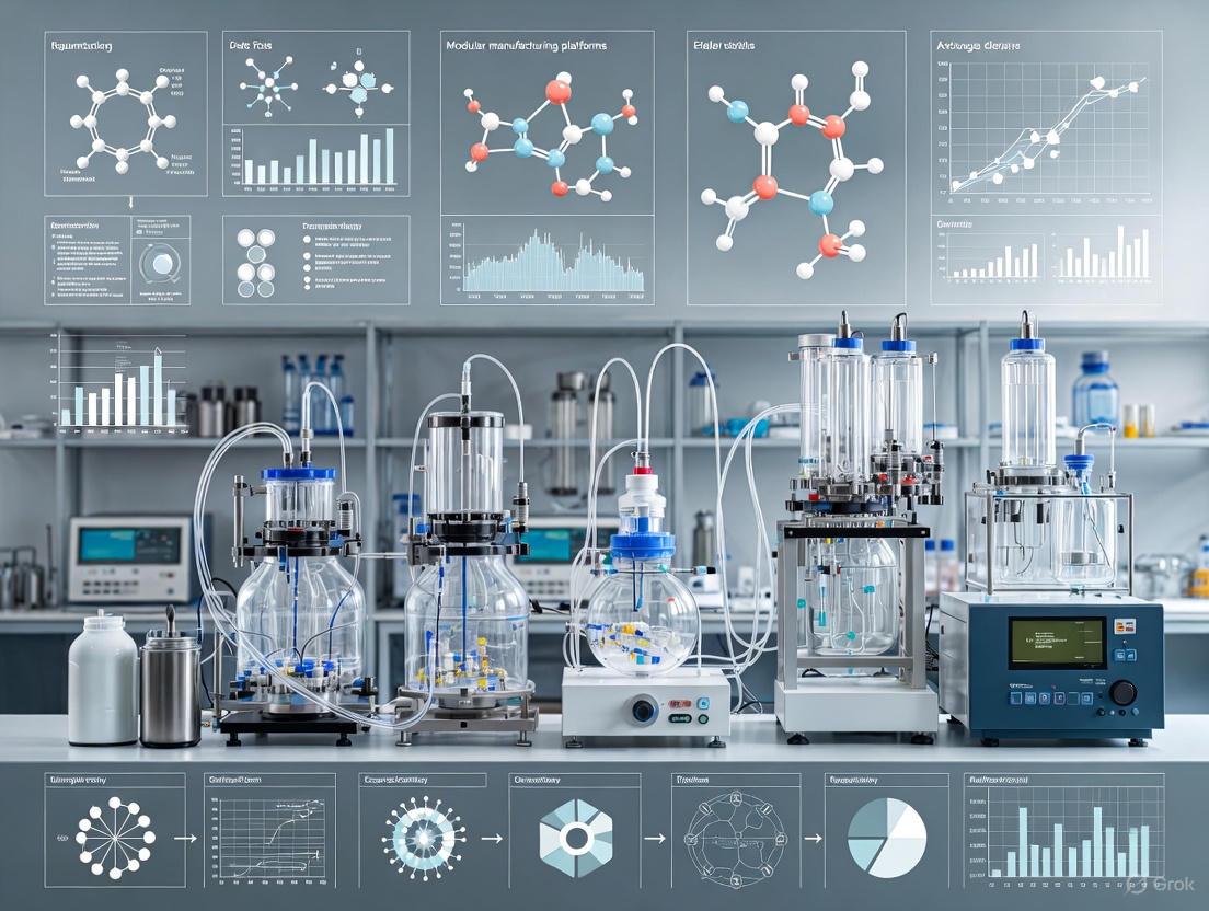
This article explores the transformative role of modular manufacturing platforms in overcoming the scalability, cost, and consistency challenges of autologous cell therapies.
Modular Manufacturing Platforms for Autologous Therapies: Scaling Personalized Medicine
Abstract
This article explores the transformative role of modular manufacturing platforms in overcoming the scalability, cost, and consistency challenges of autologous cell therapies. Tailored for researchers, scientists, and drug development professionals, it provides a comprehensive analysis from foundational concepts to real-world applications. The scope encompasses the definition and drivers of modular platforms, an examination of leading automated systems like CliniMACS Prodigy and Cocoon®, strategies for troubleshooting critical bottlenecks in vein-to-vein timelines and quality control, and a comparative validation of platform performance through case studies and cost-benefit analysis. The synthesis of these intents offers a strategic roadmap for implementing robust and commercially viable manufacturing processes for advanced therapies.
The Imperative for Modular Platforms in Autologous Therapy Manufacturing
Defining Modular and All-in-One Manufacturing Platforms
The manufacturing of autologous cell therapies, such as CAR-T and TCR-T cells, relies predominantly on two distinct technological architectures: modular and all-in-one platforms. In a modular architecture, individual, standalone units perform specific unit operations (e.g., cell separation, activation, expansion) in a sequential, often open or semi-closed manner. Conversely, all-in-one platforms (sometimes termed integrated or end-to-end systems) integrate multiple unit operations into a single, closed, automated system [1] [2]. The choice between these paradigms profoundly impacts process robustness, scalability, cost, and ultimately, the commercial viability of personalized therapies. This document delineates these platforms within the context of autologous therapy research, providing application notes, quantitative comparisons, and detailed protocols to guide platform selection and implementation.
Platform Comparison and Landscape Analysis
Defining Characteristics and Comparative Analysis
The core distinction lies in system integration and workflow. Modular platforms offer flexibility, allowing researchers to select best-in-class equipment for each step, but require extensive manual intervention and open processing. All-in-one systems sacrifice some flexibility for greatly enhanced automation, reduced hands-on time, and a functionally closed processing environment, which minimizes contamination risk and improves reproducibility [1].
Table 1: Fundamental Characteristics of Manufacturing Platforms
| Feature | Modular Platform | All-in-One Platform |
|---|---|---|
| System Architecture | Disconnected, standalone devices for each unit operation [1] | Multiple unit operations integrated into a single, automated system [1] |
| Workflow | Fragmented, requires manual transfer of product between modules [3] [1] | End-to-end, automated processing within a closed system [3] [1] |
| Operator Hands-on Time | High (e.g., >24 hours for a CAR-T process) [1] | Low (e.g., ~6 hours for a similar process) [1] |
| Cleanroom Requirements | Often requires higher-grade (Grade B/C) cleanrooms due to open processing [2] | Can operate in lower-grade (Grade C/D) cleanrooms due to closed design [2] |
| Flexibility & Customization | High; enables customization of individual steps [1] | Low to moderate; process must adapt to the platform's predefined parameters [1] [2] |
| Initial Capital Investment | Lower per device, but cumulative cost can be high | High initial cost for the integrated system [2] |
| Key Advantage | Flexibility for process development and research | Robustness, consistency, and reduced contamination risk for manufacturing |
Quantitative Performance of Representative Platforms
Data from published studies and manufacturer specifications illustrate the performance capabilities of current platforms.
Table 2: Performance Metrics of Selected All-in-One and Modular Platforms
| Platform (Vendor) | Platform Type | Key Output Metric | Process Duration | Reported Hands-on Time Reduction |
|---|---|---|---|---|
| CliniMACS Prodigy (Miltenyi Biotec) | All-in-One | ≥ 1.5 × 10^10 cells with TCT-LS process [1] | 12 days [1] | ~75% (from >24h to ~6h) [1] |
| Quantum Flex (Terumo BCT) | All-in-One | Up to 9 billion TCR-T cells [3] | 10 days [3] | Enables fully automated 3-in-1 workflow [3] |
| Cocoon (Lonza) | All-in-One | 1 patient batch per instrument [2] | Up to 10 days [2] | Fully closed and automated [2] |
| IRO (Ori Biotech) | Modular-Automated | >200x T/NK cell expansion [2] | ~7 days for expansion [2] | 50-70% [2] |
| CTS Rotea (Thermo Fisher) | Modular | Processes leukopaks at 5.3 L/hour [2] | <30 minutes for leukopak processing [2] | Automates a single unit operation [2] |
Experimental Protocols for Platform Evaluation
Protocol 1: Automated TCR-T Cell Manufacturing on an All-in-One Platform
This protocol, adapted from a University of Chicago study using the Terumo Quantum Flex, details an automated 3-in-1 workflow for T Cell Receptor-engineered T cells (TCR-T) [3].
1. Objective: To automatically perform activation, transduction, and expansion of human T cells in a single, closed system for the production of TCR-T cell therapy products.
2. Research Reagent Solutions: Table 3: Key Reagents for TCR-T Manufacturing Protocol
| Reagent/Material | Function | Example/Note |
|---|---|---|
| Peripheral Blood Mononuclear Cells (PBMCs) | Starting material for T cell isolation and culture | Use fresh or cryopreserved leukopaks [3] |
| Activation Reagent | Stimulates T cells to proliferate and become receptive to transduction | Often anti-CD3/CD28 beads or specific cytokine cocktails |
| Viral Vector | Delivers the therapeutic TCR gene to the T cells | Gamma-retroviral or lentiviral vector [3] |
| Cell Culture Medium | Provides nutrients and growth factors for T cell expansion | Serum-free, xeno-free media are standard for GMP |
| Viability Stain (e.g., Trypan Blue) | Distinguishes live from dead cells for counting and viability assessment | Used with automated cell counters or manual hemocytometers |
3. Methodology:
- Seeding and Activation: Load the system's bioreactor with 10 million PBMCs in complete medium and the required activation reagent. Initiate the automated "Activation" protocol on the platform [3].
- Transduction: Following activation, the system automatically introduces the viral vector containing the TCR transgene at a pre-programmed multiplicity of infection (MOI). The platform manages the incubation parameters for efficient gene transfer [3].
- Expansion: The system automatically perfuses fresh medium to support logarithmic cell growth. Environmental conditions (temperature, gas exchange, pH) are continuously monitored and controlled by the platform. Process monitoring samples are taken automatically [3].
- Harvest: After 10 days of culture, or once target cell numbers are achieved (e.g., ~9 billion cells), the system transfers the final product to a harvest bag for formulation and cryopreservation [3].
4. Data Analysis:
- Cell Count and Viability: Determine daily using an automated cell counter from system samples. Final product viability should be >90% [3] [1].
- Transduction Efficiency: Assess by flow cytometry for TCR expression 48-96 hours post-transduction.
- Phenotype: Characterize final product by flow cytometry for CD3+, CD4+, and CD8+ subsets and memory markers (e.g., CD45RA, CCR7).
- Function: Perform functional assays like cytokine release (ELISA/ELISpot) or cytotoxicity assays upon target cell co-culture.
The following workflow diagram illustrates the automated process.
Protocol 2: Large-Scale T Cell Manufacturing Using an Integrated System
This protocol is based on the development of the TCT-LS process for the CliniMACS Prodigy, designed to produce high cell numbers for solid tumor applications [1].
1. Objective: To manufacture a high dose of transduced T cells (≥1.5 × 10^10) from cryopreserved apheresis material using a closed, automated all-in-one system.
2. Research Reagent Solutions:
- Cryopreserved Apheresis Product: Starting material.
- CD4/CD8 Microbeads: For magnetic selection of T cell subsets.
- Large-Scale Activation Reagent: e.g., MACS GMP T TransAct-LS.
- Lentiviral Vector: For stable gene transfer.
- Large-Scale Culture Bags and Reagents: Pre-loaded into the system.
3. Methodology:
- Thaw and Select: Thaw the cryopreserved apheresis product and load it into the system. Perform an automated CD4+/CD8+ T cell selection using magnetic microbeads. The process typically recovers ~50% of CD3+ cells with >85% purity [1].
- Culture and Transduction: Seed 4 × 10^8 selected T cells into the culture chamber. The system uses a shaking culture mode from day 0 to improve gas exchange and nutrient distribution. Cells are activated and transduced with lentiviral vector according to the automated TCT-LS protocol [1].
- Expansion and Harvest: The system automatically feeds the culture for 12 days. The process expands the culture volume to a maximum of 600 mL to accommodate the high cell numbers. The final product is harvested and prepared for cryopreservation [1].
4. Data Analysis: In addition to standard metrics (count, viability, transduction efficiency), focus on:
- Cell Dose: Confirm achievement of the target cell number (≥1.5 × 10^10).
- Phenotypic Consistency: Compare CD4:CD8 ratios and memory subsets (Naive, TCM, TEM, TEMRA) to products from standard-scale processes to ensure no negative impact [1].
Decision Framework for Platform Selection
Choosing between modular and all-in-one systems depends on the research or development phase, process flexibility, and economic considerations. The following diagram outlines the key decision factors.
Guiding Principles:
- Early R&D: A modular approach is advantageous for its flexibility, allowing for iterative process changes and the testing of different reagents and parameters on best-in-class equipment [1] [2].
- Late-Stage Clinical and Commercial Manufacturing: All-in-one platforms are superior for ensuring robustness, reducing human error, cutting COGS, and meeting regulatory requirements for consistency [1] [2].
- Technology Transfer: All-in-one systems significantly reduce the complexity of transferring processes between sites, as the integrated workflow is standardized [1].
The production of autologous cell therapies represents a paradigm shift in medicine, enabling the treatment of previously intractable diseases. However, the journey from patient vein back to patient vein is fraught with significant challenges that hinder widespread adoption. Cost, scalability, and vein-to-vein timelines emerge as the three primary bottlenecks, driven by labor-intensive processes, complex logistics, and personalized production requirements. This document details these challenges within the context of modern modular manufacturing platforms, providing a quantitative analysis of current obstacles and data-driven protocols aimed at overcoming them. The integration of closed-loop automation and decentralized manufacturing models is identified as a critical strategy for enhancing the accessibility and commercial viability of these transformative therapies.
Quantitative Analysis of Production Challenges
The commercial success of autologous therapies is constrained by interconnected operational and economic factors. The data below encapsulates the core challenges faced by developers and manufacturers.
Table 1: Key Quantitative Challenges in Autologous Therapy Production
| Challenge Area | Key Metric | Current Status / Impact |
|---|---|---|
| Cost of Goods Sold (CoGS) | Cost per dose for autologous CAR-T [4] | $400,000 to over $2 million |
| Potential CoGS reduction with integrated platforms [5] | >50% | |
| Scalability & Access | Eligible patients in North America unable to access CAR-T [5] | ~80% |
| Annual batch capacity of a single Cellares Cell Shuttle [2] | 1,000+ | |
| Vein-to-Vein Timeline | Median timeline with centralized, manual processes [2] | 38.3 days |
| Timeline with automated, closed systems (e.g., Lonza Cocoon) [2] | ~10 days | |
| Manufacturing Efficiency | Labor reduction with automated platforms (e.g., Ori Biotech IRO) [2] | 50%-70% |
| Processing speed of automated leukopak system (Thermo Fisher CTS Rotea) [2] | 5.3 L/hour (from >2 hours to <30 mins) |
Detailed Breakdown of Core Challenges
Cost Drivers and Economic Hurdles
The high cost of autologous therapies is a composite of several factors. Manufacturing is not only labor-intensive but also relies on expensive raw materials, particularly viral vectors for gene delivery, which are complex and costly to produce at scale [4] [6]. Furthermore, quality control and release testing, such as sterility testing which can take up to seven days, represent a significant share of both manufacturing time and cost [4] [5]. The high upfront capital expenditure required for establishing GMP-compliant facilities, which can cost hundreds of millions of dollars, further exacerbates the cost challenge [5] [2]. These economic barriers result in therapies that are often priced beyond the sustainable coverage capacity of most healthcare systems [4].
Scalability and Manufacturing Bottlenecks
Scalability is hampered by the inherent patient-specific nature of autologous products, which precludes the economies of scale achieved in traditional mass-produced pharmaceuticals [4]. The current reliance on centralized manufacturing models creates logistical nightmares, including the need for global cryogenic shipping and complex chain-of-identity tracking [6]. This system is vulnerable to single points of failure, such as shortages of critical materials like viral vectors, which can halt production entirely [4]. Scaling out is also limited by a shortage of specialized professionals skilled in both cell biology and GMP manufacturing, making it difficult to expand capacity even when demand is clear [6].
Vein-to-Vein Timeline Pressures
The vein-to-vein timeline—the time from patient apheresis to reinfusion of the final product—is a critical metric with direct implications for patient outcomes, especially in aggressive cancers. Lengthy timelines can disqualify critically ill patients from treatment. Delays are accumulated from multiple stages, including transportation of apheresis material to centralized facilities, the multi-week cell expansion process itself, and the final quality control and release testing [4] [7]. Reducing this timeline is not merely a logistical improvement; it is a clinical necessity. For example, a 55% reduction in V2VT has been modeled to increase life expectancy for patients with relapsed/refractory large B-cell lymphoma by more than three years [2].
Emerging Solutions: The Role of Modular Manufacturing Platforms
Modular, closed, and automated manufacturing platforms are being developed to directly address the trilogy of cost, scalability, and timeline challenges. These platforms integrate multiple unit operations into a single, streamlined system.
Table 2: Comparison of Automated Closed-Loop Manufacturing Platforms
| Platform (Vendor) | Key Feature | Reported Impact | Current Market Share (Est.) [2] |
|---|---|---|---|
| Cocoon (Lonza) | Fully closed, automated system for one patient batch. | Reduces V2VT to ~10 days; deployed in 150+ units globally. | 18%-22% |
| Cell Shuttle (Cellares) | High-throughput; processes 16 batches in parallel. | FDA AMT designation (2025); capacity of 1,000+ annual batches/shuttle. | 10%-14% |
| IRO (Ori Biotech) | Modular platform with OriConnect tubeless sterile connection. | Reduces labor by 50-70% and costs by 30-50%; >50% avg. transduction rate. | N/A |
| CliniMACS Prodigy (Miltenyi Biotec) | Integrates cell selection, activation, transduction, and expansion. | 89% manufacturing success rate in Grade C cleanrooms. | 4%-8% |
| Sefia (Cytiva) | Modular platform with separate systems for selection and expansion. | Increases doses/year by up to 50%; reduces manual operators by 40%. | 7%-11% |
Workflow Integration of a Modular Platform
The following diagram illustrates how a modular platform integrates disparate manufacturing steps into a cohesive, automated workflow, minimizing manual intervention and open processing steps.
Automated vs. Manual Workflow Integration
Application Notes & Experimental Protocols
Protocol 1: Implementing a Rapid, Point-of-Care CAR-T Manufacturing Workflow
This protocol is adapted from the "GoFast" workflow and the VELCART trial, which demonstrated a significant reduction in vein-to-vein time and cost [7].
Objective: To manufacture a clinically effective CAR-T cell product in under 72 hours with a target vein-to-vein time of 9 days and a cost per dose under $50,000.
Materials & Equipment:
- Leukapheresis Product: Fresh, non-cryopreserved starting material.
- Closed-System Cell Processor: e.g., Thermo Fisher CTS Rotea for cell separation.
- Activation/Transduction Reagents: CD3/CD28 activation beads, non-viral transposon system (e.g., Sleeping Beauty or PiggyBac) or high-efficiency lentiviral vector.
- Automated Bioreactor: A closed-system, small-footprint bioreactor for cell culture (e.g., components from the Lonza Cocoon or Ori Biotech IRO platforms).
- QC Assays: Rapid sterility testing (e.g., BacT/ALERT), flow cytometry for CAR expression and cell phenotype.
Procedure:
- Day 0: Cell Selection and Activation (Within 8 hours of apheresis)
- Process the leukapheresis product using a closed-system cell processor to isolate peripheral blood mononuclear cells (PBMCs).
- Immediately activate the T-cells using CD3/CD28 activator beads in a pre-formulated, serum-free medium.
- Concurrently, perform genetic modification via electroporation with a non-viral transposon system carrying the CAR construct. Alternative: Transduce with a high-titer lentiviral vector.
Day 1-2: Abbreviated Expansion
- Transfer the activated/transduced cells to an automated bioreactor.
- Culture cells in a reduced-volume, optimized medium for 48 hours. Monitor glucose/lactate but prioritize speed over maximal cell expansion.
- The target is a 15-fold expansion, yielding a product with a less-differentiated, memory-like T-cell phenotype.
Day 3: Harvest and Formulation
- Harvest cells from the bioreactor. Wash and formulate in infusion-ready buffer.
- Sample the final product for rapid quality control.
Quality Control and Release (Parallel to Days 1-3)
- Initiate rapid microbiological testing immediately post-transduction.
- Perform final release tests (CAR expression, viability, identity) on Day 3. Leverage platform's integrated analytics where possible.
Key Considerations:
- This protocol is highly dependent on the quality of the starting apheresis material.
- The simplified process reduces the number of open manipulations, lowering contamination risk.
- The resulting T-cell product may have a distinct phenotypic profile compared to traditional longer-culture products, which may be associated with improved persistence and potency in vivo.
Protocol 2: Process Transfer to a Decentralized, Automated Platform
This protocol outlines the steps for transferring a research or early-phase manufacturing process to a commercial-grade, automated closed system for deployment in a regional center.
Objective: To successfully transfer and validate a manual CAR-T process to an automated platform (e.g., Cellares Cell Shuttle or Sartorius integrated platform) ensuring product comparability and achieving a >50% reduction in hands-on time.
Materials & Equipment:
- Master Cell Bank: Of the viral vector or non-viral construct.
- Automated Platform: Installed and qualified in a Grade C or controlled non-classified (CNC) cleanroom.
- Platform-Specific Consumables: Single-use disposable kits or cartridges.
- Analytical Tools: for comparability assessment (e.g., NGS for vector integration site analysis, flow cytometry, metabolomic profiling).
Procedure:
- Pre-Transfer Gap Analysis (Week 1-2)
- Map every unit operation of the existing manual process against the capabilities of the automated platform.
- Identify critical process parameters (CPPs) like shear stress, gas transfer rates, and feeding schedules that may differ.
Process Adaptation & Engineering Runs (Week 3-8)
- Adapt the process to fit the platform's fluidics and control systems. This may involve optimizing cell concentration, media volumes, and transduction protocols.
- Execute 3-5 engineering runs using healthy donor apheresis material to refine the process and ensure it meets target Critical Quality Attributes (CQAs).
Formal Comparability Study (Week 9-14)
- Manufacture a minimum of 3 batches using the automated platform and compare them head-to-head with 3 historical batches from the manual process.
- Analyze CQAs including:
- Identity/Purity: CAR expression %, T-cell subset composition.
- Potency: In vitro tumor cell killing assay, cytokine secretion profile.
- Viability & Expansion: Final cell count, viability.
- Safety: Sterility, mycoplasma, endotoxin.
Documentation and Regulatory Submission (Week 15-20)
- Compile all data into a comprehensive comparability protocol.
- Submit to regulatory agencies (FDA, EMA) as a major process change, if applicable for a late-phase or commercial product.
The Scientist's Toolkit: Key Research Reagent Solutions
Successful development and scaling of autologous therapies rely on a suite of specialized reagents and materials.
Table 3: Essential Reagents and Materials for Autologous Therapy R&D
| Reagent/Material | Function | Key Consideration |
|---|---|---|
| Non-Viral Gene Delivery Systems (e.g., LipidBrick Cell Ready [5], Transposon Systems [7]) | Delivers genetic payload (CAR, TCR) to patient T-cells. | Avoids complex viral vector supply chain; cost-effective and scalable; high efficiency with minimal impact on cell fitness is critical. |
| Cell Separation Beads (e.g., CD4+/CD25+ for Treg isolation [8]) | Isolates specific cell subsets from heterogeneous apheresis product. | Purity and recovery of the target population are vital for product efficacy and safety. |
| Serum-Free Culture Media | Supports ex vivo cell activation and expansion. | Formulation must maintain cell phenotype (e.g., "stemness") and prevent exhaustion; must be GMP-grade and xeno-free. |
| Activation Reagents (e.g., CD3/CD28 Activator Beads) | Provides the necessary signal to initiate T-cell proliferation. | Ratio of beads to cells and timing of removal are critical process parameters. |
| Platform-Specific Consumables | Single-use kits, cartridges, or tubing sets for automated platforms. | Represents a potential supply chain bottleneck; quality and lot-to-lot consistency are paramount. |
The path to making autologous cell therapies a mainstream, sustainable treatment option is being paved by technological innovation focused squarely on the triumvirate of cost, scalability, and vein-to-vein timelines. The industry is moving decisively away from labor-intensive, open-process models toward integrated, closed-loop automated systems. These modular platforms are demonstrating tangible benefits: slashing CoGS by over 50%, reducing vein-to-vein times from weeks to days, and enabling scalable models from centralized "smart factories" to decentralized point-of-care networks. For researchers and developers, the imperative is to design processes with scalability in mind from the earliest stages, leveraging these platforms and their associated reagent systems to build a future where transformative autologous therapies are accessible to all eligible patients.
The Shift from Artisanal Processes to Industrialized Automation
The field of autologous cell therapies is undergoing a critical transition from manual, artisanal production methods toward industrialized automation. This shift is driven by the fundamental need to overcome the limitations of small-scale, variable processes that have historically constrained the scalability, affordability, and consistent quality of patient-specific therapies [9]. Autologous therapies, which involve creating treatments from a patient's own cells, present unique manufacturing challenges not found in conventional pharmaceuticals, including inherent biological variability, complex supply chains, and stringent regulatory requirements for living products [10].
Industrialized automation, particularly through modular manufacturing platforms, represents a paradigm shift that addresses these challenges directly. By implementing closed, automated systems with standardized protocols, manufacturers can significantly reduce manual interventions, minimize contamination risks, enhance process control, and improve cost-effectiveness [11]. This transition is not merely a technical upgrade but a comprehensive reimagining of how advanced therapies are developed and produced, moving from boutique operations toward robust, reproducible manufacturing ecosystems capable of serving larger patient populations without compromising quality or accessibility [9].
Quantitative Comparison: Artisanal vs. Industrialized Approaches
The operational and economic distinctions between artisanal and industrialized manufacturing approaches reveal significant advantages for automated systems across multiple performance metrics. The following table summarizes key quantitative differences derived from current industry analysis and research.
Table 1: Performance and Economic Comparison of Manufacturing Approaches
| Performance Metric | Artisanal (Manual) Process | Industrialized (Automated) Process |
|---|---|---|
| Production Throughput | Limited batch processing; 1-2 patients per workstation weekly | Scalable parallel processing; potential for multiple patients per system daily [11] |
| Process Consistency | High variability (15-30% coefficient of variation) between operators and batches | Significantly reduced variability (<5% CV) through automated control [9] |
| Contamination Risk | Elevated risk due to frequent open handling and manual transfers | Substantially reduced via closed systems and minimal human intervention [10] |
| Cost of Goods (COGs) | Extremely high ($100,000+ per dose in some cases) | Potential for significant reduction through increased efficiency and scale [9] |
| Staffing Requirements | Labor-intensive; requires highly trained technicians for manual operations | Reduced direct labor; shifts skill requirements to system operation and monitoring [9] |
| Facility Footprint | Extensive cleanroom space required per unit of output | Compact, modular designs with higher output per square foot [11] |
| Regulatory Compliance | Challenging due to process variability; extensive documentation needed | Enhanced process control facilitates compliance through built-in data capture [10] |
The data demonstrates that industrialization addresses the core economic challenges of autologous therapies, particularly the prohibitively high Cost of Goods (COGs) that has limited patient access [9]. Furthermore, automated systems substantially improve quality control by reducing the human factors that contribute to batch-to-batch variability—a critical consideration for regulatory approval and consistent therapeutic outcomes [10].
Experimental Protocols for Process Evaluation and Implementation
Protocol 1: Comparative Analysis of Manual vs. Automated Cell Processing
Objective: To quantitatively evaluate the performance differences between manual and automated methods for critical cell processing steps, specifically cell separation and activation.
Materials:
- Starting Material: Leukapheresis products from healthy donors
- Manual Arm: Biological safety cabinets, manual pipettes, centrifugation systems, open transfer kits
- Automated Arm: Closed, automated cell processing system (e.g., Cocoon Platform, CliniMACS Prodigy)
- Analytical Equipment: Flow cytometer, cell counter, viability analyzer (e.g., Trypan Blue exclusion), endotoxin testing kit
Methodology:
- Sample Preparation: Split a single leukapheresis product into two equal aliquots using a cell separator under controlled conditions.
- PBMC Isolation:
- Manual Group: Perform Ficoll density gradient separation in biological safety cabinet with open pipetting. Record processing time and all interventions.
- Automated Group: Load the aliquot into the automated system with a pre-programmed PBMC isolation protocol. The system performs all steps in a closed pathway.
- Cell Activation and Transduction:
- Manual Group: Manually count cells, calculate reagent volumes, and add activation stimuli (e.g., CD3/CD28 beads) via pipette. After incubation, perform manual viral transduction with precise timing.
- Automated Group: The automated system performs cell counting, calculates reagent doses, and adds activation stimuli at the programmed time. Viral vector is automatically introduced at the optimal cell density and activation state.
- Process Monitoring & Data Collection:
- Record processing times for each major step in both arms.
- Collect samples at defined intervals (post-isolation, post-activation, post-transduction) for cell count, viability, and phenotype analysis via flow cytometry.
- Document all manual interventions and potential breaches in aseptic technique in the manual arm.
- Systematically record vector usage and reagent consumption in both arms.
- Output Assessment:
- Measure final cell yield, viability, transduction efficiency, and potency.
- Perform sterility testing (bacterial, fungal, mycoplasma) and endotoxin testing on final products.
Analysis: Compare the coefficient of variation for key quality attributes between the two methods across multiple runs (n≥5). Calculate the cost per dose, factoring in materials, labor, and facility costs. Assess process capability (Cpk) for critical quality attributes in both systems.
Protocol 2: Implementation of a Modular Automated Platform
Objective: To establish and validate a modular automated platform for the end-to-end manufacturing of an autologous CAR-T cell therapy.
Materials:
- Core System: Modular automated cell processing platform with separate, interconnectable modules for separation, activation, expansion, and formulation.
- Single-Use Sets: Pre-sterilized, closed fluid pathway sets designed for the platform.
- Raw Materials: GMP-grade cell culture media, activation reagents, viral vector, cytokines.
- QC Equipment: Bioanalyzer for vector potency, process analytical technology (PAT) probes (e.g., pH, dissolved oxygen) where available, Gram stain kits.
Methodology:
- Technology Transfer & System Setup:
- Transfer the existing manual process parameters to the automated platform, defining critical process parameters (CPPs) for each module.
- Install the system in a GMP-compliant manufacturing suite (ISO 7 background with ISO 5 protection for critical steps).
- Load the pre-sterilized single-use set and prime the system with media according to manufacturer instructions.
- Process Automation & Integration:
- Load the patient's leukapheresis material into the input module.
- Initiate the integrated process: the system automatically performs cell selection, activation, transduction, and expansion in a continuous, closed process.
- The system utilizes built-in PAT to monitor cell growth and metabolic status, making automated adjustments to feeding schedules and gas exchange.
- In-Process Controls & Real-Time Release:
- Implement at-line automated cell counting and viability analysis using integrated sampling and analysis modules.
- Use the platform's data management system to continuously record all process parameters (temperatures, volumes, timings) for the process record.
- Harvest and Formulation:
- Upon reaching target cell density, the system automatically initiates harvest procedures, including cell concentration and buffer exchange into the final formulation medium.
- The final drug product is aseptically filled into an infusion bag, remaining within the closed fluid path until patient administration.
- Process Validation:
- Perform three consecutive validation runs using donor-derived material to demonstrate process consistency.
- Compare all critical quality attributes (CQAs) of the final product to pre-established specifications and historical manual process data.
Analysis: Generate a process capability analysis for all CQAs. Perform a comparative analysis of the process performance (e.g., total hands-on time, total process time, success rate) between the modular automated platform and the historical manual process.
Workflow Visualization: From Artisanal to Industrialized Manufacturing
The transition from artisanal to industrialized manufacturing represents a fundamental restructuring of the production workflow, as visualized in the following diagram.
Diagram 1: Autologous Therapy Manufacturing Process Evolution
This workflow diagram illustrates the fundamental contrast between the two manufacturing paradigms. The artisanal process (red, dashed) is characterized by multiple, discrete open steps requiring extensive manual intervention, while the industrialized process (green, solid) integrates multiple unit operations into a single, closed automated system with continuous monitoring [11]. The compression of the timeline from sample to release is a critical advantage of automation, potentially reducing the vein-to-vein time for patients awaiting therapy [9].
The Scientist's Toolkit: Essential Reagents and Materials
Successful implementation of industrialized automation requires carefully selected reagents and materials compatible with closed-system processing. The following table details key solutions essential for automated cell therapy manufacturing.
Table 2: Essential Research Reagent Solutions for Automated Manufacturing
| Reagent/Material | Function & Role in Automation | Critical Quality Attributes | Automation-Specific Requirements |
|---|---|---|---|
| GMP-Grade Culture Media | Provides nutrients and environment for cell growth and maintenance. | Defined formulation, low endotoxin, consistent performance. | Pre-tested compatibility with system materials (e.g., plastics, sensors); availability in single-use, closed-system bags. |
| Cell Activation Reagents | Stimulates T-cells to initiate proliferation (e.g., anti-CD3/CD28). | High purity, specific activity, lot-to-lot consistency. | Formulation for stable, automated dispensing; compatibility with integrated fluid paths without clogging. |
| Viral Vector (Lentivirus) | Delivers genetic material for cell engineering (e.g., CAR gene). | High titer, functional potency, purity from contaminants. | Stability under prolonged storage in system bags; concentration suitable for low-volume, automated dispensing. |
| Cell Separation Reagents | Isolates target cell populations from apheresis product. | High specificity, recovery, and viability of target cells. | Compatibility with closed-system separation technologies (e.g., magnetic beads); formulated for automated liquid handling. |
| Formulation Buffer | Final medium for washing and suspending the cell therapy product. | Isotonicity, physiological pH, protein stabilizers. | Sterile, pre-mixed solutions in closed, weldable bags suitable for the final harvest and fill step. |
| Single-Use Bioreactor | Provides a sterile, controlled environment for cell expansion. | Scalable volume, integrated sensors (pH, DO), gas exchange membrane. | Modular design to integrate with the automated platform; pre-sterilized and ready-to-use. |
The selection of these reagents is guided by the principles of Quality by Design (QbD) to ensure they meet the stringent requirements of automated systems, particularly lot-to-lot consistency and compatibility with closed processing [10]. The move toward standardized, pre-qualified reagent kits specifically designed for automated platforms is a key enabler for robust and reproducible manufacturing at scale [9].
The shift from artisanal processes to industrialized automation represents the most promising pathway for making autologous cell therapies scalable, affordable, and consistently available to patients. Modular manufacturing platforms stand at the forefront of this transition, offering the flexibility, control, and closed processing necessary to overcome the historical limitations of manual production [11]. While implementation requires significant initial investment in technology and expertise, the long-term benefits—including reduced operational costs, enhanced product quality, improved regulatory compliance, and expanded patient access—are transformative for the field [9].
The future of autologous therapy manufacturing will likely involve greater integration of advanced technologies such as artificial intelligence for process optimization and predictive analytics, further accelerating the transition from artisanal craftsmanship to robust, data-driven industrialization [10]. As these automated platforms evolve and become more widespread, they will ultimately fulfill the promise of regenerative medicine by delivering sophisticated, personalized treatments not as rare commodities, but as routinely accessible standards of care.
Modular manufacturing platforms represent a transformative approach for the production of autologous cell therapies, such as CAR-T cells and other patient-specific treatments. These platforms, characterized by their flexibility, closed-system processing, and standardized unit operations, directly address fundamental Good Manufacturing Practice (GMP) requirements for product consistency, safety, and quality [12] [13]. This document outlines the key regulatory drivers shaping the adoption of these platforms and provides detailed protocols for implementing GMP-compliant processes that ensure product consistency within a modular framework. The guidance is structured to assist researchers and drug development professionals in navigating the intersection of innovative manufacturing technology and evolving regulatory expectations.
Regulatory Framework for Advanced Therapies
The foundation of GMP compliance for autologous therapies is established in the Code of Federal Regulations, specifically 21 CFR Part 211 for Finished Pharmaceuticals and 21 CFR Part 210 for manufacturing processes [14]. Regulatory oversight ensures that products are safe for use and possess the identity, strength, quality, and purity they purport to hold [14].
A significant recent development is the FDA's January 2025 draft guidance, "Consideration for Complying with 21 C.F.R. 211.110," which clarifies in-process control requirements and explicitly addresses advanced manufacturing technologies [15]. This guidance promotes a scientific, risk-based approach for defining what, where, when, and how in-process controls and testing should occur [15]. It acknowledges the flexibility of CGMPs and supports the use of advanced techniques like continuous manufacturing and real-time quality monitoring, which are hallmarks of modular platforms [15].
Key Regulatory Drivers for Modular Platforms
For autologous therapies, several critical regulatory drivers make modular platforms particularly advantageous:
- Process Control and Consistency: FDA regulations require control strategies to ensure "batch uniformity and integrity" [15]. Modular systems, with their standardized, closed processes, minimize human intervention and variability, directly supporting this requirement [16] [17].
- Aseptic Processing: Since cell therapies cannot be terminally sterilized, the entire manufacturing process must occur under aseptic conditions [10]. Closed, automated modular systems significantly reduce contamination risks from personnel and the environment [16].
- Traceability: For autologous products, maintaining chain of identity and custody from the patient to the final product is paramount [12] [17]. Integrated digital systems in advanced modular platforms provide electronic batch records that ensure full traceability [17].
- Scalability and Comparability: Regulatory authorities require demonstrating product comparability after any manufacturing process changes [10]. Modular platforms facilitate seamless scale-up and process transfer with minimal disruption to critical process parameters [13] [18].
Table 1: Key CGMP Regulations Impacting Modular Platform Design for Autologous Therapies
| Regulation | Title | Key Requirement | Modular Platform Application |
|---|---|---|---|
| 21 CFR 211.110 | Sampling and testing of in-process materials and drug products | Requires control procedures to monitor output and validate manufacturing processes [15]. | Supports use of real-time, at-line monitoring and process models paired with material testing [15]. |
| 21 CFR 211.101 | Component charge-in | Ensures components are weighed, measured, or subdivided accurately [14]. | Automated, closed fluid paths and single-use consumables with barcode tracking reduce errors [16] [17]. |
| 21 CFR 211.113 | Control of microbiological contamination | Requires procedures to prevent objectionable microorganisms in drug products [14]. | Closed processing systems and automated CIP (Clean-in-Place) functionalities mitigate contamination risk [18] [17]. |
Implementing GMP Controls in Modular Platforms
System Architecture and Design Controls
A GMP-compliant modular platform must be designed with quality built into its core architecture. This involves:
- Closed Processing: The system should form a closed, integrated path for cell processing, minimizing or eliminating open manipulations and interventions [16] [17]. This is a primary defense against microbial contamination and is a key expectation in EU GMP Annex 1 [18].
- Pre-validated Modules: Sourcing pre-validated, "turnkey" modules shortens qualification timelines and reduces validation risks [13] [18]. These modules should have documented evidence of performance qualification (PQ) for their intended unit operations.
- Digital Integration and Data Integrity: The platform must include integrated software for process control and data acquisition that complies with 21 CFR Part 11 [16] [17]. Systems like Chronicle automation software are designed to automatically collect true batch records, monitor critical process parameters, and maintain data integrity in a secure cloud [17].
Process Analytical Technology (PAT) and In-Process Controls
The FDA's 2025 draft guidance emphasizes that "sampling does not necessarily require steps for physically removing in-process materials," endorsing the use of in-line, at-line, or on-line measurements [15]. Modular platforms are ideal for implementing this PAT framework.
- Real-time Monitoring: Integrated sensors should monitor Critical Process Parameters (CPPs) such as temperature, pH, dissolved oxygen, and cell density in bioreactor modules [13] [15].
- Process Models: While the FDA advises that process models should be paired with in-process material testing (as they have not yet approved models used alone), they represent a significant opportunity for advanced control strategies in continuous manufacturing [15].
The following diagram illustrates the integrated control strategy for a modular platform, linking real-time monitoring to automated adjustments and quality oversight.
Diagram 1: Process Control & Quality Oversight Workflow
Experimental Protocols for Process Validation
Demonstrating that a modular platform can consistently produce a therapy that meets its Critical Quality Attributes (CQAs) is essential for regulatory approval. The following protocols provide a framework for this validation.
Protocol: Validation of Aseptic Processing and Closed Systems
1.0 Objective: To demonstrate that the closed modular system maintains sterility throughout the manufacturing process for an autologous CAR-T cell therapy.
2.0 Materials:
- Modular Bioprocessing Platform (e.g., Syntegon MBP, Gibco CTS Rotea/Dynacellect/Xenon systems) [16] [18]
- Sterile growth media and reagents
- Environmental monitoring equipment (settle plates, air samplers)
- Culture sterility test kits (e.g., BacT/ALERT)
3.0 Methodology:
- Media Fill Simulation: Perform a minimum of three consecutive successful media fill runs per process workflow.
- Aseptically introduce sterile cell culture media into the system as a surrogate for patient starting material.
- Process the media through all steps (e.g., separation, activation, transduction, expansion) using the same procedures, durations, and conditions as a live manufacturing run.
- Incubate the final "product" and in-process samples for 14 days and observe for microbial growth [10].
- Environmental Monitoring: Conduct active air sampling and surface monitoring at critical intervention points (e.g., during bag connections) throughout the simulation to demonstrate the absence of microbial ingress.
- System Integrity Testing: For systems with CIP and FIT (Filter Integrity Testing), like the Syntegon MBP, execute these functions and document successful completion [18].
4.0 Acceptance Criteria:
- All media fill runs must show 0% contamination in final product and in-process samples.
- All environmental monitoring results must be within predefined alert limits.
- All automated CIP and FIT cycles must complete successfully.
Protocol: Demonstrating Process Consistency and Comparability
1.0 Objective: To validate that the modular manufacturing process consistently produces autologous cell therapy products that meet pre-defined CQAs across multiple simulated patient batches.
2.0 Materials:
- Donor-derived PBMCs (from at least 3 different donors to represent patient variability)
- Modular platform with integrated analytics
- Analytical instruments for CQA testing (e.g., flow cytometer, cell counter, PCR)
3.0 Methodology:
- Process Multiple Batches: Manufacture a minimum of n=10 consecutive engineering runs using donor-derived PBMCs, following the exact clinical manufacturing process.
- In-process Monitoring: Use integrated systems to track CPPs (e.g., transduction efficiency, cell growth rate, metabolite levels) in real-time.
- Test for CQAs: For each final product, measure and record all CQAs. The table below lists common CQAs for an autologous CAR-T cell product.
Table 2: Critical Quality Attributes (CQAs) for an Autologous CAR-T Cell Therapy
| CQA Category | Specific Attribute | Target Specification | Analytical Method |
|---|---|---|---|
| Identity | CAR Transgene Expression | ≥ 30% CAR-positive T-cells | Flow Cytometry |
| Potency | In Vitro Cytotoxic Activity | ≥ 20% specific lysis of target cells | Co-culture assay with tumor cells |
| Viability | Final Product Viability | ≥ 80% | Trypan Blue Exclusion/Flow Cytometry |
| Purity | CD3+ T-cell Composition | ≥ 90% CD3+ cells | Flow Cytometry |
| Safety | Endotoxin Level | < 5 EU/kg/hr | LAL Assay |
| Dosage | Viable Cell Dose | Within ±10% of target dose | Automated Cell Counter |
4.0 Acceptance Criteria:
- All n=10 batches must yield a final product meeting all CQA specifications listed in Table 2.
- CPP data must remain within validated operating ranges for ≥ 95% of the process duration for all batches, demonstrating process control.
The Scientist's Toolkit: Essential Research Reagents and Materials
Successful translation from research to GMP manufacturing depends on the selection of qualified materials. The following table details key reagents and their functions in the context of developing a process on a modular platform.
Table 3: Essential Research Reagent Solutions for Process Development
| Reagent/Material | Function | GMP-Compliance Considerations | Example Application in Workflow |
|---|---|---|---|
| GMP-grade Cell Culture Media | Provides nutrients and environment for cell growth and expansion. | Must be xeno-free, contain no animal-derived components, and be sourced from a GMP-manufactured supply chain [16]. | Used throughout the cell culture and expansion phase in the bioreactor module. |
| Clinical-grade Cytokines (e.g., IL-2, IL-7/IL-15) | Activates and promotes the expansion and persistence of T-cells. | Requires GMP manufacturing and certificates of analysis for identity, purity, and potency [16]. | Added during T-cell activation and ex vivo expansion steps. |
| GMP-grade Viral Vector (e.g., Lentivirus) | Mediates genetic modification of T-cells to express the CAR. | One of the most critical raw materials; requires extensive safety testing (e.g., RCL, sterility, mycoplasma) [10]. | Used during the transduction step in a closed system bag or chamber. |
| Transfection Reagents (for non-viral editing) | Enables genetic material insertion via electroporation. | For platforms using the Gibco CTS Xenon system, use of GMP-compliant reagents is required for clinical production [16]. | Used in conjunction with an electroporation module for non-viral CAR insertion. |
| Magnetic Beads (e.g., for CD3/CD28 activation) | Selects and activates target T-cell populations. | Use sterile, single-use kits designed for seamless scaling from research to clinic, such as those for the CTS Dynaclect system [16]. | Used in the cell selection and activation module post-apheresis. |
| Single-use Bioprocess Containers | Closed-system bags for media, buffer, and intermediate product hold. | Must be biocompatible and sterilized by gamma irradiation. Integrity testing is critical [13]. | Used across the platform for all fluid transfers and holds, ensuring a closed pathway. |
Integrated Quality Management System
A robust Quality Management System (QMS) is the backbone of GMP compliance. For modular manufacturing, the QMS must cover:
- Change Control: A rigorous process for evaluating any changes to hardware, software, or consumables to ensure they do not adversely affect product quality [19].
- Deviations and CAPA: Systematic investigation of deviations and implementation of effective Corrective and Preventive Actions [19].
- Supplier Qualification: A program for qualifying and periodically auditing suppliers of critical raw materials and modular components to ensure they meet quality standards [10] [19].
- Personnel Training: Continuous GMP training for operators, with specific modules on the operation and aseptic practices related to the modular equipment [19].
The relationship between the modular platform, its controls, and the overarching QMS is synergistic, as shown below.
Diagram 2: QMS Integration with Modular Platform
Modular manufacturing platforms are uniquely positioned to address the core regulatory drivers for autologous therapies: ensuring GMP compliance and product consistency. By integrating closed processing, automation, and digital data integrity from the outset, these platforms provide a framework for robust, scalable, and compliant production. The experimental protocols and control strategies outlined herein provide a roadmap for researchers and developers to validate their processes in alignment with current FDA guidance and international GMP standards. As the regulatory landscape continues to evolve to accommodate advanced manufacturing, a proactive approach to process design and quality control, centered on modular principles, will be key to successfully bringing these life-changing personalized therapies to patients.
The development of autologous cell therapies represents a paradigm shift in the treatment of cancers and rare diseases. However, their commercialization faces significant economic challenges, primarily driven by high costs of goods sold (COGS) and complex manufacturing logistics, which ultimately limit patient access [20] [21]. The personalized nature of these therapies, where a single dose is manufactured from an individual patient's own cells, results in production costs that can range from $100,000 to $300,000 per dose [22]. Consequently, only an estimated 20% of eligible patients in the U.S. are able to access autologous CAR-T therapies, with global access dropping to approximately 10% [22].
Modular, automated, and closed manufacturing platforms are emerging as critical solutions to these challenges. By transforming traditional labor-intensive processes into streamlined, standardized, and scalable operations, these integrated systems can significantly reduce COGS while enhancing production capacity and consistency [5] [1] [23]. This application note details the economic impact of current manufacturing bottlenecks, provides a detailed experimental protocol for an automated process, and outlines how modular platforms can expand patient access through both centralized and decentralized production models.
Economic Impact of Manufacturing Bottlenecks
The high COGS for autologous cell therapies are attributable to a confluence of factors rooted in conventional, manual manufacturing methods. A detailed breakdown of these cost drivers and the quantifiable impact of automation is provided in Table 1.
Table 1: Economic Impact of Manufacturing Bottlenecks and Automated Solutions
| Cost & Access Driver | Traditional Manual Process Impact | Impact of Automated, Closed Systems |
|---|---|---|
| Labor Requirements | >24 hours of hands-on operator time per batch [22]. Labor can constitute >50% of manufacturing costs [22]. | Reduction to ~6 hours of hands-on time per batch (≥70% reduction) [1] [22]. |
| Facility & Cleanroom | Requires high-classification (e.g., Grade A/B) cleanrooms due to open processes, escalating infrastructure costs [23]. | Enables operation in lower-classification environments (e.g., controlled non-classified/ Grade D), reducing capital investment [5] [22]. |
| Contamination & Batch Failure | High risk due to numerous manual touchpoints, leading to product losses and costly re-manufacturing [23]. | Significantly reduced risk via functional closure, minimizing human intervention and improving batch success rates [1] [22]. |
| Process Consistency | High donor-to-donor variability exacerbated by manual handling, leading to product inconsistencies [23]. | Improved batch-to-batch consistency and reproducibility through precise parameter control and standardization [1] [23]. |
| Vein-to-Vein Time | Lengthy (weeks) due to complex logistics and external quality control, which can be detrimental for patients with aggressive diseases [24]. | Can be reduced by days, improving patient outcomes. Point-of-Care models can enable infusion within 5 days of apheresis [24]. |
| Scalability | Scaling out requires duplicating entire manual processes and cleanroom suites, which is cost-prohibitive [23]. | Facilitates scale-out through multi-parallel processing in a smaller footprint; one platform forecasts a >50% COGS reduction and 4x capacity scaling [5] [22]. |
Experimental Protocol: Automated T Cell Manufacturing for Solid Tumors
This protocol describes a closed, automated manufacturing process for generating high numbers of autologous T cells for solid tumor applications, using the CliniMACS Prodigy platform with the T cell Transduction – Large Scale (TCT-LS) process [1]. The process is compatible with a cryopreserved apheresis starting material and produces a cryopreserved drug product, adding flexibility to the supply chain.
Key Objectives:
- To achieve a target yield of ≥ 1.5 × 10^10 total viable cells after 12 days of expansion.
- To maintain critical quality attributes (CQAs) such as viability, T cell phenotype, and transduction efficiency comparable to or better than standard processes.
- To demonstrate robust technology transfer across multiple manufacturing sites.
Materials and Equipment
Table 2: Key Research Reagent Solutions and Equipment
| Item Name | Function/Application in Protocol |
|---|---|
| CliniMACS Prodigy (TCT-LS Process) | An all-in-one, closed, automated system for cell selection, activation, transduction, and expansion [1]. |
| CD4/CD8 Microbeads | Magnetic beads for the positive selection of target T cell populations from apheresis material [1]. |
| MACS GMP T Cell TransAct – Large Scale | Reagent for the activation of T cells, a critical step initiating proliferation [1]. |
| Lentiviral Vector | Vector for genetic modification of T cells (e.g., with CAR or TCR constructs) [1]. |
| Cell Culture Media | Formulated media, often supplemented with cytokines (e.g., IL-2, IL-7, IL-15), to support cell growth and viability during expansion [20] [1]. |
| Cryopreservation Solution | Contains cryoprotective agents (e.g., DMSO) to maintain cell viability during freezing and storage [20]. |
Step-by-Step Methodology
Cell Selection and Isolation:
- Thaw the cryopreserved leukapheresis material.
- The system automatically performs cell washing and density adjustment.
- Incubate the cell suspension with CD4 and CD8 Microbeads.
- Load the sample onto the system; the process automatically applies the sample through a magnetic column, washes away unlabeled cells, and elutes the magnetically labeled CD4+/CD8+ T cells. The target for selection is up to 3 × 10^9 cells [1].
- In-process control (IPC): Calculate cell recovery, purity (%CD3+ cells), and viability post-selection. Expected purity is >86% [1].
Cell Activation and Transduction:
- The selected T cells are transferred to the integrated culture chamber. The culture volume is scaled up to a maximum of 600 mL [1].
- Activate the T cell culture using MACS GMP T Cell TransAct – Large Scale reagent.
- Initiate a shaking regimen from day 0 to improve gas exchange and cell suspension.
- On day 1 post-activation, transduce the activated T cells with the lentiviral vector.
- IPC: Monitor cell count and viability. A temporary decrease in viability on day 1 is acceptable and should recover [1].
Cell Expansion:
- The culture is maintained for a total of 12 days.
- The system automatically performs feeding and media exchange cycles to maintain optimal nutrient levels and waste removal.
- The culture environment (temperature, gas) is continuously controlled by the integrated incubator.
- IPC: Perform automated or manual sampling to track cell growth, viability, and metabolic status (e.g., glucose consumption).
Final Harvest and Formulation:
- On day 12, initiate the harvest procedure. The system automatically concentrates the cell product and performs a series of washes to remove culture media and debris.
- Formulate the final cell product in an appropriate infusion buffer or cryopreservation medium.
- IPC: Determine the final total viable cell count, viability, and cell composition (e.g., %CD3+/CD4+/CD8+ by flow cytometry).
Cryopreservation (if applicable):
Key Process Parameters and Outcomes
Validation runs using this protocol have demonstrated its robustness. As shown in Figure 2 of the primary reference, the TCT-LS process consistently met its target yield [1].
- Final Cell Yield: Mean of 1.70 × 10^10 total viable cells [1].
- Final Viability: Mean of 92.6% [1].
- T Cell Phenotype: The final product was highly pure, with a mean of 98.3% CD3+ cells, and a CD4+:CD8+ ratio that was comparable to the standard process [1].
- Memory Subsets: Extensive characterization showed no significant difference in the proportion of critical T cell memory subsets between the TCT-LS and standard processes, indicating the scaled-up expansion did not negatively impact this key quality attribute [1].
Technology Platforms Enabling Cost Reduction
The implementation of the protocol in Section 3 relies on advanced manufacturing platforms. The industry is converging on two primary, complementary approaches: all-in-one integrated systems and modular closed systems.
Figure 1. A workflow diagram comparing integrated and modular automated platforms for autologous cell therapy manufacturing.
All-in-One Integrated Systems: Platforms like the CliniMACS Prodigy integrate multiple unit operations (selection, activation, culture, harvest) into a single, closed, automated system [1]. The primary advantage is extensive process standardization and a dramatic reduction in manual handling, which lowers labor costs and contamination risk. A limitation is less flexibility to adapt individual process steps once committed to the platform [1].
Modular Closed Systems: This approach, exemplified by integrated platforms from vendors like Sartorius, involves connecting discrete, closed, and automated modules for each unit operation [5] [23]. This offers greater flexibility to optimize or change specific steps (e.g., isolation method, expansion vessel) for different therapy types, which is valuable in a rapidly evolving field. It supports scale-out by allowing multiple products to be at different stages simultaneously [23].
Both platform types directly address COGs drivers by reducing cleanroom classification requirements [5], enabling parallel processing [22], and incorporating rapid quality control assays that can reduce sterility testing from 7 days to hours, thus shortening vein-to-vein time [5].
Expanding Patient Access through New Manufacturing Paradigms
Reducing COGs is a necessary, but not sufficient, condition for expanding patient access. The manufacturing model itself must evolve. The emergence of automated, closed platforms enables a shift from purely centralized production to decentralized or point-of-care (PoC) manufacturing, which can coexist with centralized facilities to maximize reach [5] [24].
Figure 2. Logical relationships between manufacturing models and their impact on patient access.
Centralized Manufacturing: This traditional model relies on large, purpose-built facilities that offer economies of scale and centralized quality oversight. However, it introduces complex and costly cold-chain logistics for shipping patient cells to the facility and the final product back to the patient, which lengthens vein-to-vein time by at least two days and creates scheduling challenges [5].
Decentralized/Point-of-Care Manufacturing: This model involves situating compact, automated manufacturing platforms (like the Cocoon or MARS Atlas systems) within or near hospital settings [24]. It can reduce vein-to-vein time significantly; one clinical trial demonstrated infusion within five days of apheresis using a 3-day manufacturing process [24]. This is critical for patients with rapidly progressing diseases. It also eliminates international shipping hurdles, potentially expanding global access. The existing network of 28 FACT-accredited centers in the U.S. represents a ready-made infrastructure that could be leveraged for such decentralized networks, potentially increasing U.S. production capacity from ~15,000 batches globally to over 15,000 batches domestically [5].
The integration of automated platforms with next-generation analytical technologies like single-cell next-generation sequencing (scNGS) and AI-driven analytics further enhances access by providing deeper product characterization. This enables process optimization to lower the number of cells needed per dose and accelerates quality control, making decentralized models more viable and efficient [21].
Core Technologies and Integrated Systems in Action
Modular manufacturing platforms are pivotal in addressing the complex production challenges associated with autologous cell therapies. These systems integrate multiple unit operations into closed, automated processes, enhancing reproducibility, reducing contamination risks, and streamlining scale-up for clinical and commercial manufacturing [25]. This section delineates the core specifications and performance metrics of three leading platforms: the CliniMACS Prodigy, the Cocoon Platform, and the Quantum system.
Table 1: Core Specifications of Leading Modular Manufacturing Platforms
| Feature | CliniMACS Prodigy | Cocoon Platform | Quantum System |
|---|---|---|---|
| Primary Manufacturer | Miltenyi Biotec | Lonza | Terumo BCT (Note: Based on industry knowledge; not in search results) |
| System Type | Integrated, single-apparatus automation | Integrated, single-apparatus automation | Modular, flexible-scale automation |
| Processing Model | Closed system, single-use disposable kits [26] | Closed system, single-use disposables [27] | Closed system, single-use disposable sets |
| Key Automation Features | Automated cell separation, culture, and concentration | Automated cell culture and at-line PAT integration [27] | Automated cell separation and culture |
| Reported Cell Types | T cells, NK cells, HSCs, monocytes, DCs [28] [26] | Information not specified in results | T cells, monocytes |
| Allogeneic Support | Yes [28] [26] | Information not specified in results | Information not specified in results |
| Autologous Support | Yes [28] | Implied by platform design | Yes |
Table 2: Documented Performance Metrics from Platform Studies
| Platform | Process Step | Performance Metric | Reported Outcome |
|---|---|---|---|
| CliniMACS Prodigy | CD34+ HSC enrichment from cord blood | Average Cell Recovery | 68.18% - 71.94% [26] |
| CliniMACS Prodigy | CD34+ HSC enrichment from cord blood | Average Purity | 57.48% - 69.73% [26] |
| CliniMACS Prodigy | Final NK cell harvest & concentration | Average Cell Yield | 74.59% - 83.74% [26] |
| CliniMACS Prodigy | Final NK cell harvest & concentration | NK Cell Purity | >80% [26] |
| Miltenyi Bioindustry | Overall cGMP batch success rate | Success Rate | 95% (based on 2024-2025 data) [28] |
Application Notes & Experimental Protocols
CliniMACS Prodigy for NK Cell Manufacturing
The following protocol details the automated, closed-system manufacturing of allogeneic Natural Killer (NK) cells from umbilical cord blood (UCB)-derived CD34+ hematopoietic stem cells, as demonstrated by Glycostem Therapeutics [26].
2.1.1 Experimental Workflow: Automated NK Cell Manufacturing
The workflow diagram below outlines the key stages of the process.
2.1.2 Detailed Methodology
Step 1: Umbilical Cord Blood Pre-Processing
- Source Material: Obtain fresh UCB units, transported at 15°C–25°C without X-ray screening to preserve viability.
- Eligibility Criteria: Select UCB units containing ≥2.0E06 CD34+ cells for R&D or ≥3.5E06 CD34+ cells for GMP batches.
- Data Collection: At receipt, record unit weight, volume, total nucleated cell (TNC) count, CD34+ cell count, red blood cell (RBC) count, and platelet (PLT) count. Process units within 72 hours of collection [26].
Step 2: Automated CD34+ Hematopoietic Stem Cell Enrichment
- Equipment & Reagents: CliniMACS Prodigy with LP-34 Enrichment Protocol (v2.2) and TS310 tubing set. Use CliniMACS PBS/EDTA Buffer with 0.5% Human Serum Albumin (HSA) as washing buffer and proprietary basal growth medium for cell elution.
- Procedure:
- Install the single-use TS310 tubing set using Prodigy Software guidance.
- Perform Fc receptor blocking using a 5% IgG solution.
- Load one vial of CliniMACS CD34 reagent.
- Execute the "normal scale" enrichment protocol (handling up to 0.6E09 CD34+ cells and 60E09 total white blood cells).
- Elute the enriched CD34+ cell fraction (approx. 80 mL).
- Collect a 1 mL sample for quality control (QC) and flow cytometry analysis [26].
Step 3: NK Cell Expansion and Differentiation
- Culture Protocol: Differentiate enriched CD34+ cells into NK cells using a fully closed, semi-automated process (uNiK) over 28–41 days.
- GMP Protocol (for clinical batches):
- Days 0–12 (Expansion): Culture cells in gas-permeable bags under static conditions at 37°C and 5% CO₂.
- Days 13–End (Differentiation): Transfer cells to a bioreactor with continuous agitation at 37°C and 6% CO₂.
- R&D Protocol: Culture cells directly in a bioreactor with continuous agitation.
- Maintenance: Replenish fresh medium containing 5%–10% human serum twice weekly throughout the culture [26].
Step 4: Final Harvest and Concentration
- Equipment: CliniMACS Prodigy.
- Procedure:
- Transfer the NK cell culture to the device.
- Execute the automated harvest and concentration process to reduce volume and prepare the final drug product.
- The resulting NK cell product is cryopreserved [26].
2.1.3 The Scientist's Toolkit: Key Research Reagent Solutions
Table 3: Essential Materials for CliniMACS Prodigy NK Cell Protocol
| Reagent/Material | Function | Supplier / Example |
|---|---|---|
| CliniMACS CD34 Reagent | Immunomagnetic labeling of target CD34+ cells for separation. | Miltenyi Biotec |
| CliniMACS PBS/EDTA Buffer | Washing buffer to maintain cell viability and prevent clumping. | Miltenyi Biotec |
| Human Serum Albumin (HSA) | Buffer additive to protect cells during processing. | Sanquin |
| IgG Solution | Fc receptor blocking reagent to reduce non-specific binding. | Griffols Deutschland GmbH |
| GBGM Medium | Proprietary basal growth medium for cell elution and culture. | Glycostem Therapeutics |
| Human Serum | Culture medium supplement providing essential growth factors. | Sanquin |
| TS310 Tubing Set | Single-use, closed disposable set for the Prodigy system. | Miltenyi Biotec |
Cocoon Platform for Process Integration
The Cocoon Platform is designed for automated, closed cell therapy manufacturing with a emphasis on integrating at-line Process Analytical Technologies (PAT) for enhanced process control [27].
2.2.1 Experimental Workflow: Integrated Process Monitoring
The workflow below highlights the Cocoon platform's key feature of PAT integration.
2.2.2 Application Notes
- Core Function: The Cocoon platform is a controlled cell processing system that enables functionally closed and automated manufacturing.
- Key Innovation: It uniquely allows for the automated delivery of cell culture samples to at-line connected instrumentation, such as PAT and electroporation systems. This integration facilitates closed, automated monitoring of Critical Process Parameters (CPPs).
- Research Benefit: This capability is essential for process optimization, ensuring cell product consistency, and making data-driven decisions during development and manufacturing. It addresses a significant industry demand for versatile solutions that combine bioreactor functions with advanced analytical integration [27].
Discussion and Strategic Implementation
The adoption of modular platforms like CliniMACS Prodigy and Cocoon represents a paradigm shift in cell therapy manufacturing. The quantitative data from over 30 manufacturing runs with the CliniMACS Prodigy demonstrates robust performance and high consistency in critical unit operations, which is essential for the commercial viability of autologous therapies [26]. Similarly, the Cocoon platform's emphasis on integrated PAT enables a Quality by Design (QbD) approach, moving the industry from empirical process development to data-driven, controlled manufacturing [27].
For researchers and drug development professionals, the strategic selection of a platform should be guided by the specific therapy's requirements. The CliniMACS Prodigy offers a proven, integrated solution for a wide range of cell types with documented high batch success rates [28] [26]. In contrast, the Cocoon platform provides superior flexibility for processes requiring intensive monitoring and control via PAT integration [27]. Implementing these platforms from early development, as part of a platform approach, can significantly accelerate timelines to an Investigational New Drug (IND) application and simplify tech-transfer to clinical-stage production [25] [28].
The transition from manual, open-process cell therapy manufacturing to closed and automated processing represents a paradigm shift essential for the scalability, commercial viability, and consistent quality of autologous therapies. Autologous cell therapies, which use a patient's own cells, present unique manufacturing challenges, including the risk of contamination, process variability, and high costs, all exacerbated by manual handling [29]. Integrated automated systems address these challenges by enclosing the multi-step manufacturing workflow—from cell selection through expansion to final harvest—within a single, closed platform, significantly minimizing human intervention and associated risks [5]. This application note details the implementation of such systems within the broader context of modular manufacturing platforms, providing researchers and drug development professionals with structured data, protocols, and visual guides to advance their therapeutic programs.
Key Principles and Benefits of Automated Processing
Automated processing is founded on the principle of integrating multiple unit operations into a seamless, closed-system workflow. This approach challenges the misconception that enhanced quality and compliance inevitably increase costs, instead demonstrating that strategic automation can simultaneously elevate product quality, regulatory compliance, and productivity [29].
The core benefits are multifaceted:
- Enhanced Aseptic Assurance: By minimizing manual interventions such as sterile welds and fluid transfers, closed automated systems drastically reduce the primary risks of contamination [29].
- Improved Data Integrity and Traceability: Integrated quality control (QC) platforms automate data capture and entry into laboratory information management systems (LIMS), creating reliable electronic batch records and audit trails [29].
- Operational and Cost Efficiency: Automation reduces the extensive personnel requirements and facility costs associated with manual processing. Forecasts indicate that integrated platforms can achieve a >50% reduction in Cost of Goods Sold (CoGS) and enable a fourfold increase in production capacity from existing facilities [5].
- Scalability and Flexibility: Automated systems can process multiple patient batches in parallel within a compact footprint, scaling capacity from tens to hundreds of patients annually and supporting both centralized and decentralized manufacturing models [29] [5].
Automated systems for cell therapy manufacturing combine specialized hardware, single-use consumables, and control software to create an integrated manufacturing environment. Platforms such as the Cell Shuttle (Cellares) and solutions from Sartorius exemplify this integrated approach [29] [5]. The performance of these systems is demonstrated through consistent, high-yield outcomes across critical unit operations.
Table 1: Performance Metrics of Automated Unit Operations in Cell Therapy Manufacturing
| Unit Operation | System/Platform | Key Performance Metric | Reported Outcome | Significance |
|---|---|---|---|---|
| CD34+ Cell Enrichment | CliniMACS Prodigy [30] | CD34+ Cell Recovery | 68.2% - 71.9% | Robust recovery across umbilical cord blood units with varying initial cell counts. |
| CD34+ Cell Enrichment | CliniMACS Prodigy [30] | Purity (High CD34+ Cell Group) | 69.7% | High purity reduces downstream processing burden. |
| Final Harvest & Concentration | CliniMACS Prodigy [30] | Cell Yield (Medium/High Volume) | 82.7% - 83.7% | Low cell loss (~20%) during critical harvest step, ensuring high final product yield. |
| Final Harvest & Concentration | CliniMACS Prodigy [30] | NK Cell Purity | >80% | Consistent high purity of the target cell population in the final product. |
| Cost and Capacity | Sartorius Integrated Platform [5] | Cost of Goods Sold (CoGS) | >50% reduction | Dramatically improves commercial viability of autologous therapies. |
| Production Capacity | Sartorius Integrated Platform [5] | Facility Throughput | Fourfold increase | Enables scaling to meet patient demand without proportional capital investment. |
Detailed Experimental Protocols
Protocol 1: Automated Enrichment of CD34+ Cells from Umbilical Cord Blood
This protocol details the initial cell selection step using the CliniMACS Prodigy platform, adapted for umbilical cord blood (UCB) processing [30].
I. Materials and Reagents
- Starting Material: Fresh UCB unit, transported at 15-25°C and exempt from X-ray screening.
- Instrument: CliniMACS Prodigy system with LP-34 Enrichment Protocol (v2.2).
- Single-Use Set: TS310 tubing set.
- Buffers and Reagents: CliniMACS PBS/EDTA Buffer with 0.5% Human Serum Albumin (HSA), proprietary Glycostem Basal Growth Medium (GBGM) for elution, CliniMACS CD34 Reagent, 5% IgG solution for Fc receptor blocking.
II. Method
- Pre-process Validation: Verify UCB unit eligibility (≥2.0E06 CD34+ cells for R&D; ≥3.5E06 for GMP). Record pre-process data including volume, total nucleated cell count, and CD34+ cell content.
- System Setup: Install the TS310 tubing set following the guided Prodigy Software (v1.4). Prime the system with CliniMACS PBS/EDTA Buffer with 0.5% HSA.
- Fc Receptor Blocking: Incubate the UCB unit with the 5% IgG solution to prevent nonspecific antibody binding.
- CD34 Labeling and Loading: Introduce the CliniMACS CD34 Reagent to the cell product. Load the labeled product into the system.
- Automated Enrichment: Init the LP-34 protocol. The system automatically performs washing, magnetic labeling, and separation, eluting the enriched CD34+ cell fraction in approximately 80 mL of GBGM.
- Quality Control Sampling: Aseptically collect a 1 mL sample from the eluted fraction for flow cytometry analysis and other QC assays.
Protocol 2: Automated Expansion, Harvest, and Formulation of Cell Therapy Products
This protocol describes the downstream expansion and harvest within a fully closed, integrated system, representative of platforms like the Cell Shuttle [29].
I. Materials and Reagents
- Input: Enriched cell population (e.g., selected T cells or CD34+ cells).
- Instrument: Integrated automated platform (e.g., Cell Shuttle, IRO platform).
- Single-Use Consumable: Integrated cartridge or kit containing bioreactor, formulation containers, and all fluidic pathways.
- Reagents: Cell-specific expansion medium, activation reagents, transduction enhancers (if applicable), formulation buffer.
II. Method
- System Loading and Seeding:
- Load the starting cell material into the dedicated inlet of the single-use cartridge.
- The system automatically transfers cells to the integrated, perfusion-enabled bioreactor module.
- The platform introduces pre-loaded culture medium and activation/transduction reagents as per the defined software protocol.
Automated Expansion Phase:
- The system maintains the culture within defined parameters (e.g., temperature, gas exchange, perfusion rate).
- Continuous monitoring of cell density and viability is achieved via integrated sensors or at-line sampling.
- The fluidic bus system manages all medium exchanges and nutrient feeds without manual intervention.
Automated Harvest and Formulation:
- Upon reaching a predefined endpoint (e.g., cell number, culture duration), the process automatically transitions to harvest.
- Cells are transferred from the bioreactor to the formulation container.
- The system performs washing and concentration steps, resuspending the final product in the appropriate formulation buffer.
- The final product is directed to a sterile, sealed output bag, ready for final QC testing and cryopreservation.
Workflow and System Architecture
The following diagram illustrates the logical flow and integration of unit operations within a closed, automated manufacturing platform.
The Scientist's Toolkit: Essential Research Reagent Solutions
Successful implementation of automated processing relies on a suite of specialized reagents and materials designed for compatibility, consistency, and performance within closed systems.
Table 2: Key Reagents and Materials for Automated Cell Therapy Manufacturing
| Reagent/Material | Function | Key Feature for Automation | Example/Note |
|---|---|---|---|
| Single-Use Cartridges/Consumables | Integrated fluidic pathway, bioreactor, and formulation chambers. | Pre-sterilized, closed-system design eliminates cross-contamination and enables platform operation. | Cellares' Cell Shuttle Cartridge [29]. |
| Lipid-Based Non-Viral Transfection Reagents | Delivery of genetic payloads (e.g., mRNA, plasmid DNA) for cell engineering. | Simple "add-to-cells" reagent; no specialized equipment needed; easily scalable and reproducible. | Sartorius' LipidBrick Cell Ready system [5]. |
| GMP-Grade Cell Separation Reagents | Immunomagnetic selection of target cell populations (e.g., CD34+ cells). | Compatible with automated magnetic separation modules within integrated platforms. | CliniMACS CD34 Reagent [30]. |
| Chemically Defined Medium | Supports cell growth, activation, and expansion. | Formulated for consistency and performance in perfusion-based bioreactor systems. | Proprietary basal growth media (e.g., GBGM) [30]. |
| Formulation and Cryopreservation Buffers | Final product resuspension and storage. | Formulated for compatibility with automated filling and final bag sealing. | Standardized cryopreservation solutions. |
Integrating Non-Viral Transfection Methods (e.g., Lipid Nanoparticles)
The advancement of autologous cell therapies hinges on the development of scalable, standardized, and cost-effective manufacturing processes. Modular manufacturing platforms are emerging as a pivotal solution to the logistical and economic challenges associated with these personalized treatments, which are often characterized by high costs and extended "vein-to-vein" timelines [5]. A critical step in producing genetically modified autologous therapies, such as CAR-T cells, is the transfection of patient-derived cells with therapeutic genetic payloads.
While viral vectors, particularly lentiviruses, have been the dominant gene-delivery method, their use introduces complexities related to production, cost, scalability, and safety [5]. Non-viral transfection methods, especially lipid nanoparticles (LNPs), offer a compelling alternative that aligns with the principles of modular and automated manufacturing. These systems provide improved safety profiles, higher payload capacity, reduced immunogenicity, and greater ease of production compared to viral vectors [31] [32]. Their simplicity and reproducibility make them exceptionally well-suited for standardized, closed, and automated workflows, which are fundamental to next-generation manufacturing platforms aiming to reduce the cost per dose and increase patient access to these life-saving medicines [5].
Comparative Analysis of Non-Viral Transfection Technologies
Selecting the appropriate transfection technology is a critical decision in process development. The table below summarizes the key characteristics of major non-viral methods relevant to autologous therapy manufacturing.
Table 1: Key Non-Viral Transfection Technologies for Autologous Therapies
| Technology | Key Mechanism | Therapeutic Payloads | Advantages | Disadvantages/Limitations |
|---|---|---|---|---|
| Lipid Nanoparticles (LNPs) | Encapsulation and cellular delivery via endocytosis; ionizable lipids enable endosomal escape [32]. | mRNA, siRNA, plasmid DNA, CRISPR-Cas9 components [31] [32]. | Favorable safety profile, lower immunogenicity [32]; scalable production; versatile payload delivery; suitable for closed-system workflows [5]. | Challenge of efficient extrahepatic delivery; potential off-target effects [32]. |
| Electroporation | Application of electrical pulses to create temporary pores in the cell membrane [32]. | mRNA, plasmid DNA, CRISPR-Cas9 ribonucleoproteins (RNPs). | High efficiency in a wide range of cell types, including primary T cells [32]. | Can be harsh on cells, compromising viability; requires specialized equipment [5]. |
| Cationic Polymers (e.g., PEI) | Formation of polyplexes with nucleic acids; protection of cargo and facilitation of cellular uptake [31]. | Plasmid DNA, siRNA. | Tunable structure, cost-effective. | Can exhibit cytotoxicity; generally lower transfection efficiency compared to other methods [31]. |
| Extracellular Vesicles (EVs)/Exosomes | Natural vesicle-mediated intercellular communication; engineered to load nucleic acids [31]. | siRNA, mRNA, proteins. | High biocompatibility; low immunogenicity; inherent targeting potential [31]. | Challenges in large-scale production and standardization [31]. |
Application Note: LNP-Mediated mRNA Transfection in a Closed Automated System
Background and Objective
This application note details a protocol for using pre-complexed LipidBrick Cell Ready LNP reagents to deliver mRNA to human T cells within a closed and automated bioreactor system (e.g., Sartorius's integrated platform or a BECA-Auto system) [5] [33]. The objective is to achieve high-efficiency transfection with minimal impact on cell viability and fitness, which is critical when working with limited numbers of patient-derived cells. This process exemplifies how non-viral methods can be seamlessly integrated into modular manufacturing to reduce vein-to-vein times and costs [5].
Experimental Protocol
Materials and Reagents
- Primary Cells: Patient-derived CD3+ T cells, isolated via apheresis and subsequent enrichment.
- Culture Medium: X-VIVO 15 serum-free medium, supplemented with 5% human AB serum and IL-2 (300 IU/mL).
- Transfection Reagent: LipidBrick Cell Ready LNP system [5].
- Therapeutic Payload: mRNA encoding the chimeric antigen receptor (CAR) of interest.
- Equipment: Automated bioreactor with expandable culture area (BECA-Auto) or equivalent closed-system platform [33].
Step-by-Step Methodology
T Cell Activation:
- Isolate Peripheral Blood Mononuclear Cells (PBMCs) from the leukapheresis product using Ficoll density gradient centrifugation.
- Isolate CD3+ T cells using magnetic-activated cell sorting (MACS) per manufacturer's instructions.
- Resuspend T cells in pre-warmed complete culture medium at a density of 1 × 10^6 cells/mL.
- Activate the T cell culture using human CD3/CD28 activation beads at a bead-to-cell ratio of 1:1.
- Incubate for 24 hours at 37°C and 5% CO2.
LNP-mRNA Complex Formation:
- Complex the LipidBrick Cell Ready reagent with the CAR-encoding mRNA payload according to the manufacturer's specified ratio (e.g., nitrogen-to-phosphate ratio) in a separate vial [5].
- Incubate the complex at room temperature for 15-20 minutes to allow for stable nanoparticle formation.
Automated Transfection in Bioreactor:
- Load the activated T cell culture and the pre-formed LNP-mRNA complexes into the designated input bags of the automated system.
- Initiate the automated "Feeding & Transfection" protocol on the bioreactor's control unit.
- The system will aseptically transfer the LNP-mRNA complexes into the culture vessel (e.g., BECA-S) containing the T cells [33].
- The process parameters (temperature, rocking speed) are maintained automatically by the platform.
Post-Transfection Culture and Expansion:
- After a 4-6 hour transfection period, the system automatically initiates a medium exchange cycle to remove excess LNPs.
- Cells are continuously cultured in the bioreactor for expansion. The system's actuation platform can adjust the culture area and provide rocking for optimal gas exchange and nutrient distribution [33].
- The system's automated aseptic sampler (DAAS) can be programmed to take small volume samples for daily monitoring of cell density, viability, and transfection efficiency [33].
Harvest and Formulation:
- Once the target cell expansion is achieved (typically after 7-10 days of total culture), initiate the automated "Harvest" protocol.
- The system transfers the final cell product into an output bag, ready for final formulation and quality control testing before infusion.
The following workflow diagram illustrates the fully integrated process from cell isolation to final harvest within the automated system.
Key Performance Metrics
Table 2: Typical Performance Data for LNP-mediated T Cell Transfection
| Parameter | Target Specification | Typical Result (Mean ± SD) |
|---|---|---|
| Transfection Efficiency | > 70% | 75% ± 5% |
| Post-Transfection Viability (24h) | > 85% | 88% ± 3% |
| CAR+ Cell Yield (Day 7) | Maximize | (2.5 ± 0.4) × 10^9 |
| Endotoxin Level | < 0.5 EU/mL | < 0.1 EU/mL |
| Mycoplasma | Not Detected | Not Detected |
The Scientist's Toolkit: Essential Reagents for Implementation
Successful implementation of this protocol relies on a set of key reagents and materials designed for robustness and compatibility with automated systems.
Table 3: Essential Research Reagent Solutions for Non-Viral Transfection
| Item | Function/Description | Example Product/Note |
|---|---|---|
| LipidBrick Cell Ready | A pre-formed, lipid-based nanoparticle system for nucleic acid delivery. Simply complex with payload and add to cells without specialized equipment [5]. | Sartorius |
| GMP-grade mRNA | In vitro transcribed mRNA encoding the therapeutic transgene (e.g., CAR). Must be highly pure and capped for optimal translation. | Thermo Fisher Scientific |
| Serum-free Cell Culture Medium | A defined, xeno-free medium optimized for the expansion of human T cells, ensuring consistency and compliance. | X-VIVO 15 |
| CD3/CD28 Activator | Magnetic beads or recombinant antibodies for efficient T cell activation, a critical step prior to genetic modification. | Gibco Dynabeads |
| IL-2 Cytokine | A critical cytokine supplement that promotes T cell survival and proliferation during the ex vivo culture process. | PeproTech |
| Closed-system Culture Vessel | A single-use, scalable bioreactor bag or chamber (e.g., BECA-S) designed for integration into automated platforms [33]. | BECA-S, AMBR |
The integration of non-viral transfection methods, particularly lipid nanoparticles, into modular manufacturing platforms represents a significant leap forward for the autologous cell therapy industry. This synergy addresses two of the field's most pressing challenges: high cost of goods sold (CoGS) and lengthy vein-to-vein times [5]. The protocol outlined herein demonstrates that LNP-based transfection is not only highly efficient but also inherently compatible with the closed, automated, and standardized workflows that are essential for scalable and decentralized manufacturing models. By moving away from complex viral vector systems and embracing these streamlined non-viral approaches, the industry can significantly increase global access to life-saving personalized cell therapies.
Digital Integration and Process Analytical Technologies (PAT) for Real-Time Monitoring
The shift towards modular and decentralized manufacturing platforms for autologous therapies demands a paradigm shift in process monitoring and control. Traditional offline, destructive testing methods are incompatible with the short vein-to-vein timelines and patient-specific batch sizes inherent to these advanced therapies. This application note details the integration of Process Analytical Technologies (PAT) and digital tools within modular frameworks to enable real-time, non-destructive monitoring of Critical Quality Attributes (CQAs). By implementing in-line sensors, automated imaging, and cloud-based data analytics, manufacturers can achieve unprecedented process understanding, reduce batch failure rates, and ensure the consistent production of safe and effective autologous cell therapies. The protocols herein provide a roadmap for deploying these technologies to facilitate real-time release and support the broader adoption of decentralized manufacturing models.
Autologous cell therapy manufacturing is characterized by high variability in starting materials and an uncompromising requirement for product quality. The traditional model of centralized manufacturing, with its reliance on end-process quality control (QC) testing, creates significant bottlenecks. Results from sterility tests, for instance, can take up to 14 days, forcing therapies to be released to patients before QC data is available [5]. This model is untenable for modular or point-of-care manufacturing, where the goal is to compress vein-to-vein time and produce therapies closer to the patient.
Digital integration and PAT offer a solution by building quality directly into the process. PAT is defined as a system for designing, analyzing, and controlling manufacturing through timely measurements of critical process parameters (CPPs) to ensure final product quality [34]. When coupled with digital data pipelines, these technologies enable a shift from reactive, offline testing to proactive, real-time process control. This is foundational for the success of modular platforms, as it allows for consistent quality assurance across multiple, geographically dispersed manufacturing nodes without a proportional increase in personnel or resources [35] [29].
PAT Implementation Strategies for Modular Platforms
The implementation of PAT in a modular, closed-system environment requires careful selection of technologies that are non-invasive, robust, and amenable to automation.
In-line Sensor Technologies for Bioreactor Monitoring
A foundational element of PAT is the use of in-line sensors integrated directly into bioreactors to monitor the cellular microenvironment in real time.
- Commonly Measured Parameters: Standard parameters include pH, dissolved oxygen (DO), and temperature [36]. These are considered baseline requirements for any advanced bioreactor system.
- Advanced Metabolite Monitoring: More sophisticated platforms now incorporate sensors for glucose and lactate levels. Real-time tracking of these metabolites allows for dynamic feeding strategies, preventing nutrient depletion or inhibitory waste accumulation that can compromise cell growth and product quality [36].
- Multi-Modal Sensor Integration: Leading-edge systems combine multiple sensor types. For example, the integration of Raman spectroscopy provides a molecular fingerprint of the culture, enabling the prediction of cell density and viability and offering insights into cellular metabolism that are not possible with standard electrochemical sensors alone [36].
Automated, Image-Based Cell Confluency and Morphology Analysis
For adherent cell cultures, which are common in induced Pluripotent Stem Cell (iPSC)-derived therapies, cell confluency is a critical process parameter. A 2025 study demonstrated a fully automated, image-based software application for real-time confluency estimation [34].
Principle: A high-throughput microscopy system (e.g., Evident CM20 incubation monitoring system) is integrated inside the incubator to automatically capture images of cells at predefined positions and intervals.
Technology Stack:
- Image Acquisition: Microscopy systems automatically capture images at regular intervals (e.g., every 4 hours) across a pre-defined grid within a culture vessel [34].
- Cloud Processing & Machine Learning: Images and metadata are transferred to a cloud service (e.g., AWS S3). A machine learning model, trained to classify pixels as "foreground" (cells) or "background," analyzes the images to estimate the percentage of confluency [34].
- Result Reporting: The calculated confluency metrics are stored in a cloud database (e.g., AWS RDS) and displayed through an interactive web-based dashboard, providing operators with near-real-time insight into culture growth [34].
This automated system replaces subjective manual microscopy, providing quantitative, reproducible data that can trigger alerts or downstream processing steps automatically.
In-line Automated Sampling and Analysis
Technologies are now emerging that automate the sampling and analysis of cultures, bridging the gap between in-line sensors and off-line assays.
- Automated Samplers: Systems like those used in a UK-Swiss consortium can perform automated, aseptic sampling from a bioreactor [36].
- Integrated Analyzers: The sampled material is then automatically transferred to integrated analyzers, such as automated cell counters or flow cytometers, to provide data on cell count, viability, and even phenotype [36]. This data is fed into a digital model of the process, creating a hybrid monitoring approach.
Table 1: Summary of Key PAT Technologies for Autologous Therapy Manufacturing
| Technology | Measured Attribute(s) | Advantage for Modular Platforms | Integration Level |
|---|---|---|---|
| In-line pH/DO/Glucose Sensors | Culture environment, metabolic status | Real-time feedback for control loops; prevents culture failure. | Direct integration into bioreactor. |
| Raman Spectroscopy | Molecular composition, metabolite concentrations | Predicts cell state and quality; non-destructive. | In-line probe. |
| Automated Microscopy + ML | Cell confluency, morphology | Provides biomass proxy; eliminates manual, subjective checks. | On-line (non-invasive imaging). |
| Automated Sampling & Flow Cytometry | Cell phenotype, surface markers | Automates complex QC assays; provides critical quality data. | At-line (automated sample transfer). |
Digital Integration and Data Management
The raw data generated by PAT tools is transformed into actionable process understanding through a robust digital infrastructure.
The Digital Data Pipeline
A seamless data flow is critical for real-time monitoring. A modern architecture typically includes:
- Data Acquisition: Sensors and instruments feed data into an on-premise system or directly to the cloud via a Supervisory Control and Data Acquisition (SCADA) system or an API-server [34].
- Cloud Storage and Processing: Data is transferred to cloud services (e.g., AWS S3 for storage, AWS RDS for databases) for scalable, secure handling [34].
- Advanced Analytics and AI: In the cloud, machine learning (ML) algorithms perform complex analyses, such as confluency estimation or anomaly detection. Digital twin technology uses live data to create a virtual replica of the process, enabling in-silico prediction and optimization [36].
- Visualization and Reporting: Results are presented through interactive dashboards, allowing operators to monitor processes and make data-driven decisions [34].
The Role of AI and Machine Learning
AI acts as the brain of the digitally integrated facility. It leverages the vast datasets generated by PAT to:
- Predict Process Outcomes: ML models can analyze trends in sensor data (e.g., oxygen consumption rates, metabolic shifts) to predict final cell yield or potency long before the process ends [36].
- Enable Real-Time Release Testing (RTRT): By correlating real-time process data with final product quality, AI models can serve as surrogates for slower, end-point tests. This is a foundational capability for replacing sterility or potency tests with predictive, data-driven release [36].
- Anomaly Detection: AI-powered monitoring systems function as "tireless quality inspectors," simultaneously analyzing thousands of parameters to identify subtle deviations that a human operator would miss, enabling pre-emptive intervention [36].
The following diagram illustrates the logical flow of data and decision-making in a digitally integrated PAT framework.
Diagram: Logical data flow in a digitally integrated PAT framework for autologous therapies.
Experimental Protocols
Protocol 1: Automated, Image-Based Monitoring of Cell Confluency in Stacked Vessels
This protocol is adapted from a 2025 study detailing an industry-level platform for confluency estimation [34].
Objective: To automate the acquisition, analysis, and reporting of cell confluency data for adherent cells cultivated in large, stacked vessels (e.g., CellSTACK, Cell Factory).
Materials:
- Adherent cell culture in a stacked vessel.
- Automated microscopy system (e.g., Evident/Olympus Provi CM20).
- On-premises computer with API control software.
- Cloud computing account (e.g., AWS) with S3 and RDS services.
Method:
- System Setup: Place the culture vessel onto the CM20 monitoring platform inside the incubator. Ensure the vessel is level and the imaging window is accessible.
- Protocol Configuration: Using the API-script, define an image acquisition protocol. A typical protocol includes:
- Positions: A grid of positions (e.g., 5x7) for representative sampling of the growth area.
- Focus: Use the autofocus function for each position.
- Interval: Set a capture interval of 2-4 hours for the duration of the cultivation.
- Data Acquisition and Transfer:
- Initiate the protocol. The system will automatically capture images at each interval.
- Images and metadata (timestamp, position, protocol ID) are first stored locally.
- A data pipeline (e.g., using Ignition SCADA) automatically transfers images to cloud storage (AWS S3) and metadata to a relational database (AWS RDS).
- Image Analysis:
- A scheduled task in the cloud continuously checks the database for new images.
- Images are fetched from S3 and analyzed by a pre-trained machine learning model for pixel classification ("cell" vs. "background").
- The model outputs a confluency percentage for each image and a mean value for the batch.
- Data Visualization:
- Results are written back to the database.
- An interactive dashboard (e.g., built with Dash for Python) queries the database and displays confluency trends over time, allowing operators to monitor growth remotely.
Protocol 2: Implementation of a PAT Framework for Real-Time Process Control
Objective: To establish a closed-loop control system for a bioreactor process using in-line sensor data and AI-driven analytics.
Materials:
- Bioreactor system with in-line sensors for pH, DO, and glucose.
- PAT software platform capable of ML analytics and hosting a digital twin.
- Automated bioreactor control unit.
Method:
- Define Critical Process Parameters (CPPs) and CQAs: Identify parameters critical to product quality (e.g., glucose concentration as a CPP, and cell viability as a CQA).
- Data Stream Integration: Connect the data outputs from all in-line sensors to the PAT platform, ensuring time-synchronized data collection.
- Model Deployment and Calibration:
- Load a pre-validated digital twin or ML model for the specific process. For example, a model that predicts cell growth based on glucose consumption and lactate production rates.
- Calibrate the model with initial cell seeding data.
- Establish Control Logic:
- Set control boundaries for key parameters. For example, maintain glucose between 2-4 g/L.
- Program the control logic: IF real-time glucose prediction from the model falls below 2 g/L, THEN trigger the automated feed pump to deliver a bolus of nutrient feed.
- System Operation and Monitoring:
- Run the process with the control loop active.
- The system will continuously monitor sensor data, run predictions via the digital twin, and execute pre-defined control actions without manual intervention.
- Operators monitor the entire process and system performance via the visualization dashboard, intervening only in case of alarms for parameters outside the model's control capability.
The Scientist's Toolkit: Research Reagent Solutions
Table 2: Essential Materials for PAT Implementation in Cell Therapy R&D
| Item | Function/Application | Example |
|---|---|---|
| LipidBrick Cell Ready Delivery System | A non-viral, lipid-based nanoparticle reagent for genetic modification of T cells, NK cells, and iPSCs. Simplifies workflow and is amenable to automated, closed systems [5]. | Sartorius |
| GMP-Grade Culture Media & Supplements | Formulated for consistency and compliance, providing a stable baseline for process monitoring and ensuring that process variability is cell-derived, not reagent-derived. | Various |
| High-Throughput Imaging System | Automated microscope placed inside an incubator for continuous, non-invasive monitoring of cell growth and morphology in stacked vessels [34]. | Evident/Olympus Provi CM20 |
| Closed-System Bioreactor with PAT ports | Bioreactor designed for single-use, with integrated ports for in-line sensors (pH, DO, glucose) and compatibility with automated sampling systems. | ambr systems (Sartorius), others |
| Machine Learning Model (Pre-trained for Confluency) | A software tool that uses pixel classification to automatically estimate the percentage of surface area covered by cells from microscope images, integrated into a cloud analytics pipeline [34]. | Custom/Platform-specific |
The shift toward decentralized and modular manufacturing is a pivotal trend in the production of autologous cell therapies, aiming to enhance patient access and standardize complex processes [37]. Automated, closed-system platforms are central to this transition, as they reduce manual handling, minimize contamination risks, and improve process reproducibility [38]. The CliniMACS Prodigy (Miltenyi Biotec) is one such integrated automation system designed for clinical-grade cell manufacturing. It consolidates the steps of cell separation, activation, transduction, expansion, and final formulation into a single, closed workflow [39]. This case study details the implementation of a large-scale T-cell process on the CliniMACS Prodigy for the production of chimeric antigen receptor (CAR) T cells, providing a detailed protocol and critical quality data. The process is presented within the broader research context of modular manufacturing platforms, which seek to make autologous therapies more scalable, consistent, and accessible [37] [5].
Materials and Methods
Key Research Reagent Solutions
The manufacturing process relies on several critical reagents to ensure compliance with Good Manufacturing Practice (GMP) and the production of a high-quality cell product. The table below catalogues essential materials and their functions.
Table 1: Essential Reagents and Materials for CAR T-cell Manufacturing on the CliniMACS Prodigy
| Item Name | Function / Application | Key Details |
|---|---|---|
| CliniMACS CD45RA Reagent [39] | Immunomagnetic depletion of naïve T cells (CD45RA+) from apheresis material. | Isolates the CD45RA- memory T-cell fraction to reduce alloreactivity potential. |
| T Cell TransAct [39] | T-cell activation. | Provides stimulus for T-cell proliferation prior to genetic modification. |
| Lentiviral Vector | Genetic modification of T cells. | Delivers the CAR gene (e.g., NKG2D-CD8TM-4-1BB-CD3ζ [39] or Dual-RevCAR [38]). |
| TexMACS Medium [39] | Cell culture and expansion. | GMP-compliant culture medium supporting T-cell growth. |
| Recombinant Human IL-2 [39] | Cell culture supplement. | Promotes T-cell expansion and survival (e.g., used at 100 IU/mL). |
Experimental Workflow and Protocol
The following diagram illustrates the complete automated workflow for manufacturing CAR T cells on the CliniMACS Prodigy, from apheresis to final product harvest.
Detailed Step-by-Step Protocol
Step 1: Starting Material Preparation. Obtain non-mobilized leukapheresis from a healthy donor or patient. The process must comply with relevant ethical and regulatory standards for human cells and tissues [39].
Step 2: CD45RA+ Depletion (Optional for Allogeneic Product). For allogeneic approaches using memory T cells, perform an initial enrichment step using the CliniMACS Plus system. Deplete CD45RA+ naive T cells using the CliniMACS CD45RA Reagent to reduce the risk of graft-versus-host disease (GvHD) [39]. The resulting CD45RA- cell fraction can be processed immediately or stored at 2–8°C for up to 24 hours.
Step 3: System Setup and Cell Loading. According to the manufacturer's instructions, prepare the single-use tubing set and necessary reagents on the CliniMACS Prodigy instrument. Load approximately 1 × 10^8 CD45RA- cells into the system for processing [39].
Step 4: T-Cell Activation. Initiate the culture in TexMACS Medium supplemented with 100 IU/mL of recombinant human IL-2. Activate T cells using T Cell TransAct on day 0 of the process to stimulate proliferation [39].
Step 5: Viral Transduction. On day 1-2 of the culture, transduce the activated T cells using a lentiviral vector encoding the CAR construct. A multiplicity of infection (MOI) of 2 has been successfully used for vectors such as NKG2D CAR or Dual-RevCAR [38] [39].
Step 6: Automated Expansion. Allow the transduced T cells to expand exponentially within the closed system for 10–13 days. The CliniMACS Prodigy automatically regulates gas exchange, feeding, and environmental conditions [38] [39].
Step 7: Final Harvest and Formulation. Terminate the culture and harvest the cells into a final formulation bag. The product is typically washed and concentrated in an infusion-ready buffer [38].
Quality Control and Release Criteria
Rigorous in-process and release testing are mandatory to ensure the safety, purity, potency, and identity of the final CAR T-cell product. The following diagram outlines the logical relationship between the key quality attributes tested and the corresponding analytical methods.
Results and Data Analysis
Process Performance and Final Product Characteristics
The automated process consistently yields a large number of genetically modified T cells with a favorable phenotype for clinical application. Data from multiple validation runs are summarized below.
Table 2: Quantitative Outcomes of CAR T-cell Manufacturing on the CliniMACS Prodigy
| Process Parameter | Reported Outcome | Therapeutic Construct |
|---|---|---|
| Total Cell Expansion (fold) | 14.5 to 16.2-fold [39]31.2-fold (Dual-RevCAR) [38]15.5-fold (average) [39] | RevCAR-E7B6-28/3zDual-RevCARNKG2D CAR |
| Final Total Cell Number | 6.8 × 10^8 to 8.0 × 10^8 [39]1.6 × 10^9 (Dual-RevCAR) [38] | RevCAR-E7B6-28/3zDual-RevCAR |
| Transduction Efficiency | 80.7% (average) [39] | RevCAR-E7B6-28/3z |
| CD4:CD8 Ratio | ~2.0 (average) [39] | RevCAR/Dual-RevCAR |
| Memory Phenotype (TCM) | >93% [39] | RevCAR/Dual-RevCAR |
| Phenotype (Low Exhaustion) | Low co-expression of PD-1, Tim-3, Lag-3 [39] | RevCAR/Dual-RevCAR |
Critical Quality Attribute (CQA) Assessment
The final cell products were tested against a panel of CQAs to ensure they met pre-defined release specifications.
Table 3: Quality Control and Release Testing of the Final CAR T-cell Product
| Critical Quality Attribute (CQA) | Test Method | Example Specification / Result |
|---|---|---|
| Safety | ||
| Sterility & Mycoplasma | Culture-based or PCR methods [40] [39] | No growth / Not detected [39] |
| Endotoxin | Limulus Amebocyte Lysate (LAL) assay [40] | Below acceptable limit [40] |
| Vector Copy Number (VCN) | qPCR [39] | Typically <5 copies/cell [39] |
| Identity & Purity | ||
| Viability | Trypan blue or automated cell counters [40] | >70-80% [40] |
| CAR Expression | Flow cytometry [39] | >70% (construct-dependent) [39] |
| Cell Composition (CD3+, CD4+, CD8+) | Flow cytometry [40] | Defines product identity and purity [40] |
| Potency | ||
| In vitro Cytotoxicity | Co-culture with target cells (e.g., 51Cr release, flow cytometry) [40] | Specific lysis of target cells [39] |
| Cytokine Secretion (e.g., IFN-γ) | ELISA, ELISpot, or Luminex [40] | Secretion upon antigen-specific stimulation [40] |
Discussion
This case study demonstrates that the CliniMACS Prodigy platform is a robust and reproducible system for the automated, GMP-compliant manufacturing of CAR T cells. The process successfully generated large-scale cell products with high transduction efficiency and a favorable central memory T-cell phenotype, which is associated with improved persistence in vivo [38] [39]. The closed and automated nature of the system directly addresses key challenges in decentralized manufacturing, including reducing operator-dependent variability, minimizing contamination risk, and simplifying the technology transfer between sites [41] [39].
The data confirms the platform's compatibility with complex CAR constructs, including switchable AND-gate systems like Dual-RevCAR T cells, which require the coordinated expression of two distinct CARs for full activation [38]. This highlights the flexibility of the platform for producing next-generation therapies. Furthermore, the ability to process cryopreserved apheresis material enhances the logistics of decentralized manufacturing networks by decoupling cell collection from the manufacturing start [41].
In the broader context of modular autologous therapy manufacturing, the CliniMACS Prodigy serves as a self-contained module that can be deployed in regional centers. This model can reduce vein-to-vein times by at least two days by eliminating shipping logistics and can significantly expand production capacity [5]. Future efforts will focus on further integrating rapid, next-generation quality control assays, such as microfluidic platforms and label-free deep learning methods for CAR expression monitoring, to reduce release testing bottlenecks and shorten timelines [40] [42].
Overcoming Critical Bottlenecks in Scalable Autologous Production
Addressing Supply Chain Vulnerabilities for Raw Materials and Viral Vectors
The advancement of autologous cell therapies, such as Chimeric Antigen Receptor (CAR)-T cell treatments, is critically dependent on a robust and resilient supply chain for essential raw materials, particularly viral vectors. The inherent complexity of these therapies, coupled with their patient-specific (autologous) nature, creates significant challenges in manufacturing and logistics that can jeopardize patient access [43] [5]. Current manufacturing paradigms, often reliant on centralized facilities, face profound vulnerabilities including plasmid DNA (pDNA) cost and variability, logistical complexities in cold-chain management, and limited production capacity for viral vectors [43] [44]. This application note outlines a strategic framework and detailed protocols for mitigating these vulnerabilities through the implementation of modular, automated manufacturing platforms and alternative technologies, directly supporting the broader thesis of enhancing resilience in autologous therapy research and production.
Strategic Framework for Supply Chain Resilience
A multi-pronged strategy is essential to de-risk the supply chain for autologous therapies. The following pillars form the foundation of a resilient approach, with a visual summary provided in Figure 1.
Quantitative Impact of Current Challenges
The following table quantifies the key challenges and their impact on the development and manufacturing of autologous therapies.
Table 1: Key Challenges in Autologous Therapy Supply Chain and Manufacturing
| Challenge Area | Specific Issue | Quantitative/Specific Impact |
|---|---|---|
| Vector Manufacturing Cost | High cost of plasmid DNA (pDNA) for transient transfection | pDNA can account for a substantial portion of upstream costs; cost for a 500L batch can exceed $500,000 [43]. |
| Patient Access | Limited manufacturing capacity and complex logistics | In North America, ~80% of eligible patients are unable to access autologous CAR-T therapies [5]. |
| Production Timelines | Lengthy vein-to-vein time (centralized model) | Standard vein-to-vein time can be 17 days for Yescarta [44]. |
| Process Variability | Low yield and recovery in downstream purification | Downstream recovery rates are frequently poor and highly variable, driving up the Cost of Goods (COGs) [43]. |
Protocol: Implementing a Multi-Sourced, Synthetic DNA Supply Strategy
Background and Principle
Plasmid DNA is a critical and costly starting material for viral vector production. Traditional pDNA, manufactured via bacterial fermentation, is prone to supply volatility, batch-to-batch variability, and carries impurities like host-cell DNA and endotoxins [43]. This protocol outlines a strategy to mitigate these risks by incorporating synthetic DNA and dual-sourcing for critical raw materials.
Materials and Reagents
Table 2: Research Reagent Solutions for DNA and Vector Supply
| Item | Function/Description | Key Consideration |
|---|---|---|
| Synthetic DNA | Enzymatically produced DNA that avoids bacterial fermentation; contains only essential genetic sequences [43]. | Redances production timelines, eliminates bacterial contaminants, and increases transfection efficiency. |
| Generation 3 Plasmids | Engineered plasmids designed to reduce the risk of generating replication-competent AAV (rcAAV) [45]. | Critical for ensuring product safety and simplifying downstream purification. |
| LipidBrick Cell Ready System | Preformed, lipid-based nanoparticles for non-viral gene delivery of mRNA, pDNA, or nanoplasmids [5]. | A simple reagent-based alternative to viral transduction or electroporation; requires no specialized equipment. |
| Stable Producer Cell Lines | Engineered mammalian cells that stably express viral components (e.g., Rep/Cap for AAV), eliminating the need for pDNA in production runs [43]. | Requires significant upfront development but offers superior consistency and eliminates pDNA supply needs long-term. |
Step-by-Step Procedure
Risk Assessment and Supplier Identification:
- Identify all critical raw materials (e.g., cell culture media, cytokines, transfection reagents, pDNA).
- For each material, qualify at least two independent suppliers.
- For pDNA, proactively engage suppliers of synthetic DNA to evaluate their capability and capacity.
Technical Qualification:
- Perform a side-by-side comparison of traditional pDNA and synthetic DNA using a standardized small-scale vector production assay (e.g., in HEK293 cells).
- Critical Parameters to Measure: Transfection efficiency, viral vector titer, and ratio of full-to-empty capsids.
- Test and qualify alternative reagents from backup suppliers against the primary source using the same rigorous quality control assays.
Process Integration and Documentation:
- Update the Master Batch Record to explicitly allow for the use of pre-qualified alternative materials.
- Establish and validate release assays that are agnostic to the material source, ensuring product quality is maintained regardless of the supplier.
Protocol: Transitioning to a Decentralized, Automated Manufacturing Platform
Background and Principle
Centralized manufacturing creates logistical bottlenecks and extends vein-to-vein times due to the need to ship patient apheresis and final product [5] [44]. Transitioning to a decentralized model, where production occurs at regional centers or hospitals, requires a shift to closed, automated, and modular platforms. These systems reduce manual intervention, minimize contamination risk, and standardize processes, making local GMP manufacturing feasible [5] [33]. The workflow for such a platform is depicted in Figure 2.
Materials and Equipment
- Modular Automated System: e.g., platforms like Sartorius's integrated system or the BECA-Auto, which provide a closed, benchtop unit [5] [33].
- Single-Use Kits: Pre-assembled, sterile tubing sets and culture vessels specific to the automated platform.
- QC Instrumentation: Integrated or adjacent equipment for rapid quality control, such as cell counters and flow cytometers [44].
- Apheresis Product: Patient-specific starting material.
Step-by-Step Procedure
The following workflow visualizes the automated, closed process for manufacturing an autologous cell therapy.
Platform Selection and Setup:
Process Execution:
- Seeding: Connect a sterile bag containing the patient's apheresis material to the platform's input manifold. Initiate the automated "Seeding" program.
- Cell Processing: The system executes closed-cell selection, activation, and genetic modification according to the pre-programmed protocol.
- Expansion: Cells are automatically cultured in an integrated perfusion bioreactor, which maintains optimal conditions (temperature, gas, nutrients) and allows for co-cultivation of different cell types (e.g., CD4+ and CD8+ T cells) [44].
- In-Process Monitoring: The system uses automated, aseptic sampling to transfer small cell samples to the integrated QC module for monitoring cell count, viability, and other critical quality attributes (CQAs).
Harvest, Release, and Data Management:
- Initiate the automated "Harvest" program to formulate the final product into an infusion bag.
- Utilize integrated rapid QC assays (e.g., a novel sterility test that reduces testing time from 7 days to hours) for final product release [5].
- The platform's digital infrastructure automatically collects and stores all process data, ensuring batch traceability and providing a rich dataset for AI-driven process optimization [44].
Discussion and Concluding Remarks
The protocols outlined herein provide a concrete pathway for research institutions and therapy developers to build a more resilient supply chain. The integration of synthetic biology (synthetic DNA, stable producer cells), process intensification (decentralized, automated platforms), and supply chain diversification directly addresses the most critical vulnerabilities in the autologous therapy ecosystem.
Evidence suggests that a decentralized, automated model can reduce the Cost of Goods Sold (CoGS) by >50% and increase a facility's production capacity fourfold from its current baseline [5]. Furthermore, by moving manufacturing closer to the patient, vein-to-vein times can be reduced by at least two days, a critical factor for patients with aggressive diseases [5].
Future efforts must focus on the standardization of these platforms, the development of universally accepted rapid potency and sterility assays, and the creation of flexible regulatory frameworks that accommodate this new, more agile manufacturing paradigm. By adopting these strategies, the field can overcome existing bottlenecks and fulfill the promise of providing accessible and transformative autologous therapies to a broader patient population.
Strategies for Minimizing Vein-to-Vein Time Through Process Intensification
For autologous cell therapies, where a patient's own cells are engineered into a living drug, the vein-to-vein time (V2VT)—the duration from cell collection (apheresis) to reinfusion of the final product—is a critical performance and clinical metric. Prolonged V2VT is associated with not only increased manufacturing costs and logistical complexities but also potential deterioration of patient health, particularly in aggressive cancers [5]. Process intensification, through the integration of automation, closed systems, and modular platforms, presents a transformative strategy to compress these timelines. This application note details practical strategies and protocols, framed within the context of modular manufacturing platforms, to significantly reduce V2VT, thereby enhancing patient access and improving commercial viability for these life-saving therapies [46].
Platform Technologies for Automated Manufacturing
The transition from open, manual processes to closed, automated systems is the cornerstone of intensifying cell therapy manufacturing. The table below summarizes key automated platforms and their impact on production capacity.
Table 1: Overview of Automated Closed-Loop Systems for Cell Therapy Manufacturing
| Platform (Vendor) | Key Features | Reported Impact on V2VT | Annual Batch Capacity (per unit) |
|---|---|---|---|
| Cocoon (Lonza) | Fully closed, automated system for one patient batch at a time [2]. | Reduces median V2VT from 38.3 days to ~10 days (over 70% reduction) [2]. | ~36 batches [2] |
| Cell Shuttle (Cellares) | Fully closed system capable of processing 16 batches in parallel; FDA AMT designation [2]. | Designed to drastically reduce V2VT through high-throughput parallel processing [2]. | 1,000+ batches [2] |
| Integrated Platform (Sartorius) | Modular, closed system with integrated QC assays; enables decentralized manufacturing [5]. | Aims to reduce V2VT by at least two days via decentralized models and rapid QC [5]. | Enables a >50% reduction in CoGS and a fourfold scale-up in capacity [5] |
| UpTempo Platform (Catalent) | Fully closed, GMP-compliant platform with minimal hands-on time [2]. | Cuts manufacturing time by half, leading to a 50% faster V2VT [2]. | Not Specified |
| IRO Platform (Ori Biotech) | Closed-system automation for activation, transduction, and expansion; reduces labor by 50-70% [2]. | Contributes to shorter V2VT through reduced process times and lower failure rates [2]. | ~1,000 annual doses within a 1,000 sq. ft. facility [2] |
Experimental Protocols for Intensified Unit Operations
Protocol: Automated CAR-T Cell Manufacturing Using a Closed System
This protocol outlines the production of autologous CAR-T cells using an integrated closed-system platform, designed to minimize manual intervention and reduce vein-to-vein time.
3.1.1 Materials and Equipment
- Leukapheresis Product: Patient-specific starting material.
- Closed Automated System (e.g., Cocoon Platform or equivalent).
- Cell-Specific Cartridge/Consumable Set: Pre-sterilized, single-use.
- Cell Culture Media: Pre-formulated, ready-to-use.
- Viral Vector or Non-Viral Transfection Reagent: For genetic modification.
- QC Assay Kits: For in-process testing and final product release.
3.1.2 Procedure
- System Setup and Priming:
- Install the single-use, pre-sterilized processing cartridge into the automated platform within a controlled non-classified (CNC) or Grade C cleanroom [2].
- The system will automatically prime all fluidic pathways with appropriate media, ensuring a closed and sterile environment.
Leukapheresis Processing and Cell Selection:
- Aseptically connect the leukapheresis product bag to the designated inlet on the cartridge.
- Initiate the automated run. The system performs counterflow centrifugation to isolate peripheral blood mononuclear cells (PBMCs) with high efficiency (>90% recovery, >95% viability) and subsequently selects T-cells using immunomagnetic methods [2].
Cell Activation, Transduction, and Expansion:
- The system automatically transfers selected cells to the activation/expansion bioreactor module.
- Cells are activated using integrated reagents (e.g., antigen-presenting nanoparticle mimics [47]).
- For genetic modification, the system introduces the viral vector or non-viral payload. Non-viral methods, such as lipid-based nanoparticles or continuous-flow electroporation, can be gentler and faster than viral methods, preserving cell fitness and shortening process time [5] [47].
- The closed bioreactor maintains optimal conditions (temperature, CO₂, nutrients) for cell expansion, often achieving >200x T-cell expansion [2].
Formulation and Final Harvest:
- Upon reaching the target cell density, the system automatically harvests, washes, and formulates the final CAR-T cell product into an infusion bag.
- The entire process, from cell selection to formulation, is completed within the closed system, eliminating open manipulations.
Protocol: Rapid, Non-Viral T-Cell Engineering Using Flow-Based Transfection
This protocol leverages continuous-flow technology for high-efficiency, non-viral gene delivery, bypassing the bottleneck of viral vector production.
3.2.1 Materials and Equipment
- Isolated T-Cells: From PBMCs.
- Flow-Based Transfection Instrument (e.g., Kytopen's Flowfect [47]).
- Non-Viral Gene Delivery Reagent (e.g., LipidBrick Cell Ready reagent [5] or Synecta CDNPs [47]).
- Nucleic Acid Payload: mRNA, circRNA, or plasmid DNA encoding the CAR or TCR.
3.2.2 Procedure
- Complexation:
- Complex the nucleic acid payload with the non-viral delivery reagent (e.g., lipid-based nanoparticles) according to the manufacturer's specifications. This is a simple mixing step requiring no specialized equipment [5].
Continuous-Flow Transfection:
- Load the T-cell suspension and the nucleic acid-complexed reagent into the instrument.
- Initiate the continuous-flow process. The technology gently combines mechanical, electrical, and chemical forces to maximize transfection efficiency and cell health [47].
- The process can transfect hundreds of billions of cells in just minutes, compared to hours or days for traditional methods [47].
Post-Transfection Incubation and Expansion:
- Collect the transfected cells and transfer them to a bioreactor for a short incubation to allow for transgene expression.
- Proceed with expansion in a closed, automated system as described in Protocol 3.1.
Diagram: Workflow Comparison: Manual vs. Intensified Automated Process
The Scientist's Toolkit: Key Research Reagent Solutions
The successful implementation of intensified processes relies on specialized reagents and materials designed for closed, automated workflows.
Table 2: Essential Reagents and Materials for Intensified Cell Therapy Manufacturing
| Reagent/Material | Function | Key characteristic for Process Intensification |
|---|---|---|
| LipidBrick Cell Ready Reagent (Sartorius) [5] | Non-viral nucleic acid delivery. | Simple "add-to-cells" reagent; no specialized equipment needed; highly scalable and gentle on cells. |
| Synecta CDNPs (BlueWhale Bio) [47] | T-cell activation and engineering. | Mimics natural T-cell priming; compatible with non-viral delivery; integrates into closed workflows. |
| Pre-Assembled Fluidic Kits/Cartridges (e.g., for Cocoon, Cell Shuttle) [2] | Single-use, pre-sterilized flow paths for automated platforms. | Enables true closed processing; eliminates manual assembly and reduces contamination risk. |
| Rapid Sterility Test Kits (e.g., Sartorius novel assay) [5] | Final product quality control. | Reduces QC time from ~7 days to a matter of hours, directly cutting V2VT. |
| CTS Rotea Counterflow Centrifugation System (Thermo Fisher) [2] | Leukopak processing and cell concentration. | Processes leukopaks in <30 mins (>90% PBMC recovery); automates a critical early bottleneck. |
Analytical Methods and Quality Control
Intensified manufacturing necessitates equally agile and robust analytical methods. The shift is towards rapid, in-line, or at-line analytics that support real-time release.
- Rapid Sterility Testing: Novel microbiological methods, such as the one developed by Sartorius, can reduce the sterility testing timeline from 7 days to a few hours, directly eliminating a major V2VT bottleneck [5].
- Real-Time Potency Assays: Developing multi-parameter potency assays that can provide results within hours, rather than days, is critical. This is particularly important for complex therapies like Tregs, where biological activity is multi-faceted [8].
- Process Analytical Technology (PAT): Integrating sensors within closed bioreactors to monitor critical process parameters (e.g., cell density, glucose, pH, metabolite levels) in real-time allows for automated feedback control, ensuring process consistency and reducing the need for manual sampling [46].
Diagram: Digital Control System for an Intensified Process
Process intensification, realized through integrated automated platforms, non-viral engineering techniques, and rapid quality control, is the definitive path to minimizing vein-to-vein time for autologous cell therapies. The protocols and data presented herein provide a roadmap for researchers and developers to implement these strategies. By adopting a modular, platform-based approach from early development, the industry can overcome the critical bottlenecks of scalability, cost, and accessibility, ultimately fulfilling the promise of delivering curative therapies to patients in need with unprecedented speed.
The advent of autologous cell therapies, such as Chimeric Antigen Receptor (CAR) T-cell treatments, has revolutionized patient care in oncology and genetic disorders. However, these transformative autologous therapies present unique manufacturing challenges, particularly concerning vein-to-vein timelines and product shelf-life. A critical bottleneck has been the reliance on traditional sterility testing methods, such as the compendial method per United States Pharmacopeia (USP) general chapter <71>, which mandates a 14-day incubation period [48]. For short-lived, patient-specific products, this delay is logistically prohibitive and can compromise cell viability and therapeutic efficacy.
Within the context of modular manufacturing platforms for autologous therapies, the integration of rapid microbiological methods (RMMs) becomes a critical enabler. These platforms aim to decentralize production, moving manufacturing closer to the patient via regional centers of excellence to reduce vein-to-vein times by at least two days [5]. The traditional 7- or 14-day sterility test directly contradicts this goal. Consequently, the industry is rapidly adopting novel, instrument-based assays that can provide confirmation of sterility in as little as 3 to 6 days, dramatically accelerating product release while maintaining the highest standards of quality and safety [49] [48]. This application note details these novel assays and provides validated protocols for their implementation in a modular manufacturing framework.
Emerging Rapid Sterility Testing Technologies
Innovative RMMs are transforming quality control for sterile products by leveraging advanced detection technologies to reduce incubation times, minimize human error, and provide objective, data-driven results. The following table summarizes the key technologies currently advancing the field.
Table 1: Comparison of Emerging Rapid Sterility Testing Technologies
| Technology/Platform | Provider | Traditional Method Replaced | Reported Time Reduction | Key Principle |
|---|---|---|---|---|
| RapidCert BI Testing [49] | Nelson Labs | 7-day BI sterility test | 7 days → 3 days | Patent-pending rapid method for biological indicator (BI) sterility testing. |
| Rapid Sterility Testing [48] | Nelson Labs | 14-day product sterility test (USP <71>) | 14 days → 6 days* | Broad-scale RMM for medical devices and pharmaceuticals; utilizes instrumentation for automated, quantitative results. |
| Novel Release Assay [5] | Sartorius | ~7-day sterility test | 7 days → hours | Novel assay integrated into an automated, closed manufacturing platform for cell therapies. |
| Growth Direct System [50] | Rapid Micro Biosystems | Traditional microbial QC | Significant reduction (specific timeframe not listed) | Automated, growth-based system for environmental monitoring and bioburden testing. |
Note: *Performance varies based on product-specific validation [48].
These technologies are particularly vital for the production of biologics and injectables, which are highly sensitive to contamination and require the most stringent sterility assurance [51] [50]. Their adoption is a cornerstone of a modern contamination control strategy, as emphasized by regulatory bodies like the FDA and EMA and outlined in Annex 1 [52].
Application Note: Validating a Rapid Sterility Method for Autologous Cell Therapies
Objective and Scope
This application note outlines a protocol for validating a rapid sterility method suitable for lot release of autologous cell therapies within a modular manufacturing platform. The validation design adheres to the guidelines set forth in USP <1223> for the validation of alternative microbiological methods [48]. The objective is to demonstrate that the chosen RMM is at least equivalent to the traditional compendial method in detecting microbial contaminants.
Experimental Protocol: Method Equivalency Study
Materials and Reagents
Table 2: Research Reagent Solutions for Rapid Sterility Testing
| Item | Function/Description | Example/Note |
|---|---|---|
| Rapid Microbiological System | Automated instrument for microbial detection and quantification. | Growth Direct System or equivalent [50]. |
| Culture Media | Supports the growth of a broad spectrum of aerobic and anaerobic microorganisms. | Soybean-Casein Digest Medium, as per USP <71>. |
| Compendial Strains | Representative challenge organisms for validation. | Staphylococcus aureus, Pseudomonas aeruginosa, Bacillus subtilis, Candida albicans, Aspergillus brasiliensis. |
| Neutralizing Agents | Inactivates antimicrobial properties of the product or process residuals. | Specific to product formulation (e.g., polysorbate). |
| Positive Controls | Confirms the method's ability to detect growth. | Inoculated samples. |
| Negative Controls | Confirms media sterility. | Uninoculated media and sample. |
Sample Preparation
- Product Simulation: For autologous cell therapies, use a placebo matrix that mimics the physical and chemical characteristics of the final drug product, such as a cell-free culture medium.
- Inoculation: Artificially contaminate separate samples with a low level (less than 100 CFU) of each compendial strain. Prepare in triplicate.
- Neutralization: Incorporate appropriate neutralizing agents into the culture media or dilution scheme to counteract any inherent antimicrobial properties of the product or its residuals.
Procedure
- Test Group: Process the inoculated samples using the rapid sterility method according to the manufacturer's instructions.
- Control Group: Process identical inoculated samples using the traditional USP <71> method.
- Incubation & Reading:
- Rapid Method: Incubate samples within the instrument and monitor for growth automatically at the specified intervals (e.g., 48 hours for BI testing [49] or 6 days for product testing [48]).
- Compendial Method: Incubate samples for 14 days at 20-25°C (for fungi) and 30-35°C (for bacteria), with visual examination for turbidity on days 3, 7, and 14.
Data Analysis
- Equivalency Criteria: The rapid method is considered equivalent if it detects all challenge organisms with a sensitivity, specificity, and accuracy of no less than 90% compared to the compendial method.
- Statistical Analysis: Compare the time to detection and the overall positive/negative agreement between the two methods using statistical tests such as Fisher's Exact Test.
The workflow for this validation study is systematic and ensures rigorous comparison.
Integration into a Modular Manufacturing Workflow
Integrating rapid sterility testing into a modular platform for autologous therapies requires a closed, automated, and digitally connected system [5]. The rapid assay acts as a final quality gate, enabling a significantly shorter vein-to-vein time. The following diagram illustrates this integrated, time-saving workflow.
The adoption of rapid sterility testing is no longer a forward-looking concept but a present-day necessity for the commercially viable and clinically effective delivery of autologous cell therapies. By replacing the traditional 7- or 14-day sterility test with novel assays that provide results in as little as 3 days to a matter of hours, manufacturers can achieve the shorter vein-to-vein times that are critical to patient outcomes [49] [5]. When integrated into modular, closed, and automated manufacturing platforms, these rapid methods form the backbone of a robust contamination control strategy. They enable the shift towards decentralized manufacturing networks, dramatically increasing patient access to these life-saving treatments by reducing costs and logistical complexities [5]. The protocols detailed herein provide a roadmap for researchers and developers to validate and implement these essential technologies.
Managing Donor-to-Donor Variability for Consistent Product Quality
For autologous cell therapies, the "raw material" is the patient's own cells, introducing inherent donor-to-donor variability that poses a significant challenge to manufacturing consistency [53]. This variability is driven by factors including the patient's disease state, prior treatments, age, and genetic background [54] [53]. In the context of modular manufacturing platforms, which are often deployed at or near the point of care, managing this variability is paramount. These platforms must be designed to accommodate a wide range of input material qualities while still producing a final product that meets critical quality attributes (CQAs). This application note details the sources of this variability and provides standardized protocols and analytical frameworks to mitigate its impact, ensuring consistent product quality within a decentralized or modular manufacturing network.
A systematic understanding of variability sources is the first step toward its management. These sources can be categorized as patient-specific, collection-related, and process-related.
Patient-Specific Factors
The donor's clinical history is a primary driver of cellular raw material quality. Key factors include:
- Disease Type and Severity: For example, patients with Chronic Lymphocytic Leukemia (CLL) often present with lymphocytosis, while lymphoma patients frequently have lymphopenia, directly impacting the collected cell population [54].
- Prior Treatments: Chemotherapy, radiation, and immunotherapy can significantly affect the quality, quantity, and functionality of cells collected via apheresis [53]. Years of cytotoxic chemotherapy can lead to a T-cell population that is suboptimal for manufacturing [54].
- Patient Demographics: Age, genetic factors, and overall health contribute to the biological variation of the starting material [53].
Table 1: Impact of Clinical Indication on Mononuclear Cell (MNC) Product and Manufacturing Outcomes (adapted from Fesnak et al. [54])
| Clinical Indication | Typical MNC Product Characteristic | Impact on Manufacturing Success |
|---|---|---|
| Chronic Lymphocytic Leukemia (CLL) | High total MNC count, lymphocytosis | Variable success; high cell count does not guarantee performance |
| Lymphoma | Low total MNC count, lymphopenia | Lower manufacturing success rates observed |
| Acute Lymphocytic Leukemia (ALL) | High total MNC count, wide % of CD3+ cells | Variable success |
| Pancreatic, Ovarian, Mesothelioma, Glioblastoma | Varies by individual patient and prior treatment | Requires process flexibility |
Collection and Pre-Processing Factors
The method and handling of cell collection introduce another layer of variability:
- Apheresis Procedure: The type of vascular access, duration of the procedure, and patient tolerance can affect product quality [54]. Interrupted blood flow can disrupt density-based separation, leading to contamination with granulocytes, platelets, and red blood cells [54].
- Collection Protocols: Differences in apheresis devices, operator training, and anticoagulants used across collection sites contribute to variability [53].
- Logistics and Cryopreservation: The time from apheresis to manufacturing, cryopreservation media, and freezing/thawing methods all influence post-thaw cell recovery and viability [54] [53].
Mitigation Strategies and Experimental Protocols
A multi-pronged approach combining strategic planning, process flexibility, and advanced technology is required to mitigate variability.
Strategic Material Sourcing and Process Design
- Incorporate Diseased Donor Material Early: During process development, intentionally introduce donor/cellular starting material variability to understand which CQAs truly indicate manufacturing outcomes [53]. Relying solely on healthy donor material risks process failure when applied to patient cells [55].
- Adopt a Risk-Based Approach: Define the most critical CQAs for your starting material and final product. Develop flexible Standard Operating Procedures (SOPs) that include instructions for handling different scenarios arising from starting material variability [53].
- Implement Rigorous Annotation: Detailed annotation of biospecimens regarding patient history, collection, and processing parameters is essential for managing variability [54].
Leveraging Automated and Closed-System Technologies
Automation is a cornerstone of managing variability in modular manufacturing platforms.
- Automated Fill-Finish: A study on the automated Finia Fill and Finish System demonstrated that scaling up the final formulation of a T-cell product could maintain consistent cell number, volume, and critical quality attributes. Variation across containers was less than 12%, with high cell viability and consistent T-cell phenotype and function [56].
- Integrated Automated Platforms: Fully automated, closed systems (e.g., Cellares Cell Shuttle, Sartorius integrated platform) standardize unit operations from cell selection to expansion and final formulation [57] [5]. These systems minimize inter-operator variation and reduce manual handling, which is a source of contamination and inconsistency.
- Benefits: Automated platforms can reduce labor by up to 90%, lower process failures by 75%, and reduce required facility space by 90%, all while being deployable in controlled non-classified (CNC) environments [57].
Table 2: Quantitative Benefits of Automated Manufacturing Platforms
| Parameter | Impact of Automation | Source |
|---|---|---|
| Process Failures | 75% reduction | [57] |
| Labor Requirement | 90% reduction | [57] |
| Facility Space | 90% reduction | [57] |
| Cross-Container Variation (Fill-Finish) | < 12% variation | [56] |
| Cost of Goods Sold (CoGS) | > 50% reduction forecast | [5] |
Protocol: Standardized Workflow for Characterizing Input Material Variability
This protocol provides a methodology for establishing the acceptable range of variability in apheresis starting material.
1. Objective: To quantitatively characterize apheresis material and correlate input material attributes with downstream manufacturing performance.
2. Materials and Equipment:
- Apheresis product
- Flow cytometer with antibodies for CD3, CD14, CD19, CD56, CD16 (to identify T-cells, monocytes, B-cells, NK cells, and granulocytes)
- Automated cell counter (e.g., NC-200) and viability dye
- Ficoll density gradient medium
- Sterile tubes and pipettes
3. Procedure: 1. Sample the Apheresis Product: Aseptically remove a representative sample upon receipt. 2. Total Nucleated Cell (TNC) Count and Viability: Perform using an automated cell counter. 3. Flow Cytometry Analysis: - Stain cells with antibody panels for immune phenotyping. - Analyze to determine the percentage of target T-cells and contaminating populations (e.g., monocytes, B-cells, granulocytes). 4. Ficoll Separation (Optional): Isolate peripheral blood mononuclear cells (PBMCs) using a standard Ficoll density gradient. Record the PBMC yield and purity. 5. Data Analysis: Correlate input material characteristics (e.g., % CD3+ T-cells, viability, granulocyte contamination) with downstream process outcomes (e.g., transduction efficiency, expansion fold, final product viability).
4. Expected Outcomes: This protocol will generate a dataset that defines the typical range of variability for a patient population. It allows for the establishment of go/no-go criteria for manufacturing and identifies which input characteristics require process adjustments [54] [53].
Protocol: Flexible T-Cell Activation and Expansion in a Bioreactor
This protocol describes a scalable and monitored process for T-cell culture, allowing for adjustments based on incoming cell quality.
1. Objective: To activate and expand T-cells to a target dose, accommodating variable starting T-cell numbers and qualities.
2. Materials and Equipment:
- Isolated PBMCs (from Protocol 3.3)
- GMP-grade T-cell activation beads (e.g., anti-CD3/CD28)
- Serum-free cell culture medium supplemented with IL-2
- Automated, closed-system bioreactor (e.g., perfusion-enabled stirred-tank Bioreactor System)
- Sterile sample couplers or rapid access hatches
3. Procedure: 1. Cell Seeding: Seed PBMCs at a recommended density in the bioreactor. 2. Activation: Add GMP-grade activation beads at the appropriate bead-to-cell ratio. 3. Process Monitoring: Utilize the bioreactor's integrated sensors for real-time monitoring of dissolved oxygen (DO), pH, and temperature. Use automated sampling ports for in-process QC (e.g., cell count, viability, glucose/lactate measurement). 4. Feeding and Media Exchange: Perform perfusion or media exchange based on glucose consumption rate or cell density, as per predefined parameters. 5. Harvest: Terminate culture when target cell numbers are reached or when growth kinetics indicate plateauing. Formulate the final product.
4. Key Flexibility Considerations:
- Adjustable Culture Duration: The process should allow for variable expansion kinetics, which are common with diseased donor cells [53].
- Feed-back Control Loops: Use real-time data on metabolites and cell density to automatically adjust feeding rates, ensuring consistent culture conditions despite variable starting materials [57] [5].
The Scientist's Toolkit: Research Reagent Solutions
Table 3: Essential Materials for Managing Variability in Autologous Therapy Research
| Item | Function | Example/Note |
|---|---|---|
| Automated Cell Counter | Provides rapid and consistent total cell count and viability assessment. | Critical for standardizing the initial input for processes. Reduces observer variability [54]. |
| Flow Cytometry Antibody Panels | Characterizes starting cell population and tracks phenotype during culture. | Panels for T-cell (CD3), monocyte (CD14), and exhaustion (e.g., PD-1) markers are essential [54]. |
| GMP-Grade T-Cell Activation Reagents | Activates T-cells for proliferation and genetic modification. | Anti-CD3/CD28 beads or soluble agonists. Reagent agnostic automated systems offer flexibility [57]. |
| Lipid-Based Nanoparticle (LNP) System | Non-viral method for genetic modification (e.g., mRNA, CRISPR). | LipidBrick Cell Ready system; a simple reagent that simplifies workflow and reduces cost vs. viral vectors [5]. |
| Closed-System Bioprocess Containers | Acts as disposable bioreactors for cell expansion. | Integrated with automated platforms; supports expansion of >20 billion cells with perfusion [57]. |
| Programmable Electroporator | Enables genetic modification via electroporation. | Within automated systems, parameters are customizable for different technologies (CRISPR, TALEN) [57]. |
Managing donor-to-donor variability is not about elimination, but about accommodation and control. For modular manufacturing platforms destined for point-of-care use, this is achieved through a combination of deep process understanding, strategic procedural design, and the implementation of automated, closed-system technologies. By characterizing variability early, designing flexible processes, and leveraging platforms that ensure standardization at the point of execution, developers can deliver autologous cell therapies with the consistent quality, safety, and efficacy required for successful clinical outcomes.
Optimizing Culture Media and Conditions for Large-Scale Cell Expansion
Within the framework of developing modular manufacturing platforms for autologous therapies, the optimization of culture media and conditions is a critical determinant of success. Large-scale cell expansion must balance the imperative for high yield with the necessity of maintaining cell functionality and phenotype, all while navigating the constraints of cost and scalability [58] [5]. This document provides detailed application notes and protocols for the expansion of two key cell types—T cells and Mesenchymal Stromal Cells (MSCs)—focusing on reproducible, scalable processes suitable for automated manufacturing platforms [33] [1].
The transition from manual, open processes to closed, automated systems is central to scaling autologous therapies. The table below summarizes key performance data and characteristics of different expansion technologies.
Table 1: Comparison of Cell Expansion Systems and Performance
| System / Platform | Cell Type | Key Process Feature | Reported Fold Expansion | Final Cell Yield | Culture Duration | Viability |
|---|---|---|---|---|---|---|
| Manual Static Culture [59] | Human T Cells | Optimized early dilution | 405 ± 174 | N/A | 10-14 days | >85% |
| Xuri W25 (Bioreactor) [59] | Human T Cells | Optimized early dilution | 263 | N/A | 11 days | High |
| BECA Platform [33] | T Cells | Expandable culture area | Data Shown | N/A | N/A | No significant difference vs. manual |
| 3L Bioreactor (Microcarriers) [60] | MSCs | 3D dynamic culture | High (Data Shown) | N/A | 7 days | High |
| CliniMACS Prodigy (TCT-LS Process) [1] | Engineered T Cells | Closed, automated | N/A | 1.5 × 10^10 | 12 days | 92.6% |
Optimized Protocols for Large-Scale Expansion
Protocol for Human T Cell Expansion in Static Culture and Bioreactors
This protocol, optimized for use with ImmunoCult reagents, demonstrates how precise control of cell density, particularly at early stages, is critical for achieving high yields of T cells for therapy [59].
Materials and Reagents
- Purified T Cells: Isolated from PBMCs via leukapheresis and separation techniques (e.g., density gradient centrifugation, MACS, or buoyant microbubbles) [61].
- Basal Medium: ImmunoCult-XF T Cell Expansion Medium.
- Cytokine: Human Recombinant IL-2 (rhIL-2), used at 10 ng/mL.
- T Cell Activator: ImmunoCult Human CD3/CD28/CD2 T Cell Activator or CD3/CD28 T Cell Activator.
- Culture Vessels: Static culture flasks or Xuri Cellbag Bioreactors for larger scale [59].
Experimental Workflow
The following diagram outlines the key stages and decision points in the T cell expansion workflow.
Detailed Methodology
- Day 0 – Seeding and Activation: Resuspend purified T cells at a density of 1.0 × 10^6 cells/mL in pre-warmed ImmunoCult-XF T Cell Expansion Medium supplemented with 10 ng/mL rhIL-2. Add the chosen T cell activator (e.g., ImmunoCult Human CD3/CD28/CD2 T Cell Activator) at a concentration of 25 µL per mL of culture. Seed the cells in an appropriate static culture flask or bioreactor [59].
- Day 3 – First Critical Dilution: This is a key optimization point. Do not allow cells to become over-confluent. Add fresh, pre-warmed medium (supplemented with 10 ng/mL rhIL-2) to increase the total culture volume by 8-fold (or maintain cell density between 1.0 - 2.5 × 10^5 cells/mL). An 8-fold dilution is particularly effective when using the CD3/CD28/CD2 activator, leading to significantly higher expansion [59].
- Day 5 and 7 – Subsequent Dilutions: On days 5 and 7 post-activation, perform a 4-fold increase in culture volume by adding fresh, supplemented medium [59].
- Scale-Out to Bioreactor (Optional): For larger-scale production, cells can be transferred to a system like the Xuri Cell Expansion System W25 on day 5. The benefits of the early (day 3) dilution are maintained in the bioreactor environment, resulting in higher total fold expansion [59].
- Harvest and Analysis (Day 10-14): Harvest cells and perform final cell count and viability assessment (e.g., using a Nucleocounter). Analyze the phenotype of the expanded T cells via flow cytometry to confirm the presence of desired memory subsets (e.g., central memory T cells) and the absence of excessive exhaustion markers [59].
Protocol for Scalable MSC Expansion in Bioreactors
This protocol outlines a closed, scalable 3D culture process for allogeneic "off-the-shelf" MSC therapies using microcarriers and bioreactors [60].
Materials and Reagents
- Cells: Frozen vial of human Bone Marrow-derived MSCs.
- Basal Media: Optimized MSC expansion media.
- Microcarrier Beads: For 3D culture.
- Bioreactor System: For example, a scalable platform (0.5L to 3L+).
- Harvesting System: Continuous Flow Centrifuge (CFC).
Experimental Workflow
The process for scaling up MSC manufacturing is summarized below.
Detailed Methodology
- Pre-culture: Thaw frozen MSC vial and culture in 2D static culture for two passages to ensure cell health and proliferation [60].
- Inoculation and Bioreactor Transfer: Inoculate the MSCs with microcarrier beads and transfer the suspension into the bioreactor (e.g., 0.5L or 3L working volume) [60].
- Expansion Culture: Culture the cells in the bioreactor for 7 days under optimized conditions (e.g., controlled temperature, pH, dissolved oxygen). The 3D dynamic environment supports high fold expansion [60].
- Harvesting: On day 7, separate the cells from the microcarrier beads. Wash and concentrate the harvested cell suspension using a Continuous Flow Centrifuge (CFC), which has been shown to achieve high viability and good cell recovery [60].
- Quality Control: The expanded cells must fulfill standard MSC criteria. Perform flow cytometry to confirm positive expression of markers CD73, CD90, and CD105, and negative expression of CD14, CD19, CD34, and CD45. Validate functionality through trilineage differentiation assays (adirogenic, osteogenic, and chondrogenic) [60].
The Scientist's Toolkit: Essential Research Reagents & Materials
The following table catalogues key reagents and their critical functions in cell expansion protocols.
Table 2: Key Reagents for Cell Culture and Expansion
| Reagent / Material | Primary Function | Example Use Case |
|---|---|---|
| Specialty Culture Media (e.g., ImmunoCult-XF, RPMI-1640) [59] [61] | Provides essential nutrients, vitamins, salts, and energy sources tailored for specific cell types. | Base medium for T cell expansion, supporting growth and viability [59]. |
| Cytokines (e.g., Recombinant IL-2) [59] | Key signaling molecules that promote cell proliferation, survival, and differentiation. | Added to T cell culture to drive massive expansion; IL-7/IL-15 can also be used [59] [61]. |
| Activation Reagents (e.g., anti-CD3/CD28 antibodies) [59] | Mimics antigen presentation, providing Signal 1 and co-stimulatory Signal 2 required for T cell activation. | Critical first step in T cell expansion to initiate the growth cycle [59] [61]. |
| Microcarrier Beads [60] | Provides a high-surface-area substrate for cell attachment and growth in 3D bioreactor cultures. | Enables scalable expansion of adherent MSCs in stirred-tank bioreactors [60]. |
| Non-Viral Transfection Reagents (e.g., LipidBrick) [5] | Lipid-based nanoparticles for delivering genetic payloads (mRNA, plasmid DNA) with minimal impact on cell fitness. | Gentle, scalable genetic modification of patient-derived T cells or NK cells for therapy [5]. |
| Cell Separation Reagents (e.g., Microbubbles, Magnetic Beads) [61] [1] | Isolate target cell populations with high purity and viability from a heterogeneous starting material. | Isolation of T cells from PBMCs prior to activation and expansion [61] [1]. |
The protocols detailed herein underscore that process optimization is as crucial as biological optimization for large-scale cell expansion. For T cells, the timing and degree of culture dilution are non-intuitive yet highly impactful parameters [59]. For MSCs, moving from 2D flasks to a controlled 3D bioreactor environment is key to achieving scalable manufacturing [60].
The overarching trend is a decisive shift toward closed, automated, and integrated systems—such as the CliniMACS Prodigy, the BECA platform, and the Xuri systems—which enhance reproducibility, reduce manual handling, and minimize contamination risks [33] [1]. Furthermore, the development of specialized, serum-free media formulations is critical for ensuring batch-to-batch consistency and compliance with regulatory standards for clinical therapies [62].
Integrating these optimized media and culture conditions into modular manufacturing platforms is the foundation for making robust, cost-effective, and accessible autologous cell therapies a widespread reality. Future work will continue to leverage data-driven approaches, such as Bayesian optimization, to further refine media compositions and process parameters, accelerating the journey from research to clinical application [63].
Performance Metrics, Case Studies, and Platform Benchmarking
The development and operation of modular manufacturing platforms for autologous therapies demand a data-driven approach to ensure efficacy, safety, and economic viability. Unlike traditional pharmaceuticals, autologous therapies are patient-specific, involving complex, small-batch production processes where the living cell is the final drug product [10] [64]. This application note provides a detailed framework of Key Performance Indicators (KPIs), experimental protocols, and essential reagents to quantitatively assess the success of these advanced manufacturing platforms. By implementing these standardized metrics, researchers and drug development professionals can objectively monitor performance, identify bottlenecks, drive continuous improvement, and demonstrate control to regulatory bodies throughout the transition from research to commercial-scale production [65] [66].
Key Performance Indicators (KPIs) for Manufacturing Platforms
Effective performance measurement requires tracking metrics across multiple domains of the manufacturing process. The following KPIs are critically important for modular autologous therapy platforms.
Table 1: Production and Efficiency KPIs for Autologous Therapy Manufacturing
| KPI Category | Key Performance Indicator | Formula | Target/Benchmark |
|---|---|---|---|
| Production Speed | Throughput [67] | # of Units Produced / Time |
>X batches per week |
| Manufacturing Lead Time (MLT) [68] [69] | Process End Time – Process Start Time |
Minimize; Target: | |
| Takt Time [67] | Net Available Time / Customer’s Daily Demand |
Match patient demand rate | |
| Process efficiency | Overall Equipment Effectiveness (OEE) [70] [69] | Availability x Performance x Quality |
>80% (World-Class) |
| Right First Time (RFT) [66] | (Total Batches - Batches with Deviations) / Total Batches |
Maximize; Target: >95% | |
| Capacity Utilization [70] | (Capacity Used / Total Available Capacity) x 100 |
Optimize for patient queue | |
| Equipment Utilization | Production Downtime [67] [70] | Sum of all downtime during a specified time frame |
Minimize; Target: <5% |
| Mean Time Between Failures (MTBF) [71] | Total Operating Time / Number of Failures |
Maximize | |
| Mean Time To Repair (MTTR) [71] | Total Repair Time / Number of Repairs |
Minimize |
Table 2: Quality and Safety KPIs for Autologous Therapy Manufacturing
| KPI Category | Key Performance Indicator | Formula | Target/Benchmark |
|---|---|---|---|
| Product Quality | Rate of Return [67] | (Number of Products Returned / Total Products Shipped) x 100 |
0% (Critical) |
| Defect Density [67] [70] | Number of Defective Units / Total Units Produced |
0% (Critical) | |
| Batch Failure Rate [66] | Number of Failed Lots / Total Lots Produced |
Minimize; Target: <1% | |
| Quality System | Deviation Rate [66] | Number of Deviations per 1,000 Batches |
Trend downwards |
| CAPA Effectiveness [66] | % of CAPAs that resolve root cause without recurrence |
>90% | |
| On-Time Closure of Quality Events [66] | % of Investigations/CAPAs closed within defined timeline |
>90% | |
| Supply Chain & Safety | Supplier Quality Index [71] | Score based on defect rates, delivery, compliance | >X/100 points |
| Contamination Rate [10] | Number of Contaminated Batches / Total Batches |
0% (Critical) | |
| Process Aseptic Confidence Level | Via Media Fill Simulation Tests [10] | No contamination in >X runs |
Table 3: Financial and Cost-Effectiveness KPIs
| KPI Category | Key Performance Indicator | Formula | Target/Benchmark |
|---|---|---|---|
| Cost Management | Cost of Poor Quality (COPQ) [71] | Internal Failure Costs + External Failure Costs |
Minimize |
| Cost Per Unit (CPU) [69] | (Direct Material + Direct Labor + Overhead) / Total Units |
Track and reduce trend | |
| Avoided Cost [67] | (Assumed Repair Cost + Production Losses) - Preventive Maintenance Cost |
Positive value | |
| Asset & Inventory | Return on Assets (ROA) [67] [70] | Net Income / Average Total Assets |
Maximize |
| Inventory Turns [67] [70] | Cost of Goods Sold (COGS) / Average Inventory |
Optimize for material viability | |
| Cash-to-Cycle Time [67] | Inventory Sale Date – Inventory Purchase Date |
Minimize |
The Role of Modularity in KPI Performance
Modular manufacturing design, a key feature of modern autologous therapy platforms, directly impacts critical KPIs like Manufacturing Lead Time (MLT) and operational complexity [68] [64]. Modular layouts, composed of relatively independent interacting units, enhance flexibility and agility. Research indicates that an optimal level of modularity can significantly reduce MLT by minimizing non-value-added waiting and transport time between process steps, thereby accelerating the entire patient-specific production chain [68].
Diagram 1: KPI Interrelationship Map
Detailed Experimental Protocols for KPI Assessment
1.0 Purpose: To quantitatively measure the effectiveness of a defined manufacturing asset (e.g., bioreactor, closed-system processing unit) by evaluating its availability, performance, and quality rate [70] [69].
2.0 Scope: This protocol applies to all critical equipment within the modular autologous therapy manufacturing platform.
3.0 Materials:
- Equipment Data Log (Electronic or Paper-Based)
- Manufacturing Execution System (MES) or ERP data
- Quality Management System (QMS) data
4.0 Procedure:
- Define Planned Production Time (PPT): Determine the total time the equipment is scheduled to operate (e.g., 8-hour shift = 480 minutes).
- Calculate Availability:
- Record all Downtime (D), including unplanned breakdowns and planned changeovers.
- Calculate Operating Time (OT) = PPT - D.
- Availability (%) = (OT / PPT) x 100 [69].
- Calculate Performance:
- From batch records, determine the Ideal Cycle Time (ICT) to produce one unit/batch.
- Count the Total Units Produced (UP) during OT.
- Calculate Ideal Run Time (IRT) = UP x ICT.
- Performance (%) = (IRT / OT) x 100 [70].
- Calculate Quality:
- From quality control records, identify the number of Defective Units (DU) produced during the period.
- Quality (%) = ((UP - DU) / UP) x 100 [69].
- Calculate OEE:
5.0 Data Analysis: An OEE score of 100% indicates perfect production. World-class manufacturing typically benchmarks an OEE of 85% or higher. Scores should be tracked over time to identify trends and root causes of losses (e.g., availability loss points to maintenance issues, performance loss to speed issues, quality loss to process defects) [70].
Protocol 2: Assessing Right First Time (RFT) in a Critical Unit Operation
1.0 Purpose: To determine the proportion of batches that proceed through a specified unit operation (e.g., cell transduction, formulation) without any deviations, rework, or rejection, indicating process robustness [66].
2.0 Scope: A single, critical unit operation within the autologous therapy manufacturing process.
3.0 Materials:
- Batch Manufacturing Records (BMRs)
- Deviation Reports from the QMS
- In-process control (IPC) data
4.0 Procedure:
- Define Study Period and Scope: Select a defined period (e.g., one quarter) and a specific unit operation to evaluate.
- Identify Total Batches: Determine the total number of batches (TB) that entered the unit operation during the period.
- Identify Non-Conforming Batches: Review BMRs and deviation reports to count the number of batches (NB) that required a deviation record, re-processing step, or were rejected due to failures at this specific unit operation.
- Calculate RFT:
- RFT (%) = ((TB - NB) / TB) x 100 [66].
5.0 Data Analysis: A high RFT rate (>95%) indicates a well-controlled and robust process. A low RFT rate necessitates root cause analysis using techniques like Fishbone diagrams or Failure Mode and Effects Analysis (FMEA) to identify and address the underlying process variability [66].
Protocol 3: Media Fill Simulation for Process Aseptic Confidence
1.0 Purpose: To validate the aseptic manufacturing process by simulating the actual cell processing using a microbial growth medium to prove that the process can be performed without microbial contamination [10].
2.0 Scope: The entire aseptic manufacturing process chain, from apheresis material receipt to final product formulation, using all the same equipment, environments, and personnel.
3.0 Materials:
- Sterile growth medium (e.g., Tryptic Soy Broth)
- Production equipment (bioreactors, tubing, connectors)
- Incubator for microbial growth
- Environmental monitoring equipment
4.0 Procedure:
- Preparation: Follow standard production procedures for equipment setup and line clearance.
- Simulation: Instead of patient cells, use sterile growth medium. Process the medium through all aseptic steps, including transfers, additions, and incubation, mimicking the exact time and conditions of the real process.
- Incubation: Aseptically fill the final simulated product into sterile containers. Incubate the containers at appropriate temperatures (e.g., 20-25°C and 30-35°C) for 14 days.
- Observation: Visually inspect the containers for microbial growth (turbidity) at defined intervals (e.g., days 3, 7, and 14).
5.0 Acceptance Criteria: The test is considered a success only if zero containers show evidence of microbial growth after the 14-day incubation period. This provides statistical confidence in the sterility of the aseptic process [10].
Diagram 2: Media Fill Workflow
The Scientist's Toolkit: Essential Research Reagents & Materials
Successful execution of the manufacturing process and associated analytical protocols relies on a suite of critical reagents and materials. The following table details key solutions used in the field of autologous cell therapy manufacturing.
Table 4: Key Research Reagent Solutions for Autologous Therapy Manufacturing
| Reagent/Material | Function/Application | Critical Quality Attributes (CQAs) |
|---|---|---|
| Cell Culture Media | Provides nutrients and environment for ex vivo cell expansion and maintenance. | Formulation consistency, endotoxin level, absence of mycoplasma, growth promotion performance [10]. |
| Growth Factors & Cytokines | Directs cell differentiation, expansion, and survival (e.g., IL-2 for T-cell cultures). | Potency, purity, sterility, stability. Requires stringent vendor qualification [10]. |
| Viral Vectors (e.g., Lentivirus) | Genetic modification of patient cells (e.g., for CAR expression). | Functional titer (TU/mL), purity, identity, sterility, and absence of replication-competent viruses [10] [64]. |
| Cell Separation/Activation Beads | Isolation and activation of specific cell populations (e.g., CD4+/CD8+ T-cells). | Specificity, efficiency, consistency, and compliance with GMP-grade materials for clinical use [10]. |
| Critical Raw Materials | GMP-grade reagents, cytokines, and supplements. | Sourced from qualified suppliers, traceable, with certificates of analysis [10] [66]. |
| Cell Cryopreservation Media | Preservation of final drug product (and potentially starting cells) for storage and transport. | Formulated to maintain high post-thaw viability and potency, DMSO quality [10]. |
| Process Analytical Technologies (PAT) | In-line, on-line, or at-line tools for monitoring Critical Process Parameters (CPPs). | Reliability, accuracy, precision, and validation for intended use [10]. |
The transition from research to commercial-scale manufacturing represents a critical challenge in the field of autologous cell therapies. Autolus Therapeutics, a clinical-stage biopharmaceutical company spun out of University College London, navigated this complex journey through strategic implementation of modular manufacturing platforms for its lead candidate, obecabtagene autoleucel (obe-cel) [72]. This CD19-directed CAR-T therapy targets relapsed or refractory B-cell Acute Lymphoblastic Leukemia (ALL), where median survival remains approximately two years even with best-in-class treatments [72].
The company's manufacturing strategy emphasized closed automated systems and modular facilities to overcome traditional bottlenecks in autologous therapy production, including high costs, inconsistent quality, and limited scalability [73]. By 2025, this approach enabled Autolus to establish the United Kingdom's first purpose-built commercial-scale CAR-T manufacturing facility in Stevenage, designed for an initial capacity of 2,000 batches annually with built-in expansion flexibility [74]. This case study examines the implementation and outcomes of this manufacturing transformation within the broader context of modular platforms for autologous therapies.
Manufacturing Challenges and Strategic Implementation
Initial Challenges in Scaling Autologous CAR-T Manufacturing
Autolus faced significant technical and operational hurdles when transitioning from early-stage research to robust clinical-stage development. Key challenges included:
- Infrastructure Requirements: Establishing regulatory-compliant GMP facilities required substantial capital investment and highly specialized workforce development [74]
- Process Consistency: Maintaining product quality and consistency across patient-specific batches with traditional open manufacturing processes [73]
- Supply Chain Complexity: Managing sophisticated logistics for autologous materials while maintaining vein-to-vein timelines critical for patient outcomes [74]
- Economic Viability: Controlling cost of goods sold (COGS) to ensure commercial sustainability of personalized therapies [73]
Modular, Automated Manufacturing Platform Implementation
Autolus implemented a strategic manufacturing approach centered on closed-system automation and facility modularity to address these challenges:
- Contained Processing: Implementing fully enclosed systems that maintained patient cells in a contained environment throughout the entire manufacturing process, reducing manual handling steps and costly infrastructure [73]
- Semi-Automated Production: Deploying machine-based manufacturing processes requiring only manual loading and offloading, ensuring consistency across batches and higher product quality [73]
- Modular Facility Design: Partnering with CGT Catapult to establish manufacturing operations within the Stevenage Manufacturing Innovation Centre (MIC), initially occupying one module and expanding to three modules as clinical demand increased [74]
- Distributed Manufacturing Model: Developing capability for both centralized commercial production and potential decentralized manufacturing at regional centers to reduce vein-to-vein times [74]
Table: Evolution of Autolus Manufacturing Capabilities
| Timeline | Manufacturing Milestone | Production Capacity | Key Features |
|---|---|---|---|
| 2017-2018 | Initial collaboration with CGT Catapult at Stevenage MIC | Clinical trial scale | First company to utilize manufacturing modules; GMP-compliant |
| 2019 | Commercial facility planning (Enfield, London) | Planned 1,000 batches/year | Fully enclosed, semi-automated processes |
| 2021-2023 | Stevenage commercial facility construction | 2,000+ batches/year | Purpose-built, modular design with expansion capability |
| 2025 | Operational commercial facility | Scalable to market demand | Integrated closed-system automation; 350 jobs created |
Technical Methodology and Process Workflow
Autologous CAR-T Manufacturing Process
The manufacturing process for obe-cel follows a standardized autologous approach with specific modifications enabled by modular automation:
- Leukapheresis: T-cells collected from patient via leukapheresis at clinical centers
- Transportation: Cryopreserved apheresis material shipped to manufacturing facility under controlled conditions
- T-cell Activation & Transduction: Isolated T-cells activated and transduced with lentiviral vector containing the CD19-targeting CAR construct
- Expansion: Genetically modified T-cells expanded ex vivo to therapeutic doses
- Formulation & Cryopreservation: Final drug product formulated, cryopreserved, and shipped back to treatment center
- Infusion: Product thawed and administered to the preconditioned patient
The entire process utilized closed automated systems specifically implemented to minimize manual operations and open processing steps [73].
Critical Process Automation and Control Systems
Autolus implemented several automated technologies to standardize and control critical manufacturing unit operations:
Diagram: CAR-T Manufacturing Workflow with Modular Automation
Analytical Methods and Quality Control
Autolus implemented comprehensive analytical methodologies to characterize product attributes and monitor clinical outcomes:
- CAR T-cell Persistence Monitoring: Utilizing droplet digital PCR (ddPCR) to quantify CAR T-cell persistence in patient blood samples at specified timepoints [75]
- Flow Cytometry Phenotyping: Employing commercially available antibodies targeting regions of the CAR construct (e.g., G4S linker) to track expansion kinetics and phenotypic profiles [75]
- Product Characterization: Analyzing drug product composition for T-cell memory subsets, particularly central memory cells (Tcm) correlated with positive clinical outcomes [75]
- Inflammatory Marker Assessment: Monitoring serum cytokine levels, C-reactive protein, and ferritin to assess CRS and other inflammatory toxicities [75]
Table: Key Analytical Methods for CAR-T Product Characterization
| Analytical Method | Application | Critical Parameters | Clinical Correlation |
|---|---|---|---|
| Droplet Digital PCR (ddPCR) | CAR T-cell persistence | CAR copy number/μg DNA | Month 3 persistence predicts longer EFS and OS [75] |
| Multicolor Flow Cytometry | Product phenotype characterization | Tcm percentage, CD25+ HLADR+ CD4+ cells | Higher Tcm in drug product predicts positive outcomes [75] |
| Cytokine Profiling | Safety monitoring | CRP, ferritin, IL-6 | Lower inflammatory markers with automated production [76] |
| Sterility Testing | Product release | Microbial contamination | Novel rapid methods reducing testing from 7 days to hours [5] |
Key Reagents and Research Solutions
The consistent manufacturing of obe-cel relied on standardized, high-quality reagents and integrated platform technologies:
Table: Essential Research Reagent Solutions for CAR-T Manufacturing
| Reagent/System | Manufacturer/Provider | Function | Application in Obe-cel Manufacturing |
|---|---|---|---|
| Lentiviral Vector | AGC Biologics | Gene delivery of CD19 CAR construct | Critical starting material; partnership since 2020 [77] |
| Cell Processing System | Thermo Fisher (Gibco CTS Rotea) | Closed cell processing, washing, concentration | Leukopak processing, cell wash and concentrate [16] |
| Magnetic Separation System | Thermo Fisher (Gibco CTS Dynacellect) | Cell isolation, bead removal | Automated cell isolation and de-beading [16] |
| Electroporation System | Thermo Fisher (Gibco CTS Xenon) | Non-viral transfection | Backup gene delivery method [16] |
| LipidBrick Cell Ready | Sartorius | Non-viral gene delivery | Lipid-based nanoparticle for nucleic acid delivery [5] |
| Cell Culture Media | Various | T-cell expansion | Formulated media supporting T-cell growth and functionality |
Results and Clinical Outcomes
Manufacturing Performance and Scalability
The implementation of modular automated platforms yielded significant improvements in manufacturing efficiency and scalability:
- Facility Implementation Timeline: The purpose-built Stevenage commercial facility was constructed in a record-breaking 17 months from conception, compared to the global standard of four years for comparable facilities [74] [72]
- Capacity Enhancement: Transitioned from limited clinical-scale production to commercial capacity for 2,000+ batches annually with built-in expansion capability [74] [72]
- Personnel Efficiency: The Stevenage facility employs approximately 200 people while supporting high-volume production, demonstrating workforce efficiency [72]
- Regulatory Success: Achieved regulatory approval from FDA (November 2024), MHRA (April 2025), and European Commission (2025) [75] [77]
Clinical Efficacy and Safety Profile
Clinical outcomes for obe-cel demonstrated promising results across multiple patient populations:
- Adult R/R B-ALL: In the pivotal study, 76% of trial patients showed complete response or incomplete recovery complete response [72]
- Pediatric R/R B-ALL: Preliminary findings from the CATULUS trial showed 95% ORR with nearly 90% of responders maintaining ongoing remission at data cut-off [75]
- Safety Profile: Demonstrated favourable safety profile with very low rates of severe CRS and ICANS compared to existing CAR-T therapies [72]
- Autoimmune Applications: Initial findings from the CARLYSLE study in severe refractory SLE showed promising safety profile with pronounced CAR T-cell expansion and deep B cell depletion [75]
Product Characteristics and Correlations
Detailed analysis of drug product attributes revealed critical correlations with clinical outcomes:
Diagram: Correlation Between Product Attributes and Clinical Outcomes
Discussion
Impact of Modular Platform on Manufacturing Economics
The adoption of modular automated manufacturing platforms significantly influenced the commercial viability of autologous CAR-T therapy:
- Cost Reduction Strategy: The fully enclosed, semi-automated approach reduced manual handling steps and infrastructure requirements, directly addressing COGS challenges [73]
- Scalability: Modular design enabled capacity expansion from initial clinical volumes to commercial scale by incrementally adding manufacturing modules [74]
- Facility Flexibility: The modular approach allowed establishment of both centralized commercial facilities and potential distributed manufacturing networks [5]
Comparative Analysis of Manufacturing Platforms
Emerging evidence suggests automated modular platforms can produce CAR-T products with comparable efficacy to traditional methods:
- Clinical Comparability: A comparative study of CD22 CAR T-cells manufactured via traditional bag culture versus Prodigy automated system showed no significant differences in response rates or incidence of CAR-associated toxicities [76]
- Inflammatory Profile: Patients receiving automated platform-manufactured cells demonstrated lower inflammatory markers (ferritin and C-reactive protein) despite similar efficacy [76]
- Expansion Characteristics: Bag-cultured cells showed greater expansion in specific patient subgroups (extramedullary disease with low bone marrow burden), suggesting context-dependent performance differences [76]
Framework for Modular Implementation in Autologous Therapies
Based on the Autolus case study, successful implementation of modular manufacturing platforms requires:
- Strategic Partnership: Collaboration with technology providers (CGT Catapult), CDMOs (AGC Biologics for viral vectors), and academic centers [74] [77]
- Platform Standardization: Implementing closed, automated systems early in development to ensure consistent technology transfer to commercial manufacturing [73]
- Regulatory Engagement: Early and continuous dialogue with regulatory agencies to qualify manufacturing processes and analytical methods [74]
- Supply Chain Integration: Developing kitting systems with third-party logistics providers to enable high-throughput operations [74]
- Talent Development: Recruiting and training specialized manufacturing teams capable of operating advanced automated systems [74]
Autolus's journey from early-stage research to commercial-scale CAR-T manufacturer demonstrates the transformative potential of modular automated platforms for autologous therapies. The company's strategic implementation of closed processing systems, facility modularity, and integrated automation addressed critical challenges in scalability, quality consistency, and commercial viability.
The successful regulatory approvals across multiple jurisdictions and promising clinical outcomes across hematologic malignancies and autoimmune indications validate this manufacturing approach. Furthermore, the correlation between specific product attributes (particularly central memory cell percentage and CAR T-cell persistence) with clinical outcomes provides critical insights for future process optimization.
This case study establishes a framework for implementing modular manufacturing platforms that can be adapted by other developers of autologous therapies, potentially accelerating the development and broadening the accessibility of these transformative personalized medicines. As the field advances, continued refinement of automated closed systems, integration of novel analytical technologies, and development of distributed manufacturing networks will further enhance the commercial sustainability of autologous cell therapies.
The manufacturing process is a critical determinant of the safety, efficacy, and accessibility of autologous cell therapies. For patient-specific treatments like CAR-T cell therapies, the choice between modular manufacturing platforms and traditional manual processes presents significant technical and commercial trade-offs. Modular platforms integrate multiple unit operations into a single, automated, closed-system device, whereas traditional processes rely on stand-alone equipment and extensive manual handling [11] [1]. This analysis compares these two paradigms within the context of autologous therapy production, providing structured data and detailed protocols to inform research and development strategies.
Quantitative Data Comparison
The following tables summarize key performance indicators and process parameters for modular versus traditional manufacturing approaches.
Table 1: Comparative Performance of Manufacturing Platforms
| Performance Indicator | Traditional Manual Process | Modular Automated Platform (CliniMACS Prodigy TCT-LS) | Source |
|---|---|---|---|
| Hands-on Operator Time | >24 hours | ~6 hours | [1] |
| Maximum Cell Output | Varies with platform | ≥ 1.5 × 10¹⁰ cells | [1] |
| Maximum Culture Volume | Varies with platform | 600 mL | [1] |
| Process Closure | Open or functionally closed modules | Closed system | [1] |
| Scalability | Scale-out (multiple workstations) | Scale-out (multiple devices) | [12] |
Table 2: Cell Product Attributes from a Comparative Study
| Product Attribute | Traditional Process (TCT) | Modular Automated Process (TCT-LS) | Statistical Significance (p-value) |
|---|---|---|---|
| Viable Cell Number (at Day 12) | 5.22 × 10⁹ | 1.70 × 10¹⁰ | < 0.0001 |
| Viability (at Day 12) | 94.2% | 92.6% | 0.0928 (Not Significant) |
| CD3+ Purity (Final Product) | 92.6% | 98.3% | 0.5906 (Not Significant) |
| CD4+:CD8+ Ratio (Final Product) | ~67:32 | ~59:40 | Not Significant (for individual subsets) |
Data adapted from Francis et al. (2023) [1]
Experimental Protocols
Protocol: Automated Manufacturing of Engineered T Cells using a Modular Platform
This protocol details the production of autologous T cells using the CliniMACS Prodigy with the T Cell Transduction – Large Scale (TCT-LS) process [1].
Objective: To generate a high number of genetically modified T cells in a closed, automated system for therapeutic use.
Starting Material: Cryopreserved leukapheresis product from a patient.
Reagents and Equipment:
- CliniMACS Prodigy instrument with TCT-LS tubing set
- MACS GMP CD4/CD8 MicroBeads
- MACS GMP T cell TransAct – Large Scale (activation reagent)
- Lentiviral vector
- Cell culture media
Methodology:
- Thawing and Selection: Load the cryopreserved leukapheresis product into the system. The process automatically thaws the material and performs immunomagnetic selection for CD4+ and CD8+ T cells using a specific column and beads.
- Activation and Transduction: The selected T cells are activated using TransAct reagent. The process includes transduction with a lentiviral vector to introduce the genetic construct (e.g., CAR or TCR).
- Expansion: Cells are cultured in a large-scale chamber (600 mL capacity) with shaking from day 0. The culture is fed and maintained by the instrument for 12 days.
- Harvest and Formulation: After the expansion phase, the cells are automatically harvested, washed, and formulated into a final product bag. The system samples the product for in-process testing.
- Cryopreservation: The final cell product is cryopreserved for subsequent infusion.
Key Process Parameters:
- Target Cell Number: Aims for ≥ 1.5 × 10¹⁰ total viable cells.
- Culture Duration: 12 days.
- System Operation: Closed and automated, with minimal operator intervention after initiation.
Protocol: Traditional Manual Manufacturing of CAR-T Cells
This protocol outlines the standard manual process for manufacturing autologous CAR-T cells, as typified by processes for products like axicabtagene ciloleucel [78].
Objective: To manufacture patient-specific CAR-T cells using a series of open or functionally closed manual unit operations.
Starting Material: Fresh or frozen leukapheresis product.
Reagents and Equipment:
- Centrifuges and biosafety cabinets
- Stand-alone bioreactor (e.g., rocking-motion bioreactor)
- Cell culture incubator
- Ficoll gradient or magnetic cell separation system (e.g., CliniMACS Plus)
- T-cell activation beads (e.g., TransAct)
- Retroviral or lentiviral vector
- Cell culture media and cytokines
Methodology:
- PBMC Isolation: The leukapheresis product is processed, often using Ficoll density gradient centrifugation, to isolate Peripheral Blood Mononuclear Cells (PBMCs).
- T-Cell Activation: PBMCs or enriched T cells are stimulated with activating agents like anti-CD3/CD28 beads or TransAct reagent.
- Genetic Modification: Activated T cells are transduced with a viral vector, typically via spinoculation or static culture.
- Expansion: Cells are transferred to a bioreactor or culture bags for ex vivo expansion over 7-10 days. Media exchanges and feeding are performed manually.
- Harvest and Formulation: The expanded cells are harvested, concentrated, and washed using centrifugation. The final product is formulated in an infusion bag.
- Cryopreservation: The product is cryopreserved and quarantined until release testing is complete.
Key Process Parameters:
- Process Control: Relies heavily on operator skill and adherence to SOPs.
- System Closure: Multiple open processing steps requiring a controlled cleanroom environment.
- Scalability: Achieved by scaling out with multiple, identical workstations.
Process Workflow Visualization
The following diagram illustrates the logical sequence and key differences between the traditional and modular manufacturing workflows.
The Scientist's Toolkit: Key Research Reagents and Materials
Table 3: Essential Reagents for Autologous T-Cell Manufacturing Research
| Reagent / Material | Function in the Manufacturing Process | Example Product |
|---|---|---|
| CD4/CD8 MicroBeads | Immunomagnetic positive selection of T-cell subsets from PBMCs; defines the starting population. | MACS GMP CD4/CD8 MicroBeads [1] |
| T-Cell Activator | Provides the initial stimulus (e.g., via CD3/CD28 signaling) to activate T cells prior to transduction. | MACS GMP T cell TransAct [1] |
| Lentiviral Vector | Stable integration of the genetic construct (CAR/TCR) into the host T-cell genome. | Clinical-grade lentivirus [78] [1] |
| Cell Culture Media | Provides nutrients, growth factors, and cytokines (e.g., IL-2) to support T-cell survival and expansion. | X-VIVO, TexMACS, or other GMP media |
| Bioreactor System | Provides a controlled environment (temperature, gas, perfusion) for large-scale cell expansion. | CliniMACS Prodigy chamber or rocking-motion bioreactor [1] |
In the development of autologous cell therapies, the transition from bench to bedside hinges on a robust and consistent manufacturing process. Modular manufacturing platforms offer the flexibility required for these patient-specific treatments, but their success is critically dependent on the quality of the cellular product. A comprehensive evaluation of critical quality attributes (CQAs)—specifically cell yield, viability, and phenotypic characterization—is therefore non-negotiable. These metrics serve as the primary indicators of process efficiency, product safety, and therapeutic potential. This document provides detailed application notes and standardized protocols for the assessment of these CQAs, framed within the context of an integrated, modular manufacturing workflow for autologous therapies. The procedures are designed to be platform-agnostic, providing researchers with reliable methods to ensure product quality from donor to dose.
Key Experimental Protocols
Accurate and consistent measurement of CQAs requires standardized, well-optimized protocols. The following sections detail foundational methodologies for assessing cell viability and phenotype.
Cell Viability Assessment via MTT Tetrazolium Reduction Assay
The MTT assay is a widely used colorimetric method to quantify metabolically active viable cells, functioning as a marker of cell viability and metabolic health [79].
2.1.1. Principle Viable cells with active metabolism reduce the yellow, water-soluble MTT (3-(4,5-dimethylthiazol-2-yl)-2,5-diphenyltetrazolium bromide) to purple, insoluble formazan crystals. When cells die, they lose this ability. The quantity of formazan, solubilized and measured via absorbance, is directly proportional to the number of viable cells [79].
2.1.2. Reagent Preparation
- MTT Solution: Dissolve MTT in Dulbecco’s Phosphate Buffered Saline (DPBS, pH 7.4) to a concentration of 5 mg/ml. Filter-sterilize through a 0.2 µm filter into a sterile, light-protected container. Store at 4°C for frequent use or -20°C for long-term storage [79].
- Solubilization Solution: Prepare a solution of 40% (vol/vol) dimethylformamide (DMF) in 2% (vol/vol) glacial acetic acid. Add 16% (wt/vol) sodium dodecyl sulfate (SDS) and dissolve completely. Adjust the pH to 4.7. Store at room temperature to avoid SDS precipitation [79].
2.1.3. Detailed Protocol
- Cell Preparation: Plate cells in a multi-well plate (e.g., 96-well format) and apply the experimental conditions.
- MTT Application: Add the prepared MTT solution to each well to achieve a final concentration of 0.2 - 0.5 mg/ml.
- Incubation: Incubate the plate for 1 to 4 hours at 37°C. The optimal incubation time is cell type-dependent and must be determined empirically.
- Solubilization: Carefully remove the culture medium. Add the solubilization solution to each well to dissolve the formed formazan crystals.
- Absorbance Measurement: Record the absorbance of each well using a plate-reading spectrophotometer, with the absorbance maximum near 570 nm. A reference wavelength of 630 nm may be used but is not always necessary [79].
2.1.4. Critical Considerations
- Cytotoxicity: MTT itself can be cytotoxic. The concentration and incubation time should be optimized to balance signal generation with cell health [79].
- Interference: Reducing compounds (e.g., ascorbic acid, glutathione) can non-enzymatically reduce MTT, leading to false positives. Include control wells without cells containing MTT and test compounds to check for interference [79].
- Metabolic State: The assay reflects cellular metabolic activity. Changes in culture conditions (e.g., confluence, nutrient depletion) that alter metabolism will affect the rate of MTT reduction, potentially compromising linearity between absorbance and cell number [79].
Phenotype Characterization via Gene Expression Analysis
Preserving and verifying the target cell phenotype throughout the manufacturing process is critical for therapeutic function.
2.2.1. Improved Cell Isolation for Phenotype Preservation An optimized isolation protocol is the first step to obtaining high-quality, phenotypically sound cells. For chondrocytes, a multiple-digestion approach using 0.1% (w/v) collagenase II has been shown to significantly improve outcomes, and similar principles can be applied to other cell types [80].
- Protocol: Instead of a single prolonged digestion, perform three sequential digestions (e.g., an initial digestion followed by two additional digestions of 5 and 3 hours) on minced tissue [80].
- Outcome: This method yields a more than fivefold increase in total cell number while maintaining high viability. Critically, cells from later digestions show a comparable or higher proliferative capacity and express high levels of phenotype-specific markers (e.g., collagen type II, aggrecan) [80].
2.2.2. Gene Expression Analysis via Real-Time PCR (RT-PCR)
- Principle: RT-PCR allows for the sensitive and quantitative measurement of the expression levels of genes specific to the desired cell phenotype.
- Protocol:
- RNA Extraction: Isolve total RNA from the harvested cell population using a commercial kit.
- cDNA Synthesis: Reverse transcribe the RNA into complementary DNA (cDNA).
- PCR Amplification: Amplify the cDNA using primers specific for the target genes (e.g., cardiac markers like troponins for cardiomyocytes [81], or chondrocyte markers like Col2a1 and aggrecan [80]). Use housekeeping genes (e.g., GAPDH, β-actin) for normalization.
- Data Analysis: Calculate the relative gene expression using a method like the 2^(-ΔΔCt) method to compare expression levels between experimental groups (e.g., 3D vs. 2D culture) [81].
Data Presentation and Analysis
Structured data presentation is key for interpreting experimental results and making cross-comparisons.
Quantitative Data from Cell Isolation and Viability Experiments
Table 1: Comparative Analysis of Cell Isolation Protocol Outcomes. This table summarizes the quantitative benefits of an optimized, multiple-digestion isolation protocol over a conventional single-digestion method, as demonstrated in chondrocyte isolation [80].
| Metric | Conventional Single Digestion | Optimized Multiple Digestion | Outcome |
|---|---|---|---|
| Total Cell Yield | Baseline | >5-fold increase | Maximizes yield from limited tissue [80] |
| Cell Viability | Variable, can be compromised | High percentage of viable cells maintained | Ensures quality of isolated cells [80] |
| Phenotype (Marker Expression) | Standard | Comparable or higher (e.g., Col II, Aggrecan) | Preserves or enhances target cell function [80] |
Table 2: MTT Assay Optimization Parameters. This table outlines key variables that must be optimized to ensure a reliable and accurate MTT viability assay [79].
| Parameter | Typical Range | Impact on Assay |
|---|---|---|
| MTT Concentration | 0.2 - 0.5 mg/ml | Higher concentrations may increase toxicity [79] |
| Incubation Time | 1 - 4 hours | Longer times increase sensitivity but also toxicity [79] |
| Linearity | Cell number-dependent | Lost if cells are confluent or metabolically impaired [79] |
| Solubilization Method | DMSO, Acidified Isopropanol, SDS-based | Affects signal stability and background [79] |
Visualizing the Workflow
Integrating assessment protocols into the manufacturing workflow is essential for quality control. The following diagram illustrates this integrated process.
The Scientist's Toolkit
Successful execution of these evaluation protocols relies on specific, high-quality reagents and platform technologies.
Table 3: Essential Research Reagent Solutions for Cell Output Evaluation.
| Item | Function/Principle | Example Application |
|---|---|---|
| Tetrazolium Reagents (e.g., MTT) | Reduced by metabolically active cells to colored formazan, indicating viability [79]. | Quantifying viable cell number in proliferation or cytotoxicity screens [79]. |
| Collagenase II | Enzyme for tissue dissociation; critical for high-yield, high-viability cell isolation [80]. | Efficient extraction of primary cells (e.g., chondrocytes) from biopsied tissue with preserved phenotype [80]. |
| Flow Electroporation Platform | Enables efficient, scalable transfection and gene-editing of primary cells [82]. | Clinical-scale engineering of T-cells for CAR-T therapies within a modular workflow [82]. |
| Automated Cell Culture System | Provides functionally closed, automated environment for cell expansion [83]. | Standardized, scalable manufacturing of cell therapies, reducing manual handling and variability [83]. |
| Gentle Cell Separation Technology | Uses buoyant microbubbles to isolate cells with minimal activation or damage [83]. | Highly pure, healthy isolation of target cells (e.g., T-cells, B-cells) to improve starting material quality [83]. |
The development of autologous cell therapies represents a paradigm shift in personalized medicine, offering transformative potential for conditions ranging from oncology to rare genetic disorders. However, their commercial viability and patient accessibility are critically constrained by exorbitant production costs. Traditional manufacturing approaches for these patient-specific therapies are characterized by open manual processes, decentralized operations, and extensive quality control (QC) requirements, which collectively drive Cost of Goods Sold (CoGS) to unsustainable levels, often exceeding $100,000 per dose [6] [22].
This application note frames the economic imperative for adopting modular manufacturing platforms within a broader research thesis on autologous therapies. By quantifying the projected reductions in CoGS through standardized, automated, and closed-system architectures, we provide researchers and development professionals with a data-driven framework to evaluate manufacturing innovations. The integration of process automation, decentralized models, and next-generation QC is not merely an operational improvement but a fundamental prerequisite for making curative therapies accessible on a global scale [5] [84].
Quantitative Cost-Benefit Analysis of Modular Platforms
A comprehensive analysis of financial metrics, drawn from current industry implementations and projections, demonstrates the significant economic advantage of transitioning to modular automated platforms for autologous therapy manufacturing.
Table 1: Projected CoGS Reduction from Modular Manufacturing Platforms
| Cost Component | Traditional Manufacturing | Modular Platform | Projected Reduction | Source / Context |
|---|---|---|---|---|
| Overall CoGS | Baseline | Integrated Platform | >50% | Sartorius Platform Projection [5] |
| Labor Costs | >24 hours hands-on time | ~6 hours hands-on time | ~70% per batch | Closed-Loop Automation [22] |
| Production Time | Baseline | Streamlined Workflow | 40% reduction per unit | Modular Construction Analogy [85] |
| Facility Utilization | Centralized, High-Class Cleanrooms | Lower-Classification (e.g., Grade D) | Significant Infrastructure Cost Saving | Closed-System Footprint [5] |
| QC Release Time | ~7 days (e.g., sterility test) | Hours (novel rapid assays) | >90% for specific tests | Integrated QC Platform [5] |
The data indicates that the high initial capital investment required for robotic systems and platform integration—which can be over 300% higher than manual setups—is offset by substantial long-term operational savings, with estimated payback periods of approximately three years [85]. The primary economic benefit stems from a fundamental shift in cost structure: reducing reliance on highly specialized labor, minimizing batch failures through process control, and accelerating vein-to-vein timelines to improve patient outcomes and facility throughput [6] [22].
Experimental Protocols for Validating Cost Reductions
To empirically validate the CoGS projections associated with modular platforms, researchers must implement controlled studies comparing traditional and next-generation processes. The following protocols outline key methodologies.
Protocol 1: Parallel Processing and Labor Efficiency Analysis
Objective: To quantify the reduction in hands-on operator time and increase in parallel processing capability using a closed, automated system versus a traditional open manual process.
Materials:
- Test Article: Leukapheresis material from healthy donors.
- Equipment: Integrated automated bioreactor (e.g., closed-system platform) vs. traditional CO2 incubators, biosafety cabinets, and stand-alone centrifuges.
- Reagents: Cell culture media, activation reagents, viral vector or non-viral transfection reagent, QC assay kits.
Methodology:
- Split-Material Design: Divide a single leukapheresis unit into two identical aliquots for parallel processing under the two systems.
- Process Mapping: For the traditional process, document every manual intervention, including time, duration, and operator skill level required for each step from cell isolation to final formulation.
- Automated Run: Load the test aliquot into the integrated platform, documenting all setup parameters and any required manual interventions.
- Parallel Processing Simulation: Run multiple automated systems simultaneously, recording the number of batches a single operator can manage effectively.
- Data Collection: Record total hands-on time, total process time, cell viability, and final cell yield for each method.
Validation Metrics: The percentage reduction in active hands-on time is a direct driver of labor cost savings. Achieving a target of >70% reduction provides strong support for the economic model [22].
Protocol 2: Comparative QC and Batch Release Timeline Analysis
Objective: To evaluate the impact of integrated, rapid QC assays on the overall vein-to-vein timeline and the cost associated with product release.
Materials:
- Samples: In-process and final product samples from both traditional and automated runs (from Protocol 1).
- QC Assays: Traditional sterility culture (USP <71>) and new rapid microbial detection method (e.g., PCR-based); flow cytometry-based potency assays vs. novel multi-omics release assays.
Methodology:
- Parallel Testing: Subject identical final product samples to both traditional and rapid QC tests simultaneously.
- Time-to-Result Measurement: Record the time from sample acquisition to data availability for decision-making for each method.
- Correlation Analysis: Validate the new rapid assays against traditional methods to ensure result comparability and reliability.
- Simulated Release: Model the batch release timeline using both the traditional QC workflow (often 7-14 days) and the integrated rapid workflow (targeting 24-48 hours).
Validation Metrics: A successful validation will show a >90% reduction in QC release time for specific tests like sterility, without compromising patient safety [5]. This directly reduces holding costs and shortens the critical vein-to-vein interval.
Workflow Visualization: Transition to Modular Manufacturing
The following diagram illustrates the logical transition from a traditional, fragmented manufacturing model to an integrated modular platform, highlighting the key stages where significant CoGS reductions are achieved.
Diagram 1: Manufacturing Model Transition (Width: 760px)
The Scientist's Toolkit: Essential Research Reagents & Platforms
Successful development and implementation of cost-effective modular manufacturing require a suite of specialized reagents and platforms designed for automation, integration, and scalability.
Table 2: Key Research Reagent Solutions for Modular Manufacturing
| Tool / Reagent | Function in Workflow | Impact on CoGS Reduction |
|---|---|---|
| Non-Viral Gene Delivery Systems (e.g., LipidBrick Cell Ready) | Simplified, scalable gene modification without specialized equipment; simply add complexed payload to cells. | Reduces one of the highest cost components (viral vectors); easily scalable and suited for standardized workflows [5]. |
| Closed, Automated Bioreactor Systems | Integrated, single-use equipment for end-to-end production from cell isolation to expansion in a sealed cassette. | Drastically reduces manual labor, contamination risk, and cleanroom facility costs; improves batch consistency [22] [84]. |
| Single-Use, Pre-Sterilized Fluidic Paths | Integrated tubing, sensors, and chambers within the automated bioreactor cassette. | Eliminates cleaning validation and cross-contamination, reducing operational complexity and utility costs [84]. |
| Rapid QC Assay Kits (e.g., multi-omics panels, rapid sterility tests) | Enables in-line or at-line monitoring of Critical Quality Attributes (CQAs) and faster product release. | Cuts QC release time from weeks to days, reducing holding costs and shortening the critical vein-to-vein timeline [5] [84]. |
| Process Analytical Technology (PAT) Tools | In-line sensors for real-time monitoring of Critical Process Parameters (CPPs) like metabolites and cell density. | Allows for real-time process control and adjustment, reducing batch failures and improving yield consistency [84]. |
The quantitative and experimental data presented substantiate the thesis that modular manufacturing platforms are a foundational strategy for achieving substantial CoGS reductions in autologous therapies. The move from labor-intensive, open processes to automated, closed systems directly targets the primary cost drivers of labor, facility overhead, and quality control. While the initial capital investment is significant, the long-term economic benefits—coupled with improved product consistency, enhanced scalability, and greater patient access—create a compelling value proposition.
For researchers and drug development professionals, prioritizing investments in integrated technologies and decentralized manufacturing models is not merely an operational decision but a strategic imperative. By embracing these advanced platforms, the cell therapy industry can overcome its primary commercial barrier and fulfill its promise of delivering curative, personalized treatments to a global patient population.
Conclusion
Modular manufacturing platforms represent a paradigm shift, moving autologous therapy production from bespoke, labor-intensive processes toward standardized, scalable, and economically viable operations. The synthesis of foundational knowledge, methodological advances, troubleshooting strategies, and validation data confirms that these integrated systems are critical for breaking current manufacturing bottlenecks. They demonstrably enhance product consistency, reduce vein-to-vein timelines and costs, and are foundational for expanding patient access to life-saving therapies. Future progress hinges on continued collaboration between industry, academia, and regulators to further harmonize standards, integrate AI and machine learning for predictive control, and establish robust decentralized manufacturing networks that can deliver these complex personalized medicines globally.
References
- 1. Development of an automated manufacturing process for ... [https://pmc.ncbi.nlm.nih.gov/articles/PMC10544074/]
- 2. Exploring The Market For Closed-Loop Cell Therapy Production [https://www.cellandgene.com/doc/exploring-the-market-for-closed-loop-cell-therapy-production-0001]
- 3. University of Chicago School of Medicine achieves first- ... [https://www.terumo.com/newsrelease/detail/20251118/6796]
- 4. Scaling advanced therapies [https://www.deloitte.com/us/en/insights/industry/health-care/cell-and-gene-therapies-growth-innovation.html]
- 5. Integrated Platforms for Autologous Cell Therapy [https://www.bioprocessintl.com/sponsored-content/autologous-cell-therapy-can-integrated-platforms-break-the-bottlenecks-]
- 6. 2025 cell and gene challenges: Scalability, supply chain ... [https://www.cellgenetherapyreview.com/3975-Featured-Articles/617533-2025-cell-and-gene-challenges-Scalability-supply-chain-and-manufacturing/]
- 7. strategies for more affordable CAR-T cell manufacturing [https://appliedcells.com/affordable-car-t-manufacturing/]
- 8. Treg cell therapy manufacturability: current state of the art ... [https://pmc.ncbi.nlm.nih.gov/articles/PMC12141221/]
- 9. Cell Therapy Manufacturing - 2025 Archive [https://www.bioprocessingsummit.com/25/cell-therapy-manufacturing]
- 10. Current challenges and future directions of ATMPs in ... [https://pmc.ncbi.nlm.nih.gov/articles/PMC12274769/]
- 11. Automated manufacturing of cell therapies [https://www.sciencedirect.com/science/article/pii/S0168365925001701]
- 12. Manufacturing autologous cell therapies [https://www.patheon.com/us/en/insights-resources/blog/autologous-cell-therapy-manufacturing-challenges-and-best-practices.html]
- 13. Modular Platforms for Flexible Bioprocessing Applications [https://ritaibioreactor.com/bioprocessing-applications/]
- 14. Current Good Manufacturing Practice (CGMP) Regulations [https://www.fda.gov/drugs/pharmaceutical-quality-resources/current-good-manufacturing-practice-cgmp-regulations]
- 15. FDA Issues New Guidance on Current Good Manufacturing ... [https://www.arnoldporter.com/en/perspectives/advisories/2025/01/fda-guidance-good-manufacturing-practices-for-drugs]
- 16. Guide to Cell Therapy GMP Manufacturing for Autologous ... [https://www.thermofisher.com/blog/behindthebench/guide-to-cell-therapy-gmp-manufacturing-for-autologous-car-t-and-beyond/]
- 17. Automating Cell Therapy Manufacturing For Increased ... [https://www.advancingrna.com/doc/automating-cell-therapy-manufacturing-for-increased-gmp-compliance-and-consistency-0001]
- 18. Syntegon launches modular bioprocessing platform at ... [https://www.syntegon.com/press/syntegon-launches-modular-bioprocessing-platform-at-achema/]
- 19. The Essential Guide to Quality Assurance: Best Practices ... [https://slite.com/en/learn/quality-assurance]
- 20. Process Steps in Cell Therapy Manufacturing [https://www.cellandgene.com/topic/cell-therapy-manufacturing]
- 21. Cell Therapy Innovations: Reducing Costs to Improve ... [https://resilience.com/blog/cell-therapy-innovations?hsLang=en]
- 22. Closed-Loop Manufacturing Is The Cell Therapy ... [https://www.cellandgene.com/doc/closed-loop-manufacturing-is-the-cell-therapy-revolution-we-need-0001]
- 23. Addressing Cell Therapy Challenges Through a Modular ... [https://www.biopharminternational.com/view/addressing-cell-therapy-challenges-through-a-modular-closed-and-automated-manufacturing-system]
- 24. The future of Point-of-Care (PoC) manufacturing [https://appliedcells.com/future-of-point-of-care/]
- 25. Automated manufacturing of cell therapies [https://pubmed.ncbi.nlm.nih.gov/39993639/]
- 26. Closed-system manufacturing of therapeutic NK cells using ... [https://www.frontiersin.org/journals/bioengineering-and-biotechnology/articles/10.3389/fbioe.2025.1586912/full]
- 27. Cocoon® platform demonstrates compatibility with off-the- ... [https://www.lonza.com/knowledge-center/cellgene/post/cocoon-in-line-connectivity]
- 28. Our cell and gene therapy services [https://www.miltenyibioindustry.com/en/our-services/cell-and-gene-therapy-services.html]
- 29. PDA 2025: How Automation Assures Product Quality for ... [https://www.biopharminternational.com/view/pda-2025-how-automation-assures-product-quality-for-next-gen-biopharma]
- 30. Closed-system manufacturing of therapeutic NK cells using ... [https://pmc.ncbi.nlm.nih.gov/articles/PMC12238007/]
- 31. Transfection Technologies for Next-Generation Therapies - PMC [https://pmc.ncbi.nlm.nih.gov/articles/PMC12347970/]
- 32. Exploring Delivery Methods for Gene Therapy Manufacturing [https://www.thermofisher.com/blog/behindthebench/unlocking-the-future-exploring-viral-and-non-viral-delivery-methods-for-gene-therapy-manufacturing/]
- 33. Transition from manual to automated processes for ... [https://www.nature.com/articles/s41598-025-00015-4]
- 34. Advancing cell therapy manufacturing: an image-based ... [https://pmc.ncbi.nlm.nih.gov/articles/PMC12521239/]
- 35. A purpose-built future: 2025 trends for cell and gene therapy [https://www.team-consulting.com/us/insights/a-purpose-built-future-2025-trends-for-cell-and-gene-therapy/]
- 36. Turning Signals into Cures: The Big Data Blueprint for Cell ... [https://biopharmaapac.com/analysis/71/6983/turning-signals-into-cures-the-big-data-blueprint-for-cell-therapy-factories.html]
- 37. Surveying local CAR T-cell manufacturing processes to ... [https://pmc.ncbi.nlm.nih.gov/articles/PMC12057077/]
- 38. CliniMACS Prodigy Manufacturing of Switchable, AND- ... [https://www.mdpi.com/1422-0067/26/11/5024]
- 39. GMP-Compliant Manufacturing of NKG2D CAR Memory T ... [https://pmc.ncbi.nlm.nih.gov/articles/PMC6795760/]
- 40. The potential of microfluidic cell analysis in CAR T ... [https://www.frontiersin.org/journals/bioengineering-and-biotechnology/articles/10.3389/fbioe.2025.1613836/full]
- 41. Development of an automated manufacturing process for ... [https://www.sciencedirect.com/science/article/pii/S2329050123001535]
- 42. Label-free chimeric antigen receptor T-cell expression ... [https://www.sciencedirect.com/science/article/abs/pii/S0006291X25011696]
- 43. Transformative Advances in Viral Vector Manufacturing [https://www.pharmasalmanac.com/articles/transformative-advances-in-viral-vector-manufacturing-unlocking-commercial-scalability-consistency-and-cost-effectiveness]
- 44. Toward Rapid, Widely Available Autologous CAR-T Cell ... [https://pmc.ncbi.nlm.nih.gov/articles/PMC9207622/]
- 45. Optimizing for Viral Vector Manufacturing [https://www.andelynbio.com/resources/optimizing-yields-tech-transfer-and-the-supply-chain-for-viral-vector-manufacturing]
- 46. The Multifaceted Future of Pharma – Chapter 11 [https://themedicinemaker.com/issues/2025/articles/sept/the-multifaceted-future-of-pharma-chapter-11-the-manufacturing-challenges-of-advanced-therapies/]
- 47. Kytopen Announces Collaboration with BlueWhale Bio to ... [https://www.kytopen.com/news/kytopen-announces-collaboration-with-bluewhale-bio-to-revolutionize-cell-therapy-manufacturing]
- 48. Nelson Labs Launches Rapid Product Sterility Testing [https://www.nelsonlabs.com/news/nelson-labs-launches-innovative-rapid-sterility-testing/]
- 49. Nelson Labs introduces RapidCert™, a fast BI sterility ... [https://www.linkedin.com/posts/nelson-laboratories-llc_nlrapidcertbitestingcarousel-d7pdf-activity-7378885191936827393-pI98]
- 50. Sterile Drug Product Testing Market 2025 Driven by Rapid ... [https://www.towardshealthcare.com/insights/sterile-drug-product-testing-market-sizing]
- 51. Sterile Drug Product Testing Market Report 2025–2034: [https://www.globenewswire.com/news-release/2025/11/04/3180528/0/en/Sterile-Drug-Product-Testing-Market-Report-2025-2034-Comprehensive-Insights-by-Towards-Healthcare.html]
- 52. PDA Pharmaceutical Microbiology Conference 2025 [https://pda.org/global-event-calendar/event-detail/pda-pharmaceutical-microbiology-conference-2025]
- 53. Managing Raw Material Variability for Autologous Cell ... [https://www.biopharminternational.com/view/managing-raw-material-variability-for-autologous-cell-therapies]
- 54. Addressing Variability in Donor Tissues and Cells - NCBI - NIH [https://www.ncbi.nlm.nih.gov/books/NBK544028/]
- 55. The hidden risks of over-reliance on healthy donor material [https://www.drugtargetreview.com/article/153651/the-hidden-risks-of-over-reliance-on-healthy-donor-material/]
- 56. Automation preserves product consistency and quality for ... [https://www.sciencedirect.com/science/article/pii/S1465324924007989]
- 57. Fully Automated, Scalable, Cell Therapy Manufacturing [https://www.cellares.com/technology/]
- 58. Cell Expansion Market 2025-2033 Overview: Trends, ... [https://www.archivemarketresearch.com/reports/cell-expansion-market-2907]
- 59. Optimization of Human T Cell Expansion Protocol [https://www.stemcell.com/optimization-of-human-t-cell-expansion-protocol-effects-of-early-cell-dilution.html]
- 60. 125 Mesenchymal Stem/Stromal Cells SCALABLE ... [https://www.sciencedirect.com/science/article/abs/pii/S1465324925001896]
- 61. Primary T Cell Culture and Expansion [https://www.akadeum.com/blog/culturing-t-cells/?srsltid=AfmBOoq9kG_sGYHTMYi2VloLFzUv0ZSSo9ZnPEdFY3trzRBgsVvWSnKp]
- 62. Specialty Cell Culture Media Market Outlook 2025-2032 [https://www.intelmarketresearch.com/specialty-cell-culture-media-2025-2032-426-961]
- 63. Accelerating cell culture media development using ... [https://www.nature.com/articles/s41467-025-61113-5]
- 64. Auto-T™ Cell Therapy Manufacturing Platform [https://resilience.com/auto-t]
- 65. Quality Metrics for Drug Manufacturing [https://www.fda.gov/drugs/pharmaceutical-quality-resources/quality-metrics-drug-manufacturing]
- 66. Quality Metrics in Pharma [https://www.qualityexecutivepartners.com/thought-leadership/quality-metrics-in-pharma]
- 67. 40+ Manufacturing KPIs and Metrics for Reporting in 2025 [https://insightsoftware.com/blog/30-manufacturing-kpis-and-metric-examples/]
- 68. Influence of Manufacturing Process Modularity on Lead ... [https://www.mdpi.com/2076-3417/13/12/7196]
- 69. Top 10 Manufacturing KPIs in 2025 [https://www.mrpeasy.com/blog/manufacturing-performance-indicator/]
- 70. 78 Essential Manufacturing Metrics and KPIs to Guide Your ... [https://www.netsuite.com/portal/resource/articles/erp/manufacturing-kpis-metrics.shtml]
- 71. Manufacturing KPI Examples: 35+ Metrics to Track in 2025 [https://goaudits.com/blog/manufacturing-kpi-examples/]
- 72. UK companies driving cell and gene therapy: Autolus case ... [https://www.bioindustry.org/resource/uk-companies-driving-cell-and-gene-therapy-autolus-case-study.html]
- 73. Autolus: 'Fully-enclosed and machine-made' CAR-Ts to ... [https://www.bioprocessintl.com/therapeutic-class/autolus-fully-enclosed-and-machine-made-car-ts-to-lower-cogs]
- 74. Autolus case study: From early-stage research company to ... [https://ct.catapult.org.uk/resources/case-studies/autolus-case-study-from-early-stage-research-company-to-a-robust-clinical-stage-development-and-manufacturing-company]
- 75. Autolus Therapeutics to Present Clinical Data Updates at the ... [https://autolus.gcs-web.com/news-releases/news-release-details/autolus-therapeutics-present-clinical-data-updates-american-2]
- 76. Outcomes following CD22 CAR T-cells in B-ALL: a tale of ... [https://www.sciencedirect.com/science/article/abs/pii/S1465324925008503]
- 77. European Commission Approves CAR-T Therapy With ... [https://www.agcbio.com/news/european-commission-approves-car-t-therapy-with-lentiviral-vectors-manufactured-by-agc-biologics]
- 78. CAR-T cell manufacturing: Major process parameters and next ... [https://pmc.ncbi.nlm.nih.gov/articles/PMC10791545/]
- 79. Cell Viability Assays - Assay Guidance Manual - NCBI Bookshelf [https://www.ncbi.nlm.nih.gov/books/NBK144065/]
- 80. Optimization of chondrocyte isolation and phenotype characterization for cartilage tissue engineering - PubMed [https://pubmed.ncbi.nlm.nih.gov/24918498/]
- 81. Introduction [https://www.frontiersin.org/journals/bioengineering-and-biotechnology/articles/10.3389/fbioe.2025.1576824/xml]
- 82. MaxCyte and Ori Biotech Collaborate to Improve ... [https://investors.maxcyte.com/news-releases/news-release-details/maxcyte-and-ori-biotech-collaborate-improve-manufacturing]
- 83. Excellos, Lonza, and Akadeum Announcement [https://www.akadeum.com/news/excellos-lonza-akadeum-accouncement/?srsltid=AfmBOoo8A3im1MEF9U84q2T3Q1bvVN4T6rg-EU3KasUNJ4haQyIUXxJq]
- 84. Advanced Manufacturing And CDMOs Are Rewriting The ... [https://www.outsourcedpharma.com/doc/advanced-manufacturing-and-cdmos-are-rewriting-the-cgt-cost-equation-0001]
- 85. Cost-benefit analysis of automating modular construction ... [https://www.frontiersin.org/journals/built-environment/articles/10.3389/fbuil.2025.1713686/full]