mRNA vs. DNA Vectors: A Comparative Analysis of Reprogramming Efficiency and Clinical Potential
This article provides a comprehensive comparison of mRNA and DNA vector technologies for cellular reprogramming, with a specific focus on efficiency, safety, and clinical application.
mRNA vs. DNA Vectors: A Comparative Analysis of Reprogramming Efficiency and Clinical Potential
Abstract
This article provides a comprehensive comparison of mRNA and DNA vector technologies for cellular reprogramming, with a specific focus on efficiency, safety, and clinical application. Aimed at researchers, scientists, and drug development professionals, it explores the foundational mechanisms of both platforms, details methodological best practices, and offers troubleshooting guidance. The content synthesizes current data to validate the performance of mRNA and DNA systems, highlighting how the choice of delivery platform impacts reprogramming success rates, genomic stability, and the feasibility of autologous cell therapies and in vivo regeneration.
Core Principles: How mRNA and DNA Vectors Engine Cellular Reprogramming
The choice of vector is a fundamental decision in genetic engineering, therapeutic development, and biomedical research. For years, plasmid DNA (pDNA) has been a dominant tool for delivering genetic material. More recently, messenger RNA (mRNA) has emerged as a powerful alternative, with its profile dramatically elevated by the success of COVID-19 vaccines. This guide provides an objective comparison of these two core technologies, framing their performance within the specific context of cellular reprogramming and gene expression research. We summarize key experimental data and methodologies to help researchers and drug development professionals select the optimal vector for their experimental and therapeutic objectives.
Core Mechanism and Vector Design
The functional distinction between mRNA and pDNA begins with their fundamental mechanism of action within the cell. mRNA vectors are designed for direct translation; once delivered to the cytoplasm, they are immediately recognized by ribosomes and used as a template to synthesize the encoded protein [1] [2]. This process mimics the natural flow of genetic information but bypasses the need for transcription.
In contrast, pDNA must undertake a more complex journey. After entering the cell, the plasmid must be transported into the nucleus. There, the host cell's transcription machinery must first transcribe the DNA into mRNA, which is then exported to the cytoplasm for translation [1] [2]. This nuclear dependency is a critical differentiator, as it introduces additional biological barriers that can limit efficiency.
The structural design of each vector reflects its distinct mechanism. A standard mRNA construct is a linear molecule engineered for stability and efficient translation. Key components include a 5' cap and a 3' poly(A) tail, which are crucial for stability and ribosomal binding [2] [3]. Untranslated regions (UTRs) flanking the coding sequence can be optimized to enhance mRNA stability and translational efficiency [4]. Notably, innovations like cap-independent vectors that use Internal Ribosomal Entry Sites (IRES) and protective 5' hairpin structures are being developed to simplify production and improve stability [3].
A plasmid DNA vector is a circular, double-stranded DNA molecule. Its core components include a bacterial origin of replication for propagation in bacteria, a selectable marker (e.g., an antibiotic resistance gene), and a multiple cloning site for inserting the gene of interest. Expression is driven by a promoter/enhancer system (e.g., CMV) active in mammalian cells, which controls the transcription of the inserted gene [1] [2].
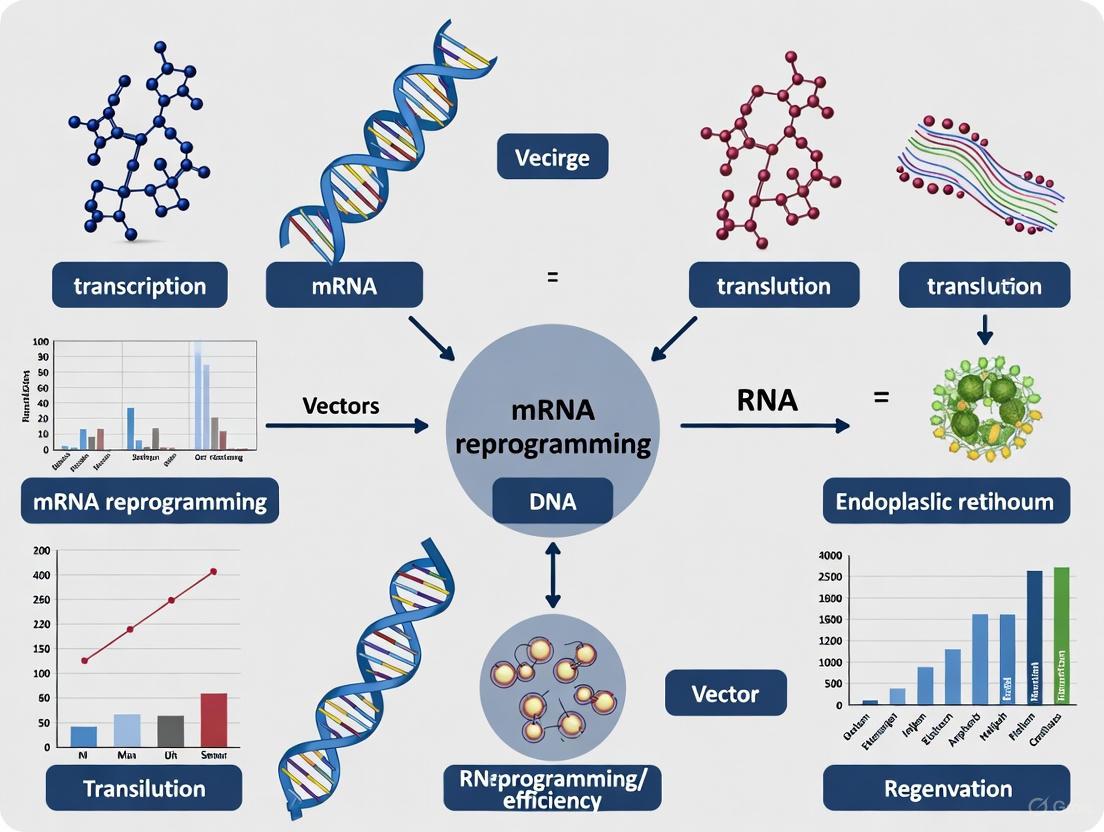
The diagram below illustrates the distinct intracellular pathways of mRNA and pDNA vectors.
Performance Comparison and Experimental Data
Objective comparison of vector performance requires evaluating key parameters such as transfection efficiency, kinetics of protein expression, duration of expression, and immunogenicity. The data summarized below are derived from controlled experimental models.
Table 1: Comparative analysis of mRNA and plasmid DNA vector characteristics.
| Feature | mRNA Vector | Plasmid DNA Vector |
|---|---|---|
| Mechanism of Action | Direct cytoplasmic translation [1] | Nuclear import required for transcription [1] |
| Onset of Protein Expression | Rapid (hours) [1] | Delayed (days) [1] |
| Duration of Protein Expression | Transient (hours to days) [2] [4] | Sustained (days to weeks) [2] |
| Genomic Integration Risk | Non-integrative; no genomic integration risk [1] [4] | Low risk, but potential for random integration exists [1] |
| Immunogenicity | Higher inherent immunogenicity; can be modulated with nucleoside modifications [2] [4] | Inherently immunogenic due to CpG motifs; can trigger innate immunity [2] |
| Ideal Application Scope | Vaccines, transient protein expression, cell reprogramming, immunotherapy [5] [2] | Applications requiring sustained protein expression, gene therapy, veterinary vaccines [6] [2] |
| Ease of Transfection | Efficient in hard-to-transfect and non-dividing cells (cell cycle-independent) [1] | Less efficient in non-dividing cells due to nuclear barrier [1] |
Quantitative Performance Data from Representative Studies
Table 2: Experimental data from peer-reviewed studies highlighting key performance metrics.
| Experiment / Parameter | mRNA Vector Performance | Plasmid DNA Vector Performance | Source/Model |
|---|---|---|---|
| Reprogramming Efficiency (iPSC Generation) | High efficiency; demonstrated suitability for generating induced pluripotent stem cells [7] | Established method, but efficiency can be variable [7] | Somatic cell reprogramming [7] |
| Cellular Uptake Efficiency | L@Mn-mRNA formulation showed 2-fold increase in cellular uptake vs. conventional LNP-mRNA [8] | Naked DNA uptake is generally low; often requires advanced delivery systems (electroporation) [6] | DC 2.4 cells (Dendritic Cells) [8] |
| Vaccine Immunogenicity | High titers of anti-HA antibodies; 100% protection in mice against lethal influenza challenge [3] | Licensed veterinary DNA vaccines show strong immunogenicity; multiple candidates in human trials [2] | Mouse influenza challenge model [3] |
| Expression Kinetics | Protein detection within a few hours post-transfection [1] | Protein expression typically begins 24-48 hours post-transfection [1] | In vitro transfection studies [1] |
Detailed Experimental Protocols
To ensure reproducibility, this section outlines detailed methodologies for key experiments cited in this guide, focusing on novel platforms and direct comparisons.
Protocol: High-Efficiency L@Mn-mRNA Vaccine Formulation
This protocol, adapted from a 2025 Nature Communications study, details the creation of a manganese-core mRNA nanoparticle (L@Mn-mRNA) with nearly double the mRNA loading capacity of conventional LNPs [8].
mRNA Enrichment (Mn-mRNA Core Formation):
- Mix mRNA (e.g., mEGFP, mLuc, or antigen-encoding) with Mn2+ ions in a molar ratio of 1 mRNA base to 5 Mn2+.
- Incubate the mixture at 65°C for 5 minutes. This mild heating facilitates the rearrangement and formation of spherical Mn-mRNA nanoparticles without significant mRNA degradation.
- Confirm nanoparticle formation and size uniformity (≈90% efficiency) using Transmission Electron Microscopy (TEM) and Dynamic Light Scattering (DLS).
Lipid Coating (L@Mn-mRNA Formation):
- Prepare a lipid mixture containing ionizable lipids, helper phospholipids, cholesterol, and PEG-lipids using established LNP formulation techniques (e.g., microfluidics).
- Combine the pre-formed Mn-mRNA nanoparticles with the lipid mixture to allow for the formation of the final L@Mn-mRNA nanosystem.
Quality Control:
- Use the Quant-it RiboGreen RNA Assay to verify high mRNA loading capacity (≈2x that of conventional LNP-mRNA).
- Assess cellular uptake efficiency, which is also approximately 2-fold higher than LNP-mRNA, via flow cytometry.
Protocol: Evaluating a Capless mRNA Vaccine for Influenza
This protocol describes the generation and in vivo testing of a cost-effective, cap-independent mRNA vaccine, as published in 2025 [3].
Vector Construction and mRNA Synthesis:
- Template Design: Engineer a DNA template containing, in sequence: a T7 promoter, a 5' double-hairpin structure, an Encephalomyocarditis virus (EMCV) Internal Ribosomal Entry Site (IRES), the coding sequence for Influenza A Hemagglutinin (HA), and a poly(A) tail sequence.
- In Vitro Transcription (IVT): Linearize the plasmid template and use a T7 RNA polymerase-based kit (e.g., NEB HiScribe) to synthesize mRNA in a single step. No additional capping enzymes or cap analogs are required.
- Purification: Treat the reaction with DNase to remove template DNA, and purify the mRNA via LiCl precipitation.
Vaccine Formulation and Immunization:
- Complex the purified, capless mRNA with pre-formed lipid-based nanoparticles (e.g., using commercial transfection reagents like Polyplus in vivo-jetRNA+).
- Inject the formulated vaccine intramuscularly into mouse models (e.g., C57BL/6) using a prime-boost regimen.
Efficacy Assessment:
- Humoral Response: Measure anti-HA antibody titers in mouse serum using an Enzyme-Linked Immunosorbent Assay (ELISA) at 2- and 4-weeks post-boost.
- Challenge Study: Expose immunized mice to a lethal dose of live Influenza A virus (e.g., A/Puerto Rico/8/1934) and monitor survival rates and weight loss over 14 days.
Essential Research Reagents and Solutions
Successful experimentation with mRNA and pDNA vectors relies on a suite of specialized reagents. The following table lists key solutions used in the featured experiments and the broader field.
Table 3: Key research reagents for working with mRNA and DNA plasmid vectors.
| Reagent / Material | Function | Example Use Case |
|---|---|---|
| Lipid Nanoparticles (LNPs) | Protect mRNA and facilitate cellular uptake and endosomal escape [8] [9]. | Delivery platform for mRNA vaccines and therapeutics [8]. |
| T7 HiScribe RNA Synthesis Kit | High-yield in vitro transcription of mRNA from a DNA template [3]. | Production of research-grade and therapeutic mRNA [3]. |
| Cap Analogs (e.g., ARCA) | Co-transcriptionally added to mRNA to generate a 5' cap, enhancing stability and translation [2]. | Standard mRNA synthesis (not required for capless IRES-driven designs) [3]. |
| Polyethylenimine (PEI) | Cationic polymer that complexes with nucleic acids for delivery; can be used for both pDNA and mRNA [9] [3]. | In vitro and in vivo transfection of various cell types [3]. |
| Mirus TransIT-mRNA Transfection Kit | Commercial lipid-based reagent optimized for high-efficiency mRNA delivery in vitro [3]. | Transfection of difficult-to-transfect cells like primary cells and stem cells [1]. |
| Quant-it RiboGreen RNA Assay | Fluorescent quantification of RNA, useful for determining loading efficiency in nanoparticles [8]. | Quality control for mRNA encapsulation in LNPs and other nano-formulations [8]. |
mRNA and DNA plasmid vectors are distinct technologies with complementary strengths. The choice between them is not a matter of superiority, but of strategic alignment with experimental or therapeutic goals. mRNA vectors offer a compelling solution for applications requiring rapid, high-yield, but transient protein expression without genomic integration risks, making them ideal for vaccines, cellular reprogramming, and transient protein therapies. DNA plasmid vectors remain invaluable for scenarios demanding sustained, long-term protein expression and have a proven track record in veterinary vaccine applications.
The ongoing evolution of both platforms—such as the development of high-loading LNP systems for mRNA and minimalistic vectors like minicircle DNA for pDNA—continues to push the boundaries of their efficiency, safety, and applicability. Researchers are thus empowered to select the vector best suited to their specific needs in the dynamic landscape of genetic medicine.
In the field of regenerative medicine and cellular reprogramming, two primary vectors—DNA plasmids and messenger RNA (mRNA)—serve as fundamental tools for delivering genetic instructions into somatic cells to alter their fate. The core distinction between these platforms lies in their site and mechanism of action: DNA-based approaches must overcome the barrier of the nuclear envelope for gene expression, whereas mRNA functions entirely within the cytoplasm. This fundamental difference has profound implications for the kinetics, efficiency, and safety of biotechnological applications, particularly in the generation of induced pluripotent stem cells (iPSCs).
This guide objectively compares the performance of these two mechanisms within the broader thesis of optimizing reprogramming protocols for research and therapeutic development. The data and methodologies presented are synthesized from current literature to provide a direct, evidence-based comparison.
Core Mechanism and Workflow Comparison
The journey of a genetic payload from delivery to functional protein expression follows distinctly different paths for DNA and mRNA. The following diagram illustrates these divergent workflows, highlighting the key steps where bottlenecks and regulatory checkpoints occur.
Figure 1: Comparative Gene Expression Pathways. The DNA pathway (yellow/orange) requires nuclear entry, a significant rate-limiting step, before transcription can begin. The mRNA pathway (blue) bypasses this bottleneck, enabling immediate cytoplasmic translation.
Key Mechanistic Differences
- Nuclear Entry Requirement: Plasmid DNA must traverse the nuclear membrane to access the transcription machinery, a process that is inefficient and serves as the major rate-limiting step for DNA-based expression [10]. In contrast, mRNA is translated immediately upon cytoplasmic delivery, bypassing this bottleneck entirely [10] [11].
- Kinetics of Protein Expression: Due to the absence of a nuclear barrier, mRNA transfection leads to faster protein expression—typically within hours—compared to DNA plasmids, which require 24-48 hours for significant protein production as they must first navigate the nuclear import process [10].
- Cargo Stability and Duration: DNA plasmids can persist episomally for extended periods, leading to sustained protein expression. mRNA is inherently transient, with protein expression typically lasting for a few days, which minimizes the risk of long-term transgene integration but may require repeated administration for some applications [10] [11].
Performance Metrics and Experimental Data
The mechanistic differences between cytoplasmic translation and nuclear entry translate into quantifiable disparities in performance metrics critical for research and therapy development. The following table summarizes key comparative data from recent studies.
Table 1: Quantitative Comparison of mRNA and DNA Plasmid Performance in Cellular Reprogramming
| Performance Metric | mRNA-Based System | DNA Plasmid System | Experimental Context |
|---|---|---|---|
| Reprogramming Efficiency | Up to 90.7% of individually plated cells [12] | Generally <1% without optimization [13] [12] | Human primary fibroblast to iPSC conversion [12] |
| Time to Protein Expression | A few hours [10] | 24-48 hours (requires nuclear entry) [10] | General transfection in vitro [10] |
| Risk of Genomic Integration | None (non-integrative, transient) [5] [10] [11] | Low but present risk of random integration [10] | Safety assessment for clinical translation [10] |
| Transfection Cytotoxicity | Can be higher (activates innate immunity) [12] [11] | Generally lower, but dependent on delivery method (e.g., electroporation) [10] | Observation in primary cell cultures [10] [12] |
Supporting Experimental Evidence
- High-Efficiency mRNA Reprogramming: A landmark study demonstrated that an optimized mRNA protocol could generate up to 4,019 iPSC colonies from 500 starting human primary neonatal fibroblasts, achieving a remarkable 90.7% reprogramming efficiency in individually plated cells [12]. This efficiency far surpasses typical DNA-based methods.
- Overcoming Innate Immunity: A key advancement enabling this high efficiency was the use of nucleoside-modified mRNAs (e.g., with N1-methylpseudouridine), which evades the cell's innate immune sensors and drastically reduces cytotoxicity and interferon responses [11]. This builds on the Nobel Prize-winning work of Karikó and Weissman.
- TNT for Nucleic Acid Delivery: Tissue Nanotransfection (TNT), a non-viral nanoelectroporation platform, can deliver both DNA and mRNA. Research utilizing this technology confirms that mRNA enables efficient protein supplementation and cellular reprogramming with a primary advantage being its cytoplasmic expression, which avoids the nuclear entry barrier faced by DNA [10].
Detailed Experimental Protocols
To ensure reproducibility and provide a clear "Scientist's Toolkit," this section outlines detailed methodologies for key experiments comparing mRNA and DNA vector performance.
High-Efficiency RNA-Based Reprogramming of Human Fibroblasts
This protocol, adapted from a high-impact study, details the steps for achieving ultra-high reprogramming efficiency with mRNA [12].
Table 2: Research Reagent Solutions for mRNA Reprogramming
| Reagent / Material | Function / Explanation |
|---|---|
| 5fM3O mod-mRNA Cocktail | A 6-factor cocktail containing synthetic, modified mRNAs for OCT4 (M3O variant), SOX2, KLF4, cMYC, LIN28A, and NANOG. Nucleoside modifications reduce immunogenicity. |
| miRNA-367/302s Mimics (m-miRNAs) | A cocktail of mature miRNA mimics that synergistically enhance reprogramming efficiency when co-transfected with mod-mRNAs. |
| Lipofectamine RNAiMAX | A proprietary transfection reagent optimized for the delivery of RNA molecules into a wide range of cells. |
| Opti-MEM-8.2 Buffer | A pH-adjusted transfection buffer (Opti-MEM, pH 8.2). The alkaline pH is critical for achieving high transfection efficiency in primary fibroblasts. |
| KOSR Medium | Knock-Out Serum Replacement medium, a defined formulation used to support reprogramming under feeder-free conditions. |
Workflow Diagram:
Figure 2: High-Efficiency mRNA Reprogramming Workflow. The protocol involves low-density seeding of fibroblasts followed by seven repeated transfections of a mod-mRNA and miRNA cocktail over 13 days, leading to the appearance of pluripotent colonies.
Key Procedural Details:
- Cell Culture: The protocol is optimized for feeder-free conditions. Human primary fibroblasts are seeded at a very low density (500 cells/well of a 6-well plate) in KOSR-based reprogramming medium.
- Transfection Complex: For each well, 600 ng of the 5fM3O mod-mRNA cocktail is mixed with 20 pmol of the m-miRNAs cocktail in pH-adjusted Opti-MEM-8.2 buffer. Lipofectamine RNAiMAX is added, and the complex is incubated and then added directly to the cells.
- Transfection Regimen: Transfections are performed every 48 hours for a total of seven transfections. This repeated delivery is critical for maintaining sustained levels of reprogramming factors.
- Efficiency Assessment: Reprogramming efficiency is quantified by counting the number of TRA-1-60-positive colonies (a key pluripotency marker) around day 9-14, relative to the number of input cells.
Protocol for DNA Plasmid Reprogramming with Sequential Factor Addition
This protocol is derived from studies showing that adding reprogramming factors sequentially, rather than simultaneously, can improve the efficiency of DNA-based iPSC generation [13].
Workflow Diagram:
Figure 3: Sequential DNA Factor Addition Workflow. Staggered introduction of OSKM factors guides cells through a more defined epigenetic transition, including a hyper-mesenchymal state, improving the efficiency of reaching pluripotency.
Key Procedural Details:
- Factor Addition Sequence: The OSKM factors are not introduced all at once. The optimal sequence begins with Oct4 and Klf4, followed by c-Myc two days later, and finally Sox2 after another two days [13].
- Mechanistic Rationale: This sequence is proposed to guide cells through a more controlled epigenetic transition. The initial expression of Oct4 and Klf4 induces a hyper-mesenchymal state, delaying the mesenchymal-to-epithelial transition (MET) and potentially allowing for more extensive epigenetic remodeling before the pluripotency network is fully activated by Sox2 [13].
- Delivery System: This protocol typically utilizes retroviral or lentiviral vectors for stable genomic integration and sustained factor expression, though non-integrating plasmid systems can also be adapted.
Discussion and Research Implications
The comparative data and protocols presented reveal a clear trade-off. The mRNA platform offers superior speed, high efficiency, and a superior safety profile due to its transient, non-integrative nature. However, this comes with challenges of potential immunogenicity and the need for repeated transfections. The DNA platform, particularly with sequential factor addition, provides a more controlled progression through epigenetic stages but is hampered by slower kinetics, lower efficiency, and the persistent risk of genomic integration.
For researchers and drug development professionals, the choice of platform depends heavily on the application. mRNA is ideally suited for protocols requiring rapid, high-efficiency reprogramming without genomic footprint, such as generating clinical-grade iPSCs for regenerative medicine. DNA vectors remain a valuable tool for fundamental studies of reprogramming mechanisms, where sustained factor expression and the ability to use integrating reporters are advantageous. As delivery technologies like TNT and LNP formulations continue to advance, the efficiency and applicability of both platforms, particularly mRNA, are expected to expand further.
The field of regenerative medicine was fundamentally transformed by the seminal discovery that somatic cell identity is not fixed but can be reprogrammed. The breakthrough came in 2006 when Shinya Yamanaka and his team demonstrated that the forced expression of four specific transcription factors—OCT4, SOX2, KLF4, and c-MYC (collectively known as the OSKM or Yamanaka factors)—could reprogram mouse fibroblasts into induced pluripotent stem cells (iPSCs) [7] [14]. This discovery earned Yamanaka the Nobel Prize in Physiology or Medicine in 2012 and established the OSKM combination as the foundational "gold standard" for cellular reprogramming.
The original Yamanaka cocktail successfully addresses the core requirement of reverting differentiated cells to a pluripotent state by activating a self-reinforcing "pluripotency network" [14]. During the reprogramming process, these exogenous factors initially suppress genes specific to somatic cells and subsequently activate endogenous expression of pluripotency factors, ultimately establishing a global embryonic gene expression pattern [14]. The molecular roles of each factor are distinct: OCT4 and SOX2 work synergistically to upregulate embryonic genes while inhibiting differentiation genes; KLF4 plays a dual role in suppressing somatic genes and activating pluripotency genes; while c-MYC primarily enhances the process by promoting global histone acetylation and cell proliferation, though it is not absolutely essential for reprogramming [7] [14].
The period from 2024 to 2025 has witnessed remarkable advances in reprogramming technology, particularly in the refinement of factor delivery methods. Among these, mRNA-based reprogramming has emerged as a particularly promising approach for both research and clinical applications due to its non-integrating nature and high efficiency [15] [11]. This guide provides a comprehensive comparison of the established Yamanaka factors against alternative reprogramming cocktails, with special emphasis on delivery system efficiency and practical experimental considerations for research applications.
Reprogramming Cocktails: A Comparative Analysis
The Yamanaka Factors (OSKM) and Variations
The original OSKM combination remains the most widely validated and utilized reprogramming cocktail, but significant optimization has occurred since its initial discovery. Researchers have extensively explored variations to enhance safety and efficiency, particularly addressing concerns regarding the oncogenic potential of c-MYC [7] [14].
OSKM Function and Optimization:
- Factor Roles and Interactions: OCT4 and SOX2 are considered the core essential factors, with OCT4 playing a particularly pivotal role—in some cases, expression of OCT4 alone in human neural stem cells has been shown to generate iPSCs [7]. The ratio of SOX2 to OCT4 has been demonstrated to significantly affect reprogramming efficiency and colony quality [14].
- c-MYC Considerations: While c-MYC substantially enhances reprogramming efficiency, its inclusion raises safety concerns for therapeutic applications. Multiple studies have confirmed that somatic cell reprogramming can be achieved using only OCT4, SOX2, and KLF4 (OSK), excluding c-MYC, though with reduced efficiency [7] [16]. Alternatively, the substitution of c-MYC with L-MYC or N-MYC can reduce tumorigenic risk while maintaining reprogramming efficiency [7].
- Alternative Combinations: The laboratory of James Thomson identified an alternative combination—OCT4, SOX2, NANOG, and LIN28 (OSNL)—sufficient for reprogramming human somatic cells [7] [14]. In this combination, NANOG functions alongside OCT4 and SOX2 to maintain pluripotency, while LIN28 accelerates cell proliferation similarly to c-MYC [14]. Some studies have explored combining all six factors (OSKMNL), reporting a 10-fold increase in reprogramming efficiency compared to OSNL, including successful reprogramming of fibroblasts from aged donors [14].
Recent AI-Driven Enhancements: A groundbreaking development in 2025 demonstrated the power of artificial intelligence in optimizing reprogramming factors. Researchers at OpenAI and Retro Biosciences used GPT-4b micro to design novel variants of SOX2 and KLF4 [17]. These AI-engineered "RetroSOX" and "RetroKLF" variants, which differed by more than 100 amino acids from their wild-type counterparts, achieved remarkable results:
- >50-fold higher expression of stem cell reprogramming markers compared to wild-type controls
- Hit rates of 30-50% for outperforming wild-type factors in screens
- Accelerated marker onset with late pluripotency markers appearing several days sooner
- Enhanced DNA damage repair capabilities, indicating higher rejuvenation potential [17]
The AI-generated variants were successfully validated across multiple delivery methods (including mRNA) and cell types, demonstrating consistent superiority over conventional factors [17].
Chemical Reprogramming Cocktails
As an alternative to transcription factor-based approaches, chemical reprogramming utilizes defined small molecule combinations to induce pluripotency without genetic manipulation. This approach offers potential advantages for clinical applications by eliminating risks associated with genetic integration [16].
Chemical reprogramming typically involves a multi-stage process requiring an intermediate plastic state, distinct from OSKM-mediated reprogramming [16]. Notable advances include:
- A two-chemical reprogramming procedure demonstrated a 42.1% lifespan increase in C. elegans, along with reduced DNA damage and amelioration of age-related epigenetic marks [16].
- The 7c cocktail of small molecules has been shown to rejuvenate mouse fibroblasts at a multi-omics scale, reversing transcriptomic and epigenomic aging clocks while ameliorating mitochondrial oxidative phosphorylation and reducing aging-associated metabolites [16].
Interestingly, chemical reprogramming appears to operate through distinct mechanisms compared to OSKM approaches. Unlike OSKM-mediated reprogramming which downregulates the p53 pathway, 7c-mediated partial reprogramming upregulates p53—a pathway known to inhibit OSKM-mediated reprogramming [16]. Furthermore, while OSKM reprogramming typically requires increased cell proliferation, 7c-mediated reprogramming achieves epigenetic rejuvenation even with decreased proliferation rates [16].
Comparative Performance Analysis
Table 1: Comparative Analysis of Reprogramming Cocktails and Delivery Methods
| Reprogramming Method | Key Components | Reprogramming Efficiency | Time to iPSC Generation | Genomic Integration | Tumorigenic Risk | Primary Applications |
|---|---|---|---|---|---|---|
| OSKM (Retroviral) | OCT4, SOX2, KLF4, c-MYC | <0.1-1% [17] | 3-4 weeks [17] | Yes [11] | High (c-MYC) [14] | Basic research, disease modeling |
| OSK (Retroviral) | OCT4, SOX2, KLF4 | Lower than OSKM [7] | 3-4 weeks | Yes [11] | Reduced (no c-MYC) [7] | Therapeutic applications research |
| OSNL | OCT4, SOX2, NANOG, LIN28 | Comparable to OSKM [14] | 3-4 weeks | Yes [14] | Moderate [14] | Basic research, alternative to OSKM |
| mRNA Reprogramming | Modified mRNA encoding factors | >30% (with optimized factors) [17] | 7-12 days [17] | No [11] | Low (non-integrating) [11] | Clinical applications, safety-sensitive research |
| Sendai Virus | RNA virus encoding factors | Moderate [18] | 3-4 weeks | No (cytoplasmic) [18] | Low (non-integrating) [18] | Basic research, disease modeling |
| Chemical Reprogramming | Small molecule cocktails | Low to moderate [16] | Extended, multi-stage [16] | No [16] | Low (non-genetic) [16] | Rejuvenation studies, therapeutic development |
Delivery Methods and Experimental Protocols
mRNA-Based Reprogramming
mRNA-based reprogramming has gained significant prominence due to its non-integrating nature and high efficiency, particularly with recent technological advancements. The foundation for mRNA therapeutics was established by Karikó and Weissman, who discovered that incorporating modified nucleosides such as N1-methylpseudouridine instead of uridine dramatically reduces the immunogenicity of synthetic mRNA [11]—a breakthrough that earned them the 2023 Nobel Prize in Physiology or Medicine.
Key mRNA Synthesis Technologies:
- 5' Cap Structures: The 5' cap is essential for mRNA stability and translation initiation. Different cap structures (Cap0, Cap1, Cap2) vary in their ability to activate immune responses, with Cap2 demonstrating reduced immunogenicity [11].
- PureCap Method: Developed by Inagaki et al., this innovative approach enables production of completely capped mRNA using a novel cap analog with an o-nitrobenzyl group that facilitates purification through RP-HPLC separation [11]. This method produces mRNA with >10 times higher translational activity compared to conventional cap analogs [11].
- UTR Optimization: The 5'- and 3'-untranslated regions (UTRs) significantly influence mRNA stability and translational efficiency, with elements like internal ribosome entry sites (IRESs) playing crucial roles in translation initiation [11].
Lipid Nanoparticle (LNP) Delivery Systems: Effective mRNA delivery requires protection from nucleases and facilitation of cellular uptake. Lipid nanoparticles have emerged as the leading delivery platform, composed of phospholipids, cholesterol, ionized lipids, and PEG lipids [11] [19]. Recent innovations have focused on:
- Organ-Specific Targeting: Reformulating LNPs through adjustments in lipid material structures and compositions enables tissue-specific mRNA delivery. Notably, cholesterol removal from LNPs has been shown to address the persistent challenge of preventing hepatic accumulation [19].
- Degradable-Core Lipids: Novel ionizable cationic lipids with biodegradable ester-cores (nAcx-Cm) have been developed, demonstrating enhanced mRNA delivery efficacy and favorable degradation profiles [19].
- Endosomal Escape Optimization: Current research focuses on improving endosomal escape efficiency, which typically remains low (~2%) and represents a major bottleneck in mRNA delivery [11].
Table 2: Key Research Reagents for mRNA Reprogramming
| Reagent Category | Specific Products/Components | Function | Application Notes |
|---|---|---|---|
| mRNA Synthesis | N1-methylpseudouridine, 5-methylcytidine [11] | Reduces immunogenicity, enhances translation | Critical for in vitro transcribed mRNA |
| Capping Systems | CleanCap, PureCap [11] | 5' cap addition for stability and translation | PureCap enables complete capping for enhanced translation |
| Lipid Nanoparticles | Ionizable lipids, phospholipids, cholesterol, PEG-lipids [11] [19] | mRNA encapsulation and delivery | Component ratios affect targeting and efficiency; cholesterol removal reduces liver tropism [19] |
| Reprogramming Factors | Wild-type OSKM mRNA, RetroSOX/RetroKLF [17] | Induction of pluripotency | AI-designed variants show dramatically enhanced efficiency |
| Cell Culture Supplements | 8-Br-cAMP, valproic acid, sodium butyrate [7] | Enhances reprogramming efficiency | 8-Br-cAMP with VPA increases efficiency up to 6.5-fold [7] |
| Characterization Tools | Antibodies against SSEA-4, TRA-1-60, NANOG [17] | Pluripotency marker detection | Essential for validating successful reprogramming |
Comparative Delivery System Efficiency
The choice of delivery method significantly impacts reprogramming outcomes, particularly in the context of mRNA versus DNA vector systems. A systematic comparison of non-integrating reprogramming methods revealed that mRNA-based reprogramming demonstrates higher efficiency when successful, though it has a somewhat lower overall success rate compared to other non-integrating methods [18]. Sendai virus-based systems provide an alternative non-integrating approach but require extended time periods to become vector-free [18], while episomal systems show a slightly higher incidence of karyotypic instability compared to other non-integrating methods, though still lower than retroviral approaches [18].
Diagram 1: Reprogramming Delivery Methods and Their Efficiency-Safety Profiles. This diagram illustrates the relationship between different delivery approaches and their characteristic efficiency and safety profiles, highlighting the advantageous position of mRNA-based systems.
Experimental Protocol for mRNA Reprogramming
For researchers implementing mRNA reprogramming, the following protocol outlines key considerations and steps:
Day 0: Cell Plating
- Plate human fibroblasts or mesenchymal stromal cells at appropriate density (typically 10,000-50,000 cells per well in 6-well plates)
- Ensure cells are in optimal growth medium and at appropriate passage number
- For cells from aged donors, consider pre-treatment with rejuvenation factors [17]
Days 1-7: Daily mRNA Transfection
- Prepare mRNA-LNP complexes according to manufacturer protocols
- For each transfection, use 0.5-1 µg mRNA per factor (OSKM or optimized variants)
- Include modified nucleosides (N1-methylpseudouridine) to reduce immunogenicity [11]
- Utilize PureCap or similar high-quality capping technology for enhanced translation [11]
- Consider including small molecule enhancers such as 8-Br-cAMP or valproic acid to boost efficiency [7]
Days 7-12: Monitoring and Validation
- Monitor for emergence of embryonic stem cell-like morphology
- Assess early pluripotency markers (SSEA-4) from day 7 [17]
- Evaluate late pluripotency markers (TRA-1-60, NANOG) from day 10-12 [17]
- Perform alkaline phosphatase staining to confirm pluripotency [17]
Days 12-21: Colony Expansion and Characterization
- Pick and expand individual colonies
- Validate pluripotency through immunocytochemistry and qPCR
- Confirm differentiation potential through trilineage differentiation assay
- Perform karyotyping to ensure genomic stability [17]
Critical Optimization Parameters:
- Factor Ratio: The specific ratio of SOX2 to OCT4 significantly influences reprogramming efficiency [14]
- Delivery Timing: Multiple rounds of transfection over 7-12 days typically yield optimal results [17]
- Cell Source: Mesenchymal stromal cells from middle-aged donors have been successfully reprogrammed using optimized mRNA factors [17]
The landscape of cellular reprogramming continues to evolve rapidly, with the Yamanaka factors maintaining their position as the foundational reference standard against which new approaches are measured. The gold standard OSKM combination has proven remarkably resilient, serving as both a reliable research tool and a platform for continuous optimization through factor engineering and delivery innovation.
The emergence of mRNA-based delivery represents perhaps the most significant advance toward clinical translation, offering an compelling combination of high efficiency and favorable safety profile. When enhanced with AI-designed factor variants, mRNA reprogramming has demonstrated unprecedented efficiency gains—exceeding 50-fold improvements in marker expression alongside accelerated reprogramming kinetics [17]. These advances position mRNA as a particularly promising vehicle for reprogramming in therapeutic contexts, especially for age-related conditions where traditional methods show reduced efficiency [17].
Future developments will likely focus on several key areas:
- Precision Targeting: Enhanced LNP formulations enabling tissue-specific reprogramming in vivo [19]
- Chemical Reprogramming Optimization: Refined small-molecule cocktails that may offer completely non-genetic rejuvenation approaches [16]
- Safety-First Engineering: Continued factor engineering to eliminate tumorigenic risks while maintaining high efficiency [7] [17]
- Manufacturing Innovation: Automated closed-system manufacturing platforms to reduce production complexity and costs [15]
For research and drug development professionals, the current technological landscape offers multiple validated pathways for iPSC generation, with selection criteria increasingly dependent on application-specific requirements for efficiency, safety, and scalability. The Yamanaka factors remain the undeniable gold standard that launched the field, while mRNA-based approaches represent the vanguard of efficiency-optimized, clinically-translatable reprogramming technologies.
The advancement of genetic engineering and cellular reprogramming is deeply influenced by the choice of vector platform. Among non-integrative strategies, messenger RNA (mRNA) and episomal DNA vectors have emerged as two leading technologies, each with distinct inherent safety profiles and performance characteristics. The groundbreaking discovery that somatic cells can be reprogrammed into induced pluripotent stem cells (iPSCs) underscored the need for "footprint-free" methods that avoid the risks of genomic modification associated with early viral vectors [20]. This need has propelled the development of both mRNA and episomal DNA technologies, which offer transient or non-integrative gene expression, respectively. Within the broader thesis of mRNA reprogramming efficiency, it is crucial to objectively compare these platforms. This guide provides a detailed, data-driven comparison of their safety, mechanisms, and practical application, providing researchers and drug development professionals with the evidence needed to select the appropriate tool for their experimental and clinical objectives.
Mechanism of Action and Key Safety Profiles
Understanding the fundamental mechanisms of these vectors is key to appreciating their safety and efficacy profiles.
2.1 mRNA Vector Mechanism and Safety Synthetic mRNA functions as a minimalistic genetic vector. Once delivered into the cell cytoplasm, it is directly translated by ribosomes into the protein of interest, such as a reprogramming factor. Its action is inherently transient and non-integrative; mRNA does not enter the nucleus and is degraded by normal cellular processes, eliminating any risk of insertional mutagenesis [21] [22]. A key safety consideration is its immunogenicity. Exogenous mRNA can be recognized by innate immune sensors (e.g., TLR7, RIG-I), potentially triggering an antiviral type I interferon response. This can lead to inhibition of protein translation and cell death [22]. This challenge has been successfully mitigated through technological advances, including the incorporation of modified nucleosides (such as pseudouridine) and high-performance liquid chromatography (HPLC) purification to remove immunogenic double-stranded RNA contaminants [22].
2.2 Episomal DNA Vector Mechanism and Safety Episomal DNA vectors are plasmid-based systems engineered to replicate autonomously within the nucleus without integrating into the host genome. They achieve this through specific genetic elements that permit replication and retention during cell division. A prominent example is the use of the scaffold/matrix attachment region (S/MAR), a human chromosomal element that tethers the vector to the nuclear matrix, enabling its long-term persistence as an extrachromosomal element [23] [24]. The primary safety advantage is the avoidance of insertional mutagenesis. However, a critical safety consideration is the theoretical, though low, risk of eventual integration or the use of potentially oncogenic viral elements (e.g., SV40 Large T-antigen, EBNA-1) in some older episomal systems [25] [23]. Newer, minimally-sized DNA nanovectors (e.g., nSMAR) have been developed to eliminate bacterial sequences and viral oncogenes, further enhancing their safety profile [23].
The following diagram illustrates the distinct intracellular pathways of these two vector types.
Diagram: Intracellular Pathways of mRNA and Episomal DNA Vectors. mRNA vectors act in the cytosol and avoid the nucleus, while episomal DNA vectors enter the nucleus but are designed to remain as non-integrating episomes.
Comparative Safety and Performance Data
The table below provides a structured summary of quantitative and qualitative data comparing the two platforms, focusing on safety, stability, and efficiency.
| Feature | mRNA Vectors | Episomal DNA Vectors |
|---|---|---|
| Genomic Integration Risk | None; transient, cytoplasmic action [21] [22] | Very low; designed for non-integrative, extrachromosomal persistence [23] [24] |
| Inherent Immunogenicity | High, but can be mitigated with nucleoside modifications and HPLC purification [22] | Lower; double-stranded DNA is less immunogenic, but bacterial backbone CpG motifs can trigger immune responses [26] [23] |
| Persistence of Expression | Short-term (hours to days); suitable for transient reprogramming needs [21] | Long-term (weeks to months, through cell divisions); stable in stem cells during self-renewal and differentiation [23] |
| Oncogenic Risk Profile | None; does not encode or interact with oncoproteins directly [20] | Low; modern systems (S/MAR) are devoid of viral oncogenes, unlike older systems (SV40 T-Ag, EBNA1) [23] |
| Reprogramming Efficiency | High; up to ~90% of individually plated primary human fibroblasts [12] | Variable; highly efficient in some stem cell lines (e.g., 60% transfection efficiency in mESCs with nSMAR) [23] |
| Key Safety Advantage | Absolute avoidance of the genome and insertional mutagenesis [20] [22] | Mitotic stability without integration, supporting long-term expression in dividing cells [23] [24] |
Experimental Protocols and Workflows
High-Efficiency mRNA Reprogramming Protocol
This protocol, adapted from a landmark Nature Communications study, details a highly efficient method for generating integration-free iPSCs from human primary fibroblasts using synthetic mRNA [12].
Key Reagents:
- 5fM3O mod-mRNA Cocktail: A combination of six synthetic, nucleoside-modified mRNAs encoding the reprogramming factors SOX2, KLF4, cMYC, LIN28A, NANOG, and a modified version of OCT4 (M3O).
- miRNA-367/302s (m-miRNAs): A cocktail of mature miRNA mimics that act as potent enhancers of the reprogramming process.
- Transfection Reagent: Lipofectamine RNAiMAX.
- Transfection Buffer: Opti-MEM adjusted to pH 8.2.
- Culture Medium: Knock-out serum replacement (KOSR) reprogramming medium under feeder-free conditions.
Methodology:
- Cell Seeding: Plate low-density human primary fibroblasts (500 cells per well of a 6-well plate).
- Transfection Regimen: Perform transfections every 48 hours for a total of seven transfections.
- Transfection Mix: For each well, complex 600 ng of the 5fM3O mod-mRNA cocktail with 20 pmol of m-miRNAs in Opti-MEM-8.2 buffer using the RNAiMAX reagent.
- Monitoring: The first TRA-1-60-positive colonies (a marker of pluripotency) typically appear within 20 days. This protocol can generate several thousand iPSC colonies from 500 starting cells, with efficiencies reaching up to 90.7% in individually plated cells [12].
The workflow for this protocol is summarized below:
Diagram: High-Efficiency mRNA Reprogramming Workflow. The protocol relies on repeated transfections of a defined mod-mRNA and miRNA cocktail.
Episomal Vector Transfection and Stability Assay
This protocol describes the generation of stably transfected stem cell lines using non-viral, S/MAR-based episomal vectors, as detailed in Stem Cell Reports [23].
Key Reagents:
- Episomal Vectors: Plasmids or nanovectors containing an S/MAR element (e.g., pSMAR, nSMAR).
- Cell Lines: Mouse or human pluripotent stem cells (e.g., mESCs, hiPSCs).
- Delivery Method: Electroporation.
Methodology:
- Electroporation: Introduce the episomal vector (e.g., nSMAR encoding a GFP reporter) into stem cells via electroporation.
- Selection and Expansion: Culture cells under appropriate selection (e.g., neomycin) for 7-30 days to select for stable transfectants.
- Long-Term Maintenance: Culture stable pools without selection pressure for over 60 days to monitor vector retention and transgene expression stability.
- Analysis:
- Flow Cytometry: Monitor transfection efficiency and the stability of GFP expression over time.
- Southern Blot/PCR: Confirm the extrachromosomal, episomal status of the vector and rule out genomic integration.
- Differentiation Assays: Differentiate the modified stem cells and assess transgene expression in progeny to confirm the vector persists through dramatic changes in the cellular milieu without silencing [23].
The Scientist's Toolkit: Essential Research Reagents
The following table catalogs key reagents and their functions for working with these non-integrative vector systems, as cited in the featured research.
| Research Reagent | Function & Application | Vector Platform |
|---|---|---|
| Lipofectamine RNAiMAX | A proprietary reagent for the efficient in vitro delivery of mRNA and miRNAs into a wide range of cell types, including sensitive primary fibroblasts [12]. | mRNA |
| Ionizable Lipid Nanoparticles (LNPs) | A delivery system that encapsulates nucleic acids, protects them from degradation, and facilitates cellular uptake; the leading platform for in vivo mRNA delivery [21] [26]. | mRNA |
| S/MAR-based Vectors (e.g., nSMAR) | Non-viral, episomal plasmids that utilize a human Scaffold/Matrix Attachment Region for nuclear retention, autonomous replication, and mitotic stability without integration [23] [24]. | Episomal DNA |
| Modified Nucleosides (e.g., Pseudouridine) | Incorporated into synthetic mRNA during IVT to reduce innate immune recognition, thereby decreasing immunogenicity and increasing protein translation [22]. | mRNA |
| Electroporation Systems (e.g., CELLECTRA) | Devices that apply controlled electrical pulses to transiently permeabilize cell membranes, enabling efficient intracellular delivery of large DNA plasmids [26]. | Episomal DNA |
| HPLC/FPLC Purification | A critical downstream purification step for IVT mRNA to remove immunogenic double-stranded RNA contaminants, significantly boosting protein yield [22]. | mRNA |
The choice between mRNA and episomal DNA vectors is not a matter of one being universally superior, but rather hinges on the specific requirements of the research or therapeutic application.
For applications demanding high-efficiency, transient expression with an absolute guarantee of no genomic contact, such as direct in vivo reprogramming or vaccination, mRNA technology holds a distinct safety advantage. Its rapid, high-yield protein production is well-suited for activating complex reprogramming cascades, as evidenced by its remarkable efficiency in converting primary human fibroblasts [20] [12]. The main challenges of immunogenicity have been largely, though not completely, solved through sophisticated engineering.
Conversely, for projects requiring long-term, stable transgene expression in dividing cells—such as the generation of engineered stem cell lines for disease modeling or differentiation studies—episomal DNA vectors are often the more pragmatic choice. Their ability to self-replicate and segregate during cell division without integration provides a favorable balance of persistence and safety [23] [24]. While the risk of integration is low, it is not entirely zero, and careful screening remains a necessary step for clinical applications.
In conclusion, both non-integrative platforms represent powerful and relatively safe tools that have dramatically advanced the field of genetic medicine. mRNA vectors excel in providing a rapid, potent, and footprint-free pulse of gene expression. In contrast, episomal DNA vectors offer a stable, long-term, and non-integrating genetic payload. The ongoing refinement of both technologies—including the development of self-amplifying RNA, advanced lipid nanoparticles, and minimalistic non-viral episomes—continues to narrow the efficacy gap with integrating vectors while upholding the paramount principle of safety first.
The generation of induced pluripotent stem cells (iPSCs) represents a landmark achievement in regenerative medicine, offering a pathway to create patient-specific cells for disease modeling and therapy. The initial method of reprogramming somatic cells using integrating viral vectors has progressively evolved toward safer, more efficient non-viral strategies. This evolution is characterized by a significant shift from DNA-based vectors to transient mRNA delivery systems, which provide enhanced safety profiles and improved practicality for clinical applications. This guide objectively compares the performance of these reprogramming strategies, focusing on the emerging paradigm of mRNA-based efficiency.
The discovery that somatic cells could be reprogrammed into iPSCs using defined factors opened new avenues for personalized medicine [7]. The original method utilized integrating retroviruses to express the OSKM transcription factors (OCT4, SOX2, KLF4, and c-Myc) [7]. While effective, this approach raised significant safety concerns due to the risk of insertional mutagenesis and potential tumorigenesis from permanent genomic integration [27]. These concerns catalyzed the development of non-integrating methods, including Sendai virus, episomal plasmids, and synthetic mRNA [7] [28]. Among these, mRNA reprogramming is considered "footprint-free," highly productive, and well-suited for clinical production of stem cells [27]. The core advantage of mRNA technology lies in its non-integrating nature, offering a controllable strategy for expressing therapeutic proteins without the risk of genomic alteration [5].
Comparative Analysis of Reprogramming Technologies
The table below provides a performance comparison of the primary reprogramming technologies based on safety, efficiency, and practical application criteria.
| Technology | Genomic Integration? | Key Advantages | Key Limitations | Reprogramming Efficiency | Best Suited For |
|---|---|---|---|---|---|
| Viral (Retro/Lenti) | Yes | High efficiency; stable expression [7] | Insertional mutagenesis; oncogenic risk [7] [27] | High [7] | Basic research |
| Sendai Virus | No | High efficiency; robust expression [28] | Biosafety level requirements; immunogenicity [28] | High [28] | Preclinical research |
| Non-Viral DNA (Episomal) | No (Low risk) | Non-integrating; use of plasmids [28] | Potential for random integration; lower efficiency [28] | Moderate [28] | Research & clinical grade iPSC generation [28] |
| Synthetic mRNA | No | No risk of integration; high efficiency; translatable to clinic [27] [28] | Requires multiple transfections; can trigger innate immune response [27] | High (with optimized protocol) [28] | Clinical-grade iPSC production [27] |
Detailed Experimental Protocols and Data
mRNA Reprogramming of Peripheral Blood Mononuclear Cells (PBMCs)
A 2025 study established an efficient protocol for generating iPSCs from PBMCs using synthetic mRNA, highlighting the critical role of the p53 suppressor MDM4 in enhancing efficiency [28].
Key Research Reagent Solutions:
Experimental Workflow:
- Cell Preparation: PBMCs are isolated from fresh human blood [28].
- Reverse Transfection: A mixture of cell suspension, synthetic reprogramming mRNA (including OSKL factors), transfection reagent, and iMatrix-511 substrate is prepared and seeded directly onto a culture plate [28].
- Culture: Cells are maintained in a defined medium. This protocol is distinct from traditional methods where RNA is introduced into already adherent cells [28].
- Colony Formation: iPSC-like colonies emerge approximately 14 days after the initial transfection [28].
- Efficiency Quantification: Reprogramming efficiency is calculated on day 9 or 14 by counting the number of TRA-1-60-positive colonies relative to the number of cells initially seeded [28].
Quantitative Findings: The study provided quantitative data on the impact of p53 pathway modulation on reprogramming efficiency from PBMCs. The results below show the number of TRA-1-60 positive colonies obtained from different donor lots under various conditions [28].
| Experimental Condition | PBMC Lot 1 | PBMC Lot 2 | PBMC Lot 3 |
|---|---|---|---|
| Reprogramming Kit + mCherry (Control) | 2 | 0 | 6 |
| + p53 R175H (dominant-negative) | 14 | 1 | 17 |
| + MDM4 Wild-Type | 66 | 3 | 74 |
| + MDM4-S367A mutant | 84 | 12 | 98 |
This data demonstrates that MDM4, particularly its stabilized S367A mutant, significantly enhances reprogramming efficiency compared to the control and other p53 suppressors, with up to a 16-fold increase observed in a poorly reprogramming donor lot (Lot 2) [28].
p53 Signaling Pathway in Reprogramming
A core finding across reprogramming studies is the critical role of the p53 tumor suppressor pathway as a barrier to efficient iPSC generation. The diagram below illustrates how different reprogramming strategies target this pathway.
Diagram Title: p53 Pathway Modulation for Efficient Reprogramming
The innate stress induced by the reprogramming process, such as from mRNA transfection, activates the p53 pathway. This activation can lead to apoptosis or cell cycle arrest, which presents a major barrier to successful reprogramming [28]. Suppressing this pathway using factors like MDM4 is therefore a common strategy to significantly enhance the efficiency of iPSC generation, particularly in sensitive cell types like PBMCs [28].
The Scientist's Toolkit: Key Reagents for mRNA Reprogramming
The successful implementation of mRNA reprogramming, particularly for difficult-to-transfect cells like PBMCs, relies on a specific set of reagents and tools.
| Reagent / Solution | Function | Example(s) |
|---|---|---|
| Reprogramming Factor mRNA | Expresses transcription factors to induce pluripotency. | Synthetic OCT4, SOX2, KLF4, LIN28/c-MYC (OSKL) mRNA [28]. |
| Efficiency Enhancer mRNA | Suppresses innate immune and stress responses to improve yield. | MDM4 or MDM2 mRNA to inhibit p53 [28]. |
| Specialized Culture Medium | Provides optimal nutrients and signaling environment for reprogramming and iPSC growth. | StemFit AK03N [28]. |
| Culture Substrate | Coats plates to support iPSC attachment and growth. | iMatrix-511 (recombinant laminin-511 E8 fragment) [28]. |
| Transfection Reagent | Enables efficient delivery of mRNA into the cell cytoplasm. | Not specified in detail, but critical for "reverse transfection" protocol [28]. |
The evolution from viral to non-viral reprogramming strategies marks a critical transition toward clinically viable iPSC technologies. Among non-viral methods, mRNA-based reprogramming has emerged as a leading approach due to its complete avoidance of genomic integration and high efficiency. Quantitative data demonstrates that optimized mRNA protocols, incorporating factors like MDM4 to modulate the p53 pathway, can robustly generate iPSCs from clinically relevant sources such as PBMCs. This positions mRNA technology as a powerful tool for creating high-quality, patient-specific iPSCs for regenerative medicine and drug discovery.
From Bench to Bedside: Protocols and Workflows for mRNA and DNA Reprogramming
mRNA-based reprogramming has emerged as a transformative methodology for generating induced pluripotent stem cells (iPSCs) with significant advantages over traditional DNA-based approaches. This guide provides a comprehensive, step-by-step protocol for implementing mRNA reprogramming, supported by comparative experimental data and detailed visualization of the underlying mechanisms. The non-integrating nature of mRNA technology eliminates the risk of genomic insertion, addressing a critical safety concern in therapeutic applications while offering enhanced reprogramming efficiency and kinetics [5] [11]. We present a rigorously optimized workflow that leverages recent advancements in mRNA chemistry, delivery systems, and sequencing strategies to maximize reprogramming success rates for research and drug development applications.
The field of cellular reprogramming was revolutionized by the discovery that somatic cells could be reprogrammed into pluripotent stem cells. Early approaches predominantly utilized viral DNA vectors to deliver reprogramming factors, but these methods carried inherent risks of insertional mutagenesis and persistent transgene expression [29]. mRNA-based technology represents a paradigm shift, offering a non-integrative strategy that maintains the cell's genomic integrity while providing precise temporal control over reprogramming factor expression [5].
The fundamental principle of mRNA reprogramming involves introducing synthetic mRNA molecules encoding key transcription factors (typically OCT4, SOX2, KLF4, and c-MYC) into target cells. These mRNA molecules are translated into proteins that initiate the reprogramming cascade, without ever entering the nucleus or integrating into the host genome [11]. The transient nature of mRNA expression – typically lasting from hours to a few days – requires repeated transfections to maintain sufficient factor levels for successful reprogramming, but this same characteristic prevents residual expression that could impede differentiation or pose tumorigenic risks [29] [11].
For researchers and drug development professionals, mRNA reprogramming offers three compelling advantages: (1) significantly reduced safety concerns for future therapeutic applications; (2) potentially faster reprogramming kinetics; and (3) the ability to precisely fine-tune the expression levels of reprogramming factors through dosing adjustments [5]. The following sections provide a comprehensive workflow to leverage these advantages effectively.
Comparative Analysis: mRNA vs. DNA Reprogramming
Key Parameter Comparison
Table 1 summarizes the critical differences between mRNA and DNA-based reprogramming methodologies, highlighting the distinct advantages of the mRNA platform for clinical translation and research applications.
Table 1: Quantitative Comparison of mRNA vs. DNA Vector Reprogramming
| Parameter | mRNA Reprogramming | DNA Vector Reprogramming | Experimental Support |
|---|---|---|---|
| Genomic Integration Risk | None | Present (except episomal) | No risk of insertional mutagenesis [11] |
| Reprogramming Efficiency | High | Variable | Up to 30% KI efficiency with optimized protocols [30] |
| Reprogramming Timeline | 2-3 weeks | 3-5 weeks | Faster kinetics due to immediate protein translation [11] |
| Factor Expression Duration | Transient (24-96 hours) | Prolonged (weeks to permanent) | Controlled by mRNA half-life, not dependent on promoter silencing [5] |
| Tumorigenicity Risk | Low | Moderate to High | No persistent oncogene expression (c-MYC) [11] |
| Handling Complexity | Moderate (requires repeated transfections) | Low (single transduction) | Multiple transfection rounds needed [29] |
| Immunogenicity | Moderate (can be mitigated with nucleoside modifications) | Low | Modified nucleosides (e.g., N1-methylpseudouridine) reduce immune recognition [11] |
Mechanism of Action and Key Advantages
The following diagram illustrates the fundamental mechanistic differences between mRNA and DNA-based reprogramming approaches, highlighting the critical pathways that give mRNA its safety advantage.
Diagram 1: Mechanism of mRNA vs. DNA reprogramming. mRNA approach avoids nuclear entry of genetic material, eliminating integration risk.
The core safety advantage of mRNA technology stems from its cytoplasmic activity, completely bypassing the need for nuclear localization and thus eliminating any risk of genomic integration [11]. This non-integrative characteristic is particularly valuable for therapeutic applications where long-term genomic stability is paramount. Additionally, the transient nature of mRNA expression provides built-in temporal control, as the reprogramming factors are naturally degraded through cellular processes without requiring complex promoter silencing mechanisms [5].
Step-by-Step mRNA Reprogramming Workflow
Complete Workflow Visualization
The following comprehensive workflow diagram outlines the complete mRNA reprogramming process, from cell preparation through to iPSC characterization, incorporating critical optimization points that significantly enhance efficiency.
Diagram 2: Complete mRNA reprogramming workflow with critical optimization points highlighted.
Detailed Protocol with Optimization Strategies
Pre-Reprogramming Phase (Days 1-3)
Step 1: Cell Source Preparation
- Select appropriate somatic cell source (fibroblasts, PBMCs, or other accessible cell types).
- Culture cells in optimized, richer alternative medium for two days prior to the first transfection to enhance cellular readiness for reprogramming [30].
- Ensure cells are 75-85% confluent and in optimal growth phase at the time of first transfection.
Step 2: mRNA-LNP Complex Preparation
- Formulate lipid nanoparticles (LNPs) containing modified mRNA encoding OCT4, SOX2, KLF4, and c-MYC reprogramming factors.
- Utilize nucleoside-modified mRNA (e.g., N1-methylpseudouridine) to reduce immunogenicity and enhance translation efficiency [11].
- Ensure complete 5' capping using advanced capping technologies (e.g., PureCap method) to maximize translational efficiency and minimize immune recognition [11].
Reprogramming Induction Phase (Days 4-18)
Step 3: Sequential mRNA Delivery
- Perform daily transfections for 14-18 days using optimized delivery protocols.
- For each transfection: dissociate cells enzymatically, prepare LNP-mRNA complexes according to manufacturer specifications, and transfect using optimized nucleofection parameters (Program CA-167 for 4D Nucleofector systems) [30].
- Critical optimization: Implement sequential delivery where possible, with plasmid delivery followed by RNP complexes in advanced gene editing applications [30].
Step 4: Post-Transfection Recovery
- Immediately following transfection, recover cells in RPMI medium for 10 minutes to significantly enhance cell survival rates [30].
- Plate transfected cells at appropriate density on Matrigel or feeder layer-coated plates.
- Implement "cold shock" incubation at 32°C for 24-48 hours post-transfection to enhance knock-in efficiency in gene editing contexts [30].
Step 5: Culture Maintenance & Monitoring
- Feed cells daily with fresh, equilibrated medium specifically formulated for pluripotent stem cells.
- Monitor closely for morphological changes indicative of reprogramming: increased nuclear-to-cytoplasmic ratio, emergence of small, compact cells with prominent nucleoli.
- Perform "double feeding" once per week by exchanging with fresh medium and skipping feeding the following day to maintain nutrient balance [29].
iPSC Selection and Expansion (Days 19-30)
Step 6: Colony Selection and Picking
- Manually pick individual colonies showing characteristic embryonic stem cell-like morphology (tightly packed cells with defined borders) using sterile pipette tips or cloning cylinders.
- Transfer selected colonies to separate wells of a 96-well plate pre-coated with appropriate substrate.
- Pick a minimum of 24 colonies to ensure adequate selection of fully reprogrammed clones, as recommended by biobanking standards [29].
Step 7: Clonal Expansion and Characterization
- Expand successfully picked colonies through sequential passaging while maintaining optimal cell density (75-85% confluency).
- Perform quality control assessments including pluripotency marker verification (e.g., alkaline phosphatase staining, immunofluorescence for NANOG, OCT4, SOX2).
- Conduct karyotyping and STR analysis to confirm genomic integrity and cell line identity [29].
The Scientist's Toolkit: Essential Research Reagents
Successful implementation of mRNA reprogramming requires carefully selected reagents and materials. The following table catalogs the essential components of a complete mRNA reprogramming workflow.
Table 2: Essential Research Reagents for mRNA Reprogramming
| Reagent Category | Specific Product/Component | Function & Application Notes |
|---|---|---|
| MRNA Constructs | Modified mRNA (OCT4, SOX2, KLF4, c-MYC) | Core reprogramming factors; use N1-methylpseudouridine-modified for reduced immunogenicity [11] |
| Delivery System | Lipid Nanoparticles (LNPs) | mRNA protection and cellular delivery; ionizable lipids enhance endosomal escape [11] |
| Transfection System | 4D Nucleofector System (X Unit) | High-efficiency delivery; use program CA-167 with P4 buffer [30] |
| Cell Culture Media | mTeSR1 or equivalent | Maintenance of pluripotent state; feed daily with room temperature-equilibrated medium [29] |
| Culture Substrate | Matrigel or Recombinant Laminin-521 | Defined substrate for feeder-free culture; enhances reproducibility [29] |
| Cell Dissociation | Versene or ReLeSR | Gentle cell dissociation; ReLeSR requires 5-7 min dry incubation at 37°C [29] |
| Cryopreservation | KOSR with DMSO | Feeder-free iPSC freezing; use 90% KOSR + 10% DMSO filter-sterilized [29] |
| Quality Control | Alkaline Phosphatase Staining Kit | Pluripotency marker assessment; perform at distribution bank stage [29] |
Troubleshooting and Optimization Strategies
Even with optimized protocols, researchers may encounter challenges during mRNA reprogramming. The following table addresses common issues and provides evidence-based solutions.
Table 3: Troubleshooting Guide for mRNA Reprogramming
| Problem | Potential Causes | Solutions & Optimization Strategies |
|---|---|---|
| Low Reprogramming Efficiency | Suboptimal mRNA delivery, poor cell viability | Implement sequential delivery approach [30]; increase cell number to 3×10⁶ per nucleofection [30]; use RPMI recovery protocol to improve survival [30] |
| High Cell Death Post-Transfection | Excessive electrical pulses, cytotoxic mRNA preparations | Optimize nucleofection parameters; implement 10-minute RPMI recovery post-transfection [30]; ensure proper mRNA modification and purification [11] |
| Incomplete Reprogramming | Insufficient factor expression, inadequate culture conditions | Extend transfection duration; use richer culture medium pre-transfection [30]; verify mRNA quality and translational efficiency [11] |
| Immunogenic Response | Unmodified mRNA, double-stranded RNA contaminants | Utilize completely capped, N1-methylpseudouridine-modified mRNA [11]; implement PureCap technology for superior capping [11] |
Emerging Technologies and Future Directions
The field of mRNA reprogramming continues to evolve rapidly, with several emerging technologies poised to enhance efficiency and applicability:
Novel Delivery Platforms: Tissue nanotransfection (TNT) represents a promising non-viral nanotechnology that enables in vivo gene delivery through localized nanoelectroporation [10]. This approach uses a hollow-needle silicon chip to concentrate electric fields at their tips, temporarily porating cell membranes for highly efficient mRNA delivery with minimal cytotoxicity.
Advanced mRNA Engineering: Recent developments in metal ion-mediated mRNA enrichment (Mn-mRNA nanoparticles) demonstrate nearly twice the mRNA loading capacity compared to conventional LNP formulations [8]. This technology enhances cellular uptake efficiency and antigen-specific immune responses, potentially benefiting reprogramming applications.
Enhanced Gene Editing Integration: The sequential factor delivery workflow enables highly efficient gene editing in iPSCs, achieving knock-in efficiencies above 30% while maintaining GMP compatibility [30]. This approach facilitates the creation of clinically relevant iPSC lines with edited features such as HLA depletion and safety switches.
mRNA-based reprogramming represents a robust, efficient, and clinically relevant methodology for generating human iPSCs. The optimized workflow presented in this guide incorporates critical advancements in mRNA design, delivery strategies, and culture techniques that collectively address the key limitations of earlier reprogramming approaches. By eliminating genomic integration risks while maintaining high reprogramming efficiency, mRNA technology provides researchers and drug development professionals with a powerful platform for generating high-quality iPSCs suitable for both basic research and therapeutic applications. As emerging technologies continue to enhance the specificity and efficiency of mRNA delivery and expression, this methodology is poised to become the gold standard for cellular reprogramming in both research and clinical settings.
The choice of a gene delivery vector is a critical determinant of success in genetic research and therapy. Among non-viral options, conventional episomal plasmids and their advanced derivative, minicircle DNA (mcDNA), represent two pivotal technologies with distinct performance characteristics. This guide provides an objective comparison of these vectors, focusing on their practical implementation within modern research and development workflows. The context of increasing interest in nucleic acid-based therapies, including mRNA platforms, underscores the need for efficient DNA vector systems that offer sustained transgene expression without genomic integration [26]. We present summarized experimental data, detailed protocols, and key reagents to inform the selection and use of these tools in basic and translational science.
Performance Comparison: Minicircle DNA vs. Conventional Plasmid DNA
Direct comparative studies reveal significant differences in the performance of minicircle DNA and conventional parental plasmids. The table below synthesizes key quantitative findings from in vitro and in vivo experiments.
Table 1: Experimental Performance Comparison of Minicircle and Parental Plasmid Vectors
| Performance Metric | Experimental System | Minicircle DNA (mcDNA) Result | Conventional Plasmid DNA (pDNA) Result | Citation |
|---|---|---|---|---|
| Transfection Efficiency | HPV-18 infected cervical cancer cells | 68% | 34% | [31] |
| Nuclear Entry Speed | Live cell imaging of cervical cancer cells | More rapid | Slower | [31] |
| Protein Expression Level | p53 protein in cervical cancer cells | 91.65 ± 2.82 U/mL | 74.75 ± 4.44 U/mL | [31] |
| Gene Expression Level | Luciferase in human melanoma and colon carcinoma cell lines | Higher and sustained expression | Lower and declining expression | [32] |
| GFP-Positive Cells | FACS analysis after gene transfer | Increased count | Lower count | [32] |
| In Vivo Performance | Jet-injection gene transfer | Improved | Standard | [32] |
| Immunogenicity Profile | Host inflammatory response | Reduced (due to fewer CpG motifs) | Higher | [33] |
The performance advantages of minicircles are attributed to their minimized molecular architecture. By eliminating the bacterial backbone, mcDNA reduces the vector size, facilitating more efficient cellular uptake and nuclear import [33] [31]. The absence of prokaryotic sequences, including unmethylated CpG motifs, is a key factor in mitigating transgene silencing and reducing host immune responses, which in turn supports more persistent and higher-level transgene expression [33] [34].
Production Workflows
The manufacturing processes for conventional plasmids and minicircles differ significantly, with minicircle production requiring an additional in vivo recombination step.
Conventional Plasmid DNA Production
The production of parental plasmid DNA is a well-established, scalable process.
- Upstream Production: The parental plasmid is transformed into a specialized E. coli producer strain (e.g., ZYCY10P3S2T). A single colony is used to inoculate a starter culture, which is then expanded in a large-scale bioreactor. Cells are grown in a rich, selective medium under controlled parameters (temperature, aeration, pH) to high density [33].
- Harvesting and Lysis: Bacterial cells are harvested by centrifugation. The pellet is resuspended and subjected to alkaline lysis, which breaks open the cells and denatures proteins and genomic DNA, while leaving the supercoiled plasmid in solution.
- Purification: The clarified lysate undergoes a series of purification steps, typically involving filtration, precipitation, and chromatography (e.g., anion-exchange, size-exclusion) to remove contaminants such as host cell proteins, RNA, and genomic DNA. The final product is a purified, supercoiled plasmid DNA [35].
Minicircle DNA Production
Minicircle production builds upon the plasmid workflow by incorporating a site-specific recombination event within the bacterial host.
- Upstream Production and Induction: The parental plasmid, containing the gene of interest flanked by specific recombination sites (e.g.,
attBandattP), is transformed into a minicircle producer strain. This strain is engineered to express recombinase enzymes (e.g., phiC31 integrase) under the control of an inducible promoter (e.g., pBAD arabinose-inducible promoter). Once the bacterial culture reaches the exponential growth phase, the recombination is induced by adding L-arabinose [33] [34]. - In Vivo Recombination: The induced recombinase catalyzes an intramolecular recombination between the
attBandattPsites. This excises the bacterial backbone (forming a "miniplasmid") from the eukaryotic therapeutic cassette (forming the "minicircle") [33]. - Harvesting and Purification: After induction, the bacterial culture contains a mixture of three circular DNA molecules: the minicircle, the miniplasmid, and potentially some residual parental plasmid. The cells are harvested and lysed. The key challenge is the chromatographic separation of the minicircle from the closely related miniplasmid, which is achieved using specialized affinity or anion-exchange chromatography techniques [34].
The following diagram illustrates the logical sequence and key differences in the production of these two vectors.
The Scientist's Toolkit: Essential Research Reagents
Successful implementation of episomal vector protocols requires specific biological and chemical reagents. The following table details key materials and their functions.
Table 2: Key Reagents for Episomal and Minicircle DNA Research
| Reagent / Material | Function and Role in Protocol |
|---|---|
| Parental Plasmid (PP) | The starting plasmid vector containing the eukaryotic expression cassette flanked by recombination sites and the bacterial backbone with origin of replication (ori) and selection marker. |
| Minicircle Producer Strain | A genetically modified E. coli strain (e.g., ZYCY10P3S2T) that stably expresses recombinase enzymes (e.g., phiC31 integrase, I-SceI endonuclease) essential for the in vivo generation of minicircles [33]. |
| L-Arabinose | The inducing agent for the pBAD promoter, which controls the expression of the recombinase in the producer strain. Its addition to the bacterial culture triggers the recombination event [33] [34]. |
| Specialized Chromatography Matrices | Resins used for the critical purification step to separate the minicircle DNA from the miniplasmid contaminant, as their similar size and charge make separation challenging [34]. |
| Electroporation System | A physical delivery method (e.g., CELLECTRA, TriGrid) that uses electrical pulses to create transient pores in cell membranes, facilitating the uptake of DNA vectors into target cells, particularly effective in vivo [26]. |
| Cationic Lipids / Polymers | Chemical carriers (e.g., liposomes, polyethylenimine - PEI) that complex with negatively charged DNA to form nanoparticles, protecting the genetic cargo and enhancing its cellular uptake in vitro and in vivo [35] [36]. |
Both conventional episomal plasmids and minicircle DNA are powerful tools in the genetic engineering arsenal. The choice between them depends on the specific requirements of the experiment or therapy. Conventional plasmids remain a robust, cost-effective, and easily produced option for a wide array of applications, including basic research and viral vector production. In contrast, minicircle DNA, with its superior transfection efficiency, enhanced transgene expression, and improved safety profile, is emerging as the vector of choice for demanding applications in non-viral gene therapy, regenerative medicine, and vaccination, where long-term, high-level gene expression is critical [33] [31]. As production methods continue to be refined and scaled, minicircle technology is poised for greater adoption in both preclinical and clinical settings.
Electroporation is a well-established physical method that uses an external electric field to temporarily increase the permeability of cell membranes, facilitating the introduction of genetic material into target cells [10] [37]. This technique has evolved significantly from bulk electroporation systems to more sophisticated, localized approaches. When applied to gene delivery, electroporation creates transient nanopores in the cell membrane through the rearrangement of polar molecules in the phospholipid bilayer, allowing charged molecules like DNA and RNA to enter the cell [10] [37]. The pores typically reseal within milliseconds after the electrical pulse ends, leaving the cell membrane intact and minimizing cytotoxicity [10].
Tissue Nanotransfection (TNT) represents a cutting-edge advancement in electroporation-based technologies. It is a novel, non-viral nanotechnology platform that enables in vivo gene delivery and direct cellular reprogramming through localized nanoelectroporation [10] [38]. TNT utilizes a silicon chip containing a hollow microneedle array that concentrates the electric field at the needle tips, creating a highly localized and efficient delivery system for genetic cargo directly into tissues [10] [37]. This technology has demonstrated transformative potential across diverse biomedical applications including tissue regeneration, ischemia repair, wound healing, and antimicrobial therapy [10] [39].
Technological Comparison of Delivery Platforms
Device Architecture and Working Principles
Conventional Electroporation Systems traditionally employ bulk electroporation (BEP) using caliper-type or plate electrodes that apply electric fields across large tissue areas [38]. These systems face challenges with specificity, potential tissue damage from high voltages, and limited precision in gene targeting [37] [38]. More recent advances include microneedle-type electrode-based bulk electroporation (MNE-BEP), which uses conductive microneedle arrays as electrodes to bypass the high resistance of the stratum corneum barrier in skin applications [38]. This approach reduces the required voltage compared to traditional BEP due to smaller distances between electrodes (typically less than 1 mm) and enables more precise targeting of epidermal and dermal layers [38].
TNT Device Architecture consists of a hollow-needle silicon chip mounted beneath a cargo reservoir containing genetic material [10]. The device is placed directly on the skin or target tissue, with the cargo reservoir connected to the negative terminal of an external pulse generator and a dermal electrode serving as the positive terminal [10]. When electrical pulses are applied, the hollow needles concentrate the electric field at their tips, enabling targeted delivery of genetic material into the tissue [10]. The TNT platform has evolved through generations: TNT 1.0 utilized nanochannel-based nanoelectroporation, while TNT 2.0 features a hollow microneedle array that improves physical contact with skin and enhances gene delivery efficiency [38].
Performance Comparison of Delivery Platforms
Table 1: Comparative Analysis of Gene Delivery Platforms
| Platform | Delivery Mechanism | Key Applications | Transfection Efficiency | Advantages | Limitations |
|---|---|---|---|---|---|
| Viral Vectors | Biological transduction via engineered viruses | Stable gene expression, iPSC generation | High transduction efficiency | High efficiency, stable expression | Immunogenicity, insertional mutagenesis, limited cargo capacity [10] [7] |
| Chemical Methods | Nanoparticle encapsulation, liposome fusion | In vitro transfection, vaccine development | Low in vivo efficiency | Reduced immunogenicity, large payload capacity | Low transfection efficiency, cytotoxicity, poor endosomal escape [10] |
| Conventional Electroporation | Bulk electroporation with plate/caliper electrodes | In vitro transfection, DNA vaccination | Variable (25-82% with optimized parameters) [37] | Broad applicability, non-viral | High voltage requirements, tissue damage, limited specificity [37] [38] |
| MNE-BEP | Conductive microneedle array electrodes | Intradermal vaccination, superficial tissue targeting | Enhanced compared to traditional intradermal injection [38] | Bypasses stratum corneum, reduced pain, controlled depth | Limited to accessible tissues, primarily skin applications [38] |
| TNT | Hollow microneedle-based localized electroporation | In vivo tissue reprogramming, regenerative medicine | >98% of targeted cells receive genetic payload [40] | High specificity, minimal cytotoxicity, non-integrative, direct in vivo application [10] [40] | Phenotypic stability questions, scalability challenges for large areas [10] |
Table 2: Genetic Cargo Delivery Performance
| Cargo Type | Mechanism of Action | Expression Kinetics | TNT Compatibility | Key Considerations |
|---|---|---|---|---|
| Plasmid DNA | Nuclear entry required for gene expression | Delayed onset (hours-days), transient or stable | Optimized for delivery [10] | Circular plasmids more efficient than linear; vulnerable to nucleases [10] |
| mRNA | Direct cytoplasmic translation | Rapid onset (hours), strictly transient | Highly compatible [10] [5] | No nuclear entry required; simpler, faster, more efficient than DNA [10] |
| CRISPR/Cas9 | Genome editing with guide RNA | Varies by component format (DNA/mRNA) | Compatible as components [10] | Requires coordinated delivery of multiple components; dCas9 fusions for transcriptional control [10] |
Experimental Data and Performance Metrics
Electroporation Efficiency and Optimization
Computational modeling of the TNT process provides valuable insights into optimization parameters. Simulations reveal that the distribution of the nonuniform electric field across skin results in varied electroporation behavior for each cell [37]. Cells directly underneath the hollow microchannels exhibit the highest total pore numbers due to stronger localized electric fields [37]. The percentage of electroporated cells within skin structure, with pore radius over 10 nm, increases from 25% to 82% as the applied voltage increases from 100 to 150 V/mm [37]. This nonlinear relationship between voltage and efficiency must be balanced against cell viability concerns, as higher voltages can cause irreversible membrane damage.
Gene delivery behavior in TNT is influenced by multiple parameters. The delivery distance increases nonlinearly as both applied voltage and pulse number increase, depending mainly on the diffusion characteristics and electric conductivity of each tissue layer [37]. Research indicates that skin exfoliation prior to TNT procedure enhances delivery depth, highlighting the importance of tissue preparation [37]. Optimization of electrical pulse parameters—including voltage amplitude, pulse duration, and inter-pulse intervals—is critical for maximizing delivery efficiency while preserving cellular viability [10].
Cellular Reprogramming Efficiency
TNT demonstrates remarkable efficiency in cellular reprogramming applications. In direct reprogramming (transdifferentiation), TNT achieves over 98% transfection efficiency in targeted cells, far exceeding most viral and non-viral gene delivery methods [40]. This high efficiency stems from the direct physical mechanism of electroporation combined with optimized genetic construct design [40]. Furthermore, TNT exhibits a unique spreading phenomenon where cellular transformation extends to nearby cells beyond those directly contacted by the chip, creating a regenerative cascade that amplifies therapeutic effects without requiring treatment of every individual cell [40].
For induced pluripotent stem cell (iPSC) generation, the efficiency varies significantly based on cell source and delivery method. Synthetic RNA reprogramming of human dermal fibroblasts (HDFs) using the StemRNA 3rd Gen Reprogramming Kit successfully generates iPSC colonies, with efficiency enhanced by suppression of p53 pathway elements [28]. When reprogramming peripheral blood-derived mononuclear cells (PBMCs) with synthetic RNA, the addition of MDM4—which suppresses p53 function—significantly improves reprogramming efficiency, highlighting cell-type-specific optimization requirements [28].
Experimental Protocols and Methodologies
TNT Chip Fabrication and Operation
TNT Chip Fabrication follows a standardized semiconductor process [37]. The procedure begins with a double-side polished 4-inch Si (100) wafer. A ~50 μm thick SU-8 photoresist is spin-coated on the wafer's backside, and a hole array (diameter = 30 μm, spacing = 150 μm) is patterned using a direct laser writer. The wafer is then etched ~450 μm deep by Bosch process (deep Si etching). After residue SU8 removal, SPR 220-7.0 photoresist is spin-coated on the frontside and patterned with a donut-shape array with precise backside alignment, followed by deep Si etching to form the hollow microneedle array. A thin SiO2 film is deposited on the frontside by plasma enhanced chemical vapor deposition to shrink the bore size to the target value. Finally, the fabricated TNT chip is mounted on a plastic drug reservoir using polydimethylsiloxane for TNT procedure usage [37].
Sterilization of TNT devices is essential for biological and medical applications. Ethylene oxide gas sterilization and gamma irradiation are frequently applied processes, with ethylene oxide preferred for preserving the interior architecture of the nanodevices [10].
TNT Operational Protocol involves placing the device directly on the target tissue or skin [10] [40]. The cargo reservoir is filled with the genetic material solution (plasmid DNA, mRNA, or CRISPR/Cas9 components). Electrical pulses are applied between the nanofabricated chip and the target tissue, typically using microsecond pulse durations [38]. The electric pulse is calibrated to be strong enough to temporarily disrupt membrane integrity while preserving cell viability [40]. The entire procedure takes only seconds, making it one of the fastest cellular reprogramming methods developed [40].
Cellular Reprogramming Methodologies
Direct Lineage Conversion using TNT typically follows this workflow: (1) Identification of target cell type and appropriate transcription factors; (2) Preparation of genetic cargo (plasmid DNA, mRNA, or CRISPR/dCas9 systems); (3) Application of TNT chip to target tissue with optimized electrical parameters; (4) Monitoring of cellular transformation through marker expression and functional assessment [10] [40]. For vasculogenic reprogramming, specific gene cocktails are delivered to skin tissue to induce formation of new functional blood vessels from skin cells [39]. Similarly, neurogenic TNT converts skin cells into nerve cells that mature using the skin as a "bioreactor" [39].
iPSC Generation Protocol using synthetic RNA involves: (1) Cell culture preparation (human dermal fibroblasts or PBMCs); (2) Mixing synthetic RNA for reprogramming with transfection reagent; (3) Combining with cell suspension and iMatrix-511 substrate; (4) Seeding onto culture plates; (5) Culture in reprogramming medium (e.g., StemFit AK03N without bFGF) until colonies appear; (6) Immunostaining analysis for pluripotency markers (e.g., TRA-1-60) typically around day 9-14 [28]. Efficiency is enhanced by co-delivery of p53 pathway suppressors such as MDM4, particularly for PBMC reprogramming [28].
Research Reagent Solutions and Materials
Table 3: Essential Research Reagents for TNT and Electroporation Studies
| Reagent/Material | Function/Purpose | Example Products/Formats | Key Considerations |
|---|---|---|---|
| TNT Silicon Chip | Creates localized electric field for nanoelectroporation | Hollow microneedle array, nanochannel designs [37] [38] | Requires semiconductor fabrication processes; SiO2 coating for bore size control [37] |
| Genetic Cargo | Reprogramming factors or therapeutic genes | Plasmid DNA, mRNA, CRISPR/Cas9 components [10] | mRNA allows direct cytoplasmic translation without nuclear entry [10] |
| Reprogramming Factors | Induce cell fate conversion | OSKM factors (OCT4, SOX2, KLF4, c-MYC), lineage-specific TFs [10] [14] | Optimal ratios critical for efficiency; c-MYC increases tumorigenicity risk [7] [14] |
| Electroporation Buffers | Maintain cell viability during electrical pulses | Ionic solutions with optimized conductivity [37] | Conductivity affects electric field distribution and delivery efficiency [37] |
| Cell Culture Substrates | Support cell growth and reprogramming | iMatrix-511, feeder cells, ECM coatings [28] | Essential for iPSC colony formation and maintenance |
| Sterilization Agents | Ensure device biocompatibility | Ethylene oxide gas, gamma irradiation [10] | Ethylene oxide preserves interior nanodevice architecture [10] |
| Plasmid Preparation Kits | Purify and concentrate genetic cargo | ZymoPURE II Plasmid Midiprep Kit [37] | High-purity supercoiled plasmids enhance transfection efficiency [10] |
| Reprogramming Media | Support reprogramming and pluripotency | StemFit AK03N, media with small molecule enhancers [28] | Optimization of components like bFGF affects efficiency |
Electroporation-based technologies, particularly Tissue Nanotransfection, represent a paradigm shift in gene delivery and cellular reprogramming. TNT's unique advantages include its non-viral approach, high specificity, minimal cytotoxicity, and ability to perform direct in vivo reprogramming [10]. The technology demonstrates remarkable efficiency, with over 98% of targeted cells successfully receiving genetic payloads [40].
While TNT shows transformative potential in regenerative medicine, wound healing, and antimicrobial applications, challenges remain in phenotypic stability, scalability for large tissue areas, and long-term functional integration [10]. Future research directions will likely focus on optimizing genetic cargo design, refining electrical parameters for different tissue types, and developing combination approaches that leverage the strengths of both physical and biological delivery systems.
The integration of TNT with emerging technologies like CRISPR-based gene editing and synthetic biology approaches will further expand its applications in personalized medicine and targeted therapies. As these platforms continue to evolve, they hold significant promise for addressing previously untreatable conditions through precise cellular reprogramming and tissue regeneration.
The choice of starting somatic cell is a critical determinant in the efficiency of generating induced pluripotent stem cells (iPSCs). While dermal fibroblasts have historically been the most common source, recent advances demonstrate that certain blood-derived cells, particularly long-term hematopoietic stem cells (LT-HSCs), can be reprogrammed with remarkably higher efficiency. The optimal reprogramming method—whether mRNA-based, Sendai virus, or episomal vectors—often depends on the source material, with implications for the safety, scalability, and clinical applicability of the resulting iPSCs. The following guide provides a detailed, data-driven comparison to inform strategic decisions in research and therapy development.
Comparison of Reprogramming Efficiency Across Somatic Cell Types
Table 1: Key Performance Metrics for Different Somatic Cell Sources
| Somatic Cell Source | Reported Reprogramming Efficiency | Commonly Used High-Efficiency Methods | Key Advantages | Key Limitations |
|---|---|---|---|---|
| Long-Term Hematopoietic Stem Cells (LT-HSCs) | Up to 44.5% ± 4.1% when using Sendai virus [41] | Sendai virus (SeV) [41] | Extremely high efficiency; lower load of somatic mutations compared to fibroblasts [41] | Difficult to isolate in large numbers from bone marrow; slower cell cycle re-entry than peripheral blood counterparts [41] |
| Peripheral Blood Mononuclear Cells (PBMCs) | Variable; highly dependent on protocol and subset. CD34+ subset is most amenable [42] | Episomal vectors [42], Sendai virus [29], Chemical reprogramming [43] | Minimal invasive collection; vast supply from blood banks; less prone to environmental mutations [42] | Heterogeneous population; requires expansion of progenitor cells for best results; lower efficiency in some mature subsets [42] |
| Dermal Fibroblasts | ~0.1% with standard methods; up to ~90.7% with optimized mRNA/miRNA protocol [12] [41] | Optimized mRNA/miRNA [12], Sendai virus, Episomal vectors [29] | Well-characterized, easy to culture and expand [29] | Invasive biopsy; may accumulate more somatic mutations over time [41]; lower efficiency with non-optimized methods |
| Granulocyte-Macrophage Progenitors (GMPs) | 13.2% ± 4.0% with Sendai virus [41] | Sendai virus (SeV) [41] | Committed progenitor, easier to obtain than LT-HSCs | Lower reprogramming efficiency compared to more primitive HSCs [41] |
Table 2: Impact of Reprogramming Method on Success Rates
| Reprogramming Method | Mechanism | Integration into Genome? | Relative Efficiency (by cell type) | Best Suited For |
|---|---|---|---|---|
| Sendai Virus (SeV) | CytoTune Sendai Reprogramming Kit uses non-integrating RNA virus vectors [29] [41] | No [29] | High for PBMCs, Fibroblasts, and especially LT-HSCs [29] [41] | Protocols where maximum efficiency from blood cells or fibroblasts is critical. |
| Optimized mRNA/miRNA | Synthetic, modified mRNAs + miRNA mimics transfected via lipid nanoparticles; high pH buffer increases efficiency [12] | No [12] | Very High for primary human fibroblasts (up to 90.7%) [12] | Clinical-grade iPSC generation; applications requiring no viral components. |
| Episomal Vectors | oriP/EBNA1-based plasmids transfected via nucleofection [29] [42] | No (vectors are diluted out over passages) [42] | Moderate; lower than SeV in comparative studies [29] | Generating integration-free iPSCs without using viral particles. |
| Chemical Reprogramming | Small molecule combinations that manipulate cell fate without genetic manipulation [43] | No [43] | Demonstrated for blood cells (cord blood, peripheral blood) [43] | Next-generation, potentially clinically safer approaches; uses easily synthesized reagents. |
Detailed Experimental Data and Protocols
High-Efficiency Reprogramming of Blood Cells
Source Material: Peripheral Blood Mononuclear Cells (PBMCs) and CD49f+ Long-Term Hematopoietic Stem Cells (LT-HSCs).
Key Findings:
- A landmark study demonstrated that single adult human CD49f+ LT-HSCs could be reprogrammed using Sendai virus at an exceptional efficiency of 44.5% ± 4.1%. This efficiency is approximately 100-fold higher than that of human skin fibroblasts [41].
- The reprogrammability is intrinsic to the primitive state of the cell, as efficiency progressively decreases in committed progenitors (CMPs: 32.1%; GMPs: 13.2%) [41].
- For heterogeneous PBMC populations, the CD34+ hematopoietic stem and progenitor cell subset is the primary population capable of being reprogrammed into iPSCs or induced mesenchymal stromal cells (iMSCs). Depletion of CD34+ cells from PBMCs before reprogramming abrogates colony formation [42].
Experimental Protocol: Sendai Virus Reprogramming of PBMCs/LT-HSCs [29] [41]
- Cell Isolation and Culture: Isolate PBMCs from whole blood via density gradient centrifugation. For LT-HSCs, sort cells using a complex marker panel (e.g., CD34+, CD38-, CD45RA-, CD49f+).
- Transduction: Incubate cells with the CytoTune Sendai Reprogramming Kit, which contains SeV vectors expressing OCT4, SOX2, KLF4, and c-MYC.
- Media Change: Refresh the medium 24 hours post-transduction to remove viral particles.
- Culture and Observation: Continue culturing for approximately 6 days, changing the medium every other day. Transduction efficiency can be estimated via a GFP reporter.
- Rep plating: Around day 7, harvest and replate transduced cells onto feeder layers or in feeder-free conditions.
- Colony Picking: After 2-3 weeks, manually pick at least 24 individual colonies with iPSC morphology for expansion.
- Quality Control: Expand clones and subject them to stringent quality control, including karyotyping, pluripotency marker staining (SSEA4, TRA-1-60, TRA-1-80), and teratoma formation assays to confirm differentiation into three germ layers [41].
Diagram 1: Sendai Virus Reprogramming Workflow for Blood Cells
Ultra-High-Efficiency mRNA Reprogramming of Fibroblasts
Source Material: Human Primary Fibroblasts.
Key Findings:
- An optimized RNA-based protocol achieved a revolutionary reprogramming efficiency of up to 90.7% of individually plated primary neonatal fibroblasts, generating over 4,000 iPSC colonies from 500 starting cells [12].
- This synergy between modified mRNA (mod-mRNA) and miRNA-367/302s mimics was critical. Transfection with mod-mRNA alone produced colonies that could not be established as long-lived lines [12].
- Success was contingent on tailoring the transfection conditions specifically to human primary fibroblasts, including the use of a high-pH transfection buffer (Opti-MEM pH 8.2), which dramatically increased transfection efficiency [12].
Experimental Protocol: Optimized mRNA/miRNA Reprogramming of Fibroblasts [12]
- Cell Seeding: Plate human primary fibroblasts at a very low density (e.g., 500 cells per well of a 6-well plate) in feeder-free conditions.
- Reprogramming Medium: Use a knockout serum replacement (KOSR)-based reprogramming medium.
- Transfection Buffer: Employ Opti-MEM adjusted to pH 8.2 (Opti-MEM-8.2) as the buffer for the Lipofectamine RNAiMAX transfection reagent. This pH adjustment was a key factor in boosting efficiency.
- RNA Transfection: Perform transfections every 48 hours. Each transfection should deliver a cocktail of:
- 600 ng of a 6-factor modified mRNA cocktail (5fM3O: OCT4, SOX2, KLF4, cMYC, LIN28, NANOG, with a modified OCT4).
- 20 pmol of a cocktail of mature miRNA-367/302s mimics (m-miRNAs).
- Feeding: Feed cells with fresh KOSR medium on days between transfections.
- Colony Formation: TRA-1-60-positive colonies typically emerge with high efficiency. A minimum of three transfections is required for success.
Diagram 2: High-Efficiency mRNA/miRNA Fibroblast Reprogramming
The Scientist's Toolkit: Essential Reagents for Reprogramming
Table 3: Key Research Reagents and Their Applications
| Reagent / Solution | Function / Purpose | Example Use Case |
|---|---|---|
| CytoTune Sendai Virus Kits | Delivers non-integrating OSKM reprogramming factors using an RNA virus backbone. | High-efficiency reprogramming of blood cells (PBMCs, HSCs) and fibroblasts [29] [41]. |
| oriP/EBNA1 Episomal Vectors | Non-integrating plasmid system for factor delivery; diluted out after several passages. | Generating integration-free iPSCs from PBMCs and fibroblasts [29] [42]. |
| Lipofectamine RNAiMAX | Transfection reagent optimized for efficient delivery of RNA molecules into cells. | Critical for mRNA/miRNA-based reprogramming protocols [12]. |
| Y-27632 (ROCK Inhibitor) | Improves survival of single pluripotent stem cells after passaging or thawing. | Added to culture medium after thawing or dissociating iPSCs to prevent apoptosis [29]. |
| Opti-MEM pH 8.2 Buffer | A specially adjusted serum-free medium that enhances transfection efficiency of mRNA. | Key component in the ultra-high-efficiency mRNA reprogramming protocol for fibroblasts [12]. |
| mTeSR1 / KOSR Medium | Defined, feeder-free culture media for the maintenance and reprogramming of human pluripotent stem cells. | Supports the growth of iPSCs during and after the reprogramming process [29] [12]. |
The data unequivocally shows that the source somatic cell is a primary factor influencing reprogramming efficiency. For maximum efficiency, LT-HSCs from peripheral blood are the superior choice, capable of being reprogrammed at near-deterministic frequencies with the Sendai virus method. When access to pure HSC populations is constrained, PBMCs, specifically their CD34+ progenitor subset, offer a highly accessible and efficient alternative using episomal or Sendai virus vectors. Conversely, dermal fibroblasts, while historically convenient, require highly optimized methods like the synergistic mRNA/miRNA protocol to achieve comparable efficiencies. The choice between methods involves a strategic trade-off: viral vectors like Sendai virus currently offer robust efficiency across multiple cell types, while non-viral mRNA and chemical approaches represent the cutting edge for generating clinically relevant, integration-free iPSCs, with performance highly dependent on specific protocol optimization.
The field of regenerative medicine has been revolutionized by the ability to reprogram cell identity, a process that encompasses two primary strategies: the generation of induced pluripotent stem cells (iPSCs) and direct lineage reprogramming (also known as transdifferentiation). iPSC reprogramming involves transforming somatic cells into a pluripotent state, enabling their subsequent differentiation into any cell type in the body. In contrast, direct reprogramming converts one somatic cell type directly into another without passing through a pluripotent intermediate, offering a more rapid and potentially safer approach for cell replacement therapies [10]. The efficacy and safety of these reprogramming processes are profoundly influenced by the choice of reprogramming factors and the delivery systems used to introduce them into target cells. While initial reprogramming methodologies relied heavily on DNA-based vectors, such as viruses and plasmids, recent advancements have positioned messenger RNA (mRNA) as a promising non-integrative alternative, boasting superior transfection efficiency and a reduced risk of genomic integration [44] [10]. This guide provides a comprehensive comparison of these core technologies, framing the discussion within the broader thesis of mRNA reprogramming efficiency compared to traditional DNA vectors.
Comparative Analysis of Reprogramming Delivery Systems
The choice of a delivery vector is critical, as it impacts key parameters including genomic integration risk, reprogramming efficiency, expression kinetics, and safety profile. The tables below summarize the core characteristics and performance metrics of the major vector classes.
Table 1: Core Characteristics of Major Reprogramming Delivery Systems
| Vector Type | Genetic Material | Genomic Integration | Key Advantages | Key Limitations |
|---|---|---|---|---|
| Retro/Lentivirus | DNA (OSKM factors) | Yes (Random) | High transduction efficiency; stable expression [7] | Insertional mutagenesis risk; immunogenicity [44] |
| Sendai Virus | RNA (OSKM factors) | No | High efficiency; non-integrating; replicates in cytoplasm [7] | Requires dilution; immunogenic potential |
| Episomal Plasmid | DNA (OSKM factors) | No (Transient) | Non-integrating; simple production [7] | Low transfection efficiency; potential insertional risk [10] |
| mRNA | Modified mRNA (OSKM) | No | Cytosolic delivery; rapid, high-level protein expression; no genomic risk [44] [10] | Requires multiple transfections; can trigger innate immune response [44] |
| Tissue Nanotransfection (TNT) | DNA, mRNA, CRISPR | No | Highly localized in vivo delivery; non-viral; minimal cytotoxicity [10] | Emerging technology; scalability challenges [10] |
Table 2: Performance Metrics of mRNA vs. DNA-Vector Reprogramming
| Performance Metric | mRNA Reprogramming | DNA Vector (e.g., Episomal) | Experimental Support & Context |
|---|---|---|---|
| Reprogramming Efficiency | High (>10x translation efficiency) [44] | Low to Moderate | PureCap mRNA showed >10x higher translational activity than standard cap analogs [44]. |
| Kinetics of Factor Expression | Hours (direct cytoplasmic translation) | Days (requires nuclear entry) | mRNA transfection is "simpler, faster, and more efficient" as it bypasses nuclear import [10]. |
| Genomic Alteration Risk | None | Low (non-integrating) to High (viral) | mRNA poses "no risk of genome insertion" [44], a key safety advantage. |
| Stability of Expression | Transient (requires repeated delivery) | Transient to Stable | mRNA's transient nature requires multiple administrations but avoids permanent genetic changes [10]. |
| Immunogenicity | Moderate (can be mitigated with nucleoside modifications) [44] | Low (non-viral) to High (viral) | Karikó & Weissman's Nobel-winning work showed pseudouridine modification suppresses immune recognition [44]. |
Experimental Protocols for Key Applications
mRNA-Based iPSC Generation Protocol
This protocol outlines a safe, non-integrating method for generating clinical-grade iPSCs using modified mRNA, ideal for applications in disease modeling and drug discovery [44] [45].
- Cell Preparation: Isolate and culture source somatic cells (e.g., dermal fibroblasts from a skin biopsy) in standard media.
- mRNA Synthesis: Generate mRNA encoding the reprogramming factors (typically OCT4, SOX2, KLF4, c-MYC (OSKM)) using:
- Co-transcriptional Capping: Use CleanCap or the PureCap analog to produce a high fraction of Cap1 or Cap2 mRNA during in vitro transcription. Cap2 structures are superior for evading immune receptor detection [44].
- Nucleoside Modification: Incorporate N1-methylpseudouridine in place of uridine to dramatically reduce the innate immune response and enhance protein expression [44].
- Template: A linearized plasmid DNA template containing the gene of interest, 5' and 3' untranslated regions (UTRs) to enhance stability and translation, and a poly-A tail sequence.
- mRNA Delivery:
- Complex the purified, modified mRNA with a transfection reagent, most commonly Lipid Nanoparticles (LNPs). LNPs protect the mRNA from degradation and facilitate cellular uptake via endocytosis [44].
- Perform daily transfections for approximately 12-18 days to maintain sustained expression of the reprogramming factors.
- Cell Culture & Medium Changes: Culture transfected cells on a feeder layer or in feeder-free conditions. Change the medium daily to remove transfection complexes and replenish nutrients.
- iPSC Colony Picking: Between days 18-25, manually pick emerging iPSC colonies based on characteristic embryonic stem cell-like morphology (small cells with large nuclei and scant cytoplasm, forming tightly packed colonies). Transfer them to new culture plates for expansion.
- Validation: Characterize established iPSC lines for pluripotency markers (e.g., NANOG, SSEA-4) via immunostaining and for trilineage differentiation potential via embryoid body formation.
2In VivoDirect Cardiac Reprogramming Protocol
This protocol describes a strategy for converting cardiac fibroblasts directly into induced cardiomyocytes (iCMs) within the living heart, a promising approach for repairing myocardial infarction damage [46] [47].
- Reprogramming Factor Selection: Prepare a cocktail of reprogramming factors. The most common combinations are:
- In Vivo Delivery Vector Formulation:
- Viral Vectors: For proof-of-concept studies, package the factors into non-integrating viral vectors like adeno-associated virus (AAV) or Sendai virus to enable efficient in vivo transduction [46].
- Non-Viral Vectors: For enhanced clinical translation, the factors can be delivered as modified mRNA encapsulated in LNPs that are tagged with fibroblast-specific targeting ligands (e.g., against FAP or DDR2) to improve specificity [46].
- Animal Model & Administration:
- Use an adult mouse model of myocardial infarction (e.g., induced by left anterior descending coronary artery ligation).
- Systemically administer the formulated vector via tail vein injection or perform direct intramyocardial injection into the infarct border zone at the time of or shortly after injury.
- Efficiency Assessment: After 4-8 weeks, analyze hearts for reprogramming efficiency using:
- Lineage Tracing: Cross fibroblast-specific Cre-driver mice (e.g., Tcf21-iCre or Fsp1-Cre) with reporter mice to lineage-track converted iCMs [46].
- Immunohistochemistry: Stain heart tissue sections for cardiomyocyte-specific markers (e.g., cTnT, α-actinin) and analyze for the presence of reporter-positive iCMs.
- Functional Analysis: Assess cardiac function improvement via echocardiography to measure parameters like Left Ventricular Ejection Fraction (LVEF).
Visualizing Key Workflows and Signaling Pathways
The following diagrams illustrate the logical workflow for selecting a reprogramming strategy and the comparative mechanism of action for DNA versus mRNA delivery.
Choosing a Reprogramming Path
DNA vs. mRNA Mechanism
Successful reprogramming experiments depend on a suite of reliable reagents and tools. The following table details key solutions utilized in the protocols above and available in the research market.
Table 3: Key Research Reagent Solutions for Cellular Reprogramming
| Reagent / Solution | Function | Example Product / Technology |
|---|---|---|
| Modified mRNA | Safe, non-integrating delivery of reprogramming factors; high-efficiency protein expression. | CleanCap, PureCap mRNA (fully capped, high-purity mRNA) [44]. |
| Lipid Nanoparticles (LNPs) | Protect mRNA and facilitate cellular delivery via endocytosis; critical for in vivo applications. | Commercially available transfection reagents (e.g., from Thermo Fisher, Reprocell); ionizable lipids are key component [44]. |
| Reprogramming Factor Kits | Pre-defined combinations of factors for consistent iPSC generation or direct reprogramming. | Cytotune (Sendai virus-based kits), StemRNA (mRNA-based kits). |
| iPSC-Derived Cells | Commercially available, physiologically relevant human cells for disease modeling and drug screening. | iCell Cardiomyocytes (Fujifilm CDI), ioCells (bit.bio), Axol Bioscience neural & cardiac cells [45]. |
| Precision Cell Programming | Technology for consistent, scalable, single-step conversion of iPSCs into defined human cell types. | opti-ox (bit.bio) [45]. |
| Tissue Nanotransfection (TNT) | A physical device for highly localized, non-viral in vivo delivery of genetic cargo (DNA, mRNA). | Custom-built silicon chip nanoelectroporation devices [10]. |
Maximizing Success: Overcoming Technical Hurdles and Enhancing Reprogramming Yield
The generation of induced pluripotent stem cells (iPSCs) from somatic cells represents a cornerstone of modern regenerative medicine and disease modeling. The critical evaluation of this process hinges on accurately quantifying two interdependent aspects: the reprogramming efficiency, which measures the success of converting somatic cells into pluripotent stem cells, and the quality of the resulting iPSCs, which determines their safety and utility for downstream applications. The choice of reprogramming method, particularly between mRNA-based approaches and traditional DNA vectors, directly influences both these metrics, carrying significant implications for clinical translation [7] [48].
As the field progresses towards clinical applications, establishing standardized, quantitative metrics has become paramount. Researchers and drug development professionals must navigate a complex landscape of quality attributes, from genomic integrity to functional differentiation potential. This guide provides a comparative analysis of these critical assessment parameters, offering a structured framework for evaluating reprogramming success across different technological platforms, with a specific focus on the emerging context of mRNA reprogramming efficiency compared to DNA vectors [48].
Key Metrics for Assessing Reprogramming Efficiency
Reprogramming efficiency is a primary benchmark for evaluating and comparing different reprogramming protocols. It quantitatively measures the effectiveness and practicality of the method, which is crucial for applications ranging from basic research to large-scale clinical production.
Core Quantitative Metrics
The assessment of reprogramming efficiency relies on several key quantitative indicators that provide objective data for comparison.
Table 1: Key Quantitative Metrics for Assessing Reprogramming Efficiency
| Metric | Description | Typical Measurement Methods | Significance |
|---|---|---|---|
| Reprogramming Efficiency Rate | The percentage of starting somatic cells that successfully become iPSC colonies [49]. | Colony counts normalized to the initial number of seeded cells. | Direct indicator of protocol effectiveness and yield; impacts resource allocation and scalability. |
| Time to Colony Emergence | The number of days required for the first recognizable iPSC colonies to appear post-reprogramming induction [7]. | Periodic microscopic observation. | Influences project timelines; faster emergence can reduce costs and risk of culture contamination. |
| Colony Formation Efficiency | The number of iPSC colonies obtained per number of starting cells or wells seeded [7]. | Colony counting after staining for pluripotency markers. | Provides a practical measure of usable output for downstream processes. |
Method-Dependent Efficiency Factors
The measured efficiency is heavily influenced by the choice of reprogramming factors and the delivery system. The original Yamanaka factors (OCT4, SOX2, KLF4, c-MYC, or OSKM) set a benchmark for efficiency. However, due to the oncogenic risk associated with c-MYC, alternative factor combinations have been developed, such as OCT4, SOX2, NANOG, and LIN28 (OSNL), which maintain efficiency while potentially improving safety [7]. Furthermore, studies have shown that the inclusion of specific small molecules can significantly enhance the reprogramming process. For instance, the addition of 8-Br-cAMP was shown to improve human fibroblast reprogramming efficiency by twofold, and its combination with valproic acid (VPA) could increase efficiency by up to 6.5-fold [7]. The use of such small molecules to modulate signaling pathways and epigenetic barriers is a key strategy for optimizing mRNA and other non-integrating reprogramming methods.
Comprehensive Assessment of iPSC Quality
Beyond simple efficiency, a rigorous quality assessment is essential to ensure that generated iPSC lines are pluripotent, genomically stable, and safe for their intended use. This is particularly critical for clinical applications, where quality must be prioritized over sheer output volume.
Critical Quality Attributes (CQAs) for iPSCs
A multi-parameter approach is necessary to fully characterize iPSC quality. These attributes collectively define the identity, functionality, and safety of the cell line.
Table 2: Critical Quality Attributes (CQAs) for iPSC Characterization
| Quality Attribute | Key Assays and Methods | Acceptance Criteria / Outcome |
|---|---|---|
| Pluripotency Marker Expression | Immunofluorescence (e.g., OCT4, SOX2, NANOG); Flow Cytometry; qRT-PCR [50]. | High-level expression of core pluripotency transcription factors and surface markers (e.g., TRA-1-60, SSEA4). |
| Trilineage Differentiation Potential | In vitro Embryoid Body (EB) formation; In vivo Teratoma assay; Directed differentiation [50]. | Successful differentiation into derivates of all three germ layers: ectoderm, mesoderm, and endoderm. |
| Genomic Integrity | Karyotyping (G-banding); SNP microarrays; Whole Genome Sequencing [48] [50]. | Normal karyotype (e.g., 46, XX or 46, XY) and absence of deleterious copy number variations (CNVs) or point mutations. |
| Genetic Stability | PCR-based or NGS-based testing [48]. | Confirmed absence of integrating reprogramming vectors or residual transgenes. |
| Phenotypic & Morphological Consistency | Phase-contrast microscopy; AI-based morphological analysis [51]. | Classic iPSC morphology: high nucleus-to-cytoplasm ratio, prominent nucleoli, compact colony growth. |
The Role of a Reference iPSC Line
To standardize research and ensure reproducibility, the concept of a well-defined reference iPSC line is gaining traction. An ideal reference line, such as the KOLF2.1J line, provides a benchmark for quality. Key criteria for such a line include being reprogrammed via non-integrative methods, having robust growth and stable pluripotency across passages, lacking high-risk alleles for common diseases, being free of common iPSC-associated genomic aberrations, and demonstrating high amenability to genome editing and efficient trilineage differentiation [50]. Utilizing such a reference allows labs to benchmark their own reprogramming and quality control processes against a community-standard.
mRNA vs. DNA Vector Reprogramming: A Comparative Analysis
The delivery method for reprogramming factors is a critical variable, creating a fundamental trade-off between efficiency, safety, and clinical applicability. A head-to-head comparison of DNA vectors and mRNA platforms reveals a clear dichotomy.
Side-by-Side Comparison of Delivery Systems
Table 3: Comparative Analysis: mRNA Reprogramming vs. DNA Vector Methods
| Feature | DNA Vectors (Retro/Lentivirus, Episomal) | mRNA Reprogramming |
|---|---|---|
| Mechanism of Action | Genomic integration (for retrovirus) or episomal maintenance of DNA sequences encoding reprogramming factors. | Delivery of synthetic mRNA molecules encoding reprogramming factors; translation occurs in the cytoplasm. |
| Reprogramming Efficiency | Typically high due to sustained factor expression [48]. | Can be very high, but often requires repeated transfections to maintain protein levels [48]. |
| Genomic Integration | Yes (for integrating viruses), posing a risk of insertional mutagenesis [48]. | No. The mRNA is transient and does not enter the nucleus, eliminating this risk [48]. |
| Footprint-Free iPSCs | No, unless using excisable systems (e.g., Cre-loxP, PiggyBac). | Yes. The mRNA is rapidly degraded, leaving no genetic trace. |
| Tumorigenicity Risk | Higher, due to potential for integration-mediated oncogene activation and use of oncogenes like c-Myc [7] [48]. | Lower, as there is no genetic integration and protocols can avoid using oncogenes like c-Myc. |
| Speed of Reprogramming | Variable, but colony emergence can be slower with non-integrating methods. | Can be faster, as protein production begins immediately after transfection [48]. |
| Technical Complexity | Moderate (viral production or nucleofection). | High, as mRNA is sensitive to degradation and can trigger innate immune responses in host cells, requiring careful optimization [48]. |
| Clinical Translation Potential | Lower due to safety concerns with integration and persistent transgene expression. | Higher, attributed to its non-integrating, footprint-free nature [48]. |
Implications for Research and Clinical Applications
The choice between mRNA and DNA vectors is application-dependent. For basic research where long-term genetic modification might be desirable, integrating lentiviral vectors remain a powerful tool. However, for clinical translation, the non-integrating nature of mRNA reprogramming presents a significant advantage by eliminating the risk of insertional mutagenesis, making it the preferred method for generating clinical-grade iPSCs [48]. Furthermore, mRNA protocols can be designed without the oncogene c-MYC, further enhancing the safety profile of the resulting cells [7]. While the technical challenges of mRNA delivery are non-trivial, the safety benefits are driving widespread adoption, particularly in therapeutic development.
Essential Research Reagent Solutions
Successful reprogramming and quality control rely on a suite of specialized reagents and tools. The following table details essential solutions for conducting iPSC research, particularly focusing on mRNA reprogramming and its quality assessment.
Table 4: Research Reagent Solutions for iPSC Reprogramming and Quality Control
| Reagent / Solution | Function / Application | Key Considerations |
|---|---|---|
| Synthetic Reprogramming mRNAs | Engineered mRNA for OCT4, SOX2, KLF4, (L-MYC), etc.; | |
| Directly delivers the reprogramming instructions to the cell cytoplasm [48]. | Often include modified nucleosides (e.g., pseudoUridine) to reduce innate immune response; requires a cold chain. | |
| mRNA Transfection Reagent | Forms complexes with mRNA to facilitate its delivery across the cell membrane. | Biocompatibility and low cytotoxicity are critical; efficiency can vary by cell type. |
| Immune Response Suppressors | Small molecules (e.g., B18R) used to inhibit the antiviral pathway activation triggered by exogenous mRNA [48]. | Crucial for enhancing cell survival and reprogramming efficiency in mRNA protocols. |
| Chemically Defined Media | Xeno-free culture media (e.g., E8 medium) for the maintenance of iPSCs [49]. | Ensures reproducibility and reduces batch-to-batch variability; essential for clinical-grade work. |
| Pluripotency Marker Detection Kit | Antibody panels for flow cytometry or immunofluorescence (e.g., against OCT4, SOX2, NANOG, TRA-1-60). | Enables quantitative assessment of pluripotency; high-purity antibodies are essential. |
| Trilineage Differentiation Kit | Directed differentiation kits to specifically generate ectoderm, mesoderm, and endoderm cell types. | Provides a standardized and reproducible system for validating functional pluripotency. |
| Genomic Integrity QC Kit | Karyotyping services or SNP microarray kits for detecting chromosomal abnormalities. | A mandatory safety checkpoint, especially after extensive passaging or single-cell cloning. |
Experimental Workflow and Signaling Pathways
The process of mRNA reprogramming is a carefully orchestrated sequence involving the manipulation of key signaling pathways. The following diagram illustrates the core workflow from somatic cell to fully characterized iPSC.
The efficiency and success of this workflow are governed by the manipulation of critical signaling pathways, which can be modulated by the translated reprogramming factors and supplemented small molecules.
The rigorous quantification of reprogramming efficiency and iPSC quality is not merely an academic exercise but a fundamental requirement for advancing the field. As this guide illustrates, a comprehensive assessment strategy must employ a multi-parametric approach, evaluating everything from the initial colony count to the deep genomic and functional characterization of the final cell line. The comparative analysis between mRNA and DNA vector methods underscores a critical trade-off: while DNA vectors can offer high efficiency, mRNA reprogramming provides a superior safety profile with its non-integrating nature, making it increasingly the method of choice for clinical translation [48]. The adoption of standardized metrics, reference cell lines, and emerging AI-driven quality control tools will be pivotal in enhancing reproducibility across laboratories [50] [51]. For researchers and drug development professionals, a thorough understanding and application of these key metrics are essential for generating reliable, safe, and effective iPSCs that will fuel the next generation of regenerative therapies and disease models.
The efficacy of mRNA-based technologies, a cornerstone in emerging biotherapeutics and cellular reprogramming, is critically dependent on successful transfection. However, a significant barrier to efficient mRNA delivery and translation is the innate immune system's robust detection of exogenous RNA. Transfected mRNA is recognized by various pattern recognition receptors (PRRs) as a pathogen-associated molecular pattern (PAMP), triggering signaling cascades that activate pro-inflammatory transcription factors like Nuclear Factor-kappa B (NF-κB) and Interferon Regulatory Factor (IRF) [52]. This activation leads to the production of type I interferons (IFN-I) and pro-inflammatory cytokines, resulting in a cellular state that inhibits translation, degrades RNA, and can induce apoptosis—collectively manifesting as cytotoxicity and reduced protein yield [53] [52]. For applications demanding high reprogramming efficiency, such as the generation of induced pluripotent stem cells (iPSCs) using mRNA-encoded transcription factors, this immune response is particularly detrimental. This guide objectively compares current strategies and products designed to mitigate this response, providing a framework for researchers to select optimal protocols for enhancing transfection efficiency and cell viability.
Mechanisms of Immune Recognition and Associated Cytotoxicity
Understanding the molecular mechanisms of immune activation is a prerequisite for selecting effective countermeasures. The cytotoxicity associated with mRNA transfection is not a singular event but a consequence of multiple activated innate immune pathways.
Key Signaling Pathways Activated by Exogenous mRNA
The following diagram illustrates the primary intracellular signaling cascades triggered by transfected mRNA, leading to innate immune activation and cytotoxic effects.
The diagram above shows that immune activation can originate from both the mRNA molecule itself and from impurities like double-stranded RNA (dsRNA) [52]. Furthermore, recent studies demonstrate that the lipid nanoparticles (LNPs) used for delivery can themselves be immunostimulatory. Specific ionizable lipids within LNPs have been shown to activate Toll-like receptor 4 (TLR4), leading to NF-κB and IRF responses, even in the absence of mRNA [54]. This highlights that the delivery vehicle is a significant contributor to the overall immune response and must be considered in any strategy to minimize cytotoxicity.
Comparative Analysis of Strategies to Minimize Immune Response
Multiple product classes and protocols have been developed to address the challenge of innate immune activation. The table below provides a structured comparison of these strategies, their mechanisms, and their performance impact.
Table 1: Comparison of Strategies for Minimizing Cytotoxicity in mRNA Transfection
| Strategy Category | Specific Product/Technique | Mechanism of Action | Impact on Transfection Efficiency | Impact on Cytotoxicity | Key Experimental Evidence |
|---|---|---|---|---|---|
| Nucleotide Modification | N1-methylpseudouridine | Incorporates modified nucleosides to dampen PRR recognition (e.g., TLR7/8). | Significant Increase (Improved translation longevity) | Significant Reduction | COVID-19 vaccines; widely validated to reduce IFN response and improve protein yield [53] [52]. |
| mRNA In Vitro Transcription (IVT) Purification | HPLC or FPLC Purification | Removes immunostimulatory IVT byproducts, particularly dsRNA impurities. | Increase (Removes translation inhibitors) | Reduction | Studies show purified mRNA evades PKR/OAS sensing, reducing translational arrest [52]. |
| Delivery Vector Engineering | Novel Ionizable Lipids (e.g., FS01) | Optimizes lipid chemistry to minimize non-specific TLR4 activation while enhancing endosomal escape. | Superior to benchmark lipids (e.g., ALC-0315, SM-102) | Balanced immune activation, lower inflammation | FS01-LNPs showed high transfection potency with minimal liver toxicity and reactogenicity in murine models [55]. |
| Delivery Vector Engineering | Standard LNPs (e.g., ALC-0315, SM-102) | Formulates mRNA with helper lipids; but certain ionizable lipids can activate TLR4. | High, but confounded by immunity | Can be high (Vector-induced) | Empty LNP formulations activated NF-κB/IRF in monocytes via TLR4; potency varied by lipid [54]. |
| Co-delivery of Immunomodulators | "mRNA Translation Boosters" | Small molecules or macromolecules that block PRRs, modulate inflammation, or aid endosomal escape. | Increase (Protects mRNA and enhances translation) | Significant Reduction | Clinically validated; compounds that block PRRs or inflammatory cascades improve protein output [53]. |
| Sequence & Codon Optimization | UTR Engineering & Codon Usage | Alters mRNA primary and secondary structure to minimize recognition by RIG-I and other cytosolic sensors. | Increase (Optimized ribosomal loading) | Reduction | Proper design reduces activation of MDA-5 and PKR, preventing innate signaling and translation inhibition [52]. |
Key Experimental Workflows for Validation
The data in Table 1 is derived from standardized experimental protocols. The diagram below outlines a typical workflow used to generate comparative performance data on mRNA transfection strategies.
A critical methodology involves using reporter cell lines, such as THP-1 monocytes engineered with NF-κB–alkaline phosphatase and IRF-luciferase reporters. Researchers stimulate these cells with various mRNA formulations (e.g., LNPs with different ionizable lipids) and measure reporter activity over 24-120 hours. The immune response is often correlated with cell viability assays (e.g., MTT) to quantify cytotoxicity directly [54]. For in vivo validation, intramuscular or intravenous administration in murine models followed by analysis of antigen-specific T-cell responses and cytokine ELISAs on serum is standard practice [55].
The Scientist's Toolkit: Essential Reagents for mRNA Transfection Research
Table 2: Key Research Reagent Solutions for mRNA Transfection Studies
| Reagent / Material | Function in Research | Example & Specific Use Case |
|---|---|---|
| Ionizable Lipids | Core component of LNPs; determines efficiency, immunogenicity, and safety. | FS01: A novel squaramide-based lipid with demonstrated high potency and improved safety profile in vaccine models [55]. ALC-0315 & SM-102: Benchmark lipids from COVID-19 vaccines; used as positive controls for high-transfection, higher-reactivity profiles [54]. |
| Modified Nucleotides | Reduces innate immune recognition by dampening PRR activation. | N1-methylpseudouridine: A standard modification used in clinical-grade mRNA to minimize TLR7/8 activation and enhance translational capacity [53] [52]. |
| Reporter Cell Lines | Quantifies pathway-specific innate immune activation. | THP-1 NF-κB/IRF Reporter Cells: Used to dissect the specific contribution of the LNP versus the mRNA to the overall immune response [54]. |
| Pattern Recognition Receptor Agonists/Antagonists | Positive controls or mechanistic tools to validate specific pathways. | R848 (TLR7/8 agonist) & MPLA (TLR4 agonist): Used as positive controls in reporter assays to benchmark LNP-induced immune activation [54]. |
| mRNA Translation Boosters | Small-molecule adjuvants that enhance protein yield by modulating the immune response. | Unnamed small molecules: Co-delivered compounds that block PRRs, facilitate endosomal escape, or modulate inflammatory cascades to boost protein expression [53]. |
The minimization of cytotoxicity in mRNA transfection is a multi-faceted problem requiring a combinatorial approach. No single strategy is sufficient alone. The data indicates that while nucleotide modification and rigorous purification address the immunogenicity of the mRNA itself, the choice of delivery vector is equally critical, as novel ionizable lipids like FS01 show comprehensive improvements in both transfection potency and safety over first-generation lipids [55]. Furthermore, the emerging class of "mRNA translation boosters" provides a complementary pharmacological strategy to fine-tune the intracellular environment for optimal protein production [53].
For research aimed at sensitive applications like iPSC generation, where high and sustained expression of reprogramming factors is essential, a protocol that integrates all these strategies is recommended: using highly purified, nucleoside-modified mRNA, formulated with a precision-engineered LNP, and potentially supplemented with an immune-modulating booster. This integrated approach ensures maximal reprogramming efficiency by systematically mitigating the innate immune barriers that cause cytotoxicity.
In the field of genetic engineering and therapeutic development, the control of protein expression is foundational. Transient expression from DNA vectors enables rapid, temporary protein production without genomic integration, a characteristic that is both a significant advantage for safety and a limitation for long-term applications [56]. Within the broader research on mRNA reprogramming efficiency, plasmid DNA (pDNA) vectors are often directly compared to mRNA platforms. A key distinction lies in their cellular mechanisms: mRNA is translated directly in the cytoplasm, leading to very fast but brief protein production, while pDNA must first enter the nucleus to be transcribed, which delays expression onset but can potentially extend its duration [57] [1]. This guide objectively compares techniques to prolong protein production from DNA vectors against alternative platforms, providing supporting experimental data and methodologies relevant to researchers and drug development professionals.
Understanding Transient Expression from DNA Vectors
Transient transfection involves the introduction of DNA into cells, resulting in temporary gene expression because the DNA does not integrate into the host cell's genome. Consequently, the vector is diluted and lost over several cell replication cycles [56]. The typical window for harvesting proteins from transiently transfected cells is 1 to 4 days post-transfection [56].
The fundamental process leading to this transient expression involves multiple steps. The plasmid DNA must be delivered into the cell, traverse the cytoplasm, and enter the nucleus. There, it is transcribed into messenger RNA (mRNA), which is then exported to the cytoplasm and translated into protein [57] [1]. The necessity for nuclear entry is a major rate-limiting step that delays the onset of expression compared to mRNA transfection and contributes to the transient nature of the production, as non-integrated DNA is not replicated during cell division [56] [1].
Head-to-Head Comparison: DNA Vectors vs. mRNA Transfection
The table below summarizes the core performance characteristics of DNA vectors and mRNA based on current literature.
Table 1: Quantitative Comparison of DNA Vector and mRNA Platform Characteristics
| Feature | Plasmid DNA (pDNA) Vectors | mRNA Transfection |
|---|---|---|
| Onset of Protein Expression | Delayed (hours to days); requires nuclear import and transcription [57] [1] | Rapid (minutes to hours); direct cytoplasmic translation [56] [1] |
| Duration of Protein Production | Typically 1-4 days, but can be extended with advanced techniques [56] | Short-lived (a few days); limited by mRNA stability [57] |
| Cellular Location of Activity | Nucleus (for transcription) | Cytoplasm (for translation) |
| Genomic Integration Risk | Low, but possible, raising safety considerations [1] | None; non-integrative and transient [5] [1] |
| Ideal Application Scope | Longer-term transient production, stably transfected pool generation [56] | Rapid, safe, short-term protein production; hard-to-transfect cells [1] |
| Key Challenge | Lower efficiency due to nuclear barrier; transient nature [57] | Instability; strong innate immune response [57] |
Techniques to Prolong Protein Production from DNA Vectors
Vector Engineering and Design
Optimizing the DNA vector itself is a primary strategy for enhancing and extending protein expression.
- Episomal Retention Systems: Engineering vectors with viral elements that allow them to be retained episomally (as non-integrated DNA) can significantly prolong expression. Systems incorporating the Epstein-Barr virus (EBV) nuclear antigen-1 (EBNA-1) protein and its associated origin of replication (oriP), or the SV40 large T-antigen with the SV40 origin of replication (SV40ori), enable the plasmid to replicate autonomously in the nucleus and be retained during cell division [56] [7]. The EpiCHO system, for example, uses a CHO cell line engineered with the polyomavirus large T antigen and a DNA vector containing both polyomavirus (PyOri) and EBV (oriP) elements, resulting in improved plasmid retention [56].
- Promoter and Regulatory Element Optimization: The choice of promoter is critical. While the Cauliflower Mosaic Virus (CaMV) 35S promoter is widely used in plant systems [58] [59], strong viral promoters like the Cytomegalovirus (CMV) immediate-early promoter are common in mammalian systems. Incorporating post-transcriptional regulatory elements, such as the Woodchuck Hepatitis Virus Posttranscriptional Regulatory Element (WPRE), can boost mRNA stability and nuclear export, thereby increasing protein yield [60].
- Sequence Optimization for Enhanced Translation: Codon optimization tailors the gene sequence to match the tRNA pool of the host cell, which can improve translation efficiency and protein yield. Furthermore, attention to mRNA secondary structure in the 5' untranslated region (UTR) and the beginning of the coding sequence is crucial. Limiting folding in these regions facilitates ribosome binding and scanning. Studies show that a higher adenosine (A) content in the first 18 nucleotides of the coding sequence increases the probability of high protein expression [60].
Host Cell Engineering
The choice and engineering of the host cell line are equally important for sustaining protein production.
- Utilization of Engineered Mammalian Cell Lines: Derivatives of the HEK293 cell line are the most commonly employed system for transient expression in mammalian cells due to their high transfection efficiency [56]. For prolonged and large-scale production, Chinese Hamster Ovary (CHO) cells are the industry standard. Creating stable cell lines that express viral antigens (like EBNA-1 or large T-antigen) allows for the use of corresponding episomal vectors, combining to extend recombinant protein production [56].
- Co-expression of Helper Factors: Co-transfecting genes that assist with plasmid replication or cell survival can be beneficial. This includes genes for anti-apoptosis to prolong cell life during production, or chaperone proteins to assist in the proper folding of complex recombinant proteins, thereby increasing functional yield [60].
Advanced Delivery and Formulation Strategies
The method used to deliver DNA into cells can greatly impact the efficiency and duration of expression.
- Agroinfiltration in Plant Systems: For plant-based protein production, Agroinfiltration is a dominant transient expression technology. Agrobacterium tumefaciens is used to deliver the T-DNA directly into plant cells. Co-expression of gene silencing suppressors, like the P19 protein from Tomato bushy stunt virus, dramatically enhances yield by stabilizing mRNA [58].
- Chemical Transfection Agents in Mammalian Systems: Chemical agents like polyethylenimine (PEI) are widely used for large-scale transient transfection of mammalian cells. The development of optimized PEI protocols (e.g., using 25kDa PEI) has been reported to achieve yields crossing 1 g/L of recombinant antibody protein in HEK293 cells [56].
Experimental Protocols for Key Techniques
Protocol 1: Assessing Episomal Vector Performance in CHO Cells
This protocol outlines the methodology for evaluating the ability of viral elements to prolong protein expression in a CHO host system [56].
- Objective: To compare the duration and yield of recombinant antibody production in CHO EBNA LT cells transfected with an oriP/EBNA-1 vector versus a standard plasmid.
- Materials:
- CHO EBNA LT cell line (engineered to express EBNA-1 and polyomavirus large T antigen)
- Plasmid DNA: Experimental vector (containing oriP and gene of interest) and Control vector (standard plasmid with gene of interest)
- Transfection reagent (e.g., PEI)
- Serum-free growth medium
- Bioreactor or shake flasks for culture
- Methodology:
- Culture CHO EBNA LT cells in serum-free medium to the log phase of growth.
- Transfect cells with equal masses of Experimental and Control plasmid DNA using an optimized PEI protocol.
- Maintain the cultures in a production bioreactor, monitoring cell viability and density.
- Sample the culture supernatant daily for up to 10-14 days.
- Quantify the recombinant antibody titer using a method like Protein A HPLC or ELISA.
- Expected Outcome: The culture transfected with the oriP/EBNA-1 vector should maintain a significantly higher antibody titer for a longer duration compared to the control, demonstrating prolonged episomal retention.
Protocol 2: Agroinfiltration for Transient Expression inNicotiana benthamiana
This protocol describes the use of agroinfiltration for high-level, transient co-expression of proteins in plants, a system ideal for producing complex molecules like virus-like particles (VLPs) [58] [59].
- Objective: To produce and assemble a multi-protein macromolecular complex (e.g., Bluetongue virus VLP) via transient agroinfiltration.
- Materials:
- Agrobacterium tumefaciens strains (e.g., GV3101) each harboring a binary vector with one of the four BTV structural genes.
- pEAQ-series expression vectors or similar, which utilize the CPMV-HT system for high-level translation [58].
- Nicotiana benthamiana plants, 4-6 weeks old.
- Induction buffer (e.g., with acetosyringone).
- Syringe or vacuum infiltration apparatus.
- Methodology:
- Individually culture the four Agrobacterium strains to saturation.
- Mix the bacterial cultures in a specific ratio to optimize the balance of structural proteins for correct VLP assembly [58].
- Resuspend the bacterial mixture in induction buffer and incubate.
- Infiltrate the bacterial suspension into the underside of N. benthamiana leaves using a syringe or vacuum.
- Harvest the leaf tissue 5-7 days post-infiltration.
- Extract total protein and purify VLPs via ultracentrifugation. Analyze assembly using electron microscopy and SDS-PAGE.
- Expected Outcome: Fully assembled, immunogenic Bluetongue virus VLPs can be recovered from the infiltrated leaf tissue, demonstrating the power of transient expression for complex synthetic biology applications.
Visualizing the Pathways and Workflows
DNA Vector Transfection and Expression Pathway
The following diagram illustrates the multi-step journey of a plasmid DNA vector from delivery to protein production, highlighting the key stages where the techniques described above can intervene to prolong expression.
Experimental Workflow for Prolonging Expression
This flowchart outlines a general experimental workflow for implementing and testing strategies to extend protein production from DNA vectors.
The Scientist's Toolkit: Key Research Reagent Solutions
The table below details essential materials and their functions for experiments aimed at prolonging transient expression from DNA vectors.
Table 2: Essential Reagents for Prolonging DNA Vector-Based Expression
| Research Reagent / Material | Function in Experimental Workflow |
|---|---|
| Episomal Plasmid Vectors (e.g., with oriP/EBNA-1, SV40ori) | Engineered DNA vectors designed for autonomous replication and nuclear retention in dividing cells, extending expression duration [56]. |
| pEAQ-HT Vectors | A series of non-replicating plant expression vectors that utilize the CPMV-HT 5'UTR to enable very high-level translation and co-expression of multiple genes [58]. |
| Engineered Cell Lines (e.g., HEK293 EBNA, CHO EBNA LT) | Mammalian host cells stably expressing viral antigens (EBNA-1, large T-Ag) to support the replication and retention of corresponding episomal plasmids [56]. |
| Transfection Reagents (e.g., PEI-25kDa) | Chemical agents that complex with DNA to facilitate its efficient delivery across the cell membrane in large-scale transient transfection of mammalian cells [56]. |
| Agrobacterium tumefaciens (e.g., strain GV3101) | A plant pathogen engineered to deliver T-DNA binary vectors into plant cells via agroinfiltration, the cornerstone of transient expression in plants [58] [59]. |
| Gene Silencing Suppressors (e.g., TBSV P19 Protein) | Co-expressed proteins that inhibit the plant's post-transcriptional gene silencing (RNAi) pathway, dramatically increasing the stability and accumulation of recombinant mRNA [58]. |
The pursuit of prolonged protein production from DNA vectors is a multi-faceted endeavor, relying on the synergistic optimization of vector design, host cell systems, and delivery methods. While mRNA platforms excel in speed and safety for short-term expression, advanced DNA vector systems—particularly those employing episomal retention mechanisms and deployed in engineered host cells—offer a powerful means to combat the transient nature of standard transfection, enabling sustained, high-yield protein production. The choice between DNA and mRNA ultimately hinges on the specific experimental or therapeutic requirements: the need for rapid, transient expression versus the goal of extended production. For researchers requiring lasting expression without the lengthy process of generating stable clonal lines, the techniques outlined here provide a robust and viable pathway.
The Impact of Donor Cell Age, Type, and Epigenetic Memory on Reprogramming Outcomes
The discovery of induced pluripotent stem cells (iPSCs) revolutionized regenerative medicine by demonstrating that somatic cells could be reprogrammed to pluripotency through the expression of specific transcription factors [7]. However, the journey from a specialized somatic cell to a pluripotent stem cell is influenced by multiple donor-specific variables that can significantly impact reprogramming outcomes. Understanding these factors—donor cell age, cell type, and the persistence of epigenetic memory—is crucial for advancing iPSC technology toward reliable clinical applications [61].
This guide objectively compares how these critical variables influence reprogramming efficiency and functionality within the context of mRNA-based reprogramming methods compared to traditional DNA vector approaches. As the field moves toward non-integrating, clinically relevant reprogramming strategies, comprehending these relationships enables researchers to select optimal donor material and reprogramming protocols for specific applications, from disease modeling to autologous cell therapies [5] [10].
Impact of Donor Cell Age on Reprogramming
Donor age affects both the efficiency of iPSC generation and the functional properties of differentiated cells derived from iPSCs. While reprogramming can reverse many hallmarks of cellular aging, residual age-related signatures may persist through the reprogramming process and manifest in differentiated progeny [62] [61].
Reprogramming Efficiency and Cellular Rejuvenation
The reprogramming process fundamentally resets markers of cellular aging. iPSCs from older donors show restored telomere length, improved mitochondrial function, reduced oxidative stress, and diminished senescence markers compared to their parent somatic cells [63] [61]. This rejuvenation effect occurs regardless of donor age, though the starting age of somatic cells may influence the efficiency of the process.
Table 1: Impact of Donor Age on Reprogramming and Cellular Properties
| Aspect | Impact of Advanced Donor Age | Key Evidence |
|---|---|---|
| Reprogramming Efficiency | Potentially reduced | Lower efficiency reported for somatic cells from old vs. young donors in some studies [61] |
| Rejuvenation Markers | Largely reset | Telomere lengthening, improved mitochondrial function, reduced senescence markers in iPSCs regardless of donor age [63] [61] |
| Differentiation Capacity | Minimally affected | iPSCs from older donors do not show diminished differentiation potential [61] |
| Functional Capacity of Differentiated Cells | Variable impact | Impaired vasculogenic function in endothelial progenitors from mature donors [64] |
| Epigenetic Age | Reset but memory possible | Reset to embryonic state; age-associated methylation patterns may be partially retained [65] [61] |
Despite the general resetting of age-associated markers, some studies indicate that donor age can influence the functional properties of iPSC-derived specialized cells. Research comparing hiPSC-derived endothelial progenitors from neonatal versus mature donors found that despite higher CD34+ yields from mature donor iPSCs, the resulting endothelial progenitors formed poorly interconnected and non-lumenized vascular structures in 3D hydrogels [64]. These findings highlight that donor age remains an important consideration for therapeutic applications requiring specific functional attributes.
Experimental Approaches for Studying Age-Related Impacts
Investigating donor age effects requires carefully controlled studies comparing iPSC lines from donors of different ages while controlling for other variables:
Cell Culture and Reprogramming Protocol:
- Isolate and culture dermal fibroblasts from neonatal (≤1 year) and mature (≥50 years) donors using standardized conditions [64]
- Reprogram using matched methods (e.g., mRNA-based Sendai virus system) with identical factors (OCT4, SOX2, KLF4, c-MYC) [7] [61]
- Use defined culture media and identical passage numbers prior to reprogramming to minimize culture-induced artifacts [61]
Functional Assessment of Differentiated Cells:
- Differentiate iPSCs into target lineages (e.g., endothelial progenitors, neurons) using standardized protocols [64]
- Assess functional capacity through lineage-specific assays:
- For endothelial progenitors: 3D vascular tube formation assays in hydrogels, VE-Cadherin immunostaining for junctional organization [64]
- For neurons: electrophysiological activity, synaptic marker expression
- For cardiomyocytes: contractile function, calcium handling
Molecular Characterization:
- DNA methylation analysis using Illumina EPIC arrays to assess epigenetic age and signature retention [64] [65]
- RNA-sequencing to identify differentially expressed pathways related to donor age
- Mitochondrial functional assays (OCR, ROS measurement) to evaluate metabolic resetting [63] [61]
Influence of Donor Cell Type on Reprogramming
The original somatic cell type used for reprogramming significantly influences the efficiency, kinetics, and molecular characteristics of the resulting iPSCs. Different cell types exhibit varying permissiveness to reprogramming factors based on their endogenous expression of pluripotency-related genes, epigenetic landscapes, and proliferative capacity [7].
Reprogramming Efficiency Across Cell Types
Some somatic cell types reprogram more efficiently than others due to their intrinsic molecular properties. For instance, certain specialized cells express higher endogenous levels of reprogramming factors or possess more accessible chromatin configurations at pluripotency gene loci [7].
Table 2: Impact of Donor Cell Type on Reprogramming Outcomes
| Donor Cell Type | Reprogramming Efficiency | Key Advantages | Limitations |
|---|---|---|---|
| Dermal Fibroblasts | Moderate (0.01%-0.1%) | Easy accessibility, well-established protocols [7] | Invasive isolation, may require expansion |
| Keratinocytes | Higher than fibroblasts | Accessible, higher efficiency [7] | Limited expansion capacity |
| Adipose-derived Cells | Moderate to high | Abundant source, multipotent features [7] | Heterogeneous cell population |
| Blood Cells | Variable | Minimal invasive collection, defined subtypes [7] | May require reprogramming enhancers |
| Neural Stem Cells | High (with OCT4 alone) | Endogenous expression of Sox2 [7] | Limited accessibility |
The choice of donor cell type involves balancing accessibility, reprogramming efficiency, and potential applications. For instance, in disease modeling where the somatic cell origin might influence epigenetic memory in the reprogrammed cells, selecting a biologically relevant cell type becomes crucial [62].
Experimental Protocols for Cell Type Comparisons
To objectively compare reprogramming efficiency across donor cell types while controlling for donor variability:
Standardized Reprogramming Conditions:
- Use the same reprogramming method (e.g., mRNA-based system) across all cell types [5] [10]
- Maintain identical culture conditions, seeding densities, and factor concentrations
- Employ a standardized timeline for analysis with set timepoints for colony emergence quantification
Efficiency Quantification Methods:
- Flow cytometry for pluripotency marker expression (TRA-1-60, SSEA4) at defined timepoints
- Alkaline phosphatase staining to identify emerging iPSC colonies
- Quantitative PCR for endogenous pluripotency gene activation (NANOG, OCT4)
- Single-cell RNA sequencing to assess heterogeneity in reprogramming trajectories
Epigenetic Memory Assessment:
- Genome-wide DNA methylation analysis comparing iPSCs to original somatic cells [65] [66]
- RNA-seq to identify residual gene expression signatures from the cell of origin
- Differentiation assays to assess biased differentiation potential toward the original lineage
Persistence of Epigenetic Memory
Epigenetic memory refers to the retention of somatic cell-of-origin epigenetic signatures in iPSCs even after reprogramming to pluripotency. This phenomenon can manifest as differential DNA methylation patterns, histone modifications, or gene expression signatures that reflect the original somatic cell type [62] [66].
Mechanisms and Consequences of Epigenetic Memory
The reprogramming process does not completely erase all epigenetic signatures from the donor cell. Incompletely reset epigenetic landscapes can lead to biased differentiation potential, where iPSCs demonstrate preferential differentiation toward lineages related to the original somatic cell type [62]. The persistence of epigenetic memory is influenced by multiple factors, including the reprogramming method, the specific donor cell type, and the extent of epigenetic remodeling during reprogramming.
The molecular basis of epigenetic memory involves several mechanisms:
- Incomplete DNA methylation resetting: Certain somatic methylation patterns resist erasure during reprogramming [65]
- Histone modification retention: Specific H3K9me3 and H3K27me3 patterns from the somatic cell may persist [66]
- Chromatin accessibility: Differences in open chromatin regions between iPSCs derived from different somatic sources [66]
Experimental Approaches for Epigenetic Memory Analysis
DNA Methylation Analysis:
- Illumina EPIC BeadChip arrays to assess genome-wide methylation patterns [65]
- Bisulfite sequencing of key loci showing somatic memory
- Comparison of iPSCs to ESCs as a reference for complete resetting
Chromatin Characterization:
- ATAC-seq to map open chromatin regions and compare accessibility patterns [66]
- ChIP-seq for H3K4me3 (active promoters), H3K27me3 (Polycomb-repressed regions), and H3K9me3 (heterochromatin) [66]
- Hi-C to assess higher-order chromatin organization
Functional Consequences:
- Directed differentiation toward multiple lineages with quantification of efficiency
- Single-cell RNA-seq during differentiation to identify biased lineage specification
- Epigenetic editing (CRISPRoff/dCas9-DNMT3A) to test causal relationships [65]
mRNA Reprogramming vs. DNA Vectors: Efficiency and Epigenetic Implications
The choice of reprogramming method significantly influences the persistence of epigenetic memory, genomic integrity, and ultimately the utility of resulting iPSCs for research and clinical applications. mRNA-based reprogramming represents a non-integrating approach with distinct advantages over DNA-based vectors for clinical translation [5] [10].
Table 3: mRNA vs. DNA Vector Reprogramming Comparison
| Parameter | mRNA Reprogramming | DNA Vector Approaches |
|---|---|---|
| Integration Risk | None - non-integrating [5] [10] | Possible (retrovirus, lentivirus, plasmid) [7] |
| Transgene Persistence | Transient (days) [5] | Stable (weeks to permanent) [7] |
| Reprogramming Efficiency | High with optimized chemistry [5] | Variable (virus: high, plasmid: low) [7] |
| Epigenetic Memory | Potentially reduced with precise control [5] | More persistent with integrating methods [62] |
| Tumorigenicity Risk | Lower - no genomic integration [5] [10] | Higher with c-Myc and integrating vectors [7] |
| Handling Complexity | High - requires multiple transfections [5] | Variable - single infection for viruses [7] |
Key Advantages of mRNA Reprogramming
mRNA-based reprogramming offers several distinct advantages for controlling donor-related variables:
Precision and Control:
- Enables precise dosing and timing of reprogramming factor expression [5]
- Allows sequential activation of factors to mimic natural reprogramming pathways
- Facilitates partial reprogramming approaches for cellular rejuvenation without complete dedifferentiation [63] [10]
Reduced Epigenetic Aberrations:
- Avoids insertional mutagenesis and associated epigenetic disruptions [5]
- Enables more complete epigenetic resetting through transient expression [5] [10]
- Minimizes persistence of somatic memory due to shorter factor expression [62]
Experimental Workflow for mRNA Reprogramming
The following diagram illustrates the key steps in mRNA reprogramming and how donor factors influence the process:
The Scientist's Toolkit: Essential Research Reagents
Table 4: Essential Reagents for Reprogramming and Characterization Studies
| Reagent Category | Specific Examples | Function in Research |
|---|---|---|
| Reprogramming Factors | OCT4, SOX2, KLF4, c-MYC (OSKM) [7] | Core transcription factors for inducing pluripotency |
| Reprogramming Enhancers | Valproic acid, Sodium butyrate, RepSox [7] | Small molecules that improve reprogramming efficiency |
| Delivery Systems | mRNA cocktails, Sendai virus, episomal plasmids [7] [5] | Vehicles for introducing reprogramming factors |
| Pluripotency Markers | Antibodies to NANOG, TRA-1-60, SSEA4 [7] | Validation of successful reprogramming |
| Epigenetic Tools | dCas9-DNMT3A, CRISPRoff, HDAC inhibitors [65] [66] | Modifying and assessing epigenetic states |
| Age Assessment Tools | Epigenetic clock algorithms, telomere length assays [65] [61] | Quantifying cellular age and rejuvenation |
The factors of donor cell age, cell type, and epigenetic memory collectively exert significant influence on reprogramming outcomes, contributing to variability in iPSC quality, differentiation potential, and functional characteristics. mRNA-based reprogramming technologies offer distinct advantages for managing these variables through their non-integrating nature, precise temporal control, and reduced persistence of somatic memory.
Understanding these relationships enables researchers to make informed decisions about donor material selection and reprogramming strategies based on specific application requirements. For disease modeling studies where maintaining age-related signatures might be desirable, selective retention of certain age-associated epigenetic marks could be beneficial. Conversely, for regenerative applications, complete resetting of age-related signatures and somatic memory is typically preferred.
Future directions include optimizing mRNA reprogramming protocols to further minimize epigenetic memory, developing strategies for targeted erasure of undesirable age-related signatures while maintaining functional characteristics, and establishing standardized quality control metrics that account for donor-specific variables. As reprogramming technologies continue to evolve, particularly with advances in epigenetic editing and precision delivery systems, researchers will gain increasingly sophisticated tools for controlling reprogramming outcomes across diverse donor sources.
Optimizing Culture Conditions and Media Formulations to Support Reprogramming Cell Fate
The advent of induced pluripotent stem cell (iPSC) technology has revolutionized regenerative medicine, disease modeling, and drug discovery. At the heart of this revolution lies the critical process of cellular reprogramming, where somatic cells are reverted to a pluripotent state through the introduction of specific factors. The efficiency and safety of this reprogramming process are profoundly influenced by two key elements: the choice of reprogramming factor delivery method and the culture conditions that support the cells during this delicate transition. While viral vectors were the first successful delivery method, mRNA reprogramming has emerged as a superior alternative, offering significant advantages in safety and control [7] [14]. This guide provides a comprehensive, data-driven comparison of these technologies, with a specific focus on optimizing the media formulations and culture environments that maximize mRNA reprogramming efficiency for research and therapeutic applications.
Technology Comparison: mRNA Reprogramming vs. DNA Vectors
The delivery method for reprogramming factors (typically OCT4, SOX2, KLF4, and c-MYC, collectively known as OSKM) is a fundamental determinant of reprogramming success. The table below provides a quantitative comparison of the two primary non-viral approaches.
Table 1: Performance Comparison of mRNA and DNA Vector Reprogramming Systems
| Feature | mRNA Reprogramming | DNA Vector (e.g., Episomal Plasmid) |
|---|---|---|
| Genomic Integration Risk | None [67] | Low, but residual episomal DNA risk present [67] |
| Reprogramming Timeline | 16–20 days [67] | 4–6 weeks [67] |
| Reprogramming Efficiency | >1% [67] | Generally lower than mRNA; can be extremely low and require multiple passaging [67] |
| Key Advantages | Rapid, high-efficiency, no genomic footprint, native epigenetics [67] | Simple production, no viral components, suitable for a range of somatic cells [7] |
| Primary Limitations | Requires optimized transfection protocols and culture conditions to mitigate innate immune response [28] [67] | Lower efficiency, potential for residual plasmid persistence, can be more labor-intensive [7] |
| Typical Cell Types | Fibroblasts, PBMCs, CD34⁺ cells, urine-derived cells [68] [67] | Fibroblasts, PBMCs [7] [28] |
The data demonstrates that mRNA reprogramming is objectively faster and more efficient than DNA-based methods, with the critical advantage of leaving no genetic footprint in the host cell. This non-integrating nature is a paramount safety consideration for clinical applications. DNA vectors, while avoiding viral components, suffer from lower efficiency and a slower reprogramming process, often requiring repeated transfections and extended culture periods [67].
Optimizing Culture Conditions for mRNA Reprogramming
Superior performance of mRNA technology is contingent upon highly optimized culture conditions. The transient nature of mRNA delivery demands precise control over the cellular environment to ensure sustained expression of reprogramming factors without triggering a detrimental innate immune response.
Media Composition and Formulation
The foundation of successful mRNA reprogramming is a specialized medium. Key components and their functions are outlined below.
Table 2: Essential Components of mRNA Reprogramming Media
| Media Component / Reagent | Function / Rationale | Optimization Notes |
|---|---|---|
| ReproTeSR or StemFit | Defined, xeno-free base medium supporting pluripotency and reprogramming. | Switching to a medium without B18R after initial transfection phase is often recommended [68]. |
| B18R (Interferon Inhibitor) | Critical for blocking the innate immune response triggered by exogenous mRNA, thereby improving cell viability and reprogramming efficiency [68]. | Typically included for the first 5-8 days of the transfection process [68]. |
| Serum-Free Enhancer B | A proprietary supplement that enhances reprogramming efficiency, particularly for challenging cell types like PBMCs or cells from aged donors [68]. | Recommended for use with PBMCs and difficult-to-reprogram cells [68]. |
| iMatrix-511 (Laminin-511) | A recombinant laminin substrate used for plate coating that promotes robust attachment and survival of reprogramming cells and emerging iPSCs [68] [28]. | Promotes clonal expansion and is essential for feeder-free culture systems [68]. |
| Engineered mRNA Constructs | Synthetic mRNAs with modified 5'/3' UTRs and codon optimization to enhance stability and translation while minimizing immune activation [67]. | A key innovation that enables high-efficiency, daily transfections without overwhelming cellular defenses [67]. |
Experimental Protocol for High-Efficiency mRNA Reprogramming
The following detailed protocol, adapted from commercial and research sources, is optimized for reprogramming human fibroblasts using mRNA-LNP technology [68].
Day 0: Seeding of Somatic Cells
- Plate human fibroblasts at an optimal density (e.g., 10,000-50,000 cells per well of a 6-well plate) on dishes pre-coated with iMatrix-511 or a similar substrate like BioLaminin 521.
- Culture the cells in a standard fibroblast growth medium.
Days 1-8: Daily mRNA Transfection
- Replace the medium with ReproTeSR or a similar reprogramming base medium, supplemented with B18R.
- Directly add the mRNA-LNP reprogramming cocktail to the culture. Commercially available kits provide a ready-to-use LNP mix, eliminating the need for separate transfection reagents [68].
- Repeat this daily transfection process for 8 consecutive days.
Days 9-15: Colony Formation and Maturation
- Switch the culture medium to ReproTeSR without B18R.
- Change the medium daily. By day 6, significant morphological changes should be visible, with deformable cells increasing. Cell aggregates often appear by day 8, and iPSC-like colonies typically emerge between days 11 and 13 [68].
- If necessary, perform a cell split to prevent over-confluence and allow for better colony expansion.
Days 16-20: Picking and Expansion
- Manually pick established iPSC colonies based on characteristic morphology (e.g., tight, flat colonies with defined borders and high nucleus-to-cytoplasm ratio).
- Transfer colonies to new iMatrix-511-coated plates and expand in a standard iPSC maintenance medium, such as mTeSR1.
Protocol Adaptation for Difficult Cell Types: PBMCs
Reprogramming Peripheral Blood Mononuclear Cells (PBMCs) requires specific adaptations [68] [28]:
- Culture Initiation: Use non-tissue culture treated plates and a specialized PBMC culture medium to encourage survival and priming.
- Transfection Schedule: Treat PBMCs with the RNA-LNP cocktail every other day for a total of four treatments, instead of daily.
- Enhanced Formulation: Include Serum-Free Enhancer B in the media throughout the transfection phase to boost efficiency [68].
- MDM4 Supplementation: Recent peer-reviewed research shows that co-delivery of MDM4 mRNA, a p53 suppressor, significantly enhances reprogramming efficiency in PBMCs. The degradation-resistant mutant MDM4-S367A shows particular promise for recalcitrant samples [28].
Signaling Pathways and Molecular Mechanisms
The journey from a somatic cell to a pluripotent stem cell is driven by the coordinated action of reprogramming factors that activate key signaling pathways and reshape the cellular identity. The following diagram illustrates the core molecular mechanism.
The diagram shows that transfected mRNA is translated into OSKM reprogramming factors in the cytoplasm. These factors work to activate the endogenous pluripotency network. A critical barrier they must overcome is the activation of the p53 pathway, which induces apoptosis or senescence in response to the stress of reprogramming. The optimization strategies—using B18R to shield the mRNA and MDM4 to suppress p53—directly target these pathways to tilt the balance toward successful reprogramming [28] [67]. This mechanistic understanding underpins the rationale for the optimized culture conditions detailed in this guide.
The Scientist's Toolkit: Key Research Reagent Solutions
The successful implementation of an mRNA reprogramming workflow relies on a suite of specialized reagents and tools.
Table 3: Essential Research Reagents for mRNA Reprogramming
| Reagent / Solution | Specific Function | Example Product / Note |
|---|---|---|
| mRNA-LNP Reprogramming Cocktail | A ready-to-use lipid nanoparticle mix encapsulating RNAs for reprogramming factors (e.g., OCT4, SOX2, KLF4, c-MYC, GLIS1). | uBriGene iPSC Reprogramming RNA-LNP Mix; REPROCELL StemRNA 3rd Gen Reprogramming Kit [68] [28]. |
| Interferon-γ Inhibitor (B18R) | A recombinant protein that binds and neutralizes type I interferons, mitigating the innate immune response to transfected mRNA and drastically improving cell health [68]. | Often included in kit formulations or available as an add-on. |
| Synthetic mRNA of p53 Suppressors | Co-transfection with mRNA for MDM4 or its mutant form (MDM4-S367A) to transiently inhibit the p53 pathway, significantly boosting efficiency in hard-to-reprogram cells like PBMCs [28]. | Critical for optimizing PBMC reprogramming protocols [28]. |
| Defined Reprogramming Medium | A xeno-free, optimized base medium that supports the metabolic and signaling needs of cells during the reprogramming process. | ReproTeSR; StemFit AK03N without bFGF [68] [28]. |
| Recombinant Laminin Coating | A defined, animal-free substrate for cell attachment that supports both the reprogramming phase and the expansion of established iPSC colonies. | iMatrix-511 (Laminin-511) [68] [28]. |
The objective data presented in this guide firmly establishes mRNA reprogramming as the superior technology for generating iPSCs, offering a combination of speed, efficiency, and biosafety that DNA vectors cannot match. However, this performance is fully realized only when the technology is paired with precisely optimized culture conditions. The critical parameters for success include the use of engineered mRNA with immune evasion features, a defined culture medium fortified with interferon inhibitors, a supportive extracellular matrix, and for difficult cell types like PBMCs, the strategic suppression of the p53 pathway using tools like MDM4 mRNA. By adhering to these optimized protocols and utilizing the recommended reagent toolkit, researchers can reliably produce high-quality iPSCs, thereby accelerating discoveries in disease modeling, drug screening, and the development of regenerative therapies.
Data-Driven Decisions: A Head-to-Head Comparison of Efficiency, Safety, and Scalability
The field of cellular reprogramming has been revolutionized by the discovery that somatic cells can be reprogrammed into induced pluripotent stem cells (iPSCs). Since Shinya Yamanaka's groundbreaking work, the primary technological challenge has been to balance reprogramming efficiency with safety profiles. Initial methods relied on integrating viral vectors, which posed significant tumorigenic risks due to genomic integration. In response, the field has developed non-integrating methods, with mRNA-based reprogramming and DNA vectors (such as episomal plasmids) emerging as two leading approaches. mRNA reprogramming is considered unambiguously "footprint-free" and is highly productive, making it a prime candidate for clinical production of stem cells. DNA vectors, particularly episomal plasmids, offer a non-integrating alternative but suffer from relatively inconstant gene expression in rapidly dividing cells. This comparative analysis provides a objective assessment of their performance based on current scientific evidence, focusing on the critical parameters of success rates and kinetics for research and therapeutic applications.
Comparative Performance Data
The following tables synthesize quantitative and qualitative data from published studies to facilitate a direct comparison between mRNA and DNA-based reprogramming methodologies.
Table 1: Key Performance Metrics for Reprogramming Methodologies
| Performance Metric | mRNA Reprogramming | DNA Vector (Episomal) | Supporting Evidence |
|---|---|---|---|
| Genomic Integration | No integration ("footprint-free") [20] | Low-frequency recombination risk; requires rigorous screening [20] | |
| Reprogramming Efficiency | High; considered "most productive" [20] [28] | Lower than mRNA; even lower than original integrating retroviruses [20] | |
| Typical Kinetics (Time to iPSC Colonies) | ~14 days from PBMCs [28] | Generally slower due to inconsistent gene expression [20] | |
| Key Advantage | Supple control over factor dosing, stoichiometry, and time course [20] | Convenience; does not require daily transfection [7] | |
| Primary Limitation | Can trigger innate immune responses; requires delivery optimization [20] | Lower iPSC yields; potential need for whole-genome sequencing to confirm safety [20] |
Table 2: Experimental Reprogramming Outcomes in Specific Cell Types
| Cell Type | Method | Key Factors / Modifications | Reported Outcome | Source |
|---|---|---|---|---|
| Peripheral Blood Mononuclear Cells (PBMCs) | Synthetic mRNA | OSKML + MDM4 | Successful generation of iPSCs; MDM4 significantly boosted efficiency [28] | |
| Human Dermal Fibroblasts (HDFs) | Synthetic mRNA | OSKML + p53 suppression (e.g., p53 R175H) | Increased reprogramming efficiency [28] | |
| Human Dermal Fibroblasts (HDFs) | Retroviral Vector | OSKM (Original Yamanaka factors) | ~0.01% efficiency (baseline for early methods) [20] | |
| Various Somatic Cells | DNA Vectors (Plasmid, Adenovirus) | OSKM | Lower iPSC yields than integrating viruses [20] |
Detailed Experimental Protocols
mRNA Reprogramming Protocol for PBMCs
A detailed methodology for deriving iPSCs from human Peripheral Blood Mononuclear Cells (PBMCs) using synthetic RNA has been documented [28]. The process involves a single initial transfection step where all components are combined.
- Step 1: Initial Transfection Setup. On day 0, a mixture is prepared containing the PBMC suspension, the synthetic RNA reprogramming cocktail (e.g., StemRNA 3rd Gen Reprogramming Kit), a transfection reagent, and iMatrix-511 in a culture plate. The culture medium used is specifically optimized for PBMCs and lacks bFGF.
- Step 2: Culture and Monitoring. The transfected cells are cultured, and iPSC-like colonies typically emerge approximately 14 days after the initial RNA transfection. The culture medium can be switched to a standard iPSC maintenance medium after colony appearance.
- Step 3: Efficiency Enhancement. A critical finding from recent research is that co-transfecting MDM4 mRNA significantly enhances the reprogramming efficiency of PBMCs. MDM4, a suppressor of p53 function, appears to be particularly effective in the context of blood cells, possibly by suppressing transient p53 activation triggered by mRNA transfection and preventing early apoptosis [28]. The use of a degradation-resistant mutant (MDM4-S367A) may further improve outcomes.
- Step 4: Characterization. Established clones should be characterized for standard pluripotency markers (e.g., TRA-1-60), undergo karyotype analysis, and demonstrate differentiation potential into cells of all three germ layers [28].
DNA Vector Reprogramming Protocol
While specific step-by-step protocols for episomal plasmid reprogramming were not detailed in the search results, the general principles and challenges are well-established [7] [20].
- Step 1: Vector Delivery. Episomal plasmids carrying the reprogramming factors (typically OCT4, SOX2, KLF4, L-MYC, LIN28, and a p53 shRNA) are introduced into somatic cells, such as fibroblasts, via methods like electroporation or lipofection [7].
- Step 2: Factor Expression and Culture. The plasmids transiently express the reprogramming factors. A key challenge is the relatively inconstant gene expression afforded when these vectors are delivered into rapidly dividing cells, which contributes to lower iPSC yields [20].
- Step 3: Colony Formation. Even with optimized vectors, this method gives lower iPSC yields than mRNA or integrating viral methods. Colonies typically appear over a period of weeks.
- Step 4: Safety Screening. A crucial final step involves rigorous screening of the resulting iPSC lines. Because all DNA-based vectors carry a risk of recombination and genomic integration, albeit at low frequency, confirmation of "footprint-free" status may involve sophisticated techniques like whole-genome sequencing [20].
Mechanism and Workflow Diagrams
The fundamental difference between mRNA and DNA-based reprogramming lies in the site and mechanism of action for the expressed factors, as illustrated below.
Diagram 1: A comparison of the fundamental mechanisms of mRNA and DNA vector-based reprogramming. The mRNA pathway is more direct, bypassing the need for nuclear entry and transcription, which contributes to its faster kinetics and higher efficiency.
The following diagram outlines the generalized workflow for conducting a reprogramming efficiency experiment, which forms the basis for the data compared in this guide.
Diagram 2: A generalized experimental workflow for testing reprogramming efficiency and kinetics. The key performance checkpoints for comparison (kinetics and efficiency) are highlighted.
The Scientist's Toolkit: Essential Reagents
Successful reprogramming, regardless of the method, relies on a suite of critical reagents. The table below details the essential components for mRNA and DNA-based protocols.
Table 3: Essential Reagents for Reprogramming Experiments
| Reagent / Solution | Function / Description | Example Use Case |
|---|---|---|
| Synthetic mRNA Cocktail | A mixture of modified mRNAs encoding reprogramming factors (e.g., OCT4, SOX2, KLF4, c-MYC, LIN28). Nucleoside modifications (e.g., pseudouridine) reduce innate immune recognition [20]. | Core component of mRNA reprogramming kits for direct translation of factors [28]. |
| Non-integrating DNA Vector | Episomal plasmids engineered with a eukaryotic origin of replication to promote vector maintenance in dividing cells without genomic integration [20]. | Delivery of reprogramming factors for a non-viral, DNA-based approach. |
| Transfection Reagent | A chemical carrier (e.g., lipid-based) that complexes with nucleic acids to facilitate their entry into cells. | Essential for delivering both mRNA and plasmid DNA into target somatic cells [28]. |
| MDM4 mRNA | Synthetic mRNA encoding MDM4, a suppressor of the p53 tumor suppressor protein. Used to enhance reprogramming efficiency, particularly in sensitive cell types like PBMCs [28]. | Add-on reagent to boost efficiency in mRNA reprogramming of blood-derived cells [28]. |
| iMatrix-511 | A recombinant laminin-511 E8 fragment substrate that provides a defined, xeno-free surface for the attachment and growth of iPSCs [28]. | Coating culture vessels to support the survival and proliferation of emerging iPSC colonies. |
| Pluripotency Marker Stain | Antibodies targeting surface markers specific to pluripotent cells, such as TRA-1-60. Used for quantitative assessment of reprogramming efficiency [28]. | Immunostaining of fixed culture wells to count successfully reprogrammed colonies. |
The comparative data and protocols presented in this guide demonstrate a clear performance advantage for mRNA reprogramming in terms of kinetics and efficiency while maintaining a superior safety profile due to its "footprint-free" nature. The recent success in reprogramming challenging primary cells like PBMCs using mRNA, augmented by efficiency boosters like MDM4, underscores its potential for clinical translation [28]. However, the choice of method remains context-dependent. DNA vectors, despite lower efficiency, are a well-established and accessible technology.
Future research will focus on further optimizing mRNA cocktails and delivery protocols to mitigate immune responses and standardize protocols across diverse cell sources. The integration of AI-driven predictive modeling is also emerging as a tool to streamline research and identify novel reprogramming factors, thereby accelerating the development of robust and efficient protocols for generating clinical-grade iPSCs [69]. This ongoing innovation ensures that reprogramming technologies will continue to evolve, offering researchers and drug developers an expanding toolkit for regenerative medicine and disease modeling.
The choice of gene delivery platform is a fundamental consideration in genetic medicine and cellular reprogramming, with significant implications for the safety and stability of therapeutic outcomes. A primary safety concern is the risk of genomic integration—the process by which foreign genetic material becomes permanently incorporated into the host cell's genome. Unintended integration can disrupt normal gene function, potentially leading to insertional mutagenesis and an increased risk of oncogenesis. Alongside this, the genetic integrity of the host cell must be preserved, avoiding unintended edits or chromosomal rearrangements.
This guide provides a comparative analysis of the safety profiles of two major technology platforms: non-integrating messenger RNA (mRNA) and potential-integrating DNA vectors, including plasmids and viral vectors. We focus on objectively assessing their genomic integration risks and impact on genetic integrity, providing structured experimental data and methodologies to inform preclinical safety assessments.
Comparative Safety Profiles: mRNA vs. DNA Vectors
The table below summarizes the core safety characteristics and associated risks of mRNA and DNA-based delivery platforms.
Table 1: Safety and Genomic Integration Profile Comparison of Gene Delivery Platforms
| Feature | mRNA-Based Platforms | DNA Vector Platforms (Plasmid/AAV) |
|---|---|---|
| Primary Mechanism | Episomal, cytoplasmic translation; no nuclear entry required [5] | Nuclear entry for transcription; can form persistent episomal forms or integrate [70] [71] |
| Theoretical Integration Risk | Very low (non-integrating by design) | Low to Moderate (varies by vector and dose) |
| Reported Integration Frequency | Rare, mediated by endogenous reverse transcriptase (LINE-1); quantification studies ongoing [72] | AAV: Up to 0.1-1% of transfected hepatocytes in NHPs [71]Plasmid: Known risk, necessitates minimized design [70] [73] |
| Key Risk Factors | Sustained cytoplasmic presence, LINE-1 unsilencing [72] | High vector dose, immunogenic transgene, double-strand break induction during editing [71] [74] |
| Impact on Genetic Integrity | Can initiate synchronized innate immune responses (IFN, cytokines) [72] | Can cause chromosomal translocations and large deletions during CRISPR-Cas9 editing [74] |
| Oncogenic Risk Potential | Hypothesized link to insertional mutagenesis and autoinflammation in susceptible individuals [72] | Documented risk of integrated vectors disrupting tumor suppressor genes or causing oncogenic translocations [74] |
Experimental Data on Integration and Genetic Stability
Quantitative data from key studies provides evidence for the risks summarized in Table 1.
Table 2: Experimental Data on Genomic Alterations from Key Studies
| Study System / Platform | Measured Genomic Event | Quantified Frequency | Experimental Method |
|---|---|---|---|
| AAV Vectors in NHP Liver [71] | Stable vector genome integration in hepatocytes | >10% of hepatocytes contained persistent vector DNA nuclear domains | qPCR, ISH, IHC, snRNA-seq |
| AAV-CRISPR-Cas9 in Mouse Retina [74] | AAV vector integration at on-target Vegfa site | Up to ~46.8% of edited events | PEM-seq |
| AAV-CRISPR-Cas9 in Mouse Retina [74] | Chromosomal translocations (Cas9 vs. Cas9TX) | ~1% (Cas9) vs. "almost eliminated" (Cas9TX) | PEM-seq |
| nms-mRNA Vaccine (Hypothesis) [72] | Reverse transcription and nuclear entry of mRNA | Not quantified; identified as a potential risk requiring further study | Analysis of LINE-1 unsilencing |
Detailed Experimental Protocols for Safety Assessment
To facilitate the replication of safety assessments, this section outlines key methodologies used in the cited research.
Protocol 1: Assessing AAV Vector Integration and Persistence in Liver
This protocol is adapted from long-term NHP studies evaluating liver-directed AAV gene therapy [71].
- Objective: To quantify the persistence, distribution, and integration status of AAV vector DNA and transgene expression over time.
- Materials:
- Animal Model: Rhesus macaques (nonhuman primates, NHPs).
- Vector: AAV8 or AAVrh10 vectors encoding a non-immunogenic transgene (e.g., species-specific β-choriogonadotropic hormone, rh-β-CG).
- Administration: Single intravenous dose.
- Methodology:
- Serial Tissue Sampling: Collect sequential liver biopsies at defined time points (e.g., days 14, 98, and 182).
- Molecular Quantification:
- qPCR: Measure total vector DNA and transgene RNA levels from homogenized tissue.
- In Situ Hybridization (ISH): Use vector-specific DNA probes to visualize the number of cells containing intranuclear vector DNA and its spatial distribution. A specific probe pair is used to target DNA and RNA separately.
- Immunohistochemistry (IHC): Detect and quantify transgene protein expression (e.g., rh-β-CG) in tissue sections.
- Single-Nucleus RNA Sequencing (snRNA-seq): Perform on nuclei isolated from liver tissue to assess transduction profiles across different cell types (hepatocytes, LSECs, Kupffer cells) and analyze vector-derived RNA expression at a single-cell level.
- Key Measurements: Serum transgene protein levels, vector genome copies per cell, percentage of DNA-positive and RNA-positive cells, and identification of persistent single nuclear domains of vector DNA.
Protocol 2: Quantifying CRISPR-Cas9 Editing Byproducts In Vivo
This protocol is based on research that evaluated the safety of CRISPR-Cas9 therapy in a mouse model of age-related macular degeneration (AMD) [74].
- Objective: To detect and quantify on- and off-target editing efficacy, chromosomal translocations, and vector integration events following in vivo CRISPR-Cas9 delivery.
- Materials:
- Animal Model: Mouse model of laser-induced choroidal neovascularization (CNV).
- Editing System: Dual AAVs encoding split-intein SpCas9 (or Cas9TX) and a sgRNA targeting Vegfa.
- Delivery: Subretinal injection of AAVs.
- Methodology:
- In Vivo Editing: Administer AAVs and induce CNV via laser photocoagulation after a set period.
- Tissue Collection: Harvest retinal tissue at multiple time points post-injection (e.g., 2, 4, 12 weeks).
- PEM-seq Analysis:
- Library Prep: Use a bait primer near the CRISPR cut site to create a sequencing library that captures the junctions of DNA repair products.
- High-Throughput Sequencing.
- Bioinformatic Analysis: Map sequencing reads to the reference genome to identify:
- Indel Efficiency: Frequency of small insertions/deletions at the target site.
- Chromosomal Translocations: Both off-target and general translocations.
- Vector Integration: AAV sequence reads fused to the host genome at the target site.
- Large Deletions.
- Key Measurements: Editing efficiency (%), frequencies of translocations (%), and AAV integration events (%).
Molecular Mechanisms and Visual Pathways
The diagrams below illustrate the distinct intracellular pathways and associated risks of mRNA and DNA vector platforms.
mRNA Platform: Innate Immune Sensing and Hypothesized Retrotranscription Risk
Diagram 1: mRNA Intracellular Fate and Hypothesized Risks. Cytosolic mRNA can trigger innate immune pathways via RIG-I-like receptors (RLRs), leading to inflammatory responses [72]. A hypothesized risk pathway involves the sustained presence of mRNA potentially unsilencing endogenous retrotransposons (LINE-1), leading to reverse transcription and possible genomic integration [72].
DNA Vector Platform: Pathways to Genomic Integration and Instability
Diagram 2: DNA Vector Intracellular Fate and Genomic Risks. DNA vectors persist in the nucleus as episomes or can leverage host repair mechanisms for integration [71]. When combined with CRISPR-Cas9, the induction of double-strand breaks can lead to intended edits but also byproducts like chromosomal translocations and large deletions. The vector itself can serve as a repair template, leading to integration [74].
The Scientist's Toolkit: Essential Reagents for Safety Assessment
The table below catalogs key reagents and methodologies critical for conducting the experiments described in this guide.
Table 3: Essential Research Reagents and Tools for Genomic Safety Profiling
| Reagent / Tool | Primary Function | Application in Safety Assessment |
|---|---|---|
| PEM-seq [74] | A sequencing method to comprehensively analyze DNA repair outcomes after CRISPR editing. | Detects and quantifies on-/off-target indels, chromosomal translocations, vector integrations, and large deletions. |
| Single-Nucleus RNA-seq (snRNA-seq) [71] | Profiles gene expression at the resolution of individual nuclei. | Maps vector-derived transgene expression across different cell types in a complex tissue (e.g., liver). |
| In Situ Hybridization (ISH) with DNA/RNA probes [71] | Visualizes the spatial distribution and abundance of specific DNA or RNA sequences within tissue sections. | Quantifies the number of cells harboring vector DNA (in the nucleus) versus expressing transgene RNA (in the cytoplasm). |
| qPCR / ddPCR | Precisely quantifies nucleic acid abundance. | Measures vector genome copy number and transgene RNA levels in tissue samples. |
| Cas9TX [74] | A engineered variant of Cas9 fused to a truncated, inactivated form of TREX2. | Used as a safer alternative to wild-type Cas9 to significantly reduce chromosomal translocations and vector integration during in vivo gene editing. |
| Minicircle DNA / Ministring DNA [70] [73] | Minimized DNA vectors devoid of bacterial backbone sequences. | Serves as a safer DNA vector with reduced size and eliminated prokaryotic regulatory elements, potentially lowering immunogenicity and genotoxicity. |
| Immune Checkpoint Inhibitors (e.g., anti-PD-1) [75] [76] | Monoclonal antibodies that block inhibitory immune checkpoints. | Used in combination studies with mRNA vaccines to investigate enhanced anti-tumor immunity and assess associated safety profiles. |
The transition of cell and gene therapies from research to clinic hinges on the ability to manufacture them at a scale that meets regulatory standards. For therapies based on mRNA reprogramming and DNA vectors, selecting a platform suitable for clinical-grade production is a critical, yet complex, decision. This guide objectively compares the scalability of these platforms, providing the experimental data and manufacturing insights necessary for researchers and drug development professionals to de-risk their path to the clinic.
Manufacturing Platform Comparison: Scalability and Process Characteristics
The choice between mRNA and DNA vectors extends beyond reprogramming efficiency to practical manufacturing considerations. The table below summarizes key scalability and process characteristics for clinical-grade production.
Table 1: Scalability and Manufacturing Comparison of mRNA and DNA Vector Platforms
| Feature | mRNA-LNP Platforms | DNA Vector Platforms (Viral) |
|---|---|---|
| Primary Production Method | In vitro transcription (IVT) followed by lipid nanoparticle (LNP) formulation [77] [78] | Transient transfection of HEK293 cells with multiple plasmids is the current standard, with a shift towards stable producer cell lines [79] [80]. |
| Inherent Scalability | Highly scalable; IVT is a cell-free biochemical process easily adapted to large volumes [78]. | Complex and less scalable; relies on large-scale mammalian cell culture, which faces challenges in yield and consistency [80]. |
| Production Timeline | Rapid; processes can be completed in days, supporting agile and on-demand manufacturing [81] [78]. | Lengthy; viral vector production can take several months, creating a significant bottleneck [80]. |
| Key Scalability Challenge | Standardizing LNP formulation and ensuring consistent encapsulation efficiency at large scale. | The high cost and variability of plasmid DNA (pDNA), low viral yield, and inefficient purification steps [80]. |
| Automation & Platform Processes | Emerging platform processes for LNP formation are becoming more standardized [82]. | Platform-based manufacturing (e.g., AGC's BravoAAV) is being adopted to standardize workflows and reduce development time [79]. |
| Downstream Processing | Relatively simpler; primarily involves purification and LNP formulation. | Complex; requires multiple steps like affinity chromatography and ultracentrifugation, often with poor recovery rates [80]. |
| Cost Drivers at Scale | Cost of enzymes for IVT and synthetic lipids for LNPs. | Plasmid DNA is a major cost driver; a 500-liter batch can require over $500,000 in pDNA alone [80]. |
Experimental Protocols for Assessing Manufacturing Potential
Evaluating a platform's manufacturing suitability requires specific experiments that go beyond basic in vitro efficacy. The following protocols are designed to generate critical data for scalability and regulatory assessments.
Protocol 1: Assessing Vector Integrity and Impurity Profile Post-Production Scale-Up
This protocol is designed to evaluate the stability and quality of vectors after a scaled-up production process, which is crucial for process validation.
- Objective: To ensure that the vector (mRNA-LNP or viral vector) maintains its integrity, potency, and meets impurity specifications after a scaled-up production run.
- Materials:
- Purified vector lot from a large-scale production batch.
- Analytical tools: HPLC-SEC (Size Exclusion Chromatography), DLS (Dynamic Light Scattering), ELISA for host cell proteins, PCR-based assays for residual DNA, and endotoxin testing kits.
- Methodology:
- Potency Assay: Perform a functional assay relevant to the product. For a reprogramming vector, this could involve transducing a qualified cell line and measuring the expression of the target transgene or a downstream marker (e.g., pluripotency marker) using flow cytometry. The relative potency should be compared to a reference standard.
- Physical Characterization:
- Use HPLC-SEC to analyze the sample for aggregates and fragments.
- Use DLS to determine the particle size distribution and polydispersity index (PDI), which indicates sample homogeneity.
- Impurity Profiling:
- Quantify host cell proteins (HCPs) using a validated ELISA kit.
- Quantify residual DNA using a qPCR-based method.
- Test for endotoxins using a Limulus Amebocyte Lysate (LAL) assay.
- Data Analysis: Compare all results against pre-defined specifications for clinical-grade material. The product-related impurities (aggregates, fragments) should be below acceptable thresholds, and process-related impurities (HCPs, DNA, endotoxins) must meet regulatory guidelines.
Protocol 2: Evaluating Consistency Across Multiple Production Batches
This experiment is critical for demonstrating that the manufacturing process is robust, reliable, and well-controlled, a key requirement for regulatory approval.
- Objective: To demonstrate the consistency of critical quality attributes (CQAs) across at least three consecutive GMP-like production batches.
- Materials: All reagents and equipment from a locked-down, scaled-up manufacturing process.
- Methodology:
- Batch Production: Execute a minimum of three full production batches (mRNA-LNP or viral vector) using the same process, equipment, and acceptance criteria for raw materials.
- CQA Analysis: For each batch, measure a panel of CQAs. This panel typically includes:
- Vector Titer (for viral vectors) or mRNA concentration.
- Infectious Titer vs. Total Particle Ratio (for viral vectors, a key safety metric) or mRNA Encapsulation Efficiency (for LNPs).
- Potency (as defined in Protocol 1).
- Purity (as defined in Protocol 1).
- Statistical Analysis: Apply descriptive statistics (mean, standard deviation) and control charts to the CQA data. Use an F-test to compare the variances between the batches. A non-significant result (p > 0.05) indicates that the variability between batches is consistent and the process is in a state of control.
- Interpretation: A successful outcome shows that all CQAs for all batches fall within the pre-defined acceptance ranges with low and consistent variance, providing strong evidence of process robustness.
Manufacturing Workflow and Scalability Pathways
The journey from research-scale to clinical-scale production involves distinct stages and challenges for each platform. The diagram below maps the critical pathways and decision points.
Diagram: Scalability Pathways for mRNA and DNA Vector Manufacturing. This workflow highlights the more linear and scalable in vitro process for mRNA-LNPs, contrasted with the more complex and challenging viral vector pathway, which is being improved by emerging technologies like stable producer cell lines [80].
The Scientist's Toolkit: Key Reagents for Manufacturing Process Development
Transitioning to clinical-grade manufacturing requires careful selection of raw materials and reagents that are scalable and compliant with regulatory standards.
Table 2: Essential Reagent Solutions for Clinical-Grade Manufacturing
| Reagent / Material | Function in Manufacturing | GMP & Scalability Considerations |
|---|---|---|
| Synthetic DNA Template | Template for in vitro transcription (IVT) of mRNA [80]. | Replaces research-grade plasmid DNA; offers faster, more scalable production with reduced impurity risk [80]. |
| Clean Cap Analog (e.g., CAP1) | Ensulates proper 5' capping of mRNA, critical for stability and translation efficiency [78]. | Must be available in high purity and large quantities for GMP production to ensure consistent product quality. |
| Lipid Nanoparticle (LNP) Components | Formulates and protects mRNA for delivery into cells; typically an ionizable lipid, phospholipid, cholesterol, and PEG-lipid [81] [77]. | Sourcing GMP-grade lipids is crucial. Scalability of LNP formation via microfluidics must be demonstrated for clinical batches [82]. |
| Plasmid DNA (pDNA) | Essential raw material for transient transfection in viral vector production. Encodes the vector genome, capsid, and helper functions [80]. | A major cost and scalability bottleneck. High-purity, GMP-grade pDNA is required, driving interest in alternative solutions like synthetic DNA [80]. |
| Packaging/Producer Cell Lines | Engineered mammalian cells (e.g., HEK293) used to produce viral vectors [79] [80]. | Moving from research to GMP-certified Master Cell Banks is critical. Stable producer cell lines are preferred as they eliminate the need for large-scale pDNA in every batch [80]. |
| Chromatography Resins | Purifies viral vectors from cell culture harvest; includes affinity and ion-exchange resins [80]. | Resins must be suitable for GMP use and scalable to large column sizes. Low recovery rates during purification remain a key challenge [80]. |
The path to commercializing cell and gene therapies is paved with manufacturing challenges. mRNA-LNP platforms hold a distinct advantage in scalability and speed, leveraging a cell-free production process that is inherently more adaptable to large-scale, cost-effective Good Manufacturing Practice (GMP) production [78]. In contrast, DNA viral vector platforms, while highly efficient at transduction, face significant hurdles related to the cost and complexity of their production, which relies on unpredictable mammalian cell culture systems [80].
The strategic decision for a development program must therefore integrate both reprogramming efficiency and scalability. For therapies targeting large patient populations or requiring rapid, on-demand manufacturing, the mRNA-LNP platform presents a more viable commercial path. For viral vectors, the adoption of platform processes [79] and innovative technologies like stable producer cell lines and synthetic DNA [80] is essential to overcome current limitations. Ultimately, a comprehensive understanding of these manufacturing landscapes will enable researchers and developers to select the platform that best balances therapeutic promise with production practicality.
The establishment of high-quality induced pluripotent stem cell (iPSC) lines is a cornerstone of modern regenerative medicine and biomedical research. For biobanking initiatives, which aim to provide standardized, reliable iPSC resources for the global scientific community, the choice of reprogramming method is a critical decision that impacts long-term utility, reproducibility, and safety [29] [83]. Non-integrating reprogramming methods are strongly preferred over earlier viral approaches due to their reduced risk of genomic alterations, thus enhancing the safety and reliability of human iPSCs (hiPSCs) for therapeutic applications [29]. Among the various non-integrating techniques available, Sendai virus (SeV) and episomal vector-based methods have emerged as two of the most prevalent due to their relative efficiency and ease of manipulation [29] [84].
This case study provides a systematic comparison of Sendai virus and episomal reprogramming methodologies within the specific context of biobanking. It examines their relative performance in terms of reprogramming efficiency, success rates, genomic stability, and practical considerations for large-scale iPSC generation and banking. The analysis is framed within the broader research landscape exploring non-integrating reprogramming strategies, which also includes emerging approaches like mRNA reprogramming [7] [85]. Understanding the technical and operational trade-offs between these established methods is essential for biobanks to optimize their cell line generation pipelines and for researchers to select the most appropriate iPSC lines for their specific applications.
Comparative Analysis of Reprogramming Methods
Key Characteristics and Mechanisms
The Sendai virus and episomal methods represent distinct biological approaches to delivering reprogramming factors into somatic cells.
Sendai Virus (SeV) is an RNA virus belonging to the Paramyxoviridae family. Its key characteristic is that it replicates in the cytoplasm in a manner independent of the host cell cycle and does not undergo genomic integration [84]. SeV-based reprogramming kits typically use four vectors expressing the canonical reprogramming factors hOCT4, hSOX2, hKLF4, and hC-MYC, often alongside a reporter gene like EmGFP to monitor transduction efficiency [29]. A significant practical consideration with SeV is that the viral vector can persist for multiple passages, requiring careful monitoring and confirmation of its clearance from established iPSC lines [84] [18].
Episomal Reprogramming relies on OriP/EBNA1-based plasmids that can replicate extra-chromosomally within the host cell nucleus. These plasmids express a combination of factors such as hOCT3/4, hSOX2, hKLF4, hL-MYC, LIN28, and often include elements like sh-p53 to enhance efficiency [29]. A major advantage of this system is the natural loss of the episomal vectors over time due to dilution and instability during cell division, typically becoming undetectable after 17-21 days [85]. However, this method requires a nucleofection step for delivery, which can be more technically challenging than viral transduction [29].
Direct Comparison of Performance Metrics
For biobanking initiatives, quantitative performance metrics are crucial for evaluating and selecting a reprogramming method. The table below summarizes a direct comparison based on recent scientific studies.
Table 1: Direct Comparison of Sendai Virus and Episomal Reprogramming Methods for Biobanking
| Performance Metric | Sendai Virus (SeV) | Episomal Vectors | Supporting Experimental Data |
|---|---|---|---|
| Reprogramming Efficiency | Significantly higher success rates relative to episomal method [29] | Lower success rates compared to SeV [29] | Comparative analysis across multiple source materials; SeV yielded superior success rates [29] |
| Genomic Integration | Non-integrating; replicates in cytoplasm [84] | Non-integrating; replicates extra-chromosomally [29] | Both methods show significantly lower CNVs, SNPs, and mosaicism vs. integrating methods [29] |
| Clearance Timeline | Relatively long time until vector-free; requires extensive passaging & screening [18] [84] | Rapid clearance; often undetectable by passage 4-5 [85] | SeV can persist beyond 10 passages; episomes are typically lost within ~17-21 days [29] [85] |
| Karyotypic Stability | No significant increase noted [18] | Slightly higher incidence of karyotypic instability [18] | Schlaeger et al. analysis noted this trend, though instability was lower than with retroviral methods [18] |
| Impact of Source Material | No significant impact on success rates observed [29] | No significant impact on success rates observed [29] | Study tested fibroblasts, LCLs, and PBMCs; source material was not a major determining factor [29] |
| Epigenetic Profile | Lowest number of aberrant methylation sites among non-integrating methods [86] | More random aberrant hypermethylated regions vs. SeV [86] | DNA methylation profiling of iPSCs from menstrual blood cells; aberrations were largely line-specific [86] |
Methodological Workflows
The experimental protocols for SeV and episomal reprogramming involve distinct steps, from the delivery of reprogramming factors to the eventual picking of iPSC colonies. The workflow below visualizes these parallel processes for direct comparison.
The workflows reveal key operational differences. The Sendai virus protocol relies on transduction and subsequent culture for about 6 days before replating, with colonies typically ready for picking in 2-3 weeks [29]. A critical and potentially time-consuming post-reprogramming step is the screening for viral clearance, as the SeV vector can persist and requires multiple passages to dilute out [84]. In contrast, the episomal protocol utilizes nucleofection for delivery and is often performed under low oxygen conditions (5% O₂) to enhance efficiency [29]. While the episomes are typically lost spontaneously with cell division, their initial delivery requires specialized equipment (nucleofector) and optimization for different cell types, using specific programs such as U-015 for LCLs or U-023 for fibroblasts [29].
The Scientist's Toolkit: Key Reagents and Materials
Successful reprogramming and biobanking require a suite of specialized reagents and tools. The following table details essential solutions for implementing SeV and episomal protocols, based on methodologies from the cited studies.
Table 2: Essential Research Reagent Solutions for iPSC Reprogramming and Biobanking
| Reagent / Material | Function / Purpose | Application in Reprogramming |
|---|---|---|
| CytoTune Sendai Reprogramming Kit | Delivery of OSKM factors via non-integrating RNA virus. | SeV Method: Used for transduction of fibroblasts and PBMCs [29]. |
| Episomal Vectors (e.g., pCXLE-based) | OriP/EBNA1 plasmids for extra-chromosomal expression of reprogramming factors. | Episomal Method: Delivered via nucleofection into LCLs or fibroblasts [29]. |
| Amaxa Nucleofector II Device | Electroporation system for high-efficiency delivery of nucleic acids into hard-to-transfect cells. | Episomal Method: Critical for plasmid delivery; uses cell-type-specific programs [29]. |
| mTeSR1 Medium | Defined, feeder-free culture medium for the maintenance of human pluripotent stem cells. | Post-Reprogramming: Used to culture and expand established iPSC colonies under defined conditions [29]. |
| Y-27632 (ROCK Inhibitor) | Selective inhibitor of Rho-associated coiled-coil kinase (ROCK). | Post-Thaw/Passaging: Significantly improves survival and recovery of iPSCs after thawing or single-cell dissociation [29]. |
| Matrigel | Basement membrane matrix extracted from mouse tumors. | Feeder-Free Culture: Provides a substrate for the attachment and growth of iPSCs in feeder-free systems [29]. |
| Mycoplasma Detection Kit | Rapid and sensitive test for mycoplasma contamination. | Quality Control (QC): Mandatory QC test to ensure iPSC cultures are free from mycoplasma contamination [29]. |
Broader Context: Positioning Among Reprogramming Strategies
The comparison between SeV and episomal methods exists within a spectrum of reprogramming technologies. Newer, clinically-oriented strategies are emerging, each with distinct advantages and limitations.
mRNA Reprogramming represents a DNA-free approach that involves repeated daily transfections of synthetic mRNA encoding reprogramming factors. While it offers high efficiency and avoids the risk of genomic integration entirely, it can be laborious and expensive, and may trigger an interferon response in transfected cells that must be managed [85] [84]. Self-replicating RNA vectors, which are based on positive-sense single-stranded RNA backbones like the Venezuelan equine encephalitis virus, offer a method that mimics cellular mRNA without a DNA intermediate. A key challenge is that they often require co-transfection with agents to suppress the immune response and can retain viral RNA components for several passages [85].
When viewed in this broader context, the Sendai virus method is often favored in basic research for its robustness and high efficiency, though its viral nature and persistence are drawbacks for clinical applications [83] [18]. Episomal vectors are frequently the choice for generating clinical-grade iPSCs due to their rapid clearance and non-viral nature, despite their lower baseline efficiency [85]. According to the Global Alliance for iPSC Therapies, episomal reprogramming is the most common approach used for producing clinical-grade iPSC lines for this reason [85].
For biobanking initiatives, the choice between Sendai virus and episomal reprogramming methods involves a critical trade-off between efficiency and practicality. The experimental data indicates that the Sendai virus system offers significantly higher reprogramming success rates, making it a powerful tool for research banks where yield is a primary concern [29]. However, the episomal method provides a non-viral, more straightforward path to clinical-grade lines with a lower safety risk profile, despite its lower efficiency [85]. This aligns with the observation from major banks like the NIGMS Repository, which prioritize long-term reliability, integrity, and reproducibility [29].
The decision is not one-size-fits-all. Biobanks focused on building research resources for disease modeling and drug screening may prioritize the higher efficiency of SeV. In contrast, banks supplying lines for cell-based therapies will likely continue to favor episomal methods or invest in emerging, footprint-free technologies like mRNA reprogramming. As the field advances, the integration of small molecules to boost the efficiency of non-integrating methods like the episomal system may further bridge this gap, enabling the generation of high-quality, clinically relevant iPSC lines that fulfill the dual demands of both research and therapy [85].
The selection of an appropriate reprogramming method is a critical first step in the development of induced pluripotent stem cells (iPSCs) for research and therapeutic applications. Since Yamanaka's landmark discovery that somatic cells could be reprogrammed using defined factors, the field has diversified delivery methods to optimize safety, efficiency, and practicality [7] [87]. The core challenge remains balancing reprogramming efficiency against safety considerations, particularly genomic integration risks that could compromise therapeutic utility [14].
This guide provides a structured comparison between two leading reprogramming methodologies: mRNA-based systems and DNA vectors. We focus on their performance across key metrics including reprogramming efficiency, safety profile, technical complexity, and applicability to disease modeling and therapeutic development, with particular attention to their use in mRNA reprogramming efficiency research compared to DNA vectors.
Technical Comparison of Reprogramming Modalities
Mechanism of Action and Workflow
The fundamental distinction between mRNA and DNA-based reprogramming lies in their mechanism for introducing reprogramming factors into somatic cells.
DNA Vector Approaches utilize engineered plasmids or viruses to deliver genes encoding the Yamanaka factors (OCT4, SOX2, KLF4, c-MYC) into target cells. These DNA constructs must enter the nucleus and be transcribed into mRNA, which is then translated into protein [7] [87]. DNA methods include retroviruses, lentiviruses, Sendai virus, episomal plasmids, and PiggyBac transposon systems, each with distinct integration profiles and persistence characteristics.
mRNA Reprogramming delivers in vitro transcribed mRNA molecules that encode the reprogramming factors directly into the cell cytoplasm, bypassing the transcription step [7]. These mRNAs are immediately translated into functional proteins using the host cell's ribosomes. The synthetic mRNAs are typically modified with 5-methylcytidine and pseudouridine to reduce innate immune recognition and improve stability [87].
The graphical workflow below illustrates the key differences in the mechanism of action between these two approaches:
Quantitative Performance Metrics
Direct comparison of reprogramming methodologies requires evaluation across multiple performance parameters. The following table summarizes key metrics based on current literature and experimental data:
Table 1: Performance Comparison of Reprogramming Modalities
| Parameter | mRNA Reprogramming | DNA Vector Approaches | Experimental Measurement |
|---|---|---|---|
| Reprogramming Efficiency | 0.5-2.5% | 0.01-0.5% | Percentage of input cells forming TRA-1-60 positive colonies at day 21-28 |
| Time to Initial Colonies | 10-14 days | 18-28 days | Days post-transfection until first embryonic stem cell-like morphology appears |
| Genomic Integration Risk | None | Variable (High with retrovirus, low with episomal) | PCR analysis of integration sites; Southern blot |
| Transgene Persistence | <24 hours | Days to weeks (can be permanent with integration) | RT-PCR for transgene mRNA; immunostaining for proteins |
| Typical Cost per Experiment | $$$$ | $$-$$$ | Relative cost including reagents and labor |
| Technical Complexity | High (requires repeated transfections) | Moderate (single or few transactions) | Number of technical manipulations; specialized expertise required |
Data compiled from experimental comparisons across multiple studies [7] [14] [87]. Efficiency metrics are representative ranges for human fibroblast reprogramming and vary by cell type and protocol optimization.
Experimental Design and Methodologies
mRNA Reprogramming Protocol
The following detailed protocol has been optimized for efficient mRNA reprogramming of human fibroblasts:
Day 0: Plating Cells
- Plate human dermal fibroblasts at 15,000-25,000 cells per well in a 6-well plate coated with gelatin
- Use fibroblast medium containing DMEM, 10% FBS, 1% GlutaMAX, and 1% non-essential amino acids
- Ensure cells are 60-70% confluent at time of first transfection
Days 1-14: Daily Transfection
- Prepare mRNA-lipid complexes for 5 reprogramming factors (OCT4, SOX2, KLF4, c-MYC, LIN28)
- Use modified mRNAs with 5-methylcytidine and pseudouridine to minimize immune activation
- Complex 1μg of each mRNA with 6μl mRNA transfection reagent in opti-MEM medium
- Incubate complexes for 10 minutes at room temperature, then add dropwise to cells
- Replace medium 4-6 hours post-transfection to reduce cytotoxicity
Days 5-21: Culture Transition
- On day 5, trypsinize cells and replate on Matrigel-coated plates at 25,000-50,000 cells per well
- Transition to E8 medium supplemented with 100μM sodium butyrate (histone deacetylase inhibitor)
- Continue daily transfections until colonies with embryonic stem cell morphology emerge (typically days 14-21)
Days 21-28: Colony Selection and Expansion
- Manually pick well-defined colonies based on embryonic stem cell-like morphology
- Transfer to Matrigel-coated 24-well plates with E8 medium containing 0.5μM Thiazovivin
- Expand and characterize clones for pluripotency markers [7] [87]
DNA Vector Reprogramming Protocol
Day 0: Plating Cells
- Plate human dermal fibroblasts at 50,000-100,000 cells per well in a 6-well plate
- Culture in fibroblast medium until 70-90% confluent
Day 1: Transduction/Transfection
- For viral methods: Add lentiviral or Sendai viral particles encoding OSKM factors at appropriate MOI (typically 5-10)
- For non-viral methods: Transfect episomal plasmids (1-2μg each) using appropriate transfection reagent
- Include polybrene (4-8μg/ml) for viral transduction enhancement if needed
Days 2-7: Post-Transduction Culture
- Replace virus-containing medium after 24 hours with fresh fibroblast medium
- Culture for additional 4-6 days to allow transgene expression
Days 7-28: Pluripotency Induction
- Trypsinize transduced cells and replate on Matrigel-coated plates with E8 medium
- For MEF feeder cultures, use irradiated MEFs at 50,000 cells/cm²
- Change medium every other day until colonies appear (typically 18-28 days)
- For Sendai virus, monitor clearance with RT-PCR at passage 3-5
Days 28-35: Colony Picking and Expansion
- Pick individual colonies based on morphology
- Expand and characterize for pluripotency markers [14]
Decision Matrix for Research and Therapeutic Applications
The optimal reprogramming method varies significantly based on the application. The following decision matrix provides guidance for method selection across common use cases:
Table 2: Decision Matrix for Reprogramming Method Selection
| Application Context | Recommended Method | Rationale | Key Quality Control Measures |
|---|---|---|---|
| Basic Research | mRNA or DNA vectors | Balance of efficiency and technical requirements | Pluripotency marker expression (OCT4, NANOG); Embryoid body formation |
| Disease Modeling | mRNA (preferred) or integration-free DNA | Avoid integration artifacts in disease phenotypes | Karyotype stability; Trilineage differentiation potential; Disease-specific functional assays |
| Therapeutic Development | mRNA or episomal plasmids | Minimal integration risk for clinical translation | Comprehensive genomic analysis; Teratoma formation; Off-target differentiation risk assessment |
| High-Throughput Screening | Sendai virus or mRNA | Consistency across large-scale experiments | Batch-to-batch reproducibility; Uniform differentiation capacity |
| CRISPR-Cas9 Gene Editing | mRNA for editing; DNA for stable lines | Transient expression reduces off-target effects | Sequencing of edited loci; Whole-genome sequencing for off-target assessment |
Special Considerations for CRISPR Integration
When combining reprogramming with gene editing technologies like CRISPR-Cas9, delivery method compatibility becomes crucial. mRNA delivery of Cas9 and guide RNAs minimizes off-target effects through transient expression, making it ideal for corrective editing in therapeutic applications [88] [89]. DNA-based CRISPR systems enable stable expression for functional genomics but carry higher risks of genomic instability [89].
Advanced computational tools like Graph-CRISPR now enable better prediction of editing efficiency by integrating both sequence and secondary structure features of guide RNAs, regardless of delivery method [89]. These tools can be incorporated into experimental planning to optimize targeting strategies.
Essential Research Reagents and Tools
Successful reprogramming requires careful selection of research reagents. The following table details essential components for establishing reprogramming workflows:
Table 3: Essential Research Reagents for iPSC Generation
| Reagent Category | Specific Examples | Function | Application Notes |
|---|---|---|---|
| Reprogramming Factors | Modified mRNAs (OCT4, SOX2, KLF4, c-MYC, LIN28); OSKM DNA vectors | Induction of pluripotency | mRNA requires daily transfection; DNA vectors vary in integration risk |
| Delivery Reagents | mRNA transfection reagent; Lentiviral packaging system; Lipofectamine | Introduction of reprogramming factors into cells | Optimization required for different cell types; Viral systems need biosafety precautions |
| Culture Media | Fibroblast medium (DMEM + FBS); Essential 8 (E8) medium | Cell maintenance and pluripotency support | Xeno-free E8 critical for therapeutic applications |
| Surface Coatings | Matrigel; Geltrex; Vitronectin; Laminin-521 | Extracellular matrix for pluripotency support | Defined matrices preferred for therapeutic use |
| Small Molecule Enhancers | Sodium butyrate (HDAC inhibitor); Valproic acid; RepSox (TGF-β inhibitor) | Improve reprogramming efficiency | Concentration optimization critical to avoid cytotoxicity |
| Pluripotency Validation | Antibodies to OCT4, SOX2, NANOG, TRA-1-60; Karyotyping reagents | Characterization of iPSCs | Multiple validation methods recommended for rigorous characterization |
Troubleshooting and Optimization Strategies
Even with optimized protocols, researchers may encounter challenges with reprogramming efficiency and quality. The following flowchart outlines a systematic approach to troubleshooting common issues:
The choice between mRNA and DNA vector reprogramming methods involves careful consideration of efficiency, safety, and application requirements. mRNA reprogramming offers significant advantages for therapeutic applications where genomic integrity is paramount, while DNA methods provide practical solutions for research applications requiring stable genetic modifications.
Future developments in reprogramming technologies will likely focus on enhancing efficiency through novel factor combinations [7], improving safety profiles with advanced vector designs [14], and developing more precise gene editing tools [89]. The integration of AI-assisted experimental design tools, such as CRISPR-GPT [88], promises to further streamline the optimization of reprogramming protocols for specific research and therapeutic goals.
As the field advances toward broader clinical application, continued refinement of decision matrices and standardized protocols will be essential for translating iPSC technology into effective disease models and regenerative therapies.
Conclusion
The comparative analysis unequivocally demonstrates that mRNA reprogramming offers significant advantages in speed, efficiency, and safety due to its cytoplasmic action and non-integrative nature, making it particularly suitable for clinical applications where transient expression is desired. While DNA vectors, especially advanced episomal systems, provide a stable and well-established alternative, their requirement for nuclear entry presents a bottleneck for kinetics. The choice between mRNA and DNA is not a matter of one being universally superior but hinges on the specific application—mRNA for rapid, high-safety in vivo reprogramming and DNA for certain research and biobanking contexts where different trade-offs are acceptable. Future directions will focus on refining delivery technologies like tissue nanotransfection, further reducing immunogenicity of mRNA platforms, and standardizing protocols to fully harness the potential of both systems for personalized regenerative medicine and novel drug discovery pipelines.
References
- 1. Why choose mRNAs over pDNA? [https://ozbiosciences.com/blog/why-choose-mrnas-over-pdna-n112]
- 2. A Comparison of Plasmid DNA and mRNA as Vaccine ... [https://www.mdpi.com/2076-393X/7/2/37]
- 3. A capless hairpin-protected mRNA vaccine encoding the ... [https://pmc.ncbi.nlm.nih.gov/articles/PMC12310517/]
- 4. Unleashing the potential of mRNA: Overcoming delivery ... [https://pmc.ncbi.nlm.nih.gov/articles/PMC11883111/]
- 5. mRNA-based technology for engineered regenerative ... [https://www.sciencedirect.com/science/article/pii/S305056232500176X]
- 6. A comprehensive comparison of DNA and RNA vaccines [https://www.sciencedirect.com/science/article/abs/pii/S0169409X24001625]
- 7. Advances in Induced Pluripotent Stem Cell Reprogramming ... [https://pmc.ncbi.nlm.nih.gov/articles/PMC12577567/]
- 8. Engineering of mRNA vaccine platform with reduced lipids ... [https://www.nature.com/articles/s41467-025-63965-3]
- 9. Interaction design in mRNA delivery systems [https://www.sciencedirect.com/science/article/abs/pii/S0168365924007831]
- 10. Tissue nanotransfection and cellular reprogramming in ... [https://www.frontiersin.org/journals/bioengineering-and-biotechnology/articles/10.3389/fbioe.2025.1558735/full]
- 11. Cell Reprogramming and Differentiation Utilizing ... [https://www.mdpi.com/2221-3759/12/1/1]
- 12. High-efficiency RNA-based reprogramming of human ... [https://www.nature.com/articles/s41467-018-03190-3]
- 13. Sequential addition of reprogramming factors improves ... [https://pmc.ncbi.nlm.nih.gov/articles/PMC5842428/]
- 14. A comprehensive analysis of induced pluripotent stem cell ... [https://www.frontiersin.org/journals/cell-and-developmental-biology/articles/10.3389/fcell.2025.1593207/full]
- 15. Current Progress and Future Perspectives of RNA-Based ... [https://pmc.ncbi.nlm.nih.gov/articles/PMC12153701/]
- 16. The long and winding road of reprogramming-induced ... [https://www.nature.com/articles/s41467-024-46020-5]
- 17. Accelerating life sciences research [https://openai.com/index/accelerating-life-sciences-research-with-retro-biosciences/]
- 18. Reprogramming methods compared [https://www.nature.com/articles/nmeth.3265]
- 19. Reformulating lipid nanoparticles for organ-targeted mRNA ... [https://www.nature.com/articles/s41467-024-50093-7]
- 20. mRNA-Based Genetic Reprogramming - PMC [https://pmc.ncbi.nlm.nih.gov/articles/PMC6453511/]
- 21. The Application of mRNA Technology for Vaccine Production ... [https://pmc.ncbi.nlm.nih.gov/articles/PMC12031289/]
- 22. mRNA vaccines — a new era in vaccinology [https://www.nature.com/articles/nrd.2017.243]
- 23. An episomal DNA vector platform for the persistent genetic ... [https://pmc.ncbi.nlm.nih.gov/articles/PMC8758943/]
- 24. Non-Viral Episomal Vector Mediates Efficient Gene ... [https://www.mdpi.com/2073-4425/14/9/1774]
- 25. Safety-modified episomal vectors for human gene therapy [https://pmc.ncbi.nlm.nih.gov/articles/PMC21070/]
- 26. DNA Vaccines in the Post-mRNA Era - PubMed Central - NIH [https://pmc.ncbi.nlm.nih.gov/articles/PMC12429263/]
- 27. Review mRNA-Based Genetic Reprogramming [https://www.sciencedirect.com/science/article/pii/S1525001618305938]
- 28. MDM4 enables efficient human iPS cell generation from ... [https://www.nature.com/articles/s41598-025-16446-y]
- 29. Human iPSC Reprogramming Success: The Impact of ... [https://pmc.ncbi.nlm.nih.gov/articles/PMC11756937/]
- 30. Sequential factor delivery enables efficient workflow for ... [https://www.nature.com/articles/s41598-025-17876-4]
- 31. The Performance of Minicircle Versus Parental DNA ... | Scilit Plasmid [https://www.scilit.com/publications/bda9ffd7e2e9ffd94db794a317402a4e]
- 32. of High Quality Performance for In Vitro and In Vivo... Minicircle DNA [https://link.springer.com/article/10.1007/s12033-012-9535-6]
- 33. Progress and prospect of minicircle as a minimized non-viral ... [https://pmc.ncbi.nlm.nih.gov/articles/PMC12481926/]
- 34. Minicircles: next-generation gene vectors [https://www.insights.bio/immuno-oncology-insights/journal/article/412/minicircles-next-generation-gene-vectors]
- 35. Advances in the Development and the Applications of Nonviral... [https://pmc.ncbi.nlm.nih.gov/articles/PMC8819515/]
- 36. Enhancing non-viral DNA delivery systems [https://www.sciencedirect.com/science/article/pii/S0168365924008459]
- 37. Modeling the gene delivery process of the needle array- ... [https://pmc.ncbi.nlm.nih.gov/articles/PMC9581438/]
- 38. Tissue Nanotransfection Silicon Chip and Related ... [https://www.mdpi.com/2079-4991/14/2/217]
- 39. MIRM Partnerships Drive Regenerative Medicine [https://www.bioprocessintl.com/emerging-therapeutics-manufacturing/together-we-innovate-driving-regenerative-medicine-through-collaboration]
- 40. The Future of Regenerative Medicine and Stem Cell Technology [https://infobymattcole.com/index.php/2025/10/03/revolutionary-tissue-nanotransfection-chip-the-future-of-regenerative-medicine-and-stem-cell-technology/]
- 41. Ultra-High-Frequency Reprogramming of Individual Long- ... [https://pmc.ncbi.nlm.nih.gov/articles/PMC6754097/]
- 42. Reprogramming of human peripheral blood mononuclear ... [https://www.nature.com/articles/s42003-023-04737-x]
- 43. Chemical reprogramming of human blood cells to ... [https://www.sciencedirect.com/science/article/abs/pii/S1934590925002607]
- 44. Cell Reprogramming and Differentiation Utilizing ... [https://pmc.ncbi.nlm.nih.gov/articles/PMC10971469/]
- 45. How Human iPSCs are Revolutionizing Drug Discovery in ... [https://bioinformant.com/ipsc-drug-discovery/]
- 46. In Vivo Targeted Reprogramming of Cardiac Fibroblasts for ... [https://pmc.ncbi.nlm.nih.gov/articles/PMC12466987/]
- 47. Cardiac repair and regeneration: cell therapy, in vivo ... [https://www.nature.com/articles/s12276-025-01549-3]
- 48. Clinical translation of human iPSC technologies [https://pmc.ncbi.nlm.nih.gov/articles/PMC12498231/]
- 49. Reprogramming of human urine cells into cardiomyocytes ... [https://www.nature.com/articles/s43856-025-00963-y]
- 50. Defining a Reference iPSC Line [https://www.jax.org/news-and-insights/jax-blog/2025/august/universal-reference-ipsc-line]
- 51. AI‐Driven Quality Monitoring and Control in Stem Cell Cultures [https://pmc.ncbi.nlm.nih.gov/articles/PMC12336434/]
- 52. Technological breakthroughs and advancements in the ... [https://www.frontiersin.org/journals/immunology/articles/10.3389/fimmu.2025.1524317/full]
- 53. The Strategies and Advances of mRNA Translation Booster [https://www.sciencedirect.com/science/article/pii/S1818087625000741]
- 54. Ionizable lipid nanoparticles of mRNA vaccines elicit NF-κB ... [https://www.nature.com/articles/s41541-025-01124-x]
- 55. A novel ionizable lipid with comprehensive improvements ... [https://pubmed.ncbi.nlm.nih.gov/40818720/]
- 56. Transient expression [https://en.wikipedia.org/wiki/Transient_expression]
- 57. A Comparison of Plasmid DNA and mRNA as Vaccine ... [https://pmc.ncbi.nlm.nih.gov/articles/PMC6631684/]
- 58. Transient expressions of synthetic biology in plants - PMC [https://pmc.ncbi.nlm.nih.gov/articles/PMC4070481/]
- 59. Transient expression vectors for functional genomics ... [https://plantmethods.biomedcentral.com/articles/10.1186/1746-4811-1-13]
- 60. The new strategies to overcome challenges in protein ... [https://pmc.ncbi.nlm.nih.gov/articles/PMC6302713/]
- 61. Age Is Relative—Impact of Donor Age on Induced ... [https://www.frontiersin.org/journals/cardiovascular-medicine/articles/10.3389/fcvm.2018.00004/full]
- 62. PROGRAMMING AND REPROGRAMMING CELLULAR AGE ... [https://pmc.ncbi.nlm.nih.gov/articles/PMC4508309/]
- 63. Decoding Aging through iPSC Reprogramming: Advances ... [https://pubmed.ncbi.nlm.nih.gov/40354373/]
- 64. Donor Age Impairs Vasculogenic Potential of hiPSC ... [https://pubmed.ncbi.nlm.nih.gov/40667338/]
- 65. Epigenetic editing at individual age-associated CpGs ... [https://www.nature.com/articles/s43587-025-00841-1]
- 66. Epigenetic regulation of reprogramming and pluripotency [https://www.frontiersin.org/journals/cell-and-developmental-biology/articles/10.3389/fcell.2025.1559183/full]
- 67. mRNA-Driven iPSC Reprogramming [https://mrna.creative-biolabs.com/mrna-driven-ipsc-reprogramming.htm]
- 68. iPSC Reprogramming Cocktail [https://www.ubrigene.com/ipsc-products/ipsc-reprogramming-cocktail/]
- 69. Cell Reprogramming Market to Hit USD 715.6 Million by ... [https://www.biospace.com/press-releases/cell-reprogramming-market-to-hit-usd-715-6-million-by-2032-driven-by-growing-regenerative-medicine-demand-coherent-market-insights]
- 70. Plasmid DNA Vaccine vector design: impact on efficacy, ... [https://pmc.ncbi.nlm.nih.gov/articles/PMC2693335/]
- 71. Integrated vector genomes may contribute to long-term ... [https://www.nature.com/articles/s41587-023-01974-7]
- 72. Potential health risks of mRNA-based vaccine therapy [https://pmc.ncbi.nlm.nih.gov/articles/PMC9876036/]
- 73. DNA Ministrings: Highly Safe and Effective Gene Delivery ... [https://www.sciencedirect.com/science/article/pii/S2162253116303067]
- 74. Safeguarding genome integrity during gene-editing ... [https://www.nature.com/articles/s41467-022-35640-4]
- 75. ESMO 2025: mRNA-based COVID vaccines generate ... [https://www.mdanderson.org/newsroom/research-newsroom/-esmo-2025--mrna-based-covid-vaccines-generate-improved-response.h00-159780390.html]
- 76. Surprising finding could pave way for universal cancer ... [https://ufhealth.org/news/2025/surprising-finding-could-pave-way-for-universal-cancer-vaccine]
- 77. Press Release [https://ir.crisprtx.com/news-releases/news-release-details/crispr-therapeutics-announces-positive-phase-1-clinical-data]
- 78. The Future of mRNA Platforms: Strategic Pause or ... [https://pmc.ncbi.nlm.nih.gov/articles/PMC12557348/]
- 79. Platform-Based Gene Therapy Manufacturing Strategies for ... [https://www.agcbio.com/biopharma-blog/platform-based-gene-therapy-manufacturing-strategies-for-scale-up]
- 80. Transformative Advances in Viral Vector Manufacturing [https://www.pharmasalmanac.com/articles/transformative-advances-in-viral-vector-manufacturing-unlocking-commercial-scalability-consistency-and-cost-effectiveness]
- 81. CRISPR Clinical Trials: A 2025 Update [https://innovativegenomics.org/news/crispr-clinical-trials-2025/]
- 82. Automated Manufacturing Processes and Platforms for ... [https://pubmed.ncbi.nlm.nih.gov/39546186/]
- 83. Human iPSC banking: barriers and opportunities - PMC [https://pmc.ncbi.nlm.nih.gov/articles/PMC6819403/]
- 84. Delivery Methods for Generating iPSCs [https://blog.addgene.org/delivery-methods-for-generating-ipscs]
- 85. The Challenges to Advancing Induced Pluripotent Stem ... [https://pmc.ncbi.nlm.nih.gov/articles/PMC10768945/]
- 86. Epigenetic-scale comparison of human iPSCs generated ... [https://pmc.ncbi.nlm.nih.gov/articles/PMC6222281/]
- 87. Induced pluripotent stem cells (iPSCs) [https://www.nature.com/articles/s41392-024-01809-0]
- 88. CRISPR-GPT for agentic automation of gene-editing ... [https://www.nature.com/articles/s41551-025-01463-z]
- 89. Graph-CRISPR: a gene editing efficiency prediction model ... [https://pmc.ncbi.nlm.nih.gov/articles/PMC12354951/]