MSC Exosomes vs. Stem Cell Transplantation: A Comparative Analysis of Safety and Immunogenicity for Clinical Translation
This article provides a comprehensive comparison of the safety and immunogenicity profiles of mesenchymal stem cell-derived exosomes (MSC-Exos) and whole MSC transplantation.
MSC Exosomes vs. Stem Cell Transplantation: A Comparative Analysis of Safety and Immunogenicity for Clinical Translation
Abstract
This article provides a comprehensive comparison of the safety and immunogenicity profiles of mesenchymal stem cell-derived exosomes (MSC-Exos) and whole MSC transplantation. Aimed at researchers, scientists, and drug development professionals, it synthesizes current evidence on the biological foundations, therapeutic mechanisms, manufacturing challenges, and clinical validation of these two regenerative approaches. The analysis covers the inherent low immunogenicity of MSC-Exos, their paracrine-mediated therapeutic actions, standardization hurdles in production, and comparative efficacy based on recent clinical trials. The review concludes that MSC-Exos present a promising cell-free alternative with a favorable safety profile, though further standardization and rigorous clinical studies are needed to fully realize their therapeutic potential.
Understanding the Core Biology: From Whole Cells to Cell-Free Vesicles
Mesenchymal Stem (or Stromal) Cell (MSC) transplantation represents a cornerstone of regenerative medicine, offering a promising therapeutic strategy for a diverse range of diseases. The defining characteristics of MSCs—including their self-renewal capacity, multipotent differentiation potential, and immunomodulatory properties—underpin their clinical utility [1]. The International Society for Cellular Therapy (ISCT) established minimal criteria to standardize their definition, ensuring consistency across research and clinical applications [1]. These criteria mandate that MSCs must be plastic-adherent under standard culture conditions, express specific surface markers (CD73, CD90, CD105), lack expression of hematopoietic markers (CD34, CD45, CD14, CD19, HLA-DR), and possess the capacity to differentiate into osteoblasts, adipocytes, and chondrocytes in vitro [1]. MSCs can be isolated from a variety of adult and neonatal tissues, and the choice of source material significantly influences their biological properties, therapeutic potency, and suitability for specific clinical indications. This guide provides a objective comparison of MSC sources and their multipotent capabilities, framed within the critical context of safety and immunogenicity in relation to the emerging field of MSC-derived exosome therapies.
MSC Defining Criteria and Key Markers
The ISCT criteria provide a essential foundation for defining MSCs, yet it is important to recognize that MSC populations from different sources exhibit heterogeneity beyond these minimum standards. Table 1 summarizes the characteristic surface marker profile that defines MSCs, while also highlighting common markers whose expression can vary based on the tissue of origin.
Table 1: Characteristic Surface Marker Profile of Human MSCs
| Marker Category | Marker | Presence | Function / Significance |
|---|---|---|---|
| Positive Markers | CD73 | ≥95% | Ecto-5'-nucleotidase; catalyzes AMP to adenosine [1]. |
| CD90 | ≥95% | Thy-1 cell surface antigen; mediates cell-cell and cell-matrix interactions [1]. | |
| CD105 | ≥95% | Endoglin; type I membrane glycoprotein essential for migration and angiogenesis [1]. | |
| CD44 | Common | Hyaluronic acid receptor; involved in adhesion and migration [2]. | |
| CD29 | Common | Beta-1 integrin; involved in cell adhesion [2]. | |
| CD146 | Variable | Melanoma cell adhesion molecule; expressed on perivascular cells [2]. | |
| Negative Markers | CD34 | ≤2%+ | Hematopoietic stem and progenitor cell marker [1]. |
| CD45 | ≤2% | Pan-leukocyte marker [1]. | |
| CD14 | ≤2% | Marker for monocytes and macrophages [1]. | |
| CD11b | ≤2% | Marker for monocytes and macrophages [1]. | |
| CD19 | ≤2% | B-cell marker [1]. | |
| CD79α | ≤2% | B-cell marker [1]. | |
| HLA-DR | ≤2% | Major Histocompatibility Complex class II molecule [1]. | |
| Variable Markers | CD34 | Can be present on AT-MSCs | Often detected on adipose tissue-derived MSCs [2]. |
| SSEA-4 | Variable | Expressed on >50% of BM- and WJ-MSCs, but low on AT-MSCs [3]. | |
| MSCA-1 | Variable | Highly expressed on BM- and AT-MSCs, but not on WJ-MSCs [3]. |
The following diagram illustrates the logical workflow for defining and characterizing MSCs based on the established criteria.
Diagram 1: Workflow for Defining MSCs via ISCT Criteria.
MSCs are isolated from a diverse range of tissues. The source impacts their yield, proliferation capacity, and functional potency, which are critical parameters for selecting the optimal cell type for research or therapy. Table 2 provides a detailed comparison of the most common MSC sources.
Table 2: Comprehensive Comparison of MSC Sources and Properties
| Source | Isolation Yield & Proliferation | Key Advantages | Key Disadvantages/Limitations | Documented Functional Strengths |
|---|---|---|---|---|
| Bone Marrow (BM-MSC) | - Yield: ~0.001-0.01% of nucleated cells [4].- Proliferation: Lower; Population Doubling Time (PDT) ~99 hrs [3]. | - Considered the "gold standard" [2].- Most extensively studied [1]. | - Invasive, painful harvest [2].- Risk of infection.- Cell number and differentiation potential decrease with donor age [4]. | - Highest immunomodulatory activity (both contact & paracrine) [3].- High osteogenic potential. |
| Adipose Tissue (AT-MSC) | - Yield: ~5,000 cells/gram tissue (500x BM) [2].- Proliferation: Moderate; PDT ~40 hrs [3]. | - Minimally invasive harvest (liposuction).- Abundant tissue source.- High yield. | - - | - Comparable immunomodulatory potential to BM-MSCs, but may be lower [3].- Robust angiogenic factor secretion [3]. |
| Wharton's Jelly (WJ-MSC) | - Proliferation: High; PDT ~21 hrs [3].- Primary Culture: ~13 days to confluence [3]. | - Non-invasive harvest, no ethical concerns.- High proliferation capacity.- Low immunogenicity. | - - | - Superior neurotrophic factor secretion [3].- Pronounced neuroregenerative potential [3]. |
| Umbilical Cord (UC-MSC) | - Proliferation: High [1]. | - Non-invasive harvest.- Enhanced proliferation.- Low immunogenicity. | - - | - - |
| Peripheral Blood (PB-MSC) | - Yield: Very low colony-forming efficiency [2]. | - Minimally invasive source. | - Very rare frequency in blood. | - Immunophenotype similar to BM-MSCs [2]. |
The following diagram synthesizes the experimental workflow for comparing the functional properties of MSCs from different sources, as outlined in the literature.
Diagram 2: Experimental Workflow for MSC Source Comparison.
The Scientist's Toolkit: Key Reagents and Methods
The transition of MSC research from basic to clinical science requires robust, reproducible, and standardized protocols. This section details essential reagents, solutions, and methods critical for the isolation, expansion, and functional characterization of MSCs.
Table 3: Essential Research Reagent Solutions for MSC Studies
| Reagent / Solution | Function / Application | Protocol Notes & Considerations |
|---|---|---|
| Culture Media | Basal nutrient support for cell growth. | - α-MEM often shows superior cell morphology and proliferative capacity compared to DMEM [5]. |
| Serum Supplements | Provides essential growth factors and adhesion proteins. | - Fetal Bovine Serum (FBS): Traditional supplement, but carries xenogenic risk [4].- Human Platelet Lysate (hPL): Xeno-free alternative for clinical-grade expansion; enhances proliferation [3]. |
| Growth Factors | Enhances proliferation and maintains stemness. | - Recombinant Human FGF-2 (rhFGF-2): Adding 10 ng/ml reduces population doubling time and increases success rate for reaching target cell doses [4]. |
| Isolation Reagents | Enzymatic digestion of tissues. | - Collagenase: Used for isolating MSCs from adipose tissue and other dense tissues [2]. |
| sEV/Exosome Isolation Kits | Isolating exosomes from conditioned medium. | - Ultracentrifugation (UC): Traditional "gold standard" but can cause aggregation [6].- Tangential Flow Filtration (TFF): Higher particle yield than UC, more suitable for large-scale GMP production [5]. |
| Flow Cytometry Antibodies | Confirmation of ISCT-defined surface marker profile. | - Positive Panel: CD73, CD90, CD105.- Negative Panel: CD34, CD45, CD11b/CD14, CD19/CD79α, HLA-DR. |
MSC Exosomes vs. Whole Cell Transplantation: A Safety and Immunogenicity Perspective
A critical advancement in the field is the recognition that many therapeutic benefits of MSCs are mediated through their paracrine activity, particularly via secreted extracellular vesicles like exosomes [7]. This has led to the development of cell-free therapies using MSC-derived exosomes (MSC-Exos).
Key Mechanisms of MSC-Exos: MSC-Exos exert their effects by transferring bioactive molecules (proteins, lipids, miRNAs) to recipient cells. They play central roles in modulating immune responses, inhibiting fibrotic pathways, and promoting tissue repair and angiogenesis [7]. Their immunomodulatory effects include influencing macrophage polarization towards an anti-inflammatory M2 phenotype, inhibiting dendritic cell maturation, suppressing T-cell and B-cell activity, and promoting regulatory T-cell proliferation [7].
Comparative Safety and Immunogenicity:
- Immunogenicity: Whole MSCs, while considered immunoprivileged, still express low levels of MHC-I and can elicit immune responses under inflammatory conditions. In contrast, MSC-Exos have lower immunogenicity and do not replicate, eliminating risks associated with uncontrolled cell division [7] [8].
- Safety Profile: MSC transplantation carries potential risks such as pulmonary embolism due to cell aggregation [8]. MSC-Exos, being nanoscale vesicles, offer a better safety profile with a lower risk of vascular occlusion and no risk of malignant transformation [7].
- Therapeutic Efficacy: MSC-Exos can mimic the therapeutic effects of their parent cells, making them a promising cell-free alternative. Preclinical studies have demonstrated their efficacy in attenuating fibrosis, modulating immune responses, and reversing pathology in disease models like systemic sclerosis and retinal degeneration [7] [5].
The field of MSC transplantation is defined by rigorous cellular criteria but is also characterized by significant functional heterogeneity across different tissue sources. The comparative data presented in this guide underscores that source selection is a primary determinant of experimental and therapeutic outcomes, influencing proliferation rates, secretome profiles, and specialized functions like neuroprotection or immunomodulation. Furthermore, the emergence of MSC-derived exosomes represents a pivotal shift towards cell-free therapeutics, offering a potentially superior safety and immunogenicity profile while retaining much of the therapeutic potency of whole cells. For researchers and drug developers, the choice between a specific MSC source and an exosome-based approach must be guided by the specific pathological mechanism being targeted, balanced against considerations of scalability, safety, and regulatory pathway.
What Are MSC Exosomes? Biogenesis, Cargo, and Natural Role in Intercellular Communication
Mesenchymal stem cell (MSC) exosomes have emerged as a pivotal cell-free therapeutic paradigm, shifting the regenerative medicine landscape from whole-cell transplantation to nano-scale vesicle-based interventions. These extracellular vesicles, ranging from 30-150 nanometers in diameter, serve as natural couriers of bioactive molecules, mediating intercellular communication through horizontal transfer of proteins, lipids, and nucleic acids. Within the context of safety and immunogenicity comparisons between MSC exosomes and stem cell transplantation, compelling evidence indicates exosomes retain therapeutic efficacy while demonstrating reduced immunogenicity, diminished tumorigenic risk, and enhanced biocompatibility. This comprehensive analysis delineates the biogenesis pathways, molecular cargo profiles, and fundamental communication mechanisms of MSC exosomes, providing researchers and drug development professionals with structured experimental data and methodological frameworks for informed therapeutic strategy development.
The therapeutic application of mesenchymal stem cells (MSCs) has been historically predicated on their multipotent differentiation capacity and paracrine activity. However, emerging research has illuminated that many therapeutic benefits previously attributed to whole MSCs are actually mediated through their secreted factors, particularly exosomes [9]. MSC exosomes represent a novel therapeutic entity that transcends the limitations of cell-based therapies while preserving multidimensional therapeutic functions including immunomodulation, angiogenesis promotion, and tissue regeneration [10] [11].
From a safety and immunogenicity perspective, MSC exosomes offer distinct advantages over their cellular counterparts. As acellular nanoparticles, they circumvent risks associated with whole-cell transplantation, including ectopic tissue formation, microvasculature occlusion, and immune rejection [12] [13]. Their lower immunogenicity profile stems from reduced expression of major histocompatibility complexes, making them promising candidates for allogeneic applications without triggering robust immune responses [14] [9]. This comprehensive guide systematically examines the biogenesis, cargo composition, and communication mechanisms of MSC exosomes while providing direct comparative analysis with MSC transplantation to inform therapeutic decision-making.
Biogenesis of MSC Exosomes
Exosome biogenesis in MSCs follows a sophisticated intracellular pathway that transforms early endosomes into secreted nano-vesicles, a process meticulously regulated by molecular switches and external stimuli [11] [12].
Endosomal Pathway and MVB Formation
The biogenesis journey initiates with the inward budding of the plasma membrane, forming early endosomes that mature into late endosomes [15]. These compartments subsequently evolve into multivesicular bodies (MVBs) through a second inward budding event that generates intraluminal vesicles (ILVs) within the MVB lumen [16] [17]. The fate of MVBs diverges at this juncture: they may fuse with lysosomes for content degradation or traffick to and fuse with the plasma membrane, releasing ILVs as exosomes into the extracellular space [11] [9].
Molecular Regulators of Biogenesis
The endosomal sorting complex required for transport (ESCRT) machinery serves as the primary regulatory system for exosome biogenesis, comprising four complexes (ESCRT-0, -I, -II, and -III) and associated proteins (ALIX, VPS4, VTA1) that collectively mediate cargo sorting and vesicle formation [11]. ESCRT-independent pathways utilizing ceramides and tetraspanin proteins (CD81, CD82, CD9) provide complementary biogenesis mechanisms [11]. The RAB family of small GTPase proteins (Rab27a, Rab27b, Rab35, Rab7) functions as intracellular traffic controllers, directing MVB movement to the plasma membrane for exosome release [11] [12].
Figure 1: MSC Exosome Biogenesis Pathway. Exosomes form through the endosomal pathway, regulated by ESCRT complexes, tetraspanins, Rab GTPases, and ceramides. MVBs either fuse with the plasma membrane to release exosomes or are degraded by lysosomes.
Enhancement Strategies for Research and Therapy
Research demonstrates that exosome biogenesis and secretion can be experimentally enhanced through specific molecular interventions. Combining N-methyldopamine and norepinephrine robustly increased exosome production by three-fold in MSCs without altering their regenerative capacity [16]. Additional upregulation strategies include inducing hypoxia (1.3-fold enrichment), overexpressing tetraspanin CD9 (2.4-fold enrichment in HEK293), or overexpressing hypoxia-inducible factor-1α (2.2-fold enrichment in MSCs) [16]. External stimuli such as hypoxia or inflammation significantly influence biomolecule packaging into exosomes, with hypoxia-conditioned MSC-derived exosomes demonstrating enhanced angiogenic activity compared to normoxic exosomes [11].
Composition and Cargo of MSC Exosomes
MSC exosomes encapsulate a diverse molecular repertoire that mirrors their parental cells, facilitating multifaceted therapeutic effects through coordinated molecular transfers.
Protein Cargo
Exosomal membranes are enriched with tetraspanins (CD9, CD63, CD81), fusion proteins (annexins, Rab GTPases), antigen presentation molecules (MHC I/II), and biogenesis-related proteins (Alix, TSG101) [11] [9]. Proteomic analyses of bone marrow MSC-derived exosomes have identified approximately 730 functional proteins governing cell growth, proliferation, adhesion, migration, and morphogenesis [11]. Characteristic MSC surface markers including CD73, CD90, and CD105 are consistently present on exosomes, affirming their cellular origin while maintaining lower immunogenicity than intact cells [11].
Nucleic Acid Cargo
The nucleic acid composition of MSC exosomes includes microRNAs (miRNAs), mRNAs, mitochondrial DNA (mtDNA), and other non-coding RNAs that are selectively packaged rather than randomly incorporated [11]. System-level miRNA analyses have identified 23 predominant miRNAs within MSC exosomes that collectively promote angiogenesis, tissue remodeling, and cardiomyocyte proliferation [11]. Comparative assessments reveal that while bone marrow and adipose-derived MSC exosomes share similar RNA compositions, they exhibit striking differences in tRNA species that reflect their differentiation status and tissue origin [11].
Table 1: Quantitative Cargo Analysis of MSC Exosomes
| Cargo Category | Specific Components | Quantitative Presence | Functional Significance |
|---|---|---|---|
| Surface Proteins | CD9, CD63, CD81 | >95% expression [9] | Vesicle identity, cellular uptake |
| CD73, CD90, CD105 | >95% expression [9] | MSC lineage identification | |
| Internal Proteins | Alix, TSG101 | ~70-80% of exosomes [11] | Biogenesis regulation |
| Heat shock proteins | Variable by cell state [11] | Stress response, protein folding | |
| Nucleic Acids | miRNAs | 23 predominant angiogenic miRNAs [11] | Post-transcriptional regulation |
| mRNAs | Specific "zipcode" sequences [11] | Horizontal gene transfer | |
| DNA fragments | Large dsDNA (>10 kb) [11] | Genetic information transfer | |
| Lipids | Cholesterol, ceramide | Conserved, cell-type specific [17] | Membrane stability, signaling |
Intercellular Communication Mechanisms
MSC exosomes function as sophisticated intercellular messengers through multiple mechanistic pathways that collectively mediate their therapeutic effects.
Uptake Mechanisms by Recipient Cells
Exosomes employ diverse entry mechanisms depending on recipient cell type and physiological context. These include membrane fusion, which directly releases exosomal contents into the cytoplasm; clathrin-mediated endocytosis; caveolin-dependent uptake; macropinocytosis; and receptor-ligand interactions that initiate downstream signaling cascades without internalization [12] [13]. The specific uptake route significantly influences subsequent cargo activity and biological outcomes.
Functional Effects on Recipient Cells
Once internalized, MSC exosomes exert multifaceted effects on recipient cells. They promote anti-inflammatory responses by transferring regulatory miRNAs like miR-21, miR-146a, and miR-181 that polarize macrophages toward the anti-inflammatory M2 phenotype [10]. They enhance tissue repair by stimulating collagen synthesis, angiogenesis, and re-epithelialization through Wnt/β-catenin signaling activation [10]. They modulate cell survival by inhibiting apoptosis through AKT signaling activation and suppressing nuclear translocation of pro-apoptotic factors [10].
Figure 2: MSC Exosome Intercellular Communication. Exosomes employ multiple uptake mechanisms (membrane fusion, endocytosis pathways, receptor binding) to deliver cargo that mediates diverse therapeutic effects in recipient cells.
Direct Comparison: MSC Exosomes vs. Stem Cell Transplantation
The therapeutic transition from whole MSC transplantation to MSC exosome application represents a paradigm shift grounded in substantial comparative evidence regarding safety, immunogenicity, and practical implementation.
Safety and Immunogenicity Profile
MSC exosomes demonstrate superior safety profiles compared to whole cell transplants. Xenogeneic studies reveal that while MSCs significantly increase circulating anti-human antibodies, exosomes trigger diminished humoral responses despite altering splenic B-cell populations [14]. This reduced immunogenicity stems from lower expression of major histocompatibility complexes and higher phosphatidylserine expression in exosomes [14]. Additionally, exosomes eliminate risks of ectopic tissue formation and microvasculature occlusion associated with larger cellular entities [10] [12].
Table 2: Safety and Immunogenicity Comparison: MSC Exosomes vs. Cell Transplantation
| Parameter | MSC Transplantation | MSC Exosomes | Experimental Evidence |
|---|---|---|---|
| Immunogenicity | Can induce allogenic immune rejection [11] | Considered non-immunogenic [11] | Xenogeneic models show MSCs trigger greater antibody production [14] |
| Tumorigenic Risk | Potential for dysregulated proliferation [10] | Non-replicative, reduced risk [9] | No evidence of tumor formation in exosome studies [9] |
| In Vivo Tracking | Limited engraftment, host clearance [9] | Enhanced tissue penetration [12] | Few MSCs reach target site; exosomes cross biological barriers [9] |
| Therapeutic Stability | Senescence after passages [16] | Consistent potency across batches [16] | MSC proliferation weakens with passages; exosomes stable [16] |
| Storage & Handling | Complex cryopreservation, viability concerns [9] | Easy preservation, extended shelf-life [13] | Exosomes withstand freezing without function loss [13] |
| Dosing Precision | Variable cell potency | Quantifiable nanoparticle dosing | Protein/nanoparticle tracking allows precise quantification [16] |
Experimental Evidence from Comparative Studies
In a pivotal study investigating immune rejection mechanisms, human MSCs and their daughter exosomes were administered to mice with renal artery stenosis. Results demonstrated that MSCs more potently triggered systemic antibody production, while exosomes altered splenic B-cell levels without significant kidney rejection [14]. This suggests exosomes may evade robust immune responses that challenge cellular therapies. Additional research confirms that exosomes from MSCs mimic therapeutic benefits of their parent cells—including reduction of renal inflammation, improvement of medullary oxygenation, and decreased fibrosis—but through divergent, potentially safer mechanisms [14].
Experimental Protocols for MSC Exosome Research
Standardized methodologies are critical for rigorous MSC exosome research and therapeutic development.
Isolation and Purification Techniques
Differential ultracentrifugation remains the gold standard for exosome isolation, involving sequential centrifugation steps: 500×g to remove cells; 10,000×g to eliminate apoptotic bodies; and 100,000–120,000×g for 60–120 minutes to pellet exosomes [16] [17]. Density gradient ultracentrifugation provides higher purity separation using iodoxinol, CsCl, or sucrose gradients [17]. Alternative approaches include size-exclusion chromatography, immunoaffinity capture, and precipitation-based techniques, each with distinct advantages in purity, yield, and scalability [13].
Characterization and Validation Methods
Nanoparticle tracking analysis (NTA) characterizes exosome size distribution and concentration through light scattering and Brownian motion [16]. Transmission electron microscopy (TEM) visualizes morphological features, typically revealing cup-shaped vesicles of 30–150 nm diameter [16]. Western blotting detects exosomal markers (CD9, CD63, CD81, TSG101, Alix) to verify isolation purity [16]. Flow cytometry with imaging capabilities enables surface marker quantification and heterogeneity assessment [14].
Functional Assays
The MTT assay evaluates cellular viability and proliferation responses to exosome treatment [16]. Gene expression analysis via RT-qPCR assesses functional effects on recipient cells, such as collagen expression in cardiac fibroblasts following exosome treatment [16]. Macrophage polarization assays measure immunomodulatory capacity through M1/M2 marker expression [10]. Angiogenesis assays examine tubule formation in endothelial cells to quantify pro-angiogenic effects [16].
Table 3: Research Reagent Solutions for MSC Exosome Studies
| Research Tool | Specific Examples | Experimental Function | Application Context |
|---|---|---|---|
| Isolation Reagents | Ultracentrifugation reagents | Physical separation by density/size | High-volume exosome isolation [16] |
| Size-exclusion chromatography columns | Size-based separation | High-purity preparation [13] | |
| Characterization Antibodies | Anti-CD63, CD9, CD81 | Tetraspanin detection | Exosome identification [16] |
| Anti-CD73, CD90, CD105 | MSC marker confirmation | Cellular origin verification [9] | |
| Enhancement Molecules | N-methyldopamine hydrochloride | Biogenesis upregulation | 3-fold production increase [16] |
| Norepinephrine bitartrate | Secretion enhancement | Combinatorial stimulation [16] | |
| Analytical Instruments | Nanoparticle Tracking Analyzer | Size/concentration measurement | NTA characterization [16] |
| Transmission Electron Microscope | Morphological visualization | Structural validation [16] |
MSC exosomes represent a transformative advancement in regenerative medicine, offering a sophisticated intercellular communication system that coordinates tissue repair, immunomodulation, and cellular homeostasis. Their defined biogenesis pathways, diverse molecular cargo, and multifaceted mechanisms of action position them as compelling therapeutic agents that effectively bridge the efficacy of MSC-based therapies with enhanced safety profiles. Direct comparative analyses substantiate that MSC exosomes maintain therapeutic potency while mitigating critical risks associated with whole-cell transplantation, including immunogenicity, tumorigenic potential, and practical delivery challenges. As research methodologies standardize and production technologies advance, MSC exosomes are poised to transition from investigative tools to mainstream clinical applications, potentially redefining regenerative medicine paradigms through cell-free therapeutic strategies that maximize efficacy while minimizing patient risk.
In the evolving landscape of regenerative medicine, mesenchymal stem cells (MSCs) and their secreted extracellular vesicles, particularly exosomes (MSC-Exos), represent two distinct therapeutic paradigms. While MSCs have been investigated for decades for their regenerative capabilities, recent research has increasingly highlighted the role of MSC-Exos as crucial mediators of therapeutic effects [18] [7]. This shift recognizes that many benefits of MSC transplantation stem not from the cells themselves differentiating and replacing damaged tissue, but from their potent paracrine signaling activity [19] [7]. MSC-Exos, as natural bioactive carriers, offer a cell-free alternative with significant advantages in safety and handling [19]. This comparison guide provides a detailed, evidence-based analysis of the structural and functional characteristics of both therapeutic agents, focusing on their implications for research and drug development within the critical context of safety and immunogenicity.
Fundamental Definitions and Characteristics
Mesenchymal Stem Cells (MSCs)
MSCs are non-hematopoietic, multipotent stromal cells first identified in bone marrow by Alexander Friedenstein in the 1970s [19] [1]. According to the International Society for Cellular Therapy (ISCT), MSCs are defined by three minimum criteria: adherence to plastic under standard culture conditions; positive expression of surface markers CD73, CD90, and CD105 (≥95%) and lack of expression of CD34, CD45, CD14 or CD11b, CD79α or CD19, and HLA-DR (≤2%); and capacity to differentiate into osteoblasts, adipocytes, and chondrocytes in vitro [8] [1]. These cells can be isolated from various tissues including bone marrow (BM-MSCs), adipose tissue (AD-MSCs), umbilical cord (UC-MSCs), and dental pulp [1].
MSC-Derived Exosomes (MSC-Exos)
MSC-Exos are a specific subtype of extracellular vesicles (EVs) with diameters of 30-150 nanometers [18] [20]. They are formed within cells through the endosomal pathway, originating from multivesicular bodies (MVBs) that fuse with the plasma membrane to release their contents into the extracellular space [18]. Unlike whole cells, exosomes are not capable of self-replication but function as sophisticated communication vehicles, transferring functional proteins, lipids, and nucleic acids (including miRNAs, mRNAs, and transcription factors) from their parent MSCs to recipient cells [18] [7]. Their membrane is a phospholipid bilayer enriched with cholesterol, sphingomyelin, ceramides, and characteristic tetraspanins (CD63, CD81, and CD9) [7].
Table 1: Core Structural and Compositional Differences
| Characteristic | MSCs | MSC-Exos |
|---|---|---|
| Nature | Live, nucleated cells | Non-living, acellular nanovesicles |
| Size | 15-30 micrometers (cell diameter) | 30-150 nanometers |
| Membrane Structure | Phospholipid bilayer with surface receptors | Phospholipid bilayer enriched with tetraspanins |
| Key Surface Markers | CD73, CD90, CD105, CD44 [8] | CD63, CD81, CD9, plus parental MSC markers [7] |
| Internal Cargo | Full cellular machinery: nucleus, mitochondria, organelles | Proteins, lipids, miRNAs, mRNAs, no organelles |
| Self-Renewal | Capable | Not capable |
Mechanism of Action: A Comparative Analysis
Therapeutic Mechanisms of MSCs
The therapeutic potential of MSCs is realized through two primary mechanisms: direct differentiation and potent paracrine activity.
- Direct Differentiation: MSCs retain the capacity to differentiate into multiple cell lineages, including osteoblasts (bone), chondrocytes (cartilage), and adipocytes (fat) [8] [1]. This allows them to potentially replace damaged or lost cells in diseased tissues.
- Paracrine Signaling: It is now widely accepted that a significant portion of the therapeutic effect is mediated by the bioactive molecules they secrete [19] [1]. This secretome includes growth factors, cytokines, chemokines, and extracellular vesicles like exosomes. These factors collectively promote tissue repair by modulating the immune response, reducing inflammation, inhibiting fibrosis, and stimulating angiogenesis [8] [1].
- Cell-Cell Interactions: MSCs can directly interact with various immune cells (T cells, B cells, dendritic cells, macrophages) via cell-surface receptors to modulate their function and polarize macrophages toward an anti-inflammatory M2 phenotype [7] [1].
Therapeutic Mechanisms of MSC-Exos
MSC-Exos primarily execute the paracrine functions of their parent cells, acting as natural, targeted delivery systems for bioactive molecules.
- Intercellular Communication: MSC-Exos facilitate the transfer of functional biomolecules from MSCs to recipient cells [18] [7]. Upon release into the extracellular environment, they fuse with the membrane of a target cell and release specific mediators that alter signal transduction pathways and gene expression profiles [7].
- Cargo-Driven Effects: The specific effects of MSC-Exos are dictated by their molecular cargo. For instance, they are rich in microRNAs that can modulate gene expression in target cells. The exosomal transfer of miR-146a has been shown to reduce inflammation, while miR-125a-3p can suppress T cell activity and maintain Th1/Th2 balance [7].
- Microenvironment Responsiveness: The content of MSC-Exos is not random; it depends on the cell of origin, its metabolic status, and external stimuli. For example, under hypoxic conditions, MSC-Exos are loaded with angiogenic factors that help prevent tissue ischemia [7].
Diagram 1: Comparative therapeutic mechanisms of MSCs and MSC-Exos. MSCs act via differentiation, paracrine signaling, and direct interaction, while MSC-Exos act primarily via cargo transfer after membrane fusion with target cells.
Safety and Immunogenicity Profile
The safety and immunogenicity profile is a critical differentiator between these two therapeutic entities and forms a core part of the thesis context.
MSC Safety and Immunogenicity
- Low Immunogenicity: MSCs are considered to have low immunogenicity due to their low expression of Major Histocompatibility Complex (MHC) class I molecules and absence of MHC class II molecules under normal conditions [8]. This allows for allogeneic transplantation without perfect matching.
- Substantial Risks: Despite their low immunogenicity, MSCs are living entities that carry non-trivial risks. These include:
- Infusion Toxicity: Potential for pulmonary embolism after intravascular administration [19] [8].
- Tumorigenicity: A theoretical risk of uncontrolled differentiation or formation of benign tumors, though the actual risk is considered low [19].
- Immune Reactions: Despite their immunomodulatory properties, they can still elicit immune responses under certain conditions.
- Complications in Transplants: For hematopoietic stem cell transplants (HSCT), which is a distinct therapy, risks include Graft-versus-Host Disease (GVHD) and graft failure [21] [22]. A meta-analysis of allo-HSCT for sickle cell disease showed rates of 20% for acute GVHD and 14% for chronic GVHD [22].
MSC-Exos Safety and Immunogenicity
MSC-Exos offer a potentially superior safety profile as an acellular therapeutic strategy.
- Inherently Low Immunogenicity: As nanoparticles, exosomes are biocompatible and have low immunogenicity, significantly reducing the risk of immune rejection [19].
- Elimination of Key Risks:
- No Tumorigenic Risk: Exosomes cannot replicate once administered, which "significantly mitigates the risk of carcinogenesis" associated with cell-based therapies [19].
- No Embolism Risk: Their nano-size prevents the risk of vascular occlusion or pulmonary embolism, a concern with MSC infusion [19] [8].
- No Risk of Graft-versus-Host Disease (GVHD): As cell-free agents, they circumvent this major complication of whole-cell transplants [19].
Table 2: Comprehensive Safety and Immunogenicity Profile
| Safety Parameter | MSCs | MSC-Exos |
|---|---|---|
| Immunogenicity | Low immunogenicity, but can elicit responses [8] | Very low immunogenicity [19] |
| Tumorigenic Risk | Low, but theoretical risk [19] | No risk of tumor formation [19] |
| Infusion Toxicity | Risk of pulmonary embolism [19] [8] | No embolism risk [19] |
| GVHD Risk | Present in HSCT (14-20% incidence) [22] | No GVHD risk [19] |
| Storage & Handling | Requires careful cryopreservation of live cells | Stable at -80°C for extended periods, maintains activity after freeze-thaw [19] |
Therapeutic Applications and Clinical Translation
Therapeutic Applications
Both MSCs and MSC-Exos have demonstrated broad therapeutic potential across numerous disease areas, often with comparable efficacy despite their structural differences.
- MSC Applications: Clinical trials have explored MSCs for conditions including osteoarthritis, traumatic brain injury, septic shock, diabetic nephropathy, respiratory infections, and autoimmune diseases like systemic sclerosis [19] [7] [1]. Their mechanisms include modulating immune responses, promoting tissue repair, and secreting angiogenic factors.
- MSC-Exos Applications: As cell-free agents, MSC-Exos are being investigated for many of the same indications. They have shown promise in treating respiratory diseases (including COVID-19 pneumonia), neurodegenerative diseases, myocardial infarction, skin wounds, and osteoarthritis [20] [19]. They exert therapeutic effects by delivering their cargo to modulate inflammation, inhibit fibrotic pathways, and promote repair [7].
Analysis of Clinical Trial Data
A 2025 review of 66 global clinical trials provided critical insights into the clinical translation of MSC-EVs and Exos [20]. Key findings include:
- Administration Routes: Intravenous infusion and aerosolized inhalation were the predominant methods. Notably, nebulization therapy for lung diseases achieved therapeutic effects at doses around 10^8 particles, significantly lower than those required for intravenous routes [20].
- Dosing Challenges: The review highlighted "large variations in EVs characterization, dose units, and outcome measures" across trials, underscoring a critical lack of harmonized reporting standards [20]. This variability complicates comparisons and dose optimization.
- Efficacy Evidence: In some studies, exosomes derived from stem cells have demonstrated significant therapeutic potential. For example, exosomes from induced pluripotent stem cell-derived cardiomyocytes improved myocardial function in a porcine model of ischemia-reperfusion injury [18].
Experimental Protocols and Research Tools
For researchers designing studies to compare MSCs and MSC-Exos, standardized protocols are essential for generating reproducible and comparable data.
Key Experimental Workflows
MSC Isolation and Culture:
- Source Selection: Choose tissue source (e.g., bone marrow, adipose tissue, umbilical cord).
- Isolation: Isolate mononuclear cells via density gradient centrifugation.
- Culture: Plate cells in adherent culture flasks with standard MSC media.
- Expansion: Passage cells upon reaching 70-80% confluence.
- Characterization: Confirm phenotype via flow cytometry for CD73, CD90, CD105 (positive) and CD34, CD45, HLA-DR (negative). Validate trilineage differentiation potential (osteogenic, adipogenic, chondrogenic) [8] [1].
MSC-Exos Isolation and Characterization:
- Conditioned Media Collection: Culture MSCs until 70-80% confluent, replace with exosome-depleted media, and collect conditioned media after 24-48 hours.
- Isolation: Isolate exosomes using differential ultracentrifugation, density gradient centrifugation, or size-exclusion chromatography [18] [20].
- Characterization: Confirm identity and purity using:
Diagram 2: Core experimental workflows for MSC culture and MSC-Exo isolation. The process begins with MSC selection and culture, which serves as the foundation for subsequent exosome production and characterization.
The Scientist's Toolkit: Essential Research Reagents
Table 3: Key Reagents and Materials for MSC and MSC-Exos Research
| Reagent / Material | Primary Function | Application Context |
|---|---|---|
| Flow Cytometry Antibodies (CD73, CD90, CD105, CD34, CD45, HLA-DR) | Phenotypic characterization and purity verification of MSCs [8] [1] | Essential for confirming MSC identity per ISCT criteria before exosome harvest or therapy |
| Trilineage Differentiation Kits (Osteo, Adipo, Chondro) | Functional validation of MSC multipotency [1] | Quality control for MSC cultures; confirms stemness of parent cells |
| CD63 / CD81 / CD9 Antibodies | Detection of exosome-specific surface tetraspanins [7] | Critical for confirming successful exosome isolation and purity |
| Nanoparticle Tracking Analysis (NTA) | Determine exosome size distribution and concentration [20] | Standardized quantification of exosome preparations for dosing |
| Differential Ultracentrifuge | Isolation of exosomes from conditioned media based on size/density [18] [20] | Primary method for obtaining purified exosome samples for research |
MSCs and MSC-Exos represent two interconnected yet distinct pillars of regenerative medicine. MSCs are living, multifunctional units capable of differentiation and complex paracrine signaling, while MSC-Exos are specialized, cell-derived nanocarriers that mediate many of the therapeutic effects of MSCs through sophisticated intercellular communication. The choice between them for research or therapeutic development is not a simple matter of superiority but depends on the specific application, desired mechanism of action, and risk tolerance. MSC-Exos present a compelling safety profile with their low immunogenicity, absence of tumorigenic risk, and elimination of embolism and GVHD concerns, aligning with the broader thesis on safety. However, challenges in standardized production, characterization, and dosing of exosomes remain significant hurdles to clinical translation [20]. Future research should focus on optimizing isolation protocols, establishing potency assays, and conducting rigorous comparative efficacy studies in specific disease models to fully delineate the appropriate applications for each of these powerful therapeutic agents.
The paracrine hypothesis has reshaped our understanding of stem cell-based therapies, proposing that the therapeutic benefits of mesenchymal stem cells (MSCs) are mediated primarily through their secreted factors rather than direct cell replacement [23] [24]. Among these secreted factors, small extracellular vesicles, particularly exosomes, have emerged as crucial mediators of intercellular communication [25] [26]. These nanoscale vesicles transport bioactive molecules—including proteins, lipids, and nucleic acids—from donor to recipient cells, modulating immune responses, reducing inflammation, and promoting tissue repair [27] [26].
This comparison guide objectively evaluates the therapeutic profiles of MSC-derived exosomes against traditional stem cell transplantation within the critical framework of safety and immunogenicity. As the field advances toward clinical applications, understanding these distinctions is paramount for researchers, scientists, and drug development professionals selecting appropriate therapeutic strategies for regenerative medicine.
Head-to-Head Comparison: MSC Exosomes vs. Stem Cell Transplantation
The following tables summarize key comparative data from preclinical studies evaluating the therapeutic efficacy and safety profiles of MSC exosomes versus stem cell transplantation.
Table 1: Quantitative Comparison of Therapeutic Efficacy in a POI Mouse Model
| Parameter | MSC Transplantation | MSC-Derived Exosomes |
|---|---|---|
| Pregnancy Rate (First Breeding) | 60% to 100% | 30% to 50% |
| Pregnancy Rate (Second Breeding) | 60% to 80% | 0% (Infertile again) |
| Estrous Cycle Restoration | Restored | Restored |
| Serum Hormone Level Restoration | Restored | Restored |
Source: Data adapted from a direct comparison study in a chemotherapy-induced primary ovarian insufficiency (POI) mouse model [28].
Table 2: Summary of Key Safety and Immunogenicity Profiles
| Characteristic | MSC Transplantation | MSC-Derived Exosomes |
|---|---|---|
| Risk of Tumorigenicity | Potential risk due to uncontrolled cell division and genomic integration [28] [25] | Lower risk; no nucleus, not self-replicating [28] [25] |
| Immunogenicity | Low but present; risk of immune rejection [25] | Very low immunogenicity [25] [29] |
| Biodistribution & Targeting | Limited homing to target site; trapped in capillaries [25] [29] | Can cross biological barriers like the blood-brain barrier [25] [27] |
| Storage & Handling | Complex, requires viable cells [28] | More stable, easier to store and standardize [28] |
| Acute Toxicity (in mice) | Not directly comparable | No significant changes in body weight, feed intake, or blood composition observed at 6x10^10 particles [29] |
Experimental Protocols for Key Comparative Studies
To critically assess the data presented in the comparisons, an understanding of the underlying experimental methodologies is essential. Below are the detailed protocols from two pivotal studies that directly inform the safety and efficacy profiles outlined above.
Protocol 1: Efficacy Comparison in a POI Mouse Model
This protocol outlines the methods used to generate the efficacy data in Table 1 [28].
- Disease Model Induction: A chemotherapy-induced primary ovarian insufficiency (POI) model was established in C57/BL6 mice using intraperitoneal injection of cyclophosphamide and busulfan.
- Therapeutic Administration:
- MSC Group: Mice received intravenous retro-orbital injections of human bone marrow-derived MSCs (hBM-MSCs) at three different doses: 1x10^4, 1x10^5, and 1x10^6 cells.
- Exosome Group: Mice received intravenous injections of MSC-derived exosomes at particle counts calculated to be equivalent to the cell doses (based on a yield of ~1500 particles/cell/24h): 1.5x10^7, 1.5x10^8, and 1.5x10^9 particles.
- Outcome Assessment:
- Molecular Analysis: Serum and ovarian tissue samples were analyzed for hormone levels (e.g., FSH, AMH) and molecular changes.
- Functional Fertility Assessment: A separate cohort of mice underwent breeding experiments to measure the restoration of fertility, quantified by pregnancy rates over two consecutive breeding rounds.
Protocol 2: Immunological Safety Evaluation of Exosomes
This protocol details the methodology for the safety data summarized in Table 2, specifically for exosomes [29].
- Exosome Production and Characterization:
- Source: Exosomes were isolated from the culture supernatant of human umbilical cord MSCs (hucMSCs) expanded in a 3D bioreactor system under hypoxic conditions.
- Isolation Method: A sequential combination of tangential flow filtration (TFF) and ultracentrifugation (UC) was employed. The supernatant was processed through a 300 kD membrane followed by two rounds of UC at 100,000× g.
- Characterization: Isolated particles were confirmed as exosomes via:
- Nanoparticle Tracking Analysis (NTA): For determining particle size distribution and concentration.
- Transmission Electron Microscopy (TEM): For visualizing characteristic cup-shaped morphology.
- Western Blotting: For detecting positive markers (CD9, TSG101, HSP70) and the absence of the negative marker Calnexin.
- In Vivo Safety Testing:
- Animal Model: 8-week-old C57BL/6 mice.
- Dosing: Mice received a single tail vein injection of 6×10^10 particles of hucMSC-exosomes in 100 µL of PBS. The control group received PBS only.
- Toxicity Endpoints: Over 14 days, the following were monitored:
- General Toxicity: Body weight, feed intake.
- Hematology: Complete blood count using an automatic analyzer.
- Immunotoxicity: Blood levels of immunoglobulins (IgA, IgM, IgG), cytokines (IFN-γ, IL-10), and lymphocyte subpopulations (CD4+, CD8+, CD19+) using flow cytometry and ELISA.
- Histopathology: Gross and microscopic examination of major organs (e.g., thymus, spleen).
Visualizing Signaling Pathways and Experimental Workflows
The following diagrams illustrate the core concepts of the paracrine hypothesis and the key experimental workflows used in the cited comparative studies.
Paracrine Mechanism of MSC-Derived EVs
Exosome Isolation and Characterization Workflow
The Scientist's Toolkit: Essential Research Reagents and Materials
Successful research in this field relies on a specific set of reagents and methodologies. The table below catalogs key solutions and materials used in the featured experiments.
Table 3: Key Research Reagent Solutions for MSC Exosome Studies
| Reagent / Material | Function / Application | Example from Search Results |
|---|---|---|
| Human Platelet Lysate (hPL) | Serum-free supplement for xeno-free MSC culture media. | Used in BM-MSC culture media (DMEM/α-MEM) for sEV production [5]. |
| Tangential Flow Filtration (TFF) | Scalable method for isolating and concentrating exosomes from large volumes of conditioned media. | Used for large-scale isolation of BM-MSC-sEVs and hucMSC-exosomes; yielded higher particles than UC [5] [29]. |
| Ultracentrifugation (UC) | Traditional, gold-standard method for exosome isolation via high-speed centrifugation. | Used as a classical method for EV isolation; often combined with TFF for final purification [26] [29]. |
| Nanoparticle Tracking Analysis (NTA) | Instrumentation to determine the size distribution and concentration of particles in an exosome preparation. | Used to analyze the size and yield of isolated BM-MSC-sEVs and hucMSC-exosomes [5] [29]. |
| Antibodies for Characterization | Essential reagents for confirming exosome identity via Western Blot. Key targets include CD9, CD63, CD81, TSG101, and Alix. | hucMSC-exosomes characterized using antibodies against CD9, TSG101, and HSP70, with Calnexin as a negative control [29]. |
| Flow Cytometry Antibodies | Used for immunophenotyping of MSCs and for analyzing immune cell populations in safety studies. | In safety studies, antibodies against CD4, CD8, and CD19 were used to profile lymphocyte subsets in mice [29]. |
From Bench to Bedside: Production, Dosing, and Clinical Application Strategies
Standardized Protocols for MSC Expansion and Characterization According to ISCT Guidelines
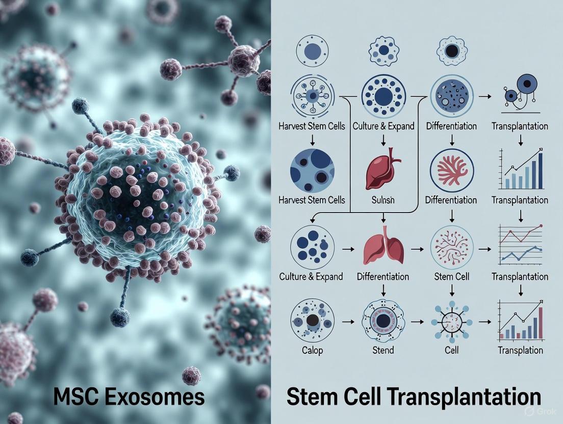
The field of regenerative medicine has increasingly recognized the therapeutic potential of Mesenchymal Stem Cells (MSCs) for treating degenerative diseases, autoimmune conditions, and tissue injuries. The International Society for Cellular Therapy (ISCT) established fundamental criteria to define human MSCs, creating a essential foundation for standardizing research and clinical applications across the global scientific community [9]. These standards ensure that MSCs used in different laboratories and clinical trials possess consistent biological properties, enabling valid comparisons between studies and reliable assessment of therapeutic outcomes.
As research has evolved, a significant paradigm shift has occurred toward understanding that many therapeutic benefits of MSCs are mediated through paracrine mechanisms rather than direct cell replacement [9]. This discovery has sparked considerable interest in MSC-derived exosomes (MSC-Exos)—nanoscale extracellular vesicles that carry bioactive molecules from their parent cells—as a promising cell-free therapeutic alternative [7] [9]. This guide examines the standardized protocols for MSC expansion and characterization according to ISCT guidelines while contextualizing their application within the rapidly advancing field of MSC exosome research, providing researchers with essential methodologies for both cellular and exosome-based investigations.
ISCT Standards for MSC Characterization
Minimum Defining Criteria
According to ISCT standards, human MSCs must satisfy three fundamental criteria [9]:
- Plastic Adherence: MSCs must adhere to plastic surfaces when maintained in standard culture conditions.
- Specific Surface Marker Expression: ≥95% of the MSC population must express CD105, CD73, and CD90, while ≤2% must lack expression of CD45, CD34, CD14 or CD11b, CD79α or CD19, and HLA-DR.
- Multipotent Differentiation Potential: MSCs must demonstrate capacity for in vitro differentiation into osteoblasts, adipocytes, and chondrocytes when induced under standard differentiation protocols.
Comprehensive Characterization Workflow
The following diagram illustrates the complete experimental workflow for MSC characterization and subsequent exosome isolation based on ISCT guidelines:
Comparative Analysis: MSC Exosomes vs. Stem Cell Transplantation
Therapeutic Mechanisms and Applications
MSC Transplantation represents the traditional cellular approach, where living cells are administered with the potential to engraft and differentiate or secrete therapeutic factors. In contrast, MSC-derived exosomes constitute a cell-free paradigm, utilizing nanovesicles to deliver therapeutic cargo without cellular risks [18].
The therapeutic benefits of MSCs were initially attributed to their differentiation capacity and direct engraftment. However, compelling evidence now indicates that paracrine secretion represents the predominant mechanism, with exosomes serving as crucial mediators [9]. These natural nanoparticles range from 30-150 nm in diameter and contain proteins, lipids, mRNAs, and microRNAs that modulate recipient cell behavior [7] [26]. For autoimmune conditions like systemic sclerosis, MSC-Exos demonstrate remarkable immunomodulatory properties by regulating macrophage polarization, suppressing autoreactive lymphocytes, and reversing fibrosis [7].
Advantages and Disadvantages Comparison
Table 1: Comprehensive comparison between MSC exosomes and stem cell transplantation
| Parameter | MSC Exosomes | Stem Cell Transplantation |
|---|---|---|
| Therapeutic Mechanism | Paracrine signaling via biomolecule transfer | Direct differentiation and paracrine effects |
| Immunogenicity | Lower immunogenicity [30] | Potential immune reactions [30] |
| Tumorigenic Risk | No risk of tumor formation [30] [9] | Potential oncological complications [30] |
| Manufacturing Challenges | Complex isolation/purification; batch variation [30] [20] | Easier expansion at large scale [30] |
| Storage & Stability | Long-term frozen storage; lyophilization possible [30] | Challenging storage/transportation [30] |
| Administration Routes | Multiple (IV, inhalation, localized) [30] [20] | Primarily intravenous or localized injection |
| Regulatory Status | Limited standards and regulations [30] | Established FDA guidelines [30] |
| Clinical Trials | 158 studies (as of Feb 2023) [30] | 7,018 studies (as of Feb 2023) [30] |
| Ethical Considerations | No ethical issues [30] | Ethical concerns for certain sources [30] |
Safety and Immunogenicity Profile
The safety profiles of these two therapeutic approaches differ substantially. MSC transplantation carries risks of infusion toxicity due to cell embolization in lungs, potential immune reactions particularly with allogeneic sources, and tumorigenic concerns including teratoma formation [30] [9]. In contrast, MSC-derived exosomes exhibit lower immunogenicity and cannot self-replicate, eliminating tumor formation risks [30] [9]. Their nanoscale size enables sterilization by filtration and avoids lung entrapment [30].
Recent clinical evidence reinforces the favorable safety profile of MSC-Exos. A review of 66 clinical trials registered between 2014-2024 identified predominant administration routes as intravenous infusion and aerosolized inhalation, with no serious adverse events reported across these studies [20]. Nebulization therapy achieved therapeutic effects at approximately 10^8 particles—significantly lower than intravenous requirements—suggesting a favorable dose-response relationship for respiratory applications [20].
Experimental Protocols for MSC Expansion
Cell Culture and Expansion
Materials Required:
- MSC sources (bone marrow, adipose tissue, umbilical cord)
- Culture flasks (T-flasks, multilayer bioreactors, or hollow-fiber systems)
- Complete culture medium (α-MEM/DMEM with fetal bovine serum)
- Supplementation (growth factors, antibiotics)
- Trypsin/EDTA for cell detachment
- Phosphate Buffered Saline (PBS)
Procedure:
- Isolation: Extract MSCs from chosen tissue source using collagenase digestion or explant method
- Primary Culture: Seed cells at 5,000-10,000 cells/cm² in culture flasks with complete medium
- Medium Changes: Replace medium every 2-3 days to remove non-adherent cells
- Subculturing: Harvest cells at 80-90% confluence using trypsin/EDTA
- Expansion: Replate at 1,000-2,000 cells/cm² for continued propagation
- Cryopreservation: Freeze cells in liquid nitrogen using DMSO-containing freezing medium
For large-scale production suitable for exosome manufacturing, hollow-fiber bioreactors are increasingly employed due to their superior surface-to-volume ratio and capacity for high-density cell culture [31]. These systems facilitate GMP-compliant exosome production by supporting cell attachment and efficient nutrient transport [31].
Quality Control During Expansion
Maintaining MSC quality during expansion requires rigorous monitoring:
- Population Doubling Time: Calculate at each passage to detect senescence
- Morphology Assessment: Document spindle-shaped, fibroblast-like appearance
- Viability Testing: Perform trypan blue exclusion assay post-harvest
- Mycoplasma Testing: Conduct regular screening for contamination
- Karyotype Analysis: Perform at regular intervals to ensure genetic stability
Experimental Protocols for MSC Characterization
Surface Marker Analysis by Flow Cytometry
Research Reagent Solutions:
- Flow Cytometer: Instrument for analyzing surface marker expression
- Fluorescent-Antibody Panels: CD105-FITC, CD73-PE, CD90-APC, CD45-PerCP, CD34-PE, HLA-DR-FITC
- Isotype Controls: Matching immunoglobulin controls for background subtraction
- Staining Buffer: PBS with 1-2% FBS for antibody dilution
- Fixation Solution: 1-4% paraformaldehyde for cell preservation
Procedure:
- Cell Preparation: Harvest MSCs at 80% confluence, wash with PBS
- Antibody Staining: Incubate 1×10^6 cells with antibody cocktails (20-25 μL/test) for 30 minutes at 4°C in darkness
- Washing: Centrifuge at 300×g for 5 minutes, discard supernatant, resuspend in staining buffer
- Fixation: Add 200-500 μL of fixation solution if analysis isn't immediate
- Acquisition: Analyze 10,000 events per sample on flow cytometer using appropriate laser configurations
- Analysis: Use software to determine percentage positive populations, applying isotype control corrections
Trilineage Differentiation Assays
Table 2: Standardized protocols for trilineage differentiation potential assessment
| Differentiation Pathway | Induction Medium Components | Differentiation Period | Staining Methods | Key Morphological Features |
|---|---|---|---|---|
| Osteogenic | 0.1 μM dexamethasone, 50 μM ascorbate-2-phosphate, 10 mM β-glycerophosphate | 21-28 days | Alizarin Red S (calcium deposits) | Mineralized matrix nodules |
| Adipogenic | 1 μM dexamethasone, 0.5 mM IBMX, 10 μg/mL insulin, 100 μM indomethacin | 14-21 days | Oil Red O (lipid vacuoles) | Intracellular lipid droplets |
| Chondrogenic | 0.1 μM dexamethasone, 50 μg/mL ascorbate-2-phosphate, 40 μg/mL proline, 10 ng/mL TGF-β3 | 21-28 days | Alcian Blue (proteoglycans) | Pellet formation with cartilaginous matrix |
Procedure for Osteogenic Differentiation:
- Seed MSCs at 20,000 cells/cm² in growth medium
- At 100% confluence, replace with osteogenic induction medium
- Change medium twice weekly for 21-28 days
- Fix cells with 4% formaldehyde for 15 minutes
- Stain with 2% Alizarin Red S (pH 4.2) for 20-30 minutes
- Document calcium deposition microscopically
Procedure for Adipogenic Differentiation:
- Seed MSCs at 50,000 cells/cm² in growth medium
- At 100% confluence, initiate three cycles of induction/maintenance
- Induction: 3 days in adipogenic induction medium
- Maintenance: 1-3 days in adipogenic maintenance medium
- Repeat cycles 3-5 times total
- Fix with 4% formaldehyde, stain with Oil Red O working solution
- Document lipid vacuole formation microscopically
Procedure for Chondrogenic Differentiation:
- Harvest 2.5×10^5 MSCs by centrifugation at 300×g for 5 minutes
- Form micromass pellet in 15 mL conical polypropylene tube
- Add chondrogenic medium without disturbing pellet
- Loosen caps for gas exchange, change medium every 2-3 days
- After 21-28 days, fix pellets, embed in paraffin, section, stain with Alcian Blue
MSC Exosome Isolation and Characterization
Exosome Isolation Techniques
Research Reagent Solutions:
- Ultracentrifuge: Equipment for high-speed exosome pelleting
- Size Exclusion Columns: Chromatography matrices for size-based separation
- Polymer-Based Precipitation: Commercial kits for exosome precipitation
- Density Gradients: Sucrose or iodixanol solutions for purification
- Filtration Units: 0.22 μm filters for sterilization and size exclusion
Isolation Methods:
- Ultracentrifugation: Gold standard method involving sequential centrifugation steps (300×g for 10 min, 2,000×g for 20 min, 10,000×g for 30 min, 100,000×g for 70 min) [26]
- Size Exclusion Chromatography: Gentle separation preserving vesicle integrity and function [31]
- Precipitation Methods: Commercial polymer-based kits offering convenience but potential impurity co-precipitation
- Immunoaffinity Capture: Antibody-based isolation using surface markers (CD63, CD81, CD9) for high purity but lower yield [26]
Exosome Characterization
Research Reagent Solutions:
- Nanoparticle Tracking Analyzer: Instrument for size and concentration analysis
- Transmission Electron Microscope: Equipment for morphological assessment
- Western Blot Equipment: For protein marker detection
- Antibody Panels: Anti-CD63, CD81, CD9, TSG101, Alix for exosome identification
- RNA Extraction Kits: For cargo analysis (microRNAs)
Characterization Methods:
- Nanoparticle Tracking Analysis: Determines size distribution (30-150 nm) and concentration [31]
- Transmission Electron Microscopy: Visualizes characteristic cup-shaped morphology [9]
- Western Blot: Detects positive markers (CD63, CD81, CD9, TSG101) and negative markers (calnexin) [9]
- Flow Cytometry: With fluorescent antibodies confirms surface marker expression [31]
- MicroRNA Profiling: RNA sequencing to characterize therapeutic cargo [7]
The following diagram illustrates the relationship between MSC characterization and exosome profiling within the context of therapeutic development:
Applications in Regenerative Medicine
Therapeutic Mechanisms of MSC-Exosomes
MSC-derived exosomes demonstrate multifaceted therapeutic effects through several key mechanisms:
Immunomodulation: MSC-Exos regulate both innate and adaptive immunity through macrophage polarization (M1/M2 balance), dendritic cell maturation suppression, T-cell and B-cell activity modulation, and Treg cell promotion [7] [9]. Specific microRNAs like miR-146a and miR-125b play crucial roles in these processes [7].
Anti-fibrotic Effects: In systemic sclerosis models, MSC-Exos attenuate fibrosis by modulating collagen deposition and fibroblast activity through specific miRNA transfer [7].
Angiogenesis Promotion: Under hypoxic conditions, MSC-Exos acquire enhanced angiogenic properties, stimulating blood vessel formation to prevent tissue ischemia [7] [32].
Tissue Regeneration: MSC-Exos promote wound healing through increased collagen synthesis, epithelialization, and neovascularization [32].
Clinical Translation Considerations
The clinical application of MSC exosomes presents both opportunities and challenges. Current clinical trials investigate MSC-Exos for diverse conditions including respiratory diseases, COVID-19-associated lung injury, orthopedic disorders, and autoimmune conditions [20] [31]. Different administration routes significantly impact dosing requirements, with aerosolized inhalation achieving therapeutic effects at substantially lower doses (approximately 10^8 particles) compared to intravenous administration [20].
Critical considerations for clinical translation include:
- Scalable Manufacturing: Transition from flask-based culture to bioreactor systems (hollow-fiber, stirred-tank) [31]
- Potency Assays: Development of reliable potency measurements correlating with clinical effects
- Standardization: Implementation of harmonized protocols for isolation, characterization, and dosing [20]
- Quality Control: Comprehensive characterization of physicochemical properties and biological cargo [31]
- Regulatory Frameworks: Adaptation of existing regulatory pathways for cell-free therapeutic products
Standardized protocols for MSC expansion and characterization following ISCT guidelines provide the essential foundation for rigorous research and successful clinical translation in regenerative medicine. These standards ensure consistent cellular populations that yield reproducible exosome products with predictable therapeutic effects. The evolving paradigm toward MSC-derived exosomes as cell-free therapeutics offers significant advantages in safety, immunogenicity, and storage stability while maintaining therapeutic efficacy through targeted molecular delivery.
As the field advances, integrating robust MSC characterization with comprehensive exosome profiling will enable researchers to establish clearer correlations between cellular properties and vesicle function. This approach will ultimately enhance both basic understanding and clinical applications, potentially revolutionizing treatment strategies for degenerative diseases, autoimmune conditions, and tissue injuries. The continued refinement of standardized protocols across both cellular and exosome research remains crucial for realizing the full potential of MSC-based therapies in regenerative medicine.
{Abstract} The transition towards mesenchymal stem cell-derived exosome (MSC-Exo) therapies represents a significant advancement in regenerative medicine, offering a cell-free paradigm with a superior safety profile. This shift necessitates the development of robust, scalable isolation methods. This guide provides a comparative analysis of the two predominant techniques—ultracentrifugation (UC) and tangential flow filtration (TFF)—evaluating their performance, impact on exosome integrity, and suitability for clinical translation.
{Introduction: The Centrality of Isolation in MSC-Exo Therapeutics} The therapeutic application of mesenchymal stem cell-derived exosomes (MSC-Exos) is predicated on their ability to mediate tissue repair, modulate immune responses, and facilitate intercellular communication without the risks associated with whole-cell transplantation, such as immunogenicity, infusion toxicity, and microvasculature occlusion [13] [12]. The efficacy and safety of these exosomal therapeutics are profoundly influenced by the methods used for their isolation and purification. Ultracentrifugation (UC) has long been the historical benchmark for exosome isolation [33] [17]. However, the demand for large-scale, reproducible, and high-quality exosome production for clinical applications has highlighted the limitations of UC and brought Tangential Flow Filtration (TFF) to the forefront as a powerful alternative [34] [26]. This guide objectively compares these two core technologies, providing experimental data and methodological context to inform their use in preclinical and clinical drug development.
{Direct Technical Comparison: UC vs. TFF} The fundamental difference between UC and TFF lies in their separation mechanism, which directly dictates their performance characteristics. UC separates particles based on size, shape, and density through the application of extreme centrifugal force (typically ~100,000–120,000 × g) [33] [17]. In contrast, TFF is a filtration-based technique where the sample flows tangentially across a membrane, preventing clogging and enabling efficient concentration and purification of exosomes based primarily on size [34] [35].
Table 1: Head-to-Head Performance Comparison of UC and TFF
| Performance Metric | Ultracentrifugation (UC) | Tangential Flow Filtration (TFF) |
|---|---|---|
| Mechanism of Separation | Density and particle size under centrifugal force [33] [17] | Size-based separation via cross-flow filtration [34] |
| Typical Exosome Yield | Lower; pellets can be inconsistent [34] | Significantly higher and more consistent [34] [35] |
| Purity | Moderate to Low; co-pellets protein aggregates and lipoproteins [34] [33] | High, especially when coupled with SEC [34] [26] |
| Processing Time | Lengthy (often > 4 hours) [34] [17] | Rapid (typically < 2 hours) [34] |
| Scalability | Limited by rotor capacity [17] | Highly scalable for industrial production [34] [26] |
| Impact on Exosome Integrity | Can cause aggregation and mechanical damage [34] | Preserves structural integrity and biological activity [34] [35] |
| Cost & Infrastructure | High initial equipment cost [13] | Requires specialized TFF equipment; cost-effective for large scale [34] |
{Experimental Data and Workflow Analysis} A direct comparative study investigating the isolation of small extracellular vesicles (sEVs) from cancer cell lines provides critical, data-driven insights. The study demonstrated that while both UC and TFF (with subsequent Size Exclusion Chromatography purification) could isolate sEV populations with consistent size distributions (up to 200 nm), TFF consistently achieved significantly higher yields [34]. Furthermore, the study concluded that TFF surpassed UC in reproducibility, time-efficiency, and cost-effectiveness, making it more suitable for large-scale research and therapeutic applications [34].
The workflows for these two methods, from cell culture conditioned media to purified exosomes, are distinct and are best understood visually. The following diagram illustrates the key steps involved in each protocol.
Diagram 1: A comparative workflow of Ultracentrifugation (UC) and Tangential Flow Filtration (TFF) for MSC-Exo isolation. The TFF process is notably more streamlined.
{The Scientist's Toolkit: Essential Reagents and Materials} Successful isolation using either method relies on a foundation of specific laboratory reagents and equipment.
Table 2: Key Research Reagent Solutions for MSC-Exo Isolation
| Reagent / Material | Function | Application in UC & TFF |
|---|---|---|
| Cell Culture Media with EV-depleted FBS | Provides nutrients for MSC growth while preventing contamination by bovine EVs [34]. | Essential preconditioning step for both methods to ensure pure exosome harvest. |
| Dulbecco's Phosphate Buffered Saline (PBS) | Isotonic buffer for washing cells and resuspending crude exosome pellets [34]. | Used in cell washing and post-UC pellet resuspension. |
| Size Exclusion Chromatography (SEC) Columns | Separates exosomes from contaminating proteins and other soluble factors based on hydrodynamic volume [34] [26]. | Often used as a critical polishing step after TFF and sometimes after UC for enhanced purity. |
| 0.22 µm Pore Filters | Removes large particle contaminants, cells, and debris from conditioned media before isolation [34]. | Standard clarification step in both protocols. |
| Protease Inhibitors | Prevents degradation of exosomal proteins during the isolation process [33]. | Added to conditioned media and buffers in both methods to preserve cargo integrity. |
{Implications for Safety and Immunogenicity Research} The choice of isolation method is not merely technical; it has direct consequences for the safety and immunogenicity profile of the final MSC-Exo product, a core consideration for clinical translation.
Preserving Integrity for Lower Immunogenicity: MSC-Exos are inherently low in immunogenicity as they lack major histocompatibility complex (MHC) molecules, reducing the risk of immune rejection [12]. However, harsh processing can compromise this. UC-induced aggregation or damage may expose novel epitopes or increase the likelihood of unwanted immune recognition. TFF, being a gentler method, better preserves exosome membrane integrity, thereby supporting the natural low immunogenicity profile of MSC-Exos [34] [35].
Ensuring Purity for Safety: The therapeutic safety of exosome preparations is contingent on their purity. Co-isolation of contaminating proteins, aggregates, or lipoproteins with UC can not only confound experimental results but also introduce unpredictable biological effects or immune responses in a clinical setting [34] [33]. The high purity achieved by TFF, especially when combined with SEC, minimizes these risks, ensuring that the observed therapeutic or immunological effects are attributable to the exosomes themselves.
{Conclusion and Future Perspectives} For decades, ultracentrifugation has served as the default technique for MSC-Exo isolation in basic research. However, the evolving landscape of regenerative medicine, with its emphasis on clinical-grade, reproducible, and safe therapeutics, demands a reevaluation of this standard. Tangential Flow Filtration emerges as a superior technology for applications where high yield, preserved biological activity, scalability, and high purity are paramount. These attributes are indispensable for robust safety and immunogenicity testing and for the eventual mass production of MSC-Exo therapies. While UC remains a viable option for small-scale proof-of-concept studies, TFF represents the forward-looking methodology that aligns with the stringent requirements of modern drug development for cell-free regenerative products.
Within the rapidly advancing field of regenerative medicine, the therapeutic comparison between stem cell transplantation and stem cell-derived exosomes has become a central focus of research. A critical, yet sometimes underexplored, factor influencing the safety and efficacy of these therapies is the method of administration. The route of delivery directly impacts biodistribution, local concentration at the target site, and the magnitude of immune response, thereby shaping the overall therapeutic outcome. This guide provides a detailed, objective analysis of three primary administration routes—intravenous infusion, aerosolized inhalation, and local injection—within the broader context of safety and immunogenicity comparisons between mesenchymal stem cell (MSC) exosomes and whole-cell transplantations. Aimed at researchers and drug development professionals, this review synthesizes current experimental data and protocols to inform preclinical and clinical study design.
The choice of administration route is dictated by the target pathology, the physical properties of the therapeutic agent (whole cells vs. nanoscale exosomes), and the desired balance between systemic and localized effects. The table below summarizes the core characteristics, advantages, and challenges of each route.
Table 1: Comparative Analysis of Administration Routes for Stem Cell and Exosome Therapies
| Administration Route | Therapeutic Agent | Key Advantages | Major Limitations & Risks | Primary Target Indications |
|---|---|---|---|---|
| Intravenous (IV) Infusion | Stem Cells | Simplicity, systemic distribution, suitability for disseminated diseases [30] | Infusion toxicity: First-pass pulmonary entrapment due to large cell size; risk of systemic hypotension [30] [36]. Immunogenicity: Potential for systemic immune recognition and rejection [37] [14]. | Haematological reconstitution, systemic inflammatory/autoimmune diseases [30] |
| MSC Exosomes | Systemic distribution without pulmonary entrapment; reduced immunogenicity; can be engineered for targeted drug delivery [30] [13] | Rapid clearance by the mononuclear phagocyte system; potential batch-to-batch variability; undefined pharmacokinetics [30] [13] | Systemic inflammatory conditions, cardiovascular diseases, as targeted drug delivery vehicles [30] [13] | |
| Aerosolized Inhalation | Stem Cells | Potential for direct lung tissue engraftment | Physical shear stress during nebulization can compromise cell viability and function; risk of post-procedural bronchospasm [18] | Pulmonary diseases (e.g., idiopathic pulmonary fibrosis, ARDS) |
| MSC Exosomes | Non-invasive pulmonary delivery; bypasses the blood-brain barrier; achieves high local concentration with minimal systemic exposure; stability for lyophilization and storage [30] [18] | Complex dosage standardization due to aerosol dynamics; limited data on long-term retention [30] | Respiratory diseases (e.g., COVID-19, pulmonary fibrosis, asthma), potential for direct-to-brain delivery [30] | |
| Local Injection | Stem Cells | High local concentration at injury site; minimizes systemic exposure and off-target effects | Invasive procedure; potential for tissue injury at injection site; limited diffusion from injection site [8] | Orthopedic injuries (cartilage, tendon), localized autoimmune lesions, cosmetic and wound healing applications [7] [8] |
| MSC Exosomes | High local concentration; minimal invasiveness and superior tissue penetration compared to cells; reduced risk of immune rejection at site [7] [13] | Technically challenging for deep-seated organs; potential for leakage from injection site [13] | Myocardial infarction, neurological disorders, muscular injuries, ovarian function restoration [7] [13] [8] |
Experimental Protocols and Data Analysis
Intravenous Administration: Quantifying Immunogenicity
Objective: To compare the humoral immune response elicited by intravenously administered xenogeneic MSCs versus their daughter exosomes in a murine model [14].
Experimental Workflow:
- Animal Model: 129-S1 mice assigned to Sham, RAS (Renal Artery Stenosis), RAS+MSC, and RAS+EV groups [14].
- Therapeutic Administration:
- Outcome Measures:
- Systemic Humoral Response: Measured by levels of circulating anti-human antibodies in murine serum using an MSC-reactive assay [14].
- Splenic B-Cell Profile: Analyzed via immunofluorescence staining for CD19+ and IgM+ B-cells in splenic tissue [14].
- Local Immune Rejection: Assessed by quantifying intrarenal T-cell (CD3+) and macrophage (F4/80+) accumulation [14].
Key Findings:
- MSCs triggered a significantly greater systemic antibody (IgM) production compared to exosomes [14].
- Exosomes altered the splenic B-cell profile (higher memory IgM+ B-cells, reduced CD19+ B-cells) but did not induce significant antibody production [14].
- Neither MSCs nor exosomes elicited significant T-cell or macrophage infiltration in the kidneys, indicating no acute local tissue rejection in this model [14].
Figure 1: Experimental workflow for comparing the immunogenicity of intravenously administered MSCs versus exosomes.
Aerosolized Inhalation: Efficacy in Pulmonary Hypertension
Objective: To evaluate the efficacy of nebulized versus intravenous milrinone (a phosphodiesterase inhibitor) in reducing pulmonary arterial pressure in cardiac surgery patients with pulmonary hypertension, providing a parallel for localized vs. systemic drug delivery [36].
Experimental Protocol:
- Study Design: Double-blind, randomized clinical trial on 32 patients with pulmonary hypertension undergoing cardiac surgery [36].
- Intervention Groups:
- Primary Outcomes: Hemodynamic parameters including mean Pulmonary Artery Pressure (mPAP) and Mean Arterial Pressure (MAP) [36].
- Secondary Outcomes: Time to extubation, duration of ICU stay, and total hospital stay [36].
Results and Data Analysis:
Table 2: Clinical Outcomes Following Nebulized vs. Intravenous Milrinone Administration [36]
| Outcome Measure | Nebulized Milrinone | Intravenous Milrinone | P-value |
|---|---|---|---|
| Mean Pulmonary Artery Pressure (mPAP) | Significant Reduction | Reduction | <0.05 |
| Mean Arterial Pressure (MAP) | Maintained | Significant Reduction | 0.09 |
| MAP/mPAP Ratio | Significantly Higher | Lower | <0.0001 |
| Time to Extubation (hours) | Shorter | Longer | 0.001 |
| ICU Stay (days) | Shorter | Longer | 0.009 |
| Hospital Stay (days) | Shorter | Longer | 0.026 |
Interpretation: The data demonstrates that aerosolized delivery achieves the primary therapeutic goal (reducing mPAP) while avoiding the significant systemic side effect (hypotension) associated with IV administration. This directly translates to tangible clinical benefits, including faster weaning from mechanical ventilation and shorter hospital stays [36]. This principle is highly relevant for exosome therapies targeting lung diseases.
Local Injection for Targeted Therapy
Objective: To assess the therapeutic potential of locally injected MSC-derived exosomes in a model of systemic sclerosis (SSc), focusing on antifibrotic and immunomodulatory effects [7].
Methodology:
- Therapeutic Agent: Exosomes derived from human MSCs, characterized for tetraspanin markers (CD9, CD63, CD81) [7].
- Administration: Local injection (e.g., intraperitoneal or intradermal) in preclinical models of SSc [7].
- Mechanistic Analysis:
- Macrophage Polarization: Evaluate exosome-induced shift from pro-inflammatory M1 to anti-inflammatory M2 phenotype via cytokine profiling (IL-6, TNF-α vs. IL-4, IL-10) and cell surface markers [7].
- Fibrosis Pathway Analysis: Examine suppression of pro-fibrotic signaling pathways (e.g., TGF-β1/Smad3) in target tissues like skin and lungs [7].
- Lymphocyte Modulation: Assess inhibition of autoreactive T-cell and B-cell activity through in vitro co-culture assays and analysis of T-regulatory cell proliferation [7].
Outcome: Local injection of MSC-exosomes was shown to attenuate fibrosis, modulate immune cell activity, and reverse pulmonary arterial hypertension in SSc models, primarily through paracrine signaling and microRNA transfer (e.g., miR-146a, miR-21-5p) [7]. The localized delivery ensures high bioavailability at the disease site while minimizing systemic immunological exposure.
Figure 2: Molecular mechanisms of locally injected MSC-exosomes, mediating therapeutic effects through immunomodulation, anti-fibrosis, and tissue repair pathways.
The Scientist's Toolkit: Essential Research Reagents and Materials
Successful execution of administration route studies requires standardized reagents and methodologies. The following table details key solutions used in the featured experiments and broader field.
Table 3: Essential Research Reagent Solutions for Administration and Analysis
| Reagent / Solution | Primary Function | Application Context | Key Considerations |
|---|---|---|---|
| Jet Nebulizer (e.g., Salter Labs 8901) | Generates an aerosolized mist of therapeutic agent for inhalation [36]. | Aerosolized inhalation delivery of drugs or exosomes in preclinical/clinical settings [36]. | Optimize particle size for alveolar deposition; assess agent stability post-nebulization. |
| Ultracentrifugation System | Isolation and purification of exosomes from cell culture supernatants via high-speed centrifugation [14]. | Standard laboratory-scale production of MSC-exosomes for all administration routes [30] [14]. | Considered the "gold standard" but time-consuming and not easily scalable [30] [13]. |
| Flow Cytometry Antibodies (CD73, CD90, CD105) | Characterization of MSC surface markers to confirm cell identity and purity per ISCT criteria [14] [8]. | Quality control of MSCs prior to transplantation or exosome production [14]. | Essential for validating the starting cellular material in both cell and exosome therapies. |
| Exosome Characterization Kit (NTA, e.g., NanoSight) | Nanoparticle Tracking Analysis (NTA) to determine exosome particle size and concentration [14]. | Quality control of isolated exosomes before administration [30] [14]. | Critical for ensuring batch-to-batch consistency and accurate dosing. |
| IFN-γ Licensing Solution | Pre-conditioning of MSCs with pro-inflammatory cytokines to enhance immunosuppressive potency [37]. | Priming MSCs for administration in highly inflammatory environments (e.g., autoimmune diseases) [37]. | Licensing upregulates immunomodulatory molecules (B7H1, PD1) but may increase immunogenicity [37]. |
| Cryopreservation Medium | Long-term storage of MSCs and exosomes in a viable state. | Preservation of therapeutic agents for off-the-shelf availability [37]. | Cryopreserved MSCs may be more susceptible to apoptosis post-thaw [37]. |
The choice between intravenous infusion, aerosolized inhalation, and local injection is a pivotal decision that profoundly influences the safety and efficacy profile of both stem cell and exosome therapies. Intravenous infusion offers systemic reach but carries inherent risks of infusion toxicity and immunogenicity for cells, risks that are markedly reduced with exosomes. Aerosolized inhalation presents a non-invasive, highly effective route for treating pulmonary diseases, enabling high local bioavailability with minimal systemic interference. Local injection provides the most targeted approach, maximizing therapeutic concentration at the injury site and further mitigating systemic immune responses, particularly for exosomes.
The accumulated data strongly suggests that MSC-derived exosomes, regardless of administration route, offer a superior safety profile concerning immunogenicity and infusion-related complications compared to their parental cells. However, challenges in exosome large-scale manufacturing, purification, and standardized dosing remain active areas of research. Future work should focus on optimizing administration protocols, engineering exosomes for enhanced targeting, and conducting direct, head-to-head comparative studies in relevant disease models to fully elucidate the interplay between the therapeutic agent, its route of delivery, and the resulting clinical outcome.
The field of regenerative medicine is undergoing a significant transformation, moving from whole-cell therapies toward cell-free approaches utilizing secretory derivatives. Within this context, mesenchymal stem cell (MSC) transplantation and MSC-derived exosomes (MSC-Exos) represent two distinct therapeutic paradigms with fundamentally different dosage considerations. MSCs are multipotent stromal cells with documented immunomodulatory and regenerative capabilities, historically administered as live cell infusions [1]. In contrast, MSC-Exos are nanoscale (30-150 nm) extracellular vesicles that function as primary mediators of MSC paracrine effects, carrying proteins, lipids, and nucleic acids from parent cells to recipient cells [18] [12]. This shift from cellular to subcellular therapeutics introduces complex questions regarding dose-effect relationships, with critical implications for therapeutic efficacy, safety, and immunogenicity within clinical development frameworks.
The fundamental distinction between these therapeutic entities necessitates different dosage parameters: MSC therapies are quantified by cell count (e.g., cells per kilogram body weight), while exosome therapies are measured by particle quantity or protein content (e.g., milligrams of exosomal protein) [38] [14]. Understanding these dosage metrics and their corresponding therapeutic windows is essential for researchers and drug development professionals designing preclinical and clinical studies. This guide objectively compares the dose-effect relationships, safety profiles, and experimental methodologies for both therapeutic approaches, providing a structured framework for dosage determination in regenerative medicine applications.
Comparative Dosage Metrics and Therapeutic Efficacy
Quantitative Dosage Parameters and Efficacy Evidence
Table 1: Comparative Dosage and Efficacy Metrics for MSC vs. MSC-Exo Therapies
| Therapeutic Parameter | MSC Transplantation | MSC-Derived Exosomes |
|---|---|---|
| Dosage Unit | Cell count (e.g., cells/kg body weight) | Particle quantity or protein content (e.g., μg protein) |
| Typical Human Dose Range | 1-10 × 10^6 cells/kg (single or repeated doses) [38] | Preclinical: 10-100 μg protein; Clinical: Emerging standards [14] |
| Documented Efficacy | Reduced mortality in ARDS (RR = 0.74, p = 0.0003) [38] | Reduced mortality in ARDS (RR = 0.63, p = 0.003) [38] |
| Dose-Response Evidence | High-dose MSCs (>1×10^6 cells/kg) showed significant mortality reduction (RR = 0.70) [38] | Preclinical dose-escalation studies show therapeutic window exists [39] |
| Therapeutic Window | Established in multiple clinical trials for inflammatory conditions [38] [40] | Potentially wider due to reduced risks; under investigation [41] [12] |
| Administration Route | Intravenous, intra-arterial, local injection [14] | Intravenous, intranasal, local application [41] [42] |
The dose-effect relationship for MSC transplantation demonstrates a clear threshold phenomenon, particularly evident in recent meta-analyses of acute respiratory distress syndrome (ARDS) treatment. High-dose MSC administration (defined as >1×10^6 cells/kg or >70 million cells per infusion) was associated with a significant reduction in all-cause mortality (Relative Risk [RR] = 0.70, 95% CI = 0.55–0.89) compared to conventional therapy [38]. This quantitative evidence establishes a clinically relevant effective dose window for MSC therapies in inflammatory conditions. The therapeutic effects appear to follow a sigmoidal dose-response curve, with minimal effects below a certain threshold, increasing efficacy within a therapeutic window, and potential plateau effects at higher doses.
For MSC-Exos, the dose-effect relationship is quantitatively different due to their mechanism of action as paracrine signal transducers rather than living therapeutic agents. In preclinical models, exosome therapies demonstrate a distinct dose-response curve with efficacy observed at specific protein concentrations (e.g., 20μg protein in murine models) [14]. The effective dose window for exosomes appears potentially wider than for cell therapies, though establishing standardized human dosing remains an active research area. The nanoscale properties of exosomes enable different biodistribution patterns compared to cells, potentially allowing for lower total therapeutic loads to achieve clinical effects [41] [42].
Safety and Immunogenicity Profile Comparison
Table 2: Safety and Immunogenicity Comparison of MSC vs. MSC-Exo Therapies
| Safety Parameter | MSC Transplantation | MSC-Derived Exosomes |
|---|---|---|
| Immunogenicity | Induces circulating anti-human antibodies; potential alloimmunization [14] | Reduced immunogenicity; lower MHC expression [14] [12] |
| Adverse Event Profile | No significant difference in AEs (RR = 1.08, p = 0.17) or SAEs (RR = 0.94, p = 0.49) vs. controls [38] | Favorable preclinical safety profile; minimal adverse events reported [41] [42] |
| Specific Safety Concerns | Potential pulmonary embolism, infusion reactions, donor-dependent variability [41] [12] | Avoids cell-related risks; no tumor formation or ectopic tissue formation [41] [42] |
| Immune Cell Activation | Modulates T-cell and macrophage responses; may trigger host immune recognition [14] | Alters splenic B-cell populations without significant kidney rejection [14] |
| Regulatory Considerations | Living biological product with complex safety monitoring | Cell-free product potentially classified as biologic or drug-device combination |
The immunogenicity profile represents a critical differentiation between these therapeutic approaches. Xenogeneic studies demonstrate that MSC transplantation significantly increases circulating anti-human antibodies, indicating a humoral immune response that may impact repeated dosing strategies [14]. In contrast, MSC-Exos triggered altered splenic B-cell profiles but did not elicit the same level of systemic antibody production, suggesting superior immune evasion capabilities [14]. This fundamental difference in immunogenicity stems from the reduced expression of major histocompatibility complex (MHC) molecules on exosomes compared to their parent cells [12].
The safety profiles further distinguish these therapeutic modalities. Meta-analyses of clinical trials confirm that MSC-based therapies do not significantly increase adverse events (AEs) or serious adverse events (SAEs) compared to control treatments (AE RR = 1.08, p = 0.17; SAE RR = 0.94, p = 0.49) [38]. However, specific risks associated with cell-based therapies include pulmonary embolism, infusion reactions, and potential tumorigenicity with long-term engraftment [41] [12]. MSC-Exos circumvent many cell-associated risks by lacking replicative capacity, demonstrating an emerging safety advantage in preclinical models and early clinical applications [41] [42].
Diagram 1: Comparative Safety and Immunogenicity Profiles of MSC Transplantation versus MSC-Derived Exosomes. MSC therapies demonstrate higher immunogenicity and specific safety concerns, while exosomes show favorable safety profiles while maintaining therapeutic efficacy.
Experimental Protocols and Methodological Considerations
Isolation and Characterization Techniques
Table 3: Key Research Reagent Solutions for MSC and Exosome Research
| Research Reagent | Function/Application | Experimental Context |
|---|---|---|
| CD73, CD90, CD105 Antibodies | MSC surface marker identification via flow cytometry | MSC characterization per ISCT criteria [1] |
| CD9, CD63, CD81 Antibodies | Exosome surface marker detection and quantification | Exosome characterization and purity assessment [26] [12] |
| Ultracentrifugation System | Gold standard exosome isolation via sequential centrifugation | Exosome purification from MSC conditioned media [26] [42] |
| Size Exclusion Chromatography | Size-based exosome separation preserving vesicle integrity | Alternative isolation method with reduced aggregation [26] |
| Nanoparticle Tracking Analysis | Particle size distribution and concentration measurement | Exosome quantification and quality control [42] |
| Density Gradient Medium | Buoyant density separation of exosomes from contaminants | High-purity exosome isolation [42] |
The methodological framework for investigating dose-effect relationships begins with standardized isolation and characterization protocols. For MSC preparation, the International Society for Cellular Therapy (ISCT) establishes minimum criteria including plastic adherence, expression of specific surface markers (CD73, CD90, CD105 ≥95%; CD34, CD45, CD14, CD19, HLA-DR ≤2%), and trilineage differentiation potential [1]. These quality control measures ensure batch-to-batch consistency essential for meaningful dose-response studies.
Exosome isolation employs distinct methodological approaches, with ultracentrifugation remaining the gold standard technique. This protocol involves sequential centrifugation steps: initial low-speed centrifugation (300-2000×g) to remove cells and debris, intermediate-speed centrifugation (10,000-20,000×g) to pellet larger extracellular vesicles, and final high-speed ultracentrifugation (≥100,000×g) to sediment exosomes [26] [42]. Alternative methods include size exclusion chromatography, which separates vesicles based on size while preserving structural integrity, and immunoaffinity capture using antibodies against specific exosome surface markers (CD9, CD63, CD81) for high-purity isolation [26]. Characterization of isolated exosomes typically involves nanoparticle tracking analysis for size distribution, transmission electron microscopy for morphological assessment, and Western blotting for marker protein detection [41] [42].
Diagram 2: Experimental Workflow for MSC-Derived Exosome Isolation and Characterization. The process begins with MSC culture, proceeds through various isolation techniques, and concludes with comprehensive characterization before therapeutic application.
Dose-Response Assessment Methodologies
Establishing dose-effect relationships requires carefully designed experimental protocols. For MSC transplantation, dose-response studies typically involve administering escalating cell doses (e.g., low: <1×10^6 cells/kg; medium: 1-5×10^6 cells/kg; high: >5×10^6 cells/kg) in disease models and monitoring therapeutic outcomes and potential toxicity [38]. Recent meta-analyses demonstrate the importance of such dose-ranging studies, revealing that high-dose MSCs (>1×10^6 cells/kg) significantly reduced mortality in ARDS patients (RR = 0.70, 95% CI = 0.55–0.89), while lower doses showed diminished efficacy [38].
For MSC-Exos, dose-response assessment involves different parameters, typically measuring exosome quantity by protein content (μg) or particle number. Experimental protocols often administer exosomes at concentrations ranging from 10-100μg protein in small animal models, with efficacy demonstrated across various disease models including neurological disorders, cardiovascular diseases, and renal injury [14] [39] [41]. The therapeutic window for exosomes appears favorable, with multiple studies reporting efficacy at doses that show no adverse effects, though optimal dosing regimens continue to be refined through preclinical investigation.
The comparative analysis of dosage considerations for MSC transplantation versus MSC-derived exosomes reveals distinct profiles with significant implications for therapeutic development. MSC transplantation demonstrates a well-characterized dose-effect relationship with established efficacy in clinical applications, particularly at higher cell doses (>1×10^6 cells/kg), but carries inherent immunogenicity concerns and cell-based risks. In contrast, MSC-Exos present a promising cell-free alternative with demonstrated efficacy in preclinical models, a potentially wider therapeutic window, and superior safety profile due to reduced immunogenicity and avoidance of cell-related complications.
For researchers and drug development professionals, these differences inform strategic decisions in therapeutic platform selection. MSC transplantation may offer advantages where direct cell engraftment and prolonged tissue presence are desirable, despite the more complex safety monitoring requirements. MSC-Exos represent an emerging paradigm with simplified manufacturing, reduced regulatory concerns, and potentially more favorable dosing regimens, though standardized quantification methods and human dosing parameters require further refinement.
The evolving understanding of dose-effect relationships for both therapeutic modalities continues to shape the field of regenerative medicine. Future research directions include optimizing exosome dosing regimens for specific clinical applications, developing standardized potency assays, and exploring combination approaches that leverage the unique advantages of both cellular and subcellular therapeutic strategies. As the field advances, precise dosage considerations will remain fundamental to translating regenerative therapies from research laboratories to clinical practice.
Navigating Challenges: Biosafety, Standardization, and Potency Enhancement
Within the rapidly evolving field of regenerative medicine, mesenchymal stem cells (MSCs) and their derived exosomes (MSC-Exos) represent two prominent therapeutic strategies. While both hold immense promise for treating a wide array of diseases, their clinical translation hinges on a rigorous and comparative understanding of their biosafety profiles. This guide provides a critical, data-driven comparison of the toxicity, tumorigenicity, and biodistribution of MSC transplantation versus MSC-derived exosome therapy, framing this analysis within the broader thesis of safety and immunogenicity. As cell-free nanoparticles, MSC-Exos are increasingly regarded as the next generation of biomedical applications, potentially offering comparable therapeutic benefits while overcoming key safety challenges associated with whole-cell transplants [43] [44]. This analysis is intended for researchers, scientists, and drug development professionals, summarizing current experimental data and methodologies to inform pre-clinical risk assessment and therapeutic development.
Fundamental Biological Differences
MSCs are multipotent stromal cells characterized by their adherence to plastic, specific surface marker expression (CD73+, CD90+, CD105+, CD34-, CD45-, HLA-DR-), and capacity to differentiate into osteogenic, chondrogenic, and adipogenic lineages [8] [1]. Initially isolated from bone marrow, MSCs can be sourced from various tissues, including adipose tissue, umbilical cord, and dental pulp. Their therapeutic mechanisms were historically attributed to direct differentiation and replacement of damaged cells, but are now understood to be predominantly mediated by paracrine signaling—the secretion of bioactive molecules that modulate the local cellular environment [1].
MSC-derived exosomes (MSC-Exos) are a key component of this paracrine effect. These are small extracellular vesicles (30-150 nm in diameter) with a lipid bilayer membrane, released by cells upon fusion of multivesicular bodies with the plasma membrane [18] [44]. They function as natural carriers of diverse bioactive molecules, including proteins, lipids, mRNA, and various non-coding RNAs (e.g., miRNA, lncRNA, circRNA), facilitating intercellular communication by transferring this functional cargo to recipient cells [43] [8]. Crucially, as acellular entities, MSC-Exos lack a nucleus and cannot replicate, which fundamentally alters their risk profile compared to whole MSCs.
Table 1: Core Biological Characteristics of MSCs vs. MSC-Exosomes
| Characteristic | MSC Transplantation | MSC-Derived Exosomes |
|---|---|---|
| Nature | Live, nucleated cells | Cell-free, nano-sized vesicles |
| Size | 15-30 μm (diameter) | 30-150 nm (diameter) |
| Proliferation Capacity | Can self-renew and expand | No ability to replicate |
| Primary Mechanism | Direct differentiation & potent paracrine secretion | Delivery of pre-packaged bioactive cargo |
| Key Cargo | Dynamically secreted factors | Proteins, lipids, miRNAs, mRNAs |
| Typical Dosage Units | Number of cells (e.g., millions of cells) | Particle number or total protein (e.g., ×10^10 particles, μg) [20] |
Comparative Biosafety Profiles
A head-to-head comparison of critical safety parameters reveals distinct advantages and challenges for each therapeutic modality.
Toxicity
Systematic toxicity assessments following standardized guidelines provide compelling evidence for the safety of MSC-Exos.
- MSC-Exos Toxicity Data: A comprehensive toxicity evaluation of MSC-Exos conducted according to OECD guidelines concluded a favorable safety profile. The studies classified MSC-Exos as a non-sensitizer (Skin Sensitization Test, OECD TG 442B), a non-irritant (Skin Irritation Test, OECD TG 439), and "No Category" for eye irritation (OECD TG 437). Furthermore, no toxicity was observed in phototoxicity (OECD TG 432) or acute oral toxicity tests (OECD TG 423) [45]. Recent studies further support this, showing that even very high single intravenous doses (up to 10,000 μg/kg) in mice or repeated injections in rats caused no mortality, significant adverse events, or alterations in hematological and biochemical indices related to liver and kidney function [46].
- MSC Transplantation Toxicity: While MSCs have a generally favorable safety profile, they are not without risk. The toxicity of cellular products is more complex, as cells can mediate tissue damage through mechanisms like immunological responses and administration-related complications. A key risk is infusion toxicity, which can include fevers, tachycardia, and, most seriously, pulmonary capillary blockage (embolism) due to the cells' larger size and aggregation potential [47] [43]. The assessment of MSC toxicity requires extensive in vivo monitoring, including mortality rates, clinical observations, and detailed histological examination of multiple organs post-transplantation [47].
Tumorigenicity and Oncogenicity
This parameter represents one of the most significant differentiators between the two therapies.
- MSC-Exos: As anucleate vesicles, MSC-Exos carry no inherent risk of malignant transformation or teratoma formation. They cannot divide or form tumors de novo. This is a paramount safety advantage that simplifies their regulatory pathway and clinical application [43] [44].
- MSC Transplantation: The tumorigenic potential of MSCs, particularly after long-term culture or genetic manipulation, remains a critical biosafety concern. While MSCs themselves are considered to have low tumorigenicity, their potential to indirectly support tumor growth or, in rare cases, undergo malignant transformation requires careful evaluation. Preclinical assessment involves a combination of in vitro assays and long-term studies in immunocompromised animal models to monitor for tumor formation [47]. This risk necessitates extensive and costly long-term follow-up in clinical trials.
Immunogenicity
The potential to provoke an unwanted immune response is a major consideration for allogeneic therapies.
- MSC-Exos: MSC-Exos demonstrate low immunogenicity. They lack major immunogenic cell surface markers and do not express HLA-DR, minimizing the risk of immune rejection. Systemic hypersensitivity tests in animal models have shown that MSC-Exos do not trigger significant allergic or immune responses, highlighting their biocompatibility for systemic administration [46] [44].
- MSC Transplantation: MSCs are historically considered "immune-privileged" due to low expression of MHC-I and absence of MHC-II. However, this is not absolute. Upon differentiation or inflammatory activation, their immunogenicity can increase, potentially leading to immune recognition and rejection, especially in allogeneic settings [47] [1]. This necessitates careful donor matching or the use of immunosuppression in some contexts.
Biodistribution
Biodistribution refers to the movement, persistence, and clearance of a therapeutic within the body.
- MSC Transplantation: When administered intravenously, MSCs are often trapped in the capillary networks of the lungs, liver, and spleen. This first-pass pulmonary entrapment can limit delivery to the actual target site and may contribute to adverse effects [47] [44]. Tracking cells over time typically requires advanced imaging techniques (e.g., PET, MRI) or quantitative PCR for human-specific DNA in animal models [47].
- MSC-Exos: Their nano-scale size allows MSC-Exos to cross biological barriers, including the blood-brain barrier, which is largely inaccessible to whole cells. Their distribution is highly influenced by the route of administration. For instance, aerosolized inhalation directly targets the lungs and achieves therapeutic effects at significantly lower doses than intravenous routes [20]. This offers a strategic advantage for treating respiratory diseases and enables more precise targeting.
Table 2: Comparative Biosafety Profile of MSCs vs. MSC-Exosomes
| Safety Parameter | MSC Transplantation | MSC-Derived Exosomes | Key Supporting Evidence |
|---|---|---|---|
| Acute Systemic Toxicity | Risk of infusion toxicity, pulmonary embolism | No acute toxicity at very high doses (e.g., 10,000 μg/kg) in mice [46] | GLP-compliant acute toxicity studies [45] [46] |
| Local Irritation | Context-dependent; potential for local reaction at site | Classified as non-irritant to skin and eyes [45] | OECD TG 439 (Skin Irritation) & 437 (Eye Irritation) |
| Tumorigenicity | Recognized risk requiring long-term evaluation | No risk of de novo tumor formation | Absence of replicative capacity; no nuclei [43] [44] |
| Immunogenicity | Low but can be activated; risk of rejection | Very low; no HLA-DR; does not trigger hypersensitivity [46] | Systemic hypersensitivity tests in rabbits [46] |
| Biodistribution | First-pass pulmonary entrapment; limited targeting | Can cross blood-brain barrier; distribution route-dependent [20] | Imaging (PET, MRI); particle tracking in disease models [20] |
| Administration Risks | Embolism, cell misdifferentiation | Fewer physical risks; amenable to nebulization [20] [44] | Clinical trials on nebulization for lung diseases [20] |
Experimental Protocols for Biosafety Assessment
To generate the comparative data outlined above, researchers employ a suite of standardized and specialized experimental protocols.
In Vivo Toxicity and Immunogenicity Assessment
- Acute and Subchronic Toxicity (OECD Guidelines): These are foundational studies. For example, OECD TG 423 (Acute Oral Toxicity) involves administering a single high dose to rodents followed by 14 days of observation for mortality and morbidity. Subchronic toxicity studies involve repeated injections over 28-90 days in rats, with periodic blood collection for hematology and clinical chemistry (e.g., albumin, ALT, AST, urea nitrogen, creatinine) to assess organ function, culminating in a full histopathological examination of major organs [47] [45] [46].
- Local Tolerance Tests: These include skin sensitization (OECD TG 442B), which uses the Local Lymph Node Assay, and skin and eye irritation tests (OECD TG 439 & 437), which apply the substance to reconstructed human epidermis or corneal models to classify its irritation potential [45].
- Systemic Hypersensitivity: This assesses the potential for allergic reactions. In one study, New Zealand rabbits received intravenous injections of MSC-Exos, and parameters like body temperature, respiratory symptoms, and hematological profiles were monitored to detect any anaphylactic or allergic response [46].
Tumorigenicity Assessment
- In Vivo Tumorigenicity Assay: This is a critical long-term study for MSCs. Immunodeficient mice (e.g., nude or NSG mice) are injected with the cellular product and monitored for up to several months. Palpation and imaging track any mass formation, and a full necropsy and histology are performed at the endpoint to identify any teratomas or malignant tumors [47].
Biodistribution Assessment
- Quantitative PCR (qPCR): A highly sensitive method for tracking human MSCs or MSC-Exos in animal tissues. It detects species-specific DNA sequences (e.g., Alu repeats for human cells in a mouse model) or exosome-delivered RNA, allowing for quantification of persistence and distribution across different organs over time [47].
- Imaging Techniques: Non-invasive imaging provides real-time spatial and temporal data.
- Positron Emission Tomography (PET): Cells or exosomes are labeled with a radiotracer (e.g., ^89^Zirconium), and their whole-body distribution is tracked longitudinally.
- Magnetic Resonance Imaging (MRI): Used when cells are labeled with superparamagnetic iron oxide (SPIO) nanoparticles, allowing for high-resolution anatomical localization.
- Nanoparticle Tracking Analysis (NTA): This is used primarily for exosome characterization to determine particle size distribution and concentration in isolated fluids prior to in vivo studies [20] [46].
Diagram 1: Integrated Workflow for Biosafety Profiling. This flowchart outlines the key experimental phases and their logical sequence in a comprehensive biosafety assessment for regenerative therapies.
The Scientist's Toolkit: Key Reagents and Materials
The following table details essential reagents and technologies used in the critical experiments described above.
Table 3: Essential Research Reagents for Biosafety Assessment
| Reagent / Technology | Primary Function in Biosafety Assessment | Specific Application Example |
|---|---|---|
| Immunodeficient Mouse Models (e.g., Nude, NSG) | In vivo assessment of tumorigenicity and long-term toxicity. | Monitoring for teratoma/tumor formation after subcutaneous or systemic injection of MSCs [47]. |
| Flow Cytometry Antibodies (CD73, CD90, CD105, CD34, CD45, HLA-DR) | Characterizing cell product identity and purity per ISCT criteria. | Verifying MSC surface marker profile pre-transplantation to ensure a defined and consistent product [46] [1]. |
| Nanoparticle Tracking Analysis (NTA) | Quantifying and sizing extracellular vesicles. | Determining the concentration (particles/mL) and size distribution of isolated MSC-Exos before dosing [20] [46]. |
| qPCR Reagents for species-specific sequences (e.g., human Alu repeats) | Highly sensitive tracking of biodistribution and persistence. | Quantifying human MSC DNA or exosome-derived RNA in mouse organ homogenates (e.g., liver, lungs, spleen) [47]. |
| Radiotracers (e.g., ^89^Zr) or Iron Oxide Nanoparticles (e.g., SPIO) | Non-invasive in vivo imaging of biodistribution. | Labeling MSCs or exosomes for longitudinal tracking via PET or MRI [47]. |
| Hematology & Clinical Chemistry Analyzers | Assessing systemic toxicity and organ function. | Measuring blood counts and serum biomarkers (e.g., ALT, AST, Creatinine) in toxicity studies to detect liver/kidney damage [47] [46]. |
| Histopathology Stains (e.g., H&E) | Gold standard for identifying structural tissue damage or tumors. | Microscopic examination of organ sections post-mortem for signs of toxicity, immune cell infiltration, or neoplasia [47]. |
The comparative analysis of critical biosafety profiles underscores a clear divergence between MSC transplantation and MSC-derived exosome therapies. MSC-Exos present a compelling safety advantage, with robust experimental data demonstrating negligible acute toxicity, no risk of tumorigenicity, low immunogenicity, and favorable biodistribution that enables novel administration routes like nebulization. In contrast, MSC transplantation, while therapeutically potent, carries inherent risks related to cell occlusion, potential for immune rejection, and a non-zero risk of tumor formation that necessitates complex and long-term safety monitoring.
For researchers and drug developers, this evidence positions MSC-Exos as a lower-risk candidate for many regenerative applications, potentially accelerating clinical translation. However, challenges in MSC-Exo production standardization, scalable manufacturing, and precise targeting remain active areas of investigation. Future work should focus on establishing universally accepted potency assays, dosing frameworks, and regulatory guidelines to fully realize the potential of this promising cell-free therapeutic platform.
The success of allogeneic cell therapies hinges on overcoming the formidable barrier of the host immune response. For regenerative medicine, two primary therapeutic candidates have emerged: live mesenchymal stem cells (MSCs) and their derived exosomes. While MSCs were initially believed to be immune-privileged, substantial evidence now confirms they can elicit both cellular and humoral immune reactions [48]. In contrast, MSC-derived exosomes (MSC-Exos), as cell-free entities, present a fundamentally different immunological profile [12]. This guide provides a structured, data-driven comparison of the immunogenic properties of MSCs versus MSC-Exos, focusing on HLA typing requirements, interactions with specific immune cell populations, and their consequent potential for allogeneic "off-the-shelf" use. The objective analysis herein is framed within a broader thesis on safety and immunogenicity, providing researchers and drug development professionals with critical insights for therapeutic design.
Immunological Head-to-Head: MSCs vs. MSC-Exos
The core of the immunogenic challenge lies in the distinct biological nature of these therapeutics. MSCs are live cells capable of expressing immunogenic surface molecules, whereas exosomes are nanosized extracellular vesicles that function primarily as signaling cargo carriers.
Table 1: Fundamental Immunogenic Properties
| Feature | Allogeneic MSCs | MSC-Derived Exosomes |
|---|---|---|
| Physical Nature | Live, nucleated cell | Extracellular vesicle (30-150 nm) |
| MHC/HLA-I Expression | Constitutive low expression; inducible by IFN-γ [48] | Absent or very low [12] |
| MHC/HLA-II Expression | Generally low; highly inducible by IFN-γ [49] [48] | Absent [12] |
| Co-stimulatory Molecules (CD80, CD86) | Negative [48] | Negative [12] |
| Primary Immune Risk | T-cell and antibody-mediated rejection [14] [48] | Minimal; primarily cargo-dependent [7] [26] |
| Ideal Use Case | Controlled, potentially HLA-matched environments | Universal "off-the-shelf" allogeneic application |
HLA Typing and Allogeneic Potential
The requirement for Human Leukocyte Antigen (HLA) matching is a critical determinant for the feasibility and scalability of allogeneic therapies.
The MSC HLA Dilemma
Allogeneic MSCs are not immunologically inert. They constitutively express HLA-I and can upregulate both HLA-I and HLA-II in inflammatory environments (e.g., upon IFN-γ exposure), making them visible to host T cells [49] [48]. This can lead to immune memory formation; one study in rhesus macaques detected significantly higher levels of circulating donor-specific antibodies in subjects receiving allogeneic versus autologous MSCs [48]. Consequently, while not always essential, HLA matching can improve engraftment and persistence. Evidence indicates that using allogeneic MSCs with homozygous HLA haplotypes can significantly reduce in vitro and in vivo immune responses [50].
The Exosome Advantage
MSC-derived exosomes offer a paradigm shift. They lack the complete MHC/HLA molecular machinery present on live cells, which drastically reduces their intrinsic immunogenicity [12]. Their lipid bilayer membrane does not express polymorphic HLA proteins in an immunogenic form. This fundamental characteristic is the basis for their classification as a low-immunogenicity-risk therapeutic, supporting their potential as a universal, "off-the-shelf" product that requires no HLA matching [7] [26].
Table 2: Experimental Evidence of Immune Responses
| Experimental Model | Allogeneic MSC Findings | MSC-Exosome Findings |
|---|---|---|
| Xenogeneic (Human to Mouse) | Increased circulating anti-human antibodies; PBMCs from recipients lysed donor MSCs in vitro [14] [48]. | No discernible kidney rejection or increased intrarenal immune cell infiltration [14]. |
| Large Animal (Equine) | Significant adverse joint response and elevated synovial cell count after second injection of allogeneic (vs. autologous) MSCs [48]. | Not Available (N/A) |
| In Vitro T-cell Recall | N/A | Reduced IFN-γ release upon re-stimulation, indicating immunosuppressive effect [14]. |
Immune Cell Responses: A Detailed Mechanism
The interaction with the host's immune system unfolds through complex, multi-cell pathways. The diagrams below contrast the distinct immune recognition pathways for allogeneic MSCs and MSC-derived exosomes.
Immune Recognition of Allogeneic MSCs
Immunomodulation by MSC-Derived Exosomes
Experimental Data and Supporting Evidence
Key Supporting Experimental Findings
- Antibody Response: A 2025 study showed xenogeneic human MSCs in mice "significantly increased circulating anti-human antibodies," while EVs triggered a different, splenic cellular response but no discernible kidney rejection [14].
- T-cell Modulation: In vitro, both MSCs and MSC-EVs exhibited an immunosuppressive effect, demonstrated by reduced IFN-γ release from T-cells upon re-stimulation [14].
- Macrophage Reprogramming: A key mechanism of MSC-Exos is their ability to polarize macrophages from a pro-inflammatory M1 phenotype to an anti-inflammatory M2 phenotype, potentially via IL-10 and other cargo [49] [7].
- B-cell Inhibition: MSC-Exos can suppress B-cell proliferation and antibody production through the transfer of specific microRNAs like miR-155-5p [7].
Detailed Experimental Protocol: Assessing Immunogenicity
The following workflow is synthesized from cited methodologies, particularly the 2025 study on immune rejection [14].
1. Cell & Vesicle Preparation:
- MSC Source: Isolate human Adipose-Derived MSCs (AD-MSCs) from healthy donors via enzymatic digestion (e.g., collagenase) of subcutaneous adipose tissue.
- Characterization: Confirm MSC identity by flow cytometry for positive (CD73, CD90, CD105) and negative (CD34, CD45, HLA-DR) markers. Verify trilineage differentiation potential.
- Exosome Isolation: Culture MSCs in exosome-depleted media. Collect conditioned media and isolate exosomes via ultracentrifugation (100,000-120,000 × g) or size-exclusion chromatography.
- Vesicle Characterization: Validate exosomes via Nanoparticle Tracking Analysis (NanoSight) for size (∼30-150 nm) and concentration. Confirm presence of tetraspanins (CD9, CD81, CD63) by flow cytometry or western blot.
2. In Vivo Animal Model:
- Subjects: Use immune-competent mice (e.g., 129-S1 strain).
- Study Groups: Divide into Sham, Disease Model (e.g., RAS), Disease Model + MSC, and Disease Model + Exosome groups (n=6-10).
- Administration: Two weeks post-disease induction, inject MSCs (5×10^5 cells) or exosomes (20 µg protein in 200µL PBS) via intra-aortic route.
3. Immune Response Assessment (Endpoint - 2 weeks post-injection):
- Humoral Immunity: Quantify circulating anti-donor antibodies in serum using an in vitro MSC reaction assay [14].
- Cellular Immunity:
- Spleen: Analyze by flow cytometry for B-cell populations (CD19+, IgM+).
- Target Tissue (e.g., Kidney): Perform immunofluorescence staining for T-cells (CD3+), activated T-cells (CD3+ Granzyme-B+), and macrophages (F4/80+ with iNOS for M1, or CD206 for M2).
- Functional Assay: Perform an in vitro T-cell recall assay by re-stimulating splenocytes and measuring IFN-γ release.
The Scientist's Toolkit: Key Research Reagents
Table 3: Essential Reagents for Immunogenicity Studies
| Reagent / Assay | Function in Research | Key Experimental Target |
|---|---|---|
| Flow Cytometry Antibodies | Cell surface and intracellular marker phenotyping | CD3, CD4, CD8, CD19, CD56, CD68, CD80, CD86, CD73, CD90, CD105, HLA-DR |
| IFN-γ | In vitro licensing of MSCs to mimic inflammatory priming | Upregulation of HLA-I/II and immunomodulatory genes like IDO, PDL-1 |
| Ultracentrifugation | Gold-standard method for exosome isolation from conditioned media | Pellet fraction containing exosomes and other small EVs |
| Nanoparticle Tracking Analysis (NTA) | Quantitative analysis of exosome size distribution and concentration | Particle size (∼30-150 nm) and concentration |
| T-cell Proliferation/Supression Assay (e.g., CFSE-based) | In vitro functional test of immunomodulatory capacity | Inhibition of activated T-cell proliferation by MSCs or exosomes |
| ELISA/Multiplex Assays | Quantification of soluble immune factors | Cytokines (IFN-γ, TNF-α, IL-10, IL-6), Anti-donor antibodies |
The choice between MSCs and MSC-Exos represents a strategic trade-off. Allogeneic MSCs offer potent, dynamic immunomodulation but carry a significant risk of immune rejection that may necessitate HLA-matching or immunosuppression, complicating their "off-the-shelf" potential [49] [48]. MSC-derived exosomes, in contrast, provide a cell-free, low-immunogenicity alternative that is inherently suitable for universal allogeneic use, with a superior safety profile regarding tumorigenicity and embolism risks [7] [12]. The decision framework is clear: for therapies where precise, local, and potent cellular interaction is paramount and immune management is feasible, MSCs remain a powerful tool. For scalable, repeatable, and safer systemic applications with minimal immune activation, MSC-derived exosomes represent the frontier of next-generation regenerative therapeutics. Future research must focus on standardizing exosome production, engineering their cargo for enhanced targeting, and validating their efficacy in large-scale human trials.
In the evolving landscape of regenerative medicine, mesenchymal stem cell (MSC)-derived exosomes have emerged as promising cell-free therapeutic agents, offering comparable efficacy to whole cell transplantation while potentially mitigating safety concerns. These nano-sized extracellular vesicles (30-150 nm) mediate therapeutic effects through their cargo of proteins, lipids, and nucleic acids, influencing processes including immunomodulation, tissue repair, and angiogenesis [18] [26]. However, their clinical translation is hampered by a significant standardization gap, creating variability in isolation, characterization, and dosing that complicates both research comparability and therapeutic application. This guide objectively compares the biosafety and immunogenicity profiles of MSC exosomes against traditional stem cell transplantation within this context of methodological variability.
The fundamental challenge lies in the lack of unified protocols. While procedures for the isolation, expansion, and therapeutic use of MSCs have been standardized under guidelines from the International Society for Cellular Therapy (ISCT), no equivalent universally accepted standards exist for the isolation and purification of extracellular vesicles (EVs) and exosomes [20] [51]. This review synthesizes current data and experimental approaches to illuminate this standardization gap and provide researchers with a framework for rigorous comparison.
Comparative Analysis: MSC Exosomes vs. Whole Cell Transplantation
Key Characteristics and Therapeutic Mechanisms
Table 1: Fundamental Characteristics of MSC Exosomes versus Whole Cell Transplantation
| Parameter | MSC Exosomes | Whole MSC Transplantation |
|---|---|---|
| Physical Nature | Nano-sized vesicles (30-150 nm) [18] | Actual, living cells (10-100 μm) [18] |
| Primary Function | Intercellular communication, molecular transfer [18] | Tissue repair, differentiation, paracrine signaling [18] |
| Therapeutic Cargo | Proteins, lipids, mRNA, microRNA [18] [26] | Live cells that can secrete factors and differentiate [18] |
| Risk of Tumorigenesis | No risk of tumorigenesis or thrombosis [20] | Potential risk of tumorigenicity [47] |
| Immunogenicity | Lower immunogenicity; do not express MHC complexes [14] [26] | Immunomodulatory but can trigger immune responses [14] |
Safety and Immunogenicity Profile
The biosafety profile represents a critical differentiator between these therapeutic modalities. Whole cell therapies carry inherent risks, including oncogenicity, tumorigenicity, and teratogenicity, which require careful analysis using in vitro methods and in vivo models in immunocompromised animals [47]. Furthermore, allogeneic or xenogeneic MSCs can induce discernible humoral immune responses. Experimental data shows that xenogeneic MSCs "significantly increased circulating anti-human antibodies" in mouse models, highlighting a risk of immune rejection despite their immunomodulatory properties [14].
In contrast, MSC-derived exosomes present a more favorable safety profile. They offer advantages such as "low immunogenicity, stability, comparable efficacy, and no risk of tumorigenesis or thrombosis" [20]. A direct comparative study found that while MSCs triggered a systemic antibody response, exosomes "altered splenic B-cell levels" but did not elicit discernible kidney rejection, suggesting they may more effectively evade the immune system [14]. This reduced immunogenicity is attributed to their lower expression of major histocompatibility complex (MHC) and higher expression of phosphatidylserine [14].
The Standardization Gap in Clinical Dosing
The translation of MSC exosomes into clinical therapeutics is severely hampered by a lack of dosing standardization. A comprehensive review of 66 global clinical trials registered between 2014 and 2024 revealed "large variations in EVs characterization, dose units, and outcome measures," underscoring the absence of harmonized reporting standards [20] [51]. This variability complicates the determination of a clear dose-effect relationship.
Nevertheless, emerging data suggests that efficacy may be "optimal in a narrow dose range" and is highly dependent on the administration route [51]. For respiratory diseases, aerosolized inhalation has emerged as a predominant method, achieving therapeutic effects at doses of approximately 10⁸ particles, significantly lower than those required for intravenous routes [20]. The table below summarizes the dosing variability and key findings from clinical trials.
Table 2: Dosing Variability and Efficacy in MSC Exosome Clinical Trials
| Administration Route | Typical Dose Range | Reported Efficacy | Common Indications |
|---|---|---|---|
| Intravenous Infusion | Wide variation in units (e.g., particle number, μg protein) [20] | Therapeutic effects observed, requires higher doses [51] | Systemic inflammatory conditions, GvHD [20] |
| Aerosolized Inhalation | ~10⁸ particles [20] | Effective at lower doses than IV [51] | Respiratory diseases (ARDS, COVID-19) [20] [51] |
| Local Injection | Varies by target tissue and lesion size [20] | Potent local effects, reduced systemic exposure [20] | Osteoarthritis, wound healing [20] |
Experimental Protocols for Isolation, Characterization, and Safety Assessment
Standardized Workflow for Exosome Research
To ensure reproducible and comparable results, researchers should adhere to a structured experimental workflow encompassing isolation, characterization, and functional validation.
Detailed Methodologies for Key Experiments
Exosome Isolation and Purification
The most common isolation methods include:
- Ultracentrifugation: This widely used technique involves a series of centrifugation steps. Initially, low-speed centrifugation (300-2,000 × g) removes cells and debris, followed by medium-speed centrifugation (10,000-20,000 × g) to pellet larger EVs. Finally, high-speed ultracentrifugation (≥100,000 × g) pellets exosomes. A key limitation is potential exosome aggregation and protein contamination [26].
- Size Exclusion Chromatography (SEC): This technique separates exosomes based on size by filtering fluids through a column packed with porous beads. Larger molecules are retained while smaller exosome particles elute earlier. SEC better maintains exosome integrity and reduces protein contamination compared to ultracentrifugation but may not completely remove co-eluting proteins and lipoproteins [26].
- Tangential Flow Filtration (TFF) with SEC: For scalable clinical translation, the combination of TFF and SEC is increasingly adopted. This method allows for processing larger volumes, achieves higher purity, and preserves exosome integrity, making it suitable for industrial applications [26].
Exosome Characterization
Rigorous characterization is essential for quality control:
- Nanoparticle Tracking Analysis (NTA): Used to determine the size distribution and concentration of exosomes in a solution [20].
- Flow Cytometry: Identifies and quantifies specific surface markers (e.g., CD9, CD81, CD63) to confirm exosome identity [20] [14].
- Electron Microscopy: Confirms the morphology and ultrastructural details of exosomes. Both transmission electron microscopy (TEM) and digital electron microscopy are commonly employed [14].
- Biochemical Assays: Western blotting or other immunoassays are used to detect characteristic exosome markers and confirm the presence of specific proteins [14].
Assessing Immunogenicity and Safety (In VivoProtocol)
The immunogenic potential of MSC exosomes versus whole MSCs can be evaluated using a xenogeneic model, as detailed below:
- Animal Model: 129-S1 mice (or other immunocompetent strains) are randomly assigned to groups: Sham, Disease Model, Disease Model + MSC, and Disease Model + Exosome [14].
- Disease Induction: Depending on the research focus, a disease model such as Renal Artery Stenosis (RAS) can be surgically induced [14].
- Intervention Administration: Two weeks post-disease induction, human MSCs (e.g., 5 × 10^5 cells/mouse) or exosomes (e.g., 20 μg protein/mouse) are administered via an appropriate route, such as intra-aortic injection [14].
- Immune Response Analysis: Two weeks post-injection, key immune parameters are assessed:
- Humoral Response: Measure circulating anti-human antibody levels using an in vitro MSC reaction assay [14].
- Cellular Response: Analyze intrarenal T-cell (CD3+) and macrophage (F4/80+) accumulation via immunofluorescence. Evaluate splenic B-cells (CD19+, IgM+) [14].
- Complement Activation: Assess kidney sections for complement C4d deposition via immunohistochemistry [14].
- Interpretation: Expected results based on prior research indicate that MSCs trigger a greater humoral (antibody) response, while exosomes primarily alter splenic B-cell populations without significant local organ rejection [14].
The Scientist's Toolkit: Essential Research Reagent Solutions
Table 3: Key Reagents and Materials for MSC Exosome Research
| Reagent / Material | Function / Application | Example Use Case |
|---|---|---|
| CD9, CD63, CD81 Antibodies | Exosome identification and characterization via flow cytometry or immunoaffinity capture [26] | Confirm successful exosome isolation and purity [14] |
| CD73, CD90, CD105 Antibodies | Identification and validation of parent MSCs via flow cytometry [14] | Ensure MSC phenotype before exosome collection [14] |
| Adipose Tissue Dissociation Enzyme Kit | Digest and dissociate adipose tissue for isolation of adipose-derived MSCs [14] | Primary isolation of MSCs from human adipose tissue [14] |
| Nanoparticle Tracking Analyzer | Determine the size distribution and concentration of isolated exosomes [20] [14] | Physical characterization and dosing calculation [20] |
| Size Exclusion Chromatography Columns | Isolation and purification of exosomes from conditioned media based on size [26] | Obtain high-purity exosome fractions for functional studies [26] |
| Stirred-Tank Bioreactor Systems | Large-scale culture of MSCs for industrial-scale exosome production [26] | Generate sufficient exosome yields for clinical applications [26] |
MSC-derived exosomes represent a promising cell-free alternative to whole cell transplantation, with a superior initial safety profile regarding immunogenicity and risks of tumorigenesis. However, the field must collectively address the significant standardization gap in their isolation, characterization, and dosing to fully realize their clinical potential. The current variability in methodologies and reporting standards undermines the comparability of study results and hinders the determination of optimal therapeutic doses.
Future efforts must focus on developing and adopting harmonized protocols, standardized potency assays, and unified dosing frameworks. As highlighted by the analysis of clinical trials, the effective dose window may be narrow and route-dependent, necessitating rigorous, well-designed preclinical studies using the experimental protocols outlined in this guide. By prioritizing standardization, the scientific community can accelerate the safe and effective translation of MSC exosome therapies from the bench to the bedside.
Preconditioning and Engineering Strategies to Enhance MSC-Exo Potency and Target Specificity
Stem cell-based therapies have demonstrated considerable potential in regenerative medicine, but their clinical translation faces limitations including infusion-related toxicities, immunogenicity, tumorigenic potential, and ethical issues [13]. Mesenchymal stem cell-derived exosomes (MSC-Exos) have emerged as a promising cell-free alternative that recapitulates the therapeutic benefits of their parent cells while overcoming these challenges [52] [13]. These nanoscale extracellular vesicles (30-150 nm in diameter) serve as natural carriers of bioactive molecules—including proteins, lipids, mRNA, and microRNA—mediating intercellular communication and facilitating tissue repair, immunomodulation, and angiogenesis [53] [52].
The therapeutic efficacy of native MSC-Exos, however, can be limited by heterogeneous potency and insufficient targeting specificity. This comprehensive review compares advanced preconditioning and engineering strategies designed to enhance MSC-Exo functionality, framing these technological advances within the compelling safety and immunogenicity profile that positions exosomes as superior candidates for clinical translation compared to whole cell transplantation.
Safety and Immunogenicity: MSC-Exosomes Versus Stem Cell Transplantation
The fundamental rationale for developing MSC-exosome therapies lies in their superior safety and immunological profile compared to whole cell transplants. A growing body of evidence indicates that exosomes exhibit reduced immunogenicity and avoid critical risks associated with cell-based therapies.
Table 1: Safety and Immunogenicity Comparison: MSC vs. MSC-Exosomes
| Parameter | MSC Transplantation | MSC-Exosomes | Experimental Evidence |
|---|---|---|---|
| Immunogenicity | Moderate; may trigger immune responses [14] | Low; reduced immunogenicity [53] [52] [13] | Xenogeneic human MSCs in mice increased anti-human antibodies; EVs did not [14] |
| Infusion Toxicity | Risk of pulmonary embolism (cell aggregation) [52] | Low; nanoscale avoids microvasculature blockage [52] | Preclinical studies show exosomes avoid lung entrapment [52] [13] |
| Tumorigenicity | Theoretical risk of uncontrolled differentiation/proliferation [53] [13] | Very low; anucleated, no self-replication [53] [42] [13] | No evidence of tumor formation in clinical trials [52] [13] |
| Ethical Concerns | Associated with certain sources (e.g., fetal tissue) [26] [13] | Minimal; cell sources avoid ethical issues [26] [13] | iPSC and adult MSC-derived exosomes are ethically acceptable [26] |
| Systemic Humoral Response | Xenogeneic MSCs induced greater antibody production [14] | EVs triggered different, less damaging immune response [14] | In vitro T-cell recall showed reduced IFN-γ release with EVs [14] |
A critical preclinical study directly comparing xenogeneic immune responses demonstrated that while human MSCs significantly increased circulating anti-human antibodies in mice, their daughter extracellular vesicles did not elicit the same humoral response [14]. Furthermore, neither MSCs nor EVs caused discernible kidney immune rejection in the model, and both exhibited immunosuppressive effects by reducing IFN-γ release upon T-cell re-stimulation [14]. This foundational immunologic advantage provides the framework for enhancing MSC-Exo potency through the strategies outlined below.
Preconditioning Strategies to Enhance MSC-Exo Potency
Preconditioning involves exposing parent MSCs to specific physiological, pharmacological, or environmental stimuli to enhance the therapeutic cargo and efficacy of the exosomes they secrete. These strategies essentially "prime" the cells to produce exosomes with optimized composition for particular applications.
Table 2: Preconditioning Strategies for Enhancing MSC-Exo Potency
| Strategy Category | Specific Stimuli | Key Experimental Findings | Proposed Mechanisms | Experimental Protocols |
|---|---|---|---|---|
| Hypoxic Preconditioning | 1-3% O₂ for 24-72 hours [13] | Enhanced angiogenic potential; improved cardiac function in MI models [13] | Upregulation of HIF-1α, VEGF, and miR-210 [13] | Chamber with controlled gas mixture (1% O₂, 5% CO₂, 94% N₂); validation via HIF-1α Western blot [13] |
| Cytokine Priming | IFN-γ, TNF-α, IL-1β [54] [13] | Boosted immunomodulatory effects; improved GvHD outcomes [54] [13] | Increased indoleamine 2,3-dioxygenase (IDO) and TGF-β [54] | 20-50 ng/mL IFN-γ for 24-48 hours; IDO activity assay for validation [54] |
| 3D Culture Systems | Spheroids, bioreactors, scaffold-based [54] [26] | Improved exosome yield; enhanced tissue repair properties [54] | Mimics native niche; alters cell-cell signaling [54] | Hanging drop or low-attachment plates for spheroid formation; bioreactors for scale-up [54] |
| Pharmacological Preconditioning | BMP-2, LPS [13] | Directed differentiation capacity; enhanced osteogenic potential [13] | Modulation of specific differentiation pathways [13] | BMP-2 at 100 ng/mL for 48 hours; alkaline phosphatase activity assay [13] |
Preconditioning Strategies and Mechanisms
Engineering Approaches for Target Specificity
Beyond preconditioning, direct engineering of either parent MSCs or isolated exosomes enables precise control over exosome composition and targeting capabilities, addressing the challenge of tissue-specific delivery.
Parent Cell Engineering
Genetic modification of parent MSCs prior to exosome collection represents a powerful approach for loading specific therapeutic cargo:
- Overexpression of Therapeutic miRNAs: Transfection of MSCs with plasmids encoding specific microRNAs (e.g., miR-21, miR-146a) results in exosomes enriched with these cytoprotective molecules, enhancing their regenerative potential in neurological and cardiovascular disease models [52] [13].
- Protein Loading: Genetic engineering of MSCs to express therapeutic proteins (e.g., glial cell-derived neurotrophic factor for Parkinson's disease) ensures their packaging into secreted exosomes [52].
Experimental Protocol: MSC Transfection for miRNA Enrichment
- Culture MSCs to 70-80% confluence in complete medium [54]
- Transfect with miRNA plasmids using lipofectamine or viral vectors
- Replace medium after 6-8 hours with exosome-depleted serum
- Collect conditioned medium after 48 hours for exosome isolation
- Validate miRNA loading using qPCR and Western blot [52]
Direct Exosome Surface Modification
Post-isolation engineering techniques enable precise customization of exosome targeting:
- Click Chemistry: Copper-catalyzed azide-alkyne cycloaddition permits covalent attachment of targeting ligands (e.g., RGD peptides for endothelial targeting) to exosome surface proteins [53] [13].
- Glycoengineering: Modification of surface glycosylation patterns can alter tropism and reduce clearance [53].
- Membrane Hybridization: Fusion of exosomes with synthetic liposomes functionalized with targeting moieties enhances delivery precision while maintaining native exosome content [13].
Exosome Engineering Approaches
The Scientist's Toolkit: Essential Research Reagents and Methodologies
Successful implementation of preconditioning and engineering strategies requires specific reagents and methodologies standardized across the field.
Table 3: Essential Research Reagents and Methodologies for MSC-Exo Research
| Reagent/Methodology | Function/Application | Examples/Specifications |
|---|---|---|
| Mesenchymal Stem Cell Sources | Parent cells for exosome production | Adipose tissue (high angiogenic potential), Bone marrow (immunomodulation), Umbilical cord (high proliferation) [52] [1] |
| Exosome Isolation Kits | Concentration and purification | Ultracentrifugation (gold standard), Size-exclusion chromatography (preserves integrity), Tangential flow filtration (scalable) [54] [52] [42] |
| Characterization Antibodies | Vesicle validation and profiling | CD63, CD81, CD9 (positive markers); Calnexin (negative marker) [14] [52] [42] |
| Preconditioning Reagents | Cell priming prior to exosome collection | Recombinant IFN-γ (immunomodulation), Hypoxia chambers (angiogenesis), 3D culture scaffolds [54] [13] |
| Engineering Tools | Cargo loading and surface modification | Lipofectamine transfection reagents, Click chemistry kits (DBCO-PEG4-NHS), Targeting peptides (RGD, iRGD) [53] [13] |
| Nanoparticle Tracking | Size and concentration analysis | NanoSight NS300 (Malvern Panalytical) for nanoparticle tracking analysis [14] [42] |
Preconditioning and engineering strategies significantly enhance the therapeutic potential of MSC-derived exosomes, offering opportunities to tailor vesicles for specific clinical applications. When framed against the compelling safety and immunogenicity advantages of MSC-Exos over whole cell transplantation—including reduced immunogenicity, avoidance of infusion toxicity, and minimal tumorigenic risk—these technological advances position exosomes as the next frontier in regenerative medicine. As standardization and manufacturing challenges are addressed through improved isolation protocols and potency assays, engineered MSC-exosomes promise to transform treatment paradigms across neurological, cardiovascular, inflammatory, and degenerative diseases.
Evidence and Efficacy: Validating Therapeutic Potential Through Preclinical and Clinical Data
The field of regenerative medicine has increasingly focused on understanding the immune responses elicited by biologic therapeutics. Mesenchymal stem/stromal cells (MSCs) have emerged as a promising therapeutic tool due to their multipotent differentiation potential, immunomodulatory properties, and relative ease of isolation from various tissues including bone marrow, adipose tissue, and umbilical cord [1] [55]. According to the International Society for Cellular Therapy (ISCT), MSCs are defined by their adherence to plastic, specific surface marker expression (CD73+, CD90+, CD105+, CD34-, CD45-, HLA-DR-), and tri-lineage differentiation potential [8] [9]. Despite their low immunogenicity profile compared to other cell types, MSCs are not fully immunoprivileged and can trigger immune reactions, particularly upon repeated administration [56] [14].
MSC-derived extracellular vesicles (MSC-EVs), particularly exosomes, have emerged as a promising cell-free alternative that may replicate the therapeutic benefits of MSCs while minimizing immunogenicity concerns [30] [9]. These nano-sized vesicles (30-150 nm) are fundamental paracrine effectors of MSCs, facilitating intercellular communication by transferring proteins, lipids, and nucleic acids to recipient cells [57] [18]. As the field advances toward clinical applications, a systematic comparison of the innate and adaptive immune response profiles of MSCs versus MSC-EVs becomes crucial for designing safer and more effective regenerative therapies. This review synthesizes current evidence on their comparative immunogenicity, providing researchers with experimental data and methodologies to inform therapeutic development.
Mechanisms of Action and Immunomodulatory Properties
Immunomodulatory Pathways of MSCs and MSC-EVs
MSCs exert their immunomodulatory effects through both direct cell-cell contact and paracrine signaling, interacting with a wide array of immune cells including T cells, B cells, natural killer (NK) cells, dendritic cells (DCs), and macrophages [57] [55]. These interactions typically result in suppression of pro-inflammatory responses and promotion of regulatory phenotypes. MSCs inhibit T-cell proliferation and activation, reduce Th1/Th2 ratio, decrease Th17 cells, and induce regulatory T cells (Tregs) [55]. They also suppress B-cell differentiation into plasma cells, inhibit NK cell cytotoxicity, and promote the transformation of pro-inflammatory M1 macrophages to anti-inflammatory M2 macrophages [57] [55].
MSC-EVs largely replicate these immunomodulatory functions through their diverse cargo of proteins, lipids, mRNAs, and microRNAs [57] [9]. The mechanisms involve specific molecular pathways summarized in the diagram below:
Figure 1: Immunomodulatory pathways of MSCs and MSC-EVs. MSCs (red) and MSC-EVs (blue) regulate immune cell functions through multiple mechanisms including macrophage polarization, T cell regulation, and dendritic cell maturation.
Comparative Immunomodulatory Efficacy
Both MSCs and MSC-EVs demonstrate significant immunomodulatory capabilities, but through partially distinct mechanisms. MSCs can dynamically respond to their local inflammatory environment through bidirectional communication with immune cells, actively adapting their secretory profile [9]. In contrast, MSC-EVs deliver predetermined cargo that modulates immune responses without environmental adaptation. Key differences include:
Macrophage Polarization: Both MSCs and MSC-EVs promote the shift from pro-inflammatory M1 to anti-inflammatory M2 macrophages, but through different mediators. MSCs utilize soluble factors like TGF-β and HGF, while MSC-EVs employ microRNAs such as miR-181c and let-7b that target TLR4/NF-κB and STAT3/AKT signaling pathways [57].
T-cell Regulation: MSCs suppress T-cell proliferation through cell contact-dependent mechanisms (PD-L1) and soluble factors (PGE2, TGF-β), while MSC-EVs utilize surface PD-L1 and CD73-mediated adenosine production, and deliver microRNAs like miR-125a-3p that suppress T-cell receptor signaling [57] [55].
B-cell Effects: MSCs directly inhibit B-cell proliferation and plasma cell differentiation, while MSC-EVs modulate B-cell function through cargo delivery, though their effects on B-cells are less characterized than T-cell effects [57] [55].
Comparative Immunogenicity Profiles
Experimental Evidence from Preclinical Models
A 2025 study directly compared immune responses to xenogeneic human MSCs and MSC-EVs in mice with renal artery stenosis, providing crucial insights into their differential immunogenicity [14]. The experimental workflow and key findings are illustrated below:
Figure 2: Experimental workflow and key findings from xenogeneic immune response study [14]. RAS: Renal Artery Stenosis.
This comprehensive study revealed that while both MSCs and MSC-EVs exhibited immunosuppressive properties in vitro (reducing IFN-γ release upon T-cell resimulation), they triggered distinct systemic immune responses in vivo [14].
Quantitative Comparison of Immune Responses
The table below summarizes the differential effects of MSCs and MSC-EVs on various components of the immune system based on current experimental evidence:
Table 1: Comparative effects of MSCs and MSC-EVs on immune cells
| Immune Parameter | MSC Effects | MSC-EV Effects | Key Mediators | References |
|---|---|---|---|---|
| T-cell Proliferation | Significant suppression | Moderate suppression | MSCs: PGE2, TGF-β, PD-L1MSC-EVs: PD-L1, CD73, adenosine | [57] [14] |
| Anti-donor Antibodies | Significantly increased | Minimal detection | MSCs: MHC-I presentationMSC-EVs: Low MHC expression | [14] |
| Macrophage Polarization | M1 to M2 shift | M1 to M2 shift | MSCs: TGF-β, HGF, TSG-6MSC-EVs: miR-181c, let-7b | [57] |
| B-cell Responses | Reduced proliferationDecreased IgM production | Altered splenic subsetsMemory B-cell changes | MSCs: Direct inhibitionMSC-EVs: miRNA transfer | [57] [14] |
| NK Cell Activity | Inhibited proliferation and cytotoxicity | Inhibited infiltration and function | MSCs: TSP1 via TGF-β/Smad2/3MSC-EVs: CX3CL1, TLR-2 | [57] |
| Dendritic Cell Maturation | Impaired maturationReduced IL-12 production | Impaired maturationReduced antigen uptake | MSCs: Soluble factorsMSC-EVs: miR-146a, miR-21-5p | [57] |
Experimental Protocols for Immunogenicity Assessment
Standardized Methodology for Comparative Studies
To ensure reproducible assessment of MSC and MSC-EV immunogenicity, researchers should implement standardized protocols encompassing isolation, characterization, and immune testing:
1. MSC Expansion and Characterization
- Isolate MSCs from tissue sources (e.g., adipose tissue, bone marrow, umbilical cord) using enzymatic digestion and plastic adherence [14].
- Culture in standard media (α-MEM or DMEM with FBS) at 37°C with 5% CO₂.
- Validate MSC identity using ISCT criteria: ≥95% expression of CD73, CD90, CD105; ≤2% expression of CD34, CD45, CD11b, CD19, HLA-DR; tri-lineage differentiation potential [8] [9].
- Use cells at passage 3-5 for experiments to avoid senescence-related changes.
2. MSC-EV Isolation and Characterization
- Collect supernatant from 70-80% confluent MSC cultures centrifuged to remove cells and debris [14] [9].
- Isolate EVs using ultracentrifugation (100,000-120,000 × g for 70-120 minutes) or size-exclusion chromatography.
- Characterize EVs using nanoparticle tracking analysis for size distribution (30-150 nm) and concentration.
- Confirm EV identity by surface markers (CD9, CD63, CD81) and absence of calnexin via western blot or flow cytometry [14] [9].
- Quantify protein content using BCA assay for dosing standardization.
3. Immune Response Assessment
- Humoral Immunity: Measure anti-donor antibodies in serum using flow cytometry-based binding assays or ELISA [14].
- Cellular Immunity:
- Analyze T-cell responses via CFSE dilution assays or flow cytometric detection of activation markers (CD69, CD25).
- Assess macrophage polarization by surface markers (CD80/CD86 for M1, CD206/CD163 for M2) and cytokine secretion.
- Splenic Analysis: Examine B-cell subsets (CD19+, IgM+) in splenic tissue using immunofluorescence or flow cytometry [14].
- Inflammatory Cytokines: Quantify IFN-γ, TNF-α, IL-10, IL-6 levels in supernatant or serum using multiplex ELISA.
The Scientist's Toolkit: Essential Research Reagents
Table 2: Key reagents for immunogenicity research
| Category | Specific Reagents | Research Application | Experimental Function |
|---|---|---|---|
| Cell Isolation | Collagenase/Type IIGentleMACS DissociatorCell strainers (100μm) | MSC isolation from tissue | Tissue digestion and single-cell preparation |
| Characterization | CD73, CD90, CD105 antibodiesCD34, CD45, HLA-DR antibodiesOsteo/Chondro/Adipo kits | MSC phenotyping by flow cytometryTri-lineage differentiation | ISCT criteria verificationFunctional validation |
| EV Isolation | Ultracentrifugation equipmentSize-exclusion columnsCD9, CD63, CD81 antibodies | EV isolation and characterization | EV purification and identity confirmation |
| Immune Monitoring | CFSECD3, CD4, CD8, CD19 antibodiesCD68, CD206, iNOS antibodiesIFN-γ, IL-10 ELISA kits | T-cell proliferationImmune cell phenotypingMacrophage polarizationCytokine measurement | Functional immune assessmentImmune cell identificationM1/M2 classificationInflammatory profiling |
Clinical Translation and Safety Considerations
Immunogenicity in Clinical Applications
The differential immunogenicity profiles of MSCs and MSC-EVs have significant implications for clinical development. Current evidence suggests that while allogeneic MSCs are generally well-tolerated initially, repeated administration risks immune sensitization. A 2025 meta-analysis of 42 randomized controlled trials involving 2,183 participants found MSC transplantation relatively safe for autoimmune and rheumatic diseases, with no significant increase in adverse events compared to controls [58]. However, the detected antibody responses against donor MSCs in preclinical models indicate potential durability limitations [14].
MSC-EVs offer several advantages as a cell-free therapeutic approach. Their nanoscale size (30-150 nm) avoids microvasculature occlusion risks associated with larger MSC cells [30] [9]. Their lower immunogenicity profile, limited capacity for self-replication, and reduced risk of tumorigenicity make them attractive for repeated administration [30] [9]. Clinical trials investigating MSC-EVs have reported favorable safety profiles, though larger studies are needed to fully establish their safety and efficacy [30].
Manufacturing and Standardization Challenges
The translation of both MSC and MSC-EV therapies faces manufacturing challenges that can impact immunogenicity. For MSCs, donor variability, culture conditions, and passage number can alter surface marker expression and immunomodulatory potency [9]. For MSC-EVs, challenges include scalable production, purification, characterization, and batch-to-batch consistency [30] [9]. The complex cargo of MSC-EVs (proteins, lipids, RNAs) necessitates rigorous quality control to ensure therapeutic reproducibility and predictable immune interactions [30].
MSCs and MSC-EVs present distinct immunogenicity profiles that inform their therapeutic application. While MSCs possess potent immunomodulatory capabilities, their cellular nature renders them susceptible to immune recognition, particularly in xenogeneic settings or with repeated administration. MSC-EVs, as subcellular nanoparticles, largely replicate the therapeutic benefits of MSCs while demonstrating lower immunogenicity, making them promising candidates for cell-free regenerative approaches.
The choice between MSCs and MSC-EVs for specific clinical applications should consider the targeted immune interactions, route of administration, treatment frequency, and patient-specific factors. Future research should focus on standardized protocols for direct comparison, engineering approaches to further reduce immunogenicity, and clinical trials specifically designed to assess long-term immune responses. As the field advances, both modalities offer exciting pathways for advancing regenerative medicine, with safety and immunogenicity considerations playing a central role in their clinical development.
The field of regenerative medicine increasingly explores mesenchymal stem cells (MSCs) and their derived exosomes as promising therapeutic agents. While both options harness the regenerative and immunomodulatory properties of MSCs, they present fundamentally different safety profiles. MSC transplantation involves administering living, functioning cells that can engraft and interact with the host's systems over time, carrying inherent risks related to cell size, biological activity, and potential for differentiation. In contrast, MSC-derived exosomes offer a "cell-free" therapy, utilizing nanosized extracellular vesicles that facilitate intercellular communication without the risks of replicating entities. This analysis objectively compares the safety profiles of these two therapeutic approaches, focusing specifically on three critical safety endpoints: infusion reactions, pulmonary embolism, and graft rejection. Understanding these distinctions is crucial for researchers and drug development professionals making strategic decisions about therapeutic platform development and risk mitigation.
Comparative Safety Profiles: MSC Exosomes vs. Stem Cell Transplantation
Table 1: Direct Comparison of Key Safety Parameters Between MSC Therapy and MSC-Derived Exosomes
| Safety Parameter | MSC Transplantation | MSC-Derived Exosomes |
|---|---|---|
| Risk of Infusion Reactions | Potential for immune activation and cytokine release | Lower immunogenicity; rare anaphylactic reactions possible [59] |
| Thromboembolism Risk | Significant risk due to tissue factor (TF/CD142) expression; pulmonary embolism reported [60] [61] | Minimal risk; no documented cases of thrombosis [30] |
| Graft Rejection/Immunogenicity | Can elicit immune responses, particularly xenogeneic; antibody production observed [14] | Low immunogenicity; reduced MHC expression facilitates immune evasion [14] [61] |
| Oncogenic Risk | Potential for malignant transformation after long-term culture; tumor formation in models [60] | Cannot self-replicate; eliminates tumor formation concerns [30] |
| Post-Administration Monitoring | Requires long-term monitoring for GVHD, rejection, and late complications | Shorter circulation half-life; reduced need for long-term safety monitoring |
| Storage & Handling | Requires strict viability control; harsh storage/transport conditions [30] | Stable for long-term frozen storage; can be lyophilized [30] |
Table 2: Clinical Trial Landscape and Reported Adverse Events
| Category | MSC Transplantation | MSC-Derived Exosomes |
|---|---|---|
| Number of Registered Clinical Trials | 7,018 studies [30] | 158 studies [30] |
| Most Common Adverse Events | Thromboembolism, fibrosis, immune reactions [60] | Pain/redness at injection site, allergic reactions [59] |
| Transfusion Eligibility Post-Treatment | Typically maintained (autologous/allogeneic) | Blood donation prohibited due to theoretical viral risk [59] |
| Theoretical Risk in Cancer Patients | Potential risk of promoting tumor growth [61] | Possible aggravation of existing cancer [59] |
Detailed Risk Analysis by Category
Infusion Reactions
MSC Transplantation: Infusion of living MSCs carries a recognized, though generally manageable, risk of infusion reactions. The large diameter of stem cells can lead to initial accumulation in the lungs after intravenous injection, potentially contributing to infusion-related toxicity [30]. Furthermore, while MSCs are considered immunoprivileged, they are not entirely invisible to the immune system and can trigger inflammatory responses in certain contexts.
MSC-Derived Exosomes: As a cell-free therapeutic, exosomes present a notably different profile. Their small size (40-160 nm) eliminates the risk of pulmonary capillary occlusion [30]. The most common side effects reported are localized reactions such as pain, redness, and swelling at the injection site, which typically resolve within days [59]. Like any biologic infusion, there remains a rare possibility of severe anaphylactic reactions, requiring appropriate clinical oversight during administration [59].
Pulmonary and Thromboembolic Complications
MSC Transplantation: Thromboembolism represents one of the most significant and documented adverse events associated with MSC therapy. This risk is mechanistically linked to the expression of tissue factor (TF/CD142) on the surface of MSCs, which is a potent initiator of the coagulation cascade [60] [61]. Clinical studies have reported cases of thromboembolism, including in patients treated for COVID-19, where MSCs were observed to promote a procoagulant state [60]. This risk is significant enough to be a primary consideration in clinical trial design and patient monitoring.
MSC-Derived Exosomes: Exosomes demonstrate a superior safety profile regarding thrombotic risk. They lack the functional machinery to express procoagulant factors like tissue factor. No studies in the search results documented pulmonary embolism or other thromboembolic events following exosome administration. Their nano-scale size and biological properties allow them to bypass the hemodynamic complications associated with larger cellular infusions [30].
Graft Rejection and Immunogenicity
MSC Transplantation: The immunogenic potential of MSCs is a complex and critical aspect of their safety profile. While often described as immunoprivileged, allogeneic and particularly xenogeneic MSCs can indeed provoke immune recognition and rejection. A key 2025 study directly investigating the immune rejection of human MSCs in mice found that MSC transplantation significantly increased circulating anti-human antibodies, indicating a humoral immune response [14]. Although the study did not observe frank kidney rejection in this model, the elicited antibody response poses a concern for long-term engraftment and the feasibility of repeat dosing.
MSC-Derived Exosomes: Exosomes demonstrate significantly lower immunogenicity. The same 2025 study confirmed that exosomes did not stimulate a similar systemic antibody response [14]. This immune evasion is attributed to their lower expression of major histocompatibility complex (MHC) molecules and higher expression of phosphatidylserine, which has anti-inflammatory properties [14] [61]. This makes exosomes a more viable candidate for "off-the-shelf" allogeneic therapies where immune rejection is a primary concern.
Experimental Data and Methodologies
Key Study on Immune Rejection Mechanisms
A seminal 2025 study provided direct experimental comparison of immune responses triggered by human MSCs versus their daughter exosomes in a mouse model [14].
Objective: To compare the activation and mechanisms of immune rejection imposed by xenogeneic human MSCs and their derived extracellular vesicles (EVs) in mice.
Methods:
- Subject & MSC Source: Human adipose-derived MSCs were harvested from healthy patients, characterized by flow cytometry (positive for CD73, CD90, CD105) and differentiation potential [14].
- Exosome Isolation: EVs were isolated from MSC supernatants via ultracentrifugation. Characterization included nanoparticle tracking analysis (NTA) for size, and flow cytometry for markers (CD9, CD81, CD29) [14].
- Animal Model: 129-S1 mice underwent sham surgery or renal artery stenosis (RAS) induction, then were divided into six groups: sham, RAS, sham+MSC, RAS+MSC, sham+EV, RAS+EV.
- Intervention: Mice received a single intra-aortic injection of either human MSCs (5×10^5 cells) or EVs (20 µg protein) two weeks post-surgery [14].
- Immune Analysis (2 weeks post-injection):
- Humoral Response: Circulating anti-human antibody levels were measured using an in vitro MSC reaction assay.
- Cellular Response: Intrarenal T-cell and macrophage infiltration was quantified via immunofluorescence. Splenic B-cell populations (CD19+, IgM+) were analyzed.
- T-cell Function: In vitro T-cell recall assay measured IFN-γ release upon re-stimulation.
Findings:
- Immunogenicity: Xenogeneic MSCs induced a significant systemic antibody (humoral) response. In contrast, EVs did not stimulate a strong antibody response but prompted changes in splenic memory B-cell profiles [14].
- Cellular Infiltration: Neither MSCs nor EVs caused significant T-cell or macrophage infiltration in the kidneys, indicating no acute tissue rejection in this model [14].
- Immunomodulation: Both MSCs and EVs exhibited immunosuppressive effects, reducing IFN-γ release in the T-cell recall assay [14].
Experimental Workflow: Immunogenicity Comparison
Therapeutic Efficacy in Primary Ovarian Insufficiency
A 2023 study directly compared the therapeutic efficacy and persistence of MSCs versus MSC-derived exosomes in a primary ovarian insufficiency (POI) mouse model, providing crucial data on long-term effects [62].
Objective: To compare the therapeutic effects and persistence of intravenously injected MSCs versus equal amounts of MSC-derived exosomes in restoring ovarian function and fertility.
Methods:
- POI Model: C57/BL6 mice with chemotherapy-induced POI.
- Treatment Groups: Four different doses of MSCs or equivalent exosome particles were administered via retro-orbital injection.
- Analysis: Ovarian function (estrous cycle, hormone levels) and fertility (pregnancy rates over two breeding rounds) were assessed.
Findings:
- Short-term Efficacy: Both MSC and exosome treatments restored estrous cyclicity, normalized serum hormone levels, and achieved pregnancy in the first breeding round (MSC: 60-100%; Exosome: 30-50%) [62].
- Critical Difference - Persistence: MSC-treated mice maintained high pregnancy rates (60-80%) in the second breeding round, demonstrating lasting effects. Exosome-treated mice reverted to infertility in the second round, indicating transient therapeutic action [62].
The Scientist's Toolkit: Essential Research Reagents
Table 3: Key Reagents for MSC and Exosome Research
| Reagent / Solution | Function / Application | Key Considerations |
|---|---|---|
| Fetal Bovine Serum (FBS) | Traditional cell culture supplement for MSC expansion | Risk of xeno-contamination; being replaced by human alternatives in clinical protocols [60] |
| Human Serum Albumin / Autologous Plasma | Clinical-grade alternative to FBS for cell culture and cryopreservation | "Gold standard" for clinical applications; avoids xenogeneic immune reactions [60] |
| TrypLE Select Enzyme | Non-animal origin recombinant enzyme for cell dissociation | Preferred over trypsin for clinical-grade manufacturing [62] |
| Flow Cytometry Antibodies (CD73, CD90, CD105, CD34, CD45) | Characterization of MSC surface markers (ISCT standards) | Essential for validating MSC identity and purity before experimentation or therapy [14] [63] |
| Nanoparticle Tracking Analysis (NTA) Instrument | Measures exosome concentration and size distribution | Critical for quality control and standardization of exosome preparations [30] |
| Ultracentrifugation System | Standard method for laboratory-scale exosome isolation | Not easily scalable for GMP production; alternative technologies being developed [30] [14] |
| Tangential Flow Filtration (TFF) | Scalable technology for exosome purification | Enables large-scale GMP-compliant production; increases yield from 3D cultures [61] |
The comparative safety analysis demonstrates a clear risk-benefit profile favoring MSC-derived exosomes across several critical parameters, particularly regarding thromboembolic complications and immune rejection. The cell-free nature of exosomes eliminates risks associated with living cell transplantation, including tumor formation and pulmonary entrapment.
However, MSC transplantation may offer superior long-term persistence and sustained therapeutic effects in certain applications, as evidenced by the POI study [62]. The transient nature of exosome action necessitates consideration of dosing frequency and delivery systems for chronic conditions.
For researchers and drug development professionals, the choice between platforms involves strategic trade-offs:
- For high-risk patient populations or conditions requiring repeat dosing, exosomes provide a compelling safety advantage.
- For applications demanding sustained paracrine signaling or structural integration, MSC transplantation may be preferable despite its higher safety monitoring requirements.
Future development should focus on engineered exosomes that combine the superior safety profile of native exosomes with enhanced targeting and persistence, potentially bridging the efficacy gap while maintaining a favorable risk profile [61].
Within regenerative medicine, mesenchymal stem cells (MSCs) and their derived exosomes (MSC-Exos) represent two pioneering therapeutic approaches with distinct biological characteristics and clinical application profiles. This analysis objectively compares the therapeutic efficacy of these modalities across various age-related and inflammatory disease models, framed within the critical context of safety and immunogenicity research. MSCs, as multipotent stromal cells, demonstrate capabilities in differentiation, paracrine signaling, and immunomodulation [8] [64]. Conversely, MSC-Exos—nanoscale extracellular vesicles ranging from 30-150 nanometers in diameter—mediate intercellular communication by transferring proteins, lipids, and nucleic acids, recapitulating many therapeutic benefits of their parent cells without cellular components [8] [18] [65]. Understanding their comparative performance across disease models is essential for guiding therapeutic development and clinical translation.
Comparative Therapeutic Efficacy Across Disease Models
The therapeutic profiles of MSCs and MSC-Exos have been evaluated across diverse preclinical and clinical models of age-related and inflammatory disorders. The quantitative outcomes summarized in the tables below reveal both overlapping and distinct efficacy patterns.
Table 1: Efficacy Outcomes of MSC Transplantation in Disease Models
| Disease Model | Therapeutic Effects | Mechanisms of Action | Reference |
|---|---|---|---|
| Systemic Sclerosis | Skin score improvement (mRSS: 31→7), lung function stabilization, GI symptom improvement (86%), reduced immunosuppressive need (63%) | Immune reboot, engraftment, differentiation | [66] |
| Premature Ovarian Failure | Increased follicle numbers, improved hormone levels, reduced granulosa cell apoptosis | AMPK/NR4A1 signaling, TGF-β1/Smad3 pathway, mitochondrial transfer | [8] |
| Age-Related Frailty | Improved physical capacity, cognitive function, vitality | Tissue repair, metabolic regulation, immunomodulation | [67] |
| Multiple Sclerosis | Halted disease progression, reversed neurological damage | Immune system reconstitution, immunomodulation | [64] |
Table 2: Efficacy Outcomes of MSC-Exosome Therapy in Disease Models
| Disease Model | Therapeutic Effects | Mechanisms of Action | Reference |
|---|---|---|---|
| Systemic Sclerosis | Attenuated fibrosis, reversed pulmonary hypertension, modulated macrophage polarization | miRNA transfer, immunoregulation, antifibrotic signaling | [7] |
| Intervertebral Disc Degeneration | Slowed degeneration, relieved nucleus pulposus senescence | miR-221-3p/DDIT4/NF-κB pathway, epigenetic modification | [68] |
| Graft vs. Host Disease | Improved dry eye symptoms, suppressed Th1 cells, increased Tregs | CD39-expressing exosomes, adenosine-related apoptosis | [65] |
| Myocardial Injury | Improved cardiac function, reduced scar size | Angiogenesis, reduced apoptosis, paracrine signaling | [18] [64] |
| Atopic Dermatitis | Alleviated pathological symptoms, reduced mast cell infiltration | Immunomodulation, suppression of allergic inflammation | [65] |
Experimental Protocols and Methodologies
MSC Transplantation Protocols
Autologous Hematopoietic Stem Cell Transplantation for Systemic Sclerosis: This well-established protocol involves a two-phase process. The mobilization phase utilizes cyclophosphamide (4 g/m²) and G-CSF to stimulate hematopoietic progenitor cell release from bone marrow into peripheral blood, followed by apheresis and cryopreservation of CD34+ cells with a target count of 4×10⁶ cells. The conditioning phase employs immunoablation with cyclophosphamide (50 mg/kg/d for 4 days) and anti-thymocyte globulin (2.5 mg/kg/d for 3 days), culminating in reinfusion of the collected cells. Disease assessments typically include modified Rodnan skin score (mRSS), pulmonary function tests (FEV1, DLCO), cardiac imaging, and gastrointestinal symptom evaluation pre-transplantation and at regular intervals post-transplantation [66].
Mesenchymal Stem Cell Administration for Age-Related Diseases: MSC protocols vary by tissue source and indication. For premature ovarian failure, MSC administration routes include intravenous, intraovarian, and abdominal injection, with studies suggesting intravenous delivery of UC-MSCs demonstrates superior functional and structural ovarian recovery. In age-related frailty models, allogeneic MSCs are typically administered intravenously once monthly, resulting in improved healthspan parameters and immunosenescence markers. MSC characterization follows International Society for Cellular Therapy (ISCT) criteria, requiring plastic adherence, expression of CD73, CD90, and CD105, absence of CD14, CD19, CD34, CD45, and HLA-DR, and tri-lineage differentiation potential [8] [67].
MSC-Exosome Isolation and Therapeutic Application
Exosome Isolation and Characterization: MSC-Exos are typically isolated from conditioned media of in vitro MSC cultures using sequential ultracentrifugation, density gradient centrifugation, or size-exclusion chromatography. Characterization involves nanoparticle tracking analysis for size distribution (confirming 30-150 nm range), transmission electron microscopy for morphological assessment, and flow cytometry for surface marker detection (CD63, CD81, CD9, CD44, CD73, CD90). The International Society for Extracellular Vesicles (MISEV2023) guidelines provide standardization frameworks for these characterization protocols [18] [65] [7].
Exosome Administration in Disease Models: Therapeutic efficacy has been demonstrated across multiple administration routes. In intervertebral disc degeneration models, hypoxic and inflammatory preconditioned MSC-Exos (Hi-Exos) are injected intradiscally to deliver miR-221-3p, which targets the DDIT4/NF-κB signaling pathway to alleviate cellular senescence. For immune disorders like graft-versus-host disease, exosomes are administered topically (e.g., eye drops for dry eye) or intravenously, demonstrating capacity to suppress Th1 cells while promoting Treg differentiation through mechanisms involving miRNA transfer (e.g., miR-146a-5p, miR-125a-3p) [65] [7] [68].
Figure 1: Comparative Therapeutic Mechanisms of MSCs and MSC-Exosomes
Safety and Immunogenicity Profile Comparison
The safety and immunogenicity profiles of MSC versus MSC-Exos therapies present critical distinctions that influence their clinical application potential.
Immunogenicity Considerations: MSCs demonstrate low immunogenicity due to minimal MHC class I expression and absence of MHC class II molecules under normal conditions, enabling allogeneic transplantation without rigorous HLA matching. However, they are not completely immunoprivileged and can trigger immune responses in certain contexts, particularly after differentiation. In contrast, MSC-Exos exhibit substantially lower immunogenicity, lacking surface markers that provoke robust immune recognition, making them promising candidates for repeat administration and off-the-shelf therapies [8] [65] [7].
Administration Safety Profiles: MSC transplantation carries risks of infusional toxicity, including pulmonary embolism due to cell lodging in microvasculature, and potential ectopic tissue formation. Preclinical studies report that larger MSC sizes (15-20 μm) contribute to capillary obstruction. Conversely, MSC-Exos, with their nanoscale size (30-150 nm), avoid microvasculature entrapment, demonstrating superior distribution kinetics and reduced risk of embolization. Additionally, as acellular entities, exosomes eliminate risks of uncontrolled cell proliferation and donor-derived mutations [65] [7].
Tumorigenicity Potential: While MSCs have demonstrated low tumorigenic risk in clinical applications, theoretical concerns remain regarding potential malignant transformation, particularly with extensive in vitro expansion. MSC-Exos present reduced tumorigenic concerns as they cannot self-replicate, though their bioactive cargo requires careful characterization to exclude oncogenic material transfer [8] [65].
Figure 2: Experimental Framework for Comparative Safety and Efficacy Assessment
The Scientist's Toolkit: Essential Research Reagents and Materials
Table 3: Essential Research Reagents for MSC and MSC-Exosome Studies
| Reagent/Material | Function/Application | Specific Examples |
|---|---|---|
| MSC Culture Media | In vitro expansion and maintenance | MesenCult, StemPro, DMEM/F12 with FBS alternatives |
| Characterization Antibodies | Surface marker confirmation by flow cytometry | Anti-CD73, CD90, CD105, CD14, CD19, CD34, CD45, HLA-DR |
| Tri-lineage Differentiation Kits | Differentiation potential assessment | Osteogenic (StemPro), Chondrogenic (StemPro), Adipogenic (StemPro) kits |
| Exosome Isolation Kits | MSC-Exo purification from conditioned media | Ultracentrifugation reagents, Size-exclusion chromatography columns, Precipitation-based kits |
| Exosome Characterization Tools | Vesicle quantification and characterization | Nanoparticle Tracking Analysis (NTA), Western blot for CD63/CD81/CD9, TEM sample preparation kits |
| Preconditioning Reagents | Enhancement of therapeutic potential | Hypoxia chambers, Cytokine cocktails (IFN-γ, TNF-α) |
| Animal Disease Models | In vivo efficacy and safety testing | BLM-induced fibrosis, Ercc1−/− accelerated aging, Spontaneous hypertensive rats |
The comparative analysis of MSC transplantation versus MSC-Exos therapy reveals a complex efficacy and safety landscape across age-related and inflammatory disease models. MSC transplantation demonstrates robust tissue regeneration capabilities through direct differentiation and structural integration, particularly evidenced in systemic sclerosis, ovarian failure, and musculoskeletal disorders. Conversely, MSC-Exos exhibit superior safety profiles with reduced immunogenicity and precise molecular targeting capabilities, showing exceptional promise in neurological, inflammatory, and degenerative conditions through epigenetic modulation and targeted paracrine signaling. The therapeutic decision framework must integrate disease-specific pathophysiology, desired mechanism of action, and risk-benefit considerations. Future research directions should prioritize standardized production protocols, engineered exosomes for enhanced targeting, and direct comparative clinical trials to definitively establish respective indications for these promising regenerative modalities.
The field of regenerative medicine has increasingly explored cell-free therapies, with Mesenchymal Stem Cell (MSC)-derived exosomes (MSC-Exos) emerging as a promising alternative to whole stem cell transplantation. This shift is driven by the pursuit of therapies that retain therapeutic benefits while mitigating risks associated with whole-cell treatments. MSC-Exos are nano-sized extracellular vesicles (30-150 nm) that facilitate intercellular communication by transferring proteins, lipids, and nucleic acids from their parent cells to recipient cells, thereby influencing cell behavior [18] [7] [26]. This review synthesizes the current clinical trial landscape for both modalities, providing a detailed, evidence-based comparison of their safety profiles and immunogenicity, crucial for researchers and drug development professionals navigating this evolving field.
Clinical Trial Landscape for MSC-Derived Exosomes
Analysis of registered clinical studies reveals a rapidly growing interest in MSC-Exos, though the field remains in early stages with notable standardization challenges.
Scope and Volume of Registered Studies
A comprehensive review of ClinicalTrials.gov, the Chinese Clinical Trial Registry, and the Cochrane Register of Studies identified 66 eligible clinical trials registered between 2014 and 2024 that investigate MSC-derived extracellular vesicles (MSC-EVs) and exosomes (Exos) [20]. This number, while significant, pales in comparison to the vast number of registered studies for MSC transplantation itself, underscoring the relative novelty of exosome-based therapeutics.
Administration Routes and Dose Optimization
Clinical trials have explored various administration routes, with intravenous infusion and aerosolized inhalation emerging as the predominant methods [20]. The analysis of dose-effect relationships has revealed a critical finding: administration route significantly impacts the effective dose. Notably, nebulization therapy for respiratory diseases achieved therapeutic effects at doses of approximately 10^8 particles, a significantly lower dose than that required for intravenous routes [20]. This suggests a narrow, route-dependent effective dose window that must be carefully considered in trial design.
Table 1: Key Characteristics of MSC-Exosome Clinical Trials (2014-2024)
| Characteristic | Findings from Registered Studies |
|---|---|
| Total Eligible Trials | 66 trials [20] |
| Predominant Administration Routes | Intravenous infusion; Aerosolized inhalation [20] |
| Common Tissue Sources | Bone marrow, adipose tissue, umbilical cord [20] |
| Major Challenge | Lack of standardized protocols for isolation, purification, and dosing [20] |
| Dose Optimization Insight | Nebulization effective at ~10^8 particles; lower than IV doses [20] |
Clinical Trial Landscape for Whole Stem Cell Transplantation
Whole MSC transplantation has a more established and extensive clinical trial history, with standardized procedures for cell isolation, expansion, and therapeutic application [20].
Scope and Therapeutic Applications
The International Society for Cellular Therapy (ISCT) has established minimum standards defining MSCs and their manufacturing, providing a foundation for clinical trial design [20] [8]. These trials span a broad range of conditions, including cardiovascular diseases, neurological disorders, autoimmune diseases, and orthopedic applications [18] [8]. The therapeutic mechanisms are multifaceted, involving direct differentiation into target cells, potent paracrine signaling, and extensive immunomodulation [8].
Safety and Immunogenicity Comparison
A critical comparison of safety profiles reveals distinct risk-benefit considerations for each therapeutic modality.
Safety Profile of MSC-Derived Exosomes
MSC-Exos present a favorable short-term safety profile with lower inherent risks than whole-cell therapies:
- Lower Immunogenicity: Due to their membrane-bound structure and lack of replicative cellular machinery, MSC-Exos exhibit reduced immunogenicity and a lower risk of triggering adverse immune responses compared to whole cells [7] [26].
- Avoidance of Cell Therapy-Specific Risks: As a cell-free therapy, MSC-Exos circumvent risks associated with whole-cell transplantation, including tumorigenicity, teratogenicity, and pulmonary embolism caused by cell aggregation [7] [8].
- Reported Adverse Events: A phase 1 safety study on topical MSC exosome ointment (PTD2021P) in healthy volunteers reported no treatment-related adverse events or serious adverse events (SAEs), indicating good tolerability for topical application [69]. Broader analyses note that reported side effects are generally mild and localized, such as redness, swelling, and tenderness, particularly when administered via microneedling in aesthetic applications [70].
Safety Profile of Whole Stem Cell Transplantation
Whole MSC transplantation carries a more complex safety profile, necessitating rigorous biosafety assessments:
- Established but Significant Risks: While procedures are more standardized, whole-cell therapies involve risks such as infusion reactions, immunogenicity, and the potential for ectopic tissue formation [47].
- Oncogenic and Teratogenic Potential: A primary safety concern is the risk of malignant transformation or unwanted differentiation, particularly with less differentiated cell sources. This requires comprehensive preclinical assessment using in vitro methods and in vivo models in immunocompromised animals [47].
- Biodistribution and Engraftment Concerns: Tracking studies reveal that administered cells can distribute to non-target organs, posing potential long-term risks. Their survival, proliferation, and integration must be carefully monitored [47].
- Immunological Complications: Despite their immunomodulatory properties, allogeneic MSCs can still elicit host immune responses or, conversely, lead to immunosuppression-related complications, such as increased susceptibility to infections [47].
Table 2: Safety and Immunogenicity Profile Comparison
| Safety Parameter | MSC-Derived Exosomes | Whole Stem Cell Transplantation |
|---|---|---|
| Immunogenicity | Lower; reduced risk of adverse immune response [7] [26] | Higher potential for immune reactions; requires HLA typing for allogeneic use [47] |
| Tumorigenicity/Risk of Malignancy | No risk of tumor formation from vesicles [7] [26] | Recognized risk; requires rigorous preclinical oncogenicity assessment [47] |
| Thrombogenic Risk | No risk of vascular occlusion [8] | Risk of pulmonary embolism from cell aggregation [8] |
| Biodistribution Control | Favorable; nano-size allows broader distribution but no engraftment [47] [8] | Complex; cells may engraft in non-target organs, requiring long-term monitoring [47] |
| Typical Adverse Events | Mild and localized (redness, swelling) for topical/injected routes [70] [69] | Infusion reactions, immunogenicity, ectopic tissue formation [47] |
Methodologies in Exosome and Stem Cell Research
Robust experimental protocols are essential for generating reliable, reproducible data in both fields.
Exosome Isolation and Characterization
Isolation Techniques:
- Ultracentrifugation: The most common method, involving serial centrifugation steps to remove debris and pellet exosomes at high speeds (≥100,000 g). It can cause exosome aggregation and protein contamination [26].
- Size Exclusion Chromatography (SEC): Separates exosomes based on size using porous beads, better preserving vesicle integrity and reducing contamination [26].
- Immunoaffinity Capture: Uses antibodies against surface markers (CD9, CD63, CD81) for high-purity isolation, though yield may be lower [26].
- Tangential Flow Filtration (TFF): Often combined with SEC for scalable, industrial-grade production, maintaining high purity and integrity [26].
Characterization:
Safety Assessment for Stem Cell Therapies
Comprehensive biosafety assessment for whole-cell therapies involves multiple, stringent evaluations [47]:
- Biodistribution Studies: Using quantitative PCR and imaging techniques (PET, MRI) to monitor cell fate, persistence, and migration over time.
- Tumorigenicity Assessment: Employing a combination of in vitro assays and in vivo models in immunocompromised animals to evaluate oncogenic potential.
- Immunogenicity Testing: Assessing activation of innate immunity (complement, T- and NK-cell responses) and requiring HLA typing for allogeneic products.
- Toxicity Profiling: Including general systemic, local, and reproductive toxicity studies with detailed clinical, hematological, and histopathological monitoring.
- Product Quality Control: Ensuring sterility, identity, potency, viability, and genetic stability of the final cell product.
Key Signaling Pathways and Therapeutic Mechanisms
Understanding the molecular mechanisms underlying both therapies is essential for rational therapy design.
Immunomodulatory Pathways of MSC-Exosomes
MSC-Exos exert sophisticated immunomodulatory effects through specific molecular pathways [7]:
- Macrophage Polarization: MSC-Exos can promote polarization to anti-inflammatory M2 phenotype via JAK1/STAT1/STAT6 signaling and miR-146a release, or to pro-inflammatory M1 phenotype in certain fibrotic models, demonstrating context-dependent regulation [7].
- Lymphocyte Regulation:
- Antifibrotic Effects: In systemic sclerosis models, MSC-Exos attenuate fibrosis by modulating macrophage polarization and suppressing fibroblast activity [7].
Anti-Aging and Regenerative Pathways of MSCs
Whole MSCs combat aging and promote tissue repair through multiple integrated mechanisms [8]:
- Cell Repair and Regeneration: Direct differentiation into various cell types to replace damaged or aging cells.
- Immune Response Regulation: Comprehensive interaction with immune cells to reduce inflammation and delay aging.
- Bioactive Molecule Secretion: Production of growth factors, cytokines, and other mediators that stimulate cellular health.
- Specific Pathway Modulation in Age-Related Diseases:
- Premature Ovarian Failure: Regulation of AMPK/NR4A1, TGF-β1/Smad3, Wnt/β-catenin, and Hippo signaling pathways [8].
- Osteoporosis and Neurodegeneration: Modulation of bone remodeling and neuroprotective pathways through paracrine factor secretion.
The Scientist's Toolkit: Essential Research Reagents and Materials
Table 3: Key Research Reagents and Materials for MSC and Exosome Studies
| Reagent/Material | Function/Application | Examples/Specifications |
|---|---|---|
| Cell Culture Media | MSC expansion and maintenance | Serum-free, xeno-free media for clinical-grade production [20] |
| Isolation Kits | Exosome purification from conditioned media | Ultracentrifugation systems; SEC columns; Immunoaffinity beads (anti-CD63, CD81, CD9) [26] |
| Characterization Antibodies | Exosome surface marker detection | Anti-CD63, CD81, CD9 for flow cytometry; MSC markers (CD73, CD90, CD105) [7] [8] |
| Nanoparticle Tracking Analyzer | Size distribution and concentration analysis | Measures particles in 30-150 nm range; provides particle concentration [20] |
| Biosafety Assay Kits | Safety assessment for cell therapies | Sterility, endotoxin, mycoplasma detection kits; immunogenicity panels [47] |
| Animal Models | Preclinical safety and efficacy testing | Immunocompromised mice for tumorigenicity studies; disease-specific models [47] |
The clinical trial landscape reveals a field in transition, with MSC-derived exosomes emerging as a promising cell-free alternative to whole stem cell transplantation. While MSC transplantation benefits from greater standardization and extensive clinical experience, it carries inherent risks related to immunogenicity, tumorigenicity, and biodistribution. Conversely, MSC-Exos offer a favorable safety profile with lower immunogenicity and avoidance of cell-specific risks, though the field requires resolution of significant standardization challenges in manufacturing, characterization, and dosing. Future research directions should prioritize the development of standardized protocols, scalable production methods, and comprehensive long-term safety studies. For researchers and drug development professionals, the choice between modalities will depend on the specific therapeutic application, risk-benefit considerations, and the current state of regulatory guidance for these evolving biological therapies.
Conclusion
The comparative analysis unequivocally positions MSC-derived exosomes as a superior therapeutic modality in terms of safety and immunogenicity for many clinical applications. MSC-Exos retain the therapeutic benefits of parental MSCs—such as immunomodulation, tissue repair, and anti-inflammatory effects—while demonstrating a significantly lower risk profile, including reduced immunogenicity, no risk of pulmonary embolism or tumor formation, and suitability for allogeneic use. However, the clinical translation of MSC-Exos is currently hampered by a lack of standardized manufacturing protocols, optimized dosing strategies, and large-scale production capabilities. Future research must prioritize the establishment of international standards for exosome isolation and characterization, the development of potency assays, and the execution of large-scale, well-controlled clinical trials. The ultimate goal is to harness the full potential of this cell-free therapy, transforming regenerative medicine by offering safer, more consistent, and more effective treatment options for a wide range of human diseases.
References
- 1. Mesenchymal stem cells in treating human diseases [https://www.nature.com/articles/s41392-025-02313-9]
- 2. A comparison of adult and neonatal tissue-derived MSC | Cell ... [https://biosignaling.biomedcentral.com/articles/10.1186/1478-811X-9-12]
- 3. A Comparative Analysis of Multipotent Mesenchymal ... [https://www.nature.com/articles/s41598-020-61167-z]
- 4. Emerging Therapeutic Approaches For Multipotent ... - PMC [https://pmc.ncbi.nlm.nih.gov/articles/PMC3748957/]
- 5. Comparison of production methods for mesenchymal stem ... [https://www.nature.com/articles/s41598-025-21218-9]
- 6. Stem cell derived exosome trilogy: an epic comparison of ... [https://pmc.ncbi.nlm.nih.gov/articles/PMC12186388/]
- 7. Mesenchymal Stem-Cell-Derived Exosomes and MicroRNAs [https://pmc.ncbi.nlm.nih.gov/articles/PMC12249468/]
- 8. Advances in mesenchymal stem cell and exosome-based ... [https://stemcellres.biomedcentral.com/articles/10.1186/s13287-025-04318-1]
- 9. Mesenchymal stem cell-derived extracellular vesicles for ... [https://www.nature.com/articles/s41419-022-05034-x]
- 10. Therapeutic potential of mesenchymal stem cell-derived ... [https://pmc.ncbi.nlm.nih.gov/articles/PMC10907468/]
- 11. Biology and therapeutic potential of mesenchymal stem ... [https://pmc.ncbi.nlm.nih.gov/articles/PMC7469857/]
- 12. Mesenchymal stem cell-derived exosomes: A paradigm ... [https://www.sciencedirect.com/science/article/abs/pii/S0014482725002125]
- 13. Clinical applications of stem cell-derived exosomes [https://www.nature.com/articles/s41392-023-01704-0]
- 14. Immune rejection of human mesenchymal stem cells ... [https://pmc.ncbi.nlm.nih.gov/articles/PMC12079651/]
- 15. Recent advances in the application of engineered ... [https://www.sciencedirect.com/science/article/abs/pii/S0014299924009269]
- 16. Boosting the Biogenesis and Secretion of Mesenchymal ... [https://pmc.ncbi.nlm.nih.gov/articles/PMC7140620/]
- 17. Mesenchymal stem cell derived-exosomes: a modern ... [https://translational-medicine.biomedcentral.com/articles/10.1186/s12967-020-02622-3]
- 18. Exosomes vs Stem Cells: Differences & Utility (2025) [https://www.dvcstem.com/post/exosomes-vs-stem-cells]
- 19. The tiny giants of regeneration: MSC-derived extracellular ... [https://pmc.ncbi.nlm.nih.gov/articles/PMC12310703/]
- 20. Trends in mesenchymal stem cell-derived extracellular ... [https://pmc.ncbi.nlm.nih.gov/articles/PMC12488731/]
- 21. Stem cell transplant without toxic preparation successfully ... [https://med.stanford.edu/news/all-news/2025/07/stem-cell-transplant.html]
- 22. Efficacy and Safety of Allogeneic Hematopoietic Stem Cell ... [https://pubmed.ncbi.nlm.nih.gov/40953733/]
- 23. Expanding the Paracrine Hypothesis of Stem Cell ... [https://pmc.ncbi.nlm.nih.gov/articles/PMC5430302/]
- 24. MSC beneficial effects and limitations, and MSC-derived ... [https://www.sciencedirect.com/science/article/pii/S2666765723000662]
- 25. Mesenchymal stem cell-derived extracellular vesicles for ... [https://pmc.ncbi.nlm.nih.gov/articles/PMC9252569/]
- 26. Stem cell derived exosome trilogy: an epic comparison of ... [https://stemcellres.biomedcentral.com/articles/10.1186/s13287-025-04440-0]
- 27. Therapeutic Potential of Extracellular Vesicles for the ... [https://pmc.ncbi.nlm.nih.gov/articles/PMC6411850/]
- 28. Comparison of the therapeutic effects between stem cells ... [https://pmc.ncbi.nlm.nih.gov/articles/PMC10283237/]
- 29. Immunological Safety Evaluation of Exosomes Derived ... [https://pmc.ncbi.nlm.nih.gov/articles/PMC12049250/]
- 30. Stem cell-derived exosome versus stem cell therapy - PMC [https://pmc.ncbi.nlm.nih.gov/articles/PMC10092910/]
- 31. Clinical applications of extracellular vesicles [https://www.frontiersin.org/journals/bioengineering-and-biotechnology/articles/10.3389/fbioe.2025.1671963/full]
- 32. Exosomes from mesenchymal stem cells: Potential ... [https://www.sciencedirect.com/science/article/pii/S0024320524006568]
- 33. Mesenchymal stem cell derived-exosomes - PubMed Central [https://pmc.ncbi.nlm.nih.gov/articles/PMC7691969/]
- 34. Comparative analysis of tangential flow filtration and ... [https://pmc.ncbi.nlm.nih.gov/articles/PMC9486818/]
- 35. Exosomes Produced from 3D Cultures of MSCs by ... [https://www.sciencedirect.com/science/article/pii/S1525001618304568]
- 36. Comparison of Nebulized Versus Intravenous Milrinone on ... [https://pmc.ncbi.nlm.nih.gov/articles/PMC9923341/]
- 37. Anti-Donor Immune Responses Elicited by Allogeneic ... [https://pmc.ncbi.nlm.nih.gov/articles/PMC5705547/]
- 38. Efficacy and safety of mesenchymal stem/stromal cells ... [https://pmc.ncbi.nlm.nih.gov/articles/PMC12481956/]
- 39. Advances in mesenchymal stem cell and exosome-based ... [https://pmc.ncbi.nlm.nih.gov/articles/PMC12296599/]
- 40. Safety and efficacy of mesenchymal stem cells in severe ... [https://www.sciencedirect.com/science/article/pii/S2589537022002759]
- 41. Mesenchymal stem cell exosome therapy: current research ... [https://stemcellres.biomedcentral.com/articles/10.1186/s13287-025-04160-5]
- 42. Mesenchymal stem cell-derived extracellular vesicles [https://www.frontiersin.org/journals/cell-and-developmental-biology/articles/10.3389/fcell.2025.1626996/full]
- 43. Mesenchymal stem cell-derived extracellular vesicles [https://pmc.ncbi.nlm.nih.gov/articles/PMC12203125/]
- 44. The tiny giants of regeneration: MSC-derived extracellular ... [https://www.frontiersin.org/journals/cell-and-developmental-biology/articles/10.3389/fcell.2025.1612589/full]
- 45. A Toxicity study of exosomes derived from mesenchymal ... [https://www.sciencedirect.com/science/article/abs/pii/S1465324920305715]
- 46. Safety evaluation of extracellular vesicles derived from ... [https://www.nature.com/articles/s41598-025-20121-7]
- 47. Safety Assessment of Stem Cell-Based Therapies [https://pmc.ncbi.nlm.nih.gov/articles/PMC12609812/]
- 48. Anti-Donor Immune Responses Elicited by Allogeneic ... [https://www.frontiersin.org/journals/immunology/articles/10.3389/fimmu.2017.01626/full]
- 49. The Importance of HLA Assessment in “Off-the-Shelf” ... [https://pmc.ncbi.nlm.nih.gov/articles/PMC6888380/]
- 50. Immunological considerations and challenges for ... [https://www.nature.com/articles/s42003-021-02237-4]
- 51. Trends in Mesenchymal Stem Cell-derived Extracellular ... [https://www.frontiersin.org/journals/medicine/articles/10.3389/fmed.2025.1625787/abstract]
- 52. Mesenchymal stromal/stem cell (MSC)-derived exosomes ... [https://stemcellres.biomedcentral.com/articles/10.1186/s13287-023-03287-7]
- 53. Stem Cell-Derived Exosomes: A Comprehensive Review of ... [https://pmc.ncbi.nlm.nih.gov/articles/PMC12417880/]
- 54. Updated protocols for cell lysate, secretome, and exosome ... [https://www.sciencedirect.com/science/article/abs/pii/S1046202325000568]
- 55. Mesenchymal stem cell-derived exosomes - PubMed Central [https://pmc.ncbi.nlm.nih.gov/articles/PMC7477653/]
- 56. Immunogenicity of mesenchymal stromal/stem cells [https://pubmed.ncbi.nlm.nih.gov/39007962/]
- 57. Immunomodulatory potential of mesenchymal stem cell ... [https://pmc.ncbi.nlm.nih.gov/articles/PMC9968735/]
- 58. Efficacy and safety of mesenchymal stromal cell ... [https://stemcellres.biomedcentral.com/articles/10.1186/s13287-025-04184-x]
- 59. The disadvantages and side effects of exosome infusion ... [https://smartskin-clinic.com/en/exosome-demerit/]
- 60. Adverse events, side effects and complications in ... [https://pmc.ncbi.nlm.nih.gov/articles/PMC9659480/]
- 61. Advancements in engineered mesenchymal stem cell ... [https://translational-medicine.biomedcentral.com/articles/10.1186/s12967-023-04729-9]
- 62. Comparison of the therapeutic effects between stem cells and ... [https://stemcellres.biomedcentral.com/articles/10.1186/s13287-023-03397-2]
- 63. MSC-derived exosomes carrying sFgl2 alleviate acute ... [https://www.sciencedirect.com/science/article/abs/pii/S1567576925016698]
- 64. Stem cells as biological drugs for incurable diseases - PMC [https://pmc.ncbi.nlm.nih.gov/articles/PMC12505543/]
- 65. Therapeutic Features and Updated Clinical Trials of Mesenchymal Stem Cell (MSC)-Derived Exosomes - PMC [https://pmc.ncbi.nlm.nih.gov/articles/PMC7916919/]
- 66. Safety and long-term efficacy of autologous hematopoietic cell ... [https://pmc.ncbi.nlm.nih.gov/articles/PMC12301330/]
- 67. Recent progress in stem cell and immune cell-based ... [https://www.frontiersin.org/journals/aging/articles/10.3389/fragi.2025.1638168/full]
- 68. Exosomes derived from MSCs exposed to hypoxic and inflammatory environments slow intervertebral disc degeneration by alleviating the senescence of nucleus pulposus cells through epigenetic modifications - PubMed [https://pubmed.ncbi.nlm.nih.gov/40206196/]
- 69. A phase 1, open-label study to determine safety and ... [https://www.sciencedirect.com/science/article/pii/S1465324925000313]
- 70. What Are Exosomes? The Best Current Evidence (2025) [https://allureaestheticsllc.com/blog/what-are-exosomes-best-evidence/]