Natural vs Synthetic Bioinks: A Comprehensive Comparative Analysis for Advanced Bioprinting Applications
This article provides a systematic comparative analysis of natural and synthetic bioinks, addressing the critical needs of researchers, scientists, and drug development professionals.
Natural vs Synthetic Bioinks: A Comprehensive Comparative Analysis for Advanced Bioprinting Applications
Abstract
This article provides a systematic comparative analysis of natural and synthetic bioinks, addressing the critical needs of researchers, scientists, and drug development professionals. It explores fundamental material properties and biological interactions, examines methodology-specific applications across bioprinting technologies, offers practical troubleshooting and optimization strategies, and delivers validated comparative performance metrics. By synthesizing current research trends and market data, this analysis serves as a strategic guide for bioink selection in tissue engineering, regenerative medicine, and pharmaceutical development, empowering professionals to make informed decisions for their specific research and clinical applications.
Fundamental Properties: Deconstructing the Core Characteristics of Natural and Synthetic Bioinks
In the rapidly advancing field of regenerative medicine, bioinks represent the fundamental building blocks of 3D bioprinting technology. These specialized materials are responsible for enabling the fabrication of anatomically precise, cell-laden constructs that replicate native tissue architecture [1]. A bioink is formally defined as a formulation of biomaterials designed to encapsulate and deliver cells, growth factors, and bioactive molecules with spatial control during the layer-by-layer additive manufacturing process [2] [1]. Their role extends far beyond merely providing structural support; they must simultaneously sustain cellular viability, proliferation, and differentiation functions that are critical for successful tissue engineering and regenerative medicine applications [2].
The development of appropriate bioinks presents a complex challenge, as their composition is highly dependent on the mechanical and biochemical requirements of the final construct and the type of bioprinter used for fabrication [3]. As research has progressed, a significant divide has emerged between synthetic, biologic, and combination bioinks, with active researchers increasingly trending toward composite formulations that leverage strengths from multiple materials [3]. This comparative analysis examines the core components, essential requirements, and performance characteristics of both natural and synthetic bioinks, providing researchers with a scientific framework for selecting and optimizing these crucial materials.
Core Components and Material Classifications of Bioinks
Bioinks can be broadly categorized based on their material origin and composition, with each class offering distinct advantages and limitations for specific tissue engineering applications.
Natural Polymer-Based Bioinks
Natural bioinks are derived from biological sources and typically exhibit excellent biocompatibility and bioactivity, providing innate cell adhesion sites and enzymatic degradation profiles [3].
- Agarose: A polysaccharide derived from red algae that forms gels through hydrogen bonding as solutions cool. While it offers tunable mechanical properties, unmodified agarose presents low rates of cellular proliferation and adhesion, often necessitating blending with other bioink components or chemical modification such as carboxylation to improve cellular compatibility [3].
- Alginate: A heteropolysaccharide derived from brown algae with unique ionic crosslinking capabilities when exposed to bipolar ions such as calcium, barium, zinc, and strontium. Its mechanical properties can be tuned by altering the ratio of its two repeating monomers ((1-4)-β-D-mannuronic acid and α-L-guluronic acid) [3].
- Decellularized Extracellular Matrix (dECM): Derived from native tissues by removing cellular components while preserving structural and functional proteins. dECM bioinks provide a highly biomimetic microenvironment rich in collagen and elastin, offering an authentic scaffold for cell growth and spreading, as demonstrated in hybrid artery models [4].
- Other Natural Polymers: This category includes gelatin methacrylate (GelMA), collagen, hyaluronic acid, fibrinogen, and chitosan, which provide environments conducive to cell attachment and tissue remodeling [5] [6].
Synthetic Polymer-Based Bioinks
Synthetic bioinks are engineered materials that offer superior tunability and control over mechanical properties but often lack innate biological recognition sites.
- Polyethylene Glycol (PEG): A highly tunable synthetic polymer that can be functionalized with bioactive motifs to enhance cellular interaction. PEG-based bioinks offer excellent reproducibility with minimal batch-to-batch variations [3] [1].
- Polycaprolactone (PCL): A biodegradable polyester known for its mechanical strength and slow degradation rate, often used in composite bioinks to provide structural reinforcement [5] [1].
- Pluronic: A thermoresponsive synthetic polymer sometimes used as a sacrificial material in bioprinting to form tubular structures inside constructs with controlled microporosity [3].
Composite and Hybrid Bioinks
Composite bioinks represent the cutting edge of bioink development, strategically combining materials from different categories to achieve synergistic effects that overcome individual material limitations [3]. A prominent example includes hybrid bioinks that combine thermoresponsive synthetic polymers with gold nanorods and biologically derived dECM to create multifunctional constructs with stimuli-responsive capabilities [4].
Table 1: Comparative Analysis of Major Bioink Material Categories
| Material Category | Key Components | Advantages | Limitations | Primary Applications |
|---|---|---|---|---|
| Natural Bioinks | Alginate, Agarose, Collagen, Gelatin, Hyaluronic Acid, dECM | High biocompatibility, innate bioactivity, enzymatic degradation, rich cell adhesion sites | Limited mechanical strength, batch-to-batch variability, potential immunogenicity | Soft tissue engineering, cartilage, vascular grafts, drug screening |
| Synthetic Bioinks | PEG, PCL, Pluronic | Tunable mechanical properties, high reproducibility, controlled degradation | Lack of cell adhesion sites, potential cytotoxicity, requires functionalization | Load-bearing tissues, high-precision constructs, structural supports |
| Composite/Hybrid Bioinks | Polymer blends, natural-synthetic combinations, nanomaterial-enhanced | Synergistic properties, balanced rheology & biofunctionality, stimuli-responsiveness | Complex formulation process, potential undefined interactions | Complex tissue interfaces, vascularized constructs, dynamic tissue models |
Essential Requirements and Key Characterization Metrics
The development and selection of successful bioinks require careful consideration of multiple interdependent requirements spanning rheological, structural, and biological properties.
Rheological Requirements for Printability
Rheological properties fundamentally determine a bioink's behavior during the bioprinting process, directly influencing printability, structural integrity, and cell viability [2] [1].
- Viscosity: Represents the material's resistance to flow under applied shear stress. Excessive viscosity can impede extrusion and damage cells through high shear stress, while insufficient viscosity leads to poor shape fidelity and structural collapse [2] [1]. Optimal viscosity ranges vary significantly by printing technology: 3–50 mPa·s for inkjet bioprinting, 30–6×10⁷ mPa·s for extrusion-based systems, and 1–300 mPa·s for laser-assisted bioprinting [7].
- Shear-Thinning Behavior: A critical property where viscosity decreases under increasing shear stress during extrusion, then recovers post-deposition to maintain structural integrity. This behavior prevents clogging, ensures continuous filament formation, and facilitates high-precision fabrication [2] [1].
- Gelation Kinetics: The rate and mechanism of crosslinking (chemical, thermal, ionic, or photo-initiated) must enable rapid solidification post-printing to prevent deformation while maintaining cytocompatibility [2].
Biological Requirements for Tissue Formation
Beyond printability, bioinks must provide a hospitable microenvironment that supports long-term cellular development and tissue maturation [3].
- Biocompatibility: The material must be non-cytotoxic during both printing and long-term culture, supporting cell viability, proliferation, and metabolic activity [7].
- Bioactivity: The ability to support cell adhesion, proliferation, and differentiation through appropriate biochemical cues, either innate to the material or incorporated via functionalization [8].
- Biodegradation: The degradation rate should match tissue regeneration speed, with non-toxic byproducts that do not elicit adverse immune responses [7].
Structural Requirements for Functional Constructs
- Mechanical Properties: The bioink must provide appropriate mechanical support during tissue maturation, with stiffness and strength tailored to specific tissue types (e.g., ~0.1-1 kPa for brain tissue, ~10-20 kPa for muscle, ~>30 kPa for bone) [1].
- Shape Fidelity: The printed construct must maintain its designed architecture throughout the maturation process, resisting deformation or collapse under physiological conditions [3].
- Structural Integrity: The material must withstand cell culture environments, including media immersion and physiological temperatures, without dissolving or degrading prematurely [3].
Table 2: Bioink Performance Requirements Across Bioprinting Technologies
| Characteristic | Micro-Extrusion Printing | Digital Light Processing (DLP) | Inkjet Bioprinting |
|---|---|---|---|
| Optimal Viscosity Range | 30 - 6×10⁷ mPa·s [7] | 1 - 300 mPa·s (photocurable) [7] | 3 - 50 mPa·s [7] |
| Key Rheological Property | Strong shear-thinning, rapid recovery | Low viscosity pre-curing, rapid photopolymerization | Low viscosity, controlled surface tension |
| Crosslinking Mechanism | Thermal, ionic, or chemical crosslinking | Photopolymerization (UV/visible light) | Typically pre-crosslinked or rapid chemical crosslinking |
| Cell Concentration | High (10⁶ - 10⁷ cells/mL) | Medium to High (10⁶ - 10⁷ cells/mL) | Low to Medium (10⁵ - 10⁶ cells/mL) |
| Resolution Capability | 100 μm - 1 mm | 10 - 100 μm | 50 - 200 μm |
| Primary Challenges | Shear stress on cells, structural collapse | Photoinitiator cytotoxicity, light scattering | Nozzle clogging, droplet consistency |
Experimental Data and Comparative Performance Analysis
Quantitative Rheological Optimization Studies
Recent research has employed systematic approaches like Design of Experiment (DoE) methodologies to optimize bioink formulations. One study integrated rheological analysis with factorial and mixture DoE to optimize a composite bioink containing hyaluronic acid, sodium alginate, and dextran-40 [8]. The investigation identified sodium alginate concentration as the primary determinant of viscosity, and established an optimal formulation with a target viscosity of 3.275 Pa·s to match a commercial benchmark (CELLINK SKIN Bioink) [8]. Capability analysis of ten production batches demonstrated process reliability, with viscosities consistently within defined boundaries (2.945-3.602 Pa·s), highlighting the robustness of the DoE-guided formulation approach [8].
Advanced Hybrid Bioink Performance
Innovative hybrid bioinks have demonstrated remarkable capabilities in creating functional tissue models. Researchers developed a dual-bioink system for printing multilayered artery models that mimic the native vascular structure [4]. The system featured:
- Hybrid Ink: An elastic, thermoresponsive synthetic polymer blend with gold nanorods for light-induced contraction/expansion.
- dECM-based Bioink: Decellularized extracellular matrix from porcine arteries enhanced with GelMA for improved strength and biocompatibility.
The resulting constructs maintained structural stability without collapse and demonstrated strong interlayer adhesion. Live-cell imaging confirmed that human vascular smooth muscle cells survived and expanded throughout the inner region for at least two weeks [4]. When exposed to pulsed near-infrared light, the constructs exhibited reversible contraction and relaxation, reproducing stimuli-responsive mechanical changes similar to native arterial mechanoadaptation [4].
Trade-offs in Bioink Design and Performance
A persistent challenge in bioink development lies in reconciling the conflicting demands of rheological properties essential for printability and the biological functionality required for tissue formation [2] [1]. This trade-off often necessitates careful balancing during formulation design:
- Increasing polymer concentration enhances viscosity and mechanical strength but may negatively impact cell viability by limiting nutrient diffusion [2] [1].
- Incorporating certain bioactive molecules improves biological functionality but may alter rheological behavior, compromising printability [1].
- Higher crosslinking density improves structural integrity but may reduce porosity and hinder cell migration [2].
These inherent trade-offs highlight why composite bioinks have gained prominence, as they offer the potential to achieve an optimal balance between these competing requirements through strategic material combinations [3] [1].
Experimental Protocols and Methodologies
DoE-Guided Bioink Optimization Protocol
The systematic optimization of bioink formulations using Design of Experiment (DoE) methodology involves several key stages [8]:
- Factor Identification: Select key compositional variables (e.g., polymer concentrations, crosslinker ratios) and define upper and lower concentration limits based on preliminary studies or literature values.
- DoE Setup: Implement full factorial or mixture DoE designs using statistical software (e.g., Minitab) to generate sample combinations that efficiently explore the defined parameter space.
- Bioink Preparation: Precisely prepare formulations according to DoE outputs using sterile techniques. UV-sterilize components, then homogenize mixtures manually between syringes or using mechanical mixing.
- Rheological Characterization: Perform rotational tests using a parallel plate rheometer:
- Isothermal Temperature Test: Measure viscosity at constant temperature (e.g., 37°C) with preshearing at 10 s⁻¹ for 1 minute followed by high-shear measurement at 80 s⁻¹ for 2.5 minutes.
- Flow Curve Analysis: Characterize shear-thinning behavior through linear shear rate ramps from 1 to 100 s⁻¹.
- Statistical Optimization: Input rheological data into response optimizer tools to identify formulations meeting target specifications (e.g., specific viscosity values).
- Quality Assessment: Validate process capability through multiple batch productions with statistical analysis of variance and normal distribution.
Hybrid Bioink Formulation and Evaluation Protocol
The development of advanced hybrid bioinks for complex tissue models follows a multi-stage validation process [4]:
Material Synthesis and Functionalization:
- Develop thermoresponsive polymer blends incorporated with light-responsive nanomaterials (e.g., gold nanorods).
- Prepare biologically derived components through tissue decellularization and composition verification via proteomic analysis.
- Enhance natural matrices with synthetic modifiers (e.g., GelMA addition to dECM).
Printing Process Optimization:
- Utilize embedded 3D printing systems with high-precision bioprinters (e.g., RegenHU 3D Discovery, GeSiM BioScaffolder).
- Optimize printing parameters including pressure, speed, and nozzle diameter for each bioink component.
- Employ support baths for printing complex, self-supporting structures.
Structural and Biological Validation:
- Assess construct stability and layer adhesion through microscopy techniques.
- Evaluate cell viability, proliferation, and spatial distribution via live-cell imaging over culture periods (e.g., 14 days).
- Characterize tissue-specific functionality through immunohistochemistry and gene expression analysis.
Functional Performance Testing:
- Apply external stimuli (e.g., near-infrared light) to evaluate active responses.
- Quantify mechanical properties under dynamic conditions.
- Assess long-term maturation and functional integration capabilities.
The Scientist's Toolkit: Essential Research Reagents and Materials
Table 3: Essential Research Reagents and Materials for Bioink Development
| Reagent/Material | Function/Application | Key Characteristics | Representative Examples |
|---|---|---|---|
| Hyaluronic Acid | Natural polymer component for bioinks | High molecular weight (1-2 million Da), bioactive, tunable modification | Biosynth, Sigma-Aldrich [8] |
| Sodium Alginate | Ionic crosslinkable natural polymer | Tunable M/G ratio, molecular weight 12,000-40,000 Da, primary viscosity determinant | Thermo Scientific [8] |
| Dextran-40 | Bioink component for rheological modification | Adjusts flow properties, enhances biocompatibility | Thermo Scientific [8] |
| Gelatin Methacryloyl (GelMA) | Photocrosslinkable bioink component | Derived from collagen, tunable mechanical properties, cell adhesion motifs | Advanced BioMatrix, Sigma-Aldrich [4] |
| Decellularized ECM (dECM) | Biomimetic bioink base | Tissue-specific composition, preserved structural proteins, highly bioactive | Custom preparation from tissue sources [4] |
| Polyethylene Glycol (PEG) | Synthetic bioink base | Highly tunable, consistent properties, functionalizable | Sigma-Aldrich, Laysan Bio [1] |
| Photoinitiators | UV/visible light crosslinking | Biocompatibility, efficient radical generation (e.g., LAP, Irgacure 2959) | Sigma-Aldrich, Cellink [5] |
| Gold Nanorods | Functional nanomaterials for stimuli-responsiveness | Photothermal properties, enable light-induced contraction | NanoComposix, Sigma-Aldrich [4] |
| Human Platelet Lysate | Culture supplement for bioinks | Rich growth factor content, enhances cell viability | Stem Cell Technologies [8] |
Decision Framework and Research Applications
The selection of appropriate bioinks represents a critical decision point in bioprinting research. The following diagram illustrates the key decision pathways and trade-offs researchers must navigate when selecting and optimizing bioinks for specific applications.
Bioink Selection Framework and Trade-offs: This decision pathway illustrates the key considerations and inherent compromises researchers navigate when selecting bioinks for specific applications, highlighting how requirements lead to different material categories with characteristic strengths and limitations.
Research Applications and Material Selection Guidelines
Different research applications demand specific bioink characteristics, guiding researchers toward appropriate material selections:
- Soft Tissue Engineering (skin, adipose, neural): Prioritize natural bioinks (alginate, collagen, dECM) for their high bioactivity and biocompatibility, accepting potential mechanical limitations [6] [7].
- Load-Bearing Tissues (bone, cartilage): Favor synthetic (PEG, PCL) or composite bioinks that provide necessary mechanical strength and structural integrity, potentially requiring biofunctionalization to enhance cellular interaction [1].
- Vascularized Constructs: Require composite/hybrid bioinks that balance printability for complex architectures with bioactivity to support endothelialization and vascular maturation [4].
- Drug Screening Platforms: Utilize natural bioinks that provide physiologically relevant microenvironments for accurate drug response assessment [1].
Emerging Trends and Future Directions
The field of bioink development continues to evolve rapidly, with several emerging trends shaping future research directions:
- Multi-material Bioprinting: Advanced systems capable of depositing multiple bioinks simultaneously to create complex tissue interfaces and gradients [4].
- Stimuli-Responsive Bioinks: "4D" bioinks that dynamically change properties in response to environmental cues such as light, temperature, or biochemical signals [4].
- Nanomaterial-Enhanced Bioinks: Integration of nanomaterials (e.g., gold nanorods, graphene) to impart novel electrical, mechanical, or responsive properties [4].
- Standardization Frameworks: Increased emphasis on standardized characterization protocols and quality control measures to enhance reproducibility and clinical translation [8].
As these advancements progress, the systematic comparison of bioink properties and performance presented in this guide provides researchers with a foundational framework for selecting, optimizing, and innovating next-generation bioinks tailored to specific tissue engineering applications.
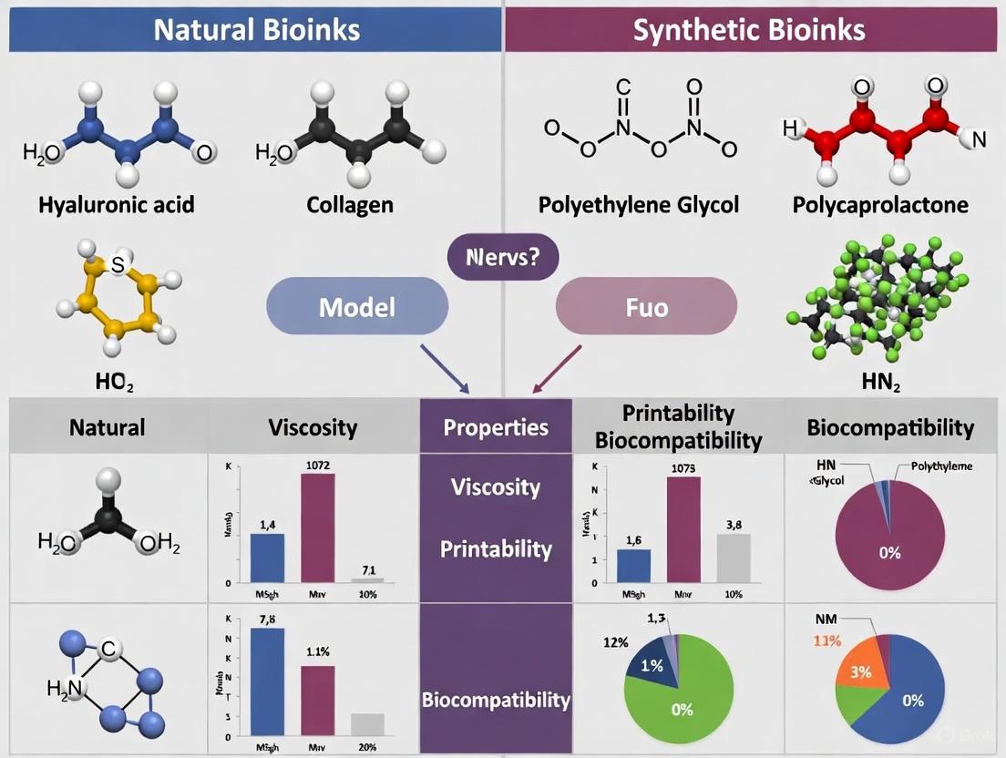
Natural bioinks are a class of biomaterials derived from biological sources, designed to be combined with living cells and processed using 3D bioprinting technologies to create tissue-like constructs. These materials are central to advancements in tissue engineering and regenerative medicine, as they provide a biomimetic environment that closely resembles the native extracellular matrix (ECM). Their innate biological properties—including biocompatibility, biodegradability, and presence of cell-adhesion motifs—make them particularly suitable for supporting cell viability, proliferation, and differentiation within bioprinted structures [9] [10].
The quest to develop ideal bioinks must navigate the "biofabrication window," a critical paradigm that describes the necessary compromise between printability (the ability to be accurately processed by a bioprinter) and biocompatibility (the ability to support cell life and function) [9]. Natural bioinks generally excel in biocompatibility but can present challenges in mechanical strength and structural fidelity post-printing. This guide provides a comparative analysis of natural bioink materials, focusing on their sources, structural properties, and performance relative to synthetic alternatives, to inform researchers and scientists in the field.
Comparative Analysis of Natural vs. Synthetic Bioinks
The table below summarizes the core characteristics of major natural bioink categories against a typical synthetic counterpart, highlighting their comparative advantages and limitations.
Table 1: Comparative Analysis of Natural Bioink Materials and a Synthetic Counterpart
| Material Category | Specific Examples & Sources | Key Structural Features | Native Biological Properties | Key Advantages | Inherent Limitations |
|---|---|---|---|---|---|
| Polysaccharide-based | Alginate (Brown seaweed), Hyaluronic Acid (Animal tissues), Chitosan (Crustacean exoskeletons) | Linear copolymer chains (e.g., alginate: G-blocks, M-blocks); forms hydrogels via ionic crosslinking (e.g., with Ca²⁺) [11] | High water content, excellent diffusion of nutrients and oxygen; some, like HA, are native ECM components [6] | Rapid gelation, tunable viscosity, high biocompatibility, low immunogenicity [12] [10] | Low mechanical strength, lack of cell-adhesive motifs (often requires modification), limited biodegradability control [2] |
| Protein-based | Collagen (Animal connective tissue), Gelatin/GelMA (Denatured collagen), Fibrin (Blood plasma) | Triple-helix structure (collagen); contains native cell-adhesion motifs (e.g., RGD sequences) [10] | Innate cell-binding domains; directly supports cell adhesion, proliferation, and differentiation [6] [10] | Excellent, inherent cytocompatibility; enzymatic biodegradability; mimics native tissue ECM [5] [9] | Low mechanical stiffness, slow gelation (often thermal), potential batch-to-batch variability [2] |
| Decellularized ECM (dECM) | Heart dECM, Liver dECM, Cartilage dECM (Animal or human tissues) | Complex, tissue-specific architecture retaining original ECM proteins, glycosaminoglycans (GAGs), and factors [6] | Preserves the full biochemical complexity and native bioactivity of the source tissue's ECM [6] | Most biomimetic microenvironment; supports tissue-specific cell function and maturation [6] | Complex processing, undefined composition, poor mechanical integrity, significant batch variability [2] |
| Synthetic Polymer (Reference) | Polyethylene Glycol (PEG), Pluronic | Highly tunable network structure; crosslinks via light or chemistry; bio-inert [5] [11] | No innate bioactivity; acts as a blank slate that can be functionalized with bioactive peptides [5] | Superior and tunable mechanical properties, high printability, reproducible and defined composition [5] [2] | Lack of cell-adhesion sites, non-degradable without modification, potential cytotoxicity from photoinitiators [11] [9] |
Quantitative Performance Benchmarking
To move beyond theoretical properties, benchmarking key performance metrics through standardized experiments is crucial. The following quantitative data, derived from model experiments, illustrates how different bioinks perform under testing conditions relevant to bioprinting applications.
Table 2: Benchmarking Key Bioprinting Performance Metrics Across Bioink Types
| Bioink Formulation | Cell Viability During Extrusion (%) | Cell Viability Post-Curing (%) | Storage Modulus (G') Post-Crosslinking (kPa) | Critical Gelation Parameters |
|---|---|---|---|---|
| RAPID Ink (Alginate-based) | >96% [11] | >80% [11] | Data not available in search results | Ionic (Ca²⁺) crosslinking in aqueous bath [11] |
| Gelatin Methacrylate (GelMA) | >90% [11] | ~50% (in-air light curing) [11] | Tunable based on degree of methacrylation and concentration [5] | UV/Viscous light crosslinking with photoinitiator [5] [11] |
| PEGDA (Synthetic) | >90% [11] | ~50% (in-air light curing) [11] | Highly tunable, can exceed many natural polymers [5] [2] | UV/Viscous light crosslinking with photoinitiator [11] |
| Pure Alginate | High (shear-thinning) [2] | High (gentle ionic crosslinking) [12] | Low to moderate (highly dependent on concentration and crosslink density) [2] | Ionic (Ca²⁺) crosslinking [12] |
Key Experimental Protocols for Benchmarking
The quantitative data in Table 2 is generated through specific, reproducible experimental protocols. Below are the methodologies for three key assays used to benchmark bioink performance, as exemplified by research in the field [11].
Protocol 1: Cell Sedimentation Assay
- Objective: To quantify the ability of a bioink to maintain a homogeneous cell suspension and prevent settling during the printing process.
- Methodology: A bioink laden with cells (e.g., 3T3 fibroblasts at a standardized density) is loaded into a bioprinter cartridge. The cartridge is left undisturbed for one hour. Subsequently, samples are collected from the top and bottom of the cartridge, and the cell concentration in each is quantified using a hemocytometer or an automated cell counter. A homogeneous suspension will show minimal difference between top and bottom cell counts.
- Significance: Prevents printhead clogging and ensures accurate cell distribution in the final construct [11].
Protocol 2: Cell Viability During Extrusion
- Objective: To quantify acute cell membrane damage caused by the shear stresses of the extrusion process itself.
- Methodology: Cells are encapsulated in the bioink and extruded at a constant, standardized flow rate (e.g., 75 µL/min). The extruded filament is immediately collected, and the cells are tested for membrane integrity using a live/dead viability assay or a membrane integrity dye (e.g., propidium iodide). The percentage of damaged cells is calculated relative to the total cell count.
- Significance: Directly measures the cytotoxic effect of the printing process, independent of long-term culture effects [11].
Protocol 3: Cell Viability Post-Curing
- Objective: To assess the immediate cytotoxicity of the bioink's crosslinking or solidification method.
- Methodology: After extrusion, the bioink is subjected to its specific curing conditions (e.g., exposure to a light source for photopolymerizing inks, or immersion in a crosslinking bath like CaCl₂ for ionic inks) for a defined period (e.g., 5 minutes). Cell viability is assessed immediately after curing using a live/dead assay. The percentage of viable cells is quantified, often noting differences between the core and the edges of the construct where curing may be more aggressive.
- Significance: Evaluates the compatibility of the crosslinking mechanism with cell survival, a critical factor for the initial health of the bioprinted construct [11].
The Scientist's Toolkit: Essential Research Reagents and Materials
Successful research and development of natural bioinks require a suite of specialized reagents and equipment. The following table details essential items for a laboratory working in this field.
Table 3: Essential Research Reagents and Materials for Natural Bioink Development
| Reagent / Material | Function and Role in Bioink Research |
|---|---|
| Ionic Crosslinkers (e.g., CaCl₂ Solution) | Used to ionically crosslink bioinks like alginate, rapidly transforming the bioink from a viscous liquid to a stable gel [11]. |
| Photoinitiators (e.g., LAP, Irgacure 2959) | A critical component for light-activated crosslinking of methacrylated bioinks (e.g., GelMA). Its concentration must be optimized to balance crosslinking efficiency and cytotoxicity [5] [11]. |
| Recombinant Proteins & Functional Peptides | Used to modify and enhance the bioactivity of natural bioinks. For example, RGD peptides can be grafted onto alginate to impart cell adhesion, and recombinant proteins like C7 are used in advanced composite inks like RAPID ink [11]. |
| Viscometers & Rheometers | Essential instruments for characterizing the flow properties (viscosity, shear-thinning, yield stress) of bioinks, which directly determine their printability [2]. |
| Live/Dead Viability Assay Kit | The standard fluorescent staining method for quantifying cell viability and distribution within a bioprinted construct, both immediately after printing and over time in culture [11] [9]. |
Visualizing the Bioink Design and Analysis Workflow
The process of developing and evaluating a natural bioink follows a logical sequence from design to final assessment. The diagram below maps this critical workflow.
Diagram Title: Bioink Development and Evaluation Workflow
The fundamental challenge in bioink development is the inherent trade-off between its mechanical/rheological properties and its biological functionality. Optimizing one often compromises the other [2]. For instance, increasing the polymer concentration of a natural bioink like collagen enhances its mechanical strength and printability but can lead to denser networks that impede nutrient diffusion and cell migration, thereby reducing viability and biological function. This trade-off is a central consideration in the comparative analysis of natural and synthetic bioinks [2] [9].
Natural bioinks, derived from polysaccharides, proteins, or decellularized ECM, offer unparalleled advantages in biocompatibility and providing a native-like microenvironment for cells, which is why they remain indispensable in tissue engineering. However, their limitations in mechanical strength, printability, and batch consistency are significant. The comparative data and protocols presented in this guide underscore that there is no single "perfect" bioink.
The future of the field lies in the intelligent design of composite and hybrid bioinks that combine the biological merits of natural polymers with the robust, tunable mechanical properties of synthetic materials or innovative formulations like RAPID ink [11] [10]. Furthermore, emerging trends such as 4D bioprinting with stimuli-responsive natural materials and AI-assisted bioink design promise to navigate the biofabrication window more effectively, pushing the boundaries toward the creation of functional, complex tissues for regenerative medicine and drug development [13].
The field of 3D bioprinting stands as a revolutionary force in tissue engineering and regenerative medicine, enabling the fabrication of complex, cell-laden constructs that mimic native tissue architecture. At the core of this technological advancement lie bioinks—specially formulated materials that encapsulate cells and biomolecules for precise deposition. While naturally derived bioinks have dominated early research efforts, synthetic bioink polymers are emerging as powerful alternatives with superior engineering control and tunability [14] [15]. This comparative analysis examines the chemical design principles and performance advantages of synthetic bioink polymers against their natural counterparts, providing researchers and drug development professionals with objective data to inform their material selection processes.
The fundamental limitation of natural bioinks—including alginate, gelatin, collagen, and hyaluronic acid—lies in their inherent batch-to-batch variability and restricted modification windows, which compromise reproducibility and precise mechanical tuning [14] [2]. In contrast, synthetic bioink platforms offer tailorable physicochemical properties through controlled manipulation of their backbone structures, compositions, and molecular weights [14] [15]. This analytical guide systematically evaluates these material classes through the lens of performance metrics critical to bioprinting success: rheological behavior, structural fidelity, mechanical properties, and biological functionality.
Comparative Analysis of Natural versus Synthetic Bioinks
Chemical Design and Material Properties
Synthetic bioinks are engineered through rational chemical design to overcome the limitations of natural systems. Platforms like maleimide-terminated polyurea (PUMA) exemplify this approach, where researchers synthesize segmented prepolymers with photo-crosslinkable end groups to create water-soluble, biocompatible, and biodegradable inks with precisely controllable properties [14]. The synthetic methodology allows independent tuning of soft segments (typically polyethylene glycol of varying molecular weights) and hard segments (diisocyanates or diisocyanates with chain extenders) to achieve target viscosity, rheology, mechanical strength, and crosslink density [14].
Similar design principles apply to polyethylene glycol diacrylate (PEGDA) systems, where molecular weight and functionalization determine the resulting hydrogel properties [16]. These synthetic platforms demonstrate superior chemical consistency compared to natural bioinks, which suffer from compositional variations based on biological source and extraction methods [14] [15]. The synthetic approach also enables incorporation of specific bioactive motifs—such as cell-adhesive RGD peptides—at controlled densities, overcoming the non-specific bioactivity of natural polymers that can sometimes trigger unintended cellular responses [14] [17].
Table 1: Fundamental Characteristics of Natural versus Synthetic Bioinks
| Property | Natural Bioinks | Synthetic Bioinks |
|---|---|---|
| Source | Animal/plant derivatives (alginate, gelatin, collagen, hyaluronic acid) [14] [17] | Laboratory-synthesized polymers (PEG, PUMA, PEGDA) [14] [16] |
| Batch Consistency | Variable due to biological source dependence [14] | High consistency through controlled synthesis [14] [15] |
| Modification Flexibility | Limited to specific functional groups (e.g., methacrylation) [14] [17] | Broad chemical tunability of backbone, composition, MW [14] |
| Crosslinking Mechanisms | Ionic (alginate), temperature-dependent (gelatin), enzymatic [17] | Photoinitiated (UV/visible light), chemical [14] [16] |
| Degradation Profile | Enzyme-dependent (collagenase, hyaluronidase), often unpredictable [17] | Controllable via polymer chemistry, linkage selection [14] |
| Cost Considerations | Moderate to high (purification challenges) [18] | Initially high but scalable production [14] [18] |
Performance Comparison and Experimental Data
Rigorous evaluation of bioink performance encompasses rheological behavior, structural fidelity post-printing, mechanical properties, and biological outcomes. Experimental data from comparative studies reveals distinct advantages for synthetic bioink systems across multiple parameters, particularly in applications demanding precise mechanical control such as skeletal muscle tissue engineering [14].
Natural bioinks like alginate-gelatin-cellulose formulations exhibit favorable shear-thinning behavior but often lack the mechanical robustness required for dynamic tissues. Research demonstrates that ALGEC bioinks (alginate-gelatin-TO-NFC) with 4-5.25% alginate, 4-5.25% gelatin, and 0.5-1.5% TO-NFC achieve compression moduli in the 5-25 kPa range, suitable for soft tissues but insufficient for load-bearing applications [19]. In contrast, PUMA bioinks demonstrate tailorable mechanical properties with elastic moduli spanning 10-200 kPa through modulation of poly(ethylene oxide) molecular weights and hard segment composition [14].
The following table summarizes quantitative performance comparisons based on experimental data from cited studies:
Table 2: Experimental Performance Comparison of Natural and Synthetic Bioinks
| Performance Metric | Natural Bioinks | Synthetic Bioinks | Experimental Method |
|---|---|---|---|
| Viscosity Range (Pa·s) | 1-50 (at shear rate 10 s⁻¹) for ALGEC formulations [19] | 5-100 (at shear rate 10 s⁻¹) tunable via MW and concentration [14] [16] | Rotational rheometry, steady sweep test (0.1-100 s⁻¹) [19] |
| Shear Recovery (%) | 75-90% for alginate-CMC-GelMA [17] | 85-98% for PUMA systems [14] | Thixotropy test, oscillatory rheometry [17] |
| Printability Resolution (μm) | 200-500 [17] | 50-200 [14] [16] | Fiber diameter analysis, precision tests [17] |
| Mechanical Strength (Elastic Modulus) | 2-50 kPa [19] [17] | 10-200 kPa (PUMA) [14] | Uniaxial compression, DMA [14] |
| Cell Viability (Short-term) | 80-95% (high bioactivity) [2] [17] | 75-90% (requires bioactive modification) [14] | Live/dead staining, metabolic assays [14] |
| Long-term Stability (weeks) | 2-4 (rapid degradation) [17] | 4-12 (controllable degradation) [14] | Mass loss, G' monitoring in PBS [14] |
Experimental Protocols for Bioink Evaluation
Rheological Characterization Protocol
Comprehensive rheological assessment is fundamental to predicting bioink printability. The following protocol, adapted from standardized methodologies, ensures consistent characterization across material platforms [2] [17]:
Sample Preparation: Prepare bioink formulations under sterile conditions. For synthetic systems like PUMA or PEGDA, dissolve polymers in PBS or cell culture medium at target concentrations (e.g., 10-30% w/v for PEGDA) [14] [16]. For natural bioinks like alginate-GelMA blends, hydrate polymers overnight at 4°C for complete dissolution [17].
Steady Shear Flow Test: Using a rotational rheometer with parallel plate geometry (25 mm diameter, 1 mm gap), subject samples to shear rates from 0.1 to 100 s⁻¹ at 25°C to simulate extrusion conditions. Record viscosity (η) at each shear rate to determine shear-thinning index (n) via Power Law model fitting: η = K·γⁿ⁻¹, where K is consistency index and n is flow behavior index [19] [2].
Oscillatory Frequency Sweep: In the linear viscoelastic region (determined by amplitude sweep), apply 1% strain while varying angular frequency from 0.1 to 100 rad/s. Record storage (G') and loss (G'') moduli to characterize viscoelastic solid (G' > G'') or liquid (G'' > G') behavior [17].
Thixotropic Recovery Test: Implement a three-interval thixotropy test: (I) low shear (0.1 s⁻¹ for 60 s) to simulate pre-printing state, (II) high shear (10 s⁻¹ for 30 s) to simulate extrusion, and (III) return to low shear (0.1 s⁻¹ for 120 s) to monitor structural recovery. Calculate recovery percentage as G'(final)/G'(initial) × 100% [17].
Temperature Ramp Test (for thermoresponsive inks): For materials like GelMA-containing bioinks, perform temperature sweeps from 4°C to 37°C at 1°C/min rate, constant frequency (1 Hz), and strain (1%) to identify gelation temperatures [17].
Printability and Structural Fidelity Assessment
Evaluating printed construct quality requires quantitative assessment of dimensional accuracy and stability:
Grid Structure Printing: Print a 25×25 mm grid structure (2 layers, 5 mm spacing) using optimized printing parameters (pressure, speed, nozzle size). Acquire images immediately after printing and after 24 hours incubation in crosslinking medium [17].
Filament Morphology Analysis: Measure filament diameter at 10 random locations per sample using image analysis software (e.g., ImageJ). Calculate printability value (Pr) as: Pr = (Dₙ - Dₐ)²/Dₙ², where Dₙ is nozzle diameter and Dₐ is actual filament diameter [17].
Shape Fidelity Quantification: Calculate line deviation (%) as |Lₚ - Lₜ|/Lₜ × 100%, where Lₚ is printed line length and Lₜ is target line length. Similarly, calculate pore area deviation (%) from grid structures [17].
Mechanical Testing of Printed Constructs: Subject printed constructs (e.g., 10×10×5 mm cubes) to uniaxial compression testing. Determine elastic modulus from linear region of stress-strain curve (typically 5-15% strain) at 0.5 mm/min compression rate [14].
Research Reagent Solutions
Successful bioink development and evaluation requires specific research reagents and materials. The following table details essential components and their functions based on the cited experimental protocols:
Table 3: Essential Research Reagents for Bioink Development and Evaluation
| Reagent/Material | Function/Application | Examples/Specifications |
|---|---|---|
| Polyethylene Glycol Diacrylate (PEGDA) | Synthetic polymer backbone for photocrosslinkable bioinks [16] | MW 700-10,000 Da; 10-40% w/v in PBS [16] |
| Photoinitiators | Initiate radical polymerization upon light exposure [16] | LAP (lithium phenyl-2,4,6-trimethylbenzoylphosphinate), I2959; 0.1-1% w/v [16] |
| Alginate | Natural polysaccharide for ionic crosslinking [19] [17] | High viscosity grade (≥2000 cps at 2%); 2-6% w/v [19] |
| Gelatin Methacryloyl (GelMA) | Photocrosslinkable natural polymer promoting cell adhesion [17] | Degree of methacrylation 60-90%; 8-16% w/v [17] |
| TEMPO-oxidized NFC | Nanocellulose reinforcement for enhanced rheology [19] | Carboxylate content 0.2-2 mmol/g; 0.5-1.5% w/v [19] |
| Crosslinking Agents | Induce hydrogel formation [17] | CaCl₂ (50-200 mM for alginate); UV light (315-400 nm, 5-20 mW/cm²) [17] |
| Viscoelastic Modifiers | Adjust rheological properties [16] | Fumed silica (0.5-2%); PEG 400 (5-15%) [16] |
Visualization of Synthetic Bioink Design and Performance
Synthetic Bioink Design and Evaluation Workflow
The following diagram illustrates the integrated workflow for synthetic bioink design, fabrication, and evaluation, highlighting the systematic approach enabled by synthetic polymer platforms:
Comparative Advantages of Synthetic Bioinks
The strategic advantages of synthetic bioinks across key performance parameters are visualized in the following diagram:
Synthetic bioink polymers represent a paradigm shift in 3D bioprinting, offering unprecedented engineering control through rational chemical design that surpasses the capabilities of natural biomaterials. The comparative data presented in this analysis demonstrates distinct advantages in mechanical tunability, batch consistency, and structural fidelity—attributes critical for clinical translation and functional tissue engineering. While natural bioinks maintain benefits in innate bioactivity, emerging synthetic platforms like PUMA successfully integrate cell-instructive elements while preserving tailorability [14].
The future trajectory of bioink development points toward increasingly sophisticated hybrid approaches, where synthetic polymers provide structural precision while natural components or engineered biofunctional domains mediate cellular responses [15]. Additionally, the integration of computational modeling and machine learning—exemplified by viscosity prediction algorithms for ALGEC bioinks—will accelerate the design iteration process [19]. As the field progresses toward printing complex, vascularized tissues and organs, synthetic bioink platforms will play an indispensable role in overcoming the limitations of natural systems, ultimately enabling the fabrication of biologically functional constructs for regenerative medicine, disease modeling, and drug development applications.
Comparative Analysis of Biocompatibility and Cell-Material Interactions
In the rapidly advancing field of 3D bioprinting, the selection of an appropriate bioink is paramount to the success of fabricating functional tissues. Bioinks, which are formulations of biomaterials, cells, and bioactive molecules, serve as the foundational building blocks for creating complex, three-dimensional structures that mimic native tissues [2] [9]. The core challenge lies in navigating the inherent trade-off between a bioink's printability—its rheological properties enabling precise deposition—and its biocompatibility—its ability to support critical cellular functions without eliciting adverse effects [2]. This guide provides a comparative analysis of natural and synthetic bioinks, focusing on their biocompatibility and cell-material interactions. It is structured to aid researchers and drug development professionals in making evidence-based selections for tissue engineering and regenerative medicine applications.
Material Classifications and Fundamental Properties
Bioinks are broadly categorized into natural, synthetic, and hybrid materials, each possessing distinct advantages and limitations that dictate their suitability for specific biomedical applications [20].
Table 1: Fundamental Properties of Natural vs. Synthetic Bioinks
| Parameter | Natural Bioinks | Synthetic Bioinks |
|---|---|---|
| Biocompatibility | Inherently high due to natural origin; possesses native biological cues [20]. | May require additional modification or functionalization to improve [20]. |
| Mechanical Properties | Generally weaker and less tunable; can be variable [20]. | Highly tunable, predictable, and often superior in strength [20]. |
| Cellular Interactions | Excellent; naturally promote cell adhesion, proliferation, and differentiation [20] [7]. | Often bio-inert; requires incorporation of adhesive peptides (e.g., RGD) to support cells [20]. |
| Printability | Can be challenging due to variability and low mechanical strength [20]. | Highly controllable due to engineered and consistent nature [20]. |
| Degradation | Enzymatic; degradation products are typically metabolized [7]. | Often hydrolytic; degradation rate and byproducts can be precisely designed [7]. |
| Common Examples | Alginate, Collagen, Gelatin, Hyaluronic Acid, Fibrin [20] [7]. | Polyethylene Glycol (PEG), Polylactic Acid (PLA), Polycaprolactone (PCL) [20]. |
The "biofabrication window" represents the critical compromise between the printability of a bioink and its biocompatibility. Optimizing one property often compromises the other; for instance, increasing polymer concentration to enhance mechanical strength can negatively impact cell viability by reducing nutrient diffusion [2] [9]. The contemporary understanding of biocompatibility has evolved beyond mere bio-safety (non-toxicity) to include active biofunctionality—the ability to promote desired cellular activities such as adhesion, proliferation, and differentiation to facilitate tissue regeneration [9].
Figure 1: The Biofabrication Window Paradigm illustrating the fundamental trade-off between printability and biocompatibility that guides bioink design.
Comparative Analysis of Biocompatibility and Cellular Responses
Biocompatibility is a multifaceted property that encompasses cell viability, adhesion, proliferation, and differentiation. The material class significantly influences these cellular responses.
2.1 Cell Viability and Proliferation Cell viability post-printing is a primary indicator of biocompatibility. Natural bioinks, such as alginate and collagen, typically provide a microenvironment that closely mimics the native extracellular matrix (ECM), leading to high cell survival and proliferation rates [20] [7]. For instance, a 2024 study on a novel alginate/pericardial fluid-based hydrogel reported a significant increase in the proliferation and differentiation of MC3T3-E1 pre-osteoblast cells compared to pure alginate hydrogels, demonstrating how natural additives can further enhance biocompatibility [21]. Synthetic bioinks, while mechanically robust, may require the incorporation of bioactive motifs (e.g., RGD peptides) to support comparable levels of cell survival and growth [20].
2.2 Cell Adhesion and Spread Cell adhesion is predominantly governed by the presence of specific cell-adhesive ligands within the bioink. Natural polymers like collagen and gelatin inherently contain these ligands, facilitating robust cell attachment and spreading [20]. In contrast, synthetic polymers like PEG are inherently bio-inert. Cell adhesion in these systems is entirely dependent on chemical modification with adhesion peptides, offering a high degree of control but adding complexity to the formulation [2] [20].
2.3 Osteogenic Differentiation (Case Study) The ability to direct stem cell differentiation is a crucial aspect of biofunctionality for tissue engineering. Experimental data from bone tissue engineering studies highlight the differences between material classes.
Table 2: Experimental Osteogenic Performance Data
| Bioink Formulation | Cell Viability (%) | Proliferation Rate | Osteogenic Marker Expression (e.g., Alkaline Phosphatase) | Key Experimental Finding | Reference |
|---|---|---|---|---|---|
| Alginate/Pericardial Fluid (A-PFS 1:1) | High | Enhanced | Significantly Increased | PFS increased biocompatibility and supported osteogenic differentiation of MC3T3-E1 pre-osteoblasts. | [21] |
| Pure Alginate Hydrogel | Moderate | Baseline | Baseline | Served as a control; provided a baseline but required enhancement for superior biological performance. | [21] |
| GelMA-based Bioink | High | High | Moderate to High (Tunable) | Photocrosslinkable and versatile; supports high cell adhesion and tunable differentiation cues for various cell types. | [20] [7] |
| PEG-based Bioink | Variable (Modification-dependent) | Variable (Modification-dependent) | Low (unless functionalized) | Requires functionalization with RGD and osteogenic peptides to direct differentiation effectively. | [20] [9] |
Experimental Protocols for Assessment
Standardized methodologies are essential for the objective comparison of bioink biocompatibility. Key experimental protocols are detailed below.
3.1 Protocol for Evaluating Cell Viability and Proliferation
- Objective: To quantify the survival and proliferation rate of cells encapsulated within a bioink post-printing.
- Materials: Bioink formulation, cell line (e.g., MC3T3-E1 pre-osteoblasts, human mesenchymal stem cells), cell culture medium, live/dead viability/cytotoxicity assay kit (e.g., Calcein AM and Ethidium homodimer-1), confocal microscope, DNA quantification kits (e.g., PicoGreen) [21] [9].
- Methodology:
- Bioprinting: Encapsulate cells within the bioink at a specified density (e.g., 1-10 million cells/mL) and fabricate 3D constructs using a calibrated extrusion bioprinter.
- Culture: Maintain the printed constructs in cell culture medium under standard conditions (37°C, 5% CO₂).
- Staining: At predetermined time points (e.g., days 1, 3, and 7), incubate constructs with the live/dead stain. Calcein AM stains live cells green, while Ethidium homodimer-1 stains dead cells red.
- Imaging & Analysis: Image multiple regions of the construct using confocal microscopy. Calculate cell viability as the percentage of live cells relative to the total number of cells. Proliferation can be further quantified by measuring total DNA content over time [9].
3.2 Protocol for Characterizing Cell-Material Interactions
- Objective: To assess cell adhesion, spreading, and cytoskeletal organization within the printed bioink matrix.
- Materials: Bioink constructs, fluorescent phalloidin (for F-actin staining), DAPI (for nuclear staining), antibodies for specific adhesion proteins (e.g., vinculin), paraformaldehyde, Triton X-100, blocking buffer [9].
- Methodology:
- Fixation: At the desired time point, rinse constructs and fix with 4% paraformaldehyde.
- Permeabilization and Blocking: Permeabilize the cells with Triton X-100 and block non-specific binding sites with a protein buffer.
- Staining: Incubate constructs with fluorescent phalloidin to visualize the actin cytoskeleton and with DAPI to identify nuclei. For detailed adhesion analysis, perform immunofluorescence staining against vinculin.
- Imaging: Use confocal microscopy to obtain high-resolution z-stacks. Analyze images for cell morphology (e.g., aspect ratio, spread area), cytoskeletal organization, and the formation of focal adhesions [9].
Figure 2: Experimental workflow for the comprehensive assessment of bioink biocompatibility and cell-material interactions.
The Scientist's Toolkit: Essential Research Reagents
A successful bioink development and evaluation pipeline relies on a suite of essential reagents and materials.
Table 3: Key Research Reagent Solutions for Bioink Testing
| Reagent / Material | Function in Research | Application Example |
|---|---|---|
| Live/Dead Viability/Cytotoxicity Kit | Distinguishes live from dead cells using fluorescent markers. | Quantifying cell survival within a bioprinted alginate-gelatin construct after 7 days of culture [9]. |
| Phalloidin (F-actin stain) | Labels filamentous actin, enabling visualization of the cytoskeleton and cell morphology. | Assessing the spread and elongation of mesenchymal stem cells encapsulated in a collagen-based bioink [9]. |
| Recombinant Human Collagen (e.g., rhCollagen) | An animal-free, consistent natural polymer used as a base material for bioinks. | Formulating a biocompatible bioink (e.g., Collink.3D) for printing tissue models and transplantable scaffolds [22]. |
| Alginate | A plant-derived polysaccharide that forms gentle ionically-crosslinked gels. | Serving as a primary component in a hydrogel for cell encapsulation and as a drug delivery system [21] [20]. |
| GelMA (Gelatin Methacryloyl) | A modified natural polymer that is photo-crosslinkable, offering tunable mechanical properties. | Creating stable, cell-laden structures with high shape fidelity using UV light crosslinking in stereolithography printing [20] [7]. |
| RGD Peptide | A cell-adhesive peptide sequence that can be grafted onto bioinks. | Functionalizing a synthetic PEG hydrogel to enable integrin-mediated cell adhesion and spreading [20]. |
| Polyethylene Glycol (PEG) | A synthetic, bio-inert polymer that serves as a tunable "blank slate" for bioinks. | Designing a hydrogel with precise mechanical properties, then functionalizing it with bioactive cues [20] [9]. |
The comparative analysis reveals that the choice between natural and synthetic bioinks is not a matter of superiority, but of strategic selection based on the target application. Natural bioinks excel in applications where superior biocompatibility, innate bioactivity, and rapid cellular integration are the highest priorities, such as in soft tissue regeneration and disease modeling. Synthetic bioinks offer distinct advantages when precise control over mechanical properties, degradation rates, and scaffold architecture is required, though their biofunctionality must be engineered. The future of the field lies in the development of advanced hybrid bioinks that strategically combine the strengths of both material classes. Furthermore, the integration of animal-free components (e.g., recombinant proteins) and smart materials that respond to environmental stimuli represents the next frontier in creating bioinks that can more accurately recapitulate the dynamic nature of native human tissues [21] [23] [22].
In the field of 3D bioprinting, bioinks serve as the foundational materials for creating complex, cell-laden constructs that aim to replicate native tissues. The mechanical properties of these bioinks—strength, elasticity, and structural integrity—are critical determinants of their success in both the printing process and final application [2] [1]. These properties must be carefully balanced against biological requirements such as cell viability and biocompatibility, creating a fundamental trade-off that researchers must navigate [2]. This comparative analysis examines the mechanical performance of natural and synthetic bioinks, providing a structured framework for researchers and drug development professionals to select appropriate materials for specific tissue engineering applications. The mechanical profile of a bioink directly influences its printability during fabrication and its functional performance in physiological environments, making understanding these properties essential for advancing regenerative medicine and drug screening platforms [24] [25].
Fundamental Mechanical and Rheological Properties
The mechanical performance of bioinks encompasses several key properties that must be considered both during the printing process and in the final construct.
Rheological Properties for Printability
For extrusion-based bioprinting—the most prevalent bioprinting method—bioinks must demonstrate specific rheological behavior to be successfully printed [24] [2]. Viscosity represents a bioink's resistance to flow and governs both printability and post-printing shape retention [2] [1]. Excessive viscosity can impede extrusion and damage cells through high pressure, while insufficient viscosity leads to poor resolution and structural collapse [2]. Shear-thinning behavior, where viscosity decreases under applied shear stress, is particularly desirable as it enables smooth extrusion through nozzles while allowing rapid recovery of viscosity after deposition to maintain structural shape [2] [1] [26]. Additionally, gelation kinetics must be optimized to facilitate rapid solidification post-printing, stabilizing the construct against deformation or collapse [2] [1].
Structural and Mechanical Properties Post-Printing
After printing and crosslinking, bioinks must provide appropriate mechanical support for encapsulated cells and eventual tissue development. The storage modulus (G′) indicates the elastic character and mechanical strength of the printed construct, representing its ability to resist deformation [27]. Yield stress prevents the material from flowing under its own weight after printing, thereby preserving the intended scaffold design [27]. Different applications demand distinct mechanical profiles; for instance, load-bearing tissues like bone require high mechanical strength, while soft tissues like neural or adipose tissue necessitate more compliant matrices [24] [25]. The degradation rate must also match the rate of new tissue formation to facilitate a gradual transfer of mechanical load to the developing tissue [27] [7].
Table 1: Key Mechanical and Rheological Properties for Bioink Evaluation
| Property | Definition | Importance in Bioprinting | Ideal Characteristics |
|---|---|---|---|
| Viscosity | Resistance to flow under applied shear stress | Determines extrudability and structural stability | Balanced for specific printing method: 10 mPa·s for inkjet; 30–6×10⁷ mPa·s for extrusion [7] |
| Shear-Thinning | Decrease in viscosity with increasing shear rate | Facilitates easy extrusion while maintaining shape fidelity | High degree of shear-thinning for smooth extrusion and rapid structural recovery [2] [26] |
| Storage Modulus (G′) | Measure of elastic solid-like behavior | Indicates mechanical strength and structural stability | Sufficiently high to maintain construct shape and provide mechanical cues [27] |
| Gelation Kinetics | Rate of solidification post-printing | Affects resolution, cell viability, and structural integrity | Rapid enough to prevent deformation but not so fast as to cause nozzle clogging [2] [1] |
| Degradation Rate | Rate at which material breaks down in physiological conditions | Should match tissue formation rate for proper load transfer | Tunable to align with specific tissue regeneration timeline [27] [7] |
Comparative Analysis of Natural Bioinks
Natural bioinks, derived from biological sources, generally provide excellent biocompatibility and biomimicry but vary significantly in their mechanical properties.
Polysaccharide-Based Bioinks
Alginate, a natural polysaccharide from seaweed, undergoes rapid ionic crosslinking (typically with calcium chloride) to form stable hydrogels [26]. While it exhibits excellent shear-thinning behavior and printability, its mechanical weakness and lack of cell-adhesive motifs limit its application without modification [24] [26]. Agarose, another seaweed-derived polysaccharide, forms thermo-reversible gels but is primarily used as a sacrificial material or for mold formation due to its limited cell interactivity [24]. Hyaluronic acid (HA), a major component of the native extracellular matrix, can be modified to enhance its mechanical properties through methacrylation or other chemical modifications, allowing tunable stiffness and degradation profiles [24] [7].
Protein-Based Bioinks
Collagen, the most abundant protein in the human ECM, offers exceptional biocompatibility and cell signaling capabilities [27]. However, its low viscosity and slow gelation at physiological conditions present challenges for printing [27]. Gelatin, derived from denatured collagen, provides thermo-reversible gelation and cell adhesion motifs, and is often modified with methacrylate groups (GelMA) to create photocrosslinkable hydrogels with enhanced mechanical stability [24] [7]. Fibrin, known for its role in wound healing, forms through enzymatic crosslinking and creates fibrous networks conducive to cell migration, though it lacks mechanical strength for load-bearing applications [24] [26]. Silk fibroin, derived from silkworms, offers impressive mechanical strength and tunability through various processing methods, making it suitable for applications requiring higher mechanical resilience [24].
ECM-Derived Bioinks
Decellularized extracellular matrix (dECM) bioinks, derived from processed native tissues, potentially provide the most biomimetic microenvironment as they preserve tissue-specific composition and signaling factors [24] [28]. However, batch-to-batch variability and poor mechanical integrity often necessitate blending with other materials to improve printability [24].
Table 2: Mechanical Properties and Characteristics of Natural Bioinks
| Bioink Material | Crosslinking Mechanism | Mechanical Strengths | Mechanical Limitations | Typical Applications |
|---|---|---|---|---|
| Alginate | Ionic (e.g., CaCl₂) | Rapid gelation, good shear-thinning, high shape fidelity | Weak mechanics, lacks cell adhesion sites | Cartilage, vascular structures, often blended [24] [26] |
| Collagen | Thermal, pH-mediated | Excellent biocompatibility, native ECM mimicry | Low viscosity, slow gelation, poor mechanical strength | Soft tissues: skin, nerve, cartilage [24] [27] |
| Gelatin/Methacryloyl (GelMA) | Photocrosslinking | Tunable mechanics, good cell adhesion, thermo-reversible | Modest mechanical strength, requires photoinitiators | Various soft tissues, often combined [24] [7] |
| Fibrin | Enzymatic | Natural role in healing, promotes cell migration | Very low mechanical strength, fast degradation | Wound healing, neural tissue [24] [26] |
| Hyaluronic Acid (HA) | Ionic, photocrosslinking | Native ECM component, tunable viscosity and mechanics | Can be too soft alone, modification needed | Cartilage, stem cell niches [24] [7] |
| Silk Fibroin | Ionic, sonication, shear | High mechanical strength, tunable degradation | Processing complexity, potential immunogenicity | Bone, cartilage, ligaments [24] |
| dECM | Thermal, enzymatic | Tissue-specific bioactivity, complex ECM composition | Very poor mechanical strength, variability | Patient-specific tissues, organ models [24] [28] |
Comparative Analysis of Synthetic Bioinks
Synthetic bioinks offer superior control over mechanical properties and printability but often lack innate bioactivity.
Poly(ethylene glycol) (PEG) and its derivatives (e.g., PEG-diacrylate) are hydrophilic polymers that can be photocrosslinked to form hydrogels with highly tunable mechanical properties [2] [26]. The mechanical strength, swelling, and degradation can be precisely controlled by varying molecular weight, functionalization, and crosslinking density [26]. However, PEG lacks cell-adhesive motifs, requiring modification with peptide sequences (e.g., RGD) to support cell attachment [26]. Polycaprolactone (PCL) is a thermoplastic polyester with high mechanical strength and slow degradation, making it suitable for support structures in biofabrication [2] [26]. While not typically used as a cell-laden hydrogel due to its high processing temperature, it is often printed alongside cell-laden bioinks to provide mechanical reinforcement for tissues like bone and cartilage [26].
The following diagram illustrates the inherent trade-off between printability and biological functionality that characterizes bioink design, particularly evident when comparing natural and synthetic materials:
Diagram 1: The fundamental trade-off in bioink design between rheological properties and biological functionality, which guides the choice between synthetic and natural materials.
Experimental Protocols for Mechanical Characterization
Standardized methodologies are essential for meaningful comparison of bioink mechanical properties across studies. The following section outlines key experimental protocols.
Rheological Characterization
Rheological testing quantifies bioink behavior during the printing process. Experiments are typically performed using a rotational rheometer with parallel plate or cone-and-plate geometry [2]. The viscosity curve is obtained by measuring viscosity across a range of shear rates (e.g., 0.1 to 100 s⁻¹), with shear-thinning behavior indicated by decreasing viscosity with increasing shear rate [2] [1]. Oscillatory amplitude sweeps measure storage modulus (G′) and loss modulus (G″) as a function of strain to determine the linear viscoelastic region and yield point [2]. Oscillatory time sweeps at constant frequency and strain monitor gelation kinetics after inducing crosslinking (e.g., via light exposure or ionic crosslinker addition) [2] [1].
Mechanical Testing of Crosslinked Constructs
After printing and crosslinking, hydrogels undergo mechanical testing to characterize their performance in physiological conditions. Uniaxial compression testing performed on cylindrical hydrogel specimens measures compressive modulus, typically calculated as the slope of the linear region of the stress-strain curve (usually between 10-20% strain) [27] [25]. Tensile testing on dog-bone shaped specimens determines elastic (Young's) modulus, ultimate tensile strength, and elongation at break [25]. Additionally, swelling tests measure mass or volume change of hydrogels in buffer solution to calculate swelling ratio, which influences mechanical properties and nutrient diffusion [26] [7].
The following workflow diagram outlines a comprehensive experimental approach for bioink mechanical characterization:
Diagram 2: Comprehensive experimental workflow for evaluating bioink mechanical properties and their biological impact.
The Researcher's Toolkit: Essential Materials and Reagents
Successful bioink development and characterization requires specific research reagents and equipment. The following table details key solutions and materials used in the field:
Table 3: Essential Research Reagents and Materials for Bioink Development
| Reagent/Material | Function/Application | Examples & Notes |
|---|---|---|
| Natural Polymers | Base material for bioinks providing biocompatibility and bioactivity | Alginate, collagen, gelatin, hyaluronic acid, fibrin, silk fibroin [24] [7] |
| Synthetic Polymers | Base material offering tunable mechanical properties and printability | PEG, PCL, PLA, PLGA [2] [26] |
| Photoinitiators | Enable photocrosslinking of bioinks under light exposure | LAP (lithium phenyl-2,4,6-trimethylbenzoylphosphinate), Irgacure 2959 [26] |
| Ionic Crosslinkers | Facilitate rapid ionic crosslinking of polysaccharide bioinks | Calcium chloride (for alginate), other divalent cations [24] [26] |
| Peptide Modifiers | Enhance cell adhesion to synthetic materials | RGD peptides, MMP-sensitive peptides [26] [25] |
| Support Bath Materials | Provide temporary support for printing low-viscosity bioinks | Gelatin microparticles (FRESH method), Carbopol [26] |
| Sacrificial Inks | Create channels for vasculature or complex geometries | Pluronic F127, carbohydrate glass [26] |
The comparative analysis of mechanical properties in natural and synthetic bioinks reveals a consistent trade-off: natural bioinks generally excel in biological functionality but often lack the mechanical robustness required for printing and load-bearing applications, while synthetic bioinks offer superior mechanical control and printability but require modification to support cell functions [2] [1]. This fundamental compromise has driven the field toward multi-component bioinks that combine materials to achieve synergistic benefits [24] [29]. For instance, incorporating strong synthetic polymers with natural bioinks can provide mechanical reinforcement, while adding natural components to synthetic inks can enhance bioactivity [24] [7].
Future research directions focus on developing more sophisticated smart bioinks with dynamic mechanical properties that can respond to environmental cues or change over time to match tissue maturation [25]. Additionally, advances in nanomaterial reinforcement continue to show promise for enhancing mechanical properties without compromising bioactivity [24]. As standardization of characterization methods improves and our understanding of cell-mechanics interactions deepens, the design of next-generation bioinks will increasingly move beyond balancing trade-offs to truly integrating mechanical and biological requirements for specific tissue engineering applications [2] [25]. This progression will be crucial for realizing the full potential of 3D bioprinting in regenerative medicine, disease modeling, and drug development.
In the field of 3D bioprinting, the degradation profile of a bioink is not merely a property but a fundamental design parameter that dictates the success of tissue engineering constructs. The degradation mechanism governs the critical balance between the dissolution of an implanted scaffold and the formation of new, functional tissue. Within comparative bioink research, a central thesis distinguishes two fundamental paradigms: the natural remodeling of bioinks derived from biological sources and the controlled hydrolysis of their synthetic counterparts. Natural bioinks, such as collagen, gelatin, and alginate, undergo enzyme-mediated degradation that cells can actively influence and participate in. In contrast, synthetic bioinks typically degrade through predictable, bulk hydrolysis of their polymer backbones, independent of cellular activity. This guide provides an objective comparison of these distinct degradation pathways, supporting researchers with structured data and experimental protocols to inform material selection and application-specific design.
Comparative Analysis of Degradation Mechanisms
The degradation behavior of natural and synthetic bioinks stems from their fundamental chemical structures and the mechanisms by which their polymer chains are cleaved. Table 1 summarizes the core characteristics of these two pathways.
Table 1: Fundamental Characteristics of Degradation Pathways
| Characteristic | Natural Bioinks (Enzymatic Remodeling) | Synthetic Bioinks (Controlled Hydrolysis) |
|---|---|---|
| Primary Mechanism | Cell-secreted enzymes (e.g., MMPs, collagenases) cleave specific peptide sequences [30] [31]. | Water molecules hydrolyze ester or other cleavable bonds in the polymer backbone [31]. |
| Degradation Kinetics | Dynamic and cell-driven; rate depends on local cell density and activity [30]. | Predictable and pre-programmed; primarily dependent on material chemistry and pH [31]. |
| Spatial Control | Localized to the cellular microenvironment, enabling invasive cell migration [30]. | Homogeneous throughout the bulk material, leading to surface erosion or bulk degradation. |
| Biological Cues | Contains innate cell-adhesive motifs (e.g., RGD) and allows cell-guided remodeling [30] [27]. | Typically bio-inert; requires functionalization with bioactive peptides to support cell adhesion [31]. |
| Mechanical Integrity | Viscoelastic and stress-relaxing; allows matrix reorganization [2]. | Primarily elastic; mechanics can be precisely tuned but are largely static until degradation [2]. |
Degradation Pathway Diagrams
The following diagrams illustrate the sequential processes of enzymatic remodeling and controlled hydrolysis, highlighting key differences in trigger, control, and outcome.
Figure 1: The enzymatic remodeling pathway of natural bioinks is a cell-instructed process. Cells adhere to the matrix, secrete specific enzymes that cleave peptide bonds, and create physical paths for migration, culminating in de novo tissue synthesis.
Figure 2: The controlled hydrolysis of synthetic bioinks is a chemistry-driven process. Hydrolytic agents in the environment attack specific chemical bonds in the polymer backbone, leading to predictable chain scission, mass loss, and eventual creation of space for tissue ingrowth.
Experimental Data and Performance Comparison
Quantitative data from degradation studies is crucial for evaluating bioink performance. Experimental protocols typically measure mass loss, mechanical property changes, and solute release profiles under physiological conditions. Table 2 consolidates key quantitative findings from the literature for common natural bioinks.
Table 2: Experimental Degradation Data of Selected Natural Bioinks
| Bioink Material | Experimental Model / Crosslinking | Key Degradation Metrics | Degradation Timeline & Notes |
|---|---|---|---|
| Collagen (Type I) | High-concentration physical hydrogel [32] | • Contraction of scaffolds in response to cellular activity.• Enzymatic biodegradability via collagenases. | • Low mechanical properties and rapid contraction can be limitations [32].• Degradation is highly cell-density dependent. |
| Gelatin-Methacryloyl (GelMA) | Covalent crosslinking via photopolymerization [30] [33] | • Susceptibility to matrix metalloproteinases (MMPs).• Degradation rate tuned by degree of methacrylation and crosslinking density. | • Higher crosslinking density slows enzymatic degradation [30].• Allows cell-mediated remodeling. |
| Alginate | Ionic crosslinking (e.g., Ca²⁺) [28] [34] | • Degradation via loss of crosslinking ions (ion exchange).• Slow, uncontrolled dissolution in physiological conditions. | • Not inherently susceptible to mammalian enzymes [28].• Degradation rate is highly dependent on the alginate formulation and purity. |
| Hyaluronic Acid | Modified with crosslinkable groups (e.g., methacrylate) [34] | • Degradation by hyaluronidase enzymes. | • Used in cartilage engineering and cosmetics [34].• Viscoelastic properties support cell growth. |
Detailed Experimental Protocols
To ensure reproducibility and enable direct comparison between studies, standardized protocols for assessing degradation are essential. Below are detailed methodologies for testing both enzymatic and hydrolytic degradation.
Protocol for In Vitro Enzymatic Degradation
This protocol is designed to quantify the degradation profile of natural bioinks in the presence of specific enzymes [32] [31].
Hydrogel Fabrication:
- Prepare bioink solutions according to the desired formulation (e.g., high-concentration collagen hydrogel [32] or GelMA solution [33]).
- Cast the bioink into sterile molds (e.g., cylindrical discs, 8 mm diameter x 2 mm height) or directly 3D bioprint constructs.
- Apply the appropriate crosslinking method: physical gelation for collagen (pH and temperature) [32], or UV light exposure for GelMA at a specified intensity and duration [33].
Initial Mass Measurement (W₀):
- After fabrication, gently rinse the hydrogels in phosphate-buffered saline (PBS) to remove any unreacted components.
- Carefully blot the hydrogels on filter paper to remove surface water and immediately record the initial wet mass (W₀).
Enzymatic Incubation:
- Prepare a degradation buffer solution. A common choice is a collagenase solution (e.g., 1-2 U/mL in PBS with 5 mM CaCl₂) for collagen and gelatin-based inks [32]. For other bioinks, select the appropriate enzyme (e.g., hyaluronidase for hyaluronic acid).
- Immerse each pre-weighed hydrogel in a sufficient volume of enzyme solution (typically 1 mL per 100 mg hydrogel) and incubate at 37°C under gentle agitation.
- Include control groups incubated in enzyme-free buffer.
Mass Loss Monitoring:
- At predetermined time points (e.g., 1, 3, 6, 12, 24 hours), remove the hydrogels from the incubation solution.
- Rinse with PBS, blot on filter paper, and record the wet mass (Wₜ).
- The percentage of mass remaining can be calculated as:
(Wₜ / W₀) × 100%.
Endpoint Analysis:
- After the final time point, the hydrogel solution can be analyzed for released glycosaminoglycans (GAGs), peptides, or other polymeric fragments using spectrophotometric assays (e.g., DMMB for GAGs, BCA for protein).
- The morphology of degraded hydrogels can be examined using scanning electron microscopy (SEM).
Protocol for In Vitro Hydrolytic Degradation
This protocol evaluates the chemical hydrolysis of synthetic bioinks or hydrolytically unstable natural derivatives [31].
Sample Preparation:
- Fabricate hydrogels as described in Section 4.1, Step 1.
Initial Dry Mass Measurement (D₀):
- After fabrication and rinsing, lyophilize the hydrogels for 48 hours or until a constant mass is achieved.
- Record the initial dry mass (D₀).
PBS Incubation:
- Immerse the dry hydrogels in PBS (pH 7.4) and incubate at 37°C. The volume of PBS should be large enough to ensure sink conditions (typically 50:1 buffer-to-hydrogel volume ratio).
- To study pH sensitivity, parallel experiments can be set up in buffers of different pH (e.g., 5.5 to simulate an inflammatory environment).
Monitoring and Sampling:
- At regular intervals (e.g., daily, weekly), remove the hydrogels from the PBS.
- Rinse with deionized water and lyophilize to constant weight to record the dry mass (Dₜ).
- The percentage of mass remaining is calculated as:
(Dₜ / D₀) × 100%. - The collected PBS supernatant at each time point can be analyzed for degradation products.
Characterization of Property Loss:
- Monitor the changes in the mechanical properties of wet hydrogels (e.g., compressive/tensile modulus) throughout the degradation timeline using a rheometer or mechanical tester [2].
- Use SEM to observe changes in the pore morphology and surface erosion of the hydrogels.
The Scientist's Toolkit: Essential Research Reagents
Selecting the appropriate reagents is fundamental to designing and interpreting degradation studies. The following table lists key materials and their functions in this field.
Table 3: Essential Reagents for Bioink Degradation Research
| Reagent / Material | Function in Degradation Studies |
|---|---|
| Collagenase (Type I, II, etc.) | Enzyme used to simulate the in vivo degradation of collagen and gelatin-based bioinks by cleaving peptide bonds [32]. |
| Matrix Metalloproteinases (MMPs) | A family of enzymes (e.g., MMP-1, MMP-2, MMP-9) crucial for studying the cell-mediated remodeling of natural bioinks that contain MMP-cleavable sites [30]. |
| Hyaluronidase | Enzyme that catalyzes the hydrolysis of hyaluronic acid, used to assess the degradation of HA-based bioinks [34]. |
| Glucono-δ-lactone (GDL) | A slow, biocompatible acidifier used to control pH-dependent gelation and phase separation processes, which can influence subsequent degradation behavior [33]. |
| TEMPO-oxidized Nanofibrillated Cellulose (TO-NFC) | A viscosity modifier and reinforcement nanomaterial in composite bioinks; its surface chemistry can influence water retention and interaction with polymers, potentially affecting degradation [19]. |
| Methacrylate Anhydride | Reagent used to synthesize GelMA from gelatin, introducing photo-crosslinkable groups that allow tuning of the hydrogel's initial density and degradation rate [30] [33]. |
| Calcium Chloride (CaCl₂) | Crosslinking agent for anionic polymers like alginate; the stability of ionic crosslinks directly controls the degradation rate in physiological environments [34] [35]. |
In the field of 3D bioprinting, bioinks represent the cornerstone technology, functioning as specialized carrier materials responsible for enabling the fabrication of anatomically precise, cell-laden constructs that replicate native tissue architecture [2]. Their role extends far beyond mere structural support, as they must simultaneously sustain critical cellular functions including viability, proliferation, and differentiation, which are indispensable for applications in regenerative medicine and personalized therapies [2]. The rheological properties of these bioinks, particularly viscosity and shear-thinning behavior, fundamentally determine their performance during the bioprinting process, directly influencing printability, structural integrity, and overall tissue fabrication success [2] [36].
A persistent and central challenge in bioink development lies in reconciling the often conflicting demands of rheological properties essential for printability with those required for biological functionality [2]. This trade-off currently limits the clinical translation of bioprinted tissues, especially for complex applications such as vascularized or mechanically dynamic organs [2]. This guide provides a comparative analysis of bioink rheology, focusing specifically on the distinctive characteristics of natural and synthetic polymer-based formulations, to offer researchers a systematic framework for selecting and optimizing materials for specific bioprinting applications.
Fundamental Rheological Concepts in Bioink Design
Defining Key Rheological Parameters
The printability of a bioink is governed by several interconnected rheological properties that determine its behavior under the stresses encountered during extrusion and deposition.
Viscosity: This represents the fundamental measure of a material's resistance to flow under applied shear stress, quantifying the internal friction between fluid layers during deformation [2]. In bioinks, viscosity must be carefully balanced: excessive viscosity can impede material flow and increase extrusion pressures to levels that may damage encapsulated cells, while insufficient viscosity compromises shape fidelity and structural stability of printed constructs [2] [37].
Shear-Thinning Behavior: Also known as pseudoplastic behavior, this refers to the phenomenon where a material's viscosity decreases with increasing shear rate [2]. This property is particularly advantageous in bioink applications, as it facilitates smooth material flow through printing nozzles under high shear conditions during extrusion, while allowing the material to maintain structural integrity at low shear rates following deposition [2] [38]. The mechanism involves structural reorganization of polymeric networks, where chain entanglements and molecular interactions are temporarily disrupted under shear stress, reducing flow resistance [2].
Viscoelasticity: Hydrogels exhibit dual solid-liquid characteristics, which can be quantified through their storage modulus (G′, representing elastic solid-like behavior) and loss modulus (G″, representing viscous liquid-like behavior) [38] [36]. The relationship between these moduli (Tan δ = G″/G′) determines the material's ability to retain shape after extrusion while providing adequate protection to encapsulated cells [38].
Rheological Trade-Offs in Bioink Performance
The development of functional bioinks requires navigating inherent trade-offs between rheological properties and biological functionality. Optimizing one feature often compromises another [2]. For instance, increasing polymer concentration enhances viscosity and mechanical strength but may negatively impact cell viability and nutrient diffusion [2]. Similarly, incorporating certain bioactive molecules to enhance biological performance may alter rheological behavior, making the bioink difficult to print [2]. This fundamental trade-off necessitates a careful, iterative approach to bioink design where specific requirements of the target tissue and application are prioritized [2].
Comparative Analysis: Natural vs. Synthetic Polymer Bioinks
Rheological Properties and Performance Characteristics
Table 1: Comparative Rheological Properties of Natural and Synthetic Polymer Bioinks
| Property | Natural Polymer Bioinks | Synthetic Polymer Bioinks |
|---|---|---|
| Base Materials | Alginate, hyaluronic acid, gelatin, collagen [2] [8] | Poly(ethylene glycol) (PEG), polycaprolactone (PCL), poly(lactic-co-glycolic) acid (PLGA) [2] [39] |
| Shear-Thinning | Pronounced inherent shear-thinning [40] [41] | Tunable through monomer selection and architecture control [39] |
| Viscosity Control | Limited tunability, batch-to-batch variations [39] | Highly tunable through molecular design and functionalization [39] |
| Mechanical Strength | Generally weaker, softer networks [40] | Superior and highly tunable mechanical properties [39] |
| Gelation Kinetics | Rapid ionic crosslinking (alginate), thermal (gelatin) [40] | Controllable photo-polymerization, chemical crosslinking [39] |
| Biocompatibility | Excellent cell attachment and survival [2] [39] | Varies; requires careful polymer selection (e.g., FDA-approved PEG, PLGA) [39] |
| Structural Fidelity | Moderate shape retention without modifiers [37] | Excellent shape retention and structural integrity [39] |
Representative Bioink Formulations and Their Properties
Table 2: Experimentally Characterized Bioink Formulations and Their Rheological Performance
| Bioink Composition | Viscosity Range | Shear-Thinning Profile | Key Rheological Findings | Reference |
|---|---|---|---|---|
| ALGEC (Alginate-Gelatin-TO-NFC) | Tunable via composition (predictive models: R²=0.98) | Significant shear-thinning | Optimal viscosity target: ~3.275 Pa·s for printability-cell viability balance | [19] |
| Alginate-Polyacrylamide | High stiffness post-curing | Not specified | Enhanced viscoelasticity and long relaxation times post-printing | [40] |
| Alginate-CMC (4% alginate with 2-4% CMC) | Modified with viscosity enhancer | Shear-thinning with quick structural recovery | Achieved >90% cell viability while maintaining shape fidelity | [37] |
| Hyaluronic Acid-Sodium Alginate-Dextran | Optimized to 3.275 Pa·s | Controlled shear-thinning | DoE-optimized; sodium alginate identified as primary viscosity determinant | [8] |
| PEG-based | Highly tunable | Adjustable through polymer architecture | Excellent mechanical properties and structural integrity | [39] |
Experimental Protocols for Rheological Characterization
Standard Rheological Measurement Methodology
Reliable assessment of bioink printability requires standardized rheological characterization protocols. The following methodology, adapted from multiple studies, represents current best practices:
Instrumentation: Rotational rheometer with parallel plate geometry (typically 20-25 mm diameter) is standard [8] [37]. A consistent plate gap of 1.0 mm is maintained, and all measurements are typically performed at room temperature (25°C) or physiological temperature (37°C) depending on the application [8] [37].
Flow Curve Measurement: A steady shear test is performed with shear rates logarithmically increased from 0.1 to 100 s⁻¹ to characterize the shear-thinning behavior [19] [37]. The resulting flow curve (viscosity vs. shear rate) is fitted to mathematical models such as the Power-Law or Herschel-Bulkley models to quantify shear-thinning intensity [42] [37].
Dynamic Oscillatory Measurements: Amplitude sweep tests are conducted at constant frequency (typically 1 Hz) to determine the linear viscoelastic region (LVR) and yield stress [38] [36]. Frequency sweep tests (0.1-100 rad/s) at a strain within the LVR characterize the viscoelastic moduli (G′ and G″) [36].
Thixotropic Recovery: Three-interval thixotropy tests (3iTT) assess structural recovery after shear [37]. This involves applying low shear (1-10 s⁻¹) to establish initial structure, high shear (50-200 s⁻¹) to break down structure, and immediate return to low shear to monitor recovery kinetics [37].
Design of Experiment (DoE) Approaches for Bioink Optimization
The complex, multi-parameter nature of bioink optimization benefits significantly from systematic approaches such as Design of Experiment (DoE) [8]. Factorial DoE can identify primary determinants of viscosity, while mixture DoE establishes optimal formulations with target properties [8]. For instance, one study utilized a full factorial DoE with hyaluronic acid, sodium alginate, and dextran-40 as factors, identifying sodium alginate as the primary determinant of bioink viscosity [8]. This approach enables researchers to efficiently navigate complex formulation spaces while understanding component interactions.
Bioink Development and Optimization Workflow
Advanced Optimization Strategies and Computational Approaches
Data-Driven Bioink Development
Machine learning approaches are emerging as powerful tools for predicting bioink behavior and accelerating formulation development. Polynomial fit (PF) and multiple regression (MR) models can effectively predict viscosity based on composition and shear rate, with demonstrated accuracy up to R² = 0.98 [19]. These data-driven approaches enable in silico optimization of bioink formulations, significantly reducing the need for exhaustive experimental trials while providing insights into how each component independently and collectively affects viscosity under different extrusion conditions [19].
Finite Element Modeling for Printability Prediction
Computational approaches using finite element method (FEM) can model the printability of inks in direct ink writing (DIW) processes [42]. These simulations focus on key factors including shear thinning behavior, shape retention, and ink blockage regions as critical determinants of printability [42]. Studies reveal that inks with better printability exhibit greater shear thinning behavior, higher yield shear strength, improved shape retention, and optimal viscosity at suitable inlet pressures [42]. Such computational tools provide valuable insights into the underlying mechanisms of printability and the intricate correlations between different factors that holistically control printing behavior.
The Scientist's Toolkit: Essential Research Reagents and Materials
Table 3: Key Research Reagents and Equipment for Bioink Development
| Category | Specific Examples | Function/Role | Experimental Considerations |
|---|---|---|---|
| Natural Polymers | Alginate (from brown algae), Hyaluronic Acid, Gelatin, Collagen | Provide biocompatible base matrix, support cell adhesion | Batch-to-batch variability; sterilization requirements [8] [39] |
| Synthetic Polymers | PEG, PCL, PLGA, PLA (FDA-approved) | Offer tunable mechanical properties and structural integrity | Require functionalization for bioactivity; degradation byproducts [39] |
| Viscosity Modifiers | Carboxymethyl Cellulose (CMC), TO-NFC, Nanoclay | Enhance shear-thinning and shape retention | Concentration-dependent effects on cell viability [19] [37] |
| Crosslinking Agents | CaCl₂ (for alginate), Photoinitiators (e.g., LAP), Thermal initiators | Enable rapid gelation post-extrusion | Cytotoxicity screening essential; gelation kinetics critical [40] [39] |
| Rheological Equipment | Rotational rheometer with parallel plate geometry (e.g., Anton Paar MCR series) | Quantify viscosity, viscoelastic moduli, yield stress | Standardized protocols essential for cross-study comparisons [8] [37] |
| Biocompatibility Assays | Live/Dead staining, metabolic activity assays | Validate cell viability and function post-printing | 3D viability assessment crucial (not just surface) [37] |
The fundamental rheological characteristics of bioinks—particularly viscosity and shear-thinning behavior—play a determining role in their printability and ultimate success in tissue engineering applications. Natural polymer-based bioinks offer superior biocompatibility and inherent bioactivity but face challenges in mechanical tunability and batch consistency. Synthetic polymer-based systems provide excellent tunability of rheological and mechanical properties but require careful engineering to ensure adequate biofunctionality.
The future of bioink development lies in sophisticated hybrid approaches that combine the advantages of both material classes, along with the implementation of advanced optimization strategies including DoE, machine learning, and computational modeling. By systematically applying these principles and methodologies, researchers can more effectively navigate the complex trade-offs between rheological properties and biological functionality, accelerating the development of advanced bioinks for clinical translation in regenerative medicine and tissue engineering.
Commercial Availability and Cost-Benefit Analysis of Bioink Types
Bioinks represent the foundational material in the rapidly advancing field of 3D bioprinting, serving as the carrier responsible for enabling the fabrication of anatomically precise, cell-laden constructs that replicate native tissue architecture [2]. Their role extends beyond mere structural support—they must also sustain cellular viability, proliferation, and differentiation functions, which are critical for applications in regenerative medicine and personalized therapies [2]. The global bioink market, valued at approximately USD 343.68 million in 2025, is projected to experience substantial growth, reaching between USD 1.187 billion by 2035 and USD 1.85 billion by 2025, with a robust Compound Annual Growth Rate (CAGR) ranging from 13.2% to 18.5% [18] [43]. This growth is primarily fueled by the escalating demand for advanced tissue regeneration solutions and the increasing adoption of bioinks in pharmacokinetic and tumor studies [18].
For researchers, scientists, and drug development professionals, selecting the appropriate bioink type involves navigating a complex landscape of material properties, performance characteristics, and cost considerations. This comparative guide provides an objective analysis of natural and synthetic bioinks, examining their commercial availability, functional performance in experimental settings, and cost-benefit ratios to inform strategic decision-making in research and development activities.
Market Segmentation and Growth Trajectories
The bioink market is segmented by material type into natural, synthetic, and hybrid bioinks, with applications spanning tissue engineering, regenerative medicine, drug discovery, and cancer research [43]. Natural bioinks currently dominate the market landscape due to their superior biocompatibility and biomimetic properties [28] [34]. The global natural bioinks market was valued at approximately USD 1.2 billion in 2023 and is projected to reach USD 3.4 billion by 2032, growing at a CAGR of 12.5% [34]. Simultaneously, the synthetic bioinks market is experiencing accelerated growth, expected to expand from USD 1.2 billion in 2023 to USD 3.8 billion by 2032, at a slightly higher CAGR of 13.5% [44]. This growth differential indicates a shifting landscape where the tunable properties of synthetic bioinks are increasingly valued for specific applications.
Geographically, North America holds the largest market share for both natural and synthetic bioinks, driven by advanced healthcare infrastructure, significant R&D expenditure, and the presence of key industry players [18] [44]. Europe follows with a strong market presence, while the Asia-Pacific region is anticipated to witness the highest growth rate during the forecast period, fueled by expanding healthcare investments, growing biotechnology sectors, and increasing government initiatives to promote bioprinting technologies [18] [44].
Key Commercial Players and Product Offerings
Table 1: Leading Bioink Manufacturers and Their Product Portfolios
| Company | Bioink Specialization | Key Product Characteristics | Target Applications |
|---|---|---|---|
| BICO (formerly CELLINK) | Natural, Synthetic, Ready-to-use | Broad portfolio, high biocompatibility | Tissue engineering, drug screening, cancer research |
| Merck KGaA | Natural, Synthetic | High purity, regulatory compliance | Pharmaceutical R&D, tissue models |
| TheWell Bioscience | Ready-to-use, Natural | Specialized formulations, ease of use | 3D cell culture, tissue constructs |
| UPM Biomedicals | Natural (fibrin-based) | Biomimetic properties | Wound healing, soft tissue engineering |
| CollPlant Biotechnologies | Natural (plant-based) | Recombinant human collagen | Biofabrication, regenerative medicine |
| Organovo Holdings, Inc. | Natural, Tissue-specific | Functional tissue constructs | Tissue therapeutics, disease modeling |
The commercial bioink landscape features a diverse range of established industry giants and emerging biotechnology companies [28] [45]. The market concentration is currently moderate, with no single company dominating, though a trend toward consolidation through mergers and acquisitions is anticipated [28]. Strategic partnerships between bioink manufacturers, 3D bioprinting companies, and research institutions are increasingly common, stimulating the development of new applications and driving product innovation [45].
Ready-to-use bioinks represent a significant advancement, simplifying the bioprinting process by eliminating complex preparation steps and offering superior consistency and quality control compared to lab-prepared alternatives [45]. Companies like BICO, TheWell Bioscience, and BIO INX are actively expanding their ready-to-use bioink portfolios to meet the growing demand from researchers seeking standardized, reproducible bioprinting outcomes [45].
Comparative Analysis of Natural and Synthetic Bioinks
Material Properties and Performance Characteristics
Table 2: Performance Comparison of Natural vs. Synthetic Bioinks
| Parameter | Natural Bioinks | Synthetic Bioinks |
|---|---|---|
| Biocompatibility | Excellent (mimics native ECM) [28] [34] | Good to Moderate (may require functionalization) [46] [44] |
| Printability | Variable (requires optimization) [2] | Excellent (highly tunable) [46] [44] |
| Mechanical Strength | Limited (soft structures) [28] [2] | Superior (tunable mechanical properties) [46] [44] |
| Degradation Profile | Enzymatic (cell-mediated) [28] | Predictable, controllable [46] |
| Cost Profile | High (purification challenges) [28] | Moderate (scalable production) [46] |
| Structural Fidelity | Moderate (may require support) [2] | High (maintains shape post-printing) [46] |
| Commercial Availability | Wide range of specialized formulations [34] | Growing portfolio of advanced materials [44] |
Natural bioinks, derived from biological sources such as alginate, collagen, gelatin, fibrin, and hyaluronic acid, closely resemble the native extracellular matrix (ECM), providing inherent biocompatibility and bioactivity [28] [34]. These materials promote excellent cell adhesion, proliferation, and differentiation, making them ideal for applications requiring direct interaction with living tissues [28]. However, natural bioinks often exhibit batch-to-batch variability, limited mechanical strength, and unpredictable degradation rates, which can challenge reproducibility and structural integrity in complex tissue constructs [2]. The production of natural bioinks involves complex purification processes from biological sources, contributing to their relatively high cost [28].
Synthetic bioinks, including materials such as polyethylene glycol (PEG), pluronic, gelatin methacrylate (GelMA), and polycaprolactone (PCL), offer superior tunability, consistent quality, and enhanced mechanical properties [46] [44]. These materials can be engineered with precise control over polymer chain length, crosslinking density, and mechanical characteristics, enabling researchers to tailor the bioink properties to specific application requirements [46]. While synthetic bioinks excel in printability and structural fidelity, they often lack inherent bioactivity and require functionalization with cell-adhesion peptides or signaling molecules to support cellular activities [46] [2]. The production costs for synthetic bioinks are generally moderate due to scalable chemical synthesis processes, though specialized formulations can command premium prices [46].
Cost-Benefit Analysis for Research Applications
Table 3: Cost-Benefit Analysis by Research Application
| Research Application | Recommended Bioink Type | Cost Considerations | Key Benefits | Limitations |
|---|---|---|---|---|
| High-Throughput Drug Screening | Synthetic (PEG, Pluronic) [44] | Moderate initial investment, lower per-use cost [46] | Batch consistency, reproducibility [46] | May require RGD functionalization [44] |
| Tissue Regeneration Studies | Natural (Alginate, Collagen) [18] [34] | Higher cost, but justified by biological performance [28] | Excellent biocompatibility, cell support [34] | Mechanical weakness, variability [2] |
| Disease Modeling (Cancer) | Hybrid Approaches [43] | Premium pricing for specialized formulations | Balanced properties for complex tissues [43] | Optimization complexity [2] |
| Organ-on-a-Chip Systems | Synthetic (GelMA) [44] | Moderate to high cost | Tunable mechanical properties, optical clarity [44] | Limited biological cues without modification [46] |
| Wound Healing Research | Natural (Collagen, Fibrin) [34] | High cost for human-derived materials | Biomimetic composition, bioactive [34] | Rapid degradation possible [28] |
The cost-benefit analysis of bioink selection must consider both direct material costs and indirect factors such as preparation time, reproducibility, and experimental success rates. Natural bioinks, while generally more expensive, provide significant benefits for applications where biological performance is paramount, such as tissue regeneration studies and wound healing research [28] [34]. The higher cost of natural bioinks is driven by complex purification processes, particularly for human-derived materials like collagen and fibrin [28]. However, these costs may be justified by reduced optimization time and improved experimental outcomes in biologically sensitive applications.
Synthetic bioinks offer compelling economic advantages for high-throughput applications and studies requiring precise mechanical control [46] [44]. Their batch-to-batch consistency reduces experimental variables, while their tunable properties enable researchers to systematically optimize printing parameters without reformulating the base material [46]. The moderate production costs of synthetic bioinks make them particularly suitable for large-scale drug screening initiatives and foundational bioprinting research, though additional expenses may be incurred for functionalization with bioactive molecules [44].
Hybrid bioinks, which combine natural and synthetic components, are emerging as a strategic compromise, offering balanced properties for complex applications like cancer research and advanced disease modeling [43]. While these specialized formulations typically command premium prices, they can provide overall cost savings by reducing the need for custom formulation development and optimizing multiple material properties simultaneously [43].
Experimental Data and Performance Metrics
Rheological Properties and Printability Assessment
The rheological behavior of bioinks fundamentally determines their performance during the bioprinting process, directly influencing printability, structural integrity, and cell viability [2]. Key rheological parameters include viscosity, shear-thinning behavior, yield stress, viscoelasticity (storage modulus G′ and loss modulus G″), and thixotropy [2]. Optimal bioink formulation requires careful balancing of these properties to ensure smooth extrusion while maintaining shape fidelity post-deposition.
Experimental data from rheological studies demonstrate that alginate-gelatin bioinks exhibit viscosity ranges between 2,863-0.08, 4,630-0.02, and 5,210-0.05 Pa·s at shear rates of 0.01-200 s⁻¹, highlighting their pronounced shear-thinning behavior essential for accurate tissue recreation [43]. This shear-thinning property, where viscosity decreases under applied stress, facilitates smooth extrusion through printing nozzles while enabling rapid recovery of structural integrity after deposition [2]. Natural bioinks typically exhibit moderate viscosity with excellent shear-thinning characteristics but may require optimization for specific printing technologies [2]. Synthetic bioinks offer more consistent rheological properties with highly tunable viscosity profiles, enabling precise control over printing resolution and structural complexity [46].
Diagram 1: Bioink Rheological Testing Workflow. This experimental protocol evaluates key parameters including viscosity profiles, viscoelastic properties, and recovery behavior to predict printability performance.
Biological Performance and Functional Outcomes
Biological performance assessment of bioinks encompasses multiple parameters including cell viability, proliferation, differentiation capacity, and tissue-specific functionality. Standardized experimental protocols for evaluating these characteristics typically involve:
Cell Viability Assay: Using live/dead staining and metabolic activity tests (e.g., MTT, Alamar Blue) at multiple time points (24, 48, 72 hours) post-printing to assess short-term and long-term cell survival within printed constructs [2]. Natural bioinks typically demonstrate superior initial cell viability (>85-90%) due to their inherent bioactivity, while synthetic bioinks may show moderate initial viability (70-80%) that can be enhanced through biofunctionalization [28] [46].
Proliferation and Migration Analysis: Quantifying cell expansion over 7-28 days using DNA content assays, immunohistochemistry for proliferation markers (Ki-67), and monitoring cell migration within the bioprinted matrix [2]. Collagen-based and fibrin-based natural bioinks typically support robust cell proliferation, while synthetic matrices may require incorporation of adhesion peptides to facilitate comparable cellular expansion [34] [44].
Tissue-Specific Function Assessment: Evaluating functional maturation through tissue-specific markers (e.g., collagen type II for cartilage, albumin for liver, contractile proteins for muscle) using PCR, immunostaining, and functional assays over 2-8 week culture periods [2]. Natural bioinks often accelerate functional maturation due to their biomimetic composition, while synthetic bioinks provide more controlled microenvironments for systematic study of development parameters [28] [46].
Experimental data from comparative studies indicates that natural bioinks typically support higher initial cell viability and faster tissue maturation, while synthetic bioinks offer superior control over mechanical properties and architectural precision [2]. Hybrid approaches are increasingly demonstrating the ability to balance these characteristics, with studies showing viability rates of 80-85% combined with excellent structural fidelity for complex tissue constructs [43].
Research Reagent Solutions and Experimental Toolkit
Essential Materials for Bioink Evaluation
Table 4: Essential Research Reagents for Bioink Characterization
| Reagent/Category | Specific Examples | Research Function | Compatibility Considerations |
|---|---|---|---|
| Crosslinking Agents | Calcium chloride (alginate), UV initiators (GelMA), enzymes (fibrin) [2] | Induce hydrogel formation post-printing | Cytocompatibility, reaction kinetics, byproducts |
| Cell Viability Assays | Live/Dead staining, MTT, Alamar Blue, ATP assays [2] | Quantify cell survival and metabolic activity | Compatibility with 3D constructs, imaging depth limitations |
| Characterization Kits | Rheometry kits, DNA quantification, ECM component ELISA | Standardize material and biological assessment | Sample preparation requirements, sensitivity ranges |
| Biofunctionalization | RGD peptides, MMP-sensitive peptides, growth factors [46] [2] | Enhance cellular interaction with synthetic matrices | Concentration optimization, stability in bioink |
| Sterilization Kits | Ethanol, antibiotic/antimycotic solutions, sterile filtration units | Maintain aseptic conditions for cell-laden printing | Material compatibility, residue concerns |
The experimental toolkit for bioink evaluation encompasses a range of specialized reagents and materials essential for comprehensive characterization. Crosslinking agents represent a critical component, with specific requirements varying by bioink chemistry [2]. Natural alginate-based bioinks typically utilize ionic crosslinkers such as calcium chloride, while photopolymerizable bioinks like GelMA require UV initiators, and fibrin-based systems employ enzymatic crosslinking approaches [2]. The selection of appropriate crosslinking methods must balance gelation kinetics with cytocompatibility, as rapid crosslinking may enhance structural fidelity but could potentially compromise cell viability through heat generation or chemical toxicity [2].
Biofunctionalization reagents have become increasingly important, particularly for synthetic bioinks that lack inherent bioactivity [46]. RGD (Arg-Gly-Asp) peptide sequences, derived from fibronectin, are widely incorporated to promote cell adhesion to synthetic matrices [2]. MMP (matrix metalloproteinase)-sensitive peptides enable cell-mediated remodeling of the bioprinted construct, mimicking natural tissue regeneration processes [2]. Growth factors and signaling molecules may be encapsulated within bioinks to direct stem cell differentiation or enhance tissue maturation, though their incorporation requires careful optimization of release kinetics and bioactivity preservation [2].
Experimental Design Considerations
Diagram 2: Bioink Selection Decision Framework. This logical workflow guides researchers in selecting appropriate bioink types based on specific application requirements and performance priorities.
Designing effective experiments with bioinks requires careful consideration of multiple interdependent parameters. Researchers must balance biological requirements with practical manufacturing constraints, often requiring iterative optimization cycles [2]. The experimental design should account for the inherent trade-offs between rheological properties essential for printability and biological functionality necessary for tissue development [2]. This typically involves screening multiple bioink formulations with systematic variation of parameters such as polymer concentration, crosslinking density, and cell density to identify optimal combinations for specific applications [2].
Standardized characterization protocols are essential for meaningful comparison between different bioink platforms. A comprehensive experimental design should include assessment of rheological properties, printability metrics (including filament collapse, spreading, and fusion behavior), structural fidelity over relevant timeframes, and biological performance through quantitative viability, proliferation, and functional assays [2]. For applications targeting clinical translation, additional evaluation of sterilization compatibility, shelf stability, and scalability should be incorporated into the experimental workflow [46] [45].
The commercial bioink landscape offers researchers diverse options with distinct cost-benefit profiles tailored to different application requirements. Natural bioinks provide superior biocompatibility and biological functionality, making them ideal for tissue regeneration applications where biological performance outweighs cost considerations [28] [34]. Synthetic bioinks offer excellent tunability, reproducibility, and cost-effectiveness for high-throughput applications and studies requiring precise control over mechanical properties [46] [44]. Hybrid approaches are increasingly bridging the gap between these material classes, providing balanced properties for complex tissue models at moderate price points [43].
Future developments in bioink technology will likely focus on addressing current limitations while expanding functional capabilities. Key emerging trends include the development of "smart" bioinks that respond to environmental stimuli, the incorporation of nanomaterials to enhance mechanical and electrical properties, the advancement of multi-material bioprinting systems, and the creation of bioinks with spatially controlled biochemical patterning [43] [46]. The growing availability of ready-to-use bioink formulations is simultaneously increasing accessibility while standardizing research outputs across different laboratories [45].
For researchers navigating this evolving landscape, strategic bioink selection should be guided by specific application requirements balanced against practical constraints. Natural bioinks remain the preferred choice for biologically demanding applications, while synthetic systems offer advantages for mechanistically focused studies and high-throughput screening. As the market continues to mature and innovation accelerates, the cost-benefit equation for different bioink categories will undoubtedly evolve, further enabling the transformative potential of 3D bioprinting across biomedical research and therapeutic development.
Application-Based Selection: Matching Bioink Properties to Bioprinting Technologies and Tissue-Specific Requirements
Bioinks—formulations of biomaterials, living cells, and bioactive molecules—serve as the foundational materials enabling the fabrication of complex, three-dimensional biological constructs through additive manufacturing [47] [2]. The composition and properties of bioinks are paramount, as they must simultaneously fulfill stringent requirements for biocompatibility, structural fidelity, and process compatibility with specific bioprinting technologies [47]. Within the broader context of comparative analysis of natural versus synthetic bioinks research, this guide examines how these material classes perform across the three predominant bioprinting modalities: extrusion-based, inkjet, and vat polymerization bioprinting.
The concept of the "biofabrication window" represents a critical paradigm in this field, describing the necessary compromise between bioink printability (the ability to be processed into stable, high-fidelity structures) and biocompatibility (the capacity to support cell viability and function) [47]. This review provides a systematic comparison of technological capabilities, material requirements, and experimental methodologies to assist researchers in selecting appropriate bioinks for their specific applications in tissue engineering, regenerative medicine, and drug development [43] [47].
Extrusion-Based Bioprinting
Extrusion-based bioprinting operates by continuously depositing bioinks through a nozzle using pneumatic, piston, or screw-driven systems [48] [49]. As the most widely adopted bioprinting technology, accounting for over half of bioprinting publications, it excels in creating large-scale constructs and enables multi-material printing through multi-nozzle systems [50] [49]. This technology accommodates the broadest viscosity range among bioprinting methods (30 mPa·s to 6×10⁷ mPa·s) and can process bioinks with high cell densities [49]. However, it faces challenges in achieving high resolution (<100 μm) and subjects cells to shear stresses that can impact viability [50] [48]. Variants like coaxial and FRESH bioprinting have emerged to address specific limitations, enabling creation of hollow tubular structures for vascularization and providing supportive baths for low-viscosity materials [48].
Vat Polymerization Bioprinting
Vat polymerization technologies, including stereolithography (SLA) and digital light processing (DLP), use light to selectively photopolymerize liquid bioresins contained in a vat [51] [52]. These methods achieve superior resolution (5-50 μm) and faster printing speeds compared to extrusion techniques, with volumetric bioprinting (VAM) capable of producing cubic centimeter-sized constructs in seconds to minutes [51] [52]. A critical requirement for vat polymerization bioinks is photocurability, necessitating the inclusion of photoinitiators and light-absorbing groups [51]. While early systems used UV light, recent advances employ visible light systems to enhance cell compatibility [51]. Limitations include potential cell damage from light exposure and difficulties in creating heterogeneous structures with multiple materials [52] [50].
Inkjet Bioprinting
Inkjet bioprinting operates by depositing bioink droplets through thermal, piezoelectric, or solenoid mechanisms [48]. This technology offers advantages in high resolution and low cost, and operates at high speeds [50]. However, it experiences challenges with nozzle clogging when using high cell densities and is typically limited to low-viscosity bioinks (typically 3.5-12 mPa·s) [50] [48]. Its applications are generally restricted to thinner tissues and lower cell densities compared to other methods.
Table 1: Technical Comparison of Major Bioprinting Technologies
| Technology | Resolution | Bioink Viscosity Range | Speed | Key Advantages | Major Limitations |
|---|---|---|---|---|---|
| Extrusion-Based | ~100 μm [52] | 0.005–100 Pa·s [52] (30 mPa·s to 6×10⁷ mPa·s [49]) | Minutes to hours [52] | High cell density, multi-material capability, structural strength [49] | Shear stress on cells, limited resolution [48] |
| Vat Polymerization | 5-50 μm [51] [52] | 0.25–10 Pa·s [52] | Seconds to minutes [52] | High resolution, fast printing, smooth surface finish [51] | Limited material choice, potential light cell damage [52] |
| Inkjet | High (specific values not provided) [50] | Low viscosity (specific range not provided) [48] | High speed [50] | Low cost, high resolution, high speed [50] | Clogging with high cell density, limited viscosity range [50] [48] |
Table 2: Natural vs. Synthetic Bioinks: Comparative Performance
| Property | Natural Bioinks | Synthetic Bioinks |
|---|---|---|
| Examples | Collagen, Alginate, Gelatin, Fibrin, Hyaluronic Acid [43], Chitosan [51] | Polyethylene Glycol (PEG) [51] [43], Polylactic Acid (PLA), Polycaprolactone (PCL) [43] |
| Biocompatibility | Excellent - inherent bioactivity promotes cell adhesion [43] | Variable - often requires modification with adhesion peptides [51] |
| Printability | Variable - often requires modification or blending [47] | Excellent - tunable mechanical and rheological properties [43] |
| Mechanical Properties | Typically weak, degradation can be rapid [43] | Tunable, typically stronger and more stable [43] |
| Technology Compatibility | Extrusion: Good (with modification) [49]; Vat Polymerization: Requires photomodification [51]; Inkjet: Lower viscosity variants [48] | Extrusion: Excellent [49]; Vat Polymerization: Excellent [51]; Inkjet: Limited to lower molecular weights [48] |
Experimental Protocols for Bioink Characterization
Assessing Printability and Structural Fidelity
Objective: Quantify bioink printability through filament collapse, spreading, and shape fidelity analysis.
Materials: Extrusion bioprinter, bioink of interest, support substrate (e.g., glass slide), imaging system (camera or microscope), image analysis software.
Methodology:
- Filament Fabrication: Print a series of straight filaments onto the substrate using standardized parameters (e.g., nozzle diameter, printing pressure, speed, height).
- Imaging: Capture high-resolution images of printed filaments immediately after deposition.
- Shape Fidelity Analysis:
- Filament Diameter: Measure actual filament diameter at multiple points, compare to theoretical nozzle diameter.
- Printing Accuracy = (Theoretical Diameter / Actual Diameter) × 100%
- Filament Morphology: Assess uniformity, presence of defects, or irregularities.
- Grid Structure Printing: Fabricate a grid-like structure (e.g., 10×10 mm with 2 mm spacing between filaments).
- Shape Fidelity Calculation:
Evaluating Cell Viability and Biocompatibility
Objective: Determine bioink biocompatibility through assessment of cell viability, proliferation, and metabolic activity.
Materials: Bioink with encapsulated cells (e.g., fibroblasts, mesenchymal stem cells), cell culture reagents, live/dead viability assay kit (e.g., calcein AM/ethidium homodimer), confocal microscope, metabolic assay (e.g., AlamarBlue, MTT).
Methodology:
- Bioink Preparation: Mix cells with bioink at target density (typically 1-10×10⁶ cells/mL), ensuring homogeneous distribution.
- Bioprinting: Fabricate constructs using optimized printing parameters for the technology being evaluated.
- Post-printing Culture: Maintain constructs in appropriate culture conditions with regular medium changes.
- Viability Assessment (Days 1, 3, 7, 14):
- Live/Dead Staining: Incubate constructs with fluorescent dyes (calcein AM for live cells, ethidium homodimer for dead cells).
- Imaging: Capture z-stack images using confocal microscopy at multiple locations.
- Quantification: Calculate cell viability (%) = (Number of live cells / Total number of cells) × 100%.
- Metabolic Activity: At each time point, incubate constructs with metabolic indicator, measure fluorescence/absorbance [47].
Mechanical Characterization of Bioink Constructs
Objective: Determine mechanical properties of bioprinted constructs under compression.
Materials: Bioprinted constructs, mechanical testing system with load cell, phosphate-buffered saline (PBS) or culture medium, calipers.
Methodology:
- Sample Preparation: Bioprint cylindrical constructs (e.g., 8 mm diameter × 4 mm height) using standardized parameters.
- Hydration: Equilibrate samples in PBS or culture medium at 37°C for 24 hours.
- Dimension Measurement: Precisely measure construct dimensions using calipers.
- Uniaxial Compression Testing:
- Place sample between compression plates.
- Apply preload to ensure contact.
- Compress at constant strain rate (e.g., 1 mm/min) until 50% strain or failure.
- Record force and displacement data.
- Analysis:
- Calculate engineering stress (force/original cross-sectional area) and strain (displacement/original height).
- Determine compressive modulus from linear region of stress-strain curve (typically 5-15% strain) [49].
Visualizing the Biofabrication Window and Technology Selection
Bioink Selection and Technology Matching Workflow
The Scientist's Toolkit: Essential Research Reagents and Materials
Table 3: Essential Research Reagents for Bioink Development and Characterization
| Reagent/Material | Function | Example Applications |
|---|---|---|
| Gelatin Methacryloyl (GelMA) | Photocrosslinkable hydrogel combining natural ECM components with synthetic tunability [51] | Cartilage and bone engineering when combined with PVA-MA [51] |
| Polyethylene Glycol Diacrylate (PEGDA) | Synthetic bioink with tunable mechanical properties and biocompatibility [51] | Vascularized tissue models; often modified with RGD peptides for cell adhesion [51] |
| Alginate-Gelatin Blends | Natural composite bioink providing improved printability and cell support [43] | Extrusion bioprinting with viscosity range of 2,863-5,210 Pa·s at shear rates of 0.01-200 s⁻¹ [43] |
| Photoinitiators (e.g., LAP, Ru/SPS) | Initiate photopolymerization in vat polymerization bioprinting [51] | Visible light crosslinking systems for improved cell viability [51] |
| RGD Peptide | Cell-adhesive peptide modification to improve cell-bioink interactions [51] | Enhancing spreading and viability in synthetic bioinks like PEG [51] |
| Live/Dead Viability Assay | Fluorescent staining to quantify cell viability in bioprinted constructs [47] | Standard assessment post-printing and during culture [47] |
The selection of appropriate bioinks remains a critical determinant of success in bioprinting applications across all major technologies. Extrusion-based bioprinting offers the greatest material flexibility and is optimal for creating large, structurally complex constructs, though with resolution limitations. Vat polymerization technologies provide superior resolution and speed but require specialized photocurable bioinks and face challenges in creating heterogeneous structures. Inkjet bioprinting serves specialized applications requiring high resolution at lower viscosities but is limited in its ability to process high cell densities.
The ongoing development of hybrid bioinks that combine the biocompatibility of natural materials with the tunable mechanical properties of synthetic polymers represents a promising direction for the field [43]. As research advances, the integration of computational modeling and artificial intelligence in bioink design is expected to accelerate the optimization of material formulations for specific tissue targets and bioprinting technologies [18]. Ultimately, the continued refinement of bioink formulations that effectively balance the competing demands of printability and biocompatibility will be essential for translating bioprinting technologies from research laboratories to clinical applications.
The field of regenerative medicine is being transformed by three-dimensional (3D) bioprinting, an innovative approach that enables the fabrication of complex biological structures with high precision and accuracy. At the core of this technology lie bioinks—specialized materials consisting of living cells combined with biocompatible substances that support cell growth and function [53]. These bioinks are broadly categorized into natural, synthetic, and hybrid types, with natural bioinks gaining significant traction due to their superior biocompatibility and ability to closely mimic the native extracellular matrix (ECM) [43] [13]. The global natural bioinks market, valued at approximately USD 1.2 billion in 2023, is projected to reach USD 3.4 billion by 2032, reflecting a compound annual growth rate (CAGR) of 12.5% [34].
Natural bioinks, derived from biological sources such as alginate, collagen, gelatin, fibrin, and hyaluronic acid, offer distinct advantages for tissue regeneration. Their intrinsic biochemical cues promote enhanced cell adhesion, proliferation, and differentiation compared to synthetic alternatives [28] [43]. Furthermore, their biodegradable properties align with the timeline of new tissue formation, making them particularly suitable for creating functional constructs for cartilage, bone, and skin applications. This review provides a comparative analysis of natural bioink performance across these tissue types, supported by experimental data and structured to guide researchers and drug development professionals in selecting appropriate bioink formulations for specific regenerative applications.
Comparative Analysis of Natural vs. Synthetic Bioinks
The selection between natural and synthetic bioinks involves a critical balance between biological functionality and structural performance. The table below summarizes the key characteristics of these material classes:
Table 1: Fundamental Properties of Natural vs. Synthetic Bioinks
| Property | Natural Bioinks | Synthetic Bioinks |
|---|---|---|
| Biocompatibility | Excellent; minimal immune response [43] | Variable; may require functionalization [43] |
| Cell Adhesion | Inherent bioactive motifs support strong adhesion [43] | Often requires modification with adhesion peptides [43] |
| Degradation Profile | Enzymatic, cell-mediated; can be unpredictable [2] | Highly tunable and predictable hydrolysis [2] |
| Mechanical Strength | Generally weak; limited structural integrity [2] [54] | High and highly tunable strength and stiffness [43] |
| Printability/Shape Fidelity | Challenging due to low viscosity and slow crosslinking [2] | Excellent; good rheological control and rapid gelation [43] |
| Biomimicry | High; closely resembles native ECM [13] | Low; lacks natural biological signals unless modified [43] |
| Cost & Scalability | Higher cost; batch-to-batch variability [43] | Lower cost; highly reproducible synthesis [43] |
Natural bioinks excel in creating a favorable microenvironment for cells. Materials like collagen and fibrin contain native binding sites that facilitate integrin-mediated cell attachment and signaling, crucial for tissue formation and maturation [2]. However, their primary limitation lies in their inferior mechanical properties. Alginate, for instance, though excellent for cell encapsulation, lacks the mechanical strength required for load-bearing applications like bone regeneration without modification or blending with other materials [55] [2].
Synthetic bioinks, such as those based on Polyethylene Glycol (PEG) or Polylactic Acid (PLA), offer superior and tunable mechanical strength and printability [43]. Their synthetic nature allows for precise control over polymer chain length, crosslinking density, and degradation rate. The main drawback is their inherent bio-inertness; they typically require chemical modification with bioactive peptides (e.g., RGD) to support cell attachment [43]. This trade-off has driven the development of hybrid bioinks, which combine the biomimetic properties of natural polymers with the robust mechanical and tunable properties of synthetic polymers, representing a promising frontier in bioink design [43] [13].
Case Study 1: Cartilage Regeneration
Performance Comparison of Bioinks
Articular cartilage has a limited self-repair capacity, making it a prime target for bioprinting strategies. The ideal bioink must support chondrocyte viability and function while producing a construct that can withstand compressive loads within a joint.
Table 2: Bioink Performance in Cartilage Tissue Engineering
| Bioink Type | Cell Viability | Compressive Modulus | Key Functional Outcomes | Experimental Model |
|---|---|---|---|---|
| Alginate-Gelatin | >90% post-printing [43] | Tunable from ~5 to 50 kPa [43] | Enhanced chondrogenesis; stable cartilage-like matrix deposition [55] [54] | In vitro chondrocyte culture; subcutaneous mouse model [55] |
| Hyaluronic Acid (HA) | 85-95% [34] | ~15-25 kPa [34] | Promotes chondrocyte phenotype; high GAG production [34] | In vitro mesenchymal stem cell (MSC) chondrogenic differentiation [34] |
| Collagen Type I | 80-90% [54] | <10 kPa (low) [54] | Excellent cell migration and proliferation; poor mechanical integrity [54] | In vitro chondrocyte-laden constructs [54] |
| Silk Fibroin | >85% [34] | Can exceed 100 kPa [34] | High mechanical strength; supports GAG and collagen type II synthesis [34] | In vitro MSC differentiation; osteochondral defect model [34] |
Detailed Experimental Protocol
A typical protocol for bioprinting a cartilage construct using an alginate-gelatin bioink involves the following steps [55] [43] [54]:
- Bioink Preparation: Sodium alginate (e.g., 3-5% w/v) is blended with gelatin (e.g., 5-7% w/v) in a physiological buffer. The blend is sterilized, and primary chondrocytes or human MSCs are resuspended in the bioink at a density of 5-20 × 10^6 cells/mL.
- Bioprinting Process: The cell-laden bioink is loaded into a sterile cartridge for extrusion-based bioprinting. Printing is performed using a pneumatic or piston-driven system at a controlled pressure (typically 15-30 kPa) and temperature (18-22°C) through a nozzle diameter of 200-400 μm. The printed construct is crosslinked in a calcium chloride (e.g., 100-200 mM) solution for ionic crosslinking of alginate.
- In Vitro Maturation: The crosslinked construct is cultured in a chondrogenic medium, typically containing TGF-β3, for 4-6 weeks. The medium is refreshed every 2-3 days.
- Outcome Assessment:
- Histology: Safranin-O staining is used to visualize sulfated glycosaminoglycan (GAG) deposition, a key component of the cartilage matrix.
- Biochemical Assays: The DNA content is quantified with a PicoGreen assay, and GAG content is measured using a dimethylmethylene blue (DMMB) assay.
- Mechanical Testing: A uniaxial compressive test is performed to determine the equilibrium compressive modulus of the matured construct.
The following workflow diagram illustrates the key decision points and processes in a cartilage bioprinting experiment:
Diagram 1: Cartilage bioprinting workflow.
Case Study 2: Bone Regeneration
Performance Comparison of Bioinks
Bone tissue engineering requires bioinks that not only support osteogenic differentiation but also provide sufficient mechanical strength to withstand physiological loads. Vascularization within the construct is another critical challenge.
Table 3: Bioink Performance in Bone Tissue Engineering
| Bioink Type | Cell Viability | Compressive Strength | Key Functional Outcomes | Experimental Model |
|---|---|---|---|---|
| Alginate-Composite | >85% [55] | Can be enhanced to 0.5-2 MPa with bioceramics [55] | Supports osteogenic differentiation; mineralized matrix formation [56] [55] | In vitro MSC culture; calvarial defect model [56] |
| Gelatin-Methacryloyl (GelMA) | 80-90% [53] | ~100-500 kPa (photocrosslinked) [53] | Excellent cell responsiveness; tunable mechanical properties [53] | In vitro osteoblast culture; cranial defect model [53] |
| Collagen-HA Composite | >90% [56] | ~50-200 kPa [56] | Mimics bone ECM; promotes osteoblast activity and angiogenesis [56] | In vitro co-culture of MSCs and HUVECs [56] |
| Silk-Based Bioinks | >80% [34] | Can exceed 10 MPa [34] | Exceptional mechanical strength; slow degradation suitable for large defects [34] | Segmental bone defect models [34] |
Detailed Experimental Protocol
A standard protocol for creating a bioprinted bone construct using an alginate-based composite bioink is outlined below [56] [55]:
- Bioink Preparation: A composite bioink is formulated by blending alginate (e.g., 3% w/v) with osteoinductive materials such as nano-hydroxyapatite (nHA) or β-tricalcium phosphate (β-TCP) (e.g., 1-5% w/v). Human bone marrow-derived MSCs are resuspended in the bioink at a density of 10-30 × 10^6 cells/mL.
- Bioprinting and Crosslinking: The bioink is printed via extrusion bioprinting (nozzle diameter 250-500 μm) onto a substrate maintained at ~10°C to improve shape fidelity. The construct is crosslinked in a calcium chloride solution. For enhanced mechanical properties, a multi-crosslinking strategy (ionic followed by photocrosslinking if using methacrylated alginate) can be employed.
- In Vitro Maturation: The construct is cultured in an osteogenic medium containing dexamethasone, β-glycerophosphate, and ascorbic acid for up to 21-28 days.
- Outcome Assessment:
- Staining: Alizarin Red S staining is used to detect calcium deposits and mineralization.
- Gene Expression: Quantitative PCR (qPCR) analyzes the expression of osteogenic markers like Runx2, Osteocalcin (OCN), and Osteopontin (OPN).
- Mechanical Testing: Compression testing to failure is performed to determine the ultimate compressive strength and elastic modulus.
- In Vivo Testing: The construct is implanted into a critical-sized bone defect (e.g., in a rodent calvaria), and new bone formation is assessed over 8-12 weeks via micro-CT and histology.
The critical relationships in designing a bioink for bone regeneration are summarized below:
Diagram 2: Bioink design for bone regeneration.
Case Study 3: Skin Regeneration
Performance Comparison of Bioinks
Bioprinting skin constructs aims to replicate the multi-layered structure of native skin, including the epidermis and dermis. Bioinks for skin must promote rapid re-epithelialization and integrate with the host wound bed.
Table 4: Bioink Performance in Skin Tissue Engineering
| Bioink Type | Cell Viability | Tensile Strength | Key Functional Outcomes | Experimental Model |
|---|---|---|---|---|
| Collagen Type I | >95% [34] | ~1-5 MPa (highly crosslinked) [34] | Gold standard for dermal mimicry; supports fibroblast and keratinocyte co-culture [34] [13] | In vitro full-thickness skin models; murine excisional wound model [13] |
| Fibrin | >90% [34] | Low (~10-50 kPa) [34] | Promotes excellent cell migration and angiogenesis; degrades rapidly [34] | In vitro HUVEC tubulogenesis; diabetic wound healing model [34] |
| Gelatin-Based | 85-95% [34] | Tunable via crosslinking [34] | Good printability and cell attachment; thermoresponsive gelling [34] [13] | In vitro keratinocyte layer formation; burn wound model [13] |
| Decellularized ECM (dECM) | >90% [43] | Varies with source tissue [43] | Tissue-specific biochemical composition; high biomimicry [43] | In vitro skin organoid formation; porcine wound healing model [43] |
Detailed Experimental Protocol
A representative protocol for bioprinting a full-thickness skin equivalent using collagen-based bioinks is as follows [34] [13]:
- Bioink Preparation:
- Dermal Bioink: Neutralized collagen type I (e.g., 5-10 mg/mL) is mixed with human dermal fibroblasts at a density of 1-5 × 10^6 cells/mL.
- Epidermal Bioink: A slightly more viscous collagen blend or a dedicated keratinocyte-friendly bioink like GelMA is prepared, containing human keratinocytes at a density of 2-5 × 10^6 cells/mL.
- Multi-Material Bioprinting: A bioprinter with multiple printheads is used. The dermal bioink is first extruded (nozzle: 300-500 μm) to form the bottom layer. This layer is often partially crosslinked (e.g., by raising the temperature for gelatin-based inks or nebulizing with CaCl₂ for alginate-collagen hybrids). Subsequently, the epidermal bioink is printed on top (nozzle: 200-300 μm) to create a stratified construct.
- Air-Liquid Interface Culture: After initial submersion culture to allow for dermal maturation, the construct is raised to an air-liquid interface for 1-2 weeks to promote keratinocyte differentiation and formation of a stratified, cornified epidermis.
- Outcome Assessment:
- Histology: Hematoxylin and Eosin (H&E) staining reveals the overall multi-layered structure. Immunohistochemistry for markers like Cytokeratin 10 (differentiated epidermis) and Vimentin (fibroblasts) confirms cell phenotype.
- Barrier Function: Transepithelial electrical resistance (TEER) measurement is used to assess the integrity of the epidermal barrier.
- In Vivo Testing: The construct is grafted onto a full-thickness wound in an immunodeficient mouse, and wound closure, integration, and neovascularization are monitored over 2-4 weeks.
The Scientist's Toolkit: Essential Research Reagents
Successful execution of the protocols above requires a suite of specialized reagents and materials. The following table details key components for a bioprinting laboratory.
Table 5: Essential Research Reagents for Bioink Development and Testing
| Reagent/Material | Function/Application | Examples/Specifications |
|---|---|---|
| Sodium Alginate | Primary polymer for bioinks; forms gentle ionic gels with Ca²⁺ [55] | High guluronic acid content for high mechanical strength; pharmaceutical grade [55] |
| Collagen Type I | Gold standard natural polymer for ECM-mimetic hydrogels [34] | Rat tail or bovine tendon source; concentration 3-10 mg/mL in acidic solution [34] |
| Gelatin-Methacryloyl (GelMA) | Photocrosslinkable bioink with excellent cell-binding motifs [53] | Degree of functionalization >70%; sterile lyophilized powder [53] |
| Hyaluronic Acid (HA) | Used in cartilage and skin bioinks for its bioactivity and high water retention [34] | Methacrylated forms (HAMA) for photocrosslinking; molecular weight 50-1000 kDa [34] |
| Mesenchymal Stem Cells (MSCs) | Multipotent cell source for bone and cartilage differentiation studies [56] | Bone marrow-derived (BMSCs) or umbilical cord-derived (UC-MSCs); low passage number ( |
| Calcium Chloride (CaCl₂) | Crosslinking agent for alginate-based bioinks [55] | Sterile 100-200 mM solution in PBS or physiological buffer [55] |
| Photoinitiator (LAP) | Enables UV or visible light crosslinking of methacrylated polymers (GelMA, HAMA) [53] | Lithium phenyl-2,4,6-trimethylbenzoylphosphinate (LAP); low cytotoxicity [53] |
| Growth Factors | Direct cell fate and tissue maturation in culture [56] [13] | TGF-β3 (cartilage), BMP-2 (bone), VEGF (vascularization); recombinant human [56] |
Natural bioinks have firmly established their role as indispensable tools in tissue engineering, offering unparalleled biocompatibility and biomimicry for regenerating cartilage, bone, and skin. The comparative analysis presented here demonstrates that while no single natural polymer is perfect, strategic selection and formulation can tailor bioink properties to meet specific tissue requirements. Alginate-based bioinks offer tunability and gentle gelation, collagen provides the closest mimicry of the native ECM, and emerging materials like dECM promise tissue-specific complexity.
The future of natural bioinks lies in overcoming current limitations. Key research directions include the development of advanced composite and hybrid bioinks to enhance mechanical properties, the integration of vasculature directly within bioprinted constructs, and the advent of 4D bioprinting where printed structures evolve their shape and function over time in response to stimuli [13]. Furthermore, the incorporation of artificial intelligence and machine learning for predictive bioink design and print parameter optimization is poised to accelerate innovation [43] [13]. As these technologies mature and regulatory pathways become clearer, the clinical translation of bioprinted tissues using natural bioinks will move closer to reality, ultimately enabling personalized regenerative therapies for patients worldwide.
Synthetic Bioinks in Pharmacokinetic Studies and High-Throughput Drug Screening
The pharmaceutical industry faces significant challenges in drug discovery, including the high costs of development and the frequent failure of drug candidates in late-stage clinical trials due to efficacy and safety concerns [57]. These limitations are partly attributed to the poor predictive capacity of traditional two-dimensional (2D) cell cultures and animal models, which often fail to accurately replicate human physiology and drug responses [58] [59]. Within this context, three-dimensional (3D) bioprinting has emerged as a transformative technology that enables the creation of more physiologically relevant human tissue models for drug screening and pharmacokinetic studies [59].
The foundation of 3D bioprinting lies in bioinks—advanced biomaterials formulated to encapsulate cells and biochemical factors to create tissue-like structures [60]. Bioinks can be broadly categorized into natural and synthetic formulations, each with distinct advantages and limitations. While natural bioinks, derived from proteins like collagen, gelatin, fibrin, and decellularized extracellular matrix (dECM), excel in biocompatibility and providing biological cues, they often suffer from batch-to-batch variability and poor mechanical properties [60]. Synthetic bioinks, typically based on polymers such as polyethylene glycol (PEG), offer superior tunability, reproducibility, and mechanical control, making them particularly valuable for high-throughput screening (HTS) applications and pharmacokinetic studies where standardization is critical [18] [60].
This guide provides a comparative analysis of synthetic bioink performance against natural alternatives, with a specific focus on their application in pharmacokinetic modeling and high-throughput drug screening platforms.
Comparative Analysis: Synthetic vs. Natural Bioinks
Table 1: Fundamental Characteristics of Natural vs. Synthetic Bioinks
| Property | Natural Bioinks | Synthetic Bioinks |
|---|---|---|
| Base Materials | Collagen, gelatin, fibrin, keratin, hyaluronic acid, dECM [60] | PEG, Pluronic, PCL, PEG-based hydrogels [60] [18] |
| Biocompatibility & Cell Recognition | High (possess innate cell adhesion motifs) [60] | Variable; often requires modification with bioactive peptides (e.g., RGD) [60] [61] |
| Mechanical & Rheological Control | Low to moderate; often soft and structurally unstable [60] | High; highly tunable stiffness, viscosity, and degradation rates [18] [60] |
| Batch-to-Batch Reproducibility | Low (due to biological source variability) [60] | High (precise chemical composition) [60] |
| Printability & Structural Fidelity | Moderate; can be challenging to optimize [60] | High; designed for excellent shear-thinning and rapid gelation [18] [61] |
| Cost-Effectiveness for HTS | Lower (expensive source materials, variability increases cost) [18] | Higher (consistent, scalable production) [18] |
| Typical Crosslinking Methods | Often physical (e.g., temperature) or mild chemical [60] | Chemical (e.g., photo-crosslinking) or ionic [61] |
Table 2: Performance in High-Throughput Screening (HTS) and Pharmacokinetic Studies
| Performance Metric | Natural Bioinks | Synthetic Bioinks |
|---|---|---|
| HTS Suitability & Standardization | Moderate; variability can confound screening results [58] [60] | High; superior reproducibility ideal for automated screening [58] [18] |
| Diffusion Kinetics Modeling | Good, but matrix density can be inconsistent [58] | Excellent; pore size and density can be precisely engineered [58] [61] |
| Tunability for Tissue-Specific Stiffness | Limited | High; critical for modeling tissue-specific drug responses [18] |
| Support for Long-Term Culture (Chronic Toxicity) | Good, but degradation can be unpredictable [60] | Excellent; controlled degradation supports chronic studies [18] |
| Integration with HTS Workflows | Can be limited by gelation time and handling [58] | High; compatible with rapid, multi-well plate formatting [58] [59] |
Experimental Data and Case Studies
Synthetic Bioinks in Drug Resistance and Toxicity Screening
Advanced synthetic bioink formulations are proving highly effective in creating disease models for drug screening. In one pivotal study, a synthetic gelatin-sodium alginate-based bioink was used to fabricate a 3D breast cancer model embedding MCF-7 cells [62]. The bioprinted construct promoted the auto-aggregation of cells into spheroids that maintained a drug-resistant phenotype (CD44high/CD24low/ALDH1high) and exhibited higher expression of resistance markers like GRP78 and ABCG2 [62]. Functionally, these 3D models demonstrated superior resistance to chemotherapeutics camptothecin and paclitaxel compared to 2D cultures, thereby providing a more clinically relevant platform for evaluating drug efficacy [62].
In another key application, researchers developed a novel PEO-CS-PMMA synthetic bioink for constructing 3D skin tissue models [62]. The copolymer was engineered to improve density, viscosity, and printability while maintaining thermal stability. The resulting printed structures were highly organized and porous, capable of absorbing water and releasing growth factors to promote healing. This model is particularly valuable for topical drug testing and toxicity screening of compounds applied to the skin.
For hepatic toxicity screening—a critical component of pharmacokinetic and safety assessment—researchers have created synthetic bioinks incorporating decellularized extracellular matrix (dECM) from liver cells to enhance biocompatibility while maintaining the tunability of synthetic systems [62]. These bioinks exhibited excellent printability and rheological properties, supporting the co-culture of liver lobule parenchymal and non-parenchymal cells. The bioprinted liver models successfully demonstrated dose-dependent toxicity responses to known hepatotoxicants like acetaminophen and troglitazone, providing a powerful and predictive platform for pre-clinical liver toxicity screening [62].
Experimental Workflow for HTS-Compatible 3D Model Creation
Protocol 1: Bioprinting 3D Tissue Constructs for High-Throughput Drug Screening
The following methodology outlines a standardizable protocol for creating arrays of 3D tissue models compatible with multi-well plate HTS formats using synthetic bioinks [58] [62].
Bioink Preparation and Cell Encapsulation:
- Prepare a sterile synthetic bioink, such as a PEG-based hydrogel precursor solution [60].
- Mix the bioink with a concentrated cell suspension of the target cell type (e.g., hepatocytes for liver models, cancer cells for tumor models) to achieve a final cell density of 5-20 million cells/mL [62]. Gently mix to ensure a homogeneous cell distribution without introducing excessive air bubbles.
Printing Process and Parameter Optimization:
- Load the cell-laden bioink into a sterile cartridge of an extrusion bioprinter.
- Utilize a 96-well or 384-well printing platform to enable direct printing into HTS-compatible plates.
- Set printing parameters based on the bioink's rheology. Typical parameters for synthetic bioinks include [61]:
- Nozzle Diameter: 100-400 μm (a balance between structural precision and cell viability)
- Printing Pressure: 15-30 kPa (optimized to minimize shear stress)
- Printing Speed: 5-15 mm/s
- Printing Temperature: Maintained at a cell-compatible level (e.g., 20-37°C)
Crosslinking and Post-Printing Maturation:
- Induce immediate gelation post-printing. For photo-crosslinkable synthetic bioinks (e.g., PEG-DA, GelMA), expose the constructs to UV or visible light (wavelength 365-405 nm) at a controlled intensity (5-15 mW/cm²) for 10-60 seconds per layer [61] [62].
- Submerge the crosslinked constructs in cell culture medium.
- Allow the printed tissues to mature in an incubator (37°C, 5% CO₂) for 24-72 hours before initiating drug treatment to enable ECM deposition and tissue reorganization.
Drug Screening and Analysis Protocol
Protocol 2: Drug Treatment and Response Analysis in Bioprinted HTS Models
Compound Library Application:
- After the maturation period, add the drug candidate compounds or compound library to the wells containing the bioprinted constructs. Use automated liquid handling systems for uniformity in HTS campaigns [58] [63].
- Include appropriate controls (e.g., vehicle control, positive control for cytotoxicity).
- Incubate for the desired period (e.g., 24-96 hours for acute studies; longer for chronic toxicity).
Endpoint Analysis for Pharmacokinetic and Efficacy Readouts:
- Viability and Cytotoxicity: Measure using assays like AlamarBlue, PrestoBlue, or ATP-based luminescence assays (e.g., CellTiter-Glo). The 3D models often require special reagent penetration protocols [62].
- Morphological Analysis: Use fluorescence or confocal microscopy following live/dead staining (e.g., Calcein-AM/propidium iodide) or immunostaining for specific markers.
- Functional Assays: Quantify tissue-specific functions. For liver models, measure albumin and urea production; for tumor models, analyze invasion or specific pathway activation [62].
- Gene and Protein Expression: Recover constructs for RNA/protein extraction to analyze expression of key targets (e.g., drug transporters, metabolizing enzymes, resistance markers) via qPCR or Western Blot [62].
The Scientist's Toolkit: Essential Research Reagents
Table 3: Key Research Reagent Solutions for Bioink-Based Drug Screening
| Reagent / Material | Function & Application | Examples / Notes |
|---|---|---|
| PEG-Based Hydrogels | Synthetic bioink backbone; offers tunable mechanical properties and high reproducibility [18] [60]. | Often functionalized with RGD peptides to enhance cell adhesion. |
| GelMA (Gelatin Methacrylate) | Hybrid bioink; combines the biocompatibility of gelatin with the controllable crosslinking of a synthetic polymer [61] [62]. | Used with photoinitiators (e.g., LAP) for UV or light-mediated curing. |
| Photoinitiators | Chemicals that generate radicals upon light exposure to crosslink photo-sensitive bioinks [61]. | Irgacure 2959, Lithium phenyl-2,4,6-trimethylbenzoylphosphinate (LAP). Critical for maintaining cell viability during crosslinking. |
| Decellularized ECM (dECM) | Additive to bioinks to enhance biological activity and mimic the native tissue microenvironment [62]. | Often combined with synthetic polymers (e.g., PEG, alginate) to create bio-instructive bioinks. |
| Functional Peptides | Small bioactive sequences used to modify synthetic bioinks and confer specific cellular interactions [18]. | RGD (for cell adhesion), MMP-sensitive peptides (for cell-mediated degradation). |
| HTS-Compatible Multi-Well Plates | The physical platform for printing and screening, enabling automation and high-throughput readouts [58] [59]. | 96-well, 384-well plates with ultra-low attachment surfaces are often used as substrates. |
Visualizing the Workflow: From Bioink to Drug Response
The following diagram illustrates the logical workflow and key decision points in utilizing synthetic bioinks for pharmacokinetic studies and high-throughput screening.
Synthetic Bioink Screening Workflow
Synthetic bioinks represent a powerful tool in the modernization of drug discovery pipelines. Their engineered reproducibility, tunable mechanical properties, and compatibility with automated HTS platforms provide distinct advantages over natural alternatives for standardized pharmacokinetic and toxicity studies [58] [18] [61]. While natural bioinks remain superior in providing innate biological cues, the ongoing development of functionalized synthetic hydrogels—enhanced with bioactive peptides or dECM components—is rapidly closing this gap, creating a new class of hybrid materials [60] [62].
The future of synthetic bioinks lies in increasing complexity and fidelity. This includes the development of multi-material printing systems capable of creating heterogeneous tissue models with integrated vasculature, and "smart" bioinks that respond to environmental stimuli to dynamically model disease progression and drug effects [18]. As these technologies mature and regulatory pathways become more defined, 3D bioprinted tissue models leveraging advanced synthetic bioinks are poised to become a standard, indispensable component in the pharmaceutical development toolkit, offering more predictive, human-relevant, and ethical alternatives to conventional screening methods.
The escalating cost of drug development, particularly in oncology where clinical trial success rates are only 5.1%, has stimulated the development of physiologically relevant tissue models with improved preclinical testing outcomes [64]. Three-dimensional (3D) bioprinting has emerged as a transformative technology that enables the creation of biomimetic cancer models by precisely depositing cells and biomaterials in spatially defined patterns to replicate the complex tumor microenvironment (TME) [65] [66]. The tumor microenvironment is characterized by a bidirectional communication between heterogeneous cellular compositions and various biochemical and biophysical cues that play major roles in cancer initiation, development, and metastasis [66]. Bioinks—the cell-laden or acellular materials used in bioprinting—serve as synthetic extracellular matrices (ECM) that directly influence cellular behavior, drug response, and metastatic potential in vitro [67] [68]. Selecting appropriate bioinks is therefore paramount for constructing disease-specific cancer models that faithfully recapitulate pathophysiological conditions, enabling more accurate drug screening and fundamental cancer biology studies.
The selection between natural and synthetic bioinks represents a critical decision point in cancer model development, as each category offers distinct advantages and limitations for replicating specific aspects of the TME. Natural bioinks, derived from biological sources, generally provide superior biocompatibility and biological recognition, while synthetic alternatives offer enhanced tunability of mechanical properties and printability [68]. This comparative guide provides an objective analysis of bioink performance based on experimental data, offering researchers a framework for selecting optimal bioink formulations for specific cancer modeling applications.
Bioink Classification and Key Characteristics
Bioinks for 3D cancer models can be broadly categorized into natural, synthetic, and composite/hybrid systems, each with distinct properties affecting their performance in mimicking the tumor microenvironment. The ideal bioink must balance printability, structural fidelity, biocompatibility, and bioactivity while providing a physiologically relevant context for studying cancer progression and treatment response [67].
Table 1: Classification of Bioinks for Cancer Modeling
| Bioink Category | Examples | Key Advantages | Major Limitations | Cancer Modeling Applications |
|---|---|---|---|---|
| Natural Polymers | Alginate, Gelatin, Collagen, Fibrin, Hyaluronic acid, Chitosan, Agarose, dECM [68] | Innate biocompatibility; inherent bioactivity; presence of cell adhesion motifs; enzymatic degradation; similarity to native ECM [67] [68] | Limited mechanical strength; batch-to-batch variability; potential immunogenicity; rapid degradation [67] | Colorectal cancer, breast cancer, glioma, oral cancer, melanoma [65] [69] |
| Synthetic Polymers | Polyethylene glycol (PEG), PLGA, PLA, PGA [66] | Highly tunable mechanical properties; excellent printability; consistent composition; controlled degradation [66] | Lack of biological recognition; potential cytotoxicity of degradation products; often requires modification for cell adhesion [66] | Models studying effect of matrix stiffness on migration [64] |
| Composite/Hybrid | Alginate-gelatin, GelMA-PEO, decellularized ECM with synthetic polymers [67] [70] | Customizable mechanical and biological properties; improved printability while maintaining bioactivity; can incorporate multiple material classes [67] | Complex fabrication processes; potential inconsistency in crosslinking; optimization challenges [70] | Vascularized tumor models, metastasis models, multi-tissue interface models [64] |
Natural Polymer-Based Bioinks
Natural bioinks, derived from biological sources, are widely used in cancer modeling due to their inherent biocompatibility and biomimetic properties. These materials closely resemble the native extracellular matrix, providing crucial biochemical cues that influence cancer cell behavior.
Alginate, a polysaccharide from brown algae, undergoes ionic crosslinking (typically with CaCl₂) to form stable hydrogels. While offering mild gelation conditions and excellent printability, alginate lacks cell-adhesive motifs and is often modified or combined with other materials to improve its biological performance [68]. Gelatin, derived from denatured collagen, provides thermo-reversible gelation and inherent cell adhesion properties due to its Arg-Gly-Asp (RGD) sequences. Its combination with alginate creates a popular composite bioink (AG) that leverages the benefits of both materials [70].
Decellularized extracellular matrix (dECM) bioinks, harvested from native tissues or organs, represent a particularly promising approach for cancer modeling. These bioinks contain tissue-specific combinations of proteins, glycoproteins, and growth factors that provide a biologically relevant microenvironment. Studies have demonstrated that dECM bioinks possess higher regenerative potential than conventional commercial hydrogels and can better mimic the native TME [68].
Key Properties for Cancer Microenvironment Replication
Beyond general classification, specific functional properties determine a bioink's effectiveness in replicating critical aspects of the tumor microenvironment:
- Matrix Stiffness: The mechanical properties of the bioink significantly influence cancer cell behavior, particularly metastatic potential. Studies using PEG-based hydrogels with variable stiffness (0.9–5.5 MPa) have demonstrated the effect of matrix mechanics on cancer cell migration, with migration speed considerably decreasing with increasing vessel diameter in HeLa cells [64].
- Degradation Kinetics: Bioinks must degrade at rates compatible with tissue remodeling and cancer cell invasion. Natural polymers typically undergo enzymatic degradation, while synthetic polymers degrade through hydrolysis, with rates controllable through molecular weight and crosslinking density [67].
- Spatial Control: Advanced bioprinting techniques enable the creation of biochemical and mechanical gradients within bioinks, mimicking the heterogeneous conditions found in actual tumors. The Freeform Reversible Embedding of Suspended Hydrogels (FRESH) technique has been used to create complex architectures with spatial control over biomolecular distribution [64].
Comparative Performance Analysis of Bioink Formulations
Printability and Structural Fidelity
Printability encompasses a bioink's ability to be consistently extruded, form stable filaments, and maintain structural fidelity after deposition. Quantitative assessment typically involves measuring resolution, shape retention, and filament fusion resistance.
Table 2: Printability and Mechanical Properties of Common Bioinks
| Bioink Formulation | Printing Technique | Printability Score (Pr) | Mechanical Properties | Shape Fidelity | Key Findings |
|---|---|---|---|---|---|
| Alginate-Gelatin (2-5%) [70] | Extrusion-based | ~1 (ideal) [70] | Storage modulus (G'): ~10⁴ Pa; Complex viscosity: ~10³ Pa·s [70] | High (with pre-cooling) | Pre-cooling (4°C for 5 mins) significantly improves flow stability and shape fidelity [70] |
| Alginate (3-5%) [68] | Extrusion-based, Inkjet | Varies with concentration | Tunable via crosslinking density | Moderate to High | Higher concentrations improve structural integrity but may reduce cell viability due to increased shear stress [68] |
| GelMA (5-15%) [67] | Extrusion-based, DLP | Concentration-dependent | Storage modulus: 0.5-20 kPa | High (with photo-crosslinking) | Mechanical properties highly tunable via UV exposure time and photoinitiator concentration [67] |
| PEG-based [66] | Stereolithography, DLP | High | Wide range (0.9-5.5 MPa reported) [64] | Excellent | Enables precise control over matrix stiffness for migration studies [64] |
| dECM [68] | Extrusion-based | Moderate | Tissue-specific mechanical properties | Moderate | Requires supportive strategies for structural integrity but provides unparalleled bioactivity [68] |
Experimental studies with alginate-gelatin (AG) hydrogels have demonstrated that incorporating a pre-cooling step (4°C for 5 minutes) before printing significantly improves flow stability and enables the fabrication of multilayered structures with high shape fidelity [70]. The printability (Pr) value, calculated as Pr = L²/16A (where L is perimeter and A is area), reaches approximately 1 under optimized printing conditions, indicating ideal gelation status with interconnected channels exhibiting square shapes [70].
Biological Performance in Cancer Models
The ultimate test of bioink performance lies in its ability to support physiologically relevant cancer cell behavior, including proliferation, invasion, drug response, and expression of biomarkers.
Table 3: Biological Performance in Cancer-Specific Models
| Bioink Formulation | Cancer Type | Cell Viability | Key Functional Outcomes | Drug Screening Applications |
|---|---|---|---|---|
| Alginate-Gelatin [71] | Multiple cancer types | >70% (post-printing) | Supports proliferation and spatial organization; enables cell-cell interactions | Used for high-throughput drug screening; shows enhanced drug resistance compared to 2D models [71] |
| Collagen-based [66] | Breast, Colorectal | High | Recapitulates tumor-stroma interactions; supports invasion assays | Models show physiologically relevant responses to chemotherapeutics [66] |
| dECM [68] | Liver, Kidney, Breast | High (tissue-specific) | Maintains tissue-specific functionality and protein expression | Provides more accurate prediction of drug metabolism and toxicity [68] |
| Hyaluronic Acid-based [68] | Glioma, Breast | High | Mimics brain ECM; supports cancer stem cell niches | Models used to evaluate targeted therapies and invasion inhibitors [68] |
| PEG-based [64] | Multiple | Variable | Enables study of stiffness-mediated migration | Used to investigate mechanotransduction pathways in cancer progression [64] |
Studies comparing 3D bioprinted cancer models with traditional 2D cultures consistently demonstrate significant differences in drug response, with 3D models showing greater resistance to chemotherapeutic agents—more closely mimicking in vivo conditions [66]. For instance, bioprinted melanoma models incorporating multiple cell types and ECM components have enabled more realistic assessment of innovative therapies, including nanocarrier-based drug delivery systems and photodynamic therapy [69].
Experimental Protocols for Bioink Evaluation
Rheological Characterization Protocol
Objective: To quantitatively assess the rheological properties of bioinks that influence printability and structural fidelity.
Materials:
- Discovery HR-3 rheometer (or equivalent) with plate-plate geometry (40 mm diameter)
- Bioink samples (≥1 mL)
- Temperature control system
Methodology:
- Load approximately 1 mL of bioink onto the bottom plate of the rheometer
- Lower the top plate to achieve a uniform gap of 0.5 mm thickness
- Set frequency to 10 rad/s and strain to 1% to ensure measurements in the linear viscoelastic regime
- Perform time sweep experiments to measure:
- Shear storage modulus (G')
- Shear loss modulus (G″)
- Complex viscosity (η*)
- Loss tangent (tan δ)
- Conduct all measurements in triplicate to ensure statistical significance [70]
Data Interpretation: Bioinks with appropriate viscoelastic properties for extrusion bioprinting typically exhibit G' > G″ at physiological temperatures, indicating solid-like behavior that supports structural integrity post-printing. The loss tangent (tan δ = G″/G') should be less than 1 for the bioink to maintain its shape after deposition [70].
Printability Assessment Protocol
Objective: To quantitatively evaluate the printability and structural fidelity of bioinks.
Materials:
- 3D bioprinter (e.g., BioX bioprinter or equivalent)
- Bioink samples
- ImageJ software or equivalent for morphological analysis
Methodology:
- Printability Test: Print two-layer grid structures with crossed patterns
- Image Acquisition: Capture high-resolution optical images of printed constructs
- Morphological Analysis: Using ImageJ software, measure:
- Perimeter (L) of interconnected channels
- Area (A) of interconnected channels
- Calculate Printability Value: Pr = L²/16A
- Pr value ≈ 1 indicates ideal gelation and printability
- Higher Pr values indicate excessive gelation
- Lower Pr values indicate insufficient gelation [70]
- Fusion Test: Print patterns with progressively decreasing gaps between filaments to determine the minimum gap that prevents fusion
- Collapse Test: Fabricate structures with varying gap sizes to assess resistance to gravitational collapse [70]
Biological Validation Protocol for Cancer Models
Objective: To validate the functional performance of bioprinted cancer models.
Materials:
- Appropriate cancer cell lines (e.g., colorectal, breast, glioma, oral cancer)
- Stromal cells (fibroblasts, endothelial cells) for co-culture models
- Culture media optimized for 3D culture
- Therapeutic compounds for drug testing
Methodology:
- Cell Viability Assessment:
- Use live/dead staining (calcein AM/ethidium homodimer) at 24, 48, and 72 hours post-printing
- Quantify viability using fluorescence microscopy and image analysis software
- Acceptable viability thresholds: >70% at 24 hours, >60% at 72 hours [71]
Proliferation Analysis:
- Measure metabolic activity using AlamarBlue or MTT assays at multiple time points
- Normalize to day 1 values to assess proliferation rates
- Compare proliferation patterns with traditional 2D cultures
Drug Response Evaluation:
- Treat bioprinted models with clinically relevant chemotherapeutic agents
- Assess dose-response relationships using viability assays
- Compare IC₅₀ values with 2D cultures and historical animal model data
- Evaluate spatial patterns of cell death within the constructs [66]
Invasion and Migration Assessment:
- Create complex TME models with stromal compartments
- Track cancer cell invasion into stromal regions over time
- Quantify invasion distance and patterns using time-lapse microscopy [64]
Bioink Selection and Evaluation Workflow: This diagram illustrates the systematic approach to selecting and evaluating bioinks for cancer modeling applications, encompassing natural, synthetic, and composite materials with comprehensive assessment criteria.
The Scientist's Toolkit: Essential Research Reagents and Materials
Successful development of bioprinted cancer models requires specific reagents and equipment tailored to bioink formulation and characterization. The following table details essential components for establishing a robust cancer bioprinting pipeline.
Table 4: Essential Research Reagents and Equipment for Cancer Bioprinting
| Category | Specific Items | Function/Purpose | Application Notes |
|---|---|---|---|
| Base Polymers | Sodium alginate, Gelatin (Type A), GelMA, PEG, Collagen Type I, Hyaluronic acid [70] [68] | Primary structural components of bioinks | Natural polymers provide bioactivity; synthetics offer mechanical control; selection depends on cancer type |
| Crosslinking Agents | CaCl₂ (for alginate), UV photoinitiators (LAP, Irgacure 2959), Enzymatic crosslinkers (e.g., transglutaminase) [70] [68] | Induce hydrogel formation from liquid bioinks | Concentration and application method significantly affect cell viability and mechanical properties |
| Cell Culture reagents | Cell lines (cancer-specific), Stromal cells (fibroblasts, endothelial cells), Specialized media, Serum alternatives, Growth factors [66] | Provide biological component of cancer models | Co-cultures enhance physiological relevance; tissue-specific media maintain phenotypic stability |
| Characterization Equipment | Rheometer, Bioprinter (extrusion-based), Confocal microscope, Plate reader [70] | Assess bioink properties and model quality | Rheology essential for printability assessment; imaging critical for structural and biological validation |
| Viability Assays | Calcein AM/EthD-1 live/dead kit, AlamarBlue, MTT, Prestoblue [71] | Quantify cell viability and proliferation | 3D models require penetration-optimized protocols; multiple time points recommended |
| Specialized Additives | RGD peptides, MMP-sensitive peptides, Growth factors (VEGF, EGF), Nanomaterials [64] [67] | Enhance biofunctionality and printability | Modify cell-material interactions; introduce responsive degradation |
The comparative analysis of bioink performance reveals that optimal selection depends heavily on the specific cancer modeling application and the aspects of the tumor microenvironment being investigated. Natural polymer-based bioinks, particularly dECM and composite formulations, generally provide superior biological performance for maintaining cancer phenotype and drug response mechanisms. Synthetic bioinks offer unparalleled control over mechanical properties, making them ideal for investigating mechanotransduction pathways in cancer progression. Composite approaches that combine the advantages of both material classes represent the most promising direction for future cancer model development.
Emerging trends including 4D bioprinting, artificial intelligence-augmented scaffold design, and advanced vascularization strategies will further enhance the physiological relevance of bioprinted cancer models [13]. The integration of AI and machine learning approaches in bioprinting processes is particularly promising for optimizing printing parameters and predicting cancer cell behavior in response to microenvironmental cues [65]. As these technologies mature, bioink selection will continue to evolve from a one-size-fits-all approach to a precision toolset tailored to specific cancer types, research questions, and therapeutic screening applications.
The successful engineering of complex, clinically relevant tissues is critically dependent on the integration of functional vascular networks. Without a perfusable vasculature, the diffusion limit of oxygen and nutrients (approximately 100–200 µm) quickly leads to the formation of necrotic cores in thicker tissue constructs, compromising their viability and function [72]. This fundamental challenge has positioned the development of advanced vascularization strategies at the forefront of 3D bioprinting research. Bioinks—the cell-laden materials at the heart of this technology—must therefore fulfill a dual mission: providing the structural fidelity needed to fabricate intricate architectures while simultaneously supporting the biological processes required for vascular network formation and maturation. This comparative guide analyzes the performance of natural and synthetic bioink platforms against the rigorous demands of vascularized tissue construction, providing researchers with experimentally-validated data to inform their material selection and methodology.
Bioink Formulations: A Comparative Analysis of Natural and Synthetic Platforms
The selection of bioink materials fundamentally dictates the success of vascularization strategies. Natural and synthetic polymers offer distinct advantages and limitations, which are quantified and compared below.
Table 1: Comparative Analysis of Natural and Synthetic Bioinks for Vascularization
| Bioink Property | Natural Bioinks (Alginate, Collagen, Gelatin) | Synthetic Bioinks (PEG, PCL, PEGDA) |
|---|---|---|
| Biocompatibility & Cell Response | Excellent; inherent bioactive motifs support high cell viability and function [72] [2] | Variable; often requires modification with bioactive peptides (e.g., RGD) to support cell adhesion [72] [16] |
| Printability & Structural Fidelity | Moderate; often requires blending or support baths (e.g., FRESH) for high-fidelity printing [72] [55] | High; offers tunable mechanical properties and excellent shape fidelity [72] [16] |
| Mechanical Tunability | Limited; mechanical strength is often low and degradation can be rapid [2] | Extensive; modulus and degradation rates can be precisely controlled [72] [73] |
| Degradation Profile | Enzymatic; can be unpredictable and potentially inflammatory [2] | Controllable; typically hydrolytic, designed to match tissue formation rates [72] |
| Cost & Scalability | Moderate to High; sourcing and purification can be costly [18] | Potentially Low; highly reproducible and scalable synthesis [16] |
Hybrid Bioink Strategy: To overcome the inherent limitations of single-component systems, research has converged on hybrid bioinks that combine the advantages of both material classes. For instance, the ALGEC bioink (comprising Alginate, Gelatin, and TEMPO-oxidized Nanofibrillated Cellulose) leverages alginate's printability, gelatin's cell-adhesive properties, and TO-NFC's reinforcement to create a synergistic formulation [19]. Similarly, a commercially available hydrogel kit (Hyaluronic Acid, Gelatin, and PEGDA) has been successfully used to bioprint a functional aorta in a rat model, demonstrating the clinical potential of this approach [74].
Quantitative Performance Data Across Bioprinting Modalities
The choice of bioprinting technology imposes specific requirements on bioink properties and directly impacts key performance metrics, including cell viability and printing resolution.
Table 2: Performance Metrics of Bioprinting Technologies for Vascular Constructs [61]
| Bioprinting Technology | Printing Efficiency (mm³/s) | Minimum Resolution | Typical Cell Viability | Key Strengths for Vascularization |
|---|---|---|---|---|
| Inkjet-Based (Droplet) | 1.67×10⁻⁷ to 0.036 | 10 µm | 74–85% | High resolution for microvascular features [61] |
| Extrusion-Based (Line) | 0.00785 to 62.83 | 100 µm | 40–90% | Ability to print high-cell-density bioinks; coaxial printing for direct vessel fabrication [72] [61] |
| DLP (Surface) | 0.648 to 840 | 2 µm | 75–95% | High efficiency and resolution for complex channel networks [72] [61] |
The Viability-Fidelity Trade-off: A central challenge in extrusion bioprinting is the inverse relationship between cell viability and structural fidelity. Higher-viscosity bioinks better maintain shape but require higher extrusion pressures, increasing shear-induced cell damage [61] [2]. The shear stress (τ) and exposure time (t) experienced by cells in the nozzle can model the resulting cell damage percentage [61]. Conversely, low-viscosity bioinks are gentler on cells but often lack the mechanical integrity to form stable, perfusable channels [2].
Experimental Protocols for Vascularization
This protocol utilizes machine learning to optimize bioink viscosity, a critical rheological property for printability.
- Step 1: Bioink Formulation: Prepare ALGEC bioinks with varying concentrations of alginate (0–5.25%), gelatin (0–5.25%), and TO-NFC (0–1.5%), maintaining a maximum total solid content of 8%.
- Step 2: Rheological Data Collection: Using a rotational rheometer with a parallel plate geometry, perform steady sweep tests to measure viscosity across shear rates from 0.1 to 100 s⁻¹ at 25°C. This generates a dataset linking composition and shear rate to viscosity.
- Step 3: Model Development and Training: Employ a Polynomial Fit (PF) or Multiple Regression (MR) machine learning model. Use 80% of the collected viscosity data to train the model, predicting viscosity as a function of composition and shear rate (η = f(γ̇, A, G, T)).
- Step 4: Model Validation and Optimization: Validate the model's predictive accuracy using the remaining 20% of the data. A well-performing model (e.g., R² of 0.98) can then be used to inversely design bioink compositions that achieve a target viscosity profile for specific vascular printing applications.
This protocol details the scaffold-free fabrication and evaluation of a functional vascular conduit.
- Step 1: Bioink Preparation and Cell Culture: Use the HyStem-C kit. Harvest rat venous smooth muscle cells (SMCs) and rat aortic fibroblasts (FCs) at passage 10 or less. Encapsulate the cells in the hydrogel mixture at a high density of 100 × 10⁶ cells/mL. Prepare two bioinks: one with 70% SMCs and 30% FCs, and another with 100% FCs.
- Step 2: Rotating Mandrel Bioprinting: Load the SMC/FC bioink into a bioprinter (e.g., Organovo). Print the primary vessel structure directly onto a rotating stainless-steel mandrel to form a tubular construct.
- Step 3: Outer Layer Reinforcement: Following the initial printing, encapsulate additional FCs in the same hydrogel and manually coat this secondary bioink onto the outer surface of the tubular construct to mimic the adventitia layer.
- Step 4: Cross-Linking and Maturation: Cross-link the bioprinted construct in a solution of H₂O₂ and HRP. Subsequently, culture the vessel in a bioreactor for one week to promote tissue maturation.
- Step 5: In Vivo Validation: Implant the matured bioengineered aorta into a rat model, using native tissue as a control. Assess functionality through longitudinal observations of graft incorporation, patency, and physiological behavior.
The Scientist's Toolkit: Essential Research Reagents and Materials
The following table details key materials and their functions in developing and evaluating bioinks for vascularization.
Table 3: Essential Research Reagents for Vascularized Tissue Bioprinting
| Reagent/Material | Function in Vascularization Strategy | Example Application |
|---|---|---|
| Alginate | Provides excellent shear-thinning behavior and rapid ionic cross-linking, forming the structural backbone of many bioinks [55] [19]. | Base polymer in ALGEC hybrid bioink [19]. |
| Gelatin | Offers thermoresponsive behavior and contains cell-adhesive motifs (e.g., RGD sequences), promoting cell attachment and spreading [19]. | Key component in ALGEC bioink to enhance cytocompatibility [19]. |
| TEMPO-Oxidized NFC (TO-NFC) | Acts as a nanoscale reinforcing agent; improves the rheological properties and printability of bioinks, enhancing shape fidelity [19]. | Additive in ALGEC bioink to improve structural integrity [19]. |
| PEGDA (Polyethylene Glycol Diacrylate) | A synthetic, photocrosslinkable polymer that provides highly tunable mechanical properties and network structure [74] [16]. | Used in a commercial hydrogel kit for bioprinting a functional rat aorta [74]. |
| Hyaluronic Acid | A natural polysaccharide that contributes to hydrogel hydration, compression strength, and cell motility [74]. | Component in the hydrogel kit for the rat aorta model [74]. |
| Photo-initiators (e.g., LAP, I2959) | Molecules that generate free radicals upon light exposure to initiate the cross-linking of photoreactive polymers like PEGDA [16]. | Enables solidification of bioinks in DLP and SLA bioprinting [16]. |
| Lithium Phenyl-2,4,6-trimethylbenzoylphosphinate (LAP) | A cytocompatible photo-initiator effective for visible light cross-linking, minimizing UV damage to cells [16]. | Preferred photo-initiator for cell-laden, light-based bioprinting [16]. |
The pursuit of vascularized complex tissues necessitates a strategic approach to bioink selection, one that moves beyond a simple "natural versus synthetic" dichotomy. The experimental data and protocols presented herein demonstrate that the most promising path forward lies in hybrid bioink systems. These formulations are engineered to leverage the superior bioactivity of natural polymers like alginate and gelatin with the structural robustness and tunability of synthetic polymers like PEGDA [19] [74]. Furthermore, the integration of data-driven optimization using machine learning models represents a paradigm shift, enabling the rational design of bioinks with predictable properties rather than reliance on extensive trial-and-error [19].
The choice between bioprinting technologies and their associated bioinks ultimately depends on the specific vascular application. For high-resolution, capillary-scale networks, inkjet and DLP bioprinting offer superior resolution, while for engineering larger, perfusable vessels or high-cell-density constructs, extrusion-based methods with advanced strategies like coaxial printing or rotating mandrels are indispensable [72] [61] [74]. By carefully considering the trade-offs between printability, viability, and mechanical functionality outlined in this guide, researchers can strategically select and optimize bioink platforms to overcome the critical challenge of vascularization and accelerate the development of clinically viable engineered tissues.
In the evolving field of tissue engineering, three-dimensional (3D) bioprinting has emerged as a transformative technology for fabricating complex, cell-laden constructs that mimic native tissues. At the core of this innovation lies the bioink—a specialized material responsible for delivering cells and providing a supportive microenvironment during and after the printing process. The choice of bioink is pivotal, as it must reconcile the often-conflicting demands of printability and biological functionality [2]. This has led to a fundamental comparative analysis between two principal bioink categories: those derived from natural sources and those that are synthetically engineered.
Synthetic bioinks, such as Polyethylene Glycol (PEG) and Polylactic Acid (PLA), offer superior tunable mechanical properties and printability but frequently lack the innate biological cues necessary to guide complex cell behaviors [43] [75]. In contrast, natural bioinks, including collagen and alginate, provide excellent biocompatibility but can suffer from weak mechanical integrity. It is within this context that Decellularized Extracellular Matrix (dECM) bioinks have risen as a premier candidate, uniquely positioned to bridge this divide. dECM bioinks are derived from native tissues that have been processed to remove cellular components while preserving the complex array of structural proteins (e.g., collagens, elastin) and bioactive molecules (e.g., growth factors, glycosaminoglycans) that constitute the native ECM [75] [76]. By harnessing this tissue-specific biochemical and architectural complexity, dECM bioinks provide an unparalleled biomimetic microenvironment that actively supports cell viability, proliferation, differentiation, and ultimately, functional tissue regeneration [77] [78].
Comparative Analysis: dECM Bioinks vs. Alternative Formulations
A critical understanding of dECM bioinks requires a direct comparison with other common bioink types. The following analysis contrasts their core characteristics, advantages, and limitations.
Table 1: Comparative Analysis of Major Bioink Types
| Feature | dECM Bioinks | Other Natural Bioinks (e.g., Collagen, Alginate) | Synthetic Bioinks (e.g., PEG, PCL) |
|---|---|---|---|
| Biocompatibility & Bioactivity | High. Provides tissue-specific biochemical cues, growth factors, and cell-binding motifs that guide cell fate [77] [78]. | High. Generally biocompatible and support cell adhesion, but lack tissue-specific complexity [43]. | Variable. Often bio-inert unless functionalized with bioactive peptides (e.g., RGD), requiring additional modification [2] [75]. |
| Mechanical Properties | Weak in native form. Often requires reinforcement (e.g., photocrosslinking) to achieve mechanical integrity [79]. | Typically soft. Mechanical properties are limited and can be difficult to tune extensively [2]. | Highly tunable and robust. Offers precise control over strength, elasticity, and degradation rates [2] [75]. |
| Printability & Structural Fidelity | Challenging. Low viscosity and slow gelation can compromise shape fidelity. Often used with support polymers or optimized via photocrosslinking [2] [79]. | Variable. Alginate prints well but lacks bioactivity; collagen has poor mechanical strength. Often formulated as hybrids [2]. | Excellent. Typically exhibit superior rheological properties, enabling high-resolution printing and structural integrity [43]. |
| Immunogenic Potential | Low if thoroughly decellularized. Residual DNA must be <50 ng/mg dry weight to avoid immune rejection [76]. | Low. | Low. Not derived from biological sources, minimizing immune concerns [75]. |
| Tissue Mimicry | Exceptional. Inherently tissue-specific, closely replicating the native ECM composition of the source organ [76] [80]. | Generic. Provides a general, non-tissue-specific cellular environment [43]. | None. Unless specifically engineered to mimic tissue mechanics. |
| Cost & Scalability | High cost, challenging scalability. Decellularization and processing are complex and difficult to standardize [77] [76]. | Moderate cost. Relatively scalable for materials like alginate and gelatin [43]. | Low cost, highly scalable. Amenable to standardized, large-scale production [43]. |
The Critical Trade-Off: Rheology vs. Biological Functionality
A central challenge in bioink development, clearly illustrated by the comparison above, is the inherent trade-off between rheological properties for printability and biological functionality [2]. A high polymer concentration may enhance viscosity and mechanical strength for better printing but can negatively impact cell viability by impeding nutrient diffusion. Conversely, incorporating delicate bioactive molecules to enhance cellular response can adversely alter the bioink's rheological behavior, making it difficult to print [2]. dECM bioinks sit at the heart of this conflict: they offer unparalleled bioactivity but often suffer from inadequate mechanical properties, necessitating advanced strategies like photocrosslinking and the development of hybrid formulations to achieve a functional balance [79].
Key Experimental Data and Performance Metrics
The performance of dECM bioinks is quantified through rigorous experimental analysis. The data below summarizes critical metrics compared to other common materials.
Table 2: Quantitative Performance Comparison of Bioinks
| Parameter | dECM Bioink (Liver, photocrosslinked) | Alginate-Gelatin Bioink | PEG-based Bioink |
|---|---|---|---|
| Viscosity (Pa·s) / Shear-thinning | Viscosity tunable via crosslinking; exhibits shear-thinning [2] | 2,863 - 0.08 (at shear rates 0.01-200 s⁻¹) [43] | High, tunable, less pronounced shear-thinning |
| Cell Viability Post-Printing (%) | >95% (excellent cytocompatibility) [76] | 85-95% [2] | 70-90% (highly dependent on functionalization) [2] |
| Elastic Modulus (kPa) | 0.5 - 5 (tunable with photocrosslinking) [79] | 2 - 10 [2] | 1 - 100+ (highly tunable) [2] |
| Printability/Resolution | ~200 µm line width (after optimization) [43] | Good resolution and shape fidelity [2] | Excellent resolution and shape fidelity [43] |
| Key Bioactive Components | Native collagens, GAGs, laminin, growth factors (VEGF, FGF) [75] [77] | Minimal; lacks inherent bioactivity | None unless synthetically added |
Experimental Protocols for dECM Bioink Evaluation
To generate comparable data, the field relies on standardized experimental protocols. Key methodologies are outlined below.
Protocol 1: Rheological Analysis for Printability
- Objective: To characterize the flow behavior and gelation kinetics of a dECM bioink.
- Methodology:
- Shear-thinning test: Using a rotational rheometer, the bioink's viscosity is measured across a defined shear rate range (e.g., 0.01 to 100 s⁻¹) to confirm a decrease in viscosity with increasing shear rate, which is essential for extrusion [2].
- Gelation kinetics: For thermosensitive dECM, the storage (G′) and loss (G″) moduli are monitored over time at 37°C. For photocrosslinkable dECM, the moduli are measured before, during, and after light exposure to determine the gelation point and final mechanical strength [79].
- Output Metrics: Viscosity curve, gelation time, and final storage modulus (G′).
Protocol 2: In Vitro Cell Viability and Function Assessment
- Objective: To evaluate the bioink's ability to support cell survival and tissue-specific function.
- Methodology:
- Cell-laden bioprinting: A dECM bioink is mixed with relevant cells (e.g., hepatocytes for liver dECM, chondrocytes for cartilage dECM) and printed into constructs.
- Live/Dead staining: At scheduled time points (e.g., day 1, 7, 14), constructs are stained with fluorescent markers (e.g., Calcein-AM for live cells, Ethidium homodimer-1 for dead cells) and imaged using confocal microscopy. Cell viability is quantified as the percentage of live cells [76] [78].
- Functional assays: Tissue-specific functions are assessed. For instance, liver constructs may be analyzed for albumin/urea secretion, while cartilage constructs are stained for sulfated glycosaminoglycans (sGAGs) and collagen type II [78].
- Output Metrics: Quantitative cell viability, gene expression profiles, and biochemical secretion rates.
Technical Workflow: From Native Tissue to Bioprinted Construct
The journey of creating a functional dECM bioink involves a multi-stage process, from sourcing tissue to the final bioprinting and crosslinking of the construct. The following diagram maps this complex workflow, highlighting critical steps and alternative pathways.
Diagram Title: dECM Bioink Fabrication and Bioprinting Workflow
Decellularization and Solubilization Strategies
The initial and most critical step is the decellularization of the source tissue, which must efficiently remove all cellular and nuclear material (potential immunogens) while minimizing damage to the native ECM structure and composition [76]. As shown in the diagram, this is achieved through a combination of:
- Chemical Methods: Use of ionic (e.g., Sodium Dodecyl Sulfate - SDS) or non-ionic (e.g., Triton X-100) detergents to lyse cells and solubilize components [75] [76].
- Enzymatic Methods: Application of enzymes like trypsin (to disrupt cell-ECM adhesion) and nucleases (DNAse, RNAse) to degrade genetic material [76].
- Physical Methods: Utilizing freeze-thaw cycles, agitation, or direct pressure to mechanically disrupt cells [76].
The success of decellularization is verified by quantifying residual DNA (<50 ng per mg of dry dECM) and confirming the preservation of key ECM proteins [76]. The resulting acellular dECM is then milled into a powder and solubilized using enzymatic (e.g., pepsin in acidic conditions) or chemical processes to create a viscous dECM solution that forms the base of the bioink [79].
Signaling Pathways Modulated by dECM Bioinks
The bioactivity of dECM bioinks is not merely passive support; they actively regulate cell behavior by engaging critical signaling pathways. The following diagram illustrates how ECM components directly influence cell fate decisions.
Diagram Title: Key Signaling Pathways Activated by dECM Bioinks
dECM bioinks influence cellular processes through two primary mechanisms:
- Biochemical Signaling: The diverse ECM proteins (e.g., collagen, laminin) present cell-binding motifs (e.g., RGD sequences) that engage with integrin receptors on the cell surface. This binding initiates intracellular signaling cascades, such as the FAK (Focal Adhesion Kinase) pathway, which regulates cell adhesion, survival, and proliferation [75]. Furthermore, dECM acts as a reservoir for growth factors (e.g., TGF-β, VEGF, FGF). Upon degradation of the matrix, these factors are released and activate corresponding Receptor Tyrosine Kinase (RTK) and SMAD pathways, guiding tissue-specific differentiation and morphogenesis [75] [78].
- Mechanotransduction: The physical stiffness and microarchitecture of the crosslinked dECM hydrogel provide mechanical cues that are sensed by cells. This triggers pathways like YAP/TAZ, where transcriptional co-activators shuttle to the nucleus and regulate genes responsible for cell proliferation and lineage specification. For example, stiffer matrices promote osteogenic differentiation, while softer matrices favor neurogenesis or adipogenesis [75].
The Scientist's Toolkit: Essential Research Reagents and Materials
Success in working with dECM bioinks relies on a suite of specialized reagents and equipment. The following table details key solutions and their applications in the workflow.
Table 3: Essential Research Reagents and Materials for dECM Bioink Development
| Reagent/Material | Function & Application in Workflow | Specific Examples / Notes |
|---|---|---|
| Decellularization Agents | Lysis of cells and removal of cellular components during the initial processing of native tissue. | Ionic Detergents: SDS (strong, can damage ECM). Non-ionic Detergents: Triton X-100 (gentler). Enzymes: Trypsin/EDTA, DNase/RNase [75] [76]. |
| Enzymatic Solubilization Agents | Digesting the solid dECM into a soluble polymer that can form a hydrogel. | Pepsin: Most common enzyme, used in acidic conditions (e.g., 0.1 M acetic acid) to create a pre-gel solution [79]. |
| Photocrosslinkable Moieties | Chemically modifying the dECM to enable rapid, light-controlled gelation for enhanced printability and mechanics. | Methacrylic Anhydride (MA): Used to create methacrylated dECM (dECM-MA). Photoinitiators: LAP, Irgacure 2959; critical for initiating crosslinking upon light exposure [81] [79]. |
| Support/Synthetic Polymers | Blending with dECM to improve rheological properties and mechanical strength, creating hybrid bioinks. | Gelatin Methacryloyl (GelMA), Hyaluronic Acid (HA), Polyethylene Glycol (PEG), Alginate. These provide temporary structural support [2] [79]. |
| Cell Dissociation Reagents | Harvesting and preparing live cells for encapsulation within the bioink prior to printing. | Trypsin-EDTA, Accutase, Collagenase. Must be cytocompatible and not affect subsequent cell viability [78]. |
| Cell Culture Media & Supplements | Maintaining cell viability and promoting tissue maturation in bioprinted constructs post-printing. | Basal media, Fetal Bovine Serum (FBS), growth factor supplements, antibiotics. Tissue-specific media formulations are often required [78]. |
dECM bioinks represent a paradigm shift in 3D bioprinting, moving from creating passive scaffolds to fabricating dynamic, bioinstructive microenvironments that closely mirror the complexity of native tissues. As this comparative guide has detailed, their principal advantage over synthetic and other natural bioinks lies in their unparalleled capacity to provide tissue-specific biochemical and mechanical cues, which are essential for advancing functional tissue regeneration and physiologically relevant disease models [77] [78].
Despite their promise, challenges in standardization, scalability, and mechanical robustness remain active areas of research. Future developments are poised to overcome these hurdles through several key strategies: the refinement of photocrosslinking techniques for better spatial and temporal control over hydrogel properties [81] [79]; the integration of artificial intelligence to optimize bioink formulation and printing parameters [77]; and the push towards personalized medicine through the creation of patient-specific dECM bioinks [77] [43]. As these innovations converge, dECM-based bioprinting is set to transition from a powerful research tool to a cornerstone of clinical regenerative therapies and advanced drug development platforms.
In the evolving field of 3D bioprinting, bioinks serve as the foundational materials for creating complex, cell-laden constructs that mimic native tissues. A persistent challenge lies in the inherent trade-off between the printability and structural integrity of a material and its capacity to support biological functions [2]. Natural bioinks, derived from sources like alginate or collagen, typically offer excellent biocompatibility and cellular interactions but often lack the mechanical robustness required for printing complex structures. Synthetic bioinks, conversely, provide tunable mechanical properties and printability but may fall short in supporting critical cell activities [82]. To transcend these limitations, the field is increasingly focusing on composite and hybrid bioinks, which synergistically combine biological cues from natural polymers with the robust, tunable properties of synthetic materials [2] [4]. This guide provides a comparative analysis of these advanced bioinks, detailing their composition, performance, and experimental protocols to inform researchers and drug development professionals.
Material Properties and Performance Comparison
The performance of a bioink is governed by its rheological properties (which affect printability) and its biological properties (which affect cell response). The table below summarizes the core advantages, disadvantages, and performance trade-offs of natural, synthetic, and hybrid bioinks.
Table 1: Comparative Analysis of Natural, Synthetic, and Hybrid Bioinks
| Parameter | Natural Bioinks | Synthetic Bioinks | Composite/Hybrid Bioinks |
|---|---|---|---|
| Key Materials | Alginate, Collagen, Gelatin, Hyaluronic Acid, dECM [68] | Polyethylene Glycol (PEG), Pluronic, PVA, PCL [44] [82] | Alginate-GelMA, PEG-dECM, Alginate with PCL [2] [4] |
| Biocompatibility & Bioactivity | High; contains innate cell-adhesion motifs, promotes high cell viability [68] | Variable; often requires chemical modification to support cell adhesion [82] | High; designed to incorporate bio-instructive cues from natural components [4] |
| Mechanical Strength & Stability | Generally weak; degradation can be rapid and unpredictable [82] | High and highly tunable; offers superior structural integrity [44] [82] | Tunable and enhanced; synthetic matrix provides stability while natural components soften the matrix [2] [4] |
| Printability & Structural Fidelity | Moderate; often requires crosslinking for shape retention, can lack mechanical strength [2] | High; excellent shear-thinning and shape fidelity post-printing [44] | High; engineered for optimal viscosity and shear-thinning using synthetic components [2] [83] |
| Degradation Profile | Enzymatic; can be too fast and poorly matched to tissue growth [68] | Hydrolytic; predictable and tunable degradation rates [44] | Controllable; combines enzymatic and hydrolytic mechanisms for balanced degradation [2] |
| Key Limitation | Poor mechanical properties, batch-to-batch variability [68] | Low bioactivity, potential cytotoxic by-products from degradation [82] | Complexity in formulation and optimization; requires balancing multiple material properties [2] |
Quantitative data further elucidates these trade-offs. For instance, the mechanical properties of biopolymers vary significantly: alginate has a Young's modulus in the range of 0.5–2.5 MPa, whereas synthetic PCL can reach 200–500 MPa [82]. Hybrid systems aim to bridge this gap. A study on a hybrid ink for artery models demonstrated that incorporating gelatin methacryloyl (GelMA) with a decellularized extracellular matrix (dECM) enhanced both strength and biocompatibility, creating a stable, cell-supportive construct [4]. Rheological properties are equally critical; an ideal bioink must exhibit shear-thinning (viscosity decreases under shear stress during extrusion and recovers afterwards) to protect cells and maintain structure. The gelation kinetics must also be optimized, with temperatures near physiological range (~36°C) being ideal for cell-laden printing [2] [83].
Experimental Protocols for Bioink Evaluation
To ensure reliability and reproducibility in bioink development, standardized experimental protocols are essential. The following sections detail key methodologies for evaluating bioink performance.
Rheological Characterization
Objective: To quantify the flow and deformation properties of a bioink, which directly determine its printability.
- Equipment: Rotational rheometer with a parallel plate or cone-and-plate geometry.
- Procedure:
- Temperature Sweep Test: Monitor the storage modulus (G′, elastic response) and loss modulus (G″, viscous response) as the temperature is ramped from a low (e.g., 20°C) to a high value (e.g., 50°C) at a fixed rate (e.g., 5°C/min). The crossover point of G′ and G″ indicates the gelation temperature [83].
- Oscillatory Stress Sweep Test: Measure G′ and G″ as a function of increasing oscillatory stress (e.g., from 0.1 to 2000 Pa) at a constant temperature (e.g., 25°C). The point where G′ sharply decreases (or crosses G″) indicates the yield stress, the stress required to initiate flow [83].
- Viscosity vs. Shear Rate Test: Measure the bioink's viscosity across a range of shear rates (e.g., 0.1 to 1000 s⁻¹). A desirable shear-thinning bioink will show a decreasing viscosity with increasing shear rate [2].
Printability and Shape Fidelity Assessment
Objective: To evaluate the bioink's ability to be extruded smoothly and form stable, accurate structures.
- Equipment: Extrusion-based bioprinter.
- Procedure:
- Filament Fabrication: Print a simple filament into air or a support bath. Assess the continuity and uniformity of the extruded filament. Clogging or irregular extrusion indicates poor printability.
- Grid Structure Printing: Print a single-layer grid pattern (e.g., 10 mm x 10 mm with 2 mm spacing between lines). After crosslinking, use microscopy to image the grid.
- Quantitative Analysis: Calculate the shape fidelity ratio by comparing the designed filament diameter and pore size to the actual printed dimensions. Values closer to 1.0 indicate higher fidelity [2] [5].
Biological Performance Evaluation
Objective: To determine the bioink's capacity to support cell life and function.
- Equipment: Cell culture facilities, confocal microscope, cell viability assay kit (e.g., Live/Dead staining).
- Procedure:
- Cell Encapsulation and Bioprinting: Mix cells (e.g., human mesenchymal stem cells, vascular smooth muscle cells) with the bioink at a predetermined density (e.g., 1-10 million cells/mL). Bioprint the cell-laden construct.
- Cell Viability Assay: At designated time points (e.g., day 1, 3, and 7), stain the constructs with a Live/Dead assay. Viable cells fluoresce green, while dead cells fluoresce red. Use confocal microscopy to image multiple z-stacks and quantify the percentage of live cells.
- Cell Morphology and Distribution: Use fluorescence microscopy after staining for cytoskeletal markers (e.g., F-actin with phalloidin) to observe cell spreading and interaction with the bioink matrix.
- Functional Assays: Perform tissue-specific assays, such as immunofluorescence for contractile proteins (e.g., α-smooth muscle actin) in muscle tissue models or glycosaminoglycan (GAG) quantification for cartilage models [4].
Diagram 1: Bioink Development Workflow. This flowchart outlines the key experimental stages for developing and validating a composite/hybrid bioink, from initial formulation to final functional assessment.
Case Study: Hybrid Bioinks for Vascular Tissue Engineering
A compelling application of hybrid bioinks is in fabricating complex, multi-layered tissues like blood vessels. A recent study successfully created artery models using two distinct hybrid bioinks [4].
- Bioink for the Outer Layer (Tunica Adventitia): This ink combined a thermoresponsive synthetic polymer with gold nanorods. The synthetic polymer provided elasticity, while the nanomaterials enabled a light-induced mechanical response, allowing the construct to contract and expand in a controlled manner when stimulated with near-infrared light.
- Bioink for the Middle Layer (Tunica Media): This ink was based on a decellularized extracellular matrix (dECM) from porcine arteries—a natural material rich in collagen and elastin. To enhance its mechanical strength for printing, it was blended with Gelatin Methacryloyl (GelMA), a semi-synthetic polymer. This combination provided an authentic microenvironment for human vascular smooth muscle cells, which survived and expanded within the printed structure.
This case demonstrates the hybrid approach's power: using a synthetic-material-dominant ink for an active function and a natural-material-dominant ink, reinforced with a synthetic component, for optimal cellular support.
The Scientist's Toolkit: Essential Research Reagents
The following table lists key materials and reagents used in the formulation and testing of composite and hybrid bioinks.
Table 2: Essential Reagents for Composite Bioink Research
| Reagent / Material | Function / Application | Examples & Notes |
|---|---|---|
| Alginate | Natural polymer; forms gentle ionically-crosslinked gels; provides base biocompatibility. | Often used with CaCl₂ as a crosslinker; viscosity (low/medium) affects printability [55] [83]. |
| Gelatin Methacryloyl (GelMA) | Semi-synthetic polymer; combines natural cell-adhesion motifs with photocrosslinkability. | Derived from gelatin; mechanical properties are tunable via UV light and photoinitiator concentration [44] [82]. |
| Polyethylene Glycol (PEG) | Synthetic polymer; provides a bio-inert, tunable backbone for hydrogel formation. | Highly customizable; often functionalized with peptides (e.g., RGD) to improve cell adhesion [44]. |
| Decellularized ECM (dECM) | Natural matrix; provides a tissue-specific complex of proteins and cues for cells. | Derived from native tissues (e.g., porcine artery); promotes high cell viability and function [4]. |
| Photoinitiator | Initiates polymerization of light-sensitive bioinks (e.g., GelMA, PEG-DA). | Irgacure 2959 or LAP; concentration must be optimized to balance crosslinking efficiency and cell toxicity [5] [82]. |
| Gold Nanorods | Nanomaterial additive; can confer stimuli-responsiveness (e.g., to light). | Used to create bioinks that contract or expand in response to near-infrared light stimulation [4]. |
The future of composite and hybrid bioinks is being shaped by several emerging trends. Intelligent bioinks that dynamically respond to environmental stimuli (e.g., enzymatic activity, pH changes) are under development for more sophisticated drug delivery and tissue modeling [84]. The push for personalized medicine is driving the use of patient-specific cells, such as induced pluripotent stem cells (iPSCs), within tailored bioink formulations [84]. Furthermore, 4D bioprinting, where printed constructs change shape or function over time, relies heavily on advanced composite materials with precisely controlled properties [55]. The integration of artificial intelligence and computational modeling is also poised to accelerate the design and optimization of next-generation bioinks, moving beyond trial-and-error approaches [55] [82].
In conclusion, while both natural and synthetic bioinks have distinct advantages and limitations, composite and hybrid strategies represent the most promising path forward for creating functional, complex tissues. By rationally designing materials that leverage the strengths of both material classes, researchers can overcome the critical trade-off between printability and biological performance. The continued refinement of these materials, guided by standardized testing and a deeper understanding of cell-material interactions, will be crucial for advancing regenerative medicine, personalized drug testing, and ultimately, clinical translation.
Diagram 2: Synergy in Hybrid Bioinks. This diagram illustrates how composite/hybrid bioinks integrate the key advantages of their natural and synthetic components to achieve superior performance and create functional tissue constructs.
The fields of pharmaceutical testing and clinical regenerative medicine represent two distinct yet increasingly interconnected pillars of modern therapeutic development. While pharmaceutical testing provides the rigorous framework for ensuring drug safety and efficacy, clinical regenerative medicine pushes the boundaries of medical innovation with transformative approaches to tissue repair and regeneration. For researchers, scientists, and drug development professionals navigating these domains, understanding their specific applications, methodologies, and requirements is crucial for guiding research investment and protocol development.
This comparative analysis examines these fields through the specific lens of natural versus synthetic bioinks research, a critical subdomain within regenerative medicine that exemplifies the broader technological and methodological distinctions between these disciplines. The evolution of bioinks—specialized formulations that encapsulate living cells for 3D bioprinting—highlights the ongoing convergence of traditional pharmaceutical manufacturing principles with groundbreaking regenerative techniques, while maintaining distinct operational paradigms.
The pharmaceutical testing and clinical regenerative medicine sectors demonstrate significantly different market characteristics, growth trajectories, and primary challenges, reflecting their respective stages of maturity and technological complexity.
Table 1: Field Overview and Market Characteristics
| Characteristic | Pharmaceutical Testing | Clinical Regenerative Medicine |
|---|---|---|
| Market Size (2025) | $4.72 billion (testing services) [85] | N/A (Therapy-specific success rates available) [86] |
| Projected Market (2034) | $11.58 billion [85] | N/A |
| CAGR (2025-2034) | 10.54% [85] | N/A |
| Primary Focus | Ensuring drug safety, efficacy, quality, and regulatory compliance [87] [85] | Repairing, replacing, or regenerating damaged cells, tissues, and organs [88] |
| Key Challenges | High costs, stringent regulatory standards, shortage of skilled personnel, rapid technological adaptation [87] [85] | Manufacturing complexity, tumorigenesis risk, scaling production, regulatory pathways, high costs [89] [86] |
Clinical regenerative medicine demonstrates remarkable success in specific applications, though metrics are typically measured in clinical outcomes rather than market value. For instance, autologous chondrocyte implantation (MACI) for cartilage repair shows 80-90% success rates, while stem cell transplants for blood cancers achieve 60-70% success rates [86]. The field is characterized by groundbreaking potential balanced by significant technical and regulatory hurdles, particularly for Advanced Therapy Medicinal Products (ATMPs) [89].
Key Applications and Success Metrics
Applications in these fields range from established testing protocols for conventional pharmaceuticals to innovative regenerative techniques for previously untreatable conditions.
Table 2: Key Applications and Success Metrics
| Application Area | Pharmaceutical Testing Examples | Clinical Regenerative Medicine Examples & Success Rates |
|---|---|---|
| Musculoskeletal | Anti-inflammatory drug potency testing | • Joint Repair/Inflammatory Conditions: ~80% success [86]• Cartilage Repair (MACI): 80-90% success [86]• Hip Collapse Prevention: >90% success with BMAC [86] |
| Oncology | Cytotoxicity testing, bioanalytical assays for chemotherapeutics | • Blood Cancers: 60-70% success with stem cell transplants [86]• Multiple Myeloma: 79% 3-year survival with stem cell transplants [86] |
| Systemic/Autoimmune | Immunogenicity testing, pharmacokinetic studies | • Autoimmune Conditions: ~80% success with MSC therapy [86]• Sickle Cell Disease: Curative potential with hematopoietic stem cell transplant [86] |
| Product Testing | • Raw Material Testing [87]• Microbiological Testing [87] [85]• Stability Testing [85]• Macromolecule Drug Analysis [87] | • Tumorigenicity Testing: In vivo studies in immunocompromised models, digital soft agar assays [89]• Cell Characterization: Karyotype testing, pluripotency validation [89] |
Experimental Protocols and Methodologies
Experimental approaches differ fundamentally, with pharmaceutical testing emphasizing standardized, reproducible assays, while regenerative medicine often requires more complex, integrated workflows.
Pharmaceutical Stability Testing Protocol
Stability testing is a cornerstone of pharmaceutical development, ensuring product quality over time under various environmental conditions [85].
Objective: To determine the shelf life and optimal storage conditions of a drug product by evaluating its physical, chemical, and microbiological properties over time.
Methodology:
- Forced Degradation Studies: Expose the drug product to extreme conditions (e.g., elevated temperature, humidity, light, acidic/basic pH) to identify potential degradation products and validate analytical methods [85].
- Long-Term Stability Testing: Store the drug product in its proposed market packaging under controlled temperature and humidity conditions (e.g., 25°C ± 2°C/60% RH ± 5%) for the duration of the proposed shelf life [85].
- Accelerated Stability Testing: Expose the product to elevated stress conditions (e.g., 40°C ± 2°C/75% RH ± 5%) to rapidly predict degradation pathways and estimate shelf life [85].
- Analytical Testing Intervals: At predetermined time points (e.g., 0, 3, 6, 9, 12, 18, 24 months), samples are analyzed for:
- Physical Attributes: Appearance, color, clarity, precipitation.
- Chemical Potency: Assay of active ingredient and degradation products.
- Microbiological Quality: Sterility or microbial limit testing.
Diagram 1: Pharmaceutical Stability Testing Workflow
Bioink Rheological and Biological Characterization Protocol
In regenerative medicine, developing bioinks for 3D bioprinting requires a balance between printability (rheology) and biological function [1] [2]. This is central to the natural vs. synthetic bioinks research thesis.
Objective: To formulate and characterize a bioink that demonstrates optimal shear-thinning behavior for printability while maintaining high cell viability and biological functionality post-printing.
Methodology:
- Bioink Formulation: Prepare hydrogels using natural polymers (e.g., collagen, alginate, hyaluronic acid), synthetic polymers (e.g., PEG, PCL), or hybrid composites, with encapsulated living cells [1] [2].
- Rheological Characterization:
- Viscosity Profile: Measure viscosity across a range of shear rates (0.1 - 100 s⁻¹) to confirm shear-thinning behavior [1] [2].
- Viscoelasticity: Determine storage (G') and loss (G") moduli via oscillatory rheometry to assess mechanical stability [1] [2].
- Gelation Kinetics: Evaluate the time and mechanism (thermal, ionic, UV) for crosslinking [1] [2].
- Printability Assessment: Extrude the bioink through a printing nozzle and assess filament continuity, shape fidelity, and structural integrity of the printed construct [1].
- Biological Functional Assays:
- Cell Viability: Use live/dead staining and quantification post-printing (e.g., at 1, 7, 14 days) [1] [2].
- Proliferation: Measure metabolic activity (e.g., AlamarBlue assay) and DNA content over time [2].
- Differentiation: For stem cell-laden bioinks, assess tissue-specific differentiation (e.g., chondrogenic, osteogenic) via histology and gene expression [2].
Diagram 2: Bioink Development and Characterization Workflow
The Scientist's Toolkit: Essential Research Reagents and Materials
The fundamental tools and reagents required for each field reflect their divergent goals, with pharmaceutical testing prioritizing analytical precision and regenerative medicine focusing on cellular viability and tissue maturation.
Table 3: Essential Research Reagents and Materials
| Field | Reagent/Material | Function & Application |
|---|---|---|
| Pharmaceutical Testing | Liquid Chromatography-Mass Spectrometry (LC-MS/MS) | High-sensitivity detection and quantification of drug molecules and metabolites in complex biological matrices [85]. |
| Cell-Based Assays | In vitro assessment of drug potency, mechanism of action, and cytotoxicity [85]. | |
| Stability Chambers | Controlled environmental systems for long-term and accelerated stability studies of drug products [85]. | |
| Laboratory Information Management System (LIMS) | Digital platform for managing sample data, workflows, and ensuring data integrity and regulatory compliance [85]. | |
| Clinical Regenerative Medicine (Bioinks) | Natural Polymers (Alginate, Collagen, Gelatin) | Provide biological cues, enhance cell adhesion, and support tissue remodeling in bioinks [1] [2]. |
| Synthetic Polymers (PEG, PCL) | Offer tunable mechanical properties and structural reinforcement for bioinks [1] [2]. | |
| Crosslinking Agents (CaCl₂, UV Initiators) | Induce hydrogel formation post-printing to provide mechanical stability to the bioprinted construct [1] [2]. | |
| Growth Factors (TGF-β, BMPs) | Direct cell differentiation and tissue maturation within the bioprinted structure [1]. |
Future Directions and Converging Trends
Both fields are being shaped by powerful technological trends, with artificial intelligence (AI) and automation playing increasingly critical roles.
Pharmaceutical Testing: AI and machine learning are being integrated to automate data analysis, predict potential quality issues, and optimize clinical trial design and patient recruitment [85] [90]. There is also a growing emphasis on real-world data (RWD) and real-world evidence (RWE) to support regulatory decision-making [90].
Clinical Regenerative Medicine: AI is being applied to address monitoring concerns, automation, and data management in the manufacturing of ATMPs [89]. Research is also focused on overcoming the inherent trade-offs in bioink design and scaling up production processes using dynamic culture systems and advanced bioreactors [89] [1].
A key convergence point is the regulatory landscape. Regulators are developing tailored guidance for ATMPs and complex biologics, emphasizing risk-based comparability assessments and extended analytical characterization [89]. Simultaneously, initiatives like the Regenerative Medicine Innovation Project (RMIP) aim to accelerate the field by supporting rigorous clinical research and developing core resources and infrastructure [88].
Optimization Strategies: Overcoming Technical Challenges in Bioink Formulation and Bioprinting Processes
In the field of 3D bioprinting, the extrusion process presents a fundamental engineering dilemma: the very rheological properties that ensure impeccable printability often jeopardize cellular viability. This challenge forms a critical axis in the comparative analysis of natural and synthetic bioinks. Extrusion-based bioprinting, the most widely adopted technique in tissue engineering laboratories, utilizes pressure-driven flow to deposit cell-laden bioinks in a layer-by-layer fashion to create complex, three-dimensional tissue constructs [2] [17]. The success of this groundbreaking technology in applications ranging from regenerative medicine to drug screening is profoundly contingent upon resolving the inherent conflict between mechanical processability and biological functionality [2]. At the heart of this conflict are the fluid forces—shear stress and extrusion pressure—experienced by cells during the bioprinting process. These forces can cause irreparable damage to cells, limiting the throughput and feature resolution of bioprinted tissues and hindering the fabrication of large, vascularized organs [91] [92]. This comparative guide objectively analyzes the performance of different bioink strategies in managing these forces, providing researchers with experimental data and protocols to inform their material selection and process optimization.
Comparative Analysis of Bioink Strategies
The strategic formulation of bioinks is paramount for navigating the trade-offs between printability, structural fidelity, and cell viability. Bioinks are specialized material formulations designed to encapsulate and deliver cells, growth factors, and bioactive molecules with spatial control [2]. They must perform a series of complex functions: safeguarding cells during and after printing, enabling precise control during the printing process, maintaining structural integrity, and fostering cell growth [93]. The choice between natural, synthetic, and hybrid bioinks dictates the available toolbox for managing viscosity and shear stress.
Natural Polymer-Based Bioinks
Natural bioinks, derived from biological sources, are celebrated for their innate biocompatibility and bioactivity. Their performance is heavily dependent on the specific polymers used and their concentrations.
Alginate, a natural polysaccharide, is a cornerstone material in bioprinting due to its excellent printability and gentle ionic crosslinking mechanism (typically with CaCl₂) [93] [17]. A study utilizing a design of experiment (DoE) approach optimized a bioink containing sodium alginate and found it to be the primary determinant of the bioink's viscosity, enabling the formulation of a bioink with a target viscosity of 3.275 Pa·s for reliable printing [8]. However, alginate alone lacks cell-adhesive motifs, which can limit cellular interactions.
Gelatin Methacrylate (GelMA) is a photocrosslinkable derivative of gelatin that offers thermo-responsive properties and contains cell-attachment motifs like RGD (arginine-glycine-aspartic acid), which promote cell adhesion and proliferation [17]. Research has shown that bioinks incorporating GelMA can be tuned by modulating temperature and GelMA content, with optimal formulations identified at 4% Alg–10% CMC–16% GelMA, demonstrating superior mechanical performance and enhanced cell proliferation [17].
κ-Carrageenan, Alginate, and Methylcellulose (κ-CAM) bioinks represent a plant-based strategy for specialized applications. One study formulated κ-CAM bioinks for a cultured seafood application, showing Young’s moduli between 14.62 and 25.70 kPa and high cell viabilities (>76.14% for 15 days) for encapsulated seabass cells [94]. These bioinks exhibited significant shear-thinning behavior and most formulations demonstrated full recovery of viscosity after the high-shear printing phase, which is crucial for shape fidelity [94].
Synthetic and Hybrid Bioink Strategies
Synthetic polymers offer tunable mechanical properties and consistency but often require modification to support cell biological functions. A persistent challenge is that increasing the polymer concentration to enhance viscosity and mechanical strength often negatively impacts cell viability by reducing nutrient diffusion or requiring higher extrusion forces [2].
Hybrid Hydrogels are engineered to combine the advantages of multiple materials. A novel bioink comprising alginate, gelatin, and TEMPO-Nano fibrillated cellulose (TO-NFC) was developed to achieve precise scaffold geometry and improved cell viability. In this system, alginate provides printability, gelatin offers a cell-friendly environment, and TO-NFC improves uniformity, dispersibility, and printability [93]. All formulations with a total solid content of 8% demonstrated desirable shear-thinning behavior, which is essential for smooth extrusion [93].
Structured Inks, such as core-shell or vascular-like designs, represent an advanced architectural approach. A computational fluid dynamics (CFD) study found that vascular-like inks with a 2:1:1 extruded fiber layer distance demonstrated significantly lower shear stress (average 6.595 Pa, maximum 2.069e+2 Pa) compared to conventional inks [95]. In core-shell inks with a 2.8 mm core layer radius, cells in the shell layer experienced an equivalent viscosity of 3.70 Pa·s, while those in the core layer experienced a lower viscosity of 1.72 Pa·s, creating a protective zone [95].
Table 1: Comparative Performance of Bioink Formulations in Managing Viscosity and Cell Viability
| Bioink Formulation | Key Components | Viscosity Profile | Shear-Thinning | Reported Cell Viability | Key Advantages |
|---|---|---|---|---|---|
| Natural: Alginate-Based [8] | Sodium Alginate, HA, Dextran-40 | 3.275 Pa·s (target) | Yes | Not specified (Focus on rheology) | Excellent printability, tunable viscosity, gentle ionic crosslinking. |
| Natural: GelMA Hybrid [17] | Alginate, CMC, GelMA (16%) | Tunable via temperature & concentration | Yes | Enhanced cell proliferation | Thermo-responsive, contains cell-adhesive RGD motifs, long-term stability. |
| Natural: κ-CAM [94] | κ-Carrageenan, Alginic Acid, Methylcellulose | Viscosity recovers post-printing | Yes | >76.14% (15 days, fish cells) | Plant-based, self-standing structures, good for non-mammalian cells. |
| Hybrid: Alg-Gel-TO-NFC [93] | Alginate, Gelatin, TO-NFC | Varies with shear rate | Yes | Good hMSC survivability post-7 days | Enhanced mechanical properties, good shape fidelity, biocompatible. |
| Structured: Vascular-like [95] | Not specified (Ink design focus) | N/A | N/A | Significantly higher vs. conventional | Lowers fluid forces on cells, innovative design reduces shear stress. |
The Impact of Process Parameters on Cell Viability
Beyond material composition, the settings of the bioprinter itself are critical. Experimental data consistently shows that cell viability decreases as shear stress increases beyond a threshold value [92]. This shear stress is influenced by several interdependent process parameters.
A study on human bronchial epithelial (HBE) cells embedded in a sodium alginate-carboxymethylcellulose (SA-CMC) bioink found that increasing extrusion pressure from 50 kPa to 100 kPa led to a higher degree of cell death [96]. This demonstrates that even with a compatible bioink, excessive process parameters can be detrimental.
The relationship between nozzle geometry and cell viability is equally critical. A comprehensive model predicts that cell viability decreases with increasing flow rate, increasing bioink viscosity, increasing nozzle length, or decreasing nozzle radius [92]. These effects are mechanistically linked to changes in shear stress and the residence time of cells within the nozzle. For instance, conventional bioprinting often uses 150–300 μm diameter nozzles, but moving to smaller diameters for higher resolution dramatically increases the risk of cell damage unless the volumetric print rate is concurrently decreased [91].
Table 2: Impact of Extrusion Process Parameters on Cell Viability
| Process Parameter | Effect on Shear Stress & Cell Viability | Experimental Evidence |
|---|---|---|
| Extrusion Pressure | Increased pressure increases shear stress, reducing cell viability. | Increasing pressure from 50 to 100 kPa reduced HBE cell viability [96]. |
| Nozzle Diameter | Smaller diameters increase shear stress, reducing viability. Model predicts viability ↓ with nozzle radius ↓ [92]. | Coated cells were extruded through a 150 μm nozzle at high rates to test mechanical reinforcement [91]. |
| Flow Rate / Print Speed | Higher flow rates increase shear stress and reduce viability. Model predicts viability ↓ with flow rate ↑ [92]. | Higher flow rates increase the rate and magnitude of pressure change, risking cell lysis [91]. |
| Bioink Viscosity | Higher viscosity increases shear stress during extrusion, reducing viability. Model predicts viability ↓ with viscosity ↑ [92]. | Optimizing viscosity is a trade-off; too high impedes flow, too low compromises structure [2]. |
Experimental Protocols for Viscosity and Viability Assessment
To systematically evaluate and optimize bioinks, researchers employ a suite of standardized experimental protocols. These methodologies allow for the direct comparison of different bioink formulations and the establishment of quantitative processing limits.
Rheological Characterization
Rheology is the foundational tool for linking bioink formulation to printability. Key tests include [17]:
- Flow Sweep Test: Performed with a rotational rheometer to measure viscosity over a range of shear rates (e.g., from 0.1 to 100 s⁻¹). This identifies shear-thinning behavior, a critical property where viscosity decreases under shear stress, facilitating extrusion, and recovers afterward to maintain shape [2] [93] [17].
- Oscillation Tests: These include frequency sweeps and amplitude sweeps to determine the viscoelastic moduli—storage modulus (G′, elastic response) and loss modulus (G″, viscous response). A dominance of G′ over G″ after deposition is key for shape persistence. The amplitude sweep can also identify the yield stress, the stress required to initiate flow [17].
- Thixotropy Test: This test mimics the printing cycle by subjecting the ink to alternating low and high shear deformation. It measures the recovery time—how quickly the bioink's structure (and thus viscosity) recovers after the high-shear extrusion phase [94]. Fast recovery is essential for multi-layer printing.
Cell Viability Assessment
Post-printing cell viability is the ultimate validation of a successful bioink and process. The standard methodology involves [91] [96]:
- Live/Dead Staining: Printed constructs are stained with fluorescent dyes, typically Calcein AM (stains live cells green) and propidium iodide or Ethidium Homodimer (stains dead cells red). Alternatively, Hoechst 33342 (stains all nuclei blue) and SYTOX Green (stains dead cells green) can be used [96].
- Quantification: Stained constructs are imaged using fluorescence microscopy. Viability is quantified as the percentage of live cells relative to the total number of cells in multiple images. It is critical to standardize the time of measurement post-printing, as delaying assessment can allow cells to recover, masking the immediate impact of extrusion [92].
- Proliferation Assays: To assess long-term biological functionality, assays like the MTT assay are used days or weeks after printing to monitor metabolic activity and cell proliferation within the construct [91].
The Scientist's Toolkit: Essential Research Reagents and Materials
Table 3: Key Reagents and Materials for Bioink Development and Evaluation
| Reagent/Material | Function/Application | Examples from Research |
|---|---|---|
| Natural Polymers | Provide biocompatibility, bioactivity, and hydrogel formation. | Alginate [8] [17], Gelatin/GelMA [93] [17], Hyaluronic Acid [8], κ-Carrageenan [94]. |
| Synthetic Polymers | Offer tunable mechanical properties and structural reinforcement. | Polyethylene Glycol (PEG) [2] [17], Polycaprolactone (PCL) [2]. |
| Crosslinkers | Stabilize printed constructs via ionic or covalent bonds. | CaCl₂ (for alginate) [17], UV Light (for GelMA, with photoinitiators) [17]. |
| Cell Lines | Model systems for evaluating bioink biocompatibility. | Human Mesenchymal Stem Cells (hMSCs) [93], H9C2 Cardiac Myoblasts [91], HBE cells [96]. |
| Viability Assay Kits | Quantify cell survival and metabolic activity post-printing. | Live/Dead Staining Kits (Calcein AM/PI) [96], MTT Assay Kits [91], Flow Cytometry Reagents [91]. |
Visualization of Optimization Workflows and Relationships
The following diagrams map the logical relationships and experimental workflows central to managing viscosity and shear stress in extrusion bioprinting.
Diagram 1: Bioink Optimization Workflow. This diagram outlines the iterative process of developing a bioink, where rheological characterization informs the selection of safe printing parameters, which are then validated by cell viability assays.
Diagram 2: Factors Governing Cell Viability During Extrusion. This map illustrates the multi-factorial nature of cell viability, showing how bioink properties, process parameters, and inherent cell mechanics collectively determine the outcome. Annotations (in red/green) indicate the general impact of increasing a given factor.
The comparative analysis of bioink strategies reveals that managing viscosity and shear stress is not a matter of selecting a single superior material, but of making informed trade-offs within an integrated system. Natural bioinks like alginate and GelMA hybrids offer a strong foundation of biocompatibility and can be tuned rheologically, while synthetic components allow for precise mechanical control. The emerging paradigms of structured inks and hybrid hydrogels show great promise in decoupling the conflict between printability and viability by creating protective microenvironments for cells [95] [93]. Furthermore, the adoption of machine learning-driven property prediction and systematic Design of Experiment (DoE) approaches can dramatically accelerate the optimization cycle, reducing the need for extensive and time-consuming empirical trials [8] [93]. For researchers, the path forward involves a holistic strategy that considers the triad of material composition, architectural design, and process parameters, all evaluated through robust rheological and biological protocols. This integrated methodology is essential for advancing the clinical translation of bioprinted tissues and organs.
In the rapidly advancing field of 3D bioprinting, bioinks represent the foundational materials that enable the fabrication of complex, cell-laden constructs for tissue engineering and regenerative medicine. These specialized formulations, comprising polymeric components, living cells, and bioactive molecules, must satisfy two often conflicting sets of requirements: demonstrating excellent printability and structural integrity while simultaneously maintaining a biocompatible environment that supports cell viability, proliferation, and function. This challenge is particularly pronounced when comparing naturally-derived and synthetically-engineered bioink systems, each presenting distinct advantages and limitations for clinical translation [2] [17].
The core challenge lies in an inherent trade-off: optimizing a bioink's rheological properties for printing fidelity frequently compromises its biological functionality, and vice versa [2]. For instance, increasing polymer concentration enhances mechanical strength but may hinder nutrient diffusion or cell mobility. Similarly, the crosslinking strategies employed to stabilize printed structures can introduce cytotoxic elements if not carefully controlled. This review provides a comparative analysis of crosslinking optimization strategies for natural and synthetic bioinks, presenting systematically organized experimental data and methodologies to guide researchers in balancing these critical parameters for specific biomedical applications.
Comparative Analysis: Natural vs. Synthetic Bioink Systems
Natural Polymer-Based Bioinks
Natural bioinks, derived from biological sources such as algae (alginate), animal connective tissue (gelatin, collagen), or bacterial fermentation (hyaluronic acid), are prized for their innate biocompatibility and bioactivity. They typically contain cell-adhesive motifs and enzymatic degradation sites that promote cell-matrix interactions and tissue remodeling [17] [97]. Alginate, a marine polysaccharide, is widely used due to its rapid ionic crosslinking with divalent cations like calcium (Ca²⁺). However, its lack of mammalian cell-adhesion motifs often necessitates modification or combination with other materials [17].
Gelatin, a denatured form of collagen, contains Arg-Gly-Asp (RGD) sequences that facilitate cell attachment and matrix metalloproteinase (MMP) sensitivity, enabling cell-driven remodeling [98]. Its thermoreversible gelation property (setting below room temperature) provides initial structural support during printing, though its low mechanical strength requires strategic crosslinking or combination with other polymers [98]. A common strategy involves formulating semi-interpenetrating networks (semi-IPNs), such as gelatin-alginate blends, which leverage the benefits of both components [98].
Table 1: Composition and Properties of Representative Natural Bioinks
| Bioink Formulation | Key Crosslinking Method | Young's Modulus | Cell Viability | Key Applications | Source |
|---|---|---|---|---|---|
| Alginate/CMC/Gelatin | Dual crosslinking: CaCl₂ (ionic) for alginate & low temp for gelatin | ~8.6 kPa | High (up to 100% of control at day 7) | Soft tissue models (e.g., neuronal, bone marrow) | [99] |
| Alginate/CMC/GelMA | Dual crosslinking: CaCl₂ (ionic) & UV (covalent) | Tunable via UV exposure | Enhanced proliferation | Gradient tissue regeneration | [17] |
| Gelatin/Alginate (15% Gelatin) | CaCl₂ (150 mM) | Not specified | Homogeneous distribution, proliferation over 14 days | Simplified skin model | [98] |
| Hyaluronic Acid/Alginate/Dextran | Physically crosslinked gel | Target viscosity 3.275 Pa·s | Implied from composition (contains HPL) | Soft tissue printing (tailored viscosity) | [8] |
Synthetic Polymer-Based Bioinks
Synthetic bioinks, such as those based on polyethylene glycol (PEG), polyvinyl alcohol (PVA), or polycaprolactone (PCL), offer superior and highly tunable mechanical properties and long-term stability [17] [97]. Their synthesis allows for precise control over polymer architecture, molecular weight, and functional groups, enabling engineers to tailor degradation rates and mechanical stiffness to match specific native tissues [2] [97]. PEG-based hydrogels are particularly popular due to their high hydrophilicity and biocompatibility, and they can be functionalized with bioactive peptides (e.g., RGD) to overcome their inherent inertness [97].
The primary crosslinking mechanisms for synthetic bioinks are often covalent and photoinitiated, such as UV-induced crosslinking of methacrylated polymers (e.g., GelMA). This creates stable, durable networks ideal for long-term implantation [17]. However, potential drawbacks include the lack of natural bioactivity, the possible cytotoxicity of photoinitiators or unreacted monomers, and degradation byproducts that may not integrate well with biological processes [97].
Table 2: Comparative Profile: Natural vs. Synthetic Bioinks
| Characteristic | Natural Bioinks | Synthetic Bioinks |
|---|---|---|
| Biocompatibility | Typically high; inherent bioactivity [97] | Must be engineered; can be inert [97] |
| Mechanical Strength | Generally weaker; unstable [97] | High and highly tunable [2] [97] |
| Printability & Shape Fidelity | Can be poor (e.g., pure gelatin); often requires support baths or blends [99] [98] | Generally good due to tunable rheology [2] |
| Degradation Profile | Enzymatic, cell-mediated (e.g., MMP-sensitive) [98] | Often hydrolytic; tunable but may lack bio-integration [97] |
| Key Crosslinking Mechanisms | Ionic (e.g., Alginate-Ca²⁺), Thermal (e.g., Gelatin) [99] [17] | Covalent (e.g., UV photopolymerization) [17] [97] |
| Cost & Reproducibility | Variable batch-to-batch; sourcing concerns [97] | High reproducibility; controlled synthesis [97] |
Experimental Protocols for Crosslinking Optimization
Protocol 1: Optimizing Ionic and Thermal Crosslinking for a Natural Bioink
This protocol is adapted from studies optimizing alginate-gelatin-CMC bioinks for FRESH bioprinting and gelatin-alginate systems for skin models [99] [98].
- Bioink Preparation: Dissolve 1.5% (w/v) low-viscosity sodium alginate, 1% (w/v) type A gelatin, and 1-2.5% (w/v) carboxymethyl cellulose (CMC) in Milli-Q water. Autoclave the mixture at 121°C for 30 minutes for sterilization. For cell-laden prints, resuspend stromal cells (e.g., MS5 at 5×10⁵ cells/mL) in the cooled, sterile bioink [99].
- Support Bath (FRESH) Preparation: Prepare a slurry of gelatin microparticles in a CaCl₂ solution (e.g., 10-30 mM). This bath provides a temporary, yield-stress support that enables the printing of low-viscosity inks [99].
- Printing Process: Load the bioink into a syringe and extrude it into the support bath using a calibrated extrusion bioprinter. Maintain the print head at a temperature above the gelation point of gelatin (e.g., 25-30°C) to ensure smooth flow, while the build plate is cooled (e.g., 5°C) to initiate physical gelation of the gelatin upon deposition [99].
- Post-Printing Crosslinking: After printing the complete structure, immerse the construct in a CaCl₂ solution (e.g., 150-200 mM) for 5-15 minutes to ionically crosslink the alginate network fully [99] [98].
- Assessment:
- Rheology: Perform a flow sweep test to confirm shear-thinning behavior and an oscillation temperature ramp to validate thermo-reversible gelation [17].
- Mechanical Testing: Conduct unconfined compression testing to determine the Young's Modulus of the crosslinked construct [99].
- Cell Viability: Use a live/dead assay at day 1 and day 7 post-printing to quantify cell survival and metabolic activity [99].
Protocol 2: Optimizing UV and Ionic Dual-Crosslinking for a Hybrid Bioink
This protocol is based on the development of alginate-CMC-GelMA bioinks for creating scaffolds with variable stiffness [17].
- Bioink Preparation: Formulate a bioink containing 4% (w/v) alginate, 10% (w/v) CMC, and 8-16% (w/v) GelMA. The CMC enhances printability and provides a fibrous ECM-like structure, while GelMA offers tunable covalent crosslinking and cell-adhesion sites [17].
- Printing Process: Extrude the bioink directly onto a build platform using a pneumatic or mechanical dispensing system. Maintain a controlled temperature to optimize viscosity during extrusion.
- Dual Crosslinking:
- Step 1 (Ionic): Immediately after deposition, mist the layer with a CaCl₂ solution to achieve partial ionic crosslinking, providing initial green strength.
- Step 2 (Covalent): After the complete structure is printed, expose the entire construct to UV light (e.g., 365 nm wavelength) for a defined period (e.g., 30-120 seconds) in the presence of a photoinitiator (e.g., LAP) to crosslink the GelMA network [17].
- Assessment:
- Shape Fidelity: Measure the filament diameter, printing accuracy, and pore geometry of the printed grid structure compared to the CAD model.
- Long-Term Stability: Incubate scaffolds in PBS or cell culture media at 37°C and monitor mass loss and swelling ratio over 21 days [17].
- Mechanical Performance: Use an oscillatory time sweep test on a rheometer to track the evolution of the storage modulus (G') during and after the dual-crosslinking process [17].
Workflow and Decision Pathways for Crosslinking Optimization
The following diagram illustrates a systematic, iterative workflow for optimizing bioink crosslinking, integrating key concepts from Design of Experiment (DoE) approaches and multi-parameter balancing.
Diagram 1: Bioink Crosslinking Optimization Workflow. This flowchart outlines an iterative process for developing an optimal bioink, highlighting the critical assessment phase where structural integrity and biocompatibility must be balanced. The use of Design of Experiment (DoE) is a key strategic step for efficiently navigating complex parameter spaces [8].
The Scientist's Toolkit: Essential Research Reagent Solutions
Successful bioink development and crosslinking optimization rely on a suite of specialized reagents and equipment. The following table details key materials and their functions in the experimental workflow.
Table 3: Essential Research Reagents and Materials for Bioink Development
| Reagent/Material | Function in Bioink Development & Crosslinking | Example from Literature |
|---|---|---|
| Sodium Alginate | Natural polysaccharide for ionic crosslinking (with Ca²⁺); provides primary hydrogel matrix. | Base polymer in SA-CMC-Gel (1.5% w/v) [99] and Alg-CMC-GelMA (4% w/v) [17]. |
| Gelatin & GelMA | Provides thermoresponsive gelation and cell-adhesive RGD motifs. GelMA enables UV-induced covalent crosslinking. | Gelatin (1-20% w/v) in alginate blends [99] [98]. GelMA (8-16% w/v) in dual-crosslinked inks [17]. |
| Carboxymethyl Cellulose (CMC) | Enhances printability and viscosity; introduces fibrous microstructure to mimic ECM. | Used at 1-2.5% w/v to improve filament integrity [99] and at 10% w/v in a composite ink [17]. |
| Calcium Chloride (CaCl₂) | Divalent cation source for ionic crosslinking of alginate; used in support baths and post-printing solutions. | Concentrations from 10 mM in FRESH baths [99] to 200 mM for post-crosslinking [98]. |
| Photoinitiator (e.g., LAP) | Absorbs UV light and generates free radicals to initiate covalent crosslinking in methacrylated polymers (GelMA). | Critical component for UV curing step in dual-crosslinking protocols [17]. |
| Hyaluronic Acid (HA) | Natural glycosaminoglycan that mimics native ECM; influences cell signaling and hydration. | Component in a DoE-optimized ink with alginate and dextran (concentration varied) [8]. |
| Rheometer | Instrument for characterizing viscosity, shear-thinning, viscoelasticity (G', G"), and gelation kinetics. | Used for flow sweeps, temperature ramps, and time sweeps to link rheology to printability [2] [8] [17]. |
The quest to perfect bioink crosslinking is fundamentally an exercise in balancing competing physical and biological requirements. As the data and protocols presented herein demonstrate, there is no universal solution. The choice between natural, synthetic, or hybrid systems—and their corresponding crosslinking strategies—must be guided by the specific target tissue's mechanical, structural, and biological imperatives.
Emerging strategies point toward increasingly sophisticated hybrid and composite systems, multi-modal crosslinking (combining ionic, covalent, and thermal mechanisms), and the use of advanced computational tools like DoE to navigate the complex formulation space efficiently [8] [17]. The ultimate goal remains the development of next-generation bioinks that do not force a compromise between printability and biocompatibility but are intelligently designed to satisfy both requisites simultaneously, thereby accelerating the clinical translation of 3D-bioprinted tissues and organs.
Natural bioinks, derived from materials like collagen, alginate, and hyaluronic acid, are prized in 3D bioprinting for their superior biocompatibility and resemblance to the native extracellular matrix (ECM) [10] [100]. They provide an ideal environment for cell adhesion, proliferation, and differentiation, which is crucial for fabricating functional tissues [28] [101]. However, two significant limitations impede their clinical translation: their inherently poor mechanical strength and low batch-to-batch consistency [2] [100].
This guide provides a comparative analysis of these limitations against synthetic alternatives and details the leading experimental strategies researchers are employing to overcome them. Successfully enhancing these properties is vital for creating reliable, patient-specific tissues for regenerative medicine and more predictive drug testing models [43] [102].
Natural vs. Synthetic Bioinks: A Comparative Analysis
The table below summarizes the fundamental trade-offs between natural and synthetic bioinks, framing the core challenges discussed in this article.
Table 1: Core Characteristics of Natural and Synthetic Bioinks
| Characteristic | Natural Bioinks | Synthetic Bioinks |
|---|---|---|
| Mechanical Strength | Generally weak; prone to deformation [2] [10] | Tunable and typically high [10] [44] |
| Batch Consistency | Low due to biological sourcing variability [18] | High due to controlled chemical production [18] |
| Biocompatibility & Bioactivity | Excellent; promotes cell adhesion and function [28] [100] | Often inert; requires modification for bioactivity [10] [44] |
| Printability | Requires optimization for structural fidelity [2] | Highly tunable for excellent printability [44] |
| Primary Materials | Collagen, alginate, fibrin, hyaluronic acid [28] [100] | Polyethylene glycol (PEG), Pluronic, Gelatin Methacrylate (GelMA) [44] |
Strategies for Enhancing Mechanical Strength
Improving the structural integrity of natural bioinks without compromising their cell-supporting properties is a primary research focus. The following table compares the most prominent strategies.
Table 2: Strategies for Improving the Mechanical Strength of Natural Bioinks
| Strategy | Mechanism of Action | Key Experimental Data & Findings |
|---|---|---|
| Forming Hybrid Bioinks | Combines natural polymers with synthetic or other natural polymers to create a synergistic network [10] [13]. | A blend of alginate and gelatin showed a 5 to 10-fold increase in compressive modulus compared to alginate alone, while maintaining high cell viability [43]. |
| Cross-Linking Optimization | Enhances the internal network of the hydrogel through chemical or physical bonds post-printing [2] [13]. | Dual cross-linking of alginate (ionic with CaCl₂, then covalent) increased Young's modulus from ~15 kPa to over 50 kPa, significantly improving shape fidelity [2]. |
| Incorporating Nanomaterials | Reinforces the hydrogel matrix with nanoscale materials like cellulose nanofibers or silicate nanoparticles [43] [13]. | Adding 2% (w/v) nanocellulose to a collagen-based bioink increased its storage modulus (G') by 300% and enhanced printing resolution [13]. |
The following diagram illustrates the logical workflow for selecting and implementing these strategies.
Strategies for Improving Batch Consistency
Achieving reproducibility is equally critical for the clinical adoption of natural bioinks. The strategies below address the variability inherent in biologically sourced materials.
Table 3: Strategies for Improving the Batch Consistency of Natural Bioinks
| Strategy | Mechanism of Action | Key Experimental Data & Findings |
|---|---|---|
| Source and Process Control | Implements strict Standard Operating Procedures (SOPs) for raw material sourcing and purification [18]. | Using recombinant human collagen instead of animal-derived collagen reduced protein sequence variability, leading to a <5% deviation in viscosity between batches [18]. |
| Comprehensive Pre-Print Characterization | Involves rigorous rheological and biochemical testing of each bioink batch before use [2] [100]. | Studies show that measuring storage (G') and loss (G") moduli via rheometry can predict printability. Batches with a G'/G" ratio (tan δ) between 0.3-0.7 consistently produced high-fidelity structures [2]. |
| Adopting Decellularized ECM (dECM) | Standardizes the biological composition of the bioink by using a defined, tissue-specific ECM [100]. | While dECM is complex, standardizing the decellularization and digestion protocol for porcine heart dECM resulted in bioinks with consistent glycosaminoglycan (GAG) content and mechanical properties across batches [100]. |
The experimental workflow for ensuring batch consistency is multi-stage, as shown below.
The Scientist's Toolkit: Essential Reagents and Equipment
To implement the strategies described above, researchers rely on a specific set of tools and materials. The following table lists key solutions for developing advanced natural bioinks.
Table 4: Essential Research Toolkit for Bioink Development
| Tool Category | Specific Examples | Function & Rationale |
|---|---|---|
| Base Natural Polymers | Alginate, Gelatin, Hyaluronic Acid, Fibrin, Decellularized ECM (dECM) [28] [100] | Forms the primary, biocompatible scaffold that mimics the native cellular environment. |
| Synthetic Modifiers | Polyethylene Glycol (PEG), Gelatin Methacrylate (GelMA), Pluronic [10] [44] | Imparts mechanical strength, improves printability, and allows for tunable, photo-crosslinkable networks. |
| Crosslinking Agents | Calcium Chloride (CaCl₂), Genipin, UV Light with Photoinitiators (e.g., LAP) [2] [13] | Stabilizes the printed 3D structure by forming permanent bonds between polymer chains. |
| Reinforcing Nanomaterials | Nanocellulose, Silk Fibroin, Carbon Nanotubes, Clay Nanosilicates [43] [13] | Acts as a filler to dramatically improve mechanical properties and can add functionality (e.g., conductivity). |
| Characterization Equipment | Rheometer, Mechanical Tester, Scanning Electron Microscope (SEM) [2] [100] | Measures key properties like viscosity, shear-thinning, elastic modulus, and internal microstructure. |
| Cell Viability Assays | Live/Dead Staining (Calcein AM/Propidium Iodide), MTT/CCK-8 Assays [2] | Quantifies the biological performance and cytotoxicity of the bioink before and after printing. |
The journey to perfecting natural bioinks is centered on overcoming the intrinsic trade-off between biological function and structural performance. No single strategy offers a perfect solution; instead, a combined approach is essential. Formulating hybrid bioinks, optimizing cross-linking protocols, and incorporating nanomaterials are the most promising paths toward robust mechanical strength. Simultaneously, rigorous source control, comprehensive characterization, and standardized processing are non-negotiable for achieving the batch consistency required for clinical translation.
By systematically implementing these strategies, researchers can transform natural bioinks from a promising research tool into a reliable technology capable of producing complex, functional tissues for regenerative medicine and advanced drug screening.
In the evolving field of 3D bioprinting, a persistent challenge lies in the inherent trade-off between the printability and mechanical strength of synthetic bioinks and their capacity to support robust biological functions [2]. Synthetic bioinks, such as those based on Polyethylene Glycol (PEG) and Gelatin Methacrylate (GelMA), offer significant advantages including excellent tunability, superior mechanical properties, and high reproducibility [44] [103]. However, their widespread application in tissue engineering and regenerative medicine is often limited by a critical drawback: their innate bio-inertness, which results in poor cell adhesion, spreading, and proliferation [2] [103].
To overcome this limitation, the strategic incorporation of cell-adhesive motifs has emerged as a leading strategy. These motifs, frequently derived from native extracellular matrix (ECM) proteins such as fibronectin and laminin, provide the necessary biological signals to transform synthetic hydrogels into bioactive scaffolds that actively interact with encapsulated cells [103]. This comparative analysis will explore the methodologies, performance metrics, and underlying mechanisms by which the integration of adhesive motifs enhances synthetic bioinks, positioning them as competitive alternatives to natural bioinks like collagen and fibrin.
Comparative Analysis of Bioink Fundamentals
The choice between natural, synthetic, and hybrid bioinks is fundamental to bioprinting success. The table below provides a high-level comparison of their core characteristics, highlighting the specific niche that motif-enhanced synthetic bioinks aim to fill.
Table 1: Fundamental Comparison of Major Bioink Types
| Characteristic | Natural Bioinks | Standard Synthetic Bioinks | Synthetic Bioinks with Adhesive Motifs |
|---|---|---|---|
| Biocompatibility & Bioactivity | Inherently high; rich in native cell-binding sites [28] [103] | Bio-inert; lacks intrinsic cell-adhesion capability [2] [103] | Engineered to be high; presents specific adhesive signals [103] |
| Mechanical & Rheological Control | Limited and variable; batch-dependent [2] [103] | Highly tunable and reproducible; excellent mechanical strength [44] [103] | Highly tunable and reproducible; mechanical properties independent of bioactivity [44] |
| Printability & Structural Fidelity | Can be poor due to low viscosity and slow crosslinking [2] | Generally excellent; good shape fidelity post-printing [43] | Maintains excellent printability of synthetic base [2] |
| Key Advantage | Excellent cellular microenvironment | Structural integrity and tunability | Combines tunability with specific bioactivity |
Key Cell-Adhesive Motifs and Their Functional Impact
Cell-adhesive motifs are short amino acid sequences that mimic the binding domains of full-length ECM proteins. Their incorporation into synthetic polymer networks, such as PEG or GelMA, transforms these otherwise passive scaffolds into interactive environments.
Table 2: Prominent Cell-Adhesive Motifs and Their Biological Functions
| Adhesive Motif | Source Protein | Primary Receptor | Cellular Functions Promoted |
|---|---|---|---|
| RGD | Fibronectin, Fibrinogen | αvβ3, α5β1 Integrins | Cell adhesion, spreading, migration, and survival [103] |
| IKVAV | Laminin | β1 Integrin, Syndecan | Neuronal differentiation, axon guidance, cell adhesion [103] |
| YIGSR | Laminin | 67 kDa LR/Laminin R | Cell adhesion, angiogenesis inhibition [103] |
| GFOGER | Collagen I | α2β1 Integrin | Strong integrin-specific adhesion, osteogenesis [103] |
The following diagram illustrates the molecular signaling pathway triggered when a cell adhesion receptor (integrin) engages with an RGD motif incorporated into a synthetic bioink, leading to downstream changes in cell behavior.
Diagram 1: RGD-Integrin Signaling Pathway for Cell Adhesion.
Experimental Protocols for Evaluating Enhanced Bioinks
To objectively compare the performance of synthetic bioinks before and after modification, standardized experimental protocols are essential. The workflow below outlines a typical bioprinting and validation pipeline.
Diagram 2: Experimental Workflow for Bioink Evaluation.
Detailed Methodology for Key Assays
1. Bioprinting and Crosslinking:
- Bioink Preparation: Synthesize PEG-based hydrogels with and without RGD peptide conjugation [103]. Encapsulate human mesenchymal stem cells (MSCs) at a density of 5-10 million cells/mL [2].
- Printing Parameters: Use a pneumatic micro-extrusion bioprinter with a nozzle diameter of 200-400 μm. Maintain a constant pressure and print speed to ensure consistent filament diameter [2] [103].
- Crosslinking: Immediately after deposition, expose constructs to visible or UV light (365 nm, 5-10 mW/cm² for 30-60 seconds) for photo-crosslinking. Ensure control over exposure time and intensity to minimize cytotoxicity [103].
2. Cell Viability and Proliferation Assay:
- Protocol: At time points of 1, 7, and 14 days post-printing, incubate constructs with a Live/Dead assay kit (e.g., Calcein-AM and Ethidium homodimer-1) for 30-45 minutes [2].
- Imaging & Analysis: Image using a confocal microscope. Quantify viable (green) and dead (red) cells across multiple fields of view using image analysis software (e.g., ImageJ) to calculate percentage viability.
3. Cell Spreading and Morphological Analysis:
- Protocol: At day 3, fix constructs with 4% paraformaldehyde, permeabilize with 0.1% Triton X-100, and stain F-actin with Phalloidin (e.g., Alexa Fluor 488 conjugate). Counterstain nuclei with DAPI [103].
- Imaging & Analysis: Capture high-resolution z-stack images via confocal microscopy. Use software to measure cell spreading area, aspect ratio, and elongation.
Quantitative Performance Comparison
The ultimate test for any bioink modification is its quantitative performance against established alternatives. The following table summarizes key experimental data from the literature comparing different bioink formulations.
Table 3: Comparative Experimental Data of Bioink Performance
| Bioink Formulation | Cell Viability (%) | Cell Spreading Area (μm²) | Printability Score (1-5) | Key Functional Outcome |
|---|---|---|---|---|
| PEG (unmodified) | >95 (Day 1) [103] | ~100 [103] | 4.5 [44] | High shape fidelity, but poor cell integration |
| PEG + RGD | >90 (Day 7) [103] | ~800 [103] | 4.5 [44] | Significant increase in cell adhesion and spreading |
| GelMA | >90 (Day 1) [44] | ~650 [44] | 4.0 [44] | Good biocompatibility and moderate printability |
| Alginate-Gelatin | 80-90 (Day 1) [43] | ~500 [43] | 4.0 [43] | Supports long-term culture (4+ weeks) [43] |
| Collagen (Natural) | >90 (Day 1) [28] | ~1000 [28] | 3.0 [28] | Excellent cell interaction, but poor mechanical strength |
The Scientist's Toolkit: Essential Research Reagents
Successfully developing and testing bioinks with adhesive motifs requires a suite of specialized reagents and materials.
Table 4: Essential Reagents for Bioink Development and Analysis
| Reagent / Material | Function / Purpose | Example & Notes |
|---|---|---|
| Synthetic Polymer | Base material for the bioink; provides mechanical structure. | Polyethylene Glycol (PEG), Pluronic, GelMA [44]. Chosen for tunable mechanical properties. |
| Cell-Adhesive Peptide | Confers bioactivity by promoting integrin-mediated cell adhesion. | RGD Peptide, IKVAV Peptide [103]. Chemically synthesized with reactive end groups (e.g., Acrylate, NHS ester) for conjugation. |
| Photo-initiator | Enables light-induced crosslinking of the bioink for solidification. | Lithium phenyl-2,4,6-trimethylbenzoylphosphinate (LAP) [103]. Preferred for its low cytotoxicity and efficiency with visible light. |
| Live/Dead Viability Assay | Quantifies the number of live and dead cells within the printed construct. | Calcein-AM (live, green) / Ethidium homodimer-1 (dead, red). A standard for assessing bioprinting biocompatibility [2]. |
| Cytoskeletal Stain | Visualizes cell morphology and spreading through F-actin labeling. | Phalloidin (e.g., Alexa Fluor conjugates). Used in conjunction with a nuclear counterstain like DAPI [103]. |
The incorporation of cell-adhesive motifs represents a powerful and refined strategy to bridge the gap between the robust mechanical properties of synthetic bioinks and the sophisticated bioactivity of their natural counterparts. As the data demonstrates, this approach successfully enhances critical cellular processes like adhesion and spreading without compromising the printability and structural fidelity that make synthetic bioinks so attractive [2] [44] [103].
While natural bioinks like collagen remain the gold standard for biocompatibility, their mechanical weaknesses and batch variability are significant drawbacks for clinical translation [28]. Synthetic bioinks modified with motifs like RGD offer a reproducible, tunable, and highly functional alternative. The future of this field lies in the development of multi-functional bioinks that incorporate not only adhesive cues but also motifs that encourage specific differentiation pathways (e.g., osteogenic, neurogenic) [104] [103]. Continued innovation in this space is critical for advancing towards the ultimate goal of 3D bioprinting: the fabrication of complex, functional human tissues for regenerative medicine and drug development.
In the evolving field of 3D bioprinting, a persistent and fundamental challenge lies in reconciling the conflicting demands of rheological properties, essential for printability, and biological functionality, necessary for supporting cellular processes [2]. This trade-off constitutes the core "printability paradox" that researchers must navigate to advance the clinical translation of bioprinted tissues. Bioinks, the cell-laden materials at the heart of this technology, must perform a dual role: they must act as carriers for living cells while also serving as structural materials capable of being precisely deposited to form complex, anatomically precise constructs [2]. The pursuit of optimal printability—encompassing excellent resolution, shape fidelity, and structural integrity—often involves modifying bioinks with various additives and modifiers, a process that must be carefully balanced against potential impacts on cell viability and tissue maturation.
Framed within the broader comparative analysis of natural versus synthetic bioinks, this review examines how different material strategies employ additives to enhance printing performance. Natural bioinks, derived from biological sources such as alginate, collagen, and hyaluronic acid, inherently provide a biocompatible environment conducive to cell adhesion and proliferation but often lack the mechanical robustness required for high-fidelity printing [103] [105]. Synthetic bioinks, conversely, offer tunable mechanical properties and high printability but may exhibit limited bioactivity [5] [103]. This comparative guide objectively evaluates the performance of various additive approaches, supported by experimental data, to provide researchers and drug development professionals with a clear understanding of current strategies for enhancing printability while maintaining biological function.
Fundamental Rheological Properties Governing Printability
The printability of a bioink is predominantly determined by its rheological behavior, which governs how the material flows during extrusion and maintains its structure post-deposition. Key rheological parameters must be carefully optimized for successful bioprinting, particularly in extrusion-based systems, the most common bioprinting modality [5] [106].
Viscosity represents a bioink's resistance to flow and is perhaps the most critical rheological parameter. A bioink must exhibit a viscosity range suitable for the specific printing technology employed—typically 30 to 30,000 mPa·s for pneumatic extrusion systems and even higher for mechanical piston-driven systems [5] [106]. Excessive viscosity can impede material flow, increase extrusion pressures to levels that damage encapsulated cells, and potentially cause nozzle clogging. Insufficient viscosity, conversely, leads to excessive spreading upon deposition, resulting in poor resolution and structural collapse [2] [106].
Shear-thinning behavior (pseudoplasticity) describes the property where a material's viscosity decreases under applied shear stress, such as when passing through a printing nozzle, then recovers once the stress is removed. This property is highly desirable for bioinks as it facilitates smooth extrusion during printing while enabling rapid shape retention post-deposition [2]. The mechanism involves structural reorganization of polymeric networks, where chain entanglements and molecular interactions temporarily disrupt under shear stress, reducing flow resistance [2].
Viscoelasticity, characterized by storage modulus (G′) and loss modulus (G″), determines a bioink's ability to behave like both a solid and a liquid under different conditions. A dominant storage modulus (G′ > G″) indicates solid-like behavior essential for maintaining structural integrity after printing, while a dominant loss modulus (G″ > G′) during extrusion allows for smooth flow [2] [106]. Yield stress represents the minimum force required to initiate flow, preventing undesired movement before extrusion and enabling support of subsequent layers during the printing process [106].
Table 1: Key Rheological Parameters for Bioink Printability
| Parameter | Optimal Range for Extrusion Bioprinting | Impact on Printability | Measurement Techniques |
|---|---|---|---|
| Viscosity | 100 - 30,000 mPa·s (extrusion-based) [5] | Determines extrusion ease and structural stability | Rotational rheometry (flow curves) |
| Shear-Thinning Index | n < 1 (Power Law model) [2] | Enables easy extrusion and rapid shape recovery | Flow curve analysis, Power Law modeling |
| Storage Modulus (G′) | G′ > G″ at rest [106] | Maintains printed structure shape | Oscillatory rheometry |
| Yield Stress | Material-dependent | Prevents sagging and supports layer stacking | Stress-controlled ramp tests |
Additives and Modifiers for Natural Bioinks
Natural bioinks, derived from biological sources, offer innate biocompatibility and bioactivity but frequently require reinforcement to achieve sufficient mechanical strength and printability. Various additives and modification strategies have been developed to enhance their performance while preserving their favorable biological properties.
Alginate-Based Systems and Ionic Crosslinking
Alginate, a heteropolysaccharide derived from brown algae, possesses a unique ability to form gels through ionic crosslinking when exposed to divalent cations such as calcium (Ca²⁺) [103]. While providing a mild crosslinking environment suitable for cell encapsulation, pure alginate hydrogels often lack mechanical strength and display inadequate shape fidelity. To address these limitations, researchers have developed composite approaches.
One effective strategy involves blending alginate with other natural polymers. For instance, combining alginate with agarose has yielded bioinks that maintain cellular viability over four weeks while supporting the fabrication of precise honeycomb patterns for cartilage engineering [103]. Similarly, the integration of carboxylated agarose (CA) with alginate has demonstrated improved survival rates for human mesenchymal stem cells (MSCs)—increasing by approximately 33% compared to native agarose—while enabling tuning of the elastic modulus to promote specific differentiation pathways such as chondrogenesis [103].
The formulation of multicomponent bioinks represents another advanced strategy. A systematic Design of Experiment (DoE) approach optimized a bioink containing sodium alginate, hyaluronic acid, and dextran-40, achieving a target viscosity of 3.275 Pa·s that matched commercial benchmarks [8]. This statistically guided formulation demonstrated excellent shear-thinning properties and consistent batch-to-batch reproducibility, highlighting the value of systematic optimization methodologies in bioink development [8].
Collagen, Gelatin, and Decellularized ECM Bioinks
Collagen and gelatin, as major components of the native extracellular matrix (ECM), provide abundant cell-binding sites and excellent cellular compatibility but present challenges related to low viscosity and slow gelation. Gelatin methacrylate (GelMA) has emerged as a particularly valuable modified polymer, combining the bioactivity of gelatin with the tunable mechanical properties afforded by photopolymerization [103]. Upon exposure to light in the presence of photoinitiators, GelMA undergoes rapid crosslinking, significantly improving structural fidelity and mechanical strength [5].
Decellularized ECM (dECM) bioinks, derived from tissue-specific extracellular matrices, theoretically provide the ideal biological microenvironment for specific cell types. However, their rheological properties often require enhancement for bioprinting applications. Research in liver tissue engineering has demonstrated that combining dECM with alginate improves printability while maintaining tissue-specific functionality, supporting enhanced hepatocyte activity and albumin synthesis [105]. Similarly, incorporating polyvinylpyrrolidone as a bioink additive has been shown to enhance stability and viscosity, improving printability in jetting-based bioprinting systems [5].
Nanomaterial Reinforcements
The incorporation of nanoscale materials represents a powerful approach to enhancing the mechanical properties of natural bioinks without significantly altering their biochemical composition. Nano clay incorporation into thermoresponsive hydrogel bioinks has been shown to effectively regulate rheological characteristics, enhancing viscoelasticity and improving both structural stacking ability and shape fidelity during printing [106].
Similarly, the addition of iodinated carrageenan (CG) to silk fibroin (SF) significantly improves the viscoelastic properties of the resulting bioink, enabling excellent shape fidelity and structural stability under physiological conditions [106]. In bone tissue engineering, nano-hydroxyapatite (nHA) composites with polymers like polyethylene glycol diacrylate (PEGDA) have been investigated, with studies optimizing parameters such as nozzle diameter, air pressure, and printing speed to achieve high printing precision [106].
Table 2: Performance Comparison of Additives for Natural Bioinks
| Additive/Modifier | Base Bioink | Key Printability Improvements | Experimental Evidence |
|---|---|---|---|
| Carboxylated Agarose | Alginate | 33% increase in MSC viability; tunable elastic modulus [103] | Enhanced chondrogenesis in human articular chondrocytes [103] |
| Hyaluronic Acid & Dextran | Sodium Alginate | Optimal viscosity of 3.275 Pa·s; excellent shear-thinning [8] | DoE-optimized formulation with consistent batch quality [8] |
| Nano Clay | Thermoresponsive Hydrogels | Enhanced viscoelasticity; improved stacking ability [106] | Better shape fidelity and structural stability [106] |
| Iodinated Carrageenan | Silk Fibroin | Excellent shape fidelity under physiological conditions [106] | Enhanced viscoelastic properties and printability [106] |
Additives and Modifiers for Synthetic Bioinks
Synthetic bioinks offer unparalleled control over mechanical properties and printability but often require modification to enhance their biological functionality. The primary strategies involve incorporating biological motifs and creating composite systems that leverage the advantages of both synthetic and natural materials.
Chemical Modification and Functionalization
Chemical modification serves as a cornerstone strategy for enhancing the bioactivity of synthetic polymers. Polyethylene glycol (PEG), while offering highly tunable mechanical properties and excellent printability, is inherently bio-inert. Through functionalization with cell-adhesive peptides (such as RGD sequences) and matrix metalloproteinase (MMP)-sensitive sequences, PEG-based bioinks can be transformed to support cell adhesion, spreading, and tissue remodeling [2] [5]. These modifications create a more biomimetic environment where cells can interact with and remodel their surroundings, crucial for long-term tissue development.
Photocrosslinkable synthetic polymers, including PEG diacrylate (PEGDA), benefit from similar functionalization approaches. However, a critical consideration is the concentration of photoinitiators, which must be carefully balanced to ensure sufficient crosslinking while avoiding cytotoxicity [5]. Advances in photoinitiator chemistry and the use of visible light curing systems (405–450 nm) instead of traditional UV light have improved biocompatibility while maintaining printing precision [5].
Composite Bioinks and Hybrid Approaches
The trend toward composite bioinks represents a paradigm shift in bioink development, leveraging the strengths of multiple materials to overcome individual limitations [103]. Research indicates that active researchers are increasingly adopting multi-material strategies to achieve properties unattainable with single-component systems [103].
A notable example involves PEG and gelatin-based composites, which combine the structural integrity and tunability of PEG with the bioactivity and thermoresponsive behavior of gelatin. These systems can be engineered to exhibit precise rheological properties ideal for extrusion printing while providing essential cell-adhesion sites [103]. Similarly, PVA and gelatin blends have been developed to achieve improved printability and mechanical strength while maintaining biocompatibility [106].
For lignin-based bioinks, research has focused on enhancing compatibility between lignin and polymer matrices through chemical modification and copolymerization [107]. These approaches improve interfacial interactions and dispersion homogeneity, enabling higher incorporation volumes and more consistent printing performance while leveraging lignin's natural antioxidant and UV-blocking properties [107].
Experimental Protocols and Assessment Methodologies
Standardized Printability Assessment Protocols
The evaluation of bioink printability requires standardized methodologies to ensure comparable results across different studies. While a universal standard has yet to be established, several core assessment methods have emerged as benchmarks in the field [106].
Filament Fusion Tests analyze the behavior of printed filaments to determine shape fidelity. This includes evaluating filament diameter uniformity and pore size regularity in grid structures. Quantitative measurements often include the line width deviation ratio and parea accuracy, which compare printed dimensions to the original digital model [106].
Stacking Ability Tests assess a bioink's capacity to support multiple layers by printing simple 3D structures such as cylinders or pyramids. The stacking stability is quantified by measuring the maximum achievable height before deformation or collapse occurs, providing insight into the bioink's structural integrity under load [106].
Biological Performance Assessments evaluate cellular responses post-printing, including cell viability (typically assessed via live/dead staining), proliferation rates (often measured through metabolic activity assays), and tissue-specific functionality (such as albumin secretion for hepatocytes or glycosaminoglycan production for chondrocytes) [2] [105].
Design of Experiment (DoE) Approaches
The optimization of bioink formulations has been significantly advanced through the application of statistical Design of Experiment (DoE) methodologies. One study demonstrated the power of this approach by employing a full factorial DoE to investigate the effects of hyaluronic acid (HA), sodium alginate (ALG), and dextran-40 (DEX) concentrations on bioink viscosity [8].
The experimental protocol involved:
- Sample Preparation: Components were weighed to desired concentrations (w/v%), UV-sterilized, and manually mixed between two Luer-Lock syringes for 10 minutes to ensure complete homogenization [8].
- Homogenization Verification: An optical microscope was used to confirm the absence of phase separation and clumping, verifying sample homogeneity [8].
- Rheological Characterization: An Anton Paar MCR 92 rheometer with parallel plate geometry (25 mm) was used to perform isothermal temperature tests at 37°C, measuring viscosity and shear stress as a function of shear rate [8].
- Statistical Optimization: Mixture DoE data were analyzed using Response Optimizer software to determine the optimal component concentrations for achieving a target viscosity of 3.275 Pa·s, matching a commercial benchmark [8].
This systematic approach enabled researchers to identify sodium alginate as the primary determinant of bioink viscosity and to develop a formulation with consistent rheological properties across multiple batches, demonstrating the robustness of DoE-guided bioink development [8].
The Scientist's Toolkit: Essential Research Reagents and Materials
Successful bioink development requires careful selection of materials and crosslinking mechanisms. The following table details key research reagents essential for formulating bioinks with enhanced printability.
Table 3: Essential Research Reagents for Bioink Development
| Reagent/Material | Function/Purpose | Representative Examples |
|---|---|---|
| Natural Polymers | Base material providing biocompatibility and bioactivity | Sodium alginate [8], hyaluronic acid [8], collagen [105], gelatin [105] |
| Synthetic Polymers | Base material offering tunable mechanical properties | Polyethylene glycol (PEG) [2], polycaprolactone (PCL) [2], Pluronic [103] |
| Rheology Modifiers | Enhance viscosity, shear-thinning, and shape fidelity | Nano clay [106], dextran-40 [8], iodinated carrageenan [106] |
| Chemical Crosslinkers | Enable covalent bonding for mechanical strength | Calcium chloride (for alginate) [103], photoinitiators (Irgacure 2959, LAP) [5] |
| Photoinitiators | Initiate photopolymerization in light-based curing | Irgacure 2959 [5], Lithium phenyl-2,4,6-trimethylbenzoylphosphinate (LAP) [5] |
| Bioactive Additives | Enhance cellular response and functionality | Peptide sequences (RGD) [2], decellularized ECM [105], growth factors [2] |
Comparative Analysis: Natural vs. Synthetic Bioink Enhancement Strategies
The approaches to enhancing printability differ significantly between natural and synthetic bioinks, reflecting their fundamental material properties and biological interactions.
Natural bioink enhancement strategies primarily focus on mechanical reinforcement through the addition of rheology modifiers, crosslinkers, and polymer blends. The emphasis is on improving structural integrity while maintaining inherent biocompatibility. The experimental data indicates that successful natural bioink formulations often employ multi-component systems, such as alginate-hyaluronic acid-dextran composites, which achieve optimal viscosity targets (3.275 Pa·s) through precise compositional balancing [8]. The incorporation of nanomaterials like nano clay further enhances viscoelastic properties without compromising the hydrophilic environment necessary for cell survival [106].
Synthetic bioink enhancement, conversely, prioritizes the incorporation of bioactivity into otherwise inert but highly printable systems. Modification with cell-adhesive peptides, MMP-sensitive sequences, and natural polymer additions aims to create a more biomimetic microenvironment [2] [103]. The comparative advantage of synthetic systems lies in their precise tunability and batch-to-batch consistency, though they may require more extensive modification to support complex cellular functions.
The enhancement of bioink printability through additives and modifiers remains a dynamic and critically important research area in 3D bioprinting. The experimental data and comparative analysis presented in this review demonstrate that both natural and synthetic bioinks benefit significantly from strategic modifications, though through different approaches. Natural bioinks require mechanical reinforcement to achieve sufficient structural integrity, while synthetic bioinks need biofunctionalization to support complex cellular processes.
Future developments in this field are likely to focus on several key areas. Machine learning and artificial intelligence are emerging as powerful tools for predicting and optimizing bioink printability, enabling researchers to navigate the complex parameter space more efficiently than traditional trial-and-error approaches [106]. These data-driven technologies can model the intricate relationships between material composition, printing parameters, and final construct properties, potentially accelerating bioink development cycles.
The advancement of multi-material and composite bioinks represents another promising direction, allowing researchers to create spatially controlled microenvironments that better mimic native tissue heterogeneity [103] [105]. Additionally, 4D printing systems utilizing smart materials that change shape or functionality over time in response to environmental stimuli offer exciting possibilities for creating dynamic tissue constructs that evolve post-printing [107].
As these technologies mature, standardization of printability assessment protocols will become increasingly important for comparing results across studies and advancing the field systematically [106]. The integration of advanced manufacturing technologies with biologically informed design principles holds the promise of ultimately resolving the printability paradox, enabling the fabrication of complex, functional tissues that can advance both drug development and regenerative medicine.
Sterilization Methods and Maintenance of Sterile Processing Conditions
In the field of 3D bioprinting, maintaining sterile processing conditions is not merely a regulatory formality but a fundamental scientific requirement. The comparative analysis of natural versus synthetic bioinks extends beyond their biological and mechanical properties to encompass their distinct interactions with sterilization processes. Natural bioinks, derived from biological sources such as alginate, collagen, and hyaluronic acid, offer superior biocompatibility and cell-interactive properties but present challenges in sterilization due to their sensitivity to high temperatures and radiation. Synthetic bioinks, including polyethylene glycol (PEG) and polylactic acid (PLA), provide enhanced tunability, mechanical stability, and better resistance to harsh sterilization methods, yet may elicit different cellular responses and require careful management of residual sterilants. This guide provides an objective comparison of sterilization methodologies, supporting experimental data, and standardized protocols to inform research practices and ensure the integrity of bioink research outcomes. The proper selection and application of sterilization techniques directly influences cell viability, structural integrity, and ultimately, the translational potential of bioprinted tissues and organs [3] [31].
Comparative Analysis of Sterilization Methods
Sterilization methods are broadly categorized into physical, chemical, and radiation-based techniques, each with distinct mechanisms, advantages, and limitations. The compatibility of these methods with sensitive bioinks and embedded electronics varies significantly, necessitating careful selection based on material properties and intended applications.
Table 1: Comprehensive Comparison of Medical Device Sterilization Methods
| Sterilization Method | Mechanism of Action | Typical Cycle Parameters | Material Compatibility | Key Advantages | Key Limitations |
|---|---|---|---|---|---|
| Steam (Autoclave) [108] | Moist heat denatures proteins and enzymes. | 15-60 min at 121-148°C, 3.5 bar pressure. | Metals, most polymers; not for heat-sensitive materials, electronics, or batteries. | Fast, non-toxic, low cost, excellent penetration. | High temperatures damage many polymers and electronics. |
| Dry Heat [109] | Oxidation of cell constituents. | 150°C (150 min), 160°C (120 min), or 170°C (60 min). | Metals, glass, powders, petroleum products, sharp instruments. | Non-toxic, non-corrosive, good penetration. | Very high temperatures, slow heat penetration, time-consuming. |
| Ethylene Oxide (ETO) [110] [108] | Alkylation of proteins, DNA, and RNA. | 2-3 hr exposure + 8-12 hr aeration at 50-60°C. | Most materials, including plastics, resins, and electronics. | Effective at low temperatures, excellent penetration. | Long cycle time, carcinogenic residue, complex regulation, environmental concerns. |
| Vaporized Hydrogen Peroxide (VHP) [109] [108] | Oxidation via reactive oxygen species. | 1-2 hr at 25-50°C, including aeration. | Plastics and electronics; not for cellulose, some nylons. | Rapid cycle, safe by-products (water, oxygen), good material compatibility. | Poor penetration ability, not suitable for devices with embedded batteries. |
| Hydrogen Peroxide Plasma [108] | Free radical formation from plasma discharge. | 1-3 hr at 40-65°C. | Plastics; not for devices with semiconductors due to RF energy. | Fast, no aeration needed, safe by-products. | RF energy can damage electronics, penetration limitations. |
| Gamma Radiation [109] [108] | DNA disruption via free radicals. | Varies by dose; ambient temperature. | Wide range; can cause oxidation in polyethylene, delamination. | High penetration, rapid, no residual toxins. | High cost, can degrade polymers, not for use in healthcare facilities. |
| Ozone [109] | Oxidation of microbial cellular components. | ~4 hr at 30-35°C. | Compatible with many heat-sensitive devices. | Generated on-site from oxygen and water; safe residuals. | Highly unstable (short half-life), relatively new method. |
The selection of an appropriate sterilization method is a critical decision point in experimental design. Steam sterilization is a robust and cost-effective choice for aqueous solutions and durable materials that can withstand high temperatures. In contrast, ethylene oxide (ETO), while effective for heat-labile materials, faces increasing regulatory scrutiny due to its toxicity and environmental impact, driving research toward safer alternatives [110]. Vaporized Hydrogen Peroxide (VHP) and low-temperature plasma methods offer a compelling balance of efficacy and material compatibility for delicate polymeric structures and electronic components integrated into advanced bioprinting systems [108]. Gamma radiation provides superior penetration for pre-packaged, single-use items but requires specialized facilities and can compromise the mechanical properties of certain polymers, a critical consideration for bioink scaffolds [109]. Researchers must align method selection with the chemical composition of their bioinks, the presence of embedded sensors, and the required sterility assurance level.
Experimental Protocols for Sterilization Validation
Validating the effectiveness of a sterilization process is paramount to ensuring experimental reproducibility and patient safety in translational research. The following protocols outline standardized methodologies for verifying sterility and assessing the impact of sterilization on bioink properties.
Protocol for Biological Indicator Testing
Biological indicators (BIs) provide a direct measure of sterilization efficacy by challenging the process with a known population of highly resistant spores.
- Objective: To verify the lethality of the sterilization cycle by confirming the elimination of a defined population of bacterial spores.
- Materials:
- Biological indicators containing Geobacillus stearothermophilus spores (for steam, VHP, and plasma) or Bacillus atrophaeus spores (for dry heat and ETO) [109].
- Appropriate growth media (Tryptic Soy Broth).
- Sterile incubation equipment.
- Procedure:
- Place the BIs in the most challenging locations within the sterilization chamber (e.g., center, corners, within lumens).
- Subject the BIs to the full sterilization cycle alongside the bioink samples or devices.
- Aseptically transfer the processed BIs to culture media under a laminar flow hood.
- Incubate the samples at the specified temperature (e.g., 55-60°C for G. stearothermophilus, 30-35°C for B. atrophaeus) for 7 days.
- Include a positive control (an unprocessed BI) and a negative control (sterile media only).
- Interpretation: The test is valid if the positive control shows growth and the negative control remains sterile. The sterilization cycle is considered effective only if all processed BIs show no growth after the incubation period.
Protocol for Assessing Post-Sterilization Bioink Cytocompatibility
This protocol evaluates whether the sterilization process induces changes to the bioink that could be toxic to cells.
- Objective: To determine the impact of sterilization on bioink cytocompatibility and subsequent cell behavior.
- Materials:
- Sterilized and non-sterilized (control) bioink samples.
- Relevant cell line (e.g., human mesenchymal stem cells for regenerative applications).
- Standard cell culture reagents (culture media, fetal bovine serum, PBS).
- Cell viability assay kit (e.g., AlamarBlue, MTT, or Live/Dead staining).
- Procedure:
- Prepare bioink samples according to standard protocols and sterilize half of the batch using the method under investigation.
- Seed cells directly onto the surface of 3D bioink scaffolds or mix cells within the bioink prior to cross-linking, as appropriate for the application.
- Culture the cell-bioink constructs under standard conditions (e.g., 37°C, 5% CO₂).
- At predetermined time points (e.g., 1, 3, and 7 days), assess cell viability and proliferation using a quantitative assay.
- For Live/Dead staining, incubate constructs with calcein-AM (labels live cells) and ethidium homodimer-1 (labels dead cells) and visualize via confocal microscopy.
- Interpretation: A successful sterilization process will result in no significant difference in cell viability, proliferation, or morphology between sterilized and non-sterilized bioink samples. A reduction of >20% in viability compared to the control is typically considered a sign of cytotoxicity induced by the process.
Table 2: Sterilization Impact on Natural vs. Synthetic Bioinks: Experimental Data Summary
| Sterilization Method | Impact on Natural Bioinks (e.g., Alginate, Collagen) | Impact on Synthetic Bioinks (e.g., PEG, PLA) | Key Supporting Experimental Findings |
|---|---|---|---|
| Steam Autoclave | Significant degradation of biopolymers; loss of mechanical integrity; possible denaturation of bioactive motifs. | Minimal impact on polymer structure; potential for minor softening if close to glass transition temperature. | Alginate-gelatin hydrogels show a 40-60% reduction in compressive modulus post-autoclaving due to chain scission [3]. |
| Ethylene Oxide (ETO) | Good compatibility; risk of residual ETO interfering with cell adhesion proteins. Residuals require prolonged aeration. | High compatibility; residuals can be trapped in hydrophobic domains, requiring validation of aeration. | Studies show ETO residuals can remain in PEG-based hydrogels at levels requiring >12 hours of aeration to reach safe limits (≤25 ppm) [110]. |
| Vaporized Hydrogen Peroxide (VHP) | Mild oxidation may alter some functional groups; generally good compatibility with most natural polymers. | Excellent compatibility; no known detrimental chemical reactions with PEG or PLA networks. | VHP processed collagen-based bioinks maintain >95% cell viability post-sterilization, demonstrating high biocompatibility retention [109]. |
| Gamma Radiation | Chain scission and crosslinking can occur; viscosity and gelation properties may be altered. | Can induce cross-linking, increasing stiffness, or degradation, reducing molecular weight. | Gamma radiation at 25 kGy can increase the elastic modulus of PLA-based scaffolds by ~15% due to increased cross-linking density [109]. |
Decision Workflow for Sterilization Method Selection
The following diagram maps the logical pathway for selecting an optimal sterilization method based on bioink composition and critical performance requirements.
Sterilization Method Selection
Essential Research Reagent Solutions for Sterile Bioprinting
Successful and reproducible sterile bioprinting relies on a suite of specialized reagents and materials. The following table details key solutions and their functions in maintaining sterility and supporting research workflows.
Table 3: Research Reagent Solutions for Sterile Bioprinting and Validation
| Research Reagent / Solution | Function / Application | Sterility & Handling Considerations |
|---|---|---|
| Biological Indicators (BIs) [109] | Validation of sterilization cycle lethality using known populations of bacterial spores (e.g., G. stearothermophilus). | Pre-sterilized, single-use vials or strips. Store as directed. Handle aseptically post-cycle for culture. |
| Chemical Indicators | Provide a visual, pass/fail confirmation that an item has been exposed to the sterilization process (e.g., autoclave tape). | Used on the exterior of packs. Do not confirm sterility; only confirm exposure to process conditions. |
| Liquid Chemical Sterilants (e.g., performic acid, hydrogen peroxide) [109] | Used for sterilizing heat-sensitive devices and surfaces. Often require specific automated reprocessing systems. | Contact times are long (3-12 hours). Rinsing may require sterile water. Cannot be monitored with biological indicators. |
| Cell Culture Media (e.g., DMEM, RPMI-1640) | Supports the growth and proliferation of cells used in bioprinting and cytocompatibility testing. | Filter-sterilized (0.22 µm) or purchased sterile. Store at 4°C and use within shelf-life. |
| Viability Assay Kits (e.g., Live/Dead, AlamarBlue) | Quantitative and qualitative assessment of cell health and proliferation on sterilized bioink scaffolds. | Components may require freezer storage. Protect from light. Perform assays under sterile conditions if continuing culture. |
| Agarose [3] | Used as a support bath for printing soft bioinks and for creating molds for 3D cellular aggregates. | Sterilized by autoclaving aqueous solutions. Can be blended with other bioinks (e.g., alginate) to modify properties. |
| Alginate [3] [31] | A natural polysaccharide bioink that undergoes gentle ionic crosslinking with calcium ions. | Solutions are typically filter-sterilized. Gamma radiation can cause chain scission, altering viscosity. |
| Gelatin Methacrylate (GelMA) [3] [31] | A widely used photo-crosslinkable bioink derived from natural polymer gelatin. | Solutions must be protected from light prior to use. Sterilize by filtration; avoid high-temperature sterilization. |
The comparative analysis of sterilization methods reveals a critical trade-off between achieving microbial eradication and preserving the structural and biological fidelity of advanced bioinks. Natural bioinks, while biologically favorable, often necessitate gentle, low-temperature sterilization techniques like Vaporized Hydrogen Peroxide or filtration to maintain their functionality. Synthetic bioinks, in contrast, generally demonstrate greater resilience to a wider array of methods, including radiation and ETO, though careful management of residuals and radiation-induced polymer changes is essential. The experimental protocols and decision framework provided herein offer researchers a standardized approach to validate their processes and make informed selections. As the field advances towards more complex, multi-material bioinks and clinical translation, the development of even more compatible, rapid, and environmentally friendly sterilization technologies will be paramount. The integration of sterilization planning early in the bioink design process is not just a regulatory hurdle, but a fundamental component of successful and reproducible research in 3D bioprinting.
The transition of bioinks from research laboratories to clinical and industrial production represents a critical frontier in regenerative medicine and tissue engineering. Bioinks, which are materials used to create tissue-like structures via 3D bioprinting, must satisfy complex requirements including printability, structural integrity, and biological functionality [2]. The fundamental challenge in scaling production lies in reconciling the often conflicting demands of rheological properties necessary for manufacturing and the biological functionality required for clinical efficacy [2]. This comparative analysis examines the scaling pathways for natural and synthetic bioinks, evaluating their respective advantages and limitations through quantitative performance metrics and experimental data.
The global bioink market, valued at approximately $303-$343 million in 2024-2025, is projected to reach $1.1-$1.85 billion by 2033-2035, reflecting a compound annual growth rate of 13.2%-19.8% [18] [43] [111]. This rapid expansion underscores both the commercial potential and technological evolution occurring within the field. North America currently dominates the market with a 43.5% revenue share, though the Asia-Pacific region is emerging as the fastest-growing market [111]. This growth is primarily fueled by increasing demand for regenerative medicine solutions, drug discovery platforms, and personalized medicine approaches [43] [45].
Table 1: Global Bioink Market Projections
| Base Year | Base Year Market Size | Projection Year | Projected Market Size | CAGR | Source |
|---|---|---|---|---|---|
| 2024 | $214.46 million | 2033 | $1,081.44 million | 19.8% | [111] |
| 2024 | $303.60 million | 2035 | $1,187.44 million | 13.2% | [43] |
| 2024 | N/A | 2025 | $1,850 million | 18.5% (2025-2033) | [18] |
Comparative Analysis: Natural vs. Synthetic Bioinks
Fundamental Characteristics and Trade-offs
Natural bioinks, derived from biological sources such as algae, animal tissues, or plant materials, include materials like collagen, alginate, gelatin, fibrin, hyaluronic acid, and decellularized extracellular matrix (dECM) [24] [103]. These materials offer inherent biocompatibility, bioactivity, and cellular recognition sites that promote cell adhesion, proliferation, and tissue remodeling [24]. Their molecular structure often mimics the native extracellular matrix, providing a familiar microenvironment for encapsulated cells. However, natural bioinks typically suffer from batch-to-batch variability, poor mechanical properties, and unpredictable degradation kinetics – significant concerns for standardized manufacturing [24] [103]. The mechanical weakness of natural bioinks often necessitates higher polymer concentrations or crosslinking strategies that may compromise cell viability [2].
Synthetic bioinks, including polyethylene glycol (PEG), polylactic acid (PLA), and polycaprolactone (PCL), offer precisely tunable mechanical properties, enhanced printability, and excellent reproducibility [18] [2]. These materials can be engineered with specific degradation rates, stiffness profiles, and functional groups for chemical modification. However, synthetic bioinks generally lack natural cell adhesion motifs and may demonstrate higher cytotoxicity, particularly during crosslinking processes [24] [103]. The absence of innate bioactivity often requires functionalization with peptide sequences (e.g., RGD) to promote cell-matrix interactions [103].
Table 2: Comparative Properties of Natural and Synthetic Bioinks
| Property | Natural Bioinks | Synthetic Bioinks | Hybrid Bioinks |
|---|---|---|---|
| Biocompatibility | High (inherent bioactivity) | Variable (may require functionalization) | High (balanced approach) |
| Mechanical Strength | Low to moderate (tunable via concentration/crosslinking) | High (precisely tunable) | Moderate to high (customizable) |
| Printability | Moderate (rheological challenges) | High (excellent control) | High (combined advantages) |
| Batch-to-Batch Variation | High (biological source dependency) | Low (manufacturing consistency) | Moderate (depends on composition) |
| Degradation Profile | Enzymatic (cell-dependent) | Predictable hydrolysis | Tailorable (combined mechanisms) |
| Cost Considerations | Moderate to high (purification challenges) | Low to moderate (synthetic production) | Moderate (complex formulation) |
| Regulatory Pathway | Complex (biological sourcing) | More straightforward (defined composition) | Complex (multiple components) |
Quantitative Performance Metrics
Rheological properties fundamentally influence bioink printability and structural fidelity. Optimal bioinks demonstrate shear-thinning behavior (viscosity decreases under shear stress) with rapid recovery post-deposition, facilitating extrusion while maintaining shape fidelity [2]. Quantitative studies indicate that natural bioinks like alginate-gelatin composites exhibit viscosity ranges of 2,863-5,210 Pa·s at low shear rates (0.01 s⁻¹), decreasing to 0.02-0.08 Pa·s at high shear rates (200 s⁻¹) [43]. This substantial shear-thinning enables smooth extrusion through fine nozzles while preventing excessive spreading upon deposition.
Cell viability remains a crucial metric for clinical translation. Extrusion-based bioprinting typically imposes shear stresses that reduce cell viability, with natural bioinks generally supporting higher post-printing viability (80-95%) compared to synthetic alternatives (60-80%) in baseline formulations [24] [112]. Advanced approaches like Penn State's HITS-Bio platform have achieved >90% cell viability while printing at 10× conventional speeds through optimized bioink composition and printing parameters [112]. Mechanical characterization shows that synthetic bioinks typically offer superior elastic moduli (1-100 kPa) compared to natural bioinks (0.1-10 kPa), though hybrid approaches successfully bridge this gap [2].
Experimental Protocols and Methodologies
Rheological Characterization Protocol
Standardized rheological assessment is essential for quantifying bioink performance during scale-up. The following protocol outlines key characterization methods:
Sample Preparation: Prepare bioink samples (≥2 mL) under sterile conditions. For natural bioinks, maintain temperature control during preparation to prevent premature gelation. For synthetic bioinks, ensure complete dissolution and mixing.
Viscosity Profiling: Using a rotational rheometer with cone-plate geometry, measure viscosity across a shear rate range of 0.01-200 s⁻¹ at constant temperature (20-37°C depending on application). This profile identifies shear-thinning behavior critical for printability [2].
Amplitude Sweep Testing: Apply oscillatory shear stress (0.1-100 Pa) at constant frequency (1 Hz) to determine the linear viscoelastic region (LVR) and yield point (where G' decreases significantly).
Frequency Sweep Testing: Within the LVR, measure storage (G') and loss (G") moduli across angular frequency range 0.1-100 rad/s to assess mechanical stability over timescales relevant to printing.
Thixotropic Recovery: Apply high shear rate (100 s⁻¹) for 30s, immediately followed by low shear rate (0.1 s⁻¹) for 60s, monitoring viscosity recovery. Faster recovery indicates better shape fidelity post-printing [2].
Gelation Kinetics: For crosslinkable bioinks, monitor G' and G" over time after initiating crosslinking (via light, ions, or temperature) to determine gelation time and rate.
This comprehensive rheological profile enables prediction of printability and structural fidelity before resource-intensive biological testing.
Printability and Shape Fidelity Assessment
Quantifying printing resolution and structural maintenance requires standardized protocols:
Filament Morphology Analysis: Print a straight filament (≥30 mm) onto a substrate and capture images immediately after deposition. Measure filament diameter at multiple points (n≥10) and calculate the swelling ratio (actual diameter/nozzle diameter) [2].
Grid Structure Printing: Print a 10×10 mm grid pattern with 1 mm spacing between filaments. After crosslinking, measure filament fusion at intersections and pore area consistency.
Stacking Ability Test: Print a 10-layer hollow cube (10×10×5 mm) with defined infill density. After crosslinking, measure wall thickness consistency, layer alignment, and angle fidelity.
Resolution Limit Determination: Print progressively smaller features (lines, circles, stars) to identify the minimum achievable feature size for each bioink formulation.
Advanced monitoring approaches, such as the MIT-developed system that captures layer-by-layer images with AI-based analysis, enable real-time defect detection and parameter optimization during printing [113].
Scaling Challenges and Industrial Translation
Manufacturing and Standardization Hurdles
The transition from laboratory to industrial production introduces significant challenges in quality control, standardization, and cost management. Natural bioinks face substantial hurdles in batch-to-batch consistency due to their biological origins [103]. For example, collagen sourced from different species or extraction methods demonstrates variations in polymerization kinetics, mechanical properties, and biological effects [103]. This variability complicates regulatory approval and clinical translation, where product consistency is paramount. Scaling production of natural bioinks requires implementing rigorous quality control measures, including standardized sourcing, purification protocols, and comprehensive characterization of each batch [45].
Synthetic bioinks, while more chemically defined, face challenges in scaling complex chemical synthesis under Good Manufacturing Practice (GMP) conditions [18]. The functionalization of synthetic polymers with bioactive motifs introduces additional manufacturing complexity and cost considerations. For both bioink categories, sterilization represents a critical scaling challenge, as many conventional methods (autoclaving, gamma irradiation) can alter material properties [45].
Cost considerations significantly impact scalability. High-end 3D bioprinters range from $50,000 to over $300,000, while specialized bioinks cost between $200 and $1,000 per milliliter [111]. These substantial costs create barriers to widespread adoption and necessitate the development of more cost-effective manufacturing approaches. Industrial scale-up requires optimizing production processes to reduce costs while maintaining quality, potentially through continuous manufacturing systems rather than batch processing [45].
Regulatory and Clinical Translation Pathways
The regulatory landscape for bioinks and bioprinted constructs remains complex and evolving. The first FDA De Novo approval for a bioprinted medical device (COAPTIUM CONNECT nerve repair device) was granted in June 2025 to 3D Systems and TISSIUM, representing a landmark in regulatory pathways for bioprinted products [112]. This approval demonstrates that regulatory hurdles can be overcome with appropriate preclinical data and quality systems.
Regulatory classification of bioinks depends on their intended application and composition. Bioinks used for implantable tissues are typically regulated as Class III medical devices, requiring extensive preclinical and clinical data [102]. Those incorporating living cells face additional regulatory complexity, as they may be regulated as both devices and biologics [102]. The regulatory pathway is further complicated by differences between major markets (FDA (U.S.), EMA (Europe), and PMDA (Japan)), creating challenges for global product development.
Clinical translation requires addressing several scientific and technical hurdles beyond regulatory approval. Vascularization remains a primary challenge, as bioprinted tissues thicker than 200-400 μm require integrated vascular networks for nutrient delivery and waste removal [102] [112]. Recent advances include Carnegie Mellon's FRESH collagen printing with ~100 μm resolution for creating perfusable vascular networks [112]. Long-term stability and functional integration of bioprinted constructs also present significant challenges, as many tissues require weeks to months to mature fully and integrate with host tissues [102].
Diagram 1: Bioink Regulatory Pathway
Technological Innovations Addressing Scaling Challenges
Advanced Bioink Formulations
The development of hybrid bioinks represents a promising approach to addressing the limitations of both natural and synthetic bioinks [102] [103]. These composite materials combine the biocompatibility of natural polymers with the mechanical tunability and printability of synthetic polymers [43]. For example, gelatin-methacrylate (GelMA) combines the natural cell adhesion motifs of gelatin with the photocrosslinkability of methacrylate groups, enabling precise control over mechanical properties and degradation kinetics [103]. Similarly, incorporating nanomaterials such as nanocellulose or electrospun fibers into hydrogel bioinks enhances their mechanical properties without significantly compromising biocompatibility [112].
Recent innovations in smart bioinks that respond to environmental stimuli (temperature, pH, light) enable4 greater control over tissue maturation and functionality [18]. University of Twente has developed aptamer-programmable bioinks that use DNA-based aptamers to dynamically regulate angiogenic signaling pathways, addressing the vascularization challenge [112]. Shape-morphing bioinks, such as those developed at University of Galway, utilize cell-generated contractile forces to self-fold into complex three-dimensional structures, potentially simplifying the printing of intricate tissue architectures [112].
Process Monitoring and Control Systems
Advanced monitoring and control systems are critical for scaling biofabrication processes. The recently developed MIT monitoring technique integrates a modular, low-cost (<$500) digital microscope that captures high-resolution images during printing and uses AI-based analysis to compare them with the intended design [113]. This system enables real-time detection of printing defects such as over- or under-deposition of bioink, facilitating rapid optimization of printing parameters across different materials [113].
The integration of artificial intelligence and machine learning in bioprinting processes represents a significant advancement for scaling. AI algorithms can analyze printing outcomes and automatically adjust parameters to optimize print quality, potentially reducing material waste and improving reproducibility [113]. These intelligent process control systems are particularly valuable for clinical translation, where consistency and reliability are paramount.
Diagram 2: Bioink Scaling Workflow
The Scientist's Toolkit: Essential Research Reagents and Materials
Table 3: Essential Research Reagents for Bioink Development and Testing
| Reagent/Material | Function | Examples | Application Notes |
|---|---|---|---|
| Natural Polymers | Provide biocompatibility and bioactivity | Alginate, collagen, gelatin, hyaluronic acid, fibrin [24] | Often require purification and characterization for consistent performance |
| Synthetic Polymers | Offer tunable mechanical properties and printability | PEG, PLA, PCL [18] [2] | May require functionalization with cell-adhesion motifs |
| Crosslinking Agents | Enable stabilization of printed structures | Calcium chloride (alginate), UV light (GelMA), enzymes (fibrin) [24] [103] | Concentration and application method significantly impact cell viability |
| Bioactive Factors | Enhance biological functionality | RGD peptides, growth factors (VEGF, TGF-β) [2] | Controlled release kinetics often necessary for optimal effect |
| Cells | Living component for tissue formation | Stem cells, primary cells, cell lines [43] | Cell density and viability critical parameters |
| Rheology Modifiers | Adjust printing and structural properties | Nanocellulose, clay nanoparticles, gelling agents [2] [112] | Significantly impact shear-thinning behavior and shape fidelity |
| Characterization Tools | Assess material properties and print quality | Rheometers, microscopy systems, mechanical testers [2] [113] | Standardized protocols essential for cross-study comparisons |
The scaling of bioink production from laboratory research to clinical and industrial applications requires addressing multifaceted challenges in materials science, manufacturing, and regulatory science. While natural bioinks offer superior biocompatibility, their batch-to-batch variability and mechanical limitations present significant scaling hurdles. Synthetic bioinks provide excellent reproducibility and tunability but often lack the bioactivity necessary for optimal tissue development. Hybrid approaches that combine the advantages of both material classes represent the most promising path forward [103].
Future progress will depend on developing standardized characterization protocols, implementing advanced process monitoring and control systems [113], and establishing clear regulatory pathways. The continued innovation in bioink formulations, particularly smart materials with dynamically tunable properties, will address critical challenges such as vascularization and functional maturation. As these technologies mature, bioinks will increasingly transition from research tools to clinical products, ultimately fulfilling their potential to revolutionize regenerative medicine and tissue engineering.
The successful scaling of bioink production will require collaborative efforts across disciplines – materials scientists, biologists, engineers, and regulatory specialists must work together to overcome the complex challenges inherent in translating these promising technologies from the laboratory to the clinic.
The advancement of 3D bioprinting into a reliable tool for tissue engineering, regenerative medicine, and drug development hinges on overcoming a central challenge: the reproducibility and standardization of its core material, bioinks [2]. Bioinks, which are materials containing living cells designed for additive manufacturing, form the very foundation upon which complex biological structures are built [24]. The field currently grapples with a fundamental trade-off, where optimizing a bioink's rheological properties for printability can often compromise its biological functionality, and vice versa [2]. This dilemma is further complicated by the ongoing comparative analysis between naturally derived and synthetically engineered bioinks.
Natural bioinks, such as alginate, gelatin methacrylate (GelMA), collagen, and fibrin, are prized for their innate biocompatibility and ability to mimic the native extracellular matrix (ECM), promoting excellent cell adhesion and viability [24] [43]. However, they often suffer from batch-to-batch variation, unpredictable degradation rates, and limited mechanical strength, which poses significant hurdles for standardization and clinical translation [14]. In contrast, synthetic bioinks, like poly(ethylene glycol) diacrylate (PEGDA) and novel platforms like polyurea-based (PUMA) inks, offer superior tunability, consistent production quality, and robust mechanical properties [14] [114]. Their synthetic nature, however, can lead to poorer cell interaction and potential cytotoxicity if not carefully designed [24]. Establishing rigorous, quantitative quality control protocols is therefore not merely beneficial but essential to objectively benchmark these diverse materials, ensure the fidelity of printed constructs, and ultimately build confidence in bioprinted tissues for research and therapeutic applications. This guide provides a comparative framework for implementing such protocols.
Essential Research Reagent Solutions for Bioink QC
A standardized toolkit of reagents and materials is fundamental for conducting reproducible bioink evaluations. The table below details key items and their functions in common quality control experiments.
Table 1: Essential Research Reagents and Materials for Bioink Quality Control
| Reagent/Material | Primary Function in QC Protocols | Example Applications in Bioink Testing |
|---|---|---|
| Photo-initiators (e.g., LAP, Eosin Y) | Initiate radical polymerization for crosslinking photo-sensitive bioinks (e.g., GelMA, PEGDA) under specific light wavelengths [14]. | Light-based curing viability assays [11]. |
| Ionic Crosslinkers (e.g., CaCl₂) | Electrostatically crosslink specific polymers (e.g., alginate, RAPID ink) to form stable hydrogels [11] [24]. | Post-printing stability and curing compatibility tests [11]. |
| Fluorescent Live/Dead Viability Assays | Differentiate between live and dead cells via fluorescent microscopy based on cell membrane integrity and enzymatic activity. | Quantifying cell viability post-encapsulation, after extrusion, and during long-term culture [11]. |
| Rheometers | Characterize rheological properties of bioinks, including viscosity, shear-thinning behavior, and viscoelasticity (G′, G″) [2]. | Assessing printability, shape fidelity, and extrusion forces. |
| 3T3 Fibroblasts | A commonly used model cell line for initial biocompatibility and cytotoxicity screening of biomaterials. | Standardized cell sedimentation and extrusion damage assays [11]. |
| Bicinchoninic Acid (BCA) Assay | Quantify protein concentration, useful for characterizing bioink components like recombinant proteins [11]. | Confirming composition and consistency of protein-based bioinks. |
Standardized Experimental Protocols for Benchmarking
To enable a direct and objective comparison between bioinks, researchers must adopt consistent experimental methodologies. The following section outlines three key quantitative assays developed to benchmark critical aspects of bioink performance.
Cell Sedimentation Assay
Objective: To quantify the ability of a bioink to maintain a homogeneous cell suspension over a typical printing timeframe, preventing clogging and ensuring uniform cell distribution in the final construct [11].
Detailed Methodology:
- Bioink Preparation: Uniformly encapsulate a standard cell type (e.g., 3T3 fibroblasts) at a defined density (e.g., 1-10 million cells/mL) within the bioink of interest.
- Loading: Transfer the cell-laden bioink into a standard syringe or print cartridge, ensuring no air bubbles are introduced.
- Incubation: Allow the loaded syringe to stand vertically under controlled conditions (e.g., room temperature or 37°C) for a set period, typically one hour, to simulate potential printer idle times.
- Sampling and Analysis: After the incubation period, carefully extract and weigh small samples from the top and bottom of the bioink column within the syringe.
- Cell Quantification: Dissociate the cells from each sample using a bioink-specific method (e.g., dissolving crosslinks with a chelator for alginate) and count the cell number in each sample using an automated cell counter or hemocytometer.
- Calculation: Calculate the cell sedimentation ratio as the number of cells in the bottom sample divided by the number of cells in the top sample. A ratio close to 1.0 indicates minimal sedimentation.
Extrusion-Based Cell Viability Assay
Objective: To directly quantify acute cell membrane damage resulting from the shear and compressive forces experienced during the extrusion printing process itself [11].
Detailed Methodology:
- Bioink Preparation: Prepare cell-laden bioink as in the sedimentation assay.
- Control Sample: Before printing, collect a small sample of the bioink and immediately assess baseline cell viability (e.g., using a live/dead stain). This is the "pre-print" viability.
- Printing Conditions: Extrude the bioink through a standardized nozzle (e.g., 22-27G) at a constant, documented flow rate (e.g., 75 µL/min) directly into a collection tube. The printing should be performed without crosslinking if possible to isolate the effect of extrusion.
- Immediate Testing: Immediately after extrusion, collect the printed bioink and test for cell membrane integrity. A fluorescence-based live/dead assay is ideal for this, as it distinguishes between cells with intact (live) and compromised (dead) membranes immediately post-printing.
- Quantification: Image the stained samples using fluorescence microscopy and quantify the percentage of cells with damaged membranes. The extrusion-induced damage is calculated as the percentage of dead cells in the extruded sample minus the baseline percentage in the pre-print control.
Post-Curing Cell Viability Assay
Objective: To evaluate the cytotoxicity of the bioink-specific curing process, including exposure to light with photo-initiators, ionic crosslinking solutions, or other curing environments [11].
Detailed Methodology:
- Sample Preparation: Deposit small, standardized droplets (e.g., 50 µL) of cell-laden bioink onto a suitable substrate.
- Application of Curing: Apply the bioink's specific curing condition for a defined period:
- Viability Assessment: After the curing process, carefully rinse the cured constructs and perform a live/dead viability assay.
- Focal Quantification: To capture edge effects, quantify cell viability specifically near the edges of the cured droplets, where cells are most exposed to the curing environment (e.g., light or ions). Compare this to viability in the center or to a non-cured control.
The following workflow diagram illustrates the sequential relationship between these three core quality control assays:
Figure 1: Sequential Workflow for Bioink Quality Control. This diagram outlines the standard order of key experiments for comprehensive bioink evaluation.
Comparative Performance Data: Natural vs. Synthetic Bioinks
Applying standardized QC protocols generates quantitative data that allows for direct, objective comparison. The following tables consolidate experimental findings from research studies benchmarking natural and synthetic bioinks.
Table 2: Quantitative Benchmarking of Bioink Performance in Key QC Assays
| Bioink Type & Formulation | Cell Sedimentation Ratio (after 1 hr) | Cell Damage During Extrusion (%) | Cell Damage After Curing (% near edges) |
|---|---|---|---|
| GelMA (Natural) [11] | Minimal settling [11] | < 10% [11] | > 50% (after light exposure) [11] |
| RAPID Ink (Natural) [11] | Prevented settling [11] | < 4% [11] | < 20% (after CaCl₂ exposure) [11] |
| PEGDA (Synthetic) [11] | Significant settling [11] | < 10% [11] | > 50% (after light exposure) [11] |
| PEGDA + Xanthan Gum (Synthetic) [11] | Prevented settling [11] | Not Specified | Not Specified |
| PUMA (Synthetic) [14] | Not Specified | Not Specified | High cell viability post-crosslinking reported [14] |
Table 3: Comparison of Inherent Properties and Trade-offs
| Property | Natural Bioinks (e.g., GelMA, Alginate, Collagen) | Synthetic Bioinks (e.g., PEGDA, PUMA) |
|---|---|---|
| Biocompatibility & Cell Interaction | Excellent; contains natural cell-adhesion motifs [24] [43]. | Often inferior; may require functionalization with peptides (e.g., RGD) [14] [24]. |
| Batch-to-Batch Variation | High, due to biological sourcing [14]. | Very low, due to controlled chemical synthesis [14] [114]. |
| Mechanical Tunability | Limited and often coupled with biological properties [24]. | Highly tunable; independent control over modulus, strength, and degradation [14]. |
| Printability & Rheology | Can be challenging; often requires additives or modifications [2]. | Can be precisely engineered for optimal shear-thinning and shape fidelity [14] [2]. |
| Typical Curing Mechanism | Often ionic (alginate) or enzymatic; some photo-crosslinkable (GelMA) [24]. | Predominantly photo-crosslinking [14]. |
| Typical Viscosity Range (for printability) | Varies widely; can be tailored but may lack stability [2]. | Can be systematically designed for target viscosity [14]. |
| Key Advantage | Biomimicry and enhanced cellular responses. | Reproducibility, mechanical strength, and design flexibility. |
| Key Disadvantage | Unpredictable consistency and weaker mechanics. | Lack of innate bioactivity and potential cytotoxicity of initiators. |
Discussion: Interpreting QC Data for Material Selection
The quantitative data derived from these QC protocols allows researchers to move beyond qualitative claims and make informed choices based on application-specific needs. The experimental data in Table 2 reveals critical, often overlooked nuances. For instance, while both GelMA and PEGDA show low cell damage during extrusion, they induce high cell death (>50%) during the post-curing phase when exposed to light and photo-initiators in ambient conditions [11]. This highlights that the curing process itself can be a major source of cytotoxicity, a factor that must be considered alongside printability. In contrast, the RAPID ink, which uses a gentle ionic crosslinking mechanism in an aqueous environment, demonstrated superior cell compatibility after curing (<20% damage) [11].
The inherent trade-offs between natural and synthetic bioinks, as summarized in Table 3, underscore why hybrid or multi-component bioinks are an increasingly popular research direction [2] [43]. The ideal bioink for a specific application—whether it be for creating a mechanically robust bone model or a highly bioactive liver tissue mimic—will require a balance of properties. The decision-making logic for selecting a bioink type based on primary research goals can be visualized as follows:
Figure 2: Decision Logic for Bioink Type Selection. This flowchart aids in selecting a bioink category based on the primary objectives of the research project.
For research focused on drug screening and disease modeling, where biological fidelity and cellular function are paramount, natural bioinks like collagen or decellularized extracellular matrix (dECM) may be preferable despite their variability [24]. Conversely, for tissue engineering applications requiring specific mechanical properties, such as a load-bearing bone or cartilage construct, the tunability and strength of synthetic bioinks like the PUMA platform or PEGDA are distinct advantages [14]. When a project demands both high biological function and structural integrity, hybrid bioinks that combine the strengths of both material classes (e.g., GelMA-PEGDA composites) represent the most promising path forward [2] [43].
The journey toward clinically relevant and scientifically reproducible 3D bioprinting is inextricably linked to the development and universal adoption of rigorous quality control protocols. The comparative analysis presented here, grounded in standardized experimental data, clearly illustrates that no single bioink—natural or synthetic—is superior in all aspects. Each class possesses a unique profile of advantages and compromises. The scientific community must therefore prioritize the implementation of consistent QC metrics, such as the sedimentation, extrusion, and post-curing viability assays detailed in this guide. By doing so, researchers can generate comparable data, objectively benchmark new bioink formulations against established standards, and systematically select the optimal material for their specific application. This disciplined, data-driven approach is the key to unlocking the full potential of 3D bioprinting and transitioning it from a promising technology to a standardized tool for biomedical research and regenerative medicine.
Performance Metrics and Validation: Quantitative Assessment of Bioink Efficacy Across Applications
In the rapidly advancing field of 3D bioprinting, bioinks serve as the foundational component for creating biologically relevant tissues. These specialized materials, comprising biomaterials, cells, and biological molecules, must satisfy dual requirements: demonstrating excellent printability for fabricating complex structures while maintaining biofunctionality to support cellular processes [13] [2]. The ongoing scientific discourse frequently centers on comparing natural and synthetic bioinks, each offering distinct advantages and limitations for tissue engineering applications [28] [44]. Natural bioinks, derived from biological sources, typically exhibit superior biocompatibility and cellular recognition, whereas synthetic alternatives provide tunable mechanical properties and enhanced batch-to-batch consistency [44] [6].
This comparative analysis objectively evaluates cell viability and proliferation rates across major bioink formulations, providing researchers with quantitative data to inform material selection. By synthesizing experimental data from recent studies and detailing essential methodologies, this guide serves as a strategic resource for advancing bioink development in regenerative medicine and drug discovery applications.
Quantitative Comparison of Bioink Performance
Extensive research has quantified how bioink composition directly influences two critical cellular outcomes: post-printing viability and long-term proliferation capacity. The data reveal significant differences between material categories and specific formulations.
Table 1: Cell Viability and Proliferation Across Major Bioink Types
| Bioink Type | Specific Formulation | Cell Type | Printing Method | Cell Viability Range | Proliferation Notes |
|---|---|---|---|---|---|
| Alginate-Based | Alginate-Methylcellulose (AlgMC) | Human Mesenchymal Stem Cells (hMSC) | Extrusion-based | 40-90% [61] | Limited without modification due to lack of cell adhesion sites [115] |
| Alginate-Based | AlgMC + Eggwhite Powder (EWP) | hMSC, NHDF, hOB | Extrusion-based | Significantly enhanced vs. plain AlgMC [115] | Marked improvement in cell adhesion, spreading, and proliferation [115] |
| Protein-Based Natural | Collagen | Various Mammalian Cells | Multiple | High (>85%) [28] [6] | Excellent; promotes robust cell adhesion and proliferation [28] |
| Protein-Based Natural | Fibrin | Various Mammalian Cells | Multiple | High [28] | Excellent; naturally supports wound healing and cellular integration [28] |
| Synthetic | Polyethylene Glycol (PEG) | Various Cell Types | Extrusion/Light-based | Varies with modification | Tunable; can be functionalized with RGD peptides to support adhesion [44] |
| Synthetic | Gelatin Methacrylate (GelMA) | Various Cell Types | Light-based (DLP) | High (can exceed 85%) [44] | Good; contains bioactive motifs supporting adhesion and proliferation [44] |
| Hybrid/Composite | Alginate-Gelatin | Various Cell Types | Extrusion-based | Moderate to High (70-90%) [13] | Improved over alginate alone due to gelatin's bioactivity [13] |
The performance of a bioink is also constrained by the selected printing technology. Different bioprinting methods impose unique physical stresses on cells, leading to technology-specific viability outcomes.
Table 2: Typical Cell Viability Ranges by Bioprinting Technology
| Bioprinting Technology | Patterning Unit | Typical Cell Viability Range | Key Influencing Factors |
|---|---|---|---|
| Inkjet-based | Droplets (Point) | 74% - 85% [61] | Thermal/piezoelectric stress, droplet impact [13] [61] |
| Extrusion-based | Filaments (Line) | 40% - 90% [61] | Shear stress, nozzle diameter, extrusion pressure [61] [2] |
| Laser-assisted | Droplets (Point) | >95% (theoretical) [13] | Laser energy, absorber layer compatibility [13] |
| Digital Light Processing (DLP) | Layers (Surface) | Varies (can be high) [61] | UV exposure duration, photoinitiator toxicity [13] [61] |
Analysis of Key Performance Trade-Offs
The data reveals a fundamental rheological-biological trade-off in bioink design. Formulations optimized for printability and mechanical strength (often requiring higher polymer content or cross-linking) frequently create a microenvironment that can compromise cell viability and inhibit proliferation [2]. For instance, high-viscosity bioinks necessary for structural integrity in extrusion printing subject cells to greater shear stress, directly impacting survival rates [61] [2].
Synthetic bioinks like PEG and Pluronic offer excellent printability and tunable mechanical properties, but their inherent bio-inertness often results in poor cell proliferation unless modified with bioactive peptides (e.g., RGD) [44]. In contrast, natural bioinks like collagen and fibrin provide a native-like ECM environment that fosters high cell viability and proliferation but may lack the mechanical robustness needed for printing certain structures [28] [6]. Hybrid materials, such as the AlgMC-EWP composite, aim to bridge this divide by combining the printability of alginate with the enhanced bioactivity of protein supplements [115].
The following diagram illustrates the core conflict between key parameters in bioink design, which is a central challenge in the field.
Detailed Experimental Protocols for Key Studies
To ensure the reproducibility of comparative data, understanding the underlying experimental methodologies is crucial. This section outlines standardized protocols for assessing cell viability and proliferation in bioprinted constructs.
Standardized Viability Assessment via Live/Dead Staining
The live/dead assay is the most common method for quantifying cell viability post-printing [61] [115]. The protocol typically involves the following steps after a designated culture period (e.g., 1, 7, 14 days):
- Sample Preparation: Wash bioprinted constructs with sterile PBS (pH 7.4) to remove serum esters that can cause false positives.
- Staining Solution Incubation: Incubate constructs for 30-60 minutes at room temperature (protected from light) in a solution containing:
- Calcein-AM (2-4 µM): A cell-permeant dye converted by intracellular esterases into a green-fluorescent calcein, identifying live cells.
- Propidium Iodide (PI) (4-8 µM) or Ethidium Homodimer-1: Cell-impermeant dyes that bind to nucleic acids in membrane-compromised cells, producing red fluorescence.
- Imaging: Rinse constructs with PBS and image using a confocal microscope or a high-content imaging system. Multiple z-stacks throughout the construct depth are required for a representative assessment.
- Quantification: Analyze images using software (e.g., ImageJ, Fiji) to count live (green) and dead (red) cells. Viability is calculated as:
% Viability = (Number of Live Cells / (Number of Live Cells + Number of Dead Cells)) * 100.
Metabolic Activity Tracking for Proliferation (AlamarBlue/MTT Assay)
Proliferation is often tracked over time (e.g., days 1, 3, 7, 14) using metabolic activity as a proxy [115]. The AlamarBlue (Resazurin) assay is non-destructive, allowing longitudinal tracking of the same sample.
- Baseline Measurement: At day 1 post-printing, establish a baseline.
- Incubation with Reagent:
- Replace the culture medium with a fresh medium containing 10% (v/v) AlamarBlue reagent.
- Incubate constructs for 2-4 hours at 37°C, protected from light.
- Fluorescence Measurement:
- Transfer 100 µL of the reacted medium from each sample to a 96-well plate.
- Measure fluorescence using a microplate reader (Excitation ~560 nm, Emission ~590 nm).
- Sample Maintenance: After measurement, remove the reagent-containing medium, rinse with PBS, and add fresh culture medium. Return constructs to the incubator.
- Data Normalization: Normalize fluorescence values at each time point to the day 1 baseline to visualize the fold-change in metabolic activity, indicating proliferation.
Case Study: Evaluating an EWP-Supplemented Bioink
A 2025 study developed a novel protein-rich bioink by supplementing AlgMC with Eggwhite Powder (EWP) to enhance its bioactivity [115]. The experimental workflow for its validation is summarized below.
Key Methodology Details [115]:
- Bioink Formulation: The experimental bioinks were prepared by dissolving 3% (w/v) alginate and 9% (w/v) methylcellulose in PBS containing 10% or 20% (w/v) EWP. Plain AlgMC dissolved in PBS served as the control.
- Cell Culture: Three cell types were used: human Mesenchymal Stem Cells (hMSCs), Normal Human Dermal Fibroblasts (NHDFs), and human primary osteoblasts (hOBs). Cells were expanded in standard culture flasks prior to encapsulation.
- Encapsulation and Printing: Cells were trypsinized, counted, and mixed with the bioinks to a final density of ~5-20 million cells/mL. The cell-laden bioinks were loaded into a sterile cartridge and printed using an extrusion-based bioprinter (e.g., 3D-Bioplotter, EnvisionTEC) through nozzles ranging from 22G to 27G. Constructs were cross-linked in a calcium chloride (CaCl₂) solution post-printing.
- Assessment: Rheological properties were tested using a rotational rheometer. Shape fidelity was analyzed by comparing printed strand dimensions to the designed model. Biological assessment followed the live/dead and proliferation protocols outlined in sections 4.1 and 4.2.
The Scientist's Toolkit: Essential Research Reagents and Materials
Successful bioink evaluation relies on a suite of specialized reagents and equipment. The following table details key solutions and materials essential for researchers in this field.
Table 3: Essential Research Reagents and Materials for Bioink Evaluation
| Category/Item | Specific Examples | Primary Function in Bioink Research |
|---|---|---|
| Base Biomaterials | Alginate, Methylcellulose, Collagen, Fibrin, PEG, GelMA, Chitosan, Hyaluronic Acid | Form the structural scaffold of the bioink, providing mechanical integrity and a 3D matrix for cells. |
| Bioactive Supplements | Eggwhite Powder (EWP), Decellularized ECM (dECM), RGD Peptides, Growth Factors (e.g., VEGF, TGF-β) | Enhance bioactivity by providing cell-adhesion motifs, nutrients, and biochemical signals to direct cell fate. |
| Crosslinking Agents | Calcium Chloride (CaCl₂), Calcium Sulfate (CaSO₄), UV Light, Photoinitiators (e.g., LAP, Irgacure 2959) | Induce hydrogel formation from liquid bioink, stabilizing the printed structure via ionic or covalent bonds. |
| Cell Culture Reagents | Cell Culture Media (DMEM, RPMI), Fetal Bovine Serum (FBS), Penicillin/Streptomycin, Trypsin-EDTA, PBS | Support the expansion of cells before printing and maintain the viability of bioprinted constructs during culture. |
| Viability/Proliferation Assays | Calcein-AM, Propidium Iodide (PI), Ethidium Homodimer-1, AlamarBlue, PrestoBlue, MTT/XTT Assay Kits | Quantify the number of living/dead cells and track metabolic activity as a proxy for cell proliferation over time. |
| Characterization Equipment | Rotational Rheometer, Confocal Microscope, Microplate Reader, Mechanical Tester | Characterize bioink printability (viscosity, shear-thinning), visualize cell distribution, and measure mechanical properties. |
Direct comparison of bioink formulations reveals a landscape defined by inherent trade-offs between material properties and biological performance. Natural bioinks, such as collagen, fibrin, and innovative supplements like EWP, consistently demonstrate a superior capacity to support high cell viability and robust proliferation by providing a native-like, bioactive microenvironment [28] [115]. In contrast, synthetic bioinks offer unparalleled control over mechanical properties and printability but require strategic functionalization to overcome bio-inertness [44].
The choice between bioink categories is not a matter of superiority but of application-specific suitability. The emerging trend towards hybrid and composite bioinks represents a promising pathway to reconcile these trade-offs, combining the structural advantages of synthetic polymers with the bioactivity of natural components [13] [2] [115]. As the field progresses, the standardization of testing protocols and detailed reporting of experimental parameters, as outlined in this guide, will be critical for generating comparable data and accelerating the clinical translation of bioprinted tissues.
In the rapidly advancing field of 3D bioprinting, bioinks serve as the foundational materials for fabricating complex, three-dimensional tissue constructs. These specialized substances, composed of natural or synthetic polymers and living cells, are central to efforts in regenerative medicine, drug testing, and disease modeling [20]. The ultimate goal of bioprinting is to create biologically relevant tissues that faithfully mimic native anatomical structures and physiological functions. However, a significant challenge persists in balancing the rheological properties necessary for printing with the biological functionality required for tissue formation [2]. This comparative analysis examines the performance of natural and synthetic bioinks through the lens of tissue-specific functionality, providing researchers with a methodological framework for assessing biochemical and molecular markers of success.
The development of optimal bioinks represents a complex trade-off between competing requirements. A bioink must demonstrate excellent printability—flowing smoothly through printing nozzles while maintaining structural integrity post-deposition—and simultaneously provide a supportive microenvironment for cell viability, proliferation, and differentiation [2] [20]. Natural bioinks, derived from biological sources, typically offer superior biocompatibility and inherent biological cues but often lack mechanical strength. Synthetic bioinks provide tunable mechanical properties and enhanced printability but may require additional modification to support robust cellular interactions [3] [20]. This review systematically evaluates these material classes against critical performance metrics, providing experimental protocols and analytical frameworks for comprehensive tissue-specific functionality assessment.
Bioink Fundamentals and Classification
Bioinks are typically hydrogel-based materials designed to encapsulate and deliver cells, growth factors, and bioactive molecules with spatial precision [2]. They can be broadly categorized into natural, synthetic, and composite/hybrid systems, each with distinct advantages and limitations for tissue-specific applications.
Natural Bioinks
Natural bioinks are derived from biological sources and include carbohydrate-based polymers (e.g., alginate, agarose, hyaluronic acid) and protein-based polymers (e.g., collagen, gelatin, fibrin, decellularized extracellular matrix) [24] [116]. These materials closely mimic the native extracellular matrix (ECM), providing inherent biocompatibility, biodegradability, and biological recognition sites that promote cell adhesion, proliferation, and tissue formation [3] [20]. However, natural bioinks often exhibit poor mechanical properties, batch-to-batch variability, and structural instability that can limit their printability and long-term functionality [24] [116].
Table 1: Characteristics of Common Natural Bioinks
| Bioink Type | Source | Key Advantages | Major Limitations | Tissue Applications |
|---|---|---|---|---|
| Alginate | Brown algae | Rapid ionic crosslinking, excellent biocompatibility | Limited cell adhesion without modification, low degradation rate | Cartilage, wound healing, drug delivery |
| Collagen | Animal tissues | Native ECM component, excellent cellular recognition | Low mechanical strength, slow gelation | Skin, bone, vascular tissues |
| Gelatin | Denatured collagen | Thermo-responsive, cell adhesion motifs | Low viscosity at physiological temperatures | Cartilage, vascular tissues |
| Fibrin | Blood plasma | Excellent hemostatic properties, natural cell binding | Very rapid degradation, poor mechanical strength | Neural, cardiac, skin tissues |
| Hyaluronic Acid | Animal tissues | Native in connective tissues, tunable viscosity | Rapid degradation, may require chemical modification | Cartilage, stem cell niches |
| dECM | Decellularized tissues | Tissue-specific biochemical composition | Complex processing, potential immunogenicity | Organ-specific models |
Synthetic Bioinks
Synthetic bioinks are engineered materials designed with precise control over chemical and physical properties. Common examples include polyethylene glycol (PEG), gelatin methacrylate (GelMA), polylactic acid (PLA), and polycaprolactone (PCL) [114] [20]. These materials offer tunable mechanical properties, enhanced reproducibility, and superior structural fidelity but often lack inherent bioactivity and may require incorporation of biological cues to support cellular functions [3] [20].
Table 2: Characteristics of Major Synthetic Bioink Categories
| Bioink Type | Composition | Key Advantages | Major Limitations | Primary Applications |
|---|---|---|---|---|
| Matrix Bioinks | PEG, GelMA, other polymers | Structural support, tunable mechanical properties | May lack cellular recognition sites | General tissue scaffolding, load-bearing tissues |
| Sacrificial Bioinks | Pluronic, agarose | Create hollow channels, temporary support | Non-biocompatible, removed after printing | Vascular networks, complex architectures |
| Support Bioinks | PCL, other thermoplastics | High mechanical strength, structural integrity | Typically not cell-laden, often removed | Composite constructs, mechanical reinforcement |
Market Context and Adoption Trends
The bioink market reflects the ongoing evolution of these technologies. The global medical grade bioink market is projected to reach approximately $1,850 million by 2025, with a robust compound annual growth rate of 18.5% during 2025-2033 [18]. The synthetic bioinks segment represents a substantial portion of this market, estimated at $500 million in 2025 with a projected CAGR of 15% through 2033 [114]. Current adoption trends indicate that natural bioinks dominate applications requiring high biological functionality, while synthetic bioinks are preferred for applications demanding precise mechanical control and reproducibility.
Quantitative Performance Benchmarking
Rigorous assessment of bioink performance requires quantitative benchmarking across multiple parameters, including rheological properties, cell viability during and after printing, and long-term tissue functionality.
Rheological Properties and Printability
The rheological behavior of bioinks directly influences their printability and structural fidelity. Key parameters include viscosity, shear-thinning behavior, gelation kinetics, and viscoelastic properties [2]. Optimal bioinks for extrusion printing demonstrate pronounced shear-thinning behavior, where viscosity decreases under shear stress during extrusion but rapidly recovers post-deposition to maintain structural integrity [2].
Natural bioinks like alginate and collagen typically exhibit favorable shear-thinning properties but may have limited mechanical strength. Synthetic bioinks like PEG and GelMA offer more tunable rheological characteristics but may require optimization to achieve appropriate flow properties [2]. Measurement of complex modulus (G′ and G″) through rheological analysis provides insight into viscoelastic behavior, with higher G′ values indicating more solid-like characteristics beneficial for shape retention.
Cell Viability Assessment
Maintaining cell viability throughout the bioprinting process is essential for creating functional tissues. Standardized protocols have been developed to quantitatively benchmark cell viability during three critical phases: sedimentation in the ink cartridge, extrusion through printing nozzles, and post-printing curing [11].
Table 3: Quantitative Cell Viability Benchmarking Across Bioink Formulations
| Bioink Type | Cell Sedimentation (1-hour assay) | Viability During Extrusion | Viability After Curing | Long-term Functionality |
|---|---|---|---|---|
| Alginate-based | Significant settling without thickeners | <10% membrane damage | >80% with optimal crosslinking | Moderate, depends on modification |
| GelMA | Minimal sedimentation | <10% membrane damage | ~50% with photoinitiation | Good with optimized formulation |
| PEGDA | Significant settling without additives | <10% membrane damage | ~50% with photoinitiation | Limited without bioactive modification |
| RAPID Ink | Minimal sedimentation | <4% membrane damage | >80% with ionic crosslinking | Promising, retains bioactive motifs |
| dECM-based | Variable by formulation | 5-15% membrane damage | 70-90% depending on source | Excellent, tissue-specific cues |
Experimental data from comparative studies reveals that bioinks with gel-phase characteristics (e.g., GelMA, RAPID inks) typically demonstrate superior performance in cell sedimentation assays compared to solution-phase inks (e.g., PEGDA alone) [11]. During extrusion, cell damage is primarily influenced by shear stress, with less than 10% membrane damage observed in optimized formulations [11]. Post-printing curing presents significant viability challenges, particularly for photopolymerized systems where exposure to photoinitiators and UV light can damage over 50% of cells near droplet edges [11]. Alternative crosslinking strategies, such as ionic crosslinking in calcium solutions for RAPID inks, demonstrate improved cell viability with fewer than 20% of cells damaged after curing [11].
Experimental Protocols for Viability Assessment
Protocol 1: Cell Sedimentation Assay
- Prepare bioink with suspended fluorescently labeled cells at standard density (e.g., 1-5 million cells/mL)
- Load bioink into printing cartridge and maintain at printing temperature
- Collect samples from top and bottom of cartridge at 20-minute intervals over 1-2 hours
- Quantify cell density in each sample using flow cytometry or manual counting
- Calculate sedimentation rate as the percentage of total cells settled per unit time
Protocol 2: Cell Viability During Extrusion
- Prepare bioink with suspended cells expressing a cytosolic fluorescent viability marker (e.g., calcein AM)
- Add a membrane integrity dye (e.g., propidium iodide) to identify damaged cells
- Extrude bioink at constant flow rate (e.g., 75 μL/min) into collection tube with crosslinking solution
- Immediately analyze collected bioink using flow cytometry or fluorescence microscopy
- Calculate percentage of cells with compromised membranes as indicator of extrusion-induced damage
Protocol 3: Post-Printing Viability Assessment
- Print standardized constructs (e.g., 10×10×2 mm grids) using optimized parameters
- Apply ink-specific curing conditions (UV exposure, ionic crosslinking, thermal gelation)
- Assess cell viability using live/dead staining at specific time points (1, 24, 72 hours post-printing)
- Quantify viability using fluorescence microscopy and image analysis software
- Evaluate spatial distribution of viability throughout construct depth
Tissue-Specific Functionality Assessment
Beyond basic viability, successful bioinks must support tissue-specific functionality, evidenced through appropriate biochemical and molecular markers. Assessment strategies vary significantly across tissue types.
Cartilage Tissue Engineering
For cartilage regeneration, bioinks must support chondrogenesis and maintain chondrocyte phenotype. Key assessment markers include:
- Biochemical: Sulfated glycosaminoglycan (sGAG) content, collagen type II deposition, hydroxyproline content
- Molecular: SOX9, ACAN, COL2A1 gene expression; minimal COL1A2 and COL10A1 expression
- Functional: Compressive modulus, equilibrium modulus, dynamic mechanical properties
Natural bioinks like alginate-agarose blends and cartilage-derived dECM have demonstrated particular promise for cartilage applications, supporting sGAG production and collagen type II deposition [24]. Hybrid approaches incorporating hyaluronic acid provide enhanced bioactivity while maintaining printability [116].
Bone Tissue Engineering
Bone-forming constructs require bioinks that support osteogenic differentiation and mineralized matrix deposition. Key assessment markers include:
- Biochemical: Calcium deposition (Alizarin Red staining), alkaline phosphatase activity, osteocalcin secretion
- Molecular: RUNX2, OSX, BSP, OCN gene expression profiles
- Functional: Compressive strength, stiffness, mineralization density
Synthetic bioinks with tunable mechanical properties often excel in bone applications, particularly when combined with hydroxyapatite particles or other mineral components [114]. Natural bioinks like collagen-type I and silk fibroin provide excellent osteoconductivity but may require reinforcement for load-bearing applications [116].
Vascular Tissue Engineering
Creating functional vasculature represents one of the most significant challenges in tissue engineering. Assessment markers include:
- Biochemical: Von Willebrand factor, collagen type IV and laminin deposition, nitric oxide production
- Molecular: PECAM-1 (CD31), VE-cadherin, VEGFR2 expression
- Functional: Barrier function, vasoresponsiveness, perfusion capability
Sacrificial bioinks have proven invaluable for creating complex vascular networks, while natural bioinks like fibrin and collagen support endothelial cell monolayer formation and functionality [24] [114]. Recent advances in multi-material printing enable creation of hierarchical vascular structures with tissue-specific mechanical properties.
Neural Tissue Engineering
Neural constructs require bioinks that support neurite extension and synaptic connectivity. Key assessment markers include:
- Biochemical: β-III-tubulin, neurofilament protein, myelin basic protein, neurotransmitter synthesis
- Molecular: MAP2, NF-L, SYN1, NSE gene expression
- Functional: Calcium signaling, electrophysiological activity, neurite extension length
Agarose-based bioinks, despite limited cell adhesion in pure form, have demonstrated excellent performance in neural applications when modified with peptide sequences like RGD [24] [3]. Hyaluronic acid-based bioinks closely mimic the native neural ECM and support three-dimensional neural network formation [116].
Molecular Signaling Pathways in Bioink-Driven Tissue Formation
The success of bioprinted constructs depends on their ability to activate appropriate molecular signaling pathways that direct cell behavior and tissue maturation. The following diagram illustrates key pathways activated by different bioink categories:
Natural bioinks rich in ECM components activate integrin-mediated signaling through FAK (focal adhesion kinase), leading to downstream activation of MAPK/ERK pathways that promote cell survival and YAP/TAZ signaling that regulates proliferation and differentiation [116] [117]. These materials also modulate NF-κB pathway activation, influencing inflammatory responses and tissue remodeling.
Synthetic bioinks typically require deliberate incorporation of bioactive motifs to activate specific signaling pathways. Common strategies include addition of TGF-β family proteins to activate SMAD pathways directing lineage commitment, and RGD peptide sequences to promote integrin binding and mechanotransduction [114] [20]. The mechanical properties of synthetic bioinks independently influence cell behavior through RhoA/ROCK signaling, affecting cytoskeletal organization and stem cell differentiation.
Advanced Bioink Engineering Strategies
Decellularized Extracellular Matrix (dECM) Bioinks
dECM bioinks represent a cutting-edge approach that captures tissue-specific biochemical complexity. Prepared through decellularization of native tissues, these bioinks preserve tissue-specific ECM composition, including structural proteins, glycosaminoglycans, and growth factors [116] [117]. Advanced decellularization methods using supercritical CO₂ technology offer improved preservation of ECM components compared to traditional detergent-based methods [117].
Assessment of dECM bioink quality includes quantification of DNA removal (<50 ng per mg dry weight), retention of sulfated glycosaminoglycans (sGAG), and preservation of structural proteins [117]. Tissue-specific functionality is enhanced through preservation of native ECM architecture and biochemical cues that direct cell behavior.
Multi-Material and Composite Bioinks
Composite bioinks that combine natural and synthetic components offer strategies to address the inherent trade-offs between printability and biofunctionality [24] [2]. These systems typically employ synthetic polymers for structural support while incorporating natural components to enhance cellular responses.
Common composite strategies include:
- Alginate-Gelatin blends: Combining alginate's printability with gelatin's cell adhesion motifs
- PEG-HA composites: Merging PEG's tunable mechanics with HA's biological recognition
- dECM-support polymer systems: Using dECM for bioactivity with alginate or other polymers for printability [117]
The Scientist's Toolkit: Essential Research Reagents
Table 4: Essential Reagents for Bioink Functionality Assessment
| Reagent Category | Specific Examples | Primary Function | Application Context |
|---|---|---|---|
| Viability Assays | Calcein AM, propidium iodide, Live/Dead kit | Distinguish live/dead cells | Post-printing viability, long-term culture |
| DNA Quantification | PicoGreen, Hoechst dyes | Quantify cell number, DNA removal in dECM | Cell proliferation, decellularization efficiency |
| ECM Component Assays | DMMB assay (GAGs), hydroxyproline (collagen) | Quantify matrix production | Cartilage formation, general matrix deposition |
| Histological Stains | Alcian Blue, Safranin O (GAGs), Alizarin Red (mineral) | Visualize tissue-specific matrix | Cartilage, bone formation |
| Antibodies | Collagen II, osteocalcin, CD31 | Detect tissue-specific proteins | Immunofluorescence, Western blot |
| Crosslinkers | CaCl₂ (alginate), photoinitiators (GelMA, PEGDA) | Stabilize printed constructs | Bioink formulation optimization |
| Rheological Modifiers | Xanthan gum, nanocellulose | Adjust viscosity and shear-thinning | Printability optimization |
The comparative analysis of natural and synthetic bioinks reveals a complex landscape where material selection involves balancing competing priorities of printability, mechanical properties, and biological functionality. Natural bioinks excel in providing innate biological cues that support tissue-specific functionality, while synthetic bioinks offer superior control over mechanical properties and printing behavior.
Successful tissue engineering requires matching bioink properties to specific application requirements through comprehensive assessment protocols. The future of bioink development lies in advanced composite materials that harness the strengths of both natural and synthetic systems, coupled with standardized benchmarking approaches that enable meaningful comparison across studies. As the field progresses, the integration of tissue-specific biochemical and molecular markers into routine assessment protocols will be essential for translating bioprinting technologies from laboratory research to clinical applications.
Researchers should adopt a systematic approach to bioink evaluation, incorporating rheological characterization, quantitative viability assessment at multiple process stages, and tissue-specific functional markers. This comprehensive assessment strategy will accelerate the development of advanced bioinks capable of supporting the complex tissue structures needed for regenerative medicine, disease modeling, and drug development applications.
In the field of 3D bioprinting, a fundamental challenge lies in replicating the intricate mechanical properties of native tissues. Bioinks must not only facilitate the printing process and support cell viability but also provide long-term structural and functional integrity post-implantation. The core thesis of comparative bioink research posits that natural and synthetic polymers offer distinct, often complementary, mechanical advantages and limitations. This guide provides an objective, data-driven comparison of these material classes, focusing on their validated performance against native tissue benchmarks. The mechanical performance of a bioink is not an isolated property but is deeply intertwined with its biological function; for instance, constructs for cartilage replacement must withstand repetitive compressive loads, while vascular grafts require resistance to pulsatile pressures without deformation [118]. Achieving this mechanical mimicry is complicated by an inherent trade-off: enhancing properties like viscosity and strength often occurs at the expense of cell viability and nutrient diffusion, creating a central optimization challenge in bioink development [2].
Comparative Analysis of Natural and Synthetic Bioink Mechanics
The selection between natural and synthetic bioinks involves a critical trade-off between biocompatibility and mechanical robustness. The following table summarizes their core characteristics, while subsequent sections provide quantitative performance data.
| Property | Natural Bioinks | Synthetic Bioinks |
|---|---|---|
| Typical Materials | Alginate, Gelatin, Hyaluronic acid, Collagen, Fucoidan [2] [6] [118] | Polyethylene glycol (PEG), Polycaprolactone (PCL), Polylactic acid (PLA) [2] [61] |
| Key Mechanical Advantage | Biomimicry, inherent bioactivity, and excellent cellular responsiveness [2] [61] | Highly tunable mechanical properties, superior structural uniformity, and high strength [2] [61] [118] |
| Primary Mechanical Limitation | Generally poor mechanical performance (e.g., compressive stresses of 5–100 kPa), often insufficient for load-bearing tissues [118] | Lack of innate biological cues; potential for cytotoxic byproducts during degradation; excessively long degradation times [118] |
| Compressive Strength Range | ~5 kPa to < 700 kPa for base materials [118] | Widely tunable, often exceeding the range of natural materials [2] |
| Strategies for Enhancement | Crosslinking (ionic, UV), composite/hybrid formulations, polymer concentration adjustments [70] [8] | Copolymerization, modification with bioactive groups, blending with natural polymers to create hybrids [2] [61] |
Quantitative Mechanical Performance Data
Validation against native tissue properties is a critical step. The table below compiles experimental data from rheological and mechanical testing, providing a comparative overview of how different bioink formulations measure up.
| Bioink Formulation | Validation Method | Key Mechanical Result | Comparison to Native Tissue |
|---|---|---|---|
| Alginate-Gelatin (8% w/v) [70] | Unconfined compression testing | Stable, porous mesostructures with tailorable mechanical properties | Properties can be tuned towards soft tissue values (e.g., skin, fat) |
| dFuGMA-2 (Fucoidan-derived) [118] | Compression testing | High compressive strength (~700 kPa) | Matches the approximate strength of native articular cartilage [118] |
| Hyaluronic Acid/Sodium Alginate/Dextran-40 (Optimized) [8] | Rheometry (Viscosity, Shear-thinning) | Target viscosity of 3.275 Pa·s; strong shear-thinning behavior | Optimized for printability to achieve structural fidelity similar to commercial benchmarks |
| CELLINK SKIN (Commercial Benchmark) [8] | Rheometry | Viscosity of 3.275 Pa·s at 25°C | Used as a reference point for skin tissue engineering applications |
Experimental Protocols for Mechanical Validation
To ensure reproducibility and standardized comparison, researchers employ a suite of established experimental protocols.
- Rheological Analysis: This is fundamental for assessing printability. Using a parallel-plate rheometer, key tests include:
- Flow Curve Measurement: A rotational test where the shear rate is ramped from 1 to 100 s⁻¹ to characterize viscosity and shear-thinning behavior [8]. This determines how the bioink will flow during extrusion.
- Oscillation Time Sweeps: This test measures the storage modulus (G') and loss modulus (G") over time at a fixed frequency and strain, quantifying the material's viscoelastic solid behavior and gelation kinetics [70].
- Unconfined Compression Testing: This method evaluates the bulk mechanical properties of the crosslinked bioink or bioprinted construct. Cylindrical samples are subjected to a controlled strain or stress, and the resulting stress-strain curve is analyzed to determine the compressive modulus and strength [70] [118].
- Printability and Shape Fidelity Tests: These are indirect mechanical assessments of the printed structure.
- Filament Fusion Test: A grid structure is printed, and the area and perimeter of the pores are analyzed to calculate a printability value (Pr), ensuring the ink can form defined structures without collapsing or merging [70].
- Filament Collapse Test: Filaments are printed over gaps to assess the bioink's resistance to gravity, indicating its yield stress and ability to support its own weight in 3D structures [70].
The Scientist's Toolkit: Essential Reagents and Materials
Successful mechanical validation requires specific reagents and equipment. The following table details key solutions and their functions in a typical workflow.
| Research Reagent / Material | Function in Mechanical Validation |
|---|---|
| Sodium Alginate | A natural polymer that provides viscosity and enables ionic crosslinking; a primary determinant of bioink viscosity [70] [8]. |
| Gelatin | A thermoresponsive polymer that provides a cell-friendly environment and enhances the structural fidelity of the bioink through thermal gelation [70]. |
| Hyaluronic Acid | A natural polymer component of the ECM that contributes to the viscoelastic properties of the bioink and influences cell behavior [8]. |
| Calcium Chloride (CaCl₂) Crosslinking Solution | An ionic crosslinker for alginate-based bioinks; it rapidly forms a hydrogel network, stabilizing the printed construct's mechanical structure [70]. |
| Photoinitiators (e.g., LAP) | Chemicals that generate free radicals upon light exposure to initiate polymerization in photocrosslinkable bioinks (e.g., GelMA, dFuGMA), determining the final hydrogel's mechanical strength [118]. |
| Parallel-Plate Rheometer | The key instrument for characterizing the rheological properties of bioinks, including viscosity, shear-thinning, and viscoelastic moduli (G' and G") [70] [8]. |
Visualizing the Mechanical Validation Workflow
The following diagram illustrates the logical workflow and decision-making process for the mechanical validation of a bioink, from initial preparation to final performance assessment.
Bioink Mechanical Validation Workflow
The comparative analysis of natural and synthetic bioinks reveals a clear, persistent trade-off: natural materials excel in bioactivity but often lack the robust mechanical properties required for load-bearing tissues, while synthetic polymers offer superior and tunable strength but require engineering to support complex biological functions. The emerging solution lies in advanced hybrid and composite bioinks, which strategically combine material classes to achieve a more optimal balance [2] [118].
Future advancements will be driven by several key technologies. The integration of Machine Learning (ML) and statistical Design of Experiment (DoE) is poised to revolutionize bioink development by efficiently navigating the complex parameter space of material composition and printing settings to predict and optimize mechanical outcomes [8] [106]. Furthermore, the exploration of novel natural materials, such as degraded fucoidan (dFuGMA), demonstrates that high mechanical performance (e.g., ~700 kPa compressive strength) can be achieved without sacrificing desirable biological properties like antimicrobial and antioxidant activity [118]. As the field progresses, the validation pipeline—encompassing rigorous rheological assessment, standardized mechanical testing, and high-fidelity printing—will remain the cornerstone of developing next-generation bioinks that truly match the sophisticated mechanical performance of native tissues.
Long-term Stability and Functional Durability in Culture and Implantation
In the evolving field of 3D bioprinting, the long-term stability and functional durability of fabricated constructs are paramount for successful clinical translation in regenerative medicine and drug development. Bioinks, the cell-laden materials at the core of this technology, must maintain their structural integrity and biological functionality over extended periods both in culture and post-implantation. These properties are heavily influenced by the fundamental choice between natural origin bioinks and synthetic origin bioinks. This guide provides a comparative analysis of these two categories, drawing on current research to objectively evaluate their performance in terms of mechanical resilience, cell viability, and functional tissue formation over time. Understanding these characteristics is crucial for scientists and researchers to select the appropriate bioink for specific applications, from tissue engineering to advanced in vitro modeling.
Comparative Performance Analysis of Natural vs. Synthetic Bioinks
The long-term performance of bioinks is a balance between their rheological properties (essential for printability and structural fidelity) and their biological functionality (critical for cell survival and tissue maturation). The table below summarizes the core characteristics of natural and synthetic bioinks that influence their long-term stability and durability.
Table 1: Core Characteristics of Natural vs. Synthetic Bioinks Influencing Long-Term Performance
| Characteristic | Natural Bioinks | Synthetic Bioinks |
|---|---|---|
| Base Materials | Collagen, Gelatin, Hyaluronic Acid, Alginate, dECM [67] [119] | Polyethylene Glycol (PEG), Polycaprolactone (PCL), Pluronic [2] [120] |
| Biocompatibility & Cell Interaction | Inherently high; presents native biological cues (e.g., RGD motifs) for excellent cell adhesion, proliferation, and differentiation [119]. | Often requires modification with bioactive motifs (e.g., RGD peptides) to support cell adhesion; otherwise, may be inert [2] [67]. |
| Printability & Structural Fidelity | Often poor due to low viscosity and slow gelation; can lead to low resolution and poor shape fidelity over time [67] [119]. | Typically excellent; offers tunable viscosity, shear-thinning behavior, and rapid cross-linking for high shape fidelity and structural integrity [2] [114]. |
| Mechanical Stability & Degradation | Mechanically weak; degradation rates can be rapid and unpredictable, governed by enzymatic activity [67] [119]. | Mechanically robust and highly tunable; degradation rates (e.g., hydrolytic) can be precisely engineered for long-term stability [2] [114]. |
| Key Advantage | Biomimetic microenvironment that enhances cell viability and tissue-specific function [119]. | Reproducible, tunable mechanical and chemical properties for reliable fabrication and durable constructs [2] [114]. |
| Primary Challenge | Reconciling conflicting demands of printability and biological functionality; poor mechanical strength [2] [67]. | inherent trade-off between rheological properties and biological functionality [2]. |
A persistent challenge in bioink development lies in the inherent trade-off between rheological properties and biological functionality [2]. Optimizing one often compromises the other. For instance, increasing the polymer concentration in a natural bioink to enhance viscosity and mechanical strength may hinder nutrient diffusion and reduce cell viability. Conversely, incorporating bioactive molecules into a synthetic bioink may alter its rheological behavior, making it difficult to print [2]. This trade-off necessitates careful, iterative design tailored to the specific target tissue.
Table 2: Quantitative Comparison of Long-Term Functional Durability
| Performance Metric | Natural Bioinks (e.g., Collagen, dECM) | Synthetic Bioinks (e.g., PEG-based) | Comparative Insight |
|---|---|---|---|
| Cell Viability Post-Printing | High (e.g., >70% in collagen scaffolds [119]) | Variable; can be high with optimized formulations but may be compromised by cross-linking chemistry [67]. | Natural bioinks provide a more conducive environment for initial cell survival. |
| Cell Proliferation Rate | Generally high, supported by native bioactive cues [119]. | Can be low unless functionalized with adhesion peptides; more controlled proliferation is possible [2]. | Natural bioinks typically foster faster and more robust cell population expansion. |
| Long-Term Metabolic Activity | Sustained high activity due to support for tissue maturation and ECM deposition [119]. | Can be maintained if bioinks are designed to degrade in sync with new tissue formation [114]. | Both can perform well, but the mechanism differs: native support vs. engineered synchrony. |
| Stability in Culture (>4 weeks) | Lower; significant hydrogel swelling, deformation, or degradation possible [67]. | Higher; maintains architectural integrity and mechanical properties for prolonged periods [114]. | Synthetic bioinks offer superior structural durability for long-term in vitro studies. |
| Degradation Rate In Vivo | Rapid and potentially unpredictable (days to weeks), driven by cell-mediated enzymatic activity [119]. | Tunable and predictable (weeks to months), often through controllable hydrolysis [114]. | Synthetic bioinks provide superior control over the construct's lifespan in implantation. |
| Vascularization Potential | High; native ECM components can promote angiogenesis and host tissue integration [121]. | Lower unless specifically bio-functionalized with angiogenic factors [2]. | Natural bioinks inherently encourage the formation of blood vessels, critical for implantation. |
Experimental Protocols for Assessing Long-Term Performance
To generate the comparative data highlighted in this guide, researchers rely on standardized yet versatile experimental protocols. The following sections detail the key methodologies used to quantify the long-term stability and functional durability of bioinks.
Protocol for Rheological Analysis and Printability Assessment
Objective: To quantitatively measure the bioink's rheological properties, which are direct indicators of its printability and initial structural fidelity [2].
- Viscosity and Shear-Thinning Measurement:
- Procedure: A rheometer with a parallel-plate or cone-and-plate geometry is used. The bioink sample (without cells) is subjected to a controlled shear rate ramp (e.g., from 0.1 to 100 s⁻¹).
- Data Collection: The viscosity is recorded as a function of the shear rate. A pronounced decrease in viscosity with increasing shear rate confirms shear-thinning behavior, which is essential for smooth extrusion through a printing nozzle [2].
- Yield Stress Determination:
- Procedure: A stress sweep test is performed on the bioink sample at a constant frequency.
- Data Collection: The yield stress is identified as the point where the storage modulus (G') drops significantly, indicating the material begins to flow. This property helps the bioink retain its shape after deposition, preventing collapse [67].
- Shape Fidelity Test:
- Procedure: A standard test structure (e.g., a grid or a hollow cylinder) is printed. The printed construct is imaged using a microscope immediately after printing.
- Data Collection: The printed dimensions are compared to the digital model. Parameters like line width, pore size, and angular fidelity are quantified to assess printing accuracy and self-supporting ability [2].
Protocol for Long-Term Cell Viability and Functionality in Culture
Objective: To evaluate the bioink's ability to support cell survival, proliferation, and metabolic activity over weeks in culture, indicating its functional durability [122].
- Bioink Preparation and Cell Seeding:
- Procedure: Bioinks are sterilized (e.g., via UV light or filtration). Living cells (e.g., mesenchymal stem cells, fibroblasts) are mixed homogenously into the bioink at a defined density (e.g., 1-10 million cells/mL) [122]. The cell-laden bioink is then printed into constructs.
- Long-Term 3D Culture:
- Procedure: The printed constructs are cultured in suitable media (e.g., Dulbecco’s Modified Eagle’s Medium supplemented with 10% fetal bovine serum) in a humidified incubator at 37°C and 5% CO₂. The media is changed regularly (e.g., every 2-3 days) [122].
- Cell Viability Assay (e.g., Live/Dead Staining):
- Procedure: At predetermined time points (e.g., day 1, 7, 14, 28), constructs are rinsed with a buffer and incubated with a staining solution containing calcein-AM (labels live cells green) and ethidium homodimer-1 (labels dead cells red).
- Data Collection: Constructs are imaged using confocal microscopy. Viability is quantified as the percentage of live cells relative to the total number of cells in multiple images [122].
- Metabolic Activity Assay (e.g., MTT Assay):
- Procedure: At various time points, the culture medium is replaced with medium containing MTT (3-(4,5-dimethylthiazol-2-yl)-2,5-diphenyltetrazolium bromide) and incubated for several hours. Metabolically active cells reduce MTT to purple formazan crystals.
- Data Collection: The crystals are solubilized, and the solution's absorbance is measured with a spectrophotometer. The absorbance value is directly proportional to the number of viable cells, allowing tracking of proliferation over time [122].
Protocol for In Vivo Implantation and Analysis
Objective: To assess the bioink's stability, degradation, and capacity to integrate with host tissue and facilitate functional tissue formation in a living organism.
- Construct Implantation:
- Procedure: Printed constructs (often acellular or seeded with cells) are implanted into an animal model (e.g., subcutaneous pocket or a site-specific defect in mice or rats). All procedures must adhere to ethical guidelines for animal research.
- Explant Harvesting:
- Procedure: After a predefined period (e.g., 4, 8, and 12 weeks), the animals are euthanized, and the constructs are explanted along with the surrounding host tissue.
- Histological Analysis:
- Procedure: Explants are fixed, dehydrated, embedded in paraffin, and sectioned into thin slices. These sections are stained, for example, with Hematoxylin and Eosin (H&E) for general morphology, and Masson's Trichrome for collagen deposition (indicative of new matrix formation).
- Data Collection: Stained sections are examined under a microscope to evaluate cell infiltration, vascularization, inflammatory response, and the extent of construct degradation and new tissue formation.
- Immunofluorescence Staining:
- Procedure: Tissue sections are stained with fluorescently tagged antibodies targeting specific proteins (e.g., CD31 for endothelial cells marking blood vessels, or Osteocalcin for bone formation).
- Data Collection: Imaging with a fluorescence microscope allows for qualitative and quantitative assessment of specific tissue maturation and functional integration with the host [122].
Visualizing the Bioink Design and Evaluation Workflow
The following diagram illustrates the critical decision points and evaluation pathways in developing and assessing bioinks for long-term stability.
The Scientist's Toolkit: Essential Research Reagents and Materials
Successful research into bioink performance requires a suite of specialized reagents and instruments. The following table details key items and their functions in this field.
Table 3: Essential Reagents and Materials for Bioink Research
| Item | Function/Application | Key Characteristics |
|---|---|---|
| Alginate | A natural polymer used as a base bioink or blended with other materials for improved printability [67]. | Biocompatible, crosslinks rapidly with divalent cations (e.g., CaCl₂); requires modification for cell adhesion. |
| Gelatin Methacryloyl (GelMA) | A widely used modified natural polymer; gelatin functionalized with methacrylate groups [67] [119]. | Excellent cell adhesion; mechanical properties and degradation can be tuned via cross-linking with UV light. |
| Polyethylene Glycol (PEG) | A synthetic polymer used as a base for bioinks; often modified with bioactive peptides (e.g., RGD) [2] [67]. | Highly tunable mechanical properties; "blank slate" that requires functionalization to support cell interaction. |
| Decellularized ECM (dECM) | Bioink derived from native tissues (e.g., skin, heart) after removing cellular components [119]. | Retains tissue-specific biochemical composition and cues, providing a highly biomimetic microenvironment. |
| Photoinitiator (e.g., LAP) | A chemical that generates free radicals upon exposure to UV or visible light to initiate cross-linking of polymers like GelMA [67]. | Critical for creating stable constructs; must be cytocompatible at the concentrations used. |
| Calcein-AM / EthD-1 | Components of a live/dead viability assay. Calcein-AM stains live cells (green), EthD-1 stains dead cells (red) [122]. | Essential for quantifying cell survival and distribution within the 3D bioprinted construct over time. |
| MTT Reagent | A yellow tetrazole compound reduced to purple formazan by metabolically active cells [122]. | Used in colorimetric assays to indirectly measure cell proliferation and viability within constructs. |
| Rheometer | An instrument used to characterize the flow and deformation of bioinks (viscosity, yield stress, viscoelasticity) [2]. | Provides quantitative data on printability and ink behavior under shear stress, crucial for print parameter optimization. |
The choice between natural and synthetic bioinks is not a matter of declaring a universal winner but of aligning material properties with application-specific requirements. Natural bioinks, with their innate bioactivity, excel in creating a biomimetic microenvironment that supports high cell viability, rapid tissue maturation, and vascularization, making them ideal for applications where biological integration is the primary goal. Conversely, synthetic bioinks offer unparalleled control over mechanical properties, structural fidelity, and degradation rates, providing durable and predictable constructs suitable for load-bearing tissues or long-term in vitro studies. The future of the field lies in sophisticated hybrid and composite bioinks that strategically combine the strengths of both worlds, alongside emerging trends like 4D bioprinting and AI-assisted design [121]. For researchers, the key is a rigorous, multi-faceted evaluation—encompassing rheology, long-term cell culture, and in vivo implantation—to ensure that the selected bioink delivers the necessary long-term stability and functional durability for their specific scientific or clinical objective.
The successful clinical translation of 3D-bioprinted tissues hinges on understanding how the body's immune system responds to implanted bioinks. Bioinks, which form the foundational scaffold for bioprinted constructs, can be broadly categorized into natural polymers (derived from biological sources) and synthetic polymers (artificially created in laboratories) [101] [100]. This guide provides a comparative analysis of the immunogenicity and subsequent host responses elicited by these two classes of bioinks in vivo. A critical challenge in tissue engineering lies in the inherent trade-off where optimizing a bioink's physical properties for printability can inadvertently compromise its biological compatibility, potentially triggering adverse immune reactions that jeopardize the implant's integration and function [2]. Natural biomaterials, while offering a biologically relevant environment, can still initiate complex immune recognition pathways [123]. Synthetic biomaterials, though providing superior tunability, often lack innate bioactivity and can provoke foreign body responses if not carefully designed [101] [10]. This guide objectively compares their performance by synthesizing experimental data on key immune parameters, detailing relevant methodologies, and visualizing the underlying biological mechanisms, thereby equipping researchers with the knowledge to make informed material selections for regenerative medicine.
Comparative Immunogenicity of Natural and Synthetic Bioinks
The host immune response to a bioink is a pivotal determinant of its in vivo success. Natural and synthetic bioinks engage with the immune system in distinct ways, leading to different outcomes in tissue integration and regeneration. The table below summarizes the core differences in the immunogenic profiles of natural and synthetic bioinks based on current research.
Table 1: Comparative In Vivo Immunogenicity of Natural vs. Synthetic Bioinks
| Aspect | Natural Bioinks | Synthetic Bioinks |
|---|---|---|
| Innate Immune Activation | Can trigger inflammatory responses through specific receptor pathways (e.g., integrins, DDR) but often promote constructive remodeling [123] [119]. | Typically initiate a foreign body response, characterized by macrophage adhesion, fusion into foreign body giant cells, and fibrotic encapsulation [10]. |
| Adaptive Immune Engagement | Potential for antigen-antibody reactions; may present antigens that activate T and B cells [123]. | Generally considered less antigenic, though additives or degradation products can sometimes elicit specific immune recognition [101]. |
| Macrophage Polarization | Often associated with a shift towards M2 (anti-inflammatory, pro-healing) phenotypes, supporting tissue integration [123]. | Frequently associated with sustained M1 (pro-inflammatory) activity and the formation of foreign body giant cells, leading to chronic inflammation [10]. |
| Long-Term Tissue Response | Biodegradation products are typically metabolized; promotes integration and functional tissue formation with reduced long-term immunogenicity [123] [119]. | Risk of persistent inflammation and fibrotic scar tissue formation around the implant, which can isolate the construct and impair its function [10]. |
| Key Immune Mechanisms | Interaction with collagen receptors (Integrins, DDRs, OSCAR), triggering controlled inflammation and downstream signaling (e.g., MAPK, PI3K) [123]. | Protein adsorption on the polymer surface, leading to macrophage adhesion and activation, complement system activation, and fibroblast recruitment [101]. |
Experimental Data and Host Response Outcomes
Quantitative in vivo data provides critical evidence for the divergent host responses to different bioink materials. Key metrics such as cell viability, cytokine expression, and tissue integration offer a direct comparison of their immunogenic performance. The following table consolidates experimental findings from studies investigating various bioinks.
Table 2: Quantitative In Vivo Host Response Data for Selected Bioinks
| Bioink Material | Cell Viability Post-Implantation | Key Immune Markers Observed | Tissue Integration & Remodeling Outcome |
|---|---|---|---|
| Decellularized ECM (dECM) | High cell viability and infiltration reported; supports host cell colonization [100] [119]. | Mitigates foreign body response by presenting native ECM molecules; promotes a pro-regenerative immune environment [119]. | Excellent biomimicry; promotes site-specific tissue regeneration with minimal fibrotic encapsulation [119]. |
| Collagen-Based | Cell viability often >70%; supports cell attachment and proliferation [123] [119]. | Activates integrin and DDR pathways, leading to controlled cytokine release and cell recruitment [123]. | Good integration, but slow degradation or structural instability can sometimes lead to unresolved inflammation [119]. |
| Chitosan | Supports cell proliferation and tissue regeneration; demonstrated antibacterial properties can reduce infection-related inflammation [101]. | Shown to stimulate haemostasis and accelerate tissue regeneration; can modulate local immune activity [123]. | Promotes wound healing and angiogenesis; silver-doped chitosan membranes enhance antimicrobial effect without altering healing capacity [101]. |
| PCL/PLA (Synthetic) | Viability is technique-dependent; high shear stress during extrusion can damage cells, but constructs offer mechanical stability [124] [10]. | Elicits a classic foreign body response, with observed macrophage adhesion and fibrotic capsule formation [10]. | High mechanical strength suitable for load-bearing bones; however, fibrotic encapsulation can limit integration with soft tissues [10]. |
| PEG (Synthetic) | Viability depends on crosslinking conditions; generally biocompatible but lacks cell-adhesion motifs [2] [101]. | Considered low in immunogenicity, but non-specific protein adsorption can still trigger an immune recognition cascade [101]. | Degradation rate can be tuned; often used in drug delivery and as a component in composite bioinks to improve bioactivity [101]. |
Detailed Experimental Protocols for Key Analyses
To ensure the reproducibility of immunogenicity studies, this section outlines standardized protocols for critical in vivo assays referenced in the comparative data.
Protocol for Assessing Macrophage Polarization In Vivo
This protocol is designed to characterize the type of inflammatory response elicited by the implanted bioink by identifying macrophage phenotypes [123] [10].
- Implantation: Implant the bioink scaffold (e.g., 5mm x 5mm cylinder) into a subcutaneous or site-specific animal model (e.g., rodent).
- Explanation and Fixation: At predetermined time points (e.g., 3, 7, 14, and 28 days post-implantation), explant the scaffold with surrounding tissue.
- Fixation and Sectioning: Fix the tissue sample in 4% paraformaldehyde for 24 hours, followed by dehydration and paraffin embedding. Section the tissue into 5-10 µm thick slices.
- Immunofluorescence Staining:
- Deparaffinize and rehydrate the tissue sections.
- Perform antigen retrieval using a citrate buffer.
- Block non-specific binding with a serum-based blocking buffer.
- Incubate with primary antibodies overnight at 4°C:
- For M1 macrophages: Use anti-iNOS (Inducible Nitric Oxide Synthase) antibody.
- For M2 macrophages: Use anti-CD206 (Mannose Receptor) antibody.
- The next day, incubate with fluorophore-conjugated secondary antibodies.
- Counterstain cell nuclei with DAPI and mount the slides.
- Imaging and Analysis: Image the stained sections using a confocal or fluorescence microscope. Quantify the ratio of M2 (CD206+) to M1 (iNOS+) macrophages within a defined peri-implant area using image analysis software.
Protocol for Evaluating Fibrotic Encapsulation
This method assesses the extent of fibrotic scar tissue formation, a key indicator of a chronic foreign body response [10].
- Implantation and Explanation: Follow steps 1 and 2 from the macrophage polarization protocol.
- Histological Staining:
- Section the explanted samples as described.
- Stain sections with Masson's Trichrome stain, which colors collagen fibers blue and cell nuclei dark red.
- Analysis:
- Examine the stained sections under a light microscope.
- Measure the thickness of the avascular, collagen-dense fibrotic capsule surrounding the implant at multiple points.
- The average capsule thickness serves as a quantitative metric for the severity of the fibrotic response.
Protocol for Analyzing Immune Cell Recruitment via Flow Cytometry
This protocol provides a quantitative profile of the immune cells infiltrating the implantation site [123].
- Implantation and Harvest: Implant the bioink and at the desired time point, harvest the implant and a defined amount of surrounding tissue.
- Single-Cell Suspension: Mechanically dissociate the tissue and digest it with collagenase to create a single-cell suspension.
- Cell Staining: Stain the cells with fluorescently labeled antibodies against specific surface markers:
- Pan-macrophages: Anti-F4/80
- T cells: Anti-CD3
- Neutrophils: Anti-Ly6G
- Flow Cytometry: Analyze the stained cell suspension using a flow cytometer. The percentage and absolute number of each immune cell population in the implant site can be quantified and compared between different bioink groups.
Signaling Pathways in Bioink-Host Interactions
The immune response to biomaterials is governed by specific molecular signaling pathways. Understanding these pathways is crucial for designing immuno-informed bioinks.
Integrin Receptor Pathway Activation by Natural Bioinks
Natural bioinks like collagen interact with host cells primarily through integrin receptors, triggering a cascade of intracellular signals that dictate cell survival, proliferation, and inflammation [123].
Natural Bioink Immune Signaling
Foreign Body Response Pathway to Synthetic Bioinks
Synthetic biomaterials often lack specific cell-binding motifs, leading to a stereotypical foreign body response initiated by non-specific protein adsorption [101] [10].
Foreign Body Response Pathway
The Scientist's Toolkit: Essential Research Reagents
To conduct the in vivo analyses described, researchers require a specific set of reagents and materials. The following table details key items and their functions in studying bioink immunogenicity.
Table 3: Essential Reagents for In Vivo Immunogenicity Analysis
| Research Reagent / Material | Function in Analysis |
|---|---|
| Anti-iNOS Antibody | A primary antibody used in immunofluorescence to identify and quantify pro-inflammatory M1 macrophages in tissue sections [10]. |
| Anti-CD206 Antibody | A primary antibody used in immunofluorescence to identify and quantify pro-healing M2 macrophages, allowing for assessment of macrophage polarization [10]. |
| Anti-F4/80 Antibody | A pan-macrophage marker antibody used in flow cytometry or staining to identify the total macrophage population recruited to the implant site [123]. |
| Anti-CD3 Antibody | Used in flow cytometry to label and quantify T lymphocytes, providing insight into adaptive immune system engagement [123]. |
| Masson's Trichrome Stain Kit | A histological staining kit used to visualize collagen deposition (blue) in tissue sections, enabling measurement of fibrotic capsule thickness around implants [10]. |
| Collagenase Enzyme | An enzyme used to digest the extracellular matrix and implanted scaffold to create a single-cell suspension from explanted tissues for flow cytometric analysis [123]. |
| Fluorophore-Conjugated Secondary Antibodies | Essential reagents that bind to primary antibodies, carrying a fluorescent label for detection and visualization under a microscope or in a flow cytometer [123]. |
| Decellularized ECM (dECM) Bioink | Serves as a positive control bioink known for its low immunogenicity and ability to promote favorable immune responses, useful for comparative studies [119]. |
| Polycaprolactone (PCL) | A common synthetic polymer often used as a reference material in studies investigating the foreign body response to synthetic bioinks [10]. |
The advancement of 3D bioprinting in tissue engineering and regenerative medicine is fundamentally constrained by a critical economic trade-off: the alignment of research and development budgets with the financial realities of clinical application. This analysis examines the cost-effectiveness of natural and synthetic bioinks, two principal material classes with distinct economic and performance characteristics. The global bioink market, a critical segment of this ecosystem, is projected to grow from USD 343.68 million in 2025 to approximately USD 1,187.44 million by 2035, reflecting a robust CAGR of 13.2% [43]. This growth is fueled by escalating demand for regenerative therapies and personalized medicine, yet it is tempered by significant cost and scalability challenges. Specialized bioinks can cost between USD 200 and USD 1,000 per milliliter, and necessary high-end bioprinters require a capital investment of USD 50,000 to over USD 300,000 [111]. This analysis provides a structured, data-driven comparison of natural and synthetic bioinks, evaluating their performance, associated research costs, and long-term economic viability in clinical translation to inform strategic resource allocation.
Methodology for Comparative Analysis
Quantitative Data Collection and Market Sizing
Market data and quantitative performance metrics for this analysis were synthesized from multiple industry reports and scientific literature. Market sizing involved aggregating projections for the overall bioink market and its sub-segments (natural and synthetic) from sources published in 2025, providing a current-year baseline [18] [43] [111]. Financial metrics, including production cost ranges, research reagent costs, and capital equipment expenses, were extracted and cross-referenced across these reports to ensure consistency.
Experimental and Performance Benchmarking
Experimental data on bioink properties were compiled from peer-reviewed scientific publications, with a focus on studies that provided directly comparable quantitative metrics for both natural and synthetic formulations. Key parameters for benchmarking included:
- Rheological Properties: Viscosity measurements and shear-thinning behavior under standardized shear rates (e.g., 0.01-200 s⁻¹) [2] [43].
- Biological Performance: Cell viability rates post-printing, typically assessed via live/dead assays over 1-7 day cultures [2].
- Structural Fidelity: Qualitative and quantitative assessments of printability and shape retention, often correlated with polymer concentration and crosslinking methods [2].
This methodological framework ensures a holistic comparison that spans from laboratory research to commercial application.
Comparative Analysis of Natural vs. Synthetic Bioinks
The bioink market is characterized by dynamic growth, with natural and synthetic variants addressing different application needs and cost segments. The broader 3D bioprinting market, within which bioinks operate, is expected to expand from USD 2.91 billion in 2025 to USD 8.42 billion by 2034, growing at a CAGR of 12.54% [125]. The following table summarizes the key market and financial metrics for natural and synthetic bioinks.
Table 1: Market Overview and Financial Metrics for Bioinks
| Metric | Natural Bioinks | Synthetic Bioinks | Overall Bioink Market |
|---|---|---|---|
| Projected Market CAGR (2025-2033) | 15-20% [28] | 15% [114] | 13.2% (2025-2035) [43] |
| Dominant Market Segment | Tissue Regeneration [28] | Matrix Bioinks (60% share) [114] | Matrix Bioinks (45.1% share) [111] |
| Typical Polymer Concentration | 1-20% [18] | 5-30% [18] | N/A |
| Key Cost Drivers | Purification of raw materials, batch-to-batch variability [28] | Chemical synthesis, characterization [43] | Raw materials, clean manufacturing, regulatory compliance [43] |
| Price per Milliliter | Medium to High [28] | Medium [114] | USD 200 - 1,000 [111] |
Material Properties and Experimental Performance
The core trade-off in bioink development lies in balancing rheological properties (printability) with biological functionality (cell support). Natural bioinks, derived from materials like collagen, alginate, and gelatin, generally provide superior biocompatibility. In contrast, synthetic bioinks, based on polymers like Polyethylene Glycol (PEG) and Polylactic Acid (PLA), offer superior tunability and mechanical strength [43]. The following table compares their core characteristics based on experimental data.
Table 2: Experimental Performance and Material Properties
| Property | Natural Bioinks | Synthetic Bioinks | Experimental Protocol & Context |
|---|---|---|---|
| Biocompatibility & Cell Viability | High (Excellent cell adhesion and proliferation) [28] | Variable (Can be enhanced with bioactive functionalization) [114] | Assessed via in vitro cell culture; viability measured using live/dead staining after 1-7 days [2]. |
| Printability & Structural Fidelity | Moderate (Requires optimization and crosslinking) [2] | High (Excellent mechanical strength and shape retention) [43] | Quantitative analysis of filament collapse and pore closure after printing; alginate-gelatin bioinks show viscosities of 2,863-5,210 Pa·s at low shear rates [43]. |
| Mechanical Strength | Low to Moderate (Soft, resembling native tissues) [2] | High (Tunable and robust) [114] | Measured via compression testing of printed constructs; synthetic polymers like PCL provide structural reinforcement [2]. |
| Degradation Profile | Enzymatic (Predictable, cell-mediated) [28] | Hydrolytic (Tunable via polymer chemistry) [114] | In vitro degradation studies in simulated physiological conditions; mass loss tracked over weeks [2]. |
Cost Structure and Research Budget Analysis
A comprehensive cost-effectiveness analysis must account for the entire workflow, from research and development to clinical scaling. Key economic considerations include:
- Research Reagent Solutions: The foundational materials for bioink research represent a significant and recurring cost. The following table details essential reagents and their functions.
Table 3: Research Reagent Solutions for Bioink Development
| Reagent/Material | Function in R&D | Cost Considerations |
|---|---|---|
| Natural Polymers (e.g., Collagen, Alginate) | Base material for natural bioinks; provides biocompatibility and cell adhesion motifs [28]. | High cost due to extensive purification requirements; subject to batch-to-batch variability [43]. |
| Synthetic Polymers (e.g., PEG, PLA) | Base material for synthetic bioinks; offers tunable mechanical and chemical properties [114]. | Cost associated with controlled chemical synthesis; generally more consistent than natural polymers [43]. |
| Crosslinking Agents (e.g., CaCl₂, UV initiators) | Induces hydrogel formation post-printing, critical for structural integrity [2]. | Adds complexity and cost; some chemical crosslinkers require extensive washing to ensure cytotoxicity is minimized [2]. |
| Cells (Primary, Stem Cells) | The "living" component of bioinks for creating functional tissues [43]. | One of the highest cost drivers; requires expensive culture media and stringent, costly sterile handling procedures [43]. |
| Growth Factors & Bioactive Molecules | Enhances biological functionality by promoting cell differentiation and tissue maturation [2]. | Very high cost; short shelf-life; requires cold-chain logistics, increasing operational expenses [43]. |
- Capital and Operational Expenditures: Beyond reagents, research budgets must account for bioprinter systems (USD 50,000-300,000) and specialized equipment for rheological testing and cell culture [111]. Scaling to clinical grade introduces further costs for Good Manufacturing Practice (GMP) compliance, quality control, and navigating stringent regulatory pathways from bodies like the FDA and EMA [28] [114]. These factors make the high production costs a primary restraint for the commercial scalability of advanced bioinks [43].
Visualizing the Decision Pathway and Experimental Workflow
The choice between natural and synthetic bioinks, and the assessment of their cost-effectiveness, can be visualized as a structured decision pathway. The following diagram maps the critical questions and decision points that researchers and product developers must navigate.
Diagram 1: Bioink Selection Decision Pathway
The experimental workflow for characterizing and validating a bioink, which directly impacts research budgets, involves a multi-stage process. The following diagram outlines the key procedural steps from material preparation to functional assessment.
Diagram 2: Bioink Experimental Workflow
The cost-effectiveness analysis of natural and synthetic bioinks reveals a landscape defined by complementary strengths and a shared challenge of clinical translation. Natural bioinks offer a lower biological risk profile due to their innate biocompatibility, making them a preferred choice for applications where direct integration with living tissue is paramount. Conversely, synthetic bioinks present a more predictable and tunable manufacturing pathway, offering potential long-term economic advantages for scalable production and applications requiring specific mechanical properties.
The future of cost-effective bioink development lies in hybrid materials that synergize the strengths of both natural and synthetic polymers. Furthermore, emerging trends such as the use of AI to optimize formulations and the exploration of space bioprinting to unlock novel microstructure pathways promise to reshape the economic calculus of the field [43]. Ultimately, aligning research budgets with clinical economics requires a nuanced strategy that prioritizes application-specific requirements, invests in the development of standardized, scalable manufacturing processes, and navigates the complex but essential regulatory landscape. This strategic alignment is critical for fulfilling the transformative promise of 3D bioprinting in medicine.
The field of 3D bioprinting is undergoing rapid transformation, evolving from a laboratory research tool to a technology capable of producing clinically relevant tissues and medical devices. This transition brings heightened regulatory scrutiny, making compliance with Food and Drug Administration (FDA) and European Medicines Agency (EMA) requirements a critical consideration for researchers and product developers. The regulatory pathway for a bioink is profoundly influenced by its fundamental composition—whether it is derived from natural sources, synthesized artificially, or is a hybrid of both. Understanding these regulatory frameworks is essential for successfully translating bioink technologies from research into clinical applications and commercial products [126].
This guide provides a comparative analysis of the regulatory and performance characteristics of natural and synthetic bioinks, offering scientists and drug development professionals a structured approach to navigating compliance requirements. We present experimental data and methodologies to inform material selection, product development, and regulatory strategy, framed within the broader context of advancing bioink research and application.
Bioink Types and Key Characteristics
Bioinks are complex materials designed to support living cells during the 3D bioprinting process. Their composition directly impacts not only their biological and mechanical performance but also the specific regulatory hurdles they must overcome.
Natural Bioinks
Natural bioinks are derived from biological sources and are prized for their inherent biocompatibility and bioactivity. Common examples include alginate, collagen, gelatin, fibrin, and hyaluronic acid. These materials closely mimic the native extracellular matrix (ECM), providing a familiar environment that promotes cell adhesion, proliferation, and function [28] [43]. Their primary regulatory advantage lies in their well-established safety profiles and biodegradation pathways within the body. However, their natural origin also presents challenges, including potential immunogenicity, batch-to-batch variability, and often inadequate mechanical strength for printing certain structures [18] [28]. From a regulatory perspective, natural bioinks, especially those of animal origin, require stringent documentation of their source, processing, and freedom from adventitious agents to ensure patient safety.
Synthetic Bioinks
Synthetic bioinks, such as Polyethylene Glycol (PEG), Polylactic Acid (PLA), and Polycaprolactone (PCL), are engineered in the laboratory. Their key advantages include precise chemical definition, excellent tunable mechanical properties, and high printability and structural fidelity [18] [43]. They offer minimal risk of immunogenicity and high batch-to-batch consistency. The main drawback is their general lack of inherent bioactivity, which often requires modification with cell-adhesion peptides (e.g., RGD sequences) to support cell attachment [61]. Regulatory scrutiny for synthetic bioinks focuses heavily on the safety profile of degradation products, the potential for chronic inflammation, and the long-term biological response to the polymer and its breakdown metabolites.
Table 1: Comparative Analysis of Natural vs. Synthetic Bioinks
| Characteristic | Natural Bioinks | Synthetic Bioinks |
|---|---|---|
| Biocompatibility | High (minimal cytotoxic response) [28] | Variable (often requires bioactive modification) [61] |
| Mechanical Strength | Low to Moderate (often requires reinforcement) [28] | Highly Tunable (can be engineered for strength) [18] |
| Printability/Structural Fidelity | Moderate (can be low-viscosity) [61] | High (excellent rheological control) [18] [43] |
| Regulatory Documentation (Source/Oigin) | Complex (sourcing, viral clearance) [127] | Complex (synthesis, impurity profiling) [126] |
| Immunogenicity Risk | Higher (animal-derived materials) [28] | Lower (chemically defined) [18] |
| Batch-to-Batch Consistency | Lower (biological variability) [28] | High (controlled synthesis) [18] |
Regulatory Frameworks: FDA and EMA
Navigating the regulatory landscape requires an understanding of how different agencies classify and evaluate bioprinted products. The FDA and EMA have established frameworks, though they are continually evolving to address the unique challenges posed by this technology.
FDA Classification and Pathways
In the United States, the FDA's regulatory approach is risk-based. Bioprinted products are regulated primarily as medical devices, biological products, or combination products, depending on their primary mode of action [126].
- Medical Devices: The Center for Devices and Radiological Health (CDRH) oversees these. Bioinks used to create scaffolds or implants are typically classified as Class II or Class III devices. A key recent milestone was the FDA De Novo approval in June 2025 of a bioprinted peripheral nerve repair device, COAPTIUM CONNECT, signaling a path to market for complex bioprinted implants [112].
- Biological Products: The Center for Biologics Evaluation and Research (CBER) is responsible if the product contains cells or tissues and is intended for therapy, reconstruction, or diagnosis. This triggers a more rigorous approval process, requiring extensive preclinical and clinical data to demonstrate safety and efficacy [126]. The FDA requires manufacturers to adhere to Quality System (QS) regulations, which govern the methods and controls used in the design, manufacturing, packaging, labeling, and storage of medical devices [126].
EMA Requirements and Upcoming Changes
The EMA regulates bioinks and bioprinted products primarily as Advanced Therapy Medicinal Products (ATMPs) or medical devices under the Medical Device Regulation (MDR). A critical requirement for biological active substances is the detailed documentation of raw materials and media components used in manufacturing, as this is essential for a proper risk assessment regarding possible residual impurities [127].
A significant regulatory development is on the horizon. The EMA is planning to consult on a reflection paper in 2025 that could propose relaxing the requirement for comparative efficacy trials in the development of biosimilar medicines [128]. If adopted, this would allow biosimilar manufacturers to rely more heavily on comparative analytical and functional data, reducing the need for expensive and time-consuming clinical trials. This shift would lower the cost of market entry for biosimilars in Europe and could influence the regulatory strategy for certain cell-based bioink products [128].
Key Compliance Considerations for All Bioinks
Regardless of the specific agency or pathway, several universal compliance requirements exist:
- Quality Management System (QMS): Implementation of a robust QMS, often aligned with ISO 13485, is fundamental [126].
- Process Validation: Manufacturers must validate that their bioprinting processes consistently produce outputs that meet predetermined specifications. This includes monitoring at pre-process, in-process, and post-process stages [126].
- Material Traceability: Detailed records of all raw materials, including chemical name, supplier information, and certificates of analysis, are mandatory to ensure supply chain control and product consistency [126].
- Sterility and Bioburden Control: demonstrating sterility or appropriate control of bioburden and endotoxins is critical for patient safety [127] [126].
Table 2: FDA and EMA Regulatory Overview for Bioink-Based Products
| Aspect | U.S. Food and Drug Administration (FDA) | European Medicines Agency (EMA) |
|---|---|---|
| Primary Centers/Directives | CDRH (Devices), CBER (Biologics) [126] | ATMP Regulation, Medical Device Regulation (MDR) [127] |
| Key Guidance/Standards | Quality System Regulation, ISO/ASTM AM Standards [126] | European Pharmacopoeia, CHMP guidelines [127] |
| Clinical Evidence Requirements | Premarket Approval (PMA) for Class III; 510(k) for Class II [126] | Clinical trial data required for ATMPs; Technical file for devices |
| Impurity & Risk Assessment | Host Cell Protein (HCP) testing often required for biologics | Requires full qualitative composition of media for risk assessment [127] |
| Upcoming Changes | Developing frameworks for point-of-care manufacturing [126] | 2025 consultation on relaxing comparative efficacy trials for biosimilars [128] |
Experimental Protocols for Compliance and Performance
To generate the data required for regulatory submissions, standardized testing protocols are essential. The following section outlines key experimental methodologies for evaluating bioink performance and safety.
Rheological Testing Protocol
The rheological properties of a bioink are critical for printability and are a key part of its chemical and physical characterization dossier.
Methodology:
- Equipment Setup: Use a rotational rheometer with a parallel plate geometry (e.g., 25 mm plate diameter) with a consistent plate gap of 1.0 mm. Maintain tests at a standard temperature (e.g., 25°C) [19].
- Shear Rate Sweep: Conduct a steady-state sweep test across a shear rate range relevant to the printing process, typically from 0.1 to 100 s⁻¹ [19].
- Data Collection: Record viscosity (Pa·s) as a function of shear rate. Plot the flow curve to assess shear-thinning behavior, which is ideal for extrusion-based printing.
- Analysis: Model the data to understand viscosity (η) as a function of shear rate (γ̇) and bioink composition. Machine learning models, such as polynomial fit regression, can effectively predict viscosity based on composition, reducing experimental burden [19].
Cell Viability Assessment Post-Printing
Demonstrating that the bioprinting process maintains cell viability is a fundamental biological safety requirement.
Methodology:
- Bioprinting: Fabricate a standard test construct (e.g., a grid structure) using established parameters for pressure, speed, and nozzle size.
- Cell Staining: At predetermined time points post-printing (e.g., 1 day and 7 days), incubate the construct with a live/dead assay kit (e.g., calcein AM for live cells and ethidium homodimer-1 for dead cells).
- Imaging and Quantification: Capture fluorescent images using a confocal microscope. Take multiple images from different locations within the construct to ensure a representative sample.
- Calculation: Use image analysis software to count live (green) and dead (red) cells. Calculate cell viability as a percentage: % Viability = (Number of Live Cells / Total Number of Cells) × 100. Viability above 70-80% is typically considered acceptable, with advanced systems now achieving over 90% [61] [112].
Structural Fidelity and Mechanical Testing
The ability of a bioink to form and maintain a defined structure is crucial for its intended function and is a key performance metric.
Methodology:
- Printing a Test Model: Print a standardized model, such as a filament, a hollow cube, or a more complex anatomical shape.
- Image-Based Fidelity Analysis:
- Filament Diameter: Measure the diameter of a deposited filament and compare it to the nozzle's inner diameter.
- Dimensional Accuracy: Use microscopic imaging (e.g., digital or laser scanning microscopy) to measure the dimensions (length, width, height) of the printed construct. Calculate the deviation from the computer-aided design (CAD) model [61].
- Mechanical Testing:
- Compressive/Tensile Strength: Use a universal mechanical tester to apply a controlled force to the construct until failure.
- Elastic Modulus: Calculate the modulus from the linear, elastic region of the stress-strain curve generated during testing [126].
The Scientist's Toolkit: Essential Research Reagents and Materials
Success in bioink development and regulatory compliance relies on a suite of specialized reagents and equipment. The following table details key solutions and their functions in the context of the experimental protocols described.
Table 3: Essential Research Reagent Solutions for Bioink Development
| Research Reagent/Material | Primary Function | Application in Compliance & Testing |
|---|---|---|
| Rotational Rheometer | Measures viscosity and shear-thinning behavior of bioinks. | Critical for rheological testing protocol; provides data on printability [19]. |
| Alginate-Gelatin-Cellulose (ALGEC) Formulation | A representative hybrid bioink with tunable properties. | Model system for studying the impact of composition on viscosity and printability [19]. |
| Live/Dead Viability/Cytotoxicity Kit | Fluorescently stains live (green) and dead (red) cells. | Essential for quantifying cell viability post-printing, a key biocompatibility metric [61]. |
| TEMPO-oxidized Nanofibrillated Cellulose (TO-NFC) | A nanomaterial viscosity modifier for bioinks. | Used to enhance mechanical strength and printability without significantly compromising viability [19]. |
| Host Cell Protein (HCP) Assay Kit | Detects and quantifies residual protein impurities. | Required for safety specification of biological active substances, per EMA and FDA expectations [127]. |
| Compression/Texture Analyzer | Quantifies the mechanical strength (e.g., elastic modulus) of bioprinted constructs. | Provides data on structural and functional performance for regulatory documentation [126]. |
Navigating the regulatory pathways for bioinks demands a proactive and strategic approach that integrates compliance considerations early in the research and development process. The choice between natural and synthetic bioinks involves a fundamental trade-off between inherent biocompatibility and controlled manufacturability, each carrying distinct regulatory implications. As the field advances, regulatory bodies are also evolving, with the EMA's planned 2025 consultation on biosimilars and the FDA's recent De Novo approval for a bioprinted device signaling a dynamic and progressing landscape.
For researchers and developers, success will be built on a foundation of robust experimental data generated from standardized protocols for assessing rheology, cell viability, and structural fidelity. By systematically understanding and addressing the requirements of the FDA and EMA, the scientific community can accelerate the translation of innovative bioink technologies from the laboratory to the clinic, ultimately realizing the transformative potential of 3D bioprinting in medicine.
The field of 3D bioprinting is undergoing a paradigm shift, moving from creating simple tissue constructs to engineering complex, functional biological architectures for regenerative medicine, drug development, and personalized therapeutics. Central to this evolution is the development of smart bioinks—advanced formulations that respond dynamically to environmental stimuli and actively support biological processes. Within this context, a critical comparative analysis of natural versus synthetic bioinks provides essential insights for researchers and drug development professionals selecting materials for specific applications. Natural bioinks, derived from materials like collagen, alginate, and hyaluronic acid, offer innate biocompatibility and biological recognition but often face challenges in mechanical robustness and batch-to-batch variability [28]. Conversely, synthetic bioinks, including PEG-based hydrogels and polymers like PCL and PLA, provide exceptional tunability, mechanical strength, and reproducibility but may lack intrinsic bioactive cues [129]. This guide objectively compares the performance of these bioink categories through experimental data and emerging trends, framing the analysis within the broader thesis that the future of biofabrication lies not in the supremacy of one material class over the other, but in the strategic integration of both through advanced manufacturing technologies such as AI-driven optimization and multi-material bioprinting.
Comparative Performance Analysis: Natural vs. Synthetic Bioinks
The selection between natural and synthetic bioinks involves navigating a complex trade-off between biological functionality and structural performance. The following analysis synthesizes key comparative data from recent studies to inform material selection.
Table 1: Comparative Analysis of Natural vs. Synthetic Bioinks for 3D Bioprinting
| Performance Characteristic | Natural Bioinks | Synthetic Bioinks |
|---|---|---|
| Key Composition Examples | Collagen, Gelatin, Alginate, Silk Fibroin, dECM [129] [28] | Polyethylene Glycol (PEG), Polycaprolactone (PCL), Polylactic acid (PLA) [129] |
| Biocompatibility & Cell Viability | Excellent; provide natural cell-adhesion motifs and bioactive signals [28] [2] | Variable; often requires functionalization with bioactive peptides (e.g., RGD) to support cell adhesion [2] |
| Printability & Structural Fidelity | Can be challenging due to low viscosity and slow gelation; often requires blending or crosslinking [129] [2] | Highly tunable rheology; excellent printability and shape fidelity post-printing [18] [2] |
| Mechanical Strength & Stability | Generally weak; degradation rates can be high and difficult to control precisely [129] | Highly tunable and mechanically robust; degradation rates can be precisely engineered [129] |
| Batch-to-Batch Variability | High, especially for dECM and other complex natural polymers [129] | Very low; offer high reproducibility and consistency [18] |
| Bioactivity & Biomimicry | Inherently bioactive; closely mimics the native extracellular matrix (ECM) [28] | Typically inert unless specifically functionalized; allows for controlled presentation of signals [18] |
| Typical Cost & Scalability | Higher cost and limited scalability for some materials (e.g., dECM) [28] | Generally more cost-effective and scalable for manufacturing [18] |
A persistent and central challenge in bioink development, as highlighted in recent rheological studies, is the fundamental trade-off between rheological properties (printability) and biological functionality [2]. For instance, increasing the polymer concentration of a natural bioink like alginate enhances its viscosity and printability but can create a denser network that impedes nutrient diffusion and cell proliferation [2]. Conversely, a synthetic bioink like PEG can be engineered for perfect printability but may fail to support cell attachment without meticulous biochemical modification. This underscores the necessity of a application-driven selection criteria.
Experimental Protocols for Evaluating Bioink Performance
Robust and standardized experimental protocols are critical for the objective comparison of bioinks. The following methodologies are widely employed in the field to generate comparable performance data.
Protocol 1: Rheological Analysis for Printability Assessment
Objective: To quantitatively measure the rheological properties of a bioink that directly influence its printability, including viscosity, shear-thinning behavior, and storage/loss moduli (G'/G″) [2].
Materials:
- Rheometer (e.g., cone-and-plate or parallel-plate)
- Temperature control unit
- Bioink sample (≥ 1 mL)
Methodology:
- Sample Loading: Load the bioink onto the rheometer plate, ensuring no air bubbles are trapped.
- Flow Ramp Test: Subject the bioink to a controlled shear rate ramp (e.g., from 0.1 to 100 s⁻¹) while measuring the resulting shear stress. This test quantifies the viscosity and identifies shear-thinning behavior, where viscosity decreases under shear stress—a critical property for smooth extrusion [2].
- Oscillatory Stress Sweep: Apply an oscillatory shear stress at a constant frequency while measuring the strain response. This identifies the yield stress, the point at which the bioink transitions from solid-like to liquid-like behavior.
- Oscillatory Frequency Sweep: At a constant stress within the linear viscoelastic region, measure the storage modulus (G′) and loss modulus (G″) across a frequency range (e.g., 0.1 to 100 rad/s). G′ > G″ indicates a solid-like, self-supporting gel structure essential for maintaining shape fidelity post-printing [2].
Data Interpretation: An ideal bioink for extrusion printing exhibits strong shear-thinning, a sufficient yield stress, and a dominant storage modulus (G′) post-crosslinking to ensure structural integrity.
Protocol 2: In Vitro Biological functionality Assay
Objective: To evaluate the capacity of a bioprinted construct to support cell viability, proliferation, and tissue-specific maturation.
Materials:
- Sterile bioink and cell culture reagents
- 3D bioprinter
- Live/Dead cell viability assay kit (e.g., Calcein AM / Ethidium homodimer-1)
- Histology reagents (e.g., for H&E staining, immunohistochemistry)
Methodology:
- Bioprinting: Encapsulate human mesenchymal stem cells (hMSCs) or other relevant cell types within the bioink at a predefined density (e.g., 1-5 million cells/mL). Bioprint standard test constructs (e.g., 10x10x2 mm grids).
- Cell Viability Analysis: At time points of 1, 7, and 14 days post-printing, incubate constructs in Live/Dead stain. Use confocal microscopy to image the constructs and quantify the percentage of live (green) vs. dead (red) cells. Cell viability >85% at 24 hours is typically considered acceptable [2].
- Proliferation and Morphology Assessment: Perform histology (H&E staining) to visualize cell distribution and morphology within the construct. Use immunohistochemistry to stain for tissue-specific markers (e.g., collagen type II for cartilage, actin fibers for muscle) to assess functional maturation.
- Mechanical Testing: For tissues under mechanical load (e.g., tendon, ligament, bone), perform unconfined compression or tensile testing on matured constructs to measure the equilibrium modulus and ultimate tensile strength, comparing these to the properties of native tissue [129].
Data Interpretation: Superior bioinks will maintain high cell viability, promote cell proliferation and migration, and guide the development of tissue-like structures with relevant biochemical and mechanical properties.
Emerging Trends and The Future Outlook
The future of bioinks is being shaped by the convergence of materials science with digital technologies, moving beyond the static nature of traditional hydrogels.
The Rise of Smart Bioinks and 4D Bioprinting
Smart bioinks are stimuli-responsive materials that enable 4D bioprinting, where the printed construct evolves over time in response to specific environmental cues [129]. These include:
- Light-curable bioinks (e.g., GelMA), which allow for rapid, precise solidification under mild UV or visible light, enabling high-resolution structures [129].
- Temperature and pH-responsive bioinks that can change their volume or degradation rate in reaction to physiological changes, useful for targeted drug delivery.
- Enzyme-responsive bioinks that degrade in the presence of cell-secreted enzymes, facilitating controlled cell-mediated remodeling and vascular invasion [2].
These materials allow for the fabrication of dynamic constructs that can better mimic the complex processes of native tissue development and healing.
The Integration of AI and Machine Learning
Artificial intelligence is revolutionizing bioink development and the bioprinting process itself. AI and machine learning (ML) algorithms are now being used to:
- Optimize Bioink Formulations: AI models can analyze large datasets of material properties and printing outcomes to predict the ideal composition and crosslinking parameters for a desired set of mechanical and biological properties, drastically reducing trial-and-error [130].
- Predict Printing Parameters: ML algorithms can recommend optimal printing pressure, speed, and temperature for a new bioink, enhancing first-pass success and reproducibility [131].
- Enable Real-time Process Control: Coupled with in-line sensors, AI can detect printing defects and auto-adjust parameters mid-print, ensuring consistent quality and high cell viability [130] [131].
Multi-Material Bioprinting and Hybrid Solutions
The field is increasingly recognizing that the future lies in hybrid bioinks, which combine the advantages of natural and synthetic polymers [2]. For example, a composite bioink might incorporate gelatin to provide cell-adhesive motifs within a mechanically robust PEG network. Furthermore, multi-material bioprinting platforms are now enabling the fabrication of complex, heterogeneous tissues—such as an osteochondral construct (bone and cartilage) or a vascularized tissue patch—within a single, continuous process [129] [131]. This approach is critical for engineering the intricate, multi-tissue interfaces found in native organs.
Visualization: From Design to Functional Tissue
The workflow for developing and validating a smart bioink is an iterative cycle of computational design, fabrication, and biological validation. The following diagram illustrates this integrated process.
Diagram Title: Smart Bioink Development Workflow
The Scientist's Toolkit: Essential Research Reagent Solutions
The experimental protocols and advanced applications described rely on a suite of essential reagents and materials. The following table details key solutions for researchers in this field.
Table 2: Essential Research Reagent Solutions for Bioink Development and Evaluation
| Reagent / Material | Function and Application in Research |
|---|---|
| Gelatin Methacryloyl (GelMA) | A versatile light-curable natural bioink; provides excellent cell adhesion and tunable mechanical properties. Widely used for soft tissue engineering and organ-on-a-chip models [131]. |
| Decellularized ECM (dECM) | A natural bioink derived from processed tissues; provides a tissue-specific complex of structural and functional proteins, offering a highly biomimetic microenvironment [129]. |
| Polyethylene Glycol (PEG)-based Hydrogels | A synthetic bioink platform; offers a bio-inert, highly tunable backbone that can be functionalized with peptides (e.g., RGD) and designed with controlled degradation linkages [2]. |
| Ionic Crosslinkers (e.g., CaCl₂) | Used to rapidly crosslink alginate-based bioinks, providing immediate structural integrity post-printing. The concentration and application method determine the gelation kinetics and final strength [2]. |
| Photoinitiators (e.g., LAP, Irgacure 2959) | Molecules that generate radicals upon exposure to light, initiating the crosslinking reaction in light-curable bioinks like GelMA and PEG-diacrylate. Critical for achieving high resolution [129]. |
| Live/Dead Viability/Cytotoxicity Kit | A standard assay using Calcein AM (stains live cells green) and Ethidium homodimer-1 (stains dead cells red) to quantitatively assess cell survival within the bioprinted construct [2]. |
The comparative analysis of natural and synthetic bioinks reveals a clear trajectory for the future: the path toward clinically functional, complex tissues will not be paved by a single material type. Instead, it will be forged through intelligent hybrid systems that merge the biocompatibility of natural polymers with the robustness and tunability of synthetic ones. The integration of AI-driven design, smart materials capable of 4D transformation, and advanced multi-material bioprinting technologies is creating a powerful new paradigm. This synergistic approach enables a feedback loop of computational prediction, precise fabrication, and biological validation, accelerating the development of bioinks that can dynamically interact with and guide cellular processes. For researchers and drug development professionals, this evolving landscape underscores the importance of a nuanced, application-focused strategy for bioink selection, moving beyond the natural versus synthetic dichotomy and towards a future of bespoke, functionally graded constructs for regenerative medicine and advanced in vitro models.
Conclusion
This comprehensive analysis demonstrates that the choice between natural and synthetic bioinks is not a binary decision but rather a strategic selection based on specific application requirements. Natural bioinks excel in biocompatibility, cell signaling, and biomimicry, making them ideal for tissue regeneration and implantation. Synthetic bioinks offer superior mechanical control, reproducibility, and tunability, advantageous for drug development and high-throughput screening. The future of bioinks lies in composite materials that harness the strengths of both categories, with emerging trends including decellularized ECM bioinks, smart responsive materials, and AI-optimized formulations. As the market projects robust growth at 16-18% CAGR, reaching $1.85 billion by 2025, continued innovation in bioink technology will progressively bridge the gap between laboratory research and clinical translation, ultimately addressing critical challenges in organ shortages, personalized medicine, and pharmaceutical development.
References
- 1. Rheological, Structural, and Biological Trade-Offs in Bioink ... [https://www.mdpi.com/2310-2861/11/8/659]
- 2. Rheological, Structural, and Biological Trade-Offs in Bioink ... [https://pmc.ncbi.nlm.nih.gov/articles/PMC12385449/]
- 3. Bioprinting Au Natural: The Biologics of Bioinks - PMC [https://pmc.ncbi.nlm.nih.gov/articles/PMC8615908/]
- 4. New Hybrid Bioinks Enable Bioprinting of Artery Models [https://3dprintingindustry.com/news/new-hybrid-bioinks-enable-bioprinting-of-artery-models-245250/]
- 5. Innovative bioinks for 3D bioprinting - PubMed Central - NIH [https://pmc.ncbi.nlm.nih.gov/articles/PMC11748162/]
- 6. Current Standing and Future Potential of 3D Bioprinting ... [https://scifiniti.com/3105-0387/2/2025.0015]
- 7. Recent trends in bioinks for 3D printing - Biomaterials Research [https://biomaterialsres.biomedcentral.com/articles/10.1186/s40824-018-0122-1]
- 8. Utilising design of experiment to design an optimised ... [https://link.springer.com/article/10.1007/s10853-025-11076-1]
- 9. Characterization of Biocompatibility of Functional Bioinks ... [https://www.mdpi.com/2306-5354/10/4/457]
- 10. Bioinks and Biomaterials in Scaffold Creation: The Future of Tiss [https://www.openaccessjournals.com/articles/bioinks-and-biomaterials-in-scaffold-creation-the-future-of-tissue-engineering-and-regenerative-medicine-18352.html]
- 11. Quantitative criteria to benchmark new and existing bio- ... [https://pmc.ncbi.nlm.nih.gov/articles/PMC5811195/]
- 12. Natural macromolecule-based bioinks for 3D bioprinting [https://www.sciencedirect.com/science/article/pii/S2405886625000235]
- 13. Bioprinted Scaffolds for Biomimetic Applications: A State-of ... [https://pmc.ncbi.nlm.nih.gov/articles/PMC12467169/]
- 14. A Novel Platform of Synthetic Polyurea Bioinks with ... [https://www.sciencedirect.com/science/article/abs/pii/S0032386125010985]
- 15. New Frontiers in 3D Printing Using Biocompatible Polymers [https://pmc.ncbi.nlm.nih.gov/articles/PMC12386597/]
- 16. Cost-effective BioInk design via additive tuning - RSC Publishing [https://pubs.rsc.org/en/content/articlehtml/2025/lp/d5lp00190k]
- 17. A comprehensive protocol for hydrogel-based bioink design [https://pubs.rsc.org/en/content/articlehtml/2025/tb/d5tb00737b]
- 18. Medical Grade Bioink 2025-2033 Trends and Competitor ... [https://www.marketreportanalytics.com/reports/medical-grade-bioink-314573]
- 19. Data-Driven Optimization of Bioink Formulations for ... [https://pmc.ncbi.nlm.nih.gov/articles/PMC12533942/]
- 20. What are bioinks , and how do they work? [https://www.cellink.com/what-is-bioink/]
- 21. Applications of a biocompatible alginate/pericardial fluid-based... [https://pubmed.ncbi.nlm.nih.gov/39623554/]
- 22. Market Size & Share | Growth Forecast Report – 2032 Bioink [https://www.gminsights.com/industry-analysis/bioink-market]
- 23. A comprehensive review in the advancements of ... [https://www.sciencedirect.com/science/article/abs/pii/S0141813025072241]
- 24. Natural Biomaterials and Their Use as Bioinks for Printing ... [https://pmc.ncbi.nlm.nih.gov/articles/PMC7924193/]
- 25. Engineering bio-inks for 3D bioprinting cell mechanical ... [https://pmc.ncbi.nlm.nih.gov/articles/PMC9947384/]
- 26. Bioinks for biofabrication: current state and future ... [https://www.regmednet.com/bioinks-for-biofabrication-current-state-and-future-perspectives/]
- 27. Collagen as a bio-ink for 3D printing: a critical review [https://pubs.rsc.org/en/content/articlehtml/2025/tb/d4tb01060d]
- 28. Unlocking the Future of Natural Bioinks: Growth and ... [https://www.datainsightsmarket.com/reports/natural-bioinks-1019223]
- 29. Bioinks and bioprinting technologies to make ... [https://www.sciencedirect.com/science/article/pii/S2590006419300146]
- 30. Advances in gelatin bioinks to optimize bioprinted cell ... [https://pmc.ncbi.nlm.nih.gov/articles/PMC10330013/]
- 31. A bioink for 3D bioprinting of tissue models [https://www.sciencedirect.com/science/article/abs/pii/S1773224724008736]
- 32. Collagen Bioinks for Bioprinting: A Systematic Review of ... [https://pmc.ncbi.nlm.nih.gov/articles/PMC8468019/]
- 33. Controlled in situ acidification enables the 3D printability of ... [https://pubs.rsc.org/en/content/articlehtml/2025/lp/d5lp00212e]
- 34. Natural Bioinks Market Report | Global Forecast From 2025 ... [https://dataintelo.com/report/global-natural-bioinks-market]
- 35. sodium alginate/dopamine/quaterniazed chitosan hydrogel ... [https://pmc.ncbi.nlm.nih.gov/articles/PMC10132973/]
- 36. From rheological concepts to additive manufacturing ... [https://pubs.rsc.org/en/content/articlehtml/2025/ma/d5ma00019j]
- 37. Rheological Analysis of Bio-ink for 3D Bio-printing Processes [https://pmc.ncbi.nlm.nih.gov/articles/PMC8920312/]
- 38. Tailoring of the rheological properties of bioinks to improve ... [https://www.sciencedirect.com/science/article/abs/pii/S0304416520302932]
- 39. Synthetic polymer-derived single-network inks/bioinks for ... [https://pubs.rsc.org/en/content/articlehtml/2021/ma/d1ma00525a]
- 40. Exploring the Rheological Properties of 3D Bioprinted ... [https://www.mdpi.com/2313-7673/10/8/491]
- 41. Rheological properties of bioinks for printing optimisation [https://repository.canterbury.ac.uk/item/91565/rheological-properties-of-bioinks-for-printing-optimisation]
- 42. A general simulation-based study on printability of inks in ... [https://www.nature.com/articles/s41598-024-74058-4]
- 43. Bioink Market Share, Size, Trend & Growth 2025-2035 [https://www.metatechinsights.com/industry-insights/bioink-market-3201]
- 44. Synthetic Bioinks Market Report | Global Forecast From ... [https://dataintelo.com/report/global-synthetic-bioinks-market]
- 45. Strategic Analysis of Ready-to-use Bioinks Market Growth ... [https://www.datainsightsmarket.com/reports/ready-to-use-bioinks-1831696]
- 46. Synthetic Bioinks Future Pathways: Strategic Insights to 2033 [https://www.datainsightsmarket.com/reports/synthetic-bioinks-1032469]
- 47. Characterization of Biocompatibility of Functional Bioinks for 3D Bioprinting - PMC [https://pmc.ncbi.nlm.nih.gov/articles/PMC10135811/]
- 48. Biomaterials for extrusion-based bioprinting and biomedical ... [https://pmc.ncbi.nlm.nih.gov/articles/PMC11225062/]
- 49. Biomaterial inks for extrusion-based 3D bioprinting: Property, classification, modification, and selection - PMC [https://pmc.ncbi.nlm.nih.gov/articles/PMC10090818/]
- 50. Extrusion bioprinting: meeting the promise of human tissue ... [https://pmc.ncbi.nlm.nih.gov/articles/PMC11894458/]
- 51. Recent Advances in Formulating and Processing Biomaterial Inks for Vat Polymerization-Based 3D Printing - PMC [https://pmc.ncbi.nlm.nih.gov/articles/PMC7473482/]
- 52. Light-based vat-polymerization bioprinting - PMC [https://pmc.ncbi.nlm.nih.gov/articles/PMC12407150/]
- 53. Bioink Market Size & Industry Growth 2030 [https://www.futuredatastats.com/bioink-market?srsltid=AfmBOor8OitwfCFP69GM3y_IojXM9raTPDtQYo9ah-fJLrzNwbCOEUbe]
- 54. Recent advances in 3D bioprinting for cartilage and ... [https://www.accscience.com/journal/IJB/articles/online_first/4966]
- 55. Alginate-based 3D bioprinting strategies for structure–function ... [https://pubs.rsc.org/en/content/articlehtml/2025/tb/d5tb01489a?page=search]
- 56. 3D bioprinting bone/cartilage organoids - PubMed Central - NIH [https://pmc.ncbi.nlm.nih.gov/articles/PMC12451290/]
- 57. 3D bioprinting for the construction of drug testing models- ... [https://www.frontiersin.org/journals/bioengineering-and-biotechnology/articles/10.3389/fbioe.2025.1457872/full]
- 58. 3D bioprinting for high-throughput screening: Drug ... [https://pmc.ncbi.nlm.nih.gov/articles/PMC7968875/]
- 59. 3D bioprinting for the construction of drug testing models ... [https://pmc.ncbi.nlm.nih.gov/articles/PMC11868281/]
- 60. Current Trends on Protein Driven Bioinks for 3D Printing [https://pmc.ncbi.nlm.nih.gov/articles/PMC8471984/]
- 61. A comprehensive review on the printing efficiency ... [https://www.sciencedirect.com/science/article/abs/pii/S1350453325001675]
- 62. Recent Advances in 3D-Bioprinting-based Drug Screening [https://www.sigmaaldrich.com/US/en/technical-documents/technical-article/materials-science-and-engineering/3d-bioprinting/recent-advances-in-3d-bioprinting-based-drug-screening?srsltid=AfmBOoowAR574ibtDVdu-adKa1bwPgTJVjBhYyOW16s1jHmF6FrBqwgF]
- 63. High-Throughput Screening of Natural Product and Synthetic ... [https://pmc.ncbi.nlm.nih.gov/articles/PMC10220967/]
- 64. 3D bioprinting for reconstituting the cancer ... - Nature [https://www.nature.com/articles/s41698-020-0121-2]
- 65. 3D Bioprinting and Artificial Intelligence for Tumor ... [https://pmc.ncbi.nlm.nih.gov/articles/PMC12505267/]
- 66. Bioprinting the Cancer Microenvironment - PMC [https://pmc.ncbi.nlm.nih.gov/articles/PMC5328669/]
- 67. Hydrogels for 3D bioprinting in tissue engineering and ... [https://pmc.ncbi.nlm.nih.gov/articles/PMC10339415/]
- 68. Natural Hydrogel-Based Bio-Inks for 3D Bioprinting in ... [https://www.mdpi.com/2310-2861/8/3/179]
- 69. 3D bioprinted melanoma models: a novel paradigm for the ... [https://biomedical-engineering-online.biomedcentral.com/articles/10.1186/s12938-025-01476-4]
- 70. Multilayer 3D bioprinting and complex mechanical ... [https://www.nature.com/articles/s41598-023-38323-2]
- 71. Advances and Challenges in 3D Bioprinted Cancer Models [https://pmc.ncbi.nlm.nih.gov/articles/PMC11991528/]
- 72. Three-Dimensional Printing and Bioprinting Strategies for ... [https://pmc.ncbi.nlm.nih.gov/articles/PMC12430996/]
- 73. A stiff bioink for hybrid bioprinting of vascularized bone ... [https://www.sciencedirect.com/science/article/pii/S0142961225003254]
- 74. Development and deployment of a functional 3D-bioprinted ... [https://www.nature.com/articles/s41598-025-93276-y]
- 75. Design and Applications of Extracellular Matrix Scaffolds in ... [https://pmc.ncbi.nlm.nih.gov/articles/PMC12293650/]
- 76. Rise of tissue- and species-specific 3D bioprinting based on ... [https://pmc.ncbi.nlm.nih.gov/articles/PMC10685010/]
- 77. Advances in decellularized extracellular matrix bioinks for ... [https://accscience.com/journal/IJB/11/5/10.36922/IJB025210205]
- 78. Tissue-Specific Decellularized Extracellular Matrix Bioinks ... [https://www.mdpi.com/1422-0067/22/15/7837]
- 79. Light-activated decellularized extracellular matrix ... [https://www.sciencedirect.com/science/article/abs/pii/S0079642525001203]
- 80. Recent Advances in Decellularized Extracellular Matrix- ... [https://www.mdpi.com/1996-1944/16/8/3197]
- 81. Light-activated decellularized extracellular matrix-based ... [https://www.sciencedirect.com/science/article/pii/S2590006425004193]
- 82. Recent Trends and Future Directions in 3D Printing of ... [https://www.mdpi.com/2504-4494/9/4/129]
- 83. 3D bioprinting of plant and animal cell-based hybrid food [https://www.nature.com/articles/s41467-025-61996-4]
- 84. 3D Printing Bioink Strategic Insights: Analysis 2025 and ... [https://www.archivemarketresearch.com/reports/3d-printing-bioink-309392]
- 85. Pharmaceutical Testing Services Market 2025 to 2034 ... [https://www.towardshealthcare.com/insights/pharmaceutical-testing-services-market-sizing]
- 86. How successful is regenerative medicine [https://californiapain.com/how-successful-is-regenerative-medicine/]
- 87. Pharmaceutical Testing Analysis 2025 and Forecasts 2033 [https://www.archivemarketresearch.com/reports/pharmaceutical-testing-137287]
- 88. Regenerative Medicine Innovation Project (RMIP) [https://www.nih.gov/regenerative-medicine-innovation-project-rmip]
- 89. Current challenges and future directions of ATMPs in ... [https://pmc.ncbi.nlm.nih.gov/articles/PMC12274769/]
- 90. Industry Outlook for 2025—Real-World Data [https://www.biopharminternational.com/view/industry-outlook-for-2025-real-world-data]
- 91. Cell death persists in rapid extrusion of lysis-resistant ... [https://www.sciencedirect.com/science/article/abs/pii/S2405886619300132]
- 92. Cell viability in extrusion bioprinting: the impact of process ... [https://link.springer.com/article/10.1007/s00397-025-01504-z]
- 93. Characterization and Machine Learning-Driven Property ... [https://www.mdpi.com/2310-2861/11/1/45]
- 94. Microalgae-enriched (bio)inks for 3D bioprinting of cultured ... [https://www.nature.com/articles/s41538-025-00386-y]
- 95. Considering cell viability in 3D printing of structured inks [https://accscience.com/journal/IJB/10/4/10.36922/ijb.2362]
- 96. Effects of Extrusion Pressure During 3D Printing on ... [https://www.mdpi.com/2313-7673/10/5/297]
- 97. Introduction, classification and applications of 3D bioprinted ... [https://pubs.rsc.org/en/content/articlehtml/2025/pm/d5pm00142k]
- 98. Optimization of Gelatin and Crosslinker Concentrations in a ... [https://www.mdpi.com/1420-3049/30/3/649]
- 99. Fabrication of 3D soft polymeric constructs at high ... [https://www.sciencedirect.com/science/article/abs/pii/S2405886625000193]
- 100. Natural Biomaterials and Their Use as Bioinks for Printing ... [https://www.mdpi.com/2306-5354/8/2/27]
- 101. Natural and Synthetic Polymers for Biomedical ... [https://www.mdpi.com/2073-4360/16/8/1159]
- 102. Organ-Specific Strategies in Bioprinting - PubMed Central - NIH [https://pmc.ncbi.nlm.nih.gov/articles/PMC12565448/]
- 103. Bioprinting Au Natural: The Biologics of Bioinks [https://www.mdpi.com/2218-273X/11/11/1593]
- 104. US Synthetic Bioinks Market Growth Drivers and Key Trends [https://www.linkedin.com/pulse/us-synthetic-bioinks-market-trends-drivers-future-growth-97dic/]
- 105. Review on Multicomponent Hydrogel Bioinks Based ... [https://www.frontiersin.org/journals/bioengineering-and-biotechnology/articles/10.3389/fbioe.2022.764682/full]
- 106. Machine Learning in Predicting and Optimizing Polymer ... [https://www.mdpi.com/2073-4360/17/13/1873]
- 107. From 3D to 4D printing of lignin towards green materials and ... [https://pubs.rsc.org/en/content/articlehtml/2025/mh/d4mh01680g]
- 108. Medical Device Sterilization: Methods Explained ... [https://esenssys.com/news/methods-of-medical-sterilization/]
- 109. Other Sterilization Methods | Infection Control [https://www.cdc.gov/infection-control/hcp/disinfection-sterilization/other-sterilization-methods.html]
- 110. Sterilization for Medical Devices [https://www.fda.gov/medical-devices/general-hospital-devices-and-supplies/sterilization-medical-devices]
- 111. Bioink Market 2025 | Industry Developments, Future Growth, [https://www.openpr.com/news/4250398/bioink-market-2025-industry-developments-future-growth]
- 112. What are the recent bioprinting breakthroughs? (July 2025) [https://quickmarketpitch.com/blogs/news/bioprinting-news]
- 113. New 3D bioprinting technique may improve production of ... [https://news.mit.edu/2025/new-3d-bioprinting-technique-may-improve-production-engineered-tissue-0917]
- 114. Synthetic Bioinks 2025 Trends and Forecasts 2033 [https://www.archivemarketresearch.com/reports/synthetic-bioinks-322369]
- 115. Novel Protein‐Rich Bioactive Bioink Stimulates Cellular ... [https://pmc.ncbi.nlm.nih.gov/articles/PMC12004440/]
- 116. Bioinks of Natural Biomaterials for Printing Tissues [https://www.mdpi.com/2306-5354/10/6/705]
- 117. Bioinks Enriched with ECM Components Obtained by ... [https://pmc.ncbi.nlm.nih.gov/articles/PMC8945720/]
- 118. High mechanical performance and multifunctional ... [https://www.sciencedirect.com/science/article/abs/pii/S0144861724010312]
- 119. An Overview of Extracellular Matrix-Based Bioinks for 3D ... [https://www.frontiersin.org/journals/bioengineering-and-biotechnology/articles/10.3389/fbioe.2022.905438/full]
- 120. Natural and Synthetic Bioinks for 3D Bioprinting [https://pure.psu.edu/en/publications/natural-and-synthetic-bioinks-for-3d-bioprinting]
- 121. Bioprinted Scaffolds for Biomimetic Applications: A State-of ... [https://www.mdpi.com/2313-7673/10/9/595]
- 122. Comparative Analysis of Osteoblastic Responses to ... [https://pmc.ncbi.nlm.nih.gov/articles/PMC11201529/]
- 123. exploring the interplay between natural biomaterials and host ... [https://pmc.ncbi.nlm.nih.gov/articles/PMC10627157/]
- 124. Advanced Biomanufacturing Technologies for Micro- ... [https://link.springer.com/article/10.1007/s12541-025-01330-6]
- 125. 3D Bioprinting Market Set to Reach USD 8.42 Billion by ... [https://www.biospace.com/press-releases/3d-bioprinting-market-set-to-reach-usd-8-42-billion-by-2034-driven-by-12-54-cagr]
- 126. 3D Bioprinting in 2025: What Medical Device Makers Need to ... [https://diasurgemed.com/ng/3d-bioprinting-in-2025-what-medical-device-makers-need-to-know-now/]
- 127. Questions and answers for biological medicinal products [https://www.ema.europa.eu/en/human-regulatory-overview/research-and-development/scientific-guidelines/biological-guidelines/questions-answers-biological-medicinal-products]
- 128. EMA to consult on biosimilars regulatory requirements in ... [https://www.pinsentmasons.com/out-law/news/ema-to-consult-on-biosimilars-regulatory-requirements-in-2025]
- 129. 3D bioprinting patient-specific grafts for tendon/ligament ... [https://pmc.ncbi.nlm.nih.gov/articles/PMC12411511/]
- 130. Towards intelligent cultivated/cultured meat factories [https://www.sciencedirect.com/science/article/abs/pii/S2405886625000570]
- 131. North America 3D Bioink and Bioprinting Market Signals ... [https://www.linkedin.com/pulse/north-america-3d-bioink-bioprinting-market-ti5yf/]