Navigating GMP Compliance in Autologous Cell Therapy: A Guide to Robust Manufacturing
This article provides a comprehensive guide for researchers, scientists, and drug development professionals on achieving and maintaining GMP compliance in autologous cell therapy manufacturing.
Navigating GMP Compliance in Autologous Cell Therapy: A Guide to Robust Manufacturing
Abstract
This article provides a comprehensive guide for researchers, scientists, and drug development professionals on achieving and maintaining GMP compliance in autologous cell therapy manufacturing. It covers foundational principles from cell collection to final product infusion, explores methodological applications of automation and closed systems, addresses troubleshooting for scalability and supply chain challenges, and outlines validation strategies and regulatory considerations. The content synthesizes current industry best practices to help navigate the complexities of producing consistent, safe, and effective patient-specific therapies.
Understanding Autologous Therapy and the Imperative of GMP
Defining Autologous Cell Therapy and Its Unique Manufacturing Paradigm
Autologous cell therapy represents a groundbreaking approach in personalized medicine, distinguished by its use of a patient's own cells to treat disease [1]. This paradigm stands in contrast to allogeneic therapies, which use cells from a donor. The fundamental principle involves collecting specific cells from a patient, such as T cells or stem cells, manipulating them ex vivo to enhance their therapeutic properties, and then reinfusing them back into the same patient [2]. This personalized approach offers significant clinical advantages, including reduced risk of immune rejection and infection transmission, while eliminating the need for donor matching and immunosuppressive drugs [1].
The autologous cell therapy market has demonstrated remarkable growth, valued at $5.5 billion in 2024 and projected to reach $22.2 billion by 2029, advancing at a compound annual growth rate of 32.3% [3]. This expansion is largely driven by increasing regulatory approvals for autologous CAR-T cell therapies and growing investment in research and development. Autologous non-stem cell therapies, particularly CAR-T cell treatments, currently dominate the market landscape, holding the largest share of the autologous stem cell and non-stem cell therapies industry [3].
The Autologous Manufacturing Paradigm
The manufacturing process for autologous cell therapies presents a unique paradigm that diverges fundamentally from traditional pharmaceutical manufacturing and allogeneic cell therapy approaches. Unlike conventional drugs produced in large batches, each autologous therapy constitutes an individual batch tailored to a single patient [2]. This patient-specific nature introduces exceptional complexities in supply chain management, production scalability, and quality control.
Key Stages of the Autologous Manufacturing Process
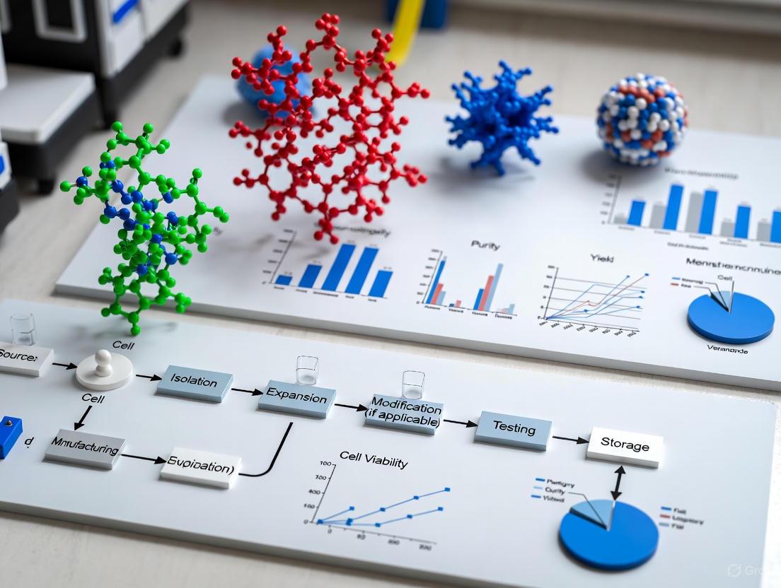
The autologous manufacturing workflow typically involves multiple interconnected stages that must be meticulously coordinated:
- Cell Collection: The process begins with leukapheresis to collect the patient's white blood cells, including T cells or other relevant cell populations [1]. The stability of this starting material is critical, with research indicating leukapheresis products remain stable for at least 25 hours at room temperature and 73 hours at cool temperatures (2-8°C) [4].
- Transportation: The collected cells are transported under controlled conditions to a specialized manufacturing facility, requiring stringent temperature monitoring and chain of identity preservation [2].
- Manufacturing and Manipulation: At the GMP facility, cells undergo various processes including selection, activation, genetic modification (e.g., using lentiviral vectors for CAR expression), and expansion [4]. This stage typically employs closed, automated systems to minimize contamination risks and maintain process consistency [1].
- Quality Control and Release: The final product undergoes rigorous testing for identity, purity, potency, viability, and sterility before release [5].
- Transport and Infusion: The finished product is transported back to the treatment center and administered to the patient [2].
Quantitative Analysis of Manufacturing Process Parameters
Recent studies have provided detailed quantitative analysis of autologous CAR-T cell manufacturing processes, demonstrating the consistency achievable with current technologies:
Table 1: Manufacturing Process Metrics for 19-FiCART Cell Production [4]
| Process Parameter | Initial Leukapheresis Product | CD4/CD8-Enriched Fraction (Day 0) | Final Drug Substance (Day 12) |
|---|---|---|---|
| CD3+ T-cell Frequency | 35.2% ± 5.5% | 79.3% ± 3.0% | 94.0% ± 1.3% |
| CD4+ T-cell Frequency | 74.7% ± 8.0% (of CD3+ cells) | 69.8% ± 4.5% (of CD3+ cells) | |
| CD8+ T-cell Frequency | 19.1% ± 6.7% (of CD3+ cells) | 27.1% ± 5.3% (of CD3+ cells) | |
| Cell Viability | >90% (maintained through process) | ||
| CAR Transduction Efficiency | 43.4% ± 2.9% | ||
| Total CAR+ T-cell Yield | >2 × 10^9 cells |
This data demonstrates the effectiveness of a 12-day semi-automated manufacturing process using CD4/CD8-positive cell enrichment and lentiviral transduction, consistently yielding sufficient quantities of highly viable CAR-T cells for clinical application [4].
Unique Challenges in Autologous Therapy Manufacturing
The autologous manufacturing paradigm presents distinct challenges that require specialized approaches to ensure successful clinical implementation.
Supply Chain Complexity
The patient-specific nature of autologous therapies creates an exceptionally complex supply chain. Each treatment batch must be meticulously tracked from collection through final infusion, maintaining strict chain of identity and chain of custody throughout the process [2]. The logistical challenges include managing patient schedules, maintaining cell viability during transport, and coordinating multiple handoffs between clinical and manufacturing sites. Any delays or temperature excursions during transportation can compromise product viability and efficacy, making robust cold chain management systems essential.
Scalability Constraints
Unlike traditional pharmaceuticals that benefit from economies of scale, autologous therapies face inherent scalability challenges. Each additional patient requires a completely separate manufacturing run with dedicated resources and oversight [2]. This "scale-out" rather than "scale-up" approach necessitates multiple parallel manufacturing platforms rather than simply increasing batch size. Manufacturers must therefore create modular and flexible facilities capable of handling multiple patient-specific batches simultaneously while maintaining strict segregation between products [2].
Cost and Accessibility Implications
The resource-intensive nature of personalized manufacturing contributes to high production costs. Each patient-specific batch requires multiple rounds of testing, processing, and transportation, all of which incur significant expenses [2]. These costs ultimately impact patient access to these transformative therapies, creating economic challenges for healthcare systems. Strategies to address cost challenges include process automation, standardization where possible, and partnerships with contract development and manufacturing organizations (CDMOs) to leverage specialized expertise and infrastructure [2] [3].
GMP Compliance Framework
Good Manufacturing Practice compliance is fundamental to ensuring the safety, quality, and efficacy of autologous cell therapies. The GMP framework for these advanced therapies encompasses several critical elements.
Facility Design and Environmental Control
GMP-compliant facilities for autologous cell therapy manufacturing require purpose-designed clean rooms classified according to air purity standards [5]. These facilities must implement strict environmental controls, including monitoring of particles, temperature, humidity, and pressure differentials. The design should ensure unidirectional flow of materials and staff to prevent cross-contamination between patient-specific batches [5]. Automated closed-system technologies have become increasingly important in minimizing process variability and reducing contamination risks while potentially enabling manufacturing in lower-grade cleanrooms [1] [6].
Quality Management Systems
A comprehensive Quality Management System is essential for autologous therapy manufacturing, incorporating quality control and quality assurance programs that cover all aspects from collection through final product release [5]. The QMS should address facilities design and maintenance, equipment qualification, materials specifications, process controls, and document management. For decentralized manufacturing models, a "Control Site" concept has been proposed to serve as the regulatory nexus, maintaining master files and ensuring consistency across multiple manufacturing locations [6].
Product Characterization and Release Testing
Each batch of autologous cell therapy must undergo rigorous characterization and release testing, including assessment of identity, purity, potency, viability, and sterility [5]. However, the relatively short shelf-life of many autologous products, particularly fresh formulations, may necessitate innovative approaches to testing, such as the use of rapid sterility methods or preliminary testing with final results available after product administration [4] [6].
Table 2: Essential Quality Attributes for Autologous Cell Therapy Release [4] [5]
| Quality Attribute | Testing Method | Acceptance Criteria |
|---|---|---|
| Identity | Flow cytometry for cell surface markers | Consistent with expected cell population |
| Purity | Flow cytometry, PCR | Minimum percentage of target cells; limits for impurities |
| Potency | In vitro cytotoxicity assays, cytokine production | Demonstration of biological activity |
| Viability | Trypan blue exclusion, flow cytometry | Typically >70-80% |
| Sterility | BacT/ALERT, PCR-based methods | No microbial contamination detected |
| Endotoxin | LAL test | Below established limits |
| Vector Copy Number | qPCR/digital PCR | Within predetermined range |
Emerging Manufacturing Models
The autologous therapy landscape is evolving with new manufacturing models that aim to address existing challenges in scalability and accessibility.
Centralized Manufacturing
The centralized manufacturing model, currently used for most commercial autologous therapies, involves production at a dedicated GMP facility that may serve multiple treatment centers [4]. This approach typically employs cryopreservation of both the starting material and final product to accommodate transportation times and quality control testing. While this model allows for concentrated expertise and infrastructure, it introduces logistical complexities and potential delays in vein-to-vein time.
Point-of-Care and Decentralized Manufacturing
Decentralized manufacturing represents an emerging paradigm that locates production facilities closer to patient care settings, potentially within or near hospital environments [6]. This approach can simplify logistics, reduce vein-to-vein time, and enable the use of fresh products without cryopreservation. Regulatory agencies including the MHRA, FDA, and EMA have begun establishing frameworks to support this model, introducing concepts such as the "manufacturer's license (Point of Care)" in the UK [6].
The decentralized model leverages automated, closed-system technologies to minimize process variability while maintaining GMP compliance in potentially less stringent environments [6]. This approach facilitates a network of manufacturing sites under central management, allowing for scale-out capacity and improved patient access.
Autologous Therapy Manufacturing Models
Automation and Technological Solutions
Automation plays a crucial role in addressing the unique challenges of autologous therapy manufacturing by enhancing consistency, reducing manual errors, and improving scalability.
Automated Manufacturing Platforms
Integrated automated systems such as the CliniMACS Prodigy (Miltenyi Biotec) and the Gibco CTS Rotea Counterflow Centrifugation System provide closed, standardized platforms for cell processing, genetic modification, and expansion [1] [4]. These systems minimize open manual processing steps, reduce contamination risks, and decrease operator-to-operator variability. The implementation of such technologies has been shown to consistently produce clinically sufficient quantities of CAR-T cells while maintaining high viability and functionality [4].
Digital Integration and Data Management
Digital integration tools, including manufacturing execution systems and electronic batch records, are becoming increasingly important for maintaining data integrity and regulatory compliance [1]. These systems enable real-time monitoring of critical process parameters, facilitate trend analysis, and support continuous process improvement. Software solutions like Gibco CTS Cellmation software improve record keeping and maintain data integrity throughout the manufacturing process [1].
The Scientist's Toolkit: Research Reagent Solutions
Successful autologous cell therapy manufacturing relies on a suite of specialized reagents and materials that ensure product quality, safety, and efficacy.
Table 3: Essential Research Reagents for Autologous Cell Therapy Manufacturing [1] [4] [5]
| Reagent/Material | Function | Application Examples |
|---|---|---|
| Lentiviral Vectors | Delivery of genetic material for cell modification | CAR gene transduction in T cells |
| Cell Separation Beads | Isolation of specific cell populations | CD4+/CD8+ T-cell selection from leukapheresis |
| Cell Culture Media | Support cell growth and maintenance | T-cell expansion during manufacturing |
| Activation Reagents | Stimulate cell proliferation and function | Anti-CD3/CD28 antibody-based T-cell activation |
| Cryopreservation Media | Maintain cell viability during frozen storage | Preservation of leukapheresis material or final product |
| Cell Analysis Reagents | Characterization of cell products | Flow cytometry antibodies for phenotyping |
| Sterility Testing Kits | Detection of microbial contamination | BacT/ALERT culture bottles for sterility testing |
Experimental Protocols
Robust, standardized protocols are essential for ensuring consistency and quality throughout the autologous therapy manufacturing process.
Leukapheresis Product Stability Assessment Protocol
Understanding the stability of the starting material is critical for defining hold times and conditions between cell collection and manufacturing initiation.
Objective: To determine the optimal storage conditions and maximum hold time for leukapheresis products before manufacturing.
Materials:
- Fresh leukapheresis products from healthy donors
- Temperature-controlled storage units (2-8°C and 15-25°C)
- Flow cytometer with appropriate antibodies (CD45, CD3, CD4, CD8, CD14, CD19, CD56)
- Cell viability staining solution (e.g., 7-AAD)
- Automated cell counter
Methodology:
- Analyze leukapheresis products immediately after collection (T0) for cellular composition and viability.
- Aliquot products and store under two conditions: cool temperature (CT: 2-8°C) and room temperature (RT: 15-25°C).
- Sample products at 25, 49, 73, and 121 hours for comprehensive analysis.
- At each timepoint, assess:
- White blood cell counts
- Cellular composition by flow cytometry
- Cell viability using viability dyes
- Appearance and color of the product
- Compare results to T0 values to determine stability limits.
Acceptance Criteria: Products are considered stable when maintaining ≥90% viability of critical cell populations (CD45+ leukocytes, CD3+ T cells) and consistent cellular composition without significant deterioration [4].
CAR-T Cell Manufacturing Protocol
This protocol outlines a semi-automated process for manufacturing autologous CAR-T cells using a closed system.
Objective: To consistently manufacture CAR-T cells meeting predefined quality attributes for clinical use.
Materials:
- Leukapheresis product
- Closed automated cell processing system (e.g., CliniMACS Prodigy)
- Cell separation reagents (e.g., CD4/CD8 microbeads)
- Lentiviral vector encoding CAR construct
- Cell culture media and supplements
- Activation reagents (e.g., anti-CD3/CD28 antibodies)
- Bioreactor or culture bags
Methodology:
- Cell Selection: Perform CD4/CD8-positive selection from leukapheresis product using magnetic separation technology.
- Cell Activation: Stimulate selected T cells with anti-CD3/CD28 antibodies to promote activation and proliferation.
- Transduction: Transduce activated T cells with lentiviral vector at appropriate multiplicity of infection (MOI).
- Expansion: Culture cells in appropriate media for approximately 12 days, monitoring cell density, viability, and nutrient levels.
- Harvesting: Collect cells when target expansion and transduction efficiency are achieved.
- Formulation: Wash and resuspend cells in final formulation buffer.
- Quality Control: Perform comprehensive testing including cell count, viability, identity, purity, potency, and sterility.
Critical Process Parameters:
- Cell density during culture
- Vector copy number
- Transduction efficiency
- Cellular composition (CD4/CD8 ratio)
- Metabolic parameters (glucose, lactate)
CAR-T Cell Manufacturing Workflow
Regulatory Considerations
The regulatory landscape for autologous cell therapies continues to evolve as these advanced therapies demonstrate clinical efficacy across a growing range of indications.
Current Regulatory Framework
Autologous cell therapies are regulated as Advanced Therapy Medicinal Products in Europe and as cell and gene therapies in the United States [5]. The regulatory framework emphasizes a risk-based approach, with requirements tailored to the specific characteristics of each product. Key considerations include the level of manipulation, cell origin, differentiation potential, and route of administration [5]. Regulatory agencies have established expedited approval pathways for promising cell and gene therapies, warranting early development of commercial strategies during clinical development [2].
Emerging Regulatory Pathways for Novel Manufacturing Models
Regulatory agencies are developing new frameworks to accommodate evolving manufacturing paradigms, particularly decentralized and point-of-care manufacturing. The UK's MHRA has introduced two new licenses: "manufacturer's license (modular manufacturing)" and "manufacturer's license (Point of Care)" to address the unique challenges of these approaches [6]. Similarly, the FDA has initiated the Framework for Regulatory Advanced Manufacturing Evaluation through its Emerging Technology Program, which includes distributed manufacturing as a platform for POCare manufacturing [6].
Autologous cell therapy represents a transformative approach to treating complex diseases, distinguished by its patient-specific manufacturing paradigm. This unique model presents significant challenges in supply chain management, scalability, cost, and regulatory compliance that differ fundamentally from traditional pharmaceutical manufacturing. The successful implementation of autologous therapies requires robust GMP frameworks, advanced technological solutions including automation and closed systems, and innovative manufacturing models such as decentralized production. As the field continues to evolve with increasing regulatory approvals and expanding clinical applications, addressing these challenges through standardized processes, technological innovation, and adaptable regulatory frameworks will be essential to realizing the full potential of autologous cell therapies for patients worldwide.
Good Manufacturing Practice (GMP) constitutes a system of regulations, guidelines, and best practices designed to ensure the quality, safety, and efficacy of pharmaceutical products [7]. In autologous cell therapy, where a patient's own cells are manufactured into a personalized treatment, adherence to GMP is paramount. These therapies, such as CAR T-cell treatments, involve collecting T cells from the patient, genetically modifying them, expanding the population, and reinfusing them [1]. This individualized nature introduces significant complexity and variability into the manufacturing process, making a robust quality framework not just a regulatory requirement but a fundamental patient safety imperative [5] [8]. The core objectives of GMP in this field are the consistent production of products that are safe for patient use, clearly identified as the intended product, pure from contaminants, and potent to achieve the desired therapeutic effect.
Core GMP Principles and Their Application
The implementation of GMP rests on several interconnected pillars. A comprehensive Quality Management System (QMS) forms the foundation, encompassing all policies, procedures, and records needed to ensure product quality [7]. Within the QMS, a risk-based approach is essential for identifying and controlling critical variables [5]. Furthermore, Quality by Design (QbD) is a scientific, risk-based framework that builds quality into the product from the outset by linking critical process parameters to critical quality attributes [9]. For autologous therapies, this means understanding how variability in starting material and manufacturing conditions impacts the final product's safety and efficacy.
The practical application of GMP is realized through several key components:
- Personnel Training and Hygiene: Personnel must receive rigorous training in GMP, hygiene, and specific operational procedures to prevent contamination and errors [7].
- Facility and Equipment Management: Facilities must be purpose-designed with environmental controls, such as classified cleanrooms with HEPA filtration, to prevent microbiological and cross-contamination [5]. Equipment must be calibrated, validated, and maintained [7].
- Raw Material and Supplier Management: All raw materials, including cytokines, culture media, and viral vectors, must be sourced from qualified suppliers and tested to meet strict specifications [7].
- Production Process Control: Manufacturing processes must be well-documented, validated, and monitored. This includes controlling critical process parameters and performing in-process quality control checks [7].
- Documentation and Record Keeping: Comprehensive documentation, including Standard Operating Procedures and Batch Manufacturing Records, provides evidence that all steps were performed correctly [7].
Analytical Methods for Assessing Critical Quality Attributes
Rigorous testing is required throughout the manufacturing process to verify that the product meets pre-defined Critical Quality Attributes (CQAs). The table below summarizes the key analytical methods used to assess the four core principles.
Table 1: Analytical Methods for Assessing Critical Quality Attributes in Cell Therapies
| Quality Attribute | Testing Objective | Key Analytical Methods | Application in Release Testing |
|---|---|---|---|
| Safety | To ensure freedom from microbiological contaminants and replication-competent viruses. | Sterility testing, Mycoplasma testing, Endotoxin (LAL) testing, Replication Competent Virus (RCV) assays [5]. | Mandatory for lot release. For short shelf-life products, rapid microbial methods may be employed [5]. |
| Identity | To confirm the product is the intended cell type and has the correct genetic modification. | Flow cytometry (surface markers), DNA sequencing (genetic modification), PCR [8]. | Mandatory for lot release to confirm the correct cell population and genetic construct are present. |
| Purity | To ensure the product is free from unwanted cell types and process residuals. | Flow cytometry (undesired cell populations), Residual DNA/Protein assays, Magnetic bead removal validation [8]. | Mandatory for lot release. Sets acceptable limits for impurities. |
| Potency | To measure the biological activity or therapeutic function of the product. | In vitro functional assays (e.g., cytokine release, cytotoxicity, target cell killing), Cell viability assays (e.g., trypan blue exclusion) [8] [10]. | Mandatory for lot release. Must be correlated to the proposed mechanism of action. |
Experimental Protocols for Key Assays
Protocol 1: Flow Cytometry for Cell Identity and Purity
This protocol is used to identify specific cell populations (e.g., CD3+ T cells) and quantify purity and impurities.
- Sample Preparation: Obtain a representative sample of the cell therapy product and wash with FACS buffer (e.g., PBS with 1% BSA). Determine total cell count and viability.
- Staining: Aliquot cells into staining tubes. Add fluorochrome-conjugated antibodies against specific surface markers (e.g., anti-CD3, CD4, CD8, CD19) and appropriate isotype controls. Incubate for 20-30 minutes in the dark at 4°C.
- Washing and Fixation: Wash cells twice with FACS buffer to remove unbound antibody. Resuspend in a fixative solution such as 1-4% paraformaldehyde if analysis is not immediate.
- Data Acquisition: Acquire data on a flow cytometer, collecting a minimum of 10,000 events per sample.
- Data Analysis: Use flow cytometry software to gate on live cells based on forward and side scatter. Analyze the percentage of positive cells for each marker to confirm identity (e.g., >95% CD3+ for a T-cell product) and assess purity by quantifying unwanted cell populations.
Protocol 2: Cytotoxicity Assay for Potency
This in vitro functional assay measures the ability of CAR-T cells to kill target tumor cells.
- Target Cell Preparation: Culture target cells (e.g., a tumor cell line expressing the target antigen) and label them with a fluorescent dye, such as CFSE.
- Effector Cell Preparation: Serially dilute the CAR-T cell product (effector cells) to create various Effector:Target (E:T) ratios.
- Co-culture: Co-culture the target cells with effector cells in a multi-well plate for a specified period (e.g., 4-24 hours). Include target cells alone (spontaneous release control) and target cells with lysis buffer (maximum release control).
- Detection of Cell Death: Add a viability dye, such as propidium iodide (PI), to each well. Alternatively, measure the release of lactate dehydrogenase (LDH) or other intracellular components into the supernatant.
- Data Acquisition and Analysis: Acquire data using a flow cytometer (for CFSE/PI) or a plate reader (for LDH). Calculate the specific cytotoxicity percentage at each E:T ratio. A reference standard should be included to ensure the assay meets pre-defined acceptance criteria.
Table 2: The Scientist's Toolkit: Key Research Reagent Solutions
| Reagent / Material | Function in Manufacturing & Quality Control |
|---|---|
| GMP-Grade Cell Culture Media | Provides nutrients and growth factors for cell expansion while ensuring freedom from contaminants. Formulations are optimized for specific cell types like T cells or MSCs [8]. |
| Cytokines (e.g., IL-2, IL-7, IL-15) | Used during cell activation and expansion to promote growth, survival, and influence final cell phenotype and potency [8]. |
| Magnetic-Activated Cell Sorting (MACS) Reagents | GMP-compliant antibodies and beads for the closed, automated isolation or removal of specific cell populations, critical for achieving product purity [1]. |
| Fluorochrome-Conjugated Antibodies | Essential reagents for flow cytometry-based testing of cell identity, purity, and characterization of cell subpopulations [8]. |
| Vector Virus (e.g., Lentivirus) | The vehicle for stable genetic modification of cells (e.g., introducing a CAR gene). Must be produced under GMP and tested for safety, identity, and potency [8]. |
| Cryoprotectants (e.g., DMSO) | Used in the final formulation to protect cell viability and functionality during cryopreservation for storage and transport [8]. |
Implementing a QbD Framework for Enhanced Control
Quality by Design (QbD) moves quality control from a traditional testing-focused model (Quality by Testing, or QbT) to a proactive, science-based approach where quality is built into the product and process [9] [11]. For autologous cell therapies, which face inherent variability in starting materials, QbD is particularly valuable for defining a flexible yet controlled design space.
The QbD workflow begins by defining the Quality Target Product Profile (QTPP), which describes the desired characteristics of the therapy to ensure safety and efficacy [9]. From the QTPP, Critical Quality Attributes (CQAs) are identified as physical, chemical, biological, or microbiological properties that must be controlled within an appropriate limit [9]. The relationship between process and product is then systematically studied to determine how Critical Process Parameters (CPPs) and Material Attributes (MAs) impact the CQAs. The multidimensional combination of these input variables that have been demonstrated to assure quality constitutes the Design Space [9]. A holistic Control Strategy is then established to ensure CPPs and MAs remain within the design space, which may include process controls, real-time monitoring, and final product testing.
Diagram 1: QbD Workflow for Cell Therapy
The Role of Automation and Digitalization in GMP Compliance
Heavy reliance on manual operations in cell therapy manufacturing introduces risks of human error, contamination, and significant documentation burdens [11]. Automation and digitalization are therefore critical enablers of robust GMP compliance. Automated, closed-system platforms like the Gibco CTS Rotea Counterflow Centrifugation System and the CTS Dynacellect Magnetic Separation System minimize human intervention in unit operations such as cell wash, concentration, and isolation, thereby reducing contamination risks and improving process consistency [1].
Digital integration through software such as CTS Cellmation software enhances data integrity and record-keeping [1]. Replacing paper-based systems with electronic batch records minimizes transcription errors and provides real-time data for process monitoring and control [11] [7]. This digital backbone is fundamental for implementing a QbD approach, as it allows for the aggregation and analysis of vast datasets from multiple patients to better understand the process design space and make data-driven decisions for continuous process verification and improvement [9] [11].
Diagram 2: Automation and Digitalization Benefits
Autologous cell therapy represents a paradigm shift in personalized medicine, using a patient's own cells to treat serious conditions like blood cancers [1]. This personalized approach reduces the risk of immune rejection and infection, eliminating the need for donor matching and immunosuppressive drugs [1]. However, the manufacturing process is exceptionally complex, involving multiple critical steps from cell collection to final infusion, each requiring stringent control to ensure product quality, safety, and efficacy. This application note provides a detailed examination of the entire autologous workflow within the context of Good Manufacturing Practice (GMP) compliance, offering structured data, experimental protocols, and visualizations to support researchers and drug development professionals in optimizing their manufacturing processes.
Autologous CAR-T Cell Therapy Workflow
The following diagram illustrates the complete journey of autologous CAR-T cell therapy from the patient to the manufacturing facility and back to the patient, highlighting the key stages and their interconnectedness.
Quantitative Process Data and Performance Metrics
Effective process control requires monitoring of critical performance indicators. The following table summarizes key quantitative metrics for major unit operations in the autologous workflow [1] [12].
Table 1: Key Performance Indicators for Autologous Workflow Unit Operations
| Process Step | Key Performance Indicators | Target Values | Impact of Deviation |
|---|---|---|---|
| Apheresis to Infusion | Total time from apheresis to infusion [12] | Target: Minimize delay (Median: 66 days in study) [12] | Delays ≥66 days associated with inferior PFS (aHR 3.13) and OS (aHR 2.53) [12] |
| Cell Isolation | Cell recovery rate, Cell viability, Purity of target cell population [1] | High recovery & viability (>90%), High purity [1] | Low yield limits starting material; low viability/purity impacts downstream steps [8] |
| Cell Activation & Expansion | Fold expansion, Population doubling time, Final cell viability, Phenotype characterization [8] | Clinical-relevant cell numbers, Maintained viability, Desired phenotype [8] | Inadequate expansion requires process repeat; altered phenotype affects therapeutic function [8] |
| Genetic Modification | Transduction efficiency, Vector copy number, Cell viability post-transduction [1] | Optimized for the specific vector and protocol [1] | Low efficiency reduces product potency; high copy number raises safety concerns [1] |
| Final Formulation | Total viable cells, Dose potency, Purity (e.g., residual beads), Sterility [1] [8] | Meets lot release specifications [1] [8] | Failure to meet specifications results in batch rejection and product loss [1] [8] |
Detailed Experimental Protocols for Key Unit Operations
Protocol: Closed-System Cell Isolation using Counterflow Centrifugation
Principle: Based on cell size and density, this protocol uses the Gibco CTS Rotea Counterflow Centrifugation System for the isolation of peripheral blood mononuclear cells (PBMCs) from leukapheresis material in a closed, automated manner [1].
Materials:
- Leukapheresis product
- Gibco CTS Rotea Counterflow Centrifugation System [1]
- CTS Rotea Single-Use Kit (sterile, closed system) [1]
- DPBS without calcium and magnesium
- Centrifuge tubes
Methodology:
- System Setup: Prime the CTS Rotea system according to the manufacturer's instructions. Install the sterile, single-use kit, ensuring all connections are secure [1].
- Sample Preparation: Dilute the leukapheresis product 1:2 with DPBS to reduce viscosity and improve separation efficiency.
- Loading: Aseptically connect the leukapheresis bag to the system's input line.
- Process Execution: Select the pre-optimized "PBMC Separation" protocol on the instrument interface. The system automatically processes the sample, separating PBMCs from red blood cells, granulocytes, and platelets.
- Collection: The target PBMC fraction is collected into a designated output bag. The system performs an integrated wash and concentration step.
- Sampling: Aseptically sample the product for in-process testing (cell count, viability).
- Product Transfer: The final, concentrated PBMC product is aseptically transferred or cryopreserved for the next process step.
Protocol: Automated Magnetic Cell Selection and Bead Removal
Principle: This protocol uses the Gibco CTS Dynacellect Magnetic Separation System for the closed, automated isolation of T cells from a PBMC population and subsequent removal of magnetic activation beads [1].
Materials:
- PBMC sample
- Gibco CTS Dynacellect Magnetic Separation System [1]
- CTS Dynabeads CD3/CD28 or other relevant selection beads
- Appropriate CTS Dynacellect Single-Use Kit [1]
- Cell culture media
Methodology: Part A: Cell Selection
- Incubation: Incubate the PBMC sample with CTS Dynabeads CD3/CD28 at a recommended bead-to-cell ratio for 30 minutes at room temperature with gentle agitation.
- System Setup: Load the Dynacellect system with the appropriate single-use kit and place the bead-bound cell sample at the input.
- Isolation: Run the "Cell Isolation" protocol. The system automatically captures bead-bound T cells in the magnetic field while unbound cells are washed to waste.
- Collection (for activation): The isolated T cells, now bound to activation beads, are collected for direct progression to the activation and expansion culture.
Part B: Bead Removal (De-beading)
- Post-expansion: After the expansion phase, the cell culture (now containing cells and activation beads) is loaded onto the Dynacellect system configured for "De-beading."
- Separation: The system automatically separates the cells from the magnetic beads. Beads are retained in the magnetic cartridge, and the purified, bead-free T cells are collected in the output bag.
- Quality Control: Verify bead removal via microscopy.
Protocol: Large-Scale Non-Viral Transfection using Electroporation
Principle: This protocol describes the use of the Gibco CTS Xenon Electroporation System for the closed, modular, large-scale electroporation of T cells to introduce CAR transgenes via non-viral methods [1].
Materials:
- Activated T cells (bead-free)
- Gibco CTS Xenon Electroporation System [1]
- CTS Xenon Single-Use Electroporation Cuvette [1]
- DNA plasmid or RNA encoding the CAR construct
- Electroporation buffer
Methodology:
- Cell Preparation: Wash and resuspend the T cell pellet in the appropriate electroporation buffer at a specified concentration (e.g., 1-2 x 10^7 cells/mL).
- Nucleic Acid Preparation: Dilute the DNA or RNA to the desired working concentration in the same electroporation buffer.
- Mixing: Combine the cell suspension and nucleic acid solution in the provided single-use electroporation cuvette, ensuring homogeneity.
- System Setup: Load the cuvette into the pre-chilled module of the CTS Xenon system.
- Electroporation: Select and run the pre-validated electroporation protocol (e.g., specific voltage, pulse length, number of pulses). The process is automated and closed.
- Recovery: Immediately after pulsing, transfer the cuvette to a post-electroporation recovery rack. Add pre-warmed culture media to the cuvette and incubate at 37°C for a short, specified duration to allow membrane resealing.
- Transfer to Expansion: Transfer the electroporated cells from the cuvette into fresh, pre-warmed expansion media for culture.
Critical Quality Control Points and GMP Compliance
A robust Quality Management System (QMS) is essential for GMP compliance. Key performance indicators like deviation rates are strongly correlated with manufacturing performance, including right-first-time rate [13] [14]. The following diagram maps the critical quality control points and their integration within the QMS.
Table 2: Critical Quality Attributes and Testing Methods
| Quality Attribute Category | Specific Test | Analytical Method | GMP Compliance Consideration |
|---|---|---|---|
| Identity | CAR expression detection, Cell surface marker profile | Flow cytometry [8] | Method must be validated for specificity and accuracy. |
| Potency | Cytotoxicity assay, Cytokine secretion profile | Co-culture with target cells, ELISA/Luminex [8] | Potency assay is a critical release test and must be quantitative and reproducible. |
| Purity | Viability, Residual bead count, Endotoxin level | Trypan blue/exclusion dye, Microscopy/flow cytometry, LAL test [1] [8] | Specifications for impurities must be set and justified. |
| Safety | Sterility, Mycoplasma, Replication-competent virus | BacT/ALERT, PCR, Culture assays [1] | Tests are required for final product release. Use of rapid microbiological methods is encouraged. |
| Genetic Mod. Characterization | Vector copy number (VCN), Transgene integration site analysis | qPCR/digital PCR, Next-generation sequencing [1] | VCN is a key safety parameter with predefined acceptance criteria. |
The Scientist's Toolkit: Essential Research Reagent Solutions
Table 3: Key Reagents and Platforms for GMP-Compliant Autologous Therapy Manufacturing
| Reagent / Solution / Platform | Function | GMP-Compliant Feature |
|---|---|---|
| Gibco CTS Rotea System [1] | Closed system for cell processing, wash, and concentration. | Closed, automated processing reduces contamination risk and operator variability [1]. |
| Gibco CTS Dynacellect System [1] | Closed, automated system for magnetic cell isolation and bead removal. | Sterile, single-use kits and automated protocols ensure process consistency and scalability [1]. |
| Gibco CTS Xenon System [1] | Large-scale, closed-system electroporator for non-viral cell engineering. | Modular, GMP-compliant system designed for clinical and commercial manufacturing [1]. |
| CTS Cellmation Software | Provides digital integration for automated platforms. | Supports CFR 21 Part 11 compliance for data integrity and electronic records [1]. |
| CTS AIM-V Medium / CTS Immune Cell Serum-Free Medium | Serum-free culture media for T cell expansion. | Xeno-free, formulated with high-quality raw materials, and designed for cell therapy applications [1]. |
| CTS Dynabeads CD3/CD28 | Magnetic beads for T cell activation and expansion. | Consistent, defined surface area for stimulation; available in GMP-manufactured format [1]. |
| CryoStor CS10 or similar | cGMP-manufactured cryopreservation medium. | Formulated to minimize cell damage during freezing and thawing; supports regulatory filing. |
The field of autologous cell therapies, often termed 'living drugs,' represents a frontier in modern medicine, offering transformative potential for conditions ranging from hematological malignancies to solid tumors and rare genetic disorders. Unlike traditional pharmaceuticals, these therapies are dynamic, patient-specific products manufactured from a patient's own cells, creating unique regulatory challenges. The regulatory landscape is rapidly evolving to balance accelerated patient access with rigorous safety and efficacy standards. Recent updates from the U.S. Food and Drug Administration (FDA) and international regulatory bodies reflect a significant shift toward flexible, evidence-based frameworks designed to address the distinct challenges of autologous cell therapy manufacturing while maintaining stringent Good Manufacturing Practice (GMP) compliance [15] [16].
This application note examines the current regulatory environment, focusing on the September 2025 FDA draft guidance on expedited programs and analogous international frameworks. Within the context of GMP compliance for autologous cell therapy manufacturing research, we detail practical protocols for navigating these complex regulatory pathways, ensuring that researchers and drug development professionals can effectively align their processes with contemporary standards.
Recent FDA Regulatory Guidance Updates
September 2025 Draft Guidance: Expedited Programs for Regenerative Medicine Therapies
In September 2025, the FDA released a pivotal draft guidance, "Expedited Programs for Regenerative Medicine Therapies for Serious Conditions," which, upon finalization, will supersede the February 2019 version. This document outlines the agency's current thinking on expedited development and review pathways for regenerative medicine products, including autologous cell therapies [17] [16].
Key Provisions of the Draft Guidance
- RMAT Designation: The guidance reiterates criteria and processes for obtaining the Regenerative Medicine Advanced Therapy (RMAT) designation under Section 506(g) of the FD&C Act. A product intended to treat a serious condition may qualify if preliminary clinical evidence indicates the potential to address unmet medical needs [17]. As of September 2025, the FDA has received nearly 370 RMAT designation requests and granted 184, with 13 of these products ultimately achieving marketing approval as of June 2025 [16].
- Flexible Trial Designs: The FDA encourages innovative clinical trial designs for rare diseases, including those utilizing natural history data as historical controls (provided populations are adequately matched) and trials where multiple clinical sites share a common manufacturing protocol and combined data to support individual Biologics License Applications (BLAs) [16].
- Enhanced Safety Monitoring: Recognizing that regenerative therapies may raise unique long-term safety considerations, the draft guidance recommends that clinical trial monitoring plans include both short-term and long-term safety assessments. It also encourages the use of digital health technologies to collect real-world safety information [16].
- CMC Considerations: The guidance emphasizes that an expedited review designation does not reduce the chemistry, manufacturing, and controls (CMC) information required to assure product quality. Sponsors are advised to pursue rapid CMC development programs to align with accelerated clinical timelines. A particular focus is placed on manufacturing changes; if comparability cannot be established with the pre-change product, the RMAT designation may be jeopardized [16].
- Use of Real-World Evidence (RWE): The draft guidance explicitly notes that real-world evidence can be utilized to support an accelerated approval application, broadening the potential data sources for demonstrating product effectiveness [16].
Complementary FDA Draft Guidances
Concurrently in September 2025, the FDA released two other critical draft guidances that form a cohesive modernized framework for cell and gene therapy development [15] [10]:
- Postapproval Methods to Capture Safety and Efficacy Data for Cell and Gene Therapy Products: This guidance provides recommendations on using real-world data collection to ensure long-term safety and effectiveness without delaying initial approvals [15] [10].
- Innovative Designs for Clinical Trials of Cellular and Gene Therapy Products in Small Populations: This document encourages the use of adaptive, Bayesian, and externally controlled trial designs to generate robust evidence with fewer patients, which is particularly relevant for rare diseases [15] [10].
Table 1: Key FDA Expedited Programs for Autologous Cell Therapies
| Program | Legal Authority | Key Features | Relevance to Autologous Therapies |
|---|---|---|---|
| RMAT Designation | Section 506(g) of FD&C Act | Intensive FDA guidance, rolling review, potential for accelerated approval based on surrogate endpoints [17] [16] | Streamlines development for serious conditions with unmet needs; accommodates manufacturing complexities |
| Fast Track | Section 506(b) of FD&C Act | Early interactions, rolling BLA submission [15] | Facilitates ongoing dialogue on CMC and clinical development challenges |
| Breakthrough Therapy | Section 506(c) of FD&C Act | Intensive guidance on efficient trial design, organizational commitment [15] | Helps optimize development programs for transformative autologous products |
Other Notable FDA Regulatory Activities
Beyond the September 2025 guidances, the FDA has undertaken several other significant actions:
- Elimination of REMS for Autologous CAR-T: In June 2025, the FDA eliminated the Risk Evaluation and Mitigation Strategies (REMS) for all approved BCMA- and CD19-directed autologous CAR-T cell immunotherapies. This removed requirements for specialized hospital certification and on-site, immediate access to tocilizumab, simplifying treatment administration and expanding patient access [18].
- Gene Therapies Global Pilot Program (CoGenT): Modeled after Project Orbis for oncology, this pilot initiative explores concurrent, collaborative regulatory reviews of gene therapy applications with international partners like the European Medicines Agency (EMA), aiming to increase harmonization and reduce approval delays [15].
International Regulatory Frameworks
The global regulatory landscape for cell therapies is also undergoing significant modernization to address the unique challenges of these products.
United Kingdom: MHRA's Progressive Stance
The UK's Medicines and Healthcare products Regulatory Agency (MHRA) has implemented forward-looking regulations:
- Point of Care and Modular Manufacture: Effective July 23, 2025, new regulations introduce frameworks for manufacturing medicines closer to patients. This is particularly impactful for autologous therapies like CAR-T, which traditionally involve centralized manufacturing far from the patient, potentially causing treatment delays. The new pathway allows hospitals and clinics to perform manufacturing steps on-site or locally [18].
- Performance and Innovation: The MHRA has cleared all statutory backlogs by March 2025 and consistently meets statutory targets for clinical trials. It has also piloted a world-first "AI Airlock" to safely develop artificial intelligence in medical devices [18].
European Union: EMA's Evolving Framework
The European Commission, through the EMA, is advancing its regulatory framework:
- Updated GMP Guidelines: A stakeholder consultation is ongoing until October 7, 2025, on revised Good Manufacturing Practice (GMP) guidelines (Chapter 4, Annex 11, and new Annex 22). These updates address the rapid advancement of digital technologies and AI systems in pharmaceutical manufacturing, aiming to support innovation while ensuring regulatory harmonization [18].
Table 2: Global Regulatory Initiatives for Advanced Therapies
| Region/Agency | Initiative | Status/Date | Key Focus |
|---|---|---|---|
| FDA (USA) | RMAT Designation [17] | Active (Updated Draft Guidance 9/2025) | Expedited development and review for regenerative medicines |
| FDA (USA) | Gene Therapies Global Pilot (CoGenT) [15] | Pilot Launched 2024 | Concurrent international collaborative reviews |
| MHRA (UK) | Point of Care & Modular Manufacture [18] | Effective 7/23/2025 | Enables local manufacturing of personalized therapies |
| EMA (EU) | Updated GMP Guidelines for AI/Digital [18] | Consultation until 10/7/2025 | Modernizing GMP for digital tech and AI in manufacturing |
GMP Compliance in Autologous Therapy Manufacturing
Core GMP Principles for 'Living Drugs'
GMP compliance is foundational to ensuring the safety, identity, purity, and potency of autologous cell therapies. The inherent variability of the starting material (patient cells) and the complex, multi-step manufacturing process necessitate a robust, well-documented quality system [8]. Key principles include:
- Process Consistency: Despite patient-specific starting material, the manufacturing process itself must be highly standardized and controlled to minimize batch-to-batch variability [1] [8].
- Prevention of Contamination and Cross-Contamination: Closed, automated systems and stringent aseptic techniques are critical, especially given the limited batch size (often a single patient) and the inability to terminally sterilize the living product [1].
- Comprehensive Traceability: From cell collection through apheresis to final infusion, strict chain of identity and chain of custody procedures must be maintained to prevent misidentification [8].
- Process Validation: Even for patient-specific products, the manufacturing process must be validated to demonstrate it consistently produces a product meeting its predetermined quality attributes [19].
The Role of Automation in GMP Compliance
Automation is increasingly critical for achieving GMP compliance in autologous therapy manufacturing by reducing manual errors, enhancing scalability, and improving process consistency [1]. Automated closed systems minimize contamination risks and reduce the need for extensive cleanroom environments.
Table 3: Example Automated Solutions for GMP-Compliant Manufacturing
| System/Technology | Function | GMP Compliance Benefit |
|---|---|---|
| Rotea Counterflow Centrifugation System [1] | Closed cell processing, washing, concentration | Reduces open processing steps, ensures high cell recovery and viability |
| Dynacellect Magnetic Separation System [1] | Automated cell isolation and bead removal | Provides high cell purity and recovery; uses sterile single-use kits |
| Xenon Electroporation System [1] | Modular, large-scale electroporation for non-viral transfection | Closed, GMP-compliant system for critical genetic modification step |
| CTS Cellmation Software [1] | Digital integration and data management | Improves record keeping, maintains data integrity for 21 CFR Part 11 compliance |
Experimental Protocols for Regulatory Compliance
Protocol: Implementing a Risk-Based Approach for Anticipated Manufacturing Changes
Objective: To proactively assess and plan for potential manufacturing changes during development to avoid jeopardizing RMAT designation or BLA approval by ensuring product comparability [16].
Methodology:
- Risk Assessment Initiation
- Convene a cross-functional team (CMC, Quality, Regulatory, Clinical).
- Identify all potential manufacturing changes anticipated from Phase 3 through commercial scale-up (e.g., raw material supplier changes, equipment scale-up, process parameter optimization).
Comparative Analytics Strategy Development
- Define a tiered approach for comparability studies:
- Tier 1 (High Risk): Changes affecting critical quality attributes (CQAs) like identity, potency, or safety. Requires extensive in vitro and in vivo functional assays, genomic stability assessment, and potentially additional clinical data.
- Tier 2 (Medium Risk): Changes with potential impact on CQAs. Requires a targeted set of analytical tests focused on specific attributes.
- Tier 3 (Low Risk): Changes with minimal perceived impact. May only require documentation and verification of process performance.
- Define a tiered approach for comparability studies:
Early Regulatory Engagement
- Schedule a meeting with FDA (e.g., Type B) to discuss the risk assessment and proposed comparability protocol.
- Seek agreement on the level of data required to demonstrate comparability for each anticipated change.
Protocol Execution and Documentation
- Execute comparability studies according to the pre-defined protocol.
- Document all data and assessments in a comprehensive report for regulatory submission.
Protocol: Integrating Real-World Evidence (RWE) for Post-Approval Safety Monitoring
Objective: To establish a systematic process for collecting and analyzing real-world data to meet post-approval safety monitoring requirements, as encouraged by the September 2025 FDA draft guidance [15] [16].
Methodology:
- Data Source Identification and Validation
- Identify potential real-world data sources (e.g., electronic health records, patient registries, claims databases, digital health technology outputs).
- Assess the suitability and quality of each source based on FDA guidelines (e.g., data completeness, accuracy, timeliness, and verifiability).
Study Protocol Development
- Define clear study objectives and hypotheses.
- Specify patient population, including inclusion/exclusion criteria.
- Identify key safety outcomes of interest (e.g., cytokine release syndrome, neurotoxicity, long-term immunosuppression, secondary malignancies).
- Develop a statistical analysis plan detailing methods to control for confounding and bias.
Data Collection and Management
- Implement a secure, HIPAA-compliant data infrastructure.
- Establish standardized data mapping and transformation procedures.
- Ensure robust patient privacy protections and data governance.
Analysis and Reporting
- Conduct analyses according to the pre-specified statistical plan.
- Prepare periodic safety reports for regulatory submission (e.g., Periodic Benefit-Risk Evaluation Report - PBRER).
- Use findings to update product labeling and inform risk management plans.
The Scientist's Toolkit: Essential Research Reagent Solutions
Table 4: Key Reagents for GMP-Compliant Autologous Cell Therapy Manufacturing
| Reagent/Category | Function | GMP-Compliance Consideration |
|---|---|---|
| Cell Culture Media | Supports cell growth, activation, and expansion during manufacturing | Use of GMP-manufactured, xeno-free formulations is critical for regulatory approval and patient safety [1] |
| Cell Separation Kits | Isolation of target cell populations (e.g., T-cells) from apheresis material | Must be closed-system, sterile, single-use kits to ensure purity and prevent contamination [1] [8] |
| Activation Reagents | Stimulates T-cell proliferation (e.g., anti-CD3/CD28 antibodies) | Sourcing from qualified GMP suppliers ensures consistency and reduces batch-to-batch variability [8] |
| Genetic Modification Tools | Introduces therapeutic genes (e.g., CAR constructs) via viral vectors or electroporation | Viral vectors require extensive safety testing; non-viral systems must use GMP-grade reagents [1] [8] |
| Cryopreservation Media | Preserves final drug product for storage and transport | Formulations with defined, animal-origin-free cryoprotectants (e.g., DMSO) are essential [8] |
The regulatory landscape for 'living drugs' is maturing rapidly, with the FDA's September 2025 draft guidance on expedited programs representing a significant step toward a more flexible, efficient, and evidence-based framework. Concurrently, international regulators like the MHRA and EMA are advancing their own innovative pathways, particularly for decentralized manufacturing and digital integration. For researchers and developers, success in this environment hinges on a proactive, strategic approach that integrates regulatory considerations into the earliest stages of process development. This includes engaging with agencies early, employing risk-based approaches to manage manufacturing evolution, leveraging real-world evidence, and embracing automation and digital tools to ensure robust GMP compliance. By aligning development programs with these modernized frameworks, the field can accelerate the delivery of safe, effective, and transformative autologous cell therapies to patients in need.
Implementing GMP-Compliant Processes and Automated Solutions
Designing GMP-Compliant Workflows for Unit Operations
The manufacturing of autologous cell therapies represents a paradigm shift in biopharmaceutical production, moving from traditional large-batch processing to patient-specific, individualized workflows. This paradigm requires a fundamentally different approach to Good Manufacturing Practice (GMP). Designing GMP-compliant workflows for the unit operations involved—from cell collection to final infusion—is critical for ensuring the safety, identity, purity, potency, and consistency of these living medicines [20]. Unlike allogeneic therapies, autologous processes present unique challenges including inherent biological variability, complex chain of identity management, and the inability to perform terminal sterilization [20] [2]. This application note details standardized protocols and quantitative performance data for key unit operations, providing a framework for implementing robust, compliant manufacturing systems suitable for both clinical and commercial-scale production.
Quantitative Analysis of Automated GMP Systems
Automation is central to addressing the challenges of cell therapy manufacturing, reducing manual intervention, enhancing scalability, and improving process consistency [1]. The following systems represent core technologies for automating critical unit operations.
Table 1: Performance Specifications for Automated GMP Cell Processing Systems
| System Function | Example System | Key Features | Reported Performance Metrics | GMP Compliance Features |
|---|---|---|---|---|
| Counterflow Centrifugation | Gibco CTS Rotea System [1] | Closed cell processing; Low output volume; Process flexibility | High cell recovery and viability | Closed system minimizes contamination risk; Supports CFR 21 Part 11 compliance |
| Magnetic Cell Separation | Gibco CTS Dynacellect System [1] | Closed, automated isolation and bead removal; High-throughput and scalable | High cell purity, recovery, and viability | GMP-compliant; Sterile single-use kits for scaling from research to clinic |
| Electroporation | Gibco CTS Xenon Electroporation System [1] | Closed, modular, large-scale electroporation; User-friendly interface | Efficient for non-viral transfection of T- and NK-cells | GMP-compliant; Modular design |
| Process Management & Data Integrity | Chronicle Automation Software [21] | Digital SOPs; Real-time data acquisition and alarms; Automated batch records | Reduces manual record-keeping; Enables real-time monitoring | GAMP 5 Category 3 software; Electronic records for traceability; Secure cloud storage |
Detailed Experimental Protocols for Critical Unit Operations
Protocol: GMP-Compliant Cell Isolation and Activation
This protocol outlines the procedure for isolating target cells from a leukapheresis product and activating them for subsequent genetic modification, using a combination of closed and automated systems.
I. Materials and Reagents
- Leukapheresis product (fresh or frozen)
- Gibco CTS Dynacellect System and compatible sterile, single-use kits [1]
- Phosphate-Buffered Saline (PBS), GMP-grade
- Serum-free, GMP-grade cell culture media
- GMP-grade cell activation reagents (e.g., anti-CD3/CD28 antibodies)
- GMP-grade cytokines (e.g., IL-2, IL-7) [8]
II. Methodology
- Cell Thawing (if using cryopreserved leukapheresis): Thaw the product quickly in a 37°C water bath and dilute slowly with pre-warmed GMP-grade media.
- Cell Washing and Concentration: Use the Gibco CTS Rotea Counterflow Centrifugation System to wash and concentrate cells. Program the system for a "Cell Wash and Concentrate" protocol per manufacturer's instructions [1].
- Cell Isolation: Transfer the washed cell concentrate to the Gibco CTS Dynacellect System. Perform magnetic-activated cell sorting (MACS) to isolate the target T-cell population using GMP-grade magnetic beads.
- Cell Activation: Resuspend the isolated cells in GMP-grade, serum-free media supplemented with soluble or bead-bound anti-CD3/CD28 antibodies (at a recommended bead-to-cell ratio of 1:1) and cytokines (e.g., IL-2 at 100-300 IU/mL) [8].
- Culture Initiation: Seed the activated cells in a GMP-compliant bioreactor or culture vessel at a density of 0.5 - 1.0 x 10^6 cells/mL.
- In-process Controls: Sample the culture for cell count, viability (target >90%), and phenotype confirmation via flow cytometry.
Protocol: Automated, Closed-System Genetic Modification
This protocol describes a GMP-compliant workflow for the genetic modification of activated T cells using non-viral electroporation.
I. Materials and Reagents
- Activated T cells from Protocol 3.1
- Gibco CTS Xenon Electroporation System and single-use electroporation cassettes [1]
- GMP-grade plasmid DNA or mRNA (e.g., encoding a CAR construct)
- GMP-grade electroporation buffer
II. Methodology
- Cell Preparation: Harvest activated T cells and wash once with GMP-grade electroporation buffer. Resuspend cells at a pre-optimized concentration (e.g., 50 - 100 x 10^6 cells/mL).
- Nucleic Acid Preparation: Dilute the GMP-grade plasmid DNA or mRNA in the same electroporation buffer to the desired final concentration.
- System Setup: Load the single-use electroporation cassette into the CTS Xenon System. Prime the system with buffer as per the manufacturer's instructions.
- Electroporation: Combine the cell suspension and nucleic acid solution within the closed system. Initiate the pre-validated electroporation protocol (e.g., specific voltage, pulse length, and number of pulses).
- Post-Transfection Recovery: Immediately after electroporation, the system transfers cells into pre-warmed recovery media. Cells are then transferred to a bioreactor for expansion.
- Quality Assessment: After 24 hours, assess transfection efficiency (e.g., by flow cytometry for surface CAR expression) and cell viability.
Workflow Visualization and Systems Integration
The following diagram illustrates the integrated workflow for autologous cell therapy manufacturing, highlighting the sequential unit operations, their inputs and outputs, and critical control points.
Diagram 1: Integrated GMP workflow for autologous CAR-T manufacturing.
The Scientist's Toolkit: Essential Research Reagents and Materials
The successful execution of GMP-compliant workflows relies on the use of qualified, traceable, and high-quality materials. The following table details essential reagents and their critical functions.
Table 2: Essential GMP-Grade Reagents for Autologous Cell Therapy Manufacturing
| Reagent/Material | Function | Critical Quality Attributes (CQAs) | GMP Consideration |
|---|---|---|---|
| Cell Culture Media [8] | Supports cell growth, activation, and expansion during manufacturing. | Basal media composition; Osmolality; pH; Endotoxin levels. | Must be xeno-free; Sourced from qualified vendors with full traceability and Certificate of Analysis (CoA). |
| Cytokines (e.g., IL-2, IL-7) [8] | Modulates T-cell expansion, differentiation, and phenotype. | Potency; Purity; Sterility. | Recombinant human origin is preferred; Requires stability data and validated storage conditions. |
| Genetic Modifying Agents (Plasmid DNA, mRNA) [1] | Introduces therapeutic transgene (e.g., CAR) into patient cells. | Identity; Purity; Supercoiling ratio (for plasmid); Integrity (for mRNA). | Produced under GMP; Documentation must cover origin (e.g., bacterial fermentation), purification process, and testing. |
| Activation Reagents (e.g., anti-CD3/CD28 beads) [8] | Provides the necessary signal to initiate T-cell proliferation. | Conjugation efficiency; Bead size distribution; Functional activity. | Single-use, clinical-grade materials; Must be validated for consistent performance across batches. |
| Cryopreservation Media [8] | Protects cell viability and functionality during freeze-thaw and storage. | Formulation (e.g., DMSO concentration); Osmolality; Sterility. | Pre-defined freeze-thaw recovery rates; Container closure compatibility validated. |
The path to robust and compliant autologous cell therapy manufacturing lies in the meticulous design of individual unit operations and their seamless integration into a closed, automated workflow. As evidenced by the quantitative data and protocols herein, leveraging purpose-built automated systems like the Gibco CTS platform and Chronicle software directly addresses core GMP challenges by enhancing process control, ensuring data integrity, and maintaining chain of identity [1] [21]. The standardization of these workflows, without compromising the flexibility needed for patient-specific products, is foundational to scaling production and improving patient access. Future advancements will continue to hinge on the strategic integration of automation, digital data management, and standardized reagent platforms to ensure that these life-saving therapies can be delivered safely, consistently, and efficiently.
In the field of autologous cell therapy manufacturing, the transition from manual, open processes to automated, closed systems represents a paradigm shift essential for achieving robust Good Manufacturing Practice (GMP) compliance. The inherent variability of patient-specific starting materials, coupled with the labor-intensive nature of traditional manufacturing, introduces significant risks to product quality, safety, and efficacy [22]. Automation addresses these fundamental challenges by minimizing human intervention, thereby reducing contamination risks and operational inconsistencies [23] [21]. For researchers and drug development professionals, implementing automated systems is no longer merely an option for scaling up but a critical strategic requirement for ensuring the consistent production of safe and effective therapies. This document details the quantitative benefits, provides applicable protocols, and outlines the necessary tools for integrating automation into GMP-compliant development and manufacturing workflows for autologous cell therapies.
Quantitative Impact: Data on Automated System Performance
The implementation of automation in cell therapy manufacturing delivers measurable improvements across critical performance indicators, including contamination control, process efficiency, and product consistency. The data in the table below summarizes key performance metrics from recent studies and commercial systems, providing a benchmark for researchers evaluating automation platforms.
Table 1: Performance Metrics of Automated Systems in Cell Therapy Manufacturing
| Performance Indicator | Manual Process Benchmark | Automated System Performance | Source / System |
|---|---|---|---|
| Cell Isolation Purity | ~40% (pre-clinical baseline) | >95% | Automated Cell Isolation System [24] |
| Debeading Process Time | ~2 hours | 29 minutes (76% reduction) | Automated Bead Removal System [24] |
| Manufacturing Throughput | Tens of patients annually | Hundreds of patients annually | Cellares Cell Shuttle [23] |
| Cell Expansion Yield | Variable, process-dependent | Up to 9 billion cells in 10 days | Terumo Quantum Flex [25] |
| Process Integration | Fragmented, multi-step workflow | 3-in-1 activation, transduction, expansion | Terumo Quantum Flex [25] |
Experimental Protocols for Automated Manufacturing
Protocol: End-to-End Automated T-Cell Manufacturing
This protocol describes an integrated workflow for manufacturing autologous T-cell therapies, such as CAR-T or TCR-T cells, using a functionally closed, automated bioreactor system, as demonstrated in recent studies [25].
3.1.1 Primary Objective To automate the critical unit operations of T-cell activation, viral transduction, and expansion within a single, closed system, minimizing manual interventions and enhancing process consistency.
3.1.2 Materials and Equipment
- Automated Bioreactor System: e.g., Quantum Flex Cell Expansion System.
- Starting Material: 10 million peripheral blood mononuclear cells (PBMCs).
- Culture Media: Pre-formulated, qualified cell culture media.
- Activation Reagents: e.g., CD3/CD28 activation beads.
- Viral Vector: Gamma retroviral or lentiviral vector carrying the gene of interest.
- Single-Use Bioreactor Set: Pre-sterilized, closed fluidic pathway.
- QC Assay Instruments: Flow cytometer, cell counter.
3.1.3 Methodology
- System Setup and Priming: Aseptically install the single-use bioreactor set and associated tubing into the automated platform. Prime the system with culture media according to the manufacturer's instructions to establish a sterile, closed environment.
- Cell Loading: Introduce the PBMC starting material into the bioreactor via a sterile connection or sample port.
- Automated Activation: Initiate the pre-programmed "activation" protocol. The system automatically adds the required volume of activation reagents and maintains the culture under defined conditions (temperature, gas exchange, mixing) for a specified duration.
- Automated Transduction: Following activation, the system executes the "transduction" protocol, introducing a pre-defined multiplicity of infection (MOI) of the viral vector into the culture chamber.
- Automated Expansion: The system initiates the "expansion" phase, which may include:
- Automated perfusion for continuous media exchange and metabolite removal.
- Automated feeding based on set schedules or in-line sensor data (e.g., glucose levels).
- Continuous monitoring and control of temperature, dissolved oxygen, and pH.
- In-Process Monitoring: Utilize integrated automated sampling devices (e.g., Device for Automated Aseptic Sampling - DAAS) to aseptically withdraw small samples for offline analysis of cell count, viability, and phenotype without breaching the closed system [26].
- Harvesting: Upon reaching the target cell density or expansion duration, initiate the automated harvest sequence. The system transfers the final cell product into a designated output bag.
3.1.4 Data Analysis and QC
- Viability and Yield: Determine using a cell counter post-harvest. The process has been shown to maintain high viability while yielding up to 9 billion cells [25].
- Transduction Efficiency: Analyze via flow cytometry for the expression of the transgene (e.g., CAR or TCR).
- Potency: Perform functional assays (e.g., cytokine release, cytotoxicity assay) to confirm biological activity.
Protocol: Automated Cell Isolation and Bead Removal
This protocol focuses on automating the initial cell processing steps, which are often labor-intensive and variable.
3.2.1 Primary Objective To achieve high-purity isolation of target cell populations (e.g., T-cells) and efficiently remove magnetic beads in a closed, automated system.
3.2.2 Materials and Equipment
- Automated Cell Processing System: e.g., Systems with integrated centrifugation and magnetic separation capabilities.
- Starting Material: Leukapheresis product.
- Cell Isolation Reagents: e.g., Magnetic antibody conjugates against target cells (e.g., CD4/CD8).
- Buffer Solutions: Pre-formulated wash and elution buffers.
3.2.3 Methodology
- System Loading: Load the leukapheresis product and all necessary reagents into the system's designated input positions.
- Protocol Selection: Select the pre-validated "Cell Isolation" protocol. The system will:
- Combine the starting material with isolation reagents.
- Incubate for the required time.
- Apply a magnetic field to separate labeled target cells from unwanted cells.
- Perform a series of wash steps to achieve high purity.
- Automated Bead Removal: If using activation beads, select the "Debeading" protocol post-isolation. The automated system efficiently separates beads from cells, a process documented to be completed in under 29 minutes [24].
- Product Output: The purified, bead-free cell population is automatically transferred to a final container for the next manufacturing step.
3.2.4 Data Analysis and QC
- Purity: Analyze by flow cytometry (e.g., percentage of CD3+ T-cells). Automated systems can achieve >95% purity [24].
- Recovery: Calculate the percentage yield of target cells from the starting material.
- Viability: Confirm high cell viability post-isolation and debeading.
Workflow Visualization: From Manual to Automated Processes
The following diagrams illustrate the stark contrast between traditional manual workflows and integrated automated systems, highlighting the reduction in complexity and error-prone steps.
Legacy Manual Manufacturing Workflow
Integrated Automated Manufacturing System
The Scientist's Toolkit: Essential Reagents and Materials
Successful implementation of automated protocols requires the use of specific, qualified reagents and materials designed for compatibility with closed systems. The table below catalogs key solutions for automated cell therapy manufacturing.
Table 2: Essential Research Reagent Solutions for Automated Manufacturing
| Reagent/Material | Function | Key Considerations for Automation |
|---|---|---|
| GMP-grade Cell Culture Media | Provides nutrients and environment for cell growth and expansion. | Formulated for stability; compatible with single-use systems and perfusion processes [1]. |
| Clinical-grade Magnetic Beads | For immunomagnetic cell selection (e.g., T-cell isolation) and activation. | Size and coating optimized for efficient, automated removal post-use [24]. |
| High-Titer Viral Vector | Mediates genetic modification (e.g., CAR gene insertion). | High titer and purity to ensure high transduction efficiency with minimal volume addition in closed systems [25]. |
| Single-Use Bioreactor Kits/Cartridges | Provides a sterile, closed environment for the entire culture process. | Integrates all necessary fluid paths, sensors, and culture surfaces; platform-specific [23] [26]. |
| Formulation Buffers | Used in final product formulation and wash steps. | Pre-sterilized, qualified for compatibility with cells and single-use materials [1]. |
Integrating automation into autologous cell therapy manufacturing is a decisive step toward achieving uncompromising GMP compliance. The data, protocols, and tools outlined in this document provide a foundation for researchers and developers to build robust, scalable, and consistent production processes. The quantitative improvements in contamination control, process efficiency, and product quality are clear. By adopting a strategic approach to automation—prioritizing high-risk, high-touch unit operations and leveraging flexible, modular platforms—therapies can be accelerated from bench to bedside with greater reliability and commercial viability [27] [24]. The future of cell therapy manufacturing lies in the seamless integration of closed automation, digital data management, and proactive quality-by-design principles, ensuring these life-saving treatments can reach the patients who need them most.
Leveraging Closed and Modular Systems to Minimize Contamination Risk
The production of autologous cell therapies, where a patient's own cells are manufactured into a personalized drug product, presents a unique set of challenges within the framework of Good Manufacturing Practice (GMP). Unlike traditional pharmaceuticals, the starting material—patient cells—cannot be sterilized, introducing an intrinsic contamination risk from the very beginning of the process [28]. Furthermore, these therapies often involve complex, multi-step, and labor-intensive processes that were historically performed as open manipulations, significantly increasing the risk of extrinsic contamination and cross-contamination between patient batches [1] [8].
Maintaining GMP compliance in this environment demands a paradigm shift from open, manual processes toward more controlled manufacturing strategies. This application note details the implementation of closed and modular systems as a foundational element of a robust Contamination Control Strategy (CCS) for autologous cell therapy manufacturing. By physically separating the product from the external environment and the operator, these systems directly address the primary sources of contamination, enhancing product safety, process consistency, and regulatory compliance [29] [30].
The Strategic Advantage of Closed Modular Systems
A closed system is designed to prevent the exchange of contaminants between the external environment and the internal product pathway, typically through the use of sterile, single-use consumables and aseptic connection methods [31] [30]. A modular system comprises standalone, interoperable units, each automating a specific unit operation (e.g., cell separation, activation, expansion). These modules can be physically and digitally integrated to create a seamless, end-to-end workflow [32] [33].
The adoption of these systems offers several strategic advantages for GMP-compliant manufacturing:
- Risk Mitigation: Closed systems drastically reduce the opportunities for microbial ingress and cross-contamination by minimizing or eliminating open processing and direct operator interaction with the product [29] [31].
- Enhanced Process Consistency: Automated modules perform unit operations with high precision, reducing human error and variability. This leads to more consistent cell products, which is a critical aspect of quality assurance for patient-specific therapies [1] [32].
- Regulatory Alignment: Both European and U.S. regulatory guidelines encourage the use of closed systems and automation to reduce contamination risks. The EU GMP Annex 1 specifically highlights the value of closed systems in processing, noting that production within a closed system can permit operation in a lower cleanroom grade (e.g., Grade D), which has significant facility design and operational cost implications [31] [30].
- Operational Efficiency: While the initial investment may be significant, closed modular systems can reduce long-term costs associated with intensive manual labor, extensive cleanroom monitoring, and production failures due to contamination [31].
Table 1: Impact of System Type on Key Manufacturing Parameters
| Parameter | Open Manual Process | Closed Modular System |
|---|---|---|
| Primary Contamination Risk | High (Operator & Environment) | Very Low [29] [31] |
| Cross-Contamination Potential | High (requires stringent changeover) | Negligible in a fully closed process [28] |
| Process Consistency | Variable (Operator-dependent) | High (Automated & standardized) [32] |
| Cleanroom Grade Requirement | Typically ISO 5 (Grade A) in ISO 7 (Grade B) | Can be operated in ISO 8 (Grade D) [31] |
| Data Integrity & Traceability | Manual record-keeping, prone to error | Automated, electronic data capture [32] |
Quantitative Evidence: Efficacy in Contamination Control
Empirical data from both commercial and clinical manufacturing settings underscores the effectiveness of this technological shift. A large-scale analysis of nearly 30,000 batches of autologous immune cells produced using open manual processes in biosafety cabinets revealed a contamination rate of 0.06% (18 cases out of 29,858 batches) [28]. It is notable that almost all these contamination events were attributed to intrinsic factors from the collected blood, yet the risk of extrinsic contamination remained a constant concern.
In contrast, the implementation of automated closed systems has demonstrated a powerful ability to control these risks. One study on the automated production of adipose-derived stromal cells using the NANT 001 bioreactor, a closed system, reported that supernatants from all cultures tested negative for microbial and endotoxin contamination [31]. This demonstrates that a fully closed and automated process can effectively maintain sterility throughout the cell expansion phase, a critical and prolonged step in manufacturing.
Table 2: Comparative Contamination Data from Manufacturing Studies
| Study Description | Processing Method | Scale | Reported Contamination Outcome |
|---|---|---|---|
| Production of Autologous Immune Cells [28] | Open manual process in BSC | 29,858 batches | 0.06% (18 batches) showed contamination |
| ASC Expansion with NANT 001 Bioreactor [31] | Fully automated closed system | Laboratory-scale validation | 0% contamination; all sterility tests negative |
Application Note: Protocol for a Semi-Automated, Closed CAR-T Cell Manufacturing Workflow
The following protocol outlines a specific implementation of a closed, modular, and semi-automated workflow for the manufacturing of autologous CAR-T cells, utilizing a platform of integrated instruments and GMP-compliant reagents [32] [33]. This workflow is designed to minimize open processing and hands-on time, thereby reducing contamination risk and enhancing product consistency.
The following diagram illustrates the sequential flow of materials and products through the closed, modular system, highlighting the key unit operations and the movement of the cell product bag.
Required Materials and Reagents
Table 3: Research Reagent Solutions for a Closed CAR-T Workflow
| Item | Function/Description | GMP-Compliant Example |
|---|---|---|
| CTS Rotea Single-Use Kit | Closed-set for cell processing; enables PBMC isolation, wash, and concentration without open steps. | Gibco CTS Rotea System Consumables [32] |
| CTS Dynabeads CD3/CD28 | Magnetic beads for simultaneous closed selection and activation of T cells. | Gibco CTS Dynabeads [32] |
| Electroporation Buffer | Optimized buffer for non-viral gene editing via electroporation, ensuring high cell viability and knock-in efficiency. | Gibco CTS Electroporation Buffer [32] |
| CTS Immune Cell Serum-Free Medium | Chemically defined, xeno-free medium formulated for the expansion of T cells and other immune cells. | Gibco CTS OpTmizer / CTS Immune Cell SR [32] |
| Cryopreservation Medium | Formulation containing cryoprotectants (e.g., DMSO) to preserve cell viability during freeze-thaw. | Gibco CTS Synth-a-Freeze or equivalent [8] |
Step-by-Step Protocol
Step 1: PBMC Isolation and Wash using the CTS Rotea System
- Objective: To isolate peripheral blood mononuclear cells (PBMCs) from the leukapheresis product and remove undesirable components like dimethyl sulfoxide (DMSO), platelets, and red blood cells.
- Procedure:
- Aseptically connect the leukapheresis bag to the pre-sterilized CTS Rotea Single-Use Kit.
- Load the kit onto the CTS Rotea Counterflow Centrifugation System.
- Run the pre-validated "PBMC Isolation" protocol. The system utilizes counterflow centrifugation, creating a fluidized cell bed to gently separate cell populations based on size and buoyancy [32].
- Upon completion, the system transfers the purified PBMC fraction into a designated output bag.
- Quality Check: Sample the output bag to assess cell yield, viability (target >90%), and purity (reduction of RBCs and platelets). The process typically achieves high T cell enrichment [32].
Step 2: T Cell Selection and Activation using the CTS Dynacellect System
- Objective: To isolate target T cells from the PBMC population and initiate activation.
- Procedure:
- Aseptically connect the PBMC bag from Step 1 to the CTS Dynacellect Magnetic Separation System kit, pre-loaded with CTS Dynabeads CD3/CD28.
- Execute the automated "Cell Isolation" protocol. The system incubates the cells with beads, applies a magnetic field to isolate the bead-bound T cells, and performs washes.
- The output is a bag of selected and activated T cells, ready for genetic modification.
- Quality Check: The process typically demonstrates an average T cell recovery of 93% with a maintained CD4:CD8 ratio, indicating no significant bias in selection [32].
Step 3: Buffer Exchange and Electroporation using the Rotea and Xenon Systems
- Objective: To prepare cells for electroporation by placing them in an appropriate buffer and introducing the CAR transgene via non-viral electroporation.
- Procedure:
- Connect the T-cell bag from Step 2 to the CTS Rotea System for a buffer exchange and concentration step. This achieves a cell recovery of approximately 86% [32].
- The concentrated cells are then transferred to the CTS Xenon Electroporation System.
- The cells are mixed with ribonucleoprotein (RNP) complexes for gene editing (e.g., CRISPR/Cas9) and/or CAR-encoding DNA.
- Perform flow-through electroporation. The CTS Xenon System is designed for GMP manufacture and provides consistent knock-in efficiency comparable to smaller R&D systems [32].
Step 4: Cell Expansion in a Closed Bioreactor
- Objective: To expand the genetically modified CAR-T cells to a therapeutically relevant dose.
- Procedure:
- Transfer the electroporated cells into a pre-conditioned closed bioreactor, such as a rocker-style system (e.g., HyPerforma Rocker Bioreactor).
- Add CTS Immune Cell Serum-Free Medium supplemented with cytokines (e.g., IL-2).
- Monitor culture parameters (pH, dissolved oxygen, cell concentration) continuously or daily.
- Feed or perfuse the culture as needed based on glucose consumption and cell density. Such systems have been shown to facilitate robust T cell expansion [32].
Step 5: Final Harvest, Formulation, and Cryopreservation
- Objective: To harvest the expanded CAR-T cells, formulate them in the final infusion medium, and cryopreserve them for shipment.
- Procedure:
- Connect the bioreactor to the CTS Rotea System for a final harvest, wash, and concentration step into the final formulation buffer.
- Transfer the final cell suspension into a cryobag.
- Mix with a cryoprotectant solution and load into a controlled rate freezer (e.g., CryoMed).
- Use a preset freezing ramp (e.g., -1°C/minute) to a final temperature below -60°C before transferring to vapor-phase liquid nitrogen for storage [8]. This process can achieve post-thaw viability of around 90% [32].
Integrating Closed Systems into a Holistic GMP Contamination Control Strategy
While closed systems are powerful tools, they function most effectively as part of a comprehensive CCS as required by regulatory bodies like the FDA and EMA [10] [30]. Key integrated elements include:
- Staff Training and Monitoring: The operator remains a primary contamination risk. Invest in routine aseptic technique training and establish non-punitive monitoring systems for operator flora [29].
- Facility and Environmental Controls: Closed systems allow for more flexible cleanroom design but must be supported by rigorous cleaning and disinfection protocols. The use of sterile, ready-to-use sporicidal disinfectants and automated room bio-decontamination (e.g., Vaporized Hydrogen Peroxide) is critical for areas housing open or semi-open processes [30].
- Raw Material Control: All product-contact materials must be sterile, endotoxin-free, and sourced from validated suppliers with GMP-grade product lines and Certificates of Analysis [29] [1].
- Quality Control and Process Monitoring: Implement in-process tests for sterility, mycoplasma, and endotoxin. Leverage the digital data logs from automated modules to provide full process traceability and facilitate deviation investigation [32] [31].
The transition from open, manual processes to closed, modular, and automated systems is a cornerstone of modern GMP compliance for autologous cell therapy manufacturing. This technical approach directly and effectively mitigates the predominant risks of extrinsic contamination and cross-contamination, while simultaneously improving process robustness, scalability, and data integrity. As the industry advances toward decentralized manufacturing models, the adoption of these systems will be indispensable for ensuring that these life-saving therapies can be produced safely, consistently, and efficiently for every patient.
In autologous cell therapy manufacturing, a robust raw material control strategy is not just a regulatory requirement but a critical factor in ensuring the safety, identity, purity, and potency of these life-saving personalized medicines. The inherent variability of patient-derived starting materials, combined with the complexity of manufacturing processes, makes the control of ancillary raw materials paramount for consistent product quality [34]. These materials encompass all components that come into contact with the cellular product during manufacturing but are not intended to be present in the final formulation, including cell culture media, cytokines, growth factors, antibodies, and reagents for genetic modification [35].
The regulatory landscape emphasizes that control of raw materials is part of the overall potency assurance strategy for cell and gene therapies [35]. As stated in the recent FDA draft guidance "Considerations for the Use of Human- and Animal-Derived Materials in the Manufacture of Cellular and Gene Therapy and Tissue-Engineered Medical Products," manufacturers must implement adequate controls to mitigate risks associated with these materials [10] [35]. This application note details practical protocols and frameworks for establishing a comprehensive raw material control strategy within the context of GMP-compliant autologous cell therapy manufacturing.
Defining and Classifying Raw Materials
Regulatory Definitions and Categories
A clear understanding of material classifications is fundamental to implementing appropriate control strategies. For cell and gene therapy (CGT) products, materials are categorized based on their function and presence in the final product [36] [35].
Table 1: Classification of Materials in Cell and Gene Therapy Manufacturing
| Material Category | Definition | Examples in Autologous Cell Therapy |
|---|---|---|
| Starting Materials | Materials from which the active substance is manufactured or extracted [35]. | Patient's own cells (e.g., T-cells collected via leukapheresis) [1] [35]. |
| Ancillary Materials | Raw materials that contact the active ingredient during manufacturing but are not intended in the final drug product [35]. | Cell culture media, cytokines, growth factors, activation antibodies, serum products, antibiotics [35]. |
| Excipients | All components of the final drug product except the active ingredient(s) [35]. | Buffers, salts, stabilizers (e.g., albumin, sucrose), cell freezing medium in the final bag [35]. |
Application to Autologous CAR-T Cell Therapy
The diagram below illustrates how these different material categories integrate into a typical autologous CAR-T cell manufacturing workflow.
Risk-Based Qualification of Raw Materials
Risk Assessment and Supplier Qualification
A risk-based approach is essential for raw material qualification, where the level of control is commensurate with the material's criticality. The USP <1043> provides a framework for a risk-based ranking system for ancillary materials [35]. Material criticality is determined by its impact on the product's Critical Quality Attributes (CQAs), such as identity, purity, potency, and safety [37].
Table 2: Risk-Based Raw Material Qualification Strategy
| Risk Level | Material Examples | Qualification Requirements | Supplier Expectations |
|---|---|---|---|
| High Criticality | Cytokines, growth factors, viral vectors, serum-derived components, GMP-grade reagents [37] [35]. | Full testing per certificate of analysis (CoA). Identity confirmation. Performance/use-testing. Vendor audits. Virus/spiroplasma testing for animal-derived components [37]. | Rigorous supplier qualification and quality agreements. GMP-grade manufacturing preferred. Full traceability and change notification [35]. |
| Medium Criticality | Specific cell culture media, non-critical buffers and salts [37]. | CoA verification plus identity testing. Reduced lot-to-lot testing after initial qualification [37]. | Established quality systems. Consistent supply chain. |
| Low Criticality | General lab reagents, common buffers with low product contact risk [37]. | Identity testing. Reliance on supplier CoA may be acceptable with justification [37]. | Basic supplier qualification. |
Stepwise Qualification Protocol
The following protocol outlines a comprehensive qualification process for a new critical raw material.
Protocol 1: Initial Qualification of a New Critical Raw Material
Objective: To generate evidence that a raw material from a specific supplier consistently meets all specified requirements for its intended use in GMP manufacturing.
Materials and Reagents:
- Test material: Minimum of 3 independent lots from the supplier.
- Reference standard: Qualified in-house or compendial standard, if available.
- Analytical reagents: For performing identity, purity, and potency assays.
Procedure:
- Information Gathering: Collect all available data on the material, including supplier's CoA, specifications, and regulatory status (e.g., USP, EP) [37].
- Specification Setting: Define internal acceptance criteria based on the supplier's specifications, compendial requirements (if applicable), and process needs.
- Supplier Evaluation: Conduct a supplier audit (on-site or questionnaire-based) to assess their Quality Management System, manufacturing controls, and change management processes [37] [35].
- Comprehensive Testing: Perform full testing on at least three independent lots of the material against all specified quality attributes. This verifies the reliability of the supplier's CoA and establishes the quality profile of the material [37].
- Use-Testing (Performance Qualification): Integrate the material into a relevant unit operation of the manufacturing process (e.g., T-cell activation or expansion) and assess its performance against critical process parameters (CPPs) and CQAs of the intermediate or drug substance [35].
- Stability Assessment: Establish preliminary stability data under defined storage conditions to support retest dates and handling procedures.
- Documentation and Approval: Compile all data—including test results, supplier audit report, and certificates—into a qualification report. The Quality Unit reviews and approves the material for GMP use [37].
Notes: For non-critical materials, the qualification process may be simplified, relying on CoA verification and identity testing after an initial lot undergoes full testing [37]. The use of research-grade materials in early development may be acceptable with a risk mitigation plan, but transition to GMP-grade is expected for late-stage clinical and commercial manufacturing [35].
Managing Patient-Derived Starting Material Variability
A unique challenge in autologous therapies is the inherent variability of the patient-derived starting material, which can significantly impact manufacturing success [34]. The following diagram outlines the major sources and management strategies for this variability.
Protocol 2: Incoming Assessment of Apheresis Material
Objective: To evaluate key quality attributes of incoming leukapheresis material to determine its suitability for manufacturing and inform potential process adjustments.
Materials and Reagents:
- Leukapheresis sample.
- Cell counting instrument (e.g., automated cell counter).
- Flow cytometer with appropriate antibodies for T-cell phenotyping (e.g., CD3, CD4, CD8).
- Trypan blue or other viability stain.
- Phosphate-buffered saline (PBS).
Procedure:
- Record Inspection: Upon receipt, document the condition of the apheresis unit, including temperature, integrity of the bag, and appearance.
- Cell Count and Viability:
- Take a representative sample from the apheresis bag and dilute as necessary with PBS.
- Mix the sample with trypan blue and load onto an automated cell counter or hemocytometer.
- Record the total nucleated cell (TNC) count and percentage viability [34].
- Cell Composition Analysis:
- Stain a sample of cells with fluorescently-labeled antibodies against CD3, CD4, and CD8.
- Analyze by flow cytometry to determine the absolute count and percentage of T-cell subsets.
- Data Review and Decision:
- Compare results against pre-defined acceptance criteria for manufacturing (e.g., minimum TNC, minimum CD3+ cell count, minimum viability).
- Use this data to calculate required volumes for subsequent process steps (e.g., activation, transduction).
Notes: Establishing a comprehensive donor cell profile for each apheresis unit helps in understanding and managing process variability. Data from this assessment should be recorded in the batch record [34].
Control Strategy and Change Management
Evolving the Control Strategy Through Development
A raw material control strategy is a living document that should evolve throughout the product development lifecycle [35]. The level of control and documentation expected by regulators increases as a product moves closer to market.
Table 3: Stage-Appropriate Control Strategy for Raw Materials
| Development Stage | Material Grade Emphasis | Testing & Documentation Focus | Regulatory Filing Content |
|---|---|---|---|
| Preclinical / Early Phase | Use highest grade available. Research-grade materials acceptable with risk mitigation [35]. | Certificate of Analysis (CoA) and identity testing. Preliminary supplier qualification. Performance data generation. | Justification for material selection and grade. Summary of qualification plan and risk assessments [35]. |
| Late Phase / Pivotal Trials | Transition to GMP-grade for critical materials. Establish redundant supply chains [35]. | Full compliance with specifications. Validated test methods. Completed supplier audits. | Detailed specifications for all raw materials. Description of qualification studies and supplier quality agreements [35]. |
| Commercial | All critical materials must be GMP-grade. Multiple approved suppliers for key materials [35]. | Rigorous ongoing testing and monitoring. Formal quality agreements with suppliers. Established change control protocols. | Complete and validated control strategy for all materials, as defined in the marketing application. |
Protocol for Managing Raw Material Changes
Protocol 3: Handling Raw Material Changes and Supplier Transitions
Objective: To ensure that any change to a raw material or its supplier is evaluated, tested, and approved without adversely affecting the safety, identity, purity, potency, or quality of the drug product.
Materials and Reagents:
- Current (qualified) raw material.
- Proposed (new) raw material.
- Relevant analytical tools and cell-based assays.
Procedure:
- Change Initiation and Documentation: Document the proposed change (e.g., new supplier, new grade, or reformulation) through a formal change control system.
- Risk Assessment: Conduct a risk assessment to determine the potential impact of the change on the manufacturing process and final product CQAs. Classify the change as major, moderate, or minor.
- Comparative Testing Plan:
- Analytical Comparability: Perform side-by-side testing of the current and proposed materials against the full specification.
- Performance Comparability (Use-Testing): Use both materials in a side-by-side small-scale model of the manufacturing process. Compare critical process outputs (e.g., cell growth, viability, transduction efficiency) and product CQAs (e.g., phenotype, potency) [38].
- Stability: Assess the stability of the new material, if applicable.
- Regulatory Assessment: Determine the regulatory impact of the change and plan for submission to health authorities if required [38].
- Implementation and Approval: Compile all data into a report. Upon successful demonstration of comparability and Quality Unit approval, the new material can be introduced into GMP manufacturing.
Notes: The FDA recommends early engagement in product development when using human- and animal-derived materials. Presenting raw material control and change management plans during a pre-IND meeting is a recommended best practice [35].
The Scientist's Toolkit: Essential Reagent Solutions
Table 4: Key Research Reagent Solutions for Autologous Cell Therapy Manufacturing
| Reagent Category | Specific Examples | Critical Function | GMP Considerations |
|---|---|---|---|
| Cell Activation & Culture | Anti-CD3/CD28 antibodies, IL-2, IL-7, IL-15 | T-cell activation, expansion, and survival [1]. | Purity, endotoxin levels, functionality testing. Prefer animal-origin-free and recombinant proteins. |
| Genetic Modification | Viral vectors (lentiviral, gamma-retroviral), mRNA, CRISPR reagents | Introduction of chimeric antigen receptor (CAR) or other genetic modifications [1] [39]. | Titer, potency, identity, sterility, and absence of replication-competent virus. |
| Cell Separation & Processing | Immunomagnetic beads (e.g., for CD4+/CD8+ selection), enzymes (e.g., DNAse) | Cell isolation, purification, and bead removal post-activation [1]. | Purity, recovery, viability, and demonstration of effective "de-beading." |
| Formulation & Cryopreservation | DMSO, human serum albumin, dextran, proprietary cryomedium | Stabilization and cryopreservation of the final drug product [35]. | Must be GMP-grade as they are excipients present in the final product bag [35]. |
| Cell Culture Media | Serum-free, xeno-free media formulations | Provides nutrients and environment for cell growth and maintenance [35]. | Consistency, composition, absence of adventitious agents, and performance qualification. |
Overcoming Key Challenges in Scalability and Supply Chain
For autologous cell therapies, where each batch is manufactured specifically for an individual patient, traditional biopharmaceutical scaling paradigms are ineffective and often economically non-viable [2]. The fundamental challenge lies in the personalized nature of these "living drugs," where each new patient requires an entirely new manufacturing run with dedicated resources and oversight [40] [2]. This application note examines the critical distinction between scaling out (increasing parallel processing capacity) and scaling up (increasing batch size) within the context of Good Manufacturing Practice (GMP) compliance. We provide a structured framework and detailed protocols for implementing scalable manufacturing strategies that maintain product quality, safety, and efficacy while addressing the unique challenges of patient-specific therapies.
Defining Scaling Out vs. Scaling Up in Autologous Therapy
In autologous cell therapy manufacturing, scaling up and scaling out represent two distinct strategic approaches to increasing production capacity, each with different implications for facility design, equipment, and operational workflows.
Conceptual Framework and Definitions
- Scaling Up: Increasing the batch size or volume within a single manufacturing process or bioreactor. This approach is common in allogeneic therapy production but presents significant challenges for autologous processes due to the patient-specific nature of each batch [2].
- Scaling Out: Increasing parallel processing capacity by adding multiple, identical, self-contained manufacturing workstations or modules to handle multiple patient batches simultaneously [1] [2]. This approach maintains the individuality of each patient's product while expanding overall production capacity.
Comparative Analysis of Scaling Approaches
Table 1: Comparison of Scaling Strategies for Autologous Cell Therapies
| Parameter | Scaling Up | Scaling Out |
|---|---|---|
| Batch Definition | Single, large batch from donor material | Multiple, parallel patient-specific batches |
| Production Model | "Scale-up" for allogeneic | "Scale-out" for autologous [2] |
| Facility Impact | Larger single-use equipment, increased cleanroom footprint | Multiple identical workstations, modular facility design [1] |
| Automation Focus | Large-scale bioreactor systems, perfusion technologies | Closed, modular automated systems [1] [41] |
| GMP Emphasis | Process consistency across large volumes | Process consistency across multiple parallel batches [41] |
| Primary Challenge | Maintaining cell quality and phenotype in large cultures | Managing complexity and ensuring comparability across parallel processes [40] |
Strategic Implementation Framework
The following decision pathway illustrates the strategic considerations for implementing scaling out versus scaling up in autologous therapy manufacturing:
GMP Compliance Considerations for Scalable Manufacturing
Regulatory Foundation and Phase-Appropriate Approaches
Transitioning from preclinical to commercial manufacturing requires careful attention to evolving GMP requirements throughout the product lifecycle. The table below outlines key considerations across development phases:
Table 2: GMP Requirements Across Cell Therapy Development Phases
| Development Phase | Primary Scaling Focus | GMP Emphasis | Documentation Requirements |
|---|---|---|---|
| Preclinical/Research | Process feasibility | Research grade reagents, open systems [41] | Research records, proof-of-concept data |
| Early Phase Clinical (I-II) | Initial scale-out capability | GMP principles (21 CFR 210), phase-appropriate controls [41] | IND/IMPD documentation, early CMC data |
| Late Phase Clinical (III) | Commercial-aligned scale-out | Closed workflows, validated equipment, trained staff [41] | Robust CMC documentation, process characterization |
| Commercial | Full commercial scale-out | Full cGMP (21 CFR 210-211), process validation, qualified methods [41] | Complete validation documentation, commercial batch records |
Critical GMP Considerations for Scaling Out
Implementing a successful scale-out strategy requires addressing several GMP-critical factors:
- Process Comparability: Demonstrating that multiple parallel manufacturing trains produce equivalent product quality, safety, and efficacy [41]. This requires rigorous process characterization and validation.
- Facility Design: Implementing modular, flexible manufacturing spaces that can accommodate multiple parallel processing stations while maintaining appropriate environmental controls and segregation [1].
- Quality Systems: Developing robust quality management systems (QMS) capable of managing the complexity of multiple simultaneous patient batches, including electronic batch records, chain of identity tracking, and deviation management [41] [21].
- Personnel Training: Ensuring standardized training and qualification across multiple manufacturing teams to minimize operator-induced variability [41].
- Supply Chain Management: Establishing reliable, qualified suppliers for critical raw materials and components to support increased parallel processing demands [41].
Experimental Protocols for Scalability Assessment
Protocol: Comparative Analysis of Scaling Methodologies
Objective: To systematically evaluate and compare scaling up versus scaling out approaches for specific autologous cell therapy processes.
Materials and Equipment:
- Gibco CTS Rotea Counterflow Centrifugation System or equivalent [1]
- Gibco CTS Dynacellect Magnetic Separation System or equivalent [1]
- Gibco CTS Xenon Electroporation System or equivalent [1]
- Multiple parallel cell culture stations or bioreactors
- Quality control analytical equipment (flow cytometer, PCR, viability analyzer)
Methodology:
- Process Mapping: Document all unit operations in the current manufacturing process, identifying open versus closed steps and manual versus automated operations.
- Scale-Up Modeling: For scaling up approach, model the impact of increased culture volume on critical process parameters (CPPs) and critical quality attributes (CQAs).
- Scale-Out Modeling: For scaling out approach, design parallel processing workflows for 5x, 10x, and 20x current capacity.
- Risk Assessment: Perform Failure Mode and Effects Analysis (FMEA) for both approaches, focusing on contamination risk, operator error, and process failure points.
- Economic Modeling: Calculate cost of goods (COGS) for both approaches at varying production volumes.
- Comparative Analysis: Evaluate both approaches against key parameters including product quality, scalability potential, regulatory risk, and economic viability.
Data Analysis:
- Record comparative data on process efficiency, cell quality metrics, and operational requirements
- Document all deviations and their impact on product quality
- Calculate throughput, capacity, and resource utilization for each approach
Protocol: Validation of Parallel Processing Stations
Objective: To demonstrate comparability across multiple parallel manufacturing stations in a scale-out model.
Materials and Equipment:
- Multiple identical processing stations (3-5 recommended for initial validation)
- Standardized cell source for validation (cryopreserved PBMCs from single donor)
- All critical raw materials from qualified vendors
- Quality control testing equipment
Methodology:
- Station Qualification: Perform installation qualification (IQ) and operational qualification (OQ) for each manufacturing station.
- Process Performance Qualification (PPQ): Execute identical manufacturing processes on each station using standardized starting materials.
- In-Process Monitoring: Monitor and record critical process parameters (CPPs) for each unit operation across all stations.
- Product Characterization: Perform comprehensive quality control testing on all final products, including:
- Identity (flow cytometry for cell surface markers)
- Viability (trypan blue exclusion or equivalent)
- Potency (target cell killing assay for CAR-T products)
- Purity (residual reagent testing)
- Safety (sterility, mycoplasma, endotoxin)
- Statistical Analysis: Perform statistical comparison of CQAs across all stations using appropriate methods (e.g., ANOVA for normally distributed data).
Acceptance Criteria:
- All CPPs must remain within established ranges across all stations
- CQAs must meet pre-defined specifications with no statistically significant differences between stations
- All products must meet release criteria for identity, purity, potency, and safety
Essential Research Reagent Solutions
Successful implementation of scaling strategies requires carefully selected reagents and materials that support both compliance and scalability.
Table 3: Essential Research Reagents and Materials for Scalable Autologous Therapy Manufacturing
| Reagent/Material Category | Specific Examples | Function in Scaling | GMP Considerations |
|---|---|---|---|
| Cell Culture Media | Serum-free, xeno-free media formulations [41] | Supports consistent cell expansion across multiple batches | Defined composition, GMP-manufactured, reduced batch-to-batch variability |
| Cell Separation Reagents | CTS Dynabeads CD3/CD28 [1] | Standardized T-cell activation across parallel processes | GMP-compliant, reduced risk of adventitious agents |
| Genetic Modification Tools | GMP-grade viral vectors, CRISPR/Cas9 components [41] | Consistent genetic modification efficiency | GMP-manufactured, fully characterized, appropriate safety testing |
| Cryopreservation Solutions | Defined cryoprotectants without animal components [8] | Maintains cell viability during storage and transport | Serum-free, GMP-grade, standardized formulation |
| Process Ancillaries | Recombinant cytokines (IL-2, IL-7, IL-15) [8] | Controlled cell expansion and differentiation | GMP-grade, high purity, well-characterized |
Implementation Workflow for Scaling Out Strategy
The following workflow illustrates the key stages in implementing a successful scale-out strategy for autologous cell therapies:
Scaling out, rather than scaling up, represents the most viable strategy for expanding manufacturing capacity for patient-specific autologous cell therapies while maintaining GMP compliance. Successful implementation requires careful planning, appropriate technology selection, and rigorous process validation to ensure consistent product quality across multiple parallel manufacturing trains. By adopting a "begin with the end in mind" approach and leveraging closed, automated systems, manufacturers can overcome the inherent scalability challenges of personalized therapies and bring these transformative treatments to broader patient populations.
Managing Complex Logistics and Cold Chain for Patient-Specific Starting Material
In autologous cell therapy manufacturing, the patient's own cells are the starting material, making the initial logistics and cold chain not merely a transport step but the first critical unit operation of the production process [20]. This patient-specific paradigm introduces profound challenges in Good Manufacturing Practice (GMP) compliance, where chain of identity (CoI) and chain of custody (CoC) are as crucial as temperature control [20]. Unlike traditional pharmaceuticals, these living products exhibit inherent variability and cannot be terminally sterilized, placing immense importance on aseptic handling and preservation from the moment of collection [20]. This application note details the protocols and infrastructure required to manage these complex logistics while maintaining GMP compliance from patient bedside to manufacturing facility.
Core Challenges in Patient-Specific Logistics
Identity and Custody Management
Each autologous batch is destined for a single patient, making identity management a non-negotiable GMP requirement. A single labeling error could compromise patient safety, requiring digitized labeling systems and barcoded batch tracking that maintain a tamper-proof audit trail from donation to administration [20].
Temporal and Viability Constraints
Many autologous therapies, particularly those utilizing fresh cells, have extremely short shelf lives—sometimes less than 72 hours—creating a fixed window for transportation, processing, and quality control [20]. This necessitates real-time release testing and seamless handoffs between clinical, logistics, and manufacturing teams.
Temperature Sensitivity and Cryopreservation
Cell therapies are highly sensitive to temperature excursions, which can diminish cell viability, alter growth patterns, and negatively impact functionality [42]. Maintaining cryogenic temperatures (typically below -130°C to -196°C) is essential to halt metabolic activities and preserve cellular integrity during storage and transport [43] [8].
Quantitative Cold Chain Specifications
The table below summarizes critical temperature parameters and their applications in autologous therapy logistics:
Table 1: Temperature Specifications for Cell Therapy Logistics
| Temperature Range | Primary Applications | Preservation Duration | Common Technologies |
|---|---|---|---|
| Cryogenic (≤ -150°C to -196°C) | Long-term storage of cell therapies (CAR-T, TCR); Prevents degradation [43] | Years (in stable storage) | Liquid nitrogen (LN2) tanks and dry vapor shippers [43] |
| Ultra-Low (-70°C to -80°C) | Short-term storage; Prevents RNA degradation; Stable for AAV vectors [43] | Weeks to Months | Ultra-low-temperature freezers [43] |
| Frozen (-20°C) | Vaccine components, DNA samples [44] | Varies | Standard freezers |
| Refrigerated (2°C to 8°C) | Short-term preservation of biologics; Immediate use products [44] [43] | Days | Medical-grade refrigerators [43] |
| Controlled Room Temp (15°C to 25°C) | Oral solids, diagnostic instruments [44] | Varies | Temperature-controlled rooms [43] |
Table 2: Impact of Temperature Excursions and Mitigation Strategies
| Excursion Type | Impact on Cellular Material | Risk Mitigation Strategies |
|---|---|---|
| Uncontrolled Thawing | Ice crystal formation, cell rupture, loss of viability [8] | Validated, controlled-rate freezing protocols (-1°C/min) [43] |
| Suboptimal Storage | Diminished cell growth, altered phenotypes, protein instability [42] | Continuous monitoring with real-time alerts [44] |
| Dry Ice Inconsistency | Inconsistent temperature profiles, pH alteration, cell degradation [42] | Advanced dry vapor shippers for consistent -196°C maintenance [42] |
Experimental Protocols for Cold Chain Validation
Protocol: Thermal Validation of Shipping Systems
Purpose: To qualify thermal packaging solutions under simulated transit conditions. Method: [45]
- Pre-conditioning: Condition phase change materials (PCMs) and insulated containers at the specified temperature for ≥24 hours.
- Sensor Placement: Place calibrated temperature data loggers at predetermined critical locations within the void space of the packaging system.
- Stability Testing: Expose the assembled package to simulated transit conditions in an environmental chamber, testing against extreme seasonal temperatures (e.g., -20°C to +40°C) for durations exceeding the maximum expected transit time by 25%.
- Data Analysis: Download and analyze temperature data to confirm the system maintains the required temperature range throughout the test duration. A successful validation demonstrates no excursions beyond the specified limits.
Protocol: Receipt and Handling of Patient-Specific Starting Material
Purpose: To ensure GMP-compliant handling of incoming apheresis material. Method: [45]
- Priority Handling: Identify and prioritize cold chain packages immediately upon receipt.
- Container Inspection: Visually inspect the external container for integrity and damage before removing it from the receiving area.
- Temperature Verification:
- Check the data logger to confirm there were no temperature excursions during transit.
- Scan the barcode to electronically log the shipment's receipt into the tracking system and confirm CoI.
- Storage Transfer:
- Move the unopened container to the appropriate staging area (e.g., 2-8°C refrigerator or -150°C vapor-phase nitrogen storage).
- Unwrap the product as close as possible to its final storage location to minimize transient temperature exposure.
- Documentation: File all shipping documentation, including the temperature report and chain of custody paperwork, in the patient-specific batch record.
Essential Research Reagent and Material Solutions
The following table lists key materials and technologies required for implementing a robust logistics and cold chain protocol.
Table 3: Essential Materials for Cell Therapy Logistics
| Item | Function | Application Notes |
|---|---|---|
| Cryoprotective Agents (CPAs) | Protect cells from freezing and thawing damage by reducing ice crystal formation [43]. | Commonly use 5-10% Dimethyl Sulfoxide (DMSO); requires post-thaw removal in some processes [43]. |
| Cryogenic Storage Bags | Hold the final cell therapy drug product for freezing and storage. | Use bags designed to withstand ultra-low temperatures; employ protective overwraps to prevent mechanical damage [8]. |
| Validated Shipping Systems | Maintain required temperature ranges during transport. | Select systems (e.g., dry vapor shippers) certified to maintain -150°C or below for extended durations (e.g., 10+ days) [43] [42]. |
| Temperature Data Loggers | Monitor and record temperature history during transit. | Use devices that provide real-time GPS and temperature data; ensure they are 21 CFR Part 11 compliant for audit trails [44] [20]. |
| Electronic Batch Record (EBR) System | Digitally manage patient-specific batch records and CoI/CoC. | Critical for reducing manual errors and accelerating batch review; enables "Quality at the Source" [46]. |
| Barcode/Blockchain Tracking System | Maintain a secure, tamper-proof Chain of Identity (CoI). | Ensures traceability from tissue procurement to final patient administration [20]. |
Workflow Visualization: End-to-End Logistics Management
The diagram below illustrates the integrated workflow for managing patient-specific starting material, highlighting critical control points for GMP compliance.
Autologous Cell Therapy Logistics Workflow
This workflow delineates the three critical phases for managing patient-specific starting material, with emphasis on the GMP control points that ensure product identity, viability, and quality.
The successful management of logistics and cold chain for patient-specific starting material is a foundational element of GMP compliance in autologous cell therapy manufacturing. It requires an integrated system combining validated technologies (such as cryopreservation and real-time monitoring), robust procedures (for identity management and handling), and trained personnel who understand the criticality of each step. As the industry moves toward greater automation and digitization, implementing these detailed protocols ensures that the integrity of these life-saving therapies is maintained from the patient to the manufacturing facility and back again.
Strategies for Cost Reduction and Improving Patient Access
The advancement of autologous cell therapies, particularly Chimeric Antigen Receptor (CAR) T-cell therapies, represents a frontier in modern medicine for treating various cancers and other serious conditions [8]. However, the personalized nature of these treatments, which are manufactured on a per-patient basis using the patient's own cells, presents significant challenges in manufacturing scalability, cost, and ultimately, patient accessibility [1] [47]. The high cost of goods sold (COGS) is a major driver of the final price of these therapies, creating a substantial barrier to widespread adoption [48]. This application note details strategic frameworks and specific, actionable protocols designed to reduce manufacturing costs and improve patient access while maintaining the highest standards of Good Manufacturing Practice (GMP) compliance. By focusing on process intensification, automation, and innovative manufacturing models, these strategies provide a roadmap for developers to make life-saving therapies more economically viable and accessible.
Current Challenges and Cost Drivers in Autologous Therapy Manufacturing
Autologous cell therapy manufacturing is inherently complex and expensive. The primary cost drivers stem from the personalized nature of the production process [49]. Each patient's therapy is a separate batch, requiring individual collection, processing, and testing. Key cost components include:
- Personalized Manufacturing: Each dose is manufactured from a single patient's cells, preventing the economies of scale achieved in traditional pharmaceutical manufacturing [47] [49].
- Complex Logistics: The transport of leukapheresis material between the clinical site and a centralized manufacturing facility requires sophisticated, often international, cold-chain logistics [47].
- Viral Vector Usage: Most commercially approved CAR-T products rely on lentiviral or gamma-retroviral vectors for genetic modification, with the cost of a single viral batch for one patient exceeding \$16,000 [47].
- Lengthy Expansion Times: Conventional CAR-T cell manufacturing processes can take 2-3 weeks, contributing to high facility utilization costs and delaying treatment for critically ill patients [49].
- Stringent GMP Requirements: Compliance with GMP mandates specialized cleanroom facilities, extensive quality control (QC) testing, and highly trained personnel, all of which contribute to high capital and operational expenditures [1] [8].
The following table summarizes the major cost drivers and their impact on the final product.
Table 1: Key Cost Drivers in Autologous Cell Therapy Manufacturing
| Cost Driver | Impact on COGS and Access | Reference |
|---|---|---|
| Personalized (Autologous) Production | Eliminates economies of scale; requires individual batch processing for each patient. | [47] [49] |
| Viral Vector Transduction | Single patient viral batch cost >\$16,000; contributes significantly to raw material cost. | [47] |
| Centralized Manufacturing & Logistics | Complex, international cold-chain shipping increases cost and vein-to-vein time. | [47] [6] |
| Lengthy In Vitro Expansion (7-14 days) | High facility utilization costs; risk of T-cell exhaustion and undesirable phenotype. | [47] [48] [49] |
| Open, Manual Processes | High labor costs; increased contamination risk requiring stringent cleanroom controls. | [1] [50] |
Strategic Frameworks for Cost Reduction
To address the challenges outlined above, the industry is evolving through three synergistic strategic pillars: Process Intensification, Automation & Closed Systems, and Decentralized Manufacturing. The logical relationship between these strategies and their impact on the ultimate goals of cost reduction and improved access is visualized below.
Process Intensification: Shortened Manufacturing Timelines
Reducing the duration of the in vitro cell expansion phase is a critical lever for cost reduction. Shorter processes not only decrease facility occupancy costs but can also yield a more potent final product by preserving a more favorable T-cell phenotype (e.g., less differentiated, memory-like T cells) [48] [49].
Protocol 3.1.1: Rapid 3-Day CAR-T Cell Manufacturing
This protocol outlines a rapid manufacturing process designed to produce CAR-T cells in under 72 hours, significantly reducing the traditional timeline [48] [49].
- Objective: To generate a functional CAR-T cell product within 3 days, minimizing in vitro expansion and preserving a naïve and central memory T-cell phenotype.
- Materials:
- Leukapheresis Product: Fresh or thawed patient-derived peripheral blood mononuclear cells (PBMCs).
- T-cell Enrichment Reagent: Such as anti-CD3/CD28 magnetic beads for simultaneous activation and expansion.
- Genetic Modification System: Non-viral vector system (e.g., Sleeping Beauty or piggyBac transposon system) or optimized viral vector (e.g., lentivirus with high transduction efficiency).
- Culture Media: X-VIVO 15 or similar serum-free media, supplemented with IL-7 and IL-15.
- Bioreactor: Small-scale, closed-system bioreactor (e.g., G-Rex) or multi-well plates for static culture.
- Methodology:
- Day 0: Cell Isolation & Activation
- Isolate PBMCs from leukapheresis product using density gradient centrifugation or an automated closed system (e.g., Gibco CTS Rotea System) [1].
- Perform T-cell enrichment via positive selection using magnetic-activated cell sorting (MACS) with anti-CD3/CD28 beads.
- Resuspend the enriched T cells at a concentration of 1 x 10^6 cells/mL in pre-warmed, cytokine-supplemented media.
- Transfer the cell suspension to the bioreactor or culture vessel.
- Day 1: Genetic Modification
- At approximately 24 hours post-activation, perform genetic modification.
- For non-viral methods: Use electroporation (e.g., with the Gibco CTS Xenon Electroporation System) to introduce the transposon/transposase system encoding the CAR [1] [47].
- For viral methods: Add the viral vector (e.g., lentivirus) at a pre-optimized multiplicity of infection (MOI) to the culture.
- Day 2-3: Short-term Culture and Harvest
- Continue culture without further intervention.
- Monitor cell density and viability. The goal is not massive expansion but successful genetic modification and maintenance of cell health.
- On Day 3, harvest the cells. If using activation beads, remove them using a magnetic separator or an automated system like the Gibco CTS Dynacellect [1].
- Wash and formulate the final product in infusion-ready buffer.
- Perform in-process and release testing (e.g., cell count, viability, flow cytometry for CAR expression, and sterility testing).
- Day 0: Cell Isolation & Activation
- Critical Considerations:
- Quality Control (QC): This rapid process necessitates robust, rapid QC analytics to ensure product safety and potency within the short timeframe.
- Phenotype Monitoring: Employ flow cytometry to confirm the presence of desirable T-cell subsets (e.g., CD62L+ CCR7+ central memory T cells).
Table 2: Quantitative Impact of Process Intensification Strategies
| Strategy | Traditional Approach | Intensified Approach | Potential Impact | Reference |
|---|---|---|---|---|
| Manufacturing Timeline | 7-14 days | ~3 days | Reduces facility costs; may improve T-cell potency. | [48] [49] |
| Genetic Engineering | Viral Vectors (Lentivirus) | Non-viral Vectors (Transposons, CRISPR) | Could reduce cost by >\$16,000 per patient by eliminating viral vectors. | [47] |
| Final Product Phenotype | Often more differentiated | Naïve & central memory T-cell enriched | Potential for improved persistence and efficacy in vivo. | [48] [49] |
| Reported Cost Reduction | Benchmark: \$ hundreds of thousands | Median cost <\$50,000 (in clinical trials) | Significant reduction in total production cost. | [49] |
Automation and Closed-System Manufacturing
The transition from open, manual processes to automated, closed systems is fundamental to improving efficiency, consistency, and scalability while reducing contamination risk and the need for highly classified cleanrooms [1] [51] [50].
Protocol 3.2.1: Implementing an Automated, Closed CAR-T Manufacturing Workflow
This protocol describes the integration of automated technologies for key unit operations in CAR-T manufacturing.
- Objective: To establish a consistent, scalable, and GMP-compliant manufacturing process by automating core steps from cell isolation to formulation.
- Materials:
- Automated Counterflow Centrifugation System: e.g., Gibco CTS Rotea System for cell washing, concentration, and PBMC separation [1].
- Automated Magnetic Separation System: e.g., Gibco CTS Dynacellect System for cell isolation and bead removal [1].
- Closed/Modular Electroporation System: e.g., Gibco CTS Xenon System for non-viral genetic modification [1].
- Bioreactor with Automated Monitoring: Closed culture system with capabilities for in-line monitoring of parameters like pH and dissolved oxygen.
- Single-Use, Closed Fluidic Paths: Pre-assembled, sterile disposable kits for each system to prevent cross-contamination.
- Methodology:
- Cell Processing:
- Connect the leukapheresis bag to the automated centrifugation system using a sterile, closed-set tubing set.
- Run an optimized program for PBMC separation or red blood cell lysis. The system automatically performs buffer exchanges and concentrates cells into a defined output volume.
- Cell Isolation & Activation:
- Transfer the processed cell output to the automated magnetic separation system.
- Use a GMP-compliant, CD3/CD4/CD8-specific reagent kit for T-cell selection. The system performs the isolation and washing steps in a closed manner.
- The same system can be used later for the automated removal of activation beads.
- Cell Culture & Expansion:
- Transfer the isolated T cells to a closed bioreactor system. Integrate the bioreactor into an automated incubator if possible.
- Use in-line sensors to monitor cell growth and metabolic status, feeding data into a manufacturing execution system (e.g., CTS Cellmation software) for improved record-keeping and data integrity [1].
- Genetic Modification:
- Post-activation, transfer cells to the closed electroporation system. Use a pre-programmed protocol for consistent electrical parameters.
- The system mixes cells with nucleic acid (e.g., transposon DNA) and performs electroporation within a single-use, closed cassette.
- Final Formulation:
- After expansion, harvest cells and use the automated centrifugation system for a final wash and concentration step into the final formulation buffer.
- Cell Processing:
- Critical Considerations:
- Technology Transfer: Process parameters must be rigorously defined and transferred from development to GMP operations.
- Operator Training: Staff require comprehensive training on the operation, maintenance, and troubleshooting of automated systems.
- Regulatory Compliance: Ensure all automated systems and associated software are compliant with 21 CFR Part 11 requirements for electronic records [1].
Decentralized and Point-of-Care Manufacturing Models
Decentralized manufacturing moves production from a single, centralized facility to multiple regional hubs or even to the point-of-care (POCare) at qualified treatment centers [6]. This model can drastically reduce logistics costs and vein-to-vein times, enabling the infusion of fresh, non-cryopreserved products [49] [6].
Protocol 3.3.1: Framework for a Point-of-Care CAR-T Manufacturing Network
This protocol outlines the operational and quality management structure for implementing decentralized manufacturing.
- Objective: To establish a network of POCare manufacturing sites capable of producing consistent, high-quality CAR-T cell therapies under a centralized Quality Management System (QMS).
- Materials:
- "GMP-in-a-Box" or Deployable Manufacturing Units: Pre-fabricated, modular cleanrooms or containerized manufacturing suites that can be installed at hospital sites [6].
- Integrated Automated Systems: Closed, end-to-end automated manufacturing platforms (e.g., MARS Bar, MARS Atlas) that minimize operator intervention and variability [49].
- Centralized "Control Site": A central facility responsible for overarching quality assurance, regulatory compliance, and process validation [6].
- Methodology:
- Regulatory Strategy and QMS Setup:
- The "Control Site" obtains the necessary manufacturing license and serves as the single point of contact for regulatory agencies (e.g., FDA, EMA) [6].
- The Control Site establishes and maintains a "POCare Master File" containing all process and product information for the decentralized network.
- A unified, centralized QMS is implemented, with standard operating procedures (SOPs) applied across all POCare sites to ensure consistency.
- Site Qualification and Tech Transfer:
- Qualify each POCare manufacturing site (e.g., academic hospital) against predefined facility, equipment, and personnel criteria.
- Transfer the validated, automated manufacturing process from the Control Site to the POCare site.
- Demonstrate process comparability by manufacturing consistency lots and comparing critical quality attributes (CQAs) across the central and POCare sites [6].
- POCare Manufacturing Execution:
- The patient undergoes leukapheresis at the treatment center.
- The apheresis product is transferred directly to the on-site or co-located GMP manufacturing unit.
- The entire manufacturing process (as described in Protocols 3.1.1 and 3.2.1) is carried out locally using the standardized, closed automated platform.
- The fresh product is infused into the patient without cryopreservation, significantly reducing the vein-to-vein time.
- Ongoing Monitoring and Oversight:
- The Control Site conducts regular remote and on-site audits of the POCare sites.
- Data from all manufacturing runs across the network are aggregated at the Control Site for continuous process verification.
- Regulatory Strategy and QMS Setup:
- Critical Considerations:
- Regulatory Buy-in: Early engagement with regulatory agencies is crucial to align on the Control Site model and comparability protocols.
- Training Platform: A standardized, centralized training program for all operators across the network is essential to minimize operator-induced variability.
The Scientist's Toolkit: Essential Research Reagents and Solutions
The successful implementation of the above strategies relies on a suite of specialized reagents and automated platforms. The following table details key solutions for developing and scaling cost-effective autologous cell therapies.
Table 3: Key Research Reagent Solutions for Cost-Effective Therapy Development
| Product/Technology | Function | Application in Cost-Reduction Strategy |
|---|---|---|
| Gibco CTS Rotea System | Closed, automated counterflow centrifugation system for cell processing. | Enables automated cell washing and concentration in a closed system, reducing labor and contamination risk. [1] |
| Gibco CTS Dynacellect System | Closed, automated magnetic separation system for cell isolation and bead removal. | Automates T-cell selection and activation bead removal, improving consistency and yield while reducing hands-on time. [1] |
| Gibco CTS Xenon Electroporation System | Closed, modular, large-scale electroporation system. | Facilitates GMP-compliant non-viral genetic modification, a key to reducing reliance on expensive viral vectors. [1] |
| Non-Viral Transposon Systems (e.g., Sleeping Beauty, piggyBac) | DNA-based systems for genomic integration of transgenes. | Provides a cheaper alternative to viral vectors for stable CAR expression, potentially reducing material cost by thousands of dollars per dose. [47] |
| CRISPR/Cas9 Systems | Precision gene-editing tool. | Used in developing allogeneic "off-the-shelf" therapies and to enhance autologous CAR-T cell function (e.g., knocking out inhibitory receptors). [47] |
| CTS Cellmation Software | Software for managing process data and electronic batch records. | Supports regulatory compliance (21 CFR Part 11), improves data integrity, and enables better process control and analysis. [1] |
The path to making autologous cell therapies more affordable and accessible requires a fundamental rethinking of traditional manufacturing paradigms. The strategies detailed in this application note—process intensification through shortened timelines and non-viral engineering, the adoption of automation and closed systems, and the implementation of decentralized POCare manufacturing models—provide a concrete framework for achieving this goal. By integrating these approaches early in the development lifecycle and leveraging the associated protocols and technologies, researchers, scientists, and drug development professionals can significantly reduce COGS while maintaining GMP compliance and product quality. This holistic and innovative approach is critical to fulfilling the promise of cell therapy for a broader patient population.
Process Optimization through Quality by Design (QbD) and Risk Assessment
The manufacturing of autologous cell therapies presents a unique set of challenges, including inherent variability in patient starting materials, complex living cell processes, and stringent regulatory requirements for Good Manufacturing Practice (GMP) compliance. The Quality by Design (QbD) framework, as outlined in ICH Q8(R2), provides a systematic, scientific, and risk-based approach to building quality into these processes from the outset, rather than relying solely on end-product testing [52]. For autologous therapies, where each batch is a single patient's treatment, process robustness is critical. This Application Note details the practical implementation of QbD principles and risk assessment tools to optimize manufacturing processes, enhance product consistency, and ensure GMP compliance for autologous cell therapies.
Defining the QbD Framework for Autologous Cell Therapy
The foundation of a successful QbD application is the establishment of clear, predefined objectives. For autologous cell therapies, this begins with a patient-centric definition of quality.
Quality Target Product Profile (QTPP)
The QTPP is a prospective summary of the quality characteristics of the drug product that ensures the desired safety and efficacy for the patient [52]. For an autologous cell therapy, such as a CAR-T cell product, the QTPP must reflect its critical nature as a personalized, living drug.
Table 1: Example QTPP for an Autologous CAR-T Cell Therapy
| QTPP Element | Target | Rationale |
|---|---|---|
| Dosage Form | Suspension of viable, genetically modified T-cells in cryopreservation medium | Suitability for intravenous infusion. |
| Route of Administration | Intravenous | Ensures delivery to the systemic circulation for targeting hematologic malignancies. |
| Dosage Strength | Target cell dose of 2.0 x 10^8 CAR-positive viable T-cells | Based on clinical efficacy data. |
| Container Closure System | Cryobag suitable for vapor phase liquid nitrogen storage | Maintains cell viability and sterility during long-term storage and transport. |
| Drug Product Quality Attributes | Viability ≥ 80%, Purity (CD3+ CAR+ cells ≥ 80%), Potency (target cytokine release & cytotoxicity), Sterility, Endotoxin within limits | Ensures safety, purity, and potency of the final product. |
Critical Quality Attributes (CQAs)
CQAs are physical, chemical, biological, or microbiological properties or characteristics that must be within an appropriate limit, range, or distribution to ensure the desired product quality as defined by the QTPP [52]. A risk assessment is used to identify potential CQAs from the list of quality attributes.
Table 2: CQA Risk Assessment for an Autologous CAR-T Cell Therapy
| Quality Attribute | Risk Assessment (Impact on Safety/Efficacy) | CQA Designation |
|---|---|---|
| Viability | High. Low viability may compromise therapeutic efficacy. | Critical |
| Potency | High. Directly linked to the mechanism of action and clinical effect. | Critical |
| Purity (CD3+ CAR+) | High. Impurities (e.g., unmodified cells) may affect safety or efficacy. | Critical |
| Vector Copy Number | High. Relates to the level of genetic modification and potential safety risks. | Critical |
| Sterility | High. Contamination poses a direct safety risk to the patient. | Critical |
| Cell Count & Dose | High. Under/over-dosing impacts efficacy and safety. | Critical |
| Appearance | Medium. Visible clumping or discoloration may indicate process issues. | Non-Critical |
| Identity (Cell Surface Markers) | Medium. Confirms the product is T-cell based. | Non-Critical |
Risk Assessment to Guide Process Understanding
Risk assessment is the engine of QbD, guiding development efforts toward the most critical aspects of the process. An initial risk assessment, using a tool like an Ishikawa (fishbone) diagram, can identify potential sources of variability.
Following this, a more granular Failure Mode and Effects Analysis (FMEA) is conducted to prioritize process parameters for experimental evaluation.
Table 3: FMEA for Key Process Steps of CAR-T Cell Manufacturing
| Process Step | Potential Failure Mode | Potential Effects | Severity | Potential Causes | Occurrence | Current Controls | Detection | RPN |
|---|---|---|---|---|---|---|---|---|
| Cell Activation | Low activation efficiency | Poor transduction, low final yield | 8 | Bead-to-cell ratio incorrect; low cytokine level | 5 | Standardized reagent qualification | Post-activation flow cytometry | 320 |
| Viral Transduction | Low transduction efficiency | Low CAR+ %, reduced potency | 9 | Incorrect MOI; low cell viability at time of transduction | 4 | Vector titration; viability check | Vector copy number assay; flow cytometry | 360 |
| Cell Expansion | Poor expansion | Low final cell count, unable to meet dose | 8 | Suboptimal feeding schedule; incorrect seeding density | 6 | In-process cell counting | Daily cell count and viability monitoring | 384 |
Establishing the Design Space through Design of Experiments (DoE)
A Design Space is the multidimensional combination and interaction of input variables (e.g., material attributes) and process parameters that have been demonstrated to provide assurance of quality [52]. Operating within the design space is not considered a regulatory change. For the critical expansion step of CAR-T manufacturing, a DoE can be used to model the process and define the design space.
Experimental Protocol: DoE for Cell Expansion
Aim: To define the design space for the cell expansion process by understanding the interaction between Critical Process Parameters (CPPs) and their impact on CQAs.
Materials:
- Starting Material: Activated and transduced T-cells from a healthy donor (to minimize patient material variability during development).
- Basal Medium: X-VIVO-15 or equivalent serum-free medium.
- Cytokines: Recombinant human IL-2 and IL-7.
- Bioreactor: A closed-system, automated bioreactor (e.g., G-Rex or equivalent wave-type bioreactor).
- Analytical Equipment: Automated cell counter (e.g., NC-200), flow cytometer, qPCR for vector copy number, potency assay (e.g., cytokine release upon antigen stimulation).
Method:
- Define Factors and Ranges: Based on the FMEA, select CPPs and their ranges.
- Factor A: Initial Seeding Density (0.5 - 2.0 x 10^6 cells/mL)
- Factor B: IL-2 Concentration (50 - 200 IU/mL)
- Factor C: Feed Interval (Every 24 - 72 hours)
- Design the Experiment: A Response Surface Methodology (RSM) design, such as a Central Composite Design (CCD), is appropriate for modeling quadratic relationships. This requires approximately 20-30 individual runs, which can be executed as small-scale (e.g., 6-well plate or 10 mL bioreactor) models.
- Execute Runs: Perform each run according to the randomized experimental design. Maintain all other parameters constant.
- Measure Responses: For each run, measure the following CQAs as responses at the end of expansion:
- Final Total Cell Count (Viable Cells)
- Fold Expansion
- Final Viability (%)
- % CAR-Positive Cells
- Potency (e.g., IFN-γ release in pg/mL)
- Statistical Analysis and Modeling: Use statistical software (e.g., JMP, Design-Expert) to fit the data to a polynomial model. Analyze the significance of model terms and check for model adequacy using lack-of-fit tests and R-squared values. Generate contour plots to visualize the design space.
Design Space Visualization
The analysis may reveal, for example, that a high fold expansion with high CAR+ percentage is achieved within a specific range of seeding density and IL-2 concentration. This operable region constitutes the design space.
Implementation of a Control Strategy
The Control Strategy is a planned set of controls, derived from current product and process understanding, that ensures process performance and product quality [52]. For the design space defined above, the control strategy would include:
- Input Material Controls: Specifications for incoming patient apheresis material (e.g., minimum mononuclear cell count and viability), media, cytokines, and viral vector.
- In-Process Controls (IPC):
- Monitor cell count and viability at key stages (post-activation, post-transduction, harvest).
- Confirm transduction efficiency via in-process sampling for vector copy number or CAR expression.
- Process Parameter Controls: Automatically control or manually document that the process parameters (seeding density, cytokine concentration, feed schedule) are maintained within the defined design space.
- Real-Time Release Testing: For certain attributes, Process Analytical Technology (PAT) can be employed. For example, inline glucose/lactate sensors can monitor metabolic activity, serving as a proxy for cell growth and health [53].
- Drug Product Release Tests: The final battery of tests on the cryopreserved bag, including sterility, mycoplasma, endotoxin, potency, identity, purity, and viability.
The Scientist's Toolkit: Essential Research Reagent Solutions
Table 4: Key Reagents for QbD-driven Cell Therapy Process Development
| Reagent / Material | Function in Process Development | Key Consideration for QbD |
|---|---|---|
| Serum-Free, Xeno-Free Media | Provides nutrients for cell growth and expansion. | Lot-to-lot consistency is a Critical Material Attribute (CMA). Supplier qualification and raw material testing are essential. |
| Magnetic Activation Beads | Simulates antigen presentation to activate T-cells. | Bead-to-cell ratio is a CPP. Consistency in size, surface area, and antibody density is a CMA. |
| Viral Vector (Lentivirus) | Delivers genetic material (CAR) to T-cells. | Multiplicity of Infection (MOI) is a CPP. Vector titer, purity, and infectivity are CMAs requiring rigorous QC. |
| Recombinant Cytokines (IL-2, IL-7, IL-15) | Promotes T-cell survival, expansion, and influences phenotype. | Concentration and timing of addition are CPPs. Bioactivity and purity are CMAs. |
| Cryopreservation Medium | Protects cells during freeze-thaw. | Composition and cooling rate are CPPs for maintaining post-thaw viability, a CQA. |
Implementing a systematic QbD approach for autologous cell therapy manufacturing transforms process development from an empirical exercise into a science-driven, risk-based endeavor. By defining the QTPP, identifying CQAs, using risk assessment to focus development, and establishing a design space through DoE, manufacturers can create a robust and well-understood process. This enhanced process understanding directly enables a more effective control strategy, leading to consistent production of high-quality therapies for patients, improved regulatory flexibility, and more efficient GMP compliance [52] [54]. This is particularly critical as the industry moves towards decentralized manufacturing models, where demonstrating process consistency across multiple sites is paramount [6].
Analytical Testing, Process Validation, and Demonstrating Comparability
The establishment of a robust analytical testing strategy is a cornerstone of Good Manufacturing Practice (GMP) compliance for autologous cell therapy manufacturing. This strategy must ensure the safety, identity, purity, potency, and quality (SIPPQ) of these living medicinal products throughout their complex lifecycle [55] [56]. A central challenge in developing this strategy lies in the strategic selection and application of compendial versus product-specific analytical methods [55] [57].
Compendial methods, such as those for sterility and endotoxin testing, are standardized and widely recognized by regulatory authorities [55] [58]. However, due to the unique and complex nature of cell therapies—including their mechanism of action (MOA) and inherent variability of starting materials—many critical quality attributes (CQAs) require the development of novel, product-specific methods [55] [57] [59]. This application note provides a structured framework for developing and implementing a phase-appropriate analytical strategy that effectively balances the use of these two methodological approaches, ensuring both regulatory compliance and scientific rigor for autologous cell therapies.
Regulatory Framework and Analytical Principles
Regulatory guidance from the U.S. Food and Drug Administration (FDA) and the International Council for Harmonisation (ICH) provides a clear framework for analytical development. Health authorities encourage the use of compendial methods where applicable but acknowledge the necessity for flexibility and innovation when dealing with novel cell therapy products [55] [10].
The principles of Quality by Design (QbD) are instrumental in building a scientifically sound testing strategy [60] [57]. This involves a thorough understanding of the product and its manufacturing process to identify CQAs that are linked to clinical safety and efficacy. The analytical control strategy is then designed to monitor these CQAs effectively [56]. The level of analytical method validation, required by ICH Q2(R2), must be phase-appropriate, evolving from assay suitability in early phases to full validation for commercial marketing applications [55].
Strategic Selection: Compendial vs. Product-Specific Methods
The decision to use a compendial or a product-specific method depends on the specific quality attribute being tested and the stage of product development. The table below summarizes the core applications and considerations for each approach.
Table 1: Comparison of Compendial and Product-Specific Analytical Methods
| Aspect | Compendial Methods | Product-Specific Methods |
|---|---|---|
| Definition | Standardized methods published in official pharmacopoeias (e.g., USP, Ph. Eur.) | Novel methods developed to address the unique characteristics of a specific product |
| Regulatory Stance | Strongly encouraged for applicable tests (e.g., safety) [55] | Required when no compendial method exists; require scientific justification [55] |
| Primary Applications | Sterility, endotoxin, mycoplasma, and mycoplasma testing [55] [56] | Potency, identity (complex phenotypes), vector copy number, and residual process impurities [55] [59] |
| Development & Validation | Verification for the specific product matrix is typically sufficient [59] | Requires full, product-specific development and validation [55] [57] |
| Key Advantages | Well-understood, readily accepted by regulators, standardized performance criteria [55] | Tailored to the product's specific mechanism of action, can measure biologically relevant attributes [57] |
| Key Challenges | May not be suitable for complex cell-based products [55] | Resource-intensive to develop, optimize, and validate; higher regulatory scrutiny [57] [59] |
The Critical Role of Potency Assays
Potency testing represents one of the most challenging areas where product-specific methods are invariably required. The FDA defines potency as "the specific ability or capacity of the product, as indicated by appropriate laboratory tests or by adequately controlled clinical data obtained through the administration of the product in the manner intended, to effect a given result" [55].
For cell therapies, a potency assay must be a quantitative measure of the product's biological activity and should be linked to its proposed mechanism of action [55] [56]. Given the complexity, a single assay may be insufficient. A strategic approach involves developing a potency assay matrix—a set of complementary assays that together measure the various biological functions contributing to the product's overall therapeutic effect [56]. For a Chimeric Antigen Receptor (CAR) T-cell product, this could include measurements of CAR expression (e.g., by flow cytometry), cytokine secretion upon antigen exposure, and direct in vitro cytotoxic activity against target cells [4] [59].
Experimental Protocols for Key Product-Specific Methods
Protocol: Validation of a Flow Cytometry-Based Identity Test
Flow cytometry is a cornerstone technique for assessing cell product identity and purity. This protocol outlines the validation of an immunophenotyping method according to ICH Q2(R2) principles [58].
1.0 Objective: To validate a multi-color flow cytometry panel for characterizing the cellular identity of an autologous CAR T-cell drug product, demonstrating specificity, precision, and repeatability.
2.0 Materials:
- Stained Sample: Cell product stained with fluorescently-labeled antibodies against target antigens (e.g., CD3, CD4, CD8, CAR marker).
- Unstained Control: Cell product processed without antibodies to assess autofluorescence.
- Fluorescence Minus One (FMO) Controls: Individual samples each missing one antibody from the panel to accurately set positive/negative gates [58].
- Compensation Beads: Used to correct for spectral overlap between fluorochromes.
- Standardized Beads: For daily instrument performance qualification and ensuring reproducibility [58].
3.0 Methodology: 1. Cell Staining: Resuspend 1x10⁶ cells in staining buffer. Add pre-optimized antibody cocktail and incubate for 30 minutes in the dark at 4°C. Wash cells twice to remove unbound antibody. 2. Instrument Setup and Standardization: Perform daily quality control using standardized beads to ensure laser delays and fluorescence detectors are properly calibrated [58]. 3. Data Acquisition: Acquire a minimum of 10,000 events per sample on a flow cytometer. First, acquire compensation beads and single-stained controls to create a compensation matrix. Then, acquire FMO controls and fully stained samples. 4. Data Analysis: Apply the compensation matrix to all data files. Using FMO controls as a reference, establish gating strategies to identify positive populations for each marker. Report the percentage of positive cells for each antigen.
4.0 Validation Parameters & Acceptance Criteria:
- Specificity: The FMO control must clearly separate positive and negative populations for each marker [58].
- Precision (Repeatability): The method is performed thrice on the same cell therapy product. The results are specific and repeatable, demonstrated by an inter-experiment coefficient of variation (CV%) of less than 10% [58].
- Intermediate Precision (Ruggedness): Assessed by different analysts on different days. The CV% for the reported percentages should be < 15%.
The workflow for this method validation is systematic, progressing from foundational controls to comprehensive analysis.
Protocol: Validation of an Endotoxin Test (LAL) as a Limit Test
The Limulus Amebocyte Lysate (LAL) test is a critical safety compendial method that must be validated for the specific cell therapy product matrix.
1.0 Objective: To validate the kinetic chromogenic LAL test for detection of bacterial endotoxin in a cell therapy product supernatant, ensuring specificity and repeatability as a limit test for impurities [58].
2.0 Materials:
- Kinetic chromogenic LAL test kit.
- Endotoxin standard.
- Control Standard Endotoxin (CSE).
- Depyrogenated labware.
- Cell therapy product supernatant.
3.0 Methodology: 1. Sample Preparation: Dilute the product supernatant as necessary to overcome interference. Use validation controls to demonstrate that the dilution is valid. 2. Spike Recovery (Specificity): Spike the sample with a known amount of CSE (e.g., 0.5 EU/mL). The recovery of the spiked endotoxin must be within 50-200% to prove the method is suitable for the matrix. 3. Standard Curve: Perform the assay with a series of endotoxin standards (e.g., 0.005 to 1 EU/mL) in duplicate. The correlation coefficient (CC) of the standard curve must be ≥ 0.980 [58]. 4. Testing: Incubate the test samples and standards at 37°C and measure the reaction time photometrically at 405 nm.
4.0 Validation Parameters & Acceptance Criteria:
- Specificity/Spike Recovery: Recovery of CSE from the product matrix should be between 50% and 200% [58].
- Repeatability: The coefficient of variation (CV%) for the spike recovery should be < 10% [58].
- Linearity: The standard curve should have a correlation coefficient of ≥ 0.980 [58].
The Scientist's Toolkit: Key Research Reagent Solutions
The development and execution of robust analytical methods rely on high-quality, well-characterized reagents. The following table details essential materials and their critical functions.
Table 2: Essential Research Reagents for Cell Therapy Analytics
| Reagent / Material | Function / Application | Critical Considerations |
|---|---|---|
| Fluorochrome-conjugated Antibodies | Cell surface and intracellular marker detection for identity and purity by flow cytometry [59]. | Specificity, brightness (titer), lot-to-lot consistency. Validation via titration and use of FMO controls is crucial [58] [59]. |
| Reference Standards & Controls | System suitability controls for assays; used for calibration and monitoring assay performance over time [59]. | For autologous therapies, in-house generated standards are often necessary. Stability and control of these materials are paramount [59]. |
| Cell Culture Reagents | Maintenance and expansion of target cells for functional potency assays (e.g., cytotoxicity) [4]. | Serum quality, growth factor activity, and batch-to-batch consistency can significantly impact assay variability [57]. |
| LAL Reagent & Endotoxin Standards | Detection and quantification of bacterial endotoxins as a critical safety test [58]. | Must be from a qualified supplier. Validation in the product matrix is required to rule out interference [55] [58]. |
| Product-Specific Reagents (e.g., CAR Detection Reagents) | Highly specific detection of transgene expression (e.g., for identity and potency) [59]. | Often custom-generated (e.g., labeled peptides or antibodies). Development is time-consuming but increases assay specificity [59]. |
Implementation and Concluding Recommendations
Implementing a robust analytical strategy requires a phased, lifecycle approach. In early-phase trials, the focus should be on "fit-for-purpose" methods that are suitable for their intended use, demonstrating control and suitability without the need for full validation [56] [57]. As the product advances towards pivotal trials and commercialization, these methods must undergo rigorous qualification and full validation in accordance with ICH Q2(R2) [55] [59].
A successful strategy involves:
- Early Engagement: Initiate potency assay development early in the product lifecycle due to its complexity and long development time [56].
- Risk-Based Approach: Apply QbD principles to method development and validation, focusing resources on the most critical and high-risk CQAs [57].
- Continuous Improvement: As clinical data accumulate, refine methods and specifications. Employ data science and advanced analytics to better understand process and product relationships [60].
- Strategic Balance: Leverage compendial methods for standard safety tests wherever scientifically justified, and invest in robust, mechanistically grounded product-specific methods for identity, purity, and potency.
By strategically integrating both compendial and product-specific methods within a phase-appropriate, science-driven framework, developers of autologous cell therapies can build a robust analytical foundation that ensures patient safety, product efficacy, and regulatory compliance from first-in-human trials to commercial marketing authorization.
Critical Quality Attributes (CQAs) and Validating Potency Assays
For autologous cell therapies, where a patient's own cells are manufactured into a therapeutic product, Critical Quality Attributes (CQAs) are fundamental properties that must be controlled to ensure product quality. These attributes have a defined range or distribution to ensure the product's safety and efficacy [61]. Among these, potency stands as a pivotal CQA, defined by the FDA as "the specific ability or capacity of the product to effect a given result" [62] [63]. It serves as a direct measure of the biological activity of the therapy and is expected to reflect its proposed Mechanism of Action (MoA) [64] [63].
Developing a robust potency assay is particularly challenging for autologous products due to inherent patient-to-patient variability in the starting cellular material [61]. Unlike allogeneic products, where starting material can be more homogeneous, autologous therapies are beholden to the quality of the patient's own cells, making the establishment of consistent and meaningful CQAs a complex but essential task [61]. Consequently, a validated potency assay is not merely a regulatory checkbox; it is a critical tool for lot release, stability testing, and demonstrating comparability after manufacturing process changes [64].
Strategic Identification of CQAs and Potency Assay Selection
The journey toward GMP compliance begins with the strategic identification of CQAs, which directly informs the selection and design of the potency assay.
Defining Critical Quality Attributes
A risk-based approach should be used to define CQAs, linking them to the product's MoA and considering the stage of clinical development [62]. For autologous cell therapies, key CQAs often extend beyond potency to include:
- Cell Viability and Count: A fundamental attribute, though insufficient alone to define potency [64].
- Cell Phenotype and Identity: Confirmation of the correct cell population through surface marker expression (e.g., CD34+ for stem cells, CD3+ for T-cells) [65] [66].
- Vector Copy Number (VCN) and Transduction Efficiency: For genetically modified therapies, these are critical attributes ensuring successful genetic engineering [65].
- Purity and Sterility: Ensuring the product is free from microbial contamination and process-related impurities [66].
The following diagram illustrates the logical workflow for identifying CQAs and linking them to a potency assurance strategy.
The Potency Assurance Strategy
Regulatory agencies expect a multi-faceted potency assurance strategy that goes beyond a single lot-release test [62]. This strategy should be phase-appropriate, becoming more rigorous as the product advances through clinical development. For early-phase trials, using a limited number of well-defined CQAs may be more appropriate than a fully validated statistical potency assay [62]. The strategy should integrate with existing Quality Risk Management (QRM) systems and leverage prior knowledge [62].
A review of 31 FDA-approved Cell Therapy Products (CTPs) reveals that a combination of potency tests is commonly employed. The table below summarizes the categories and prevalence of these tests.
Table 1: Categories of Potency Tests Used in 31 FDA-Approved Cell Therapy Products (CTPs) [65]
| Potency Test Category | Number of Non-Redacted Tests | Percentage of Total | Examples |
|---|---|---|---|
| Viability and Count | 37 | 52% | Cell viability, total nucleated cells (TNC), viable CD34+ cell count [65] |
| Expression | 19 | 27% | CAR expression by flow cytometry, protein expression [65] |
| Bioassays | 7 | 7% | Cytotoxicity, Colony Forming Unit (CFU), IFN-γ release upon target cell stimulation [65] |
| Genetic Modification | 6 | 9% | Vector Copy Number (VCN), percent LVV+ cells [65] |
| Histology | 2 | 3% | Tissue organization, viability of key cell types [65] |
Regulatory Framework and Validation Guidelines
Adherence to regulatory guidelines is non-negotiable for GMP compliance. Key principles from the FDA, EMA, and international bodies include:
- Phase Appropriateness: The degree of potency assurance should be appropriate for the clinical development phase [62]. Fully validated assays are required for commercial release, but qualified methods may be acceptable for early-phase trials [64].
- Quantitative and Functional Assays: The FDA expects a quantitative, functional potency assay for product release that reflects the biological activity of the product [64]. The EU may allow validated surrogate assays for release if correlated with a functional characterization assay [64].
- Multi-Assay Approach: For complex products with multiple MoAs, a matrix of assays may be necessary to fully characterize potency [64]. However, for release, the goal is often one robust in vitro assay addressing the main MoA [62].
The Validation Process
According to ICH Q2(R2) guidelines, analytical method validation must demonstrate that the procedure is suitable for its intended purpose [67]. The table below outlines the key performance characteristics and their typical acceptance criteria for a validated potency assay.
Table 2: Key Validation Parameters for a G-Compliant Potency Assay [67] [68]
| Validation Parameter | Description | Example Acceptance Criteria |
|---|---|---|
| Specificity | Ability to assess the analyte in the presence of other components | VEGF in unspiked medium < LLOQ (e.g., <20 pg/mL) [67] |
| Linearity & Range | The assay produces results proportional to analyte concentration within a given range | R² ≥ 0.97 - 0.99 over the specified range [67] [68] |
| Accuracy | Closeness of measured value to a known true value | Mean recovery 85-105% [67] |
| Precision | ||
| ∟ Repeatability | Precision under the same operating conditions | CV% ≤ 10% [67] |
| ∟ Intermediate Precision | Precision within a single laboratory (different days, analysts) | CV% ≤ 20% [67] |
| Robustness | Capacity to remain unaffected by small, deliberate variations in method parameters | Consistent results when co-culture time varies between 23-25 hours [68] |
Application Notes and Experimental Protocols
Protocol 1: Validating a VEGF ELISA Potency Assay for CD34+ Cell Therapy
This protocol is adapted from the validation of a potency assay for ProtheraCytes, an expanded autologous CD34+ cell therapy, where secretion of Vascular Endothelial Growth Factor (VEGF) is a key MoA for promoting revascularization [67].
1. Principle: A sandwich-type quantitative ELISA is used to measure the concentration of VEGF secreted by CD34+ cells into the culture supernatant after a defined expansion period. This concentration serves as a measure of the product's pro-angiogenic potency [67].
2. Materials and Reagents:
- Automated Immunoassay System: ELLA system (Bio-Techne) or equivalent automated platform.
- VEGF Cartridge/Kit: Simple Plex Cartridge Kit containing VEGF-A (Bio-Techne # SPCKA-CS-001911).
- Cell Culture Supernatant: From expanded CD34+ cells, collected after 9 days of culture.
- Reference Standard: Recombinant VEGF-A for spiking and standard curve.
- Controls: High and low concentration VEGF controls.
3. Experimental Workflow:
4. Critical Steps and Parameters:
- Sample Collection: Standardize the time of supernatant collection to ensure consistency.
- Specificity: Demonstrate that the culture medium alone gives a signal below the Lower Limit of Quantification (LLOQ) [67].
- Linearity: Validate the assay range (e.g., 20 pg/mL to 2800 pg/mL) using spiked samples, ensuring an R² ≥ 0.997 [67].
- System Suitability: Each run must include high and low controls with values within predefined ranges and a standard curve with R² > 0.95 [67].
Protocol 2: Validating a Flow Cytometry-Based Killing Assay for CAR-T Cell Potency
This protocol outlines the validation of a cytofluorimetric killing assay to measure the cytotoxicity of anti-CD19 CAR-T cells, a direct reflection of their MoA [68].
1. Principle: Effector CAR-T cells are co-cultured with target cells expressing the cognate antigen (CD19). After a defined period, target cell death is quantified by flow cytometry using a dead cell dye (e.g., 7-AAD). The specific cytotoxicity is calculated relative to controls [68].
2. Materials and Reagents:
- Effector Cells: Anti-CD19 CAR-T cells, plus non-transduced CD4+/CD8+ lymphocytes from the same donor as a background control.
- Target Cells: CD19+ cell line (e.g., REH), and a CD19- cell line (e.g., MOLM-13) for specificity.
- Staining Antibodies: Anti-CD3, anti-CD19.
- Viability Dye: 7-Amino-Actinomycin D (7-AAD).
- Culture Medium: RPMI supplemented with 10% FBS.
- Flow Cytometer: MACSQuant Analyzer 10 or equivalent.
3. Experimental Workflow:
4. Critical Steps and Parameters:
- Effector-to-Target (E:T) Ratio: Validate the chosen ratio (e.g., 1:1) to ensure it is within the linear range of the assay and provides a robust signal [68].
- Specificity: Demonstrate significantly higher killing of CD19+ targets compared to CD19- targets and non-transduced lymphocytes [68].
- Gating Strategy: Set acquisition gates on forward and side scatter properties, and identify dead target cells as CD3-/CD19+/7-AAD+ [68].
- Robustness: The method should be robust to minor variations, such as co-culture times between 23-25 hours [68].
- Precision: Intra-assay and inter-assay precision (CV%) should be established, and inter-operator precision can be confirmed with an intra-class correlation coefficient (ICC) > 0.4 [68].
The Scientist's Toolkit: Essential Reagents and Materials
Table 3: Key Research Reagent Solutions for Potency Assay Development
| Reagent / Material | Function | Example Application |
|---|---|---|
| Custom Cell Mimics (e.g., TruCytes) | Standardized controls replicating target cell phenotype/function; enable early assay development before final clinical material is available. | Used in CAR-T potency assays to stimulate IFN-γ secretion, providing a consistent, MoA-based readout [63]. |
| Cytokine Detection Kits (e.g., ELISA, ELLA) | Quantify secreted proteins that are surrogates for biological activity (e.g., IFN-γ, VEGF). | VEGF ELISA for CD34+ cell potency [67]; IFN-γ ELISA for CAR-T cell potency [65]. |
| Flow Cytometry Reagents (Antibodies, Viability Dyes) | Phenotype cells and quantify specific cell populations or cell death. | Anti-CD19 CAR idiotype antibody for transduction efficiency; 7-AAD for killing assay viability [68]. |
| Defined Cell Lines | Serve as consistent target cells in functional bioassays (e.g., cytotoxicity). | REH (CD19+) and MOLM-13 (CD19-) cell lines for assessing CAR-T specificity [68]. |
| Reference Standards | Calibrate assays and allow for relative potency calculations. | Recombinant VEGF protein for standard curve in VEGF potency assay [67]. |
The establishment of well-defined CQAs and a rigorously validated potency assay is a cornerstone of GMP compliance for autologous cell therapies. A phase-appropriate, risk-based strategy that leverages a combination of analytical methods—from simple cell counts to complex functional bioassays—is essential for demonstrating product consistency, safety, and efficacy. By implementing robust, MoA-reflective protocols like those described for VEGF secretion and CAR-T cytotoxicity, developers can build a compelling quality package that meets regulatory expectations and, most importantly, ensures patients receive a potent and reliable therapy.
The commercialization of autologous cell therapies necessitates manufacturing strategies that are both scalable and compliant with Good Manufacturing Practice (GMP). Unlike traditional pharmaceuticals, autologous therapies are patient-specific, making capacity expansion a complex endeavor of scaling out a network of individual batch processes rather than scaling up a single large batch [69] [2]. This application note provides a comparative analysis of manufacturing network expansion options and details the requisite validation protocols to ensure GMP compliance, product quality, and patient safety.
Manufacturing Network Expansion Options: A Comparative Analysis
Capacity expansion for autologous cell therapies can be achieved through several strategic approaches, each with distinct implications for implementation timeline, regulatory oversight, and validation rigor. These options range from short-term optimizations to long-term, capital-intensive projects [69].
Table 1: Comparative Analysis of Capacity Expansion Options for Autologous Cell Therapies
| Expansion Method | Implementation Time & Cost | Capacity Increase | Key Validation & Regulatory Requirements | Best-Suited Scenario |
|---|---|---|---|---|
| Increase Existing Suite/Room Capacity [69] | Short-term; Low to Moderate cost | Limited | • Aseptic Process Simulation (APS)• Process Performance Qualification (PPQ)• Change Being Affected (CBE) filing often sufficient | Quick, incremental capacity gains via process intensification and automation. |
| Addition of Suites/Rooms to an Existing Site [69] | Short to Medium-term; Moderate cost | Moderate | • APS (sterility validation)• PPQ often required• Prior Approval Supplement (PAS) may be needed | Growing demand requiring more manufacturing trains at a proven location. |
| Expansion of Existing Sites [69] | Medium to Long-term; High cost | Substantial | • Comprehensive APS & PPQ• Comparability studies• Prior Approval Supplement (PAS) and/or Pre-Approval Inspection (PAI) | Major increase in capacity at a validated site; may involve new construction. |
| Addition of an Internal Site [69] | Long-term; Very High cost | Very Substantial | • Full validation (APS, PPQ)• Extensive comparability studies• PAS required | Establishing a new, company-controlled manufacturing facility (e.g., new building). |
| Addition of an External CMO [69] | Medium-term; Variable cost (Capital vs. Operational) | Substantial | • Full validation (APS, PPQ)• Extensive comparability studies• PAS and tech transfer activities | Accessing external expertise and capacity without major capital investment. |
| Decentralized (Point-of-Care) Manufacturing [6] | Variable (depends on platform deployment) | Highly Scalable via "scale-out" | • Centralized QMS with a "Control Site"• Validation of automated, closed systems• Comparability across all networked sites• New regulatory frameworks (e.g., UK's POC license) | Overcoming logistics hurdles for short shelf-life products; improving patient access. |
The following decision pathway outlines a logical sequence for selecting a capacity expansion strategy:
Regulatory Framework and Quality Management
A robust Quality Management System (QMS) is the foundation of any expansion activity. Regulatory agencies like the FDA, EMA, and MHRA emphasize that product quality and safety must be maintained regardless of the manufacturing location [6] [10].
For decentralized models, a "Control Site" model is recommended. This central entity holds the marketing authorization, maintains the Master File, and provides overarching quality assurance and regulatory oversight for all networked manufacturing sites, ensuring consistency and compliance [6].
A critical regulatory requirement when expanding manufacturing is demonstrating product comparability. Any change in the manufacturing process or location must be shown not to adversely impact the drug product's Critical Quality Attributes (CQAs) [69] [6] [70]. The FDA's guidance "Manufacturing Changes and Comparability for Human Cellular and Gene Therapy Products" outlines a tiered approach for these assessments [10].
Experimental Protocols for Capacity Validation
The following protocols provide a framework for validating capacity expansions, ensuring processes remain in a state of control and product CQAs are maintained.
Protocol for Aseptic Process Simulation (APS)
Objective: To demonstrate that the aseptic manufacturing process can be performed without microbial contamination in the new or modified facility/suite [69] [70].
Methodology:
- Media Selection: Use a sterile, growth-promoting liquid culture medium like Tryptic Soy Broth.
- Simulation Runs: Perform at least three consecutive successful simulation runs per process configuration. The APS must mimic the entire manual manufacturing process, including the maximum number of operators and the maximum duration.
- Intervention Tracking: Document and execute all routine and non-routine interventions (e.g., vial connections, sample withdrawals) that occur during the actual process.
- Incubation: Incubate the medium at controlled temperatures (e.g., 20-25°C and 30-35°C) for 14 days.
- Monitoring: Visually inspect containers for microbial growth at days 3, 7, and 14.
Acceptance Criteria: No more than 1 positive medium container in 3,000 units for autologous processes is a typical industry standard. All environmental monitoring data must remain within specified limits [70].
Protocol for Process Performance Qualification (PPQ)
Objective: To provide a high degree of assurance that the manufacturing process, as deployed in the new capacity, is reproducible and consistently produces a drug product meeting its predefined quality attributes [69] [71].
Methodology:
- Study Design: Execute a minimum of three consecutive successful commercial-scale batches using the expanded capacity (new suite, room, or site).
- Sampling Plan: Implement a rigorous sampling plan that covers all critical process steps and demonstrates uniformity and consistency. The plan should be more extensive than for routine manufacturing.
- Data Collection: Monitor and record all critical process parameters (CPPs). Test all in-process materials and final drug products against established CQAs, including identity, purity, potency, and viability [8].
- Raw Materials: Use raw materials from the approved commercial supply chain.
Acceptance Criteria: All CPPs must remain within validated ranges. The final drug product must meet all pre-defined specifications and CQAs for all PPQ batches.
Protocol for Comparability Studies
Objective: To demonstrate that the product manufactured after the capacity expansion is highly similar to the product manufactured before the change, with no adverse impact on safety or efficacy [69] [10].
Methodology:
- Analytical Testing: Conduct a comprehensive side-by-side analysis of the final drug product from the pre-change process and at least three batches from the post-change (expanded) process.
- Tiered Approach:
- Tier 1: Head-to-head comparison of CQAs known to be potentially sensitive to the process change.
- Tier 2: Evaluation of quality attributes that are not considered critical but may provide insight into process consistency.
- Tier 3: Assessment of general quality attributes to ensure they meet acceptance criteria.
- Potency Assay: A robust and validated potency assay is critical for comparability, as it serves as a direct measure of the product's biological function [10].
Acceptance Criteria: The data must demonstrate that the drug product from the expanded capacity is highly similar to the pre-change product. Any detected differences must be justified and shown to have no negative impact on the product's safety or efficacy profile.
The workflow for designing and executing a comprehensive comparability study is as follows:
The Scientist's Toolkit: Essential Research Reagent Solutions
The successful execution of validation protocols relies on high-quality, GMP-compliant reagents and automated systems.
Table 2: Key Reagents and Systems for Cell Therapy Manufacturing and Validation
| Reagent / System | Function / Application | GMP-Compliance & Features |
|---|---|---|
| Closed, Automated Cell Processing System (e.g., Gibco CTS Rotea System) [1] | Cell washing, concentration, and buffer exchange; minimizes manual open-process steps. | Closed system; reduces contamination risk; enables processing in lower-grade cleanrooms. |
| Automated Magnetic Separation System (e.g., Gibco CTS Dynacellect System) [1] | Cell isolation and bead removal; high-throughput and scalable. | GMP-compliant; sterile, single-use kits support scaling from research to clinic. |
| Closed, Modular Electroporation System (e.g., Gibco CTS Xenon System) [1] | Non-viral transfection/electroporation of T-cells and NK-cells. | GMP-compliant; user-friendly interface for process development and manufacturing. |
| GMP-Grade Cell Culture Media & Supplements [1] [8] | Supports cell activation and expansion while maintaining compliance. | Sourced from qualified vendors; supports transition from discovery to commercial manufacturing. |
| GMP-Grade Cytokines (e.g., IL-2, IL-7, IL-15) [8] | Promotes T-cell expansion and alters phenotype during culture. | High-quality, recombinant proteins with full traceability and documentation. |
| Cryopreservation Media with DMSO [8] | Protects cell viability and functionality during freezing for transport and storage. | Formulated for controlled-rate freezing to minimize cellular damage. |
Selecting a capacity expansion strategy for autologous cell therapies requires a balanced consideration of speed, cost, control, and regulatory burden. From optimizing existing suites to establishing decentralized networks, each option carries a defined set of validation requirements centered on APS, PPQ, and comparability studies. A proactive, science-driven validation strategy, supported by automated technologies and a robust QMS, is paramount for successful scaling that maintains GMP compliance, ensures product quality, and ultimately expands patient access to these transformative therapies.
For developers of autologous cell therapies, navigating the regulatory landscape for Chemistry, Manufacturing, and Controls (CMC) is a critical component of successful drug development. The US Food and Drug Administration (FDA) has intensified its focus on CMC, with manufacturing issues being a leading cause of Complete Response Letters (CRLs) for cell and gene therapy products [72]. The inherent complexity of these living medicines—where each batch is personalized to a single patient—demands a robust and well-documented CMC strategy. This document provides detailed application notes and protocols, framed within GMP compliance, to guide researchers and drug development professionals in preparing regulatory submissions, structuring Investigational New Drug (IND) applications, and managing post-approval manufacturing changes effectively.
CMC Considerations for Autologous Cell Therapies
The autologous cell therapy CMC journey is defined by unique challenges, including a variable patient-specific starting material, complex and often manual manufacturing processes, and a limited shelf life, which necessitates a tightly controlled supply chain [72] [8]. A proactive and science-driven control strategy is paramount for regulatory success.
Critical CMC Challenges and Regulatory Responses
| CMC Challenge | Autologous Therapy Consideration | Regulatory Guidance & Recommended Mitigation |
|---|---|---|
| Potency Assurance | Potency assays must be quantitatively linked to the mechanism of action (MoA) and reproducible across every single patient batch [72]. | FDA's Potency Assurance for Cellular and Gene Therapy Products (Dec 2023) recommends that the "ideal potency assay should be quantitative, linked directly to the mechanism of action, and confirm lot-to-lot consistency" [10] [72]. |
| Analytical Comparability | Demonstrating comparability is required after any significant manufacturing process change; this is complex with a constantly evolving process and no large historical data pools [72]. | FDA's Manufacturing Changes and Comparability for Human Cellular and Gene Therapy Products (July 2023) outlines a flexible approach. Sponsors are encouraged to "engage FDA early with a proposed comparability protocol," and "retain samples from Phase 1 trials" to establish a comparability narrative [10] [72]. |
| Control Strategy | Over-reliance on end-product testing is risky. A modern approach involves identifying and controlling Critical Process Parameters (CPPs) linked to Critical Quality Attributes (CQAs) [72]. | A risk-based framework, such as ICH Q9(R1), is recommended. The strategy should involve mapping "where in-process testing and release testing need to take place" to control CQAs throughout production [72]. |
| Stability & Sterility | Real-time stability data is often limited at the time of submission. Shipping validation is critical for these temperature-sensitive products [72]. | FDA expects "shipping validation studies that prove the container preserves sterility across different conditions" [72]. Sponsors should collect real-time data from the outset and conduct degradation studies. |
Experimental Protocol: Establishing a Link Between Potency Assay and MoA
Objective: To develop and validate a potency assay that is quantitatively linked to the biological mechanism of action for an autologous CAR-T cell therapy.
Materials:
- Research Reagent Solutions:
- CAR-T Cells: The investigational autologous therapy product.
- Target Antigen-Positive & Negative Cell Lines: For co-culture assays (e.g., NALM-6 for CD19+).
- Cell Culture Media: RPMI-1640 supplemented with fetal bovine serum (FBS).
- Cytokine Detection Kit: ELISA or multiplex immunoassay (e.g., LEGENDplex) to quantify IFN-γ, IL-2, Granzyme B.
- Flow Cytometry Antibodies: For detecting T-cell activation markers (e.g., CD69, CD107a).
- Cell Viability Stain: e.g., Propidium Iodide or Annexin V.
- Automated Cell Counter: e.g., Vi-CELL BLU.
Methodology:
- Co-culture Setup: Seed target antigen-positive and negative cells in separate wells of a 96-well plate. Add CAR-T cells at multiple effector-to-target (E:T) ratios. Include wells with CAR-T cells alone and target cells alone as controls.
- Incubation: Incubate the co-culture plates for 18-24 hours at 37°C with 5% CO₂.
- Supernatant Collection: Centrifuge plates and carefully collect supernatant from each well.
- Cytokine Quantification: Use the cytokine detection kit to measure levels of IFN-γ, IL-2, and Granzyme B in the supernatant according to the manufacturer's instructions. This measures the secretory function of the CAR-T cells.
- Cell Harvest and Staining: Harvest cells from the co-culture wells and stain with antibodies for activation markers (CD69, CD107a) and a viability stain.
- Flow Cytometry Analysis: Analyze the samples using flow cytometry to determine the percentage of activated and viable CAR-T cells.
- Target Cell Killing Assessment: Using the data from the automated cell counter, calculate the percentage of specific lysis of target cells at each E:T ratio.
- Data Correlation: Perform statistical analysis to correlate the measured parameters (cytokine secretion, activation markers) with the direct measure of potency (target cell killing). A strong, dose-dependent correlation validates the surrogate assay as an indicator of the product's biological activity.
IND Structure and Submission Strategies
The structure of an IND application for a cell therapy must clearly communicate the product's rationale, manufacturing, and controls. The FDA has provided novel pathways to improve efficiency for complex therapies.
IND Submission Under a Master Protocol
For sponsors investigating multiple versions of a product (e.g., CAR-Ts with different scFv domains or gene-edited therapies using different vectors), the FDA allows an "umbrella trial" structure under a single master protocol [39]. This strategy avoids duplicative submissions and accelerates early-phase development.
Quantitative Data: IND Content and Timelines
| IND Section | Key Autologous Therapy Components | Regulatory Guidance Reference |
|---|---|---|
| Manufacturing Info | Detailed description of cell collection, apheresis, transport logistics, manufacturing process, and all reagents; description of the closed-systems strategy [8]. | Chemistry, Manufacturing, and Control (CMC) Information for Human Gene Therapy INDs (Jan 2020) [10]. |
| Pharmacology & Toxicology | In vitro and in vivo studies justifying the MoA and dose; studies addressing the safety of the gene-editing approach (e.g., off-target analysis) [39]. | Preclinical Assessment of Investigational Cellular and Gene Therapy Products (Nov 2013) [10]. |
| Clinical Protocol | Patient eligibility (considering prior therapies), lymphodepletion regimen, dosing schedule, and detailed management plan for adverse events like Cytokine Release Syndrome (CRS) [39]. | Considerations for the Design of Early-Phase Clinical Trials of Cellular and Gene Therapy Products (June 2015) [10]. |
| Environmental Assessment | Claim for categorical exclusion per 21 CFR 25.31 for genetically modified microorganisms; may not be required for ex vivo genetically modified cellular products [10]. | Determining the Need for and Content of Environmental Assessments for Gene Therapies... (Mar 2015) [10]. |
Managing Post-Approval Changes
After a therapy is approved, manufacturing processes will inevitably need refinement and scale-up. The FDA's 2025 draft guidance, "Postapproval Methods to Capture Safety and Efficacy Data for Cell and Gene Therapy Products," along with other key documents, provides a framework for managing these changes while ensuring continued product quality and patient safety [73].
Workflow for Managing Manufacturing Changes
A systematic approach to post-approval changes, centered on comparability, is essential for maintaining GMP compliance and regulatory flexibility.
Protocol for a Post-Approval Comparability Study
Objective: To demonstrate that a drug product manufactured after a defined process change (e.g., change of a critical raw material supplier) is comparable to the product from the pre-change process used in the pivotal clinical trials.
Materials:
- Research Reagent Solutions:
- Pre-Change Drug Product: Multiple lots of the final drug product manufactured and cryopreserved using the established process.
- Post-Change Drug Product: Multiple lots manufactured using the new process with the changed material.
- Validated Assay Kits: For all CQAs, including potency, identity, purity, and safety (e.g., sterility, mycoplasma, endotoxin).
- Cell-based Assay Reagents: For the MoA-linked potency assay (as described in Section 2.2).
- Flow Cytometry Panel: For phenotypic identity characterization.
- Stability Chamber: To support real-time and accelerated stability studies.
Methodology:
- Study Design: The study should be statistically powered, testing a sufficient number of pre-change and post-change lots to account for inherent process variability.
- Analytical Testing: Test pre-change and post-change products in parallel using a panel of orthogonal methods. The panel must include:
- Identity: Flow cytometry for cell surface markers.
- Potency: The validated MoA-linked bioassay (e.g., cytokine release or cytotoxicity).
- Purity & Viability: Percentage of viable target cells, residual contaminant levels.
- Safety: Sterility, mycoplasma, and endotoxin testing.
- General Quality: Cell count, viability (e.g., by trypan blue exclusion), and appearance.
- Stability Study: Place both pre-change and post-change products on accelerated and real-time stability studies to demonstrate that the shelf-life and storage conditions remain valid.
- Data Analysis & Acceptance Criteria: Define pre-specified acceptance criteria for comparability, which are often based on statistical equivalence testing. The data should demonstrate that any observed differences are within the validated range of analytical and product variability and have no adverse impact on the product's safety or efficacy profile.
The Scientist's Toolkit: Essential Research Reagents
A robust CMC strategy relies on high-quality, well-characterized reagents. The following table details key materials essential for the development and quality control of autologous cell therapies.
| Research Reagent / Material | Function in CMC & GMP Context |
|---|---|
| Cell Separation Reagents | Magnetic-activated (MACS) or fluorescence-activated (FACS) cell sorting reagents are critical for isolating pure populations of T cells or other starting cell types from apheresis material, ensuring process consistency [8]. |
| Cell Culture Media & Supplements | Serum-free, xeno-free media and defined cytokine supplements (e.g., IL-2, IL-7, IL-15) are used for T-cell activation and expansion. Their quality and consistency are vital for achieving target cell numbers and controlling final product phenotype [8]. |
| Viral Vectors / Gene-Editing Components | Lentiviral or retroviral vectors for CAR gene delivery, or CRISPR/Cas9 components for gene editing. Their titer, purity, and identity are critical CMC attributes that must be strictly controlled [72] [39]. |
| Characterization Antibodies | Fluorescently labeled antibodies used in flow cytometry panels to characterize the identity (e.g., CD3, CD4, CD8), purity, and activation state of the final drug product [8]. |
| Cryopreservation Medium | A GMP-grade formulation containing cryoprotectants like DMSO, used to preserve cell viability and functionality during frozen storage and transport to the clinical site [8]. |
| Reference Standard & Cell Banks | A well-characterized cell bank or reference standard is essential for qualifying and validating analytical methods, ensuring that potency and other tests remain consistent throughout the product lifecycle [72]. |
Conclusion
GMP compliance for autologous cell therapy manufacturing is a multifaceted endeavor that requires a holistic approach, integrating robust process design, strategic automation, and rigorous quality control from the outset. The future of the field hinges on the industry's ability to standardize processes where possible, embrace flexible and modular automation, and engage proactively with regulators to refine guidelines for these complex, personalized medicines. By mastering these elements, manufacturers can overcome current bottlenecks in scalability and cost, ultimately accelerating the delivery of transformative treatments to patients in need.
References
- 1. Guide to Cell Therapy GMP Manufacturing for Autologous ... [https://www.thermofisher.com/blog/behindthebench/guide-to-cell-therapy-gmp-manufacturing-for-autologous-car-t-and-beyond/]
- 2. Manufacturing autologous cell therapies [https://www.patheon.com/us/en/insights-resources/blog/autologous-cell-therapy-manufacturing-challenges-and-best-practices.html]
- 3. Autologous Stem Cell & Non-stem Cell Therapies Market ... [https://www.marketsandmarkets.com/Market-Reports/autologous-cell-therapy-market-837.html]
- 4. Establishing a GMP-compliant manufacturing process and ... [https://www.nature.com/articles/s41598-025-92736-9]
- 5. Cell therapy: cGMP facilities and manufacturing - PMC [https://pmc.ncbi.nlm.nih.gov/articles/PMC3666518/]
- 6. Implementation of a quality management system for ... [https://pmc.ncbi.nlm.nih.gov/articles/PMC12361990/]
- 7. Key Components of GMP: A Practical Implementation Guide [https://jafconsulting.com/key-components-of-gmp-a-practical-implementation-guide/]
- 8. Process Steps in Cell Therapy Manufacturing [https://www.cellandgene.com/topic/cell-therapy-manufacturing]
- 9. Advanced Therapies: Toward Quality by Design [https://www.bioprocessintl.com/cell-therapies/quality-by-design-for-advanced-therapies-an-informed-route-to-enhanced-late-stage-clinical-success-and-empowered-process-flexibility]
- 10. Cellular & Gene Therapy Guidances [https://www.fda.gov/vaccines-blood-biologics/biologics-guidances/cellular-gene-therapy-guidances]
- 11. Why QbD and Digitalization Are Foundations for Cell ... [https://ispe.org/pharmaceutical-engineering/november-december-2022/why-qbd-and-digitalization-are-foundations-cell]
- 12. Prolonged chimeric antigen receptor‐T apheresis to ... [https://pmc.ncbi.nlm.nih.gov/articles/PMC12512065/]
- 13. Quantitative Analysis of the QMS for Pharmaceutical ... [https://link.springer.com/article/10.1007/s12247-021-09579-w]
- 14. Benchmarking the Quality Practices of Global ... [https://pmc.ncbi.nlm.nih.gov/articles/PMC9589742/]
- 15. From the Editors — Cell Therapy Regulation in 2025 [https://www.isctglobal.org/telegrafthub/blogs/isct-head-office1/2025/10/07/from-the-editors-cell-therapy-regulation-in-2025-a]
- 16. FDA Publishes New Draft Guidance on Regenerative ... [https://www.hklaw.com/en/insights/publications/2025/10/fda-publishes-new-draft-guidance-on-regenerative-medicine-therapies]
- 17. Expedited Programs for Regenerative Medicine Therapies ... [https://www.fda.gov/regulatory-information/search-fda-guidance-documents/expedited-programs-regenerative-medicine-therapies-serious-conditions-0]
- 18. Regulatory Round-up - July 2025 - Cell and Gene Therapy [https://ct.catapult.org.uk/news/regulatory-round-up-july-2025]
- 19. Current Good Manufacturing Practice (CGMP) Regulations [https://www.fda.gov/drugs/pharmaceutical-quality-resources/current-good-manufacturing-practice-cgmp-regulations]
- 20. in the Era of Advanced GMP ... - DDReg pharma Compliance Therapies [https://resource.ddregpharma.com/blogs/gmp-compliance-in-the-era-of-advanced-therapies-and-biologics/]
- 21. Automating Cell Therapy Manufacturing For Increased ... [https://www.advancingrna.com/doc/automating-cell-therapy-manufacturing-for-increased-gmp-compliance-and-consistency-0001]
- 22. Automation in the translation of an autologous cell therapy ... [https://www.insights.bio/cell-and-gene-therapy-insights/journal/article/385/Automation-in-the-translation-of-an-autologous-cell-therapy-from-lab-to-commercial-scale]
- 23. PDA 2025: How Automation Assures Product Quality for ... [https://www.biopharminternational.com/view/pda-2025-how-automation-assures-product-quality-for-next-gen-biopharma]
- 24. Addressing Cell Therapy Challenges Through a Modular ... [https://www.biopharminternational.com/view/addressing-cell-therapy-challenges-through-a-modular-closed-and-automated-manufacturing-system]
- 25. University of Chicago School of Medicine achieves first- ... [https://www.terumo.com/newsrelease/detail/20251118/6796]
- 26. Transition from manual to automated processes for ... [https://pmc.ncbi.nlm.nih.gov/articles/PMC12056031/]
- 27. Is Early Automation The Key To Scalable Cell Therapy ... [https://www.cellandgene.com/doc/is-early-automation-the-key-to-scalable-cell-therapy-manufacturing-0001]
- 28. Experience of contamination during autologous cell ... [https://pmc.ncbi.nlm.nih.gov/articles/PMC6581811/]
- 29. 7 Expert Strategies to Control Contamination in Cell & ... [https://www.biopharminternational.com/view/7-expert-strategies-to-control-contamination-in-cell-gene-therapy-manufacturing]
- 30. Maintaining Contamination Control In Advanced Therapy ... [https://www.americanpharmaceuticalreview.com/Featured-Articles/614441-Maintaining-Contamination-Control-In-Advanced-Therapy-Medicinal-Product-Manufacturing/]
- 31. - GMP Production of Compliant Adipose-Derived Stromal... Autologous [https://pmc.ncbi.nlm.nih.gov/articles/PMC8959900/]
- 32. insights.bio/ cell -and-gene- therapy -insights/journal/article/2175... [https://www.insights.bio/cell-and-gene-therapy-insights/journal/article/2175/Applying-a-closed-]
- 33. CAR T Autologous Using a Semiautomatic... Cell Manufacturing [https://www.bioprocessintl.com/cell-culture-media/autologous-car-t-cell-manufacturing-using-a-semiautomatic-closed-modular-workflow]
- 34. Managing Raw Material Variability for Autologous Cell ... [https://www.biopharminternational.com/view/managing-raw-material-variability-for-autologous-cell-therapies]
- 35. Why You Need A Raw Material Control Strategy [https://www.cellandgene.com/doc/why-you-need-a-raw-material-control-strategy-0001]
- 36. Critical Materials considerations for Cell and Gene therapy ... [https://voisinconsulting.com/blog/critical-materials-considerations-for-cell-and-gene-therapy-product-development-part-2/]
- 37. Qualification of Raw Materials for Biopharmaceutical Use [https://www.biopharminternational.com/view/qualification-raw-materials-biopharmaceutical-use]
- 38. Managing raw material changes in cell & gene therapy [https://www.insights.bio/cell-and-gene-therapy-insights/webinars/746/managing-raw-material-changes-in-cell-gene-therapy-ensuring-compliance-and-consistency]
- 39. Understanding FDA Cell and Gene Therapy Guidance [https://www.synthego.com/blog/fda-cell-and-gene-therapy-guidance/]
- 40. 2025 cell and gene challenges: Scalability, supply chain ... [https://www.cellgenetherapyreview.com/3975-Featured-Articles/617533-2025-cell-and-gene-challenges-Scalability-supply-chain-and-manufacturing/]
- 41. Bridging preclinical to commercial manufacturing in cell ... [https://www.insights.bio/cell-and-gene-therapy-insights/journal/article/3605/bridging-preclinical-to-commercial-manufacturing-in-cell-therapy-mind-the-gap]
- 42. How Cold Chain Logistics Work in Clinical Trials [https://www.appliedclinicaltrialsonline.com/view/how-cold-chain-logistics-work-clinical-trials]
- 43. Cell & Gene Therapy Cold Chain: Ensuring Product Integrity [https://www.cellandgene.com/topic/cell-gene-therapy-cold-chain]
- 44. End-to-End Cold Chain for Clinical Trials [https://www.shipmercury.com/blog/end-to-end-cold-chain-for-clinical-trials]
- 45. 5 simple ways of securing the cold chain [https://sofrigam.com/en/thematic-study/13-5-simple-ways-of-securing-the-cold-chain]
- 46. Certification in GMP & Gene Cell Therapy [https://www.mastercontrol.com/gxp-lifeline/gmp-certification-in-cgt-manufacturing/]
- 47. Cost-effective strategies for CAR-T cell therapy manufacturing - PMC [https://pmc.ncbi.nlm.nih.gov/articles/PMC12022644/]
- 48. Cell Therapy Innovations: Reducing Costs to Improve ... [https://resilience.com/blog/cell-therapy-innovations?hsLang=en]
- 49. strategies for more affordable CAR-T cell manufacturing [https://appliedcells.com/affordable-car-t-manufacturing/]
- 50. Finding closure: Lowering the costs of cell and gene ... [https://www.bioprocessintl.com/bioprocess-insider/finding-closure-lowering-the-costs-of-cell-and-gene-therapies]
- 51. Automated manufacturing of cell therapies [https://www.sciencedirect.com/science/article/pii/S0168365925001701]
- 52. ICH Q8 Explained: A Guide to Pharmaceutical ... [https://intuitionlabs.ai/articles/ich-q8-pharmaceutical-development]
- 53. A purpose-built future: 2025 trends for cell and gene therapy [https://www.team-consulting.com/us/insights/a-purpose-built-future-2025-trends-for-cell-and-gene-therapy/]
- 54. Beyond Box-Checking: How Smart CMC Programs Accelerate ... [https://kincellbio.com/beyond-box-checking-how-smart-cmc-programs-accelerate-cell-therapy-commercialization/]
- 55. Cell Therapy Manufacturing: Testing, Validation & ... [https://www.allucent.com/resources/blog/cell-therapy-manufacturing]
- 56. Cell and gene therapy answers: Assessing the critical ... [https://www.labcorp.com/education-events/articles/cell-and-gene-therapy-answers-assessing-critical-quality-attributes-challenges-and-importance-cmc]
- 57. Analytical considerations for cellular therapy manufacturing [https://www.insights.bio/cell-and-gene-therapy-insights/journal/article/448/Analytical-considerations-for-cellular-therapy-manufacturing]
- 58. Validation of analytical methods in compliance with good ... [https://pmc.ncbi.nlm.nih.gov/articles/PMC3765465/]
- 59. Analytical Testing Strategies for CAR T-Cell Products [https://www.bioprocessintl.com/manufacturing/analytical-testing-strategies-for-car-t-cell-products]
- 60. Interfacing data science with cell therapy manufacturing [https://www.sciencedirect.com/science/article/abs/pii/S1465324924000999]
- 61. Understanding the Role of Critical along the... Quality Attributes [https://www.insights.bio/cell-and-gene-therapy-insights/journal/article/344/Understanding-the-Role-of-Critical-Quality-Attributes-along-the-Commercialization-Pathway]
- 62. Potency Assurance for Cellular and Gene Therapy Products [https://www.asgct.org/advocacy/policy-statements/potency-assurance-for-cellular-and-gene-therapy-products]
- 63. How Potency Assay Strategy Can Accelerate Cell Therapy ... [https://slingshotbio.com/how-potency-assay-strategy-can-accelerate-cell-therapy-development/]
- 64. Potency testing of cell and gene therapy products [https://www.frontiersin.org/journals/medicine/articles/10.3389/fmed.2023.1190016/full]
- 65. Analysis of the measurements used as potency tests ... [https://pmc.ncbi.nlm.nih.gov/articles/PMC11877884/]
- 66. -Compliant Production of GMP Adipose-Derived Stromal... Autologous [https://pubmed.ncbi.nlm.nih.gov/35356775/]
- 67. Validation of a potency assay for CD34 + cell based therapy [https://www.nature.com/articles/s41598-025-14828-w]
- 68. In vitro CAR-T cell killing: validation of the potency assay [https://pmc.ncbi.nlm.nih.gov/articles/PMC11219661/]
- 69. Manufacturing Capacity Expansion and Validation for Autologous Cell Therapies | PDA [https://www.pda.org/pda-letter-portal/home/full-article/manufacturing-capacity-expansion-and-validation-for-autologous-cell-therapies]
- 70. Current challenges and future directions of ATMPs in ... [https://pmc.ncbi.nlm.nih.gov/articles/PMC12274769/]
- 71. Manufacturing and Scalability Assessment [https://umbrex.com/resources/industry-analyses/how-to-analyze-a-biotechnology-company/manufacturing-and-scalability-assessment/]
- 72. Why CMC is a key sticking point for cell and gene therapies ... [https://www.cencora.com/resources/pharma/why-cmc-is-a-sticking-point-for-cgts]
- 73. Postapproval Methods to Capture Safety and Efficacy Data [https://www.fda.gov/regulatory-information/search-fda-guidance-documents/postapproval-methods-capture-safety-and-efficacy-data-cell-and-gene-therapy-products]