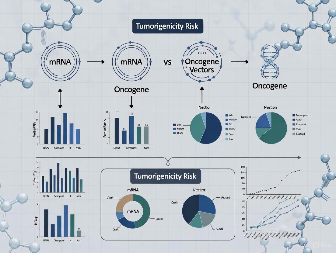
Oncogene Vectors vs. mRNA Platforms: A Comparative Analysis of Tumorigenicity Risks in Therapeutic Development
This article provides a comprehensive comparison of the tumorigenicity risks associated with mRNA-based therapeutic platforms and traditional oncogene vectors.
Oncogene Vectors vs. mRNA Platforms: A Comparative Analysis of Tumorigenicity Risks in Therapeutic Development
Abstract
This article provides a comprehensive comparison of the tumorigenicity risks associated with mRNA-based therapeutic platforms and traditional oncogene vectors. Tailored for researchers, scientists, and drug development professionals, it explores the foundational mechanisms of oncogenesis, detailing how viral vectors carrying oncogenes pose risks through insertional mutagenesis and gain-of-function mutations, while mRNA vectors are non-integrating and transient. The scope covers methodological approaches for risk assessment, strategies for troubleshooting and optimizing safety profiles, and a direct validation of the risk-benefit ratios of each platform. By synthesizing current research and clinical data, this analysis aims to inform safer design principles for next-generation biologics and gene therapies.
Decoding the Mechanisms: How Vectors and Oncogenes Influence Cancer Risk
Fundamental Principles of mRNA Vaccine Biology and Transient Expression
mRNA vaccines represent a novel class of biotherapeutics that leverage messenger RNA technology to direct cells to produce specific antigens, thereby inducing an immune response [1]. Unlike traditional vaccines, mRNA vaccines are non-infectious and non-integrating, eliminating the risks of infection or insertional mutagenesis associated with some viral vector platforms [1]. The fundamental principle involves delivering synthetic mRNA molecules encoding target antigens to host cells, where the host's translational machinery produces the antigenic proteins that subsequently activate immune responses [1] [2].
The transient nature of mRNA expression is a cornerstone of its safety profile. mRNA does not enter the cell nucleus and lacks the enzymatic machinery needed for integration, remaining in the cytoplasm where it is translated and eventually degraded through normal cellular processes [1] [2]. This transient expression kinetics differentiates mRNA platforms from gene therapies utilizing integrating viral vectors, which carry potential for long-term genotoxic risks [3].
Molecular Architecture and Transient Expression Kinetics
Structural Components of Synthetic mRNA
Therapeutic mRNA requires specific structural elements to ensure stability, efficient translation, and controlled immunogenicity [2] [4]. The components work synergistically to optimize protein expression while maintaining the transient expression profile:
- 5' Cap Structure: The 7-methylguanosine cap (m7G) facilitates ribosome binding and protects from 5' exonuclease degradation. Further modifications to create Cap1 (m7GpppN1mp) or Cap2 structures reduce innate immune recognition and enhance translation efficiency [4].
- 5' Untranslated Region (UTR): These regulatory sequences influence mRNA stability and translational efficiency. Optimal UTRs derived from highly expressed genes can significantly enhance protein production [1] [5].
- Open Reading Frame (ORF): This protein-coding sequence can be optimized through codon usage to match host tRNA abundance, improving translational efficiency and protein yield [1] [2].
- 3' UTR and Poly(A) Tail: The 3' untranslated region and polyadenylate tail work cooperatively to regulate mRNA stability and translation. An optimal poly(A) tail length (typically 100-150 nucleotides) protects against degradation and synergizes with the 5' cap to enhance circularization and ribosomal recycling [1] [4].
Mechanisms of Transient Expression
The transient expression profile of mRNA vaccines is governed by several biological processes that limit their duration in cells. The following diagram illustrates the complete lifecycle from delivery to degradation:
The mRNA vaccine lifecycle encompasses several critical phases that collectively ensure transient expression. After cellular uptake, typically mediated by lipid nanoparticles (LNPs), the mRNA must escape endosomal encapsulation to reach the cytoplasm where translation occurs [1] [5]. The mRNA is then recruited to ribosomes that synthesize the encoded antigenic protein. These antigens are processed and presented via MHC molecules to activate immune responses [2]. Concurrently, the mRNA molecule undergoes progressive degradation by cytoplasmic nucleases, while the newly synthesized proteins are processed for immune presentation or turned over by cellular proteostasis mechanisms [1]. This coordinated process ensures that antigen expression is self-limited, typically persisting for hours to several days depending on modifications and delivery systems.
Tumorigenicity Risk Profile: mRNA versus Oncogene Vectors
Fundamental Safety Distinctions
The transient cytoplasmic expression of mRNA vaccines presents a fundamentally different safety profile compared to integrating viral vectors used in gene therapy. The table below summarizes key distinctions:
Table 1: Tumorigenicity Risk Comparison Between mRNA Platforms and Integrating Viral Vectors
| Parameter | mRNA Vaccine Platform | Oncogene Viral Vectors (γRV/LV) |
|---|---|---|
| Cellular localization | Exclusively cytoplasmic | Nuclear entry and integration |
| Genome interaction | No genomic integration | Random or targeted integration |
| Expression kinetics | Transient (hours to days) | Potentially permanent |
| Oncogenic mechanism | No direct genotoxic potential | Insertional mutagenesis, proto-oncogene transactivation |
| Documented clinical malignancies | None reported | 21+ genotoxicity events in γRV trials [3] |
| Regulatory elements | Optimized UTRs, codon usage | Viral LTRs, heterologous promoters |
| Immune recognition | Self-adjuvanting through PRRs | Varies by vector system |
Clinical Evidence of Genotoxicity Profiles
The safety differential between these platforms is substantiated by clinical trial outcomes. Gamma-retroviral vectors (γRV) used in early hematopoietic stem cell (HSC) gene therapies demonstrated significant genotoxicity, with documented cases of leukemogenesis attributed to vector integration near proto-oncogenes like LMO2, CCND2, and BMI1 [3]. A recent systematic review documented 21 genotoxicity events across seven clinical trials for primary immunodeficiency, with the majority attributed to trans-activation of LMO2 (nine patients) or MDS-EVI1 complex (six patients) [3].
Self-inactivating lentiviral vectors (SIN-LV) demonstrate improved safety profiles but still carry integration-related risks. Recent reports describe myeloid malignancies following SIN-LV HSC gene therapy for X-ALD, with investigations revealing frequent vector integration into proto-oncogenes including MECOM [3]. In contrast, mRNA vaccines have no documented cases of insertional mutagenesis or vector-mediated malignant transformation, consistent with their cytoplasmic localization and transient expression profile [1] [2].
Experimental Approaches for Tumorigenicity Assessment
Methodologies for Integration Site Analysis
The assessment of genotoxic risk for gene therapy vectors requires sophisticated molecular techniques to track integration sites and clonal dynamics:
- Linear Amplification-Mediated PCR (LM-PCR): This method selectively amplifies vector-genome junction fragments for high-sensitivity detection of integration sites [3].
- Next-Generation Sequencing (NGS): High-throughput sequencing of integration sites enables comprehensive mapping and monitoring of clonal abundance over time [3].
- Bioinformatic Analysis Pipelines: Specialized algorithms identify genomic features near integration sites (e.g., transcription start sites, cancer-associated genes) and track clonal dynamics [3].
- Clonal Tracking Longitudinal Studies: Repeated sampling of genetically modified cells over extended periods monitors for emergence of dominant clones potentially indicating transformation events [3].
mRNA Distribution and Persistence Studies
For mRNA therapeutics, the analytical focus shifts to distribution and persistence studies:
- Biodistribution Analysis: Quantitative PCR measures mRNA levels in various tissues over time to confirm transient presence and clearance [1] [5].
- Immunohistochemical Staining: Tissue sections stained for encoded antigens verify localized expression and duration [6].
- Protein Expression Kinetics: ELISA and Western blotting quantify encoded antigen production and persistence [5].
- Innate Immune Activation Assays: ELISpot and cytokine profiling assess interferon and inflammatory responses to mRNA and delivery components [6] [4].
The following workflow illustrates the comprehensive safety assessment strategy for mRNA therapeutics:
Essential Research Reagents and Methodologies
Table 2: Essential Research Tools for mRNA Biology and Safety Assessment
| Research Tool Category | Specific Examples | Research Application |
|---|---|---|
| mRNA Synthesis Systems | T7/T3/SP6 RNA polymerases, CleanCap co-transcriptional capping, nucleotide modifications | In vitro transcription of research-grade mRNA with structural fidelity [4] |
| Delivery Vehicles | Lipid nanoparticles (LNPs), polymeric carriers, lipoplexes | Protect mRNA and facilitate cellular delivery [1] [2] |
| Analytical Characterization | HPLC, mass spectrometry, dynamic light scattering | Assess mRNA purity, modification efficiency, and nanoparticle properties [7] [4] |
| In Vitro Translation Systems | Rabbit reticulocyte lysate, cell-free expression systems | Evaluate translational efficiency and protein yield [5] |
| Cell-Based Assay Systems | Immature dendritic cells, primary macrophages, specialized cell lines | Model immune activation and antigen presentation [2] [6] |
| Animal Models | Mice, non-human primates | Evaluate in vivo immunogenicity, distribution, and toxicity [6] |
| Molecular Analysis Tools | qPCR systems, RNA sequencing, cytokine ELISAs, flow cytometry | Quantify mRNA persistence, immune responses, and cellular phenotypes [3] [6] |
The fundamental biology of mRNA vaccines, characterized by cytoplasmic expression, non-integration, and transient kinetics, underlies their favorable tumorigenicity profile compared to viral vector platforms. While viral vector systems face documented challenges with insertional mutagenesis and oncogene transactivation, mRNA platforms demonstrate a distinct safety advantage through their self-limited expression duration and absence of genomic interaction [1] [3].
Advanced experimental approaches continue to refine our understanding of both platform types, with integration site analysis remaining critical for viral vectors, and biodistribution/persistence studies suiting mRNA evaluation. As both technologies evolve, their complementary risk assessment frameworks provide comprehensive safety insights to guide therapeutic development [3] [5].
The ongoing optimization of mRNA design, delivery systems, and manufacturing processes continues to enhance the platform's utility while maintaining its inherent safety advantages [1] [8]. This positions mRNA technology as a versatile platform with applications spanning infectious diseases, cancer immunotherapy, and beyond, supported by its favorable risk-benefit profile.
Oncogenes represent a critical class of genes whose mutation drives the development and progression of cancer. This review comprehensively examines the transformation of normal proto-oncogenes into oncogenes through gain-of-function (GOF) mutations, elucidating the molecular mechanisms underlying this process and its implications for tumorigenesis. We explore the distinct classes of oncogenic mutations, including point mutations, gene amplifications, and chromosomal rearrangements, and their resultant effects on protein function and cellular signaling networks. Within the context of modern cancer research, we provide a direct comparison of two prominent gene delivery systems—viral oncogene vectors and mRNA-based platforms—evaluating their respective tumorigenicity risks based on integration potential, persistence of expression, and genotoxic profiles. By integrating current understanding of oncogene biology with emerging nucleic acid delivery technologies, this analysis aims to inform therapeutic strategies that maximize efficacy while minimizing oncogenic risk in clinical applications.
Oncogenes are mutated genes that have the potential to cause cancer, functioning as accelerators of cell proliferation and contributors to malignant transformation [9]. Before their mutation, these genes exist as proto-oncogenes, which play essential roles in regulating normal cellular processes including growth, division, differentiation, and programmed cell death (apoptosis) [9] [10]. The fundamental distinction between these two states lies in their activity regulation: proto-oncogenes are tightly controlled, while oncogenes exhibit constitutive, unregulated activity that drives uncontrolled cell growth [10].
The discovery of oncogenes has deep roots in virology, with the first tumor-causing virus identified by Peyton Rous in the early twentieth century [10]. Subsequent Nobel Prize-winning work by Bishop and Varmus demonstrated that the cancer-causing gene in the Rous sarcoma virus was actually a hijacked version of a normal host gene (c-src) [10]. This pivotal finding established that oncogenes can originate from normal cellular genes, fundamentally advancing our understanding of cancer genetics. Today, research has identified more than 100 different oncogenes associated with various cancer types, with comprehensive genomic studies revealing mutations in 727 known cancer genes across diverse human malignancies [11] [12].
Proto-oncogenes encode proteins that function at multiple levels within cellular signaling networks. These include cell surface receptors such as EGFR and KDR, intracellular signal transducers like HRAS and KRAS, and cell cycle regulators including cyclin D1 and cyclin E1 [10]. Under physiological conditions, these proteins respond to precise regulatory signals, ensuring appropriate cellular behavior. However, when proto-oncogenes mutate into oncogenes, they produce proteins that continuously signal growth and division regardless of external cues, or they may be expressed at inappropriately high levels, ultimately leading to tumor formation [9] [10].
Mechanisms of Proto-oncogene Activation
The transformation of proto-oncogenes into oncogenes occurs through several distinct genetic mechanisms that ultimately enhance the activity or expression of the gene product. These molecular events convert normally regulated genes into drivers of malignant transformation, with cancer genomic analyses revealing that oncogenes are mutated in approximately 89% of tumors [12].
Point Mutations
Point mutations represent specific changes in the DNA sequence that can dramatically alter the function of the encoded protein. These mutations may lead to a hyperactive gene product that functions independently of normal regulatory controls [10]. A classic example is the Ras family of oncogenes, where single nucleotide substitutions create constitutively active Ras proteins that continuously signal cell proliferation [11]. Specific point mutations can also occur in the promoter region of a proto-oncogene, leading to significantly increased transcription rates and consequent overexpression of the normal protein [10]. The prevalence of such mutations across human cancers underscores their importance in oncogenesis, with KRAS mutations occurring in 84% of pancreatic adenocarcinomas and BRAF mutations present in many thyroid carcinomas [12].
Gene Amplification
Gene amplification events result in extra chromosomal copies of a proto-oncogene, substantially increasing the dosage of the gene within the cell [11] [10]. This amplification leads to elevated levels of the encoded protein, overwhelming normal regulatory mechanisms and driving excessive cell proliferation. For example, the HER2 oncogene is frequently amplified in certain breast cancers, resulting in overexpression of the HER2 receptor tyrosine kinase that promotes aggressive tumor behavior [11]. Genomic studies have revealed that such amplification events are common across multiple cancer types, contributing significantly to oncogene-driven tumorigenesis [12].
Chromosomal Rearrangements
Chromosomal rearrangements represent large-scale genetic alterations that can activate proto-oncogenes through two primary mechanisms. First, translocations may relocate a proto-oncogene to a new chromosomal site adjacent to strong regulatory elements, leading to its aberrant overexpression [10]. Second, and more notably, translocations can create fusion genes that combine portions of two separate genes, producing novel fusion proteins with oncogenic properties [10]. The most famous example is the Philadelphia chromosome, resulting from a translocation between chromosomes 9 and 22 that generates the BCR-ABL fusion gene [10]. This fusion produces a constitutively active tyrosine kinase that drives chronic myelogenous leukemia (CML) and represents a successful therapeutic target [11] [10].
Table 1: Major Mechanisms of Proto-oncogene Activation
| Mechanism | Genetic Alteration | Result | Cancer Example |
|---|---|---|---|
| Point Mutation | Single nucleotide change in coding sequence | Hyperactive protein that signals constitutively | KRAS in pancreatic cancer |
| Promoter Mutation | Mutation in regulatory region | Increased transcription and overexpression | c-MYC in Burkitt lymphoma |
| Gene Amplification | Multiple gene copies | Protein overexpression | HER2 in breast cancer |
| Chromosomal Translocation | Gene relocation to active chromatin | Aberrant expression | c-MYC in Burkitt lymphoma |
| Gene Fusion | Fusion of two genes | Novel chimeric protein with oncogenic activity | BCR-ABL in CML |
Gain-of-Function Mutations in Oncogenesis
Gain-of-function (GOF) mutations represent a fundamental class of genetic alterations that confer new or enhanced functional properties to proteins, driving oncogenesis through diverse molecular mechanisms. Unlike loss-of-function mutations that inactivate tumor suppressor genes, GOF mutations typically enhance proliferative signaling, inhibit cell death, or otherwise promote malignant phenotypes [13]. Understanding the specific nature of these mutations provides critical insights for developing targeted cancer therapies.
Gain of Structural Domains
GOF mutations frequently alter protein domains to create constitutively active signaling molecules. Protein domains are evolutionarily conserved regions with specific functional properties, and mutations within these domains can profoundly change protein behavior [13]. For example, frequent mutations at D835 in FLT3, D816 in KIT, and V600 in BRAF—all located within kinase domains—result in constitutive activation of these oncogenes [13]. Additionally, gene fusion events can create novel chimeric proteins that combine domains from different genes, generating oncoproteins with new functional capabilities. The BCR-ABL fusion protein exemplifies this mechanism, combining regulatory domains from BCR with the kinase domain of ABL to create a constitutively active tyrosine kinase that drives chronic myelogenous leukemia [10]. Such domain-level alterations can also enable novel protein-protein interactions, as demonstrated by the E545K mutation in the helical domain of PIK3CA, which confers the ability to associate with insulin receptor substrate 1 (IRS1) and rewire oncogenic signaling pathways [13].
Gain of Novel Interaction Motifs
Beyond structured domains, GOF mutations can create novel interaction motifs in unstructured protein regions, enabling new pathogenic protein interactions. Intrinsically disordered regions (IDRs) lack stable three-dimensional structures but play crucial roles in protein signaling and regulation [13]. Mutations within IDRs can generate novel short linear motifs (SLiMs) that mediate interactions with other proteins, DNA, or RNA. For instance, the c-Myc oncoprotein utilizes its IDRs to perform diverse interactions in cancer, and mutations in these regions can further alter its interaction network [13]. Specific disease mutations in the IDRs of proteins like GLUT1, ITPR1, and CACNA1H can create novel dileucine motifs that increase clathrin binding, potentially altering protein localization and function [13]. Similarly, mutations in the catenin gene (CTNNB1) can perturb SLiMs involved in the Wnt signaling pathway, contributing to various cancers [13]. These findings highlight how even small mutations in unstructured regions can create novel functional interfaces that drive oncogenesis.
Non-coding GOF Mutations
While traditionally focused on protein-coding regions, emerging research reveals that GOF mutations in non-coding regions also contribute significantly to cancer. More than 90% of disease-associated variants fall within non-coding regions, where they can perturb interactions between transcription factors (TFs) and their binding sites [13]. Such alterations can create novel TF binding sites or enhance existing ones, leading to aberrant expression of oncogenes. Additionally, mutations in regulatory elements can disrupt normal transcriptional control mechanisms, resulting in oncogene overexpression without altering the coding sequence itself. The extensive mutational landscape of human cancers shows that these non-coding GOF mutations work in concert with coding mutations to drive tumor development and progression [12].
Diagram Title: GOF Mutation Mechanisms in Oncogene Activation
Comparative Tumorigenicity Risk: mRNA versus Oncogene Vectors
In cancer research and therapeutic development, two primary nucleic acid delivery platforms have emerged: mRNA-based vectors and viral oncogene vectors. These systems differ fundamentally in their mechanisms of action, persistence, and potential tumorigenic risks, with important implications for their research applications and therapeutic potential.
mRNA-based Vectors
mRNA-based vectors deliver genetic information encoding tumor antigens or therapeutic proteins to host cells without integrating into the genome [14]. These platforms work by introducing mRNA sequences into the cytoplasm, where they are directly translated into proteins without nuclear entry [14] [15]. The transient nature of mRNA expression—typically lasting from hours to several days—significantly reduces tumorigenicity concerns, as the genetic material does not persist long-term and cannot integrate into host DNA [14] [16]. However, challenges remain in achieving efficient in vivo delivery, as naked mRNA is susceptible to degradation by nucleases and requires sophisticated delivery systems such as lipid nanoparticles (LNPs) for protection and cellular uptake [14]. Modified nucleosides can enhance mRNA stability and reduce immunogenicity, but repeated administration may still trigger innate immune responses that limit efficacy [16]. From a tumorigenicity perspective, mRNA platforms present minimal risk due to their non-integrating, transient expression characteristics, making them particularly attractive for cancer immunotherapy and vaccine development [14] [16].
Viral Oncogene Vectors
Viral vectors utilize engineered viruses to deliver genetic material, with a key distinction being that they typically deliver DNA rather than RNA [15]. These systems employ harmless viral shells (such as adenoviruses or adeno-associated viruses) to package and deliver therapeutic genes to target cells [15]. Unlike mRNA platforms, viral vectors must enter the nucleus for transcription, and certain viral types (particularly retroviruses and lentiviruses) can integrate their genetic payload into the host genome [15]. This integration capability creates inherent tumorigenicity risks, as insertional mutagenesis can disrupt tumor suppressor genes or activate proto-oncogenes [10]. Additionally, viral vectors often trigger stronger immune responses against the viral components themselves, potentially limiting repeated administration and complicating therapeutic applications [15]. While non-integrating viral vectors have been developed to mitigate these risks, the theoretical potential for genotoxicity remains a consideration in their research and clinical application, particularly when delivering potent oncogenes or growth factors.
Table 2: Tumorigenicity Risk Comparison Between Vector Platforms
| Parameter | mRNA Vectors | Oncogene Viral Vectors |
|---|---|---|
| Genetic Material | mRNA | DNA |
| Nuclear Entry | Not required | Required |
| Genome Integration | No | Possible (depends on viral type) |
| Expression Duration | Transient (hours to days) | Prolonged (weeks to months) |
| Tumorigenicity Risk | Low | Moderate to High |
| Primary Risk Mechanism | Immunostimulation | Insertional mutagenesis, persistent oncogene expression |
| Immune Response | Against encoded antigen | Against viral components and encoded antigen |
| Delivery Method | Lipid nanoparticles, electroporation | Viral transduction |
Experimental Assessment of Tumorigenic Potential
Rigorous experimental approaches are essential for evaluating the tumorigenic potential of both vector types. For mRNA vectors, assessment typically includes evaluation of local and systemic inflammation, duration of expression, and potential autoimmune reactions against self-antigens [14] [16]. For viral vectors, critical experiments include integration site analysis to identify preferences for cancer-related genomic loci, long-term persistence studies in animal models, and careful monitoring for clonal expansion indicative of transformation events [15]. Both platforms require comprehensive toxicology studies in relevant animal models, with particular attention to organs susceptible to tumor development. Regulatory authorities like the FDA provide guidelines for such assessments, requiring extensive preclinical data before clinical trial approval [15]. These experimental protocols help researchers characterize and mitigate potential oncogenic risks associated with each platform.
Research Reagents and Methodologies
Advancing the study of oncogenes and developing safer gene delivery systems requires specialized research tools and experimental approaches. The following section outlines key reagents and methodologies essential for investigating oncogene function and vector safety profiles.
Essential Research Reagents
Table 3: Key Research Reagents for Oncogene and Vector Studies
| Reagent Category | Specific Examples | Research Applications |
|---|---|---|
| Cell Culture Models | Immortalized cell lines, primary cells, organoids | In vitro transformation assays, gene expression studies |
| Animal Models | Immunodeficient mice, transgenic oncogene models | In vivo tumorigenicity studies, therapeutic efficacy testing |
| Gene Delivery Tools | Lipid nanoparticles, viral packaging systems, electroporators | Vector delivery, efficiency optimization |
| Detection Antibodies | Phospho-specific antibodies, epitope tags | Protein expression analysis, signaling pathway activation |
| Sequencing Reagents | RNA-seq kits, whole-exome sequencing panels | Mutational profiling, expression analysis, integration site mapping |
| Signal Transduction Assays | Pathway reporter constructs, kinase activity assays | Oncogene functional characterization, signaling network mapping |
Key Experimental Protocols
Oncogene Transformation Assay
This fundamental protocol assesses the malignant potential of candidate oncogenes through a series of methodical steps. First, researchers introduce the candidate gene into immortalized but non-tumorigenic cells (such as NIH-3T3 fibroblasts) using either viral transduction or mRNA transfection [10]. Following gene delivery, transformed cells are selected based on morphological changes including rounded cell shape, increased refractility, and loss of contact inhibition. The most critical step involves evaluating anchorage-independent growth through soft agar colony formation assays, where transformed cells proliferate without surface attachment—a hallmark of malignant transformation [10]. Finally, in vivo validation is performed by injecting putative transformed cells into immunodeficient mice and monitoring for tumor formation over 4-8 weeks, with tumor tissues subsequently analyzed for preservation of the introduced oncogene and characteristic histopathological features.
Vector Integration Site Analysis
This protocol specifically addresses the tumorigenicity risk assessment of viral vectors through comprehensive integration profiling. The process begins with genomic DNA extraction from transduced cells followed by fragmentation and adapter ligation. Vector-genome junctions are then enriched through PCR amplification using vector-specific and genomic primers, with subsequent high-throughput sequencing of these junction fragments [15]. Bioinformatic analysis aligns the sequences to the reference genome to identify precise integration sites, with particular attention to cancer-related genomic loci such as proto-oncogene promoters or tumor suppressor gene coding regions. Long-term tracking studies monitor for clonal expansion by analyzing the relative abundance of specific integration sites over time, with dominant clones indicating potential selective advantages that may precede transformation.
mRNA Vector Expression Kinetics
This methodology characterizes the temporal profile of mRNA-based vectors, a critical parameter for both efficacy and safety assessment. The protocol involves introducing mRNA vectors (often encapsulated in lipid nanoparticles) into target cells or animal models. Researchers then collect samples at multiple timepoints post-delivery for quantitative PCR analysis to measure mRNA persistence and western blotting or immunoassays to quantify translated protein levels [14]. Simultaneously, immune activation markers are assessed through cytokine profiling and immune cell infiltration analysis. To evaluate the potential for extended expression without increased risk, self-amplifying RNA systems can be tested using similar methodologies, with careful attention to the duration and magnitude of expression [16].
Diagram Title: Oncogene Research Experimental Workflow
The journey from proto-oncogene to oncogene represents a fundamental pathway in cancer development, with gain-of-function mutations serving as key drivers of malignant transformation. Through diverse mechanisms including point mutations, gene amplifications, and chromosomal rearrangements, normally regulated cellular genes become powerful oncogenic drivers that disrupt controlled proliferation and promote tumorigenesis. The comprehensive characterization of these mutations across human cancers has revealed both the complexity and patterns in oncogene activation, providing critical insights for targeted therapeutic development.
In the context of modern cancer research, the choice between mRNA and viral vector platforms involves careful consideration of their distinct tumorigenicity profiles. mRNA vectors offer a favorable safety profile due to their transient, non-integrating nature, while viral vectors provide persistent expression but carry theoretical risks related to genomic integration and insertional mutagenesis. Understanding these distinctions enables researchers to select appropriate systems for specific applications, whether investigating oncogene function or developing novel cancer therapeutics.
Future directions in oncogene research will likely focus on combinatorial approaches that target multiple oncogenic pathways simultaneously, advanced vector engineering to enhance specificity and safety, and personalized strategies based on individual tumor mutation profiles. As our understanding of oncogene biology continues to evolve, so too will our ability to develop increasingly precise and effective interventions for cancer treatment, ultimately improving outcomes for patients facing oncogene-driven malignancies.
Mechanisms of Insertional Mutagenesis by Viral Vector Platforms
The advancement of viral vector-based gene therapies and vaccines represents a significant milestone in modern medicine. These platforms, which include retrovectors, lentivectors, and adenovectors, have demonstrated remarkable efficacy in treating monogenic disorders and developing novel vaccines. However, their utilization is accompanied by a critical safety concern: insertional mutagenesis. This process occurs when the integration of viral DNA disrupts or alters the function of host genes, potentially leading to oncogenic transformation [3]. As these therapeutic platforms are increasingly deployed in clinical settings, understanding and comparing their mutagenic potential becomes paramount for researchers and drug development professionals.
The tumorigenicity risk profile varies considerably across different vector platforms. While viral vectors possess a well-documented, though diminishing, risk profile, emerging non-viral platforms such as mRNA vaccines present a distinctly different safety paradigm. This guide objectively compares the mechanisms and frequencies of insertional mutagenesis across leading viral vector platforms, contextualizes these risks against non-integrating mRNA alternatives, and provides experimental approaches for quantifying genotoxicity in preclinical research.
Molecular Mechanisms of Insertional Mutagenesis
Insertional mutagenesis occurs through distinct biological mechanisms when viral vectors integrate their genetic material into the host genome. The specific molecular consequences depend on the integration site relative to host genes and the regulatory elements carried by the vector.
Enhancer-Mediated Transactivation
This mechanism involves vector-encoded enhancer elements activating neighboring host proto-oncogenes. Gamma-retroviral vectors (γRVs) with intact long terminal repeat (LTR) promoters are particularly prone to this effect. Clinical trials for X-SCID using such vectors resulted in T-cell leukemias where the vector integrated near the LMO2 proto-oncogene, driving its aberrant expression [3]. The powerful viral enhancers triggered oncogene transcription, leading to clonal expansion and transformation.
Gene Disruption
Integration events within coding regions can disrupt tumor suppressor genes or critical regulatory genes. The physical insertion of vector DNA can truncate host gene transcripts, alter splicing patterns, or create non-functional fusion proteins. This loss-of-function mechanism is agnostic to vector type and depends primarily on the integration site preferences and the specific gene disrupted [3].
Aberrant Transcript Processing
Self-inactivating lentiviral vectors (SIN-LVs), while safer, can still cause genotoxicity through aberrant transcript processing. In a clinical trial for β-thalassemia, vector integration into the HMGA2 gene led to the production of truncated transcripts that were resistant to normal degradation pathways. This resulted in clonal dominance, though in this specific case, it coincidentally contributed to therapeutic benefit [3].
Table 1: Documented Oncogenic Events in Clinical Trials Linked to Insertional Mutagenesis
| Vector Platform | Disease Context | Oncogene Target | Molecular Mechanism | Clinical Outcome |
|---|---|---|---|---|
| Gamma-Retroviral (γRV) | X-SCID [3] | LMO2 | Enhancer-mediated transactivation | T-cell leukemia |
| Gamma-Retroviral (γRV) | CGD [3] | MDS1-EVI1 (MECOM) | Enhancer-mediated transactivation | Myelodysplastic syndrome |
| Gamma-Retroviral (γRV) | Wiskott-Aldrich Syndrome [3] | LMO2, MDS1, MN1 | Enhancer-mediated transactivation | T-ALL, AML |
| Self-Inactivating Lentiviral (SIN-LV) | X-ALD [3] | MECOM | Mechanism under investigation | Myeloid malignancies |
Comparative Genotoxicity Across Vector Platforms
The genotoxic potential of viral vectors is influenced by their integration site preferences, the regulatory elements in their design, and their propensity to drive aberrant gene expression.
Gamma-Retroviral Vectors (γRV)
Integration Profile: Gamma-retroviral vectors exhibit a strong preference for integrating near transcription start sites (TSS) and promoter regions of active genes [3]. This bias significantly increases the probability of disrupting regulatory elements or activating proto-oncogenes.
Safety Record: The genotoxicity of early γRV vectors is well-documented. A meta-analysis identified 21 genotoxicity events across seven clinical trials for primary immunodeficiencies, with the majority involving transactivation of LMO2 or the MDS-EVI1 complex [3]. These events led to a high incidence of leukemias, particularly in trials for X-SCID, CGD, and WAS.
Lentiviral Vectors (LV)
Integration Profile: Lentiviral vectors also integrate into active genes but show a reduced bias for promoter-proximal regions compared to γRVs. This wider distribution lowers the risk of directly perturbing highly sensitive regulatory zones near TSS [3].
Safety Record and Emerging Concerns: Self-inactivating (SIN) lentiviral vectors, which have deleted U3 promoter regions from their LTRs, represent a safer generation of vectors. However, recent long-term follow-up has revealed that risks persist. In trials for X-linked adrenoleukodystrophy (X-ALD), SIN-LV therapy was linked to the development of myelodysplastic syndrome (MDS) and acute myeloid leukemia (AML) in several patients [3]. Investigations frequently found vector integrations in proto-oncogenes like MECOM, indicating that even SIN designs can drive clonal expansion through mechanisms that are still under investigation.
Adenoviral Vectors (Ad)
Integration Profile: Adenoviral vectors are predominantly episomal, meaning they remain in the cell's cytoplasm without integrating into the host genome. This is their primary safety advantage over retro- and lentiviral platforms [17].
Primary Safety Concerns: The main risks associated with adenovectors are not related to insertional mutagenesis but to robust inflammatory immune responses and, in rare cases, conditions like vaccine-induced immune thrombotic thrombocytopenia (VITT), as observed with some COVID-19 vaccines [17]. Their lack of integration makes them a lower tumorigenicity risk in terms of genotoxicity.
Table 2: Comparative Genotoxicity Profiles of Viral Vector Platforms
| Platform Feature | Gamma-Retroviral (γRV) | Lentiviral (LV) | Adenoviral (Ad) |
|---|---|---|---|
| Integration Preference | Promoter-proximal/Transcription Start Sites [3] | Active genes (reduced promoter bias) [3] | Primarily episomal (non-integrating) [17] |
| Key Genotoxic Mechanism | Enhancer-mediated transactivation from intact LTRs [3] | Aberrant splicing, gene disruption, insertional activation [3] | Not applicable (primary risk is immunogenicity) [17] |
| Documented Clinical Events | Multiple cases of leukemia (X-SCID, CGD, WAS) [3] | Emerging cases of MDS/AML (X-ALD, SCD) [3] | No direct insertional oncogenesis reported [17] |
| Relative Tumorigenicity Risk | High | Moderate | Very Low |
The mRNA Vaccine Platform: A Non-Integrating Alternative
In contrast to viral vectors, mRNA-based platforms offer a fundamentally different mechanism of action that inherently avoids the risk of insertional mutagenesis.
Cytoplasmic Action and Transient Expression
mRNA vaccines function exclusively in the cytoplasm of host cells. The delivered mRNA strand is directly translated by ribosomes into the target protein antigen without ever entering the nucleus [14] [18]. This process completely bypasses the host cell's genome, eliminating any risk of insertional mutagenesis [18] [19]. Furthermore, mRNA is a transient molecule with a finite half-life, leading to temporary protein expression that further reduces long-term risks [19].
Safety Profile in Clinical Applications
The extensive global deployment of mRNA COVID-19 vaccines has provided a substantial safety dataset. The primary adverse events of interest have been immune-related reactions, such as myocarditis or severe allergic reactions, with no evidence linking these vaccines to oncogenesis [17]. This safety profile underscores the theoretical advantage of mRNA platforms in terms of genotoxicity.
Table 3: Key Feature Comparison: Viral Vectors vs. mRNA Platform
| Characteristic | Integrating Viral Vectors (γRV, LV) | mRNA Platform |
|---|---|---|
| Nuclear Entry | Required for integration [3] | Not required; functions in cytoplasm [14] [18] |
| Genome Integration | Yes, inherent to mechanism [3] | No [18] |
| Theoretical Risk of Insertional Mutagenesis | Present | Absent |
| Duration of Expression | Long-term/Potentially permanent [3] | Transient [19] |
| Primary Safety Concerns | Insertional oncogenesis, clonal dominance [3] | Immunogenicity, inflammatory responses [17] [19] |
Experimental Protocols for Assessing Genotoxicity
Robust preclinical assessment is critical for evaluating the tumorigenic potential of novel vector designs. The following methodologies are standards in the field.
Insertion Site Analysis
This technique maps the exact genomic locations of vector integrations to identify preferences for cancer-related loci.
- Methodology: Techniques like inverse PCR (iPCR) and linear amplification-mediated PCR (LAM-PCR) are used to isolate and sequence the genomic DNA flanking integrated vector proviruses [20]. Next-generation sequencing of these fragments allows for the genome-wide mapping of integration sites.
- Data Interpretation: The resulting dataset is analyzed for non-random clustering of integrations. A statistically significant skewing of integrations near proto-oncogenes (e.g., LMO2, MECOM) or within tumor suppressor genes indicates a higher genotoxic potential. The clonal abundance of cells harboring specific integrations is also tracked over time.
In Vitro Transformation Assays
These assays directly test the potential of a vector to cause uncontrolled cell growth, a hallmark of cancer.
- Methodology: The cell culture-based transformation assay is a key model. One protocol involves transducing immortalized but non-tumorigenic cell lines (e.g., HT1080-derived cells) with the test vector [20]. The cells are then monitored in culture for the acquisition of transformation phenotypes.
- Phenotypic Endpoints: Key readouts include focus formation (cells piling up on top of each other), anchorage-independent growth (the ability to form colonies in soft agar), and proliferation in low-serum conditions [20]. The emergence of these phenotypes suggests the vector has disrupted normal growth control mechanisms.
In Vivo Tumorigenicity Studies
These long-term studies provide the most comprehensive safety assessment by evaluating genotoxicity in a whole-organism context.
- Methodology: Immunodeficient mice (e.g., NSG mice) are transplanted with human hematopoietic stem cells (HSCs) that have been transduced with the candidate vector. The mice are monitored over several months for signs of hematologic malignancy [3].
- Endpoint Analysis: Monitoring includes periodic blood sampling to track the clonal composition of the human cell graft via integration site analysis. At the study's end, organs are examined pathologically for evidence of cancer. The development of leukemia/lymphoma in these models, especially if linked to a specific vector integration site, is a major red flag.
The diagram below illustrates the logical relationship and workflow between these key experimental protocols.
The Scientist's Toolkit: Essential Research Reagents
The following reagents and tools are fundamental for conducting research on insertional mutagenesis and vector safety.
Table 4: Key Research Reagents for Insertional Mutagenesis Studies
| Reagent / Tool | Function | Example Application |
|---|---|---|
| Packaging Cell Lines | Produce replication-incompetent viral vector particles. | Generating γRV or LV stocks for transduction [20]. |
| Inverse PCR (iPCR) / LAM-PCR Kits | Amplify and isolate host-genome/vector-junction sequences. | Mapping vector integration sites in transduced cells [20]. |
| Next-Generation Sequencing (NGS) | High-throughput sequencing of integration sites. | Genome-wide analysis of integration site preferences [3]. |
| Immortalized Cell Lines | Provide a consistent cellular substrate for transformation assays. | Testing focus formation in vitro (e.g., HCT9, HEK293) [20]. |
| Immunodeficient Mouse Models | Support engraftment of human cells for in vivo studies. | NSG mice for HSC tumorigenicity tracking [3]. |
| Flow Cytometry Antibodies | Identify and sort specific cell populations. | Tracking clonal expansion of transduced cells in vivo. |
The risk of insertional mutagenesis remains a critical differentiator among genetic medicine platforms. Integrating viral vectors, particularly the historical γRVs, carry a significant and documented risk, while modern SIN lentiviral vectors present a reduced, yet still present, hazard. In contrast, mRNA-based platforms completely circumvent this risk through their cytoplasmic, non-integrating mechanism of action [14] [18] [3].
The choice of platform for clinical or research applications must therefore involve a careful benefit-risk analysis. For diseases requiring long-term, stable gene expression (e.g., hemoglobinopathies, immunodeficiencies), lentiviral vectors may be justified despite their lower genotoxicity profile. For applications where transient expression is sufficient, such as vaccination or short-term protein replacement, mRNA platforms offer a compelling safety advantage regarding tumorigenicity. As the field advances, continued refinement of vector designs, coupled with the rigorous application of the experimental protocols outlined herein, will be essential to maximize therapeutic efficacy while minimizing oncogenic risk.
Key Signaling Pathways Dysregulated by Oncogene Activation (e.g., Ras/MAPK, PI3K/AKT)
The transformation of normal cells into cancerous ones is frequently driven by mutations that hijack core intracellular signaling pathways. Among the most critical are the RAS/RAF/MAPK and PI3K/AKT pathways, which regulate fundamental cellular processes including proliferation, survival, metabolism, and differentiation. When activated by oncogenic mutations, these pathways drive uncontrolled tumor growth and confer resistance to treatment. Understanding the precise mechanisms of dysregulation, the associated tumorigenicity risks, and the therapeutic strategies to counter them forms a cornerstone of modern cancer biology. This guide provides a comparative analysis of these pathways, framed within the context of tumorigenicity risks associated with different gene delivery vectors, to support researchers and drug development professionals in their experimental and therapeutic endeavors.
The RAS/RAF/MAPK Pathway
Pathway Mechanism and Dysregulation
The RAS/RAF/MAPK pathway is a highly conserved signaling cascade that transmits extracellular signals from growth factors to the nucleus, governing critical cellular decisions. The canonical pathway flows from RAS to RAF, to MEK, and finally to ERK [21]. Upon activation, ERK1/2 translocates to the nucleus and phosphorylates transcription factors such as c-Fos, c-Jun, and Elk-1, regulating genes essential for cell cycle progression and survival [22].
Oncogenic activation most commonly occurs through mutations in RAS genes (KRAS, NRAS, HRAS) or the BRAF gene. Mutated RAS becomes locked in a GTP-bound "on" state, leading to constitutive signaling independent of extracellular stimuli [22]. The BRAF V600E mutation, frequently found in melanoma, results in a constitutively active kinase that continuously stimulates the pathway [22]. Such defects are associated with numerous cancers, including melanoma, non-small cell lung carcinoma, colorectal cancer, and thyroid cancer [21].
Quantitative Dysregulation Data
Table 1: Prevalence and Impact of RAS/RAF/MAPK Pathway Dysregulation in Selected Cancers
| Cancer Type | Common Mutations | Approximate Mutation Prevalence | Key Clinical Implications |
|---|---|---|---|
| Melanoma | BRAF V600E | ~50% [21] | Target for BRAF/MEK inhibitor therapy |
| Colorectal Cancer | KRAS | 35-45% [21] | Predicts resistance to EGFR antibodies |
| Non-Small Cell Lung Cancer (NSCLC) | KRAS | 25-30% [21] | Associated with smoking history; therapeutic target |
| Thyroid Cancer | BRAF, RAS | ~45% (BRAF), ~10% (RAS) [21] | BRAF mutation correlates with aggressive disease |
| Pancreatic Cancer | KRAS | >90% [22] | Major driver in pancreatic carcinogenesis |
Experimental Protocols for Pathway Inhibition
The standard methodology for investigating MAPK pathway inhibition involves treating mutant cell lines with targeted inhibitors and assessing downstream signaling and phenotypic consequences.
Protocol 1: Assessing Efficacy of RAF/MEK Inhibitor Combinations
- Cell Culture: Utilize human-derived cancer cell lines harboring BRAF V600E mutations (e.g., A375 melanoma cells).
- Inhibitor Treatment: Treat cells with clinically relevant inhibitors:
- RAF inhibitor (RAFi): Dabrafenib (1-100 nM)
- MEK inhibitor (MEKi): Trametinib (1-100 nM)
- Combination: Dabrafenib + Trametinib
- Incubation: Maintain treated cells for 48-72 hours.
- Downstream Analysis:
- Western Blotting: Analyze lysates for phosphorylated ERK (p-ERK) and total ERK to measure pathway suppression.
- Viability Assay: Perform MTT or CellTiter-Glo assay to quantify cell viability.
- Apoptosis Assay: Use flow cytometry with Annexin V/PI staining to detect apoptotic cells [21].
Protocol 2: Investigating Therapy-Induced Autophagy
- Therapy Application: Treat RAF-mutant cancer cells with a RAFi (e.g., Vemurafenib).
- Autophagy Modulation: Co-treat with autophagy inhibitors such as chloroquine (CQ) or hydroxychloroquine (HCQ).
- Autophagy Detection:
- Immunofluorescence: Track LC3 protein puncta formation.
- Western Blot: Analyze LC3-I to LC3-II conversion.
- Functional Assessment: Compare viability and apoptosis in cells treated with RAFi alone versus RAFi + autophagy inhibitor [21].
The PI3K/AKT Pathway
Pathway Mechanism and Dysregulation
The PI3K/AKT pathway is a central regulator of cell survival, growth, and metabolism. Activation begins when growth factors bind to receptor tyrosine kinases (RTKs), recruiting PI3K to the membrane. PI3K phosphorylates the lipid PIP2 to generate PIP3. AKT is then recruited to the membrane where it is phosphorylated and fully activated by PDK1 (on Thr308) and mTORC2 (on Ser473) [23]. Activated AKT phosphorylates numerous downstream effectors, including mTORC1, GSK3β, and FOXO transcription factors, to promote cell growth and inhibit cell death [23].
Hyperactivation of this pathway in cancer can occur through multiple mechanisms:
- Mutation/Amplification of genes like PIK3CA (encoding the PI3K p110α subunit) or AKT itself.
- Loss or mutation of the negative regulator PTEN, a tumor suppressor that dephosphorylates PIP3 back to PIP2 [23] [24].
- Overexpression of upstream coactivators such as TMEPAI, SALL4, TCL1B, and TGF-β, which enhance pathway activity through distinct mechanisms [23].
Quantitative Dysregulation Data
Table 2: Prevalence and Impact of PI3K/AKT Pathway Dysregulation in Selected Cancers
| Cancer Type | Common Alterations | Approximate Prevalence of Hyperactivation | Key Clinical Implications |
|---|---|---|---|
| Glioblastoma | PTEN loss, PIK3CA mutation | ~88% [23] [25] | Associated with resistance to TTFields therapy [25] |
| Breast Cancer | PIK3CA mutation, PTEN loss | Up to 70% [23] | Target for PI3Kα-specific inhibitors (e.g., Alpelisib) |
| Prostate Cancer | PTEN loss, AKT amplification | 40% (early-stage) to 70-100% (advanced) [23] | Drives disease progression and therapy resistance |
| Serous Ovarian Cancer | PIK3CA mutation, AKT amplification | ~45% (high-grade) [23] | Potential target for AKT inhibitors |
| Endometrial Cancer | PIK3CA mutation, PTEN loss | ~40% [23] | Common driver event |
Experimental Protocols for Pathway Analysis
The following protocols are used to investigate PI3K/AKT activation and its functional consequences in cancer models.
Protocol 1: Luminex Multiplex Assay for Signaling Analysis
- Cell Treatment: Subject cancer cell lines (e.g., U-87 MG glioblastoma) to an experimental condition (e.g., TTFields application, drug treatment).
- Cell Lysis: Harvest cells and prepare lysates using RIPA buffer supplemented with protease and phosphatase inhibitors.
- Protein Quantification: Determine protein concentration using a Bradford or BCA assay.
- Multiplex Immunoassay: Incubate 500 µg of protein lysate with antibody-coated magnetic beads from a customized Luminex kit (e.g., AKT/mTOR pathway panel).
- Detection and Analysis: Measure bead fluorescence on a Luminex analyzer to quantify the phosphorylation and total levels of multiple pathway components (e.g., AKT, p70S6K, 4E-BP1) simultaneously [25].
Protocol 2: Sensitization via PI3K/AKT Pathway Inhibition
- In Vitro Co-treatment:
- Cell Seeding: Plate cancer cells at a defined confluence.
- Drug Application: Treat cells with a PI3K inhibitor (e.g., Alpelisib, Buparlisib) or an AKT inhibitor (e.g., MK-2206) alone and in combination with a primary therapy.
- Viability Assessment: After 72 hours, measure cell viability using a standardized assay (e.g., MTS). Calculate combination indices to determine synergism.
- In Vivo Validation:
- Animal Models: Implant tumor cells orthotopically or subcutaneously into immunodeficient mice.
- Treatment Groups: Administer vehicle, PI3K inhibitor alone, primary therapy alone, or the combination.
- Endpoint Analysis: Monitor tumor volume over time and analyze excised tumors via immunohistochemistry for markers like pAKT (Ser473) to confirm pathway inhibition [25].
Tumorigenicity Risk Profile of Gene Delivery Vectors
The choice of vector for delivering oncogenic material or therapeutic genes in research and therapy carries distinct tumorigenicity risks.
Table 3: Comparison of Gene Delivery Vector Profiles
| Vector | Genome Type | Integration Profile | Primary Tumorigenicity Risk | Key Advantages |
|---|---|---|---|---|
| AAV Vectors | ssDNA | Predominantly episomal; low-frequency, non-specific integration [26] [27] | Insertional mutagenesis (low risk); DNA damage response (DDR) & inflammatory signaling at high doses [28] [27] | Low pathogenicity; broad tropism; long-term expression [26] [27] |
| mRNA Platforms | RNA | Non-integrating; transient expression in cytoplasm [16] | No risk of insertional mutagenesis; transient nature limits long-term risk | Rapid development; high safety profile; no genome integration risk [16] |
| γ-Retroviruses | ssRNA | Integrates into host genome, prefers transcription start regions [26] | High risk of insertional mutagenesis (e.g., activation of oncogenes) [26] | Stable long-term expression; large cargo capacity [26] |
| Lentiviruses | ssRNA | Integrates into active genomic regions [26] | Risk of insertional mutagenesis (e.g., lymphoma development) [26] | Infects dividing and non-dividing cells [26] |
| Adenoviruses | dsDNA | Non-integrating; episomal [26] | No insertional mutagenesis risk; but can trigger strong immune responses [26] | High transduction efficiency; large cargo capacity [26] |
Essential Research Reagent Solutions
Table 4: Key Reagents for Studying Dysregulated Pathways
| Reagent Category | Specific Examples | Research Function | Application Context |
|---|---|---|---|
| Small Molecule Inhibitors | Dabrafenib (RAF inhibitor), Trametinib (MEK inhibitor), Alpelisib (PI3K inhibitor), MK-2206 (AKT inhibitor) | Chemically inhibit key nodal kinases to dissect pathway function and as therapeutic candidates [21] [23] [25] | In vitro cell culture, in vivo animal models |
| AAV Serotypes | AAV2, AAV5, AAV6, AAV8, AAV9 | Gene delivery vectors with varying tissue tropisms (e.g., CNS, liver, muscle) for in vivo studies [26] [27] | Gene replacement, functional gene studies, in vivo modeling |
| siRNA/shRNA Libraries | siRNA targeting KRAS, BRAF, AKT, PTEN | Knock down gene expression to study loss-of-function phenotypes and identify synthetic lethal interactions [16] | High-throughput screens, functional genomics |
| Validated Antibodies | p-ERK (Thr202/Tyr204), p-AKT (Ser473), Total ERK, Total AKT, PTEN | Detect protein levels and activation status (phosphorylation) via Western Blot, IHC, and Flow Cytometry [22] [25] | Pathway activation analysis in cell lysates and tissue sections |
| Cell Line Models | A375 (BRAF V600E melanoma), U-87 MG (PTEN mutant glioblastoma), HCT116 (KRAS mutant colorectal) | Pre-validated in vitro models with defined oncogenic mutations for mechanistic and drug testing studies [21] [25] | Basic mechanistic studies, high-throughput drug screening |
Signaling Pathway and Experimental Workflow Diagrams
RAS/RAF/MAPK and PI3K/AKT Signaling Pathways
Experimental Workflow for Pathway Inhibition Studies
Inherent Safety Advantages of Non-integrating mRNA Vectors
The advancement of genetic medicine hinges on the safe and effective delivery of therapeutic nucleic acids. Within this field, a critical safety consideration is the risk of tumorigenicity—the potential for a treatment to initiate cancer. This risk is intrinsically linked to the vector's ability to modify the host genome. Non-integrating mRNA vectors represent a paradigm shift with a superior safety profile, as they function exclusively in the cytoplasm and do not enter the nucleus, thereby eliminating the risk of insertional mutagenesis that is a recognized concern with integrating viral vectors [29] [30] [19].
This guide provides an objective comparison between mRNA vectors and traditional DNA-based or viral vectors, with a specific focus on tumorigenicity risk. It is structured within the context of oncogene vector research, summarizing key mechanistic data and experimental evidence to inform researchers and drug development professionals.
Fundamental Biological and Safety Comparisons
The core difference in tumorigenicity potential between mRNA and DNA vectors stems from their fundamental biology and intracellular fate. Table 1 summarizes the key comparative characteristics.
Table 1: Fundamental Comparison of mRNA and DNA Vector Characteristics
| Characteristic | Non-integrating mRNA Vectors | DNA Vectors (Plasmid/Viral) |
|---|---|---|
| Site of Activity | Cytoplasm [14] [30] [31] | Nucleus [14] |
| Genome Integration | No risk of insertional mutagenesis [29] [32] [30] | Potential for integration and insertional mutagenesis [32] |
| Nuclear Entry Required | Not required [31] | Required [14] |
| Persistence of Genetic Material | Transient (hours to days) [31] | Can be long-lasting or permanent [32] |
| Oncogene Activation Risk | Theoretically absent due to cytoplasmic activity [32] | Possible due to random integration near proto-oncogenes [32] |
| Primary Safety Concern | Immunogenicity [30] [19] | Insertional mutagenesis [32] |
The following diagram illustrates the distinct intracellular pathways and associated tumorigenicity risks of mRNA versus DNA vectors.
Experimental Evidence and Key Methodologies
The theoretical safety advantages of mRNA vectors are supported by robust experimental evidence. Key protocols are designed to directly assess and quantify the risk of genomic integration and oncogenic transformation.
Key Experimental Protocols for Assessing Tumorigenicity
1. Protocol for Integration Site Analysis
- Objective: To identify and map locations where foreign DNA has integrated into the host genome.
- Methodology: After in vivo or in vitro administration of the vector, genomic DNA is harvested from target cells. Techniques such as linear amplification-mediated polymerase chain reaction (LM-PCR) or next-generation sequencing are used to isolate vector-genome junction fragments. These sequences are then mapped to the reference genome to identify integration sites [32].
- Significance for mRNA: This test is a critical benchmark for DNA vectors. mRNA vectors serve as a negative control, as they are not expected to generate any integration sites due to their cytoplasmic location and lack of reverse transcription machinery [29].
2. Protocol for Long-Term Tumorigenicity Studies In Vivo
- Objective: To monitor the long-term development of tumors in animal models following vector administration.
- Methodology: Immunocompetent or immunodeficient mice are administered a high dose of the vector and monitored over their natural lifespan (typically 1-2 years). Animals are regularly examined for tumor formation via palpation and imaging. Upon sacrifice, a full histopathological analysis of tissues and organs is conducted [32].
- Significance for mRNA: Studies using mRNA reprogramming for induced pluripotent stem cells (iPSCs) have shown that the resulting cells do not contain integrated transgenes. When these "footprint-free" iPSCs are differentiated and transplanted back into animal models, they demonstrate a significantly reduced risk of teratoma formation compared to iPSCs generated with integrating retroviruses [32].
3. Protocol for In Vitro Transformation Assay
- Objective: To assess the potential of a vector to induce oncogenic transformation in cultured cells.
- Methodology: Primary cells (e.g., fibroblasts) are transduced with the vector and passaged repeatedly in culture. The assay monitors the emergence of hallmarks of transformation, such as focus formation, anchorage-independent growth in soft agar, and proliferation in low-serum conditions [32].
- Significance for mRNA: The transient expression of transgenes from mRNA vectors is less likely to drive the sustained proliferative signaling required for cellular transformation compared to permanently integrating vectors, particularly those encoding potent oncogenes like c-Myc [32].
Quantitative Data from Comparative Studies
Table 2 summarizes experimental data that highlights the safety and efficacy of non-integrating methods, particularly mRNA, in direct comparison to integrating vectors.
Table 2: Experimental Data Comparison from Vector Studies
| Experimental Model | Integrating Vector (Retrovirus) | Non-Integrating Vector (mRNA) | Reference / Context |
|---|---|---|---|
| Genomic Integration | Detected integration sites with risk near proto-oncogenes [32] | No integration sites detected ("footprint-free") [32] | iPSC reprogramming [32] |
| Tumorigenicity In Vivo | Teratoma formation with risk of malignant transformation [32] | Significantly reduced teratoma risk [32] | iPSC differentiation and transplantation [32] |
| Oncogene Reactivation | Sporadic reactivation of silenced transgenes (e.g., c-Myc) possible [32] | No reactivation risk due to transient presence and degradation [29] [31] | Long-term culture of modified cells [32] |
| Therapeutic Protein Expression | Sustained, long-term expression [32] | Transient, self-limiting expression (hours to days) [31] | Protein replacement therapy & vaccines [29] [19] |
The successful and safe application of mRNA technology relies on a specific set of reagents and materials. Table 3 details essential components for working with non-integrating mRNA vectors.
Table 3: Research Reagent Solutions for mRNA Vector Work
| Research Reagent / Material | Function and Importance |
|---|---|
| Lipid Nanoparticles (LNPs) | The most advanced delivery system for in vivo mRNA delivery. LNPs protect mRNA from degradation and facilitate cellular uptake and endosomal escape [29] [30] [33]. |
| Nucleotide Modifiers (e.g., Pseudouridine) | Key tools to reduce the innate immunogenicity of in vitro transcribed (IVT) mRNA. Incorporating modified nucleosides increases translation efficiency and duration of protein expression [30] [19]. |
| In Vitro Transcription (IVT) Kits | Commercial kits typically include T7 RNA polymerase, cap analogs, and nucleotide mixes for the efficient production of research-grade mRNA [29] [19]. |
| HPLC/FPLC Purification Systems | Critical for removing immunostimulatory impurities from IVT mRNA, such as double-stranded RNA (dsRNA). Purification can increase protein translation by up to 1000-fold in primary cells [30]. |
| Electroporation Systems | A highly efficient physical method for transfecting cells, especially hard-to-transfect primary cells like dendritic cells, with mRNA ex vivo [14]. |
The body of evidence unequivocally demonstrates that non-integrating mRNA vectors possess inherent safety advantages over integrating DNA vectors, primarily through the elimination of insertional mutagenesis risk. This makes them a particularly attractive platform for applications where long-term genomic alteration is unacceptable, such as in prophylactic vaccines, transient protein replacement therapies, and the generation of clinical-grade iPSCs.
Future research will continue to optimize mRNA design, delivery, and manufacturing. Key areas of focus include further enhancing mRNA stability and translation efficiency, refining LNP formulations for targeted delivery and reduced reactogenicity, and scaling up Good Manufacturing Practice (GMP) production. As these technological hurdles are overcome, the superior safety profile of mRNA vectors is poised to enable a new generation of genetic medicines with minimized tumorigenicity concerns.
From Design to Delivery: Methodologies for Assessing and Mitigating Tumorigenic Potential
The development of sophisticated genetic medicine platforms has revolutionized therapeutic strategies, particularly in oncology. Each platform presents a distinct profile of efficacy, durability, and safety, with tumorigenicity risk being a paramount consideration in vector design and clinical application. The following table provides a high-level comparison of the three major platform architectures.
| Feature | Non-Replicating mRNA | Self-Amplifying mRNA (saRNA) | Viral Vectors (e.g., AAV, Adenovirus) |
|---|---|---|---|
| Core Mechanism | Direct translation of encoded antigen without replication [34] | Intracellular self-replication via viral replicon, enhancing antigen production [35] | Delivery of transgene via infectious viral particle; can be replicating or non-replicating [34] [36] |
| Genetic Material | mRNA | mRNA | DNA |
| Key Components | 5' cap, 5' UTR, ORF, 3' UTR, poly(A) tail [4] [19] | Non-structural proteins (nsP1-4), subgenomic promoter, ORF for antigen [35] | Viral capsid, engineered genome (e.g., with E1/E3 gene deletions in Adenovirus) [34] |
| Dosage Requirement | Higher (μg to mg range) [35] | Lower (can be 10-100x lower than non-replicating mRNA) [35] | Variable; dose-limited by immunogenicity [37] |
| Duration of Antigen Expression | Short (days) [19] | Prolonged (weeks) due to amplification [19] | Long-term (months to years) for AAV; transient for Adenovirus [37] |
| Innate Immune Activation | Controllable via nucleoside modification [2] [19] | High, due to dsRNA replication intermediates [35] | High; primary concern is vector-induced immunotoxicity [37] |
| Tumorigenicity Risk (Oncogene Vectors) | Very Low; no risk of genomic integration, transient expression [4] [19] | Very Low; cytoplasmic replication, no risk of genomic integration [35] | Theoretical Risk; AAV can have non-integrating and rare integrating events; Adenovirus is non-integrating, but immunogenic [37] |
| Primary Delivery System | Lipid Nanoparticles (LNPs) [4] [2] | LNPs, Lipopolyplex (LPR) [35] | None (intrinsic viral tropism) |
Platform Architectures and Mechanisms of Action
Non-Replicating mRNA
This platform utilizes a simple, linear mRNA molecule engineered to mimic mature eukaryotic mRNA. Its structure includes a 5' cap and a 3' poly(A) tail that protect the molecule from degradation and enhance translation efficiency [4] [19]. The open reading frame (ORF) is flanked by untranslated regions (UTRs) optimized for robust ribosome recruitment [2]. Upon delivery into the target cell's cytoplasm, the mRNA is directly translated into the target protein (antigen) using the host's ribosomal machinery. The key differentiator is that it lacks the genetic machinery to replicate itself, resulting in a transient, high-level burst of antigen expression that typically lasts for a few days [34] [19].
Self-Amplifying mRNA (saRNA)
saRNA is derived from the genomes of positive-sense single-stranded RNA viruses, such as alphaviruses [35]. The viral structural protein genes are replaced by the gene(s) of interest, but the genes encoding the non-structural proteins (nsP1-4) are retained. These nsPs form an RNA-dependent RNA polymerase (RDRP) complex upon translation [35] [38]. This RDRP uses the delivered positive-strand saRNA as a template to create both negative-strand intermediates and new positive-strand saRNAs, as well as subgenomic mRNAs that are efficiently translated into the antigen. This self-amplification cycle leads to a much higher and more sustained level of antigen production compared to non-replicating mRNA, even at significantly lower doses [35].
Viral Vectors
Viral vectors are genetically engineered viruses that act as delivery vehicles to introduce therapeutic genetic material into host cells. They are broadly divided into replicating and non-replicating vectors [34] [36]. Non-replicating vectors, such as certain adenoviruses and Adeno-Associated Viruses (AAV), are rendered replication-incompetent by deleting essential viral genes (e.g., E1 and E3 in adenovirus), which are then replaced by the therapeutic transgene expression cassette [34]. These vectors efficiently infect cells and deliver their DNA payload, leading to transgene expression. AAV vectors are noted for their long-term transgene expression without integration into the host genome, though rare integration events are a theoretical concern [37]. Adenoviral vectors typically elicit strong immune responses but offer transient expression as they do not integrate [34] [37].
Diagram 1: Comparative mechanisms of action for non-replicating mRNA, self-amplifying mRNA, and non-replicating viral vectors, highlighting key differences in cellular processing and duration of antigen expression.
Quantitative Data and Experimental Comparison
Immunogenicity and Efficacy in Preclinical Models
Data from preclinical studies, particularly in oncology models, highlight the performance differences between these platforms. The following table summarizes key experimental findings.
| Platform | Experimental Model | Key Immunological Readout | Reported Outcome | Citation |
|---|---|---|---|---|
| Non-Replicating mRNA | Multiple Cancer Models (e.g., Melanoma, NSCLC) | Antigen-Specific T-cell Activation, Antibody Titers | Induces strong CD8+ T cell and antibody responses; efficacy often boosted with ICIs [2] [38] | [2] [38] |
| Self-Amplifying mRNA | HPV16 E7 Cervical Cancer (TC-1) Mouse Model | Antigen-Specific CD8+ T-cell Frequency, Tumor Growth Inhibition | LPR-saRNA-GM-CSF-E7 vaccine elicited a more robust innate immune response and stronger HPV16 E7-specific CD8+ T-cell responses compared to non-replicating mRNA, leading to significant tumor growth inhibition and prolonged survival [35] | [35] |
| Viral Vector (AAV) | Duchenne Muscular Dystrophy (DMD) Clinical Trials | Transgene Expression Duration, Liver Toxicity | Sustained long-term transgene expression; dose-dependent acute liver injury and immune responses preclude re-dosing [37] | [37] |
Detailed Experimental Protocol: saRNA Vaccine in Cervical Cancer Model
A landmark study directly compared saRNA and non-replicating mRNA, providing a template for rigorous platform evaluation [35].
- Objective: To evaluate the immunogenicity and therapeutic efficacy of a VEE virus-based saRNA vaccine encoding an engineered HPV16 E7 oncoprotein, fused to GM-CSF, in a mouse model of HPV-associated cervical cancer.
- Vaccine Construction:
- The HPV16 E7 gene was mutated to disrupt its pRb-binding and zinc finger motifs, rendering it non-oncogenic.
- This gene was cloned into a VEE replicon plasmid, creating saRNA-E7.
- A second construct was made by fusing the engineered E7 to Granulocyte-macrophage colony-stimulating factor (GM-CSF) to enhance antigen presentation, creating saRNA-GM-CSF-E7.
- A non-replicating mRNA vaccine encoding the same antigen was used for comparison.
- Delivery System & Formulation: Both saRNA constructs were encapsulated in Liposome-Protamine-RNA (LPR) nanoparticles or Lipid Nanoparticles (LNPs).
- Immunization Protocol: TC-1 tumor-bearing mice were vaccinated via intramuscular injection.
- Key Readouts:
- Innate Immune Activation: Measurement of cytokine levels (e.g., IFN-α, IL-6) post-vaccination.
- T-cell Immunogenicity: Flow cytometry analysis of HPV16 E7-specific CD8+ T cells in splenocytes.
- Therapeutic Efficacy: Monitoring of tumor volume and mouse survival over time.
- Results:
- The LPR-saRNA-GM-CSF-E7 vaccine induced significantly higher levels of innate immune cytokines and a greater frequency of E7-specific CD8+ T cells compared to both saRNA-E7 and non-replicating mRNA vaccines.
- This enhanced immunogenicity translated to superior tumor growth inhibition and a significant extension of survival.
- The study demonstrated that saRNA could achieve potent efficacy at low doses and that its effect could be synergistically enhanced by molecular adjuvants like GM-CSF [35].
Diagram 2: Workflow of a preclinical study comparing saRNA and non-replicating mRNA vaccines in a cervical cancer mouse model, illustrating the experimental groups and key performance readouts [35].
Tumorigenicity Risk Analysis in Oncogene Vector Research
A core consideration in cancer vaccine development, especially when targeting oncogenic drivers, is the theoretical risk of tumorigenicity associated with the delivered genetic material.
mRNA Platforms (Non-Replicating and Self-Amplifying): These platforms present a minimal tumorigenicity risk. The primary reasons are:
- Cytoplasmic Action: mRNA functions entirely in the cytoplasm and does not need to enter the nucleus. This eliminates the risk of insertional mutagenesis, a concern with DNA-based vectors where random integration into the host genome could disrupt tumor suppressor genes or activate oncogenes [4] [19].
- Transient Expression: Both mRNA types are inherently transient. Non-replicating mRNA degrades quickly, and while saRNA persists longer, it is still a cytoplasmic RNA molecule with a finite lifespan. This transient nature prevents long-term, unregulated expression of the delivered antigen, which is critical if the antigen itself has oncogenic potential [35] [19]. For example, in the saRNA-HPV16 E7 study, the E7 gene was deliberately mutated to attenuate its oncogenic functions before vaccination [35].
Viral Vector Platforms: The risk profile is more complex and varies by vector:
- Adenovirus: These are typically non-integrating, with their DNA remaining episomal in the nucleus. The main risks are not related to tumorigenesis but to potent immunogenicity, which can cause severe inflammatory responses, as seen in historical clinical tragedies [37].
- Adeno-Associated Virus (AAV): AAV was historically believed to be predominantly non-integrating; however, research shows it can persist in the host for years. While the risk is low, there is a theoretical concern about rare integration events, particularly in the context of long-term expression of a transgene [37]. The primary safety concerns with AAV are hepatic toxicity and immune responses against the capsid, which can be severe and fatal, as observed in clinical trials for XLMTM and DMD [37].
Diagram 3: Tumorigenicity risk assessment for different platform architectures, highlighting the safety advantages of mRNA platforms due to their cytoplasmic action and transient nature, contrasted with the theoretical risks and dominant immunotoxicity concerns of viral vectors.
The Scientist's Toolkit: Essential Research Reagents
Successful research and development in this field rely on a suite of specialized reagents and tools. The following table details key solutions for working with these platform technologies.
| Research Reagent / Solution | Function / Application | Key Considerations |
|---|---|---|
| In Vitro Transcription (IVT) Kits | Scalable production of synthetic mRNA and saRNA. | Requires high-yield RNA polymerases (e.g., T7) and capping enzymes (e.g., CleanCap for co-transcriptional Cap 1 addition) [4] [19]. |
| Nucleoside-Modified NTPs | Supplements for IVT (e.g., N1-methylpseudouridine). | Critical for reducing innate immune recognition of exogenous mRNA, thereby enhancing translation and durability [2] [19]. |
| Ionizable Cationic Lipids | Key component of LNPs for efficient mRNA encapsulation and delivery. | Enables endosomal escape; structure affects potency and tolerability (e.g., BAMEA-O16B used in saRNA study) [35]. |
| Liposome-Protamine-RNA (LPR) Complexes | Alternative nanoparticle delivery system for saRNA/mRNA. | Protamine condenses RNA, while liposomes provide stability; can induce strong immune responses suitable for vaccines [35]. |
| Linearized Plasmid DNA Templates | Template for IVT of non-replicating mRNA. | Must contain the ORF flanked by optimized UTRs, a T7 promoter, and a poly(T) tract for the poly(A) tail [4]. |
| Replicon Plasmid Vectors | Template for IVT of saRNA. | Derived from viral genomes (e.g., VEE virus); contains nsP genes and subgenomic promoter but lacks structural genes [35]. |
| Cell Line Models (e.g., TC-1) | Preclinical in vivo evaluation of cancer vaccines. | TC-1 cells express the HPV16 E6/E7 oncogenes; ideal for testing immunogenicity and therapeutic efficacy of vaccines targeting these antigens [35]. |
Lipid nanoparticles (LNPs) have emerged as a revolutionary delivery platform, particularly for nucleic acid-based therapeutics. Their structural flexibility, biocompatibility, and capacity to encapsulate diverse therapeutic agents ranging from mRNA to gene-editing tools have positioned them at the forefront of biomedical innovation [39]. The composition of LNPs typically includes four key components: ionizable lipids, which bind to negatively charged cargoes and assist in endosomal escape; phospholipids, which provide structural integrity; cholesterol, which enhances nanoparticle stability and facilitates membrane fusion; and PEGylated lipids, which improve nanoparticle stability and circulation time [40]. This sophisticated architecture enables LNPs to protect fragile genetic payloads from degradation, facilitate cellular uptake, and promote intracellular release of therapeutic agents.
As synthetic delivery systems created in the laboratory, LNPs are recognized as foreign entities by the immune system, which significantly influences both their effectiveness and safety profile [40]. The potential for toxicity arises from multiple factors, including lipid composition, where specific ionizable lipids may interact with Toll-like receptors (TLRs), posing risks for inflammatory responses [40]. Understanding these safety considerations is particularly crucial when evaluating tumorigenicity risks in the context of mRNA-based therapies compared to traditional oncogene vectors. While mRNA-based therapeutics offer the advantage of transient expression without genomic integration—theoretically presenting lower tumorigenicity risk compared to integrating viral vectors—the safety profile of the delivery system itself must be thoroughly characterized [41] [42]. This review systematically compares the safety aspects of LNP-based delivery systems, providing experimental data and methodologies to inform their risk-benefit assessment in therapeutic applications.
Comparative Safety Profiles of Delivery Systems
Lipid Nanoparticles Versus Viral Vectors: A Safety Perspective
Table 1: Key Safety Characteristics of Lipid Nanoparticles vs. Viral Vectors
| Safety Parameter | Lipid Nanoparticles (LNPs) | Viral Vectors (e.g., Adenovirus, AAV) |
|---|---|---|
| Immunogenicity | Generally lower immunogenicity; suitable for repeated dosing [41] | Often trigger stronger immune responses; limits repeated administration [41] |
| Genomic Integration | No integration; transient expression [41] | Some vectors (e.g., lentivirus) integrate into host genome [41] |
| Insertional Mutagenesis Risk | Nonexistent [41] | Potential risk, especially with integrating vectors [41] |
| Inflammatory Response | Local and systemic reactogenicity; manageable [40] [43] | Can elicit strong inflammatory responses [44] |
| Scalability and Manufacturing | Relatively easy to scale up [41] | Complex and costly large-scale production [41] |
Quantitative Safety Assessment of LNP Formulations
Table 2: Experimental Safety Data from Preclinical LNP Studies
| Study Focus | LNP Formulation | Experimental Model | Key Safety Findings | Reference |
|---|---|---|---|---|
| General Toxicity | RGV-DO-003 mRNA vaccine LNP | Sprague-Dawley rats | Mild injection site swelling; transient body weight decrease; reversible hematological changes (increased WBC, neutrophils) [45] | [45] |
| Hepatotoxicity Reduction | Lipid 7 (Novel ionizable lipid) | C57BL/6 mouse model | Reduced off-target mRNA accumulation in liver; mitigated hepatotoxicity risks while maintaining efficacy [46] | [46] |
| Organ-Specific Toxicity | SM-102-based LNP | Sprague-Dawley rats | Increased liver weight in females; purulent inflammation at injection site; all changes reversible [45] | [45] |
| Cardiovascular and Respiratory Safety | RGV-DO-003 mRNA vaccine LNP | Sprague-Dawley rats | No significant changes in cardiovascular parameters; transient increase in respiratory rate and tidal volume [45] | [45] |
| Comparative Reactogenicity | Comirnaty (mRNA LNP) vs. Viral Vector vs. Inactivated | Human clinical study | Pain (87.4%), fatigue (56.9%), myalgia (37.2%) after 1st dose; increased after booster [43] | [43] |
Methodological Approaches to LNP Safety Assessment
Comprehensive Toxicity Study Protocols
Repeated-Dose Toxicity Study in Rodents: The foundational methodology for assessing LNP safety involves rigorous repeated-dose toxicity studies in appropriate animal models, typically Sprague-Dawley rats. The standard protocol includes administering the LNP formulation via the intended clinical route (often intramuscular) at multiple dose levels, with a vehicle control and LNP-only control group for comparison. The study duration typically spans both acute observation (1-2 weeks) and a recovery period to assess reversibility of findings. Critical endpoints include daily clinical observations, body weight measurements, food consumption, ophthalmological examinations, hematology, clinical chemistry, urinalysis, gross necropsy, organ weight analysis, and histopathological examination of all major organs [45].
The hematological assessment specifically monitors white blood cell count (WBC), neutrophil (NEU), lymphocyte (LYM), basophil (BAS), monocyte (MON), and eosinophil (EOS) counts, with particular attention to inflammatory responses. Clinical chemistry panels focus on markers of organ damage, including creatine phosphokinase (CK) for muscle damage, and liver enzymes (ALT, AST) for hepatic assessment. Organ weight analysis typically includes liver, spleen, kidneys, heart, and organs associated with the injection site. Histopathological examination provides microscopic evidence of tissue damage, inflammation, or other pathological changes, with special attention to the injection site, liver, spleen, and bone marrow [45].
Immunogenicity and Reactogenicity Assessment: A critical aspect of LNP safety profiling involves comprehensive evaluation of immune responses. This includes measuring binding antibody titers against the LNP components and encapsulated cargo, assessing inflammatory cytokines (e.g., TNF-α, IL-1β), and characterizing immune cell infiltration at the injection site and in secondary lymphoid organs. Flow cytometry analysis of tumor-infiltrating immune cells provides insights into the immunomodulatory properties of LNPs, with particular focus on dendritic cells, natural killer cells, and T-cell populations [46] [45].
Advanced Biodistribution and Organ-Specific Toxicity Models
Low-Liver-Accumulation LNP Screening: Innovative screening approaches have been developed to identify LNP formulations with improved safety profiles through reduced hepatic accumulation. The standard methodology involves synthesizing a diverse library of ionizable lipids with systematic variations in hydrophobic tail structures. Researchers then formulate LNPs encapsulating reporter mRNAs (e.g., eGFP for in vitro screening, fluc for in vivo bioluminescence imaging) using microfluidic mixing devices with standardized lipid molar ratios (e.g., Lipid:DSPC:Chol:PEG at 50:10:38.5:1.5) and consistent N/P ratios [46].
In vivo screening involves intramuscular administration of LNP formulations followed by longitudinal bioluminescence imaging to quantify reporter expression at the injection site and in major organs (heart, liver, spleen, lungs, kidneys). This approach enables identification of lead candidates with enhanced retention at the administration site and reduced off-target distribution. Lead formulations are subsequently evaluated in disease models (e.g., HPV tumor models) to confirm maintained efficacy alongside improved safety profiles. Histological analysis of organ tissues, particularly liver, provides confirmation of reduced pathological changes compared to conventional LNP formulations [46].
LNP-Mediated Immune Response Pathway
Diagram 1: LNP-Mediated Immune Response Pathway - This diagram illustrates the sequential immune events following LNP administration, from initial recognition to clinical manifestations, highlighting potential safety concerns.
LNP Safety Assessment Workflow
Diagram 2: Comprehensive LNP Safety Assessment Workflow - This diagram outlines the standardized methodology for evaluating LNP safety, from initial formulation to comprehensive endpoint analysis.
The Scientist's Toolkit: Essential Reagents for LNP Safety Assessment
Table 3: Key Research Reagent Solutions for LNP Safety Evaluation
| Reagent/Category | Specific Examples | Function in Safety Assessment |
|---|---|---|
| Ionizable Lipids | SM-102, DLin-MC3-DMA, ALC-0315, Lipid 7 [40] [46] | Core functional component affecting efficacy and toxicity; modified to reduce hepatotoxicity |
| Phospholipids | DSPC, DOPE [40] [42] | Provide structural integrity to LNP formulation |
| PEGylated Lipids | DMG-PEG, ALC-0159 [40] | Enhance stability and circulation time; influence immunogenicity |
| Characterization Kits | Zeta potential analyzers, Dynamic Light Scattering (DLS) instruments [46] [45] | Measure particle size, PDI, and surface charge critical for safety |
| Cell Viability Assays | MTT, LDH release assays [44] | Assess cytotoxicity in vitro |
| Biodistribution Tools | Luciferase-encoding mRNA, IVIS Imaging Systems [46] | Track tissue distribution and accumulation |
| Hematology Analyzers | Automated hematology systems [45] | Quantify blood cell counts and identify inflammatory responses |
| Clinical Chemistry Assays | Creatine phosphokinase (CK), liver enzyme (ALT, AST) tests [45] | Evaluate organ-specific toxicity |
| Cytokine Profiling | ELISA kits, multiplex immunoassays [46] [45] | Quantify inflammatory cytokines (TNF-α, IL-1β, IL-6) |
| Histopathology Reagents | Fixatives, stains, microscopy equipment [45] | Assess tissue-level damage and immune cell infiltration |
The safety profile of lipid nanoparticles is intrinsically linked to their composition, design, and administration route. While LNP-based delivery systems present distinct safety advantages over viral vectors—particularly regarding genomic integration and insertional mutagenesis risks—they introduce unique safety considerations including reactogenicity, inflammatory responses, and organ-specific accumulation. The development of novel LNP formulations with optimized ionizable lipids and targeting capabilities demonstrates the potential to mitigate these safety concerns while maintaining therapeutic efficacy.
Comprehensive safety assessment requires a multifaceted approach encompassing in vitro screening, rigorous preclinical toxicity studies, and careful monitoring of immunogenicity and organ-specific toxicity. The continued refinement of LNP design principles, coupled with standardized safety assessment protocols, will be essential to fully realize the potential of this promising delivery platform while minimizing potential risks. As the field advances, the strategic balance between delivery efficiency and biosafety will remain paramount in the translation of LNP-based therapeutics to clinical application.
Preclinical Models for Evaluating Oncogenic Potential and Long-Term Safety
The assessment of oncogenic potential and long-term safety represents a critical gateway in the translation of novel therapeutic platforms from laboratory research to clinical application. Within the context of cancer therapeutics, this evaluation is paramount for two prominent technological approaches: messenger RNA (mRNA)-based therapeutics and oncogene-delivering vectors. While both strategies aim to combat cancer, their fundamental mechanisms of action and inherent safety profiles diverge significantly. mRNA therapeutics function through transient expression of encoded proteins, leveraging the body's own cellular machinery to produce therapeutic antigens or replace deficient proteins without genomic integration [47]. In contrast, oncogene vectors, often viral-based, are designed to deliberately introduce and express genetic material that can directly or indirectly drive oncogenic processes, typically for the purpose of modeling cancer in preclinical settings or, in some cases, for gene therapy where the risk-benefit ratio is carefully considered. This article provides a systematic comparison of the preclinical models and methodologies employed to evaluate the tumorigenicity risks associated with these distinct platforms, offering a structured framework for researchers and drug development professionals tasked with safety assessment.
The core of the tumorigenicity risk comparison lies in the fundamental difference in the fate of the delivered genetic material. mRNA-based therapeutics, as exemplified by the COVID-19 vaccines and emerging cancer immunotherapies, are transient by design. They do not enter the cell nucleus or integrate into the host genome, thereby virtually eliminating the risk of insertional mutagenesis—a recognized concern with some viral vector-based therapies [47] [2]. Their activity is confined to the cytoplasm, where they are translated into protein and subsequently degraded by normal cellular processes. This transient nature necessitates a focus on different safety parameters, such as immunogenicity, the potential for exaggerated pharmacological effects, and the rare possibility of RNA-mediated innate immune activation that could theoretically promote inflammatory conditions favorable to tumorigenesis, though evidence for the latter is limited [16] [48]. Conversely, oncogene vectors are engineered for persistent expression and often rely on integration or stable episomal maintenance to drive long-term oncogene expression, inherently posing a different and more direct risk profile that includes insertional mutagenesis, off-target effects, and uncontrolled cellular proliferation.
Comparative Safety Profiles: mRNA Therapeutics vs. Oncogene Vectors
The inherent safety profiles of mRNA therapeutics and oncogene vectors stem from their distinct biological mechanisms. The table below summarizes the core risk parameters for each platform.
Table 1: Fundamental Safety and Tumorigenicity Risk Profiles
| Risk Parameter | mRNA-Based Therapeutics | Oncogene Vectors (e.g., Viral Vectors) |
|---|---|---|
| Genomic Integration | No nuclear entry; risk of insertional mutagenesis is negligible [47] | Designed for integration or stable retention; risk of insertional mutagenesis is a primary concern |
| Persistence of Expression | Transient (hours to days); duration can be modulated by design [19] [47] | Long-term or permanent expression to sustain oncogenic drive |
| Primary Oncogenic Risk | Theoretical immune-mediated inflammation; exaggerated pharmacology | Direct oncogene expression; disruption of endogenous tumor suppressor genes |
| Design Strategy to Mitigate Risk | Nucleoside modification (e.g., pseudouridine) to reduce immunogenicity; optimized UTRs for controlled translation [19] [2] | Use of tissue-specific promoters; suicide genes; self-inactivating vectors; careful selection of integration sites |
The safety profile of mRNA therapeutics has been significantly bolstered by key technological advancements. The groundbreaking discovery that incorporating modified nucleosides like pseudouridine can dampen the innate immune response to synthetic mRNA was a pivotal step forward, reducing the potential for unintended inflammatory consequences [2]. Furthermore, the use of lipid nanoparticles (LNPs) provides a protective delivery vehicle that can be engineered for organ-specific targeting, thereby minimizing off-target exposure and potential side effects [47]. In contrast, the design of oncogene vectors for preclinical modeling has evolved to include more sophisticated control systems, such as inducible promoters and Cre-lox systems, which allow for spatial and temporal control of oncogene expression to better mimic human disease and contain risks [16].
Preclinical Models for Tumorigenicity Assessment
A multi-faceted approach utilizing various preclinical models is essential for a comprehensive evaluation of oncogenic potential. The following table outlines the common in vivo and in vitro models and their specific applications in safety assessment for both platforms.
Table 2: Preclinical Models for Evaluating Oncogenic Potential and Long-Term Safety
| Model System | Application for mRNA Therapeutics | Application for Oncogene Vectors | Key Readouts & Endpoints |
|---|---|---|---|
| Immunodeficient Mice (e.g., NSG) | Assess potential for uncontrolled cell proliferation in vivo; evaluate off-target tissue effects [16] | Test tumor-initiating potential of transduced cells; assess vector-mediated oncogenesis | Tumor formation, histopathology, biodistribution analysis |
| Syngeneic Mouse Models | Evaluate immunogenicity, cytokine release, and potential for immune-related pathology [48] | Study oncogene-driven tumorigenesis in immunocompetent hosts; assess immune evasion | Immune cell infiltration, cytokine profiling, tumor growth kinetics |
| Humanized Mouse Models | Study human-specific immune responses and potential immunotoxicity [16] | Investigate oncogene effects in a context of a human immune system | Engraftment of human immune cells, antigen-specific responses |
| Repeated-Dose Toxicity Studies (Rodents/Non-Rodents) | Identify target organs of toxicity; determine a safety margin for clinical translation [19] | Assess chronic toxicity from persistent oncogene expression | Clinical pathology, histopathology, organ function tests |
| In Vitro Transformation Assays | Evaluate potential of mRNA-encoded proteins to induce cell transformation [16] | Directly test the transforming capability of the delivered oncogene | Focus formation, anchorage-independent growth in soft agar |
| Genotoxicity Studies | Confirm lack of interaction with host genome (e.g., Ames test, micronucleus) [47] | Assess potential for insertional mutagenesis and chromosomal damage | Mutation frequency, chromosomal aberration analysis |
The workflow for a standard preclinical safety assessment often integrates data from these models sequentially. The process typically begins with in vitro studies to screen for acute cytotoxicity and genotoxic potential, progresses to short-term in vivo studies to evaluate biodistribution, pharmacokinetics, and pharmacodynamics, and culminates in long-term chronic toxicity and carcinogenicity studies in relevant animal models to identify any delayed adverse effects [16] [47]. For mRNA therapeutics, a particular emphasis is placed on assessing the innate and adaptive immune response through cytokine analysis and immunophenotyping in syngeneic or humanized models, as the immunostimulatory nature of RNA is a double-edged sword that must be carefully balanced for both efficacy and safety [19].
Experimental Protocol: Long-Term Carcinogenicity Bioassay in Rodents
Objective: To evaluate the potential of a novel mRNA-based therapeutic or an oncogene vector to induce neoplasms following repeated administration over a major portion of the animal's lifespan.
Methodology:
- Animals and Grouping: Female and male rodents (typically mice and rats) are allocated into several groups: a negative control group (receiving vehicle), a positive control group (receiving a known carcinogen), and multiple dose groups for the test article. The high dose should elicit minimal toxicity, while the low dose should be a multiple of the intended human exposure [16].
- Dosing and Duration: The test article is administered via the intended clinical route (e.g., intravenous injection for LNPs, intramuscular for some vaccines) repeatedly for 24-30 months in rats and 18-24 months in mice. For mRNA therapeutics, this may involve a dosing schedule mimicking the proposed clinical frequency [19].
- In-Life Observations: Animals are monitored daily for mortality and moribundity. Detailed clinical observations, body weight, and food consumption are recorded regularly. Blood samples may be collected for pharmacokinetic and toxicokinetic analysis.
- Terminal Procedures: All animals undergo a full necropsy at the study's end or when moribund. All gross lesions and a standard set of tissues are collected, preserved, and processed for histopathological evaluation by a board-certified pathologist.
- Data Analysis: The incidence, type, and latency of benign and malignant neoplasms are compared between control and treated groups using appropriate statistical methods. A significant increase in common tumors or the emergence of rare tumors is considered a cause for concern.
Visualization of Key Concepts and Workflows
Tumorigenicity Risk Assessment Pathways
The following diagram illustrates the divergent initial pathways and primary oncogenic risks associated with mRNA therapeutics versus oncogene vectors, highlighting the critical differences in their interaction with the host cell.
Preclinical Safety Evaluation Workflow
This flowchart outlines a generalized, integrated workflow for the preclinical safety assessment of novel therapeutic platforms, showing the progression from initial in vitro screening to definitive in vivo studies.
The Scientist's Toolkit: Essential Research Reagents
Successful execution of the experimental protocols and models described above relies on a suite of specialized reagents and tools. The following table details key solutions essential for research in this field.
Table 3: Key Research Reagent Solutions for Tumorigenicity Assessment
| Research Reagent / Solution | Function in Experimental Protocol | Specific Application Example |
|---|---|---|
| Lipid Nanoparticles (LNPs) | Protect and deliver mRNA payload; enhance cellular uptake and endosomal escape [47] | Formulating mRNA vaccines for in vivo efficacy and safety testing in rodent models. |
| Nucleoside-Modified mRNA | Reduces innate immune recognition; increases translational efficiency and stability [19] [2] | Generating mRNA constructs with minimized immunogenicity for cleaner safety profiles. |
| Immunodeficient Mouse Strains (e.g., NSG, NOG) | Provide a permissive in vivo environment for studying human cell growth and tumorigenesis without immune rejection [16] | Assessing the tumor-initiating potential of cells transduced with oncogene vectors. |
| Viral Vector Systems (e.g., Lentivirus, AAV) | Enable efficient and stable delivery and expression of transgenes (e.g., oncogenes) into target cells [16] | Creating genetically engineered cell lines or animal models for cancer research. |
| Pathology Service Platforms | Provide expert histopathological evaluation of tissue sections for pre-neoplastic and neoplastic lesions. | Determining the incidence and type of tumors in long-term carcinogenicity bioassays. |
| Cytokine Profiling Assays (Luminex/ELISA) | Quantify a panel of inflammatory cytokines and chemokines in serum or tissue homogenates. | Monitoring immune activation and potential cytokine release syndrome after mRNA administration. |
| Next-Generation Sequencing (NGS) | Detect genomic integration sites of viral vectors; identify off-target mutations [16] | Evaluating the risk of insertional mutagenesis in cells treated with integrating viral vectors. |
The rigorous preclinical assessment of oncogenic potential is a non-negotiable component in the development of both mRNA therapeutics and oncogene vectors. As this comparison illustrates, the safety frameworks, while sharing common pillars of toxicology, must be tailored to address the distinct risk profiles of each platform. For mRNA therapeutics, the overwhelming evidence from both preclinical models and extensive clinical use points to a favorable safety profile, with a negligible risk of insertional mutagenesis and a primary focus on managing immunogenicity [47] [2]. The continuous refinement of delivery systems, such as LNPs, and mRNA design, including nucleoside modifications and self-amplifying or circular RNA constructs, promises to further enhance this profile by enabling lower doses and more precise targeting [19] [47]. In contrast, the use of oncogene vectors, while indispensable for biomedical research, demands a heightened and specific focus on controlling genomic integration and persistent expression. The ongoing development of more sophisticated preclinical models, including humanized systems and complex genetically engineered mice, coupled with advanced analytical tools like NGS, will undoubtedly improve the predictive power of these safety assessments. This will ultimately ensure that the translation of groundbreaking research into clinical therapies is achieved with the highest possible regard for patient safety.
Understanding the biodistribution and potential genomic integration of gene delivery vectors is a critical component in evaluating their relative tumorigenicity risks. For mRNA-based therapeutics, the risk of insertional mutagenesis is considered low because their mechanism of action is confined to the cytoplasm and does not involve genomic integration [19] [49]. Conversely, oncogene vectors (such as certain viral vectors) present a more complex risk profile due to their potential for persistent expression and unintended genomic interactions [50] [27]. This guide objectively compares the analytical techniques and experimental data used to track these vectors, providing a framework for assessing their safety profiles in preclinical and clinical development. The methodologies outlined here enable researchers to make informed comparisons about the relative safety of different gene therapy platforms.
Different analytical techniques are employed to address distinct aspects of vector tracking. Biodistribution studies determine the vector's travel and persistence in biological systems, while integration assays assess whether foreign genetic material has become part of the host genome. The table below summarizes the primary techniques used for these purposes for both mRNA and DNA-based vectors.
Table 1: Key Analytical Techniques for Vector Tracking
| Technique | Primary Application | Typical Vector Type | Key Measured Parameter | Sensitivity | Throughput |
|---|---|---|---|---|---|
| qPCR/ddPCR | Biodistribution & persistence | All types (mRNA, DNA, viral) | Vector genome copies [51] [50] | High (3 copies/50ng DNA) [50] | Medium-High |
| IVIS | Biodistribution & protein expression | All types (via reporter genes) | Bioluminescence intensity [51] [52] | Medium (Organ level) | High |
| Sequencing (NGS) | Integration site analysis | DNA vectors (viral, oncogene) | Integration sites & frequency [27] | High | Low |
| Southern Blot | Vector genome state | DNA vectors (viral) | Episomal vs. integrated forms [50] | Low-Medium | Low |
| Radioactive Tracer Studies | Biodistribution (organ level) | All types (via labeling) | % Injected Dose/Gram (%ID/g) [53] | High | Medium |
Detailed Methodologies for Biodistribution Studies
Quantitative Polymerase Chain Reaction (qPCR) Analysis
Objective: To quantitatively assess the presence and persistence of vector genomes in tissues over time.
Protocol Details:
- Tissue Collection: At predetermined time points post-administration (e.g., 6 hours, 24 hours, 7 days, 28 days), collect target tissues (e.g., liver, spleen, heart, kidney, lung, gonads, and injection site) [50].
- DNA Extraction: Homogenize tissues and extract total cellular DNA using standardized kits. The quality and quantity of DNA should be verified via spectrophotometry.
- Assay Design: Design primers and probes specific to the vector sequence (e.g., GFP transgene or vector-specific promoter) that do not cross-react with the host genome [50]. For mRNA detection, reverse transcription to cDNA is required first.
- Quantification: Perform qPCR reactions with standards of known copy number to generate a standard curve. Data is typically expressed as vector genomes per microgram of DNA or per cell equivalent [51] [50].
- Sensitivity: The assay sensitivity for adenoviral vectors has been demonstrated at approximately 3 copies per 50 ng of total cellular DNA [50].
In Vivo Imaging System (IVIS) for Reporter Proteins
Objective: To non-invasively monitor the spatial and temporal distribution of vector-encoded protein expression in live animals.
Protocol Details:
- Vector Design: Administer a vector encoding a reporter protein such as firefly luciferase (FLuc) formulated in lipid nanoparticles (LNPs) or a viral capsid [52].
- Imaging Time Course: Anesthetize animals and image at serial time points (e.g., 3, 6, 24, 48, 72 hours) post-injection. A peak signal for intramuscularly administered mRNA-LNPs is often observed around 6 hours [52].
- Substrate Administration: Inject the luciferase substrate D-luciferin intraperitoneally prior to imaging.
- Data Analysis: Quantify the bioluminescence signal as total flux (photons/second) in regions of interest (ROIs). Expression is typically most prominent at the injection site and in the liver for systemically administered LNP-mRNA [52]. Ex vivo imaging of excised organs can provide higher resolution data on organ-level distribution.
Table 2: Comparative Biodistribution Data for Different Vector Platforms
| Vector Platform | Primary Route | Key Target Tissues | Persistence Duration | Notable Findings |
|---|---|---|---|---|
| mRNA-LNP | Intramuscular | Injection site, Liver, Spleen [52] | Short-term (days) [52] | Luciferase signal undetectable by 4 days post-injection in some studies [52] |
| Adenovirus (HAd5) | Intravenous | Liver, Spleen [50] | Medium-term (weeks) | Rapid clearance; significant decline in genome copies by day 16 [50] |
| Bovine Adenovirus (BAd3) | Intravenous | Heart, Kidney, Lung, Liver, Spleen [50] | Long-term (>16 days) [50] | Significantly higher persistence in all tissues vs. HAd5; high levels in lung, heart, kidney at day 16 [50] |
| Porcine Adenovirus (PAd3) | Intravenous | Liver, Spleen, Heart [50] | Short-term | More rapid clearance than HAd5; levels below detection at later time points [50] |
Radiotracer-Based Biodistribution Studies
Objective: To obtain highly quantitative data on the pharmacokinetics and tissue accumulation of radiolabeled vectors or their components.
Protocol Details:
- Radiolabeling: Incorporate a gamma-emitting radioisotope (e.g., I-125, Zr-89, Cu-64) into the vector or its delivery system (e.g., LNP) without altering its biological properties.
- Dose Administration and Tissue Collection: Precisely measure the injected activity dose. At each time point, euthanize animals, collect tissues of interest, and weigh them.
- Radioactivity Measurement: Count tissue samples in a calibrated gamma counter. Data is processed to calculate % Injected Dose per Gram of tissue (%ID/g) or Standardized Uptake Value (SUV) [53].
- Best Practices: Consistent procedures for tissue collection (e.g., washing, blotting) are critical for reproducibility. Use of a shared, standardized calculator for %ID/g is recommended to enable cross-study comparisons [53].
Methodologies for Integration Analysis
Southern Blot Analysis for Vector Genome State
Objective: To determine the physical state of the vector DNA within host cells—specifically, whether it exists as episomal circles or has integrated into the host genome.
Protocol Details:
- DNA Extraction and Digestion: Extract high-molecular-weight DNA from target tissues. Digest the DNA with restriction enzymes that cut either once or do not cut within the vector genome.
- Gel Electrophoresis and Transfer: Separate the digested DNA via agarose gel electrophoresis, denature, and transfer to a membrane.
- Hybridization and Detection: Hybridize the membrane with a labeled, vector-specific probe. The size of the detected fragments indicates the state of the vector DNA: linear episomal, circular episomal, or high-molecular-weight integrated forms [50].
- Utility: This method confirmed that BAd3, PAd3, and HAd5 vector genomes in mouse liver were primarily detected as linear episomal forms, with no significant integration observed under the experimental conditions [50].
Next-Generation Sequencing (NGS) for Integration Site Analysis
Objective: To identify the precise genomic locations where vector DNA has integrated at a genome-wide scale.
Protocol Details:
- Library Preparation: Use methods like LAM-PCR or non-LAM-PCR to amplify regions between the vector genome and the flanking host genomic DNA [27].
- Sequencing and Bioinformatics: Perform deep sequencing of the amplified fragments. Map the sequences to the host genome to identify integration sites.
- Risk Assessment: Analyze the data for preferences for integration near oncogenes or tumor suppressor genes. AAV vectors are known to rarely integrate, but when they do, they may show a preference for active genes and regions near DNA breakpoints, which is a key consideration for tumorigenicity risk [27].
The Scientist's Toolkit: Essential Research Reagents
Table 3: Key Research Reagents for Tracking Studies
| Reagent / Solution | Function | Example Application |
|---|---|---|
| Lipid Nanoparticles (LNPs) | Delivery vehicle for mRNA | Formulating mRNA for in vivo delivery [52] |
| qPCR/ddPCR Reagents | Absolute quantification of nucleic acids | Measuring vector genome copies in tissue DNA extracts [51] |
| D-Luciferin | Substrate for firefly luciferase | IVIS imaging for biodistribution of reporter gene expression [52] |
| Restriction Enzymes | Cutting DNA at specific sequences | Southern blot analysis to determine vector genome state [50] |
| Radioisotopes | Labeling for sensitive detection | Radiotracer biodistribution studies (%ID/g calculation) [53] |
| Polymerases for NGS | Amplifying DNA for sequencing libraries | Integration site analysis (e.g., LAM-PCR) [27] |
Experimental Workflow and Data Interpretation
Integrated Workflow for Comprehensive Vector Analysis
The following diagram illustrates the typical workflow for conducting biodistribution and integration studies, from animal dosing to final data analysis.
Critical Considerations for Data Interpretation
- mRNA vs. DNA Vectors: Data consistently show that mRNA vectors have a transient distribution profile, primarily at the injection site and liver, with no evidence of genomic integration [19] [52]. This supports their lower theoretical tumorigenic risk.
- Viral Vector Persistence: Studies with adenoviral vectors demonstrate significantly different biodistribution and persistence patterns depending on the serotype. For example, BAd3 showed higher and more prolonged presence in heart, kidney, and lung tissues compared to HAd5 [50].
- Sensitivity Limits: The detection limit of each method (e.g., 3 copies/50ng DNA for qPCR [50]) must be considered when claiming "no detection" in tissues.
- Route of Administration: The distribution pattern is highly dependent on administration route. Intramuscular injection primarily leads to local expression and drainage to lymphoid organs, while intravenous administration results in widespread distribution, often with high liver uptake [52].
The analytical techniques described provide a comprehensive toolkit for comparing the biodistribution and integration potential of different gene therapy vectors. The collective data from these methods enable evidence-based assessments of tumorigenicity risk. mRNA-based vectors consistently demonstrate a favorable profile characterized by transient expression and no genomic integration, while DNA-based and viral vectors require more extensive analysis of their persistence and genomic interactions. By implementing the standardized protocols and comparative frameworks outlined in this guide, researchers can systematically evaluate the safety of novel vector platforms and advance the development of safer gene therapies.
Clinical Trial Designs for Monitoring Long-Term Tumorigenicity Risks
The advancement of novel biological therapeutics, particularly those utilizing viral vectors and mRNA platforms, has revolutionized cancer treatment and gene therapy. However, these innovations carry distinct long-term tumorigenicity risks that necessitate sophisticated clinical trial monitoring strategies. Tumorigenicity—the potential to initiate tumor formation—can manifest through multiple mechanisms including insertional mutagenesis, oncogenic vector components, persistent expression of transgenes, and cellular transformation [3] [54]. Understanding these risks is paramount for researchers and drug development professionals evaluating the safety profiles of emerging therapies.
This guide provides a comprehensive comparison of tumorigenicity risk assessment frameworks for two prominent therapeutic platforms: mRNA-based systems and oncogene vectors. We examine their distinct risk profiles, monitoring methodologies, and clinical trial designs tailored to detect and quantify long-term tumorigenic potential, supported by experimental data and standardized protocols for consistent safety evaluation across development programs.
Comparative Tumorigenicity Risk Profiles
Fundamental Risk Mechanisms by Platform
The underlying biological mechanisms driving tumorigenicity differ substantially between mRNA and oncogene vector platforms, necessitating distinct risk assessment approaches:
Oncogene Vector Platforms utilizing gamma-retroviral (γRV) and lentiviral vectors (LV) present well-documented risks primarily through insertional mutagenesis, where vector integration disrupts endogenous gene regulation. Clinical evidence has documented numerous cases of leukemogenesis following hematopoietic stem cell (HSC) gene therapy, with predominant integrations near transcription start sites of proto-oncogenes like LMO2, CCND2, BMI1, and MDS-EVI1 [3]. The self-inactivating (SIN) configurations in modern LV vectors have reduced but not eliminated these risks, as evidenced by recent cases of myeloid malignancies following SIN-LV gene therapy for X-linked adrenoleukodystrophy [3].
mRNA Platforms present fundamentally different risk profiles characterized by transient expression and cytoplasmic activity without genomic integration. The primary tumorigenicity concerns for mRNA therapeutics include residual pluripotent stem cells in regenerative medicine applications, potential sustained immunostimulation leading to chronic inflammation, and possible reactivation of pluripotency networks in differentiated cells [54] [19]. Unlike integrating vectors, mRNA does not directly modify host DNA, significantly reducing its theoretical tumorigenic potential [14] [55].
Table 1: Comparative Tumorigenicity Risk Profiles of Therapeutic Platforms
| Risk Parameter | Oncogene Vectors | mRNA Platforms |
|---|---|---|
| Primary Mechanism | Insertional mutagenesis, proto-oncogene transactivation | Transient translational activity, immunostimulation |
| Genomic Integration | Yes (random or targeted) | No (cytoplasmic activity only) |
| Expression Duration | Persistent/long-term | Transient (hours to days) |
| Documented Clinical Events | T-ALL, AML, MDS in HSC trials [3] | No direct tumorigenicity reported to date [19] |
| Key Contributing Factors | Vector design, integration pattern, transgene, patient factors | Nucleotide modifications, delivery system, immunogenicity |
| Theoretical Oncogenesis Risk | High (clinically documented) | Low (primarily theoretical) |
Quantitative Risk Assessment in Clinical Trials
Clinical data reveals significantly different risk profiles between platforms. A systematic review documented 21 genotoxicity events following γRV vector treatment across seven clinical trials for primary immunodeficiency [3]. The majority were attributed to trans-activation of LMO2 (nine patients) or MDS-EVI1 complex (six patients). For SIN-LV vectors, the risk appears substantially lower but persists, with recent reports of myeloid malignancies in X-ALD patients years after treatment [3].
For mRNA platforms, extensive clinical experience from COVID-19 vaccines (>1 billion doses administered) has revealed no significant tumorigenicity signals in epidemiological monitoring [6] [56]. However, applications requiring repeated administration or higher doses for chronic conditions warrant continued vigilance, as recurrent innate immune activation could theoretically promote inflammatory conditions favorable to tumor development [16] [19].
Clinical Trial Monitoring Frameworks
Essential Monitoring Components Across Platforms
Effective tumorigenicity monitoring in clinical trials requires multi-layered approaches tailored to platform-specific risks:
Longitudinal Integration Site Analysis is critical for oncogene vectors, employing next-generation sequencing to track clonal dynamics and identify expansions potentially leading to transformation. Protocols should include linear amplification-mediated PCR (LAM-PCR) or non-restrictive LAM-PCR for comprehensive integration site profiling at multiple timepoints (baseline, 3, 6, 12, 24 months post-treatment) [3].
Clonal Dominance Monitoring through tracking vector copy number (VCN) and specific integration sites in hematopoietic populations provides early warning of potentially problematic expansions. Thresholds for concern include: VCN > 5, dominant clones comprising >25% of circulating cells, or progressive expansion over consecutive timepoints [3].
Tumor Surveillance Protocols should include regular physical examinations, imaging studies (ultrasound, CT, or MRI based on theoretical risk), and hematologic monitoring with peripheral blood smears and bone marrow biopsies if abnormalities detected. For mRNA platforms with primarily theoretical risks, standard oncologic surveillance appropriate to the patient population is typically sufficient [3] [54].
Biomarker Monitoring encompasses inflammatory markers (CRP, cytokine panels), cancer biomarkers (PSA, CEA, etc. based on risk profile), and immunologic markers (T-cell repertoires, antibody profiles) to detect early signs of dysregulation [19].
Table 2: Minimum Recommended Monitoring Schedule by Platform
| Monitoring Method | Oncogene Vectors | mRNA Platforms | Key Endpoints |
|---|---|---|---|
| Integration Site Analysis | 3, 6, 12, 24 months; then annually | Not applicable | Clonal diversity, expansion patterns |
| Vector Copy Number | 3, 6, 12, 24 months; then annually | Not applicable | Average copies per cell, distribution |
| Imaging Studies | Annual (type based on risk) | Standard of care for indication | New mass formation, tissue architecture |
| Hematologic Monitoring | Quarterly year 1, then biannual | Standard of care for indication | Cytopenias, abnormal morphologies |
| Cancer Biomarkers | Biannual | Standard of care for indication | Elevation trends requiring intervention |
| Immune Monitoring | 3, 12, 24 months | 1, 6, 12 months (if immunogenic app) | Cytokine levels, autoantibodies |
Platform-Specific Monitoring Considerations
Oncogene Vector-Specific Protocols must account for delayed adverse events, with some malignancies emerging 5+ years post-treatment [3]. Monitoring should extend for at least 15 years, with particular vigilance in pediatric patients who have extended lifespan for late events to manifest. Special attention should be paid to patients with underlying DNA repair defects or predisposition syndromes who may be at elevated risk [3].
mRNA-Specific Monitoring should focus on acute inflammatory responses and potential autoimmunity, particularly with repeated administration. Protocol should include anti-drug antibody assessment, type I interferon response quantification, and thorough documentation of local and systemic reactogenicity [19]. For regenerative applications utilizing mRNA-reprogrammed cells, stringent pluripotency marker assessment (Nanog, Oct4, Sox2) should be conducted to ensure complete differentiation and absence of residual undifferentiated cells [54].
Experimental Models for Tumorigenicity Assessment
Preclinical Modeling Strategies
Immune-Competent Animal Models provide critical assessment of tumorigenic potential in the context of intact immune surveillance. For oncogene vectors, the NOD/SCID/IL2Rγnull (NSG) mouse model has demonstrated predictive value for human hematologic malignancy risk [3]. For mRNA platforms, syngeneic tumor models can assess potential enhancement or inhibition of tumor growth through immunomodulation [6] [56].
Long-Term Carcinogenicity Studies following ICH S1B guidelines are recommended for products with theoretical tumorigenic concern, typically employing rodent models with 2-year observation periods. For products with limited persistence (like mRNA), alternative chronic repeat-dose toxicity studies with careful histopathology may be sufficient [54].
In Vitro Transformation Assays including soft agar colony formation and cell proliferation control assessment can provide early indicators of transformation potential. These are particularly valuable for oncogene vectors and cell therapies, with less utility for mRNA platforms [54].
Specialized Assay Protocols
Integration Site Analysis Workflow:
- Genomic DNA Isolation from peripheral blood or bone marrow mononuclear cells
- Restriction Enzyme Digestion with frequent cutters (e.g., Msel, Tsp509I)
- Linker Ligation using T4 DNA ligase and specific double-stranded linkers
- Nested PCR Amplification with vector-specific and linker-specific primers
- High-Throughput Sequencing on Illumina or similar platforms
- Bioinformatic Analysis mapping integration sites to human genome (hg38)
- Clonal Tracking over multiple timepoints to identify expansions [3]
Pluripotency Marker Detection Protocol (for mRNA-reprogrammed cells):
- Cell Fixation with 4% paraformaldehyde
- Permeabilization with 0.1% Triton X-100
- Antibody Incubation with anti-Oct4, anti-Nanog, anti-Sox2 primary antibodies
- Fluorescent Secondary Antibody incubation
- Flow Cytometry Analysis or confocal microscopy
- Quantitative Threshold Setting (<0.1% positive for clinical applications) [54]
The following diagram illustrates the key molecular pathways and monitoring strategies for tumorigenicity risk across different therapeutic platforms:
The Scientist's Toolkit: Essential Research Reagents
Table 3: Critical Reagents for Tumorigenicity Risk Assessment
| Reagent/Category | Specific Examples | Primary Application | Considerations |
|---|---|---|---|
| Integration Site Analysis | LAM-PCR kits, TSP509I restriction enzyme, linkers, nested PCR primers | Oncogene vector safety profiling | Sensitivity threshold: 0.1-1% clonal abundance |
| Vector Copy Number Assays | Digital PCR systems, TaqMan probes for vector sequences | Clonal abundance quantification | Standard curve required; multiplex with reference genes |
| Pluripotency Marker Antibodies | Anti-Oct4, anti-Nanog, anti-Sox2, anti-SSEA4 | Residual stem cell detection | Multiple epitopes recommended; flow cytometry validated |
| Cell Transformation Assays | Soft agar, focus formation kits, proliferation markers | In vitro transformation potential | Positive controls critical (e.g., Ras-transformed lines) |
| Immune Monitoring Panels | Cytokine multiplex arrays, interferon alpha/beta ELISA, immune cell markers | mRNA immunogenicity profiling | Timing critical (peak responses 6-48h post-dose) |
| Next-Generation Sequencing | Illumina platforms, target capture panels, bioinformatic pipelines | Comprehensive genomic safety | Coverage >1000x for low-frequency integration sites |
The distinct tumorigenicity risk profiles of mRNA and oncogene vector platforms necessitate fundamentally different clinical trial monitoring approaches. Oncogene vectors require intensive, long-term genomic monitoring for insertional mutagenesis, with protocols extending 15+ years to capture delayed adverse events. In contrast, mRNA platforms demand focused assessment of inflammatory and autoimmune potential, with substantially less concern for direct genotoxicity.
Future directions include developing more predictive preclinical models, standardized monitoring protocols across trials, and advanced bioinformatic tools for early detection of clonal expansions. As both platforms evolve toward enhanced efficacy, continuous vigilance regarding long-term safety remains paramount for researchers and clinicians advancing these transformative technologies.
Navigating Challenges: Strategies for Optimizing Safety and Minimizing Oncogenic Risk
The success of mRNA therapeutics, vividly demonstrated by COVID-19 vaccines, represents a landmark achievement in modern medicine. However, a significant challenge persists: the inherent immunogenicity of exogenous mRNA can trigger unwanted immune responses, potentially compromising both safety and efficacy. This is particularly critical in contexts beyond prophylactic vaccination, such as protein replacement therapy and cancer immunotherapy, where prolonged expression and minimal immune activation are essential. For researchers comparing tumorigenicity risks between mRNA and oncogene vectors, understanding how to engineer safer mRNA platforms is paramount. Unlike DNA-based vectors that carry a theoretical risk of insertional mutagenesis, mRNA-based therapeutics operate in the cytoplasm without nuclear entry, inherently eliminating this risk. The strategic application of nucleotide modifications provides a powerful tool to further enhance this safety profile by dampening unwanted immune recognition while improving translational efficiency.
Molecular Mechanisms of mRNA Immunogenicity
The human innate immune system possesses sophisticated machinery for detecting foreign RNA, interpreting it as a signature of viral invasion. Understanding these mechanisms is fundamental to designing effective countermeasures.
Innate Immune Sensing Pathways
When synthetic mRNA enters cells, it is scrutinized by multiple classes of pattern recognition receptors (PRRs) located in various cellular compartments [57] [58]:
- Endosomal Sensors: Toll-like receptors (TLR7 and TLR8) specifically recognize single-stranded RNA and uridine-rich sequences. Activation triggers the production of pro-inflammatory cytokines like IL-6 and TNF-α, and type I interferons via the MyD88 signaling pathway [58] [59].
- Cytosolic Sensors: RIG-I (Retinoic acid-inducible gene I) and MDA5 (Melanoma Differentiation-Associated protein 5) detect cytoplasmic RNA. RIG-I is particularly sensitive to double-stranded RNA (dsRNA) byproducts and RNA with a 5'-triphosphate group, leading to a potent interferon (IFN) response [57] [58].
- Effector Mechanisms: The subsequent IFN response activates proteins like protein kinase R (PKR), which phosphorylates eukaryotic initiation factor 2α (eIF2α) to globally shut down protein translation, and 2',5'-oligoadenylate synthetase (OAS), which activates RNase L to degrade RNA [57]. This coordinated response effectively halts the production of the therapeutic protein and can lead to apoptosis of the transfected cell.
The Immunogenicity-Safety Nexus in Therapeutics
This immunogenicity presents a double-edged sword. In vaccine applications, some immune activation can be beneficial, acting as a built-in adjuvant. However, for non-vaccine applications such as protein replacement therapy (e.g., for metabolic or genetic disorders), cell reprogramming, or genome editing, this activation is counterproductive [19]. It leads to:
- Reduced Protein Yield: Translation inhibition limits therapeutic protein production.
- Shortened Duration of Effect: mRNA and protein half-lives are reduced.
- Potential Safety Risks: Excessive inflammatory responses can cause significant adverse effects and limit dose tolerability [57] [58].
The following diagram illustrates the core pathways through which unmodified mRNA triggers an innate immune response.
Key Nucleotide Modification Strategies and Experimental Data
Nucleotide modifications serve as a primary strategy to evade the immune surveillance system. They can be broadly categorized into modifications of the nucleobase and modifications of the sugar-phosphate backbone.
Nucleobase Modifications
The most well-established and clinically validated approach involves replacing uridine with modified analogs.
Table 1: Common Nucleobase Modifications and Their Effects
| Modification Type | Replaces | Key Effects on mRNA | Experimental Evidence |
|---|---|---|---|
| Pseudouridine (Ψ) | Uridine | Reduces TLR7/8 activation; improves translation efficiency and stability [57] [58]. | Foundational studies showed Ψ-modified mRNA evaded immune detection and increased protein production in dendritic cells [58]. |
| N1-methylpseudouridine (m1Ψ) | Uridine | Superior reduction of immunogenicity compared to Ψ; enhances translational efficiency; used in COVID-19 vaccines [57] [58] [60]. | In vitro studies show m1Ψ-modified HA mRNA resulted in significantly higher protein expression than unmodified mRNA in human myoblasts and dendritic cells [60]. |
| 5-methylcytidine (m5C) | Cytidine | Reduces immune activation; improves mRNA stability and translation [57] [19]. | Used in combination with uridine modifications for synergistic effect on reducing immunogenicity [19]. |
| 5-methoxyuridine (5moU) | Uridine | Alternative uridine modification that can lower TLR-mediated immune recognition [57] [61]. | Studies indicate it helps mitigate innate immune sensing, though is less commonly used than m1Ψ [57]. |
While nucleobase modifications are highly effective, recent investigations have revealed nuanced considerations. For instance, a 2024 study highlighted that m1Ψ modification can, in specific contexts, cause ribosomal frameshifting during translation, potentially leading to the production of off-target protein products [57]. Although this did not diminish the protective immune response in the context of COVID-19 vaccines, it underscores the importance of rigorous sequence-specific checks for therapeutic applications requiring absolute protein fidelity.
Sugar and Backbone Modifications
Inspired by the success of antisense oligonucleotides and siRNA therapeutics, researchers are exploring modifications to the mRNA's ribose sugar and phosphate backbone to enhance stability.
Table 2: Emerging Sugar and Backbone Modification Strategies
| Modification Type | Location | Key Effects on mRNA | Experimental Evidence |
|---|---|---|---|
| 2'-Fluoro (2'-F) | Ribose (Sugar) | Dramatically increases nuclease resistance; enhances thermodynamic stability. Position-dependent effect on translation [62]. | A 2025 study using chemically synthesized mRNA found that 2'-F modification at the first nucleoside of a codon (1st NC) significantly improved stability without strongly compromising translation, unlike modification at the second or third position [62]. |
| 2'-O-methyl (2'-OMe) | Ribose (Sugar) | Improves stability and reduces immunogenicity. Can be used in untranslated regions (UTRs) [62]. | Chemical synthesis allows position-specific incorporation. Terminal 2'-OMe modifications in the 5'-UTR and poly(A) tail were shown to boost peptide expression in cell-free systems [62]. |
| Phosphorothioate (PS) | Phosphate Backbone | Increases resistance to nucleases by replacing a non-bridging oxygen with sulfur. | When combined with 2'-O-MOE sugar modifications in the poly(A) tail, PS linkages further enhanced translational activity of synthetic mRNAs [62]. |
The experimental workflow for evaluating these modifications typically involves a combination of in vitro transcription (for nucleobase-modified mRNA) or complete chemical synthesis (for precise sugar/backbone modifications), followed by rigorous in vitro and in vivo testing.
Comparative Analysis of Modified mRNA Performance
The efficacy of nucleotide modifications is not absolute but is influenced by the mRNA sequence, delivery vehicle, and target cell type. Direct comparisons of modified and unmodified mRNAs in controlled experiments provide the most actionable data for developers.
Table 3: Comparative Experimental Data on Modified vs. Unmodified mRNA
| Experimental Context | Key Comparative Metric | Unmodified (UNR) mRNA | N1-methylpseudouridine (MNR) mRNA | Source |
|---|---|---|---|---|
| In Vitro Transfection (Primary Human Myoblasts) | Protein Expression (H3N2 HA) | Baseline | Significantly higher with cKK-E10 and OF-02 LNPs [60]. | [60] |
| In Vitro Transfection (Primary Human Dendritic Cells) | Protein Expression (H3N2 HA) | Baseline | Significantly higher with cKK-E10 and OF-02 LNPs; trended higher with SM-102 LNPs [60]. | [60] |
| In Vitro Global Translation (Puromycin Assay) | Global Translational Repression | ~58% inhibition at low dose | 40-46% less repression than UNR, indicating less disruption of cellular function [60]. | [60] |
| In Vitro Transcriptomics | Antiviral Gene Signature (e.g., OAS, IFIT) | Strong induction | Reduced and delayed activation, dependent on LNP type [60]. | [60] |
| Chemical Synthesis (Uncapped mRNA) | Peptide Expression (Cell-free system) | Baseline (NK001) | 2'-F modification at 1st nucleoside of codon (NK003): No strong deleterious effect. 2'-OMe terminal modification (NK002): 4-fold increase [62]. | [62] |
A critical finding from recent research is the synergistic effect between nucleotide modifications and the delivery vehicle. For example, the same MNR mRNA can show different levels of protein expression and immune activation when delivered using different ionizable lipids (e.g., OF-02, cKK-E10, SM-102) [60]. This highlights that the LNP component is not a neutral carrier but an active participant in the cellular response, capable of inducing inflammatory signaling itself [58] [60]. Therefore, optimizing the mRNA molecule and its delivery system as an integrated unit is essential for achieving the best safety and efficacy profile.
To implement the strategies discussed, researchers require access to a specific toolkit of reagents and methodologies.
Table 4: Key Research Reagent Solutions for mRNA Engineering
| Reagent / Resource | Function | Application in Modification Research |
|---|---|---|
| Modified NTPs | Substrates for IVT. | m1Ψ-UTP, Ψ-UTP, and m5C-CTP are used to create nucleoside-modified mRNA during the transcription reaction to reduce immunogenicity [57] [58]. |
| Cap Analogs | Co-transcriptional 5' capping. | CleanCap analogs enable high-fidelity co-transcriptional capping (Cap 1 structure), which is crucial for reducing recognition by immune sensors like RIG-I and IFIT1 [57] [19]. |
| Ionizable Lipids | Key component of LNPs for mRNA delivery and adjuvanticity. | SM-102, ALC-0315, and research-grade lipids like OF-02 and cKK-E10 are critical for in vivo delivery efficiency and influence the reactogenicity of the mRNA drug [58] [60] [63]. |
| RNA Purification Kits | Removal of immunogenic IVT byproducts. | HPLC or FPLC purification is essential for removing double-stranded RNA (dsRNA) impurities, which are potent inducers of innate immune responses [57] [58]. |
| Innate Immune Assays | Quantifying immunogenicity. | ELISAs for IFNs and cytokines (e.g., IL-6), qPCR for interferon-stimulated genes (ISGs), and reporter cell lines (e.g., HEK-Blue hTLR7) are used to benchmark modified mRNA safety [60] [63]. |
Nucleotide modifications represent a cornerstone in the development of safer and more effective mRNA therapeutics. The transition from unmodified RNA to nucleotides like N1-methylpseudouridine has been a paradigm shift, enabling clinical success by effectively evading innate immune surveillance. The emerging frontier of position-specific sugar and backbone modifications, enabled by advanced chemical synthesis, promises a new generation of mRNAs with optimized stability, minimal immunogenicity, and enhanced translational capacity. For the field of tumorigenicity risk assessment, these technological advances are profoundly significant. They enable the creation of mRNA vectors that achieve high, durable therapeutic protein expression without triggering the deleterious interferon-driven inflammatory pathways that could confound safety analyses. As the toolkit for mRNA engineering expands, the intrinsic safety advantage of mRNA—its transient, non-integrating nature—can be fully leveraged, solidifying its role as a versatile and safe platform for the next generation of genetic medicines.
The advancement of gene therapy is fundamentally constrained by the potential tumorigenic risks associated with viral vector platforms. While these vectors enable groundbreaking treatments, their integration profiles and interactions with host cell machinery can inadvertently activate proliferative pathways, posing significant safety challenges. This review systematically compares the tumorigenicity risks of conventional viral vectors against emerging mRNA platforms, focusing on their distinct mechanisms of genotoxicity and strategies for de-targeting proliferative pathways. The imperative for safer vector design has catalyzed innovative approaches to minimize oncogenic potential while maintaining therapeutic efficacy. We present a comprehensive analysis of experimental data and safety profiles, providing researchers and drug development professionals with critical insights for platform selection in therapeutic development. By examining molecular mechanisms, quantitative risk assessments, and engineering solutions, this guide aims to inform the development of next-generation gene delivery systems with enhanced safety profiles for clinical applications.
Molecular Mechanisms and Safety Profiles
Viral Vector Integration Risks
Retroviral and Lentiviral Vectors: These vectors integrate into the host genome, enabling stable long-term transgene expression but posing insertional mutagenesis risks. The random integration pattern can disrupt tumor suppressor genes or activate proto-oncogenes through promoter insertion or enhancer effects. Although self-inactivating (SIN) designs have reduced this risk by deleting viral enhancer/promoter elements in the integrated provirus, comprehensive genomic safe harbor strategies remain under development [64].
Adeno-Associated Viral (AAV) Vectors: AAV vectors predominantly persist as episomal circular forms but exhibit low-frequency genomic integration at the AAVS1 site on human chromosome 19 through homology-mediated processes. While generally considered safer than retroviral systems, AAV vectors can trigger host immune responses through Toll-like receptor 9 (TLR9) activation, which senses unmethylated CpG motifs in vector genomes. These immune responses can create inflammatory microenvironments potentially conducive to cellular transformation [65]. Recent engineering approaches have incorporated TLR9-inhibitory sequences (e.g., TTAGGG repeats) directly into AAV genomes, markedly reducing innate immune and T cell responses while enhancing transgene expression in mouse and pig models [65].
Adenoviral Vectors: Adenoviral vectors remain primarily episomal but express early genes like E1A that can bind and inactivate p53, counteracting cellular apoptosis and permitting viral replication. This interaction creates a theoretical risk of oncogenic transformation, though newer generations with more extensive deletions have mitigated these concerns [64].
mRNA Platform Safety Advantages
mRNA-based platforms present fundamentally different safety profiles by avoiding genomic integration entirely. These synthetic mRNA molecules are translated in the cytoplasm without nuclear entry, eliminating risks of insertional mutagenesis. Their transient nature enables precise control over therapeutic protein expression duration, though this requires optimization for sustained efficacy in chronic conditions [16] [66].
Key safety advantages include:
- No genomic integration: Cytoplasmic processing prevents nuclear entry and genome modification
- Controlled persistence: Expression duration matches mRNA half-life, typically days to weeks
- Reduced immunogenicity: Sequence engineering and purification minimize innate immune activation
- No viral components: Eliminates risks associated with viral proteins or recombination
However, manufacturing challenges remain, including the removal of double-stranded RNA contaminants from in vitro transcription reactions that can stimulate unintended immune responses. Advances in purification technologies and sequence optimization have substantially addressed these concerns [16].
Table 1: Fundamental Safety Profiles of Gene Delivery Platforms
| Platform | Genomic Integration | Oncogenic Risk Mechanisms | Key Safety Engineering |
|---|---|---|---|
| Retroviral/Lentiviral | High (random) | Insertional mutagenesis, enhancer activation | Self-inactivating (SIN) designs, chromatin insulators |
| AAV | Low (predominantly episomal) | TLR9-mediated inflammation, rare targeted integration | TLR9-inhibitory sequences, CpG reduction |
| Adenoviral | Very low (episomal) | E1A-p53 interaction, inflammatory responses | Multi-deleted genomes (E1, E3 deletions) |
| mRNA | None (cytoplasmic) | Minimal; theoretical immune-mediated risk | Nucleoside modifications, HPLC purification |
Quantitative Safety Comparison: Experimental Data
Immune Activation Profiles
Comparative studies in murine models demonstrate significantly different immune activation patterns between platforms. Intravenous administration of self-complementary AAV8 vectors (1 × 10^11 vg) stimulated substantial type I interferon (Ifnb1 and Ifna13) expression in liver tissue at 2 hours post-injection. In contrast, AAV8 vectors incorporating TLR9-inhibitory sequences (io1) elicited no detectable innate immune response at the same dose [65]. The engineered vectors also avoided macrophage infiltration observed with standard AAV vectors, confirming reduced immunogenicity.
mRNA platforms exhibit dose-dependent immune stimulation that can be modulated through sequence engineering. Unmodified mRNA can activate pattern recognition receptors including TLR7 and TLR8, while nucleoside-modified mRNA (e.g., pseudouridine) significantly reduces these effects. Recent clinical data for mRNA-4359, an investigational cancer antigen therapy, demonstrated a manageable safety profile with no new immune-related adverse events when combined with pembrolizumab in checkpoint inhibitor-resistant melanoma patients [67].
Tumorigenicity in Preclinical Models
Long-term tumorigenicity assessments reveal critical differences between platforms:
- Retroviral vectors: Historical studies demonstrated leukemia development in SCID-X1 and ADA-SCID trials due to insertional activation of LMO2 proto-oncogene
- AAV vectors: Limited evidence of tumorigenesis in animal studies, though one study reported hepatocellular carcinoma in mice with systemic AAV delivery, potentially related to insertional activation of miR-341 or Rian loci
- mRNA platforms: No documented direct tumorigenicity in preclinical models, with safety profiles primarily focused on immunogenicity and toxicity
Table 2: Quantitative Safety Assessment Across Platforms
| Parameter | Retroviral Vectors | AAV Vectors | mRNA Platforms |
|---|---|---|---|
| Integration Frequency | ~70-80% of transduced cells | <0.1-1% of transduced cells | None detectable |
| Innate Immune Activation | Moderate (TLR sensing) | High (TLR9-mediated) | Modifiable (sequence-dependent) |
| Duration of Expression | Lifelong (integrating) | Months to years (episomal) | Days to weeks (transient) |
| Documented Oncogenic Events | Yes (clinical cases) | Rare (preclinical only) | None reported |
| Dose-Limiting Toxicity | Insertional mutagenesis | Liver toxicity, immune responses | Reactogenicity, inflammation |
Engineering Strategies for De-risking Viral Vectors
AAV Vector De-risking Approaches
Substantial engineering efforts have focused on reducing AAV immunogenicity and genotoxicity:
TLR9 Pathway Evasion: Incorporation of specific inhibitory oligonucleotides directly into the AAV genome has demonstrated significant success. The "io1" sequence, consisting of three copies of TTAGGG (a known TLR9 antagonist) separated by AAAAA linkers, when inserted into the 5' UTR of a self-complementary AAV8 vector, reduced interferon responses and macrophage infiltration in mouse liver while boosting human factor IX expression nearly 3-fold compared to standard vectors [65]. This "coupled immunomodulation" approach cloaks the vector from immune detection while maintaining therapeutic potency.
Capsid Engineering: Directed evolution and rational design have produced novel AAV capsids with improved tissue specificity and reduced off-target transduction. These modified capsids can decrease vector doses required for efficacy, indirectly reducing genotoxicity risks.
Promoter Optimization: Tissue-specific promoters limit transgene expression to relevant cell types, minimizing unnecessary genetic exposure in proliferative tissues. Synthetic promoters with reduced CpG content further decrease innate immune activation.
Retroviral/Lentiviral Vector Safety Engineering
Integration Site Control: Strategies to direct integration to genomic safe harbors include:
- Zinc-finger nucleases: Target specific genomic loci with minimal oncogenic potential
- Integrase mutants: Bias integration toward transcriptionally active but safer regions
- Chromatin insulators: Flanking transgenes with insulator elements to prevent enhancer cross-talk
Suicide Genes: Incorporation of inducible caspase genes (e.g., iCasp9) enables selective elimination of transduced cells if aberrant proliferation occurs, providing a safety switch for clinical applications.
Experimental Protocols for Tumorigenicity Assessment
In Vitro Transformation Assays
Soft Agar Colony Formation Protocol: Purpose: Assess anchorage-independent growth as a correlate of tumorigenic potential Methodology:
- Prepare base layer: 1.2% agar in complete culture medium in 6-well plates
- Mix 10,000-50,000 vector-transduced cells with 0.7% agar in medium
- Layer cell suspension over base agar and allow to solidify
- Culture for 3-4 weeks with periodic feeding
- Stain colonies with iodonitrotetrazolium chloride and quantify Interpretation: Compare colony formation frequency between vector-transduced and control cells; >5% increase considered significant
Limiting Dilution Transplantation Assay: Purpose: Evaluate in vivo tumor initiation capacity with different vector doses Methodology:
- Transduce primary cells with serial vector dilutions
- Implant decreasing cell numbers (10^6 to 10^3) into immunocompromised mice
- Monitor weekly for tumor formation over 6 months
- Calculate tumor-initiating cell frequency using ELDA software Key Controls: Include positive control (known oncogene) and negative control (non-transduced cells)
Integration Site Analysis
Linear Amplification-Mediated PCR (LM-PCR): Purpose: Map genomic integration sites for assessing insertional mutagenesis risk Methodology:
- Extract genomic DNA from transduced cells (≥10^6 cells)
- Digest DNA with frequently cutting restriction enzymes (MseI, NlaIII)
- Ligate with biotinylated linkers
- Perform nested PCR using vector-specific and linker-specific primers
- Sequence amplicons and map to reference genome
- Analyze proximity to oncogenes (≤50kb) and cancer-related genes
High-Throughput Sequencing Integration Site Analysis: Purpose: Comprehensive assessment of integration preferences and hotspots Methodology:
- Fragment genomic DNA by sonication to 300-500bp
- End-repair, A-tail, and ligate with barcoded adapters
- Perform capture hybridization with vector-specific biotinylated probes
- Amplify and sequence on Illumina platform (≥10^7 reads/sample)
- Bioinformatic pipeline: quality filtering, genome alignment, integration site calling
- Statistical analysis for significant integration hotspots
Pathway Visualization: Tumorigenicity Risk Mechanisms
Diagram 1: Comparative Tumorigenicity Risk Pathways Across Gene Delivery Platforms. Viral vectors pose risks through multiple mechanisms including genomic integration, innate immune activation, and tumor suppressor disruption, while mRNA platforms avoid these risks through cytoplasmic processing and transient expression.
Experimental Workflow for Vector Safety Assessment
Diagram 2: Comprehensive Vector Safety Assessment Workflow. A tiered approach integrating in vitro, in vivo, and molecular analyses provides robust evaluation of tumorigenic potential before clinical advancement.
The Scientist's Toolkit: Essential Research Reagents
Table 3: Key Research Reagents for Vector Safety Assessment
| Reagent/Category | Specific Examples | Research Application | Safety Assessment Function |
|---|---|---|---|
| TLR9 Inhibitors | ODN TTAGGG, IRS954 | AAV immune profiling | Block CpG-mediated TLR9 activation to reduce vector immunogenicity |
| Integration Site Analysis Kits | LAM-PCR kits, Nextera transposase | Retroviral/lentiviral safety | Map genomic integration sites and identify potential oncogenic hotspots |
| Cell Transformation Assays | Soft agar colony formation, focus formation assays | General vector safety | Assess anchorage-independent growth as tumorigenicity correlate |
| p53 Pathway Reporters | p53-responsive luciferase constructs, MDM2 inhibitors | Adenoviral vector assessment | Monitor p53 pathway integrity following vector exposure |
| Genomic Safe Harbor Targeting | Zinc finger nucleases, CRISPR-Cas9 (AAVS1 target) | Retroviral vector engineering | Direct integration to characterized genomic safe harbor loci |
| Immune Profiling Panels | Cytokine multiplex assays, IFN-α/β ELISAs | All vector platforms | Quantify innate immune activation and inflammatory responses |
| Suicide Gene Systems | iCasp9, HSV-TK/ganciclovir | Clinical safety strategy | Enable selective elimination of transduced cells if needed |
The comprehensive analysis presented herein demonstrates distinct tumorigenicity risk profiles across gene delivery platforms. Viral vectors, particularly integrating systems, present measurable though increasingly manageable risks through sophisticated engineering approaches. mRNA platforms offer substantially reduced genotoxicity concerns but face different challenges in durability and delivery efficiency. The selection of an appropriate platform must balance these safety considerations with therapeutic requirements—long-term expression needs may justify the controlled risks of advanced viral vectors, while transient expression requirements may favor mRNA approaches. Future directions will likely see continued convergence of these technologies, with mRNA therapies benefiting from viral vector delivery strategies, and viral vectors incorporating mRNA-inspired immune evasion designs. As the field advances, the rigorous safety assessment protocols detailed in this review will remain essential for ensuring the responsible development of next-generation gene therapies.
Addressing Off-Target Expression and Improving Tissue-Specific Delivery
The advancement of genetic medicine hinges on the precise delivery of therapeutic nucleic acids to target cells while minimizing off-target effects. This is particularly critical in oncology, where the tumorigenic risk of delivery vectors is a paramount concern. This guide provides a comparative analysis of delivery platforms, focusing on mRNA-based lipid nanoparticles (LNPs) and viral oncogene vectors, two prominent systems with distinct off-target profiles and tissue specificity. mRNA LNPs represent a transient, non-integrating approach, whereas viral vectors based on oncogenes like adenovirus (Ad) offer high transduction efficiency but carry inherent risks of insertional mutagenesis and oncogene activation [68] [69]. We objectively compare their performance through experimental data, detail key methodologies for evaluating their safety and specificity, and provide a toolkit of reagents essential for this research.
Comparative Analysis of Delivery Platforms
The core challenge in genetic medicine lies in balancing delivery efficiency with safety. Table 1 provides a direct comparison of the major delivery systems based on current research and experimental data.
Table 1: Performance Comparison of Key Delivery Platforms
| Feature | mRNA Lipid Nanoparticles (LNPs) | Adenoviral (Ad) Vectors | Adeno-Associated Virus (AAV) Vectors |
|---|---|---|---|
| Therapeutic Payload | mRNA (transient expression) [70] | DNA (episomal, non-integrating) [68] | DNA (primarily episomal, risk of rare integration) |
| Inherent Tumorigenicity Risk | Very Low (non-integrating) [70] | Low (non-integrating, but strong immunogenicity) [68] | Low, but requires monitoring (risk of rare integration) |
| Primary Off-Target Concern | Passive accumulation in liver/spleen; non-specific cellular uptake [71] [72] | Pre-existing immunity; strong hepatotropism leading to liver toxicity [68] | Off-target transduction; immune response to capsid |
| Key Targeting Strategy | Active targeting: Antibody-functionalized LNPs [72] | Active targeting: Capsid protein engineering [68] | Active targeting: Capsid engineering |
| Quantitative Targeting Efficacy | Optimally targeted LNPs: >1,000x higher target protein expression vs. non-targeted LNPs; >8x higher vs. conventional antibody conjugation [72] | Capsid-engineered vectors: Altered tissue distribution; reduced lung targeting demonstrated [68] | Varies significantly with serotype and engineering |
| Experimental Model(s) | In vivo mouse models; primary human PBMCs ex vivo [72] | In vivo mouse models [68] | In vivo animal models |
Strategies for Mitigating Off-Target Effects
For mRNA/LNP Platforms
- Active Tissue-Specific Targeting: A groundbreaking approach involves functionalizing LNPs with antibodies to create precisely targeted vehicles. A 2025 study demonstrated a novel antibody capture system using the TP1107 nanobody. This system captures antibodies onto the LNP surface in an optimal orientation, maximizing binding to the target receptor. This method resulted in protein expression levels over 1,000 times higher in target cells compared to non-targeted LNPs and more than 8 times higher than LNPs functionalized with conventional, random-orientation antibody conjugation techniques [72]. This strategy is highly effective for directing LNPs to specific immune cells, such as T cells, with minimal off-target delivery.
- miRNA-Based De-Targeting: To reduce undesirable expression in off-target tissues like the liver, researchers incorporate miR-122 binding sites into the untranslated regions (UTRs) of therapeutic mRNAs. Since miR-122 is highly expressed in hepatocytes, its binding to the engineered mRNA triggers transcript degradation, thereby suppressing protein production in the liver. Experimental data show that this strategy effectively restricts off-target liver expression for systemically administered LNP-mRNA therapies intended for other tissues [71].
For Viral Vectors
- Capsid Engineering: A primary method to alter the natural tropism of viral vectors and reduce off-targeting is through capsid protein modification. For Adenoviral (Ad) vectors, this includes conjugating targeting ligands or inserting tumor-specific peptides into the capsid. For instance, researchers have reported that incorporating a myeloid cell-binding peptide (MBP) into the coat of a gorilla adenovirus (GAd) successfully altered its tissue distribution, reducing its natural lung tropism for more specific applications [68].
- Serotype Selection and Hybrid Platforms: Using rare viral serotypes (e.g., non-human Ad) can help evade pre-existing neutralizing antibodies in patients, which are a major cause of dose-limiting toxicity and reduced efficacy. Furthermore, embedding viral vectors within synthetic hybrid platforms (e.g., liposomes) can shield them from immune recognition, improve pharmacokinetics, and allow for additional surface modifications to enhance targeting [68].
Experimental Protocols for Evaluation
Protocol: Evaluating LNP Targeting Specificity ex vivo
This protocol is adapted from methods used to validate the specificity of antibody-targeted LNPs [72].
- Step 1: LNP Formulation and Functionalization. Prepare LNPs containing an mRNA payload (e.g., encoding a luciferase or GFP reporter) using microfluidic mixing. For the test group, functionalize LNPs using the optimal nanobody capture system (e.g., TP1107optimal) and the desired targeting antibody (e.g., anti-CD3 for T cells). Include controls: non-targeted LNPs and LNPs with randomly oriented antibodies.
- Step 2: Cell Incubation. Isolate primary human peripheral blood mononuclear cells (PBMCs). Incubate the different LNP formulations with the PBMCs at a standardized concentration and duration (e.g., 37°C, 4-6 hours).
- Step 3: Analysis of Specificity. After incubation, analyze cells by flow cytometry. Measure the percentage of reporter-positive cells within the target cell population (e.g., CD3+ T cells) versus non-target populations (e.g., CD19+ B cells, CD14+ monocytes). A high specificity ratio (target % / non-target %) indicates successful targeting.
- Step 4: Quantitative Expression Measurement. Using a luciferase assay, quantify the total protein expression in cell lysates. Compare the luminescence signals between the optimally targeted LNP group and the control groups to quantify the fold-increase in expression.
Diagram 1: ex vivo LNP Targeting Workflow
Protocol: Assessing miRNA-Mediated De-Targeting in vivo
This protocol is used to validate strategies for reducing off-target expression in the liver [71].
- Step 1: mRNA Construct Design. Engineer two versions of a luciferase-encoding mRNA: one with a standard UTR (control) and one with multiple copies of a miR-122 binding site inserted into its 3' UTR.
- Step 2: LNP Formulation and Administration. Formulate both mRNAs into LNPs. Systemically administer these LNP formulations (e.g., via intravenous injection) into mouse models.
- Step 3: In vivo Imaging. At designated time points post-injection (e.g., 6h, 24h, 48h), use an in vivo imaging system (IVIS) to quantify luciferase bioluminescence in the liver and other target tissues.
- Step 4: Post-mortem Analysis. After the final imaging time point, euthanize the animals, harvest organs (liver, spleen, target tissue), and perform ex vivo luciferase assays on tissue lysates to obtain quantitative data on protein levels in each organ.
Diagram 2: In vivo De-Targeting Assay
The Scientist's Toolkit: Key Research Reagents
Table 2: Essential Reagents for Targeted Delivery Research
| Reagent / Solution | Function in Research | Specific Example / Note |
|---|---|---|
| Ionizable Lipids | Core component of LNPs; enables mRNA encapsulation and endosomal escape [70] [72] | DLin-MC3-DMA (MC3), SM102 (used in COVID-19 vaccines) |
| PEG-Lipids | Stabilizes LNP structure and modulates pharmacokinetics; anchor for targeting ligands [72] | DMG-PEG2000, DSPE-PEG2000 (longer stability for conjugation) |
| Targeting Nanobodies | Captures antibodies on LNP surface in a defined, optimal orientation for superior targeting [72] | TP1107 nanobody, site-specifically modified with azPhe at Gln15 |
| miR-122 Binding Site Sequences | Engineered into mRNA UTRs to degrade transcript in hepatocytes, reducing liver off-target expression [71] | 1-5 copies inserted into 3' or 5' UTR; no significant difference in efficacy between locations reported |
| Reporter mRNAs | Encodes easily detectable proteins (e.g., luciferase, GFP) to quantify delivery efficiency and specificity [71] [72] | Firefly or NanoLuc luciferase, eGFP; allows for both imaging and quantitative assay |
| Specialized Cell Culture Media | Maintains viability and function of primary cells during ex vivo transduction experiments [72] | Formulations for primary immune cells (e.g., T cells, PBMCs) |
The development of in vitro transcribed (IVT) messenger RNA (mRNA) as a therapeutic modality represents a transformative advance in vaccinology and treatment of diseases, including cancer. A significant challenge in this field is managing product purity, specifically the removal of immunogenic double-stranded RNA (dsRNA) by-products generated during the IVT process. These dsRNA impurities are recognized as critical quality attributes by regulatory agencies because they can trigger unwanted innate immune responses and suppress protein translation, potentially compromising therapeutic efficacy and safety [73]. Within the context of tumorigenicity risk assessment, comparing mRNA platforms against traditional oncogene vectors, the purity profile of mRNA therapeutics offers a distinct safety advantage. Unlike DNA-based vectors that carry a theoretical risk of genomic integration and mutagenesis, mRNA operates transiently in the cytoplasm without such risks. However, the presence of dsRNA contaminants can stimulate inflammatory pathways that, in a worst-case scenario, might create a pro-tumorigenic microenvironment in susceptible individuals. Therefore, effective dsRNA removal is not merely a purification step but a critical risk mitigation strategy in the development of safe mRNA-based drugs [19].
Understanding dsRNA: Origins and Consequences
Mechanisms of dsRNA By-product Formation
During IVT, the T7 RNA polymerase can exhibit aberrant activity, leading to the generation of a heterogeneous population of dsRNA impurities through several mechanisms [73]:
- 3'-Extension of Run-Off Transcripts: The released run-off RNA can rebind to the polymerase enzyme, fold back on itself, and use upstream RNA as a template for extension, creating longer-than-expected dsRNA products.
- Promoter-Independent Antisense Transcription: The RNA polymerase can switch to the non-template strand, generating RNA molecules complementary to the intended run-off transcript, which then hybridize to form dsRNA.
- Pairing of Abortive Transcripts: Short, abortive transcripts (2–12 nucleotides) generated during inefficient synthesis can randomly pair to form dsRNA fragments.
Impact on Therapeutic Efficacy and Safety
The consequences of dsRNA contamination are profound and directly impact both the performance and safety profile of mRNA therapeutics [74] [73] [75]:
- Immune Activation: dsRNA is recognized as a pathogen-associated molecular pattern (PAMP) by cellular sensors such as Toll-like receptors (TLR3), RIG-I, MDA5, and PKR. This recognition triggers downstream signaling cascades leading to the production of type I interferons and pro-inflammatory cytokines.
- Translation Inhibition: Activation of the dsRNA sensor PKR or its homolog Prkra (PACT in mammals) can lead to a global shutdown of protein translation, either through phosphorylation of the initiation factor eIF2α or through sequestration of the translation machinery, drastically reducing the yield of the desired therapeutic protein.
- Cell Stress and Necrosis: In sensitive models, such as early zebrafish embryos, dsRNA by-products have been shown to induce extensive cell necrosis and delay normal developmental transitions, highlighting their potential cytotoxicity [74].
Table 1: Summary of dsRNA Impacts and Detection Levels
| Aspect | Impact of dsRNA Contamination | Reported Specification Targets |
|---|---|---|
| Immunogenicity | Triggers innate immune response (Type I IFN, cytokines) [73] | ≤ 0.1% (w/w), < 0.5%, or ≤ 2000 pg dsRNA/µg RNA [73] |
| Protein Expression | Suppresses global translation, reducing therapeutic protein yield [74] [75] | N/A |
| Cellular Viability | Can induce cell stress and necrosis [74] | N/A |
| Regulatory Concern | Recognized as a Critical Quality Attribute (CQA) [73] | EMA recommends verification of absence via immunoblot [73] |
Comparative Analysis of dsRNA Removal Strategies
A multi-faceted approach is required to effectively mitigate dsRNA risk, encompassing optimization of the IVT reaction itself and the implementation of robust downstream purification protocols. The following sections compare the primary strategies employed.
Chromatography-Based Purification Methods
Chromatography is a cornerstone of industrial-scale mRNA purification, with several modalities offering distinct mechanisms for separating dsRNA from single-stranded RNA (ssRNA).
Table 2: Comparison of Chromatographic dsRNA Removal Strategies
| Method | Mechanism of Action | Pros | Cons | Performance Metrics |
|---|---|---|---|---|
| Reverse Phase Ion Pairing HPLC (RPIP-HPLC) | Hydrophobic interaction with alkylated stationary phase; separates by length/structure [73]. | Considered the "gold standard"; high resolution and effectiveness [73] [76]. | Requires toxic acetonitrile; expensive; less scalable [73]. | High purity; effective dsRNA removal [73]. |
| Cellulose-Based Chromatography | Binds dsRNA selectively via affinity for the crystalline polysaccharide structure [73]. | Effective; does not require toxic organic solvents; more scalable than RPIP-HPLC [73]. | Requires high-salt binding conditions [73]. | >90% dsRNA removal; matches RPIP-HPLC performance in some studies [73]. |
| dsRNA Affinity Resin | Utilizes a immobilized ligand with high specificity for the A-form helix of dsRNA [76]. | High specificity; can reduce dsRNA to ~0.00007% w/w; works on standard nucleotides [76]. | Newer technology; cost and scalability to be fully determined. | >100-fold reduction; purified standard nucleotide mRNA performs comparably to modified mRNA [76]. |
| Oligo-dT Chromatography | Binds poly(A) tail of mature mRNA [73]. | Effective for removing template DNA, truncated RNA, and dsRNA lacking poly(A) tails [73]. | Less effective against dsRNA by-products that contain a poly(A) tail [73]. | Good for general mRNA purification; may need pairing with other methods for full dsRNA clearance. |
Diagram 1: Chromatography Purification Workflow
Enzymatic and Chemical Processing Strategies
Enzymatic Digestion with RNase III
RNase III is a double-strand-specific ribonuclease that can be used to digest dsRNA impurities post-IVT. Treatment selectively cleaves dsRNA into small fragments, which are then removed through subsequent purification steps like precipitation or filtration [75].
- Protocol Outline: The IVT reaction is treated with RNase III under optimized buffer and temperature conditions. The reaction is halted, and the desired full-length ssRNA is separated from the small dsRNA fragments via lithium chloride precipitation or size-exclusion chromatography [75].
- Advantages and Limitations: This method is highly effective at eliminating dsRNA-mediated immune activation. However, a key risk is potential off-target cleavage if the therapeutic mRNA itself contains extensive secondary structures that RNase III might recognize and cleave, leading to reduced yield of the full-length product [75].
Nucleoside Modification
A preemptive strategy to reduce immunogenicity involves incorporating modified nucleosides, such as N1-methylpseudouridine (m1Ψ), during the IVT reaction. These modifications alter the secondary structure and biophysical properties of the RNA, leading to two beneficial effects: they inherently reduce the recognition of the mRNA by innate immune sensors, and they also significantly decrease the generation of dsRNA by-products by the T7 RNA polymerase [74].
- Mechanistic Insight: Research shows that m1Ψ-modified dsRNA exhibits a significantly lower binding affinity for the cellular dsRNA sensor Prkra (PACT). This reduced binding mitigates the activation of stress response pathways that lead to global translation inhibition, thereby improving protein yield and cell health [74].
- Synergy with Purification: While powerful, nucleoside modification is often used in combination with purification methods (e.g., cellulose or RP-HPLC) to achieve the highest purity and lowest immunogenicity profile for sensitive therapeutic applications [75].
IVT Process Optimization
Optimizing the IVT reaction conditions themselves is a foundational step for minimizing dsRNA generation at the source. This includes using high-quality template DNA, engineered T7 polymerase variants with reduced template-switching activity, optimizing NTP and magnesium concentrations, and adjusting reaction temperature and duration [73]. While process optimization can reduce dsRNA levels, it is rarely sufficient alone to meet the strict purity specifications for therapeutics, making downstream purification necessary.
The Scientist's Toolkit: Essential Reagents for dsRNA Removal
Table 3: Key Research Reagents and Materials
| Reagent / Material | Function / Application | Examples / Notes |
|---|---|---|
| T7 RNA Polymerase | Enzyme for in vitro transcription. Engineered variants can reduce dsRNA by-product formation [75]. | |
| Modified Nucleotides | Incorporated during IVT to reduce dsRNA generation and inherent immunogenicity. | N1-methylpseudouridine (m1Ψ), Pseudouridine (Ψ) [74] [19]. |
| RNase III | Double-strand-specific ribonuclease for enzymatic digestion of dsRNA impurities [75]. | Requires careful optimization to avoid mRNA degradation. |
| Chromatography Resins | Stationary phases for purifying mRNA from impurities. | Cellulose, dsRNA-specific affinity resins, Oligo-dT, RPIP columns [73] [76]. |
| CleanCap AG | Co-transcriptional capping system. Produces Cap 1 structure, enhancing translation and reducing immunogenicity [77]. | More efficient than post-transcriptional capping. |
| Detection Assays | To quantify and qualify dsRNA impurities. | Immunoblot with dsRNA-specific antibodies, lateral flow immunoassay [73]. |
The journey toward clinical-grade mRNA therapeutics necessitates a rigorous and multi-layered approach to eliminating dsRNA contaminants. No single method provides a perfect solution; rather, a combination strategy is imperative for success. The most robust manufacturing pipelines begin with IVT process optimization and the use of modified nucleotides like m1Ψ to minimize dsRNA at the source. This is followed by one or more orthogonal purification steps, where chromatography methods such as cellulose-based purification or novel affinity resins offer scalable and effective removal. For the most stringent purity demands, RPIP-HPLC remains a powerful analytical and preparative tool, while RNase III treatment presents a highly effective enzymatic option, provided the risks of off-target cleavage are controlled.
In the broader context of tumorigenicity risk, the non-integrative nature of mRNA presents a fundamental safety advantage over oncogene-bearing viral vectors. However, this advantage can only be fully realized by ensuring high product purity. The mitigation of dsRNA-driven innate immune activation is not merely a technical purification goal but a critical component of the risk-benefit assessment for mRNA therapeutics, ensuring that these powerful tools deliver their therapeutic potential without unintended consequences.
The convergence of mRNA vaccine technology and immune checkpoint inhibitor (ICI) therapy represents a transformative approach in cancer immunotherapy. This guide objectively compares the performance of this combination strategy against alternative modalities, with a specific focus on the mechanistic basis and emerging clinical data supporting its efficacy. Framed within a broader thesis on tumorigenicity, the inherent safety profile of non-integrating mRNA vectors is contrasted with historical challenges of oncogene-activating viral vectors. We summarize quantitative clinical outcomes, detail key experimental protocols, and provide essential resources for the research and development community.
Immune checkpoint inhibitors (ICIs), such as anti-PD-1/PD-L1 and anti-CTLA-4 antibodies, have revolutionized oncology by reinvigorating the host's antitumor immunity [78]. However, their efficacy is often limited to a subset of patients, particularly those with pre-existing tumor immunity [6]. A significant barrier to response is an immunosuppressive tumor microenvironment (TME) characterized by tolerogenic dendritic cells, myeloid suppressor cells, and regulatory T cells [6].
The combination with mRNA vaccines is rationally designed to overcome this limitation. mRNA vaccines deliver genetic information encoding tumor antigens (TAs) to the host, enabling the production of immune responses against cancer cells [14]. They can be designed to target shared tumor-associated antigens (TAAs) or patient-specific neoantigens [14] [66]. When combined, the mRNA vaccine acts to "prime" the immune system by generating or amplifying a tumor-specific T-cell response, while the ICI "releases the brakes" on these activated T cells, enabling them to effectively attack the tumor [79] [80]. This synergistic one-two punch can potentially convert immunologically "cold" tumors, which are non-responsive to ICIs alone, into "hot," T-cell-inflamed tumors [56].
Mechanistic Insights and Signaling Pathways
The synergistic effect of mRNA vaccines and ICIs is mediated through a defined sequence of immune activation events. The diagram below illustrates the core signaling pathways and cellular interactions.
Figure 1: Mechanism of Action for mRNA Vaccine and ICI Combination Therapy. The process begins with mRNA vaccine uptake, triggering innate and adaptive immune activation. Subsequently, checkpoint inhibitors block the PD-1/PD-L1 interaction, allowing primed T-cells to kill tumor cells.
The mechanistic workflow involves several key steps, as illustrated above. Preclinical models demonstrate that SARS-CoV-2 spike mRNA vaccination leads to a substantial increase in type I interferon (IFN), which enables innate immune cells to prime CD8+ T-cells that target tumor-associated antigens [6]. This is a crucial step in resetting the immune milieu. In response, tumor cells upregulate the immune checkpoint protein PD-L1 as a defense mechanism [56] [79]. This adaptive resistance creates a perfect environment for ICIs, which block PD-L1/PD-1 interactions, to unleash the primed T-cells and mediate tumor cell killing [56]. This sequence confirms that concomitant ICI treatment is required for maximal efficacy, especially in immunologically cold tumors [6].
Comparative Clinical Efficacy Data
Recent clinical evidence robustly supports the potent synergy observed in preclinical models. A large retrospective study analyzed patients with advanced non-small cell lung cancer (NSCLC) and metastatic melanoma who received an mRNA COVID-19 vaccine within 100 days of initiating ICI treatment [6] [56] [79].
Table 1: Overall Survival (OS) in Patients Treated with ICI, with vs. without mRNA Vaccine
| Cancer Type | Patient Groups | Median OS (Months) | 3-Year OS (%) | Adjusted Hazard Ratio (HR) | P-value |
|---|---|---|---|---|---|
| Advanced NSCLC [6] | ICI + mRNA Vaccine (n=180) | 37.3 | 55.7% | 0.51 | < 0.0001 |
| ICI only (n=704) | 20.6 | 30.8% | (Reference) | ||
| Metastatic Melanoma [6] | ICI + mRNA Vaccine (n=43) | Not Reached* | 67.6% | 0.37 | 0.0048 |
| ICI only (n=167) | 26.7 | 44.1% | (Reference) |
Note: *Median survival had not been reached at the time of data analysis, indicating a significant proportion of patients were still alive [6] [79].
The survival benefit was consistent across different vaccine manufacturers (BNT162b2 and mRNA-1273) and was most pronounced in patients with immunologically "cold" tumors, who experienced a nearly five-fold improvement in three-year overall survival [56]. Importantly, this effect was specific to mRNA vaccines; receipt of non-mRNA pneumonia or influenza vaccines within the same timeframe resulted in no detectable survival benefit [6] [79].
For context, the efficacy of therapeutic cancer vaccines or ICIs as monotherapies is more modest. A 2025 meta-analysis of randomized controlled trials (RCTs) found that therapeutic vaccines provided an insignificant improvement in OS (pooled mean difference of 1.89 months), while ICIs showed a statistically significant but modest OS benefit (pooled mean difference of 1.32 months) [81]. The data in Table 1 demonstrates that the combination strategy can yield substantially greater survival gains.
Key Experimental Protocols
To validate the clinical observations, a critical preclinical study was conducted to model the interaction between SARS-CoV-2 mRNA vaccines and ICIs [6]. The following provides a detailed methodology for this key experiment.
In Vivo Modeling of mRNA Vaccine and ICI Synergy
This protocol is designed to evaluate the antitumor efficacy of the combination therapy in a murine model.
- Objective: To determine whether a commercially available SARS-CoV-2 mRNA vaccine can sensitize established tumors to immune checkpoint blockade.
Materials:
- Animals: Immunocompetent, tumor-bearing mice (e.g., C57BL/6).
- Tumor Model: Syngeneic murine cancer cell lines (e.g., MC38 colon carcinoma, B16 melanoma).
- mRNA Vaccine: Clinically available SARS-CoV-2 mRNA vaccine (e.g., BNT162b2) or a synthesized equivalent. The mRNA construct should be validated for fidelity based on size and encapsulated in lipid nanoparticles (LNPs) meeting clinical specification ranges for encapsulation efficiency, size distribution, polydispersity, and charge [6].
- Immune Checkpoint Inhibitor: Anti-murine PD-1 and/or anti-CTLA-4 monoclonal antibodies.
- Control: Appropriate control (e.g., saline or empty LNPs).
Methods:
- Tumor Implantation: Inoculate mice subcutaneously with the chosen cancer cell line and allow tumors to establish to a palpable size (~50-100 mm³).
- Randomization: Randomize mice into four experimental groups:
- Group 1: Control + Isotype control antibody
- Group 2: Control + ICI
- Group 3: mRNA Vaccine + Isotype control antibody
- Group 4: mRNA Vaccine + ICI
- Dosing:
- mRNA Vaccine: Administer via intramuscular injection. A prime-boost regimen (e.g., days 0 and 14) is typically used.
- ICI Therapy: Administer via intraperitoneal injection. Treatment usually begins after the first vaccine dose and continues for multiple cycles (e.g., anti-PD-1 antibody administered every 2-3 days for a total of 3-5 doses).
- Monitoring:
- Tumor Growth: Measure tumor dimensions 2-3 times per week using calipers. Calculate tumor volume.
- Survival: Monitor animal survival as a primary endpoint.
- Endpoint Analysis:
- Immune Correlates: At endpoint, harvest tumors, spleen, and draining lymph nodes.
- Flow Cytometry: Analyze immune cell populations (e.g., CD8+ T-cells, CD4+ T-cells, Tregs, myeloid-derived suppressor cells) and their activation status (e.g., IFN-γ, TNF-α production).
- Cytokine Analysis: Measure serum or tissue levels of Type I IFN and other cytokines via ELISA or multiplex assays.
- Immunohistochemistry: Assess tumor-infiltrating lymphocytes (TILs) and PD-L1 expression on tumor cells.
Key Outcomes: The combination group (mRNA Vaccine + ICI) should demonstrate significant tumor growth inhibition and extended survival compared to all other groups. Correlative analyses should show increased intratumoral CD8+ T-cells, a surge in type I IFN, and elevated PD-L1 expression on tumor cells, confirming the proposed mechanism [6] [79].
The Scientist's Toolkit: Research Reagent Solutions
The table below lists essential materials and reagents for conducting research in this field, based on the protocols cited.
Table 2: Key Research Reagents for Investigating mRNA and ICI Combinations
| Reagent / Solution | Function / Application | Examples / Notes |
|---|---|---|
| mRNA Constructs | Encodes the antigen of interest for immune priming. | SARS-CoV-2 spike mRNA [6]; Tumor-associated antigens (TAAs) like NY-ESO-1 or MART-1 [14] [66]; Neoantigens specific to the tumor model. |
| Lipid Nanoparticles (LNPs) | Delivery vehicle for in vivo mRNA administration, protecting mRNA and enhancing cellular uptake. | Clinically relevant LNPs with defined size (e.g., ~80-100 nm), polydispersity, and encapsulation efficiency [6]. Cationic lipids/ionizable lipids are key components. |
| Immune Checkpoint Inhibitors | Block inhibitory receptors on T-cells to potentiate their anti-tumor activity. | Anti-PD-1 mAb (e.g., RMP1-14 for mouse models); Anti-PD-L1 mAb (e.g., B7-H1); Anti-CTLA-4 mAb (e.g., 9D9) [6]. |
| Syngeneic Mouse Models | In vivo platforms for evaluating immunotherapy efficacy in an intact immune system. | MC38 (colon adenocarcinoma); B16-F10 (melanoma); CT26 (colon carcinoma). Choose models with defined "cold" or "hot" phenotypes. |
| Flow Cytometry Antibodies | Profiling immune cell populations and their functional states in blood, spleen, and tumor. | Antibodies against: CD3, CD4, CD8, CD19, NK1.1 (lineage); CD69, CD44, CD62L (activation/memory); PD-1, Tim-3, LAG-3 (exhaustion); IFN-γ, TNF-α (intracellular cytokines). |
| ELISA/Multiplex Assay Kits | Quantifying cytokine and chemokine levels in serum or tissue culture supernatants. | Kits for Type I Interferons (IFN-α/β), IFN-γ, TNF-α, IL-2, IL-6, etc. [6]. |
Safety and Tumorigenicity Profile
Framing this research within a tumorigenicity risk comparison is highly relevant. A primary safety advantage of mRNA vaccines over viral vector-based gene therapies is their non-integrating nature. mRNA vaccines operate in the cytoplasm and do not enter the nucleus, presenting no risk of insertional mutagenesis [14] [66]. This contrasts sharply with historical challenges using gamma-retroviral vectors (γRV) for hematopoietic stem cell (HSC) gene therapy, which led to cases of leukemia due to vector integration near proto-oncogenes like LMO2 [3]. While newer self-inactivating lentiviral vectors (SIN-LV) have a safer profile, concerns about genotoxicity and clonal expansions persist, with documented cases of haematological malignancy post-treatment [3].
The primary risks associated with mRNA vaccines are related to their potent immunostimulatory effects, which can include transient inflammatory reactions (e.g., fever, fatigue) and, in the context of combination with ICIs, potential for increased immune-related adverse events (irAEs). However, these are generally manageable and do not carry the long-term genotoxic risk associated with integrating vectors.
Head-to-Head Validation: A Direct Risk-Benefit Analysis of mRNA and Oncogene Vectors
The advancement of genetic medicine hinges on the meticulous evaluation of tumorigenicity risk associated with different therapeutic platforms. This guide provides a structured comparison between two prominent technologies: mRNA-based vectors and oncogene-based delivery systems. mRNA vaccines represent a novel class of biotherapeutics that leverage synthetic mRNA to instruct cells to produce specific antigens, thereby inducing an immune response without altering the host genome [1]. In contrast, oncogene vectors often utilize viral delivery mechanisms to introduce genetic material that can include or trigger oncogenic pathways. Understanding their distinct profiles is critical for researchers and drug development professionals aiming to design safe and effective therapies, particularly in oncology. This framework systematically compares key risk parameters, supported by experimental data and methodologies, to inform preclinical safety assessments.
Comparative Risk Parameter Tables
The following tables summarize the core characteristics and associated tumorigenicity risks of each platform.
Table 1: Fundamental Platform Characteristics and Associated Risks
| Parameter | mRNA Vectors | Oncogene Vectors |
|---|---|---|
| Genome Integration Potential | Non-integrating; functions in cytoplasm [1] | Potential for insertional mutagenesis [1] |
| Persistence of Genetic Material | Transient; degraded via normal cellular processes [1] | Can be long-lasting or permanent, raising long-term safety concerns [1] |
| Oncogenic Payload | Encodes antigens for immune activation [82] | May deliver or activate known oncogenes (e.g., MYC, CCNE1) [83] |
| Primary Tumorigenicity Concern | Low theoretical risk due to transient expression and non-integration [1] [8] | Direct induction of replication stress and genomic instability [83] |
Table 2: Quantifiable Biomarkers and Experimental Readouts
| Biomarker / Assay | Association with Tumorigenicity | Application in Risk Assessment |
|---|---|---|
| Replication Stress Markers (γH2AX, p-RPA) | Indicator of DNA damage and fork stalling [83] | Key for oncogene vector assessment; correlates with oncogene amplification (e.g., CCNE1) [83] |
| CD8+ T Cell Infiltration | Measure of anti-tumor immune activation [84] | Predictive of mRNA vaccine efficacy; >20% infiltration associated with 60% tumor reduction [84] |
| APC Density in Lymph Nodes | Crucial for initiating adaptive immunity [84] | Biomarker for mRNA efficacy; density >500 cells/mm³ linked to 55% higher response [84] |
| M2/M1 Macrophage Ratio | Immunosuppressive tumor microenvironment [84] | A reduced ratio improves mRNA vaccine outcomes by 50% [84] |
Experimental Protocols for Risk Assessment
Protocol 1: Assessing Oncogene-Induced Replication Stress
This methodology is used to quantify the replication stress and DNA damage triggered by oncogene activation, a key risk factor for tumorigenesis [83].
- Cell Line Engineering: Generate isogenic cell lines (e.g., triple-negative breast cancer or non-transformed retinal epithelial cells) engineered to overexpress oncogenes such as
CDC25A,CCNE1, orMYCusing a doxycycline-inducible system [83]. - DNA Fiber Assay:
- Pulse-Labeling: Expose cells to two consecutive thymidine analogs: iododeoxyuridine (IdU) for 20-30 minutes, followed by chlorodeoxyuridine (CldU) for an additional 20-30 minutes.
- Cell Lysis and DNA Spreading: Lyse cells on a glass slide using a solution of PBS, EDTA, and 0.5% SDS to spread the DNA fibers.
- Immunostaining: Fix the DNA fibers and perform immunostaining using specific antibodies against IdU and CldU to visualize the labeled tracts under a fluorescence microscope.
- Data Analysis: Measure the lengths of the IdU and CldU tracts. A significant reduction in IdU tract length upon oncogene induction is a direct indicator of slowed replication fork progression and replication stress [83].
- Immunohistochemical Validation:
- Staining: Perform IHC staining on cell pellets or tumor samples for replication stress markers like phospho-RPA (p-RPA) and γH2AX.
- Correlation Analysis: Quantify staining intensity. A strong correlation (e.g., R=0.451 for p-RPA) with the expression of the induced oncogene validates the presence of DNA damage response [83].
Protocol 2: Evaluating mRNA Vaccine-Induced Anti-Tumor Immunity
This protocol evaluates the efficacy and, by extension, the anti-tumorigenic potential of mRNA vaccines by measuring their ability to generate a robust and targeted immune response [84].
- Vaccine Formulation and Administration:
- Formulation: Encapsulate mRNA encoding tumor-specific antigens (e.g., neoantigens) within lipid nanoparticles (LNPs) of 100-200 nm diameter to ensure stability and efficient cellular delivery [84].
- Dosing: Administer the LNP-mRNA vaccine via intramuscular injection to a preclinical model or human patient.
- Immune Monitoring:
- T Cell Analysis: At defined timepoints post-vaccination, isolate peripheral blood mononuclear cells (PBMCs) or tumor-infiltrating lymphocytes (TILs). Use flow cytometry or ELISpot assays to quantify the frequency and functionality of antigen-specific CD8+ and CD4+ T cells.
- Tumor Microenvironment Analysis: Analyze tumor biopsies for key biomarkers, including the density of cytotoxic T cells, antigen-presenting cells (APCs), and the M2/M1 macrophage ratio via IHC or transcriptomic analysis [84].
- Efficacy Correlation:
- Tumor Measurement: Monitor tumor volume through caliper measurements or imaging.
- Outcome Correlation: Correlate immune readouts with therapeutic outcomes. A 60% increase in CD8+ T cell infiltration or a 60% reduction in tumor volume is indicative of a potent anti-tumor response, mitigating tumorigenic risk [84].
Signaling Pathways and Risk Mechanisms
The following diagrams, generated using DOT language, illustrate the distinct mechanisms through which mRNA vaccines and oncogene vectors interact with cellular processes, highlighting their divergent tumorigenicity risk profiles.
Figure 1: mRNA Vaccine Mechanism - Low Tumorigenicity Risk. The pathway illustrates the transient and non-integrating nature of mRNA vaccines. The synthetic mRNA is delivered to the cytoplasm via lipid nanoparticles (LNPs), translated into the target antigen protein, and then promptly degraded by normal cellular processes. This process activates an immune response without entering the nucleus or interacting with host DNA, presenting a low theoretical risk of insertional mutagenesis [1] [82] [8].
Figure 2: Oncogene Vector Mechanism - High Tumorigenicity Risk. This pathway depicts the high-risk cascade triggered by oncogene vectors. Overexpression of oncogenes (e.g., CCNE1, MYC) leads to aberrant activation of CDK2, which causes uncoordinated firing of DNA replication origins. This results in replication stress, stalling of replication forks, and ultimately, DNA double-strand breaks. This damage fuels genomic instability, a direct and well-established pathway to tumorigenesis [83].
The Scientist's Toolkit: Essential Research Reagents
Table 3: Key Reagents for Tumorigenicity Risk Assessment Research
| Reagent / Solution | Function in Research | Application Context |
|---|---|---|
| Doxycycline-Inducible System | Controls precise temporal expression of genes of interest [83] | Essential for experimentally inducing oncogene overexpression in cell lines to study replication stress [83]. |
| Thymidine Analogs (IdU, CldU) | Label newly synthesized DNA strands for visualization and measurement [83] | Core components of the DNA fiber assay to quantify replication fork kinetics and detect replication stress [83]. |
| Anti-γH2AX & Anti-p-RPA Antibodies | Detect DNA damage response markers via IHC/IF [83] | Used to validate the presence of replication stress and DNA damage in cells or tissue samples [83]. |
| Lipid Nanoparticles (LNPs) | Protect and deliver mRNA into the cell cytoplasm [1] [84] | The primary delivery system for therapeutic mRNA in vaccines, crucial for in vivo studies and clinical applications [82] [84]. |
| Ionizable Amino Lipids | Key component of LNPs; enables endosomal escape and mRNA release [82] | Critical for the efficiency of LNP-based mRNA delivery systems [82]. |
| Flow Cytometry Antibodies (CD8, CD4, etc.) | Identify, quantify, and characterize specific immune cell populations [85] [84] | Used to monitor the immune response elicited by mRNA vaccines, including T-cell activation and characterization. |
The advancement of novel therapeutic platforms, particularly mRNA-based vaccines and viral vectors carrying oncogenes, has revolutionized cancer treatment and prophylactic strategies. Within the context of drug development, a critical component of safety assessment involves the meticulous monitoring of Serious Adverse Events (SAEs) and specific concerns regarding neoplastic events. For mRNA vaccines, theoretical risks have centered on potential impacts on the tumor immune microenvironment, whereas for oncogene vector-based therapies (e.g., some viral vectors), the risk of insertional mutagenesis leading to new or secondary malignancies is a primary consideration [86] [87]. This guide objectively compares the available clinical trial data on these safety parameters, providing a structured analysis for researchers and drug development professionals. The data is framed within a broader thesis on tumorigenicity risk, contrasting the mechanisms and safety profiles of these two distinct technological approaches.
Comparative Safety Profiles: mRNA Vaccines vs. Oncogene Vector Therapies
The table below summarizes key safety parameters from recent clinical trials and reports for mRNA-based therapies and oncogene vector-based therapies. The data highlights differences in the nature and incidence of reported serious adverse events.
Table 1: Comparison of Serious Adverse Event (SAE) Profiles from Clinical Trials
| Therapeutic Platform | Reported SAEs | Incidence & Context | Reported Neoplastic Events | Key Clinical Context |
|---|---|---|---|---|
| mRNA Vaccines (Cancer Indications) | Inflammatory responses (e.g., cytokine release), fever, fatigue [38] [88]. | Generally acceptable safety profiles in early-phase trials; SAEs often manageable and related to immune activation [38] [88]. | No significant increase reported in trials to date [38] [89]. | Focus on therapeutic use in existing cancer patients; long-term risk data is still being accrued. |
| Oncogene Vector (AAV-based Gene Therapy) | Acute liver failure, hepatotoxicity [87]. | Cited in specific cases (e.g., Elevidys for DMD); led to clinical holds, boxed warnings, and a revised risk mitigation plan [87]. | Not a primary reported SAE in recent high-profile cases. | Safety events prompted significant regulatory intervention, highlighting the risk of severe, therapy-related SAEs. |
Analysis of Tumorigenicity Risk and Neoplastic Events
A direct comparison of neoplastic event incidence is challenging due to the different patient populations and stages of development for these platforms. The following table breaks down the theoretical risks, assessment methods, and current clinical evidence.
Table 2: Tumorigenicity Risk Analysis for mRNA and Oncogene Vector Platforms
| Risk Assessment Parameter | mRNA-Based Vaccines | Oncogene Vector-Based Therapies |
|---|---|---|
| Theoretical Risk | Low; mRNA is non-integrating and transiently expressed, minimizing genomic alteration risk [90]. | Higher; based on history of early retroviral vectors. Risk of insertional mutagenesis with integrating vectors remains a key preclinical concern [87]. |
| Primary Safety Concern | Immune-related SAEs, off-target effects from lipid nanoparticles [16]. | Vector-related toxicity (e.g., hepatotoxicity with high-dose AAV), insertional mutagenesis [87]. |
| Preclinical Assessment | In vitro transformation assays, in vivo tumorigenicity studies in relevant models. | Genomic integration site analysis (e.g., LAM-PCR, NGS), long-term carcinogenicity studies in animals. |
| Clinical Evidence (Neoplastic Events) | No significant signal in current cancer vaccine trials [38] [89]. | For modern AAV vectors: No major signal in recent trials, though long-term data is limited. The primary SAEs have been related to acute immune/inflammatory responses [87]. |
Experimental Protocols for Safety Assessment
Robust experimental protocols are essential for evaluating the safety and tumorigenicity risk of novel therapeutic modalities.
Genomic Integration Site Analysis (For Oncogene Vectors)
This protocol is critical for assessing the risk of insertional mutagenesis associated with viral vectors.
- Sample Collection: Genomic DNA is isolated from target tissues (e.g., hepatocytes for AAV therapies, hematopoietic cells for lentiviral therapies) at various time points post-treatment.
- Library Preparation: DNA is fragmented, and adapter sequences are ligated. Vector-genome junctions are enriched using techniques like linear amplification-mediated PCR (LAM-PCR) or non-restrictive methods followed by next-generation sequencing (NGS).
- Bioinformatic Analysis: NGS reads are aligned to the reference human genome. The location, frequency, and clonal abundance of integration events are analyzed.
- Risk Assessment: Integration sites are scrutinized for proximity to oncogenes (e.g., LM02, CCND2), tumor suppressor genes, or genomic regulatory elements. The clonal expansion of cells with specific integration sites is monitored over time as a potential indicator of pre-malignant outgrowth [87].
In Vivo Tumorigenicity Study (For Both Platforms)
This preclinical study assesses the potential of a product to cause or promote tumors in vivo.
- Animal Model Selection: Immunodeficient mice (e.g., NOG, NSG) or humanized mouse models are typically used to allow engraftment of human cells and avoid immune-mediated rejection of the test article.
- Dosing Regimen: Animals are administered the test article (mRNA-LNP or viral vector) at a dose level exceeding the proposed human clinical dose, often via the intended clinical route (e.g., intravenous, intramuscular).
- Control Groups: Groups include animals treated with a negative control (e.g., saline, formulation buffer) and a positive control (e.g., known tumorigenic cells).
- Study Duration & Endpoints: The study duration is typically up to 1 year. Endpoints include:
- Tumor Monitoring: Regular palpation and observation for mass formation.
- Bioimaging: In vivo imaging (e.g., IVIS) may be used to track luciferase-labeled cells.
- Terminal Analysis: Full necropsy, histopathological examination of tissues and any gross lesions, and analysis of vector persistence and integration [16] [89].
Visualizing Tumorigenicity Risk Pathways
The following diagram illustrates the distinct theoretical pathways to tumorigenicity for mRNA vaccines versus oncogene vector-based therapies, highlighting key biological processes and risks.
Diagram 1: Contrasting theoretical pathways for neoplastic event risk between mRNA and oncogene vector platforms. The mRNA pathway (green) involves transient expression and immune activation, carrying a low theoretical risk. The oncogene vector pathway (red) involves a risk of genomic integration, which can lead to insertional mutagenesis and a theoretically higher risk of neoplastic events.
The Scientist's Toolkit: Key Research Reagents
The following table details essential materials and reagents used in the development and safety assessment of these therapeutic platforms.
Table 3: Essential Research Reagents for Development and Safety Assessment
| Research Reagent | Function in Development/Testing | Specific Application |
|---|---|---|
| Ionizable Lipids | Key component of Lipid Nanoparticles (LNPs) for encapsulating and delivering mRNA; enables endosomal escape [16] [91]. | Formulation of mRNA vaccines and therapeutics. |
| Nucleotide Triphosphates (NTPs) | Building blocks for in vitro transcription (IVT) to synthesize mRNA [91]. | Manufacturing of mRNA constructs. |
| Vaccinia Capping Enzyme (VCE) | Adds a 5' cap structure to synthetic mRNA, enhancing stability and translation efficiency [91]. | Downstream processing of IVT mRNA. |
| Affinity/Ion Exchange Resins | Purification of synthesized mRNA or viral vectors from process-related impurities [91]. | Downstream manufacturing and purification. |
| Pattern Recognition Receptor (PRR) Assays | Assess innate immunogenicity of mRNA by detecting TLR7/8 activation [90]. | Preclinical safety and efficacy screening. |
| Next-Generation Sequencing (NGS) Kits | Analyze genomic integration sites of viral vectors and track clonal dynamics [87]. | Preclinical and clinical safety assessment (insertional mutagenesis). |
| MHC Tetramers | Identify and isolate T cells that recognize specific vaccine antigens (e.g., neoantigens, viral oncoproteins) [89]. | Immune monitoring in preclinical and clinical studies. |
Within drug development, particularly for advanced modalities like gene therapies and mRNA vaccines, understanding the cellular fate of delivered genetic material is paramount. The central question revolves around whether this material remains transient and cytoplasmic or has the potential to integrate into the host genome, a key determinant of product safety and tumorigenicity risk [92]. This guide provides an objective comparison between mRNA-based systems and viral DNA vectors, such as those based on adeno-associated virus (AAV), focusing on the molecular evidence for their respective genomic fates. We summarize quantitative data from key studies, detail the experimental protocols used to generate this evidence, and place these findings within the critical context of oncogenic risk assessment for researchers and drug development professionals.
Fundamental Mechanisms and Associated Risks
The fundamental difference in the cellular processing of mRNA and DNA vectors establishes the basis for their distinct safety profiles, particularly regarding genomic integration.
Table 1: Core Mechanistic Comparison of mRNA and Oncogene Vectors
| Feature | mRNA-Based Systems | DNA Vectors (e.g., AAV, Lentivirus) |
|---|---|---|
| Site of Activity | Cytoplasm [93] [94] | Nucleus [14] |
| Genomic Integration | Not required for function; considered non-integrating [94] | Possible, either by design (lentivirus) or as a rare event (AAV) [95] |
| Theoretical Integration Risk | Very low; reverse transcription required prior to integration [96] | Low for AAV, but documented in primate studies [95] |
| Primary Tumorigenicity Concern | Potential insertional mutagenesis from reverse-transcribed cDNA [96] | Insertional mutagenesis from integrated vector DNA [95] |
| Typical Expression Kinetics | Transient (hours to days) [93] | Can be long-term (months to years), either from episomes or integrated copies [95] |
The following diagram illustrates the divergent intracellular pathways and potential risks associated with mRNA and DNA vectors.
Quantitative Evidence from Key Studies
Recent studies provide quantitative data on the genomic fate of these platforms, moving theoretical risks toward evidence-based profiling.
Table 2: Summary of Key Experimental Evidence on Genomic Fate
| Study System / Platform | Key Finding | Quantitative Measure | Experimental Method |
|---|---|---|---|
| AAV Vectors in NHP Liver [95] | Vector DNA integration detected | ~1% of hepatocytes contained integrated vector genomes | ddPCR-based hybridization assay [95] |
| AAV Vectors in NHP Liver [95] | Decline from peak transgene expression | RNA levels fell to a stable steady state 3- to 6-fold lower than peak | qPCR on sequential liver biopsies [95] |
| Nucleoside-modified mRNA [96] | Potential for reverse transcription | Hypothesized pathway; no quantitative frequency established | Mechanistic hypothesis based on in vitro observation [96] |
| mRNA (Theoretical) [94] | No genomic integration | Mechanism actively avoids nuclear entry | N/A |
Detailed Experimental Protocols for Fate Analysis
To equip researchers with the methodologies for generating the evidence cited above, this section details key experimental protocols.
Detecting Integrated Vector DNA via ddPCR
A primary method for quantifying integrated AAV vector genomes involves a droplet digital PCR (ddPCR)-based hybridization assay [95].
Workflow:
- Genomic DNA Extraction: Isolate high-quality, high-molecular-weight DNA from target tissues (e.g., liver biopsies).
- Restriction Enzyme Digestion: Use enzymes that do not cut within the AAV vector sequence but digest the host genomic DNA into manageable fragments.
- Droplet Generation and PCR: Partition the digested DNA into thousands of droplets for a digital PCR reaction. Use two distinct probe-based assays:
- Vector-Specific Assay: Targets a sequence within the AAV transgene.
- Genome-Internal Assay: Targets a single-copy host genomic locus (e.g., RPP30) for normalization.
- Hybridization Assay: Following initial PCR, a sequence-specific probe hybridization step is performed to dramatically reduce false-positive signals from episomal or fragmented DNA, specifically enriching for signal from integrated genomes.
- Quantification and Analysis: Measure the number of positive droplets for both vector and reference assays. Integration frequency is calculated as (vector copies per diploid genome) = (vector count / reference count) × 2 [95].
The workflow for this precise quantification method is outlined below.
Assessing mRNA Transience and Immunogenicity
The transient nature of mRNA and its potential to trigger innate immune sensing are key to its safety and function profile [19] [96].
Workflow:
- In Vitro Transcription (IVT) and Modification: Synthesize mRNA using a DNA template. Incorporate modified nucleotides (e.g., pseudouridine, 1-methylpseudouridine) to reduce innate immunogenicity [19].
- Formulation: Complex the mRNA with a delivery vehicle, most commonly Lipid Nanoparticles (LNPs) for in vivo use [82].
- In Vivo Administration and Sampling: Administer the mRNA-LNP formulation and collect tissue samples (e.g., muscle, liver, blood) at multiple time points post-injection.
- Protein Expression Analysis: Quantify protein expression over time using techniques like ELISA or Western Blot to determine the kinetics and duration of expression [93].
- Immune Activation Profiling:
- Cytokine Analysis: Measure serum levels of type I interferons (IFN-α, IFN-β) and pro-inflammatory cytokines (IL-6, TNF-α) via ELISA.
- Immune Cell Activation: Analyze antigen-presenting cell (APC) activation markers (e.g., CD80, CD86) using flow cytometry.
The Scientist's Toolkit: Essential Research Reagents
Table 3: Key Reagent Solutions for Molecular Fate Studies
| Reagent / Tool | Function in Research | Example Application in Fate Studies |
|---|---|---|
| Lipid Nanoparticles (LNPs) | In vivo delivery of mRNA; protects from degradation, enables cellular uptake [82]. | Delivery vehicle for mRNA vaccines and therapeutics; component ratios can be optimized to influence expression kinetics and biodistribution. |
| Droplet Digital PCR (ddPCR) | Absolute quantification of nucleic acid molecules without a standard curve; high sensitivity and precision [95]. | Used in the specific hybridization assay protocol to detect and quantify rare integrated AAV vector genomes against a background of episomal DNA. |
| In Vitro Transcription (IVT) Kits | High-yield synthesis of research-grade mRNA from a DNA template [19]. | Production of mRNA with modified nucleotides (e.g., pseudouridine) to study the effect on innate immune activation and protein expression levels. |
| Ionizable Cationic Lipids | Key component of LNPs; enables encapsulation and endosomal escape of nucleic acids [82]. | Critical for efficient mRNA delivery; different structures can be screened for optimal performance in target cells and for modulating immunogenicity. |
| Next-Generation Sequencing (NGS) | High-throughput sequencing of DNA or RNA; allows for unbiased genomic analysis [95]. | Identification of genomic integration sites for AAV vectors (e.g., in NHP studies) and analysis of the transcriptomic response to mRNA delivery. |
The collective evidence demonstrates a clear divergence in the genomic integration potential and associated tumorigenicity risks between mRNA platforms and viral DNA vectors.
mRNA-Based Systems: The molecular profile of mRNA is defined by its transient, cytoplasmic activity. While a theoretical risk of reverse transcription exists based on hypotheses involving endogenous retroelements like LINE-1 [96], the overwhelming experimental data and mechanistic understanding support its classification as a non-integrating platform. The primary safety advantages include no risk of insertional mutagenesis under normal conditions, a short duration of action that limits exposure, and a chemical profile that can be optimized to minimize unwanted immune recognition [19] [94].
Oncogene DNA Vectors (e.g., AAV): In contrast, AAV vectors, while largely persisting as episomes, show documented evidence of genomic integration at low frequencies in primate models [95]. These integration events can involve complex concatemeric structures and occur at broadly distributed genomic loci. Although the oncogenic consequence of a given integration event is likely low, the demonstrated potential for insertional mutagenesis necessitates rigorous long-term monitoring in clinical applications.
For researchers and drug developers, this comparison underscores that the choice of platform involves a direct trade-off between the durability of expression and the profile of genomic risk. mRNA offers a favorable safety profile regarding tumorigenicity, suitable for vaccines and transient protein expression. In contrast, AAV vectors provide long-term transgene expression, a critical feature for many gene therapies, but this benefit comes with a quantifiable, albeit low, risk of genomic integration that must be carefully managed and monitored throughout the product lifecycle.
Regulatory Perspectives and Risk Assessment for Clinical Approval
The advancement of novel biological therapeutics necessitates rigorous evaluation of tumorigenicity—the potential to initiate tumor formation. This risk assessment is a critical component of the regulatory approval process, ensuring patient safety without stifling innovation. Tumorigenicity concerns span multiple therapeutic classes, including gene therapies utilizing integrating vectors, cell-based therapies derived from pluripotent stem cells (PSCs), and even newer nucleic acid-based modalities like mRNA therapeutics [97] [54]. Each category presents distinct risk profiles rooted in its fundamental biological mechanisms. Regulatory agencies globally require comprehensive data demonstrating adequate control of these risks, though specific requirements and practices vary across regions, reflecting the evolving nature of the field and the complexity of establishing standardized assessment protocols [97]. This guide provides a comparative analysis of tumorigenicity risks and regulatory assessment frameworks, focusing particularly on mRNA therapeutics versus traditional oncogene vectors to inform researchers and drug development professionals.
Comparative Tumorigenicity Risk Profiles of Therapeutic Platforms
Fundamental Risk Mechanisms Across Platforms
Different therapeutic platforms carry tumorigenic risks through distinct biological mechanisms. Understanding these fundamental differences is crucial for appropriate risk assessment and mitigation strategy design.
Table 1: Comparative Tumorigenicity Mechanisms Across Therapeutic Platforms
| Therapeutic Platform | Primary Tumorigenicity Mechanisms | Risk Level | Key Contributing Factors |
|---|---|---|---|
| Oncogene Vectors (γ-retroviral) | Insertional mutagenesis via integration near proto-oncogenes; strong enhancer-promoter activity in LTRs [98] | High | Random integration bias toward transcriptional start sites; persistent transgene expression |
| Lentiviral Vectors | Insertional mutagenesis (reduced risk); fusion transcripts from viral splice donors [98] | Moderate | Vector design (SIN LTRs reduce risk); internal promoter strength; target cell type |
| Pluripotent Stem Cell (PSC) Therapies | Teratomas from undifferentiated cells; malignant transformation from aberrant gene networks [54] | High | Residual undifferentiated cells; reactivation of pluripotency genes (Oct4, Nanog, Sox2) |
| mRNA Therapeutics | Theoretical risk from prolonged immunomodulation; no genomic integration [16] [19] | Low | Transient expression; non-integrating; modified nucleosides reduce immune activation |
Quantitative Risk Assessment Data
Preclinical models provide quantitative data for comparative risk assessment. Montini et al. utilized a tumor-prone mouse model (Cdkn2a-/-) to directly compare lentiviral and γ-retroviral vectors, demonstrating that the lentiviral insertion pattern reduced tumorigenic risk by approximately a factor of 10 compared to similarly designed γ-retroviral vectors when corrected for the number of vector integrations [98]. Furthermore, self-inactivating (SIN) configurations in both lentiviral and γ-retroviral vectors showed significantly reduced genotoxicity, with some SIN lentiviral vectors containing strong internal enhancer-promoters showing no detectable genotoxic effects in this sensitive model [98].
In contrast, mRNA therapeutics inherently mitigate these risks through their biological design. As non-integrating modalities with transient activity, they avoid the insertional mutagenesis that characterizes viral vectors [16] [19]. Their expression typically lasts from hours to several days, insufficient to drive the malignant transformation processes that require sustained oncogene expression [19]. Additionally, nucleoside modifications (e.g., pseudouridine) further reduce unintended immune activation that could theoretically contribute to tumor-promoting inflammation [19] [99].
Experimental Protocols for Tumorigenicity Assessment
In Vivo Tumorigenicity Testing in Immunodeficient Models
Objective: To assess the potential of cellular therapeutics or gene-modified cells to form tumors in vivo.
Methodology:
- Cell Preparation: Prepare the therapeutic product according to final release specifications, including any differentiated derivatives from PSCs [54].
- Animal Model Selection: Utilize immunodeficient mice (e.g., NOD-scid, NSG) to prevent xenogeneic rejection. For highly sensitive detection, use models with additional tumorigenicity-sensitizing mutations (e.g., Cdkn2a-/-) [98].
- Dosing and Administration: Administer cells via the intended clinical route (e.g., subcutaneous, intramuscular, or into target organs). Include a positive control (e.g., undifferentiated PSCs) and negative control (e.g., fully differentiated counterparts) [54].
- Observation Period: Monitor animals for 12-26 weeks, assessing for palpable mass formation and overall health.
- Endpoint Analysis: Perform histopathological examination of injection sites and potential metastatic locations. Confirm human origin of any tumors via species-specific biomarkers [54].
Key Considerations: The stringency of this assay must be balanced against clinical reality; overly sensitive models may yield false positives that unnecessarily halt promising therapies.
Insertional Mutagenesis Tracking in Gene Therapy
Objective: To map vector integration sites and identify clones with potential for oncogenic expansion.
Methodology:
- Genomic DNA Extraction: Isolate DNA from peripheral blood or target tissue at multiple timepoints post-treatment.
- Integration Site Mapping: Use linear amplification-mediated PCR (LAM-PCR) or similar methods to amplify host-genome/vector-junction fragments [98].
- High-Throughput Sequencing: Sequence amplified fragments to identify integration sites.
- Bioinformatic Analysis: Map integration sites to the reference genome, noting proximity to proto-oncogenes, tumor suppressor genes, and transcription start sites.
- Clonal Tracking: Monitor abundance of specific clones over time; expanding clones with integrations near growth-related genes warrant further investigation [98].
Key Considerations: The γ-retroviral vectors show strong bias for integrating near transcriptional start sites and proliferation-associated genes, while lentiviral vectors preferentially integrate within active transcription units without the same proto-oncogene bias [98].
Residual Undifferentiated Cell Detection in PSC Therapies
Objective: To quantify residual undifferentiated pluripotent stem cells in cellular therapeutic products.
Methodology:
- Flow Cytometry: Use antibodies against surface markers of undifferentiated cells (e.g., TRA-1-60, TRA-1-81, SSEA-4) [54].
- qPCR Analysis: Measure expression of pluripotency genes (OCT4, NANOG, SOX2) with sensitivity down to 0.001% [54].
- Teratoma Assay: The gold-standard functional assay where cells are injected into immunodeficient mice and monitored for teratoma formation for 12-20 weeks [54].
- In Vitro Colony Formation: Plate single cells at low density and assess for formation of pluripotent colonies.
Key Considerations: No single method is sufficient; a combination of highly sensitive assays is required to adequately quantify this risk.
Molecular Pathways in Tumorigenicity Risk
The following diagram illustrates key shared transcriptional networks that underpin the tumorigenicity risks of certain therapeutic platforms, particularly pluripotent stem cell derivatives and vectors with strong enhancer-promoter elements:
Diagram Title: Shared Gene Networks in Pluripotency and Oncogenesis
These shared networks explain why residual undifferentiated PSCs or vectors activating these pathways pose significant tumorigenicity risks. The core pluripotency factors (Oct4, Nanog, Sox2) and Myc-centered network coordinately regulate self-renewal, proliferation, and differentiation blockade—properties essential for normal development but dangerously oncogenic when dysregulated in somatic tissues [54].
Risk Mitigation Strategies Across Platforms
Platform-Specific Engineering Solutions
Different therapeutic platforms require distinct engineering approaches to mitigate tumorigenicity risks:
Viral Vector Design:
- Self-Inactivating (SIN) Configurations: Deletion of enhancer-promoter sequences in long terminal repeats (LTRs) significantly reduces genotoxicity [98].
- Cellular Promoters: Use of endogenous cellular promoters rather than strong viral promoters provides more physiological transgene regulation.
- Insulator Elements: Incorporation of chromatin insulators can shield neighboring genes from enhancer-mediated activation.
Pluripotent Stem Cell Engineering:
- Sorting Strategies: Magnetic or fluorescence-activated cell sorting using surface markers (e.g., SSEA-5, CD50, CD200) to deplete undifferentiated cells [54].
- Suicide Genes: Introduction of inducible suicide genes (e.g., herpes simplex virus thymidine kinase) that allow ablation of proliferating cells if necessary [54].
- Metabolic Selection: Engineering cells to depend on specific nutrients not available in host tissue.
mRNA Design:
- Nucleoside Modifications: Incorporation of modified nucleosides (e.g., pseudouridine) reduces innate immune recognition and improves safety profile [19] [99].
- Sequence Optimization: Codon optimization and UTR engineering enhance translation efficiency without prolonging expression beyond therapeutic window [19].
- Purity Improvements: Advanced purification methods remove aberrant RNA species and double-stranded RNA contaminants that could cause unintended immune activation [16].
The Scientist's Toolkit: Essential Research Reagents
Table 2: Key Reagent Solutions for Tumorigenicity Assessment
| Reagent/Cell Line | Primary Application | Key Features and Considerations |
|---|---|---|
| Immunodeficient Mice (e.g., NSG, NOG) | In vivo tumorigenicity assays | Lack adaptive immunity; support human cell engraftment; various sensitized models available |
| Pluripotency Markers (TRA-1-60, SSEA-4) | Residual PSC detection | Highly specific to undifferentiated state; use multiple markers for maximum sensitivity |
| LAM-PCR Reagents | Integration site analysis | Comprehensive mapping of vector integration sites; requires optimized primer sets |
| hESC/iPSC Lines (e.g., H9, KhES-3) | Positive controls for teratoma assays | Essential assay controls; maintain under defined conditions to preserve pluripotency |
| Flow Cytometry Antibodies | Purity and characterization analysis | Multiplexed panels for comprehensive characterization; include viability markers |
| qPCR Assays for Pluripotency Genes | Sensitive residual cell detection | Extreme sensitivity (to 0.001%); requires careful validation to avoid false positives |
Regulatory Considerations and Future Directions
Regulatory assessment of tumorigenicity requires a weight-of-evidence approach, considering both the intrinsic properties of the product and the intended clinical use [97]. Factors influencing regulatory requirements include the product's biological mechanism, target cell population, persistence in vivo, and the patient population's underlying disease and alternatives [97] [98]. While standardized assays are emerging, the global regulatory landscape remains heterogeneous, with differences in technical implementation guides and quantification standards [97].
The field is progressively moving toward more sensitive and predictive assays, including:
- Improved Sensitized Animal Models: Genetically engineered with specific tumor suppressor deficiencies relevant to human cancer pathways.
- Single-Cell Omics Approaches: RNA sequencing and ATAC-seq at single-cell resolution to identify rare populations with oncogenic signatures.
- Humanized Organoid Models: 3D culture systems that better recapitulate human tissue architecture for transformation assessment.
- Computational Risk Prediction: AI and machine learning approaches to predict integration site consequences and oncogenic potential [99].
For mRNA therapeutics, the favorable risk profile has enabled accelerated regulatory progress, though monitoring for theoretical risks associated with prolonged immunomodulation or rare integration events remains prudent [16] [19]. As these platforms evolve toward personalized applications, particularly in cancer neoantigen vaccines and rare diseases, regulatory frameworks must simultaneously ensure safety while facilitating efficient development of these promising modalities.
The comparison of tumorigenicity risks between mRNA-based therapeutics and oncogene vectors is a critical area of modern drug development. Research in this field relies on the analysis of complex, and often sensitive, datasets encompassing clinical, genomic, and transcriptomic information. Synthetic data generation technologies offer a powerful solution for sharing and analyzing such data while preserving patient privacy and complying with data protection regulations. By creating artificial datasets that mimic the statistical properties of real patient data, researchers can facilitate collaborative and reproducible research without compromising individual privacy. This guide objectively compares the performance of various synthetic data products, providing researchers with the benchmarks needed to select appropriate tools for generating reliable and useful synthetic data in oncology and beyond. The subsequent sections will present experimental data on product performance, detail the methodologies for evaluation, and contextualize these tools within the research workflow.
Product Performance Comparison
Independent benchmarks are crucial for objectively evaluating the performance of different synthetic data generation solutions. The following tables summarize key findings from recent analyses, focusing on the accuracy and reliability of various vendors in replicating complex data structures.
Table 1: Comparative Performance in Single-Table Generation Tasks [100]
| Vendor/Product | Use Case | Key Performance Metrics & Results |
|---|---|---|
| YData Fabric | Credit Card Fraud Detection | Maintained high data integrity; accurately replicated distribution of fraud and non-fraud transactions. |
| YData Fabric | Healthcare Patient Records | Preserved statistical properties of patient demographics and medical conditions; adhered to strict privacy standards. |
| SDV (Datacebo) | Various (Fintech, Insurance) | General performance was evaluated; specific results were superseded by higher-performing vendors in the benchmark. |
| MostlyAI | Various (Fintech, Insurance) | General performance was evaluated; specific results were superseded by higher-performing vendors in the benchmark. |
| Gretel | Various (Fintech, Insurance) | General performance was evaluated; specific results were superseded by higher-performing vendors in the benchmark. |
Table 2: Comparative Performance in Multi-Table Generation Tasks [100]
| Vendor/Product | Database | Key Performance Metrics & Results |
|---|---|---|
| YData Fabric | Berka (Financial) | Successfully captured double periodicity (monthly and semester) in transaction volume; maintained database integrity. |
| YData Fabric | AdventureWorks (Complex Sales) | Only solution to successfully complete the synthetic data generation process for this complex, timestamped database. |
| YData Fabric | MovieLens (Recommendations) | Superior performance in preserving the integrity of user preferences and movie ratings. |
| Other Vendors | AdventureWorks | Failed to complete the synthetic data generation process while maintaining database integrity. |
Table 3: SynDiffix vs. Single-Table Synthetic Data Techniques (Based on SDNIST Utility Metrics) [101]
| Synthetic Data Technique | 1-Column Measure Accuracy | 2-Column Measure Accuracy | 3-Column Measure Accuracy | 24-Column Measure Accuracy |
|---|---|---|---|---|
| SynDiffix (Multi-table) | 10x more accurate (than next best) | 17x more accurate (than next best) | 2x more accurate (than next best) | 3x less accurate (than the best technique) |
| Proprietary Commercial Products | Medium | Medium | Medium | High |
| Other Techniques (e.g., CTGAN) | Lower | Lower | Lower | Medium |
Experimental Protocols for Synthetic Data Evaluation
To ensure the fair and objective comparison of synthetic data products, independent benchmarks rely on standardized evaluation methodologies and metrics.
Benchmarking Methodology
The general workflow for benchmarking involves several critical steps [100]:
- Dataset Selection: A diverse set of real-world datasets is chosen to represent various challenges and use cases (e.g., financial, healthcare, multi-table relational databases).
- Synthetic Data Generation: Each vendor/product generates synthetic data from the selected real datasets. To ensure a fair comparison, all tools are typically used with their default parameters.
- Metric Calculation: A predefined set of quality metrics is computed to compare the synthetic data against the original ground-truth data.
- Result Analysis: The metric results are analyzed to determine which synthetic data product best preserves the statistical properties and structural relationships of the original data.
Evaluation Metrics
The quality of synthetic data is measured using a variety of statistical similarity metrics [102] [100]:
- Distribution Metrics (Column-wise): These assess how well the synthetic data replicates the probability distribution of each individual column.
- Kullback-Leibler Divergence (KL-divergence): Measures how much information is lost when the synthetic distribution is used to approximate the original distribution. A lower value indicates greater similarity [102].
- Cosine Similarity: Measures the cosine of the angle between two vectors (in this case, the frequency probability arrays of the original and synthetic data). A value closer to 1 indicates greater similarity [102].
- Data Reduction & Clustering Metrics (Table-wise): These evaluate how well the synthetic data preserves relationships across multiple columns and the overall structure of the dataset.
- Autoencoder + PCA: An autoencoder neural network is trained to compress the original data into a lower-dimensional "embedding" that captures latent structures. The synthetic data is then passed through the same autoencoder. Finally, PCA is used to reduce the embeddings to 2D for visualization. The similarity of the two 2D plots is qualitatively assessed [102].
- Clustering Comparison: An unsupervised clustering algorithm (e.g., K-means) is applied to both the original and synthetic data. The resulting "elbow curves" (which help determine the optimal number of clusters) and cluster center positions are compared to evaluate structural similarity [102].
Mechanisms of Action and Risk Profiles in Context
Understanding the mechanisms of mRNA therapeutics and the function of synthetic data tools provides context for their application in tumorigenicity research.
mRNA Vaccines and the Tumor Immune Microenvironment
The following diagram illustrates how mRNA vaccines can modulate the Tumor Immune Microenvironment (TIME) to exert anti-tumor effects, a key mechanism under investigation [103] [82].
Diagram 1: mRNA vaccine mechanism in anti-tumor immunity.
The SynDiffix Anonymization Mechanism
SynDiffix employs a distinct, non-generative approach to synthesize data while ensuring strong anonymity, which is crucial for handling sensitive medical data [101]. The process can be visualized as follows:
Diagram 2: SynDiffix synthetic data generation with anonymization.
The Scientist's Toolkit: Essential Research Reagents and Materials
This table lists key reagents and technologies used in the development and evaluation of mRNA-based therapeutics and synthetic data, as featured in the cited research.
Table 4: Key Research Reagents and Solutions
| Item / Technology | Function / Application | Specific Example / Role |
|---|---|---|
| Lipid Nanoparticles (LNPs) | Delivery system for mRNA vaccines; protect mRNA and facilitate cellular uptake [16] [82]. | Key component of COVID-19 mRNA vaccines; crucial for delivering therapeutic mRNA in cancer immunotherapy [56] [82]. |
| In Vitro Transcription (IVT) | Primary method for synthesizing mRNA vaccines in a cell-free system [16] [82]. | Enables rapid, scalable production of mRNA sequences encoding tumor antigens for vaccine development [82]. |
| Immune Checkpoint Inhibitors (ICIs) | Class of drugs that block checkpoint proteins (e.g., PD-1, CTLA-4), releasing brakes on the immune system [103]. | Used in combination with mRNA vaccines to counter tumor immune evasion and enhance anti-tumor T-cell activity [56] [103]. |
| Chemical Modifications (Nucleotides) | Incorporation of modified nucleosides (e.g., pseudouridine) into mRNA sequences [16] [82]. | Increases mRNA stability, reduces innate immunogenicity, and enhances protein expression [82]. |
| Autoencoders | Type of neural network used for unsupervised learning and data compression [102]. | Used as a metric to generate low-dimensional embeddings of tabular data for synthetic data quality evaluation [102]. |
| Principal Component Analysis (PCA) | Linear dimensionality reduction technique [102]. | Used to visualize high-dimensional data and compare the overall structure of original vs. synthetic datasets [102]. |
| Gene Set Enrichment Analysis (GSEA) | Computational method to determine if a pre-defined set of genes shows statistically significant differences between two biological states [104]. | Used to identify B-cell modules and genes (CD24, CDK14) associated with breakthrough infections in cancer patients [104]. |
Conclusion
The comparative analysis conclusively demonstrates that mRNA-based platforms present a significantly lower inherent risk of tumorigenicity compared to viral vectors carrying oncogenes, primarily due to their non-integrating, transient nature and cytoplasmic expression. While oncogene vectors carry a documented risk of insertional mutagenesis and persistent oncogene expression, mRNA technologies have built-in safety features that can be further optimized through nucleotide modification and advanced delivery systems like LNPs. Future directions should focus on long-term surveillance in clinical trials, development of novel delivery platforms with enhanced tissue specificity, and the creation of standardized regulatory frameworks for tumorigenicity risk assessment. The continued refinement of mRNA technology solidifies its position as a safer, more controllable platform for a new era of genetic medicine, from cancer immunotherapy to protein replacement therapies.
References
- 1. mRNA Vaccines: Current Applications and Future Directions [https://pmc.ncbi.nlm.nih.gov/articles/PMC12572956/]
- 2. Unraveling the potential: mRNA therapeutics in oncology [https://www.frontiersin.org/journals/oncology/articles/10.3389/fonc.2025.1643444/full]
- 3. Current landscape of vector safety and genotoxicity after ... [https://www.nature.com/articles/s41375-025-02585-8]
- 4. Research progress of mRNA vaccines for infectious diseases [https://eurjmedres.biomedcentral.com/articles/10.1186/s40001-025-03060-x]
- 5. Advances in RNA-based cancer therapeutics: pre-clinical and ... [https://molecular-cancer.biomedcentral.com/articles/10.1186/s12943-025-02463-y]
- 6. SARS-CoV-2 mRNA vaccines sensitize tumours to immune ... [https://www.nature.com/articles/s41586-025-09655-y]
- 7. Pfizer and BioNTech Announce Topline Data ... [https://www.pfizer.com/news/press-release/press-release-detail/pfizer-and-biontech-announce-topline-data-demonstrating]
- 8. The promise of mRNA vaccines in cancer treatment [https://www.sciencedirect.com/science/article/abs/pii/S104084282500160X]
- 9. Oncogene [https://www.genome.gov/genetics-glossary/Oncogene]
- 10. Proto-oncogenes to Oncogenes to Cancer [https://www.nature.com/scitable/topicpage/proto-oncogenes-to-oncogenes-to-cancer-883/]
- 11. Oncogene: What They Are & What They Do [https://my.clevelandclinic.org/health/body/24949-oncogenes]
- 12. Mutational landscape of cancer-driver genes across ... [https://www.nature.com/articles/s41598-023-39608-2]
- 13. Gain-of-Function Mutations: An Emerging Advantage for ... [https://pmc.ncbi.nlm.nih.gov/articles/PMC6944532/]
- 14. Comparison of DNA and mRNA vaccines against cancer - PMC [https://pmc.ncbi.nlm.nih.gov/articles/PMC7080609/]
- 15. Viral vector vaccines vs. mRNA vaccines: The differences [https://www.susupport.com/blogs/knowledge/viral-vector-vaccines-vs-mrna-vaccines-the-differences]
- 16. Advances in RNA-based cancer therapeutics: pre-clinical and ... [https://pmc.ncbi.nlm.nih.gov/articles/PMC12512572/]
- 17. Past, Present, and Future of Viral Vector Vaccine Platforms [https://pmc.ncbi.nlm.nih.gov/articles/PMC12115715/]
- 18. Advances and applications of RNA vaccines in tumor treatment [https://molecular-cancer.biomedcentral.com/articles/10.1186/s12943-024-02141-5]
- 19. Progress and prospects of mRNA-based drugs in pre- ... [https://www.nature.com/articles/s41392-024-02002-z]
- 20. Mutagenesis by reversible promoter insertion to study the ... [https://pmc.ncbi.nlm.nih.gov/articles/PMC1088393/]
- 21. Targeting the RAS/RAF/MAPK pathway for cancer therapy [https://www.nature.com/articles/s41392-023-01705-z]
- 22. MAPK/ERK Signaling in Tumorigenesis: mechanisms of ... [https://pmc.ncbi.nlm.nih.gov/articles/PMC12436680/]
- 23. Targeting Tumorigenic Coactivators in the PI3K/AKT ... [https://pmc.ncbi.nlm.nih.gov/articles/PMC12571992/]
- 24. Review article The mechanism of AKT activation in cancer [https://www.sciencedirect.com/science/article/pii/S3050538025000407]
- 25. Role of the PI3K/AKT signaling pathway in the cellular ... [https://www.nature.com/articles/s41419-025-07546-8]
- 26. AAV-Based Gene Therapy: Opportunities, Risks, and Scale ... [https://pmc.ncbi.nlm.nih.gov/articles/PMC12428605/]
- 27. Adeno-associated virus as a delivery vector for gene ... [https://www.nature.com/articles/s41392-024-01780-w]
- 28. AAV vectors trigger DNA damage response-dependent pro ... [https://www.nature.com/articles/s41467-025-58778-3]
- 29. The Application of mRNA Technology for Vaccine Production ... [https://pmc.ncbi.nlm.nih.gov/articles/PMC12031289/]
- 30. mRNA vaccines — a new era in vaccinology [https://www.nature.com/articles/nrd.2017.243]
- 31. Advances in mRNA non-viral delivery approaches [https://www.sciencedirect.com/science/article/abs/pii/S0169409X21003239]
- 32. mRNA-Based Genetic Reprogramming - PMC [https://pmc.ncbi.nlm.nih.gov/articles/PMC6453511/]
- 33. Particles that enhance mRNA delivery could reduce ... [https://news.mit.edu/2025/particles-enhance-mrna-delivery-could-reduce-vaccine-dosage-costs-1107]
- 34. What is a Non - Replicating Vaccine? [https://www.news-medical.net/health/What-is-a-Non-Replicating-Vaccine.aspx]
- 35. Self-amplifying mRNA nanovaccine encoding GM-CSF ... [https://www.sciencedirect.com/science/article/abs/pii/S0141813025094905]
- 36. Exploring the potential of oncolytic viral vectors in targeted ... [https://link.springer.com/article/10.1007/s44337-025-00328-3]
- 37. Is the tide turning on viral vectors? The shifting landscape ... [https://www.bioprocessintl.com/therapeutic-class/is-the-tide-turning-on-viral-vectors-the-shifting-landscape-of-gene-therapy-delivery]
- 38. RNA-based cancer vaccines: mechanisms, clinical ... [https://link.springer.com/article/10.1007/s12026-025-09725-3]
- 39. Translational Advances in Lipid Nanoparticle Drug Delivery ... [https://pmc.ncbi.nlm.nih.gov/articles/PMC12567512/]
- 40. Recent Advances in Lipid Nanoparticles and Their Safety ... [https://pmc.ncbi.nlm.nih.gov/articles/PMC11510967/]
- 41. Lipid Nanoparticles vs. Viral Vectors: A Comparison for ... [https://www.helixbiotech.com/post/lipid-nanoparticles-vs-viral-vectors-a-comparison-for-gene-therapy]
- 42. Lipid nanoparticles for mRNA delivery [https://www.nature.com/articles/s41578-021-00358-0]
- 43. Safety comparison of mRNA, viral vector, and inactivated ... [https://bmcinfectdis.biomedcentral.com/articles/10.1186/s12879-025-11254-1]
- 44. Systemic Delivery of CRISPR/Cas9 Targeting HPV ... [https://pmc.ncbi.nlm.nih.gov/articles/PMC6904748/]
- 45. Efficacy and safety of a novel multivalent mRNA vaccine ... [https://www.nature.com/articles/s41598-025-07661-8]
- 46. Low-liver-accumulation lipid nanoparticles enhance the ... [https://translational-medicine.biomedcentral.com/articles/10.1186/s12967-025-06924-2]
- 47. Pioneering next-generation mRNA therapeutics through ... [https://www.sciencedirect.com/science/article/abs/pii/S1369702125003608]
- 48. mRNA vaccines and SiRNAs targeting cancer ... [https://pmc.ncbi.nlm.nih.gov/articles/PMC12227412/]
- 49. mRNA-based vaccines and therapeutics: an in-depth survey ... [https://jbiomedsci.biomedcentral.com/articles/10.1186/s12929-023-00977-5]
- 50. Comparative Analysis of Vector Biodistribution, Persistence ... [https://pmc.ncbi.nlm.nih.gov/articles/PMC2728445/]
- 51. Biodistribution studies for cell therapy products [https://pmc.ncbi.nlm.nih.gov/articles/PMC8282960/]
- 52. Biodistribution of RNA Vaccines and of Their Products [https://www.mdpi.com/2227-9059/12/1/59]
- 53. A Guide to Ex Vivo Biodistribution Studies with ... [https://link.springer.com/article/10.1007/s11307-025-02055-8]
- 54. Tumorigenicity as a Clinical Hurdle for Pluripotent Stem Cell ... [https://pmc.ncbi.nlm.nih.gov/articles/PMC3967018/]
- 55. Current Trends in Messenger RNA Technology for Cancer ... [https://pmc.ncbi.nlm.nih.gov/articles/PMC11978394/]
- 56. ESMO 2025: mRNA-based COVID vaccines generate ... [https://www.mdanderson.org/newsroom/research-newsroom/-esmo-2025--mrna-based-covid-vaccines-generate-improved-response.h00-159780390.html]
- 57. mRNA medicine: Recent progresses in chemical ... [https://pmc.ncbi.nlm.nih.gov/articles/PMC12311907/]
- 58. Innate immune mechanisms of mRNA vaccines - PMC [https://pmc.ncbi.nlm.nih.gov/articles/PMC9641982/]
- 59. Vaccine adjuvants: mechanisms and platforms [https://www.nature.com/articles/s41392-023-01557-7]
- 60. Synergistic effect of nucleoside modification and ionizable ... [https://www.nature.com/articles/s41541-025-01263-1]
- 61. Nucleotide Vaccines: Advantages and Mechanism [https://www.bocsci.com/resources/nucleotide-vaccines-advantages-and-mechanism.html?srsltid=AfmBOooiAByW7wwluKJuQPDY8173dp0MhD7akdqaW3qV3vB0Zpw_Yi9B]
- 62. Position-specific ORF nucleoside-ribose modifications ... [https://www.nature.com/articles/s41467-025-65788-8]
- 63. Immunogenicity and effectiveness of an mRNA therapeutic ... [https://www.life-science-alliance.org/content/7/6/e202302448]
- 64. Viral vector platforms within the gene therapy landscape [https://www.nature.com/articles/s41392-021-00487-6]
- 65. Engineering adeno-associated viral vectors to evade innate ... [https://pmc.ncbi.nlm.nih.gov/articles/PMC8409505/]
- 66. Comparison of DNA and mRNA vaccines against cancer [https://www.sciencedirect.com/science/article/abs/pii/S1359644619304623]
- 67. Moderna Presents Promising Early Data for Its ... [https://www.biospace.com/press-releases/moderna-presents-promising-early-data-for-its-investigational-cancer-antigen-therapy-at-the-2025-european-society-for-medical-oncology-congress]
- 68. Strategic Advances in Targeted Delivery Carriers for ... [https://www.mdpi.com/1422-0067/26/14/6879]
- 69. Translational Advances in Oncogene and Tumor ... [https://pmc.ncbi.nlm.nih.gov/articles/PMC11940485/]
- 70. Advances in RNA-based therapeutics: current breakthroughs ... [https://pmc.ncbi.nlm.nih.gov/articles/PMC12592931/]
- 71. Reducing off-target expression of mRNA therapeutics and ... [https://www.sciencedirect.com/science/article/pii/S2329050124002183]
- 72. A versatile antibody capture system drives specific in vivo ... [https://www.nature.com/articles/s41565-025-01954-9]
- 73. Removing immunogenic double-stranded RNA impurities ... [https://www.sciencedirect.com/science/article/abs/pii/S002196732400949X]
- 74. N1-methylpseudouridine mRNA modification enhances ... [https://pmc.ncbi.nlm.nih.gov/articles/PMC12529930/]
- 75. Optimizing dsRNA Removal in IVT mRNA [https://www.labroots.com/webinar/optimizing-dsrna-removal-ivt-mrna-strategies-enhanced-translation-reduced-immunogenicity?srsltid=AfmBOorLOyklbvlfcLqQUiKEJaJ6lhGgT9fcMa408jftpAy7QnraxYfd]
- 76. Removal of dsRNA byproducts using affinity chromatography [https://www.sciencedirect.com/science/article/pii/S2162253125001039]
- 77. a comprehensive review of mRNA vaccine technology and ... [https://virologyj.biomedcentral.com/articles/10.1186/s12985-025-02645-6]
- 78. Review Immune checkpoint therapy—current perspectives ... [https://www.sciencedirect.com/science/article/pii/S0092867423002246]
- 79. Study finds COVID-19 mRNA vaccine sparks immune ... [https://ufhealth.org/news/2025/study-finds-covid-19-mrna-vaccine-sparks-immune-response-to-fight-cancer]
- 80. ESMO 2025: Study finds COVID-19 mRNA vaccine sparks ... [https://ecancer.org/en/news/27155-esmo-2025-study-finds-covid-19-mrna-vaccine-sparks-immune-response-to-fight-cancer]
- 81. Comparative Efficacy of Immune Checkpoint Inhibitors and ... [https://pmc.ncbi.nlm.nih.gov/articles/PMC12030876/]
- 82. mRNA vaccines in tumor targeted therapy - Biomarker Research [https://biomarkerres.biomedcentral.com/articles/10.1186/s40364-024-00644-3]
- 83. An mRNA expression-based signature for oncogene ... [https://pmc.ncbi.nlm.nih.gov/articles/PMC7612401/]
- 84. Biomarkers of mRNA vaccine efficacy derived from ... [https://journals.plos.org/ploscompbiol/article?id=10.1371/journal.pcbi.1013163]
- 85. Clinical trials of self-replicating RNA-based cancer vaccines [https://www.nature.com/articles/s41417-023-00587-1]
- 86. TIME for Endometrial Cancer: Advancements and ... [https://pmc.ncbi.nlm.nih.gov/articles/PMC12616419/]
- 87. 2025 Marks Technology, Regulatory, and Disease Area ... [https://www.healthadvances.com/insights/blog/2025-marks-technology-regulatory-and-disease-area-shifts-in-cell-and-gene-therapy]
- 88. mRNA Vaccines in Modern Immunotherapy for Non-Small ... [https://pmc.ncbi.nlm.nih.gov/articles/PMC12467296/]
- 89. Cancer vaccines: current status and future directions [https://jhoonline.biomedcentral.com/articles/10.1186/s13045-025-01670-w]
- 90. The Bright Future of mRNA as a Therapeutic Molecule [https://www.mdpi.com/2073-4425/16/4/376]
- 91. MRNA Vaccine Raw Materials Market [https://sg.finance.yahoo.com/news/mrna-vaccine-raw-materials-market-152600432.html]
- 92. Stable vs transient expression: Which to use and when? [https://www.eppendorf.com/us-en/lab-academy/life-science/cell-biology/stable-vs-transient-expression/]
- 93. Why mRNA Transfection Is Transforming Transient ... [https://www.promegaconnections.com/why-mrna-transfection-is-transforming-transient-expression-workflows/]
- 94. mRNA Delivery Technology | Thermo Fisher Scientific - US [https://www.thermofisher.com/us/en/home/references/gibco-cell-culture-basics/transfection-basics/applications/mrna-delivery.html]
- 95. Integrated vector genomes may contribute to long-term ... [https://www.nature.com/articles/s41587-023-01974-7]
- 96. Potential health risks of mRNA-based vaccine therapy [https://pmc.ncbi.nlm.nih.gov/articles/PMC9876036/]
- 97. Global regulatory considerations and practices for ... [https://www.sciencedirect.com/science/article/pii/S0273230024002101]
- 98. Preventing and exploiting the oncogenic potential of ... [https://pmc.ncbi.nlm.nih.gov/articles/PMC2662574/]
- 99. Considerations for mRNA Product Development, ... [https://www.mdpi.com/2076-393X/13/5/473]
- 100. Data benchmarks: Independent vendor Synthetic comparisons [https://ydata.ai/resources/synthetic-data-benchmarks-independent-vendor-comparisons.html]
- 101. A Comparison of SynDiffix Multi- table versus Single- table ... Synthetic [https://arxiv.org/html/2403.08463v1]
- 102. How to measure statistical similarity on tabular data? | Medium [https://medium.com/@olivia.liang032/how-to-measure-statistical-similarity-on-tabular-data-demonstrated-using-synthetic-data-66a1aa60084d]
- 103. The main battlefield of mRNA vaccine – Tumor immune ... [https://www.sciencedirect.com/science/article/abs/pii/S1567576922008517]
- 104. Cancer type and gene signatures associated with ... [https://www.nature.com/articles/s41541-025-01141-w]