Optimizing Bioink Rheology for Printability: A Comprehensive Guide for Tissue Engineering and Drug Development
This article provides a comprehensive analysis of strategies to optimize bioink rheology for enhanced printability in extrusion-based 3D bioprinting, a critical technology for tissue engineering and drug development.
Optimizing Bioink Rheology for Printability: A Comprehensive Guide for Tissue Engineering and Drug Development
Abstract
This article provides a comprehensive analysis of strategies to optimize bioink rheology for enhanced printability in extrusion-based 3D bioprinting, a critical technology for tissue engineering and drug development. It explores the fundamental rheological principles governing printability, including viscosity, shear-thinning, and viscoelasticity. The scope extends to advanced methodological approaches for bioink design, systematic troubleshooting of the printability-biofunctionality trade-off, and the application of novel validation techniques and machine learning for predictive optimization. Aimed at researchers and scientists, this review synthesizes current knowledge to guide the development of next-generation bioinks capable of fabricating complex, functional tissue constructs for regenerative medicine and pharmaceutical applications.
The Rheology-Printability Nexus: Core Principles and Key Challenges in Bioink Design
In extrusion-based 3D bioprinting, printability defines the capacity of a bioink to be reliably processed into stable, three-dimensional constructs. This property emerges from the critical interplay of three fundamental rheological pillars: extrudability, the smooth and continuous flow of bioink through a nozzle under applied pressure; shape fidelity, the ability of a deposited filament to maintain its intended dimensions and architecture post-deposition; and structural integrity, the capacity of the printed construct to support successive layers and resist deformation or collapse over time [1] [2]. Achieving a balance among these three aspects is a central challenge in bioink design, as optimizing for one often compromises another. For instance, a bioink formulated for easy extrudability may lack the mechanical strength to maintain shape fidelity, while a very robust hydrogel may require excessive extrusion pressures that damage encapsulated cells [1]. This article provides a technical framework for researchers to diagnose, troubleshoot, and optimize these core components of printability.
Troubleshooting Guides and FAQs
Troubleshooting Common Printability Issues
Table 1: Troubleshooting Guide for Extrusion-Based Bioprinting
| Problem Symptom | Potential Root Cause | Diagnostic Steps | Corrective Actions |
|---|---|---|---|
| Inconsistent extrusion or no extrusion [3] [4] | Nozzle clogging from debris or crosslinked bioink; Insufficient extrusion pressure; Filament jam in extruder gears. | 1. Visually inspect nozzle for debris.2. Perform a manual extrusion test.3. Check extruder gears for grinding or slipping. | 1. Clean nozzle with appropriate solvent or perform a "cold pull".2. Gradually increase extrusion pressure while testing.3. Clear jammed filament and ensure smooth spool feeding. |
| Poor shape fidelity: Filaments spread excessively or collapse [1] [5] | Bioink viscosity too low; Insufficient shear-thinning or yield stress; Slow gelation kinetics. | 1. Measure bioink viscosity at low shear rates.2. Conduct a yield stress test.3. Observe filament behavior post-deposition. | 1. Increase polymer concentration or use a thickening agent.2. Optimize bioink formulation for rapid recovery post-extrusion.3. Incorporate faster crosslinking mechanisms (e.g., ionic, photo). |
| Lack of structural integrity: Multi-layer constructs collapse or fuse [1] [6] | Low mechanical strength (low G′); Inadequate viscoelasticity; Poor layer bonding. | 1. Perform oscillatory rheology to measure storage modulus (G′).2. Assess self-supporting capacity (e.g., collapse index). | 1. Enhance crosslinking density or use composite bioinks.2. Formulate hybrid hydrogels (e.g., alginate-xanthan gum) for balanced properties.3. Adjust gelation kinetics to ensure interlayer adhesion. |
| Low cell viability post-printing [1] | Excessive shear stress during extrusion; Cytotoxic crosslinking methods; Prolonged printing time. | 1. Assess viability with live/dead assay.2. Correlate with extrusion pressure and nozzle diameter. | 1. Increase nozzle diameter to reduce shear.2. Use milder, biologically compatible crosslinkers.3. Optimize printing parameters (pressure, speed) to minimize process duration. |
Frequently Asked Questions (FAQs)
Q1: My bioink extrudes smoothly but the filaments merge and lose definition. How can I improve resolution? This indicates a low storage modulus (G′) and insufficient yield stress, causing the material to flow after deposition. Focus on enhancing the bioink's viscoelastic solid character. Strategies include:
- Increasing the polymer concentration to strengthen the hydrogel network.
- Blending with a polymer that provides high elasticity, such as xanthan gum or gellan gum [5].
- Accelerating the crosslinking process to lock the filament in place immediately upon deposition [2].
Q2: What rheological properties are most critical for creating complex, unsupported structures like vascular networks? For such advanced applications, thixotropy and yield stress are paramount. The bioink must fluidize under the high shear stress in the nozzle (enabling extrudability) and then rapidly recover its solid-like properties (high G′) upon deposition to hold its shape without a support bath [2] [5]. Furthermore, the structural integrity must be sufficient to span gaps and support overhangs, which can be quantified by measuring the unsupported span length a filament can bridge without collapsing [5].
Q3: How can I quantitatively predict the printability of a new bioink formulation before extensive experimentation? Machine learning (ML) models are emerging as powerful tools for this purpose. A Rheology-Informed Hierarchical Machine Learning (RIHML) approach can integrate rheological property data (e.g., viscosity, yield stress) with printing parameters (e.g., pressure, speed) to accurately predict outcomes like printing resolution and Z-axis error [6]. This data-driven strategy can significantly reduce the time and cost of bioink optimization.
Quantitative Data and Experimental Protocols
Key Rheological Properties and Target Values
Table 2: Key Rheological Properties for Optimal Printability
| Rheological Property | Definition & Role in Printability | Target Value / Ideal Behavior | Measurement Technique |
|---|---|---|---|
| Shear-Thinning Index (n) | Degree of viscosity decrease under shear. Critical for extrudability and reducing cell shear stress. | Power-law index n < 1 (Pseudoplastic behavior). The lower the n, the more pronounced the shear-thinning [1]. | Flow sweep test on a rotational rheometer. |
| Yield Stress (τ₀) | The minimum stress required to initiate flow. Prevents filament spreading post-deposition, enhancing shape fidelity. | Material-dependent, but a finite τ₀ is required for filament stability [5]. | Stress ramp test or via fitting rheological data (e.g., Herschel-Bulkley model). |
| Storage Modulus (G′) | Measure of the solid-like, elastic character of the material. Directly relates to structural integrity. | G′ > G″ at low stresses (post-printing conditions). A higher G′ supports more complex structures [2]. | Oscillatory amplitude sweep test. |
| Thixotropic Recovery | The ability of a material to recover its viscosity and structure after the cessation of shear. | Rapid recovery (high recovery percentage within seconds) is ideal for maintaining shape [5]. | Three-interval thixotropy test (3-ITT). |
Detailed Experimental Protocol: Rheological Optimization of a Hybrid Hydrogel
This protocol, adapted from a systematic study on alginate-xanthan gum (AL-XA) hydrogels, provides a reproducible methodology for evaluating bioink printability [5].
1. Aim: To formulate and characterize a hybrid hydrogel with balanced extrudability, shape fidelity, and structural integrity for extrusion-based bioprinting.
2. Materials:
- Polymers: Sodium Alginate (AL), Xanthan Gum (XA).
- Crosslinking Solution: Calcium Chloride (CaCl₂, 1.5-3% w/v).
- Equipment: Rotational rheometer, extrusion bioprinter, optical microscope or scanner for fidelity analysis.
3. Methodology:
- Step 1: Hydrogel Preparation. Prepare hybrid formulations (e.g., AL₄XA₄: 4% w/v AL, 4% w/v XA) by dissolving polymers in deionized water using a planetary centrifugal mixer to ensure homogeneity and remove air bubbles.
- Step 2: Rheological Characterization.
- Flow Sweep Test: Measure viscosity (η) over a shear rate range (e.g., 0.01 to 100 s⁻¹) to confirm shear-thinning behavior. Fit data to the Power-Law model to obtain the consistency index (K) and flow behavior index (n).
- Oscillatory Amplitude Sweep: Determine the linear viscoelastic region (LVR), storage modulus (G′), loss modulus (G″), and yield stress (τ₀).
- Thixotropic Recovery Test: Apply a high shear rate to break down the structure, then a low shear rate to monitor recovery over time.
- Step 3: Printability Assessment.
- Extrudability: Visually assess the continuity and uniformity of extruded filaments under various pressures.
- Shape Fidelity: Print a standard test structure (e.g., a grid or a star) and use image analysis to calculate the Printability Ratio (PR) based on the perimeter and area of the macropores, comparing the printed structure to the digital model [5].
- Structural Integrity/Self-Supporting Capacity: Print a filament to bridge a gap of increasing span. Measure the maximum unsupported span achieved without collapse. Quantify sagging using a Collapse Index.
4. Expected Outcomes: An optimized formulation like AL₄XA₄ should exhibit pronounced shear-thinning, rapid thixotropic recovery, and the ability to form self-supporting filaments with spans up to 6 mm, demonstrating a successful balance of the rheological triad [5].
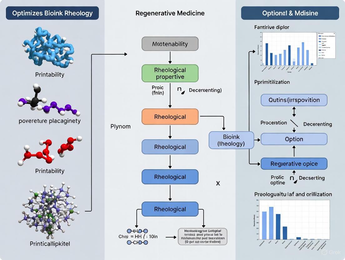
Workflow Visualization
Diagram 1: A systematic workflow for bioink development, linking rheological characterization directly to key printability assessments.
The Scientist's Toolkit: Research Reagent Solutions
Table 3: Essential Materials for Bioink Development and Printability Research
| Material / Reagent | Function / Rationale | Example Use Case |
|---|---|---|
| Sodium Alginate | A natural polysaccharide that provides excellent shear-thinning and rapid ionic crosslinking with divalent cations like Ca²⁺, beneficial for extrudability and initial structural integrity [5]. | Base polymer in hybrid bioinks for cartilage or soft tissue engineering. |
| Xanthan Gum | A bacterial polysaccharide used as a rheological modifier to enhance viscosity, yield stress, and thixotropic recovery, significantly improving shape fidelity [5]. | Blended with alginate to create stable, self-supporting filaments for complex structures. |
| Gelatin / GelMA | Provides biological motifs for cell adhesion and can be thermally gelled. Often combined with other polymers to improve bioactivity and tune mechanical properties [1]. | Used in composite bioinks to enhance cell viability and function in printed constructs. |
| Calcium Chloride (CaCl₂) | A divalent cation source used for ionic crosslinking of alginate-based bioinks, instantly improving the storage modulus (G′) and structural integrity post-printing [5]. | Applied as a mist or in a support bath to crosslink printed alginate-containing constructs. |
| Carbopol-based Support Bath | A yield-stress fluid that acts as a temporary, self-healing support medium for suspended bioprinting, enabling the fabrication of complex and fragile structures with low-viscosity bioinks [6]. | Used for printing intricate vascular networks or soft organoids that cannot support their own weight. |
In the field of 3D bioprinting, bioinks are specialized formulations designed to encapsulate and deliver living cells to create three-dimensional tissue constructs [1]. The rheological properties of a bioink are fundamental, as they directly influence its behavior during the bioprinting process, determining key outcomes such as printability, structural integrity, and cell viability [1] [7]. A persistent challenge in bioink development lies in reconciling the conflicting demands of rheology—essential for printability—and biological functionality, which is critical for supporting cell life and tissue maturation [1]. This guide details the essential rheological properties, their role in the bioprinting process, and provides practical troubleshooting advice for researchers aiming to optimize their bioink formulations.
Fundamental Rheological Properties and Their Roles
The successful deposition of a bioink and the stability of the resulting construct hinge on a set of key rheological properties. The table below summarizes these core properties and their impact on the bioprinting process.
Table 1: Essential Rheological Properties for Bioink Design
| Property | Definition | Role in Bioprinting | Consequence if Not Optimized |
|---|---|---|---|
| Viscosity | Resistance of a material to flow under applied shear stress [1]. | Governs extrudability and post-printing shape retention [1]. | High viscosity: Clogging, high extrusion pressure, cell damage [1]. Low viscosity: Poor resolution, filament collapse [1]. |
| Shear-Thinning | Decrease in viscosity with increasing shear rate [1]. | Enables smooth flow through the nozzle during extrusion and rapid recovery upon deposition [1] [7]. | Difficult extrusion, poor filament definition, or inability to maintain shape after printing. |
| Viscoelasticity | Simultaneous existence of viscous (liquid-like) and elastic (solid-like) properties [7]. | Storage Modulus (G'): Dominance ensures shape persistence after printing [7]. Loss Modulus (G"): Dominance facilitates flow during extrusion [7]. | Low G': Construct deformation or collapse under its own weight. Unbalanced G'/G": Either poor extrusion or poor shape fidelity. |
| Yield Stress | The minimum stress required to initiate flow [7]. | Allows the bioink to behave like a solid at rest, providing stability to the printed structure before and after crosslinking [5]. | Filament slumping or spreading after deposition, leading to loss of architectural precision. |
| Thixotropy | Time-dependent recovery of viscosity and structure after shear is removed [7]. | Mimics printing conditions; ensures the ink recovers its solid-like properties quickly after exiting the nozzle [7]. | Slow structural recovery leads to fusion between layers and poor stacking, resulting in a loss of 3D structure [8]. |
Experimental Protocols for Rheological Characterization
A standardized protocol for rheological characterization is crucial for reproducible bioink development and for correlating material properties with printability. The following workflow outlines a comprehensive testing procedure.
Detailed Rheological Test Procedures
The following tests should be performed using a rotational rheometer with a cone-plate or parallel-plate geometry [9].
Flow Sweep Test
- Purpose: To characterize the shear-thinning behavior of the bioink, which is critical for extrudability [7].
- Protocol: Measure the steady-state shear viscosity ((\eta)) across a shear rate range (e.g., 0.1 to 1000 s⁻¹) that encompasses the conditions during extrusion [1] [9]. The resulting flow curve will show how viscosity decreases with increasing shear rate.
Amplitude Sweep Test
- Purpose: To determine the Linear Viscoelastic Region (LVE) and identify the yield stress of the material [7].
- Protocol: At a fixed frequency (typically 1 Hz [9] or 100 rad/s [7]), apply an oscillatory strain from a low value (e.g., 0.05% [9]) to a high value (e.g., 10% [9]). The point where the storage modulus (G') deviates from linearity or crosses the loss modulus (G") indicates the end of the LVE and can be used to approximate the yield stress [7].
Frequency Sweep Test
- Purpose: To study the frequency-dependent viscoelastic behavior and determine the material's structural relaxation time [7].
- Protocol: Within the LVE (at the strain determined from the amplitude sweep), measure G' and G" over a frequency range (e.g., 0.1 to 10 Hz [9]). This test provides insight into the stability of the ink's internal structure over different timescales.
Thixotropy Test
- Purpose: To evaluate the self-healing nature and structural recovery of the bioink after extrusion [7].
- Protocol: Subject the bioink to alternating low and high oscillatory shear strains (or shear rates) to mimic the conditions before, during, and after printing [7]. The speed and extent to which G' recovers after the high-shear phase indicate the material's thixotropic recovery, which is vital for layer stacking [7].
Time Sweep Test
- Purpose: To measure the evolution of mechanical properties (final stiffness) during and after crosslinking [7].
- Protocol: After depositing the bioink or initiating the crosslinking process (e.g., by adding a crosslinker or exposing to UV light), perform an oscillatory time sweep at a fixed strain and frequency. The increase in G' over time indicates the stiffening and solidification of the construct [7].
Troubleshooting Guide: Common Rheology-Related Issues
This section addresses frequent challenges encountered during bioprinting, linking them to underlying rheological causes and proposing actionable solutions.
Table 2: Troubleshooting Common Bioprinting Issues
| Problem | Potential Rheological Cause(s) | Recommended Solutions |
|---|---|---|
| Needle Clogging [8] | Viscosity too high; Insufficient shear-thinning; Particle/aggregate size larger than nozzle diameter [8]. | - Increase printing pressure (while monitoring cell viability) [8]. - Use a larger needle gauge [10] [8]. - Ensure bioink homogeneity and characterize particle size if using additives [8]. |
| Layers merging or collapsing; poor 3D structure [8] | Insufficient viscosity and yield stress at rest; Slow thixotropic recovery; Inadequate crosslinking [8]. | - Increase bioink polymer concentration to enhance zero-shear viscosity and yield stress [1]. - Optimize crosslinking time and crosslinker concentration for faster solidification [10] [8]. - Reformulate bioink to improve thixotropy and structural recovery [7]. |
| Low Cell Viability Post-Printing | High shear stress during extrusion due to high viscosity or small nozzle size; Harsh crosslinking conditions [1] [10]. | - Use a larger needle diameter or a tapered needle tip to reduce shear stress [10]. - Reduce printing pressure [10]. - Optimize crosslinking method (e.g., use milder crosslinkers or lower concentrations) [10]. |
| Poor Filament Definition & Shape Fidelity | Low storage modulus (G') and yield stress; Overly rapid crosslinking diffusion causing surface imperfections [5]. | - Reformulate bioink to increase G' and yield stress, e.g., by using hybrid polymers [5]. - Adjust crosslinking protocol (e.g., use a support bath like Carbopol for suspended printing) [6]. - Optimize G-code parameters (print speed, pressure) to match material properties [5]. |
| Lack of Structural Integrity Post-Printing [8] | Insufficient or inappropriate crosslinking; Low final stiffness (G') of the crosslinked network [7] [8]. | - Characterize and optimize the crosslinking method (ionic, UV, thermal) for your bioink [8]. - For ionic crosslinking, test different crosslinker (e.g., CaCl₂) concentrations (e.g., 1.5-3%) [5]. - For photocrosslinking, ensure correct photoinitiator and wavelength are used [8]. |
The Scientist's Toolkit: Key Reagents and Equipment
The table below lists essential materials and instruments frequently used in the development and characterization of bioinks.
Table 3: Essential Research Reagents and Equipment for Bioink Development
| Item | Function/Application | Examples / Notes |
|---|---|---|
| Natural Polymers | Provide biocompatibility and bioactivity; form the base of many hydrogel bioinks [1] [11]. | Sodium Alginate [11] [12] [5], Gelatin Methacryloyl (GelMA) [11] [7], Collagen [11], Hyaluronic Acid [1]. |
| Synthetic Polymers | Offer tunable mechanical properties and printability for structural reinforcement [1]. | Polyethylene Glycol (PEG) [1] [11], Polycaprolactone (PCL) [1]. |
| Crosslinking Agents | Induce gelation and solidification of the bioink post-extrusion, providing mechanical stability [10] [12]. | Calcium Chloride (CaCl₂) for alginate [12] [5]; Photoinitiators (e.g., LAP) for GelMA and PEGDA [11]. |
| Rheometer | Essential instrument for characterizing the rheological properties of bioinks [9] [7]. | Equipped with cone-plate or parallel-plate geometry [9]. Must be capable of flow sweeps, oscillation tests, and temperature control. |
| Support Bath Materials | A yield-stress fluid used in suspended bioprinting to support low-viscosity bioinks and enable the printing of complex, overhanging structures [6]. | Carbopol-based baths [6], gelatin slurry, other microparticle gels. |
Frequently Asked Questions (FAQs)
Q1: What is the most critical rheological property for extrusion-based bioprinting? While all properties are interconnected, shear-thinning behavior is often considered fundamental. It enables the bioink to flow under the high shear stress in the nozzle and then immediately regain viscosity upon deposition to hold its shape. This property is a prerequisite for effective extrusion and shape fidelity [1] [7].
Q2: How can I increase the shape fidelity of my bioink without harming cells? This is a classic trade-off. Strategies include:
- Using hybrid bioinks: Combine a natural polymer (for biocompatibility) with a synthetic or another natural polymer (for mechanical strength) [1] [5]. For example, blending alginate with gelatin or xanthan gum can improve printability and structural integrity [7] [5].
- Employing a support bath: Suspended bioprinting allows the use of softer, more cell-friendly bioinks, as the support bath (e.g., Carbopol) provides the external structure needed during printing [6].
- Optimizing crosslinking: Implement a dual-crosslinking strategy (e.g., ionic followed by UV) to achieve rapid initial stabilization and long-term stability [7].
Q3: My bioink clogs the nozzle, but reducing viscosity harms structural integrity. What can I do? Instead of reducing the overall polymer concentration, which weakens the gel, try:
- Increasing the printing temperature if your bioink is thermoresponsive (e.g., GelMA-based) [7].
- Switching to a larger nozzle diameter [10]. Note that this reduces printing resolution.
- Ensuring full dissolution and homogeneity of the polymers to prevent micro-gels that can cause clogs [8].
Q4: How do I know if my bioink has sufficient yield stress? A simple qualitative test is to extrude a filament onto a surface. If the filament slumps or spreads significantly under its own weight, the yield stress is likely too low. Quantitatively, the yield stress can be determined from an amplitude sweep test on a rheometer as the stress at which G' begins to drop significantly, indicating the onset of flow [7].
Frequently Asked Questions (FAQs)
Fundamental Concepts
What is the core "inherent trade-off" in bioink design? The fundamental trade-off lies in balancing rheological properties (essential for printability and structural integrity) against biological functionality (essential for cell viability and function). Optimizing one often compromises the other. For example, increasing polymer concentration to enhance viscosity and mechanical strength for better printability can hinder nutrient diffusion and reduce cell viability, while formulations optimized for cell survival may lack the structural robustness needed for high-fidelity printing [1] [13].
Why is shear-thinning behavior so critical for a bioink? Shear-thinning is a key rheological property where the bioink's viscosity decreases under applied shear stress (e.g., during extrusion through a nozzle) but recovers once the stress is removed. This behavior is crucial because it:
- Enables smooth extrusion and prevents clogging by reducing flow resistance during printing.
- Facilitates post-deposition shape retention and structural stability, as the bioink regains its viscosity after being deposited [1] [13].
How do bioink components like alginate and gelatin contribute differently to the formulation? Different polymers offer complementary properties, which is why they are often used in composite bioinks:
- Alginate: Provides excellent shear-thinning behavior and allows for rapid ionic cross-linking (e.g., with CaCl₂), which is crucial for stabilizing printed structures. However, it lacks inherent cell-adhesive motifs [14] [5].
- Gelatin (and its derivative GelMA): Offers inherent bioactivity and cell-adhesion sites, supporting cell viability and proliferation. It can be covalently cross-linked (e.g., via UV light) for long-term stability [14] [15]. Combining them creates a hybrid system where alginate improves printability and GelMA supports biological function [14].
Troubleshooting Common Experimental Issues
My bioink requires very high pressure to extrude, and I observe low cell viability after printing. What should I investigate? High extrusion pressure and subsequent cell damage are frequently linked to excessive shear stress. To troubleshoot, examine these factors:
- Bioink Viscosity: Characterize your bioink's zero-shear viscosity. An excessively high viscosity may require a reduction in polymer concentration or the use of a polymer with a lower molar mass [14] [16].
- Nozzle Diameter: Consider increasing the nozzle diameter. A smaller diameter significantly increases flow resistance and shear stress [15] [16].
- Printing Parameters Systematically: Reduce the printing pressure and/or increase the printing speed. However, note that these parameters are interdependent and require careful balancing to maintain print fidelity [15].
The initial layers of my construct spread excessively after deposition, leading to poor shape fidelity. How can I improve structural integrity? Poor shape fidelity typically indicates insufficient structural stability post-deposition. Solutions include:
- Modify Bioink Formulation: Increase the bioink's yield stress and storage modulus (G′). This can be achieved by slightly increasing the concentration of the structural polymer (e.g., alginate) or incorporating a reinforcing agent like nano-clay (laponite) [17] [5].
- Optimize Cross-linking: Implement a dual-cross-linking strategy. For a GelMA/alginate bioink, this involves immediate ionic cross-linking of alginate upon deposition to provide initial green strength, followed by UV cross-linking of GelMA for final mechanical robustness [14] [13].
- Evaluate Rheological Properties: Ensure your bioink has a loss tangent (G″/G') in an optimal range (e.g., 0.25–0.45 for gelatin-alginate composites), which balances extrusion uniformity and structural integrity [18].
I am getting inconsistent printing results between different batches of the same bioink formulation. What could be the cause? Batch-to-batch inconsistency often stems from variations in raw material properties or processing conditions. Focus on:
- Polymer Molar Mass: The molar mass of polymers like sodium alginate is a critical but often overlooked parameter. Source alginate with consistent molar mass specifications, and be aware that sterilization processes like autoclaving can degrade molar mass and alter rheology [14].
- Sterilization Protocol: Standardize the sterilization method for your bioink components. If using autoclaving for alginate, monitor and control the duration precisely to avoid variable polymer degradation [14].
- Adopt a DoE Framework: Instead of a one-factor-at-a-time approach, use Design of Experiment (DoE) to systematically understand the interaction of multiple factors (e.g., concentrations, cross-linking time) and create a robust, reproducible formulation space [17] [19].
Table 1: Key Rheological Parameters and Their Impact on Printability and Cell Viability
| Rheological Parameter | Optimal Range / Target Value | Impact on Printability | Impact on Cell Viability |
|---|---|---|---|
| Zero-Shear Viscosity | Target-specific; requires balancing | High viscosity improves shape fidelity but may cause clogging. Low viscosity leads to spreading. | High viscosity can require high extrusion pressure, increasing shear-induced cell death [1]. |
| Loss Tangent (G″/G') | 0.25 - 0.45 (for gelatin-alginate) [18] | Lower values correlate with better structural integrity. Higher values improve extrusion uniformity. | An optimal balance ensures gentle extrusion and a stable 3D environment for cells. |
| Thixotropic Recovery | Rapid (seconds) [5] | Faster recovery helps maintain the shape of multilayered structures. | Indirectly supports viability by creating a stable microenvironment. |
| Shear-Thinning Index (n) | n < 1 (Power Law model) [1] | Strong shear-thinning enables easy flow during extrusion and quick solidification after. | Reduces the shear stress experienced by cells during the extrusion process. |
Table 2: Comparison of Common Bioink Formulations and Their Characteristics
| Bioink Formulation | Key Advantages | Key Limitations | Typical Cell Viability Range |
|---|---|---|---|
| GelMA / Alginate | Tunable rheology; dual cross-linking (ionic/UV); cell-adhesive [14]. | Molar mass of alginate and sterilization methods critically affect properties [14]. | Varies with cross-linking; can be optimized for high viability. |
| Gelatin-Alginate Composite | Favorable biocompatibility; thermoresponsive [17]. | Limited mechanical strength for hard tissues; sensitivity to temperature. | 40% - 90% (Extrusion-based) [16]. |
| Alginate-Xanthan Gum | Enhanced shear-thinning and thixotropy; high print fidelity [5]. | Lack of inherent cell adhesiveness requires modification. | Data not provided in sources. |
| Hybrid (with Laponite) | Improved storage modulus and shape fidelity; supports cell attachment [17]. | Nanoparticle content must be optimized to avoid cytotoxicity. | Can be formulated for >95% viability at low extrusion pressure [17]. |
Detailed Experimental Protocols
Protocol: Systematic Printability Optimization Using Design of Experiments (DoE)
This protocol provides a framework for efficiently optimizing a multi-component bioink, moving beyond traditional trial-and-error [17] [19].
1. Objective Definition
- Define clear Response Variables: e.g., storage modulus (G′), zero-shear viscosity, extrusion pressure, printed strand width, and cell viability.
- Define the Factors and Ranges: Identify the components and process parameters to be investigated (e.g., concentrations of Alginate, GelMA, Gelatin; cross-linker concentration; printing pressure). Set minimum and maximum levels for each based on preliminary data.
2. Experimental Design and Data Fitting
- Select a DoE approach, such as a Central Composite Design (CCD) for Response Surface Methodology.
- Use statistical software (e.g., Minitab) to generate a set of experimental runs.
- Prepare and test bioinks according to the DoE matrix.
- Measure the response variables for each run.
- Fit the data to a model (e.g., a quadratic polynomial) and perform analysis of variance (ANOVA) to identify significant factors and interaction effects.
3. Multi-Response Optimization and Validation
- Use a Multi-Response Optimization (MRO) tool or algorithm to find the parameter settings that simultaneously satisfy all the targets for your responses (e.g., "maximize cell viability," "minimize extrusion pressure," "target a storage modulus of X Pa").
- The output will be an optimized bioink formulation and/or printing parameters.
- Prepare the optimized bioink and experimentally validate the model's predictions by measuring the key responses.
Diagram 1: DoE Optimization Workflow
Protocol: Rheological Characterization of Bioinks
A standardized rheological assessment is crucial for predicting printability [18] [5].
1. Steady-State Shear Flow Test
- Purpose: To determine viscosity versus shear rate and identify shear-thinning behavior.
- Method:
- Use a parallel plate or cone-plate geometry on a rheometer.
- Set a constant temperature (e.g., 20-25°C for printing, 37°C for physiological conditions).
- Logically ramp the shear rate from a low to a high value (e.g., 0.01 to 100 s⁻¹).
- Key Outputs: Flow curve (viscosity vs. shear rate). Fit data to the Power-Law model to obtain the flow behavior index (n) and consistency index (K).
2. Oscillatory Amplitude Sweep Test
- Purpose: To determine the linear viscoelastic region (LVR), storage modulus (G′), loss modulus (G″), and yield stress.
- Method:
- Using oscillatory mode, apply a small, constant frequency (e.g., 1 Hz) while gradually increasing the shear strain or stress.
- Key Outputs: The point where G′ drops sharply indicates the yield stress, beyond which the material flows. The values of G′ and G″ within the LVR indicate the inherent solid-like and liquid-like character, respectively.
3. Oscillatory Frequency Sweep Test
- Purpose: To characterize the viscoelastic structure of the bioink over different timescales.
- Method:
- Within the LVR, apply a small, constant strain while sweeping over a range of frequencies (e.g., 0.1 to 100 rad/s).
- Key Outputs: G′(ω) and G″(ω). A bioink where G′ > G″ across the frequency range is considered a solid-like gel, which is desirable for shape retention.
The Scientist's Toolkit: Research Reagent Solutions
Table 3: Essential Materials for Bioink Development and Characterization
| Category / Item | Specific Example(s) | Primary Function in Bioink Development |
|---|---|---|
| Natural Polymers | Sodium Alginate (various Mw), Gelatin, Gelatin Methacryloyl (GelMA), Hyaluronic Acid [14] [19] | Provide biocompatibility, bioactivity, and a base for hydrogel formation. Often modified to enable cross-linking. |
| Synthetic Polymers / Reinforcements | Polyethylene Glycol (PEG), Laponite (nano-clay) [17] | Provide tunable mechanical properties (PEG) or act as a rheological modifier to enhance yield stress and shape fidelity (Laponite). |
| Cross-Linking Agents | Calcium Chloride (CaCl₂), Lithium Phenyl-2,4,6-trimethylbenzoylphosphinate (LAP photoinitiator) [14] | Enable solidification of the bioink post-printing via ionic (Ca²⁺ for alginate) or covalent (LAP for UV-cross-linking of GelMA) mechanisms. |
| Characterization Tools | Rotational Rheometer, Size-Exclusion Chromatography (SEC) | Rheometer quantifies key printability parameters (viscosity, G′, G″). SEC characterizes polymer molar mass, a critical but variable property [14]. |
Diagram 2: Factors Governing Bioink Performance
Hydrogels are the cornerstone materials, or bioinks, for 3D bioprinting. Their role extends beyond being a simple cell carrier; they must provide structural support during and after printing while simultaneously sustaining cellular viability, proliferation, and differentiation [1]. The rheological properties of a hydrogel—essentially, how it flows and deforms—are fundamental to its performance. These properties directly dictate the printability of a bioink, including its ability to be extruded through a fine nozzle, maintain its shape post-deposition, and achieve structural fidelity in complex constructs [1] [5]. A persistent challenge in the field is the inherent trade-off between rheological properties and biological functionality. Optimizing for mechanical strength and printability often involves polymer compositions or crosslinking densities that can negatively impact cell viability and function [1].
The composition of a hydrogel—whether it is derived from natural sources, synthesized in a lab, or a hybrid of both—is the primary determinant of its rheological signature. Understanding the distinctions between these classes is the first step in rationally designing or selecting a bioink for a specific application in tissue engineering and regenerative medicine.
Natural vs. Synthetic Hydrogels: A Comparative Analysis
The choice between natural and synthetic polymers involves a series of trade-offs. The table below summarizes the key characteristics, advantages, and disadvantages of each class.
Table 1: Comparison of Natural and Synthetic Hydrogels for Bioink Formulation
| Feature | Natural Hydrogels | Synthetic Hydrogels |
|---|---|---|
| Examples | Alginate, Gelatin, Chitosan, Hyaluronic Acid, Collagen [1] [20] | Polyethylene Glycol (PEG), Polycaprolactone (PCL), Poly(vinyl alcohol) (PVA) [1] [21] |
| Biocompatibility & Bioactivity | Typically excellent; often contain cell adhesion motifs and support tissue remodeling [1] [20] | Often limited; can be bio-inert unless functionalized with bioactive peptides [1] [20] |
| Mechanical Strength | Generally poor; soft and susceptible to premature degradation [22] [20] | Highly tunable; can be engineered for a wide range of mechanical properties [1] [22] |
| Batch-to-Batch Variability | High, due to biological sourcing [20] | Low, offering high reproducibility [20] |
| Degradation Profile | Enzymatic and unpredictable [20] | Controllable and predictable via polymer chemistry [20] |
| Typical Rheological Signature | Often exhibit shear-thinning but may lack structural integrity without crosslinking [5] | Rheology can be precisely designed, but may lack inherent shear-thinning [1] |
The Hybrid Approach
To overcome the limitations of single-component systems, hybrid hydrogels have been developed. These combine natural and synthetic polymers to achieve a synergistic effect, such as the biocompatibility of natural polymers with the mechanical robustness and tunability of synthetic ones [1] [20]. For instance, alginate is frequently blended with other polymers like xanthan gum or gelatin to improve its printability and elasticity [5].
Troubleshooting Guide: Common Rheological and Biological Issues
This section addresses frequent challenges encountered during the development and printing of hydrogel-based bioinks.
FAQ: Rheology and Printability
Q1: My bioink is too viscous and will not extrude smoothly, or it requires very high pressure. What can I do?
- Cause: The polymer concentration or crosslinking density is too high, leading to excessive viscosity and potential nozzle clogging.
- Solutions:
- Dilute the bioink: Systematically decrease the polymer concentration and re-evaluate rheology.
- Increase printing temperature: If using a thermoresponsive polymer, a slight temperature increase can lower viscosity.
- Use a larger nozzle diameter: This reduces shear stress and required extrusion pressure [23].
- Formulate for better shear-thinning: Ensure your bioink has pronounced shear-thinning behavior, where viscosity decreases under the shear stress of extrusion [1] [5].
Q2: My printed construct slumps, spreads, or lacks definition. How can I improve shape fidelity?
- Cause: Insufficient structural integrity, which can be due to low yield stress, slow recovery (thixotropy), or inadequate crosslinking.
- Solutions:
- Increase polymer concentration or crosslinker density: This enhances the hydrogel's storage modulus (G′) and yield stress, helping it resist deformation [1] [22].
- Optimize crosslinking kinetics: The bioink should transition from a fluid to a solid-like state rapidly after deposition. Consider using rapid ionic crosslinking (e.g., CaCl₂ for alginate) or photo-crosslinking [1] [5].
- Add a rheological modifier: Incorporate a polymer like xanthan gum to enhance viscoelasticity and thixotropic recovery [5].
Q3: How does my choice between a natural and synthetic polymer directly impact the printing process?
- Answer: Natural polymers like alginate and gelatin often exhibit excellent shear-thinning and biocompatibility but may require blending or crosslinking to achieve mechanical stability. Synthetic polymers like PEG offer superior and tunable mechanical strength but may require chemical modification to be printable and may not support cell adhesion without functionalization [1]. The decision often hinges on whether biological function or mechanical precision is the primary requirement for your application.
FAQ: Cell Viability and Biological Function
Q4: Cell viability is low after printing. What are the most likely causes?
- Cause: High shear stress during extrusion is a primary culprit. This can be caused by high viscosity, high flow rates, or small nozzle diameters [10] [23].
- Solutions:
- Optimize process parameters: Reduce the extrusion pressure or flow rate. Use a larger nozzle diameter or a tapered nozzle to reduce shear stress [10] [23].
- Characterize bioink rheology: Ensure the bioink has strong shear-thinning properties to minimize viscosity during extrusion [1].
- Control print time: Prolonged printing time can subject cells to non-optimal conditions; optimize the bioink formulation for faster printing sessions [10].
Q5: My cells are not proliferating or functioning correctly within the printed construct.
- Cause: The hydrogel environment may not be bio-instructive. This is common with synthetic hydrogels that lack cell adhesion sites, or if the crosslinking density is too high, restricting nutrient diffusion and cell spreading [1].
- Solutions:
- Use natural or hybrid hydrogels: Incorporate natural ECM-derived components like collagen or gelatin methacrylate (GelMA) [1].
- Functionalize synthetic hydrogels: Incorporate bioactive peptides (e.g., RGD) into synthetic networks to promote cell adhesion [1] [20].
- Modulate crosslinking density: Reduce the degree of crosslinking to increase mesh size and facilitate cell migration and nutrient transport [1].
Q6: How can I systematically identify the cause of low viability?
- Answer: Implement a rigorous control strategy [10]:
- 2D Control: Culture cells on a standard plate to confirm the health of your initial cell stock.
- 3D Pipette Control: Create a non-printed, pipetted hydrogel thin film with encapsulated cells. This isolates material toxicity and crosslinking effects from printing-induced shear stresses.
- 3D Print Control: Print a simple structure (e.g., a thin film) to assess the combined impact of your bioink and printing process on viability.
Experimental Protocols for Rheological Characterization
Standardized characterization is key to reproducible bioink development. Below are key methodologies.
Workflow for Bioink Development and Validation
The following diagram outlines a systematic workflow for developing and optimizing a bioink, integrating material selection, rheological characterization, and biological validation.
Key Rheological Tests and Parameters
Table 2: Essential Rheological Tests for Bioink Characterization
| Test Type | Parameters Measured | Interpretation and Significance |
|---|---|---|
| Flow Ramp / Viscosity Curve | Viscosity (η) vs. Shear Rate (γ̇) | Confirms shear-thinning behavior. A decreasing viscosity with increasing shear rate is essential for easy extrusion and minimal cell damage [1] [5]. |
| Amplitude Sweep | Storage Modulus (G′), Loss Modulus (G″), Yield Stress (τy) | Determines the linear viscoelastic region (LVR) and the stress required to cause the gel to flow (yield). A higher G′ than G″ indicates solid-like behavior. Yield stress is critical for shape retention [24] [5]. |
| Frequency Sweep | G′ and G″ vs. Angular Frequency (ω) | Probes the internal gel structure. G′ should be greater than G″ across a wide frequency range for structural stability. A crossover indicates a sol-gel transition [21]. |
| Thixotropic Recovery | Viscosity or Modulus Recovery vs. Time | Measures how quickly the material recovers its structure after the high shear of extrusion. Fast recovery is vital for multi-layer printing [5]. |
Protocol: Measuring Shear-Thinning Behavior and Yield Stress
Objective: To characterize the flow properties of a hydrogel bioink and determine its suitability for extrusion-based printing.
Materials and Equipment:
- Rheometer (cone-plate or parallel plate geometry)
- Prepared bioink solution
- Temperature control unit
Methodology:
- Loading: Carefully load the bioink onto the rheometer plate, ensuring no air bubbles are trapped.
- Equilibration: Allow the sample to equilibrate at the printing temperature (e.g., 20-37°C).
- Flow Curve Measurement:
- Set the measurement type to a steady-state flow sweep.
- Program a shear rate range that covers the expected conditions during printing (e.g., 0.01 to 100 s⁻¹).
- Record the viscosity (η) as a function of shear rate (γ̇).
- Amplitude Sweep Measurement:
- Set the measurement to an oscillatory amplitude sweep.
- Apply a constant frequency (e.g., 1 Hz) and logarithmically increase the oscillatory stress or strain.
- Record the storage modulus (G′) and loss modulus (G″).
- Data Analysis:
- Shear-thinning: Plot viscosity vs. shear rate. A power-law model (η = Kγ̇ⁿ⁻¹) can be fitted, where a flow behavior index (n) < 1 confirms shear-thinning [1] [5].
- Yield Stress: Identify the point where G′ sharply decreases and crosses G″ (flow point). The stress at this point is often reported as the yield stress, indicating the stress required to initiate flow [24].
The Scientist's Toolkit: Essential Research Reagents
Table 3: Key Materials and Their Functions in Bioink Development
| Reagent / Material | Function / Application |
|---|---|
| Sodium Alginate | A natural polymer for its rapid ionic crosslinking with Ca²⁺, providing a mild gelation process for cell encapsulation [1] [5]. |
| Gelatin Methacrylate (GelMA) | A semi-synthetic polymer offering excellent bioactivity from gelatin and tunable mechanical properties via UV-induced crosslinking [1] [20]. |
| Polyethylene Glycol (PEG) | A synthetic polymer used as a blank slate bioink; its mechanical and biochemical properties can be precisely controlled through chemistry and functionalization [1]. |
| Xanthan Gum | A natural rheological modifier used to enhance the shear-thinning behavior and viscoelasticity of other polymers like alginate [5]. |
| Calcium Chloride (CaCl₂) | A divalent cation source used to ionically crosslink alginate-based bioinks, instantly providing structural integrity [5]. |
| Photoinitiators (e.g., LAP, Irgacure 2959) | Chemicals that generate radicals upon UV or visible light exposure to initiate the crosslinking of polymers like GelMA or PEG-diacrylate [22]. |
Advanced Topics: Towards Smart Bioinks
The future of bioinks lies in "smart" or stimuli-responsive systems. These are hydrogels designed to change their properties (e.g., shape, stiffness, degradation) in response to specific environmental cues such as temperature, pH, or light [20]. This field, often referred to as 4D bioprinting, introduces the dimension of time, where printed constructs evolve post-fabrication to better mimic dynamic native tissues [20]. Furthermore, AI-driven design is emerging as a powerful tool to predict hydrogel properties, optimize printability, and model the complex time-dependent behavior of smart bioinks, thereby accelerating the development of next-generation bioinks for personalized medicine [20].
In the field of 3D bioprinting, cross-linking is the fundamental process that transforms a liquid bioink into a stable, three-dimensional structure. This process significantly influences the mechanical properties, structural fidelity, and biological functionality of the final constructed. The kinetics of gelation—whether achieved through chemical, ionic, or thermal methods—directly determines the printability of a bioink, affecting its extrusion behavior, shape retention, and ultimately, its suitability for creating functional tissues. Optimizing these strategies is essential for balancing the often-conflicting demands of printability and cell viability [25] [13].
The rheological properties of a bioink are inextricably linked to its cross-linking mechanism. Key parameters such as viscosity, shear-thinning behavior, yield stress, and gelation kinetics govern how a bioink flows during extrusion and how it maintains structure after deposition. Understanding the interplay between cross-linking strategy and rheology is therefore paramount for researchers aiming to develop advanced bioinks for regenerative medicine and drug development applications [26] [27].
Fundamentals of Cross-Linking Strategies
Cross-linking methods can be broadly categorized into physical and chemical pathways, each with distinct mechanisms, kinetics, and implications for bioink design.
Physical Cross-Linking
Ionic Interactions
Ionic cross-linking involves the addition of multivalent cations to an anionic polymer solution to induce gelation. This method is most commonly used with sodium alginate, a polysaccharide whose carboxylic groups bind with multivalent cations like Ca²⁺ to form an "egg-box" structure [25]. This rapid cross-linking method occurs under mild conditions—at room temperature and physiological pH—making it cell-friendly. However, ionically cross-linked hydrogels can be mechanically weak and may exhibit poor long-term stability due to the potential release of metal ions [25].
Thermal Gelation
Thermosensitive hydrogels, such as gelatin, pluronics, and methylcellulose, undergo sol-gel transitions in response to temperature changes [26]. These materials are typically liquid at lower temperatures and form gels upon warming. The gelation kinetics are controlled by temperature and polymer concentration. Thermal gelation provides a reversible cross-linking mechanism but may require supportive strategies to ensure structural integrity at physiological temperatures [26].
Chemical Cross-Linking
Chemical cross-linking creates permanent, covalent bonds between polymer chains, resulting in networks with superior mechanical strength and stability. Common approaches include:
- Photocrosslinking: Uses ultraviolet (UV) or visible light in the presence of photoinitiators to trigger polymerization [25]. Materials like gelatin methacryloyl (GelMA) and polyethylene glycol (PEG) are frequently cross-linked this way.
- Enzymatic Cross-linking: Employ enzymes such as thrombin to catalyze bond formation [25].
- Schiff Base Formation and Click Chemistry: Specific chemical reactions that create stable covalent linkages [25].
While chemically cross-linked hydrogels provide excellent shape fidelity, the cross-linking kinetics must be carefully controlled to avoid printer nozzle blockage, and the process may expose cells to potentially cytotoxic compounds or radiation [25] [10].
The following diagram illustrates the decision-making workflow for selecting an appropriate cross-linking strategy based on bioink composition and target tissue properties.
Troubleshooting Common Cross-Linking Issues
This section addresses frequently encountered problems in bioprinting related to cross-linking strategies, providing practical solutions for researchers.
FAQ: Frequently Asked Questions
Q1: My bioprinted construct lacks structural integrity and collapses after printing. How can I improve its stability? Structural collapse typically indicates insufficient or poorly timed cross-linking [8]. First, characterize your bioink's rheological properties, particularly its shear-thinning behavior and yield stress [26] [13]. For ionic cross-linking, optimize the concentration of the cross-linking agent (e.g., CaCl₂ for alginate) [8]. For photocrosslinkable bioinks, ensure the appropriate wavelength and exposure time are used [8]. Consider using a hybrid cross-linking strategy; for example, mixing a low-viscosity polymer like sodium alginate with GelMA creates an interpenetrated network that can be rapidly ionically cross-linked followed by photocurring for enhanced stability [28].
Q2: How does the cross-linking method affect the viability of cells within my bioink? Different cross-linking methods present unique challenges to cell viability [10]. Chemical cross-linking may involve cytotoxic cross-linkers or generate exothermic reactions [25]. Photocrosslinking requires careful selection of photoinitiator concentration and UV exposure to avoid DNA damage or reactive oxygen species generation [25] [10]. Ionic cross-linking, while generally cell-friendly, can be compromised by high ion concentrations [25]. Always run a 3D pipette control for each material, cross-linking process, and cell type to isolate viability issues specific to your cross-linking method [10].
Q3: I am experiencing nozzle clogging during the printing process. Is this related to my cross-linking strategy? Yes, premature cross-linking is a common cause of clogging. For photocurable hydrogels, ensure you are using an opaque nozzle to protect the bioink from premature light exposure [28]. For ionic cross-linking, check that the cross-linker solution is not inadvertently contacting the bioink within the printhead. For thermally gelling bioinks, verify that the printing environment and syringe temperature are controlled to prevent gelation before extrusion [26]. In all cases, optimizing the cross-linking kinetics is crucial to ensure gelation occurs after deposition, not before [25].
Q4: The layers of my multi-layer construct are merging instead of stacking neatly. What parameters should I adjust? This issue often stems from slow gelation kinetics or an insufficient degree of cross-linking in the previously deposited layers [8]. The bottom layer must achieve sufficient structural integrity to support subsequent layers. Optimize your cross-linking time to ensure rapid solidification after deposition [8]. You can also adjust the bioink viscosity and ensure it exhibits adequate shear-thinning and recovery [8] [26]. Performing rheological tests to understand the thixotropic nature of your bioink is essential for solving this problem [8].
Troubleshooting Guide: Common Print Defects and Solutions
Table 1: Troubleshooting common bioprinting issues related to cross-linking.
| Problem | Potential Causes | Recommended Solutions |
|---|---|---|
| No structural integrity [8] | Insufficient cross-linking density; Slow gelation kinetics; Low bioink viscosity | Optimize cross-linker concentration [8]; Use hybrid cross-linking (e.g., ionic + light) [28]; Increase polymer percentage if possible [28]. |
| Poor layer stacking [8] | Slow cross-linking recovery; Low yield stress; High layer sagging | Increase cross-linking rate between layers [8]; Perform rheological tests to optimize thixotropy [8] [26]; Adjust layer height and print speed. |
| Nozzle clogging [28] | Premature cross-linking; Bioink heterogeneity; High viscosity | Use opaque nozzles for photoinks [28]; Ensure homogeneous bioink [8]; Increase nozzle gauge size; Use tapered needle tips [10]. |
| Low cell viability [10] | Cytotoxic cross-linkers; High shear stress during extrusion; Harsh cross-linking conditions | Switch to milder cross-linking (e.g., ionic vs. chemical) [25]; Reduce print pressure [10]; Use larger needle gauges [10]; Test cross-linker biocompatibility [10]. |
| Shape deformation [27] [13] | Low yield stress; Inadequate viscoelasticity; Slow stress relaxation | Formulate bioinks with higher yield stress [27] [13]; Optimize cross-linking for rapid network recovery; Use supporting bath (e.g., FRESH) [28]. |
The Scientist's Toolkit: Essential Reagents and Materials
Table 2: Key research reagents and materials for cross-linking strategies in bioink formulation.
| Reagent/Material | Function in Cross-Linking | Common Applications |
|---|---|---|
| Sodium Alginate [25] | Ionic gelation with divalent cations (e.g., Ca²⁺). | A base bioink material, often blended with other polymers for improved printability. |
| Gelatin Methacryloyl (GelMA) [27] | Photocrosslinking under UV/visible light with a photoinitiator. | A versatile bioink for creating cell-laden constructs with tunable mechanical properties. |
| Calcium Chloride (CaCl₂) [25] | A common source of Ca²⁺ ions for cross-linking alginate. | Used as an ionic cross-linker, often in a bath for post-printing stabilization. |
| Photoinitiators (e.g., LAP, Irgacure 2959) [25] | Absorb light energy to generate radicals, initiating polymerization. | An essential component for photocrosslinking of materials like GelMA and PEGDA. |
| Pluronic F127 [26] | Thermally gelling polymer that acts as a sacrificial support material. | Used in FRESH bioprinting to support low-viscosity hydrogels during printing. |
| Gellan Gum [25] | Ionic cross-linking with metal ions, similar to alginate. | Used as a base or supplementary polymer to enhance mechanical strength. |
Experimental Protocols for Cross-Linking Optimization
Protocol 1: Rheological Characterization of Bioink Cross-Linking Kinetics
Purpose: To quantitatively assess the gelation kinetics and viscoelastic properties of a bioink during the cross-linking process [26] [27].
Materials:
- Rheometer with parallel plate or cone-and-plate geometry
- Temperature control unit (Peltier system)
- Bioink sample
- Cross-linking trigger (e.g., UV light attachment, humidified chamber for ionic cross-linker diffusion)
Method:
- Sample Loading: Load the uncross-linked bioink onto the rheometer plate. Maintain a constant gap suitable for the material viscosity.
- Time Sweep Test: Initiate a time sweep in oscillatory mode at a fixed frequency (e.g., 1 Hz) and strain (within the linear viscoelastic region).
- Trigger Cross-linking: After a stable initial reading, trigger the cross-linking mechanism:
- Photocrosslinking: Activate the UV light source for a predetermined duration.
- Ionic Cross-linking: Introduce a controlled mist of cross-linking solution (e.g., CaCl₂) at the sample edge.
- Thermal Gelation: Ramp the temperature to the gelation point.
- Data Monitoring: Monitor the storage modulus (G′) and loss modulus (G″) over time until they plateau. The point where G′ surpasses G″ indicates the gelation point.
- Analysis: Calculate the gelation time and rate from the evolution of G′. The final G′ value indicates the stiffness of the cross-linked network [26] [27].
Protocol 2: Printability and Shape Fidelity Assessment
Purpose: To evaluate the performance of a cross-linking strategy in a practical printing context.
Materials:
- 3D bioprinter
- Standardized printing nozzle (e.g., 25G)
- Bioink and necessary cross-linking equipment (e.g., light source, cross-linking bath)
- Imaging system (microscope or camera)
Method:
- Design a Test Structure: Create a simple 3D model, such as a grid or a multi-layered hollow cube, to assess filament uniformity, stacking, and bridging.
- Establish Printing Parameters: Calibrate pressure and speed to achieve consistent extrusion.
- Print and Cross-link: Print the structure while applying the cross-linking method simultaneously or immediately after deposition.
- Image Acquisition: Capture high-resolution images of the printed construct from top and side views.
- Quantitative Analysis:
- Filament Diameter Consistency: Measure filament diameter at multiple points and calculate the coefficient of variation.
- Strut Morphology: Compare printed strut diameter to the nozzle gauge diameter to detect over- or under-extrusion [8].
- Pore Fidelity: Measure the angles and dimensions of pores in the grid structure. Ideal pores are square; deviation indicates spreading or collapse [27].
- Layer Alignment: For multi-layer structures, check if needles embed into previous layers (Z-height too low) or if layers fail to bond (Z-height too high) [8].
The following diagram outlines the logical sequence of experiments for systematically optimizing a bioink, from initial rheological screening to final functional validation.
From Theory to Bioprinting: Rheological Characterization and Advanced Formulation Strategies
For researchers in tissue engineering and drug development, mastering the rheological characterization of bioinks is a critical step toward achieving high printability and cell viability. This guide details the four essential rheological tests, providing troubleshooting FAQs and experimental protocols to optimize your bioink development process.
#1 The Scientist's Toolkit: Essential Research Reagents and Materials
The table below lists key materials and instruments commonly used in the rheological analysis of bioinks.
| Item | Function/Brief Explanation |
|---|---|
| Hyaluronic Acid | Natural polymer providing a cell-friendly environment and contributing to bioink viscosity. [29] |
| Sodium Alginate | A primary determinant of bioink viscosity; forms soft networks through ionic cross-linking (e.g., with CaCl₂). [29] [7] |
| Gelatin Methacrylate (GelMA) | Provides thermo-responsive properties and cell-attachment motifs (e.g., RGD); can be covalently cross-linked via UV light. [7] |
| Carboxymethyl Cellulose (CMC) | A viscosity modifier used to create hybrid hydrogels, improving printability and shape fidelity. [30] [7] |
| Dextran | A polymer used in bioink formulations to adjust rheological properties. [29] |
| Anton Paar MCR Rheometer | A rotational rheometer used for conducting oscillatory and rotational tests. [29] [31] |
| RheoCompass Software | Software for rheometers that allows for predefined measurement templates and customized test definitions. [32] |
| Parallel Plate Geometry (e.g., 25 mm) | A standard measuring geometry for rheological tests on hydrogel samples. [29] |
#2 Core Rheological Tests: Purpose and Protocol
Flow Sweep Test
- Purpose: A rotational test that characterizes the viscous flow behavior of a bioink. It is crucial for identifying shear-thinning behavior, where viscosity decreases under increasing shear rate, a property essential for smooth extrusion during bioprinting. [7] [31]
- Experimental Protocol:
- Instrument Setup: Load the bioink sample onto the rheometer, often using a parallel plate geometry (e.g., 25 mm diameter). Carefully trim excess sample and set the measurement gap (e.g., 1 mm). [29]
- Test Parameters: Program a linear ramp of the shear rate, typically from 1 to 100 s⁻¹. The temperature should be controlled, commonly at 25°C (room temperature) or 37°C (physiological temperature). [29] [7]
- Data Collection: The software (e.g., RheoCompass) records the viscosity and shear stress as a function of the applied shear rate, generating a flow curve. [29]
Amplitude Sweep Test
- Purpose: An oscillatory test performed to determine the linear viscoelastic (LVE) region and identify the yield stress of the material. It measures how the bioink's structure responds to increasing deformation. [7]
- Experimental Protocol:
- Instrument Setup: Use the same geometry as for other oscillatory tests.
- Test Parameters: The test is performed at a fixed frequency (typically 10 rad/s or 1 Hz) while the shear strain amplitude is progressively increased, often from about 0.01% to 100%. [7]
- Data Collection: The software records the storage modulus (G′), loss modulus (G″), and shear stress as functions of the applied strain. The point where G′ starts to decrease significantly marks the end of the LVE region and indicates the yield point. [7]
Frequency Sweep Test
- Purpose: An oscillatory test used to investigate the frequency-dependent viscoelastic behavior of the bioink within its linear viscoelastic region. It provides information about the material's structural stability and relaxation behavior. [7]
- Experimental Protocol:
- Instrument Setup: Ensure the sample is within the linear viscoelastic region, as determined by the amplitude sweep.
- Test Parameters: Apply a constant, small strain amplitude (within the LVE region) while oscillating over a range of angular frequencies (e.g., 0.1 to 100 rad/s). [7]
- Data Collection: The software records G′ and G″ across the frequency spectrum. A predominantly solid-like material will exhibit G′ > G″ across most frequencies. [7]
Thixotropy Recovery Test
- Purpose: This test mimics actual printing conditions by evaluating the bioink's ability to recover its structure after shearing. It measures the self-healing nature of the ink, which is vital for shape retention post-deposition. [26] [7]
- Experimental Protocol:
- Instrument Setup: Standard oscillatory rheometer setup.
- Test Parameters: The test involves applying alternating intervals of low and high oscillatory shear deformation.
- Low-shear (or rest) phase: A low strain (within the LVE region) simulates the state before and after printing.
- High-shear phase: A high strain (non-destructive) simulates the extrusion process. [7]
- Data Collection: The recovery of the storage modulus (G′) is monitored after each high-shear interval. A rapid recovery of G′ indicates good thixotropy and self-healing ability. [7]
#3 Rheological Test Workflow
The diagram below outlines the logical sequence and key decision points for the essential rheological tests for bioink characterization.
#4 Troubleshooting Guide: Frequently Asked Questions (FAQs)
Q1: My bioink does not show a clear yield point in the amplitude sweep. What could be the reason?
- A: The lack of a clear yield point often indicates a material that is too liquid-like or has a very weak structure. To address this, consider increasing the polymer concentration or incorporating a secondary polymer or nanofiller to enhance network strength and create a more defined solid-to-liquid transition. [7]
Q2: After extrusion, my bioprinted filament spreads excessively and loses shape fidelity, despite the bioink showing shear-thinning. What rheological property should I improve?
- A: This is a classic issue of insufficient structural recovery. While shear-thinning ensures extrudability, rapid thixotropy (self-healing) is required post-deposition. Use the thixotropy recovery test to quantify how quickly your bioink's storage modulus (G′) recovers after shearing. A slow recovery indicates a need to reformulate for faster self-healing, potentially by using different cross-linking mechanisms or polymers. [26] [7]
Q3: The frequency sweep shows that my bioink's loss modulus (G″) is higher than its storage modulus (G′) at low frequencies. Is this acceptable for bioprinting?
- A: Generally, no. For a bioink to maintain its shape after printing, it should behave like a solid (G′ > G″) under resting conditions. If G″ > G′ at low frequencies, the material is too liquid-like and will not provide adequate structural support. You should increase the degree of cross-linking or modify the bioink composition to enhance its elastic, solid-like character at rest. [7]
Q4: How can I use rheology to minimize cell damage during the extrusion process?
- A: Cell viability is strongly influenced by shear stress. The flow sweep test allows you to model the shear stresses the bioink (and encapsulated cells) will experience inside the printer nozzle. To protect cells, optimize your bioink's shear-thinning behavior so that it flows at the lowest possible applied pressure. Furthermore, ensure the yield stress (from the amplitude sweep) is high enough to prevent the bioink from flowing until the extrusion pressure is applied, but not so high that it requires damaging forces to extrude. [1] [30]
Q5: My bioink's viscosity is too high for easy extrusion, but reducing it compromises shape fidelity. How can I resolve this trade-off?
- A: This is a central challenge in bioink design. A strategic approach is to develop a composite or hybrid bioink. For example, you can blend a natural polymer like alginate (for biocompatibility) with a synthetic polymer like polyethylene glycol (PEG) or a viscosity modifier like carboxymethyl cellulose (CMC) to achieve better tunability of rheological properties without drastically reducing polymer content, thus balancing extrudability and shape fidelity. [1] [30] [7]
#5 Key Parameters and Target Values for Bioink Rheology
The table below summarizes the target values and interpretation of key parameters obtained from rheological tests to guide bioink optimization.
| Test | Key Parameter | Target/Desired Behavior for Printability | Interpretation & Rationale |
|---|---|---|---|
| Flow Sweep | Shear-thinning index (n) | n < 1 (Power-law model) | Confirms viscosity decreases under shear, facilitating extrusion. A lower 'n' indicates more pronounced shear-thinning. [1] [26] |
| Flow Sweep | Viscosity at printing shear rate | 0.01 - 60,000 Pa·s (broad range for extrusion) [26] | Must be low enough for extrusion but high enough to resist immediate spreading. Optimal value is bioink-specific. [1] |
| Amplitude Sweep | Yield Stress (τ_y) | Clearly defined value | Indicates the stress required to initiate flow. A sufficient yield stress prevents sagging and enables 3D structuring. [7] |
| Amplitude Sweep | Linear Viscoelastic (LVE) Region | As wide as possible | A wide LVE region indicates a robust and stable internal structure that can withstand deformations. [7] |
| Frequency Sweep | Crossover Point (G′ = G″) | At high frequency or none (G′ > G″) | G′ > G″ across frequencies indicates solid-like, self-supporting behavior essential for shape fidelity. [7] |
| Thixotropy Recovery | Recovery Ratio (G′final / G′initial) | Close to 1 (≥ 90%) | A high recovery ratio signifies excellent self-healing, allowing the bioink to quickly solidify after deposition. [7] |
Core Concepts in Bioink Development and DoE
What is the fundamental challenge in bioink development that DoE helps to address? Bioink development involves balancing often conflicting requirements. A bioink must have optimal rheological properties (like viscosity and shear-thinning) for printability, while also providing a supportive biological environment for cell viability and function [1]. Optimizing one property can inadvertently compromise another. DoE provides a systematic framework to navigate these trade-offs efficiently, reducing experimental time and resources by exploring multiple factors and their interactions simultaneously [29].
How does DoE directly improve the bioink formulation process? Traditional one-factor-at-a-time approaches are slow and can miss critical interactions between component concentrations. A study optimizing a bioink of hyaluronic acid, sodium alginate, and dextran-40 used a factorial DoE to identify sodium alginate as the primary driver of viscosity, and a mixture DoE to find an optimal formulation matching a commercial benchmark viscosity of 3.275 Pa·s [29]. This method ensures that the developed bioink is not only optimal but also consistently reproducible across multiple batches.
Experimental Protocols & Workflows
DoE Workflow for Bioink Optimization
The diagram below outlines a systematic protocol for applying DoE to bioink development.
Detailed Methodology for a DoE-based Bioink Study
The following protocol is adapted from a study that integrated rheology and DoE to optimize a hyaluronic acid, alginate, and dextran-based bioink [29].
1. Pre-experiment Planning and DoE Setup
- Define Goal: The primary goal was to formulate a bioink with a target viscosity of 3.275 Pa·s at 25°C, matching a commercial benchmark.
- Select Factors and Ranges: A mixture Design of Experiment was selected with four factors:
- Hyaluronic Acid (HA)
- Sodium Alginate (ALG)
- Dextran-40 (DEX)
- Phenol-free DMEM media
- Concentration limits for each component were set based on existing literature to ensure biological relevance and printability [29].
- Generate DoE Matrix: Using statistical software (e.g., Minitab), generate an "extreme vertices design." This creates a set of ~35 unique sample combinations, with the concentrations of all components summing to 100% [29].
2. Bioink Sample Preparation
- Weigh each component according to the DoE output concentrations (% w/v).
- UV-sterilize all solid components for 15 minutes.
- Manually mix the components between two Luer-Lock syringes for 10 minutes to ensure complete homogenization.
- Verify homogenization using brightfield microscopy at 10x magnification to confirm the absence of phase separation and clumping [29].
3. Rheological Characterization
- Equipment: Use a parallel plate rheometer (e.g., Anton Paar MCR 92).
- Isothermal Test: Perform at 37°C to simulate physiological conditions. The test includes:
- Pre-shearing at 10 s⁻¹ for 1 minute.
- Shearing at a high rate of 80 s⁻¹ for 2.5 minutes to measure viscosity and shear stress [29].
- Flow Curve Test: To characterize shear-thinning behavior, run a linear ramp of shear rate from 1 to 100 s⁻¹ at 25°C or 37°C [29] [7].
4. Data Analysis and Optimization
- Input the measured viscosity data for all DoE samples into the statistical software.
- Use the Response Optimizer tool to identify the component concentrations that produce a viscosity closest to the target value (3.275 Pa·s), within a defined acceptable range (e.g., ±10%) [29].
- The software provides the statistically optimal formulation.
5. Quality Assurance of the Development Process
- Prepare the optimized bioink formulation in multiple batches (e.g., n=10).
- Measure the viscosity of each batch and perform a process capability analysis.
- Conduct statistical tests (e.g., Levene's test for equal variances, Anderson-Darling test for normality) to ensure the development process is robust and reproducible [29].
Troubleshooting Common DoE and Bioink Issues
FAQ: Our bioink formulation has high viscosity, leading to excessive extrusion pressure and low cell viability. How can DoE help? High viscosity often results from high polymer concentrations. A factorial DoE can systematically identify which polymer is the primary contributor. The solution may involve reducing the concentration of that specific polymer or incorporating a shear-thinning agent. DoE helps find a new balance where viscosity is reduced for better cell viability, while other components are adjusted to maintain structural integrity [1] [29].
FAQ: Our printed constructs lack shape fidelity and collapse. What rheological properties should we target using DoE? Shape fidelity relies on a high storage modulus (G') and yield stress post-deposition. Use DoE to optimize for these parameters. You should aim for a formulation that is shear-thinning (flows easily during extrusion) and has rapid structural recovery (self-healing) after the shear force is removed. DoE can help find the cross-linker concentration and polymer blend that maximizes post-printing elasticity and yield stress without compromising extrudability [7].
FAQ: How can we ensure our optimized bioink is consistent across different batches? After using DoE to find your optimal formulation, you must validate its robustness. As demonstrated in the protocol, prepare at least 10 independent batches of the optimized bioink and perform a process capability analysis on a key parameter like viscosity. This statistical tool (e.g., in Minitab) will show if your process can consistently produce bioinks within your specified quality limits [29].
FAQ: We need a bioink that is both printable and supports high cell proliferation. What material combinations can DoE explore? DoE is ideal for testing hybrid bioinks. A common strategy is combining a mechanically robust polymer like sodium alginate with a bioactive polymer like gelatin methacrylate (GelMA), which contains cell-adhesive RGD motifs [7]. A mixture DoE can find the best ratio of alginate for printability and GelMA for biocompatibility. Other components like carboxymethyl cellulose (CMC) can also be included in the DoE to further fine-tune rheology and stability [7].
The Scientist's Toolkit: Essential Research Reagents
Table 1: Key Materials for DoE-guided Bioink Development
| Research Reagent | Function in Bioink Development | Key Considerations |
|---|---|---|
| Sodium Alginate | Primary determinant of viscosity; provides structural basis via ionic cross-linking (e.g., with CaCl₂) [29]. | Viscosity control, rapid gelation. |
| Gelatin Methacrylate (GelMA) | Provides bioactive RGD peptides for cell adhesion; enables tunable mechanical properties via photocrosslinking [7]. | Biocompatibility, photocurability. |
| Hyaluronic Acid (HA) | Natural polymer contributing to the extracellular matrix-like environment; influences rheology [29]. | Bioactivity, rheology modification. |
| Carboxymethyl Cellulose (CMC) | Polymer used to enhance rheological and mechanical properties of bioink blends [7]. | Printability, shear-thinning. |
| Photoinitiator | (e.g., Irgacure 2959) Initiates radical polymerization upon UV exposure to crosslink methacrylated polymers like GelMA [33]. | Crosslinking, cytocompatibility. |
| Ionic Crosslinker | (e.g., Calcium Chloride, CaCl₂) Induces rapid gelation of alginate-based bioinks to stabilize printed structures [7]. | Gelation kinetics, structural fidelity. |
Bioink Property Relationships
The diagram below illustrates the interconnected properties that must be balanced in an optimal bioink, and how they are influenced by its composition.
Troubleshooting Guides and FAQs
Frequently Asked Questions
Q1: My alginate-based bioink spreads excessively after printing, leading to poor shape fidelity. What are the primary strategies to improve its structural integrity? Excessive spreading in alginate bioinks is typically due to insufficient viscosity or slow cross-linking kinetics. To address this:
- Increase Polymer Concentration: Raising the alginate concentration (e.g., to 4% w/v) can enhance viscosity and yield stress, improving shape retention [7].
- Use Blended Formulations: Combine alginate with other polymers like Carboxymethyl Cellulose (CMC) (e.g., 10% w/v) or Gellan Gum (GG). These blends improve shear-thinning and provide immediate structural support post-deposition [7] [34].
- Optimize Cross-linking: Ensure rapid ionic cross-linking using adequate CaCl₂ concentration. A dual-cross-linking approach, combining ionic cross-linking with a secondary method (e.g., UV for GelMA), can significantly enhance stability [7] [34].
Q2: How can I reduce the high extrusion pressure required for my GelMA bioink without compromising its final mechanical properties? High extrusion pressure can damage encapsulated cells. To mitigate this:
- Leverage Thermo-Responsive Properties: Adjust printing temperature to utilize GelMA's natural shear-thinning behavior. Printing at a slightly elevated temperature (e.g., just above the gelation point) can lower viscosity for easier extrusion [7].
- Modify Formulation: Consider slightly reducing the GelMA concentration and supplementing with a viscosity modifier like Alginate or CMC to maintain mechanical strength while improving extrudability [7] [13].
- Optimize Nozzle Geometry: Use a larger nozzle diameter to reduce shear stress, balancing the need for print resolution with cell viability [30].
Q3: My hybrid Alginate-Gellan Gum construct shrinks significantly after cross-linking. How can I minimize this deformation? Shrinkage is a common issue with alginate due to its gelation mechanism.
- Optimize the Polymer Ratio: Gellan Gum (GG) exhibits low deformation during gelation. Increasing the proportion of GG in the Alginate-GG composite can reduce overall construct shrinkage [34].
- Incorporate Reinforcements: Add inert reinforcements like silk nanoparticles (SNPs). These particles act as rheology modifiers, improving shear-thinning and structural stability, thereby minimizing shrinkage post-cross-linking [34].
- Control Cross-linking Density: A very high cross-linking density can exacerbate shrinkage. Systematically vary the CaCl₂ concentration to find the optimal level that provides sufficient stiffness without excessive volume change [34].
Q4: What is the most critical rheological property to ensure high cell viability during the extrusion process? Shear-thinning behavior is paramount. A bioink that significantly reduces its viscosity under the high shear stress within the printer nozzle will extrude more easily, requiring lower pressure and subjecting cells to less damaging shear forces [30] [13]. This property must be coupled with rapid recovery of viscosity (self-healing) after deposition to maintain the printed structure [7] [34].
Q5: How can I quickly and inexpensively screen the printability of a new hydrogel formulation? A cost-effective methodology involves several simple tests to approximate performance before using advanced rheometers [35]:
- Filament Collapse Test: Assesses the ink's ability to hold its shape by extruding a filament onto a surface and observing its deformation.
- Printing Grid Test: Evaluates printing precision and ability to form structures without fusion or gaps.
- Sessile Drop Method: Provides an indication of potential cell viability by analyzing the hydrogel's surface interaction with a liquid.
Troubleshooting Common Printing Issues
| Problem | Primary Cause | Material-Specific Solutions |
|---|---|---|
| Poor Extrudability (Clogging, high pressure) | Viscosity too high, lack of shear-thinning [13]. | Alginate/GelMA: Increase temperature for GelMA [7]. Alginate-GG-SNP: Formulation has inherent high shear-thinning [34]. |
| Low Shape Fidelity (Spreading, fusion) | Low yield stress, slow gelation kinetics [30]. | Alginate: Blend with 10% CMC [7]. Alginate-GG: Leverage GG's stiff network [34]. Use dual cross-linking [7]. |
| Low Cell Viability (Post-printing) | High shear stress in nozzle, toxic cross-linker [30]. | All materials: Optimize pressure/nozzle diameter [30]. GelMA: Ensure photo-initiator is cytocompatible [7]. |
| Construct Shrinkage (Post-cross-linking) | High polymer concentration, cross-linking density [34]. | Alginate-GG: Increase GG ratio; add silk nanoparticles [34]. |
Quantitative Data for Bioink Formulation
Table 1: Optimized Formulations and Key Rheological Properties from Literature
| Bioink Formulation | Optimal Concentration | Key Rheological & Functional Properties |
|---|---|---|
| Alginate – CMC – GelMA [7] | 4% Alg – 10% CMC – 16% GelMA | Superior cell proliferation; Dual (UV/CaCl₂) cross-linking; Long-term stability (21 days). |
| Alginate – Gellan Gum – Silk Nanoparticles [34] | Alg-GG with native SNPs | Enhanced shear-thinning & print fidelity; Improved storage modulus (G'); Reduced scaffold shrinkage. |
| Alginate – Carboxymethyl Cellulose [30] | Alginate with CMC | Achieved >90% cell viability; Analytical model for shear stress. |
Table 2: Target Rheological Properties for Functional Bioinks
| Property | Target Value / Behavior | Impact on Printability & Biocompatibility |
|---|---|---|
| Shear-Thinning | High viscosity at rest, low viscosity under shear [7] [34] [13] | Enables easy extrusion and high cell viability [13]. |
| Yield Stress | Sufficient to support upper layers [7] | Prevents collapse and maintains shape fidelity [7] [30]. |
| Storage Modulus (G') | G' > Loss Modulus (G") after deposition [7] | Indicates solid-like, self-supporting behavior post-printing [7]. |
| Gelation Time | Rapid (seconds) [7] [34] | Stabilizes structure immediately after deposition, preventing spreading [7]. |
Experimental Protocols for Assessing Printability
Protocol 1: Comprehensive Rheological Analysis for Printability
This protocol outlines a sequence of rheological tests to correlate material properties with printability [7].
Flow Sweep Test (Shear-thinning)
- Objective: Quantify viscosity decrease with increasing shear rate.
- Method: Using a rotational rheometer, measure viscosity across a shear rate range (e.g., 0.1 to 100 s⁻¹).
- Interpretation: A strong shear-thinning behavior is observed as a significant, non-linear drop in viscosity, which is essential for extrudability [7] [13].
Amplitude Sweep Test (Yield Stress & LVE)
- Objective: Determine the yield stress and the linear visco-elastic (LVE) range.
- Method: At a fixed frequency (e.g., 10 Hz), apply an increasing oscillatory strain (0.1% to 1000%). Monitor storage (G') and loss (G") moduli.
- Interpretation: The yield stress is identified as the point where G' sharply decreases, indicating structural breakdown. The maximum strain within the LVE regime indicates the deformation limit the material can withstand without irreversible damage [7].
Thixotropy (Step-Shear) Test
- Objective: Evaluate the bioink's recovery after extrusion.
- Method: Apply a low shear (simulating pre-printing), then a high shear (simulating extrusion), and finally return to low shear (simulating post-printing recovery). Monitor G' and G".
- Interpretation: Rapid recovery of G' after the high-shear phase indicates good self-healing, crucial for maintaining the shape of successive layers [7].
Protocol 2: Low-Cost Printability Assessment for Initial Screening
This methodology provides a fast, inexpensive way to compare hydrogels without specialized rheometers [35].
Filament Collapse Test
- Method: Extrude a straight filament (e.g., 4 cm) onto a substrate. Capture an image from the side after a set time (e.g., 1 minute).
- Analysis: Measure the filament's final diameter at multiple points. A hydrogel that maintains a diameter close to the nozzle diameter has high shape fidelity. Calculate a "Printability Value (Pr)" using the formula involving filament diameter and layer height [35].
Printing Grid Test
- Method: Print a grid structure (e.g., 15mm x 15mm with 2mm spacing between filaments).
- Analysis: Use image analysis software (e.g., ImageJ) to calculate the pore size area and filament thickness uniformity. Low deviation from the designed structure indicates high printing precision [35].
Bioink Optimization Workflow
The Scientist's Toolkit: Essential Research Reagents & Materials
Table 3: Key Materials for Bioink Development and Their Functions
| Material / Reagent | Function in Bioink Formulation |
|---|---|
| Alginate (Alg) | Biocompatible backbone for ionic (Ca²⁺) cross-linking; provides tunable mechanical properties [7] [36]. |
| Gelatin Methacrylate (GelMA) | Provides cell-adhesive RGD motifs; enables UV-induced covalent cross-linking for long-term stability [7]. |
| Gellan Gum (GG) | Offers rapid physical gelation and a stiff network that reduces deformation, improving shape fidelity [34]. |
| Carboxymethyl Cellulose (CMC) | Acts as a viscosity modifier and rheology enhancer, improving shear-thinning and printability [7] [30]. |
| Silk Nanoparticles (SNPs) | Functions as a reinforcement filler; enhances shear-thinning, yield stress, and mechanical properties of composite inks [34]. |
| Calcium Chloride (CaCl₂) | Ionic cross-linker for alginate, inducing rapid gelation to stabilize the printed structure [7] [34]. |
| Photo-initiator (e.g., LAP) | Initiates radical polymerization under UV light to covalently cross-link methacrylated polymers like GelMA [7]. |
Fundamental Concepts: Suspended Bioprinting and Rheology
FAQ: What is suspended bioprinting and why is it particularly useful for soft bioinks?
Suspended bioprinting, also known as support bath bioprinting, is an advanced technique where bioink is extruded into a reservoir of a yield-stress material (the support bath) rather than onto a flat print bed [37]. This support bath acts as a temporary, thermoreversible scaffold that fully surrounds and supports the deposited bioink, enabling the fabrication of complex 3D structures that would otherwise collapse under their own weight [38] [39]. This method is exceptionally valuable for printing soft, cell-friendly materials—such as collagen, fibrinogen, and low-viscosity alginate—which possess excellent biological compatibility but often lack the mechanical strength to maintain shape fidelity in air [40] [37].
FAQ: What key rheological properties are critical for successful suspended bioprinting?
The success of suspended bioprinting hinges on the precise interplay of several key rheological properties, pertaining to both the bioink and the support bath. The table below summarizes these essential parameters.
Table: Key Rheological Properties in Suspended Bioprinting
| Component | Rheological Property | Function & Importance |
|---|---|---|
| Bioink | Shear-Thinning | Viscosity decreases under shear stress (in the nozzle), enabling smooth extrusion, then increases after deposition to maintain structure [1] [38]. |
| Bioink | Viscoelasticity | The bioink exhibits both solid-like (elastic, G′) and liquid-like (viscous, G″) behavior. Rapid recovery of solid-like properties post-extrusion is crucial for shape fidelity [38]. |
| Support Bath | Yield Stress | The bath behaves like a solid below a critical stress, suspending the bioink filament. Above this stress (from the moving nozzle), it fluidizes locally to allow deposition [38] [37]. |
| Support Bath | Rapid Shear-Recovery | After the nozzle passes, the support bath must quickly recover its solid-like structure to lock the bioink in place [37]. |
The following diagram illustrates the general workflow and core mechanism of the suspended bioprinting process.
Troubleshooting Common Experimental Challenges
FAQ: My extruded bioink filaments are rough, uneven, or merge within the support bath. What could be wrong?
This is typically a issue of bioink rheology and extrusion parameters. A filament that merges or lacks definition often indicates a bioink viscosity that is too low or slow gelation kinetics, causing it to flow excessively before stabilizing [8] [1].
Troubleshooting Steps:
- Characterize Bioink Viscosity: Perform rheological tests to ensure your bioink is shear-thinning. If viscosity is too low, consider increasing the polymer concentration or adding a thickening agent (e.g., methylcellulose or nanoclay) [41].
- Optimize Crosslinking: For ionic crosslinking (e.g., alginate with CaCl₂), ensure the crosslinker concentration in the bath or co-extrusion system is sufficient and homogeneous. For photocrosslinking, optimize light intensity and exposure time to accelerate solidification [8] [42].
- Adjust Printing Parameters: Reduce the print speed to allow more time for the bioink to stabilize in the bath before the next layer is deposited. Simultaneously, you may need to adjust the extrusion pressure to match the new speed and prevent under- or over-extrusion [8].
FAQ: My construct collapses or deforms during the removal from the support bath. How can I improve structural integrity?
This problem points to inadequate mechanical strength of the crosslinked construct.
Troubleshooting Steps:
- Reinforce the Bioink Formulation: Use composite or hybrid bioinks. Blending natural polymers (e.g., gelatin, alginate) with synthetic polymers (e.g., PEG, PCL) can enhance mechanical robustness while maintaining biocompatibility [1].
- Ensure Complete Crosslinking: Verify that crosslinking is thorough and uniform throughout the entire construct. For photopolymerizable bioinks, ensure light penetration is sufficient. For thermal crosslinking, optimize the temperature and duration [8].
- Gentle Bath Removal: If using a FRESH method (gelatin support bath), melt the bath at 37°C and use careful pipetting for removal. Using a black background can improve visibility of transparent constructs. For fragile prints, consider printing in a cell culture plate with transwell inserts to facilitate easier transfer without physical handling [39].
FAQ: I am experiencing frequent needle clogging during extrusion. What are the main causes and solutions?
Clogging is often related to bioink homogeneity, particle size, and needle geometry [8].
Troubleshooting Steps:
- Ensure Bioink Homogeneity: Centrifuge the bioink at a low RPM for 30 seconds to remove air bubbles. Triturate (mix) the bioink slowly and gently along the walls of its container to minimize bubble formation [8].
- Check Particle Size: If your bioink contains nanoparticles or cell aggregates, ensure their size is significantly smaller (e.g., <1/10 of the needle's inner diameter) than the needle gauge to prevent blockages. Pre-characterize particle size using SEM or similar methods [8].
- Adjust Needle and Pressure: If the bioink is homogeneous and clogging persists, increase the extrusion pressure slightly. If extrusion does not occur at pressures safe for cells (typically below 2 bar), switch to a needle with a larger inner diameter [8].
The Scientist's Toolkit: Research Reagent Solutions
This table details key materials used in developing and optimizing bioinks and support baths for suspended bioprinting.
Table: Essential Reagents for Suspended Bioprinting Research
| Reagent / Material | Primary Function | Key Considerations |
|---|---|---|
| Hyaluronic Acid (HA) | Natural polymer for bioinks; provides biocompatibility and mimics native ECM [29]. | Molecular weight significantly influences zero-shear viscosity and shear-thinning profile [38]. |
| Sodium Alginate | Natural polymer for bioinks; allows for gentle ionic crosslinking with calcium [41] [29]. | Concentration is a primary determinant of bioink viscosity. Ratio of guluronic to mannuronic acid affects gel stiffness [29]. |
| Gelatin (Porcine Skin) | Base for FRESH support baths and bioink component; thermoreversible [41] [39]. | Forms a physical network upon cooling. Microparticle size distribution in baths affects printing resolution [39]. |
| Carbopol (Polyacrylic Acid) | Common synthetic polymer for creating granular support baths [6] [37]. | Exhibits clear yield-stress and shear-thinning behavior. Concentration allows tuning of bath stiffness and support capability [37]. |
| Methylcellulose | Additive to increase bioink viscosity and improve shape fidelity without affecting chemical crosslinking [41]. | Enhances rheological properties for printing but is typically not crosslinked itself. |
| Calcium Chloride (CaCl₂) | Ionic crosslinker for alginate-based bioinks [8] [39]. | Can be incorporated into the support bath or co-extruded. Concentration must be optimized for cell viability and gelation speed [8]. |
Advanced Optimization & Protocols
Experimental Protocol: Systematically Optimizing Bioink Formulation Using Design of Experiment (DoE)
A systematic DoE approach can efficiently identify the optimal concentrations of bioink components to achieve target rheological properties [29].
Detailed Methodology:
- Define Factors and Boundaries: Select the bioink components (e.g., Hyaluronic Acid, Sodium Alginate, Dextran) and set upper and lower concentration limits (w/v%) based on literature and preliminary tests [29].
- Generate DoE Matrix: Use statistical software (e.g., Minitab) to create a mixture DoE, which generates a set of sample combinations with varying component ratios.
- Prepare and Test Samples: Prepare each bioink formulation according to the DoE output. Perform rheological characterization using a rotational rheometer.
- Run a Flow Curve Test: Use a parallel plate geometry. Program a linear ramp of shear rate (e.g., from 1 to 100 s⁻¹) to measure viscosity and shear stress, thereby determining the shear-thinning behavior and viscosity at key shear rates [29].
- Statistical Analysis and Optimization: Input the viscosity data into the DoE software. Use the response optimizer to identify the component concentrations that produce a viscosity matching your target (e.g., a commercial bioink or a predefined ideal value) [29].
Advanced Technique: In-Situ Elasticity Monitoring for Quality Control
A novel approach to ensure construct quality involves real-time monitoring of mechanical properties during printing.
Detailed Methodology:
- Setup: Integrate an ultrasound shear wave elastography system with the bioprinter.
- Printing and Monitoring: As printing occurs within the support bath, the system sends ultrasonic shear waves through the developing construct.
- Data Acquisition & Analysis: The phase velocity of the shear waves is measured. This velocity is correlated with the elastic modulus (storage modulus, G′) of the bioink material, often using a rheological model like the Kelvin-Voigt model.
- Feedback: This allows for non-destructive, quantitative tracking of the scaffold's elasticity during the printing and crosslinking process, enabling real-time parameter adjustments or quality verification [40].
The following diagram illustrates the logic of integrating rheology and machine learning for advanced bioprinting optimization, as explored in recent research [6].
Frequently Asked Questions (FAQs)
FAQ 1: What are the most critical rheological properties for an extrusion-based bioink? The most critical properties are viscosity, shear-thinning behavior, and viscoelasticity [43] [1] [7]. A bioink must be shear-thinning, meaning its viscosity decreases under the shear stress of extrusion, allowing for smooth flow through the nozzle. Once deposited, it must quickly recover its viscosity to maintain the printed structure's shape and fidelity [1] [44]. Viscoelasticity, measured by storage modulus (G′) and loss modulus (G″), determines the ink's ability to behave like a solid to support itself (high G′) while also flowing like a liquid during extrusion [45] [7].
FAQ 2: My bioink clogs the nozzle during printing. What could be the cause and how can I fix it? Nozzle clogging typically results from excessively high viscosity or inadequate shear-thinning [1]. This can occur if the polymer concentration is too high or if the bioink's structural recovery is too rapid.
- Solutions:
- Reduce polymer concentration: Systematically lower the concentration of the primary polymer (e.g., alginate) and use a statistical design of experiment (DoE) to find the minimum effective concentration [29].
- Increase shear-thinning: Incorporate a shear-thinning agent like nanofibrillated cellulose (TO-NFC) or carboxymethyl cellulose (CMC) to improve flow under pressure [46] [7].
- Optimize printing parameters: Increase the printing pressure or use a larger nozzle diameter to overcome flow resistance [47].
FAQ 3: My printed constructs spread and lack resolution. How can I improve shape fidelity? Poor shape fidelity is often caused by low zero-shear viscosity, slow recovery post-extrusion, or an insufficient storage modulus (G′) [43] [1].
- Solutions:
- Increase polymer concentration or cross-linking: Moderately increase the solid content or use a dual-crosslinking strategy (e.g., ionic with CaCl₂ followed by UV) for rapid stabilization [46] [7].
- Modulate viscoelasticity: Formulate the bioink to have a higher storage modulus (G′) than loss modulus (G″) after deposition. A study on gelatin-alginate blends suggested a G′/G″ ratio between 0.25–0.45 can balance smooth extrusion and structural integrity [45].
- Add a reinforcing agent: Components like gelatin methacrylate (GelMA) or TO-NFC can enhance structural integrity and shape retention [46] [7].
FAQ 4: How do encapsulated cells affect the rheology of my bioink? Encapsulated cells can significantly alter the bioink's rheological properties [48]. The effect is complex and depends on cell density and type. Studies have shown that cells can either increase or decrease the viscosity and storage modulus of the bioink, likely due to cell-matrix interactions that alter the hydrogel network [48]. It is crucial to perform rheological characterization on the final cell-laden bioink, not just the acellular hydrogel, to ensure reliable printability [48].
Troubleshooting Guides
Problem: Poor Extrudability and Nozzle Clogging
| Symptom | Possible Cause | Solution |
|---|---|---|
| High extrusion pressure required [1] | Bioink viscosity too high at printing shear rates | 1. Dilute the formulation: Slightly decrease total polymer content. 2. Enhance shear-thinning: Add a shear-thinning polymer like TO-NFC (0.5-1.5%) [46]. |
| Erratic extrusion, clicking sound from extruder | Bioink has a high yield stress or is not shear-thinning enough | 1. Reformulate: Ensure flow behavior index (n) from Power Law model is significantly less than 1 [49] [44]. 2. Increase temperature: For thermosensitive bioinks (e.g., gelatin-based), use a heated syringe to lower viscosity before extrusion [7]. |
| Clogs with cell-laden bioinks | Cell density is too high, or cells are aggregating | 1. Optimize cell density: Test viability and printability at different densities (e.g., 0.1-2 million cells/mL) [48]. 2. Improve bioink homogeneity: Ensure cells are uniformly dispersed before loading [48]. |
Problem: Low Shape Fidelity and Structural Collapse
| Symptom | Possible Cause | Solution |
|---|---|---|
| Filaments spread excessively upon deposition | Low zero-shear viscosity; slow structural recovery [1] | 1. Increase polymer concentration: Raise the concentration of the structural polymer (e.g., alginate). 2. Use rapid cross-linking: Employ immediate ionic (CaCl₂) or UV cross-linking at the nozzle [7]. |
| Construct collapses after few layers | Low storage modulus (G′); insufficient mechanical strength [1] [45] | 1. Add a reinforcing polymer: Incorporate GelMA or CMC to increase G′ [7]. 2. Optimize cross-linking density: Increase cross-linker concentration or exposure time [7]. |
| Poor fusion between layers | Surface drying or overly rapid gelation | 1. Control humidity: Print in a humidified chamber to prevent dehydration. 2. Adjust gelation kinetics: Slow down the cross-linking rate slightly to improve layer adhesion [7]. |
Key Experimental Protocols and Data
Protocol 1: Rheological Characterization of Bioink
This protocol is essential for quantifying viscosity and shear-thinning behavior [46] [7].
- Equipment Setup: Use a rotational rheometer with a parallel-plate geometry (e.g., 25 mm diameter, 1 mm gap). Maintain a constant temperature (e.g., 25°C or 37°C) [46] [29].
- Steady-State Flow Test:
- Procedure: Subject the bioink to a shear rate ramp, typically from 0.1 to 100 s⁻¹ or 0.01 to 1000 s⁻¹ [46] [49].
- Data Analysis: Plot viscosity (η) versus shear rate (γ̇). Fit the data to the Power Law model: η = Kγ̇ⁿ⁻¹, where K is the consistency index and n is the flow behavior index. A value of n < 1 confirms shear-thinning behavior [49] [44].
- Oscillatory Test:
- Procedure: Perform an amplitude sweep at a fixed frequency (e.g., 1 Hz) to determine the linear viscoelastic region (LVE). Then, perform a frequency sweep within the LVE to measure storage modulus (G′) and loss modulus (G″) [45] [7].
- Data Analysis: G′ > G″ indicates solid-like behavior, which is desirable for shape retention post-printing [45].
Protocol 2: Machine Learning-Guided Bioink Optimization
This advanced protocol uses historical data to predict optimal formulations, drastically reducing experimental trials [46].
- Data Generation: Create a dataset by preparing bioinks with varying concentrations of components (e.g., alginate: 0-5.25%, gelatin: 0-5.25%, TO-NFC: 0-1.5%) and measuring their viscosity across a range of shear rates [46].
- Model Training: Use 80% of the data to train a polynomial fit or multiple regression model. The model predicts viscosity (η) as a function of composition and shear rate: η = f(γ̇, A, G, T) [46].
- Validation and Optimization: Use the remaining 20% of data for validation. A well-performing model can achieve an R² > 0.98 [46]. The model can then be used to identify formulations that meet a target viscosity.
Quantitative Data from Literature
Table 1: Optimized Formulations from Recent Studies
| Bioink System | Optimal Composition | Target Viscosity / Rheological Properties | Key Finding |
|---|---|---|---|
| ALGEC [46] | Alginate (0-5.25%), Gelatin (0-5.25%), TO-NFC (0-1.5%) | Predictable viscosity (ML model R² = 0.98, MAE = 0.12) | Machine learning models can accurately predict bioink viscosity, optimizing formulations in silico. |
| Alg-CMC-GelMA [7] | 4% Alg, 10% CMC, 16% GelMA | Not specified quantitatively; exhibited strong shear-thinning and structural integrity. | A dual-curing (UV + ionic) approach produced scaffolds with long-term stability (21 days) and enhanced cell proliferation. |
| HA-Alg-Dextran [29] | Hyaluronic Acid, Sodium Alginate, Dextran-40 (exact % from DoE) | Target: 3.275 Pa·s (to match commercial benchmark) | Sodium alginate was the primary determinant of viscosity. A DoE approach reliably produced bioinks with consistent properties. |
Table 2: Effect of Cell Density on Silk-Gelatin Bioink Rheology [48]
| Cell Density (Million cells/mL) | Effect on Storage Modulus (G′) | Effect on Complex Viscosity |
|---|---|---|
| 0 (Acellular) | Baseline | Baseline |
| 0.1 | Decrease | Decrease |
| 0.5 | Decrease | Decrease |
| 1.0 | Decrease | Decrease |
| 2.0 | Decrease | Decrease |
The Scientist's Toolkit: Essential Research Reagents
Table 3: Key Materials for Bioink Development and Their Functions
| Material | Category | Primary Function in Bioink Formulation |
|---|---|---|
| Alginate [46] [49] [7] | Natural Polymer | Provides excellent printability and rapid ionic cross-linking with CaCl₂, forming the structural backbone of the hydrogel. |
| Gelatin / GelMA [46] [45] [7] | Natural Polymer (Protein) | Promotes cell adhesion through RGD sequences. GelMA offers tunable mechanical properties via UV cross-linking. |
| TEMPO-oxidized NFC (TO-NFC) [46] | Nanomaterial | Acts as a rheological modifier; significantly enhances shear-thinning behavior and improves homogeneity of the bioink. |
| Carboxymethyl Cellulose (CMC) [7] | Natural Polymer | Functions as a viscosity enhancer and shear-thinning agent, improving shape fidelity. |
| Hyaluronic Acid (HA) [29] | Natural Polymer | Mimics the native extracellular matrix, providing bioactivity and supporting cell proliferation. |
| Calcium Chloride (CaCl₂) [49] [7] | Cross-linker | Ionic cross-linker for alginate, inducing rapid gelation to stabilize printed filaments. |
| Photoinitiator (e.g., LAP) | Cross-linking Agent | Enables UV-induced cross-linking of methacrylated polymers (like GelMA) for permanent mechanical stability. |
Experimental Workflow and Conceptual Diagrams
Diagram 1: Bioink development workflow.
Diagram 2: Ideal shear-thinning behavior for bioprinting.
Solving the Bioink Puzzle: Strategies to Overcome Printability and Cell Viability Challenges
Within the broader objective of optimizing bioink rheology for enhanced printability, addressing nozzle clogging is a critical technical challenge that directly impacts the feasibility and reproducibility of bioprinting research. Clogging primarily arises from an imbalance between the bioink's composition—specifically its polymer concentration and the size/loading of particle or fiber additives—and the printing parameters. This guide provides a systematic, troubleshooting-oriented approach to identify the root causes of clogging and implement effective solutions. By integrating rheological principles with practical experimental protocols, researchers can design bioinks that balance printability with biological functionality, thereby advancing the development of reliable tissue constructs and drug screening platforms.
Understanding the Root Causes of Clogging
Nozzle clogging in extrusion-based bioprinting is not a singular issue but a symptom of complex interactions between the bioink's material properties and the printing system's configuration. A deep understanding of these root causes is the first step toward effective optimization.
Polymer Concentration and Viscosity: The concentration of polymers like sodium alginate, hyaluronic acid, or gelatin methacrylate (GelMA) is a primary determinant of bioink viscosity [1] [29]. A higher polymer concentration generally increases viscosity, which enhances the structural integrity of the printed filament but also raises the resistance to flow through the nozzle. Excessively high viscosity can require unsustainable extrusion pressures and ultimately lead to complete nozzle blockage [1] [50]. Furthermore, high polymer content can hinder nutrient diffusion and negatively impact cell viability, creating a direct trade-off between printability and biological performance [1].
Particle and Fiber Additives: The incorporation of reinforcing additives such as nano-clay, cellulose nanocrystals (CNC), or short fibers introduces a significant risk of clogging [6] [51]. The key factors are the size and loading of these additives relative to the nozzle's inner diameter. Agglomeration of nanoparticles or the length of short fibers can physically obstruct the nozzle channel [51]. For instance, additives must be substantially smaller than the nozzle diameter to pass through smoothly, and high loading percentages increase the likelihood of particle-particle interactions that impede flow [50].
Inadequate Shear-Thinning Behavior: A desirable bioink for extrusion exhibits strong shear-thinning behavior, meaning its viscosity decreases under the shear stress applied during extrusion [1] [7] [5]. This property allows the bioink to flow easily through the nozzle and then rapidly recover a more solid-like state upon deposition to maintain shape. Bioinks with insufficient shear-thinning will maintain a high viscosity within the nozzle, requiring excessive force for extrusion and increasing the risk of clogging [1] [52].
Cross-Linking Kinetics: Premature cross-linking, whether due to environmental exposure (e.g., unintended exposure to light for photoresponsive inks) or slow gelation within the printing cartridge, can cause the bioink to solidify before it is extruded [7]. Controlling the kinetics of cross-linking, whether ionic (e.g., with alginate and CaCl₂) or photochemical (e.g., with GelMA), is crucial to prevent internal clogging.
Table 1: Primary Factors Contributing to Nozzle Clogging
| Factor | Description | Rheological Correlation |
|---|---|---|
| High Polymer Concentration | Increases zero-shear viscosity and extrusion pressure [1]. | Elevated storage modulus (G') and complex viscosity [7]. |
| Particle/Fiber Additives | Cause physical obstruction; risk of agglomeration [51]. | Can increase yield stress and disrupt shear-thinning [52]. |
| Poor Shear-Thinning | Bioink does not thin sufficiently under shear in the nozzle [1]. | Low flow behavior index (n) in Power Law model [5]. |
| Rapid Gelation | Bioink solidifies prematurely before extrusion [7]. | Increased storage modulus (G') before extrusion. |
Troubleshooting FAQs and Solutions
FAQ 1: My bioink, which has a high polymer concentration for mechanical stability, keeps clogging the nozzle. What can I do without compromising structural integrity?
- Solution A: Optimize Concentration Systematically: Rather than relying on trial and error, employ a statistical Design of Experiment (DoE) approach. Systematically vary the concentrations of your polymer components (e.g., alginate, hyaluronic acid) to identify a formulation that achieves a target viscosity suitable for your nozzle size. Research has successfully used mixture DoE to tailor bioink viscosity to match commercial benchmarks (approximately 3.275 Pa·s) [29].
- Solution B: Utilize Hybrid Formulations: Blend a high-concentration polymer with a complementary polymer or a rheology modifier. For example, combining alginate with xanthan gum or carboxymethyl cellulose (CMC) can enhance shear-thinning behavior and improve extrudability without a linear increase in viscosity [7] [5]. A formulation of 4% alginate with 10% CMC has been used as part of a stable base for bioinks [7].
- Solution C: Adjust Printing Parameters: Increase the printing temperature if your bioink is thermoresponsive (e.g., containing GelMA). A temperature ramp test can identify the optimal printing temperature where viscosity is minimized [7]. Simultaneously, you may slightly increase the nozzle diameter or use specialized nozzle designs, such as those with anti-clogging features, to accommodate higher-viscosity materials [51].
FAQ 2: Clogging occurs after I add reinforcing particles/fibers to my bioink. How can I mitigate this?
- Solution A: Ensure Additive Size and Dispersion: The additive size must be significantly smaller than the nozzle diameter. For example, when using a 22G nozzle (410 μm inner diameter), particles should be in the nanometer or low micrometer range. To prevent agglomeration, implement rigorous dispersion techniques such as ultrasonic sonication and shear mixing before printing [51].
- Solution B: Optimize Additive Loading: There is an optimal loading percentage for each additive and bioink system. Start with low loadings (e.g., 1-2% w/v) and gradually increase while monitoring printability. High loadings of cellulose nanocrystals (CNC) or nano-clay can dramatically alter rheology and lead to clogging, even if the primary particles are small [6] [52].
- Solution C: Switch to a Piston-Driven System: For high-viscosity bioinks with additives, piston-driven or screw-based extrusion systems are more effective than pneumatic systems. They provide greater mechanical force and more direct control over extrusion, helping to push viscous composites through the nozzle [50] [52].
FAQ 3: My bioink extrudes inconsistently, with occasional skipping or filament rupture, suggesting partial clogging. What is the cause?
- Solution A: Characterize Shear-Thinning and Recovery: Perform a rotational flow sweep test to confirm your bioink exhibits shear-thinning. A power-law model can fit this behavior, with a flow behavior index (n) < 1 indicating shear-thinning [1] [5]. Additionally, conduct a thixotropy test (a three-interval thixotropy test, 3ITT) to measure the material's recovery after shear. A slow recovery can lead to filament sagging, while a very rapid recovery might contribute to clogging [7].
- Solution B: Reformulate for Better Thixotropy: Incorporate materials that promote rapid structural recovery post-extrusion. Xanthan gum is known to contribute to rapid thixotropic recovery in hybrid hydrogels, helping the filament stabilize quickly after leaving the nozzle [5].
- Solution C: Fine-Tune Printing Pressure and Speed: The volumetric flow rate must be synchronized with the print head's speed. Using a power-law model to predict pressure requirements can help establish stable flow conditions. Adjusting the G-code to optimize the extrusion multiplier and print speed can mitigate under- or over-extrusion, which are precursors to clogging [5].
Experimental Protocols for Optimization
Protocol 1: Rheological Characterization for Clogging Prediction
This protocol is essential for quantitatively assessing a bioink's propensity to clog and establishing its optimal printing conditions.
- Objective: To measure key rheological parameters (viscosity, shear-thinning, yield stress, thixotropic recovery) that correlate with extrudability and clogging risk.
- Materials and Equipment: Rotational rheometer with parallel plate geometry (e.g., 25 mm diameter), bioink samples [29] [5].
- Procedure:
- Flow Sweep Test: Shear the bioink across a range of shear rates (e.g., 0.1 to 100 s⁻¹) at a constant temperature (e.g., 25°C or 37°C). Plot viscosity versus shear rate.
- Data Modeling: Fit the resulting flow curve to the Power-Law model: ( \tau = K \cdot \dot{\gamma}^n ), where ( \tau ) is shear stress, ( K ) is the consistency index, ( \dot{\gamma} ) is shear rate, and ( n ) is the flow behavior index. A value of
n < 1confirms shear-thinning behavior [5]. - Thixotropy Test (3ITT):
- Interval 1 (Low shear, 0.1 s⁻¹ for 60 s): Simulates pre-printing storage.
- Interval 2 (High shear, 80 s⁻¹ for 30 s): Simulates extrusion.
- Interval 3 (Low shear, 0.1 s⁻¹ for 120 s): Simulates structural recovery post-deposition.
- Monitor the recovery of storage modulus (G') over time in Interval 3. The time required for G' to recover to 90% of its initial value is a key metric [7].
- Interpretation: A bioink with high zero-shear viscosity, a high consistency index (K), and/or a slow thixotropic recovery time poses a higher risk of clogging.
Protocol 2: DoE for Optimizing Bioink Formulation
This protocol provides a systematic framework for balancing multiple components and parameters to minimize clogging.
- Objective: To identify the optimal concentrations of polymers and additives that minimize clogging while maintaining printability and structural fidelity.
- Materials and Equipment: Bioink components (e.g., sodium alginate, hyaluronic acid, dextran), syringes, rheometer, bioprinter [29].
- Procedure:
- Factor Selection: Choose factors to optimize (e.g., concentration of Alginate, HA, and Dextran). Set minimum and maximum concentration bounds for each based on literature [29].
- DoE Setup: Use statistical software (e.g., Minitab) to generate a mixture design, which creates a set of sample formulations with varying component ratios.
- Response Measurement: Prepare each formulation and measure key responses, including:
- Viscosity at a relevant shear rate.
- Printability Score (e.g., ability to form a continuous filament without clogging).
- Shape Fidelity of a standard test structure (e.g., a grid).
- Statistical Analysis and Optimization: Use the software's response optimizer to find the component concentrations that simultaneously meet the targets for all responses (e.g., a target viscosity of ~3.275 Pa·s) [29].
- Interpretation: The DoE output provides an optimal, statistically validated formulation that reduces clogging risk by design.
Diagram 1: A systematic troubleshooting workflow for diagnosing and addressing the root causes of nozzle clogging.
The Scientist's Toolkit: Key Reagents and Materials
Table 2: Essential Research Reagents for Bioink Development and Clogging Prevention
| Reagent/Material | Function in Bioink Formulation | Application Notes |
|---|---|---|
| Sodium Alginate | Primary biopolymer providing shear-thinning and ionic cross-linking capability [36] [5]. | Concentration is a key driver of viscosity; often used in 3-4% (w/v) as a base [7]. |
| Gelatin Methacrylate (GelMA) | Photocrosslinkable polymer providing biological cues (e.g., RGD motifs) and tunable mechanical properties [7]. | Thermo-responsive; printability can be controlled by modulating temperature and concentration (e.g., 8-16%) [7]. |
| Hyaluronic Acid (HA) | Natural polymer contributing to viscoelasticity and biocompatibility [29]. | Can increase viscosity; concentration must be carefully balanced in mixtures [29]. |
| Xanthan Gum | Rheology modifier that enhances shear-thinning and thixotropic recovery [5]. | Used as an additive (e.g., 0.5-2%) to improve extrudability and prevent sagging [5]. |
| Carboxymethyl Cellulose (CMC) | Polymer used in hybrid formulations to adjust rheology and water retention [7]. | A concentration of 10% has been used with 4% alginate to form a stable bioink base [7]. |
| Cellulose Nanocrystals (CNC) | Nano-reinforcement additive to improve mechanical strength [6]. | High loading risks clogging; requires rigorous dispersion and compatibility with nozzle size [6]. |
| Calcium Chloride (CaCl₂) | Ionic cross-linker for alginate-based bioinks, providing post-printing stability [7] [5]. | Concentration (e.g., 1.5-3%) and application method (e.g., spraying, bath) affect gelation kinetics and clogging risk [5]. |
| Photoinitiators (e.g., LAP) | Enables UV cross-linking of methacrylated polymers like GelMA [7]. | Must be used at cytocompatible concentrations; UV exposure must be controlled to prevent premature curing in the nozzle. |
FAQs: Addressing Common Experimental Challenges
FAQ 1: Why do my bioprinted constructs collapse or deform after printing, and how can I improve structural fidelity?
Construct collapse primarily occurs due to insufficient mechanical properties of the bioink post-deposition. The key is optimizing the bioink's yield stress and gelation kinetics. [1] [30] Yield stress is the minimum force required to make the bioink flow; after extrusion, a high enough yield stress prevents the structure from sagging under its own weight. [7] Simultaneously, rapid gelation—achieved through thermal, ionic, or UV crosslinking—locks the deposited filaments in place quickly, preventing deformation and ensuring high shape fidelity. [1] [7] To address this, consider increasing the polymer concentration or incorporating a material that facilitates rapid, physical gelation (like alginate with ionic crosslinkers) to enhance immediate shape retention. [29] [7]
FAQ 2: How can I increase bioink viscosity for better shape fidelity without compromising cell viability?
This is a classic trade-off in bioink design. [1] [30] Increasing polymer concentration boosts viscosity and yield stress, improving printability, but it also increases the extrusion pressure required. This higher shear stress can damage cells. [30] The solution lies in formulating shear-thinning bioinks. [1] [7] These materials have high viscosity at rest (maintaining shape after printing) but their viscosity decreases under the shear stress of extrusion, allowing smooth flow with less pressure and lower cell damage. [1] Using a hybrid bioink system, such as combining alginate for rapid ionic crosslinking with carboxymethyl cellulose (CMC) to modify rheology, can achieve this balance without excessively high polymer concentrations. [30] [7]
FAQ 3: What is the relationship between nozzle diameter, extrusion pressure, and cell health?
The relationship is critically inverse. Reducing the nozzle diameter increases the shear stress experienced by cells during extrusion, which can quantifiably reduce cell viability. [30] Analytical models show that shear stress (τ) is a function of material viscosity, applied pressure, and nozzle geometry. [30] Therefore, to maintain high cell viability (>90%), you must optimize these parameters together: [30]
- Use the largest nozzle diameter feasible for your target print resolution.
- Use the lowest extrusion pressure that still ensures consistent extrusion.
- Employ bioinks with pronounced shear-thinning behavior to reduce viscous resistance during extrusion. [1]
FAQ 4: My bioink clogs the nozzle frequently. How can I improve extrudability?
Nozzle clogging can stem from several issues:
- Insufficient Shear-Thinning: The bioink's viscosity may not drop sufficiently under shear. Characterize your ink's flow curve; a strong shear-thinning profile is essential for smooth extrusion. [1] [7]
- Particle or Cell Aggregates: If your bioink contains cells or other particles, ensure they are well-dispersed and significantly smaller than the nozzle diameter.
- Overly Rapid Gelation: If crosslinking begins prematurely inside the nozzle (e.g., due to temperature), it will cause clogging. Fine-tune the gelation triggers (e.g., temperature, crosslinker exposure) to ensure it only solidifies after deposition. [47]
FAQ 5: How can I systematically optimize my bioink formulation for my specific application?
A haphazard, one-factor-at-a-time approach is inefficient. Employ a Design of Experiment (DoE) methodology. [29] This statistical approach allows you to vary multiple components (e.g., concentrations of alginate, hyaluronic acid, dextran) simultaneously to model their effect on key responses like viscosity and printability. Using DoE, you can identify the primary determinants of your bioink's properties (e.g., sodium alginate concentration is a major driver of viscosity) and find an optimal formulation that meets a specific target, such as matching the viscosity of a commercial benchmark. [29]
Data Presentation: Key Parameters for Bioink Design
Table 1: Critical Rheological Properties and Their Impact on Printability and Biology
| Property | Definition | Impact on Printability | Impact on Biology | Optimal Characteristic |
|---|---|---|---|---|
| Viscosity | Resistance to flow [1] | High viscosity can hinder extrusion; low viscosity causes spreading. [1] | High viscosity requires high pressure, damaging cells; low viscosity may not support cells. [30] | Balanced for easy extrusion and shape fidelity. [1] |
| Shear-Thinning | Viscosity decreases under shear stress. [1] | Enables easy extrusion and immediate shape recovery. [1] [7] | Reduces shear stress on cells during extrusion, improving viability. [1] | Pronounced shear-thinning behavior. [1] |
| Yield Stress | Stress required to initiate flow. [7] | Prevents filament collapse; supports layer-by-layer stacking. [7] | High yield stress may create a restrictive environment for cell migration. | Sufficiently high to support structure. [7] |
| Gelation Kinetics | Speed of crosslinking/solidification. [1] | Rapid gelation preserves structural integrity and design precision. [1] | Slow gelation may allow deformation; overly rapid gelation can trap cells in stress. | Fast, controllable gelation post-deposition. [1] |
Table 2: Troubleshooting Guide for Common Structural Failures
| Problem | Potential Causes | Solutions |
|---|---|---|
| Filament Spreading | Bioink viscosity too low. [1]Slow gelation kinetics. [1] | Increase polymer concentration. [29]Use a faster crosslinking mechanism (e.g., ionic vs. thermal). [7] |
| Layer Collapse | Insufficient yield stress. [7]Low mechanical stability of scaffold. | Formulate composite bioinks (e.g., Alginate-GelMA). [7]Optimize crosslinking density (e.g., dual crosslinking with CaCl₂ and UV). [7] |
| Poor Cell Viability Post-Printing | Excessive shear stress in nozzle. [30]Bioink is cytotoxic. | Increase nozzle diameter; reduce extrusion pressure. [30]Use shear-thinning bioinks. [1] Perform cell viability testing on material alone. [47] |
| Inconsistent Extrusion | Nozzle clogging.Bioink not homogeneous. | Filter bioink; use larger nozzle. [30] Ensure complete homogenization between syringes. [29] |
Experimental Protocols for Key Characterization
Protocol 1: Rheological Characterization of Bioink Functionality
Objective: To quantify key rheological properties (viscosity, shear-thinning, yield stress, viscoelasticity) that correlate with printability and predict cell viability. [30] [7]
Materials: Rheometer (e.g., Anton Paar MCR series), parallel plate geometry, bioink samples.
Methodology:
- Flow Curve Test: Perform a rotational test with a shear rate ramp (e.g., from 1 to 100 s⁻¹). This measures viscosity (η) as a function of shear rate (γ̇). Plot the flow curve to confirm shear-thinning behavior (decreasing η with increasing γ̇). [29] [7]
- Amplitude Sweep Test: Conduct an oscillatory test at a fixed frequency, increasing the shear strain (γ). The point where the storage modulus (G′) drops significantly is the yield stress, indicating the transition from solid-like to liquid-like behavior. [7]
- Thixotropy (Recovery) Test: Mimic printing conditions by applying a three-interval protocol: low shear (resting state), high shear (extrusion), and immediate return to low shear (recovery). Monitor the recovery of G′ over time to assess the ink's self-healing ability. [7]
Protocol 2: Systematic Optimization Using Design of Experiments (DoE)
Objective: To efficiently identify the optimal concentrations of bioink components to achieve a target viscosity and printability. [29]
Materials: Bioink components (e.g., HA, Alg, Dextran), statistical software (e.g., Minitab).
Methodology:
- Define Factors and Ranges: Select the components (factors) to optimize and set their minimum and maximum concentration limits (e.g., Hyaluronic Acid: 0.5-1.5%, Sodium Alginate: 1-3%). [29]
- Generate Experimental Design: Use software to create a Mixture DoE, which outputs a set of sample combinations (e.g., 35 different formulations) to prepare and test. [29]
- Measure Responses: For each formulation, measure the response variable, typically viscosity using an isothermal rotational test at 37°C. [29]
- Statistical Analysis and Optimization: Input the data into the software's response optimizer to identify the component concentrations that yield the target viscosity (e.g., 3.275 Pa·s), confirming the robustness of the formulation with capability analysis. [29]
Protocol 3: Assessing Cell Viability Under Printing Conditions
Objective: To decouple the effects of material toxicity from process-induced shear stress on cell viability. [47]
Materials: Bioink, cells, syringe and nozzle setup, live/dead assay kit.
Methodology:
- 3D Control Creation (Cell Encapsulation): Create thin films of the cell-laden bioink and crosslink them. This establishes a baseline viability, isolating the material's biocompatibility. [47]
- Bioprinting Study: Print the cell-laden bioink using determined parameters (pressure, nozzle size). Culture the printed constructs. [47]
- Viability Analysis: At defined time points (e.g., day 1, 3, 7), perform a live/dead assay on both the bioprinted constructs and the 3D controls. A significant drop in viability in printed samples versus controls indicates shear-induced damage, guiding parameter re-optimization (e.g., larger nozzle, lower pressure). [30] [47]
Visualization: Bioink Optimization Workflow
Bioink Development Workflow
Bioink Design Trade-Offs
The Scientist's Toolkit: Essential Research Reagents & Materials
Table 3: Key Materials for Bioink Development and Their Functions
| Material | Category | Key Function in Bioink Formulation | Example Use |
|---|---|---|---|
| Sodium Alginate | Natural Polymer | Provides rapid ionic gelation (with Ca²⁺), enhancing shape fidelity. [30] [29] [7] | Combined with CMC to create a shear-thinning ink with immediate gelation ability. [30] |
| Gelatin Methacrylate (GelMA) | Modified Natural Polymer | Offers thermo-responsiveness and tunable mechanical properties via UV crosslinking. Contains cell-adhesive RGD motifs. [7] | Used in a dual-crosslinking system with alginate for long-term stability and enhanced biocompatibility. [7] |
| Carboxymethyl Cellulose (CMC) | Natural Polymer | Acts as a viscosity modifier and enhances the shear-thinning behavior of bioinks. [30] | Blended with alginate to improve rheology without compromising cytocompatibility. [30] |
| Hyaluronic Acid (HA) | Natural Polymer | Mimics the native extracellular matrix; influences water retention and viscosity. [29] | Used in a DoE with alginate and dextran to tailor bioink viscosity for soft tissue applications. [29] |
| Polyethylene Glycol (PEG) | Synthetic Polymer | Provides highly tunable mechanical properties and serves as a bio-inert backbone for chemical modification. [1] [7] | Used in synthetic or hybrid bioinks where precise control over stiffness and crosslinking density is required. [1] |
| Calcium Chloride (CaCl₂) | Crosslinking Agent | Ionic crosslinker for alginate, inducing rapid gelation to stabilize printed structures. [7] | Applied as a post-printing spray or bath to crosslink alginate-containing constructs immediately. [7] |
| Photoinitiator (e.g., LAP) | Crosslinking Catalyst | Enables UV-induced crosslinking of methacrylated polymers (e.g., GelMA), providing long-term stability. [7] | Added to GelMA-based bioinks for covalent crosslinking under safe UV light exposure. [7] |
In the field of 3D bioprinting, a fundamental challenge lies in reconciling the conflicting demands of bioink rheology for printability and the biological functionality required to sustain cellular life [1]. Extrusion-based bioprinting poses a significant risk to cell viability, as the process of encapsulating and extruding cells through fine nozzles exposes them to potentially damaging mechanical forces, with shear stress being a primary culprit [53]. The successful fabrication of tissues and organs hinges on the ability to model, understand, and mitigate these stresses. This technical support center provides a structured guide to troubleshooting cell viability issues, grounded in the latest research on optimizing shear stress, nozzle geometry, and extrusion parameters for bioink development.
Troubleshooting Guides
Guide 1: Addressing Low Post-Printing Cell Viability
Problem: A significant percentage of cells are non-viable immediately after the bioprinting process.
| Potential Cause | Diagnostic Steps | Corrective Actions |
|---|---|---|
| Excessive Shear Stress [53] [54] | Calculate/measure shear stress in nozzle. Check if viability loss is concentrated in specific bioinks or print settings. | Increase nozzle diameter. Reduce printing pressure. Use shear-thinning bioinks. Employ conical/tapered nozzles over cylindrical ones [53] [55]. |
| Suboptimal Nozzle Geometry [53] [55] | Visually inspect extruded filament for inconsistency. Compare cell viability between different nozzle types (conical vs. cylindrical). | Switch to a nozzle with a smoother, more tapered internal geometry. Use CFD modeling to optimize junction angles and outlet design for multi-material printing [55]. |
| Inadequate Bioink Rheology [1] | Perform rheological testing to measure viscosity and shear-thinning behavior. Observe printed construct for poor shape fidelity or spreading. | Adjust bioink polymer concentration or formulation to enhance shear-thinning behavior. Incorporate rheological modifiers to improve yield stress and viscoelasticity [1] [56]. |
Guide 2: Managing Poor Structural Fidelity and Cell Damage
Problem: The printed construct lacks dimensional accuracy or collapses, and cell viability remains low despite adjustments to pressure and nozzle size.
| Potential Cause | Diagnostic Steps | Corrective Actions |
|---|---|---|
| Unoptimized Crosslinking [1] | Evaluate gelation kinetics. Test construct stability in culture media over time. | Optimize crosslinking strategy (photo, ionic, thermal) for rapid stabilization post-deposition. Ensure crosslinking parameters are not cytotoxic [1]. |
| Cell Sensitivity to Extrusion [53] | Perform a live/dead assay immediately after printing. Check for activation of stress markers like HSP70. | Implement shear stress preconditioning for cells prior to printing [53]. Consider using bioinks with better cytocompatibility (e.g., GelMA, alginate-gelatin blends) [56] [55]. |
| Incorrect Flow Rate for Channel Perfusion [56] | Use CFD modeling to simulate wall shear stress (WSS) in perfused channels. Track long-term viability in culture. | For vascular channels, calibrate perfusion flow rate. An evenly distributed, physiological WSS (e.g., achieved at ~4.62 mm/min in one study) can enhance long-term cell viability and morphology [56]. |
Frequently Asked Questions (FAQs)
Q1: What are the most effective methods for accurately assessing cell viability after bioprinting? The two most common methods are Fluorescence Microscopy (FM) and Flow Cytometry (FCM). FM uses stains like FDA/PI to distinguish live and dead cells based on membrane integrity and allows for direct imaging. However, it can be subject to user bias and material autofluorescence. FCM uses multiparametric staining (e.g., Hoechst, Annexin V, PI) to quantitatively classify viable, apoptotic, and necrotic cell populations in a high-throughput manner. Studies show a strong correlation between the two methods (r = 0.94), but FCM is often more precise and sensitive, especially under high cytotoxic stress [57].
Q2: How can I predict the level of shear stress my cells will experience during printing? Shear stress can be predicted using mathematical models and Computational Fluid Dynamics (CFD). Empirical models often relate maximum shear stress to printing parameters like pressure and nozzle diameter [53]. For more advanced analysis, CFD simulations can model the fluid dynamics inside the nozzle, providing detailed maps of shear stress, pressure, and velocity. This is particularly useful for optimizing complex multi-material nozzle geometries, as it allows designers to minimize high-shear zones before physical prototyping [55].
Q3: Can shear stress ever have a beneficial effect on bioprinted cells? Yes, within a specific range, shear stress can be beneficial. While excessive stress causes cell death, moderate levels can induce positive physiological responses. For example, in vascular tissue engineering, exposing endothelial cells to physiological levels of fluid shear stress promotes better cell alignment, maturation, and expression of vascular growth factors, leading to more functional tissue constructs [54] [56].
Q4: What are the key considerations for cryopreserving bioprinted constructs or cell-laden bioinks? Maintaining cell viability during cryopreservation requires careful control. Key steps include: using cryoprotective agents like DMSO (typically at 10%) to prevent ice crystal formation; controlling the cooling rate (optimally around -1°C per minute using a programmed freezer or device like "Mr. Frosty"); and storing cells in the vapor phase of liquid nitrogen (-150°C to -196°C) for stability. It is critical to use cells with high viability (>90%) before freezing and to use rapid thawing at 37°C upon recovery [58].
Experimental Protocols & Data Presentation
Protocol 1: Shear Stress Preconditioning of Cells
This protocol is based on the method described by Boularaoui et al. to enhance cell tolerance to printing-induced stress [53].
- Cell Preparation: Culture C2C12 myoblasts (or your relevant cell line) to 70-80% confluency.
- Preconditioning Setup: Use a custom-built parallel plate flow chamber. Expose the cells in 2D to a constant, moderate shear stress for a defined, short-term period.
- Validation of Stress Response: Validate that the cellular stress response pathways have been activated by examining the expression of a stress marker like Heat Shock Protein 70 (HSP70) using flow cytometry, comparing to non-stressed (negative control) and heat-shocked (positive control) cells.
- Encapsulation and Bioprinting: Trypsinize the preconditioned cells and mix them into the bioink. Proceed with extrusion bioprinting using your optimized parameters.
- Viability Assessment: Perform a live/dead assay 24 hours post-printing to assess cell viability and compare it to a control group of non-preconditioned cells.
Protocol 2: Computational Optimization of Nozzle Geometry
This protocol outlines a CFD-based approach for designing a nozzle that minimizes shear stress and backflow [55].
- Parameterization: Define the key geometric features of the nozzle (e.g., inner diameter, branch angles, junction curvature, outlet size).
- CFD Simulation: Use software like COMSOL or ANSYS Fluent to simulate the flow of bioink (modeled as a non-Newtonian fluid, e.g., using the Herschel-Bulkley model) through the nozzle geometry.
- Metric Evaluation: From the simulations, extract key performance metrics: maximum wall shear stress, backflow potential, and material switching time.
- Surrogate Modeling & Optimization: Train a surrogate model (e.g., Gaussian Process) on simulation data. Apply an optimization algorithm (e.g., Bayesian Optimization) to find the geometry that minimizes a composite cost function of the three metrics.
- Validation: Fabricate the optimized nozzle and validate its performance experimentally by measuring backflow with high-speed imaging and conducting cell viability assays post-printing.
Table 1: Key Parameters for Nozzle Geometry Optimization via CFD [55]
| Parameter | Impact on Bioprinting Outcome | Optimization Goal |
|---|---|---|
| Nozzle Inner Diameter | Smaller diameters increase shear stress; larger diameters reduce resolution. | Find balance between cell viability and print resolution. |
| Branch Angle | Steeper angles can increase turbulence and backflow. | Smoother, more gradual angles promote laminar flow. |
| Junction Curvature | Sharp junctions create recirculation zones and higher local shear. | Use rounded, interpolated curvatures for smoother material merging. |
| Outlet Length/Shape | Influences velocity profile and final filament uniformity. | Design to ensure stable, streamlined flow at the exit. |
Table 2: Comparison of Cell Viability Assessment Techniques [57]
| Method | Principle | Advantages | Disadvantages |
|---|---|---|---|
| Fluorescence Microscopy (FM) | Visualizes stained live/dead cells (e.g., FDA/PI) on a substrate. | Direct imaging; accessible. | Lower throughput; potential for sampling bias; material autofluorescence can interfere. |
| Flow Cytometry (FCM) | Quantifies fluorescently labeled cells in suspension. | High-throughput; quantitative; can distinguish apoptosis vs. necrosis; statistically robust. | Requires single-cell suspension; cannot visualize spatial location. |
Essential Visualizations
Diagram 1: Shear Stress Preconditioning Workflow
Diagram 2: Nozzle Optimization Logic
The Scientist's Toolkit: Research Reagent Solutions
Table 3: Essential Materials and Their Functions in Bioprinting Research
| Item | Function / Application | Examples / Notes |
|---|---|---|
| Natural Polymer Bioinks | Provide a biocompatible, bioactive environment that supports cell adhesion and proliferation. | GelMA: Photocrosslinkable, excellent cell compatibility [56]. Alginate: Ionic crosslinking, often blended with other materials [55]. Collagen & Hyaluronic Acid: Mimic native extracellular matrix [1]. |
| Synthetic Polymer Bioinks | Offer tunable mechanical properties and structural reinforcement for constructs. | PEGDA: Photocrosslinkable, highly tunable [56]. PCL: Provides mechanical strength, often used in composite scaffolds [1]. |
| Crosslinking Agents | Enable stabilization and solidification of the bioink post-extrusion to maintain structural fidelity. | CaCl₂ (for alginate); UV Light (for GelMA, PEGDA); Thermal (for gelatin-based inks) [1] [56]. |
| Cryoprotective Agents | Protect cells from damage during freezing and thawing processes for cell banking. | DMSO: Most common agent, used at ~10% concentration [58]. Glycerol: An alternative cryoprotectant. |
| Cell Viability Assays | Quantify the percentage of living cells in a sample post-printing or after treatment. | Flow Cytometry Kits (Annexin V/PI); Fluorescent Stains (Calcein-AM/PI); MTT/Resazurin Assays (metabolic activity) [57] [59]. |
| CFD Software | Computationally model fluid flow, shear stress, and pressure within nozzles and channels to guide design. | COMSOL Multiphysics [56]; ANSYS Fluent; open-source alternatives. |
In the field of 3D bioprinting, a fundamental and persistent challenge lies in reconciling the conflicting demands of rheological properties, which are essential for printability, and biological functionality, which is critical for cell survival and tissue maturation [1]. Bioinks must be extrudable through fine nozzles, yet immediately stabilize post-deposition to maintain complex 3D structures. Simultaneously, they must provide a supportive, biocompatible microenvironment for encapsulated cells. This often creates a trade-off where improving mechanical stability can compromise cell viability, and vice-versa [1] [15].
To address this challenge, dual-crosslinking strategies and composite bioink formulations have emerged as powerful synergistic approaches. Dual-crosslinking combines two distinct gelation mechanisms—typically a rapid, reversible physical crosslinking for printability with a slower, irreversible chemical crosslinking for long-term stability. Composite bioinks blend multiple materials, each contributing distinct properties, to create a formulation whose combined performance exceeds that of its individual components [25] [7]. This technical resource center is designed to guide researchers in implementing these advanced strategies to overcome common experimental hurdles in the pursuit of mechanically viable, cell-laden constructs.
Troubleshooting Guides & FAQs
Frequently Asked Questions
Q1: My bioprinted constructs collapse after printing. How can I improve their structural fidelity?
A: Construct collapse is typically due to insufficient immediate stabilization post-extrusion. We recommend a dual-crosslinking approach that combines a rapid physical gelation with a secondary, stronger chemical crosslink.
- Rapid Primary Crosslinking: Utilize thermo-reversible gels (e.g., gelatin or gelatin methacrylate/GelMA) that provide an initial gelling transition as the bioink cools on the print bed. Alternatively, use immediate ionic crosslinking (e.g., with CaCl₂ for alginate) [25] [60].
- Secondary Reinforcement: Apply a secondary crosslinking mechanism, such as photo-crosslinking for methacrylated polymers (GelMA, CMA) or chemical crosslinking with genipin, to form a permanent, covalently bonded network that locks the structure in place [61] [7].
Q2: I am experiencing high cell death after the bioprinting and crosslinking process. What are the potential causes and solutions?
A: High cell death can stem from multiple factors related to both printing and crosslinking:
- Shear Stress: Optimize printing parameters (pressure, speed) and use bioinks with strong shear-thinning behavior to minimize shear forces during extrusion [1] [15]. Nozzle diameter is also critical; smaller diameters increase shear stress.
- Crosslinker Cytotoxicity: Evaluate the concentration and exposure time of chemical crosslinkers. For instance, while genipin is less cytotoxic than glutaraldehyde, concentrations greater than 1 mM can still be cytotoxic to cells directly exposed to it [61] [62]. Consider using a two-step crosslinking where the primary crosslink provides enough stability to handle the construct for a subsequent, gentle secondary crosslinking step.
- Post-Printing Viability: Ensure your crosslinked hydrogel allows for sufficient nutrient diffusion and has incorporated cell-adhesive motifs (e.g., RGD peptides in GelMA) to support long-term survival and proliferation [7].
Q3: How can I tune the mechanical properties of my bioink to match those of my target tissue?
A: Mechanical properties are directly tunable through the composite bioink formulation and crosslinking density.
- Composite Formulations: Blend stiffening polymers (e.g., alginate, gellan gum) with more elastic or soft components (e.g., gelatin, ECM components) [7] [62].
- Crosslinking Control: The concentration of crosslinkers (e.g., CaCl₂, photoinitiator, genipin) and the duration/intensity of crosslinking (e.g., UV exposure time) directly control the final compressive modulus. A protocol using low dual crosslinking (0.5 mM genipin) on methacrylated collagen (CMA) demonstrated significantly improved modulus while maintaining >80% cell viability [61]. A systematic rheological characterization is essential for correlating these parameters with the final scaffold stiffness [7].
Troubleshooting Common Experimental Issues
| Problem | Potential Cause | Suggested Solution |
|---|---|---|
| Nozzle Clogging | Bioink viscosity too high; premature crosslinking inside nozzle. | Increase printing temperature for thermo-sensitive inks; use a larger nozzle diameter; filter bioink before loading to remove aggregates. |
| Poor Print Fidelity (Filament Spreading) | Bioink viscosity too low; slow crosslinking kinetics. | Increase polymer concentration; use a pre-cooled print bed to accelerate thermo-gelation; employ a support bath or a dual-crosslinking strategy with a rapid primary step [7] [60]. |
| Low Cell Viability Post-Printing | Excessive shear stress; cytotoxic crosslinking conditions. | Reduce extrusion pressure and speed; optimize crosslinker type and concentration (e.g., use 0.5 mM instead of 1 mM genipin) [61]; ensure bioink has strong shear-thinning properties [1]. |
| Construct Degrades Too Quickly | Insufficient crosslinking density; degradation rate mismatch. | Increase crosslinker concentration or crosslinking time; employ a covalent crosslinking strategy (photo- or chemical) instead of purely ionic (e.g., CaCl₂ alone) [25] [7]. |
| Lack of Cell Spreading/Maturation | Bioink lacks bioactive cues; matrix stiffness is suboptimal. | Incorporate bioactive components like Matrigel or RGD-functionalized polymers; blend with native ECM components (e.g., collagen, gelatin); tune crosslinking density to match target tissue stiffness [63] [60]. |
Experimental Protocols & Data
Detailed Methodology: Dual-Crosslinking of Methacrylated Collagen (CMA)
This protocol, adapted from a study on generating mechanically viable cell-laden constructs, outlines the steps for a photo-chemical followed by a chemical crosslinking process [61].
1. Bioink Preparation:
- Synthesize or procure methacrylated collagen (CMA).
- Prepare a photoinitiator solution (e.g., VA-086).
- Mix CMA with the photoinitiator and the cell suspension (e.g., human MSCs) thoroughly but gently to avoid bubble formation and ensure homogeneous cell distribution.
2. Primary Photocrosslinking:
- Load the cell-laden CMA bioink into a printing cartridge.
- Extrude the bioink according to the designed 3D structure while simultaneously exposing it to UV light (at a specific wavelength and intensity, e.g., 365 nm) to initiate the primary photocrosslinking. This step provides the initial structural fidelity.
3. Secondary Chemical Crosslinking:
- Prepare a genipin solution at the desired concentration. For high cell viability, a low concentration of 0.5 mM is recommended.
- Immerse the primarily crosslinked construct in the genipin solution for a specified period.
- Rinse the construct thoroughly with sterile buffer to remove any residual crosslinker.
4. Post-Printing Analysis:
- Mechanical Testing: Perform unconfined compression testing to determine the compressive modulus. Dual-crosslinked constructs typically show a significant increase compared to photocrosslinked-only controls.
- Viability Assay: Use a Live/Dead assay at 24-72 hours post-printing to confirm high cell viability (>80% for the low dual-crosslinking group).
- Degradation Study: Monitor mass loss in enzymatic (e.g., collagenase) solution to demonstrate enhanced stability.
The following table summarizes key quantitative findings from the literature, providing benchmarks for researchers developing their own systems.
Table 1: Benchmarking Dual-Crosslinking and Composite Bioink Performance
| Bioink Formulation | Crosslinking Strategy | Key Performance Outcomes | Reference |
|---|---|---|---|
| Methacrylated Collagen (CMA) | Photochemical (UV) + Genipin (0.5 mM & 1.0 mM) | Compressive Modulus: Significantly improved vs. single crosslink. Viability (Low Dual, 0.5 mM): >80% Viability (High Dual, 1.0 mM): Significant decrease (p<0.05) | [61] |
| Alginate/Gelatin/Matrigel | Ionic (CaSO₄) | Optimal Matrigel: 20% provided best spatial distribution and A549 cell viability for infection models. | [60] |
| Alginate/CMC/GelMA | Dual (Ionic CaCl₂ + UV Photocrosslinking) | Optimal Formulation: 4% Alg–10% CMC–16% GelMA showed long-term stability (21 days), superior mechanical performance, and enhanced cell proliferation. | [7] |
| Gellan Gum | Dual (Ionic 0.5% CaCl₂ + Covalent 2% Glutaraldehyde) | Printability: Formulation B13 showed excellent printability and structural fidelity. Optimal with pre-printing crosslinking. | [62] |
The Scientist's Toolkit: Essential Research Reagents
Table 2: Key Reagents for Dual-Crosslinking and Composite Bioink Research
| Reagent / Material | Function in Bioink Design | Key Considerations |
|---|---|---|
| Alginate | Ionic-crosslinkable polysaccharide providing mechanical strength and rapid gelation with divalent cations (e.g., Ca²⁺). | Lacks cell-adhesion motifs; often blended with other materials. Degradation is not enzymatic in mammals. [25] [60] |
| Gelatin Methacrylate (GelMA) | Photocrosslinkable derivative of gelatin; provides excellent bioactivity and tunable mechanical properties. | Concentration and degree of methacrylation control stiffness and swelling. Requires a photoinitiator (e.g., LAP, VA-086) and light source (UV/visible). [7] |
| Genipin | Bi-functional chemical crosslinker derived from gardenia fruit; forms covalent bonds with amine-containing polymers (e.g., collagen, gelatin). | Cytotoxicity is concentration-dependent. >1 mM is directly cytotoxic to cells. A low concentration (0.5 mM) is effective for secondary crosslinking with high viability. [61] |
| Matrigel | Basement membrane extract rich in ECM proteins and growth factors. | Enhances biological functionality and cell adhesion. Mechanically weak on its own; must be used as a composite component. Batch-to-batch variability can be a concern. [60] |
| Gellan Gum | Ionic-crosslinkable polysaccharide; forms strong, brittle gels with cations. | Exhibits excellent shear-thinning behavior. Can be modified with methacrylate groups for dual-crosslinking. [62] |
| Photoinitiators (e.g., VA-086, LAP) | Molecules that generate radicals upon light exposure to initiate polymerization of methacrylated polymers. | Cytotoxicity is a primary concern. VA-086 is a thermally decomposable initiator known for better cytocompatibility. Concentration and UV exposure time must be optimized. [61] [64] |
Workflow and Pathway Visualizations
Bioink Design and Optimization Pathway
This diagram visualizes the logical workflow and decision-making process for developing an optimized dual-crosslinked composite bioink.
Dual-Crosslinking Mechanism for Enhanced Stability
This diagram illustrates the synergistic action of two crosslinking mechanisms on a composite polymer network to achieve enhanced mechanical stability.
Achieving high-fidelity 3D bioprinted constructs hinges on the precise calibration of the core printing parameters: pressure, nozzle speed, and temperature. These factors are deeply intertwined, governing the rheological behavior of bioinks—their viscosity, shear-thinning, and gelation kinetics—which directly impact printability, structural integrity, and ultimately, cell viability [1]. This guide provides targeted troubleshooting and methodologies to help researchers navigate these complex interactions for optimized bioink performance in extrusion-based bioprinting.
Core Parameter Interdependence and Troubleshooting
The following table summarizes the primary cause and solution for common printability issues stemming from parameter imbalance.
| Problem Observed | Primary Cause | Recommended Solution |
|---|---|---|
| Poor Extrusion / Under-Extrusion | Pressure too low; Nozzle temperature too low; Nozzle speed too high [65] [66] | Perform an extrusion test; Increase pressure by 5 PSI increments; Increase nozzle temperature to reduce viscosity [65]. |
| Excessive Strand Width / Over-Extrusion | Pressure too high; Nozzle speed too low [65] | Decrease pressure; Increase nozzle speed to match material flow rate [65]. |
| Poor Structural Fidelity / Collapse | Nozzle speed too high, preventing layer adhesion; Temperature inhibiting crosslinking [1] [65] | Decrease nozzle speed; Optimize crosslinking protocol (chemical, thermal, or photo) for faster gelation [1] [65]. |
| Stringing or Filament Dragging | Nozzle temperature too high, reducing viscosity; Lack of proper retraction settings [67] [66] | Lower nozzle temperature; If hardware allows, enable and tune retraction distance and speed [66]. |
| Nozzle Clogging | Nozzle temperature too low; Bioink viscosity too high for nozzle diameter; Particle aggregation [1] [68] | Increase nozzle temperature; Use a larger nozzle diameter; Ensure bioink is homogenized and free of aggregates [1]. |
Frequently Asked Questions (FAQs)
Q1: How does bioink viscosity relate to printing pressure and temperature? Bioink viscosity is the key property linking pressure and temperature. A high-viscosity bioink requires higher pressure to extrude, which can generate excessive shear stress and damage cells [1]. Increasing the nozzle temperature typically lowers the viscosity, making extrusion easier at lower pressures. The goal is to find a temperature that provides a viscosity low enough for smooth extrusion but high enough to maintain shape fidelity upon deposition [1] [29].
Q2: My bioink extrudes well but my structures collapse. Which parameter should I adjust first? This indicates a failure in rapid solidification post-deposition. Before adjusting hardware parameters, first ensure your crosslinking method (e.g., ionic, UV) is optimized for speed and efficiency [65]. If crosslinking is sufficient, then reduce your nozzle speed. A slower speed gives each layer more time to stabilize and crosslink before the next layer is deposited, improving structural integrity [65].
Q3: What is the systematic method for finding a starting point for these parameters? A systematic approach is crucial. Begin with an Extrusion Test to determine a pressure range that provides consistent flow without clogging or oozing [65]. Next, perform a Line Test to optimize nozzle speed and layer height for a given pressure, aiming for straight, consistent filaments [65]. Finally, use a Z-stack Calibration to confirm that your parameters allow for repeatable multi-layer fabrication [65].
Quantitative Relationships and Experimental Protocols
Parameter Interdependence in Decision-Making
The following diagram outlines the logical workflow for balancing pressure, nozzle speed, and temperature based on print outcomes.
Key Experimental Protocols for Parameter Optimization
Extrusion and Flow Rate Test
Purpose: To determine the minimum pressure required for consistent extrusion and to calculate the volume flow rate of the bioink [65].
- Protocol:
- Fill a syringe with bioink and load it into the bioprinter.
- In the software, command a continuous extrusion.
- If no material extrudes or extrusion is uneven, increase the pressure in small increments (e.g., 5 PSI) and repeat until a consistent flow is achieved.
- To calculate flow rate, extrude the material into a weighing tray for set durations (e.g., 1, 2, 3 seconds). Weigh the extruded material and plot weight vs. time. The slope of the line is the mass flow rate. Convert to volume flow rate using the bioink's density [65].
Line Test (Print Speed and Path Height Calibration)
Purpose: To identify the optimal combination of nozzle speed and layer height (path height) for a given extrusion pressure [65].
- Protocol:
- Design a G-code script that prints a series of straight lines. The script should systematically vary the nozzle speed (e.g., across columns) and the layer height (e.g., across rows).
- Print the test structure using your previously determined extrusion pressure.
- Analyze the lines under a brightfield microscope. The optimal parameters will produce straight, consistent filaments with a width close to the nozzle's inner diameter. Lines that are too thin or broken indicate speed is too high, while bloated lines indicate speed is too low or pressure is too high [65].
Z-stack Calibration
Purpose: To test the ability of the bioink to form stable, multi-layer structures with the optimized parameters [65].
- Protocol:
- Print a simple multi-layer structure, such as a 10mm cylinder, using the parameters from the line test.
- Compare the final height of the printed construct to the designed height.
- If the structure collapses, increase crosslinking during the print or slightly reduce nozzle speed. If the needle drags through previous layers, the layer height may be too small. If extrusion becomes droplet-like, the layer height may be too large or the speed too high [65].
The Scientist's Toolkit: Essential Research Reagents and Materials
The following table details key materials and their functions in bioink formulation and printability research.
| Material / Reagent | Function in Bioink Formulation and Printing |
|---|---|
| Sodium Alginate | A natural polymer that is a primary determinant of bioink viscosity; can be ionically crosslinked with calcium chloride for rapid gelation [29]. |
| Hyaluronic Acid (HA) | A natural polymer component of the extracellular matrix that provides biochemical cues to support cell adhesion and proliferation [29]. |
| Dextran-40 | A polysaccharide used as a rheology modifier in bioink formulations to help tune viscosity and shear-thinning behavior [29]. |
| Carbopol-based Support Bath | A yield-stress fluid used in suspended bioprinting to provide temporary mechanical support for low-viscosity bioinks, enabling the fabrication of complex overhangs [6]. |
| Calcium Chloride (CaCl₂) | A crosslinking agent used to ionically crosslink alginate-based bioinks, transforming the liquid ink into a stable hydrogel post-deposition [6]. |
| Polyethylene Glycol (PEG) | A synthetic polymer often used to create hybrid bioinks, offering tunable mechanical properties and serving as a backbone for photocrosslinkable chemistries [1]. |
Benchmarking and Predictive Modeling: Ensuring Robust and Clinically Relevant Bioinks
In the field of 3D bioprinting, "printability" encompasses the ability to fabricate cell-laden constructs that are both structurally faithful to the digital design and biologically functional. This technical support center addresses the core challenges researchers face in quantifying and optimizing the key pillars of printability: filament fusion, porosity, and dimensional accuracy. Success hinges on balancing the conflicting demands of a bioink's rheological (flow) properties and its biological functionality [1]. The following guides and protocols provide standardized methodologies to diagnose issues, improve experimental outcomes, and ensure the reliability of your bioprinted constructs for regenerative medicine and drug development applications.
Troubleshooting Guides & FAQs
FAQ: What is the fundamental trade-off in bioink design?
The primary trade-off lies in reconciling rheological properties essential for printability with biological functionality necessary for cell viability and tissue maturation [1].
- Rheological Needs: A bioink must have optimal viscosity, shear-thinning behavior, and rapid gelation kinetics to be extruded smoothly, maintain shape post-deposition, and achieve structural integrity.
- Biological Needs: The same bioink must provide a supportive microenvironment for cell adhesion, proliferation, and differentiation, which can be compromised by high polymer concentrations or harsh crosslinking methods.
Optimizing for one aspect often negatively impacts the other, requiring an iterative, application-specific approach to bioink design [1].
Troubleshooting Common 3D Bioprinting Issues
| Problem Description | Potential Causes | Recommended Solutions |
|---|---|---|
| Inconsistent Extrusion | Bioink viscosity is too high, causing clogging; or too low, leading to dripping [1] [69]. | Optimize bioink formulation for shear-thinning. Adjust nozzle temperature or pressure. Use a nozzle with a larger diameter. |
| Poor Filament Fusion | Low printing temperature; fast print speed; slow gelation kinetics; unsuitable bioink composition [1]. | Adjust printing temperature and speed to enhance layer bonding. Optimize crosslinking strategy (e.g., photo-initiator concentration, UV intensity) for faster stabilization. |
| Dimensional Inaccuracy | Poor shape fidelity due to low viscosity or slow gelation; printing parameters (layer height, speed) not optimized [70] [71]. | Calibrate extrusion multiplier/flow rate. Use a lower layer height and adjust print speed. Ensure the bioink has sufficient yield stress and rapid gelation kinetics [70] [1]. |
| Excessive Porosity | High layer height; low printing temperature; high print speed; low infill density [70]. | Adjust printing parameters (see Table 2). For mechanical parts, increase infill density. For tissue engineering, strategically control porosity as a design feature [70]. |
Experimental Protocols for Quantifying Printability
Protocol 1: Assessing Dimensional Accuracy
This protocol is adapted from methodologies used for evaluating metal-filled filaments and small features in FFF printing [70] [72].
1. Sample Fabrication:
- Design: Design a standardized test artifact, such as a cuboid or a part containing small circular and square through-holes of known dimensions (e.g., 1-5 mm) [70] [72].
- Printing: Fabricate the parts using the bioink and printer under evaluation. systematically varying key parameters like layer height, print speed, and extrusion multiplier.
2. Dimensional Measurement:
- Tools: Use a digital micrometer, optical microscope, or digital calipers for external dimensions [70] [72]. For internal features like holes, optical inspection with image processing software is highly effective [72].
- Data Collection: Measure the actual dimensions (Width, Depth, Height) of the printed parts.
- Analysis: Calculate the relative error for each dimension and the total relative error [70]: Relative Error (%) = [(Actual Dimension - Design Dimension) / Design Dimension] × 100
3. Parameter Optimization:
- Studies on copper-filled PLA have shown that dimensional accuracy in width and depth is primarily influenced by layer height and printing temperature, while accuracy in height is mainly affected by print speed and its interaction with layer height [70]. Use this knowledge to guide your parameter selection.
Protocol 2: Quantifying Porosity
1. Gravimetric Method (Archimedes' Principle):
- This method calculates porosity based on the density of the printed part.
- Procedure:
- Measure the mass of the printed sample (Msample).
- Measure the volume of the sample (Vsample) either by calculating from its dimensions or using fluid displacement.
- Calculate the apparent density: ρapparent = Msample / Vsample.
- Know or measure the true density of the solid bioink material (ρsolid).
- Calculate the porosity (P): P (%) = [1 - (ρapparent / ρsolid)] × 100 [70]
2. Image Analysis Method:
- This involves analyzing cross-sectional images of the printed construct.
- Procedure:
- Capture high-resolution images of the sample's cross-section using a microscope or micro-CT scanner.
- Use image processing software (e.g., ImageJ) to distinguish pores from the solid material.
- Calculate the percentage of the total area occupied by pores to determine the porosity.
The Impact of Printing Parameters on Dimensional Accuracy and Porosity
Table 1: Summary of parameter effects based on a study of copper-filled PLA filaments [70].
| Printing Parameter | Effect on Dimensional Accuracy | Effect on Porosity |
|---|---|---|
| Layer Height | A lower layer height (e.g., 0.1 mm) improves accuracy in width and depth [70]. | A lower layer height generally reduces porosity [70]. |
| Print Speed | A higher speed (e.g., 40 mm/s) can improve height accuracy but must be balanced with fusion quality [70]. | A higher print speed can increase porosity [70]. |
| Printing Temperature | A lower temperature (e.g., 200°C) can improve dimensional accuracy [70]. | A lower temperature can lead to higher porosity [70]. |
| Extrusion Multiplier | A lower multiplier (e.g., 0.94) helps minimize dimensional error by reducing over-extrusion [70]. | Directly controls material deposition; under-extrusion increases porosity. |
Diagram 1: Experimental workflow for comprehensive printability assessment.
The Scientist's Toolkit: Essential Research Reagents & Materials
Table 2: Key materials and instruments for printability research.
| Item | Function in Printability Assessment |
|---|---|
| Hydrogel Pre-polymers | Base materials for bioink formulation (e.g., Alginate, Gelatin-MA, Hyaluronic Acid, Collagen). Their concentration and molecular weight dictate initial rheology. |
| Crosslinking Agents | Induce gelation to stabilize the printed structure (e.g., CaCl₂ for alginate; UV light with photo-initiators for photopolymerizing inks). Critical for shape fidelity. |
| Rheometer | The key instrument for characterizing bioink viscosity, shear-thinning behavior, yield stress, and viscoelastic moduli (G', G") before printing [1]. |
| Sterile Syringes & Nozzles | For loading and extruding the bioink. Nozzle geometry and diameter directly impact printing resolution and extrusion pressure. |
| Biocompatible Pipette Tips | Used in extrusion-based systems, these are sterile, single-use alternatives for small-scale or high-throughput bioprinting. |
| Optical Microscope / Confocal | For post-print visualization of filament morphology, layer fusion, and pore structure. Can be used for dimensional measurement [72]. |
| Image Analysis Software | Software like ImageJ is used to quantify dimensional accuracy and porosity from microscopic images [72]. |
| Digital Balance | For gravimetric measurement of sample mass, which is essential for calculating porosity via the Archimedes method. |
Diagram 2: The core trade-off in bioink design: optimizing rheology for printability while maintaining biological functionality [1].
This technical support center is designed to assist researchers and scientists in overcoming common experimental challenges in the field of bioink rheology and printability. A key challenge in extrusion-based bioprinting is the poor resolution and mechanical strength of bioinks optimized for high biocompatibility, which can degrade the fidelity of printed structures [6]. Furthermore, optimizing printing conditions is difficult due to the complex interplay between bioink rheology, printing parameters, and the properties of support baths in suspended printing scenarios [6] [73].
This resource provides targeted troubleshooting guides and detailed experimental protocols, framed within the context of a broader thesis on optimizing bioink rheology. It specifically incorporates insights from advanced predictive modeling, such as the Rheology-Informed Hierarchical Machine Learning (RIHML) approach, which has demonstrated significant improvements in predicting printing resolution and errors by integrating rheological properties directly into its learning architecture [6] [74].
Troubleshooting FAQs and Guides
FAQ 1: My printed structures lack fine detail and have poor resolution. What factors should I investigate?
Poor resolution is often a multi-factorial problem. You should systematically investigate the following:
- Bioink Rheology: The shear-thinning behavior and viscoelastic recovery of your bioink are critical. Materials that do not recover quickly after extrusion will lead to spreading and loss of resolution [6] [75]. Characterize your bioink's viscosity and storage modulus through rheological testing.
- Printing Parameters: Nozzle diameter, nozzle speed, and printing pressure have a direct and complex relationship with resolution [6] [75]. For instance, a higher nozzle speed can reduce the residence time of bioink in the shear zone, potentially improving filament definition [75].
- Support Bath Properties (for suspended bioprinting): When using a support bath, its rheological properties as a Bingham plastic material are essential for stabilizing the bioink filament. The resolution varies significantly between horizontal and axial dimensions and is influenced by printing velocities and the rheological properties of the support bath [6].
Evidence-Based Solution: Recent studies show that machine learning models, particularly RIHML, can accurately predict axial and horizontal resolutions by using printing parameters and rheological data as inputs. This data-driven approach can identify optimal parameter combinations that are non-intuitive [6].
FAQ 2: I am experiencing frequent needle clogging during printing. How can I resolve this?
Needle clogging is a common issue that can halt experiments and waste valuable bioinks.
- Ensure Bioink Homogeneity: The first step is to verify that your bioink is homogeneous without any phase separation or particle agglomeration [8].
- Adjust Bioink Formulation: If your bioink contains nanoparticles, ensure the particle size is smaller than the needle gauge diameter to prevent clogging [8].
- Optimize Printing Pressure: A temporary increase in pressure can sometimes clear a minor clog. However, when working with cells, limit the pressure to a maximum of 2 bar to maintain cell viability. If clogging persists, change the needle gauge to a larger size [8].
- Check for Premature Crosslinking: For photocurable hydrogels, ensure that you are using an opaque nozzle to protect the hydrogel from being exposed to UV/blue light prematurely, which would cause it to crosslink inside the needle [28].
FAQ 3: The layers of my construct are merging or collapsing, resulting in a 2D-like structure instead of a 3D one. What is wrong?
This is typically a issue of structural integrity, often related to the bioink's inability to support its own weight.
- Insufficient Bioink Viscosity: The bioink may not have the appropriate rheological properties for 3D structuring. Perform rheological tests to understand the thixotropic nature of your bioink [8].
- Inadequate or Slow Crosslinking: The crosslinking time might be too slow, meaning the bottom layer does not gain enough mechanical strength in time to support the subsequent layer. Optimize your crosslinking method (ionic, thermal, photochemical) and its parameters (e.g., crosslinker concentration, light intensity, exposure time) [8] [47].
- Utilize Support Baths: For low-viscosity bioinks, apply FRESH bioprinting or other suspended bioprinting methods. These techniques use a support bath to provide external stabilization during the printing and crosslinking process, enabling the fabrication of complex 3D structures that would otherwise collapse [6] [28].
FAQ 4: How can I predict the printability of a new bioink formulation without extensive trial-and-error experimentation?
Traditional experimental methods are time-consuming and inefficient. A modern, data-driven approach is recommended.
- Adopt Rheology-Informed Machine Learning: Develop or use existing machine learning models that incorporate rheological properties. The RIHML model has been successfully applied to predict key printability factors like axial resolution, horizontal resolution, and z-axis position error for various bioinks [6] [74]. This model uses inputs such as printing parameters (nozzle diameter, speed, pressure) and measured rheological properties (viscosity, storage modulus) to forecast printing outcomes with high accuracy, significantly reducing the need for exhaustive experimentation [6].
Table 1: Common Bioprinting Issues and Data-Driven Solutions
| Issue | Root Cause | Solution | Supporting Evidence |
|---|---|---|---|
| Poor Resolution [6] | Complex interplay of bioink rheology, printing parameters, and support bath properties. | Use RIHML models to identify optimal parameter sets from rheological data and printing parameters. | RIHML demonstrated lowest error in predicting axial (~14%) and horizontal (~42%) resolution [6]. |
| Needle Clogging [8] | Bioink inhomogeneity, particle size > needle gauge, premature crosslinking. | Ensure homogeneous mixing; characterize particle size; use opaque nozzles for light-curing bioinks. | Practical guideline: limit pressure to 2 bar for cell-laden bioinks to preserve viability [8]. |
| Layer Collapse [8] | Low bioink viscosity, slow crosslinking kinetics. | Optimize crosslinking time; use support baths like FRESH bioprinting for low-viscosity inks. | Support baths act as Bingham plastics to stabilize deposited filaments [6] [28]. |
| Unpredictable Printability [73] | Absence of standardized models for assessment. | Employ hierarchical ML approaches that use rheological data as direct input for generalizable predictions. | ML can uncover patterns in complex datasets to accelerate bioink optimization [73]. |
Experimental Protocols & Methodologies
Protocol 1: Developing a Dataset for Machine Learning-Based Printability Prediction
This protocol is fundamental for employing any data-driven approach to optimize bioink printability.
- Bioink Preparation and Variation: Prepare your base bioink(s). To build a robust dataset, systematically vary the concentrations of its constituents (e.g., polymer, crosslinker, nanocellulose for visualization) [6] [74].
- Rheological Characterization: For each bioink formulation, measure key rheological properties.
- Printing and Automated Image Analysis: Print standardized structures (e.g., simple filaments or grids) using a range of systematically varied printing parameters (nozzle diameter, nozzle speed, pressure). Use image analysis software to automatically process the printing outcomes and extract quantitative metrics such as filament diameter (horizontal resolution), layer height (axial resolution), and z-axis position error [6].
- Dataset Compilation: Compile a comprehensive dataset where each entry links a set of input parameters (bioink formulation, rheological properties, printing parameters) to the measured output parameters (printing resolutions, errors) [6] [73].
Protocol 2: Workflow for Implementing a Rheology-Informed Hierarchical Machine Learning (RIHML) Model
This protocol outlines the steps to utilize the RIHML framework for printability prediction.
- Input Data Preparation: Organize your compiled dataset into three primary input types for the model:
- Model Training and Comparison: Train the RIHML model, which is designed as a multi-input neural network, on your dataset. It is advisable to benchmark its performance against other classical machine learning models, such as Support Vector Regression (SVR) and Random Forest (RF), as well as a conventional artificial neural network [6].
- Model Validation and Application: Validate the trained model by comparing its predictions against experimental results from new, unseen bioink formulations or printing conditions. Once validated, the model can be used to generate printability maps that predict outcomes across a wide range of parameter combinations, guiding optimal printing strategy [6].
The following diagram illustrates the logical workflow and hierarchical structure of the RIHML approach described in this protocol.
Protocol 3: Standardized Evaluation of Printability
To ensure comparability across experiments, adopt quantitative metrics for evaluating printability.
- Extrudability: Qualitatively assess the ability to form continuous filaments without breaks or irregular droplets [73].
- Printability Index (Pr): Calculate Pr = (Perimeter²)/(16 × Area) to evaluate the fidelity of square pores in a grid structure. A value of 1 indicates a perfect square [73].
- Width Index (WI): Calculate WI = (Average Filament Width) / (Theoretical Nozzle Diameter). This assesses the degree of filament spreading after deposition [73].
- Printing Resolution: Measure the horizontal resolution (filament width) and axial resolution (layer height) directly from printed constructs using image analysis software [6].
- Z-Axis Position Error: Quantify the deviation between the designed z-height and the actual printed height, which is crucial for multi-layer structures [6].
The Scientist's Toolkit: Research Reagent Solutions
This section details key materials and computational tools essential for research in bioink rheology and printability prediction.
Table 2: Essential Research Reagents and Materials
| Item | Function/Description | Example Use Case |
|---|---|---|
| Sodium Alginate [6] | A natural polymer that forms a hydrogel via ionic crosslinking (e.g., with CaCl₂). Serves as a base material for bioinks. | Used as a composite bioink with cellulose nanocrystal (CNC) for printability studies [6]. |
| Carbopol-based Support Bath [6] | A Bingham plastic material used in suspended bioprinting. It fluidizes under shear stress (nozzle movement) and self-heals to support the bioink. | Provides a suspension medium for printing low-viscosity bioinks into complex 3D structures without collapse [6]. |
| Cellulose Nanocrystal (CNC) [6] | Added to bioinks to improve mechanical properties and for visualization of printed structures within a support bath. | Combined with sodium alginate to create a composite bioink for enhanced visualization and rheology [6]. |
| Pluronic F-127 [74] | A sacrificial polymer that can be extruded and later removed. It is also used as a bioink component. | Used as a fugitive ink or as a constituent in bioinks for machine learning dataset generation [74]. |
Table 3: Computational Tools for Machine Learning and Visualization
| Tool Name | Type | Primary Function in Printability Research |
|---|---|---|
| neptune.ai [76] | SaaS Platform | Logging and visualizing ML experiments; tracking parameters, metrics, and model versions; comparing runs. |
| Weights & Biases [76] | SaaS Platform | Experiment tracking; visualization of learning curves, histograms, and model predictions. |
| TensorBoard [76] | Open-Source Toolkit | Visualizing model graphs (for TensorFlow), tracking metrics, and viewing histograms of weights/biases. |
| dtreeviz [76] | Python Library | Visualizing and interpreting tree-based models, which can be useful for simpler predictive tasks. |
| RIHML Model [6] [74] | Custom ML Model | A specialized hierarchical neural network for predicting printing resolution using rheological data and printing parameters. |
In the field of 3D bioprinting, bioinks represent the cornerstone technology, functioning as the carrier responsible for enabling the fabrication of anatomically precise, cell-laden constructs that replicate native tissue architecture [1]. Their role extends far beyond mere structural support; they must simultaneously sustain cellular viability, proliferation, and differentiation, which are critical for applications in regenerative medicine and personalized therapies [1]. A persistent and central challenge lies in reconciling the conflicting demands of rheological properties, which are essential for printability, and biological functionality [1]. This fundamental trade-off between processability and cell-supporting capacity limits the clinical translation of bioprinted tissues, particularly for complex, vascularized, or mechanically dynamic organs [1] [77]. This technical support center is framed within the broader thesis of optimizing bioink rheology for printability research, providing targeted troubleshooting guides and FAQs to help researchers navigate these intricate challenges.
Troubleshooting Guides: Resolving Common Bioprinting Issues
Pre-Printing and Setup Issues
Problem 1: Air bubbles in the bioink during syringe loading.
- Problem Description: Air bubbles lead to inhomogeneity, uneven printing, and voids in the final construct, compromising structural integrity [8].
- Solution:
- Centrifugation: Centrifuge the bioink at a low RPM for approximately 30 seconds. Avoid high RPM to prevent cell clustering [8].
- Trituration Technique: When mixing bioink with cells, triturate slowly and gently dispense the bioink along the walls of the falcon tube instead of directly into the solution to minimize air entrapment [8].
Problem 2: Needle tip colliding with the print bed.
- Problem Description: The extruder head moves from its home position and crashes the needle into the print bed, causing damage [8].
- Solution:
- Coordinate Calibration: Ensure the XYZ coordinates of the center point of the print area are accurately set in the G-code [8].
- G-code Optimization: Use movement commands (e.g.,
G1 Z5 F200for Trivima Basic, Advanced, and Pro printers) to adjust the Z-height before the extruder head moves to the print area, creating a safe clearance [8].
Printing Process Issues
Problem 3: Needle clogging during bioprinting.
- Problem Description: Extrusion stops or becomes inconsistent due to a blockage in the nozzle [8].
- Solution:
- Check Homogeneity: Ensure the bioink is homogeneous without phase separation [8].
- Increase Pressure: Temporarily increase the extrusion pressure to dislodge the clog. When working with cells, limit pressure to a maximum of 2 bar to preserve viability. If extrusion does not occur, change the needle [8].
- Particle Size Control: If using nanoparticles, confirm their size is smaller than the needle gauge diameter. Pre-characterize particle size using SEM and ensure they are well-dispersed to prevent agglomeration [8].
- Nozzle Clearing: For clogged pellet extrusion nozzles, soak the nozzle in a solvent that can dissolve the residual polymer (e.g., dichloromethane for PLGA) [8].
Problem 4: Layers merging or collapsing instead of stacking.
- Problem Description: The printed structure lacks three-dimensionality and appears flat, as layers merge into a 2D structure [8].
- Solution:
- Optimize Bioink Viscosity: Perform rheological tests to characterize the thixotropic nature (recovery behavior) of the bioink. The viscosity may be insufficient to support the weight of subsequent layers [8].
- Adjust Crosslinking Time: Optimize the crosslinking time to ensure the bottom layer rapidly gains sufficient structural integrity to act as a stable foundation for the next layer [8]. This could involve tuning the concentration of ionic crosslinkers or the intensity and duration of light exposure for photocrosslinking bioinks.
Post-Printing and Biological Outcome Issues
Problem 5: Lack of structural integrity in scaffolds post-printing.
- Problem Description: The printed construct is weak, deforms, or collapses after printing [8].
- Solution:
- Crosslinking Optimization: The choice and parameters of the crosslinking method are critical [8].
- Photocrosslinking: Determine the appropriate wavelength and exposure time.
- Thermal Crosslinking: Optimize the print bed temperature.
- Ionic Crosslinking: Characterize the optimal concentration of the crosslinking agent (e.g., CaCl₂ for alginate).
- Self-crosslinking Polymers: Print at ultra-low speeds to allow sufficient time for struts to crosslink after deposition [8].
- Crosslinking Optimization: The choice and parameters of the crosslinking method are critical [8].
Problem 6: Low cell viability in bioprinted constructs.
- Problem Description: A high percentage of cells are dead after the bioprinting process [10] [23].
- Solution:
- Control Shear Stress: The shear stress experienced by cells during extrusion is a primary cause of death [23]. To mitigate this:
- Needle Selection: Use larger needle diameters and tapered needle tips, which decrease the necessary extrusion pressure and shear stress [10].
- Pressure Optimization: Test and use the lowest possible print pressure that still ensures consistent extrusion. Set up a 24-hour viability study to test the effects of different pressures and needle types [10].
- Maintain Sterility: Contamination will destroy cells. Sterilize all components (print bed, nozzles) with 70% ethanol and use a biosafety cabinet for bioink preparation. Utilize printers with inbuilt Germicidal UV and HEPA filters, activating UV for 30 minutes before printing and leaving HEPA on during the process [8].
- Manage Print Time: The total time cells spend in the cartridge during printing can affect viability. Keep print sessions as short as possible and characterize the maximum allowable print time for your bioink formulation [10].
- Control Shear Stress: The shear stress experienced by cells during extrusion is a primary cause of death [23]. To mitigate this:
Frequently Asked Questions (FAQs)
FAQ 1: What are the most critical rheological properties for a bioink, and why? The most critical rheological properties are viscosity, shear-thinning behavior, viscoelasticity (storage modulus G' and loss modulus G''), and yield stress [1] [2]. Viscosity determines flow resistance, while shear-thinning allows viscosity to decrease under shear stress for easy extrusion and then recover post-deposition for shape fidelity [1]. Viscoelasticity (specifically, a high G' indicating solid-like behavior after deposition) ensures the construct maintains its structure, and yield stress defines the minimum stress required to initiate flow, preventing collapse under its own weight [2].
FAQ 2: How does the choice between natural, synthetic, and hybrid bioinks impact my research?
- Natural Bioinks (e.g., collagen, alginate, gelatin, fibrin, hyaluronic acid) excel in biocompatibility, cell adhesion, and biodegradability as they closely mimic the native extracellular matrix [1] [78]. However, they often lack mechanical strength and can have batch-to-batch variability.
- Synthetic Bioinks (e.g., Polyethylene Glycol (PEG), Polylactic Acid (PLA), Polycaprolactone (PCL)) offer superior and tunable mechanical properties, printability, and reproducibility [1] [78]. Their drawback is a general lack of innate cell-interactive motifs.
- Hybrid Bioinks combine natural and synthetic components to balance the benefits of both, aiming to achieve good biocompatibility with enhanced mechanical stability and printability [1] [78]. They are increasingly popular for addressing the core trade-off in bioink design.
FAQ 3: What are the essential controls for a rigorous bioprinting experiment? To accurately pinpoint the source of problems, always include these three controls [10]:
- 2D Control: A standard 2D cell culture for each cell type and concentration used. This establishes a baseline for cell health and proliferation.
- 3D Pipette Control (Thin Films): A 3D cell-laden hydrogel structure created by pipetting (not printing) for each material, concentration, and crosslinking method. This isolates the effects of the material and crosslinking chemistry from the printing process.
- 3D Print Control (Thin Films): A simple, thin structure printed with each unique combination of bioink, crosslinking, pressure, and needle type. This directly tests the impact of the printing parameters on cell viability and structure formation.
FAQ 4: What emerging technologies are shaping the future of bioink optimization?
- Machine Learning (ML): ML models, particularly rheology-informed hierarchical machine learning (RIHML), are being used to predict key printing outcomes like resolution and errors by integrating rheological data and printing parameters, significantly reducing trial-and-error [6].
- Suspended Bioprinting: This technique uses a support bath (often a yield-stress hydrogel) to stabilize the bioink filament during printing, enabling the fabrication of complex, overhanging structures with low-viscosity bioinks that would otherwise collapse [6].
- 4D Bioprinting: This involves designing bioinks that can change their shape or functionality over time in response to external stimuli (e.g., temperature, pH), creating dynamic tissue models [77].
- Advanced Crosslinking Strategies: Development continues on ionically cross-linked, temperature-sensitive, photosensitive, and shear-thinning bioinks to provide greater spatial and temporal control over the stabilization of printed constructs [79].
Experimental Protocols & Data Presentation
Standard Protocol: Rheological Characterization of a Novel Bioink
Objective: To determine the key rheological properties of a bioink and assess its fundamental printability.
Materials:
- Rheometer (e.g., rotational rheometer with parallel plate or cone-and-plate geometry)
- Temperature control unit
- Bioink sample
Methodology:
- Flow Sweep Test: Measure the bioink's viscosity (η) over a range of shear rates (e.g., 0.01 to 100 s⁻¹). This identifies shear-thinning behavior (viscosity decreases with increasing shear rate) and allows fitting to models like the Power Law to quantify the flow behavior index [1] [2] [6].
- Amplitude Sweep Test: Apply an increasing oscillatory stress (or strain) at a fixed frequency to determine the linear viscoelastic region (LVR), the storage modulus (G'), and the loss modulus (G''). The point where G' starts to decrease significantly is the yield stress [2].
- Frequency Sweep Test: Within the LVR, measure G' and G'' over a range of frequencies (e.g., 0.1 to 100 rad/s) to understand the viscoelastic stability of the material's structure over different timescales.
- Recovery Test: Subject the bioink to alternating high and low shear strains to simulate the extrusion and deposition phases of printing. This assesses the thixotropic recovery—how quickly the bioink regains its solid-like properties after shearing [2].
Standard Protocol: Assessing Cell Viability Post-Bioprinting
Objective: To quantify the impact of the bioprinting process on the viability of encapsulated cells.
Materials:
- Live/Dead viability/cytotoxicity assay kit (e.g., Calcein AM for live cells, Ethidium homodimer-1 for dead cells)
- Confocal or fluorescence microscope
- Cell culture reagents
Methodology:
- Print Constructs: Bioprint 3D constructs using the optimized parameters. Include a pipetted 3D control.
- Incubate: Culture the printed constructs and controls in suitable medium for a defined period (e.g., 1 day and 7 days).
- Stain: At each time point, incubate the constructs with the Live/Dead stain according to the manufacturer's protocol.
- Image and Analyze: Acquire z-stack images using a confocal microscope from multiple regions of the construct. Use image analysis software (e.g., ImageJ) to count live (green) and dead (red) cells and calculate the percentage cell viability [10] [23].
Quantitative Data Tables
Table 1: Comparative Analysis of Major Bioink Material Types
| Material Type | Examples | Key Advantages | Key Limitations | Typical Viscosity Range |
|---|---|---|---|---|
| Natural | Collagen, Alginate, Gelatin, Fibrin, Hyaluronic Acid [78] | Excellent biocompatibility & cell adhesion; Mimic native ECM [1] [78] | Low mechanical strength; Batch-to-batch variability [1] | Highly tunable (e.g., Alginate-Gelatin: 2,863 - 5,210 Pa·s at low shear) [78] |
| Synthetic | PEG, PLA, PCL [78] | Tunable mechanical properties; High printability & reproducibility [1] [78] | Lack of cell-adhesion motifs; Potential degradation acidity [1] | Tunable, often designed for specific printability |
| Hybrid | GelMA, Alginate-PEG composites [1] [78] | Balanced mechanical & biological properties [1] [78] | Complexity in formulation & optimization [1] | Designed to exhibit optimal shear-thinning and recovery |
Table 2: Optimizing Process Parameters for Cell Viability in Extrusion Bioprinting
| Process Parameter | Effect on Cell Viability | Recommended Optimization Strategy |
|---|---|---|
| Print Pressure | Viability decreases with increasing pressure (increased shear stress) [10] [23] | Use the lowest pressure that allows consistent extrusion; test a range of pressures in a viability study [10]. |
| Nozzle Diameter | Viability decreases with decreasing diameter (increased shear stress) [23] | Use the largest practicable needle diameter for the target feature resolution; consider tapered tips [10]. |
| Nozzle Speed | High speed can cause filament breaking; low speed can increase shear exposure time. | Lower print speed to reduce drag and improve adhesion; optimize to match extrusion rate [8]. |
| Bioink Viscosity | High viscosity increases shear stress during extrusion, reducing viability [23]. | Find the minimum polymer concentration that provides adequate shape fidelity post-deposition. |
| Crosslinking Method | Harsh chemicals or rapid pH changes can damage cells [10]. | Choose cytocompatible crosslinkers (e.g., CaCl₂ for alginate); optimize concentration and exposure time. |
Essential Visualizations for Bioink Research
Bioink Development and Selection Workflow
Diagram Title: Bioink Development Workflow
The Bioink Property Triangle
Diagram Title: Bioink Property Trade-Offs
The Scientist's Toolkit: Essential Research Reagents & Materials
Table 3: Key Reagents and Materials for Bioink Research
| Item Name | Function/Application | Key Considerations |
|---|---|---|
| Sodium Alginate | Natural polymer for bioinks; ionically crosslinked with CaCl₂ [6]. | Versatile and widely used; biocompatible but lacks cell adhesion motifs without modification. |
| Gelatin Methacryloyl (GelMA) | Hybrid bioink; combines natural cell adhesion (gelatin) with controllable photocrosslinking [80]. | Degree of functionalization affects mechanical properties and gelation kinetics. |
| Polyethylene Glycol (PEG) | Synthetic polymer base; provides a highly tunable, biologically inert hydrogel platform [1] [78]. | Can be functionalized with peptides (e.g., RGD) to promote cell adhesion. |
| Carbopol | Polymer used to create yield-stress support baths for suspended bioprinting [6]. | Its rheological properties (shear-thinning, self-healing) are concentration-dependent. |
| Calcium Chloride (CaCl₂) | Ionic crosslinker for alginate-based bioinks [8] [6]. | Concentration and exposure time must be optimized for sufficient gelation and cell health. |
| Photoinitiator (e.g., LAP) | Initiates polymerization in light-based (e.g., DLP, SLA) bioprinting of photopolymerizable inks like GelMA [77]. | Must be cytocompatible and used at concentrations that balance crosslinking efficiency with cell viability. |
| Live/Dead Viability Assay | Standard kit for fluorescent quantification of cell survival in printed constructs [10]. | Requires confocal microscopy for 3D imaging and accurate quantification throughout the scaffold. |
Frequently Asked Questions (FAQs)
Q1: Why does our bioprinted scaffold lose its structural integrity and collapse after several weeks in culture? This is typically a sign of an imbalance between the degradation rate of the bioink and the production of new extracellular matrix (ECM) by the cells. If the scaffold degrades too quickly, it cannot provide mechanical support long enough for the cells to form a stable, self-supporting tissue. To address this, consider using a composite bioink that combines faster-degrading polymers (e.g., gelatin) for cell remodeling with slower-degrading polymers (e.g., alginate) for extended structural support [1] [64].
Q2: How can we prevent a reduction in cell viability and proliferation over long-term culture? Reduced cell viability often stems from inadequate nutrient diffusion or compromised mechanical properties over time. As cell proliferation increases the density of the construct, waste removal and nutrient supply can become limited. Furthermore, if the scaffold's viscoelastic properties decay, it fails to provide the necessary mechanical cues for cell survival and growth. Ensure your bioink has an interconnected porous microstructure and monitor the elastic modulus (G′) throughout the culture period to ensure it remains in a conducive range for your specific cell type [1] [64].
Q3: What is the root cause of poor printability and shape fidelity when we try to increase the bioink's polymer concentration to enhance its long-term stability? This illustrates the fundamental trade-off between rheology and biological functionality. Increasing polymer concentration improves viscosity and mechanical strength but can also increase shear stress during extrusion, damaging cells. It can also make the bioink too viscous to print smoothly, leading to nozzle clogging and poor resolution. A high-concentration bioink may not properly crosslink, creating diffusion barriers that hinder nutrient transport and ultimately compromise long-term cell health [1].
Q4: Our scaffold crosslinks properly initially but becomes brittle and fractures in long-term culture. Why? Brittleness often results from a crosslinking density that is too high. While this provides initial strength, it leaves little room for the dynamic remodeling that is essential for tissue development. Cells are unable to degrade and reshape their microenvironment, and the scaffold cannot withstand the cyclic loads or growth-induced stresses over time. Explore dual-crosslinking strategies or dynamic hydrogels that can be remodeled by cellular activity to maintain mechanical integrity without becoming brittle [1] [64].
Troubleshooting Guides
Problem: Rapid and Uncontrolled Scaffold Degradation
- Observation: The scaffold degrades completely before tissue maturation, leading to construct failure.
- Investigation & Resolution:
- Characterize Degradation Kinetics: Perform in vitro degradation studies by incubating acellular scaffolds in phosphate-buffered saline (PBS) or cell culture medium at 37°C. Weigh the scaffolds at regular intervals to calculate mass loss over time.
- Tune Bioink Composition: To slow down degradation, incorporate polymers with slower degradation profiles, such as alginate or certain synthetic polymers like polycaprolactone (PCL). Adjust the ratio of fast-degrading (e.g., gelatin) to slow-degrading components [1] [64].
- Adjust Crosslinking Density: Increase the degree of crosslinking (e.g., higher crosslinker concentration or longer crosslinking time) to create a denser polymer network that is more resistant to hydrolysis and enzymatic breakdown.
Problem: Significant Drop in Cell Viability and Proliferation After Week 2
- Observation: Initial cell viability is high (>90%) but sharply declines after 14 days in culture.
- Investigation & Resolution:
- Assess Nutrient Diffusion: Confirm your scaffold maintains interconnected porosity. Histological sections (e.g., H&E staining) can reveal if cell death occurs primarily in the core of the construct, indicating a diffusion limitation.
- Monitor Mechanical Cues: Use rheometry to track the evolution of the storage modulus (G′) over time in culture. A sudden drop may indicate degradation that is too rapid, while an increase might suggest excessive matrix deposition and construct contraction, both of which can stress cells.
- Optimize Bioactive Cues: Incorporate sustained-release capsules of growth factors (e.g., VEGF for vascularization, TGF-β for chondrogenesis) to provide continuous biological signals that support proliferation and differentiation over the long term [1].
Table 1: Target Ranges for Key Bioink Properties Influencing Long-Term Outcomes
| Property | Target Range for Long-Term Performance | Measurement Technique | Rationale |
|---|---|---|---|
| Complex Viscosity | 10 - 30 Pa·s (at printing shear rates) [64] | Rheometry | Ensures printability while providing initial structural integrity. |
| Storage Modulus (G′) | > Loss Modulus (G″); Stable over time in culture [1] | Rheometry | Indicates a solid-like, stable structure that can support cells long-term. |
| Degradation Rate | Matched to tissue formation rate (weeks to months) [1] | In vitro mass loss study | Provides a temporary scaffold that is replaced by native ECM. |
| Cell Viability | > 80% after 21 days in culture [64] | Live/Dead staining, ATP assays | Indicates a biocompatible environment that supports long-term cell health. |
Table 2: Essential Reagent Solutions for Long-Term Validation Experiments
| Reagent / Material | Function / Role | Example & Notes |
|---|---|---|
| Sodium Alginate | Natural polymer providing shear-thinning behavior and ionic crosslinking capability [12]. | Often used at 3 wt%; allows gentle encapsulation of cells and crosslinking with calcium ions [12]. |
| Carboxymethylcellulose (CMC) / Methylcellulose | Rheology modifier that enhances structural integrity and print fidelity [12]. | Adds toughness and improves shape fidelity without significantly impacting biocompatibility [12]. |
| Calcium Chloride (CaCl₂) | Crosslinking agent for ionic hydrogels like alginate [12]. | Commonly used at 100 mM concentration; diffusion-based crosslinking solidifies the extruded bioink [12]. |
| Gelatin / GelMA | Bioactive polymer promoting cell adhesion and degradation. | Provides cell-binding motifs (e.g., RGD); degradation allows for cell-mediated remodeling. Often combined with other polymers. |
| Photoinitiators | Enable chemical crosslinking of polymers via light (vat polymerization) [64]. | Lithium phenyl-2,4,6-trimethylbenzoylphosphinate (LAP) is a common biocompatible photoinitiator used with visible blue or UV light. |
Detailed Experimental Protocols
Protocol 1: In Vitro Degradation and Mass Loss Profiling
Objective: To quantitatively track the degradation profile of a bioink scaffold over time under simulated physiological conditions.
Materials:
- Acellular bioink scaffolds (e.g., 10 mm diameter x 2 mm height discs)
- Degradation medium (e.g., PBS, DMEM), sterile
- Incubator set to 37°C
- Analytical balance
Methodology:
- Initial Mass (M₀): Pre-weigh each scaffold (n=5) after fabrication and crosslinking. Gently blot to remove excess surface liquid.
- Incubation: Place each scaffold in a separate vial containing 10 mL of degradation medium. Incubate at 37°C.
- Medium Refreshment: Change the degradation medium every 2-3 days to maintain enzyme activity and pH.
- Time-Point Measurement: At predetermined time points (e.g., days 1, 3, 7, 14, 21, 28), remove scaffolds from incubation.
- Gently rinse with deionized water.
- Blot dry to remove surface water.
- Weigh the scaffold (Mₜ).
- Return the scaffold to fresh medium for continued study.
- Data Analysis: Calculate the percentage of remaining mass at each time point using the formula: Remaining Mass (%) = (Mₜ / M₀) × 100. Plot remaining mass versus time to generate a degradation curve.
Protocol 2: Long-Term Cell Viability and Proliferation Assessment
Objective: To monitor the health and growth of cells within the 3D bioprinted construct over several weeks.
Materials:
- Cell-laden bioprinted constructs
- Standard cell culture medium
- Live/Dead viability/cytotoxicity kit (e.g., Calcein AM / Ethidium homodimer-1)
- Confocal microscope or fluorescence microscope
- Cell proliferation assay kit (e.g., AlamarBlue, MTS)
Methodology:
- Culture Maintenance: Maintain printed constructs in standard cell culture conditions (37°C, 5% CO₂), with medium changes every 2-3 days.
- Viability Staining (Qualitative):
- At each time point (e.g., days 1, 7, 14, 21), incubate constructs in a working solution of Calcein AM (2 µM) and EthD-1 (4 µM) for 30-45 minutes.
- Image using a confocal microscope. Live cells fluoresce green, dead cells fluoresce red.
- Use image analysis software (e.g., ImageJ) to calculate viability percentage from multiple z-stack images.
- Proliferation Assay (Quantitative):
- At the same time points, transfer constructs to a new plate and incubate with a metabolic dye like AlamarBlue (10% v/v in culture medium) for 2-4 hours.
- Measure the fluorescence of the supernatant using a plate reader.
- The fluorescence intensity is directly proportional to the number of metabolically active cells. Plot the results over time to generate a proliferation curve.
Experimental Workflows and Relationships
Diagram 1: The diagram illustrates the core dynamic in long-term validation: the competition between scaffold degradation (red arrows) and cell-driven tissue development (green arrows). The final functional outcome depends on the balance between these two processes over time. An imbalance leads to failure, which can be addressed using the corresponding troubleshooting guide.
Diagram 2: This troubleshooting workflow provides a systematic approach to diagnosing and resolving the common issue of declining cell viability in long-term cultures. It integrates the use of FAQs, experimental protocols, and the iterative Design-Build-Test-Learn (DBTL) cycle to guide bioink reformulation and validation.
In the field of 3D bioprinting, bioinks—formulations of biomaterials and living cells—are central to fabricating complex tissue constructs. The clinical translation of these technologies hinges on the manufacturing consistency and biological performance of bioinks [1]. Process Capability Analysis, a statistical method rooted in Six Sigma methodologies, provides a powerful framework for quantifying this consistency. It uses capability indices (Cp and Cpk) to measure how well a bioink production process can meet specified rheological and biological specifications [81]. This analysis moves bioink development from empirical trial-and-error to a robust, data-driven framework, ensuring that every batch performs reliably in the bioprinting process, ultimately supporting cell viability and structural fidelity in printed tissues [1] [82].
Core Concepts: Cp and Cpk in the Bioink Context
Process capability indices Cp and Cpk are statistical measures that compare the natural variation of a manufacturing process to the allowable specification limits [81].
- Cp (Process Capability): This index measures the potential capability of a process, assuming its output is perfectly centered between the upper and lower specification limits (USL and LSL). It is a ratio of the specification width to the process variation (6σ, which represents 99.73% of outcomes in a normal distribution). A higher Cp indicates a more precise process with less inherent variation [81] [83].
- Formula:
Cp = (USL - LSL) / 6σ
- Formula:
- Cpk (Process Capability Index): This index accounts for the actual performance of a process by considering whether the process mean is centered on the target value. Cpk measures how close the process mean is to the nearest specification limit, making it a measure of both precision and accuracy [81] [83]. It is always equal to or less than Cp.
- Formula:
Cpk = min[(USL - Mean) / 3σ, (Mean - LSL) / 3σ]
- Formula:
Interpreting Cp and Cpk Values
The table below provides a general guideline for interpreting these indices in a manufacturing setting, using the analogy of parking a car in a garage [81].
| Cpk Value | Sigma Level | Process Capability Interpretation | Analogy: Parking a Car in a Garage |
|---|---|---|---|
| < 0 | - | Process mean is outside specification limits; regularly produces defects. | Regularly crashing into the walls [81]. |
| 0.5 | ~1.5σ | Barely capable; high chance of producing defects. | Good chance of hitting the wall on entry [81]. |
| 1.0 | 3σ | Minimally capable; 99.73% of output within specs. Process may drift. | Car may be just touching the nearest edge of the entry [81]. |
| 1.33 | 4σ | Satisfactory capability; considered a minimum for most robust processes. | Good clearance. You have room to spare [81]. |
| 1.67 | 5σ | Good capability. | Great clearance [81]. |
| 2.0 | 6σ | Excellent capability; world-class quality with very low defect probability. | Excellent clearance. You could double the car's width [81]. |
For bioinks, a Cpk of at least 1.33 is often targeted, indicating the process is well-controlled and can tolerate minor shifts without generating out-of-spec material [81].
Diagram 1: Relationship between Process Output, Specification Limits, and Capability Indices. Cp assesses potential if centered, while Cpk measures actual performance based on the mean's location.
The Scientist's Toolkit: Essential Materials & Reagents
The following table details key materials and reagents critical for developing and characterizing bioinks, along with their primary functions [5] [82].
| Material/Reagent | Category | Primary Function in Bioink Development |
|---|---|---|
| Sodium Alginate | Natural Polymer | Provides a shear-thinning base for extrusion and enables gentle ionic crosslinking with divalent cations like Ca²⁺ [5]. |
| Gelatin Methacryloyl (GelMA) | Modified Natural Polymer | Offers tunable mechanical properties via photo-crosslinking and contains cell-adhesive motifs [82]. |
| Xanthan Gum | Natural Polymer | Modulates viscoelastic properties, improves extrudability, and enhances structural fidelity post-deposition [5]. |
| Poly(ethylene glycol) (PEG) | Synthetic Polymer | Serves as a biocompatible, chemically tunable "blank slate" polymer for creating hydrogels with controlled properties [82]. |
| Calcium Chloride (CaCl₂) | Crosslinking Agent | Ionic crosslinker for alginate-based bioinks, rapidly stabilizing extruded filaments [5]. |
| Photoinitiators (e.g., LAP) | Chemical Initiator | Enables UV or light-mediated crosslinking of methacrylated polymers (e.g., GelMA) for structural stability [82]. |
| Carbopol | Support Bath Material | Acts as a yield-stress fluid in suspended bioprinting, providing temporary support for low-viscosity bioinks to form complex structures [6]. |
Troubleshooting Common Bioink Manufacturing Issues
Low Cell Viability Post-Printing
- Q: My bioprinted constructs consistently show cell viability below 80%. What are the potential causes and solutions?
- A: Low cell viability is often a result of excessive shear stress during extrusion or toxic crosslinking conditions [10] [82].
- Cause 1: High Extrusion Shear Stress. High viscosity bioinks or small nozzle diameters require high extrusion pressure, damaging cells [10].
- Solution: Optimize printing parameters. Systematically test lower print pressures and larger nozzle diameters. Use tapered nozzles to reduce shear [10]. Ensure your bioink exhibits strong shear-thinning behavior, which reduces viscosity under the shear stress of extrusion, protecting cells [1] [82].
- Cause 2: Cytotoxic Crosslinking. The crosslinking process may expose cells to harsh chemicals (e.g., certain photoinitiators) or prolonged stressful conditions [10].
- Solution: Screen alternative crosslinking methods or agents. For photo-crosslinking, use visible light (405-450 nm) and cytocompatible photoinitiators like LAP instead of UV light and toxic initiators [64]. For ionic crosslinking, optimize the concentration and exposure time of the crosslinker (e.g., CaCl₂) [5].
- Experimental Control: Always include a 3D pipetted control (a thin film of bioink deposited without printing) to isolate the effects of the printing process from the bioink material itself [10].
- Cause 1: High Extrusion Shear Stress. High viscosity bioinks or small nozzle diameters require high extrusion pressure, damaging cells [10].
Poor Structural Fidelity and Filament Collapse
- Q: My printed structures lack resolution, spread excessively upon deposition, or collapse in multi-layer prints. How can I improve shape fidelity?
- A: This indicates an imbalance in the bioink's rheological properties, specifically insufficient yield stress or slow gelation kinetics [1] [43].
- Cause 1: Insufficient Yield Stress. The bioink lacks the internal strength to hold its shape against gravity after deposition [5].
- Solution: Reformulate the bioink. Develop hybrid hydrogels (e.g., alginate-xanthan gum) that combine the benefits of multiple polymers to enhance yield stress and mechanical stability [5]. Increase the polymer concentration within the limits of cell compatibility.
- Cause 2: Slow Gelation Kinetics. The bioink does not solidify fast enough after leaving the nozzle, leading to fusion of adjacent filaments and loss of definition [1].
- Solution: Optimize the crosslinking strategy. Implement pre-crosslinking (partial gelation prior to printing) or use rapid crosslinking mechanisms such as UV light curing immediately at the print head or co-axial printing into a crosslinking mist [82].
- Advanced Technique: Employ suspended bioprinting. Print within a support bath (e.g., Carbopol) that acts as a temporary yield-stress fluid, providing external support to the bioink filament until it is fully crosslinked, enabling the fabrication of complex overhangs [6].
- Cause 1: Insufficient Yield Stress. The bioink lacks the internal strength to hold its shape against gravity after deposition [5].
Inconsistent Printability Between Bioink Batches
- Q: I observe significant variation in printability and final construct properties when using different batches of the same bioink formulation. How can I improve batch-to-batch consistency?
- A: This is a classic problem of process variation and is the primary issue that Process Capability Analysis is designed to solve.
- Cause: Uncontrolled Variation in Raw Materials or Processing. Natural polymers like alginate can have batch-to-batch variations in molecular weight. Processing steps such as mixing, degassing, or sterilization may not be standardized [43].
- Solution: Implement a Quality by Design (QbD) approach.
- Identify Critical Quality Attributes (CQAs): Define the key rheological (e.g., complex modulus, yield stress), mechanical, and biological properties your bioink must have [43].
- Identify Critical Process Parameters (CPPs): Determine which material and manufacturing parameters (e.g., polymer molecular weight, concentration, mixing speed and time, sterilization method) most significantly affect the CQAs.
- Establish a Control Strategy: Use Process Capability Analysis to quantify the variation in your CQAs. Calculate Cp and Cpk for your key specifications. If Cpk is below the target (e.g., 1.33), you must reduce process variation by tightening controls on your CPPs or widen your specification limits if biologically justified [83].
- Solution: Implement a Quality by Design (QbD) approach.
- Cause: Uncontrolled Variation in Raw Materials or Processing. Natural polymers like alginate can have batch-to-batch variations in molecular weight. Processing steps such as mixing, degassing, or sterilization may not be standardized [43].
Experimental Protocols for Quantifying Printability and Performance
Standardized assessment methodologies are crucial for generating the high-quality data needed for Process Capability Analysis [43].
Protocol 1: Rheological Characterization of Bioinks
Objective: To quantitatively measure the key rheological properties that dictate printability.
- Shear-Thinning Behavior:
- Method: Perform a flow sweep test using a rotational rheometer.
- Parameters: Measure viscosity (η) over a shear rate range relevant to extrusion (e.g., 0.1 to 100 s⁻¹).
- Output: Plot log(η) vs. log(Shear Rate). A decreasing viscosity with increasing shear rate confirms shear-thinning. Fit data to a Power-Law model to quantify the behavior [1] [5].
- Yield Stress Measurement:
- Method: Perform a oscillatory stress or strain amplitude sweep.
- Parameters: Measure the storage (G') and loss (G'') moduli as a function of increasing stress/strain.
- Output: The yield stress is defined as the stress at which G' equals G'' (the crossover point), indicating the transition from solid-like to liquid-like behavior. This value indicates the bioink's ability to hold its shape post-deposition [5].
- Thixotropic Recovery:
- Method: Perform a three-interval thixotropy test (3ITT).
- Parameters: Apply low shear (simulating rest), then high shear (simulating extrusion), then immediately return to low shear (simulating recovery post-deposition).
- Output: Measure the time and percentage for the viscosity or G' to recover after the high-shear phase. Rapid recovery is essential for maintaining the shape of each layer [5].
Protocol 2: Quantitative Assessment of Printing Fidelity
Objective: To objectively measure the accuracy of the printed construct compared to its digital design.
- Filament Morphology:
- Method: Print a single-line filament onto a substrate and capture an image from above.
- Measurement: Use image analysis software (e.g., ImageJ) to measure the average filament diameter and its uniformity (standard deviation). Compare to the nozzle diameter [43].
- Grid Structure Test (for Shape Fidelity):
- Method: Print a single-layer grid pattern (e.g., 10mm x 10mm with 1mm spacing).
- Measurement: Analyze the image to calculate the Printability Ratio (PR) and Pore Fidelity.
PR = (Printed Perimeter)² / (16 * Printed Area)- A PR of 1 indicates a perfect square. Deviations indicate spreading or collapse [43].
- Stacking Ability (for Self-Supporting Structures):
- Method: Print a simple, multi-layered construct such as a hollow square or a filament spanning an unsupported gap.
- Measurement: Measure the Collapse Index by comparing the final dimensions (e.g., pore area, sagging distance) to the designed dimensions [5].
The quantitative data generated from these protocols can be used to set specification limits and calculate process capability indices, as illustrated in the following table.
| Performance Metric | Target Specification | Process Mean (μ) | Process Std. Dev. (σ) | Calculated Cpk | Interpretation |
|---|---|---|---|---|---|
| Cell Viability (%) | > 80% (LSL) | 88% | 2.5% | (88-80)/(3*2.5) = 1.07 | Marginally Capable - Process requires improvement. |
| Yield Stress (Pa) | > 50 Pa (LSL) | 75 Pa | 8 Pa | (75-50)/(3*8) = 1.04 | Marginally Capable - High risk of low-yield batches. |
| Filament Diameter (μm) | 400 ± 40 μm | 405 μm | 10 μm | min[(440-405)/(310), (405-360)/(310)] = min[1.17, 1.5] = 1.17 | Capable but not robust - Process is not centered. |
Diagram 2: A comprehensive experimental workflow for characterizing bioinks and generating data for Process Capability Analysis, from initial rheology to final quality control.
FAQs on Process Capability in Bioink Manufacturing
Q1: What is the difference between Cp and Cpk, and why is Cpk more important for my bioink process? A: Cp measures the potential of your process if it were perfectly centered. Cpk measures the actual performance, accounting for any drift from the target. Since bioink processes (e.g., polymer viscosity, cell concentration) can drift from batch to batch, Cpk provides a more realistic and critical assessment of your ability to consistently meet specifications [81] [83].
Q2: My process has a high Cp (>1.33) but a low Cpk (<1.0). What does this mean, and what should I do? A: This indicates your process has low variation (precise) but is poorly centered. The materials you are producing are consistent, but they are consistently off-target. To improve Cpk, you need to adjust your process mean to bring it closer to the target value. For example, if your bioink's average viscosity is consistently higher than the specification midpoint, you should adjust polymer concentration or mixing parameters to center the process [83].
Q3: How can I use Process Capability Analysis when my bioink has multiple critical quality attributes (e.g., viscosity, cell viability, gelation time)? A: You must perform a capability analysis for each critical attribute individually. A bioink batch is only considered capable if the Cpk for all its key CQAs meets the minimum acceptable value. This multi-dimensional view is essential for holistic quality control. Statistical process control (SPC) charts can be used to monitor each CQA over time [83].
Q4: What are typical sources of variation (σ) in bioink manufacturing that I should control? A: Key sources include:
- Raw Material Variation: Molecular weight and purity of natural polymers [43].
- Mixing Process: Inconsistent mixing speed, time, or temperature during bioink preparation.
- Cell Handling: Variation in cell passage number, viability, and concentration during bioink formulation [10].
- Environmental Factors: Fluctuations in ambient temperature or humidity during printing or testing.
Conclusion
Optimizing bioink rheology for printability is a multi-faceted challenge that requires a holistic approach, balancing material science, biological requirements, and engineering principles. The foundational understanding of rheological properties provides the essential groundwork for designing functional bioinks. Methodologically, the integration of systematic DoE and comprehensive rheological profiling is crucial for efficient formulation. Troubleshooting must simultaneously address mechanical integrity and cell viability, often through innovative strategies like composite materials and dual-crosslinking. Finally, the adoption of machine learning and robust validation frameworks marks a paradigm shift towards predictive, data-driven bioink development, significantly accelerating the design process. The future of the field lies in creating intelligent, application-specific bioinks that not only print with high fidelity but also actively direct biological function. This progress is pivotal for advancing clinical translation in regenerative medicine, enabling the fabrication of complex, vascularized tissues and more physiologically relevant models for drug development and disease modeling.
References
- 1. Rheological, Structural, and Biological Trade-Offs in Bioink ... [https://pmc.ncbi.nlm.nih.gov/articles/PMC12385449/]
- 2. From rheological concepts to additive manufacturing ... [https://pubs.rsc.org/en/content/articlehtml/2025/ma/d5ma00019j]
- 3. 3D Printing Troubleshooting: Inconsistent Extrusion Rate [https://www.3dxtech.com/blogs/trouble-shooting/3d-printing-troubleshooting-inconsistent-extrusion-rate?srsltid=AfmBOoqLX8TYNzAY4tQxaq5ou-N6QG4aLDTOVCOc7mDbi80VvLYRk-yW]
- 4. 3D Printer Not Extruding? Troubleshooting & Quick Fixes [https://jlc3dp.com/blog/3d-printer-not-extruding-fix]
- 5. Rheological optimization of hybrid alginate–xanthan gum ... [https://link.springer.com/article/10.1007/s00289-025-05923-z]
- 6. Predicting printability in suspended bioprinting using a ... [https://www.sciencedirect.com/science/article/abs/pii/S2405886625000430]
- 7. A comprehensive protocol for hydrogel-based bioink design [https://pubs.rsc.org/en/content/articlehtml/2025/tb/d5tb00737b]
- 8. Bioprinting 101: Troubleshooting common challenges and ... [https://nextbiginnovationlabs.com/bioprinting-101-troubleshooting-common-challenges-and-solutions/]
- 9. Measurement of bioink rheological properties [https://bio-protocol.org/exchange/minidetail?id=7337277&type=30]
- 10. Troubleshooting Guide: 3D Bioprinted Culture Viability - Allevi [https://www.allevi3d.com/troubleshooting-issues-with-3d-viability/]
- 11. Selection for 3D Bioprinting Bioink [https://www.sigmaaldrich.com/DM/en/technical-documents/technical-article/cell-culture-and-cell-culture-analysis/3d-cell-culture/3d-bioprinting-bioinks]
- 12. Bioink: Composition Testing - 2025 UBC iGEM Wiki [https://2025.igem.wiki/ubc-vancouver/bioprinter/bioink-composition-testing]
- 13. Rheological, Structural, and Biological Trade-Offs in Bioink ... [https://www.mdpi.com/2310-2861/11/8/659]
- 14. Tuning Rheology and Printability of Gelatin Methacryloyl/ ... [https://pmc.ncbi.nlm.nih.gov/articles/PMC12573050/]
- 15. Bioprinting: A focus on improving bioink printability and cell ... [https://www.sciencedirect.com/science/article/abs/pii/S0378517323004404]
- 16. A comprehensive review on the printing efficiency ... [https://www.sciencedirect.com/science/article/abs/pii/S1350453325001675]
- 17. of Optimization of printability with multi-response... bioinks [https://link.springer.com/article/10.1007/s40964-024-00828-1]
- 18. of gelatin-alginate composite Optimization using... bioink printability [https://pubmed.ncbi.nlm.nih.gov/29923501/]
- 19. Utilising design of experiment to design an optimised for... bioink [https://www.springerprofessional.de/en/utilising-design-of-experiment-to-design-an-optimised-bioink-for/51126866]
- 20. Advancements in Hydrogels: A Comprehensive Review of ... [https://www.mdpi.com/2073-4360/17/15/2026]
- 21. Rheological criteria for distinguishing self-healing and non- ... [https://www.sciencedirect.com/science/article/abs/pii/S0032386121005929]
- 22. Relationship between Structure and Rheology of ... [https://pmc.ncbi.nlm.nih.gov/articles/PMC8700820/]
- 23. Cell viability in extrusion bioprinting: the impact of process ... [https://link.springer.com/article/10.1007/s00397-025-01504-z]
- 24. Relationships between printability and rheology of inks for ... [https://www.sciencedirect.com/science/article/pii/S2665927125002515]
- 25. Crosslinking Strategies for Three-Dimensional Bioprinting ... [https://pmc.ncbi.nlm.nih.gov/articles/PMC7754762/]
- 26. Rheology as a Tool for Fine-Tuning the Properties of ... [https://pmc.ncbi.nlm.nih.gov/articles/PMC10058016/]
- 27. Insights on shear rheology of inks for extrusion-based 3D ... [https://www.sciencedirect.com/science/article/abs/pii/S2405886621000026]
- 28. Hydrogel Extrusion Troubleshooting Guide - Allevi [https://www.allevi3d.com/hydrogel-extrusion-troubleshooting/]
- 29. Utilising design of experiment to design an optimised ... [https://link.springer.com/article/10.1007/s10853-025-11076-1]
- 30. Rheological analysis of bio-ink for 3D bio-printing processes [https://www.sciencedirect.com/science/article/abs/pii/S1526612522001475]
- 31. Exploring the Rheological Properties of 3D Bioprinted ... [https://www.mdpi.com/2313-7673/10/8/491]
- 32. Rheometer Software: RheoCompass [https://www.anton-paar.com/be-en/products/details/software-for-rheometry-rheocompasstm/?srsltid=AfmBOopOUEILTBoKnBCYEe03B2Yl9B8GLytnixfBlmWYWLzaJbf5i_2V]
- 33. Current advancements in bio-ink technology for cartilage ... [https://pmc.ncbi.nlm.nih.gov/articles/PMC10559728/]
- 34. 3D bioprinted silk-reinforced Alginate-Gellan Gum ... [https://www.sciencedirect.com/science/article/abs/pii/S2405886622000422]
- 35. Methodology for characterizing the printability of hydrogels [https://pmc.ncbi.nlm.nih.gov/articles/PMC10090807/]
- 36. Alginate-based 3D bioprinting strategies for structure–function ... [https://pubs.rsc.org/en/content/articlehtml/2025/tb/d5tb01489a?page=search]
- 37. Biofabrication in suspension media—a decade of advances [https://pmc.ncbi.nlm.nih.gov/articles/PMC12131275/]
- 38. The rheology of direct and suspended extrusion bioprinting [https://pmc.ncbi.nlm.nih.gov/articles/PMC7864677/]
- 39. The FRESH Method - Allevi [https://www.allevi3d.com/fresh/]
- 40. Suspended bioprinting with in-situ elasticity monitoring ... [https://www.sciencedirect.com/science/article/abs/pii/S2214860425000314]
- 41. Investigation of Biomaterial Ink Viscosity Properties and ... [https://pmc.ncbi.nlm.nih.gov/articles/PMC10740413/]
- 42. Top 7 Solutions for 3D Printing Bed Adhesion Problems [https://www.sovol3d.com/blogs/news/top-7-3d-printing-bed-adhesion-solutions?srsltid=AfmBOoqtWE-o25UABAr3xPcSGrXPZm5Q6pvlRyeoC0Q9Nh42Z4j6yvMR]
- 43. Assessment Methodologies for Extrusion-Based Bioink ... [https://pmc.ncbi.nlm.nih.gov/articles/PMC7039534/]
- 44. Shear Thinning - an overview [https://www.sciencedirect.com/topics/engineering/shear-thinning]
- 45. Bioink Formulation and Machine Learning-Empowered ... [https://pmc.ncbi.nlm.nih.gov/articles/PMC9240914/]
- 46. Data-Driven Optimization of Bioink Formulations for ... [https://pmc.ncbi.nlm.nih.gov/articles/PMC12533942/]
- 47. How to Optimize a Custom Bioink for Printability and Viability [https://www.allevi3d.com/how-to-optimize-a-novel-bioink/]
- 48. Effect of varying cell densities on the rheological properties ... [https://www.sciencedirect.com/science/article/abs/pii/S2405886622000513]
- 49. Relationship between shear-thinning rheological ... [https://pmc.ncbi.nlm.nih.gov/articles/PMC10090819/]
- 50. 3D bioprinting with high-viscosity bioinks: A custom- ... [https://accscience.com/journal/IJB/articles/online_first/4715]
- 51. Recent advances in design optimization and additive ... [https://www.nature.com/articles/s44334-025-00040-1]
- 52. Machine Learning in Predicting and Optimizing Polymer ... [https://www.mdpi.com/2073-4360/17/13/1873]
- 53. Harnessing shear stress preconditioning to improve cell ... [https://www.sciencedirect.com/science/article/pii/S2405886621000579]
- 54. Hydrodynamic shear stress' impact on mammalian cell ... [https://pubmed.ncbi.nlm.nih.gov/38277669/]
- 55. Multi-material nozzle geometry design optimization for ... [https://www.sciencedirect.com/science/article/abs/pii/S2214860425003239]
- 56. Numerical and Experimental Analysis of Shear Stress ... [https://www.mdpi.com/2072-666X/13/10/1766]
- 57. Evaluating cell viability assessment techniques [https://pmc.ncbi.nlm.nih.gov/articles/PMC12492793/]
- 58. Key Strategies for Optimal Cell Viability and Stability [https://www.creative-bioarray.com/support/mastering-cell-culture-and-cryopreservation-key-strategies-for-optimal-cell-viability-and-stability.htm]
- 59. How to measure and improve cell viability? [https://www.susupport.com/blogs/knowledge/how-to-measure-and-improve-cell-viability]
- 60. Optimization of cell-laden bioinks for 3D bioprinting and ... [https://www.nature.com/articles/s41598-018-31880-x]
- 61. Dual crosslinking strategy to generate mechanically viable ... [https://www.sciencedirect.com/science/article/abs/pii/S0928493119319198]
- 62. Optimization of gellan gum-based bioink printability for ... [https://www.sciencedirect.com/science/article/abs/pii/S014181302506355X]
- 63. 3D Bioprinting Strategies, Challenges, and Opportunities to ... [https://pmc.ncbi.nlm.nih.gov/articles/PMC8653950/]
- 64. Innovative bioinks for 3D bioprinting - PubMed Central - NIH [https://pmc.ncbi.nlm.nih.gov/articles/PMC11748162/]
- 65. Print Parameter Optimization Guide | Support - Allevi [https://www.allevi3d.com/print-parameter-optimization/]
- 66. 3D Printing Settings and Parameters: Types and Optimization [https://www.raise3d.com/blog/3d-printing-settings-and-parameters/]
- 67. The Comprehensive OrcaSlicer Calibration Guide [https://www.obico.io/blog/orcaslicer-comprehensive-calibration-guide/]
- 68. Parameter tuning for sustainable 3D Printing(3DP) of clay ... [https://www.sciencedirect.com/science/article/pii/S2307187724001408]
- 69. Print Quality Troubleshooting Guide [https://www.simplify3d.com/resources/print-quality-troubleshooting/]
- 70. An Experimental Investigation about the Dimensional ... [https://www.mdpi.com/2075-4701/13/9/1608]
- 71. A recent review on advancements in dimensional accuracy ... [https://www.aimspress.com/article/doi/10.3934/matersci.2024046?viewType=HTML]
- 72. Quality Evaluation of Small Features Fabricated by Fused ... [https://pmc.ncbi.nlm.nih.gov/articles/PMC11818158/]
- 73. Machine Learning in Predicting and Optimizing Polymer ... [https://pmc.ncbi.nlm.nih.gov/articles/PMC12252067/]
- 74. Prediction of Printing Resolution using Rheology-Informed ... [https://wbc2024.com/index.php?GP=program/poster_list&ACT=abs_view&key=2987&SH=NON]
- 75. Effective Bioprinting Resolution in Tissue Model Fabrication [https://pmc.ncbi.nlm.nih.gov/articles/PMC6554720/]
- 76. The Best Tools for Machine Learning Model Visualization [https://neptune.ai/blog/the-best-tools-for-machine-learning-model-visualization]
- 77. Bioprinted Scaffolds for Biomimetic Applications: A State-of ... [https://pmc.ncbi.nlm.nih.gov/articles/PMC12467169/]
- 78. Bioink Market Share, Size, Trend & Growth 2025-2035 [https://www.metatechinsights.com/industry-insights/bioink-market-3201]
- 79. 3D Printing Bioink Strategic Insights: Analysis 2025 and ... [https://www.archivemarketresearch.com/reports/3d-printing-bioink-309392]
- 80. An Introduction to 3D Bioprinting: Possibilities, Challenges ... [https://pmc.ncbi.nlm.nih.gov/articles/PMC6266989/]
- 81. Process Capability (Cp & Cpk) [https://sixsigmastudyguide.com/process-capability-cp-cpk/]
- 82. Bioprinting 101: Design, Fabrication, and Evaluation of Cell ... [https://pmc.ncbi.nlm.nih.gov/articles/PMC7480731/]
- 83. GD&T Article - Process Capable Tolerancing by Don Day [https://www.tec-ease.com/article-PCT.php]