Optimizing Controlled-Rate Freezing for PBMCs: A Protocol for Enhanced Viability and Functionality in Cell Therapy
This article provides a comprehensive guide to controlled-rate freezing protocols for Peripheral Blood Mononuclear Cells (PBMCs), a critical process in cell therapy development.
Optimizing Controlled-Rate Freezing for PBMCs: A Protocol for Enhanced Viability and Functionality in Cell Therapy
Abstract
This article provides a comprehensive guide to controlled-rate freezing protocols for Peripheral Blood Mononuclear Cells (PBMCs), a critical process in cell therapy development. Tailored for researchers and drug development professionals, it covers the scientific foundation of cryopreservation, step-by-step methodological application, common troubleshooting scenarios, and validation strategies for protocol comparison. By synthesizing current best practices and recent findings on cryopreservation media and standardization, this resource aims to empower scientists to achieve high post-thaw viability and maintain robust cell functionality, thereby ensuring the reliability of downstream assays and the success of therapeutic applications.
The Science of PBMC Cryopreservation: Why Controlled-Rate Freezing is Critical for Cell Therapy
Peripheral Blood Mononuclear Cells (PBMCs) are critical components of the immune system, consisting primarily of lymphocytes (T cells, B cells, and NK cells) and monocytes [1]. These cells play a pivotal role in immune response and are extensively utilized in various research fields, including immunology, oncology, vaccine development, and cell therapy [1]. The ability to isolate and cryopreserve PBMCs has revolutionized biomedical research, allowing for long-term storage and viability testing while enabling standardized analysis across multiple time points and geographical locations [2] [3].
In cell therapy and immunological research, PBMCs serve as a fundamental tool for studying immune function, disease mechanisms, drug efficacy, and vaccine responses [1]. Their versatility extends to applications such as cancer research, infectious disease studies, and autoimmunity investigations [1]. For cellular therapies, particularly hematopoietic stem cell transplantation, the cryopreservation of peripheral blood stem cells (PBSCs)—a subset of PBMCs—is crucial for ensuring consistent product quality and clinical outcomes [4] [5]. The integrity of PBMCs following cryopreservation is essential for accurate assessment of T cell phenotypes and immunogenicity, which are critical for both basic research and clinical applications [3].
PBMC Processing and Cryopreservation Fundamentals
Key Processing Steps and Variables
The processing of PBMCs involves multiple critical steps from blood collection to final cryopreservation, each requiring precise technical control to preserve cell viability and function [3]. The initial stage involves collecting peripheral blood using venepuncture with anticoagulant-lined vacuum tubes [3]. Common anticoagulants include Ethylenediaminetetraacetic acid (EDTA), heparin, or citrate, each with specific advantages and disadvantages [3]. According to the Office of HIV/AIDS Network Coordination (HANC) Standard Operating Procedures (SOPs), the type of anticoagulant used for each sample must be documented, as use of EDTA rather than heparin has been linked to diminished immunogenicity following PBMC stimulation in some studies [3].
Post-collection processing time and temperature are critical parameters affecting cellular viability and T cell immunogenicity [3]. The HANC-SOP recommends that processing time should not exceed 8 hours [3]. Processing delays of 24 hours or more have been associated with reduced cell viability, and ambient temperatures less than 22°C have been shown to reduce PBMC viability and immunogenicity [3]. For blood transportation, maintaining ambient room temperature (15-25°C) for less than 24 hours after collection is considered the best choice for preserving cell integrity [6].
PBMCs are typically isolated from peripheral blood using density-gradient centrifugation methods, such as Ficoll-Paque, or clinically convenient cell preparation tubes (CPTs) [3]. The isolation method can influence cell viability and recovery, with one study finding that Ficoll-processed PBMCs had higher viability compared to CPT-processed PBMCs, though other studies have reported no significant differences [3]. Technician experience has been estimated to contribute to approximately 60% of the variability in cell recovery, highlighting the importance of standardized procedures and training [3].
Cryopreservation Principles
Cryopreservation is the preservation of intact living cells at cryogenic temperatures, effectively halting all biological activity while maintaining structural integrity [7] [1]. This process allows for the storage of cells for extended periods, often for years, making PBMCs available for research and clinical applications as needed [8].
The fundamental principle of cryopreservation involves cooling cells in the presence of a cryoprotectant, which reduces intracellular ice formation and osmotic stress during the freezing process [6]. Dimethyl sulfoxide (DMSO) is the most widely used cryoprotectant, functioning by preventing intracellular ice formation and preserving cell viability [4] [8]. However, DMSO can cause dose-dependent adverse effects and exhibits cytotoxicity at room temperature, making optimization of its concentration and exposure time crucial [4] [2].
The physical freezing process is equally critical, as the cooling rate significantly impacts cell survival. A controlled rate of approximately -1°C/minute allows sufficient time for water to move out of cells, preventing destructive intracellular ice crystal formation [6]. Following cryopreservation, cells are typically stored at temperatures below -135°C in the vapor phase of liquid nitrogen for long-term preservation [8] [1].
Controlled-Rate Freezing Protocol for PBMCs
Equipment and Reagents
Table 1: Essential Reagents and Equipment for PBMC Cryopreservation
| Category | Specific Items | Specifications/Notes |
|---|---|---|
| Cryopreservation Media | CryoStor CS10 [8] [2] | Serum- and animal component-free; contains 10% DMSO |
| Laboratory-formulated medium [8] | 90% Fetal Bovine Serum (FBS) + 10% DMSO | |
| NutriFreez D10 [2] | Serum-free alternative with 10% DMSO | |
| Critical Equipment | Controlled-rate freezer [4] [8] | Preferred method for standard cooling rate |
| Isopropanol freezing container [8] | Mr. Frosty or Corning CoolCell as alternatives | |
| Consumables | Cryogenic vials [8] | - |
| Liquid nitrogen storage system [4] | For long-term storage at <-135°C |
Step-by-Step Protocol
Option 1: Cryopreservation with Commercial Serum-Free Medium
This protocol uses CryoStor CS10, which is specifically formulated for cell cryopreservation and is serum- and animal component-free [8].
- Preparation: Wipe the outside of the CryoStor CS10 container with 70% ethanol before opening. Label appropriate cryogenic vials. Pre-cool the cryopreservation medium to 2-8°C [8].
- Cell Pellet Formation: Ensure PBMCs are in a single-cell suspension. Centrifuge cells at 300 × g for 10 minutes to form a pellet. Carefully remove the supernatant without disturbing the pellet [8].
- Resuspension: Gently flick the tube to resuspend the cell pellet. Add cold CryoStor CS10 to achieve a final cell concentration of 0.5 - 10 × 10^6 cells/mL and mix thoroughly [8]. Transfer the suspension to labeled cryovials.
- Equilibration: Incubate the filled vials at 2-8°C for 10 minutes [8].
- Controlled-Rate Freezing: Cryopreserve cells using a standard slow rate-controlled cooling protocol (approximately -1°C/minute) until reaching at least -80°C [8]. This can be achieved using a controlled-rate freezer or an isopropanol freezing container placed in a -80°C freezer overnight.
- Long-Term Storage: Transfer the frozen vials to vapor phase liquid nitrogen (below -135°C) for long-term storage. Minimize exposure to room temperature during transfer by using dry ice [8].
Option 2: Cryopreservation in Laboratory-Formulated Medium
This protocol uses a common laboratory formulation of 90% FBS and 10% DMSO [8].
- Media Preparation: Prepare 20% DMSO in FBS. Keep this mixture on ice. Note: Do not place pure 100% DMSO on ice as it may form crystals. Use a glass pipette for handling DMSO [8].
- Cell Preparation: Label cryogenic vials. Centrifuge PBMCs at 300 × g for 10 minutes to obtain a pellet. Remove the supernatant and resuspend the cell pellet in cold FBS to a concentration of 1-20 × 10^6 cells/mL. Keep on ice [8].
- Mixing with Cryoprotectant: Gently mix the cell suspension with an equal volume of the cold 20% DMSO in FBS solution. This results in a final concentration of 10% DMSO and 90% FBS, with a final cell concentration of 0.5-10 × 10^6 cells/mL [8].
- Rapid Transfer: Rapidly transfer 1 mL of the final cell suspension to each pre-cooled cryovial. Note: Do not let cells sit in the cryopreservation medium at room temperature. Work quickly and keep vials on ice until freezing [8] [2].
- Freezing: Immediately place the cryovials into an isopropanol freezing container and transfer the container to a -80°C freezer overnight [8].
- Long-Term Storage: The next day, transfer the vials to vapor phase liquid nitrogen for long-term storage (below -135°C) [8].
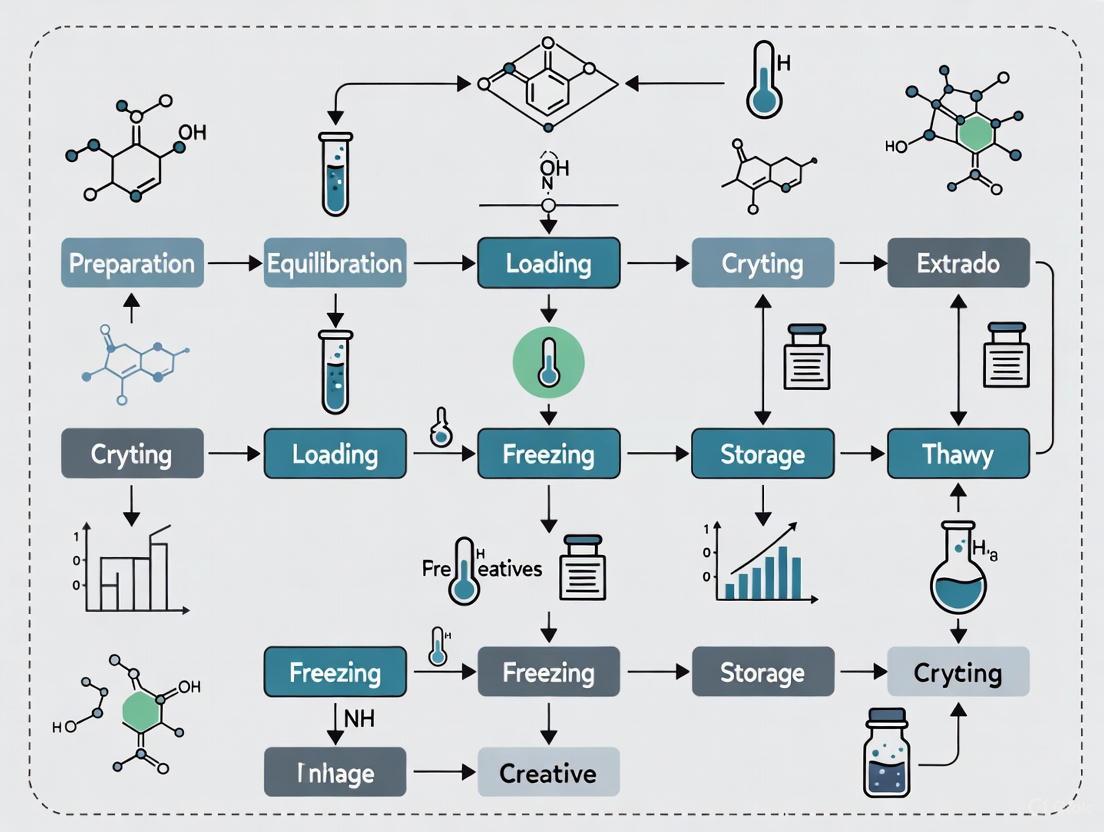
Diagram 1: PBMC Controlled-Rate Freezing Workflow. This diagram outlines the critical steps for cryopreserving PBMCs, highlighting the need for precise temperature control and timely execution.
Post-Thaw Assessment and Validation
Thawing and Recovery Protocol
The thawing process is critical for maintaining the viability and functionality of cryopreserved PBMCs. Inconsistent thawing procedures can significantly impact cell recovery and experimental outcomes [9].
- Rapid Thawing: Retrieve vials from liquid nitrogen storage and immediately place them in a 37°C water bath. Gently agitate until only a small ice crystal remains (approximately 1-2 minutes) [9] [1].
- Dilution: Using a pipette, gently transfer the cell suspension to a 15 mL tube containing 10 mL of pre-warmed washing medium (e.g., RPMI 1640 supplemented with 10-20% FBS or human serum) [9].
- Centrifugation: Centrifuge the cell suspension at 500 × g for 10 minutes at room temperature [9].
- Supernatant Removal: Carefully aspirate the supernatant, which contains the cytotoxic DMSO.
- Second Wash: Resuspend the cell pellet in 10 mL of fresh washing medium and centrifuge again at 500 × g for 10 minutes [9].
- Final Resuspension: Aspirate the supernatant and resuspend the cell pellet in an appropriate culture medium for downstream applications.
Viability and Functionality Assessment
Rigorous post-thaw assessment is essential to ensure PBMC quality. The following table summarizes common quality control metrics and their methodologies.
Table 2: Post-Thaw PBMC Quality Control Assessments
| Assessment Type | Method/Assay | Acceptance Criteria / Typical Results | References |
|---|---|---|---|
| Viability | Trypan Blue Exclusion | >90% viability (using 20% S-RPMI (human) as wash medium resulted in 95.7% viability) | [9] |
| Flow Cytometry with 7-AAD or Propidium Iodide | Distinguishes live/dead cells; provides quantitative data | [7] [9] | |
| Cell Recovery & Count | Hemocytometer or Automated Cell Counter | Absolute count of live PBMCs | [9] |
| Immunophenotyping | Multicolor Flow Cytometry | Quantification of T cells (CD4+, CD8+), B cells, NK cells, monocytes | [7] [2] |
| Functionality | Cytokine Release Assays (ELISA/ELISpot) | Antigen-specific response (e.g., IFN-γ secretion) | [3] [2] |
| Intracellular Cytokine Staining | Evaluation of polyfunctional T cell responses via flow cytometry | [2] | |
| Proliferation Assays (e.g., CFSE) | Measurement of cell division capacity in response to stimuli | [1] |
Impact of Cryopreservation on PBMC Biology
Studies evaluating the effect of cryopreservation on PBMC transcriptome profile using single-cell RNA sequencing have identified six major immune cell types (monocytes, dendritic cells, NK cells, CD4+ T cells, CD8+ T cells, and B cells) in both fresh and cryopreserved samples [7]. While cell viability and population composition remain relatively stable after 6 and 12 months of cryopreservation, the number of cells captured in scRNA-seq data declined significantly (~32%) after 12 months, suggesting reduced capture efficiency [7]. Importantly, the transcriptome profiles did not show substantial perturbation over the 12-month testing period, with only a few key genes involved in the AP-1 complex, stress response, or response to calcium ion exhibiting small-scale changes (< two folds) [7].
Long-term studies comparing freezing media have shown that PBMCs cryopreserved in CryoStor CS10 and NutriFreez D10 maintain high viability and functionality (assessed by cytokine secretion and T/B cell FluoroSpot), comparable to the FBS+10% DMSO reference medium, for up to 2 years [2]. Media with DMSO concentrations below 7.5% showed significant viability loss and were not suitable for long-term storage [2].
Applications in Cell Therapy and Immunological Research
Role in Therapeutic Development
PBMCs are indispensable in advancing cell therapies and immunotherapies. In hematopoietic stem cell transplantation (HSCT), cryopreserved peripheral blood stem cells (PBSCs) are the primary graft source, where engraftment success largely depends on the number of viable CD34+ cells [4]. The cryopreservation of these products allows for flexibility in transplantation timing and ensures product availability.
In vaccine development, PBMCs are critical for assessing cellular immune responses. Cryopreservation enables the simultaneous analysis of samples collected from a single participant at different time points, significantly reducing assay variability [2]. This is particularly valuable in multi-center clinical trials where local sample collection is combined with centralized analysis [3] [2].
For adoptive T-cell therapies, such as Chimeric Antigen Receptor (CAR) T-cell therapy, PBMCs serve as the starting material for generating therapeutic products. The optimized cryopreservation of source PBMCs and the final cellular product is essential for maintaining consistent quality and potency [4].
Standardization for Research and Clinical Trials
The use of cryopreserved PBMCs minimizes operator-dependent inter-assay and inter-laboratory variation, which is a significant challenge in multi-center clinical trials [9]. Standardized protocols for freezing, storage, and thawing are therefore critical for ensuring data comparability and reproducibility across studies [3]. The adoption of stringent Standard Operating Procedures (SOPs), such as those developed by the Office of HIV/AIDS Network Coordination (HANC), is widely recommended to minimize technical variability that can profoundly influence cellular viability and immunogenicity [3].
Diagram 2: Key Research and Clinical Applications of PBMCs. Cryopreserved PBMCs are a foundational resource for diverse therapeutic and investigative areas in modern immunology.
The Scientist's Toolkit: Essential Reagent Solutions
Table 3: Key Research Reagent Solutions for PBMC Processing and Cryopreservation
| Reagent/Category | Specific Examples | Function & Application Notes |
|---|---|---|
| Cryopreservation Media | CryoStor CS10, NutriFreez D10 [2] | Serum-free, GMP-compatible media containing 10% DMSO. Provide consistent performance and minimize batch-to-batch variability compared to lab-formulated media. |
| Serum-Containing Media | 90% FBS + 10% DMSO [8] | Traditional, cost-effective formulation. Raises concerns about xenoantigen exposure, pathogen transmission, and ethical considerations. |
| Density Gradient Media | Ficoll-Paque, Lymphoprep, Histopaque [3] [6] | Polysaccharide solutions used to isolate PBMCs from whole blood via centrifugation based on buoyant density. |
| Washing & Culture Media | RPMI 1640, supplemented with FBS or Human Serum [9] | Used for diluting thawed cells and subsequent cell culture. Supplementation with 10-20% serum helps maintain cell viability post-thaw. |
| Viability Stains | Trypan Blue, 7-AAD, Propidium Iodide [7] [9] | Dyes that selectively stain non-viable cells, allowing for quantification of viability post-thaw via microscopy or flow cytometry. |
| Flow Cytometry Reagents | Anti-CD45, Anti-CD3, Anti-CD19, Anti-CD14 Antibodies [9] [2] | Fluorescently conjugated antibodies used for immunophenotyping to determine the composition and purity of PBMC subsets. |
The successful cryopreservation of PBMCs using controlled-rate freezing protocols is a cornerstone technique in modern immunological research and cell therapy development. The integrity of these cells post-thaw is paramount, as it directly influences the reliability of data on immune function, disease mechanisms, and therapeutic efficacy. Current evidence supports the use of serum-free, commercially available cryopreservation media containing 10% DMSO, coupled with a standardized cooling rate of -1°C/minute, for optimal long-term preservation of PBMC viability and functionality. Adherence to detailed protocols for both freezing and thawing, combined with rigorous quality control assessments, ensures that cryopreserved PBMCs remain a robust and reproducible resource. This enables their critical application across a wide spectrum of fields, from basic science to clinical trials and advanced therapeutic manufacturing.
Cryopreservation is a cornerstone of modern cell therapy research, enabling the long-term storage and viability of vital cellular starting materials like peripheral blood mononuclear cells (PBMCs). The fundamental challenge of cryopreservation lies in navigating two interrelated physical phenomena: ice formation and osmotic stress. Intracellular ice crystallization can mechanically disrupt cellular membranes and organelles, while osmotic stress during freeze-thaw cycles can cause detrimental cell shrinkage or swelling, triggering apoptotic pathways [10] [11]. For cell therapy developers, mastering the control of these phenomena through optimized protocols is not merely an academic exercise; it is essential for ensuring high post-thaw viability, functionality, and consistency of cellular products, thereby enhancing the reliability of downstream research and clinical applications [12] [13]. This article details the core principles and provides actionable protocols to safeguard PBMCs against these cryogenic insults.
Fundamental Cryobiological Mechanisms
The Dual Threat: Ice Formation and Osmotic Stress
During cryopreservation, cells face two primary, interconnected threats. The first is intracellular ice formation (IIF), which is almost always lethal, causing physical rupture of cellular membranes and organelles [11]. The second is osmotic stress, a consequence of water turning to ice in the extracellular solution. As pure water freezes out, the concentration of dissolved solutes in the unfrozen fraction increases dramatically, creating a hypertonic environment. This draws water out of cells osmotically, leading to excessive cell shrinkage and potential damage to the cell membrane and cytoskeleton if not properly managed [10] [13].
The relationship between cooling rate and cell survival is governed by the "two-factor hypothesis," which posits that an optimal cooling rate exists that minimizes both IIF and osmotic stress. The following diagram illustrates how different cooling rates influence these competing factors and determine cell fate.
Osmotic Stress as a Kinetic Phenomenon
The rate at which osmotic stress is applied is a critical determinant of cell fate. Research has demonstrated that cells exhibit markedly different survival outcomes when exposed to acute (step) versus gradual (ramp) hypertonic stress, even when the final osmolyte concentration is identical [10].
- Acute Stress: The sudden application of high NaCl concentrations (e.g., +300 mosmol/L) leads to severe cell death, with viability dropping to about 15% [10].
- Gradual Stress: A slow, linear increase to the same final concentration over 10 hours significantly improves viability, which can reach 40% [10].
This survival advantage is linked to fundamental cellular signaling and metabolic adaptations. During gradual stress, cells do not exhibit the strong activation of caspase and stress signaling pathways (e.g., p38, JNK) that is characteristic of acute stress [10]. Furthermore, studies have identified a unique accumulation of the osmoprotectant amino acid proline in gradually stressed cells, a response not observed under acute conditions [10]. This suggests that controlled-rate freezing, which mimics a gradual ramp stress, allows time for beneficial cellular adaptations that are crucial for survival. The diagram below maps the distinct signaling pathways activated by these different stress profiles.
Application Notes: Optimizing PBMC Cryopreservation for Cell Therapy
The Scientist's Toolkit: Essential Reagents and Materials
Successful cryopreservation relies on a set of well-defined reagents and materials. The selection of cryopreservation media, in particular, has a direct impact on post-thaw recovery and functionality.
Table 1: Key Research Reagent Solutions for PBMC Cryopreservation
| Reagent/Material | Function & Rationale | Example Products & Formulations |
|---|---|---|
| Cryopreservation Medium | Provides a protective environment; contains cryoprotectants to reduce ice crystal formation and osmotic shock. | CryoStor CS10 [12] [14] [8], NutriFreez D10 [14], Lab-made 90% FBS + 10% DMSO [8] |
| Cryoprotectant (DMSO) | Permeating agent that stabilizes the cell membrane and prevents intracellular ice formation. Critical concentration is typically 10% [14] [8]. | Dimethyl Sulfoxide (DMSO) |
| Controlled-Rate Freezer | Equipment that ensures a consistent, optimal cooling rate (typically -1°C/min), which is crucial for high viability [12] [11]. | Programmable controlled-rate freezers |
| Isopropanol Freezing Container | A simple, accessible device placed in a -80°C freezer to approximate a -1°C/min cooling rate [8] [6]. | Corning CoolCell, Mr. Frosty |
| Liquid Nitrogen Storage | Provides long-term storage at temperatures below -135°C (the glass transition temperature of water), halting all biochemical activity [11] [8]. | Liquid nitrogen vapor phase storage dewars |
Quantitative Impact of Protocol Variables on PBMC Quality
Adherence to standardized protocols directly influences key quality attributes of cryopreserved PBMCs. The following table synthesizes quantitative data from recent studies on how critical parameters affect cell viability, recovery, and functionality.
Table 2: Impact of Cryopreservation Parameters on PBMC Quality Attributes
| Parameter | Optimal Value / Condition | Impact on Viability, Recovery & Functionality |
|---|---|---|
| DMSO Concentration | 10% [14] [8] | Concentrations below 7.5% show significant viability loss. 10% DMSO in serum-free media (CS10, NutriFreez D10) maintains high viability and T-cell functionality comparable to FBS-based media for up to 2 years [14]. |
| Cooling Rate | -1°C/minute [11] [8] [6] | This rate allows sufficient time for cellular dehydration, minimizing lethal intracellular ice formation. Faster or slower rates reduce viability [11]. |
| Post-Thaw Viability | ≥ 90% (achievable with optimized protocols) [12] | Standardized cryopreserved leukapheresis can achieve ≥90% post-thaw viability, with recovery and phenotypic profiles comparable to fresh PBMCs [12]. |
| Cell Concentration | 5–10 x 10^6 cells/mL [8] | Higher concentrations risk aggregation and reduced viability. The optimal range ensures uniform cryoprotectant penetration and cooling [8] [6]. |
| Processing Time | ≤ 8 hours from collection to freezing [3] | Delays in processing (>24 hours) are associated with reduced cell viability and diminished T cell immunogenicity [3]. |
Detailed Experimental Protocols
Protocol: Cryopreservation of PBMCs Using a Closed Automated System
This protocol is adapted from a standardized process for cryopreserved leukapheresis, demonstrating high viability and compatibility with CAR-T manufacturing platforms [12].
Materials:
- Purified PBMCs or leukapheresis product
- Cryopreservation medium (e.g., CryoStor CS10, pre-cooled to 2–8°C)
- Cryogenic bags or vials
- Controlled-rate freezer (e.g., Thermo Profile 4) or isopropanol freezing container
- Centrifuge
Procedure:
- Cell Preparation: Ensure cells are in a single-cell suspension. Centrifuge at 300 x g for 10 minutes to pellet cells. Carefully remove the supernatant [8].
- Formulation: Resuspend the cell pellet in pre-cooled cryopreservation medium to a target concentration of ~5 x 10^7 cells/mL [12]. Mix thoroughly but gently.
- Dispensing: Transfer the cell suspension to cryogenic bags or vials. For bags, a formulation volume of 20 mL/bag is typical [12].
- Time-Sensitive Freezing: Initiate controlled-rate freezing within 120 minutes of cryoprotectant addition to minimize DMSO toxicity at room temperature [12].
- Controlled-Rate Freezing: Place bags/vials in a controlled-rate freezer and run a program with a cooling rate of -1°C/min [12] [8].
- Long-Term Storage: Once freezing is complete, immediately transfer the bags/vials to vapor-phase liquid nitrogen for long-term storage (below -135°C) [8].
Protocol: Functional Validation of Cryopreserved PBMCs via T-Cell Stimulation
This protocol outlines a method to assess the functionality of thawed PBMCs, which is critical for ensuring their utility in immunology research and cell therapy.
Materials:
- Cryopreserved PBMCs
- Water bath (37°C)
- Pre-warmed complete culture medium (e.g., RPMI-1640 + 10% FBS)
- DNase I (optional, to reduce clumping from DNA release)
- T-cell mitogens (e.g., PHA) or antigenic peptides
- ELISA or FluoroSpot kits for cytokine detection (e.g., IFN-γ)
Procedure:
- Thawing: Rapidly thaw cryovials by gentle agitation in a 37°C water bath until just ice-free [14].
- Dilution & Washing: Transfer the cell suspension to a tube containing pre-warmed medium supplemented with DNase I (10 µg/mL). Centrifuge at 300 x g for 10 minutes to remove cryoprotectants [14] [6].
- Resting: Resuspend the cell pellet in complete culture medium and incubate at 37°C, 5% CO2 for 4-24 hours. This "resting" period allows cells to recover from the freeze-thaw stress and is critical for restoring immunogenicity [3].
- Stimulation: Seed rested PBMCs into a culture plate and stimulate with a mitogen or specific antigen. Include unstimulated controls.
- Functionality Assessment: After 24-48 hours, collect supernatant for cytokine analysis (e.g., IFN-γ ELISA) or perform a FluoroSpot assay to quantify antigen-specific T-cell responses [14]. High functionality, comparable to fresh PBMCs, confirms a successful cryopreservation process.
The Critical Role of Controlled-Rate Freezing in Preventing Intracellular Ice Crystals and Membrane Damage
In cell therapy research, the functional integrity of Peripheral Blood Mononuclear Cells (PBMCs) is a critical determinant of therapeutic efficacy. Cryopreservation is indispensable for creating and managing cell banks for research and clinical applications. However, the freeze-thaw process itself presents a significant risk of cryogenic injury, primarily through the formation of intracellular ice crystals and osmotic stress that compromises membrane integrity. Controlled-rate freezing is a cornerstone technology designed to mitigate these risks by precisely managing the phase change of water, thereby preventing the lethal damage that occurs with uncontrolled freezing. This application note details the underlying mechanisms, provides optimized protocols, and presents experimental data demonstrating the critical role of controlled-rate freezing in preserving PBMC viability and function for cell therapy workflows.
The Mechanism of Cryoinjury and the Rationale for Controlled Freezing
The damage incurred during freezing is primarily a consequence of ice formation and accompanying osmotic shifts. The "two-factor hypothesis" of cryoinjury describes the competing risks associated with different cooling rates, as illustrated in the diagram below.
Figure 1. The Two-Factor Hypothesis of Cryoinjury. The relationship between cooling rate and cell survival is a balance between two damaging factors. At high cooling rates, lethal intracellular ice formation (IIF) dominates. At low cooling rates, damage from "solution effects"—prolonged exposure to hypertonic extracellular solutions—causes harm. An optimum cooling rate exists that minimizes the sum of these injuries [15].
- Extracellular Ice and Osmotic Imbalance: During slow cooling, ice first forms in the extracellular solution. As pure water is sequestered into ice, the concentration of solutes in the remaining unfrozen liquid rises dramatically. This creates a hypertonic environment, driving water out of the cell through osmosis and leading to detrimental cellular dehydration and shrinkage [16] [15].
- Intracellular Ice Formation (IIF): At high cooling rates, water does not have sufficient time to leave the cell before the intracellular contents supercool. When nucleation occurs, water freezes inside the cell. These intracellular ice crystals are mechanically disruptive, shearing organelle and plasma membranes, and are almost universally lethal [16] [17].
- The Role of Controlled Ice Nucleation: A key advancement in controlled-rate freezing is the active triggering of ice nucleation at a defined, elevated temperature (e.g., -5°C to -6°C). This prevents deep supercooling, which is a stochastic event that can lead to heterogeneous ice formation and unpredictable IIF across a cell population. Controlled nucleation ensures a consistent and predictable freezing process, promoting uniform extracellular ice formation and allowing sufficient time for controlled cellular dehydration [16].
Quantitative Impact on PBMC Cryopreservation
The choice of freezing protocol directly impacts key metrics of PBMC quality, including viability, recovery, and functional potency. The following table summarizes experimental data comparing different freezing approaches.
Table 1. Comparison of PBMC Cryopreservation Methods [18].
| Freezing Method | Cooling Rate | Time to -80°C | Post-Thaw Viability | Cell Activity (Proliferation) | Key Operational Advantage |
|---|---|---|---|---|---|
| Slow Freezing (SLF) with Container | ~1°C/min | 3 hours | Equivalent to SLF | Equivalent to SLF | Standardized, low-cost method |
| Electromagnetic Field (EMF) Freezing | Not specified | 0.25 hours | Equivalent to SLF | Equivalent to SLF | Rapid transfer to LN₂; superior operational efficiency |
| Slow Freezing (SLF) with Container (Held at -80°C for 168h) | ~1°C/min | 3 hours | Significant decline | Significant decline | N/A (Highlights risk of delayed transfer) |
Beyond immediate viability, studies have shown that optimized controlled-rate freezing protocols have minimal impact on the transcriptomic profile of PBMC subpopulations. Single-cell RNA sequencing analysis of PBMCs cryopreserved for 6 to 12 months demonstrated preserved cell type distribution and no substantial perturbation in gene expression profiles, underscoring the method's ability to maintain biological fidelity for downstream research applications [7].
Optimized Protocols for PBMC Cryopreservation
Standard Controlled-Rate Freezing Protocol for PBMCs
This protocol is adapted from industry best practices and scientific literature for reliable preservation of PBMCs [19] [7].
- Step 1: Harvest and Count Cells. Isolate PBMCs using standard Ficoll-Paque density gradient centrifugation. Resuspend the cell pellet in a pre-cooled (2-8°C) cryopreservation medium. A common and effective choice is CryoStor CS10 (10% DMSO), although other commercial or lab-formulated media can be used. Determine total cell count and viability via Trypan Blue exclusion using an automated cell counter or hemocytometer. Adjust the cell concentration to a target of 1x10⁷ cells/mL.
- Step 2: Aliquot and Package. Dispense the cell suspension into cryogenic vials (e.g., 1 mL/vial). Securely close the vials. For passive cooling, place the vials into an isopropanol freezing chamber (e.g., "Mr. Frosty") or an isopropanol-free container (e.g., Corning CoolCell) that has been pre-equilibrated to room temperature.
- Step 3: Initiate Controlled Freezing. Immediately transfer the loaded freezing container to a -80°C mechanical freezer. The insulating properties of the container will enforce an approximate cooling rate of -1°C/minute, which is optimal for most mammalian cells. Leave the container in the -80°C freezer for a minimum of 4 hours, or preferably overnight.
- Step 4: Long-Term Storage. After the initial freezing period, promptly transfer the cryovials to the vapor phase of a liquid nitrogen storage tank (below -135°C) for long-term storage. Note: Prolonged storage at -80°C leads to a gradual decline in viability and is not recommended [18] [19].
Advanced Protocol: Controlled Nucleation for Enhanced Dehydration
For applications requiring the highest consistency, using a programmable controlled-rate freezer (CRF) with an active nucleation function is recommended. The workflow for this advanced protocol is shown below.
Figure 2. Advanced PBMC Freezing with Controlled Nucleation. This protocol uses a programmable freezer to actively trigger ice formation, promoting uniform dehydration [16].
- Critical Step: Annealing. The "hold" or "annealing" step immediately after nucleation is crucial. This provides additional time for intracellular water to exit the cell before the temperature drops low enough to cause intracellular vitrification or ice formation, further reducing the risk of IIF [16].
Thawing and Assessment Protocol
- Rapid Thawing. Retrieve a vial from liquid nitrogen storage and immediately place it in a 37°C water bath with gentle agitation until only a small ice crystal remains. The thawing process should be rapid to minimize the damaging effects of ice recrystallization [19].
- Dilution and Washing. Gently transfer the cell suspension to a 15 mL tube containing 10 mL of pre-warmed complete culture medium (e.g., RPMI-1640 with 10% FBS). This step rapidly dilutes the cytotoxic DMSO. Centrifuge at 400 x g for 5 minutes. Discard the supernatant and resuspend the cell pellet in fresh warm medium. A second wash is often recommended [7].
- Viability and Function Assessment. Perform a cell count and viability assessment using Trypan Blue exclusion. For a more accurate assessment of membrane integrity in mixed populations, use flow cytometry with a viability dye such as LIVE/DEAD Fixable Violet Dead Cell Stain or propidium iodide [20] [7]. Functional assays, such as stimulation with anti-CD3/CD28 antibodies followed by proliferation analysis using a dye like CellTrace CFSE, should be conducted to confirm preserved immunocompetence [20] [18].
The Scientist's Toolkit: Essential Reagents and Materials
Table 2. Key Research Reagent Solutions for Controlled-Rate Freezing.
| Item | Function & Rationale |
|---|---|
| CryoStor CS10 | A ready-to-use, cGMP-manufactured freezing medium containing 10% DMSO. It provides an optimized, serum-free, and defined environment that minimizes freeze-thaw stress [19]. |
| DMSO (Cell Culture Grade) | A permeating cryoprotectant. It penetrates the cell, displaces water, and forms hydrogen bonds to suppress IIF. Must be handled with care due to potential cytotoxicity and epigenetic effects [16] [21]. |
| LIVE/DEAD Fixable Viability Stains | Amine-reactive dyes that covalently label dead cells with compromised membranes. They allow for accurate exclusion of dead cells during flow cytometry analysis, even after fixation/permeabilization [20] [7]. |
| Propidium Iodide (PI) | A membrane-impermeant DNA stain that is excluded by live cells. It is a cost-effective dye for simple viability assessment via flow cytometry, though it cannot be used on fixed cells [22] [7]. |
| CellTrace CFSE Cell Proliferation Kit | A fluorescent dye that stably labels intracellular proteins. With each cell division, the fluorescence intensity halves, allowing for tracking of proliferation dynamics in thawed PBMCs [20]. |
| Isopropanol Freezing Chamber (e.g., Mr. Frosty) | A passive cooling device that uses the thermal buffering capacity of isopropanol to achieve an approximate cooling rate of -1°C/minute in a standard -80°C freezer [21] [19]. |
| Controlled-Rate Freezer | A programmable instrument that provides precise, user-defined control over cooling rates and can incorporate features like active ice nucleation, which is critical for high-end reproducibility [16]. |
Controlled-rate freezing is not merely a convenience but a critical procedure for ensuring the reliability and success of PBMC-based research in cell therapy. By understanding and controlling the physical processes of ice formation—specifically through optimized cooling rates and the strategic application of controlled nucleation—researchers can effectively prevent the formation of lethal intracellular ice crystals and mitigate membrane-damaging osmotic stress. The protocols and data presented herein provide a roadmap for implementing this vital technology, ultimately contributing to robust and reproducible cell therapy research outcomes.
In cell therapy research, the cryopreservation of peripheral blood mononuclear cells (PBMCs) is a critical step for ensuring a consistent and readily available cell source for downstream applications. The transition from research to clinical application demands rigorous quantification of cell quality post-thaw. While viability and cell count recovery are fundamental initial metrics, they are insufficient alone to guarantee success in sophisticated experimental or therapeutic workflows. A comprehensive assessment must also confirm the retention of key cellular functions, such as proliferation and cytokine secretion, to ensure these cells are truly fit for purpose. This application note delineates the essential success metrics—post-thaw viability, recovery, and functional integrity—and provides detailed protocols for their evaluation within the context of a controlled-rate freezing protocol for PBMCs.
Quantifying Post-Thaw Viability and Recovery
Defining and accurately measuring viability and recovery is the first critical step in evaluating a cryopreservation protocol. These quantitative metrics provide the initial triage for cell quality.
Key Definitions and Metrics
- Post-Thaw Viability: The percentage of live cells in the total post-thaw cell population, typically assessed using dye exclusion methods like trypan blue. A viability of ≥80% is a commonly accepted benchmark for proceeding with most downstream applications [23].
- Cell Recovery Yield: The percentage of viable nucleated cells recovered after thawing compared to the number cryopreserved. This metric is calculated as: (Total Viable Cells Post-Thaw / Total Viable Cells Pre-Freeze) x 100%. High recovery maximizes the starting material for subsequent experiments.
Impact of Cryopreservation Media
The choice of cryoprotectant medium is a primary determinant of post-thaw viability and recovery. Long-term studies (up to 2 years) have systematically compared traditional fetal bovine serum (FBS)-based media with modern, serum-free alternatives [2]. The data indicates that media containing 10% DMSO consistently outperform those with lower or zero DMSO concentrations in preserving PBMC viability and function over extended periods.
Table 1: Comparison of Cryopreservation Media Performance on PBMC Viability and Functionality
| Cryopreservation Medium | DMSO Concentration | Long-Term Viability (2 years) | T Cell Functionality | B Cell Functionality |
|---|---|---|---|---|
| FBS + DMSO (Reference) | 10% | High | Preserved | Preserved |
| CryoStor CS10 | 10% | High (Comparable to FBS) | Preserved | Preserved |
| NutriFreez D10 | 10% | High (Comparable to FBS) | Preserved | Preserved |
| Bambanker D10 | 10% | High | Tended to diverge from reference | Preserved |
| Media with <7.5% DMSO | 2%-5% | Significant viability loss | Not Assessed (Eliminated) | Not Assessed (Eliminated) |
Influence of Freezing Methodology
The freezing technique itself can significantly impact cell quality and operational efficiency. A recent study comparing the standard slow-freezing (SLF) method with a novel electromagnetic field (EMF)-assisted freezer demonstrated that while both methods yielded equivalent post-thaw viability and cell activity, the EMF method offered a substantial reduction in processing time [18].
Table 2: Operational Comparison of Slow-Freezing vs. EMF-Freezing Methods
| Parameter | Slow-Freezing (SLF) Method | EMF-Freezing Method |
|---|---|---|
| Freezing Rate | -1°C/minute | Not specified |
| Minimum Freezing Time | 3 hours | 0.25 hours (15 minutes) |
| Post-Thaw Viability | Equivalent to EMF method | Equivalent to SLF method |
| Post-Thaw Cell Activity | Equivalent to EMF method | Equivalent to SLF method |
| Key Advantage | Widely accessible, cost-effective (e.g., Mr. Frosty) | Rapid transfer to LN₂, superior operational efficiency |
Assessing Functional Integrity for Downstream Applications
Beyond simple viability, a cell's capacity to perform its intended biological functions is paramount. Functional integrity is the ultimate validator of a successful cryopreservation strategy.
Proliferation Capacity
The ability of T-cells to undergo robust proliferation upon antigen-specific or polyclonal stimulation is a gold-standard functional assay.
- Protocol: T-cell Proliferation Assay [24] [18]
- Coat Plates: Coat a 96-well plate with 0.01% poly-L-ornithine solution (50 µL/well) for 1 hour at ambient temperature. Aspirate and dry plates for 30-60 minutes.
- Apply Activators: Add 100 µL of a solution containing both anti-CD3 and anti-CD28 antibodies (each at 2 µg/mL, 2X final concentration) to the coated wells. Include control wells with culture medium only.
- Seed Cells: Thaw and wash PBMCs as per standard protocol. Resuspend cells in culture medium at a density of 4 x 10⁵ cells/mL. Add 100 µL of cell suspension (40,000 cells/well) to each well.
- Culture and Assess: Culture cells for 3-5 days. Proliferation can be quantified using methods like CFSE dilution via flow cytometry or colorimetric assays like MTT/XTT that measure metabolic activity [24].
Cytokine Secretion Profile
Functional immune cells should retain their ability to secrete cytokines upon activation. This can be measured using techniques like ELISpot/FluoroSpot or intracellular cytokine staining (ICS) coupled with flow cytometry [2].
- Workflow: Functional Immune Cell Assessment [2]
Post-Thaw Processing and Purity
The method used to isolate PBMCs post-thaw can create a significant trade-off between cell recovery and purity, which in turn affects downstream functionality. For example, CD14+ monocyte depletion has been shown to correlate with reduced T-cell proliferation, highlighting the importance of cell-to-cell interactions in functional assays [24] [25]. The choice of processing method—whether a simple wash, density gradient, or immunodepletion of granulocytes/red blood cells—should be application-specific.
Detailed Experimental Protocols
A consistent and rapid thawing process is critical to minimize the cytotoxic effects of DMSO.
- Thaw: Gently agitate cryovial in a 37°C water bath until only a small ice crystal remains.
- Transfer and Dilute: Immediately transfer the 1 mL cell suspension to a 15 mL conical tube. Slowly add 10 mL of pre-warmed (37°C) wash medium (e.g., RPMI-1640 supplemented with 10% FBS and 10 µg/mL DNase) drop-wise while gently swirling the tube. DNase is crucial to digest DNA released from dead cells, preventing cell clumping.
- Wash: Centrifuge at 400 x g for 5 minutes at room temperature.
- Resuspend and Count: Discard the supernatant and gently resuspend the cell pellet in an appropriate volume of culture medium. Perform a cell count and viability assessment using trypan blue exclusion.
This assay evaluates the clonogenic potential and fitness of progenitor cells within the PBMC population.
- Prepare Cells: Thaw and wash PBMCs. Resuspend in appropriate medium.
- Plate in Methylcellulose: Mix cells with semi-solid methylcellulose medium containing cytokines and growth factors specific for myeloid or lymphoid lineages. Ensure a homogenous mixture.
- Culture: Transfer the cell-methylcellulose mixture to culture plates or dishes. Incubate at 37°C, 5% CO₂ for 12-14 days in a humidified incubator.
- Score Colonies: After the incubation period, count the number of distinct colonies (e.g., CFU-GM, CFU-E) under an inverted microscope. The number and type of colonies indicate the frequency and differentiation potential of hematopoietic progenitors.
The Scientist's Toolkit: Research Reagent Solutions
Table 3: Essential Reagents and Kits for Post-Thaw PBMC Analysis
| Product Name | Type/Function | Key Application |
|---|---|---|
| CryoStor CS10 [2] | Serum-free, protein-free freezing medium | High-viability, long-term cryopreservation of PBMCs; GMP-grade available. |
| NutriFreez D10 [2] | Animal-protein-free freezing medium | Viable FBS-free alternative for PBMC cryopreservation. |
| Lymphoprep [2] | Density gradient medium | Isolation of mononuclear cells from whole blood or thawed cord blood units. |
| EasySep Direct Human PBMC Isolation Kit [25] | Immunodepletion-based cell isolation kit | Post-thaw isolation of PBMCs; enhances purity and day-0 viability. |
| Anti-CD3/CD28 Antibodies [18] | T-cell receptor activators | Polyclonal stimulation of T-cells in proliferation and cytokine assays. |
| Trypan Blue [6] [23] | Viability stain | Differential staining of live (unstained) and dead (blue) cells for counting. |
| CoolCell [2] | Cell freezing container | Provides a consistent -1°C/minute cooling rate in a -80°C freezer. |
A multi-faceted approach is imperative for defining success in PBMC cryopreservation. Relying solely on post-thaw viability is an outdated and insufficient practice. Robust application notes must mandate the integration of cell recovery metrics with rigorous assessments of functional integrity, including proliferation capacity and cytokine secretion profiles. Furthermore, the selection of advanced cryopreservation media and standardized, efficient thawing processes are critical controllable factors that directly impact data quality and reproducibility. By adopting the comprehensive metrics and detailed protocols outlined in this document, researchers and therapy developers can significantly de-risk their downstream applications, ensuring that cryopreserved PBMCs are not merely alive but are fully functional and fit for their intended purpose in the cell therapy pipeline.
A Step-by-Step Guide to Implementing a Gold-Standard PBMC Freezing Protocol
In the field of cell therapy research, the cryopreservation of peripheral blood mononuclear cells (PBMCs) is a critical step that can determine the success of downstream applications, including the manufacturing of chimeric antigen receptor T-cell (CAR-T) therapies [26]. The pre-freezing preparation phase is particularly crucial, as decisions made during this stage directly impact post-thaw viability, recovery, and functionality [3] [6]. Proper pre-freezing protocols ensure that PBMCs retain their immunological characteristics and therapeutic potential after long-term storage [26]. This application note details standardized procedures for cell concentration optimization, viability assessment, and reagent equilibration within the context of a controlled-rate freezing protocol, providing researchers with evidence-based methodologies to maximize PBMC quality for cell therapy applications.
Key Parameters for Pre-Freezing Preparation
Optimal Cell Concentration and Cryopreservation Media
Extensive research has established optimal cell concentration ranges for PBMC cryopreservation. Adherence to these parameters minimizes ice crystal formation and osmotic stress, thereby preserving cell integrity during freezing and thawing processes [6].
Table 1: Recommended Cell Concentrations and Cryoprotectants for PBMC Cryopreservation
| Parameter | Recommended Range | Notes | Primary Source |
|---|---|---|---|
| Cell Concentration | 5-10 × 10⁶ cells/mL | Lower end (0.5-1 × 10⁶ cells/mL) may be used for specific applications; higher concentrations require validation. | [8] |
| DMSO Concentration | 10% | Final concentration in cryopreservation medium. 5% DMSO has been reported to improve recovery in some studies. | [3] [8] |
| Serum Source | 90% FBS or Serum-Free Alternatives (e.g., CryoStor CS10) | FBS is cost-effective but introduces variability and safety concerns. Serum-free media are preferred for clinical applications. | [8] |
Viability Assessment and Acceptance Criteria
Rigorous viability assessment prior to freezing is essential for predicting cryopreservation success and ensuring experimental reproducibility. Viability measurements serve as a critical quality control checkpoint.
Table 2: Viability Assessment Methods and Pre-Freeze Acceptance Criteria
| Method | Principle | Procedure Highlights | Acceptance Criteria |
|---|---|---|---|
| Trypan Blue Exclusion | Dye exclusion by viable cells with intact membranes. | Cells mixed 1:1 with trypan blue; counted manually with hemocytometer or automated cell counter. | >90% viability before cryopreservation. [27] |
| Propidium Iodide (PI) Staining | Fluorescent dye excluded by viable cells. | Stained cells analyzed by flow cytometry or fluorescence-based cell counting. | >90% viability before cryopreservation. [7] |
Detailed Experimental Protocols
Protocol: Determining Optimal Cell Concentration for Cryopreservation
Objective: To empirically determine the ideal cell concentration for cryopreservation that yields the highest post-thaw viability and recovery for a specific cell therapy research application.
Materials:
- Isolated PBMCs (≥90% viability)
- Cryopreservation medium (e.g., 10% DMSO in FBS or commercial serum-free medium)
- Cryogenic vials
- Centrifuge
- Automated cell counter or hemocytometer
- Trypan blue stain
Method:
- Prepare Cell Suspensions: Isolate PBMCs using standard Ficoll-Paque density gradient centrifugation or apheresis [28]. Perform a cell count and viability assessment using trypan blue exclusion [27].
- Create Concentration Gradient: Centrifuge the required cell volume at 300 × g for 10 minutes. Aspirate the supernatant, leaving a small volume to avoid disturbing the pellet. Resuspend the cell pellet in cold (2-8°C) cryopreservation medium to create master suspensions at two different concentrations. From these, prepare a dilution series to achieve final concentrations of 1, 5, 10, and 15 × 10⁶ cells/mL in cryovials [8].
- Cryopreservation and Storage: Follow a standardized controlled-rate freezing protocol, decreasing temperature at approximately -1°C/minute using a programmable freezer or an isopropanol freezing container placed at -80°C overnight. Subsequently, transfer vials to long-term storage in vapor phase liquid nitrogen (below -135°C) [27] [8].
- Post-Thaw Analysis: After a minimum storage period (e.g., 1 week), rapidly thaw one vial from each concentration group in a 37°C water bath. Dilute the thawed cells in pre-warmed culture medium and centrifuge at 400 × g for 5 minutes to remove DMSO. Resuspend the pellet, perform a cell count and viability assessment, and calculate the percentage recovery of viable cells [18].
- Data Analysis: Plot post-thaw viability and recovery against the pre-freeze cell concentration. The optimal concentration is that which maximizes both parameters.
Protocol: Cell Viability Assessment via Propidium Iodide Staining and Flow Cytometry
Objective: To accurately assess the viability of PBMCs prior to cryopreservation using a flow cytometry-based method.
Materials:
- Isolated PBMCs
- Propidium Iodide (PI) stock solution (e.g., 1 mg/mL)
- Phosphate Buffered Saline (PBS)
- Flow cytometry tubes
- Flow cytometer
Method:
- Cell Preparation: Harvest and wash PBMCs in PBS. Resuspend the cell pellet in PBS to a concentration of approximately 1-5 × 10⁶ cells/mL.
- Staining: Add PI to the cell suspension at a final concentration of 1-5 µg/mL. Incubate for 5-15 minutes at 2-8°C, protected from light [7].
- Acquisition and Analysis: Analyze the stained cells on a flow cytometer within 1 hour. Use the FL2 or FL3 channel (excitation/emission ~535/617 nm) to detect PI fluorescence. Establish a forward scatter (FSC) vs. side scatter (SSC) gate to select the primary lymphocyte population. Collect a minimum of 10,000 events within this gate. Viable cells will be PI-negative, while non-viable cells with compromised membranes will be PI-positive.
- Interpretation: Calculate the percentage of viable cells as (PI-negative cells / total gated cells) × 100%. Only proceed with cryopreservation if viability meets the pre-defined acceptance criterion (e.g., ≥90%) [27].
The Scientist's Toolkit: Essential Research Reagents
Table 3: Key Reagents for PBMC Pre-Freezing Preparation
| Reagent/Solution | Function | Key Considerations |
|---|---|---|
| Cryopreservation Medium | Protects cells from ice crystal damage and osmotic stress during freezing. | For 10% DMSO/90% FBS: Prepare 20% DMSO in FBS on ice. Mix with cell suspension 1:1 for final concentration. Work quickly to minimize DMSO exposure at room temperature. [8] |
| Fetal Bovine Serum (FBS) | Provides nutrients and proteins that stabilize cell membranes. | Introduces lot-to-lot variability and xeno-risks. Not suitable for clinical-grade products. [8] |
| Dimethyl Sulfoxide (DMSO) | Penetrating cryoprotectant that reduces intracellular ice formation. | Use high-grade, sterile DMSO. Final concentration is critical; typically 10%. Can be toxic to cells at room temperature. [3] [6] |
| Serum-Free Cryopreservation Media (e.g., CryoStor CS10) | Defined, xeno-free alternative to FBS-containing media. | Preferred for clinical applications due to reduced variability and safety risks. Pre-cool to 2-8°C before use. [8] |
| Trypan Blue Stain | Vital dye for distinguishing viable from non-viable cells. | Non-viable cells uptake the dye and appear blue. Mix cell suspension 1:1 with dye for counting. [7] [27] |
| Propidium Iodide (PI) | Fluorescent vital dye for flow cytometry-based viability testing. | Binds to DNA of membrane-compromised cells. Must be used with a flow cytometer. [7] |
Workflow Visualization
The following diagram summarizes the key decision points and steps in the pre-freezing preparation process for PBMCs.
Meticulous pre-freezing preparation is a foundational element in the development of a robust controlled-rate freezing protocol for PBMCs in cell therapy research. By standardizing cell concentration within the recommended range of 5-10 × 10⁶ cells/mL, rigorously assessing pre-freeze viability to ensure it exceeds 90%, and carefully preparing and equilibrating cells with cryopreservation media, researchers can significantly enhance the post-thaw quality of their cellular products [8] [26]. Adherence to these detailed protocols ensures that PBMCs retain high viability, recovery, and critical functionality, thereby supporting the reliability and reproducibility of advanced cell therapies.
Cryopreservation of peripheral blood mononuclear cells (PBMCs) is a fundamental process in immunological studies and clinical trials, particularly in vaccine development and cell therapy research [2] [14]. The choice of cryopreservation medium significantly impacts cell viability, recovery, phenotype, and functionality upon thawing. Traditional freezing formulations typically combine fetal bovine serum (FBS) with 10% dimethyl sulfoxide (DMSO), which presents considerable challenges including ethical concerns regarding animal product use, risk of pathogen transmission, batch-to-batch variability, and DMSO cytotoxicity at room temperature [2] [14] [8]. This application note provides a comprehensive comparison between FBS-based and serum-free cryopreservation media, evaluates optimal DMSO concentrations, and presents standardized protocols within the context of controlled-rate freezing for PBMCs in cell therapy applications.
Comparative Analysis of Cryopreservation Media
Performance Evaluation of Serum-Free Alternatives
Recent comprehensive studies have systematically evaluated commercially available serum-free media against traditional FBS-based controls. A 2025 study assessing PBMCs from 11 healthy volunteers over a 2-year period demonstrated that specific serum-free formulations can effectively match or surpass the performance of FBS-based media [2] [14] [29].
Table 1: Viability and Functionality of PBMCs Cryopreserved in Different Media Over 2 Years
| Cryopreservation Medium | Composition | Viability Across 2 Years | T-cell Functionality | B-cell Functionality | Recommended Application |
|---|---|---|---|---|---|
| FBS + 10% DMSO (Reference) | 90% FBS + 10% DMSO | High | Reference standard | Reference standard | General research |
| CryoStor CS10 | Serum-free + 10% DMSO | High (Comparable to FBS) | Maintained | Maintained | Clinical trials, GMP work |
| NutriFreez D10 | Serum-free + 10% DMSO | High (Comparable to FBS) | Maintained | Maintained | Clinical trials, Research |
| Bambanker D10 | Serum-free + 10% DMSO | High | Divergent from reference | Maintained | Research applications |
| CryoStor CS5 | Serum-free + 5% DMSO | Significant loss after M0 | Not assessed | Not assessed | Not recommended |
| CryoStor CS2 | Serum-free + 2% DMSO | Significant loss after M0 | Not assessed | Not assessed | Not recommended |
The data clearly indicate that serum-free media containing 10% DMSO (CryoStor CS10 and NutriFreez D10) maintain PBMC viability and functionality comparable to the traditional FBS reference medium across all time points up to 2 years [2] [14]. Media with DMSO concentrations below 7.5% showed significant viability loss and were eliminated from consideration after initial assessments [2].
DMSO Concentration Optimization
DMSO concentration critically influences cryopreservation outcomes through its dual role as cryoprotectant and cytotoxic agent. Studies systematically evaluating DMSO concentrations reveal a clear optimal range for PBMC preservation.
Table 2: Impact of DMSO Concentration on PBMC Cryopreservation Outcomes
| DMSO Concentration | Cell Viability | Cell Recovery | Functionality Preservation | Toxicity Concerns | Recommendation |
|---|---|---|---|---|---|
| 2-5% | Significant viability loss | Poor | Not maintained | Low | Not recommended |
| 7.5% | Moderate | Moderate | Partially maintained | Moderate | Potential alternative with optimization |
| 10% | High | High | Fully maintained | Manageable with proper handling | Recommended |
| 15% | High | Moderate | Maintained | Increased concern | Acceptable with caution |
| 20% | Decreased | Decreased | Variable | High | Not recommended |
Research demonstrates that 10% DMSO represents the optimal balance between cryoprotection and cytotoxicity, preventing intracellular ice crystal formation while minimizing toxic effects on PBMCs [2] [30]. While 15% DMSO maintains adequate viability, it offers no significant advantages over 10% concentrations and presents greater toxicity concerns [30]. DMSO concentrations exceeding 15% consistently demonstrate reduced cell viability and recovery due to increased cytotoxicity [30].
Experimental Protocols
Cryopreservation Protocol for PBMCs Using Serum-Free Media
The following protocol utilizes CryoStor CS10 or NutriFreez D10 for optimal PBMC cryopreservation, compatible with controlled-rate freezing systems [2] [8].
Materials and Reagents
Table 3: Essential Research Reagents for PBMC Cryopreservation
| Reagent/Equipment | Function | Example Product | Specifications |
|---|---|---|---|
| Cryopreservation Medium | Protects cells during freezing | CryoStor CS10 | Serum-free, 10% DMSO |
| DMSO | Cryoprotectant | Sigma-Aldrich #D2650 | Cell culture grade |
| Cryogenic Vials | Cell storage | Nalgene | 1-2 mL capacity |
| Controlled-Rate Freezer | Programmable freezing | Planer Kryo10 | -1°C/minute rate |
| Isopropanol Container | Alternative freezing | Corning CoolCell | For -80°C freezing |
| Liquid Nitrogen Storage | Long-term preservation | Vapor phase storage | Below -135°C |
| Hanks' Balanced Salt Solution | Cell washing | Standard formulation | With buffers |
| DNase I | Prevents clumping | Roche #11284932001 | 10 µg/mL concentration |
Step-by-Step Procedure
PBMC Preparation: Isolate PBMCs from whole blood using density gradient centrifugation with Lymphoprep or similar medium. Centrifuge at 300 × g for 10 minutes to obtain a cell pellet [8].
Cell Counting and Viability Assessment: Resuspend cells in HBSS and determine concentration and viability using trypan blue exclusion or automated cell counters.
Medium Preparation: Use pre-chilled (2-8°C) serum-free cryopreservation medium. For CryoStor CS10, use directly as provided [8].
Cell Resuspension: Carefully remove supernatant from pelleted PBMCs. Gently resuspend cells in cold cryopreservation medium at recommended concentration of 5-10 × 10⁶ cells/mL [2] [8].
Aliquoting: Dispense 1 mL cell suspension into pre-chilled cryogenic vials. Maintain samples on ice or at 2-8°C throughout the process.
Equilibration: Incubate filled cryovials at 2-8°C for 10 minutes to allow cryoprotectant penetration [8].
Controlled-Rate Freezing: Place vials in controlled-rate freezer programmed for a cooling rate of approximately -1°C/minute to -80°C [31] [8].
Long-Term Storage: Transfer frozen vials to vapor-phase liquid nitrogen storage (-135°C to -196°C) for long-term preservation [8].
Thawing and Assessment Protocol
Proper thawing techniques are critical for maintaining cell viability and functionality after cryopreservation.
Rapid Thawing: Gently agitate cryovial in 37°C water bath until only a small ice crystal remains [14] [8].
DNase Treatment: Immediately transfer cell suspension to pre-warmed RPMI 1640 medium containing 10% FBS and DNase I (10 µg/mL) to prevent cell clumping [14].
Centrifugation: Centrifuge at 300 × g for 10 minutes to remove cryopreservation medium containing DMSO [8].
Viability Assessment: Resuspend cells in complete culture medium and determine viability using trypan blue exclusion or flow cytometry with viability dyes.
Functionality Testing: Assess functionality through T-cell and B-cell assays such as cytokine secretion profiles, FluoroSpot assays, or intracellular cytokine staining [2] [14].
Integration with Controlled-Rate Freezing in Cell Therapy
Importance in CAR-T Manufacturing and Cell Therapy Applications
Cryopreserved PBMCs serve as critical starting materials for advanced cell therapies, including chimeric antigen receptor T-cell (CAR-T) manufacturing. Studies demonstrate that cryopreserved leukapheresis products maintain ≥90% post-thaw viability with recovery and phenotypic profiles comparable to fresh PBMCs [32]. Furthermore, cryopreserved PBMCs stored for up to 2 years generate CAR-T products with comparable expansion potential, cell phenotype, differentiation profiles, and cytotoxicity against tumor cells compared to those derived from fresh PBMCs [26].
Controlled-rate freezing protocols ensure reproducible cooling rates, significantly improving cell yields and functionality compared to uncontrolled freezing methods. Research demonstrates that PBMCs cryopreserved using controlled-rate freezing resulted in approximately 50% higher dendritic cell yields and significantly enhanced antigen-specific IFN-γ release from autologous effector T cells compared to standard isopropyl alcohol freezing containers [31].
Protocol Standardization for Clinical Applications
For clinical applications, protocol standardization is essential. Key parameters include:
- Cell Concentration: Optimal freezing concentration of ~5 × 10⁷ cells/mL for leukapheresis products [32]
- Time Sensitivity: Limit interval from cryoprotectant addition to freezing initiation to ≤120 minutes [32]
- Cooling Rate: Strict maintenance of -1°C/minute cooling rate through controlled-rate freezing equipment [31] [32]
- Quality Controls: Regular assessment of viability, recovery, phenotype, and functionality across storage periods
Serum-free cryopreservation media containing 10% DMSO, particularly CryoStor CS10 and NutriFreez D10, provide effective alternatives to traditional FBS-based media for PBMC cryopreservation. These formulations maintain high cell viability and functionality for up to 2 years while addressing ethical concerns, batch variability, and potential pathogen transmission risks associated with FBS. When combined with standardized controlled-rate freezing protocols, serum-free cryopreservation media support the manufacturing requirements of advanced cell therapies by ensuring consistent cell quality, improving supply chain resilience, and enabling distributed manufacturing models. Implementation of these optimized cryopreservation strategies provides researchers and clinicians with robust, reproducible methods for preserving PBMC samples for both basic research and clinical applications.
In the field of cell therapy research, the cryopreservation of peripheral blood mononuclear cells (PBMCs) represents a fundamental process that can determine the success of downstream applications, from basic research to clinical therapies. The viability, functionality, and recovery of these precious cellular materials hinge upon the precise control of freezing parameters during cryopreservation. Among these parameters, the cooling rate stands as arguably the most critical, with the -1°C/minute rate established as the gold standard for preserving PBMC integrity. This controlled-rate freezing protocol ensures that cells transition from their physiological state to cryogenic storage with minimal damage, maintaining their therapeutic potential for future use.
The fundamental importance of this specific cooling rate lies in its ability to balance two competing damaging processes: intracellular ice crystal formation and osmotic stress. At cooling rates that are too rapid, water within the cell does not have sufficient time to exit before freezing, resulting in lethal intracellular ice crystals that physically disrupt organelles and membrane structures. Conversely, excessively slow cooling rates prolong cellular exposure to hypertonic conditions as extracellular water freezes first, leading to damaging osmotic efflux of water and excessive cell shrinkage. The -1°C/minute rate optimally navigates these hazards by allowing sufficient time for water to migrate out of the cell before freezing occurs, thereby minimizing both intracellular ice formation and solution effects [6].
For researchers and drug development professionals working with PBMCs, mastering controlled-rate freezing is not merely a technical skill but a critical competency that directly impacts research reproducibility, therapeutic efficacy, and clinical outcomes. This application note provides a comprehensive framework for implementing this essential technique, comparing equipment options, presenting structured protocols, and detailing the underlying principles that make controlled-rate freezing indispensable in advanced cell therapy workflows.
Equipment Options for Achieving Controlled-Rate Freezing
Researchers have multiple pathways to achieve the critical -1°C/minute cooling rate, ranging from specialized programmable equipment to simpler, cost-effective alternatives. The choice among these options depends on factors including processing scale, regulatory requirements, budget constraints, and the need for documentation. The table below provides a comparative analysis of the primary equipment categories available to research laboratories.
Table 1: Equipment Options for Achieving -1°C/Minute Cooling Rate
| Equipment Type | Mechanism of Action | Key Advantages | Limitations | Typical Applications |
|---|---|---|---|---|
| Controlled-Rate Freezers | Programmable, microprocessor-controlled freezing profiles using liquid nitrogen or mechanical cooling. | Highest precision and reproducibility; programmable multi-step protocols; data logging for compliance; suitable for high-value samples [33]. | Significant capital investment; higher operational costs; requires regular maintenance. | GMP manufacturing; clinical trial materials; core facilities. |
| Isopropanol-Freezing Containers | Passive cooling devices that use isopropanol as a heat sink to achieve approximately -1°C/minute when placed at -80°C. | Low cost; simple operation; suitable for small batches; requires no specialized equipment [6] [8]. | Limited capacity; cooling rate cannot be adjusted or validated; no process documentation. | Research-scale projects; pilot studies; laboratories with budget constraints. |
| Automated Closed Systems | Integrated systems (e.g., Finia Fill and Finish System) that automate formulation, aliquoting, and transfer to controlled-rate freezers [33]. | Full process automation and control; closed system reduces contamination risk; high reproducibility; cGMP compliance. | Highest cost and complexity; requires significant training and infrastructure. | Automated cell therapy biomanufacturing; clinical product processing. |
The selection of appropriate equipment must align with the research context. For discovery-phase research where documentation is less critical, isopropanol containers provide a practical solution. However, for process development or manufacturing of cell therapies destined for clinical application, the precision and documentation capabilities of programmable controlled-rate freezers become essential [33].
Establishing the Protocol: A Step-by-Step Guide to PBMC Cryopreservation
The following comprehensive protocol outlines the standardized procedure for cryopreserving PBMCs using the critical -1°C/minute cooling rate. This methodology can be adapted across equipment platforms while maintaining the core principles essential for cell viability and functionality.
Pre-Freezing Preparation: Critical First Steps
Materials and Reagents:
- Cryopreservation Medium: Commercial serum-free medium containing 10% DMSO (e.g., CryoStor CS10) is recommended for optimal post-thaw viability and to avoid lot-to-lot variability and pathogen transmission risks associated with fetal bovine serum (FBS) [2] [8].
- Cells: PBMCs isolated from whole blood or leukopaks, preferably processed within 24 hours of collection to maximize initial viability [6].
- Equipment: Based on selection from Table 1 (controlled-rate freezer, isopropanol freezing container, or automated system).
- Labware: Cryogenic vials, pipettes, centrifuge, and access to -80°C freezer and liquid nitrogen storage.
Cell Processing Prior to Freezing:
- Isolation and Washing: Isolate PBMCs using standard density gradient centrifugation (e.g., Ficoll-Paque). Ensure all reagents and cells are equilibrated to room temperature (15-25°C) before separation, as cold temperatures impair red blood cell aggregation and can lead to PBMC contamination [6].
- Cell Counting and Viability Assessment: Perform cell counting and viability assessment (e.g., trypan blue exclusion) to establish baseline metrics.
- Centrifugation and Formulation: Centrifuge cells at 300 × g for 10 minutes. Gently resuspend the cell pellet in cold (2-8°C) cryopreservation medium to achieve a final concentration of 5-10 × 10^6 cells/mL [8]. Note that higher cell concentrations may require validation for specific applications.
- Aliquoting: Transfer the cell suspension to appropriately labeled cryogenic vials, typically 1-2 mL per vial.
The Controlled Freezing Process: Executing the -1°C/Minute Rate
The specific procedures diverge based on the equipment selected, though all aim to achieve the crucial -1°C/minute cooling rate through the critical temperature zone where ice crystal formation occurs.
Option A: Using a Programmable Controlled-Rate Freezer This method offers the highest level of control and is recommended for critical applications and process documentation.
- Loading: Place the filled cryovials into the pre-cooled chamber of the controlled-rate freezer.
- Program Initiation: Initiate a standardized freezing protocol. While specific programs may be validated, a common profile begins at 4°C and implements a ramp of -1°C/minute until reaching at least -40°C to -50°C [33].
- Completion: After the program completes, immediately transfer vials to long-term storage. The gradual cooling allows water to sufficiently exit the cells, minimizing lethal intracellular ice formation [6].
Option B: Using a Passive Isopropanol Container This accessible method provides an approximate -1°C/minute cooling rate suitable for many research contexts.
- Container Preparation: Ensure the isopropanol freezing container (e.g., Mr. Frosty, CoolCell) is filled with isopropanol to the indicated level [8].
- Loading: Place cryovials into the container slots and securely close the lid.
- Freezing: Transfer the entire container to a -80°C freezer for a minimum of 18-24 hours (or overnight). The isopropanol acts as a heat sink to ensure a slow, controlled cooling rate of approximately -1°C/minute [6] [8].
Post-Freezing Handling and Long-Term Storage
Regardless of the freezing method, proper handling after the freezing process is complete is essential for maintaining sample integrity.
- Immediate Transfer: Following the freezing process, immediately transfer cryovials to long-term storage in the vapor phase of liquid nitrogen (below -135°C) [8].
- Avoid Temperature Fluctuations: Minimize vials' exposure to higher temperatures during transfer by using dry ice.
- Storage Consideration: Long-term storage at -80°C is not recommended, as metabolic processes are not completely arrested and viability will decline over time [8].
Research Reagent Solutions for PBMC Cryopreservation
The selection of cryopreservation media is a critical decision that significantly impacts post-thaw cell recovery and functionality. The table below catalogizes key reagents and their roles in the cryopreservation workflow.
Table 2: Essential Research Reagents for PBMC Cryopreservation
| Reagent Category | Specific Examples | Function and Application Notes |
|---|---|---|
| Serum-Free Commercial Media | CryoStor CS10, NutriFreez D10 [2] | Xeno-free, defined formulation containing 10% DMSO; eliminates FBS variability and safety concerns; supports high viability/functionality over 2+ years [2]. |
| Traditional FBS-Based Medium | 90% FBS + 10% DMSO [8] | Established, low-cost lab-made option; effective but carries FBS-associated risks including pathogen transmission and batch-to-batch variability. |
| Reduced DMSO Formulations | CryoStor CS5, CS7.5 [2] | Contain lower DMSO (5%, 7.5%); may reduce DMSO cytotoxicity but studies show viability can be compromised compared to 10% DMSO [2]. |
| Density Gradient Medium | Lymphoprep, Ficoll-Paque [2] [33] | Essential for initial PBMC isolation from whole blood or leukopaks prior to the cryopreservation process. |
| Cell Processing Additives | Benzonase [34] | Nuclease enzyme added during thawing/washing to digest sticky DNA released from dead cells, reducing cell clumping and improving recovery. |
Recent comparative studies evaluating cryopreservation over a 2-year period have demonstrated that serum-free media containing 10% DMSO (CryoStor CS10 and NutriFreez D10) maintain PBMC viability, phenotype, and functional immune responses at levels comparable to traditional FBS-based media, establishing them as viable alternatives for both research and clinical settings [2].
Visualizing the Workflow: From Cell Isolation to Cryogenic Storage
The following diagram illustrates the complete experimental workflow for PBMC cryopreservation, integrating key decision points and technical steps from cell isolation through to final storage.
PBMC Cryopreservation Workflow
Troubleshooting and Quality Control Considerations
Even with meticulous adherence to protocol, researchers may encounter challenges with PBMC cryopreservation. The following table addresses common issues and provides evidence-based solutions to optimize recovery and viability.
Table 3: Troubleshooting Common PBMC Cryopreservation Issues
| Problem | Potential Causes | Recommended Solutions |
|---|---|---|
| Low Post-Thaw Viability | • Excessive DMSO exposure at room temperature• Suboptimal cooling rate• Aged starting material | • Limit DMSO exposure time (<30 mins pre-freeze) [6]• Validate cooling rate accuracy• Use blood processed <24h old [6] |
| Poor Cell Recovery/Clumping | • DNA release from dead cells• Microclots from improper blood handling | • Add benzonase (50U/mL) to wash medium post-thaw [34]• Avoid continuous rocking of blood pre-processing [6] |
| Granulocyte Contamination | • Cold blood used in density gradient• Prolonged whole blood storage before processing | • Ensure blood & reagents are at room temperature before separation [6]• Isolate PBMCs within 24h of blood draw [6] |
| Reduced T-cell Functionality | • Cryopreservation-induced stress• Granulocyte contamination in final product | • Use high-quality, serum-free cryomedium [2]• Consider CD15/CD16 MicroBeads for granulocyte depletion [6] |
Implementing rigorous quality control measures is essential for successful cryopreservation. At minimum, researchers should document and track pre-freeze viability and cell counts, post-thaw viability and recovery rates, and, for critical applications, confirm cellular functionality through assays such as cytokine release or proliferation upon stimulation [2]. These metrics not only validate the freezing process but also provide essential data for correlating cell quality with downstream experimental outcomes.
The implementation of a standardized controlled-rate freezing protocol utilizing the -1°C/minute cooling rate is a foundational technique that significantly enhances the reliability and reproducibility of research utilizing PBMCs. By understanding the underlying principles, selecting appropriate equipment, meticulously following established protocols, and implementing systematic quality control, researchers can ensure that their valuable cellular samples retain maximal viability and functionality for downstream applications.
As the field of cell therapy continues to advance, with growing emphasis on allogeneic products and decentralized manufacturing models [32], the role of robust, standardized cryopreservation will only increase in importance. Mastering these techniques empowers researchers and clinicians to build valuable cell banks, synchronize analytical timelines, and ultimately contribute to the development of more effective cellular therapeutics. The protocols and guidelines presented herein provide a pathway to achieving this critical competency in cell therapy research.
Within the critical workflow of cell therapy research, the controlled-rate freezing of Peripheral Blood Mononuclear Cells (PBMCs) is a pivotal step for preserving cell viability and function. However, the procedures immediately following freezing—specifically, the safe transfer of samples to long-term liquid nitrogen storage and the implementation of robust sample tracking—are equally critical yet often overlooked. Proper execution of these steps ensures the integrity of precious cellular material for future analytical assays and therapeutic applications. This document details standardized protocols and best practices for these post-freezing activities, framed within a broader thesis on controlled-rate freezing for PBMCs.
Safe Transfer to Long-Term Liquid Nitrogen Storage
Critical Parameters for Sample Transfer
Following controlled-rate freezing, PBMCs must be rapidly and securely transferred to long-term storage conditions to prevent ice crystal formation and thermal stress that can compromise cell viability and transcriptomic profiles [6] [7]. The table below summarizes the key parameters for this critical transition.
Table 1: Key Parameters for Transfer to Long-Term Storage
| Parameter | Recommended Condition | Rationale & Consequences |
|---|---|---|
| Target Storage Temperature | Below -135°C (vapor phase of liquid nitrogen) [8] [35] | Halts all biological activity and prevents gradual loss of viability that occurs at -80°C [8]. |
| Maximum Recommended Storage at -80°C | < 1 week (overnight is standard) [36] | Extended storage at -80°C is not recommended and leads to a gradual decline in cell viability and quality [8] [18]. |
| Transfer Environment | Dry Ice [8] | Minimizes sample exposure to room temperature, preventing partial thawing and ice crystal damage during transfer. |
| Long-Term Storage Viability | Stable viability and transcriptome profile demonstrated after 6 and 12 months at liquid nitrogen temperatures [7] | Validates the protocol for long-term preservation of cells for research and clinical applications. |
Operational Workflow for Safe Transfer
The following workflow outlines the standardized procedure for transferring cryopreserved PBMCs from a -80°C freezing environment to long-term liquid nitrogen storage.
Step-by-Step Protocol:
- Preparation: Pre-chill a suitable storage rack or container on a bed of dry ice. Ensure the liquid nitrogen storage tank has been prepared with space in the vapor phase (below -135°C), not the liquid phase, to prevent potential vial explosion or leakage [8] [35].
- Retrieval: Quickly retrieve the cryogenic vials from the -80°C freezer or isopropanol freezing container (e.g., Mr. Frosty, CoolCell). Minimize their exposure to room temperature [8].
- Secure Transfer: Immediately place the vials into the pre-chilled container on dry ice. Keep the vials on dry ice throughout the transfer process to the liquid nitrogen storage facility [8].
- Final Storage: While keeping the vials on dry ice, transfer them to their assigned locations in the vapor phase of the liquid nitrogen tank.
- Documentation: Immediately update the laboratory information management system (LIMS) or sample log to record the new storage location (e.g., tank ID, cane ID, box position). This is described in detail in Section 3.
Sample Tracking and Data Management
In cell therapy, maintaining a complete chain of custody and identity for each patient-specific batch is non-negotiable. A robust sample tracking system is essential for regulatory compliance and operational integrity.
Essential Elements of a Sample Tracking System
A purpose-built Laboratory Information Management System (LIMS) is critical for managing the complex data associated with PBMC banking [37]. The following table outlines the key functionalities required.
Table 2: Essential Components of a Sample Tracking System for PBMC Storage
| System Component | Function & Importance |
|---|---|
| Centralized Database | A single, cloud-based source for all cell line and sample data, enabling easy search and access from any internet-enabled device [37]. |
| Lineage Tracing | Documents the full hierarchy of samples, including parental lines, master banks, working banks, and individual vials, ensuring traceability and supporting reproducibility [37]. |
| Storage Management | Tracks the physical location of every vial across diverse container types (cryovials, boxes) and multiple storage sites or liquid nitrogen tanks [37]. |
| License & Agreement Management | Centralizes license terms, restrictions, and expiration dates for cell lines, helping to avoid costly compliance issues [37]. |
| Audit Log | Automatically records all activities (e.g., freezing, access, transfer) for adherence to standards like 21 CFR Part 11 and GxP, which is critical for cell therapy applications [37]. |
| Batch & Bulk Management | Allows for bulk editing of records and batch uploads of new vials, reducing repetitive manual tasks and associated errors [37]. |
Implementing a Sample Tracking Workflow
Integrating sample tracking into the post-freezing handling protocol ensures data integrity from the moment of freezing. The logic of sample tracking from vial to database is illustrated below.
Step-by-Step Protocol:
- Pre-Labeling: Before cryopreservation, label cryovials with a unique, barcode-friendly identifier that can withstand cryogenic conditions [35].
- Initial Registration: Upon vial preparation, scan the vial ID into the LIMS to create a new sample record.
- Metadata Entry: Associate critical metadata with the sample record, including:
- Donor/Patient identifier
- Cell concentration and viability at freezing
- Date and time of cryopreservation
- Specific freezing protocol and cryoprotectant used (e.g., CryoStor CS10) [8]
- Location Update: After transferring the vial to its long-term location in the liquid nitrogen tank (as per Section 2), immediately update its record in the LIMS with the precise storage coordinates (Tank, Rack, Shelf, Box, Position).
- Broader Linkage: Ensure the vial record is linked to all relevant broader context, such as the master cell bank lineage, associated license agreements, and any future quality control (QC) test results [37].
The Scientist's Toolkit: Research Reagent Solutions
Successful post-freezing handling relies on specific materials and reagents. The following table details essential items for these protocols.
Table 3: Essential Research Reagents and Materials for Post-Freezing Handling
| Item | Function & Application Notes |
|---|---|
| CryoStor CS10 | A ready-to-use, serum-free, cGMP-manufactured cryopreservation medium containing 10% DMSO. Provides a defined, protective environment, minimizing lot-to-lot variability and safety concerns associated with FBS [8] [35]. |
| Cryogenic Vials | Specialized tubes designed for ultra-low temperatures. Must be properly labeled with cryo-resistant labels. Use barcoded vials for efficient tracking [8] [35]. |
| Isopropanol Freezing Container | e.g., Mr. Frosty or Corning CoolCell. Provides an approximate -1°C/minute cooling rate in a standard -80°C freezer, which is critical for cell viability [8] [6] [36]. |
| LIMS (Cell Line Specific) | e.g., Limfinity Cell Line LIMS. A digital system designed explicitly for managing cell line storage, lineage, licensure, and associated data, replacing error-prone paper records [37]. |
| Controlled-Rate Freezer | A programmable freezer that precisely controls the cooling rate (e.g., -1°C/min). Offers superior reproducibility compared to passive containers [8] [7]. |
| ThawSTAR CFT2 | An automated, GMP-compliant thawing instrument. Reduces operator-dependent variability in thawing, enhancing reproducibility for cell therapy workflows [35]. |
The safe transfer of cryopreserved PBMCs to long-term liquid nitrogen storage and the implementation of an unambiguous sample tracking system are not mere administrative tasks; they are integral, scientifically-grounded components of a robust controlled-rate freezing protocol. Adherence to the detailed procedures for physical transfer and digital sample management outlined in this document ensures the preservation of cellular viability, functionality, and identity. This rigorous approach is foundational for achieving the reproducibility and compliance required to advance cell therapy research from the laboratory to the clinic.
Solving Common Challenges: A Troubleshooting Guide for PBMC Cryopreservation
In the field of cell therapy research, the cryopreservation of Peripheral Blood Mononuclear Cells (PBMCs) is a fundamental process for preserving cellular material for therapeutic applications. A controlled-rate freezing protocol is essential within this framework to ensure maximum post-thaw viability and functionality, which are critical for the success of downstream clinical applications. This application note delineates the primary factors affecting PBMC viability after thawing—specifically, dimethyl sulfoxide (DMSO) exposure and cooling rates—and provides optimized, detailed protocols to address the prevalent challenge of low cell recovery.
The following tables consolidate key experimental findings from published studies on DMSO concentration and cooling rates.
Table 1: Impact of DMSO Concentration and Freezing Medium Temperature on PBMC Viability Data derived from a study testing 192 PBMC cryotubes from 16 healthy volunteers [30].
| DMSO Concentration | Freezing Medium Temperature | FBS Concentration | Post-Thaw Viability | Key Findings |
|---|---|---|---|---|
| 10% | 4°C | 40% or 70% | High | Recommended concentration [30] |
| 15% | 4°C | 40% or 70% | Highest | No significant benefit over 10% [30] |
| 20% | 25°C | 40% or 70% | Significantly Decreased | High toxicity; not recommended [30] |
| 10% | 25°C | 40% or 70% | Moderate | Viability decreased compared to 4°C [30] |
Table 2: Effect of Cooling and Thawing Rates on T Cell Viability Data from a systematic study on human peripheral blood T cells cryopreserved in DMSO [38].
| Cooling Rate | Thawing Rate | Viable Cell Number | Key Observation |
|---|---|---|---|
| -1 °C/min | 1.6 - 113 °C/min | No significant impact | Provided cells are cooled slowly, a wide range of thawing rates is acceptable [38] |
| -10 °C/min | 113 / 45 °C/min (Rapid) | No significant reduction | Rapid thawing mitigates the damage from rapid cooling [38] |
| -10 °C/min | 6.2 / 1.6 °C/min (Slow) | Significantly Reduced | Slow thawing after rapid cooling leads to ice recrystallization and cell death [38] |
Table 3: Impact of Thawing and Washing Media on PBMC Recovery Data from a study evaluating thawing methods using paired samples from healthy donors [9].
| Thawing Parameter | Options Tested | Optimal Choice | Impact on Viability/Recovery |
|---|---|---|---|
| Washing Medium | BSS, FBS, PBS, RPMI, S-RPMI (Bovine), S-RPMI (Human) | FBS or S-RPMI (Human) | Produced the highest viability (96.1% and 95.7%, respectively) [9] |
| Mixing Method | Pouring cells into medium vs. layering medium over cells | Pouring cells into medium | Resulted in significantly higher recovery of live PBMCs [9] |
| Post-Thaw Incubation | Immediate processing vs. 2-4 hour rest | Immediate processing | Incubation for 2-4 hours significantly reduced live PBMC count [9] |
Experimental Protocols
Protocol: Optimized Cryopreservation of PBMCs
This protocol integrates best practices for DMSO handling and controlled cooling to maximize post-thaw viability [30] [8].
Research Reagent Solutions:
| Item | Function | Example & Notes |
|---|---|---|
| Cryopreservation Medium | Protects cells from freeze-thaw damage | CryoStor CS10 (serum-free) or 10% DMSO in 90% FBS [8] |
| DMSO (Dimethyl Sulfoxide) | Cryoprotectant; prevents intracellular ice crystal formation | Final concentration of 10%; handle with care due to cytotoxicity [30] [8] |
| Fetal Bovine Serum (FBS) | Provides nutrients and additional protection in serum-containing media | Use clinical-grade for therapeutic applications; has lot-to-lot variability [8] |
| Isopropanol Freezing Container | Provides an approximate cooling rate of -1°C/min in a -80°C freezer | Mr. Frosty, Corning CoolCell [8] |
| Controlled-Rate Freezer | Precisely controls the cooling rate for high reproducibility | Gold-standard for clinical and manufacturing settings [11] |
Materials:
- Purified PBMCs
- Cryopreservation medium (e.g., CryoStor CS10 or lab-made 10% DMSO/90% FBS), cold (2-8°C)
- Cryogenic vials
- Isopropanol freezing container (e.g., CoolCell) or controlled-rate freezer
- -80°C freezer
- Liquid nitrogen storage tank
Procedure:
- Preparation: Label cryogenic vials. Ensure the cryopreservation medium is cold. Work swiftly to minimize DMSO exposure at room temperature [8].
- Cell Pelletting: Centrifuge purified PBMCs at 300 × g for 10 minutes. Carefully aspirate the supernatant without disturbing the cell pellet [8].
- Resuspension: Gently flick the tube to loosen the pellet. Resuspend the PBMCs in cold cryopreservation medium to a final concentration of 0.5 - 10 × 10^6 cells/mL [8]. Mix thoroughly but gently to create a single-cell suspension.
- Aliquoting: Rapidly transfer 1 mL of the cell suspension into each pre-labeled cryovial [8].
- Pre-Freezing Incubation (Optional but Recommended): Incubate the filled vials at 2-8°C for 10 minutes before initiating the freezing process. This ensures the medium is cold at the start of freezing, which improves viability [30].
- Controlled-Rate Freezing:
- Using an isopropanol freezing container: Place the cryovials into the container and immediately transfer it to a -80°C freezer for overnight freezing. This method achieves an approximate cooling rate of -1°C/min [8].
- Using a controlled-rate freezer: Employ a program that maintains a cooling rate of -1°C/min until the temperature reaches at least -40°C to -90°C, after which the vials can be transferred to long-term storage [38] [7]. This is the most reproducible method.
- Long-Term Storage: For long-term preservation, transfer the frozen vials to the vapor phase of liquid nitrogen (below -135°C) within 24 hours. Avoid storage at -80°C for extended periods [8].
Protocol: Optimized Thawing and Washing of PBMCs
The thawing process is equally critical for cell recovery. This protocol is designed to minimize DMSO toxicity and osmotic stress [9] [7].
Materials:
- Cryopreserved vial of PBMCs
- Water bath at 37°C
- Pre-warmed washing medium (e.g., RPMI-1640 supplemented with 10% FBS or human serum)
- Centrifuge
Procedure:
- Rapid Thawing: Remove the vial from liquid nitrogen and immediately place it in a 37°C water bath. Gently agitate the vial until only a small ice crystal remains (approximately 1-1.5 minutes) [9] [7].
- Rapid Dilution: Wipe the vial with 70% ethanol. Using a pipette, gently transfer the cell suspension to a tube containing 10 mL of pre-warmed washing medium. To maximize recovery, pour the cell suspension directly into the medium rather than layering the medium over the cells [9].
- Washing: Centrifuge the cell suspension at 500 × g for 10 minutes at room temperature [9].
- DMSO Removal: Carefully aspirate the supernatant, which contains the diluted DMSO.
- Second Wash: Resuspend the cell pellet in another 10 mL of pre-warmed washing medium and centrifuge again at 500 × g for 10 minutes [9].
- Final Resuspension: Aspirate the supernatant and resuspend the PBMC pellet in an appropriate culture medium for immediate use in downstream assays. Avoid post-thaw incubation (a "resting" period) as it can significantly reduce live cell counts [9].
Workflow and Decision Pathway
The following diagram synthesizes the key parameters and their interactions into a logical workflow for optimizing PBMC cryopreservation.
Achieving high post-thaw viability in PBMCs is contingent upon a meticulously controlled process that minimizes cryo-injury. The integrated data and protocols presented here demonstrate that the synergistic optimization of DMSO exposure (maintaining a 10% concentration with minimal room temperature contact and pre-cooled medium) and cooling dynamics (a consistent rate of -1°C/min) forms the cornerstone of a robust controlled-rate freezing protocol. Adherence to these evidence-based application notes will provide cell therapy researchers with a reliable framework to enhance the quality and consistency of their cryopreserved PBMC products, thereby supporting the advancement of reliable and efficacious cell therapies.
In the field of cell therapy research, the cryopreservation of peripheral blood mononuclear cells (PBMCs) is a fundamental procedure for creating reliable and reproducible cellular starting materials. Maintaining high cell viability and functionality post-thaw is paramount for the success of downstream applications, from basic research to clinical manufacturing. Two of the most significant technical challenges that compromise sample quality are cell clumping, often caused by the release of genomic DNA from dying cells, and induction of apoptosis due to cryoprotectant toxicity. This application note details validated protocols for incorporating DNase treatment and rapid cryoprotectant dilution into a controlled-rate freezing workflow for PBMCs, specifically framed within the demands of cell therapy research. By systematically addressing these pitfalls, researchers can significantly enhance the recovery of functional, single-cell suspensions crucial for therapies like CAR-T cell manufacturing.
The Core Challenge: Cell Clumping and Apoptosis in PBMC Cryopreservation
The process of freezing and thawing PBMCs subjects cells to severe physicochemical stresses, leading to two primary failure modes:
Cell Clumping: During thawing, non-viable cells release viscous, sticky genomic DNA into the suspension. This DNA acts as a molecular glue, entrapping viable cells into large aggregates or clumps [39] [6]. These clumps lead to substantial cell loss during subsequent washing and pipetting steps, skew immunophenotyping results by selectively excluding certain populations, and severely impair cell function in functional assays and expansion protocols [39] [40].
Apoptosis: The cryoprotectant Dimethyl Sulfoxide (DMSO) is essential for preventing intracellular ice crystal formation. However, DMSO exerts cytotoxic effects when cells are exposed to it at room temperature [2] [8]. A slow or suboptimal post-thaw dilution process prolongs this exposure, triggering apoptotic pathways and reducing the yield of healthy, functional cells [40].
The table below summarizes the root causes and consequences of these key challenges.
Table 1: Core Challenges in PBMC Cryopreservation and Thawing
| Challenge | Primary Cause | Impact on PBMCs |
|---|---|---|
| Cell Clumping | Release of genomic DNA from dead/damaged cells during thaw [39] [6] | Significant cell loss during washing; skewed cell population ratios; impaired functionality and expansion capacity [39]. |
| Induction of Apoptosis | Prolonged exposure to cytotoxic DMSO at room temperature during thaw/dilution [2] [40] | Reduced overall cell viability and recovery; loss of critical T-cell function, compromising cell therapy products [41] [40]. |
Quantitative Assessment of DNase Efficacy
The addition of a DNase treatment step to the standard thawing procedure is a proven intervention to prevent cell clumping. The following data, adapted from key literature, validates its effectiveness without adversely affecting cell function.
Table 2: Efficacy of DNase Treatment in Post-Thaw PBMC Recovery
| Parameter | Without DNase | With DNase | Assessment Method |
|---|---|---|---|
| Cell Clumping | Significant, visible clumping | Efficiently avoided clump formation | Visual inspection, cell counting [39] |
| Cell Viability | Not directly reported | No detectable negative changes | Trypan blue exclusion [39] |
| Surface Marker Expression | Not applicable | No detectable changes | Flow cytometry for standard leukocyte markers [39] |
| Lymphocyte Function | Not applicable | Preserved proliferation and cytokine induction in response to stimuli | Functional assays (e.g., response to common stimuli) [39] |
A recent study evaluating cryopreservation media further supports the stability of PBMCs, showing that viable cells cryopreserved in media like CryoStor CS10 maintained high viability and functionality across key immune cell types (including monocytes, DCs, NK cells, and T cells) over a 2-year storage period [2]. This underscores that with optimized protocols, long-term storage is feasible without substantial perturbation.
Detailed Protocols
Protocol 1: DNase Treatment to Minimize Post-Thaw Cell Clumping
This protocol is adapted from a study demonstrating that DNase treatment preserves lymphocyte viability and function for functional studies [39].
Principle: Recombinant DNase enzymatically degrades extracellular genomic DNA released from dead cells during the thaw process, preventing the formation of viscous networks that trap viable cells.
Materials:
- Pre-warmed complete culture medium (e.g., RPMI-1640 + 10% FBS)
- Pre-warmed DNase I solution (e.g., recombinant, suitable for cell culture)
- Water bath (37°C)
- Centrifuge
- Appropriate cell culture tubes/plates
Procedure:
- Rapid Thawing: Remove the cryovial from liquid nitrogen storage and immediately place it in a 37°C water bath. Gently agitate until only a small ice crystal remains [42] [8].
- Transfer and Dilute: Wipe the vial with ethanol and gently transfer the cell suspension to a tube containing a pre-warmed volume of culture medium that is at least 10 times the volume of the thawed cell suspension.
- Centrifuge: Pellet the cells by centrifugation at 300–500 × g for 5–10 minutes at room temperature [42] [8].
- DNase Treatment: Carefully aspirate the supernatant. Gently resuspend the cell pellet in pre-warmed complete culture medium supplemented with DNase I (a common working concentration is 10–100 Kunitz units/mL).
- Incubate: Incubate the cell suspension for 15–30 minutes at 37°C in a CO₂ incubator.
- Wash and Resuspend: Centrifuge the cells again to pellet them. Aspirate the supernatant containing degraded DNA and resuspend the final cell pellet in the desired culture medium for counting and subsequent use.
DNase Treatment Workflow
Protocol 2: Rapid Cryoprotectant Dilution to Minimize Apoptosis
This protocol focuses on the rapid removal of DMSO immediately upon thawing to mitigate its cytotoxic effects [6] [40] [8].
Principle: Rapidly reducing the concentration of DMSO below toxic thresholds minimizes the duration of osmotic stress and chemical toxicity, thereby preserving cell membrane integrity and reducing the initiation of apoptosis.
Materials:
- Pre-warmed (37°C) complete culture medium or PBS
- Water bath (37°C)
- Centrifuge
- Pipettes
Procedure:
- Prepare Tubes: Pre-fill a 15 mL conical tube with 10 mL of pre-warmed (37°C) complete culture medium or PBS.
- Rapid Thaw and Transfer: Quickly thaw the cryovial in a 37°C water bath and immediately transfer the 1 mL of cell suspension directly into the pre-filled tube of warm medium. This achieves an immediate 1:10 dilution of DMSO.
- Immediate Mixing: Gently mix the cell suspension by pipetting up and down 2–3 times to ensure homogenous dilution [42].
- Prompt Centrifugation: Without delay, centrifuge the diluted cell suspension at 300–500 × g for 5 minutes at room temperature to pellet the cells [42] [8].
- Aspirate Supernatant: Carefully aspirate the supernatant, which now contains the diluted, cytotoxic DMSO.
- Resuspend and Count: Gently resuspend the cell pellet in fresh, pre-warmed culture medium. Proceed to count and assess viability.
Rapid Dilution Workflow
The Scientist's Toolkit: Essential Research Reagents
The following table lists key reagents and their critical functions in optimizing PBMC cryopreservation and thawing.
Table 3: Essential Reagents for PBMC Cryopreservation and Recovery
| Reagent / Solution | Function & Importance |
|---|---|
| DNase I (Recombinant) | Enzymatically degrades extracellular genomic DNA to prevent cell clumping post-thaw, critical for recovering a single-cell suspension [39]. |
| Cryopreservation Medium (e.g., CryoStor CS10) | A serum-free, GMP-formulated medium containing 10% DMSO. Provides a defined, consistent, and protective environment for cells during freeze-thaw, minimizing batch variability [2] [8]. |
| Controlled-Rate Freezer (or Isopropanol Freezing Container) | Ensures a consistent, optimal freezing rate (approx. -1°C/min) to minimize intracellular ice crystal formation, a key variable in a controlled-rate freezing protocol [8]. |
| Pre-Warmed Wash Medium | Used for the rapid dilution of DMSO post-thaw. Pre-warming to 37°C reduces thermal shock and aids in cell recovery [42] [8]. |
Integrated Workflow for Cell Therapy Applications
For cell therapy research, particularly in automated or closed systems, integrating these steps into a seamless workflow is essential. The following diagram illustrates how DNase treatment and rapid dilution fit into a comprehensive GMP-compliant process for generating raw materials like cryopreserved leukapheresis for CAR-T manufacturing [32].
Integrated PBMC Processing for Cell Therapy
Correcting for Contaminating Granulocytes and Poor PBMC Separation from Whole Blood
In cell therapy research, the quality of starting cellular material is a paramount determinant of success. Peripheral Blood Mononuclear Cells (PBMCs) serve as the fundamental starting material for various therapeutic applications, including Chimeric Antigen Receptor (CAR) T-cell therapy [43]. The isolation of high-purity PBMCs is technically challenging, and a common issue faced by researchers is contamination by granulocytes, particularly low-density neutrophils, which co-purify with mononuclear cells during standard density gradient centrifugation [44]. This contamination can severely compromise the integrity of downstream assays and the efficacy of cellular products [45]. Within the context of optimizing a controlled-rate freezing protocol for PBMCs, correcting this initial separation issue is critical, as freezing granulocyte-contaminated samples can lock in these deficiencies, adversely affecting post-thaw viability, recovery, and functionality.
The Impact of Granulocyte Contamination on PBMC Integrity and Function
Granulocyte contamination is not merely a matter of purity; it has direct and measurable consequences on the quality of PBMC samples. Contaminating granulocytes, which are inherently short-lived and prone to activation and death during processing, can negatively impact the surrounding PBMCs.
- Assay Failure and Loss of Target Cells: Studies from multi-center trials have documented that granulocyte contamination is a primary cause of flow cytometry assay failure. When contaminated samples are stained with a regulatory T cell panel requiring fixation and permeabilization, a dramatic loss of total cellular events and a specific reduction in CD3+CD4+ T-cell populations are observed [45]. One study reported an assay failure rate of 31% due to this phenomenon, which was reduced to 0% after implementing a granulocyte depletion step [45].
- Impaired Cellular Function: The presence of granulocytes in PBMC fractions has been correlated with a significant decline in T-cell proliferation following stimulation [6]. This suggests that granulocytes can actively suppress the immune functions that are often the focus of research and therapy development.
- Mechanism of Interference: The detrimental effects are primarily attributed to the activation and death of granulocytes during culture and processing. When granulocytes die, they release their contents, including sticky DNA, which can cause viable PBMCs to clump together, leading to poor cell recovery [6]. Furthermore, the fixation buffers required for intracellular staining (e.g., Foxp3) appear to accelerate this process, leading to the complete degradation of sample quality [45].
Table 1: Documented Impacts of Granulocyte Contamination on PBMC Samples
| Impact | Observation | Consequence |
|---|---|---|
| Loss of Cell Integrity | Altered scatter properties in flow cytometry; reduced CD3+CD4+ events [45] | Invalid experimental data; failed quality control |
| Reduced T-cell Function | Decline in proliferation response to mitogens like PHA [6] | Compromised assessment of immune competence |
| Physical Cell Loss | Clumping of viable cells due to extracellular DNA from dead granulocytes [6] | Lower cell recovery; unreliable cell counts |
Identifying and Assessing Contamination
Before corrective measures can be applied, it is essential to identify the presence and extent of granulocyte contamination.
- Sources of Contamination: The most common source is the use of whole blood stored for more than 24 hours before processing. Prolonged storage activates granulocytes, altering their buoyancy and causing them to co-band with PBMCs during density gradient centrifugation [6]. This issue is also prevalent in blood from patients with certain clinical conditions, such as sepsis, autoimmune diseases, and chronic infections [44].
- Assessment Methods: The standard method for assessing contamination is flow cytometric analysis. A simple staining panel can identify granulocytes based on their light scatter properties and surface marker expression (e.g., CD15+ or CD16+, and high Side Scatter) [44] [45]. Routine analysis of freshly isolated PBMCs using this technique provides a quantitative measure of purity and flags samples requiring further cleaning.
Protocols for Correction and Improved Separation
Several proven methods can correct or prevent granulocyte contamination. The choice of protocol depends on the sample volume, the required purity, and available resources.
Magnetic-Activated Cell Sorting (MACS) for Granulocyte Depletion
This is a highly effective post-isolation method for removing contaminating granulocytes from already isolated PBMC samples.
Detailed Experimental Protocol [44]:
- Isolate PBMCs: Isolate PBMCs from whole blood using standard density gradient centrifugation with Ficoll-Paque or Lymphoprep [46].
- Prepare Cells: Pellet the isolated cells and resuspend them in an isolation buffer (e.g., PBS supplemented with 2 mM EDTA and 0.5% BSA) at a concentration of 10 million cells per 80 µL.
- Label with MicroBeads: Add CD15 MicroBeads (20 µL per 10 million cells) to the cell suspension. Mix well and incubate for 15 minutes at 4°C.
- Wash and Resuspend: Wash the cells by adding a large volume of isolation buffer and centrifuging. Resuspend the pellet in buffer (500 µL per 100 million cells).
- Magnetic Separation: Pass the cell suspension through a magnetic separation column (e.g., using an AutoMACS Pro Separator). The negative fraction (unlabeled cells) that flows through the column contains the purified PBMCs, free of CD15+ granulocytes.
- Validate Purity: A small aliquot of the depleted fraction should be analyzed by flow cytometry to confirm the removal of granulocytes.
RosetteSep for Pre-Depletion from Whole Blood
This method depletes granulocytes directly from whole blood before density gradient centrifugation, simplifying the subsequent isolation.
Detailed Experimental Protocol [45]:
- Collect Blood: Draw blood into anticoagulant tubes (e.g., sodium heparin).
- Add Depletion Cocktail: Add the RosetteSep Human Granulocyte Depletion Cocktail (5 µL per mL of whole blood) to the blood sample.
- Incubate: Incubate for 20 minutes at room temperature to allow the antibody cocktail to form immunorosettes.
- Dilute and Layer: Dilute the treated blood with a 3x volume of PBS + 2% FBS. Carefully layer the diluted blood over a density gradient medium like Ficoll-Paque PLUS.
- Centrifuge: Centrifuge at 1200 x g for 20 minutes with the brake off. The granulocytes, cross-linked to red blood cells, will pellet along with the RBCs, while the purified PBMCs are collected from the interface.
Optimizing Standard Density Gradient Centrifugation
Often, poor separation can be mitigated by strictly controlling the conditions of the density gradient procedure itself.
Critical Factors for Success [6] [46]:
- Temperature is Critical: All reagents (blood, buffers, Ficoll) must be at room temperature (15-25°C). Cold temperatures prevent red blood cell aggregation, leading to RBC and granulocyte contamination in the PBMC layer.
- Freshness of Blood: Process blood within 24 hours of draw for optimal results. Viability and separation efficiency decline with prolonged storage.
- Handling and Centrifugation: Layer the diluted blood gently over the density medium to avoid mixing. Always centrifuge with the brake disengaged to prevent disturbing the gradient layers during deceleration.
Table 2: Comparison of Granulocyte Correction Methods
| Method | Principle | Advantages | Disadvantages |
|---|---|---|---|
| CD15+ MACS Depletion | Magnetic separation using CD15 antibodies [44] | High specificity and purity; effective on pre-isolated PBMCs | Requires specialized equipment (MACS separator); additional cost for beads |
| RosetteSep Pre-Depletion | Antibody-mediated cross-linking of granulocytes to RBCs in whole blood [45] | Integrated into isolation workflow; no special equipment needed | Optimized for specific collection tubes; adds a pre-incubation step |
| Optimized Density Gradient | Control of physical parameters (temperature, time, G-force) [6] [46] | No additional reagents; relies on perfecting standard technique | May not be sufficient for heavily contaminated or old blood samples |
Integration with a Controlled-Rate Freezing Protocol
Correcting for granulocyte contamination is a critical pre-processing step that directly enhances the outcomes of a subsequent controlled-rate freezing protocol. The integration of these processes ensures that only high-quality, functional cells are preserved.
Diagram 1: Integrated workflow for processing and cryopreserving high-purity PBMCs.
- Freezing High-Quality Cells: Controlled-rate freezing at -1°C/minute minimizes intracellular ice formation and osmotic stress, maximizing post-thaw viability [6] [8]. This process is most effective when applied to a pure PBMC population. Freezing granulocyte-contaminated samples leads to the release of degradative enzymes and DNA from dead granulocytes during the thaw process, which can entrap and damage the viable PBMCs of interest [6].
- Superior Functional Outcomes: Research demonstrates that DCs generated from PBMCs that were cryopreserved using a controlled-rate freezer (CRF) showed significantly higher cell yields and induced a higher antigen-specific IFN-γ release from autologous T-cells compared to cells frozen using standard isopropanol containers [31]. This underscores that a workflow combining high-purity isolation with optimized cryopreservation yields the most therapeutically relevant cells.
The Scientist's Toolkit: Research Reagent Solutions
Table 3: Essential Reagents for Correcting Granulocyte Contamination and Cryopreservation
| Item | Function | Example Product/Brand |
|---|---|---|
| Density Gradient Medium | Separates blood components based on density for initial PBMC isolation. | Lymphoprep, Ficoll-Paque [46] |
| Granulocyte Depletion Cocktail | Antibody mix for depleting granulocytes directly from whole blood. | RosetteSep Human Granulocyte Depletion Cocktail [45] |
| CD15 MicroBeads | Magnetic beads for positive selection and depletion of granulocytes from PBMCs. | CD15 MicroBeads, human [44] |
| Magnetic Cell Separator | Instrument for performing magnetic separations. | AutoMACS Pro Separator [44] |
| Controlled-Rate Freezer | Equipment that ensures a consistent, optimal cooling rate for cryopreservation. | Planer Kryo10 [31] |
| Cryopreservation Medium | Protects cells from freeze-thaw damage; often contains DMSO. | CryoStor CS10 [8] |
Contaminating granulocytes present a significant and often overlooked obstacle in obtaining reliable data and manufacturing potent cell therapy products from PBMCs. By implementing robust assessment and correction protocols, such as MACS or RosetteSep depletion, researchers can ensure that the cells they are working with—and ultimately cryopreserving—are of the highest purity and integrity. Integrating these corrective measures with an optimized controlled-rate freezing protocol creates a seamless workflow from blood draw to frozen cell bank, guaranteeing that the frozen cells are a true and functional representation of the donor's immune system, ready for use in critical research and therapeutic applications.
{Application Note}
Managing Pre-Freeze Variables: The Impact of Anticoagulant Choice and Processing Time Delays
In cell therapy research, the cryopreservation of peripheral blood mononuclear cells (PBMCs) is a critical step to ensure a viable and functionally consistent cellular product for downstream applications. While controlled-rate freezing protocols are often the focus of optimization, variables introduced before the freezing process—such as the choice of anticoagulant during blood collection and delays in processing—can profoundly impact post-thaw cell recovery, viability, and immunogenicity. Neglecting these pre-freeze factors can compromise the integrity of research data and the efficacy of cell-based therapeutics. This application note, framed within a broader thesis on controlled-rate freezing for PBMCs, synthesizes current evidence to provide detailed protocols and data-driven recommendations for managing these initial variables.
The Impact of Anticoagulant Choice
The selection of an anticoagulant for blood collection is the first decisive factor in the PBMC processing pipeline. It is not merely a matter of preventing coagulation; different anticoagulants interact with cells and assays in distinct ways.
2.1 Comparative Analysis of Common Anticoagulants The table below summarizes the key characteristics and documented effects of the most commonly used anticoagulants in PBMC processing.
Table 1: Impact of Anticoagulant Choice on PBMC Processing
| Anticoagulant | Mechanism of Action | Advantages | Disadvantages & Documented Effects on PBMCs |
|---|---|---|---|
| Heparin | Activates antithrombin III to inhibit coagulation cascade enzymes. | Prevents platelet activation effectively [3]. | • Has been linked to diminished immunogenicity following PBMC stimulation compared to EDTA in some studies [3] [41].• Can inhibit PCR if downstream genetic analysis is planned. |
| EDTA (Ethylenediaminetetraacetic acid) | Chelates calcium ions, which are essential for coagulation. | Effective at preventing clots; standard for complete blood counts [3]. | • Use over heparin has been associated with reduced immunogenicity [3] [41].• Viability may become statistically associated with anticoagulant type if cryopreservation is delayed [3] [41]. |
| Citrate | Chelates calcium ions. | Lower cytotoxicity compared to other options. | Less commonly reported on; may be considered an alternative with specific assay validation. |
2.2 Experimental Protocol: Assessing Anticoagulant Impact on T-Cell Immunogenicity
Objective: To evaluate the functional integrity of T-cells isolated from blood collected in different anticoagulants. Materials:
- Blood collection tubes (Sodium Heparin, EDTA, Citrate)
- Ficoll-Paque or equivalent density gradient medium
- Normosol-R or PBS
- Cell culture media (e.g., RPMI-1640 with 10% FBS)
- Mitogen (e.g., Phytohemagglutinin - PHA) or antigen for stimulation
- IFN-γ ELISpot kit or flow cytometry reagents for intracellular cytokine staining
Methodology:
- Collection: Draw venous blood from a single, consented healthy donor and distribute it equally into pre-labeled sodium heparin, EDTA, and citrate vacuum tubes.
- Processing: Process all tubes within 2 hours of collection using an identical density-gradient centrifugation protocol [6]. Isolate PBMCs and perform a cell count and viability assessment (e.g., using Trypan Blue or an automated cell counter).
- Cryopreservation: Cryopreserve an aliquot of PBMCs from each anticoagulant condition using a standardized controlled-rate freezing protocol with 10% DMSO as a cryoprotectant [47] [48].
- Thawing & Resting: After a minimum storage period (e.g., 1 week), thaw the cryopreserved PBMCs rapidly in a 37°C water bath. Wash cells to remove DMSO and rest them in complete culture media for 4-6 hours at 37°C and 5% CO₂ [41].
- Stimulation & Assay:
- Seed PBMCs from each condition into an ELISpot plate pre-coated with IFN-γ capture antibody.
- Stimulate cells with a mitogen (PHA) or a relevant antigenic peptide. Include unstimulated controls.
- After 24-48 hours, develop the plate according to the manufacturer's instructions.
- Quantify the number of spot-forming units (SFUs), which represent individual IFN-γ-secreting T-cells.
Expected Outcome: The number of IFN-γ SFUs will serve as a quantitative measure of T-cell immunogenicity. A significant reduction in SFUs in the EDTA group compared to the heparin group would corroborate findings of diminished immunogenicity [3] [41].
The Consequences of Processing Time Delays
Logistical constraints in multi-center trials or core facilities often lead to delays between blood draw and PBMC isolation. The duration of this delay is a critical variable that directly affects sample quality.
3.1 Quantitative Effects of Processing Delays on PBMC Quality Processing delays impact more than just overall viability; they can alter the composition of the PBMC population and the functional capacity of the cells.
Table 2: Documented Effects of Processing Time Delays on PBMCs
| Parameter | Effect of Delayed Processing (up to 72 hours) | Key Citations |
|---|---|---|
| Overall Viability | ↓ Significant reduction after 48 hours. | [49] |
| Immunophenotyping | ↑ Percentage of T-cells (at 72h, with cryopreservation).↑ Percentage of B cells and NK cells (without cryopreservation).↓ Percentage of NK cells (at 72h, with cryopreservation).→ Monocytes and dendritic cells largely unaffected. | [50] |
| Cell Contamination | ↑ Granulocyte contamination in the PBMC fraction after 24 hours. | [49] |
| Functional Capacity | ↓ Number of IFN-γ secreting cells (ELISpot).↓ Metabolic and IFN-γ pathway activities (scRNA-seq).↑ Inflammatory and proliferation pathway activities (scRNA-seq). | [49] |
| Gene Expression | Gene expression correlations in major PBMC types drop below 0.8 after 24h delay compared to 2-4h. | [49] |
3.2 Experimental Protocol: Evaluating the Combined Effect of Processing Delay and Cryopreservation
Objective: To systematically analyze the impact of processing delay followed by cryopreservation on PBMC composition and viability. Materials:
- Cell Preparation Tubes (CPT) with Sodium Heparin
- Controlled-rate freezer
- Cryopreservation media (e.g., 90% FCS, 10% DMSO)
- Flow cytometer with antibody panels for T-cells (CD3), B-cells (CD19), NK cells (CD56), and monocytes (CD14)
Methodology:
- Sample Collection & Staggered Processing: Collect blood from healthy volunteers into multiple CPT tubes. Process and cryopreserve the tubes at staggered time points: immediately (0h), 24h, 48h, and 72h post-collection. All tubes must be held at a consistent room temperature (20-25°C) during the delay [50].
- Cryopreservation: Use a standardized cryopreservation protocol across all samples. The HANC-SOP recommends resuspending PBMCs to 10⁷/mL in ice-cold cryopreservation media (e.g., 10% DMSO in FCS) and using a controlled-rate freezer [3].
- Thawing & Analysis: After a standardized storage period, thaw all samples simultaneously using a rapid thawing method [41]. Perform a cell count and viability assessment.
- Immunophenotyping: Stain the thawed PBMCs with a viability dye and a cocktail of fluorescently-labeled antibodies against CD3, CD19, CD56, and CD14. Analyze the samples on a flow cytometer to determine the percentage of each major immune cell subset.
Expected Outcome: This protocol will validate the trends shown in Table 2, demonstrating that a combination of delayed processing and cryopreservation can significantly skew the recovered immune cell profile, potentially biasing downstream experimental results [50].
The Scientist's Toolkit: Essential Research Reagents
Table 3: Key Reagents for PBMC Processing and Quality Control
| Reagent / Material | Function / Application | Protocol Note |
|---|---|---|
| Sodium Heparin Tubes | Anticoagulant for blood collection. Preferred for functional T-cell assays to preserve immunogenicity. | Document the type of anticoagulant used for every sample [3] [41]. |
| Ficoll-Paque | Density gradient medium for isolating PBMCs from whole blood. | Use reagents at room temperature (15-25°C) to ensure proper separation and prevent granulocyte contamination [6]. |
| DMSO (Dimethyl Sulfoxide) | Penetrating cryoprotectant agent (CPA) that reduces intracellular ice formation. | Standard concentration is 10% in FCS. Toxicity increases with exposure time; work quickly during freezing and wash out promptly after thawing [47] [6]. |
| Normosol-R | An isotonic, balanced salt solution base for preparing DMSO-free cryoprotectant formulations. | Can be used as a vehicle for osmolyte-based CPA combinations like Sucrose-Glycerol-Isoleucine (SGI) [47]. |
| Fetal Calf Serum (FCS) | Component of cryopreservation media; provides extracellular protein and membrane-stabilizing factors. | Often used at 90% in combination with 10% DMSO. Lower concentrations can be tested for optimization [48]. |
| Calcein-AM / Propidium Iodide (PI) | Fluorescent dyes for post-thaw viability assessment. Calcein-AM stains live cells (green), PI stains dead cells (red). | A high-throughput method for screening multiple cryoprotectant formulations [47]. |
| ELISpot Kits (e.g., IFN-γ) | Functional assay to quantify antigen-specific T-cell responses by detecting cytokine secretion. | A gold-standard for assessing retained immunogenicity after cryopreservation [49]. |
Integrated Workflow and Decision Pathway
The following diagram synthesizes the key decision points and recommendations outlined in this note into a single, actionable workflow for managing pre-freeze variables. Adhering to this pathway will help standardize PBMC processing and enhance data reproducibility.
The pursuit of robust and reproducible results in cell therapy research demands rigorous control over every step of the PBMC cryopreservation pipeline. As demonstrated, the pre-freeze variables of anticoagulant choice and processing time are not merely logistical details but are critical experimental parameters that directly determine the quality of the cellular starting material. Selecting sodium heparin for T-cell functional assays and minimizing processing delays to under 24 hours, as recommended by gold-standard HANC-SOPs, are essential practices. By integrating these evidence-based protocols and the provided decision framework into a controlled-rate freezing thesis, researchers can significantly enhance the validity and translational potential of their findings.
Ensuring Reproducibility: Protocol Validation, Comparative Media Analysis, and Functional Assays
The Office of HIV/AIDS Network Coordination (HANC) and International Maternal Pediatric Adolescent AIDS Clinical Trials (IMPAACT) network have established gold-standard procedures for processing peripheral blood mononuclear cells (PBMCs) to address critical challenges in immunological research reproducibility. These standardized protocols were developed in response to the heightened need for globally coordinated T-cell clinical trials with harmonized PBMC processing methods, particularly following the HIV/AIDS epidemic. The Cross-Network PBMC Processing SOP represents a consensus methodology required for all laboratories cryopreserving PBMCs for major HIV/AIDS clinical trials networks, including ACTG, HPTN, HVTN, and MTN [41] [51]. This standardization is particularly crucial within the context of controlled-rate freezing protocols for PBMCs in cell therapy research, where minor technical variations can profoundly impact cellular viability, recovery, and transcriptional profiles, ultimately affecting the reliability of downstream experimental results and clinical applications.
The Critical Need for Standardization in PBMC Research
The collection, cryopreservation, thawing, and culture of PBMCs can profoundly influence T-cell viability and immunogenicity, creating significant challenges for research reproducibility and data comparison across studies. Amid mounting concerns over scientific replicability, there is growing acknowledgment that improved methodological rigor and transparent reporting are required to facilitate independent reproducibility [41]. Technical variations during PBMC processing have been linked to:
- Differential immune cell responses to stimuli, potentially explaining inconsistencies between vaccine antigen testing and clinical trial immunogenicity [41]
- Altered cell population composition following cryopreservation and recovery, which could skew research findings [42]
- Reduced cell viability and transcriptomic stability over extended storage periods, affecting long-term study reliability [42]
The implementation of stringent Standard Operating Procedures (SOPs) specifically addresses these variability factors, ensuring that PBMCs retain their natural in-vivo immunogenic capabilities when studied in vitro.
Core HANC and IMPAACT PBMC Processing Protocols
Cross-Network PBMC Processing SOP
The Cross-Network PBMC Processing SOP provides detailed instructions for the isolation and cryopreservation of PBMCs at network-affiliated laboratories, specifically designed to simplify and clarify procedures, particularly at shared sites [52]. Key components of this protocol include:
- Mandatory Documentation: Comprehensive recording of processing timing, calculations, and any problems encountered during processing using a PBMC Processing Worksheet and the Laboratory Data Management System (LDMS) [52]
- Standardized Isolation: PBMCs are isolated from healthy donors' apheresis using density gradient centrifugation with Lymphocyte Separation Medium, followed by centrifugation at 700 × g for 30 minutes at room temperature with the brake off [42]
- Cell Washing: PBMC layers are collected and washed with PBS three times at 500 × g for 5 minutes at room temperature, with initial cell viability measured using trypan blue exclusion assay [42]
This SOP is intended to harmonize procedures across multiple HIV/AIDS clinical trials networks of the National Institute of Allergy and Infectious Diseases (NIAID), with the understanding that network protocol-specific instructions supersede those present in the general guidelines [51].
Controlled-Rate Freezing Protocol
The controlled-rate freezing process represents a critical component of the PBMC cryopreservation protocol, with specific parameters optimized for cell therapy applications:
- Cryoprotectant Medium: PBMCs are resuspended in Recovery cell Culture Freezing Medium at a concentration of 100 × 10⁶ cells/mL [42]
- Programmable Freezing: Cells are frozen using a CryoMed Freezer or similar controlled-rate freezer with the following optimized freezing rate: 1.0°C/min to -4°C, 25.0°C/min to -40°C, 10.0°C/min to -12.0°C, 1.0°C/min to -40°C, and 10.0°C/min to -90°C [42]
- Storage Conditions: Frozen cells are stored in liquid nitrogen tanks at -161°C until use, effectively halting all biological activity while maintaining structural integrity [42]
Thawing and Recovery Protocol
The IMPAACT PBMC Thawing SOP outlines a meticulous recovery process for cryopreserved cells:
- Rapid Thawing: Frozen cells are thawed in a 37°C water bath until only a small portion of ice remains [42]
- Gentle Processing: The cell suspension is gently transferred to a tube containing 10 mL of prewarmed RP10 medium (RPMI1640 supplemented with 10% heat-inactivated FBS, 10 mM HEPES, and 0.1 mg/mL Gentamycin) [42]
- Centrifugation and Washing: Cells are centrifuged at 500 × g for 5 minutes at room temperature, followed by resuspension in 10 mL of warmed RP10 medium and two additional washes under the same conditions [42]
This optimized recovery procedure was established through systematic testing of different temperatures (37°C vs. 4°C), washing buffers (RP10, plain RPMI-1640, or PBS), and vortexing conditions, with evaluation based on cell viability measurements using trypan blue staining and propidium iodide staining with FACS analysis [42].
Validation Data for Cryopreservation Protocols
Viability and Stability Metrics
Recent validation studies using single-cell RNA sequencing (scRNA-seq) have demonstrated the effectiveness of optimized cryopreservation protocols for maintaining PBMC integrity. The following table summarizes key quantitative findings from these investigations:
Table 1: Validation Metrics for Cryopreserved PBMCs Using Optimized Protocols
| Parameter | 6-Month Cryopreservation | 12-Month Cryopreservation | Analytical Method |
|---|---|---|---|
| Cell Viability | Relatively stable across all major immune cell types | Relatively stable across all major immune cell types | Trypan blue exclusion, PI staining with FACS [42] |
| Cell Capture Efficiency | Minimal reduction | Significant reduction (~32%) | scRNA-seq cell capture [42] |
| Transcriptomic Profiles | No substantial perturbation | No substantial perturbation | scRNA-seq [42] |
| Immune Cell Populations | Identified 6 major types: monocytes, DCs, NK cells, CD4+ T cells, CD8+ T cells, and B cells | Maintained identification of 6 major cell types | scRNA-seq [42] |
| Key Gene Expression | Minimal change | Small-scale changes (<2 folds) in AP-1 complex, stress response, and calcium ion response genes | scRNA-seq [42] |
Comparative Performance Data
Research comparing different source materials for cell therapy applications provides additional validation for cryopreservation approaches:
Table 2: Comparison of Cryopreserved Source Materials for Cell Therapy Applications
| Parameter | Cryopreserved Leukapheresis | Cryopreserved PBMCs | Significance |
|---|---|---|---|
| Post-thaw Viability | ≥90% [32] | Variable, protocol-dependent | Enables reliable manufacturing |
| Lymphocyte Proportion | 66.59 ± 2.64% [32] | 52.20 ± 9.29% [32] | p < 0.05 |
| T-cell Proportion | Increased [32] | Standard | Advantageous for T-cell therapies |
| CAR-T Manufacturing Compatibility | Compatible with multiple platforms [32] | Compatible | Enables distributed manufacturing |
| Transport Logistics | Decouples from fresh material logistics [32] | Similar advantage | Improves supply chain resilience |
Experimental Workflow for Protocol Implementation
The following workflow diagram illustrates the complete PBMC processing and validation procedure:
Essential Research Reagent Solutions
The successful implementation of HANC and IMPAACT PBMC protocols requires specific, quality-controlled reagents. The following table details essential materials and their functions:
Table 3: Essential Research Reagents for PBMC Processing and Cryopreservation
| Reagent/Supply | Function | Specifications | Quality Control |
|---|---|---|---|
| Lymphocyte Separation Medium | Density gradient medium for PBMC isolation | Ficoll-Paque or equivalent [42] | Room temperature equilibration critical [6] |
| Recovery Cell Culture Freezing Medium | Cryoprotectant for controlled-rate freezing | Contains DMSO at <10% concentration [42] | Limit exposure time pre-freezing [6] |
| Heat-Inactivated Fetal Bovine Serum (FBS) | Supplement for culture and freezing media | Gamma-irradiated, heat-inactivated [53] | IQA/VQA validated lots required [53] |
| RP10 Medium | Complete medium for thawing and recovery | RPMI1640 with 10% FBS, HEPES, Gentamycin [42] | Pre-warmed to 37°C for recovery [42] |
| Trypan Blue Stain | Viability assessment | 0.4% solution for exclusion assay [42] | Used with hemocytometer or automated counter [42] |
| Propidium Iodide (PI) | Viability staining for FACS analysis | DNA intercalating agent [42] | Distinguishes live/dead cells in population analysis [42] |
| Multicolor Flow Cytometry Antibodies | Immunophenotyping of PBMC subsets | CD3, CD16/56, CD45, CD4, CD19, CD8 combinations [42] | Validated for use with cryopreserved cells [42] |
Technical Considerations and Pitfall Management
Successful implementation of these standardized protocols requires attention to several critical technical aspects that can significantly impact PBMC quality and functionality:
Pre-processing Variables
- Anticoagulant Selection: The HANC-SOP mandates documentation of anticoagulant type (EDTA, heparin, or citrate) as this choice may influence cellular immunogenicity, with some studies reporting diminished immune responses in EDTA-collected samples [41]
- Processing Time and Temperature: Processing within 8 hours of venipuncture is recommended, with delays up to 24 hours associated with reduced cell viability in some studies, although conflicting evidence exists [41]
- Isolation Method Consistency: While both Ficoll-Paque and cell preparation tubes (CPTs) are used, technician experience accounts for approximately 60% of variability in cell recovery, highlighting the need for comprehensive training [41]
Cryopreservation Optimization
- DMSO Exposure Time: Cells must be processed rapidly after cryoprotectant addition, as prolonged exposure to 10% DMSO becomes toxic to sensitive cell types, causing declined viability and recovery [6]
- Controlled-Rate Freezing: The optimal freezing rate of approximately -1°C/minute allows sufficient time for water to move out of cells, preventing intracellular ice crystal formation that damages cellular structures [6]
- Storage Stability: When properly cryopreserved and stored at temperatures below -135°C, PBMCs can maintain viability and functionality for extended periods, with studies demonstrating preserved immune cell populations and transcriptomic profiles after 12 months of storage [42]
The HANC and IMPAACT standardized protocols for PBMC processing represent rigorously validated methodologies that ensure research reproducibility and reliability in cell therapy applications. The implementation of these protocols provides:
- Enhanced Cross-Study Comparability: Standardized methods enable valid comparisons between studies conducted across different laboratories and timepoints [41]
- Improved Cell Quality Metrics: Optimized freezing and recovery procedures maintain cell viability, population composition, and transcriptomic profiles [42]
- Robust Documentation Practices: Mandatory recording of processing parameters creates accountability and enables troubleshooting [52]
- Clinical Translation Reliability: Standardized PBMC materials support the development of reproducible cell therapies, including CAR-T applications [32]
For researchers implementing these protocols, the HANC resources provide comprehensive supporting documents, including the Cross-Network Cold Chain Guidelines, PBMC Laboratory Readiness Guide, and additional network-specific supplements [51]. Adoption of these gold-standard procedures represents a critical step toward enhancing methodological rigor in immunology research and cell therapy development, particularly as the field advances toward distributed manufacturing models requiring exceptional process consistency and product reliability.
Application Notes and Protocols
1. Introduction Within the framework of developing a robust controlled-rate freezing protocol for Peripheral Blood Mononuclear Cells (PBMCs) in cell therapy research, the selection of an appropriate cryopreservation medium is paramount. Cryopreservation exposes cells to significant stresses, including ice crystal formation and osmotic shock, which can compromise cell viability, recovery, and most critically, functionality post-thaw [3] [19]. While formulations containing Fetal Bovine Serum (FBS) have been the historical standard, concerns regarding lot-to-lot variability, the risk of xenogeneic immune responses, and the introduction of undefined components have driven the development of serum-free, chemically defined alternatives [2] [54]. This application note provides a comparative analysis of FBS-containing and serum-free media, including CryoStor and other formulations, presenting quantitative data, detailed protocols, and key considerations to guide researchers in optimizing PBMC cryopreservation for cell therapy applications.
2. Comparative Performance of Cryopreservation Media Long-term stability and preservation of cellular function are critical for cell therapy products. The following tables summarize key quantitative findings from comparative studies on various cryopreservation media.
Table 1: Post-Thaw Viability and Recovery of PBMCs in Different Cryopreservation Media
| Cryopreservation Medium | Composition | Cell Viability (%) | Cell Recovery (%) | Key Findings |
|---|---|---|---|---|
| FBS + 10% DMSO | 90% FBS, 10% DMSO | 71.5 [55] | 80.9 [55] | High performance but has risk of pathogen transmission and undefined components [2]. |
| CryoStor CS10 | Serum-free, 10% DMSO | 70.1 - 94.7 [55] | 78.0 [55] | Consistently high viability & recovery; maintains T-cell functionality over 2 years [2] [55]. |
| Synth-a-Freeze | Protein-free, 10% DMSO | 62.4 - 88.4 [55] | 68.4 [55] | Lower viability and recovery compared to CS10 and FBS controls [55]. |
| 70% RPMI/20% FBS/10% DMSO | Culture medium-based | 63.7 [55] | 72.5 [55] | Intermediate performance, lower than FBS and CS10 [55]. |
| NutriFreez D10 | Serum-free, 10% DMSO | High (Comparable to FBS10) [2] | N/R | Viable alternative to FBS; maintains immune response [2]. |
| Bambanker D10 | Serum-free, 10% DMSO | High (Comparable to FBS10) [2] | N/R | Comparable viability but may alter T-cell functionality [2]. |
Table 2: Impact of DMSO Concentration in Serum-Free Media on PBMC Viability (2-Year Study)
| Cryopreservation Medium | DMSO Concentration | Viability Over 2 Years | Recommendation |
|---|---|---|---|
| CryoStor CS10 / NutriFreez D10 | 10% | High, stable viability and functionality [2] | Recommended for long-term storage |
| CryoStor CS7.5 | 7.5% | Promising but eliminated due to potential preparation errors [2] | Not recommended |
| CryoStor CS5 / CS2 | 5%, 2% | Significant viability loss; eliminated after initial assessment [2] | Not recommended |
3. Recommended Protocols for PBMC Cryopreservation Adherence to standardized protocols is critical for minimizing variability and ensuring consistent cell quality. The following protocol is adapted from gold-standard methodologies [3] [8].
3.1. Protocol A: Cryopreservation of PBMCs using CryoStor CS10 This protocol uses a ready-to-use, serum-free medium optimized for cell stability [8] [19].
- Materials:
- Purified PBMCs in single-cell suspension.
- CryoStor CS10 (e.g., STEMCELL Technologies, #07930), chilled to 2-8°C.
- Cryogenic vials.
- Controlled-rate freezing apparatus (e.g., CoolCell or Mr. Frosty).
- Refrigerated centrifuge.
- Method:
- Harvest: Isolate PBMCs using density gradient centrifugation (e.g., Ficoll-Paque). Ensure all equipment and reagents are sterile and chilled.
- Wash & Count: Centrifuge the cell suspension at 300 × g for 10 minutes. Aspirate supernatant completely and resuspend the pellet in a small volume of buffer. Perform a viable cell count using Trypan Blue exclusion.
- Prepare Cell Pellet: Centrifuge again at 300 × g for 10 minutes. Carefully aspirate the supernatant.
- Resuspend in Cryomedium: Resuspend the cell pellet in chilled CryoStor CS10 to a final concentration of 5-10 × 10^6 cells/mL. Gently mix to achieve a homogeneous suspension.
- Aliquot: Rapidly transfer 1 mL aliquots into pre-labeled cryovials.
- Pre-freeze Incubation: Incubate the vials at 2-8°C for 10 minutes.
- Controlled-Rate Freezing: Place vials in a CoolCell or Mr. Frosty freezing container and immediately transfer to a -80°C freezer for 18-24 hours. This achieves an approximate cooling rate of -1°C/min.
- Long-Term Storage: Transfer frozen vials to the vapor phase of a liquid nitrogen freezer (< -135°C) for long-term storage. Avoid storage at -80°C.
3.2. Protocol B: Cryopreservation of PBMCs using 10% DMSO in FBS This traditional protocol is effective but carries the aforementioned risks associated with FBS [8].
- Materials:
- Purified PBMCs.
- Fetal Bovine Serum (FBS), chilled.
- Dimethyl Sulfoxide (DMSO), cell culture grade.
- Cryogenic vials, controlled-rate freezing apparatus, refrigerated centrifuge.
- Method:
- Harvest & Count: Follow steps 1-3 from Protocol A.
- Prepare Cryomedium: Prepare a solution of 20% DMSO in FBS. Keep this mixture on ice. Note: Do not place pure DMSO on ice.
- Pre-suspend Cells: Resuspend the PBMC pellet in cold FBS to a concentration of 2-20 × 10^6 cells/mL. Keep on ice.
- Mix & Aliquot: Gently mix the cell suspension with an equal volume of the 20% DMSO/FBS solution. The final concentration will be 10% DMSO and 90% FBS, with a cell concentration of 1-10 × 10^6 cells/mL. Rapidly transfer 1 mL aliquots to cryovials.
- Freeze & Store: Immediately place vials into a freezing container and into a -80°C freezer. After 18-24 hours, transfer to liquid nitrogen vapor phase for long-term storage.
4. Experimental Workflow for Media Comparison The following diagram illustrates a standardized workflow for the comparative evaluation of cryopreservation media, as implemented in long-term studies.
5. The Scientist's Toolkit: Essential Reagents and Materials Table 3: Key Research Reagent Solutions for PBMC Cryopreservation
| Item | Function/Description | Example Products / Components |
|---|---|---|
| Serum-Free Cryomedium | Chemically defined, xeno-free medium for clinical-grade cryopreservation. Protects cells without introducing foreign antigens. | CryoStor CS10 [2] [55], NutriFreez D10 [2] |
| FBS-Based Cryomedium | Traditional, lab-made formulation. Effective but has regulatory and safety concerns for therapy. | 90% FBS + 10% DMSO [8] [55] |
| Controlled-Rate Freezer | Device to ensure consistent, optimal cooling rate (-1°C/min), maximizing cell survival. | Controlled-rate freezer apparatus [21] |
| Passive Freezing Container | Insulated container placed in -80°C freezer to approximate a -1°C/min cooling rate. | CoolCell [8], Mr. Frosty [19] |
| Cryoprotectant (DMSO) | Penetrating agent that reduces ice crystal formation. Cytotoxic at room temperature; use pre-chilled. | Dimethyl Sulfoxide [21] [8] |
| Density Gradient Medium | For isolation of PBMCs from whole blood or leukopaks. | Ficoll-Paque [3], Lymphoprep [2] |
| Viability Stain | Dye exclusion test to determine the percentage of live cells post-thaw. | Trypan Blue [21] [56] |
6. Conclusion and Media Selection Framework The choice of cryopreservation media is a critical determinant in the success of PBMC-based cell therapies. While FBS+10% DMSO remains a effective control medium, the scientific and regulatory trajectory strongly favors serum-free, chemically defined alternatives.
For researchers operating in a therapeutic context, CryoStor CS10 and NutriFreez D10 emerge as robust, commercially available solutions that support both immediate post-thaw viability and long-term functional integrity of PBMCs, as evidenced by stable performance over a 2-year period [2]. Media with DMSO concentrations below 7.5% are not recommended for long-term PBMC storage due to significant viability loss [2]. The provided protocols and comparative data offer a foundation for implementing a standardized, reliable cryopreservation process essential for the advancement of cell therapy research.
The reliability of cell-based immunology research, particularly in the development of advanced therapies, is fundamentally dependent on the quality of the cellular starting material. This principle is especially critical when investigating adaptive immune responses, where the functional integrity of T-cells and B-cells must be preserved from sample collection through final analysis. Within the context of a broader thesis on controlled-rate freezing protocols for Peripheral Blood Mononuclear Cells (PBMCs) in cell therapy research, this application note establishes the critical link between proper cryopreservation and the success of downstream functional assays. Employing a standardized freezing method is not merely a preparatory step but a foundational practice that ensures the accurate assessment of T-cell immunogenicity and B-cell memory responses, thereby generating reliable, reproducible data for drug development professionals [14].
The viability and functionality of PBMCs are prerequisites for meaningful immunogenicity risk assessment. As highlighted by the European Immunogenicity Platform, assays examining T-cell activation, proliferation, and specificity are essential tools for drug developers seeking to reduce unwanted immunogenicity of biologics at the design stage [57]. Similarly, understanding the durability and quality of B-cell memory, a cornerstone of lifelong immunity and vaccine efficacy, requires the study of memory B-cell (MBC) reactivation and antibody production [58] [59]. This document provides detailed methodologies for key assays, framed within the essential practice of optimized PBMC cryopreservation, to support robust and consistent validation of cell functionality.
The Critical Link: PBMC Cryopreservation and Assay Success
The functional fidelity of T- and B-cells in immunological assays is profoundly influenced by pre-analytical conditions. Cryopreservation exposes cells to extreme physical and chemical stresses, and suboptimal protocols can lead to irreversible damage, including membrane rupture, ice crystal formation, and apoptosis, ultimately compromising assay endpoints [6] [14].
A 2025 longitudinal study systematically evaluated the impact of freezing media on PBMC viability and functionality over a two-year period. The findings underscore that the choice of cryopreservation medium directly affects experimental outcomes. The study concluded that serum-free media containing 10% DMSO, such as CryoStor CS10 and NutriFreez D10, effectively preserved PBMC viability and, most importantly, the functional immune response, making them viable alternatives to traditional FBS-based media [14]. This is critical for cell therapy research, where maintaining an uncompromised immune cell phenotype and function is paramount.
The diagram below outlines the integrated workflow from blood collection to functional analysis, highlighting how each step in the PBMC processing and freezing protocol feeds into the subsequent validation assays.
Figure 1: Integrated workflow from blood collection to functional analysis. Key factors (red ovals) like donor selection, freezing medium, and cold chain management directly impact critical steps in the PBMC processing pipeline (yellow boxes), ultimately determining the success of downstream functional assays (green box).
Key Assays for T-cell Immunogenicity
T-cell immunogenicity assays are vital for assessing the potential of biotherapeutic drugs, including CAR-T cells, to elicit unwanted cellular immune responses [57] [60]. The CD4+ T-cell response is particularly critical, as it can help establish persistent, high-affinity antibody responses. Harmonization of key assay parameters is essential for maximizing data confidence and ensuring consistent interpretation [57].
IFN-γ ELISpot Assay
The IFN-γ ELISpot assay is a sensitive and robust method for detecting antigen-reactive T-cells at the single-cell level, making it a cornerstone for cellular immunogenicity assessment [60].
Detailed Protocol:
- Plate Preparation: Coat a 96-well polyvinylidene difluoride (PVDF) plate with an anti-human IFN-γ capture antibody. Incubate overnight at 4°C. Block the plate with complete cell culture medium for at least 2 hours at 37°C.
- Cell Seeding and Stimulation: Use thawed PBMCs that have been rested for 4-16 hours post-thaw. Seed cells in triplicate wells at a density of 2.0–3.0 x 10^5 cells per well. Stimulate cells with:
- Positive Control: Phytohemagglutinin (PHA) at 5 µg/mL.
- Negative Control: Complete medium alone or an irrelevant peptide pool.
- Test Articles: The drug product or CAR-T protein domains, typically as a peptide library (15-mer peptides with 11-amino acid overlap) at a concentration of 1–2 µg/mL per peptide [60].
- Incubation: Incubate the plate for 24–48 hours in a humidified incubator at 37°C with 5% CO₂.
- Detection: Following incubation, discard cells and add a biotinylated anti-human IFN-γ detection antibody. Incubate for 2 hours at room temperature. Add streptavidin-alkaline phosphatase (AP) and incubate for 1 hour.
- Spot Development: Add a precipitating substrate (e.g., BCIP/NBT). The reaction will produce dark purple spots at the site of cytokine secretion.
- Spot Enumeration: Once spots are clearly visible, stop the reaction by washing with distilled water. Air-dry the plate and count the spots using an automated ELISpot reader.
Data Analysis: A response is typically considered positive if the mean spot-forming unit (SFU) count in the test well exceeds the mean of the negative control wells by a predefined threshold (e.g., 2-fold) and the difference is statistically significant (e.g., p < 0.05). The results are often reported as SFU per million input cells.
Intracellular Cytokine Staining (ICS) and Flow Cytometry
Intracellular Cytokine Staining (ICS) coupled with flow cytometry provides a high-parameter, multiplexed readout of antigen-specific T-cell responses, allowing for the simultaneous identification of responding T-cell subsets (e.g., CD4+ vs. CD8+) and the cytokines they produce (e.g., IFN-γ, IL-2, TNF-α).
Detailed Protocol:
- Cell Stimulation: Seed thawed PBMCs in a 96-well U-bottom plate at 1–2 x 10^6 cells per well. Stimulate with peptides in the presence of a protein transport inhibitor (e.g., Brefeldin A or Monensin) for 4–6 hours (for CD8+ T-cells) or 12–16 hours (for CD4+ T-cells) at 37°C.
- Surface Staining: After stimulation, wash cells and stain with fluorochrome-conjugated antibodies against surface markers (e.g., CD3, CD4, CD8) for 20–30 minutes at 4°C in the dark.
- Fixation and Permeabilization: Fix cells using a formaldehyde-based buffer (e.g., 4% paraformaldehyde) for 20 minutes. Permeabilize cells using a saponin-based buffer.
- Intracellular Staining: Stain cells with fluorochrome-conjugated antibodies against cytokines (e.g., IFN-γ, IL-2) for 30 minutes at 4°C in the dark.
- Data Acquisition and Analysis: Acquire data on a flow cytometer. Analyze data using flow cytometry software, gating on live, single cells, then on lymphocyte population, and finally on T-cell subsets to identify the frequency of cytokine-positive cells.
Table 1: Key T-cell Immunogenicity Assays: A Comparative Overview
| Assay Parameter | IFN-γ ELISpot | Intracellular Cytokine Staining (ICS) |
|---|---|---|
| Primary Readout | Frequency of cytokine-secreting cells | Frequency and phenotype of cytokine-producing cells |
| Key Advantage | High sensitivity; single-cell resolution | Multiplexing of cytokines and cell surface markers |
| Throughput | High | Medium |
| Sample Number | Suited for large sample batches | More suited for focused, deep phenotyping |
| Critical Controls | Positive (PHA), Negative (media), Peptide library | Positive (SEB), Negative (media), Fluorescence Minus One (FMO) |
| Data Output | Spot-forming units (SFU) per million cells | Percentage of cytokine-positive cells within T-cell subsets |
Key Assays for B-cell Memory Responses
Memory B cells (MBCs) and long-lived plasma cells are the two main components of B-cell memory, providing lifelong immunity against pathogens [58]. Assessing this compartment is crucial for evaluating vaccine efficacy and long-term immune protection. Studies have shown that individuals, whether previously infected with SARS-CoV-2 or naive, can maintain a substantial SARS-CoV-2-reactive memory B-cell pool after vaccination, which is associated with lower incidence of breakthrough infections [59].
Memory B-cell FluoroSpot
The B-cell FluoroSpot assay allows for the simultaneous detection of multiple immunoglobulin classes (e.g., IgG, IgA, IgM) secreted by single antigen-specific memory B-cells, providing a robust measure of the functional MBC pool.
Detailed Protocol:
- Plate Preparation: Coat a 96-well PVDF plate with a mixture of antigens or anti-immunoglobulin capture antibodies. For antigen-specific detection, coat with the antigen of interest (e.g., SARS-CoV-2 spike protein). Incubate overnight at 4°C. Block the plate with complete medium for 2 hours at 37°C.
- Cell Seeding and Stimulation: Seed thawed PBMCs at a density of 2.0–4.0 x 10^5 cells per well. To induce differentiation and antibody secretion, stimulate MBCs with a polyclonal activator, such as a combination of R848 (a TLR7/8 agonist) and recombinant human IL-2, for 24–48 hours.
- Detection and Development: After stimulation, discard cells and add a mixture of fluorochrome-conjugated detection antibodies specific for different human immunoglobulin isotypes (e.g., anti-IgG-FITC, anti-IgA-Cy3). Incubate for 2 hours at room temperature.
- Signal Amplification (if required): Depending on the kit, a development step with anti-FITC and anti-Cy3 amplification conjugates may be necessary.
- Spot Enumeration: Analyze the plate using a FluoroSpot reader equipped with filters specific for the fluorochromes used. The assay will detect spots of different colors, corresponding to MBCs secreting different antibody isotypes.
Assessment of Antigen-Specific B-cells by Flow Cytometry
This method allows for the direct identification and phenotypic characterization of antigen-specific MBCs in circulation without the need for in vitro stimulation.
Detailed Protocol:
- Antigen Probe Generation: Label recombinant antigen proteins (e.g., SARS-CoV-2 RBD) with biotin or directly with fluorochromes. Titrate the probes to determine the optimal staining concentration.
- Cell Staining: Use thawed and rested PBMCs. To exclude dead cells, include a viability dye in the staining panel.
- Surface Staining: Stain cells with antibodies against B-cell markers (e.g., CD19, CD20, CD27, CD21, CD38) and the labeled antigen probe for 30 minutes at 4°C in the dark. For biotinylated probes, use a fluorochrome-conjugated streptavidin for detection.
- Data Acquisition and Analysis: Acquire data on a high-parameter flow cytometer. The analysis typically involves gating on live, singlet, CD19+ B-cells, and then identifying antigen-binding cells. Further phenotyping can distinguish classical MBCs (CD20+CD27+), atypical MBCs (CD27-CD21-), and plasmablasts (CD20-CD27++CD38++) [59].
Table 2: Key B-cell Memory Assays: A Comparative Overview
| Assay Parameter | Memory B-cell FluoroSpot | Antigen-Specific B-cell Flow Cytometry |
|---|---|---|
| Primary Readout | Frequency of immunoglobulin-secreting cells | Frequency and phenotype of antigen-binding B-cells |
| Key Advantage | Functional output (secretion); isotype discrimination | Direct ex vivo identification; deep phenotyping |
| Throughput | Medium | Medium to Low |
| Stimulation Required | Yes (Polyclonal e.g., R848/IL-2) | No |
| Critical Reagents | Coating antigen, fluorochrome-conjugated detection antibodies | Fluorescently-labeled antigen probes, antibody panel for phenotyping |
| Data Output | IgG/IgA/IgM spots per million input cells | Percentage of antigen-binding cells within B-cell subsets |
The following diagram illustrates the two major pathways of B-cell memory generation, which produce distinct MBC subsets that can be interrogated by the assays described above.
Figure 2: Origins of heterogeneous memory B-cell (MBC) subsets. B-cell memory generation occurs via two primary pathways: a rapid, pre-germinal center (GC) pathway and a slower GC pathway involving somatic hypermutation. These pathways give rise to MBCs with distinct characteristics, which can be differentiated using specific functional and phenotypic assays. Tfh: T follicular helper cell.
The Scientist's Toolkit: Essential Reagents and Materials
The successful implementation of these immunogenicity assays relies on a foundation of high-quality, well-characterized reagents. The following table details essential materials and their functions.
Table 3: Research Reagent Solutions for Immunogenicity Assays
| Reagent/Material | Function & Application | Key Considerations |
|---|---|---|
| Cryopreservation Media (e.g., CryoStor CS10) | Protects cells from ice crystal damage and osmotic shock during freezing. | DMSO concentration (ideally 10% for PBMCs); serum-free, GMP-grade formulations are preferred for clinical applications [14]. |
| Peptide Libraries | Used to stimulate antigen-specific T-cells in ELISpot and ICS assays. | Peptide length (typically 15-mers), overlap (e.g., 11-aa), coverage of entire protein, and solubility are critical [57] [60]. |
| Labeled Antigen Probes | For direct staining and identification of antigen-specific B-cells via flow cytometry. | High-purity antigen; fluorochrome-to-protein ratio must be optimized to avoid non-specific binding or diminished affinity. |
| ELISpot/FluoroSpot Kits | Pre-coated plates and paired antibodies for detecting secreted cytokines or immunoglobulins. | Assay sensitivity and specificity; compatibility of fluorochromes in FluoroSpot; validation for use with human samples. |
| High-Parameter Flow Cytometry Panels | Antibody panels for deep phenotyping of T- and B-cell subsets. | Include lineage markers (CD3, CD4, CD8, CD19), memory markers (CD45RO, CD27), activation markers, and a viability dye. |
| Cell Stimulation Cocktails (e.g., R848 + IL-2) | Polyclonal activators to induce differentiation and antibody secretion from Memory B-cells in FluoroSpot. | Optimization of concentration and duration is required to maximize specificity and yield. |
The path to reliable data in immunology and cell therapy research is paved by standardized and validated protocols from start to finish. As detailed in this application note, the implementation of a controlled-rate freezing protocol for PBMCs is a non-negotiable prerequisite for the accurate assessment of T-cell immunogenicity and B-cell memory responses. The integrity of the cellular starting material directly dictates the quality of the data generated by sophisticated functional assays like ELISpot, ICS, and B-cell FluoroSpot. By adopting the detailed methodologies and best practices outlined herein—from stringent donor selection and the use of optimized freezing media to the execution of carefully validated assays—researchers and drug developers can significantly enhance the reproducibility and clinical relevance of their work, thereby accelerating the development of next-generation biologics and cell therapies.
In cell therapy research, the cryopreservation of peripheral blood mononuclear cells (PBMCs) is a fundamental process that can significantly impact downstream therapeutic applications. Maintaining the viability, functionality, and phenotypic integrity of these cells through controlled-rate freezing is paramount for the reliability of clinical trials and research outcomes. This application note establishes a comprehensive framework for auditing and improving your controlled-rate freezing protocol for PBMCs, emphasizing documentation rigor and quality control measures essential for cell therapy development.
The process of PBMC cryopreservation introduces multiple variables that can compromise cell quality if not properly controlled and documented. Research indicates that technical variations during PBMC processing can profoundly influence T cell viability and immunogenicity, potentially contributing to inconsistencies in immunological research [3]. By implementing a structured quality framework with detailed documentation protocols, researchers can significantly enhance experimental reproducibility, facilitate regulatory compliance, and ultimately improve the translational potential of cell-based therapies.
The Critical Role of Documentation in Cryopreservation
Essential Documentation Elements
Complete documentation creates an auditable trail that supports process validation and troubleshooting. The Office of HIV/AIDS Network Coordination (HANC) has established gold-standard PBMC processing protocols that mandate specific documentation requirements [3]. These should be considered the minimal standard for therapeutic applications.
Table 1: Essential Documentation Elements for PBMC Cryopreservation
| Process Stage | Documentation Element | Purpose and Rationale |
|---|---|---|
| Sample Collection | Donor ID, collection date/time, anticoagulant type, phlebotomist ID, needle gauge | Track donor variability and collection parameters; anticoagulant choice affects immunogenicity [3] |
| Transport & Storage | Processing delay duration, ambient temperature during transport, storage conditions | Viability declines with processing delays >8 hours; temperature affects granulocyte contamination [3] [6] |
| PBMC Isolation | Isolation method (Ficoll vs. CPT), technician ID, centrifuge parameters, room temperature | Technician experience accounts for ~60% of cell recovery variability [3] |
| Cryopreservation | Freezing media formulation/batch, cell concentration, cryoprotectant exposure time, freezing rate | DMSO cytotoxicity increases with exposure time; freezing rate impacts ice crystal formation [6] [14] |
| Storage | Freeze-to-storage delay time, storage temperature records, storage duration, location in LN2 tank | Extended storage at -80°C causes gradual viability decline; consistent storage conditions minimize variability [18] |
| Thawing | Thawing method, wash media composition, viability assessment post-thaw, recovery rate | Rapid thawing reduces DMSO exposure; washing procedure affects functionality [19] [7] |
Implementing a Documentation System
A robust documentation system should integrate both paper-based records and electronic data capture where possible. For laboratories operating under Good Manufacturing Practice (GMP) guidelines, electronic systems with 21 CFR Part 11-compliant features ensure data authenticity and integrity [61]. These systems provide three levels of accessibility to authorized personnel, protect freezing profiles and data, and allow export of run and event logs for audit purposes [61].
The following diagram illustrates the interconnected documentation framework across the cryopreservation workflow:
Quality Control Metrics and Specifications
Establishing Quality Control Parameters
Quality control for PBMC cryopreservation requires both quantitative metrics and functional assessments to ensure cells maintain their therapeutic potential. The following parameters should be established as critical quality attributes (CQAs) for auditing your process.
Table 2: Quality Control Specifications for PBMC Cryopreservation
| Quality Parameter | Target Specification | Measurement Technique | Rationale |
|---|---|---|---|
| Post-thaw Viability | ≥ 90% [32] | Trypan blue exclusion, flow cytometry with viability dyes | Viability <90% indicates process issues; DNA release from dead cells causes clumping [6] |
| Cell Recovery | ≥ 70% of pre-freeze count | Automated cell counting, hemocytometer | Low recovery suggests freezing damage or processing losses [32] |
| DMSO Exposure Time | ≤ 120 minutes [32] | Process timing documentation | Extended DMSO exposure at room temperature increases cytotoxicity [6] |
| Freezing Rate | -1°C/minute [19] [18] | Controlled-rate freezer data, thermocouple validation | Optimal rate minimizes intracellular ice crystallization [19] |
| Lymphocyte Proportion | Consistent with pre-freeze composition (±10%) [32] | Flow cytometry (CD45, CD3, CD19, CD56) | Maintains population balance for functional applications [32] |
| T Cell Functionality | ≥ 80% of fresh cell response [14] | Activation assays (CD3/CD28), cytokine secretion | Ensures immunocompetence for therapeutic use [14] |
| Storage Temperature | ≤ -135°C (vapor phase LN2) [6] [7] | Continuous monitoring systems | Prevents metabolic activity and preserves long-term viability [6] |
Analytical Methods for Quality Assessment
Implementing standardized analytical methods is crucial for generating comparable quality data across batches and time points:
- Viability Assessment: Utilize dual-approach verification with trypan blue exclusion for immediate post-thaw assessment and flow cytometry with fixable viability dyes (e.g., Zombie UV Fixable Viability kit) for more accurate determination of membrane integrity [33].
- Functionality Testing: Employ T-cell activation assays using anti-CD3/CD28 stimulation with measurement of proliferation (CFSE dilution) and cytokine production (ELISA, FluoroSpot) [14]. For comprehensive functional profiling, intracellular cytokine staining followed by flow cytometry provides multidimensional data.
- Phenotypic Characterization: Use multiparameter flow cytometry panels (e.g., CD3, CD4, CD8, CD19, CD56, CD14) to monitor population stability across freeze-thaw cycles. Standardized panels enable comparison between batches and laboratories [7] [32].
- Molecular Integrity Assessment: For advanced therapeutic applications, consider transcriptomic profiling via single-cell RNA sequencing to detect subtle cryopreservation-induced perturbations in gene expression [7].
Standardized Controlled-Rate Freezing Protocol
Protocol for PBMC Cryopreservation
The following detailed protocol establishes a standardized approach for controlled-rate freezing of PBMCs, incorporating critical quality control checkpoints.
Materials and Reagents
- Cryopreservation Media: CryoStor CS10 or equivalent serum-free media with 10% DMSO [14]
- Controlled-Rate Freezing Device: Programmable freezer or passive cooling device (e.g., Corning CoolCell) [62]
- Cryogenic Vials: Internally-threaded, sterile vials for secure storage [19]
- Liquid Nitrogen Storage System: Vapor phase storage system with temperature monitoring [6]
Procedure
- Cell Preparation: Isolate PBMCs using density gradient centrifugation (Ficoll-Paque PLUS) within 8 hours of blood collection [3]. Perform cell count and viability assessment using trypan blue exclusion.
- Media Formulation: Centrifuge cells at 400 × g for 10 minutes and resuspend in pre-chilled (2-8°C) cryopreservation media at 1×10⁷ cells/mL [3]. Gently mix by pipetting to ensure homogeneous suspension.
- Aliquoting: Dispense 1 mL aliquots into pre-chilled cryogenic vials. Maintain samples on wet ice during aliquoting to minimize DMSO exposure time.
- Controlled-Rate Freezing:
- Option A (Programmable Freezer): Use a CryoMed Controlled-Rate Freezer with the following profile: 1.0°C/min to -4°C, 25.0°C/min to -40°C, 10.0°C/min to -12.0°C, 1.0°C/min to -40°C, 10.0°C/min to -90°C [7].
- Option B (Passive Cooling Device): Place vials in a Corning CoolCell or Mr. Frosty freezing container and transfer immediately to a -80°C freezer for 24 hours [62].
- Long-Term Storage: Transfer cryovials to vapor phase liquid nitrogen storage (-135°C to -196°C) within 24 hours of freezing [18]. Document exact storage location.
- Quality Control: Reserve one vial from each batch for quality control assessment, including post-thaw viability and functionality testing.
Protocol for Thawing and Recovery
A standardized thawing process is equally critical for maintaining cell quality:
- Rapid Thawing: Remove vials from storage and immediately place in a 37°C water bath with gentle agitation until only a small ice crystal remains [7].
- Dilution: Transfer cell suspension to a 15 mL tube containing 10 mL of pre-warmed RPMI-1640 supplemented with 10% FBS and 10 µg/mL DNase [14]. Add dropwise with gentle mixing.
- Washing: Centrifuge at 400 × g for 5 minutes at room temperature. Discard supernatant and resuspend in appropriate culture media.
- Resting: Resuspend cells in complete media and incubate at 37°C, 5% CO₂ for 4-6 hours before functional assays to recover from freeze-thaw stress [3].
Audit Framework and Process Improvement
Implementing a Systematic Audit Process
Regular audits of your cryopreservation process are essential for identifying variability and implementing targeted improvements. Establish a quarterly audit cycle that examines both process parameters and outcome measures.
The following diagram illustrates the continuous improvement cycle for auditing and optimizing cryopreservation processes:
Key Performance Indicators for Process Monitoring
Establish regular monitoring of these KPIs to identify process drift and improvement opportunities:
- Viability Stability: Monitor post-thaw viability trends across batches. Investigate deviations exceeding ±5% from baseline.
- Recovery Efficiency: Track cell recovery rates relative to pre-freeze counts. Declining recovery may indicate freezing rate issues or cryoprotectant toxicity.
- Functional Competence: Regularly assess T-cell responsiveness to activation. Compare to historical data and establish acceptance criteria.
- Donor Variability Impact: Analyze whether specific donor characteristics correlate with cryopreservation outcomes to refine inclusion criteria.
- Equipment Performance: Document controlled-rate freezer performance, including temperature profiles and consistency between runs.
Addressing Common Process Failures
When audit results indicate suboptimal performance, target investigations based on specific quality issues:
- Low Viability: Assess processing time delays, cryoprotectant exposure duration, freezing rate deviations, and storage temperature stability [3] [6].
- Poor Recovery: Evaluate density gradient separation technique, cell concentration accuracy, and thawing procedures [6].
- Reduced Functionality: Examine pre-freeze cell health, cryopreservation media composition, and post-thaw resting conditions [3] [14].
- Population Shifts: Investigate isolation methods, temperature fluctuations during processing, and selective loss during freezing [7] [32].
Research Reagent Solutions
Table 3: Essential Materials for PBMC Cryopreservation Workflow
| Category | Specific Product/Equipment | Function and Application Notes |
|---|---|---|
| Cryopreservation Media | CryoStor CS10 [14] | Serum-free, GMP-manufactured freezing medium; maintains high viability and functionality |
| Cryopreservation Media | NutriFreez D10 [14] | Animal-protein-free alternative; comparable performance to FBS-based media |
| Controlled-Rate Freezers | Thermo Scientific CryoMed [61] | Programmable freezer with 21 CFR Part 11 compliance; precise rate control |
| Passive Freezing Containers | Corning CoolCell [62] | Alcohol-free freezing device; provides consistent -1°C/minute cooling in -80°C freezer |
| Cryogenic Storage | Liquid nitrogen vapor phase tanks | Maintains temperatures ≤ -135°C; prevents ice crystal formation during long-term storage |
| Viability Assessment | Zombie UV Fixable Viability Kit [33] | Flow cytometry-based viability staining; superior to trypan blue for accuracy |
| Cell Separation | Ficoll-Paque PLUS [18] | Density gradient medium for PBMC isolation; room temperature processing critical |
| Documentation Systems | 21 CFR Part 11-compliant software [61] | Electronic record keeping; ensures data integrity, security, and traceability |
Implementing a rigorous framework for documentation and quality control in PBMC cryopreservation is fundamental to advancing cell therapy research. By establishing comprehensive documentation practices, defining critical quality parameters, standardizing protocols, and implementing regular audit cycles, researchers can significantly improve process consistency and cellular product quality. This structured approach enhances experimental reproducibility, facilitates regulatory compliance, and ultimately strengthens the translation of cell-based therapies from bench to bedside.
The integration of modern technologies such as automated systems [33], 21 CFR Part 11-compliant equipment [61], and serum-free cryopreservation media [14] further supports the implementation of robust, auditable processes. As cell therapies continue to evolve, maintaining this disciplined approach to process documentation and quality improvement will be essential for realizing their full therapeutic potential.
Conclusion
Mastering controlled-rate freezing is not merely a technical exercise but a fundamental determinant of success in cell therapy. A robust, standardized protocol directly translates to reliable PBMC samples with high viability and preserved functionality, which is indispensable for accurate immunological assays and therapeutic development. The future of the field points towards greater standardization, widespread adoption of serum-free, xeno-free freezing media, and enhanced quality control through detailed documentation. By integrating the foundational principles, methodological rigor, proactive troubleshooting, and rigorous validation outlined in this article, researchers can significantly improve the reproducibility of their work and accelerate the translation of PBMC-based discoveries into effective clinical therapies.
References
- 1. Cryopreserved PBMC Considerations | Immune System ... [https://sanguinebio.com/blog/cryopreserved-pbmc-considerations/?srsltid=AfmBOop8Wuwieda4T8Ux5ck-1VO4byV8BMvrIAvZflSFp_mt1C3vHuTN]
- 2. Evaluation of the viability and functionality of human ... [https://www.frontiersin.org/journals/immunology/articles/10.3389/fimmu.2025.1627973/full]
- 3. Technical pitfalls when collecting, cryopreserving, thawing ... [https://pmc.ncbi.nlm.nih.gov/articles/PMC11133553/]
- 4. Current practices in peripheral blood stem cell processing ... [https://www.bloodresearch.or.kr/journal/view.html?doi=10.1007/s44313-025-00090-6]
- 5. Current practices in peripheral blood stem cell processing ... [https://pubmed.ncbi.nlm.nih.gov/40694247/]
- 6. Optimizing PBMC Viability and Recovery: Updated Best ... [https://cgt.global/pbmc-viability-and-recovery/]
- 7. The effects of cryopreservation on PBMCs transcriptome ... [https://www.frontiersin.org/journals/immunology/articles/10.3389/fimmu.2025.1690316/full]
- 8. Protocol for Cryopreserving PBMCs [https://www.stemcell.com/how-to-cryopreserve-pbmcs.html]
- 9. Optimizing recovery of frozen human peripheral blood ... [https://journals.plos.org/plosone/article?id=10.1371/journal.pone.0187440]
- 10. Kinetics of osmotic stress regulate a cell fate switch ... [https://pmc.ncbi.nlm.nih.gov/articles/PMC7895434/]
- 11. Impact of Different Cooling Methods on the Stability ... [https://www.intechopen.com/chapters/84789]
- 12. a multi-platform comparative study - PMC - PubMed Central [https://pmc.ncbi.nlm.nih.gov/articles/PMC12354897/]
- 13. Optimizing Cryopreservation for Therapeutic Cells [https://www.bioprocessintl.com/fill-finish/optimizing-cryopreservation-for-therapeutic-cells]
- 14. Evaluation of the viability and functionality of human ... [https://pmc.ncbi.nlm.nih.gov/articles/PMC12355051/]
- 15. Ice Inhibition for Cryopreservation: Materials, Strategies, ... [https://pmc.ncbi.nlm.nih.gov/articles/PMC7967093/]
- 16. Impact of controlled ice nucleation on intracellular ... [https://www.sciencedirect.com/science/article/abs/pii/S0378517324009281]
- 17. Walking on thin ice: controlled freezing & thawing of ... [https://www.insights.bio/cell-and-gene-therapy-insights/journal/article/2677/Walking-on-thin-ice-controlled-freezing-thawing-of-pharmaceutical-active-molec]
- 18. Comparing the Activity of Peripheral Blood Mononuclear ... [https://pmc.ncbi.nlm.nih.gov/articles/PMC12631015/]
- 19. Protocols and Best Practices for Freezing Cells [https://www.stemcell.com/cryopreservation-basics-protocols-and-best-practices-for-freezing-cells]
- 20. Cell health assays for flow cytometry [https://www.thermofisher.com/us/en/home/references/newsletters-and-journals/bioprobes-journal-of-cell-biology-applications/bioprobes-69/cell-health-assays-flow-cytometry.html]
- 21. Cell Freezing Protocols | Thermo Fisher Scientific - US [https://www.thermofisher.com/us/en/home/references/gibco-cell-culture-basics/cell-culture-protocols/freezing-cells.html]
- 22. Flow Cytometry Protocols for Assessing Membrane ... [https://link.springer.com/protocol/10.1007/978-1-0716-4518-5_32]
- 23. Advancing Cell Therapy with Cryopreserved Leukopaks [https://www.isctglobal.org/telegrafthub/blogs/isct-head-office1/2025/09/10/advancing-cell-therapy-with-cryopreserved-leuk]
- 24. Impact of pre- and post-thaw processing on recovery ... [https://www.sciencedirect.com/science/article/abs/pii/S1465324925008400]
- 25. Impact of pre- and post-thaw processing on recovery ... [https://pubmed.ncbi.nlm.nih.gov/41026081/]
- 26. Comparative analysis and process optimization for ... [https://www.nature.com/articles/s41598-025-89686-7]
- 27. Key Strategies for Optimal Cell Viability and Stability [https://www.creative-bioarray.com/support/mastering-cell-culture-and-cryopreservation-key-strategies-for-optimal-cell-viability-and-stability.htm]
- 28. Maximizing Peripheral Blood Mononuclear Cells Recovery ... [https://www.bioprocessintl.com/cell-therapies/maximizing-pmbc-recovery-and-viability-a-method-to-optimize-and-streamline-peripheral-blood-mononuclear-cell-isolation-cryopreservation-and-thawing]
- 29. Evaluation of the viability and functionality of human ... [https://pubmed.ncbi.nlm.nih.gov/40821813/]
- 30. Optimization of Human Peripheral Blood Mononuclear Cells (PBMCs) Cryopreservation - PMC [https://pmc.ncbi.nlm.nih.gov/articles/PMC3920500/]
- 31. Controlled-rate freezer cryopreservation of highly ... [https://pmc.ncbi.nlm.nih.gov/articles/PMC3493671/]
- 32. a multi-platform comparative study | Scientific Reports [https://www.nature.com/articles/s41598-025-14865-5]
- 33. Streamlined Methods for Processing and Cryopreservation ... [https://en.bio-protocol.org/en/bpdetail?id=4900&type=0]
- 34. Cryopreservation of Human Mucosal Leukocytes - PMC [https://pmc.ncbi.nlm.nih.gov/articles/PMC4883784/]
- 35. Human PBMC Isolation, Freezing, Thawing, Sourcing, and More [https://www.stemcell.com/pbmc]
- 36. Isolation, freezing, and thawing of PBMCs | Mabtech [https://www.mabtech.com/knowledge-hub/isolation-freezing-and-thawing-pbmcs]
- 37. Cell Line LIMS Helps Cell Therapy Workflows [https://www.azenta.com/learning-center/blog/5-ways-optimized-cell-line-management-helps-streamline-cell-therapy-workflows]
- 38. The Impact of Varying Cooling and Thawing Rates on ... [https://www.nature.com/articles/s41598-019-39957-x]
- 39. DNAse treatment following thawing of cryopreserved PBMC is ... [https://pubmed.ncbi.nlm.nih.gov/16737707/]
- 40. Cryopreserved PBMC Considerations | Immune System ... [https://sanguinebio.com/blog/cryopreserved-pbmc-considerations/?srsltid=AfmBOoplReUMqdf1Ug9wyOBoLfxNBNEfjbgUHG8rwGUeFLJZ6zgROT-6]
- 41. Technical pitfalls when collecting, cryopreserving, thawing ... [https://www.frontiersin.org/journals/immunology/articles/10.3389/fimmu.2024.1382192/full]
- 42. The effects of cryopreservation on PBMCs transcriptome ... [https://pmc.ncbi.nlm.nih.gov/articles/PMC12575299/]
- 43. Why Separate Peripheral Blood Mononuclear Cells [https://www.akadeum.com/blog/pbmc-isolation-methods-isolation-white-blood-cells/?srsltid=AfmBOooXWOsdq5NS7jCIyabRSpEWNN9D64mlWeB5cWtSZEi3Mp06JdYE]
- 44. Low-Density Granulocyte Contamination From Peripheral ... [https://www.frontiersin.org/journals/immunology/articles/10.3389/fimmu.2021.684119/full]
- 45. Impact of granulocyte contamination on PBMC integrity of ... [https://pmc.ncbi.nlm.nih.gov/articles/PMC5573627/]
- 46. Protocol for Isolating Mononuclear Cells from Whole Blood [https://www.stemcell.com/isolating-mononuclear-cells-from-whole-blood-by-density-gradient-centrifugation.html]
- 47. Understanding the freezing responses of T-cells and other ... [https://pmc.ncbi.nlm.nih.gov/articles/PMC7682753/]
- 48. Cryopreservation of peripheral blood mononuclear cells for ... [https://www.sciencedirect.com/science/article/pii/S0022175920301915]
- 49. Impact of delayed PBMC processing on functional and ... [https://www.sciencedirect.com/science/article/pii/S0022175923000960]
- 50. EFFECT OF DELAYED CELL PROCESSING AND ... [https://pmc.ncbi.nlm.nih.gov/articles/PMC6423980/]
- 51. Laboratory [https://www.hanc.info/resources/sops-guidelines-resources/laboratory.html]
- 52. Cross-Network PBMC Processing SOP [https://www.hanc.info/resources/sops-guidelines-resources/laboratory/cross-network-pbmc-processing-sop.html]
- 53. ACTG/IMPAACT Laboratory Resources [https://www.hanc.info/resources/sops-guidelines-resources/laboratory/actg-impaact-laboratory-resources.html]
- 54. Fetal bovine serum-free cryopreservation methods for ... [https://pubmed.ncbi.nlm.nih.gov/29448017/]
- 55. Quantitative and Functional Assessment of the Influence ... [https://pmc.ncbi.nlm.nih.gov/articles/PMC8837123/]
- 56. Cryopreserved PBMC Considerations | Immune System ... [https://sanguinebio.com/blog/cryopreserved-pbmc-considerations/?srsltid=AfmBOorhcf4TDN-5Mutbacvaz4Wpl1QYIAlP6McIWYIJ6F5RqMXblgOK]
- 57. T Cell Assays for Non-Clinical Immunogenicity Risk ... [https://www.frontiersin.org/journals/immunology/articles/10.3389/fimmu.2025.1723110/full]
- 58. B cell memory: from generation to reactivation [https://www.nature.com/articles/s41420-024-01889-5]
- 59. Long-lasting antibody B-cell responses to SARS-CoV-2 ... [https://www.sciencedirect.com/science/article/pii/S2211124725002694]
- 60. Cellular Immunogenicity Assessments in CAR-T Cell ... [https://pubmed.ncbi.nlm.nih.gov/40908464/]
- 61. CryoMed Controlled-Rate Freezers that Meet 21 CFR Part ... [https://www.thermofisher.com/blog/life-in-the-lab/cryomed-controlled-rate-freezers-that-meet-21-cfr-part-11-requirements/]
- 62. Ensuring Standardized Controlled-Rate Freezing For All ... [https://www.cellandgene.com/doc/cryogenic-storage-ensuring-standardized-controlled-rate-freezing-for-all-cell-types-0001]