Optimizing Cryopreservation for Autologous Cell Therapies: A Guide to Protocols, Challenges, and Quality Assurance
This article provides a comprehensive analysis of cryopreservation methodologies critical for the success of autologous cell therapies.
Optimizing Cryopreservation for Autologous Cell Therapies: A Guide to Protocols, Challenges, and Quality Assurance
Abstract
This article provides a comprehensive analysis of cryopreservation methodologies critical for the success of autologous cell therapies. It explores the foundational science of cryoprotectants and cold chain logistics, details optimized protocols for therapeutic cells like CAR-Ts and stem cells, and addresses key troubleshooting areas such as cell viability and process standardization. Drawing on recent 2025 research and industry surveys, the content also offers comparative validation of cryopreserved versus fresh starting materials. Tailored for researchers, scientists, and drug development professionals, this guide synthesizes current evidence and best practices to enhance product viability, ensure supply chain resilience, and improve clinical outcomes in personalized medicine.
The Science and Critical Role of Cryopreservation in Autologous Therapies
Understanding the Autologous Therapy Workflow and Cryopreservation's Pivotal Role
Autologous cell therapies represent a revolutionary paradigm in personalized medicine, where a patient's own cells are harnessed, processed, and reintroduced as a therapeutic agent. Unlike allogeneic therapies that use donor-derived cells, autologous approaches utilize cells collected from the patient themselves, significantly reducing the risk of immune rejection and graft-versus-host disease (GvHD) [1]. This personalized therapeutic model is particularly valuable in oncology, with CAR-T cell therapies demonstrating remarkable success against hematologic malignancies, and in regenerative medicine for repairing damaged tissues [1] [2].
The "vein-to-vein" workflow for autologous therapies presents unique logistical challenges that make cryopreservation not merely beneficial but essential. These living drugs have an exceptionally short ex vivo half-life—sometimes as little as a few hours—creating an immense logistical challenge for manufacturing, quality control, and timely readministration [1]. Cryopreservation, the process of preserving cells at ultra-low temperatures (typically below -130°C to -196°C), effectively pauses biological activity, providing the temporal flexibility needed to overcome these challenges [3] [4] [5]. By halting all metabolic processes and biochemical activity, cryopreservation enables stable long-term storage while maintaining cellular viability and functionality, thereby serving as the critical enabler for the entire autologous therapy pipeline [3] [4].
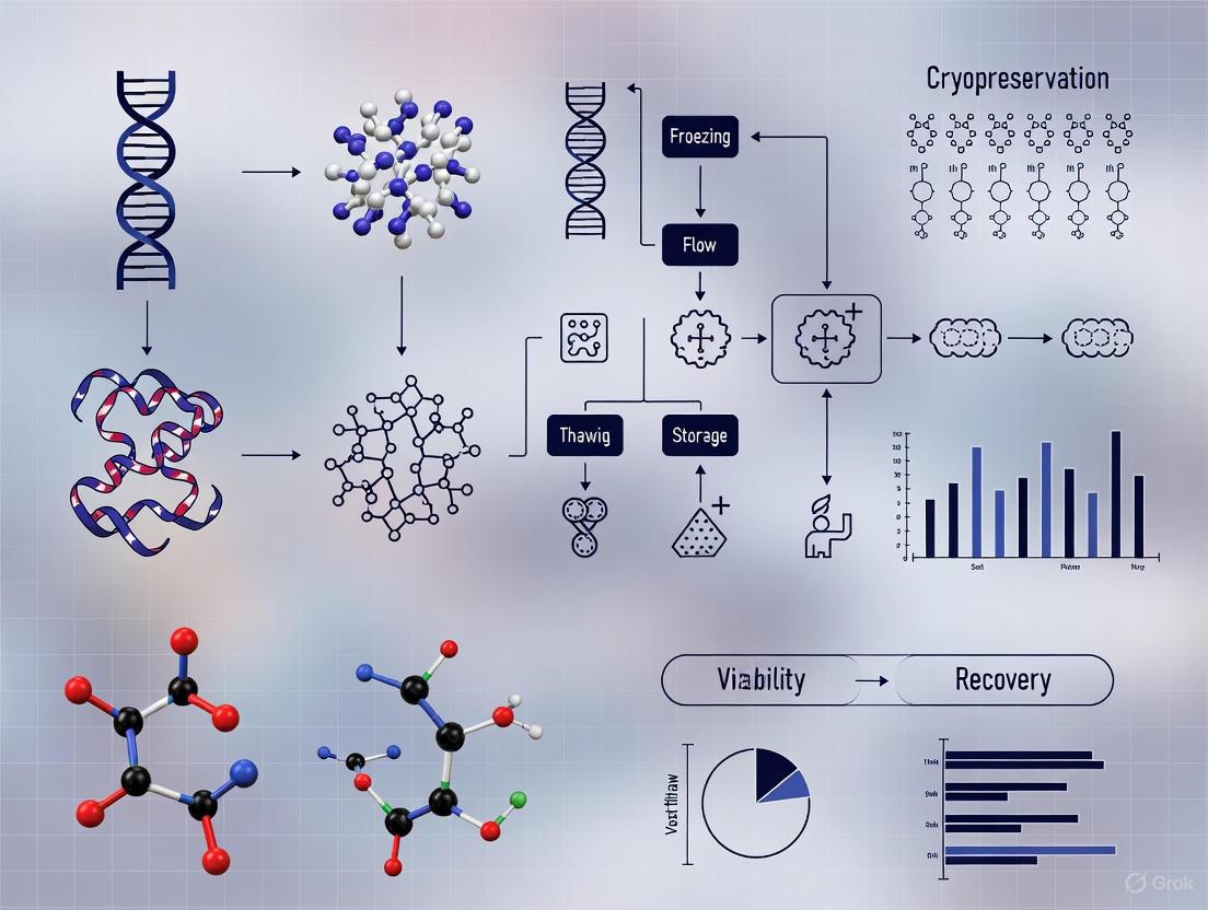
The Autologous Therapy Workflow: A Step-by-Step Analysis
The journey of an autologous cell therapy from patient to product and back again involves a meticulously coordinated sequence of events where cryopreservation plays multiple pivotal roles. The workflow can be visualized as a cyclic process with cryopreservation serving as stabilizing anchors at critical junctures.
Key Stages and Cryopreservation Integration
Cell Collection and Initial Cryopreservation: The process begins with collecting the patient's cells, typically through leukapheresis for immune cells or tissue biopsy for stem cells [6]. This starting material is highly time-sensitive and must be stabilized immediately. Initial cryopreservation decouples the collection procedure from downstream manufacturing, providing flexibility and allowing time for pre-processing quality checks [3] [5]. Proper cryopreservation at this stage ensures that the foundational cellular material retains its therapeutic potential.
Manufacturing and Final Product Cryopreservation: After thawing the starting material, cells undergo complex manufacturing processes including activation, genetic modification (e.g., CAR or TCR transduction), and ex vivo expansion [6]. The final therapeutic product is then cryopreserved in infusion-ready containers. This final cryopreservation is arguably the most critical, as it enables essential quality control testing, allows for precise treatment scheduling, and creates a stable product that can be transported globally or stored for future use, such as redosing [3] [4] [5].
Quantitative Impact of Cryopreservation on Cell Therapy Parameters
The integration of cryopreservation fundamentally alters the operational and economic landscape of autologous therapies. The following data illustrates its measurable impact across key parameters.
Table 1: Market and Operational Data for Cell Cryopreservation
| Parameter | Quantitative Data | Significance for Autologous Therapy |
|---|---|---|
| Global Market Value (2024) | $12.65 billion [7] | Indicates substantial infrastructure investment and industry reliance on cryopreservation technologies. |
| Projected Market Value (2029) | $35.3 billion (CAGR: 22.5%) [7] | Reflectits the anticipated growth in cell-based therapies and their dependency on robust storage solutions. |
| Post-Thaw Viability (Automated Systems) | >90% [8] | Demonstrates that optimized protocols can maintain high cell viability, a critical quality attribute. |
| Viable Storage Duration | Decades [3] | Enables long-term biobanking of starting materials and final products, supporting multi-dose treatment regimens. |
Table 2: Comparative Analysis: Fresh vs. Cryopreserved Leukopak Starting Material
| Characteristic | Fresh Leukopak | Cryopreserved Leukopak |
|---|---|---|
| Processing Timeline | 24-36 hours post-collection [5] | Indefinitely stable after freezing; processed at convenience |
| Logistical Complexity | High (requires immediate transport and processing) [5] | Low (decouples collection from manufacturing) [5] |
| Scheduling Flexibility | Low (tight coupling of procedures) | High (enables asynchronous operations) [5] |
| Risk of Product Variability | Higher (influenced by transport delays) [5] | Lower (standardized processing from stable material) [5] |
| Quality Control Window | Narrow (must occur during or after manufacturing) | Ample (testing can be completed pre-manufacturing) [5] |
Detailed Experimental Protocol: Automated Processing and Cryopreservation
This protocol outlines a streamlined, automated method for the cryopreservation of autologous cell therapy products, suitable for both adherent (e.g., MSCs) and suspension (e.g., T cells) cell types, utilizing Good Laboratory Practices (GLP) to ensure translational suitability [8].
Materials and Reagents
Table 3: Research Reagent Solutions for Cell Cryopreservation
| Reagent / Material | Function / Application | Example Product / Specification |
|---|---|---|
| Cryostor CS-10 | A clinical-grade, serum-free cryopreservation medium containing 10% DMSO. Minimizes ice crystal formation and osmotic shock. [8] | BioLife Solutions (Cat# NC9930384) |
| Dimethyl Sulfoxide (DMSO) | Permeating cryoprotectant agent (CPA). Penetrates the cell, reducing intracellular ice crystal formation. Can be cytotoxic. [9] | GMP-grade, typically used at 5-10% (v/v) [9] [5] |
| Lymphoprep | Density gradient medium for the isolation of peripheral blood mononuclear cells (PBMCs) from apheresis products. [8] | STEMCELL Technologies (Cat# 07801) |
| FINIA Tubing Set | Single-use, closed-system consumable for use with the Finia Fill and Finish System. Includes bags for product, mixing, and QC. [8] | Terumo BCT (Cat# 22050 for 50mL set) |
| TrypLE Express | Enzyme solution for detaching adherent cells (e.g., MSCs) from culture surfaces without damaging surface proteins. [8] | Millipore Sigma (Cat# 12605028) |
| Zombie UV Fixable Viability Kit | Fluorescent dye for flow cytometry-based assessment of cell viability post-thaw. Distinguishes live from dead cells. [8] | BioLegend (Cat# 423107) |
Step-by-Step Procedure
Part A: Cell Harvest and Preparation
- Harvesting Cells: For suspension cells (e.g., T cells), collect and concentrate cells by centrifugation. For adherent cells (e.g., MSCs), wash with PBS Ca²⁺/Mg²⁺-free and dissociate using a reagent like TrypLE Express. Neutralize the enzyme with a medium containing serum or platelet lysate [8].
- Cell Counting and Viability Assessment: Perform a cell count and viability check using an automated cell counter or flow cytometry with a viability dye. The protocol requires high pre-freeze viability (>90%) for optimal post-thaw recovery [8].
- Formulation for Freezing: Centrifuge the cell suspension and resuspend the cell pellet at the target concentration in an appropriate isotonic, protein-supported base medium (e.g., Dilution Buffer: 98% PBS + 2% human platelet lysate). Keep the cell suspension at 2-8°C to maintain viability [8].
Part B: Automated Formulation and Filling with the Finia System
- System Setup: Load the sterile FINIA tubing set and reagents (cell suspension and cryopreservation medium) into the Finia Fill and Finish System. Program the method to control temperatures, mixing ratios, and fill volumes [8].
- Automated Mixing and Aliquoting: The system will cool the cell suspension and Cryostor CS-10 to a specified temperature (e.g., 4°C), then combine them in a stepwise manner with gentle mixing to ensure uniform cell distribution and minimize osmotic stress. The final formulated product is automatically aliquoted into multiple cryogenic product bags [8].
- Quality Control Sampling: The system automatically fills a dedicated QC bag, which is used for post-processing and post-thaw quality control tests, including sterility, potency, and phenotype [8].
Part C: Controlled-Rate Freezing
- Loading: Transfer the filled product bags into a controlled-rate freezer, ensuring good contact with the canister surface for optimal heat transfer.
- Freezing Program: Initiate a standardized freezing ramp. A common protocol is [8]:
- Start at 4°C.
- Cool at -1°C per minute to -40°C.
- Cool at -5°C per minute to -100°C.
- Hold at -100°C for 10 minutes before transferring to long-term storage.
- Storage: Immediately transfer the frozen product bags to the vapor phase of a liquid nitrogen freezer (below -130°C) for long-term storage [3] [5].
Part D: Thawing and Assessment
- Rapid Thaw: Thaw the product bag rapidly in a 37°C water bath with gentle agitation until only a small ice crystal remains.
- Viability and Phenotype Analysis: For QC purposes, analyze the thawed sample from the QC bag. Assess viability (e.g., using Zombie UV dye) and phenotype (via flow cytometry with relevant antibodies) to confirm the product meets release specifications [8].
The workflow and relationships of the equipment used in this protocol are summarized in the following diagram:
Critical Success Factors and Troubleshooting
Achieving high post-thaw viability and functionality requires meticulous attention to several biological and technical parameters. The following table outlines common challenges and evidence-based solutions.
Table 4: Troubleshooting Guide for Autologous Therapy Cryopreservation
| Challenge | Potential Cause | Recommended Solution |
|---|---|---|
| Low Post-Thaw Viability | Intracellular ice formation causing physical damage; osmotic shock during CPA addition/removal. | Optimize cooling rate (typically -1°C/min); use stepwise addition of CPA; ensure rapid, uniform thawing [3] [9]. |
| Reduced Cell Functionality/Potency | Cryopreservation-induced activation of apoptotic pathways; oxidative stress from Reactive Oxygen Species (ROS). | Consider adding antioxidants to the cryomedium; minimize the time cells are exposed to liquid CPA before freezing; validate potency assays post-thaw [5]. |
| DMSO Toxicity | Cytotoxic effects of DMSO on cells and patient side effects upon infusion. | Use the lowest effective DMSO concentration (e.g., 5-7.5%); explore DMSO-free or reduced-DMSO cryomedium with non-permeating CPAs like sucrose or trehalose [9]. |
| Inconsistent Freezing Profiles | Unreliable equipment or overfilled cryocontainers leading to variable heat transfer. | Use programmable controlled-rate freezers; validate the freezing profile with thermocouples; do not exceed validated fill volumes [3] [8]. |
| Logistical Failure (Temperature Excursion) | Dry ice sublimation or liquid nitrogen depletion during transport. | Use validated, qualified shippers with temperature monitors; ensure proper packing procedures and contingency plans [9]. |
Cryopreservation is the linchpin that enables the practical application of autologous cell therapies by providing the essential stability and flexibility required to navigate complex manufacturing and treatment schedules. As the field advances toward more automated, closed-system processes, the development of optimized, standardized cryopreservation protocols will be critical for ensuring that these powerful personalized medicines realize their full therapeutic potential and become accessible to patients worldwide. The integration of robust cryopreservation within the autologous workflow is not merely a technical step but a fundamental strategic component that underpins the entire therapeutic model, from ensuring product quality and patient safety to enabling global scalability.
Cryopreservation is an indispensable tool in biomedical research and clinical applications, enabling long-term storage of cells and tissues for autologous cell therapies. The process faces two fundamental, interconnected challenges: intracellular ice crystallization and osmotic stress. When cells are exposed to sub-zero temperatures, water constitutes approximately 70% or more of total cell mass, making it the primary contributor to freezing injury [10]. Ice crystals can mechanically disrupt cellular membranes and organelles, while solute concentration effects can cause protein denaturation and irreversible cellular damage [10] [11]. Understanding these mechanisms is crucial for developing effective cryopreservation protocols for cell therapies, where maintaining high viability, potency, and functionality post-thaw is paramount for clinical success [9].
Core Damage Mechanisms
Intracellular Ice Crystallization
Intracellular ice formation (IIF) is widely recognized as a lethal event during cryopreservation [11]. The cooling rate critically determines the probability of IIF. At slow cooling rates, water has sufficient time to exit the cell, minimizing supercooling and avoiding intracellular freezing. In contrast, rapid cooling increases the likelihood of IIF as water molecules within the cell do not have time to migrate outward before freezing in place [10]. The process of recrystallization—where smaller ice crystals merge into larger, more damaging structures—can occur even during storage at intermediate temperatures like -80°C, leading to progressive cell death over time [12].
Recent studies using synchrotron-based x-ray diffraction have revealed that ice formation during warming may be more critical than during cooling. In bovine oocytes cooled with standard vitrification solutions, no ice was detected after cooling, yet significant ice crystallization occurred during warming [13]. This suggests that most ice-related damage in current protocols actually happens during the thawing phase rather than the freezing phase.
Osmotic Stress
As extracellular ice forms, solutes are excluded from the growing ice lattice, leading to a dramatic increase in the solute concentration of the remaining unfrozen fraction. This creates an osmotic imbalance that draws water out of cells, potentially causing excessive dehydration and volumetric changes [10] [11]. The degree of injury depends on the extent of this osmotic shock and the cell's ability to tolerate volume changes. The "unfrozen fraction" hypothesis suggests that damage results from the combined effects of increased solute concentration and reduced unfrozen water volume [11]. The presence of cryoprotective agents (CPAs) modifies this phase behavior but introduces its own challenges with potential toxicity.
Table 1: Key Damage Mechanisms in Cryopreservation
| Damage Mechanism | Underlying Cause | Cellular Consequences |
|---|---|---|
| Intracellular Ice Crystallization | Rapid cooling traps water intracellularly; Recrystallization during warming | Mechanical disruption of membranes and organelles; Lethal to most cell types |
| Osmotic Stress | Extracellular ice formation concentrates solutes; Creates osmotic imbalance | Cell dehydration and excessive volume changes; Solute toxicity effects |
| Solution Effects | High solute concentration in unfrozen fraction | Protein denaturation; Membrane damage |
| CPA Toxicity | Chemical effects of cryoprotectants | Altered cellular metabolism; Functional impairment post-thaw |
Quantitative Analysis of Cryopreservation Parameters
Optimizing cryopreservation protocols requires careful balancing of multiple parameters. Research on mouse oocytes has demonstrated that survival rates are highly dependent on both cooling and warming rates. One study found that with rapid warming (2950°C/min), survival remained at 75% for the first month at -80°C, but declined to 40% over the next two months, primarily due to recrystallization of intracellular ice [12]. In contrast, slow warming (139°C/min) resulted in only approximately 5% survival even immediately after cooling to -80°C [12].
Table 2: Impact of Cooling and Warming Rates on Cell Survival
| Cell Type | Cooling Rate | Warming Rate | CPA | Survival Outcome | Reference |
|---|---|---|---|---|---|
| Mouse oocytes | 187°C/min to -196°C | 2950°C/min | EAFS10/10 | ~75% after 1 month at -80°C; ~40% after 3 months | [12] |
| Mouse oocytes | 187°C/min to -196°C | 139°C/min | EAFS10/10 | ~5% survival even at 0 time at -80°C | [12] |
| Mouse oocytes | 294°C/min to -80°C | 2950°C/min | EAFS10/10 | ~90% after 7 days; dropped to ~35% after 3 months | [12] |
| Bovine oocytes | ~30,000°C/min | Conventional | Standard VS | No ice after cooling; large ice fractions during warming | [13] |
| Bovine oocytes | ~600,000°C/min | Conventional | Standard VS | Ice formation largely eliminated during cooling and warming | [13] |
Cryoprotective Agents and Their Mechanisms
Permeating Cryoprotectants
Permeating cryoprotectants (CPAs) are low-molecular-weight compounds that readily cross cell membranes. Common examples include dimethyl sulfoxide (DMSO), glycerol, ethylene glycol, and propylene glycol [10]. Their primary mechanism of action involves hydrogen bonding with water molecules, which depresses the freezing point and reduces the ability of water molecules to form ice nucleation sites [10]. DMSO, the most widely used CPA, increases membrane porosity at concentrations around 10%, allowing water to flow more freely through the membrane [10]. However, at higher concentrations (around 40%), DMSO can cause lipid bilayers to disintegrate, highlighting the importance of concentration optimization [10].
Non-Permeating Cryoprotectants
Non-permeating agents include compounds like sucrose, trehalose, polyethylene glycol (PEG), and polyvinylpyrrolidone (PVP) [10]. These larger molecules remain extracellular and exert their protective effects primarily by accelerating cell dehydration through osmotic pressure [9]. Trehalose is particularly notable because it is produced naturally by various organisms including bacteria, fungi, yeast, insects, and plants to withstand freezing [10]. Its chemical structure, with a glucose dimer linked via an α-1,1-glycosidic bond, provides exceptional stability under extreme temperatures [10].
Vitrification Strategies
Vitrification represents an alternative approach to traditional freezing, where high CPA concentrations and ultra-rapid cooling rates are used to transition water directly into an amorphous glassy state without ice crystal formation [9]. This method requires CPA concentrations of 40% w/v or more, which can be toxic to cells [9]. Recent research focuses on vitrification mixtures that combine permeating and non-permeating agents to reduce the required concentration of toxic CPAs while maintaining effective ice inhibition [10].
Experimental Protocols
Protocol for Assessing Ice Formation in Oocytes Using X-Ray Diffraction
This protocol adapts methodology from bovine oocyte studies for application to therapeutic cell lines [13].
Materials:
- Cryotop supports or crystallography loops
- Liquid nitrogen-based cryocrystallography instrument
- Synchrotron x-ray source with cryostream capability
- Equilibration solution (e.g., 1.5 M ethylene glycol)
- Vitrification solution (e.g., 6.6 M ethylene glycol + sucrose)
Procedure:
- Equilibrate cells in equilibration solution for 10-15 minutes at room temperature
- Transfer to vitrification solution for 60 seconds at room temperature
- Mount samples on Cryotop supports or crystallography loops
- Cool samples at controlled rates (~30,000°C/min or ~600,000°C/min) using automated cryocooler
- Maintain samples at -173°C using N₂ gas stream during x-ray data collection
- Collect 2D diffraction patterns with exposure times appropriate for cell type
- Warm samples in situ by blocking cold gas stream and directing room temperature N₂ at sample
- Analyze diffraction patterns for ice structure and volume fraction
Applications: This protocol enables quantitative assessment of intracellular ice formation during both cooling and warming phases, allowing researchers to optimize CPA compositions and thermal protocols for specific cell therapy products.
Protocol for Cryopreservation of PBMCs for Immunotherapy Applications
This protocol details the cryopreservation of peripheral blood mononuclear cells, critical for autologous cell therapies like CAR-T cells [14].
Materials:
- CPT cell preparation tubes with sodium citrate
- HBSS with penicillin-streptomycin (HBSS-PS)
- Hemolytic buffer
- Supplemented RPMI with HEPES buffer
- Fetal calf serum (FCS)
- Dimethyl sulfoxide (DMSO)
- Mr. Frosty freezing container or controlled-rate freezer
Procedure: Part A: PBMC Isolation
- Maintain CPT tubes at room temperature in upright position for 30 minutes after unpacking
- Invert tubes gently 8-10 times prior to centrifugation
- Centrifuge for 30 minutes at 1700 g at room temperature
- Aspirate plasma layer using sterile pipette, transfer to labeled 50 mL conical tube
- Add 3 mL HBSS-PS to each CPT tube, gently pipette to rinse sides and capture cells
- Transfer HBSS-PS/cell mixture to conical tube with plasma
- Add additional HBSS-PS to bring volume to 30-35 mL
- Centrifuge conical tubes for 10 minutes at 330 g at room temperature
- Pour off supernatant, disperse pellet by gentle tapping
- Add 3 mL hemolytic buffer, mix gently, incubate 5 minutes at room temperature
- Add 3 mL HBSS-PS, mix gently
- Centrifuge for 10 minutes at 330 g, pour off supernatant
Part B: PBMC Cryopreservation
- Resuspend cell pellet in 10 mL HBSS-PS
- Count cells using hemacytometer
- Prepare cryopreservation medium: 20% FCS + 10% DMSO in supplemented RPMI
- Slowly add cryopreservation medium to cells to achieve final concentration of 1-5×10⁶ cells/mL
- Aliquot 1 mL volumes into cryovials
- Place cryovials in Mr. Frosty freezing container or controlled-rate freezer
- Freeze at -1°C/min to -80°C
- Transfer to liquid nitrogen vapor phase (-140°C to -180°C) for long-term storage
Quality Control: Post-thaw viability assessment using trypan blue exclusion or dual fluorometric SYTO 13/GelRed assay is recommended [15].
The Scientist's Toolkit: Essential Research Reagents
Table 3: Key Reagents for Cryopreservation Research
| Reagent | Function | Application Notes |
|---|---|---|
| DMSO (Dimethyl Sulfoxide) | Permeating cryoprotectant | 10% concentration common; increases membrane porosity; toxic at high concentrations |
| Glycerol | Permeating cryoprotectant | One of earliest discovered CPAs; effective for many cell types |
| Ethylene Glycol | Permeating cryoprotectant | Lower toxicity alternative to DMSO for some applications |
| Trehalose | Non-permeating cryoprotectant | Natural disaccharide; exceptional stability; used in combination therapies |
| Sucrose | Non-permeating cryoprotectant | Facilitates dehydration; often used in vitrification solutions |
| Hyaluronic Acid | Non-permeating cryoprotectant | Emerging alternative; reduces DMSO requirements |
| SYTO 13/GelRed Assay | Viability assessment | Fluorometric method; alternative to trypan blue |
| CPT Tubes | PBMC isolation | Integrated blood collection and density-based separation |
Visualizing Cryopreservation Workflows
Cryopreservation Decision Pathway
Ice Formation Damage Pathways and CPA Protection Mechanisms
Implications for Autologous Cell Therapies
The principles of intracellular ice crystallization and osmotic stress management have direct implications for the manufacturing and clinical success of autologous cell therapies. For CAR-T cell therapies, which frequently use DMSO at concentrations of 5-10% [9], cryopreservation-induced damage can affect not only viability but also critical therapeutic functions like proliferation, cytokine secretion, and target cell killing. DMSO toxicity presents particular challenges in clinical settings, where infusion-related adverse events including neurological, gastrointestinal, cardiovascular, and hepatic complications have been reported [9].
Recent advances focus on reducing DMSO concentration through optimized vitrification mixtures that combine permeating and non-permeating agents [10] [9]. Alternative approaches include the development of ambient temperature transport systems that avoid cryopreservation altogether through nutrient, oxygen, and structural support during shipment [9]. As the cell therapy market continues to expand—projected to reach USD $97 billion by 2033 [9]—optimizing cryopreservation protocols to maintain cell potency and functionality while minimizing toxic CPA exposure will remain a critical research priority.
Understanding the fundamental mechanisms of intracellular ice formation and osmotic stress enables researchers to develop more effective preservation strategies for the next generation of autologous cell therapies, ultimately improving clinical outcomes for patients.
Dimethyl sulfoxide (DMSO) is a widely utilized penetrating cryoprotective agent (CPA) essential for the cryopreservation of cells in autologous cell therapy research and development [16] [17]. Its ability to penetrate cell membranes and prevent intracellular ice crystallization—a primary cause of cell death during freezing—makes it a cornerstone of contemporary cryopreservation protocols [18]. However, its application is coupled with significant, dose-dependent cytotoxicity concerns that complicate its use, particularly for therapies destined for clinical administration [19] [20]. For researchers in drug development, a precise understanding of DMSO's dual nature—its protective mechanisms and its toxicological profile—is critical for designing effective and safe cryopreservation strategies for sensitive therapeutic cells. This document details the mechanisms, quantitative toxicity data, and practical protocols to guide its use in autologous cell therapy research.
Core Mechanisms of Action
DMSO provides cryoprotection through multiple interconnected biophysical mechanisms.
- Ice Crystallization Inhibition: DMSO disrupts the hydrogen bonding network between water molecules, thereby lowering the freezing point of the aqueous solution and reducing the rate and extent of ice crystal formation during cooling. This action minimizes mechanical damage to cellular structures from intracellular and extracellular ice [18] [21].
- Membrane Penetration and Stabilization: As a low-molecular-weight, permeable molecule, DMSO readily crosses the plasma membrane. This enables it to protect both intracellular and extracellular compartments. While it can cause membrane dehydration at high concentrations, it also helps to stabilize membrane phospholipids during the freezing process, maintaining membrane integrity [21] [17].
- Vitrification Promotion: At high concentrations and rapid cooling rates, DMSO facilitates vitrification, a process where water solidifies into a non-crystalline, glassy state. This avoids the damaging phase transition to ice altogether, which is particularly critical for preserving complex structures like tissues and organoids [20] [17].
Cytotoxicity Concerns: Pathways and Quantitative Data
Despite its efficacy, DMSO induces cytotoxicity through several pathways, with effects manifesting at both the cellular and patient levels. The toxicity is influenced by concentration, temperature, and duration of exposure [19] [18].
Mechanisms of Cellular Toxicity
- Membrane and Organelle Damage: DMSO compromises membrane integrity and can induce pore formation, particularly at concentrations exceeding 10% (v/v). It also disrupts mitochondrial function, reducing membrane potential and increasing the production of reactive oxygen species (ROS), leading to oxidative stress and apoptosis [20] [18] [22].
- Epigenetic and Transcriptional Alterations: Exposure to DMSO can disrupt cellular epigenetic profiles. It interferes with DNA methyltransferases and histone-modifying enzymes, leading to changes in gene expression patterns. This is especially problematic for stem cell therapies, as it can reduce pluripotency and induce unwanted differentiation [20] [17].
- Protein Denaturation and Osmotic Stress: At temperatures above 0°C, DMSO can destabilize proteins through hydrophobic interactions. Furthermore, the rapid influx or efflux of DMSO and water during the addition or removal steps can cause significant osmotic shock, resulting in cell lysis [19] [18].
Clinical Adverse Effects
In autologous cell therapies, the patient's own cells are cryopreserved, stored, and later infused. Residual DMSO in the infusion product is associated with various adverse reactions, including nausea, headaches, cardiovascular instability, allergic reactions, and, in rare cases, severe neurological events such as seizures or cardiac arrest [23] [20] [24].
Quantitative Toxicity Data
The table below summarizes key toxicity findings from recent research, highlighting the concentration and time dependence of DMSO-induced damage.
Table 1: Quantitative Profile of DMSO Cytotoxicity
| Cell Type / System | DMSO Concentration | Exposure Conditions | Observed Effect | Reference |
|---|---|---|---|---|
| Human Chondrocytes | >10% (v/v) | Varying, at 37°C | Significant cytotoxicity; induction of apoptosis via caspase-9 and -3 activation. | [18] [22] |
| Dermal Fibroblasts | 5% to 30% (v/v) | 10-30 min, at 4°C, 25°C, 37°C | Decreasing viability with increasing concentration, temperature, and exposure time. | [19] |
| Rat Myocardium | >10% (v/v) | At 30°C | Irreversible ultrastructural alterations. | [19] |
| Peripheral Blood Progenitor Cells | Increase from 7.5% to 10% | Standard cryopreservation | Reduction in clonogenic potential. | [19] |
| Neural Cells (in vitro) | 0.5% - 1% | Culture conditions | 50% viability loss in rat hippocampal neurons; decreased viability in retinal ganglion neurons. | [23] |
| Patient Infusion | Varies with product | Direct infusion | Adverse events: nausea, cardiovascular issues, allergic reactions, rare neurological events. | [20] [24] |
Experimental Protocols for Evaluation
For researchers developing autologous cell therapies, evaluating DMSO toxicity in their specific cellular product is paramount. Below is a generalized protocol that can be adapted.
Protocol: Assessing DMSO Toxicity on Cell Viability and Function
Objective: To determine the maximum tolerated concentration and exposure time of DMSO for a specific candidate therapeutic cell type.
Materials:
- Candidate therapeutic cells (e.g., T-cells, stem cells)
- Complete growth medium
- High-purity, sterile DMSO
- Phosphate Buffered Saline (PBS)
- Cell culture plates
- Flow cytometer with annexin V/PI staining kit
- Cell counter and viability analyzer (e.g., trypan blue exclusion)
- Incubator at 37°C and 5% CO₂
Method:
- Cell Preparation: Harvest and resuspend cells in complete growth medium to a standardized concentration (e.g., 1 x 10⁶ cells/mL).
- DMSO Exposure:
- Prepare serial dilutions of DMSO in complete medium across a relevant range (e.g., 0.5%, 2.5%, 5%, 10% v/v). Keep solutions on ice.
- Add equal volumes of cell suspension to each DMSO solution, mixing gently to achieve the final desired DMSO concentrations.
- Incubate the cell-DMSO mixtures for a defined time (e.g., 15, 30, 60 minutes) in a 37°C incubator.
- Include a control group with cells in medium only (0% DMSO).
- Termination of Exposure and Analysis:
- After exposure, dilute each sample 10-fold with cold complete medium to rapidly reduce DMSO concentration.
- Centrifuge cells, wash once with PBS, and resuspend in fresh medium.
- Viability and Apoptosis Analysis:
- Perform trypan blue exclusion for immediate viability count.
- Use annexin V and propidium iodide (PI) staining followed by flow cytometry to distinguish live (annexin V-/PI-), early apoptotic (annexin V+/PI-), and late apoptotic/necrotic (annexin V+/PI+) populations.
- Functional Assay (Cell-type dependent):
- For immune cells (e.g., T-cells, NK cells): Perform a cytotoxicity assay against target cells or measure cytokine release upon stimulation after a 24-hour recovery period [24].
- For stem cells: Assess differentiation potential and pluripotency marker expression after several days in culture.
Workflow Diagram
The following diagram illustrates the key damage pathways during cryopreservation and the protective and toxic roles of DMSO.
Diagram: DMSO in Cryopreservation - Protection vs. Toxicity. This diagram outlines the primary damage pathways during freezing (center) and the protective mechanisms of DMSO (left). Concurrently, it highlights the cytotoxic pathways activated by DMSO itself under suboptimal conditions (right), both leading to cell death.
The Scientist's Toolkit: Research Reagent Solutions
The following table lists key reagents and their functions for investigating DMSO-based cryopreservation.
Table 2: Essential Research Reagents for CPA Toxicity Studies
| Reagent / Material | Function in Protocol | Specific Example / Note |
|---|---|---|
| High-Purity DMSO | Primary cryoprotectant for freeze-thaw cycles and toxicity studies. | Use sterile, compendial-grade (e.g., USP) material to ensure consistency and minimize contaminant-induced variability. |
| Annexin V / PI Apoptosis Kit | Flow cytometry-based detection of apoptosis and necrosis in cells post-DMSO exposure. | Critical for distinguishing the mode of cell death induced by cytotoxic insults. |
| Trypan Blue Solution | Dye exclusion assay for rapid, quantitative assessment of cell membrane integrity and viability. | Standard, simple method for immediate post-thaw or post-exposure viability count. |
| Controlled-Rate Freezer | Equipment to precisely control cooling rate during freezing, a critical variable for cell survival. | Enables standardization and optimization of freeze protocols (e.g., -1°C/min). |
| Viability-Specific Functional Assay Kits | Assess functional recovery post-thaw, which is as important as simple viability. | Examples: CFSE-based proliferation kits; CD107a degranulation or IFN-γ ELISpot for immune cells. |
| ROCK Inhibitor (e.g., Y-27632) | Small molecule added to culture medium to improve survival of sensitive cells, like stem cells, after thawing. | Shown to improve recovery of hiPSCs post-thaw, reducing apoptosis [20]. |
DMSO remains an exceptionally effective CPA, but its cytotoxicity presents a significant challenge for autologous cell therapy. The path forward involves a meticulous, evidence-based approach to protocol design, where DMSO concentration, exposure time, and temperature are optimized for each specific cell product. Furthermore, the field is actively pursuing strategies to mitigate DMSO-related risks, including the development of DMSO-free cryopreservation solutions using alternative CPAs like deep eutectic solvents [25], sugars (trehalose, sucrose) [20] [18], and advanced polymers [20] [17], as well as improved post-thaw washing techniques. For the researcher, a deep understanding of the dual nature of DMSO is not just academic—it is a fundamental requirement for ensuring the viability, functionality, and safety of transformative autologous cell therapies.
The successful administration of autologous cell therapies is intrinsically tied to the integrity of a complex and vulnerable journey—the cryogenic cold chain. These patient-specific therapies, wherein cells are collected from a patient, engineered or activated at a centralized manufacturing facility, and then returned to the same patient, are critically dependent on cryopreservation for storage and transport. Maintaining a continuous ultra-low temperature environment, typically at -150°C or below using liquid nitrogen (LN2), is not merely a logistical preference but a fundamental requirement to preserve cell viability, potency, and function [26] [27].
The logistical and financial hurdles embedded within this cold chain represent a significant bottleneck in the broader translation and commercialization of these transformative treatments. This document delineates the specific challenges—from market fragmentation and technical inconsistencies to prohibitive costs—and provides detailed application notes and standardized protocols designed to fortify the cryogenic supply chain for researchers and drug development professionals.
Quantitative Analysis of Logistical and Financial Hurdles
A comprehensive analysis of the cryogenic cold chain requires an understanding of its quantitative inefficiencies and cost drivers. The tables below summarize key data on operational impacts and financial burdens gathered from recent industry and scientific reviews.
Table 1: Impact of Market and Technical Fragmentation on Cryogenic Logistics
| Challenge Category | Specific Impact Metric | Quantitative Effect | Context / Region |
|---|---|---|---|
| Overall Chain Efficiency | Cumulative Efficiency Reduction | 18-25% decrease in efficiency [28] | Fragmented supply chains |
| Technology Inconsistency | Use of Automated Warehouses | Only 12% of providers use them, leading to temperature fluctuations [28] | African cooling logistics |
| Produce moved via refrigerated transport | Only 51% of produce is moved this way, leading to high food loss [28] | China (as a proxy for infrastructure variability) | |
| Logistical Inefficiency | Product Loss due to Chain Breaks | Up to 23% of products lost [28] | Agricultural sector (illustrative of re-loading risks) |
| Operational Risk | Temperature Deviation Risk | Supply chains exceeding critical limits 4.7x more frequently [28] | Fragmented vs. consolidated chains |
Table 2: Financial and Economic Challenges in Cell Therapy Logistics
| Factor | Financial Metric / Consequence | Therapeutic / Commercial Impact |
|---|---|---|
| Therapy List Price | Up to $4.3 million per dose [29] | Severe limitations on patient access and payer reimbursement |
| Infrastructure Investment | Small providers can only apply 15-20% of costs to predictive maintenance or blockchain tracking [28] | Widening technology gap and inconsistent quality |
| Corrective Costs | High cost of product failure due to temperature deviation [27] [29] | Compromised viability, delayed treatments, lost revenue |
Experimental Protocols for Cryopreservation and Viability Assessment
Protocol: Slow Freezing Cryopreservation of Cell Therapy Products
This protocol outlines a standardized method for the cryopreservation of autologous cell therapy products, such as T-cells or stem cells, using a controlled-rate freezer and liquid nitrogen storage, based on established methodologies [30].
1. Reagents and Materials:
- Cell suspension in appropriate media
- Cryoprotectant Agent (CPA): e.g., Clinical-grade DMSO (final concentration 5-10%)
- Serum or protein-rich media (e.g., Albumin)
- Cryopreservation bags or 2ml cryovials
- Controlled-rate freezer
- LN2 vapor-phase storage tank
2. Procedure:
- Step 1: Preparation. Pre-cool the controlled-rate freezer and prepare a cooling chamber with LN2. Label cryovials/bags with unique patient identifiers.
- Step 2: CPA Addition. Prepare a 2X freezing medium containing 20% DMSO and 20% serum in base media. Slowly mix the 2X freezing medium with an equal volume of the cell suspension to achieve a final concentration of 10% DMSO and ~5-10 x 10^6 cells/mL. Gently mix to avoid osmotic shock.
- Step 3: Packaging. Aseptically dispense the cell-CPA mixture into cryovials (e.g., 1-2 mL) or cryobags. Seal securely.
- Step 4: Controlled-Rate Freezing. Immediately transfer samples to the controlled-rate freezer. Initiate the program:
- Step 4.1: Hold at +4°C for 10 minutes.
- Step 4.2: Cool at -1°C/min to -40°C [30].
- Step 4.3: Cool at -5°C/min to -90°C.
- Step 4.4: Hold at -90°C for 10 minutes before transfer.
- Step 5: Long-Term Storage. Rapidly transfer the frozen samples to a pre-cooled LN2 vapor-phase storage system, maintaining a steady state below -150°C [26] [27].
3. Quality Control Note: A sample from the batch should be tested for viability and sterility post-cryopreservation. The cooling curve should be validated and documented for each run.
Protocol: Post-Thaw Cell Viability and Potency Analysis
Assessing cell health after thawing is critical for confirming the success of the cryopreservation and transport process. This protocol measures viability and metabolic activity.
1. Reagents and Materials:
- Thawed cell sample
- Phosphate Buffered Saline (PBS)
- Trypan Blue solution (0.4%) or proprietary viability dyes (e.g., Propidium Iodide)
- Cell culture media
- WST-1 or MTT assay kit
- Flow cytometer or automated cell counter
- Microplate reader
2. Procedure:
- Step 1: Thawing. Rapidly thaw the cryovial in a 37°C water bath with gentle agitation until only a small ice crystal remains (≈ 2-3 minutes).
- Step 2: Washing. Immediately and gently transfer the cell suspension to a tube containing pre-warmed media (10x the volume of the vial) to dilute the cytotoxic DMSO. Centrifuge at 300-400 x g for 5-10 minutes. Aspirate the supernatant.
- Step 3: Viability Count. Resuspend the cell pellet in PBS. Mix a small aliquot with an equal volume of Trypan Blue. Load onto a hemocytometer and count viable (unstained) and non-viable (blue) cells using an automated cell counter or manual count. Calculate viability:
% Viability = (Viable Cell Count / Total Cell Count) * 100. The FDA often requires ≥80% viability for CAR-T cell products [30]. - Step 4: Potency/Metabolic Assay. Seed washed cells into a 96-well plate. Add WST-1 reagent according to the manufacturer's instructions. Incubate for 1-4 hours at 37°C. Measure the absorbance of the formazan product at 440-450 nm using a microplate reader. Higher absorbance correlates with greater metabolic activity and cell health [30].
Diagram 1: Post-thaw cell analysis workflow.
Protocols for Managing Integrated Cryogenic Logistics
Protocol: Designing a Scalable Cryogenic Storage Infrastructure
Selecting and implementing the correct storage system is fundamental for R&D and clinical-scale operations. This protocol guides the selection process based on capacity and scalability needs [26].
1. Assessment and Planning:
- Step 1: Capacity Forecasting. Estimate the number of samples to be stored over the next 1-5 years. A standard unit is a 2ml cryovial. For example, 8,800 vials equate to 88 cryoboxes [26].
- Step 2: Spatial and LN2 Planning. Ensure the laboratory or storage room has adequate floor strength, ventilation, and proximity to LN2 supply lines or bulk tanks. Plan for a 21-day LN2 hold time for uninterrupted storage during supply disruptions [27].
2. System Selection and Implementation:
- Step 3: Model Selection. Choose a system based on forecasted capacity:
- Step 4: Inventory Management Integration. Integrate the storage system's software (e.g., FreezerPro) with the institutional Enterprise Resource Planning (ERP) system for real-time visibility and streamlined workflows [26] [27].
- Step 5: Access Control & Training. Implement controlled access systems and standardized operating procedures (SOPs) to limit errors and ensure consistency during sample retrieval and return [27].
Protocol: Risk Mitigation for Cryogenic Transport of Autologous Products
This protocol ensures the integrity of the therapy during its most vulnerable phase: transport from the manufacturing site to the clinical center.
1. Pre-Shipment Preparation:
- Step 1: Shipper Qualification. Qualify specialized cryogenic shippers capable of maintaining a stable vapor-phase LN2 temperature (<-150°C) for a duration exceeding the expected transit time by at least 50% [27] [29].
- Step 2: Packaging Configuration. Use best-practice packaging configurations with absorbent materials to contain potential leaks. Pre-condition the shipper with LN2 according to the manufacturer's specifications.
- Step 3: Documentation & Labeling. Ensure all packages are clearly labeled with "Cryogenic Material," "Do X-Ray," and unique patient identifiers. Prepare all necessary chain of identity (COI) and chain of custody (COC) documents.
2. Execution and Monitoring:
- Step 4: Real-Time Monitoring. Place a calibrated temperature data logger inside the shipper to record the temperature throughout the transit. Set clear alert thresholds (e.g., > -150°C).
- Step 5: Carrier and Route Selection. Utilize a logistics provider specializing in cryogenic transport for cell and gene therapies [29]. Establish protocols for airport security scan exemptions to avoid damaging radiation [29].
- Step 6: Contingency Planning. Have a documented contingency plan for flight delays or unforeseen events, which may include access to regional storage centers with cryogenic filling stations [29].
Diagram 2: Cryogenic transport risk mitigation workflow.
The Scientist's Toolkit: Essential Reagents and Materials
Table 3: Key Research Reagent Solutions for Cryogenic Logistics
| Item / Reagent | Function / Application | Key Consideration |
|---|---|---|
| DMSO (Cryoprotectant) | Penetrating CPA; reduces intracellular ice crystal formation [30]. | Cytotoxic at high concentrations/ prolonged exposure; requires post-thaw washing. Optimal final concentration ~10%. |
| Controlled-Rate Freezer | Provides a reproducible, linear cooling rate (e.g., -1°C/min) to minimize cell damage during freezing [30]. | Critical for process consistency and viability. Cooling rates must be optimized for specific cell types. |
| LN2 Storage System | Provides long-term storage at <-150°C in vapor phase to halt all biochemical activity [26] [27]. | Systems offer varying capacities (Pico to Tera). 21-day LN2 hold time enhances supply chain resilience [27]. |
| Cryogenic Shippers | Insulated containers pre-charged with LN2 to maintain cryogenic temperatures during transport [29]. | Must be validated for duration and stability. Specialized providers offer certified shippers and monitoring. |
| Temperature Data Logger | Electronic device that records temperature history throughout storage or transport for quality assurance [29]. | Data is critical for regulatory compliance and verifying product integrity upon receipt. |
| Inventory Management Software | Tracks sample location, identity, and freezing history; integrates with ERP systems [26] [27]. | Essential for maintaining Chain of Identity (COI) in autologous therapies and audit trails. |
Cryopreservation is a cornerstone of modern autologous cell therapy, enabling the complex logistics between cell collection, manufacturing, and patient infusion. For therapies like Chimeric Antigen Receptor T-cell (CAR-T), the quality of the final product is intrinsically linked to the freezing and thawing processes. This application note synthesizes current research to provide detailed protocols and data on how cryopreservation impacts critical quality attributes (CQAs)—viability, potency, and efficacy—of cell-based products, providing a framework for researchers to optimize their own processes.
Quantitative Impact of Cryopreservation on Cell Viability
Long-term cryopreservation can maintain high cell viability, though a gradual, time-dependent decline is often observed. The data below summarizes viability outcomes for different cell types under various storage conditions.
Table 1: Long-Term Viability of Cryopreserved Cell Products
| Cell Type | Storage Temperature | Storage Duration | Post-Thaw Viability | Key Findings | Source |
|---|---|---|---|---|---|
| Hematopoietic Stem Cells (HSCs) | -80°C (uncontrolled-rate) | Median 868 days (≈2.4 years) | Median 94.8% | Viability decline of ~1.02% per 100 days; engraftment kinetics preserved. | [31] |
| Peripheral Blood Mononuclear Cells (PBMCs) | -196°C (Liquid Nitrogen) | 3.5 years | Average 90.95% | No significant change in viability with extended freezing time; T-cell proportion remained stable. | [32] |
| PBMCs | -196°C (Liquid Nitrogen) | 12 months | Relatively Stable | Cell viability stable; scRNA-seq cell capture efficiency reduced by ~32%. | [33] |
| Ovine Spermatozoa | -196°C (Liquid Nitrogen) | 50 years | Functional | Successful pregnancy rate of 61%; demonstrated long-term preservation of function. | [34] |
The data indicates that while absolute viability is a critical metric, it is not the sole determinant of clinical success. For instance, HSC products with a moderate viability decline still supported successful engraftment, underscoring the importance of functional potency assays [31].
Impact on Cellular Potency and Function
Beyond simple viability, preserving the functional capacity and "fitness" of cells is paramount for autologous therapies. Research demonstrates that cryopreservation can have nuanced effects on potency.
Functional Persistence in CAR-T Cells
A critical 2025 study directly compared CAR-T cells generated from fresh and cryopreserved PBMCs using the PiggyBac transposon system. The results demonstrated that CAR-T products derived from PBMCs cryopreserved for up to two years exhibited:
- Comparable expansion potential in vitro.
- No significant differences in cell phenotype (CD4+/CD8+ ratios) or transfection efficiency.
- Similar differentiation and exhaustion marker profiles (critical for in vivo persistence).
- Equivalent cytotoxicity against target tumor cells (SKOV-3) in functional assays [32].
Transcriptomic and Population Stability
Single-cell RNA sequencing (scRNA-seq) of PBMCs cryopreserved for 6 and 12 months revealed that the transcriptome profiles of major immune cell types (T cells, B cells, NK cells, monocytes) showed minimal perturbation. While a small number of stress-response genes were subtly altered, the overall genomic landscape was preserved, supporting the functional data that cryopreserved cells retain their identity and potential [33].
Detailed Experimental Protocols for Assessment
To ensure product quality, standardized protocols for post-thaw analysis are essential. The following are detailed methodologies adapted from recent studies.
Protocol: Viability and Phenotype Analysis of Cryopreserved PBMCs for CAR-T Manufacturing
This protocol is adapted from the workflow used to generate the comparative data in Table 1 and Section 3.1 [32].
Objective: To assess the viability, recovery, and immunophenotype of cryopreserved PBMCs prior to CAR-T manufacturing.
Materials:
- Cryopreserved PBMC vial(s) stored in liquid nitrogen.
- Water bath (37°C).
- Centrifuge.
- Complete RP10 Medium: RPMI1640 + 10% FBS + 10mM HEPES + 0.1 mg/mL Gentamycin.
- Flow cytometry buffer (e.g., PBS + 2% BSA).
- Viability Stain: Propidium Iodide (PI) or 7-Aminoactinomycin D (7-AAD).
- Antibody Panel: Fluorescently-labeled antibodies against CD3, CD45, CD4, CD8, CD19, CD56, CD45RO, CCR7.
Procedure:
- Thawing: Remove vial from liquid nitrogen and thaw rapidly in a 37°C water bath until only a small ice crystal remains.
- Washing: Gently transfer cell suspension to a 15mL tube containing 10mL of pre-warmed RP10 medium. Centrifuge at 500 x g for 5 minutes at room temperature.
- Resuspension: Discard supernatant, gently tap tube to loosen pellet, and resuspend in 10mL of fresh RP10 medium. Perform a second wash under identical conditions.
- Viability Count: Resuspend cells in a small volume. Mix an aliquot with Trypan Blue and count using a hemocytometer, or use an automated cell counter.
- Flow Cytometry Staining:
- Aliquot 1-2 x 10^6 cells into a flow tube.
- Wash cells with flow cytometry buffer.
- Resuspend in buffer containing a viability dye (e.g., PI) and incubate on ice for 15-30 minutes, protected from light.
- Wash cells to remove unbound dye.
- Block with FC receptor blocking agent for 10 minutes.
- Add surface antibody cocktail and incubate for 20-30 minutes at 4°C in the dark.
- Wash cells twice and resuspend in buffer for acquisition on a flow cytometer.
- Analysis: Analyze data to determine the percentage of live cells (viability dye negative) and the proportion of T cells (CD3+), B cells (CD19+), NK cells (CD56+), and T cell subsets (e.g., naïve T cells: CD45RO- CCR7+).
Protocol: Functional Potency Assay via Cytotoxicity
This protocol measures the cytotoxic function of CAR-T cells, a direct measure of potency, as described in the comparative CAR-T study [32].
Objective: To evaluate the in vitro cytotoxic activity of CAR-T cells derived from cryopreserved starting material.
Materials:
- Effector cells: CAR-T cells and non-transduced control T cells (Mock-T).
- Target cells: Antigen-positive cancer cell line (e.g., SKOV-3 for mesothelin-targeting CAR).
- Real-Time Cell Analysis (RTCA) system or alternative platform (e.g., flow cytometry-based killing assay).
- Cell culture plates.
- Cytokine release assay kits (e.g., for IFN-γ, IL-2).
Procedure:
- Target Cell Seeding: Seed target cells in the appropriate microplate according to the RTCA system manufacturer's instructions. Allow cells to adhere and establish a baseline impedance reading.
- Effector Cell Addition: After baseline establishment, add CAR-T or Mock-T cells at specified Effector:Target (E:T) ratios (e.g., 4:1, 2:1). Include a target cell-only well as a background control.
- Monitoring: Continuously monitor impedance for 24-96 hours. Cytotoxicity is calculated by the system software based on the change in impedance relative to the target cell-only control.
- Cytokine Measurement: Following the co-culture period, collect supernatant from the assay. Quantify the concentration of key cytokines like IFN-γ using a standardized ELISA or multiplex bead-based assay.
- Analysis: Compare the cytotoxicity curves and cytokine secretion levels of CAR-T cells from cryopreserved PBMCs against those from fresh PBMCs and control groups.
The following diagram illustrates the logical relationship between cryopreservation process parameters, the critical quality attributes (CQAs) of the cell product, and the experimental methods used for assessment.
The Scientist's Toolkit: Essential Reagents and Materials
Successful cryopreservation and analysis rely on a suite of specialized reagents and equipment. The following table details key solutions used in the featured research.
Table 2: Essential Research Reagents for Cryopreservation Studies
| Item | Function/Description | Example Application in Protocols |
|---|---|---|
| Controlled-Rate Freezer (CRF) | Precisely controls cooling rate to minimize intracellular ice formation and osmotic stress. | Used in PBMC freezing protocol to ensure consistent, reproducible freezing [35]. |
| Cryoprotectant Agent (CPA) - DMSO | Penetrating cryoprotectant that reduces ice crystal formation; requires careful handling due to cytotoxicity. | Standard component of cryopreservation media for HSCs and PBMCs [31] [32]. |
| Serum-Free Freezing Media | Xeno-free, defined formulation that eliminates contamination risks from animal sera. | Preferred for clinical-grade manufacturing; trend towards specialized, optimized media [36] [37]. |
| Viability Dyes (7-AAD, PI) | DNA-binding dyes that are excluded by live cells with intact membranes; used for viability staining. | Used in flow cytometry protocols to distinguish live/dead cells in PBMC and HSC products [31] [33]. |
| Magnetic Cell Separation Beads | Enable positive or negative selection of specific cell populations (e.g., CD4+/CD8+ T cells). | Used to enrich T cells from thawed PBMCs prior to CAR-T manufacturing [32]. |
| PiggyBac Transposon System | Non-viral gene delivery system for stable gene integration; lower cost and immunogenicity than viral systems. | Used in the featured protocol to generate CAR-T cells from cryopreserved PBMCs [32]. |
The body of evidence confirms that with optimized and controlled processes, cryopreservation is a robust and reliable method for preserving the viability, potency, and efficacy of autologous cell therapies. While a slow, time-dependent decline in viability can occur, this does not necessarily preclude clinical success if the cell product meets critical quality specifications. The integration of detailed process controls, including controlled-rate freezing and standardized thawing, alongside rigorous analytical assessments of function and potency, is essential for ensuring that the final therapeutic product delivers its intended clinical benefit.
Standardized Protocols and Emerging Methods for Clinical Applications
Optimized Cryopreservation Protocols for Leukapheresis and PBMC Starting Materials
The successful development and manufacturing of autologous cell therapies are fundamentally dependent on the quality and viability of their cellular starting materials. Cryopreservation of leukapheresis products and peripheral blood mononuclear cells (PBMCs) enables critical flexibility in manufacturing logistics, decoupling cell collection from processing and facilitating the creation of cell banks for research and development [38]. However, standard cryopreservation approaches can introduce variability that compromises cell recovery, functionality, and ultimately, experimental reproducibility [39] [40]. This application note provides detailed, optimized protocols for the cryopreservation of leukapheresis products and PBMCs, specifically framed within the context of autologous cell therapy research. By implementing these standardized procedures, researchers can significantly enhance the consistency and reliability of their cellular starting materials, thereby improving downstream therapeutic outcomes.
Optimized Cryopreservation Protocol for PBMCs
Background and Principle
PBMCs, comprising lymphocytes and monocytes, are critical for immunological research and therapy development. The objective of PBMC cryopreservation is to preserve these cells in a state of suspended animation, maintaining high viability and functionality for years [41]. The principle relies on controlled-rate freezing in the presence of cryoprotectants to minimize intracellular ice crystal formation and osmotic stress, which are primary causes of cell death [42].
Detailed Step-by-Step Protocol
Materials:
- Biological Material: Isolated PBMCs.
- Cryopreservation Medium: 90% Fetal Calf Serum (FCS) + 10% DMSO, cooled to 2-8°C [39]. Alternatively, commercial media like Recovery Cell Culture Freezing Medium can be used [33].
- Equipment: Controlled-rate freezer (e.g., CryoMed), or isopropanol freezing chamber (e.g., "Mr. Frosty"), liquid nitrogen storage system.
Procedure:
- Isolation: Isolate PBMCs from whole blood using density gradient centrifugation (e.g., Ficoll-Paque) at room temperature. Cold reagents can lead to poor separation and granulocyte contamination [42].
- Washing: Wash isolated cells to remove residual plasma, platelets, and separation medium.
- Resuspension: Gently resuspend the PBMC pellet in chilled cryopreservation medium to a final concentration of 1-5 x 10^6 cells/mL [41] [42]. Work quickly to minimize DMSO exposure, as its cytotoxic effects increase over time at temperatures above 0°C [42].
- Aliquoting: Dispense 1 mL of cell suspension into cryogenic vials.
- Freezing:
- Using a Controlled-Rate Freezer: Employ a standardized freezing rate of -1°C/min to at least -80°C before transfer to long-term storage [42]. Advanced programs can be used (e.g., 1.0°C/min to -4°C, 25.0°C/min to -40°C, 10.0°C/min to -12.0°C, 1.0°C/min to -40°C, 10.0°C/min to -90°C) [33].
- Using an Isopropanol Chamber: Place sealed vials in the chamber pre-cooled with isopropyl alcohol and transfer immediately to a -80°C freezer for 18-24 hours. This apparatus approximates the -1°C/min cooling rate [42].
- Storage: Transfer vials to the vapor phase of liquid nitrogen (-135°C to -196°C) for long-term storage [41].
Thawing and Post-Thaw Recovery of PBMCs
Procedure:
- Thawing: Rapidly thaw cryovials by gently swirling in a 37°C water bath until only a small ice crystal remains [33].
- Dilution: Immediately transfer the cell suspension to a tube containing 10 mL of pre-warmed complete medium (e.g., RPMI-1640 with 10% FBS). Gently mix by pipetting 2-3 times. This rapid dilution is critical to reduce DMSO toxicity [41] [33].
- Washing: Centrifuge the cell suspension at 500 x g for 5 minutes at room temperature. Discard the supernatant and gently resuspend the pellet in fresh warm medium. Some protocols recommend a second wash step [33].
- Viability Assessment: Assess cell viability using trypan blue exclusion or propidium iodide staining. Post-thaw viability should ideally exceed 90% [33].
Table 1: Critical Quality Attributes for Cryopreserved PBMCs
| Parameter | Optimal Range / Target | Rationale |
|---|---|---|
| Pre-freeze Viability | >95% | Ensances post-thaw recovery [42]. |
| Freezing Concentration | 1-5 x 10^6 cells/mL | Prevents clumping & ensures cryoprotectant access [41]. |
| Cooling Rate | -1°C/min | Standard for PBMCs; minimizes intracellular ice [42]. |
| Post-thaw Viability | ≥90% | Key indicator of protocol success [33]. |
| DMSO Concentration | 10% | Standard effective concentration; must be washed out post-thaw [39]. |
Optimized Cryopreservation Protocol for Leukapheresis Products
Background and Rationale
Direct cryopreservation of leukapheresis products presents distinct advantages over processing into PBMCs first, including higher lymphocyte yields and preservation of critical cellular diversity needed for T-cell activation [38] [43]. This approach is particularly valuable for autologous CAR-T cell therapy research, where starting material is limited. The main challenge lies in managing non-target cellular impurities like red blood cells and platelets, which can impact post-thaw T-cell quality [38].
Detailed Step-by-Step Protocol
Materials:
- Biological Material: Leukapheresis collection.
- Cryopreservation Medium: Clinical-grade CS10 (10% DMSO) or equivalent.
- Equipment: Closed-system automated processing platform (recommended), centrifuges with large-volume rotors, controlled-rate freezer, liquid nitrogen storage system.
Procedure:
- Initial Assessment: Determine the total leukocyte count, viability, and CD3+ T-cell proportion in the leukapheresis product.
- Impurity Reduction: Perform a centrifugation-based wash step to reduce non-cellular impurities (RBCs, platelets) and adjust hematocrit to 5-10% [38].
- Formulation: Resuspend the cell pellet in cryopreservation medium to a target concentration of 5-8 x 10^7 cells/mL [38] [43]. A final DMSO concentration of 7.5-10% must be ensured [38].
- Aliquoting: Dispense the cell suspension into cryobags (e.g., 20 mL per bag, ensuring ≥ 1 x 10^9 cells per bag as a Critical Quality Attribute) [38].
- Time-Sensitive Freezing: Initiate controlled-rate freezing within ≤ 120 minutes of cryoprotectant addition to minimize toxicity [38]. Use a validated freezing profile.
- Storage: Transfer bags to the vapor phase of liquid nitrogen for long-term storage.
Thawing and Quality Assessment
Procedure:
- Thawing: Rapidly thaw the cryobag in a 37°C water bath.
- Washing: Due to the high cell density and impurity content, a post-thaw wash is typically required. Dilute the product and centrifuge to remove DMSO and cell debris.
- Quality Control: Assess post-thaw viability (target ≥90%) and CD3+ T-cell proportion. The product should be compatible with downstream manufacturing platforms (e.g., non-viral, lentiviral CAR-T platforms) with no significant loss in expansion potential or cytotoxicity [38] [43].
Table 2: Critical Process Parameters for Leukapheresis Cryopreservation
| Process Step | Parameter Specification | Impact on Quality |
|---|---|---|
| Cell Concentration | 5-8 x 10^7 cells/mL | Accommodates high-density requirements for large volumes [38]. |
| Final DMSO (v/v) | 7.5% - 10% | Ensures consistent cryoprotection; balances efficacy & toxicity [38]. |
| Formulation Duration | ≤ 120 minutes | Limits DMSO exposure time before freezing, preserving viability [38]. |
| Freezing Protocol | Controlled-rate (e.g., Thermo Profile 4) | Prevents destructive ice crystal formation [38]. |
| Post-thaw Viability | 90.9% - 97.0% | Validates the entire cryopreservation process [38]. |
Experimental Workflow and Quality Assessment
The following diagram illustrates the integrated experimental workflow for processing and evaluating cryopreserved leukapheresis and PBMC samples.
Integrated Workflow for Cryopreservation and Analysis
Essential Research Reagent Solutions
Table 3: Key Reagents and Materials for Cryopreservation Workflows
| Reagent / Material | Function / Application | Examples / Specifications |
|---|---|---|
| Cryoprotectant (DMSO) | Prevents intracellular ice crystal formation, reduces osmotic stress. | Clinical-grade, 10% final concentration in FCS or commercial media (e.g., CS10) [39] [38]. |
| Density Gradient Medium | Isolates PBMCs from whole blood by centrifugation. | Ficoll-Paque, Lymphocyte Separation Medium; use at room temperature [39] [42]. |
| Cell Culture Media | For post-thaw washing, dilution, and functional assays. | RPMI-1640 supplemented with 10% FBS (e.g., RP10 medium) [33]. |
| Viability Stains | Differentiate live and dead cells for quality control. | Trypan Blue, Propidium Iodide (PI), Live/Dead Fixable Violet Stain kits [41] [33]. |
| Flow Cytometry Antibodies | Immunophenotyping of immune cell subsets post-thaw. | Anti-CD3 (T cells), CD19 (B cells), CD56 (NK cells), CD14 (monocytes) panels [33]. |
The standardized cryopreservation protocols detailed in this application note provide a robust framework for preserving the critical quality attributes of leukapheresis and PBMC starting materials. Adherence to optimized parameters—including controlled cooling rates, defined cell concentrations, time-sensitive processing, and rapid thawing techniques—is essential for achieving high post-thaw viability and maintaining cellular functionality. For the autologous cell therapy research sector, implementing these protocols enhances experimental reproducibility, facilitates flexible manufacturing logistics, and ultimately contributes to the development of more reliable and effective therapeutic products. Future directions will likely focus on further standardization, the development of DMSO-free cryoprotectant solutions, and the integration of fully automated, closed-system technologies to minimize variability and improve scalability.
Cryopreservation serves as a pivotal enabling technology in the development and delivery of autologous cell therapies, where a patient's own cells are harvested, manipulated, and reintroduced. This process provides stable and secure extended cell storage for primary tissue isolates and engineered cell products [44]. For autologous therapies, cryopreservation decouples the complex manufacturing logistics from the treatment schedule, allowing for flexibility in clinical administration and ensuring product availability when patients are ready for treatment [45] [44]. The two predominant methodologies for cryopreserving cellular materials are controlled-rate freezing (slow freezing) and vitrification (ultra-rapid cooling). Each technique employs distinct physical principles and biological protection mechanisms, leading to different applications, advantages, and limitations within autologous therapy pipelines.
Selecting the appropriate cryopreservation method is critical for maintaining cell viability, functionality, and phenotype post-thaw. This choice directly impacts the success of clinical outcomes in regenerative medicine, cancer immunotherapy, and fertility preservation [46] [44]. This article provides a detailed comparison of these fundamental techniques, supported by quantitative data and structured protocols, to guide researchers and therapy developers in making evidence-based decisions for their specific cellular products.
Fundamental Principles and Comparative Analysis
Physical and Biological Mechanisms
Controlled-rate freezing involves a slow, programmed reduction of temperature, typically at a rate of -1°C per minute, allowing for controlled dehydration of cells. As the extracellular solution freezes, water is progressively removed from the cell interior to balance the chemical potential, minimizing lethal intracellular ice formation [46] [47]. The process requires precise equipment to manage the latent heat of fusion released when water changes to ice, which can be mitigated by techniques such as ice seeding [47]. This method relies on permeating cryoprotectants like dimethyl sulfoxide (DMSO) at concentrations of 5-10% to protect cells from osmotic shock and solution effects [46].
Vitrification, in contrast, is an ultra-rapid cooling process that transitions an aqueous solution directly into a glassy, amorphous solid, completely avoiding ice crystallization [48] [46]. This is achieved by combining extremely high cooling rates with high concentrations of cryoprotectants (often 6-8 M), which dramatically increase the solution viscosity during cooling [46] [49]. While effective for small volumes like oocytes and embryos, vitrification presents technical challenges in scaling up to larger tissue pieces and bulk cell suspensions due to the difficulty in achieving homogenous rapid cooling and the potential toxicity of high cryoprotectant concentrations [46] [44].
Comparative Performance Across Cell and Tissue Types
The efficacy of controlled-rate freezing versus vitrification varies significantly depending on the biological material. The table below summarizes key comparative findings from recent studies.
Table 1: Comparison of Cryopreservation Method Performance Across Biological Materials
| Biological Material | Controlled-Rate Freezing Outcomes | Vitrification Outcomes | Comparative Conclusion | Source |
|---|---|---|---|---|
| Human Ovarian Tissue | Standard method; restores endocrine function and enables live births [48]. | Equivalent proportion of intact primordial follicles (Pooled OR=1.228, P=0.390) [48]. | No significant difference in follicle integrity; vitrification offers shorter processing time [48]. | |
| Human Ovarian Tissue (Post-Transplant) | Lower estradiol levels at 6 weeks post-transplantation in nude mice [49]. | Significantly higher hormone levels (P<0.05) and higher proportion of normal follicles at 6 weeks [49]. | Vitrification showed better performance in functional recovery after transplantation [49]. | |
| Neonatal Bovine Testicular Tissue | Better seminiferous tubule integrity (Controlled: 47.89%; Uncontrolled: 39.05%) [50]. | Lower tubule integrity (19.15%) but comparable germ cell density and reduced apoptosis [50]. | Slow freezing superior for structure; Vitrification better for cell survival and function [50]. | |
| Hematopoietic Stem Cells (HSCs) | Clinical gold standard; uses 5-10% DMSO with controlled cooling at 1-2°C/min [46]. | Not commonly used for bulk HSC suspensions due to volume scaling challenges [46] [44]. | Controlled-rate freezing is the established, validated method for HSC grafts [46]. | |
| CAR-T Cell Starting Material | Standardized, closed-system protocols exist for leukapheresis material [45]. | Feasible but not widely reported for bulk apheresis products [45]. | Controlled-rate freezing is the prevalent logistical solution [45]. |
Decision Framework for Method Selection
The choice between controlled-rate freezing and vitrification is multifactorial. The following workflow diagram outlines the key decision points based on cell type, product specifications, and infrastructure.
Decision workflow for cryopreservation method selection
Detailed Experimental Protocols
Protocol for Controlled-Rate Freezing of Cell Therapy Products
This protocol is adapted for mononuclear cells from leukapheresis, relevant to autologous CAR-T therapy starting material [46] [45] [44].
3.1.1 Reagents and Equipment
- Cryoprotectant Medium: Base medium (e.g., NaCl solution, Plasma-Lyte A) supplemented with 10% v/v DMSO and human serum albumin (HSA) or patient plasma. The final DMSO concentration is typically 5-10% [46] [44].
- Equipment: Programmable controlled-rate freezer, liquid nitrogen freezer (vapor phase, ≤ -130°C), cryogenic storage bags or vials, -80°C mechanical freezer (if using uncontrolled method).
3.1.2 Step-by-Step Procedure
- Cell Preparation: Concentrate leukapheresis material to the target cell density (e.g., 50-200 x 10^6 cells/mL) via centrifugation. Keep cells at 4°C during processing to minimize metabolic activity.
- Cryoprotectant Addition: Slowly add an equal volume of pre-chilled (4°C) 2X cryoprotectant medium to the cell suspension dropwise with gentle agitation. This gradual addition minimizes osmotic shock, resulting in a final suspension containing 5-10% DMSO.
- Packaging: Aseptically dispense the final cell suspension into cryobags or cryovials. For bags, leave adequate headspace (typically 10-30%) to allow for expansion during freezing.
- Freezing:
- Controlled-Rate: Place samples in the programmable freezer. Initiate a standard cooling ramp: from 4°C to -5°C at -1°C/min; hold and induce ice nucleation (seeding) at -5°C; cool from -5°C to -40°C at -1°C/min; then cool from -40°C to -100°C at -3°C to -5°C/min [46] [47].
- Uncontrolled-Rate: As an alternative, place vials in an isopropanol-filled "Mr. Frosty" container and transfer directly to a -80°C freezer for 24 hours before LN2 transfer. Note that this method is less reproducible [50].
- Storage: Immediately transfer frozen samples to the vapor phase of a liquid nitrogen freezer (≤ -130°C) for long-term storage [47].
Protocol for Vitrification of Ovarian Tissue Fragments
This protocol is based on the VF2 method described by [49], which showed superior results in post-transplantation hormone production and follicle survival.
3.2.1 Reagents and Equipment
- Equilibration Solution: M199 medium with 20% Serum Substitute Supplement (SSS), 10% Ethylene Glycol (EG), and 10% Dimethyl Sulfoxide (DMSO) [49].
- Vitrification Solution: M199 medium with 20% SSS, 20% EG, 20% DMSO, and 0.5 M Sucrose [49].
- Warming Solutions: Pre-warmed (37°C) sucrose solutions in M199 with 20% SSS at concentrations of 1.0 M, 0.5 M, and 0 M for stepwise dilution [49].
- Equipment: Metallic grids or cryotubes, forceps, liquid nitrogen, sterile surgical blades.
3.2.2 Step-by-Step Procedure
- Tissue Preparation: Dissect ovarian cortex into small fragments (e.g., 10mm x 10mm x 1-2mm) using a scalpel in holding medium.
- Equilibration: Transfer tissue fragments to Equilibration Solution at room temperature for 25 minutes.
- Vitrification: Transfer fragments to Vitrification Solution for 15 minutes at room temperature.
- Cooling: Using forceps, place each tissue fragment on a pre-cooled metallic grid or directly into a cryotube and plunge immediately into liquid nitrogen.
- Storage: Store samples in liquid nitrogen.
- Warming and Removal of Cryoprotectants:
- Rapidly immerse the sample vial or grid in a 37°C water bath for 1 minute.
- Transfer the tissue to 1.0 M sucrose solution at 37°C for 1 minute.
- Sequentially transfer the tissue through 0.5 M and 0 M sucrose solutions, incubating for 5 minutes in each at room temperature.
- Post-Thaw Assessment: Assess tissue viability and follicle morphology via histology or viability staining before transplantation or in vitro culture.
The Scientist's Toolkit: Essential Reagents and Materials
Successful implementation of cryopreservation protocols requires specific reagents and equipment. The following table catalogs the essential components for both methods.
Table 2: Essential Research Reagents and Materials for Cryopreservation
| Category | Item | Specific Function | Example Application |
|---|---|---|---|
| Cryoprotectants | Dimethyl Sulfoxide (DMSO) | Permeating agent; reduces ice crystal formation and mitigates osmotic shock. | Standard cryoprotectant for HSCs and lymphocytes in controlled-rate freezing [46]. |
| Ethylene Glycol (EG) | Permeating agent; often combined with DMSO for vitrification. | Key component in vitrification solutions for ovarian tissue [49]. | |
| Sucrose | Non-permeating agent; induces osmotic dehydration and stabilizes cell membranes. | Used in both slow freezing and vitrification solutions as an osmotic buffer [46] [49]. | |
| Base Media & Supplements | Serum Substitute Supplement (SSS) / Albumin | Provides macromolecular support, membrane stabilization, and reduces mechanical stress. | Supplement in freezing media for ovarian and testicular tissue [49] [50]. |
| HEPES-buffered Medium | Maintains physiological pH outside a CO₂ incubator during sample processing. | Used for tissue collection and during cryoprotectant exposure steps [49]. | |
| Hardware & Consumables | Programmable Controlled-Rate Freezer | Ensures precise, reproducible cooling rates for optimal cell survival. | Essential for standardized controlled-rate freezing of cell therapy products [47]. |
| Cryogenic Storage Bags | Closed-system containers for freezing and storing large cell volumes. | Used for cryopreserving leukapheresis material and final CAR-T products [45] [44]. | |
| Metallic Grids / Cryotops | Provide a carrier for ultra-rapid cooling by facilitating direct contact with LN2. | Used in vitrification protocols for tissue fragments [49]. |
The selection between controlled-rate freezing and vitrification is not a matter of universal superiority but rather strategic application. Controlled-rate freezing remains the cornerstone for scalable, standardized processing of bulk cell suspensions like HSCs and CAR-T cell starting materials, offering robust and validated outcomes despite requiring specialized equipment [46] [45]. In contrast, vitrification demonstrates significant promise for small-volume applications and complex tissues, particularly where post-thaw function—such as endocrine recovery in ovarian tissue or reduced apoptosis in testicular tissue—is a critical endpoint [49] [50].
Future developments in cryopreservation science will focus on mitigating the limitations of both techniques. For controlled-rate freezing, this includes optimizing DMSO-free cryomediums to reduce patient side effects and improving closed-system automation [46] [45]. For vitrification, research is directed toward developing lower-toxicity cryoprotectant cocktails and novel physical methods like "super flash freezing" and "nanowarming" to enable the homogeneous, rapid cooling and warming of larger volumes [46]. As autologous cell therapies continue to advance, the evolution of these cryopreservation methods will be integral to creating reliable, effective, and globally accessible treatments.
The formulation of cryopreservation media represents a critical juncture in the development of robust autologous cell therapies, where the competing priorities of cell viability and patient safety must be carefully balanced. Dimethyl sulfoxide (DMSO) remains the gold standard cryoprotectant for preserving therapeutic cells, yet its association with both in vitro cytotoxicity and in vivo adverse effects necessitates strategic formulation approaches [51]. For autologous therapies, where the processed cells are reinfused into the patient, this balance is particularly crucial. The fundamental challenge lies in mitigating DMSO-related toxicity while maintaining the post-thaw viability, potency, and engraftment potential of the cellular product [52]. Current research focuses on a multi-pronged solution: reducing the absolute concentration of DMSO and incorporating protective additives such as hydroxyethyl starch (HES) and albumin to bolster the cryoprotective efficacy of the formulation [53] [54] [55]. This application note details these advanced formulation strategies, providing structured data and protocols to support their development and implementation within the context of autologous cell therapy research.
Quantitative Data on Cryomedia Formulations
The efficacy of cryomedia formulations is evaluated through key metrics including post-thaw cell recovery, viability, and functional potency. The following tables consolidate quantitative findings from recent studies on DMSO concentration, HES supplementation, and albumin use.
Table 1: Impact of DMSO Concentration on Hematopoietic Stem Cell Cryopreservation Outcomes (Meta-Analysis) [52]
| DMSO Concentration | CD34+ Cell Viability | Post-Infusion Adverse Events | Neutrophil Engraftment (Median Days) | Platelet Engraftment (Median Days) |
|---|---|---|---|---|
| 10% (Standard) | Benchmark | Higher incidence | 12.0 | 13.0 |
| 5% | No significant difference | Reduced incidence | 12.0 | 14.0 |
Table 2: Post-Thaw T Cell Performance with Albumin Supplementation in Reduced-DMSO Cryomedia [55]
| Cryomedia Formulation | Final DMSO Concentration | Albumin Additive | Viable Cell Recovery (72h Post-Thaw) | Proliferation (Fold Expansion) | Key Phenotype Preservation |
|---|---|---|---|---|---|
| CryoStor CS10 | 10% | None | Low | ~1.0x | Baseline |
| 8% | 5% rHSA (Optibumin) | Moderate | ~1.5x | Improved | |
| 6% | 10% rHSA (Optibumin) | High | ~2.0x | Superior | |
| CryoStor CS5 | 5% | None | Very Low | <1.0x | Baseline |
| 4% | 5% rHSA (Optibumin) | Moderate | ~1.3x | Improved | |
| 3% | 10% rHSA (Optibumin) | High | ~1.6x | Superior |
Note: rHSA = recombinant Human Serum Albumin. Data shown is a summary from two healthy donors; performance of blood-derived HSA was lower than rHSA.
Table 3: Effect of HES and Nucleation Temperature on Cell Viability After Transient Warming [53]
| Initial Freezing Condition | Peak Warming Temperature | Jurkat Cell Viability After Single TWE | Ice Crystal Growth Trend |
|---|---|---|---|
| Standard Protocol | -20°C | Low | Significant |
| with 6% HES | -20°C | High | Increased, but protective |
| Lowered Nucleation Temp | -20°C | High | Moderated |
Note: TWE = Transient-Warming Event. HES = Hydroxyethyl starch.
Detailed Experimental Protocols
Protocol 1: Formulation and Testing of Reduced-DMSO Cryomedia with Albumin
This protocol outlines the process for creating and evaluating cryomedia where recombinant human albumin (rHSA) enables a reduction in DMSO concentration [55].
I. Materials
- Basal Cryomedium: Commercial, protein-free cryopreservation base (e.g., CryoStor CS10 or CS5).
- Recombinant Human Albumin (rHSA): 25% solution, cGMP-grade (e.g., Optibumin).
- Cell Suspension: Therapeutic T cells or MSCs, washed and concentrated.
- Equipment: Controlled-rate freezer, liquid nitrogen storage, 37°C water bath, hemocytometer or automated cell counter, flow cytometer.
II. Method
- Cryomedia Formulation:
- Prepare the experimental cryomedia in a laminar flow hood under aseptic conditions.
- For a 10 mL final volume, combine CryoStor CS10 with the 25% rHSA solution to achieve final albumin concentrations of 5% and 10% (v/v).
- Critical Note: This addition displaces an equal volume of CryoStor, thereby reducing the final DMSO concentration. For example:
5% rHSA formulation: 7.0 mL CS10 + 2.0 mL of 25% rHSA → Final: 8% DMSO, 5% rHSA.10% rHSA formulation: 6.0 mL CS10 + 4.0 mL of 25% rHSA → Final: 6% DMSO, 10% rHSA.
- Include a control of CryoStor CS10 with no additives (10% DMSO).
Cell Processing and Freezing:
- Centrifuge the harvested cell suspension and resusclude the cell pellet in the pre-chilled (2-8°C) experimental cryomedia to a target final cell concentration (e.g., 10-20 x 10^6 cells/mL).
- Aliquot the cell suspension into cryovials.
- Place cryovials in a controlled-rate freezer and initiate the freezing program: -1°C/min to -80°C.
- Transfer the vials to the vapor phase of liquid nitrogen for long-term storage.
Post-Thaw Analysis:
- Rapidly thaw a cryovial in a 37°C water bath with gentle agitation.
- Immediately transfer the thawed cells to a pre-warmed culture medium. Do not wash.
- Viability and Recovery: Perform cell counts at 0, 24, 48, and 72 hours post-thaw using trypan blue exclusion.
- Proliferation Assay: Culture cells and monitor viable cell counts over 72 hours, normalizing to the count at the first time point post-thaw.
- Phenotype Analysis: At 24 and 72 hours, stain cells for flow cytometry analysis of memory T cell markers (e.g., CD45RO, CCR7, CD95 for Tscm, Tcm) or MSC surface markers (e.g., CD73, CD90, CD105).
Protocol 2: Evaluating HES as a Supplemental Cryoprotectant Against Transient Warming
This protocol describes the use of HES to improve cell stability during temperature fluctuations that can occur during storage or transport [53] [56].
I. Materials
- Cryoprotectants: DMSO, 6% (w/v) Hydroxyethyl Starch (HES) solution.
- Cell Line: Jurkat cells or relevant primary therapeutic cells.
- Equipment: Cryomicroscope, programmable thermal chamber to simulate transient warming events (TWEs), temperature logger.
II. Method
- Sample Preparation:
- Prepare two primary cryomedia:
Control: 10% DMSO in base medium.Test: 10% DMSO + 6% HES in base medium.
- Resuspend cells in each cryomedium.
- Prepare two primary cryomedia:
Controlled Freezing with Modified Nucleation:
- Split each group (Control and Test) further for nucleation temperature study.
- For one set, initiate ice nucleation at the standard temperature (e.g., -5°C). For the other set, lower the nucleation temperature by 10°C (e.g., -15°C) using a pre-cooled needle or an ultrasonic device.
- Complete the freezing protocol at -1°C/min to -80°C, then transfer to LN₂.
Simulated Transient Warming & Analysis:
- Subject frozen samples to a defined TWE profile, peaking at -10°C, -20°C, or -30°C for a set duration.
- Use a cryomicroscope to capture images before, during, and after the TWE to quantify changes in ice crystal area.
- After the TWE cycle, thaw the samples rapidly in a 37°C water bath.
- Assess immediate cell viability using flow cytometry with Annexin V/PI staining to distinguish live, early apoptotic, and necrotic populations.
The workflow for this protocol, encompassing formulation, controlled freezing, and analysis, is summarized in the diagram below.
Mechanisms of Action and Synergistic Effects
Understanding how DMSO, HES, and albumin function individually and in concert is key to rational cryomedia design. The following diagram illustrates their primary mechanisms and synergistic relationships in protecting cells during cryopreservation.
Key Mechanistic Insights:
- DMSO functions primarily as a penetrating cryoprotectant. It enters the cell, depresses the freezing point of water, and facilitates vitrification, thereby preventing lethal intracellular ice formation [52]. However, it is also a chemical stressor.
- HES acts as a non-penetrating cryoprotectant and an ice-recrystallization inhibitor. It creates an osmotic gradient that promotes controlled cell dehydration before freezing. Critically, during transient warming events, HES modulates the growth of ice crystals, a major source of cell damage, thereby improving survival despite potentially enhancing recrystallization kinetics [53] [56].
- Recombinant Albumin serves as a multi-functional stabilizer. It coats cell membranes, minimizing damage from shear stress and preventing aggregation [54]. Its ability to scavenge free radicals and buffer the extracellular environment reduces apoptotic signaling and mitigates the cytotoxic effects of DMSO, leading to superior post-thaw recovery and proliferation [55].
The Scientist's Toolkit: Essential Reagents and Materials
Table 4: Key Research Reagents for Cryomedia Formulation
| Reagent / Solution | Function / Role in Formulation | Key Considerations for Autologous Therapies |
|---|---|---|
| DMSO (cGMP Grade) | Penetrating cryoprotectant; prevents intracellular ice formation. | Source and quality must be suitable for clinical use. Goal is to minimize concentration (5-7.5%) without compromising recovery [52]. |
| Hydroxyethyl Starch (HES) | Non-penetrating cryoprotectant; inhibits damaging ice recrystallization during temperature fluctuations. | Improves viability in protocols prone to transient warming [53]. |
| Recombinant Human Albumin (rHSA) | Stabilizer, surfactant, and anti-apoptotic agent; enables DMSO reduction. | Superior to plasma-derived HSA: eliminates pathogen risk, ensures batch-to-batch consistency, and enhances post-thaw performance [54] [55]. |
| Protein-Free Cryopreservation Base | Chemically defined base medium (e.g., CryoStor CS5/CS10). | Provides a foundation for additive optimization; ensures regulatory compliance and simplifies qualification [57]. |
| cGMP-Grade Sucrose/Trehalose | Non-penetrating CPA; can be used as a DMSO supplement or in DMSO-free formulations. | Helps stabilize cell membranes osmotically; useful for further reducing DMSO content [51]. |
The strategic formulation of cryomedia by integrating reduced concentrations of DMSO with functional additives like HES and recombinant albumin presents a viable path toward safer and more effective autologous cell therapies. The data and protocols provided herein offer a framework for researchers to systematically optimize their cryopreservation processes.
Implementation Checklist:
- Define Critical Quality Attributes (CQAs): Determine the required post-thaw viability, recovery, potency, and functional metrics for your specific cell product.
- Benchmark Current Formulation: Establish a baseline using your standard DMSO-containing cryomedia.
- Screen Additives Systematically: Introduce HES or recombinant albumin in a stepwise manner, using the provided protocols to assess their impact on your CQAs.
- Explore DMSO Reduction: Once a protective effect from additives is confirmed, titrate down the DMSO concentration (e.g., from 10% to 7.5% or 5%) and re-evaluate performance.
- Validate Under Stress Conditions: Test the optimized formulation against relevant stressors, such as simulated transport with transient warming events, to ensure robustness.
- Document for Regulatory Compliance: Maintain detailed records of all formulation components, their sources (preferring cGMP-grade and recombinant sources), and the data generated during optimization.
By adopting this rational approach to cryomedia design, scientists can directly address the dual challenges of preserving cell function and ensuring patient safety, thereby strengthening the entire pipeline for autologous cell therapies.
Advances in Closed, Automated Systems for Enhanced Process Control and Consistency
The global market for automated cell therapy processing systems is experiencing significant growth, driven by the escalating demand for personalized medicine, particularly in treating chronic diseases such as cancer and autoimmune disorders. The global Automated Cell Therapy Processing Systems Market is projected to be valued at USD 1.79 billion in 2025 and is expected to reach a substantial USD 8.5 billion by 2035, registering a compound annual growth rate (CAGR) of 16.2% [58]. This expansion is largely fueled by the critical need to enhance manufacturing efficiency, reduce contamination risks, and ensure compliance with Good Manufacturing Practices (GMP) in the production of autologous cell therapies. Automated and closed systems are at the forefront of addressing these challenges, improving product quality and scalability while mitigating the risks associated with manual, open-process handling [58] [59].
For autologous cell therapies, where a patient's own cells are manipulated and returned, cryopreservation is a cornerstone step. It extends shelf-life and allows for necessary quality control and logistics planning. However, traditional cryopreservation is fraught with challenges, including cryoprotectant (CPA) toxicity (notably from dimethyl sulfoxide, or DMSO), logistical hurdles of cold chain transport, and risks of cryo-induced cell damage and dysfunction, which can compromise therapeutic efficacy [9]. The transition to closed, automated systems represents a paradigm shift, offering a path to overcome these hurdles by minimizing human intervention, standardizing processes, and ensuring the consistent execution of cryopreservation protocols, thereby safeguarding cell viability and function from manufacture to infusion.
Table: Global Automated Cell Therapy Processing Systems Market Forecast
| Metric | Value |
|---|---|
| Market Size (2025E) | USD 1.79 Billion |
| Market Value (2035F) | USD 8.5 Billion |
| CAGR (2025 to 2035) | 16.2% |
The Rationale for Automation in Cell Therapy and Cryopreservation
Challenges of Manual Processing
Traditional autologous cell therapy workflows are often reliant on manual, open-process handling. These methods inherently introduce risks such as contamination, human error, and data integrity vulnerabilities, all of which directly impact patient safety and therapeutic efficacy [59]. Manual processes involve frequent injections, sterile welds, and material transfers, with each step presenting a potential point of failure. Furthermore, in the context of cryopreservation, manual techniques lead to variable cooling rates and CPA addition/removal, resulting in inconsistent post-thaw cell viability and function [9] [60].
Benefits of Closed, Automated Systems
Implementing integrated automation challenges the misconception that advancing quality and compliance invariably increases costs. As highlighted in a 2025 PDA conference presentation, strategic investment in automation simultaneously elevates quality and compliance standards while enhancing productivity [59]. The core benefits include:
- Enhanced Aseptic Assurance: By keeping patient material within a closed system from initial loading until harvest, these systems significantly reduce manual intervention and associated contamination risks [59].
- Improved Process Control and Consistency: Automated systems provide software-defined, precise control over all unit operations, minimizing process variability and ensuring that every batch meets stringent release criteria [58].
- Scalability: Automated platforms can process multiple patient batches in parallel within a compact footprint, scaling manufacturing capacity to meet clinical and commercial demand [59].
- Data Integrity: Automation streamlines the generation of electronic batch records and integrates with quality control systems, providing a reliable audit trail critical for regulatory compliance [59].
Featured Technology: An Automated, Closed-System Platform
The Cell Shuttle platform (Cellares) serves as a prominent example of an integrated, closed, and automated system designed for cell therapy manufacturing, encompassing the critical cryopreservation step [59].
This platform employs a single-use consumable cartridge that integrates all essential unit operations, allowing patient material to remain within a closed system from initial loading until harvest and final formulation, which includes filling into cryobags. The cartridge's passive components are activated by a bioprocessing system that provides electric motors, load cells, and peristaltic pumps. Key modules within the cartridge include a centrifugal elutriation system for cell enrichment, magnetic selection and electroporation flow cells, a perfusion-enabled bioreactor system, and formulation containers [59]. A fluidic bus system facilitates software-defined transfer of cells and reagents between modules, offering workflow flexibility within a single cartridge design.
Application Notes & Protocols
Protocol: Automated Processing and Cryopreservation of Autologous T-Cells
Objective: To reproducibly manufacture and cryopreserve autologous T-cells for therapeutic use within a closed, automated system, ensuring high post-thaw viability and functionality.
Materials:
- Starting Material: Leukapheresis product from a patient.
- Equipment: Cell Shuttle system or equivalent automated closed-system platform [59].
- Consumables: Single-use, closed processing cartridge.
- Reagents: Cell culture media, activation reagents, transduction enhancers (for engineered therapies), cryopreservation medium (e.g., containing 5-10% DMSO) [9] [61].
Procedure:
- System Setup and Loading: Install the single-use cartridge into the automated platform. Aseptically connect the patient's leukapheresis bag to the designated input port of the cartridge, ensuring all connections are secure within the closed pathway.
- Closed-System Elutriation and Activation: Initiate the automated workflow. The system performs centrifugal elutriation to enrich for mononuclear cells. The enriched cells are then transferred to the bioreactor module and incubated with activation reagents (e.g., CD3/CD28 beads) under controlled perfusion.
- Genetic Modification (If applicable): For engineered therapies like CAR-T, the system automatically mediates transfection/transduction within a dedicated flow cell module.
- Expansion: Activated (and potentially transduced) T-cells are expanded in the perfusion-enabled bioreactor. The system continuously monitors and controls environmental parameters (pH, dissolved oxygen, temperature) and feeds cells to achieve a target cell count.
- Formulation and Cryopreservation: a. The harvested cells are automatically washed and concentrated. b. The system transfers the cell concentrate to a final formulation container and mixes it with a pre-defined volume of cryopreservation medium (e.g., final concentration of 5% DMSO). The entire process, including CPA addition, is performed under controlled conditions to minimize osmotic stress [9] [60]. c. The final formulated product is aseptically filled into a cryobag, which remains a closed, integral part of the system.
- Controlled-Rate Freezing: The filled cryobag is automatically sealed, disconnected, and transferred to a controlled-rate freezer. The freezing process follows a pre-validated, software-defined curve to optimize cell viability during the freezing process [60] [62].
- Long-Term Storage: The cryopreserved product is transferred to a vapor-phase liquid nitrogen storage system (−196°C) for long-term preservation [62].
Workflow Visualization
Diagram Title: Automated Closed-System Cell Therapy Workflow
Protocol: Integrated Automated Quality Control
Objective: To perform in-process and release testing assays automatically, ensuring data integrity and consistency.
Materials: Automated QC platform (e.g., Cell Q, Cellares) integrating cell counters, flow cytometers, centrifuges, and plate readers [59].
Procedure:
- Sample Collection: In-process samples are automatically drawn from the manufacturing cartridge into the integrated QC system.
- Automated Assay Execution: The platform performs:
- Cell Count and Viability (e.g., via trypan blue exclusion or flow-based methods).
- Flow Cytometry for immunophenotyping (CD3, CD4, CD8, CAR expression) and purity analysis.
- Potency Assays (e.g., cytokine release upon antigen stimulation).
- Data Integrity: Results are automatically uploaded into a Laboratory Information Management System (LIMS), generating electronic batch records and providing a reliable audit trail [59].
The Scientist's Toolkit: Research Reagent Solutions
Table: Essential Materials for Automated Cell Therapy Processing and Cryopreservation
| Item | Function | Considerations for Automated/Closed Systems |
|---|---|---|
| Single-Use Closed Cartridge | Integrated platform for cell separation, activation, expansion, and formulation. | Must be sterile and integrate all necessary unit operations (elutriation, electroporation, bioreactor) [59]. |
| Cryopreservation Medium | Protects cells from freezing-induced damage. Typically contains a cryoprotectant like DMSO. | DMSO concentration (e.g., 5-10%) must be optimized for the cell type and automated mixing process. Toxicity and post-thaw wash requirements are key considerations [9] [61]. |
| Liquid Nitrogen (LN₂) | Medium for long-term storage at −196°C. | Classed as a hazardous material for transport; requires specialized storage dewars and safety protocols [9] [62]. |
| Controlled-Rate Freezer | Provides a reproducible, optimal cooling rate to maximize cell viability during freezing. | Critical for standardizing the cryopreservation endpoint in an otherwise automated workflow [60] [62]. |
| Automated QC Reagents | Kits for cell counting, flow cytometry, and potency assays. | Must be compatible with integrated automated QC instruments (e.g., plate readers, flow cytometers) for hands-off operation [59]. |
The implementation of closed, automated systems yields quantifiable benefits across key performance indicators, from contamination control to financial performance.
Table: Performance Comparison: Manual vs. Automated Closed Systems
| Parameter | Traditional Manual Process | Automated Closed System | Data Source / Rationale |
|---|---|---|---|
| Contamination Risk | High (frequent open manipulations) | Significantly Reduced (closed fluidic path) | Contamination rates are directly reduced by minimizing aseptic interventions [59]. |
| Process Consistency | Variable (operator-dependent) | High (software-defined, standardized) | Automated systems ensure standardized procedures, critical for regulatory compliance and product quality [58] [59]. |
| Post-Thaw Viability | Variable (inconsistent freezing protocols) | High and Consistent (controlled-rate freezing integrated) | Controlled freezing protocols and standardized CPA addition improve viability outcomes [60]. |
| Operator Hands-on Time | High | Minimal (largely hands-off) | Automating core manufacturing and QC processes reduces labor needs and human error [59]. |
| Scalability (Batches/Year) | Limited by manual capacity | High (parallel processing of multiple cartridges) | Systems like the Cell Shuttle can scale to manufacture hundreds of patient batches annually [59]. |
| Data Integrity | Manual transcription risk | High (automated data upload to LIMS) | Integration with electronic batch records and LIMS provides a reliable audit trail [59]. |
Quality Control and Regulatory Pathway
A robust QC strategy is integral to the automated process. The system should perform in-process controls at critical steps, such as cell count and viability after enrichment, transduction efficiency, and final product characterization. Release criteria must include sterility, mycoplasma, endotoxin, purity, potency, and identity [59] [62].
Regulatory bodies like the FDA and EMA provide frameworks for Advanced Therapy Medicinal Products (ATMPs). The use of closed, automated systems strongly supports regulatory submissions by demonstrating enhanced process control, reduced contamination risk, and improved data integrity [58] [62]. A key part of the regulatory strategy involves validating the entire automated workflow, including the integrated cryopreservation step, to show consistent production of a product that meets all pre-defined quality attributes.
Quality Control Data Flow
Diagram Title: Automated Quality Control and Data Flow
The adoption of closed, automated systems for cell therapy manufacturing and integrated cryopreservation is a transformative advancement for the field. These systems directly address the critical challenges of scalability, reproducibility, and safety that have hindered the broader application of autologous cell therapies. By minimizing human intervention, these platforms enhance aseptic assurance and process control, leading to more consistent and high-quality products. Furthermore, the inherent standardization and comprehensive data capture streamline regulatory pathways. As the cell therapy market continues its rapid growth, embracing these automated technologies is not merely an option but a necessity for translating the promise of regenerative medicine into reliable and accessible treatments for patients worldwide.
The field of autologous cell therapy faces a critical paradox: while cryopreservation enables essential logistics like quality control testing and transportation, traditional methods using dimethyl sulfoxide (DMSO) introduce significant challenges. DMSO, despite being the gold standard cryoprotectant for decades, demonstrates dose-dependent cytotoxicity and is associated with adverse patient effects ranging from mild symptoms to severe complications including cardiac arrhythmias and neurological effects [63] [51]. Furthermore, the cold chain logistics required for cryopreserved products present substantial hurdles, with dry ice and liquid nitrogen shipments classified as hazardous materials and prohibited in many regions [9].
These challenges have accelerated research into two complementary innovative approaches: DMSO-free freezing media and ambient temperature storage alternatives. The global market for DMSO-free freezing culture media is experiencing robust growth, projected to reach approximately USD 950 million in 2025 with an estimated Compound Annual Growth Rate (CAGR) of around 7.5%, anticipated to reach nearly USD 1.7 billion by 2033 [64]. This expansion reflects critical advancements in cell therapy and regenerative medicine, where preserving cell viability and function during storage is paramount.
For autologous cell therapies specifically, these novel approaches promise to enhance patient safety by eliminating DMSO toxicity concerns while simultaneously simplifying logistics and potentially reducing costs. This application note details the latest protocols and evidence supporting the implementation of DMSO-free media and ambient storage within autologous therapy research workflows.
DMSO-Free Cryopreservation Media
Composition and Mechanisms
DMSO-free cryopreservation media utilize alternative cryoprotective agents (CPAs) that mitigate ice crystal formation without DMSO's cytotoxic effects. These formulations typically incorporate a combination of non-penetrating CPAs like trehalose and sucrose, penetrating CPAs such as glycerol and ethylene glycol, and specialized additives including antioxidants and membrane stabilizers [51]. The protective mechanism involves extracellular stabilization through glass formation during freezing, reducing osmotic stress, and minimizing oxidative damage during the freeze-thaw cycle.
Advanced formulations may also include biocompatible polymers like polyvinyl pyrrolidone or carboxylated poly-l-lysine, which provide structural support to cell membranes during temperature transitions [51]. Some innovative approaches facilitate intracellular delivery of normally non-penetrating CPAs through techniques including electroporation, nanoparticle encapsulation, or extended pre-incubation periods, enhancing cryoprotection for challenging cell types [51].
Key Formulations and Performance Data
Table 1: Comparison of DMSO-Free Cryoprotectant Formulations for Mesenchymal Stromal Cells
| Cryoprotectant Strategy | Specific Formulation | Post-Thaw Viability | Cell Recovery | Reference |
|---|---|---|---|---|
| Sugars + Sugar Alcohols | 1M trehalose + 10% glycerol | 77% | Not specified | [51] |
| Polymers | 7.5% carboxylated poly-l-lysine | >90% | Not specified | [51] |
| Sugar + Polymer Combination | 3% trehalose + 5% dextran 40 + 4% polyethylene glycol | ~95% | ~95% | [51] |
| Multi-Component | 150mM sucrose + 300mM ethylene glycol + 30mM alanine + 0.5mM taurine + 0.02% ectoine | 96% | 103% | [51] |
| Electroporation-Assisted | 400mM trehalose | 83% | Not specified | [51] |
Recent research demonstrates that optimized DMSO-free formulations can achieve post-thaw viability comparable to, and in some cases exceeding, traditional DMSO-containing media. For example, a specialized combination of sucrose, ethylene glycol, and protective amino acids achieved 96% viability and 103% recovery with embryonic stem cell-derived MSCs, surpassing standard 10% DMSO protocols [51]. Similarly, a trehalose-dextran-polyethylene glycol formulation maintained approximately 95% viability and recovery in adipose tissue-derived MSCs [51].
Protocol: Cryopreservation of MSCs Using DMSO-Free Media
Principle: This protocol utilizes a combination of penetrating and non-penetrating cryoprotectants to preserve human mesenchymal stromal cells (MSCs) without DMSO cytotoxicity, maintaining viability, differentiation potential, and immunomodulatory properties post-thaw.
Materials:
- Cryoprotectant Solution: Prepare 150mM sucrose, 300mM ethylene glycol, 30mM alanine, 0.5mM taurine, and 0.02% ectoine in basal medium [51]
- Cell Suspension: Harvested MSCs at 80-90% confluency during log-phase growth [65]
- Controlled-Rate Freezer or Isopropanol-Free Container (e.g., Corning CoolCell) [65]
- Cryogenic Vials (e.g., Corning Cryogenic Vials) [65]
Procedure:
- Cell Preparation: Harvest MSCs using standard enzymatic digestion (e.g., trypsin-EDTA). Ensure cells are healthy and free from microbial contamination through mycoplasma testing prior to freezing [65].
- Cell Counting: Centrifuge the cell suspension (300 × g for 5 minutes) and resuspend the pellet in basal medium to achieve a concentration of 1×10^6 to 5×10^6 cells/mL [65].
- Cryomedium Addition: Gradually mix the cell suspension with an equal volume of pre-chilled cryoprotectant solution to achieve a final concentration of 1.5-2.5×10^6 cells/mL. Maintain the cell-cryomedium mixture on ice throughout the process.
- Aliquoting: Dispense 1-1.5mL of the cell suspension into pre-labeled cryogenic vials.
- Freezing: Use one of the following controlled-rate freezing methods:
- Long-Term Storage: Transfer cryovials to liquid nitrogen storage (-135°C to -196°C) for long-term preservation [65].
Quality Control:
- Assess post-thaw viability using trypan blue exclusion or flow cytometry with viability dyes.
- Validate functionality through differentiation assays (osteogenic, adipogenic, chondrogenic) and immunophenotype characterization (CD73+, CD90+, CD105+, CD34-, CD45-) post-thaw.
Diagram 1: DMSO-Free MSC Cryopreservation Workflow
Ambient Temperature Storage Systems
Principles and Benefits
Ambient temperature storage represents a paradigm shift from traditional cryopreservation by maintaining cells at above-freezing temperatures (typically 4°C to 25°C) using advanced nutrient, oxygen, and structural support systems [9]. This approach eliminates cryoprotectant toxicity entirely and avoids ice crystal formation damage, while substantially reducing logistical complexities associated with ultra-low temperature transport [9].
The fundamental principle involves slowing cellular metabolism to a maintenance state without completely arresting biochemical activity. By providing continuous nutrient delivery, optimized oxygen tension, and structural matrices (typically hydrogels), cells can remain viable for days to weeks without cryopreservation [9]. This approach is particularly advantageous for autologous therapies where the storage period between manufacturing and administration is relatively short.
Optimal Temperature Ranges by Cell Type
Table 2: Ambient Storage Temperature Optimization for Epithelial Cell Types
| Cell Type | Optimal Temperature Range | Maximum Storage Duration | Key Findings | Reference |
|---|---|---|---|---|
| Epidermal Cells | 12-16°C | 7-10 days | Lower temperatures (12°C) preserved undifferentiated phenotype and proliferative function | [66] |
| Retinal Pigment Epithelial Cells | 16°C | 5-7 days | 4°C storage caused microtubule fragility; 37°C was suboptimal | [66] |
| Conjunctival Epithelial Cells | 4-16°C | 7 days | Lower storage temperatures showed fewer dead cells compared to higher temperatures | [66] |
| ARPE-19 Cell Line | 16°C | 7 days | Highest expression of cell survival genes at 16°C; 37°C resulted in cell cycle arrest | [66] |
| Human Fetal RPE | 4-16°C | 7 days | Best morphology preservation at lower temperatures; membrane blebbing at higher temperatures | [66] |
Research consistently demonstrates that storage at 37°C is suboptimal for most cell types, with rapid viability decline typically occurring after 7-10 days of ambient storage [66]. The composition of the storage medium proves equally critical as temperature, with tailored formulations significantly extending functional preservation compared to basic salt solutions [66].
Protocol: Hydrogel-Based Ambient Storage for Cell Therapies
Principle: This methodology utilizes hydrogel encapsulation to provide structural support, nutrient diffusion, and waste removal for cells maintained at ambient temperatures during transport or short-term storage.
Materials:
- Hydrogel Matrix: Alginate, collagen, or synthetic polymer hydrogel (e.g., Puramatrix)
- Storage Medium: Cell-type specific medium with added antioxidants and energy substrates
- Oxygen Supply: Portable oxygen-permeable membrane devices or oxygen-releasing biomaterials
- Temperature-Regulated Container: Passive cooling device or active temperature control system
Procedure:
- Cell Encapsulation:
- Mix concentrated cell suspension (2-5×10^6 cells/mL) with hydrogel precursor solution according to manufacturer specifications.
- Initiate gelation through appropriate method (ionic crosslinking for alginate, temperature activation for collagen, pH adjustment for synthetic peptides).
- Form uniform constructs (typically 100-500μm thickness) to ensure adequate nutrient diffusion.
Storage System Assembly:
- Transfer hydrogel constructs to bioreactor chamber or storage device containing pre-equilibrated storage medium.
- For extended storage (>3 days), integrate oxygen delivery system to maintain 5-20% oxygen tension.
- Seal system while ensuring gas exchange capability.
Temperature Management:
- Maintain storage temperature specific to cell type (typically 12-16°C for most epithelial and stromal cells).
- Use validated temperature monitoring device with continuous logging during storage period.
Recovery and Assessment:
- Dissolve hydrogel matrix using appropriate method (e.g., alginate with citrate solution, synthetic matrices with specific disruptors).
- Collect cells by gentle centrifugation (200 × g for 5 minutes).
- Assess viability, functionality, and phenotype markers specific to intended therapeutic application.
Quality Control:
- Monitor medium pH and glucose consumption throughout storage period.
- Validate recovery efficiency and functional potency through appropriate assays post-storage.
Diagram 2: Ambient Temperature Storage Workflow Using Hydrogel Encapsulation
Applications in Autologous Cell Therapy
CAR-T Cell Manufacturing from Cryopreserved Leukapheresis
Recent advances demonstrate that cryopreserved leukapheresis products can serve as effective starting materials for chimeric antigen receptor (CAR) T-cell manufacturing, decoupling collection from production timelines. Optimized protocols using clinical-grade cryoprotectant CS10 (10% DMSO) achieve post-thaw viability ≥90% with recovery and phenotypic profiles comparable to fresh leukapheresis [43]. This approach maintains critical quality attributes including T-cell fitness and CAR functionality while improving supply chain resilience.
Standardized processing parameters for leukapheresis cryopreservation include:
- Cell Concentration: 5×10^7 to 8×10^7 cells/mL in final formulation [43]
- Cryoprotectant: CS10 (10% DMSO) with strict time limitation (≤120 minutes) from addition to freezing initiation [43]
- Container: Cryobags with 20mL volume, containing ≥1×10^9 cells per bag as a critical quality attribute [43]
Notably, cryopreserved leukapheresis products demonstrate a higher lymphocyte proportion (66.59%) compared to cryopreserved PBMCs (52.20%), correlating with enhanced CAR-T manufacturing potential [43]. This approach enables centralized manufacturing facilities to process material from geographically dispersed collection sites without viability concerns associated with fresh shipment time constraints.
Comparative Performance in CAR-T Platforms
Table 3: Functional Comparison of Cryopreserved vs. Fresh Leukapheresis in CAR-T Manufacturing
| Performance Metric | Fresh Leukapheresis | Cryopreserved Leukapheresis | Significance |
|---|---|---|---|
| Initial Viability | 99.0% | 91.0-97.0% | Lower initial viability but functional recovery post-electroporation |
| Lymphocyte Proportion | 68.68% | 66.59% | No statistically significant difference in key subsets |
| CD3+ T-cell Proportion | 43.82-56.31% | 42.01-51.21% | Minimal variation, indicating no significant T-cell loss |
| CAR+ Cell Proportion | Comparable across platforms | Comparable across platforms | No manufacturing compromise observed |
| Cytotoxicity | Maintained in all platforms | Maintained in all platforms | Functional preservation confirmed |
The compatibility of cryopreserved starting materials with multiple CAR-T manufacturing platforms (non-viral, lentiviral, and Fast CAR-T systems) demonstrates the robustness of this approach [43]. This standardization enables more flexible manufacturing scheduling and quality control testing prior to production initiation, potentially reducing failure rates in autologous therapy manufacturing.
The Scientist's Toolkit: Essential Research Reagents
Table 4: Key Reagent Solutions for DMSO-Free and Ambient Storage Research
| Reagent Category | Specific Examples | Function | Application Notes |
|---|---|---|---|
| DMSO-Free Cryomedia | CryoStor CS10, mFreSR | Cell-specific cryopreservation | GMP-manufactured, serum-free options available for clinical applications [65] |
| Hydrogel Matrices | Alginate, Puramatrix, Collagen | 3D structural support for ambient storage | Provides mechanical protection and enables nutrient/waste diffusion [9] |
| Non-Penetrating CPAs | Trehalose, Sucrose, Dextran | Extracellular cryoprotection | Depress freezing point and stabilize cell membranes [51] |
| Penetrating CPAs | Glycerol, Ethylene Glycol | Intracellular cryoprotection | Lower toxicity alternatives to DMSO with different permeability profiles [51] |
| Cryopreservation Containers | Corning CoolCell, Nalgene Mr. Frosty | Controlled-rate freezing | Maintain -1°C/minute cooling rate without programmable equipment [65] |
| Storage Media Additives | Ectoine, Taurine, Alanine | Stress protection and metabolic support | Reduce oxidative damage and maintain energy balance during storage [51] |
The integration of DMSO-free cryopreservation media and ambient temperature storage systems represents a significant advancement in autologous cell therapy research and development. These approaches collectively address two critical challenges: reducing potential patient exposure to cytotoxic cryoprotectants and simplifying the complex cold chain logistics associated with traditional cryopreservation.
Evidence demonstrates that optimized DMSO-free formulations can achieve post-thaw viability exceeding 90% for diverse cell types, including therapeutically relevant MSCs and immune cells [51]. Similarly, ambient storage systems utilizing hydrogel encapsulation and temperature optimization maintain cell viability and functionality for periods sufficient to enable flexible therapy administration schedules [9] [66]. For autologous therapies specifically, cryopreserved leukapheresis starting materials demonstrate particular promise, enabling decentralized collection with centralized manufacturing without compromising final product quality [43].
As the field advances, key research priorities include establishing standardized protocols across different cell types, conducting comprehensive functional validation post-preservation, and addressing regulatory considerations for clinical implementation. The continued innovation in preservation technologies will undoubtedly enhance the accessibility, safety, and efficacy of autologous cell therapies for diverse medical applications.
Solving Common Challenges: From Cryoinjury to Supply Chain Gaps
Cryopreservation is a fundamental technology for the long-term storage of cellular starting materials in autologous cell therapies, such as Chimeric Antigen Receptor T-cell (CAR-T) therapy [45]. The formation, growth, and recrystallization of ice crystals during freeze-thaw cycles represent a major limitation, causing fatal cryoinjury to biological samples by disrupting membranes and subcellular structures [67]. For autologous therapies, where patient-specific cells are harvested, cryopreserved, and later reinfused, maintaining high post-thaw viability and function is paramount to treatment success [45] [68]. This Application Note outlines practical strategies and detailed protocols to mitigate ice crystal damage, leveraging advanced materials and optimized physical processes to enhance cryopreservation outcomes for cell therapy products.
Fundamentals of Cryoinjury
Cryoinjury primarily results from two mechanisms: the direct mechanical damage caused by intracellular and extracellular ice crystals and the accompanying osmotic stress due to solute concentration [67]. The survival of cryopreserved cells is highly dependent on cooling and warming rates, which determine the nature of ice formation.
- Slow Freezing: At low cooling rates, extracellular water freezes first, increasing the solute concentration outside the cell. This creates an osmotic gradient that draws water out of the cell, leading to severe cell dehydration and solute damage [67].
- Rapid Freezing & Vitrification: At high cooling rates, intracellular water does not have time to efflux, leading to the formation of intracellular ice, which is typically lethal [67]. Vitrification, the transition of water into a glassy, ice-free state, can be achieved using high concentrations of cryoprotective agents (CPAs) and ultra-rapid cooling. However, a critical risk during warming is devitrification, where the glassy solution crystallizes, and ice recrystallization, where larger ice grains grow at the expense of smaller ones [67] [13].
Recent synchrotron-based X-ray diffraction studies on bovine oocytes have demonstrated that while rapid cooling can prevent ice formation during the initial freeze, a significant, damaging ice fraction often develops during the warming phase, highlighting the critical importance of optimizing warming protocols [13].
Strategic Approaches to Mitigate Cryoinjury
Strategies to combat cryoinjury involve chemical inhibition of ice crystals, advanced engineering of the cellular environment, and precise control of physical parameters during thermal cycling. The following table summarizes the core approaches.
Table 1: Strategic Approaches to Mitigate Cryoinjury
| Strategy Category | Specific Approach | Mechanism of Action | Key Benefit(s) | Consideration(s) |
|---|---|---|---|---|
| Chemical Inhibition | Small molecule CPAs (e.g., DMSO) | Colligatively depress freezing point, reduce ice formation [23] | Well-established, high efficacy | Concentration-dependent cytotoxicity [23] |
| Antifreeze Proteins (AFPs) | Adsorb to specific ice crystal planes, inhibit growth & recrystallization [67] [69] | High specific activity, low toxicity | Sourcing cost, potential immunogenicity [70] | |
| Synthetic Polymers & Nano-materials | Physically block ice crystal growth via molecular crowding/ surface interactions [67] | Tunable properties, scalable | Requires rigorous biocompatibility testing [67] | |
| Engineering Strategies | Intracellular Trehalose Delivery | Stabilizes membranes & proteins in dehydrated state [67] | Natural disaccharide, high biocompatibility | Requires efficient intracellular delivery method [67] |
| Cell Encapsulation (e.g., Hydrogels) | Controls ice crystal geometry, reduces mechanical stress [67] | Provides 3D protective microenvironment | May impede mass transfer (nutrients/CPAs) [67] | |
| Physical Field Technologies | Optimized Cooling/Warming Rates | Maximizes vitrification potential, minimizes devitrification [67] [13] | Directly addresses root cause of ice formation | Requires specialized equipment for ultra-rapid rates [13] |
| Magnetic/Electric Field Assistance | Modifies ice nucleation and crystal structure [67] | Non-thermal physical intervention | Mechanisms and efficacy are still under investigation [67] |
The logical workflow for selecting and applying these strategies can be visualized as a decision-making pathway.
Application Notes & Protocols
Protocol: Utilizing Antifreeze Proteins for Mammalian Cell Cryopreservation
This protocol details the use of an insect antifreeze protein, ApAFP752, to improve the post-thaw viability of human embryonic kidney (HEK) 293T cells, demonstrating application both inside (IC) and outside (EC) the cell [69].
4.1.1 Research Reagent Solutions
Table 2: Key Reagents for AFP Cryopreservation Protocol
| Reagent/Material | Function | Example/Note |
|---|---|---|
| EGFP–ApAFP752 Plasmid | Expression vector for intracellular AFP | Enables transfection and intracellular production of tagged AFP [69] |
| Recombinant TrxA-ApAFP752 | Purified protein for extracellular AFP | Added directly to the freezing medium [69] |
| TransIT-293 Transfection Reagent | Facilitates plasmid delivery | For introducing plasmid DNA into HEK 293T cells [69] |
| Freezing Medium | Base cryopreservation solution | Typically DMEM with 10-20% FBS and varying [DMSO] [69] |
| Dulbecco's Phosphate-Buffered Saline (DPBS) | Cell washing and resuspension | Used for post-thaw assessment steps [69] |
4.1.2 Methodology
Intracellular AFP (IC AFP) Preparation:
- Cell Culture: Maintain HEK 293T cells in DMEM supplemented with 10% fetal bovine serum at 37°C and 5% CO₂.
- Transfection: At ~80% confluency, transfect cells with the
EGFP–ApAFP752plasmid using a transfection reagent like TransIT-293, following the manufacturer's optimized protocol. Incubate for 48 hours to allow for protein expression [69]. - Efficiency Check: Visualize transfection efficiency using epifluorescence microscopy and quantify using flow cytometry to measure EGFP expression [69].
Extracellular AFP (EC AFP) Preparation:
- Protein Purification: Express and purify the recombinant
TrxA-ApAFP752fusion protein from E. coli using a standard protein purification system (e.g., affinity chromatography) [69]. - Medium Supplementation: Add the purified
TrxA-ApAFP752directly to the freezing medium at the desired working concentration.
- Protein Purification: Express and purify the recombinant
Cryopreservation and Thawing:
- Harvesting: Detach transfected (for IC AFP) or untransfected (for EC AFP) cells using trypsin-EDTA.
- Freezing Medium Preparation:
- For IC AFP tests: Resuspend transfected cells in freezing medium containing DMSO.
- For EC AFP tests: Resuspend untransfected cells in freezing medium containing DMSO and the supplemental
TrxA-ApAFP752. - For Combined tests: Resuspend transfected cells in freezing medium containing DMSO and
TrxA-ApAFP752.
- Freezing: Aliquot cell suspensions into cryovials. Cool at a controlled rate of approximately 1°C/min using a freezing container to -80°C before transferring to liquid nitrogen for storage (≥4 weeks) [69].
- Thawing: Rapidly warm cryovials in a 37°C water bath for 2-3 minutes until only a small ice crystal remains.
Post-Thaw Assessment:
- Immediately dilute the thawed cell suspension in pre-warmed complete medium.
- Assess viability using Trypan Blue exclusion and an automated cell counter.
- Quantify cellular damage by measuring Lactate Dehydrogenase (LDH) release into the supernatant.
- Evaluate metabolic activity using a assay such as MTS [69].
4.1.3 Expected Outcomes
The use of ApAFP752 intracellularly, extracellularly, and especially in combination, is expected to show statistically significant improvements in post-thaw viability, reduced LDH release, and higher metabolic activity compared to DMSO-only controls [69].
Protocol: Ultra-Rapid Cooling and Warming for Ice-Free Cryopreservation
This protocol focuses on eliminating ice formation by maximizing cooling and warming rates, thereby favoring a vitreous state.
4.2.1 Methodology
Sample Preparation:
- Equilibrate cells (e.g., bovine oocytes) in standard equilibration and vitrification solutions containing penetrating (e.g., ethylene glycol, DMSO) and non-penetrating (e.g., sucrose) CPAs [13].
- For ultra-high cooling, place a small volume (e.g., < 1 µL) of the cell suspension on a specialized crystallography loop [13].
Ultra-Rapid Cooling:
- Cool the sample using an automated, liquid-nitrogen-based cryocooler.
- Achieve a cooling rate of ~600,000 °C/min by plunging the thin film sample directly into liquid nitrogen. This is significantly higher than the ~30,000 °C/min typical of devices like the Cryotop [13].
Optimized Convective Warming:
- To prevent devitrification during warming, use a directed stream of room-temperature nitrogen gas onto the sample.
- This method can achieve warming rates a factor of ~20 higher than conventional practice in assisted reproduction, sufficient to outpace ice crystal growth [13].
4.2.2 Validation via X-ray Diffraction Synchrotron-based time-resolved X-ray diffraction can be used to validate the absence of crystalline ice during both cooling and warming, confirming the vitreous state [13]. The following diagram illustrates this integrated experimental workflow.
The Scientist's Toolkit
Table 3: Essential Reagents and Materials for Cryoinjury Mitigation Research
| Category | Item | Critical Function & Note |
|---|---|---|
| Cryoprotective Agents | Dimethyl Sulfoxide (DMSO) | Penetrating CPA; industry standard but cytotoxic [23]. |
| Ethylene Glycol (EG) | Penetrating CPA; common in vitrification cocktails [13]. | |
| Sucrose | Non-penetrating CPA; provides osmotic counteraction [13]. | |
| Ice-Binding Molecules | Recombinant Antifreeze Proteins (AFPs) | Inhibit ice recrystallization; insect AFPs (e.g., ApAFP752) induce less damaging, hexagonal ice crystals [69] [70]. |
| Synthetic Ice Growth Inhibitors | E.g., poly(vinyl alcohol); customizable polymers for ice inhibition [67]. | |
| Specialized Equipment | Controlled-Rate Freezer | Enables standard slow-freeze protocols (1°C/min) [23]. |
| Vitrification Devices (e.g., Cryotop) | Facilitates high cooling rates (~30,000 °C/min) with minimal solution volume [13]. | |
| Liquid Nitrogen Cryocoolers | Enables ultra-high cooling rates (>100,000 °C/min) for research [13]. | |
| Automated Thawing Device | Provides consistent, rapid warming to minimize devitrification [23]. | |
| Assessment Tools | Synchrotron X-ray Diffraction | Gold-standard for quantifying ice formation/structure in samples [13]. |
| Flow Cytometer w/ Viability Stains | Quantifies post-thaw cell viability (e.g., using 7-AAD) [68]. | |
| Colony-Forming Unit (CFU) Assay | Assesses functional capacity of stem/progenitor cells post-thaw [68]. | |
| LDH & MTS Assay Kits | Measures cytotoxicity (LDH release) and metabolic activity, respectively [69]. |
Mitigating cryoinjury requires a multi-faceted approach that addresses the physical phenomenon of ice formation through biological, chemical, and engineering interventions. The integration of novel ice-inhibiting molecules like AFPs, coupled with advanced thermal cycling protocols that leverage ultra-rapid cooling and warming, presents a powerful strategy to minimize ice crystal damage. For the development of robust autologous cell therapies, implementing these strategies within a closed-system manufacturing process is critical to ensure product sterility, safety, and consistency [45]. By systematically applying these principles and protocols, researchers and therapy developers can significantly enhance post-thaw cell viability and function, thereby improving the reliability and efficacy of critical regenerative medicines.
Dimethyl sulfoxide (DMSO) remains the cryoprotectant of choice for autologous hematopoietic stem cell transplantation (ASCT) and emerging cell therapies due to its exceptional capacity to facilitate vitrification and maintain post-thaw cell viability [10]. However, its administration is associated with significant patient risks, including nausea, vomiting, cardiac arrhythmias, neurological complications, and renal impairment [71] [72]. These adverse events necessitate rigorous protocols to manage DMSO exposure during the infusion process. This application note provides evidence-based, detailed methodologies for establishing DMSO infusion limits and implementing post-thaw washing procedures to enhance patient safety while preserving product efficacy. The protocols are framed within the critical context of autologous cell therapy cryopreservation, where balancing toxicity management with cell recovery is paramount for successful engraftment outcomes.
Establishing Infusion Limits for DMSO and Cellular Content
Quantitative Guidelines for Safe Infusion
Infusion-related adverse events are linked not only to DMSO but also to the high cellular content of cryopreserved products. Establishing clear, quantitative limits for daily infusion is a fundamental risk mitigation strategy. The evidence-based thresholds are summarized in Table 1.
Table 1: Established Infusion Limits for Cryopreserved Cell Products
| Parameter | Recommended Limit | Clinical Rationale & Evidence |
|---|---|---|
| DMSO Dose | ≤ 1 g per kg patient body weight per day [72] | Considered an acceptable maximum by EBMT and AABB; higher doses correlate with increased frequency and severity of adverse events [72]. |
| DMSO Volume | ≤ 10 mL per kg patient body weight per day [71] | Standard restriction to limit DMSO amount, based on products frozen in 10% DMSO [71]. |
| Total Nucleated Cell (TNC) Dose | ≤ 1.63 × 10⁹ TNC per kg per day [71] | Implementation of this limit significantly reduced infusion-related grade 3-5 severe adverse events from 4% to 0.6% [71]. |
Protocol: Implementing a Multi-Day Infusion Schedule
Principle: For autologous peripheral blood stem cell (PBSC) products exceeding the limits defined in Table 1, the infusion should be split over multiple days.
Pre-Infusion Planning:
- Calculate Total Load: Determine the total DMSO dose (g/kg), DMSO volume (mL/kg), and TNC dose (TNC/kg) contained in the entire cryopreserved product.
- Assess Need for Splitting: If any single parameter exceeds the recommended daily limit, proceed with planning a multi-day infusion.
- Schedule Infusions: Plan for consecutive daily infusions until the entire product is administered. The product bags for each day should be thawed immediately prior to that day's infusion.
Execution:
- Pre-Medication and Pre-Hydration: Administer pre-medication (e.g., diphenhydramine and hydrocortisone) and intravenous pre-hydration per institutional guidelines before each daily infusion [71].
- Infusion Monitoring: Closely monitor the patient for adverse reactions during the infusion and for at least 4 hours post-infusion. Document all events on an infusion monitoring form.
- Product Handling: Thaw only the number of bags designated for that day's infusion to minimize cell degradation.
Clinical Impact: Adopting this policy does not compromise neutrophil or platelet engraftment and does not increase the overall costs of transplantation, while significantly improving patient safety [71].
Optimizing and Removing DMSO from Products
Strategy 1: Reducing DMSO Concentration in Cryopreservation Media
Principle: Lowering the initial concentration of DMSO in the freezing media is a straightforward, proactive approach to reduce the toxic load without requiring post-thaw manipulation.
Evidence from Meta-Analysis: A systematic review and meta-analysis of controlled clinical studies concluded that reducing DMSO concentration from 10% to 5% is a viable strategy [52]. Key findings include:
- Engraftment: No significant difference in the median days to neutrophil or platelet engraftment was observed between the 10% and 5% DMSO groups.
- Viability: While a statistically significant reduction in post-thaw CD34+ cell viability was noted with 5% DMSO, this did not translate to a clinical impairment of engraftment potential.
- Safety: Infusing products with lower DMSO concentration directly reduces the DMSO dose administered to the patient, thereby mitigating infusion-related toxicities [52].
Strategy 2: Post-Thaw DMSO Removal by Washing
Principle: For products cryopreserved with standard DMSO concentrations (e.g., 10%), particularly in patients at high risk for adverse reactions, DMSO can be removed after thawing and prior to infusion via a centrifugation and washing process.
Protocol: Post-Thaw DMSO Reduction by Centrifugation
- Indications: Patients with a history of severe DMSO reactivity, high risk of malignant arrhythmia, or severely impaired renal function [73].
- Materials:
- Sterile transfer packs or cryobags
- Cell culture wash medium (e.g., PlasmaLyte A supplemented with human serum albumin)
- Automated cell processor or manual centrifugation equipment (e.g., swinging-bucket centrifuge)
- 0.9% Sodium Chloride Injection, USP
Methodology:
- Thawing: Thaw the cryopreserved PBSC product at the patient's bedside or in a laboratory water bath at 37°C with gentle agitation.
- Dilution: Immediately transfer the thawed product to a sterile transfer pack. Gradually add an equal volume of wash medium or 0.9% saline to dilute the DMSO and reduce osmotic shock.
- Centrifugation: Centrifuge the diluted product at a pre-optimized, reduced speed (e.g., 300-400 x g) for 10-15 minutes to pellet the cells while minimizing mechanical damage.
- Supernatant Removal: Carefully decant or aspirate the supernatant, which contains the majority of the DMSO and cell debris.
- Washing: Resuspend the cell pellet in a fresh volume of wash medium. Repeat the centrifugation and supernatant removal steps once.
- Final Resuspension: Resuspend the final washed cell pellet in a small volume of 0.9% saline or infusion medium, suitable for administration.
- Quality Control: Sample the final product to determine viable nucleated cell count, viable CD34+ cell count, and cell viability (e.g., via trypan blue exclusion).
Performance and Considerations:
- Cell Recovery: This process demonstrates high recovery of viable nucleated cells (median: 120.85%) and mononuclear cells (median: 104.53%) [73].
- CD34+ Cell Loss: A significant decrease in the total number of viable CD34+ cells can occur (median recovery: 51.49%) [73]. Therefore, this protocol should only be applied to high-risk patients when the initial collected CD34+ cell dose is sufficient to accommodate potential losses and avoid prolonged engraftment.
- Functionality: The colony-forming unit (CFU) capacity is generally well preserved (median recovery: 93.37%), indicating retained functionality of progenitors post-wash [73].
The Scientist's Toolkit: Essential Reagents and Materials
Table 2: Key Research Reagent Solutions for Managing DMSO Toxicity
| Item | Function/Application | Specific Examples & Notes |
|---|---|---|
| Cryopreservation Media | Base solution for freezing cells with reduced DMSO. | 5% DMSO in saline/autologous plasma [52]; Commercial DMSO-free solutions (e.g., CryoScarless, Pentaisomaltose) [20]. |
| Wash Medium | Dilution and resuspension of thawed cells for DMSO removal. | PlasmaLyte A; 0.9% Sodium Chloride; media supplemented with human serum albumin (HSA) or other protein source [73]. |
| Pre-Medication | Prophylaxis against infusion-related reactions. | Diphenhydramine (antihistamine); Hydrocortisone (corticosteroid) [71]. |
| Hydroxyethyl Starch (HES) | Extracellular cryoprotectant used in combination with DMSO. | Allows for reduction of DMSO concentration in freezing media (e.g., 5% DMSO + 6% HES) [52]. |
| Trehalose | Non-permeating, natural disaccharide cryoprotectant. | Investigational; requires delivery methods (nanoparticles, electroporation) for intracellular activity [20] [10]. |
Workflow and Decision Pathways
The following decision tree visualizes the application of the protocols detailed in this note, guiding the selection of the appropriate strategy based on product characteristics and patient risk factors.
In the field of autologous cell therapies, cryopreservation is a critical unit operation that bridges product manufacturing and patient administration. However, this process introduces significant sources of variability that can compromise cell quality, potency, and therapeutic efficacy. For autologous products, where the starting material is inherently variable due to patient-to-patient differences, controlling process-related variability through standardization becomes paramount to ensuring consistent product quality [74]. This application note details the major sources of variability in cryopreservation processes and provides standardized protocols and solutions to mitigate these risks within a Good Manufacturing Practice (GMP) framework. Standardization is particularly crucial as the cell therapy market expands, with forecasts predicting growth to USD $97 billion by 2033 [9].
Quantitative Analysis of Cryopreservation Variability
Understanding and controlling the key parameters in cryopreservation is essential for reducing process variability. The following table summarizes the primary sources and their impact on cell therapy products.
Table 1: Major Sources of Variability in Cryopreservation Processes for Autologous Cell Therapies
| Variability Source | Impact on Product Quality | Standardization Strategy |
|---|---|---|
| Cryoprotectant Agents (CPA) - DMSO Concentration & Exposure Time | Cytotoxicity, reduced cell viability, changes in cell morphology, increased apoptotic events, and post-transplantation complications [9]. | Standardize concentration (e.g., 5-10% v/v), define uniform exposure time (≤30 minutes), and implement controlled washing steps. |
| Cooling Rate Profile | Intracellular ice formation (if too fast) or osmotic stress (if too slow), leading to mechanical damage and cell death [9]. | Implement controlled-rate freezing systems with a standardized profile (e.g., -1°C/min for many cell types). Validate for specific cell products. |
| Source Material (Patient-Derived Cells) | Donor-specific attributes (health status, treatment history) cause inter-patient variability in cell expansion, viability, and post-thaw recovery [74]. | Establish pre-defined apheresis collection protocols and implement rigorous incoming cell quality assessments against acceptance criteria. |
| Thawing Process (Rate, Temperature) | Rapid temperature shifts can cause osmotic shock and membrane rupture, reducing recovery of viable, functional cells [9]. | Standardize thawing method (e.g., 37°C water bath), duration, and immediate processing steps post-thaw. |
Standardized Experimental Protocol for Cryopreservation Process Validation
This protocol provides a methodology to validate a standardized cryopreservation process, ensuring it consistently yields a product meeting pre-defined Critical Quality Attributes (CQAs).
Objective
To define and validate a standardized cryopreservation process for an autologous cell therapy product that ensures post-thaw viability ≥80%, potency, and identity.
Materials and Reagents
Table 2: Research Reagent Solutions for Cryopreservation
| Reagent/Material | Function | GMP Consideration |
|---|---|---|
| Defined Cryopreservation Medium | Formulated solution (e.g., containing human serum albumin, electrolytes) to maintain cell integrity during freeze-thaw. | Use of GMP-grade, xeno-free components is critical to ensure product safety and consistency. Avoid research-grade reagents [74]. |
| Controlled-Rate Freezer | Equipment to enforce a precise, reproducible cooling rate profile (e.g., -1°C/min). | Equipment must be validated and maintained under a formal calibration program. |
| Cryogenic Storage Vials | Containers for final product formulation and storage. | Use vials that are sterile, non-pyrogenic, and qualified for liquid nitrogen storage. |
| Programmable Water Bath | For standardized and documented thawing process. | Requires temperature calibration and validation to ensure uniform warming. |
Methodology
Pre-freeze Assessment
- Cell Harvest & Formulation: Harvest cells and formulate the final product in the defined cryopreservation medium. Ensure uniform mixing to guarantee a homogeneous cell suspension.
- Baseline Testing: Aseptically remove a representative sample for baseline analysis of CQAs: cell count, viability (e.g., via trypan blue exclusion), identity (flow cytometry), and potency (e.g., a functional assay).
Controlled-Rate Freezing
- Loading: Transfer filled cryogenic vials to the controlled-rate freezer.
- Freezing Cycle: Execute the validated freezing program. A generic, foundational protocol is detailed below. Note: This protocol must be optimized and validated for a specific cell type.
- Start temperature: +4°C
- Step 1: Hold at +4°C for 10 minutes.
- Step 2: Cool from +4°C to -5°C at a rate of -1°C/min.
- Step 3: Cool from -5°C to -50°C at a rate of -1°C/min.
- Step 4: Cool from -50°C to -100°C at a rate of -5°C/min.
- Step 5: Transfer vials to liquid nitrogen vapor phase (-135°C to -196°C) for long-term storage.
Thawing and Post-thaw Assessment
- Thawing: Rapidly thaw a vial by placing it in a programmable water bath at 37°C for approximately 2-3 minutes, with gentle agitation. The process is complete when only a small ice crystal remains.
- Immediate Processing: Immediately dilute the cell suspension in a pre-warmed, appropriate washing buffer to dilute the cytotoxic DMSO.
- Post-thaw Analysis: Perform the same suite of CQA tests as in the pre-freeze assessment (cell count, viability, identity, potency).
Data Analysis and Statistical Tools
- Process Capability Analysis: Use statistical tools like Cp and Cpk to determine if the cryopreservation process can consistently produce outputs within specification limits (e.g., viability ≥80%) [75].
- Control Charts: Implement control charts (e.g., for post-thaw viability) for ongoing monitoring of process stability and to detect deviations in real-time during commercial manufacturing [75].
Process Visualization and Workflow
The following diagram illustrates the integrated strategy for managing variability in autologous cell therapy manufacturing, from sourcing to cryopreservation.
Integrated Variability Management Workflow
The successful commercialization of autologous cell therapies is fundamentally dependent on robust strategies to minimize process variability. By implementing the standardized protocols, statistical process controls, and GMP-grade reagents outlined in this application note, manufacturers can significantly enhance process robustness. This systematic approach to managing variability in cryopreservation ensures the consistent production of safe, potent, and high-quality cell therapy products for patients.
The advancement of autologous cell therapies represents a paradigm shift in personalized medicine, yet their time-sensitive and patient-specific nature introduces profound supply chain vulnerabilities. Unlike conventional pharmaceuticals, these "living medicines" require a complex, orchestrated journey from cell collection from the patient to their eventual reinfusion. This process is inherently constrained by tight viability timelines and the imperative for uncompromised product quality. Within this framework, cryopreservation emerges as a critical enabling technology, providing the necessary temporal buffer to manage logistical variables. However, the cryopreservation process itself—from the choice of cryoprotectants to freezing protocols—directly impacts final cell viability and potency, thereby influencing the entire supply chain's resilience [76]. These challenges are compounded by fragile logistics networks, where any disruption—from a delayed flight to a temperature excursion—can compromise a patient's entire treatment batch [77]. This Application Note delineates a holistic strategy, integrating optimized cryopreservation methodologies with robust logistical frameworks, to mitigate risks and ensure the reliable delivery of transformative autologous therapies.
Quantitative Foundations: Assessing Cryopreservation Impact on Critical Quality Attributes
Effective risk mitigation is predicated on a quantitative understanding of how cryopreservation affects cellular products. The following tables summarize key empirical data on post-thaw cell attributes and the impact of cryoprotectant concentration, providing a foundation for evidence-based protocol design.
Table 1: Quantitative Impact of Cryopreservation on hBM-MSC Attributes Over Time [78]
| Cell Attribute | 0-4 Hours Post-Thaw | 24 Hours Post-Thaw | Long-Term Impact (Beyond 24h) |
|---|---|---|---|
| Viability | Significantly reduced | Recovered to acceptable levels | Variable; protocol-dependent |
| Apoptosis Level | Significantly increased | Decreased but may remain elevated | Returns to baseline |
| Metabolic Activity | Significantly impaired | Remains lower than fresh cells | Recovers with culture |
| Adhesion Potential | Significantly impaired | Remains lower than fresh cells | Recovers with culture |
| Proliferation Rate | Not applicable | Not applicable | Generally comparable to fresh cells |
| CFU-F Ability | Not applicable | Not applicable | Reduced in majority of cell lines |
| Differentiation Potential | Not applicable | Not applicable | Variable, line-specific effects |
Table 2: Impact of DMSO Concentration on Autologous HSC Cryopreservation Outcomes [79]
| Outcome Measure | 5% DMSO | 10% DMSO | Clinical Implication |
|---|---|---|---|
| CD34+ Cell Viability (Post-Thaw) | Higher | Lower | Improved product quality |
| Neutrophil Engraftment | Comparable | Comparable | No negative impact on efficacy |
| Platelet Engraftment | Comparable | Comparable | No negative impact on efficacy |
| Infusion-Related Side Effects | Lower | Higher | Improved patient tolerability |
Comprehensive Risk Mitigation Framework
A proactive, layered approach is essential to secure the supply chain for autologous products. The following protocols and strategies address the most critical vulnerabilities.
Protocol: Functional Validation of a Novel Cryopreservation Solution
Objective: To empirically evaluate the efficacy of Dental Pulp Stem Cell-Conditioned Medium (DPSC-CM) as a cryopreservation solution for enhancing bone flap viability and regenerative capacity, as demonstrated in recent research [80].
Background: Standard cryopreservation solutions like DMSO, while effective for cell suspension freezing, can be suboptimal for complex tissues and are associated with toxicity and complications. DPSC-CM, rich in paracrine factors, offers a promising alternative by supporting cell survival and function post-thaw [80].
Materials:
- Dental Pulp Stem Cell-Conditioned Medium (DPSC-CM): Prepared from human DPSCs cultured in serum-free medium for 48 hours, concentrated, and sterile-filtered [80].
- Control Solutions: Standard cryopreservation media (e.g., 10% DMSO in α-MEM, 0.9% NaCl solution).
- Biological Model: Mouse critical-size calvarial defect model.
- Autologous Cranial Flaps: Harvested from the murine model.
- Cell Lines: Mouse embryonic osteoblast cells (MC3T3-E1) and Human Umbilical Veendothelial Cells (HUVECs) for in vitro validation.
- Analysis Equipment: High-resolution micro-CT scanner, equipment for histomorphometric analysis, flow cytometer.
Methodology:
- DPSC-CM Preparation & Characterization:
- Isolate DPSCs from human third molars with ethical approval.
- Culture DPSCs to 80-90% confluence, then switch to serum-free medium for 48 hours to generate conditioned medium.
- Collect the supernatant, centrifuge to remove debris, and concentrate using centrifugal filters. Aliquot and store at -80°C.
- Characterize DPSC-CM using transmission electron microscopy and protein analysis [80].
- In Vitro Functional Assays:
- Osteogenesis Assay: Culture MC3T3-E1 cells with DPSC-CM or control media under osteogenic conditions. Quantify mineralization via Alizarin Red S staining after 21 days.
- Angiogenesis Assay: Perform proliferation and tube formation assays with HUVECs using DPSC-CM or control media. Quantify tube length and branch points.
- In Vivo Cranioplasty Model:
- Preserve harvested autologous mouse cranial flaps for 4 weeks at -196°C in either DPSC-CM, 10% DMSO, α-MEM, or NaCl.
- Perform cranioplasty by reimplanting the preserved flaps into the critical-size defects.
- Allow 8-12 weeks for healing, then sacrifice animals for analysis.
- Post-Reimplantation Analysis:
- Micro-CT Analysis: Systematically quantify bone volume/total volume (BV/TV), bone mineral density (BMD), and new bone formation at the suture sites.
- Histomorphometric Evaluation: Process explanted specimens for histology (e.g., H&E, Masson's Trichrome). Quantify osteocyte viability, new bone formation, and neoangiogenesis.
- Anti-inflammatory Assessment: Immunostain for inflammatory markers (e.g., TNF-α, IL-10) to evaluate the local immune response.
Expected Outcomes: Flaps preserved in DPSC-CM are expected to demonstrate significantly superior bone healing, higher neovascularization, and a modulated anti-inflammatory microenvironment compared to all control groups, validating its efficacy as a multifunctional preservation solution [80].
Protocol: Post-Thaw Recovery Assessment for hBM-MSCs
Objective: To quantitatively evaluate the recovery of human Bone Marrow-derived Mesenchymal Stem Cells (hBM-MSCs) in the first 24 hours post-thaw, a critical window for therapies intended for immediate use [78].
Background: Cryopreservation induces transient but impactful stresses on cells. A 24-hour recovery period is often proposed, but the detailed kinetics of functional recovery are not fully understood, leading to potential variability in product potency at the time of administration [78].
Materials:
- Cryopreserved vials of passage-matched hBM-MSCs.
- Standard cell culture reagents: complete growth medium (e.g., DMEM+10% FBS), PBS, trypsin-EDTA.
- Assay kits: for cell viability (e.g., trypan blue, flow cytometry with 7-AAD), apoptosis (Annexin V/PI), metabolic activity (e.g., MTT, AlamarBlue), and cell adhesion.
- Flow cytometer and plate reader.
Methodology:
- Cell Thawing:
- Rapidly thaw a vial of hBM-MSCs in a 40°C water bath for 1 minute.
- Transfer the cell suspension to pre-warmed complete medium to dilute the cryoprotectant.
- Centrifuge, resuspend in fresh medium, and count cells.
- Time-Course Seeding and Analysis:
- Seed cells for various assays immediately (0 h), 2 h, 4 h, and 24 h post-thaw. Include a passage-matched fresh cell control for each time point.
- Viability & Apoptosis: Use trypan blue exclusion and Annexin V/PI staining followed by flow cytometry at each time point.
- Metabolic Activity: Plate cells in a 96-well format and measure metabolic activity using a reagent like AlamarBlue at each time point.
- Adhesion Potential: Seed cells and after a set adhesion period (e.g., 4h), gently wash off non-adherent cells and count the remaining adhered cells.
Data Interpretation: This protocol generates kinetic data that reveals the trajectory of cellular recovery. It allows for the establishment of product release specifications not just based on immediate post-thaw viability, but on functional metrics at a time point most relevant to clinical use.
Strategic Framework: Supply Chain Resilience Planning
Objective: To implement logistical and strategic countermeasures that address the core vulnerabilities in the autologous therapy supply chain.
1. Dual-Sourcing and Supplier Management:
- Vulnerability: Single points of failure for critical materials (reagents, vectors, disposables) [77].
- Mitigation Protocol: Validate secondary suppliers for all critical raw materials during early process development. This includes GMP-grade cytokines, growth factors, and single-use bioprocess containers. Maintain a risk-rated inventory buffer for the most critical components to protect against supply shocks [77] [76].
2. Integrated Cold Chain Logistics:
- Vulnerability: Gaps in temperature control and process handoffs during transport of patient-derived starting material and final product [77].
- Mitigation Protocol: Partner with logistics providers specializing in cryogenic shipping who offer validated, continuously monitored shipping systems. Implement a centralized platform that provides real-time visibility into shipment location, temperature, and integrity for all stakeholders. Pre-qualify contingency shipping routes and couriers [81].
3. Cross-Functional Forecasting and Digital Management:
- Vulnerability: Disconnected planning leading to resource bottlenecks (e.g., cleanroom time, staff, materials) despite available capacity [77].
- Mitigation Protocol: Develop integrated digital models that synchronize program timelines, patient enrollment schedules, material needs, and production suite availability. Utilize electronic batch records and inventory management systems that are linked to this forecasting model to enable proactive resourcing and avoid delays [77] [76].
The Scientist's Toolkit: Essential Reagents and Materials
Table 3: Key Research Reagent Solutions for Cryopreservation and Quality Control
| Reagent / Material | Function & Application | Key Considerations |
|---|---|---|
| Dimethyl Sulfoxide (DMSO) | Penetrating cryoprotectant agent (CPA) standard for many cell types. Prevents intracellular ice crystal formation. | Concentration (5-10%) must be optimized to balance viability with toxicity [79]. Requires sterile, GMP-grade sourcing for clinical use. |
| Dental Pulp Stem Cell-Conditioned Medium (DPSC-CM) | Novel, multifunctional preservation solution. Provides paracrine factors that support osteogenesis, angiogenesis, and cell survival post-thaw [80]. | Requires standardized production and characterization (e.g., vesicle content, protein profile). Batch-to-batch consistency is critical. |
| Programmed Freezing Container (e.g., "Mr. Frosty") | Provides a consistent, controlled cooling rate (approx. -1°C/min) for slow freezing protocols, crucial for reproducible results [78]. | Requires validation of the cooling rate for specific cell types and volumes. Isopropyl alcohol must be replaced as recommended. |
| Annexin V / Propidium Iodide (PI) Apoptosis Kit | Flow cytometry-based assay to distinguish viable (Annexin V-/PI-), early apoptotic (Annexin V+/PI-), and late apoptotic/necrotic (Annexin V+/PI+) cells post-thaw [78]. | Essential for a nuanced assessment of cell health beyond simple viability stains. Should be performed at multiple post-thaw time points. |
| AlamarBlue / MTT Assay Kit | Colorimetric or fluorometric measure of cellular metabolic activity, serving as a proxy for cell health and proliferation potential post-thaw [78]. | Provides functional data complementary to viability counts. Recovery of metabolic activity can lag behind membrane integrity recovery. |
Workflow and System Visualization
Autologous Therapy Workflow
Supply Chain Resilience Strategy
The market for autologous cell therapies is experiencing rapid growth, projected to expand at a compound annual growth rate (CAGR) of 18.9% from 2025 to 2034, ultimately reaching a value of approximately USD 54.21 billion [82]. Despite this promising trajectory, the field faces significant challenges in scalability and cost-effectiveness. Each patient-specific batch necessitates a complex, personalized manufacturing journey, often resulting in treatment costs ranging from $300,000 to $500,000 per patient [82]. The integration of automation and optimized logistics into the cryopreservation workflow is therefore not merely an operational improvement but a fundamental requirement for making these transformative therapies more accessible and commercially viable on a global scale.
Regulatory frameworks in the U.S., Europe, and key Asia-Pacific (APAC) regions like Japan, South Korea, and Australia recognize cryopreservation of cellular starting materials as a minimal manipulation, provided it does not alter the biological characteristics of the cells [45]. This classification allows for the execution of these processes in a closed system within a controlled, non-classified environment, significantly reducing the need for costly cleanroom infrastructure and associated facility maintenance [45]. This regulatory positioning is a critical enabler for the scalable and cost-effective models discussed in this application note.
Quantitative Benefits of Automation
Recent independent studies have quantified the substantial advantages of automating cryogenic workflows. A comparative analysis by the Advanced Regenerative Manufacturing Institute (ARMI) | BioFabUSA demonstrated clear benefits of an automated storage and retrieval system (Azenta Life Sciences' CryoArc Pico) over manual methods [83].
Table 1: Quantitative Comparison of Manual vs. Automated Cryogenic Handling
| Parameter | Manual Process | Automated Process | Impact |
|---|---|---|---|
| Sample Access Time | Significant | Significantly less | Increases operational efficiency and reduces labor [83] |
| Personnel & PPE Required | More personnel and PPE | Fewer personnel and reduced PPE | Enhances personnel safety and reduces operational costs [83] |
| Risk of Sample Exposure | Higher (e.g., entire rack removed) | Significantly reduced (targeted access) | Preserves sample integrity and post-thaw cell functionality [83] |
| Process Documentation | Prone to manual error | Controlled access and traceability | Strengthens quality and supports compliance requirements [83] |
Beyond storage, automating the entire biomanufacturing chain is critical. The use of automated, closed systems like the Finiа Fill and Finish System for the formulation and aliquoting of cell suspensions has been shown to maintain post-thaw cell viability of >90% while improving volume accuracy and minimizing contamination risks and operator error [8]. This level of automation is essential for standardizing processes that are inherently variable due to their patient-specific nature.
Figure 1: Workflow comparison showing efficiency gains of automation in sample handling.
Application Notes & Protocols
Detailed Protocol: Automated Processing and Cryopreservation
This protocol provides a streamlined procedure for the automated processing and cryopreservation of adherent and suspension cells using the Finiа Fill and Finish System and a controlled-rate freezer, ensuring high post-thaw viability and compliance with Good Laboratory Practices (GLP) [8].
Key Features:
- Applicable to small- or large-scale manufacturing of cell therapy products.
- Suitable for both adherent cells (e.g., Mesenchymal Stromal Cells, MSCs) and suspension cells (e.g., Peripheral Blood Mononuclear Cells, PBMCs).
- Employs temperature control and rapid partitioning to maintain high cell viability.
- A closed-system workflow that reduces contamination risk [8].
Materials and Reagents:
- Biological Materials: Human MSCs or a human peripheral blood leukopak.
- Culture & Reagents: Prime-XV MSC Expansion XSFM, Penicillin/Streptomycin, TrypLE Express, CryoStor CS-10, Lymphoprep.
- Laboratory Supplies:
- FINIA 250 Tubing Set (Terumo Blood and Cell Technologies, catalog number: 22250).
- T-150 transfer bag.
- Controlled-rate freezer (e.g., Thermo Fisher Scientific).
- Vapor phase storage cryovials.
- Equipment: Finiа Fill and Finish System, Controlled-rate freezer, Liquid nitrogen storage tank.
Procedure:
- Cell Preparation:
- Adherent Cells (MSCs): Culture MSCs in a HYPERFlask until 70-80% confluent. Wash with PBS and detach using TrypLE Express. Neutralize with culture medium and collect the cell suspension [8].
- Suspension Cells (PBMCs): Isolate PBMCs from a fresh leukopak using density gradient centrifugation with Lymphoprep [8].
- Pre-system Processing:
- Centrifuge the cell suspension at 100–400 × g for 5-10 minutes.
- Aspirate the supernatant and resuspend the cell pellet in an appropriate buffer (e.g., dilution buffer: 98% PBS, 2% human platelet lysate) to achieve a target concentration.
- Determine total cell count and viability using an automated cell counter or hemocytometer with Trypan Blue exclusion [8].
- Finiа System Setup and Run:
- Load the single-use FINIA tubing set into the Finiа Fill and Finish System.
- Program the system's Cell Processing Application (CPA) for the specific protocol, defining parameters for cooling, mixing ratios of cell suspension and cryoprotectant (e.g., CryoStor CS-10), and aliquoting volumes.
- Load the cell suspension and cryopreservation solution into the designated bags in the disposable set.
- Initiate the automated run. The system will cool the materials, mix them in a step-wise manner, and aliquot the final cryopreservation solution containing cells into the attached product bags, which are then automatically sealed [8].
- Controlled-Rate Freezing:
- Transfer the filled product bags into a controlled-rate freezer.
- Employ a freeze profile that reduces temperature at approximately 1°C per minute. The specific profile may require optimization and re-evaluation when scaling up or changing equipment [84].
- Once the run is complete, immediately transfer the frozen product bags to a liquid nitrogen storage tank for long-term preservation in the vapor phase (below –135°C) [8].
- Quality Control and Validation:
- Use the dedicated QC bag from the FINIA set for post-thaw analysis.
- Assess post-thaw cell count, viability (e.g., using Zombie UV Fixable Viability kit), and phenotype via flow cytometry to validate the process [8].
The Scientist's Toolkit: Essential Research Reagent Solutions
Table 2: Key Reagents and Materials for Automated Cryopreservation Workflows
| Item | Function / Application | Example Product / Catalog Number |
|---|---|---|
| Cryoprotective Agent | Reduces ice crystal formation; protects cells from freeze-thaw stress. | CryoStor CS-10 [8] |
| Serum-Free Freezing Medium | Chemically defined, protein-free cryopreservation. | Synth-a-Freeze Cryopreservation Medium [85] |
| Automated Fill-Finish System | Closed-system, automated formulation & aliquoting of cell suspensions. | Finiа Fill and Finish System [8] |
| Cryogenic Freezing Bag | Industry-preferred bag for freezing cell products in a closed system. | OriGen CryoStore Freezing Bag [86] |
| Controlled-Rate Freezer | Standardizes freezing process; ensures consistent, reproducible cooling rate (~1°C/min). | Various manufacturers (e.g., Thermo Fisher) [8] |
| Cell Dissociation Reagent | Gently detaches adherent cells from culture surfaces. | TrypLE Express [8] |
Integrated Logistics and Supply Chain Framework
A robust, integrated logistics framework is paramount for autologous therapies, where the cell product is the patient's own and cannot be replaced. A fragmented supply chain with multiple vendors introduces handoff risks, communication breakdowns, and misaligned quality standards, which can compromise product integrity and patient safety [87].
Adopting an integrated, single-vendor supply chain model connects critical activities—from apheresis collection kit distribution and IntegriCell cryopreservation services to final-mile delivery—under a unified quality management system [87]. This integration de-risks the workflow and provides continuous visibility. Platforms like the Cryoportal logistics management system offer real-time monitoring of environmental conditions (temperature, orientation, shock), creating a single, validated source of truth for the entire product journey and ensuring immediate corrective action can be taken for any deviations [87].
Figure 2: Integrated supply chain framework for autologous cell therapies.
For global scalability, the choice between local and centralized cryopreservation is critical. Local cryopreservation at the collection site mitigates the risks of shipping fresh, temperature-sensitive leukapheresis material, especially from remote areas with long transit times [45]. This approach allows cells to be harvested at an optimal point in the patient's disease course, potentially enhancing final therapy outcomes [45]. Centralized cryopreservation at the manufacturing site, while offering economies of scale, must operate under stringent GMP/GCTP standards and requires an exceptionally reliable cold chain [45]. A hybrid model, enabled by integrated logistics partners, can offer the flexibility needed to serve diverse geographic regions effectively.
Data-Driven Validation: Comparing Cryopreserved and Fresh Cell Products
This application note provides a detailed protocol and comparative analysis for assessing the functional equivalence of CAR-T cells manufactured from cryopreserved versus fresh leukapheresis starting material. Within the broader thesis on cryopreservation methods for autologous cell therapies, we demonstrate that a standardized, automated cryopreservation process for leukapheresis products maintains critical quality attributes (CQAs) of resulting CAR-T cells, including expansion potential, phenotypic profile, and cytotoxic function. The data and methodologies presented herein support the use of cryopreserved leukapheresis as a robust and scalable raw material, decoupling manufacturing from the logistical constraints of the "cold chain" and enhancing supply chain resilience for decentralized production models [43].
The clinical success of Chimeric Antigen Receptor (CAR) T-cell therapy in hematological malignancies is well-established. However, the field faces significant manufacturing hurdles, including a heavy reliance on the complex logistics of fresh leukapheresis material, which has a narrow 24-72 hour transport window and is susceptible to viability decay [43]. Cryopreservation of starting material offers a solution but raises critical questions regarding its impact on the fitness and function of the final CAR-T cell product.
This document outlines a standardized, automated protocol for the cryopreservation of leukapheresis products and presents a comprehensive comparative analysis of CAR-T cells derived from cryopreserved versus fresh leukapheresis. The focus is on key metrics of functional equivalence: post-thaw recovery, ex vivo expansion, immunophenotype, and cytotoxic activity. The protocols are designed to be compatible with multiple CAR-T manufacturing platforms, including viral and non-viral systems [43].
Materials and Methods
Key Research Reagent Solutions
The following table details essential reagents and their functions for the processing and analysis of leukapheresis and CAR-T cells.
Table 1: Essential Research Reagents and Materials
| Item | Function/Description | Example Source/Catalog |
|---|---|---|
| Leukapheresis Product | Source material for CAR-T manufacturing; contains peripheral blood mononuclear cells (PBMCs) and other leukocytes. | Human leukopak [8]. |
| Cryostor CS10 | Clinical-grade cryoprotectant containing 10% DMSO; minimizes ice crystal formation and cellular damage during freezing. | BioLife Solutions, Cat# NC9930384 [8]. |
| Lymphoprep | Density gradient medium for the isolation of PBMCs from whole blood or leukapheresis product. | STEMCELL Technologies, Cat# 07801 [8]. |
| FINIA Tubing Set | Single-use, closed-system set for automated formulation and aliquoting of cell suspensions pre-cryopreservation. | Terumo BCT, Cat# 22050 (50 mL set) [8]. |
| Zombie UV Viability Kit | Fixable viability dye for flow cytometry; distinguishes live/dead cells in immunophenotyping panels. | BioLegend, Cat# 423107 [8]. |
| Cell Culture Media | Serum-free or serum-supplemented media for T-cell activation and expansion (e.g., X-VIVO, TexMACS). | Various suppliers. |
Automated Cryopreservation Protocol for Leukapheresis
This protocol leverages automated systems to ensure standardization, reproducibility, and high post-thaw viability [8].
2.2.1. Initial Processing and Impurity Reduction
- Centrifugation: Subject the leukapheresis product to a centrifugation step to reduce non-cellular impurities, such as residual red blood cells and platelets, which can impact post-thaw T-cell viability and recovery. The target hematocrit level should be reduced to 5-10% [43].
- Formulation: Use the Finia Fill and Finish System or an equivalent automated system.
- Program the system to cool the cell suspension and cryoprotectant (Cryostor CS10) to 2-8°C.
- The system automatically mixes the leukapheresis product with the cryoprotectant in a stepwise manner to achieve a final cell concentration of ( 5 \times 10^7 ) to ( 8 \times 10^7 ) cells/mL.
- The final formulated product is aliquoted into freezing bags. The entire process from cryoprotectant addition to the initiation of controlled-rate freezing must be completed within 120 minutes to ensure optimal viability [43].
2.2.2. Controlled-Rate Freezing and Storage
- Freezing Program: Transfer the product bags to a controlled-rate freezer. Use a validated freezing curve, typically involving a rate of -1°C/min to -40°C, followed by a faster cooling rate to below -100°C.
- Storage: Immediately transfer the frozen bags to the vapor phase of liquid nitrogen (≤ -150°C) for long-term storage.
CAR-T Manufacturing and Functional Assay Protocols
2.3.1. CAR-T Cell Manufacturing from Cryopreserved Leukapheresis
- Thawing: Rapidly thaw a bag of cryopreserved leukapheresis in a 37°C water bath.
- Washing: Dilute the thawed product with pre-warmed culture medium and centrifuge to remove DMSO and cell debris.
- T-cell Activation & Transduction: Proceed with standard CAR-T manufacturing workflows. This typically involves:
- Activation: Stimulate T-cells using anti-CD3/CD28 magnetic beads or similar agents.
- Genetic Modification: Transduce activated T-cells using a lentiviral or retroviral vector encoding the CAR construct, or utilize non-viral methods like transposon systems or the Fast CAR-T platform [43].
- Expansion: Culture cells in appropriate media with cytokines (e.g., IL-2) for 7-14 days.
2.3.2. Critical Quality Attribute (CQA) Analysis Perform the following analyses on CAR-T cells derived from both cryopreserved and fresh leukapheresis (parallel control) at key manufacturing stages.
Viability and Recovery:
- Method: Use an automated cell counter (e.g., Via-1-Cassette) or flow cytometry with a viability dye.
- Calculation: ( \text{Post-thaw Viability (\%)} = \frac{\text{Number of live cells}}{\text{Total number of cells}} \times 100 ). Cell recovery is calculated as the percentage of viable cells recovered post-thaw relative to the pre-freeze count.
Immunophenotyping by Flow Cytometry:
- Staining: Label cells with fluorescent antibodies against CD3 (T-cells), CD4/CD8 (T-cell subsets), CD45RA/CCR7 (naïve, central memory, effector memory), and CD19 (for anti-CD19 CAR-T cells).
- Analysis: Use a flow cytometer to quantify the percentage of each T-cell subset. This assesses the impact of cryopreservation on the initial T-cell population and the final CAR-T product.
In Vitro Cytotoxicity Assay:
- Co-culture: Co-culture CAR-T cells with target tumor cells (e.g., Raji cells for CD19+ targets) at various Effector:Target (E:T) ratios.
- Measurement: After 24 hours, measure supernatant for IFN-γ and TNF-α secretion by ELISA to assess T-cell effector function [88]. Alternatively, use real-time cell analysis or luciferase-based killing assays to quantify specific lysis of target cells.
Results and Data Analysis
Post-Thaw Quality of Cryopreserved Leukapheresis
Systematic optimization of the cryopreservation process yields leukapheresis products with high post-thaw quality, suitable for downstream CAR-T manufacturing.
Table 2: Quality Metrics of Optimized Cryopreserved Leukapheresis
| Quality Attribute | Pre-Cryopreservation | Post-Thaw | Acceptance Criteria |
|---|---|---|---|
| Viability | 94.0 - 96.15% | 90.9 - 97.0% | ≥ 90% [43] |
| CD3+ T-cell Proportion | 41.19 - 56.45% | 42.01 - 51.21% | Consistent with pre-freeze |
| Cell Concentration | ( 4.06 - 5.12 \times 10^7 ) /mL | ( 3.49 - 4.67 \times 10^7 ) /mL | N/A |
| Lymphocyte Proportion | 68.68% (Fresh) | 66.59% | Higher than cryo-PBMCs (52.20%) [43] |
Functional Equivalence in CAR-T Cell Products
Comparative studies across multiple CAR-T manufacturing platforms demonstrate that using cryopreserved leukapheresis does not compromise the critical quality attributes of the final cell product.
Table 3: Comparative Analysis of CAR-T Cells from Fresh vs. Cryopreserved Leukapheresis
| CAR-T Quality Attribute | Fresh Leukapheresis | Cryopreserved Leukapheresis | Significance |
|---|---|---|---|
| Post-manufacturing Viability | High (e.g., 99.0%) | High (e.g., 91.0%) | Slightly lower post-thaw, but functionally recovers [43] |
| Expansion Fold (ex vivo) | Benchmark | Comparable | No significant difference [43] |
| CAR+ Transduction Efficiency | Benchmark | Comparable | No significant difference [43] |
| T-cell Phenotype (e.g., CD4/CD8) | Benchmark | Comparable | Profile maintained [43] |
| Cytokine Secretion (IFN-γ, TNF-α) | Benchmark | Comparable or Enhanced* | Potent effector function [43] |
| In vitro Cytotoxicity | Benchmark | Comparable | Effective tumor cell killing [43] |
Note: Studies show that cryopreserved leukapheresis can have a higher initial lymphocyte proportion, which may correlate with enhanced CAR-T potential [43].
Discussion
The data generated from the protocols above confirm the functional equivalence of CAR-T cells manufactured from cryopreserved leukapheresis when compared to those from fresh material. The slight initial decrease in post-thaw viability is mitigated by subsequent functional recovery during culture, with no significant impact on expansion, phenotype, or cytotoxic function [43].
The primary advantage of this approach is the transformation of the CAR-T supply chain. By implementing a standardized, automated cryopreservation process, manufacturing becomes decoupled from the logistical pressures of fresh material transport. This enables:
- Supply Chain Resilience: Creation of a stable, "on-demand" starting material inventory.
- Distributed Manufacturing: Facilitation of multi-site or centralized manufacturing models without geographical constraints.
- Enhanced Patient Access: Reduction in treatment failures associated with the deterioration of fresh leukapheresis during transit from immunocompromised patients [43].
A critical success factor is process standardization, particularly the strict control over the time from cryoprotectant addition to freezing initiation (≤ 120 minutes) and the use of closed, automated systems to minimize operator-dependent variability and contamination risk [43] [8].
This application note provides robust evidence and detailed methodologies to support the use of cryopreserved leukapheresis as a universal starting material for CAR-T cell manufacturing. The comparative analysis confirms functional equivalence across key metrics, validating cryopreservation as a pivotal strategy for advancing the scalability, reliability, and global accessibility of autologous cell therapies.
Cryopreservation is a critical step for the development of off-the-shelf and autologous cell therapies, enabling long-term storage and distribution. However, the freezing and thawing process can significantly impair critical cellular functions, challenging the translation of in vitro potency to in vivo efficacy [9]. A growing body of evidence indicates that while cryopreservation often maintains high cell viability, it can severely compromise key therapeutic attributes, such as cytotoxicity, motility, and metabolic activity, particularly in the critical hours post-thaw [89] [78]. This application note details the specific impacts of cryopreservation on anti-tumor activity and provides standardized protocols for validating and restoring the potency of cryopreserved cell therapies, with a focus on Natural Killer (NK) and T-cell based products.
The Impact of Cryopreservation on Cellular Function
The process of cryopreservation induces a range of stressors that can lead to cell dysfunction beyond simple viability loss. The formation of intra- and extracellular ice crystals causes osmotic stress and mechanical damage to membranes and organelles [9]. Furthermore, the cryoprotectants themselves, such as Dimethyl Sulfoxide (DMSO), can be cytotoxic, affecting cell adhesion, proliferation, and function if not properly removed [9] [78].
Quantitative studies on human bone marrow-derived mesenchymal stem cells (hBM-MSCs) reveal that cryopreservation significantly reduces metabolic activity and adhesion potential, with these attributes not fully recovering even at 24 hours post-thaw [78]. For immune cells like NK cells, the impairment is particularly pronounced in more physiologically relevant 3D environments, where motility and the ability to locate and eliminate tumorigenic cells are essential for therapeutic success [89].
Table 1: Quantitative Impact of Cryopreservation on Cell Attributes (Based on hBM-MSC Data [78])
| Cell Attribute | Immediately Post-Thaw | 24 Hours Post-Thaw | Notes |
|---|---|---|---|
| Viability | Reduced | Recovered to near-baseline | Viability often recovers after a resting period. |
| Apoptosis Level | Significantly Increased | Decreased, but may remain elevated | Indicates delayed-onset cell death. |
| Metabolic Activity | Significantly Impaired | Remains Lower than Fresh Cells | A key indicator of functional health. |
| Adhesion Potential | Significantly Impaired | Remains Lower than Fresh Cells | Critical for tissue engraftment. |
| Proliferation Rate | Not Assessed at 0h | Comparable to Fresh Cells (Long-Term) | May require several days to assess. |
| CFU-F Ability | Not Assessed at 0h | Variable / Reduced (Long-Term) | Indicates impairment of clonogenic potential. |
For cytotoxic immune cells, the functional decline can be severe. Research shows that the cytotoxicity of cryopreserved NK cells is markedly impaired. However, this functionality can be effectively restored through specific post-thaw activation methods [89].
Table 2: Functional Impact on Cryopreserved NK Cells [89]
| Functional Metric | Cryopreserved NK Cells | After 1-Day Co-culture with Activated T Cells |
|---|---|---|
| Motility in 3D | Significantly Impaired | Markedly Enhanced |
| Natural Cytotoxicity | Significantly Impaired | Restored / Enhanced |
| ADCC | Significantly Impaired | Restored / Enhanced |
| Killing Kinetics | Slowed | Substantially Accelerated |
Protocols for Restoring and Validating Cytotoxicity
Protocol 1: Revitalizing Cryopreserved NK Cells via Co-culture
This protocol is designed to rapidly restore the cytotoxic function of cryopreserved NK cells, making it suitable for off-the-shelf therapy applications [89].
- Principle: Direct physical contact with activated T cells provides localized high concentrations of IL-2, revitalizing NK cell motility and killing capacity.
- Materials:
- Cryopreserved human NK cells.
- Autologous primary human CD4+ or CD8+ T cells.
- Anti-CD3/anti-CD28 antibody-coated beads.
- Cell culture medium (e.g., RPMI-1640 with 10% FBS).
- Target cells (e.g., K562 for natural cytotoxicity, Raji for ADCC).
- Rituximab (for ADCC assay).
- Procedure:
- Thawing: Rapidly thaw cryopreserved NK cells and T cells in a 37°C water bath. Dilute in pre-warmed medium and centrifuge to remove cryoprotectant.
- T Cell Activation: Culture T cells with anti-CD3/anti-CD28 beads for 3 days to achieve full activation.
- Co-culture: Co-culture the thawed NK cells with the pre-activated T cells at an appropriate ratio (e.g., 1:1) for 24 hours. Critical Step: Ensure direct cell contact is possible; use of transwells will abrogate the effect.
- Functional Assay: After co-culture, separate NK cells and assess cytotoxicity using a real-time killing assay or live-cell imaging against target cells.
The following workflow diagram illustrates the key steps and mechanistic insight of this protocol:
Protocol 2: Standardized Cytotoxicity Assay Using Live-Cell Imaging
A robust method for quantifying the killing efficiency of revitalized cell therapies.
- Principle: Directly visualize and quantify target cell death in real-time using fluorescent reporters.
- Materials:
- Effector cells (e.g., revitalized NK cells or CAR-T cells).
- Target cells (e.g., K562 line stably expressing apoptosis reporter pCasper [GFP-RFP FRET pair]).
- Live-cell imaging system (e.g., confocal microscope with environmental chamber).
- Image analysis software.
- Procedure:
- Setup: Seed target cells in an imaging-compatible plate. Add effector cells at the desired Effector-to-Target (E:T) ratio.
- Image Acquisition: Place the plate in the live-cell imaging system maintained at 37°C and 5% CO₂. Acquire images every 15-30 minutes for 4-24 hours.
- Quantification: Analyze the time-lapse data. For the pCasper reporter, apoptosis is indicated by a loss of FRET signal (shift to green), while necrosis is indicated by a complete loss of fluorescence.
- Analysis: Calculate killing kinetics and determine the percentage of specific lysis over time.
Protocol 3: Multi-Parameter Potency Assessment for CAR-T Products
For advanced therapies like CAR-T cells, a multi-omics approach provides a comprehensive potency profile [90].
- Principle: Go beyond simple cytotoxicity to assess genomic, epigenomic, and metabolic characteristics that correlate with clinical persistence and efficacy.
- Workflow: The following diagram outlines the key profiling stages and analytical methods for a comprehensive CAR-T potency assessment.
- Key Analytical Methods:
- Vector Copy Number (VCN): Use droplet digital PCR (ddPCR) for lot-release safety testing [90].
- TCR Repertoire: Apply bulk or single-cell TCR sequencing (TCR-seq) to assess clonal diversity, which may correlate with persistence [90].
- Epigenomic Profiling: Analyze DNA methylation profiles to infer differentiation state (e.g., stem-like memory vs. terminally exhausted), a key predictor of in vivo expansion [90].
The Scientist's Toolkit: Essential Reagents & Materials
Table 3: Key Research Reagent Solutions for Validation
| Reagent/Material | Function | Example Application |
|---|---|---|
| Anti-CD3/anti-CD28 Beads | Polyclonal T cell activator | Used to pre-activate T cells for NK cell revitalization [89]. |
| IL-2-Presenting Synthetic T Cells | Donor-independent NK cell stimulator | Controllable alternative to live T cells for restoring NK cytotoxicity [89]. |
| Apoptosis Reporter Cell Line (e.g., K562-pCasper) | Real-time visualization of cell death | Enables live-cell imaging-based quantification of killing kinetics and death modality [89]. |
| CS10 Cryoprotectant | Clinical-grade freezing medium (10% DMSO) | Standardized cryopreservation of leukapheresis products and therapeutic cells [43]. |
| Droplet Digital PCR (ddPCR) | Absolute quantification of vector copy number | Essential safety and quality assay for genetically modified cell products like CAR-Ts [90]. |
Validating the anti-tumor activity of cryopreserved cell therapies requires a multifaceted approach that acknowledges and mitigates the functional deficits induced by the freezing process. The protocols outlined herein, from rapid 24-hour revitalization techniques to comprehensive multi-omics profiling, provide a framework for ensuring that cryopreserved products retain their critical quality attributes. As the field moves towards distributed manufacturing and off-the-shelf therapies, standardizing these potency and cytotoxicity assays will be paramount for clinical success and regulatory approval.
This application note provides a systematic framework for assessing the impact of long-term cryopreservation on cellular fitness, a critical parameter for ensuring the efficacy of autologous cell therapies. Within the broader thesis context of optimizing cryopreservation methods, we present consolidated quantitative data on viability loss, alongside detailed protocols for evaluating post-thaw recovery and functionality. The guidelines and methodologies herein are designed to assist researchers and drug development professionals in establishing robust, predictive stability models for their cell therapy products.
Cryopreservation is an indispensable tool in the development of autologous cell therapies, enabling logistical flexibility, quality control testing, and the creation of cell banks [44]. However, the "cold truth" is that the freezing and thawing process, as well as the duration of storage, can introduce variability and negatively impact critical cellular attributes [78] [40]. For cell therapies, the final product is administered post-thaw, making post-thaw fitness a direct determinant of therapeutic efficacy.
While cryopreservation at ultra-low temperatures (typically below -130°C) halts metabolic activity, a gradual, time-dependent decline in cell viability and function can still occur [31] [91]. Consequently, understanding and quantifying this decline through long-term stability studies is a regulatory and scientific imperative. This document outlines key experimental approaches and quality controls to accurately assess storage duration impact, ensuring that cellular starting materials and final drug products maintain their fitness throughout their shelf life.
Quantitative Impact of Storage Duration on Cell Fitness
Long-term stability data reveals that while cryopreservation is effective, it is not benign. The following table summarizes quantitative findings on the impact of storage duration across different cell types, which is vital for setting shelf-life specifications and quality control limits.
Table 1: Quantitative Impact of Long-Term Storage on Cell Fitness Parameters
| Cell Type | Storage Conditions | Storage Duration | Key Findings on Cell Fitness | Source |
|---|---|---|---|---|
| Hematopoietic Stem Cells (HSCs) | -80°C, Uncontrolled-rate freezing | Median 868 days (≈2.4 years) | Viability decline of ~1.02% per 100 days; 94.8% median post-thaw viability maintained. | [31] |
| Human Dermal Fibroblasts (HDFs) | Liquid Nitrogen vapor phase | 0-6 months vs >24 months | 0-6 months storage: Highest number of vials with optimal cell attachment post-revival. | [91] |
| Bone Marrow-Derived MSCs (hBM-MSCs) | Liquid Nitrogen | 1 week (minimum) | Significant reduction in metabolic activity and adhesion potential at 0-4 hours post-thaw; variable recovery of these functions and differentiation potential at 24 hours post-thaw and beyond. | [78] |
| Leukapheresis Product (for CAR-T) | Cryogenic conditions (≤ -150°C) | 30 months | Post-thaw viable cell recovery comparable to 6-week cryopreserved material, supporting extended shelf-life. | [45] |
| Various Cell Types (Cell Bank Data) | Liquid Nitrogen (vapor vs liquid phase) | N/A | Storage in the vapor phase of a cryo-tank correlated with a higher number of vials showing optimal cell attachment after 24h. | [91] |
The data underscores several critical points for autologous therapy research. First, a quantifiable, time-dependent loss of viability occurs, even in stably stored products [31]. Second, the definition of "fitness" must extend beyond simple viability to include functional metrics such as metabolic activity, adhesion, proliferation, and differentiation capacity, which can be impaired even when viability appears high [78]. Finally, the recovery period post-thaw is a critical variable; assessments immediately after thawing (0h) may reveal more severe damage than assessments after a 24-hour recovery period [78].
Experimental Protocols for Assessing Cell Fitness
A comprehensive assessment of cell fitness post-thaw requires a multi-parametric approach. Below are detailed protocols for key experiments cited in the literature.
Protocol: Time-Course Analysis of Post-Thaw Viability and Recovery
This protocol is designed to capture the immediate and short-term impact of cryopreservation, providing a kinetic profile of cellular recovery [78].
Methodology:
- Thawing: Rapidly thaw cryovials in a 37°C water bath for approximately 1 minute until only a small ice crystal remains.
- Dilution: Immediately dilute the cell suspension 1:10 with pre-warmed complete culture medium to reduce cryoprotectant toxicity.
- Centrifugation: Centrifuge the cell suspension at 200-500 x g for 5 minutes. Discard the supernatant containing the cryoprotectant.
- Resuspension and Plating: Resuspend the cell pellet in fresh, pre-warmed culture medium and plate cells at a density suitable for the specific cell type.
- Time-Point Assessment: Assess cells at critical post-thaw time points:
- T = 0 hours: Sample cells immediately after resuspension for analysis.
- T = 2, 4, and 24 hours: Leave remaining plated cells in a 37°C, 5% CO2 incubator. Harvest and analyze cells at each specified time point.
Analysis:
- Viability: Perform cell counts using Trypan Blue exclusion or an automated cell counter. Calculate viability as (Live Cells / Total Cells) × 100.
- Apoptosis: Use flow cytometry with Annexin V/7-AAD staining to quantify early and late apoptotic cells.
- Metabolic Activity: Assess using assays such as AlamarBlue or MTT at each time point.
- Adhesion Potential: For adherent cells (e.g., MSCs), quantify the number of attached cells versus floating cells at each time point using microscopy or a cell counter.
Protocol: Functional Potency Assays
Fitness is not solely defined by viability. These assays evaluate the retention of critical cellular functions after long-term storage [78].
A. Colony-Forming Unit (CFU) Assay:
- Seed Cells: After thawing and a brief recovery (e.g., 24h), seed cells at a very low density (e.g., 100-1,000 cells per well in a 6-well plate) to avoid colony merging.
- Culture: Culture cells for 10-14 days, with regular medium changes, to allow for colony formation.
- Fix and Stain: Remove medium, gently wash with PBS, and fix cells with 4% Paraformaldehyde (PFA). Stain colonies with Crystal Violet or similar dye.
- Analysis: Count the number of colonies (typically defined as >50 cells). Compare the CFU frequency (Colonies Formed / Cells Seeded) of cryopreserved cells against a fresh or passage-matched control.
B. Differentiation Assay:
- Culture and Induce: After thawing and recovery, culture test cells to ~80% confluency.
- Switch to Induction Media: Replace the standard growth medium with specific differentiation induction media (e.g., osteogenic or adipogenic for MSCs).
- Maintain Differentiation: Culture cells for 2-4 weeks, changing the induction media every 2-3 days.
- Assess Differentiation: Fix cells and perform lineage-specific staining (e.g., Oil Red O for adipocytes, Alizarin Red S for osteocytes). Quantify differentiation efficiency via image analysis or dye elution and spectrophotometry.
Protocol: Phenotypic Characterization by Flow Cytometry
This protocol verifies that cryopreservation does not alter the cell surface marker profile, a critical quality attribute.
Methodology:
- Harvest and Wash: Harvest post-thaw cells (after a 24h recovery is often recommended) and wash with FACS buffer (PBS + 1-2% FBS).
- Stain: Aliquot cells into tubes and incubate with fluorochrome-conjugated antibodies against key phenotypic markers (e.g., CD34 for HSCs, CD73/CD90/CD105 for MSCs, CD3 for T cells) and appropriate isotype controls for 20-30 minutes on ice, protected from light.
- Wash and Fix: Wash cells twice with FACS buffer to remove unbound antibody. Resuspend in FACS buffer, often with a viability dye (e.g., 7-AAD) to gate on live cells.
- Acquire and Analyze: Analyze the cell suspension on a flow cytometer. Use fluorescence-minus-one (FMO) controls to set appropriate gating boundaries. Report the percentage of positive cells for each marker in the live cell population.
Workflow and Decision Pathway for Stability Studies
The following diagram illustrates the logical workflow for designing and conducting a long-term stability study for autologous cell therapies, integrating the protocols described above.
The Scientist's Toolkit: Essential Research Reagents and Materials
A successful stability study relies on standardized, high-quality materials. The following table lists key reagents and equipment essential for conducting the experiments described in this note.
Table 2: Essential Research Reagents and Solutions for Cryopreservation Stability Studies
| Category | Item | Function / Application | Key Considerations |
|---|---|---|---|
| Cryoprotectants & Media | Dimethyl Sulfoxide (DMSO) | Penetrating cryoprotectant that reduces ice crystal formation. | Industry standard (e.g., 10%); potential cytotoxicity requires post-thaw wash for some applications [23] [91]. |
| Serum-Based Medium (e.g., FBS + 10% DMSO) | Common cryopreservation medium for many cell types. | Effective for fibroblasts, HSCs; contains animal-derived components [91]. | |
| Chemically Defined, Xeno-Free Media (e.g., CryoStor) | Synthetic, animal-free cryopreservation medium. | Redances variability and safety risks; ideal for clinical therapies [23] [91]. | |
| Viability & Function Assays | Trypan Blue | Dye exclusion test for immediate post-thaw viability count. | Distinguishes live (unstained) from dead (blue) cells [91]. |
| 7-AAD / Propidium Iodide (PI) | Flow cytometry viability dyes that exclude live cells. | Allows viability gating in phenotypic characterization [31]. | |
| Annexin V Apoptosis Kit | Detects phosphatidylserine externalization for early apoptosis. | Quantifies delayed-onset apoptosis post-thaw [78]. | |
| AlamarBlue / MTT | Measures cellular metabolic activity as a fitness indicator. | Functional assay that can show impairment even when viability is high [78]. | |
| Critical Equipment | Controlled-Rate Freezer (CRF) | Precisely controls cooling rate (e.g., -1°C/min). | State-of-the-art for process control and documentation; superior to passive freezing [35]. |
| Passive Freezing Container (e.g., "Mr. Frosty") | Provides approximate -1°C/min cooling in a -80°C freezer. | Low-cost alternative for research; less control and consistency [78] [91]. | |
| Liquid Nitrogen Storage Tank | For long-term storage at ≤ -150°C (vapor phase preferred). | Maintains product stability; vapor phase reduces contamination risk [91]. | |
| Programmable Water Bath / Thawing Device | Provides controlled, consistent thawing at 37°C. | Minimizes osmotic stress and DMSO exposure; improves reproducibility vs. manual water baths [35]. |
Long-term stability studies are a cornerstone of developing safe and effective autologous cell therapies. By adopting a rigorous, multi-parametric approach that assesses not only viability but also functional potency and phenotypic stability, researchers can build a comprehensive understanding of how storage duration impacts cell fitness. The quantitative models and standardized protocols provided here offer a foundation for establishing scientifically justified shelf lives, ensuring that the cryopreserved cellular products administered to patients retain their therapeutic potential. As the field advances, moving beyond standardized cryopreservation formulas to optimized, cell-type-specific protocols will be crucial for overcoming current scalability challenges and mitigating the subtle but cumulative damage induced by long-term storage [23] [40].
Cryopreservation has emerged as a cornerstone technology enabling the advancement of autologous cell therapies, where patient-specific cells are harvested, processed, and reintroduced after a storage period. Current clinical practices have evolved significantly to address the unique challenges of preserving cellular viability, functionality, and potency throughout the therapeutic manufacturing pipeline. Industry-wide surveys conducted by organizations such as the ISCT Cold Chain Management & Logistics Working Group provide invaluable benchmarking data that reveals consensus practices, technological adoption rates, and persistent challenges across leading clinical and manufacturing sites [35]. This application note synthesizes quantitative findings from nationwide surveys to establish industry benchmarks and provides detailed protocols supporting standardized implementation of cryopreservation methods for autologous cell therapy research and development.
The cryopreservation landscape is characterized by rapid technological innovation alongside persistent standardization challenges. Survey data indicates that 87% of respondents utilize controlled-rate freezing for cell-based products, while the remaining 13% rely on passive freezing methods, predominantly for therapies in earlier clinical development stages (up to phase II) [35]. This distribution reflects the industry's prioritization of process control as products advance toward commercialization. The following sections present comprehensive survey data, detailed methodological protocols, and analytical frameworks to support implementation of current best practices in clinical cryopreservation for autologous therapies.
Comprehensive Survey Data and Industry Benchmarks
Recent comprehensive surveys across the cell therapy industry provide crucial quantitative benchmarks for evaluating and implementing cryopreservation processes. The tabulated data below represents aggregated responses from numerous clinical and manufacturing sites, highlighting current practices, resource allocation, and technological adoption rates.
Table 1: Industry-Wide Cryopreservation Practices and Resource Allocation
| Survey Parameter | Benchmark Finding | Clinical Context |
|---|---|---|
| Controlled-Rate Freezer (CRF) Adoption | 87% of respondents [35] | Predominant for late-stage and commercial products |
| Default CRF Profile Usage | 60% of respondents [35] | Common across all clinical stages and industry sectors |
| System Qualification Approach | Nearly 30% rely on vendors [35] | Requires careful gap analysis for user-specific conditions |
| Post-Thaw Analytics Priority | High resource allocation [35] | Focus on cell viability and recovery assessment |
| Freeze Curve Utilization in Release | Limited use for release [35] | Primary reliance on post-thaw analytics instead |
| Biggest Industry Hurdle | 22% identify "Ability to process at large scale" [35] | Major challenge for commercial-scale manufacturing |
Survey data reveals significant resource allocation patterns across different aspects of cryopreservation processes. When asked to identify areas facing the most challenges and receiving the most resources, respondents consistently highlighted cryopreservation (freezing process and CryoMedia composition) and post-thaw analytics as primary foci [35]. This resource distribution reflects the critical importance of both the freezing parameters and the subsequent assessment of cell quality and functionality after thawing.
The data further indicates that scaling cryopreservation represents a major hurdle for the industry, with 22% of respondents identifying "Ability to process at a large scale" as the most significant challenge to overcome [35]. This challenge is particularly relevant as more autologous cell therapies transition from early-phase clinical trials toward commercialization, necessitating robust, scalable cryopreservation strategies. Additionally, 75% of respondents cryopreserve all units from an entire manufacturing batch together, while 25% divide manufacturing batches into sub-batches for cryopreservation, reflecting different approaches to managing batch size and freezer capacity [35].
Table 2: Cryopreservation Methods and Profile Optimization Practices
| Cryopreservation Aspect | Practice Percentage | Implementation Notes |
|---|---|---|
| Controlled-Rate Freezing | 87% [35] | Associated with later clinical stages |
| Passive Freezing | 13% [35] | Primarily early stages (up to phase II) |
| Default CRF Profiles | 60% [35] | Used across clinical stages |
| Optimized CRF Profiles | 40% (implied) [35] | For challenging cell types (iPSCs, CAR-T cells) |
| Post-Thaw Wash Procedures | 67% of preclinical iPSC studies [92] | For DMSO removal before administration |
Adoption of controlled-rate freezing is significantly higher for late-stage and commercial products, suggesting a transition toward more controlled processes as therapies advance through clinical development [35]. The use of optimized CRF profiles instead of default settings is particularly prevalent for challenging cell types including iPSCs, hepatocytes, cardiomyocytes, photoreceptor cells, macrophages, B cells, and specific cases of T-cells, NK-cells, HSCs, and MSCs [35]. This differentiation highlights the need for cell-specific optimization rather than one-size-fits-all approaches to cryopreservation.
Detailed Experimental Protocols
Protocol 1: Controlled-Rate Freezing System Qualification
Principle: Comprehensive qualification of controlled-rate freezers (CRFs) ensures consistent freezing performance across varying load conditions and container configurations. This protocol addresses the industry gap where nearly 30% of organizations rely solely on vendor qualifications, which may not represent site-specific conditions [35].
Materials:
- Controlled-rate freezer
- Temperature logging system with calibrated probes
- Multiple container types (cryobags, vials, etc.)
- Placebo or test cell product
- Liquid nitrogen supply
Procedure:
- Installation Qualification (IQ): Verify proper installation according to manufacturer specifications, including level placement, electrical connections, and gas supply lines.
Operational Qualification (OQ):
- Empty chamber temperature mapping: Place temperature probes at 15-20 locations throughout empty chamber
- Execute standard freezing cycle with empty chamber
- Verify temperature uniformity within ±2°C across all locations
- Full load temperature mapping: Repeat with fully loaded chamber using representative container configurations
Performance Qualification (PQ):
- Mixed Load Testing: Configure chamber with different container types representing extreme use cases
- Freeze Curve Mapping: Monitor and record freeze curves across different chamber locations
- Edge Case Evaluation: Test performance with minimum and maximum recommended fill volumes
- Temperature Transition Validation: Verify controlled cooling rates at critical phases (pre-nucleation, post-nucleation, final cooling)
Documentation:
- Record all temperature data and generate system performance report
- Establish alert and action limits for critical process parameters
- Define acceptable container configurations and loading patterns
Troubleshooting Tips: If temperature uniformity exceeds specifications, verify proper airflow and reconsider load configuration. If nucleation consistency varies, check fill volumes and verify cryoprotectant composition.
Protocol 2: Post-Thaw Viability and Functionality Assessment
Principle: Comprehensive post-thaw analysis provides critical quality attribute data essential for product release and process optimization. This protocol addresses the industry priority where post-thaw analytics receive significant resource allocation [35].
Materials:
- Water bath or controlled-rate thawing device (maintained at 37°C)
- Culture medium (pre-warmed)
- Centrifuge
- Hemocytometer or automated cell counter
- Flow cytometer with viability stains
- Cell-specific functional assay reagents
Procedure:
- Thawing Process:
- Rapidly thaw cryopreserved product in 37°C water bath with gentle agitation
- Remove from water bath when only a small ice crystal remains
- Immediately transfer to pre-warmed culture medium for dilution
Cell Viability Assessment:
- Perform 1:1 dilution with trypan blue or equivalent viability stain
- Count viable and non-viable cells using hemocytometer or automated cell counter
- Calculate percentage viability: (Viable cells/Total cells) × 100
Flow Cytometric Analysis:
- Stain cells with Annexin V/PI or equivalent viability dyes
- Include cell-type-specific surface markers for identity confirmation
- Analyze using flow cytometry with appropriate controls
Functional Potency Assay:
- Perform cell-type-specific functional assay (e.g., cytokine release for immune cells, differentiation potential for stem cells)
- Compare functionality to pre-freeze controls or reference standards
- Document fold expansion potential where applicable
Data Interpretation:
- Compare post-thaw viability to established specifications (typically >70% for most cell types)
- Assess functional recovery relative to pre-freeze benchmarks
- Document any out-of-specification results for investigation
Validation Parameters: Establish assay precision (CV <15%), accuracy (>80% recovery of spiked controls), and linearity (R² >0.95) for all quantitative methods.
Workflow Visualization and Process Mapping
Diagram 1: Clinical cryopreservation workflow for autologous cell therapies, highlighting critical process parameters and quality assessment points.
Research Reagent Solutions and Essential Materials
Implementation of robust cryopreservation protocols requires specific reagent systems and specialized materials. The following table details essential components for clinical-grade cryopreservation processes, with attention to current industry standards and regulatory considerations.
Table 3: Essential Research Reagents and Materials for Clinical Cryopreservation
| Reagent/Material | Function | Application Notes |
|---|---|---|
| DMSO-based Cryomedium | Penetrating cryoprotectant prevents intracellular ice crystal formation [93] | Clinical-grade, serum-free formulations preferred; concentration typically 5-10% (v/v) [92] |
| Programmable CRF | Controls cooling rate (-0.5°C to -2.0°C/min typical range) [35] | Requires qualification for specific container configurations and load patterns |
| Cryogenic Containers | Maintains sterility during storage (cryobags, vials) | Validated for liquid nitrogen exposure; closed systems preferred for regulatory compliance [45] |
| Controlled Thawing Device | Ensures consistent warming rate (≈45°C/min) [35] | Reduces contamination risk versus water baths; improves reproducibility |
| Viability Assay Kits | Assesses post-thaw cell integrity and function | Flow cytometry-based (Annexin V/PI) and metabolic activity assays |
| Cell-Specific Media | Supports post-thaw recovery and functionality | Formulated for specific cell types (T-cells, stem cells, etc.) |
DMSO remains the gold standard cryoprotectant despite cytotoxicity concerns, accounting for 70.9% of the cell freezing media market [93]. Recent research focuses on developing DMSO-free alternatives using combinations of FDA-approved cryoprotective agents including sugars, alcohols, and proteins, with some formulations showing promising results comparable to DMSO in preclinical studies [92]. Additionally, the industry is increasingly adopting closed-system processing for apheresis formulation and cryopreservation, which reduces contamination risk and may allow processing in less stringent air classification environments [45].
Industry benchmarking data reveals a cryopreservation landscape in rapid evolution, characterized by high adoption of controlled-rate freezing technologies alongside significant challenges in standardization and scalability. The survey findings presented in this application note provide crucial reference points for organizations implementing or optimizing cryopreservation processes for autologous cell therapies. As the field advances, several key trends are emerging that will shape future practices.
The development of DMSO-free cryopreservation media represents a significant innovation frontier, with potential to eliminate cytotoxicity concerns and simplify administration by removing post-thaw washing steps [92]. Additionally, automation and integration of artificial intelligence into cryopreservation processes show promise for enhancing reproducibility, reducing costs, and improving scalability – addressing the industry's identified primary hurdle of large-scale processing [35] [82]. These advancements, coupled with continued refinement of standardized protocols and quality control measures, will support the continued growth and success of autologous cell therapies across an expanding range of clinical applications.
In the development of autologous cell therapies, cryopreservation is not merely a storage step but a critical process unit operation that can significantly impact the critical quality attributes (CQAs) of the final therapeutic product [45]. Establishing well-defined CQAs for product release is essential for ensuring consistent product safety, identity, purity, potency, and viability throughout the cryopreservation lifecycle [40]. The transition from research to commercial-scale manufacturing necessitates a robust quality framework that integrates both quantitative metrics and qualitative assessments, providing a comprehensive understanding of product quality and process control [94]. This application note provides detailed protocols and methodologies for establishing and measuring CQAs specifically within the context of cryopreserved autologous cell therapies, aiming to support researchers and drug development professionals in maintaining product quality and patient safety.
Quantitative and Qualitative Quality Control Metrics
A comprehensive quality control strategy for cryopreserved autologous cell therapies integrates both quantitative measurements and qualitative assessments [94]. Quantitative data provides objective, numerical evidence of product quality and process consistency, while qualitative data offers crucial context and depth, explaining the "why" behind the numbers and capturing nuances that numerical data alone may miss [95] [96].
Table 1: Essential Quantitative QC Metrics for Cryopreserved Cell Therapy Release
| Metric Category | Specific Parameter | Target Range / Acceptance Criterion | Analytical Method | Relevance to Product Quality |
|---|---|---|---|---|
| Viability & Recovery | Post-thaw Viability | ≥ 70-80% [97] | Flow cytometry (e.g., 7-AAD, Annexin V) | Ensures sufficient live cells for therapeutic efficacy. |
| Viable Cell Recovery | ≥ 70-90% of pre-freeze count [45] | Automated cell counter, Trypan blue exclusion | Indicates success of cryopreservation process. | |
| Potency | Fold Expansion (Post-thaw) | Comparable to pre-freeze baseline [45] | In vitro culture & cell counting | Demonstrates functional capacity for ex vivo manufacturing. |
| Transduction Efficiency (for CAR-T) | Consistent with pre-freeze levels [45] | Flow cytometry (reporter expression) | Confirms genetic modification is retained. | |
| CD3+ % / CD4+/CD8+ Ratio | Consistent with pre-freeze profile [45] | Flow cytometry (immunophenotyping) | Verifies identity and composition of T-cell product. | |
| Identity & Purity | VCN (Vector Copy Number) | Per product specification | ddPCR, qPCR | Confirms genetic identity and safety. |
| Sterility (Bacteria/Fungi) | No growth [98] | BacT/ALERT, culture | Mandatory safety release criterion. | |
| Mycoplasma | Not Detected | PCR, culture | Mandatory safety release criterion. | |
| Process-Related | Residual DMSO | ≤ 10 μg/mL (or per validation) | HPLC/GC | Ensures safety of cryoprotectant agent. |
| Endotoxin | ≤ 5 EU/kg/hr | LAL assay | Mandatory safety release criterion. |
Qualitative metrics, though more challenging to quantify, are vital for a holistic quality assessment. These include:
- Cell Morphology: Microscopic assessment of post-thaw cells for normal morphology, membrane integrity, and absence of excessive blebbing or granularity, which can indicate freeze-thaw stress [40].
- Functional Potency Assessments: Qualitative evaluation of effector functions through methods like cytokine release profiles (e.g., IFN-γ, IL-2) upon antigen-specific stimulation, which provides context for quantitative potency data [94].
- Customer/Patient Feedback: In a clinical context, qualitative reports on product appearance post-thaw (e.g., clumping, color) can be early indicators of process deviations [96].
Experimental Protocols for CQA Determination
Protocol: Post-Thaw Viability and Recovery Assessment
Objective: To quantitatively determine the viability and recovery of viable cells following the thawing of a cryopreserved autologous cell therapy product.
Materials:
- Cryopreserved cell therapy product
- Water bath or controlled thawing device (37°C)
- Pre-warmed complete medium (e.g., RPMI-1640 + 10% FBS)
- DNas I (optional, for reducing cell clumping)
- Phosphate Buffered Saline (PBS)
- Trypan blue solution (0.4%) or automated cell counter viability dye
- Hemocytometer or automated cell counter (e.g., Vi-CELL, NucleoCounter)
- Flow cytometer with 7-AAD/Annexin V staining capabilities
Methodology:
- Rapid Thawing: Remove the cryobag or vial from liquid nitrogen storage and immediately place it in a 37°C water bath with gentle agitation until only a small ice crystal remains. Ensure the container's seal is not submerged.
- Aseptic Transfer & Dilution: Wipe the exterior with 70% ethanol and aseptically transfer the product into a sterile container. Slowly dilute the cell suspension 1:10 with pre-warmed complete medium containing DNase I (10-50 U/mL) to reduce clumping from released DNA.
- Cell Counting and Viability Staining:
- Manual Method: Mix 10 μL of the diluted cell suspension with 10 μL of 0.4% Trypan blue. Incubate for 1-2 minutes. Load onto a hemocytometer and count live (unstained) and dead (blue) cells.
- Automated Method: Load the diluted cell suspension into the automated cell counter according to the manufacturer's instructions.
- Flow Cytometry Method: Stain 1x10^5 cells with 7-AAD or Annexin V/propidium iodide and analyze by flow cytometry for a more accurate viability count, distinguishing apoptosis from necrosis.
- Calculations:
- Total Viable Cell Count (Post-thaw): Total Cell Count × % Viability
- % Viable Cell Recovery: (Post-thaw Viable Cell Count / Pre-freeze Viable Cell Count) × 100
Protocol: In Vitro Potency Assay for CAR-T Cells
Objective: To assess the functional potency of cryopreserved CAR-T cells post-thaw by measuring antigen-specific cytokine release and cytotoxic activity.
Materials:
- Thawed CAR-T cell product
- Target cells expressing the specific antigen (e.g., NALM-6 for CD19 CAR-T)
- Negative control cells (antigen-negative)
- Complete T-cell medium
- 96-well tissue culture plates (U-bottom for cytotoxicity, flat-bottom for cytokine)
- Cytokine ELISA or Luminex kits (e.g., for IFN-γ, IL-2)
- LDH release kit or flow-based cytotoxicity assay (e.g., CFSE/7-AAD)
Methodology:
- Effector Cell Resting: After thawing and washing, rest the CAR-T cells in complete medium for 4-6 hours in a 37°C incubator.
- Co-culture Setup:
- Cytotoxicity Assay: Seed target and control cells in a 96-well plate. Add CAR-T cells at varying Effector:Target (E:T) ratios (e.g., 40:1, 20:1, 10:1). Incubate for 18-24 hours.
- Cytokine Release Assay: Set up a similar co-culture with a standardized E:T ratio (e.g., 1:1). Incubate for 24 hours.
- Analysis:
- Cytotoxicity: Measure specific lysis using an LDH release kit according to the manufacturer's protocol. Calculate % Cytotoxicity = (Experimental LDH - Spontaneous LDH) / (Maximum LDH - Spontaneous LDH) × 100.
- Cytokine Release: Collect supernatant from the co-culture and quantify cytokine levels using ELISA or a multiplex immunoassay.
- Interpretation: Compare the cytotoxic activity and cytokine release profile of the post-thaw CAR-T cells against pre-freeze samples and established product-specific specifications.
Workflow Diagram: CQA Establishment Pathway
The following diagram outlines the logical workflow for establishing and validating Critical Quality Attributes for a cryopreserved cell therapy product.
The Scientist's Toolkit: Essential Reagents & Materials
A robust cryopreservation and QC process requires carefully selected, qualified materials. The table below details key reagents and their critical functions.
Table 2: Key Research Reagent Solutions for Cryopreservation QC
| Reagent / Material | Function & Importance | Example / Notes |
|---|---|---|
| Chemically Defined Cryomedium | Base solution for cryopreservation; provides nutrients and buffer to support cell metabolism during freeze-thaw, reducing cell stress. | PluriPrep [97]; ensures consistency and eliminates variability of "home-brew" solutions. |
| Cryoprotectant Agent (CPA) | Penetrating agent that reduces intracellular ice crystal formation, the primary cause of freezing-induced cell death. | Dimethyl Sulfoxide (DMSO); used at 5-10% [40]. Critical to minimize residual levels in final product [45]. |
| Controlled-Rate Freezer | Provides a reproducible, optimized freezing rate (e.g., -1°C/min) to ensure consistent post-thaw recovery and viability. | Essential for scaling allogeneic therapies where batch-to-batch consistency is critical [40]. |
| GMP-Grade Closed System | Integrated bags, tubes, and sterile welders/connectors that protect cellular starting materials from contaminant exposure. | Mitigates contamination risk (a key purity CQA) and allows processing in a controlled, non-classified area [45]. |
| Cell Viability & Apoptosis Assays | To quantify post-thaw viability and distinguish between live, early apoptotic, and necrotic cell populations. | 7-AAD / Annexin V staining with flow cytometry provides a more accurate viability count than Trypan blue alone [40]. |
| Luminex/ELISA Kits | To quantify potency-based cytokine release (e.g., IFN-γ, IL-2) as a functional CQA post-thaw. | Provides quantitative data for a key qualitative potency attribute, linking CQAs to biological function [94]. |
The establishment of scientifically sound and clinically relevant CQAs is a cornerstone of quality for cryopreserved autologous cell therapies. By implementing a holistic control strategy that leverages both quantitative metrics and qualitative insights, developers can ensure that their products consistently meet the predefined quality standards necessary for patient safety and therapeutic efficacy [94]. The protocols and frameworks outlined in this application note provide a actionable roadmap for researchers to rigorously define, measure, and validate the CQAs that are critical to the successful release of these advanced therapies. As the industry advances, continued optimization of cryopreservation processes and their associated CQAs will be paramount in unlocking the full regenerative potential of autologous cell therapies for patients worldwide.
Conclusion
Cryopreservation is not merely a storage step but a critical determinant of success for autologous cell therapies. Mastering its complexities—from foundational science and optimized protocols to rigorous troubleshooting and validation—is essential for ensuring product consistency, patient safety, and therapeutic efficacy. The future will be shaped by the continued standardization of GMP-compliant processes, the clinical adoption of novel approaches like DMSO-free media and ambient transport to mitigate current drawbacks, and the deeper integration of automation and AI for robust, scalable manufacturing. By addressing these challenges, the field can enhance the resilience of the cell therapy supply chain and reliably deliver on the promise of personalized regenerative medicine for a broader patient population.
References
- 1. Autologous vs. Allogeneic Cell Therapies: Promises & ... [https://www.genedata.com/resources/learn/details/blog/autologous-allogeneic-cell-therapies]
- 2. Autologous vs Allogeneic Cell Therapy: 7 Key Differences ... [https://int.livhospital.com/autologous-vs-allogeneic-cell-therapy-7-key-differences-and-practical-applications/]
- 3. Frontiers | Cryopreservation as a Key Element in the Successful ... [https://www.frontiersin.org/articles/10.3389/fmed.2020.592242/full]
- 4. Cryopreservation as a Key Element in the Successful Delivery of Cell-Based Therapies—A Review - PMC [https://pmc.ncbi.nlm.nih.gov/articles/PMC7727450/]
- 5. Cryopreservation: Key to Scalable & Global Cell Therapy Distribution - Atlantis Bioscience Pte Ltd [https://www.atlantisbioscience.com/blog/cryopreservation-in-cell-therapy-enabling-scalability-quality-and-global-distribution/]
- 6. Formulation Considerations for Autologous T Cell Drug Products - PMC [https://pmc.ncbi.nlm.nih.gov/articles/PMC8400304/]
- 7. Cell Cryopreservation Global Market Report 2025 [https://www.thebusinessresearchcompany.com/report/cell-cryopreservation-global-market-report]
- 8. Streamlined Methods for Processing and Cryopreservation of Cell Therapy Products Using Automated Systems [https://en.bio-protocol.org/en/bpdetail?id=4900&type=0]
- 9. From cold chain to ambient: Benefits, risks, and evidence ... [https://pmc.ncbi.nlm.nih.gov/articles/PMC12593556/]
- 10. Cryopreservation: An Overview of Principles and Cell- ... [https://pmc.ncbi.nlm.nih.gov/articles/PMC7995302/]
- 11. The relevance of ice crystal formation for ... [https://www.sciencedirect.com/science/article/abs/pii/S0011224010000222]
- 12. 80 °C: the role of the recrystallization of intracellular ice [https://pubmed.ncbi.nlm.nih.gov/21239524/]
- 13. Ice formation and its elimination in cryopreservation of ... [https://www.nature.com/articles/s41598-024-69528-8]
- 14. A practical cryopreservation and staining protocol for ... [https://pmc.ncbi.nlm.nih.gov/articles/PMC6060642/]
- 15. Effects of different cryopreservation parameters on the ... [https://www.sciencedirect.com/science/article/pii/S0011224024000385]
- 16. Cryopreservation and its clinical applications - PMC [https://pmc.ncbi.nlm.nih.gov/articles/PMC5395684/]
- 17. Chemical approaches to cryopreservation [https://www.nature.com/articles/s41570-022-00407-4]
- 18. Cryoprotectant Toxicity: Balancing Safety and Efficacy [https://allanchem.com/cryoprotectant-toxicity-safety-efficacy/]
- 19. Cryoprotectant Toxicity: Facts, Issues, and Questions - PMC [https://pmc.ncbi.nlm.nih.gov/articles/PMC4620521/]
- 20. Strategies in developing dimethyl sulfoxide (DMSO) [https://pmc.ncbi.nlm.nih.gov/articles/PMC9579275/]
- 21. The need for novel cryoprotectants and cryopreservation ... [https://www.sciencedirect.com/science/article/abs/pii/S0304416520302609]
- 22. Cell Culture FAQ: How does DMSO affect your cells? [https://www.eppendorf.com/us-en/lab-academy/life-science/cell-biology/cell-culture-faq-how-does-dmso-affect-your-cells]
- 23. Optimizing cryopreservation strategies for scalable cell therapies [https://pmc.ncbi.nlm.nih.gov/articles/PMC11659802/]
- 24. Probing mechanisms of cryopreservation damage to ... [https://www.sciencedirect.com/science/article/abs/pii/S1465324925000350]
- 25. Innovations in Platelet Cryopreservation: Evaluation of DMSO ... [https://pmc.ncbi.nlm.nih.gov/articles/PMC12563202/]
- 26. Automated Cryogenic Storage Systems (-190°C) [https://www.azenta.com/automated-sample-storage-systems/automated-cryogenic-storage-systems-190c]
- 27. Cryogenic Storage & Transport [https://www.cencora.com/solutions/cryogenic-storage-and-transport]
- 28. 真正的连锁混乱❄️📦:市场破碎化如何危害食物和药物安全🍎 ... [https://xpert.digital/zh-cn/kuehl%E8%BF%9E%E9%94%81%E6%B7%B7%E4%B9%B1%E9%80%9A%E8%BF%87%E5%B8%82%E5%9C%BA%E7%A0%B4%E7%A2%8E/]
- 29. Overcoming hurdles in cell and gene therapy ... [https://www.worldcourier.com/insights/overcoming-hurdles-in-cell-and-gene-therapy-commercialization]
- 30. 综述:从冷链到常温运输:细胞疗法物流中的优势、风险与证据 [https://www.ebiotrade.com/newsf/2025-10/20251025083321739.htm]
- 31. Viable and Functional: Long-Term −80 °C Cryopreservation ... [https://pmc.ncbi.nlm.nih.gov/articles/PMC12525932/]
- 32. Comparative analysis and process optimization for ... [https://www.nature.com/articles/s41598-025-89686-7]
- 33. The effects of cryopreservation on PBMCs transcriptome ... [https://www.frontiersin.org/journals/immunology/articles/10.3389/fimmu.2025.1690316/full]
- 34. 细胞储存10年、20年,长期低温冻存复苏后的活性如何? [https://bydrug.pharmcube.com/news/detail/49351f89a75b3a182d08c20ca9143a64]
- 35. Cryopreservation - Key Survey Findings from the ISCT ... [https://www.isctglobal.org/telegrafthub/blogs/ken-ip1/2025/05/28/cryopreservation-key-survey-findings-from-the-isct]
- 36. Cell Cryopreservation Market Size to Expand USD 77.52 ... [https://www.globenewswire.com/news-release/2025/08/25/3138584/0/en/Cell-Cryopreservation-Market-Size-to-Expand-USD-77-52-Bn-by-21-05-CAGR-till-2034.html]
- 37. 2025年度全球细胞冷冻保存液企业国际化业务战略布局与 ... [https://m.gelonghui.com/p/2453805]
- 38. a multi-platform comparative study - PMC - PubMed Central [https://pmc.ncbi.nlm.nih.gov/articles/PMC12354897/]
- 39. Technical pitfalls when collecting, cryopreserving, thawing ... [https://pmc.ncbi.nlm.nih.gov/articles/PMC11133553/]
- 40. Current issues & future trends in cryopreservation for ... [https://www.insights.bio/cell-and-gene-therapy-insights/journal/article/3053/Current-issues-future-trends-in-cryopreservation-for-advanced-therapies]
- 41. Cryopreserved PBMC Considerations | Immune System ... [https://sanguinebio.com/blog/cryopreserved-pbmc-considerations/?srsltid=AfmBOoopiTRqikHBExhz5VUQg5GPcL4LcNo0aRqeqsbYPJUReZM9JwRv]
- 42. Optimizing PBMC Viability and Recovery: Updated Best ... [https://cgt.global/pbmc-viability-and-recovery/]
- 43. a multi-platform comparative study | Scientific Reports [https://www.nature.com/articles/s41598-025-14865-5]
- 44. Cryopreservation as a Key Element in the Successful ... [https://www.frontiersin.org/journals/medicine/articles/10.3389/fmed.2020.592242/full]
- 45. Practice of Cryopreservation of Cellular Starting Materials from ... [https://pmc.ncbi.nlm.nih.gov/articles/PMC12446400/]
- 46. Pros and Cons of Cryopreserving Allogeneic Stem Cell Products [https://pmc.ncbi.nlm.nih.gov/articles/PMC10968795/]
- 47. The Importance of Controlling the Rate of Freezing [https://www.thermofisher.com/blog/life-in-the-lab/the-importance-of-controlling-the-rate-of-freezing/]
- 48. Comparison of vitrification and conventional slow freezing ... [https://pmc.ncbi.nlm.nih.gov/articles/PMC5265884/]
- 49. Comparative effectiveness of vitrification and slow freezing ... [https://bmcwomenshealth.biomedcentral.com/articles/10.1186/s12905-024-03505-1]
- 50. A comparative analysis of vitrification and two slow freezing ... [https://link.springer.com/article/10.1007/s44164-025-00085-8]
- 51. Dimethyl sulfoxide in cryopreserved mesenchymal stromal cell ... [https://pmc.ncbi.nlm.nih.gov/articles/PMC12362920/]
- 52. Impact of lower concentrations of dimethyl sulfoxide on ... [https://www.sciencedirect.com/science/article/abs/pii/S1465324924000537]
- 53. Impact of nucleation temperature and hydroxyethyl starch ... [https://pubmed.ncbi.nlm.nih.gov/40912070/]
- 54. The Effect of Albumin on Viability in Stem Cell Therapy [https://www.bioprocessintl.com/cell-therapies/the-effect-of-albumin-on-viability-in-stem-cell-therapy]
- 55. Enhanced Cryopreservation of T Cells Using Optibumin ... [https://invitria.com/resources/enhanced-cryopreservation-of-t-cells-using-optibumin-rhsa/]
- 56. Impact of Nucleation Temperature and Hydroxyethyl Starch ... [https://www.sciencedirect.com/science/article/pii/S0306456525001913]
- 57. Optimizing Cryopreservation for Therapeutic Cells [https://www.bioprocessintl.com/fill-finish/optimizing-cryopreservation-for-therapeutic-cells]
- 58. Automated & Closed Cell Therapy Systems Market 2025-35 [https://www.futuremarketinsights.com/reports/automated-and-closed-cell-therapy-processing-systems-market]
- 59. PDA 2025: How Automation Assures Product Quality for ... [https://www.biopharminternational.com/view/pda-2025-how-automation-assures-product-quality-for-next-gen-biopharma]
- 60. Strategies to enhance stability of cryopreservation ... [https://www.sciencedirect.com/science/article/abs/pii/S0734975025002496]
- 61. Comparison of Whole Blood Cryopreservation Methods for ... [https://pmc.ncbi.nlm.nih.gov/articles/PMC9103885/]
- 62. Cell Cryopreservation: Unlocking Regenerative Medicine [https://www.bioprocessintl.com/cell-therapies/unlocking-regenerative-medicine-through-cell-cryopreservation]
- 63. Current practices in peripheral blood stem cell processing ... [https://www.bloodresearch.or.kr/journal/view.html?doi=10.1007/s44313-025-00090-6]
- 64. DMSO-free Freezing Culture Media Trends and Forecast ... [https://www.datainsightsmarket.com/reports/dmso-free-freezing-culture-media-1170387]
- 65. Protocols and Best Practices for Freezing Cells [https://www.stemcell.com/cryopreservation-basics-protocols-and-best-practices-for-freezing-cells]
- 66. Considering Optimal Temperature for Short-Term Storage of ... [https://pmc.ncbi.nlm.nih.gov/articles/PMC8416270/]
- 67. Ice Inhibition for Cryopreservation: Materials, Strategies, ... [https://pmc.ncbi.nlm.nih.gov/articles/PMC7967093/]
- 68. Long-Term Cryopreservation of Hematopoietic Stem and ... [https://www.sciencedirect.com/science/article/abs/pii/S1465324925008369]
- 69. Intracellular and Extracellular Antifreeze Protein Significantly ... [https://pmc.ncbi.nlm.nih.gov/articles/PMC9139014/]
- 70. The expanding horizons of antifreeze proteins from food ... [https://www.sciencedirect.com/science/article/abs/pii/S0924224425003887]
- 71. Limiting the daily total nucleated cell dose of cryopreserved ... [https://pmc.ncbi.nlm.nih.gov/articles/PMC3204152/]
- 72. Dimethyl sulfoxide in cryopreserved mesenchymal stromal cell ... [https://translational-medicine.biomedcentral.com/articles/10.1186/s12967-025-06807-6]
- 73. Post-thaw dimethyl sulfoxide reduction in autologous ... [https://www.sciencedirect.com/science/article/pii/S2531137925002330]
- 74. Addressing Variability and Meeting Quality Expectations in the ... [https://www.ncbi.nlm.nih.gov/books/NBK544021/]
- 75. Statistical Tools in Process Validation - [https://pharmuni.com/2024/11/08/statistical-tools-in-process-validation/]
- 76. Manufacturing autologous cell therapies [https://www.patheon.com/us/en/insights-resources/blog/autologous-cell-therapy-manufacturing-challenges-and-best-practices.html]
- 77. Supply Chain Builds Resilience for Advanced Therapies [https://biobridgeglobal.org/industry/build-resilience-for-advanced-therapies-with-supply-chain-and-resource-planning/]
- 78. Quantitative assessment of the impact of cryopreservation ... [https://pmc.ncbi.nlm.nih.gov/articles/PMC7734731/]
- 79. Impact of lower concentrations of dimethyl sulfoxide on... [https://pubmed.ncbi.nlm.nih.gov/38416086/]
- 80. Application of dental pulp stem cell -conditioned medium combined with... [https://stemcellres.biomedcentral.com/articles/10.1186/s13287-025-04407-1]
- 81. Turning Risk into Resilience with Supply Chain ... [https://www.cryoport.com/bio-blog-turning-risk-into-resilience-with-supply-chain-resilience-for-cdmos/]
- 82. Autologous Cell Therapy Market Expand at 18.9% CAGR ... [https://www.towardshealthcare.com/insights/autologous-cell-therapy-market-sizing]
- 83. Cold Chain Automation & Cryogenic Sample Management [https://www.azenta.com/learning-center/blog/benefits-automating-cold-chain-infrastructure-cryogenic-sample-management]
- 84. "Process and equipment scale-up of controlled-rate freezing in ... [https://dc.engconfintl.org/cellbasedtherapies_v/83/]
- 85. Cell Freezing Protocols | Thermo Fisher Scientific - US [https://www.thermofisher.com/us/en/home/references/gibco-cell-culture-basics/cell-culture-protocols/freezing-cells.html]
- 86. Cryopreservation [https://www.origen.com/products/cryopreservation]
- 87. The Connected Chain: Why Supply Chain Integration ... [https://www.cryoport.com/bio-blog-the-connected-chain-why-supply-chain-integration-matters-in-advanced-therapy-manufacturing/]
- 88. Improving CAR-T cell function through a targeted cytokine ... [https://pmc.ncbi.nlm.nih.gov/articles/PMC12379361/]
- 89. Restoring NK Cell Cytotoxicity Post‐Cryopreservation via ... [https://pmc.ncbi.nlm.nih.gov/articles/PMC12622558/]
- 90. New insights on potency assays from recent advances and ... [https://www.frontiersin.org/journals/immunology/articles/10.3389/fimmu.2025.1597888/full]
- 91. Optimisation of cryopreservation conditions, including storage ... [https://bmcmolcellbiol.biomedcentral.com/articles/10.1186/s12860-024-00516-6]
- 92. Cryopreservation practices in clinical and preclinical iPSC ... [https://pmc.ncbi.nlm.nih.gov/articles/PMC12348306/]
- 93. Cell Freezing Media Market | Global Market Analysis Report [https://www.futuremarketinsights.com/reports/cell-freezing-media-market]
- 94. Mastering Quantitative & Qualitative Metrics for Success [https://www.insightful.io/blog/qualitative-vs-quantitative]
- 95. Qualitative vs Quantitative Metrics: A Comprehensive ... [https://www.launchnotes.com/blog/qualitative-vs-quantitative-metrics-a-comprehensive-comparison]
- 96. Quantitative vs Qualitative Data: Differences & Benefits [https://atlan.com/quantitative-vs-qualitative-data/]
- 97. Cryopreservation (II): Cutting edge solutions [https://pluristyx.com/pluripost-cutting-edge-cryo-media-solutions/]
- 98. Fresh vs. Frozen Cells in CGT: The Cryopreservation ... [https://www.organabio.com/fresh-or-frozen-navigating-the-cryopreservation-dilemma-for-cgt/?srsltid=AfmBOool7iKn1S1LI-pvHWdDu4YwAh7LkP7cnEnRJ_jiKnrlZ1Ku6JWQ]