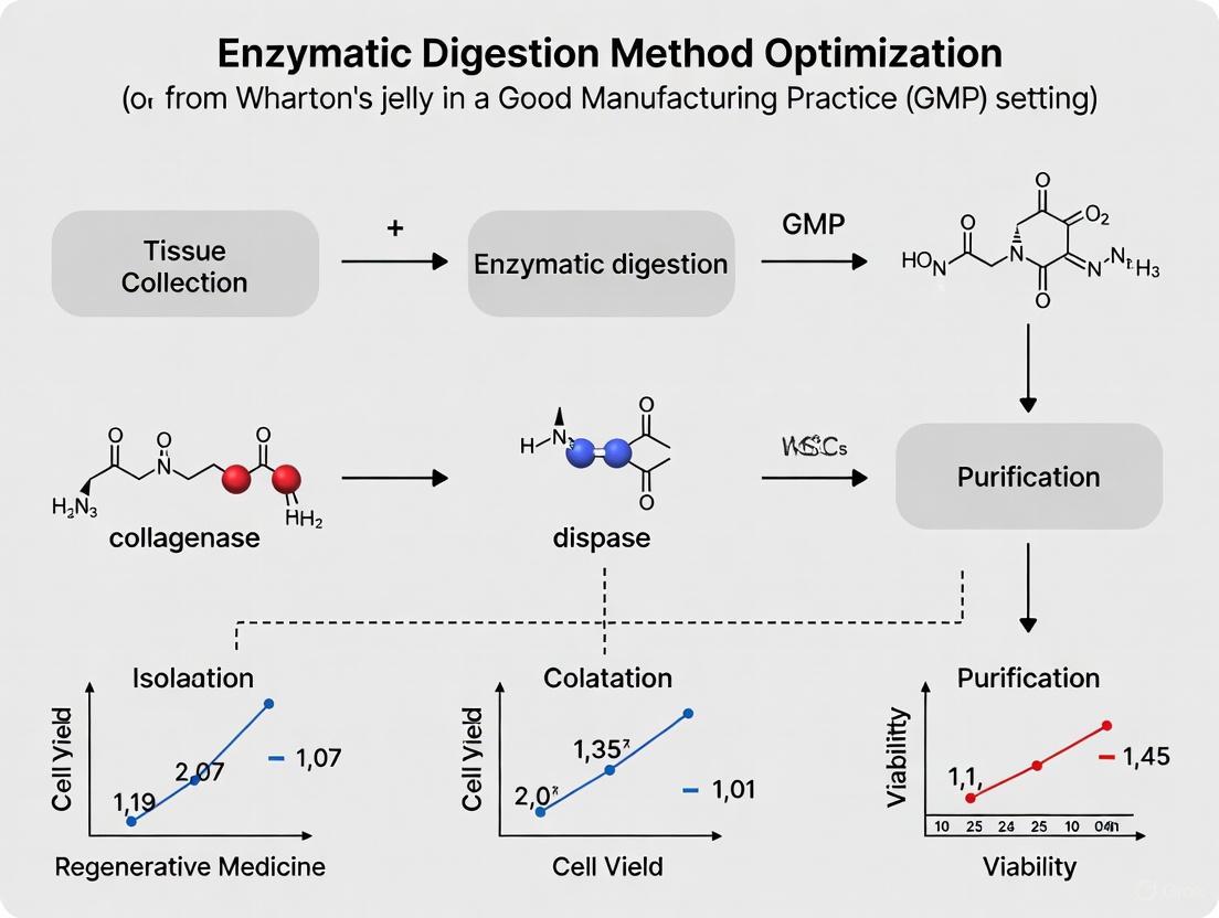
Optimizing Enzymatic Digestion for GMP-Compliant Wharton's Jelly Mesenchymal Stromal Cell Manufacturing: A Scalable Approach for Clinical Applications
This comprehensive review addresses the critical need for standardized, Good Manufacturing Practice (GMP)-compliant protocols in isolating and expanding Wharton's Jelly-derived Mesenchymal Stromal Cells (WJ-MSCs) through enzymatic digestion.
Optimizing Enzymatic Digestion for GMP-Compliant Wharton's Jelly Mesenchymal Stromal Cell Manufacturing: A Scalable Approach for Clinical Applications
Abstract
This comprehensive review addresses the critical need for standardized, Good Manufacturing Practice (GMP)-compliant protocols in isolating and expanding Wharton's Jelly-derived Mesenchymal Stromal Cells (WJ-MSCs) through enzymatic digestion. Targeting researchers, scientists, and drug development professionals, we synthesize current methodologies from foundational principles to advanced manufacturing scale-up. The article systematically explores WJ-MSC biology and therapeutic rationale, details optimized enzymatic parameters and procedural workflows, troubleshoots common challenges in viability and yield, and validates methods through comparative analyses and quality control measures. By integrating the latest research on scalable bioreactor systems and stability studies, this resource provides a foundational guide for translating WJ-MSC therapies from laboratory research to clinically viable regenerative medicine products.
Understanding Wharton's Jelly MSCs: Biology, Therapeutic Potential, and GMP Necessity
Historical Context and Fundamental Discoveries
The story of Wharton's jelly-derived mesenchymal stem cells (WJ-MSCs) begins not with the cells themselves, but with the discovery of their nurturing matrix. Wharton's jelly itself was first described in 1656 by Thomas Wharton, who identified this gelatinous connective tissue within the umbilical cord [1]. However, the isolation and characterization of the stem cells residing within this matrix is a much more recent development. The foundational era for mesenchymal stem cells (MSCs) commenced in the 1970s when Friedenstein and colleagues first identified and isolated these cells from bone marrow [2] [3]. Decades later, in 1991, Arnold Caplan coined the term "mesenchymal stem cells," providing a clearer identity for this cell population [2] [3].
A pivotal milestone was reached when McElreavey et al. successfully cultured cells from Wharton's jelly, opening a new source for MSCs beyond traditional adult tissues [1]. This discovery was significantly advanced by the work of Pittenger et al., who definitively demonstrated the multipotent nature of MSCs, confirming their ability to differentiate into adipocytic, chondrocytic, and osteocytic lineages [2] [3]. This established a critical functional criterion for defining MSCs. To standardize the field, the International Society for Cellular Therapy (ISCT) established minimal defining criteria for MSCs in 2006, which include plastic adherence, specific surface marker expression, and multilineage differentiation potential [2]. The continued scientific interest in WJ-MSCs is reflected in their characterization as a "Holy Grail" in tissue bioengineering and reconstructive medicine, underscoring their perceived potential in regenerative applications [4] [5].
Defining Characteristics of WJ-MSCs
WJ-MSCs possess a unique combination of biological properties that make them particularly attractive for clinical use. These characteristics distinguish them from MSCs derived from other, more conventional sources like bone marrow or adipose tissue.
Table 1: Key Characteristics of Wharton's Jelly-Mesenchymal Stem Cells
| Characteristic | Description | Significance/Advantage |
|---|---|---|
| Origin & Source | Isolated from the gelatinous Wharton's jelly in the umbilical cord, a perinatal tissue [1] [6]. | Considered medical waste; non-invasive collection, no ethical concerns [2] [6]. |
| Proliferation Capacity | High proliferation rate and longevity in culture [1] [6]. | Enables large-scale expansion for clinical applications [2]. |
| Immunophenotype | Positive for CD105, CD73, CD90; negative for CD45, CD34, HLA-DR [2] [7]. | Confirms identity as MSCs and indicates low immunogenicity [1]. |
| Differentiation Potential | Multilineage potential: adipogenic, chondrogenic, osteogenic [2] [8]. Also hepatogenic, neurogenic, etc. [8]. | Core property for tissue engineering and regenerative medicine. |
| Immunomodulatory Properties | Low immunogenicity and strong immunosuppressive capacity [2] [9]. | Suitable for allogeneic transplantation without matching; useful for immune-related disorders [1]. |
| Secretome | Releases a plethora of bioactive molecules (growth factors, cytokines, extracellular vesicles) [6]. | Mediates therapeutic effects via paracrine signaling, influencing tissue repair and immune response [6]. |
Anatomical Location and Ontogeny
Anatomically, the umbilical cord consists of two umbilical arteries and one umbilical vein, embedded within the Wharton's jelly matrix and covered by an amniotic epithelium [1] [6]. The Wharton's jelly itself is the major source of MSCs from the cord, with yields reaching up to 4,700,000 MSCs/cm of tissue [6]. The origin of WJ-MSCs is believed to be linked to waves of migrating fetal MSCs during early human development that became resident in the cord, or they may be primitive MSCs originating from the local mesenchyme [1]. Their ontogeny is connected to the earliest hematopoietic-forming sites in the intra-embryonic aorta-gonad-mesonephros (AGM) region, from where MSCs circulate to various tissues during embryogenesis [6].
Compared to adult-derived MSCs, WJ-MSCs offer several distinct advantages. Their embryonic nature means they are considered "younger" and more primitive, resulting in a higher proliferation rate, longer telomeres, and a broader differentiation potential than their adult counterparts from bone marrow (BM-MSCs) or adipose tissue (AT-MSCs) [1] [6]. Furthermore, their procurement is non-invasive and painless, as the umbilical cord is typically discarded as medical waste after birth, thereby avoiding the ethical controversies associated with embryonic stem cells [2] [1] [6]. These features, combined with their immune-evasive and immune-regulatory capacities, make WJ-MSCs display promising transplantable features for allogeneic cell therapy [1].
Experimental Workflow for WJ-MSC Isolation and Culture
The following diagram illustrates a generalized experimental workflow for the isolation, expansion, and application of WJ-MSCs, integrating key steps from the cited research.
The Scientist's Toolkit: Key Research Reagent Solutions
For scientists embarking on WJ-MSC research, particularly with a focus on GMP-compliant production, the selection of reagents is critical. The following table details essential materials and their functions as identified in recent, optimized protocols.
Table 2: Essential Research Reagents for GMP-compliant WJ-MSC Isolation and Culture
| Reagent / Material | Specific Example / Grade | Function in Protocol |
|---|---|---|
| Enzyme for Digestion | Collagenase NB6 GMP Grade (Nordmark Biochemicals) [2] [3] | Enzymatic dissociation of Wharton's jelly matrix to release MSCs. The GMP grade is essential for clinical translation. |
| Culture Medium | MSC Serum- and Xeno-Free Medium (e.g., NutriStem) [2] | Provides a defined, animal-free base medium for cell growth, enhancing safety profile. |
| Growth Supplement | Human Platelet Lysate (hPL) (e.g., 2% - 5% concentration) [2] [3] | Supplements the base medium with growth factors and proteins to support cell proliferation. |
| Priming Cytokine | Interferon-gamma (IFN-γ) [9] | Enhances the immunosuppressive properties of WJ-MSCs by inducing IDO activity, boosting therapeutic potency. |
| Cryopreservation Medium | GMP-compliant Cryomedium with DMSO [2] | Protects cell viability during the freeze-thaw process for cell banking and storage. |
Protocol Insight: Enzymatic Digestion Optimization
A key focus in GMP research is the optimization of the enzymatic digestion process. A 2024 study systematically determined that the optimal parameters for isolation are a concentration of 0.4 PZ U/mL Collagenase NB6 and a digestion time of 3 hours at 37°C, which resulted in a higher yield of passage 0 (P0) WJ-MSCs [2] [3]. This study also found a positive correlation between the weight of the umbilical cord tissue and the yield of P0 cells, providing a useful metric for predicting initial cell yield [2]. When comparing isolation methods, the enzymatic digestion method demonstrated a faster outgrowth of WJ-MSCs during the initial passage compared to the simpler explant method, though both methods ultimately yield cells with comparable characteristics after the first passage [2].
WJ-MSCs represent a significant advancement in the field of regenerative medicine. From their historical discovery in the umbilical cord to their current status as a promising tool for GMP-compliant cell therapy, their unique biological properties set them apart. Their high proliferative capacity, potent immunomodulatory functions, and ethically favorable source position them as a leading candidate for treating a wide range of degenerative and immune-mediated diseases. Ongoing research continues to refine their isolation, expansion, and therapeutic application, moving this young but promising field closer to widespread clinical reality.
Wharton's jelly-derived mesenchymal stem cells (WJ-MSCs) have emerged as a premier cell source for regenerative medicine and immunomodulatory therapies, offering distinct advantages over MSCs derived from traditional sources like bone marrow or adipose tissue. Their unique biological properties make them particularly suitable for allogeneic transplantation and large-scale therapeutic manufacturing. This document outlines the key therapeutic advantages of WJ-MSCs—specifically their potent immunomodulatory capabilities, exceptional proliferative capacity, and inherent low immunogenicity—within the context of optimizing enzymatic digestion methods for Good Manufacturing Practice (GMP)-compliant production. These attributes collectively position WJ-MSCs as a robust candidate for addressing the challenges of standardized, scalable cell therapy products [10] [2].
For researchers and drug development professionals, understanding and leveraging these advantages is crucial for developing effective, safe, and commercially viable therapies. The following sections provide a detailed examination of these properties, supported by quantitative data and experimental protocols essential for translational research.
Quantitative Profiling of Key Therapeutic Advantages
The superior therapeutic profile of WJ-MSCs is demonstrated through measurable attributes across immunomodulation, proliferation, and immunogenicity. The data in the tables below provide a consolidated overview for easy comparison and evaluation.
Table 1: Comparative Analysis of MSC Sources for Therapeutic Applications
| Property | WJ-MSCs | Bone Marrow MSCs (BM-MSCs) | Adipose Tissue MSCs (AD-MSCs) |
|---|---|---|---|
| Immunomodulatory Factor Production | High levels of IL-10, TGF-β, IL-6, VEGF, HLA-G6 [10] | Moderate factor production | High levels of CXCL1, CXCL9, CXCL10; more immunosuppressive factors than BM-MSCs [11] |
| Proliferation/Doubling Time | Shorter doubling time; extensive ex vivo expansion capacity [10] | Longer doubling time; limited proliferative capacity [10] | Moderate proliferative capacity |
| HLA Class I Expression | Very low expression [10] | Standard expression | Standard expression |
| HLA-DR Expression | Absent [10] | Can be induced upon inflammation | Can be induced upon inflammation |
| Tissue Collection | Non-invasive, medical waste [2] | Invasive and painful aspiration [12] | Invasive procedure |
| Therapeutic Specialization | Potent immunosuppression, low immunogenicity [11] | Hematopoietic support, immunomodulation [11] | Angiogenic repair, metabolic regulation [11] |
Table 2: Quantitative Proliferation and Marker Data of WJ-MSCs in Culture
| Parameter | Findings | Notes |
|---|---|---|
| Optimal Passages for Expansion | Passages 2 to 5 exhibit higher viability and proliferation ability [2] [3] | Later passages show increased population doubling time [13] |
| Population Doubling Time (PDT) | Varies with passage; e.g., PDT in early passages can be as low as ~1.8 days [14] | Varies with culture conditions including media and supplements [13] [14] |
| Surface Marker Expression (CD90, CD73, CD105) | ≥95% expression, meeting ISCT criteria [15] [13] | Consistent across passages when cultured under standardized conditions [13] |
| Negative Marker Expression (CD34, CD45, HLA-DR) | ≤2% expression, meeting ISCT criteria [15] [13] | Consistent across passages when cultured under standardized conditions [13] |
| Effect of Seeding Density on Yield | Positive correlation between umbilical cord tissue weight and P0 WJ-MSC yield [2] | Optimization of seeding density is critical for maximizing initial yield |
Detailed Experimental Protocols
GMP-Compliant Isolation of WJ-MSCs via Enzymatic Digestion
This optimized protocol ensures high yield and quality of WJ-MSCs for clinical applications [2].
Materials:
- Umbilical cord tissue (>20 cm length) collected post-cesarean section with informed consent.
- GMP-grade Collagenase NB6 (Nordmark Biochemicals).
- DPBS (without Ca²⁺ and Mg²⁺).
- MSC Serum- and Xeno-Free Medium (e.g., NutriStem).
- Human Platelet Lysate (hPL, e.g., Stemulate).
- 0.5% povidone-iodine solution.
- Tissue culture flasks or multi-layer cell factories.
Methodology:
- Preprocessing: Transport the UC tissue at 2-10°C within 24 hours of collection. Rinse with DPBS to remove blood contaminants. Decontaminate with 0.5% povidone-iodine for 3 minutes, followed by three washes with DPBS.
- Tissue Preparation: Dissect the cord to expose Wharton's jelly and meticulously remove the two arteries and one vein. Mince the remaining Wharton's jelly tissue into 1-4 mm³ fragments and record the tissue weight.
- Enzymatic Digestion: Transfer the tissue fragments to a digestion solution containing 0.4 PZ U/mL Collagenase NB6 in serum-free medium. Incubate for 3 hours at 37°C with gentle agitation.
- Cell Harvesting: Neutralize the collagenase activity by adding a complete medium containing hPL. Pass the cell suspension through a 100 µm cell strainer to remove undigested tissue. Centrifuge the filtrate at 400 × g for 10 minutes.
- Primary Culture: Resuspend the cell pellet in a complete culture medium (e.g., NutriStem supplemented with 2-5% hPL). Seed the cells at an optimized density correlated with the initial tissue weight. Incubate at 37°C with 5% CO₂.
- Medium Change and Passaging: Replace the medium every 48-72 hours. Upon reaching 80-90% confluence, typically within the first 1-2 weeks, passage cells using standard trypsinization protocols. For clinical applications, prioritize the use of cells from passages 2-5.
Protocol for Evaluating Immunomodulatory Potential
This in vitro functional assay assesses the capacity of WJ-MSCs to suppress T-cell proliferation, a key immunomodulatory mechanism [10].
Materials:
- Isolated and culture-expanded WJ-MSCs (Passage 2-4).
- Peripheral blood mononuclear cells (PBMCs) from healthy donors.
- T-cell mitogen (e.g., Phytohemagglutinin-P (PHA-P)).
- Co-culture transwell system (optional for contact-dependent experiments).
- Flow cytometry kit for T-cell proliferation analysis (e.g., CFSE dilution).
Methodology:
- WJ-MSC Preparation: Seed WJ-MSCs in a 24-well plate and allow them to adhere overnight to form a monolayer (~70-80% confluence).
- PBMC Activation: Isolate PBMCs via density gradient centrifugation. Label PBMCs with CFSE and activate them with PHA-P (e.g., 5 µg/mL).
- Co-culture Establishment: Add the activated, CFSE-labeled PBMCs directly to the WJ-MSC monolayer (for contact-dependent suppression) or in the transwell insert (to study soluble factor-mediated suppression).
- Incubation and Analysis: Co-culture cells for 3-5 days. Harvest PBMCs and analyze CFSE dilution using flow cytometry to measure T-cell proliferation. Compare the proliferation rate of T-cells co-cultured with WJ-MSCs to that of T-cells cultured alone (positive control).
Signaling Pathways and Immunomodulatory Mechanisms
WJ-MSCs exert their potent immunomodulatory effects through a multi-faceted approach involving soluble factors, direct cell contact, and the induction of regulatory immune cells. The diagram below illustrates the core mechanisms and signaling pathways.
Diagram: Immunomodulatory Mechanisms of WJ-MSCs. WJ-MSCs suppress effector T-cells and B-cells while promoting regulatory T-cells (Tregs) and inhibiting dendritic cell (DC) maturation via soluble factors and direct contact.
The Scientist's Toolkit: Essential Research Reagents
Successful isolation, expansion, and functional characterization of WJ-MSCs rely on specific, high-quality reagents. The following table details essential materials for GMP-compliant research.
Table 3: Key Research Reagent Solutions for WJ-MSC Workflows
| Reagent/Material | Function/Application | Example & Notes |
|---|---|---|
| GMP-grade Collagenase NB6 | Enzymatic digestion of umbilical cord tissue to isolate WJ-MSCs. | Contains collagenase class I/II and neutral protease. Optimal concentration: 0.4 PZ U/mL [2]. |
| Human Platelet Lysate (hPL) | Serum-free supplement for MSC culture media, promoting expansion. | Superior to FBS, reduces zoonotic risk. Concentrations of 2% and 5% show similar expansion efficacy [2] [12]. |
| Serum/Xeno-Free Basal Medium | Base medium for the expansion of clinical-grade WJ-MSCs. | Formulations like NutriStem support robust cell growth while adhering to GMP standards [2]. |
| Antibody Panels for Flow Cytometry | Characterization of WJ-MSCs based on ISCT criteria. | Positive Markers: CD90, CD73, CD105, CD44. Negative Markers: CD34, CD45, CD11b, CD19, HLA-DR [15] [13]. |
| Differentiation Kits (Osteo/Chondro/Adipo) | Functional validation of MSC trilineage differentiation potential. | Commercially available kits (e.g., StemPro from Gibco) provide standardized protocols for differentiation and staining [13]. |
| Transwell Co-culture Systems | Mechanistic studies to distinguish between contact-dependent and soluble factor-mediated immunomodulation. | Permeable inserts allow physical separation of WJ-MSCs from responder immune cells while sharing the soluble milieu [10]. |
The consolidated data and protocols presented herein underscore the significant therapeutic potential of WJ-MSCs, rooted in their defined immunomodulatory, proliferative, and low immunogenicity profile. The optimization of enzymatic digestion methods, as detailed in the GMP-compliant protocols, is a critical step toward achieving reproducible and scalable manufacturing of these cells. For researchers and drug developers, focusing on passages 2-5, utilizing defined media supplements like hPL, and employing rigorous functional potency assays are key strategies for successful translation. As the field advances, the integration of these standardized approaches will be instrumental in harnessing the full clinical potential of WJ-MSCs for treating a wide array of immune-mediated and degenerative diseases.
The development of clinical-grade cell therapies is strictly governed by Good Manufacturing Practice (GMP) regulations to ensure the safety, quality, and efficacy of these advanced biologic products. In both the United States (US) and European Union (EU), cell therapy products, including those derived from Wharton's jelly mesenchymal stromal cells (WJ-MSCs), are classified as Advanced Therapy Medicinal Products (ATMPs) and must comply with a comprehensive regulatory framework [16]. This framework mandates that products are consistently produced and controlled to quality standards appropriate for their intended clinical use, requiring certified raw materials, validated manufacturing processes, and rigorous documentation for full traceability [17]. The core objective of GMP is to minimize risks in pharmaceutical production that cannot be eliminated through final product testing alone, an principle critically important for living cell-based therapies.
United States (FDA) Regulations
The US Food and Drug Administration (FDA) regulates human cells, tissues, and cellular and tissue-based products (HCT/Ps) under Title 21 of the Code of Federal Regulations. Key sections include:
- 21 CFR Part 1271: Governs human cells, tissues, and cellular and tissue-based products, outlining donor eligibility, current good tissue practice (cGTP), and regulatory requirements.
- 21 CFR Part 211: Details Current Good Manufacturing Practice (cGMP) for finished pharmaceuticals.
- 21 CFR Part 312: Covers requirements for Investigational New Drug (IND) applications.
- 21 CFR Part 600: Pertains to biological products, including requirements for a Biologics License Application (BLA) [16].
The FDA's Office of Therapeutic Products (OTP), which recently replaced the Office of Tissues and Advanced Therapies (OTAT), oversees the regulation of cell and gene therapy products. This "super office" has six sub-offices covering gene therapy CMC, cellular therapy and human tissue CMC, clinical evaluation, pharmacology/toxicology, and review management, and has been actively staffing to handle the surge in cell and gene therapy applications [18].
European Union (EMA) Regulations
In the EU, the regulatory framework for ATMPs is established through several key legislations:
- Regulation (EC) No 1394/2007: The central legislation governing advanced therapy medicinal products.
- Directive 2009/120/EC: Addresses the scientific and technical requirements of ATMPs.
- Directive 2004/23/EC: Sets standards of quality and safety for the donation, procurement, testing, processing, preservation, storage and distribution of human tissues and cells.
- EU GMP-ATMP (EudraLex Volume 4): Provides specific Guidelines on Good Manufacturing Practice for Advanced Therapy Medicinal Products [16].
The European Medicines Agency (EMA) evaluates marketing authorization applications for ATMPs through its Committee on Advanced Therapies (CAT) [16].
Key Regulatory Guidance Documents
The FDA has issued numerous guidance documents specific to cellular therapies, providing detailed recommendations for sponsors. Recent and relevant guidances include [19]:
- Human Gene Therapy Products Incorporating Human Genome Editing (Final Guidance, January 2024)
- Considerations for the Development of Chimeric Antigen Receptor (CAR) T Cell Products (Final Guidance, January 2024)
- Potency Assurance for Cellular and Gene Therapy Products (Draft Guidance, December 2023)
- Manufacturing Changes and Comparability for Human Cellular and Gene Therapy Products (Draft Guidance, July 2023)
- Studying Multiple Versions of a Cellular or Gene Therapy Product in an Early-Phase Clinical Trial (Final Guidance, November 2022)
Diagram 1: GMP Regulatory Landscape for Cell Therapies. This diagram outlines the key regulatory frameworks governing clinical-grade cell therapies in the United States (yellow) and European Union (green), culminating in the core GMP compliance requirements.
GMP-Compliant Manufacturing Process
Core GMP Principles for Cell Therapy
GMP-compliant manufacturing of cell therapies requires adherence to fundamental principles designed to ensure product safety and quality:
- Quality Management: Implementation of a comprehensive quality system covering all aspects of production and control.
- Facility and Equipment Control: Appropriate design, maintenance, and cleaning of facilities and equipment to prevent contamination and cross-contamination.
- Material Management: Rigorous control of all starting and raw materials, including qualification of suppliers and testing of components.
- Production and Process Controls: Validated and controlled manufacturing processes with documented procedures for each step.
- Quality Control Testing: In-process, release, and stability testing to ensure the identity, purity, potency, and safety of the final product [17].
For cell separation kits specifically, GMP compliance requires sterility and low endotoxin levels (tested per USP <71> and USP <85>), certified raw materials such as USP Class VI plastics, complete documentation and traceability of every component and lot, and manufacturing under a quality system with validated procedures and third-party audits [17].
Manufacturing Workflow for WJ-MSCs
The manufacturing process for GMP-compliant WJ-MSCs involves multiple critical steps from tissue acquisition to final product release, with comprehensive quality control at each stage [2] [9]:
Diagram 2: GMP-Compliant WJ-MSC Manufacturing Workflow. This diagram illustrates the key stages in manufacturing clinical-grade Wharton's jelly MSCs, with quality control testing (red) as a critical release gate, all operating within a GMP environment (blue).
Optimization of Enzymatic Digestion for WJ-MSCs
Experimental Protocol: Enzymatic Digestion Method
Objective: To establish a standardized, GMP-compliant enzymatic digestion protocol for isolation of WJ-MSCs from umbilical cord tissue with high yield and viability.
Materials and Reagents:
- GMP-grade Collagenase NB6 (Nordmark Biochemicals, Germany)
- DPBS (without Ca²⁺, Mg²⁺)
- MSC Serum- and Xeno-Free Medium (e.g., NutriStem)
- Human Platelet Lysate (hPL) (e.g., Stemulate)
- 0.5% povidone-iodine solution
- GMP-compliant cell culture flasks/bioreactors
Procedure:
- Tissue Collection and Pre-processing: Obtain umbilical cord (>20 cm length) following cesarean section with informed consent. Transport to facility within 24 hours at 2-10°C. Test donor blood for pathogens (HBV, HCV, HTLV, TP, HIV, EBV, CMV) [2].
- Decontamination and Dissection: Rinse cord with DPBS. Decontaminate with 0.5% povidone-iodine solution for 3 minutes. Rinse thoroughly with DPBS three times. Cut cord into 3-6 cm segments, open to expose Wharton's jelly, and carefully remove blood vessels [2].
- Tissue Mincing: Mince Wharton's jelly into 1-4 mm³ fragments using sterile surgical scalpels. Weigh tissue fragments accurately [2].
- Enzymatic Digestion: Transfer tissue fragments to digestion vessel. Add 0.4 PZ U/mL Collagenase NB6 solution (optimized concentration). Incubate at 37°C for 3 hours (optimized duration) with gentle agitation [2].
- Cell Collection and Seeding: Neutralize enzyme activity with complete culture medium. Filter cell suspension through 100μm cell strainer. Centrifuge and resuspend cell pellet in culture medium (MSC Serum- and Xeno-Free Medium + 2-5% hPL). Seed at optimized density of 1g tissue per 75 cm² flask [2].
- Primary Culture: Incubate at 37°C, 5% CO₂. Perform first medium change after 72 hours to remove non-adherent cells, then change medium every 3-4 days [2].
- Cell Passaging: When cultures reach 80-90% confluence (typically after 10-14 days), harvest cells using GMP-grade trypsin/EDTA and passage at recommended density of 5,000-6,000 cells/cm² [2].
Optimization Parameters and Data
Extensive optimization studies have identified critical parameters for maximizing WJ-MSC yield and quality through enzymatic digestion:
Table 1: Optimization of Enzymatic Digestion Parameters for WJ-MSC Isolation
| Parameter | Tested Conditions | Optimal Value | Impact on Yield/Quality |
|---|---|---|---|
| Enzyme Concentration | 0.2, 0.4, 0.6 PZ U/mL | 0.4 PZ U/mL | Higher yield of P0 WJ-MSCs without compromising viability [2] |
| Digestion Time | 2, 3, 4 hours | 3 hours | Balanced approach for complete tissue dissociation and cell recovery [2] |
| Seeding Density | 0.5g, 1g, 2g tissue per 75 cm² flask | 1g tissue per 75 cm² flask | Optimal cell outgrowth and utilization of tissue material [2] |
| Culture Medium | 2%, 5%, 10% hPL | 2-5% hPL | Similar expansion levels, with 2% being more cost-effective [2] |
| Tissue Weight Correlation | Various weights | Positive correlation | Higher tissue weight yields more P0 WJ-MSCs [2] |
Table 2: Comparative Analysis of WJ-MSC Isolation Methods
| Characteristic | Enzymatic Digestion Method | Explant Method |
|---|---|---|
| Primary Cell Culture Time | Faster outgrowth during initial passage [2] | Slower initial outgrowth [2] |
| P0 Yield | Higher cell yield [2] | Lower initial yield [2] |
| Cell Viability | High, when optimized [2] | High [2] |
| Morphology | Standard MSC morphology [2] | Standard MSC morphology [2] |
| Surface Marker Expression | Maintains MSC phenotype (CD73+, CD90+, CD105+, CD34-, CD45-) [2] | Maintains MSC phenotype [2] |
| Differentiation Capacity | Maintains adipogenic, chondrogenic, osteogenic potential [2] | Maintains differentiation potential [2] |
| Standardization | More easily standardized [2] | Challenging to standardize [2] |
Scaling and Process Transfer
Successful translation from laboratory-scale to pilot-scale production requires systematic scale-up:
- Laboratory Scale: Traditional flask-based culture systems
- Pilot/Production Scale: Cell factory-based production or bioreactor systems [2]
- Process Comparability: Demonstration that scaled-up process produces equivalent cells in terms of identity, purity, potency, and safety [19]
Studies confirm that scalable manufacturing processes from laboratory scale to pilot scale can successfully ensure production of high-quality WJ-MSCs, with passages 2 to 5 exhibiting higher viability and proliferation ability throughout consecutive passaging [2].
Quality Control and Product Release
Critical Quality Attributes (CQAs)
For clinical-grade WJ-MSCs, established CQAs must be thoroughly evaluated before product release:
- Identity: Confirmation of MSC phenotype through surface marker expression (CD73+, CD90+, CD105+, CD34-, CD45-, HLA-DR-) [2] [16]
- Viability: Typically >70% for cryopreserved products, >90% for fresh products [2]
- Purity: Minimal contamination with hematopoietic cells, endotoxin levels within specified limits [17]
- Potency: Demonstration of immunosuppressive capability, often through T-cell proliferation inhibition assays; IFN-γ priming can enhance this property [9]
- Safety: Sterility (bacteria, fungi, mycoplasma), absence of replication-competent viruses, and karyotypic stability [2] [16]
Enhanced Potency Through IFN-γ Priming
Research demonstrates that cytokine licensing can enhance the therapeutic properties of WJ-MSCs:
- IFN-γ Priming Protocol: Incubate confluent WJ-MSCs with 25-50 ng/mL IFN-γ for 24-48 hours before harvest [9]
- Mechanism: Upregulation of indoleamine 2,3-dioxygenase (IDO) activity, leading to tryptophan depletion and kynurenic acid production, which inhibits T-cell proliferation [9]
- Efficacy: Primed WJ-MSCs demonstrate significantly higher immunosuppressive properties in vitro and enhanced prevention of xeno-GVHD in vivo [9]
The Scientist's Toolkit: Essential Research Reagent Solutions
Table 3: Essential GMP-Compliant Reagents for WJ-MSC Manufacturing
| Reagent/Supply | Function | GMP-Compliant Example |
|---|---|---|
| Collagenase NB6 GMP | Enzymatic digestion of umbilical cord tissue to isolate WJ-MSCs | Nordmark Biochemicals [2] |
| MSC Serum/Xeno-Free Medium | Culture medium supporting MSC growth without animal components | NutriStem (Biological Industries) [2] |
| Human Platelet Lysate (hPL) | Serum replacement providing growth factors for MSC expansion | Stemulate (Sexton Biotechnologies) [2] |
| GMP-Grade Cell Separation Kits | Isolation or depletion of specific cell populations | Akadeum Microbubble-Based Kits [17] |
| GMP-Grade Trypsin/EDTA | Cell detachment during passaging | Various GMP-grade vendors |
| Cryopreservation Media | Long-term storage of cell products with maintained viability | Defined, serum-free, GMP-grade formulations |
The successful development of clinical-grade WJ-MSC therapies requires meticulous attention to GMP requirements throughout the entire manufacturing process, from donor selection and tissue acquisition to final product formulation and release. The enzymatic digestion method, when optimized with parameters such as 0.4 PZ U/mL collagenase concentration and 3-hour digestion time, provides an efficient and standardized approach for WJ-MSC isolation that can be successfully scaled from laboratory to pilot-scale production. Implementation of comprehensive quality control measures, including identity, purity, potency, and safety testing, ensures that the final cell product meets regulatory standards for clinical application. Furthermore, strategies such as IFN-γ priming can enhance the immunosuppressive properties of WJ-MSCs, potentially improving their therapeutic efficacy in clinical applications such as graft-versus-host disease prevention.
The therapeutic application of Wharton's jelly-derived mesenchymal stromal cells (WJ-MSCs) in regenerative medicine and immunomodulation necessitates a robust and reproducible pipeline from initial tissue acquisition to final cell product. The source material quality and preprocessing methods are critical determinants of the safety, efficacy, and compliance of the resulting clinical-grade cells. This protocol details standardized procedures for the collection, decontamination, and tissue preparation of the human umbilical cord, establishing a foundational step for subsequent enzymatic digestion and large-scale manufacturing of WJ-MSCs under Good Manufacturing Practice (GMP) standards. Framed within a broader thesis on optimizing enzymatic digestion methods for WJ-MSCs, this document ensures the integrity of the starting tissue, thereby maximizing downstream cell yield, viability, and functional potency.
Umbilical Cord Collection and Initial Decontamination
The initial collection phase is paramount for minimizing microbial contamination and preserving tissue viability.
Materials and Reagents for Collection
- Sterile Collection Kit: Typically provided by a processing bank or assembled in-house, containing sterile containers, clamps, and saline solution [20] [21].
- Decontamination Agents: 70% ethanol or iodine-based antiseptic liquids and sterile gauze [22] [20].
- Sterile Saline Solution: 0.9% Sodium Chloride (NaCl) for transporting the cord tissue [20].
- Specimen Container: A sterile, leak-proof container for cord transport [20].
Step-by-Step Collection Protocol
- Informed Consent and Donor Screening: Obtain informed consent from the mother prior to delivery. Maternal blood must be collected and tested for transmissible infectious diseases as per regulatory standards [21] [23].
- Cord Blood Collection (Optional): Following the birth of the baby and clamping of the umbilical cord, cord blood can be collected via venipuncture from the cord. The puncture site must be thoroughly cleaned with a sterile gauze soaked in alcohol or an iodine-based antiseptic for at least 30 seconds and allowed to dry before collection [22] [20].
- Cord Tissue Collection (Ex-Utero): After the delivery of the placenta, the umbilical cord is collected.
- Wipe the entire length of the cord with sterile gauze to remove residual blood and fluids [22] [20].
- Disinfect the cord thoroughly from end to end using a sterile swab or gauze soaked in 70% ethanol [22] [20]. Allow it to air-dry for approximately 30 seconds.
- Using sterile scissors, clamp the cord at both ends and cut a segment of 15-30 cm [20] [23].
- Repeat the decontamination procedure on the isolated cord segment [20].
- Packaging and Transportation:
- Place the cord segment in a sterile container.
- Fill the container with sterile 0.9% NaCl solution to fully submerge the tissue, which helps preserve cell viability during transport [20].
- Affix a unique tracking label to the container for full traceability.
- The specimen should be stored at room temperature and transported to the processing laboratory as soon as possible, ideally within 24-48 hours, via a designated medical courier [21].
The following workflow summarizes the key stages from collection to the initiation of processing:
Tissue Digestion for WJ-MSC Isolation
The dissociation of Wharton's jelly from the umbilical cord is a critical step that directly impacts MSC yield, viability, and function. Optimization is required to balance high cell recovery with the preservation of cell surface markers and functionality.
Comparative Analysis of Digestion Methods
The choice of digestion protocol significantly influences the outcome of the isolation. The table below summarizes key parameters and performance metrics for different methods as evidenced by recent research.
Table 1: Comparative Analysis of Tissue Digestion Methods for Cell Isolation
| Digestion Method | Key Enzymes and Reagents | Incubation Conditions | Reported Performance | Key Advantages |
|---|---|---|---|---|
| Sequential Enzymatic Digestion [24] | Step 1: Dispase II (10 mg/mL)Step 2: Liberase (Collagenase I/II blend, 0.5 mg/mL) + DNase I (50 U/mL) | Step 1: 37°C, 45 min, shakingStep 2: 37°C, 45 min, shaking | Higher cell viability and yield per gram of tissue compared to simultaneous and overnight methods [24]. | Effective separation of tissue components; superior preservation of cell viability [24]. |
| Simultaneous Digestion [24] | Collagenase IV (600 U/mL), Hyaluronidase (600 U/mL), DNase I (50 U/mL) | 37°C, 2 hours, shaking | Lower cell viability compared to the sequential method [24]. | Simpler, single-step process. |
| Overnight Digestion [24] | Collagenase IV, DNase I | 37°C, 16-18 hours, no shaking | Based on a published method; generally results in lower viability due to prolonged exposure [24]. | Requires less active handling time. |
Optimized Sequential Digestion Protocol for WJ-MSC Isolation
Based on the comparative data, a sequential digestion method is recommended for optimal results.
Reagents and Solutions
- Digestion Buffer 1: RPMI medium supplemented with 10% Fetal Bovine Serum (FBS) and Dispase II (10 mg/mL) [24].
- Digestion Buffer 2: RPMI/10% FBS containing Liberase TL (a GMP-compatible blend of Collagenase I and II, 0.5 mg/mL) and DNase I (50 U/mL) [24].
- Phosphate-Buffered Saline (PBS) with antibiotic-antimycotic [23].
- Red Blood Cell (RBC) Lysis Buffer (if required) [24].
Step-by-Step Procedure
- Tissue Preparation: In a sterile biological safety cabinet, transfer the umbilical cord to a sterile Petri dish. Rinse it twice with PBS containing antibiotic-antimycotic to remove residual contaminants [23]. Remove the two arteries and one vein using sterile instruments. Mince the remaining Wharton's jelly tissue into 1-2 mm³ explants using a sterile scalpel [23].
- First Digestion (Dispase): Transfer the minced tissue to a sterile container with Digestion Buffer 1. Incubate with shaking (800 rpm) at 37°C for 45 minutes [24].
- Second Digestion (Liberase/DNase): After the first incubation, pellet the undigested tissue fragments. Remove the Dispase buffer and replace it with Digestion Buffer 2. Incubate again with shaking (800 rpm) at 37°C for 45 minutes [24].
- Cell Harvesting:
- Neutralize the digestion reaction by adding a complete culture medium containing serum.
- Filter the cell suspension through a 100 µm sterile cell strainer to remove undigested tissue fragments, followed by a 40 µm strainer to obtain a single-cell suspension [24].
- Centrifuge the filtrate to pellet the cells.
- If the pellet is contaminated with red blood cells, resuspend it in 1x RBC Lysis Buffer according to the manufacturer's protocol, then wash with PBS [24].
- Cell Counting and Viability Assessment: Resuspend the final cell pellet in an appropriate buffer. Determine total cell count and viability using trypan blue exclusion or automated cell counters.
The relationship between enzyme selection and dissociation outcomes can be guided by the following troubleshooting principle:
Towards GMP Compliance: Scaling and Manufacturing
For clinical translation, the isolated WJ-MSCs must be expanded in a scalable, cGMP-compliant manner.
Scalable Bioreactor Expansion
Moving from traditional 2D flask cultures to microcarrier-based 3D bioreactor systems is essential for producing the billions of cells required for clinical trials and commercial therapies [25] [23].
Table 2: Large-Scale Bioreactor Parameters for WJ-MSC Expansion
| Parameter | Spinner Flask (Process Development) | Stirred-Tank Bioreactor (STR50 - 50L) | Purpose |
|---|---|---|---|
| Culture System | Microcarrier (MC)-based 3D suspension [25] | Microcarrier-based 3D suspension [25] [23] | Increases surface-to-volume ratio for high-density culture. |
| Culture Medium | Serum-/Xeno-free (e.g., MSC Nutristem XF) supplemented with Human Platelet Lysate [23] | Serum-/Xeno-free (e.g., MSC Nutristem XF) supplemented with Human Platelet Lysate [23] | Ensures GMP-compliance and reduces risk of zoonotic contaminants. |
| Seeding Density | Optimized in spinner flasks [25] | ~1.2 x 10⁶ cells/mL [23] | Initiates culture at an optimal cell concentration. |
| Process Outcome | Foundation for scale-up [25] | ~37 billion cells after 7 days (27-fold expansion, 95% harvest efficiency) [23] | Achieves commercial-scale cell yields. |
Research Reagent Solutions for GMP-Compliant Workflows
The following table lists essential reagents and their functions in the context of GMP-focused research and development.
Table 3: Essential Reagents for GMP-Grade WJ-MSC Processing
| Reagent / Solution | Function / Application | Example / Note |
|---|---|---|
| Liberase TL | GMP-compatible enzyme blend for gentle tissue dissociation. | A proprietary blend of Collagenase I and II; used in the optimized sequential digestion protocol [24]. |
| Dispase II | Proteolytic enzyme for initial tissue loosening. | Used in the first step of sequential digestion to separate tissue components [24]. |
| DNase I | Prevents cell clumping by digesting DNA released from damaged cells. | Added to dissociation cocktails to increase cell yield and viability [24]. |
| MSC Nutristem XF Medium | Defined, xeno-free medium for clinical-grade cell expansion. | Supports serum-free culture of WJ-MSCs in bioreactors [23]. |
| Human Platelet Lysate (hPL) | GMP-compatible growth supplement for cell culture medium. | Used as a replacement for Fetal Bovine Serum (FBS) to promote xeno-free expansion [23]. |
| Collagen-Coated Microcarriers | Provide a surface for anchorage-dependent cell growth in 3D bioreactors. | Essential for achieving high cell yields in stirred-tank systems [23]. |
A rigorously controlled process for umbilical cord collection, decontamination, and tissue preparation is the cornerstone of manufacturing high-quality Wharton's jelly MSCs. The adoption of an optimized sequential enzymatic digestion protocol, utilizing enzymes like Liberase and Dispase, provides a superior balance of high cell yield and viability compared to traditional single-step or overnight methods. This optimized preprocessing pipeline, when integrated with scalable, cGMP-compliant bioreactor systems, enables the transition from laboratory research to the clinical application of WJ-MSCs, supporting their growing demand in advanced therapeutic development.
The transition of Wharton's jelly-derived mesenchymal stromal cells (WJ-MSCs) from laboratory research to clinical applications requires robust, standardized, and Good Manufacturing Practice (GMP)-compliant isolation protocols. The choice between the two primary isolation methods—enzymatic digestion and explant culture—represents a critical initial decision that impacts cell yield, quality, functionality, and compliance with regulatory standards for cellular therapeutics [2]. This document provides a detailed comparison of these methodologies, framing the analysis within the context of optimizing enzymatic digestion for GMP-compliant manufacturing of WJ-MSCs. It is designed to assist researchers, scientists, and drug development professionals in selecting and refining protocols for clinical-scale production.
Comparative Analysis: Enzymatic Digestion vs. Explant Method
A comprehensive understanding of the core differences, advantages, and limitations of each isolation method is fundamental to process design. The table below summarizes the key characteristics of each technique.
Table 1: Core Characteristics of Enzymatic Digestion and Explant Isolation Methods
| Feature | Enzymatic Digestion | Explant Method |
|---|---|---|
| Basic Principle | Uses proteolytic enzymes (e.g., collagenase) to dissociate tissue and release individual cells [2]. | Explant tissue pieces are cultured, allowing MSCs to migrate out from the tissue onto the culture surface [26] [2]. |
| Processing Time | Shorter time to initial cell harvest [2]. | Longer time for primary cell outgrowth [2]. |
| Initial Cell Yield | Higher initial yield of P0 cells [27] [2]. | Lower initial yield [27]. |
| Technical Complexity | Higher; requires optimization of enzyme concentration, time, and temperature [2]. | Lower; technically simpler, but requires careful explant sizing and placement [26]. |
| Proteolytic Stress | Present; potential risk of damaging cell surface receptors if not optimized [27]. | Absent; avoids enzymatic stress, preserving natural cell状态 [26]. |
| Utilization of Native ECM | Disrupts the native extracellular matrix (ECM) during digestion. | Preserves the native ECM, which provides a reservoir of growth factors and cytokines that support cell migration and growth [26]. |
| Standardization | Can be highly standardized with defined parameters [2]. | Can be challenging to standardize due to explant size and attachment variability [2]. |
Beyond these core characteristics, studies have compared the performance of cells isolated by each method after initial expansion. While the explant method may show benefits in proliferation capacity and the retention of certain markers like CD146 [27], other research indicates that after the initial passage (P0), cells from both methods exhibit comparable viability, morphology, surface marker expression, and differentiation capacity [2]. The choice of method often depends on the specific application requirements, such as the need for high P0 yield versus simpler processing.
Experimental Protocols and Data
Optimized Enzymatic Digestion Protocol for WJ-MSCs
The following protocol is adapted from GMP-compliant studies for the isolation of WJ-MSCs using enzymatic digestion [2].
Materials:
- Tissue Source: Human umbilical cord (>20 cm length), collected after informed consent and tested for pathogens.
- Reagents: DPBS (without Ca²⁺/Mg²⁺), 0.5% povidone-iodine solution, GMP-grade Collagenase NB6 (Nordmark Biochemicals), Serum/Xeno-free culture medium (e.g., NutriStem), Human Platelet Lysate (hPL).
Pre-processing:
- Transportation: Transport the UC to the processing facility within 24 hours at 2-10°C.
- Decontamination: Rinse the cord with DPBS and decontaminate with 0.5% povidone-iodine for 3 minutes, followed by three thorough rinses with DPBS.
- Dissection: Using a scalpel, open the cord to expose Wharton's jelly. Carefully remove the two arteries and one vein.
- Mincing: Mince the Wharton's jelly tissue into small fragments of 1-4 mm³.
Optimized Digestion and Culture:
- Digestion: Transfer the tissue fragments to a digestion vessel. Use a concentration of 0.4 PZ U/mL of GMP-grade Collagenase NB6 in serum-free medium. Incubate for 3 hours at 37°C with gentle agitation (e.g., 60-70 rpm) [2].
- Neutralization & Filtration: After digestion, add an equal volume of culture medium supplemented with hPL to neutralize the enzyme. Filter the cell suspension through a 100 μm cell strainer to remove undigested tissue debris.
- Centrifugation: Centrifuge the filtrate at 300-500 × g for 10 minutes. Carefully remove the supernatant.
- Seeding: Resuspend the cell pellet in culture medium (e.g., supplemented with 2-5% hPL). Seed the cells at an optimized density. Studies suggest using 1 gram of original tissue per 75 cm² flask [2].
- Culture: Incubate the culture at 37°C in a 5% CO₂ humidified incubator.
- Medium Change: Perform the first medium change after 48-72 hours to remove non-adherent cells. Subsequently, change the medium twice a week.
- Passaging: Once cells reach 80-90% confluency, passage them using trypsin/EDTA or a GMP-compliant dissociation agent.
The following diagram illustrates the optimized enzymatic digestion workflow.
Explant Method Protocol with Minimal Cube Explant (MCE)
The explant method can be optimized by controlling the size of the tissue pieces, as demonstrated by the Minimal Cube Explant (MCE) approach [27].
Materials: (Similar to enzymatic digestion, excluding collagenase)
Pre-processing: (Identical to steps 1-4 in the enzymatic digestion protocol)
Explant Culture:
- Sizing: After mincing, categorize the tissue fragments to achieve a uniform size. The MCE 2-4 (2-4 mm pieces) has been identified as optimal for isolating fast-proliferating cells with high yield [27].
- Attachment: Evenly distribute the explants onto the surface of a culture dish (e.g., 1g of tissue per 150 mm dish). Allow the explants to firmly attach to the bottom of the dish by incubating them undisturbed in a 37°C incubator for about 1 hour.
- Feeding: Gently add pre-warmed culture medium (e.g., LG-DMEM with 10% FBS or serum-free alternatives) to the dish, taking care not to dislodge the attached explants.
- Culture: Incubate the culture at 37°C in a 5% CO₂ incubator. Change the medium every 2-3 days, carefully removing and adding medium to minimize disturbance to the explants.
- Cell Outgrowth: MSCs will begin to migrate out from the explants and adhere to the culture surface typically within 3-7 days.
- Harvesting: Once a sufficient halo of outgrown cells is observed (usually at 80-90% confluency in the areas between explants), the cells can be passaged. Remove the culture medium and the original tissue explants (which can be filtered out using a 100 μm strainer during subculture). The adherent MSCs are then detached using trypsin/EDTA for further expansion.
Quantitative Data Comparison
The following table summarizes key quantitative findings from studies directly comparing the two isolation methods for WJ-MSCs.
Table 2: Quantitative Comparison of Method Performance from Experimental Studies
| Performance Metric | Enzymatic Digestion | Explant Method (MCE 2-4) | Notes & Citation |
|---|---|---|---|
| Initial Outgrowth | Faster | Slower | Enzymatic digestion yields P0 cells more quickly [2]. |
| P0 Cell Yield | Higher | Lower | Digestion directly releases a larger initial cell number [2]. |
| Proliferation Capacity | Standard | Enhanced | MCE 2-4 showed higher proliferation and colony-forming units [27]. |
| CD146+ Expression | Lower | Significantly Higher | MCE 2-4 maintained high CD146+ expression until later passages (P20), suggesting better preservation of a progenitor subpopulation [27]. |
| Bioactive Factor Secretion | Standard | Higher | MCE 2-4 conditioned medium showed higher secretion of various factors, including bFGF, leveraging the native tissue microenvironment [27]. |
The Scientist's Toolkit: Essential Research Reagents
For GMP-compliant manufacturing, the selection of reagents is critical. The following table lists key materials and their functions based on the cited protocols.
Table 3: Key Reagent Solutions for GMP-compliant WJ-MSC Isolation and Culture
| Reagent / Material | Function / Role | GMP-Compliant Example |
|---|---|---|
| Collagenase NB6 GMP | Proteolytic enzyme for digesting the collagenous matrix of Wharton's jelly to release cells [2]. | Nordmark Biochemicals |
| Human Platelet Lysate (hPL) | Serum-free supplement for cell culture media; provides growth factors and attachment proteins, replacing fetal bovine serum (FBS) [2]. | Stemulate (Sexton Biotechnologies) |
| Serum/Xeno-Free Basal Medium | Defined culture medium that supports MSC expansion while eliminating animal-derived components [2]. | NutriStem (Biological Industries) |
| MSC-Brew GMP Medium | A complete, ready-to-use, animal component-free medium designed for GMP-compliant MSC expansion [28]. | Miltenyi Biotec |
| Microcarriers & Bioreactors | For scalable 3D expansion of MSCs in stirred-tank bioreactors to achieve large lot sizes for commercial production [25]. | Not specified |
Integration with GMP Manufacturing and Scaling
The isolation method is the first step in a larger GMP-compliant production pipeline. Process optimization must consider subsequent scaling. Research demonstrates that WJ-MSCs isolated and expanded using optimized protocols can be successfully scaled from laboratory flasks to pilot-scale cell factories and even to large-scale 50 L stirred-tank bioreactors,
yielding approximately 37 billion cells in a single run while maintaining phenotype, differentiation potential, and genetic stability [25]. Furthermore, to enhance the therapeutic potency of GMP-grade WJ-MSCs, IFN-γ priming has been employed. This licensing step enhances the immunosuppressive properties of the cells, primarily through the induction of indoleamine 2,3-dioxygenase (IDO) activity, which has shown efficacy in improving outcomes in preclinical models of Graft-versus-Host Disease (GvHD) [9]. The following diagram outlines this integrated GMP workflow.
Both enzymatic digestion and explant methods are viable for isolating WJ-MSCs. The decision is application-dependent. The enzymatic digestion method is advantageous when a high initial yield of P0 cells and a faster start to the production timeline are critical, provided that enzyme concentration and digestion time are carefully optimized to minimize proteolytic stress. In contrast, the explant method, particularly the MCE 2-4 protocol, offers a technically simpler, enzyme-free alternative that better preserves the native tissue microenvironment, potentially leading to cells with enhanced proliferative capacity, higher expression of certain progenitor markers, and reduced processing costs [26] [27]. For GMP-compliant manufacturing aimed at clinical therapies, the enzymatic digestion method can be highly standardized and integrated with large-scale bioreactor systems, while the explant method presents a compelling, robust option for generating high-quality cell banks. Ultimately, the choice should be validated against target-specific potency assays to ensure the final cell product meets its intended therapeutic function.
GMP-Compliant Enzymatic Digestion Protocol: Step-by-Step Isolation and Culture
Within the development of cell-based therapies, the isolation and expansion of Mesenchymal Stromal Cells (MSCs) from Wharton's jelly (WJ) present a promising pathway due to their high proliferation capacity and immunomodulatory properties. The transition from research-grade to clinically applicable therapies necessitates the use of Good Manufacturing Practice (GMP)-compliant processes, where the selection of critical reagents—specifically enzymes and culture media—is paramount. These reagents must ensure not only the efficiency and yield of the manufacturing process but also the safety, purity, and potency of the final cellular product. This application note provides detailed protocols and data-driven guidance for the selection and use of GMP-grade enzymes and animal-free culture media, specifically optimized for the enzymatic digestion and expansion of Wharton's jelly-derived MSCs (WJ-MSCs).
Selection Criteria for GMP-Grade Enzymes
Choosing the appropriate GMP-grade enzyme for tissue digestion is a critical first step that directly impacts cell yield, viability, and phenotypic stability. The selected enzyme must be manufactured under a quality management system compliant with ISO 9001 and ISO 13485 standards, and its formulation should be animal-origin-free (AOF) to mitigate the risk of zoonotic pathogen transmission and immunogenic reactions [29] [30].
For WJ-MSC isolation, collagenase-based enzymes are most commonly employed. A recent, comprehensive study optimized the enzymatic digestion of Wharton's jelly using the GMP-grade enzyme Collagenase NB6 [2]. The study systematically evaluated enzyme concentration and digestion time to maximize the yield of viable P0 cells. The results, summarized in Table 1, provide a clear protocol for optimal isolation.
Table 1: Optimization of Enzymatic Digestion for WJ-MSC Isolation using Collagenase NB6 [2]
| Enzyme Concentration (PZ U/mL) | Digestion Time (Hours) | Relative Cell Yield at P0 | Key Findings |
|---|---|---|---|
| 0.2 | 2, 3, 4 | Low | Suboptimal yield across all time points. |
| 0.4 | 2 | Moderate | Good yield, but further optimization possible. |
| 0.4 | 3 | High | Highest cell yield; recommended condition. |
| 0.4 | 4 | Moderate | Prolonged digestion may compromise cell viability. |
| 0.6 | 2, 3, 4 | Moderate to High | Higher enzyme cost with no significant yield benefit over 0.4 PZ U/mL. |
Beyond specific activity, general quality attributes for any GMP-grade enzyme must be verified. These specifications ensure the reagent's safety and consistency for clinical manufacturing, as exemplified by commercial GMP-grade enzyme offerings [30]:
- Endotoxin Levels: ≤ 0.25 EU/kU
- Bioburden: ≤ 10 CFU per 100,000 U
- Purity: Validated by SDS-PAGE/HPLC
- Animal-Origin-Free (AOF): All raw materials and finished products confirmed AOF.
Comparative Enzyme Performance
Research on other tissues underscores the importance of empirical testing for specific applications. A study on bovine adipose tissue compared 32 enzymatic conditions and found that Liberase at a concentration of 0.1% for 3 hours provided the highest cell yield in combination with a low population doubling time [31]. While the tissue source differs, this highlights the broader principle that enzyme blends (e.g., Liberase TM, a proprietary blend of collagenase I and II) can offer advantages in efficiency and yield over traditional single-component enzymes.
Optimization of GMP-Compliant, Animal-Free Culture Media
The expansion of WJ-MSCs following isolation requires culture media that support robust growth while maintaining cellular phenotype and function, all under xeno-free conditions. Fetal Bovine Serum (FBS) is conventionally used but poses significant clinical risks, including batch-to-batch variability and potential immunogenicity [32]. Transitioning to human platelet lysate (hPL) or chemically-defined, serum-free/xeno-free (SFM/XF) media is therefore critical for clinical translation.
A direct comparison of media formulations for MSC expansion demonstrated that an FDA-approved SFM/XF medium (MSC Serum- and Xeno-Free Medium) outperformed standard media supplemented with 10% FBS or 10% HPL in key aspects [32]. Cells cultured in SFM/XF medium exhibited potent immunosuppressive properties, which were diminished in HPL-expanded MSCs. Furthermore, a study on infrapatellar fat pad-derived MSCs (FPMSCs) confirmed that cells cultured in MSC-Brew GMP Medium (an animal component-free medium) showed enhanced proliferation rates and lower doubling times across passages compared to other media [28].
The optimization of hPL concentration has also been systematically investigated for WJ-MSCs. Research showed that lower concentrations of hPL, such as 2%, can be as effective as 5% in supporting cell expansion, offering a cost-effective strategy for large-scale manufacturing without compromising cell quality [2]. Table 2 summarizes the impact of different media formulations on MSC characteristics.
Table 2: Impact of Culture Media Formulations on MSC Properties [28] [32] [2]
| Media Formulation | Proliferation & Doubling Time | Immunosuppressive Properties | Differentiation Potential | Recommended Use |
|---|---|---|---|---|
| SFM/XF (e.g., NutriStem, MSC-Brew) | High proliferation, low doubling time [28] | Potent immunosuppressive function [32] | Maintained, but lower than HPL-media [32] | Ideal for therapeutic applications requiring immunomodulation |
| Media + 2% hPL | Similar expansion to 5% hPL [2] | Data specific to WJ-MSCs limited | Data specific to WJ-MSCs limited | Cost-effective for large-scale expansion |
| Media + 5% hPL | High proliferation [2] | Diminished compared to SFM/XF [32] | Enhanced adipogenic & osteogenic potential [32] | Suitable for research on differentiation |
| Media + 10% FBS | Lower proliferation [28] [32] | Potent immunosuppressive function [32] | Lower than HPL-media [32] | Not recommended for clinical applications |
Integrated Protocol for WJ-MSC Isolation and Expansion
This section provides a detailed, step-by-step protocol for the GMP-compliant isolation and expansion of WJ-MSCs, integrating optimized parameters for enzymes and culture media.
Materials and Reagents
- GMP-Grade Enzyme: Collagenase NB6 (0.4 PZ U/mL) [2]
- Digestion Buffer: DPBS (without Ca2+/Mg2+)
- Culture Medium: SFM/XF medium (e.g., NutriStem) or SFM/XF base supplemented with 2% hPL [2]
- Tissue Transport Medium: DPBS with antibiotics (Penicillin/Streptomycin/Amphotericin B)
Step-by-Step Procedure
Umbilical Cord Tissue Collection and Pre-processing:
- Obtain informed consent and collect UC tissue from healthy donors following cesarean section.
- Transport the UC in a pre-cooled (2-10°C) container with transport medium within 24 hours [2].
- Under a biological safety cabinet, rinse the tissue with DPBS to remove residual blood.
- Decontaminate using a 0.5% povidone-iodine solution for 3 minutes, followed by three rinses with DPBS [2].
- Using sterile surgical instruments, dissect the UC to expose Wharton's jelly, and carefully remove the two arteries and one vein.
- Mince the Wharton's jelly into small fragments of 1-4 mm³.
Optimized Enzymatic Digestion:
- Weigh the tissue fragments and transfer them to a digestion vessel.
- Add the pre-warmed GMP-grade Collagenase NB6 solution at the optimized concentration of 0.4 PZ U/mL [2].
- Incubate for 3 hours at 37°C with constant agitation (e.g., shaking at 800 rpm).
- Neutralize the digestion reaction by adding an equal volume of cold culture medium.
- Filter the cell suspension through a 100 µm cell strainer to remove undigested tissue.
- Centrifuge the filtrate at 300 × g for 10 minutes. Resuspend the cell pellet in culture medium.
Primary Cell Culture and Passaging:
- Seed the digested cells at a density of 0.5 g to 1 g of original tissue per 75 cm² flask [2].
- Culture the cells in a humidified incubator at 37°C and 5% CO₂.
- Refresh the culture medium twice weekly.
- Monitor for MSC outgrowth, which typically appears within a few days to a week.
- Upon reaching 80-90% confluency, passage the cells using a GMP-grade dissociation reagent.
- For clinical applications, passages 2 to 5 (P2-P5) are recommended as they exhibit higher viability and proliferation ability [2].
The Scientist's Toolkit: Essential Research Reagent Solutions
Table 3: Essential GMP-Compliant Reagents for WJ-MSC Manufacturing
| Reagent Category | Specific Examples | Function & Importance | GMP-Grade Attributes |
|---|---|---|---|
| Digestion Enzymes | Collagenase NB6 [2], Liberase [31] | Dissociates extracellular matrix to release viable MSCs from tissue. | AOF, low endotoxin, Certificate of Analysis (CoA), full traceability. |
| Culture Media | NutriStem [2], MSC-Brew GMP Medium [28] | Provides nutrients and signals for MSC expansion and maintenance of phenotype. | Chemically defined, xeno-free, compliant with FDA/ICH guidelines. |
| Growth Supplements | Human Platelet Lysate (hPL) [32] [2] | Supplements media with growth factors and adhesion proteins to promote proliferation. | Sourced from approved human donors, tested for pathogens, low variability. |
| Cell Dissociation Reagents | GMP-grade Trypsin/TRYpLE | Detaches adherent MSCs during passaging while maintaining high viability. | AOF, purified, performance-tested for consistent activity. |
| Critical Buffers & Solutions | DPBS (without Ca2+/Mg2+) | Used for tissue washing, reagent dilution, and as a basal salt solution. | AOF, sterile-filtered, endotoxin-controlled. |
Workflow and Stability Diagrams
The following diagram illustrates the complete integrated workflow for the GMP-compliant manufacturing of WJ-MSCs, from tissue collection to cryopreservation of the final cell product.
Diagram 1: Integrated GMP Workflow for WJ-MSC Manufacturing. Critical quality control checkpoints are highlighted in red and connected via dashed lines.
Understanding the stability of the final cell product is crucial for clinical use. The following diagram outlines key findings from stability studies, informing storage and handling procedures.
Diagram 2: Stability Profile of Cryopreserved WJ-MSC Products. Proper post-thaw handling is critical to maintain cell viability and product quality.
The isolation of Mesenchymal Stromal Cells (MSCs) from Wharton's jelly (WJ) represents a critical initial step in producing cell therapies compliant with Good Manufacturing Practice (GMP). The enzymatic digestion method directly influences the initial yield, viability, and functional characteristics of the isolated WJ-MSCs, thereby impacting the efficiency and scalability of the entire manufacturing process [33] [2]. This Application Note provides a detailed, evidence-based protocol for optimizing the core parameters of enzymatic digestion—enzyme concentration, incubation time, and temperature—to ensure high-yield, high-quality WJ-MSCs for clinical-scale production.
Summarized Quantitative Data
The following tables consolidate optimal digestion parameters and comparative data from recent GMP-focused studies.
Table 1: Optimized Enzymatic Digestion Parameters for WJ-MSC Isolation
| Parameter | Optimal Condition | Experimental Outcome | Source |
|---|---|---|---|
| Enzyme | Collagenase NB6 GMP (0.4 PZ U/mL) | Higher yield of P0 WJ-MSCs; GMP-compliant reagent [2]. | [2] |
| Incubation Time | 3 hours | Effective tissue dissociation balancing yield and cell viability [2]. | [2] |
| Incubation Temperature | 37 °C | Standard and optimal temperature for enzyme activity [2]. | [2] |
| Seeding Density | 1 g tissue per 75 cm² flask | Identified as part of optimal culture parameters post-digestion [2]. | [2] |
Table 2: Comparative Analysis of Digestion Parameters
| Enzyme Concentration (PZ U/mL) | Digestion Time (Hours) | Relative Cell Yield | Notes |
|---|---|---|---|
| 0.2 | 2, 3, 4 | Lower | Suboptimal digestion [2]. |
| 0.4 | 3 | High | Recommended optimal condition [2]. |
| 0.6 | 2, 3, 4 | Variable | Higher concentration not consistently beneficial [2]. |
Experimental Protocols
GMP-Compliant Isolation of WJ-MSCs via Optimized Enzymatic Digestion
Principle: This protocol uses the GMP-grade enzyme Collagenase NB6 to efficiently dissociate Wharton's jelly tissue, releasing MSCs while preserving cell viability and functionality for subsequent expansion.
Materials:
- Tissue Source: Human umbilical cord (≥ 20 cm length) obtained with informed consent and ethical approval [2].
- Reagents:
Methodology:
- Tissue Pre-processing:
- Transport the umbilical cord at 2-10 °C within 24 hours of collection [2].
- Rinse the cord thoroughly with DPBS to remove residual blood [2].
- Decontaminate by immersing in 0.5% povidone-iodine solution for 3 minutes, followed by three rinses with DPBS [2].
- Using sterile instruments, cut the cord into 3-6 cm segments, open to expose Wharton's jelly, and meticulously remove the two arteries and one vein [2].
- Extract Wharton's jelly, rinse with DPBS, and mince into 1-4 mm³ fragments using a surgical scalpel. Weigh the tissue fragments [2].
Optimized Enzymatic Digestion:
- Transfer the weighed tissue fragments to a digestion vessel.
- Prepare a solution of Collagenase NB6 at a concentration of 0.4 PZ U/mL in an appropriate buffer or base medium [2].
- Add the enzyme solution to the tissue fragments, ensuring complete immersion.
- Incubate with continuous agitation (e.g., using an orbital shaker) at 37 °C for 3 hours [2].
Cell Harvest and Seeding:
- Following digestion, neutralize the enzyme activity by adding a volume of complete culture medium.
- Pass the cell suspension through a 100 μm cell strainer to remove undigested tissue aggregates [33].
- Centrifuge the filtered suspension to pellet the cells. Remove the supernatant.
- Resuspend the cell pellet in fresh culture medium (e.g., NutriStem XF + 2% HPL) [33] [2].
- Seed the cells into culture flasks at a density of 1 g of original tissue per 75 cm² flask [2].
- Perform the first medium exchange after 5 days, and subsequently every 3 days until cell confluence reaches 60-80% for passage (P0) [33] [2].
Workflow and Pathway Diagrams
Diagram 1: GMP-Compliant WJ-MSC Isolation Workflow. This diagram outlines the complete sequence from tissue collection to the establishment of primary cultures, highlighting the critical pre-processing and optimized digestion steps.
The Scientist's Toolkit
Table 3: Essential Research Reagent Solutions for WJ-MSC Isolation
| Reagent / Material | Function / Role | GMP Consideration |
|---|---|---|
| Collagenase NB6 GMP Grade | Critical enzyme for digesting the collagen-rich extracellular matrix of Wharton's jelly to release MSCs. | This is a GMP-compliant source material, essential for clinical-grade manufacturing [2]. |
| Human Platelet Lysate (HPL) | Serum-free supplement providing essential growth factors and adhesion proteins for MSC expansion. | Xeno-free alternative to FBS, mitigates risks of immunogenic reaction and batch variation [33]. |
| Serum/Xeno-Free Basal Medium (e.g., NutriStem XF) | Defined formulation supporting MSC proliferation and maintaining phenotype without animal components [33]. | Supports a completely defined, xeno-free culture system, aligning with GMP and safety standards [33]. |
| CTS TrypLE Select | Recombinant enzyme for cell passaging; gentle on cells and reduces clumping. | A GMP-compliant, animal-origin-free trypsin replacement for cell dissociation [33]. |
Within Good Manufacturing Practice (GMP)-compliant research, the production of Wharton's jelly-derived mesenchymal stromal cells (WJ-MSCs) for therapeutic applications requires precise optimization of critical process parameters. Following the enzymatic digestion of tissue, the selection of appropriate seeding density and culture vessels is paramount for achieving efficient cell expansion while maintaining cell phenotype, potency, and genetic stability [2]. These parameters directly impact the success of scaling up manufacturing processes from laboratory research to pilot-scale clinical production. This application note provides detailed, evidence-based protocols for tissue seeding, framed within the context of a broader thesis on optimizing enzymatic digestion methods for WJ-MSCs.
Optimal Seeding Densities for WJ-MSC Expansion
The initial cell seeding density is a critical factor that influences cell-cell communication, nutrient consumption, and overall expansion efficiency. Systematic studies have identified optimal densities for different stages of the culture process.
Table 1: Recommended Seeding Densities for WJ-MSC Culture
| Culture Stage | Recommended Seeding Density | Key Findings and Rationale |
|---|---|---|
| Primary Culture (P0) | 0.5 g to 1 g of digested tissue per 75 cm² flask [2] | This density, based on pre-digestion tissue weight, correlates with higher P0 cell yield. A positive correlation between umbilical cord weight and P0 WJ-MSC quantity has been observed [2]. |
| Routine Passaging | 4,500 – 5,500 cells/cm² [33]~5,000 cells/cm² [34] [28] | This range provides adequate space and resources for efficient proliferation, prevents contact inhibition, and minimizes spontaneous differentiation. Seeding at this density typically achieves 85-95% confluency within 3 days [33]. |
| Colony-Forming Unit (CFU) Assay | 20 – 500 cells per 15 mm culture dish [28] | Low-density seeding is essential for assessing clonogenic potential. Cells are cultured for 10-14 days before fixation and staining to quantify CFUs [28]. |
Experimental Protocol: Determining Optimal Seeding Density for Proliferation
Objective: To identify the seeding density that maximizes WJ-MSC proliferation rate and yield while maintaining cell morphology.
Materials:
- WJ-MSCs at Passage 2 [33]
- Serum-free medium (e.g., NutriStem XF) supplemented with 2% HPL [33] [2]
- Tissue culture-treated flasks (e.g., 25 cm² or 75 cm²)
- DPBS (without Ca²⁺ and Mg²⁺)
- Recombinant trypsin (e.g., TrypLE Select) [33]
- Automated cell counter or hemocytometer [33]
Method:
- Cell Preparation: Harvest WJ-MSCs at 85-95% confluency using a gentle enzymatic dissociation reagent. Quench the enzyme with an appropriate volume of complete medium. Centrifuge the cell suspension and resuspend the pellet in fresh, pre-warmed culture medium [33].
- Cell Counting: Determine the cell concentration and viability using an automated cell counter (e.g., Vi-Cell Blu) with the trypan blue exclusion method [33].
- Experimental Seeding: Seed cells into multiple 25 cm² flasks at different densities. A recommended test range is 2,000, 4,000, 6,000, and 8,000 cells/cm². Ensure each condition is prepared in duplicate or triplicate.
- Incubation and Monitoring: Place the flasks in a humidified incubator at 37°C and 5.0% CO₂. Monitor cell growth daily using an inverted microscope to observe morphology and confluency [33].
- Harvesting and Analysis: Once cells in the optimal density flasks (e.g., 4,500-5,500 cells/cm²) reach 85-95% confluency, harvest and count all flasks. Record the total number of viable cells harvested from each flask [33].
- Calculations: Calculate the population doubling time (PDT) using the formula: PDT = T × log₂ / (logN - logX₀), where T is the culture time, N is the total harvested cells, and X₀ is the initial number of cells plated [33].
Culture Vessel Selection and Scale-Up
The choice of culture vessel is integral to scaling up WJ-MSC manufacturing from laboratory-scale research to pilot-scale production, ensuring process control and reproducibility.
Table 2: Culture Vessels for Scalable WJ-MSC Manufacturing
| Scale | Culture Vessel | Application and Rationale |
|---|---|---|
| Laboratory Scale | 25 cm² to 175 cm² tissue culture-treated flasks [33] [2] | Used for initial process development, optimization studies, and small-scale feasibility experiments. Provides a controlled environment for parameter testing. |
| Pilot/Production Scale | Cell factories (e.g., Nunc Cell Factory system) [2] | Essential for large-scale, GMP-compliant production. These multi-layered vessels provide a large surface area (e.g., up to 25,000 cm²) while minimizing footprint and handling, reducing the risk of contamination and improving process consistency [2]. |
Experimental Protocol: Scale-Up Using Cell Factories
Objective: To transition from flask-based culture to a scalable cell factory system for the production of clinical-grade WJ-MSCs.
Materials:
- WJ-MSCs at an intermediate passage (e.g., P2-P3) with confirmed viability and phenotype [2]
- GMP-compliant, serum-free culture medium
- Cell factory system (e.g., 4-layer or 10-layer)
- Bioreactor bag reader/warming tray (optional, for pre-warming medium)
- Peristaltic pump and sterile tubing set for closed-system fluid transfer
Method:
- Preparation: Pre-warm culture medium and DPBS to room temperature. Aseptically connect the sterile tubing set to the medium reservoir and the cell factory's inlet port within a Grade A biosafety cabinet.
- Seeding the Cell Factory:
- Harvest WJ-MSCs as described in Section 2.1 and resuspend them in a sufficient volume of medium to ensure even distribution across all layers. A common practice is to use 200 mL of final cell suspension for a 4-layer factory [2].
- Gently introduce the cell suspension into the cell factory via the inlet port using a peristaltic pump, ensuring a controlled flow rate.
- Rock the cell factory gently front-to-back and side-to-side to distribute cells evenly.
- Incubation and Feeding: Place the cell factory in a humidified CO₂ incubator. For feeding, drain the spent medium through the outlet port and slowly add fresh, pre-warmed medium via the inlet port using the pump system.
- Harvesting: Once target confluency (e.g., 85-95%) is reached, remove the spent medium. Rinse the cell layers with DPBS. Add the enzymatic dissociation reagent, rock to coat, and incubate. Neutralize the enzyme by adding complete medium and pumping it through the system to dislodge the cells. Collect the cell harvest from the outlet port into a sterile collection bag [2].
- Quality Control: Perform in-process controls, including cell count, viability assessment, and sterility testing. For the final product, validate identity (flow cytometry for CD73, CD90, CD105; lack of hematopoietic markers), potency (e.g., trilineage differentiation or immunomodulatory assay), and viability [33] [2].
The Scientist's Toolkit: Essential Research Reagents
Table 3: Key Reagents for GMP-Compliant WJ-MSC Culture
| Reagent | Function | Example Products & Notes |
|---|---|---|
| Serum-Free Media | Provides defined nutrients, growth factors; eliminates batch variability & xeno-risks. | NutriStem XF [33], Prime-XV MSC Expansion XSFM [33], MSC-Brew GMP Medium [28]. |
| Human Platelet Lysate (HPL) | Xeno-free supplement; provides adhesion factors & growth factors for proliferation. | Stemulate [33] [2], PLTGold [33]. Often used at 2-5% in serum-free media [33] [2]. |
| GMP-Compliant Enzymes | Tissue dissociation for isolation & cell passaging. | Collagenase NB6 GMP [33] [2], Recombinant Trypsin (TrypLE Select) [33]. |
| Cell Factory Systems | Scalable surface for large-scale cell production. | Nunc Cell Factory (Thermo Fisher Scientific) [2]. |
Workflow and Decision Pathway
The following diagram illustrates the integrated workflow from primary culture to scaled-up production, incorporating seeding and vessel selection decisions.
Workflow for WJ-MSC Seeding and Scale-Up. This diagram outlines the critical steps and decision points in the GMP-compliant manufacturing process for Wharton's Jelly-derived MSCs, from primary culture through to quality control and final product release.
The advancement of cell-based therapeutics necessitates the development of optimized, standardized, and clinically compliant culture systems. For mesenchymal stem cells derived from Wharton's jelly (WJ-MSCs), transitioning from traditional fetal bovine serum (FBS) to defined alternatives is crucial for clinical translation. This application note provides detailed protocols for implementing serum-free media (SFM) and human platelet lysate (HPL) supplementation within a GMP-compliant framework for WJ-MSC expansion. These systems address critical concerns regarding xeno-immunization, pathogen transmission, and batch-to-batch variability associated with FBS, while supporting robust cell growth and maintaining functional properties [35] [36].
HPL, derived from human platelet concentrates, contains a rich diversity of growth factors and cytokines that efficiently stimulate cell proliferation. Its use enables humanized manufacturing of cell therapeutics within a reasonable timeframe [35]. Simultaneously, modern SFM formulations provide defined nutritional and hormonal environments that eliminate animal-derived components, enhancing consistency and safety profiles for regenerative medicine applications [37] [38]. This document outlines practical strategies for integrating these advanced culture technologies into WJ-MSC research and development workflows.
Technical Specifications and Comparative Analysis
Quantitative Comparison of Culture Supplementation Options
Table 1: Comprehensive comparison of culture supplementation systems for WJ-MSC expansion
| Parameter | Fetal Bovine Serum (FBS) | Human Platelet Lysate (HPL) | Serum-Free Media (SFM) |
|---|---|---|---|
| Definition | Undefined, complex mixture | Partially defined, human-derived | Chemically defined or protein-free |
| Growth Factors | Variable animal-derived profile | High concentration of human PDGF, VEGF, TGF-β, EGF, FGF, IGF-1 [39] [40] | Defined recombinant human factors |
| Typical Working Concentration | 10-20% | 5-10% [40] | 100% |
| Population Doubling Time | Baseline | 396.5% increase in MSC numbers compared to FBS [39] | Cell type-dependent, often comparable or superior to FBS |
| Batch Variability | High | Reduced through pooling of multiple donors [35] [41] | Minimal with chemically defined formulations |
| Pathogen Risk | Animal pathogens (viruses, prions) | Human pathogens, mitigated by pathogen reduction technologies [35] [41] | Lowest risk with animal component-free formulations |
| Regulatory Status | Discouraged by EMA for clinical applications [35] | GMP-compliant versions available [41] | Preferred for clinical manufacturing |
| Cost Considerations | Moderate, but increasing | Moderate to high | Initially high, but cost-effective through reduced screening and validation |
Table 2: HPL preparation methods and their characteristics
| Method | Procedure | Growth Factor Release Efficiency | Implementation Complexity |
|---|---|---|---|
| Freeze-Thaw Cycles | Repeated cycling between -80°C and 37°C (typically 3-5 cycles) [39] | High | Low |
| Sonication | Ultrasonic bath treatment (approximately 20 kHz for 30 min) [39] | Moderate to High | Moderate |
| Chemical Activation | Thrombin or calcium chloride activation | High | High (requires additional purification) |
| Combined Approaches | Freeze-thaw followed by sonication | Potentially highest | High |
Impact on MSC Characteristics and Functionality
Research demonstrates that HPL supplementation significantly alters MSC behavior compared to FBS-containing systems. Studies reveal that MSCs cultured in HPL medium demonstrate reduced cell size, spreading area, and vinculin puncta, while enhancing cell proliferation and lipid droplet accumulation compared to those cultured in FBS control media [40]. RNA sequencing analysis has identified approximately 1,900 differentially expressed genes between HPL-MSCs and FBS-MSCs, with enrichment in focal adhesion, ECM-receptor interaction, and PI3K-Akt/MAPK signaling pathways [40]. These molecular differences translate to functional changes in the cultured cells, potentially influencing their therapeutic efficacy in clinical applications.
Experimental Protocols
HPL Preparation from Platelet Concentrates
Principle: HPL is produced through the lysis of human platelet concentrates obtained from certified blood banks, releasing growth factors from platelet α-granules [35] [39].
Materials:
- Platelet concentrates (expired or fresh) from healthy donors
- Sterile cryovials and freezing containers
- Water bath at 37°C
- Centrifuge with temperature control
- 0.1 µm filtration system
- Storage containers (-20°C or -80°C)
Procedure:
- Pooling (Optional): Combine platelet concentrate units from multiple donors (minimum 100 donors recommended) to minimize lot-to-lot variability [41].
- Freezing: Transfer platelet suspension to cryovials and freeze at -80°C for a minimum of 12 hours.
- Thawing: Rapidly thaw frozen platelets in a 37°C water bath with gentle agitation.
- Cycle Repetition: Repeat freeze-thaw cycles 3-5 times to ensure complete platelet lysis [39].
- Clarification: Centrifuge the lysate at 3,000-4,000 × g for 30 minutes at 4°C to remove platelet membranes and debris.
- Filtration: Sterile-filter the supernatant through a 0.1 µm filter to ensure sterility and remove residual particulates.
- Pathogen Reduction: Implement pathogen reduction technologies such as electron beam (e-beam) irradiation per manufacturer instructions [41].
- Quality Control: Test each batch for growth factor content (PDGF-BB, TGF-β1, VEGF), endotoxin levels, and sterility.
- Storage: Aliquot and store at -20°C or -80°C for up to 12 months.
Notes:
- For clinical applications, use only HPL manufactured under GMP conditions from FDA-registered, AABB-certified blood banks [41].
- Fibrinogen-depleted HPL variants are available that do not require heparin addition [41].
- Document donor screening, testing for transfusion-transmitted infections, and all manufacturing steps to ensure traceability [35].
Sequential Adaptation of WJ-MSCs to Serum-Free Media
Principle: Gradually acclimating cells to SFM reduces adaptation stress and maintains viability by allowing progressive metabolic reprogramming [37].
Materials:
- WJ-MSCs at 70-80% confluence in serum-containing medium
- Serum-containing base medium (e.g., DMEM/F12 with 10% FBS)
- Target serum-free medium formulation
- phosphate-buffered saline (PBS)
- Trypsin/EDTA or animal-free dissociation reagent
- Culture vessels
Procedure:
- Pre-adaptation: Ensure WJ-MSCs are in mid-logarithmic growth phase with >90% viability before beginning adaptation.
- Initial Passage (75:25): Detach and reseed cells in a mixture of 75% serum-containing medium and 25% SFM.
- Second Passage (50:50): Passage cells at higher density (1.5-2× normal seeding density) into 50% serum-containing medium and 50% SFM.
- Third Passage (25:75): Transition to 25% serum-containing medium and 75% SFM.
- Final Passage (100% SFM): Culture cells in 100% SFM. If cells exhibit poor viability, include an intermediate step at 90% SFM for 2-3 passages [37].
- Stabilization: Maintain cells for at least 3 passages in 100% SFM to ensure complete adaptation.
- Preservation: Create frozen stocks of fully adapted cells for future use.
Troubleshooting:
- Cell Clumping: Gently triturate clusters during passaging; consider using recombinant trypsin inhibitors [37].
- Reduced Viability: Increase seeding density; extend time at previous adaptation step; check osmolality and pH of SFM.
- Morphological Changes: Monitor but do not be concerned if doubling times and viability remain acceptable [37].
- Antibiotic Toxicity: Reduce antibiotic concentration 5-10 fold compared to serum-containing media [37].
WJ-MSC Isolation and Expansion in Optimized Systems
Principle: WJ-MSCs are isolated from umbilical cord tissue using enzymatic or explant methods and expanded in optimized media supplemented with HPL or SFM formulations.
Materials:
- Human umbilical cord (fresh or transported in antibiotic solution)
- Sterile dissection instruments
- PBS with 2-5× antibiotics/antimycotics
- Collagenase Type I or II (for enzymatic method)
- Culture flasks/vessels
- Complete growth media (base medium + selected supplement)
Enzymatic Isolation Procedure:
- Processing: Wash umbilical cord thoroughly in PBS containing antibiotics to remove blood contaminants.
- Vessel Removal: Dissect away umbilical arteries and vein.
- Tissue Preparation: Mince Wharton's jelly into 1-2 mm³ fragments.
- Enzymatic Digestion: Incubate tissue fragments with 1-2 mg/mL collagenase in serum-free base medium for 2-4 hours at 37°C with agitation.
- Digestion Neutralization: Add serum-containing medium or serum-free trypsin inhibitor to neutralize enzymes.
- Filtration: Filter cell suspension through 100 µm strainer to remove undigested tissue.
- Collection: Centrifuge at 400 × g for 10 minutes and resuspend pellet in complete growth medium.
- Primary Culture: Seed cells at 5,000-10,000 cells/cm² in culture vessels.
Explant Method Procedure (Alternative):
- Tissue Preparation: After washing and vessel removal, place 3-5 mm Wharton's jelly pieces directly in culture flasks.
- Attachment: Add minimal medium volume and incubate undisturbed for 5-7 days to allow cell migration from tissue explants.
- Medium Expansion: Carefully add additional complete medium after cell outgrowth is observed.
- Explant Removal: Remove tissue pieces after 10-14 days when adequate adherent cells are present.
Expansion in Optimized Systems:
- HPL-Supplemented Media: Use 5-10% HPL in basal medium (DMEM/F12 or α-MEM) [40]
- SFM Formulations: Use commercial SFM specifically designed for MSC culture
- Growth Factor Enhancement: Supplement with 10 ng/mL bFGF to significantly increase proliferation rates [42]
- Passaging: Harvest cells at 80-90% confluence using animal-free enzymatic alternatives
Signaling Mechanisms and Biological Effects
Molecular Signaling Pathways Activated by HPL
HPL exerts its effects on WJ-MSCs through activation of multiple intracellular signaling pathways. Research demonstrates that the abundant growth factors and cytokines in HPL activate PI3K-Akt and MAPK signaling pathways in MSCs, which regulate critical cellular processes including proliferation, adhesion, and differentiation potential [40].
The MAPK pathway, in particular, plays a pivotal role in mediating HPL's effects on lipid metabolism in MSCs. Inhibition studies using PD0325901 (a MAPK inhibitor) have demonstrated significant impairment of lipid droplet formation in HPL-cultured MSCs, underscoring the essential role of MAPK phosphorylation in HPL-driven adipogenesis [40]. This signaling activation results in distinctive biological effects, including the accumulation of small lipid droplets that differ morphologically and molecularly from those in fully differentiated adipocytes.
The Scientist's Toolkit: Essential Research Reagents
Table 3: Key reagents for optimized WJ-MSC culture systems
| Reagent Category | Specific Examples | Function | Application Notes |
|---|---|---|---|
| Basal Media | α-MEM, DMEM/F12 [42] | Nutrient foundation | DMEM/F12 provides broader nutrient profile; both support WJ-MSC growth |
| HPL Supplements | GMP-compliant, pathogen-reduced HPL [41] | Growth factor source | Pooled from >100 donors reduces variability; fibrinogen-depleted versions eliminate heparin requirement |
| Serum-Free Media | Commercial SFM formulations (StemSpan, StemPro, MSCgm) | Defined culture environment | Select formulations specifically validated for MSC expansion |
| Growth Factors | bFGF (10 ng/mL) [42] | Proliferation enhancement | Significantly increases WJ-MSC expansion rates in both serum-containing and SFM |
| Attachment Matrices | Recombinant human fibronectin, vitronectin | Cell adhesion | Essential for SFM cultures; replaces adhesion factors normally provided by serum |
| Enzymatic Dissociation | Recombinant trypsin, animal-free dissociation reagents | Cell passaging | Gentle formulations prevent damage to surface markers and viability |
| Quality Assessment | Flow cytometry kits (CD73, CD90, CD105, CD34, CD45) | Phenotypic characterization | Essential for confirming MSC identity post-adaptation |
| Differentiation Kits | Osteogenic, adipogenic, chondrogenic induction media | Functional validation | Confirm multipotency after culture in optimized systems |
Implementation Considerations for GMP Compliance
Transitioning to HPL or SFM systems requires careful planning for regulatory compliance. For clinical applications, source materials must adhere to strict quality standards. HPL should be manufactured from FDA-registered, AABB-certified blood banks with appropriate donor screening, testing for transfusion-transmitted infections, and informed consent procedures [35] [41]. Documented traceability from donor to final product is essential.
SFM formulations intended for clinical use should be manufactured under GMP conditions with certificates of analysis provided for each lot [36]. Pharmaceutical-grade heparin (if required) should be used rather than research-grade alternatives. Batch records should document all media preparation steps, including lot numbers of all components and quality control testing results.
Quality control testing for both HPL and SFM should include sterility testing (bacteria, fungi, mycoplasma), endotoxin testing (<5 EU/mL recommended), growth promotion testing, and identity confirmation where applicable. For HPL, additional testing for growth factor content (PDGF-BB, TGF-β1) provides valuable batch consistency data [35].
The optimization of WJ-MSC culture systems through implementation of SFM and HPL supplementation represents a critical advancement in cell therapy manufacturing. These defined systems address the ethical, safety, and regulatory concerns associated with FBS while enhancing cell proliferation and maintaining functional properties. The protocols outlined in this application note provide researchers with practical methodologies for transitioning to these advanced culture platforms, supported by mechanistic insights into their biological effects. As the field advances, further refinement of these systems through component optimization and signaling pathway manipulation will continue to enhance the safety, efficacy, and scalability of WJ-MSC-based therapies.
The transition from laboratory-scale research to pilot-scale production represents a critical bottleneck in the development of cell-based therapies. For Wharton's jelly-derived mesenchymal stromal cells (WJ-MSCs), maintaining consistent cell identity, purity, and potency while scaling to clinically relevant numbers requires meticulous process optimization. This application note provides a structured framework for scaling WJ-MSC expansion from traditional flask-based culture to pilot-scale cell factories and bioreactor systems, with specific focus on integration with enzymatic digestion method optimization within a Good Manufacturing Practice (GMP) context.
The fundamental challenge in bioreactor scale-up lies in recreating the same local cellular environment despite dramatic changes in vessel size and geometry. Cells don't "know" what size bioreactor they're in—they only respond to their immediate local environment. Successful scaling means maintaining consistent environmental conditions regardless of vessel size [43]. This requires careful attention to both scale-independent parameters (pH, temperature, dissolved oxygen, media composition) and scale-dependent parameters (mixing, oxygen transfer, shear forces) that change non-linearly with increasing bioreactor volume [44].
Scaling Principles and Key Parameters
Fundamental Scale-Up Considerations
The progression from laboratory flasks to pilot-scale bioreactors introduces significant changes in the physical and chemical environment. Table 1 summarizes the primary parameters that must be controlled during scale-up of WJ-MSC cultures.
Table 1: Key Parameters for WJ-MSC Process Scale-Up
| Parameter Category | Specific Parameter | Laboratory Scale (Flasks) | Pilot Scale (Bioreactor) | Scale-Up Consideration |
|---|---|---|---|---|
| Physical Environment | Power input (P/V) | N/A (static) | 20-100 W/m³ | Constant P/V often used as scaling criterion [44] |
| Mixing time | N/A (diffusion-limited) | 1-3 minutes | Increases with scale; can create gradients [44] | |
| Shear stress | Minimal | Controlled via impeller design | Tip speed often maintained constant (1-2 m/s) [43] | |
| Mass Transfer | Oxygen transfer (kLa) | 1-5 h⁻¹ | 5-20 h⁻¹ | Must meet oxygen demand of high cell densities [43] |
| CO₂ removal | Surface diffusion | Sparging + surface | Accumulation becomes challenging at large scales [44] | |
| Process Control | pH control | CO₂ in incubator | Base addition + CO₂ sparging | More complex feedback control required [44] |
| Temperature control | Incubator | Jacketed vessel | Heat transfer limitations emerge at large scales [44] | |
| Metabolite monitoring | End-point sampling | In-line/at-line sensors | Enables feeding strategies and metabolic control [43] |
Impact of Bioreactor Geometry
Geometric similarity is often a prerequisite for scale-up, with maintaining similar height-to-diameter (H/T) and impeller-to-tank diameter (D/T) ratios across scales. However, one consequence of maintaining H/T ratios constant during scale-up is a dramatic reduction in the ratio of surface area to volume (SA/V). This reduction creates challenges for heat removal and CO₂ stripping in large-scale systems [44]. The transition from 2D static culture to 3D suspension systems in bioreactors further complicates this translation, requiring careful optimization of microcarrier selection and agitation parameters to ensure efficient cell attachment and proliferation while minimizing shear-induced damage [25] [45].
Experimental Protocols for Scale Translation
Optimized Enzymatic Digestion for WJ-MSC Isolation
Principle: Establish a robust, GMP-compliant isolation method that maximizes yield of primary WJ-MSCs with preserved functionality, providing a consistent seed train for scaled expansion.
Reagents and Materials:
- GMP-grade Collagenase NB6 (Nordmark Biochemicals)
- Sodium deoxycholate (SDC) or other GMP-compliant digestion enhancers
- DPBS (without Ca²⁺, Mg²⁺)
- Dissociation solution (Trypsin/EDTA or equivalent)
- MSC Serum- and Xeno-Free Medium (e.g., NutriStem)
- Human platelet lysate (hPL), 2-5%
- 0.5% povidone-iodine solution for decontamination
Procedure:
- Umbilical Cord Preprocessing:
- Collect UC tissue (>20 cm length) following ethical guidelines and informed consent.
- Transport within 24 h at 2-10°C in sterile transport medium.
- Rinse with DPBS to remove blood contaminants.
- Decontaminate with 0.5% povidone-iodine solution for 3 minutes, followed by three DPBS rinses.
- Dissect to expose Wharton's jelly and carefully remove blood vessels.
- Mince Wharton's jelly into 1-4 mm³ fragments using surgical scalpel.
Optimized Enzymatic Digestion:
- Transfer tissue fragments to digestion vessel.
- Add pre-warmed (37°C) digestion medium containing 0.4 PZ U/mL Collagenase NB6 [2].
- Incubate with continuous agitation for 3 h at 37°C.
- Terminate digestion by adding complete medium with serum or hPL.
- Filter cell suspension through 100 μm cell strainer to remove undigested tissue.
- Centrifuge at 300-400 × g for 10 min and resuspend in culture medium.
Primary Culture Initiation:
- Seed digested cells at optimal density of 1 g tissue equivalent per 75 cm² flask [2].
- Culture in MSC Serum- and Xeno-Free Medium supplemented with 2-5% hPL.
- Maintain at 37°C, 5% CO₂ with medium exchange every 2-3 days.
- Passage at 70-80% confluency using standard dissociation reagents.
Validation Metrics:
- Primary cell yield: Correlation with tissue weight [2]
- Population doubling time during initial passages
- Surface marker expression (CD73+, CD90+, CD105+, HLA-DR-)
- Differentiation potential (osteogenic, adipogenic, chondrogenic)
- Absence of microbial contamination
Scale-Up in Stacked Plate Cell Factories
Principle: Translate 2D flask culture to multi-layer vessels for intermediate-scale expansion while maintaining similar surface attachment and growth characteristics.
Reagents and Materials:
- Passage 2-5 WJ-MSCs from optimized isolation
- MSC Serum- and Xeno-Free Medium with 2-5% hPL
- Trypsin/EDTA or equivalent dissociation reagent
- 1-10 layer cell factories (e.g., Corning, Nunc)
- Closed-system transfer sets for sterile fluid handling
Procedure:
- Seed Train Establishment:
- Harvest WJ-MSCs from T-flasks at 70-80% confluency.
- Determine cell count and viability (>90% required).
- Prepare cell suspension at 5,000-10,000 cells/cm² in culture medium.
Cell Factory Inoculation:
- Pre-rinse cell factory with DPBS or culture medium.
- Introduce cell suspension through closed port system using sterile welds or connectors.
- Distribute cells evenly by rocking factory front-to-back and side-to-side.
- Place in humidified CO₂ incubator at 37°C, 5% CO₂.
Culture Maintenance:
- Perform complete medium exchanges every 2-3 days using closed fluid transfer systems.
- Monitor cell morphology and confluence through integrated viewing ports or small samples.
- Harvest at 80-90% confluency (typically 5-7 days) using enzymatic dissociation.
- Recirculate dissociation reagent to ensure even coverage and cell detachment.
Harvest and Assessment:
- Collect cell suspension through closed system into sterile collection bags.
- Centrifuge and resuspend for subsequent expansion or cryopreservation.
- Determine total cell yield, viability, and sterility.
Validation Metrics:
- Fold expansion compared to flask controls
- Population doubling time and time to confluence
- Maintenance of phenotypic markers (flow cytometry)
- Genetic stability (karyotyping at extended passages)
- Batch consistency across multiple productions
Microcarrier-Based Bioreactor Expansion
Principle: Transition to 3D suspension culture using microcarriers in stirred-tank bioreactors for high-yield WJ-MSC production while maintaining critical quality attributes.
Reagents and Materials:
- Dissolvable collagen-coated microcarriers (e.g., Corning)
- Serum-free MSC medium compatible with suspension culture
- 2-50 L stirred-tank bioreactor with environmental controls
- DO, pH, and temperature probes with feedback control
- Perfusion system for continuous medium exchange
Procedure:
- Microcarrier Preparation:
- Hydrate and sterilize microcarriers according to manufacturer's instructions.
- Prepare stock suspension at 5-10 g/L in culture medium.
- Transfer to bioreactor vessel to achieve final concentration of 2-5 g/L.
Bioreactor Inoculation:
- Pre-condition bioreactor to setpoints (37°C, pH 7.2, DO 50%).
- Inoculate with WJ-MSCs at 1,000-2,000 cells/cm² microcarrier surface area [45].
- Implement intermittent agitation (e.g., 35-45 rpm for 5 min, followed by 15-30 min static) for first 4-8 h to facilitate attachment.
- Confirm attachment efficiency (>70%) through microscopic examination.
Expansion Phase Optimization:
- Implement continuous agitation at 40-60 rpm to maintain homogeneity without damaging shear.
- Maintain DO at 30-50% through cascade control (O₂ sparging, agitation increase).
- Control pH at 7.2 through CO₂ sparging or base addition as needed.
- Implement perfusion or fed-batch feeding strategy based on metabolite analysis.
- Monitor glucose consumption and lactate production to guide feeding strategy.
Harvest and Microcarrier Dissociation:
- Stop agitation and allow microcarriers to settle.
- Remove spent medium and wash with dissociation buffer.
- Add dissolution solution specific to microcarrier type (e.g., collagenase for collagen-coated carriers).
- Incubate with mild agitation until cells are released and microcarriers dissolved.
- Filter through 100 μm mesh to remove any undissolved particles.
- Concentrate cells by centrifugation and resuspend in formulation buffer.
Validation Metrics:
- Final cell density and viability
- Harvest efficiency (>90% target) [25]
- Fold expansion (24-27-fold in 50L system) [25]
- Maintenance of differentiation potential and immunomodulatory function
- Chromosomal stability and phenotypic marker consistency
- Sterility and endotoxin testing
Process Analytical Technologies and Quality Control
Implementing robust monitoring throughout the scale-up process is essential for maintaining quality. Table 2 outlines key quality attributes to monitor across scales.
Table 2: Quality Attribute Monitoring Across Scales
| Quality Attribute | Analytical Method | Acceptance Criteria | Scale-Appropriate Frequency |
|---|---|---|---|
| Identity | Flow cytometry (CD73/90/105) | >95% positive | Each batch |
| Flow cytometry (HLA-DR, CD34/45) | <5% positive | Each batch | |
| Viability | Trypan blue exclusion | >90% | Daily monitoring |
| Membrane integrity assays | >80% | Pre- and post-cryopreservation | |
| Proliferation | Population doublings | P2-P5 optimal [2] | Each passage |
| Growth kinetics | Consistent doubling time | Each batch | |
| Potency | Differentiation assays (tri-lineage) | Positive staining | Key passages |
| Immunomodulation (T-cell suppression) | >30% inhibition | Key batches | |
| Safety | Sterility (bacteria/fungi) | No growth | Each batch |
| Mycoplasma | Not detected | Each batch | |
| Endotoxin | <5 EU/kg | Each batch | |
| Genetic Stability | Karyotyping | Normal diploid | Extended passages |
The Scientist's Toolkit: Essential Research Reagents and Materials
Table 3: Key Research Reagent Solutions for WJ-MSC Scale-Up
| Reagent/Material | Function | GMP-Compliant Example | Critical Parameters |
|---|---|---|---|
| Collagenase NB6 GMP | Enzymatic digestion of umbilical cord tissue | Nordmark Biochemicals | 0.4 PZ U/mL, 3 h digestion [2] |
| Sodium Deoxycholate (SDC) | Digestion enhancer, trypsin activity booster | Sigma-Aldrich (GMP grade if available) | 1% concentration, removed by phase separation [46] |
| Dissolvable Microcarriers | 3D substrate for bioreactor expansion | Corning Life Sciences | 2-5 g/L, 1000-2000 cells/cm² seeding [45] |
| Serum/Xeno-Free Medium | Base culture medium | NutriStem (Biological Industries) | Supplement with 2-5% hPL [2] |
| Human Platelet Lysate (hPL) | Growth factor supplement | Stemulate (Sexton Biotechnologies) | 2-5% concentration, pathogen inactivated [2] |
| GMP-Grade Dimethyl Sulfoxide | Cryopreservation | Pharmacopeia grade | 5-10% in final formulation |
| Closed System Transfer Sets | Aseptic fluid transfer | Various manufacturers | Sterile tubing welds/connectors |
Scale-Up Workflow and Bioreactor Control Visualization
The following diagrams illustrate the core scaling pathway and bioreactor control logic for WJ-MSC expansion.
WJ-MSC Scale-Up Pathway
Bioreactor Process Control Logic
Successful scale-up of WJ-MSC manufacturing requires a systematic approach that integrates optimized enzymatic digestion with scaled expansion technologies. The protocols outlined herein demonstrate that maintaining critical quality attributes across scales is achievable through science-based scaling principles rather than empirical trial-and-error. By implementing the detailed methodologies for enzymatic digestion, cell factory expansion, and bioreactor scale-up presented in this application note, researchers can advance WJ-MSC therapies from laboratory research toward clinical application with maintained quality and functionality.
The transition from 2D static culture to 3D bioreactor systems represents a paradigm shift in cell therapy manufacturing, offering the potential for clinically relevant cell yields—up to 37 billion cells in a 50L bioreactor system [25]—while maintaining phenotypic stability, differentiation potential, and sterility. This scaling framework provides a foundation for GMP-compliant manufacturing of WJ-MSCs for regenerative medicine applications.
Solving Common Challenges: Maximizing Yield, Viability, and Process Efficiency
In the field of regenerative medicine, the isolation of Wharton's jelly-derived mesenchymal stromal cells (WJ-MSCs) represents a critical step in producing cell therapies that comply with Good Manufacturing Practice (GMP) standards. The enzymatic digestion method directly influences the initial yield, viability, and functional characteristics of the isolated cells, thereby determining the success of subsequent manufacturing processes. This application note provides a detailed experimental framework for optimizing two pivotal parameters: enzyme concentration and digestion duration, specifically for the GMP-compliant isolation of WJ-MSCs.
Core Optimization Data
Systematic investigation of collagenase concentration and digestion time has identified optimal parameters for maximizing the yield of primary (P0) WJ-MSCs.
Table 1: Optimal Digestion Parameters for WJ-MSC Isolation
| Parameter | Low Level | Intermediate Level | High Level | Optimal Value |
|---|---|---|---|---|
| Collagenase NB6 Concentration | 0.2 PZ U/mL | 0.4 PZ U/mL | 0.6 PZ U/mL | 0.4 PZ U/mL [2] [3] |
| Digestion Time | 2 hours | 3 hours | 4 hours | 3 hours [2] [3] |
| Primary Cell Yield | Lower | Higher | Comparable but riskier | Optimal Combination |
Detailed Experimental Protocol
Materials and Reagents
Table 2: Essential Research Reagent Solutions for GMP-Compliant WJ-MSC Isolation
| Reagent / Material | Function / Purpose | Example & Notes |
|---|---|---|
| Collagenase NB6 (GMP-grade) | Enzymatic digestion of Wharton's jelly extracellular matrix. | Nordmark Biochemicals; use at 0.4 PZ U/mL [2]. |
| Human Platelet Lysate (hPL) | Serum-free culture medium supplement for cell expansion. | Sexton Biotechnologies' Stemulate; tested at 2% and 5% [2]. |
| MSC Serum-/Xeno-Free Base Medium | Culture medium supporting WJ-MSC growth. | Biological Industries' NutriStem [2]. |
| DPBS (without Ca²⁺, Mg²⁺) | Washing and rinsing umbilical cord tissue. | Gibco [2]. |
| 0.5% Povidone-Iodine Solution | Decontamination of umbilical cord tissue before processing. | ADF Hi-Tech Disinfectants; 3-minute contact time [2]. |
Step-by-Step Isolation Procedure
- Umbilical Cord Preprocessing: Collect the umbilical cord (e.g., >20 cm length) and transport at 2-10°C within 24 hours. Rinse with DPBS, decontaminate with 0.5% povidone-iodine solution for 3 minutes, and perform three final rinses with DPBS to remove all disinfectant residue [2].
- Tissue Dissection and Mincing: Carefully dissect the cord to expose and remove the blood vessels (two arteries, one vein). Extract the Wharton's jelly, rinse it with DPBS, and mince it into small fragments of 1-4 mm³ [2].
- Enzymatic Digestion Setup: Transfer the minced tissue fragments into a digestion vessel. Add the pre-warmed Collagenase NB6 solution at the optimized concentration of 0.4 PZ U/mL [2].
- Digestion Incubation: Incubate the mixture for 3 hours at 37°C with constant, gentle agitation to ensure uniform digestion [2].
- Cell Harvesting and Seeding: After digestion, neutralize the enzyme activity using a complete culture medium. Filter the resulting cell suspension through a cell strainer (e.g., 100 µm) to remove undigested tissue fragments. Centrifuge the filtrate, resuspend the cell pellet in culture medium (e.g., NutriStem supplemented with 2-5% hPL), and seed the cells at the desired density [2].
Critical Factors and Troubleshooting
Beyond the core parameters of enzyme concentration and time, several other factors are crucial for a successful and reproducible isolation process.
Table 3: Troubleshooting Guide for Common Digestion Issues
| Problem | Potential Cause | Recommended Solution |
|---|---|---|
| Low Cell Yield | Suboptimal enzyme concentration or digestion time; insufficient tissue. | Adhere strictly to 0.4 PZ U/mL and 3 hours. Ensure a positive correlation between tissue weight and yield [2]. |
| Poor Cell Viability | Over-digestion; harsh enzymatic conditions; contaminants. | Avoid exceeding optimal digestion duration. Ensure reagents are GMP-grade and sterile [2] [47]. |
| Incomplete Digestion | Inadequate enzyme activity; incorrect temperature/pH. | Verify enzyme storage conditions (-20°C), aliquot to minimize freeze-thaw cycles, and ensure correct reaction setup [47]. |
| High Contamination | Inadequate decontamination of starting tissue. | Follow strict decontamination protocol (e.g., 3-min povidone-iodine treatment) and rinse thoroughly [2]. |
The precise optimization of enzymatic digestion parameters is a foundational element in manufacturing high-quality WJ-MSCs for clinical applications. The established protocol of using 0.4 PZ U/mL of GMP-grade Collagenase NB6 for a 3-hour digestion at 37°C provides a robust and reproducible method for obtaining high yields of P0 WJ-MSCs. This optimized protocol, integrated within a comprehensive GMP framework, ensures the production of cells suitable for scalable manufacturing and advanced therapeutic development.
Within the context of optimizing enzymatic digestion methods for Wharton's Jelly Mesenchymal Stromal Cells (WJ-MSCs) in Good Manufacturing Practice (GMP) research, a fundamental challenge is the variability in initial cell yield. This variability can significantly impact the scalability and lot consistency required for commercial-scale production of cell therapies [25]. This Application Note presents a critical, data-driven correlation that serves as a predictive tool for manufacturing: a positive relationship between the weight of the umbilical cord tissue and the yield of Passage 0 (P0) WJ-MSCs [48]. Establishing such correlations is a cornerstone of robust process control, enabling more accurate forecasting of raw material needs and streamlining production workflows for clinical-grade WJ-MSCs.
Experimental Evidence & Data Analysis
A pivotal study aimed at developing a GMP-compliant manufacturing method provided quantitative evidence for the relationship between tissue mass and cell yield. During the optimization of enzymatic digestion protocols, researchers observed that the amount of collected umbilical cord tissue directly influenced the number of cells obtained after the initial isolation step [48].
Table 1: Key Experimental Findings from a GMP-Optimization Study
| Experimental Parameter | Finding | Significance for Manufacturing |
|---|---|---|
| Correlation Observed | Positive correlation between umbilical cord tissue weight and P0 WJ-MSC yield [48] | Allows for predictive modeling of initial cell harvest based on raw material input. |
| Optimal Enzyme Parameter | Collagenase NB6 at 0.4 PZ U/mL for 3 hours [48] | Defines a key controlled variable for a standardized digestion process. |
| Primary Seeding Metric | Seeding density based on tissue weight (e.g., 0.5 g, 1 g, 2 g per flask) [48] | Supports a scalable strategy from the initial isolation step. |
This correlation provides a tangible metric for process planning. By simply weighing the processed Wharton's jelly tissue before digestion, manufacturers can generate a reliable estimate of the expected P0 cell yield, thereby reducing one major source of process unpredictability.
Detailed Experimental Protocols
Umbilical Cord Pre-processing and Weight Assessment
Principle: To standardize the initial handling of raw material, ensuring aseptic conditions and accurate tissue weight measurement as the first critical process parameter [48].
Materials:
- DPBS (without Ca²⁺ and Mg²⁺)
- 0.5% Povidone-iodine solution
- Sterile surgical scalpels and forceps
- Precision balance
Workflow:
- Collection and Transport: Obtain the umbilical cord (>20 cm length) after cesarean section from pre-screened donors with informed consent. Transport to the processing facility within 24 hours at 2–10°C [48].
- Weighing: Measure and record the net weight of the intact umbilical cord.
- Decontamination: Rinse the cord with DPBS to remove residual blood. Immerse in a 0.5% povidone-iodine solution for 3 minutes for decontamination, followed by three thorough rinses in DPBS [48].
- Dissection and Mincing: Dissect the cord to expose Wharton's jelly and carefully remove the two arteries and one vein. Mince the purified Wharton's jelly into small fragments of 1–4 mm³.
- Final Weight Measurement: Weigh the minced tissue fragments. This weight is the key value used for the correlation with cell yield and for calculating seeding density [48].
Diagram 1: Tissue Pre-processing and Key Weight Measurement Workflow
Optimized Enzymatic Digestion for WJ-MSC Isolation
Principle: To efficiently release viable WJ-MSCs from the weighed Wharton's jelly tissue using a GMP-compliant enzyme, optimizing for both cell yield and preservation of cell functionality [48].
Materials:
- GMP-grade Collagenase NB6 (Nordmark Biochemicals)
- Serum- and Xeno-Free Basal Medium (e.g., NutriStem)
- Human Platelet Lysate (hPL)
- 75 cm² Cell Culture Flasks
Workflow:
- Enzyme Preparation: Reconstitute GMP-grade Collagenase NB6 to a working concentration of 0.4 PZ U/mL in an appropriate buffer or basal medium [48].
- Digestion: Transfer the weighed, minced tissue fragments into the collagenase solution. Incubate for 3 hours at 37°C with gentle agitation [48].
- Reaction Termination: Add an equal volume of complete culture medium (e.g., basal medium supplemented with 2-5% hPL) to neutralize the enzyme.
- Cell Collection: Filter the cell suspension through a 100 µm cell strainer to remove undigested tissue. Centrifuge the filtrate to pellet the cells.
- Seeding and Culture: Resuspend the cell pellet in fresh complete medium. Seed the cells into culture flasks based on the initial weight of the minced tissue (e.g., 0.5 g, 1 g, or 2 g of tissue per 75 cm² flask) [48]. Culture in a humidified incubator at 37°C and 5% CO₂.
- Yield Calculation: After the P0 cells reach confluence, detach and count them. The total viable cell count represents the P0 yield, which can be correlated back to the recorded tissue weight.
The Scientist's Toolkit: Research Reagent Solutions
Table 2: Essential Reagents for GMP-Compliant WJ-MSC Isolation
| Reagent / Material | Function / Application | GMP Consideration | Example from Literature |
|---|---|---|---|
| Collagenase NB6 | Enzymatic digestion of Wharton's jelly extracellular matrix to release MSCs. | Critical GMP-grade enzyme; concentration and time must be optimized [48]. | 0.4 PZ U/mL for 3 hours [48]. |
| Human Platelet Lysate (hPL) | Serum-free supplement for cell culture medium; promotes MSC expansion. | Xeno-free alternative to Fetal Bovine Serum (FBS), reducing immunogenicity risks [49] [48]. | Used at 2% or 5% concentration in serum-free medium [48]. |
| Serum/Xeno-Free Basal Medium | Base nutrient medium for cell culture. | Ensures a chemically defined, animal-component-free environment [25] [48]. | NutriStem (Biological Industries) [48]. |
| Basic Fibroblast Growth Factor (bFGF) | Growth factor supplement to enhance proliferation. | Can be used to increase expansion rates; requires GMP-grade sourcing [50]. | Supplementation in growth medium for scale-up [50]. |
The established correlation between umbilical cord tissue weight and initial WJ-MSC yield is more than an observational finding; it is a powerful predictive tool for GMP manufacturing [48]. This relationship allows for:
- Enhanced Production Planning: Forecasting initial cell biomass from a given batch of tissue, improving logistics and resource allocation.
- Reduced Process Variability: Providing a measurable input parameter (tissue weight) to control and standardize the critical first step of the production process.
- Scalability: The principle of using tissue weight to determine scale-up parameters (e.g., enzyme volume, culture vessel size) facilitates the translation from laboratory-scale flask production to pilot-scale cell factory-based manufacturing [48].
In conclusion, integrating tissue weight as a key process parameter significantly advances the goal of a robust, standardized, and scalable GMP-compliant manufacturing process for Wharton's jelly-derived mesenchymal stromal cells. This approach directly addresses the inherent variability in biological starting materials, paving the way for more consistent and reliable production of clinical-grade cell therapies.
For Wharton's Jelly-derived Mesenchymal Stromal Cells (WJ-MSCs) destined for clinical applications, maintaining a consistent phenotypic profile—defined by specific surface marker expression and functional potency—during in vitro expansion is not merely a quality check but a fundamental regulatory requirement. The process of enzymatic isolation and subsequent expansion can inadvertently apply selective pressures, leading to phenotypic drift, loss of defining cellular functions, and ultimately, product failure. Operating within a Good Manufacturing Practice (GMP) framework necessitates that all protocols, from tissue digestion to large-scale biomass production, are designed with phenotype preservation as a core objective. This Application Note details validated, GMP-compliant strategies to ensure that expanded WJ-MSCs retain their critical quality attributes, thereby safeguarding their identity, purity, and potency for therapeutic use.
The following tables consolidate key quantitative data from recent studies on culture conditions and their impact on MSC phenotype and expansion efficiency.
Table 1: Impact of Xeno-Free Media on MSC Proliferation and Phenotype
| Cell Source | Basal Medium | Supplement | Population Doubling Time (Days) | Key Phenotypic Findings | Source |
|---|---|---|---|---|---|
| Infrapatellar Fat Pad (FPMSC) | MSC-Brew GMP Medium | Not Specified | Lower across passages | >95% viability; maintained stem cell marker expression | [28] |
| Bone Marrow (BM-hMSC) | DMEM/F12 | Human Plasma SCC | 4.6 | Preserved genetic stability, phenotype, and differentiation potential | [51] |
| Adipose Tissue (AT-hMSC) | DMEM/F12 | Human Plasma SCC | 6.4 | Preserved genetic stability, phenotype, and differentiation potential | [51] |
Table 2: Large-Scale Bioreactor Expansion of WJ-MSCs
| Bioreactor Scale | Culture System | Fold Expansion | Total Cell Yield | Phenotype and Quality Post-Expansion | Source |
|---|---|---|---|---|---|
| 2 L Stirred-Tank | Microcarriers (3D) | 24-fold | Not Specified | Preserved characteristic phenotypes, differentiation potential, chromosomal stability | [25] [23] |
| 50 L Stirred-Tank | Microcarriers (3D) | 27-fold | ~37 billion cells | Maintained immunomodulatory potential; met clinical lot release criteria | [25] [23] |
Detailed Experimental Protocols
Protocol 1: GMP-Compliant Isolation and Initial Expansion of WJ-MSCs
This protocol is adapted from established clinical-grade manufacturing processes [25] [23].
Objective: To isolate WJ-MSCs from human umbilical cord tissue using an explant method and initiate expansion under xeno-free, GMP-compliant conditions.
Materials:
- Tissue Source: Fresh human umbilical cord (15-20 cm) from full-term births with maternal consent and infectious disease testing.
- Wash Solution: Phosphate-buffered saline (PBS) supplemented with antibiotic-antimycotic.
- Basal Medium: MSC Nutristem XF Basal Medium (Sartorius, Cat#05-200-1A).
- Supplement: 0.06% MSC NutriStem XF Supplement Mix (Sartorius, Cat#05-201-1U) + 5% PLTGold Human Platelet Lysate (Clinical Grade).
- Culture Vessels: Cell culture dishes and CellStack multilayer flasks.
Methodology:
- Tissue Processing: Aseptically rinse the umbilical cord twice with PBS-antibiotic solution to remove blood contaminants.
- Vessel Dissection: Remove the two arteries and one vein from the cord using sterile surgical instruments.
- Explant Preparation: Mince the cleaned Wharton's jelly into 1–2 mm³ explants.
- Initial Plating: Place explants evenly into culture dishes containing the complete xeno-free medium. Do not disturb the plates for the first 5-7 days to allow for cell migration.
- Initial Culture: Incubate at 37°C, 5% CO₂. Perform a half-medium change every 3 days.
- Primary Expansion: Once well-developed colonies appear (typically after 10-14 days), and cells reach ~80% confluence, passage the cells using xeno-free dissociation enzymes (e.g., TrypLE Select).
- Cell Seeding: Seed the harvested P0 cells at a density of 4,500 cells/cm² into new culture vessels (e.g., CellStack) for continued expansion. Early-passage cells (P2-P3) are typically used as a starting inoculum for large-scale bioreactor processes.
Protocol 2: Phenotypic Validation by Flow Cytometry
Objective: To confirm the identity and purity of expanded WJ-MSCs by assessing surface marker expression in accordance with ISCT guidelines [16] [28] [52].
Materials:
- Harvested Cells: WJ-MSCs from passage 3-5.
- Dissociation Agent: Xeno-free TrypLE Select.
- Staining Buffer: PBS supplemented with 1-2% fetal bovine serum (FBS) or human serum albumin.
- Antibody Panel: BD Stemflow Human MSC Analysis Kit or equivalent.
- Key Antibodies:
- Positive Markers: CD73, CD90, CD105.
- Negative Markers: CD34, CD45, CD11b or CD14, CD19, HLA-DR.
- Flow Cytometer: BD FACS Fortessa or similar.
Methodology:
- Cell Preparation: Harvest cells using TrypLE Select, quench with complete medium, and wash twice with PBS.
- Cell Counting: Resuspend the cell pellet in staining buffer and determine cell concentration. Aliquot approximately 1 x 10⁵ cells per staining tube.
- Antibody Staining: Add the recommended volume of fluorochrome-conjugated antibodies to the cell aliquots. Include isotype-matched control antibodies for background subtraction.
- Incubation: Incubate tubes for 30-45 minutes at 4°C in the dark.
- Washing: Wash cells twice with staining buffer to remove unbound antibodies.
- Resuspension: Resuspend the final cell pellet in a fixed volume of staining buffer for acquisition.
- Data Acquisition and Analysis: Run samples on the flow cytometer. Analyze data using software such as FlowJo. The population is considered phenotypically positive if ≥95% of cells express CD73, CD90, and CD105, and ≤2% of cells express the negative markers.
The Scientist's Toolkit: Essential Research Reagents
Table 3: Key Reagents for GMP-Compliant WJ-MSC Research
| Reagent / Material | Function / Application | Example Product (Supplier) | Critical GMP Consideration |
|---|---|---|---|
| Human Platelet Lysate (hPL) | Xeno-free supplement replacing FBS; provides growth factors and adhesion factors. | PLTGold Human Platelet Lysate | Clinical-grade, pathogen-tested, standardized for lot-to-lot consistency. |
| Serum-Free/Xeno-Free Media | Chemically defined basal medium for expansion. | MSC Nutristem XF; MSC-Brew GMP Medium; MesenCult-ACF Plus | Eliminates immunogenic animal proteins; supports phenotype maintenance. |
| Microcarriers | Provides a 3D surface for adherent cell growth in scalable bioreactor systems. | SoloHill Collagen Coated MCs (Sartorius) | cGMP-compliant, sterilizable, designed for high cell yield and efficient harvest. |
| Stirred-Tank Bioreactor (STR) | Controlled, scalable system for 3D MSC expansion. | Biostat STR 50L (Sartorius) | Enables precise control of pH, DO, temperature, and agitation; single-use versions available. |
| Xeno-Free Dissociation Enzyme | Enzymatic cell detachment for passaging and harvest. | TrypLE Select Enzyme (Gibco) | Animal-origin free, reduces risk of pathogen transmission and immune reactions. |
Visual Workflows
Diagram: GMP Workflow for WJ-MSC Expansion & Phenotyping
Diagram Title: GMP Workflow for WJ-MSC Expansion
Diagram: Strategy for MSC Phenotype Preservation
Diagram Title: Strategy for MSC Phenotype Preservation
The successful translation of WJ-MSC therapies from research to clinic hinges on robust, reproducible processes that guarantee product quality. As demonstrated, preserving the MSC phenotype during expansion is an integrated outcome of multiple factors: the use of defined, xeno-free culture components, the implementation of controlled and scalable bioreactor systems, and a rigorous quality control regimen anchored by frequent phenotypic and functional checks. The protocols and data summarized here provide a foundational framework for researchers and drug development professionals to optimize their own GMP-compliant workflows, ensuring that the manufactured cell product is not only sufficient in quantity but, more importantly, consistent in its therapeutic identity and potency.
Within the framework of optimizing the enzymatic digestion method for Wharton's jelly-derived Mesenchymal Stromal Cells (WJ-MSCs) under Good Manufacturing Practice (GMP), establishing robust cryopreservation and post-thaw protocols is a critical pillar for ensuring final product quality. The therapeutic potential of WJ-MSCs in regenerative medicine is well-established, but their clinical application hinges on the ability to preserve cell viability, identity, and potency after long-term storage [2] [15]. Cryopreservation, while indispensable, subjects cells to significant stresses, including osmotic damage, mechanical injury from ice crystal formation, and oxidative damage from reactive oxygen species (ROS) [53]. This application note provides detailed, evidence-based protocols for the cryopreservation storage stability assessment, and post-thaw evaluation of WJ-MSCs, consolidating critical quantitative data and methodologies into an actionable guide for researchers and drug development professionals.
Principles of Cryopreservation and Cryoinjury
The process of cryopreservation aims to suspend biological activity by placing cells at deep low temperatures. However, the freezing and thawing processes can induce three primary types of cryodamage, as illustrated in the diagram below.
Understanding these mechanisms is fundamental to developing effective cryopreservation strategies. The consequent damage can lead to loss of cell viability, function, and ultimately, the failure of the therapeutic product [53].
Critical Parameters for Cryopreservation Stability
Stability studies are essential for defining the shelf-life of cryopreserved WJ-MSC products and establishing safe handling windows post-thaw. Key quantitative findings from stability research are summarized in the table below.
Table 1: Stability Study Parameters for Cryopreserved WJ-MSCs
| Parameter Investigated | Experimental Condition | Key Quantitative Finding | Implication for Protocol |
|---|---|---|---|
| Freeze-Thaw Cycles [2] | Multiple cycles | "Multiple freeze-thaw cycles... led to reduced cell viability and viable cell concentration." | Strictly limit freeze-thaw cycles; use single-use aliquots. |
| Post-Thaw Storage (Liquid) [2] | Thawed DP stored at 20–27°C | "Subsequent thawing and dilution of the DPs resulted in a significant decrease in both metrics, especially when stored at 20–27 °C." | Minimize hold time of thawed, diluted product at room temperature. |
| Cooling Rate [54] | Controlled-rate freezing at 1°C/min from 0°C to -10°C | This cooling profile was "effective in maintaining viability with stemness," balancing dehydration and ice crystal formation. | Employ a controlled-rate freezer with an optimized cooling profile. |
| Cryoprotectant Toxicity [53] | DMSO concentrations (5-10%) | DMSO is linked to adverse reactions; traces in the final infusion can cause abdominal cramps, nausea, cardiac arrhythmias, and renal failure. | Use lower DMSO concentrations (<10%) and ensure post-thaw washing to remove residual DMSO. |
Detailed Experimental Protocols
Protocol: Cryopreservation of WJ-MSCs Using Controlled-Rate Freezing
This protocol is designed for the cryopreservation of enzymatically digested and expanded WJ-MSCs at passages 2-5, which exhibit higher viability and proliferation capacity [2].
I. Materials and Reagents
- Cells: WJ-MSC culture, preferably at P2-P5.
- Basal Solution: Plasmalyte or Xeno-free/Serum-free basal medium (e.g., NutriStem XF Basal Medium) [23].
- Cryoprotectant Solution: 5% Human Serum Albumin (HSA) in basal solution [23].
- Permeable Cryoprotectant: Clinical Grade Dimethyl Sulfoxide (DMSO) [53].
- Equipment: Controlled-rate freezer, liquid nitrogen tank, cryogenic vials, biosafety cabinet, refrigerator, centrifuge.
II. Step-by-Step Procedure
- Cell Harvesting: Harvest WJ-MSCs at approximately 80-90% confluence using a GMP-compliant dissociation enzyme like TrypLE Select. Quench enzyme activity with complete culture medium.
- Cell Counting and Viability Assessment: Perform a cell count and viability check using Trypan Blue exclusion or an automated cell counter. Centrifuge the cell suspension.
- Cryomedium Preparation: Prepare the final cryopreservation medium, consisting of Plasmalyte, 5% HSA, and 10% DMSO [23]. The medium must be prepared under sterile conditions and chilled (2-8°C) before use.
- Cell Resuspension: Resuspend the cell pellet in the chilled cryomedium to a target concentration of 1-5 x 10^6 cells/mL. Gently mix to ensure a homogeneous suspension.
- Aliquoting: Aseptically aliquot the cell suspension into cryogenic vials (e.g., 1.0 - 1.8 mL per vial).
- Controlled-Rate Freezing:
- Immediately transfer the filled cryovials to a controlled-rate freezer.
- Initiate the freezing program. A proven effective cooling profile includes [54]:
- 1°C/min from 0°C to -10°C
- 0.5°C/min from -10°C to -40°C
- 0.25°C/min from -40°C to -50°C
- 0.1°C/min from -50°C to -60°C
- After the program completes, immediately transfer the vials to the vapor phase of a liquid nitrogen storage tank (-135°C to -196°C) for long-term storage.
Protocol: Post-Thaw Viability and Quality Assessment
This protocol outlines the steps for thawing WJ-MSCs and conducting a comprehensive quality control assessment.
I. Materials and Reagents
- Water Bath: Set to 37°C.
- Thawing Medium: Pre-warmed complete culture medium (e.g., NutriStem XF supplemented with 2-5% human platelet lysate) [2].
- Centrifuge Tube
- Staining Solutions: Trypan Blue for viability; specific antibodies for flow cytometry.
- Equipment: Biosafety cabinet, centrifuge, microscope, hemocytometer or automated cell counter, flow cytometer.
II. Step-by-Step Procedure
- Rapid Thawing: Retrieve a cryovial from liquid nitrogen storage. Without delay, immerse it in a 37°C water bath with gentle agitation until only a small ice crystal remains (approximately 2-3 minutes).
- Decontamination: Wipe the exterior of the vial with 70% ethanol and transfer it to a biosafety cabinet.
- Dilution and DMSO Removal: Gently transfer the thawed cell suspension to a centrifuge tube containing a pre-warmed volume of thawing medium that is at least 10 times the volume of the cryomedium. This step dilutes the toxic DMSO.
- Centrifugation: Centrifuge the cell suspension at approximately 300-400 x g for 5-10 minutes.
- Resuspension and Counting: Discard the supernatant and gently resuspend the cell pellet in fresh, pre-warmed complete medium. Perform a cell count and viability assessment. A viability of >70-80% is generally considered acceptable, though >90% is ideal [54] [53].
- Extended Quality Control (For Release Criteria):
- Flow Cytometry: Analyze the cells for positive (CD73, CD90, CD105 ≥95%) and negative (CD34, CD45, CD11b, CD19, HLA-DR ≤2%) marker expression as per ISCT criteria [15] [55].
- Potency Assays: Conduct functional assays such as tri-lineage differentiation (osteogenic, adipogenic, chondrogenic) or immunomodulatory assays (e.g., T-cell suppression) to confirm biological functionality [56] [55].
- Stability Post-Thaw: If cells are not used immediately, seed them at a standard density (e.g., 4500 cells/cm² [23]) and monitor adherence, morphology, and proliferation rate over 24-72 hours.
The overall workflow, from thawing to quality assessment, is depicted below.
The Scientist's Toolkit: Essential Research Reagent Solutions
The successful implementation of these protocols relies on GMP-compliant, high-quality reagents. The following table lists essential materials and their functions.
Table 2: Key Research Reagent Solutions for WJ-MSC Cryopreservation
| Reagent / Material | Function / Role | Example & Notes |
|---|---|---|
| Controlled-Rate Freezer | Precisely controls cooling rate to minimize cryoinjury. | Critical for replicating the optimal 1°C/min cooling profile through the freezing point [54]. |
| DMSO (GMP Grade) | Permeable cryoprotectant; reduces intracellular ice formation. | Final concentration of 10% is common, but associated with toxicity; requires post-thaw washing [23] [53]. |
| Human Serum Albumin (HSA) | Non-permeable cryoprotectant; provides oncotic pressure and membrane stability. | Used at 5% in cryomedium as a protein stabilizer and to mitigate cryoinjury [23]. |
| Serum/Xeno-Free Medium | Basal medium for cryopreservation and post-thaw culture. | NutriStem XF provides a defined, GMP-compliant environment for clinical-grade production [2] [23]. |
| Human Platelet Lysate (hPL) | Culture medium supplement for cell expansion post-thaw. | A GMP-compliant alternative to fetal bovine serum (FBS); used at 2-5% concentration [2]. |
| TrypLE Select | Enzyme for gentle cell dissociation before cryopreservation. | A GMP-compliant, animal-origin-free recombinant protease alternative to trypsin [23]. |
The path from an optimized enzymatic digestion protocol to a clinically effective WJ-MSC therapy is inextricably linked to mastering cryopreservation and storage. The stability data and detailed protocols provided here underscore that cell quality is not only defined at the point of harvest but is profoundly influenced by every subsequent step—freezing, storage, thawing, and handling. Adherence to controlled-rate freezing protocols, strict limits on post-thaw liquid storage, and comprehensive quality assessment are non-negotiable for ensuring that WJ-MSC-based advanced therapy medicinal products (ATMPs) maintain their identity, purity, viability, and potency, thereby fulfilling their promise in regenerative medicine.
The transition from conventional two-dimensional (2D) planar culture to three-dimensional (3D) microcarrier-based systems in stirred-tank reactors (STRs) is a critical advancement for the large-scale manufacturing of human mesenchymal stem cells (hMSCs). This shift is particularly vital for Wharton's jelly-derived MSCs (WJ-MSCs), which hold significant therapeutic potential in regenerative medicine [57] [2]. However, achieving commercial-scale production, which requires billions of cells, presents substantial scalability challenges [25]. These hurdles primarily involve optimizing bioprocess parameters to ensure high cell density expansion while rigorously maintaining cell quality attributes—identity, purity, and potency—essential for clinical compliance [25]. This document outlines these scalability challenges within the context of Good Manufacturing Practice (GMP) research and provides detailed application notes and protocols to facilitate robust process development.
Core Scalability Challenges and Engineering Characterization
Successful scale-up requires a deep understanding of the interplay between engineering parameters and biological outcomes. The core challenges can be categorized as follows:
- Fluid Dynamics and Shear Stress: The flow environment within the STR directly impacts cell health and productivity. Excessive shear stress from impeller rotation can damage cells, while insufficient mixing leads to microcarrier settling and nutrient gradients [57]. Characterizing parameters like maximum shear stress (
τ_max) and power input per unit volume (P/V) is crucial for creating a cell-friendly environment. - Oxygen Mass Transfer: As cell density increases, maintaining adequate dissolved oxygen (DO) levels becomes a critical limitation. The volumetric oxygen mass transfer coefficient (
kLa) is a key parameter that must be optimized and scaled [57]. - Process Parameter Consistency: During scale-up, it is essential to maintain a consistent fluid dynamic environment across different bioreactor sizes (e.g., from 1 L to 50 L) to ensure cell phenotype and functionality remain unchanged [57] [25].
Recent engineering studies highlight the role of novel impeller designs, such as the Bach impeller, which demonstrates efficient particle suspension at low power inputs, creating a reduced shear environment suitable for sensitive hMSCs [57].
The tables below consolidate key quantitative findings from recent studies to inform process design and scale-up.
Table 1: Performance Metrics of WJ-MSC Expansion in Stirred-Tank Bioreactors
| Scale & Impeller Type | Microcarrier Concentration | Max. Cell Density (cells/mL) | Fold Expansion | Viability | Key Process Parameter | Citation |
|---|---|---|---|---|---|---|
| 1 L STR (Bach impeller) | 5.6 g/L Cytodex 1 | ~1.7 × 10^6 | N/R | >90% | Impeller speed = 75-150 rpm | [57] |
| 1 L STR (Bach impeller) | 11.2 g/L Cytodex 1 | ~1.7 × 10^6 | N/R | >90% | Higher MC density tested | [57] |
| 50 L STR (cGMP) | N/R | ~1.2 × 10^6 | 27-fold | >95% (harvest) | 7-day culture, 95% harvest efficiency | [25] |
Table 2: Impact of Culture Media on UC-MSC Proliferation and Function
| Culture Medium | Population Doubling Time (PDT) | Key Functional Outcome | Cell Morphology | Citation |
|---|---|---|---|---|
| α-MEM + HPL | Superior performance | Baseline for comparison | Standard | [33] |
| DMEM/F12 + HPL | Superior performance | Baseline for comparison | Standard | [33] |
| NutriStem XF + 2% HPL | Competitive | Strongest immunomodulatory effect in MLR | Reduced diameter, higher uniformity | [33] |
| Prime-XV SFM + 2% HPL | Shortest PDT (passaging) | High primary culture output | Reduced diameter, higher uniformity | [33] |
Experimental Protocols
Protocol: Isolation of WJ-MSCs via Enzymatic Digestion for Bioreactor Seeding
This optimized GMP-compliant protocol ensures a high yield of primary WJ-MSCs for subsequent bioreactor expansion [2] [33].
Research Reagent Solutions:
- Collagenase NB6 GMP: A GMP-grade enzyme blend critical for digesting the Wharton's jelly matrix without damaging cells [2].
- Human Platelet Lysate (HPL): A xeno-free supplement for media, replacing fetal bovine serum to reduce immunogenic risks and batch variability [33].
- Microcarriers (e.g., Cytodex 1): Provide a high-surface-area scaffold for adherent cell growth in 3D suspension culture within STRs [57].
- Edible Porous Microcarriers (EPMs): Gelatin-based, macroporous scaffolds used in cellular agriculture; allow cell-laden microtissues to be used directly in final products without harvesting [58].
Protocol: Microcarrier-Based Expansion in a Stirred-Tank Bioreactor
This protocol details the steps for scaling up WJ-MSC cultures using a STR system, based on successful translations to 50 L scale [57] [25].
Pre-culture Preparation:
- Microcarrier Preparation: Hydrate Cytodex 1 microcarriers in phosphate-buffered saline (PBS) according to manufacturer instructions. Sterilize by autoclaving. A typical working concentration is 5.6 g/L, though higher densities (e.g., 11.2 g/L) can be tested for yield improvement [57].
- Bioreactor Setup: Assemble the STR (e.g., a 1 L UniVessel or custom system) with all probes (pH, DO). Install the chosen impeller (e.g., Bach impeller).
- Cell Inoculation: Seed WJ-MSCs at the desired density onto the pre-hydrated microcarriers in the STR. Initial impeller speed should be low (e.g., 30-50 rpm) to facilitate cell attachment, typically for 4-8 hours.
Expansion Phase Process Control:
- Agitation Strategy: After the initial attachment period, gradually increase the impeller speed to the operational setpoint. Studies with the Bach impeller have used speeds of 75, 115, and 150 rpm. The optimal speed should suspend microcarriers homogeneously without creating damaging shear forces [57].
- Environmental Control: Maintain culture temperature at 37°C, pH within a defined range (e.g., 7.0-7.4), and dissolved oxygen (DO) at a setpoint (e.g., 30-50% air saturation). The
kLashould be characterized for the system to ensure oxygen demand is met at high cell densities [57]. - Feeding Strategy: Conduct periodic medium exchanges or perfuse fresh media to replenish nutrients and remove metabolic waste products. This is critical for sustaining high cell viability and density over 5-7 days of culture [25].
Harvest:
- At the end of the expansion phase (typically when cell density plateaus), harvest the cells. This often involves enzymatic digestion (e.g., using trypsin) to detach cells from the microcarriers, followed by separation of cells from spent microcarriers via filtration or settling [25].
The Scientist's Toolkit: Essential Research Reagent Solutions
Table 3: Key Reagents and Materials for Microcarrier-Based WJ-MSC Culture
| Reagent/Material | Function & Rationale | Example Products / Notes |
|---|---|---|
| GMP-Grade Enzymes | Isolate cells from tissue with high yield and minimal damage; essential for regulatory compliance. | Collagenase NB6 GMP [2] |
| Serum-Free/Xeno-Free Media | Supports cell growth without animal-derived components, enhancing safety and reducing batch variability. | NutriStem XF, PRIME-XV MSC Expansion XSFM [33] |
| Microcarriers | Provides a high-surface-area substrate for adherent cell growth in 3D suspension culture within STRs. | Cytodex 1 [57]; Edible Porous MCs for food applications [58] |
| Human Platelet Lysate (HPL) | A xeno-free supplement for media, replacing fetal bovine serum to reduce immunogenic risks. | Stemulate, PLTGold; often used at 2-10% concentration [33] |
| Novel Bioreactor Impellers | Provides efficient mixing and particle suspension at low shear stress, protecting sensitive cells. | Bach impeller [57] |
Scale-Up Strategy and Concluding Remarks
A rational scale-up strategy is fundamental to overcoming scalability hurdles. This involves maintaining key engineering parameters, such as constant power input per unit volume (P/V) or similar shear stress (τ_max), across different bioreactor scales to replicate the successful growth environment found at smaller scales [57] [59]. Computational fluid dynamics (CFD) is an invaluable tool for modeling the flow field and predicting these parameters during scale-up [59].
In conclusion, while the implementation of microcarrier-based 3D culture in STRs presents significant challenges, a systematic approach integrating GMP-compliant isolation methods, optimized and well-characterized bioreactor processes, and defined culture reagents provides a clear pathway to success. The protocols and data summarized here offer a foundation for researchers to develop robust, scalable manufacturing processes for WJ-MSC-based therapies, ensuring that cell quality is never compromised for quantity.
Method Validation and Comparative Analysis: Ensuring Quality and Therapeutic Potential
Within the framework of optimizing the enzymatic digestion method for Wharton's jelly-derived mesenchymal stromal cells (WJ-MSCs) under Good Manufacturing Practice (GMP) standards, rigorous quality control (QC) is paramount. This document outlines standardized protocols for assessing the critical quality attributes of phenotype, viability, and proliferation potency. These QC assessments are essential for ensuring that manufactured WJ-MSCs retain their identity, purity, and functional capacity for clinical applications in regenerative medicine, such as graft-versus-host disease (GVHD) prevention and other immunomodulatory therapies [9] [3].
Phenotype Assessment by Flow Cytometry
The immunophenotype of WJ-MSCs must adhere to the criteria established by the International Society for Cellular Therapy (ISCT). This involves positive expression (≥95%) of typical mesenchymal markers and negative expression (≤2%) of hematopoietic markers [2].
Protocol: Surface Marker Staining and Flow Cytometry
Materials:
- Antibodies: FITC- or PE-labeled anti-human CD73, CD90, CD105, CD34, CD45, HLA-DR.
- Staining buffer: PBS with 2% FBS.
- Flow cytometer (e.g., BD FACSCanto).
Method:
- Harvesting: Detach WJ-MSCs at the desired passage (e.g., P2-P5) using a GMP-compliant dissociation reagent. Wash cells twice with staining buffer [60].
- Staining: Resuspend approximately 1x10^5 cells in 100 µL of staining buffer. Add the recommended volume of fluorescently conjugated antibodies. Incubate on ice for 30 minutes in the dark [60].
- Washing and Analysis: Wash cells twice to remove unbound antibody. Resuspend in staining buffer and analyze immediately on the flow cytometer. Use unstained and isotype control cells to set appropriate gating and thresholds [60].
Expected Phenotype Profile
Table 1: Standard Immunophenotype Profile for WJ-MSCs
| Marker Category | Specific Marker | Expression | ISCT Requirement |
|---|---|---|---|
| Positive | CD73 | ≥ 95% | Positive |
| CD90 | ≥ 95% | Positive | |
| CD105 | ≥ 95% | Positive | |
| Negative | CD34 | ≤ 2% | Negative |
| CD45 | ≤ 2% | Negative | |
| HLA-DR | ≤ 2% | Negative |
Viability Assessment
Accurate viability assessment is critical for lot release and for determining post-thaw recovery. Different methods offer varying levels of throughput, objectivity, and suitability for fresh versus cryopreserved products [61].
Protocol Comparison for Viability Assays
Table 2: Comparison of Common Viability Assays for WJ-MSCs
| Assay Method | Principle | Workflow | Key Considerations |
|---|---|---|---|
| Manual Trypan Blue (TB) | Dye exclusion by intact membranes [62]. | Cells mixed with 0.4% TB, counted on hemocytometer [61]. | Simple, cost-effective. Subjective; small event count; not ideal for cryopreserved cells with debris [61]. |
| Automated TB (Vi-Cell BLU) | Automated TB exclusion [61]. | Sample loaded into an automated analyzer. | Increases throughput and reproducibility compared to manual TB [61]. |
| Flow Cytometry (7-AAD/PI) | Nucleic acid binding in membrane-compromised cells [61]. | Cells stained with 7-AAD or PI, analyzed by flow cytometer without washing [61]. | Objective; high-throughput; allows multiplexing with phenotyping. More complex instrumentation [61]. |
| Image-based (AO/PI by Cellometer) | Fluorescent staining: AO (live, green) and PI (dead, red) [61]. | Cells stained with AO/PI, analyzed by automated fluorescence imager. | Provides rapid and accurate cell count and viability; less subjective than manual TB [61]. |
Detailed Protocol: Flow Cytometry with 7-AAD
Materials:
- 7-AAD staining solution.
- Flow cytometry tubes.
- Flow cytometer.
Method:
- Sample Preparation: Prepare a single-cell suspension of WJ-MSCs at a concentration of 1x10^6 cells/mL.
- Staining: Add 7-AAD to the cell suspension (e.g., 5-10 µL per 100 µL of cells). Incubate at room temperature for 10 minutes in the dark. Do not wash [61].
- Acquisition and Analysis: Acquire samples on the flow cytometer immediately. Viable cells are identified as the 7-AAD-negative population [61].
Proliferation Potency Assessment
Proliferation potency is a key indicator of MSC fitness and is often optimized during process development, for instance, by using low seeding density and media supplements like bFGF [50].
Protocol: Population Doubling Time (PDT) and Metabolic Activity
Materials:
- Cell culture flasks/plates.
- Hemocytometer or automated cell counter.
- Metabolic assay kit (e.g., WST-1, MTT).
Method A: Population Doubling Time
- Seeding: Seed WJ-MSCs at a defined, low density (e.g., 1,000-4,000 cells/cm²) in triplicate.
- Harvesting: Harvest cells after a standardized culture period (e.g., 3-5 days). Count the total viable cell number using a viability assay.
- Calculation: Calculate the Population Doubling (PD) and Population Doubling Time (PDT) using the formulas:
- PD = [log10(N(H)) - log10(N(S))] / log10(2)
- PDT = Culture Time (hours) / PD
- Where N(H) is the number of cells harvested and N(S) is the number of cells seeded.
Method B: Metabolic Assay (WST-1)
- Seeding: Seed cells in a 96-well plate at a density within the linear range of the assay.
- Incubation: After culture, add WST-1 reagent directly to the wells and incubate for 1-4 hours at 37°C [62].
- Measurement: Measure the absorbance of the formazan product at 440 nm using a plate reader. The absorbance is directly proportional to the number of metabolically active cells [62].
Proliferation Data from Process Optimization
Table 3: Proliferation Parameters in Optimized Culture Systems
| Culture Parameter | Impact on Proliferation | Reference / Note |
|---|---|---|
| Low Seeding Density | Promotes rapid expansion; >1x10^8 cells in 15 days from a single UC. | [50] |
| bFGF Supplementation | Enhances proliferation rate while retaining differentiation potential and immunosuppressive capacity. | [50] |
| Optimal Passage Range | Passages 2 to 5 exhibit higher viability and proliferation ability. | [3] [2] |
| Serum-/Xeno-Free Media with 2% hPL | Supports efficient expansion, comparable to higher hPL concentrations. | [3] [2] |
| Bioreactor Expansion (50L) | Achieves 27-fold expansion, yielding ~37 billion cells in 7 days. | [25] |
The Scientist's Toolkit: Essential Reagents for QC Assessment
Table 4: Key Research Reagent Solutions for WJ-MSC Quality Control
| Reagent / Kit | Function in QC Assessment |
|---|---|
| Collagenase NB6 GMP (0.4 PZ U/mL) | GMP-compliant enzyme for the initial isolation of WJ-MSCs from tissue [3] [2]. |
| Fluorochrome-labeled Antibodies (CD73, CD90, CD105, CD34, CD45) | Used for flow cytometric analysis to confirm MSC immunophenotype identity and purity [60]. |
| 7-AAD / Propidium Iodide (PI) | Nucleic acid dyes for flow cytometry or image-based viability assays, identifying dead cells [61]. |
| Trypan Blue Solution (0.4%) | Vital dye for manual or automated viability assessment based on membrane integrity [61] [62]. |
| WST-1 / MTT Reagents | Tetrazolium salts used in colorimetric assays to measure metabolic activity as a proxy for cell viability and proliferation [62]. |
| Human Platelet Lysate (hPL) | Serum-free, xeno-free media supplement used in GMP-compliant culture to support WJ-MSC expansion [3] [2]. |
| Basic Fibroblast Growth Factor (bFGF) | Growth factor supplement shown to enhance the proliferation rate of WJ-MSCs during scale-up [50]. |
Experimental Workflow Diagram
The following diagram illustrates the logical workflow for the quality control assessment of WJ-MSCs following enzymatic isolation and expansion.
WJ-MSC Quality Control Workflow
The consistent production of clinically relevant WJ-MSCs hinges on a robust QC framework that rigorously assesses phenotype, viability, and proliferation. The protocols and data summarized here, including the use of IFN-γ priming to enhance immunomodulatory potency [9] and scalable bioreactor systems [25], provide a foundation for GMP-compliant manufacturing. Adherence to these standardized application notes ensures that WJ-MSC-based therapies are characterized, safe, and potent, thereby supporting their successful translation into clinical trials and beyond.
Within the framework of optimizing enzymatic digestion methods for the Good Manufacturing Practice (GMP)-compliant production of Wharton's jelly mesenchymal stem cells (WJ-MSCs), the functional characterization of the resulting cells is paramount. This document provides detailed application notes and protocols for confirming two critical functional properties of WJ-MSCs: their capacity for tri-lineage differentiation and their immunomodulatory capacity. These assays are essential for verifying cell quality and potency prior to their use in clinical-scale regenerative therapies [63] [2].
Tri-Lineage Differentiation Potential
The ability to differentiate into adipocytes, osteocytes, and chondrocytes is a defining hallmark of MSCs, as established by the International Society for Cellular Therapy (ISCT) [2] [64]. This potential confirms the multipotency of the isolated WJ-MSC population.
Quantitative Analysis of Differentiation Markers
The following table summarizes key molecular markers and their expression changes during the directed differentiation of WJ-MSCs, providing quantitative metrics for confirming successful lineage specification.
Table 1: Marker Expression Changes During WJ-MSC Tri-Lineage Differentiation
| Lineage | Inducer/Base Medium | Key Stains & Markers | Expression Change Upon Differentiation | Notes |
|---|---|---|---|---|
| Adipogenic | DMEM, 1 µM dexamethasone, 0.5 mM IBMX, 10 µg/mL insulin [64] | Oil Red O (lipid droplets) | Positive stain in differentiated cells | Accumulation of intracellular lipid vacuoles [64]. |
| CD44 & CD73 surface markers [64] | Decreased [64] | CD44 and CD73 are reliable markers of undifferentiated state. | ||
| Osteogenic | DMEM, 0.1 µM dexamethasone, 10 mM β-glycerophosphate, 50 µM ascorbate-2-phosphate [64] | Alizarin Red S (calcium deposits) | Positive stain in differentiated cells | Mineralization of the extracellular matrix [64]. |
| CD29, CD90, CD105, CD166 [64] | Differential expression [64] | Profile changes confirm differentiation commitment. | ||
| Chondrogenic | Serum-free DMEM, 1% ITS, 50 µg/mL ascorbate-2-phosphate, 10 ng/mL TGF-β1 [64] | Alcian Blue (proteoglycans) | Positive stain in differentiated cells | Synthesis of sulfated glycosaminoglycans in pellet culture [64]. |
| CD44 & CD73 surface markers [64] | Decreased [64] | Consistent reduction across all three lineages. |
Experimental Protocol: Tri-Lineage Differentiation
This protocol assumes the use of WJ-MSCs isolated via enzymatic digestion (e.g., 0.4 PZ U/mL Collagenase NB6 for 3 hours) and cultured in a GMP-compliant medium, such as NutriStem supplemented with 2-5% human platelet lysate (hPL) [3] [2]. Passages 2 to 5 (P2-P5) are recommended for differentiation assays due to high viability and proliferation capacity [2].
Workflow Overview:
Detailed Procedure:
- Seeding and Baseline Confirmation: Seed WJ-MSCs at a density of 2.1 x 10^4 cells/cm² in standard growth medium. Upon reaching 80-90% confluence, confirm the expression of standard MSC surface markers (CD73, CD90, CD105 >95%; CD3, CD45, CD34 <2%) via flow cytometry. This serves as the undifferentiated baseline [64] [65].
- Adipogenic Differentiation:
- Induction: Replace the growth medium with adipogenic induction medium.
- Cycling: Culture the cells for 14-21 days, cycling between induction medium (3 days) and maintenance medium (1-3 days).
- Fixation and Staining: Rinse cells with PBS, fix with 4% formaldehyde for 10-15 minutes, and stain with filtered Oil Red O working solution for 30-60 minutes. Rinse thoroughly with water to remove excess stain and visualize lipid droplets under a microscope.
- Osteogenic Differentiation:
- Induction: Replace the growth medium with osteogenic induction medium.
- Maintenance: Culture the cells for 21-28 days, with medium changes every 3-4 days.
- Fixation and Staining: Rinse cells with PBS, fix with 4% formaldehyde for 10-15 minutes, and stain with 2% Alizarin Red S solution (pH 4.1-4.3) for 20-30 minutes. Rinse with water to visualize calcium deposits.
- Chondrogenic Differentiation:
- Pellet Formation: Harvest cells and centrifuge 2.5 x 10^5 cells in a 15 mL polypropylene tube at 300-500 x g for 5 minutes to form a pellet. Do not disturb the pellet.
- Induction: Carefully add chondrogenic induction medium without disrupting the pellet. Loosen the tube caps to allow for gas exchange.
- Maintenance: Culture the pellets for 21-28 days, feeding with fresh induction medium every 2-3 days.
- Processing and Staining: Fix pellets in 4% formaldehyde, embed in paraffin, section using a microtome, and stain sections with Alcian Blue solution to detect sulfated proteoglycans.
Immunomodulatory Capacity
WJ-MSCs exert therapeutic effects largely through paracrine activity, modulating immune responses via cell-to-cell contact and soluble factors [63] [66]. Their secretome, comprising cytokines, chemokines, growth factors, and extracellular vesicles (EVs), can suppress T-cell proliferation and polarize macrophages, making them potent tools for treating inflammatory and autoimmune conditions [63] [66].
Key Immunomodulatory Mechanisms and Factors
Table 2: Key Immunomodulatory Molecules and Functions of WJ-MSCs
| Mechanism of Action | Key Soluble Factors & Markers | Primary Function / Effect |
|---|---|---|
| Soluble Factor Secretion | Prostaglandin E2 (PGE2), Indoleamine 2,3-dioxygenase (IDO), HLA-G5 [66] | Suppression of T-cell (Th, Tc) proliferation and cytokine secretion [66]. |
| Interleukin-6 (IL-6), Hepatocyte Growth Factor (HGF), Transforming Growth Factor-β1 (TGF-β1) [66] | Induction of regulatory T-cells (Tregs); general anti-inflammatory activity [66]. | |
| Cell Contact-Dependent | Programmed Death-Ligand 1 (PD-L1) [66] | Mediates immunosuppression via direct contact with immune cells [66]. |
| Macrophage Polarization | VEGF, IL-10, IL-6, PGE2 [66] | Polarization of macrophages from pro-inflammatory (M1) to anti-inflammatory (M2) phenotype [66]. |
Experimental Protocol: T-Cell Proliferation Assay
A standard method to quantify WJ-MSC immunomodulatory potency is the inhibition of mitogen-induced peripheral blood mononuclear cell (PBMC) proliferation.
Signaling Pathways in WJ-MSC Mediated Immunomodulation:
Detailed Procedure:
- WJ-MSC Preparation:
- Seed WJ-MSCs in a 24-well plate at varying densities (e.g., 1:10 to 1:100 MSC:PBMC ratios) and allow them to adhere overnight to form a monolayer [66].
- Optional: For transwell experiments, seed WJ-MSCs in the upper chamber insert (e.g., 0.4 µm pore size) to separate them from PBMCs, thereby testing the effect of soluble factors alone [66].
- PBMC Isolation and Staining:
- Isolate PBMCs from human blood using density gradient centrifugation (e.g., Ficoll-Paque).
- Label the PBMCs with a cell proliferation dye such as CFSE (Carboxyfluorescein succinimidyl ester). The dye dilution in daughter cells can be tracked via flow cytometry to measure proliferation inhibition.
- Co-culture and Stimulation:
- Add the CFSE-labeled PBMCs (e.g., 1 x 10^5 cells per well) directly to the WJ-MSC monolayer or in the lower chamber of the transwell system.
- Stimulate the PBMCs with a mitogen like Phytohemagglutinin-P (PHA-P, e.g., 5 µg/mL) to trigger proliferation [66].
- Include control wells with PBMCs + PHA-P but without WJ-MSCs to determine the maximum proliferation level.
- Analysis:
- After 3-5 days of co-culture, harvest the PBMCs and analyze CFSE fluorescence intensity by flow cytometry.
- The percentage of proliferating cells (CFSE^low) in the co-culture is compared to the control. Effective WJ-MSCs will show a significant, dose-dependent reduction in the percentage of proliferating CFSE^low T-cells.
The Scientist's Toolkit: Research Reagent Solutions
Table 3: Essential Reagents for WJ-MSC Functional Characterization
| Item | Function / Application | GMP-Compliant / Standard Examples |
|---|---|---|
| Collagenase NB6 GMP | Enzymatic digestion of umbilical cord tissue for primary WJ-MSC isolation [3] [2]. | Nordmark Biochemicals |
| Serum/Xeno-Free Basal Medium | Base medium for cell culture and differentiation, reducing variability and safety concerns. | NutriStem (Biological Industries) [2] |
| Human Platelet Lysate (hPL) | Serum replacement for cell culture expansion; supports growth and maintains differentiation potential [2]. | Stemulate (Sexton Biotechnologies) [2] |
| Tri-Lineage Differentiation Kits | Pre-mixed, standardized media for adipogenic, osteogenic, and chondrogenic induction. | Various commercial suppliers (e.g., MilliporeSigma, Thermo Fisher) |
| Flow Cytometry Antibodies | Characterization of surface markers (CD73, CD90, CD105) and intracellular markers (NANOG, OCT-4) [64] [65]. | Conjugated antibodies from BD Biosciences, Miltenyi Biotec, etc. |
| PHA-P | Mitogen used to stimulate T-cell proliferation in immunomodulation assays [66]. | Phytohemagglutinin-P from various biological reagent suppliers |
| CFSE Cell Proliferation Dye | Fluorescent dye for tracking and quantifying cell division in immune cell co-culture assays. | CFSE from Thermo Fisher Scientific, BioLegend, etc. |
Within the framework of optimizing enzymatic digestion methods for Wharton's jelly-derived mesenchymal stromal cells (WJ-MSCs) in Good Manufacturing Practice (GMP) research, selecting the appropriate isolation technique is a critical first step in process development. The enzymatic digestion and explant methods represent the two primary, validated approaches for the initial isolation of WJ-MSCs from umbilical cord tissue [56] [2]. The choice between these methods has significant implications for cell yield, processing time, and the subsequent scalability of the manufacturing process, all of which are essential considerations for drug development professionals aiming to translate research into clinically applicable therapies. This application note provides a detailed, data-driven comparison of these two isolation methodologies to inform robust, GMP-compliant protocol development.
Quantitative Performance Comparison
A comparative analysis of the enzymatic digestion and explant methods reveals a distinct trade-off between cell yield and initial processing simplicity. The table below summarizes the key performance metrics based on current research.
Table 1: Quantitative Comparison of WJ-MSC Isolation Methods
| Performance Metric | Enzymatic Digestion Method | Explant Method |
|---|---|---|
| Initial Cell Yield | Higher initial yield of P0 cells [2] | Lower initial yield; dependent on outgrowth [2] |
| Time to Primary Culture | Faster; cells available for culture immediately post-digestion [2] | Slower; requires 2-4 weeks for cells to migrate from tissue fragments [67] [23] |
| Processing Time | Shorter initial isolation; ~3 hours digestion [2] [68] | Minimal active processing; relies on extended culture [23] |
| Scalability for GMP | More amenable to scale-up and controlled bioprocessing [23] | Challenging to standardize for large-scale production [2] |
| Cell Characteristics Post-Expansion | Phenotype, viability, and differentiation capacity are comparable to explant-derived cells after passaging [2] | Phenotype, viability, and differentiation capacity are comparable to enzyme-derived cells after passaging [2] |
Detailed Experimental Protocols
Optimized Enzymatic Digestion Protocol for WJ-MSCs
The following protocol is optimized for GMP-compliance and high yield [2].
Materials:
- Tissue Source: Human umbilical cord (typically >20 cm length) from full-term cesarean section, with informed consent and maternal pathogen testing [2] [23].
- Reagents:
Procedure:
- Pre-processing: Rinse the umbilical cord with DPBS to remove blood contaminants. Decontaminate by immersing in 0.5% povidone-iodine solution for 3 minutes, followed by three thorough rinses in DPBS [2] [23].
- Tissue Dissection: Cut the cord into 3-6 cm segments. Open segments longitudinally to expose Wharton's jelly, and meticulously remove the two arteries and one vein using a surgical scalpel [2] [68].
- Mincing: Excise the Wharton's jelly, weigh it, and mince it into 1-4 mm³ fragments using sterile instruments [2].
- Enzymatic Digestion: Transfer the minced tissue to a centrifuge tube containing 0.4 PZ U/mL Collagenase NB6 solution. Incubate for 3 hours at 37°C with gentle agitation [2].
- Digestion Quenching & Cell Recovery: Add an equal volume of complete culture medium (e.g., basal medium supplemented with 2% hPL) to neutralize the enzyme. Centrifuge the suspension at 300 × g for 10 minutes [68]. Discard the supernatant.
- Seeding and Primary Culture: Resuspend the cell pellet in complete medium. Seed the cells directly into culture flasks at a density optimized for the amount of starting tissue (e.g., 1 g tissue per 75 cm² flask) [2]. Culture at 37°C in a 5% CO2 humidified incubator.
- Medium Exchange: Perform a half-medium change every 3 days. Well-developed colonies are typically observed within 10 days [23].
Standardized Explant Method Protocol for WJ-MSCs
This protocol emphasizes simplicity and minimizes the use of enzymatic reagents [23].
Materials:
- Tissue Source: As described in section 3.1.
- Reagents:
Procedure:
- Pre-processing and Dissection: Follow the identical pre-processing and tissue dissection steps as outlined in Steps 1 and 2 of the enzymatic protocol.
- Explant Preparation: After removing the blood vessels, mince the Wharton's jelly into small fragments of approximately 1–2 mm³ [23].
- Tissue Seeding: Without enzymatic treatment, transfer the tissue fragments directly to culture dishes. The fragments can be evenly distributed across the surface, allowing them to adhere to the plastic. Gently add a sufficient volume of complete culture medium to cover the fragments without dislodging them [23].
- Primary Culture: Culture the fragments at 37°C in a 5% CO2 humidified incubator for at least 3 days without disturbance to allow for cell outgrowth [69].
- Medium Exchange and Monitoring: After the initial period, carefully replace half of the medium every 3-4 days. Monitor the cultures for 2-4 weeks for the emergence of fibroblast-like, plastic-adherent cells migrating from the tissue explants [67] [23].
- Passaging: Once the outgrowing cells reach approximately 80% confluence, they can be detached using a reagent like TrypLE Select and subcultured for expansion [23].
The following workflow synthesizes the two protocols, highlighting the key decision points and procedural steps from tissue collection to expanded WJ-MSCs.
The Scientist's Toolkit: Key Research Reagent Solutions
The transition to GMP-compliant manufacturing necessitates the use of well-defined, clinical-grade reagents. The table below lists essential materials and their critical functions in the isolation and expansion of WJ-MSCs.
Table 2: Essential Reagents for GMP-compliant WJ-MSC Isolation and Culture
| Reagent / Material | Function & Role in GMP Compliance | Examples / Specifications |
|---|---|---|
| GMP-grade Collagenase | Enzymatically digests the extracellular matrix of Wharton's jelly to release cells. GMP-grade ensures traceability and reduces risk of contaminants. | Collagenase NB6 (0.4 PZ U/mL) [2] |
| Xeno-free Culture Medium | Provides nutrients for cell growth without animal-derived components (xeno-free), a key safety requirement for clinical applications. | MSC Nutristem XF Basal Medium [23] |
| Human Platelet Lysate (hPL) | Serves as a xeno-free supplement providing growth factors and attachment proteins, replacing fetal bovine serum (FBS). | 2-5% concentration in basal medium [2] [23] |
| Recombinant Trypsin/TrypLE | Used for passaging adherent cells. A non-animal origin recombinant alternative to porcine trypsin supports a xeno-free process. | TrypLE Select [23] [69] |
For GMP-focused research aimed at large-scale production of WJ-MSCs, the enzymatic digestion method presents significant advantages due to its higher initial cell yield, faster initiation of primary cultures, and greater suitability for controlled, scalable bioprocesses like bioreactor expansion [2] [23]. While the explant method offers simplicity and avoids enzymatic stress on cells, its longer timeline and challenges in standardization make it less ideal for industrial-scale therapeutic manufacturing. Ultimately, the selection of an isolation protocol must be integrated into a holistic GMP strategy that encompasses all aspects of manufacturing, from tissue sourcing and reagent qualification to process control and quality assurance, ensuring the consistent production of safe, potent, and clinically effective WJ-MSC therapies.
Within Good Manufacturing Practice (GMP) research for Wharton's jelly mesenchymal stromal cells (WJ-MSCs), demonstrating genomic stability is a critical safety requirement for clinical applications. The optimization of the enzymatic digestion method for isolation must be paired with rigorous, routine genetic characterization to ensure that manufactured cells retain their identity, purity, and potency throughout expansion. Karyotyping serves as a foundational cytogenetic tool in this process, providing a comprehensive visual overview of the chromosomal complement to detect large-scale abnormalities that could compromise patient safety or product efficacy [70] [71]. This Application Note details integrated protocols for karyotype analysis and longitudinal safety profiling of WJ-MSCs across culture passages, supporting the control strategies essential for advanced therapeutic medicinal products.
The Critical Role of Karyotyping in GMP-Compliant WJ-MSC Manufacturing
Genomic instability can be introduced or exacerbated by suboptimal culture techniques, the reprogramming process, and extended in vitro passaging [71]. For WJ-MSCs destined for clinical use, such as the treatment of Graft-versus-Host Disease (GvHD) or Amyotrophic Lateral Sclerosis (ALS), genetic abnormalities pose significant risks, including altered differentiation potential, biased experimental outcomes, and potential tumorigenicity [70] [72].
G-banded karyotype analysis is widely recognized as the "gold standard" for assessing genetic stability and is a key expectation of regulatory bodies like the FDA for Investigational New Drug (IND) applications [71]. It enables the detection of major chromosomal anomalies—such as aneuploidies, translocations, and large deletions/duplications—at a resolution of >5-10 Mb [70] [71]. Monitoring these changes across passages is crucial for determining the safe and effective passage range for clinical-grade WJ-MSCs. Studies indicate that passages 2 to 5 exhibit high viability and proliferation ability, making this a critical window for analysis [2]. Large-scale GMP manufacturing processes have successfully expanded WJ-MSCs up to passage 9, underscoring the need for thorough profiling to define the upper limit of safe expansion [2].
Experimental Workflow for Genomic Safety Profiling
The following diagram illustrates the integrated workflow for the enzymatic isolation of WJ-MSCs and their subsequent genomic safety evaluation across multiple passages.
Detailed Karyotyping Protocol for WJ-MSCs
Research Reagent Solutions
The following table details the essential materials required for the karyotype analysis protocol.
| Category | Item | Function/Explanation |
|---|---|---|
| Mitotic Arrest Reagents | Colcemid (demecolcine) | Arrests cells in metaphase by inhibiting spindle fiber formation, enabling chromosome condensation and visualization [70]. |
| Hypotonic Solution | 0.075 M Potassium Chloride (KCl) | Swells cells, separates chromosomes, and lyses red blood cells to improve metaphase spread quality [70]. |
| Fixatives | Methanol:Acetic Acid (3:1 ratio) | Preserves chromosomal morphology and removes residual cytoplasm that can obscure banding patterns [70]. |
| Staining Reagents | Giemsa Stain | Produces the characteristic G-banding pattern (G-bands) for chromosome identification and anomaly detection [70]. |
| Cell Handling | Trypsin-EDTA, PBS | Standard reagents for cell detachment and washing to create a single-cell suspension for harvesting [70]. |
Step-by-Step Methodology
This protocol is adapted from standardized cytogenetic procedures for stem cell characterization [70].
Stem Cell Preparation and Mitotic Arrest
Cell Harvesting and Hypotonic Treatment
- Detach cells using a trypsin-EDTA solution and neutralize with complete culture medium. Centrifuge the cell suspension and resuspend the pellet in PBS [70].
- Slowly add pre-warmed 0.075 M KCl hypotonic solution to the cell pellet. Incubate at 37°C for 20 minutes. This critical step swells the cells, separating the chromosomes for clearer visualization [70].
Fixation and Slide Preparation
- Carefully add ice-cold methanol:acetic acid (3:1) fixative to the cell suspension. Mix gently and centrifuge. Repeat this fixation step 2–3 times to ensure complete removal of cellular debris [70].
- Drop the fixed cell suspension onto chilled, clean microscope slides from a height of 30–50 cm. Allow slides to air-dry completely. Proper technique here is essential for achieving well-spread metaphase chromosomes [70].
G-Banding and Karyogram Analysis
- Stain the slides with Giemsa stain for 5–10 minutes. Rinse gently with distilled water and air-dry [70].
- Examine slides under a brightfield microscope. Capture digital images of at least 20 well-spread metaphase cells.
- Use cytogenetic software to arrange the chromosomes into a standardized karyogram, pairing homologous chromosomes. Analyze for numerical and structural abnormalities [70] [71].
Troubleshooting Common Issues
| Problem | Possible Cause | Solution |
|---|---|---|
| Low mitotic index | Cells not actively dividing; culture too confluent | Harvest at ~80% confluence; refresh medium before Colcemid addition [70]. |
| Poor chromosome spreads | Inadequate hypotonic treatment; low dropping height | Adjust hypotonic incubation time; drop cell suspension from 30-50 cm height [70]. |
| Weak banding patterns | Under-staining; old stain solution | Standardize Giemsa staining duration; prepare fresh staining solutions [70]. |
| Recurrent abnormalities | Culture stress; extended passaging | Regularly monitor cultures (e.g., every 10 passages); replace unstable lines [70]. |
Passage-Based Safety Profiling and Data Interpretation
Profiling Schedule and Acceptance Criteria
Longitudinal genomic assessment is vital for defining the safe operational window for WJ-MSCs. The following table summarizes a recommended profiling schedule and typical quantitative findings from GMP-compliant studies.
| Passage Number | Profiling Purpose | Key Quantitative Findings from GMP Studies |
|---|---|---|
| Passage 2 | Establish Baseline Profile | Passages 2-5 show highest viability and proliferation ability [2]. Ideal for creating Master Cell Banks. |
| Passage 5 | In-process Control | Cells from P2 to P5 maintain characteristic phenotype, differentiation potential, and chromosomal stability in scalable bioreactor cultures [25]. |
| Passages 7-9 | Late-passage Safety Check | Successful expansion up to P9 demonstrated; genomic stability must be confirmed for clinical use at these later passages [2]. |
Interpreting Karyotyping Results and Complementary Techniques
A normal karyotype for human WJ-MSCs will display 46 chromosomes with no visible structural abnormalities. Common anomalies to identify include aneuploidy (abnormal chromosome number), translocations (exchange of material between chromosomes), and deletions or duplications of chromosomal segments [70] [71].
While G-banded karyotyping is the gold standard for detecting large-scale abnormalities (>5-10 Mb), it has limitations. It cannot detect point mutations or smaller copy number variations [71]. For a more comprehensive safety profile, orthogonal techniques such as SNP microarrays or next-generation sequencing (NGS) should be considered to identify smaller genetic alterations [73] [70]. This multi-faceted approach is crucial for validating the safety of WJ-MSCs, especially when manufactured using optimized enzymatic digestion protocols for clinical trials.
Integrating a rigorous karyotyping protocol and longitudinal safety profiling throughout the culture passages of WJ-MSCs is a non-negotiable component of GMP-compliant manufacturing. The methodologies detailed in this Application Note provide a framework for researchers to reliably monitor genomic stability, thereby de-risking the therapeutic development process. By defining the safe passage range and ensuring chromosomal integrity, scientists can build a compelling safety dossier for regulatory submissions, accelerating the translation of high-quality WJ-MSC-based therapies from the laboratory to the clinic.
Within regenerative medicine and immunomodulatory therapy, the selection of an appropriate mesenchymal stromal cell (MSC) source is a critical determinant of therapeutic success. While bone marrow-derived MSCs (BM-MSCs) and adipose tissue-derived MSCs (AT-MSCs) represent established adult sources, Wharton's jelly-derived MSCs (WJ-MSCs) from the umbilical cord have emerged as a promising alternative with distinct biological advantages [74] [4]. This application note provides a comprehensive comparative benchmark of WJ-MSCs against BM-MSCs and AT-MSCs, contextualized within the framework of Good Manufacturing Practice (GMP) compliant production for drug development. The data presented herein support the strategic integration of WJ-MSCs into therapeutic pipelines, particularly for applications demanding high proliferative capacity, potent immunomodulation, and scalable manufacturing.
A thorough evaluation of MSC physiological and functional characteristics reveals source-dependent advantages, crucial for matching cell source to application.
Table 1: Functional and Phenotypic Benchmarking of MSC Sources
| Parameter | WJ-MSCs | BM-MSCs | AT-MSCs | Significance/Notes |
|---|---|---|---|---|
| Proliferation & Senescence | ||||
| Max Population Doublings | High (Passages 17-18) [74] | Highest (Passages 22-24) [74] | Lowest (Passages 11-12) [74] | F-BM-MSCs are fetal BM-MSCs. |
| Colony Forming Unit (CFU-F) | 25.7 ± 8.9 [74] | 33.9 ± 7.8 [74] | 18.4 ± 4.6 [74] | Measured at passage 3. |
| Immunomodulatory Potential | ||||
| Response to IFN-γ | Upregulates IDO1, HLA-G5, CXCL9/10/11 [74] | Information Missing | Information Missing | Priming enhances immunosuppressive function. |
| T-cell Proliferation Inhibition | Strong [75] | Information Missing | Information Missing | Phytohemagglutinin (PHA) stimulated. |
| Migration to Inflammation | Superior to BM- and AD-MSCs [76] | Moderate [76] | Moderate [76] | Toward activated lymphocytes in MLR. |
| Differentiation Capacity | ||||
| Adipogenic (Standard) | Low [77] | Information Missing | High (Inherent) [77] | Can be enhanced with Oleic Acid [77]. |
| Osteogenic | Variable (Reported as superior in porcine [78]) | High [78] | Information Missing | Species-specific differences may exist. |
| Chondrogenic | High [78] | Information Missing | High [78] | |
| Hepatogenic | Demonstrated [8] | Information Missing | Information Missing | Using pre-term UC-derived WJ-MSCs. |
| General Characteristics | ||||
| Immunogenicity | Low (HLA-DR negative) [6] | Low | Low | WJ-MSCs express HLA-G5 [74]. |
| Donor Variability | Minimized via donor pooling [75] | Higher [75] | Higher [75] | Pooling is a GMP strategy. |
| Collection Procedure | Non-invasive, medical waste [4] [6] | Invasive, painful [74] | Invasive [74] | No ethical concerns for WJ-MSCs. |
Table 2: Key Soluble Factors and Gene Expression Profiles
| Molecule Category | Key Molecules in WJ-MSCs | Comparative Expression/Function |
|---|---|---|
| Immunomodulatory Factors | IDO1, HLA-G5 [74] | Significantly upregulated by IFN-γ priming. |
| Chemokines | CCL2, CCL7, CXCL2 [76] | Produced in higher quantities by UC-MSCs co-cultured with MLR vs. BM/AD-MSCs. |
| Pro-inflammatory Chemokines | CXCL9, CXCL10, CXCL11 [74] | Upregulated by IFN-γ; may recruit immune cells to sites of inflammation. |
| Adhesion Molecules | ICAM-1, VCAM-1 [74] | Upregulated by IFN-γ; facilitate interaction with immune cells. |
Key Insights from Comparative Data
- Proliferative Advantage: Both WJ-MSCs and fetal BM-MSCs exhibit significantly greater expansion capacity and clonality compared to AT-MSCs, which senesce earlier. This is a critical operational advantage for generating clinically relevant cell numbers [74].
- Potent and Primable Immunomodulation: WJ-MSCs demonstrate a strong capacity to inhibit T-cell proliferation [75]. Their immunosuppressive function is significantly enhanced by priming with the pro-inflammatory cytokine IFN-γ, which upregulates key mediators like IDO1 and HLA-G5 [74]. Furthermore, they secrete higher levels of chemokines like CCL2 and CCL7 when co-cultured with activated immune cells and possess a superior ability to migrate toward sites of inflammation compared to BM- and AT-MSCs [76].
- Differentiation Potential: WJ-MSCs reliably undergo trilineage differentiation, a defining MSC criterion. While their innate adipogenic capacity is lower than AT-MSCs, it can be significantly improved through optimized protocols, such as supplementation with oleic acid [77]. Their potential extends to other lineages, including hepatocytes, as demonstrated with cells from pre-term umbilical cords [8].
Detailed Experimental Protocols for Benchmarking
To ensure reproducibility in GMP-compliant research, the following core protocols are detailed.
GMP-Compliant Isolation of WJ-MSCs via Enzymatic Digestion
Objective: To isolate WJ-MSCs from umbilical cord tissue using a standardized, xeno-free enzymatic digestion method [2].
Reagents:
- GMP-grade Collagenase NB6 (Nordmark Biochemicals)
- DPBS (without Ca²⁺, Mg²⁺)
- 0.5% Povidone-iodine solution
- MSC Serum- and Xeno-Free Medium (e.g., NutriStem)
- Human Platelet Lysate (hPL, e.g., 2-5%)
Workflow:
- Tissue Collection & Preprocessing: Obtain umbilical cord (>20 cm length) with informed consent following cesarean section. Transport at 2-10°C within 24 hours. Rinse with DPBS, decontaminate with 0.5% povidone-iodine for 3 minutes, and rinse thoroughly [2].
- Wharton's Jelly Dissection: Dissect the cord to expose Wharton's jelly. Remove the two arteries and one vein. Mince the remaining Wharton's jelly tissue into 1-4 mm³ fragments [2].
- Enzymatic Digestion: Digest the tissue fragments using 0.4 PZ U/mL Collagenase NB6 for 3 hours at 37°C with agitation [2].
- Neutralization and Seeding: Neutralize the digestion reaction with culture medium containing 10% FBS or hPL. Filter the cell suspension through a 100-µm strainer and centrifuge. Seed the resulting cell pellet at an optimized density of 1-2 g of original tissue per 75 cm² flask [2].
- Culture: Maintain cells in xeno-free medium supplemented with 2-5% hPL. The initial migrating cells are designated as Passage 0 (P0).
Protocol for Assessing Immunomodulatory Gene Expression
Objective: To quantify the effect of inflammatory priming on the expression of immunomodulatory genes in WJ-MSCs using real-time quantitative PCR (qPCR) [74].
Reagents:
- Recombinant human IFN-γ
- RNA extraction kit (e.g., TRIzol)
- cDNA synthesis kit
- Real-time PCR Master Mix
- TaqMan assays or primers for: IDO1, HLA-G5, CXCL9, CXCL10, CXCL11, ICAM1, VCAM1, IL-6, TGF-β, HGF, VEGFA.
Workflow:
- Cell Culture & Priming: Culture WJ-MSCs until 70-80% confluency. Add IFN-γ (a common concentration is 10-50 ng/mL) to the treatment group for 24-48 hours. Maintain an unprimed control.
- RNA Extraction & cDNA Synthesis: Extract total RNA from both primed and unprimed cells. Quantify RNA and synthesize cDNA.
- Real-time qPCR: Perform qPCR for the target genes. Include housekeeping genes (e.g., GAPDH, ACTB) for normalization.
- Data Analysis: Calculate fold-change in gene expression in primed vs. unprimed cells using the 2^(-ΔΔCt) method. IFN-γ treatment is expected to significantly upregulate genes like IDO1, HLA-G5, and the CXCR3 ligand chemokines [74].
Protocol for Functional T-cell Proliferation Inhibition Assay
Objective: To evaluate the functional capacity of WJ-MSCs to suppress the proliferation of activated peripheral blood mononuclear cells (PBMCs) [74] [75].
Reagents:
- Peripheral Blood Mononuclear Cells (PBMCs) from healthy donor.
- Phytochemagglutinin (PHA) or CD3 antibody for T-cell activation.
- WJ-MSCs (with or without IFN-γ priming).
- Co-culture medium (e.g., RPMI-1640 with supplements).
- CFSE Cell Division Tracker or BrdU/EdU proliferation kit.
Workflow:
- PBMC Labeling & Activation: Label PBMCs with CFSE. Activate CFSE-labeled PBMCs using PHA or anti-CD3 antibody.
- Co-culture Setup: Seed WJ-MSCs and allow to adhere. Co-culture activated PBMCs with WJ-MSCs at a standardized ratio (e.g., 5:1 PBMC:MSC). Include controls for PBMCs alone (max proliferation) and unstimulated PBMCs (background).
- Harvest and Analysis: After 5 days of co-culture, harvest cells and analyze CFSE dilution by flow cytometry to determine the percentage of proliferated T-cells. WJ-MSCs are expected to cause a significant reduction in the proportion of proliferated T-cells compared to the activated PBMC-only control [74] [75].
The Scientist's Toolkit: Essential Research Reagents
Table 3: Key Reagent Solutions for WJ-MSC Research and Manufacturing
| Reagent/Category | Specific Examples & Grades | Function & Application Notes |
|---|---|---|
| Enzymes for Isolation | GMP-grade Collagenase NB6 [2] | Digests extracellular matrix of Wharton's Jelly for cell release. Critical for yield and viability. |
| Culture Media | Serum/Xeno-Free Basal Media (e.g., NutriStem) [2] | Provides base nutrients for cell growth under defined, GMP-compliant conditions. |
| Media Supplements | Human Platelet Lysate (hPL, 2-5%) [2] [75] | Replaces FBS; provides growth factors and attachment factors for xeno-free expansion. |
| Priming Cytokines | Recombinant Human IFN-γ [74] | Activates immunomodulatory pathways, enhancing MSC immunosuppressive potency. |
| Microcarriers for Bioreactors | Uncoated Polystyrene (e.g., Star Plus) [75] | Provides a high surface-to-volume ratio substrate for scalable 3D expansion in bioreactors. |
| Bioreactor Systems | Stirred-Tank Bioreactor (STR) [75] | Enables closed-system, controlled, large-scale manufacturing of clinical-grade cells. |
Signaling Pathways and Experimental Workflows
The following diagrams illustrate the key signaling interactions and standard experimental workflows in WJ-MSC research.
WJ-MSC Immunomodulation Signaling Pathway
Diagram 1: IFN-γ Priming Enhances WJ-MSC Immunosuppression. This diagram outlines how the pro-inflammatory cytokine IFN-γ primes WJ-MSCs, leading to the upregulation of key immunosuppressive molecules like IDO1 and HLA-G5, which directly inhibit T-cell proliferation, and the secretion of chemokines that can promote regulatory T cell (Treg) responses [74].
WJ-MSC GMP Manufacturing and Benchmarking Workflow
Diagram 2: Workflow for GMP-Compliant WJ-MSC Manufacturing and Benchmarking. This workflow charts the journey from umbilical cord tissue to a characterized WJ-MSC product, highlighting the key stages of isolation, expansion, and quality control necessary for clinical translation [2] [75]. The final steps involve critical functional potency assays and comparative benchmarking against other MSC sources.
The consolidated data from functional, molecular, and manufacturing benchmarks position Wharton's jelly as a superior source of MSCs for specific therapeutic applications, particularly in allogeneic settings and inflammatory diseases. The high proliferative capacity, potent and primable immunomodulatory functions, and robust migratory potential of WJ-MSCs offer distinct advantages over traditional adult sources. Furthermore, the establishment of scalable, GMP-compliant manufacturing protocols, from enzymatic isolation to microcarrier-based bioreactor expansion, provides a clear and validated pathway for the translation of WJ-MSCs from research into clinical drug development pipelines. This benchmarking supports the strategic selection of WJ-MSCs for researchers and drug development professionals aiming to develop effective and manufacturable cell therapy products.
Conclusion
The optimization of enzymatic digestion methods for WJ-MSC isolation represents a critical advancement in regenerative medicine, bridging laboratory research and clinical application. This synthesis demonstrates that standardized, GMP-compliant protocols employing specific parameters—such as 0.4 PZ U/mL collagenase concentration and 3-hour digestion—enable high-yield production of therapeutically competent cells. Successful scale-up to bioreactor systems achieving billions of cells while maintaining phenotype, differentiation potential, and genomic stability underscores the method's commercial viability. Future directions should focus on refining serum-free culture systems, automating manufacturing processes, and correlating specific isolation parameters with clinical efficacy in targeted therapeutic applications. As WJ-MSC therapies continue to advance through clinical trials, robust, scalable manufacturing protocols will be indispensable for meeting regulatory requirements and delivering consistent, safe, and effective cellular products to patients.
References
- 1. Wharton's Jelly-Derived Mesenchymal Stem Cells [https://pmc.ncbi.nlm.nih.gov/articles/PMC3709752/]
- 2. A GMP-compliant manufacturing method for Wharton's jelly ... [https://stemcellres.biomedcentral.com/articles/10.1186/s13287-024-03725-0]
- 3. A GMP-compliant manufacturing method for Wharton's jelly ... [https://pubmed.ncbi.nlm.nih.gov/38702793/]
- 4. Wharton's jelly mesenchymal stem cells (WJ-MSCs), a “ ... [https://www.sciencedirect.com/science/article/pii/S0753332225007644]
- 5. Wharton's jelly mesenchymal stem cells (WJ-MSCs), a " ... [https://pubmed.ncbi.nlm.nih.gov/40967076/]
- 6. Wharton's jelly mesenchymal stem cells: a concise review ... [https://www.frontiersin.org/journals/cell-and-developmental-biology/articles/10.3389/fcell.2023.1211217/full]
- 7. Maternal and neonatal factors' effects on wharton's jelly ... [https://www.nature.com/articles/s41598-024-72386-z]
- 8. Wharton's jelly mesenchymal stromal cells derived from ... [https://www.frontiersin.org/journals/cell-and-developmental-biology/articles/10.3389/fcell.2025.1626353/full]
- 9. Optimized GMP-grade Wharton's jelly's mesenchymal ... [https://www.sciencedirect.com/science/article/abs/pii/S2452318625000522]
- 10. Mesenchymal Stem Cells from the Wharton's Jelly of the ... [https://pmc.ncbi.nlm.nih.gov/articles/PMC6657936/]
- 11. The role and mechanism of mesenchymal stem cells in ... [https://pmc.ncbi.nlm.nih.gov/articles/PMC12175325/]
- 12. Identification and validation of multiple cell surface markers ... [https://pmc.ncbi.nlm.nih.gov/articles/PMC4982273/]
- 13. Proliferation, Characterization and Differentiation Potency ... [https://pmc.ncbi.nlm.nih.gov/articles/PMC7305462/]
- 14. Comparison of production methods for mesenchymal stem ... [https://www.nature.com/articles/s41598-025-21218-9]
- 15. Mesenchymal stem cells in treating human diseases [https://www.nature.com/articles/s41392-025-02313-9]
- 16. Optimization of Mesenchymal Stromal Cell (MSC ... [https://pmc.ncbi.nlm.nih.gov/articles/PMC9261977/]
- 17. What Makes a GMP Cell Separation Kit? [https://www.akadeum.com/blog/what-makes-a-gmp-cell-separation-kit/?srsltid=AfmBOopXflBiwXHkvT4QxB_mmQGB1Pe98quuULj5uQE-4a4_sn4b9FCx]
- 18. Understanding FDA Cell and Gene Therapy Guidance [https://www.synthego.com/blog/fda-cell-and-gene-therapy-guidance/]
- 19. Cellular & Gene Therapy Guidances [https://www.fda.gov/vaccines-blood-biologics/biologics-guidances/cellular-gene-therapy-guidances]
- 20. Instructions for Cord Blood and Tissue Collection [https://lifelinecordblood.com/RO/en-gb/75-2016-08-04-02-20-22/288-tissue-collection-enl]
- 21. Cord Blood Banking Process [https://www.cryo-cell.com/cord-blood/banking-process]
- 22. Cord Blood and Cord Tissue Stem Cell Collection Tutorial [https://www.viacord.com/cord-blood-and-cord-tissue-stem-cell-collection-tutorial?srsltid=AfmBOorxDdVvIA0NHWM0-ln0MvMKEfSUOzQBL60nJtWyWI5RNc9LiBee]
- 23. Large Scale Manufacturing of Wharton's Jelly ... [https://pmc.ncbi.nlm.nih.gov/articles/PMC12595233/]
- 24. Optimization of Tissue Digestion Methods for ... [https://pmc.ncbi.nlm.nih.gov/articles/PMC10854603/]
- 25. Large Scale Manufacturing of Wharton's Jelly ... [https://pubmed.ncbi.nlm.nih.gov/41209032/]
- 26. Explant culture: An advantageous method for isolation of ... [https://pmc.ncbi.nlm.nih.gov/articles/PMC6529062/]
- 27. Minimal Cube Explant Provides Optimal Isolation Condition of... [https://pmc.ncbi.nlm.nih.gov/articles/PMC9294096/]
- 28. Optimizing mesenchymal stem cell therapy: from isolation ... [https://pmc.ncbi.nlm.nih.gov/articles/PMC12054297/]
- 29. 8 tips to follow when choosing a restriction enzyme for in ... [https://www.neb.com/en-us/nebinspired-blog/8-tips-to-follow-when-choosing-a-restriction-enzyme-for-in-vitro-mrna-transcription-for-vaccine-production?srsltid=AfmBOoo545L_UJfcwPuemevE5RE2ADmqz9mIXUk8KIu8y3VOENaQzwm0]
- 30. GMP-grade enzymes for biotherapeutics manufacturing [https://www.qiagen.com/us/applications/pharma-biotech/gmp-grade-enzymes]
- 31. Evaluation of enzymatic protocols to optimize efficiency ... [https://www.nature.com/articles/s41538-024-00313-7]
- 32. Optimization of human mesenchymal stem cell manufacturing [https://www.nature.com/articles/srep16570]
- 33. Optimization of Culture Media for Human Umbilical Cord- ... [https://pmc.ncbi.nlm.nih.gov/articles/PMC12638159/]
- 34. How to optimize MSC culturing conditions for enhanced growth? [https://yoconcelltherapy.com/how-to-optimize-msc-culturing-conditions-for-enhanced-growth/]
- 35. Human Platelet Lysate for Good Manufacturing Practice ... [https://pmc.ncbi.nlm.nih.gov/articles/PMC8153614/]
- 36. Serum-free media: ask the experts [https://www.regmednet.com/serum-free-media-ask-the-experts/]
- 37. Serum-Free Media (SFM) | Thermo Fisher Scientific - ES [https://www.thermofisher.com/us/en/home/life-science/cell-culture/mammalian-cell-culture/cell-culture-media/serum-free-media.html]
- 38. The Development and Impact of Serum-Free Media [https://pubmed.ncbi.nlm.nih.gov/41074929/]
- 39. Human platelet lysate – A potent (and overlooked) orthobiologic [https://pmc.ncbi.nlm.nih.gov/articles/PMC8339333/]
- 40. Human platelet lysate enhances small lipid droplet ... [https://stemcellres.biomedcentral.com/articles/10.1186/s13287-024-04085-5]
- 41. Human Platelet Lysate, Pathogen-Reduced, GMP-Compliant [https://www.stemcell.com/products/human-platelet-lysate-pathogen-reduced-gmp-compliant.html]
- 42. A Simple High Yield Technique for Isolation of Wharton's ... [https://www.ajmb.org/Article?id=60572]
- 43. Comprehensive Guide to Bioreactor Scale and Design ... [https://www.linkedin.com/pulse/comprehensive-guide-bioreactor-scale-design-ankit-singh-xtupf]
- 44. Exploring Principles of Bioreactor Scale-Up [https://www.bioprocessintl.com/bioreactors/lessons-in-bioreactor-scale-up-part-1-mdash-exploring-introductory-principles]
- 45. Preparing for cell culture scale-out: establishing parity of ... [https://pmc.ncbi.nlm.nih.gov/articles/PMC6657181/]
- 46. Quantitative Assessment of In-solution Digestion Efficiency ... [https://pmc.ncbi.nlm.nih.gov/articles/PMC3790306/]
- 47. Restriction Enzyme Digestion Key Considerations [https://www.thermofisher.com/us/en/home/life-science/cloning/cloning-learning-center/invitrogen-school-of-molecular-biology/molecular-cloning/restriction-enzymes/restriction-enzyme-key-considerations.html]
- 48. A GMP-compliant manufacturing method for Wharton's jelly ... [https://pmc.ncbi.nlm.nih.gov/articles/PMC11069138/]
- 49. Clinical Protocols for the Isolation and Expansion of ... [https://pmc.ncbi.nlm.nih.gov/articles/PMC3076361/]
- 50. Optimization and scale-up of Wharton's jelly-derived ... [https://www.sciencedirect.com/science/article/pii/S1873506110000978]
- 51. Human mesenchymal stem cells maintain their phenotype ... [https://stemcellres.biomedcentral.com/articles/10.1186/s13287-017-0552-z]
- 52. Validated methods for isolation and qualification of ... [https://translational-medicine.biomedcentral.com/articles/10.1186/s12967-025-06972-8]
- 53. Principles and Protocols For Post-Cryopreservation Quality ... [https://www.frontiersin.org/journals/pharmacology/articles/10.3389/fphar.2022.907943/full]
- 54. Comparison of different freezing rates on post-thaw viability ... [https://www.sciencedirect.com/science/article/abs/pii/S0011224025000094]
- 55. Mesenchymal Stromal Cell-Based Products: Challenges ... [https://www.mdpi.com/1422-0067/25/11/6063]
- 56. Validated methods for isolation and qualification of ... [https://pmc.ncbi.nlm.nih.gov/articles/PMC12403454/]
- 57. Scalable, High‐Density Expansion of Human Mesenchymal ... [https://pmc.ncbi.nlm.nih.gov/articles/PMC12417783/]
- 58. Scalable production of muscle and adipose cell-laden ... [https://www.nature.com/articles/s41467-025-57015-1]
- 59. Technologies and challenges for scaling up bioprocesses ... [https://www.sciencedirect.com/science/article/abs/pii/S0924224425001761]
- 60. Comparison of Explant-Derived and Enzymatic Digestion ... [https://pmc.ncbi.nlm.nih.gov/articles/PMC3638666/]
- 61. Assessment and Comparison of Viability Assays for ... [https://pmc.ncbi.nlm.nih.gov/articles/PMC10872314/]
- 62. Cell Viability and Proliferation Assays [https://www.sigmaaldrich.com/US/en/technical-documents/technical-article/cell-culture-and-cell-culture-analysis/imaging-analysis-and-live-cell-imaging/cell-viability-and-proliferation?srsltid=AfmBOopaEaXhLHg0Iht4KpztkBzq3SOc-d-Ad_6poJU_5hkUj9bHrtNv]
- 63. Wharton's jelly mesenchymal stem cells: a concise review of ... [https://pmc.ncbi.nlm.nih.gov/articles/PMC10333601/]
- 64. Multi-Lineage Differentiation of Human Umbilical Cord ... [https://journals.plos.org/plosone/article?id=10.1371/journal.pone.0122465]
- 65. Molecular and Functional Verification of Wharton's Jelly ... [https://www.mdpi.com/1422-0067/20/8/1807]
- 66. Exploration of immunomodulatory mechanism of caprine ... [https://www.sciencedirect.com/science/article/abs/pii/S0008874924000820]
- 67. Comparative analysis of human Mesenchymal Stromal Cells ... [https://biologydirect.biomedcentral.com/articles/10.1186/s13062-025-00697-w]
- 68. 4.1. Isolation and Cultivation of Human Wharton's Jelly- ... [https://bio-protocol.org/exchange/minidetail?id=7650508&type=30]
- 69. Characterisation of Mesenchymal Stromal Cells (MSCs) ... [https://pmc.ncbi.nlm.nih.gov/articles/PMC12112012/]
- 70. Karyotype Analysis Protocol [https://www.creative-biolabs.com/stem-cell-therapy/karyotype-analysis.htm]
- 71. The Barcode of Genetic Health: Karyotyping in Stem Cell ... [https://www.wicell.org/blog/the-barcode-of-genetic-health-karyotyping-in-stem-cell-cultures/]
- 72. Intravenous vs intrathecal transplantation of allogeneic ... [https://pubmed.ncbi.nlm.nih.gov/40879603/]
- 73. Tracing genomic instability in induced mesenchymal ... [https://pmc.ncbi.nlm.nih.gov/articles/PMC12046023/]
- 74. Comparative analysis of human mesenchymal stem cells ... [https://pmc.ncbi.nlm.nih.gov/articles/PMC4962749/]
- 75. GMP compliant clinical grade and xenofree manufacturing ... [https://www.sciencedirect.com/science/article/abs/pii/S1369703X22001395]
- 76. Superior migration ability of umbilical cord-derived ... [https://www.frontiersin.org/journals/cell-and-developmental-biology/articles/10.3389/fcell.2024.1329218/full]
- 77. Enhancing adipogenesis in Wharton's jelly multipotent ... [https://www.nature.com/articles/s41598-025-16867-9]
- 78. and Wharton's Jelly-Derived Mesenchymal Stem Cells [https://www.mdpi.com/2076-2615/13/18/2947]