Optimizing iPSC-Derived Cardiomyocyte Cryopreservation: Advanced Media, Protocols, and Functional Validation
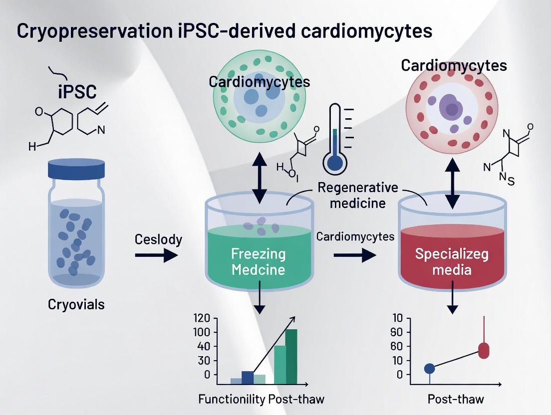
This article provides a comprehensive resource for researchers and drug development professionals on the cryopreservation of human induced pluripotent stem cell-derived cardiomyocytes (hiPSC-CMs).
Optimizing iPSC-Derived Cardiomyocyte Cryopreservation: Advanced Media, Protocols, and Functional Validation
Abstract
This article provides a comprehensive resource for researchers and drug development professionals on the cryopreservation of human induced pluripotent stem cell-derived cardiomyocytes (hiPSC-CMs). It explores the critical limitations of traditional cryoprotectants like DMSO and details the development and optimization of specialized, DMSO-free cryopreservation media. The scope covers foundational principles, step-by-step methodological protocols, strategies for troubleshooting and enhancing post-thaw recovery and function, and rigorous techniques for validating cryopreserved hiPSC-CMs for use in disease modeling, drug discovery, and therapeutic applications.
The Critical Need for hiPSC-CM Cryopreservation in Modern Biomedicine
Human induced pluripotent stem cell-derived cardiomyocytes (hiPSC-CMs) represent a revolutionary platform in cardiovascular research, offering an unlimited source of human cardiomyocytes that recapitulate patient-specific genetics. Since the groundbreaking development of hiPSCs by Dr. Shinya Yamanaka over a decade ago, this technology has enabled researchers to generate pluripotent stem cells from both healthy individuals and patients with various cardiovascular conditions [1]. The differentiation of hiPSCs into cardiomyocytes has become one of the most robust and efficient lineage differentiation protocols, achieving purities exceeding 90% in optimized systems [2]. These cells express fundamental components of cardiac contractility including ion channels for action potential generation, structures for excitation-contraction coupling, and proteins for calcium handling, making them invaluable for modeling heart disease, drug discovery, and regenerative therapy applications [3].
The integration of hiPSC-CM technology into research pipelines has been particularly valuable for addressing the critical gap between animal models and human physiology in drug development. Despite ideal results in nonhuman models, clinical trial failures frequently occur due to interspecies differences in cardiac electrophysiology [1]. hiPSC-CMs provide a human-relevant platform that can better predict drug responses and toxicity, potentially reducing late-stage drug attrition and enhancing patient safety [4] [3].
Key Applications of hiPSC-CMs
Disease Modeling
hiPSC-CMs have proven exceptionally valuable for modeling inherited cardiomyopathies, particularly ion channelopathies with well-understood impacts on action potential generation and propagation [1]. These disease models accurately recapitulate cellular disease phenotypes and enable mechanistic studies of pathogenesis.
Table 1: hiPSC-CM Models of Inherited Cardiomyopathies
| Disease Category | Specific Disease | Related Genes | Cellular Phenotype |
|---|---|---|---|
| Ion Channelopathy | Long QT Syndrome Type 1 (LQT1) | KCNQ1 | Slow outward potassium current (IKs), abnormal channel activities, increased susceptibility to tachyarrhythmia [1] |
| Long QT Syndrome Type 2 (LQT2) | KCNH2 | Prolonged action potential duration, reduction in IKr potassium current [1] | |
| Long QT Syndrome Type 3 (LQT3) | SCN5A | Accelerated recovery from Nav1.5 inactivation, action potential prolongation, early afterdepolarizations [1] | |
| Catecholaminergic Polymorphic Ventricular Tachycardia (CPVT) | RYR2, CASQ2 | Abnormal calcium leakage from sarcoplasmic reticulum, cytosolic calcium overload, delayed afterdepolarizations [1] | |
| Structural Cardiomyopathy | Hypertrophic Cardiomyopathy (HCM) | MYH7, MYBPC3, ACTC, TNNT2 | Hypertrophic morphology, arrhythmogenicity [1] |
| Dilated Cardiomyopathy (DCM) | TTN, TNNT2, SCN5A, DES | Dilated morphology, reduced contractility [1] | |
| Arrhythmogenic Cardiomyopathy (ACM) | DSP, DSC, DSG2, JUP | Structural disorganization, arrhythmias [1] | |
| Metabolic Cardiomyopathy | Barth Syndrome (BTHS) | TAZ | Metabolic abnormalities, contractile dysfunction [1] |
The gold standard for hiPSC-CM disease modeling involves the use of isogenic controls generated through CRISPR/Cas9 genome editing. This approach allows researchers to create control hiPSCs with identical genomic sequences except for the specific variant of interest, enabling definitive attribution of phenotypic differences to the disease-causing mutation [3]. Furthermore, patient-specific hiPSC-CMs facilitate the development of personalized medicine approaches, as they retain the complete genetic background of the donor, including polymorphisms that may influence drug responses [4].
Drug Screening and Toxicity Testing
hiPSC-CMs have emerged as a powerful tool for drug discovery and safety pharmacology, addressing a critical need for human-relevant screening platforms. Traditional drug development faces high attrition rates, with cardiovascular toxicity representing a major cause of failure during clinical trials [4]. hiPSC-CMs express the key ion channels responsible for cardiac electrophysiology, including the hERG channel crucial for drug-induced QT prolongation risk assessment [1].
The application of hiPSC-CMs in drug screening spans multiple approaches:
- High-Throughput Cardiotoxicity Screening: Evaluation of drug-induced proarrhythmic potential using multi-electrode arrays or calcium imaging in 2D monolayer cultures [3]
- Mechanism-Specific Therapeutic Discovery: Identification of compounds that reverse specific disease phenotypes in patient-derived hiPSC-CMs [3]
- Precision Medicine Applications: Testing drug responses across hiPSC-CMs from diverse genetic backgrounds to identify patient-specific efficacy and toxicity [4]
International collaborative efforts have proposed new paradigms for proarrhythmia risk assessment based on hiPSC-CMs, potentially offering more accurate prediction of clinical cardiotoxicity compared to traditional animal models [1]. Furthermore, engineered heart tissues (EHTs) provide more physiologically relevant contexts for evaluating contractile effects of drug candidates [4].
Regenerative Therapy
The potential use of hiPSC-CMs in regenerative therapy represents a promising approach for addressing the limited regenerative capacity of the human heart following injury. It is estimated that transplantation of up to one billion hPSC-CMs may be required for sufficient contractile tissue repair in human myocardial infarction [5]. Several studies in animal models have demonstrated that transplanted hiPSC-CMs can survive, integrate with host tissue, and electrically couple to the native myocardium [5].
Key considerations for regenerative applications include:
- Cell Purity: High purity (≥70% cTnT+) CM formulations improve contractility in vitro and in vivo following transplantation [6]
- Pro-survival Treatments: Pre-treatment with heat shock, IGF1, and cyclosporine A prior to transplantation enhances engraftment and survival [5]
- Maturation State: Less mature, pre-contraction stage CMs (differentiation days 12-30) exhibit better cryopreservation recovery, potentially making them more suitable for banking therapeutic products [5]
Experimental Protocols
Suspension Culture Cardiac Differentiation Protocol
The following optimized protocol for bioreactor differentiation enables large-scale production of hiPSC-CMs with high purity and reproducibility [2]:
Key Protocol Details:
- Input Cell Quality: Use quality-controlled master cell banks with >70% SSEA4 expression for consistent differentiation outcomes [2]
- Critical Timing Parameters: Initiate differentiation when embryoid bodies reach 100µm diameter for optimal efficiency [2]
- Small Molecule Concentrations: 7µM CHIR99021 (Wnt activator) for 24 hours, followed by 5µM IWR-1 (Wnt inhibitor) for 48 hours [2]
- Yield: Approximately 1.21 million cells per mL with >90% TNNT2+ cardiomyocytes [2]
This suspension culture method offers advantages over traditional monolayer differentiation, including improved scalability, reduced batch-to-batch variability, and more mature functional properties of the resulting cardiomyocytes [2].
DMSO-Free Cryopreservation Protocol
Recent advances have enabled the development of DMSO-free cryopreservation protocols that maintain high post-thaw viability and functionality [7]:
Protocol Optimization Parameters:
- Cooling Rate: Rapid cooling at 5°C/min significantly improves recovery compared to conventional 1°C/min rates [7]
- Nucleation Temperature: Low nucleation temperature of -8°C enhances post-thaw viability [7]
- CPA Composition: Optimized combinations of naturally occurring osmolytes (trehalose, glycerol, isoleucine) can achieve >90% post-thaw recovery [7]
- Post-Thaw Characterization: Assess viability, replating efficiency, calcium handling, and cardiac marker expression to validate functionality [7] [8]
Cardiac Progenitor Cryopreservation and Reseeding Protocol
An alternative approach involves cryopreserving cardiac progenitor cells at specific developmental stages rather than fully differentiated cardiomyocytes [6]:
Differentiate hiPSCs to Cardiac Progenitors:
- Generate EOMES+ mesoderm or ISL1+/NKX2-5+ cardiac progenitor cells using established protocols
Cryopreserve Progenitors:
- Dissociate progenitor cells at appropriate differentiation stage (typically days 3-7)
- Freeze in controlled-rate freezer using standard cryoprotectant solutions
Thaw and Reseed for Enhanced Purity:
- Thaw cryopreserved progenitors and reseed at optimized densities (1:2.5 to 1:5 surface area ratio)
- Continue differentiation according to standard protocols
Outcome: 10-20% absolute improvement in CM purity without negative impact on contractility or sarcomere structure [6]
The Scientist's Toolkit: Essential Research Reagents
Table 2: Key Reagents for hiPSC-CM Differentiation and Cryopreservation
| Reagent Category | Specific Examples | Function | Application Notes |
|---|---|---|---|
| Small Molecule Inducers | CHIR99021 (GSK3-β inhibitor) | Wnt pathway activation; induces mesoderm formation [2] | Optimal concentration typically 6-7 µM; duration critical for efficiency [2] |
| IWP2/IWR-1 (Wnt inhibitors) | Wnt pathway inhibition; promotes cardiac specification [2] | Typically applied after CHIR99021 treatment; concentration 3-5 µM [2] | |
| Cryoprotectants | DMSO (Conventional) | Penetrating cryoprotectant; prevents ice crystal formation [7] | Standard concentration 10%; associated with toxicity and functional impairment [7] |
| DMSO-Free Formulations | Natural osmolytes provide cryoprotection without DMSO toxicity [7] | Combinations of trehalose, glycerol, isoleucine; >90% post-thaw recovery reported [7] | |
| Cell Culture Supplements | ROCK inhibitor (Y27632) | Enhances cell survival after dissociation and thawing [7] [2] | Critical for improving replating efficiency of cryopreserved hiPSC-CMs [8] |
| Sodium L-lactate | Metabolic purification; selects for cardiomyocytes [7] [2] | Enriches cardiomyocyte population to >98% purity [7] | |
| Characterization Tools | Cardiac Troponin T (TNNT2) | Cardiomyocyte-specific structural protein marker [2] | Flow cytometry standard for assessing differentiation efficiency and purity [2] |
| MLC2v (Ventricle) & MLC2a (Atrium) | Chamber-specific myosin light chain isoforms [8] | Determines cardiomyocyte subtype specification; cryopreservation may enrich ventricular subtypes [8] |
Current Challenges and Future Directions
Despite significant advances, several challenges remain in the widespread adoption of hiPSC-CM technology. The physiological immaturity of hiPSC-CMs relative to adult cardiomyocytes represents a major limitation, as cells in culture typically maintain fetal-like characteristics including spontaneous beating, disorganized sarcomeres, and metabolic immaturity [3]. Current maturation strategies include:
- 3D Engineered Tissues: Culture in three-dimensional formats improves sarcomere organization and contractile force generation [3]
- Metabolic Manipulation: Shifting culture conditions from glycolytic to oxidative substrates promotes metabolic maturation [3]
- Electromechanical Stimulation: Application of physiological pacing and mechanical loading enhances structural and functional maturation [3]
For cryopreservation specifically, challenges include managing the anomalous osmotic behavior of hiPSC-CMs post-thaw, where cells undergo excessive dehydration upon resuspension in isotonic medium [7]. Additionally, cryopreservation has been shown to potentially alter the subtype composition of hiPSC-CM populations, with evidence of ventricular cardiomyocyte enrichment in some cases [8].
Future directions will likely focus on standardization of differentiation and cryopreservation protocols across different hiPSC lines, further optimization of DMSO-free cryoprotectant formulations, and the development of integrated platforms that combine hiPSC-CM technology with tissue engineering for more predictive drug screening and enhanced regenerative outcomes.
Human induced pluripotent stem cell-derived cardiomyocytes (hiPSC-CMs) represent a transformative advancement for cardiovascular disease modeling, drug discovery, and regenerative medicine. However, the effective cryopreservation of these cells remains a significant bottleneck that hampers both research reproducibility and clinical translation. Conventional cryopreservation methods using dimethyl sulfoxide (DMSO) consistently yield post-thaw viabilities between 50-80%, with concerning alterations in cellular function that compromise experimental outcomes and therapeutic potential [7] [9]. The field urgently requires standardized, optimized protocols that maintain not only cell viability but also electrophysiological fidelity, contractile properties, and pharmacological responsiveness post-thaw.
This application note addresses the central challenges in hiPSC-CM cryopreservation by presenting quantitative comparisons of current approaches, detailed optimized protocols, and practical tools for implementation. By integrating the most recent advances in cryopreservation science, we provide researchers with a framework to overcome key technical barriers and improve the rigor and reproducibility of their work with hiPSC-CMs.
Quantitative Analysis of Cryopreservation Outcomes
Table 1: Comparative Analysis of Post-Thaw hiPSC-CM Recovery and Viability
| Cryopreservation Method | Post-Thaw Viability (%) | Functional Assessment | Key Advantages | Key Limitations |
|---|---|---|---|---|
| Conventional DMSO (10%) | 69.4 ± 6.4% [7] | Reduced contractility; Altered drug response [9] | Widely accessible; Standardized | Functional alterations; Adverse effects |
| DMSO-Free Cocktail | >90% [7] | Preserved contractility and calcium handling [7] | Enhanced viability; No DMSO toxicity | Requires optimization for cell type |
| Bioreactor-Differentiated (bCMs) | >90% after cryo-recovery [2] | More mature functional properties [2] | High consistency; Scalable production | Specialized equipment required |
| Progenitor Stage Cryopreservation | 70-90% recovery [6] | Maintained differentiation capacity [6] | Enables on-demand CM production | Requires timing optimization |
Table 2: Documented Functional Changes in Recovered hiPSC-CMs
| Functional Parameter | Change Post-Cryopreservation | Experimental Evidence | Implications |
|---|---|---|---|
| Contractile Function | Reduced contraction velocity and deformation distance [9] | Motion tracking analysis | Impacts disease modeling and contractility studies |
| Calcium Handling | Line-dependent alterations in Ca2+ transients [9] | Fura-2 fluorescence imaging | Affects excitation-contraction coupling studies |
| Drug Response | Altered sensitivity and enhanced propensity for drug-induced arrhythmic events [9] | Microelectrode arrays with pharmacological testing | Critical for cardiotoxicity screening applications |
| Gene Expression | Upregulation of cell cycle genes [9] | RNA sequencing analysis | Induces proliferative state not present in mature CMs |
| Force Generation | Enhanced total force in selected populations [10] | Traction force microscopy | Suggests selection for more robust cells |
Optimized Cryopreservation Protocols
Principle: Replacement of DMSO with naturally occurring osmolytes (trehalose, glycerol, and isoleucine) identified through differential evolution algorithm optimization to minimize toxicity while maintaining cryoprotection.
Materials:
- hiPSC-CMs purified using sodium L-lactate treatment (>98% purity)
- Cryoprotectant components: trehalose (Sigma-Aldrich), glycerol (Humco), isoleucine (Sigma-Aldrich)
- Controlled-rate freezer
- Freezing containers: CryoStor CS10
Methodology:
- Cell Preparation: Harvest Day 20 hiPSC-CMs using 0.25% Trypsin-EDTA for 12 minutes at 37°C.
- CPA Loading: Resuspend singularized hiPSC-CMs in DMSO-free CPA solution gradually to minimize osmotic shock.
- Controlled-Rate Freezing:
- Cooling rate: 5°C/min
- Nucleation temperature: -8°C
- Final storage temperature: -196°C (liquid nitrogen)
- Thawing and Recovery:
- Rapid thaw in 37°C water bath with gentle agitation
- Gradual dilution of CPA using isotonic culture medium
- Resuspend in RPMI/B-27 medium with 5μM ROCK inhibitor (Y27632)
- Allow 30-minute recovery before experimental use
Quality Control:
- Assess post-thaw recovery (>90% expected)
- Validate contractile function via calcium transient studies
- Confirm cardiac identity via immunocytochemistry (cardiac troponin T)
Principle: Cryopreservation of intermediate differentiation stages (EOMES+ mesoderm and ISL1+/NKX2-5+ cardiac progenitors) enables on-demand production of high-purity cardiomyocytes while avoiding the functional alterations associated with mature CM cryopreservation.
Materials:
- hiPSC lines (WTC11 validated)
- Small molecule inhibitors: CHIR99021 (GSK3β inhibitor), IWP2 (Wnt inhibitor)
- Defined extracellular matrices: fibronectin, vitronectin, laminin-111
- Cryopreservation medium
Methodology:
- Cardiac Differentiation Initiation:
- Differentiate hiPSCs using GiWi protocol (CHIR99021 followed by IWP2)
- Monitor progression to EOMES+ mesoderm (approximately day 3-4)
- Progenitor Cryopreservation:
- Harvest EOMES+ mesoderm or ISL1+/NKX2-5+ CPCs
- Cryopreserve in aliquots using controlled-rate freezing
- Post-Thaw Differentiation:
- Thaw cryopreserved progenitors rapidly
- Reseed at optimized density (1:2.5 to 1:5 surface area ratio)
- Continue differentiation protocol
- Achieve terminal CM purity with 10-20% absolute improvement
Validation:
- Flow cytometry for cTnT+ purity assessment
- MUSCLEMOTION analysis of contractile parameters
- Assessment of sarcomere structure and junctional Cx43 expression
The Scientist's Toolkit: Essential Research Reagents
Table 3: Key Research Reagents for hiPSC-CM Cryopreservation
| Reagent/Category | Specific Examples | Function | Protocol Applications |
|---|---|---|---|
| Cryoprotective Agents | DMSO, trehalose, glycerol, isoleucine | Protect cells from ice crystal formation; Mitigate osmotic stress | DMSO-free formulations; Conventional cryopreservation |
| Small Molecule Inhibitors | CHIR99021 (GSK3β inhibitor), IWP2 (Wnt inhibitor), Y27632 (ROCK inhibitor) | Direct cardiac differentiation; Enhance post-thaw survival | Progenitor differentiation; Post-thaw recovery media |
| Extracellular Matrices | Matrigel, fibronectin, vitronectin, laminin-111 | Provide structural support; Enhance cell attachment and signaling | Reseeding of progenitors; Post-thaw plating |
| Cell Purification Reagents | Sodium L-lactate, metabolic selection media | Enrich cardiomyocyte population; Improve purity | Pre-cryopreservation preparation; Population validation |
| Viability Assessment Tools | Calcein AM, ethidium homodimer-1, Fura-2 | Quantify live/dead cells; Assess functional recovery | Post-thaw quality control; Functional validation |
Visualizing Workflows and Signaling Pathways
Cryopreservation Pathways for hiPSC-CMs
Optimized Cryopreservation Workflow
Effective cryopreservation remains a critical bottleneck in the widespread application of hiPSC-CMs, but recent advances in DMSO-free formulations and progenitor stage preservation offer promising solutions. The quantitative data presented herein demonstrates that optimized protocols can achieve post-thaw recoveries exceeding 90% while maintaining critical functional properties. However, researchers must remain cognizant of the persistent functional alterations in recovered hiPSC-CMs, particularly regarding drug response and contractile properties.
For field-wide progress, we recommend increased adoption of standardized viability assessment protocols, systematic functional validation beyond simple attachment and beating observations, and continued development of defined, xeno-free cryopreservation solutions. Through implementation of the optimized protocols and analytical frameworks presented in this application note, researchers can significantly improve the reproducibility and translational potential of their hiPSC-CM studies.
Human induced pluripotent stem cell-derived cardiomyocytes (hiPSC-CMs) have emerged as a transformative tool for cardiovascular disease modeling, drug discovery, and regenerative therapy development. The ability to generate patient-specific cardiomyocytes in vitro has created unprecedented opportunities for personalized medicine and high-throughput cardiotoxicity screening. However, the effective storage and transportation of these cells through cryopreservation remain a significant bottleneck in their clinical and commercial application.
Conventional cryopreservation protocols predominantly rely on dimethyl sulfoxide (DMSO) as a cryoprotective agent (CPA), presenting substantial limitations for hiPSC-CMs. This application note synthesizes current research findings to delineate the specific drawbacks of DMSO-based cryopreservation and presents emerging DMSO-free alternatives that demonstrate superior post-thaw recovery and functional preservation. As the field advances toward clinical applications, addressing these limitations becomes paramount for ensuring the safety, efficacy, and reproducibility of hiPSC-CM-based therapies and assays.
Key Limitations of DMSO in hiPSC-CM Cryopreservation
Cytotoxicity and Functional Impairment
Substantial evidence indicates that DMSO exerts concentration-dependent cytotoxic effects on hiPSC-CMs, compromising their viability and electrophysiological functionality. Research demonstrates that DMSO concentrations as low as 1% significantly alter key electrophysiological parameters in hiPSC-CMs [11].
Table 1: Effects of DMSO on hiPSC-CM Electrophysiological Parameters
| DMSO Concentration | Osmolality (mOsmol/kg) | Resting Membrane Potential | Action Potential Amplitude | Sodium Spike Amplitude | Field Potential Duration |
|---|---|---|---|---|---|
| 0.3% | ~336 | Unaffected | Unaffected | Unaffected | Unaffected |
| 1% | >500 | Significantly decreased | Significantly decreased | Significantly decreased | Significantly decreased |
| 3% | >800 | Irregular waveform | Irregular waveform | Irregular waveform | Irregular waveform |
These electrophysiological alterations are particularly concerning for drug safety screening applications, where accurate assessment of cardiotoxicity depends on maintaining native cardiomyocyte function. Furthermore, DMSO concentrations above 1% result in osmolality exceeding 400 mOsmol/kg, creating non-physiological conditions that compromise cellular integrity [11].
Compromised Post-Thaw Recovery and Viability
Conventional DMSO-based cryopreservation yields suboptimal recovery rates for hiPSC-CMs. Recent comparative studies reveal that standard 10% DMSO protocols enable post-thaw recoveries of only 69.4 ± 6.4%, significantly lower than optimized DMSO-free formulations that achieve recoveries exceeding 90% [7] [12]. This substantial difference in recovery directly impacts experimental efficiency and cost, particularly for high-throughput screening applications requiring large cell quantities.
The inferior performance of DMSO stems from its inability to adequately protect hiPSC-CMs against cryo-injury. hiPSC-CMs exhibit unique biophysical properties, including a large osmotically inactive volume and anomalous post-thaw osmotic behavior characterized by sharp volume decreases after resuspension in isotonic culture medium [7]. These characteristics necessitate specialized cryoprotection strategies that conventional DMSO formulations cannot provide.
Altered Drug Response Profiles
Cryopreservation-induced alterations in hiPSC-CMs significantly impact their responsiveness to pharmacological compounds. Comparative transcriptomic and functional analyses reveal that recovered hiPSC-CMs exhibit altered drug sensitivity and enhanced propensity for drug-induced arrhythmic events compared to their fresh counterparts [13].
These findings have critical implications for drug safety assessment, as cryopreservation artifacts could lead to either false positive or false negative results in cardiotoxicity screening. The altered drug response profiles observed in DMSO-cryopreserved hiPSC-CMs undermine their reliability for preclinical drug evaluation, potentially compromising drug development pipelines and patient safety.
Clinical Translation Challenges
The clinical application of DMSO-cryopreserved hiPSC-CMs faces significant safety and regulatory hurdles. DMSO administration in patients carries risks of adverse effects, including allergic reactions, gastrointestinal disturbances, neurological symptoms, and cardiac side effects [7] [14]. Furthermore, DMSO is associated with epigenetic alterations, particularly disruptions in DNA methylation mechanisms, raising concerns about its use with reprogrammed cells [7] [15].
Current clinical practice requires post-thaw washing to remove DMSO before administration, introducing additional processing steps that increase contamination risk, cell loss, and procedural complexity. Analysis of clinical trials reveals that 32% (18/57) of iPSC-based clinical trials disclosed DMSO use, with 9% (5/57) performing post-thaw wash steps before administration [14]. This additional manipulation complicates the adoption of off-the-shelf cell therapies and increases manufacturing costs.
DMSO-Free Cryopreservation: An Emerging Solution
Optimized Cryoprotectant Formulations
Recent advances in DMSO-free cryopreservation have identified synergistic combinations of naturally occurring osmolytes that effectively protect hiPSC-CMs during freezing and thawing. These formulations typically comprise sugars, sugar alcohols, and amino acids that collectively stabilize cellular structures and minimize ice crystal formation [7] [15].
Table 2: DMSO-Free Cryoprotectant Components and Functions
| Component Category | Specific Compounds | Concentration Range | Protective Mechanism |
|---|---|---|---|
| Sugars | Trehalose, Sucrose | Variable | Membrane stabilization, osmotic balance |
| Sugar Alcohols | Glycerol | Variable | Colligative cryoprotection, membrane integration |
| Amino Acids | L-Isoleucine | Variable | Stabilization of proteins and membranes |
| Proteins | Human Serum Albumin | Constant | Adsorption to surfaces, additional stabilization |
| Surfactants | Poloxamer 188 | Constant | Membrane stabilization |
Differential evolution algorithms have enabled rapid optimization of multi-component DMSO-free formulations, identifying specific concentration ranges that maximize post-thaw recovery while maintaining cellular functionality [7] [15]. These optimized formulations demonstrate remarkable consistency across different freezing modalities and adaptability to unplanned procedural deviations.
Protocol Optimization for hiPSC-CMs
Effective DMSO-free cryopreservation requires optimization of freezing parameters beyond CPA composition. Controlled-rate freezing experiments have identified a rapid cooling rate of 5°C/min and a low nucleation temperature of -8°C as optimal for hiPSC-CMs [7]. These parameters differ significantly from traditional cryopreservation approaches, highlighting the need for cell-type-specific protocol development.
Low-temperature Raman spectroscopy has proven invaluable for characterizing the freezing behavior of hiPSC-CMs in different CPA solutions, enabling data-driven optimization of freezing protocols based on solute partitioning and ice formation dynamics [7] [12]. This biophysical approach facilitates rational protocol design rather than empirical optimization.
DMSO-Free hiPSC-CM Cryopreservation Workflow
Functional Validation of DMSO-Free Approaches
Comprehensive functional assessment demonstrates that hiPSC-CMs cryopreserved using optimized DMSO-free methods retain critical physiological properties. Immunocytochemistry and calcium transient studies confirm preservation of cardiac markers and normal calcium handling post-thaw [7]. Additionally, contractile function assessment using advanced platforms like CONTRAX, a high-throughput traction force microscopy pipeline, verifies maintained contractile properties in recovered cells [16].
The preservation of functional integrity extends to drug responsiveness, with DMSO-free cryopreserved hiPSC-CMs exhibiting appropriate contractile responses to pharmacological agents such as Mavacamten, a cardiac myosin inhibitor [16]. This reliability in drug response is essential for cardiotoxicity screening applications.
Experimental Protocols
DMSO-Free Cryopreservation Protocol for hiPSC-CMs
Materials:
- hiPSC-CMs differentiated via Wnt pathway modulation
- DMSO-free CPA solution (trehalose, glycerol, isoleucine, human serum albumin, poloxamer 188)
- Controlled-rate freezer
- Cryogenic vials
- Water bath (37°C)
- Recovery medium
Procedure:
- Cell Preparation: Differentiate hiPSC-CMs using Wnt pathway inhibition followed by sodium L-lactate purification. Harvest cells at day 20 using 0.25% Trypsin-EDTA treatment for 12 minutes at 37°C [7].
- CPA Addition: Resuspend cell aggregates in DMSO-free CPA solution containing optimized concentrations of trehalose, glycerol, and isoleucine in basal buffer. Add CPA dropwise to cell suspension at 1:1 ratio [15].
- Equilibration: Incubate CPA-cell mixture at room temperature for 30-60 minutes to permit CPA permeabilization [15].
- Controlled-Rate Freezing:
- Initiate cooling at -10°C/min to 0°C
- Hold at 0°C for 10 minutes for temperature equilibration
- Cool at -1°C/min to nucleation temperature of -8°C
- Induce ice nucleation manually using a Cryogun or automated nucleation
- Continue cooling at -5°C/min to -60°C
- Rapid cool at -10°C/min to -100°C [7]
- Storage: Transfer vials to liquid nitrogen for long-term storage.
- Thawing: Rapidly thaw vials in 37°C water bath for approximately 2.5 minutes.
- Recovery: Dilute thawed cells dropwise with culture medium and plate immediately without washing steps.
Post-Thaw Functional Assessment
Calcium Transient Analysis:
- Plate recovered hiPSC-CMs on appropriate substrates at defined density.
- Load cells with calcium-sensitive fluorescent dye (e.g., Cal-520 or Fluo-4).
- Record calcium transients using high-speed fluorescence imaging at 37°C.
- Analyze transient amplitude, duration, and kinetics using specialized software.
Contractile Function Assessment:
- Utilize CONTRAX pipeline for high-throughput traction force microscopy.
- Plate cells on hydrogel substrates with tunable stiffness (10-35 kPa).
- Acquire video recordings of contracting cells at high frame rate (>100 fps).
- Compute contractile parameters including maximum force, work, power, and contraction/relaxation velocities [16].
Electrophysiological Evaluation:
- Perform manual patch clamp recording to assess action potential parameters.
- Utilize multi-electrode arrays for field potential duration measurement.
- Validate electrophysiological maturity through response to ion channel blockers.
The Scientist's Toolkit: Research Reagent Solutions
Table 3: Essential Materials for DMSO-Free hiPSC-CM Cryopreservation
| Reagent/Category | Specific Examples | Function/Application | Considerations |
|---|---|---|---|
| DMSO-Free CPA Components | Trehalose, Sucrose, Glycerol, L-Isoleucine | Cryoprotection via membrane stabilization and osmotic balance | Optimize concentrations using differential evolution algorithms |
| Basal Buffers | HBSS with Ca²⁺, Mg²⁺, glucose | CPA vehicle solution | Maintain physiological ion concentrations |
| Supplemental Additives | Human Serum Albumin, Poloxamer 188 | Additional membrane stabilization | Use at non-micelle forming concentrations |
| Differentiation Reagents | CHIR99021, IWP2, IWR-1 | Wnt pathway modulation for cardiac differentiation | Small molecules reduce lot-to-lot variability |
| Functional Assessment Tools | Calcium-sensitive dyes, CONTRAX pipeline, Multi-electrode arrays | Post-thaw functional validation | Implement high-throughput methods for population-level analysis |
The limitations of conventional DMSO-based cryopreservation methods for hiPSC-CMs necessitate a paradigm shift toward optimized DMSO-free approaches. The evidence presented demonstrates that DMSO compromises hiPSC-CM viability, functionality, and clinical utility through multiple mechanisms, including direct cytotoxicity, electrophysiological alteration, and induction of aberrant drug responses.
Emerging DMSO-free cryopreservation strategies address these limitations through rationally designed CPA cocktails that leverage synergistic interactions between naturally occurring osmolytes. These advanced formulations, coupled with optimized freezing parameters specific to hiPSC-CM biophysical properties, enable post-thaw recoveries exceeding 90% while maintaining critical functional attributes.
As the field progresses toward clinical translation and increased reliance on hiPSC-CMs for drug safety assessment, adoption of DMSO-free cryopreservation will be essential for ensuring cellular fidelity, experimental reproducibility, and therapeutic safety. The protocols and methodologies outlined herein provide a foundation for implementing these advanced preservation techniques in both research and clinical settings.
Within cardiovascular research and drug development, human induced pluripotent stem cell-derived cardiomyocytes (hiPSC-CMs) represent a foundational resource for disease modeling, regenerative medicine, and safety pharmacology [17] [2]. The widespread adoption of these cells is contingent upon reliable long-term storage, making effective cryopreservation a critical technological pillar. A successful cryopreservation strategy must extend beyond mere post-thaw cell survival to encompass the preservation of complex functional and phenotypic properties. This application note defines the three core goals—post-thaw viability, functional integrity, and phenotypic stability—and provides detailed protocols and benchmarks for their assessment within the broader context of developing specialized cryopreservation media.
Defining and Quantifying the Core Goals
Goal 1: Post-Thaw Viability
Post-thaw viability is the primary and most immediate indicator of cryopreservation success. It measures the percentage of cells that survive the freeze-thaw cycle with intact membrane integrity. High viability is a prerequisite for all subsequent functional and phenotypic assessments.
Table 1: Benchmarking Post-Thaw Viability in hiPSC-CMs
| Cryopreservation Approach | Reported Viability | Key Parameters | Citation |
|---|---|---|---|
| DMSO-Free Cryopreservation Medium | >90% post-thaw recovery | Optimized cocktail of trehalose, glycerol, and isoleucine; cooling rate: 5°C/min | [7] |
| Bioreactor-Differentiated hiPSC-CMs (bCMs) | >90% viability after cryo-recovery | Controlled-rate freezing; quality-controlled master cell banks | [2] |
| Conventional DMSO (10%) | ~69.4% ± 6.4% post-thaw recovery | Standard slow-freezing protocol; 1°C/min cooling rate | [7] |
| Conventional DMSO (10%) | 50-80% post-thaw viability (typical range) | Often associated with reduced contractility | [7] [18] |
Goal 2: Functional Integrity
Functional integrity refers to the retention of core cardiomyocyte electro-mechanical properties after thawing, including contractility, calcium handling, and electrophysiology. These functions are essential for predictive drug testing and disease modeling.
Table 2: Key Metrics for Assessing Functional Integrity
| Functional Metric | Assessment Method | Impact of Cryopreservation | Citation |
|---|---|---|---|
| Contractility | Motion tracking (velocity, deformation) | Recovered hiPSC-CMs show reduced contraction velocity and deformation distance in a line-dependent manner. | [9] |
| Calcium Handling | Ca²⁺ transient imaging (Fura-2) | Line-dependent effect on Ca²⁺ transient properties, consistent with contractility findings. | [9] |
| Electrophysiology | Microelectrode Array (MEA) | Shorter field potential duration (FPD) and increased beat rate observed in some lines. | [18] [9] |
| Drug Response | MEA / Calcium transient with pharmacologic agents | Altered sensitivity and enhanced propensity for drug-induced arrhythmic events. | [9] |
Goal 3: Phenotypic Stability
Phenotypic stability ensures that the molecular and structural identity of hiPSC-CMs remains unaltered by the cryopreservation process. This includes the stability of the transcriptome, the preservation of sarcomeric structure, and the maintenance of a mature cardiomyocyte signature.
- Transcriptomic Stability: RNA-seq analyses reveal that recovered hiPSC-CMs can exhibit an upregulation of cell cycle genes, indicating a shift in transcriptional programming. The expression of genes associated with ion channels, calcium handling, and sarcomere structure may remain statistically unchanged, though a line-dependent effect is often observed [9].
- Structural Stability: Immunocytochemistry for cardiac-specific markers such as cardiac Troponin T (TNNT2) and α-Actinin (ACTN2) is used to confirm the preservation of sarcomeric structure and organization post-thaw [2].
- Maturation State: The ratio of ventricular myosin heavy chain (MYH7) to atrial (MYH6)—a key maturation marker—should be maintained after cryopreservation. Studies show this ratio can remain stable post-thaw, while functional maturity may be compromised [9].
Experimental Protocols for Goal Assessment
Protocol: Assessment of Post-Thaw Viability and Recovery
This protocol is adapted from methods used to achieve >90% post-thaw recovery [7].
Materials:
- hiPSC-CMs (e.g., differentiated via Wnt pathway modulation and purified with sodium l-lactate)
- Optimized DMSO-free freezing medium (e.g., containing trehalose, glycerol, isoleucine)
- Controlled-rate freezer or isopropanol chamber (e.g., "Mr. Frosty")
- Liquid nitrogen storage tank
- Hemocytometer or automated cell counter
- Trypan blue stain
Procedure:
- Harvesting: On differentiation day ~20, harvest hiPSC-CMs using 0.25% Trypsin-EDTA for 12 minutes at 37°C.
- Preparation: Resuspend the singularized cell pellet in cold freezing medium at a concentration of 1-5 x 10^6 cells/mL.
- Aliquoting: Dispense 1 mL aliquots into cryogenic vials.
- Freezing: Place vials in a controlled-rate freezer. Apply the following curve:
- Cool at 5°C/min to a nucleation temperature of -8°C.
- Hold for 5 minutes for seeding.
- Complete the cooling process to -80°C, then transfer to liquid nitrogen for storage.
- Thawing: Rapidly thaw vials in a 37°C water bath for approximately 2 minutes.
- Viability Assessment: Immediately dilute thawed cells in pre-warmed culture medium, perform a cell count, and assess viability using Trypan blue exclusion.
Protocol: Functional Assessment via Calcium Transient Imaging
This protocol is used to evaluate calcium handling, a key component of functional integrity [9].
Materials:
- Fresh or recovered hiPSC-CMs plated on imaging-compatible dishes
- Fura-2 AM calcium indicator dye
- Field stimulation apparatus
- Live-cell fluorescence imaging system with 340/380 nm excitation and 510 nm emission filters
Procedure:
- Loading: Load plated cardiomyocytes with 2-5 µM Fura-2 AM in culture medium for 20 minutes at 37°C.
- Washing: Replace dye-containing medium with fresh pre-warmed medium and incubate for a further 20 minutes to allow for de-esterification.
- Stimulation: Place the dish on the microscope stage and field-stimulate the cells at 0.5 Hz to ensure synchronous contractions.
- Imaging: Record fluorescence emission ratios (F340/F380) at a high temporal resolution (≥100 Hz) for at least 30 seconds per field of view.
- Analysis: Analyze recordings for key parameters, including:
- Transient Amplitude: (Fₘₐₓ - Fₘᵢₙ)/Fₘᵢₙ
- Time to Peak: Duration from baseline to maximum.
- Decay Time Constant (Tau, τ): Time for the signal to decay to 37% of its peak amplitude.
Protocol: Evaluation of Phenotypic Stability via Transcriptomic Analysis
This protocol outlines the steps for RNA sequencing to assess transcriptomic stability [9].
Materials:
- Fresh and recovered hiPSC-CM pellets (in triplicate)
- RNA extraction kit (e.g., RNeasy Mini Kit)
- RNA quality assessment equipment (e.g., Bioanalyzer)
- RNA sequencing library preparation kit
- High-throughput sequencer
Procedure:
- RNA Extraction: Extract total RNA from fresh and recovered hiPSC-CM pellets according to the manufacturer's instructions.
- Quality Control: Assess RNA integrity (RIN > 8.0 is recommended).
- Library Prep and Sequencing: Prepare cDNA libraries and sequence on an appropriate platform (e.g., Illumina) to a minimum depth of 20 million reads per sample.
- Bioinformatic Analysis:
- Align reads to a reference genome (e.g., GRCh38).
- Perform differential gene expression analysis (e.g., using DESeq2) to compare fresh vs. recovered samples.
- Conduct pathway enrichment analysis (e.g., GO, KEGG) on differentially expressed genes, paying close attention to pathways like "cell cycle," "cardiac muscle contraction," and "calcium signaling."
The Scientist's Toolkit: Research Reagent Solutions
Table 3: Essential Reagents for hiPSC-CM Cryopreservation Research
| Reagent / Material | Function | Example & Notes |
|---|---|---|
| Specialized Freezing Media | Protects cells from ice crystal damage and osmotic stress. | STEMdiff Cardiomyocyte Freezing Medium: A commercially available, defined formulation. DMSO-Free Cocktails: e.g., trehalose, glycerol, and isoleucine mixtures can yield >90% recovery [7]. |
| Cryoprotectant Agents (CPAs) | Penetrating (e.g., DMSO) or non-penetrating (e.g., sugars) agents that mitigate freezing damage. | DMSO (10%): Conventional CPA, but linked to functional alterations [7] [9]. Trehalose: A natural non-penetrating osmolyte that stabilizes membranes and proteins [7]. |
| Controlled-Rate Freezing Apparatus | Ensures reproducible and optimal cooling rates. | Programmable Freezer: Allows for multi-step protocols (e.g., 5°C/min) [7]. Isopropanol Chambers (e.g., "Mr. Frosty"): Provides an approximate -1°C/min cooling rate in a -80°C freezer [19]. |
| Viability & Functional Assay Kits | Quantify post-thaw health and performance. | Trypan Blue: For basic viability counts. Fura-2 AM: For calcium transient assays [9]. MEA Systems: For non-invasive, long-term electrophysiological recording [18] [9]. |
Visualizing the Interdependence of Cryopreservation Goals
The three core goals are not isolated but are deeply interconnected. High viability is the foundation upon which functional integrity and phenotypic stability are built, while functional integrity is the ultimate expression of a stable phenotype. The diagram below illustrates this critical relationship and the primary assessment methods for each goal.
Protocol Deep Dive: Composing and Implementing Specialized Cryopreservation Media
The cryopreservation of human-induced pluripotent stem cell-derived cardiomyocytes (hiPSC-CMs) is a critical enabling technology for their application in disease modeling, drug discovery, and regenerative therapies. Conventional cryopreservation protocols largely depend on dimethyl sulfoxide (DMSO) as a cryoprotectant, which is associated with significant drawbacks including cytotoxic effects, reduced post-thaw viability and function, and undesirable side effects in therapeutic applications [7]. For hiPSC-CMs specifically, conventional DMSO-based cryopreservation typically yields post-thaw recoveries of only 69.4% ± 6.4% [7]. These limitations have accelerated the development of DMSO-free formulations that utilize naturally occurring osmolytes—specifically sugars, sugar alcohols, and amino acids—which offer improved biosafety and functional preservation profiles. This application note details the key components, protective mechanisms, and optimized protocols for DMSO-free cryopreservation of hiPSC-CMs, providing researchers with practical methodologies for implementation.
Key Components of DMSO-Free Formulations and Their Mechanisms
DMSO-free cryoprotective formulations function through coordinated mechanisms that address the primary stressors encountered during freezing and thawing: intracellular ice crystal formation, osmotic shock, and oxidative damage. These formulations typically comprise three principal classes of cryoprotective compounds, each contributing distinct protective functions.
Sugars and Their Stabilizing Role
Sugars, particularly di-, tri-, and oligosaccharides, serve as non-penetrating cryoprotectants that stabilize cell membranes and proteins during freezing and dehydration through multiple mechanisms. Their protective efficacy varies significantly based on molecular structure and size.
Table 1: Efficacy of Various Sugars in Protein Stabilization During Freezing and Drying
| Sugar | Type | Freeze-Thaw Recovery (-20°C) | Freeze-Drying Recovery | Air-Drying Recovery | Key Applications |
|---|---|---|---|---|---|
| Trehalose | Disaccharide | ~75% | ~80% | ~40% | Universal stabilizer for proteins and membranes |
| Sucrose | Disaccharide | ~78% | ~80% | ~45% | Common DMSO-free formulation component |
| Raffinose | Trisaccharide | ~72% | ~78% | ~35% | Enhanced stabilization in combination therapies |
| Melezitose | Trisaccharide | ~77% | ~83% | ~38% | Highest efficacy in freeze-drying applications |
| Glucose | Monosaccharide | ~76% | ~53% | ~25% | Basic osmotic stabilizer |
| Maltose | Disaccharide | ~75% | ~72% | ~30% | Intermediate protection profile |
| Lactose | Disaccharide | ~76% | ~76% | <10% | Effective for freeze-drying only |
| Stachyose | Oligosaccharide | ~78% | ~77% | ~33% | Large oligosaccharide with high glass transition |
Sugars function primarily through vitrification, where they form an amorphous glassy state that prevents ice crystal formation and immobilizes cellular structures, and water replacement, where their hydroxyl groups form hydrogen bonds with phospholipids and proteins, preserving membrane integrity and protein conformation in the absence of hydration water [20] [21]. Disaccharides and trisaccharides generally outperform monosaccharides due to their higher glass transition temperatures and more stable hydrogen-bonding networks [21].
Sugar Alcohols and Membrane Protection
Sugar alcohols (polyols) constitute another essential class of cryoprotectants, with varying protective capabilities based on their molecular configuration and interactions with biological structures.
Table 2: Efficacy of Sugar Alcohols in Protein Stabilization
| Sugar Alcohol | Freeze-Thaw Recovery (-20°C) | Freeze-Drying Recovery | Air-Drying Recovery | Molecular Characteristics |
|---|---|---|---|---|
| Pinitol | ~78% | ~81% | ~45% | Cyclitol, superior membrane stabilization |
| Quebrachitol | ~77% | ~75% | ~42% | Methylated cyclitol, unique protection profile |
| Sorbitol | ~76% | ~66% | ~59% | Acyclic polyol, good overall protection |
| myo-Inositol | ~65% | ~5% | ~8% | Cyclitol, can destabilize proteins |
| Mannitol | ~60% | ~4% | ~10% | Acyclic polyol, tends to crystallize |
The most effective sugar alcohols, particularly cyclitols like pinitol and quebrachitol, function through membrane stabilization by interacting with phospholipid head groups and maintaining bilayer integrity during dehydration, and crystallization inhibition by remaining amorphous during freezing and drying, thereby preventing damaging phase separations [21]. Interestingly, certain sugar alcohols like mannitol and myo-inositol can actually destabilize proteins during freezing and drying processes, highlighting the importance of selective formulation [21].
Amino Acids and Osmotic Regulation
Amino acids contribute to cryoprotection through diverse mechanisms including osmotic balance, antioxidant activity, and direct protein interactions. While specific quantitative data on amino acids in hiPSC-CM cryopreservation is less extensively documented in the search results, their inclusion in optimized cryoprotectant cocktails developed through differential evolution algorithms has demonstrated significant improvements in post-thaw recovery [7]. These compounds help maintain intracellular osmotic equilibrium without the toxicity associated with penetrating cryoprotectants like DMSO, and certain amino acids can directly stabilize protein structures through specific molecular interactions.
Experimental Protocols for hiPSC-CM Cryopreservation
Optimal DMSO-Free Formulation for hiPSC-CMs
The following protocol is adapted from peer-reviewed research demonstrating post-thaw recoveries exceeding 90% for hiPSC-CMs [7]:
Composition Optimization Method:
- Differential Evolution Algorithm: Employ computational optimization to determine the ideal molar ratios of sugar, sugar alcohol, and amino acid components [7].
- Component Selection: Utilize naturally occurring osmolytes rather than synthetic compounds to enhance biosafety.
- Validation: Confirm optimal formulations through post-thaw viability assays, functional assessments (calcium transients, contractility), and phenotypic characterization.
Controlled-Rate Freezing Protocol
Freezing parameters significantly impact post-thaw recovery, with cooling rate and nucleation temperature being particularly critical for hiPSC-CMs:
Critical Parameters:
- Cooling Rate: 5°C/minute has been identified as optimal for hiPSC-CMs, significantly faster than the 1°C/minute typically used for other cell types [7].
- Nucleation Temperature: -8°C provides superior post-thaw recovery compared to warmer nucleation temperatures [7].
- Post-Thaw Processing: Resuspend cells in isotonic culture medium and monitor for anomalous osmotic behavior, characterized by a sharp decrease in cell volume following resuspension that requires careful management [7].
Post-Thaw Functional Validation
Comprehensive assessment of hiPSC-CM function after cryopreservation is essential for confirming protocol efficacy:
Viability and Recovery Metrics:
- Post-Thaw Recovery: Quantify using standardized viability assays (e.g., trypan blue exclusion, flow cytometry with viability dyes).
- Benchmarking: Compare against DMSO-based controls (typically ~69% recovery).
Functional Assessments:
- Calcium Transient Studies: Evaluate calcium handling properties using fluorescent indicators (e.g., Fluo-4, Fura-2) to ensure preserved electrophysiological function [7].
- Immunocytochemistry: Confirm maintenance of cardiac-specific markers including troponins, connexin-43, and α-actinin [7].
- Morphological Analysis: Assess sarcomeric organization and cell integrity through high-content imaging.
The Scientist's Toolkit: Essential Research Reagents
Table 3: Key Reagents for DMSO-Free hiPSC-CM Cryopreservation
| Reagent Category | Specific Examples | Function | Application Notes |
|---|---|---|---|
| Sugars | Trehalose, Sucrose, Raffinose | Vitrifying agents, membrane stabilizers | Disaccharides show superior performance to monosaccharides |
| Sugar Alcohols | Pinitol, Quebrachitol, Sorbitol | Osmotic regulators, membrane protectors | Cyclitols often outperform linear polyols |
| Amino Acids | Isoleucine, other natural amino acids | Osmotic balance, protein stabilization | Concentration must be optimized for specific cell types |
| Basal Media | RPMI/B-27, commercial DMSO-free media | Carrier solution for cryoprotectants | Must be compatible with final application (research vs. therapeutic) |
| Cell Separation | Sodium L-lactate, purification reagents | Cardiomyocyte enrichment | Critical for obtaining >98% pure hiPSC-CM populations |
| Viability Assays | Trypan blue, flow cytometry dyes, ATP assays | Post-thaw recovery quantification | Use multiple methods for comprehensive assessment |
| Functional Assays | Calcium-sensitive dyes, antibody panels | Functional validation | Essential for confirming therapeutic utility |
DMSO-free cryopreservation formulations comprising optimized combinations of sugars, sugar alcohols, and amino acids represent a significant advancement in hiPSC-CM biopreservation. These formulations leverage multiple synergistic protective mechanisms—including vitrification, membrane stabilization, and osmotic regulation—to achieve post-thaw recoveries exceeding 90% while maintaining critical cardiac functions. The implementation of specific freezing parameters, particularly a rapid cooling rate of 5°C/minute and nucleation at -8°C, is essential for maximizing recovery. As research in this field progresses, further refinement of component ratios and the incorporation of emerging cryoprotectant classes like Natural Deep Eutectic Systems (NADES) promise to enhance cryopreservation efficacy while supporting the transition toward clinically compatible, therapeutically oriented cell processing protocols.
The cryopreservation of human induced pluripotent stem cell-derived cardiomyocytes (hiPSC-CMs) represents a critical technological pillar supporting cardiovascular disease modeling, drug discovery, and regenerative therapies. While conventional cryopreservation methods using dimethyl sulfoxide (DMSO) provide a foundational approach, they frequently yield suboptimal post-thaw recoveries for hiPSC-CMs, with reported viabilities between 50-80% and functional impairments such as reduced contractility [7]. Successful cryopreservation necessitates precise control over two fundamental physical parameters: the cooling rate and the nucleation temperature (the temperature at which ice crystallization is initiated). This application note details a refined controlled-rate freezing protocol optimized specifically for hiPSC-CMs, leveraging recent research on their unique biophysical properties to achieve post-thaw recoveries exceeding 90% [7].
Key Principles of Optimization
The Critical Role of Cooling Rate
The cooling rate directly dictates the physical state of water inside and outside the cell during freezing. A slow, controlled rate allows water to gradually exit the cell before it freezes, minimizing the formation of lethal intracellular ice crystals.
- Traditional Guideline: A cooling rate of -1°C/minute is standard for many cell types [22] [19].
- hiPSC-CM Specific Optimization: Recent evidence indicates that hiPSC-CMs achieve superior post-thaw recovery (~90%) with a more rapid cooling rate of -5°C/minute [7]. This optimized rate likely balances sufficient cellular dehydration with reduced exposure time to concentrated solutes and cryoprotectant toxicity.
The Importance of Nucleation Temperature
Controlling the temperature at which extracellular ice forms (nucleation) is crucial for reproducible results. Uncontrolled, stochastic supercooling followed by rapid ice formation can cause cellular damage.
- Optimized Parameter: For hiPSC-CMs, a defined nucleation temperature of -8°C has been identified as optimal [7].
- Functional Impact: Precise nucleation at this temperature ensures consistent ice formation, promotes controlled cellular dehydration, and contributes to the high viability outcomes observed with the optimized protocol.
Table 1: Optimized vs. Conventional Freezing Parameters for hiPSC-CMs
| Parameter | Conventional Protocol | Optimized hiPSC-CM Protocol | Impact on Cell Survival |
|---|---|---|---|
| Cooling Rate | -1°C/min [19] | -5°C/min [7] | Redces intracellular ice formation & osmotic stress. |
| Nucleation Temperature | Often not specified | -8°C [7] | Ensures consistent, controlled extracellular ice formation. |
| Cryoprotectant | 10% DMSO [22] | DMSO-free osmolyte cocktails [7] | Mitigates DMSO-induced toxicity and epigenetic effects. |
Experimental Protocol for Controlled-Rate Freezing
Pre-Freeze Preparation
- Cell Quality Assurance: Begin with hiPSC-CMs in the log phase of growth, characterized by >90% viability and high purity (>98% TNNT2+ cardiomyocytes, achieved through methods like metabolic selection with sodium L-lactate) [7] [2]. Ensure cells are free from microbial contamination, including mycoplasma.
- Cryoprotectant Formulation: Prepare freezing medium. While 10% DMSO in complete medium is conventional, consider optimized DMSO-free formulations. These may contain cocktails of naturally occurring osmolytes—such as trehalose (a sugar), glycerol (a sugar alcohol), and isoleucine (an amino acid)—optimized using algorithms like Differential Evolution (DE) [7].
- Cell Harvesting and Suspension:
- Gently detach adherent hiPSC-CM cultures using a dissociation reagent like TrypLE Express or trypsin [22].
- Resuspend the cell pellet in the pre-chilled (2-8°C) freezing medium.
- Adjust the cell concentration to within the range of 1x10^6 to 10x10^6 cells/mL [19]. Aliquot the suspension into sterile cryogenic vials.
Controlled-Rate Freezing Procedure
The following workflow and parameter relationship are critical for protocol success:
- Initial Equilibration: Place the loaded cryovials into the controlled-rate freezer chamber, pre-cooled to 4°C. Allow the samples to equilibrate for 10-15 minutes to ensure thermal uniformity [23].
- Initiate Cooling: Begin the freezing program with a cooling rate of -5°C per minute from 4°C down to the nucleation temperature of -8°C [7].
- Induce Nucleation (Seeding): Hold the temperature at -8°C for 5-10 minutes. During this hold, manually induce nucleation using a pre-chilled tool or an automated "seeding" function from the controlled-rate freezer. This step ensures controlled extracellular ice formation [7] [23].
- Secondary Cooling: After nucleation is confirmed, resume cooling at a rate of -5°C per minute from -8°C down to a final temperature of at least -40°C (typically -40°C to -60°C is sufficient) [23].
- Final Storage: Immediately transfer the cryovials to long-term storage in the vapor phase of a liquid nitrogen tank (≤ -135°C) [22].
The Scientist's Toolkit: Essential Reagents and Materials
Table 2: Key Research Reagent Solutions for hiPSC-CM Cryopreservation
| Item | Function / Application | Examples / Specifications |
|---|---|---|
| Specialized Freezing Media | Provides a protective, defined environment during freeze-thaw; contains cryoprotectants. | STEMdiff Cardiomyocyte Freezing Medium; Serum-free, DMSO-free osmolyte cocktails (e.g., Trehalose, Glycerol, Isoleucine) [7] [19]. |
| Controlled-Rate Freezer | Precisely controls sample cooling rate to optimize cell survival. | Planer series freezers; Enables programming of steps including nucleation temperature [23]. |
| Ice Nucleator | Controls the temperature of extracellular ice formation for protocol consistency. | Snomax (from P. syringae); Chipped forceps for manual seeding [24] [23]. |
| Liquid Nitrogen Storage | Long-term storage of frozen cells at ≤ -135°C to halt all metabolic activity. | Cryovials stored in the vapor phase to prevent explosion risks associated with liquid phase storage [22]. |
| Cryogenic Vials | Secure, sterile containment for cell suspensions during freezing and storage. | Internal-threaded, sterile vials; Resistant to liquid nitrogen temperatures [19]. |
Troubleshooting and Protocol Validation
Debugging Your Freezing Profile
A significant advantage of controlled-rate freezing is the ability to debug the process. If post-thaw viability is low, you can halt the protocol at different stages (e.g., after nucleation, after reaching -40°C), rapidly thaw the sample, and assess viability. This pinpoints the damaging segment of the freezing profile for optimization [23].
Post-Thaw Functional Validation
Beyond simple viability counts (e.g., >90% with Trypan Blue exclusion), it is essential to confirm the functional recovery of hiPSC-CMs post-thaw.
- Immunocytochemistry: Confirm the expression and proper sarcomeric organization of key cardiac markers like cardiac Troponin T (TNNT2) and α-Actinin (ACTN2) [7] [2].
- Calcium Transient Imaging: Validate the electrophysiological functionality by measuring calcium handling properties [7].
- Contraction Analysis: Assess spontaneous beating rate and contractile force to ensure mature phenotypic retention [2].
The move beyond a one-size-fits-all cryopreservation approach is paramount for the successful application of sensitive cell types like hiPSC-CMs. By implementing this optimized, step-by-step controlled-rate freezing protocol—specifically leveraging a cooling rate of -5°C/minute and a defined nucleation temperature of -8°C—researchers can achieve highly viable, functional, and reproducible hiPSC-CM banks. This reliability is fundamental for advancing high-quality, reproducible research in disease modeling and drug discovery, and is a critical step toward future therapeutic applications.
The successful thawing and recovery of cryopreserved human induced pluripotent stem cell-derived cardiomyocytes (hiPSC-CMs) is a critical determinant for experimental reproducibility in cardiac disease modeling, drug discovery, and safety pharmacology. Post-thaw viability and functional recovery can be highly variable, with conventional cryopreservation methods using dimethyl sulfoxide (DMSO) often reporting post-thaw viabilities between 50% and 80% [7]. Recent advances in cryopreservation science have identified optimized parameters and DMSO-free solutions that can significantly enhance post-thaw outcomes, achieving recovery rates over 90% while preserving cardiac-specific functionality [7]. This application note details evidence-based protocols for thawing and recovering hiPSC-CMs, incorporating quantitative data on critical parameters to maximize cell survival, maturation, and functional integration for downstream applications.
Quantitative Analysis of Post-Thaw Recovery Parameters
The following tables summarize key quantitative findings from recent studies investigating hiPSC-CM thawing and recovery.
Table 1: Comparative Analysis of Cryoprotectant Agent (CPA) Performance for hiPSC-CMs
| Cryoprotectant Type | Post-Thaw Recovery (%) | Cooling Rate (°C/min) | Nucleation Temperature (°C) | Key Functional Outcomes |
|---|---|---|---|---|
| 10% DMSO (Conventional) | 69.4 ± 6.4% [7] | 1 (Commonly used) [7] | Not Specified | Reduced contractility, increased arrhythmic events in some studies [7] |
| Optimized DMSO-free CPA | >90% [7] | 5 [7] | -8 [7] | Preserved morphology, calcium handling, and cardiac markers [7] |
| Not Applicable (Fresh Cells) | 100% (Reference) | Not Applicable | Not Applicable | Baseline function and morphology [25] |
Table 2: Impact of Thawing and Seeding Practices on hiPSC-CM Outcomes
| Parameter | Optimal Condition/Value | Impact on Recovery & Function |
|---|---|---|
| Post-Thaw Resuspension Medium | Isotonic culture medium with 5 μM ROCK inhibitor (Y-27632) [26] [27] | Enhances initial cell survival and attachment [26] [27] |
| Post-Thaw Osmotic Behavior | Anomalous cell volume drop observed [7] | Managing excessive dehydration may be crucial for viability [7] |
| Cell Density for Reseeding | Lower density (1:2.5 surface area ratio) [6] | Improves cardiomyocyte purity by ~12% without negatively affecting cell number [6] |
| Long-Term Maintenance Medium | Specialized cardiomyocyte support medium (e.g., STEMdiff [28] or metabolic maturation medium MM-1 [29]) | Supports structural and functional maturation; induces cardiac troponin I isoform shift [29] |
Experimental Protocols
Protocol for Thawing and Initial Plating of hiPSC-CMs
Principle: Rapid thawing minimizes ice crystal damage, while the use of ROCK inhibitor increases cell survival by inhibiting apoptosis [26] [27].
Reagents:
- Pre-warmed (37°C) cardiomyocyte recovery or basal medium (e.g., RPMI/B-27 medium [7])
- ROCK inhibitor (Y-27632) stock solution (e.g., 20 mM in DMSO [26])
- Complete cardiomyocyte maintenance medium (e.g., STEMdiff Cardiomyocyte Support Medium [28])
Procedure:
- Preparation: Pre-warm an appropriate volume of recovery medium to 37°C. Add ROCK inhibitor to the pre-warmed medium at a final concentration of 5–10 μM [26] [27].
- Thawing: Remove the cryovial from liquid nitrogen storage and immediately place it in a 37°C water bath for 1-2 minutes. Gently agitate until only a small ice crystal remains [7].
- Dilution: Transfer the thawed cell suspension dropwise into the prepared pre-warmed medium containing ROCK inhibitor. This gradual dilution reduces osmotic shock.
- Centrifugation: Centrifuge the cell suspension at 200 × g for 5 minutes to pellet the cells and remove the cryoprotectant [7].
- Resuspension & Seeding: Carefully aspirate the supernatant. Gently resuspend the cell pellet in complete cardiomyocyte maintenance medium supplemented with 5 μM ROCK inhibitor. Seed cells at the recommended density (e.g., 0.35 × 10^5 cells/cm² [29] or a lower density for improved purity [6]) onto pre-coated culture vessels.
- Initial Media Exchange: After 24 hours, carefully replace the medium with fresh complete maintenance medium without ROCK inhibitor to support long-term culture and maturation [29].
Protocol for Assessing Post-Thaw Viability and Function
Principle: Comprehensive assessment of post-thaw recovery includes viability quantification, morphological analysis, and functional validation to ensure cells are suitable for downstream applications [7] [25].
Reagents:
- Trypan Blue solution [28]
- Phosphate Buffered Saline (PBS)
- Fixative (e.g., 4% Paraformaldehyde)
- Permeabilization buffer (e.g., 0.1% Triton X-100 in PBS)
- Blocking solution (e.g., 1-5% BSA in PBS)
- Primary antibodies (e.g., anti-cardiac Troponin T, anti-ACTN2 [2])
- Fluorescently-labeled secondary antibodies
- Calcium-sensitive dyes (e.g., Rhod-2 AM [26])
Procedure:
- Viability Assessment: At 24 hours post-thaw, harvest a subset of cells and mix with Trypan Blue solution. Count viable (unstained) and non-viable (blue) cells using a hemocytometer or automated cell counter to calculate percentage viability [28].
- Immunocytochemical Analysis:
- At 48-72 hours post-thaw, wash cells with PBS and fix with 4% PFA for 15 minutes at room temperature.
- Permeabilize and block cells with blocking solution for 1 hour.
- Incubate with primary antibodies against cardiac markers (e.g., cTnT, ACTN2) overnight at 4°C [2].
- The next day, incubate with appropriate secondary antibodies for 1 hour at room temperature.
- Image using a fluorescence microscope to confirm sarcomeric structure and cardiomyocyte identity.
- Functional Assessment (Calcium Transients):
Workflow Visualization
The following diagram illustrates the complete post-thaw handling and media exchange workflow for hiPSC-CMs:
The Scientist's Toolkit: Essential Research Reagents
Table 3: Key Reagents for hiPSC-CM Thawing and Culture
| Reagent/Category | Specific Examples | Function & Application |
|---|---|---|
| ROCK Inhibitor | Y-27632 [26] [27] | Improves post-thaw cell survival and attachment by inhibiting apoptosis. Used in resuspension medium for first 24h. |
| Specialized Media | STEMdiff Cardiomyocyte Support Medium [28]; Metabolic Maturation Medium (MM-1) [29] | Provides optimized nutrients and factors for long-term maintenance and promotes structural/functional maturation. |
| DMSO-Free CPA Components | Trehalose, Glycerol, Isoleucine [7] | Naturally-occurring osmolytes that protect cells during freezing/thawing, avoiding DMSO toxicity while enabling high recovery. |
| Extracellular Matrix (ECM) | Fibronectin-Matrigel Composite [30]; GFR Matrigel [27] | Provides a biomimetic substrate that enhances cell adhesion, survival, and maturation post-thaw. |
| Characterization Antibodies | Anti-cardiac Troponin T (cTnT), Anti-ACTN2 (α-actinin) [2] | Validates cardiomyocyte identity and sarcomeric structure post-thaw via immunocytochemistry. |
| Functional Assay Reagents | Rhod-2 AM [26]; Lactate-based purification media [29] | Assesses calcium handling functionality; enriches cardiomyocyte population by metabolic selection. |
Optimal thawing and recovery of hiPSC-CMs requires an integrated approach combining rapid thawing techniques, appropriate cryoprotectant removal, strategic use of ROCK inhibitor, careful attention to reseeding density, and long-term culture in specialized maturation media. The protocols and data presented herein provide a validated framework for maximizing post-thaw viability, purity, and functionality of hiPSC-CMs, enabling more reliable and reproducible results in cardiac research and drug development applications. By implementing these best practices, researchers can significantly enhance the translational relevance of their hiPSC-CM models.
The translation of human induced pluripotent stem cell-derived cardiomyocytes (hiPSC-CMs) from research tools to clinical therapeutics and large-scale drug screening platforms is critically dependent on robust cryopreservation. Conventional cryopreservation protocols using dimethyl sulfoxide (DMSO) as a cryoprotectant typically achieve post-thaw recoveries between 50% and 80%, with documented alterations in cellular functionality, transcriptome, and drug response profiles [7] [13]. These limitations pose significant challenges for applications requiring high cell viability and predictable performance, including regenerative medicine, disease modeling, and safety pharmacology.
This application note details a novel, high-efficiency cryopreservation protocol that achieves exceptional post-thaw recovery exceeding 90% while maintaining key cardiomyocyte functions. By replacing DMSO with optimized combinations of naturally occurring osmolytes and precisely controlling freezing parameters, this method addresses fundamental limitations of conventional approaches and enables more reliable utilization of hiPSC-CMs across research and therapeutic applications.
Key Experimental Findings & Comparative Analysis
Quantitative Performance Comparison
The following table summarizes key performance metrics from the featured high-efficiency protocol alongside results from conventional cryopreservation methods reported in recent literature.
Table 1: Comparative Analysis of hiPSC-CM Cryopreservation Outcomes
| Parameter | High-Efficiency Protocol | Conventional DMSO Protocol | Progenitor Stage Cryopreservation | Functional Impact |
|---|---|---|---|---|
| Post-Thaw Recovery | >90% [7] | 69.4% ± 6.4% [7] | 70-90% (progenitor recovery) [6] | Enables higher viable cell yield |
| Cooling Rate | 5°C/min [7] | 1°C/min [7] | Not specified | Minimizes ice crystal formation |
| Nucleation Temperature | -8°C [7] | Not typically controlled | Not specified | Controls ice formation dynamics |
| Cryoprotectant Composition | Trehalose, Glycerol, Isoleucine [7] | 10% DMSO [7] [8] | 10% DMSO [6] | Reduces cytotoxicity and functional alterations |
| Post-Thaw Phenotype | Preserved cardiac markers, calcium transients, morphology [7] | Altered transcriptome, electrophysiology, drug response in some studies [13] | Retained differentiation capacity [6] | Critical for predictive assay performance |
| Contractile Properties | Maintained [7] | Variable reports: preserved [8] or enhanced force [10] | Improved purity after differentiation [6] | Essential for functional studies |
Protocol Workflow Visualization
The following diagram illustrates the comprehensive experimental workflow for the high-efficiency cryopreservation protocol, from cardiomyocyte differentiation through functional validation post-thaw:
Detailed Experimental Protocols
Cardiomyocyte Differentiation and Purification
Principle: Generate high-purity hiPSC-CMs through defined small molecule-directed differentiation followed by metabolic selection.
Procedure:
- hiPSC Culture: Maintain CCND2 hiPSC line (or equivalent) on Matrigel-coated plates in mTeSR1 medium. Culture for 5 days to achieve 80-90% confluency [7].
- Cardiac Differentiation Initiation (Day 0): Add 6.5 μM CHIR99021 in RPMI/B-27 without insulin to induce mesoderm formation. Incubate for 48 hours [7].
- Cardiac Specification (Day 2): Replace medium with fresh RPMI/B-27 without insulin supplemented with 5 μM IWP2 to inhibit Wnt pathway and promote cardiac differentiation [7].
- Medium Exchange (Days 4 & 6): Refresh with RPMI/B-27 without insulin [7].
- Maintenance Phase (Day 8+): Replace medium with RPMI/B-27 complete medium every 3 days. Spontaneous contractions typically appear by Day 7 with robust beating by Day 12 [7].
- Cardiomyocyte Purification (Days 10-14): Add glucose-free DMEM with 4mM sodium L-lactate every 2 days to enrich cardiomyocytes (>98% purity) by exploiting metabolic differences [7].
- Harvest (Days 14-20): Dissociate with 0.25% Trypsin-EDTA for 12 minutes at 37°C. Resuspend in RPMI/B-27 with 20% FBS and 5μM ROCK inhibitor (Y27632). Allow 30-minute recovery before use [7].
DMSO-Free Cryoprotectant Formulation
Principle: Utilize optimized combinations of naturally occurring osmolytes to provide cryoprotection without DMSO-associated toxicity.
Procedure:
- Prepare Base Solution: Combine trehalose, glycerol, and isoleucine in phosphate-buffered saline [7].
- Optimize Composition: Use differential evolution (DE) algorithm to determine optimal concentration ratios maximizing post-thaw recovery [7].
- Sterile Filtration: Filter sterilize (0.22μm) the final cryoprotectant solution [7].
- Temperature Equilibration: Warm solution to room temperature before use [7].
Controlled-Rate Freezing Protocol
Principle: Precise control of cooling rate and nucleation temperature to minimize cryoinjury from ice crystal formation.
Procedure:
- Cell Preparation: Harvest Day 20 hiPSC-CMs and concentrate to 1×10^6 cells/mL in freezing medium [7].
- Cryoprotectant Addition: Gently mix cell suspension with equal volume of DMSO-free cryoprotectant solution [7].
- Aliquoting: Distribute 1mL aliquots into cryovials [7].
- Programmable Freezing: Place vials in controlled-rate freezer and execute protocol:
Thawing and Recovery Assessment
Principle: Rapid thawing with appropriate medium conditions to maximize cell survival and functional recovery.
Procedure:
- Thawing: Rapidly warm cryovials in 37°C water bath with gentle agitation until just ice-free [7].
- Dilution: Slowly add pre-warmed culture medium (3:1 ratio to freezing suspension) dropwise with gentle mixing [7].
- Centrifugation: Pellet cells at 200×g for 5 minutes [7].
- Resuspension: Resuspend in RPMI/B-27 with 20% FBS and 5μM ROCK inhibitor [7].
- Plating: Seed at optimized density (determined empirically for each cell line) on appropriate substrate [8].
- Functional Assessment: At 24-72 hours post-thaw, evaluate:
- Viability: Trypan blue exclusion or flow cytometry with viability dyes [7] [8]
- Calcium Handling: Calcium transient imaging using Fluo-4 or similar dyes [7]
- Contractile Function: Video-based analysis of beating parameters [10]
- Immunocytochemistry: Cardiac troponin T, α-actinin, myosin heavy chain [7] [8]
The Scientist's Toolkit: Essential Research Reagents
Table 2: Key Research Reagents for High-Efficiency hiPSC-CM Cryopreservation
| Reagent/Category | Specific Examples | Function & Application Notes |
|---|---|---|
| Cell Lines | CCND2 hiPSC line (with cyclin D2 overexpression) [7] | Enhanced cardiac differentiation yield; other well-characterized lines also applicable |
| Differentiation Components | CHIR99021 (GSK3-β inhibitor), IWP2 (Wnt inhibitor) [7] | Directed cardiac differentiation via Wnt pathway modulation |
| Purification Reagents | Sodium L-lactate in glucose-free DMEM [7] | Metabolic selection to enrich cardiomyocyte population (>98% purity) |
| DMSO-Free CPA Components | Trehalose, Glycerol, Isoleucine [7] | Natural osmolytes providing cryoprotection without DMSO toxicity |
| Ice Recrystallization Inhibitors | N-aryl-D-aldonamides (e.g., 2FA) [31] | Suppress ice crystal growth during freezing; improve post-thaw function |
| Pro-survival Additives | ROCK inhibitor (Y27632) [7] | Enhances post-thaw survival by inhibiting apoptosis |
| Freezing Containers | Controlled-rate freezer [7] | Enables precise cooling rate control (5°C/min) and nucleation temperature management |
| Functional Assay Reagents | Calcium-sensitive dyes (e.g., Fluo-4), Antibodies (cTnT, α-actinin) [7] [8] | Assessment of post-thaw cardiomyocyte structure and function |
Critical Parameters for Success
Physical-Chemical Optimization
The exceptional performance of this protocol derives from addressing multiple cryoinjury mechanisms simultaneously. Key factors include:
- Cooling Rate Optimization: The rapid 5°C/min cooling rate minimizes time for osmotic damage and intracellular ice formation, which is particularly detrimental to sensitive hiPSC-CMs [7].
- Nucleation Control: Precise induction of ice formation at -8°C ensures consistent, controlled extracellular crystallization without damaging intracellular contents [7].
- Osmotically Inactive Volume: hiPSC-CMs exhibit a large osmotically inactive volume, influencing water transport during freezing and requiring specialized cryoprotectant formulations [7].
Alternative Strategy: Progenitor Stage Cryopreservation
For applications requiring extended culture post-thaw, cryopreservation at progenitor stages represents a complementary strategy:
- EOMES+ Mesoderm and ISL1+/NKX2-5+ CPCs demonstrate 70-90% recovery after cryopreservation and can differentiate into cardiomyocytes with improved purity when reseeded at appropriate densities [6].
- This approach enables quality control of progenitors and on-demand CM production, potentially simplifying manufacturing workflows for therapeutic applications [6].
The high-efficiency cryopreservation protocol detailed in this application note achieves unprecedented post-thaw recovery exceeding 90% while maintaining critical cardiomyocyte functions. By systematically addressing the limitations of conventional DMSO-based methods through optimized cryoprotectant composition, precise control of freezing parameters, and comprehensive functional validation, this approach enables more reliable utilization of hiPSC-CMs across research, drug development, and therapeutic applications. Implementation of these methods supports the growing demand for robust cryopreservation strategies that preserve both cell viability and functionality, ultimately accelerating the translation of hiPSC-CM technologies to clinical impact.
Solving Common Challenges: Strategies to Maximize Recovery and Maturation
Addressing Anomalous Osmotic Behavior and Excessive Post-Thaw Dehydration
Within the broader context of advancing cryopreservation protocols for human induced pluripotent stem cell-derived cardiomyocytes (hiPSC-CMs), managing post-thaw cell behavior presents a significant challenge. Recent research has identified anomalous osmotic behavior and excessive dehydration as critical factors compromising post-thaw recovery and functionality [7]. These phenomena occur when the delicate balance between intracellular and extracellular osmotic pressure is disrupted during the freezing and thawing processes, leading to mechanical stress, oxidative damage, and ultimately reduced cell viability and function [32] [33]. This application note provides detailed methodologies and data-driven insights to help researchers identify, quantify, and mitigate these issues, thereby improving the reliability of hiPSC-CM cryopreservation for drug development and basic research applications.
Understanding the Phenomenon
Mechanisms of Osmotic Damage in Cryopreservation
During cryopreservation, cells undergo severe osmotic stress from two primary sources:
- Intracellular ice formation at rapid cooling rates causes mechanical damage to cellular membranes [34]
- Solution effects from concentrated solutes during slow cooling lead to toxic electrolyte levels and cell dehydration [32]
HiPSC-CMs exhibit anomalous osmotic behavior characterized by a sharp decrease in cell volume following resuspension in isotonic culture medium post-thaw [7]. This excessive dehydration exceeds normal osmotic responses and correlates with poor recovery outcomes. The process generates oxidative stress through increased production of reactive oxygen species (ROS), leading to lipid peroxidation and cellular damage—a mechanism observed in other cell types including rhesus macaque spermatozoa [33].
Consequences for hiPSC-CM Structure and Function
The structural and functional integrity of hiPSC-CMs is particularly vulnerable to osmotic stress:
- Contractile properties may be altered, with studies reporting changes in force generation and calcium handling [10]
- Electrophysiological function can be compromised, affecting drug screening accuracy and disease modeling reliability [5]
- Recovery viability typically ranges between 50-80% with conventional DMSO-based protocols [7]
Table 1: Quantitative Comparison of Osmotic Behavior in hiPSC-CMs
| Parameter | Pre-Freeze Characteristics | Post-Thaw Anomalous Behavior | Functional Impact |
|---|---|---|---|
| Cell Volume | Normal, stable in isotonic conditions | Sharp volume decrease in isotonic medium [7] | Reduced viability and recovery |
| Osmotically Inactive Volume | Larger than hiPSCs [7] | Not characterized | Requires tailored cryoprotectant approaches |
| Membrane Integrity | Intact | Compromised, increased permeability | Altered calcium handling [10] |
| Oxidative Stress Markers | Baseline levels | Increased ROS and lipid peroxidation [33] | Cumulative cellular damage |
Experimental Protocols for Assessment
Protocol 1: Characterizing Osmotic Behavior
Purpose: To quantify the anomalous osmotic response of hiPSC-CMs post-thaw and establish baseline parameters for protocol optimization.
Materials:
- HiPSC-CMs differentiated using established protocols [2] [7]
- DMSO-free cryoprotectant solution (e.g., trehalose-glycerol-isoleucine mixture) [7]
- Isotonic culture medium (e.g., RPMI/B-27) [7]
- Microscope with time-lapse imaging capabilities
- Cell diameter measurement software
Methodology:
- Differentiate hiPSC-CMs using Wnt pathway modulation with CHIR99021 and IWP2 [2] [7]
- Harvest cells at day 20 using 0.25% Trypsin-EDTA treatment for 12 minutes at 37°C [7]
- Cryopreserve using controlled-rate freezing at optimal parameters (5°C/min cooling rate, -8°C nucleation temperature) [7]
- Thaw cells rapidly at 37°C and resuspend in isotonic culture medium
- Image cells immediately and at 5-minute intervals for 60 minutes
- Measure diameter of 50-100 individual cells at each time point
- Calculate volume assuming spherical morphology using the formula: V = (4/3)πr³
- Plot volume vs. time to characterize dehydration kinetics
Data Analysis:
- Compare pre-freeze and post-thaw volume trajectories
- Calculate percentage volume reduction at each time point
- Establish correlation between dehydration magnitude and recovery outcomes
Protocol 2: Evaluating Oxidative Stress Markers
Purpose: To quantify oxidative damage resulting from osmotic stress during cryopreservation.
Materials:
- HiPSC-CMs cryopreserved using test and control protocols
- ROS detection probes (e.g., H₂DCFDA for general ROS, MitoSOX for mitochondrial superoxide)
- Lipid peroxidation assay kit (e.g., MDA assay)
- Flow cytometer or fluorescence microplate reader
- Antioxidant supplements (e.g., α-tocopherol) for control experiments [33]
Methodology:
- Divide hiPSC-CMs into three groups: fresh (control), standard cryopreservation, and optimized cryopreservation
- Add ROS probes to cell suspensions according to manufacturer protocols
- Incubate for 30 minutes at 37°C protected from light
- Analyze fluorescence using flow cytometry (10,000 events per sample)
- Perform lipid peroxidation assay on cell lysates
- Include antioxidant-treated controls to confirm specificity of oxidative stress detection [33]
Data Analysis:
- Express ROS levels as mean fluorescence intensity relative to fresh controls
- Calculate fold-increase in lipid peroxidation products compared to non-frozen cells
- Correlate oxidative stress markers with osmotic behavior and recovery metrics
Optimized Cryopreservation Workflow
The following workflow integrates strategies to minimize anomalous osmotic behavior and excessive dehydration:
Diagram 1: Optimized cryopreservation workflow to mitigate osmotic stress. Key steps include using DMSO-free cryoprotectants and controlled freezing parameters.
Research Reagent Solutions
Table 2: Essential Research Reagents for Managing Osmotic Stress
| Reagent Category | Specific Examples | Function & Application Notes |
|---|---|---|
| DMSO-Free Cryoprotectants | Trehalose-glycerol-isoleucine mixture [7] | Natural osmolyte combination; enables >90% post-thaw recovery; reduces toxicity |
| Permeating Cryoprotectants | DMSO, ethylene glycol, propanediol [32] | Penetrate cell membranes; depress freezing point; use at minimal effective concentrations |
| Non-Permeating Agents | Sucrose, trehalose, raffinose [32] | Provide extracellular protection; enable vitrification; reduce required PA concentrations |
| Osmotic Stabilizers | Ficoll 70 [34] | Polymer that improves osmotic tolerance; enables storage at -80°C for extended periods |
| Antioxidants | α-Tocopherol [33] | Reduces oxidative stress from osmotic imbalance; decreases lipid peroxidation |
| Cell Recovery Media | STEMdiff Cardiomyocyte Support Medium [28] | Specialized formulation for thawing and maintaining hiPSC-CM function |
| Pro-Survival Factors | IGF-1, cyclosporine A [5] | Enhance cell survival when administered pre-freeze; improve engraftment potential |
Key Performance Data
Table 3: Quantitative Comparison of Cryopreservation Approaches
| Cryopreservation Method | Post-Thaw Recovery | Cooling Rate | Nucleation Temperature | Osmotic Behavior | Functional Preservation |
|---|---|---|---|---|---|
| Conventional DMSO (10%) | 69.4 ± 6.4% [7] | 1°C/min [5] | Not specified | Anomalous dehydration [7] | Reduced contractility [10] |
| DMSO-Free Optimized | >90% [7] | 5°C/min [7] | -8°C [7] | Managed dehydration | Preserved calcium transients [7] |
| Progenitor Stage freezing | 70-90% [6] | 1°C/min [6] | Not specified | Not characterized | Maintains differentiation capacity [6] |
Effectively addressing anomalous osmotic behavior and excessive post-thaw dehydration requires a multifaceted approach combining biophysical characterization, optimized cryoprotectant formulations, and controlled freezing parameters. The protocols and data presented here demonstrate that DMSO-free solutions based on naturally occurring osmolytes, coupled with rapid cooling rates and low nucleation temperatures, can significantly improve hiPSC-CM recovery while preserving critical cardiac functions. Implementation of these strategies will enhance the reliability of hiPSC-CM applications in drug development, disease modeling, and regenerative medicine research.
The advancement of cardiovascular disease modeling, drug discovery, and regenerative medicine is increasingly reliant on the use of human-induced pluripotent stem cell-derived cardiomyocytes (hiPSC-CMs). A significant bottleneck in the widespread application of these cells is the development of robust cryopreservation protocols that ensure high post-thaw viability and functional recovery. Traditional cryopreservation methods largely depend on dimethyl sulfoxide (DMSO) as a cryoprotectant, which is associated with detrimental effects on cell recovery, function, and patient safety [35]. This application note details the systematic optimization of two critical biophysical parameters—cooling rate and nucleation temperature—for the cryopreservation of hiPSC-CMs using a DMSO-free cryoprotective agent (CPA). The data and protocols presented herein are framed within a broader research thesis aimed at developing specialized, xeno-free cryopreservation media for hiPSC-derived cells.
Key Optimized Parameters and Functional Outcomes
The optimization process revealed that hiPSC-CMs have distinct biophysical requirements compared to other cell types. The identified optimal parameters not only maximize cell recovery but also preserve critical cardiac functions post-thaw.
Table 1: Optimized Cryopreservation Parameters for hiPSC-CMs
| Parameter | Optimal Value | Experimental Range Tested | Post-Thaw Recovery |
|---|---|---|---|
| Cooling Rate | 5 °C/min [35] | Varied | ~90% with DMSO-free CPA [35] |
| Nucleation Temperature | -8 °C [35] | Varied | Significantly greater than DMSO (69.4 ± 6.4%) [35] |
| Post-Thaw Seeding Density | Higher densities recommended | N/A | Improves recovery of cryopreserved hiPSC-CMs [8] |
Post-Thaw Functional Validation
Studies confirm that hiPSC-CMs cryopreserved under optimized conditions retain their essential functional characteristics:
- Calcium Handling and Contractility: Cells frozen using the best-performing DMSO-free CPA or DMSO preserved calcium transients and displayed cardiac markers similar to pre-freeze levels [35]. Some research indicates that cryopreservation may even select for larger, more robust cells with enhanced total force generation and altered calcium dynamics [10].
- Electrophysiological Properties: One study reported that cryopreserved hiPSC-CMs from a specific cell line exhibited longer action potential durations, potentially linked to an enrichment of ventricular-like cardiomyocytes in the post-thaw population [8].
- Ventricular Cardiomyocyte Enrichment: Flow cytometric analyses for cardiac troponin T (cTnT) and myosin light chain isoforms (MLC2a and MLC2v) showed that cryopreservation, when combined with adjusted seeding densities, can yield a population of hiPSC-CMs with a ventricular phenotype [8].
Experimental Protocol for Controlled-Rate Freezing
This section provides a detailed, step-by-step methodology for the cryopreservation of hiPSC-CMs using the optimized biophysical parameters.
Materials and Reagents
Table 2: Research Reagent Solutions
| Item | Specification / Function | Example / Comment |
|---|---|---|
| CPA Base | DMSO-free cocktail of naturally occurring osmolytes | Optimized mixture of a sugar, sugar alcohol, and amino acid using a differential evolution algorithm [35]. |
| Cell Dissociation Reagent | Enzyme-free for gentle dissociation | Versene solution (EDTA-based) or 0.25% Trypsin-EDTA [35] [36]. |
| Recovery Medium | Contains ROCK inhibitor | RPMI/B-27 medium with 20% FBS and 5 μM Y-27632 ROCK inhibitor [35]. |
| Coating Matrix | For post-thaw culture | Matrigel, Geltrex, or defined substrates like Laminin-521 [36]. |
Step-by-Step Procedure
Cell Preparation and Harvesting:
- Culture and differentiate hiPSCs into cardiomyocytes using a defined protocol, such as Wnt pathway modulation with CHIR99021 and IWP2 [35] [2].
- On the day of cryopreservation (e.g., Day 20 of differentiation), harvest hiPSC-CMs by enzymatic dissociation (e.g., 0.25% Trypsin-EDTA for 12 minutes at 37°C) [35].
- Neutralize the enzyme activity using a recovery medium containing serum and a ROCK inhibitor (e.g., 5 μM Y-27632). Allow the cells to recover in this medium for 30 minutes at room temperature [35].
Cryoprotectant Addition and Vialing:
- Centrifuge the recovered cell suspension and resuspend the pellet in the pre-cooled, optimized DMSO-free CPA at a recommended concentration (e.g., 1 × 10^6 cells/mL) [35] [37].
- Aliquot the cell suspension into cryovials and place them in a controlled-rate freezer that has been pre-cooled to the start temperature (usually 4°C).
Controlled-Rate Freezing Cycle:
- Initiate the freezing program with the following critical steps:
- Cooling Segment 1: From 4°C to the nucleation temperature of -8°C at a rate of 1 °C/min [35].
- Induce Nucleation: Trigger ice nucleation at -8°C using the freezer's manual or automatic nucleation feature (e.g., a burst of liquid nitrogen). Hold at this temperature for 5-10 minutes to ensure complete ice formation.
- Cooling Segment 2: From -8°C to a terminal temperature of -50°C to -80°C at the optimized rate of 5 °C/min [35].
- Final Transfer: Transfer the cryovials directly to a long-term storage vessel, such as the vapor phase of liquid nitrogen (below -135°C).
- Initiate the freezing program with the following critical steps:
Thawing and Post-Thaw Culture:
- Rapidly thaw the cryovials in a 37°C water bath with gentle agitation until only a small ice crystal remains.
- Slowly dilute the thawed cell suspension drop-wise with pre-warmed culture medium.
- Centrifuge the cells to remove the CPA, resuspend in recovery medium containing a ROCK inhibitor, and seed at a high density (e.g., 0.9 × 10^5 cells/cm² or higher) onto pre-coated culture vessels to maximize attachment efficiency [8].
- Change the medium 24 hours post-thaw to remove the ROCK inhibitor and any non-adherent debris, then continue with standard culture protocols.
Workflow and Logical Pathway
The following diagram illustrates the complete experimental workflow, from cell preparation to functional validation, integrating the key optimized parameters.
Discussion and Mechanistic Insights
The success of the outlined protocol is grounded in the unique biophysical properties of hiPSC-CMs. These cells are characterized by a large osmotically inactive volume and exhibit anomalous osmotic behavior post-thaw, undergoing excessive dehydration upon resuspension in isotonic medium [35]. The optimized parameters directly address these characteristics:
- The rapid cooling rate of 5 °C/min likely minimizes prolonged exposure to concentrated solutes and mitigates the damaging effects of excessive cell dehydration.
- The low nucleation temperature of -8 °C reduces the extent of undercooling before ice formation, thereby controlling the kinetics of ice crystal growth and reducing the risk of intracellular ice formation.
Low-temperature Raman spectroscopy studies have been pivotal in understanding solute partitioning and intracellular ice formation, providing a non-empirical basis for selecting these parameters [35] [38]. Furthermore, it is crucial to note that cryopreservation is not a neutral process; it can act as a selection pressure, potentially enriching for a subpopulation of hiPSC-CMs that are larger and exhibit altered contractile properties and transcriptomes [10]. This finding must be considered when designing experiments, as the post-thaw cell population may not be perfectly representative of the pre-freeze culture.
This application note establishes that the precise control of cooling rate and nucleation temperature is critical for the successful cryopreservation of hiPSC-CMs. The implementation of the optimized protocol—featuring a cooling rate of 5 °C/min and a nucleation temperature of -8 °C with a DMSO-free CPA—enables post-thaw recoveries exceeding 90% while maintaining key cardiac functions. This refined approach provides researchers and drug development professionals with a reliable method for creating biobanks of hiPSC-CMs, thereby enhancing the reproducibility and scalability of research in cardiac disease modeling, drug discovery, and the development of regenerative therapies.
The use of human induced pluripotent stem cell-derived cardiomyocytes (hiPSC-CMs) has become invaluable for cardiac disease modeling, drug screening, and regenerative medicine. However, the fetal-like phenotype and metabolic immaturity of these cells often limit their physiological relevance and predictive power [39] [40]. Two critical technical challenges confront researchers: the functional immaturity of hiPSC-CMs and the practical need for cryopreservation to enable scalable, reproducible experiments. This application note addresses these parallel challenges by presenting an integrated strategy that combines advanced metabolic maturation media with optimized cryopreservation protocols, providing researchers with a standardized approach to generate highly functional, adult-like hiPSC-CMs with enhanced physiological properties.
Background and Rationale
The Metabolic Immaturity of hiPSC-CMs
hiPSC-CMs typically exhibit a fetal-like metabolic profile, relying predominantly on glycolysis for ATP production rather than the fatty acid oxidation characteristic of adult cardiomyocytes [40] [41]. This metabolic immaturity is coupled with structural and functional limitations, including disorganized sarcomeres, absent T-tubules, immature electrophysiological properties, and reduced contractile force [39]. Adult cardiomyocytes derive approximately 60-70% of their ATP from fatty acid oxidation, while hiPSC-CMs may produce more than 50% of ATP from glycolysis, creating an energy production deficit that limits their functional capabilities [40].
The Critical Role of Cryopreservation in hiPSC-CM Workflows
Cryopreservation enables crucial experimental flexibility, including quality control testing, batch-to-batch consistency, and logistical planning for high-throughput applications. Recent advances in suspension culture differentiation systems have demonstrated that cryopreserved hiPSC-CMs can maintain high viability (>90%) and functional properties post-thaw, addressing previous limitations with monolayer-differentiated cells [2]. The integration of metabolic maturation with these improved cryopreservation approaches represents a significant advancement for the field.
Metabolic Maturation Media Composition and Mechanisms
Metabolic maturation media are designed to recapitulate the postnatal metabolic switch from glycolysis to fatty acid oxidation by providing appropriate oxidative substrates and modulating culture conditions. The strategic composition of these media targets multiple aspects of cardiomyocyte maturation.
Table 1: Key Components of Metabolic Maturation Media and Their Functions
| Component | Concentration Range | Primary Function | Mechanistic Basis |
|---|---|---|---|
| Albumin-bound fatty acids (AlbuMAX) | 1-5 mg/mL | Fatty acid source for β-oxidation | Provides physiological fatty acids to drive metabolic switching; enhances fatty acid uptake capacity [42] |
| Reduced glucose | 1-5 mM | Promotes oxidative metabolism | Creates mild metabolic stress that incentivizes fatty acid utilization; depletes rapidly in culture [42] |
| L-Carnitine | 0.5-2 mM | Fatty acid shuttle | Facilitates transport of fatty acids into mitochondria for oxidation [42] |
| Creatine | 1-5 mM | Energy buffer | Regenerates ATP from ADP at sites of high energy demand; enhances phosphocreatine system [42] |
| Taurine | 0.5-2 mM | Osmolyte & antioxidant | Stabilizes membranes and regulates calcium handling; supports long-term viability [42] |
| Triiodothyronine (T3) | 1-100 nM | Thyroid hormone signaling | Nuclear receptor agonist; promotes mitochondrial biogenesis and metabolic gene expression [43] |
Physiological Consequences of Metabolic Maturation
hiPSC-CMs cultured in metabolic maturation media exhibit significantly enhanced functional properties compared to those maintained in standard culture conditions. These improvements include:
- Enhanced Electrophysiological Maturity: Increased resting membrane potential (-65.6 mV vs -44.1 mV in controls), greater action potential upstroke velocity (11.0 V/s vs 4.2 V/s), and development of a characteristic "notch-and-dome" morphology in a subset of cells [44]
- Metabolic Transformation: 30% higher maximal oxygen consumption rates, increased fatty acid uptake, and enhanced spare respiratory capacity, indicating improved mitochondrial function [42]
- Structural Organization: Elongated cell morphology, aligned sarcomeres with increased length (approaching the adult range of 1.9-2.2 μm), and improved mitochondrial distribution along myofibrils [44] [39]
- Functional Sodium Channel Dependence: Transition from calcium-dependent to sodium-dependent action potentials, with complete asystole induced by tetrodotoxin at IC50 ~3.88 μM, consistent with adult Nav1.5 sensitivity [42]
Integrated Protocol: Cryopreservation and Metabolic Maturation
This section provides a detailed methodology for combining cryopreservation with metabolic maturation to produce highly functional hiPSC-CMs suitable for drug screening and disease modeling applications.
Bioreactor Differentiation and Cryopreservation
Table 2: Optimized Suspension Culture Differentiation and Cryopreservation Parameters [2]
| Parameter | Specification | Notes |
|---|---|---|
| Starting cell quality | >70% SSEA4+ by FACS | Critical for differentiation efficiency |
| EB size at CHIR addition | 100 μm diameter | Larger EBs (>300 μm) reduce efficiency |
| CHIR99021 concentration | 7 μM | Wnt activation for mesoderm induction |
| CHIR incubation duration | 24 hours | Optimized for suspension culture |
| IWR-1 concentration | 5 μM | Wnt inhibition for cardiac specification |
| IWR incubation duration | 48 hours | Initiated 24h after CHIR removal |
| Yield | ~1.21 million cells/mL | With >90% TNNT2+ purity |
| Cryopreservation viability | >90% post-thaw | Using controlled freeze/thaw protocols |
| Onset of contraction | Differentiation day 5 | Earlier than monolayer (day 7) |
Protocol Steps:
Quality-Controlled Cell Banking: Establish master cell banks of hiPSCs with verified karyotype, mycoplasma-free status, and pluripotency marker expression (>70% SSEA4+ by FACS) [2]
Suspension Culture Differentiation:
- Aggregate hiPSCs in stirred bioreactor systems to form embryoid bodies
- Monitor EB diameter and initiate differentiation with 7 μM CHIR99021 when EBs reach 100 μm
- After 24 hours, replace medium to remove CHIR99021
- Following a 24-hour gap, add 5 μM IWR-1 for 48 hours to promote cardiac specification
- Continue culture in basal maintenance medium until day 15-20
Controlled Cryopreservation:
- Dissociate differentiated hiPSC-CMs using gentle enzymatic digestion
- Resuspend in cryopreservation medium containing 10% DMSO and suitable cryoprotectants
- Use controlled-rate freezing systems with optimized cooling curves
- Store in liquid nitrogen vapor phase for long-term preservation
Post-Thaw Recovery and Metabolic Maturation
Protocol Steps:
Rapid Thaw and Recovery:
- Quickly thaw cryovials in 37°C water bath with gentle agitation
- Transfer cell suspension to pre-warmed culture medium containing ROCK inhibitor
- Centrifuge at low speed (100-200 × g) to remove cryoprotectant
- Plate at optimized density of 1.25 × 10^5 cells/cm² on appropriate substrates
Metabolic Maturation Phase:
- After 48-72 hours of recovery, transition cells to metabolic maturation medium
- Culture for 3-5 weeks with medium changes every 2-3 days
- For enhanced maturation, combine with additional stimuli:
Quality Assessment:
- Monitor spontaneous beating frequency and rhythm
- Assess viability and sarcomeric organization via immunostaining for cardiac troponin T and α-actinin
- Evaluate metabolic maturity via oxygen consumption rates and fatty acid uptake assays
Experimental Results and Validation
Functional Outcomes of Combined Approach
Implementation of the integrated cryopreservation and metabolic maturation strategy produces hiPSC-CMs with significantly enhanced adult-like properties:
Table 3: Quantitative Comparison of hiPSC-CM Properties Following Integrated Maturation [42] [44] [2]
| Parameter | Standard Culture | Metabolic Maturation Only | Combined Approach | Adult CM Reference |
|---|---|---|---|---|
| Resting Membrane Potential (mV) | -44.1 ± 9.8 | -58.2 ± 7.4 | -65.6 ± 8.5 | ~-85 mV |
| AP Upstroke Velocity (V/s) | 4.2 ± 1.4 | 6.6 ± 2.5 | 11.0 ± 7.4 | 100-300 V/s |
| Sarcomere Length (μm) | 1.7-2.0 | 1.8-2.1 | 1.9-2.2 | 1.9-2.2 μm |
| O₂ Consumption Rate (pmol/min/cell) | 147 ± 16 | 172 ± 9 | 202 ± 9 | Variable |
| TTX Sensitivity (IC50) | Resistant to 100 μM | ~3.88 μM | ~3.88 μM | ~1-5 μM |
| Spontaneous Beat Rate (bpm) | 60-80 | 40-60 | 30-50 | 60-100 (paced) |
| Ventricular Marker Expression | Variable | Increased | 83.4% MLC2v+ | >90% |
Applications in Disease Modeling and Drug Screening
The enhanced maturity achieved through this integrated approach significantly improves the fidelity of hiPSC-CMs for pharmacological and disease modeling applications:
- Long QT Syndrome Modeling: Metabolically matured hiPSC-CMs expressing NaV1.5 mutations demonstrate appropriate action potential prolongation and drug responses that correlate with clinical observations [42]
- Drug Sensitivity Profiling: Matured cells show appropriate sensitivity to channel blockers including tetrodotoxin (Na+ channel) and verapamil (Ca2+ channel), with improved prediction of clinical cardiotoxicity [44]
- Engineered Heart Tissues: When incorporated into 3D tissue constructs, metabolically matured hiPSC-CMs generate greater contractile force and exhibit improved sarcomere organization and long-term survival [42]
The Scientist's Toolkit: Essential Research Reagents
Table 4: Key Research Reagent Solutions for Integrated Cryopreservation and Maturation
| Reagent/Category | Specific Examples | Function & Application Notes |
|---|---|---|
| Metabolic Media Supplements | AlbuMAX, Lipid-rich B-27, L-carnitine, creatine, taurine | Drives metabolic switching from glycolysis to fatty acid oxidation; use for 3-5 week maturation period [42] |
| Cryopreservation Systems | Controlled-rate freezers, DMSO-based cryoprotectant media | Maintains high viability (>90%) post-thaw; essential for banking and distribution [2] |
| Specialized Culture Substrates | Aligned nanopatterned surfaces, cardiac-specific ECM scaffolds | Promotes structural maturation and sarcomere alignment; hiPSC-CF-ECM shows enhanced results [44] [45] |
| Electrostimulation Equipment | C-Pace EP systems, custom stimulation setups | Applied at 2 Hz for 3-4 weeks; key driver of electrophysiological and mitochondrial maturation [44] |
| Maturation Assessment Tools | Seahorse XF Analyzers, patch clamp systems, calcium imaging | Quantifies metabolic parameters (OCR, ECAR), electrophysiology, and calcium handling [42] [40] |
Workflow Integration and Visual Protocol
The integrated workflow for cryopreservation and metabolic maturation involves sequential phases that can be implemented in standard cell culture laboratories. The process from pluripotent stem cells to fully matured cardiomyocytes requires approximately 8-10 weeks, including differentiation, cryopreservation, recovery, and maturation phases.
Integrated Workflow for hiPSC-CM Cryopreservation and Maturation
The integration of optimized cryopreservation protocols with metabolic maturation strategies enables the consistent production of highly functional hiPSC-CMs that more closely recapitulate adult cardiomyocyte physiology. This combined approach addresses both practical experimental needs for cell banking and distribution, while simultaneously enhancing the physiological relevance of the resulting cardiomyocytes for drug discovery and disease modeling applications. The standardized protocols and quality benchmarks provided in this application note offer researchers a validated pathway to implement these advanced methodologies in their own laboratories, potentially increasing the predictive accuracy of hiPSC-CM-based assays and accelerating cardiovascular drug development.
The transition from research-scale experiments to large-batch, clinical-grade cryopreservation presents significant challenges for the commercialization and clinical application of human induced pluripotent stem cell-derived cardiomyocytes (hiPSC-CMs). While cryopreservation protocols for hiPSC-CMs have been established, most conventional methods are optimized for small volumes (typically 1-2 mL cryovials) and utilize dimethyl sulfoxide (DMSO) as the primary cryoprotectant [46] [5]. The scaling process necessitates addressing fundamental cryobiological differences, optimizing functional recovery, and ensuring compliance with regulatory requirements for clinical applications [46] [47]. This application note outlines key considerations and protocols for implementing robust, scalable cryopreservation workflows for hiPSC-CMs, with a focus on maintaining cell viability, purity, and functionality post-thaw.
Fundamental Challenges in Cryopreservation Scale-Up
Thermodynamic Differences Between Small and Large Volumes
The physical determinants of the freezing process differ fundamentally between small and large volumes. In low-volume samples (<2 mL), minimal temperature gradients typically result in uniform undercooling throughout the sample, followed by rapid growth of a continuous ice network in a process termed network solidification (NS) [48]. In contrast, large-volume samples experience significant temperature gradients between the cooling interface and the bulk volume, leading to progressive solidification (PS) where ice nucleation occurs at the cold wall and an ice front progresses through the sample [48]. These differences in ice formation dynamics directly impact cell viability and require protocol modifications.
Recent studies demonstrate that progressive solidification in large volumes can result in fewer but proportionally more viable cells 24 hours post-thaw compared to network solidification, though these differences may diminish after 48-72 hours as cells recover division capability [48]. Understanding these fundamental cryobiological principles is essential for developing effective scale-down models to emulate large-volume ice formation for protocol optimization [48].
Clinical Translation Challenges
The development of "off-the-shelf" allogeneic hiPSC-CM therapies faces significant cryopreservation hurdles. Current meta-analyses of clinical trials reveal that 100% of preclinical iPSC-based cell therapy candidates use DMSO as a cryoprotectant, with 67% employing a uniform freeze rate of 1°C/min and 100% requiring post-thaw washing to remove cytotoxic DMSO [46]. This washing step introduces significant risks including contamination through adventitious agents and product damage through pipetting-induced shear stress [46]. For clinical applications involving direct injection into sensitive tissues (e.g., heart, brain, spine), even residual DMSO concentrations pose safety concerns, with studies showing 0.5-1.0% DMSO causing significant viability loss in neuronal cells [46].
Table 1: Analysis of Cryopreservation Practices in iPSC-Based Cell Therapies
| Aspect | Preclinical Studies (n=12) | Clinical Implications |
|---|---|---|
| Cryoprotectant | 100% use DMSO | Safety concerns for novel administration routes |
| Freeze Rate | 67% use 1°C/min (33% unspecified) | Lack of optimization for different cell types |
| Post-Thaw Processing | 100% require washing | Contamination risk, cell loss, point-of-care complexity |
| Storage Duration | Varied reporting | Impact on potency and viability not fully characterized |
Protocol Optimization for Scale-Up
DMSO-Free Cryopreservation Strategies
Recent advances in DMSO-free cryopreservation offer promising alternatives for clinical applications. Studies have demonstrated that optimized combinations of naturally occurring osmolytes can achieve post-thaw recoveries exceeding 90%, significantly higher than conventional DMSO-based protocols (69.4 ± 6.4%) [7]. These formulations typically incorporate:
- Sugars and sugar alcohols (e.g., trehalose) providing extracellular stabilization
- Permeating cryoprotectants (e.g., glycerol) at reduced concentrations
- Amino acids (e.g., isoleucine) enhancing membrane protection
The development process for DMSO-free solutions involves biophysical characterization of hiPSC-CMs, including membrane permeability parameters and osmotically inactive volume, followed by optimization of cryoprotectant composition using computational approaches like differential evolution algorithms [7].
Controlled Rate Freezing Parameters
Optimization of freezing parameters is critical for large-scale cryopreservation. Key parameters include:
- Cooling rate: hiPSC-CMs show optimal recovery at rapid cooling rates (5°C/min) compared to conventional 1°C/min rates [7]
- Nucleation temperature: Controlled initiation of ice formation at specific temperatures (-8°C for hiPSC-CMs) improves consistency [7]
- Storage conditions: Vapor phase liquid nitrogen storage minimizes contamination risks while maintaining temperature stability [49]
Table 2: Optimal Freezing Parameters for hiPSC-CMs
| Parameter | Conventional Protocol | Optimized Protocol | Impact on Viability |
|---|---|---|---|
| Cooling Rate | 1°C/min | 5°C/min | Significantly improved |
| Nucleation Temperature | Not typically controlled | -8°C | Improved consistency |
| Cryoprotectant | 10% DMSO | DMSO-free osmolyte mix | >90% recovery |
| Storage Phase | Liquid phase | Vapor phase | Reduced contamination risk |
Integrated Workflow for Large-Scale hiPSC-CM Cryopreservation
The following workflow diagram illustrates an optimized, scalable process for hiPSC-CM cryopreservation, integrating critical quality control checkpoints and protocol adaptations for large volumes:
Research Reagent Solutions for Scalable Cryopreservation
Table 3: Essential Materials for Large-Scale hiPSC-CM Cryopreservation
| Reagent/Category | Specific Examples | Function/Purpose |
|---|---|---|
| Cryoprotectants | DMSO-free osmolyte mixes (trehalose, glycerol, isoleucine) | Membrane stabilization, ice crystal inhibition |
| Cryopreservation Media | Commercial defined media (CryoStor) | Chemically-defined, animal component-free preservation |
| Controlled-Rate Freezers | Programmable freezer with large chamber capacity | Reproducible cooling profiles for large volumes |
| Storage Systems | Vapor phase liquid nitrogen tanks | Secure long-term storage, reduced contamination risk |
| Quality Assessment | Flow cytometry panels (TNNT2, viability markers) | Pre- and post-thaw quality control |
| Bioreactor Systems | Stirred suspension bioreactors | Scalable CM differentiation and harvest |
Successful scale-up of hiPSC-CM cryopreservation requires addressing fundamental cryobiological differences between small and large volumes, optimizing freezing parameters for specific cell types, and implementing DMSO-free strategies suitable for clinical applications. The protocols and considerations outlined herein provide a framework for developing robust, scalable cryopreservation processes that maintain cell viability, functionality, and purity while meeting regulatory requirements for clinical translation. As the field advances, continued refinement of large-volume cryopreservation protocols will be essential for realizing the full potential of hiPSC-CMs in regenerative medicine and drug development applications.
Beyond Viability: Rigorous Functional and Phenotypic Assessment of Cryopreserved hiPSC-CMs
The cryopreservation of human induced pluripotent stem cell-derived cardiomyocytes (hiPSC-CMs) is a critical step for enabling their widespread application in disease modeling, drug screening, and regenerative medicine. However, maintaining cellular integrity and function post-thaw requires rigorous validation using specialized media and protocols. This application note provides detailed methodologies and benchmarks for assessing three fundamental aspects of hiPSC-CM quality after cryopreservation: sarcomere structure, calcium handling, and electrophysiological function. By establishing standardized validation metrics, researchers can ensure that cryopreserved hiPSC-CMs retain physiological relevance for downstream applications.
Sarcomere Structure Validation
Quantitative Structural Analysis via Super-Resolution Microscopy
The sarcomere is the fundamental contractile unit of cardiomyocytes, and its structural integrity is a key indicator of cellular maturity and health. Super-resolution microscopy techniques, particularly Photoactivated Localization Microscopy (PALM) and Structured Illumination Microscopy (SIM), enable quantitative evaluation of sarcomeric organization beyond the diffraction limit of conventional microscopy [50] [51].
Key Validation Metrics:
- Sarcomere length: Mature hiPSC-CMs should approach the adult benchmark of approximately 2.0 μm [51]. Immature hiPSC-CMs typically exhibit shorter sarcomeres (∼1.61 μm) compared to those cultured on maturation-promoting substrates (∼1.86 μm) [52].
- Z-disc thickness: Measured via PALM imaging, this parameter indicates the density and organization of α-actinin cross-linking within the sarcomere [50].
- Myofibril density and alignment: Quantified through automated image analysis and machine learning algorithms, these metrics reflect the overall organization of the contractile apparatus [51].
Table 1: Sarcomere Structural Parameters in hiPSC-CMs Under Different Culture Conditions
| Condition | Sarcomere Length (μm) | Z-disc Thickness (nm) | Organization | Reference Benchmark |
|---|---|---|---|---|
| Immature hiPSC-CMs (Matrigel) | 1.61 ± 0.15 | Not reported | Disorganized, radial pattern | [52] |
| Mature hiPSC-CMs (MatrixPlus) | 1.86 ± 0.12 | Not reported | Highly organized | [52] |
| Adult Cardiomyocytes | ~2.0 | ~100-120 | Highly aligned | [51] |
| Neonatal Cardiomyocytes | Similar to hiPSC-CMs | Similar to hiPSC-CMs | Intermediate | [50] |
Experimental Protocol: Sarcomere Imaging and Analysis
Materials:
- hiPSC-CMs plated on appropriate substrate (e.g., glass coverslips)
- Primary antibody: anti-sarcomeric α-actinin (e.g., ab9465, 1:100 dilution)
- Secondary antibody: fluorescently-labeled (e.g., AlexaFluor647, 1:200)
- Fixative: 2-4% paraformaldehyde
- Permeabilization buffer: 0.1-0.5% Triton X-100
- Blocking solution: 1-5% BSA in PBS
- Super-resolution microscope (e.g., Zeiss ELYRA system)
Methodology:
- Cell Culture: Plate cryopreserved hiPSC-CMs after thawing and allow recovery for 3-7 days post-plating. For maturation studies, culture cells on patterned surfaces or human perinatal stem cell-derived extracellular matrix (e.g., CELLvo MatrixPlus) for at least 7 days [52].
- Fixation and Permeabilization: Rinse cells with PBS and fix with 2% paraformaldehyde for 15 minutes at room temperature. Permeabilize with 0.2% Triton X-100 for 5 minutes [51].
- Immunostaining: Block cells with 1% BSA for 30 minutes, then incubate with primary antibody against α-actinin for 1-2 hours at room temperature or overnight at 4°C. After PBS washes, incubate with fluorescent secondary antibody for 1 hour [51].
- Image Acquisition: Acquire super-resolution images using PALM or SIM modalities. For PALM, acquire 5,000-10,000 frames to enable single-molecule localization [50] [51].
- Quantitative Analysis:
- Use custom software or open-source tools (e.g., ImageJ with plugins) to measure sarcomere length from Z-line to Z-line.
- Calculate Z-disc thickness from the full-width at half-maximum of Gaussian fits to α-actinin signals in PALM images [50].
- Determine myofibril alignment using Fourier transform or gradient-based methods.
Calcium Handling Assessment
Functional Calcium Handling Metrics
Calcium handling is a critical indicator of hiPSC-CM functional maturity and overall health after cryopreservation. Comprehensive evaluation should include assessment of both spontaneous and triggered calcium transients, as well as sarcoplasmic reticulum (SR) calcium storage and release capacity [53] [52].
Key Validation Metrics:
- Calcium transient amplitude: Reflects the amount of calcium released during each contraction cycle.
- Calcium transient duration (CaTD): Typically measured at 80% decay (CaTD80), longer durations may indicate immaturity.
- SR calcium load: Assessed via caffeine-induced calcium release.
- Calcium transport mechanisms: Relative contributions of SERCA2a (sarcoendoplasmic reticulum calcium ATPase) and NCX (sodium-calcium exchanger) to calcium reuptake and extrusion.
Table 2: Calcium Handling Parameters in hiPSC-CMs Versus Adult Cardiomyocytes
| Parameter | hiPSC-CMs | Adult Rabbit Ventricular CMs | Maturation Trend | Citation |
|---|---|---|---|---|
| CaTD80 (ms) | Significantly longer than APD80 | Not specified | Decreases with maturation | [52] |
| SERCA Activity | Slower than adult CMs | Faster | Increases with maturation | [53] |
| NCX Activity | Slower than adult CMs | Faster | Increases with maturation | [53] |
| SERCA/NCX Contribution | Comparable to adult CMs | Reference standard | Maintained during maturation | [53] |
| SR Calcium Storage | Robust, caffeine-releasable | Robust | Maintained after cryopreservation | [53] |
Experimental Protocol: Calcium Transient Imaging
Materials:
- hiPSC-CMs plated at appropriate density (e.g., 50,000-100,000 cells/well in 96-well plate)
- Calcium-sensitive dye (e.g., CalBryte520AM, Fluo-4, or Fura-2) or genetically encoded calcium indicator (e.g., GCaMP6)
- Extracellular solution (e.g., Tyrode's solution)
- Caffeine (10-20 mM) for SR calcium assessment
- Cardiac optical mapping system or fluorescent plate reader with rapid kinetics
- β-adrenergic agonists (e.g., isoproterenol) for pharmacological validation
Methodology:
- Cell Preparation: Plate cryopreserved hiPSC-CMs after thawing and culture for 3-5 days to allow recovery. Use human extracellular matrix coatings (e.g., CELLvo MatrixPlus) to enhance maturation if needed [52].
- Dye Loading: Incubate cells with 2-5 μM calcium-sensitive dye (acetoxymethyl ester form) for 20-30 minutes at 37°C in culture medium or extracellular solution. Rinse with dye-free solution and allow 15 minutes for de-esterification [52].
- Image Acquisition:
- Place cells in temperature-controlled chamber (37°C) on imaging system.
- For spontaneous transients, record at least 30 seconds of baseline activity at 100-500 frames per second.
- For triggered transients, field stimulate at physiological frequencies (0.5-2 Hz).
- Pharmacological Challenges:
- Apply caffeine (10-20 mM) to assess SR calcium load.
- Apply isoproterenol (100 nM) to assess β-adrenergic responsiveness.
- Data Analysis:
- Calculate calcium transient parameters (amplitude, duration, time-to-peak) from background-subtracted fluorescence signals (ΔF/F0).
- Determine SERCA and NCX contributions through exponential fitting of decay phases under different conditions.
Electrophysiological Profiling
Comprehensive Electrophysiological Metrics
Electrophysiological assessment is essential for validating the functional integrity of cryopreserved hiPSC-CMs, particularly for drug safety testing and disease modeling applications. Multiple parameters should be evaluated to ensure cells exhibit appropriate electrical behavior [52] [8].
Key Validation Metrics:
- Action potential duration (APD): Typically measured at 30%, 50%, and 90% repolarization (APD30, APD50, APD90).
- Conduction velocity: Critical for assessing cell-cell coupling and tissue-level function.
- Resting membrane potential: Indicator of basal ion channel activity.
- Beat rate variability: Measure of rhythmicity and pacemaker function.
- Drug responsiveness: Ability to predictably respond to known cardioactive compounds.
Table 3: Electrophysiological Parameters of hiPSC-CMs
| Parameter | hiPSC-CMs | Mature Characteristics | Functional Significance | Citation |
|---|---|---|---|---|
| APD90 | Variable by cell line | Stabilizes with maturation | Prolonged APD indicates ventricular-like phenotype | [8] |
| Conduction Velocity | ~50 cm/s on mature substrates | Approaches adult human values (~50-60 cm/s) | Indicates functional gap junction coupling | [52] |
| Upstroke Velocity | Lower than adult CMs | Increases with maturation | Reflects sodium channel function | [52] |
| Post-Thaw Electrophysiology | Comparable to fresh cells | Maintained after cryopreservation | Validates freezing protocol | [8] |
Experimental Protocol: Optical Mapping of Action Potentials
Materials:
- hiPSC-CMs plated as monolayers in 96-well format for high-throughput assessment
- Voltage-sensitive dye (e.g., FluoVolt)
- Calcium-sensitive dye (e.g., CalBryte520AM) for parallel assessment
- Extracellular solution (e.g., Tyrode's solution)
- Cardiac optical mapping system with appropriate filters and high-speed camera
- Field stimulation electrodes (for paced recordings)
- Reference compounds (e.g., E-4031 for hERG block, isoproterenol for β-adrenergic stimulation)
Methodology:
- Cell Preparation: Plate cryopreserved hiPSC-CMs at high density (≥1 × 10^5 cells/cm²) to form confluent monolayers after thawing. Culture for 5-7 days to allow functional recovery and maturation [8].
- Dye Loading:
- Prepare voltage-sensitive dye according to manufacturer's instructions (typically 1:1000 dilution from stock).
- Incubate cells with dye for 10-20 minutes at 37°C.
- Rinse with dye-free solution and allow 10 minutes for stabilization.
- Optical Mapping:
- Use cardiac electrophysiology plate reader or custom optical mapping system.
- Record spontaneous activity or pace at defined frequencies (0.5-2 Hz) with field stimulation.
- Acquire data at high temporal resolution (≥1000 frames per second).
- Pharmacological Validation:
- Test response to known channel blockers (e.g., E-4031 for IKr blockade)
- Assess β-adrenergic response with isoproterenol
- Data Analysis:
- Calculate action potential parameters (APD30, APD50, APD90, triangulation)
- Determine conduction velocity by analyzing propagation across the monolayer
- Compare drug responses to established reference values
Research Reagent Solutions
Table 4: Essential Research Reagents for hiPSC-CM Validation
| Reagent Category | Specific Examples | Function | Application Notes |
|---|---|---|---|
| Extracellular Matrix | CELLvo MatrixPlus, Matrigel, Laminin-521 | Promote cell adhesion and maturation | Human perinatal ECM enhances structural maturity [52] |
| Calcium Indicators | CalBryte520AM, Fluo-4 AM, GCaMP6 | Visualize calcium transients | GCaMP6 enables longitudinal studies; dyes for acute measurements [52] |
| Voltage Sensors | FluoVolt, Voltage-sensitive dyes | Measure action potentials | Compatible with high-throughput screening platforms [52] |
| Sarcomere Markers | Anti-α-actinin, anti-cardiac troponin T | Identify sarcomeric structure | Essential for super-resolution microscopy [50] [51] |
| Cryopreservation Media | DMSO-containing, DMSO-free formulations | Cell storage and viability | New DMSO-free cocktails show improved recovery [7] |
Comprehensive validation of sarcomere structure, calcium handling, and electrophysiological function is essential for ensuring the reliability of cryopreserved hiPSC-CMs in research and therapeutic applications. The protocols and metrics outlined herein provide a standardized framework for assessing cellular integrity after thawing, with particular emphasis on quantitative, reproducible measurements. By implementing these validation strategies, researchers can confidently utilize cryopreserved hiPSC-CMs for disease modeling, drug screening, and developmental studies, thereby advancing cardiovascular research and therapeutic development.
Within cardiovascular research, drug discovery, and regenerative medicine, human induced pluripotent stem cell-derived cardiomyocytes (hiPSC-CMs) represent a critical cellular resource. A robust cryopreservation process is essential to create reliable, off-the-shelf cell stocks for these applications. For years, dimethyl sulfoxide (DMSO) has been the conventional cryoprotectant of choice, despite documented concerns regarding its cytotoxicity and potential to compromise cell function [7]. This application note provides a comparative analysis of post-thaw functional performance between hiPSC-CMs cryopreserved using traditional DMSO-based methods and novel DMSO-free solutions, providing detailed protocols to support their implementation in research and development.
Comparative Performance Data
The following tables summarize key quantitative findings from recent studies comparing DMSO and DMSO-free cryopreservation of hiPSC-CMs.
Table 1: Post-Thaw Recovery and Viability Metrics
| Performance Metric | DMSO (10%) | Optimized DMSO-Free Formulation | Citation |
|---|---|---|---|
| Post-Thaw Recovery | 69.4% ± 6.4% | > 90% | [7] |
| Post-Cryopreservation Viability | > 90% (in specific studies) | > 90% (comparable) | [2] [25] |
| Cooling Rate | 1°C/min (often used, not optimal) | 5°C/min (identified as optimal) | [7] |
| Nucleation Temperature | Not standardized | -8°C (identified as optimal) | [7] |
Table 2: Post-Thaw Functional and Phenotypic Characteristics
| Characteristic | DMSO (10%) | Optimized DMSO-Free Formulation | Citation |
|---|---|---|---|
| Calcium Transients | Preserved | Preserved | [7] |
| Cardiac Markers (e.g., TNNT2) | Maintained | Maintained | [7] [25] |
| Maturation Shift | Can promote enrichment of ventricular-like subtypes | Similar preservation of phenotype and function | [25] |
| Osmotic Behavior Post-Thaw | N/A | Anomalous (sharp volume drop) | [7] |
Detailed Experimental Protocols
Protocol for DMSO-Free Cryopreservation of hiPSC-CMs
This protocol is adapted from Mallya et al. (2025) [7].
3.1.1 Pre-freeze Preparation: Cardiomyocyte Differentiation and Harvest
- Cardiac Differentiation: Generate hiPSC-CMs using a defined protocol of Wnt pathway modulation. Initiate differentiation on confluent hiPSCs using 6.5 μM CHIR99021 in RPMI/B-27 minus insulin medium for 48 hours. On Day 2, replace with medium containing 5 μM IWP2. On Day 8, transition to RPMI/B-27 maintenance medium [7].
- Cell Purification: Between differentiation Days 10-14, enrich the cardiomyocyte population by replacing the medium with DMEM without glucose, supplemented with 4 mM sodium L-lactate, every 2 days. This method yields a population of >98% pure hiPSC-CMs [7].
- Cell Harvest: On Day 20, harvest hiPSC-CMs by incubating with 0.25% Trypsin-EDTA for 12 minutes at 37°C. Neutralize the trypsin using RPMI/B-27 medium with 20% fetal bovine serum (RPMI20) and 5 μM ROCK inhibitor (Y27632). Allow the singularized cells to recover in this medium for 30 minutes before cryopreservation [7].
3.1.2 Cryoprotectant Solution Preparation
- Prepare the optimized DMSO-free cryoprotectant (CPA) cocktail. The specific formulation consists of a mixture of naturally occurring osmolytes: a sugar (e.g., trehalose), a sugar alcohol (e.g., glycerol), and an amino acid (e.g., L-isoleucine) [7] [15]. The exact optimal concentration is determined via a differential evolution algorithm.
3.1.3 Controlled-Rate Freezing Process
- CPA Addition: Mix the cell pellet with the pre-cooled DMSO-free CPA solution.
- Equilibration: Incubate the cell-CPA mixture for 30-60 minutes at room temperature to permit CPA permeation [15].
- Loading: Transfer the cell suspension into cryogenic vials.
- Freezing Program: Use a controlled-rate freezer with the following optimized parameters [7]:
- Cooling Rate: 5°C/min.
- Nucleation Temperature: Manually seed (induce ice nucleation) at -8°C.
- Final Temperature: Cool to at least -60°C before transferring to long-term storage in liquid nitrogen.
Protocol for Functional Assessment Post-Thaw
3.2.1 Thawing and Recovery
- Rapidly thaw cryopreserved vials in a 37°C water bath for approximately 2.5 minutes [15].
- Immediately dilute the thawed cell suspension drop-wise with pre-warmed culture medium.
- Centrifuge to remove the CPA and resuspend the cell pellet in fresh culture medium supplemented with a ROCK inhibitor to support cell survival [7].
3.2.2 Functional Characterization
- Calcium Transient Imaging: Culture thawed hiPSC-CMs and use fluorescent calcium indicators (e.g., Cal-520 or Fluo-4) to assess intracellular calcium handling and transient kinetics, key indicators of electromechanical coupling [7].
- Immunocytochemistry (ICC): Fix cells and stain for cardiac-specific markers such as Cardiac Troponin T (TNNT2) and Alpha-Actinin (ACTN2) to confirm structural integrity and sarcomeric organization [7] [2].
- Electrophysiological Analysis: For a comprehensive functional evaluation, perform patch-clamp experiments to record action potentials and confirm the retention of electrophysiological properties post-thaw [25].
The Scientist's Toolkit: Essential Research Reagents
Table 3: Key Reagents for hiPSC-CM Cryopreservation and Characterization
| Reagent Category | Specific Examples | Function in Protocol |
|---|---|---|
| DMSO-Free CPA Components | Trehalose, Glycerol, L-Isoleucine, Human Serum Albumin, Poloxamer 188 [7] [15] | Non-toxic osmolytes that protect cells from ice crystal damage during freezing, replacing DMSO. |
| Cardiac Differentiation Agents | CHIR99021 (GSK3-β inhibitor), IWP2 (Wnt inhibitor) [7] [2] | Small molecules for directed cardiac differentiation via Wnt pathway modulation. |
| Cell Survival Supplements | ROCK inhibitor (Y-27632) [7] [2] | Improves survival of dissociated and thawed cells by inhibiting apoptosis. |
| Characterization Antibodies | Anti-Cardiac Troponin T (TNNT2), Anti-Alpha-Actinin (ACTN2) [7] [2] | Immunostaining to validate cardiomyocyte identity and sarcomere structure. |
| Functional Assay Kits | Calcium-sensitive fluorescent dyes (e.g., Fluo-4, Cal-520) [7] | Enable functional assessment of calcium handling via live-cell imaging. |
Experimental Workflow and Signaling Pathways
The following diagram illustrates the complete experimental workflow for the comparative analysis of cryopreservation methods, from cell generation to functional validation.
Workflow for Comparative Cryopreservation Analysis
The underlying biology of hiPSC-CM generation centers on the directed control of the Wnt/β-catenin signaling pathway, as depicted below.
Wnt Pathway in Cardiac Differentiation
The move towards DMSO-free cryopreservation represents a significant advancement in the biobanking of hiPSC-CMs. Quantitative evidence demonstrates that optimized DMSO-free formulations can surpass traditional DMSO-based methods in post-thaw recovery, while equally preserving critical cardiomyocyte functions such as calcium handling and structural integrity. The protocols and data outlined herein provide a foundational framework for researchers to implement these improved cryopreservation strategies, enhancing the reproducibility and reliability of hiPSC-CMs in disease modeling, drug discovery, and future therapeutic applications.
Within the broader context of developing cryopreservation protocols for human induced pluripotent stem cell-derived cardiomyocytes (iPSC-CMs), assessing phenotypic purity and stability is paramount. The therapeutic and research utility of these cells depends entirely on the retention of a defined, pure cardiac phenotype after freeze-thaw cycles. Cryopreservation imposes significant stress that can alter gene expression, compromise sarcomeric structures, and reduce population homogeneity. This application note details a suite of protocols for the rigorous post-thaw evaluation of cardiac marker expression and the quantification of ventricular subtype enrichment, providing critical quality control metrics for ensuring that cryopreserved iPSC-CM batches meet the stringent standards required for basic research and drug development.
Quantitative Assessment of Cardiac Markers
A multi-modal approach is required to confirm cardiac identity and purity post-thaw. The following table summarizes the key markers and expected outcomes for a high-purity iPSC-CM population.
Table 1: Key Markers for Evaluating iPSC-CM Phenotypic Purity
| Evaluation Method | Target Marker | Technique | Expected Outcome in Pure iPSC-CMs |
|---|---|---|---|
| Structural Maturation | α-actinin, Cardiac Troponin T (cTnT) | Immunofluorescence [54] | >90% of cells positive; organized striated sarcomeres [54] |
| Functional Identity | Connexin 43 (Cx43), Ryanodine Receptor 2 (RYR2) | Immunofluorescence [44] | Cx43 localized to cell membranes; striated RYR2 pattern with high colocalization α-actinin [44] |
| Ventricular Subtype | MLC2v, MLC2a | Immunofluorescence / FACS [54] | High MLC2v:MLC2a ratio indicating ventricular lineage enrichment [54] |
| Pluripotency Clearance | Nanog, Oct4 | qPCR / Flow Cytometry [55] | Absence of expression to ensure no undifferentiated iPSC contamination [55] |
| Electrophysiology | Multiple Ion Channels | Patch Clamp [44] | Adult-like "notch-and-dome" AP morphology; negative resting membrane potential (< -65 mV) [44] |
Experimental Protocol: Immunofluorescence for Structural Assessment
This protocol is designed to evaluate the structural integrity and protein expression of key cardiac markers in thawed iPSC-CMs.
- Cell Seeding: Plate thawed iPSC-CMs onto nanopatterned (NP) surfaces (e.g., 10 μm line width) coated with iMatrix-511 or equivalent GMP-compliant substrate to promote cellular alignment and maturation [44] [55]. Culture in a metabolically matured medium (MM), such as a lipid-rich formulation supplemented with a high concentration of calcium [44].
- Fixation: On day 7 post-thaw, aspirate the medium and wash cells once with DPBS. Fix with 4% paraformaldehyde (in 4% sucrose) for 20 minutes at room temperature [55].
- Permeabilization and Blocking: Wash cells with PBS. Permeabilize and block simultaneously using a solution of 10% goat serum with 0.1% Triton X-100 in PBS for 1 hour at room temperature [55].
- Primary Antibody Staining: Incubate cells with primary antibodies diluted in PBS with 3% goat serum and 0.1% Triton X-100 overnight at 4°C. Key antibodies include:
- Secondary Antibody Staining: Wash and incubate with appropriate fluorescent-conjugated secondary antibodies (e.g., Alexa Fluor 488, 555) for 2 hours at room temperature, protected from light [55].
- Imaging and Analysis: Counterstain nuclei with DAPI. Image using a high-resolution confocal microscope. Quantify the percentage of cells with positive staining and assess sarcomere organization (e.g., sarcomere length, Z-line alignment) and protein localization using image analysis software (e.g., ImageJ). A pure population should show >90% positivity and highly organized structures [54] [44].
Experimental Protocol: Flow Cytometry for Population Purity
This protocol provides a quantitative measure of the percentage of cells expressing specific cardiac markers.
- Cell Dissociation: Harvest thawed and recovered iPSC-CMs using enzyme-free dissociation buffer or accutase [55].
- Staining for Surface Markers: Resuspend the cell pellet in FACS buffer (0.1% BSA in PBS) containing a viability dye (e.g., Live/Dead Fixable Near-IR) and antibodies against surface proteins like SIRPA (CD172a) for 30 minutes at 4°C [54] [55].
- Fixation and Permeabilization: Fix cells with 1% PFA for 20 minutes. Permeabilize using FACS buffer with 0.5% Saponin for 20 minutes at 4°C [55].
- Staining for Intracellular Markers: Incubate cells with antibodies against intracellular targets (e.g., cTnT, MLC2v) in saponin-containing buffer for 30 minutes at 4°C [54] [55].
- Analysis: Acquire data on a flow cytometer (e.g., BD LSR Fortessa). Analyze the data to determine the percentage of live cells that are positive for the cardiac markers of interest. Purity of >90% for cTnT+ cells should be achieved [54].
Research Reagent Solutions
The following table lists essential reagents for the differentiation, purification, and characterization of iPSC-CMs, as referenced in the protocols.
Table 2: Key Research Reagent Solutions for iPSC-CM Generation and Evaluation
| Item | Function / Application | Example Product / Citation |
|---|---|---|
| iPSC Culture | Maintains pluripotency for differentiation | StemBrew Medium (Miltenyi Biotec) [55] |
| Cardiac Differentiation | Directs iPSCs toward cardiomyocyte fate | StemMACS CardioDiff Kit XF (Miltenyi Biotec) [55] |
| Cell Culture Substrate | Coats plates for cell adhesion | iMatrix-511 (Takara) [55] |
| Maturation Medium | Enhances metabolic & electrophysiological maturity | Lipid-rich medium with high Ca²⁺ [44] |
| Nanopatterned Surfaces | Induces structural alignment and sarcomere organization | 10-20 μm line width patterns [44] |
| Fluorescence-Activated Cell Sorter (FACS) | Purifies CMs based on surface/intracellular markers | FACSAria II (BD Biosciences) [54] |
| Cardiac Marker Antibodies | Identifies and characterizes CMs (IF, Flow) | Anti-α-actinin, cTnT, Cx43, MLC2v [54] |
| RNA Switch Technology | Purifies CMs by targeting non-cardiac cells for death | miR-1/miR-302a-5p switches with puromycin selection [55] |
Workflow and Signaling Pathways
iPSC-CM Phenotypic Purity Assessment Workflow
The following diagram outlines the core experimental workflow for generating and evaluating iPSC-CMs, from thawing to final characterization.
Signaling in iPSC-CM Maturation
This diagram illustrates key signaling pathways targeted by maturation protocols to enhance iPSC-CM phenotypic purity and maturity.
Reproducibility, or the ability to produce corroborating results across different experiments addressing the same scientific question, is a cornerstone of reliable scientific discovery. This is particularly critical in high-throughput experiments where accuracy and reproducibility are often susceptible to unobserved confounding factors, commonly known as batch effects [56]. Within the specific context of cryopreservation research for human induced pluripotent stem cell-derived cardiomyocytes (hiPSC-CMs), batch-to-batch consistency is not merely a technical concern but a fundamental requirement for enabling disease modeling, drug discovery, and therapeutic applications [2] [7]. The inherent variability in cardiac differentiation outcomes and the functional impact of cryopreservation present significant barriers to rigor and reproducibility [2]. This Application Note outlines a comprehensive strategy, integrating computational reproducibility assessment and optimized bioprocess protocols, to ensure batch-to-batch consistency in the production and cryopreservation of hiPSC-CMs for high-throughput applications.
A Quantitative Framework for Assessing Reproducibility
The Challenge of Reproducibility in High-Throughput Studies
Traditional methods for quantifying reproducibility often focus solely on the consistency of statistically significant findings, without addressing the unique settings of high-throughput experiments or providing an overall assessment for the entire replication experiment [56]. A more nuanced approach is required, one that can classify experimental units—such as genes, proteins, or cellular features—based on the concordance of their underlying effects across multiple batches or replicates.
The INTRIGUE Method and Directional Consistency
The INTRIGUE (quantIfy and coNTRol reproducIbility in hiGh-throUghput Experiments) computational method provides a robust framework for evaluating and controlling reproducibility [56]. Its approach is built upon a novel definition of reproducibility that emphasizes directional consistency (DC), which is particularly relevant when experimental units are assessed with signed effect size estimates (e.g., gene expression fold-changes).
The DC criterion posits that, with high probability, the underlying effects of reproducible signals are expected to have the same sign (positive or negative) across replicates [56]. This is a scale-free, intuitive extension of the principle that an effect is reliably detected if its direction can be confidently determined.
INTRIGUE employs Bayesian hierarchical models to classify each experimental unit into one of three mutually exclusive latent categories:
- Null signals: Consistent zero true effects in all experiments.
- Reproducible signals: Consistent non-zero true effects in all experiments.
- Irreproducible signals: Inconsistent effects across experiments [56].
The method outputs posterior classification probabilities for each unit, which can be used in false discovery rate (FDR) control procedures to identify reproducible and irreproducible signals systematically [56].
Practical Application and Output
For researchers, the implementation of INTRIGUE provides two key quantitative indicators:
- Proportions of signal types: The estimated proportions of null (( \pi{Null} )), reproducible (( \piR )), and irreproducible (( \pi_{IR} )) signals among all experimental units.
- Irreproducibility proportion: A quantity, ( \rho{IR} ), which measures the relative proportion of irreproducible findings in non-null signals: ( \rho{IR} := \frac{\pi{IR}}{\pi{IR} + \pi_R} ) [56].
The combination of (( \pi{IR}, \rho{IR} )) serves as an informative indicator to quantify the severity of a lack of reproducibility from observed data. Simulation studies have demonstrated that this approach yields accurately calibrated probabilistic quantification and that the power to detect reproducible signals monotonically increases with the number of replications [56].
Figure 1: A computational workflow for assessing reproducibility. The INTRIGUE model processes effect sizes from multiple batches to classify signals and quantify reproducibility [56].
Protocols for Reproducible hiPSC-Cardiomyocyte Generation and Cryopreservation
Optimized Suspension Culture for Cardiac Differentiation
Achieving batch-to-batch consistency begins with a robust and standardized differentiation protocol. The following optimized workflow for generating hiPSC-CMs in stirred suspension systems has been demonstrated to produce high purity and yield with low inter-batch variability [2].
Basic Protocol: Stirred Suspension Cardiac Differentiation
- Starting Material: Use a quality-controlled master cell bank (MCB) of hiPSCs. Input hiPSCs should be characterized for pluripotency (e.g., >70% SSEA4+ by FACS) and confirmed mycoplasma-free [2].
- Embryoid Body (EB) Formation: Seed hiPSCs in a stirred bioreactor or spinner flask. Monitor EB diameter; the protocol targets CHIR99021 addition when the average EB diameter reaches 100 µm (typically at 24 hours). EBs smaller than 100 µm or larger than 300 µm lead to reduced differentiation efficiency [2].
- Cardiac Differentiation:
- Day 0 (Mesoderm Induction): Add 7 µM CHIR99021 (a Wnt pathway activator) to the culture medium (e.g., RPMI/B-27 without insulin).
- Day 1 (24 hours post-CHIR): Replace medium with fresh base medium.
- Day 2 (Cardiac Specification): Add 5 µM IWR-1 (a Wnt pathway inhibitor).
- Day 4: Replace medium with fresh base medium.
- Day 8 onwards: Replace medium with cardiomyocyte maintenance medium (e.g., RPMI/B-27 with insulin), refreshing every 2-3 days [2].
Table 1: Key Reagents for Suspension Cardiac Differentiation
| Reagent Name | Function in Protocol | Critical Parameters |
|---|---|---|
| CHIR99021 (GSK3-β inhibitor) | Activates Wnt/β-catenin signaling to induce mesoderm formation [2]. | Concentration (e.g., 7 µM); duration of exposure (24 hours) [2]. |
| IWR-1 (Wnt inhibitor) | Inhibits Wnt signaling to direct mesoderm towards cardiac lineage [2]. | Concentration (e.g., 5 µM); timing of addition relative to CHIR99021 washout [2]. |
| RPMI/B-27 Medium | A defined, serum-free medium supporting cardiomyocyte differentiation and maintenance. | Use "without insulin" for differentiation phase; "with insulin" for maintenance phase [2]. |
Expected Outcomes and Benchmark Data: This protocol consistently produces ~1.21 million cells per mL with high cardiomyocyte purity (>90% TNNT2+ cells) across multiple hiPSC lines [2]. The resulting bioreactor-differentiated CMs (bCMs) exhibit more mature functional properties and significantly lower inter-batch variability in marker gene expression (e.g., ACTN2) compared to standard monolayer-differentiated CMs (mCMs) [2]. Contraction typically begins earlier (differentiation day 5) in bCMs, and the majority of cells possess a ventricular identity (e.g., ~83% MLC2v+) [2].
Figure 2: An optimized workflow for reproducible hiPSC-CM differentiation in suspension culture. Key checkpoints like EB diameter ensure consistent outcomes [2].
DMSO-Free Cryopreservation for Functional Consistency
Conventional cryopreservation of hiPSC-CMs relies on dimethyl sulfoxide (DMSO), which is associated with cytotoxicity, functional alterations, and is not ideal for direct administration in therapeutic protocols [7] [46]. The following protocol details a DMSO-free approach that preserves high post-thaw viability and function.
Basic Protocol: DMSO-Free Cryopreservation of hiPSC-CMs
- Pre-freeze Biophysical Characterization: Determine the osmotically inactive volume of the hiPSC-CMs. These cells are typically larger than hiPSCs and exhibit a large osmotically inactive volume, which influences freezing behavior [7].
- Cryoprotectant Formulation: Prepare the optimal DMSO-free CPA. A cocktail of naturally-occurring osmolytes, such as trehalose, glycerol, and isoleucine, optimized via a differential evolution algorithm, has been shown to be effective [7].
- Controlled-Rate Freezing:
- Cool the cell suspension at a rapid rate of 5 °C/min.
- Induce nucleation (seeding) at a low temperature of -8 °C.
- Continue cooling to -80°C or below for storage [7].
- Thawing and Recovery:
- Rapidly thaw cells in a 37°C water bath.
- Dilute the CPA carefully and monitor for anomalous osmotic behavior, as hiPSC-CMs can drop sharply in volume after resuspension in isotonic medium [7].
- Resuspend in recovery medium, potentially with a ROCK inhibitor, and allow functional recovery.
Expected Outcomes: This DMSO-free protocol has demonstrated post-thaw recoveries of over 90%, significantly greater than a standard DMSO control (69.4 ± 6.4%) [7]. The cryopreserved hiPSC-CMs maintain their morphology, express key cardiac markers, and exhibit normal calcium transients, confirming preserved post-thaw function [7].
Table 2: Comparison of Cryopreservation Media and Outcomes for hiPSC-CMs
| Cryoprotectant (CPA) Formulation | Post-Thaw Viability / Recovery | Key Functional Findings | Considerations for High-Throughput |
|---|---|---|---|
| 10% DMSO (Conventional) | ~50-80% [7]; Reported 69.4% [7] | Reduced contractility; increased susceptibility to drug-induced arrhythmias reported in some studies [7] [46]. | Requires post-thaw washing to remove cytotoxic DMSO, introducing complexity and risk of contamination [46]. |
| DMSO-Free CPA Cocktail | >90% [7] | Preserved calcium handling and cardiac marker expression; morphology intact [7]. | Enables direct post-thaw use, streamlining workflow; eliminates washing step and associated variability [7]. |
| 5% DMSO + 6% HES | >80% (in H9c2 cardiac myoblasts) [57] | Maintained ability to differentiate into functional cardiac myotubes [57]. | HES is non-permeating and can protect against intracellular ice formation during rapid cooling [57]. |
The Scientist's Toolkit: Essential Research Reagents
Table 3: Key Research Reagent Solutions for Reproducible hiPSC-CM Workflows
| Reagent / Material | Function / Application |
|---|---|
| (-)-Blebbistatin | Myosin II ATPase inhibitor. Improves viability and maintains morphology during isolation of primary cardiomyocytes; can be used to suppress contraction for specific assays [58]. |
| ROCK Inhibitor (Y-27632) | Rho-associated coiled-coil kinase inhibitor. Reduces apoptosis and improves survival of single-cell suspensions after passaging, thawing, or transplantation [7] [58]. |
| Small Molecule Wnt Modulators (CHIR99021, IWP2, IWR-1) | Enable defined, cost-effective cardiac differentiation via sequential Wnt pathway activation and inhibition, reducing lot-to-lot variability compared to growth factors [2]. |
| Sodium L-Lactate | Used for metabolic selection and purification of hiPSC-CMs from a mixed population, enabling purities >98% [7]. |
| Hydroxyethyl Starch (HES) | Non-permeating cryoprotectant. Often used in combination with lower concentrations of permeating CPAs (like DMSO or glycerol) to mitigate toxicity while providing protection against intracellular ice formation [57]. |
| Trehalose | A non-permeating sugar osmolyte. A key component in many DMSO-free CPA cocktails, providing stabilization to cell membranes during freezing and drying [7]. |
Concluding Remarks
Ensuring batch-to-batch consistency for high-throughput applications involving hiPSC-CMs requires an integrated strategy that spans from computational quality control to optimized wet-lab protocols. The adoption of quantitative reproducibility frameworks like INTRIGUE allows for the principled assessment of replication success across batches. Furthermore, transitioning from traditional monolayer differentiation to stirred suspension systems and from DMSO-based to advanced DMSO-free cryopreservation protocols directly addresses major sources of variability. Implementing the detailed application notes and protocols provided here will empower researchers to generate more reliable, reproducible, and translationally relevant data in the field of cardiac research and drug development.
Conclusion
The successful cryopreservation of hiPSC-CMs using advanced, DMSO-free specialized media is no longer a mere aspiration but a demonstrable reality. By integrating optimized cryoprotectant cocktails with precise freezing protocols and comprehensive post-thaw validation, researchers can achieve high recoveries while preserving critical cardiac functions. This capability is pivotal for creating reliable biobanks, standardizing assays for drug discovery and toxicity testing, and advancing toward off-the-shelf allogeneic cell therapies. Future directions will focus on further elucidating the low-temperature biophysics of these cells, standardizing maturation protocols post-thaw, and streamlining automated, GMP-compliant manufacturing processes to fully realize the clinical and commercial potential of hiPSC-CMs.
References
- 1. hiPSC-Derived Cardiac Tissue for Disease Modeling and ... [https://pmc.ncbi.nlm.nih.gov/articles/PMC7727666/]
- 2. Efficient and reproducible generation of human iPSC ... [https://www.nature.com/articles/s41467-024-50224-0]
- 3. Human iPSC Modeling of Heart Disease for Drug Development [https://pmc.ncbi.nlm.nih.gov/articles/PMC8054828/]
- 4. Application of hiPSC as a Drug Tester Via Mimicking ... [https://www.frontiersin.org/journals/genetics/articles/10.3389/fgene.2022.891159/full]
- 5. Cryopreservation of Human Pluripotent Stem Cell-derived ... [https://pmc.ncbi.nlm.nih.gov/articles/PMC5328614/]
- 6. Enhancing human pluripotent stem cell differentiation to ... [https://pmc.ncbi.nlm.nih.gov/articles/PMC12124670/]
- 7. DMSO-free cryopreservation of hiPSC-derived cardiomyocytes [https://stemcellres.biomedcentral.com/articles/10.1186/s13287-025-04384-5]
- 8. Cryopreservation of human pluripotent stem cell-derived ... [https://pmc.ncbi.nlm.nih.gov/articles/PMC7611364/]
- 9. Effects of Cryopreservation on Human Induced Pluripotent ... [https://pmc.ncbi.nlm.nih.gov/articles/PMC7897580/]
- 10. The effects of xeno-free cryopreservation on the contractile ... [https://www.sciencedirect.com/science/article/pii/S002228282200075X]
- 11. The assessment of electrophysiological activity in human ... [https://www.sciencedirect.com/science/article/abs/pii/S1056871916301897]
- 12. DMSO-free cryopreservation of hiPSC-derived cardiomyocytes [https://pubmed.ncbi.nlm.nih.gov/40495211/]
- 13. Effects of Cryopreservation on Human Induced Pluripotent ... [https://www.sciencedirect.com/science/article/pii/S2213671120304586]
- 14. Cryopreservation practices in clinical and preclinical iPSC ... [https://pmc.ncbi.nlm.nih.gov/articles/PMC12348306/]
- 15. Cryopreservation of Human iPS Cell Aggregates in a ... [https://www.frontiersin.org/journals/bioengineering-and-biotechnology/articles/10.3389/fbioe.2020.00001/full]
- 16. Tracking single hiPSC-derived cardiomyocyte contractile ... [https://www.nature.com/articles/s41467-024-49755-3]
- 17. The Challenges to Advancing Induced Pluripotent Stem ... [https://pmc.ncbi.nlm.nih.gov/articles/PMC10768945/]
- 18. Effects of cryopreservation on human induced pluripotent ... [https://www.axionbiosystems.com/resources/publication/effects-cryopreservation-human-induced-pluripotent-stem-cell-derived]
- 19. Protocols and Best Practices for Freezing Cells [https://www.stemcell.com/cryopreservation-basics-protocols-and-best-practices-for-freezing-cells]
- 20. Efficacy of sugar alcohols and sugars in protein ... [http://www.frigidzonemedicine.com.cn/en/article/doi/10.1515/fzm-2025-0007]
- 21. Efficacy of sugar alcohols and sugars in protein stabiliz... [https://www.degruyterbrill.com/document/doi/10.1515/fzm-2025-0007/html?srsltid=AfmBOorenfFEssTrKbFmkrTqD0cKf-sZjd6V71KyUQ7Mm9qXSHQJVTqp]
- 22. Cell Freezing Protocols | Thermo Fisher Scientific - US [https://www.thermofisher.com/us/en/home/references/gibco-cell-culture-basics/cell-culture-protocols/freezing-cells.html]
- 23. Optimizing Freezing Profiles. Presentation given by Allison ... [https://www.planer.com/archive/292-2019/1048-optimizing-freezing-profiles.html]
- 24. Effect of Ice Nucleation and Cryoprotectants during High ... [https://pmc.ncbi.nlm.nih.gov/articles/PMC6753837/]
- 25. Cryopreservation of human pluripotent stem cell-derived ... [https://www.sciencedirect.com/science/article/pii/S1873506119303289]
- 26. Protocol for differentiating cardiomyocytes and generating ... [https://pmc.ncbi.nlm.nih.gov/articles/PMC11835656/]
- 27. hiPSC-derived cardiac fibroblasts dynamically enhance the ... [https://www.frontiersin.org/journals/bioengineering-and-biotechnology/articles/10.3389/fbioe.2025.1546483/full]
- 28. STEMdiff™ Cardiomyocyte Support Medium [https://www.stemcell.com/products/stemdiff-cardiomyocyte-support-medium.html]
- 29. Metabolic Culture Medium Enhances Maturation of Human ... [https://www.mdpi.com/1422-0067/26/15/7248]
- 30. Enhanced cardiomyocyte differentiation and repair through ... [https://www.sciencedirect.com/science/article/pii/S0753332225008364]
- 31. Improved Cryopreservation of Human Induced Pluripotent ... [https://pmc.ncbi.nlm.nih.gov/articles/PMC10631806/]
- 32. Cryopreservation: An Overview of Principles and Cell- ... [https://pmc.ncbi.nlm.nih.gov/articles/PMC7995302/]
- 33. Osmotic stress induces oxidative cell damage to rhesus ... [https://pubmed.ncbi.nlm.nih.gov/19846599/]
- 34. Improving Cell Recovery: Freezing and Thawing ... [https://pmc.ncbi.nlm.nih.gov/articles/PMC8909336/]
- 35. DMSO-free cryopreservation of hiPSC-derived ... [https://pmc.ncbi.nlm.nih.gov/articles/PMC12150479/]
- 36. A Protocol for Culture and Characterization of Human ... [https://pmc.ncbi.nlm.nih.gov/articles/PMC10506163/]
- 37. The impact of repeated temperature cycling on ... [https://www.frontiersin.org/journals/bioengineering-and-biotechnology/articles/10.3389/fbioe.2024.1443795/full]
- 38. Differentiation of Human iPS Cells Into Sensory Neurons ... [https://pmc.ncbi.nlm.nih.gov/articles/PMC8712858/]
- 39. Human induced pluripotent stem cell-derived ... [https://www.frontiersin.org/journals/bioengineering-and-biotechnology/articles/10.3389/fbioe.2025.1687840/full]
- 40. Maturation of pluripotent stem cell-derived cardiomyocytes [https://stemcellres.biomedcentral.com/articles/10.1186/s13287-024-03961-4]
- 41. Metabolic Maturation in hiPSC-Derived Cardiomyocytes [https://www.mdpi.com/1424-8247/18/8/1133]
- 42. Metabolic Maturation Media Improve Physiological Function of ... [https://pmc.ncbi.nlm.nih.gov/articles/PMC7437654/]
- 43. Metabolic Culture Medium Enhances Maturation of Human ... [https://pmc.ncbi.nlm.nih.gov/articles/PMC12346743/]
- 44. Comprehensive promotion of iPSC-CM maturation by ... [https://www.nature.com/articles/s41467-025-58044-6]
- 45. Human iPSC-derived cardiac-specific extracellular matrix ... [https://www.sciencedirect.com/science/article/pii/S2452199X25002804]
- 46. Optimizing cryopreservation strategies for scalable cell therapies [https://pmc.ncbi.nlm.nih.gov/articles/PMC11659802/]
- 47. Cryobiological aspects of upscaling cryopreservation for ... [https://www.sciencedirect.com/science/article/pii/S0011224024003109]
- 48. A scale down process for the development of large volume ... [https://pmc.ncbi.nlm.nih.gov/articles/PMC4271741/]
- 49. Optimisation of cryopreservation conditions, including storage ... [https://bmcmolcellbiol.biomedcentral.com/articles/10.1186/s12860-024-00516-6]
- 50. Quantitative Evaluation of the Sarcomere Network of Human... [https://pubmed.ncbi.nlm.nih.gov/32316650/]
- 51. Monitoring the maturation of the sarcomere network... [https://link.springer.com/article/10.1007/s00018-022-04196-3]
- 52. High-throughput longitudinal electrophysiology screening ... [https://pmc.ncbi.nlm.nih.gov/articles/PMC10320609/]
- 53. Comparable calcium handling of human iPSC ... - PubMed [https://pubmed.ncbi.nlm.nih.gov/25982839/]
- 54. High-purity enrichment of functional cardiovascular cells ... [https://pmc.ncbi.nlm.nih.gov/articles/PMC4415083/]
- 55. Generation and purification of iPSC-derived cardiomyocytes ... [https://stemcellres.biomedcentral.com/articles/10.1186/s13287-025-04319-0]
- 56. Quantify and Control Reproducibility in High-throughput ... [https://pmc.ncbi.nlm.nih.gov/articles/PMC8240032/]
- 57. Systematic cryopreservation study of cardiac myoblasts in ... [https://journals.plos.org/plosone/article?id=10.1371/journal.pone.0295131]
- 58. Functional isolation, culture and cryopreservation of adult ... [https://www.nature.com/articles/s41392-022-01044-5]