Optimizing the 5' Cap and Poly(A) Tail for Enhanced mRNA Stability and Therapeutic Efficacy
This article provides a comprehensive guide for researchers and drug development professionals on optimizing the 5' cap and poly(A) tail to enhance the stability, translational efficiency, and overall efficacy of...
Optimizing the 5' Cap and Poly(A) Tail for Enhanced mRNA Stability and Therapeutic Efficacy
Abstract
This article provides a comprehensive guide for researchers and drug development professionals on optimizing the 5' cap and poly(A) tail to enhance the stability, translational efficiency, and overall efficacy of mRNA therapeutics. We explore the foundational biology of these key regulatory elements, detail advanced design methodologies including novel algorithmic and structural innovations, and address critical troubleshooting and optimization strategies for manufacturing. Furthermore, we cover state-of-the-art analytical techniques for validating mRNA quality and present comparative data on the performance of various designs. By synthesizing the latest research, this review serves as a strategic resource for overcoming the inherent instability of mRNA and advancing the development of next-generation mRNA medicines.
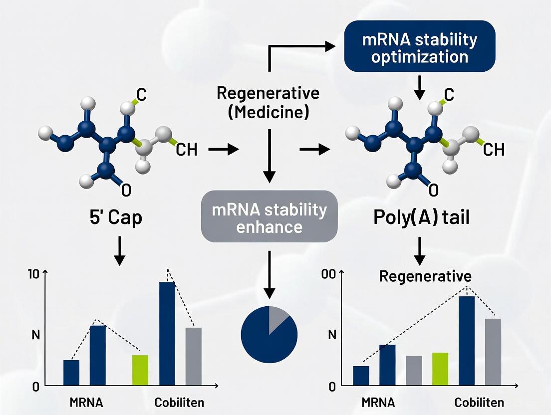
The Dynamic Duo: Foundational Roles of the 5' Cap and Poly(A) Tail in mRNA Biology
Core Concepts: The 5' Cap Structure and Mechanism
The 5' cap is a specially altered nucleotide found at the 5' end of eukaryotic messenger RNA (mRNA) and is essential for creating stable, mature mRNA capable of translation [1]. This structure is added co-transcriptionally, meaning it is modified while the RNA is still being synthesized by RNA polymerase II [2] [3].
Structural Evolution: Cap-0, Cap-1, and Cap-2
The core cap structure evolves through specific enzymatic steps, resulting in different forms with varying biological impacts [1] [4].
Table: 5' Cap Structures and Their Characteristics
| Cap Type | Chemical Structure | Key Features | Typical Occurrence |
|---|---|---|---|
| Cap-0 | m⁷GpppN | • 7-methylguanylate base structure• Protects from 5' exonucleases [1] | Base structure in all capped mRNAs [4] |
| Cap-1 | m⁷GpppNm | • Cap-0 + 2'-O-methylation of the first transcribed nucleotide (N) [1]• Reduces immunogenicity by identifying RNA as "self" [2] [5] | Common in higher eukaryotes and mammalian mRNA therapeutics [5] [4] |
| Cap-2 | m⁷GpppNmNm | • Cap-1 + 2'-O-methylation of the second transcribed nucleotide [1]• Further contributes to immune evasion [4] | Less common, found on some mammalian mRNAs [1] |
The following diagram illustrates the coordinated, multi-step enzymatic process that builds the 5' cap structure.
Essential Functions of the 5' Cap in mRNA Metabolism
The 5' cap is not merely a decorative end; it serves as a critical functional module recognized by specific cellular proteins throughout the mRNA life cycle [2]. Its four primary functions are:
- Regulation of Nuclear Export: The cap is bound by the nuclear Cap-Binding Complex (CBC), which is recognized by the nuclear pore complex, facilitating mRNA export to the cytoplasm [1].
- Prevention of Degradation: The cap protects the mRNA from 5' to 3' exonucleases [1] [3]. It also blocks the access of decapping enzymes, thereby increasing the mRNA's half-life [1].
- Promotion of Translation: In the cytoplasm, the CBC is replaced by the translation initiation factor eIF4E, which binds the cap and is part of the eIF4F complex. This complex recruits the ribosome to initiate protein synthesis [1] [2].
- Promotion of Splicing: The cap assists in the excision of introns, particularly the 5' proximal intron, by interacting with the spliceosome during RNA processing [1].
The diagram below shows how cap-binding proteins orchestrate different functions in the nucleus and cytoplasm.
Experimental Protocols for Capping In Vitro-Transcribed mRNA
For synthetic mRNA (in vitro-transcribed, IVT mRNA) used in therapeutics and research, proper capping is crucial for stability, translation efficiency, and avoiding undesirable immune responses [4]. There are two primary methods to achieve this.
Co-transcriptional Capping
This one-step method involves adding a cap analog directly to the IVT reaction mixture. The RNA polymerase incorporates this analog at the 5' end of the nascent RNA chain [5] [4].
Table: Common Cap Analogs for Co-transcriptional Capping
| Cap Analog | Mechanism & Features | Advantages | Disadvantages |
|---|---|---|---|
| Standard Cap (mCap) | • Dinucleotide (m⁷GpppG) [4]• Cap-0 structure | • Simple to use | • Can be incorporated in reverse orientation (inefficient translation) [4] |
| Anti-Reverse Cap Analog (ARCA) | • Modified m⁷GpppG to ensure proper forward incorporation [4] | • Higher translation efficiency than mCap• Straightforward protocol | • Still only produces Cap-0 structure |
| Trinucleotide Cap (e.g., CleanCap) | • Synthetic Cap-1 analog (m⁷GpppmN) [4] | • High capping efficiency (~94%)• Produces immunogenicity-reducing Cap-1 directly• Used in Pfizer-BioNTech COVID-19 vaccine [4] | • Higher cost than dinucleotide analogs |
Protocol: Co-transcriptional Capping with ARCA
- Reaction Setup: Combine the following components in a nuclease-free tube:
- Template DNA (with promoter for T7, SP6, or T3 RNA polymerase)
- NTPs (ATP, CTP, UTP, 1-1.5 mM each)
- GTP (0.5-1 mM)
- ARCA analog (4-6 mM, typically at 4:1 to 10:1 ratio over GTP)
- RNA polymerase (T7, SP6, or T3)
- RNase inhibitor
- Reaction buffer (with Mg²⁺ and DTT)
- Incubation: Incubate at 37°C for 1-2 hours.
- DNase Treatment: Add DNase I to digest the DNA template; incubate 15 min at 37°C.
- Purification: Purify the mRNA using standard methods (e.g., lithium chloride precipitation or chromatography).
Post-transcriptional Capping
This two-step method involves first synthesizing the uncapped mRNA, then using enzymes to add the cap. This typically requires viral capping enzymes like Vaccinia Capping Enzyme (VCE) or Faustovirus Capping Enzyme (FCE), followed by a 2'-O-Methyltransferase to create the Cap-1 structure [5] [4].
Protocol: Enzymatic Capping with VCE and 2'-O-MTase
- IVT Reaction: Perform a standard in vitro transcription reaction without any cap analog.
- DNase Treatment & Purification: Digest the DNA template and purify the uncapped mRNA.
- Capping Reaction: Set up the following reaction with the purified mRNA:
- Uncapped mRNA
- Vaccinia Capping Enzyme (VCE)
- GTP (as substrate for guanylylation)
- S-adenosylmethionine (SAM) (as methyl donor)
- Reaction buffer
- Incubate at 37°C for 30-60 minutes. This generates the Cap-0 structure.
- 2'-O-Methylation: Directly add mRNA Cap 2'-O-Methyltransferase (2'-O-MTase) to the same reaction tube without purification.
- Incubate for an additional 30-60 minutes at 37°C. This converts Cap-0 to Cap-1.
- Final Purification: Purify the capped mRNA for downstream applications.
The Scientist's Toolkit: Key Reagents for mRNA Capping
Table: Essential Reagents for mRNA Capping Workflows
| Reagent / Material | Function | Application Notes |
|---|---|---|
| Cap Analogs (ARCA, Trinucleotide) | Incorporated by RNA polymerase to create 5' cap during transcription [4] | Choice impacts capping efficiency, orientation fidelity, and final cap structure (Cap-0 vs. Cap-1) [4] |
| Vaccinia Capping Enzyme (VCE) | Catalyzes Cap-0 formation on 5' diphosphate RNA; possesses RNA triphosphatase, GTase, and guanine-N7 MTase activities [5] [4] | Used in post-transcriptional capping; Moderna's COVID-19 vaccine used VCE [4] |
| Faustovirus Capping Enzyme (FCE) | Broader temperature range and higher enzyme activity than VCE [4] | Alternative for post-transcriptional Cap-0 synthesis |
| 2'-O-Methyltransferase (2'-O-MTase) | Adds a methyl group to the 2'O position of the first nucleotide, converting Cap-0 to Cap-1 [5] [4] | Essential step in post-transcriptional capping to reduce immunogenicity |
| S-adenosylmethionine (SAM) | Serves as the methyl group donor for the methylation reactions catalyzed by methyltransferases [4] | Required for both enzymatic Cap-0 formation and 2'-O-methylation |
Troubleshooting and FAQs
FAQ 1: Why is my in vitro-transcribed mRNA yielding low protein expression, even though it is full-length?
- Potential Cause: Inefficient capping. Uncapped mRNA is poorly translated and unstable. In co-transcriptional capping, this can be due to low cap analog concentration or reverse incorporation of the cap.
- Solution:
- For co-transcriptional capping: Increase the cap analog to GTP ratio (e.g., 10:1). Switch to a superior cap analog like ARCA or a trinucleotide Cap-1 analog to ensure proper orientation and higher efficiency.
- For any method: Verify capping efficiency using techniques like LC-MS/MS, cap-specific ELISA, or by testing translation in a cell-free system compared to a commercially capped control RNA.
FAQ 2: My mRNA triggers a high innate immune response in cell culture, indicated by interferon activation. How can I reduce this?
- Potential Cause: The presence of double-stranded RNA (dsRNA) contaminants from IVT and/or an incomplete cap structure (e.g., Cap-0 instead of Cap-1). In higher eukaryotes, Cap-0 is recognized as "non-self" by the innate immune system [2] [5].
- Solution:
- Ensure you are generating a Cap-1 structure. Use either a trinucleotide analog co-transcriptionally or perform post-transcriptional capping with 2'-O-MTase.
- Rigorously purify your mRNA to remove dsRNA contaminants, such as by HPLC or FPLC.
- Incorporate modified nucleosides (e.g., N1-methylpseudouridine) during IVT to further dampen immune recognition.
FAQ 3: What is the best method for capping mRNA for therapeutic applications?
- Answer: Both methods are used in approved therapies, but the trend is toward high-efficiency Cap-1 formation.
- Co-transcriptional capping with trinucleotide analogs (e.g., CleanCap) is efficient (~94%), simple, and was successfully used in the Pfizer-BioNTech vaccine [4].
- Post-transcriptional enzymatic capping offers excellent control and reliably produces genuine Cap-1 structures, as used in the Moderna vaccine [4] [6]. The choice depends on the balance between simplicity, cost, and the specific requirement for capping efficiency and purity.
FAQ 4: How can I detect and quantify the cap structure on my synthesized mRNA?
- Answer: Several methods are available:
- LC-MS/MS: The gold standard. Can identify and precisely quantify the proportions of Cap-0, Cap-1, and Cap-2 structures in a sample [4].
- Cap-Specific ELISA: Uses antibodies specific to Cap-0 or Cap-1 for relative quantification. It is a high-throughput method [4].
- Differential Enzymatic Digestion & Gel Analysis: Uses enzymes like RNA 5' pyrophosphohydrolase (RppH) that selectively decap incomplete RNAs, allowing visualization of capped vs. uncapped populations by gel shift.
- RT-qPCR: Can be used to infer capping efficiency, as the 5' cap is necessary for the template-switching mechanism used by some reverse transcriptases [4].
Core Concepts: Poly(A) Tail Function and Regulation
What are the primary functions of the poly(A) tail?
The poly(A) tail is a stretch of adenosine nucleotides added to the 3′ end of messenger RNA (mRNA). It serves two critical functions:
- mRNA Stability: The tail protects the mRNA from degradation by exoribonucleases [7] [8]. Deadenylation (shortening of the tail) is often the first rate-limiting step in mRNA decay [7] [8].
- Translation Enhancement: The tail dramatically enhances the efficiency of translation initiation through a synergistic interaction with the 5′ cap structure [9] [10].
How does the cap-poly(A) tail synergy work, and what is the "closed-loop" model?
The 5' cap and 3' poly(A) tail cooperate to stimulate translation synergistically, meaning their combined effect is greater than the sum of their individual effects [9] [10]. This synergy is mediated by a specific protein complex that forms a "closed-loop" of the mRNA molecule.
The poly(A)-binding protein (PABP) bound to the tail interacts with the scaffolding protein eIF4G, which simultaneously binds the cap-binding protein eIF4E. This eIF4E•eIF4G•PABP complex effectively circularizes the mRNA [9] [10]. This configuration enhances the affinity of eIF4E for the 5' cap and facilitates the recycling of ribosomal subunits, thereby boosting translation initiation [10]. Recent evidence suggests that the intrinsic compactness of mRNA secondary structure helps bring the ends into proximity, further stabilizing this complex [9].
Figure 1: The Closed-Loop Model. The eIF4E•eIF4G•PABP complex bridges the 5' cap and 3' poly(A) tail, circularizing the mRNA to synergistically enhance translation initiation.
How is poly(A) tail length dynamically regulated?
Poly(A) tail length is not static; it is dynamically controlled by opposing enzymes and is a key point of post-transcriptional regulation, especially during rapid cell state transitions.
- Cytoplasmic Polyadenylation: Noncanonical poly(A) polymerases, such as TENT4A and TENT4B, can elongate tails in the cytoplasm to activate translation of specific mRNAs [11] [12]. This is crucial in processes like macrophage activation and early embryonic development [11].
- Deadenylation: Deadenylase enzymes shorten the tail, leading to translational repression and mRNA decay [11]. The RNA-binding protein ZFP36, for example, recruits deadenylation complexes to destabilize target mRNAs [11].
Troubleshooting FAQs and Guides
How can I resolve issues with cDNA synthesis from polyadenylated RNA?
Problems during reverse transcription (RT) can prevent the successful analysis of poly(A) tailed mRNA. The table below outlines common issues and solutions.
Table 1: Troubleshooting cDNA Synthesis for Poly(A) Tailed mRNA
| Problem | Possible Cause | Recommended Solution |
|---|---|---|
| Low cDNA yield | RNA secondary structure in GC-rich regions or 3' UTR | Denature RNA at 65°C for 5 minutes before RT. Use a thermostable reverse transcriptase and perform RT at a higher temperature (e.g., 50°C) [13]. |
| Low RNA integrity (degraded poly(A) tail) | Assess RNA integrity before cDNA synthesis. For degraded RNA, use random hexamers instead of oligo-dT primers to ensure coverage [13]. | |
| Non-specific amplification | Genomic DNA contamination | Treat RNA samples with DNase I before RT. Include a no-RT control in experiments [13]. |
| Incomplete cDNA representation | Inefficient priming from poly(A) tail | Optimize primer mixture and concentration (e.g., oligo-dT and random hexamers). For full-length cDNA synthesis, use a reverse transcriptase with high processivity and low RNase H activity [13]. |
What should I do if my in vitro transcribed mRNA translates poorly?
Inefficient translation can often be traced to improper 5' capping or a missing/short poly(A) tail.
- Verify 5' Capping Efficiency: Always use a capping method that yields a high percentage of Cap-1 structure, as this is critical for translation and reducing immunogenicity. Co-transcriptional capping with analogs like CleanCap can achieve over 95% efficiency [14]. For enzymatic capping, the newer Faustovirus Capping Enzyme (FCE) offers higher general activity and better performance on challenging substrates compared to traditional Vaccinia Capping Enzyme (VCE) [15].
- Ensure a Poly(A) Tail is Present: Include a poly(A) tail of sufficient length (typically 100-120 nucleotides) in your mRNA construct. The absence of a poly(A) tail severely impairs the cap-poly(A) synergy and thus translational output [9].
- Check for Inhibitors: After synthesis, clean up your mRNA to remove residual salts and enzymes that can inhibit downstream translation assays [16].
Why is understanding poly(A) tail dynamics important in disease research?
Dysregulation of poly(A) tail length is increasingly recognized as a key factor in pathogenesis. For example, a 2025 study on COVID-19 revealed significant alterations in poly(A) tail lengths and the incorporation of non-adenine residues in the host transcriptome of infected patients [12]. These modifications can influence mRNA stability and the host's antiviral response, providing new insights into disease mechanisms and potential therapeutic targets [12].
Experimental Protocols & Reagent Toolkit
Method for Analyzing Poly(A) Tail Length Dynamics (TED-seq)
To study tail length changes transcriptome-wide during a biological response (e.g., immune activation), you can use Tail End Displacement Sequencing (TED-seq) [11].
Workflow:
- Cell Stimulation & Harvest: Stimulate your model system (e.g., LPS-activated macrophages) and collect cells at relevant time points [11].
- RNA Extraction & Library Prep: Extract total RNA. Use TED-seq, which involves a precise size selection (e.g., 300 nt) of fragments that include the beginning of the poly(A) tail [11].
- Sequencing & Data Analysis: Sequence the libraries. The poly(A) tail length for each 3' UTR isoform is derived by calculating
300 nt - (distance from read 5' end to the polyadenylation site)[11]. Reads from transcripts with longer tails cluster closer to the cleavage site.
Figure 2: TED-seq Workflow. A protocol for genome-wide profiling of poly(A) tail lengths with 3' UTR isoform resolution [11].
Research Reagent Solutions
The following table lists key reagents and their functions for studying mRNA caps and poly(A) tails.
Table 2: Essential Reagents for mRNA Cap and Tail Research
| Reagent / Kit | Function / Application | Key Feature |
|---|---|---|
| CleanCap Analog (e.g., M6) [14] | Co-transcriptional mRNA capping. | >95% efficiency for Cap-1 structure; streamlines mRNA production. |
| Faustovirus Capping Enzyme (FCE) [15] | Enzymatic capping of synthetic mRNA. | Higher capping activity and broader temperature tolerance than VCE. |
| Vaccinia Capping Enzyme (VCE) [15] | Enzymatic capping of synthetic mRNA. | Established enzyme for generating Cap-0 structure; used with 2'-O-methyltransferase for Cap-1. |
| RNase 4 [15] | Analysis of 5' cap structures via LC-MS. | Simplifies workflow by allowing flexible cleavage sites, unlike RNase H. |
| TED-seq Protocol [11] | Genome-wide profiling of poly(A) tail length. | Provides isoform-specific tail length data with high accuracy. |
| Poly(A)-Binding Protein (PABP) | In vitro studies of translation and stability. | Core component of the closed-loop complex; available from multiple suppliers. |
| Tempus Blood RNA Tubes [12] | Stabilization of RNA in whole blood samples for transcriptomic studies. | Preserves RNA integrity for downstream analysis of tail dynamics in patient samples. |
Troubleshooting Guides
Issue 1: Low Translation Efficiency Despite High-Quality mRNA
- Problem: Your in vitro transcribed (IVT) mRNA is intact and pure, but protein expression in cell culture is low.
Possible Causes & Solutions:
Cause A: Inefficient 5' Capping
- Solution: Switch from co-transcriptional capping with analogs to post-transcriptional enzymatic capping. Enzymatic capping using Vaccinia Capping Enzyme (VCE) and 2'-O-Methyltransferase (2'-O-MTase) generates a natural Cap-1 structure, which is superior for translation and reduces immunogenicity compared to Cap-0 structures produced by many analogs [17] [18].
- Verification: Analyze the cap structure using techniques like mass spectrometry or HPLC to confirm the presence of the Cap-1 (m7GpppNm) structure.
Cause B: Disrupted Closed-Loop Formation
- Solution: Ensure the poly(A) tail is sufficiently long and bound by PABPC. The cytoplasmic poly(A)-binding protein (PABPC1) binds the poly(A) tail and interacts with translation initiation factors like eIF4G at the 5' cap, forming a closed-loop complex that is critical for efficient translation initiation and ribosome recycling [19] [1].
- Verification: Check poly(A) tail length using RNase H/Oligo(dT) Northern blot analysis or PCR-based methods (e.g., PAT assays) [20]. Aim for tails of ~70 nt in yeast and ~200 nt in mammals for optimal initial function [19].
Issue 2: Inconsistent mRNA Stability and Half-Life
- Problem: mRNA degradation rates are highly variable between experiments, leading to unpredictable protein expression duration.
Possible Causes & Solutions:
Cause A: Uncontrolled Deadenylation
- Solution: Engineer the mRNA's 3' end to modulate deadenylation rates. Incorporating specific secondary structures (e.g., stem-loops) or non-adenosine nucleotides (like uridines) into the poly(A) tail region can impede the process of deadenylation by complexes like CCR4-NOT and Pan2-Pan3, thereby enhancing mRNA stability [21] [22].
- Verification: A recent study demonstrated that a poly(A) tail with an internal loop structure (A50L50LO) significantly enhanced luciferase expression and mRNA stability in vivo compared to standard linear poly(A) tails [22].
Cause B: Non-Canonical RNA Tailing
- Solution: Be aware that uridylation, not just adenylation, can occur on the 3' end. Uridylation by enzymes like TENT3A/B is a potent signal for rapid mRNA decay via the exoribonuclease Dis3L2 [21]. Ensure your mRNA sequence and structure do not inadvertently promote this pathway.
- Verification: Use 3'-end sequencing techniques (e.g., TAIL-seq) to detect the presence of non-A nucleotides (U, G, C) in the tail, which are hallmarks of mixed tailing and decay signals [21].
Issue 3: Poor In Vivo Performance of mRNA Therapeutics
- Problem: mRNA candidates perform well in vitro but show weak and short-lived expression in animal models.
Possible Causes & Solutions:
Cause A: mRNA Instability During Storage and Delivery
- Solution: Utilize advanced lipid nanoparticle (LNP) formulations that incorporate lyoprotectants. A 2025 study showed that trehalose-loaded LNPs (TL-LNPs) preserve mRNA chemical integrity during lyophilization and storage. The co-delivered trehalose also reduces oxidative stress in transfected cells, bridging the in vitro-in vivo efficacy gap [23].
- Verification: Characterize LNP size, PDI, and encapsulation efficiency post-lyophilization. Test in vivo transfection efficiency after storage to confirm stability.
Cause B: Suboptimal Structural Elements for In Vivo Environment
- Solution: Incorporate RNA stability enhancer elements. A high-throughput screen identified viral-derived elements (e.g., "A7") that recruit TENT4 to extend the poly(A) tail, preventing deadenylation and resulting in durable, high-level protein expression in mouse liver, surpassing the performance of circular RNA [24].
- Verification: Clone candidate stability elements into the 3' UTR of your mRNA construct and measure protein expression over time in vivo compared to a control.
Frequently Asked Questions (FAQs)
Q1: What is the definitive evidence for the closed-loop model of translation? The model is supported by extensive biochemical and genetic data. Key evidence includes the identification of the physical interaction between the poly(A)-binding protein (PABPC) on the tail and the translation initiation factor eIF4G, which is itself bound to the 5' cap-binding protein eIF4E. This complex circularizes the mRNA, synergistically enhancing translation initiation and protecting both ends from decay [19] [1].
Q2: For in vitro transcription, is co-transcriptional or post-transcriptional capping better for my mRNA vaccine research? Post-transcriptional enzymatic capping is generally superior for therapeutic applications. While co-transcriptional capping with analogs is faster, it can lead to incomplete capping and reverse-orient cap incorporation. Enzymatic capping ensures a nearly 100% correct Cap-1 structure, which maximizes translation and minimizes unwanted immune recognition by the host [17] [18].
Q3: How long should the poly(A) tail be for optimal expression in mammalian cells? While historically thought to be 150-250 nucleotides, recent studies suggest the relationship is more complex. For initial translation, a longer tail (~120-150 nt) is beneficial as it can bind more PABPC molecules. However, research in mRNA vaccines has shown that tail structure (e.g., incorporating loop-forming sequences) can be as important as length alone for maximizing stability and expression [22] [20]. The optimal length may also vary with cell type and mRNA sequence.
Q4: Besides adenosines, what other nucleotides can be found in poly(A) tails and why does it matter? Sequencing studies have revealed "mixed tails" containing uridines (U), guanosines (G), and cytosines (C). A single guanosine residue within a poly(A) tail can transiently stall deadenylation, while uridylation is a clear signal to recruit decay machinery like the exoribonuclease Dis3L2 [21]. Therefore, non-A nucleotides are critical regulators of mRNA half-life.
Q5: How can I experimentally measure the poly(A) tail length of my mRNA of interest? Common methods include:
- RNase H/Oligo(dT) Northern Blot: A direct, gel-based method that avoids PCR bias but requires abundant mRNA [20].
- PCR-based PAT assays (e.g., LM-PAT, ePAT): These are more sensitive and suitable for low-abundance transcripts but can be biased towards amplifying shorter tails [20].
- Next-Generation Sequencing (e.g., TAIL-seq): Provides genome-wide, high-resolution tail length data but is more complex and costly [19] [21].
Data Presentation
Table 1: Impact of Poly(A) Tail Structure on mRNA Efficacy
Data derived from in vitro and in vivo studies comparing different poly(A) tail designs in mRNA platforms [22].
| Poly(A) Tail Design | Description | Relative Luciferase Expression (in vivo, 24h) | Key Finding |
|---|---|---|---|
| A50L50LO | 50A + Linker + 50A with complementary linker sequence (forms a loop) | Highest | A loop structure within the poly(A) tail region significantly enhances stability and sustained protein expression. |
| A30L70 | 30A + Linker + 70A (linear, positive control) | Intermediate | A standard, linear design used in commercial vaccines, showing good performance. |
| A120 | 120 adenosine residues (linear) | Lower | A long, linear tail is less effective than structured tails for maintaining prolonged expression. |
Table 2: Comparison of mRNA 5' Capping Methods
Summary of key characteristics for co-transcriptional and post-transcriptional capping strategies [17] [18].
| Capping Method | Principle | Cap Structure | Advantages | Disadvantages |
|---|---|---|---|---|
| Co-transcriptional | Cap analogs (e.g., m7GpppG) are added to the IVT reaction. | Cap-0 (m7GpppN...) | Simple, one-step process. | Lower capping efficiency; risk of reverse cap incorporation. |
| Post-transcriptional Enzymatic | IVT mRNA is enzymatically processed using VCE and 2'-O-MTase. | Cap-1 (m7GpppNm...) | High-fidelity, >99% capping efficiency; superior translation; lower immunogenicity. | Two-step process; higher cost and longer time. |
Experimental Protocols
Protocol 1: Assessing Poly(A) Tail Length by RNase H/Oligo(dT) Northern Blot
This protocol allows for direct measurement of tail length without PCR amplification biases [20].
- RNA Preparation: Isolate high-quality, intact total RNA from your cells or tissue.
- RNase H Cleavage:
- Set up two reactions for each sample:
- Reaction 1 (Test): RNA + Oligo(dT)~20~ + Gene-Specific Primer (optional).
- Reaction 2 (Control): RNA + Gene-Specific Primer only (no Oligo(dT)).
- Add RNase H buffer and RNase H enzyme. Incubate to cleave RNA-DNA hybrids.
- Set up two reactions for each sample:
- Gel Electrophoresis and Northern Blot:
- Run the cleavage products on a denaturing agarose or polyacrylamide gel.
- Transfer the RNA to a membrane.
- Hybridize the membrane with a radiolabeled or digoxigenin-labeled probe specific to your target mRNA.
- Analysis:
- The control reaction shows the full-length fragment. The test reaction, cleaved within the poly(A) tail, produces a shorter fragment.
- The difference in size between the two fragments corresponds to the length of the poly(A) tail.
Protocol 2: Evaluating mRNA Stability Using Novel Poly(A) Tail Designs
This protocol outlines the steps to test engineered poly(A) tails, as demonstrated in recent vaccine research [22].
- mRNA Construct Design:
- Design mRNA constructs encoding your protein of interest (e.g., luciferase, antigen).
- Clone different poly(A) tail sequences downstream of the ORF and 3' UTR. Examples include:
- A~120~: A simple, long adenosine tail.
- A~30~L~70~: A tail with a linker sequence, mimicking designs used in commercial vaccines.
- A~50~L~50~LO: A tail designed with a complementary linker sequence to form an internal loop structure.
- In Vitro Transcription and Capping:
- Synthesize mRNA in vitro using a kit like the Takara IVTpro system.
- Perform post-transcriptional enzymatic capping using VCE and 2'-O-MTase to ensure a uniform Cap-1 structure [17].
- Purify the mRNA to remove dsRNA contaminants and impurities.
- In Vivo Expression Analysis:
- Formulate the purified mRNAs into LNPs.
- Administer the mRNA/LNPs intramuscularly or intravenously to mice (e.g., C57BL/6).
- Measure protein expression over time (e.g., at 6h, 24h, 48h) using non-invasive imaging (for luciferase) or ELISA (for secreted proteins like hEPO) from serum samples.
Mandatory Visualization
Diagram 1: The mRNA Closed-Loop Model
This diagram illustrates the key proteins and interactions that circularize mRNA, driving efficient translation.
Diagram 2: Workflow for Testing mRNA Stability
This diagram outlines the key steps for an experiment designed to test how different poly(A) tail structures affect mRNA stability and protein expression.
The Scientist's Toolkit
Table 3: Key Research Reagent Solutions
Essential materials and their functions for optimizing mRNA 5' cap and poly(A) tail research.
| Item | Function/Benefit | Example Use Case |
|---|---|---|
| Vaccinia Capping Enzyme (VCE) | Adds a 7-methylguanylate cap (m7G) to the 5' end of RNA. | The first step in creating a high-fidelity Cap-0 structure during post-transcriptional capping [17]. |
| mRNA Cap 2'-O-Methyltransferase (2'-O-MTase) | Methylates the first nucleotide adjacent to the cap, converting Cap-0 to Cap-1. | Used after VCE to create the immunologically stealthy and highly translatable Cap-1 structure [17] [18]. |
| Poly(A) Polymerase | Enzymatically adds a poly(A) tail to the 3' end of RNA. | Useful for adding defined, homogenous poly(A) tails to IVT mRNA in a post-transcriptional step [21]. |
| Lipid Nanoparticles (LNPs) | A delivery system that protects mRNA from degradation and facilitates cellular uptake. | Essential for in vivo delivery of mRNA constructs to test the efficacy of different cap and tail designs [22] [23]. |
| Trehalose | A lyoprotectant that stabilizes mRNA during freeze-drying and can reduce oxidative stress in cells. | Can be co-loaded with mRNA in LNPs (TL-LNP) to enhance stability during storage and improve in vivo transfection efficiency [23]. |
| Takara IVTpro mRNA Synthesis System | A commercial system for in vitro transcription, compatible with both co-transcriptional and post-transcriptional capping. | Provides a streamlined workflow for synthesizing high-quality, capped IVT mRNA for research [17]. |
For mRNA stability research, optimization has traditionally focused on the 5' cap and the length of the 3' poly(A) tail. However, emerging research reveals that incorporating secondary structures, specifically loops, into the poly(A) tail region can significantly enhance mRNA stability and translational efficiency. This technical support center provides researchers with practical guidance for implementing these novel structural designs, troubleshooting common experimental challenges, and validating the performance of advanced mRNA constructs.
FAQ: Loop Structures in Poly(A) Tails
What are the proposed benefits of adding loop structures to poly(A) tails? Incorporating loop structures into poly(A) tails improves mRNA stability and increases protein expression levels. Research demonstrates that a designed loop structure (A50L50LO) exhibited higher bioluminescence signals both in vitro and in vivo, and increased human erythropoietin (hEPO) expression in mouse models compared to linear poly(A) tail controls. This indicates enhanced mRNA stability and translational efficiency [25].
How do loop structures mechanistically enhance mRNA stability? Loop structures containing secondary structure and double-stranded RNA (dsRNA) characteristics can stabilize the poly(A) tail by impeding deadenylation mediated by the CCR4–NOT complex. Certain viral transcripts incorporate non-adenosine ribonucleotides or structural elements to similarly inhibit deadenylation and enhance stability, a principle applied in these engineered designs [25].
Does increased protein expression from loop-structured tails always enhance immune responses? Not necessarily. While studies show loop structures increase antigen expression, the cellular and humoral immune responses against HPV E6/E7 and influenza HA antigens did not show statistically significant differences across different poly(A) tail structures, despite the observed differences in protein expression levels [25].
What are the primary methods for adding poly(A) tails to IVT mRNA? There are two main methods, each with distinct advantages and limitations [26]:
| Method | Advantages | Disadvantages |
|---|---|---|
| Template-Encoded | Consistent, defined tail length; one-step reaction; suitable for large-scale production. | Difficult synthesis of long nucleotide stretches; unstable in plasmids; unable to change tail after cloning. |
| Enzymatic | Flexible length modulation; can be applied to already synthesized RNA. | Requires extra reaction step; higher cost; variable tail length; less suitable for large-scale production. |
What quality control methods are recommended for poly(A) tail analysis? Due to the repetitive nature of poly(A) sequences, traditional Sanger sequencing is often insufficient. Liquid chromatography-mass spectrometry (LC-MS) provides high-resolution data on poly(A) tail length and heterogeneity. Next-generation sequencing (NGS) and capillary electrophoresis are also used, though NGS may introduce PCR biases [26].
Troubleshooting Guides
Problem 1: Inconsistent Protein Expression from mRNA Constructs
Potential Cause: Heterogeneous poly(A) tail lengths in your mRNA preparations.
Solutions:
- Switch Tailing Method: Utilize template-encoded poly(A) tails instead of enzymatic tailing to generate more consistent tail lengths across mRNA batches [26].
- Verify Template Integrity: If using template-encoded tails, sequence the plasmid DNA after amplification to check for bacterial recombination that can cause truncation in long repetitive sequences. Consider using optimized DNA preparation protocols or segmenting poly(A) regions with short spacers to improve plasmid stability [27].
- Implement Rigorous QC: Adopt LC-MS for accurate determination of poly(A) tail molecular weight and length distribution, providing superior resolution over traditional methods [26].
Problem 2: Poor mRNA Stability in Solution
Potential Cause: Accelerated hydrolytic degradation due to suboptimal sequence and structure.
Solutions:
- Optimize Sequence/Structure: Leverage high-throughput methods like In-line-seq to understand sequence and structure-based rules that mitigate hydrolytic degradation. Designs with optimized secondary structure can significantly improve in-solution stability [28].
- Incorporate Modified Nucleotides: Use pseudouridine (ψ) modification during in vitro transcription. This modification has been shown to enhance protection against nucleases and improve overall mRNA stability [27].
- Utilize "Superfolder" mRNAs: Design highly structured "superfolder" mRNAs that have been demonstrated to simultaneously improve both stability and protein expression [28].
Problem 3: Low Transfection Efficiency or Protein Output
Potential Cause: Suboptimal poly(A) tail length or structure failing to facilitate efficient translation.
Solutions:
- Optimize Tail Length: Test poly(A) tails of different lengths. Research indicates that a tail length of approximately 100 nucleotides is often optimal for maximal protein expression in therapeutic contexts, with longer tails not necessarily providing further benefits [26].
- Engineer Loop Structures: Design a poly(A) tail with an internal complementary linker sequence (e.g., A50-Linker-A50-complementary linker) that forms a stabilizing loop structure, which has been shown to outperform standard linear tails [25].
- Consider Codon Optimization: Optimize the coding sequence (CDS) by replacing rare codons with more frequently used synonymous codons. This enhances translation efficiency without altering the amino acid sequence of the encoded protein [27].
Experimental Protocols & Data Analysis
Protocol: Designing and Testing a Loop-Structured Poly(A) Tail
This protocol is adapted from research that successfully demonstrated enhanced stability and expression using loop structures in poly(A) tails [25].
1. Design and In Silico Prediction
- Sequence Design: Design a poly(A) tail sequence such as "A50-Linker-A50-complementary linker" (A50L50LO). The complementary linker sequence allows the formation of a small loop structure within the tail.
- Folding Prediction: Perform RNA folding analysis using programs like RNAfold to predict secondary structure formation. Confirm that sequences with complementary sequences form the intended small loops.
2. mRNA Construct Assembly
- Vector Construction: Clone the poly(A) tail sequence into your IVT vector along with your gene of interest.
- Stability Assessment: Verify the stability of the plasmid in bacterial hosts by restriction digest and gel electrophoresis. Smearing on the gel indicates unstable poly(A) by-products.
- In Vitro Transcription: Synthesize mRNA using T7 RNA polymerase. Include a CleanCap analog for proper 5' capping and use N1-methylpseudouridine triphosphate instead of UTP to reduce immunogenicity.
3. In Vitro and In Vivo Testing
- Cell Culture Transfection: Transfert multiple cell lines (e.g., HeLa, A549) with equal amounts of mRNA constructs (e.g., 500 ng/well). Include controls with standard poly(A) tails (e.g., A120, A30L70).
- Expression Analysis: Measure protein output at various time points (e.g., 6, 24, 48 hours) using appropriate assays (e.g., luminescence for luciferase reporters, ELISA for specific proteins).
- In Vivo Validation: Formulate mRNA into Lipid Nanoparticles (LNPs). Administer to animal models (e.g., 5 μg intramuscularly to C57BL/6 mice) and monitor protein expression over time using imaging systems or serum analysis.
Quantitative Data from Key Studies
Table 1: Protein Expression Comparison of Poly(A) Tail Designs Data derived from luciferase and hEPO expression studies in vitro and in vivo [25]
| Poly(A) Tail Design | Description | Relative Luminescence (HeLa, 24h) | Relative hEPO Expression (in vivo, 6h) |
|---|---|---|---|
| A50L50LO | Loop structure with complementary linker | Highest | Highest |
| A30L70 | Linear, segmented tail (BioNTech control) | High | High (slightly lower than A50L50LO) |
| A50L50LX | Linear structure with non-complementary linker | Moderate | Moderate |
| A120 | Linear adenosine tail | Lower | Lower |
Table 2: Essential Research Reagents for mRNA Stability Work A toolkit of key materials and their functions for developing stabilized mRNA constructs.
| Reagent / Material | Function / Application | Key Consideration |
|---|---|---|
| T7 RNA Polymerase | In vitro transcription of mRNA from DNA template | High yield and fidelity is critical [25]. |
| CleanCap Reagent | Co-transcriptional capping to produce Cap 1 structure | Essential for evading innate immune recognition [29]. |
| N1-methylpseudouridine (m1Ψ) | Modified nucleotide for IVT; reduces immunogenicity, increases translation | Superior to pseudouridine; Nobel Prize-winning technology [29] [27]. |
| Poly(A) Polymerase | Enzymatic addition of poly(A) tail post-transcription | Allows flexible tail length optimization [26]. |
| Lipid Nanoparticles (LNPs) | Delivery vehicle for in vivo mRNA transport; enhances stability | Protects mRNA from degradation; composition affects stability [30]. |
| LC-MS Instrumentation | High-resolution quality control of poly(A) tail length | Provides accurate molecular weight data, superior to Sanger sequencing for repetitive sequences [26]. |
Diagnostic and Optimization Workflows
Decision Flow: Selecting a Poly(A) Tailing Strategy
Workflow: Experimental Validation of mRNA Stability
Key Takeaways for Researchers
- Loop Structures Enhance Stability: The A50L50LO design (A50-Linker-A50 with complementary linker) demonstrates that engineered secondary structures in the poly(A) tail region can significantly improve mRNA stability and protein expression compared to traditional linear tails [25].
- Tail Length is Necessary but Not Sufficient: While an optimal poly(A) tail length (typically ~100 nucleotides) is fundamental, it does not preclude the additional benefits gained from structural optimization within the tail [26].
- Rigorous QC is Non-Negotiable: The repetitive nature of poly(A) tails makes them prone to recombination and heterogeneity. Implementing advanced QC methods like LC-MS is essential for reliable results [26].
- Expression vs. Immunogenicity is Complex: While loop structures increase protein expression, this does not always directly translate to enhanced immune responses, indicating that other factors are at play in vaccine efficacy [25].
From Theory to Therapy: Methodological Strategies for 5' Cap and Poly(A) Tail Design
The 5' cap is a fundamental structure for messenger RNA (mRNA), critically governing its stability, translational efficiency, and immunogenicity [31] [32] [33]. This modified guanosine nucleotide, linked to the mRNA's 5' end via a 5'-5' triphosphate bridge, is recognized by the eukaryotic translation initiation factor eIF4E, facilitating ribosome assembly and protein synthesis [32]. Concurrently, the cap protects the mRNA from exonucleolytic degradation and, together with the poly(A) tail, helps distinguish self-RNA from non-self RNA, thereby reducing unwanted immune activation [33] [34]. Despite its critical role, the native cap structure is vulnerable to enzymatic decapping, a key regulatory step in mRNA turnover. Decapping enzymes, such as the Dcp1/Dcp2 complex, catalyze the removal of the cap, committing the mRNA to irreversible 5'→3' degradation [31]. For researchers developing mRNA therapeutics, overcoming this inherent instability is a major hurdle. The emergence of cap analogs resistant to decapping and with high affinity for eIF4E is therefore a central focus in the field, aiming to create more stable and potent mRNA-based drugs and vaccines [31] [35] [36].
FAQ: Troubleshooting Common Experimental Issues
Q1: My in vitro transcribed mRNA shows poor translation efficiency despite high capping efficiency. What could be the cause? Poor translation can result from the use of cap analogs with low affinity for eIF4E or those that are incorporated in the reverse orientation during transcription. Ensure you are using anti-reverse cap analogs (ARCAs), which are chemically modified (e.g., with a 3'-O-methyl group) to ensure incorporation in the correct orientation [31] [34]. Additionally, check the integrity of your cap structure itself. Recent studies show that even capped mRNA can contain degradation impurities, such as imidazole ring-opened m7G or hydrolyzed triphosphate bridges, which significantly reduce protein expression without triggering immune alerts in standard assays [33]. Using more stable cap analogs or adjusting IVT conditions to minimize hydrolysis can resolve this.
Q2: My mRNA triggers a high immune response in cell culture. How can I reduce its immunogenicity? High immunogenicity is frequently caused by uncapped mRNA impurities in your preparation, which present 5'-triphosphates that are recognized by innate immune receptors like RIG-I [34]. To address this, strive for higher capping efficiency. Consider switching to trinucleotide cap analogs (e.g., CleanCap) that enable direct Cap-1 incorporation and can achieve capping efficiencies >90% [35] [34]. For the purest product, novel technologies like PureCap analogs allow for physical separation of capped from uncapped mRNA via RP-HPLC, achieving nearly 100% capping efficiency and eliminating this source of immunogenicity [34].
Q3: How can I experimentally determine if my modified cap analog is resistant to decapping? Resistance to decapping is typically validated through in vitro decapping assays. Purify your capped mRNA and incubate it with recombinant human decapping enzyme hDcp2. Analyze the reaction products over time using analytical techniques like liquid chromatography-mass spectrometry (LC-MS) to detect and quantify the release of m7GDP, the signature product of hDcp2 activity [31] [33]. A cap analog resistant to decapping will show significantly reduced m7GDP production compared to a standard cap control like m7GpppG.
Q4: What are the key considerations when choosing a cap analog for therapeutic mRNA development? Selecting a cap analog involves balancing several properties:
- Decapping Resistance: Look for analogs with modifications like boranophosphate (BH3) or imidodiphosphate (NH) that show superior resistance to hDcp2 [31].
- eIF4E Binding Affinity: The analog must have high affinity for eIF4E to ensure efficient translation initiation. This can be measured by techniques like microscale thermophoresis (MST) [32].
- Capping Efficiency: The analog should be efficiently incorporated by RNA polymerase during IVT. Trinucleotide analogs often outperform dinucleotides here [35].
- Immunogenicity Profile: Ensure your mRNA preparation has minimal uncapped impurities. Cap structures like Cap-2 (with two 2'-O-methylations) further reduce immune recognition compared to Cap-1 [34].
- Overall mRNA Stability: The ultimate test is the half-life of your mRNA in relevant cell lines. Analogs like m7GppBH3pm7G have demonstrated enhanced stability in HeLa cells [31].
Research Reagent Solutions
The table below summarizes key reagents for working with advanced 5' cap analogs.
Table 1: Essential Research Reagents for Cap Analog Studies
| Reagent / Material | Function / Application | Key Examples / Notes |
|---|---|---|
| Advanced Cap Analogs | Prime IVT reactions to produce mRNA with enhanced properties. | m7GppBH3pm7G (stability) [31]; α-phosphorothiolate trinucleotides (translation) [35]; PureCap analogs (purification) [34]. |
| High-Efficiency RNA Polymerase | Engineered enzyme for superior co-transcriptional capping efficiency. | Codex HiCap RNA Polymerase (>95% capping with lower dsRNA byproducts) [37]. |
| Decapping Enzymes | In vitro assessment of cap stability and resistance. | Recombinant hDcp2 (Dcp1/Dcp2 complex) for decapping assays [31]. |
| eIF4E Protein | Measure binding affinity of novel cap analogs. | Recombinant protein for MST or SPR binding studies [32]. |
| LC-MS Systems | Characterize cap integrity and identify degradation products. | Ion-pair RP-UPLC-MS for profiling capped, uncapped, and degraded 5' ends [33]. |
Experimental Protocols
Protocol: Assessing Cap Analog Resistance to hDcp2 Decapping In Vitro
Objective: To quantify the resistance of a novel cap analog to hydrolysis by the human decapping enzyme hDcp2.
Materials:
- Purified mRNA capped with the test analog and a control (e.g., m27,2'-OGppSpG, D2).
- Recombinant hDcp2 enzyme (commercially available).
- Decapping reaction buffer (e.g., 50 mM HEPES pH 7.5, 100 mM NaCl, 2 mM MgCl2, 1 mM DTT).
- LC-MS system with reverse-phase capability.
Method:
- Set Up Reactions: In a reaction tube, combine 1 µg of capped mRNA with recombinant hDcp2 in decapping buffer. Include a no-enzyme control for each mRNA.
- Incubate: Allow the reaction to proceed at 37°C for a predetermined time (e.g., 30-60 minutes).
- Terminate Reaction: Heat-inactivate the enzyme at 70°C for 10 minutes.
- Analyze Products: Inject the reaction mixture into the LC-MS. Monitor for the production of m7GDP, the characteristic product of hDcp2 cleavage between the α- and β-phosphates [31] [33].
- Quantify Resistance: Compare the peak area or concentration of m7GDP released from the test analog-capped mRNA to that released from the control mRNA. A significant reduction indicates higher decapping resistance.
Protocol: Evaluating Translational Efficiency of Capped mRNA in Cell Culture
Objective: To compare the protein expression output of mRNAs capped with different analogs.
Materials:
- mRNAs (e.g., encoding luciferase) capped with different analogs, purified to high purity.
- Appropriate cell line (e.g., HeLa, A549).
- Transfection reagent.
- Protein quantification assay (e.g., luciferase assay kit, ELISA).
Method:
- Cell Seeding: Seed cells in a multi-well plate to reach 70-80% confluence at the time of transfection.
- Transfect mRNA: Transfect a fixed, equimolar amount of each mRNA into the cells using a standard transfection protocol.
- Incubate: Culture the cells for an appropriate time to allow for translation (e.g., 6-24 hours).
- Harvest and Quantify: Lyse the cells and quantify the protein of interest using your chosen assay.
- Normalize and Analyze: Normalize the protein data to total protein concentration or a housekeeping gene. Compare the relative luminescence/fluorescence units across the different cap analogs to determine which supports the highest translational efficiency [31] [34].
Diagrams and Workflows
mRNA Lifecycle and Decapping Pathways
Diagram 1: mRNA turnover pathway and decapping role.
Advanced Cap Analog Development Workflow
Diagram 2: Development and testing workflow for novel cap analogs.
Core Concepts: Poly(A) Tail Architectures
The poly(A) tail is a critical determinant of mRNA stability, translation efficiency, and overall therapeutic efficacy. While traditional designs have focused on linear adenosine sequences, recent advances demonstrate that incorporating specific structural elements can significantly enhance mRNA performance.
Linear Poly(A) Tails: These are homopolymeric sequences of adenosine residues. Their length is a key factor, with longer tails (e.g., 100-120 nucleotides) generally allowing for the binding of more Poly(A)-Binding Proteins (PABPs), which helps protect the mRNA from degradation and enhances translation initiation [22] [18]. A common commercial benchmark is the A30L70 design (A30-Linker-A70) used in licensed vaccines [22].
Structured Poly(A) Tails: These designs incorporate non-adenosine elements to create secondary structures. A prominent example is the A50L50LO design, which consists of an A50 sequence, a linker, another A50 sequence, and a complementary linker sequence that forms a stable loop structure [22]. This architecture impedes the activity of deadenylase complexes (like CCR4–NOT) that shorten the tail, thereby prolonging the mRNA's functional lifespan in the cytoplasm [22].
Table 1: Key Characteristics of Poly(A) Tail Architectures
| Architecture | Description | Postulated Mechanism of Action |
|---|---|---|
| Linear (e.g., A120) | A single, continuous run of adenosine residues. | Binds PABPs in a "beads on a string" manner to support translation initiation and provide basal stability [22] [38]. |
| Benchmark Linear (e.g., A30L70) | Two adenosine tracts (A30 and A70) separated by a short linker. | Functions similarly to a linear tail; the linker may help ensure accurate tail length during synthesis [22]. |
| Structured Loop (e.g., A50L50LO) | Two A50 tracts flanking a linker sequence that is complementary to an adjacent sequence, forming a loop. | The terminal loop structure acts as a physical barrier to 3'-5' exonuclease activity, slowing deadenylation and decay [22]. |
| Chemically Modified Tail | A linear poly(A) tail with a proprietary chemical moiety added to the 3'-end post-synthesis (e.g., TriLink's ModTail). | Hypothesized to prevent 3’-exonuclease cleavage, thereby extending the duration of protein expression [39]. |
Troubleshooting Guide: Poly(A) Tail Optimization
Problem 1: Low or Transient Protein Expression
Potential Cause: Inefficient poly(A) tail failing to protect mRNA from rapid degradation or recruit PABPs effectively.
Solutions:
- Consider a Structured Design: Switch from a linear A120 tail to an A50L50LO architecture. One study showed this design exhibited higher and more sustained bioluminescence signals in vivo compared to A30L70 and A120 tails [22].
- Verify Tail Length and Integrity: Use analytical techniques like capillary electrophoresis to confirm the length and homogeneity of the poly(A) tail after in vitro transcription (IVT) and purification.
- Explore Commercial Modifications: Investigate tail modification services like TriLink's ModTail, which has been shown in murine models to increase the duration of protein expression for at least 72 hours post-injection [39].
Problem 2: Inconsistent Experimental Results Between Cell Lines
Potential Cause: Cell-type-specific differences in deadenylase activity or RNA-binding protein abundance.
Solutions:
- Empirically Test Architectures: Test multiple poly(A) tail designs in your specific cell models. Research indicates that while the A50L50LO tail consistently promoted high expression across four tested cell lines (Nor10, HeLa, A549, HepG2), the performance of other tails like A30L70 was more variable [22].
- Include a Positive Control: Always use a validated, high-performing construct (e.g., one with a known effective tail architecture) as a benchmark in your experiments to control for cell health and transfection efficiency.
Problem 3: Unintended Immune Activation
Potential Cause: While often linked to dsRNA impurities from IVT, certain complex RNA structures could potentially be sensed by pattern recognition receptors.
Solutions:
- Purify mRNA Rigorously: Use HPLC or FPLC purification to remove aberrant IVT products like double-stranded RNA (dsRNA) impurities, which are a primary trigger of innate immunity.
- Assess Immunogenicity: For novel tail designs, measure innate immune activation using assays like interferon-beta ELISA or sequencing of interferon-stimulated genes. One study on loop-structured tails found no significant difference in immune cell activation or cytokine profiles compared to standard tails [22].
Experimental Protocols
Protocol 1: Evaluating Tail Architectures Using a Luciferase Reporter
Objective: To compare the stability and translational efficiency of different poly(A) tail designs in a standardized system.
Materials:
- Plasmids: DNA templates for IVT containing the firefly luciferase (F/L) gene flanked by optimized UTRs, followed by the poly(A) tail sequence to be tested (e.g., A120, A30L70, A50L50LO) [22].
- IVT Kit: In vitro transcription kit with a cap analog (e.g., CleanCap).
- Delivery Vehicle: A transfection reagent (e.g., Lipofectamine MessengerMAX) or lipid nanoparticles (LNPs).
- Cell Line: Adherent cells such as HEK293T or HeLa.
- Detection Instrument: Luminescence plate reader or live-cell imager (e.g., Incucyte).
Method:
- Synthesize mRNA: Perform IVT to produce the various luciferase mRNAs. Purify the mRNA using a method that ensures integrity (e.g., oligo-dT purification).
- Transfect Cells: Seed cells in a 96-well plate. Transfect with a standardized amount (e.g., 500 ng/well) of each mRNA construct.
- Measure Expression: Quantify luminescence at multiple time points (e.g., 6, 24, 48 hours post-transfection).
- Analyze Data: Plot luminescence over time. A construct that maintains a higher signal at later time points indicates improved mRNA stability conferred by the poly(A) tail.
Protocol 2: In Vivo Assessment of Protein Expression Duration
Objective: To determine the impact of poly(A) tail architecture on the duration of protein expression in a live animal model.
Materials:
- mRNA-LNPs: Formulate the test mRNAs (e.g., encoding luciferase or human Erythropoietin (hEPO)) into Lipid Nanoparticles (LNPs) of consistent size and composition [22] [39].
- Animal Model: C57BL/6 mice.
- Imaging/Assay System: In Vivo Imaging System (IVIS) for bioluminescence or ELISA kit for hEPO quantification.
Method:
- Administer mRNA: Inject mice intramuscularly or intravenously with a standardized dose of each mRNA-LNP formulation.
- Monitor Expression:
- Analyze Data: Compare the signal intensity and persistence across the different poly(A) tail groups. The area under the curve (AUC) for signal over time is a useful metric for overall performance.
Workflow Visualization
The following diagram illustrates the key decision points and experimental workflow for optimizing poly(A) tail architecture.
The Scientist's Toolkit: Research Reagent Solutions
Table 2: Essential Reagents and Resources for Poly(A) Tail Research
| Reagent / Resource | Function / Description | Example Use Case |
|---|---|---|
| DNA Template with Tail Variant | Plasmid linearized downstream of the poly(A) tail sequence to be tested. | Serves as the template for IVT to produce mRNA with a specific tail architecture [22]. |
| Cap Analog (e.g., CleanCap) | Co-transcriptional capping reagent that creates a Cap 1 structure for high translation efficiency. | Used in IVT to ensure proper 5' capping, which works synergistically with the poly(A) tail [39] [40]. |
| Modified Nucleotides (e.g., N1-methylpseudouridine) | Nucleotide analogs that reduce mRNA immunogenicity and can enhance translation. | Incorporated during IVT to produce therapeutic-grade mRNA for in vivo studies [22] [40]. |
| Lipid Nanoparticles (LNPs) | Delivery vehicle that protects mRNA from degradation and facilitates cellular uptake. | Essential for formulating mRNA for efficient delivery in in vivo efficacy and durability studies [22] [39]. |
| TriLink ModTail Service | A proprietary service for adding a chemical modification to the 3'-end of the poly(A) tail. | Used to empirically test if a 3'-end modification improves expression duration in your target system [39]. |
Frequently Asked Questions (FAQs)
Q1: Is there a universally "best" poly(A) tail length? A: No, the optimal length can be context-dependent. While longer tails (≥100 nucleotides) generally perform better, the specific antigen, UTRs, and target cell type can influence the ideal length. Empirical testing of several lengths (e.g., 80, 100, 120 nt) is recommended for new constructs [39] [18].
Q2: Does a structured poly(A) tail alter the immunogenicity of the mRNA? A: Based on current research, it does not appear to. One study expressing HPV and influenza antigens found that a loop-structured tail (A50L50LO) led to higher protein levels but showed no statistically significant difference in T-cell immunity or antibody titers compared to linear tails [22].
Q3: Can I use a structured poly(A) tail with any mRNA sequence? A: The design is independent of the coding sequence. Structured tails like A50L50LO are part of the 3' UTR/poly(A) tail construct and can be ligated or encoded downstream of any antigen open reading frame [22] [39].
Q4: How does the 5' cap interact with the poly(A) tail? A: The 5' cap and poly(A) tail function synergistically. The 5' cap binds eukaryotic initiation factor 4E (eIF4E), while the poly(A) tail, via PABP, interacts with eIF4G. This circularizes the mRNA, enhancing ribosome recycling and translation initiation, and stabilizing the entire molecule [41] [18]. Degradation of the 5' cap significantly reduces protein expression, independent of the poly(A) tail [41] [42].
Q5: What is the mechanism behind the improved performance of structured tails? A: The primary mechanism is thought to be increased resistance to deadenylation. The secondary structure (e.g., the loop in A50L50LO) physically impedes the exonuclease enzymes that shorten the poly(A) tail, which is the first and rate-limiting step in mRNA decay. This results in a longer functional half-life for the mRNA [22].
Frequently Asked Questions (FAQs) & Troubleshooting
FAQ 1: What are the main optimization objectives of the LinearDesign algorithm, and how does it overcome the immense search space of mRNA sequences?
LinearDesign simultaneously optimizes for two key objectives to improve mRNA efficiency: structural stability (measured by Minimum Free Energy, MFE) and codon optimality (measured by the Codon Adaptation Index, CAI) [43]. The algorithm addresses the prohibitively large search space (e.g., ~2.4×10^632 sequences for the SARS-CoV-2 spike protein) by leveraging concepts from computational linguistics [43] [44]. It formulates the mRNA design space as a Deterministic Finite-state Automaton (DFA), where each path represents a possible mRNA sequence. It then uses "lattice parsing" to efficiently find the optimal sequence, analogous to identifying the most likely sentence among similar-sounding alternatives [43]. This approach reduces a problem that would take billions of years to solve via enumeration to just minutes for a protein like Spike [43] [44].
FAQ 2: My LinearDesign-optimized sequence shows suboptimal expression. The start codon region might be poorly structured. How can I fix this?
Sequences straight from LinearDesign can sometimes have suboptimal secondary structures around the start codon region, which can impede translation initiation [45]. To resolve this:
- Use a two-stage optimization workflow: Initialize your sequence using LinearDesign and then further refine it with a tool like VaxPress, which can optimize for additional features, including secondary structures near the start codon [45].
- Apply a conservative start strategy: When using VaxPress, employ the
--conservative-start Noption (where N is the number of iterations, e.g., 10). This focuses mutations solely on the start codon region initially, allowing the algorithm to fix this critical area before optimizing the rest of the sequence [45]. - Adjust mutation rate: When starting from an already-optimized LinearDesign sequence, lower the initial mutation rate in subsequent optimizations (e.g.,
--initial-mutation-rate 0.01) to allow for finer refinements without losing the globally optimal structure [45].
FAQ 3: The output from LinearDesign contains tandem repeat sequences that complicate manufacturing. How can I remove them?
LinearDesign's algorithm does not explicitly consider tandem repeats, and their occurrence is more probable when the CAI weight (λ) is high [45]. You can remove them by using VaxPress for post-processing.
- Command Example:
- Strategy: This command starts with the LinearDesign output but sets a high weight on the "repeats" fitness function (
--repeats-weight 10) to strongly select against tandem repeats. It also maintains a high weight on MFE to preserve the optimized secondary structure while minimizing the influence of CAI [45].
FAQ 4: How do I balance the trade-off between MFE and CAI when using LinearDesign?
The balance between structural stability (MFE) and codon usage (CAI) is controlled by the hyperparameter λ (lambda) [43].
λ = 0: The optimization considers MFE only [43].- Increasing
λ: Gives more weight to CAI during the joint optimization process [43] [45]. - Recommended Practice: Values between
0.5and4.0are usually suitable starting points for initialization [45]. The optimal value may depend on your specific protein and experimental context. Consult the original LinearDesign publication for detailed insights into theλvalue's implications [45].
Experimental Protocols & Methodologies
Protocol: In Vitro Evaluation of mRNA Stability and Protein Expression
This protocol outlines a standard method for testing the stability and translational efficiency of designed mRNA constructs in cell culture.
1. mRNA Template Preparation:
- Design: Generate mRNA sequences encoding a reporter gene (e.g., Firefly Luciferase (F/L)) using LinearDesign with different
λparameters [22]. - In Vitro Transcription (IVT): Synthesize mRNA using an IVT system. Ensure all constructs include a 5' Cap-1 structure (to minimize immunogenicity) and a poly(A) tail of defined length and structure [46] [22].
- Purification: Purify the IVT mRNA to remove impurities like double-stranded RNA (dsRNA), which can trigger innate immune responses and confound results [47].
2. Cell Culture Transfection:
- Cell Lines: Use a panel of relevant cell lines (e.g., HeLa, A549, HepG2) to assess performance across different cellular environments [22].
- Transfection: Transfect cells with equal masses (e.g., 500 ng/well) of each mRNA construct using a standardized transfection reagent [22].
- Controls: Include a positive control (e.g., a known well-performing sequence like A30L70) and a negative control (e.g., a non-translated mRNA) [22].
3. Measurement and Analysis:
- Time-Course Measurement: Harvest cells at multiple time points (e.g., 6, 24, and 48 hours post-transfection) [22].
- Protein Expression Quantification:
- Data Interpretation: Compare the magnitude and duration of protein expression across the different designs. A superior design will typically show higher and more sustained expression [22].
Protocol: In Vivo Evaluation of mRNA Immunogenicity
This protocol describes how to test the efficacy of mRNA vaccines designed for enhanced immunogenicity, as demonstrated with COVID-19 and VZV vaccines [43].
1. mRNA-LNP Formulation:
- Formulation: Encapsulate the optimized mRNA sequences (e.g., encoding SARS-CoV-2 Spike protein) into Lipid Nanoparticles (LNPs) using established methods [22] [47].
- Characterization: Characterize the resulting mRNA-LNP complexes using Dynamic Light Scattering (DLS) to determine particle size and polydispersity [22].
2. Animal Immunization:
- Animals: Use appropriate animal models, such as mice (e.g., C57BL/6) [22].
- Administration: Administer the mRNA-LNP formulation via a relevant route (e.g., intramuscular injection) [22]. Include control groups receiving a benchmark vaccine (e.g., a standard codon-optimized design) [43].
3. Immune Response Analysis:
- Humoral Immunity: Collect serum from immunized animals at predetermined intervals. Measure antigen-specific antibody titers using ELISA [22]. LinearDesign-optimized vaccines have shown increases in antibody titers by up to 128-fold in mice compared to codon-optimized benchmarks [43].
- Cellular Immunity: Isolate splenocytes after the immunization schedule. Stimulate cells with antigen-derived peptides and analyze antigen-specific T-cell responses (e.g., CD8+ T cells) using flow cytometry by measuring cytokine production (IFN-γ, TNF-α) and activation markers (CD69, CD25) [22].
Data Presentation
Quantitative Data on Algorithm Performance and Efficacy
Table 1: Performance Metrics of LinearDesign-Optimized mRNA Vaccines In Vivo
| mRNA Design | Target Antigen | Model System | Key Result (Antibody Titre) | Reference |
|---|---|---|---|---|
| LinearDesign | SARS-CoV-2 Spike | Mice | Up to 128x increase vs. codon-optimized benchmark | [43] |
| LinearDesign | Varicella-Zoster Virus (VZV) | Mice | Profound increase in antibody response | [43] |
| LinearDesign (unmodified) | SARS-CoV-2 Spike | Mice | Up to 23x increase in antibody response vs. codon-optimized benchmark | [44] |
Table 2: Comparison of Poly(A) Tail Structures on Protein Expression
| Poly(A) Tail Design | Description | Relative Protein Expression (in vivo) | Key Finding | Reference |
|---|---|---|---|---|
| A50L50LO | A50-Linker-A50 with complementary linker forming a loop | Highest | A loop structure in the poly(A) tail improves mRNA stability and translation efficiency. | [22] |
| A30L70 | A30-Linker-A70 (used in BioNTech/Pfizer vaccine) | High | A positive control with robust performance. | [22] |
| A120 | 120 adenosine residues | Lower | A long, simple poly(A) tail is less effective than structured designs. | [22] |
| A50L50LX | A50-Linker-A50 with non-complementary linker | Lower | A linear linker is less effective than a loop-forming linker. | [22] |
Table 3: Key Reagents for mRNA Synthesis and Analysis
| Research Reagent / Material | Function / Purpose | Example Use Case |
|---|---|---|
| Vaccinia Capping Enzyme (VCE) & 2'-O-Methyltransferase | Enzymatic post-transcriptional capping to generate Cap-1 structure. | Minimizes immunogenicity and improves translation efficiency of IVT mRNA [46]. |
| Pseudouridine (ψ) or N1-methylpseudouridine (m1ψ) | Modified nucleotides used in IVT. | Enhances mRNA stability and reduces innate immune recognition [47] [46]. |
| Ionizable Lipid Nanoparticles (LNPs) | Delivery system for mRNA. | Protects mRNA from degradation, facilitates cellular uptake, and enables endosomal escape [47]. |
| Luciferase Reporter Gene | A standard reporter for quantifying protein expression levels. | Enables high-throughput screening of mRNA design efficiency in vitro and in vivo via bioluminescence [22]. |
| Human α-globin 5' UTR | A highly efficient 5' untranslated region. | Boosts cap-dependent translation of therapeutic mRNA [48]. |
Visualizations
LinearDesign mRNA Optimization Workflow
LinearDesign mRNA Optimization Workflow
Poly(A) Tail Loop Structure Design
Poly(A) Tail Loop Structure Design
Integrating UTRs and Nucleoside Modifications for Synergistic Effects on mRNA Performance
For researchers and drug development professionals, the stability and translational efficiency of mRNA therapeutics are paramount. While the 5' cap and poly(A) tail are foundational, the untranslated regions (UTRs) and nucleoside modifications act as critical regulatory layers that fine-tune mRNA performance. When strategically combined, these elements can produce synergistic effects, leading to enhanced protein expression, reduced immunogenicity, and improved stability, ultimately maximizing the efficacy of mRNA-based therapies and vaccines. This guide addresses key experimental challenges in integrating these components.
Core Concepts and Quantitative Data
The Synergistic Relationship Between UTRs and Nucleoside Modifications
UTR selection and nucleoside modification are not independent variables. They work in concert to control mRNA fate. Nucleoside modifications, such as pseudouridine (Ψ) and N1-methylpseudouridine (m1Ψ), primarily enhance mRNA performance by evading the innate immune system and increasing ribosome density, thereby boosting translation[CITATION:8]. UTRs, on the other hand, directly regulate translational efficiency and mRNA stability[CITATION:9]. The synergy arises because modified nucleosides can alter mRNA secondary structure, which in turn can affect how regulatory proteins and complexes interact with the UTRs. For instance, a modified base in the 5' UTR might make it more accessible to the preinitiation complex, while a modified 3' UTR could enhance binding of stability-promoting proteins.
Quantitative Comparison of System Components
The tables below summarize key quantitative findings on UTR performance and poly(A) tail function to inform experimental design.
Table 1: Comparative Analysis of UTR and Ionizable Lipid Components from Globally-Marketed Vaccines
| Vaccine Component | Pfizer-BioNTech (BNT162b2) | Moderna (mRNA-1273) | Experimental Finding |
|---|---|---|---|
| Ionizable Lipid | ALC-0315 | SM-102 | SM-102 performed better for intramuscular mRNA delivery and antibody production in mice, and for long-term stability at 4°C[CITATION:1]. |
| 5' UTR Performance | -- | -- | Pfizer-BioNTech's 5' UTR outperformed its counterpart in contributing to transgene expression in mice[CITATION:1]. |
| 3' UTR Performance | -- | -- | Moderna's 3' UTR outperformed its counterpart in contributing to transgene expression in mice[CITATION:1]. |
| Nucleotide Modification | N1-methylpseudouridine (m1Ψ) | N1-methylpseudouridine (m1Ψ) | Varying m1Ψ content at the wobble position had little effect on vaccine efficacy[CITATION:1]. |
Table 2: Impact of Poly(A) Tail Length on Translation Efficiency
| Poly(A) Tail Length | Impact on Cap-Dependent Translation | Impact on Cap-Independent Translation | Notes and Proposed Mechanism |
|---|---|---|---|
| 75 nucleotides | Stimulated, but largely independent of length, with this exception[CITATION:4]. | Positive correlation with length[CITATION:4]. | Proposed to orchestrate a double closed-loop mRNA structure that couples initiation and termination[CITATION:4]. |
| ~100 nucleotides | -- | -- | Found to be optimal for maximal protein expression; further increases did not enhance translation efficiency[CITATION:6]. |
| 100-150 nucleotides | -- | -- | Generally considered to confer greater stability to the transcript[CITATION:10]. |
Experimental Protocols & Workflows
High-Throughput mRNA Screening with PERSIST-seq
To systematically delineate the effects of UTRs, codon sequence, and RNA structure, a high-throughput method like PERSIST-seq (Pooled Evaluation of mRNA in-solution Stability, and In-cell Stability and Translation RNA-seq) can be employed[CITATION:5].
Detailed Methodology:
- Library Design and Synthesis: A combinatorial library of mRNA sequences is designed with extensive variations in the 5' UTR, CDS, and 3' UTR. Each template includes a shared T7 promoter, unique barcodes in the 3' UTR for multiplexing, and a constant region at the 3' end for pooled PCR and RT reactions.
- Pooled mRNA Production: The full-length DNA templates are subjected to a one-pot in vitro transcription (IVT) reaction. The resulting mRNA library is co-transcriptionally capped (e.g., with Cap 1) and polyadenylated in a single pool to minimize batch effects.
- Cell Transfection and Harvesting: The mRNA library is transfected into target cells (e.g., HEK293T). For translation efficiency, cells are lysed and subjected to polysome profiling to separate mRNAs based on ribosome load. For in-cell stability, mRNA is harvested at multiple time points after transfection to monitor degradation.
- Sequencing and Data Analysis: RNA is extracted from polysome fractions and total cellular RNA time points. The barcodes in the 3' UTR are sequenced using short-read sequencing. Computational analysis then correlates each barcode's abundance and distribution across polysome fractions (for translation efficiency) and over time (for stability) with its specific UTR and sequence features.
The workflow for this screening process is as follows:
Evaluating Individual mRNA Constructs
For validating lead candidates from a screen or testing a smaller number of constructs, a standard protocol for in vitro and in vivo evaluation is essential.
Detailed Methodology:
- Template Construction: Clone the gene of interest (e.g., Nanoluc or Firefly luciferase) between selected 5' and 3' UTRs into a plasmid containing a poly(A) tail sequence (e.g., 100-120 nucleotides) and a T7 promoter. Verify the sequence and poly(A) tail integrity by Sanger sequencing or LC-MS, as long repetitive sequences are unstable in plasmids[CITATION:6] [27].
- mRNA Synthesis: Perform IVT using a kit that supports co-transcriptional capping with CleanCap (to produce Cap 1) and includes modified nucleosides (e.g., N1-methylpseudouridine-5'-triphosphate). Purify the mRNA using methods like cellulose purification to remove double-stranded RNA impurities.
- In Vitro Transfection: Transfect a consistent mass of each mRNA construct into relevant cell lines (e.g., HEK293T, HeLa) using a standardized lipid nanoparticle (LNP) formulation or a transfection reagent.
- Output Measurement:
- Protein Expression: Quantify protein output at multiple time points (e.g., 6, 24, 48 hours) using a sensitive assay like luciferase or ELISA.
- mRNA Stability: Extract total cellular RNA at multiple time points post-transfection. Quantify the specific mRNA levels using RT-qPCR with probes targeting the CDS.
- Immunogenicity: Measure secreted cytokines (e.g., IFN-β) in the cell culture supernatant via ELISA to assess innate immune activation.
Troubleshooting FAQs
FAQ 1: Despite using N1-methylpseudouridine and optimized UTRs, my mRNA construct still shows poor protein expression. What could be the issue?
This is a common problem where individual optimized parts do not guarantee a functional whole.
- Problem: The issue often lies in the coding sequence (CDS) or its interaction with the UTRs. A highly structured CDS can impede ribosome elongation, and poorly optimized codons can affect both translation and mRNA stability[CITATION:5] [49].
- Solution:
- Codon Optimization: Use codon optimization tools that not only match tRNA abundance but also minimize stable secondary structures within the CDS that can hinder ribosome progression.
- Design 'Superfolder' mRNAs: Recent research indicates that highly structured "superfolder" mRNAs, designed with optimized CDS structure, can simultaneously improve both stability and expression. This can be achieved using platforms like Eterna for RNA design[CITATION:5].
- Check Poly(A) Tail Integrity: Verify the length and homogeneity of the poly(A) tail in your final mRNA product using capillary electrophoresis or LC-MS. Plasmid-derived poly(A) tails are prone to truncation during bacterial amplification, leading to variable and shorter tails that impair stability and translation[CITATION:6] [27].
FAQ 2: My therapeutic mRNA produces the target protein but also triggers an unwanted immune response in the target cells. How can I mitigate this?
Immune activation can negate the therapeutic effect and cause toxicity.
- Problem: The most likely cause is the presence of double-stranded RNA (dsRNA) impurities generated during IVT, which are potent inducers of innate immune receptors like TLR3, MDA5, and RIG-I[CITATION:9]. An insufficient Cap 1 structure (Cap 0) can also be recognized as non-self[CITATION:10].
- Solution:
- Improve mRNA Purity: Implement rigorous purification protocols after IVT, such as HPLC or cellulose-based purification, which are highly effective at removing dsRNA contaminants.
- Ensure Proper Capping: Use a co-transcriptional capping method like CleanCap to ensure near-complete (>99%) Cap 1 formation, which is crucial for evading immune detection[CITATION:10].
- Re-evaluate UTRs: Some viral-derived UTRs, while strong drivers of expression, may contain immune-stimulatory motifs. Consider screening alternative UTRs (e.g., from human genes like HBA1 or CYBA) that offer high expression with lower immunogenicity[CITATION:5].
FAQ 3: How does poly(A) tail length specifically impact the different stages of translation, and what is the optimal length for my construct?
The effect of the poly(A) tail on translation is more nuanced than simply "longer is better."
- Problem: A misunderstanding of the mechanistic role of the poly(A) tail can lead to suboptimal construct design.
- Solution:
- Initiation: The poly(A) tail, via PABP binding to the 5' cap, stimulates cap-dependent translation initiation. However, recent studies show that the efficiency of the initiation step itself is largely unaffected by extending the tail beyond a certain point[CITATION:4].
- Termination: A longer poly(A) tail increases the binding of eukaryotic release factors (eRFs) to the ribosome, inducing more efficient hydrolysis of peptidyl-tRNA and thus, more efficient translation termination[CITATION:4].
- Optimal Length: A tail of approximately 100 nucleotides is often sufficient for maximal protein expression in many systems[CITATION:6]. A recent study highlighted a special role for a 75 nt tail in potentially forming a specific closed-loop structure that couples initiation and termination[CITATION:4]. We recommend testing a range from 70-120 nt for your specific application.
The functional impact of the poly(A) tail on translation stages is summarized below:
The Scientist's Toolkit: Research Reagent Solutions
Table 3: Essential Reagents for mRNA Optimization Research
| Research Reagent / Tool | Function | Key Considerations |
|---|---|---|
| Cap 1 Analog (e.g., CleanCap) | Co-transcriptional capping to produce Cap 1 (m7GpppNm). | Critical for evading innate immune recognition and reducing immunogenicity compared to Cap 0[CITATION:10]. |
| Modified Nucleotides (Ψ, m1Ψ) | Replaces uridine in IVT mix to reduce immunogenicity and enhance translation. | m1Ψ has been shown to outperform Ψ in providing enhanced protein expression and reduced immunogenicity[CITATION:8]. |
| In Vitro Transcription Kit | Enzymatic synthesis of mRNA from a DNA template. | Select kits that are compatible with modified nucleotides and co-transcriptional capping for a streamlined workflow. |
| HPLC or Cellulose Purification | Purification of IVT mRNA to remove dsRNA impurities and aborted transcripts. | Essential for minimizing immune activation; standard phenol-chloroform extraction does not effectively remove dsRNA[CITATION:9]. |
| Lipid Nanoparticles (LNPs) | Delivery vehicle for encapsulating and protecting mRNA, facilitating cellular uptake. | The ionizable lipid component (e.g., SM-102, ALC-0315) can impact delivery efficiency and stability[CITATION:1]. |
| Stable Plasmid System for Poly(A) | DNA template with encoded poly(A) tail for consistent tail length in IVT. | Plasmids with long poly(A) tracts are unstable; use systems with segmented poly(A) spacers to prevent recombination and maintain tail integrity[CITATION:6] [27]. |
Navigating Challenges: Troubleshooting and Optimizing mRNA Stability and Expression
For researchers focused on optimizing the 5' cap and poly(A) tail for mRNA stability, achieving consistent, high-quality in vitro transcribed (IVT) mRNA is paramount. The therapeutic efficacy of mRNA is directly dependent on key quality attributes: a properly formed 5' cap for translation initiation, a defined poly(A) tail for stability and translation, and high purity to avoid immunostimulatory impurities like double-stranded RNA (dsRNA). These elements are frequently sources of variability and can significantly impact protein expression levels and the translational capacity of your mRNA constructs. This guide addresses the common pitfalls associated with these attributes and provides proven troubleshooting methodologies for their resolution.
Frequently Asked Questions (FAQs)
FAQ 1: What are the primary quality attributes I need to control for in my IVT mRNA, and why are they critical for my stability research?
The primary critical quality attributes (CQAs) are the 5' cap structure, the poly(A) tail, and the absence of dsRNA impurities.
- 5' Cap: Essential for ribosome binding and initiation of translation. An incomplete cap structure leads to significantly reduced protein expression as the mRNA is not efficiently recognized by the translational machinery [50].
- Poly(A) Tail: A key determinant of mRNA stability and translational efficiency. It protects the mRNA from degradation and facilitates the formation of a closed-loop complex with the 5' end, enhancing ribosome recycling [50] [26]. Heterogeneity in tail length can lead to inconsistent experimental results and variable protein expression.
- dsRNA Impurities: These are common byproducts of the IVT process that can activate innate immune receptors (e.g., PKR, RIG-I, TLR3), leading to a potent interferon response that shuts down host cell protein synthesis and compromises the translational output of your therapeutic mRNA [50] [51] [52].
FAQ 2: How can I accurately determine the length and heterogeneity of the poly(A) tails in my mRNA samples?
Accurately measuring poly(A) tail length is challenging due to its repetitive sequence. While capillary gel electrophoresis can provide high-resolution size distribution data, advanced techniques are often required for precise characterization.
- Liquid Chromatography-Mass Spectrometry (LC-MS): This high-throughput method provides high-resolution data on poly(A) tail length and heterogeneity by determining the molecular weight of the tail fragment. It is highly accurate and efficient for quality control [26].
- Direct RNA Sequencing (Nanopore): This long-read sequencing technology can capture not only the sequence but also poly(A) tail length fluctuations and the incorporation of non-adenine residues, offering a comprehensive view of tail dynamics [53].
FAQ 3: What strategies can I employ to reduce dsRNA impurities in my IVT mRNA preps?
Two main strategic approaches exist: upstream engineering to prevent dsRNA formation and downstream purification to remove it.
- Upstream Approach: Use engineered T7 RNA polymerases. Novel chimeric T7 RNA polymerases have been developed with increased selectivity for DNA templates, reducing dsRNA formation by 3- to 4-fold compared to wild-type T7 RNAP [52].
- Downstream Purification:
- Ion-Pair Reverse-Phase Chromatography (IP-RP LC): A highly efficient chromatographic method for polishing mRNA. It effectively removes dsRNA, hybridized RNA fragments, and residual DNA template [54].
- Enzymatic Treatment: Using enzymes like RNase III, which specifically cleaves dsRNA, can be used to digest these impurities. This method is often combined with subsequent purification to remove the fragments [55].
Troubleshooting Guides
Pitfall 1: Incomplete 5' Capping
1.1 Causes and Detection Incomplete capping typically arises from suboptimal ratios of cap analog to total nucleotide triphosphates (NTPs) in the IVT reaction, or from inefficient capping enzymes in co-transcriptional capping. This leads to a mixture of capped and uncapped mRNA species, reducing overall translational efficiency [50]. Detection is primarily achieved through analytical techniques like liquid chromatography coupled with mass spectrometry (LC-MS), which can separate and identify different cap structures (Cap-0, Cap-1) based on their mass, providing a precise measurement of capping efficiency [50].
1.2 Solutions and Optimized Protocols
- Optimize Cap Analog Ratio: Increase the molar ratio of cap analog to GTP. A common starting point is a 4:1 to 6:1 ratio of Cap analog to GTP. Test different ratios to find the optimum for your specific system, balancing cost and efficiency.
- Use CleanCap Technology: Employ co-transcriptional capping analogs like CleanCap. This technology uses a trinucleotide cap analog that is incorporated directly during the transcription reaction by the T7 RNA polymerase, resulting in >90% capping efficiency and a higher fraction of the superior Cap-1 structure.
- Enzymatic Capping Post-IVT: If co-transcriptional capping is inefficient, implement a post-transcriptional enzymatic capping step using Vaccinia Capping System. This enzyme complex adds a Cap-0 structure to the 5' end of the mRNA. This can be followed by a 2'-O-methyltransferase step to convert Cap-0 to Cap-1.
Table 1: Capping Efficiency Analysis Methods
| Method | Principle | Key Advantage | Reference Technique |
|---|---|---|---|
| LC-MS | Separates capped and uncapped RNA by mass | High precision; identifies specific cap structures | [50] |
| HPLC-UV | Separates species by hydrophobicity | High-throughput for routine testing | [50] |
Pitfall 2: Poly(A) Tail Heterogeneity
2.1 Causes and Detection Heterogeneity stems from the method used to generate the tail. Enzymatic polyadenylation post-IVT often produces tails of variable length within a single reaction. While template-encoded tailing provides more consistent lengths, it is technically challenging as long stretches of thymines in the DNA template are unstable in bacterial hosts during plasmid propagation, leading to sequence degradation [26]. Detection methods include:
- Capillary Gel Electrophoresis (CGE): Provides high-resolution analysis of mRNA size distribution, revealing the heterogeneity of the main transcript and the poly(A) tail contribution [50].
- LC-MS: As mentioned in FAQ 2, this is the preferred method for direct and accurate measurement of tail length and heterogeneity [26].
2.2 Solutions and Optimized Protocols
- Prioritize Template-Encoded Tailing: For large-scale or GMP production, use template-encoded tails. To overcome stability issues, use specialized bacterial strains (e.g., Stbl2) designed to maintain repetitive sequences and regularly re-streak from master stocks to minimize generations.
- Define Optimal Tail Length: Research indicates that a tail length of approximately 100 nucleotides is often sufficient for maximal protein expression, and further increases may not enhance translation efficiency [26]. Test lengths between 70-120 nucleotides for your specific mRNA.
- Explore Novel Tail Structures: Recent research shows that incorporating structural elements into the poly(A) tail can enhance mRNA stability. For instance, designing a tail with a loop structure (e.g., A50-Linker-A50 with a complementary linker sequence) has been shown to exhibit higher and more sustained protein expression both in vitro and in vivo compared to standard linear structures [22].
Poly(A) Tail Synthesis and Optimization
Table 2: Comparison of Poly(A) Tailing Methods
| Parameter | Enzymatic Tailing | Template-Encoded Tailing |
|---|---|---|
| Process | Separate reaction after IVT | One-step, during IVT |
| Length Control | Flexible, but highly variable | Consistent and defined |
| Cost | Higher (extra enzymes) | Lower |
| Suitability for Scale-up | Less suitable | Ideal |
| Main Challenge | Batch variability | Difficult DNA synthesis/cloning |
Pitfall 3: dsRNA Impurities
3.1 Causes and Detection dsRNA is a major byproduct of IVT, formed through several mechanisms by T7 RNA polymerase, including template-switching to the non-coding strand and RNA self-priming [52]. Even trace amounts can have detrimental effects, triggering immune pathways that inhibit translation. Detection is crucial:
- dsRNA ELISA: A highly sensitive immunoassay using anti-dsRNA antibodies (e.g., J2 monoclonal antibody) to quantify dsRNA impurity levels in your final mRNA product [52].
- Gel Electrophoresis: Can visualize dsRNA impurities as higher molecular weight smears or discrete bands, but is less quantitative than ELISA.
3.2 Solutions and Optimized Protocols A dual-strategy approach is most effective.
A. Upstream Reduction:
- Engineered T7 RNAP: Replace wild-type T7 RNAP with a novel chimeric T7 RNA polymerase. This enzyme has a tethered DNA-binding domain that improves template selectivity, reducing dsRNA formation by 3- to 4-fold and demonstrating improved salt tolerance [52].
- Protocol:
- Set up a standard IVT reaction, substituting WT T7 RNAP with the chimeric T7 RNAP at a concentration of 0.8 µM.
- Incubate at 38°C for 90 minutes.
- Proceed with DNase I treatment and standard purification.
- Protocol:
B. Downstream Removal:
- Ion-Pair Reverse-Phase Chromatography (IP-RP LC): This is a powerful polishing step.
- Protocol:
- Use monolithic chromatographic supports (e.g., CIMac) for efficient binding and separation.
- Load the IVT reaction mixture onto the column in a binding buffer.
- Elute the purified mRNA using a salt or ethanol gradient. dsRNA, being more structured, has stronger hydrophobic interactions and elutes later than the single-stranded mRNA product [54].
- Protocol:
- Combined RNase III Treatment: For maximum purity, treat the IVT reaction with RNase III, which specifically cleaves dsRNA, followed by a standard silica-based purification to remove the small fragments [55].
Impact and Mitigation of dsRNA Impurities
Table 3: Comparison of dsRNA Mitigation Strategies
| Strategy | Mechanism | Relative dsRNA Reduction | Key Advantage | Limitation |
|---|---|---|---|---|
| Chimeric T7 RNAP | Prevents formation | 3-4 fold [52] | Redces downstream load | New enzyme characterization |
| IP-RP Chromatography | Removes impurity | High (post-IVT) [54] | Highly effective polishing | Adds a purification step |
| RNase III Treatment | Digests impurity | High (post-IVT) [55] | Highly specific | Requires fragment removal |
The Scientist's Toolkit: Research Reagent Solutions
Table 4: Essential Reagents for mRNA Quality Control and Improvement
| Reagent / Tool | Function | Application in Troubleshooting |
|---|---|---|
| Chimeric T7 RNA Polymerase | Engineered enzyme with reduced dsRNA byproduct formation. | Upstream reduction of dsRNA impurities [52]. |
| Anti-dsRNA Antibody (J2) | Specifically binds to dsRNA for detection and quantification. | Detecting dsRNA impurities via ELISA [52]. |
| Ion-Pair RP Columns (Monolith) | Chromatographic media for high-resolution separation. | Purification step to remove dsRNA and other impurities [54]. |
| LC-MS Instrumentation | Analyzes molecular mass of nucleic acids. | Precisely characterizing capping efficiency and poly(A) tail length [50] [26]. |
| Cap Analogs (e.g., CleanCap) | Co-transcriptional capping reagents. | Achieving high capping efficiency and Cap-1 structure [50]. |
| Poly(A) Polymerase | Enzyme for adding poly(A) tails post-transcription. | Enzymatic polyadenylation method [26]. |
Strategies to Mitigate Innate Immune Recognition and Enhance In-Solution Stability
Frequently Asked Questions
FAQ 1: What are the primary strategies to reduce unwanted innate immune recognition of mRNA? Unwanted innate immune recognition can be mitigated through several key strategies:
- Nucleoside Modification: Replace uridine with naturally occurring derivatives like pseudouridine (Ψ) or N1-methylpseudouridine (m1Ψ). This helps mRNA evade detection by innate immune sensors like TLR7/8, reducing inflammation and improving translation [56] [57].
- Purification to Remove dsRNA: Stringent purification of in vitro transcribed (IVT) mRNA is critical to eliminate double-stranded RNA (dsRNA) byproducts. These byproducts are potent triggers of innate immune sensors such as MDA5, PKR, and OAS [56] [58].
- Optimized 5' Capping: Ensure a complete 5' cap structure (e.g., Cap 1) to mimic natural eukaryotic mRNA. This reduces recognition by sensors like IFIT1 and RIG-I, which can detect uncapped or incompletely capped mRNA [56] [59].
FAQ 2: How can I improve the stability and longevity of my mRNA in solution? Enhancing in-solution stability involves addressing both the mRNA sequence/structure and the solution environment:
- Sequence and Structural Optimization: Design mRNAs with optimized secondary structures. Algorithms like LinearDesign can create "superfolder" mRNAs with increased secondary structure, which correlates with better stability and higher protein expression [43] [60].
- Poly(A) Tail Engineering: Incorporate structured elements, such as loops, within the poly(A) tail. For example, a design like A50-Linker-A50 with a complementary linker sequence (A50L50LO) can form a stabilizing loop that enhances mRNA half-life and protein expression compared to linear poly(A) tails [25].
- Buffer and Formulation Optimization: The buffering species, pH of the solution, and mRNA concentration directly impact stability. Using appropriate buffering agents and higher mRNA concentrations can improve integrity. Additionally, shorter mRNAs generally demonstrate greater stability than longer ones [61].
FAQ 3: My mRNA shows good expression but high reactogenicity. What could be the cause? High reactogenicity is often linked to the lipid nanoparticle (LNP) carrier and residual immune activation.
- LNP Component: The ionizable lipid in LNPs is a primary driver of local and systemic inflammatory responses, inducing cytokines like IL-6 and IL-1β [56] [62]. Optimizing the ionizable lipid structure for better biodegradability and tolerability can help reduce these adverse events.
- Insufficient Immune Evasion: Even with nucleoside modifications, incomplete purification can leave behind immunostimulatory dsRNA contaminants. Re-evaluating the purification process, such as implementing HPLC or chromatographic methods to remove dsRNA, is essential [56] [58].
FAQ 4: How do I balance high protein expression with low immunogenicity for non-vaccine applications? For non-immunotherapeutic applications like protein replacement therapy, the goal is to maximize protein expression while minimizing immune activation.
- Combine Multiple Strategies: Use nucleoside-modified mRNA (e.g., m1Ψ) that is stringently purified to remove immune-triggering impurities [56] [57].
- Optimize UTRs and Coding Sequence: Select 5' and 3' UTRs that enhance translation and stability without triggering immune sensors. Furthermore, use codon optimization and structural designs that promote high ribosome load and in-cell stability [60].
- Consider LNP Design: Explore LNP formulations with reduced inherent adjuvant activity, as the LNP's adjuvant effect is undesirable in non-vaccine applications [56] [63].
FAQ 5: What is the role of the 5' cap in mRNA stability and translation, and how can I ensure proper capping? The 5' cap is essential for stability, preventing exonuclease degradation, and for translation, by recruiting initiation factors.
- Co-transcriptional Capping: Use trinucleotide cap analogs like CleanCap in the IVT reaction. This method results in higher capping efficiency and yield compared to older post-transcriptional capping methods [59] [57].
- Cap Analysis: Employ techniques such as LC-MS to accurately determine the capping efficiency and identity of the 5' terminus of your synthesized mRNA [59].
Troubleshooting Guides
Problem: Low Protein Expression
| Potential Cause | Investigation | Solution |
|---|---|---|
| Poor mRNA stability in solution | Check mRNA integrity via gel electrophoresis or bioanalyzer after storage. | Optimize buffer composition and pH; use stabilizing excipients; increase secondary structure [61]. |
| Inefficient translation initiation | Analyze capping efficiency (e.g., LC-MS). | Switch to a co-transcriptional capping method (e.g., CleanCap) to ensure near-complete capping [59]. |
| High innate immune activation | Measure cytokine induction (e.g., IFN-β, IL-6) in transfected cells. | Use nucleoside-modified ribonucleotides (e.g., m1Ψ) and improve purification to remove dsRNA [56] [60]. |
| Suboptimal UTRs | Test different UTRs in a parallel reporter assay. | Screen and implement known enhancing UTRs from highly expressed genes or viruses (e.g., HBB, viral elements) [60]. |
Problem: Unwanted Innate Immune Activation
| Potential Cause | Investigation | Solution |
|---|---|---|
| Immunogenic LNP component | Compare immune profile of empty LNPs vs. mRNA-LNPs in vivo. | Reformulate LNPs with novel, biodegradable ionizable lipids that have lower reactogenicity [56] [62]. |
| Residual dsRNA impurities | Detect dsRNA using specific antibodies (e.g., J2 antibody) in a dot blot. | Incorporate a dedicated purification step, such as HPLC or cellulose-based purification, to remove dsRNA [56] [58]. |
| Lack of nucleoside modification | Test unmodified mRNA vs. modified mRNA in an immune cell assay. | Synthesize mRNA with modified ribonucleotides like pseudouridine (Ψ) or m1Ψ [56] [57]. |
| Contaminated reagents | Test all IVT components individually for nuclease or endotoxin contamination. | Use high-purity, molecular biology grade reagents and nuclease-free water [63]. |
Experimental Protocols & Data
Protocol 1: Assessing mRNA-Induced Innate Immune Responses
Title: Measuring Cytokine Secretion from Antigen-Presenting Cells. Objective: To quantify the activation of innate immune pathways by mRNA constructs. Materials:
- Immortalized macrophage cell line (e.g., RAW 264.7) or primary dendritic cells.
- mRNA samples (test and control formulations).
- Lipofectamine or a standard transfection reagent.
- ELISA or LEGENDplex kits for cytokines (e.g., IFN-β, IFN-α, IL-6, TNF). Method:
- Culture cells in appropriate media and seed in 24-well plates to reach 70-80% confluency at time of transfection.
- Transfect cells with 100-500 ng of mRNA per well using the transfection reagent according to manufacturer's instructions. Include controls: mock transfection (reagent only) and a positive control (e.g., unmodified mRNA).
- Collect cell culture supernatant 6-24 hours post-transfection.
- Clarify the supernatant by centrifugation and analyze cytokine levels using ELISA or a multiplex bead-based assay.
- Compare the cytokine profile of your optimized mRNA to controls to confirm reduced immunogenicity.
Protocol 2: Evaluating mRNA In-Solution Stability
Title: Forced Degradation Study to Determine mRNA Shelf-Life. Objective: To measure the kinetic degradation rate of mRNA under various storage conditions. Materials:
- Purified mRNA samples.
- Thermostated incubator or water bath.
- Gel electrophoresis or bioanalyzer system. Method:
- Aliquot mRNA (e.g., 0.1-0.5 µg/µL) into different buffer conditions (e.g., Tris-EDTA vs. specialized pharmaceutical buffers).
- Incubate aliquots at a stressed temperature (e.g., 37°C) and remove samples at predetermined time points (e.g., 0, 1, 3, 7 days).
- Also, store one aliquot at -80°C as an undegraded reference.
- At each time point, analyze mRNA integrity. A common method is denaturing agarose gel electrophoresis or the Agilent Bioanalyzer RNA Nano Chip to calculate the RNA Integrity Number (RIN).
- Plot the percentage of intact mRNA remaining versus time to determine the degradation rate and identify the most stable formulation [61].
Quantitative Data on mRNA Optimization Strategies
Table 1: Impact of Poly(A) Tail Structure on Protein Expression In Vivo
| Poly(A) Tail Design | Description | Relative Luciferase Expression (24h) | Relative hEPO Expression (6h) |
|---|---|---|---|
| A50L50LO | A50-Linker-A50 with complementary linker forming a loop | High (Benchmark) | High (Benchmark) |
| A30L70 | A30-Linker-A70 (Linear control) | Moderate | Moderate |
| A120 | Linear A120 tail | Low | Low |
Data adapted from [25] demonstrating that a loop structure in the poly(A) tail (A50L50LO) sustains higher protein expression over time compared to linear tails.
Table 2: Algorithmic mRNA Design Improves Immunogenicity
| mRNA Design Strategy | Relative Antibody Titer in Mice | mRNA Half-Life | Protein Expression |
|---|---|---|---|
| Codon Optimization Only | 1x (Baseline) | Baseline | Baseline |
| LinearDesign Algorithm | Up to 128x increase | Improved | Improved |
Data summary from [43] showing that algorithmic optimization of mRNA secondary structure and codon usage simultaneously can dramatically enhance immunogenicity and stability.
Signaling Pathways and Workflows
Innate Immune Sensing of mRNA Vaccines
Title: mRNA innate immune sensing and mitigation.
mRNA Optimization Workflow
Title: mRNA drug optimization workflow.
The Scientist's Toolkit
Table 3: Essential Reagents for mRNA Optimization Research
| Reagent / Material | Function | Key Considerations |
|---|---|---|
| Modified Nucleotides (e.g., N1-methylpseudouridine) | Reduces innate immune recognition by TLRs and other sensors; can enhance translation efficiency and stability. | Critical for non-immunotherapy applications. Effectiveness depends on complete substitution of uridine [56] [57]. |
| CleanCap AG Analog | Co-transcriptional capping reagent. Enables high-efficiency synthesis of Cap 1 mRNA, improving translation and reducing immune recognition by RIG-I and IFITs. | Superior to post-transcriptional capping methods, leading to higher yields and more homogeneous products [59]. |
| Ionizable Lipids (e.g., SM-102, ALC-0315) | Key component of LNPs for mRNA delivery. Enables endosomal escape and provides adjuvant activity. | The specific ionizable lipid is a major determinant of both delivery efficiency and reactogenicity. New lipids are being developed for improved safety profiles [56] [62]. |
| Chromatography Purification Systems (e.g., FPLC, HPLC) | Removes immunostimulatory impurities from IVT mRNA, such as dsRNA byproducts and abortive transcripts. | Essential for achieving low immunogenicity. Methods like oligo dT purification can also help select for full-length mRNA [56] [58]. |
| LinearDesign Algorithm | Computational tool for mRNA sequence design. Optimizes both secondary structure (stability) and codon usage to maximize protein expression and half-life. | Can find an optimal mRNA sequence for a large protein (like Spike) in minutes, leading to dramatic improvements in antibody responses in vivo [43]. |
Troubleshooting Guides
Guide 1: Addressing Poor Protein Yield from Structured mRNAs
Problem: Your designed mRNA has strong secondary structure predicted to enhance stability, but shows low protein expression in cellular assays.
Explanation: Highly stable secondary structures can indeed block the translation machinery. However, recent research indicates that the key is not avoiding structure altogether, but designing it strategically. "Superfolder" mRNAs with optimized global structure can actually improve both stability and expression [60]. The primary driver of protein output is often in-cell mRNA stability rather than just high ribosome load [60].
Solution:
- Optimize structural placement: Ensure the region immediately around the start codon has low secondary structure to facilitate initiation [64].
- Utilize computational tools: Employ the DegScore model to optimize sequences for in-solution stability while considering translation requirements [60].
- Consider UTR selection: Implement 5' and 3' UTRs from naturally stable and highly translated mRNAs (e.g., viral UTRs, human HBB UTRs) that work in concert with a structured CDS [60].
Guide 2: Managing mRNA Degradation in Solution
Problem: Your mRNA transcripts degrade rapidly during in vitro storage or handling, before cellular delivery.
Explanation: In-solution degradation is often caused by hydrolytic cleavage. The intrinsic chemical instability of RNA can be mitigated by specific sequence and structure designs that protect vulnerable bonds [60].
Solution:
- Apply high-throughput design rules: Use principles revealed by In-line-seq data, which identifies sequence and structure-based rules to mitigate hydrolytic degradation [60].
- Incorporate stabilizing nucleosides: Use pseudouridine (ψ) modification, which has been shown to further enhance the stability of structured mRNAs [60].
- Optimize the 5' cap structure: Ensure a complete Cap-1 structure (m7GpppNm) is present, as this provides resistance to 5' exonucleases and improves stability [1] [65] [66].
Frequently Asked Questions (FAQs)
FAQ 1: Does a stronger secondary structure in the coding sequence always reduce translation efficiency?
Not necessarily. While very stable structures near the start codon can hinder initiation, the global picture is more complex. Counterintuitively, studies have found a positive correlation between overall mRNA folding strength and protein abundance. The dynamic folding of mRNA during translation means that ribosomes can unwind structures as they traverse, and certain structured "superfolder" designs can simultaneously improve stability and expression [60] [67].
FAQ 2: Which region of the mRNA has the most significant impact on translation efficiency: 5' UTR, CDS, or 3' UTR?
All regions are important, but the 3' UTR can play a surprisingly critical role. Genome-wide analyses in mouse embryonic stem cells revealed that RNA structures in the 3' UTR have a much stronger influence on translation efficiency compared to those in the coding region or 5' UTR [64]. The openness (single-strandedness) of the 5' end of the 3' UTR is particularly associated with high translation efficiency [64].
FAQ 3: What is the optimal poly(A) tail design for balancing stability and translation?
While longer poly(A) tails generally promote stability and translation, structure within the tail is also a key factor. Recent studies show that incorporating a loop structure within the poly(A) tail (e.g., A50-Linker-A50 with a complementary linker sequence) can enhance translation efficiency and mRNA stability more effectively than a simple linear poly(A) tail of the same length [22]. This structure likely enhances the binding of poly(A)-binding proteins (PABPs) and protects the tail from degradation.
FAQ 4: How does the 5' cap structure contribute to mRNA stability?
The 5' cap is essential for stability and translation. The basic Cap-0 structure (m7GpppN...) protects the mRNA from 5' exonuclease degradation [1]. The Cap-1 structure (with a methylated 2'-O on the first transcribed nucleotide) further reduces immunogenicity and is critical for distinguishing self from non-self RNA, thereby preventing unwanted immune activation that could lead to mRNA degradation [65] [66].
Data Presentation
Table 1: Impact of mRNA Structural Elements on Key Output Parameters
| Structural Element | Design Feature | Effect on Stability | Effect on Translation | Key Evidence |
|---|---|---|---|---|
| Global CDS Structure | "Superfolder" mRNAs with optimized secondary structure | Increases in-solution and in-cell stability [60] | Can increase protein output [60] | PERSIST-seq library screening [60] |
| 5' UTR Structure | Low stability around start codon | Minimal direct effect | Significantly increases initiation [64] | Ribosome profiling and DMS-seq [64] |
| 3' UTR Structure | Open, unstructured 5' end of 3' UTR | Can influence mRNA half-life | Strongly increases efficiency [64] | Random forest modeling of icSHAPE data [64] |
| Poly(A) Tail Structure | Loop structure within poly(A) tail (A50L50LO) | Increases stability [22] | Enhances translation efficiency [22] | In vivo bioluminescence and ELISA [22] |
Table 2: Comparison of Poly(A) Tail Designs on Luciferase Expression In Vivo
| Poly(A) Tail Design | Description | Relative Luminescence (24h post-injection) | Human EPO Expression (Serum) |
|---|---|---|---|
| A50L50LO | A50-Linker-A50 with complementary linker forming a loop | Highest signal maintained [22] | Highest expression [22] |
| A30L70 | A30-Linker-A70 (Industry standard) | Moderate signal [22] | Moderate expression [22] |
| A50L50LX | A50-Linker-A50 with non-complementary linker | Lower signal [22] | Lower expression [22] |
| A120 | Linear 120-adenosine tail | Lower signal [22] | Lower expression [22] |
Experimental Protocols
Protocol 1: Systematic Evaluation of mRNA Designs Using PERSIST-seq
Purpose: To simultaneously delineate the in-cell stability, ribosome load, and in-solution stability of diverse mRNA variants in a high-throughput manner [60].
Methodology:
- Library Design: Design a library of mRNA sequences with variations in 5' UTR, CDS, and 3' UTR. Include barcodes in the 3' UTR for multiplexing.
- Pooled Synthesis: Synthesize full-length DNA templates via commercial gene synthesis and perform one-pot in vitro transcription (IVT) with capping (e.g., Cap-1) and poly(A) tailing.
- In-Cell Assay: Transfect the pooled mRNA library into cells. At various time points, extract total RNA and separate transcripts by the number of bound ribosomes (e.g., via polysome profiling).
- In-Solution Assay: Incubate the mRNA library in a buffered solution and sample over time to measure degradation.
- Sequencing & Analysis: Use barcode sequencing to quantify the abundance of each mRNA variant in different fractions (e.g., total RNA, polysome fractions, degraded samples) over time. Model the data to derive stability and translation parameters.
Protocol 2: Assessing Poly(A) Tail Structure Function
Purpose: To evaluate the effect of different poly(A) tail structures on mRNA translation efficiency and stability in vitro and in vivo [22].
Methodology:
- mRNA Constructs: Clone your gene of interest (e.g., luciferase, hEPO) into vectors with different poly(A) tail designs (e.g., A120, A30L70, A50L50LO).
- In Vitro Transcription: Synthesize mRNA using an IVT system. Purity and check integrity via gel electrophoresis.
- In Vitro Test: Transfert purified mRNAs into different cell lines (e.g., HeLa, A549). Measure protein output at multiple time points (e.g., 6, 24, 48 h) using a relevant assay (e.g., luminescence, ELISA).
- In Vivo Test: Formulate mRNAs into Lipid Nanoparticles (LNPs). Inject mice intramuscularly or intravenously. Monitor protein expression over time using IVIS (for luciferase) or ELISA (for secreted proteins like hEPO) on collected serum.
Visualizations
Diagram 1: PERSIST-seq Workflow for mRNA Optimization
Diagram 2: mRNA Element Interactions in Translation/Stability
The Scientist's Toolkit
Research Reagent Solutions
| Tool / Reagent | Function in mRNA Optimization | Example / Source |
|---|---|---|
| PERSIST-seq Platform | High-throughput method to simultaneously measure in-cell mRNA stability, ribosome load, and in-solution stability for hundreds of designs. | [60] |
| In-line-seq | A high-throughput method to map RNA degradation patterns via hydrolytic cleavage, revealing rules for in-solution stability. | [60] |
| Vaccinia Capping Enzyme (VCE) | Enzyme for post-transcriptional capping to ensure a proper 5' Cap-0 structure on IVT mRNA. | Takara Bio [65] |
| mRNA Cap 2'-O-Methyltransferase | Enzyme that adds a methyl group to the first nucleotide to create the immunogenicity-reducing Cap-1 structure. | Takara Bio [65] |
| DegScore Model | A computational regression model that allows in silico optimization of RNA sequences for enhanced in-solution stability. | [60] |
| EternaFold | An RNA secondary structure prediction software based on a model trained on extensive experimental data. | Web Server [68] |
| RNAfold | A widely used algorithm for predicting minimum free energy (MFE) RNA secondary structures. | Vienna RNA Web Suite [68] [69] |
The development of effective mRNA therapeutics hinges on overcoming two central challenges: mRNA instability and inefficient protein expression. For years, researchers have faced a presumed trade-off: designing mRNAs with stable secondary structures might improve their longevity in solution but could hinder the cellular translation machinery's ability to process them. The advent of high-throughput screening methods has fundamentally transformed our ability to tackle this challenge. Techniques like PERSIST-seq (Pooled Evaluation of mRNA in-solution Stability, and In-cell Stability and Translation RNA-seq) and In-line-seq now enable the systematic, parallel analysis of hundreds of diverse mRNA designs. This approach has revealed that, contrary to traditional assumptions, it is possible to design mRNAs that are both highly stable and highly expressive. These methodologies provide the robust, data-driven foundation needed to establish definitive mRNA design rules, moving the field beyond trial-and-error toward rational design [70] [28].
Core Methodologies and Experimental Protocols
The PERSIST-seq Workflow
The PERSIST-seq platform is a massively parallel reporter assay designed to simultaneously measure key performance metrics for a diverse library of mRNAs.
Experimental Protocol:
- Library Design and Synthesis: A combinatorial library of mRNA sequences is designed, incorporating variations in the 5' UTR, coding sequence (CDS), and 3' UTR. The library includes naturally occurring UTRs from highly expressed human genes (e.g., hemoglobin HBB, ribosomal proteins) and viruses (e.g., SARS-CoV-2, dengue virus), alongside algorithmically designed "superfolder" CDS regions. Each DNA template includes a unique 6-9 nt barcode in the 3' UTR for multiplexing [28].
- Pooled In Vitro Transcription (IVT): The full DNA library is transcribed in a single, pooled IVT reaction. The resulting mRNAs are co-transcriptionally capped (e.g., using CleanCap for Cap 1 structure) and polyadenylated to ensure uniform processing [28] [71].
- Parallel In-Cell and In-Solution Assays:
- In-Cell Translation: The pooled mRNA library is transfected into cells (e.g., HEK293T). Transfected cells are subjected to sucrose gradient fractionation to separate mRNAs based on the number of bound ribosomes (polysome profiling). Sequencing of barcodes across fractions allows calculation of a ribosome load for each mRNA construct [70] [28].
- In-Cell Stability: Transfected cells are harvested over a time course. Quantifying the abundance of each barcode over time measures the mRNA's half-life within the cellular environment.
- In-Solution Stability: The mRNA pool is incubated in a controlled buffer solution, and the degradation of each variant is tracked over time via barcode sequencing [28].
- Sequencing and Data Integration: Short-read sequencing of the barcodes, combined with the unique design of the library, allows for the deconvolution of all three key metrics—in-solution stability, in-cell stability, and ribosome load—for every mRNA variant in the pool. This data is integrated to build predictive models of protein output [28].
The following diagram illustrates the integrated PERSIST-seq workflow:
The In-line-seq Workflow
In-line-seq is a complementary high-throughput method that provides nucleotide-resolution insights into RNA degradation via in-line hydrolysis.
Experimental Protocol:
- RNA Library Incubation: A diverse library of thousands of short, structured RNAs is incubated under controlled conditions in a buffered solution. This environment promotes in-line hydrolysis, where the 2'-OH group of the ribose sugar directly attacks the phosphodiester backbone, a major contributor to chemical RNA degradation [70] [28].
- Fragmentation Site Mapping: The resulting cleavage fragments are reverse-transcribed and sequenced. The breakpoints identified in the sequencing data correspond to nucleotides susceptible to hydrolysis.
- DegScore Calculation: The sequencing data is used to compute a "DegScore" for any given RNA sequence. This regression model quantifies the intrinsic degradation propensity of an RNA based on its sequence and structural context, enabling in silico optimization of CDS regions for superior in-solution stability [28].
Key Design Rules from High-Throughput Data
The integration of PERSIST-seq and In-line-seq data has led to several paradigm-shifting insights.
In-Cell Stability is a Primary Driver of Protein Output
A pivotal discovery from PERSIST-seq is that in-cell mRNA stability is a greater driver of protein output than high ribosome load. This finding reorients optimization priorities, emphasizing the importance of designing mRNAs that can persist in the complex and destructive cellular environment [70] [28].
Designing "Superfolder" mRNAs
The data conclusively overturned the assumption that high structure is detrimental. It revealed that highly structured "superfolder" mRNAs can be designed to exhibit both enhanced stability in solution and higher protein expression in cells. These designs show that structure, when strategically implemented, can protect the mRNA from degradation without impeding translation [70] [28].
The Synergistic Effect of Nucleoside Modifications
The combination of strategic mRNA design with nucleoside modifications like pseudouridine (ψ) further amplifies performance. Pseudouridine modification works synergistically with structured CDS designs to simultaneously boost stability and expression, and also helps to evade the innate immune system [28] [71].
Table 1: Key mRNA Design Rules Revealed by High-Throughput Screening
| Design Parameter | Traditional Assumption | PERSIST-seq/In-line-seq Insight | Impact on Therapeutic Efficacy |
|---|---|---|---|
| In-Cell Stability vs. Ribosome Load | Ribosome load (translation initiation) is the key limiter. | In-cell stability is a greater driver of final protein output. | Prioritizing stability leads to more durable and effective proteins. |
| RNA Secondary Structure | Structure reduces translation efficiency and should be minimized. | "Superfolder" mRNAs with high structure improve both stability and expression. | Enables simultaneous optimization of mRNA longevity and potency. |
| Nucleoside Modifications | Primarily viewed as a means to reduce immunogenicity. | Pseudouridine (ψ) works synergistically with structure to enhance stability and expression. | Multi-functional benefit: increases safety, stability, and protein yield. |
| 5' Cap Structure | Cap 0 is sufficient for translation. | Cap 1 is critical for evading immune sensors (e.g., MDA5, IFITs) and enabling high in vivo expression. | Essential for effective translation in vivo and preventing antiviral responses. |
Troubleshooting Guides and FAQs
Frequently Asked Questions (FAQs)
Q: Our mRNA library shows poor diversity in the final sequencing data after PERSIST-seq. What could be the cause?
- A: This could stem from bias introduced during the pooled IVT or transfection steps. Ensure the IVT reaction is optimized for large, complex pools and that the transfection is performed at an mRNA concentration that avoids saturation. Additionally, verify that the unique molecular barcodes are sufficiently complex and do not contain sequences that cause premature transcription termination [28].
Q: The measured in-solution stability from our screen does not correlate well with the in-cell stability. Why?
- A: This is expected. In-solution stability primarily reflects intrinsic chemical degradation (e.g., hydrolysis measured by In-line-seq). In-cell stability is influenced by a multitude of additional factors, including ribonuclease activity, cellular compartmentalization, and active decay pathways. The two assays provide complementary, not identical, information [28].
Q: We designed superfolder mRNAs, but see no improvement in protein expression. What might be wrong?
- A: First, verify that the structured regions do not occlude key functional elements like the start codon or 5' cap. Second, ensure that the structured CDS is paired with UTRs known to support high expression and stability (e.g., from PERSIST-seq data, such as certain viral UTRs). The design rules are synergistic, and a poor UTR choice can undermine an optimized CDS [28].
Troubleshooting Guide for Common Experimental Issues
Table 2: Troubleshooting PERSIST-seq and In-line-seq Experiments
| Problem | Potential Causes | Solutions |
|---|---|---|
| Low Signal in Sequencing | Inefficient IVT, poor transfection, over-purification of mRNA. | - Include spike-in controls to monitor reaction efficiency.- Optimize transfection reagent-to-mRNA ratio.- Avoid over-purifying mRNA, which can lead to loss. |
| High Background Noise in Polysome Profiles | mRNA degradation, improper sucrose gradient fractionation. | - Use RNase-free techniques and inhibitors.- Calibrate the fractionation system carefully.- Validate with control mRNAs of known ribosome occupancy. |
| Inconsistent DegScore Predictions | Library bias in In-line-seq, poor model training. | - Ensure the initial RNA library for In-line-seq has high sequence/structure diversity.- Validate DegScore predictions on a small set of individual mRNAs outside the library context. |
| Poor Correlation Between Ribosome Load & Protein Output | Issues with the protein assay, or genuine biological discovery. | - Use a sensitive, reliable protein reporter (e.g., Nanoluc).- Remember: PERSIST-seq data shows in-cell stability is often a stronger predictor than ribosome load. |
The Scientist's Toolkit: Essential Research Reagents
Table 3: Key Reagent Solutions for mRNA High-Throughput Screening
| Reagent / Material | Function in PERSIST-seq/In-line-seq | Critical Consideration |
|---|---|---|
| Combinatorial DNA Library | Template for IVT; contains variable UTRs/CDS and constant barcode region. | Ensure high-quality, full-length synthesis from the vendor (e.g., Twist Bioscience). Barcode uniqueness is paramount. |
| CleanCap Reagent | Co-transcriptional capping to produce Cap 1 mRNA structures. | Cap 1 is superior to Cap 0 (ARCA) for evading innate immunity and enhancing in vivo translation [71]. |
| Pseudouridine-5'-Triphosphate (ψ) | Modified nucleoside to incorporate into mRNA. | Enhances stability, reduces immunogenicity, and works synergistically with structured CDS designs [28] [71]. |
| Sucrose Gradients | Separates mRNAs based on the number of bound ribosomes (polysome profiling). | Gradient preparation must be highly reproducible for comparative analysis across experiments. |
| Unique Molecular Barcodes | Enables multiplexing and deconvolution of hundreds of mRNA variants in a pool. | Barcodes should be located in the 3' UTR and designed to have minimal secondary structure to ensure accurate sequencing [28]. |
Visualizing the mRNA Optimization Pathway
The following diagram synthesizes the key findings from high-throughput screening into a logical pathway for optimizing mRNA therapeutics, highlighting how PERSIST-seq and In-line-seq data inform specific design choices.
Proof of Performance: Validating and Comparing mRNA Constructs for Therapeutic Development
FAQ 1: What are the key methods for measuring 5' capping efficiency?
The 5' cap is a critical quality attribute for mRNA, and its efficiency can be measured using several methods, with Liquid Chromatography-Mass Spectrometry (LC-MS) being the gold standard [72] [73].
Detailed Protocol: LC-MS Analysis for 5' Cap Characterization [72] [73]
- mRNA Fragmentation: The full-length mRNA is cleaved to generate a short fragment containing the 5' cap. This can be achieved using:
- Fragment Isolation: The cleaved 5' end fragment is isolated, for example, using streptavidin-coated magnetic beads if a biotinylated probe was used [72].
- LC-MS Analysis: The isolated fragment is analyzed using high-resolution mass spectrometry. The mass data confirms the identity of the cap structure (e.g., Cap-0 or Cap-1), and the chromatographic data allows for relative quantitation of capped versus uncapped species to determine capping efficiency [72].
Alternative Method: 5' CapQ Immunoassay This is a more rapid, microarray-based immunoassay that provides a unique measurement of mRNA that is intact from the 5' cap to the 3' poly(A) tail [74] [75].
- Workflow: An anti-5' cap antibody immobilized on a microarray captures capped mRNA. A fluorescently labeled poly(T) oligonucleotide then binds to the poly(A) tail for detection.
- Advantage: The entire assay takes less than 2 hours and requires no enzymatic digestion or extensive sample preparation, making it suitable for rapid process optimization [75].
The following diagram illustrates the core principle of the 5' CapQ assay workflow:
FAQ 2: How can I accurately determine the length of the poly(A) tail on my mRNA therapeutic?
Poly(A) tail length is heterogeneous, making its analysis challenging. Next-Generation Sequencing (NGS) methods, particularly long-read sequencing, are now the most powerful tools for this task [76] [77].
Detailed Protocol: VAX-seq for Poly(A) Tail Length Analysis [77] This protocol uses Oxford Nanopore Technologies (ONT) sequencing.
- cDNA Library Preparation: mRNA is reverse-transcribed using a primer that anchors to the 3' terminus of the poly(A) tail. This ensures the entire tail is included in the sequenced cDNA.
- Long-Read Sequencing: The library is sequenced on a nanopore sequencer, generating full-length reads of the mRNA transcript.
- Bioinformatic Analysis:
- Reads are aligned to a reference sequence.
- The poly(A) tail length is calculated from the alignments. Note: raw alignments may systematically underestimate tail length due to sequencing errors in the homopolymeric region.
- Software like
tailfindrcan be used on the raw sequencing data to normalize these errors and provide an accurate estimate of the true tail length [77].
Alternative Established Methods
- LC-MS: Provides high-resolution data on poly(A) tail length distribution and heterogeneity without the need for PCR amplification, making it excellent for quality control [26].
- Capillary Electrophoresis: Can separate mRNA species by size and provide information on tail length distribution, but cannot detect sequence variations or impurities within the tail itself [26].
Comparison of Poly(A) Tail Analysis Methods
| Method | Key Principle | Key Advantages | Key Limitations |
|---|---|---|---|
| Long-Read Sequencing (VAX-seq) [77] | Full-length sequencing of mRNA | Provides sequence identity, integrity, and tail length in one assay; captures heterogeneity | Requires specialized bioinformatics; can have errors in homopolymeric regions |
| LC-MS [26] | Mass determination of poly(A) fragments | High-resolution; quantitative; no amplification bias | Requires enzymatic digestion; higher cost and expertise |
| Capillary Electrophoresis [26] | Size-based separation | High-resolution sizing; quantitative | No information on tail sequence or composition |
mRNA integrity refers to the proportion of full-length, non-degraded mRNA in a sample. The recommended methods provide a size profile of the mRNA population.
Detailed Protocol: Using VAX-seq to Assess Integrity [77]
- Library Preparation and Sequencing: Follow the same cDNA library preparation and long-read sequencing steps as used for poly(A) tail analysis (FAQ 2).
- Read Length Analysis: After sequencing, the length of each individual read is determined.
- Size Distribution Profiling: The read lengths are aggregated to generate a size distribution profile. A high-quality mRNA sample will show a dominant peak at the expected full-length size. Fragmented mRNAs appear as a smear or discrete smaller peaks, allowing for quantitative assessment of integrity (e.g., 77% full-length, 23% fragmented) [77].
Standard Industry Methods
- Capillary Gel Electrophoresis (CGE): Separates mRNA molecules by size in a capillary tube under an electric field. It provides a high-resolution electrophoregram showing the main peak and any degradation products [75].
- Agarose Gel Electrophoresis (AGE): A simpler, more accessible method that separates RNA fragments on a gel. However, it has lower resolution and relies on subjective visual analysis [75].
Research Reagent Solutions for mRNA Analysis
The table below lists key reagents and tools essential for the analytical techniques discussed.
| Research Reagent | Function in Analysis |
|---|---|
| Vaccinia Capping Enzyme (VCE) [73] | Enzymatic post-transcriptional capping for Cap-0 structure; used in manufacturing and control experiments. |
| Faustovirus Capping Enzyme (FCE) [73] | Higher-activity enzyme for robust enzymatic capping, especially beneficial for challenging mRNA substrates. |
| RNase 4 [73] | Streamlines LC-MS cap analysis by providing flexible and specific cleavage of mRNA to generate 5' cap-containing fragments. |
| Anti-5' Cap Monoclonal Antibody [75] | Key component of the 5' CapQ assay; used to capture and detect intact, capped mRNA. |
| Template Plasmids with Defined Poly(A) [26] | Ensures consistent production of mRNA with a specific, known poly(A) tail length for reproducible results. |
| Poly(T) Capture Oligonucleotide [77] [75] | Used in sequencing library prep (anchored to poly(A) tail) and in the 5' CapQ assay (for fluorescent detection). |
Integrated Workflow for Comprehensive mRNA Characterization
For a complete analysis, the different methods can be integrated into a single workflow. The following diagram outlines how these techniques can be combined to fully characterize the 5' cap, poly(A) tail, and overall integrity of an mRNA sample.
Troubleshooting Guides
Poor Correlation Between In Vitro and In Vivo Results
Problem: Data from in vitro assays does not accurately predict in vivo protein expression or immunogenicity.
Solutions:
- Investigate mRNA Structural Integrity: Use capillary gel electrophoresis (CGE) to check the percentage of intact mRNA. A loss of in vitro potency can occur even before a significant loss in integrity is observed, suggesting the in vitro potency assay is highly sensitive to other factors like higher-order structure [78].
- Verify Antigen Fidelity: Ensure the mRNA-translated protein antigen is in a functionally intact form. Use selective monoclonal antibodies (mAbs) that recognize conformational, antigenic epitopes in immunoassays [78].
- Optimize Lipid Nanoparticle (LNP) Size: Analyze LNP size by dynamic light scattering (DLS). A suboptimal size distribution (e.g., particles outside the 80–120 nm range) can lead to poor in vivo immunogenicity, even if in vitro translation is efficient [78].
- Confirm In Vitro System Relevance: Use a physiologically relevant cell line for in vitro transfection. For example, HepG2 cells were selected after evaluating six mammalian cell lines for protein expression from an mRNA construct encoding a stabilized RSVpreF protein [78].
Low In Vitro Protein Expression
Problem: Transfected cells show low levels of the desired protein.
Solutions:
- Check Poly(A) Tail Structure: A loop structure in the poly(A) tail (e.g., A50L50LO) can significantly enhance mRNA stability and translation efficiency compared to a standard linear tail. Verify the tail structure using sequencing or other analytical methods [22].
- Verify 5' Cap Integrity: Ensure a high-efficiency cap-1 structure is present, as it is critical for translation and reduces immunogenicity. Use post-transcriptional capping with enzymes like Vaccinia Capping Enzyme (VCE) and 2'-O-Methyltransferase (2'-O-MTase) for more consistent results than cap analogs [79].
- Assess mRNA Purity and Integrity: Run gel electrophoresis to confirm mRNA integrity post-purification and ensure there is no significant degradation [22].
- Titrate Transfection Parameters: Optimize the amount of mRNA and the transfection reagent-to-mRNA ratio for your specific cell line.
Weak or No Fluorescence Signal in Cell-Based Assays
Problem: When using reporter genes (e.g., luciferase), the signal is weak or absent.
Solutions:
- Confirm Antigen and Cell Status:
- Use freshly isolated cells whenever possible.
- Verify the target protein is expressed in the chosen cell line at detectable levels.
- For low-expression antigens, use a bright fluorescent dye or a two-step staining method to increase sensitivity [80].
- Review Fixation and Permeabilization: If detecting an intracellular protein, ensure the fixation and permeabilization methods are appropriate for your target, as incorrect methods can make the antigen inaccessible to antibodies [80].
- Optimize Antibody Usage:
- The antibody may be too dilute; increase its concentration or perform a titration to find the optimal amount.
- Optimize antibody incubation time and temperature [80].
- Check Fluorescent Dye: Ensure the fluorescence of the dye has not faded due to light exposure or long storage [80].
High Background Signal in Flow Cytometry or Immunoassays
Problem: Excessive non-specific signal obscures the specific signal.
Solutions:
- Reduce Non-Specific Binding:
- Add blocking agents (e.g., BSA or FBS) prior to antibody incubation and increase the blocking time.
- Dilute antibodies in the blocking solution [80].
- Address Cell-Based Issues:
- Use a reactive dye to exclude dead cells, which can cause high background.
- For cells with high autofluorescence, use fluorescent dyes that emit in the red-shift channel [80].
- Optimize Antibody Concentration: High antibody concentrations can cause non-specific binding. Titrate the antibody to find the optimal concentration [80].
- Increase Washes: Add more wash steps after staining to ensure excess, unbound antibody is removed [80].
Frequently Asked Questions (FAQs)
Q1: Why is establishing an in vitro-in vivo correlation (IVIVC) important for mRNA vaccine development? A: A robust IVIVC allows for the replacement of costly, variable, and time-consuming in vivo immunogenicity tests with rapid, reproducible in vitro assays for lot release. This aligns with the 3Rs (Replacement, Reduction, and Refinement) principle for animal use and is crucial for the rapid distribution of vaccines, especially during a pandemic [78].
Q2: How can I create mRNA samples with varying potencies to test correlation? A: Gradual structural destabilization under controlled stress conditions, such as thermal stress, is an established method. By incubating mRNA-LNP samples at elevated temperatures for different durations, you can create a series of samples with a range of relative potencies for parallel in vitro and in vivo testing [78].
Q3: Does higher in vitro translation efficiency always lead to a stronger immune response? A: Not necessarily. One study demonstrated that a poly(A) tail with a loop structure (A50L50LO) showed significantly higher protein expression both in vitro and in vivo compared to other tail structures. However, when this mRNA encoded antigens, there was no statistically significant difference in T-cell immunity across the groups, and only a moderate difference in antibody response. This indicates that the expression difference did not have as large an impact on the immune response as expected [22].
Q4: What are the key elements of mRNA structure that impact stability and translation efficiency? A: The key elements are the 5' cap, the 5' and 3' untranslated regions (UTRs), the coding sequence, and the 3' poly(A) tail. Optimizing each of these—through a cap-1 structure, stable UTRs, nucleotide modification (e.g., N1-methylpseudouridine), and a structured poly(A) tail—is critical for high protein yield [22] [40] [79].
Q5: My assay has a large window but high variability. Is it suitable for screening? A: The Z'-factor is a key metric that considers both the assay window and the data variability. An assay with a large window but high noise may have a lower Z'-factor than an assay with a smaller window and low noise. Assays with a Z'-factor > 0.5 are generally considered suitable for screening. You can improve the Z'-factor by optimizing reagent concentrations and protocols to reduce standard deviations [81].
Experimental Protocols for Key Correlation Experiments
Protocol: Thermal Stress Test to Model mRNA Destabilization
Purpose: To generate mRNA vaccine samples with a gradient of potencies for establishing in vitro-in vivo correlation [78].
Procedure:
- Sample Preparation: Aliquot identical samples of your mRNA-LNP formulation.
- Application of Stress: Incubate the aliquots at a defined elevated temperature (e.g., 25°C, 30°C, 40°C) for varying durations (e.g., 0, 1, 3, 7 days). Keep one aliquot at the recommended storage temperature (-80°C) as an unstressed control.
- Analysis of Stressed Samples:
- mRNA Integrity: Analyze all samples by Capillary Gel Electrophoresis (CGE) to determine the percentage of intact, full-length mRNA [78].
- LNP Size: Use Dynamic Light Scattering (DLS) to monitor changes in particle size and distribution [78].
- In Vitro Potency: Transfect a relevant cell line (e.g., HepG2) and measure protein expression using a qualified immunoassay [78].
- In Vivo Immunogenicity: Immunize animals (e.g., mice) with the stressed samples and measure antigen-specific antibody titers and/or neutralization potency [78].
Protocol: In Vitro Cell-Based Potency Assay
Purpose: To quantify functional protein expression in a cell culture system, serving as an in vitro correlate for in vivo activity [78].
Procedure:
- Cell Seeding: Seed an appropriate cell line (e.g., HepG2) in a multi-well plate and culture until they reach 70-90% confluence.
- Transfection: Transfect cells with a range of concentrations of the mRNA-LNP test article and a reference standard. Include appropriate controls (e.g., negative control, blank).
- Incubation: Incubate for a defined period (e.g., 24 hours) to allow for protein expression.
- Protein Detection:
- Lysate Methods: Lyse cells and detect the expressed antigen using a selective, quantitative immunoassay (e.g., ELISA with conformation-specific mAbs).
- Live-Cell Imaging: For a more direct functional readout, use quantitative imaging. Fix and permeabilize cells, then stain using a fluorescently labeled, antigen-specific detection antibody. Quantify the fluorescence signal per cell [78].
- Data Analysis: Calculate the relative potency (EC50) of the test article compared to the reference standard using a suitable parallel line analysis software.
Table 1: Impact of Poly(A) Tail Structure on mRNA Performance
This table summarizes key quantitative findings from a study comparing different poly(A) tail structures [22].
| Poly(A) Tail Structure | Description | In Vitro Luciferase Signal (Relative) | In Vivo hEPO Expression (Relative) | Impact on T-cell Immunity |
|---|---|---|---|---|
| A50L50LO | Loop structure with complementary linker | Highest (Superior in all 4 cell lines) | Highest | No significant difference vs. other structures |
| A30L70 | Linear structure with linker (Positive Control) | High (in 2/4 cell lines) | Moderate | No significant difference vs. other structures |
| A50L50LX | Linear structure with linker | Lower than A50L50LO | Lower than A50L50LO | No significant difference vs. other structures |
| A120 | Linear adenosine tail | Lower than A50L50LO | Lower than A50L50LO | No significant difference vs. other structures |
Table 2: Key Reagent Solutions for mRNA Stability and Translation Research
| Research Reagent | Function/Benefit | Application in Featured Experiments |
|---|---|---|
| Vaccinia Capping Enzyme (VCE) & 2'-O-MTase | Enzymatic generation of cap-1 structure; superior efficiency and consistency over cap analogs [79]. | Post-transcriptional capping of IVT mRNA to ensure high translation initiation and reduced immunogenicity. |
| Conformation-Specific mAbs | Antibodies that recognize native, structurally intact antigenic epitopes; critical for functional potency assays [78]. | Used in ELISA or imaging-based in vitro potency assays to ensure the expressed protein is functionally relevant. |
| Structured Poly(A) Tail (e.g., A50L50LO) | A loop structure in the poly(A) tail that enhances mRNA stability and translation efficiency [22]. | Incorporated into the mRNA sequence during in vitro transcription to maximize protein yield. |
| N1-methylpseudouridine (m1Ψ) | A modified nucleoside that replaces uridine, reducing mRNA immunogenicity and enhancing translation [40]. | Used in the nucleotide mix during IVT to produce therapeutic-grade mRNA. |
| Lipid Nanoparticles (LNPs) | Delivery system that protects mRNA, facilitates cellular uptake, and promotes endosomal escape [78] [40]. | Formulated with mRNA to create the final vaccine or therapeutic product for in vitro and in vivo testing. |
The efficacy of mRNA vaccines and therapeutics is fundamentally governed by the stability and translational efficiency of the mRNA molecule, which are largely determined by its 5' cap and 3' poly(A) tail. These elements work synergistically to protect the mRNA from degradation, facilitate nuclear export, and initiate translation. The 5' cap structure, particularly in its higher-order forms, is recognized by eukaryotic translation initiation factor 4E (eIF4E), which is essential for ribosome recruitment and the initiation of protein synthesis [32] [19]. Conversely, the poly(A) tail binds poly(A)-binding proteins (PABPs), which not only protect the mRNA from exonucleolytic degradation but also interact with the 5' cap-bound eIF4E to form a closed-loop complex that potently enhances translation and overall mRNA stability [19] [18]. This review, framed within a broader thesis on optimizing these structures for mRNA stability research, provides a technical support resource in a question-and-answer format to address specific experimental challenges faced by researchers and drug development professionals.
Frequently Asked Questions (FAQs) & Troubleshooting
Cap-Related Queries
Q1: Our mRNA constructs show poor protein expression yields. Could the cap structure be a factor, and how can we test this?
A: Yes, the cap structure is a critical factor. A deficient cap fails to bind efficiently to eIF4E, severely hampering translation initiation. To troubleshoot:
- Verify Capping Efficiency: Use analytical methods like the 5' CapQ assay, which is a rapid microarray-based immunoassay that simultaneously quantifies the presence of both the 5' cap and the 3' poly(A) tail on intact mRNA, providing a direct measure of your product's quality [75]. Alternatively, liquid chromatography-mass spectrometry (LC-MS) can be used for precise identification and quantification of cap structures and their variants [75].
- Consider Advanced Cap Analogues: For novel applications, explore next-generation cap analogues. For instance, FlashCaps incorporate a photo-cleavable group at the N2 position of the cap guanosine. This modification prohibits binding to eIF4E and translation until irradiated with light, offering spatiotemporal control. This not only serves as an experimental tool but also confirms the critical role of cap-eIF4E interaction [32].
Q2: Are there dynamic, regulatory modifications of the 5' cap that we should consider in our experimental design?
A: Yes, recent evidence shows the cap is subject to reversible epitranscriptomic modifications. The nucleotide adjacent to the m7G cap, if it is a 2'-O-methyladenosine (Am), can be further methylated to form N6,2'-O-dimethyladenosine (m6Am).
- Function: m6Am-marked transcripts are significantly more stable because this modification confers resistance to the mRNA-decapping enzyme DCP2, thereby protecting the mRNA from degradation [82].
- Regulation: The m6Am modification is dynamically reversible. The protein FTO selectively demethylates m6Am back to Am, reducing the transcript's stability [82]. The methylation status of m6Am is thus a dynamic regulatory mechanism controlling mRNA stability.
- Experimental Consideration: When mapping m6A, be aware that standard anti-m6A antibodies can also recognize m6Am, potentially leading to misannotation of peaks. Specific protocols to distinguish between these two modifications are necessary [82].
Tail-Related Queries
Q3: We observe rapid degradation of our mRNA in cellular assays. How can the poly(A) tail design be optimized for stability?
A: The length and structure of the poly(A) tail are paramount for stability.
- Tail Length: Longer poly(A) tails (typically 100-120 nucleotides for therapeutics) allow for the binding of more PABP molecules, which protect the mRNA from deadenylases and slow the initiation of decay [22] [19] [18].
- Incorporating Structural Elements: Recent research demonstrates that introducing secondary structures, such as a loop, within the poly(A) tail region can dramatically enhance stability and translation. One study designed an A50-Linker-A50 tail where the linker was complementary to its own sequence, allowing it to form a small loop structure (A50L50LO). This structure exhibited superior protein expression and a longer duration of signal in vivo compared to standard linear tails, likely by impeding the deadenylation process [22].
- Non-A Residues: Emerging sequencing technologies have revealed that native poly(A) tails often contain non-adenosine residues (e.g., guanosine, uridine). The incorporation of such residues, a process dubbed "mixed tailing," can inhibit deadenylation and enhance mRNA stability, mimicking a strategy used by some viruses [22] [83].
Q4: How does the poly(A) tail architecture specifically influence the immunogenicity of an mRNA vaccine?
A: The primary effect of the poly(A) tail on immunogenicity is indirect. By enhancing mRNA stability and translational capacity, a well-designed tail leads to greater and more prolonged antigen production, which can potentiate both cellular and humoral immune responses [18]. However, a direct comparative study found that while a loop-structured poly(A) tail (A50L50LO) significantly increased antigen expression in vivo, this increase did not translate to a statistically significant difference in antigen-specific CD8+ T-cell responses compared to other tail structures, though all were higher than the negative control. Antibody responses showed more variability, with some optimized tails eliciting stronger humoral immunity [22]. This suggests that while essential for high antigen expression, the tail structure alone may not be the sole determinant of immunogenicity, and its optimization must be considered within the broader context of the vaccine platform.
Performance of Different Poly(A) Tail Architectures
The following table summarizes quantitative findings from a study comparing various poly(A) tail structures on luciferase expression in vivo [22].
Table 1: In Vivo Performance of Poly(A) Tail Architectures
| Poly(A) Tail Structure | Description | Normalized Luminescence Signal (6h) | Normalized Luminescence Signal (24h) |
|---|---|---|---|
| A50L50LO | A50-Linker-A50 with complementary linker forming a loop | ~1.0 | ~0.9 |
| A30L70 | A30-Linker-A70 (Linear, positive control) | ~0.9 | ~0.4 |
| A50L50LX | A50-Linker-A50 with non-complementary linker (linear) | ~0.8 | ~0.2 |
| A120 | Linear A120 tail | ~0.7 | ~0.1 |
Enzyme Kinetics of FTO Demethylation
The table below compares the catalytic efficiency of the FTO enzyme on two different substrates, m6Am and m6A, highlighting its strong preference for the cap-adjacent modification [82].
Table 2: Catalytic Efficiency of FTO on Methylated Adenosine Substrates
| Substrate | Location | FTO kcat (min⁻¹) | FTO Catalytic Efficiency (kcat/Km, M⁻¹s⁻¹) |
|---|---|---|---|
| m6Am | Cap-adjacent nucleotide (5' end) | ~6.0 | ~1250 |
| m6A | Internal mRNA base (e.g., near stop codon) | ~0.3 | ~12 |
Experimental Protocols
Protocol 1: Assessing mRNA Integrity and Capping Efficiency with the 5' CapQ Assay
This protocol is adapted from a recent method designed for the rapid, simultaneous quantification of capped and tailed intact mRNA [75].
- Principle: A microarray is printed with an anti-5' cap antibody. Captured mRNA is detected using a fluorescently-labeled poly(T) oligonucleotide that hybridizes to the poly(A) tail.
- Materials:
- VaxArray 5' CapQ slides (or similar)
- Anti-5' cap capture antibody
- Cy3-labeled poly(T) detection oligonucleotide
- mRNA sample and sequence-matched standard
- Hybridization buffer and wash buffers
- Microarray scanner
- Procedure:
- Sample Preparation: Dilute the mRNA sample and a known standard in an appropriate hybridization buffer.
- Hybridization: Apply the sample to the microarray slide and incubate for one hour at room temperature protected from light.
- Detection: Add the Cy3-labeled poly(T) detection probe and incubate for an additional 30 minutes.
- Washing: Perform a series of brief washes to remove unbound material.
- Scanning and Analysis: Scan the slide immediately using a fluorescence microarray scanner. The fluorescence intensity is proportional to the amount of intact, capped, and polyadenylated mRNA present.
Protocol 2: Evaluating the Functional Impact of Poly(A) Tail Loops
This protocol is based on a study that designed and tested loop structures within the poly(A) tail [22].
- Principle: mRNA constructs encoding a reporter (e.g., firefly luciferase, FLuc) are synthesized with different poly(A) tail architectures and transfected into cells or administered in vivo to compare expression levels and kinetics.
- Materials:
- DNA templates for IVT with different poly(A) tail designs (e.g., A50L50LO, A30L70, A120).
- In vitro transcription kit, including capping reagents and nucleotide mixes.
- Cell lines (e.g., HeLa, A549) or animal models (e.g., C57BL/6 mice).
- Lipid nanoparticles (LNPs) or other delivery vehicles for in vivo studies.
- Luciferase assay kit and IVIS imaging system.
- Procedure:
- mRNA Synthesis: Synthesize and purify mRNA constructs using IVT. Confirm integrity via gel electrophoresis.
- Formulation: For in vivo studies, encapsulate mRNA into LNPs and characterize particle size.
- Delivery: Transfert cells or intramuscularly inject mice with an equal mass of each mRNA construct.
- Quantification: Measure luminescence signals at multiple time points (e.g., 6, 24, 48 hours). For in vivo studies, use IVIS for longitudinal tracking.
- Analysis: Compare the peak expression and the decay rate of the luminescence signal to determine which tail structure provides the most stable and high-level expression.
Visualized Signaling Pathways and Workflows
mRNA Cap and Tail Closed-Loop Translation Initiation
Diagram 1: Closed-Loop Translation Model
FTO Regulation of mRNA Stability via m6Am
Diagram 2: FTO Regulates mRNA Stability
The Scientist's Toolkit: Research Reagent Solutions
Table 3: Essential Reagents for mRNA Cap and Tail Research
| Reagent / Tool | Function / Description | Key Use-Case |
|---|---|---|
| FlashCap Analogues [32] | Photocaged 5' cap analogues (e.g., DMNB or NPM groups at N2 position). | Optochemical control of mRNA translation; allows precise, light-induced activation of protein expression. |
| Anti-5' Cap Antibody [75] | Monoclonal antibody specific for the 5' m7G cap structure. | Immunoaffinity capture for quantifying capped mRNA (e.g., in the 5' CapQ assay). |
| Poly(T) Detection Probe [75] | Fluorescently-labeled oligo(dT) (e.g., Cy3-poly(T)). | Detection and quantification of polyadenylated mRNA in hybridization-based assays. |
| FTO (Fat Mass and Obesity-associated Protein) [82] | Dioxygenase that demethylates m6Am and, less efficiently, m6A. | Studying reversible RNA methylation and its impact on mRNA stability and cellular fate. |
| Loop-Structured Poly(A) Tail Templates [22] | DNA templates for IVT with designed complementary sequences in the poly(A) region. | Engineering highly stable and translatable mRNA constructs by impeding deadenylation. |
Technical Support Center
Troubleshooting Guides & FAQs
Q1: Our in vitro transcribed (IVT) mRNA with a loop-structured poly(A) tail shows significantly lower yield than the linear poly(A) control. What could be the cause? A: This is a common issue when the DNA template for the loop structure is not optimally designed.
- Cause: Secondary structures in the DNA template can cause RNA polymerase stalling or premature termination during IVT.
- Solution:
- Re-analyze Template: Use mFold or NUPACK software to predict secondary structures in your DNA template.
- Optimize Sequence: Introduce silent mutations to disrupt stable hairpins, especially near the loop-forming region.
- Increase Enzyme Concentration: As a temporary workaround, a 1.5x increase in T7 RNA Polymerase concentration can sometimes help, but template optimization is the preferred solution.
Q2: We observe inconsistent results in eukaryotic translation efficiency assays between our two poly(A) tail constructs. How can we standardize this? A: Inconsistency often stems from improper mRNA quantification or the presence of double-stranded RNA (dsRNA) byproducts.
- Cause 1: Inaccurate Quantification. UV spectrophotometry can overestimate concentration due to contaminants.
- Solution: Use a fluorometric RNA-specific assay (e.g., RiboGreen) for accurate quantification before the translation assay.
- Cause 2: dsRNA Byproducts. IVT reactions can generate immunogenic dsRNA, which can inhibit translation.
- Solution: Purify your mRNA using HPLC or a cellulose-based purification kit to remove dsRNA contaminants. Re-test in translation assays.
Q3: The loop-structured poly(A) tail mRNA demonstrates poor performance in our mouse model despite excellent in vitro data. What are the potential reasons? A: The in vivo environment presents additional barriers not seen in vitro.
- Cause 1: Rapid Degradation. The loop structure may be more susceptible to ribonucleases in serum or the cytoplasm.
- Solution: Co-transfect with a PABP [Poly(A)-Binding Protein] expression plasmid in a cell culture model to see if stabilizing the tail complex rescues expression. For in vivo, consider alternative LNP formulations that enhance endosomal escape.
- Cause 2: Immune Recognition. The unique loop structure might be recognized by pattern recognition receptors.
- Solution: Test your purified mRNA in an IFN-beta reporter assay to check for innate immune activation. Ensure dsRNA contaminants are thoroughly removed.
Q4: How do we confirm the successful formation of the loop structure in our mRNA? A: Standard gel electrophoresis is insufficient. Use the following confirmatory assays:
- Method 1: RNase H Assay. RNase H cleaves RNA in RNA-DNA hybrids. Use a DNA oligo complementary to the 3' end of your mRNA. If the poly(A) tail is linear, RNase H will cleave it off. If it's looped and the 3' end is protected, cleavage will be hindered. Analyze by denaturing gel.
- Method 2: Reverse Transcription-PCR (RT-PCR). Design primers that span the predicted loop junction. Successful amplification of a cDNA product only if the 5' and 3' ends are covalently linked in the loop structure.
Experimental Protocols
Protocol 1: DNA Template Design and IVT for Loop-Structured Poly(A) Tails
- Template Design: Design a dsDNA template where the sequence encoding the poly(A) tail is followed immediately by the reverse complement of a 5' untranslated region (UTR) sequence. This allows the 3' end to hybridize back to the 5' UTR.
- PCR Amplification: Generate the template using PCR with high-fidelity DNA polymerase.
- In Vitro Transcription (IVT):
- Set up a 100 µL reaction: 1 µg linear DNA template, 1x T7 Reaction Buffer, 7.5mM of each NTP, 0.5 µL murine RNase Inhibitor, and 0.5 µL T7 RNA Polymerase.
- Incubate at 37°C for 2 hours.
- DNase Treatment: Add 2 µL of DNase I (RNase-free) and incubate at 37°C for 15 minutes.
- Purification: Purify the mRNA using a silica-membrane based kit, followed by HPLC or cellulose-based purification to remove dsRNA.
Protocol 2: mRNA Stability Assay in Mammalian Cells
- Cell Seeding: Seed HeLa or HEK293 cells in a 24-well plate to reach 80% confluency in 24 hours.
- Transfection: Transfect 500 ng of each mRNA construct (loop vs. linear) using a lipofectamine-based reagent.
- Actinomycin D Chase: 4 hours post-transfection, add Actinomycin D (5 µg/mL) to halt transcription.
- Time-Point Harvesting: Harvest total RNA (using a commercial kit) at T=0, 2, 4, 8, and 24 hours post-Actinomycin D addition.
- qRT-PCR Analysis: Perform qRT-PCR for the mRNA of interest and a stable endogenous control (e.g., GAPDH). Calculate relative abundance using the 2^(-ΔΔCt) method and plot mRNA half-life.
Data Presentation
Table 1: Quantitative Comparison of mRNA Constructs
| Parameter | Linear Poly(A) (100nt) | Loop-Structured Poly(A) (50nt+50nt) |
|---|---|---|
| IVT Yield (µg/µL) | 2.5 ± 0.3 | 1.8 ± 0.4 |
| Half-life in Cells (hrs) | 10.2 ± 1.1 | 14.5 ± 1.8 |
| Relative Translation Efficiency (Luciferase) | 1.0 ± 0.15 | 1.6 ± 0.22 |
| IFN-beta Secretion (pg/mL) | 125 ± 25 | 105 ± 18 |
| In Vivo Protein Expression (AUC) | 1.0 ± 0.2 | 1.9 ± 0.3 |
Mandatory Visualization
Diagram 1: Loop vs Linear Poly(A) Tail mRNA Structure
Diagram 2: mRNA Stability & Translation Workflow
The Scientist's Toolkit
Table 2: Essential Research Reagents
| Reagent | Function/Benefit |
|---|---|
| T7 RNA Polymerase | High-yield RNA synthesis from a DNA template with a T7 promoter. |
| Cap Analog (e.g., CleanCap) | Enables co-transcriptional capping for a natural 5' cap structure, enhancing stability and translation. |
| DNase I (RNase-free) | Degrades the DNA template post-IVT to prevent interference in downstream applications. |
| RiboGreen Assay | Fluorescent quantification specifically for RNA, more accurate than A260. |
| Cellulose Resin | Selectively binds to and removes dsRNA contaminants from IVT reactions, reducing immunogenicity. |
| Poly(A) Binding Protein (PABP) | Recombinant protein used in assays to study poly(A) tail stability and interaction. |
| Actinomycin D | Transcription inhibitor used in mRNA half-life chase experiments. |
Conclusion
The strategic optimization of the 5' cap and poly(A) tail is paramount for unlocking the full potential of mRNA therapeutics. Moving beyond traditional designs, innovations such as computationally optimized sequences, novel cap analogs, and structured poly(A) tails with loop formations demonstrate significant promise in simultaneously enhancing mRNA stability and protein expression. Future efforts must focus on integrating these elements with other regulatory sequences and delivery platforms, while employing advanced analytical methods for rigorous characterization. As our understanding of mRNA biology deepens, these tailored 5' and 3' end optimizations will be crucial for developing more potent, durable, and broadly applicable mRNA medicines for infectious diseases, cancer, and protein replacement therapies.
References
- 1. Five-prime cap [https://en.wikipedia.org/wiki/Five-prime_cap]
- 2. mRNA capping: biological functions and applications - PMC [https://pmc.ncbi.nlm.nih.gov/articles/PMC5027499/]
- 3. 5' cap - (General Biology I) - Vocab, Definition, Explanations [https://fiveable.me/key-terms/college-bio/5-cap]
- 4. mRNA Capping: Process, Mechanisms, Methods & Applications [https://lifesciences.danaher.com/us/en/library/mrna-capping.html]
- 5. 5-prime capping of mRNA [https://www.takarabio.com/learning-centers/mrna-and-cdna-synthesis/mrna-synthesis/5-prime-capping-of-mrna?srsltid=AfmBOorkloiXtx4ArcNjjBg2BfyKm_GR8Ge7_hDPCNsEAO-YdqyJClO3]
- 6. mRNA Vaccines Boost Stability via Poly(A) Extension [https://www.bocsci.com/blog/nature-breakthrough-mrna-vaccines-extend-polya-tails-boosting-mrna-stability-and-immune-response/?srsltid=AfmBOooFT49L4EtO7XQHEjEtNMt-T3CNS92ojAFQ8l8uLA__B4xWaXmW]
- 7. The Poly(A)-Binding Protein and an mRNA Stability ... [https://pmc.ncbi.nlm.nih.gov/articles/PMC86108/]
- 8. An mRNA Stability Complex Functions with Poly(A) [https://pmc.ncbi.nlm.nih.gov/articles/PMC84253/]
- 9. Intrinsically unstructured sequences in the mRNA 3′ UTR reduce the... [https://pmc.ncbi.nlm.nih.gov/articles/PMC9750134/]
- 10. Translational control by the poly ( A ) binding protein: A check for mRNA ... [https://link.springer.com/article/10.1134/S0026893306040133]
- 11. Dynamic and widespread control of poly(A) tail length ... [https://pmc.ncbi.nlm.nih.gov/articles/PMC9202586/]
- 12. Poly(A) tail dynamics, non-adenine incorporation and ... [https://www.nature.com/articles/s41598-025-21969-5]
- 13. 逆转录疑难解答指南 - 赛默飞 [https://www.thermofisher.cn/cn/zh/home/life-science/cloning/cloning-learning-center/invitrogen-school-of-molecular-biology/rt-education/reverse-transcription-troubleshooting.html]
- 14. CleanCap mRNA Capping Technology [https://www.trilinkbiotech.com/cleancap]
- 15. Optimizing mRNA Capping and Analysis [https://www.neb.com/en-us/nebinspired-blog/optimizing-rna-capping-and-analysis?srsltid=AfmBOooo8OCmehj5aEddmfuG0AJlinkJKMFfwi3dQwa1k_XYWSGbeQcH]
- 16. Troubleshooting Guide for RNA Cleanup [https://www.neb.cn/zh-cn/tools-and-resources/troubleshooting-guides/troubleshooting-guide-for-rna-cleanup]
- 17. 5-prime capping of mRNA [https://www.takarabio.com/learning-centers/mrna-and-cdna-synthesis/mrna-synthesis/5-prime-capping-of-mrna?srsltid=AfmBOoqOWp8_cs61C_Fzl9qXJ_g2cXVNdldh_pn6_b3ZP0ePhd-wHC1i]
- 18. Modifications of mRNA vaccine structural elements for ... - PMC [https://pmc.ncbi.nlm.nih.gov/articles/PMC8450916/]
- 19. Roles of mRNA poly(A) tails in regulation of eukaryotic gene ... [https://pmc.ncbi.nlm.nih.gov/articles/PMC7614307/]
- 20. Determinants and Implications of mRNA Poly(A) Tail Size [https://pmc.ncbi.nlm.nih.gov/articles/PMC4163081/]
- 21. Regulation of RNA stability at the 3′ end - PMC [https://pmc.ncbi.nlm.nih.gov/articles/PMC10884531/]
- 22. Loop structure in poly(A) tail of mRNA vaccine enhances ... [https://www.nature.com/articles/s41541-025-01287-7]
- 23. Trehalose-loaded LNPs enhance mRNA stability and ... [https://www.nature.com/articles/s41541-025-01253-3]
- 24. RNA stability enhancers for durable base-modified mRNA ... [https://pubmed.ncbi.nlm.nih.gov/?term=5200-20250001%2FSeoul+National+University%5BGrants+and+Funding%5D&sort=date&sort_order=desc]
- 25. Loop structure in poly(A) tail of mRNA vaccine enhances ... [https://pmc.ncbi.nlm.nih.gov/articles/PMC12623927/]
- 26. Enhancing mRNA Therapeutics in Two Shakes of a Poly(A) ... [https://en.vectorbuilder.com/resources/vector-academy/troubleshooting/mRNA-polyA-tail.html]
- 27. Fine-Tuning mRNA Structure for Better mRNA Therapies [https://blog.genewiz.com/fine-tuning-mrna-structure-for-better-mrna-therapies]
- 28. Combinatorial optimization of mRNA structure, stability ... [https://www.nature.com/articles/s41467-022-28776-w]
- 29. Minimalist mRNAs: A fast track to cancer vaccines - PMC [https://pmc.ncbi.nlm.nih.gov/articles/PMC12395492/]
- 30. Improving mRNA Stability [https://rna.bocsci.com/support/mrna-stability-storage-degradation-factors-and-stabilization-strategies.html]
- 31. Translation, stability, and resistance to decapping of mRNAs ... [https://pmc.ncbi.nlm.nih.gov/articles/PMC3078746/]
- 32. Photocaged 5′ cap analogues for optical control of mRNA ... [https://www.nature.com/articles/s41557-022-00972-7]
- 33. Mind the cap: Detecting degradation impurities in synthetic ... [https://pmc.ncbi.nlm.nih.gov/articles/PMC12476059/]
- 34. Cap analogs with a hydrophobic photocleavable tag ... [https://www.nature.com/articles/s41467-023-38244-8]
- 35. Trinucleotide cap analogs with triphosphate chain ... [https://pubmed.ncbi.nlm.nih.gov/39248095/]
- 36. mRNA Stability: An Unresolved Challenge for Broad ... [https://pubmed.ncbi.nlm.nih.gov/41140008/]
- 37. Potential Unlocked! [https://www.aldevron.com/blog/enable-mrna-therapeutic-development]
- 38. A kinetic ruler controls mRNA poly(A) tail length - PMC [https://pmc.ncbi.nlm.nih.gov/articles/PMC12581835/]
- 39. How Poly(A) Tail mRNA Modification Boosts RNA ... [https://www.trilinkbiotech.com/poly-a]
- 40. mRNA medicine: Recent progresses in chemical ... [https://pmc.ncbi.nlm.nih.gov/articles/PMC12311907/]
- 41. Physicochemical and functional assessment of messenger ... [https://pubmed.ncbi.nlm.nih.gov/40520363/]
- 42. Cap-end impurities under forced degradation conditions [https://www.sciencedirect.com/science/article/pii/S2162253125001246]
- 43. Algorithm for optimized mRNA design improves stability ... [https://www.nature.com/articles/s41586-023-06127-z]
- 44. Algorithm for Optimized mRNA Design Improves Stability ... [https://arxiv.org/abs/2004.10177]
- 45. User Guides — VaxPress 0.4 documentation - Read the Docs [https://vaxpress.readthedocs.io/en/latest/user_guides.html]
- 46. 5-prime capping of mRNA [https://www.takarabio.com/learning-centers/mrna-and-cdna-synthesis/mrna-synthesis/5-prime-capping-of-mrna?srsltid=AfmBOopOTNkVPH25FeIj5faIgtolK5qCYjBKZ-CUdUA7uK6KVSekOxCv]
- 47. Challenges in mRNA Drug Development [https://www.bocsci.com/resources/overcoming-common-challenges-in-mrna-drug-development.html?srsltid=AfmBOoohzSKVt7TrW_fhQctaiC1vFxt5T9a4eKYsRw8kjY8X2x6Nzf2l]
- 48. Evaluation of synthetic mRNA with selected UTR ... [https://www.sciencedirect.com/science/article/pii/S2162253125002021]
- 49. mRNA stability fine-tunes gene expression in the developing ... [https://pmc.ncbi.nlm.nih.gov/articles/PMC11838918/]
- 50. Current Analytical Strategies for mRNA-Based Therapeutics [https://pmc.ncbi.nlm.nih.gov/articles/PMC11990077/]
- 51. Technological breakthroughs and advancements in the ... [https://www.frontiersin.org/journals/immunology/articles/10.3389/fimmu.2025.1524317/full]
- 52. Engineered chimeric T7 RNA polymerase improves salt ... [https://pmc.ncbi.nlm.nih.gov/articles/PMC12630135/]
- 53. Poly(A) tail dynamics, non-adenine incorporation and ... - PMC [https://pmc.ncbi.nlm.nih.gov/articles/PMC12575734/]
- 54. Reverse-phase chromatography removes double-stranded ... [https://www.sciencedirect.com/science/article/pii/S2162253125000459]
- 55. Optimizing dsRNA Removal in IVT mRNA [https://www.labroots.com/webinar/optimizing-dsrna-removal-ivt-mrna-strategies-enhanced-translation-reduced-immunogenicity?srsltid=AfmBOorD_ilo8XT3BL_-RgOqEIFjGyNTEvgkU2u0cNXBG7CKD5fy2fKw]
- 56. Innate immune mechanisms of mRNA vaccines - PMC [https://pmc.ncbi.nlm.nih.gov/articles/PMC9641982/]
- 57. Progress and prospects of mRNA-based drugs in pre- ... [https://www.nature.com/articles/s41392-024-02002-z]
- 58. Strategies for controlling the innate immune activity of ... [https://www.sciencedirect.com/science/article/abs/pii/S0169409X21002933]
- 59. Enhancing cap-independent translation of linear mRNA [https://www.nature.com/articles/s41467-025-64257-6]
- 60. Combinatorial optimization of mRNA structure, stability ... [https://pmc.ncbi.nlm.nih.gov/articles/PMC8940940/]
- 61. Factors Affecting Stability of RNA – Temperature, Length, ... [https://www.sciencedirect.com/science/article/pii/S0022354923004987]
- 62. Innate immune responses against mRNA vaccine promote ... [https://www.nature.com/articles/s41467-024-51411-9]
- 63. Opportunities and Challenges in the Delivery of mRNA-Based ... [https://pmc.ncbi.nlm.nih.gov/articles/PMC7076378/]
- 64. Deciphering the role of RNA structure in translation efficiency [https://bmcbioinformatics.biomedcentral.com/articles/10.1186/s12859-022-05037-7]
- 65. 5-prime capping of mRNA [https://www.takarabio.com/learning-centers/mrna-and-cdna-synthesis/mrna-synthesis/5-prime-capping-of-mrna?srsltid=AfmBOoqdSqu0vNvw8WNtxYSo7Xoly1O9O70nsLpYDZb3frMPqWvn6fQO]
- 66. CAPAM: The mRNA Cap Adenosine N6-Methyltransferase [https://pmc.ncbi.nlm.nih.gov/articles/PMC6378647/]
- 67. Deciphering the rules by which dynamics of mRNA secondary ... [https://pmc.ncbi.nlm.nih.gov/articles/PMC4005662/]
- 68. List of RNA structure prediction software [https://en.wikipedia.org/wiki/List_of_RNA_structure_prediction_software]
- 69. Welcome to the Predict a Secondary Structure Web Server [https://rna.urmc.rochester.edu/RNAstructureWeb/Servers/Predict1/Predict1.html]
- 70. PERSIST-seq & In-line-seq – Combinatorial optimization of ... [https://www.rna-seqblog.com/persist-seq-in-line-seq-combinatorial-optimization-of-mrna-structure-stability-and-translation-for-rna-based-therapeutics/]
- 71. mRNA Basics [https://www.trilinkbiotech.com/mrna-basics]
- 72. Simplifying mRNA 5' Cap Characterization [https://www.thermofisher.com/blog/analyteguru/simplifying-mrna-5-cap-characterization/]
- 73. Optimizing mRNA Capping and Analysis [https://www.neb.com/en-us/nebinspired-blog/optimizing-rna-capping-and-analysis?srsltid=AfmBOopzck0jHTOaYMQ_aJxUvOpvPrBjMuRzc9JYGlPIMFWc8YBEjlPQ]
- 74. Assay for rapid quantification of capped and tailed intact ... [https://pubmed.ncbi.nlm.nih.gov/40466481/]
- 75. Assay for rapid quantification of capped and tailed intact ... [https://www.sciencedirect.com/science/article/abs/pii/S0264410X2500636X]
- 76. Measuring the tail: Methods for poly(A) tail profiling - PMC [https://pmc.ncbi.nlm.nih.gov/articles/PMC10078590/]
- 77. mRNA vaccine quality analysis using RNA sequencing - PMC [https://pmc.ncbi.nlm.nih.gov/articles/PMC10514319/]
- 78. Establishing correlation between in vitro potency and in ... [https://www.nature.com/articles/s41541-025-01181-2]
- 79. 5-prime capping of mRNA [https://www.takarabio.com/learning-centers/mrna-and-cdna-synthesis/mrna-synthesis/5-prime-capping-of-mrna?srsltid=AfmBOorVnZH6MinJF0j7PuiQ88aQG1d9jqEPI-fWvkJB3g3BKLPIeO-H]
- 80. Functional Assay Protocol & Troubleshooting - Creative Biolabs [https://www.creativebiolabs.net/functional-assay.htm]
- 81. Drug Discovery Assays Support—Troubleshooting [https://www.thermofisher.com/us/en/home/technical-resources/technical-reference-library/drug-discovery-development-support-center/drug-discovery-assays-support/drug-discovery-assays-support-troubleshooting.html]
- 82. Reversible methylation of m6Am in the 5′ cap controls ... [https://pmc.ncbi.nlm.nih.gov/articles/PMC5513158/]
- 83. poly(A) tail-mediated RNA epigenetic regulation [https://www.sciencedirect.com/science/article/abs/pii/S0968000424001610]