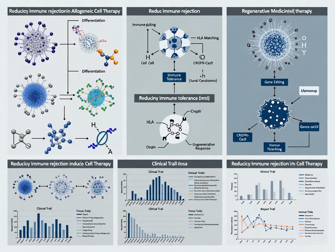
Overcoming Immune Rejection in Allogeneic Cell Therapy: Strategies for 'Off-the-Shelf' Therapeutics
Allogeneic cell therapies offer a scalable, 'off-the-shelf' alternative to autologous treatments but face significant barriers from host immune rejection and graft-versus-host disease (GvHD).
Overcoming Immune Rejection in Allogeneic Cell Therapy: Strategies for 'Off-the-Shelf' Therapeutics
Abstract
Allogeneic cell therapies offer a scalable, 'off-the-shelf' alternative to autologous treatments but face significant barriers from host immune rejection and graft-versus-host disease (GvHD). This article provides a comprehensive analysis for researchers and drug development professionals, covering the foundational immunology of allorejection, advanced gene-editing and cloaking methodologies, strategies for troubleshooting persistence and efficacy, and comparative validation of emerging clinical data. It synthesizes the latest advances in genetic engineering, iPSC technology, and immune-evasive designs that are paving the way for durable and broadly applicable allogeneic cell-based medicines.
The Immunological Battlefield: Understanding Barriers to Allogeneic Cell Acceptance
Technical Support & Troubleshooting Hub
Frequently Asked Questions (FAQs)
Q1: What are the fundamental operational differences between autologous and allogeneic cell therapy manufacturing?
A1: The core difference lies in the cell source and resulting production workflow, which creates a trade-off between personalization and scalability [1].
- Autologous Therapies use the patient's own cells. This creates a circular, patient-specific supply chain where each batch is customized for a single individual, requiring adaptable production environments and complex logistics to track each patient's cells from collection to reinfusion [1] [2].
- Allogeneic Therapies use cells from a healthy donor. This enables a more linear supply chain, where a single batch can be manufactured in large quantities, cryopreserved, and then aliquoted into individual "off-the-shelf" doses for many patients, benefiting from standardized processes and economies of scale [1] [3].
Q2: We are observing host-mediated rejection of our allogeneic CAR-T cells in preclinical models. What are the primary engineering strategies to overcome this?
A2: Host-versus-graft reaction (HVGR) is a major challenge. The following "immune cloaking" strategies are being employed to evade host immune detection [4] [5]:
- Disrupt HLA Class I Expression: Knockout of β2-microglobulin (B2M) disrupts classical HLA class I expression, reducing recognition by the host's CD8+ T cells [4].
- Express Non-Classical HLA Molecules: Overexpression of HLA-E or HLA-G can inhibit host NK cell-mediated killing, which is often triggered by missing "self" HLA signals [4].
- Employ Alloimmune Defense Receptors (ADR): Engineering CAR cells to express receptors that recognize host immune activation markers (e.g., CD45) can allow the allogeneic cells to selectively eliminate alloreactive host immune cells upon encounter [4].
Q3: Our allogeneic CAR-T product is triggering Graft-versus-Host Disease (GvHD) in our models. How can this be mitigated?
A3: GvHD is primarily mediated by the donor T cell's native T-cell receptor (TCR) recognizing host tissues as foreign. The primary solution is to disrupt the TCR complex [5]:
- Knockout TCR Alpha Constant (TRAC) Locus: This is a highly efficient method to prevent surface expression of the TCRαβ complex, significantly reducing alloreactivity. The residual TCR-positive cells are often depleted from the final product [5].
- Use Alternative Cell Sources: Consider using cell types with inherently lower GvHD risk, such as CAR-NK cells, CAR-NKT cells, or double-negative T cells (DNTs), which do not cause GvHD or have inherent regulatory functions [4] [5].
Q4: Why might a regulatory agency issue a hold or rejection for a cell therapy Investigational New Drug (IND) application based on CMC issues?
A4: Regulatory agencies like the FDA are increasingly stringent on Chemistry, Manufacturing, and Controls (CMC). Common pitfalls leading to rejection include [6]:
- Insufficient Product Characterization: Lack of a validated potency assay is a critical gap. You must demonstrate your product's biological activity.
- Unresolved Manufacturing Issues: Inconsistencies in the manufacturing process, inadequate process control, or facility readiness concerns.
- Incomplete Data: Gaps in stability data, lack of comparability studies for process changes, or insufficient validation of analytical methods.
Troubleshooting Guides
Problem: Inconsistent Potency in Allogeneic iPSC-Derived Cell Products
Potential Causes and Solutions:
| Cause | Solution |
|---|---|
| Unstable iPSC Master Cell Bank | Conduct rigorous genetic and functional stability testing of the master cell bank over multiple passages. |
| Variability in Differentiation Protocols | Implement controlled, closed-system bioreactors and standardized cytokine cocktails to ensure consistent differentiation. |
| Inadequate In-process Quality Controls | Introduce real-time monitoring systems (e.g., metabolite tracking) and intermediate potency assays during manufacturing [7]. |
Problem: High Manufacturing Failure Rate for Autologous Products Due to Poor Starting Cell Quality
Potential Causes and Solutions:
| Cause | Solution |
|---|---|
| Patient T-cell Exhaustion or Senescence | Implement a pre-screening assay for T-cell fitness. Consider using T-cell subsets or alternative cell types from the patient's sample. |
| Extended Vein-to-Vein Time | Optimize logistics and supply chain. Use point-of-care or decentralized manufacturing models to reduce transit and processing times [7]. |
| Uncontrolled Manufacturing Environment | Adopt closed, automated processing systems to minimize manual handling, contamination risk, and process variability [1] [7]. |
Quantitative Data Comparison
Comparative Analysis: Autologous vs. Allogeneic Cell Therapies
Table: Key Characteristics of Autologous and Allogeneic Cell Therapy Platforms
| Parameter | Autologous Therapy | Allogeneic Therapy |
|---|---|---|
| Cell Source | Patient's own cells [1] | Healthy donor(s) [1] |
| Manufacturing Model | Customized, patient-specific [1] | Standardized, "off-the-shelf" [1] [3] |
| Typical Production Time | Several weeks [2] | Immediate availability (pre-made) [2] |
| Scalability | Scale-out (multiple parallel lines) [1] | Scale-up (large single batches) [1] |
| Key Immunological Risks | No GvHD, lower rejection risk [2] | Risk of GvHD and host rejection [1] [2] |
| Supply Chain Complexity | High (circular, patient-centric) [1] | Lower (linear, centralized) [1] |
| Cost Structure | High cost per batch [1] [2] | Potential for lower cost per dose [1] [2] |
Clinical Safety and Efficacy of Allogeneic CAR-Cell Therapies
Table: Pooled Efficacy and Safety Outcomes for Allogeneic CAR-Cell Therapies in Relapsed/Refractory Large B-Cell Lymphoma (based on meta-analysis of 19 studies) [4]
| Outcome Measure | Pooled Estimate | Patient Population |
|---|---|---|
| Best Overall Response Rate (bORR) | 52.5% [95% CI, 41.0-63.9] | 235 patients evaluable for response |
| Best Complete Response Rate (bCRR) | 32.8% [95% CI, 24.2-42.0] | 235 patients evaluable for response |
| Grade 3+ Cytokine Release Syndrome (CRS) | 0.04% [95% CI 0.00-0.49] | 334 infused patients |
| Grade 3+ Immune Effector Cell-Associated Neurotoxicity Syndrome (ICANS) | 0.64% [95% CI 0.01-2.23] | 334 infused patients |
| GvHD-like Reactions | 1 occurrence across 334 patients | 334 infused patients |
Experimental Protocols
Detailed Protocol: Generation of Allogeneic CAR-T Cells with TCR Knockout
Objective: To produce universal allogeneic CAR-T cells from healthy donor PBMCs by disrupting the TRAC locus to prevent GvHD and introducing a CAR transgene for antitumor specificity.
Materials: Healthy donor leukapheresis product, T-cell activation beads, lentiviral vector encoding the CAR, CRISPR-Cas9 reagents (sgRNA targeting TRAC), electroporation device, T-cell culture media (IL-2).
Methodology:
- T-Cell Isolation and Activation: Isolate PBMCs and activate T cells using anti-CD3/CD28 activation beads.
- TRAC Locus Knockout: Electroporate activated T cells with CRISPR-Cas9 ribonucleoprotein (RNP) complex targeting the TRAC locus.
- CAR Transduction: Transduce cells with the lentiviral CAR vector 24-48 hours post-activation.
- Expansion and Harvest: Culture cells in media supplemented with IL-2 for 7-14 days to expand the modified T-cell population.
- Analytical and Quality Control Checks:
- Flow Cytometry: Confirm knockout of TCR (e.g., using anti-TCRαβ antibody) and expression of the CAR.
- Functional Potency Assay: Co-culture CAR-T cells with target-positive tumor cells and measure cytokine (IFN-γ) release and target cell killing.
- Sterility Testing: Perform mycoplasma and endotoxin testing on the final product.
Experimental Workflow: Creating Immune-Evasive Allogeneic Cells
The following diagram illustrates the key steps and engineering strategies involved in creating a universal, immune-evasive allogeneic cell product.
The Scientist's Toolkit: Key Reagents & Technologies
Table: Essential Reagents and Platforms for Allogeneic Cell Therapy R&D
| Reagent / Technology | Function / Application | Key Consideration |
|---|---|---|
| CRISPR-Cas9 Systems | Gene editing for TCR knockout (e.g., TRAC) and insertion of transgenes [5]. | Critical to assess off-target effects and genotoxicity. |
| Lentiviral Vectors | Stable delivery of large genetic payloads (e.g., CAR constructs) [5]. | Requires careful titration to ensure optimal transduction efficiency and safety. |
| Induced Pluripotent Stem Cells (iPSCs) | Clonal, renewable source for deriving consistent batches of immune effector cells (T, NK) [4] [8]. | Must ensure complete differentiation and genomic stability of master cell banks. |
| Closed Automated Bioreactors | Scalable expansion of cells under controlled, sterile conditions [7] [6]. | Reduces manual handling, contamination risk, and process variability. |
| Potency Assays | Measure biological activity of the final product (e.g., cytokine release, cytotoxicity) [6]. | Required by regulators; must be qualified/validated to demonstrate product consistency. |
| Alloimmune Defense Receptors (ADR) | Novel engineered receptors that allow donor cells to target and eliminate host immune cells attacking them [4]. | An emerging technology to directly combat host rejection mechanisms. |
Troubleshooting Guides
FAQ 1: My allogeneic CAR-NK cells are being cleared rapidly in immunocompetent mouse models. How can I improve their persistence?
Issue: Rapid clearance of infused allogeneic cells by host immune systems, limiting therapeutic efficacy.
Solution: Implement a multi-pronged gene editing strategy to simultaneously evade T cell, NK cell, and macrophage recognition.
Investigate Host T-cell Mediated Rejection: Host CD8+ T cells recognize mismatched HLA class I molecules on donor cells. Complete knockout of β2-microglobulin (B2M) eliminates surface expression of HLA class I, preventing T cell recognition. However, this can trigger NK cell "missing-self" activation. [9]
Prevent NK Cell "Missing-Self" Response: To counteract the effects of B2M knockout, overexpress non-classical HLA molecules like HLA-E or HLA-G. These molecules engage inhibitory receptors (e.g., NKG2A) on host NK cells, providing a "don't eat me" signal. [9] [10]
Counter Macrophage Phagocytosis: Macrophages clear cells lacking "self" markers via the SIRPα-CD47 axis. Overexpression of CD47 on your therapeutic cells sends an inhibitory signal to macrophages (via SIRPα), significantly reducing phagocytosis and prolonging circulation time. [9]
Recommended Experimental Protocol:
- Gene Editing: Use CRISPR-Cas9 to knock out the B2M gene in your NK cell line (e.g., NK-92 or iPSC-derived NK cells).
- Transgene Expression: Co-transduce the cells with lentiviral vectors carrying:
- A gene for HLA-E or HLA-G.
- A gene for CD47.
- Validation In Vitro:
- Use flow cytometry to confirm loss of HLA class I and high surface expression of HLA-E/G and CD47.
- Perform co-culture assays with human PBMCs to assess resistance to T-cell mediated killing.
- Perform co-culture assays with macrophages to quantify phagocytosis resistance.
- Validation In Vivo: Test the persistence and efficacy of these multi-edited cells in immunocompetent humanized mouse models compared to unedited controls.
FAQ 2: The tumor microenvironment (TME) is suppressing my allogeneic cell therapy function. What strategies can I use to enhance resistance?
Issue: The immunosuppressive TME (e.g., TGF-β, checkpoints, metabolic constraints) inactivates therapeutic cells upon tumor infiltration.
Solution: Engineer cells with built-in resistance to key immunosuppressive factors in the TME.
Block TGF-β Signaling: The immunosuppressive cytokine TGF-β is a major inhibitor of NK and T cell function. Engineer your cells to express a dominant-negative TGF-β receptor II (dnTGFβRII), which blocks downstream signaling and maintains cell activity in TGF-β-rich environments. [9]
Convert Inhibitory to Activating Signals: The PD-1/PD-L1 axis is a critical immune checkpoint. Design a PD-1:CD28 switch receptor. The extracellular domain binds PD-L1, but the intracellular domain is an activating CD28 signal. This converts an inhibitory signal into a co-stimulatory one. [9]
Provide Cytokine Support: To counteract cytokine deprivation, engineer cells to express membrane-bound IL-15 or IL-12. This provides autocrine/paracrine survival and activation signals, enhancing persistence and function within the TME. [9] [11]
Recommended Experimental Protocol:
- Vector Design: Construct a lentiviral vector encoding your chosen transgenes (e.g., dnTGFβRII, PD-1:CD28, mbIL-15).
- Cell Engineering: Transduce your therapeutic cells (e.g., T cells or NK cells) with the vector.
- Functional In Vitro Assays:
- Culture engineered cells with recombinant TGF-β and measure phospho-SMAD2 levels (via Western blot) to confirm disruption of signaling.
- Co-culture cells with PD-L1+ tumor cells and assess activation markers (e.g., CD69) and cytokine production (e.g., IFN-γ) via flow cytometry.
- Under low IL-2/IL-15 conditions, measure cell viability and proliferation to validate the benefit of mbIL-15.
- In Vivo Validation: Test the function of these armored cells in solid tumor xenograft models and analyze tumor infiltration and functional status by flow cytometry or IHC post-harvest.
Data Presentation
Table 1: Key Gene Editing Targets for Evading Host Immune Effectors
| Immune Effector | Recognition Mechanism | Gene Editing Strategy | Target Molecule | Expected Outcome |
|---|---|---|---|---|
| CD8+ T Cell | Recognizes allogeneic HLA class I (HLA-A, -B) | Knockout | β2-microglobulin (B2M) | Abolishes HLA class I surface expression, evading T cell detection. [9] |
| NK Cell | Detects "missing-self" (absence of self-HLA) | Overexpression | HLA-E / HLA-G | Engages NKG2A inhibitory receptor on NK cells, preventing activation. [9] [10] |
| Macrophage | Phagocytoses cells lacking "self" CD47 | Overexpression | CD47 | Binds SIRPα on macrophages, delivering a "don't eat me" signal. [9] |
| TME (TGF-β) | Immunosuppressive cytokine signaling | Express Dominant-Negative Receptor | dnTGFβRII | Confers resistance to TGF-β-mediated suppression. [9] |
| TME (PD-1) | Inhibitory checkpoint signaling | Express Switch Receptor | PD-1:CD28 | Converts PD-L1 inhibitory signal into a T/NK cell activating signal. [9] |
Table 2: Research Reagent Solutions for Immune Evasion Studies
| Research Reagent | Function / Application | Example Use Case |
|---|---|---|
| CRISPR-Cas9 System | Precise gene knockout (e.g., B2M) or knock-in. | Creating hypoimmunogenic base lines in iPSCs or immune cells. [9] [10] |
| Lentiviral Vector | Stable delivery of large transgenes (e.g., HLA-E, CD47, CAR). | Engineering cells to express immune-evasion proteins. [9] |
| Anti-Human HLA-ABC Antibody | Flow cytometry validation of HLA class I knockout. | Confirming B2M knockout efficiency. [10] |
| Recombinant CD47 Protein | Binding assays to validate SIRPα interaction. | Verifying functional activity of overexpressed CD47. |
| Recombinant TGF-β | In vitro simulation of immunosuppressive TME. | Testing the functional resilience of dnTGFβRII-expressing cells. [9] |
| iPSC Line | Scalable, standardized source for differentiated cells. | Generating uniform, gene-edited "off-the-shelf" NK or T cells. [9] [11] |
Experimental Workflow & Signaling Pathways
Diagram 1: Engineered Cell Evasion of Host Immunity
Diagram 2: Core Immune Evasion Engineering Workflow
Core Mechanisms: How Do Alloreactive T Cells Cause GvHD?
Graft-versus-Host Disease (GvHD) is a systemic disorder that occurs when immunocompetent donor T cells (the graft) recognize the recipient's tissues (the host) as foreign and mount an immune attack. This process is a major complication following allogeneic hematopoietic stem cell transplantation (HCT) [12] [13].
The fundamental requirements for GvHD to develop were first outlined by Billingham and are still held true today [14]:
- The graft must contain immunologically competent cells (T cells).
- The recipient must express tissue antigens (e.g., HLA) not present in the donor.
- The recipient must be incapable of mounting an effective response to eliminate the transplanted cells [14].
The pathophysiology of this T-cell-mediated attack is typically described in three sequential phases [12]:
1. Afferent Phase (Activation): Conditioning regimens (chemotherapy or radiation) before transplant cause significant tissue damage. This damage activates host antigen-presenting cells (APCs) and increases the expression of inflammatory cytokines (e.g., TNF-α, IL-1, IL-6) and host major histocompatibility complex (MHC) antigens. This inflammatory milieu is often called a "cytokine storm" [12] [14].
2. Efferent Phase (Proliferation and Differentiation): Donor T cells interact with these activated host APCs. The donor T cells recognize the host's foreign human leukocyte antigens (HLAs)—both major and minor histocompatibility antigens—leading to their activation, proliferation, and differentiation into cytotoxic effector cells [12] [14].
3. Effector Phase (Tissue Destruction): Activated donor cytotoxic CD8+ T cells, along with natural killer (NK) cells, migrate to target organs, primarily the skin, gastrointestinal (GI) tract, and liver, causing direct cellular damage and apoptosis. This results in the clinical manifestations of GvHD [12].
Table 1: Key Cytokines in GvHD Pathogenesis and Their Roles [12].
| Cytokine | Primary Role in GvHD Pathogenesis |
|---|---|
| TNF-α | Promotes inflammation and direct tissue damage. |
| IL-2 | Crucial for T-cell activation and proliferation. |
| IL-1 | Proinflammatory cytokine that contributes to tissue damage. |
| IL-6 | Promotes B-cell activation and inflammatory responses. |
| IL-12 | Stimulates differentiation of naive T cells into Th1 cells. |
| IL-17 | Promotes inflammation, particularly in the gut. |
| IFN-γ | Involved in the inflammatory response and macrophage activation. |
| TGF-β | Has complex, dual roles; can be both pro- and anti-inflammatory. |
Experimental Insights: How Can We Track and Target Alloreactive T Cells?
A critical step in managing GvHD is the ability to identify and characterize the alloreactive T cells responsible for the attack. Recent research has identified CD70 as a specific marker for these pathogenic T cells [15].
CD70 is a costimulatory molecule transiently upregulated on T cells after recent T-cell receptor (TCR) engagement. Studies show that CD70+ T cells are highly activated, exhibit a transcriptional profile indicative of MYC-driven glycolysis and proliferation, and are significantly enriched in the blood and tissues of patients during acute GvHD episodes. These cells display an oligoclonal TCR repertoire, indicating a targeted immune response against host antigens [15].
Experimental Protocol: Assessing Alloreactive T Cells via CD70
Objective: To identify, isolate, and characterize alloreactive T cells from patient samples based on CD70 expression.
Methodology:
- Sample Collection: Collect peripheral blood mononuclear cells (PBMCs) or tissue biopsies (e.g., skin, GI) from transplant recipients at regular intervals post-transplant and during suspected GvHD flares.
- Cell Staining: Stain the samples with fluorescently labeled antibodies, including:
- Anti-CD3 (Pan T-cell marker)
- Anti-CD4 (Helper T-cell marker)
- Anti-CD8 (Cytotoxic T-cell marker)
- Anti-CD70 (Marker of recent activation/alloreactivity)
- Viability dye
- Flow Cytometry & Sorting: Use fluorescence-activated cell sorting (FACS) to isolate pure populations of CD4+CD70+ and CD8+CD70+ T cells for downstream functional and molecular analyses.
- Downstream Applications:
- Transcriptional Profiling: Perform RNA sequencing (RNA-seq) on sorted populations to define their gene expression signature.
- TCR Repertoire Analysis: Use sequencing to characterize the TCR diversity of CD70+ clones, confirming their oligoclonality.
- Functional Assays: Co-culture sorted CD70+ T cells with host-derived target cells to confirm their alloreactive potential and cytotoxic activity.
- In Vitro Blockade: Utilize anti-CD70 blocking antibodies in co-culture assays to measure the reduction in T-cell proliferation and inflammatory cytokine (e.g., IFN-γ, IL-17) secretion [15].
Table 2: Research Reagent Solutions for Studying Alloreactive T Cells in GvHD.
| Research Reagent | Function in Experiment |
|---|---|
| Anti-CD70 Antibody | Key reagent for identifying, blocking, or depleting alloreactive T-cell populations via flow cytometry or functional assays [15]. |
| Anti-CD3/CD4/CD8 Antibodies | Essential for defining basic T-cell subsets during immunophenotyping. |
| Recombinant Cytokines (e.g., IL-2) | Used in T-cell culture and proliferation assays to maintain cell viability and activation. |
| CFSE (Cell Trace Dye) | Fluorescent dye used to track and quantify T-cell division and proliferation in response to host antigens. |
| ELISA/Luminex Kits | For quantifying cytokine secretion (e.g., IFN-γ, TNF-α, IL-17) in cell culture supernatants to measure alloreactive T-cell function. |
Troubleshooting Guide & FAQ: Addressing Common Research Challenges
FAQ 1: Our in vitro T-cell activation assays do not reliably correlate with GvHD outcomes in our animal models. What could be wrong?
- Answer: This is a common translational challenge. Focus on using more specific markers of alloreactivity rather than general activation.
- Solution: Implement CD70 staining in your assays. CD70+ T cells are a more specific subset enriched for alloreactivity compared to general markers like CD25 [15].
- Check Your Model: Ensure your animal model has sufficient HLA/minor antigen mismatch. The inflammatory context is critical; consider incorporating conditioning regimen-like radiation into your model to mimic the "cytokine storm" of the afferent phase, which is essential for robust GvHD induction [12] [14].
FAQ 2: We are developing an allogeneic cell therapy and want to minimize the risk of GvHD. What are the primary genetic engineering strategies?\
- Answer: The primary strategy is to disrupt the T cell's ability to recognize host antigens or execute an attack.
- T-cell Depletion: The most direct method is to physically remove T cells from the graft product.
- Targeted Gene Editing: Use CRISPR/Cas9 or other nucleases to knock out the T-cell receptor (TCR) gene in donor cells, preventing them from recognizing host antigens altogether. This is a robust approach for "off-the-shelf" therapies like CAR-NK or CAR-T cells [9].
- Targeted Immunosuppression: An emerging strategy is to target specific activation pathways, such as using anti-CD70 antibodies to block the CD27-CD70 costimulatory axis, which has been shown to attenuate alloreactive T-cell responses in pre-clinical models [15].
FAQ 3: Why do some patients still develop GvHD even with a perfectly HLA-matched sibling donor?
- Answer: HLA identity does not guarantee compatibility for all immune antigens.
- Minor Histocompatibility Antigens (miHAs): The immune response can be directed against miHAs, which are immunogenic peptides derived from polymorphic cellular proteins presented by MHC molecules. Even with matched HLAs, differences in these minor antigens can trigger GvHD [14].
- Other Risk Factors: Several non-genetic factors increase GvHD risk, as summarized in the table below.
Table 3: Key Risk Factors for Developing GvHD [12] [13] [14].
| Risk Factor Category | Specific Factor | Impact on GvHD Risk |
|---|---|---|
| Genetic Disparity | HLA Mismatch | Increases |
| Female Donor to Male Recipient | Increases | |
| Minor Histocompatibility Antigen Mismatch | Increases | |
| Donor/Recipient Factors | Older Age (Donor or Recipient) | Increases |
| Donor Prior Pregnancy | Increases | |
| Transplant Modality | Peripheral Blood Stem Cell Source (vs. Bone Marrow) | Increases |
| High Number of Infused T cells | Increases | |
| No ATG (Antithymocyte Globulin) Use | Increases | |
| Clinical History | Prior Acute GvHD | Increases (for chronic GvHD) |
| Positive CMV Serology | Increases |
In allogeneic cell therapy, the Host-versus-Graft (HvG) response represents a fundamental immunological barrier where the recipient's immune system recognizes donor cells as foreign and mounts an immune response to eliminate them [16]. This allorejection process significantly diminishes cellular persistence—the duration therapeutic cells remain viable and functional in the patient—and consequently undermines treatment efficacy [17]. Unlike autologous therapies that use a patient's own cells, "off-the-shelf" allogeneic products from healthy donors face coordinated attacks from multiple arms of the host immune system, including T cells, natural killer (NK) cells, and antibodies [18] [19]. Understanding and troubleshooting these rejection mechanisms is essential for developing successful and durable allogeneic cellular therapies.
Frequently Asked Questions: Mechanisms & Solutions
What specific immune effectors mediate allogeneic cell rejection?
The host immune system employs multiple effector mechanisms to clear allogeneic cells, each requiring distinct mitigation strategies.
Table: Effector Mechanisms in Host-versus-Graft Responses
| Immune Effector | Recognition Mechanism | Consequence for Allogeneic Cell |
|---|---|---|
| Host T Cells | Recognize foreign HLA (Human Leukocyte Antigen) molecules on donor cells via T Cell Receptors (TCRs) [16] [20]. | Direct killing via cytolytic mechanisms (perforin/granzyme) and cytokine-mediated activation of other immune cells [21]. |
| Host NK Cells | Detect "missing self" due to absent or mismatched host HLA class I molecules on donor cells [16] [17]. | Activation of direct cytotoxicity and antibody-dependent cellular cytotoxicity (ADCC) [16]. |
| Host B Cells | Produce allospecific antibodies against foreign HLA and other polymorphic antigens on donor cells [16]. | Opsonization of donor cells, leading to complement-dependent cytotoxicity (CDC) and ADCC [16] [17]. |
How can we engineer donor cells to evade T cell-mediated rejection?
The most common strategy involves disrupting the expression of HLA molecules on the donor cell surface to prevent recognition by host T cells.
- TCR Complex Disruption: Knocking out the T-cell receptor alpha constant (TRAC) gene prevents the expression of the endogenous TCR, effectively eliminating the risk of Graft-versus-Host Disease (GvHD) when using T-cell-based therapies [21] [17]. This is often a foundational edit for allogeneic CAR-T cells.
- HLA Class I Ablation: Knocking out β2-microglobulin (B2M), an essential subunit for HLA class I surface expression, prevents recognition by host CD8+ cytotoxic T cells [16] [17]. A critical caveat is that this creates "missing self" and can trigger NK cell rejection, requiring complementary edits described below.
- HLA Class II Ablation: Disrupting the class II major histocompatibility complex transactivator (CIITA) gene abrogates the expression of HLA class II molecules, shielding cells from host CD4+ T helper cell recognition [16].
What strategies prevent NK cell activation against HLA-deficient cells?
Removing HLA molecules triggers NK cell rejection. Solutions involve engineering cells to express non-polymorphic, inhibitory HLA molecules.
- HLA-E Expression: Engineering cells to express the non-classical HLA-E molecule, which can be recognized by the inhibitory receptor NKG2A on NK cells, provides a "do not eat me" signal [16] [17]. This is often achieved by expressing a single-chain HLA-E-B2M fusion protein in the B2M locus [17].
- HLA-G Expression: Similarly, expression of the non-polymorphic HLA-G molecule, known for its potent immunosuppressive role at the maternal-fetal interface, can inhibit both NK cell and T cell responses [16] [22].
Can the host microenvironment be modulated to favor persistence?
Yes, alongside editing donor cells, modulating the host environment is a critical pillar for success.
- Lymphodepletion (LD) Conditioning: Pre-treatment with chemotherapy agents like fludarabine and cyclophosphamide depletes host lymphocytes, reducing the immediate army of cells that can mediate rejection [16]. This creates "immunological space" and enhances engraftment by increasing availability of homeostatic cytokines like IL-7 and IL-15 [16].
- Targeted Immunosuppression: Using antibodies like Alemtuzumab (anti-CD52) to deplete a broad range of host lymphocytes is effective. A common strategy is to engineer donor cells to be CD52-negative, making them resistant to Alemtuzumab, allowing for selective host immunosuppression without harming the therapeutic product [17].
- Regulatory T Cell (Treg) Co-therapy: The adoptive transfer of Tregs, a specialized T-cell subset that maintains immune tolerance, is being explored to suppress alloreactive responses and create a tolerogenic microenvironment [23] [22]. Tregs can suppress effector T cells through various mechanisms, including consumption of IL-2 and expression of inhibitory molecules like CTLA-4 [22].
The Scientist's Toolkit: Key Reagents & Models
Table: Essential Tools for Studying HvG Responses
| Tool Category | Specific Example | Function in HvG Research |
|---|---|---|
| Gene Editing Tools | CRISPR-Cas9, TALENs, ZFNs [21] [17] | Precisely knock out genes like TRAC, B2M, and CIITA in donor cells to reduce immunogenicity. |
| In Vitro Assays | Mixed Lymphocyte Reaction (MLR) [21] | Co-culture donor cells with irradiated host immune cells to measure T-cell activation and proliferation, predicting alloreactivity potential. |
| In Vivo Models | Immunodeficient mice reconstituted with human immune system (e.g., NSG mice) [21] | Provide a humanized in vivo model to study cell persistence, trafficking, and rejection mechanisms in a more physiologically relevant context. |
| Critical Antibodies | Alemtuzumab (anti-CD52) [17] | Used for lymphodepletion; necessitates CD52 knockout in donor cells for resistance. |
| Persistence Tracking | PET Reporter Genes [16] | Enable non-invasive, serial imaging to monitor the biodistribution and persistence of infused cells in vivo, beyond blood PK measurements. |
Troubleshooting Experimental Protocols
Protocol: Validating Allo-Evasion with Mixed Lymphocyte Reaction (MLR)
Objective: To assess the potential of engineered donor cells to provoke host T-cell activation in vitro [21].
- Stimulator Cells (Donor): Use the engineered allogeneic cells (e.g., TCR-/HLA-I-/HLA-E+ CAR-T cells). Irradiate these cells to prevent their proliferation.
- Responder Cells (Host): Isolate Peripheral Blood Mononuclear Cells (PBMCs) from a healthy, HLA-mismatched donor to simulate the host immune system.
- Co-culture: Seed irradiated stimulator cells with responder PBMCs at a defined ratio (e.g., 1:1) in a culture medium for 5-7 days.
- Analysis:
- Flow Cytometry: Analyze T-cell activation markers (e.g., CD69, CD25) and proliferation dyes (e.g., CFSE) in the responder population.
- ELISA: Measure the concentration of pro-inflammatory cytokines (e.g., IFN-γ) in the supernatant [21].
- Troubleshooting:
- High Background Activation: Ensure stimulator cells are adequately irradiated to prevent confounding proliferation.
- Weak Response: Use multiple, genetically distinct PBMC donors as responders to account for population variability in alloreactivity.
Protocol: Assessing In Vivo Persistence via Reporter Gene Imaging
Objective: To track the location and survival of infused cells over time in a preclinical model, overcoming the limitation of blood pharmacokinetics [16].
- Engineering: Stably transduce the master cell bank of your therapeutic cell line with a PET reporter gene (e.g., thymidine kinase) and a fluorescent or luminescent reporter (e.g., GFP, luciferase) for dual-mode tracking.
- Mouse Model: Use an immunodeficient mouse model (e.g., NSG) that can be engrafted with a human immune system (humanized mice) to model the HvG response.
- Cell Administration: Infuse the engineered cells into the mice.
- Longitudinal Imaging:
- Optical Imaging: Use bioluminescence imaging at frequent intervals to monitor total body cell burden and location.
- PET Imaging: At specific timepoints, administer the corresponding PET radiotracer to obtain high-resolution, quantitative data on cell distribution and density [16].
- Troubleshooting:
- Weak Signal: Optimize the expression level of the reporter gene and confirm the stability of the construct in expanded cells.
- Rapid Signal Loss: Compare the persistence of your multi-edited "allo-evading" cells against control cells (e.g., only CAR-modified) to directly quantify the impact of your engineering strategy on overcoming HvG.
Engineering Stealth Cells: Gene Editing and Cloaking Strategies to Evade Immunity
Scientific Background and Rationale
What is the primary mechanism by which TRAC disruption prevents GvHD?
Graft-versus-host disease (GvHD) occurs when donor T cells recognize the recipient's healthy tissues as foreign, primarily through interactions between the donor T cell receptor (TCR) and host major histocompatibility complex (MHC) molecules. The TCRαβ complex, essential for antigen recognition, consists of an α chain and a β chain. The T cell receptor alpha constant (TRAC) gene encodes the constant region of the α chain, and as a single-gene locus, it presents an efficient target for complete TCR disruption [24] [5]. Knocking out TRAC prevents surface expression of the functional TCRαβ complex, thereby eliminating the fundamental mechanism for alloreactive T cell activation and subsequent GvHD pathogenesis [5].
Why is TRAC a preferred target over other TCR subunits for preventing GvHD?
While the TCR β chain contains two possible constant regions (TRBC1 and TRBC2), the α chain has only one constant gene (TRAC) [5]. This makes TRAC a more efficient single target for complete TCR ablation compared to targeting multiple β chain genes. Successful TRAC disruption results in loss of surface TCR expression in over 90% of cells, which can be further purified to less than 0.05% TCR-positive cells remaining through additional magnetic bead depletion systems [25]. This approach effectively mitigates GvHD alloreactivity while allowing T cells to maintain their cytotoxic function through introduced chimeric antigen receptors (CARs) [25].
Experimental Protocols and Workflows
Detailed Protocol: TRAC Knockout using CRISPR-Cas9 in Primary Human T Cells
Overview of Workflow:
Step-by-Step Methodology:
T Cell Source and Isolation:
T Cell Activation:
Ribonucleoprotein (RNP) Complex Formation and Delivery:
- For CRISPR-Cas9: Complex purified Cas9 protein with synthetic sgRNA targeting TRAC exon 1.
- Use modified, nuclease-protected sgRNA with 2'-O-methyl phosphorothioate modifications at terminal bases to enhance stability and editing efficiency [27].
- Incubate Cas9 protein and sgRNA at room temperature for 10-20 minutes to form RNP complexes.
- Deliver RNP complexes via electroporation using optimized protocols for primary T cells (e.g., Lonza 4D-Nucleofector) [27] [28].
Post-Editing Cell Expansion:
- Culture edited cells in appropriate T cell media with IL-2 supplementation.
- Monitor cell density and maintain cells at 0.5-2 × 10^6 cells/mL.
- Expand cells for 10-14 days, achieving 44-129 fold expansion depending on editing conditions [26].
Validation and Characterization:
- Assess editing efficiency by flow cytometry for surface CD3 expression loss 7-14 days post-editing.
- Evaluate genomic disruption by mismatch-sensitive enzyme assays or next-generation sequencing of the target site [26].
- Validate functionality through cytokine production and cytotoxicity assays against target cells [25].
Comparative Protocol: TRAC Disruption using TALENs
Key modifications from the CRISPR protocol:
- TALEN mRNA Production: Generate TALEN mRNA using T3 or T7 polymerase-based in vitro transcription systems [27].
- Enhanced Stability: Include an exogenous polyadenylation signal by E. coli poly(A) polymerase, which has been shown to increase TRAC disruption rates from ~30% to ~60% [27].
- Delivery: Electroporation of TALEN mRNA following the same activation and expansion principles.
Troubleshooting Common Experimental Challenges
How can I improve editing efficiency in primary T cells?
Editing efficiency can be impacted by multiple factors. The table below summarizes common issues and evidence-based solutions:
Table: Troubleshooting Guide for Low TRAC Editing Efficiency
| Problem | Potential Causes | Verified Solutions | Reported Outcomes |
|---|---|---|---|
| Low knockout efficiency | Suboptimal gRNA design, unmodified sgRNA, inadequate RNP delivery | Use protected sgRNA (2'-O-methyl), optimize Cas9:gRNA ratio, validate gRNA efficiency [27] [26] | Efficiency increased from ~60% to >90% using modified guides [27] |
| Poor cell viability post-electroporation | Electroporation toxicity, genotoxic stress from multiple DSBs | Optimize electroporation parameters, use RNP instead of plasmid DNA, reduce number of simultaneous edits [26] | Viability maintained by using clinical-grade electroporation systems and optimized buffers [25] |
| High variability between donors | Donor-specific T cell fitness, activation state differences | Standardize activation (anti-CD3/CD28 beads), pre-screen donors for responsiveness, use pooled donors for research [24] | Consistent >90% TCR disruption across 6 donors achieved with standardized protocol [25] |
What strategies mitigate risks of chromosomal abnormalities in edited T cells?
Recent research has identified that Cas9 editing can cause unintended targeted chromosome loss, a significant safety concern for clinical applications [28]. A modified manufacturing process that incorporates p53 expression has been shown to reduce chromosome loss while preserving editing efficacy. This approach was successfully employed in a first-in-human clinical trial (NCT03399448) [28]. Additionally, using RNP complexes instead of plasmid-based delivery and limiting the number of simultaneous edits can reduce genotoxic stress.
How can I ensure my TRAC-knockout T cells remain functionally competent?
While TRAC disruption prevents GvHD, researchers must verify that edited T cells maintain anti-tumor efficacy. Studies show that TRAC-knockout CAR-T cells can efficiently eliminate target cells and produce high cytokine levels when challenged with antigen-positive leukemia cells [25]. However, complete TCR ablation may impair IL-7/IL-15-dependent survival signaling, potentially affecting long-term persistence [5]. Functional validation through in vitro cytotoxicity assays and in vivo xenograft models is essential to confirm maintained therapeutic function.
Frequently Asked Questions (FAQs)
What are the key differences between CRISPR and TALEN platforms for TRAC editing?
Table: Comparison of Gene Editing Platforms for TRAC Disruption
| Parameter | CRISPR-Cas9 | TALENs |
|---|---|---|
| Targeting Mechanism | sgRNA-DNA base pairing [29] | Protein-DNA recognition [27] |
| Efficiency in T Cells | High (>90% with optimized reagents) [25] | Moderate (~60% with polyadenylation) [27] |
| Multiplexing Capacity | High (multiple gRNAs simultaneously) [30] | Low (requires custom protein engineering) [27] |
| Off-Target Risk | Moderate (dependent on gRNA specificity) [29] [28] | Low (higher DNA binding specificity) [27] |
| Delivery Format | RNP complex (Cas9 protein + sgRNA) [28] | mRNA encoding TALEN proteins [27] |
| Molecular Weight | ~160 kDa for Cas9 [30] | Larger, dimeric structure [27] |
Can I combine TRAC knockout with other genetic modifications?
Yes, multiplexed editing is a key advantage of CRISPR platforms. Researchers have successfully combined TRAC knockout with:
- CAR integration into the TRAC locus itself, providing homogeneous expression [25] [31]
- HLA ablation (B2M knockout for Class I, various approaches for Class II) to evade host immune rejection [26]
- Immune checkpoint disruption (PD-1, etc.) to enhance anti-tumor activity [30]
When performing multiple edits, monitor cell viability closely and consider using high-specificity Cas variants to minimize off-target effects.
How do I validate the specificity of my TRAC editing?
Comprehensive validation should include:
- On-target efficiency: Flow cytometry for CD3/TCR surface expression loss, sequencing of the target locus [26]
- Off-target assessment: Use bioinformatics tools (e.g., CHOPCHOP, COSMID) to predict potential off-target sites, followed by sequencing of top predicted sites [25] [26]
- Karyotype integrity: Employ methods to detect chromosomal abnormalities, particularly at the target site [28]
- Functional validation: Demonstrate reduced alloreactivity in mixed lymphocyte reactions while maintaining CAR-specific cytotoxicity [25] [26]
The Scientist's Toolkit: Essential Research Reagents
Table: Key Reagents for TRAC Gene Editing Experiments
| Reagent Category | Specific Examples | Function/Purpose | Considerations |
|---|---|---|---|
| Nucleases | SpCas9 protein, Cas12a (Cpf1), TALEN mRNAs | Creates double-strand breaks at TRAC locus | Cas9: >90% efficiency; TALENs: ~60% efficiency [27] [30] |
| Targeting Guides | TRAC-specific sgRNAs (modified) | Guides nuclease to target sequence | 2'-O-methyl-modified sgRNAs enhance efficiency [27] |
| Delivery System | Electroporation equipment (4D-Nucleofector) | Introduces editing components into cells | Clinical-grade systems improve viability [25] |
| Activation Reagents | Anti-CD3/CD28 beads or antibodies | Activates T cells for editing | Standardized activation critical for consistency [25] |
| Cell Culture | IL-2, serum-free media, supplements | Supports T cell expansion post-editing | IL-2 concentration affects expansion and phenotype [26] |
| Validation Tools | Flow antibodies (CD3, TCR), sequencing primers | Assesses editing efficiency and specificity | Multi-parameter flow confirms phenotype [26] |
Diagram: Logical workflow showing how TRAC editing prevents GvHD while enabling CAR-specific anti-tumor activity. DSB: Double-Strand Break; NHEJ: Non-Homologous End Joining; HDR: Homology-Directed Repair.
A primary barrier to the successful implementation of "off-the-shelf" allogeneic cell therapies is immune rejection by the recipient's immune system. Allogeneic cell therapies, derived from healthy donors, are recognized as foreign and eliminated by host T cells and Natural Killer (NK) cells, a process known as allorejection [32]. A cornerstone strategy to overcome this is the genetic modification of donor cells to reduce their immunogenicity. This technical support center details the challenges and solutions surrounding two key approaches: complete Beta-2-Microglobulin (B2M) knockout and more advanced strategies for selective HLA retention.
? Frequently Asked Questions (FAQs)
1. Why is B2M knockout insufficient to prevent immune rejection?
While B2M knockout successfully eliminates surface expression of all HLA class I molecules (including HLA-A, -B, -C, -E, and -G), it creates a new vulnerability [33]. The absence of HLA-E, a ligand for the inhibitory receptor NKG2A on NK cells, renders the modified cells susceptible to "missing-self" recognition and killing by host NK cells [33]. Furthermore, studies in humanized mouse models have shown that even with B2M knockout, transplanted cells can still be rejected by host T cells, particularly if there is pre-existing expression of HLA class II on the therapeutic cells [34].
2. What is the advantage of selective HLA knockdown over complete B2M knockout?
Selective HLA knockdown aims to inhibit the classical HLA class I molecules (HLA-A, -B, -C) that are primary triggers for CD8+ T-cell-mediated rejection, while deliberately preserving the non-classical molecule HLA-E [33]. This dual strategy allows the cell to evade T-cell recognition while simultaneously providing an inhibitory signal to NK cells via the retained HLA-E, thus protecting the cell from NK-mediated killing [33].
3. Which host immune cells are the main drivers of allorejection?
CD8+ T cells have been identified as the dominant cell type mediating the rejection of allogeneic cell products [33]. They recognize mismatched classical HLA class I molecules (HLA-A, -B, -C) on donor cells. NK cells also contribute significantly, particularly when donor cells lack inhibitory ligands like HLA-E, leading to "missing-self" activation [35].
4. Beyond HLA class I, what other modifications are being explored?
Emerging strategies involve multiplex genetic engineering to create truly "hypoimmunogenic" cells. This includes knocking out genes responsible for both HLA class I (via B2M knockout) and HLA class II (via CIITA knockout) expression, while also introducing transgenes for immune checkpoint modulators like PD-L1 or single-chain HLA-E to further enhance persistence and safety [33] [35].
Troubleshooting Guides
Problem: Poor In Vivo Persistence of B2M-KO Cell Therapy
| Symptom | Possible Cause | Recommended Solution |
|---|---|---|
| Rapid clearance of cells in immunocompetent model | Rejection by host NK cells due to "missing-self" response [33] | Co-express HLA-E or single-chain HLA-E (SCE) to engage NKG2A inhibitory receptor on NK cells [33] [35]. |
| Infiltration of CD8+ T cells around graft | Residual HLA class I expression or HLA class II-mediated T cell activation [34] | Verify knockout efficiency. Implement CIITA knockout to eliminate HLA class II [35]. |
| Compromised tissue integrity and tubulitis in organoid models | Persistent T-cell mediated rejection despite B2M KO [34] | Employ a combined strategy: knockdown HLA-ABC, knockout CIITA, and overexpress an immune checkpoint like PD-L1 [34] [33]. |
Problem: Inconsistent Results Between In Vitro and In Vivo Models
| Observation | In Vitro Result | In Vivo Result | Explanation & Action |
|---|---|---|---|
| Potent suppression/function | Strong activity in in vitro suppression assays [35] | Reduced efficacy and cell survival [35] | In vitro assays lack a fully functional host immune system. Re-evaluate efficacy in a humanized mouse model to account for CD8+ T-cell mediated rejection [35]. |
| Effective evasion of T cell killing | Protected from allogeneic T cells in co-culture | Rejection occurs | Standard co-cultures may not simulate the inflammatory tumor microenvironment (TME). Engineer cells to express PD-L1 to resist T-cell exhaustion and suppression in the TME [33]. |
Experimental Protocols & Workflows
Protocol 1: Generating Immune-Evasive Cells via Selective HLA Knockdown and Checkpoint Expression
This protocol details the one-step lentiviral construction of allogeneic CAR-NK cells capable of evading both T and NK cell rejection, as validated in xenograft models [33].
Key Steps:
- Design shRNA for Selective HLA-ABC Knockdown: Design shRNAs targeting conserved regions of HLA-A, -B, and -C heavy chains with at least 2 nucleotide mismatches to HLA-E to ensure specificity [33].
- Construct Lentiviral Vector: Use a single lentivector to express:
- The selective HLA-ABC shRNA (e.g., shRNA #1 from the study) under a U6 promoter.
- A Chimeric Antigen Receptor (CAR) for tumor targeting.
- An immune checkpoint protein (PD-L1 or single-chain HLA-E) [33].
- Transduce and Expand NK Cells: Isolate NK cells from healthy donor PBMCs or use a cell line. Transduce with the lentiviral vector and expand ex vivo.
- Validate Phenotype:
- Confirm reduction of HLA-ABC surface expression via flow cytometry.
- Verify preservation or expression of HLA-E.
- Confirm CAR and PD-L1/SCE expression.
- Functional Assays:
- In vitro cytotoxicity: Assess resistance to killing by allogeneic CD8+ T cells and NK cells.
- In vivo persistence: Measure cell survival and tumor control in a humanized mouse model engrafted with human PBMCs [33].
The following workflow diagram illustrates the core strategy of this protocol:
Protocol 2: Multiplex CRISPR-Cas9 Engineering of Hypoimmunogenic Tregs
This protocol describes the creation of hypoimmunogenic allogeneic Tregs for treating autoimmunity and transplant rejection, enabling evasion of both T and NK cells [35].
Key Steps:
- Isolate and Activate Human Tregs: Isulate CD4+CD25+ Tregs from healthy donor PBMCs and activate them.
- Multiplex CRISPR Editing:
- Knockout B2M: Disrupts HLA class I expression to prevent CD8+ T cell recognition.
- Knockout CIITA: Disrupts HLA class II expression.
- Knock-in HLA-E/B2M Fusion Gene: Simultaneously with B2M knockout, integrate a gene encoding a single-chain HLA-E-B2M fusion protein to protect from NK cell-mediated "missing-self" killing [35].
- Expand and Validate Edited Tregs:
- Confirm loss of HLA class I and II via flow cytometry.
- Confirm surface expression of HLA-E.
- Verify FOXP3 stability and suppressive function in in vitro suppression assays [35].
- In Vivo Validation: Test the ability of engineered Tregs to survive and suppress alloresponses in a humanized mouse model of skin transplant rejection [35].
The engineering logic for creating these universal Tregs is summarized below:
The Scientist's Toolkit: Key Research Reagents
| Reagent / Tool | Function in HLA Ablation/Modulation | Key Consideration |
|---|---|---|
| CRISPR-Cas9 System | Knocks out genes like B2M (for HLA-I) and CIITA (for HLA-II) [34] [35]. | Optimal gRNA design is critical for efficiency and to minimize off-target effects. |
| Lentiviral Vectors | Delivers complex genetic payloads (e.g., shRNA, CAR, PD-L1) in a single step [33]. | Monitor viral titer and transduction efficiency to ensure uniform modification. |
| shRNA for HLA-ABC | Knocks down classical HLA-I (A, B, C) while sparing HLA-E [33]. | Specificity must be validated by confirming HLA-E remains expressed. |
| Single-Chain HLA-E (SCE) | A single polypeptide mimicking HLA-E/B2M complex; inhibits NK cells via NKG2A [33]. | More consistent surface expression compared to endogenous HLA-E. |
| Immune Checkpoint Proteins (PD-L1) | Overexpression protects allogeneic cells from rejection by engaging PD-1 on host T cells [33]. | Can also enhance the anti-tumor activity and reduce exhaustion of the therapeutic cells [33]. |
| Humanized Mouse Models | In vivo model with a functional human immune system to test cell persistence and efficacy [34] [33] [35]. | Essential for predicting clinical performance, as in vitro assays often fail to model rejection. |
The table below consolidates critical quantitative data from recent research, highlighting the performance of different engineering strategies.
| Cell Type & Modification | Key In Vivo / In Vitro Result | Experimental Model | Reference |
|---|---|---|---|
| B2M-KO iPSC-derived Kidney Organoids | No difference in T cell infiltration/tubulitis vs. control; B2M-KO alone insufficient for protection. | Humanized mice with human PBMCs [34] | [34] |
| CAR-NK with HLA-ABC KD + PD-L1/SCE | Evaded host CD8+ T cell and NK cell rejection; enhanced tumor control & improved safety profile. | Xenograft mouse model [33] | [33] |
| Tregs: B2M/CIITA KO + HLA-E KI | Prolonged skin graft survival comparable to autologous Tregs; evaded both T and NK cell attack. | Human skin graft in humanized mice [35] | [35] |
| Selective HLA-ABC shRNA (#1) | Reduced HLA-A*02 expression by ~13-fold; did not reduce IFNγ-induced HLA-E expression. | Jurkat T cells & primary human NK cells [33] | [33] |
CD47-SIRPα Signaling Pathway: The "Don't Eat Me" Signal Explained
FAQ: What is the fundamental mechanism by which CD47 protects cells from phagocytosis?
The CD47-SIRPα signaling axis serves as a critical "don't eat me" signal that protects cells from phagocytosis by myeloid cells. CD47, a transmembrane protein widely expressed on cell surfaces, binds to Signal Regulatory Protein α (SIRPα) on macrophages and other myeloid cells. This interaction triggers intracellular signaling through Src homology 2 domain-containing phosphatases (Shp1 and Shp2), which inhibits myosin accumulation at the phagocytic synapse, thereby preventing phagocytic engulfment [36].
This mechanism is physiologically crucial for protecting healthy cells, particularly red blood cells, from clearance by splenic macrophages. Research has demonstrated that CD47-deficient red blood cells are rapidly cleared from circulation, highlighting the essential nature of this pathway for cellular longevity [36]. In therapeutic contexts, cancer cells often exploit this pathway by overexpressing CD47 to evade immune surveillance, making the CD47-SIRPα axis a promising target for cancer immunotherapy [36].
Technical Challenges and Solutions in CD47 Engineering
FAQ: Why does simply overexpressing wild-type CD47 present challenges for cell therapy?
While CD47 overexpression seems logically beneficial for protecting therapeutic cells from phagocytosis, several significant challenges complicate this approach. A major concern is that CD47 functions not only as a ligand for SIRPα but also as a receptor that transmits intracellular signals upon binding with other ligands like thrombospondin-1 (TSP-1). These signals can induce detrimental effects including T cell exhaustion, inhibition of angiogenesis, and even cell death [37] [38]. This dual functionality means that wild-type CD47 overexpression may inadvertently trigger unwanted signaling consequences that compromise cellular function and therapeutic efficacy.
Additionally, when combining CD47-overexpressing cell therapies with systemic anti-CD47 antibodies (designed to block CD47 on tumor cells), a critical conflict emerges. Anti-CD47 antibodies can opsonize therapeutic cells, potentially enhancing their phagocytosis rather than preventing it, thereby abrogating therapeutic benefits [39]. This creates a significant barrier for combination immunotherapy approaches.
FAQ: What engineering strategies can overcome the limitations of wild-type CD47 overexpression?
Researchers have developed innovative engineered CD47 variants that separate the beneficial "don't eat me" function from detrimental signaling effects:
CD47(Q31P) - "47E": This engineered CD47 variant engages SIRPα and provides a "don't eat me" signal that cannot be blocked by anti-CD47 antibodies. T cells expressing 47E are resistant to macrophage-mediated clearance even after treatment with anti-CD47 antibodies and demonstrate enhanced antitumor efficacy by mediating sustained macrophage recruitment to tumors [39].
CD47-IgV (GPI-anchored variant): This mutant CD47 replaces the transmembrane and intracellular domains with a glycosylphosphatidylinositol (GPI) membrane anchor. CD47-IgV is efficiently expressed on the cell surface and protects against phagocytosis similarly to wild-type CD47, but does not transmit cell death signals or inhibit angiogenesis. This approach maintains the protective function while eliminating adverse signaling effects [37].
Table 1: Comparison of CD47 Engineering Strategies
| Approach | Mechanism | Advantages | Limitations |
|---|---|---|---|
| Wild-type CD47 Overexpression | Enhanced SIRPα engagement | Simple engineering approach | Risk of pro-apoptotic signaling; susceptible to anti-CD47 blockade |
| CD47(Q31P) - "47E" | Altered binding epitope | Resistant to anti-CD47 antibodies; enables combination therapy | Requires extensive validation for different cell types |
| CD47-IgV (GPI-anchored) | Signal-deficient variant | Eliminates detrimental signaling; maintains protection | Altered membrane dynamics due to GPI anchor |
| Endogenous CD47 Knock-in | Physiological expression control | Maintains native regulation | May not provide sufficient expression for protection |
Experimental Protocols and Validation Methods
FAQ: What are the key experimental methods for validating CD47 function and protection?
In Vitro Phagocytosis Assay: This fundamental assay quantitatively measures macrophage-mediated phagocytosis of target cells. The protocol involves co-culturing CFSE-labeled target cells (control and CD47-modified) with human macrophages derived from M-CSF-stimulated peripheral blood mononuclear cells (PBMCs) or bone marrow-derived macrophages (BMDMs). After 4 hours of incubation, phagocytosis is assessed using flow cytometry or confocal microscopy by measuring the percentage of SIRPα+CFSE+ macrophages. CD47-modified cells should show significantly reduced phagocytosis compared to controls [37].
In Vivo Persistence and Tumor Models: For therapeutic cell validation, immunodeficient NCG or NSG mice (which lack T, B, and NK cells but have intact macrophage populations) are employed. Target cells are administered intravenously, and persistence is tracked using bioluminescence imaging (BLI) for cells expressing reporters like nanoluciferase. For tumor models, mice bearing human tumor xenografts are treated with CD47-modified therapeutic cells, with monitoring of both tumor control and therapeutic cell persistence in blood, spleen, and tumor sites [39].
Flow Cytometry Validation: Comprehensive characterization of CD47 expression levels is essential using flow cytometry with anti-CD47 antibodies. This should include assessment of expression uniformity across cell populations, monitoring of expression stability over time in culture, and comparison with calreticulin expression (an "eat me" signal that often increases as cells age). CD47 expression typically decreases over time in culture while calreticulin increases, rendering aged cells more susceptible to phagocytosis [39].
Table 2: Quantitative Protection Data from CD47 Engineering Studies
| Cell Type | Modification | Phagocytosis Reduction | Persistence Enhancement | Reference Model |
|---|---|---|---|---|
| CAR-T Cells | CD47(Q31P) - 47E | Resistant to anti-CD47 induced clearance | Sustained in tumors with anti-CD47 | 143B osteosarcoma xenografts |
| Jurkat Cells | CD47-IgV | Comparable to wild-type CD47 (~70% reduction) | Enhanced leukemogenic potential | NCG mouse model |
| CAR-T Cells | CD47 knockout | Complete loss of protection | No engraftment | Nalm6 leukemia model |
| A20 Cells | CD47-IgV | Equal to wild-type protection | Increased mortality in syngeneic mice | BALB/c mouse model |
Research Reagent Solutions
Table 3: Essential Research Reagents for CD47 Studies
| Reagent/Cell Line | Application | Key Features | Experimental Use |
|---|---|---|---|
| Anti-CD47 Antibodies (B6H12) | CD47 blockade | Blocks CD47-SIRPα interaction; induces phagocytosis | Testing susceptibility to phagocytosis |
| Recombinant SIRPα-Fc | CD47 binding studies | Binds CD47 without Fc-mediated effects | Assessing CD47 functionality |
| CD47KO Jurkat Cells | Engineering platform | CD47 null background | Expressing CD47 variants |
| NCG/NSG Mice | In vivo persistence | Macrophage-competent, lymphocyte-deficient | Testing therapeutic cell survival |
| Primary Macrophages | Phagocytosis assays | M-CSF differentiated from PBMCs | In vitro phagocytosis quantification |
Application in Allogeneic Cell Therapy
FAQ: How does CD47 engineering specifically benefit allogeneic cell therapies?
In allogeneic cell therapy, donor-derived cells face rapid elimination by host immune systems through multiple mechanisms, with macrophage-mediated phagocytosis representing a significant barrier. CD47 engineering enhances the persistence of allogeneic products by providing strong "don't eat me" signals that counteract this clearance. This approach is particularly valuable for CAR-T and CAR-NK therapies, where limited persistence remains a major clinical challenge [9] [40].
Clinical evidence from allogeneic CAR-T and CAR-NK trials for relapsed/refractory large B-cell lymphoma demonstrates promising safety profiles with very low incidences of severe cytokine release syndrome (0.04%) or neurotoxicity (0.64%), supporting the feasibility of allogeneic approaches. However, persistence remains suboptimal, indicating the need for enhanced immune evasion strategies like CD47 engineering [41].
The most advanced applications combine CD47 engineering with other immune evasion strategies, such as β2-microglobulin (B2M) knockout to prevent T-cell-mediated rejection and HLA-E overexpression to mitigate NK-cell-mediated clearance. This multi-faceted approach creates more robust "stealth" therapeutic cells capable of prolonged persistence in allogeneic hosts [9] [40].
Troubleshooting Common Experimental Issues
FAQ: Why do my CD47-modified cells still show susceptibility to phagocytosis?
Several factors can compromise CD47-mediated protection:
Insufficient Expression Levels: CD47 expression must reach a critical threshold to effectively engage SIRPα. Using strong promoters (EF1α, CMV) or multiple integration approaches can enhance expression. Monitor expression levels quantitatively by flow cytometry using quantification beads.
Imbalanced "Eat Me" Signals: Elevated calreticulin exposure or other pro-phagocytic signals can override CD47 protection. Monitor calreticulin expression and ensure healthy cell status during manufacturing.
Species Compatibility Issues: In xenograft models, cross-species reactivity between human CD47 and mouse SIRPα may be suboptimal. Consider using humanized SIRPα models or testing multiple CD47 variants for optimal cross-species interaction.
Antibody Interference: If using anti-CD47 antibodies in combination therapies, ensure engineered CD47 variants contain mutations (like Q31P) that prevent antibody binding while maintaining SIRPα engagement [39].
FAQ: How can I optimize CD47 expression in difficult-to-transduce primary cells?
For challenging primary cells like T cells or NK cells:
Vector Selection: Utilize lentiviral vectors with appropriate pseudotyping (VSV-G commonly used) and potentially incorporate fusogenic proteins to enhance transduction.
Promoter Optimization: Test cell-type-specific promoters rather than relying solely on universal promoters, as expression can vary significantly between cell types.
Combination Approaches: Implement CD47 engineering alongside cytokine support (e.g., IL-15 for NK cells) to enhance both persistence and functionality.
Timing Considerations: Introduce CD47 early in the manufacturing process, as expression typically decreases over time in culture, leaving aged therapeutic cells vulnerable to phagocytosis [39] [9].
Troubleshooting Guides
Guide 1: Overcoming Host vs. Graft Rejection of Allogeneic CAR-NK Cells
Problem: Adoptively transferred allogeneic CAR-NK cells are being rapidly rejected by the recipient's immune system, leading to poor persistence and limited anti-tumor efficacy.
Background: Allogeneic cell rejection is primarily mediated by host CD8+ T cells recognizing mismatched HLA Class I (HLA-A, B, C) on donor cells. Furthermore, strategies to eliminate HLA Class I to evade T cells can make donor cells vulnerable to host NK cell-mediated killing via "missing-self" recognition. [33]
Solution: Implement a combined strategy of selective HLA knockdown with concurrent immune checkpoint overexpression.
Step 1: Selective Knockdown of HLA-ABC
- Objective: Evade host CD8+ T cell recognition.
- Protocol: Use lentiviral vectors to express shRNAs specifically targeting the heavy chains of HLA-A, B, and C (e.g., shRNA #1 from literature) or common light chain β2-microglobulin (B2M). Critical Note: Target HLA-ABC directly rather than B2M, as B2M knockdown also depletes non-classical HLA-E, which is needed for NK cell inhibition. [33]
- Validation: 48 hours post-transduction, assess surface HLA-ABC expression via flow cytometry using pan-HLA-ABC antibodies (e.g., W6/32). Effective knockdown should show a ~13-fold reduction in expression. [33]
Step 2: Overexpress HLA-E to Inhibit Host NK Cells
- Objective: Protect HLA-ABC-low donor cells from host NK cell attack.
- Protocol: Co-express a single-chain HLA-E (SCE) construct. This engineered protein links the HLA-E heavy chain, B2M, and a stabilizing peptide (e.g., from HLA-G signal sequence) into a single polypeptide, ensuring stable surface expression even when endogenous B2M is low. [33]
- Validation: Confirm surface HLA-E expression on transduced cells using flow cytometry (e.g., antibody 3D12). The SCE expression should not be affected by shRNAs targeting HLA-ABC heavy chains. [33]
Step 3: Overexpress PD-L1 to Inhibit Host T Cells and NK Cells
- Objective: Further suppress host CD8+ T cell and NK cell function by engaging the PD-1 checkpoint.
- Protocol: Include PD-L1 in the same lentiviral expression construct. PD-L1 expression on activated NK cells is associated with improved tumor control and trafficking. [33]
- Validation: Verify PD-L1 surface expression via flow cytometry. Functional assays should show reduced activation of co-cultured PD-1+ T cells.
Expected Outcome: The resulting CAR-NK cells (HLA-ABClow/HLA-Ehigh/PD-L1high) show significantly enhanced persistence in vivo by simultaneously evading CD8+ T cell and NK cell allorejection. [33]
Guide 2: Addressing Variable Efficacy of NKG2A/HLA-E Axis Blockade
Problem: Blocking the NKG2A receptor on immune cells to counteract HLA-E-mediated inhibition yields inconsistent results across cancer types.
Background: The NKG2A-HLA-E axis is a co-inhibitory checkpoint. NKG2A is expressed on ~50% of NK cells and a subset of CD8+ T cells. Its ligand, HLA-E, is often upregulated in tumors. The outcome of blockade depends on the peptide presented by HLA-E and the immune context. [42]
Solution: Systematically analyze the tumor microenvironment and combination therapies.
Step 1: Characterize HLA-E Peptide Context
- Protocol: Perform immunopeptidomics to sequence the peptides bound to HLA-E on your target tumor cells. HLA-E typically presents signal peptides from classical HLA-I molecules. The affinity of HLA-E for the inhibitory receptor NKG2A is much higher when it presents these self-peptides compared to pathogen-derived peptides. [42]
Step 2: Profile Immune Cell Receptors
- Protocol: Use flow cytometry on tumor-infiltrating lymphocytes (TILs) or peripheral blood mononuclear cells (PBMCs) to quantify the proportion of NKG2A+ NK and CD8+ T cells. Also, check for expression of the activating receptor NKG2C, which can bind HLA-E presenting certain peptides and abrogate inhibition. [42]
Step 3: Implement Rational Combination Therapy
- Protocol: Combine NKG2A blockade (e.g., with Monalizumab) with other modalities.
- For "Cold" Tumors: Combine with PD-1/PD-L1 blockade. NKG2A blockade can restore the cytotoxicity of NK and T cells, while PD-1/PD-L1 blockade primarily reverses T cell exhaustion, providing a complementary effect. [42] [43]
- To Prevent Exhaustion: The sustained activation from checkpoint blockade can lead to exhaustion. Monitor expression of other checkpoints like LAG-3 or TIM-3 and consider sequential or triple-combination blockade. [43]
- Protocol: Combine NKG2A blockade (e.g., with Monalizumab) with other modalities.
Expected Outcome: A more predictable and potent anti-tumor response by ensuring the HLA-E context is permissive for NKG2A blockade and by leveraging synergistic checkpoint combinations.
Frequently Asked Questions (FAQs)
FAQ 1: Why should I overexpress both HLA-E and PD-L1 in allogeneic cells? Don't they both inhibit immunity?
Answer: While both are inhibitory ligands, they target different arms of the host immune system to create a comprehensive "shield" for the allogeneic cells. The table below summarizes their distinct roles:
| Feature | HLA-E | PD-L1 |
|---|---|---|
| Primary Receptor | NKG2A (on NK cells and CD8+ T cells) [42] | PD-1 (on T cells, also on NK cells and macrophages) [44] |
| Main Host Cell Targeted | Natural Killer (NK) cells [33] | CD8+ T cells [33] [44] |
| Biological Role in Allorejection | Prevents NK cell "missing-self" attack after HLA-ABC knockdown [33] | Suppresses T cell receptor (TCR)-mediated activation and cytotoxicity [44] |
| Key Mechanistic Insight | Sends a "self" signal to inhibit NK cell cytotoxicity. [42] | Delivers an inhibitory signal that overrides T cell activation. [44] |
FAQ 2: I've knocked down HLA-ABC in my CAR-NK cells, but they are still being killed by host immune cells. What am I missing?
Answer: This is a classic issue. Knocking down HLA-ABC makes your cells invisible to host T cells but simultaneously makes them a prime target for host NK cells. NK cells are activated by the absence of "self" HLA molecules ("missing-self" recognition). [33] You must also protect your cells from NK cells. The recommended solution is to overexpress HLA-E, which engages the inhibitory receptor NKG2A on NK cells, signaling that the cell is "self" and should not be killed. [42] [33]
FAQ 3: Are there any unexpected benefits of overexpressing PD-L1 or HLA-E on the CAR-NK cells themselves?
Answer: Yes, recent studies report surprising functional benefits beyond evading allorejection:
- Reduced Exhaustion: CAR-NK cells overexpressing PD-L1 or single-chain HLA-E (SCE) exhibit less exhaustion, maintaining their cytotoxic function over time. [33]
- Enhanced Cytotoxicity: These modified cells show upregulation of genes involved in cytotoxicity, leading to more effective tumor cell killing. [33]
- Improved Safety Profile: These cells produce decreased levels of inflammatory cytokines associated with Cytokine Release Syndrome (CRS), suggesting a better safety profile. [33]
Signaling Pathway Diagrams
HLA-E & PD-L1 Mediated Inhibition of Host Immunity
Experimental Workflow for Allogeneic CAR-NK Cell Engineering
The Scientist's Toolkit: Key Research Reagents
| Reagent / Tool | Function & Mechanism | Key Experimental Consideration |
|---|---|---|
| shRNA targeting HLA-ABC | Function: Selective knockdown of classical HLA-I. Mechanism: RNA interference degrades mRNA for HLA-A, B, C heavy chains, sparing HLA-E. [33] | Confirm specificity by checking that IFNγ-induced HLA-E expression is not reduced. [33] |
| Single-Chain HLA-E (SCE) | Function: Stable HLA-E surface expression. Mechanism: Single polypeptide linking HLA-E heavy chain, B2M, and peptide, independent of endogenous B2M. [33] | Superior to wild-type HLA-E expression when B2M is knocked down. Ensures consistent NKG2A ligand presentation. [33] |
| PD-L1 Expression Construct | Function: Engages PD-1 on host T cells. Mechanism: Delivers inhibitory signal to suppress TCR-mediated activation and cytotoxicity. [33] [44] | Can be integrated into the same lentivector as the CAR and shRNA for one-step engineering. [33] |
| Anti-NKG2A Blocking Antibody (e.g., Monalizumab) | Function: Research tool to block the HLA-E/NKG2A axis. Mechanism: Binds NKG2A, preventing its interaction with HLA-E, thereby releasing NK and CD8+ T cell inhibition. [42] | Use in vitro to validate the functional role of the HLA-E/NKG2A interaction in your system. |
| Allorejection Co-culture Assay | Function: Models host rejection of donor cells in vitro. Mechanism: Co-cultures engineered cells with HLA-mismatched PBMCs to measure cytotoxicity and donor cell persistence. [33] | Use flow cytometry to track donor cell death and host immune cell activation. |
Core Strategies for Generating Hypoimmunogenic iPSCs
The creation of universal iPSCs centers on engineering cells to evade host immune recognition, primarily by modifying the Human Leukocyte Antigen (HLA) system. The table below summarizes the primary gene editing strategies employed.
Table 1: Key Gene Editing Strategies for Hypoimmunogenic iPSCs
| Target | Gene(s) | Molecular Function | Immune Evasion Effect | Key Considerations |
|---|---|---|---|---|
| HLA Class I | B2M |
Encodes β2-microglobulin, essential for surface expression of all HLA class I molecules [45] [46] | Prevents CD8+ T-cell recognition by eliminating polymorphic HLA-A, -B, and -C [45] [9] | Complete knockout can trigger "missing-self" response and NK cell-mediated lysis [46] [9] |
| HLA Class I | HLA-A, HLA-B |
Encode highly polymorphic classical HLA class I alpha chains [47] | Prevents CD8+ T-cell recognition while potentially retaining HLA-C for NK cell inhibition [48] | Requires multiple edits; retaining HLA-C may not fully prevent NK cell activation in all recipients [9] |
| HLA Class II | CIITA |
Master regulator for transcription of all HLA class II genes [49] [45] | Prevents CD4+ T-cell recognition by eliminating HLA-DR, -DQ, -DP [48] [46] | An alternative is knocking out components of the RFX complex (RFXANK, RFX5, RFXAP) [46] |
| NK Cell Inhibition | HLA-E / HLA-G |
Non-polymorphic HLA molecules that bind to NKG2A on NK cells, delivering an inhibitory signal [45] [46] | Suppresses NK cell activation; often achieved by knocking a B2M-HLA-E single-chain trimer into the B2M locus [46] [9] |
Protects against the "missing-self" response triggered by HLA class I ablation [46] |
| Phagocytosis Inhibition | CD47 |
"Don't-eat-me" signal that binds SIRPα on macrophages [45] [9] | Reduces macrophage-mediated clearance of infused cells [9] | Overexpression must be applied cautiously due to its role in tumor immune evasion [9] |
The following diagram illustrates the workflow for generating and validating these engineered cells, from somatic cell sourcing to final characterization.
Frequently Asked Questions (FAQs) & Troubleshooting
Experimental Design
Q1: What is the advantage of using HLA-homozygous donor cells as a starting material? Using HLA-homozygous iPSCs (e.g., from a donor with identical HLA alleles on both chromosomes) significantly simplifies gene editing. A single guide RNA (gRNA) is often sufficient to achieve biallelic knockout of a target HLA gene, whereas heterozygous cells may require multiple gRNAs, increasing technical complexity and the risk of incomplete editing [48]. This approach is the foundation of haplotype bank strategies, which aim to match a large population with a limited number of lines [45].
Q2: Should I target B2M or the specific HLA-A and HLA-B genes? Both are valid strategies with different implications.
- B2M Knockout: This is a highly efficient single-gene edit that ablates all HLA class I surface expression (A, B, C, E, G), effectively evading CD8+ T cells. However, this creates a strong "missing-self" signal that can activate Natural Killer (NK) cells [45] [9]. This necessitates an additional "add-back" strategy, such as expressing the HLA-E single-chain trimer at the B2M locus, to inhibit NK cells [46].
- HLA-A/B Knockout: This selective approach retains HLA-C and non-classical HLA molecules like HLA-E, which can engage inhibitory receptors (KIRs, NKG2A) on NK cells, potentially mitigating the "missing-self" response [48] [9]. The feasibility depends on the specific HLA alleles and the required gRNAs.
Genome Editing & Validation
Q3: My edited clones show poor growth or aberrant morphology after single-cell cloning. What could be the cause? This is a common challenge. Potential causes include:
- Genomic Damage: Multiplexed CRISPR-Cas9 editing, especially at complex and highly homologous loci like HLA, can induce unexpected large deletions, chromosomal translocations, or complex genomic rearrangements that are detrimental to cell health [48].
- Off-Target Effects: While less common with ribonucleoprotein (RNP) delivery, gRNAs can tolerate mismatches and cleave off-target sites with similar sequences, disrupting essential genes [48].
- Cell Stress: The combined stress of electroporation and single-cell cloning can be significant.
- Troubleshooting Steps:
- Characterize Genomic Integrity: Implement rigorous quality control (QC) beyond standard karyotyping. Use whole-genome sequencing (WGS) and optical genome mapping to detect structural variations that karyotyping might miss [48].
- Screen Multiple Clones: Always isolate and expand multiple independent clones, as genomic abnormalities are often clone-specific.
- Optimize Culture Conditions: Use a validated culture medium supplemented with a ROCK inhibitor (e.g., Y-27632) during the single-cell cloning and recovery phase to improve cell survival [50].
Q4: How do I confirm successful HLA knockout at the protein level? The gold standard is flow cytometry after stimulating the cells with interferon-gamma (IFN-γ) for 24-48 hours. IFN-γ is a potent inflammatory cytokine that potently upregulates HLA class I and II expression. Testing under steady-state conditions is insufficient, as low basal expression might be misinterpreted as a successful knockout. Only clones that remain negative for HLA-A/B and HLA-DR/DP/DQ after IFN-γ stimulation can be considered properly edited [48] [47].
Functional Characterization
Q5: My hypoimmunogenic iPSCs show normal pluripotency but fail to differentiate efficiently into the target cell type. What should I check? This indicates that the gene editing may have interfered with the differentiation process.
- Check Differentiation Protocol: Ensure your differentiation protocol is robust and well-optimized for the specific parental iPSC line.
- Assess Impact of Modifications: The genetic modifications themselves could be the cause. For example, while B2M knockout is generally well-tolerated, the overexpression of add-back molecules like HLA-E or CD47 could theoretically influence differentiation signaling pathways in a lineage-specific manner [45].
- Investigate Clonal Variation: Differentiate multiple, independently edited clones. If the differentiation defect is consistent across all clones, the modification itself may be the culprit. If only some clones are affected, it is more likely due to random genomic damage or clonal variation [48] [45].
Q6: What is the best in vitro assay to test the immune evasion of my edited iPSCs? A robust method is a co-culture assay with allogeneic immune cells.
- Differentiate your edited iPSCs into the target therapeutic cell type (e.g., cardiomyocytes, neural progenitors).
- Irradiate the differentiated cells to prevent overgrowth.
- Co-culture them with peripheral blood mononuclear cells (PBMCs) from allogeneic, healthy donors mismatched at multiple HLA loci.
- Measure T-cell activation as a readout after several days. Key metrics include:
Hypoimmunogenic cells should elicit significantly reduced T-cell proliferation, activation, and cytokine production compared to unedited control cells.
The Scientist's Toolkit: Essential Research Reagents
The table below lists key reagents and their functions for generating and characterizing hypoimmunogenic iPSCs, as referenced in the search results.
Table 2: Key Reagents for Hypoimmunogenic iPSC Research
| Reagent / Tool | Function / Application | Examples / Notes |
|---|---|---|
| GMP-grade Cas9 Protein | Clinical-grade nuclease for CRISPR-Cas9 genome editing under GMP-compliant conditions [48] | Used with synthetic gRNAs to form Ribonucleoprotein (RNP) complexes for editing [48] |
| ActiCells RUO Hypo hiPSCs | Ready-to-use, research-use-only (RUO) hypoimmunogenic iPSC line (B2M/CIITA double KO) [49] | Cat. # ASE-9550; isogenic match to forthcoming GMP-grade versions [49] |
| TARGATT Hypo hiPSC Knock-in Kit | Kit for site-specific insertion of large payloads (up to 20 kb) into the H11 safe harbor locus of a hypoimmunogenic iPSC line [49] | Cat. # AST-9650; includes cloning and integrase plasmids [49] |
| ExCellerate iPSC Expansion Medium | Serum-free, animal-free medium for consistent expansion and maintenance of iPSCs [50] | Supports long-term culture while maintaining stemness marker expression [50] |
| ROCK Inhibitor | Small molecule that significantly improves survival of iPSCs after passaging and single-cell cloning [50] | Critical for enhancing cell viability after electroporation and during clonal expansion [50] |
| Stemness Marker Antibodies | Antibodies for confirming pluripotency of edited iPSC clones via flow cytometry or immunocytochemistry [50] | Targets include Oct-3/4, Sox2, Nanog, SSEA-4, and TRA-1-60 [50] [47] |
| HLA Staining Antibodies | Antibodies for detecting surface expression of HLA class I and class II molecules by flow cytometry [48] [47] | Essential for validating knockout efficiency before and after IFN-γ stimulation [48] |
| STEMdiff Trilineage Differentiation Kit | Kit for in vitro differentiation of iPSCs into cell types representing the three germ layers (ectoderm, mesoderm, endoderm) [47] | Used to confirm retained differentiation potential post-editing [47] |
FAQs: Addressing Common Challenges in NK-CAR Development
Q1: What are the key advantages of using NK-cell specific co-stimulatory domains over traditional T-cell domains like CD28 or 4-1BB?
A1: NK-cell specific co-stimulatory domains are engineered to align with native NK cell biology, leading to improved signaling, reduced exhaustion, and enhanced cytotoxic function. Unlike T-cell domains, which may cause metabolic stress or suboptimal signaling in NK cells, NK-optimized domains synergize with natural NK activation pathways. Domains such as 2B4, DAP10, and NKG2D have demonstrated superior performance in preclinical models, enhancing IFN-γ production, degranulation, and persistence while maintaining a favorable safety profile [9].
Q2: Our allogeneic CAR-NK cells show poor persistence in immunocompetent mouse models. What immune evasion strategies should we prioritize?
A2: Poor persistence often results from host immune rejection. A multi-pronged approach is recommended to address the different arms of the host immune system:
- For T-cell-mediated rejection: Knock out β2-microglobulin (B2M) to eliminate surface expression of HLA class I molecules, making your cells invisible to host T cells [9] [16].
- For NK-cell-mediated rejection (Missing-self): Overexpress non-classical HLA-E to engage the inhibitory receptor NKG2A on host NK cells, inhibiting their attack [9] [16].
- For phagocyte-mediated clearance: Overexpress the "don't eat me" signal CD47, which binds SIRPα on macrophages to prevent phagocytosis [9] [16].
Q3: How can we effectively integrate a safety switch without compromising the anti-tumor potency of our CAR-NK product?
A3: Successful integration requires a balance between safety and efficacy. The FailSafe technology is an example of a drug-inducible safety switch that remains inert until activated by a specific, clinically approved small molecule drug [51]. To minimize impact on potency:
- Use a high-expression promoter to ensure consistent and uniform expression of the safety switch.
- Validate function in long-term cytotoxicity assays to confirm that the switch does not impair chronic anti-tumor activity.
- Perform single-cell cloning after genetic engineering to select a master cell line with optimal characteristics for both safety and potency [51] [16].
Q4: We observe fratricide (self-killing) in our CAR-NK cultures. What is the likely cause and how can it be prevented?
A4: Fratricide typically occurs when the target antigen of the CAR is also expressed at low levels on the NK cells themselves. This is a common issue with CARs targeting antigens like BCMA or CD7.
- Solution: Use CRISPR/Cas9 gene editing to knock out the target antigen gene in the NK cells prior to CAR transduction. This prevents self-recognition and eliminates fratricide, allowing for robust expansion of a pure, functional CAR-NK cell product [9].
Troubleshooting Guides for NK-CAR Experiments
Table 1: Troubleshooting Common NK-CAR Experimental Issues
| Problem | Potential Cause | Recommended Solution |
|---|---|---|
| Low CAR Transduction Efficiency | Suboptimal viral titer; NK cell activation state; Non-optimal transduction protocol. | - Use high-titer lentiviral/retroviral vectors.- Pre-activate NK cells with IL-2/IL-15 before transduction.- Utilize spinoculation to enhance vector-cell contact. |
| Inadequate Tumor Killing In Vitro | Weak CAR signaling; Checkpoint inhibition; Suppressive cytokines. | - Incorporate NK-optimized co-stimulatory domains (2B4, DAP10).- Knock out inhibitory checkpoints like TIGIT or PD-1 [9].- Engineer cells to express a dominant-negative TGF-β receptor to resist suppression [9]. |
| Rapid Clearance In Vivo | Host immune rejection (T cells, NK cells, macrophages). | - Implement multiplex gene editing: B2M-KO, HLA-E overexpression, and CD47 overexpression for multi-layered immune evasion [9] [16]. |
| Poor Expansion or Persistence | Lack of homeostatic cytokine signals; Activation-induced cell death. | - Engineer cells to co-express membrane-bound IL-15 or constitutively active IL-15 receptors [9].- FOXO1 knockout can enhance persistence and metabolic fitness through transcriptional reprogramming [9]. |
| Safety Switch Failure | Inefficient transduction; Epigenetic silencing; Immunogenicity of the switch protein. | - Use a high-vector copy number during manufacturing.- Employ chromatin insulators in the vector design to prevent silencing.- Use fully humanized protein domains to minimize host immune responses. |
Table 2: Key Research Reagent Solutions for NK-CAR Development
| Reagent / Technology | Function in Experiment | Key Considerations |
|---|---|---|
| NK Cell Source (iPSCs) | Provides a standardized, scalable platform for generating homogeneous NK cells. | Enables multiplex gene editing (e.g., B2M-KO, HLA-E knock-in) at the stem cell level to create a master cell bank [9] [51]. |
| CRISPR/Cas9 System | Enables precise knockout (B2M, TIGIT) or knock-in (CAR, safety switch) of genes. | Assess off-target effects via whole-genome sequencing. Use ribonucleoprotein (RNP) delivery for high efficiency in primary NK cells [9]. |
| NK-Optimized CAR Vectors | Delivers CAR constructs with NK-specific signaling domains. | Ensure the vector backbone contains NK-optimized promoters and, if using viruses, a pseudotype that efficiently infects NK cells. |
| Cytokines (IL-2, IL-15) | Supports NK cell activation, expansion, and survival during culture. | IL-15 is generally preferred over IL-2 as it promotes longer-lived "memory-like" NK cells and reduces activation-induced cell death. |
| FailSafe / iACT | Provides safety switch and immune cloaking capabilities. | These are proprietary technologies that may require licensing. iACT allows immune evasion without complete HLA knockout [51]. |
Detailed Experimental Protocols
Protocol 1: Generating an Immune-Evasive CAR-NK Cell Product from iPSCs
Objective: To create a multiplexed-edited, off-the-shelf CAR-NK cell product capable of evading host immunity.
Materials:
- iPSC Master Cell Bank (e.g., PSXi013 [51])
- CRISPR/Cas9 reagents (RNP complexes for B2M knockout)
- Donor DNA template for HLA-E and CD47 overexpression
- CAR expression vector (with NK-optimized co-stimulatory domains)
- NK cell differentiation media
Methodology:
- Serial Gene Editing at iPSC Stage: Use CRISPR/Cas9 to knock out the B2M gene in iPSCs. Subsequently, use a donor DNA template to knock-in HLA-E and CD47 transgenes into safe-harbor loci [9] [16].
- Single-Cell Cloning & Characterization: Expand edited iPSCs and perform single-cell cloning. Screen clones for successful edits (via sequencing and flow cytometry), genetic stability, and pluripotency. Select a single clone to create a Master Cell Bank [51] [16].
- CAR Integration: Introduce the NK-optimized CAR construct into the selected iPSC clone using a lentiviral vector or transposon system (e.g., Sleeping Beauty) [9].
- NK Cell Differentiation: Differentiate the engineered iPSCs into functional NK cells using a defined, GMP-compliant protocol involving co-culture with feeder cells and specific cytokine cocktails.
- Functional Validation: Validate the final product through:
- Flow cytometry: Confirm surface expression of CAR, HLA-E, and CD47, and absence of HLA class I.
- Cytotoxicity assays: Test tumor-killing ability against antigen-positive cell lines.
- Immune evasion assays: Co-culture with allogeneic T cells, NK cells, and macrophages to demonstrate resistance to rejection [9].
Protocol 2: Validating Safety Switch Functionality
Objective: To confirm the efficient and rapid elimination of CAR-NK cells upon administration of the inducing agent.
Materials:
- CAR-NK cells expressing the FailSafe safety switch [51]
- Inducing drug (e.g., specific small molecule)
- Flow cytometry setup with viability dyes (Annexin V, PI)
- Real-time cell analysis (RTCA) system or luminescence-based cytotoxicity assay
Methodology:
- In Vitro Activation: Co-culture FailSafe-expressing CAR-NK cells with target tumor cells at an effector-to-target ratio that induces robust activation and proliferation.
- Drug Administration: Add the clinically approved inducing drug to the culture at a predetermined concentration.
- Quantify Cell Death: Monitor cell death over 24-72 hours.
- Use flow cytometry to quantify the percentage of Annexin V+/PI+ CAR-NK cells.
- Use an RTCA system to track real-time loss of cell viability.
- Assess Bystander Effect: Co-culture FailSafe+ CAR-NK cells with FailSafe- non-transduced cells. Administer the drug and measure death in both populations to ensure the "bystander effect" is minimal.
- In Vivo Validation: Use mouse models to demonstrate that drug administration leads to the rapid clearance of CAR-NK cells, as measured by bioluminescence imaging or flow cytometry of blood and tissues [51].
Signaling Pathways and Experimental Workflows
NK-CAR Signaling and Evasion Strategy
Allogeneic CAR-NK Development Workflow
Navigating Complexities: Balancing Immune Evasion, Function, and Safety
Overcoming Fratricide and Batch-to-Batch Variability in Donor-Derived Products
Frequently Asked Questions (FAQs)
Q1: What are the primary causes of fratricide in allogeneic cell therapy products? Fratricide, where therapeutic cells attack each other, primarily occurs due to the expression of target antigens on the therapeutic cells themselves. A common example is in anti-CD19 CAR-T cell therapy for B-cell malignancies, where the CAR-T cells may express CD19 and trigger self-elimination. Furthermore, in allogeneic NK cell therapies, activating signals between cells can lead to fratricide if inhibitory checkpoints are not properly modulated [9].
Q2: How does donor variability impact the quality of "off-the-shelf" products? Donor variability is a key source of batch-to-batch variation. Starting material from different donors exhibits significant heterogeneity in T cell phenotype, cytokine production, expansion potential, and transduction efficiency. Factors such as donor age, health status, and genetic background influence the proliferative capacity and functionality of the final cell product, challenging the standardization and consistency of allogeneic therapies [52].
Q3: What genetic engineering strategies can prevent host immune rejection without triggering fratricide? Preventing rejection requires a balanced approach. Knocking out HLA class I molecules (via B2M knockout) avoids T-cell-mediated rejection but exposes cells to NK-cell-mediated "missing-self" killing. To circumvent this, strategies include overexpressing non-classical HLA molecules (HLA-E, HLA-G) to inhibit NK cells or selectively retaining HLA-C alleles. These methods help the therapeutic cells evade both T and NK cells of the host, reducing the risk of both rejection and fratricide [9] [53] [16].
Q4: Which cell sources are most promising for reducing product variability? Induced pluripotent stem cells (iPSCs) are a leading platform for minimizing variability. A master iPSC bank can be extensively engineered and characterized, then used to produce unlimited, standardized doses of therapeutic cells (e.g., CAR-T or CAR-NK cells). This approach contrasts with donor-derived products, which are subject to the inherent variability of different individuals [9] [52] [16].
Troubleshooting Guides
Problem 1: Low Persistence of Allogeneic Cells In Vivo
Potential Causes and Solutions:
- Cause: Host T-cell-mediated rejection due to HLA mismatch.
- Cause: NK-cell-mediated rejection triggered by "missing self" (absence of HLA-I).
- Cause: Phagocyte-mediated clearance by macrophages.
Problem 2: High Batch-to-Batch Variability in Donor-Derived Products
Potential Causes and Solutions:
- Cause: Inherent genetic and phenotypic differences between donors.
- Solution: Implement rigorous donor pre-screening and selection programs. Utilize comprehensive immunophenotyping and HLA/KIR profiling to select donors with consistent, optimal characteristics [54].
- Cause: Variable T cell fitness and expansion potential from different donors.
- Cause: Inconsistent manufacturing processes.
- Solution: Integrate closed, automated bioreactor systems and robust quality control measures throughout the manufacturing workflow to ensure process consistency and product uniformity [55].
Key Data and Experimental Protocols
The tables below summarize core strategies and reagents for developing robust allogeneic cell therapies.
Table 1: Strategies to Overcome Immune Rejection and Variability
| Challenge | Engineering Strategy | Key Genetic Modification(s) | Mechanism of Action |
|---|---|---|---|
| Host T-cell Rejection | HLA Class I Knockout | Knockout of B2M gene [9] [53] | Ablates surface HLA-I expression, preventing recognition by host CD8+ T cells. |
| Host NK-cell Rejection | "Missing-Self" Protection | B2M KO + HLA-E or HLA-G knock-in [9] [53] [16] | Engages inhibitory receptor NKG2A on NK cells, suppressing cytotoxicity. |
| Macrophage Clearance | "Don't Eat Me" Signaling | Overexpression of CD47 [9] [16] | Binds SIRPα on phagocytes, delivering an inhibitory signal that blocks phagocytosis. |
| GvHD | TCR Disruption | Knockout of TRAC locus [52] [53] | Eliminates the endogenous T-cell receptor, preventing attack on host tissues. |
| Batch Variability | iPSC Platform | Derivation from a single, engineered iPSC master cell bank [52] [16] | Provides a standardized, scalable, and renewable cell source, eliminating donor variability. |
Table 2: Research Reagent Solutions for Allogeneic Cell Engineering
| Research Reagent | Function in Experiment | Application in Allogeneic Therapy |
|---|---|---|
| CRISPR-Cas9 System | Precise gene knockout (e.g., B2M, TRAC) or knock-in [9] [53] | Enables multiplex gene editing to disrupt immunogenic proteins and insert protective transgenes. |
| Lentiviral Vector | Stable delivery and integration of transgenes (e.g., CAR, CD47) [9] | Used for efficient and durable expression of CAR constructs and other effector molecules. |
| CryoStor CS10 | Cryopreservation medium [54] | Preserves cell viability and functionality during long-term storage of "off-the-shelf" products. |
| Alemtuzumab (anti-CD52) | Lymphodepleting antibody [53] | Depletes host lymphocytes; used with CD52-knockout therapeutic cells to confer resistance. |
| Recombinant IL-15 | T/NK cell cytokine [9] | Supports the ex vivo expansion and in vivo persistence of therapeutic cells. |
Experimental Protocol: Generating an Allo-Evasive CAR-T Cell Product
This protocol outlines the key steps for creating universal CAR-T cells resistant to host immunity [9] [53].
- Donor Cell Isolation: Isolate T cells from a healthy donor via leukapheresis.
- T Cell Activation: Activate the T cells using anti-CD3/CD28 beads or antibodies.
- Multiplex Gene Editing:
- Use CRISPR-Cas9 ribonucleoproteins (RNPs) to simultaneously target:
- TRAC Locus: To prevent GvHD.
- B2M Locus: To prevent host T-cell recognition.
- Use an electroporation system for RNP delivery.
- Use CRISPR-Cas9 ribonucleoproteins (RNPs) to simultaneously target:
- CAR and Transgene Introduction:
- Transduce the cells with a lentiviral vector encoding:
- The CAR construct (e.g., anti-CD19 CAR).
- An HLA-E single-chain trimer (to inhibit NK cells).
- A CD47 transgene (to inhibit phagocytosis).
- Transduce the cells with a lentiviral vector encoding:
- Ex Vivo Expansion: Culture the edited cells in a medium supplemented with IL-2 and IL-15 for 10-14 days to achieve therapeutic-scale expansion.
- Quality Control and Characterization:
- Flow Cytometry: Confirm knockout efficiency (TCR negative, HLA-I low) and transgene expression (CAR+, CD47+, HLA-E+).
- Functional Assays:
- Cytotoxicity Assay: Validate tumor-killing potency against target cells.
- Mixed Lymphocyte Reaction (MLR): Demonstrate resistance to killing by allogeneic T and NK cells from multiple donors.
- Sterility Testing: Ensure the final product is free of microbial contamination.
- Cryopreservation: Cryopreserve the final cell product in CryoStor CS10 for use as an "off-the-shelf" therapy [54].
Signaling Pathways and Workflows
The following diagrams illustrate the core engineering strategy and the experimental workflow for creating allo-evasive cells.
Allo-Evasive Engineering Logic
iPSC Platform Workflow
Frequently Asked Questions (FAQs)
Q1: What is the "Missing-Self" Paradox in allogeneic cell therapy? The "Missing-Self" Paradox describes a critical challenge in developing allogeneic cell therapies. While knocking out HLA class I molecules (via B2M gene disruption) is an effective strategy to evade host T-cell-mediated rejection, it simultaneously triggers elimination by host natural killer (NK) cells [9] [56]. NK cells are innate immune lymphocytes that identify and attack cells lacking surface expression of "self" HLA class I molecules, a phenomenon known as the "missing-self" response [57] [58].
Q2: What are the primary NK cell receptors involved in this rejection process? NK cell activity is governed by a balance of signals from inhibitory and activating receptors. The key receptors involved in the missing-self response are:
- Inhibitory Receptors: These include Killer-cell Immunoglobulin-like Receptors (KIRs), which recognize specific groups of HLA-C, HLA-B, and HLA-A allotypes, and NKG2A, which recognizes the non-classical HLA-E molecule [57] [59]. In a healthy "self" cell, engagement of these receptors transmits a dominant inhibitory signal that prevents NK cell activation.
- Activating Receptors: These include receptors like NKG2C (which also binds HLA-E) and NKG2D [59] [60]. When inhibitory signals are absent (due to missing self) and activating signals are present, the NK cell becomes activated and lyses the target.
Q3: What are the main strategies to prevent NK cell-mediated clearance? Researchers are developing multi-layered "cloaking" strategies to overcome this paradox. The most prominent approaches are:
- Expression of Non-Classical HLA Molecules: Engineering cells to express HLA-E or HLA-G can provide an inhibitory signal to NK cells via NKG2A, compensating for the lost classical HLA class I [9] [56].
- Disruption of the Immune Synapse: Knocking out adhesion molecules like CD58 and ICAM3 on therapeutic cells impairs the stable formation of the immune synapse between the NK cell and its target, thereby reducing cytotoxicity [59].
- Use of Agonists for Inhibitory Receptors: Novel biologic agents, such as selective KIR agonists, are being developed to directly engage and stimulate inhibitory KIRs on NK cells, sending a potent "off" signal [58].
- Overexpression of "Don't Eat Me" Signals: Engineering cells to overexpress CD47 helps inhibit phagocytosis by macrophages and may also offer some protection against NK cell killing [9] [16].
Troubleshooting Guide: Common Experimental Issues
Problem: HLA-E Engineered Cells Are Still Rejected by Certain NK Cell Donors
- Potential Cause: Variable NK Cell Receptor Repertoire. The efficacy of HLA-E expression is dependent on the recipient's NK cell phenotype. If a patient has a high frequency of NKG2C+ NK cells (often expanded due to cytomegalovirus reactivation), HLA-E binding can paradoxically activate, rather than inhibit, these NK cells, leading to rejection [59].
- Solution: Implement a layered cloaking strategy. Combine HLA-E expression with disruption of adhesion molecules (CD58, ICAM3) to simultaneously provide inhibitory signals and physically impede the cytotoxic synapse [59]. Alternatively, profile the NK cell receptor repertoire of the target patient population to tailor the therapy.
Problem: Inconsistent Persistence of Gene-Edited Allogeneic Cells In Vivo
- Potential Cause: Macrophage-Mediated Clearance. Even if NK cell rejection is mitigated, other innate immune effector cells, particularly macrophages, can clear the infused cells. This is a common issue when cells are trapped in reticuloendothelial tissues like the liver and spleen [9] [16].
- Solution: Co-express the "don't eat me" signal CD47 on your therapeutic cells. CD47 binding to SIRPα on macrophages delivers a potent inhibitory signal that prevents phagocytosis and has been shown to prolong the circulation time of allogeneic cells [9] [16].
Problem: Low Efficiency of Multiplex Gene Editing in Primary NK Cells
- Potential Cause: Technical Limitations of Primary Cell Editing. Primary NK cells are notoriously difficult to genetically modify and have limited expansion capacity. Multiplex editing (e.g., B2M KO plus HLA-E knock-in) can lead to low yields, genomic abnormalities, and batch-to-batch variability [16].
- Solution: Utilize an induced Pluripotent Stem Cell (iPSC) platform. Perform serial gene edits at the iPSC stage, which is more amenable to complex genetic engineering. You can then create a master cell bank from a fully characterized single-cell clone and differentiate these iPSCs into the desired immune effector cells (e.g., NK or T cells), ensuring a uniform and scalable product [9] [16].
Research Reagent Solutions
The table below summarizes key reagents and their functions for developing allogeneic cell therapies resistant to missing-self response.
Table 1: Essential Research Reagents for Overcoming NK Cell-Mediated Clearance
| Research Reagent / Tool | Function / Mechanism of Action | Experimental Application |
|---|---|---|
| CRISPR/Cas9 system (e.g., sgRNA for B2M) | Knocks out Beta-2-microglobulin, ablating surface expression of HLA class I molecules to evade T-cell recognition [9] [56]. | Generation of universal allogeneic cell products. |
| HLA-E Single-Chain Expression Vector | Provides a ligand for the inhibitory NKG2A receptor on NK cells, compensating for the lack of classical HLA class I and inhibiting the missing-self response [9] [56]. | Reconstitution of inhibitory signaling post-HLA knockout. |
| CD47 Overexpression Vector | Engages SIRPα on phagocytic cells (macrophages, dendritic cells), delivering a "don't eat me" signal to prevent phagocytic clearance [9] [16]. | Enhancing cell persistence in vivo by evading innate immunity. |
| sgRNA for CD58/ICAM3 | Disrupts genes encoding key adhesion molecules, impairing the formation of a stable immune synapse between the NK cell and the therapeutic cell [59]. | Implementing a physical barrier to NK cell cytotoxicity. |
| Selective KIR Agonists (e.g., for KIR2DL1) | Antibody-based tools that selectively cluster and activate inhibitory KIRs on NK cells, providing a direct "off" signal to suppress activation [58]. | Pharmacological inhibition of NK cell activity against edited cells. |
Experimental Protocols
Protocol 1: In Vitro NK Cell Clearance Assay
Objective: To functionally validate the efficacy of "cloaking" strategies by measuring the resistance of gene-edited cells to NK cell-mediated killing.
- NK Cell Isolation: Isolate NK cells from healthy donor peripheral blood mononuclear cells (PBMCs) using a negative selection NK cell isolation kit and MACS columns [56].
- Target Cell Preparation:
- Generate your experimental therapeutic cells (e.g., iPSC-derived CD8 T-cells or primary NK cells) with the desired gene edits (e.g., B2M KO, HLA-E knock-in, CD58 KO).
- Label these "target" cells with a fluorescent dye (e.g., CFSE).
- Co-culture Setup: Co-culture the labeled target cells with the isolated NK cells ("effectors") at various Effector-to-Target (E:T) ratios (e.g., 1:1, 5:1, 10:1) in a 96-well U-bottom plate. Include controls for spontaneous and maximum lysis.
- Cytotoxicity Measurement: After a defined incubation period (e.g., 4-6 hours), measure specific lysis using a flow cytometry-based assay (e.g., by quantifying the proportion of dead CFSE+ target cells using a viability dye) or a traditional Chromium-51 release assay.
- Data Analysis: Compare the percentage of specific lysis between non-edited (control) and multi-gene-edited (experimental) target cells. Successful cloaking is demonstrated by a significant reduction in lysis of the experimental group [59].
Protocol 2: Validating HLA-E Surface Expression and Function
Objective: To confirm correct surface expression of engineered HLA-E and its functional binding to NKG2A.
- Flow Cytometry: Stain gene-edited cells with a fluorescently labeled antibody specific for HLA-E and analyze by flow cytometry. Compare the mean fluorescence intensity (MFI) to non-edited and/or B2M KO-only controls to confirm successful surface expression [56].
- NKG2A Binding Assay:
- Use a recombinant NKG2A-Fc fusion protein or a stable cell line expressing NKG2A.
- Incubate the HLA-E engineered cells with the NKG2A probe.
- Detect binding using a secondary antibody against the Fc fragment (if using NKG2A-Fc) or via flow cytometry.
- This confirms that the expressed HLA-E is properly folded and functional in engaging its inhibitory receptor [59].
Signaling Pathways and Experimental Workflows
Diagram 1: NK Cell Activation vs. Inhibition Signaling
The following diagram illustrates the critical signaling pathways in NK cells that determine whether they become activated against HLA-knockout cells or are inhibited by engineered solutions.
Figure 1: NK Cell Activation vs. Inhibition Signaling. This diagram contrasts the signaling outcomes when an NK cell encounters a standard HLA-I knockout cell (leading to activation) versus a cell engineered with HLA-E and adhesion molecule knockout (leading to inhibition).
Diagram 2: Layered Cloaking Strategy Workflow
This workflow outlines the sequential steps for creating an allogeneic cell therapy product with multi-layered protection against immune rejection.
Figure 2: Layered Cloaking Strategy Workflow. A sequential engineering and validation pipeline for creating allogeneic cell therapies resistant to both adaptive and innate immune rejection.
Countering the Immunosuppressive Tumor Microenvironment (TME) with Cytokine Armoring
Troubleshooting Guides
FAQ 1: Why do my cytokine-armored CAR T-cells show poor persistence in the allogeneic solid tumor microenvironment?
Issue: Adoptively transferred allogeneic cytokine-armored CAR T-cells exhibit limited expansion and persistence in vivo, resulting in diminished anti-tumor efficacy.
Explanation: The solid TME presents multiple barriers, including immunosuppressive cytokines (IL-4, IL-10, TGF-β), metabolic stress (hypoxia, nutrient deprivation), and suppressive cell populations (Tregs, MDSCs, TAMs) that limit T-cell survival and function [61] [62]. Furthermore, allogeneic cells face host immune rejection, which can be accelerated in a pro-inflammatory environment.
Solution: Implement a multi-faceted armoring strategy.
- Armor with γ-chain cytokines: Engineer CAR T-cells to constitutively or inducibly express IL-15. IL-15 promotes a central memory or stem cell memory-like phenotype, enhances in vivo proliferation, and sustains killing upon repeated tumor challenges, leading to prolonged survival in tumor-bearing mice [61].
- Co-express dominant-negative TGF-β receptor: To counter the specific immunosuppressive effects of TGF-β, which is abundant in the TME, armor CAR T-cells with a dominant-negative TGF-β receptor (DNR). This construct disrupts endogenous TGF-β signaling, preserving T-cell effector functions [61].
- Optimize the CAR costimulatory domain: Utilize a second-generation CAR with a 4-1BB costimulatory domain, which is known to improve CAR T-cell persistence compared to CD28 [63] [61].
- Ensure adequate lymphodepletion: Prior to allogeneic CAR T-cell infusion, a lymphodepletion regimen is crucial. This creates a favorable cytokine milieu by eliminating endogenous immune cells that compete for homeostatic cytokines and reduces host-mediated rejection of the allogeneic product [64].
Preventive Measures:
- Perform rigorous pre-clinical validation of CAR T-cell function in long-term co-culture assays with suppressive myeloid cells or Tregs.
- Monitor in vivo persistence through serial measurements of CAR T-cell levels in peripheral blood and tumor sites.
FAQ 2: How can I prevent systemic toxicity from cytokine secretion while maintaining anti-tumor efficacy?
Issue: Constitutive secretion of potent pro-inflammatory cytokines (e.g., IL-12) by armored CAR T-cells leads to severe off-tumor toxicity in mouse models, recapitulating the dose-limiting toxicities observed in historical systemic IL-12 trials [63].
Explanation: Systemic exposure to high levels of cytokines can trigger a dangerous inflammatory cascade. The goal of cytokine armoring is to localize the cytokine's activity to the tumor site.
Solution: Employ synthetic gene circuits that restrict cytokine production to the TME.
- Implement an NFAT-inducible promoter system: Genetically engineer CAR T-cells so that the cytokine gene (e.g., IL-12) is under the control of a nuclear factor of activated T-cells (NFAT)-responsive promoter. This creates a "T-cells Redirected for Universal Cytokine Killing" (TRUCK) design, where cytokine production is directly induced upon CAR engagement (antigen recognition) [65] [61]. This tightly couples cytokine release to the presence of the tumor.
- Consider membrane-tethering: As an alternative to secretion, engineer cytokines like IL-12 to be tethered to the CAR T-cell membrane. This further restricts the bioactive cytokine to the immediate synaptic space between the CAR T-cell and its target, minimizing diffusion into the systemic circulation [61].
- Titrate the cytokine expression level: Use promoters of varying strengths or internal ribosomal entry site (IRES) sequences to achieve constitutive expression levels that are therapeutically effective locally but below the threshold for systemic toxicity [63].
Experimental Protocol: Testing Inducible Cytokine Systems
- In Vitro Validation:
- Co-culture NFAT-cytokine CAR T-cells with antigen-positive and antigen-negative tumor cell lines.
- Use ELISA or Luminex multiplex assays to measure cytokine concentration in the supernatant. Confirm that significant cytokine production occurs only in the presence of the target antigen.
- Verify CAR T-cell activation and cytotoxic activity via flow cytometry (for activation markers like CD69) and real-time cytotoxicity assays.
- In Vivo Validation:
- Utilize immunodeficient (e.g., NSG) mouse models engrafted with antigen-positive human tumor cells.
- Administer CAR T-cells and monitor mice for signs of systemic toxicity (e.g., weight loss, lethargy, cytokine release syndrome biomarkers).
- Compare tumor growth inhibition and overall survival between groups treated with conventional CAR T-cells and TRUCKs.
- At endpoint, harvest tumors and serum. Analyze intratumoral cytokine levels via immunohistochemistry and systemic cytokine levels via serum multiplex assays to confirm localized activity.
FAQ 3: My armored CAR T-cells infiltrate the tumor but display an exhausted phenotype. How can I counteract this?
Issue: Tumor-infiltrating CAR T-cells show upregulated expression of multiple inhibitory receptors (e.g., PD-1, TIM-3, LAG-3) and exhibit functional exhaustion, characterized by impaired cytokine production and reduced killing capacity.
Explanation: The TME is rich in immune-inhibitory signals, such as PD-L1 expressed on tumor and stromal cells. Persistent antigen exposure from solid tumors drives T-cell exhaustion, which can be exacerbated by certain cytokine signals like high-dose IL-2 [66] [61].
Solution: Combine cytokine armoring with strategies to neutralize intrinsic and extrinsic exhaustion signals.
- Armor with IL-12: IL-12 not only enhances CAR T-cell cytotoxic function but also mediates resistance to Treg cell inhibition and reprograms tumor-associated macrophages [63]. It can promote a more robust and durable T-cell response.
- Incorporate a PD-1 DNR: Co-express a dominant-negative PD-1 (PD-1 DNR) receptor. This chimeric receptor lacks the intracellular signaling domain and sequesters PD-L1, blocking the endogenous PD-1/PD-L1 inhibitory axis without transmitting an inhibitory signal to the CAR T-cell [61].
- Explore novel cytokine payloads: Armoring with IL-18 has shown promise in enhancing CAR T-cell effector function and remodeling the TME, potentially leading to a less exhausted T-cell phenotype [67] [61].
Diagnostic Steps:
- Flow Cytometry Analysis: Isolate T-cells from dissociated tumors or from in vitro co-cultures. Stain for exhaustion markers (PD-1, TIM-3, LAG-3) and perform intracellular staining for effector cytokines (IFN-γ, TNF-α). Compare the exhaustion profile of armored vs. non-armored CAR T-cells.
- Metabolic Profiling: Assess the metabolic state of the T-cells, as exhausted T-cells often have impaired mitochondrial function.
Table 1: Efficacy of Select Cytokine Armoring Strategies in Preclinical Models
| Cytokine Armor | Model System | Key Efficacy Findings | Impact on Persistence | Reference |
|---|---|---|---|---|
| IL-15 | Syngeneic melanoma model | Enhanced proliferation & tumor eradication; promoted memory phenotype | Improved persistence & sustained killing upon re-challenge | [61] |
| IL-12 | Syngeneic mouse EL4 tumor model (anti-CD19) | Improved tumor eradication without preconditioning; resistance to Treg suppression | Improved expansion and cytokine production | [63] |
| IL-12 | SCID Beige mouse model of ovarian cancer (anti-MUC16) | Superior tumor eradication; reprogramming of tumor-associated macrophages | Increased CAR T-cell expansion and cytotoxic effect | [63] |
Table 2: Clinical Trials of Cytokine-Armored CAR T-Cells
| Armoring Strategy | Target Antigen | Cancer Indication | Clinical Trial Identifier / Status | Key Notes |
|---|---|---|---|---|
| IL-12 | Not Specified | Various | Clinical trials underway [63] | Preclinical studies showed efficacy without observed toxicity in mice. |
| IL-18 | Various | Solid Tumors | In clinical evaluation [61] | Aims to enhance effector function and remodel the TME. |
The Scientist's Toolkit: Key Research Reagent Solutions
Table 3: Essential Reagents for Developing Cytokine-Armored CAR T-Cells
| Reagent / Tool | Function in Experimentation | Example Application |
|---|---|---|
| NFAT-Responsive Promoter Plasmids | Provides genetic control for inducible transgene expression. | Creating TRUCKs; linking cytokine (e.g., IL-12) production to CAR signaling [65] [61]. |
| Dominant-Negative Receptor Constructs (e.g., TGF-β DNR, PD-1 DNR) | Blocks specific immunosuppressive pathways in the TME. | Armoring CAR T-cells to resist TGF-β-mediated inhibition or PD-1/PD-L1-induced exhaustion [61]. |
| Common γ-Chain Cytokines (e.g., IL-15) | Enhances T-cell proliferation, survival, and memory formation. | Constitutively arming CAR T-cells to improve in vivo persistence and anti-tumor efficacy [61]. |
| Chemokine Receptor Knock-in Vectors (e.g., CXCR2, CCR4) | Improves T-cell homing to tumor sites. | Engineering CAR T-cells to express receptors matching tumor-derived chemokines, enhancing tumor infiltration [61]. |
Signaling Pathways and Experimental Workflows
Diagram 1: IL-12 Armored CAR T-Cell Signaling
Diagram 2: Workflow for Evaluating Armored CAR T-Cells
FAQs: Tumorigenicity of iPSCs
Q1: What are the primary origins of tumorigenicity in iPSC-derived cell therapy products?
The tumorigenic risk in iPSC-based therapies primarily stems from two sources: the presence of residual undifferentiated pluripotent stem cells in the final product and the potential for genomic instability acquired during the reprogramming or culture process. Even a small number of undifferentiated iPSCs retained in a differentiated cell product can form teratomas or other tumors upon transplantation. Furthermore, the reprogramming process itself and subsequent cell expansions can introduce genetic and epigenetic abnormalities that increase oncogenic potential [68] [69]. This is a formidable obstacle to clinical implementation.
Q2: What strategies can be employed to eliminate residual undifferentiated iPSCs from final cell products?
Several strategies exist to eliminate residual undifferentiated iPSCs, broadly categorized as methods that exploit the unique biological characteristics of pluripotent cells. The following table summarizes the primary approaches:
Table 1: Strategies for Eliminating Residual Undifferentiated iPSCs
| Strategy | Mechanism of Action | Key Advantage | Key Limitation |
|---|---|---|---|
| Metabolic Selection | Utilizes culture media that is selectively toxic to iPSCs (e.g., by targeting their unique reliance on glycolysis or specific amino acids). | Simple integration into existing manufacturing workflows; no genetic modification required. | May not be 100% effective if metabolic shifts during differentiation are incomplete. |
| Physical Separation | Employs technologies like fluorescence-activated cell sorting (FACS) or magnetic-activated cell sorting (MACS) to remove cells expressing pluripotency markers (e.g., SSEA-4, TRA-1-60). | High-purity separation is achievable. | High cost for GMP-grade equipment; potential for cell stress or damage during sorting. |
| Genetic "Suicide Switches" | Introduction of a genetically encoded safety switch (e.g., iCasp9 or herpes simplex virus thymidine kinase) that is active only in undifferentiated cells, allowing for their targeted elimination if a tumor forms. | Provides a crucial safety net even post-transplantation. | Requires genetic modification, adding complexity and regulatory scrutiny. |
| Antibody-Based Toxin Delivery | Uses antibodies targeting iPSC-specific surface markers to deliver a cytotoxic agent directly to residual pluripotent cells. | High specificity for the target cell population. | Requires identification of highly specific and robust surface markers. |
Each method has distinct advantages and limitations, and a combination of strategies is often recommended for robust risk mitigation [68].
Q3: How can the efficiency of pluripotent stem cell elimination be rigorously validated?
Validating the efficiency of PSC elimination requires a multi-faceted approach:
- In vitro assays: The gold standard is the teratoma formation assay in immunodeficient mice, where the final cell product is injected and monitored for tumor development. A significant reduction or absence of teratomas indicates effective elimination.
- Flow cytometry: Used to quantify the percentage of cells expressing pluripotency markers (e.g., OCT4, NANOG, SSEA-4, TRA-1-60) in the pre- and post-purification product.
- qPCR: Measures the expression levels of pluripotency-associated genes in the final product.
A combination of these methods is necessary to convincingly demonstrate the safety profile of the cell therapy product [68].
FAQs: Off-Target Effects of Gene Editing
Q4: Why are off-target effects a major concern in clinical genome editing?
Off-target effects occur when CRISPR-Cas nucleases cause unintended DNA breaks at sites other than the intended target. This poses a critical safety risk in therapeutic applications because:
- Oncogenic Potential: An off-target edit could disrupt a tumor suppressor gene or activate an oncogene, potentially leading to cancer [70] [71].
- Confounded Results: In research and development, off-target effects can make it difficult to link an observed phenotype to the intended genetic modification, hindering progress [71].
- Regulatory Hurdle: Regulatory agencies like the FDA require extensive characterization of off-target activity for clinical trial approval, making it a major barrier to translation [70] [71]. The risk is particularly acute for in vivo gene therapies, where edits cannot be reversed or selected against once administered [71].
Q5: What are the best practices for predicting and detecting off-target edits?
A comprehensive safety assessment involves a pipeline of prediction and empirical detection methods, as outlined in the table below.
Table 2: Methods for Predicting and Detecting CRISPR Off-Target Effects
| Method | Description | Application |
|---|---|---|
| In silico Prediction | Using bioinformatics tools (e.g., CRISPOR) to design gRNAs with high on-target and low predicted off-target activity. | Guide RNA selection and initial risk assessment. |
| Candidate Site Sequencing | Sanger or next-generation sequencing (NGS) of a shortlist of genomic sites identified as potential off-targets by prediction tools. | Initial, low-cost screening of top candidate off-target sites. |
| Targeted Sequencing Methods (GUIDE-seq, CIRCLE-seq) | Experimental methods that identify sites in the genome that have been bound or cleaved by the Cas nuclease, providing an unbiased list of potential off-target sites. | Comprehensive, unbiased identification of off-target loci for deeper analysis. |
| Whole Genome Sequencing (WGS) | Sequencing the entire genome of edited cells to identify all potential edits, including large chromosomal rearrangements. | Most comprehensive safety assessment; required by many regulators for clinical applications. |
A robust strategy often begins with careful gRNA design, followed by unbiased off-target discovery using methods like GUIDE-seq, and culminates in WGS to confirm the final product's safety [70] [71].
Q6: What experimental strategies can minimize the occurrence of off-target effects?
Multiple strategies can be employed to enhance editing precision:
- Choose High-Fidelity Editing Systems: Use high-fidelity Cas9 variants (e.g., SpCas9-HF1, eSpCas9) engineered to reduce off-target cleavage while maintaining on-target activity [71].
- Leverage Alternative Editors: Consider base editing or prime editing systems that do not create double-strand breaks, thereby significantly reducing off-target events [72] [71].
- Optimize gRNA Design: Select gRNAs with high specificity scores, and consider chemical modifications (e.g., 2'-O-methyl analogs) to improve stability and specificity [71].
- Control Editor Exposure: Use transient delivery methods like Cas9 ribonucleoprotein (RNP) complexes or mRNA instead of DNA plasmids. This shortens the window of time the nuclease is active in the cell, reducing off-target opportunities [71].
- Employ Anti-CRISPR Proteins: A novel strategy involves using cell-permeable anti-CRISPR proteins (e.g., the LFN-Acr/PA system) to rapidly deactivate Cas9 after the intended editing is complete, boosting specificity by up to 40% [73].
The following workflow diagram illustrates the integrated strategy for managing off-target risk:
Experimental Protocols
Protocol 1: Metabolic Purging of Residual iPSCs Using L-Leucine Methyl Ester
This protocol uses a compound selectively toxic to undifferentiated iPSCs based on their high lysosomal activity [68].
- Preparation: Differentiate your iPSCs toward the desired lineage. Prepare a 100 mM stock solution of L-Leucine Methyl Ester (LLMe) in PBS.
- Treatment Optimization: Determine the optimal dose and duration. Test LLMe concentrations between 1-10 mM on both undifferentiated iPSCs and the differentiated target cells to find a window that kills iPSCs but spares differentiated cells (e.g., 5 mM for 2 hours).
- Application: Apply the optimized LLMe concentration to the final cell therapy product in culture medium.
- Incubation: Incubate cells at 37°C for the determined duration (e.g., 2-4 hours).
- Wash and Recover: Thoroughly wash the cells with PBS to remove LLMe completely. Return cells to fresh standard culture medium for recovery.
- Validation: Assess cell viability and validate elimination efficiency using flow cytometry for pluripotency markers (e.g., TRA-1-60) and/or a functional teratoma assay in immunodeficient mice.
Protocol 2: Off-Target Assessment Using GUIDE-seq
GUIDE-seq (Genome-wide, Unbiased Identification of DSBs Enabled by sequencing) is an unbiased method to detect off-target sites in the actual cell type being edited [71].
- Transfection: Co-deliver your CRISPR-Cas9 components (e.g., Cas9 RNP complex) along with a blunt-ended, double-stranded oligonucleotide tag (the "GUIDE-seq tag") into a relevant number of cells (e.g., 500,000 HEK293T or primary T cells) using an appropriate method like electroporation.
- Genomic DNA Extraction: Allow editing to proceed for ~48-72 hours, then extract high-quality genomic DNA.
- Library Preparation & Sequencing: Shear the genomic DNA. Prepare a sequencing library using primers specific to the GUIDE-seq tag, which will enrich for genomic regions that have incorporated the tag via the Cas9-induced double-strand break repair process.
- Data Analysis: Perform high-throughput sequencing and analyze the data using dedicated GUIDE-seq software to map all tag integration sites, which correspond to both on-target and off-target cleavage events.
- Validation: Use amplicon sequencing to confirm the frequency of indels at the identified off-target sites.
The Scientist's Toolkit: Essential Research Reagents
Table 3: Key Reagents for Mitigating Tumorigenicity and Off-Target Effects
| Reagent / Tool | Function | Example Use Case |
|---|---|---|
| Anti-TRA-1-60 Antibody | Recognizes a specific glycoprotein epitope highly expressed on the surface of human pluripotent stem cells. | Labeling and subsequent removal of residual iPSCs via FACS or MACS [68]. |
| Inducible Caspase 9 (iCasp9) System | A genetically encoded "safety switch." Administration of a small molecule drug (AP1903) induces apoptosis in cells expressing the iCasp9 gene. | Incorporated into the iPSC line; allows for ablation of the entire cell graft if undesired growth occurs post-transplantation [52]. |
| High-Fidelity Cas9 (e.g., SpCas9-HF1) | An engineered nuclease with point mutations that reduce non-specific interactions with the DNA backbone, lowering off-target activity. | Replacing wild-type SpCas9 in gene-editing experiments to improve specificity without sacrificing on-target efficiency [71]. |
| Cas9 Ribonucleoprotein (RNP) | A pre-formed complex of purified Cas9 protein and guide RNA. | Direct delivery into cells via electroporation or microinjection for highly efficient, transient editing that minimizes off-target effects [71]. |
| Anti-CRISPR Protein (AcrIIA4) | A natural inhibitor of SpCas9 that binds and inactivates the nuclease. | Used as a tool to control the timing of editing or in the novel LFN-Acr/PA system to rapidly shut off Cas9 activity after editing is complete [73]. |
| GMP-Grade Clinical iPSC Seed Clones | Master cell banks of human iPSCs manufactured under Good Manufacturing Practice (GMP) standards, with full donor screening and quality control. | Provides a standardized, well-characterized, and regulatory-compliant starting material for developing clinical-grade cell therapies [74]. |
Integrated Safety Workflow for Allogeneic Cell Therapy Development
The following diagram synthesizes the key troubleshooting steps from both domains into a unified safety workflow for developing an allogeneic, gene-edited iPSC-derived therapy.
Integrated Multi-Gene Constructs for One-Step Generation of Stealth Cell Products
Troubleshooting Guides
Table 1: Troubleshooting Common Experimental Issues
| Problem Area | Specific Issue | Potential Cause | Recommended Solution |
|---|---|---|---|
| Transduction & Transgene Expression | Low CAR transduction efficiency in primary T cells | Suboptimal viral titer; T cell activation state; inefficient gene delivery | Pre-activate T cells with CD3/CD28 beads for 24 hours prior to transduction; perform a viral titer kill curve; consider alternative delivery systems (e.g., transposons) [75]. |
| Inconsistent expression of multiple transgenes | Promoter interference; genetic instability of the construct; silencing | Use a combination of distinct, strong promoters (e.g., EF1α, PGK) or a viral 2A peptide system for co-expression; employ CpG-free scaffold to reduce silencing [16]. | |
| Immune Evasion Function | Incomplete MHC-I downregulation | Inefficient B2M knockout or TAPi function | Validate B2M knockout via flow cytometry and Western blot; use a highly efficient TAPi like EBV BNLF2a; confirm surface MHC-I loss with antibody binding capacity quantitation [75] [9]. |
| Residual susceptibility to NK cell killing | "Missing-self" response due to total HLA loss | Co-express a single, non-polymorphic HLA molecule like HLA-E or HLA-G to engage inhibitory receptor NKG2A on NK cells [16] [9]. | |
| Phagocytic clearance by host macrophages | Lack of "don't eat me" signals on engineered cells | Incorporate transgenes for CD47 overexpression to inhibit phagocytosis by engaging SIRPα on macrophages [16] [9]. | |
| Cell Fitness & Potency | Reduced cell viability or expansion post-editing | Cytotoxicity of multiplexed editing; disruption of critical genes | Implement sequential editing and single-cell cloning to isolate a healthy master cell bank; include a pro-survival cytokine like IL-15 in the culture medium [16] [9]. |
| Impaired antitumor cytotoxicity despite CAR expression | T-cell exhaustion; tonic signaling; disrupted native biology | Include a "stealth" checkpoint inhibitor such as dominant-negative PD-1 in the construct; ensure the CAR co-stimulatory domain (e.g., 4-1BB) is compatible with persistence [75] [9]. |
Experimental Protocols
Protocol 1: In Vitro Validation of Stealth Function via Mixed Lymphocyte Reaction (MLR)
Purpose: To assess the ability of stealth-engineered CAR-T cells to evade detection and attack by allogeneic immune cells [75].
Method Details:
- Cell Preparation:
- Responder Cells: Isolate CD3+ T cells from a healthy donor's PBMCs (different from the CAR-T cell donor) and label them with CellTrace Violet.
- Stimulator Cells: Use the following as stimulators: (a) Untransduced T cells, (b) Conventional CAR-T cells, (c) Stealth CAR-T cells. Label them with CFSE.
- Co-culture: Co-culture the responder and stimulator cells at a 4:1 ratio (responder:stimulator) in the presence of IL-2 (20 IU/mL). Include wells with MHC-I and/or MHC-II blocking antibodies as a control for specificity.
- Restimulation & Analysis: Pulse with fresh stimulator cells and cytokines on days 7 and 14. On day 16, analyze the responder T cells by flow cytometry for CellTrace Violet dilution to measure cell division/proliferation. A reduced proliferation rate indicates successful evasion by the stealth stimulators [75].
Protocol 2: In Vivo Persistence and Function Assay in Immunocompetent Mice
Purpose: To evaluate the persistence and antitumor efficacy of stealth cells in a host with a fully functional immune system, which is critical for validating allogeneic approaches [75] [9].
Method Details:
- Model Generation: Use immunocompetent mouse models transplanted with luciferase-expressing tumor cells (e.g., NALM-6 leukemia or JeKo-1 lymphoma).
- Cell Administration: Inject stealth or conventional CAR-T cells into tumor-bearing mice.
- Monitoring:
- Persistence: Use bioluminescent imaging or flow cytometry of peripheral blood to track the presence of infused cells over time. Superior persistence of stealth cells indicates successful evasion of host immunity.
- Efficacy: Monitor tumor burden via bioluminescent imaging and measure mouse survival. Effective stealth cells should maintain potent antitumor activity comparable to conventional CAR-T cells without being rejected [75].
Frequently Asked Questions (FAQs)
Q1: Why is a "one-shot" multi-gene construct approach preferable to sequential editing for creating stealth cells?
A1: The integrated multi-gene construct strategy simplifies manufacturing, which is a significant challenge for allogeneic therapies [76]. It ensures that all genetic modifications are delivered simultaneously, leading to a more uniform product where every cell expresses the full suite of stealth transgenes. This reduces batch-to-batch variability and streamlines the production process compared to complex, multi-step gene editing, which can be inefficient and increase the risk of genomic abnormalities [16].
Q2: Our stealth CAR-T cells show excellent MHC downregulation but are still cleared in vivo. What are we missing?
A2: Complete ablation of MHC-I, while protecting from T cells, triggers the "missing-self" response, making your cells vulnerable to Natural Killer (NK) cells [9]. A comprehensive stealth strategy must address multiple immune barriers simultaneously. Beyond MHC modulation (e.g., with B2M KO and CIITA knockdown), you should consider:
- NK Cell Evasion: Introduce HLA-E to engage the inhibitory NKG2A receptor on NK cells [9].
- Macrophage Evasion: Overexpress CD47 to block phagocytosis by engaging SIRPα [16] [9].
- Host Antibodies: Be aware that residual mismatched HLA or the CAR itself can elicit antibody-mediated rejection (ADCC/ADCP) [16].
Q3: Can autologous CAR-T cells also be immunogenic, and do they benefit from stealth engineering?
A3: Yes. Even autologous CAR-T cells can be recognized as foreign by the patient's immune system. This is often directed against the non-self components of the CAR, such as the murine-derived scFv region [75]. Clinical data confirm that patients can develop humoral and cellular immune responses against these elements, which limits the persistence of the CAR-T cells and the success of repeated infusions. Therefore, stealth engineering to cloak these immunogenic parts is beneficial for both allogeneic and autologous products [75].
Q4: How can we reliably measure the success of our stealth modifications in a clinical setting?
A4: The gold standard is cell persistence in patients, typically measured via blood pharmacokinetic (PK) profiling [16]. However, blood PK can be an imperfect surrogate. More advanced methods are being developed, including:
- IFNγ ELISpot Assays: Using patient T cells to react against the CAR, demonstrating reduced reactivity to stealth variants [75].
- Non-Invasive Imaging: Engineering cells with PET reporter genes allows for serial imaging to track total body persistence, biodistribution, and tumor localization, providing a much clearer picture of in vivo fate [16].
The Scientist's Toolkit: Research Reagent Solutions
Table 2: Essential Reagents for Stealth Cell Product Development
| Reagent / Tool | Function in Stealth Engineering | Example & Notes |
|---|---|---|
| CRISPR/Cas9 System | Gene knockout (e.g., B2M, TCR, CIITA) | High-fidelity Cas9 variants to minimize off-target effects. Used for disrupting genes required for immune recognition [5] [9]. |
| Lentiviral Vector | Delivery of multi-gene stealth constructs | Can package complex genetic cargo (CAR, TAPi, shRNA, CD47). Ensures stable genomic integration for persistent transgene expression [75]. |
| Epstein-Barr Virus TAPi (BNLF2a) | Inhibition of MHC-I surface expression | A viral protein that blocks the TAP transporter, preventing peptide loading and surface expression of MHC-I, effectively hiding cells from CD8+ T cells [75]. |
| shRNA Targeting CIITA | Knockdown of MHC-II surface expression | CIITA is the master regulator of MHC-II expression. Its knockdown reduces visibility to CD4+ T helper cells [75]. |
| Recombinant HLA-E / HLA-G | Protection from NK cell "missing-self" response | When overexpressed, these non-classical HLA molecules engage inhibitory receptors (NKG2A, KIR2DL4) on NK cells, signaling "self" [9]. |
| Anti-Human MHC-I Antibody | Validation of MHC downregulation | Used in flow cytometry and antibody binding capacity assays to quantitatively confirm the reduction of surface MHC-I [75]. |
Signaling Pathways and Experimental Workflows
Stealth Cell Engineering Immune Evasion Pathways
One-Step Stealth Cell Generation Workflow
From Bench to Bedside: Assessing Clinical Efficacy, Safety, and Future Directions
The treatment of relapsed/refractory (r/r) large B-cell lymphoma (LBCL) represents a significant clinical challenge, with a high unmet need for effective therapies. While autologous chimeric antigen receptor (CAR)-T cell therapies have transformed the treatment landscape, 60–65% of patients eventually relapse, highlighting the need for improved approaches [41] [4]. Allogeneic CAR-T and CAR-NK cell therapies have recently emerged as promising alternatives, offering the potential to shorten manufacturing times, reduce costs, and expand access to a broader patient population [41]. This technical support center content is framed within the broader thesis of reducing immune rejection in allogeneic cell therapy research, providing troubleshooting guidance and methodological support for researchers developing these innovative treatments.
Efficacy and Safety Evidence: Meta-Analysis Data
A recent systematic review and meta-analysis compiles the currently available clinical trial data on the efficacy and safety of these novel therapies in adult patients with r/r LBCL, encompassing 19 studies and 334 patients [41] [4]. The quantitative findings from this analysis are summarized in the tables below.
Table 1: Pooled Efficacy Outcomes from Meta-Analysis
| Outcome Measure | Pooled Estimate | 95% Confidence Interval | Patient Population |
|---|---|---|---|
| Best Overall Response Rate (bORR) | 52.5% | 41.0 - 63.9% | 235 patients evaluable for response |
| Best Complete Response Rate (bCRR) | 32.8% | 24.2 - 42.0% | 235 patients evaluable for response |
Table 2: Pooled Safety Profile from Meta-Analysis
| Safety Event | Incidence Rate | 95% Confidence Interval | Comments |
|---|---|---|---|
| Grade 3+ Cytokine Release Syndrome (CRS) | 0.04% | 0.00 - 0.49 | Markedly lower than autologous products |
| Grade 3+ ICANS | 0.64% | 0.01 - 2.23 | Markedly lower than autologous products |
| Low-grade CRS | 30% | 14 - 48 | Mostly mild and manageable |
| Low-grade ICANS | 1% | 0 - 4 | Mostly mild and manageable |
| Graft-versus-Host Disease (GvHD) | 1 case | Across 334 infused patients | Remarkably low incidence |
Table 3: Comparative Analysis: Allogeneic CAR-T vs. CAR-NK Cells
| Feature | Allogeneic CAR-T Cells | Allogeneic CAR-NK Cells |
|---|---|---|
| Source | Healthy donor αβ T cells | Peripheral/cord blood, iPSCs, cell lines |
| Key Challenge | GvHD risk (endogenous TCR) | Limited persistence, host rejection |
| Primary Engineering Need | TCR knockout (e.g., TRAC locus) | Cytokine support (e.g., IL-15), immune evasion edits |
| GvHD Risk | Moderate to High (without editing) | Very Low (innately lack TCR) |
| Advantages | Robust cytotoxicity, immune memory potential | Innate anti-tumor activity, MHC-independent |
Troubleshooting Guide: FAQs on Allogeneic Cell Therapy Development
Immune Rejection and Persistence
Q1: Our allogeneic CAR-T cells show poor persistence in vivo despite TCR knockout. What could be the cause? Poor persistence can result from host immune recognition beyond T-cell-mediated rejection. Key mechanisms to investigate include:
- Host NK Cell "Missing-Self" Response: TCR knockout often involves B2M knockout to eliminate HLA class I, making your cells vulnerable to host NK cells that attack cells lacking "self" HLA [9] [16].
- Macrophage-Mediated Clearance: Phagocytic cells (macrophages) in the host can clear infused cells via phagocytosis, independent of T and NK cells [9].
- Solution: Implement a combinatorial gene editing approach. Alongside B2M knockout, overexpress non-classical HLA molecules like HLA-E to engage the inhibitory receptor NKG2A on NK cells, suppressing their activation [9]. Furthermore, consider overexpressing CD47, a "don't eat me" signal, to inhibit phagocytosis by macrophages [9] [16].
Q2: Our iPSC-derived allogeneic CAR-NK cells are rejected upon re-dosing in a preclinical model. How can we address this? Rejection upon re-dosing indicates the development of adaptive immune memory against your product.
- Cause: The initial dose likely sensitized the host's immune system, generating memory T cells and alloantibodies that cause rapid clearance of subsequent doses [16].
- Solution: Employ multi-faceted "allo-evasion" engineering in your master iPSC line. This includes:
- B2M and CIITA Knockout: To disrupt both HLA class I and class II expression, evading CD8+ and CD4+ T cell recognition [16].
- HLA-E or HLA-G Expression: To inhibit both NK cell and T cell responses [16].
- CD47 Overexpression: To shield from phagocytic cells and NK cell attack [9] [16]. Establishing a master cell bank with these edits ensures a uniform, scalable product designed to overcome host immunity [16].
Efficacy and Tumor Microenvironment (TME)
Q3: Our allogeneic CAR cells show good initial tumor killing in vitro but lose efficacy in the suppressive solid tumor microenvironment. What strategies can enhance persistence and function? The TME imposes major barriers, including immunosuppressive cytokines and metabolic constraints.
- Challenge: Suppressive factors like TGF-β, metabolic competition, and checkpoint signaling exhaust CAR cells [77] [9].
- Solutions:
- Engineer Resistance: Express a dominant-negative TGF-β receptor (dnTGF-βRII) to render cells resistant to TGF-β suppression [9].
- Use Switch Receptors: Incorporate a PD-1:CD28 switch receptor that converts the inhibitory PD-1 signal into an activating CD28 signal [9].
- Provide Cytokine Support: Co-express cytokines like IL-15 to promote survival and persistence in the TME [9]. For in vivo delivery, nanoparticles can be used for localized cytokine release [78].
- Improve Trafficking: Engineer cells to express chemokine receptors (e.g., CXCR4) that match the tumor's chemokine profile to enhance infiltration [9].
Manufacturing and Engineering
Q4: What is the optimal method for generating allogeneic CAR-T cells without GvHD risk? The primary strategy is the disruption of the T-cell receptor (TCR) to prevent GvHD.
- Protocol: Use CRISPR/Cas9 or TALEN to knock out the TCR alpha constant (TRAC) locus. This is more efficient than targeting the beta chain as there is only one TRAC gene, and it prevents surface expression of the complete TCR complex [5].
- Validation: After editing, it is critical to perform residual TCRαβ+ cell depletion (e.g., via magnetic bead selection) from the final product to eliminate any non-edited alloreactive T cells [5].
- Consideration: Be aware that complete TCR knockout may impair long-term persistence and functionality of CAR-T cells due to the loss of tonic TCR signaling. Monitor for exhaustion markers in vitro [5].
Q5: For allogeneic CAR-NK cells, what are key considerations in CAR design to maximize anti-tumor activity? While CAR structures are similar to those in T cells, optimal signaling in NK cells requires adapting co-stimulatory domains.
- Recommended Co-stimulatory Domains: Use NK-specific signaling domains such as 2B4 (CD244), DAP10, or NKG2D instead of T-cell-centric domains like CD28. These domains have shown superior synergy with NK cell physiology, enhancing IFN-γ production and degranulation while reducing exhaustion [9].
- Delivery Platform: For clinical translation, consider mRNA electroporation for transient expression in early trials due to its high safety profile. For stable expression, non-viral transposon systems (e.g., PiggyBac) or CRISPR-based targeted integration are gaining traction [9].
The Scientist's Toolkit: Key Research Reagents
Table 4: Essential Reagents for Allogeneic Cell Therapy R&D
| Reagent / Tool | Primary Function | Application Example |
|---|---|---|
| CRISPR-Cas9 System | Precise gene knockout (e.g., TRAC, B2M) and knock-in. | Disruption of TCR to prevent GvHD in CAR-T cells [5]. |
| TALENs | Alternative nuclease for precise gene editing. | Engineering immune evasion edits in iPSCs [5]. |
| Lentiviral Vectors | Stable delivery of CAR constructs into target cells. | Generating CAR-modified NK or T cells [9]. |
| mRNA & Electroporation | Transient expression of CARs or editing machinery. | Rapid, footprint-free production of CAR-NK cells [9]. |
| Cytokines (IL-2, IL-15) | Ex vivo expansion and enhancement of in vivo persistence. | Culture and formulation of CAR-NK cells; IL-15 can be co-expressed as a transgene [78] [9]. |
| Lipid Nanoparticles (LNPs) | In vivo delivery of CAR-encoding mRNA. | Enabling in vivo generation of CAR-T cells, bypassing ex vivo manufacturing [78]. |
| Anti-CD52 Antibody | Lymphodepleting agent. | Used in pre-conditioning regimens to clear host lymphocytes and enhance engraftment of infused cells [16]. |
Experimental Workflows and Signaling Pathways
Allogeneic Cell Rejection Mechanism
The following diagram illustrates the key mechanisms of host immune rejection that allogeneic cell therapies must overcome.
Gene Editing Workflow for Allo-Evasion
This workflow outlines the key steps for engineering allogeneic cells to evade host immune rejection.
Safety Data at a Glance: Quantitative Comparison
The table below summarizes the incidence of key adverse events, providing a high-level comparison of the safety profiles.
| Adverse Event | Allogeneic CAR-T/NK Therapies (Pooled Incidence) | Autologous CAR-T Therapies (Typical Incidence Range) | Notes & Context |
|---|---|---|---|
| Graft-vs-Host Disease (GvHD) | Very Low ( [4]) | Not Applicable | GvHD is a unique risk for allogeneic therapies. The low incidence is achieved through genetic engineering (e.g., TCR knockout) [21]. |
| Any Grade CRS | 30% [95% CI, 14-48] ( [4]) | Common (Varies by product) | Often more frequent and severe in autologous therapies, correlating with higher disease burden [79]. |
| Severe (Grade 3+) CRS | 0.04% [95% CI, 0.00-0.49] ( [4]) | ~5-10% (Varies by product) [79] | The incidence of severe CRS is markedly lower in allogeneic products [4]. |
| Any Grade ICANS | 1% [95% CI, 0-4] ( [4]) | Common (Varies by product) | Neurotoxicity is a major concern for autologous products, with rates generally higher than in allogeneic therapies [79]. |
| Severe (Grade 3+) ICANS | 0.64% [95% CI, 0.01-2.23] ( [4]) | ~3-5% (Varies by product) [79] | The risk of severe ICANS is significantly reduced in allogeneic platforms [4]. |
Key Takeaway: Allogeneic, or "off-the-shelf," CAR-engineered cell therapies demonstrate a remarkably favorable safety profile compared to autologous CAR-T cells, particularly concerning severe toxicities like CRS and ICANS, while also successfully mitigating the risk of GvHD through genetic engineering [4].
Expert FAQs on Safety and Experimental Design
Q1: What are the primary immunological mechanisms behind the superior safety profile of allogeneic CAR-NK cells?
The enhanced safety of CAR-NK cells is rooted in their innate biology. Unlike T cells, Natural Killer (NK) cells do not express a T-cell receptor (TCR), eliminating the primary mechanism for causing GvHD. Furthermore, their cytokine release profile upon activation is generally less inflammatory than that of T cells, which intrinsically lowers the risk of severe CRS and ICANS [9] [4]. This makes them a compelling "off-the-shelf" therapeutic option from a safety perspective.
Q2: How can we effectively model and assess the risk of GvHD for a novel allogeneic CAR-T productin vitro?
The Mixed Lymphocyte Reaction (MLR) is a standard in vitro assay for this purpose.
- Protocol Summary: Irradiated peripheral blood mononuclear cells (PBMCs) from a potential recipient (or a pool representing common HLA types) are used as stimulators. Your novel TCR-knockout allogeneic CAR-T cells are used as effectors. The co-culture is then analyzed for T-cell activation markers (via flow cytometry) and pro-inflammatory cytokine secretion (e.g., IFN-γ, via ELISA) [21].
- Interpretation: A significantly reduced activation and cytokine response in the test group compared to a non-edited allogeneic T-cell control indicates successful mitigation of alloreactive potential.
Q3: Beyond TCR knockout, what are emerging strategies to prevent host rejection of allogeneic cell products?
Preventing host-versus-graft rejection is critical for the persistence of allogeneic cells. Key genetic engineering strategies include:
- B2M Knockout: Disrupts classical HLA class I expression to evade host CD8+ T cells [9] [21].
- HLA-E Overexpression: A novel strategy to inhibit both host T and NK cell attacks. By overexpressing the non-classical HLA-E molecule (which engages the inhibitory receptor NKG2A on immune cells), the engineered cells can become "stealthier" and resist clearance [9].
- CD47 Overexpression: The "don't eat me" signal. Overexpression of CD47 on therapeutic cells engages SIRPα on host macrophages, inhibiting phagocytosis and prolonging cell persistence in vivo [9].
Troubleshooting Guide: Addressing Common Experimental Challenges
| Challenge | Possible Root Cause | Proposed Solution |
|---|---|---|
| Unexpected GvHD in animal models despite TCR knockout | Incomplete TCR knockout or presence of residual TCR+ cells in the final product. | Implement a stringent double-knockout strategy for TRAC and TRBC genes. Always include a purification step (e.g., FACS or magnetic sorting) for the knockout population before infusion to ensure a pure product [21]. |
| Poor persistence of allogeneic CAR-NK cells in vivo | Host immune rejection (e.g., by T cells or macrophages); lack of sustained survival signals. | Combine B2M knockout with HLA-E/CD47 overexpression to create a multi-layered immune evasion shield. Engineer cells to express cytokines like IL-15, which provides critical pro-survival and homeostatic signals [9] [4]. |
| Low efficacy in a high-disease-burden model | An immunosuppressive tumor microenvironment (TME) suppressing cell activity. | Co-express functional enhancers in your CAR construct, such as dominant-negative TGF-β receptors to resist TME suppression or "switch receptors" like PD-1:CD28 that convert inhibitory into activating signals [9]. |
Core Signaling Pathways in GvHD and CRS/ICANS
The diagram below illustrates the key cellular and molecular events in GvHD and CRS/ICANS.
The Scientist's Toolkit: Essential Research Reagents
This table lists key reagents for developing and testing allogeneic cell therapies.
| Research Reagent | Primary Function in Allogeneic Therapy Research |
|---|---|
| CRISPR/Cas9 or TALEN Systems | Gene editing for knocking out immunogenic proteins (e.g., TCR, B2M) to prevent GvHD and rejection [9] [21]. |
| Recombinant HLA-E Protein | For in vitro assays to validate the interaction and inhibitory function of engineered HLA-E on NK cell activation [9]. |
| Anti-TCR Antibodies | Critical for flow cytometry-based characterization and purification of TCR-negative cells post-editing [21]. |
| Recombinant IL-15 | A key cytokine for promoting the ex vivo expansion and in vivo persistence of NK cells and memory-like T cells [9] [4]. |
| CD47-Fc Fusion Protein | Used in functional assays to block the CD47-SIRPα axis and confirm its role in protecting therapeutic cells from phagocytosis [9]. |
| Lentiviral/Viral Vectors | For stable integration of CAR constructs and other transgenes (e.g., HLA-E, CD47, IL-15) into immune effector cells [9]. |
Core Assay Fundamentals
What are the fundamental principles behind using MLR and organoid models for predicting GvHD and rejection?
The Mixed Lymphocyte Reaction (MLR) and organoid models serve as critical in vitro tools for assessing the potential for GvHD and rejection in allogeneic cell therapies. Their fundamental principles are outlined below.
Mixed Lymphocyte Reaction (MLR): This assay predicts T-cell-mediated alloreactivity, the primary driver of GvHD. It co-cultures immune cells from two genetically distinct donors to simulate the initial recognition phase of an immune response. When effector cells from a potential donor are mixed with irradiated stimulator cells from a recipient, the effector T cells proliferate upon recognizing mismatched HLA antigens on the stimulator cells. The magnitude of this proliferation, measured by blast transformation or cytokine release, indicates the strength of the potential GvHD response [21].
Organoid Models: These 3D, tissue-engineered in vitro models mimic the structural and functional characteristics of native organs, such as the intestine or skin. They are used to study the effector stage of GvHD, where alloreactive immune cells infiltrate and damage specific target tissues. By infusing donor immune cells into recipient-derived organoids, researchers can directly observe and quantify tissue damage and the underlying inflammatory responses, providing a more physiologically relevant prediction of GvHD severity than traditional 2D cultures [21].
How do these assays fit into the development pipeline for allogeneic cell therapies?
These assays are employed at critical junctures in the development cycle to de-risk the transition from preclinical research to clinical trials.
MLR Assay: Protocols and Troubleshooting
What is a detailed step-by-step protocol for a standard MLR assay?
The following protocol provides a robust methodology for assessing alloreactive T cell responses.
| Step | Parameter | Specification |
|---|---|---|
| 1. Cell Isolation | Effector Cells: | Peripheral Blood Mononuclear Cells (PBMCs) or spleen-derived mononuclear cells (SPMCs) from the donor [80] [21]. |
| Stimulator Cells: | PBMCs/SPMCs from the recipient [21]. | |
| 2. Stimulator Inactivation | Method: | Irradiation (e.g., gamma irradiation) to prevent blast transformation and proliferation [21]. |
| 3. Co-culture Setup | Effector:Stimulator Ratio: | A common ratio is 1:1 [21]. |
| Culture Duration: | Typically 5-7 days. | |
| Culture Medium: | RPMI-1640 supplemented with serum and cytokines (e.g., IL-2) as needed. | |
| 4. Readout Analysis | Proliferation: | Flow cytometry to assess blast transformation and T cell activation markers (e.g., CD25, CD69) [21]. |
| Cytokine Secretion: | ELISA or multiplex immunoassay to quantify pro-inflammatory cytokines (e.g., IFN-γ, TNF-α, IL-6) in the supernatant [21]. |
Our MLR assay shows consistently low signal. What are the potential causes and solutions?
Low proliferation or cytokine secretion can result from technical or biological factors.
| Problem | Potential Cause | Troubleshooting Solution |
|---|---|---|
| Weak Alloreactivity | Low HLA mismatch between donor and recipient. | Confirm HLA disparity. Use fully HLA-mismatched cells as a positive control [80]. |
| Inefficient Stimulation | Over-irradiation of stimulator cells, leading to inadequate antigen presentation. | Titrate irradiation dose to ensure it halts proliferation without causing excessive cell death. |
| Suboptimal Culture | Insufficient co-stimulation or cytokine support for T cell activation. | Add exogenous IL-2 to the culture medium to support T cell growth and activity [21]. |
| Poor Cell Health/Viability | Low viability of isolated PBMCs/SPMCs at the start of the assay. | Check viability (e.g., via Trypan Blue exclusion) and ensure it is >90% before assay initiation. |
What key reagents are essential for a successful MLR assay?
A successful MLR relies on a set of core reagents, as detailed in the table below.
Research Reagent Solutions for MLR
| Item | Function in Assay | Specific Example / Target |
|---|---|---|
| Ficoll-Paque | Density gradient medium for isolation of PBMCs from whole blood. | - |
| Cell Culture Medium | Supports the survival and activation of immune cells during co-culture. | RPMI-1640, supplemented with Fetal Bovine Serum (FBS) [80]. |
| Recombinant Human IL-2 | T cell growth factor that enhances proliferation and response. | - |
| Anti-CD3/CD28 Antibodies | Positive control to non-specifically activate T cells and validate cell functionality. | - |
| ELISA Kits | Quantify secreted cytokines to measure immune activation. | IFN-γ, TNF-α, IL-6 [80] [21]. |
| Flow Cytometry Antibodies | Analyze cell proliferation, activation, and differentiation. | CD3 (T cells), CD4 (Helper T), CD8 (Cytotoxic T), CD25 (activation) [21]. |
Organoid Models: Application and Troubleshooting
How are organoid models established and used to study GvHD?
Organoids are derived from stem cells and used to model the tissue damage phase of GvHD.
Detailed Protocol:
- Organoid Generation: Generate organoids from pluripotent stem cells (e.g., iPSCs) or tissue-resident adult stem cells from the intended recipient. These cells are embedded in a 3D matrix (e.g., Matrigel) and cultured with specific growth factors to promote self-organization into structures resembling native tissues, such as the intestine [21].
- Inflammatory Priming: Prior to co-culture, challenge the organoids with inflammatory cytokines like interferon-gamma (IFN-ɣ) to upregulate HLA-I and HLA-II expression, mimicking the inflammatory environment post-transplantation [80].
- Co-culture: Introduce donor-derived alloreactive immune cells (e.g., PBMCs or purified T cell clones) into the organoid culture system.
- Readout and Analysis:
- Histological Analysis: Assess organoid integrity and cell death (e.g., via caspase-3 staining for apoptosis).
- Molecular Analysis: Use spatial transcriptomics or qPCR to profile the expression of pro-inflammatory genes and tissue damage-specific biomarkers (e.g., REG3α for intestinal damage) [81] [21].
Our organoids show high baseline damage without immune cell addition. What could be wrong?
Spontaneous organoid death points to issues with the culture system itself.
| Problem | Potential Cause | Troubleshooting Solution |
|---|---|---|
| Spontaneous Degeneration | Nutrient depletion or accumulation of waste products in the culture medium. | Increase the frequency of medium changes. Optimize the feeding schedule. |
| Matrix & Passaging Issues | Over-digestion during passaging or poor-quality extracellular matrix. | Standardize the enzymatic passaging protocol. Use high-quality, lot-tested Matrigel. |
| Cell Death Post-Inflammatory Priming | Excessive concentration of inflammatory cytokines is directly toxic to the organoids. | Titrate the concentration of IFN-ɣ or other cytokines used for priming to find a sub-toxic dose that still upregulates HLA [80]. |
What key reagents are essential for establishing GvHD organoid models?
The table below lists critical reagents for organoid-based GvHD studies.
Research Reagent Solutions for Organoid Models
| Item | Function in Assay | Specific Example / Target |
|---|---|---|
| Extracellular Matrix | Provides a 3D scaffold for organoid growth and development. | Matrigel, Cultrex BME. |
| Specialized Growth Media | Contains precise factors for organoid differentiation and maintenance. | Intestinal Organoid Growth Medium (e.g., containing Wnt3a, R-spondin, Noggin). |
| Recombinant Human Cytokines | Used to prime organoids and create an inflammatory microenvironment. | IFN-ɣ (for HLA upregulation) [80], TNF-α. |
| Tissue Damage Biomarker Assays | Quantify organ-specific damage during GvHD. | ELISA for REG3α (intestine) [81], Elafin (skin) [81]. |
| Spatial Transcriptomics Kits | Enable high-resolution, location-based analysis of gene expression within the organoid. | - |
Data Interpretation and Integration
How should quantitative data from these assays be interpreted?
Data from MLR and organoid models provide quantitative metrics for risk assessment. The following table summarizes key biomarkers and their interpretation.
| Assay | Key Biomarker / Readout | Low-Risk Indication | High-Risk Indication | Context & Notes |
|---|---|---|---|---|
| MLR | IFN-γ Secretion | Low or baseline levels (comparable to autologous control) [80]. | Significantly elevated levels (e.g., >10x increase over control) [80]. | A strong indicator of Th1 cell activation and alloreactivity. |
| TNF-α Secretion | Low or baseline levels. | Significantly elevated levels. | Key pro-inflammatory cytokine mediating tissue damage. | |
| Organoid | REG3α | Low concentration in supernatant. | High concentration; predicts non-response to therapy and mortality [81]. | Specific biomarker for gastrointestinal GvHD and damage to Paneth cells. |
| ST2 (IL-33R) | Low serum or culture levels. | Elevated levels; associated with treatment-refractory aGVHD [81]. | A biomarker for inflammation-driven immune responses. |
How can we integrate MLR and organoid data for a comprehensive risk profile?
For a robust preclinical safety assessment, data from both assays should be integrated. The MLR assay is highly sensitive for detecting the initiation of T cell alloreactivity, while the organoid model provides critical information on the functional effector capacity of these cells to cause tissue damage. A product that shows low response in both assays presents a lower risk profile. Conversely, a strong response in either assay, particularly both, signals a high risk of GvHD and may necessitate further engineering (e.g., TCR knockout) or donor selection strategies [21].
The Role of AI and Computational Design in Predicting HLA-KIR Interactions and Optimizing Editing
Frequently Asked Questions (FAQs)
Q1: Why is predicting HLA-KIR interactions so critical for the success of allogeneic cell therapies?
The interaction between Killer Immunoglobulin-like Receptors (KIRs) on Natural Killer (NK) cells and Human Leukocyte Antigens (HLAs) on donor cells is a primary mechanism of immune rejection for allogeneic therapies. If donor cells are recognized as "missing self" (lacking the recipient's inhibitory HLA ligands), host NK cells will initiate killing, rapidly clearing the therapeutic cells [9] [82]. Accurately predicting these interactions is therefore essential to design "stealth" donor cells that can evade this rejection. Computational models that analyze donor-recipient KIR and HLA profiles can predict NK cell alloreactivity and help select or engineer the most compatible cell products, significantly reducing relapse and improving persistence [82].
Q2: What are the main computational methods used to predict NK cell alloreactivity, and how do they differ?
Several predictive models are used, each with a different theoretical basis. The table below summarizes the two main types:
| Model Name | Core Principle | Key Inputs | Primary Limitation |
|---|---|---|---|
| Ligand-Ligand Model [82] | Compares HLA class I ligands (e.g., C1/C2, Bw4, A3/A11) between donor and recipient. Does not require donor KIR genotyping. | Donor and recipient HLA typing. | Oversimplified; ignores the donor's actual KIR repertoire. |
| Receptor-Ligand (Missing-Ligand) Model [82] | Considers both donor KIR genes and recipient HLA ligands. Predicts alloreactivity if the recipient lacks a ligand for an inhibitory KIR present in the donor. | Donor KIR genotype and recipient HLA typing. | Traditional presence/absence KIR typing may not reflect functional protein expression. |
High-resolution genotyping is crucial, as some KIR alleles (e.g., KIR3DL1*004) are not expressed on the cell surface, leading to inaccurate predictions if only gene presence is considered [82].
Q3: Our team has successfully engineered HLA-evading cells using B2M knockout, but in vivo persistence remains low. What other immune barriers are we likely facing?
While β2-microglobulin (B2M) knockout effectively ablates classical HLA class I to evade host T-cells, it can trigger the "missing-self" response, making the cells highly vulnerable to host NK cell clearance [9] [52]. Furthermore, you may be encountering phagocyte-mediated clearance. Macrophages in the host's reticuloendothelial system (e.g., liver and spleen) can phagocytose infused cells. A key strategy to overcome this is to overexpress CD47, a "don't eat me" signal that engages SIRPα on macrophages [9] [52]. Therefore, a multi-pronged engineering approach is necessary to simultaneously evade T-cells, NK cells, and macrophages.
Q4: How can AI and machine learning be practically integrated into our existing cell therapy development workflow?
AI can be leveraged at multiple stages of the development pipeline. Key applications include:
- Immunogenomic Profiling: AI models can analyze complex datasets linking HLA and KIR genotypes to clinical outcomes (e.g., relapse, persistence) across diverse patient populations. This helps identify optimal HLA-KIR matching rules for patient selection [9].
- Predictive Editing: AI systems can propose multi-gene editing strategies tailored to a patient's specific HLA and KIR background. For instance, for a recipient with a specific KIR repertoire, the model might recommend a combination of B2M knockout, HLA-E overexpression, and CD47 integration to maximize evasion [9].
- Target Identification and Validation: Machine learning can analyze genomic and proteomic data to identify novel tumor targets and predict the immunogenicity of engineered constructs, de-risking the discovery process [83].
Troubleshooting Guides
Problem 1: Inconsistent In Vivo Persistence of Universally Engineered Cells Across Different Preclinical Models
Potential Cause: The efficacy of your universal editing strategy (e.g., HLA-E overexpression) is highly dependent on the host's immune architecture. For example, if host NK cells have low expression of the inhibitory receptor NKG2A (which binds HLA-E) and high expression of activating receptors, the HLA-E strategy may be ineffective or even counterproductive [9].
Solution:
- Implement Patient-Stratified Designs: Move beyond a one-size-fits-all universal cell. Use AI-driven immunogenomic profiling to segment potential recipients based on their KIR and HLA profiles.
- Develop a Modular Engineering Toolkit: Create a library of engineered master cell lines (e.g., using iPSC platforms) with different immune-evasion combinations (e.g., Line A: B2M-KO/HLA-E; Line B: B2M-KO/CD47; Line C: Selective HLA retention). Match the most appropriate cell product to the patient's specific immune phenotype [9] [84] [52].
Problem 2: Failure to Accurately Predict NK Cell Alloreactivity and Clinical Relapse in Patients
Potential Cause: Relying on low-resolution KIR typing (presence/absence of genes) or outdated predictive models that do not account for functional allelic diversity or complex KIR haplotype interactions.
Solution:
- Upgrade to High-Resolution Genotyping: Implement next-generation sequencing (NGS) for KIR and HLA typing to identify non-expressed alleles and achieve allele-level specificity [82].
- Validate and Apply Advanced Models: Move beyond basic models. In a pediatric haploidentical transplant study, the Synthesis-iKIR model and the interaction between KIR3DL2 and HLA-A3/A11 were independently associated with improved overall survival and significantly lower relapse rates, respectively [82]. Test these and other modern models (e.g., ct-KIR score) on your data.
- Incorporate Molecular Mismatch Analysis: Adopt in-silico methods like HLA eplet mismatch load and PIRCHE-II scores, which are associated with antibody-mediated rejection in transplants and provide a more granular view of HLA immunogenicity [85].
Problem 3: Overcoming the Dual Challenge of T-cell and NK-cell Mediated Rejection Without Triggering Fratricide
Potential Cause: Ablating all HLA class I via B2M knockout is a common strategy to prevent T-cell rejection but inevitably exposes the cell to NK cell "missing-self" killing.
Solution: Adopt a "camouflage" or "selective retention" strategy instead of complete ablation.
- HLA "Camouflage": Knock out B2M to remove polymorphic HLA-A/B, and simultaneously overexpress a single, non-polymorphic HLA molecule like HLA-E or HLA-G. HLA-E engages the inhibitory receptor NKG2A on a broad subset of NK cells, effectively suppressing the missing-self response [9] [41].
- Selective HLA Retention: Instead of knocking out all HLA, use gene editing to selectively retain HLA-C alleles. HLA-C is the primary ligand for many inhibitory KIRs (KIR2DL1/2/3). This maintains "self" recognition for host NK cells while still reducing the T-cell antigen repertoire [9]. This strategy may require patient-specific matching.
AI-Driven Workflow for Personalized Allogeneic Cell Design
Experimental Protocols & Data
Protocol: Computational Prediction of NK Cell Alloreactivity Using the Receptor-Ligand Model
Objective: To predict the risk of NK cell-mediated rejection of a donor cell product for a specific recipient.
Materials:
- High-resolution HLA typing data for the recipient (A, B, C).
- High-resolution KIR genotyping data for the donor.
- Reference database for KIR-HLA ligand assignments (e.g., IPD-KIR Database).
Methodology:
- Assign Recipient HLA Ligands: Categorize the recipient's HLA alleles into known KIR ligand groups:
- HLA-C: Assign as C1 (Asn80) or C2 (Lys80) group.
- HLA-B: Identify if they carry the Bw4 epitope (particularly Bw4-80I for KIR3DL1).
- HLA-A: Check for A3/A11 alleles (ligands for KIR3DL2).
- Inventory Donor Inhibitory KIRs: From the donor's KIR genotype, list the expressed inhibitory KIRs (e.g., KIR2DL1, KIR2DL2/3, KIR3DL1, KIR3DL2).
- Check for Missing Ligands: For each inhibitory KIR in the donor, check if the recipient possesses the corresponding HLA ligand.
- KIR2DL1 requires HLA-C2.
- KIR2DL2/3 requires HLA-C1.
- KIR3DL1 requires HLA-Bw4.
- KIR3DL2 requires HLA-A3/A11.
- Determine Alloreactivity Risk: If the recipient is missing a ligand for one or more of the donor's inhibitory KIRs, the donor NK cells are predicted to be alloreactive against the recipient's cells. This implies a higher risk of rejection for an unedited donor cell product.
Key Clinical Data on KIR-HLA Interactions and Outcomes
The following table summarizes quantitative findings from a clinical study in pediatric haploidentical transplantation, highlighting the impact of specific KIR-HLA interactions [82].
| KIR-HLA Interaction or Model | Clinical Outcome Measured | Effect Size (Hazard Ratio) | P-value | Notes |
|---|---|---|---|---|
| KIR3DL2 + A3/A11 (in A3/A11- patients) | Relapse Risk | 0.136 (86% lower risk) | p = 0.0489 | Significant protective effect against relapse. |
| Synthesis-iKIR Model | Overall Survival | 0.305 | p < 0.005 | Independent association with improved survival. |
| Combined Synthesis-iKIR/KIR2DS1 | Overall Survival | 0.316 | p < 0.005 | Strong independent association with improved survival. |
Protocol: Designing an Immune-Evasive iPSC Master Cell Line for Allogeneic Therapy
Objective: To create a clonal, master iPSC line with multiplex gene edits that confer resistance to T-cell, NK-cell, and macrophage-mediated rejection.
Key Engineering Steps:
- Knockout of β2-Microglobulin (B2M): Using CRISPR/Cas9, disrupt the B2M gene to prevent surface expression of all polymorphic HLA class I molecules, thereby evading host CD8+ T-cell recognition [9] [52].
- Overexpression of HLA-E: Introduce a transgene for HLA-E. This non-classical HLA molecule binds to the inhibitory receptor NKG2A on NK cells, providing a "don't kill" signal that compensates for the missing self-HLA from the B2M knockout [9] [41].
- Overexpression of CD47: Introduce a transgene for the CD47 protein. CD47 acts as a "don't eat me" signal by engaging SIRPα on phagocytes, inhibiting macrophage-mediated clearance of the infused cells [9] [52].
- Differentiation and Validation: Differentiate the engineered iPSC line into the desired therapeutic cell type (e.g., CAR-NK or CAR-T cells). Rigorously validate the edits and test the final product's resistance to immune cell attack in co-culture assays with primary T cells, NK cells, and macrophages.
Multiplexed Engineering to Counter Immune Rejection
The Scientist's Toolkit: Research Reagent Solutions
| Item | Function/Application in HLA-KIR Research |
|---|---|
| Next-Generation Sequencing (NGS) KIR Typing Kits | High-resolution genotyping of KIR loci to determine not just gene presence/absence but also allelic diversity, which is critical for accurate functional prediction [82]. |
| CRISPR/Cas9 Gene Editing System | Precision genome editing tool for knocking out immune-related genes (e.g., B2M, TCR) in donor cells to reduce immunogenicity [9] [52] [5]. |
| Lentiviral/Retroviral Vectors | Stable gene delivery systems for introducing transgenes (e.g., CAR, HLA-E, CD47) into primary immune cells or iPSCs [9] [52]. |
| iPSC-derived NK Cell Platform | A standardized, scalable source of NK cells that is highly amenable to multiplex gene editing, enabling the creation of uniform, well-characterized master cell lines for therapy [9] [52]. |
| HLAMatchmaker Algorithm | An in-silico tool for calculating HLA eplet mismatch loads at the molecular level, providing a more granular assessment of HLA compatibility and immunogenic risk than conventional typing [85]. |
Allogeneic cell therapies, derived from healthy donors, offer the promise of "off-the-shelf" treatments for cancer, autoimmune diseases, and other conditions. Unlike autologous therapies that use a patient's own cells, allogeneic products can be manufactured at scale, providing greater accessibility and reduced costs. However, a central challenge limiting their widespread adoption is immune rejection, where the recipient's immune system recognizes and eliminates the transplanted donor cells. This technical support center provides troubleshooting guidance and detailed methodologies for researchers developing next-generation allogeneic products designed to overcome host immune responses.
The most advanced strategies to reduce immunogenicity involve genetic engineering to create hypoimmunogenic cells. Key approaches include knocking out genes essential for immune recognition and inserting transgenes that provide protective signals. The core targets for these modifications are the Human Leukocyte Antigen (HLA) complexes, which are primary triggers of immune rejection [16] [5]. This guide addresses the specific technical challenges you may encounter when implementing these strategies.
Frequently Asked Questions (FAQs) & Troubleshooting
Q1: Our allogeneic CAR-T cells are being rapidly cleared in vivo. What are the primary immune effector mechanisms responsible, and how can we address them?
A: Rapid clearance is typically caused by Host-versus-Graft Reaction (HVGR). The table below summarizes the key immune effectors and corresponding engineering strategies.
Table: Troubleshooting Host Immune Responses to Allogeneic Cell Therapies
| Observed Issue | Likely Immune Mediator | Underlying Mechanism | Recommended Engineering Strategy |
|---|---|---|---|
| Rapid T-cell mediated clearance | Host CD8+ T cells [16] [86] | Recognition of mismatched HLA Class I (HLA-A, -B, -C) on donor cells [16]. | Knockout (KO) of B2M gene to disrupt surface expression of HLA Class I [49] [86]. |
| CD4+ T-cell help & antibody response | Host CD4+ T cells [16] | Recognition of mismatched HLA Class II molecules on donor cells. | Knockout (KO) of CIITA gene to prevent HLA Class II expression [49] [16]. |
| NK-cell mediated killing post-HLA-I KO | Host Natural Killer (NK) cells [9] [86] | "Missing-self" response triggered by absence of self-HLA-I molecules [16]. | Knock-in (KI) of non-polymorphic HLA-E (fused to B2M) to engage inhibitory receptor NKG2A on NK cells [9] [86]. |
| Phagocytic clearance | Host Macrophages [9] | Recognition of donor cells as "non-self" via missing "don't eat me" signals. | Overexpression of CD47 to engage inhibitory receptor SIRPα on macrophages [9]. |
| Graft-versus-Host Disease (GvHD) | Donor T cells [5] | Alloreactive donor T-cell receptor (TCR) recognizes host tissues. | Knockout of TCRα chain (TRAC) to eliminate TCR surface expression [5] [52]. |
Q2: We have successfully created B2M/CIITA KO cells, but now see increased susceptibility to NK cell killing. How can we resolve this "missing-self" problem?
A: This is a classic challenge in immune evasion engineering. The solution involves replacing the function of the knocked-out polymorphic HLA molecules with a universal, non-polymorphic alternative.
Solution: Introduce a HLA-E-B2M fusion gene into the B2M locus [86]. This strategy:
- Replaces Function: HLA-E is a non-polymorphic ligand for the inhibitory receptor NKG2A on NK cells and a subset of T cells.
- Provides Universal Protection: The single HLA-E transgene can suppress NK cell activity across a broad patient population, unlike polymorphic HLA-C which would require patient-specific matching [9].
- Retains Editing Efficiency: This knock-in can be performed in tandem with B2M KO using CRISPR/Cas9 and donor templates [86].
Diagram: Engineering Strategy to Evade T and NK Cell Responses
Q3: What are the critical quality control checkpoints for characterizing our final hypoimmunogenic cell product?
A: A rigorous QC workflow is essential. After genetic modification, you must verify both the successful edits and the functional properties of the cells.
Table: Key Quality Control Checkpoints for Hypoimmunogenic Cell Products
| QC Category | Specific Assay | Expected Outcome | Citation |
|---|---|---|---|
| Genotypic Validation | Sanger Sequencing / NGS | Confirmation of indels at B2M, CIITA, TRAC loci and precise HLA-E knock-in. | [49] |
| Surface Phenotype | Flow Cytometry | Loss of HLA Class I (using pan-HLA-I Ab) and Class II. Gain of HLA-E and/or CD47. | [86] |
| Functional Potency | In Vitro Suppression Assay (for Tregs) | Retention of immunosuppressive function post-editing. | [86] |
| Cytotoxicity / Cytokine Assay (for CAR-T) | Retention of target cell killing capacity. | [52] | |
| Safety Profile | Karyotyping / RNA-seq | Confirmation of genomic integrity and absence of gross abnormalities. | [52] |
| In Vivo Mouse Model | Assess persistence and monitor for GvHD or tumorigenicity. | [86] |
Detailed Experimental Protocols
Protocol 1: Generation of Hypoimmunogenic T Cells via CRISPR-Cas9
This protocol outlines the key steps for creating a B2M/CIITA double-knockout T cell line with an HLA-E knock-in, based on methodologies successfully used in recent research [86].
1. Guide RNA (gRNA) Design and Complex Formation:
- Targets: Design gRNAs for the B2M gene (exon 2) and the CIITA gene.
- Ribonucleoprotein (RNP) Complex: Form RNP complexes by pre-incubating Alt-R S.p. Cas9 nuclease with synthetic gRNAs for each target. Using RNP complexes enhances editing efficiency and reduces off-target effects compared to plasmid-based delivery.
2. Electroporation and Knock-in:
- Cell Preparation: Isolate and activate primary human T cells from a leukapheresis sample using CD3/CD28 beads.
- Electroporation: On day 3 post-activation, co-electroporate cells with the RNP complexes and a single-stranded DNA (ssDNA) donor template for the HLA-E-B2M fusion gene. The donor template should contain homology arms matching the sequences flanking the B2M cut site.
- Conditions: Use a specialized electroporation system (e.g., Neon, Lonza) with optimized parameters for primary T cells.
3. Post-Editing Culture and Expansion:
- Immediately after electroporation, transfer cells to pre-warmed culture medium supplemented with IL-2 (e.g., 100 IU/mL).
- Culture for 7-10 days, maintaining cell density and replenishing IL-2 as needed, to allow for expansion and phenotypic stabilization.
4. Validation and Characterization:
- Flow Cytometry: Confirm the loss of surface HLA Class I (using an antibody like W6/32) and Class II, and confirm the expression of HLA-E.
- Functional Assays: Perform suppression assays (for Tregs) or cytotoxicity assays (for effector T cells) to ensure the edited cells have retained their therapeutic function.
Protocol 2: In Vivo Persistence and Efficacy Testing in a Humanized Mouse Model
To properly assess whether your engineered cells can evade the human immune system, a robust in vivo model is critical [86].
1. Model Generation:
- Utilize immunodeficient mice (e.g., NSG or NOG strains).
- Reconstitute the mouse immune system by intravenously injecting human PBMCs from a third-party donor (to create an allogeneic environment). Alternatively, engraft a human skin graft to model transplant rejection.
2. Cell Therapy Administration:
- After confirming immune reconstitution or graft acceptance, infuse your hypoimmunogenic cell product (e.g., Tregs, CAR-Ts) intravenously. Include control groups receiving unedited allogeneic cells and autologous cells.
3. Monitoring and Endpoint Analysis:
- Persistence: Track the presence of your infused cells in peripheral blood and tissues (e.g., spleen) over time using flow cytometry or bioluminescent imaging.
- Efficacy:
- For anti-rejection therapies (e.g., Tregs): Monitor skin graft survival, scoring for signs of rejection (e.g., erythema, necrosis).
- For oncology therapies (e.g., CAR-T): Measure tumor volume regression and overall survival in tumor-bearing models.
- Histology & Transcriptomics: At endpoint, analyze tissue sections (skin, tumor) with H&E staining and spatial transcriptomics to assess immune cell infiltration and functional pathways.
The Scientist's Toolkit: Research Reagent Solutions
Table: Essential Reagents for Developing Hypoimmunogenic Cell Therapies
| Reagent / Tool | Function / Application | Example Use Case | Citation |
|---|---|---|---|
| CRISPR-Cas9 System | Precise gene knockout (B2M, CIITA, TRAC) and knock-in (HLA-E). | Creating a double KO (B2M/CIITA) in iPSCs or primary T cells. | [49] [86] |
| TALENs | Alternative nuclease for gene editing, particularly in clinical-grade products. | Disruption of TRAC in the allogeneic CAR-T product UCART19. | [52] |
| ActiCells Hypo hiPSCs | Commercially available, research-use-only (RUO) hypoimmunogenic iPSC line. | Starting material for differentiating various immune cell types (NK, T) with built-in immune evasion. | [49] |
| HLA-E-B2M Donor Template | Single-stranded DNA template for homology-directed repair (HDR). | Knocking-in the HLA-E transgene into the endogenous B2M locus to prevent NK cell attack. | [86] |
| IL-2 / IL-15 Cytokines | Critical for ex vivo expansion and in vivo persistence of T and NK cells. | Adding to culture media to support the growth and survival of edited T cells. | [9] |
| Anti-HLA Class I Antibody | Flow cytometry validation of B2M KO success. | Staining edited cells to confirm loss of surface HLA Class I. | [86] |
Diagram: Integrated Workflow for Hypoimmunogenic Cell Therapy Development
Conclusion
The field of allogeneic cell therapy is rapidly advancing beyond the initial hurdle of GvHD through sophisticated genetic engineering. The convergence of TCR knockout, targeted HLA modulation, and expression of immune checkpoint molecules like CD47 and HLA-E is creating a new generation of 'stealth' therapeutics. Promising clinical data, particularly from allogeneic CAR-NK and iPSC-derived platforms, confirm markedly improved safety profiles with manageable rates of GvHD and severe CRS. The future trajectory points towards the development of integrated, multi-gene edited, off-the-shelf products. Success will hinge on optimizing in vivo persistence without compromising safety, standardizing complex manufacturing processes, and deploying advanced diagnostics for patient-specific immune profiling. These efforts collectively aim to fulfill the ultimate promise of allogeneic therapy: scalable, effective, and readily accessible cell medicines for a broad patient population.
References
- 1. Differences Between Autologous and Allogeneic Cell ... [https://www.patheon.com/us/en/insights-resources/blog/differences-between-autologous-and-allogeneic-cell-therapies.html]
- 2. Autologous vs. Allogeneic Cell Therapies: Promises & ... [https://www.genedata.com/resources/learn/details/blog/autologous-allogeneic-cell-therapies]
- 3. Allogeneic Cell Therapy Market Size, Share & Trends 2035 [https://www.rootsanalysis.com/reports/allogeneic-cell-therapy-market.html]
- 4. Allogeneic CAR-engineered cellular therapy for relapsed and ... [https://pmc.ncbi.nlm.nih.gov/articles/PMC12279871/]
- 5. Genome-edited allogeneic CAR-T cells: the next generation of ... [https://jhoonline.biomedcentral.com/articles/10.1186/s13045-025-01745-8]
- 6. Why gene and cell therapies are stalling at the FDA [https://www.drugdiscoverynews.com/why-gene-and-cell-therapies-are-stalling-at-the-fda-16527]
- 7. 2025 cell and gene challenges: Scalability, supply chain ... [https://www.cellgenetherapyreview.com/3975-Featured-Articles/617533-2025-cell-and-gene-challenges-Scalability-supply-chain-and-manufacturing/]
- 8. Scaling Hope: The Growth of Allogeneic Cell Therapy Sector [https://www.precisionformedicine.com/blog/article-scaling-hope-the-growth-of-allogeneic-cell-therapy-sector/]
- 9. Overcoming Immune Barriers in Allogeneic CAR-NK Therapy [https://pmc.ncbi.nlm.nih.gov/articles/PMC12292777/]
- 10. The Time for Cell Therapy is Now [https://appliedstemcell.com/the-time-for-cell-therapy-is-now/]
- 11. Insights into next-generation immunotherapy designs and tools [https://jhoonline.biomedcentral.com/articles/10.1186/s13045-025-01701-6]
- 12. Graft-Versus-Host Disease - StatPearls - NCBI Bookshelf - NIH [https://www.ncbi.nlm.nih.gov/books/NBK538235/]
- 13. Graft vs. Host Disease (GvHD) [https://my.clevelandclinic.org/health/diseases/10255-graft-vs-host-disease-an-overview-in-bone-marrow-transplant]
- 14. Pathogenesis and Management of Graft versus Host Disease [https://pmc.ncbi.nlm.nih.gov/articles/PMC4141413/]
- 15. CD70 identifies alloreactive T cells and represents a ... [https://www.sciencedirect.com/science/article/pii/S2473952924004403]
- 16. The Challenge Of Immune Rejection For Allogeneic Cell ... [https://www.cellandgene.com/doc/the-challenge-of-immune-rejection-for-allogeneic-cell-therapies-0001]
- 17. Strategies for overcoming bottlenecks in allogeneic CAR-T ... [https://pmc.ncbi.nlm.nih.gov/articles/PMC10405079/]
- 18. Allogeneic Cellular Therapy – Diagnostic Challenges and ... [https://www.cap.org/member-resources/articles/allogeneic-cellular-therapy-diagnostic-challenges-and-opportunities-for-laboratory-practice]
- 19. Allogeneic CART progress: platforms, current ... [https://pmc.ncbi.nlm.nih.gov/articles/PMC12198129/]
- 20. Overcoming immunological barriers in regenerative medicine [https://pmc.ncbi.nlm.nih.gov/articles/PMC4409427/]
- 21. Addressing graft-versus-host disease in allogeneic cell-based ... [https://ehoonline.biomedcentral.com/articles/10.1186/s40164-025-00654-3]
- 22. Regulatory T cell therapy for Graft-versus-Host Disease [https://www.nature.com/articles/s41409-025-02553-x]
- 23. Overcoming Immune Rejection and Graft-Versus-Host ... [https://www.openaccessjournals.com/articles/overcoming-immune-rejection-and-graftversushost-disease-in-cell-therapies-a-path-to-safer-treatments-18358.html]
- 24. Addressing graft-versus-host disease in allogeneic cell-based ... [https://pmc.ncbi.nlm.nih.gov/articles/PMC12046680/]
- 25. Genome editing of donor-derived T cells to generate ... [https://pmc.ncbi.nlm.nih.gov/articles/PMC7928014/]
- 26. Abrogation of HLA surface expression using CRISPR/Cas9 ... [https://www.nature.com/articles/s41598-020-74772-9]
- 27. Evaluation of TCR Gene Editing Achieved by TALENs ... [https://pmc.ncbi.nlm.nih.gov/articles/PMC4786913/]
- 28. Mitigation of chromosome loss in clinical CRISPR-Cas9 ... [https://www.sciencedirect.com/science/article/pii/S0092867423009753]
- 29. CRISPR/Cas9 technology for advancements in cancer ... [https://pmc.ncbi.nlm.nih.gov/articles/PMC11490091/]
- 30. Leveraging CRISPR gene editing technology to optimize ... [https://www.nature.com/articles/s41375-024-02444-y]
- 31. Targeted T cell receptor gene editing provides predictable ... [https://www.sciencedirect.com/science/article/pii/S2666379121002287]
- 32. Emerging trends in clinical allogeneic CAR cell therapy [https://www.sciencedirect.com/science/article/pii/S2666634025001047]
- 33. Selective HLA knockdown and PD-L1 expression prevent ... [https://www.nature.com/articles/s41467-025-63863-8]
- 34. T-Cell Mediated Immune Rejection of Beta-2-Microglobulin ... [https://pubmed.ncbi.nlm.nih.gov/37843402/]
- 35. HLA matching or CRISPR editing of HLA class I/II enables ... [https://pmc.ncbi.nlm.nih.gov/articles/PMC12518618/]
- 36. CD47‐signal regulatory protein α signaling system and its ... [https://pmc.ncbi.nlm.nih.gov/articles/PMC6113446/]
- 37. Expression of a mutant CD47 protects against ... [https://pmc.ncbi.nlm.nih.gov/articles/PMC10983038/]
- 38. CD47 induces T cell exhaustion through interaction with ... [https://www.nature.com/articles/s41590-025-02346-w]
- 39. Engineered CD47 protects T cells for enhanced antitumour ... [https://www.nature.com/articles/s41586-024-07443-8]
- 40. Overcoming Immune Barriers in Allogeneic CAR-NK Therapy [https://www.mdpi.com/2218-273X/15/7/935]
- 41. Allogeneic CAR-engineered cellular therapy for relapsed ... [https://www.frontiersin.org/journals/immunology/articles/10.3389/fimmu.2025.1585556/full]
- 42. Unlocking the therapeutic potential of the NKG2A-HLA-E ... [https://pmc.ncbi.nlm.nih.gov/articles/PMC11529472/]
- 43. Immune checkpoint therapy for solid tumours: clinical ... [https://www.nature.com/articles/s41392-023-01522-4]
- 44. The role of PD-1/PD-L1 and application of immune- ... [https://pmc.ncbi.nlm.nih.gov/articles/PMC9513184/]
- 45. Hypoimmunogenic Human iPSCs for Repair and ... [https://pmc.ncbi.nlm.nih.gov/articles/PMC12384217/]
- 46. Technology [https://universalcells.com/technology]
- 47. Generation of hypoimmunogenic universal iPS cells ... [https://www.nature.com/articles/s12276-025-01422-3]
- 48. Generation of hypoimmunogenic induced pluripotent stem ... [https://pmc.ncbi.nlm.nih.gov/articles/PMC9198376/]
- 49. Our two new hypoimmunogenic hiPSC products are here ... [https://appliedstemcell.com/two-new-hypoimmunogenic-hipsc-products-to-accelerate-allogeneic-therapies-announcement/]
- 50. Induced Pluripotent Stem Cells (iPSCs) for Cell Therapy [https://www.bio-techne.com/research-areas/stem-cells/ipsc]
- 51. Pluristyx Collaborates with Breakthrough T1D to Make " ... [https://pluristyx.com/pluristyx-collaborates-with-breakthrough-t1d-to-make-immune-cloaked-and-safety-switch-enabled-allogeneic-cell-line-for-type-1-diabetes-therapies/]
- 52. Engineering the next generation of allogeneic CAR cells [https://pmc.ncbi.nlm.nih.gov/articles/PMC12277817/]
- 53. Strategies for overcoming bottlenecks in allogeneic CAR-T ... [https://www.frontiersin.org/journals/immunology/articles/10.3389/fimmu.2023.1199145/full]
- 54. Overcoming the Donor Selection Challenges to Drive ... [https://allcells.com/blog-overcome-gmp-donor-selection-challenges/]
- 55. Scaling Allogeneic Cell Therapy Manufacturing [https://www.patheon.com/us/en/insights-resources/blog/scaling-allogeneic-cell-therapy-manufacturing.html]
- 56. HLA Class I Knockout Converts Allogeneic Primary NK ... [https://www.frontiersin.org/journals/immunology/articles/10.3389/fimmu.2020.586168/full]
- 57. NK Cells: Not Just Followers But Also Initiators of Chronic ... [https://www.frontierspartnerships.org/journals/transplant-international/articles/10.3389/ti.2024.13318/full]
- 58. Selective agonists of KIR and NKG2A to evade missing self ... [https://pubmed.ncbi.nlm.nih.gov/41023081/]
- 59. A Layered Cloaking Strategy to Generate Allogeneic iPSC- ... [https://www.sciencedirect.com/science/article/pii/S000649712404792X]
- 60. Comprehensive snapshots of natural killer cells functions ... [https://www.nature.com/articles/s41392-024-02005-w]
- 61. Current strategies for armoring chimeric antigen receptor T ... [https://www.frontiersin.org/journals/immunology/articles/10.3389/fimmu.2025.1643941/full]
- 62. The clinical role of the TME in solid cancer [https://www.nature.com/articles/s41416-018-0327-z]
- 63. Armored CAR T-cells: utilizing cytokines and pro-inflammatory ... [https://pmc.ncbi.nlm.nih.gov/articles/PMC5529098/]
- 64. Clinical Pharmacology Considerations for the "Off-the-Shelf ... [https://pubmed.ncbi.nlm.nih.gov/38501153/]
- 65. Review Arming CAR-T cells with cytokines and more [https://www.sciencedirect.com/science/article/pii/S1525001623005415]
- 66. Key Players of the Immunosuppressive Tumor ... [https://pmc.ncbi.nlm.nih.gov/articles/PMC8957227/]
- 67. Current strategies for armoring chimeric antigen receptor T ... [https://pubmed.ncbi.nlm.nih.gov/41019052/]
- 68. Elimination of tumorigenic pluripotent stem cells from their ... [https://www.sciencedirect.com/science/article/pii/S221367112500147X]
- 69. Clinical translation of human iPSC technologies [https://pmc.ncbi.nlm.nih.gov/articles/PMC12498231/]
- 70. Off-target effects in CRISPR-Cas genome editing for ... [https://www.sciencedirect.com/science/article/pii/S2162253125001908]
- 71. CRISPR Off-Target Editing: Prediction, Analysis, and More [https://www.synthego.com/blog/crispr-off-target-editing/]
- 72. CMN Weekly (17 October 2025) [https://crisprmedicinenews.com/news/cmn-weekly-17-october-2025-your-weekly-crispr-medicine-news/]
- 73. A boost for the precision of genome editing [https://news.mit.edu/2025/boost-precision-genome-editing-0820]
- 74. Current Landscape of FDA Stem Cell Approvals and Trials ... [https://www.reprocell.com/blog/current-landscape-of-fda-stem-cell-approvals-and-trials-2023-2025]
- 75. Stealth transgenes enable CAR-T cells to evade host immune ... [https://pmc.ncbi.nlm.nih.gov/articles/PMC11086422/]
- 76. New Guide Tackles Best Practices For Making Allogeneic ... [https://www.cellandgene.com/doc/new-guide-tackles-best-practices-for-making-allogeneic-cell-therapies-0001]
- 77. CAR-T and CAR-NK as cellular cancer immunotherapy for ... [https://www.nature.com/articles/s41423-024-01207-0]
- 78. Clinical trials of nanoparticle-enhanced CAR-T and NK cell ... [https://www.frontiersin.org/journals/medicine/articles/10.3389/fmed.2025.1655693/full]
- 79. Clinical promise of autologous stem cell transplantation with ... [https://pmc.ncbi.nlm.nih.gov/articles/PMC12444351/]
- 80. Immunogenicity of autologous and allogeneic human ... [https://pmc.ncbi.nlm.nih.gov/articles/PMC12281383/]
- 81. Current status, challenges, and integration pathways of ... [https://pmc.ncbi.nlm.nih.gov/articles/PMC12615434/]
- 82. Prediction and effect on relapse of natural killer cell ... [https://www.frontiersin.org/journals/immunology/articles/10.3389/fimmu.2025.1643244/full]
- 83. The Role of AI in Drug Development in 2025 [https://digitalhealth.folio3.com/blog/role-of-ai-in-drug-development/]
- 84. Progress and challenges in developing allogeneic cell ... [https://www.sciencedirect.com/science/article/pii/S1934590925000943]
- 85. Impact of HLA evolutionary divergence and donor-recipient ... [https://www.nature.com/articles/s41467-025-60485-y]
- 86. HLA matching or CRISPR editing of HLA class I/II enables ... [https://www.nature.com/articles/s41467-025-64945-3]