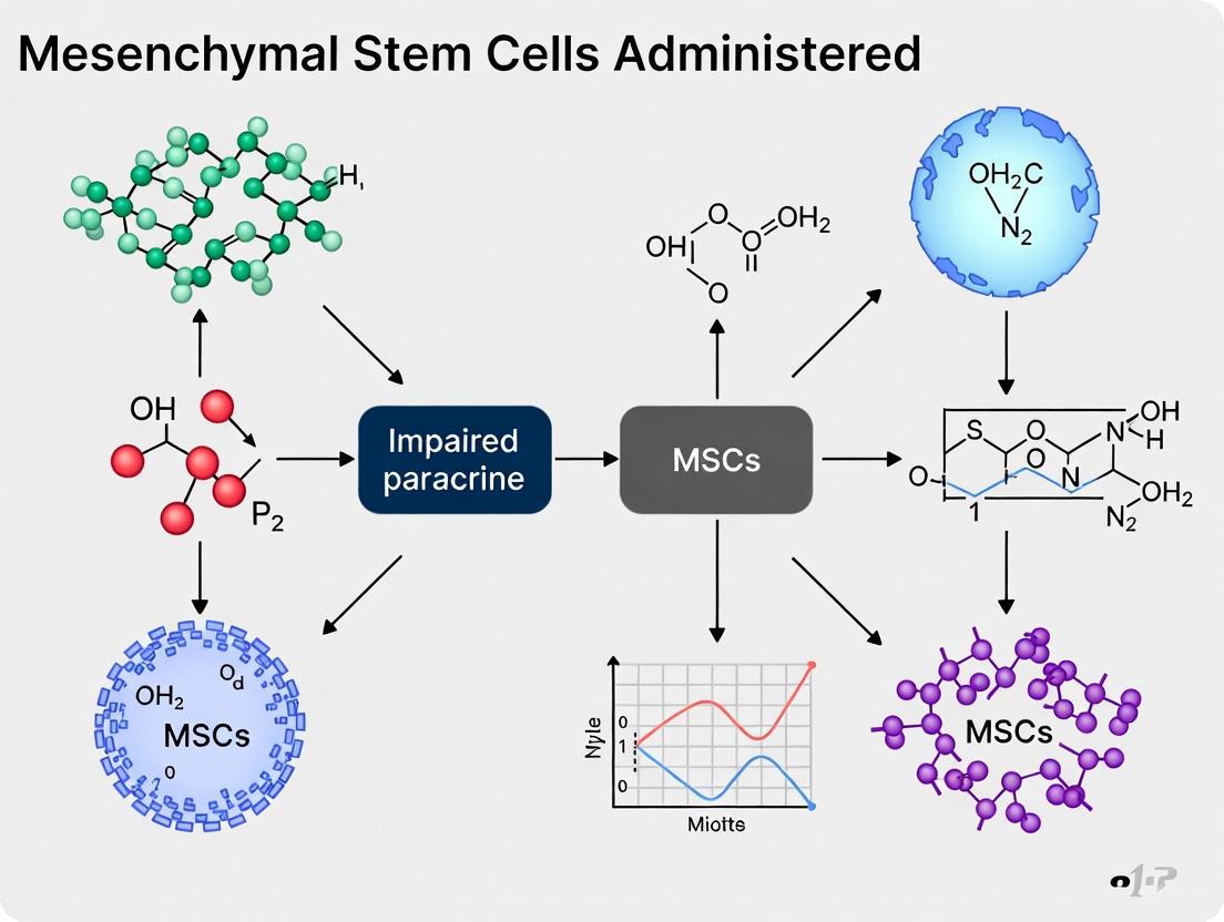
Overcoming Impaired Paracrine Function in Administered MSCs: Strategies for Enhancing Therapeutic Efficacy
The therapeutic promise of Mesenchymal Stromal Cells (MSCs) is increasingly attributed to their paracrine activity rather than direct differentiation.
Overcoming Impaired Paracrine Function in Administered MSCs: Strategies for Enhancing Therapeutic Efficacy
Abstract
The therapeutic promise of Mesenchymal Stromal Cells (MSCs) is increasingly attributed to their paracrine activity rather than direct differentiation. However, the clinical efficacy of MSC-based therapies is often limited by the impaired secretory function of administered cells, affected by factors such as poor survival, insufficient homing, and a hostile recipient microenvironment. This article provides a comprehensive analysis for researchers and drug development professionals, exploring the foundational biology of MSC paracrine mechanisms, methodological advances in priming and engineering, troubleshooting strategies for in vivo optimization, and validation through potency assays and clinical trial data. By synthesizing current research and emerging trends, this review aims to guide the development of next-generation MSC therapies with robust and reliable paracrine activity.
Deconstructing MSC Paracrine Mechanisms: From Secretome to Clinical Hurdles
For decades, the therapeutic potential of mesenchymal stromal cells (MSCs) was attributed to their ability to differentiate into various cell types and directly replace damaged tissues. However, a significant paradigm shift has occurred in the field. Research now indicates that the primary mechanism behind MSC therapy is not differentiation and engraftment, but rather their paracrine activity - the secretion of bioactive factors that modulate the host environment [1] [2] [3].
This shift in understanding carries profound implications for both basic research and clinical applications. This technical support article addresses the core challenges associated with impaired paracrine ability in administered MSCs and provides targeted troubleshooting guidance to enhance the efficacy of your therapeutic development.
Core Concepts: Understanding the Secretome
What constitutes the MSC secretome?
The MSC secretome is a complex mixture of bioactive molecules secreted by MSCs into the extracellular environment. It includes soluble proteins, growth factors, cytokines, chemokines, lipids, and extracellular vesicles (EVs) such as exosomes and microvesicles [2] [4]. These EVs themselves carry a cargo of proteins, lipids, and nucleic acids (including miRNAs and mRNAs) that can mediate cell-to-cell communication over distance [1].
Why has the field shifted toward the paracrine paradigm?
The paradigm shift from differentiation to paracrine signaling as the primary therapeutic mechanism is supported by several key observations:
- Limited Engraftment: Studies tracking administered MSCs consistently show poor long-term survival, retention, and engraftment at injury sites, yet therapeutic effects are still observed [2] [5].
- Conditioned Medium Efficacy: The conditioned medium from MSC cultures, which contains the secretome but not the cells themselves, can recapitulate many therapeutic benefits of whole MSC transplantation in disease models [2] [6].
- Rapid "Hit-and-Run" Mechanism: MSCs often exert long-lasting effects after they have been cleared from the body, supporting a "hit-and-run" or "touch and go" mechanism where their brief presence initiates regenerative cascades via secreted factors [5].
The following diagram illustrates the fundamental shift in how MSC therapeutic mechanisms are now understood.
Troubleshooting Guide: Addressing Impaired Paracrine Function
FAQ 1: How can I troubleshoot poor secretome production in my MSC cultures?
Problem: MSC cultures yield insufficient quantities of therapeutic factors in their secretome, leading to diminished experimental or therapeutic outcomes.
Diagnosis and Solutions:
- Verify Cell Source and Characterization: Different MSC sources (bone marrow, adipose tissue, umbilical cord) produce secretomes with varying compositions and potencies [1] [3]. Ensure your MSCs meet ISCT criteria (positive for CD73, CD90, CD105; negative for CD34, CD45, HLA-DR) and document the tissue source precisely [7] [3].
- Optimize Culture Conditions:
- Preconditioning with Hypoxia: Culture MSCs at 1-5% O₂ to better mimic physiological conditions and enhance secretion of pro-angiogenic (VEGF) and pro-survival factors [4].
- Inflammatory Priming ("Licensing"): Treat MSCs with low doses of pro-inflammatory cytokines (e.g., IFN-γ, TNF-α) to boost immunomodulatory factor secretion (IDO, PGE2, TSG-6) [7].
- 3D Culture Systems: Utilize spheroids or biomaterial scaffolds to create more physiologically relevant microenvironments that enhance paracrine factor production compared to 2D monolayers [2] [7].
- Monitor Population Doubling: High passage numbers (generally beyond P5-P8) can lead to cellular senescence and reduced secretome potency. Use early-passage cells and establish criteria for maximum allowable population doublings [1].
FAQ 2: What strategies can improve the in vivo homing and retention of administered MSCs?
Problem: After administration, MSCs show poor migration to target tissues and rapid clearance, limiting their local paracrine impact.
Diagnosis and Solutions:
- Route of Administration Optimization: The choice of administration route significantly impacts MSC distribution and retention [5].
- Enhance Homing Capacity:
- Chemokine Receptor Upregulation: Pretreat MSCs with cytokines or small molecules to increase expression of homing receptors (e.g., CXCR4, which responds to SDF-1 gradients at injury sites) [7] [5].
- Biomaterial-Assisted Delivery: Utilize hydrogels, scaffolds, or microparticles to protect MSCs from initial clearance mechanisms (particularly lung entrapment after IV injection) and provide sustained, local release at target sites [2] [7].
The table below summarizes the advantages and challenges of different administration routes for MSCs.
| Administration Route | Key Advantages | Primary Challenges | Best For |
|---|---|---|---|
| Intravenous (IV) | Minimally invasive, systemic distribution | Significant pulmonary first-pass effect; wide dissemination | Systemic conditions, GVHD [8] [5] |
| Local/Intralesional | High local concentration at target site | Technically challenging; potential for rapid efflux | Focal defects, osteoarthritis, cartilage repair [7] |
| Intra-arterial | Direct delivery to organ vascular beds | Risk of microemboli; requires specialized skills | Liver, kidney, myocardial applications [5] |
| Biomaterial-Encapsulated | Protected niche; sustained paracrine release | Additional complexity; biocompatibility concerns | Structured tissue engineering [2] [7] |
FAQ 3: How can I standardize the characterization of MSC secretome potency?
Problem: Inconsistent or undefined secretome composition leads to variable experimental results and therapeutic efficacy.
Diagnosis and Solutions:
- Implement Potency Assays:
- Functional Assays: Measure the ability of conditioned medium to suppress T-cell proliferation (immunomodulation), promote endothelial tube formation (angiogenesis), or inhibit fibroblast collagen production (anti-fibrosis) [1] [2].
- Molecular Profiling: Use targeted (ELISA) or untargeted (proteomics, miRNA sequencing) approaches to characterize key secreted factors [2].
- Consider Cell-Free Alternatives: If consistency proves unattainable with live MSCs, transition to using the purified secretome, conditioned medium, or isolated extracellular vesicles as a more definable biological product [2] [6].
The table below outlines key functional components of the MSC secretome and how to measure them.
| Secretome Function | Key Molecular Mediators | Recommended Assays |
|---|---|---|
| Immunomodulation | IDO, PGE2, TGF-β, IL-10, TSG-6 | T-cell suppression assay; IDO activity kit; PGE2 ELISA [1] [2] |
| Angiogenesis | VEGF, bFGF, ANG-1, miR-210 | HUVEC tube formation; chick chorioallantoic membrane assay; VEGF ELISA [2] |
| Anti-fibrosis | HGF, miR-29, miR-125b | Collagen gel contraction; fibroblast proliferation; α-SMA staining [2] |
| Anti-apoptosis | VEGF, STC-1, IGF-1, miR-214 | Annexin V/PI staining; caspase activity; mitochondrial membrane potential [2] |
Essential Research Toolkit
Key Research Reagents and Solutions
| Reagent/Solution | Function | Application Notes |
|---|---|---|
| Serum-free Medium | Collection of conditioned medium | Essential for uncontaminated secretome analysis; use for 24-48h conditioning [2] |
| IFN-γ (10-50 ng/mL) | Inflammatory priming | Boosts immunomodulatory capacity via IDO upregulation [7] |
| Hypoxia Chamber (1-5% O₂) | Physiologic preconditioning | Enhances angiogenic and survival factor secretion [4] |
| Transwell Migration Assay | Homing capacity assessment | Tests MSC response to SDF-1 or other chemoattractants [7] [5] |
| Lymphocyte Proliferation Kit | Potency assay (immunomodulation) | Measures functional suppression of activated PBMCs or T-cells [1] |
| VEGF ELISA Kit | Angiogenic potential quantification | Key biomarker for pro-angiogenic secretome [2] |
| Hydrogel Scaffolds (e.g., Alginate) | 3D culture & delivery | Enhances secretome production and in vivo retention [2] [7] |
| Extracellular Vesicle Isolation Kit | Secretome fractionation | Isolates exosomes/microvesicles for mechanistic studies [2] |
Experimental Protocols
Protocol 1: Inflammatory Priming to Enhance Immunomodulatory Secretome
Purpose: To boost the production of immunomodulatory factors in MSCs prior to administration or secretome collection.
Materials:
- Confluent (70-80%) MSC culture (P3-P5)
- Serum-free basal medium
- Recombinant human IFN-γ
- Centrifugal concentrators (3kD MWCO)
Procedure:
- Wash MSCs twice with PBS to remove serum components.
- Add serum-free medium containing 25 ng/mL IFN-γ.
- Incubate for 24-48 hours under standard culture conditions (37°C, 5% CO₂).
- Collect conditioned medium and centrifuge at 3,000 × g for 10 min to remove cell debris.
- Concentrate 10-fold using centrifugal concentrators.
- Analyze key factors (IDO, PGE2) by ELISA and validate functionality in T-cell suppression assays.
- Use immediately or store at -80°C with protease inhibitors.
Troubleshooting:
- Cytotoxicity: If cell death exceeds 15%, reduce IFN-γ concentration to 10 ng/mL.
- Insufficient IDO Induction: Verify MSC responsiveness by checking STAT1 phosphorylation via Western blot.
Protocol 2: Functional Validation of Angiogenic Secretome
Purpose: To quantitatively assess the angiogenic potential of MSC-derived secretome.
Materials:
- HUVECs (passage 3-5)
- Growth factor-reduced Matrigel
- MSC-conditioned medium (concentrated 10x)
- Basal endothelial medium (negative control)
- Endothelial growth medium (positive control)
- 96-well plates
- Imaging system with capillary network analysis software
Procedure:
- Thaw Matrigel on ice and coat 96-well plates (50 μL/well). Polymerize at 37°C for 30 min.
- Harvest HUVECs and resuspend at 1.0 × 10⁵ cells/mL in test media.
- Seed 100 μL cell suspension per well on polymerized Matrigel.
- Incubate at 37°C for 6-18 hours.
- Capture images (4x objective) of capillary-like structures from 3 random fields per well.
- Quantify total tube length, number of branches, and number of meshes using analysis software.
- Compare test samples to positive and negative controls. A potent angiogenic secretome should show ≥50% increase in tube formation versus negative control.
The following diagram illustrates the experimental workflow for priming MSCs and validating their secretome functionality, integrating both protocols.
Advanced Technical Considerations
Navigating the Regulatory Landscape for Paracrine-Based Products
As the field moves toward secretome-based or cell-free therapies, regulatory considerations evolve:
- Product Definition: MSC secretome products may be classified differently by regulatory agencies (e.g., as biological products rather than ATMPs), which can significantly impact development pathways [6].
- Potency Assays: Regulatory approval requires validated, quantitative potency assays that reliably predict clinical efficacy. Develop correlation between in vitro secretome profiling and in vivo functional outcomes early [1].
- Manufacturing Consistency: Implement rigorous quality control for secretome production, including standardized conditioning protocols, defined media, and comprehensive characterization of critical quality attributes [2] [6].
Emerging Solutions: Beyond Native MSCs
When native MSCs consistently demonstrate impaired paracrine function despite optimization, consider these advanced approaches:
- Genetic Engineering: Modify MSCs to overexpress specific therapeutic factors (e.g., VEGF, HGF) or homing receptors (CXCR4) to enhance targeted paracrine effects [7].
- Cell-Free Therapeutics: Transition to using purified extracellular vesicles or specific factor cocktails from MSC secretome, offering better definition, safety profile, and storage stability [2] [5].
- Biomaterial-Guided Paracrine Activity: Use engineered scaffolds that not only deliver MSCs but also actively instruct their paracrine behavior through mechanical and biochemical cues [2] [4].
By systematically addressing paracrine function through these troubleshooting approaches, researchers can significantly enhance the therapeutic reliability and efficacy of MSC-based applications, ultimately advancing more effective regenerative therapies.
Mesenchymal stem cells (MSCs) have long been investigated for their remarkable potential in regenerative medicine. Initially, the focus was on their capacity to differentiate into multiple cell types and engraft at injury sites. However, a paradigm shift has occurred with the realization that the primary therapeutic benefits of MSCs are mediated through their paracrine activity, not their long-term engraftment [9] [10]. It is now understood that as much as 80% of their regenerative potential can be attributed to the broad array of bioactive molecules they secrete [9]. This collective set of secretions is known as the MSC secretome.
The secretome represents a cornerstone for novel cell-free therapeutic strategies, circumventing major challenges associated with whole-cell transplants, such as low cell survival, poor engraftment, potential immunogenicity, and risks of lung entrapment or tumorigenicity [9] [11]. The secretome comprises two main components: a soluble fraction (growth factors, cytokines, chemokines) and a vesicular fraction (extracellular vesicles like exosomes and microvesicles) [9]. This technical support article details the anatomy of the MSC secretome and provides a practical guide for researchers aiming to harness its potential, particularly within the context of overcoming impaired paracrine ability in administered MSCs.
Decoding the Secretome: Core Components and Functions
The MSC secretome is a complex, dynamic mixture that acts as a primary mediator of intercellular communication. Its composition is not fixed but is "personalized" according to the local microenvironmental cues encountered by the parent MSCs [11]. The table below summarizes the key constituents and their primary biological roles.
Table 1: Core Components of the MSC Secretome and Their Functions
| Secretome Component | Key Examples | Primary Documented Functions |
|---|---|---|
| Soluble Factors | VEGF, HGF, FGF, IGF-1, TGF-β1, PGE2, IDO, IL-10, TSG-6 [9] [11] | Promotes angiogenesis, cell survival, and proliferation; exerts potent immunomodulation by suppressing T-cell proliferation, polarizing macrophages to an M2 anti-inflammatory state, and inhibiting dendritic cell maturation [9] [11]. |
| Extracellular Vesicles (EVs) | Exosomes, Microvesicles [9] | Acts as key delivery vehicles for proteins, lipids, and nucleic acids (e.g., miRNAs). Mediates intercellular communication by transferring bioactive cargo to recipient cells, influencing their gene expression and function [9] [12]. |
| EV Cargo (Molecular Payload) | miRNAs (e.g., miR-21, miR-29b), cytokines, growth factors [13] | Regulates gene expression in target cells; downregulates pro-apoptotic genes (e.g., Bax, caspases), reduces oxidative stress, and restores mitochondrial function [13]. |
The therapeutic effects of these components are multifaceted. In neurological contexts, the secretome has been shown to restore mitochondrial bioenergetics and reduce oxidative stress in human neural progenitor cells exposed to neurotoxins, partly by normalizing dysregulated miRNAs and mRNAs [13]. In the immune realm, factors like PGE2 and IDO are crucial for suppressing T-cell responses and inducing macrophage polarization toward the regenerative M2 phenotype [9] [11].
Figure 1: Workflow from MSC culture to the therapeutic application of its secretome, highlighting the two major component groups.
The Scientist's Toolkit: Essential Reagents and Materials
Successful research into the MSC secretome requires a suite of specific reagents and instruments. The following table outlines essential materials, drawing from experimental protocols cited in the literature.
Table 2: Key Research Reagent Solutions for MSC Secretome Studies
| Reagent/Material | Function/Application | Example from Literature |
|---|---|---|
| Flow Cytometry Antibodies | Characterization of MSC surface markers (ISCT criteria). | Antibodies against CD105, CD73, CD90 (positive) and CD45, CD34, CD14, CD11b, CD19, HLA-DR (negative) [3] [13]. |
| Culture Medium | In vitro expansion of MSCs and secretome collection. | α-MEM supplemented with 10% stem cell-qualified FBS and antibiotics [13]. |
| Preconditioning Agents | Modulating secretome composition to enhance therapeutic potency. | Pro-inflammatory cytokines (IFN-γ, TNF-α), hypoxic conditions (<5% O₂), or 3D culture environments [14]. |
| EV Isolation Kits | Isolation of extracellular vesicles from conditioned medium. | Use of commercial kits or ultracentrifugation for purifying exosomes and microvesicles [12]. |
| Polystyrene Beads | Calibration and size estimation of flow cytometers for EV analysis. | Green fluorescent beads of various sizes (20nm - 1.9μm) [15]. |
Troubleshooting Guide: Addressing Common Experimental Challenges
This section addresses specific issues researchers might encounter when working with the MSC secretome.
FAQ 1: How can I enhance the immunomodulatory potency of my MSC secretome?
- Challenge: The immunosuppressive properties of MSCs are not always inherent; they often require specific activation or "priming" to achieve full potency [14].
- Solution: Preconditioning MSCs prior to secretome collection is a key strategy.
- Cytokine Priming: Expose MSCs to pro-inflammatory cytokines like interferon-gamma (IFN-γ) and tumor necrosis factor-alpha (TNF-α). This upregulates the expression of immunomodulatory molecules such as IDO, PGE2, and PD-L1 [14].
- Hypoxic Culture: Culturing MSCs under hypoxic conditions (e.g., 1-3% O₂) can upregulate the secretion of trophic factors like VEGF, FGF-2, HGF, and IGF-1, and enhance the production of IDO and PGE2 [14] [13].
- 3D Culture: Growing MSCs as spheroids or in hydrogels can better maintain their immunosuppressive phenotype compared to traditional 2D monolayer culture [14].
FAQ 2: My secretome preparations are highly variable. How can I improve consistency?
- Challenge: The composition and therapeutic potential of the MSC secretome are deeply influenced by the cell source, culture conditions, and isolation protocols, leading to batch-to-batch variability [11] [12].
- Solution:
- Standardize Cell Source: Carefully select and characterize the MSC tissue source (e.g., bone marrow, adipose tissue, umbilical cord). Be aware that proteomic and miRNA profiles differ between sources [12] [3].
- Control Culture Conditions: Standardize passage number, cell confluence at harvest, serum quality (or use defined, serum-free media), and collection intervals for conditioned medium [3].
- Implement Quality Control: Use defined assays to characterize secretome batches. This can include protein quantification, ELISA for specific factors (e.g., VEGF, HGF), and nanoparticle tracking analysis (NTA) to determine EV concentration and size distribution [12].
FAQ 3: How can I efficiently load therapeutic cargo into MSC-derived extracellular vesicles?
- Challenge: Leveraging EVs as drug delivery vehicles requires efficient loading of therapeutic molecules.
- Solution: Cargo loading can be achieved through cell-based or non-cell-based methods.
- Cell-Based Loading: Transfect parent MSCs with the desired cargo (e.g., small RNAs, mRNAs). The cells will then package the cargo into EVs during their biogenesis. This method is suitable for large biomolecules but relies on transfection efficiency [14].
- Non-Cell-Based Loading: Isolate blank EVs first, then load the cargo directly.
- Electroporation: Effective for loading nucleic acids and some small molecules, though it may affect EV stability.
- Sonication/Freeze-Thaw: Can achieve relatively high loading efficiency but may cause aggregation or damage to EVs.
- Passive Incubation: Simple incubation of small molecule drugs (e.g., doxorubicin) with isolated EVs. This is straightforward but has low loading efficiency [14].
Detailed Experimental Protocol: Secretome Collection and Application in a Neurotoxicity Model
The following protocol is adapted from a study demonstrating the restoration of monocrotophos-induced toxicity in human neural progenitor cells (hNPCs) using the human MSC secretome [13].
Figure 2: Step-by-step experimental workflow for evaluating the restorative effects of the MSC secretome in a neural toxicity model.
Phase 1: MSC Culture and Secretome Collection
- Cell Culture: Culture Human Wharton's Jelly MSCs (HWJ-MSCs) in α-MEM supplemented with 10% stem cell-qualified FBS, 1% antibiotic-antimycotic, and 0.2% sodium bicarbonate at 37°C and 5% CO₂ [13].
- Characterization: Confirm MSC identity by flow cytometry for positive (CD105, CD73, CD90) and negative (CD34, CD45, HLA-DR) markers [13].
- Secretome Collection:
- At 70-80% confluence, wash cells with PBS and switch to a serum-free medium.
- Culture for 48 hours under standard or preconditioned (e.g., hypoxic, cytokine-stimulated) conditions.
- Collect the conditioned medium (CM), which contains the secretome.
- Secretome Processing:
- Centrifuge the CM at a minimum of 2000-3000 x g for 10-20 minutes to remove cell debris.
- Concentrate the supernatant using a 3 kDa molecular weight cut-off (MWCO) concentrator.
- Filter-sterilize the concentrated secretome using a 0.22 µm filter.
- Aliquot and store at -80°C.
Phase 2: Therapeutic Application in a Neural Toxicity Model
- Differentiate Neural Progenitor Cells (NPCs): Generate human NPCs (hNPCs) from human induced pluripotent stem cells (hiPSCs) and characterize them for markers like OCT-4 and SOX-2 [13].
- Induce Toxicity: Challenge the hNPCs with a neurotoxic agent, such as the organophosphate pesticide Monocrotophos (MCP), at a predetermined concentration to establish a model of impaired neuronal function.
- Apply Secretome: Treat the MCP-injured hNPCs with the prepared MSC secretome.
- Outcome Assessment:
- Viability and Oxidative Stress: Measure levels of reactive oxygen species (ROS), lipid peroxidation, and antioxidant enzymes.
- Mitochondrial Bioenergetics: Assess using assays like the MTT assay or more advanced Seahorse Analyzer technology.
- Molecular Profiling: Analyze changes in miRNA (e.g., miR-200, miR-34) and mRNA (e.g., P53, APAF) expression via qPCR and RNA sequencing.
- Proteomic Analysis: Use liquid chromatography-mass spectrometry (LC-MS/MS) to identify protein expression changes and secretome components [13].
Advanced Techniques: Flow Virometry for EV and Virus Analysis
The analysis of extracellular vesicles shares technical challenges with the field of virometry due to the small size of the particles. The following protocol, adapted from HIV-1 studies, provides a framework for high-sensitivity analysis of EVs [15].
- Instrument Calibration: Use a set of green fluorescent polystyrene bead standards (e.g., 20nm to 1.9μm) to calibrate the flow cytometer. This is critical for determining the instrument's detection threshold and for size estimation [15].
- Sample Preparation: Isolate EVs from the MSC secretome via ultracentrifugation or commercial kits. For fluorescent labeling, incubate EVs with specific dyes or antibodies against surface markers (e.g., CD63, CD81 for exosomes).
- Instrument Settings:
- Standard Flow Cytometer (e.g., BD FACSAria II): Set a threshold on a fluorescence channel (e.g., FITC) rather than on light scatter (FSC/SSC) to detect small particles. Reduce FSC voltage to minimize laser noise [15].
- Dedicated Nanoparticle Cytometer (e.g., Apogee A50): These instruments are optimized for submicron particles and may use large angle light scatter (LALS) thresholds for detection [15].
- Sorting and Downstream Analysis: Fluorescently labeled EVs can be sorted via Fluorescence-Activated Cell Sorting (FACS). Sorted populations can then be used for functional assays, such as treating target cells to assess their biological activity, or for further molecular characterization [15].
FAQ: What are the primary causes of impaired paracrine function in MSCs after administration?
Q: After I administer MSCs, why do they sometimes fail to produce the expected therapeutic paracrine factors?
The impaired paracrine function following administration is often due to a combination of factors related to the harsh in vivo environment and cellular stress. The primary causes include:
Hostile Microenvironment at Injury Sites: Administered MSCs often encounter a harsh microenvironment characterized by high levels of reactive oxygen species (ROS), inflammation, and hypoxia at the site of injury [16] [17]. This hostile milieu can overwhelm the cells, reducing their viability and capacity for protein synthesis and secretion, thereby impairing their paracrine activity.
Insufficient Homing and Poor Engraftment: A significant proportion of intravenously administered MSCs can become trapped in capillary networks, particularly in the lungs, a phenomenon known as the "pulmonary first-pass effect" [7] [18]. This prevents a sufficient number of cells from reaching the target tissue. Even those that do arrive often exhibit poor long-term survival and engraftment, with most transplanted cells being cleared within days to weeks [16].
Donor Heterogeneity and Cell Source Variability: The therapeutic potency of MSCs, including their paracrine function, is not uniform. It is influenced by the donor's age and health status, as well as the tissue source of the MSCs (e.g., bone marrow vs. umbilical cord) [19] [16]. This inherent biological variability can lead to inconsistent experimental and clinical outcomes.
Inadequate Preconditioning: MSCs that are expanded in vitro under standard conditions may not be equipped to handle the specific stresses they encounter in vivo. The absence of targeted preconditioning (e.g., exposure to hypoxia or pro-inflammatory cytokines) means the cells are not "primed" to mount a robust and effective paracrine response upon transplantation [7] [20].
FAQ: How can I troubleshoot poor migration and homing of MSCs to the target tissue?
Q: My tracking data shows that very few MSCs are reaching the intended site of injury. What could be going wrong?
Poor homing is a common hurdle. The table below summarizes the key issues and verification steps.
| Issue | Underlying Cause | Verification Experiments |
|---|---|---|
| Inefficient Systemic Delivery | Pulmonary first-pass effect; cells trapped in liver/spleen [7]. | Use IVIS or fluorescence imaging to track cell distribution post-administration. Check for high signal in lungs. |
| Weak Chemotactic Response | MSCs have low expression of homing receptors (e.g., CXCR4); target tissue has insufficient chemoattractant gradient [7] [17]. | Measure expression of homing receptors (CXCR4, CD44) on your MSC batch via flow cytometry. Analyze chemokine levels (SDF-1, MCP-1) in target tissue. |
| Administration Route Error | Intravenous injection may not be optimal for your target tissue; direct local injection might be required [7]. | Compare intravenous vs. intra-arterial vs. local injection in your disease model for final cell delivery efficiency. |
| Cell Size and Viability | Larger or clumped cells are physically trapped in capillaries; low pre-injection viability [18]. | Perform a cell size analysis before injection; ensure viability is >90% and cells are in a single-cell suspension. |
The following workflow can help diagnose homing problems systematically:
FAQ: What experimental protocols can I use to verify impaired paracrine secretion?
Q: What are the definitive lab experiments to confirm that my administered MSCs are actually suffering from impaired paracrine function?
To directly test the hypothesis of impaired paracrine function, a combination of in vitro and ex vivo analyses is required. Below is a detailed protocol for a key experiment.
Experimental Protocol: Analysis of MSC Secretome from Recovered Cells
Aim: To isolate MSCs from the target tissue post-administration and directly quantify their secretory capacity.
Materials:
- Animal Model of Disease: (e.g., rodent stroke model, myocardial infarction model).
- MSCs: Luciferase/GFP-labeled for tracking.
- Collagenase/DNase Solution: For tissue digestion.
- Fluorescence-Activated Cell Sorting (FACS): For isolating GFP+ MSCs.
- ELISA/Multiplex Immunoassay Kits: For quantifying cytokines (e.g., VEGF, HGF, IL-10, TGF-β).
- Cell Culture Plates: 96-well plates for secretome collection.
Method:
- Cell Administration: Administer GFP-labeled MSCs (e.g., 1-2 million cells) into your disease model via the chosen route (e.g., intravenous) [7].
- Tissue Harvest: At a critical timepoint post-administration (e.g., 24-72 hours), euthanize the animal and harvest the target tissue (e.g., brain for stroke, heart for MI) and a control organ (e.g., muscle).
- Single-Cell Suspension: Mince the tissue finely and digest it using a collagenase/DNase solution (e.g., 1-2 mg/mL for 30-60 mins at 37°C) to create a single-cell suspension.
- Cell Sorting: Use FACS to isolate live GFP+ MSCs from the tissue digest. The sorting gates should be set using tissues from animals that received no cells or unlabeled cells as a control.
- Secretome Collection: Plate a standardized number of recovered GFP+ MSCs (e.g., 10,000 cells) in a 96-well plate in serum-free medium. Culture for 24 hours. Centrifuge the collected conditioned media to remove any cells or debris.
- Secretome Analysis:
- Quantitative ELISA: Perform ELISAs for key paracrine factors like VEGF (angiogenesis) and TGF-β (immunomodulation).
- Multiplex Immunoassay: Use a multiplex assay to profile a broader panel of cytokines, growth factors, and chemokines simultaneously [20].
- Data Comparison: Compare the secretome profile of the recovered MSCs against the secretome profile of in vitro cultured MSCs (the baseline control).
Expected Outcome: Successful execution will reveal whether MSCs residing in the target tissue have a diminished, altered, or enhanced secretory profile compared to naive cells, providing direct evidence for impaired (or improved) paracrine function.
FAQ: What are the proven strategies to enhance paracrine functionin vivo?
Q: Knowing these hurdles, what can I do to my MSCs before administration to make their paracrine function more resilient?
Several preconditioning and engineering strategies have shown promise in preclinical studies for boosting the paracrine activity of MSCs. The quantitative benefits of some strategies are summarized in the table below.
| Strategy | Mechanism of Action | Key Paracrine Factors Enhanced (Sample Data) |
|---|---|---|
| Hypoxic Preconditioning [16] | Mimics physiological low O₂, activating HIF-1α signaling to boost pro-survival & angiogenic factor secretion. | VEGF (↑ ~50-80%), FGF2 (↑ ~30%), HGF (↑ ~25%) [16]. |
| Inflammatory Priming (e.g., IFN-γ, TNF-α) [20] | "Licenses" MSCs to exert stronger immunomodulatory effects via induction of regulatory molecules. | IDO1 (↑ >100%), PGE2 (↑ ~60%), TGF-β (↑ ~40%) [20]. |
| Biophysical Stimulation (pFUS) [20] | Ultrasound waves mechanically stimulate cells, altering cytokine secretion profiles in an intensity-dependent manner. | Low-intensity pFUS upregulated IL-10, IL-1RA, VEGF in BM-MSCs [20]. |
| Genetic Modification [16] | Overexpression of specific genes (e.g., Akt, Hif-1α) to enhance survival and trophic factor production. | Akt-MSCs show ↑ VEGF, FGF2, IGF-1 secretion and superior survival post-transplantation [16]. |
The logical relationship between the hurdle, the strategy, and the molecular mechanism can be visualized as follows:
The Scientist's Toolkit: Key Research Reagent Solutions
The following table details essential materials and reagents used in the experiments and strategies discussed above.
| Research Reagent / Tool | Function in Experimental Context |
|---|---|
| GFP/Luciferase Labeling [16] | Enables real-time tracking of MSC distribution, homing efficiency, and persistence in vivo using imaging systems. |
| FACS (Fluorescence-Activated Cell Sorter) | Critical for isolating pure populations of administered (e.g., GFP+) MSCs from heterogeneous tissue digests for downstream secretome analysis. |
| Multiplex Immunoassay (Luminex) [20] | Allows simultaneous quantification of dozens of cytokines, chemokines, and growth factors from small volumes of conditioned media or serum. |
| pFUS System [20] | A non-invasive technology used to mechanically precondition MSCs in vitro or even in vivo to modulate their paracrine secretion profile. |
| Hypoxia Chamber | A sealed chamber that maintains a low-oxygen environment (e.g., 1-5% O₂) for preconditioning MSCs prior to administration, boosting their resilience and pro-angiogenic output. |
| Recombinant Cytokines (IFN-γ, TNF-α) [20] | Used for inflammatory priming of MSCs in culture to enhance their immunomodulatory potency and license them for stronger therapeutic effects. |
Troubleshooting Guide: Addressing Common Homing and Engraftment Challenges
This guide helps diagnose and resolve the primary causes of poor mesenchymal stem cell (MSC) engraftment observed in experimental models.
Table 1: Troubleshooting MSC Homing and Engraftment Failures
| Observed Problem | Potential Underlying Cause | Recommended Solutions & Experimental Considerations |
|---|---|---|
| Low Cell Survival Post-Infusion | Apoptosis/Anoikis due to harsh in vivo microenvironment (ROS, ischemia, inflammation) [21] [22]. | Preconditioning: Incubate MSCs with melatonin, atorvastatin, or IGF-1 to activate pro-survival pathways like PI3K/AKT [21].Genetic Modification: Overexpress anti-apoptotic genes (e.g., Bcl-2) [21].Biomaterials: Use thermosensitive hydrogel to encapsulate and protect cells during delivery [21]. |
| Insufficient Homing to Target Tissue | Poor navigation through the circulatory system and failure to extravasate [23] [22]. | Preconditioning: Prime MSCs with hypoxia or cytokines (e.g., SDF-1) to upregulate homing receptors (CXCR4, integrins) [22].Delivery Route: Use intra-arterial delivery to bypass the first-pass lung entrapment seen with intravenous injection [24].Cell Engineering: Modify MSC surface with PSGL-1 or Sialyl-Lewis X to enhance rolling on endothelial selectins [22]. |
| Impaired Paracrine Function | Failure of MSCs to adequately respond to inflammatory signals, often linked to deficient signaling pathways [25]. | Pathway Activation: Ensure critical pathways like NF-κB are functional. Research indicates Rap1 is essential for NF-κB activity and subsequent immunomodulatory cytokine production [25].Licensing: Pre-treat MSCs with pro-inflammatory cytokines (IFN-γ, TNF-α) to enhance their immunosuppressive potency [24] [25]. |
| Poor Long-Term Engraftment & Transient Presence | Cell death after initial homing or failure to anchor/retain in the tissue niche [24] [22]. | Improve Niche Compatibility: Co-transplant MSCs with supportive ECM proteins or use biomimetic scaffolds [26].Enhance Adhesion: Modulate expression of integrins (e.g., α4β1/VLA-4) and their ligands (VCAM-1) to improve adhesion to niche cells [23] [22]. |
| Adverse Thrombotic Events | High expression of procoagulant tissue factor (TF/CD142) on certain MSC sources, especially at high doses [27]. | Product Testing: Quantify TF/CD142 expression on your MSC product via flow cytometry or ELISA [27].Source Selection: Consider using bone marrow-derived MSCs (BM-MSCs) which have lower inherent TF expression compared to some perinatal or adipose-derived cells [27].Dose Adjustment: Re-evaluate cell dosage, as risk increases with higher cell numbers [27]. |
Frequently Asked Questions (FAQs)
FAQ 1: What are the key cellular steps in the systemic homing of intravenously infused MSCs? The systemic homing of MSCs is an active, multi-step process reminiscent of leukocyte trafficking. After infusion, cells must first navigate the circulatory system. The subsequent homing cascade involves: [22]
- Rolling: Initial tethering and slow rolling of MSCs on the activated endothelial lumen, mediated primarily by P-selectin and its ligand CD24, as well as VLA-4/VCAM-1 interactions [22].
- Activation: G protein-coupled chemokine receptors (e.g., CXCR4) on MSCs are activated by ligands (e.g., SDF-1) upregulated in injured tissues [23] [22].
- Arrest & Adhesion: Activation triggers firm adhesion to the endothelium, largely dependent on integrins like VLA-4 binding to VCAM-1 [23] [22].
- Transmigration (Diapedesis): Adherent MSCs crawl and extravasate across the endothelial barrier into the parenchymal tissue [22].
FAQ 2: Why is the therapeutic engraftment of MSCs typically so low, and how is it measured? Engraftment rates are often below 5% and are transient, with most cells disappearing within days to a few weeks post-transplantation [22] [28]. This is attributed to a confluence of factors:
- Harsh Microenvironment: Cells face oxidative stress, inflammation, and ischemia in the injured tissue, triggering apoptosis [21] [22].
- Lung Entrapment: A significant portion of intravenously infused cells are physically trapped in the pulmonary capillaries, a phenomenon known as the "first-pass" effect [24].
- Lack of Sufficient Homing Signals: The cells may not adequately express the necessary receptors to respond to chemoattractants from the target site [23]. Engraftment is quantified using various methods, including in vivo imaging (bioluminescence, fluorescence), radionuclide labeling (PET, SPECT), and ex vivo analysis of excised tissues via qPCR, flow cytometry, or histology for specific genetic or fluorescent markers [24].
FAQ 3: How does the tissue source of MSCs impact their homing potential and safety profile? The tissue source introduces significant heterogeneity in MSC properties [27] [19] [28].
- Homing Potential: MSCs from different sources express varying levels of homing receptors (e.g., CXCR4, integrins). For instance, umbilical cord-derived MSCs (UC-MSCs) often exhibit higher proliferative and migratory capacities compared to bone marrow-derived MSCs (BM-MSCs) [19].
- Safety Profile (Hemocompatibility): A critical safety consideration is the expression of procoagulant tissue factor (TF/CD142). BM-MSCs are generally more hemocompatible, while MSCs from perinatal tissues (e.g., placenta, umbilical cord) or adipose tissue can express much higher levels of TF, increasing the risk of thrombotic events upon infusion, especially at high doses [27].
FAQ 4: What is the relationship between MSC engraftment and their paracrine function? The relationship is dual in nature. First, a certain level of engraftment and local survival is likely required for MSCs to secrete trophic factors at a therapeutically relevant concentration within the target tissue [24] [25]. Second, the functional potency of the engrafted MSCs is paramount; simply being present is insufficient. The cells must be "licensed" by the local microenvironment to adopt an immunosuppressive phenotype. For example, the immunomodulatory potency of MSCs is heavily dependent on paracrine factors, and deficiencies in key signaling pathways (e.g., Rap1/NF-κB) can severely impair cytokine production and therapeutic efficacy, even if the cells engraft [25].
Experimental Protocols for Enhancing Homing and Engraftment
Protocol: Chemical Preconditioning to Improve MSC Survival
This protocol uses Melatonin to activate the PI3K/AKT pro-survival pathway, protecting MSCs from apoptosis post-transplantation [21].
Workflow Overview
Step-by-Step Methodology:
- Cell Culture: Isolate and culture MSCs (e.g., from bone marrow) until 70-80% confluency in standard culture flasks [21].
- Preconditioning:
- Prepare a working solution of Melatonin (e.g., 1µM - 10µM) in the standard culture medium [21].
- Replace the existing medium with the Melatonin-containing medium.
- Incubate the cells for 24-48 hours under normal culture conditions (37°C, 5% CO₂).
- Harvesting: After incubation, wash the cells with PBS to remove residual Melatonin. Harvest the MSCs using a standard dissociation reagent like trypsin-EDTA.
- Validation (In Vitro):
- TUNEL Assay: Subject preconditioned and control MSCs to oxidative stress (e.g., H₂O₂) and quantify apoptosis rates using a TUNEL assay to confirm reduced cell death.
- Western Blot: Analyze cell lysates for increased phosphorylation of AKT, indicating successful activation of the pro-survival pathway.
Protocol: Genetic Modification to Enhance MSC Homing
This protocol involves modifying MSCs to overexpress the CXCR4 receptor, improving their chemotactic response to the SDF-1 gradient in injured tissues [22].
Workflow Overview
Step-by-Step Methodology:
- Viral Transduction:
- Culture MSCs to 50-60% confluency.
- Replace the medium with a lentiviral vector containing the human CXCR4 gene and a selection marker (e.g., puromycin resistance) at a predetermined Multiplicity of Infection (MOI). Include polybrene (e.g., 8 µg/mL) to enhance transduction efficiency.
- After 24 hours, replace the virus-containing medium with fresh growth medium.
- Selection & Expansion:
- 48 hours post-transduction, add the selection antibiotic (e.g., puromycin) to the culture medium to select for successfully transduced cells.
- Culture the selected cells for several passages to establish a stable line.
- Validation:
- Flow Cytometry: Confirm high-level surface expression of CXCR4 protein compared to untransduced control MSCs.
- Transwell Migration Assay: Validate enhanced functional homing by placing modified and control MSCs in the upper chamber of a transwell insert, with SDF-1α in the lower chamber. After several hours, count the number of cells that have migrated through the membrane. CXCR4-overexpressing MSCs should show significantly higher migration towards SDF-1α.
The Scientist's Toolkit: Key Research Reagents & Materials
Table 2: Essential Reagents for Investigating MSC Homing and Engraftment
| Category | Reagent / Material | Primary Function in Research | Key Considerations |
|---|---|---|---|
| Preconditioning Agents | Melatonin [21] | Activates PI3K/AKT pathway to protect against apoptosis. | Test a dose range (e.g., 1-10 µM) for optimal effect. |
| SDF-1α (CXCL12) [23] [22] | Licenses MSCs and is used in vitro to test/enhance CXCR4-mediated migration. | Critical for validating homing receptor function in migration assays. | |
| IFN-γ & TNF-α [24] | "Licenses" MSCs, enhancing their immunomodulatory paracrine function. | Mimics inflammatory in vivo environment. | |
| Cell Tracking & Imaging | Luciferase Reporter [24] | Enables in vivo bioluminescence imaging (BLI) for longitudinal cell tracking. | Requires genetic modification; signal is proportional to viable cell number. |
| GFP Reporter [24] [25] | Allows histological identification and fluorescent-based tracking of MSCs. | Useful for endpoint analysis of engraftment location. | |
| Genetic Modification Tools | Lentiviral Vectors (e.g., CXCR4) [22] | Stably modifies MSCs to overexpress homing receptors. | Optimize MOI to balance efficiency and cell viability. |
| siRNA/shRNA (e.g., against Rap1) [25] | Knocks down specific genes to study their function in paracrine signaling. | Used to validate mechanisms, e.g., Rap1's role in NF-κB signaling. | |
| Functional Assays | Transwell / Boyden Chamber [22] | Standard in vitro assay to quantify MSC migration toward a chemoattractant (e.g., SDF-1). | Key for pre-validating homing potential before in vivo studies. |
| Flow Cytometry Antibodies (CD142/TF) [27] | Quantifies procoagulant tissue factor expression for safety assessment. | Essential for screening MSC products for thrombotic risk. |
Frequently Asked Questions (FAQs): Core Concepts
FAQ 1: What is meant by the "paracrine activity" of MSCs, and why is it therapeutically important? The paracrine activity of Mesenchymal Stem Cells (MSCs) refers to their ability to secrete bioactive molecules—such as growth factors, cytokines, and extracellular vesicles (EVs)—that mediate therapeutic effects, rather than relying on direct cell replacement [3] [16]. These secreted factors can modulate the immune system, reduce inflammation, promote angiogenesis, and activate endogenous repair pathways in damaged tissues [16] [29]. The importance of this mechanism has grown as research shows that after administration, most MSCs do not engraft long-term but are rapidly cleared, with their therapeutic benefits being largely mediated by their secretome [30] [16]. This makes the paracrine effect a primary driver of the observed clinical outcomes.
FAQ 2: How does a diseased host microenvironment "quench" or impair this paracrine function? A diseased host microenvironment can quench MSC paracrine function through several mechanisms:
- Sustained Pro-Inflammatory Signals: While a mild inflammatory cue can activate MSCs, a highly inflammatory microenvironment with persistent high levels of pro-inflammatory cytokines like TNF-α and IFN-γ can exhaust MSC function, leading to cellular senescence or a loss of their immunomodulatory capacity [30] [31].
- Dysregulated Epigenetic Landscape: The disease state can alter the epigenetic profile (e.g., DNA methylation, histone modifications) of both host cells and administered MSCs. For instance, MSCs derived from diseased donors or exposed to a pathologic microenvironment may carry aberrant epigenetic marks that suppress the expression of key therapeutic factors [30].
- Metabolic Stressors: Conditions like hypoxia and oxidative stress, common in many disease states (e.g., diabetic complications, myocardial infarction), can disrupt normal MSC metabolism and alter the composition of their secretome, reducing its reparative potency [32] [30].
FAQ 3: What are the key host-derived factors that contribute to this quenching effect? The key factors are often soluble mediators and cellular components of the diseased tissue milieu. The table below summarizes the primary culprits.
Table 1: Key Host-Derived Factors that Quench MSC Paracrine Activity
| Factor Category | Specific Examples | Impact on MSC Paracrine Function |
|---|---|---|
| Pro-inflammatory Cytokines | TNF-α, IFN-γ, IL-1β [31] | Can over-activate and exhaust MSCs, leading to reduced production of anti-inflammatory mediators like IDO1 and PGE2, and potentially inducing senescence [30] [31]. |
| Metabolic Stressors | Reactive Oxygen Species (ROS), Hypoxia [32] | Disrupts MSC metabolism, can trigger DNA damage, and alters the cargo (e.g., miRNAs, proteins) packaged into secreted extracellular vesicles [30]. |
| Profibrotic Mediators | TGF-β1 [32] | Can push MSCs toward a pro-fibrotic phenotype, shifting the secretome away from anti-fibrotic and regenerative functions. |
| Components of the Immune Microenvironment | M1 Macrophages, Activated T Cells [29] | Create a feed-forward loop of inflammation that MSCs may be unable to sufficiently counteract, thereby quenching their immunomodulatory paracrine activity. |
FAQ 4: What are the functional consequences of a quenched secretome on experimental outcomes? A quenched MSC secretome leads directly to failed experiments and inconsistent data through several measurable outcomes:
- Reduced Efficacy in Disease Models: Treated animals show significantly lower improvement in key metrics (e.g., reduced fibrosis resolution, impaired wound closure, poorer functional recovery) compared to those treated with MSCs possessing a potent secretome [33] [32] [34].
- Failure to Polarize Macrophages: A key anti-inflammatory mechanism of MSCs is their ability to shift macrophages from a pro-inflammatory (M1) to a reparative (M2) phenotype. A quenched secretome fails to drive this polarization, perpetuating inflammation [33] [29].
- Diminished Proliferation of Repair Cells: The conditioned medium from impaired MSCs will fail to stimulate the proliferation of key repair cells like articular chondrocytes or dermal fibroblasts in co-culture assays [34].
Troubleshooting Guides: Diagnosing and Solving Paracrine Quenching
Troubleshooting Guide 1: Diagnosing a Quenched Paracrine Secretome
Problem: Your MSC-based therapy is showing inconsistent or poor efficacy in a disease model, and you suspect the host microenvironment is quenching the paracrine activity.
Solution: Follow this diagnostic workflow to identify the nature of the impairment.
Diagram: Experimental Workflow for Diagnosing a Quenched Secretome
Step-by-Step Diagnostic Procedures:
Step 1: Perform In Vitro Potency Assays on Recovered MSCs.
- Objective: To determine if MSCs exposed to the diseased environment have lost their intrinsic immunomodulatory functions.
- Protocol:
- MSC Isolation: After treating your in vivo disease model, isolate MSCs from the target tissue or retrieve them from perfusion culture systems that mimic the host environment.
- T Cell Suppression Assay:
- Co-culture recovered MSCs with activated peripheral blood mononuclear cells (PBMCs) or purified T cells from a healthy donor [31].
- Use a mitogen like phytohemagglutinin (PHA) or anti-CD3/CD28 antibodies to activate T cells.
- After 72-96 hours, measure T cell proliferation using a standardized assay like [3H]-thymidine incorporation or CFSE dilution followed by flow cytometry [31].
- Expected Result: Potent MSCs should suppress T cell proliferation by ≥50-70%. A quenched MSC population will show significantly lower suppression (e.g., ≤30%) [31].
- IDO1 Activity Measurement:
- Culture recovered MSCs in a medium supplemented with tryptophan.
- Activate them with a cytokine cocktail (e.g., 10 ng/mL TNF-α + 100 ng/mL IFN-ɣ) for 48 hours [31].
- Collect the conditioned medium and measure the concentration of kynurenine, the product of IDO1-mediated tryptophan catabolism, using a spectrophotometer or ELISA.
- Expected Result: Functional MSCs will produce high levels of kynurenine (>3000 pg/mL). Quenched MSCs will show reduced production [31].
Step 2: Characterize the Host Microenvironment.
- Objective: To quantify the levels of quenching factors present in the diseased tissue.
- Protocol:
- Collect tissue homogenates or plasma/serum from your disease model at the time of MSC administration.
- Use a multiplex cytokine array or ELISA to quantify the concentrations of key pro-inflammatory cytokines such as TNF-α, IFN-γ, and IL-1β [31].
- Perform immunophenotyping of immune cells in the tissue (e.g., via flow cytometry) to assess the ratio of M1 to M2 macrophages and the activation state of T cells [33] [29].
- Data Correlation: Correlate high levels of inflammatory cytokines and a dominant M1 macrophage profile with the observed reduction in MSC potency from Step 1.
Step 3: Analyze the MSC Secretome Directly.
- Objective: To directly assess the quantity and quality of vesicles and factors secreted by MSCs.
- Protocol:
- Extracellular Vesicle (EV) Isolation: Collect conditioned medium from your recovered MSCs. Isolve EVs using sequential ultracentrifugation:
- EV Characterization:
- Nanoparticle Tracking Analysis (NTA): To determine the particle size distribution and concentration [33] [34].
- Transmission Electron Microscopy (TEM): To confirm the classic cup-shaped morphology of EVs [33] [34].
- Western Blot: To confirm the presence of EV markers (e.g., CD9, CD81, TSG101) and absence of negative markers (e.g., calnexin) [33] [34].
- Cargo Analysis:
- Extract RNA and protein from isolated EVs.
- Perform microRNA sequencing and mass spectrometry-based proteomics to compare the cargo profile of EVs from quenched versus potent MSCs. Look for deficits in key anti-fibrotic (e.g., miR-let7c) or immunomodulatory miRNAs (e.g., miR-21, miR-146) and proteins (e.g., USP10) [33] [32] [35].
Troubleshooting Guide 2: Strategies to Overcome Paracrine Quenching
Problem: You have identified that the host microenvironment is quenching your MSC therapy. What interventions can you implement to rescue paracrine activity?
Solution: Employ preconditioning or engineering strategies to "armor" MSCs against the hostile environment.
Diagram: Strategic Approaches to Overcome Paracrine Quenching
Detailed Intervention Protocols:
Intervention 1: Cytokine Preconditioning (Priming).
- Rationale: Pre-exposing MSCs to a mild inflammatory signal in vitro can boost their anti-inflammatory machinery, preparing them for the harsh in vivo environment [31].
- Protocol:
- Culture MSCs to 70-80% confluence.
- Replace the medium with fresh medium containing a priming cocktail. A common and effective cocktail is 10 ng/mL TNF-α + 100 ng/mL IFN-γ [31].
- Incubate for 24-48 hours.
- Wash the cells with PBS to remove cytokines before harvesting for administration.
- Expected Outcome: Preconditioned MSCs will exhibit significantly higher expression of IDO1 and PGE2, leading to enhanced suppression of T cell proliferation and improved efficacy in disease models [31].
Intervention 2: Genetic Engineering to Enhance Secretome.
- Rationale: Directly modifying MSCs to overexpress key therapeutic factors can ensure consistent, high-level production despite the hostile microenvironment [33].
- Protocol (Example: Overexpressing USP10):
- Gene Cloning: Clone the full-length human USP10 cDNA into a lentiviral expression vector.
- Virus Production: Generate high-titer lentiviral particles in a packaging cell line like HEK293T.
- MSC Transduction: Infect MSCs with the USP10-lentivirus in the presence of a transduction enhancer like polybrene.
- Selection: Use antibiotic selection (e.g., Puromycin) to create a stable, polyclonal population of USP10-overexpressing MSCs [33].
- Expected Outcome: Engineered MSCs will secrete EVs enriched with USP10, which stabilizes the transcription factor KLF4 in recipient macrophages via deubiquitination. This promotes an anti-fibrotic, M2-like macrophage phenotype and enhances tissue repair, as demonstrated in models of liver fibrosis [33].
Intervention 3: Shift to a Potent, Defined Cell-Free EV Approach.
- Rationale: Bypass the vulnerability of live cells by using their secreted EVs. This allows for the use of clonally derived MSC lines with pre-validated high potency, ensuring a consistent and high-quality product [34].
- Protocol:
- Source Selection: Use a characterized, high-potency MSC line (e.g., the CD317neg Y201 clonal line, which shows superior EV bioactivity) [34].
- EV Manufacturing: Produce and isolate EVs from these MSCs at scale using methods like ultracentrifugation or tangential flow filtration.
- Quality Control: Rigorously characterize each EV batch via NTA, TEM, and Western Blot to ensure consistency.
- Administration: Administer the purified EVs to the disease model. The dosage can be optimized based on particle count or total protein (e.g., 150 μg EVs per mouse) [33].
- Expected Outcome: EVs from a potent MSC source will reliably deliver their therapeutic cargo (proteins, miRNAs) to recipient cells, promoting chondrocyte proliferation, suppressing T cell activation, and reducing disease activity in inflammatory models, independent of the host's ability to support live MSCs [34].
The Scientist's Toolkit: Research Reagent Solutions
Table 2: Essential Reagents for Studying MSC Paracrine Quenching
| Reagent / Tool | Function / Application | Specific Examples & Notes |
|---|---|---|
| Pro-inflammatory Cytokines | For in vitro priming of MSCs and creating disease-mimicking conditions. | TNF-α, IFN-γ, IL-1β. Use at 10-100 ng/mL for priming [31]. |
| EV Isolation Kits | For purifying small extracellular vesicles (sEVs) from MSC-conditioned medium. | Ultracentrifugation is the gold standard; commercial kits (e.g., based on size-exclusion chromatography) can be alternatives [33] [34]. |
| Characterization Equipment | For validating the identity and quantity of isolated EVs. | Nanosight LM14 (NTA) for size/concentration; Transmission Electron Microscope for morphology; Western Blot for markers (CD9, CD81, TSG101) [33] [34]. |
| Lentiviral Vectors | For genetic engineering of MSCs to overexpress protective or therapeutic factors. | Used to stably overexpress genes like USP10 or KLF4 to enhance EV potency [33]. |
| Potency Assay Kits | For quantifying the functional capacity of MSCs and their secretome. | IDO1 Activity Kits (measure kynurenine); PGE2 ELISA Kits; T Cell Proliferation Assay Kits (e.g., CFSE-based) [31]. |
| Defined MSC Lines | To reduce heterogeneity and obtain a consistent, potent source of cells/EVs. | Clonal MSC lines (e.g., Y201) demonstrate superior and more reproducible EV bioactivity compared to heterogeneous populations [34]. |
Strategic Enhancement of MSC Secretory Capacity: Priming, Engineering, and Delivery
FAQs: Core Concepts for Researchers
Q1: Why is preconditioning necessary for enhancing MSC therapy in conditions like acute kidney injury (AKI)?
After administration, MSCs face a harsh microenvironment (e.g., oxidative stress, inflammation, and anoikis) in injured tissues, leading to massive cell death—often exceeding 80-90% within the first week [21]. This low survival rate, coupled with impaired paracrine ability, significantly limits the clinical efficacy of MSC-based treatments. Preconditioning is an adaptive strategy designed to prepare MSCs for this challenging environment, thereby enhancing their survival, retention, and secretory function post-transplantation [36] [21].
Q2: What is the biological rationale behind using a combination of hypoxia and inflammatory cytokines for preconditioning?
This combination strategy aims to mimic the in vivo microenvironment of damaged tissue, which is often characterized by both low oxygen tension (hypoxia) and a pronounced inflammatory response [36]. Hypoxia preconditioning primarily enhances the expression of pro-survival genes and angiogenic factors [36] [21]. Concurrently, priming with inflammatory cytokines like IFN-γ, TNF-α, and IL-1β "licenses" the MSCs, potently upregulating key immunomodulatory factors such as IDO, PGE2, and TSG-6 [36] [37]. This synergistic approach prepares MSCs to better withstand in vivo stresses and exert stronger therapeutic effects.
Q3: How does cytokine priming affect donor-dependent heterogeneity in MSC potency?
A key benefit of cytokine priming is the reduction of donor-dependent heterogeneity. Research shows that preconditioning with a proinflammatory cocktail (IFN-γ, TNF-α, and IL-1β) enhances the immunomodulatory capacity of MSCs from different donors and tissue sources (e.g., bone marrow and adipose tissue) more consistently, making their therapeutic profile more uniform and predictable [37].
Troubleshooting Experimental Protocols
Issue: Low MSC Survival After Preconditioning
- Potential Cause: Overly harsh preconditioning conditions, such as excessive cytokine concentrations or severely low oxygen levels, can induce apoptosis and senescence [36].
- Solution: Optimize preconditioning parameters. A protocol using 2% O₂ for 24 hours with a cytokine mix (e.g., IFN-γ at 20 ng/ml, TNF-α at 10 ng/ml, IL-1β at 20 ng/ml) has been shown to enhance immunomodulatory function without severely compromising viability [36] [37]. Consider testing a range of conditions to find the optimal balance for your specific MSC source.
Issue: Inconsistent Immunomodulatory Outcomes
- Potential Cause: Uncontrolled variability in MSC populations due to donor or source differences [37].
- Solution: Implement standardized cytokine priming protocols. Preconditioning can reduce inter-donor variability. Ensure consistent MSC characterization (surface marker expression, differentiation potential) and use defined cytokine concentrations and hypoxia exposure times across all experiments [37].
Issue: Poor Engraftment and Retention of Administered MSCs
- Potential Cause: Cells are not adequately prepared for the ischemic and inflammatory microenvironment of the target tissue.
- Solution: Explore combinatorial preconditioning and delivery strategies. Preconditioning with melatonin or using a thermosensitive hydrogel for delivery have been shown in AKI models to improve MSC survival and retention post-transplantation [21].
Table 1: Preconditioning Strategies to Improve MSC Survival and Paracrine Ability
| Preconditioning Strategy | Specific Agent/Condition | Reported Outcomes | Proposed Mechanism |
|---|---|---|---|
| Cytokine/Chemical Incubation | 14S,21R-diHDHA (DHA-derived mediator) | ↑ Survival rate; ↓ Apoptosis in mouse I/R model [21] | Activation of PI3K/AKT signaling pathway [21] |
| S-nitroso N-acetyl penicillamine (SNAP, NO donor) | ↑ Proliferation, survival, and engraftment in ischemic kidney [21] | ↑ Expression of AKT and Bcl-2 [21] | |
| Atorvastatin | ↑ Viability of implanted MSCs; improved renal function [21] | Suppression of TLR4 signaling [21] | |
| Melatonin | ↑ MSC survival after intraparenchymal injection; accelerated renal recovery [21] | Antioxidant effects [21] | |
| Muscone | Enhanced proliferative ability of BMSCs in gentamicin-induced AKI [21] | Not specified in source | |
| IGF-1 (Insulin-like Growth Factor-1) | ↑ MSC number; ↓ Apoptosis [21] | Not specified in source | |
| Hypoxia & Cytokine Combination | 2% O₂ + IL-1β, TNF-α, IFN-γ | Enhanced immunomodulatory properties; inhibited NK cell toxicity; did not damage core biological characteristics [36] | Upregulation of immune-related genes (e.g., IDO, TSG-6); decreased coagulation-related tissue factor [36] |
| Culture Improvement | 3D Spheroid Culture | ↑ Survival rate, ECM, ROS-scavenging proteins, Bcl-2, and pro-survival p-AKT in rat I/R model [21] | Enhanced resistance to stress |
| Hydrogel Delivery | Thermosensitive Hydrogel | ↑ Survival rate; ↓ Apoptosis in rat I/R model [21] | Physical protection and improved retention |
Table 2: Key Signaling Pathways in MSC Preconditioning
| Signaling Pathway | Preconditioning Stimulus | Key Molecular Players | Functional Outcome in MSCs |
|---|---|---|---|
| PI3K/AKT | 14S,21R-diHDHA, SNAP, 3D Culture [21] | AKT, Bcl-2 | Promotes cell survival, proliferation, and resistance to apoptosis [21] |
| HIF Signaling | Hypoxia, Pro-inflammatory Cytokines (IL-6, TNF-α, MCP1) [38] | HIF1α, HIF2α, HIF3α | HIF1α/2α: Adaption to hypoxia, angiogenesis. HIF3α: Regulated by cytokines via NF-κB and epigenetic changes, potential role in inflammation [38] |
| NF-κB Signaling | Pro-inflammatory Cytokines (e.g., TNF-α, IL-1β) [38] | NF-κB, IκBα | Critical for the cytokine-induced expression of HIF3α and other immunomodulatory genes [38] |
The Scientist's Toolkit: Research Reagent Solutions
Table 3: Essential Reagents for Preconditioning Experiments
| Reagent / Material | Function / Application | Example from Literature |
|---|---|---|
| Recombinant Human Cytokines | To "license" MSCs and enhance immunomodulatory factor secretion. | IFN-γ (20 ng/ml), TNF-α (10 ng/ml), IL-1β (20 ng/ml) for 24-hour priming [37]. |
| Tri-Gas Incubator | To maintain precise, low-oxygen conditions for hypoxia preconditioning. | Culture at 2% O₂, 5% CO₂, and 93% N₂ at 37°C for 24 hours [36]. |
| Chemical Preconditioning Agents | To activate specific pro-survival signaling pathways. | Melatonin, Atorvastatin, SNAP (NO donor), Muscone [21]. |
| Thermosensitive Hydrogel | To act as a scaffold for 3D culture and/or a delivery vehicle to enhance MSC retention in vivo. | Used to encapsulate MSCs, improving survival and retention after injection in I/R AKI models [21]. |
| Ficoll-Paque / Density Gradient Medium | For isolation of peripheral blood mononuclear cells (PBMCs) from blood samples for co-culture assays. | Used to isolate PBMCs and NK cells from umbilical cord blood to test MSC immunomodulatory capacity [36]. |
Detailed Experimental Protocols
Protocol 1: Hypoxia and Inflammatory Factor Preconditioning of UC-MSCs
This protocol is adapted from a 2023 study and details the combination preconditioning of Umbilical Cord MSCs [36].
- Cell Culture: Culture UC-MSCs in standard conditions (normoxia: 21% O₂, 5% CO₂, 37°C) until 70-80% confluent.
- Preconditioning Medium Preparation: Prepare fresh culture medium containing a mixture of inflammatory cytokines:
- Interleukin-1β (IL-1β)
- Tumor Necrosis Factor-α (TNF-α)
- Interferon-γ (IFN-γ)
- Application and Incubation: Replace the standard medium with the preconditioning medium. Immediately transfer the cells to a tri-gas incubator set to hypoxic conditions (2% O₂, 5% CO₂, 93% N₂ at 37°C).
- Duration: Incubate the cells for 24 hours.
- Harvesting: After 24 hours, the cells (now termed "primed UC-MSCs" or PUC-MSCs) can be harvested for subsequent analysis or administration [36].
Protocol 2: Assessing Immunomodulatory Capacity via PBMC and NK Cell Co-culture
This functional assay is critical for validating the effect of preconditioning [36].
- Immune Cell Isolation: Isolate Peripheral Blood Mononuclear Cells (PBMCs) and NK cells from human umbilical cord blood or peripheral blood using density-gradient centrifugation with Ficoll-Paque.
- Co-culture Setup:
- Seed the preconditioned (or control) MSCs in a culture plate.
- Inoculate the isolated PBMCs or NK cells directly onto the MSCs. A common effective ratio for inhibition is 3:1 (PBMCs to MSCs).
- For NK cell cultures, include IL-2 (e.g., 100 U/mL) in the medium.
- Controls: Include wells with immune cells alone (without MSCs) to establish baseline proliferation.
- Analysis: After an appropriate co-culture period (e.g., 72-96 hours), assess outcomes:
- Proliferation Assay: Measure PBMC or NK cell proliferation.
- Cytotoxicity Assay: Quantify the inhibition of NK cell-induced cytotoxicity against target cells.
Signaling Pathway and Experimental Workflow
FAQs: Core Concepts and Strategic Planning
Q1: Why is genetic engineering necessary to enhance the paracrine ability of MSCs? While MSCs naturally secrete therapeutic factors, their native paracrine capacity can be insufficient for treating severe injuries or chronic diseases. The hostile inflammatory microenvironment at injury sites can compromise MSC survival and function, and inherent donor- or tissue-source-related heterogeneity leads to variable and unpredictable therapeutic outcomes [39] [16]. Genetic engineering provides a strategy to overcome these limitations by consistently enhancing the production of key trophic and homing factors, thereby standardizing and amplifying the therapeutic potency of MSC products [39] [40].
Q2: What are the primary strategic goals when overexpressing factors in MSCs? The overarching goals are to improve the efficacy and reliability of MSC-based therapies. This is broken down into several key objectives:
- Enhancing Secretome Potency: To boost the production of specific anti-inflammatory (e.g., IL-10, TSG-6), pro-regenerative, and pro-angiogenic factors from MSCs [39] [10].
- Improving Targeted Delivery: To increase the expression of homing receptors (e.g., CXCR4) on MSCs, facilitating their efficient migration from the administration site to the specific injured tissue [7] [5].
- Promoting Cell Survival: To overexpress pro-survival genes (e.g., Akt1) that help MSCs withstand the harsh, inflammatory conditions of the diseased microenvironment, ensuring they function long enough to exert their therapeutic effect [16].
Q3: What is the difference between using viral vectors and CRISPR-based systems for overexpression? The choice of tool depends on the desired outcome and risk assessment.
- Viral Vectors (e.g., Lentivirus, Adenovirus): Are highly efficient for stable (lentivirus) or transient (adenovirus) gene integration and strong, persistent expression of the target protein. They are the preferred method for reliably adding a gene to enhance the secretion of a specific trophic factor [16].
- CRISPR/Cas9 Systems (e.g., CRISPRa): This platform uses a catalytically "dead" Cas9 (dCas9) fused to transcriptional activators. It does not alter the DNA sequence but allows for the targeted upregulation of the MSCs' own endogenous genes. This is ideal for potently enhancing the expression of native genes, such as those for homing receptors or intrinsic anti-inflammatory pathways, without introducing foreign genetic material [39].
Troubleshooting Guides: Common Experimental Challenges
Issue 1: Low Transduction Efficiency in MSCs
Problem: The MSCs are not effectively taking up the genetic construct, resulting in a low percentage of successfully modified cells.
Solutions:
- Optimize Viral Titer and Multiplicity of Infection (MOI): Perform a dose-response assay with different MOIs to find the optimal balance between high efficiency and low cytotoxicity.
- Utilize Transduction Enhancers: Supplement the culture medium with agents like polybrene (e.g., 4-8 µg/mL) or protamine sulfate (e.g., 5-10 µg/mL) to neutralize charge repulsion between viral particles and the cell membrane.
- Centrifugation-Based Method: Employ "spinoculation" by centrifuging plates (e.g., 800-1000 × g for 30-60 minutes at 32°C) to increase virus-cell contact.
- Validate MSC Quality: Ensure MSCs are healthy, in an active growth phase (50-70% confluency), and at low passage number, as senescence reduces transduction efficiency [40].
Issue 2: Inconsistent or Silenced Transgene Expression
Problem: The overexpressed gene is initially detected but its expression diminishes or becomes silenced over subsequent cell passages.
Solutions:
- Switch Vector Backbone: For lentiviral systems, ensure the vector contains chromatin insulator elements (e.g., the chicken β-globin insulator cHS4) to protect the transgene from positional effects and epigenetic silencing.
- Use a Strong, Constitutive Promoter: Employ promoters known for stable long-term expression in MSCs, such as EF-1α (Elongation Factor 1-alpha) or CAG (a hybrid cytomegalovirus early enhancer/chicken β-actin promoter).
- Confirm Clonality: If using a pooled population, isolate single-cell-derived clones and screen for those with stable, high-level expression to eliminate non-expressing cells.
Issue 3: Poor In Vivo Homing Despite High CXCR4 Expression In Vitro
Problem: Engineered MSCs show excellent CXCR4 expression in culture but fail to efficiently migrate to target tissues in animal models.
Solutions:
- Verify Receptor Functionality: Perform a chemotaxis assay in vitro using an SDF-1α (CXCL12) gradient to confirm that the overexpressed CXCR4 receptor is functional and directs cell migration.
- Check Administration Route: Intravenous infusion leads to a significant first-pass trapping of MSCs in the lungs [7] [5]. Consider alternative routes like intra-arterial or local injection to improve delivery to the specific target organ.
- Analyze Target Tissue: Confirm that the target tissue is secreting the appropriate chemokine ligands (e.g., SDF-1α) to attract the engineered MSCs, as the homing process is a two-step mechanism involving both the cell and its target [5].
Table 1: Key Trophic and Homing Factors for MSC Engineering
| Factor Category | Specific Factor | Primary Therapeutic Function | Evidence of Effect |
|---|---|---|---|
| Anti-inflammatory | IL-10 | Potent immunosuppression; skews macrophages to anti-inflammatory M2 phenotype [39] | Enhanced immunomodulation in autoimmune disease models [39] |
| TSG-6 (TNF-stimulated gene 6) | Downregulates TLR2/NF-κB signaling; reduces pro-inflammatory cytokine release [39] | Mitigated inflammation in models of rheumatoid arthritis and myocardial infarction [39] | |
| Pro-survival | Akt1 (Protein Kinase B) | Inhibits mitochondrial apoptosis pathway; enhances resilience in hostile microenvironments [16] | Improved MSC engraftment and cardiac function in myocardial infarction models [16] |
| Homing | CXCR4 (C-X-C chemokine receptor type 4) | Receptor for SDF-1α; critical for MSC migration to sites of injury and bone marrow [5] | Increased homing to infarcted myocardium and ischemic brain tissue in animal studies [5] |
Table 2: Comparison of Primary Genetic Engineering Tools
| Tool | Mechanism | Key Advantages | Key Limitations | Typical Efficiency in MSCs |
|---|---|---|---|---|
| Lentivirus | Stable integration into host genome | Long-term, stable expression; suitable for in vivo studies and clinical scale-up | Risk of insertional mutagenesis; size limitation for transgene (~8kb) | 30-80%, can be optimized [16] |
| Adenovirus | Episomal (non-integrating) | High transduction efficiency; very high transient expression; large cargo capacity | Transient expression (1-2 weeks); can trigger strong host immune response | 60-90% [16] |
| CRISPR/dCas9 (CRISPRa) | Targeted transcriptional activation of endogenous genes | Precise upregulation of native genes; no foreign gene insertion; multiplexing possible | Requires knowledge of target gene's promoter; potential for off-target transcriptional activation [39] | Varies; highly dependent on gRNA design and delivery [39] |
Detailed Experimental Protocols
Protocol 1: Lentiviral Overexpression of Trophic Factors (e.g., IL-10)
Objective: To generate a stable MSC population that constitutively overexpresses Interleukin-10 (IL-10).
Materials:
- Research Reagent Solutions: 3rd generation lentiviral packaging plasmids (psPAX2, pMD2.G), transfer plasmid with IL-10 gene under EF-1α promoter, 293T cells, polyethylenimine (PEI), polybrene, puromycin, MSC growth medium.
- Procedure:
- Virus Production: Co-transfect 70-80% confluent 293T cells in a 10cm dish with the transfer, packaging, and envelope plasmids using PEI. Replace medium after 6-8 hours.
- Virus Harvesting: Collect virus-containing supernatant at 48 and 72 hours post-transfection. Pool supernatants, centrifuge to remove cell debris, and filter through a 0.45µm filter. Concentrate using ultracentrifugation or PEG-it virus precipitation solution.
- MSC Transduction: Seed MSCs at 50% confluency. The next day, add fresh medium containing the concentrated lentivirus and polybrene (6 µg/mL). After 24 hours, replace with fresh MSC growth medium.
- Selection and Expansion: 48 hours post-transduction, begin selection with puromycin (e.g., 1-2 µg/mL, concentration must be predetermined by a kill curve). Maintain selection for at least 5-7 days until all cells in the negative control dish are dead.
- Validation: Expand the resistant cell pool and validate IL-10 overexpression using ELISA on the conditioned medium and/or qRT-PCR.
Protocol 2: Enhancing Homing Receptor Expression using CRISPRa
Objective: To upregulate the endogenous CXCR4 gene in MSCs to improve their homing capability.
Materials:
- Research Reagent Solutions: Plasmid encoding dCas9-VP64 (or dCas9-SAM system), plasmid(s) encoding guide RNA(s) targeting the CXCR4 promoter region, lipofectamine or nucleofection system for MSC transfection.
- Procedure:
- gRNA Design: Design 3-5 gRNAs targeting regions 200-500 base pairs upstream of the CXCR4 transcription start site (TSS). Use online tools (e.g., CRISPOR) to minimize off-target effects.
- Cell Transfection: Seed MSCs to reach 70-80% confluency at the time of transfection. Co-transfect the dCas9-activator and gRNA plasmids using a high-efficiency method like nucleofection, following the manufacturer's protocol for MSCs.
- Expression Analysis: 72-96 hours post-transfection, analyze CXCR4 expression.
- Surface Expression: Use flow cytometry with a fluorescently-labeled anti-CXCR4 antibody.
- Functional Validation: Perform a transwell migration assay toward an SDF-1α gradient to confirm improved chemotaxis.
Signaling Pathways and Experimental Workflows
Diagram 1: Key Signaling Pathways in Engineered MSC Paracrine Action
Key Signaling Pathways in Engineered MSC Paracrine Action
Diagram 2: Experimental Workflow for Generating Engineered MSCs
Experimental Workflow for Generating Engineered MSCs
The Scientist's Toolkit: Research Reagent Solutions
Table 3: Essential Reagents for MSC Genetic Engineering
| Reagent / Material | Function / Application | Example & Notes |
|---|---|---|
| Lentiviral Packaging System | Production of replication-incompetent lentiviral particles for stable gene delivery. | 3rd Generation Systems (e.g., psPAX2, pMD2.G): Offer enhanced safety profile. Must be used with a transfer plasmid containing the transgene. |
| CRISPR/dCas9 Activator System | For targeted transcriptional upregulation of endogenous MSC genes. | dCas9-VP64/p65/MS2 (SAM system): Provides strong synergistic activation. Requires specific gRNAs targeting the gene promoter. |
| Transfection Reagents | Facilitating nucleic acid delivery into MSCs, which are often hard to transfect. | Nucleofection Systems: Often highest efficiency for MSCs. Lipofectamine 3000: Common lipid-based alternative. |
| Polybrene | A cationic polymer that enhances viral transduction efficiency. | Used at 4-8 µg/mL during transduction to neutralize charge repulsion. Can be toxic; requires optimization. |
| Selection Antibiotics | For selecting and maintaining a pure population of successfully engineered cells. | Puromycin: Common for lentiviral systems. A kill curve must be performed to determine the optimal concentration for MSCs. |
| Validated Antibodies | For characterizing and confirming surface marker and transgene expression. | Anti-CXCR4 (for flow cytometry), Anti-CD90/CD105/CD73 (for MSC phenotyping). Always use isotype controls. |
| ELISA Kits | Quantitative measurement of secreted trophic factors from engineered MSCs. | IL-10, TSG-6, HGF ELISA Kits: Used to assay the conditioned medium and confirm enhanced paracrine function. |
| SDF-1α Chemokine | The ligand for CXCR4; essential for validating the functionality of overexpressed receptors. | Used in transwell migration (chemotaxis) assays to demonstrate improved homing capacity in vitro. |
## Troubleshooting Guide: Common Experimental Challenges
FAQ 1: Why is the viability of my encapsulated MSCs low after transplantation?
Potential Cause & Solution 1: Inappropriate Mechanical Properties. The hydrogel stiffness may not mimic the native tissue environment, inducing anoikis or apoptosis.
- Troubleshooting: Measure the elastic modulus of your hydrogel and tune it to match the target tissue (e.g., 1–10 kPa for adipogenic/neurogenic differentiation, 25–40 kPa for osteogenic commitment) [41]. Use nanoindentation or rheology for characterization.
Potential Cause & Solution 2: Lack of Essential Bioadhesive Motifs. Synthetic hydrogels (e.g., pure PEG) often lack cell-adhesion sites, preventing integrin binding and survival signaling.
Potential Cause & Solution 3: Poor Nutrient Diffusion. High hydrogel density or small pore size can limit the diffusion of oxygen and nutrients to the cells.
- Troubleshooting: Increase the hydrogel's porosity and average pore size. Consider using macroporous scaffolds (pore sizes 10–200 μm) instead of nanoporous hydrogels (<70 nm) to enhance diffusion and facilitate cell-cell interactions [42].
FAQ 2: How can I prevent the rapid clearance of the MSC secretome and extend its therapeutic window?
Potential Cause & Solution 1: Uncontrolled Bolus Release. Weak physical interactions (e.g., simple adsorption) between the hydrogel and bioactive factors lead to rapid diffusion.
- Troubleshooting: Employ stronger binding strategies. Use heparin-based hydrogels to affinity-bind growth factors (e.g., VEGF, FGF-2, BMP-2) or chemically conjugate factors to the polymer network via EDC/NHS chemistry for sustained, localized release [43].
Potential Cause & Solution 2: Degradation Mismatch. The hydrogel degrades too quickly, failing to protect and retain the secretome.
Potential Cause & Solution 3: Utilize "Smart" Hydrogels. Standard hydrogels release cargo passively and may not respond to the dynamic wound environment.
- Troubleshooting: Develop stimuli-responsive hydrogels that release encapsulated cells or factors in response to local physiological cues like pH, enzymatic activity (e.g., MMPs), or temperature changes at the injury site [41].
FAQ 3: The paracrine function of my delivered MSCs is weaker than expected. How can I enhance it?
Potential Cause & Solution 1: Suboptimal 3D Microenvironment. Standard 2D culture or confining nanoporous hydrogels do not promote robust cell-cell communication, which is key for potent secretome activity.
- Troubleshooting: Culture MSCs in 3D macroporous scaffolds that allow cells to form spheroids and establish cadherin-mediated cell-cell contacts. This has been shown to significantly upregulate the secretion of VEGF, IGF-1, and other regenerative factors compared to nanoporous environments [42].
Potential Cause & Solution 2: Lack of Preconditioning. MSCs are not primed to survive or function in the harsh in vivo wound microenvironment (hypoxia, inflammation).
- Troubleshooting: Precondition MSCs in vitro before encapsulation. This can be achieved through:
- Hypoxic culture to enhance survival and pro-angiogenic factor secretion [44].
- Cytokine stimulation (e.g., with IFN-γ and TNF-α) to boost immunomodulatory properties and promote M2 macrophage polarization [44].
- Pharmacological preconditioning with agents like caffeic acid or α-ketoglutarate to upregulate VEGF and other growth factors [44].
- Troubleshooting: Precondition MSCs in vitro before encapsulation. This can be achieved through:
Potential Cause & Solution 3: Shifting to a Cell-Free Approach. The transient survival of MSCs limits the duration of paracrine signaling.
- Troubleshooting: Directly load and deliver the MSC secretome, particularly MSC-derived extracellular vesicles (EVs/exosomes), within hydrogels. This cell-free approach avoids cell survival issues and can be further enhanced by engineering the EVs to carry specific therapeutic miRNAs or proteins [2] [45] [46].
FAQ 4: My hydrogel system lacks bioactivity and integration with the host tissue.
Potential Cause & Solution 1: Purely Synthetic Composition. Synthetic hydrogels offer tunability but are often bio-inert.
- Troubleshooting: Create bio-hybrid systems. Incorporate decellularized extracellular matrix (dECM) components into synthetic networks to provide native biochemical cues that promote host cell infiltration, adhesion, and tissue integration [41].
Potential Cause & Solution 2: Insufficient Angiogenic Signaling. Without blood vessel formation, the implant cannot integrate, and core regions become necrotic.
Protocol 1: Assessing Paracrine Function via Conditioned Media Collection
- Encapsulation: Encapsulate MSCs (e.g., 1-5 million cells/mL) in your biomaterial scaffold (e.g., macroporous alginate) [42].
- Culture: Maintain constructs in standard growth medium for 24-48 hours to allow recovery.
- Wash & Serum-Starvation: Rinse constructs with PBS and switch to a low-serum (e.g., 0.5-2% FBS) or serum-free basal medium.
- Conditioning: Incubate for 24-48 hours to allow factor secretion.
- Collection: Collect the medium and centrifuge (e.g., 2,000 × g for 10 min) to remove cells and debris. Aliquot and store the supernatant (Conditioned Media) at -80°C.
- Analysis: Analyze using ELISA (for specific factors like VEGF, IGF-1, TSG-6) or proteomic arrays to profile the secretome [2] [42].
Protocol 2: Functional In Vitro Assay for Angiogenic Potential
- Prepare Conditioned Media: From Protocol 1.
- Seed Target Cells: Seed human umbilical vein endothelial cells (HUVECs) in a 96-well plate.
- Apply Treatment: Replace medium with conditioned media from experimental (MSC-laden hydrogel) and control (empty hydrogel, 2D MSCs) groups.
- Perform Tube Formation Assay: Plate HUVECs on a layer of Matrigel and incubate with the conditioned media. After 4-18 hours, image the structures.
- Quantify: Measure total tube length, number of branches, and nodes using image analysis software (e.g., ImageJ Angiogenesis Analyzer) [42].
Table 1: Key Components of the MSC Secretome and Their Regenerative Functions [2].
| Biological Function | Key Growth Factors & Cytokines | Key MicroRNAs (miRNAs) |
|---|---|---|
| Angiogenesis | VEGF, bFGF, MCP-1, PDGF, HGF | miR-21, miR-23, miR-126, miR-210 |
| Immunomodulation | IDO, HGF, PGE2, TGF-β1, TSG-6 | miR-21, miR-146a |
| Anti-apoptosis | VEGF, bFGF, G-CSF, HGF, IGF-1, STC-1 | miR-25, miR-214 |
| Anti-fibrosis | HGF, PGE2, IDO | miR-26a, miR-29, miR-125b |
| Proliferation | VEGF, bFGF, HGF, IGF-1, LIF, PDGF | miR-17 |
Comparison of Hydrogel Types for MSC and Secretome Delivery
Table 2: Advantages and Disadvantages of Various Hydrogel Scaffold Types [41] [43] [47].
| Hydrogel Type | Examples | Advantages | Disadvantages |
|---|---|---|---|
| Natural Polymers | Alginate, Collagen, Hyaluronic Acid, Chitosan | High bioactivity, biocompatibility, inherent cell adhesion motifs | Batch-to-batch variability, rapid degradation, weak mechanics |
| Synthetic Polymers | PEG, PLGA, PVA | Highly tunable mechanics, reproducible, controlled degradation | Lack of bioactivity, may require functionalization (e.g., RGD) |
| ECM-Derived | Decellularized tissue matrices | Closely mimic native biochemical microenvironment, high bioactivity | Weak mechanical strength, potential immunogenicity, variability |
| Composite/Hybrid | PEG-fibrinogen, Alginate-Gelatin, ECM-Synthetic blends | Combines bioactivity of natural materials with tunability of synthetics | More complex fabrication and characterization |
## Visualizing Signaling and Workflows
Signaling Pathways in MSC-mediated Immunomodulation
Hydrogel-MSC Experimental Workflow
## The Scientist's Toolkit: Research Reagent Solutions
Table 3: Essential Materials for Biomaterial-Assisted MSC Delivery Research.
| Reagent / Material | Function / Application | Key Considerations |
|---|---|---|
| RGD Peptide | Functionalizes synthetic hydrogels to promote MSC adhesion and prevent anoikis via integrin binding [41] [42]. | Optimal density is critical; too high can limit cell motility. |
| Heparin | Incorporated into hydrogels to provide high-affinity binding sites for growth factors (e.g., VEGF, FGF-2), enabling controlled release [43]. | Can alter hydrogel mechanical properties. |
| Decellularized ECM (dECM) | Provides a native, tissue-specific biochemical microenvironment to enhance MSC survival, differentiation, and paracrine function [41]. | Source tissue and decellularization method impact bioactivity. |
| Polyethylene Glycol (PEG) | A versatile, synthetic "blank slate" polymer for creating hydrogels with highly tunable mechanical properties [41] [43]. | Must be modified with bioactive motifs (e.g., RGD, MMP-sensitive peptides). |
| Alginate | A natural polymer for forming gentle, ionically-crosslinked hydrogels suitable for cell encapsulation and injectable delivery [41] [42]. | Lacks cell adhesion; requires RGD modification. Degradation is not enzymatic. |
| Extracellular Vesicles (EVs) | Cell-free therapeutic agents carrying MSC-derived miRNAs, proteins, and lipids. Can be loaded into hydrogels for sustained release [45] [46] [47]. | Isolation purity (e.g., ultracentrifugation, size-exclusion chromatography) is critical for reproducibility. |
Frequently Asked Questions
What are the primary advantages of using cell-free therapeutics over whole MSC transplants? Cell-free therapeutics, primarily the MSC secretome and extracellular vesicles (EVs), offer several key advantages over live mesenchymal stem cell (MSC) transplants. They eliminate risks associated with cell transplantation, including immune rejection, tumorigenicity, and microvasculature occlusion [48] [49]. They provide superior safety profiles, as they cannot proliferate or form undesirable tissues. From a manufacturing perspective, secretome-based products can be standardized, lyophilized for long-term storage, and administered as off-the-shelf reagents, overcoming the logistical and variability challenges of live-cell systems [48] [49].
How does impaired paracrine function in administered MSCs affect therapeutic outcomes? The therapeutic efficacy of MSCs is predominantly mediated by their paracrine activity [2] [50] [51]. When this function is impaired, it directly compromises their immunomodulatory and regenerative potential. For example, research shows that Rap1 deficiency in MSCs provokes paracrine dysfunction by impairing the NF-κB signaling pathway, leading to reduced production of critical cytokines. This results in a failure to effectively suppress allograft rejection in heart transplantation models and an inability to adequately inhibit T-cell proliferation in vitro [25]. This underscores that the functional quality of the secretome is more critical than the mere physical presence of MSCs.
Troubleshooting Guides for Secretome & EV Production
Troubleshooting Low Secretome Yield and Potency
Problem: The collected secretome shows low concentrations of key bioactive factors.
| Possible Cause | Recommended Solution | Key References |
|---|---|---|
| Non-optimized MSC culture conditions | Culture MSCs on soft (0.2 kPa) hydrogels to boost anti-inflammatory & pro-angiogenic factors; or on stiff (100 kPa) substrates to enhance proliferative cues [52]. | [52] |
| Inadequate preconditioning (priming) | Prime MSCs with pro-inflammatory cytokines (e.g., IFN-γ, TNF-α) or subject to hypoxia (1-3% O₂) prior to secretome collection to mimic injury microenvironment and enhance immunomodulatory factor secretion [48]. | [48] |
| Donor- and source-dependent variability | Select a potent MSC source. Umbilical Cord (Wharton's Jelly) MSCs often yield a more potent secretome for immunomodulation, while Adipose-Derived MSCs may be superior for angiogenic applications [48] [49]. | [48] [49] |
| Cellular senescence during expansion | Monitor population doubling levels; use low-passage cells (preferably < P6) and consider culture supplements to prevent age-related decline in secretory function [48]. | [48] |
Problem: The secretome exhibits inconsistent therapeutic effects between batches.
| Possible Cause | Recommended Solution | Key References |
|---|---|---|
| Lack of standardized production protocol | Implement a standardized, Good Manufacturing Practice (GMP)-compliant protocol for MSC expansion, feeding, conditioning, and secretome collection. Use defined serum-free media [48]. | [48] |
| Uncharacterized secretome composition | Perform quality control checks via proteomic analysis (e.g., LC-MS/MS) or ELISA for key factors (e.g., VEGF, TGF-β, IDO, PGE2) to define a potency marker profile for your application [2] [48]. | [2] [48] |
Troubleshooting Extracellular Vesicle (EV) Isolation and Function
Problem: Low yield of EVs during isolation from conditioned medium.
| Possible Cause | Recommended Solution | Key References |
|---|---|---|
| Inefficient isolation method | Adopt industrial-scale methods like Tangential Flow Filtration (TFF) for high-yield, GMP-compatible EV harvesting, which is superior to traditional ultracentrifugation [49]. | [49] |
| Overlooked EV subpopulations | Acknowledge that different centrifugation speeds or purification kits isolate different EV subsets. Characterize isolated EVs by size (NTA), morphology (TEM), and markers (CD81, CD63, CD9) [50]. | [50] |
Problem: Isolated EVs show poor uptake by target cells or inadequate biodistribution in vivo.
| Possible Cause | Recommended Solution | Key References |
|---|---|---|
| Natural tropism for clearance organs | Understand that upon systemic administration, EVs naturally accumulate in the liver, spleen, and lungs. For other targets, develop engineered EVs with targeting ligands (e.g., peptides, antibodies) on their surface [50]. | [50] |
| Loss of EV integrity/activity during processing | Avoid multiple freeze-thaw cycles. Use cryoprotectants (e.g., trehalose) for lyophilization to preserve EV structure and bioactivity for storage [48]. | [48] |
Detailed Experimental Protocols
Protocol: Generating a Potent, Immunomodulatory MSC Secretome
This protocol is designed to produce a secretome enriched with immunomodulatory factors, ideal for applications in treating inflammatory conditions or immune-mediated rejection [25] [48].
Step-by-Step Workflow:
MSC Expansion:
- Source MSCs from human umbilical cord Wharton's jelly (UC-MSCs) or bone marrow (BM-MSCs). Use cells at low passages (P3-P6).
- Culture in a standardized, serum-free medium to avoid introducing unknown animal-derived factors.
Cell Priming (Preconditioning):
- When cells reach 70-80% confluence, prime them to enhance paracrine function.
- Replace the medium with a fresh serum-free medium containing a cytokine cocktail (e.g., 20 ng/mL IFN-γ and 10 ng/mL TNF-α).
- Incubate for 24-48 hours. This inflammatory priming mimics the in vivo injury microenvironment and potently boosts the secretion of immunomodulators like PGE2 and TSG-6 [48].
Conditioned Medium (CM) Collection:
- After the priming period, carefully collect the CM.
- Centrifuge the CM at 2,000 × g for 10 minutes to remove any dead cells or large debris.
- Transfer the supernatant to a new tube and proceed immediately to concentration or store at -80°C.
Secretome Concentration and Formulation:
- Concentrate the CM using ultrafiltration centrifugal devices (e.g., 3 kDa molecular weight cut-off).
- The concentrated secretome can be buffer-exchanged into a pharmaceutically acceptable solution like PBS.
- For long-term storage, consider lyophilization (freeze-drying).
Quality Control:
Protocol: Isolation and Purification of MSC-Derived EVs
This protocol outlines the isolation of EVs from MSC-conditioned medium using differential ultracentrifugation, a widely used method [50] [48].
Step-by-Step Workflow:
Prepare Conditioned Medium:
- Culture MSCs in a serum-free medium for 24-48 hours. Using serum that has been depleted of contaminating bovine EVs by overnight ultracentrifugation is critical.
- Collect the CM and perform an initial low-speed centrifugation at 2,000 × g for 20 minutes to remove cells and apoptotic bodies.
Concentrate CM and Remove Large Debris:
- Centrifuge the supernatant at 10,000 × g for 30 minutes at 4°C. This step removes larger microvesicles and cellular debris.
- Carefully collect the supernatant, which contains the EVs of interest.
Ultracentrifugation to Pellet EVs:
- Transfer the supernatant to ultracentrifuge tubes.
- Pellet the EVs by ultracentrifugation at 100,000 × g for 70-120 minutes at 4°C.
EV Washing and Resuspension:
- Discard the supernatant and gently wash the EV pellet with a large volume of cold, sterile PBS.
- Repeat the ultracentrifugation step (100,000 × g, 70 minutes) to re-pellet the washed EVs.
- Carefully resuspend the final EV pellet in a small volume of PBS or a suitable storage buffer.
EV Characterization:
- Nanoparticle Tracking Analysis (NTA): To determine the particle size distribution and concentration.
- Transmission Electron Microscopy (TEM): To visualize the morphology and confirm a cup-shaped structure.
- Western Blot: To confirm the presence of positive EV markers (e.g., CD63, CD81, CD9, TSG101) and the absence of negative markers (e.g., Calnexin).
Secretome and EV Production Workflow
Key Signaling Pathways in MSC Paracrine Function
Understanding the intracellular signaling that governs secretome production is crucial for diagnosing and correcting impaired paracrine function.
The Rap1/NF-κB Axis: A Master Regulator of Immunomodulatory Secretome Research has identified Rap1 as a critical adapter protein that activates the NF-κB signaling pathway in MSCs [25]. NF-κB is a central transcription factor that coordinates the expression of a wide array of cytokines and growth factors. When Rap1 is deficient or dysfunctional, NF-κB transcriptional activity is significantly reduced. This leads to a failure in the production of key immunomodulatory soluble factors, ultimately rendering the MSCs incapable of effectively suppressing T-cell proliferation and inflammatory responses, as seen in models of heart allograft rejection [25].
Mechanosensing and Secretome Biasing The mechanical properties of the MSC microenvironment, such as substrate stiffness, directly influence secretome composition via mechanotransduction pathways like YAP/TAZ [2] [52]. MSCs cultured on soft substrates (0.2 kPa) produce a secretome that promotes differentiation, angiogenesis, and macrophage phagocytosis, characterized by elevated IL-6. In contrast, MSCs on stiff substrates (100 kPa) produce a secretome that boosts MSC proliferation, with elevated levels of OPG, TIMP-2, MCP-1, and sTNFR1 [52].
Rap1/NF-κB Pathway in Paracrine Regulation
The Scientist's Toolkit: Research Reagent Solutions
| Category / Reagent | Function & Application | Key Examples |
|---|---|---|
| MSC Priming Reagents | Enhance specific secretome profiles by preconditioning MSCs. | Inflammatory Cytokines: IFN-γ, TNF-α (boost immunomodulation) [48]. Hypoxia Mimetics: CoCl₂ (stabilize HIF-1α, enhance angiogenic factors) [48]. |
| EV Isolation Kits | Simplify and standardize the extraction of EVs from conditioned medium. | Polymer-Based Precipitation Kits (e.g., PEG-based). Size-Exclusion Chromatography (SEC) Columns for high-purity EV isolation [50] [48]. |
| Characterization Tools | Validate the identity, quantity, and quality of secretome and EV preparations. | NTA: Particle concentration/size. ELISA/LC-MS/MS: Specific protein quantification. CD63/CD81 Antibodies: Confirm EV presence via WB or flow cytometry [50] [48]. |
| Engineering Tools | Modify MSCs or EVs to enhance targeting and potency. | CRISPR/Cas9: Genetically engineer MSCs to overexpress specific miRNAs (e.g., miR-21, miR-146a) in their EVs [49]. Click Chemistry: Covalently attach targeting ligands (e.g., RGD peptides) to EV surfaces [50]. |
Troubleshooting Guide: Common Challenges in Secretome Production
FAQ: How does the tissue source of MSCs impact the therapeutic potential of the secretome?
The therapeutic potential of the Mesenchymal Stem Cell (MSC) secretome is significantly influenced by its tissue origin. Proteomic analyses reveal that while all MSCs share common secretory functions, the specific composition and abundance of factors vary substantially between sources [53].
Fetal versus Adult Tissue Sources: Secretomes from fetal-derived MSCs, such as those from the placenta (PL) and Wharton's jelly (WJ), generally have a more diverse protein composition compared to those from adult tissues like adipose (AD) or bone marrow (BM) [53]. A comparative proteomic study identified 511 proteins in the PL-MSC secretome and 440 in the WJ-MSC secretome, versus 265 and 253 in AD-MSC and BM-MSC secretomes, respectively [53]. This greater diversity is often associated with a broader functional or therapeutic potential.
Functional Commonalities and Differences: Despite compositional differences, functional analyses indicate that secretomes from different sources share key characteristics, such as promoting cell migration and inhibiting programmed cell death [53]. However, nuanced differences exist; for instance, BM-MSC secretome may be more involved in processes like epithelial-mesenchymal transition (EMT) and chemotaxis, while AD-MSC secretome might be more focused on cytoplasmic development [53].
Table 1: Key Secretome Components and Their Variation Across MSC Sources [2] [54] [53]
| Biological Function | Key Factors | Presence/Notes by MSC Source |
|---|---|---|
| Angiogenesis | VEGF, bFGF (FGF2), MCP-1, HGF, IL-8, miR-21, miR-126, miR-210 [2] [54] | A core function across all sources; fetal-derived MSCs may secrete a wider array of related proteins [53]. |
| Immunomodulation | IDO, PGE2, TGF-β1, TSG-6, IL-10, HGF, miR-21, miR-146a [2] [9] | Umbilical cord-derived MSCs show a strong potential to suppress T-cell proliferation [51]. |
| Anti-apoptosis | VEGF, STC-1, IGF-1, miR-25, miR-214 [2] | A function commonly identified in secretomes from AD, PL, WJ, but less pronounced in BM [53]. |
| Anti-fibrosis | HGF, miR-26a, miR-29, miR-125b [2] | Paracrine factors can reduce fibrosis, a key benefit in cardiac and cutaneous repair [54] [10]. |
FAQ: My MSC cultures are showing poor viability, which is compromising secretome yield. What are the main causes?
Poor cell survival, both in culture and post-delivery, is a major limitation in harnessing the full therapeutic potential of MSCs and their secretome [2] [9]. The following are common culprits:
- Anoikis: This is a form of apoptosis triggered by inadequate cell adhesion to the extracellular matrix (ECM). Upon transplantation, the lack of proper MSC-ECM interaction downregulates crucial survival pathways like PI3K/Akt and MEK/ERK, leading to low engraftment efficiency [2].
- Hostile Microenvironment at Injury Site: Injected MSCs often face a harsh environment characterized by inflammation, hypoxia, and oxidative stress, which most of the cells cannot survive [2] [9]. This challenges the paradigm that MSCs work through direct engraftment and differentiation.
- Improper Culture Conditions: In vitro, several factors can compromise cell health:
- Incorrect CO₂ Tension: The CO₂ level in the incubator must match the sodium bicarbonate concentration in the medium to maintain physiological pH [55].
- Toxic Contaminants: Mycoplasma contamination can cause subtle but damaging effects on culture health, including altered morphology and viability [55] [56].
- Serum Quality: The use of poor-quality or improperly stored fetal bovine serum (FBS) can negatively impact cell growth and health [55].
FAQ: How can I modulate the MSC secretome to enhance its therapeutic profile for a specific application?
The MSC secretome is highly plastic and can be "tuned" or "licensed" through various engineering and conditioning approaches to augment its therapeutic efficacy for specific diseases [2] [51].
- Inflammatory Priming (Licensing): Pre-treating MSCs with pro-inflammatory cytokines like IFN-γ or TNF-α can enhance the immunomodulatory capacity of their secretome. This priming boosts the secretion of factors like IDO, PGE2, and TGF-β1, which more effectively suppress immune cell responses [9].
- Hypoxic Preconditioning: Culturing MSCs under low oxygen conditions (e.g., 1-5% O₂) that mimic their physiological niche can upregulate pro-survival and angiogenic factors in the secretome, such as HIF-1α, VEGF, and bFGF [57]. This can improve the secretome's capacity to support tissue repair in ischemic environments, like after a heart attack.
- Biomaterial-Based Engineering: Three-dimensional (3D) culture systems or scaffolds can profoundly influence secretome composition by restoring in vivo-like cell-ECM interactions [2]. Matrix stiffness, for example, can influence MSC mechano-signaling via the YAP/TAZ pathway, directing the deposition of proteins and altering the secretome profile [2].
Experimental Protocols for Secretome Characterization & Application
Protocol 1: Evaluating the Paracrine Effect of MSC Secretome on Cardiomyocyte Protection
This protocol is adapted from a study investigating the effect of MSC secretome on hypoxic cardiomyocytes in vitro [57].
Objective: To simulate myocardial infarction/reperfusion in vitro and assess whether MSC secretome can protect cardiomyocytes from hypoxia-induced damage.
Materials:
- Cell Types: Human cardiomyocytes (e.g., from PromoCell GmbH), human bone-marrow derived MSCs.
- Culture Media: Cardiomyocyte Basal Medium with SupplementMix; DMEM HG with 10% FBS for MSC expansion; M199 medium for experiments.
- Special Equipment: Hypoxia chamber (e.g., Billups-Rothenberg Inc.) capable of maintaining <1% O₂, transwells (3 μm).
Workflow Diagram: Cardiomyocyte Protection Assay
Methodology:
- Cell Culture: Culture human CMCs in 6-well plates until 95% confluent. Culture MSCs separately.
- Secretome Collection: Culture MSCs in M199 + 1% P/S for 24 hours. Collect the conditioned medium (secretome) and filter to remove cells/debris.
- Hypoxia Induction: Place CMC plates in a hypoxia chamber flushed with a nitrogen/oxygen mixture for 2 hours to achieve <1% O₂.
- Experimental Groups: After hypoxia, replace the medium on all CMCs with fresh M199 + 1% P/S. Then separate into:
- Hyp-CMC: Hypoxic CMCs without further treatment.
- Hyp-CMC-SMSC: Hypoxic CMCs stimulated with the collected MSC secretome.
- Hyp-CMC-MSC: Hypoxic CMCs co-cultured directly with MSCs using transwells.
- Analysis: Harvest samples at various reoxygenation time points (4, 8, 24, 48, 72 h). Analyze changes in gene expression (e.g., HIF-1α, Ki-67, RhoA) via qPCR, protein expression (e.g., IL-18) via ELISA, and cell proliferation/metabolic activity using assays like MTT or BrdU [57].
This protocol outlines the steps for profiling and comparing the protein composition of secretomes from MSCs derived from different tissues [53].
Objective: To characterize and compare the protein secretome of MSCs derived from adipose tissue, bone marrow, placenta, and Wharton's jelly.
Materials:
- MSC Sources: Adipose (AD), Bone Marrow (BM), Placenta (PL), Wharton's Jelly (WJ).
- Equipment: Mass Spectrometer (LC-MS/MS), cell culture facilities.
- Software: SignalP, SecretomeP, TMHMM, Ingenuity Pathway Analysis (IPA).
Workflow Diagram: Secretome Proteomic Profiling
Methodology:
- MSC Isolation and Validation: Isolate MSCs from AD, BM, PL, and WJ sources. Confirm their identity using the International Society for Cellular Therapy (ISCT) criteria: plastic adherence, expression of surface markers (CD105, CD73, CD90 ≥95%; CD45, CD34, CD14, CD19, HLA-DR ≤2%), and ability to differentiate into osteocytes, adipocytes, and chondrocytes in vitro [51] [53].
- Secretome Collection: Culture validated MSCs in serum-free medium for a defined period (e.g., 24-48 hours) to avoid contamination from serum proteins. Collect the conditioned medium, which contains the secretome.
- Sample Preparation: Concentrate the conditioned medium using centrifugal filters. Prepare the proteins for mass spectrometry by digestion into peptides.
- Mass Spectrometry Analysis: Analyze the peptide mixtures using Liquid Chromatography-Tandem Mass Spectrometry (LC-MS/MS) with a label-free quantification (LFQ) method, such as the MaxLFQ algorithm [53].
- Data Analysis and Bioinformatics:
- Identify and quantify proteins present in the secretome of each MSC source.
- Use principal component analysis (PCA) and hierarchical clustering to visualize differences between sources.
- Perform Gene Ontology (GO) enrichment and pathway analysis (e.g., with IPA) to understand the biological functions of the identified proteins.
- Predict classically and non-classically secreted proteins using tools like SignalP and SecretomeP [53].
Table 2: Research Reagent Solutions for MSC Secretome Studies
| Reagent / Material | Function / Application | Key Considerations |
|---|---|---|
| DMEM / RPMI-1640 Medium | Standard basal media for MSC expansion [55] [57]. | CO₂ levels must match NaHCO₃ concentration (e.g., 3.7 g/L NaHCO₃ requires 5-10% CO₂) to maintain pH [55]. |
| Fetal Bovine Serum (FBS) | Provides essential growth factors and nutrients for cell growth [55]. | Quality and lot-to-lot variation significantly impact MSC health and secretome; store correctly to prevent aggregate formation [55]. |
| Trypsin / Accutase | Enzymatic detachment of adherent cells for passaging [56]. | Trypsin can degrade surface proteins; milder alternatives like Accutase are preferable for preserving cell integrity [56]. |
| HEPES Buffer | pH buffering agent. | Useful for stabilizing pH when CO₂ control is suboptimal, typically used at 10-25 mM [55]. |
| L-Glutamine / GlutaMAX | Essential amino acid for cell metabolism. | L-Glutamine is unstable; GlutaMAX (a dipeptide) is a stable alternative that reduces ammonia buildup [55]. |
| Transwell Inserts | Enable co-culture of different cell types without direct contact [57]. | Used to study the paracrine effects of MSCs on target cells (e.g., cardiomyocytes) by allowing secretome diffusion. |
| Antibiotics (e.g., Cefotaxime) | Combat bacterial contamination in culture [55]. | Use with caution. Can be phytotoxic to some cells and may mask low-level contamination. Always test for toxicity on your specific cell line [55]. |
| Hypoxia Chamber | Apparatus for creating low-oxygen environments for cell preconditioning [57]. | Critical for simulating ischemic conditions in vitro and for priming MSCs to enhance pro-angiogenic secretome. |
Troubleshooting Paracrine Failure: Practical Solutions for Robust In Vivo Performance
The therapeutic promise of Mesenchymal Stem Cells (MSCs) in regenerative medicine is significantly influenced by their successful delivery and retention at target sites. A core theme in modern MSC research is addressing their impaired paracrine ability following administration, which is directly impacted by the route of delivery. While MSCs possess potent immunomodulatory and tissue-reparative functions, primarily mediated through their secretome of bioactive molecules, a major translational challenge is ensuring that a sufficient number of cells reach and remain active at the injury site to exert these effects [3] [1]. The administration route is not merely a logistical choice but a critical biological determinant that shapes the pharmacokinetics—including biodistribution, persistence, and ultimate therapeutic efficacy—of these living drugs [5] [58]. This guide provides a structured, evidence-based overview of intravenous and local delivery strategies to help researchers optimize MSC retention for specific experimental and therapeutic applications.
MSC Delivery Route FAQs: A Troubleshooting Guide
Q1: What is the primary pharmacokinetic difference between IV and local injection?
The fundamental difference lies in the initial biodistribution and "first-pass" effect. Intravenously delivered MSCs enter the systemic circulation and are immediately subjected to the pulmonary first-pass effect, where a significant proportion (often the majority) of cells become mechanically trapped in the lung's capillary network due to their larger size (15-30 µm) compared to capillary diameters (10-15 µm) [59] [58]. This entrapment occurs within minutes of infusion. Subsequently, cells may redistribute to the liver and spleen over hours to days, with only a small fraction reaching non-filtering organs [58]. In contrast, local injection (e.g., intra-articular, intramuscular, or into a scaffold at a defect site) bypasses this systemic filtration, placing a high concentration of cells directly at the target tissue, thereby maximizing initial local retention [60] [61].
Q2: Why do my intravenously delivered MSCs show poor retention in my target organ (e.g., heart or joint)?
Poor retention after IV delivery is a well-documented challenge and occurs for several reasons:
- Mechanical Sequestration: As above, the lungs act as a primary physical barrier [58].
- Hostile Microenvironment: The disease site may present an inhospitable milieu (e.g., ischemia, inflammation, oxidative stress) that compromises MSC survival and engraftment post-distribution [14].
- Lack of Specific Homing Signals: While MSCs are known to home to sites of inflammation, the signals may not be strong or specific enough to overcome the overwhelming effect of mechanical entrapment in other vascular beds [5] [58].
- Rapid Clearance: MSCs infused intravenously have a relatively short lifespan in circulation and are actively cleared by phagocytic cells in the reticuloendothelial system [5].
Q3: How can I experimentally quantify and compare MSC retention between different delivery methods?
Robust quantification requires sensitive tracking methodologies. The table below summarizes key techniques.
Table: Methodologies for Tracking MSC Biodistribution and Retention
| Method | Mechanism | Key Advantages | Key Limitations |
|---|---|---|---|
| Bioluminescence Imaging (BLI) | Expresses luciferase enzyme in MSCs; light emission after substrate injection is detected [59] [60]. | High sensitivity, low background, excellent for longitudinal studies in small animals. | Semi-quantitative; light scattering and tissue attenuation affect depth sensitivity. |
| Fluorescence Imaging | Uses GFP or other fluorescent proteins; cells tracked ex vivo or in vivo with specialized cameras [60]. | Relatively simple, allows for histological validation. | Limited tissue penetration; autofluorescence can be an issue. |
| Radionuclide Imaging (Scintigraphy/SPECT) | Cells labeled with radioactive isotopes (e.g., 111In, 99mTc); gamma emission is detected [59]. | Truly quantitative, high penetration depth, clinically translatable. | Radioactivity hazard; short half-life of isotopes; does not indicate cell viability. |
| Magnetic Resonance Imaging (MRI) | Cells labeled with iron oxide nanoparticles (SPIOs); causes hypointense signal on T2-weighted images [59]. | High anatomical resolution, clinically translatable. | Difficult to quantify cell number; signal can be confounded by bleeding or metal deposits. |
| Quantitative PCR (qPCR) | Detects species-specific DNA sequences (e.g., Alu in human cells) in animal tissue samples [59]. | Highly sensitive and quantitative for total cell presence. | Requires animal sacrifice; does not distinguish between live and dead cells. |
Q4: My locally injected MSCs still disappear quickly. What strategies can improve local retention?
Even with local delivery, cell death and washout can limit retention. Effective strategies focus on creating a protective niche:
- Biomaterial Encapsulation: Delivering MSCs within hyaluronic acid (HA)-based hydrogels or other biocompatible scaffolds significantly improves retention. These materials provide a 3D extracellular matrix-like environment that supports cell viability and can be engineered for sustained release [60]. A 2025 study demonstrated that local MSC delivery in an HA-hydrogel resulted in confirmed retention at the fracture site and superior bone healing compared to systemic delivery [60].
- Pre-conditioning (Priming): Exposing MSCs to specific cues in vitro before administration can enhance their resilience and paracrine activity. This includes priming with hypoxia or pro-inflammatory cytokines (e.g., IFN-γ, TNF-α), which upregulates the expression of pro-survival and immunomodulatory genes [14].
- Genetic Engineering: Overexpressing anti-apoptotic genes (e.g., Bcl-2) or genes enhancing homing receptors (e.g., CXCR4) can increase MSC survival and integration in the hostile target tissue microenvironment [14].
Quantitative Data Comparison: Intravenous vs. Local Delivery
The choice between administration routes is supported by concrete pharmacokinetic and efficacy data. The following tables synthesize key findings from preclinical and clinical studies to enable direct comparison.
Table 1: Pharmacokinetic & Biodistribution Profile Comparison
| Parameter | Intravenous (IV) Delivery | Local Injection |
|---|---|---|
| Initial Biodistribution | Primarily lungs (>80% initially), then liver, spleen [59] [58]. | Highly concentrated at the injection site (e.g., joint, fracture site) [60] [61]. |
| Theoretical Targeting | Systemic | Focal / Regional |
| Time to Peak Concentration at Target | Delayed and variable (hours to days) [5]. | Immediate (minutes) [61]. |
| Key Limiting Factor | Pulmonary first-pass effect and mechanical entrapment [58]. | Rapid washout from injection site and cell death [5]. |
| Ideal Clinical Indications | Systemic inflammatory/autoimmune diseases (GvHD, Crohn's), ARDS [1] [61]. | Focal orthopedic injuries (osteoarthritis, tendonitis, fracture non-union), localized cartilage defects [60] [61]. |
Table 2: Efficacy Outcomes from Representative Studies
| Study Model | Delivery Route | Key Efficacy Finding | Reference |
|---|---|---|---|
| Murine Polytrauma Model (Fracture + Chest Trauma) | Systemic (IV) | Failed to significantly enhance fracture healing compared to controls. | [60] |
| Murine Polytrauma Model (Fracture + Chest Trauma) | Local (HA-Hydrogel at fracture site) | Promoted significant bone formation, confirmed by CT and histology. | [60] |
| Clinical Trials (Osteoarthritis) | Local (Intra-articular) | Positive outcomes reported for pain, function, and joint structure; effective dose range ~50-100 million cells. | [61] |
| Clinical Trials (GvHD, Crohn's) | Systemic (IV) | Demonstrated clinical benefits, leading to approved products in some regions (e.g., Alofisel, Prochymal). | [1] [5] |
The Scientist's Toolkit: Essential Reagents and Materials
Table: Key Research Reagents for MSC Delivery and Tracking Studies
| Reagent / Material | Function / Application | Example Use in Experiments |
|---|---|---|
| Hyaluronic Acid (HA) Hydrogel | A biocompatible, degradable scaffold for local MSC delivery that enhances cell retention and viability at the target site. | Used in [60] to encapsulate MSCs for local delivery to a murine femur fracture, preventing pulmonary entrapment and improving bone healing. |
| Lentiviral Vector (e.g., pCCLc-MNDU3-Luciferase-PGK-eGFP) | Genetic engineering tool to stably transduce MSCs with bioluminescent (luciferase) and fluorescent (GFP) reporters for in vivo tracking and ex vivo validation. | Used in [60] to create GFP+/Luc+ MSCs for longitudinal BLI tracking and histological confirmation of location. |
| Polyacrylamide Hydrogels | Tunable substrates used in in vitro studies to investigate how substrate stiffness (e.g., 0.2 kPa vs. 100 kPa) influences MSC paracrine activity and differentiation. | Used in [62] to show that soft (0.2 kPa) substrates bias MSCs towards a secretome that promotes osteogenesis, angiogenesis, and immunomodulation. |
| Iron Oxide Nanoparticles (SPIOs) | A contrast agent for labeling MSCs to enable non-invasive tracking using Magnetic Resonance Imaging (MRI). | Cells labeled with SPIOs can be visualized in vivo as hypointense signals on T2-weighted MRI scans to monitor their location over time [59]. |
| Recombinant Cytokines (e.g., IFN-γ, TNF-α) | Used for in vitro "priming" or pre-conditioning of MSCs to enhance their immunomodulatory potency and survival post-transplantation. | Pre-treatment upregulates immunosuppressive factors like IDO and PGE2, priming MSCs for improved efficacy in inflammatory environments [14]. |
Experimental Protocol: Comparing Delivery Routes in a Preclinical Model
The following workflow and diagram outline a robust experimental design to compare IV and local MSC delivery in a rodent model of tissue injury, focusing on retention and therapeutic outcome.
Diagram: Experimental Workflow for Comparing MSC Delivery Routes. IHC: Immunohistochemistry.
Detailed Protocol Steps:
Cell Preparation and Labeling:
- Culture and expand MSCs (e.g., bone marrow-derived) under standard conditions.
- Transduce MSCs with a lentiviral vector carrying a dual-fusion reporter gene (e.g., Luciferase2/GFP) following established protocols [60]. Validate transduction efficiency via fluorescence microscopy and luciferase activity assay.
- For the local delivery group, prepare the cell-hydrogel construct according to the manufacturer's or published protocol (e.g., [60]).
Animal Model and Cell Administration:
- Randomize animals into experimental groups (e.g., IV, Local, Control) following institutional animal care guidelines.
- IV Group: Administer a single dose of MSCs (e.g., 1-2 million cells in saline) via the tail vein.
- Local Group: Administer an equivalent dose of MSCs suspended in or encapsulated with the HA-hydrogel directly to the target site (e.g., fracture gap, joint space).
- Control Group: Administer the vehicle (saline or hydrogel alone).
In Vivo Bioluminescence Imaging (BLI):
- At predetermined time points (e.g., 1 hour, 24 hours, 72 hours, 1 week) post-injection, acquire BLI data.
- Inject the animal with D-luciferin substrate (150 mg/kg, i.p.).
- After 10-15 minutes, anesthetize the animal and place it in the imaging chamber. Acquire luminescence images with a suitable exposure time (1-60 seconds).
- Quantify the total flux (photons/second) within a defined Region of Interest (ROI) over the target area and/or major organs to track cell retention and distribution longitudinally [59] [60].
Ex Vivo Biodistribution Analysis:
- At the endpoint, euthanize the animals and harvest relevant organs (lungs, liver, spleen, and target tissue).
- For qPCR analysis, snap-freeze a portion of each tissue. Extract genomic DNA and perform qPCR using primers specific to the transgene (e.g., Luciferase) or a species-specific sequence (e.g., Alu for human MSCs in a mouse model) to quantify MSC presence quantitatively [59].
- For histology, fix another portion of the target tissue, process, and embed. Perform sections for H&E staining and immunohistochemistry for GFP to visually confirm MSC location and survival.
Assessment of Therapeutic Efficacy:
- Depending on the disease model, perform relevant functional and structural analyses.
- For a bone healing model, use micro-Computed Tomography (micro-CT) to quantify bone volume and callus mineralization. Follow with biomechanical testing to assess the strength of the healed bone.
- For other models, use appropriate assays (e.g., cytokine ELISAs for inflammation, functional behavioral tests for neurological outcomes).
Visualizing MSC Homing and Retention Mechanisms
The journey and fate of MSCs are governed by distinct mechanisms depending on the delivery route. The following diagram illustrates these pathways.
Diagram: Pathways of MSC Retention After IV vs. Local Delivery. Green nodes indicate advantageous outcomes; red nodes indicate key challenges.
Optimizing the administration route for Mesenchymal Stem Cells is a critical step in overcoming the challenge of their impaired paracrine ability post-transplantation. The evidence clearly indicates that local delivery strategies, particularly when enhanced with biomaterial scaffolds, offer a superior solution for achieving maximum cell retention at a specific target site, as demonstrated in models of orthopedic repair [60]. Conversely, intravenous infusion remains indispensable for treating systemic conditions where modulating the immune system globally is the primary goal, despite its inherent inefficiency in targeted organ delivery [1] [61]. Future research will continue to refine these paradigms through advanced cell engineering (to enhance homing and survival), the development of smarter, more responsive biomaterials, and a deeper understanding of the pharmacokinetic-pharmacodynamic relationship of MSCs in vivo [14] [5]. By meticulously selecting and optimizing the delivery route, researchers can significantly improve the translational success of MSC-based therapies.
FAQs: Addressing Core Experimental Challenges
Q1: What are the primary components of a hostile microenvironment that limit the efficacy of administered MSCs? The hostile microenvironment encountered by MSCs after transplantation is characterized by several factors that impair cell survival and paracrine function:
- Hypoxia and Nutrient Deprivation: Poorly vascularized target sites, such as wounded tissue or solid tumors, exhibit low oxygen tension (hypoxia) and insufficient nutrient supply. This leads to metabolic stress and triggers oxidative stress-induced anoikis, a form of cell death caused by lack of adhesion [2] [63].
- Excessive Inflammatory Mediators: High levels of pro-inflammatory cytokines, including tumor necrosis factor-alpha (TNF-α) and interferons, create a pro-apoptotic environment that reduces MSC viability and can alter their secretome profile [64] [63].
- Lack of Survival Signals: The absence of essential cell-matrix interactions at the transplantation site downregulates crucial survival signaling pathways, specifically the phosphoinositide 3-kinase/protein kinase B (PI3K/Akt) and mitogen-activated protein kinase/extracellular-signal-regulated kinase (MEK/ERK) pathways, further promoting MSC death [2].
Q2: Our in vitro data is promising, but our MSCs show poor survival in vivo. What are the main strategies to enhance their resilience? The field has developed several engineering strategies to precondition MSCs for the harsh in vivo conditions. The most prominent approaches are summarized in the table below.
Table 1: Core Strategies to Enhance MSC Survival and Secretion
| Strategy | Key Mechanism of Action | Primary Outcome |
|---|---|---|
| Cytokine Preconditioning [64] [63] | Primes MSCs by exposing them to specific inflammatory signals (e.g., IFN-γ, TNF-α, IL-1β, TGF-β1). | Enhances immunomodulatory function; upregulates migration and survival proteins (e.g., MMP-3); promotes pro-regenerative macrophage polarization. |
| Pharmacological Preconditioning [63] | Uses chemical agents (e.g., α-ketoglutarate, caffeic acid) to modulate cellular metabolism and stress responses. | Boosts antioxidant capacity; increases secretion of angiogenic factors (e.g., VEGF, HIF-1α); improves survival in hypoxic/oxidativestress conditions. |
| Genetic Modification [63] [65] | Introduces genes to overexpress specific therapeutic proteins (e.g., interferons, growth factors) or enhance stress resistance. | Enables sustained, targeted delivery of therapeutic payloads to disease sites; can enhance resistance to apoptosis. |
| Biomaterial Scaffolds & Hydrogels [2] [63] | Provides a physical 3D structure that mimics the native extracellular matrix (ECM), offering mechanical support and cell-adhesion sites. | Prevents anoikis; enhances cell retention and engraftment at the target site; allows for localized and sustained paracrine signaling. |
Q3: How does cytokine preconditioning work, and which cytokines are most effective? Cytokine preconditioning does not simply make MSCs resistant; it actively "licenses" or "primes" them to be more therapeutically potent. The cytokines act as warning signals, triggering MSCs to upregulate their anti-inflammatory and pro-survival machinery before they are transplanted.
- Interferon-gamma (IFN-γ) with TNF-α: Coculture with these cytokines is highly effective for enhancing immunomodulation. It upregulates the expression of C-C motif chemokine ligand 2 (CCL2) and IL-6, which drives host macrophages toward a pro-regenerative M2 phenotype, creating a more favorable microenvironment for healing [63].
- Interleukin-1β (IL-1β): Preconditioning with IL-1β enhances the migratory capacity of MSCs by increasing the expression of matrix metalloproteinase-3 (MMP-3), which helps the cells navigate through tissue [63].
- Transforming Growth Factor-beta 1 (TGF-β1): This preconditioning has been shown to enhance the survival and engraftment of bone marrow-derived MSCs (BM-MSCs) post-transplantation, significantly reducing wound healing time in murine models [63].
Q4: We are considering using biomaterial scaffolds. What are the key functional benefits? Biomaterial scaffolds are not just passive delivery vehicles. They actively counteract the hostile microenvironment by:
- Providing Essential Survival Signals: By offering a substrate for adhesion, scaffolds activate integrin-mediated signaling, which upregulates the PI3K/Akt and MEK/ERK pathways, protecting MSCs from anoikis [2].
- Creating a Protective Niche: Scaffolds and hydrogels shield MSCs from immediate immune attack and shear forces, while creating a hydrated, 3D microenvironment that supports cell proliferation and paracrine secretion [63].
- Influencing MSC Fate: The physical properties of the scaffold, such as stiffness, can influence MSC differentiation and secretome composition via mechano-signaling pathways like Yes-associated protein/transcriptional coactivator (YAP/TAZ) [2].
Troubleshooting Guides
Problem: Low MSC Retention and Engraftment at Target Site
Potential Causes and Solutions:
Cause: Lack of Adhesion Leading to Anoikis.
- Solution: Utilize biomaterial scaffolds (e.g., hydrogels based on collagen, fibrin, or synthetic polymers) that provide integrin-binding motifs [2] [63].
- Protocol:
- Encapsulation: Mix MSCs with a hydrogel precursor solution (e.g., 10-20 million cells/mL) at 4°C.
- Cross-linking: Transfer the mixture to a mold and induce gelation at 37°C for 20-30 minutes.
- Implantation: Surgically implant the cell-laden hydrogel into the target site.
Cause: Insufficient Migratory (Homing) Response.
- Solution: Precondition MSCs with cytokines or small molecules to upregulate homing receptors like CXCR4.
- Protocol:
- Preconditioning Culture: Culture MSCs for 24-48 hours in complete medium supplemented with 10-20 ng/mL of SDF-1 (CXCL12) or IL-1β.
- Validation: Confirm increased CXCR4 expression via flow cytometry before transplantation.
Problem: Impaired Paracrine Secretion Post-Transplantation
Potential Causes and Solutions:
Cause: Hypoxia-Induced Cellular Stress.
- Solution: Precondition MSCs under controlled hypoxic conditions (1-5% O₂) to upregulate hypoxia-inducible factors (HIFs) and enhance secretion of angiogenic factors.
- Protocol:
- Hypoxic Incubation: Place MSC cultures in a modular incubator chamber flushed with a gas mixture of 1% O₂, 5% CO₂, and balance N₂.
- Duration: Maintain preconditioning for 24-72 hours prior to cell harvest.
- Analysis: Assess the conditioned medium for increased levels of VEGF, FGF, and HGF via ELISA.
Cause: Overwhelming Inflammatory Environment.
- Solution: Prime MSCs with a combination of IFN-γ (10-50 ng/mL) and TNF-α (10-25 ng/mL) to boost their immunomodulatory capacity.
- Protocol:
- Priming: Treat MSCs with IFN-γ and TNF-α for 24-48 hours before harvesting.
- Mechanism: This combination synergistically upregulates the expression of immunomodulators like TSG-6 and PGE2, and induces indoleamine 2,3-dioxygenase (IDO), which suppresses local T-cell activity and creates a more favorable microenvironment for MSC function [64] [63].
The following diagram illustrates the core challenges in the hostile microenvironment and the corresponding engineering strategies used to shield MSCs.
Problem: Inconsistent Therapeutic Outcomes Between MSC Batches
Potential Causes and Solutions:
- Cause: Donor and Source Tissue Heterogeneity.
- Solution: Implement rigorous batch-to-batch characterization that goes beyond surface markers to include functional potency assays.
- Protocol:
- Potency Assay: Condition medium from a sample of each MSC batch for 24-48 hours.
- Functional Test: Apply the conditioned medium to a target cell assay relevant to your disease model (e.g., a T-cell suppression assay for immunomodulation, or an endothelial tube formation assay for angiogenesis).
- Quantification: Measure the concentration of key secretome factors (e.g., VEGF, HGF, IDO, PGE2) via multiplex ELISA or LC-MS/MS to establish a "potency signature" for the batch [2] [64].
The Scientist's Toolkit: Research Reagent Solutions
Table 2: Essential Reagents for MSC Shielding Experiments
| Reagent / Material | Function / Application | Key Examples & Notes |
|---|---|---|
| Priming Cytokines [64] [63] | Preconditioning MSCs to enhance paracrine function and survival. | Recombinant Human IFN-γ, TNF-α, IL-1β, TGF-β1. Use at 10-50 ng/mL for 24-48 hours. |
| Pharmacological Agents [63] | Modulating MSC metabolism and stress response. | α-Ketoglutarate (antioxidant, promotes angiogenesis), Caffeic acid (enhances secretome function). |
| Hydrogel Polymers [2] [63] | 3D cell encapsulation and delivery, providing mechanical support and preventing anoikis. | Fibrin, Collagen Type I, Hyaluronic Acid (MA-based), Alginate, Poly(ethylene glycol) (PEG). Select based on gelation mechanism and biocompatibility. |
| Hypoxia Chamber [63] | Creating controlled low-oxygen environments for preconditioning. | Modular incubator chambers that can be flushed with pre-mixed gas (e.g., 1% O₂). |
| Lentiviral Vectors [65] | Genetically modifying MSCs for stable overexpression of therapeutic transgenes. | Used for delivering genes like IFN-β, Trail, or suicide genes. Ensure biosafety level 2 containment. |
| ELISA Kits [2] [64] | Quantifying secretome components for quality control and potency assessment. | Multiplex kits for VEGF, HGF, IGF-1, TSG-6, PGE2, and IDO activity are highly recommended. |
The workflow below outlines a comprehensive experimental plan for developing and validating shielded MSCs, integrating multiple strategies from the toolkit.
A paradigm shift has occurred in our understanding of how administered mesenchymal stromal cells (MSCs) mediate their therapeutic effects. Rather than directly replacing damaged tissues, these cells primarily function as "sensors and switchers" of the immune system, releasing a complex repertoire of bioactive molecules that modulate the local microenvironment, reduce inflammation, and promote regeneration [66]. This paracrine activity is central to their therapeutic mechanism.
However, a significant challenge in clinical applications is the impaired paracrine ability of administered MSCs. Once transplanted into the harsh, inflammatory environment of a diseased site, MSCs face reduced viability and diminished secretory function [67] [40]. Factors such as low oxygen tension, nutrient deprivation, and widespread inflammation can overwhelm the cells, leading to suboptimal therapeutic outcomes. Combination therapies that precondition MSCs with specific immunomodulators or growth factors before transplantation represent a powerful strategy to overcome this limitation. These approaches "prime" the cells, enhancing their resilience and boosting their paracrine signature to ensure a more potent and sustained therapeutic effect after administration [67] [66].
Troubleshooting Common Experimental Issues
Table 1: Common Problems and Solutions in MSC Combination Therapy Research
| Problem Area | Specific Issue | Potential Causes | Recommended Solutions |
|---|---|---|---|
| Cell Viability & Engraftment | Poor survival post-transplantation | Inhospitable tissue microenvironment (hypoxia, inflammation) [67]; Instant Blood-Mediated Inflammatory Reaction (IBMIR) upon intravascular infusion [68] | Implement hypoxic preconditioning (1-7% O₂ for 24-72 hours) to augment resistance to ischemic stress [67]. Use 3D culture systems (spheroids) to better recapitulate the native niche and improve engraftment [67]. |
| Paracrine Secretion | Low potency & variable secretome | Lack of appropriate inflammatory signals; Donor-dependent heterogeneity; Cell senescence during expansion [67] [40] | Prime with soluble factors (e.g., IFN-γ at 10-50 ng/mL for 24-48 hours) to license MSCs and boost anti-inflammatory factor secretion (IDO, PGE2) [67] [66]. Pre-treat with TLR agonists (e.g., Poly(I:C)) to enhance immunosuppressive functions [69] [66]. |
| Functional Output | Inconsistent immunomodulation in co-culture assays | Incorrect MSC:Immune Cell ratio; Inadequate priming protocol; Uncontrolled culture conditions [69] | Standardize co-culture ratios (e.g., 1:5 to 1:10 MSC to PBMC). Characterize the inflammatory milieu and pre-license MSCs accordingly. Use metabolic assays (e.g., IDO activity) to quantify function [69]. |
| Manufacturing & Safety | Heterogeneity of final MSC product | Donor age and health status; Variations in isolation techniques and culture media; Serial passaging leading to senescence [40] [70] | Adhere to strict GMP guidelines and quality control. Limit the number of cell passages. Perform thorough characterization (surface markers, differentiation, karyotyping) pre-transplantation [70]. |
Detailed Experimental Protocols for Combination Strategies
Protocol 1: Cytokine Priming to Enhance Immunomodulatory Potential
Objective: To license MSCs towards an anti-inflammatory phenotype by preconditioning with Interferon-gamma (IFN-γ) and Tumor Necrosis Factor-alpha (TNF-α), thereby boosting their secretion of key immunosuppressive molecules like Indoleamine 2,3-dioxygenase (IDO) and Prostaglandin E2 (PGE2) [66].
Materials:
- Culture-expanded MSCs (Passage 3-5)
- Complete MSC growth medium
- Recombinant human IFN-γ and TNF-α
- Phosphate Buffered Saline (PBS)
- T-75 culture flasks or 6-well plates
Methodology:
- Cell Seeding: Harvest and count MSCs. Seed cells at a density of 5,000 - 8,000 cells/cm² in standard growth medium and allow to adhere overnight in a 37°C, 5% CO₂ incubator.
- Priming Stimulation: Prepare a fresh priming medium consisting of complete growth medium supplemented with 10-50 ng/mL of IFN-γ and 10-30 ng/mL of TNF-α [66].
- Incubation: Aspirate the standard medium from the MSCs and replace it with the priming medium. Incubate the cells for 24-48 hours.
- Cell Harvesting: After the priming period, wash the cells gently with PBS to remove all cytokines. The MSCs can now be harvested using a standard trypsinization protocol for subsequent in vivo administration or in vitro functional assays.
- Quality Control: Validate priming efficacy by measuring IDO activity (via kynurenine assay in the supernatant) or by quantifying the upregulation of PD-L1 surface expression using flow cytometry [69].
Protocol 2: Hypoxic Preconditioning for Improved Survival & Angiogenic Secretome
Objective: To mimic the native MSC niche and enhance cell resilience, survival, and pro-angiogenic factor secretion (e.g., VEGF) by culturing MSCs under physiologically relevant low oxygen tension before transplantation into ischemic tissues [67].
Materials:
- Culture-expanded MSCs
- Complete MSC growth medium
- Hypoxia workstation or multi-gas CO₂ incubator (capable of maintaining 1-5% O₂)
- Control incubator (standard 20% O₂)
Methodology:
- Cell Preparation: Harvest and seed MSCs as described in Protocol 1. Allow cells to adhere under standard conditions (20% O₂, 5% CO₂).
- Hypoxic Exposure: Once cells reach 60-70% confluence, transfer the experimental group to the hypoxia incubator set to 1-5% O₂, 5% CO₂, and balanced N₂. Maintain the control group in the standard incubator.
- Incubation Duration: Culture the MSCs under hypoxic conditions for a minimum of 24 hours. Studies show optimal effects can be achieved with exposures ranging from 24 to 72 hours [67].
- Cell Harvesting: Remove cells from the hypoxia chamber and harvest immediately for use.
- Validation: Confirm preconditioning success by measuring upregulation of Hypoxia-Inducible Factor-1alpha (HIF-1α) via Western blot and increased concentration of VEGF and other angiogenic factors in the conditioned medium via ELISA [67].
Table 2: Quantitative Effects of Preconditioning on MSC Paracrine Factors
| Preconditioning Method | Key Soluble Factors Upregulated | Reported Experimental Fold-Increase/Change | Functional Outcome in Models |
|---|---|---|---|
| IFN-γ Priming | Indoleamine 2,3-dioxygenase (IDO), PGE2, HLA-G5 | Significant increase in IDO activity & protein expression [68] [66] | Enhanced suppression of T-cell proliferation; Polarization of macrophages to M2 phenotype [66] |
| Hypoxic Preconditioning | VEGF, FGF, HGF, HIF-1α | Improved secretion of angiogenic cytokines [67] | Increased vascular density in murine hindlimb ischemia model; Improved cell retention in infarcted tissue [67] |
| Pharmacological (ATRA) | VEGF, Angiopoietin-2, HIF-1α, CXCR4 | Upregulation of genes and trophic factors [67] | Enhanced angiogenesis and tube formation; Improved wound epithelialization in rat excisional model [67] |
| 3D Spheroid Culture | TSG-6, STC-1, COX-2, miRNA-containing EVs | Altered secretome profile vs. 2D culture [67] [66] | Improved anti-inflammatory effects; Enhanced survival post-transplantation [67] |
The Scientist's Toolkit: Key Research Reagent Solutions
Table 3: Essential Reagents for MSC Combination Therapy Research
| Reagent / Material | Function in Experimentation | Specific Example & Application Note |
|---|---|---|
| Pro-inflammatory Cytokines | To "license" MSCs and enhance immunosuppressive molecule production. | Recombinant Human IFN-γ: Used at 10-50 ng/mL to trigger IDO and PGE2 upregulation. TNF-α: Often used in combination with IFN-γ for synergistic effects [66]. |
| Growth Factors | To direct MSC secretome towards regenerative outcomes like angiogenesis. | Stromal Cell-Derived Factor-1α (SDF-1α): Priming with SDF-1α improved neovascularization in diabetic wound models [67]. Platelet-Derived Growth Factor (PDGF): Enhances MSC migratory capacity via CD44-HA interactions [67]. |
| Pharmacological Agents | To chemically precondition MSCs at doses feasible in vitro but toxic in vivo. | All-trans Retinoic Acid (ATRA): A vitamin A metabolite that upregulates pro-angiogenic genes (VEGF, HIF-1α, CXCR4) and improves proliferation [67]. Liproxstatin-1: A ferroptosis inhibitor that, when used to prime MSCs, reduced airway inflammation in an asthma model [67]. |
| TLR Agonists | To mimic bacterial or viral infection and modulate MSC immune response. | Poly(I:C) (TLR3 agonist) and LPS (TLR4 agonist): Their activation in MSCs influences NF-κB activity and cytokine production, restoring efficient T-cell responses during infection [69]. |
| GMP-grade Cell Culture Media & Supplements | To ensure reproducible, clinically relevant, and safe manufacturing of MSCs. | Use of xeno-free media supplements and defined sera alternatives is critical to minimize immunogenicity and comply with regulatory standards for Advanced Therapy Medicinal Products (ATMPs) [70]. |
FAQs on MSC Combination Therapies
Q1: Why is priming with pro-inflammatory cytokines like IFN-γ necessary if the goal is to reduce inflammation? MSCs require exposure to an inflammatory milieu to become fully immunosuppressive. This process is often called "licensing." In the absence of signals like IFN-γ, MSCs may not sufficiently upregulate critical enzymes like IDO, which are essential for suppressing T-cell responses. Priming ensures MSCs are pre-activated and capable of exerting potent immunomodulation immediately upon transplantation [66].
Q2: What are the critical safety considerations when using genetically modified or pharmacologically primed MSCs? Safety is paramount. Key considerations include:
- Tumorigenicity: Rigorous screening for undifferentiated cells is necessary, especially when using potent growth factors or genetic modifications. The dose and duration of priming must be optimized to avoid unintended transformation [71].
- Immunogenicity: While MSCs are considered immune-privileged, priming can alter their surface marker expression. Allogeneic MSCs, especially after long-term culture or manipulation, may elicit an immune response upon repeated administration [40] [70].
- Uncontrolled Differentiation: Ensuring that preconditioned MSCs do not differentiate into undesirable cell types post-transplantation is crucial. Thorough in vitro differentiation and purity checks are required before in vivo use [71].
Q3: How does 3D culture spheroid formation enhance MSC paracrine function compared to 2D priming? 3D spheroid culture creates an environment that more closely resembles the native MSC niche. This spatial arrangement can induce mild hypoxia and increase cell-cell contact in the spheroid core, which collectively alters gene expression and leads to a secretome that is often more potent and enriched with anti-inflammatory factors (like TSG-6) and EVs compared to monolayer (2D) culture. It can be combined with soluble factor priming for a synergistic effect [67].
Q4: Our in vivo results are inconsistent despite successful in vitro priming. What could be the issue? This is a common translational challenge. Key factors to investigate are:
- Delivery Route: Intravenous infusion leads to significant cell trapping in the lungs. Consider local/targeted delivery or using cell-free derivatives like exosomes [68] [72].
- Host Microenvironment: The inflammatory signals in your animal model might differ from your in vitro conditions. Characterize the in vivo milieu at the time of administration to better tailor your priming strategy [40].
- Cell Homing: Preconditioned MSCs may still have limited migratory capacity. Using a priming agent that upregulates homing receptors (e.g., CXCR4 via SDF-1α or ATRA) could improve recruitment to the injury site [67].
Frequently Asked Questions (FAQs) on MSC Potency
FAQ 1: What are the primary sources of heterogeneity in MSC preparations that affect potency? MSC potency is influenced by several inherent and technical factors leading to heterogeneity:
- Donor Biology: Significant biological differences exist between MSCs from different individuals (inter-donor) and from different tissues within the same donor (e.g., bone marrow vs. adipose tissue). Age is a critical factor, as MSCs from older donors often show functional decline, including reduced proliferative and differentiation potential [73].
- Production Process: Variations in culture conditions, including the type of serum used, composition of culture media, and the number of times cells are expanded (passage number), can significantly alter the MSC secretome and phenotype [10] [73]. Cryopreservation and subsequent thawing can induce "post-thaw heat shock," further compromising fitness [74].
- Definitive Markers: The lack of a single unique marker for MSCs complicates their identification and consistent isolation. The field relies on a combination of plastic adherence, surface marker expression (CD105+, CD73+, CD90+, CD45-, CD34-, etc.), and tri-lineage differentiation potential, which can still encompass a heterogeneous cell population [3] [73].
FAQ 2: Why do MSC-based therapies sometimes show inconsistent efficacy in clinical trials, and how is this linked to paracrine ability? Inconsistent efficacy is often attributed to the administration of MSC populations with suboptimal "fitness," meaning their biological potency is not adequate for the intended therapeutic effect [74]. The paracrine ability—the secretion of growth factors, cytokines, and extracellular vesicles—is now considered a primary mechanism of action for tissue repair and immunomodulation [3] [10]. If this paracrine function is impaired due to heterogeneity, donor age, or an inadequate production process, the MSCs will fail to elicit the necessary therapeutic response in the target tissue, leading to clinical trial failures [74].
FAQ 3: What are Critical Quality Attributes (CQAs), and which ones are most relevant for paracrine-mediated potency? Critical Quality Attributes (CQAs) are biological properties of an MSC product that must be within an appropriate range to ensure the product's safety and efficacy. For potency driven by paracrine action, key CQAs include [74]:
- Secretome Profile: The specific composition and quantity of secreted bioactive molecules (e.g., TNFα-stimulated gene 6 protein - TNFAIP6, PGE2, IL-10) [74] [75].
- Immunomodulatory Potency: The demonstrated capacity to suppress T-cell proliferation or induce macrophage polarization toward an anti-inflammatory (M2) phenotype [74] [75].
- Expression of Fitness Markers: Basal levels of genes and proteins like
TNFAIP6andHMOX1have been identified as discriminators of "fit" versus "unfit" MSCs [74]. - Viability and Metabolic Health: Cell health post-thaw is critical for initial engraftment and secretory function.
FAQ 4: How can we design a potency assay that reliably predicts the in vivo therapeutic effect for a specific disease? A robust potency assay should be based on a clinically relevant Mechanism of Action (MoA) and be quantitative and sensitive [74]. The strategy involves:
- Define the Clinical Use Case: Identify the primary therapeutic effect (e.g., immunomodulation for GvHD, angiogenesis for myocardial infarction).
- Select a Relevant Bioassay: Choose an in vitro assay that measures a key CQA. For impaired paracrine function in immunomodulation, this could be a co-culture assay where MSCs are challenged with activated immune cells and their suppression of T-cell proliferation or reduction of pro-inflammatory cytokines (e.g., IFN-γ, TNF-α) is measured [74] [76].
- Incorporate Quantitative Readouts: Use methods like ELISA, multiplex bead arrays, or qPCR to quantitatively measure specific secreted factors (e.g., TNFAIP6) or gene expression changes in target cells [74].
- Benchmark Against a Reference: Compare the potency of new batches against a pre-qualified reference MSC batch with known in vivo efficacy [74].
Troubleshooting Guides for Common Potency Issues
Problem: Low or Variable Immunomodulatory Potency in Co-culture Assays
| Observation | Potential Root Cause | Corrective Action |
|---|---|---|
| Low suppression of T-cell proliferation. | MSCs are not adequately "licensed" or activated by inflammatory signals. | Pre-conditioning: Prime MSCs with a low dose of IFN-γ (e.g., 10-50 ng/mL) for 24 hours before the assay to enhance IDO and PGE2 expression [74]. |
| High variability between replicates/donors. | Inconsistent MSC seeding density or health; high passage number. | Standardize Passage: Use MSCs at a low, consistent passage number (e.g., P4-P6). Ensure >90% viability post-thaw and use standardized counting methods. |
| Inconsistent cytokine secretion profile. | Uncontrolled culture conditions; serum variability. | Define Process Parameters: Move to serum-free or xeno-free media. Strictly control critical process parameters like oxygen tension (e.g., use physiological 2-5% O₂) [74]. |
Problem: Poor In Vivo Engraftment and Persistence After Administration
| Observation | Potential Root Cause | Corrective Action |
|---|---|---|
| Rapid clearance of MSCs after systemic infusion. | Cells are trapped in the lung capillary bed (first-pass effect) or are recognized as foreign and phagocytosed. | Cell Engineering: Improve homing by overexpressing homing receptors (e.g., CXCR4). Route of Administration: Consider local/intra-arterial delivery if feasible [5]. |
| Low retention at local injection site. | Harsh inflammatory microenvironment; anoikis (cell death due to lack of adhesion). | Hydrogel Encapsulation: Deliver MSCs in a protective, biocompatible hydrogel matrix that supports survival and gradual paracrine release [5]. |
| Loss of viability post-thaw. | Suboptimal cryopreservation or thawing process. | Optimize Cryopreservation: Use controlled-rate freezing and validated cryoprotectant solutions. Perform a post-thaw "rest" period in culture before administration [74]. |
Experimental Protocols for Assessing Paracrine Potency
Protocol 1: Quantitative PCR for MSC Fitness Markers
Objective: To quantitatively assess the basal fitness of an MSC batch by measuring the gene expression of key CQAs like TNFAIP6 and HMOX1 [74].
Materials:
- Research Reagent Solutions:
- TRIzol Reagent or equivalent for RNA isolation.
- cDNA Synthesis Kit (e.g., High-Capacity cDNA Reverse Transcription Kit).
- Quantitative PCR Master Mix (e.g., SYBR Green or TaqMan).
- Primers for
TNFAIP6,HMOX1, and housekeeping genes (e.g.,GAPDH,β-actin).
Methodology:
- Cell Lysis: Harvest 1x10^6 MSCs and lyse directly in a culture plate using TRIzol reagent. Homogenize the lysate.
- RNA Isolation: Follow the manufacturer's protocol to isolate total RNA. Determine RNA concentration and purity using a spectrophotometer.
- cDNA Synthesis: Convert 1 µg of total RNA into cDNA using the reverse transcription kit.
- Quantitative PCR:
- Prepare reactions containing qPCR master mix, forward and reverse primers, cDNA template, and nuclease-free water.
- Run the reaction in a real-time PCR instrument with the following cycling conditions: 95°C for 10 mins (initial denaturation), followed by 40 cycles of 95°C for 15 secs and 60°C for 1 min.
- Data Analysis: Calculate the relative gene expression using the 2^(-ΔΔCt) method, normalizing to housekeeping genes and comparing to a reference MSC batch.
Protocol 2: Flow Cytometry-Based Immunophenotyping
Objective: To confirm MSC identity and purity according to ISCT criteria and check for the presence of undesirable markers [3] [77].
Materials:
- Research Reagent Solutions:
- Flow Cytometry Staining Buffer (PBS with 1-2% FBS).
- Antibody Panels: Anti-human CD105, CD73, CD90 (positive markers); CD45, CD34, CD11b, CD19, HLA-DR (negative markers).
- Viability Dye (e.g., 7-AAD or Propidium Iodide).
- Fixation Buffer (optional).
Methodology:
- Cell Preparation: Harvest and count MSCs. Create a single-cell suspension.
- Staining: Aliquot 1x10^5 to 5x10^5 cells per tube. Resuspend cells in staining buffer containing the pre-titrated antibody cocktail. Incubate for 30 mins in the dark at 4°C.
- Washing: Wash cells twice with cold staining buffer to remove unbound antibody.
- Viability Staining (optional): Resuspend cell pellet in buffer containing a viability dye before analysis.
- Acquisition and Analysis: Analyze cells on a flow cytometer. Collect data for at least 10,000 events per sample. The population should be ≥95% positive for CD105, CD73, and CD90 and ≤2% positive for hematopoietic markers [3] [73].
Diagram 1: Workflow for MSC fitness qPCR.
Protocol 3: T-Cell Suppression Co-culture Assay
Objective: To functionally evaluate the immunomodulatory (paracrine) potency of MSCs by measuring their ability to suppress the proliferation of activated immune cells [74] [75].
Materials:
- Research Reagent Solutions:
- Peripheral Blood Mononuclear Cells (PBMCs) from healthy donor.
- T-cell activator (e.g., Anti-CD3/CD28 beads).
- Cell Culture Media (RPMI-1640 with supplements).
- CFSE Cell Division Tracker Kit or EdU Assay Kit.
- IFN-γ for MSC priming.
Methodology:
- MSC Preparation: Seed MSCs in a well of a 96-well plate and allow to adhere overnight. Optionally, prime with IFN-γ (25 ng/mL).
- PBMC Activation and Labeling: Isolate PBMCs and label with CFSE according to the kit protocol. Activate T-cells within the PBMC population using anti-CD3/CD28 beads.
- Co-culture: Add the CFSE-labeled, activated PBMCs directly to the MSC monolayer at a predetermined ratio (e.g., 1:10 MSC:PBMC). Include controls (activated PBMCs alone, non-activated PBMCs).
- Incubation: Culture cells for 3-5 days.
- Analysis by Flow Cytometry: Harvest non-adherent cells and analyze CFSE dilution on a flow cytometer to measure T-cell proliferation. The percentage of suppression is calculated as:
(1 - (% divided T-cells in co-culture / % divided T-cells in control)) × 100.
Diagram 2: T-cell suppression co-culture assay.
The Scientist's Toolkit: Essential Research Reagents
| Item | Function in Potency Assessment | Example Application |
|---|---|---|
| IFN-γ | A critical cytokine for "licensing" MSCs, enhancing their immunomodulatory potency by inducing IDO and other soluble factors. | Pre-conditioning MSCs for 24h before in vitro or in vivo use to boost paracrine function [74]. |
| Anti-CD3/CD28 Beads | Polyclonal activators of T-cells; used to simulate immune activation in co-culture potency assays. | Generating activated immune cells for MSC suppression assays [76]. |
| CFSE Cell Tracer | A fluorescent dye that dilutes with each cell division, allowing quantification of cell proliferation by flow cytometry. | Tracking the division of T-cells in suppression co-culture assays [76]. |
| ELISA/Multiplex Bead Array Kits | Quantitative immunoassays for measuring specific proteins secreted by MSCs (e.g., TNFAIP6, PGE2, Angiogenin). | Quantifying the secretome profile of MSCs as a CQA [74]. |
| SYBR Green/TaqMan qPCR Kits | For quantitative measurement of gene expression levels related to MSC fitness and function. | Assessing basal levels of fitness markers like TNFAIP6 and HMOX1 [74]. |
| Validated Antibody Panels | Antibodies for specific surface markers (CD105, CD73, CD90, etc.) for identity and purity confirmation by flow cytometry. | Routine quality control to ensure MSC populations meet ISCT criteria [3] [77]. |
Troubleshooting Guide: Common Experimental Issues and Solutions
FAQ 1: Why can't I detect a strong MSC signal in deep tissues using fluorescence imaging, even with high-labeling efficiency in vitro?
- Problem: This is a fundamental limitation of optical imaging techniques. Visible light scatters and is absorbed by biological tissues, significantly reducing signal intensity and resolution at depths greater than a few millimeters [78] [79].
- Solutions:
- Switch to Near-Infrared-II (NIR-II) Imaging: Utilize fluorescent probes operating in the second near-infrared window (1000-1700 nm). NIR-II light experiences less scattering and absorption, offering improved tissue penetration and spatiotemporal resolution compared to traditional NIR-I or visible light probes [78].
- Adopt Multimodal Imaging: Employ a multimodal contrast agent. For instance, label MSCs with a probe that contains both a fluorescent tag (for high-sensitivity surface/superficial tissue imaging) and a component for a deep-tissue modality like MRI or photoacoustic (PA) imaging. This allows you to correlate the high-resolution fluorescence data with whole-body distribution from MRI or PA [78] [79].
- Consider Alternative Modalities: For deep-tissue quantitative tracking, consider using nuclear imaging (PET/SPECT) or MRI, which do not have depth limitations [78] [5].
FAQ 2: My directly labeled MSCs show a weakening signal over time. Is the signal loss due to cell death or label dilution from cell division?
- Problem: Direct labeling methods (e.g., with fluorescent dyes or magnetic nanoparticles) suffer from signal dilution as cells divide, because the label is distributed among daughter cells and not actively regenerated. This makes it difficult to distinguish between signal loss from cell proliferation and signal loss from cell death [78] [79].
- Solutions:
- Use a Viability-Assessing Contrast Agent: Implement advanced responsive agents. For MRI, a T1-T2 switchable contrast agent based on extremely small iron oxide nanoparticles (ESIONPs) can be used. In live cells, it produces a T1-enhanced signal; upon cell death and increased ROS, the nanoparticles aggregate, switching the signal to T2 enhancement, providing a clear signature of apoptosis [80].
- Employ Reporter Gene Technology: Use indirect labeling by genetically engineering MSCs to express a reporter gene (e.g., luciferase for bioluminescence imaging or a fluorescent protein). The signal is tied to cell viability and is not diluted upon division, as each daughter cell continues to express the reporter protein. This provides a more reliable long-term tracking method [78] [79].
FAQ 3: After intravenous administration, why do most of my MSCs get trapped in the lungs, and how can I improve homing to the target tissue?
- Problem: Following intravenous infusion, a significant proportion of MSCs are initially trapped in the lung capillaries due to their large size and non-specific interactions. This reduces the number of cells reaching the intended injury site [7] [5] [81].
- Solutions:
- Preconditioning (Priming): Pre-treat MSCs in vitro before transplantation. This can involve incubation with cytokines (e.g., IFN-γ, TNF-α) or chemicals (e.g., melatonin) that enhance their survival, motility, and expression of homing receptors like CXCR4 [21] [81].
- Modify Administration Route: If clinically feasible, use local administration (e.g., intra-articular, intrathecal, direct injection into the target tissue) to bypass systemic circulation and deliver cells directly to the site of action [7] [5].
- Bioengineering Approaches: Genetically modify MSCs to overexpress key homing receptors (e.g., HCELL, CXCR4) that facilitate tethering, rolling, and adhesion to the activated endothelium at the injury site [5].
FAQ 4: How can I track both the distribution and the functional state (e.g., paracrine activity) of my administered MSCs simultaneously?
- Problem: Standard tracking methods primarily reveal location but are "blind" to the functional status and therapeutic activity of the cells, which is central to understanding their impaired paracrine ability.
- Solutions:
- Multimodal Functional Probes: Use a combination of a tracking modality (like MRI with SPIONs) and a functional readout. For example, engineer MSCs with a bioluminescence (BLI) reporter gene under the control of a promoter for a key paracrine factor (e.g., VEGF, IDO). The MRI will show where the cells are, while the BLI signal intensity will report on the level of paracrine factor expression [78] [79].
- Secretome-Tracking Nanosensors: Co-administer or pre-load MSCs with nanosensors designed to detect and report the local concentration of specific molecules secreted by the cells, providing a direct measure of their paracrine activity in real-time.
Table 1: Comparison of Major Imaging Modalities for In Vivo MSC Tracking [78] [5] [79]
| Imaging Modality | Spatial Resolution | Tissue Penetration | Key Advantage | Key Limitation | Tracking Duration |
|---|---|---|---|---|---|
| Fluorescence Imaging | 1-3 μm (microscopy) | < 1 cm | High sensitivity, real-time, multi-color | Poor penetration, photobleaching | Short to Mid-term |
| Bioluminescence (BLI) | 3-5 mm | < 2-3 cm | High sensitivity, no background | Requires genetic modification, low resolution | Mid to Long-term |
| Magnetic Resonance (MRI) | 25-100 μm | Unlimited | High anatomical resolution, no depth limit | Low sensitivity, high cost | Mid to Long-term |
| Photoacoustic (PA) | 20-200 μm | 2-4 cm | Good resolution at depth, functional | Limited clinical agents | Depends on agent |
| Positron Emission (PET) | 1-2 mm | Unlimited | High sensitivity, quantitative | Radiation, low resolution, short isotope half-life | Short-term |
Table 2: Labeling Methods for MSC Tracking [78] [79]
| Labeling Method | Technique | Pros | Cons | Impact on Paracrine Ability |
|---|---|---|---|---|
| Direct Labeling | Incubation with dyes (e.g., DiI), nanoparticles (SPIONs, QDs) | Simple, rapid, high initial signal | Signal dilution with division, false positives from dead cells, potential cytotoxicity | Potential impairment due to nanoparticle uptake or dye toxicity [78] |
| Indirect Labeling (Reporter Genes) | Genetic modification to express Fluorescent/Luciferase proteins | Stable, long-term, tracks viable cells only, no dilution | Complex, time-consuming, risk of mutagenesis, immunogenicity | Generally minimal, but depends on transduction method and insertion site [78] |
Detailed Experimental Protocols
Protocol 1: Tracking MSC Viability In Vivo Using a T1-T2 Switchable MRI Contrast Agent [80]
This protocol allows for non-invasive monitoring of MSC survival post-transplantation, crucial for studies on impaired engraftment and paracrine function.
- Objective: To longitudinally track the viability of transplanted Bone Marrow-derived MSCs (BMSCs) in a mouse model using a ROS-responsive, switchable MRI contrast agent.
- Materials:
- Primary BMSCs.
- Switchable contrast agent: ESIONPs-GSH (Extremely Small Iron Oxide Nanoparticles coated with Glutathione and ADPS).
- Culture medium and transfection reagents (if needed).
- Animal model (e.g., subcutaneous mouse model).
- 1.5 T or higher MRI scanner.
- Methodology:
- Synthesis of ESIONPs-GSH: Prepare monodisperse oleic acid-modified ESIONPs via high-temperature thermal decomposition. Ligand exchange is performed to replace oleic acid with ROS-sensitive GSH and amphipathic hydrophilic ADPS molecules [80].
- Labeling of BMSCs:
- Culture BMSCs to 70-80% confluence.
- Incubate BMSCs with ESIONPs-GSH (e.g., 50 µg Fe/mL) in serum-free medium for 12-24 hours.
- Remove excess nanoparticles by washing thoroughly with PBS.
- Verify labeling efficiency and cell viability via Prussian Blue staining and a live/dead assay (e.g., Calcein-AM/EthD-1) [82].
- Transplantation: Harvest the labeled BMSCs and resuspend in PBS. Transplant ~1-5 x 10^6 cells subcutaneously into the flanks of mice.
- In Vivo MRI Tracking:
- Anesthetize the mouse and place it in the MRI scanner.
- Acquire both T1-weighted and T2-weighted images at the transplantation site over multiple time points (e.g., days 1, 3, 7, 14).
- Key Interpretation: A strong T1 signal with a weak T2 signal indicates viable, labeled MSCs. A switch to a strong T2 signal at the implant site indicates MSC apoptosis and ROS-induced aggregation of the contrast agent [80].
- Troubleshooting:
- Low Signal-to-Noise: Optimize nanoparticle concentration and incubation time to maximize iron uptake without causing toxicity.
- No Signal Switch: Confirm the ROS-responsiveness of the ESIONPs-GSH in vitro using H₂O₂ treatment before proceeding to in vivo studies.
Protocol 2: Multimodal (Fluorescence/MRI) Tracking of MSC Distribution [78] [79]
This protocol provides high-resolution anatomical localization of MSCs (via MRI) complemented by high-sensitivity ex vivo validation (via fluorescence).
- Objective: To correlate whole-body biodistribution of MSCs with high-resolution histological data.
- Materials:
- MSCs.
- Dual-modal contrast agent (e.g., SPIONs conjugated with a near-infrared fluorescent dye like Cy5.5 or DIR).
- Animal disease model.
- MRI scanner and fluorescence imager (or confocal microscope for histology).
- Methodology:
- Labeling of MSCs:
- Incubate MSCs with the fluorescent SPIONs (e.g., 25-50 µg Fe/mL) for 12-24 hours.
- Wash cells thoroughly to remove unincorporated particles.
- Confirm labeling with fluorescence microscopy and/or MRI phantom studies.
- Administration and In Vivo Imaging:
- Systemically (e.g., intravenously) or locally administer labeled MSCs into the animal model.
- At predetermined time points, anesthetize the animal and acquire MR images (primarily T2*/T2-weighted for SPIONs) to locate hypointense (dark) signals indicating MSC presence.
- Following the final in vivo scan, sacrifice the animal and perfuse with PBS. Harvest organs of interest (e.g., lung, liver, spleen, target tissue).
- Ex Vivo Analysis:
- Image the excised organs using a fluorescence imager to detect the fluorescent signal from the probe, confirming the biodistribution pattern suggested by MRI.
- Fix organs, section, and stain with Prussian Blue (for iron) and counterstain with Nuclear Fast Red. Alternatively, perform direct fluorescence imaging on tissue sections.
- Correlate the MRI findings with the fluorescence and histology data to precisely determine MSC localization at the cellular level.
- Labeling of MSCs:
- Troubleshooting:
- Signal Mismatch: Ensure the fluorescent dye is stably conjugated to the SPIONs. Free dye can lead to discrepant signals.
- High Background Fluorescence: Use a NIR dye to minimize autofluorescence from tissues.
Key Signaling Pathways in MSC Homing and Survival
The homing and subsequent survival of MSCs are critical for their paracrine function. These processes are regulated by specific signaling pathways that can be targeted to improve therapeutic outcomes.
Diagram 1: MSC Homing and Survival Pathways.
The Scientist's Toolkit: Essential Research Reagents
Table 3: Key Reagent Solutions for Tracking MSC Fate [78] [21] [82]
| Reagent / Material | Function / Application | Specific Examples |
|---|---|---|
| Superparamagnetic Iron Oxide Nanoparticles (SPIONs) | T2/T2* contrast agent for MRI tracking; causes signal void (dark contrast) on images. | Ferucarbotran (Resovist), Molday ION Rhodamine B (combined MRI/fluorescence) [78] |
| Extremely Small Iron Oxide Nanoparticles (ESIONPs) | T1 contrast agent that can switch to T2 upon aggregation; used for viability sensing. | ESIONPs-GSH (ROS-responsive) [80] |
| Near-Infrared (NIR) Dyes | Fluorescent labels for optical imaging; reduce tissue autofluorescence. | DiR, DiD, Cy5.5, NIR-II probes (e.g., Ag2S quantum dots) [78] [79] |
| Reporter Gene Systems | Genetic labeling for long-term, viability-based tracking. | Firefly Luciferase (fluc) for BLI, Green Fluorescent Protein (GFP) for fluorescence [78] [79] |
| Preconditioning Agents | Enhance MSC survival, homing, and paracrine function before transplantation. | Melatonin, IFN-γ, TNF-α, Hypoxia, Lipopolysaccharide (LPS) [21] [81] |
| Cell Viability Assays | Quantify cell survival and proliferation in vitro and ex vivo. | MTT, CCK-8, Calcein-AM/EthD-1 (live/dead staining) [82] |
| Dual-Modal Contrast Agents | Enable correlated imaging with two complementary modalities. | SPIONs conjugated to NIR dyes (MRI/Fluorescence), Radiolabeled nanoparticles (PET/MRI) [78] [79] |
Validating Enhanced Paracrine Efficacy: From Potency Assays to Clinical Outcomes
Troubleshooting Guides
FAQ 1: Why is there a poor correlation between my in vitro secretome data and the observed in vivo therapeutic effect?
This is a common challenge often stemming from the fact that the secretome is highly dynamic and influenced by the specific in vitro culture environment, which may not accurately mirror the in vivo disease state [83].
- Problem: The in vitro culture conditions do not mimic the pathological microenvironment, leading to a non-representative secretome.
- Solution: Implement a preconditioning strategy during in vitro culture to better simulate the target disease environment.
- Hypoxic Conditioning: Culture MSCs in low oxygen tension (0.5% to 5% O₂) to upregulate pro-angiogenic and reparative factors like VEGF, HGF, and PLGF [83]. One study showed that hypoxic preconditioning (5% O₂) of adipose-derived MSCs resulted in a secretome that significantly improved cell migration, proliferation, and angiogenesis in a rodent gastric injury model compared to normoxic (21% O₂) conditions [83].
- Inflammatory Priming: Expose MSCs to low levels of pro-inflammatory cytokines such as Interleukin-1 (IL-1) or TNF-α. For instance, priming MSCs in a 3D spheroid culture with IL-1 increased secretion of therapeutic factors like IL-1 receptor antagonist (IL-1RA), VEGF, and G-CSF [83].
- Problem: The secretome collection method introduces contaminants or fails to capture key bioactive components.
- Solution: Standardize the Conditioned Media (CM) generation protocol.
- Serum-Free Incubation: Before CM collection, wash cells and incubate in a defined, serum-free basal medium (e.g., DMEM, DMEM/F12) for 24-48 hours to avoid contamination from serum proteins [83] [84].
- Concentration and Storage: Collect supernatant and concentrate it using a 3 kDa molecular weight cut-off concentrator. The CM can be lyophilized for long-term storage [84]. Always include a control of the unconditioned basal medium to account for any non-cell-derived components.
Heterogeneity can arise from donor variability, tissue source, and culture methods [16]. Standardization is key to establishing a reliable potency assay.
- Problem: Donor-to-donor and source-to-source variability (e.g., Bone Marrow vs. Umbilical Cord) leads to inconsistent secretome composition.
- Solution: Characterize and select MSC sources based on their inherent secretory potential.
- Source Selection: Umbilical Cord-derived MSCs (UC-MSCs), particularly from Wharton's Jelly, are often preferred for their non-invasive harvest, immune-privileged status, and high proliferative capacity, which can lead to a more consistent and potent secretome [49].
- 3D Culture Systems: Transition from 2D monolayer cultures to 3D spheroid cultures. 3D cultures have been shown to yield higher protein concentrations and a cytokine profile that is more physiologically relevant. One study demonstrated that 3D spheroid culture increased secretion of IL-1RA, VEGF, and G-CSF compared to 2D culture [83].
- Problem: Lack of standardized analytical methods to define a "potent" secretome profile.
- Solution: Employ a multi-parametric potency assay that combines functional in vitro tests with specific biomarker quantification.
- Functional Assays: Test the CM in standardized in vitro bioassays such as:
- Biomarker Analysis: Use techniques like ELISA or mass spectrometry to quantify key factors identified in your functional assays (e.g., TSG-6, PGE2, IDO for immunomodulation; VEGF, HGF for angiogenesis and repair) [49] [86]. This creates a correlation between the analytical profile and the biological function.
FAQ 3: What are the critical parameters to control when formulating conditioned media for a potency assay?
Variability in the formulation process itself is a major preclusion to clinical translation [83].
Table 1: Key Parameters for Standardized Conditioned Media Formulation
| Parameter | Considerations | Impact on Secretome |
|---|---|---|
| Basal Media | Low glucose DMEM, DMEM/F12, proprietary SFM [83] | Varies cytokine and protein concentrations; influences nutrient availability and thus MSC secretion. |
| Serum Supplements | Fetal Bovine Serum (FBS) vs. Human Platelet Lysate vs. Serum-Free Media [83] | FBS introduces variability and xenogenic risks; Human Platelet Lysates may enhance expansion but alter immunosuppressive properties. |
| Oxygen Tension | Normoxia (21% O₂) vs. Hypoxia (0.5%-5% O₂) [83] | Hypoxia upregulates key growth factors (VEGF, HGF, PLGF) and can enhance therapeutic efficacy. |
| Culture Dimensionality | 2D Monolayer vs. 3D Spheroid [83] | 3D culture yields higher protein concentration and a more physiologically relevant cytokine profile. |
| Cell Confluency | Typically 60-80% at start of CM collection [83] | Prevents over-confluence and cell stress, which can alter the secretome. |
Experimental Protocols
Protocol 1: Generating Preconditioned MSC Secretome (Conditioned Media)
This protocol outlines the steps for generating CM from MSCs preconditioned with inflammatory cytokines in a 3D spheroid culture, a method shown to enhance paracrine function [83].
Key Reagents:
- Human MSCs (e.g., Bone Marrow or Umbilical Cord derived)
- MSC expansion media (e.g., RoosterBio High Performance Media)
- Serum-free basal medium (e.g., low-glucose DMEM)
- Priming cytokine (e.g., recombinant human IL-1α/β)
- Ultra-low attachment plate (for 3D spheroid formation)
- Centrifugal concentrators (3 kDa MWCO)
Methodology:
- Cell Expansion: Culture MSCs in expansion media until 70-80% confluency. It is critical to perform phenotypic analysis (e.g., flow cytometry for CD73, CD90, CD105) to ensure genetic stability [83].
- Spheroid Formation:
- Harvest cells and create a cell suspension at 2.5 x 10⁵ cells per pellet in serum-free medium.
- Transfer the suspension to a conical tube and centrifuge to form a pellet (e.g., 500g for 5 minutes).
- Gently resuspend the pellet in a small volume of serum-free medium and plate in an ultra-low attachment plate. Alternatively, use the hanging drop method.
- Incubate for 24 hours to allow for spheroid self-assembly.
- Inflammatory Priming:
- After spheroid formation, carefully replace the medium with fresh serum-free medium containing a low dose of the priming cytokine (e.g., 1-10 ng/mL IL-1).
- Incubate for 24 hours.
- Conditioned Media Collection:
- Gently collect the supernatant from the spheroid culture.
- Centrifuge at 2000-3000g for 10 minutes to remove any cellular debris.
- Filter the supernatant through a 0.22 μm pore filter.
- Concentration and Storage:
Protocol 2: In Vitro T-cell Proliferation Inhibition Assay
This functional assay measures the immunomodulatory potency of the MSC secretome, a key therapeutic mechanism [3] [85].
Key Reagents:
- Isolated human peripheral blood mononuclear cells (PBMCs)
- Conditioned Media (CM) from Protocol 1 (reconstituted if lyophilized)
- Control: Unconditioned serum-free basal medium
- T-cell activator (e.g., anti-CD3/CD28 beads)
- Cell proliferation dye (e.g., CFSE)
- Flow cytometer
Methodology:
- T-cell Isolation and Labeling: Isolate PBMCs from donor blood via density gradient centrifugation. Isolate T-cells using a negative selection kit. Label the T-cells with a cell proliferation dye like CFSE according to the manufacturer's instructions.
- Assay Setup:
- Plate the CFSE-labeled T-cells in a 96-well plate.
- Add T-cell activator (anti-CD3/CD28 beads) to all experimental wells to induce proliferation.
- Add either preconditioned MSC CM or control unconditioned medium to the respective wells.
- Include controls for non-activated T-cells (no beads) and activated T-cells with no CM (to determine maximum proliferation).
- Incubation and Analysis:
- Incubate the plates for 3-5 days.
- Harvest the cells and analyze by flow cytometry to measure CFSE dilution.
- The percentage of proliferated cells in the CM-treated group is compared to the activated control group. A potent immunomodulatory secretome will show a significant, dose-dependent inhibition of T-cell proliferation.
Signaling Pathways and Experimental Workflows
Secretome Potency Assay Workflow
The following diagram illustrates the integrated workflow for establishing a correlation between in vitro secretome profiles and in vivo function, incorporating preconditioning, characterization, and validation steps.
The Scientist's Toolkit: Research Reagent Solutions
Table 2: Essential Materials for Secretome Potency Assay Development
| Item | Function/Description | Example/Citation |
|---|---|---|
| Polyacrylamide Hydrogels | Tunable substrates to study the effect of mechanical stiffness on secretome composition. Soft (0.2 kPa) vs. stiff (100 kPa) substrates bias the secretome towards different therapeutic outcomes [62]. | |
| Pulsed Electromagnetic Field (PEMF) Device | A biophysical tool to precondition MSCs. PEMF exposure (e.g., 2-3 mT for 10 min) can potentiate the chondro-regenerative and anti-inflammatory paracrine function of MSCs [84]. | |
| Ultra-Low Attachment Plates | For the formation of 3D MSC spheroids, which enhance cell-cell contact and produce a more potent secretome compared to 2D monolayers [83]. | |
| Centrifugal Concentrators (3 kDa MWCO) | For concentrating conditioned media after collection, enabling the study of secreted factors without dilution [84]. | |
| Human Platelet Lysate | A xeno-free alternative to Fetal Bovine Serum (FBS) for MSC culture expansion, reducing immunogenic risks for clinical translation [83]. | |
| Recombinant Human Cytokines (IL-1, TNF-α, IFN-γ) | For inflammatory priming of MSCs to mimic a disease microenvironment and alter the secretome profile towards a more therapeutic, anti-inflammatory state [83] [85]. | |
| Antibody Arrays | High-throughput screening tool for semi-quantitative analysis of hundreds of secreted proteins in conditioned media. Data should be interpreted with caution and validated [86]. | |
| Extracellular Vesicle (EV) Isolation Kits | For isolating the vesicular fraction of the secretome (exosomes, microvesicles) to study its distinct role in mediating therapeutic effects [49]. | Tangential Flow Filtration (TFF) for industrial-scale EV biomanufacturing [49]. |
FAQs: Foundational Concepts and Model Selection
Q1: What are the key animal models for studying inflammatory pain, and how do I choose? Several well-characterized models are used to study inflammatory pain, each with distinct advantages and limitations. The choice depends on your research focus, such as acute vs. chronic inflammation or the specific organ system involved [87].
- Cutaneous (Skin) Pain Models: These are often used for their simplicity and translatability. Common inductors include capsaicin (TRPV1 channel activation), formalin (biphasic pain response), and mustard oil [87].
- Joint Pain Models: These are critical for researching conditions like rheumatoid arthritis (RA) and osteoarthritis (OA).
- Collagen-Induced Arthritis (CIA): An autoimmune-driven chronic model that closely mimics the pathophysiology of human RA, ideal for long-term therapeutic studies [87] [88].
- Freund's Complete Adjuvant (FCA)-Induced Hyperalgesia: Produces persistent pain and mechanical hyperalgesia, closely resembling postoperative or persistent injury pain in humans [87].
- Monosodium Iodoacetate (MIA)-Induced Osteoarthritis: Involves injection of MIA into the knee joint, causing cartilage degradation and chronic pain, replicating the degenerative aspects of OA [88].
- Visceral (Gut) Pain Models: Inducers like capsaicin, mustard oil, or acetic acid (writhing test) are applied to the gut to study conditions like inflammatory bowel disease [87].
Q2: How is pain quantitatively assessed in these preclinical models? Pain and hypersensitivity are measured using behavioral and electrophysiological techniques. Key methods include [87]:
- von Frey Hair Algesiometry: Measures mechanical allodynia (pain from a non-painful stimulus) and hyperalgesia (increased pain from a painful stimulus).
- Hargreaves Test: Assesses thermal hyperalgesia.
- Weight-Bearing and Gait Analysis: Quantifies pain-related guarding behavior and changes in walking pattern, especially in joint pain models.
- Grimace Scale: Evaluates spontaneous pain by scoring facial expressions.
- Electrophysiology: Records from nociceptive nerves to directly measure neuronal sensitization.
Q3: What is the primary mechanism by which administered MSCs exert their therapeutic effects? While MSCs were initially thought to work by differentiating into target cells, evidence now shows their regenerative and immunomodulatory effects are mediated predominantly through paracrine action [21] [3]. Transplanted MSCs release a diverse range of bioactive molecules—including growth factors, cytokines, and extracellular vesicles (EVs)—that modulate the local environment, promote tissue repair, and exert anti-inflammatory effects [3].
Q4: My administered MSCs show poor efficacy. What could be the central problem? A major bottleneck in MSC therapy is the harsh host environment post-transplantation. Key issues leading to impaired efficacy include [21]:
- Poor Survival Rate: >80-90% of grafted cells can die within the first week due to reactive oxygen species (ROS), inflammation, and anoikis.
- Impaired Paracrine Ability: The hostile microenvironment can suppress the secretion of therapeutic factors from the surviving MSCs.
- Low Engraftment: Many cells get trapped in non-target organs like the lungs and spleen.
Q5: What are "preconditioning strategies" and how can they improve MSC therapy? Preconditioning involves exposing MSCs to sublethal stress or specific chemical/biological factors before transplantation to enhance their resilience and functionality. This is a promising approach to overcome the challenge of impaired paracrine ability [21]. Strategies include:
- Incubation with Cytokines/Chemical Compounds: Treating MSCs with molecules like melatonin, atorvastatin, or IGF-1 can activate pro-survival pathways (e.g., PI3K/AKT), enhancing their resistance to apoptosis and boosting paracrine function [21].
- Genetic Modification: Engineering MSCs to overexpress specific anti-apoptotic or antioxidant genes.
- Improvement of Culture Conditions: Using 3D culture or hypoxia-mimicking conditions to prime the cells for the in vivo environment.
Troubleshooting Guides
Guide 1: Addressing Low MSC Survival and Engraftment
Problem: Administered MSCs show poor survival and engraftment in the target tissue, limiting therapeutic efficacy.
Possible Causes & Solutions:
- Cause: Anoikis and Oxidative Stress.
- Solution: Pharmacological Preconditioning.
- Cause: Hostile Inflammatory Microenvironment.
- Solution: Cytokine Preconditioning.
- Protocol: Pre-treat MSCs with a low dose of TNF-α (e.g., 10 ng/mL) or IGF-1 (e.g., 50 ng/mL) for 24 hours [21].
- Rationale: This "primes" the MSCs, making them more resistant to subsequent inflammatory insult and improving their immunomodulatory capacity.
- Solution: Cytokine Preconditioning.
- Cause: Physical Loss from Injection Site.
- Solution: Use of a Scaffold.
- Protocol: Encapsulate MSCs in a thermosensitive hydrogel (e.g., polyphosphazene-based) before application to the injured joint or tissue [21].
- Rationale: The hydrogel provides a protective 3D matrix, improving local retention and cell survival.
- Solution: Use of a Scaffold.
Guide 2: Mitigating Impaired Paracrine Ability of MSCs
Problem: Surviving MSCs exhibit insufficient secretion of therapeutic factors (e.g., growth factors, EVs).
Possible Causes & Solutions:
- Cause: Suppressive Host Microenvironment.
- Solution: Hypoxic Preconditioning.
- Protocol: Culture MSCs in a hypoxic chamber (1-3% O₂) for 24-48 hours prior to harvesting for transplantation.
- Rationale: Hypoxia mimics the in vivo injury environment and has been shown to upregulate the expression of pro-angiogenic and trophic factors (e.g., VEGF, HGF) in MSCs.
- Solution: Hypoxic Preconditioning.
- Cause: Inadequate MSC "Fitness" Post-Transplantation.
- Solution: Genetic Modification for Enhanced Paracrine Function.
- Protocol: Genetically engineer MSCs to overexpress a key paracrine factor like HGF or an anti-apoptotic gene like Bcl-2 using lentiviral vectors.
- Rationale: Ensures sustained, high-level production of therapeutic factors even in the hostile injury environment, directly targeting the issue of impaired paracrine ability [21].
- Solution: Genetic Modification for Enhanced Paracrine Function.
Data Presentation
| Organ/System | Experimental Model | Key Advantages | Major Limitations | Suitable for MSC Therapy Research? |
|---|---|---|---|---|
| Skin | Formalin-induced pain | Natural pain response; biphasic response differentiates inflammatory & non-inflammatory pain | Less translatability; NSAIDs only work at high doses | Limited |
| Joint | FCA-induced hyperalgesia | Persistent pain; good for mechanical hyperalgesia; NSAIDs show good efficacy | Minimal immune system involvement; polyarthritic animals can become sick | Yes, for persistent injury pain |
| Joint | CIA (Collagen-Induced Arthritis) | Pathology close to human RA; chronic inflammatory pain; gradual progression | Technically demanding; long development time; variable severity | Yes, for chronic autoimmune arthritis |
| Joint | MIA-induced osteoarthritis | Reproduces OA joint degeneration & chronic pain | Primarily a model of joint degeneration | Yes, for osteoarthritis & chronic pain |
| Gut | Acetic acid-induced writhing | Highly sensitive to analgesics; easy and reproducible induction | Poor specificity for drug development | Limited |
| Preconditioning Strategy | Example Agent | Proposed Mechanism of Action | Documented Outcome in Animal Models |
|---|---|---|---|
| Incubation with Cytokines/Chemicals | Melatonin | Activates PI3K/AKT pathway; enhances antioxidant defenses | Increased MSC survival, accelerated renal function recovery in I/R injury [21] |
| Incubation with Cytokines/Chemicals | Atorvastatin | Suppresses TLR4 signaling | Increased MSC viability, improved renal function and morphology in I/R injury [21] |
| Incubation with Cytokines/Chemicals | IGF-1 (Insulin-like Growth Factor-1) | Activates pro-survival and proliferative pathways | Enhanced MSC proliferation, reduced apoptosis in various injury models [21] |
| Genetic Modification | Overexpression of Bcl-2 | Inhibits mitochondrial pathway of apoptosis | ↑Cell survival, anti-apoptosis, antioxidant, and anti-inflammatory effects [21] |
| Biomaterial Support | Thermosensitive Hydrogel | Provides protective 3D matrix, improves local retention | ↑Survival rate and ↓apoptosis of MSCs in I/R injury model [21] |
Experimental Protocol: Assessing MSC Efficacy in a Murine CIA Model
Objective: To evaluate the therapeutic effect of preconditioned MSCs on disease progression and pain in a Collagen-Induced Arthritis (CIA) model.
Materials:
- Animals: DBA/1J mice (male, 8-10 weeks old).
- Induction: Bovine type II collagen and Freund's Complete Adjuvant (FCA).
- Cells: Human Bone Marrow-derived MSCs (preconditioned vs. naive).
- Assessment Tools: von Frey filaments, Hargreaves apparatus, plethysmometer (for paw volume), clinical arthritis scoring sheet.
Methodology:
- Arthritis Induction: On Day 0, emulsify bovine type II collagen with FCA. Immunize mice intradermally at the base of the tail. On Day 21, administer a booster injection [88].
- Cell Administration: On Day 28 (post-confirmation of arthritis onset), randomly group mice and administer a single intravenous or intra-articular injection of:
- Group 1: Vehicle control (PBS).
- Group 2: Naive MSCs (5 x 10^5 cells/mouse).
- Group 3: Preconditioned MSCs (e.g., melatonin-pretreated, 5 x 10^5 cells/mouse).
- Disease and Pain Monitoring: Assess mice twice weekly until study end.
- Clinical Arthritis Score: Visually score each paw (0-4) based on redness, swelling, and deformity. The maximum score per mouse is 16 [88].
- Paw Volume: Measure using a plethysmometer.
- Mechanical Allodynia: Apply von Frey filaments to the hind paw plantar surface to determine the paw withdrawal threshold.
- Thermal Hyperalgesia: Use the Hargreaves test to measure paw withdrawal latency to a radiant heat source [87].
- Endpoint Analysis: On Day 50, collect joints for histological analysis (e.g., H&E staining for inflammation, Safranin O for cartilage integrity).
The Scientist's Toolkit: Key Research Reagent Solutions
| Item | Function/Application in MSC & Inflammation Research |
|---|---|
| Freund's Complete Adjuvant (FCA) | Used to induce chronic inflammatory hyperalgesia and autoimmune arthritis (as in the CIA model) by provoking a strong immune response [87]. |
| Type II Collagen | Key antigen used for immunization in the CIA model to trigger an autoimmune response against joint cartilage [88]. |
| von Frey Filaments | A set of calibrated nylon fibers used to apply precise mechanical pressure to assess mechanical allodynia and hyperalgesia in rodent paws [87]. |
| Hargreaves Apparatus | An instrument that projects a focused beam of radiant heat onto the plantar surface of a rodent's paw to measure thermal hyperalgesia [87]. |
| Melatonin | A hormone used as a preconditioning agent for MSCs to enhance their survival and paracrine function post-transplantation via activation of the PI3K/AKT pathway [21]. |
| Thermosensitive Hydrogel | A biocompatible polymer solution that gels at body temperature, used to encapsulate MSCs for improved local retention and survival at the injury site [21]. |
Signaling Pathways and Experimental Workflows
MSC Preconditioning Signaling
In Vivo Efficacy Assessment
FAQ: Understanding MSC Therapeutic Enhancement
Q1: What is the primary mechanism behind MSC therapy, and why is enhancement needed? Early research hypothesized that administered Mesenchymal Stem/Stromal Cells (MSCs) directly replaced damaged tissues. However, it is now widely accepted that their therapeutic effects are primarily mediated through paracrine signaling—the secretion of bioactive factors like growth factors, cytokines, and extracellular vesicles (EVs) that modulate immune responses and promote tissue repair [85] [1]. A significant clinical challenge is the impaired paracrine ability of administered MSCs, which can be caused by the hostile inflammatory microenvironment of the target tissue, poor cell survival after transplantation, and low engraftment rates [40] [2]. Enhancement strategies aim to overcome these limitations by boosting the cells' secretory profile, improving their homing capability, and increasing their resilience.
Q2: What are the main strategies to enhance MSC function for clinical applications? Researchers are developing several key strategies to enhance MSC potency, as summarized in the table below.
Table 1: Key Strategies for Enhancing MSC Therapeutic Efficacy
| Strategy | Key Approach | Primary Goal |
|---|---|---|
| Preconditioning/Priming | Exposing MSCs to inflammatory cytokines (e.g., IFN-γ), hypoxia, or other biochemical/physiological stressors before administration. | Boost secretion of immunomodulatory factors (e.g., IDO, PGE2) and enhance survival post-transplantation [7] [89]. |
| Genetic Engineering | Using tools like CRISPR to modify genes to overexpress therapeutic factors (e.g., VEGF, anti-inflammatory cytokines) or homing receptors (e.g., CXCR4) [85] [90]. | Create MSCs with sustained, targeted paracrine activity and improved migration to injury sites. |
| Biomaterial Scaffolds | Seeding MSCs into 3D scaffolds or hydrogels that provide structural support and a protective niche [7] [2]. | Enhance cell retention at the target site, prolong survival, and allow for sustained release of paracrine factors. |
| Mitochondrial Transfer | Leveraging the newly discovered ability of MSCs to donate healthy mitochondria to damaged cells via tunneling nanotubes [85]. | Restore cellular bioenergetics in injured tissues, showing promise for conditions like ARDS and myocardial ischemia. |
Q3: How do clinical outcomes compare between enhanced and native MSCs? While both native and enhanced MSCs have demonstrated safety in clinical trials, enhanced MSCs often show superior efficacy. The table below summarizes comparative outcomes from selected clinical contexts.
Table 2: Comparative Clinical Outcomes of Native vs. Enhanced MSCs
| Disease Context | Native MSC Therapy Outcomes | Enhanced MSC Therapy Outcomes |
|---|---|---|
| Graft-versus-Host Disease (GVHD) | Remestemcel-L (bone marrow-derived MSCs) showed a 70.4% overall response rate at day 28 in pediatric patients with steroid-refractory acute GVHD [85]. | Clinical trials using MSCs engineered to overexpress immunomodulatory genes like IDO are underway, with preclinical models showing enhanced suppression of T-cell proliferation [85] [40]. |
| Cardiovascular Disease | The PARACCT trial reported that allogeneic MSCs helped reduce scar formation and enhance ejection fraction post-myocardial infarction [85]. | Preclinical studies of MSCs preconditioned with growth factors or hypoxia show further improved angiogenesis and reduced infarct size compared to native MSCs [2]. |
| Orthopedic Repair | Direct intra-articular injection of MSCs has shown promise for cartilage repair in osteoarthritis [7]. | MSCs delivered via 3D biomaterial scaffolds demonstrate significantly improved retention, survival, and continuous release of trophic factors, leading to better structural regeneration in preclinical models [7] [2]. |
| COVID-19/ARDS | UC-MSCs in the REMEDY trial lowered mortality and improved oxygenation in severe COVID-19 patients by suppressing cytokine storms [85]. | MSCs engineered for improved mitochondrial transfer have shown enhanced ability to restore alveolar epithelial cell function in preclinical ARDS models, leading to increased ATP levels and reduced oxidative stress [85]. |
Troubleshooting Common Experimental Issues
Problem: Low MSC Survival and Retention After Administration Issue: A significant number of administered MSCs undergo anoikis (detachment-induced apoptosis) or are cleared by the immune system before exerting their therapeutic effect [2]. Solution:
- Utilize 3D Biomaterial Scaffolds: Encapsulate MSCs in hydrogels or other scaffolds that mimic the extracellular matrix. This provides survival signals via integrin binding, activating PI3K/Akt and MEK/ERK pathways to prevent anoikis [2].
- Precondition with Hypoxia: Culture MSCs under low oxygen tension (e.g., 1-5% O2) prior to administration. This upregulates pro-survival genes like HIF-1α and enhances the secretion of pro-angiogenic factors like VEGF [89].
- Genetic Modification: Engineer MSCs to overexpress anti-apoptotic proteins (e.g., Bcl-2) to improve their resistance to stress in the hostile target microenvironment [40].
Problem: Inconsistent Paracrine Secretome Profile Issue: The secretome of MSCs is highly variable due to donor-to-donor differences, tissue source, and culture conditions, leading to inconsistent experimental and clinical outcomes [40] [2]. Solution:
- Standardized Priming Protocols: Implement a consistent preconditioning regimen. For immunomodulation, prime MSCs with a defined concentration of IFN-γ (e.g., 50 ng/mL for 48 hours) to reliably upregulate indoleamine 2,3-dioxygenase (IDO) production [7] [89].
- Move to 3D Culture Systems: Shift from traditional 2D monolayers to 3D spheroid cultures. 3D culture enhances cell-cell contact and more closely mimics the native MSC niche, consistently increasing the secretion of anti-inflammatory factors (e.g., PGE2, TSG-6) and EVs [89].
- Use Characterized Cell Banks: Establish and use master cell banks with well-documented secretome profiles (e.g., via ELISA or mass spectrometry for key factors like VEGF, HGF, TGF-β) to ensure a consistent starting material [40].
Problem: Poor Homing to Target Tissues Issue: Intravenously administered MSCs often get trapped in lung capillaries, failing to reach the intended site of injury [7]. Solution:
- Engineer Homing Receptors: Genetically modify MSCs to overexpress the CXCR4 receptor, which responds to the SDF-1 chemokine gradient released by damaged tissues, significantly improving migration [90].
- Optimize Delivery Route: Whenever feasible, use local administration (e.g., intra-articular, intramyocardial, intrathecal) to bypass systemic circulation and deliver cells directly to the target site [7].
- Pre-activate with Inflammatory Cytokines: Brief exposure to TNF-α can activate adhesion molecules on MSCs, improving their "rolling" and adhesion to endothelial cells at the injury site [7].
Detailed Experimental Protocols for MSC Enhancement
Protocol 1: Preconditioning MSCs with Interferon-gamma (IFN-γ) to Boost Immunomodulation This protocol is designed to enhance the immunosuppressive capacity of MSCs by upregulating key enzymes like IDO.
- Cell Culture: Expand human MSCs (e.g., BM-MSCs or UC-MSCs) in standard culture medium to 70-80% confluence.
- Priming Medium Preparation: Prepare a priming medium by supplementing the standard MSC medium with 25-50 ng/mL of recombinant human IFN-γ.
- Priming Incubation: Replace the standard medium with the IFN-γ priming medium. Incubate the cells for 24-48 hours under normal culture conditions (37°C, 5% CO2).
- Harvesting and Validation:
Protocol 2: Genetic Modification of MSCs using CRISPR-Cas9 to Overexpress CXCR4 This protocol outlines the steps to enhance the homing potential of MSCs.
- Vector Design: Design a CRISPR-Cas9 homology-directed repair (HDR) donor vector containing the CXCR4 gene under a strong constitutive promoter (e.g., CMV or EF1α).
- Transfection: Electroporate or use a viral vector to deliver the CRISPR-Cas9 ribonucleoprotein complex and the HDR donor vector into MSCs at approximately 70% confluence.
- Selection and Expansion: After transfection, culture the cells in selection medium (e.g., containing puromycin) for 7-14 days to select for successfully modified clones.
- Validation:
- In Vitro: Confirm CXCR4 overexpression using flow cytometry. Perform a transwell migration assay towards an SDF-1α gradient to functionally validate enhanced homing.
- In Vivo: Use bioluminescent imaging in animal models to track and quantify the accumulation of CXCR4-overexpressing MSCs at the site of injury compared to native MSCs [85] [90].
Visualizing Key Signaling Pathways and Workflows
MSC Homing and Paracrine Signaling Mechanism
Mitochondrial Transfer for Tissue Repair
The Scientist's Toolkit: Essential Research Reagents
Table 3: Key Research Reagent Solutions for MSC Enhancement Studies
| Reagent / Material | Function in MSC Research | Specific Example & Application |
|---|---|---|
| Recombinant Human IFN-γ | Preconditioning agent to enhance immunomodulatory potency. | Used at 25-50 ng/mL for 48 hours to upregulate IDO and PGE2 expression [89]. |
| Hypoxia Chamber | Creates a low-oxygen environment for physiological preconditioning. | Culture MSCs at 1-5% O2 to mimic tissue injury and enhance secretion of VEGF and other pro-survival factors [2]. |
| CRISPR-Cas9 System | Genetic engineering tool for targeted gene overexpression or knockout. | Used to overexpress homing receptors (CXCR4) or therapeutic factors (e.g., VEGF, IL-10) in MSCs [85] [90]. |
| Synthetic Hydrogels (e.g., PEG, Alginate) | 3D biomaterial scaffolds for cell delivery and support. | Provides a tunable, protective matrix to improve MSC retention and survival at the transplantation site [7] [2]. |
| Extracellular Vesicle Isolation Kits | For purifying and analyzing the vesicular component of the MSC secretome. | Used to isolate EVs for cell-free therapies or to analyze cargo (miRNAs, proteins) after preconditioning [2] [89]. |
| Flow Cytometry Antibodies | For characterizing MSC surface markers and assessing purity. | Essential panel: CD73, CD90, CD105 (positive); CD34, CD45, HLA-DR (negative) per ISCT criteria [7] [3]. |
Frequently Asked Questions (FAQs)
FAQ 1: What is the fundamental difference between cell-based and cell-free paracrine therapies? Cell-based therapy involves the administration of live Mesenchymal Stem/Stromal Cells (MSCs), which then exert their therapeutic effects at the target site through paracrine signaling [91] [7]. In contrast, cell-free paracrine therapy utilizes only the secreted products of these cells—such as extracellular vesicles (EVs), exosomes, and the soluble secretome—harvested from MSC cultures, bypassing the need to administer whole cells [7].
FAQ 2: Under what experimental conditions should I prefer a cell-free approach? A cell-free approach is often preferable when your experimental design or therapeutic application requires a defined, off-the-shelf product; when you need to avoid the risks associated with whole-cell transplantation, such as pulmonary entrapment or cellular emboli; or when the mechanism of action is definitively linked to the soluble factors and vesicles secreted by MSCs [7].
FAQ 3: What are the primary challenges associated with the impaired paracrine ability of administered MSCs? The paracrine function of MSCs can be compromised by a hostile host microenvironment (e.g., extensive inflammation or ischemia), donor-related variables (e.g., age and health status), and suboptimal cell culture or handling protocols that lead to premature cell senescence or death before sufficient trophic factors are secreted [91] [7].
FAQ 4: How can I improve the homing and retention of administered MSCs in my in vivo model? Strategies to enhance homing include in vitro preconditioning of MSCs with hypoxia or inflammatory cytokines (e.g., TNF-α) to upregulate homing receptors, genetic modification to enhance expression of key homing molecules, and the use of biomaterial scaffolds or hydrogels to locally retain and protect the cells at the site of injury [7].
FAQ 5: My MSC secretome collection has low yield. How can I optimize it? To optimize secretome yield, consider using 3D culture systems (e.g., spheroids) instead of traditional 2D monolayers, employing serum-free and xeno-free media formulations, and applying specific priming stimuli during culture. Concentrating the conditioned medium via tangential flow filtration or ultrafiltration can also increase the final concentration of therapeutic factors [7].
Troubleshooting Guides
Problem 1: Low Efficacy of Administered MSCs in an Animal Model
Possible Causes and Solutions:
Cause: Poor Cell Viability Post-Administration The administered MSCs are dying too quickly in the hostile in vivo environment to exert a sustained paracrine effect.
- Solution: Precondition MSCs prior to administration. Culture them under mild hypoxic conditions (e.g., 1-5% O₂) or with pro-inflammatory cytokines like IFN-γ to enhance their resilience and immunomodulatory activity [7].
- Solution: Utilize a biomaterial scaffold. Encapsulate or seed MSCs in a scaffold that provides structural support and protects them from the immediate inflammatory environment, allowing for a more controlled and prolonged release of paracrine factors [7].
Cause: Inefficient Homing to Target Tissue Intravenously administered MSCs are getting trapped in capillary beds (especially in the lungs) and not reaching the intended site of injury.
- Solution: Optimize the delivery route. For localized injuries, use direct injection (intra-articular, intramuscular, intrathecal) instead of intravenous infusion to maximize local cell presence [7].
- Solution: In vitro priming. Prime MSCs with molecules that upregulate surface homing receptors (e.g., CXCR4) to improve their response to chemotactic signals from the injured tissue [7].
Problem 2: Inconsistent Results with Cell-Free Secretome
Possible Causes and Solutions:
Cause: Lack of Standardized Secretome Collection Protocol Variations in cell culture conditions, harvest timing, and processing methods lead to batch-to-batch variability in the secretome's composition and potency.
- Solution: Standardize production. Use consistent cell passages, the same confluence level at the time of media collection, defined serum-free media, and standardized concentration methods (e.g., ultrafiltration) across all experiments [7].
Cause: Use of Senescent or Low-Potency MSCs The MSCs used to produce the secretome are from a high passage number or a donor source with inherently low secretory activity.
Quantitative Data Comparison
The table below summarizes the core characteristics of cell-based and cell-free paracrine therapies to aid in experimental selection.
Table 1: Core Characteristics of Cell-Based and Cell-Free Paracrine Therapies
| Feature | Cell-Based Therapy (MSCs) | Cell-Free Therapy (Secretome/EVs) |
|---|---|---|
| Therapeutic Agent | Living cells [91] | Soluble factors, extracellular vesicles, exosomes [7] |
| Primary Mechanisms | Differentiation, paracrine signaling, immunomodulation, direct homing & integration [91] | Paracrine signaling, immunomodulation (via secreted factors) [7] |
| Key Advantages | Dynamic, self-adapting "living drug"; sustained, multi-factorial response [91] | Off-the-shelf product; lower risk of immune rejection; no risk of tumorigenicity from cells; easier storage and standardization [7] |
| Major Challenges | Risk of immune rejection, pulmonary entrapment, potential for uncontrolled differentiation or tumor formation, complex logistics (viability) [91] [7] | Rapid clearance in vivo; complex and costly production and isolation; potential loss of synergistic effects from whole cells [7] |
| Scalability | Challenging, requires extensive cell culture and quality control [91] | Potentially easier, can be produced in large bioreactor batches [7] |
| Regulatory Status | Regulated as Advanced Therapy Medicinal Products (ATMPs) in Europe and HCT/Ps in the US [7] | Regulatory pathway is still evolving, often considered a biological product [7] |
Table 2: Quantitative Comparison of Key Parameters for Researchers
| Parameter | Cell-Based Therapy (MSCs) | Cell-Free Therapy (Secretome/EVs) |
|---|---|---|
| Typical Dose | Millions to hundreds of millions of cells [92] | Varies (e.g., µg-mg of EV protein, mL of concentrated secretome) |
| Storage | Cryopreserved in liquid N₂ (long-term) [92] | Often stable at -80°C; lyophilization possible |
| Shelf Life | Limited after thawing (hours) | Can be months to years when properly stored |
| Immunogenicity | Low, but allogeneic cells may still elicit a response [7] | Very low to negligible [7] |
| Tumorigenicity Risk | Theoretical concern (very low with MSCs) [91] | None |
| Onset of Action | Can be delayed (cells need to acclimate) | Typically faster (bioactive factors immediately available) |
Experimental Protocols
Protocol 1: Priming MSCs to Enhance Paracrine Function
Objective: To increase the immunomodulatory and regenerative potential of MSCs or their secretome by preconditioning with inflammatory cytokines.
- Cell Culture: Culture human MSCs (e.g., from bone marrow or umbilical cord) to 70-80% confluence in standard growth medium.
- Priming Stimulus: Replace the growth medium with fresh medium containing a priming agent. A common and effective priming stimulus is a combination of IFN-γ (50 ng/mL) and TNF-α (10 ng/mL) [7].
- Incubation: Incubate the cells for 24-48 hours. An incubator set to 37°C and 5% CO₂ is used.
- Harvest:
- For Cell-Based Therapy: Wash the primed MSCs with PBS, trypsinize, and resuspend in the appropriate buffer for administration [7].
- For Cell-Free Therapy: Collect the conditioned medium. Centrifuge it at 2,000 × g for 10 minutes to remove cell debris. The supernatant can then be concentrated and the extracellular vesicles (EVs) purified via ultracentrifugation (100,000 × g for 70 minutes) or size-exclusion chromatography [7].
- Validation: Validate the priming effect by analyzing the upregulation of immunomodulatory factors (e.g., IDO, PGE2) in the cells or the increased concentration of specific miRNAs and cytokines (e.g., TGF-β, IL-10) in the secretome via ELISA or multiplex assays [7].
Protocol 2: Isolation and Characterization of Extracellular Vesicles from MSC Secretome
Objective: To isolate and validate the presence of EVs from MSC-conditioned medium.
- Conditioned Medium Collection: Culture MSCs in EV-depleted serum medium or serum-free medium for 24-48 hours. Collect the medium and perform sequential centrifugation:
- 300 × g for 10 min to remove live cells.
- 2,000 × g for 10 min to remove dead cells and large debris.
- 10,000 × g for 30 min to remove apoptotic bodies and other large particles [7].
- EV Isolation (Ultracentrifugation): Transfer the supernatant to ultracentrifugation tubes. Pellet the EVs by ultracentrifugation at 100,000 × g for 70 minutes at 4°C. Carefully discard the supernatant and resuspend the EV pellet in sterile PBS [7].
- EV Characterization:
- Nanoparticle Tracking Analysis (NTA): To determine the particle size distribution and concentration.
- Transmission Electron Microscopy (TEM): To visualize the morphology of the isolated EVs.
- Western Blot: To confirm the presence of EV-positive markers (e.g., CD9, CD63, CD81, TSG101) and the absence of negative markers (e.g., Calnexin) [7].
Signaling Pathways and Workflows
ESP: EV Isolation and Analysis
MPT: MSC Priming and Therapeutic Pathways
The Scientist's Toolkit: Research Reagent Solutions
Table 3: Essential Materials for MSC Paracrine Research
| Research Reagent / Tool | Function in Experiment |
|---|---|
| Defined MSC Culture Media (Xeno-Free) | Provides a standardized, animal-serum-free environment for growing MSCs or producing clinical-grade secretome, reducing batch variability and safety concerns [7]. |
| Recombinant Human Proteins (e.g., IFN-γ, TNF-α) | Used as priming agents to pre-activate MSCs, enhancing their immunomodulatory potential and the therapeutic profile of their secretome prior to administration or collection [7]. |
| Ultracentrifugation System | The gold-standard method for isolating and purifying extracellular vesicles (EVs) and exosomes from MSC-conditioned medium based on their size and density [7]. |
| Size-Exclusion Chromatography (SEC) Columns | An alternative/complementary method to ultracentrifugation for high-purity isolation of EVs, effectively separating them from soluble proteins and other contaminants [7]. |
| Nanoparticle Tracking Analyzer (NTA) | Characterizes isolated EVs by measuring their particle size distribution and concentration, providing critical quality control data for secretome preparations [7]. |
| ELISA / Multiplex Assay Kits | Quantifies the levels of specific paracrine factors (cytokines, growth factors) in the MSC secretome, allowing for functional validation and batch-to-batch consistency checks [7]. |
| 3D Cell Culture Scaffolds/Spinners | Used to create 3D MSC spheroids, which more closely mimic the natural cell microenvironment and have been shown to significantly enhance paracrine factor production compared to 2D culture [7]. |
Regulatory and Manufacturing Considerations for Clinically Viable Enhanced Products
Frequently Asked Questions (FAQs)
Regulatory Framework
What are the key regulatory designations for accelerating the development of regenerative medicine therapies?
The Regenerative Medicine Advanced Therapy (RMAT) designation is a crucial regulatory pathway established by the 21st Century Cures Act [93]. As of September 2025, the FDA has received almost 370 RMAT designation requests and approved 184, with 13 of those products ultimately approved for marketing [93]. This designation, along with other expedited programs (Fast Track, Breakthrough Therapy), is designed to facilitate the development and review of cell and gene therapies for serious conditions where there is an unmet medical need [94] [93]. The FDA encourages the use of innovative trial designs (e.g., those comparing multiple investigational agents) and the incorporation of real-world evidence (RWE) to support these applications, especially for rare diseases [94] [93].
How do regulatory agencies classify Mesenchymal Stem/Stromal Cell (MSC)-based products?
In the United States, the Food and Drug Administration (FDA) categorizes MSC products as Human Cellular and Tissue-based Products (HCT/Ps) and regulates them as biological products under 21 CFR Part 1271 [7]. In the European Union, the European Medicines Agency (EMA) classifies them as Advanced Therapy Medicinal Products (ATMPs) under Regulation No. 1394/2007 [7]. The foundational criteria for defining MSCs, established by the International Society for Cellular Therapy (ISCT), include plastic adherence, tri-lineage mesodermal differentiation potential, and a specific surface marker profile (positive for CD73, CD90, CD105; negative for hematopoietic markers) [7].
What are the critical Chemistry, Manufacturing, and Controls (CMC) considerations for MSC therapies with expedited designations?
The FDA notes that regenerative medicine therapies with expedited clinical development may face unique challenges in aligning product development with faster clinical timelines [93]. Sponsors are advised to pursue a more rapid CMC development program. A critical aspect is manufacturing comparability: if manufacturing changes are made after receiving an RMAT designation, the post-change product may no longer qualify if comparability with the pre-change product cannot be established [93]. The agency recommends conducting a risk assessment for any planned or anticipated manufacturing changes to determine their potential impact on product quality [93].
Manufacturing & Quality Control
What are the primary challenges in the clinical-scale expansion of MSCs?
The major challenge is that the initial frequency of MSCs in tissues is very low (generally less than 0.1% of bone marrow mononuclear cells), which necessitates extensive ex vivo expansion to achieve transplantable doses, often in the range of 1–5 million cells per kilogram of patient body weight or even higher [95]. This process requires Good Manufacturing Practice (GMP)-graded cell processing to ensure safe and high-quality cell production [95]. Furthermore, MSCs can undergo replicative senescence, losing their proliferation and differentiation potential after a limited number of population doublings in culture, which directly impacts manufacturing yield and product quality [96].
What are the alternatives to Fetal Bovine Serum (FBS) for clinical-grade MSC expansion, and why are they needed?
The use of FBS is a major safety concern due to the risk of transferring immunogenic xenoproteins and transmitting infectious agents, such as transmissible spongiform encephalopathy (TSE) [95]. Consequently, regulatory agencies like the EMA recommend using materials of non-animal origin [95]. Human-supplemented alternatives, such as human serum or platelet lysate, are being adopted to mitigate these risks [95]. Research has shown that MSCs cultured in FBS can internalize FBS-derived proteins, which may be immunogenic and compromise the clinical effectiveness of the transplant [95].
How can the "potency" of an MSC product, particularly its paracrine function, be assured?
Potency assurance is a key regulatory requirement, and the FDA has issued specific draft guidance on the topic [97]. For MSC products, potency testing should ideally measure a biological activity that is linked to the intended clinical effect [97]. Given that the therapeutic benefits are often mediated by the paracrine secretome, assays that quantify the release of specific immunomodulatory factors (e.g., IL-6, OPG, TIMP-2), pro-regenerative factors, or the effects of the conditioned medium on target cells (e.g., immune cell modulation, angiogenesis promotion) are highly relevant for potency assessment [52].
Product Enhancement & Characterization
How can the physical microenvironment be manipulated to enhance the therapeutic paracrine activity of MSCs?
The physical cues during manufacturing, such as substrate stiffness, are strong drivers of MSC paracrine activity [52]. Research shows that MSCs cultured on soft (0.2 kPa) hydrogel substrates produce a secretome that promotes osteogenesis, adipogenesis, angiogenesis, and macrophage phagocytosis [52]. In contrast, MSCs on stiff (100 kPa) substrates produce a secretome that boosts MSC proliferation [52]. This knowledge can be used to tailor the culture environment to manufacture MSCs with a secretome optimized for specific clinical applications, such as enhancing immunomodulation or tissue regeneration [52].
What are the key functional assays for characterizing the adipogenic differentiation potential of MSCs?
Adipogenic potential is a key marker of MSC multipotency. A high-throughput 3D aggregate culture method in a 96-well plate format has been developed as a robust quality control tool [96]. In this model, differentiated MSC-derived adipocytes express mRNA for key adipogenic transcription factors (PPARγ2, C/EBPα, SREBP1) and adipokines (leptin, adipsin) [96]. This assay can distinguish between degrees of cellular senescence and is useful for testing medium formulations or drugs in a high-volume format [96]. The table below summarizes the core components of the adipogenic differentiation media.
Table: Key Components of Adipogenic Differentiation Media
| Component | Function | Typical Concentration |
|---|---|---|
| Dexamethasone | Glucocorticoid agonist; initiates adipogenic commitment | 1 μM [96] |
| Indomethacin | Cyclooxygenase inhibitor; promotes differentiation | 100 μM [96] |
| IBMX | Phosphodiesterase inhibitor; elevates intracellular cAMP | 0.5 mM [96] |
| Insulin | Promotes lipid accumulation and maturation | 1.745 μM (10 μg/mL) [96] |
Troubleshooting Guides
Problem: Low Post-Administration Cell Viability and Engraftment
Potential Causes & Solutions:
- Cause: Hostile inflammatory microenvironment at the target site.
- Solution: Implement preconditioning or priming strategies prior to administration. This involves exposing MSCs in vitro to stimuli that mimic the in vivo environment (e.g., pro-inflammatory cytokines like IFN-γ or TNF-α) to enhance their resilience and immunomodulatory capacity [7].
- Cause: Poor homing and retention at the injury site after systemic administration.
- Solution:
- Consider direct administration (e.g., intra-articular, intramuscular) to minimize systemic dilution and pulmonary entrapment [7].
- Utilize biomaterial scaffolds (e.g., hydrogels, tubular structures) to act as protective niches that support cell survival, retention, and continuous release of paracrine factors [7]. The biophysical characteristics of these materials (thickness, porosity, electroconductivity) are critical for success [7].
- Investigate techniques for homing improvement, such as cell surface engineering to enhance interactions with the vascular endothelium at the target site [7].
- Solution:
Problem: Inconsistent Paracrine Secretome Profiles Between Production Batches
Potential Causes & Solutions:
- Cause: Variability in culture conditions, particularly substrate properties.
- Solution: Standardize and tightly control the manufacturing microenvironment. As research indicates, substrate stiffness significantly biases the MSC secretome [52]. Implementing standardized, GMP-compliant biomaterials or microcarriers with defined mechanical properties can help reduce this variability.
- Cause: Drift in cell quality and differentiation potential due to replicative senescence.
- Solution: Implement rigorous in-process quality controls. Use high-throughput potency assays, such as the 3D adipogenic [96] or chondrogenic differentiation assays, to monitor the functional properties of MSCs at various passages. Establish a maximum allowable passage number for production based on this data to ensure consistent product potency.
Problem: Contamination with Xenogenic Proteins from FBS
Potential Causes & Solutions:
- Cause: Use of fetal bovine serum during cell expansion.
- Solution: Transition to xenogeneic-free, human-derived supplements [95]. Several GMP-compliant human platelet lysates and serum-free, chemically defined media are now available. While transferring cells from FBS to human serum-containing medium can significantly decrease contamination, it may not completely eliminate absorbed xenoproteins, underscoring the need for using human supplements from the outset of manufacturing [95].
The Scientist's Toolkit: Research Reagent Solutions
Table: Essential Materials for Clinical-Scale MSC Manufacturing & Characterization
| Item / Reagent | Function / Application | Key Considerations |
|---|---|---|
| GMP-Grade Ficoll / Percoll | Density gradient medium for isolation of mononuclear cells from bone marrow aspirates [95]. | Density of 1.073–1.077 g/ml. Automated systems (e.g., Sepax) can enhance cell recovery and process standardization [95]. |
| Human Platelet Lysate (hPL) | Serum substitute for xenogeneic-free clinical-grade MSC expansion [95]. | Mitigates risk of xenogenic immunogenicity and pathogen transmission. Requires lot-to-lot testing for consistent growth support. |
| Polypropylene V-Bottom 96-Well Plates | Platform for 3D aggregate culture for high-throughput differentiation and potency assays (adiopgenic, chondrogenic) [96]. | Enables material/labor savings and easy handling of aggregates for histology/biochemistry compared to fragile monolayer cultures [96]. |
| Defined Adipogenic Induction Cocktail | Directs MSC differentiation into adipocytes for quality control of multipotency [96]. | Typically contains Dexamethasone, IBMX, Indomethacin, and Insulin. Must be prepared with high-purity, well-characterized components [96]. |
| Polyacrylamide Hydrogels | Tunable substrate for researching the effect of mechanical cues (stiffness) on MSC paracrine secretome [52]. | Allows creation of specific stiffness (e.g., 0.2 kPa vs. 100 kPa) to bias secretome for immunomodulation or tissue regeneration [52]. |
Experimental Workflows & Pathways
Diagram: Workflow for Clinical-Scale MSC Manufacturing & Quality Control
Diagram: Substrate Stiffness Modulates MSC Paracrine Signaling
Conclusion
The challenge of impaired paracrine ability in administered MSCs is a central bottleneck in cell therapy, but it is not insurmountable. A multi-pronged strategy that combines a deep understanding of MSC biology with advanced technological interventions—such as precision priming, genetic engineering, and sophisticated delivery systems—is paving the way for a new generation of highly effective therapies. The field is moving towards a more nuanced view where the therapeutic product is not just the cell, but the optimized secretome it is engineered to deliver. Future success will depend on the widespread adoption of mechanism-aligned potency assays, robust clinical trial designs with clear paracrine-related endpoints, and the development of standardized, scalable manufacturing processes. By reframing MSCs as tunable delivery platforms for therapeutic factors, researchers can unlock their full potential to treat a wide spectrum of human diseases.
References
- 1. Mesenchymal stromal cell therapy: Progress to date and ... [https://www.sciencedirect.com/science/article/pii/S1525001625000930]
- 2. Effects of Mesenchymal Stem Cell‐Derived Paracrine Signals ... [https://pmc.ncbi.nlm.nih.gov/articles/PMC7995150/]
- 3. Mesenchymal stem cells in treating human diseases [https://www.nature.com/articles/s41392-025-02313-9]
- 4. Effect of the Microenvironment on Mesenchymal Stem Cell ... [https://pubmed.ncbi.nlm.nih.gov/28186467/]
- 5. Pharmacokinetic characteristics of mesenchymal stem cells ... [https://www.nature.com/articles/s41392-024-01936-8]
- 6. The paradigm of stem cell secretome in tissue repair and ... [https://pmc.ncbi.nlm.nih.gov/articles/PMC11711308/]
- 7. Mesenchymal Stem/Stromal Cells and Their Paracrine Activity ... [https://pmc.ncbi.nlm.nih.gov/articles/PMC8875256/]
- 8. Challenges and advances in clinical applications of ... [https://pmc.ncbi.nlm.nih.gov/articles/PMC7880217/]
- 9. Mesenchymal stem cells secretome: The cornerstone of ... [https://pmc.ncbi.nlm.nih.gov/articles/PMC7789121/]
- 10. Mesenchymal stem cells: paracrine signaling and ... [https://pmc.ncbi.nlm.nih.gov/articles/PMC2902653/]
- 11. Mesenchymal stem cell secretome for regenerative medicine [https://pmc.ncbi.nlm.nih.gov/articles/PMC11976416/]
- 12. Understanding molecular characteristics of extracellular ... [https://www.sciencedirect.com/science/article/pii/S2773041724000015]
- 13. Mesenchymal stem cell secretome restores ... [https://www.sciencedirect.com/science/article/abs/pii/S0006291X25007016]
- 14. Current Strategies and Therapeutic Applications of ... [https://pmc.ncbi.nlm.nih.gov/articles/PMC11206583/]
- 15. High sensitivity detection and sorting of infectious human ... [https://pmc.ncbi.nlm.nih.gov/articles/PMC5393045/]
- 16. Mesenchymal Stem Cell-Based Therapies: Challenges ... [https://link.springer.com/article/10.1007/s12013-025-01895-z]
- 17. Therapeutic Potential of Mesenchymal Stem Cells in Stroke ... [https://pmc.ncbi.nlm.nih.gov/articles/PMC12024902/]
- 18. Frequently Asked Questions [https://www.dvcstem.com/faq]
- 19. Mesenchymal stem cells: opening a new chapter in the ... [https://pmc.ncbi.nlm.nih.gov/articles/PMC12465317/]
- 20. The Paracrine Function of Mesenchymal Stem Cells in ... [https://pmc.ncbi.nlm.nih.gov/articles/PMC7784560/]
- 21. Preconditioning strategies for improving the survival rate ... [https://pmc.ncbi.nlm.nih.gov/articles/PMC6349184/]
- 22. Enhancing mesenchymal stem cell survival and homing ... [https://stemcellres.biomedcentral.com/articles/10.1186/s13287-023-03476-4]
- 23. Bone Marrow Homing and Engraftment Defects of Human ... [https://pmc.ncbi.nlm.nih.gov/articles/PMC5419183/]
- 24. MSCs: Delivery Routes and Engraftment, Cell-Targeting ... [https://pmc.ncbi.nlm.nih.gov/articles/PMC3755386/]
- 25. Rap1 deficiency-provoked paracrine dysfunction impairs ... [https://www.nature.com/articles/s41419-018-0414-3]
- 26. Exploring MSC and HSPC interactions: new frontiers in ... [https://stemcellres.biomedcentral.com/articles/10.1186/s13287-025-04753-0]
- 27. Therapeutic MSC Infusions: When more of a good thing is ... [https://parentsguidecordblood.org/en/news/therapeutic-msc-infusions-when-more-good-thing-not-always-better]
- 28. Challenges in Clinical Development of Mesenchymal ... [https://pmc.ncbi.nlm.nih.gov/articles/PMC6811694/]
- 29. The interplay between mesenchymal stem cells and ... [https://www.frontiersin.org/journals/immunology/articles/10.3389/fimmu.2025.1661340/full]
- 30. The epigenetic landscape of mesenchymal stem cell and ... [https://www.sciencedirect.com/science/article/abs/pii/S0962892425000881]
- 31. Immune modulation via adipose derived Mesenchymal ... [https://www.nature.com/articles/s41598-021-91870-4]
- 32. the impact of mesenchymal stromal cells on long-term ... [https://pmc.ncbi.nlm.nih.gov/articles/PMC12127150/]
- 33. Engineered Mesenchymal Stem Cell-Derived Small ... [https://pmc.ncbi.nlm.nih.gov/articles/PMC12380376/]
- 34. Extracellular vesicle bioactivity and potential for clinical ... [https://stemcellres.biomedcentral.com/articles/10.1186/s13287-025-04665-z]
- 35. miRNAs from mesenchymal-stem-cell-derived extracellular ... [https://pmc.ncbi.nlm.nih.gov/articles/PMC12538106/]
- 36. Hypoxia and inflammatory factor preconditioning enhances ... [https://pmc.ncbi.nlm.nih.gov/articles/PMC10696190/]
- 37. Improving the therapeutic profile of MSCs: Cytokine ... [https://www.frontiersin.org/journals/immunology/articles/10.3389/fimmu.2025.1473788/full]
- 38. Pro-inflammatory cytokines activate hypoxia-inducible ... [https://www.nature.com/articles/s41598-018-24221-5]
- 39. CRISPR-mediated engineering of mesenchymal stromal/stem ... [https://pmc.ncbi.nlm.nih.gov/articles/PMC12574024/]
- 40. Challenges and advances in clinical applications of ... [https://jhoonline.biomedcentral.com/articles/10.1186/s13045-021-01037-x]
- 41. Mesenchymal stromal cell-laden hydrogels in tissue ... [https://pmc.ncbi.nlm.nih.gov/articles/PMC12464487/]
- 42. Biomaterials that promote cell-cell interactions enhance the ... [https://www.sciencedirect.com/science/article/abs/pii/S0142961217304180]
- 43. Biomaterial-assisted local and systemic delivery of bioactive ... [https://pmc.ncbi.nlm.nih.gov/articles/PMC6615988/]
- 44. Engineering strategies to enhance the research progress of ... [https://stemcellres.biomedcentral.com/articles/10.1186/s13287-025-04471-7]
- 45. Effects of Hydrogels on Mesenchymal Stem/Stromal Cells ... [https://pmc.ncbi.nlm.nih.gov/articles/PMC11911545/]
- 46. Biomaterial-enhanced delivery of stem cell-derived ... [https://www.sciencedirect.com/science/article/abs/pii/S0141813025057666]
- 47. Advantages and disadvantages of various hydrogel ... [https://www.sciencedirect.com/science/article/pii/S075333222401271X]
- 48. Updated protocols for cell lysate, secretome, and exosome ... [https://www.sciencedirect.com/science/article/abs/pii/S1046202325000568]
- 49. Exploring the therapeutic potential of MSC-derived ... [https://stemcellres.biomedcentral.com/articles/10.1186/s13287-025-04616-8]
- 50. Towards Therapeutic Delivery of Extracellular Vesicles [https://pmc.ncbi.nlm.nih.gov/articles/PMC5141304/]
- 51. The secretion profile of mesenchymal stem cells and ... [https://www.nature.com/articles/s41392-022-00932-0]
- 52. Substrate mechanical properties bias MSC paracrine ... [https://www.sciencedirect.com/science/article/pii/S1742706123003732]
- 53. Comparative Proteomic Analysis of the Mesenchymal Stem ... [https://www.mdpi.com/1422-0067/22/2/845]
- 54. Paracrine Factors Released by Stem Cells of Mesenchymal ... [https://pmc.ncbi.nlm.nih.gov/articles/PMC9622561/]
- 55. Mammalian Cell Culture Basics Support—Troubleshooting [https://www.thermofisher.com/us/en/home/technical-resources/technical-reference-library/cell-culture-support-center/mammalian-cell-culture-basics-support/mammalian-cell-culture-basics-support-troubleshooting.html]
- 56. A Beginner's Guide to Cell Culture: Practical Advice for ... [https://pmc.ncbi.nlm.nih.gov/articles/PMC10000895/]
- 57. Comparative Effect of MSC Secretome to MSC Co-culture ... [https://www.frontiersin.org/journals/bioengineering-and-biotechnology/articles/10.3389/fbioe.2020.502213/full]
- 58. Concise Review: Mesenchymal Stem Cell‐Based Drug Delivery [https://pmc.ncbi.nlm.nih.gov/articles/PMC6127224/]
- 59. Dissecting the Pharmacodynamics and Pharmacokinetics of ... [https://pmc.ncbi.nlm.nih.gov/articles/PMC6581775/]
- 60. Systemic versus local delivery of mesenchymal stem cells to ... [https://pmc.ncbi.nlm.nih.gov/articles/PMC12487495/]
- 61. Stem Cell Therapy: IV vs Injection Delivery Explained [https://adamloiacono.com/stem-cell-therapy-iv-vs-injection-delivery-explained/]
- 62. Substrate mechanical properties bias MSC activity and... paracrine [https://pubmed.ncbi.nlm.nih.gov/37422008/]
- 63. Engineering strategies to enhance the research progress ... [https://pmc.ncbi.nlm.nih.gov/articles/PMC12220656/]
- 64. Mesenchymal Stromal Cell Secretome: Influencing ... [https://pmc.ncbi.nlm.nih.gov/articles/PMC6288292/]
- 65. Engineered mesenchymal stem/stromal cells against cancer [https://www.nature.com/articles/s41419-025-07443-0]
- 66. Frontiers | Recent advances to enhance the immunomodulatory... [https://www.frontiersin.org/journals/immunology/articles/10.3389/fimmu.2022.1010399/full]
- 67. Methods and Limitations of Augmenting Mesenchymal ... [https://pmc.ncbi.nlm.nih.gov/articles/PMC10254976/]
- 68. Mesenchymal stromal cell therapies : immunomodulatory properties... [https://stemcellres.biomedcentral.com/articles/10.1186/s13287-020-01855-9]
- 69. Mesenchymal Stem Cell Immunomodulation: Mechanisms and... [https://pmc.ncbi.nlm.nih.gov/articles/PMC7751844/]
- 70. Challenges and Controversies in Human Mesenchymal ... [https://pmc.ncbi.nlm.nih.gov/articles/PMC6481040/]
- 71. Ethical and Safety Issues of Stem Cell-Based Therapy - PMC [https://pmc.ncbi.nlm.nih.gov/articles/PMC5765738/]
- 72. MSC beneficial effects and limitations, and MSC-derived ... [https://www.sciencedirect.com/science/article/pii/S2666765723000662]
- 73. The heterogeneity of mesenchymal stem cells [https://pmc.ncbi.nlm.nih.gov/articles/PMC10734083/]
- 74. Hallmarks of MSCs: Key quality attributes for pharmacology ... [https://www.sciencedirect.com/science/article/abs/pii/S1934590925001882]
- 75. Review: progression and heterogeneity of multiple sources of ... [https://stemcellres.biomedcentral.com/articles/10.1186/s13287-025-04515-y]
- 76. New insights on potency assays from recent advances and ... [https://www.frontiersin.org/journals/immunology/articles/10.3389/fimmu.2025.1597888/full]
- 77. Flow Cytometry Blood Cell Identification - StatPearls - NCBI [https://www.ncbi.nlm.nih.gov/books/NBK608011/]
- 78. Optical and MRI Multimodal Tracing of Stem Cells In Vivo [https://pmc.ncbi.nlm.nih.gov/articles/PMC10751174/]
- 79. Fluorescence-Based Mono- and Multimodal Imaging for In ... [https://www.mdpi.com/2218-273X/13/12/1787]
- 80. In Vivo MRI tracking of bone mesenchymal stem cell ... [https://www.sciencedirect.com/science/article/abs/pii/S1748013224004079]
- 81. How to enhance MSCs therapeutic properties? An insight on ... [https://stemcellres.biomedcentral.com/articles/10.1186/s13287-024-03935-6]
- 82. Development of methods for detecting the fate ... [https://pmc.ncbi.nlm.nih.gov/articles/PMC7508719/]
- 83. Stem cell secretome, regeneration, and clinical translation [https://pmc.ncbi.nlm.nih.gov/articles/PMC7859812/]
- 84. Pulsed electromagnetic fields potentiate the paracrine of... function [https://stemcellres.biomedcentral.com/articles/10.1186/s13287-020-1566-5]
- 85. From bench to bedside: translating mesenchymal stem cell ... [https://pmc.ncbi.nlm.nih.gov/articles/PMC12344367/]
- 86. Defining the adult hippocampal neural stem cell secretome ... [https://www.sciencedirect.com/science/article/pii/S0006899320300731]
- 87. Preclinical Assessment of Inflammatory Pain - PMC [https://pmc.ncbi.nlm.nih.gov/articles/PMC6492823/]
- 88. Inflammatory Pain Models in Preclinical Research [https://www.mdbneuro.com/blog/inflammatory-pain-models-in-preclinical-research]
- 89. Mesenchymal stem cell secretome for regenerative medicine [https://www.sciencedirect.com/science/article/pii/S2090123224001814]
- 90. Advances and clinical challenges of mesenchymal stem cell ... [https://pmc.ncbi.nlm.nih.gov/articles/PMC11294097/]
- 91. Stem cells as biological drugs for incurable diseases - PMC [https://pmc.ncbi.nlm.nih.gov/articles/PMC12505543/]
- 92. Stem Cell Therapy: Overview, Benefits & Risks (2025) [https://www.dvcstem.com/post/stem-cell-therapy]
- 93. FDA Publishes New Draft Guidance on Regenerative ... [https://www.hklaw.com/en/insights/publications/2025/10/fda-publishes-new-draft-guidance-on-regenerative-medicine-therapies]
- 94. From the Editors — Cell Therapy Regulation in 2025 [https://www.isctglobal.org/telegrafthub/blogs/isct-head-office1/2025/10/07/from-the-editors-cell-therapy-regulation-in-2025-a]
- 95. Clinical Protocols for the Isolation and Expansion of ... [https://pmc.ncbi.nlm.nih.gov/articles/PMC3076361/]
- 96. Assessing Adipogenic Potential of Mesenchymal Stem Cells [https://pmc.ncbi.nlm.nih.gov/articles/PMC3574742/]
- 97. Cellular & Gene Therapy Guidances [https://www.fda.gov/vaccines-blood-biologics/biologics-guidances/cellular-gene-therapy-guidances]