Scalable Manufacturing of Mesenchymal Stromal Cells: A Complete Guide from Laboratory to Pilot Scale
This article provides a comprehensive guide for researchers and drug development professionals on scaling up Mesenchymal Stromal Cell (MSC) manufacturing from laboratory to pilot scale.
Scalable Manufacturing of Mesenchymal Stromal Cells: A Complete Guide from Laboratory to Pilot Scale
Abstract
This article provides a comprehensive guide for researchers and drug development professionals on scaling up Mesenchymal Stromal Cell (MSC) manufacturing from laboratory to pilot scale. It covers the foundational principles of MSC biology and the regulatory framework governing Advanced Therapy Medicinal Products (ATMPs). The piece details optimized, GMP-compliant isolation methods like enzymatic digestion, explores scalable culture systems including cell factories, and addresses critical troubleshooting areas such as heat and mass transfer. Furthermore, it outlines the essential quality controls, potency assays, and stability studies required for process validation. By synthesizing current research and practical methodologies, this guide aims to support the transition of MSC-based therapies from research into robust, clinically viable manufacturing processes.
Laying the Groundwork: Core Principles and Regulations for MSC Scale-Up
Troubleshooting Guide & FAQs for Scalable MSC Manufacturing
This technical support center addresses common challenges researchers face when scaling up Mesenchymal Stromal Cell (MSC) manufacturing processes from laboratory to pilot scale within the Advanced Therapy Medicinal Product (ATMP) framework.
Frequently Asked Questions (FAQs)
Q1: What are the critical classification considerations for MSC-based products as ATMPs? A1: According to the European Medicines Agency (EMA), MSC-based products are classified as ATMPs when the cells undergo "substantial manipulation" or are used for a different essential function in the body. They can be categorized as either somatic-cell therapy products or tissue-engineered products, depending on their mechanism of action [1]. For official classification, you can apply for a scientific recommendation from EMA's Committee for Advanced Therapies (CAT), which provides a list of classified products [2].
Q2: What are the primary challenges in standardizing MSC manufacturing? A2: Standardization is challenging due to MSC heterogeneity, which is influenced by tissue source, isolation methods, and culture conditions [3]. Stakeholders emphasize the need for standardized assays to enable comparison across manufacturers and processes. However, concerns exist that overly rigid standards could inhibit innovation, suggesting a focus on assay standardization rather than standardizing the cells themselves [3].
Q3: How can I control biocontamination in ATMP manufacturing with short shelf-life products? A3: For short shelf-life ATMPs like many MSCs, traditional sterility testing is often not feasible. You must implement a strategy using rapid microbiological methods for screening raw materials, cell stocks, and viral stocks. The strategy should also include rigorous raw material release criteria and, for allogeneic products, strict donor recruitment screening [4].
Q4: What are the key optimization parameters for the enzymatic digestion of Wharton's Jelly MSCs (WJ-MSCs)? A4: For a GMP-compliant process using Collagenase NB6, key optimized parameters have been identified [5]. The following table summarizes the critical parameters and their optimal ranges:
Table: Optimal Parameters for Enzymatic Digestion of WJ-MSCs
| Parameter | Optimal Value/Range | Function/Impact |
|---|---|---|
| Enzyme Concentration | 0.4 PZ U/mL Collagenase NB6 | Higher yields of P0 WJ-MSCs [5] |
| Digestion Time | 3 hours | Balances cell yield and viability [5] |
| Temperature | 37°C | Optimal for enzyme activity [5] |
| pH Range | 7.0 - 7.4 | Optimal for enzyme activity [5] |
| Seeding Density | 0.5g - 2g tissue per 75 cm² flask | Investigated range for optimal initial culture [5] |
Q5: Which passages (P) of MSCs are most suitable for clinical-scale manufacturing? A5: Stability studies indicate that passages 2 through 5 (P2-P5) exhibit higher cell viability and proliferation ability, making them the most suitable generations for clinical application. It is recommended to avoid using very late passages, as cells may show reduced performance [5].
Troubleshooting Common Experimental Issues
Issue: Low Cell Yield from Primary Isolation (Enzymatic Digestion)
- Potential Cause 1: Suboptimal enzyme concentration or digestion time.
- Solution: Systematically test different concentrations of GMP-grade enzymes (e.g., 0.2, 0.4, 0.6 PZ U/mL) and digestion times (2, 3, 4 hours) to establish a curve for your specific setup [5].
- Potential Cause 2: Low initial tissue quality or quantity.
- Solution: Ensure a correlation between the weight of the umbilical cord tissue and the expected yield of P0 WJ-MSCs. Record tissue weight as a critical process parameter [5].
Issue: Poor Cell Growth or Viability After Passaging
- Potential Cause 1: Inconsistent or suboptimal culture media.
- Solution: Use standardized, GMP-compliant media supplements. Studies show that 2% and 5% concentrations of human platelet lysate (hPL) can provide similar levels of cell expansion, allowing for optimization of cost and composition [5].
- Potential Cause 2: Inappropriate seeding density.
- Solution: Optimize the seeding density after passaging. A stable, consistent seeding density is critical for maintaining proliferation rates and genetic stability during scale-up.
Issue: Inconsistent MSC Product Characteristics Between Batches
- Potential Cause: Uncontrolled process variability and lack of defined Critical Process Parameters (CPPs) and Critical Quality Attributes (CQAs).
- Solution: Implement automated, scaled-down systems to gain process insight. Use multivariate data analysis software to correlate CPPs with CQAs, establishing a robust and well-understood process [6].
Issue: Reduced Cell Viability After Cryopreservation and Thawing
- Potential Cause: Exposure to multiple freeze-thaw cycles or suboptimal storage conditions of the Drug Product (DP).
- Solution: Conduct stability studies to define the shelf-life. Note that multiple freeze-thaw cycles and storage of DPs at 20–27°C after thawing lead to significant decreases in cell viability and viable cell concentration. Define a strict single-use protocol for thawed products [5].
The Scientist's Toolkit: Key Research Reagent Solutions
Table: Essential Materials for GMP-compliant MSC Manufacturing
| Reagent/Material | Function in the Process | Example & Note |
|---|---|---|
| GMP-grade Enzymes | Isolation of MSCs from tissue via enzymatic digestion. | Collagenase NB6 GMP Grade. Essential for a closed, controlled process [5]. |
| Xeno-Free Culture Medium | Supports cell growth without animal-derived components. | Serum- and xeno-free basal media (e.g., NutriStem). Reduces risk of contamination and immunogenicity [5]. |
| Human Platelet Lysate (hPL) | Serum-free supplement for cell culture media. | Can be used at 2% or 5% concentrations. A GMP-compliant alternative to fetal bovine serum [5]. |
| Biocontamination Screening Kits | Rapid detection of endotoxins, mycoplasma, and viruses. | Essential for in-process quality control and raw material release, especially for short shelf-life products [4]. |
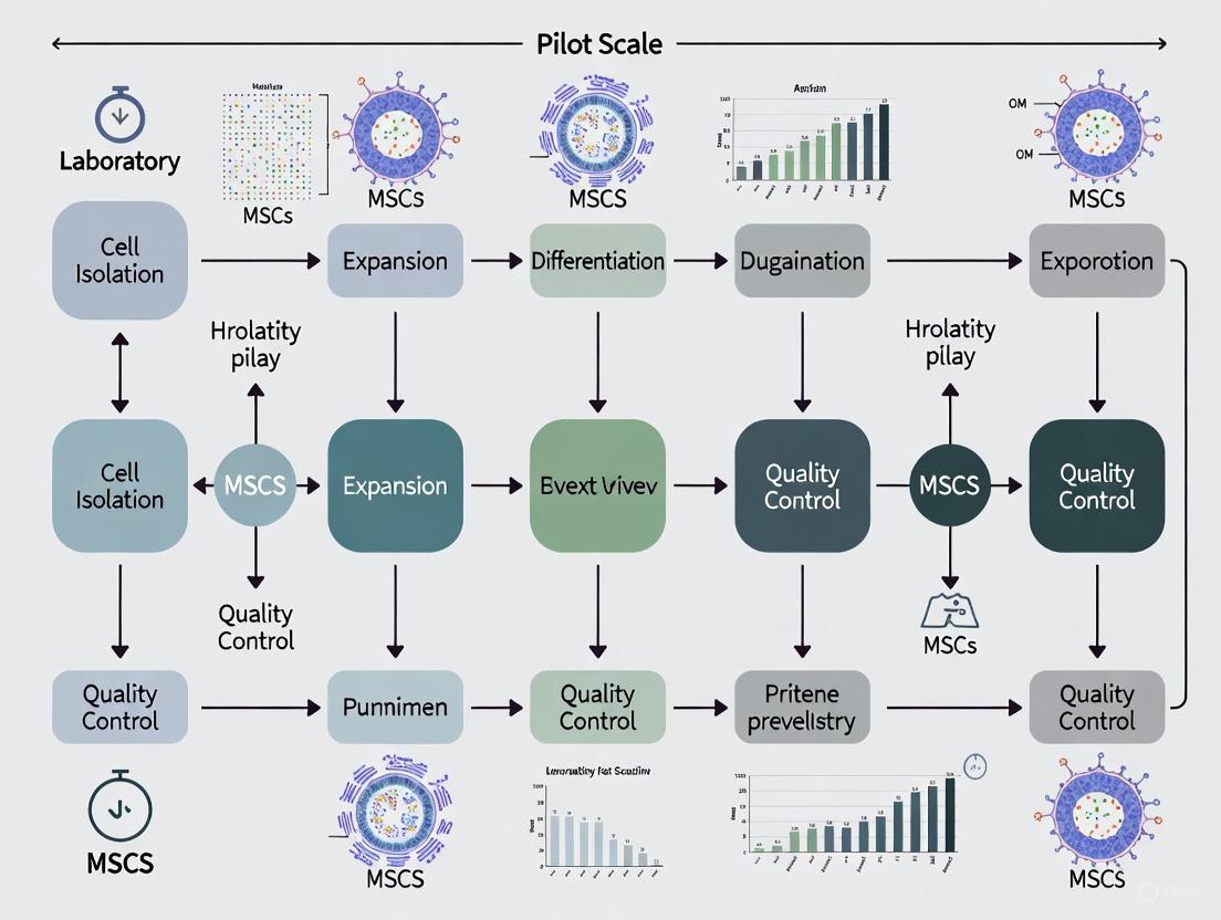
Experimental Workflow and Signaling
The following diagram illustrates the logical workflow for transitioning from laboratory-scale to pilot-scale MSC manufacturing, integrating process optimization and quality control.
Scalable GMP-Compliant MSC Manufacturing Workflow
Troubleshooting Common Regulatory Challenges in MSC Scale-Up
Q1: Our team is scaling an MSC process from laboratory to pilot scale. What are the critical regulatory considerations for the pilot-scale environment?
Scaling up mesenchymal stem cell (MSC) manufacturing introduces specific regulatory requirements, particularly for pilot-scale operations which bridge laboratory research and commercial production [7]. The table below outlines common challenges and solutions:
| Challenge | Regulatory Consideration | Practical Solution |
|---|---|---|
| Process Control | Ensure Consistent Cell Quality during expansion [6]. | Implement scaled-up, controlled bioreactors (pH, DO, temperature) with serum-free, xeno-free media [6] [8]. |
| Product Characterization | Demonstrate Quality and Purity of the final product [9]. | Use live-cell analysis and flow cytometry for extensive characterization; employ multivariate data analysis to correlate Critical Process Parameters (CPPs) and Critical Quality Attributes (CQAs) [6]. |
| Donor Eligibility | Comply with differing US and EU requirements for allogeneic donors [10] [9]. | For US: Follow detailed FDA 21 CFR 1271, Subpart C [10]. For EU: Adhere to relevant EU and member state-specific legal requirements [9]. |
| GMP Compliance | Navigate differing US and EU GMP expectations for clinical trials [9]. | In the EU: Follow GMP guidelines specific to ATMPs, which may include mandatory self-inspections [9]. In the US: Implement a phased, risk-based approach, with full verification typically occurring at the BLA stage [9]. |
Q2: What are the key differences in donor eligibility requirements between the EU and US that could impact our allogeneic MSC pipeline?
Harmonizing donor screening protocols for transatlantic development is a common hurdle. The regulatory approaches diverge significantly, as summarized below:
| Aspect | European Union (ATMP Regulation) | United States (21 CFR) |
|---|---|---|
| Regulatory Framework | Directive 2004/23/EC; referenced in ATMP guidelines [9]. | 21 CFR Part 1271, Subpart C [10]. |
| Guidance Specificity | Provides general guidance; compliance with EU and member state laws is required [9]. | Highly prescriptive; detailed recommendations on specific communicable disease agents, testing, and laboratory qualifications are provided in draft guidance documents [10] [9]. |
| Donor Testing & Screening | Requirements are referenced but less centralized [9]. | FDA provides explicit, binding regulations and detailed, non-binding recommendations on which diseases to test and screen for (e.g., HIV, HBV, HCV) [10]. |
| Impact on Development | Developers must navigate multiple national requirements within the EU [9]. | Use of donor material that does not meet FDA eligibility determination requirements can cause significant delays and increased costs [9]. |
Q3: How do GMP expectations for early-stage clinical trials differ between the EU and US for an ATMP like an MSC therapy?
Understanding this difference early is crucial for planning and resource allocation. The core distinction lies in the timing and method of verifying GMP compliance [9].
- In the European Union, compliance with GMP guidelines specific to ATMPs is a prerequisite for initiating clinical trials. This is often achieved through mandatory self-inspections, where the sponsor documents evidence of an effective quality system [9].
- In the United States, the FDA employs a more gradual, phase-appropriate approach. Early in clinical development, the agency relies on sponsor attestation of GMP compliance. Full compliance is typically verified via a pre-license inspection conducted by FDA inspectors during the review of a Biologics License Application (BLA) [9].
Experimental Protocol: A GMP-Compliant Pilot-Scale Production of Freeze-Dried MSC Secretome
The following detailed methodology, adapted from a published research paper, outlines a scalable, GMP-compliant process for manufacturing a lyophilized MSC secretome product, providing a practical example of navigating the transition from laboratory to pilot scale [8].
This workflow diagrams the GMP-compliant pilot-scale production process for freeze-dried MSC secretome.
Materials and Reagents
The table below lists key reagents used in this protocol and their GMP-compliant sourcing considerations.
| Reagent/Supply | Function in the Protocol | GMP/Regulatory Consideration |
|---|---|---|
| Collagenase, Type II | Tissue digestion to isolate stromal vascular fraction [8]. | Select a grade suitable for clinical-grade human MSC production [8]. |
| Platelet Lysate (PL) | Serum-free, xeno-free cell culture supplement for MSC expansion [8]. | Use a commercial kit designated for clinical-grade manufacturing to reduce pathogen risk [8]. |
| Cell Culture Media (DMEM/F12) | Base medium for cell expansion and secretome collection [8]. | Use serum-free formulations for secretome collection to ensure consistency and safety [8]. |
| Mannitol | Acts as a cryoprotectant during the freeze-drying (lyophilization) process [8]. | A commonly used pharmaceutical excipient that is generally recognized as safe. |
Step-by-Step Methodology
1. Donor Screening & MSC Isolation: - Obtain adipose tissue from informed donors following ethical approval (e.g., from an institutional review board). Exclude donors with a history of septicemia, specific infections (HIV, Hepatitis B/C), prion diseases, or malignant tumors [8]. - Mechanically mince the washed tissue and digest with 0.075% (w/v) collagenase type II. Centrifuge the digest to obtain the stromal vascular fraction for culture [8].
2. Cell Expansion & Validation: - Culture MSCs in a Grade B cleanroom using a GMP-compliant factory. Expand cells until passage 6 using media supplemented with clinical-grade platelet lysate [8]. - Perform quality controls on the MSCs to ensure they meet identity criteria (e.g., ISCT standards), sterility (per European Pharmacopoeia), and show no signs of tumorigenesis or karyotype abnormalities [8].
3. Secretome Collection: - At sub-confluence, switch MSCs to serum-free medium to induce secretome release. Collect conditioned media at 9 and 24 hours, then combine them. Check cell viability at the end of the collection period [8].
4. Purification & Formulation: - Purify the pooled conditioned media using a scalable ultrafiltration process. This step concentrates the secretome and removes undesirable small molecules [8]. - Add a cryoprotectant like mannitol to the purified secretome to protect the bioactive components during the lyophilization process [8].
5. Freeze-Drying & Quality Control: - Lyophilize the formulated secretome to produce a stable, "ready-off-the-shelf" powder, referred to as "lyo-secretome" [8]. - Characterize the final product using Nanoparticle Tracking Analysis (NTA) to detect extracellular vesicles, Fourier-Transform Infrared Spectroscopy (FTIR) to confirm the presence of proteins and lipids, and proteomic analysis to identify key protein components. Perform safety tests, including sterility, cytotoxicity, and blood compatibility [8].
FAQ on Emerging Regulatory Topics
Q4: Our MSC product uses a biodegradable scaffold. How is it classified, and what are the key regulatory highlights?
This combination is classified as a Combined ATMP in the EU [1]. The regulatory framework emphasizes the integral role of both the biological component (cells) and the device (scaffold). You must demonstrate the safety and function of the combined product as a whole, which includes meeting relevant standards for the medical device component in addition to those for the biological medicine [1].
Q5: Where can I find the most current regulatory guidelines for ATMPs in the EU?
The European Medicines Agency (EMA) continuously updates its guidelines. Two critical recent documents include:
- The Guideline on requirements for investigational ATMPs in clinical trials, which came into effect on July 1, 2025 [9]. This multidisciplinary document consolidates quality, non-clinical, and clinical requirements.
- A concept paper proposing revisions to the GMP guideline specific to ATMPs (Part IV), published for consultation in May 2025. This revision aims to align ATMP GMP with updated annexes and incorporate ICH Q9 (Quality Risk Management) and Q10 (Pharmaceutical Quality System) principles [11] [12].
Q6: What is the single most important step to avoid major regulatory setbacks during scale-up?
The most critical step is to establish a robust and well-understood manufacturing process at the pilot scale that can be consistently transferred to full-scale production. This involves using a risk-based approach to identify and control Critical Process Parameters (CPPs) that impact your product's Critical Quality Attributes (CQAs) [6] [7]. Engaging with regulatory agencies (e.g., via EMA's scientific advice or FDA's INTERACT meetings) early in the scale-up process is highly recommended to align your development strategy with regulatory expectations [1] [9].
Defining 'Substantial Manipulation' and its Impact on MSC Product Classification
FAQ: Substantial Manipulation and Regulatory Classification
Q1: What is 'substantial manipulation' in the context of MSC-based products?
In the European Union regulatory framework, 'substantial manipulation' refers to processes that alter the biological characteristics, physiological functions, or structural properties of cells or tissues relevant to their intended clinical function. Whether a manufacturing process involves substantial manipulation is the primary factor determining if an MSC product is classified as an Advanced Therapy Medicinal Product (ATMP) [13] [14].
The Regulation (EC) No 1394/2007 provides legal definitions, and its implementing directives specify processes that are not considered substantial manipulation (minimal manipulation) [13]:
- Cutting, grinding, shaping, centrifugation
- Soaking in antibiotic or antimicrobial solutions
- Sterilization, irradiation
- Cell separation, concentration, or purification
- Filtering, freezing, cryopreservation, and vitrification
Conversely, common MSC culture processes typically are substantial manipulation, such as extensive in vitro expansion that alters cell phenotypes, or genetic modification [14].
Q2: How does substantial manipulation impact the regulatory classification of my MSC product?
The determination of substantial manipulation directly dictates whether your product falls under the ATMP regulatory framework and which specific category it belongs to.
- Substantially manipulated MSCs are classified as ATMPs and require a centralized marketing authorization through the European Medicines Agency (EMA) [13] [14].
- Not substantially manipulated MSCs may be regulated under the national Hospital Exemption pathway or as a tissue transplant, provided they are used for their homologous function [15].
The table below summarizes how the manipulation and intended use of MSCs determine their classification:
Table 1: Impact of Substantial Manipulation on MSC Product Classification in the EU
| Product Characteristics | Substantial Manipulation? | Intended Use | ATMP Classification | Applicable Regulatory Pathway |
|---|---|---|---|---|
| Cultured, expanded MSCs | Yes | Immunomodulation (non-homologous) | Somatic Cell Therapy Medicinal Product (sCTMP) | Centralized Marketing Authorisation [16] [14] |
| Genetically modified MSCs | Yes | Any therapeutic use | Gene Therapy Medicinal Product (GTMP) | Centralized Marketing Authorisation [17] |
| Cultured MSCs on a scaffold | Yes | Tissue repair/regeneration | Tissue Engineered Product (TEP) | Centralized Marketing Authorisation [1] [16] |
| Freshly isolated, minimally processed MSCs | No | Same essential function (homologous) | Not an ATMP | Hospital Exemption or national tissue regulations [15] [13] |
Q3: What is the practical consequence if my MSC product is classified as an ATMP?
An ATMP classification means your product is regulated as a medicinal product [14]. This triggers specific and stringent requirements [17] [14]:
- Marketing Authorization: You must obtain a centralized marketing authorization from the European Commission, based on a scientific assessment by the EMA and its Committee for Advanced Therapies (CAT).
- Good Manufacturing Practice (GMP): The entire manufacturing process must comply with GMP standards specific to ATMPs.
- Non-Clinical and Clinical Data: You must demonstrate the product's quality, safety, and efficacy through comprehensive data packages, similar to those required for conventional drugs.
Q4: I am unsure how to classify my product. What should I do?
The EMA offers a free-of-charge procedure to obtain a scientific recommendation on ATMP classification [15]. You can submit a request to the Committee for Advanced Therapies (CAT), which will deliver a recommendation within 60 days of receiving a valid request [15]. This procedure is strongly advised to get a definitive, case-by-case determination and to plan your development pathway accordingly [15].
Experimental Protocols: Key Characterization for Classification
Robust experimental characterization is critical for regulatory submissions. The following protocols provide essential data to support the classification of your MSC product and its safety and efficacy profile.
Protocol 1: Standardized Immunophenotypic Characterization of MSCs
This protocol verifies that your cell product meets the minimal defining criteria for MSCs, a fundamental quality attribute.
- Objective: To confirm the expression of characteristic surface markers on the MSC product.
- Materials:
- Single-cell suspension of the final MSC product.
- Flow cytometry buffer (e.g., PBS with 1-2% FBS).
- Antibodies against positive markers (CD73, CD90, CD105) and negative markers (CD45, CD34, CD14, CD19, HLA-DR).
- Isotype-matched control antibodies.
- Flow cytometer.
- Methodology:
- Cell Preparation: Harvest, wash, and count cells. Aliquot ~1x10^5 cells per test tube.
- Staining: Add optimal concentrations of fluorochrome-conjugated antibodies to the cell pellets. Include isotype controls. Incubate for 30 minutes in the dark at 4°C.
- Washing: Wash cells twice with flow cytometry buffer to remove unbound antibody.
- Acquisition: Resuspend cells in buffer and analyze on the flow cytometer. Collect a minimum of 10,000 events per sample.
- Analysis: Use software to analyze the data. The population of interest must demonstrate ≥95% positivity for CD73, CD90, and CD105, and ≤2% positivity for the negative markers [18] [16].
Protocol 2: In Vitro Trilineage Differentiation Assay
This protocol demonstrates the multipotency of your MSC product, a key functional biological property that can be altered by substantial manipulation.
- Objective: To prove the capacity of MSCs to differentiate into osteocytes, adipocytes, and chondrocytes in vitro.
- Materials:
- Validated MSC trilineage differentiation kits (osteogenic, adipogenic, chondrogenic) or individually prepared media components.
- Culture plates (e.g., 12-well plates for osteo/adipogenesis; pellet culture or micromass for chondrogenesis).
- Fixatives and stains: Alizarin Red S (osteogenesis), Oil Red O (adipogenesis), Alcian Blue or Safranin O (chondrogenesis).
- Methodology:
- Seeding: Seed MSCs at a standardized density in growth media until 70-80% confluent.
- Induction: Replace growth media with specific differentiation induction media. Maintain control cells in growth media without inducers.
- Culture: Culture cells for 14-21 days, refreshing the differentiation media every 2-3 days.
- Staining: At endpoint, wash, fix, and stain cells according to standard protocols for each lineage.
- Osteogenesis: Alizarin Red S stains calcium deposits red/orange.
- Adipogenesis: Oil Red O stains lipid vacuoles red.
- Chondrogenesis: Alcian Blue stains proteoglycans blue-green.
- Documentation: Quantify staining intensity or perform RNA analysis of lineage-specific genes to provide robust data for regulatory dossiers [16] [19].
The Scientist's Toolkit: Essential Research Reagents
The following reagents are essential for the development and characterization of MSC-based ATMPs.
Table 2: Key Research Reagent Solutions for MSC-based ATMP Development
| Reagent/Category | Specific Examples | Function & Importance in Development |
|---|---|---|
| Cell Separation | CD34+ selection kits, Ficoll-Paque | Isolation of specific cell populations from starting material (e.g., bone marrow). Critical for process definition [17]. |
| Cell Culture Media | Defined, xeno-free media supplements (e.g., FGF-2) | Supports MSC expansion without animal components, enhancing safety and regulatory compliance [19] [14]. |
| Flow Cytometry Antibodies | Anti-CD73, CD90, CD105, CD45, CD34, HLA-DR | Quality control and identity testing of the final MSC product. Mandatory for lot release [18] [16]. |
| Differentiation Kits | Trilineage differentiation kits (osteo, adipo, chondro) | Standardized assessment of MSC functionality and potency. Provides critical product characterization data [16] [19]. |
| Cryopreservation Media | GMP-grade DMSO, defined cryomedia | Ensures stable and viable cell banks for raw materials (cell seeds) and final product. Vital for supply chain [14]. |
Process Flowchart: MSC Product Classification Logic
The following diagram illustrates the logical decision process for classifying an MSC-based product based on the EU regulatory framework, integrating concepts of substantial manipulation and homologous use.
For researchers and drug development professionals working toward the scalable manufacturing of Mesenchymal Stromal Cells (MSCs), the selection of a starting material is a critical foundational decision. This choice profoundly impacts downstream processes, including expansion capacity, therapeutic potency, and regulatory compliance. MSCs can be isolated from various tissues, but bone marrow (BM), adipose tissue (AT), and Wharton's Jelly (WJ) from the umbilical cord are among the most widely investigated sources [20]. Within a regulated manufacturing framework, where the goal is to produce Advanced Therapy Medicinal Products (ATMPs) that are consistently safe, potent, and efficacious, understanding the inherent differences between these sources is paramount [21] [22]. This technical support article provides a comparative analysis and troubleshooting guide to inform your selection process.
Q1: How does the proliferation capacity of MSCs from different sources impact large-scale production?
A: The proliferation capacity is a major differentiator with direct implications for achieving clinically relevant cell numbers. Scalable production requires cells that can be expanded extensively without rapid senescence.
- Wharton's Jelly (WJ-MSCs) and Fetal Bone Marrow (F-BM-MSCs) demonstrate superior proliferative potential. They can be cultured for significantly longer periods and exhibit higher growth rates [23].
- Adipose Tissue (AT-MSCs) typically have the shortest culture time and lowest growth rate, with proliferation often ceasing by passage 11-12, compared to passages 17-18 for WJ-MSCs and 22-24 for F-BM-MSCs [23].
- Colony Forming Unit (CFU) efficiency, a measure of clonogenicity, is also significantly higher in WJ-MSCs and F-BM-MSCs compared to AT-MSCs [23].
Q2: Are there functional differences in the immunomodulatory properties of MSCs from these sources?
A: Yes, the tissue source can influence immunomodulatory function. This is crucial for therapies targeting conditions like graft-versus-host disease (GvHD) or sepsis.
- Priming with Inflammatory Stimuli: The immunomodulatory function of MSCs is not constitutive but is enhanced by inflammatory cytokines like IFN-γ. Treatment with IFN-γ upregulates key immunomodulatory genes (e.g., IDO1, HLA-G5) and chemokines (e.g., CXCL9, CXCL10, CXCL11) in WJ-MSCs [23].
- Comparative In Vivo Performance: In an experimental model of sepsis, both BM-MSCs and WJ-MSCs regulated leukocyte trafficking and reduced organ dysfunction. However, only WJ-MSCs significantly improved bacterial clearance and survival, suggesting a superior therapeutic profile for this specific indication [24].
Q3: What are the key donor and sourcing considerations for a scalable process?
A: Sourcing logistics and donor variability are significant practical considerations.
- Donor Age and Variability: WJ-MSCs are derived from birth tissues, so their function is not influenced by donor age, unlike BM-MSCs or AT-MSCs from adult donors [25]. This can lead to a more consistent starting material.
- Availability and Invasiveness: Obtaining BM is an invasive and painful procedure for the donor, and the frequency of MSCs in BM is very low (~1 MSC per 10,000-100,000 mononuclear cells) [22]. Adipose tissue is more accessible via liposuction. Wharton's Jelly is obtained from donated umbilical cords, a non-invasive process that utilizes medical waste and offers a high yield of primitive MSCs [23] [20].
Q4: How does the differentiation potential vary, particularly for specific tissue engineering applications?
A: While all MSCs are multipotent, their propensity for specific lineages can differ.
- Adipogenic Differentiation: AT-MSCs possess an inherent and superior capacity for adipogenesis compared to WJ-MSCs [25]. However, recent research shows that optimizing differentiation protocols, such as supplementing with oleic acid, can significantly enhance lipid droplet formation and adipogenic marker expression in WJ-MSCs, bringing their capacity closer to that of AT-MSCs [25].
- Osteogenic Differentiation: BM-MSCs are often considered to have a strong predisposition toward osteogenic differentiation [24].
Q5: What are the critical regulatory considerations when selecting a source?
A: MSCs are regulated as Advanced Therapy Medicinal Products (ATMPs) in the EU and as biologic products in the US [21]. The entire manufacturing process, from donor selection to final product administration, must comply with Good Manufacturing Practice (GMP) standards. Key points include:
- Donor Screening: Rigorous donor eligibility screening is mandatory, following directives like 2004/23/EC in the EU and 21 CFR 1271 in the US [21].
- Source Material: Using human-derived supplements like Human Platelet Lysate (hPL) to replace Fetal Bovine Serum (FBS) is critical for compliance and reducing xenogenic risks [22].
- Substantial Manipulation: Cell culture and expansion are considered "substantial manipulation," which firmly categorizes the final product as an ATMP [21].
Comparative Data at a Glance
Table 1: Quantitative Comparison of Key MSC Source Characteristics
| Characteristic | Bone Marrow (BM) | Adipose Tissue (AT) | Wharton's Jelly (WJ) |
|---|---|---|---|
| Proliferation Capacity / Final PD | High [23] | Lowest [23] | High [23] |
| CFU-F Efficiency (Passage 3) | ~34 colonies [23] | ~18 colonies [23] | ~26 colonies [23] |
| Invasiveness of Collection | High (painful) [23] | Moderate (liposuction) | None (medical waste) [20] |
| Therapeutic Efficacy in Sepsis Model | Improved organ function, no survival benefit [24] | Information Missing | Improved organ function and survival [24] |
| Adipogenic Potential (vs. AT-MSC) | Lower | High (Gold Standard) | Lower, but improvable with protocol optimization [25] |
| Donor Age Impact | Affected by age | Affected by age | Not affected by age [25] |
Table 2: Research Reagent Solutions for MSC Manufacturing
| Reagent Category | Example Product / Composition | Function & Rationale |
|---|---|---|
| Serum-Free/Xeno-Free Media | MSC-Brew GMP medium [22]; Various commercial serum-free formulations [26] | Eliminates batch-to-batch variability and xenogenic infection risks from FBS; ensures defined, GMP-compliant conditions. |
| Humanized Growth Supplement | Human Platelet Lysate (hPL) [22] | GMP-compliant alternative to FBS; enhances cell proliferation and expansion in automated systems. |
| Cell Dissociation Reagent | Trypsin-EDTA; Non-animal derived dissociation reagents [26] | Detaches adherent cells for passaging or harvest. Animal-free options are preferred for regulatory compliance. |
| Culture Substrate/Matrix | Fibronectin, Vimentin, Cryoprecipitate [22] | Coats bioreactor surfaces (e.g., hollow fibers) to enable adhesion and growth of MSCs. |
| Inflammatory Priming Agent | Interferon-gamma (IFN-γ) [23] | Pre-conditioning agent used to enhance the immunomodulatory potency of MSCs before therapeutic application. |
Troubleshooting Guide: Common Challenges in MSC Sourcing and Manufacturing
Problem: Low Cell Yield and Proliferation After Seeding
- Potential Cause #1: The initial MSC frequency in the source tissue is low.
- Potential Cause #2: Suboptimal culture medium or supplements.
- Solution: Transition from FBS to GMP-grade, defined supplements like human platelet lysate (hPL), which has been shown to significantly enhance expansion of MSCs in automated bioreactors [22]. Systematically test commercial serum-free media designed for your specific MSC source.
Problem: Inconsistent Immunomodulatory Potency Between Batches
- Potential Cause: The immunomodulatory function of MSCs is not constitutive and requires activation.
- Solution: Implement a pre-conditioning step with a pro-inflammatory cytokine like IFN-γ. This upregulates critical immunosuppressive factors like IDO1 and HLA-G5, leading to a more potent and consistent product [23]. Standardize the concentration and duration of priming across all batches.
Problem: Inadequate Adipogenic Differentiation for Soft Tissue Engineering
- Potential Cause: WJ-MSCs have an intrinsically lower adipogenic potential compared to AT-MSCs.
- Solution: Modify the differentiation protocol. Supplement the standard adipogenic induction cocktail with oleic acid. Lipidomic studies show this significantly enhances lipid droplet formation and the expression of adipogenic markers in WJ-MSCs [25].
Problem: Challenges in Scaling Up from Flasks to Bioreactors
- Potential Cause: Manual 2D flask-based culture is inefficient, labor-intensive, and has a high risk of contamination.
- Solution: Adopt automated, closed-system bioreactor platforms. Systems like the Quantum Cell Expansion System (hollow fiber bioreactor) or the CliniMACS Prodigy (with adherent cell culture process) can reduce manual steps, improve yields, and ensure GMP compliance [22]. These systems also allow for better control of environmental factors like dissolved oxygen.
Experimental Workflow and Signaling Pathways
Diagram 1: Experimental Workflow for MSC Source Selection & Manufacturing
Diagram 2: Key Signaling Pathways in MSC Immunomodulation & Differentiation
Good Manufacturing Practice (GMP) Fundamentals for Cell Therapy Products
Regulatory Framework for Cell Therapy Products
Cell Therapy Products (CTPs) are classified as drugs or Advanced Therapy Medicinal Products (ATMPs) by major regulatory agencies and must be manufactured according to Good Manufacturing Practice (GMP) standards. The fundamental goal of GMP is to ensure consistent production and control of product quality to safeguard patient safety and the reliability of clinical data [27] [28].
International Regulatory Perspectives
Regulatory approaches to GMP for CTPs vary across international jurisdictions, particularly concerning the phases of clinical development. The following table summarizes the key requirements in Canada, the United States, and the European Union.
Table 1: International GMP Requirements for Cell Therapy Clinical Trials
| Jurisdiction & Authority | Regulatory Status of CTPs | GMP Evidence & Inspection | Key Distinguishing Features |
|---|---|---|---|
| Canada (Health Canada) [27] | Drugs requiring GMP [27]. | Implicit evidence via Clinical Trial Application "No Objection Letter" [27]. | Flexible, risk-based approach; no establishment license strictly required for clinical trials, though strategically necessary by Phase 3 [27]. |
| United States (US FDA) [27] | Drugs requiring GMP and Good Tissue Practice (GTP) [27]. | Phase 1: Implicit via IND approval. Phase 2/3: Explicit via Establishment License in FDA database [27]. | Phase 1 products exempt from 21 CFR 211; site registration required for Phase 2/3 studies [27]. |
| European Union (EMA) [27] | Advanced Therapy Medicinal Products (ATMPs) [27]. | Explicit evidence via Manufacturing Authorization and Qualified Person (QP) declaration in EudraCT database [27]. | Manufacturing authorization required for all clinical trial phases; well-defined ATMP regulations [27]. |
Fundamental cGMP Considerations for MSC Manufacturing
For Mesenchymal Stromal Cell (MSC) therapies, complying with cGMP requires addressing specific challenges related to the biological nature of the product. The following considerations are critical for designing a scalable and robust manufacturing process [29].
Table 2: Top cGMP Considerations for MSC Therapeutics
| Consideration | Key Challenges | cGMP-Compliant Strategies |
|---|---|---|
| Donor & Cell Source [29] | Donor age, health, and tissue source impact MSC properties and potency [29]. | Define donor eligibility criteria. Choose tissue source (e.g., Bone Marrow, Adipose, Umbilical Cord) based on scientific and logistical rationale [29] [30]. |
| Culture Media [29] [30] | Fetal Bovine Serum (FBS) poses xenogenic risks and batch variability [29]. | Use defined, xeno-free media (e.g., human Platelet Lysate or commercial GMP media) to enhance consistency and safety [29] [31]. |
| Cell Expansion [29] [30] | Process variables (seeding density, passages) affect growth kinetics and product quality [29]. | Standardize isolation, plating density, and limit population doublings (e.g., <20) to control senescence and maintain functionality [29] [30]. |
| Final Product Form [29] | Logistical choice between "fresh" culture-adapted cells and cryopreserved "off-the-shelf" cells [29]. | "Fresh" cells have optimal fitness. Cryobanked cells require robust, DMSO-free cryopreservation protocols to maximize post-thaw viability and function [29]. |
| Product Characterization [30] [28] | MSC cultures are heterogeneous; no single specific surface marker exists [29]. | Use a panel of markers (CD73+, CD90+, CD105+, CD45-). Control purity via immunoselection. Perform karyotypic analysis and potency assays [30] [28]. |
Experimental Protocol: GMP-Compliant Isolation and Expansion of MSCs
The following methodology, adapted from a 2025 study, outlines a protocol for the GMP-compliant isolation and expansion of MSCs from the infrapatellar fat pad (FP), demonstrating the translation from research-scale to clinical-grade production [31].
- Tissue Acquisition and Donor Eligibility: Obtain tissue (e.g., infrapatellar fat pad) as surgical waste following informed consent and approval from an ethics review committee. Ensure donor screening complies with applicable directives (e.g., European Union Tissue and Cells Directive) [31].
- Isolation Protocol:
- Mechanically mince the tissue into approximately 1 mm³ pieces.
- Digest the tissue with 0.1% collagenase in serum-free media for 2 hours at 37°C.
- Centrifuge the digested tissue at 300 ×g for 10 minutes and remove the supernatant.
- Wash the cell pellet with Phosphate-Buffered Saline (PBS) and filter through a 100 μm filter.
- Following a final centrifugation, resuspend the cell pellet in the chosen culture medium [31].
- Culture Expansion and Media Comparison:
- Seed cells at a density of 5 × 10³ cells/cm² and passage at 80-90% confluency.
- For GMP compliance, use animal component-free media such as MSC-Brew GMP Medium (Miltenyi Biotec) or MesenCult-ACF Plus Medium (StemCell Technologies). The study showed MSC-Brew GMP Medium resulted in lower doubling times and higher colony-forming units, indicating enhanced proliferation and potency [31].
- Quality Control and Product Release Testing:
- Viability: Assess using Trypan Blue exclusion. The protocol achieved >95% post-thaw viability [31].
- Sterility: Test for bacteria (sterility) and mycoplasma using systems like BacT/Alert [31].
- Purity and Identity: Confirm via flow cytometry for MSC surface markers (CD73+, CD90+, CD105+, CD45-) and endotoxin testing [31].
- Potency: Perform colony-forming unit (CFU) assays to demonstrate clonogenic capacity [31].
- Cryopreservation and Stability: Cryopreserve cells in a defined cryoprotectant. Perform stability studies to determine shelf-life; the cited study showed product specifications were maintained for up to 180 days of storage [31].
The Scientist's Toolkit: Essential Research Reagent Solutions
Selecting the right raw materials is critical for GMP compliance. Reagents must be qualified, and their use justified to ensure product quality and patient safety [28].
Table 3: Key Reagent Solutions for GMP-Compliant MSC Manufacturing
| Reagent / Material | Function | GMP-Compliant Considerations |
|---|---|---|
| Basal Media (e.g., MEM α) [31] | Provides essential nutrients and environment for cell growth. | Use GMP-grade versions with documented traceability and quality assurance certificates. |
| Media Supplements [29] [31] | Supports cell growth and proliferation. | Replace FBS with xeno-free supplements like Human Platelet Lysate (hPL) or commercial defined media (e.g., MSC-Brew GMP Medium). |
| Dissociation Enzymes (e.g., Collagenase) [31] | Digests extracellular matrix to isolate cells from tissue. | Source GMP-grade, recombinant enzymes where possible to avoid animal-derived contaminants. |
| Cell Separation Reagents [30] | Enriches for target MSC population. | Use closed-system, immunomagnetic separation devices with GMP-compliant antibodies (e.g., against CD271). |
| Cryopreservation Media [29] | Protects cells during freeze-thaw cycles. | Opt for defined, xeno-free, and DMSO-free formulations to minimize patient side effects and enhance safety. |
Troubleshooting Guides and FAQs
Frequently Asked Questions (FAQs)
Q: What are the key differences between manufacturing for early-phase (Phase 1) versus late-phase (Phase 2/3) clinical trials? [27]
- A: Regulatory stringency increases with clinical phase. For Phase 1, the FDA provides flexibility, focusing on safety and product characterization as part of the IND review, while explicit GMP compliance (21 CFR 211) and establishment registration are required for Phase 2/3. In the EU, a manufacturing authorization is required for all phases. Health Canada assesses GMP for all phases within the Clinical Trial Application but becomes more stringent in later stages [27].
Q: Our academic lab wants to initiate a first-in-human trial. Do we need a commercial-scale GMP facility? [27] [28]
- A: Not necessarily. Early-phase trials can be conducted in academic GMP facilities designed for investigational products. The standards for GMP (quality systems, documentation, controls) are the same for academic and industrial facilities, but the scale and automation may differ. The key is to demonstrate a robust quality system that ensures product safety and quality [27] [28].
Q: How can we control for MSC heterogeneity and ensure batch-to-batch consistency? [29] [30]
- A: Standardize every aspect of the process: donor eligibility criteria, tissue source, isolation method, seeding density, culture media, and passage number. Implement rigorous in-process controls and release criteria, including identity (flow cytometry), purity (sterility, endotoxin), viability, and potency assays. Moving to defined, xeno-free media significantly reduces batch-to-batch variability introduced by serum [29] [30] [31].
Troubleshooting Common Manufacturing Issues
Problem: Low Cell Viability Post-Thaw
- Potential Causes: Suboptimal cryopreservation formula; uncontrolled freeze/thaw rate; cells harvested at an unhealthy state.
- Solutions: Develop and validate a defined, DMSO-free cryoprotectant solution. Use controlled-rate freezers. Ensure cells are harvested during log-phase growth and that viability is >95% before cryopreservation [29].
Problem: Low Yield or Proliferation Rate
- Potential Causes: Poor quality of starting tissue; suboptimal culture media; inappropriate seeding density; microbial contamination.
- Solutions: Strictly control donor criteria and tissue transport conditions. Test and qualify different GMP-compliant media formulations (e.g., MSC-Brew GMP Medium showed enhanced proliferation [31]). Optimize and standardize seeding density. Perform thorough sterility and mycoplasma testing.
Problem: Inconsistent Potency Between Batches
- Potential Causes: Donor variability; undefined culture components; high passage number leading to senescence.
- Solutions: Bank a Master Cell Bank from a well-characterized donor for allogeneic use. Use defined, xeno-free media to eliminate variability from serum. Limit the number of population doublings and establish a maximum passage number for production. Develop a quantitative potency assay relevant to the mechanism of action [29] [30] [28].
From Bench to Pilot Plant: Implementing Scalable MSC Manufacturing Processes
For researchers and drug development professionals working on scalable manufacturing processes for Mesenchymal Stromal Cells (MSCs), selecting the appropriate isolation technique is a critical first step that impacts every subsequent stage of production. The two predominant methods—enzymatic digestion and explant culture—offer distinct advantages and challenges for laboratories transitioning from research to pilot scale. This technical support center provides detailed troubleshooting guides, FAQs, and methodological protocols to optimize your isolation strategy based on empirical data and recent advancements in the field. Whether you are establishing a new GMP-compliant process or seeking to improve existing yields, this resource addresses the key technical considerations for both enzymatic and explant approaches within the context of scalable MSC manufacturing.
Method Comparison & Quantitative Data
The choice between enzymatic and explant methods involves trade-offs between cell yield, processing time, and cell characteristics. The following table summarizes quantitative data from recent studies to facilitate evidence-based decision-making.
Table 1: Quantitative Comparison of Enzymatic Digestion vs. Explant Method for MSC Isolation
| Parameter | Enzymatic Digestion Method | Explant Method | Research Context |
|---|---|---|---|
| Initial Cell Yield (P0) | (1.75 \pm 2.2 \times 10^5) cells/g [32] | (4.89 \pm 3.2 \times 10^5) cells/g [32] | Wharton's Jelly isolation |
| Time to First Harvest | ~7 days to confluence [33] | ~10-15 days to confluence [33] [34] | General protocol |
| Population Doubling Time | Variable; can be longer [33] | Shorter doubling times [33] | General characteristic |
| Growth Factor Release (bFGF) | Lower levels in supernatant [32] | 55.0 ± 25.6 ng/g total released; higher initial levels [32] | Wharton's Jelly study |
| Expression of Pluripotency Markers | Standard expression [35] | Upregulated genes related to mitosis; stable OCT4, SOX2, NANOG expression [32] [36] | Gene expression profiles |
| Cell Viability Post-Isolation | >95% with optimized protocols [37] | High, minimal physical damage [36] | Optimized GMP process |
Frequently Asked Questions (FAQs)
Q1: Which isolation method is more suitable for initial pilot-scale production aiming for high cell yield quickly?
For pilot-scale production where time is a critical factor, the enzymatic digestion method is often preferred. It provides a quicker initial cell harvest, with cells typically reaching confluence within about 7 days, compared to 10-15 days for the explant method [33]. Furthermore, a GMP-compliant manufacturing study confirmed that enzymatic digestion exhibited a faster outgrowth of Wharton's jelly-derived MSCs (WJ-MSCs) during the initial passage (P0) compared to the explant method [37].
Q2: How does the choice of isolation method impact the critical quality attributes (CQAs) of MSCs, such as phenotype and differentiation potential?
Evidence suggests that after the initial passage (P0), MSCs isolated by either method show no significant disparities in terms of cell viability, morphology, proliferation, surface marker expression, and differentiation capacity after passaging [37]. Both methods can yield cells that meet the International Society for Cell & Gene Therapy (ISCT) defining criteria for MSCs [35]. The choice of method, therefore, primarily affects the initial yield and speed, not the fundamental cell characteristics after expansion.
Q3: We are experiencing low cell yields with the enzymatic method. What are the key parameters to optimize?
Low yield in enzymatic digestion is frequently due to suboptimal digestion parameters. Key factors to optimize include:
- Enzyme Type and Concentration: A study optimizing bovine adipose tissue-derived MSC isolation found that Liberase TM at 0.1% concentration for 3 hours yielded significantly higher cells than Collagenase type I [34]. For Wharton's jelly, 0.4 PZ U/mL Collagenase NB6 was identified as optimal [37].
- Digestion Time: Excessive digestion time can damage cells. A 3-hour digestion time is a common starting point for optimization [34] [37].
- Seeding Density Post-Digestion: Following digestion, optimizing the density at which the cell pellet is seeded is crucial for achieving optimal growth [37].
Q4: Does the explant method produce a more "native" or potent cell population?
Some research indicates that the explant method may better preserve the native state of MSCs. Studies on Wharton's jelly-derived MSCs showed that the explant method resulted in the release of significantly higher levels of natural growth factors like basic Fibroblast Growth Factor (bFGF) in the first week of culture [32]. Furthermore, genes related to mitosis were upregulated in explant-derived MSCs, and they maintained strong expression of pluripotency markers like OCT4, SOX2, and NANOG [32] [36]. This suggests the explant method may yield cells with a more robust proliferative and signaling profile.
Troubleshooting Guides
Troubleshooting Low Cell Yield in Enzymatic Digestion
- Problem: Low cell viability and yield at Passage 0 (P0).
- Potential Causes & Solutions:
- Cause: Over-digestion or harsh enzymatic activity.
- Cause: Inadequate seeding density post-digestion.
- Solution: Optimize the density of the digested cell pellet when seeding into culture flasks. A higher seeding density may be required initially to support colony formation [37].
- Cause: Suboptimal culture media.
Troubleshooting Slow Cell Outgrowth in Explant Method
- Problem: No or minimal cell migration from tissue explants after 1-2 weeks.
- Potential Causes & Solutions:
- Cause: Explants are not properly adhering to the culture surface.
- Solution: Allow explants to adhere to the dry plastic surface for 5-10 minutes at room temperature before carefully adding culture medium. Keep the medium level at or below the height of the tissue pieces to prevent detachment [36].
- Cause: Tissue fragments are too large.
- Cause: Contamination with other cell types.
- Cause: Explants are not properly adhering to the culture surface.
Detailed Experimental Protocols
Optimized Enzymatic Digestion Protocol for Wharton's Jelly
This protocol is adapted from a GMP-compliant manufacturing study [37].
- Key Reagents:
- Collagenase NB6 GMP (0.4 PZ U/mL)
- DPBS (without Ca²⁺ and Mg²⁺)
- MSC Serum- and Xeno-Free Medium (e.g., NutriStem)
- Human Platelet Lysate (hPL, 2-5%)
- Procedure:
- Tissue Preparation: Collect umbilical cord and transport at 2-10°C within 24 hours. Rinse with DPBS and decontaminate. Remove blood vessels and extract Wharton's jelly. Mince into 1-4 mm³ fragments.
- Enzymatic Digestion: Transfer tissue fragments to a digestion solution of 0.4 PZ U/mL Collagenase NB6. Incubate for 3 hours at 37°C with gentle agitation.
- Reaction Quenching & Washing: Neutralize the enzyme by adding a double volume of complete culture medium (e.g., NutriStem + 2% hPL). Centrifuge the cell suspension to pellet the cells. Wash the pellet with DPBS.
- Seeding and Culture: Resuspend the final cell pellet in complete medium and seed into culture flasks at the optimized density. Place in a 37°C, 5% CO₂ incubator.
- Medium Change: Refresh the medium twice a week. Observe for the formation of adherent MSC colonies.
Optimized Explant Culture Protocol for Wharton's Jelly and Subamnion
This protocol is adapted from a recent study comparing MSC sources [36].
- Key Reagents:
- Low glucose DMEM
- 10% Fetal Bovine Serum (FBS)
- L-glutamine, Non-Essential Amino Acids, Penicillin/Streptomycin/Amphotericin B
- Procedure:
- Tissue Dissection: Wash the umbilical cord with PBS. Cut into ~2 cm pieces and open longitudinally. Carefully separate the loose Wharton's jelly (WJ) from the denser subamnion (SA) membrane using a scalpel.
- Explant Preparation: Divide the WJ and SA into small segments (approx. 10 mm²).
- Explant Seeding: Place the tissue pieces in 6-well plates. For SA, ensure the subamnion layer is facing the bottom of the dish. Let the explants adhere to the dry plastic for 5 minutes at room temperature.
- Initial Culture: Gently add complete culture medium (DMEM, 10% FBS, antibiotics), ensuring the level does not exceed the height of the tissue to avoid detachment.
- Incubation and Medium Change: Culture the explants in a humidified atmosphere with 5% CO₂ at 37°C. Change the medium after 2 days, and subsequently twice a week. Cell outgrowths from the explants are typically observed within the first week.
Workflow and Scalability Diagrams
The following diagram illustrates the logical workflow for selecting and scaling an MSC isolation process, from method choice to pilot-scale production.
Diagram 1: MSC Isolation and Scale-Up Workflow
The Scientist's Toolkit: Essential Research Reagents
Table 2: Key Reagents for MSC Isolation and Expansion
| Reagent / Material | Function / Application | Examples / Notes |
|---|---|---|
| Collagenase NB6 (GMP) | Enzymatic digestion of tissue matrix. | GMP-compliant enzyme; 0.4 PZ U/mL for 3h is optimal for UC [37]. |
| Liberase TM | High-purity enzyme blend for tissue dissociation. | Can provide higher cell yield and viability vs. standard collagenase [34]. |
| Human Platelet Lysate (hPL) | Serum-free culture supplement for cell expansion. | Xeno-free alternative to FBS; 2-5% concentration effective for MSC growth [37] [38]. |
| Microcarriers (MC) | Provide surface for 3D cell culture in bioreactors. | Essential for scalable expansion in stirred-tank bioreactors (STRs) [38]. |
| Stirred-Tank Bioreactor (STR) | Controlled, scalable system for large-scale cell production. | Enables production of billions of cells; e.g., 50L STR yielded ~37 billion cells [38]. |
Troubleshooting Guide: FAQs on Critical Process Parameters
Q1: What is the optimal seeding density for expanding Mesenchymal Stem Cells (MSCs)?
A: Lower seeding densities generally favor more efficient MSC expansion. One study investigating synovial fat pad-derived MSCs found that proliferation rates were significantly affected by the initial plating density, with lower densities (e.g., 50-500 cells/cm²) often showing superior population doublings compared to higher densities [40]. However, the relationship can be complex, as the impact of other factors like donor age was also shown to vary depending on the specific density used [40]. For routine passaging, it is recommended to passage cells upon reaching ~85% confluency and to avoid overly confluent cultures, which can lead to poor cell survival [41].
Q2: How does Human Platelet Lysate (HPL) compare to Fetal Bovine Serum (FBS) for MSC culture?
A: HPL is a highly effective, xeno-free alternative to FBS. Multiple studies demonstrate that MSCs cultured in HPL-supplemented media exhibit superior proliferation rates compared to those in FBS [42] [43]. One study showed that MSCs expanded with 10% HPL derived from leukoreduction filters (f-hPL) had proliferation rates 300% higher than those cultured with FBS [43]. Furthermore, HPL supports the expansion of MSCs that meet International Society for Cell & Gene Therapy criteria for surface markers and differentiation potential, making it suitable for clinical-scale manufacturing [42] [43].
Q3: What are the critical safety considerations when using HPL?
A: The viral safety of HPL is paramount and is built on multiple layers [44]:
- Donor Screening: Blood donors must be healthy individuals who pass a medical history questionnaire and are screened for transfusion-transmitted infectious diseases using serological and nucleic acid testing (NAT) [42] [44].
- Pathogen Reduction: Implementing pathogen reduction technologies (PRT), such as treatment with psoralen/UV light or riboflavin/UV light, can further increase the safety margin of HPL by inactivating a broad spectrum of viruses [42] [44].
- Manufacturing Traceability: Sourcing HPL from licensed blood centers with robust quality management and digital documentation systems ensures full traceability [42].
Q4: My MSCs are not proliferating adequately. What could be the issue?
A: Inadequate proliferation can stem from several factors related to process parameters:
- Suboptimal Seeding Density: As noted above, seeding density is critical. Consistently passaging at high confluency can result in poor cell health and survival [41].
- Culture Supplement: The choice of supplement significantly impacts growth. If using FBS, switching to HPL can dramatically improve proliferation rates [43].
- Cell Health and Quality: The quality of the starting cells is crucial. Remove differentiated cells from the population before passaging or induction, as their presence can reduce overall expansion efficiency [41]. Also, check for signs of senescence, which increases with donor age and can reduce proliferative capacity [40].
Q5: What is the recommended concentration of HPL for MSC expansion?
A: While specific optimal concentrations may vary, successful protocols typically use HPL in the range of 5-10% (v/v). One optimization study concluded that MSC isolation by mononuclear cell gravity sedimentation, combined with culture medium supplementation with 5% platelet lysate in a hypoxic atmosphere (5% O₂), significantly improved yield and reduced expansion time [45]. Another study used 10% f-hPL to achieve a 300% higher proliferation rate compared to FBS [43]. We recommend testing a range within 5-10% to determine the ideal concentration for your specific cell source and basal medium.
The following tables consolidate key quantitative findings from the literature to guide experimental design.
Table 1: Impact of Seeding Density on MSC Expansion
| Seeding Density (cells/cm²) | Observed Effect on Proliferation | Notes |
|---|---|---|
| 50 - 500 | Generally favored higher population doublings [40] | Lower densities often more efficient for expansion. |
| 1,000 - 2,500 | Mixed or no correlation with donor age factors [40] | May be a more stable density for certain cell lines. |
| 5,000 - 7,500 | Age-related decline in population doublings observed [40] | Higher densities may exacerbate donor-age effects. |
| 10,000 | Age-related increase in population doublings observed [40] | Effect of density is complex and non-linear. |
Table 2: Performance of Different Culture Media Supplements
| Supplement Type | Key Performance Characteristics | Key Considerations |
|---|---|---|
| Fetal Bovine Serum (FBS) | Traditional supplement; supports MSC expansion. | Risk of xenogenic immune responses, ethical concerns, potential zoonotic contamination [42] [43]. |
| Human Platelet Lysate (HPL) | Superior proliferation (e.g., 20-300% higher than FBS) [43]; xeno-free; supports clinical-scale expansion [42]. | Requires anticoagulant (e.g., heparin) unless fibrinogen-depleted; viral safety is a critical parameter [44] [46]. |
| Serum-Free Media (SFM) | Defined formulation; eliminates serum variability and safety risks. | May require specific adhesion substrates; performance can be cell-type specific and less robust than HPL [47] [48]. |
| Human AB Serum | Xeno-free; used in clinical trials. | High cost and limited supply compared to HPL [43]. |
Experimental Protocols for Parameter Optimization
Protocol 1: Determining Optimal HPL Concentration
Objective: To identify the optimal concentration of HPL for maximizing the proliferation of a specific MSC source.
Materials:
- Basal medium (e.g., MEM-α [43] [45])
- HPL stock (commercial or prepared in-house)
- Heparin solution (if using non-fibrinogen-depleted HPL) [46]
- Gentamicin or other antibiotics [43]
- Bone marrow-derived MSCs (e.g., from Lonza [43])
- 24-well cell culture plate
- Cell counter
Methodology:
- Prepare Media: Supplement the basal medium with varying concentrations of HPL (e.g., 2.5%, 5%, 7.5%, 10%). Include a control with 10% FBS. Add heparin (e.g., 2 IU/mL) to all HPL conditions and antibiotics to all media [43].
- Seed Cells: Seed MSCs at a density of 5 × 10³ cells per cm² in the 24-well plate [43].
- Culture Cells: Incubate the cells under standard conditions (37°C, 5% CO₂). Perform medium changes as per standard protocol.
- Harvest and Count: After 5 days, harvest the cells from each well and count using an automated cell counter or hemocytometer.
- Calculate Growth: Calculate the fold increase in cell number (final cell count / initial seeded cell count) for each HPL concentration.
- Analysis: Compare the fold increase across conditions to determine the HPL concentration that yields the highest proliferation.
Protocol 2: Optimizing Seeding Density for Expansion
Objective: To establish the seeding density that minimizes expansion time while maintaining cell phenotype.
Materials:
- Validated MSC culture medium (e.g., with optimal HPL concentration)
- MSCs at an early passage
- Multi-well plates (e.g., 6-well or 12-well plates)
- Trypsin or other dissociation reagent
- Cell counter
Methodology:
- Prepare Cells: Harvest and create a single-cell suspension of MSCs. Determine cell viability using trypan blue exclusion [41].
- Seed at Varying Densities: Seed cells across multiple wells at a range of densities (e.g., 500, 1,000, 2,500, 5,000 cells/cm²) [40].
- Monitor Growth: Monitor cells daily. Passage cells when they reach ~85% confluency, as overly confluent cultures can lead to poor survival [41].
- Record Data: At each passage, record the time to confluency, the final cell yield, and the calculated population doublings.
- Characterize Cells: At the end of the expansion phase (e.g., P4), characterize cells from each density condition for standard MSC surface markers (via flow cytometry) and tri-lineage differentiation potential (osteogenic, adipogenic, chondrogenic) to ensure quality is maintained [45].
Process Optimization and HPL Manufacturing Workflows
MSC Expansion Process Optimization
GMP-Compliant HPL Manufacturing Pathway
The Scientist's Toolkit: Research Reagent Solutions
Table 3: Essential Materials for MSC Expansion Process Development
| Item | Function / Application | Example Products / Components |
|---|---|---|
| Basal Medium | Provides essential nutrients and salts for cell survival and growth. | MEM-α [43] [45], DMEM/F12 [47]. |
| Culture Supplements | Stimulates robust cell proliferation; xeno-free alternative to FBS. | Human Platelet Lysate (HPL) [42] [46] [43], StemPro MSC SFM [49]. |
| Anticoagulant | Prevents coagulation of HPL in culture medium. | Heparin Solution [46] [43]. |
| Dissociation Reagent | Detaches adherent MSCs from culture surfaces for passaging. | Trypsin-EDTA, Accutase. |
| Cell Culture Substrate | Surface coating to support cell adhesion, especially in serum-free or microcarrier cultures. | Geltrex [41], CELLstart [49], Collagen I. |
| Microcarriers | Provide a high-surface-area substrate for scaling up MSC expansion in 3D bioreactor systems. | Cytodex 3 [47], Solohill Plastic [47]. |
| Differentiation Kits | Validated reagents to confirm MSC multipotency (osteogenic, adipogenic, chondrogenic). | StemPro Osteogenesis/Adipogenesis/Chondrogenesis Kits [49]. |
| Pathogen Reduction Tech | Inactivates viruses and other pathogens in HPL to enhance safety. | Psoralen/UV light, Riboflavin/UV light [42] [44]. |
The transition from traditional laboratory flasks to pilot-scale cell factories is a critical step in the scalable manufacturing of Mesenchymal Stromal Cells (MSCs) for therapeutic applications. This shift is necessary to meet the clinical demand for billions of cells per dose while maintaining consistent quality, functionality, and compliance with Good Manufacturing Practice (GMP) standards [50] [51]. This technical support center provides targeted troubleshooting guides and FAQs to help researchers, scientists, and drug development professionals navigate the specific challenges encountered during this scale-up process.
Experimental Protocols & Workflows
Key Workflow: Transitioning from 2D Planar Culture to 3D Bioreactors
The following diagram outlines the core process for scaling up MSC manufacturing, integrating both upstream and downstream processing steps.
Detailed Methodology for Pilot-Scale MSC Expansion in Bioreactors
The protocol below is adapted from a study demonstrating the scalable expansion of induced pluripotent stem cell-derived MSCs (ihMSCs) on gelatin methacryloyl microcarriers (GelMA-M) in vertical wheel bioreactors (VWB) [52].
Bioreactor Setup and Seeding:
- Use a single-use, instrumented bioreactor system (e.g., a 500 mL to 3L Vertical Wheel Bioreactor from PBS Biotech) [52].
- Prepare GelMA microcarriers equivalent to 2500 cm² of growth area in the bioreactor vessel [52].
- Seed with an initial cell density of 2.5×10⁶ ihMSCs into the 500 mL VWB [52].
- Culture parameters: Maintain standard conditions (37°C, 5% CO₂) with controlled agitation to minimize shear stress [52] [50].
Feeding and Monitoring:
Harvesting and Downstream Processing:
- At the end of the expansion phase (typically after 6 days, when the surface area is fully occupied), terminate the culture [52].
- Harvest cells by digesting the GelMA microcarriers using a standard trypsin-based cell dissociation reagent. This streamlined process results in a single-cell suspension with >95% viability [52].
- Subsequent downstream steps include cell separation, washing, concentration, and formulation into the final drug product [50].
Troubleshooting Common Scale-Up Challenges
Frequently Asked Questions (FAQs)
Q1: Our cell yields are lower in the bioreactor compared to multilayer flasks. What could be the cause? A: This is a common challenge. Lower yields can stem from suboptimal seeding efficiency, inadequate mixing leading to nutrient gradients, or excessive shear stress damaging cells [50]. Ensure you have optimized the microcarrier concentration and agitation speed. Studies show that using customizable, degradable microcarriers like GelMA can improve yields compared to traditional polystyrene microcarriers [52].
Q2: How can we effectively monitor cell growth and confluence in a 3D bioreactor system? A: Moving beyond traditional sampling, leverage inline or at-line technologies. Research demonstrates that microcarriers with superior optical properties, like GelMA, allow for non-invasive visualization of cell bodies using Elastic Light Scattering (ELS) modalities. This allows for accurate, label-free cell enumeration without disturbing the culture [52].
Q3: What is the most efficient method for detaching MSCs from microcarriers at harvest? A: The harvest strategy depends on the microcarrier. For standard plastic microcarriers, enzymatic detachment followed by filtration is required, which can cause shear stress and cell loss [52] [50]. A more efficient approach is to use degradable microcarriers (e.g., GelMA), where the carriers themselves can be digested using standard trypsin-based reagents, simplifying harvest and improving cell viability and yield [52].
Q4: We are planning a new pilot facility. What non-technical bottlenecks should we anticipate? A: As you scale out operations, consider logistical and facility design aspects. A commonly overlooked bottleneck is gowning capacity. If a large number of operators need to gown for a "ballroom" style facility, a small gowning area can become a critical path delay, with gowning times potentially stretching to 24 hours a day [53]. Plan for adequate gowning space from the outset.
Troubleshooting Guide Table
| Problem | Potential Cause | Recommended Solution |
|---|---|---|
| Low Cell Yield | Inefficient seeding [50], high shear stress [50], nutrient gradients [50] | Optimize seeding protocol and agitation rate. Use microcarriers designed for MSCs (e.g., GelMA) [52]. |
| Poor Cell Viability Post-Harvest | Harsh detachment methods [52] [50], shear stress during filtration [50] | Switch to degradable microcarriers that are digested upon harvest [52]. Optimize enzymatic cocktail and duration. |
| Difficulty Monitoring Growth | Opacity of traditional microcarriers, lack of inline sensors [52] | Use optically clear microcarriers (e.g., GelMA) enabling label-free ELS monitoring [52]. Implement process analytical technology (PAT). |
| Inconsistent Differentiation Post-Expansion | Improper culture conditions, selective pressure during scale-up [52] | Perform quality control assays post-expansion. Ensure culture media and supplements are consistent and GMP-compliant [37]. |
| Process is Not Cost-Effective | Reliance on manual, planar culture systems [52] [51] | Transition to automated, closed-system bioreactors. Data shows volumes >500 mL in VWB are more cost-effective than monolayer culture [52]. |
The Scientist's Toolkit: Essential Materials & Reagents
The table below lists key reagents and materials critical for successfully scaling up MSC manufacturing, based on the cited experimental protocols.
Research Reagent Solutions
| Item | Function/Application in Scale-Up | Example/Note |
|---|---|---|
| Vertical Wheel Bioreactor (VWB) | Provides efficient mixing with low shear stress, ideal for sensitive MSCs and microcarriers [52]. | PBS Biotech systems are used for scales from 100 mL to 3L [52]. |
| Gelatin Methacryloyl (GelMA) Microcarriers | Customizable, degradable microcarriers that enable streamlined harvest via digestion and allow non-invasive monitoring [52]. | Spherical microcarriers synthesized via microfluidics [52]. |
| GMP-compliant Enzymes | For tissue dissociation (e.g., umbilical cord) and microcarrier/cell digestion during harvest [37]. | Collagenase NB6 GMP grade is recommended for clinical-scale isolation [37]. |
| Human Platelet Lysate (hPL) | Serum-free, GMP-compliant growth supplement for MSC culture medium [37]. | Concentrations of 2% and 5% have shown similar expansion levels for WJ-MSCs [37]. |
| Single-Use Bioreactor Vessels | Pre-sterilized, disposable culture chambers that eliminate cleaning and reduce cross-contamination risk [50]. | Available as rigid cylinders or flexible bags fixed in support containers [50]. |
Quantitative Data for Process Comparison
When selecting a scale-up platform, quantitative data on cell yield and cost is essential for decision-making. The table below summarizes key performance metrics from the literature.
Scale-Up Platform Performance Metrics
| Cultivation Platform | Typical Maximum Cell Yield (cells/cm²) | Relative Cost-Effectiveness at Pilot Scale | Key Advantages |
|---|---|---|---|
| Planar Multilayer Flasks | 21,000 (SD 800) [52] | Lower | Well-characterized, simple protocol [50]. |
| Polystyrene Microcarriers | 15,000 (SD 1500) [52] | Medium | Increases surface area-to-volume ratio [50]. |
| GelMA Microcarriers in VWB | 30,000 (SD 2000) [52] | Higher (becomes cost-effective >500 mL) [52] | Streamlined harvest, superior yields, enables non-invasive monitoring [52]. |
Pathway to GMP-Compliant Manufacturing
Adhering to regulatory standards is paramount. The following diagram outlines the critical pathway from research to a GMP-compliant pilot-scale process, highlighting key quality checkpoints.
Defining Critical Quality Attributes (CQAs) Throughout the Expansion Process
Frequently Asked Questions (FAQs)
Q1: What is a Critical Quality Attribute (CQA) in the context of MSC manufacturing?
A CQA is a physical, chemical, biological, or microbiological property or characteristic that must be within an appropriate limit, range, or distribution to ensure the desired product quality, safety, and efficacy of Mesenchymal Stromal Cell (MSC) therapies [55] [56]. These attributes are challenging to measure directly in production and are central to the FDA’s Process Analytical Technology (PAT) and Quality by Design (QbD) frameworks [55] [57]. For MSCs, CQAs are not just a final product check; they must be monitored and controlled throughout the entire expansion process to ensure a consistent and potent cell product [56] [58].
Q2: What are the core CQAs for MSCs, and how do they relate to scalability?
The core CQAs for MSCs can be categorized based on regulatory requirements and functional output. The table below summarizes the primary CQA categories and their significance for scaled manufacturing.
Table 1: Core Critical Quality Attributes for MSC Manufacturing
| CQA Category | Specific Attributes | Importance for Scalable Expansion |
|---|---|---|
| Safety | Sterility, Mycoplasma, Endotoxin [56] | Standardized, compendial tests; essential for all scales to ensure patient safety. |
| Identity/Purity | Adherence to plastic; Expression of CD73, CD90, CD105; Lack of hematopoietic markers (CD45, CD34, etc.) [56] [59] | Confirms the basic cell type is correct. Must be maintained consistently across passages and scales. |
| Potency | Immunomodulatory activity (e.g., IDO activity); Trilineage differentiation; Cytokine secretion profile; Angiogenic potential [56] [59] | Directly linked to the therapeutic mechanism of action (MoA). The most challenging CQA to define and control during scale-up. |
| Viability | Post-thaw viability; Membrane integrity [60] | Critical for ensuring a sufficient dose of functional cells is delivered to the patient. |
For scalable processes, a significant challenge is ensuring these CQAs remain consistent from the small-scale, laboratory-based expansion (e.g., flasks) to larger pilot-scale and commercial bioreactors [56]. Attributes like potency and replication capacity are highly sensitive to process parameters and can drift with increased passaging [58].
Q3: Why do my MSCs' CQAs change as I scale up the expansion process?
Changes in CQAs during scale-up often signal a shift in the biological state of the cell population due to altered process parameters. Key reasons include:
- Passage-Induced Senescence: As MSCs are expanded over multiple passages, they can enter a senescent state. This is marked by a reduced growth rate, changes in cell morphology, and a loss of therapeutic potency, even if surface markers appear normal [58]. One study showed that late-passage MSCs lost surface marker expression after transplantation, while early-passage cells did not, highlighting a critical functional difference [58].
- Process Parameter Variability: The "product is the process" is a common phrase in cell therapy [56]. Seemingly minor changes in the scale-up environment—such as dissolved oxygen, pH fluctuations, feeding schedules, or shear stress in bioreactors—can significantly impact CQAs like proliferation rate and secretome profile [56] [59].
- Donor and Sourcing Heterogeneity: Biological starting material from different donors or tissue sources (e.g., bone marrow vs. adipose) inherently possesses variable functional attributes, making consistent CQA targets difficult to achieve across batches [59].
Q4: My potency assays are not predictive of in vivo efficacy. What are my options?
This is a common and critical challenge. The 2024 FDA approval of an MSC product and previous Complete Response Letters for others have emphasized the need for potency assays that scientifically demonstrate a relationship to the product's biologic activity [59]. To address this:
- Align Assays with Mechanism of Action (MoA): Move beyond generic, historical assays. Develop a quantitative potency CQA that is specifically linked to your product's therapeutic effect [56] [59]. For an immunomodulatory product, this might be the suppression of T-cell proliferation or the induction of regulatory macrophages, rather than just trilineage differentiation [59].
- Implement Multivariate and "Fitness" Assays: Instead of relying on a single test, use a panel of assays that measure "basal fitness" [59]. This can include:
- Metabolic Function: Assessing mitochondrial activity and redox balance [58].
- Secretomics: Quantifying the release of key therapeutic factors (e.g., TNFAIP6, HMOX1, PGE2) [59].
- Advanced Morphology: Using AI-based systems to detect subtle, predictive changes in cell morphology that precede functional decline [58].
- Focus on Assay Robustness: Ensure your potency methods are qualified early, with a clear understanding of their precision, reproducibility, and sensitivity to make reliable decisions during process scaling [60].
Troubleshooting Guides
Problem: Declining Growth Rate and Increased Senescence During Late-Passage Expansion
Potential Causes and Solutions:
Table 2: Troubleshooting Declining Growth and Senescence
| Observed Symptom | Potential Root Cause | Investigative Experiments & Solutions |
|---|---|---|
| Sustainable growth is lost after a certain passage number (e.g., >P5) | Natural replicative senescence; Culture medium exhaustion; Critical Process Parameter (CPP) drift. | 1. Establish a Growth Rate Limit: Define a CPP where the fold-expansion per passage must remain above a threshold (e.g., >10-fold) [58].2. Monitor Senescence Biomarkers: Implement at-line assays for β-galactosidase (X-Gal) staining and Ki67 expression to quantitatively track senescence [58].3. Define a Maximum Passage Number: Based on data, set a hard limit for in vitro passaging in your controlled process. |
| Shift in cell morphology (e.g., enlarged, flattened cells) | Senescence; Stress from suboptimal culture conditions. | Quantify Morphology: Use an AI-based image analysis system to objectively measure parameters like pseudopod length or cell area. A sharp increase in these metrics can signal the loss of homeostatic replication potential and serve as a PP [58]. |
| High apoptosis (Annexin V staining) in bioreactor samples | Shear stress from impeller; Nutrient starvation; Toxic metabolite accumulation (e.g., lactate/ammonia). | Characterize Bioreactor Environment: Use design of experiments (DoE) to model the impact of CPPs like agitation speed, dissolved CO2, and feeding strategies on apoptosis. Establish a proven acceptable range for these parameters [61]. |
Experimental Workflow for Investigating Senescence:
The following diagram outlines a logical workflow for identifying and addressing senescence-related issues in MSC expansion.
Problem: Inconsistent Potency Between Donors or Manufacturing Batches
Potential Causes and Solutions:
Cause: Inherent Donor Heterogeneity.
- Solution: Implement a rigorous donor screening program. Use pre-selection functional assessments (e.g., basal cytokine secretion profile, expansion potential) that are predictive of your desired potency CQA [56] [59]. Consider the strategy of using pooled mononuclear cells from multiple donors to create a more consistent and "younger" starting cell bank [59].
Cause: Poorly Defined or Measured Potency CQA.
- Solution: Refine your potency assay to be quantitative, sensitive, and clinically relevant. As recommended by the ISCT, move from qualitative histological stains (e.g., for trilineage differentiation) to quantitative measures like ELISA for specific secreted factors (e.g., TNFAIP6, PGE2) or qPCR for gene expression [56] [59]. Ensure the assay is robust and validated for inter-laboratory reproducibility [60].
Essential Research Reagent Solutions for CQA Determination
The following table lists key reagents and their critical functions in establishing and monitoring CQAs during MSC expansion.
Table 3: Key Reagent Solutions for CQA Analysis in MSC Expansion
| Research Reagent / Tool | Primary Function in CQA Assessment |
|---|---|
| Flow Cytometry Antibody Panels (CD73, CD90, CD105, CD45, CD34, HLA-DR) | Definitive assessment of Identity and Purity CQAs as per ISCT criteria [56]. |
| Trilineage Differentiation Kits (Osteogenic, Adipogenic, Chondrogenic) | Historical assessment of multipotency, a common Potency attribute. Moving towards quantitative versions [56]. |
| ELISA/Kits for Soluble Factors (IDO, TNFAIP6, HMOX1, PGE2, Angiogenic cytokines) | Quantitative measurement of secretome-based Potency CQAs, directly linkable to MoA [56] [59]. |
| Viability & Senescence Assays (Annexin V, Propidium Iodide, β-Galactosidase) | Assessment of Viability and detection of senescent cells, which is a key Process Parameter for controlling expansion [58]. |
| Process Analytical Technology (PAT) (In-line sensors for pH, DO, metabolites) | Monitoring and controlling Critical Process Parameters (CPPs) in bioreactors to maintain consistent CQAs [55] [56]. |
The Relationship Between CQAs, CPPs, and Control Strategy
A fundamental principle of QbD is the linkage between CQAs and Critical Process Parameters (CPPs). A CPP is a process parameter whose variability impacts a CQA and therefore must be controlled to ensure the product meets its quality standards [57] [61]. The following diagram illustrates how these elements integrate into a comprehensive control strategy for scalable MSC manufacturing.
Frequently Asked Questions (FAQs)
1. Why is the passage number critical in MSC manufacturing, and what is the difference between Passage Number and Population Doubling Level (PDL)?
The passage number is a record of how many times a culture has been subcultured (harvested and reseeded) [62]. In scalable manufacturing, using cells within a consistent, low-passage range (e.g., P2-P5) is crucial for ensuring batch-to-batch consistency in critical quality attributes like potency, differentiation capacity, and specific marker expression [63] [64].
The Population Doubling Level (PDL), in contrast, represents the cumulative number of times the cell population has actually doubled since isolation. PDL is a more accurate measure of the culture's "biological age" because it accounts for variations in split ratios, which passage number does not [62] [64]. For instance, a 1:4 split results in 2 doublings, while a 1:10 split results in approximately 3.3 doublings, yet both only increase the passage number by one.
2. What are the key indicators that my MSCs are within the optimal harvest window (e.g., Passages 2-5)?
Cells within the optimal window typically exhibit:
- Stable, Typical Morphology: Cells maintain the characteristic spindle-shaped, fibroblast-like morphology without significant enlargement or granularity [65] [62].
- Consistent and Robust Growth: A stable, relatively short doubling time during the exponential (log) phase of growth is a key metric of cell health [64].
- High Viability: Post-harvest viability should consistently be above a predetermined threshold (e.g., >90%).
- Preservation of Critical Quality Attributes: Cells should consistently express standard MSC surface markers (CD73+, CD90+, CD105+; CD14-, CD19-, CD34-, CD45-) and retain tri-lineage differentiation potential, as required for clinical applications [66].
3. What are the consequences of over-passaging MSCs in a scaled-up process?
Over-passaging can lead to genotypic and phenotypic drift, fundamentally altering the cell product [62] [64]. Specific risks include:
- Reduced Proliferative Capacity: A noticeable increase in doubling time and a decline in maximum cell density, often a precursor to senescence [64].
- Loss of Function: Diminished differentiation potential into adipocytes, osteoblasts, and chondrocytes [66] [64].
- Genetic and Phenotypic Instability: Accumulation of genetic changes and selective overgrowth of adapted subpopulations can make the cell line unsuitable for therapeutic use [62] [64].
- Irreproducible Results: This is a primary cause of experimental variability and failure in both research and manufacturing [64].
4. How can I technically determine the optimal harvest window for my specific MSC line and process?
Establishing the window requires proactive characterization across multiple passages. Key methodologies include:
- Growth Kinetics Analysis: Systematically track population doubling time and cumulative PDL at each passage. The optimal window is where these parameters are most stable [64].
- Regular Phenotypic Monitoring: Use flow cytometry to verify surface marker expression at regular intervals (e.g., every few passages) to confirm phenotype stability [63] [66].
- Functional Potency Assays: Periodically test differentiation potential and other relevant potency markers to define the passage limit where function begins to decline [66].
Troubleshooting Guide
| Problem | Potential Cause | Recommended Solution |
|---|---|---|
| Increased Doubling Time | Onset of senescence due to high PDL; Sub-optimal culture conditions (media, O₂). | Return to low-PDL stock; establish a passage limit. Optimize culture environment (e.g., consider hypoxic incubation at 2% O₂ for tenocytes) [63] [64]. |
| Morphological Changes | Genetic drift or selection pressure from over-passaging; Cellular adaptation to in vitro conditions. | Revert to master cell bank with a defined PDL. Intensify morphological monitoring and set stricter passage limits [65] [62]. |
| Loss of Differentiation Potential | Over-passaging; Inconsistent culture methods. | Perform functional assays early to establish a baseline. Adhere strictly to SOPs for passaging and media formulation [65] [66]. |
| Low Post-Harvest Viability | Over-exposure to enzymatic dissociation reagents; Excessive shear stress during harvesting in bioreactors. | Optimize harvest protocol (e.g., duration of enzyme exposure). In scaled systems, evaluate gentler harvest methods like alternating tangential flow filtration (ATF) [67]. |
| Inconsistent Cell Yields | Variable seeding densities; Inaccurate confluency measurements at harvest. | Standardize seeding and harvest criteria (e.g., always passage at 80-90% confluency). Use live-cell imaging tools for precise, non-destructive confluency measurement [64]. |
Experimental Protocols for Identifying Optimal Harvest Windows
Protocol 1: Longitudinal Tracking of Growth and Phenotypic Stability
Aim: To characterize growth kinetics and phenotypic markers across consecutive passages (P0 to P8) to identify the window of optimal performance.
Methodology:
- Cell Culture: Culture MSCs in a controlled environment (e.g., 5% CO₂, 2-5% O₂ for physiological relevance) [63]. Use a consistent seeding density and vessel type.
- Data Collection at Each Passage:
- Seeding and Harvest Counts: Use an automated cell counter or hemocytometer to record exact cell numbers at seeding (N₁) and harvest (N₂).
- Doubling Time Calculation: Calculate the doubling time for each passage using the formula: Td = (t₂ − t₁) × ln(2) / ln(N₂ / N₁) [64].
- PDL Calculation: Calculate the population doublings for each passage and maintain a running cumulative PDL using the formula: ΔPDL = 3.32 × log₁₀(N₂ / N₁) [64].
- Flow Cytometry: At passages P2, P4, P6, and P8, analyze cells for standard positive (CD73, CD90, CD105) and negative (CD14, CD19, CD34, CD45) MSC markers [66].
Data Presentation: Summarize quantitative data for easy comparison.
Table 1: Example Growth and Phenotypic Data Across Passages
| Passage Number | Avg. Doubling Time (hrs) | Cumulative PDL | Viability (%) | CD73/CD90/CD105+ (%) |
|---|---|---|---|---|
| P2 | 34.5 | 8.2 | 95.5 | 98.7 |
| P3 | 33.8 | 11.5 | 96.1 | 99.0 |
| P4 | 35.1 | 14.7 | 95.0 | 98.5 |
| P5 | 36.0 | 17.9 | 94.8 | 98.3 |
| P6 | 38.5 | 21.0 | 93.5 | 97.9 |
| P7 | 41.2 | 24.1 | 92.0 | 96.5 |
| P8 | 45.5 | 27.0 | 90.1 | 95.0 |
Protocol 2: Functional Potency Assessment via Tri-Lineage Differentiation
Aim: To assess the retention of differentiation potential, a key quality attribute, within the hypothesized optimal harvest window.
Methodology:
- Cell Source: Use MSCs from low (P3), mid (P5), and higher (P7) passages.
- Induction: Following established protocols, direct cells from each passage towards adipogenic, osteogenic, and chondrogenic lineages using specific induction media [66].
- Analysis:
- Adipogenic: Stain with Oil Red O to visualize lipid droplets.
- Osteogenic: Stain with Alizarin Red S to detect calcium deposits.
- Chondrogenic: Assess glycosaminoglycan production with Alcian blue or Safranin O staining.
- Scoring: Use a semi-quantitative scoring system (e.g., 0 to ++++) to evaluate the intensity and extent of differentiation.
Data Presentation:
Table 2: Tri-Lineage Differentiation Potential Across Passages
| Passage Number | Adipogenic Induction (Oil Red O) | Osteogenic Induction (Alizarin Red) | Chondrogenic Induction (Alcian Blue) |
|---|---|---|---|
| P3 | ++++ | ++++ | ++++ |
| P5 | +++ | ++++ | +++ |
| P7 | ++ | +++ | ++ |
Experimental Workflow and Decision Pathway
The following diagram outlines the logical workflow for establishing and validating an optimal harvest window in a scalable manufacturing context.
The Scientist's Toolkit: Key Research Reagent Solutions
Table 3: Essential Materials for MSC Passaging and Quality Control
| Item | Function & Importance in Scalable Manufacturing |
|---|---|
| Defined, Xeno-Free Medium | Supports consistent growth and ensures compliance with clinical safety regulations by eliminating animal-derived components [67] [66]. |
| Microcarrier Beads | Provides a high-surface-area substrate for the scalable expansion of adherent MSCs in stirred-tank bioreactors, enabling 3D culture from 0.5L to 10L+ scales [67] [66]. |
| Characterized FBS or HPL | Fetal Bovine Serum (FBS) or Human Platelet Lysate (HPL) are critical supplements. HPL is often preferred for clinical applications. Batch-to-batch variability must be tested to ensure process consistency [63] [67]. |
| Validated Dissociation Reagent | Enzymes (e.g., trypsin) used to detach cells from culture surfaces. Optimized concentration and exposure time are critical for maintaining high post-harvest viability [67]. |
| Cell Retention System (e.g., ATF) | Alternating Tangential Flow (ATF) filtration systems enable efficient medium exchange and cell retention in perfusion-mode bioreactors, supporting high-density cell cultures and simplifying the harvest process [67]. |
| Flow Cytometry Antibody Panels | Validated antibodies for positive (CD73, CD90, CD105) and negative (CD34, CD45, CD11b/14) MSC markers are essential for in-process controls and product release testing [66]. |
| Tri-Lineage Differentiation Kits | Standardized induction media and stains for adipogenic, osteogenic, and chondrogenic lineages are required for functional potency assays [66]. |
Navigating Scale-Up Challenges: Solutions for Common MSC Manufacturing Hurdles
Troubleshooting Common Scaling Challenges
FAQ: Heat and Mass Transfer
Q: Why does my process experience overheating when moving from a lab-scale reactor to a pilot-scale system?
A: This common issue arises from changes in the surface-area-to-volume ratio. As system volume increases, the surface area for heat dissipation does not scale proportionally. In lab-scale reactors, the high surface-area-to-volume ratio enables efficient heat removal. However, in larger vessels, this ratio decreases significantly, creating thermal hotspots and potential product degradation [68]. Solutions include implementing internal cooling coils, optimizing impeller design for better heat distribution, or using external heat exchangers with circulation loops.
Q: Why does mixing efficiency decrease at larger scales, and how can I improve it?
A: Mixing challenges occur due to transition from laminar to turbulent flow and inadequate power input per unit volume. At laboratory scale, mixing is often highly efficient with minimal energy input. In larger tanks, achieving uniform mixing requires significantly more power, and dead zones can develop where mixing is insufficient [68]. To address this, consider using computational fluid dynamics (CFD) to model flow patterns, install baffles to prevent vortex formation, optimize impeller type and placement, or increase the impeller diameter-to-tank diameter ratio.
Q: How can I maintain dynamic similarity when scaling up my bioreactor system?
A: Maintaining dynamic similarity requires preserving key dimensionless numbers across scales [68]:
- Reynolds Number (Re): Predicts flow regime (laminar vs. turbulent)
- Prandtl Number (Pr): Relates momentum diffusivity to thermal diffusivity
- Nusselt Number (Nu): Correlates convective to conductive heat transfer Create a scaling strategy that prioritizes the most critical parameters for your specific process, whether it's heat transfer, mass transfer, or reaction kinetics.
Temperature Control Issues
Q: My biological samples show inconsistent results after scale-up. Could temperature gradients be the cause?
A: Yes, temperature gradients are a common culprit. In large-scale MSC bioreactors (up to 50L), maintaining temperature uniformity is critical [38]. Even brief exposures to non-optimal temperatures can alter cell transcriptomes and have long-term effects on cell identity and function [69]. Implement multiple, strategically placed temperature sensors and consider a distributed heating/cooling system rather than single-point control.
Q: How can I prevent heat damage to sensitive biological components during scale-up?
A: For MSC expansion in bioreactors, consider these strategies [38] [70]:
- Implement gradual temperature change protocols rather than step changes
- Use protective additives like heat shock proteins that activate at specific temperatures
- Optimize heating rates to allow for cellular adaptation
- Employ real-time monitoring of cell viability markers during temperature transitions
Quantitative Process Parameters for Scale-Up
Table 1: Key Parameters for MSC Bioreactor Scale-Up
| Parameter | Laboratory Scale (Spinner Flask) | Pilot Scale (2L Bioreactor) | Production Scale (50L Bioreactor) |
|---|---|---|---|
| Working Volume | 100-500 mL | 1-2 L | 30-50 L |
| Cell Yield | 1-5 × 10^8 cells | ~1.2 × 10^6 cells/mL | ~37 billion total cells [38] |
| Expansion Fold | 10-15x | 24x [38] | 27x [38] |
| Culture Duration | 7-10 days | 7 days [38] | 7-13 days [38] |
| Harvest Efficiency | 85-90% | >90% | 95% [38] |
Table 2: Dimensionless Numbers for Scaling Calculations
| Dimensionless Number | Formula | Scaling Significance | Target Value |
|---|---|---|---|
| Reynolds Number (Re) | ρND²/μ | Flow regime prediction [68] | >10,000 (turbulent) |
| Power Number (Np) | P/ρN³D⁵ | Power consumption [68] | Scale-independent |
| Pumping Number (Nq) | Q/ND³ | Flow capacity [68] | Scale-independent |
| Nusselt Number (Nu) | hD/k | Heat transfer efficiency [68] | Maintain across scales |
Experimental Protocols for Scaling Studies
Protocol 1: Heat Transfer Characterization During Scale-Up
Objective: Quantify heat transfer coefficients at different scales to predict temperature control requirements.
Materials:
- Laboratory-scale bioreactor (0.5-2L)
- Pilot-scale bioreactor (5-20L)
- Temperature sensors and data acquisition system
- Heating/cooling system with precise control
Methodology:
- Install calibrated temperature sensors at multiple locations within each bioreactor
- Fill reactors with water or culture medium to the working volume
- Implement a step change in jacket temperature from 20°C to 37°C
- Record temperature at all sensor locations every 10 seconds until steady state is reached
- Calculate overall heat transfer coefficient (U) using the formula: U = (mCpΔT)/(AΔT_lm)
- Repeat under agitated and non-agitated conditions
- Compare coefficients across scales and develop correlation for predicting large-scale behavior
Protocol 2: Mixing Efficiency Assessment for MSC Microcarrier Cultures
Objective: Ensure uniform nutrient distribution and prevent microcarrier settling at increased scales.
Materials:
- Bioreactors at multiple scales
- Microcarriers suitable for MSC culture [38]
- Conductivity probe and tracer solution
- Microcarrier settlement analysis apparatus
Methodology:
- Establish MSC culture on microcarriers per optimized protocols [38]
- Perform tracer studies by injecting saline solution and measuring conductivity at various positions
- Determine mixing time as time required to reach 95% homogeneity
- Assess microcarrier distribution by sampling from top, middle, and bottom of reactor
- Correlate impeller speed with mixing efficiency and cell viability
- Identify minimum agitation speed for homogeneous distribution without damaging cells
- Scale up using constant power per volume or constant tip speed as appropriate
Research Reagent Solutions for MSC Scale-Up
Table 3: Essential Materials for Large-Scale MSC Manufacturing
| Reagent/Material | Function | Example Product | Application Notes |
|---|---|---|---|
| Serum-Free Media | Cell nutrition without animal components | MSC Nutristem XF [38] | Essential for cGMP compliance; supports expansion to billions of cells |
| Human Platelet Lysate | Growth factor supplementation | PLTGold Human Platelet Lysate [38] | Xeno-free alternative to FBS; clinical grade |
| Collagen-Coated Microcarriers | 3D growth surface | SoloHill Collagen MCs [38] | Provides increased surface-area-to-volume ratio for scalable expansion |
| Dissociation Reagent | Cell harvesting | TrypLE Select [38] | Enzyme-free cell detachment; maintains cell viability |
| Cryopreservation Medium | Cell storage | Plasmalyte + DMSO + HSA [38] | Maintains cell viability and functionality post-thaw |
Scale-Up Workflow and Decision Pathways
Scale-Up Workflow for MSC Processes
Heat Management Control System
Bioreactor Temperature Control System
Key Technical Recommendations
Implement progressive scale-up through intermediate volumes (bench → 2L → 50L) to identify and resolve issues incrementally [38]
Prioritize both geometric and dynamic similarity when scaling equipment, particularly maintaining consistent power per volume and heat transfer coefficients [68]
For MSC processes specifically, consider mild heat treatment protocols (41°C for 1 hour) that can enhance stem cell properties and retard aging during long-term culture [70]
Establish comprehensive monitoring with multiple sensors for temperature, dissolved oxygen, and pH at different spatial positions to identify gradients early
Develop scale-down models for troubleshooting by creating small-scale systems that mimic large-scale heterogeneity and challenge points
For researchers scaling Mesenchymal Stem Cell (MSC) manufacturing from laboratory to pilot scale, achieving high cell yields while ensuring genetic stability is a critical challenge. This balance is paramount for producing safe, effective, and consistent therapies for clinical applications [71]. Process optimization addresses fundamental issues such as genetic drift, selective pressure during prolonged cultivation, and the complex interplay between culture conditions and product quality [71] [72]. This technical support center provides targeted troubleshooting guides and FAQs to help you overcome specific hurdles in your scalable MSC research process.
Frequently Asked Questions (FAQs)
Q1: What are the primary causes of decreasing cell yield and viability over long-term culture?
Decreased yield and viability during scale-up are often due to two key factors:
- Genetic Instability and Drift: Over multiple cell divisions, spontaneous mutations, chromosomal rearrangements, and epigenetic changes can accumulate. This genetic drift leads to the emergence of subpopulations of cells that may proliferate faster but are less productive or genetically unstable, ultimately reducing overall yield and consistency [71].
- Apoptosis and Selective Pressure: Suboptimal culture conditions can trigger programmed cell death (apoptosis). Furthermore, prolonged cultivation exerts selective pressure, favoring the survival of faster-growing clones that may not be the most therapeutically potent. This can compromise both the final yield and the quality of your MSC product [71] [73].
Q2: How can I optimize culture media to enhance yield without compromising genetic stability?
Media optimization is a cornerstone of process control. Key strategies include:
- Systematic Formulation: Experiment with different basal media and feeding strategies (e.g., batch, fed-batch, perfusion) to identify conditions that maximize growth and product quality [74]. Using serum-free or chemically defined media reduces variability and contamination risks [74].
- Metabolic Engineering: Monitor and control the accumulation of metabolic by-products like lactate. Adjusting media components to alter the central carbon metabolism of cells can reduce inhibitory waste products and enhance robust cell growth [73].
- Just-in-Time (JIT) Inventory: For media components, implementing a JIT approach ensures fresh materials are available, reducing waste and storage costs while maintaining the integrity of culture supplements [75].
Q3: What key parameters should I monitor to ensure genetic stability during scale-up?
Consistently monitor the following parameters to safeguard genetic stability:
- Growth Rate and Doubling Time: Significant deviations can indicate the emergence of unstable clones [74].
- Productivity Consistency: Measure the expression levels of your target therapeutic protein or critical MSC secretion factors over multiple generations [74].
- Genetic Stability Assays: Conduct long-term stability tests over multiple passages (e.g., 60-100 generations). This includes checking for consistent post-translational modifications (like glycosylation profiles) and using assays to detect genetic anomalies [71] [74].
- Karyotyping and Genetic Fingerprinting: Regularly validate the genetic integrity of your master cell bank and production cells [72].
Q4: What are the best strategies for selecting a high-producing, stable MSC clone?
A rigorous, multi-step selection and screening process is essential.
- Clone Selection Workflow:
- Transfection & Single-Cell Cloning: Start with a highly efficient transfection method and isolate single cells to ensure clonality.
- High-Throughput Screening: Screen a large number of clones for both high productivity (titer) and rapid growth rate.
- Stability Assessment: Take the top-performing clones and passage them for an extended period (e.g., 60+ generations). Monitor for consistent growth and productivity.
- Characterization: Select the final clone(s) based on comprehensive data, including product quality attributes (e.g., PTMs, potency) and genetic stability [74].
Q5: How can digital tools and automation improve process control during scaling?
Leveraging technology is key to reproducible scale-up.
- Digital Twins: Create a virtual model of your bioprocess to simulate production outcomes, test different parameters, and optimize conditions without disrupting your physical pilot-scale system. This saves time and resources [75] [76].
- Process Automation: Automated bioreactor systems control critical parameters like pH, temperature, and dissolved oxygen with high precision, reducing human error and enhancing consistency during extended production runs [77] [75].
- Data Integration: Use an Enterprise Resource Planning (ERP) system or other data management platform to centralize information on inventory, process parameters, and quality control, enabling better forecasting and decision-making [75].
Troubleshooting Guides
Problem 1: Declining Product Titer or Cell Yield Over Successive Production Runs
| Possible Cause | Investigation Questions | Recommended Actions |
|---|---|---|
| Genetic Drift [71] | Has the cell line been passaged beyond the characterized stable range? | • Return to an earlier passage from the working cell bank. • Implement a maximum passage number for production. • Intensify genetic stability monitoring. |
| Emergence of Less Productive Subclones [71] | Are there changes in growth rate or morphology? | • Re-clone the population to isolate high-producing clones. • Use a more stringent selection pressure (if applicable). |
| Suboptimal Culture Conditions [74] | Have you audited bioreactor parameters (pH, DO, temp)? | • Review bioreactor process parameter logs. • Re-optimize feeding strategies and media composition. • Calibrate sensors and control systems. |
Problem 2: High Levels of Cell Death or Apoptosis in Bioreactor
| Possible Cause | Investigation Questions | Recommended Actions |
|---|---|---|
| Shear Stress [77] | Are agitation and sparging rates too high? | • Reduce impeller speed and aeration rates. • Use shear-protectant additives (e.g., Pluronic F-68). |
| Nutrient Depletion or Toxin Accumulation [73] | Are glucose/glutamine levels low? Is lactate/ammonia high? | • Shift from batch to fed-batch or perfusion mode. • Optimize feed media composition and timing. |
| Inconsistent Process Parameters [74] | Are pH or DO levels fluctuating outside setpoints? | • Verify calibration of probes. • Tighten control loops for pH and DO. • Review and validate process control software. |
Problem 3: Inconsistent Product Quality (e.g., Glycosylation Profiles)
| Possible Cause | Investigation Questions | Recommended Actions |
|---|---|---|
| Uncontrolled Culture Conditions [74] | Are pH, temperature, or dissolved CO2 levels shifting? | • Tighten environmental control ranges in the bioreactor. • Monitor and control dissolved CO2. |
| Media Variability [74] | Are there lot-to-lot differences in serum or key raw materials? | • Switch to chemically defined media. • Increase raw material testing and qualification. |
| Genetic Instability [71] | Does the product quality correlate with increased passage number? | • Characterize the product quality profile of your cell bank. • Establish a passage limit for production based on quality attributes. |
Essential Data for Process Optimization
Key Stability Testing Parameters and Methods
This table summarizes critical parameters to monitor for assessing genetic stability and their corresponding analytical methods.
| Parameter | Target/Acceptance Criterion | Analytical Method |
|---|---|---|
| Specific Productivity | Consistent qP (pg/cell/day) over ≥60 generations [71] | Titer measurement & viable cell count |
| Post-Translational Modifications (Glycosylation) | Consistent glycan profile (e.g., % afucosylation) [74] | HPAEC-PAD or LC-MS |
| Karyotype | Normal diploid karyotype [72] | G-banding analysis |
| Phenotype Stability | Consistent surface marker expression (≥95% positive for CD73, CD90, CD105) [78] | Flow Cytometry |
Effects of Process Optimization on Yield and Stability
This table outlines the potential impact of various optimization strategies on key outcomes.
| Optimization Strategy | Potential Impact on Cell Yield | Potential Impact on Genetic Stability |
|---|---|---|
| Anti-Apoptosis Engineering [73] | ↑↑↑ (High Improvement) | ↑ (Slight Improvement) |
| Media & Feeding Optimization [74] | ↑↑ (Medium Improvement) | ↑↑ (Medium Improvement) |
| Cell Cycle Regulation [73] | ↑ (Slight Improvement) | ↑↑ (Medium Improvement) |
| Clone Screening & Selection [74] | ↑↑ (Medium Improvement) | ↑↑↑ (High Improvement) |
Visualized Workflows and Pathways
Genetic Drift Impact on Cell Populations
Cell Line Development and Optimization Workflow
The Scientist's Toolkit: Research Reagent Solutions
| Reagent / Material | Function in Process Optimization |
|---|---|
| Serum-Free / Chemically Defined Media | Provides consistent, reproducible growth conditions; eliminates variability and contamination risks from serum [74]. |
| Selection Antibiotics (e.g., Puromycin) | Allows for the selective pressure required to maintain plasmid expression and select for successfully engineered cells [73]. |
| Apoptosis Inhibitors | Chemical inhibitors or engineered genes (e.g., BCL-2) that delay cell death, extend culture longevity, and increase overall yield [73]. |
| Cell Separation Beads (e.g., CD105+) | For the isolation and purification of specific MSC subpopulations to ensure a consistent starting material [78]. |
| Cryopreservation Medium | Formulated to ensure high post-thaw viability and recovery, critical for maintaining your cell bank stability [74]. |
| Metabolites (e.g., Glucose, Glutamine) | Essential nutrients that must be carefully controlled in fed-batch or perfusion systems to optimize growth and productivity [73]. |
FAQs on Freeze-Thaw Cycles and Cell Function
Q1: What is the core problem with subjecting cell therapies to multiple freeze-thaw cycles? Multiple freeze-thaw cycles induce cumulative mechanical and metabolic stress on cells, leading to reduced viability and function. The primary mechanisms of damage include:
- Intracellular Ice Formation: Ice crystals form in the cytoplasm during freezing, causing mechanical damage to cell membranes and organelles [79].
- Solution Effect: As water freezes, the concentration of solutes in the remaining liquid increases, leading to harmful osmotic stress and dehydration of the cell [79].
- Membrane Damage: Repeated freezing and thawing can compromise the integrity of the cell membrane. Studies on lymphocytes show a progressive increase in cell death with each thawing cycle [80].
- Oxidative Stress: The process can lead to the generation of reactive oxygen species, which is evidenced by a continued increase in protein carbonyl content, a marker of oxidative damage, as observed in studies on proteins [81].
Q2: How do multiple freeze-thaw cycles specifically impact the viability of primary human cells like lymphocytes? For primary human lymphocytes, each additional freeze-thaw cycle significantly reduces survival and function [80].
- Viability: A study quantifying the effects on peripheral blood mononuclear cells (PBMCs) frozen for five years found a progressive increase in cell death percentages over three rounds of thawing [80].
- Function: Lymphocyte proliferation capability significantly decreased upon repeated thawing. Furthermore, the production of key cytokines like IL-10, IL-6, GM-CSF, IFN-gamma, and IL-8 showed a trend toward lower values after stimulation [80].
- Immunophenotype: While the frequency of the main lymphocyte subsets (T cells, NK cells) was stable across thawings, a significant reduction of B cell frequency was observed in frozen samples compared to fresh ones [80].
Q3: Are all cell types equally susceptible to freeze-thaw damage? No, different cell types exhibit varying levels of sensitivity to cryopreservation and freeze-thaw cycles [82].
- Hematopoietic Stem and Progenitor Cells (HSPCs): These are generally more resilient and can successfully engraft and repopulate a hematopoietic system after thawing, owing to their extensive self-renewal properties [82].
- Mesenchymal Stromal/Stem Cells (MSCs): The impact on MSCs is more complex. While basic phenotype and differentiation potential may be unaltered, critical functional properties, such as immunomodulatory capacity, can be impaired. One study reported a 50% reduction in in vitro immunosuppression after thawing, though this effect might be pathway-specific [83] [84]. Furthermore, an exhaustive number of freezing steps (≥4) may induce earlier cellular senescence [83] [84].
- Lymphocytes (T cells, NK cells): As noted above, these cells show clear cumulative damage in viability and proliferation after multiple cycles [80].
Q4: From a manufacturing perspective, what are the trade-offs between using fresh and frozen cell products? Using frozen products is often essential for scalable and commercially viable manufacturing, but it requires careful process optimization [82] [83].
- Benefits of Frozen Products:
- Risks of Frozen Products:
Q5: What are the key controlled parameters during freezing and thawing to maximize cell recovery? The rate of temperature change is a critical factor [86].
- Cooling Rate: A slow, controlled-rate freezing of approximately -1°C per minute is widely recommended to minimize intracellular ice formation and is considered the "Goldilocks" zone for many cells [79] [86].
- Thawing Rate: The required thawing rate depends on the cooling rate. For cells cooled slowly (at -1°C min⁻¹ or slower), the thawing rate has minimal impact on viability. However, for cells cooled rapidly (e.g., -10°C min⁻¹), slow thawing rates can be detrimental because they allow for damaging ice recrystallization. In such cases, rapid thawing is necessary [86].
The following tables consolidate key quantitative findings from research on freeze-thaw cycles.
Table 1: Impact of Multiple Freeze-Thaw Cycles on Primary Human Lymphocytes [80]
| Freeze-Thaw Cycles | Cell Viability | Proliferation Capacity | Cytokine Production | B-cell Frequency |
|---|---|---|---|---|
| Fresh (0 cycles) | Baseline (High) | Baseline (High) | Baseline (High) | Baseline |
| 1st Thaw | Reduced | Significantly decreased | Trend toward lower values (IL-10, IL-6, GM-CSF, IFN-γ, IL-8) | Significantly reduced vs. fresh |
| 2nd & 3rd Thaw | Progressively increased cell death | Further significant decrease | Continued declining trend | Remained low |
Table 2: Functional and Structural Changes in Proteins After Multiple Freeze-Thaw Cycles [81]
| Number of F-T Cycles | Surface Hydrophobicity & Free Sulfhydryl | Carbonyl Content (Oxidation) | Emulsifying & Foaming Properties |
|---|---|---|---|
| 0 (Control) | Baseline | Baseline | Baseline |
| 1-3 Cycles | Increased to a peak | Continued to increase | Significantly improved, reaching maximum at 3 cycles |
| 4-5 Cycles | Decreased from peak | Continued to increase | Declined from peak values |
Table 3: Guidelines for Freeze-Thaw Cycles in MSC Manufacturing [83] [84]
| Manufacturing Scenario | Number of Freezing Steps | Impact on MSC Attributes |
|---|---|---|
| Standard Clinical Product | 1 (at passage 2) | Phenotype and differentiation largely unaltered; immunosuppressive capacity may be reduced by ~50% in specific assays. |
| Cell Banking | 2 (with culture between steps) | Feasible; does not substantially affect basic manufacturing parameters or quality. |
| Excessive Banking/Research | ≥4 | May induce earlier senescence; not recommended for clinical products. |
Troubleshooting Guide
Problem: Low post-thaw cell viability.
- Potential Cause 1: Suboptimal freezing rate.
- Potential Cause 2: Inadequate cryoprotectant.
- Solution: Standardize the use of cryoprotectants like DMSO (e.g., 10%). For reduced DMSO toxicity, consider using combination solutions, such as 5% DMSO with 5% Hydroxyethyl Starch (HES), which has shown success for rat MSCs [85].
- Potential Cause 3: Slow thawing of rapidly frozen samples.
- Solution: If cells were frozen rapidly, they must be thawed rapidly (e.g., in a 37°C water bath) to avoid ice recrystallization damage. For cells frozen slowly, the thawing rate is less critical [86].
Problem: Loss of critical cell function (e.g., immunosuppression) despite good viability.
- Potential Cause: Functional impairment post-thaw.
- Solution: Allow for a post-thaw recovery period in culture before functional assay or administration. Some studies assess MSC function some time after thawing rather than immediately post-thaw [85]. Furthermore, validate the function of the cryopreserved product itself rather than relying solely on data from fresh cells [83].
Problem: High oxidative stress markers in post-thaw cells.
- Potential Cause: Cumulative oxidative damage during freeze-thaw cycles.
- Solution: The progressive increase in carbonyl content, as seen in protein isolates, is a clear indicator of this [81]. While not directly tested in the provided studies, strategies could include adding antioxidants to the freezing medium or strictly minimizing the number of freeze-thaw cycles.
Experimental Protocols for Impact Assessment
Protocol 1: Quantifying Lymphocyte Viability and Function After Multiple Thaws
Objective: To evaluate the cumulative effect of multiple freeze-thaw cycles on the survival, phenotype, and proliferative function of primary human lymphocytes.
Materials:
- Cryopreserved Peripheral Blood Mononuclear Cells (PBMCs)
- Pre-defined culture medium (e.g., RPMI with serum)
- Cryoprotectant (e.g., CryoStor10 or 10% DMSO)
- Cell stimulation cocktail (e.g., CD3/CD28 Dynabeads)
- Flow cytometry antibodies (for CD3, CD4, CD8, CD19, CD56)
- Viability dye (e.g., Propidium Iodide)
- CFSE proliferation dye
- Cytokine detection assay (e.g., ELISA or Luminex)
Methodology [80]:
- Thawing: Rapidly thaw a vial of PBMCs in a 37°C water bath. Immediately transfer to pre-warmed culture medium.
- Re-freezing: After viability count and phenotyping, re-suspend the cells in fresh cryoprotectant at a high concentration (e.g., 1x10⁷ cells/mL). Return the vial to controlled-rate freezing and subsequent storage.
- Repeated Cycles: Repeat steps 1 and 2 for the desired number of cycles (e.g., 1, 2, and 3 total thaw events).
- Analysis:
- Viability: Assess at each thaw point using trypan blue or a flow cytometry-based viability dye.
- Immunophenotyping: Use flow cytometry to quantify the percentage of T cells (CD3⁺), helper T cells (CD4⁺), cytotoxic T cells (CD8⁺), B cells (CD19⁺), and NK cells (CD56⁺).
- Proliferation: Label cells with CFSE before stimulation with CD3/CD28 beads. Measure CFSE dilution via flow cytometry after 3-5 days.
- Cytokine Production: Collect supernatant from stimulated cultures and quantify cytokine levels (e.g., IL-10, IL-6, GM-CSF, IFN-gamma, IL-8).
Protocol 2: Assessing MSC Potency After Cryopreservation
Objective: To determine the impact of a single or multiple freeze-thaw cycles on the in vitro immunosuppressive capacity and senescence of Mesenchymal Stromal Cells.
Materials:
- Passage 2 MSCs
- DMSO-containing or DMSO-free cryopreservation solution (e.g., PRIME-XV FreezIS)
- Cryobags or cryovials
- Controlled-rate freezer
- T-cell suppression assay kit (e.g., PBMCs + mitogen/bead stimulation)
- Senescence-associated β-galactosidase (SA-β-gal) staining kit
- Flow cytometry antibodies for MSC phenotype (CD73, CD90, CD105, CD34, CD45)
- Freezing: Detach MSCs at the target passage (e.g., P2). Wash and re-suspend in the chosen cryopreservation solution. Transfer to cryobags and freeze using a validated controlled-rate freezing protocol.
- Thawing and Re-freezing: For multiple freeze-thaw studies, thaw the cells after a minimum storage period, culture for at least one passage to allow recovery, and then repeat the freezing process.
- Analysis:
- Viability & Recovery: Calculate post-thaw viability (e.g., via NucleoCounter) and total viable cell recovery.
- Phenotype: Confirm MSC identity by flow cytometry for positive and negative markers.
- Immunosuppression Assay: Co-culture thawed MSCs with stimulated PBMCs. Measure T-cell proliferation (e.g., via CFSE dilution or ³H-thymidine incorporation) and compare to cultures without MSCs to calculate percent suppression.
- Senescence: Perform SA-β-gal staining on cells that have undergone multiple freeze cycles (e.g., ≥4) and quantify the percentage of stained cells.
Signaling Pathways and Experimental Workflows
Diagram Title: MSC Manufacturing Workflow with Freeze-Thaw Impact Points
The Scientist's Toolkit: Research Reagent Solutions
Table 4: Essential Materials for Cryopreservation and Functional Analysis
| Reagent / Solution | Function / Application | Example Product / Composition |
|---|---|---|
| DMSO-based Cryoprotectant | Penetrating cryoprotectant; protects against intracellular ice formation. | CryoStor10 (10% DMSO) [86] |
| DMSO-Free Cryoprotectant | Reduces potential DMSO cytotoxicity; clinical safety. | PRIME-XV FreezIS [87] |
| Hydroxyethyl Starch (HES) | Non-penetrating cryoprotectant; can be combined with DMSO to reduce its concentration. | 5% HES (various MW) + 5% DMSO [85] |
| Controlled-Rate Freezer | Ensures reproducible, optimal cooling rate to maximize cell viability. | CytoSAVER; programs at -1°C/min [79] [86] |
| Platelet Lysate Medium | Serum-free, xeno-free medium for clinical-grade MSC expansion. | D-MEM low glucose + 10% pooled platelet lysate + heparin [83] [84] |
| T-cell Stimulation Reagents | To activate lymphocytes for functional proliferation and cytokine assays. | GMP grade Dynabeads CD3/CD28 [86] |
| Senescence Detection Kit | To identify and quantify senescent cells after stress like multiple freezes. | Senescence-associated β-galactosidase (SA-β-gal) Staining Kit [83] |
Technical Support Center
Frequently Asked Questions (FAQs)
Q1: What are the most critical factors to evaluate when selecting a new raw material supplier for a scalable process? Beyond unit cost, a robust evaluation must include the supplier's quality control systems, relevant certifications (e.g., ISO, GMP), lead-time reliability, and financial stability [88] [89]. It is crucial to calculate the total landed cost, which includes shipping, import duties, and inventory holding costs, not just the ex-factory price [88]. For materials used in therapeutics, you must also verify compendial grades and ensure the supplier can provide all necessary regulatory documentation [90].
Q2: Our team is transitioning from lab-scale to pilot-scale production. What are the key risks in scaling up the supply chain? The primary risks during scale-up involve unanticipated supplier capacity limits, inconsistencies in material quality at larger volumes, and complex logistical and import regulations that were not a factor at the lab level [91] [92]. A common mistake is assuming a supplier who reliably provides 10-gram batches can seamlessly fulfill 10-kilogram orders. Proactive planning should include a thorough supplier capacity audit and an understanding of country-specific import requirements for all ancillary materials to prevent border delays [92].
Q3: How can we prevent ancillary supplies from causing trial delays? Centralize the management of ancillary supplies with a partner or dedicated internal team [93]. This approach leverages economies of scale for better pricing, ensures standardization across all sites, and provides the expertise to navigate complex medical device regulations in different countries [93] [92]. For critical items, maintain a backstock and establish a network of backup vendors to mitigate the impact of supply chain disruptions [91].
Q4: What does a resilient sourcing strategy look like for a research organization? A resilient strategy avoids single points of failure. This means diversifying your supplier base across different geographic regions (multi-sourcing) and developing ready-to-go mitigation plans for potential disruptions [88] [94]. It is no longer advisable to rely on a single supplier or region. Embracing a hybrid model, where high-volume commodities are sourced overseas and critical/custom items are sourced locally or near-shore, is the modern approach to de-risking the supply chain [88].
Q5: Why is kitting a recommended strategy for managing ancillary supplies in complex experiments? Kitting—the process of combining multiple individual ancillary items into a single, pre-assembled package—streamlines logistics and reduces site-level errors [92]. It simplifies inventory management, minimizes shipping costs, and ensures that sites receive a standardized, compatible set of supplies. When designing kits, consider item expiration dates and use solicited feedback from end-users (the sites) to optimize the kit design for storage and usability [92].
Troubleshooting Guides
Problem: Inconsistent Experimental Results Suspected to be Caused by Raw Material Variability
| Step | Action | Rationale & Technical Details |
|---|---|---|
| 1. Isolate | Review batch records and isolate testing to a single material lot. | Correlate result discrepancies with specific raw material batch numbers to identify the potential source of variance. |
| 2. Investigate | Request the Certificate of Analysis (CoA) and full quality control data from the supplier for the suspect batch. | Compare the data against established specification limits. Scrutinize even parameters that are "within spec" for noticeable shifts from typical values. |
| 3. Audit | Conduct a supplier audit, either remotely via video call or in-person, focusing on their QC and process control. | Verify the supplier's quality management systems. A serious manufacturer will have no problem showing their facility and processes [88]. |
| 4. Qualify | Increase incoming inspection stringency and consider implementing third-party laboratory testing for critical material attributes. | This independent verification ensures material quality and consistency and validates the supplier's CoA [89]. |
| 5. Mitigate | If the issue persists, qualify an alternative or secondary supplier for the material. | Diversifying sources builds supply chain resilience and provides leverage for resolving quality issues [88] [94]. |
Problem: Critical Ancillary Supply (e.g., a Specialized Filter) is Suddenly Unavailable
| Step | Action | Rationale & Technical Details |
|---|---|---|
| 1. Assess Impact | Determine the criticality and function of the item in your process and inventory levels. | Understand the lead time you have before the shortage directly impacts research activities. |
| 2. Contact Supplier | Immediately engage the supplier to understand the root cause and their proposed resolution timeline. | Inquire about substitute products they may offer. The cause could be a raw material shortage, production issue, or regulatory change. |
| 3. Activate Backups | Contact your pre-vetted network of backup vendors to source the item [91]. | Using a secondary supplier network is the most effective short-term solution to avoid operational shutdowns. |
| 4. Regulatory Check | If a substitute is found, verify its regulatory status and compliance (e.g., MDR, USP/EP) before purchase. | Using a non-compliant ancillary material can lead to protocol deviations and invalidated data, especially in regulated research [93] [92]. |
| 5. Update SOPs | Document the event and update Standard Operating Procedures (SOPs) to formally approve the new item or supplier. | This formalizes the change, ensures standardization across the organization, and prepares for future disruptions [91]. |
Experimental Protocols for Supply Chain Integrity
Protocol 1: Supplier Qualification and Vetting Methodology
Objective: To establish a standardized, repeatable process for evaluating and selecting new suppliers of raw and ancillary materials, ensuring they meet the technical, quality, and compliance requirements for scalable MSC research.
- Define Requirements: Create a detailed specification sheet for the material, including technical attributes, required quality standards (e.g., compendial grades), target price, and acceptable lead times [89].
- Identify Potential Suppliers: Create a long-list of suppliers using B2B platforms, trade shows, and industry referrals. Prefer suppliers with stated experience in the life sciences or advanced manufacturing sectors [88].
- Request for Information (RFI): Send a preliminary questionnaire to the long-list to gather basic data on their company size, capabilities, and certifications.
- Shortlist and Sample Evaluation: Based on RFI responses, shortlist 3-5 suppliers. Request samples for rigorous in-house testing against your specification sheet [88].
- Due Diligence & Audit: For the top candidates, conduct a virtual or on-site audit. Key areas to review include:
- Quality Control Systems: Evidence of in-house QA, inspection records, and testing equipment.
- Certifications: Valid and relevant certifications such as ISO 9001, GMP, or industry-specific standards [88] [90].
- Financial Health: Basic solvency checks to assess supplier stability.
- Communication: Assess responsiveness, transparency, and language capabilities [88].
- Negotiate & Contract: Finalize pricing, payment terms, Minimum Order Quantities (MOQs), and key performance indicators (KPIs). The contract should explicitly outline quality requirements and liability [89].
Protocol 2: Risk Mitigation Plan for Single-Source Critical Materials
Objective: To develop a proactive contingency plan for a material or component available from only one supplier, thereby minimizing potential disruption to research timelines.
- Risk Identification: Formally designate the material as "single-source critical" in your internal systems.
- Supplier Monitoring: Intensify monitoring of this supplier's performance using a scorecard (tracking on-time delivery, defect rates, communication) [88]. Utilize Supply Chain Visibility (SCV) tools to monitor for geo-political or logistical disruptions that could impact the supplier [94].
- Inventory Strategy: For stable materials, maintain a strategic safety stock level to buffer against short-term disruptions [91].
- Identify and Pre-Qalify an Alternative: Actively research and identify a potential alternative supplier or a technically suitable substitute material. Begin the sample evaluation and qualification process outlined in Protocol 1 for this backup option [94].
- Document the Plan: Create a formal mitigation plan document that includes the name of the backup supplier/substitute, required lead times, and the steps to activate the switch. Review this plan quarterly.
Supplier Evaluation and Material Specification Tools
Table 1: Key Performance Indicators (KPIs) for Supplier Evaluation
| KPI Category | Specific Metric | Target & Why It Matters |
|---|---|---|
| Quality | Quality Defect Rate | <0.5%. Measures the percentage of batches rejected upon receipt, directly impacting experimental consistency. |
| Reliability | On-Time Delivery Rate | >95%. Crucial for maintaining continuous research operations and meeting project milestones. |
| Responsiveness | Average Response Time to Queries | <24 hours. Indicates the supplier's level of customer service and support. |
| Compliance | Audit Score (from Protocol 1) | Pass/Fail with corrective actions. A quantitative measure of their quality system's maturity [88]. |
Table 2: Specification Guide for Critical Ancillary Materials
| Material | Key Function | Critical Specifications & Compliance Notes |
|---|---|---|
| Single-Use Bioreactor Bags | Cell culture and expansion in MSCs. | Material: USP Class VI tested resin. Sterility: Validated sterilization method (e.g., Gamma irradiation). Integrity: Certificate of analysis for leachables/extractables [90]. |
| Cell Separation Filters | Isolation and purification of MSCs. | Pore Size: Certified pore size distribution. Biocompatibility: ISO 10993 testing. Regulatory: Must comply with relevant Medical Device Regulations (MDR) if used in clinical trials [92]. |
| Cryopreservation Media | Long-term storage of MSC lines. | Formulation: Defined, serum-free composition. Quality: Endotoxin levels below specified limit (e.g., <0.5 EU/mL). GMP: Manufactured under GMP conditions for clinical stages [90]. |
Process Visualization
Supplier Qualification Workflow
Single-Source Risk Mitigation
The Scientist's Toolkit: Essential Materials & Solutions
Research Reagent Solutions for MSC Manufacturing Scale-Up
| Item | Primary Function in MSC Research | Key Considerations for Scale-Up |
|---|---|---|
| Cell Culture Media | Provides nutrients and growth factors for MSC expansion. | Transition from research-grade to GMP-grade, serum-free formulations. Ensure supplier can provide large, consistent batches with full traceability [90] [89]. |
| Dissociation Enzymes (e.g., Trypsin) | Detaches adherent MSCs from culture surfaces for passaging or harvest. | Verify animal-origin-free status and consistency in activity units. Variability can significantly impact cell yield and viability at larger scales. |
| Characterization Antibodies | Flow cytometry analysis of MSC surface markers (e.g., CD73, CD90, CD105). | Prioritize vendors that provide regulatory support packets and consistent lot-to-lot performance, which is critical for IND submissions. |
| Biomaterial Scaffolds | Provides a 3D structure for MSC differentiation and tissue formation. | Source from suppliers with robust quality control for porosity and mechanical properties. For clinical translation, sterilization validation and biocompatibility data are essential [90]. |
Technical Support Center
Troubleshooting Guides and FAQs
FAQ 1: Why is the measured oxygen concentration in my bioreactor off-gas analysis unstable or imprecise, and how can I fix it?
- Potential Causes and Solutions:
- Cause: Low precision of the mass spectrometer, especially when monitoring small changes in oxygen consumption rates common in fermentations [95].
- Solution: Utilize a magnetic sector mass spectrometer instead of a quadrupole design. Magnetic sector analyzers provide improved precision (reportedly 2 to 10 times better), accuracy, and long intervals between calibration, which is critical for detecting subtle physiological changes in cultures [95].
- Cause: Contamination of the mass spectrometer's inlet or ion source.
- Solution: Magnetic sector technology is more resistant to contamination. Ensure regular maintenance and follow the manufacturer's recommended calibration schedule using the integrated Rapid Multistream Sampler (RMS) and compliant software [95].
FAQ 2: How can I ensure accurate, quantitative monitoring of residual solvent during the API drying process for PAT compliance?
- Potential Causes and Solutions:
- Cause: Use of a mass spectrometer that only provides qualitative or semi-quantitative data [95].
- Solution: Implement a process mass spectrometer designed for quantitative analysis. The system must be capable of accurate calibration with gases or liquids and handle the specific pressure ranges of your dryers [95].
- Cause: Difficulty in sampling from multiple dryers operating at different pressures.
- Solution: Use a system with a Variable Pressure Inlet, which can directly sample from pressures ranging from 1000 mBar to 0.3 mBar and switch reliably between up to 10 different dryers [95].
FAQ 3: My MSC expansion process in a bioreactor is not yielding the expected cell density. What should I investigate?
- Potential Causes and Solutions:
- Cause: Suboptimal feeding strategy, leading to nutrient depletion (e.g., glucose) or accumulation of inhibitory metabolites [95].
- Solution: Integrate real-time metabolite monitoring tools. For example, use a process analyzer equipped with data science tools to model glucose content and precisely determine the optimal time for nutrient feeding, ensuring consistent cell reproduction [95].
- Cause: Inefficient transfer from traditional flask-based expansion to a large-scale bioreactor system.
- Solution: Leverage automated cell expansion systems. When shifting from manual flask culture to a system like the Quantum Cell Expansion System, ensure proper bioreactor coating (e.g., fibronectin) and optimize critical process parameters like seeding density and gas control (hypoxia vs. normoxia) which can significantly impact yield and functionality [22].
FAQ 4: How do I establish a control strategy for a new MSC manufacturing process using QbD and PAT principles?
- Potential Causes and Solutions:
- Cause: Lack of understanding of the relationship between Critical Process Parameters (CPPs) and Critical Quality Attributes (CQAs) or Intermediate Quality Attributes (IQAs) [96].
- Solution:
- Define CQAs: Start with predefined objectives for your final MSC product (e.g., cell viability, specific phenotype markers, potency) [22] [96].
- Risk Assessment: Use Quality by Design (QbD) tools to identify which process parameters significantly impact your CQAs. These become your CPPs [96].
- Implement PAT: Select appropriate PAT tools to monitor the relevant IQAs and CPPs in real-time during unit operations. For MSCs, this could include monitoring metabolic gases, glucose, or lactate [95] [96].
- Create a Design Space: Using experimental data, establish a multidimensional range of CPPs that ensures your CQAs are met [96].
Experimental Protocol: Implementing PAT for Real-Time Metabolite Monitoring in a Bioreactor
Aim: To provide a detailed methodology for integrating a PAT tool for real-time glucose monitoring to control feeding in a mammalian cell or MSC bioprocess.
Materials:
- Bioreactor system (e.g., Xuri W25, Quantum, or benchtop equivalent)
- PAT tool for metabolite monitoring (e.g., Thermo Scientific MarqMetrix All-In-One Process Analyzer or equivalent Raman spectroscopy system) [95]
- Bioprocess control software
- Appropriate culture media and feed substrates
Procedure:
- System Integration and Calibration: Connect the PAT analyzer to the bioreactor via a flow cell or immersion probe. Perform a factory calibration according to the manufacturer's instructions to establish a baseline model for glucose concentration [95].
- Inoculation: Aseptically seed the bioreactor with the cell culture at the desired seeding density.
- Data Acquisition and Modeling: Initiate real-time, nondestructive analysis using the PAT tool. The integrated data science tools will continuously analyze the spectral data and model the glucose content in the broth [95].
- Set-Point Definition: Define a critical lower control limit for glucose concentration based on prior process knowledge and experimental objectives.
- Process Control: Configure the bioprocess software to trigger a feed pump when the real-time glucose model predicts concentration will fall below the set-point.
- Monitoring and Verification: Continuously monitor the culture growth, viability, and other key parameters (e.g., off-gas composition) to verify the effectiveness of the PAT-controlled feeding strategy. Compare against historical data from fixed-schedule feeding protocols.
Data Presentation
Table 1: Comparison of Automated Platforms for Large-Scale MSC Manufacturing
| Platform | Manufacturer | Key Technology | Maximum Scale / Culture Area | Key Advantages for MSCs | References |
|---|---|---|---|---|---|
| Quantum Cell Expansion System | Terumo BCT | Hollow fiber bioreactor | 21,000 cm² | Closed, automated system; continuous medium exchange; suitable with human platelet lysate (hPL); enables hypoxic culture. | [22] |
| CliniMACS Prodigy | Miltenyi Biotec | Integrated automation with Adherent Cell Culture (ACC) kit | 1-layer CellSTACK (from data) | Fully automated from isolation to harvest; uses GMP-compliant media (e.g., MSC-Brew). | [22] |
| Xuri Cell Expansion System W25 | Cytiva | Rocking-motion bioreactor (single-use) | Not specified in results | Scalability; closed system; flexibility for various cell types. | [22] |
Table 2: PAT Tools for Monitoring Critical Parameters in Bioprocessing
| Unit Operation / Focus | Critical Parameter / IQA | PAT Tool | Function & Application | |
|---|---|---|---|---|
| Fermentation/Cell Culture | Oxygen (O₂), Carbon Dioxide (CO₂) | Process Mass Spectrometer (e.g., Prima PRO) | Provides fast, precise off-gas analysis for calculating metabolic rates (e.g., OUR, CER). | [95] |
| Metabolite Monitoring | Glucose, Lactate | Raman Spectrometer (e.g., MarqMetrix All-In-One) | Real-time, nondestructive analysis of key metabolites to inform feeding strategies. | [95] |
| API Drying | Residual Solvent Concentration | Mass Spectrometer with Variable Pressure Inlet (e.g., Prima PRO VP) | Quantitative, headspace analysis to determine drying endpoint and prevent over-processing. | [95] |
The Scientist's Toolkit: Research Reagent Solutions
Table 3: Essential Materials for MSC Manufacturing and PAT Implementation
| Item | Function in the Process |
|---|---|
| Human Platelet Lysate (hPL) | A GMP-compliant, xeno-free growth supplement used to replace fetal bovine serum (FBS) for MSC expansion, enhancing cell growth and adherence to regulatory standards [22]. |
| GMP-Grade Culture Media (e.g., MSC-Brew) | Specifically formulated, serum-free or humanized media designed for the clinical-scale production of MSCs, ensuring consistency and compliance [22]. |
| Bioreactor Coating Substrates (e.g., Fibronectin) | Used to coat the surface of hollow fiber bioreactors or other substrates to facilitate cell adhesion and growth during scalable expansion [22]. |
| Calibration Gas Mixtures | Certified gas mixtures of known composition (e.g., O₂, CO₂, N₂) used for accurate calibration of process mass spectrometers for reliable off-gas analysis [95]. |
Workflow and Process Diagrams
PAT Implementation and Control Workflow for MSC Manufacturing
Systematic Troubleshooting Process for PAT and Bioprocessing
Ensuring Product Quality: Analytical Methods and Comparative Process Data
Establishing Potency Assays and Demonstrating Batch-to-Batch Consistency
Frequently Asked Questions (FAQs)
1. What is a potency assay and why is it legally required for Advanced Therapy Medicinal Products (ATMPs)? Potency assays are quantitative tests that measure the biological activity of a product, which is directly linked to its specific ability to achieve the intended therapeutic effect as indicated in the product label. For ATMPs, including mesenchymal stromal cell (MSC) products, these assays are legally required because they provide critical information on the quality attributes needed for characterization, ensuring that each batch consistently meets the expected clinical effects. Potency testing brings consistency that the ATMP will deliver the therapeutic benefit promised, moving beyond mere cell identity, quantity, and viability to guarantee functionality [97].
2. How can we develop a potency assay when the Mechanism of Action (MoA) is complex or not fully understood? Many cell therapy products, including MSCs, have multiple and often not fully characterized mechanisms of action. In such cases, regulatory authorities acknowledge that the MoA may not be fully understood, particularly in early development. A practical approach is to:
- Identify a Key Functional Marker: Focus on a specific, measurable biological function that is strongly linked to the intended clinical effect. For example, for MSCs targeting inflammatory conditions, the secretion of Interleukin-1 Receptor Antagonist (IL-1RA) has been used as a key marker of anti-inflammatory potency [98].
- Use a Matrix Approach: If the therapeutic outcome is mediated by multiple effector molecules, a matrix of tests can be developed to capture the overall functional profile [99].
- Employ Surrogate Markers: A bioassay not directly associated with the MoA may be used if a relationship can be shown between the selected assay and another that better reflects the mechanism of action [99].
3. What are the major sources of batch-to-batch variability in MSC manufacturing? Batch-to-batch variability in MSC products arises from several key sources:
- Biological Source: Donor-to-donor variability and differences in tissue sources (e.g., bone marrow, adipose tissue, umbilical cord) can significantly alter the biological properties of the cells and their secreted products [100].
- Raw Materials: Variability in culture media components, supplements like fetal bovine serum or human platelet lysate, and other reagents can impact cell growth and functionality [101] [102].
- Manufacturing Process: Inconsistencies in process parameters during cell culture, passaging, and harvest can lead to variations in the final product. This is especially true for complex processes that are not fully understood or controlled [103].
- Cryopreservation and Storage: The processes of freezing and thawing cells can affect cell viability and functionality, contributing to batch differences [97].
4. Our potency assay is showing high variability. What steps can we take to improve its robustness? Improving the robustness of a potency assay involves optimizing and controlling critical parameters:
- Standardize Critical Parameters: For cell-based bioassays, carefully define and control factors such as the cell ratio between effector and target cells, the timing and concentration of stimulation agents, and the specific read-out parameters. For example, when testing MSC immunomodulatory potency, the ratio of MSCs to immune cells (e.g., T cells or macrophages) must be optimized and fixed [98] [97].
- Use a Relevant Biological Model: Design the assay to mimic the pathophysiological conditions of the target disease as closely as possible. For instance, using M1-polarized macrophages in a co-culture system to test the anti-inflammatory capacity of MSCs for wound healing applications [98].
- Implement Rigorous Validation: Validate the assay method for specificity, precision, linearity, accuracy, and range in accordance with regulatory guidelines like ICH M10 [98].
- Establish a Qualified Reference Standard: Use a well-characterized internal standard to run alongside each assay to control for inter-assay variability and allow for relative potency calculations [99].
5. What strategies can we use to demonstrate batch-to-batch consistency to regulators? Demonstrating consistency requires a holistic, data-centric strategy:
- Comprehensive Characterization: Employ a suite of orthogonal analytical methods (e.g., chromatographic, spectroscopic, and bioassay techniques) to build a complete profile of the product's identity, purity, potency, and critical quality attributes (CQAs) [104].
- Multivariate Statistical Analysis: For complex products, use statistical tools like principal component analysis (PCA) on fingerprinting data (e.g., chromatographic fingerprints). Control charts (e.g., Hotelling T2 and DModX) can then be used to evaluate whether new batches are consistent with historical batches that have demonstrated clinical efficacy [103].
- Quality by Design (QbD): Implement QbD principles and Design of Experiments (DoE) to understand the impact of process parameters on CQAs and establish a design space that ensures consistent product quality [101].
- Control Strategy: Develop a control strategy that encompasses raw material qualification, in-process controls, and release testing to minimize variability at every stage of production [102].
Troubleshooting Guides
Issue 1: Failure in Potency Assay During Batch Release
Problem: A batch of your MSC product has failed to meet the pre-defined acceptance criteria for the potency assay, risking batch rejection.
Solution:
- Step 1: Investigate the Assay Itself. Rule out technical failure first.
- Check the performance and passage number of the cells used in the bioassay (e.g., responder T cells or THP-1 macrophages).
- Confirm that all critical reagents (e.g., stimulation agents like IFN-γ and LPS, culture media) are within their qualification period and have not undergone unapproved changes [98].
- Repeat the assay using a backup sample and include the qualified reference standard to verify the result.
- Step 2: Investigate the Manufacturing Process.
- Review the batch manufacturing records for any deviations in process parameters (e.g., cell density, duration of culture, enzymatic passaging).
- Check for any changes in raw materials, especially new lots of culture media or supplements like human platelet lysate [101] [102].
- Analyze in-process data (e.g., cell growth rates, metabolite profiles) to identify any anomalies that could point to a process-related cause [102].
- Step 3: Analyze Correlative Data.
- Examine data from other characterization assays. Has there been a shift in the product's phenotypic marker profile, secretome profile, or other CQAs? This can help determine if the potency failure is an isolated issue or part of a broader quality shift [104].
- Step 4: Determine Root Cause and Path Forward.
- If the root cause is an assignable error in testing, the batch may be retested.
- If the root cause is a minor, well-understood process deviation with historical data showing no impact on clinical performance, a deviation report with proper justification may be filed.
- If the product itself is truly subpotent, the batch must be rejected. The investigation should focus on implementing corrective and preventive actions (CAPA) to prevent recurrence.
Issue 2: High Variability in Raw Materials Affecting Process Consistency
Problem: Inconsistent performance of your MSC process due to variability in a critical raw material, such as human platelet lysate (hPL).
Solution:
- Step 1: Enhance Raw Material Characterization. Go beyond the certificate of analysis. Perform additional functional testing on new lots of the raw material before they are released for GMP use. For hPL, this could involve using a standardized bioassay (e.g., a cell growth promotion assay) with a reference MSC line to qualify the lot [97].
- Step 2: Implement Rigorous Supplier Qualification. Contractually obligate suppliers to provide detailed and consistent data. Establish a partnership for transparent communication and joint investigation of variability issues [102].
- Step 3: Employ a Pooling or Blending Strategy. Where feasible, create large pools of the raw material from multiple lots to average out lot-to-lot variability and create a more consistent supply for manufacturing.
- Step 4: Consider Alternative Materials. Investigate the use of defined, xeno-free culture media supplements, which offer greater consistency and reduce the risk of adventitious agent introduction, though they may require process re-optimization.
Experimental Protocols
Protocol 1: In Vitro Potency Assay for MSC Immunomodulatory Capacity Using Macrophage Co-Culture
This protocol measures the anti-inflammatory potency of MSCs by quantifying IL-1RA secretion in a co-culture model with M1-polarized macrophages, simulating an inflammatory microenvironment [98].
Research Reagent Solutions
| Item | Function |
|---|---|
| THP-1 Human Monocyte Cell Line | Source for generating M1 macrophages in a standardized manner. |
| Phorbol 12-myristate 13-acetate (PMA) | Induces differentiation of THP-1 monocytes into macrophages. |
| Recombinant Human Interferon-γ (IFN-γ) | Cytokine used to polarize macrophages towards a pro-inflammatory M1 phenotype. |
| Lipopolysaccharides (LPS) | Potent stimulator of M1 macrophage activation and inflammatory cytokine production. |
| Human IL-1RA Quantikine ELISA Kit | Validated method for the quantitative measurement of IL-1RA in co-culture supernatants. |
| Anti-CD36 & Anti-CD80 Antibodies | Flow cytometry antibodies for confirming macrophage differentiation (CD36) and M1 polarization (CD80). |
Methodology:
- Macrophage Differentiation: Culture THP-1 monocytes in RPMI 1640 medium containing 150 nM PMA for 48 hours to differentiate them into macrophages.
- Co-Culture Setup: Seed ABCB5+ MSCs (or your MSC product) with the THP-1-derived macrophages at an optimized ratio (e.g., determined from a ratio-finding experiment). Use unstimulated co-cultures and macrophage-only cultures as controls.
- M1 Polarization & Stimulation: At the start of co-culture, add 50 IU/mL IFN-γ. At 24 hours, add a second dose of IFN-γ (50 IU/mL) along with 20 ng/mL LPS.
- Sample Collection: After 48 hours of total co-culture, collect the supernatant and clarify by centrifugation.
- Flow Cytometric Validation: Harvest a sample of THP-1-derived cells after differentiation and polarization. Stain with anti-CD36 and anti-CD80 antibodies. Successful differentiation and M1 polarization are confirmed if ≥50% of cells express both markers, while ≤5% of undifferentiated THP-1 cells express them.
- IL-1RA Quantification: Measure the concentration of IL-1RA in the supernatant using a validated ELISA kit, following the manufacturer's instructions.
Validation Parameters: The ELISA method should be validated for parameters as per ICH M10, including:
- Calibration Curve: Linear range, for example, from 125 pg/mL (LLOQ) to 4000 pg/mL.
- Precision and Accuracy: Within-run and between-run precision (%CV) and accuracy (% bias) should meet pre-defined criteria (e.g., ±20% for LLOQ and ±15% for other levels).
- Selectivity: Demonstrate reliable measurement in the presence of culture medium components [98].
Protocol 2: Batch-to-Batch Consistency Evaluation Using Chromatographic Fingerprinting and Multivariate Analysis
This methodology uses high-performance liquid chromatography (HPLC) fingerprinting combined with multivariate statistics to evaluate the quality consistency of complex biological products across multiple batches [103].
Workflow for Batch Consistency Analysis
Methodology:
- Data Collection: Acquire HPLC fingerprint data from a large number of historical production batches (N) that represent normal process variation. The fingerprint is defined by the peak areas of K characteristic peaks.
- Data Preprocessing: Construct a data matrix X (N rows x K columns). Preprocess the data by standardizing and weighting each peak according to its variability among batches. This ensures that minor peaks with high variability are not overlooked in favor of major peaks.
- Statistical Model Building: Perform Principal Component Analysis (PCA) on the preprocessed data from the historical batches to create a model that captures the common-cause variation of a consistent process.
- Establish Control Limits: Using the PCA model, calculate multivariate control limits, such as Hotelling's T2 (monitoring variation within the model) and DModX (distance to model, monitoring variation not explained by the model).
- Evaluate New Batches: For each new batch, generate its HPLC fingerprint, preprocess the data, and project it onto the established PCA model. If the new batch's T2 and DModX values fall within the control limits, it is considered consistent with previous batches.
Key Statistical Parameters for Control Charts
| Parameter | Description | Purpose in Consistency Evaluation |
|---|---|---|
| Hotelling's T² | A measure of the variation within the PCA model (the score space). | Flags a batch that is within the model structure but has an extreme projection, indicating it is a consistent but extreme batch. |
| DModX (Distance to Model) | A measure of the variation not explained by the PCA model. | Flags a batch that is unlike the historical batches in its correlation structure, indicating a potential new type of variation or outlier. |
| Control Limits | Statistically derived thresholds (e.g., 95% or 99% confidence limits) for T² and DModX. | Provides the objective criteria for determining if a new batch's variation is consistent with normal, historical process variation. |
Fundamental Scale Differences and Their Impact
Transitioning Mesenchymal Stromal/Stem Cell (MSC) production from laboratory to pilot scale is not a simple matter of increasing volume but involves fundamental changes in process parameters and control strategies. The table below summarizes the core differences between these scales.
Table 1: Core Differences Between Laboratory and Pilot Scales
| Parameter | Laboratory Scale | Pilot Scale |
|---|---|---|
| Scale & Objective | Small-scale (e.g., flasks, small bioreactors); initial feasibility and proof-of-concept [7] | Intermediate-scale (e.g., 10-100 L bioreactors); process optimisation and feasibility testing for commercial production [7] [105] |
| Process Control | Highly controlled, idealised conditions; easy variable manipulation [7] [106] | Mimics commercial operations; designed to identify real-world challenges like heat transfer and mixing gradients [107] [7] |
| Cost & Resource Implications | Cost-effective; uses minimal materials and equipment; time-efficient [7] | Lower cost than full-scale operations but involves significant investment in equipment, energy, and trained personnel [7] [105] |
| Primary Output & Data Use | Provides preliminary insights and valuable data for process refinement [7] | Generates robust data for scale-up roadmap; reduces risk associated with commercial-scale production [107] [7] |
The shift from laboratory to pilot scale introduces new variables that can significantly impact process efficiency and cell output. In the laboratory, cultures are grown under ideal, small-scale conditions [105]. However, during scale-up, challenges such as differences in oxygen transfer, changes in heat distribution, increased risk of concentration gradients, and the effect of scaling on agitation emerge [107] [105]. These factors make it essential to ensure the process is robust and reproducible at different volumes, which is a primary goal of pilot-scale studies [105].
Troubleshooting Common MSC Scale-Up Challenges
This section addresses specific issues researchers might encounter when scaling MSC processes and provides evidence-based solutions.
FAQ 1: How can we manage increased spontaneous differentiation in MSC cultures during scale-up?
Problem: Excessive differentiation (>20%) in scaled-up MSC cultures, compromising population purity and therapeutic potential.
Solutions:
- Monitor Culture Medium: Ensure complete cell culture medium is fresh (e.g., less than 2 weeks old when stored at 2-8°C) [108].
- Manage Passaging Rigorously: Remove areas of differentiation prior to passaging. Ensure cell aggregates after passaging are evenly sized and do not allow colonies to overgrow [108].
- Optimize Environmental Control: Minimize the time culture plates are outside the incubator (target <15 minutes). Decrease colony density by plating fewer cell aggregates during passaging [108].
- Adjust Reagent Exposure: Reduce incubation time with dissociation reagents (e.g., ReLeSR), as your specific MSC line may be more sensitive [108].
FAQ 2: What strategies address inefficient mass and heat transfer in pilot-scale bioreactors?
Problem: As scale increases, processes like mixing, oxygen transfer, and heat management become less efficient, leading to gradients that impact cell growth and consistency [107] [105].
Solutions:
- Optimize Bioreactor Design: Select or design bioreactors that allow precise control and adjustment of temperature, pH, dissolved oxygen (DO), and agitation [105]. Custom-designed reactors and separation units can be tailored to the specific process needs [107].
- Implement Advanced Process Control: Use pilot trials and simulation tools to determine the optimal scale-up factor [107]. Implement advanced cooling and heating systems to maintain optimal reaction conditions and ensure process stability [107].
- Leverage Process Analytical Technology (PAT): Integrate sensors and automation for real-time monitoring of key parameters (pH, DO, temperature, foam) to detect deviations and respond in time [105].
FAQ 3: Why is cell output or viability lower in the pilot scale compared to the lab, and how can it be improved?
Problem: Lower than expected cell yield or viability after scaling up the production process.
Solutions:
- Address Dissociation and Seeding: If using passaging reagents, work quickly after treatment to minimize the duration cell aggregates are in suspension. Avoid excessive pipetting to break up aggregates; instead, optimize incubation time with the passaging reagent [108].
- Validate Surface Coating: Ensure the correct cultureware is used with the appropriate coating matrix (e.g., non-tissue culture-treated plates for Vitronectin XF vs. tissue culture-treated for Corning Matrigel) [108].
- Systematic Process Optimization: Conduct intermediate pilot tests (e.g., on 10-100 L equipment) before larger scaling. This allows for meticulous testing and adjustment of process parameters to ensure chemical and biological processes behave predictably [107] [105].
- Comprehensive Monitoring: Use advanced analytics and control systems to track process performance, making real-time adjustments to maintain optimal conditions for cell growth [107].
Experimental Protocols for Scale Translation
This section provides detailed methodologies for key experiments that bridge laboratory and pilot-scale development for MSC manufacturing.
Protocol 1: Bioreactor Scale-Up and Process Parameter Validation
Objective: To translate an MSC expansion process from a laboratory-scale bioreactor to a pilot-scale system while maintaining critical quality attributes (CQAs).
Materials:
- Research Reagent Solutions: See Table 3 for essential materials.
- Equipment: Laboratory-scale bioreactor (e.g., 2 L), pilot-scale bioreactor (e.g., 50-100 L), pH and DO probes, cell counter, flow cytometer, sterility testing kits.
Methodology:
- Laboratory Baseline (Step-wise Scaling): Establish a robust expansion protocol in the lab-scale bioreactor. Record all parameters: setpoints for pH (e.g., 7.2-7.4), DO (e.g., 30-50%), temperature (e.g., 37°C), agitation speed, and feeding schedule [107] [105].
- Pilot-Scale Operation: Scale the process to the pilot bioreactor. Do not simply increase volume; focus on maintaining constant key parameters that are scale-dependent, such as power input per volume (P/V) or oxygen mass transfer coefficient (kLa). This is a critical step for validating process parameters [107] [105].
- In-Process Monitoring (IPC): Take frequent samples for analysis. Monitor cell density, viability (via Trypan Blue exclusion), glucose consumption, and lactate production. Perform daily sterility checks [107].
- Product Characterization: At harvest, perform a comprehensive quality control analysis on the cells. This includes:
- Flow Cytometry: Confirm MSC phenotype (e.g., CD73+, CD90+, CD105+, CD34-, CD45-) [109].
- Differentiation Potential: Conduct in vitro trilineage differentiation assays (osteogenic, adipogenic, chondrogenic) to confirm functional potency [109].
- Genomic Stability: Assess karyotype to ensure no genetic abnormalities have arisen during expansion.
Protocol 2: Comparative Harvest and Post-Processing Evaluation
Objective: To evaluate and optimize the harvesting and concentration steps for MSCs at the pilot scale, comparing efficiency and cell health to laboratory methods.
Methodology:
- Harvesting: Use a scaled-appropriate dissociation reagent. Monitor dissociation closely to avoid over-exposure, which can damage surface markers and reduce viability [108].
- Volume Reduction: Implement a primary concentration step, such as continuous centrifugation or tangential flow filtration (TFF), to reduce the volume of the harvested cell suspension from the pilot-scale bioreactor.
- Performance Metrics: Compare the following to laboratory-scale (centrifugation) results:
- Harvest Efficiency: Percentage of viable cells recovered from the bioreactor.
- Processing Time: Total time from initiation of harvest to final concentrated product.
- Cell Quality: Post-harvest viability, and membrane integrity.
- Functionality: Post-thaw recovery and differentiation potential after the harvesting process.
Visualization of Process Workflow and Challenges
The following diagram illustrates the key stages, decision points, and common challenges in the journey from laboratory discovery to pilot-scale production of MSCs.
MSC Scale-Up Pathway
Essential Research Reagent Solutions for MSC Scale-Up
The table below details key reagents and materials critical for successful MSC research and process scale-up, along with their primary functions.
Table 3: Key Research Reagent Solutions for MSC Scale-Up
| Reagent/Material | Function in MSC Scale-Up |
|---|---|
| Defined Culture Medium (e.g., mTeSR Plus) | Supports MSC growth and maintains pluripotency; serum-free formulations enhance consistency and reduce regulatory concerns for scale-up [108] [109]. |
| Dissociation Reagents (e.g., ReLeSR, Gentle Cell Dissociation Reagent) | Enzymatic or non-enzymatic solutions for detaching cells from culture surfaces during passaging; optimal use is critical for maintaining high viability and cell function [108]. |
| Extracellular Matrix (e.g., Vitronectin XF, Corning Matrigel) | Coats cultureware to provide a surface for cell attachment and growth, enabling feeder-free culture systems essential for standardized, scalable production [108]. |
| Basic Fibroblast Growth Factor (bFGF/FGF-2) | A key cytokine added to media to promote MSC proliferation and maintain their undifferentiated state during ex vivo expansion [109]. |
| Bioreactor Systems (10 - 100 L scale) | Purpose-designed equipment for pilot-scale expansion, allowing control of critical parameters (temperature, pH, DO, agitation) in a closed, monitored system [105]. |
Conducting Stability Studies for Final Drug Products and In-Process Materials
Troubleshooting Common Stability Study Challenges
This section addresses frequent issues encountered during stability studies for drug products and in-process materials, with specific considerations for scaling up Mesenchymal Stem/Stromal Cell (MSC) manufacturing.
Q1: Our stability data shows unexpected degradation peaks in HPLC analysis. How should we investigate this?
A: Unexpected degradation peaks indicate new impurities are forming. Follow this systematic approach [110]:
- Confirm Method Specificity: Verify your stability-indicating method can separate and detect degradants. Perform forced degradation studies to validate the method can distinguish the Active Pharmaceutical Ingredient (API) from degradation products [110] [111].
- Conduct Root Cause Analysis: Use techniques like the "5 Whys" or fishbone diagrams. Investigate potential causes including changes in raw materials, manufacturing process deviations, or inappropriate storage conditions [110].
- Identify Degradation Products: Use LC-MS/MS for structural elucidation. In silico prediction tools like Zeneth can help predict likely degradation pathways and identify structures by providing mechanistic explanations and likelihood scores for potential degradants [112].
- Review Stress Conditions: Ensure forced degradation studies cover all relevant stress conditions (hydrolytic, oxidative, photolytic, thermal). Overly harsh conditions may create irrelevant degradants, while mild conditions might miss important pathways [112].
Q2: Our MSC-based product shows decreased viability after 3 months of storage. What factors should we investigate?
A: For cell-based products like MSCs, decreased viability often relates to cryopreservation or formulation issues [113] [114]:
- Evaluate Cryopreservation Parameters: Assess cryoprotectant agent type, concentration, cooling rates, and storage temperature consistency. Transitioning from research-grade to GMP-compliant cryopreservation systems requires validation [114].
- Analyze Formulation Components: Review excipient compatibility. Some formulation buffers or stabilizers may become toxic over time or under specific storage conditions.
- Check Container Closure System: Verify primary packaging integrity and assess potential leachables from container materials that might affect cell viability [115].
- Review Manufacturing Scale Differences: If scaling from laboratory to pilot scale, consider how changes in bioreactor parameters (shear stress, oxygen transfer, harvesting methods) might affect long-term stability [114] [116].
Q3: How can we justify our stability study design and stress conditions to regulatory authorities?
A: Regulatory justification requires scientific rationale and documentation [112]:
- Implement Science-Based Approach: Base your stability study design on knowledge of the drug substance behavior and properties, and experience from clinical formulation studies [115].
- Document Forced Degradation Rationale: Justify selected stress conditions with scientific literature, prior knowledge, and in silico predictions. Tools like Zeneth provide documented prediction reports that can support your regulatory submission [112].
- Align with ICH Guidelines: Follow ICH Q1A(R2) for stability testing, ICH Q1B for photostability, and ICH Q2(R2) for method validation. For MSC products, additionally follow relevant FDA/EMA guidelines for Advanced Therapy Medicinal Products (ATMPs) [110] [114].
- Provide Statistical Rationale: Include data on batch-to-batch variability, statistical analysis methods for shelf-life determination, and sampling plans.
Experimental Protocols for Key Stability Assessments
Forced Degradation Studies Protocol
Forced degradation studies help identify potential degradation products and validate stability-indicating methods [112].
Materials and Equipment:
- API and drug product samples
- Controlled temperature baths (e.g., 40°C, 60°C, 80°C)
- Photostability chamber (ICH Q1B compliant)
- Hydrogen peroxide solution (0.1%-3%) for oxidative stress
- Buffer solutions (pH 3, 5, 7, 9) for hydrolytic stress
- HPLC/UPLC system with PDA and MS detectors
- In silico prediction software (e.g., Zeneth)
Procedure:
- Sample Preparation: Prepare solutions and solid samples as appropriate for each stress condition.
- Thermal Stress: Expose samples to elevated temperatures (40°C, 60°C, 80°C) for specified durations.
- Hydrolytic Stress: Treat samples with buffers at various pH values, monitoring degradation over time.
- Oxidative Stress: Add hydrogen peroxide solutions (varying concentrations) to samples and monitor.
- Photolytic Stress: Expose samples to visible and UV light per ICH Q1B guidelines.
- Analysis: Analyze stressed samples using HPLC/PDA/MS to separate and identify degradation products.
- Data Interpretation: Compare results with in silico predictions to identify degradation pathways.
Acceptance Criteria: Aim for 5-20% degradation of the API to avoid secondary degradation processes [112].
Stability Study Design for MSC-Based Products
This protocol outlines stability testing for MSC-based products during scale-up from laboratory to pilot scale [114].
Materials and Equipment:
- MSC samples from different manufacturing scales
- Controlled rate freezer
- Cryogenic storage tanks (-150°C to -196°C)
- Cell culture reagents for viability assessment
- Flow cytometer with MSC marker panel (CD73, CD90, CD105)
- Differentiation media (osteogenic, adipogenic, chondrogenic)
- Immunomodulatory function assay materials
Procedure:
- Sample Preparation: Collect MSCs from laboratory-scale culture and pilot-scale bioreactors (e.g., Quantum System, CliniMACS Prodigy).
- Formulation and Cryopreservation: Cryopreserve cells using GMP-complied protocols with human platelet lysate instead of fetal bovine serum [114].
- Real-Time Stability: Store cryopreserved samples at recommended storage temperature (typically -150°C to -196°C).
- Accelerated Stability: Expose samples to temperature fluctuations to simulate shipping conditions.
- Testing Intervals: Pull samples at 0, 3, 6, 12, 18, and 24 months for analysis.
- Quality Testing:
- Viability and cell count
- Phenotype identity (flow cytometry for CD73, CD90, CD105)
- Differentiation potential (osteogenic, adipogenic, chondrogenic)
- Sterility testing
- Immunomodulatory function (T-cell suppression assay)
- Genomic stability assessment
Acceptance Criteria: Cells must maintain >70% viability, retain phenotype markers (>80% positive for CD73, CD90, CD105), maintain differentiation potential, and demonstrate functional immunomodulation [114].
Essential Research Reagent Solutions
Table: Key Reagents for Stability Studies of MSC-Based Products
| Reagent/Category | Function in Stability Studies | GMP Considerations |
|---|---|---|
| Human Platelet Lysate (hPL) | Serum replacement for MSC culture and cryopreservation; enhances expansion in bioreactors [114] | Must be pathogen-inactivated; qualified for GMP manufacturing |
| MSC-Brew GMP Medium | Defined, xeno-free medium for clinical-grade MSC expansion [114] | Fully qualified, GMP-compliant, lot-to-lot consistency |
| Cryoprotectant Agents (DMSO) | Protect cells during freezing and storage; concentration and removal critical for stability [114] | GMP-grade, endotoxin-tested; controlled-rate freezing systems recommended |
| Flow Cytometry Antibodies (CD73, CD90, CD105) | Identity testing for MSC characterization per ISCT criteria [114] | Validated for identity testing; GMP-compliant staining protocols |
| Differentiation Media Kits (Osteo, Adipo, Chondro) | Potency testing through trilineage differentiation potential [114] | GMP-grade components; standardized protocols for consistency |
| HPLC/MS Grade Solvents | Analysis of degradation products in formulation buffers [110] [111] | Low UV absorbance; high purity for sensitive detection |
Stability Study Workflows and Decision Pathways
Stability Study Design Workflow
Stability Study Design Workflow: Systematic approach to designing comprehensive stability studies for drug products and in-process materials.
Stability Indicating Method Validation Workflow
Method Validation Workflow: Process for developing and validating stability-indicating analytical methods.
Stability Testing Conditions and Requirements
Table: ICH Stability Testing Conditions for Climatic Zones [117]
| Climatic Zone | Type of Climate | Long-Term Testing Conditions | Minimum Testing Duration |
|---|---|---|---|
| Zone I | Temperate | 21°C ± 2°C / 45% RH ± 5% | 12 months |
| Zone II | Mediterranean/Subtropical | 25°C ± 2°C / 60% RH ± 5% | 12 months |
| Zone III | Hot, Dry | 30°C ± 2°C / 35% RH ± 5% | 12 months |
| Zone IVa | Hot Humid/Tropical | 30°C ± 2°C / 65% RH ± 5% | 12 months |
| Zone IVb | Hot/Higher Humidity | 30°C ± 2°C / 75% RH ± 5% | 12 months |
| Accelerated | - | 40°C ± 2°C / 75% RH ± 5% | 6 months |
Table: Stability Testing Frequency for Drug Products [111]
| Study Type | Year 1 | Year 2 | Subsequent Years |
|---|---|---|---|
| Long-Term (Real-Time) | 0, 3, 6, 9, 12 months | 18, 24 months | Annually through shelf life |
| Accelerated | 0, 3, 6 months | - | - |
| Intermediate (if needed) | 0, 6, 9, 12 months | - | - |
The transition of Wharton's Jelly-derived Mesenchymal Stem Cells (WJ-MSCs) from laboratory research to clinical application represents a pivotal challenge in regenerative medicine. This case study examines the successful development and implementation of a Good Manufacturing Practice (GMP)-compliant scale-up process for WJ-MSCs, bridging the critical gap between small-scale experiments and pilot-scale production suitable for therapeutic applications. The scalable manufacturing framework encompasses optimized isolation techniques, culture parameters, and systematic quality controls to ensure the production of high-quality, clinically relevant cell populations [5] [118].
WJ-MSCs hold significant therapeutic potential due to their multipotent differentiation capacity, immunomodulatory properties, and relative abundance in medical waste tissue typically discarded after birth. Their derivation from umbilical cord tissue minimizes ethical concerns while providing easy accessibility [119]. However, achieving consistent, large-scale production under GMP standards requires meticulous process optimization and standardization, which this case study addresses through comprehensive parameter investigation and translational studies [5].
Key Experimental Findings and Optimized Parameters
Comparative Analysis of Isolation Methods
Through systematic investigation, researchers have optimized two primary methods for WJ-MSC isolation: enzymatic digestion and explant culture. Each method presents distinct advantages and operational considerations for scalable manufacturing.
Table: Comparison of WJ-MSC Isolation Methods
| Parameter | Enzymatic Digestion Method | Explant Method |
|---|---|---|
| Process Principle | Uses collagenase to dissociate tissue and release cells [5] | Relies on cellular migration from tissue fragments onto culture surface [5] [118] |
| Primary Cell Yield | Higher initial yield of P0 WJ-MSCs [5] | Lower initial yield, relies on outgrowth [5] |
| Time to Initial Outgrowth | Faster initial cell appearance [5] | Slower initial outgrowth [5] |
| Process Standardization | Highly standardized with defined parameters [5] | Challenging to standardize due to tissue fragment variability [5] |
| Reagent Considerations | Requires GMP-grade enzymes [5] | Minimizes external reagents, potentially simpler [5] |
| Cell Characteristics | No significant differences in viability, morphology, or differentiation capacity after passaging [5] | No significant differences in viability, morphology, or differentiation capacity after passaging [5] |
Optimized Parameters for Enzymatic Digestion
Comprehensive parameter optimization has established precise conditions for effective enzymatic isolation of WJ-MSCs:
Table: Optimized Enzymatic Digestion Parameters
| Parameter | Optimal Condition | Alternative Tested Conditions | Impact on Outcome |
|---|---|---|---|
| Enzyme Concentration | 0.4 PZ U/mL Collagenase NB6 GMP [5] | 0.2, 0.6 PZ U/mL [5] | Higher cell yield at optimal concentration [5] |
| Digestion Time | 3 hours [5] | 2, 4 hours [5] | Balance between complete digestion and cell viability [5] |
| Culture Media Supplement | 2-5% human Platelet Lysate (hPL) [5] | 10% hPL, Fetal Bovine Serum [5] [118] | Similar expansion with 2% and 5% hPL; eliminates xeno-contamination risk [5] [118] |
| Tissue Weight Correlation | Positive correlation between tissue weight and P0 cell yield [5] | Variable tissue amounts tested [5] | Enables yield prediction based on initial tissue [5] |
Passage Selection and Expansion Kinetics
Long-term culture studies have identified critical parameters for maintaining cell quality during expansion:
Table: WJ-MSC Characteristics Across Passages
| Passage Range | Viability and Proliferation | Recommended Use | Stability Considerations |
|---|---|---|---|
| P0 (Primary Culture) | Variable yield depending on isolation method [5] | Initial expansion, banking | Highest population heterogeneity [5] |
| P2-P5 | Highest viability and proliferation capacity [5] | Ideal for therapeutic applications [5] | Maintain genetic stability and functionality [5] |
| Beyond P5 | Progressive senescence, slowed proliferation [30] | Research use only | Risk of reduced multipotentiality [30] |
| General Guideline | Limit to <20 population doublings [30] | Clinical applications | Prevents culture-induced senescence [30] |
Scale-Up Manufacturing Workflow
The transition from laboratory to pilot-scale production involves a systematic, integrated approach encompassing multiple critical stages:
Scale-Up Technologies and Systems
Various technologies facilitate the transition from small-scale to pilot-scale production:
Table: Scale-Up Technologies for WJ-MSC Manufacturing
| Production System | Scale Capacity | Advantages | Limitations |
|---|---|---|---|
| Multi-layer Flasks | Laboratory scale [5] | Simple operation, minimal investment [5] | Labor-intensive, limited scalability [118] |
| Pilot-scale Cell Factories | Pilot scale [5] | Increased surface area, closed system [5] | Challenging for cell harvesting [118] |
| Microcarrier-based Bioreactors | Clinical scale (5L capacity demonstrated) [118] | High surface-to-volume ratio, efficient mixing, improved process control [118] | Requires optimization of microcarrier type and hydrodynamic parameters [118] |
| 3D Hydrogel Microcapsules | Pre-clinical scale (7-fold expansion in 6 days) [120] | Reproducible, scalable, enables genetic modification [120] | Emerging technology, further validation needed [120] |
Troubleshooting Guides and FAQs
Common Experimental Challenges and Solutions
Table: Troubleshooting Guide for WJ-MSC Scale-Up
| Problem | Potential Causes | Recommended Solutions |
|---|---|---|
| Low Cell Yield at Isolation | Suboptimal enzyme concentration or digestion time [5] | Optimize enzyme concentration (0.4 PZ U/mL Collagenase NB6) and digestion time (3 hours) [5] |
| Poor Cell Attachment | Improper surface coating, low quality supplements [30] | Use validated GMP-complienced surfaces; test different hPL batches [30] |
| Slow Proliferation Rate | Suboptimal passage time, inappropriate seeding density [5] [30] | Passage at 80-90% confluency; seed at 1,000-4,000 cells/cm² [30]; use 2-5% hPL supplementation [5] |
| Decreased Viability Post-Thaw | Suboptimal cryopreservation formula, improper freezing/thawing rate [5] | Use controlled-rate freezing; avoid multiple freeze-thaw cycles; minimize storage at 20-27°C after thawing [5] |
| Senescence at Higher Passages | Excessive population doublings [30] | Limit passages to P2-P5 for therapy; maintain population doublings <20 [5] [30] |
| Microcarrier Detachment in Bioreactors | Suboptimal agitation parameters [118] | Optimize stirring speed; select compatible microcarriers (e.g., Star Plus) [118] |
Frequently Asked Questions
Q: What are the critical quality attributes for release of clinical-grade WJ-MSCs? A: Clinical-grade WJ-MSCs must demonstrate >95% viability, express typical MSC markers (CD73, CD90, CD105), lack hematopoietic markers (CD45, CD34, HLA-DR), maintain differentiation potential, and pass sterility tests including mycoplasma, endotoxin, and microbial contamination [30] [31]. Genetic stability should be verified by karyotyping [118].
Q: How can I minimize batch-to-batch variability in WJ-MSC production? A: Implement donor pooling strategies [118], use completely defined media formulations [31], standardize isolation parameters [5], and consider microcarrier-based bioreactor systems which show less heterogeneity between batches [118].
Q: What are the advantages of human platelet lysate over FBS for clinical-scale production? A: hPL eliminates xeno-contamination risks, reduces immunogenicity concerns, demonstrates comparable or superior expansion capabilities at 2-5% concentration, and complies with GMP requirements for clinical applications [5] [118].
Q: What storage conditions maintain WJ-MSC viability and potency? A: Multiple freeze-thaw cycles significantly reduce viability. Storage at 20-27°C after thawing causes substantial decreases in viable cell concentration. Use cryopreservation within validated containers and maintain consistent freezing protocols with appropriate cryoprotectants [5].
Q: When should I consider transitioning from planar culture to bioreactors? A: Consider bioreactor systems when scaling beyond 10 billion cells, requiring lot sizes for allogene applications, or seeking improved process control and reduced operational complexity [118]. Start with laboratory-scale bioreactors for process optimization before moving to pilot-scale systems [5].
Research Reagent Solutions
Table: Essential Reagents for GMP-Compliant WJ-MSC Manufacturing
| Reagent Category | Specific Products | Function | GMP Compliance |
|---|---|---|---|
| Isolation Enzymes | Collagenase NB6 GMP [5] | Tissue dissociation and cell isolation [5] | GMP-grade available [5] |
| Culture Media | MSC Serum- and Xeno-Free Medium (NutriStem) [5], MSC-Brew GMP Medium [31] | Cell growth and expansion [5] [31] | Defined, xeno-free formulations [5] [31] |
| Media Supplements | Human Platelet Lysate (hPL) [5] [118] | Provides growth factors and adhesion proteins [5] [118] | Human-sourced, pathogen tested [118] |
| Culture Surfaces | Multi-layer flasks, Cell Factories [5] | Provide scalable growth surface [5] | Sterile, validated for cell culture [5] |
| Microcarriers | Star Plus (Polystyrene) [118] | 3D substrate for bioreactor-based expansion [118] | GMP-compliant options available [118] |
| Bioreactor Systems | Stirred-tank bioreactors [118] | Controlled, scalable expansion environment [118] | Designed for GMP compliance [118] |
The successful scale-up of WJ-MSCs under GMP conditions requires integrated optimization of isolation techniques, culture parameters, and appropriate scale-up technologies. This case study demonstrates that through systematic parameter optimization and process control, transition from laboratory research to pilot-scale production is achievable while maintaining cell quality and functionality. The implementation of GMP-compliant protocols, appropriate quality controls, and troubleshooting strategies ensures the production of safe and effective WJ-MSCs for regenerative medicine applications, advancing the field toward broader clinical translation.
Frequently Asked Questions
Q1: What are the most critical quality attributes to define for an MSC therapy? Establishing a clear Quality Target Product Profile (QTPP) is the first step. For MSC therapies, this includes defining critical quality attributes (CQAs) such as dosage (cell number and viability), potency (identity, differentiation potential), and product quality (genetic stability, purity) [121]. These CQAs are the foundation for your release specifications and process controls.
Q2: Our MSC viability drops significantly post-thaw. What could be the cause? This is a common scaling challenge. Immediate post-thaw viability and functionality can be compromised by the cryopreservation process [29]. Consider evaluating DMSO-free, xenogeneic-free cryopreservation solutions, which have been shown to achieve similar cell recovery and post-thaw proliferative capacity compared to traditional DMSO-containing solutions, while also reducing potential toxicity [87].
Q3: How can we control for donor-to-donor variability in allogeneic MSC products? Donor-related factors like age, gender, and health status significantly impact MSC properties and growth kinetics [29]. Implementing a robust donor screening and selection strategy is crucial. Furthermore, during process development, you should identify Critical Process Parameters (CPPs) that, when controlled, can help ensure consistent product quality despite biological variability [121].
Q4: What are the key sterility testing requirements for an MSC product lot release? Safety testing is non-negotiable. Your release specifications must include sterility, endotoxin levels, and mycoplasma testing [122]. For products using animal-derived components, additional testing for adventitious agents is required.
Troubleshooting Guides
Issue 1: Low Viable Cell Density at Harvest
Problem: The final harvested product does not meet the minimum required viable cell density for dosing.
| Potential Root Cause | Investigation | Solution |
|---|---|---|
| Suboptimal culture media | Compare growth curves and final viability in different media formulations (e.g., FBS vs. hPL vs. xeno-free, chemically defined media) [29]. | Transition to a GMP-compliant, xeno-free media that supports high MSC expansion while maintaining cell quality [87]. |
| Incorrect seeding density | Review data from development runs to identify the optimal plating cell density for expansion. | Standardize the seeding density and confluency at passage as a Critical Process Parameter (CPP) [29] [121]. |
| Inadequate nutrient supply or waste removal in bioreactors | Monitor metabolites (e.g., glucose, lactate) and physiochemical parameters (pH, dissolved oxygen) throughout the run [121]. | Optimize feeding strategies or process control parameters for pH and dissolved oxygen (DO) in your bioreactor system [121]. |
Issue 2: Failure to Meet Identity (Phenotype) Specifications
Problem: The expanded MSC population does not meet the ISCT phenotypic criteria (e.g., low expression of CD73, CD90, CD105).
| Potential Root Cause | Investigation | Solution |
|---|---|---|
| Over-expansion leading to senescence/drift | Correlate immunophenotype with population doubling levels and passage number. | Establish a maximum allowable passage number based on development data showing stable phenotype within that range [29]. |
| Presence of inappropriate impurities (unwanted cell types) | Increase the frequency of in-process immunophenotyping during the culture period. | For a more homogeneous product, consider cell enrichment technologies like immunoselection with specific antibodies [29]. |
| Impact of bioreactor culture system | Compare the immunophenotype of cells expanded in 2D flasks versus 3D bioreactors on microcarriers. | During scale-up, confirm that the bioreactor process parameters (e.g., shear stress) do not negatively impact cell identity and define this as a CPP [121]. |
Issue 3: Positive Sterility Test or High Endotoxin Levels
Problem: A batch fails release due to contamination with microorganisms or detection of unacceptable endotoxin levels.
| Potential Root Cause | Investigation | Solution |
|---|---|---|
| Compromised aseptic technique during a unit operation | Review environmental monitoring data and process video logs to identify potential breach points. | Implement more rigorous operator training and qualification for open-process steps. Where possible, switch to closed-system processing [122]. |
| Contaminated raw material or reagent | Test incoming raw materials, especially those that cannot be sterilized (e.g., specific media supplements). | Use only GMP-grade reagents with certificates of analysis. Qualify your vendors and establish strict raw material acceptance criteria [122]. |
Experimental Protocols for Key Release Assays
Protocol 1: Flow Cytometry for MSC Immunophenotype (Identity)
Methodology:
- Harvest and Wash: Harvest a representative sample of MSCs (e.g., ~1x10^5 cells), wash with PBS, and divide into aliquots for test and control antibodies.
- Stain: Resuspend cells in a buffer containing fluorochrome-conjugated antibodies against positive markers (CD73, CD90, CD105) and negative markers (CD34, CD45, HLA-DR), as per ISCT guidelines [121]. Include isotype-matched control antibodies.
- Incubate and Fix: Incubate for 30-60 minutes in the dark at 4°C. Wash cells to remove unbound antibody and resuspend in a fixative buffer.
- Acquire and Analyze: Analyze samples using a flow cytometer. Collect data for a minimum of 10,000 events. The population is considered positive for a marker if fluorescence intensity is greater than 99% of the isotype control.
Protocol 2: In Vitro Trilineage Differentiation (Potency)
Methodology:
- Seed Cells: Seed MSCs at a standardized density (e.g., 2x10^4 cells/cm²) in specific differentiation media.
- Differentiate:
- Osteogenic: Culture in media containing dexamethasone, ascorbate, and β-glycerophosphate for 2-3 weeks. Stain with Alizarin Red S to detect calcium deposits.
- Adipogenic: Culture in media containing dexamethasone, insulin, and indomethacin for 1-3 weeks. Stain with Oil Red O to detect lipid vacuoles.
- Chondrogenic: Pellet culture in media containing TGF-β and ascorbate for 3-4 weeks. Stain sections with Alcian Blue to detect sulfated proteoglycans.
- Analyze: Use qualitative (staining intensity) or quantitative (dye elution and spectrophotometry) methods to confirm differentiation potential [121].
The Scientist's Toolkit: Research Reagent Solutions
| Item | Function | Brief Explanation |
|---|---|---|
| Xeno-Free, Chemically Defined Media (e.g., PRIME-XV) | MSC Expansion | Supports large-scale expansion of MSCs in closed systems while maintaining stemness and viability, eliminating risks associated with FBS [87]. |
| DMSO-Free Cryopreservation Solution (e.g., FreezIS) | Cell Banking | Provides a non-toxic alternative for cryobanking MSCs, achieving high post-thaw viability and recovery without the concerns of DMSO residuals [87]. |
| Human Platelet Lysate (hPL) | Media Supplement | A xeno-free alternative to FBS for MSC expansion; however, requires careful sourcing and testing to mitigate the risk of disease transmission [29]. |
| Microcarriers | 3D Bioreactor Cultivation | Provides a surface for MSCs to adhere to and grow on within agitated bioreactor systems, enabling scalable 3D expansion [121]. |
| Flow Cytometry Antibody Panels | Identity/Purity Testing | Fluorochrome-conjugated antibodies against CD73, CD90, CD105 (positive) and CD34, CD45 (negative) are essential for quality control and release per ISCT criteria [121]. |
Experimental and Process Workflows
The following diagrams outline the core processes for ensuring MSC products meet release specifications.
Viability Analysis Workflow
Identity and Purity Analysis Workflow
Scalable MSC Manufacturing Process
Conclusion
The successful scale-up of MSC manufacturing from laboratory to pilot scale is a multidisciplinary endeavor, hinging on a deep understanding of cell biology, process engineering, and a robust regulatory framework. By methodically addressing foundational principles, implementing scalable methodologies, proactively troubleshooting, and rigorously validating the process, researchers can overcome the significant barriers to clinical translation. The future of MSC-based therapies depends on this ability to transition from small-scale experiments to reproducible, high-quality, and economically viable manufacturing processes. Future directions will likely involve greater process automation, the development of more advanced bioreactor systems, and the integration of machine learning for predictive process control, ultimately accelerating the delivery of these promising treatments to patients.
References
- 1. Advanced therapy medicinal products: Overview [https://www.ema.europa.eu/en/human-regulatory-overview/advanced-therapy-medicinal-products-overview]
- 2. Scientific recommendations on classification of advanced ... [https://www.ema.europa.eu/en/human-regulatory-overview/marketing-authorisation/advanced-therapies-marketing-authorisation/scientific-recommendations-classification-advanced-therapy-medicinal-products]
- 3. Attitudes Towards Standardization of Mesenchymal ... [https://pmc.ncbi.nlm.nih.gov/articles/PMC10630078/]
- 4. Advanced Therapy Medicinal Products (ATMP) Technology ... [https://www.atu.ie/courses/master-of-science-advanced-therapy-medicinal-products-atmp-technology-and-manufacturing]
- 5. A GMP-compliant manufacturing method for Wharton's jelly ... [https://pmc.ncbi.nlm.nih.gov/articles/PMC11069138/]
- 6. Sartorius MSC Pilot Solution [https://www.sartorius.com/en/applications/cell-and-gene-therapy/cell-therapy/msc/msc-pd/msc-pilot-solution]
- 7. Difference Between Pilot-Scale, Full-Scale & Lab Scale [https://academiainsider.com/pilot-scale-101-difference-between-pilot-scale-full-scale-lab-scale/]
- 8. Pilot Production of Mesenchymal Stem/Stromal Freeze ... [https://pmc.ncbi.nlm.nih.gov/articles/PMC6262564/]
- 9. EMA Guideline On Clinical-Stage ATMPs Comes Into Effect [https://www.cellandgene.com/doc/ema-guideline-on-clinical-stage-atmps-comes-into-effect-on-the-verge-of-convergence-0001]
- 10. Recommendations for Determining Eligibility of Donors ... [https://www.fda.gov/regulatory-information/search-fda-guidance-documents/recommendations-determining-eligibility-donors-human-cells-tissues-and-cellular-and-tissue-based]
- 11. EMA Proposes Revisions to GMP Guideline for ATMPs [https://www.gmp-compliance.org/gmp-news/ema-proposes-revisions-to-gmp-guideline-for-atmps]
- 12. Proposed Revision of EU GMP Part IV, ATMPs [https://www.nsf.org/in/en/life-science-news/proposed-revision-of-eu-gmp-part-iv-atmps]
- 13. Cell therapy medicinal product regulatory framework in ... [https://www.frontiersin.org/journals/immunology/articles/10.3389/fimmu.2012.00253/full]
- 14. Optimization of Mesenchymal Stromal Cell (MSC ... [https://www.frontiersin.org/journals/immunology/articles/10.3389/fimmu.2022.918565/full]
- 15. CAT scientific recommendations on classification [https://www.eurogct.org/research-pathways/therapy-classification/atmps-applicable-regulatory-pathways/cat-scientific]
- 16. Evolution of Mesenchymal Stem Cell Therapy as an Advanced ... [https://pmc.ncbi.nlm.nih.gov/articles/PMC8945480/]
- 17. Manufacturing process development of ATMPs within a ... [https://insights.bio/immuno-oncology-insights/journal/article/467/manufacturing-process-development-of-atmps-within-a-regulatory-framework-for-eu-clinical-trial-marketing-authorisation-applications]
- 18. Manipulated Mesenchymal Stem Cells Applications in ... [https://pmc.ncbi.nlm.nih.gov/articles/PMC7119211/]
- 19. Substantial Overview on Mesenchymal Stem Cell Biological ... [https://pmc.ncbi.nlm.nih.gov/articles/PMC6862078/]
- 20. Validated methods for isolation and qualification of ... [https://translational-medicine.biomedcentral.com/articles/10.1186/s12967-025-06972-8]
- 21. Optimization of Mesenchymal Stromal Cell (MSC ... [https://pmc.ncbi.nlm.nih.gov/articles/PMC9261977/]
- 22. Automated Manufacturing Processes and Platforms for ... [https://pmc.ncbi.nlm.nih.gov/articles/PMC11872983/]
- 23. Comparative analysis of human mesenchymal stem cells ... [https://pmc.ncbi.nlm.nih.gov/articles/PMC4962749/]
- 24. Bone marrow vs Wharton's jelly mesenchymal stem cells in ... [https://stemcellres.biomedcentral.com/articles/10.1186/s13287-019-1295-9]
- 25. Enhancing adipogenesis in Wharton's jelly multipotent ... [https://www.nature.com/articles/s41598-025-16867-9]
- 26. Toward a scalable and consistent manufacturing process ... [https://www.insights.bio/cell-and-gene-therapy-insights/journal/article/522/Toward-a-scalable-and-consistent-manufacturing-process-for-the-production-of-human-MSCs]
- 27. Considering Cell Therapy Product “Good Manufacturing Practice” Status - PMC [https://pmc.ncbi.nlm.nih.gov/articles/PMC5936751/]
- 28. Cell therapy: cGMP facilities and manufacturing - PMC [https://pmc.ncbi.nlm.nih.gov/articles/PMC3666518/]
- 29. Current good manufacturing practice considerations for ... [https://pmc.ncbi.nlm.nih.gov/articles/PMC9934414/]
- 30. Production of mesenchymal stromal/stem cells according to ... [https://pmc.ncbi.nlm.nih.gov/articles/PMC3707032/]
- 31. Optimizing mesenchymal stem cell therapy: from isolation to ... [https://bmcmolcellbiol.biomedcentral.com/articles/10.1186/s12860-025-00539-7]
- 32. Comparison of Explant-Derived and Enzymatic Digestion ... [https://pmc.ncbi.nlm.nih.gov/articles/PMC3638666/]
- 33. Comparing Mesenchymal Stem Cell (MSC) Isolation ... [https://www.corning.com/worldwide/en/products/life-sciences/resources/stories/the-cutting-edge/comparing-MSC-isolation-techniques.html]
- 34. Evaluation of enzymatic protocols to optimize efficiency ... [https://www.nature.com/articles/s41538-024-00313-7]
- 35. Validated methods for isolation and qualification of ... [https://pmc.ncbi.nlm.nih.gov/articles/PMC12403454/]
- 36. Mesenchymal Stromal/Stem Cells Isolated by Explant ... [https://www.mdpi.com/2076-3417/14/17/8036]
- 37. A GMP-compliant manufacturing method for Wharton's jelly ... [https://stemcellres.biomedcentral.com/articles/10.1186/s13287-024-03725-0]
- 38. Large Scale Manufacturing of Wharton's Jelly ... [https://pmc.ncbi.nlm.nih.gov/articles/PMC12595233/]
- 39. A robust and standardized method to isolate and expand ... [https://www.sciencedirect.com/science/article/pii/S1465324923010071]
- 40. Optimising Human Mesenchymal Stem Cell Numbers for ... [https://pmc.ncbi.nlm.nih.gov/articles/PMC3289841/]
- 41. Stem Cell Culture Support—Troubleshooting [https://www.thermofisher.com/us/en/home/technical-resources/technical-reference-library/cell-culture-support-center/stem-cell-culture-support/stem-cell-culture-support-troubleshooting.html]
- 42. Human Platelet Lysate for Good Manufacturing Practice ... [https://pmc.ncbi.nlm.nih.gov/articles/PMC8153614/]
- 43. Human platelet lysate produced from leukoreduction filter ... [https://stemcellres.biomedcentral.com/articles/10.1186/s13287-025-04329-y]
- 44. Viral safety of human platelet lysate for cell therapy and ... [https://www.sciencedirect.com/science/article/abs/pii/S1473050219302381]
- 45. Optimization of mesenchymal stem cell expansion ... [https://www.sciencedirect.com/science/article/pii/S0301472X08001264]
- 46. Human Platelet Lysate (hPL) [https://www.stemcell.com/products/human-platelet-lysate.html]
- 47. Serum-free media for mesenchymal stem cells expansion on ... [https://bmcproc.biomedcentral.com/articles/10.1186/1753-6561-9-S9-P70]
- 48. Towards the standardization of human platelet lysate ... [https://pmc.ncbi.nlm.nih.gov/articles/PMC12245862/]
- 49. Mesenchymal Stem Cell (MSC) Media, Reagents & ... [https://www.thermofisher.com/us/en/home/life-science/stem-cell-research/mesenchymal-stem-cells/msc-stem-cell-culture-media-reagents.html]
- 50. Manufacturing human mesenchymal stem cells at clinical scale [https://pmc.ncbi.nlm.nih.gov/articles/PMC5895685/]
- 51. 2D to 3D Transition of Mesenchymal Stem Cell Expansion ... [https://www.sartorius.com/en/application-note-transition-your-msc-process-from-2d-to-3d-1292022]
- 52. Scalable manufacture of therapeutic mesenchymal stromal ... [https://pmc.ncbi.nlm.nih.gov/articles/PMC11057043/]
- 53. Preparing for bottlenecks when scaling cell therapies [https://www.bioprocessintl.com/upstream-downstream-processing/dressed-for-success-preparing-for-bottlenecks-when-scaling-cell-therapies]
- 54. FAQ - Clinical iPSCs [https://www.reprocell.com/clinical-stem-cell-services/faq-ipscs]
- 55. What are Critical Quality Attributes (CQAs)? [https://www.hamiltoncompany.com/knowledge-base/article/what-are-critical-quality-attributes-cqas?srsltid=AfmBOoq4rUXr_J7ZywrpQ2pFKarmOzQz6cbbs4l9KdYRu3YHO5MuEFDb]
- 56. Critical Quality Attributes (CQAs): Know Their Importance & ... [https://www.roosterbio.com/blog/critical-quality-attributes-cqas-know-their-importance-limitations-in-product-process-development/]
- 57. Q8, Q9, & Q10 Questions and Answers -- Appendix [https://www.fda.gov/regulatory-information/search-fda-guidance-documents/q8-q9-q10-questions-and-answers-appendix-qas-training-sessions-q8-q9-q10-points-consider]
- 58. A Novel Approach for Determining the Critical Quality ... [https://pmc.ncbi.nlm.nih.gov/articles/PMC10021496/]
- 59. Hallmarks of MSCs: Key quality attributes for pharmacology ... [https://www.sciencedirect.com/science/article/abs/pii/S1934590925001882]
- 60. Identifying and Measuring Critical Quality Attributes - NCBI [https://www.ncbi.nlm.nih.gov/books/NBK475684/]
- 61. Determining Criticality-Process Parameters and Quality ... [https://www.biopharminternational.com/view/determining-criticality-process-parameters-and-quality-attributes-part-i-criticality-continuum]
- 62. Cell line passage numbers explained [https://www.culturecollections.org.uk/training-and-support/cell-line-passage-numbers-explained/]
- 63. Hypoxic Incubation Conditions for Optimized Manufacture of ... [https://pmc.ncbi.nlm.nih.gov/articles/PMC8616528/]
- 64. Doubling Time, Passage Number & PDL Explained [https://www.snapcyte.com/doubling-time-passage-number-pdl-everything-you-need-to-know/]
- 65. Mitigating Over-passaging in Cell Culture: Strategies for ... [https://www.cellportsoftware.com/blog/mitigating-overpassaging-in-cell-culture-strategies-for-maintaining-endpoint-integrity]
- 66. 125 Mesenchymal Stem/Stromal Cells SCALABLE ... [https://www.sciencedirect.com/science/article/abs/pii/S1465324925001896]
- 67. Cell retention in scalable, perfusion-based mesenchymal ... [https://pmc.ncbi.nlm.nih.gov/articles/PMC12271218/]
- 68. Engineering Scale-Up: Overcoming Challenges from Lab ... [https://www.linkedin.com/pulse/engineering-scale-up-overcoming-challenges-from-lab-industry-saylam-5gpze]
- 69. Short heat shock has a long-term effect on mesenchymal stem ... [https://pmc.ncbi.nlm.nih.gov/articles/PMC10387575/]
- 70. Mild heat treatment in vitro potentiates human adipose stem ... [https://biomaterialsres.biomedcentral.com/articles/10.1186/s40824-023-00448-w]
- 71. From High Yield to High Stability: Strategies for Consistent Antibody Production - ProteoGenix [https://www.proteogenix.science/scientific-corner/antibody-production/yield-stability-strategies-antibody-production/]
- 72. Mesenchymal Stem Cell-Based Therapies: Challenges ... [https://link.springer.com/article/10.1007/s12013-025-01895-z]
- 73. Current strategies for the development of high-yield HEK293 cell lines - ScienceDirect [https://www.sciencedirect.com/science/article/abs/pii/S1369703X24000664]
- 74. Cell Line Development and Optimization Analysis | Umbrex [https://umbrex.com/resources/how-to-analyze-a-biotechnology-company/cell-line-development-and-optimization-analysis/]
- 75. Manufacturing Scalability: Scale Manufacturing Successfully [https://www.netsuite.com/portal/resource/articles/erp/manufacturing-scalability.shtml]
- 76. Synopsys Demonstrates Framework for Optimizing ... [https://finance.yahoo.com/news/synopsys-demonstrates-framework-optimizing-manufacturing-200000630.html]
- 77. Effective Strategies for Scaling Manufacturing Operations [https://www.rfgen.com/blog/scaling-manufacturing/]
- 78. Mesenchymal stem cells in treating human diseases [https://www.nature.com/articles/s41392-025-02313-9]
- 79. Cryopreservation & Cell Viability: Unlocking Preservation [https://strexcell.com/blog/cell-viability-after-cryopreservation/]
- 80. Quantifying the Detrimental Effects of Multiple Freeze/Thaw ... [https://pubmed.ncbi.nlm.nih.gov/36614075/]
- 81. Effects of Freeze–Thaw Cycles on the Structures and ... [https://pmc.ncbi.nlm.nih.gov/articles/PMC10418645/]
- 82. Impact of Cryopreservation and Freeze - Thawing on Therapeutic... [https://pmc.ncbi.nlm.nih.gov/articles/PMC9045030/]
- 83. The Utilization of Freezing Steps in Mesenchymal Stromal ... [https://pmc.ncbi.nlm.nih.gov/articles/PMC6646664/]
- 84. Frontiers | The Utilization of Freezing Steps in Mesenchymal Stromal... [https://www.frontiersin.org/journals/immunology/articles/10.3389/fimmu.2019.01627/full]
- 85. Effect of different freezing rates during cryopreservation of rat... [https://bmcbiotechnol.biomedcentral.com/articles/10.1186/1472-6750-12-49]
- 86. The Impact of Varying Cooling and Thawing Rates on ... [https://www.nature.com/articles/s41598-019-39957-x]
- 87. Advancing Key Processes of the MSC Therapy ... [https://www.sciencedirect.com/science/article/abs/pii/S1465324925001902]
- 88. Your Complete Guide to Product Sourcing in 2025 [https://zignify.net/blog/product-sourcing-guide-2025/]
- 89. Raw Material Sourcing: Process, Strategies, Challenges [https://usetorg.com/blog/raw-material-sourcing]
- 90. Examining the complexities of ancillary materials and ... [https://www.biophorum.com/news/examining-the-complexities-of-ancillary-materials-and-components-in-cgt/]
- 91. A Practical Guide to Scaling Lab Operations Efficiently [https://www.labmanager.com/a-practical-guide-to-scaling-lab-operations-efficiently-33947]
- 92. Overcoming Challenges Associated with Ancillary Supply ... [https://www.myonex.com/news/myonex-featured-in-act-overcoming-challenges-associated-with-ancillary-supply-management/]
- 93. Why Investigator Sites Shouldn't Order Ancillary Supplies [https://www.ancillare.com/2025/04/16/why-investigator-sites-shouldnt-order-ancillary-supplies/]
- 94. Daily Archives: November 19, 2025 [https://sourcinginnovation.com/wordpress/2025/11/19/]
- 95. Process Analytical Technology | PAT Testing [https://www.thermofisher.com/us/en/home/industrial/pharma-biopharma/manufacturing-control-pharma-biopharma/process-analytical-technology.html]
- 96. Process Analytical Technology Tools for Monitoring ... [https://pmc.ncbi.nlm.nih.gov/articles/PMC8234957/]
- 97. Optimisation of a potency assay for the assessment ... [https://pmc.ncbi.nlm.nih.gov/articles/PMC5809679/]
- 98. Potency assay to predict the anti-inflammatory capacity of a ... [https://pmc.ncbi.nlm.nih.gov/articles/PMC11065629/]
- 99. Potency Assays for Mesenchymal Stromal Cell Secretome ... [https://pmc.ncbi.nlm.nih.gov/articles/PMC10253866/]
- 100. Critical considerations for the development of potency tests ... [https://www.sciencedirect.com/science/article/abs/pii/S1465324921000013]
- 101. Navigating Batch-to-Batch Variability with a Data-Centric ... [https://www.zaether.com/resources/navigating-batch-to-batch-variability-with-a-data-centric-approach/]
- 102. How To Promote Batch-To-Batch Consistency In CAR-T ... [https://www.cellandgene.com/doc/how-to-promote-batch-to-batch-consistency-in-car-t-therapies-0001]
- 103. Batch-to-Batch Quality Consistency Evaluation of Botanical ... [https://pmc.ncbi.nlm.nih.gov/articles/PMC3665986/]
- 104. Biologics Characterization for Ensuring Quality and Consistency [https://www.mabion.eu/science-hub/articles/biologics-characterization-for-ensuring-product-quality-and-consistency/]
- 105. How to scale up a process from a laboratory to an industrial ... [https://techmigroup.com/en/how-to-scale-up-a-process-from-a-laboratory-to-an-industrial-plant/]
- 106. Optimising processes at pilot scale - CSIRO Research [https://research.csiro.au/resourcesandsustainability/process-optimisation-pilot-scale-facilities/]
- 107. Guide of Pilot Plant Scale-Up Techniques [https://adesisinc.com/a-comprehensive-guide-to-pilot-plant-scale-up-techniques/]
- 108. Troubleshooting Guide for hPSC Culture [https://www.stemcell.com/troubleshooting-tips-for-hpsc-culture.html]
- 109. Challenges in Clinical Development of Mesenchymal ... [https://pmc.ncbi.nlm.nih.gov/articles/PMC6811694/]
- 110. Building a Troubleshooting Knowledge Base for Stability ... [https://www.pharmastability.com/building-a-troubleshooting-knowledge-base-for-stability-laboratories/]
- 111. Stability testing overview for Pharmaceutical products [https://www.gmpsop.com/stability-testing-for-pharmaceutical-products/]
- 112. How To Overcome The Critical Challenges Faced In ... [https://www.lhasalimited.org/blog/key-challenges-forced-degradation/]
- 113. Biological Considerations in Scaling Up Therapeutic Cell ... [https://pmc.ncbi.nlm.nih.gov/articles/PMC7247829/]
- 114. Automated Manufacturing Processes and Platforms for ... [https://link.springer.com/article/10.1007/s12015-024-10812-5]
- 115. A Short Guide to Stability Studies [https://pharmaceuticalmanufacturer.media/pharmaceutical-industry-insights/latest-pharmaceutical-manufacturing-industry-insights/a-short-guide-to-stability-studies/]
- 116. 5 Methods for Scaling Up Bioprocessing in Research Labs [https://genemod.net/blog/5-methods-for-scaling-up-bioprocessing-in-research-labs]
- 117. A Q&A guide to stability storage [https://q1scientific.com/a-guide-to-stability-storage/]
- 118. GMP compliant clinical grade and xenofree manufacturing ... [https://www.sciencedirect.com/science/article/abs/pii/S1369703X22001395]
- 119. Therapeutic Potential of Wharton's Jelly Mesenchymal ... [https://www.frontiersin.org/journals/cell-and-developmental-biology/articles/10.3389/fcell.2020.00016/full]
- 120. Scalable Expansion of MSCs in 3D VitroGel® MSC ... [https://www.thewellbio.com/research-highlights/scalable-expansion-of-mscs-in-3d-vitrogel-msc-microcapsules-for-biomanufacturing/]
- 121. Identification of critical process parameters and quality ... [https://www.frontiersin.org/journals/bioengineering-and-biotechnology/articles/10.3389/fbioe.2025.1608194/full]
- 122. Critical CMC Step for Cell Therapy IND Submission [https://kincellbio.com/the-most-effective-cmc-step-for-successful-cell-therapy-ind-submission/]