Scalable MSC-Exosome Production: Strategies to Overcome Low Yield for Clinical Translation
Mesenchymal stem cell-derived exosomes (MSC-Exos) hold immense therapeutic promise, but their clinical translation is critically limited by challenges in obtaining sufficient quantities.
Scalable MSC-Exosome Production: Strategies to Overcome Low Yield for Clinical Translation
Abstract
Mesenchymal stem cell-derived exosomes (MSC-Exos) hold immense therapeutic promise, but their clinical translation is critically limited by challenges in obtaining sufficient quantities. This article provides a comprehensive guide for researchers and drug development professionals on overcoming low exosome yield. We explore the foundational reasons for production bottlenecks, compare established and novel isolation methodologies like Tangential Flow Filtration and Size-Exclusion Chromatography, and detail optimization strategies from cell preconditioning to culture media selection. Furthermore, we outline rigorous validation and characterization protocols essential for ensuring the quality, potency, and safety of produced exosomes, presenting a holistic roadmap from the lab to scalable clinical-grade manufacturing.
Understanding the MSC-Exosome Bottleneck: Why Yield Matters for Therapy
Troubleshooting Guide: Addressing Low Exosome Yield
Problem: Low yield of exosomes from MSC cultures. Low exosome yield can significantly hamper research progress and therapeutic development. The solutions below address the most common culprits.
Q1: How can I modify my MSC culture conditions to improve exosome yield? A: Optimizing the culture environment is a fundamental first step. Key factors to consider include:
- Culture Medium: The choice of basal medium can influence cell health and, consequently, vesicle secretion. One study directly compared Dulbecco’s Modified Eagle Medium (DMEM) and Alpha Minimum Essential Medium (α-MEM), both supplemented with 10% human platelet lysate. While not statistically significant, MSCs cultured in α-MEM showed a trend toward higher proliferative capacity and produced a higher average yield of particles per cell (
4,318.72 ± 2,110.22) compared to DMEM (3,751.09 ± 2,058.51) [1]. - Cell Passage Number: Be mindful of cellular senescence. As MSCs are passaged, their proliferative capacity decreases, which can impact vesicle production. Research indicates that the cell population doubling time increases and the number of adherent cells decreases by passage 6, suggesting that using earlier passages (e.g., P3-P5) is advisable for exosome production [1].
- Cell Preconditioning: Subjecting MSCs to mild stress before collecting the conditioned medium can enhance the therapeutic potency and potentially the yield of exosomes. Common preconditioning strategies include [2]:
- Hypoxia: Culturing cells in low oxygen conditions (e.g., 1-5% O₂).
- Inflammatory Cytokine Priming: Exposing cells to low doses of cytokines like TNF-α (10-20 ng/mL) or IL-1β.
- Chemical Stimulation: Using agents like lipopolysaccharide (LPS) at low concentrations (0.1-1 μg/mL).
Q2: Which isolation method should I use to maximize my exosome recovery? A: The isolation technique is a major determinant of final yield and purity. The classical method, ultracentrifugation (UC), is often compared to more modern approaches like tangential flow filtration (TFF).
Table 1: Comparison of Exosome Isolation Methods
| Method | Principle | Average Yield | Key Advantages | Key Disadvantages |
|---|---|---|---|---|
| Ultracentrifugation (UC) | Sequential centrifugation based on size and density | Baseline | Considered the "gold standard"; widely accessible [3] | Time-consuming; requires expensive equipment; lower yield; can damage exosomes [3] |
| Tangential Flow Filtration (TFF) | Size-based separation using tangential flow | Statistically higher than UC [1] | Higher yield; scalable for manufacturing; gentler on vesicles [1] | Requires specialized equipment; membrane fouling can be an issue [4] |
| Precipitation | Polymer-based precipitation of vesicles | High (but variable) | Simple and fast protocol; no specialized equipment needed [5] | Lower purity; co-precipitation of contaminants [3] |
| Size-Exclusion Chromatography (SEC) | Separation by size using a porous matrix | Good, with high purity | High purity; preserves vesicle integrity and function [4] | Limited sample volume; can dilute samples [4] |
A direct comparative study found that particle yields were statistically higher when isolated by TFF than by UC [1]. For projects requiring high yield and scalability, TFF is superior, whereas UC or SEC may be preferred for analytical work requiring high purity.
Q3: How can I characterize my isolated exosomes to ensure I have a pure preparation? A: Proper characterization is essential to confirm that your yield consists of exosomes and not other contaminants. Adhere to the guidelines from the International Society for Extracellular Vesicles (MISEV2023). A complete characterization includes [4] [6]:
- Nanoparticle Tracking Analysis (NTA) or Dynamic Light Scattering (DLS): To determine the size distribution (expected ~30-200 nm) and concentration of particles [1] [3].
- Transmission Electron Microscopy (TEM): To visualize the cup-shaped spherical morphology of exosomes [1] [5].
- Western Blotting: To detect the presence of positive protein markers (e.g., tetraspanins CD9, CD63, CD81; ESCRT-related proteins ALIX, TSG101) and the absence of negative markers (e.g., calnexin, GM130) from cell organelles [1] [4].
Experimental Protocol: Preconditioning MSCs with TNF-α to Modulate Exosome Cargo
This protocol is designed to enhance the immunomodulatory properties of the resulting exosomes, which can be critical for their therapeutic efficacy.
- Culture MSCs to 70-80% confluence in your standard medium (e.g., α-MEM + 10% hPL).
- Replace Medium: Aspirate the culture medium and wash the cells with PBS. Replace with fresh, serum-free medium or medium containing exosome-depleted FBS.
- Apply Preconditioning Stimulus: Add recombinant human TNF-α to the culture medium at a concentration of 10-20 ng/mL [2].
- Incubation: Incubate the cells for 24-48 hours.
- Collect Conditioned Medium (CM): Collect the CM containing the secreted exosomes. Centrifuge the CM at 2,000 × g for 30 minutes to remove dead cells and debris.
- Isolate Exosomes: Immediately isolate exosomes from the clarified CM using your chosen method (e.g., TFF or UC). The resulting exosomes will have an altered miRNA profile, typically showing increased levels of miR-146a, which is associated with anti-inflammatory effects [2].
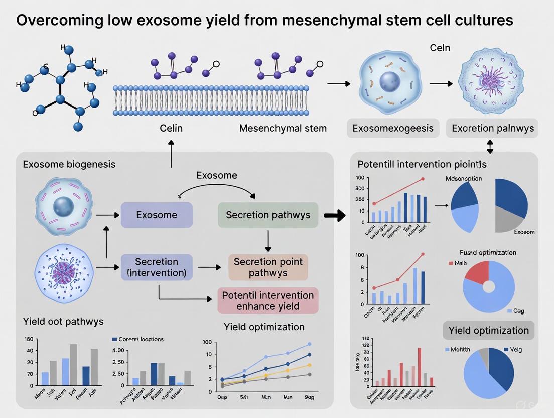
Visualizing the Workflow: From MSC Culture to Exosome Isolation
The following diagram illustrates a streamlined workflow for producing and isolating MSC-exosomes, incorporating strategies to overcome low yield.
Diagram 1: Optimized workflow for MSC-exosome production, covering culture to characterization.
The Scientist's Toolkit: Essential Research Reagents
This table lists key reagents and their critical functions for successfully working with MSC-exosomes.
Table 2: Research Reagent Solutions for MSC-Exosome Workflows
| Reagent / Material | Function / Application | Key Considerations |
|---|---|---|
| Human Platelet Lysate (hPL) | Serum supplement for xeno-free MSC culture medium. Promotes cell proliferation [1]. | Preferred over FBS for clinical translation; ensures a xeno-free environment. |
| Recombinant Human TNF-α | Preconditioning agent to enhance immunomodulatory cargo of exosomes [2]. | Use at low doses (10-20 ng/mL); concentration-dependent effects on miRNA profile. |
| Antibodies: CD9, CD63, CD81 | Detection of positive exosome surface markers via Western Blot or flow cytometry [1] [4]. | Critical for identity confirmation; part of MISEV guidelines. |
| Antibody: Calnexin | Negative control marker for Western Blot (endoplasmic reticulum protein) [1]. | Its absence confirms exosome preparation is free from major cellular contaminants. |
| PKH67 / PKH26 Dyes | Lipophilic fluorescent dyes for labeling exosome membranes for tracking and uptake studies [3]. | Allows visualization of exosome internalization by recipient cells in vitro. |
| Trehalose | Biostabilizer for exosome storage [6]. | Added to exosome pellets or PBS suspensions before freezing at -80°C to preserve integrity and function. |
| Polyethylene Glycol (PEG) | Polymer used in precipitation-based exosome isolation kits [5]. | Enables rapid isolation but may co-precipitate non-exosomal material; purity can be a concern. |
FAQs on Mechanisms and Applications
Q4: What are the primary mechanisms by which MSC-exosomes exert their therapeutic effects? A: MSC-exosomes are multimodal therapeutic agents. Their mechanisms can be unified into several key pillars, including [6]:
- Maintaining Immunological-Stromal Homeostasis: They modulate immune cells (e.g., promoting anti-inflammatory macrophage polarization) via carried cytokines and miRNAs like miR-146a and miR-181a [2] [7].
- Reprogramming Metabolic Circuitry: They can alter the metabolic state of recipient cells in diseases like liver fibrosis [6].
- Determining Cell Fate: They deliver transcription factors and regulatory RNAs that promote tissue-specific differentiation, such as stimulating osteogenesis for bone repair via BMP-2 and RUNX2 [4].
- Direct Extracellular Modulation: The "EMCEV" model proposes that exosomes can signal directly through surface receptors without being internalized, enabling a "one EV to many cells" effect [8].
Q5: What are the main challenges in translating MSC-exosomes to the clinic? A: Despite their promise, several hurdles remain [8] [4] [3]:
- Manufacturing Standardization: Lack of standardized, scalable production methods leads to heterogeneity in exosome quality and potency ("the process defines the product") [8].
- Defining Critical Quality Attributes (CQAs): Establishing consistent metrics for identity, purity, and potency is complex due to their inherent biological variability [8].
- Storage and Stability: Determining optimal long-term storage conditions (e.g., at -80°C with stabilizers like trehalose) to prevent aggregation and preserve function is an active area of research [6].
- Tumorigenicity Concerns: While safer than whole cells, the potential influence of MSC-exosomes on cancer growth requires careful evaluation [7] [9].
Signaling Pathway: Exosomal miRNA in Anti-inflammatory Macrophage Polarization
The following diagram details a key molecular mechanism by which preconditioned MSC-exosomes modulate the immune response.
Diagram 2: How exosomal miR-146a from preconditioned MSCs drives anti-inflammatory macrophage polarization.
Frequently Asked Questions (FAQs) on Low Exosome Yield
FAQ 1: Why is low exosome yield from Mesenchymal Stem Cell (MSC) cultures a critical problem for clinical translation? Low exosome yield is a major bottleneck because it prevents the generation of the large, consistent quantities of exosomes required for effective therapeutic dosing in clinical trials and eventual treatments [10]. The inherent low abundance of exosomes in biological samples and standard culture conditions makes it difficult to scale up production to clinically relevant levels, hindering the transition from promising laboratory research to practical patient applications [11] [12].
FAQ 2: Beyond the overall low yield, what are the related challenges during exosome isolation? The challenge of low yield is often compounded by two other common issues during the purification process:
- Contamination: Many standard isolation methods, such as ultracentrifugation and polymer-based precipitation, are size-based and can co-purify other extracellular vesicles, lipoproteins, or protein aggregates. This contaminates the exosome preparation and can affect the reliability of downstream experiments and therapeutic safety [13] [12].
- Loss of Exosome Integrity: Harsh isolation techniques, particularly those involving high centrifugal forces or aggressive chemicals, can damage the delicate lipid bilayer of exosomes. This compromises their biological activity and the integrity of their cargo, rendering them less effective for functional studies or therapies [12] [14].
FAQ 3: What are the primary biological mechanisms within the cell that limit exosome production? Exosome yield is fundamentally limited by the cell's innate biogenesis and secretion pathways. Key intracellular regulators include:
- The Endosomal Sorting Complex Required for Transport (ESCRT) machinery, which is crucial for the formation of intraluminal vesicles inside multivesicular bodies (MVBs) [15] [16].
- Rab GTPase proteins, which regulate membrane traffic and the fusion of MVBs with the plasma membrane to release exosomes. Notably, some proteins in this family, like Rab4, can act as inhibitors by diverting MVBs for degradation or recycling rather than for secretion [10] [15].
- The availability of membrane components (lipids) is also a rate-limiting factor for the robust secretion of lipid-structured exosomes [10].
Troubleshooting Guide: Strategies to Boost Exosome Yield
Strategy 1: Genetic and Molecular Engineering of Parental MSCs
A highly effective approach involves modulating the expression of key genes that regulate the exosome biogenesis pathway to enhance production.
Detailed Protocol: Knockdown of Rab4 to Boost Yield
- Objective: To increase exosome biogenesis and secretion by reducing the expression of Rab4, a gene that inhibits exosome production.
- Principle: Knockdown of Rab4 decreases the number of early/late endosomes while increasing the number of MVBs, thereby promoting the formation of exosome precursors [10].
- Materials:
- MSC culture (e.g., AML12 cells or human MSCs)
- siRNA targeting Rab4 (e.g., Silencer Select siRNA)
- Appropriate transfection reagent (e.g., Lipofectamine RNAiMAX)
- Negative control siRNA (siNC)
- Cell culture medium and supplements
- Methodology:
- Cell Seeding: Seed MSCs in a 6-well plate to reach 60-70% confluency at the time of transfection.
- Transfection Complex Preparation: Dilute the Rab4-specific siRNA and the negative control siRNA in separate tubes with a reduced-serum medium. In another tube, dilute the transfection reagent.
- Complex Formation: Combine the diluted siRNA with the diluted transfection reagent (1:1 ratio), mix gently, and incubate for 10-20 minutes at room temperature.
- Transfection: Add the siRNA-transfection reagent complexes dropwise to the cells in fresh medium.
- Incubation: Incubate the cells for 48-72 hours to allow for maximal gene knockdown.
- Harvesting: After the incubation period, collect the cell culture supernatant for exosome isolation.
- Expected Outcome: This method has been shown to increase exosome yield significantly. When combined with RCMP supplementation (see below), a 14-fold increase in production has been reported [10].
Strategy 2: Optimization of Culture Conditions and Supplementation
Modifying the cell's microenvironment and nutrient supply can passively enhance exosome production without genetic manipulation.
Detailed Protocol: Supplementation with Red Cell Membrane Particles (RCMPs)
- Objective: To provide additional membrane components to MSCs, thereby supporting the increased biogenesis of exosomes.
- Principle: RCMPs serve as a rich source of lipids and membrane components. When taken up by MSCs, these components can be used to replenish the resources needed for the robust secretion of lipid-structured exosomes [10].
- Materials:
- Phosphate Buffered Saline (PBS)
- DiI fluorescent dye (for uptake validation)
- Centrifuge
- Transmission Electron Microscope (for characterization)
- Methodology:
- RCMP Preparation: Isolate red blood cells from mouse or human blood. Wash the cells with PBS and subject them to repeated freeze-thaw cycles or extrusion through membranes to create nanosized red cell membrane particles [10].
- Characterization: Analyze the size and morphology of the prepared RCMPs using techniques like Dynamic Light Scattering (DLS) or Transmission Electron Microscopy (TEM).
- Supplementation: Add the prepared RCMPs directly to the culture medium of MSCs.
- Incubation and Uptake: Incubate the cells for 24-48 hours to allow for efficient uptake of the RCMPs. Uptake can be confirmed using fluorescence microscopy if the RCMPs are labeled with a dye like DiI.
- Harvesting: Collect the conditioned medium for exosome isolation.
- Expected Outcome: RCMP supplementation alone can boost exosome production, and its effect is additive when combined with genetic strategies like Rab4 knockdown [10].
Strategy 3: Advanced Isolation Techniques to Maximize Recovery
Choosing the right isolation method is critical to maximize the recovery of the exosomes you have produced.
The following table compares the most common exosome isolation methods, highlighting their impact on yield, purity, and integrity—key factors for clinical translation.
| Method | Principle | Yield | Purity | Impact on Integrity | Best for Clinical Translation? |
|---|---|---|---|---|---|
| Differential Ultracentrifugation | Size and density via sequential spinning | Low to Medium [13] | Medium; co-pellets proteins/lipoproteins [3] [16] | High risk of damage from shear forces [13] [14] | Not ideal due to low yield and potential damage |
| Density Gradient Ultracentrifugation | Buoyant density in a gradient medium | Low [13] | High [16] [14] | Good; gentler than differential UC [14] | Good for high-purity requirements, but yield is low |
| Size-Exclusion Chromatography (SEC) | Size-based separation through a porous resin | High [13] | High [13] [14] | Excellent; preserves integrity and function [13] [14] | Highly suitable; good yield and excellent integrity |
| Ultrafiltration | Size-based exclusion using membranes | High [13] | Low [13] | Risk of damage and clogging [3] [14] | Limited by low purity and potential damage |
| Polymer Precipitation | Reduced solubility via polymers (e.g., PEG) | High [13] [14] | Low; co-precipitates contaminants [3] [13] | Polymers may compromise integrity [12] | Not ideal due to low purity and polymer interference |
| Immunoaffinity Capture | Antibody binding to surface markers (e.g., CD63, CD81) | Low [13] | High [13] [14] | Good; gentle isolation [14] | Excellent for specific subpopulations, but yield is low |
| Microfluidic Devices | Size, affinity, or properties via microchips | High [13] [14] | High [13] [14] | Good; minimal sample loss [14] | Promising; high throughput, purity, and yield in an integrated system |
Visualizing the Workflow: From MSC Culture to High-Yield Exosomes
The following diagram illustrates a consolidated experimental workflow that integrates the key troubleshooting strategies outlined above to overcome the challenge of low exosome yield.
The Scientist's Toolkit: Essential Reagents and Materials
The table below lists key reagents and materials used in the featured yield-enhancement protocols, along with their primary functions.
| Reagent/Material | Function/Application |
|---|---|
| Rab4-specific siRNA | Knocks down the expression of the Rab4 gene to release the brake on exosome biogenesis [10]. |
| Transfection Reagent | Facilitates the delivery of siRNA into MSCs for genetic modification [10]. |
| Red Cell Membrane Particles (RCMPs) | Provides essential lipids and membrane components to MSCs, supporting increased exosome production [10]. |
| Size-Exclusion Chromatography (SEC) Columns | Isolates exosomes with high yield and preserved biological integrity, ideal for downstream therapeutic use [13] [14]. |
| Microfluidic Devices | Provides a high-throughput, integrated platform for isolating exosomes with high purity and minimal loss [13] [14]. |
| CD63/CD81 Antibodies | Used for immunoaffinity-based isolation or characterization of exosomes via specific surface markers [3] [14]. |
Troubleshooting Guides
Guide 1: Addressing Low Exosome Yield from MSC Cultures
Problem: Insufficient quantity of exosomes isolated from Mesenchymal Stem Cell (MSC) cultures.
Explanation: Low exosome yield can significantly hamper research progress and therapeutic application development. The quantity of exosomes produced is highly sensitive to specific cell culture parameters.
Solutions:
- Optimize Cell Seeding Density: Evidence shows that lower cell seeding densities can dramatically increase exosome production per cell. One study demonstrated that reducing the seeding density from 1x10⁴ cells/cm² to 1x10² cells/cm² resulted in a 50 to 200-fold increase in vesicle production per cell across multiple passages [17]. Start experiments by testing a range of seeding densities.
- Increase Collection Frequency: The accumulation of exosomes in the culture medium may trigger feedback mechanisms that suppress further production. Collecting conditioned medium more frequently can mitigate this. Research indicates that collecting medium every 12 hours instead of every 24 hours can increase total EV yield by 1.6 to 2.6-fold over the same period [17].
- Monitor Cell Passage Number: As MSCs are passaged in culture, they undergo replicative senescence, which impacts their functionality. While total particle count might remain stable, the bioactivity of the exosomes can decline significantly. One study found a marked decrease in the pro-vascularization bioactivity of exosomes derived from passage 5 (P5) MSCs compared to earlier passages (P2-P4) [17]. Establish a strict limit for the cell passages used for exosome production (e.g., do not use cells beyond P4-P5).
- Consider 3D Bioreactor Cultures: Transitioning from traditional 2D flask cultures to 3D bioreactor systems can enhance both the yield and biological activity of exosomes. Bioreactors allow for better control of environmental parameters (pH, O₂) and can support much higher cell densities [18].
Guide 2: Managing Variable Exosome Bioactivity and Purity
Problem: Isolated exosomes demonstrate inconsistent therapeutic effects or are contaminated with non-exosomal components.
Explanation: The therapeutic potency of exosomes is not solely dependent on quantity. Factors such as donor cell health, culture medium purity, and isolation techniques critically influence the quality and functional integrity of the final exosome preparation.
Solutions:
- Control Donor Cell Conditions: The age and health status of the donor cells directly affect exosome quality. For instance, exosomes derived from older bone marrow MSCs (BMSCs) exhibit diminished osteogenic and lipogenic abilities compared to those from younger BMSCs [19]. Standardize donor cell sources and preconditioning methods where possible.
- Use Exosome-Depleted Fetal Bovine Serum (FBS): Standard FBS contains a high concentration of bovine exosomes and lipoproteins that co-purify with your target MSC exosomes, contaminating the preparation and diluting bioactivity. Using exosome-depleted FBS (prepared by ultracentrifugation or ultrafiltration) can dramatically improve purity. One study showed that this step enhanced the purity of Umbilical Cord MSC (UCMSC)-derived exosomes by 15.6 times, which subsequently improved their wound healing and angiogenic effects by 23.1% and 71.4%, respectively [20].
- Select an Advanced Isolation Method: The standard ultracentrifugation (UC) method can co-isolate contaminants like protein aggregates and lipoproteins and may damage exosomes. Tangential Flow Filtration (TFF) is a scalable alternative that is gentler and provides higher yields. Research has demonstrated that TFF can increase the isolation yield of exosomes by two orders of magnitude (92.5 times) compared to UC [20].
- Avoid Repeated Freeze-Thaw Cycles: The integrity of exosomes can be compromised by improper storage. Cryopreservation at -80°C or -196°C is common, but repeated freezing and thawing can negatively impact exosome structure and function. For long-term storage, consider freeze-drying with appropriate cryoprotectants [19].
Frequently Asked Questions (FAQs)
FAQ 1: How critical is the cell passage number for MSC exosome production, and what is the recommended cut-off?
It is highly critical. While the total number of exosome-sized particles produced may not change dramatically with later passages, their biological activity can be severely compromised. One key study demonstrated that the pro-vascularization bioactivity of MSC-derived exosomes, a critical function for regenerative medicine, was significantly reduced at passage 5 (P5) compared to earlier passages (P2-P4) [17]. It is strongly recommended to establish a working limit, typically not exceeding passage 4 or 5, for producing exosomes intended for therapeutic studies.
FAQ 2: Our yields from 2D flask cultures are low. What are the prospects of scaling up production?
Scaling up is not only possible but essential for clinical translation. Stirred-tank bioreactors offer a viable path for scaling up production. They provide a controlled environment (pH, dissolved O₂, temperature) that mimics physiological conditions and avoids cellular stress. Furthermore, moving to a 3D culture system using microcarriers in bioreactors has been shown to enhance both the yield and biological activity of the produced exosomes compared to standard 2D cultures [18]. This approach allows for "scale-up" (using larger vessels) rather than just "scale-out" (using more flasks), making the process more efficient and reproducible.
FAQ 3: What are the major sources of contamination during exosome isolation, and how can we minimize them?
The primary sources of contamination depend on your starting material:
- From Cell Culture Medium: The most significant contaminant is often bovine exosomes and lipoproteins from FBS [20] [21]. Solution: Use exosome-depleted FBS.
- From Biological Fluids: When isolating from blood plasma or serum, the main contaminants are lipoproteins (e.g., LDL, HDL) and abundant proteins like albumin, which have overlapping physical properties with exosomes [21]. In urine, the Tamm-Horsfall protein (uromodulin) can form networks that trap exosomes [21].
- General Impurities: Protein aggregates and other non-vesicular particles are common contaminants in all isolation methods based solely on size or density [15].
Minimization strategies include using depleted serum, combining complementary isolation techniques (e.g., TFF with size-exclusion chromatography), and incorporating washing steps [19] [20].
FAQ 4: Why is the source of MSCs (e.g., bone marrow vs. umbilical cord) important for exosome output?
The tissue source of MSCs is a major determinant of exosome characteristics. Different anatomical niches subject MSCs to different microenvironments and biological roles, which is reflected in their exosomal "cargo" of proteins, lipids, and RNAs. For example, exosomes released by adipose-derived MSCs (ADSCs) from different anatomical fat depots contained different abundances of miRNAs [19]. This source-dependent variation means that exosomes from different MSC sources may have inherent biases in their therapeutic efficacy for specific applications (e.g., bone regeneration vs. immunomodulation).
The following tables consolidate key quantitative findings from research on factors affecting exosome output.
Table 1: Impact of Cell Culture Parameters on Yield and Bioactivity
| Parameter | Condition | Impact on Production | Impact on Bioactivity/Quality | Key Evidence |
|---|---|---|---|---|
| Cell Seeding Density | Low (1x10² cells/cm²) vs. High (1x10⁴ cells/cm²) | ~50-200x increase in EVs/cell at lower density [17] | No significant difference in vascularization activity reported [17] | Nanoparticle Tracking Analysis, Endothelial Gap Closure Assay [17] |
| Cell Passage Number | Early (P2-P4) vs. Late (P5) | No significant change in particle count [17] | Significant decrease in vascularization bioactivity at P5 [17] | Nanoparticle Tracking Analysis, Endothelial Gap Closure Assay [17] |
| Collection Frequency | Every 12h vs. Every 24h | 1.6 to 2.6x increase in total yield with more frequent collection [17] | Not explicitly measured | Nanoparticle Tracking Analysis [17] |
Table 2: Impact of Isolation and Purity Methods
| Method | Variable | Outcome | Key Evidence |
|---|---|---|---|
| Isolation Technique | Tangential Flow Filtration (TFF) vs. Ultracentrifugation (UC) | 92.5x higher yield with TFF [20] | Particle count comparison via ZetaView [20] |
| Serum Preparation | Exosome-Depleted FBS (UF-dFBS) vs. Normal FBS (nFBS) | 15.6x higher purity (CD73 expression); 71.4% improved angiogenic effect [20] | Western Blot (CD73), Endothelial Cell Tube Formation Assay [20] |
Experimental Protocols
Protocol 1: Depleting Exosomes from Fetal Bovine Serum (FBS)
Principle: Remove bovine exosomes from FBS prior to cell culture to prevent contamination of MSC-derived exosome preparations.
Materials: Ultracentrifuge with fixed-angle rotor (e.g., Type 70 Ti), ultracentrifuge tubes, standard FBS, sterile phosphate-buffered saline (PBS).
Procedure:
- Transfer the desired volume of standard FBS into ultracentrifuge tubes.
- Balance the tubes precisely with PBS or other tubes containing an equal mass of FBS.
- Centrifuge at 100,000 - 120,000 x g for 16-18 hours at 4°C [20].
- Carefully collect the supernatant (the depleted FBS) using a pipette, avoiding the pellet which contains bovine exosomes and other particles.
- Filter the supernatant through a 0.22 µm PES filter under sterile conditions to remove any potential contaminants.
- Aliquot and store at -20°C or -80°C.
Note: As an alternative, ultrafiltration using devices with a 100 kDa molecular weight cut-off can be used for a faster, though potentially less complete, depletion process [20].
Protocol 2: Isoming Exosomes via Tangential Flow Filtration (TFF)
Principle: TFF separates and concentrates exosomes from large volumes of conditioned cell culture media based on size, using tangential flow to prevent filter clogging.
Materials: TFF system, 300-500 kDa molecular weight cut-off filter cartridges [20], conditioned cell culture medium, peristaltic pump, storage vessel.
Procedure:
- Clarification: Centrifuge the collected conditioned medium at low speed (e.g., 2,000 x g for 30 min) to remove cells and large debris. Follow by vacuum filtration through a 0.22 µm filter [20].
- System Setup: Prime the TFF system with PBS or a suitable buffer. Connect the filter, pump, and media reservoir.
- Concentration: Pump the clarified medium through the TFF system. The filter retains exosomes (and other large molecules) while allowing smaller molecules and contaminants to pass through as permeate. The exosomes are recirculated and concentrated in the retentate.
- Diafiltration: Continue the process while adding a diafiltration buffer (e.g., PBS) to the retentate at the same rate as the permeate is removed. This step washes away residual contaminants from the concentrated exosomes. Typically, 5-10 volume exchanges are performed.
- Final Recovery: Once diafiltration is complete, recover the final concentrated exosome retentate from the system.
- Optional Further Purification: The TFF product can be further purified using techniques like Size-Exclusion Chromatography (SEC) to remove remaining soluble proteins and lipoproteins [19].
Signaling Pathways and Workflows
Exosome Biogenesis and Isolation
Critical Factors for High-Quality Exosome Production
The Scientist's Toolkit
Table 3: Key Research Reagent Solutions
| Item | Function in Exosome Research | Key Consideration |
|---|---|---|
| Exosome-Depleted FBS | Growth supplement for MSC culture that minimizes contamination of the final exosome prep with bovine vesicles. | Essential for obtaining pure, clinically relevant MSC exosomes. Can be prepared in-lab via ultracentrifugation or purchased commercially [20]. |
| Tangential Flow Filtration (TFF) System | A scalable method for isolating and concentrating exosomes from large volumes of conditioned cell culture medium. | Superior yield compared to ultracentrifugation; gentler on vesicles; ideal for process scale-up [20] [18]. |
| Size-Exclusion Chromatography (SEC) Columns | For high-purity purification of isolated exosomes, effectively separating them from contaminating proteins and lipoproteins. | Often used as a polishing step after TFF or UC to remove soluble impurities while preserving vesicle integrity [19] [21]. |
| Nanoparticle Tracking Analyzer (NTA) | Instrument used to characterize exosomes by determining their particle size distribution and concentration in a liquid suspension. | Critical for quality control, providing data on the quantity and size profile of your exosome preparation [17] [20]. |
| Tetraspanin Antibodies (CD63, CD81, CD9) | Antibodies against common exosome surface markers, used in techniques like Western Blot or ELISA to confirm exosome identity. | Part of the minimal characterization requirements per MISEV guidelines. Note that expression levels can vary between exosomes from different cell sources [17] [15]. |
Exosomes are nanoscale extracellular vesicles (EVs), typically 30-150 nm in diameter, that are secreted by virtually all cell types and play a crucial role in intercellular communication [3] [22] [9]. These lipid-bilayer enclosed vesicles originate from the endosomal pathway, specifically from intraluminal vesicles (ILVs) formed within multivesicular bodies (MVBs) during their maturation [3] [22]. When MVBs fuse with the plasma membrane, they release these ILVs into the extracellular space as exosomes [22]. The biogenesis process is regulated by both the endosomal sorting complex required for transport (ESCRT) machinery and ESCRT-independent mechanisms, with Rab GTPases (particularly Rab27a and Rab27b) controlling their release [22] [9].
For researchers working with mesenchymal stem cell (MSC) cultures, understanding these pathways is fundamental to addressing the common challenge of low exosome yield. The therapeutic potential of MSC-derived exosomes (MSC-exosomes) in regenerative medicine, including applications in bone repair, cardiovascular disease, neurological disorders, and wound healing, has generated significant interest in optimizing their production [3] [23] [24]. These exosomes mediate many of the therapeutic effects previously attributed to the MSCs themselves, offering advantages such as reduced immunogenicity, higher stability, and the ability to cross biological barriers like the blood-brain barrier [25] [26].
Troubleshooting Guide: Low Exosome Yield
FAQs on Production Challenges
Why is my exosome yield from MSC cultures so low? Low yield frequently stems from suboptimal cell culture conditions. MSCs require specific cues to activate exosome biogenesis pathways. Ensure your culture uses serum-free media or exosome-depleted fetal bovine serum (FBS) to avoid contaminating bovine exosomes that skew yield measurements and downstream analysis [27] [28]. Furthermore, cellular stress from high passage numbers, nutrient deprivation, or suboptimal confluence at harvest can drastically reduce MVB formation and exosome secretion.
How can I increase exosome production without compromising quality? Transitioning from 2D to 3D culture systems or using bioreactors can significantly enhance yield by improving cell viability and creating a more physiologically relevant microenvironment that stimulates exosome release [29]. Genetic approaches, such as overexpressing key regulatory genes like Rab27a, can directly enhance the exocytosis of MVBs [9]. Environmental cues like mild hypoxia or cytokine priming (e.g., with IFN-γ or TNF-α) can also mimic in vivo stress conditions, upregulating the molecular machinery responsible for exosome biogenesis and secretion [3].
My isolated exosomes are contaminated with proteins and other vesicles. What went wrong? This is a common issue with certain isolation techniques. Ultracentrifugation, while widely used, often co-precipitates protein aggregates and lipoproteins due to similar sedimentation properties [3] [23]. To improve purity, consider combining methods. Implementing a density gradient centrifugation step after initial ultracentrifugation can effectively separate exosomes from contaminants based on their buoyant density [23] [26]. Alternatively, size-exclusion chromatography (SEC) or tangential flow filtration (TFF) are excellent choices for obtaining high-purity exosome preparations suitable for therapeutic development [27] [26].
Experimental Protocols for Yield Improvement
Protocol 1: Serum-Free Conditioned Media Collection
- Objective: To collect MSC-conditioned media devoid of contaminating serum exosomes.
- Procedure:
- Culture MSCs to 70-80% confluence in standard growth medium.
- Wash cells thoroughly 3x with phosphate-buffered saline (PBS) to remove residual serum.
- Replace with serum-free medium or medium containing exosome-depleted FBS.
- Incubate for 24-48 hours. Note that prolonged serum starvation (>48 hours) may induce apoptosis and release of contaminating apoptotic bodies.
- Collect conditioned media and proceed immediately to isolation or store at -80°C.
- Key Considerations: The choice of serum-free medium formulation is critical to maintain cell health and function. Validate that your MSCs retain their characteristic phenotype and viability under the chosen serum-free conditions [27] [28].
Protocol 2: Priming MSCs with Pro-Inflammatory Cytokines
- Objective: To enhance exosome yield and modulate cargo by activating MSC secretory pathways.
- Procedure:
- Culture MSCs to 70-80% confluence.
- Treat cells with a priming agent such as IFN-γ (10-50 ng/mL) or TNF-α (10-20 ng/mL) in fresh culture medium.
- Incubate for 24 hours.
- Wash cells with PBS and replace with serum-free collection medium (as in Protocol 1).
- Collect conditioned media after 24-48 hours.
- Key Considerations: Priming can alter the therapeutic profile of the secreted exosomes. Always functionally validate the primed exosomes in your specific assay (e.g., angiogenic, immunomodulatory) to ensure the desired effect is enhanced [3].
The Scientist's Toolkit: Research Reagent Solutions
Table 1: Essential Reagents and Materials for Exosome Research
| Reagent/Material | Function/Application | Key Considerations |
|---|---|---|
| Exosome-Depleted FBS | Provides growth factors and nutrients without contaminating bovine exosomes. | Essential for pre-isolation cell culture to ensure the exosomes collected are of human/cellular origin. |
| Serum-Free Media Formulations | Supports cell health during the exosome secretion phase without serum interference. | Choose a formulation validated for your specific MSC type (e.g., adipose, bone marrow). |
| Protease & Phosphatase Inhibitors | Preserves the protein and phosphoprotein cargo of exosomes during isolation. | Add to conditioned media immediately after collection to prevent cargo degradation. |
| Density Gradient Medium (e.g., Iodixanol) | Separates exosomes from contaminants based on buoyant density. | Critical for high-purity isolation; typically used following ultracentrifugation [23]. |
| PBS (Calcium/Magnesium-Free) | Washing cells and diluting/dialyzing exosome pellets. | Divalent cations can cause exosome aggregation; use Ca²⁺/Mg²⁺-free buffers. |
| Size-Exclusion Chromatography (SEC) Columns | Isolates exosomes based on size, resulting in high-purity preparations. | Preserves exosome integrity and function; easily scalable [26]. |
| Tangential Flow Filtration (TFF) Systems | Concentrates and purifies exosomes from large volumes of conditioned media. | Ideal for scalable, GMP-compliant production; maintains exosome bioactivity [27] [29]. |
| CD63/CD81/CD9 Antibodies | Detects tetraspanin markers for exosome identification via Western blot or flow cytometry. | Part of the minimal characterization set required by MISEV guidelines. |
Isolation Method Comparison and Data
Selecting the appropriate isolation method is critical for balancing yield, purity, and scalability in both research and therapeutic contexts.
Table 2: Quantitative Comparison of Exosome Isolation Methods
| Isolation Method | Typical Yield | Purity | Time Required | Key Advantages | Major Limitations |
|---|---|---|---|---|---|
| Ultracentrifugation | Moderate (~5-25%) [3] | Low-Moderate | 4-6 hours | Low cost; widely accepted; handles large volumes. | Co-sedimentation of contaminants; shear stress may damage exosomes [3] [23]. |
| Density Gradient Centrifugation | Low | High | 6-18 hours | Excellent purity; separates exosomes from proteins/lipoproteins [26]. | Low yield; complex and time-consuming operation [26]. |
| Size-Exclusion Chromatography (SEC) | High | High | 1-2 hours | High purity; maintains vesicle integrity and function; good reproducibility [26]. | Sample dilution; limited sample volume per run. |
| Tangential Flow Filtration (TFF) | High | High | 2-4 hours (scalable) | Scalable for manufacturing; closed-system for GMP; high yield and purity [27]. | High initial equipment cost; requires optimization. |
| Polymer-Based Precipitation | High | Low | 30 min - 2 hours | Simple and fast; no specialized equipment. | Co-precipitation of non-exosomal material (e.g., proteins, RNAs); requires additional purification [23]. |
| Immunoaffinity Capture | Low | Very High | 3-4 hours | Exceptional specificity for exosomes with specific surface markers. | High cost; low yield; may only capture a subpopulation of exosomes [3]. |
Visualizing Exosome Biogenesis and Workflows
Exosome Biogenesis Pathway
Optimized Production Workflow
Exosome Isolation Methods: Balancing Yield, Purity, and Scalability
A major roadblock in the clinical translation of mesenchymal stem cell-derived exosomes (MSC-exosomes) is the inability to isolate sufficient quantities of high-purity exosomes efficiently. The isolation method chosen directly impacts key parameters: yield, purity, scalability, and biological functionality [21] [30]. This guide provides a comparative analysis and troubleshooting support for three core isolation techniques—Ultracentrifugation (UC), Tangential Flow Filtration (TFF), and Size-Exclusion Chromatography (SEC)—within the context of overcoming low-yield challenges from MSC cultures.
Comparative Analysis Table
The table below summarizes the key characteristics of the three isolation methods, providing a clear comparison of their performance and suitability for different experimental needs.
| Isolation Method | Theoretical Basis | Average Yield from MSC Culture | Relative Purity | Scalability | Key Advantages | Key Limitations |
|---|---|---|---|---|---|---|
| Ultracentrifugation (UC) | Sedimentation velocity & density of particles [21] | Baseline (Reference) | Moderate [21] | Low [31] | Considered the "gold standard"; no reagent dependency [21] | Labor-intensive; requires skilled technician; potential for vesicle damage & aggregation [32] [21] |
| Tangential Flow Filtration (TFF) | Size-based separation using membranes & tangential flow [31] | ~20-140x higher than UC [1] [33] [31] | Moderate to High [31] | High [31] | Fast processing of large volumes; gentle on vesicles; high recovery [1] [31] | High initial equipment cost; membrane fouling can occur [31] |
| Size-Exclusion Chromatography (SEC) | Size-based separation via porous beads [32] [21] | Not explicitly quantified vs. UC | High [34] [21] | Medium | Excellent purity & preservation of vesicle integrity; simple operation [34] [21] | Limited sample volume per run; sample dilution [21] |
Detailed Methodologies & Protocols
Ultracentrifugation (UC) Protocol
This is a typical differential UC protocol for isolating exosomes from MSC-conditioned medium [21].
Sample Pre-clearing:
- Centrifuge the conditioned medium at 300 × g for 10 minutes to pellet cells.
- Transfer supernatant to a new tube and centrifuge at 2,000 × g for 10 minutes to remove dead cells and debris.
- Transfer supernatant and centrifuge at 10,000 × g for 30 minutes to remove larger vesicles and organelles.
Exosome Pelleting:
- Transfer the supernatant to ultracentrifuge tubes. Balance tubes carefully.
- Pellet exosomes by ultracentrifugation at 100,000 - 150,000 × g for 1-6 hours (optimize time based on rotor type).
Washing (Optional):
- Resuspend the pellet in a large volume of phosphate-buffered saline (PBS).
- Repeat the ultracentrifugation step to improve purity.
Resuspension:
- Finally, resuspend the exosome pellet in a small volume of PBS or your desired buffer.
Tangential Flow Filtration (TFF) Protocol
This scalable protocol is adapted for processing large volumes of MSC-conditioned media [31].
Pre-filtration:
- Pre-clear the conditioned medium using a 0.22 µm filter to remove large particles that could clog the TFF system.
Concentration and Diafiltration:
- Load the pre-cleared medium into the TFF system equipped with a membrane with a molecular weight cutoff (e.g., 100-300 kDa) or pore size suitable for exosome retention.
- Recirculate the sample, applying tangential flow to concentrate it to a desired smaller volume.
- Perform diafiltration by continuously adding PBS or an isotonic buffer to the concentrate. This exchanges the buffer and removes soluble contaminants like proteins.
Final Recovery:
- Once the desired volume and buffer exchange are complete, recover the concentrated exosome sample.
Size-Exclusion Chromatography (SEC) Protocol
SEC is often used as a polishing step after initial concentration (e.g., via UC or TFF) [32] [21].
Column Preparation:
- Pack a column with a porous polymer resin (e.g., Sepharose) or use a pre-packed commercial column.
- Equilibrate the column with 2-3 column volumes of an isotonic eluent, such as PBS.
Sample Preparation and Loading:
- If the sample volume is too large, pre-concentrate it using a method like ultrafiltration.
- Load the sample onto the column. The maximum recommended sample volume is typically 0.5-5% of the column's total volume.
Elution and Fraction Collection:
- Elute the sample with PBS or your chosen elution buffer. Exosomes, being large, are excluded from the pores and elute in the void volume first.
- Collect the eluate in sequential fractions. The first few fractions will contain the exosomes, followed by fractions containing smaller proteins and contaminants.
Analysis and Pooling:
- Analyze the fractions (e.g., via nanoparticle tracking analysis or protein assay) to identify those richest in exosomes.
- Pool the exosome-containing fractions for downstream use.
The following workflow diagram illustrates the decision-making process for selecting and applying these isolation methods.
Diagram: Decision Workflow for Exosome Isolation from MSC Cultures
Frequently Asked Questions & Troubleshooting
Q1: I am using UC, but my exosome yield from MSC cultures is consistently low. What can I optimize?
- A: Low yield in UC can stem from several factors:
- Cell Culture Health: Ensure MSCs are healthy and at an appropriate passage number (low passage, preferably < P6) [33]. Standardize harvest conditions [32].
- Pellet Loss: The exosome pellet may be loose and invisible. After centrifugation, mark the tube side expected to contain the pellet and carefully remove all supernatant to avoid disturbing it [35].
- Rotor Type: Swinging-bucket rotors provide a more defined pellet path than fixed-angle rotors, potentially improving yield and reproducibility [21].
- Combined Approach: For large sample volumes, consider pre-concentrating the medium using TFF or a precipitation reagent before performing UC [32].
Q2: My exosomes isolated via SEC are too dilute for downstream applications. How can I concentrate them?
- A: This is a common limitation of SEC. You can gently concentrate the pooled exosome fractions using:
- Ultrafiltration: Use centrifugal filter units with an appropriate molecular weight cutoff (e.g., 100 kDa). Be aware that exosomes can be lost due to absorption to the filter [34].
- Secondary UC: A short, high-speed centrifugation step (e.g., 100,000 × g for 70 minutes) can be used to pellet and resuspend exosomes in a smaller volume [21].
Q3: How can I improve the purity of my exosome preparation when working with complex samples like serum or plasma?
- A: A combination of methods often yields the best results.
- Pre-clearing with SEC: Performing a size-exclusion chromatography step prior to immunoaffinity capture or other specific isolations can significantly reduce contaminating proteins and lipoproteins [32].
- Alternative Affinity Method: Consider using phosphatidylserine (PS)-affinity capture (e.g., MagCapture kit). This method can offer high purity and recovery efficiency by targeting a lipid component present on many extracellular vesicles [34].
Q4: Does the choice of MSC culture method impact the success of downstream isolation?
- A: Absolutely. Moving from a traditional 2D culture to a 3D culture system (e.g., using hollow fiber bioreactors or microcarriers) can dramatically increase the total number of exosomes produced per cell, thereby improving the starting material for any isolation method [36] [33] [31]. One study reported a 19.4-fold increase in total exosome production from MSCs in a 3D hollow fiber bioreactor compared to 2D culture [33].
The Scientist's Toolkit: Key Research Reagents
This table lists essential materials and kits used in the field for exosome isolation and analysis.
| Product Name / Category | Primary Function | Key Features / Application Notes |
|---|---|---|
| Dynabeads (CD9/CD63/CD81) [32] | Immunoaffinity isolation of exosomes | Antibody-coated magnetic beads for highly specific capture; ideal for flow cytometry or Western blot analysis. |
| MagCapture Exosome Isolation Kit PS [34] | Phosphatidylserine-affinity isolation | Captures PS-positive vesicles; metal-ion dependent binding allows gentle, neutral-pH elution; species-independent. |
| Total Exosome Isolation Reagent [35] | Polymer-based precipitation | Easy, fast precipitation from various samples (serum, cell culture media); suitable for RNA/protein analysis. |
| Exosome-Human CD63/CD81 Flow Detection Reagent [32] | Flow cytometry detection | Antibodies for detecting and quantifying exosomes captured on magnetic beads via flow cytometry. |
| Hollow Fiber Bioreactor [33] [31] | 3D Cell Culture System | Scalable system for expanding MSCs and producing large quantities of exosomes in a small footprint. |
Overcoming the challenge of low exosome yield from MSC cultures requires an integrated approach that considers both upstream production and downstream isolation.
- For Maximum Yield and Scalability: Tangential Flow Filtration (TFF), especially when combined with 3D MSC cultures, is the most powerful strategy, offering increases of over 100-fold compared to traditional UC [31].
- For Maximum Purity: Size-Exclusion Chromatography (SEC) is superior for removing contaminating proteins and is an excellent polishing step, though it may require a pre-concentration step [34] [21].
- The "Gold Standard" with Caveats: Ultracentrifugation remains widely used but is hampered by low scalability, potential for vesicle damage, and operator dependency [32] [21].
The optimal choice is dictated by the specific requirements of the downstream application, balancing the need for yield, purity, scalability, and preserving biological activity.
Ultracentrifugation (UC) is widely regarded as the gold standard technique for isolating exosomes from biological fluids and cell culture media, including conditioned medium from Mesenchymal Stem Cell (MSC) cultures. This method leverages high centrifugal forces to separate vesicles based on their size, density, and shape. For MSC researchers, UC is prized for its ability to produce highly enriched exosome fractions without the requirement for complex sample pre-processing or extensive technical expertise, making it a cornerstone of traditional exosome research [37].
The process typically involves differential ultracentrifugation, a series of sequential centrifugation steps at increasing speeds to first remove cells and debris, then larger vesicles, and finally to pellet the exosomes themselves at high forces of approximately 100,000 to 120,000 x g [38] [37]. An advanced variant, density gradient ultracentrifugation, utilizes a medium such as sucrose or iodixanol to create a density gradient, which further purifies exosomes by separating them from contaminating proteins and other non-vesicular particles based on their buoyant density [37]. Despite its widespread use, understanding the limitations of UC—particularly for scaling up production for therapeutic applications—is crucial for advancing MSC-exosome research.
Key Limitations for Scaling Up MSC-Exosome Production
While UC is a robust research tool, its application in large-scale MSC-exosome production for therapeutics faces significant hurdles. The table below summarizes the core challenges.
Table 1: Key Limitations of Ultracentrifugation for Scale-Up
| Limitation | Impact on Scale-Up for MSC-Exosome Production |
|---|---|
| Low Exosome Yield [39] [6] | The quantity of exosomes secreted by MSCs is inherently low; UC does not solve this fundamental issue and can result in further loss of precious material. |
| Co-precipitation of Contaminants [3] [40] | Proteins, such as albumin, and high-density lipoproteins (HDL) often co-sediment with exosomes due to similar densities, reducing purity and requiring additional, time-consuming purification cycles. |
| Time-Consuming Process [6] | A single UC run can take over 70 minutes, and multiple cycles (e.g., 5 cycles) are often needed for sufficient purity, drastically limiting throughput [40]. |
| Equipment and Expertise Dependency [38] | The method requires access to expensive ultracentrifuge infrastructure and specific operational expertise, which can be a bottleneck for widespread or high-volume production. |
| Potential Vesicle Damage [3] [6] | The high g-forces and mechanical stresses during pelleting and resuspension can damage exosomal membranes, potentially compromising their integrity and biological function. |
| Batch-to-Batch Variability [6] | The multi-step, manual nature of UC can lead to inconsistencies in exosome preparations between different runs and operators. |
Frequently Asked Questions (FAQs) and Troubleshooting
Q1: My exosome yield from MSC-conditioned media is consistently low with UC. What can I optimize?
- Check Cell Culture Conditions: Ensure MSCs are healthy and cultured in exosome-depleted fetal bovine serum (FBS) to avoid contaminating bovine vesicles [39]. The percentage of cell confluence at the time of media collection (e.g., 90%) can also impact yield [39].
- Optimize Centrifugation Parameters: For density gradient UC, the duration of centrifugation is critical. For example, equilibrium conditions for separating lipoproteins were only reached after 48 hours in one study, highlighting that longer spins can improve separation [41]. Always follow rotor-specific k-factor calculations for optimal pelleting.
- Avoid Overloading: Do not exceed the recommended volume for ultracentrifuge tubes. Overloading leads to inefficient pelleting and imbalances [42].
- Consider an Alternative or Hybrid Method: Explore techniques like size-exclusion chromatography (SEC) either as a standalone method or in combination with an initial UC step. One study found that combining one cycle of UC with SEC provided improved results relative to SEC alone [40].
Q2: My UC-isolated MSC-exosomes show high protein contamination in proteomic analysis. How can I improve purity?
- Increase Wash Cycles: Perform multiple rounds of resuspension in phosphate-buffered saline (PBS) and repeated ultracentrifugation. Research has shown that five cycles of UC were necessary to efficiently remove over 95% of contaminating serum proteins [40].
- Implement a Density Gradient: Switch from differential UC to density gradient ultracentrifugation (DGC). DGC is an advanced technique that better separates exosomes from soluble proteins and other particles based on their buoyant density, resulting in higher purity isolates [37].
- Dilute Your Sample: Diluting the serum or conditioned media with PBS before the first ultracentrifugation step can reduce viscosity and improve separation efficiency, leading to a cleaner pellet [40].
Q3: What are the critical steps to prevent exosome damage during UC?
- Gentle Resuspension: After the final ultracentrifugation step, avoid vigorous pipetting or vortexing to resuspend the exosome pellet. Gently pipette up and down or let the pellet soak in buffer for a period before resuspending.
- Optimized Centrifugation Time: Use the minimum required centrifugation time at each speed to pellet the target vesicles. Excessively long spins can pack the pellet too tightly and make gentle resuspension difficult.
- Pre-Chill Equipment: For experiments requiring low temperatures, pre-chill the centrifuge and rotor to maintain sample integrity during the centrifugation process [42].
Experimental Protocol: Optimized Ultracentrifugation for Serum/Plasma
This protocol, adapted from current literature, details an optimized multi-cycle UC method designed to maximize exosome purity from serum or plasma, which is directly relevant for assessing MSC-exosome presence in vivo [40].
Materials:
- Hardware: Ultracentrifuge (e.g., Beckman Optima series), fixed-angle or swinging-bucket rotor (e.g., Type 70.1, SW 60), polyallomer ultracentrifuge tubes.
- Reagents: PBS (pH 7.4), pre-analytically processed human plasma or serum (e.g., from EDTA tubes, centrifuged at 2,200 x g to remove cells/platelets) [38].
Procedure:
- Sample Preparation: Dilute the plasma/serum sample with an equal volume of PBS to reduce viscosity [40].
- Remove Cells and Debris: Centrifuge the diluted sample at 2,000 x g for 10 minutes at 4°C. Transfer the supernatant to a new tube.
- Remove Larger Vesicles: Centrifuge the supernatant from step 2 at 10,000 x g for 30 minutes at 4°C to pellet larger microvesicles and apoptotic bodies. Carefully collect the supernatant [39].
- First Ultracentrifugation: Transfer the supernatant to ultracentrifuge tubes. Balance tubes precisely with PBS. Centrifuge at 100,000 x g for 120 minutes at 4°C [40].
- Wash Cycles (Repeat 4 times): Carefully discard the supernatant, leaving a small volume (~2 mm) to avoid disturbing the pellet. Resuspend the pellet in a large volume (e.g., 4 mL) of PBS. Centrifuge again at 100,000 x g for 70 minutes at 4°C. Discard the supernatant after each wash [40].
- Final Resuspension: Resuspend the final, purified exosome pellet in a small volume of PBS (e.g., 50-200 µL) for downstream analysis.
The following workflow diagram illustrates this optimized protocol and its outcomes.
Quantitative Comparison of Exosome Isolation Methods
To make an informed decision on isolation strategies, it is essential to quantitatively compare UC with other emerging techniques. The table below summarizes key performance metrics based on comparative studies.
Table 2: Quantitative Comparison of Exosome Isolation Methods
| Method | Relative Purity (Protein Contamination) | Relative Yield | Processing Time | Key Advantages | Key Disadvantages for Scale-Up |
|---|---|---|---|---|---|
| Differential UC | Moderate (5-25% residual protein) [40] | Low to Moderate | Very High (>5 hours) [40] | High purity potential, no special reagents required [37] | Time-consuming, low throughput, potential vesicle damage [3] [6] |
| Density Gradient UC | High | Low | Very High (~16-48 hours) [41] [37] | Excellent purity, effective removal of contaminants [37] | Lengthy process, low yield, not scalable [37] |
| Size-Exclusion Chromatography (SEC) | High (less serum protein contamination) [40] | Low [40] | Low | Good purity, gentle on vesicles [40] | Sample dilution, column-dependent, low throughput [40] [6] |
| Polymer-Based Precipitation | Low (high impurity levels) [6] | High (~2.5x higher than UC) [38] | Low (6x faster than UC) [38] | Fast, simple, high yield [38] | Co-precipitation of non-exosomal material (e.g., lipoproteins) [3] [6] |
| Exosome Mimetic Vesicles (EMVs) | Moderate (different protein profile) [39] | Very High (several-fold higher) [39] | Moderate | Bypasses cellular secretion, high particle output [39] | Not native exosomes; generated by cell extrusion [39] |
The Scientist's Toolkit: Essential Research Reagents & Materials
Table 3: Key Materials and Reagents for Ultracentrifugation-Based Exosome Isolation
| Item | Function/Application | Example Specifications/Notes |
|---|---|---|
| Ultracentrifuge | Generates high g-forces necessary to pellet nanosized exosomes. | Requires rotors capable of ~100,000 x g (e.g., Beckman Type 70.1, SW 60) [38]. |
| Polyallomer/Carbonate Tubes | Hold samples during ultracentrifugation. | Must be compatible with the rotor and able to withstand high g-forces (e.g., Beckman Ultra-Clear tubes) [40]. |
| Phosphate-Buffered Saline (PBS) | Used for diluting samples and washing exosome pellets. | Dilution reduces sample viscosity; PBS is used for resuspension during wash cycles [40]. |
| Iodixanol or Sucrose | Forms the density gradient for high-purity isolation. | Used in Density Gradient UC to separate particles by buoyant density [37]. |
| Exosome-Depleted FBS | Used in MSC cell culture to produce exosomes for isolation. | Critical for avoiding contamination of MSC-exosome preparations with bovine vesicles from standard FBS [39]. |
| Protease Inhibitor Cocktails | Added to samples to prevent proteolytic degradation of exosomal cargo. | Important for downstream analyses like proteomics [40]. |
For researchers focused on harnessing the therapeutic potential of mesenchymal stem cell (MSC)-derived exosomes, low yield remains a significant bottleneck. Traditional isolation methods like ultracentrifugation are difficult to scale, can damage exosomes, and often result in substantial product loss, with recovery rates potentially as low as 30% [43]. Tangential Flow Filtration (TFF) presents a scalable and gentle alternative. Unlike methods where the flow is directed perpendicularly through the filter (dead-end filtration), TFF operates by recirculating the feed stream tangentially across the membrane surface [44] [45]. This crossflow generates a sweeping action that minimizes membrane fouling and the formation of a "filter cake," enabling efficient processing of large-volume MSC culture supernatants to achieve high-yield, high-integrity exosome harvests [43] [46].
Frequently Asked Questions (FAQs) on TFF for Exosome Harvesting
1. Why is TFF superior to ultracentrifugation for scaling up MSC exosome production? TFF offers several key advantages for scaling up exosome production from MSC cultures. It is a continuous, reproducible process that is gentler on delicate exosomes, preserving their structural integrity and biological function [46]. In contrast, the high shear forces of ultracentrifugation can damage exosomes and lead to low recovery rates of around 30% [43]. TFF systems are also inherently more scalable, allowing for seamless transition from small-scale process development to large-volume clinical or commercial production [44] [45].
2. What type of TFF system is best for handling shear-sensitive MSC exosomes? For shear-sensitive products like exosomes, Hollow Fiber Modules are often the ideal choice. Their simple flow geometry results in a very laminar flow, which provides lower shear stress and gentler processing compared to Flat Sheet Cassettes [44]. This helps maintain the integrity of the exosome membrane and surface proteins, which is critical for both diagnostic sensitivity and therapeutic applications [43] [44].
3. How do I choose the correct membrane cut-off (MWCO) for isolating MSC exosomes? The membrane cut-off should be selected based on the size of your target exosomes. MSC-derived exosomes typically range from 30 to 150 nm in diameter [47] [29]. An appropriate MWCO, often in the ultrafiltration range (e.g., 100-500 kDa), will retain the exosomes in the retentate while allowing smaller contaminants like proteins and nucleic acids to pass through in the permeate [44] [45]. The optimal MWCO must be determined empirically to maximize both purity and yield.
4. What are the most critical parameters to optimize in a TFF process? The efficiency of TFF is a balance of two key operational parameters [44] [48]:
- Transmembrane Pressure (TMP): This is the pressure differential across the membrane. If TMP is too high, it can force molecules into the membrane pores, creating a dense gel layer that severely restricts flow (fouling). If too low, it will not drive sufficient filtration [44].
- Cross Flow Rate (CFR): This is the rate at which the feed solution recirculates tangentially across the membrane. An optimal CFR creates a turbulent flow that sweeps away retained material, minimizing fouling and maintaining a high filtration rate [44] [48].
5. Can TFF be combined with other methods for higher purity exosome isolation? Yes, TFF is often used as an initial concentration and crude purification step in a multi-step workflow. The concentrated retentate from TFF can be further purified using techniques like Size-Exclusion Chromatography (SEC) to remove residual protein contaminants or Immunoaffinity Capture to isolate specific subpopulations of exosomes [43] [47]. This hybrid approach leverages the scalability of TFF with the high purity of other methods.
Troubleshooting Common TFF Challenges in Exosome Isolation
This guide addresses specific issues that may arise during TFF processing of MSC conditioned media.
| Symptom / Problem | Potential Causes | Recommended Solutions |
|---|---|---|
| Rapid Decline in Permeate Flow Rate | Membrane fouling due to a gel layer formed by retained particles or proteins [44]. | Optimize the Cross Flow Rate to increase the sweeping effect at the membrane surface [48]. Reduce the Transmembrane Pressure to a less aggressive setpoint [44]. |
| Low Exosome Yield in Retentate | Membrane pore size or MWCO is too large, allowing exosomes to pass into the permeate [44]. Leaks in the cassette or system seals [44]. | Select a membrane with a smaller pore size/MWCO suitable for the exosome size range (e.g., 100-500 kDa) [44]. Inspect and tighten cassette seals and system fittings; consider automated torque systems for consistent sealing [44]. |
| Poor Exosome Integrity or Activity | Excessive shear stress from high Cross Flow Rates or an aggressive pump [43] [44]. | Switch to a low-shear TFF system, such as one using a gentle Quattroflow four-piston diaphragm pump or hollow fiber modules [44] [48]. Lower the Cross Flow Rate to the minimum required for maintaining flow. |
| Inconsistent Process Performance | Manual system setup with variable pressure and flow control [44]. Inconsistent membrane packing or cassette sealing. | Implement an automated TFF system with recipe-driven control for process consistency and data integrity [44]. Use pre-stacked, encapsulated cassette bundles to eliminate manual assembly errors [44]. |
| High Contaminant Protein in Final Product | Incomplete diafiltration or insufficient wash volumes. Co-precipitation of proteins with exosomes. | Increase the number of diafiltration volumes (typically 5-10) to ensure complete buffer exchange and contaminant removal [45]. Consider a multi-step purification strategy, following TFF with a polishing step like SEC [47]. |
Essential Research Reagent Solutions for TFF
A successful TFF process relies on several key components. The following table details these essential materials and their functions.
| Item / Reagent | Function / Purpose |
|---|---|
| TFF Cassette (Flat Sheet) | Contains the ultrafiltration membrane; ideal for processes demanding higher flux and for less shear-sensitive products. Constructed with layered membranes and mesh screens to create turbulent flow [44]. |
| Hollow Fiber Module | An alternative to cassettes; a cylinder housing tubular fibers. Provides laminar, low-shear flow, making it ideal for gentle processing of sensitive MSC exosomes [44]. |
| Diafiltration Buffer (e.g., PBS) | Used during the diafiltration step to exchange the exosome solution into the final formulation buffer (e.g., for storage or downstream applications) and remove contaminants [32] [45]. |
| Pre-filtration Media | Used to clarify the raw MSC conditioned media by removing cells, large cell debris, and microvesicles before TFF, protecting the TFF membrane from clogging [43]. |
| Four-Piston Diaphragm Pumps | Provide stable, consistent, and low-shear flow recirculation, which is essential for a stable TFF process and for maintaining exosome integrity [48]. |
| Pre-stacked Cassette Bundles | Eliminate the manual, labor-intensive process of stacking cassettes and gaskets, reducing the risk of misassembly and leaks, thereby enhancing reliability [44]. |
Experimental Workflow: TFF for MSC Exosome Isolation
The following diagram illustrates the key stages of a TFF-based workflow for harvesting exosomes from mesenchymal stem cell cultures.
Optimizing MSC Exosome Yield and Purity
The path to overcoming low exosome yield requires a strategic approach to process development. Integrating TFF as a core technology enables a scalable and gentle method for processing large volumes of MSC culture media. By carefully selecting the system configuration (hollow fiber for sensitivity, flat sheet for high flux), meticulously optimizing TMP and CFR to balance yield and purity, and implementing a robust diafiltration strategy, researchers can achieve significant improvements in exosome recovery. Adopting automated TFF systems and pre-stacked cassettes further enhances reproducibility and data integrity, which is critical for therapeutic development [44]. Ultimately, by moving beyond traditional, low-yield methods and embracing the scalable potential of TFF, the field can accelerate the translation of MSC-derived exosome research into reliable diagnostic and therapeutic applications.
Within the critical research aim of overcoming the low exosome yield from mesenchymal stem cell (MSC) cultures, achieving high-purity isolates is non-negotiable. Contaminants like proteins and lipoproteins can severely confound experimental results and therapeutic efficacy. Size-exclusion chromatography (SEC) has emerged as a powerful, size-based separation technique that excels in providing high-purity exosome preparations, preserving vesicle integrity, and maintaining biological activity. This technical support center provides targeted troubleshooting and FAQs to help you integrate SEC effectively into your workflow for sensitive downstream applications.
FAQs: Core Principles of SEC for Exosomes
1. How does SEC fundamentally work to separate exosomes from contaminants?
SEC separates biomolecules based on their hydrodynamic volume or size as they pass through a column packed with porous beads [49]. Larger molecules, such as exosomes, cannot enter the pores of the beads and thus travel a shorter path, eluting first. Smaller molecules and contaminants, including soluble proteins and lipoproteins, temporarily enter the pores and are retained longer in the column, resulting in later elution [49] [21]. This gentle, non-interactive mechanism is key to preserving the structural integrity and function of isolated exosomes.
2. Why is SEC particularly recommended for sensitive applications like MSC-exosome research?
SEC offers several advantages critical for sensitive applications [11]:
- Preserved Vesicle Integrity: Unlike methods like ultracentrifugation, which can damage exosomes through high shear forces, SEC is a gentle technique that minimizes vesicle deformation or rupture [50].
- High Purity with Low Co-aggregation: SEC effectively separates exosomes from common contaminants in culture media, such as bovine serum-derived particles and protein aggregates, leading to a cleaner preparation [20] [21].
- Maintained Biological Activity: By isolating exosomes in a functional state without harsh chemicals or forces, SEC ensures that therapeutically relevant cargoes (e.g., miRNAs, proteins) remain active, which is crucial for functional studies and drug development [20] [11].
3. What are the main limitations or challenges of using SEC?
While powerful, SEC has limitations to consider:
- Sample Volume and Dilution: SEC has a limited sample loading volume (typically 0.5-2% of the total column volume) to maintain resolution [49]. The eluted exosome fraction is also diluted in the mobile phase buffer, often requiring a subsequent concentration step [21].
- Resolution and Throughput: Standard SEC may not fully resolve exosomes from other extracellular vesicles of similar size or from certain lipoproteins. It is also less suited for processing very large sample volumes compared to methods like Tangential Flow Filtration (TFF) [20].
- Potential for Non-Specific Interactions: Although designed to be non-interactive, electrostatic or hydrophobic interactions between the sample and the stationary phase can sometimes occur, requiring optimization of the mobile phase [49].
Troubleshooting Guides
Common SEC Problems and Solutions
| Problem | Potential Causes | Recommended Solutions |
|---|---|---|
| Low Exosome Recovery | Sample overloading; Exosome degradation/precipitation; Poor elution | Load ≤ 5% of total column volume [49]; Ensure proper sample pre-filtration (0.22 µm) and storage conditions; Use appropriate buffer to prevent aggregation. |
| High Contaminant Presence | Column degradation/overuse; Insufficient sample pre-clearation; Incorrect buffer conditions | Replace or regenerate column; Pre-clear sample via 10,000-20,000 g centrifugation [21]; Optimize mobile phase ionic strength (e.g., add 100-150 mM NaCl) to minimize unwanted interactions [49]. |
| Poor Resolution (Broad/Overlapping Peaks) | Excessive flow rate; Incorrect sample viscosity/volume; Air bubbles in column | Reduce flow rate for better separation (e.g., 0.1-0.5 mL/min for analytical columns) [49]; Ensure sample viscosity matches mobile phase and load volume is small; Degas buffers and follow proper column priming procedures. |
| Increased Backpressure | Column clogging; Buffer precipitation; System blockage | Pre-filter all samples and buffers (0.22 µm); Check for buffer compatibility/salts precipitating; Inspect and clean in-line filters, frits, or tubing [51]. |
Optimizing SEC for High Purity and Yield
| Optimization Parameter | Impact on Separation | Guidelines for MSC-Exosomes |
|---|---|---|
| Pore Size of Beads | Determines the size range of molecules separated. | Select beads with a separation range that includes 30-150 nm for exosomes (e.g., resins with pore sizes optimized for 50-1000 kDa or 10-500 nm) [49]. |
| Mobile Phase Composition | Reduces non-specific binding; maintains exosome stability. | Use PBS or Tris buffers at physiological pH (7.2-7.5). Add 100-150 mM NaCl to shield electrostatic interactions. For hydrophobic issues, consider low % organic solvents or arginine [49]. |
| Flow Rate | Affects resolution and run time. | Slower flow rates (e.g., 0.1-0.5 mL/min) enhance resolution but increase run time. Optimize to balance peak sharpness and experimental duration [49] [51]. |
| Sample Load Volume | Critical for maintaining high resolution. | For highest resolution, keep sample load volume between 0.5-5% of the total column volume. Do not exceed 10% [49]. |
| Sample Pre-Clearance | Removes large debris that can block the column. | Centrifuge conditioned media at 10,000-20,000 g for 30 min followed by 0.22 µm filtration is essential before SEC [20] [21]. |
Advanced Strategy: Integrated Workflows
For the highest purity and yield in MSC-exosome research, SEC is often best used in combination with other methods. A highly effective strategy involves using Tangential Flow Filtration (TFF) for initial volume reduction and crude isolation, followed by SEC for final polishing and high-resolution purification [20]. This hybrid approach leverages the scalability of TFF and the superior purity of SEC. Research has demonstrated that a TFF-SEC workflow can increase the isolation yield of exosomes by nearly two orders of magnitude compared to ultracentrifugation alone, while subsequent SEC steps effectively remove co-isolated impurities like lipoproteins, significantly enhancing the purity and bioactivity of the final exosome product [20].
The following diagram illustrates the decision-making process for selecting and optimizing an SEC-based isolation strategy.
Optimization Workflow for SEC
The Scientist's Toolkit: Essential Research Reagents & Materials
| Item | Function in SEC Exosome Isolation | Key Considerations |
|---|---|---|
| SEC Columns | The core component containing porous beads for size-based separation. | Choose based on sample volume (e.g., 10mL for 0.5mL load) and purity needs. Sepharose-based (e.g., Sephacryl) or cross-linked agarose resins are common [49] [21]. |
| Mobile Phase Buffer | Carries the sample through the column; maintains pH and ionic strength. | Phosphate-buffered saline (PBS) or Tris-buffered saline are standard. Add 100-150 mM NaCl to minimize non-specific interactions [49]. |
| Pre-column Filter (0.22 µm) | Removes large particles, cell debris, and aggregates to prevent column clogging. | Essential step after low-speed centrifugation to protect column integrity and performance [20] [21]. |
| Fraction Collector | Automatically collects eluent in small, sequential volumes. | Allows for precise pooling of the exosome-rich fractions (typically in the void volume) separate from contaminant-rich fractions [49]. |
| Concentration Device | Concentrates the diluted exosome fractions obtained from SEC. | Ultrafiltration centrifugal units (e.g., 100 kDa MWCO) are commonly used post-SEC to achieve the desired final sample concentration [20]. |
Exosomes are nanoscale extracellular vesicles (30-150 nm in diameter) released by nearly all cell types and play a crucial role in intercellular communication by transferring functional cargos like miRNAs, mRNAs, and proteins between cells [3] [14]. In the context of mesenchymal stem cell (MSC) research, exosomes have emerged as promising cell-free therapeutic agents with applications in bone regeneration, cardiovascular repair, and immunomodulation [3] [16]. However, a significant bottleneck in their clinical translation is the low yield of exosomes obtained from MSC cultures, making efficient isolation methods critically important for research and therapeutic development [10] [29].
The selection of an appropriate isolation method must balance multiple factors: purity, yield, preservation of biological activity, processing time, and cost. Among the various techniques available, polymer-based precipitation and immunoaffinity capture have gained prominence as effective approaches, particularly for processing the complex biological samples typically encountered in MSC research [13] [14]. This technical support center document provides detailed troubleshooting guides and experimental protocols to help researchers optimize these two emerging methods for their specific applications.
Method Comparison and Selection Guidance
Technical Comparison of Isolation Methods
Table 1: Comparative analysis of polymer-based precipitation and immunoaffinity capture methods
| Parameter | Polymer-Based Precipitation | Immunoaffinity Capture |
|---|---|---|
| Principle | Reduced solubility and precipitation of exosomes using water-excluding polymers (e.g., PEG) [14] | Specific antibody-antigen binding to exosome surface markers (e.g., CD63, CD81, CD9) [13] [14] |
| Purity | Low to moderate; co-precipitation of non-vesicular contaminants like lipoproteins [52] [13] | High; specific for exosomes bearing target antigens [13] |
| Recovery Yield | High [52] [13] | Low to moderate [13] |
| Processing Time | Moderate (several hours to overnight) [52] | Relatively fast (1-3 hours) [14] |
| Cost | Low to moderate [13] | High (antibody costs) [13] |
| Equipment Needs | Basic centrifuge capable of 10,000 × g [52] | Specialized equipment for magnetic separation or columns [14] |
| Sample Volume | Flexible, suitable for small volumes [52] | Limited by binding capacity of antibodies [14] |
| Preservation of Bioactivity | Excellent; preserves biological activities and morphological integrity [52] | Good; gentle elution conditions maintain structure [13] |
| Downstream Compatibility | Polymers may interfere with downstream analysis [13] | Excellent; specific binding reduces contaminants [13] |
| Best For | Rapid assessment, processing multiple samples, when high purity is not critical [52] | Isolation of specific exosome subpopulations, applications requiring high purity [13] |
Visual Guide to Method Selection
Figure 1: Method selection workflow for exosome isolation from MSC cultures
Polymer-Based Precipitation
Detailed Experimental Protocol
Principle: This method utilizes water-excluding polymers such as polyethylene glycol (PEG) to reduce the solubility of exosomes in solution, causing them to precipitate at low centrifugal forces [52] [14].
Materials Required:
- Total Exosome Isolation reagent (commercial kit) or PEG solution
- Phosphate-buffered saline (PBS)
- Refrigerated centrifuge capable of 10,000 × g
- MSC-conditioned medium
- Protease inhibitors
Step-by-Step Protocol:
- Sample Preparation: Clarify MSC-conditioned medium by sequential centrifugation at 300 × g for 10 min (remove cells), 2,000 × g for 10 min (remove dead cells), and 20,000 × g for 60 min (remove cell debris and large vesicles) [52].
- Precipitation: Mix the clarified supernatant with Total Exosome Isolation reagent in a 2:1 ratio (supernatant:reagent). Incubate overnight at 4°C on a roller mixer for continuous gentle mixing [52].
- Recovery: Centrifuge the mixture at 10,000 × g for 60 minutes at 4°C. Carefully discard the supernatant without disturbing the pellet [52].
- Resuspension: Resuspend the exosome pellet in an appropriate volume of PBS (e.g., 200 μL for 10 mL starting material). Avoid washing unless necessary, as this can lead to significant exosome loss [52].
Troubleshooting Guide for Polymer-Based Precipitation
Table 2: Common issues and solutions for polymer-based precipitation
| Problem | Possible Causes | Solutions |
|---|---|---|
| Low yield | Incomplete precipitation | Increase incubation time (up to 24 hours) [52] |
| Insufficient polymer concentration | Optimize sample-to-reagent ratio; typically 2:1 is effective [52] | |
| Protein contamination | Co-precipitation of non-vesicular proteins | Incorporate additional purification step (size-exclusion chromatography) [13] |
| Inadequate initial clarification | Increase speed/duration of clarification spins [52] | |
| Difficulty resuspending pellet | Over-drying of pellet | Leave small amount of supernatant when discarding; resuspend in appropriate buffer [52] |
| Exosome aggregation | Use sterile-filtered PBS with protease inhibitors; pipette gently [52] | |
| Interference with downstream analysis | Residual polymer in final preparation | Use purification columns designed to remove polymers [13] |
| Dilute sample sufficiently to reduce polymer concentration [13] |
Immunoaffinity Capture
Detailed Experimental Protocol
Principle: This technique utilizes antibodies specific to exosome surface markers (e.g., CD63, CD81, CD9) immobilized on solid supports such as magnetic beads or chromatography resins to selectively capture exosomes from complex mixtures [13] [14] [53].
Materials Required:
- Antibody-coated magnetic beads (e.g., anti-CD63, CD81, or CD9)
- Magnetic separation rack
- Binding buffer (PBS with 1% BSA)
- Elution buffer (low pH glycine buffer or neutral buffer with competing agents)
- MSC-conditioned medium
Step-by-Step Protocol:
- Sample Preparation: Pre-clear MSC-conditioned medium by centrifugation at 20,000 × g for 60 minutes to remove large vesicles and debris [52].
- Bead Preparation: Wash antibody-coated magnetic beads three times with binding buffer using magnetic separation [14].
- Incubation: Incubate the pre-cleared sample with washed beads for 1-3 hours at room temperature or 4°C with gentle rotation [14].
- Washing: Separate beads using a magnetic rack and wash 3-5 times with binding buffer to remove unbound material [14].
- Elution: Resuspend beads in elution buffer and incubate for 5-10 minutes with gentle agitation. Separate eluate containing exosomes from beads using magnetic rack [14].
- Neutralization: Immediately neutralize low pH elution buffers with Tris-HCl buffer (pH 8.0) to preserve exosome integrity [14].
Troubleshooting Guide for Immunoaffinity Capture
Table 3: Common issues and solutions for immunoaffinity capture
| Problem | Possible Causes | Solutions |
|---|---|---|
| Low yield | Insufficient antibody binding capacity | Increase bead-to-sample ratio; titrate to find optimal conditions [54] |
| Target antigen not abundantly expressed | Use antibody cocktail targeting multiple surface markers (CD9, CD63, CD81) [14] | |
| Non-specific binding | Inadequate blocking | Include 2% BSA in binding and wash buffers [54] |
| Insufficient washing | Increase number of washes; transfer bead pellet to fresh tube for final wash [54] | |
| Difficulty with elution | Too strong antibody-antigen interaction | Use more stringent elution conditions (low pH, high salt, or competing peptides) [54] |
| Antibody leaching from beads | Covalently crosslink antibody to beads or Protein A/G [54] | |
| Exosome damage | Harsh elution conditions | Optimize elution buffer pH; test gentler elution methods [14] |
| Mechanical stress | Handle beads gently; avoid vortexing [14] |
Integrated Strategies for Enhanced Yield from MSC Cultures
Boosting Exosome Production from MSCs
To address the fundamental challenge of low exosome yield from MSC cultures, consider implementing these evidence-based strategies:
Genetic Manipulation:
- Knockdown of Rab4 gene in MSC donor cells increased exosome yield by regulating the formation of multivesicular bodies without obvious cytotoxicity [10].
- Inhibition of other genes in the exosome biogenesis pathway (e.g., Rab7) can divert exosomes from degradation pathways toward secretion [10].
Culture Optimization:
- Supplementation with red cell membrane particles (RCMPs) in the culture medium provides additional membrane components for exosome biogenesis, further boosting yield [10].
- Combination of Rab4 knockdown and RCMP supplementation increased exosome yield up to 14-fold without altering exosome size, morphology, or cargo loading efficiency [10].
Research Reagent Solutions
Table 4: Essential reagents for exosome isolation and characterization
| Reagent/Category | Specific Examples | Function/Application |
|---|---|---|
| Polymer Precipitation Kits | Total Exosome Isolation kit (Invitrogen) [52] | Precipitation of exosomes using proprietary polymer formulation |
| Exo-spin Exosome Purification Kit [52] | Combines precipitation with size-based purification | |
| Immunoaffinity Capture Reagents | Anti-tetraspanin coated magnetic beads (CD9, CD63, CD81) [14] | Selective capture of exosome subpopulations |
| Protein A/G conjugated beads [53] | Flexible antibody immobilization platform | |
| Characterization Antibodies | Exosome panel (Calnexin, CD9, CD63, CD81, Hsp70, TSG101) [14] | Identification and characterization of exosomes |
| Critical Buffers and Supplements | Protease and phosphatase inhibitors [54] | Prevent degradation of exosomal proteins during isolation |
| EVs-depleted BSA/FBS [52] | Preparation of conditioned media without exogenous vesicle contamination |
Frequently Asked Questions (FAQs)
Q1: Which method is better for preserving the biological activity of MSC-derived exosomes? Polymer-based precipitation better preserves biological activities according to comparative studies. Research shows that exosomes isolated by polymer-based precipitation demonstrated significantly higher miRNA transfer capability to recipient cells and better cellular uptake compared to ultracentrifugation and ultrafiltration methods [52].
Q2: Can I combine these methods for better results? Yes, combining methods often yields superior results. For instance, polymer-based precipitation followed by size-based purification (PBP+SP) can reduce protein contamination while maintaining good yield [52]. Similarly, ultrafiltration combined with size-exclusion chromatography has been shown to reduce impurity cytokines in isolated exosomes [13].
Q3: How can I increase the yield of exosomes from my MSC cultures before isolation? Genetic and culture manipulation strategies can significantly boost yield. Knocking down Rab4 gene expression in MSCs combined with supplementing culture medium with red cell membrane particles (RCMPs) increased exosome yield up to 14-fold without affecting cargo loading efficiency or therapeutic efficacy [10].
Q4: What are the key quality control checkpoints for isolated exosomes? Essential characterization includes: (1) Nanoparticle tracking analysis for size distribution and concentration; (2) Electron microscopy for morphology; (3) Western blot for exosome markers (CD63, TSG101, HSP70) and absence of negative markers (e.g., GM130); (4) Assessment of biological activity through functional assays [52] [14].
Q5: How do I handle small sample volumes typical in MSC research? Polymer-based precipitation is particularly suitable for small sample volumes, as it doesn't require specialized equipment and can process volumes as small as 100-500 μL effectively [52]. For immunoaffinity capture, use smaller bead volumes and scale down proportionally while maintaining adequate antibody concentration for efficient capture.
Maximizing Output: Preconditioning, Culture Strategies, and Process Control
Frequently Asked Questions (FAQs)
FAQ 1: What are the most critical cell culture parameters to control for maximizing MSC exosome yield and quality?
The most critical parameters are the culture system (2D vs. 3D), cell seeding density, and cell passage number. These factors directly impact both the quantity and therapeutic potency of the exosomes produced.
- Passage Number: The therapeutic bioactivity of MSC-derived exosomes declines with higher passage numbers. Specifically, exosomes from passage 5 (P5) MSCs showed a significant decrease in pro-vascularization bioactivity compared to those from earlier passages (P2-P4) [17].
- Cell Seeding Density: Lower cell seeding densities result in a substantially higher production of exosomes per cell. Studies show that reducing the seeding density from 1E4 cells/cm² to 1E2 cells/cm² can increase EV production per cell by approximately 50 to 200-fold [17].
- Collection Frequency: Increasing the frequency of conditioned medium collection enhances total exosome yield. Collecting medium every 12 hours instead of every 24 hours can increase total EV production by about 1.6 to 2.6-fold over a 24-hour period [17].
Table 1: Impact of Cell Culture Parameters on MSC Exosome Production and Quality
| Parameter | Effect on Yield | Effect on Bioactivity | Key Experimental Evidence |
|---|---|---|---|
| Passage Number | No significant change in particle number [17]. | Significant decrease in pro-vascularization bioactivity beyond passage 4 [17]. | In vitro gap closure assay with HDMECs showed P5 exosomes were ~60% less effective than P2 [17]. |
| Seeding Density | Dramatic increase (50-200x) in particles/cell at lower densities (1E2 vs. 1E4 cells/cm²) [17]. | No significant change in vascularization bioactivity observed [17]. | Nanoparticle Tracking Analysis (NTA) and CD63 ELISA confirmed higher productivity at lower densities [17]. |
| Collection Frequency | Increase (1.6-2.6x) with more frequent collection (every 12h vs. 24h) [17]. | Data not explicitly provided in search results. | NTA quantification of EVs collected from the same number of MSCs over 24 hours [17]. |
FAQ 2: My exosome yields from traditional 2D flasks are too low for clinical applications. What scalable alternatives exist?
Traditional 2D culture is a major bottleneck for clinical-scale exosome production. Transitioning to Three-Dimensional (3D) culture systems is the primary strategy for scalable manufacturing [36] [55] [56].
- Problem with 2D Culture: Yields are often extremely low, and MSCs can lose their original traits and functions over time, impacting exosome quality [36].
- Solution with 3D Culture: These systems better mimic the in vivo microenvironment, enhancing cell-cell and cell-matrix interactions. This leads to higher cell densities and significantly increased exosome production without sacrificing quality [36] [56].
Table 2: Comparison of 2D and 3D Culture Systems for MSC Exosome Production
| Feature | 2D Culture (Traditional Flasks) | 3D Culture (Bioreactors & Scaffolds) |
|---|---|---|
| Scalability | Limited surface area; difficult to scale up [55]. | Highly scalable using bioreactors (e.g., up to 80L) [55]. |
| Exosome Yield | Low yield per cell; insufficient for clinical doses [36] [56]. | 3-19x higher reported yield; suitable for clinical-scale production [55] [56]. |
| Cellular Environment | Artificial, forced polarity; can induce replicative senescence [36]. | Mimics natural tissue environment; maintains stem cell properties [36] [56]. |
| Therapeutic Potency | Potency can decrease with passage [17]. | Enhanced bioactivity reported (e.g., angiogenesis, immunosuppression) [36] [56]. |
Table 3: Overview of Common 3D Culture Methods for Enhanced Exosome Production
| 3D Method | Key Principle | Advantages | Disadvantages | Reported Yield Increase |
|---|---|---|---|---|
| Hanging Drop [56] | Cells aggregate in suspended droplets to form spheroids. | Simple, cost-effective, no scaffolds needed. | Low-throughput, difficult medium exchange, limited control over size. | ~2x higher than 2D [56]. |
| Microwell Array [56] | Cells form spheroids within prefabricated micro-wells. | Better control over spheroid size and shape, higher throughput. | Limited adhesion area, potential for central necrosis. | Enables serum-free production for easy extraction [56]. |
| Scaffold-based (e.g., Hydrogels) [56] | Cells grow within a porous 3D matrix that mimics ECM. | Provides adhesion sites, protects from stress gradients. | Can be complex to prepare; polymer may require removal. | Upregulated therapeutic gene expression [56]. |
| Bioreactors (e.g., Hollow Fiber) [55] [56] | Cells grow on microcarriers in a controlled, agitated vessel. | Highly scalable, high cell density, controlled environment. | Higher cost, more complex operation. | ~19.4x higher total production than 2D [56]; 3x higher particle concentration [55]. |
FAQ 3: Beyond the culture system, are there other strategies to boost exosome production from MSCs?
Yes, preconditioning MSCs with specific biochemical or physiological stimuli is a highly effective strategy to enhance both the yield and potency of exosomes [36].
- Hypoxia: Culturing MSCs under low-oxygen conditions simulates a physiological niche and has been shown to enhance the biological activity of the exosomes they produce [36].
- Genetic Modification: Knocking down specific genes that inhibit exosome biogenesis can dramatically increase yield. For example, knocking down Rab4 was shown to increase exosome secretion by regulating the formation of multivesicular bodies (MVBs), and when combined with red cell membrane particle (RCMP) supplementation, led to a 14-fold increase in exosome yield [10].
- Supplementation: Adding specific components to the culture medium can support the high metabolic demand of exosome production. Supplementing with RCMPs provides essential membrane components, further boosting yield [10].
The following workflow diagrams two key experimental strategies for enhancing exosome production.
Experimental Protocols
Protocol 1: Isulating Exosomes via Ultracentrifugation (UC) for Cell Culture Supernatant [57]
This is a detailed protocol for the classic "gold standard" method, commonly used for research-scale exosome isolation.
- Pre-clearing Steps:
- Centrifuge conditioned medium at 300 g at 4°C for 10 minutes to remove detached cells.
- Transfer supernatant to a new tube and filter it through a 0.22 µm filter to remove apoptotic bodies, microvesicles, and cell debris. This yields "clarified" conditioned medium.
- Ultracentrifugation:
- Transfer the clarified supernatant to ultracentrifuge tubes. Balance tubes carefully.
- Centrifuge at 100,000 g avg at 4°C for 90 minutes using a fixed-angle rotor (e.g., Type 50.2 Ti).
- Carefully decant and discard the supernatant. The crude exosome pellet may be visible.
- Washing Step:
- Resuspend the pellet in a large volume (e.g., 1 mL) of ice-cold, sterile PBS.
- Pool resuspended pellets and perform a second round of ultracentrifugation at 100,000 g avg at 4°C for 90 minutes.
- Final Resuspension:
- Discard the supernatant and resuspend the final, purified exosome pellet in 50-500 µL of PBS or a suitable buffer for downstream applications. Aliquot and store at -80°C.
Protocol 2: Isulating Exosomes via Size-Exclusion Chromatography (SEC) [57] [47]
This method is gentler than UC and preserves exosome integrity, providing good separation from contaminating proteins.
- Sample Preparation:
- Pre-clear conditioned medium or biofluid as described in the UC protocol (steps 1-2). For large volumes, concentrate the sample using ultrafiltration devices (e.g., 100 kDa cutoff) first.
- Column Equilibration:
- Pack an SEC column with the appropriate resin (e.g., qEV columns) according to the manufacturer's instructions.
- Equilibrate the column with a compatible buffer, typically PBS or a similar isotonic solution. Ensure at least 2-3 column volumes of buffer pass through.
- Sample Loading and Elution:
- Carefully load the prepared sample onto the top of the column. Avoid disturbing the resin bed.
- As the buffer flows through, begin collecting sequential fractions. Exosomes will elute in the early (void volume) fractions, while smaller proteins and contaminants will elute later.
- Analysis and Storage:
- Analyze fractions using nanoparticle tracking analysis (NTA) or Western blot (for CD63, TSG101) to identify exosome-rich fractions.
- Pool positive fractions and concentrate if necessary. Aliquot and store at -80°C.
The Scientist's Toolkit: Research Reagent Solutions
Table 4: Essential Materials and Reagents for Optimizing MSC Exosome Production
| Item | Function / Application | Examples / Notes |
|---|---|---|
| Serum-Free Media | Used during the exosome production phase to avoid contamination with bovine exosomes from FBS [57] [56]. | Dulbecco's Modified Eagle Medium (DMEM) base is common [57]. |
| Ultracentrifuge | Essential equipment for pelleting exosomes via ultracentrifugation (UC) [57] [16]. | Beckman Coulter Optima series with fixed-angle rotors (e.g., Type 50.2 Ti) [57]. |
| Size-Exclusion Chromatography (SEC) Columns | For gentle, high-purity isolation of intact exosomes [57] [47]. | Commercial qEV columns [57]. |
| Hollow Fiber Bioreactor | A 3D culture system for scalable, high-density MSC expansion and exosome production [55] [56]. | Enables continuous medium exchange and high yield [56]. |
| Microcarriers | Provide a surface for MSC attachment and growth within 3D bioreactor systems [55]. | Used in spinner flasks and large-scale bioreactors [55]. |
| siRNA against Rab4 | Genetic tool to knock down an exosome biogenesis inhibitor, significantly boosting yield [10]. | A key component in the combinatorial booster strategy [10]. |
| Red Cell Membrane Particles (RCMPs) | Culture supplement to provide essential lipids/membrane components for enhanced exosome biogenesis [10]. | Prepared from mouse blood; used with Rab4 knockdown [10]. |
| Nanoparticle Tracking Analysis (NTA) | Instrument for quantifying exosome concentration and size distribution [57] [17]. | NanoSight LM10 is commonly used [17]. |
| Antibodies for Characterization | Essential for confirming exosome identity and purity via Western blot [57] [17]. | Positive markers: CD63, TSG101, Alix [17] [16]. Negative marker: GM130 (Golgi apparatus) [10]. |
Troubleshooting Guide: Frequently Asked Questions
FAQ 1: Why is preconiditioning necessary for MSC-exosome therapies? Mesenchymal stem cells (MSCs) in standard 2D culture often have low exosome yield and produce exosomes with limited therapeutic potency. Preconditioning mimics the hostile environment of injured tissue (e.g., low oxygen, inflammation), "priming" MSCs to enhance both the quantity and quality of their secreted extracellular vesicles (EVs). This adaptive strategy leads to the production of EVs enriched with specific beneficial factors that significantly improve regenerative outcomes in various disease models [58] [36] [59].
FAQ 2: My MSC-exosome yield is still low after hypoxia preconditioning. What can I do? Low yield can be addressed by combining hypoxia with a transition to a three-dimensional (3D) culture system. Conventional 2D culture does not support high-volume exosome production. Research shows that 3D culture systems, such as hollow fiber bioreactors, can increase the total MSC-exosome production by about 19.4 times compared to traditional 2D culture. Using 3D scaffolds or spheroid cultures can also improve the survival and functionality of MSCs, further enhancing secretome output [36].
FAQ 3: How do I choose between different preconditioning stimuli? The choice of stimulus should be guided by your specific therapeutic goal. Different stimuli enrich exosomes with distinct cargo profiles, leading to different functional outcomes. The table below summarizes the primary applications of common preconditioning strategies.
| Preconditioning Stimulus | Primary Therapeutic Goal | Key Functional Outcomes |
|---|---|---|
| Hypoxia | Angiogenesis, Tissue Regeneration | Enhances secretion of pro-angiogenic factors (e.g., VEGF), improves cell survival, and boosts regenerative capacity [58] [60]. |
| Pro-inflammatory Cytokines (e.g., TNF-α, IL-1β) | Immunomodulation, Anti-inflammation | Promotes a anti-inflammatory phenotype in recipient immune cells (e.g., macrophages), mitigates inflammatory damage [2]. |
| Lipopolysaccharide (LPS) | Immunomodulation, Protection against Inflammatory Damage | Alters miRNA profile of exosomes (e.g., upregulates miR-181a-5p, miR-150-5p) to confer cytoprotective effects [2]. |
| 3D Culture | Increasing Yield, General Tissue Repair | Dramatically increases the quantity of exosomes produced and can enhance capabilities in endothelial cell proliferation and angiogenesis [36]. |
FAQ 4: Are there any risks associated with using preconditioned MSC-exosomes? Preconditioning is generally considered to enhance safety compared to whole-cell therapies. MSC-exosomes are acellular, which mitigates risks associated with immune rejection, tumor formation, and the potential for forming ectopic tissues. Furthermore, they do not carry the risk of transmitting infectious pathogens from donor to host. However, the variability in exosome content introduced by different preconditioning protocols requires rigorous quality control to ensure consistency and predictable therapeutic effects [2].
FAQ 5: The miRNA content in my pre-conditioned exosomes is inconsistent. How can I control this? Variability in miRNA profiles is a known challenge and is highly dependent on the specific type and dose of the preconditioning stimulus. To ensure consistency:
- Standardize Stimulus Dose: For example, low-dose TNF-α (10 ng/mL) and a higher dose (20 ng/mL) lead to different miRNA profiles in the resulting exosomes [2].
- Characterize Profiles: Use high-throughput gene sequencing to identify and verify the miRNA expression profiles (e.g., key miRNAs like miR-21, miR-146a, miR-125a) under your specific preconditioning protocol [2].
- Maintain Consistent Culture Conditions: Ensure that other factors like cell passage number, confluency, and serum content are uniform across preparations.
Experimental Protocols for Key Preconditioning Strategies
Protocol 1: Hypoxia Preconditioning for Enhanced Angiogenesis
This protocol details how to precondition MSCs with low oxygen to generate exosomes with improved angiogenic and regenerative potential.
1. Principle Culture MSCs at low oxygen tension (typically 1-5% O₂) to stabilize Hypoxia-Inducible Factors (HIFs). This activates a genetic program that modulates processes like inflammation, migration, proliferation, and angiogenesis, which is reflected in the altered protein and RNA cargo of the secreted exosomes [58].
2. Materials
- Hypoxia Chamber/Workstation: A tri-gas incubator or a sealed chamber with a gas regulator for N₂ and CO₂.
- Gas Mixture: 1-5% O₂, 5% CO₂, balanced N₂.
- Mesenchymal Stem Cell Culture: Standard media and reagents.
3. Step-by-Step Procedure
- Step 1: Culture MSCs to about 70-80% confluency under standard conditions (37°C, 21% O₂, 5% CO₂).
- Step 2: Place the culture flasks/plates inside the pre-equilibrated hypoxia chamber or workstation.
- Step 3: Flush the chamber with the pre-mixed gas (e.g., 1% O₂, 5% CO₂, 94% N₂) to achieve the desired oxygen concentration. Maintain for 24-48 hours [58] [60].
- Step 4: After the preconditioning period, collect the conditioned medium for exosome isolation.
- Step 5 (Optional): For a combined stimulus, inflammatory cytokines like TNF-α can be added to the medium during the hypoxia exposure [60].
4. Key Analysis & Validation
- Exosome Isolation: Use standard methods like ultracentrifugation or size-exclusion chromatography to isolate exosomes from the conditioned medium.
- Functional Assay: Perform an in vitro endothelial tube formation assay using HUVEC cells to confirm the enhanced angiogenic potential of the hypoxic exosomes compared to normoxic controls.
Protocol 2: Cytokine Preconditioning for Immunomodulation
This protocol uses pro-inflammatory cytokines to license MSCs to produce exosomes with enhanced anti-inflammatory properties.
1. Principle Licensing MSCs with cytokines like TNF-α or IL-1β mimics an inflammatory microenvironment. This priming alters the exosome cargo, enriching for miRNAs (e.g., miR-146a) that can polarize macrophages towards an anti-inflammatory M2 phenotype and suppress damaging immune responses [2] [60].
2. Materials
- Recombinant Human Cytokines: TNF-α, IL-1β.
- Cell Culture Media: Serum-free or exosome-depleted FBS media is recommended during preconditioning to avoid contaminating bovine exosomes.
3. Step-by-Step Procedure
- Step 1: Prepare a stock solution of the cytokine (e.g., TNF-α) in a suitable buffer (e.g., PBS with carrier protein).
- Step 2: Culture MSCs to about 70-80% confluency.
- Step 3: Replace the culture medium with fresh medium containing the preconditioning stimulus.
- For TNF-α: Use a concentration of 10-20 ng/mL [2].
- For IL-1β: Use a concentration sufficient to induce a inflammatory response (similarly, low ng/mL range).
- Step 4: Incubate the cells for 24-48 hours at 37°C, 5% CO₂.
- Step 5: Collect the conditioned medium for exosome isolation.
4. Key Analysis & Validation
- Cargo Analysis: Use qRT-PCR to confirm the upregulation of target miRNAs (e.g., miR-146a) in the isolated exosomes.
- Functional Assay: Co-culture preconditioned exosomes with lipopolysaccharide (LPS)-activated macrophages and measure the secretion of pro-inflammatory (e.g., TNF-α, IL-6) vs. anti-inflammatory (e.g., IL-10) cytokines.
Table 1: Impact of Preconditioning on MSC-Exosome Yield and Cargo
| Preconditioning Method | Reported Effect on Yield | Key Altered Cargo Components | Documented Efficacy (Model) |
|---|---|---|---|
| 3D Culture | Increase of ~19.4x (Hollow Fiber Bioreactor) [36] | Unique proteins & miRNAs; enhanced pro-angiogenic factors [36] | Improved functional recovery after CNS injury [36] |
| Hypoxia (1-5% O₂) | Significantly enhances secretion of EVs with regenerative cargo [58] | Upregulation of angiogenic factors (VEGF, bFGF); HIF-dependent genes [58] [60] | Higher regenerative capacity in cardiac, skin, and bone repair [58] |
| TNF-α (10 ng/mL) | Can increase total protein content of exosomes [2] | Upregulation of miR-146a [2] | Enhanced macrophage polarization; improved wound healing [2] |
| TNF-α (20 ng/mL) | Not specified | Upregulation of miR-146a and miR-34 [2] | Amplified immunomodulatory effects [2] |
| LPS (0.1-1 μg/mL) | Not specified | Dose-dependent miRNA shifts (e.g., miR-222-3p, miR-181a-5p, miR-150-5p) [2] | Mitigation of inflammatory damage [2] |
The Scientist's Toolkit: Research Reagent Solutions
Table 2: Essential Reagents for Preconditioning Experiments
| Reagent / Material | Function in Preconditioning | Example & Note |
|---|---|---|
| Tri-Gas Hypoxia Incubator | Provides a controlled, low-oxygen environment for cell culture. | Essential for physiological hypoxia studies (1-5% O₂). Alternative: hypoxia-mimicking chemicals (e.g., CoCl₂) [58]. |
| Recombinant Human Cytokines | Licenses MSCs to an activated, immunomodulatory state. | TNF-α and IL-1β are commonly used. Use exosome-depleted FBS during stimulation [2] [60]. |
| 3D Culture System | Increases exosome yield and mimics a more in vivo-like cell environment. | Options include hollow fiber bioreactors, microcarriers, or hydrogel-assisted cultures [36]. |
| LPS (Lipopolysaccharide) | Activates Toll-like receptor pathways, simulating a bacterial inflammatory challenge. | Dose is critical; different low doses (0.1-1 μg/mL) induce distinct exosomal miRNA profiles [2]. |
| NF-κB Pathway Inhibitor | Tool for mechanistic studies to validate the role of specific signaling pathways. | e.g., TCPA-1, used to investigate NF-κB's role in HIF3α expression [61]. |
Signaling Pathways and Workflows
Diagram Title: Signaling Pathways in MSC Preconditioning
Diagram Title: Experimental Workflow for MSC Preconditioning
For researchers focusing on Mesenchymal Stem Cell (MSC) cultures, overcoming low exosome yield is a significant bottleneck that impedes both in vitro studies and clinical translation. Exosomes, typically 30-150 nm in diameter, are specialized cargo delivery vesicles secreted from cells by the fusion of Multivesicular Bodies (MVBs) with the plasma membrane [62] [63]. These extracellular vesicles play crucial roles in intercellular communication by transferring proteins, lipids, and nucleic acids to recipient cells [6] [63]. The therapeutic potential of MSC-derived exosomes is immense, spanning regenerative medicine, immunomodulation, and targeted drug delivery [64] [6]. However, their widespread clinical adoption is constrained by challenges in scalable production [29]. This guide addresses specific experimental issues by targeting the molecular pathways that regulate exosome biogenesis and secretion to enhance yield from MSC cultures.
FAQs and Troubleshooting Guides
FAQ: What are the primary molecular pathways that regulate exosome biogenesis in MSCs?
Answer: Exosome biogenesis in MSCs is primarily governed by two well-defined molecular pathways, both of which present viable targets for enhancing secretion yields.
- The ESCRT-Dependent Pathway: The Endosomal Sorting Complex Required for Transport (ESCRT) machinery is a multi-protein system crucial for membrane remodelling. It consists of five core complexes (ESCRT-0, -I, -II, -III, and Vps4) that work sequentially to sort ubiquitinated cargo and drive the inward budding of the endosomal membrane to form Intraluminal Vesicles (ILVs) inside MVBs [63] [65] [66]. Key components include TSG101 (ESCRT-I) and ALIX (an accessory protein), and depletion of these proteins often reduces exosome secretion [62] [65].
- The ESCRT-Independent Pathway: This pathway relies on lipids and tetraspanins. A key mediator is neutral sphingomyelinase 2 (nSMase2), which generates ceramide from sphingomyelin [62] [66]. Ceramide induces negative membrane curvature, facilitating ILV budding without the need for the full ESCRT machinery [65]. Tetraspanins (CD9, CD63, CD81) also organize membrane microdomains that promote selective cargo clustering and vesicle formation [65].
The diagram below illustrates how these pathways converge in the biogenesis of exosomes.
FAQ: My MSC culture yields low exosome numbers. How can I modulate culture conditions to enhance secretion?
Answer: Low yield often stems from suboptimal cell culture conditions. Evidence-based optimization of the microenvironment can significantly boost exosome secretion without genetic manipulation.
- Problem: Suboptimal culture media and conditions.
- Solution:
- Media Selection: A 2025 study directly compared culture media for Bone Marrow-MSCs (BM-MSCs) and found that using Alpha Minimum Essential Medium (α-MEM) resulted in a higher particle yield per cell (4,318.72 ± 2,110.22) compared to Dulbecco's Modified Eagle Medium (DMEM) (3,751.09 ± 2,058.51), although the difference was not statistically significant. Cells in α-MEM also showed a slightly higher expansion ratio [1].
- Serum Supplements: Use human platelet lysate (hPL) in place of fetal bovine serum (FBS) for xeno-free, clinical-grade production. This supports robust MSC growth and subsequent exosome secretion [1].
- Physical Stressors: Applying mild physical or metabolic stress can upregulate biogenesis pathways.
- Gamma Radiation: A 2025 study on macrophages demonstrated that gamma radiation significantly enhances exosome release by activating the EGFR/IGFR-MYC signaling axis, which upregulates motor proteins Myh10 and Myo5b involved in exosome secretion [67]. While this requires validation in MSCs, it presents a novel, scalable strategy.
- Other Stressors: Hypoxia and glucose deprivation have also been shown to promote exosome release in various cell types by activating stress-induced pathways [67].
FAQ: Which isolation method should I use to maximize the yield and purity of exosomes from conditioned media?
Answer: The choice of isolation method critically impacts both the yield and purity of your final exosome preparation, affecting downstream applications and experimental conclusions.
- Problem: Low recovery or impure exosome preparations from conditioned media.
- Solution: While differential ultracentrifugation (UC) is the most common method, it can have low purity and yield [62] [6]. For greater scalability and yield, consider Tangential Flow Filtration (TFF). A 2025 study directly comparing isolation methods for BM-MSC-sEVs found that "particle yields were statistically higher when isolated by tangential flow filtration (TFF) than by ultracentrifugation (UC)" [1]. TFF is more suitable for processing large volumes of conditioned media and is less prone to causing vesicle damage or aggregation compared to repeated high-speed centrifugation spins [1].
The table below summarizes the key characteristics of common isolation methods.
| Method | Principle | Merits | Pitfalls | Relative Yield |
|---|---|---|---|---|
| Differential Ultracentrifugation | Stepwise increase in centrifugal force separates particles by density and size [62]. | No additional reagents required; widely established [62]. | Low purity; potential for vesicle damage; time-consuming [6]. | Baseline [1] |
| Tangential Flow Filtration (TFF) | Size-based separation using filters in a continuous flow system [1]. | High yield; scalable for large volumes; gentle on vesicles [1]. | Requires specialized equipment; membrane fouling can occur. | Higher than UC [1] |
| Size Exclusion Chromatography | Size-based separation by filtration through a porous gel matrix [6]. | High purity; minimal alteration of exosomal surface structure [62]. | Low yield; sample dilution; limited scalability [62] [6]. | Lower |
| Polymer-Based Precipitation | Precipitation using polymers like PEG to increase particle size [62] [6]. | Fast and easy; good for small volumes. | Co-precipitation of non-exosomal impurities (e.g., proteins) [6]. | Variable (high but impure) |
FAQ: Which specific molecular targets can I manipulate to genetically enhance exosome biogenesis?
Answer: Targeting key regulators of the biogenesis and secretion pathways can lead to a direct increase in exosome output. The table below summarizes prime molecular targets and the experimental evidence supporting their roles.
| Target | Description | Role in Biogenesis/Secretion | Experimental Evidence |
|---|---|---|---|
| nSMase2 | Neutral sphingomyelinase, generates ceramide [62]. | Key enzyme in ESCRT-independent ILV formation; ceramide induces membrane curvature [65] [66]. | Inhibition of nSMase2 (e.g., with GW4869) reduces exosome secretion in multiple cell types [65]. |
| RalA/RalB | Small GTPases crucial for exocytosis and tumorigenesis [62]. | Recruits PLD1 to endosomes, generating phosphatidic acid to promote ILV biogenesis and cargo sorting [62]. | Overexpression enhances exosome secretion; localized on CD63-positive endosomes/MVBs [62]. |
| Rab GTPases (e.g., Rab31) | Small GTPases involved in vesicular trafficking and MVB docking [62]. | Regulates MVB docking at the plasma membrane; involved in ESCRT-independent cargo sorting [62]. | Rab31 knockdown impairs exosome secretion; interacts with flotillin proteins [62]. |
| ESCRT Components (e.g., TSG101, ALIX) | Core subunits of the ESCRT machinery [62]. | Facilitates ubiquitinated cargo sorting and membrane scission during ILV formation [63] [65]. | Depletion of TSG101 or ALIX markedly reduces exosome secretion in various cells [62] [65]. |
| Caveolin-1 | Marker protein for caveolae formation [66]. | Involved in cargo sorting in ILVs and may regulate early endosome formation [62] [66]. | Associated with mechanoadaptation and cholesterol homeostasis; found in exosomes [62] [66]. |
The following diagram maps these molecular targets onto the cellular journey of an exosome, from biogenesis to release, providing a visual guide for experimental targeting.
The Scientist's Toolkit: Research Reagent Solutions
This table provides a list of essential reagents and tools used to study and manipulate exosome biogenesis pathways, based on the protocols and research cited in this guide.
| Reagent/Tool | Function/Description | Example Application in Research |
|---|---|---|
| GW4869 | A specific, non-competitive inhibitor of nSMase2 [65]. | Used to inhibit the ESCRT-independent pathway and confirm ceramide-dependent exosome secretion [65]. |
| Human Platelet Lysate (hPL) | A xeno-free serum supplement for MSC culture [1]. | Used in GMP-compliant, clinical-grade expansion of MSCs to produce exosomes, replacing FBS [1]. |
| Gamma Irradiator | A source of ionizing radiation (e.g., Cobalt-60) [67]. | Used as a physical stimulus to activate the EGFR/IGFR-MYC axis and enhance exosome secretion in macrophage studies [67]. |
| Anti-TSG101 / Anti-ALIX Antibodies | Antibodies for detection and validation of ESCRT components [62] [63]. | Used in Western Blotting to characterize isolated exosomes and confirm ESCRT pathway activity [62] [67]. |
| siRNA/shRNA (for RalA, Rab31, etc.) | Tools for targeted gene knockdown [62]. | Used to transiently or stably knock down key GTPases to study their specific role in biogenesis and secretion [62]. |
| CD63, CD81, CD9 Antibodies | Antibodies against canonical exosome surface tetraspanins [6] [63]. | Used for immunoblotting, flow cytometry, or affinity-based capture to identify and isolate exosomes [1] [67]. |
| Nanoparticle Tracking Analyzer | Instrument for quantifying particle concentration and size distribution (e.g., Nanosight NS300) [1] [67]. | Essential for characterizing exosome preparations and accurately comparing yields between experimental conditions [1] [67]. |
Troubleshooting Guide: Overcoming Low Exosome Yield from Mesenchymal Stem Cell Cultures
This guide addresses common challenges in producing exosomes from Mesenchymal Stem Cells (MSCs) for clinical applications, following Quality by Design (QbD) and Good Manufacturing Practice (GMP) principles.
Frequently Asked Questions (FAQs)
Q1: Our MSC culture system produces low exosome yields. What upstream factors should we investigate? Low exosome yield often originates from suboptimal cell culture conditions. Key factors to optimize include:
- Culture Environment: Transitioning from 2D to 3D culture systems can significantly increase yield. Hollow fiber bioreactors, a type of 3D culture, have been shown to increase total MSC-exosome production by approximately 19.4 times compared to conventional 2D culture [36].
- Culture Medium: Studies indicate that using Alpha Minimum Essential Medium (α-MEM) may result in a higher average yield of particles per cell (4,318.72 ± 2,110.22) compared to Dulbecco's Modified Eagle Medium (DMEM) (3,751.09 ± 2,058.51), though the difference was not statistically significant in one study [1].
- Cell Source and Health: The proliferative capacity and health of your MSCs directly impact exosome output. Use early-passage cells and monitor population doubling time [1].
Q2: Which isolation method provides the best balance of high yield, purity, and GMP compliance for scaling up? The choice of isolation method is critical for process scalability and product quality. The table below compares common techniques:
| Isolation Method | Reported Yield | Key Advantages | GMP Scalability |
|---|---|---|---|
| Tangential Flow Filtration (TFF) [1] | Statistically higher than Ultracentrifugation [1] | High yield, gentle process, suitable for large volumes [1] | High [1] |
| Multimodal Flowthrough Chromatography (MFC) [68] | Significantly higher than SEC [68] | High yield and purity, removes protein/nucleic acid contaminants, scalable [68] | High [68] |
| Size Exclusion Chromatography (SEC) [68] | Lower than MFC [68] | Good purity and functionality for 2D cultures [68] | Moderate (can struggle with high-impurity sources) [68] |
| Ultracentrifugation (UC) [1] | Lower than TFF [1] | Widely used; considered a classic method [1] | Low (can cause EV damage and aggregation) [68] |
| Size Exclusion-Fast Protein Liquid Chromatography (SE-FPLC) [69] | High yield (88.47%) [69] | Rapid (<20 min), effective removal of albumin/lipoproteins [69] | High [69] |
| Monolithic Chromatography [70] | High yield [70] | Concentrates and purifies in one step, reduces DNA/protein impurities [70] | High (easily transferable from R&D to cGMP) [70] |
Q3: How can we improve the therapeutic potency of the exosomes we produce? The therapeutic efficacy of MSC-exosomes can be enhanced through preconditioning or engineering strategies:
- Preconditioning: Culturing MSCs under hypoxic conditions or in 3D environments can enhance the biological activity of the exosomes they secrete [36].
- Biochemical Cues: Exposing MSCs to specific cytokines or small molecules can alter the cargo of the exosomes, potentially boosting their therapeutic effect for a specific application [36].
Q4: What are the critical quality attributes (CQAs) we must monitor for GMP-compliant exosome production? A robust Quality Management System (QMS) is essential. CQAs for exosomes include [71] [1]:
- Identity: Confirmation of exosome markers (e.g., CD9, CD63, TSG101) via Western Blot [1].
- Potency: Demonstrated biological activity in a relevant functional assay (e.g., enhancing cell proliferation or reducing apoptosis) [1].
- Purity: Low levels of process-related impurities such as host-cell proteins and nucleic acids [68] [70].
- Safety: Sterility (absence of bacteria, fungi, mycoplasma) and low endotoxin levels (e.g., <0.25 EU/mL) [1].
Experimental Protocols for Key Investigations
Protocol 1: Comparing Isolation Method Yield and Purity This protocol outlines a direct comparison of TFF and UC, as described in scientific literature [1].
- Objective: To isolate small extracellular vesicles (sEVs) from BM-MSC conditioned medium and compare the yield and purity achieved by TFF and UC.
- Materials:
- Conditioned medium from BM-MSCs (e.g., cultured in α-MEM with 10% human platelet lysate) [1].
- Ultracentrifuge and appropriate rotors.
- Tangential Flow Filtration system with appropriate molecular weight cut-off filters.
- Nanoparticle Tracking Analysis (NTA) instrument (e.g., Malvern Nanosight).
- Bicinchoninic Acid (BCA) Assay kit for protein quantification.
- Methodology:
- Conditioned Medium Collection: Culture BM-MSCs to 80-90% confluency, replace with fresh, serum-free medium, and collect conditioned medium after 48 hours. Remove cells and debris via centrifugation at 400 × g for 10 min [1].
- Ultracentrifugation (UC):
- Centrifuge the supernatant at 10,000 × g for 30 minutes to remove larger vesicles.
- Transfer the supernatant to ultracentrifuge tubes and pellet sEVs at 100,000 × g for 70 minutes.
- Resuspend the pellet in phosphate-buffered saline (PBS) [1].
- Tangential Flow Filtration (TFF):
- Process the conditioned medium through a TFF system with a defined molecular weight cut-off (e.g., 500 kDa) to concentrate and diafilter the sample.
- The final retentate contains the concentrated sEVs [1].
- Analysis:
- Yield: Determine particle concentration (particles/mL) using NTA.
- Purity: Calculate a purity index by dividing the particle concentration by the total protein concentration (measured by BCA assay). A higher ratio indicates a purer preparation with less protein contamination.
Protocol 2: Evaluating Therapeutic Efficacy in a Cell Damage Model This protocol assesses the functionality of isolated sEVs using an in vitro oxidative stress model [1].
- Objective: To evaluate the protective effects of BM-MSC-sEVs on ARPE-19 retinal pigment epithelium cells damaged by hydrogen peroxide (H₂O₂).
- Materials:
- ARPE-19 cell line.
- BM-MSC-sEVs isolated via TFF or other method.
- Hydrogen Peroxide (H₂O₂).
- Cell viability assay kit (e.g., MTT, MTS).
- Flow cytometer with Annexin V/PI staining kit for apoptosis.
- Methodology:
- Induction of Damage: Culture ARPE-19 cells and expose them to a defined concentration of H₂O₂ (e.g., 0.5-1.0 mM) to induce oxidative damage. A control group should not be exposed to H₂O₂.
- sEV Treatment:
- Pre-treatment: Apply sEVs (e.g., 50 µg/mL) to cells 24 hours before H₂O₂ exposure.
- Post-treatment: Apply sEVs (e.g., 50 µg/mL) to cells 24 hours after H₂O₂ exposure.
- Viability Assessment: After the treatment period, measure cell viability using the MTT/MTS assay. Calculate the percentage viability relative to the untreated control.
- Apoptosis Analysis: Harvest the cells and stain with Annexin V and Propidium Iodide (PI). Use flow cytometry to quantify the percentage of cells in early and late apoptosis. A significant reduction in total apoptotic cells indicates therapeutic efficacy of the sEVs [1].
Workflow Visualization
The following diagram illustrates the integrated QbD-based workflow for improving MSC exosome production, from upstream culture to downstream isolation and quality control.
The Scientist's Toolkit: Research Reagent Solutions
| Item | Function & Rationale |
|---|---|
| Hollow Fiber Bioreactor | A 3D culture system that drastically increases cell density and exosome production yield compared to 2D flasks [36] [1]. |
| Human Platelet Lysate (hPL) | A xeno-free supplement for MSC culture media, essential for GMP-compliant processes aiming at clinical applications [1]. |
| Tangential Flow Filtration (TFF) System | A scalable filtration system for gentle concentration and purification of exosomes from large volumes of conditioned medium, preserving vesicle integrity [1]. |
| Multimodal Chromatography Resin | A chromatography resin with an inert, porous shell that binds impurities (proteins, nucleic acids) while allowing exosomes to pass through in the flow-through, enhancing purity [68]. |
| Salt-Tolerant Nuclease | An enzyme used in purification processes to effectively degrade host-cell DNA impurities, a critical step for ensuring product purity and safety [70]. |
| Nanoparticle Tracking Analyzer | An instrument used to characterize exosomes by determining particle size distribution and concentration, key attributes for identity and yield calculation [1]. |
Within the broader research goal of overcoming low exosome yield from mesenchymal stem cell (MSC) cultures, preserving the structural and functional integrity of isolated exosomes is paramount. The processes of isolation, purification, and storage can introduce significant stress on these delicate nanovesicles, leading to aggregation, damage, and cargo loss that directly undermine therapeutic efficacy and experimental reproducibility [72]. For researchers and drug development professionals, implementing rigorous protocols to mitigate these issues is not merely optional but fundamental to achieving reliable, translatable results. This technical support center provides targeted guidance to address the most common integrity challenges encountered during exosome processing from MSC cultures, offering practical solutions framed within a quality-by-design approach.
FAQs on Exosome Integrity and Stability
Q1: What are the primary causes of exosome aggregation during processing? Exosome aggregation is frequently caused by mechanical stress and inappropriate buffer conditions. High-speed centrifugation, particularly during ultracentrifugation, can force vesicles into close proximity, leading to fusion or irreversible clumping [26]. Additionally, using buffers with incorrect pH or ionic strength can neutralize the natural negative surface charge (zeta potential) of exosomes, reducing the electrostatic repulsion that keeps them stably dispersed in solution [73]. This aggregation not only compromises integrity but also severely impacts accurate quantification and dosing.
Q2: How can I determine if my isolation method is damaging the exosomes? Damage can be assessed through a combination of physical characterization, biomarker analysis, and functional assays. Key indicators of damage include:
- Size and Morphology Shifts: Use Nanoparticle Tracking Analysis (NTA) to detect significant size distribution changes and Transmission Electron Microscopy (TEM) to visualize破裂 or irregular morphologies [1] [26].
- Cargo Leakage: Detect the presence of intra-exosomal marker proteins (e.g., ALIX, TSG101) in the supernatant after a second, high-speed centrifugation, which suggests membrane rupture [72].
- Purity Checks: Contamination with protein aggregates or lipoproteins, detectable via western blot for non-exosomal proteins, can indicate co-isolation of impurities that may contribute to instability [72] [9].
Q3: Does the choice of MSC culture medium affect exosome stability post-isolation? Yes, the physiological state of the parent MSCs directly influences the quantity and quality of the exosomes they produce. While studies show that MSCs cultured in α-MEM versus DMEM showed differences in proliferative capacity and particle yield, the culture medium's composition can indirectly affect exosome membrane composition and cargo, thereby influencing their resilience to downstream processing stresses [1]. Ensuring optimal MSC health is therefore the first step in obtaining robust exosomes.
Troubleshooting Guides
Problem: Low Yield and Particle Aggregation after Ultracentrifugation
Potential Causes and Solutions:
- Cause: Mechanical Shear Force. The extreme g-forces (≥100,000 × g) and prolonged run times of ultracentrifugation can force particles together, causing aggregation and damage [26].
- Solution: Optimize Protocol and Consider Alternatives.
- Introduce a Density Gradient: Replace the final ultracentrifugation step with a density gradient centrifugation (using sucrose or iodixanol) to separate exosomes more gently based on buoyant density, which can significantly improve purity and reduce aggregation [26].
- Switch to Tangential Flow Filtration (TFF): For larger-scale preparations, TFF isolates and concentrates exosomes based on size with lower shear stress. Research has demonstrated that TFF provides a statistically higher particle yield compared to ultracentrifugation while maintaining biological activity [1].
- Optimize Resuspension: Always resuspend the final exosome pellet in a neutral, isotonic buffer (e.g., PBS) supplemented with a cryoprotectant like trehalose (e.g., 10-50 mM) and use gentle pipetting to avoid mechanical stress.
Problem: Loss of Biological Activity after Isolation and Storage
Potential Causes and Solutions:
- Cause: Improper Storage Conditions. Freezing at -80°C without cryoprotectants can lead to the formation of ice crystals that puncture the exosome membrane, while repeated freeze-thaw cycles are particularly detrimental [74].
- Solution: Standardize Storage Protocols.
- Use Cryoprotectants: Add sugars like trehalose (5-10% w/v) or sucrose to the storage buffer. These molecules form a stable glassy matrix that protects exosome membranes during freezing [74].
- Aliquot for Single Use: Divide the exosome preparation into small, single-use aliquots to completely avoid freeze-thaw cycles.
- Consider Lyophilization: For long-term stability, lyophilization (freeze-drying) in the presence of cryoprotectants can preserve exosome integrity for extended periods without the need for ultra-low temperatures.
Problem: Contamination with Non-Exosomal Proteins
Potential Causes and Solutions:
- Cause: Co-Precipitation of Impurities. Methods like polymer-based precipitation, while high-yield, often co-precipitate non-exosomal material like proteins and lipoproteins, which can foul the sample and lead to aggregation [72] [73].
- Solution: Implement Orthogonal Purification.
- Combine Techniques: Follow a primary isolation method (e.g., precipitation or TFF) with a polishing step such as Size-Exclusion Chromatography (SEC). SEC effectively separates exosomes from soluble proteins based on size, resulting in a high-purity preparation with preserved integrity and function [72] [26].
- Validate Purity: Adhere to MISEV (Minimal Information for Studies of Extracellular Vesicles) guidelines. Use western blot to confirm the presence of positive markers (e.g., CD63, CD81, TSG101) and the absence of negative markers (e.g., calnexin, apolipoproteins) [72].
The choice of isolation method directly impacts critical parameters of exosome integrity, including yield, purity, and preservation of function. The data below summarizes the performance of common techniques.
Table 1: Comparative Analysis of Exosome Isolation Methods
| Method | Reported Yield | Impact on Integrity & Purity | Scalability | Key Integrity Considerations |
|---|---|---|---|---|
| Differential Ultracentrifugation (UC) | Medium [72] | High purity potential, but risk of mechanical damage and aggregation from high g-forces [26] | Medium | The "gold standard" but requires careful optimization to minimize shear stress. |
| Tangential Flow Filtration (TFF) | High [1] | Medium-High purity; gentler process that preserves vesicle structure and reduces aggregation [1] | High | Excellent for scalable, GMP-compatible production with good integrity outcomes. |
| Size-Exclusion Chromatography (SEC) | Medium [72] | High purity; excellent preservation of vesicle structure and biological function due to gentle separation [72] [26] | Medium (High when combined with TFF) | Ideal as a polishing step after concentration to remove contaminants and aggregates. |
| Polymer-Based Precipitation | High [72] | Low purity; frequent co-precipitation of contaminants (proteins, lipoproteins) that can promote aggregation [72] [73] | High | High yield comes at the cost of purity, requiring additional steps for many applications. |
| Immunoaffinity Capture | Low [72] | Very High purity for specific subpopulations; gentle process but may bind to and mask functional surface proteins [72] | Low | Best for targeting specific exosome subtypes, not for total yield. |
Experimental Protocols for Integrity Assessment
Protocol: Assessing Exosome Integrity via Nanoparticle Tracking and TEM
Objective: To physically characterize exosome size, concentration, and morphological integrity post-isolation.
Materials:
- Isolated MSC-exosome sample
- Particle-free PBS
- Nanoparticle Tracking Analyzer (e.g., Malvern Nanosight)
- Transmission Electron Microscope
- Formvar/carbon-coated grids
- Uranyl acetate stain (2%)
Method:
- Sample Dilution: Dilute the exosome sample in particle-free PBS to achieve an ideal concentration for NTA (20-100 particles per frame).
- NTA Analysis: Inject the sample into the NTA system. Perform five 60-second videos, ensuring camera level and particle count are within manufacturer specifications. The software will generate a size distribution profile and particle concentration.
- TEM Sample Preparation: Apply 5-10 μL of undiluted or minimally diluted exosome sample to a TEM grid for 1 minute. Wick away excess liquid with filter paper.
- Negative Staining: Add 5-10 μL of 2% uranyl acetate to the grid for 1 minute. Wick away the excess and allow the grid to air-dry completely.
- Imaging: Image the grid under the TEM at 80-100 kV. Intact exosomes will appear as cup-shaped (due to staining artifacts) or circular vesicles with a continuous membrane. Damaged exosomes may appear as broken circles, irregular shapes, or membrane fragments.
Protocol: Functional Integrity Assay via In Vitro Uptake
Objective: To confirm that isolated exosomes are functionally intact and can be taken up by recipient cells.
Materials:
- Isolated MSC-exosomes
- Target cells (e.g., ARPE-19 retinal pigment epithelial cells or other relevant cell line)
- Cell culture medium
- Fluorescent lipophilic dye (e.g., PKH67, DiD)
- Exosome-free FBS
- Confocal microscope
Method:
- Labeling: Label 10-50 μg of exosomes with a fluorescent dye (e.g., PKH67) according to the manufacturer's protocol. Remove unbound dye using a size-exclusion column (e.g., qEVoriginal) or ultracentrifugation.
- Cell Treatment: Seed target cells in a glass-bottom culture dish. At 70% confluency, replace the medium with exosome-free medium containing the labeled exosomes (e.g., 10-50 μg/mL). Incubate for 4-24 hours.
- Fixation and Imaging: Wash cells with PBS, fix with 4% paraformaldehyde for 15 minutes, and mount with a DAPI-containing mounting medium.
- Analysis: Image using a confocal microscope. Functional, intact exosomes will be visible as punctate fluorescent signals within the cytoplasm of the recipient cells, indicating successful cellular uptake. A lack of uptake or diffuse, non-punctate fluorescence may suggest exosome damage or aggregation.
The Scientist's Toolkit: Essential Reagents for Integrity Preservation
Table 2: Key Research Reagent Solutions for Preserving Exosome Integrity
| Reagent / Material | Function in Integrity Preservation | Example Application/Note |
|---|---|---|
| Trehalose | Cryoprotectant that forms a glassy matrix to prevent ice crystal formation and membrane fusion during freezing [74]. | Add at 5-10% (w/v) to exosome pellet before resuspension and storage at -80°C. |
| Human Platelet Lysate (hPL) | Xeno-free supplement for MSC culture media; produces MSCs with high proliferative capacity, indirectly yielding more robust exosomes [1]. | Use as a serum replacement (e.g., 10%) in MSC expansion medium to optimize parent cell health. |
| Iodixanol | Medium for density gradient centrifugation; provides a gentle, iso-osmotic environment for high-purity exosome separation, minimizing aggregation [26]. | Used to create a discontinuous or continuous gradient for separating exosomes from contaminants. |
| Size-Exclusion Chromatography (SEC) Columns | For orthogonal purification; removes soluble proteins and aggregates after initial concentration, drastically improving sample purity [72] [26]. | Columns like qEVoriginal are used for fine purification and buffer exchange into a storage buffer like PBS. |
| Protease/Phosphatase Inhibitors | Prevent degradation of exosomal protein and phosphoprotein cargo during and after isolation, preserving biological activity. | Add a commercial cocktail to the lysis buffer during exosome cargo analysis (e.g., for western blot). |
| Particle-Free PBS | Isotonic buffer for resuspending and diluting exosomes; maintains physiological pH and osmolarity to prevent aggregation and swelling/rupture. | Always filter through a 0.1 μm filter to avoid background noise in NTA and other analyses. |
Workflow and Pathway Visualization
Workflow for an Integrity-Focused Exosome Processing
Integrity Verification and Troubleshooting Pathway
From Particles to Potency: Characterizing and Validating Your Exosome Product
Technical Support Center
Nanoparticle Tracking Analysis (NTA) Troubleshooting
Q: Why is my particle concentration measurement from NTA lower than expected from my low-yield MSC-exosome prep? A: Low particle concentration can result from several factors:
- Inefficient Exosome Isolation: The isolation method (e.g., ultracentrifugation) may not be optimal for your low-concentration sample. Consider switching to a more efficient method like size-exclusion chromatography (SEC) or a polymer-based precipitation kit.
- Sample Viscosity: Residual proteins or polymers from the culture medium or isolation reagents can increase viscosity, suppressing Brownian motion and leading to underestimated concentration and size. Dilute the sample in PBS or filter it through a 100-kDa filter.
- Camera Level Misconfiguration: A camera level set too low will fail to detect faintly scattering particles. Increase the camera level until the background is just noisy, and all visible particles are tracked.
- Particle Aggregation: Aggregates may be counted as a single particle. Ensure your sample is properly vortexed and diluted in a particle-free buffer.
Q: My NTA size profile shows a large peak of particles below 50 nm. Are these exosomes or debris? A: This is a common challenge. Small particles could be:
- Non-exosomal vesicles (e.g., lipoproteins, micelles).
- Protein aggregates from the culture medium.
- Detection artifacts. Ensure the detection threshold is appropriately set. Use Western Blot for specific markers (CD9, CD63) to confirm the presence of exosomes in this size fraction.
Transmission Electron Microscopy (TEM) Troubleshooting
Q: My TEM grid shows very few exosome-like structures. What could be wrong? A: This directly relates to low yield.
- Insufficient Sample Loading: With low-yield preps, the standard protocol may not deposit enough exosomes. Concentrate your sample further using a centrifugal concentrator (e.g., 100 kDa MWCO) before applying it to the grid.
- Inefficient Staining: Uranyl acetate can form crystals that obscure vesicles. Ensure the stain is filtered (0.22 µm) and that excess liquid is properly blotted away.
- Grid Quality: Hydrophobic grids repel aqueous samples. Use glow-discharged grids to make the surface hydrophilic and improve adhesion.
Q: The exosomes in my TEM images appear to be ruptured or distorted. A: This is often a fixation or drying artifact.
- Inadequate Fixation: Always use a primary fixative (e.g., 2-4% paraformaldehyde) before negative staining to preserve morphology.
- Sample Drying: If the grid is allowed to air-dry completely, surface tension can collapse the vesicles. Always maintain a thin film of liquid and use blotting, not drying.
Western Blot Troubleshooting
Q: I cannot detect CD63, CD9, or TSG101 in my MSC-exosome sample despite a visible protein band. A: This is a critical issue when yield is low.
- Insufficient Protein Loaded: Low-yield preps have low total protein. Concentrate your sample and load a high volume (e.g., 20-30 µL of lysate). Use a sensitive detection method like chemiluminescence with a high-sensitivity substrate.
- Antibody Specificity: Ensure your antibodies are validated for exosome detection. Some antibodies may recognize epitopes that are masked or denatured. Check literature for antibodies known to work with MSC exosomes.
- Lysis Efficiency: Exosomes have a robust membrane. Use a strong RIPA buffer with SDS and sonicate the sample briefly to ensure complete lysis and protein release.
- Marker Absence: MSCs can produce exosomes with variable marker expression. CD63 is sometimes weakly expressed. Always use a combination of markers (e.g., CD9 and TSG101).
Q: My Western Blot has a high background, making it difficult to interpret. A:
- Insufficient Blocking: Block the membrane for at least 1 hour at room temperature or overnight at 4°C with 5% BSA or non-fat milk in TBST.
- Antibody Concentration Too High: Titrate your primary and secondary antibodies to find the optimal dilution that minimizes background.
- Inadequate Washing: Increase the number and duration of washes with TBST after antibody incubations.
Experimental Protocols
Protocol 1: NTA of MSC-derived Exosomes
- Sample Preparation: Thaw exosome pellet on ice. Resuspend in 100-200 µL of sterile, filtered (0.1 µm) 1x PBS.
- Dilution: Dilute the sample 1:100 to 1:1000 in filtered PBS to achieve an ideal concentration of 10^8 - 10^9 particles/mL for the instrument.
- Instrument Setup: Prime the instrument with filtered PBS. Calibrate with 100-nm polystyrene beads.
- Measurement: Inject the diluted sample. Capture five videos of 60 seconds each. Set camera level to 14-16 and detection threshold to 5-10.
- Analysis: Use the software to calculate the mean, mode, and concentration (particles/mL) for each measurement. Report the average of the five replicates.
Protocol 2: Negative Staining TEM for MSC-exosomes
- Grid Preparation: Glow-discharge a carbon-coated formvar grid for 30 seconds to make it hydrophilic.
- Sample Application: Apply 5-10 µL of exosome sample to the grid. Let adsorb for 10 minutes in a humidified chamber.
- Washing: Wick away liquid with filter paper. Wash with two drops of deionized water.
- Negative Staining: Apply a drop of 2% uranyl acetate solution for 1 minute. Wick away excess stain.
- Drying: Air-dry the grid completely.
- Imaging: Observe under TEM at 80-100 kV.
Protocol 3: Western Blot for Exosomal Markers (CD9, CD63, TSG101)
- Exosome Lysis: Add an equal volume of 2x RIPA buffer with protease inhibitors to the exosome pellet. Vortex and incubate on ice for 30 minutes.
- Protein Quantification: Perform a BCA assay.
- Gel Electrophoresis: Load 10-20 µg of protein per well on a 4-20% gradient SDS-PAGE gel. Run at 120 V for 90 minutes.
- Transfer: Transfer proteins to a PVDF membrane at 100 V for 60 minutes on ice.
- Blocking: Block membrane with 5% BSA in TBST for 1 hour.
- Primary Antibody Incubation: Incubate with anti-CD9, anti-CD63, and anti-TSG101 antibodies (diluted in 5% BSA/TBST) overnight at 4°C.
- Washing: Wash membrane 3 x 10 minutes with TBST.
- Secondary Antibody Incubation: Incubate with HRP-conjugated secondary antibody for 1 hour at room temperature.
- Detection: Develop with enhanced chemiluminescence (ECL) substrate and image.
Data Presentation
| Technique | Key Parameter | Expected Result for MSC-Exosomes | Common Pitfall with Low Yield |
|---|---|---|---|
| NTA | Size Mode | 80 - 150 nm | Underestimation of concentration due to high background. |
| NTA | Particle Concentration | Varies; > 1e8 particles/mL from 1e6 cells | Low readings due to aggregation or incorrect dilution. |
| TEM | Morphology | Cup-shaped, bilayered vesicles | Empty grids due to insufficient sample load. |
| Western Blot | CD9, CD63, TSG101 | Strong, clear bands at ~25, ~50, ~46 kDa | Faint or absent bands due to low protein load. |
The Scientist's Toolkit
| Reagent/Material | Function |
|---|---|
| Size-Exclusion Chromatography (SEC) Columns | High-purity exosome isolation with minimal co-isolation of contaminants. |
| 100-kDa MWCO Centrifugal Filters | Rapid concentration of dilute exosome samples for downstream applications. |
| BSA for Blocking | Reduces non-specific antibody binding in Western Blot, lowering background. |
| High-Sensitivity ECL Substrate | Enables detection of low-abundance proteins from low-yield exosome preps. |
| Glow Discharger | Makes TEM grids hydrophilic, ensuring even sample adsorption. |
Visualizations
Title: NTA Measurement Workflow
Title: TEM Sample Preparation Workflow
Title: Western Blot No Signal Troubleshooting
Troubleshooting Guides and FAQs
Q1: My exosome yield from MSC cultures is consistently low. What are the primary factors I should investigate? A: Low yield typically stems from three areas: cell culture conditions, isolation method inefficiency, or quantification errors. First, ensure your MSCs are healthy, at 70-80% confluency, and cultured in exosome-depleted FBS for at least 48 hours. Second, optimize your isolation protocol; ultracentrifugation (UC) can have poor recovery. Consider switching to a concentration step or a polymer-based precipitation kit. Finally, confirm your quantification method (e.g., NTA) is properly calibrated and not missing smaller particles.
Q2: I have a high particle count (by NTA) but low protein concentration. Does this indicate pure exosomes or an issue? A: This is a classic sign of poor purity, specifically co-isolation of non-exosomal contaminants like protein aggregates or lipoproteins. A high particle-to-protein ratio (e.g., >3.0 x 10^9 particles/μg) is a key indicator of purity. Your result suggests the presence of many small, protein-poor particles that are not exosomes. Implement a density gradient purification step post-isolation to remove these contaminants.
Q3: My exosome preparation is contaminated with soluble proteins. How can I remove them? A: Soluble protein contamination is common with precipitation kits and inefficient UC washes. To mitigate this:
- Increase Washes: Add a PBS washing step followed by high-speed centrifugation (110,000g for 70 mins) after your initial isolation.
- Use a Size-Exclusion Chromatography (SEC) Column: This is the gold standard for separating exosomes from soluble proteins. It effectively resolves particles based on size, providing a much purer preparation.
- Optimize UC Parameters: Ensure you are using the correct g-force and run time for your rotor to pellet exosomes effectively without pelleting aggregates.
Q4: What is the most reliable way to confirm the identity of my isolated MSC exosomes? A: Confirmation requires a multi-faceted approach using specific markers. Use Western blot or flow cytometry to detect positive markers (e.g., CD63, CD81, CD9, TSG101, Alix) and the absence of negative markers (e.g., Calnexin, GM130, which indicate cellular debris). Additionally, imaging with Transmission Electron Microscopy (TEM) confirms the classic cup-shaped morphology.
Data Presentation
Table 1: Comparison of Exosome Isolation Methods from MSC Conditioned Media
| Method | Typical Yield (Particles per MSC) | Purity (Particle-to-Protein Ratio) | Key Advantages | Key Limitations |
|---|---|---|---|---|
| Ultracentrifugation (UC) | 1-5 x 10^3 | 1.5 - 2.5 x 10^9 | Gold standard; no chemical additives; scalable. | Low recovery; equipment intensive; co-pellets contaminants. |
| Size-Exclusion Chromatography (SEC) | 2-4 x 10^3 | 3.0 - 5.0 x 10^9 | High purity; preserves vesicle integrity; simple. | Sample dilution; limited loading volume; column cost. |
| Polymer-Based Precipitation | 5-15 x 10^3 | 0.5 - 1.5 x 10^9 | High yield; simple protocol; low equipment need. | Low purity (co-precipitates contaminants); polymer can interfere with downstream assays. |
| Tangential Flow Filtration (TFF) | 8-20 x 10^3 | 2.0 - 4.0 x 10^9 | Excellent for large volumes; high yield and scalability. | Initial setup cost; potential for membrane fouling. |
Table 2: Key Metrics for Evaluating MSC Exosome Purity and Identity
| Metric | Technique | Target/Expected Result | Indicates |
|---|---|---|---|
| Particle Concentration | Nanoparticle Tracking Analysis (NTA) | 1 x 10^8 - 1 x 10^11 particles/mL | Total vesicle yield and size distribution. |
| Particle-to-Protein Ratio | NTA + BCA/Micro BCA Assay | >3.0 x 10^9 particles/μg | Purity; high ratio indicates less non-vesicular protein. |
| Positive Marker Presence | Western Blot / Flow Cytometry | CD63, CD81, CD9, TSG101 | Presence of exosomal transmembrane and cytosolic proteins. |
| Negative Marker Absence | Western Blot | Calnexin (ER), GM130 (Golgi) | Minimal contamination from intracellular compartments. |
| Morphology | Transmission Electron Microscopy (TEM) | Cup-shaped, 30-150 nm vesicles | Structural confirmation of exosomes. |
Experimental Protocols
Protocol 1: Sequential Ultrafiltration combined with Size-Exclusion Chromatography (UF-SEC) for High-Purity MSC Exosome Isolation
- Conditioned Media Collection: Culture MSCs to 80% confluency in media supplemented with 10% exosome-depleted FBS. After 48 hours, collect the conditioned media.
- Pre-Clearing: Centrifuge the media at 2,000g for 30 minutes at 4°C to remove dead cells and debris. Filter the supernatant through a 0.22 μm PES filter.
- Concentration: Load the cleared supernatant into a 100 kDa molecular weight cut-off (MWCO) tangential flow filtration (TFF) system or centrifugal concentrator. Concentrate the volume by 50-100x.
- Size-Exclusion Chromatography: Equilibrate an SEC column (e.g., qEVoriginal) with PBS. Load the concentrated sample (up to 500 μL) and elute with PBS. Collect 500 μL fractions.
- Fraction Pooling: Analyze fractions by NTA and protein assay. Pool the fractions containing the highest particle concentration (typically fractions 7-9 in qEV columns) with a low protein content.
Protocol 2: Quantifying Yield and Purity via NTA and BCA Assay
- Sample Preparation: Dilute the isolated exosome sample 100- to 1000-fold in sterile, filtered 1x PBS to achieve an ideal concentration for NTA (20-100 particles per frame).
- Nanoparticle Tracking Analysis (NTA):
- Calibrate the NTA instrument with 100 nm polystyrene beads.
- Inject the diluted sample and record five 60-second videos.
- Use the instrument's software to calculate the particle concentration (particles/mL) and size distribution.
- Protein Quantification (BCA Assay):
- Use the undiluted or minimally diluted exosome sample.
- Perform a standard Micro BCA assay according to the manufacturer's instructions.
- Measure the absorbance and calculate the total protein concentration (μg/mL).
- Calculate Particle-to-Protein Ratio: Divide the particle concentration (particles/mL) by the protein concentration (μg/mL).
Diagrams
Title: UF-SEC Exosome Workflow
Title: MSC Exosome Biogenesis Pathway
The Scientist's Toolkit
Table 3: Essential Research Reagent Solutions for MSC Exosome Isolation & Analysis
| Reagent / Material | Function / Application |
|---|---|
| Exosome-Depleted FBS | Fetal Bovine Serum processed to remove bovine exosomes, ensuring that exosomes in culture media are solely from MSCs. |
| Dulbecco's Phosphate Buffered Saline (DPBS) | Used for washing cells and as a buffer for exosome isolation and storage. |
| 100 kDa MWCO Centrifugal Filters | For concentrating exosome samples from large volumes of conditioned media via ultrafiltration. |
| qEV Size-Exclusion Columns | Pre-packed columns for separating exosomes from soluble proteins and other contaminants with high resolution and reproducibility. |
| Protease & Phosphatase Inhibitor Cocktails | Added to lysis or isolation buffers to prevent degradation of exosomal proteins and phosphoproteins. |
| Micro BCA Protein Assay Kit | A highly sensitive colorimetric assay for quantifying low protein concentrations in exosome samples. |
| CD63/CD81/CD9 Antibodies | For detecting canonical exosome surface tetraspanin markers via Western Blot or Flow Cytometry. |
| Transmission Electron Microscope Grids | Carbon-coated grids used for adsorbing exosomes for morphological validation by TEM. |
In the development of cell-based therapies, including those utilizing Mesenchymal Stem Cell-derived exosomes (MSC-exosomes), potency assays are indispensable tools that measure the biological activity of a product and its ability to elicit a specific therapeutic effect [75] [76]. These assays provide critical insights into the therapeutic potential and consistency of biological products, ensuring they can achieve the desired clinical effect [77]. For researchers working to overcome challenges with low exosome yield from MSC cultures, implementing robust potency assays is paramount for confirming that quantity does not come at the expense of biological quality and function.
Regulatory agencies worldwide consider potency a Critical Quality Attribute (CQA) that must be measured for each product lot to ensure consistent therapeutic performance [78]. According to regulatory guidelines, potency is "the specific ability or capacity of the product to affect a given result" [76] [78]. Unlike simple characterization tests, potency assays should be quantitative, mechanism-based measurements that directly reflect the product's intended mechanism of action (MoA) [75] [78].
Frequently Asked Questions (FAQs) on Potency Assays
Q1: What exactly is a potency assay and why is it mandatory for cell therapy products?
A potency assay is a quantitative test that measures the biological activity of a therapeutic product based on its specific mechanism of action [75] [76]. These assays are mandatory because they provide direct evidence that the product possesses the specific biological activity required for its intended clinical effect [78]. Regulatory agencies like the FDA and EMA require potency testing for each product lot to ensure that patients consistently receive an active therapy that will perform as expected [76] [78].
Q2: How do potency assays for MSC-exosomes differ from those for cellular therapies?
While MSC-exosomes and cellular therapies may share some therapeutic mechanisms, exosome potency assays face unique challenges. MSC-exosomes exhibit complex, multimodal modes of action that can involve diverse mechanisms impacting various cell types and processes [8]. Traditional models suggesting direct internalization of exosomes by target cells are being challenged, with emerging evidence supporting the Extracellular Modulation of Cells by EVs (EMCEV) model, where exosomes modulate the extracellular environment enabling "one EV to many cells" interaction [8]. This complexity often necessitates a matrix of complementary assays to fully capture exosome potency.
Q3: Can a single potency assay capture all relevant biological functions of MSC-exosomes?
For most complex biologics like MSC-exosomes, a matrix approach using multiple assays is often necessary to adequately define potency [75] [76]. This is particularly true when products have multiple mechanisms of action or when not all mechanisms are completely understood [76]. The FDA has rejected testing schemes that relied on a single potency assay as inadequate, emphasizing the need for a comprehensive approach [76] [78].
Q4: What are the consequences of not developing adequate potency assays early in development?
Delaying potency assay development risks significant program delays and potential regulatory rejection. Case studies demonstrate that potency testing issues were cited in almost 50% of Advanced Therapy Medicinal Product (ATMP) marketing applications in the EU [78]. In one notable case, a company faced multi-year delays in approval because the initial single-assay approach was deemed insufficient by regulators [78]. Developing robust potency assays early allows for data-driven process optimization and prevents last-minute surprises during regulatory review.
Troubleshooting Guides for Common Potency Assay Challenges
Problem 1: High Variability in Potency Measurements
Potential Causes and Solutions:
- Cause: Inconsistent starting materials or donor variability [79]
- Solution: Implement rigorous donor screening and establish well-characterized master cell banks
- Cause: Lack of appropriate reference standards [76]
- Solution: Develop and characterize in-house reference standards; use custom cell mimics (e.g., TruCytes) for standardized inputs [78]
- Cause: Unoptimized assay conditions
- Solution: Conduct rigorous assay optimization and validation following ICH Q2 and USP chapters 1032-1034 [79]
Problem 2: Difficulty Linking Assay to Mechanism of Action
Potential Causes and Solutions:
- Cause: Incomplete understanding of the product's mechanism of action
- Solution: Invest in thorough mechanism elucidation studies early in development; consider multiple candidate mechanisms [76]
- Cause: Assay measures surrogate rather than direct activity
- Cause: Complex, multimodal mechanisms
- Solution: Adopt a matrix approach with complementary assays that together capture the key biological functions [76]
Problem 3: Lengthy Assay Time Affecting Product Release
Potential Causes and Solutions:
- Cause: Extended biological responses (e.g., 96-120 hours) [79]
- Solution: Develop surrogate rapid release assays with demonstrated correlation to full potency assays [76]
- Cause: Complex co-culture systems requiring extended incubation
- Solution: Implement simplified, homogeneous assay formats like Lumit Immunoassays that offer "no-wash" protocols and faster results [77]
- Cause: Multi-step manual processes
- Solution: Automate where possible; streamline sample processing and data analysis workflows
Table 1: Troubleshooting Common Potency Assay Development Challenges
| Problem | Root Cause | Recommended Solution |
|---|---|---|
| High variability | Biological system inherent variability | Implement statistical controls; use standardized reagents; increase replicates |
| Poor specificity | Non-specific signaling or uptake | Use knockout controls; implement blocking antibodies; validate with multiple target types |
| Low sensitivity | Weak signal-to-noise ratio | Optimize detection reagents; increase assay incubation times; amplify signal systems |
| Transferability issues | Customized instruments or protocols | Standardize equipment; document settings thoroughly; conduct co-validation studies |
Problem 4: Inadequate Potency Assays Delaying Regulatory Approval
Potential Causes and Solutions:
- Cause: Insufficient assay validation
- Solution: Early alignment with regulatory expectations; comprehensive validation including accuracy, precision, specificity, and robustness [79]
- Cause: Single assay for complex mechanism
- Cause: Lack of quantitative data
Key Experimental Protocols for MSC-Exosome Potency Assessment
Protocol 1: Lumit Cytokine Immunoassay for Functional Potency
Purpose: To quantitatively measure cytokine secretion (e.g., IFN-γ) from activated MSC-exosomes or effector cells as a potency indicator [77].
Materials:
- Lumit Cytokine Immunoassay reagents (antibody-smart peptide conjugates, substrate)
- MSC-exosomes or effector cells
- Target cells (specific to mechanism of action)
- White multiwell plates
- Luminescence-compatible plate reader
Procedure:
- Plate effector cells and target cells at optimized Effector:Target (E:T) ratios
- Incubate for activation period (typically 24 hours)
- Transfer 10µl of supernatant to assay plates (no dilution or media transfer needed)
- Add Lumit Immunoassay reagents according to manufacturer's protocol
- Incubate for 1-2 hours at room temperature
- Measure luminescence using compatible plate reader
- Quantify cytokine concentration using standard curve [77]
Troubleshooting Tips:
- Ensure cells are maintained in optimal condition before assay
- Optimize E:T ratio for robust signal without saturation
- Include controls for non-specific luminescence
Protocol 2: HiBiT Target Cell Killing Bioassay for Cytotoxic Activity
Purpose: To measure target cell killing capacity of MSC-exosomes or effector cells through gain-of-signal luminescence [77].
Materials:
- Target cells expressing HiBiT fusion protein
- Effector cells or MSC-exosomes
- Bio-Glo TCK Reagent (extracellular LgBiT)
- Luminescence plate reader
Procedure:
- Plate HiBiT-expressing target cells in multiwell plates
- Add serially diluted effector cells or MSC-exosomes at indicated E:T ratios
- Incubate for predetermined time (4-72 hours based on mechanism)
- Add Bio-Glo TCK Reagent to release HiBiT tag from lysed cells
- Measure luminescence immediately after reagent addition
- Calculate specific killing using appropriate controls [77]
Troubleshooting Tips:
- Validate HiBiT expression in target cells regularly
- Include target-only and effector-only controls
- Optimize incubation time for specific experimental system
Table 2: Comparison of Potency Assay Methodologies for MSC-Exosomes
| Assay Type | Measured Parameter | Time Required | Key Advantages | Common Applications |
|---|---|---|---|---|
| Lumit Immunoassay | Cytokine secretion | 2-4 hours | Homogeneous, no-wash protocol; broad linear range | CAR-T cells, iPSC-derived cells, MSC-exosomes |
| HiBiT TCK Bioassay | Target cell killing | 4-72 hours | Highly sensitive; specific measurement; gain of signal | Immune cell therapies, cytotoxic exosomes |
| Flow Cytometry | Surface markers, cytotoxicity | 4-6 hours | Multi-parameter analysis; single-cell resolution | Phenotypic characterization, cell death quantification |
| ELISA | Protein secretion | 4-6 hours | Well-established; highly specific | Cytokine production, biomarker quantification |
Research Reagent Solutions for Potency Assays
Table 3: Essential Research Reagents for Potency Assay Development
| Reagent/Category | Specific Examples | Function in Potency Assessment |
|---|---|---|
| Detection Systems | Lumit Technology, HiBiT Bioassay | Enable sensitive, homogeneous detection of biological activity without wash steps [77] |
| Effector Cells | TCR/CD3 Effector Cells (NFAT/IL-2) | Provide standardized cellular systems for measuring T-cell activation and CAR function [77] |
| Reference Materials | Custom cell mimics (e.g., TruCytes) | Offer consistent, reproducible targets for assay standardization and validation [78] |
| Target Cell Lines | Engineered tumor cells (Firefly luc, HiBiT) | Enable specific measurement of cytotoxic activity through loss or gain of signal [77] |
| Cytokine Standards | Recombinant cytokines, quality controls | Provide quantitative standards for calibration and assay performance monitoring |
Visualizing Potency Assay Strategies and Signaling Pathways
Diagram 1: MSC-Exosome Potency Assay Strategy
Diagram 2: Target Cell Killing Bioassay Mechanism
Developing robust potency assays for MSC-exosomes requires a strategic, forward-thinking approach that begins early in product development. The complex, multimodal nature of MSC-exosome therapeutics necessitates a matrix approach rather than reliance on a single assay [76]. By implementing mechanism-based assays that directly measure biological activity, researchers can not only meet regulatory requirements but also gain valuable insights into their product's functional characteristics.
For research focused on overcoming low exosome yield from MSC cultures, potency assays serve as essential quality indicators that ensure process improvements and scaling strategies do not compromise biological function. The integration of modern assay technologies like Lumit Immunoassays and HiBiT Bioassays can provide sensitive, quantitative measurements of potency even with limited material [77]. Furthermore, establishing well-characterized reference materials and controls enables meaningful comparison across batches and process iterations [78].
A well-designed potency strategy ultimately accelerates therapeutic development by providing critical data for process optimization, quality control, and regulatory submissions – ensuring that MSC-exosome products consistently deliver their intended therapeutic benefit.
Exosomes derived from Mesenchymal Stem Cells (MSCs) are nano-scale extracellular vesicles (30–150 nm) that hold transformative potential for therapeutic applications, including regenerative medicine, drug delivery, and immunomodulation [81] [43]. However, their clinical translation is significantly hampered by a major bottleneck: low yield from MSC cultures. Efficiently isolating a sufficient quantity of exosomes for research and clinical applications remains a critical challenge. This guide provides a structured framework to help you benchmark your experimental exosome yield against established standards and identifies key strategies to improve your outcomes.
Benchmarking Your Yield: Isolation Method Efficiency
The method you choose for isolating exosomes from your MSC culture conditioned media is the single greatest factor determining your yield and purity. The table below summarizes the performance of common isolation techniques, providing a baseline for you to compare your results against.
Table 1: Performance Benchmarking of Exosome Isolation Methods [72] [43] [57]
| Isolation Method | Expected Yield | Purity | Scalability | Key Advantages | Key Limitations |
|---|---|---|---|---|---|
| Differential Ultracentrifugation | Medium | High | Medium | Considered the "gold standard"; economical consumables [43]. | Time-consuming; can cause exosome damage; low yield (~30%) [43]. |
| Size-Exclusion Chromatography (SEC) | Medium | Medium–High | High | Preserves exosome structure and function; high reproducibility [72] [4]. | Limited sample volume processing; can be slow [4]. |
| Tangential Flow Filtration (TFF) | High | Medium | High | Quick; high yield; excellent for large volumes [72] [4]. | Risk of membrane fouling; can be less consistent [4]. |
| Polymer-Based Precipitation | High | Low | High | Fast and simple protocol [72]. | Co-precipitation of contaminants (e.g., proteins) [72] [57]. |
| Immunoaffinity Capture | Low | Very High | Low | High selectivity for specific exosome subtypes [72]. | Limited throughput; high cost [72]. |
Detailed Protocol: Differential Ultracentrifugation (Gold Standard)
To ensure your results are comparable to most published literature, follow this detailed protocol for differential ultracentrifugation:
- Cell Culture Conditioning: Grow MSCs to 70-80% confluency. Wash cells with PBS and incubate with serum-free media for 24-48 hours to produce conditioned media [57].
- Initial Clarification: Centrifuge the collected conditioned media at 300 × g for 10 minutes at 4°C to remove detached cells.
- Removal of Cell Debris: Transfer the supernatant and centrifuge at a higher force (e.g., 2,000 × g for 20 minutes) to pellet dead cells and large debris.
- Filtration: Filter the supernatant through a 0.22 µm pore filter to remove apoptotic bodies, microvesicles, and remaining contaminants [57].
- Ultracentrifugation: Centrifuge the clarified supernatant in an ultracentrifuge at 100,000 × g for 90 minutes at 4°C using a fixed-angle rotor (e.g., Type 50.2 Ti) [57].
- Wash/Resuspension: Carefully discard the supernatant. Resuspend the crude exosome pellet in a small volume of ice-cold PBS (e.g., 1 mL).
- Second Ultracentrifugation: Perform a second ultracentrifugation step under the same conditions (100,000 × g, 90 minutes) to wash the exosomes.
- Final Resuspension: Resuspend the final, purified exosome pellet in 50-100 µL of PBS or your desired buffer [57].
Note: Yield from this method can be as low as 30% recovery, and repeated centrifugation can damage exosomes [43].
The Scientist's Toolkit: Key Research Reagent Solutions
The following reagents and materials are essential for successful exosome isolation and characterization.
Table 2: Essential Research Reagents and Materials for Exosome Isolation
| Reagent / Material | Function / Application | Example Use Case |
|---|---|---|
| Ultracentrifuge & Rotors | High-force centrifugation to pellet exosomes. | Differential ultracentrifugation protocol. |
| 100 kDa Ultrafiltration Devices | Concentration of large volumes of conditioned media. | An alternative to initial ultracentrifugation steps; centrifuge-based devices show better yield than pressure-driven ones [57]. |
| Size-Exclusion (qEV) Columns | Purification of exosomes based on size. | Isolating high-purity exosomes from pre-concentrated samples for downstream analysis. |
| OptiPrep (Iodixanol) | Density gradient medium for high-purity isolation. | Creating a discontinuous gradient to separate exosomes from contaminants like protein aggregates [57]. |
| Polyethylene Glycol (PEG) | Polymer for precipitating exosomes from solution. | Used in commercial precipitation kits for a simple, high-yield (but lower purity) isolation [72] [43]. |
| CD9, CD63, CD81 Antibodies | Immunoaffinity capture and characterization of exosomal surface markers. | Isolating specific exosome subpopulations or confirming exosome identity via Western blot or flow cytometry [4]. |
Optimizing Yield: Key Factors and Protocols
Beyond the isolation method, several upstream factors can significantly influence your final exosome yield.
Table 3: Strategies for Upstream Optimization of MSC Exosome Yield
| Factor | Impact on Yield | Recommended Optimization Strategy |
|---|---|---|
| Cell Source | Different MSC sources have varying exosome secretory profiles. | Bone marrow MSCs are a gold standard for regenerative applications. Adipose-derived MSCs may offer abundant yield with minimally invasive procurement [4]. |
| Cell Culture Conditions | High cell viability and active growth are crucial. | Use serum-free or exosome-depleted FBS media during the conditioning phase to avoid contaminating bovine exosomes [57]. |
| Bioreactors | Traditional 2D culture limits cell density and yield. | Transition to 3D bioreactor systems to massively increase cell biomass and, consequently, exosome production [29]. |
| Pre-conditioning/Stimulation | Cellular stress can modulate exosome release. | Apply specific stimuli (e.g., low pH, hypoxia, or chemical agents) to enhance exosome production. Research shows low pH conditions can significantly increase the yield of isolated exosomal protein and RNA [82]. |
Detailed Protocol: Low pH Pre-conditioning for Enhanced Yield
Evidence suggests that incubating cells in a mildly acidic environment can boost exosome yield, mimicking aspects of the tumor microenvironment [82].
- Prepare Acidic Media: Adjust the pH of your standard serum-free cell culture media to a mildly acidic range (e.g., pH ~6.5-6.8) using a sterile HCl solution.
- Cell Conditioning: Once your MSC cultures reach 70-80% confluency, wash the cells with PBS and replace the standard media with the pre-adjusted low-pH media.
- Incubation: Incubate the cells for 24 hours under normal culture conditions (37°C, 5% CO₂).
- Collection: Collect the conditioned low-pH media.
- Isolation: Proceed with your standard exosome isolation protocol (e.g., ultracentrifugation as described above).
Note: One study found that acidic conditions increased concentrations of exosomal protein and RNA and boosted levels of markers CD9, CD63, and HSP70, while alkaline conditions destroyed exosomes [82].
Troubleshooting FAQs: Addressing Low Yield Directly
Q: I'm following the ultracentrifugation protocol, but my yield is consistently low. What are the first things I should check? A: First, verify your starting material:
- Cell Viability: Ensure your MSCs are healthy and >90% viable before conditioning. High necrosis or apoptosis will contaminate your sample.
- Serum Quality: Always use exosome-depleted FBS or serum-free media during the conditioning phase. Standard FBS is saturated with bovine exosomes that will contaminate your isolate.
- Rotor Calibration: Confirm you are using the correct k-factor for your ultracentrifuge rotor to achieve the stated 100,000 × g force.
Q: My protein assay shows high yield, but my nanoparticle tracking analysis (NTA) indicates low particle count. What does this mean? A: This discrepancy is a classic sign of low purity. High protein concentration with low particle count suggests co-isolation of protein aggregates or other contaminants. This is common with precipitation-based methods [57]. To improve purity, consider adding a density gradient (e.g., OptiPrep) or size-exclusion chromatography (SEC) polishing step after initial isolation.
Q: How can I scale up exosome production without compromising quality? A: Moving from 2D flasks to 3D bioreactor culture systems is the most effective way to scale up. For isolation, Tangential Flow Filtration (TFF) is designed for processing large volumes efficiently while maintaining exosome integrity, making it more suitable for scale-up than ultracentrifugation [4] [29].
Workflow and Pathway Visualization
To successfully navigate the process of isolating and benchmarking exosomes, follow the logical workflow below. This diagram integrates key decision points for both optimizing yield and ensuring accurate characterization.
Diagram 1: Exosome Isolation & Benchmarking Workflow.
Frequently Asked Questions (FAQs)
General MISEV Guidelines
Q1: What is MISEV and why is it important for my research on MSC-derived exosomes?
MISEV (Minimal Information for Studies of Extracellular Vesicles) is a guideline established by the International Society for Extracellular Vesicles (ISEV) to ensure rigor, reproducibility, and transparency in EV research. The latest version, MISEV2023, refines standards to meet evolving challenges in the field. For your research on mesenchymal stem cell (MSC)-derived exosomes, adhering to MISEV is crucial because it provides clear definitions, detailed experimental design requirements, and data reporting standards. This ensures your work on overcoming low exosome yield is reliable, comparable with other studies, and trusted by the scientific community [83].
Q2: How should I name the vesicles I isolate from MSC cultures according to MISEV?
MISEV2023 recommends using the generic term "extracellular vesicles (EVs)" and operational extensions rather than potentially misleading terms like "exosomes" or "microvesicles" that imply specific biogenesis pathways which are difficult to establish conclusively. Unless you have specifically isolated and characterized a population based on its biogenesis, you should describe your vesicles based on their physical characteristics (e.g., size), biochemical composition, or cell of origin. For example, you might report "small EVs (sEVs) from human adipose-derived MSCs" rather than simply "exosomes" [83].
Sample Collection & Pre-processing
Q3: What pre-analytical variables for MSC culture must I report to comply with MISEV?
When reporting on MSC-EV studies, MISEV2023 requires detailed documentation of cell culture conditions to ensure reproducibility. You must report [83]:
- Culture Medium: Composition and preparation, including whether serum, platelet lysate, or other complex additives were used.
- Cell Characteristics: Source, passage number, and characterization of your MSC population.
- Culture Conditions: Seeding density, duration of culture, and harvesting frequency.
- Processing: Method and timing of cell culture medium collection and any subsequent storage conditions before EV isolation.
Q4: How should I store my MSC-conditioned medium and isolated exosomes?
There is no single harmonized standard for EV storage due to their diversity. MISEV states that storage conditions, including any additives used, must be adequately reported. You should also study the impact of your chosen storage method on EV quantity and quality. A common practice is to store isolated exosomes at -80°C, but you must avoid repeated freeze-thaw cycles, which can damage them. Aliquotting your samples before storage is highly recommended to minimize this risk [83] [84].
Isolation & Characterization
Q5: What is the best method for isolating exosomes from MSC culture medium to maximize yield?
MISEV states there is no single optimal separation method; the choice should be based on your downstream application and scientific question. However, it requires that you report all methodological details for reproducibility. For MSC cultures, common methods include ultracentrifugation, size-exclusion chromatography (SEC), precipitation, and immunoaffinity capture. To address low yield, you might consider combining methods (e.g., ultrafiltration to concentrate conditioned medium followed by SEC) or exploring newer techniques like tangential flow filtration (TFF), which can be more scalable [83] [11].
Q6: According to MISEV, how must I characterize my MSC-derived exosomes to confirm their identity?
MISEV mandates a combination of characterization techniques to verify the presence of EVs and assess the presence of potential contaminants. You should employ [83] [11] [84]:
- Quantification and Size Distribution: Using techniques like Nanoparticle Tracking Analysis (NTA) or Tunable Resistive Pulse Sensing (TRPS).
- Visualization: Using Transmission Electron Microscopy (TEM) to confirm a lipid bilayer structure.
- Biochemical Characterization:
- Positive Markers: Demonstrate the presence of transmembrane or membrane-associated proteins found in most EVs (e.g., tetraspanins CD63, CD81, CD9) and proteins associated with biogenesis (e.g., TSG101, Alix).
- Negative Markers: Demonstrate the absence (or low levels) of proteins from intracellular compartments (e.g., Calnexin from the ER, GM130 from the Golgi apparatus, Cytochrome C from mitochondria) to rule out co-isolation of contaminants.
Troubleshooting Low Yield
Q7: My exosome isolation yields from MSC cultures are consistently low. What should I check first?
Low yield is a common challenge. To troubleshoot, systematically examine your process [83] [85] [84]:
- Check Cell Viability and Secretion: Ensure your MSCs are healthy and actively producing EVs. Consider using serum-free media or media with EV-depleted serum during the production phase to reduce background contamination.
- Optimize Collection Time: Harvest conditioned medium at the optimal time point when EV secretion is high but before nutrient depletion or excessive cell death occurs.
- Review Isolation Protocol: Ensure your isolation technique (e.g., ultracentrifugation speed/time, column equilibration) is followed precisely. Skilled technique is critical to avoid vesicle loss.
- Avoid Contamination and Damage: Harsh mechanics or chemicals in some isolation kits can damage exosomes. Consider gentler methods and ensure all equipment is clean to avoid contamination that can interfere with quantification.
Q8: How can I enhance the production of exosomes from my MSCs?
MISEV2023 encourages reporting of any strategies used to enhance EV production. Several methods have been explored to increase MSC exosome yield [86] [11]:
- 3D Culture: Growing MSCs as spheroids or using bioreactors can enhance exosome secretion compared to traditional 2D culture.
- Stress Induction: Applying mild stress such as hypoxia, thermal stress, or serum starvation can stimulate cells to release more exosomes.
- Biochemical Stimulation: Pre-treating MSCs with factors like melatonin, cytokines (e.g., IFN-γ), or specific growth factors can boost exosome production and even enhance their regenerative cargo.
Troubleshooting Guides
Low Exosome Yield from MSC Cultures
| Problem Area | Specific Issue | Potential Solution | MISEV Reporting Requirement |
|---|---|---|---|
| Cell Source & Culture | Low EV secretion by MSCs. | Use early-passage cells; precondition with hypoxia or inflammatory cytokines; switch to 3D culture or bioreactors [86] [11]. | Report MSC source, passage number, culture conditions, and any preconditioning [83]. |
| Pre-Analytical Steps | EV loss during medium processing. | Avoid prolonged storage; if necessary, freeze at -80°C without repeat freeze-thaws; use EV-depleted FBS in culture media [83] [84]. | Detail medium composition, harvest frequency, and storage conditions [83]. |
| Isolation Method | Inefficient EV recovery. | Combine methods (e.g., concentration + SEC); optimize protocol parameters; validate against a different method (e.g., TFF) [83] [85]. | Report all isolation details meticulously for reproducibility [83]. |
| Characterization | Inaccurate quantification. | Use multiple methods (e.g., NTA for count, BCA for protein); confirm absence of contaminants (e.g., apolipoproteins) [83] [84]. | Report quantification results and provide evidence of EV identity and purity [83]. |
Incomplete MISEV Characterization
Problem: Uncertainty in fulfilling all MISEV requirements for EV characterization, leading to manuscript rejections or unreliable data. Solution: Follow the mandatory characterization workflow to ensure all MISEV pillars are addressed [83] [11] [84].
Quantify and Size Your EVs:
- Action: Use Nanoparticle Tracking Analysis (NTA), Tunable Resistive Pulse Sensing (TRPS), or Dynamic Light Scattering (DLS).
- Report: Particle concentration and size distribution profile.
Visualize Your EVs:
- Action: Use Transmission Electron Microscopy (TEM).
- Report: Images showing lipid bilayer morphology (e.g., cup-shaped vesicles).
Analyze Biochemical Composition:
- Action (Positive Markers): Use Western Blot, flow cytometry, or ELISA to detect at least three transmembrane (e.g., CD63, CD81, CD9) or cytosolic (e.g., TSG101, Alix) EV-associated proteins.
- Action (Negative Markers): Perform assays to demonstrate the absence (or low levels) of contaminants from organelles like the endoplasmic reticulum (Calnexin), Golgi (GM130), or mitochondria (Cytochrome C).
- Report: Full, uncropped blot images and methodology for all assays.
Experimental Protocols & Data Presentation
Detailed Protocol: Isolation of MSC-derived Exosomes via Ultracentrifugation
This protocol is a common baseline method that should be meticulously reported as per MISEV [83] [86] [11].
Cell Culture:
- Culture MSCs in appropriate medium until 70-80% confluent.
- Replace medium with EV-depleted serum medium or serum-free medium.
- Condition for 24-48 hours.
- CRITICAL: Record passage number, confluence, and exact conditioning time.
Collection & Pre-clearing:
- Collect conditioned medium into sterile centrifuge tubes.
- Centrifuge at 300 × g for 10 min to remove cells.
- Transfer supernatant and centrifuge at 2,000 × g for 20 min to remove dead cells.
- Transfer supernatant and centrifuge at 10,000 × g for 30 min to remove cell debris and large vesicles.
- CRITICAL: Filter the supernatant through a 0.22 µm filter. Record all centrifuge parameters.
Ultracentrifugation:
- Transfer the cleared supernatant to ultracentrifuge tubes.
- Balance tubes precisely.
- Centrifuge at ≥100,000 × g for 70-120 min at 4°C to pellet exosomes.
- CRITICAL: Carefully discard the supernatant without disturbing the pellet.
Washing & Resuspension:
- Wash the pellet by resuspending in a large volume of cold, sterile PBS.
- Repeat ultracentrifugation at ≥100,000 × g for 70 min.
- Carefully discard the supernatant and resuspend the final exosome pellet in a small volume (e.g., 50-100 µL) of PBS or your desired buffer.
- CRITICAL: Aliquot to avoid freeze-thaw cycles. Store at -80°C.
Comparison of Exosome Isolation Methods
When dealing with low yield, choosing and reporting the right isolation method is key. The table below summarizes common options.
Table 1: Comparison of Common Exosome Isolation Methods for MSC Cultures
| Method | Principle | Pros | Cons | Best for Yield? |
|---|---|---|---|---|
| Ultracentrifugation [83] [11] | Sequential centrifugation based on size/density. | Considered gold standard; good purity for research. | Time-consuming; low yield; requires expensive equipment; can damage exosomes. | No |
| Size-Exclusion Chromatography (SEC) [83] [11] | Separation by size through a porous matrix. | Preserves vesicle integrity and function; good purity. | Samples are diluted; may require a pre-concentration step. | Good (when combined with concentration) |
| Precipitation [11] [85] | Polymer-based (e.g., PEG) volume exclusion. | Simple; high yield; no special equipment. | Co-precipitates non-EV material (e.g., lipoproteins), lower purity. | Yes (but purity is compromised) |
| Immunoaffinity Capture [11] [84] | Antibody binding to surface markers (e.g., CD63, CD81). | High specificity and purity for subpopulations. | Lower yield; high cost; only captures marker-positive vesicles. | No |
Key Characterization Techniques as per MISEV
Table 2: Essential Exosome Characterization Assays and Reporting Standards
| Technique | Parameter Measured | MISEV-Compliant Reporting Requirements | Typical Outcome for MSC-Exosomes |
|---|---|---|---|
| Nanoparticle Tracking Analysis (NTA) [11] [84] | Particle size distribution & concentration. | Instrument settings, dilution factor, software version. | Peak size ~100-150 nm; concentration particles/mL. |
| Transmission Electron Microscopy (TEM) [11] [84] | Morphology & lipid bilayer. | Sample preparation method (e.g., negative staining). | Spherical, cup-shaped vesicles with a lipid bilayer. |
| Western Blot [83] [84] | Protein markers (positive & negative). | Full, uncropped blots; antibody details (cat. #, dilution). | CD63+, CD81+, TSG101+, Alix+; Calnexin-. |
| Flow Cytometry [32] [84] | Surface marker profiling. | Bead type/size (if used), gating strategy, antibody details. | Detection of tetraspanins (CD9/63/81). |
The Scientist's Toolkit: Research Reagent Solutions
Table 3: Essential Reagents and Kits for MSC Exosome Research
| Reagent / Kit | Function | Application in MSC Exosome Workflow |
|---|---|---|
| EV-Depleted Fetal Bovine Serum (FBS) | Cell culture supplement without background EVs. | Used during MSC conditioning to produce clean, contaminant-free conditioned medium for isolation [83]. |
| Phosphatidylserine (PS)-Binding Kits (e.g., MagCapture) | Isolation of EVs based on surface lipid affinity. | Can capture a broad range of EVs from MSC-conditioned medium without relying on specific protein markers [34]. |
| CD9/CD63/CD81 Isolation Beads (e.g., Dynabeads) | Immunoaffinity capture of specific EV subpopulations. | Isolating tetraspanin-positive exosomes from MSC cultures for functional studies or analysis [32]. |
| Size-Exclusion Columns (e.g., qEV columns) | Purification of EVs based on size. | High-purity isolation of MSC exosomes after initial concentration of conditioned medium [11]. |
| Protease & Phosphatase Inhibitor Cocktails | Preservation of protein and phosphoprotein integrity. | Added to lysis buffers or isolation buffers to protect exosomal cargo during processing for downstream -omics analysis [84]. |
| BCA Protein Assay Kit | Colorimetric quantification of total protein. | Measuring total protein content of isolated MSC exosome preparations as a crude quantification metric (use alongside particle counting) [34] [84]. |
Conclusion
Overcoming the challenge of low MSC-exosome yield is not insurmountable but requires an integrated, multi-faceted strategy. The path forward lies in moving beyond a single-method approach and instead combining optimized cell culture conditions, strategic preconditioning to enhance cellular output, and selecting isolation technologies like TFF that are designed for scalability without sacrificing quality. Rigorous characterization is non-negotiable to ensure that increased yield translates to a therapeutically potent product. Future prospects will be shaped by interdisciplinary advances in bioreactor design, genetic engineering of parent cells, and the establishment of universal standards, ultimately transforming MSC-exosomes from a promising lab tool into a mainstream, scalable therapeutic reality for precision medicine.
References
- 1. Comparison of production methods for mesenchymal stem ... [https://www.nature.com/articles/s41598-025-21218-9]
- 2. preconditioning strategies for MSC-derived extracellular ... [https://www.frontiersin.org/journals/pharmacology/articles/10.3389/fphar.2025.1509418/full]
- 3. The Opportunities and Challenges of Mesenchymal Stem ... [https://pmc.ncbi.nlm.nih.gov/articles/PMC11640604/]
- 4. Exosome-based therapeutics in bone regeneration [https://pmc.ncbi.nlm.nih.gov/articles/PMC12512519/]
- 5. Isolation and characterization of exosomes from adipose ... [https://pmc.ncbi.nlm.nih.gov/articles/PMC8053584/]
- 6. Mesenchymal stem cell-derived exosomes as cell-free ... [https://pmc.ncbi.nlm.nih.gov/articles/PMC12632147/]
- 7. Mesenchymal stem cell-derived extracellular vesicles for ... [https://www.nature.com/articles/s41419-022-05034-x]
- 8. Overcoming challenges in MSC-sEV therapeutics [https://www.sciencedirect.com/science/article/pii/S1465324925005912]
- 9. Bio fluid exosomes: promises, challenges, and future ... [https://translational-medicine.biomedcentral.com/articles/10.1186/s12967-025-06886-5]
- 10. An optimized exosome production strategy for enhanced ... [https://pmc.ncbi.nlm.nih.gov/articles/PMC9618217/]
- 11. Clinical applications of stem cell-derived exosomes [https://www.nature.com/articles/s41392-023-01704-0]
- 12. 3 Commonly Encountered Issues Surrounding Exosome ... [https://norgenbiotek.com/blog/3-commonly-encountered-issues-surrounding-exosome-purification?srsltid=AfmBOorV2wPtbx9p6yNAKKhOdYCqFa3Zvp78UAWos78aHf9iTXrYcZlK]
- 13. Recent developments in isolating methods for exosomes - PMC [https://pmc.ncbi.nlm.nih.gov/articles/PMC9879965/]
- 14. Exosome isolation and detection: Methods and applications [https://www.abcam.com/en-us/knowledge-center/cell-biology/exosome-isolation-and-detection]
- 15. Challenges and opportunities in exosome research ... [https://pmc.ncbi.nlm.nih.gov/articles/PMC6481742/]
- 16. Mesenchymal stem cell derived-exosomes: a modern ... [https://translational-medicine.biomedcentral.com/articles/10.1186/s12967-020-02622-3]
- 17. Impact of cell culture parameters on production and ... [https://pmc.ncbi.nlm.nih.gov/articles/PMC5579732/]
- 18. Scaling-up the production of stem cell-derived extracellular ... [https://www.insights.bio/cell-and-gene-therapy-insights/journal/article/2190/scalingup-the-production-of-stem-cellderived-extracellular-vesicles-in-stirredtank-bioreactors]
- 19. Exosome Source Matters: A Comprehensive Review from ... [https://pmc.ncbi.nlm.nih.gov/articles/PMC11858990/]
- 20. Defined MSC exosome with high yield and purity to ... [https://pmc.ncbi.nlm.nih.gov/articles/PMC8060739/]
- 21. Principles and Problems of Exosome Isolation from ... [https://pmc.ncbi.nlm.nih.gov/articles/PMC9202659/]
- 22. Exosomal long noncoding RNAs and microRNAs in colorectal ... [https://pmc.ncbi.nlm.nih.gov/articles/PMC12306893/]
- 23. therapy for the treatment of... Mesenchymal stem cell exosomes [https://translational-medicine.biomedcentral.com/articles/10.1186/s12967-025-06445-y]
- 24. -derived Mesenchymal Hold Promise in the... Stem Cell Exosomes [https://www.dovepress.com/mesenchymal-stem-cell-derived-exosomes-hold-promise-in-the-treatment-o-peer-reviewed-fulltext-article-IJN]
- 25. Isolation methods of exosomes derived from dental stem cells [https://pmc.ncbi.nlm.nih.gov/articles/PMC12170887/]
- 26. Mesenchymal stem cell-derived extracellular vesicles [https://www.frontiersin.org/journals/cell-and-developmental-biology/articles/10.3389/fcell.2025.1626996/full]
- 27. GMP‐Compliant Process for the Manufacturing of an ... [https://pmc.ncbi.nlm.nih.gov/articles/PMC12365392/]
- 28. Method standardization of secretome production, collection ... [https://www.sciencedirect.com/science/article/pii/S2352320425000811]
- 29. Recent advances in scalable exosome production [https://www.sciencedirect.com/science/article/pii/S2096691125000196]
- 30. Mesenchymal Stem Cell-Derived Extracellular Vesicles [https://www.frontiersin.org/journals/cell-and-developmental-biology/articles/10.3389/fcell.2020.00149/full]
- 31. Exosomes Produced from 3D Cultures of MSCs by ... [https://pmc.ncbi.nlm.nih.gov/articles/PMC6277553/]
- 32. Exosomes Frequently Asked Questions (FAQs) [https://www.thermofisher.com/us/en/home/life-science/cell-analysis/exosomes/exosomes-webinar-q-and-a.html]
- 33. Three-dimensional culture of MSCs produces exosomes with ... [https://stemcellres.biomedcentral.com/articles/10.1186/s13287-020-01719-2]
- 34. Q&As and Troubleshooting | [Life Science]Products [https://labchem-wako.fujifilm.com/us/category/01360.html]
- 35. Total Exosome Isolation Reagent (from cell culture media) [https://www.thermofisher.com/store/v3/products/faqs/4478359]
- 36. Preconditioning and Engineering Strategies for Improving the ... [https://pmc.ncbi.nlm.nih.gov/articles/PMC9124131/]
- 37. Unveiling Exosome Isolation and Purification Protocols [https://www.thermofisher.com/blog/life-in-the-lab/a-deep-dive-into-ultracentrifugation-techniques/]
- 38. Exosome Isolation by Ultracentrifugation and Precipitation [https://pmc.ncbi.nlm.nih.gov/articles/PMC8088761/]
- 39. Comprehensive proteomic analysis of exosome mimetic ... [https://stemcellres.biomedcentral.com/articles/10.1186/s13287-022-03008-6]
- 40. Comparison of an Optimized Ultracentrifugation Method ... [https://pmc.ncbi.nlm.nih.gov/articles/PMC6292670/]
- 41. Advantages and limitations of density gradient ... [https://www.sciencedirect.com/science/article/pii/S0022227520377762]
- 42. Mastering Ultracentrifugation: A Guide to the Do's and Don'ts [https://www.linkedin.com/pulse/mastering-ultracentrifugation-guide-dos-donts-jeff-rich]
- 43. Efficient methods of isolation and purification of extracellular ... [https://pmc.ncbi.nlm.nih.gov/articles/PMC12463815/]
- 44. Tangential Flow Filtration: A Complete Guide for ... [https://www.repligen.com/unit-operations/uf-df/tangential-flow-filtration-guide]
- 45. Tangential Flow Filtration (TFF) – Clarification to Final ... [https://www.sartorius.com/en/products/process-filtration/tangential-flow-filtration]
- 46. Tangential Flow Filtration Service & Maintenance [https://www.macontrols.com/tangential-flow-filtration-old1]
- 47. Exosome Isolation Guide: Comparing Five Key Methods ... [https://www.procellsystem.com/resources/cell-culture-academy/exosome-isolation-guide-comparing-five-key-methods-to-maximize-your-efficiency-1985]
- 48. Tangential Flow Filtration | Biotech - PSG, a Dover Company [https://www.psgdover.com/biotech/bioprocesses/tangential-flow-filtration]
- 49. Size exclusion chromatography: Fundamentals and ... [https://www.abcam.com/en-us/knowledge-center/proteins-and-protein-analysis/size-exclusion-chromatography-fundamentals-and-applications]
- 50. 3 Commonly Encountered Issues Surrounding Exosome Purification [https://norgenbiotek.com/blog/3-commonly-encountered-issues-surrounding-exosome-purification]
- 51. Tips & Tricks GPC/SEC: What You Need to Know to Allow ... [https://www.chromatographyonline.com/view/tips-tricks-gpcsec-what-you-need-know-allow-efficient-gpcsec-troubleshooting]
- 52. Polymer-based precipitation preserves biological activities ... [https://pmc.ncbi.nlm.nih.gov/articles/PMC5638560/]
- 53. Immunoaffinity chromatography: an introduction to ... [https://pmc.ncbi.nlm.nih.gov/articles/PMC2903764/]
- 54. Immunoprecipitation Troubleshooting [https://www.antibodies.com/applications/immunoprecipitation/immunoprecipitation-troubleshooting]
- 55. Comparison of msc-evs manufatured in 2D versus scalable ... [https://www.sciencedirect.com/science/article/pii/S1465324919305857]
- 56. Various Three-Dimensional Culture Methods and Cell Types ... [https://pmc.ncbi.nlm.nih.gov/articles/PMC10313642/]
- 57. Optimized exosome isolation protocol for cell culture ... [https://pmc.ncbi.nlm.nih.gov/articles/PMC4507751/]
- 58. Role of hypoxia preconditioning in therapeutic potential ... [https://pmc.ncbi.nlm.nih.gov/articles/PMC9350626/]
- 59. Exosomes from preconditioned mesenchymal stem cells [https://www.sciencedirect.com/science/article/pii/S2352320424000099]
- 60. derived soluble proteins and extracellular vesicles (EVs) [https://www.sciencedirect.com/science/article/abs/pii/S0142961220308802]
- 61. Pro-inflammatory cytokines activate hypoxia-inducible ... [https://www.nature.com/articles/s41598-018-24221-5]
- 62. THE INS-AND-OUTS OF EXOSOME BIOGENESIS ... [https://pmc.ncbi.nlm.nih.gov/articles/PMC10811273/]
- 63. The exosome journey: from biogenesis to uptake and ... [https://biosignaling.biomedcentral.com/articles/10.1186/s12964-021-00730-1]
- 64. Updated protocols for cell lysate, secretome, and exosome ... [https://www.sciencedirect.com/science/article/abs/pii/S1046202325000568]
- 65. Exosome-Based Drug Delivery: A Next-Generation Platform ... [https://pmc.ncbi.nlm.nih.gov/articles/PMC12567338/]
- 66. The role of exosomes in immunopathology and potential ... [https://www.nature.com/articles/s41423-025-01323-5]
- 67. Ionizing radiation leads to exosome secretion in macrophages ... [https://journals.plos.org/plosone/article?id=10.1371/journal.pone.0336322]
- 68. Scalable purification of extracellular vesicles with high ... [https://pmc.ncbi.nlm.nih.gov/articles/PMC11080796/]
- 69. High throughput and rapid isolation of extracellular ... [https://www.sciencedirect.com/science/article/pii/S2452199X24003219]
- 70. Exosome Purification | High Yield & Purity with Monoliths [https://www.biaseparations.com/solutions/evs/]
- 71. Research Use Only to Good ... | Thermo Fisher Scientific - US [https://www.thermofisher.com/us/en/home/biotech-lab-solutions/biotech-learning-center/ruo-gmp-compliant-biologics.html]
- 72. Exosome Isolation Protocols and Quantification Guidelines [https://www.sepscience.com/exosome-isolation-protocols-and-quantification-guidelines-12144]
- 73. The Opportunities and Challenges of Mesenchymal Stem ... [https://www.mdpi.com/2073-4409/13/23/1956]
- 74. The tiny giants of regeneration: MSC-derived extracellular ... [https://pmc.ncbi.nlm.nih.gov/articles/PMC12310703/]
- 75. What you should know about potency assays [https://www.biopharma-excellence.com/2020-5-4-what-you-should-know-about-potency-assays/]
- 76. Navigating Challenges in Cell Therapy Potency Assays [https://www.biopharminternational.com/view/navigating-challenges-in-cell-therapy-potency-assays]
- 77. Potency and Functional Assays for Cell Therapy [https://www.promega.com/applications/cell-and-gene-therapy/cell-therapy/potency-and-functional-assays-cell-therapy/]
- 78. How Potency Assay Strategy Can Accelerate Cell Therapy ... [https://slingshotbio.com/how-potency-assay-strategy-can-accelerate-cell-therapy-development/]
- 79. Potency Assays: Development & Regulatory ... [https://abzena.com/articles/blog/potency-assays-development-regulatory-considerations/]
- 80. Potency assays and biomarkers for cell-based advanced ... [https://pmc.ncbi.nlm.nih.gov/articles/PMC10288881/]
- 81. Frontiers | Mesenchymal -derived stem as a plausible... cell exosomes [https://www.frontiersin.org/journals/cell-and-developmental-biology/articles/10.3389/fcell.2025.1563427/full]
- 82. pH increases the Low of yield isolation exosome [https://pubmed.ncbi.nlm.nih.gov/25849885/]
- 83. Minimal Information for Studies of Extracellular Vesicles (MISEV) [https://pmc.ncbi.nlm.nih.gov/articles/PMC11597804/]
- 84. Frequently Asked Questions (FAQs) [https://www.creative-biolabs.com/exosome/frequently-asked-questions-faqs.htm]
- 85. 3 Commonly Encountered Issues Surrounding Exosome ... [https://norgenbiotek.com/blog/3-commonly-encountered-issues-surrounding-exosome-purification?srsltid=AfmBOopC_vRnLGvcjxKlQ5lM_QFoQtAWHkKmnTThvN7UYArO1ViI2dKS]
- 86. Therapeutic potential of mesenchymal stem cell-derived ... [https://pmc.ncbi.nlm.nih.gov/articles/PMC10907468/]