Scalable Production of MSC Exosomes: A Comprehensive Guide to Tangential Flow Filtration
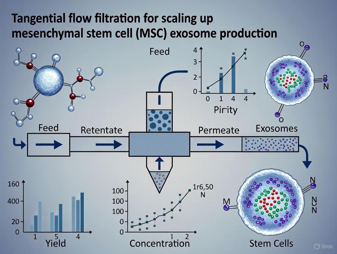
The clinical translation of mesenchymal stem cell (MSC)-derived exosomes is critically limited by challenges in large-scale manufacturing.
Scalable Production of MSC Exosomes: A Comprehensive Guide to Tangential Flow Filtration
Abstract
The clinical translation of mesenchymal stem cell (MSC)-derived exosomes is critically limited by challenges in large-scale manufacturing. This article addresses this bottleneck by providing a comprehensive analysis of Tangential Flow Filtration (TFF) as a scalable solution for MSC exosome production. We explore the foundational principles of exosome biology and the limitations of traditional isolation methods like ultracentrifugation. A detailed methodological framework for implementing TFF is presented, including its synergistic combination with 3D cell culture and downstream purification via Size Exclusion Chromatography (SEC). The content further covers practical troubleshooting and optimization strategies, and provides a rigorous comparative analysis validating TFF's superior yield, preserved bioactivity, and cost-effectiveness for industrial-scale and clinical-grade exosome manufacturing.
The Scalability Challenge in MSC Exosome Production and the Rise of TFF
Why Scalable Production is a Critical Bottleneck for Clinical Translation
The therapeutic potential of mesenchymal stem cell (MSC)-derived exosomes is increasingly recognized across a wide spectrum of diseases, from neurodegenerative disorders to wound healing and oncology [1] [2] [3]. These nanosized extracellular vesicles (30–150 nm) act as critical mediators of intercellular communication, carrying bioactive molecules that can exert therapeutic effects comparable to their parent MSCs [4]. However, the clinical translation of exosome-based therapies faces a fundamental manufacturing challenge: the inability to produce sufficient quantities of high-purity exosomes using conventional laboratory methods. This bottleneck is particularly pronounced when considering that preclinical studies typically require doses of 10⁹–10¹¹ exosomes per mouse to achieve biological outcomes [5]. To put this in perspective, the exosomal protein yield from 1 mL of conditioned medium is generally less than 1 µg, indicating that processing liters of conditioned medium is necessary to produce sufficient exosomes for a single animal study [2]. This review examines why scalable production represents a critical bottleneck and how tangential flow filtration (TFF) emerges as a promising solution within the context of MSC exosome manufacturing.
The Scalable Production Bottleneck: Quantitative Analysis
Limitations of Conventional Production Methods
Traditional exosome isolation methods, primarily differential ultracentrifugation (UC), present significant limitations for clinical translation. UC is constrained by equipment capabilities, processing volumes, and lengthy procedures that can compromise exosome integrity [6] [7]. When considering industrial-scale biomanufacturing requiring hundreds of liters of media, UC becomes impractical as "hundreds of parallel processes would likely be required" [8]. Additionally, UC often leads to incomplete sedimentation, exosome aggregation, and contamination from macromolecules, collectively reducing yield and purity [6]. These limitations are further compounded by the restricted expansion capacity of MSCs in conventional two-dimensional (2D) culture systems [2].
Yield Comparisons Across Production Platforms
The table below summarizes quantitative yield comparisons across different exosome production platforms, highlighting the dramatic improvements possible with optimized scalable methods:
Table 1: Quantitative Comparison of Exosome Production Yields Across Different Methods
| Production Method | Relative Yield Increase | Key Findings | Reference |
|---|---|---|---|
| 3D Culture + TFF vs. 2D + UC | 140-fold | 20-fold from 3D culture; 7-fold from TFF isolation | [5] [9] |
| TFF vs. UC (MDA-MB-231 cells) | 100-fold | 10¹⁰ vs. 10⁸ particles/10⁶ cells | [6] |
| 3D Microcarrier Culture (cAD-MSCs) | 2.4-3.2 fold | Increased yield and concentration in conditioned medium | [2] |
| Umbilical Cord vs. Other MSCs | 4-fold higher | Superior doubling time and exosome production per cell | [5] |
Impact of Cell Source and Culture System
The scalability challenge begins upstream with cell source selection and culture conditions. Research demonstrates that umbilical cord-derived MSCs produce four times more exosomes per cell than those from bone marrow or adipose tissue, with a more favorable doubling time (~4 days vs. ~7 days) [5]. Furthermore, transitioning from 2D to 3D culture systems represents a critical step toward scalability. Microcarrier-based 3D cultures double cell density (40,000 cells/cm² vs. 20,000 cells/cm² in 2D) and more closely mimic the native cellular microenvironment, resulting in not only increased yield but also enhanced bioactivity of the produced exosomes [2] [5] [10].
Tangential Flow Filtration: A Scalable Solution for Exosome Production
TFF Principles and Advantages
Tangential flow filtration (TFF) addresses key limitations of conventional methods by employing a filtration strategy where the feed flow travels parallel to the membrane surface. This configuration minimizes filter fouling—a significant issue in normal flow filtration—by continuously sweeping the membrane surface [8]. TFF can process volumes ranging from liters to thousands of liters in a closed system, making it suitable for industrial-scale production [4] [8]. The method simultaneously achieves three critical downstream processing objectives: exosome concentration, purification (removal of smaller contaminants like proteins), and media exchange (buffer exchange into formulation buffers suitable for storage or administration) [8].
Table 2: Key Technical Advantages of TFF for Scalable Exosome Production
| Feature | Technical Advantage | Impact on Production |
|---|---|---|
| Parallel Flow Path | Minimizes membrane fouling | Enables processing of large volumes |
| Closed System | Reduces contamination risk | Suitable for cGMP manufacturing |
| Single-Step Processing | Simultaneous concentration and purification | Reduces processing time and product loss |
| Scalable Platform | Linear scale-up from lab to industrial scale | Supports clinical translation and commercialization |
| Gentle Processing | Maintains exosome integrity and bioactivity | Improves product quality and potency |
TFF Implementation and Optimization
Successful TFF implementation requires careful optimization of several parameters. The selection of an appropriate molecular weight cutoff (MWCO) membrane—typically 3-6 times smaller than the target exosome size—is crucial for efficient separation [8]. Operational parameters including transmembrane pressure (TMP) and cross-flow rate must be balanced to maximize permeate flux while minimizing fouling and maintaining exosome integrity [8]. Furthermore, equipment selection between hollow fibers (gentler process, lower shear) and cassettes (higher fluxes, greater concentration capability) should align with specific application requirements and exosome characteristics [8].
Diagram 1: TFF-Integrated Downstream Process for Scalable Exosome Production. This workflow illustrates the complete process from 3D bioreactor culture to final exosome formulation, highlighting TFF's role in concentration and purification.
Integrated Experimental Protocols for Scalable Exosome Production
Protocol: Microcarrier-Based 3D Culture for Enhanced Exosome Production
Principle: Microcarrier-based 3D culture systems increase cell density and improve exosome yield by providing a larger surface area for cell growth in bioreactor systems [2] [5].
Materials:
- Microcarriers: Polystyrene beads (100-500 µm diameter)
- Bioreactor System: Stirred-tank or hollow fiber bioreactor
- Cells: Umbilical cord-derived MSCs (high-yield source)
- Media: Serum-free exosome collection media (e.g., VSCBIC-3 for canine AD-MSCs)
Procedure:
- Seeding Phase: Suspend microcarriers in growth media at appropriate density. Seed MSCs at 5,000-10,000 cells/cm² microcarrier surface area with intermittent agitation over 3 hours to facilitate attachment [2].
- Resting Phase: Allow 21 hours for firm cell attachment without agitation.
- Expansion Phase: Culture for 3-5 days with continuous agitation (e.g., 60-100 rpm) in growth media, monitoring cell density until reaching approximately 40,000 cells/cm² [5].
- Conditioning Phase: Replace growth media with serum-free exosome collection media. Culture for 48-72 hours to collect exosomes in the conditioned media [2].
- Harvest: Collect conditioned media containing exosomes for downstream processing.
Quality Control: Monitor cell viability via live/dead staining throughout the process. Expected viability should exceed 70% during the conditioning phase [2].
Protocol: TFF for Exosome Concentration and Purification
Principle: TFF separates exosomes from smaller contaminants based on size exclusion using a recirculating flow path parallel to the membrane surface [8].
Materials:
- TFF System: Hollow fiber or cassette configuration with 100-500 kDa MWCO membrane
- Pump: Peristaltic or diaphragm pump capable of generating appropriate cross-flow
- Reservoir: Sterile container for retentate collection
- Diafiltration Buffer: Appropriate formulation buffer (e.g., PBS)
Procedure:
- System Setup: Assemble TFF system according to manufacturer instructions. Flush membrane with diafiltration buffer to remove preservatives.
- Clarification Pre-treatment: Pre-clarify conditioned media through sequential filtration (e.g., 100 µm then 0.45 µm) to remove large debris and prevent membrane fouling [8].
- Concentration: Pump clarified conditioned media through TFF system at optimized cross-flow rate (typically 200-500 mL/min) and TMP (2-10 psi). Recirculate retentate while collecting permeate until desired concentration factor (typically 10-50x) is achieved [8].
- Diafiltration: Continue TFF operation while adding diafiltration buffer to the retentate at the same rate as permeate generation. Process for 5-10 diavolumes to exchange media components and remove contaminants [8].
- Recovery: Recover concentrated, purified exosomes from the retentate reservoir.
- Storage: Aliquot exosomes and store at -20°C for short-term use or -80°C for long-term preservation [10].
Quality Control: Monitor particle concentration by nanoparticle tracking analysis, protein content by spectrophotometry, and exosome markers (CD9, CD63, CD81) by flow cytometry or western blot [10] [8].
The Scientist's Toolkit: Essential Research Reagent Solutions
Table 3: Key Research Reagents and Materials for Scalable Exosome Production
| Reagent/Material | Function | Example/Specification |
|---|---|---|
| Microcarriers | Provide 3D surface for cell expansion | Polystyrene beads (100-500 µm) |
| Serum-Free Media | Exosome collection without serum contamination | VSCBIC-3, SF-DMEM |
| TFF Membranes | Size-based separation and concentration | 100-500 kDa MWCO hollow fibers |
| Diafiltration Buffers | Formulation and buffer exchange | PBS, specialized formulation buffers |
| Characterization Reagents | Quality control and validation | CD9/CD63/CD81 antibodies, NTA standards |
The transition from laboratory-scale exosome production to clinically relevant manufacturing represents a formidable challenge that must be addressed to realize the therapeutic potential of MSC-derived exosomes. Tangential flow filtration, particularly when integrated with 3D culture systems, offers a viable path forward by addressing key limitations of conventional methods. The 140-fold yield improvement demonstrated with 3D-TFF combinations [5] [9] provides a compelling case for adopting these technologies. Furthermore, the scalability, closed-system processing, and compatibility with cGMP manufacturing make TFF particularly suitable for clinical translation. As research continues to optimize TFF parameters and integrate complementary purification technologies, the scalable production of high-quality exosomes will become increasingly feasible, ultimately overcoming this critical bottleneck and accelerating the development of exosome-based therapeutics for clinical application.
Mesenchymal stem cell (MSC)-derived exosomes are nano-sized extracellular vesicles (30-150 nm in diameter) that are actively secreted by MSCs and play a crucial role in intercellular communication [11] [12]. These vesicles are formed through the inward budding of the endosomal membrane, leading to the creation of multivesicular bodies (MVBs) that subsequently fuse with the plasma membrane to release exosomes into the extracellular space [11] [13]. As natural bioactive molecular carriers, MSC-derived exosomes precisely deliver functional proteins, lipids, and nucleic acids to recipient cells, influencing diverse biological processes including tissue repair, immune regulation, and inflammatory response modulation [14] [12].
The therapeutic interest in MSC-derived exosomes has grown substantially due to their significant advantages over traditional cell-based therapies. Unlike intact MSCs, exosomes exhibit lower immunogenicity as they lack major histocompatibility complex (MHC) molecules, reducing the risk of immune rejection [12]. Their nanoscale size enables efficient biological barrier penetration, including the blood-brain barrier, and they present no risk of tumorigenicity or embolism associated with whole-cell transplantation [14] [12]. Additionally, exosomes offer enhanced stability for storage and can be administered through various routes including intravenous injection, topical application, and aerosolized inhalation [14] [15]. These properties position MSC-derived exosomes as promising "cell-free" therapeutic agents in regenerative medicine, with 64 registered clinical trials currently investigating their application across various disease areas [14].
Table 1: Key Characteristics of MSC-Derived Exosomes
| Property | Specification | Therapeutic Significance |
|---|---|---|
| Size Range | 30-150 nm [11] [12] | Enables efficient tissue penetration and crossing of biological barriers |
| Membrane Structure | Lipid bilayer [5] | Protects cargo from degradation and facilitates membrane fusion with target cells |
| Key Surface Markers | CD9, CD63, CD81 [11] [5] | Used for identification and characterization; enriched during biogenesis |
| Cargo Composition | Proteins, lipids, mRNA, miRNA [14] [12] | Mediates diverse therapeutic effects through transfer of bioactive molecules |
| Storage Stability | Stable at -80°C for extended periods [14] | Enables off-the-shelf availability and logistical flexibility |
Biogenesis and Cargo Loading
Biogenesis Pathways
The formation of MSC-derived exosomes occurs through a complex, tightly regulated process rooted in the endosomal system, often referred to as the endosomal sorting complex required for transport (ESCRT) pathway [12]. The biogenesis process begins with the invagination of the plasma membrane to form early endosomes, which subsequently mature into late endosomes or multivesicular bodies (MVBs) [12]. During this transformation, the endosomal membrane undergoes inward budding, capturing cytoplasmic components—including proteins, lipids, and nucleic acids—within intraluminal vesicles (ILVs) [12]. These ILVs ultimately become exosomes when the MVBs fuse with the plasma membrane, releasing the vesicles into the extracellular space [11] [13]. This intricate process ensures that exosomes are packaged with specific biomolecules that reflect the physiological state and functional capacity of their parent MSCs.
Several molecular mechanisms regulate exosome biogenesis and cargo sorting. The ESCRT machinery, comprised of approximately 30 proteins organized into four complexes (ESCRT-0, -I, -II, and -III), plays a critical role in recognizing ubiquitinated proteins and facilitating ILV formation [12]. Additionally, ESCRT-independent pathways involving tetraspanins (CD9, CD63, CD81) and lipids such as ceramide contribute to exosome formation and content selection [12]. Small GTPases from the Rab family, particularly Rab27a and Rab27b, regulate the trafficking of MVBs and their subsequent fusion with the plasma membrane [12]. Understanding these mechanisms provides opportunities for therapeutic enhancement, as evidenced by studies showing that overexpression of tetraspanin CD9 can increase exosome production by 2.4-fold [11].
Cargo Composition
MSC-derived exosomes carry a diverse array of biomolecules that mirror their therapeutic potential. According to the ExoCarta exosome database, these vesicles contain over 9,000 proteins and 3,400 RNAs associated with various biological functions [16]. The cargo includes growth factors, cytokines, transcription factors, enzymes, and both coding and non-coding RNAs (mRNA, miRNA, lncRNA, circRNA) [14] [12]. This molecular repertoire enables exosomes to influence cellular processes in recipient cells by transferring functional genetic material and proteins that can reprogram target cell behavior and function.
The specific composition of MSC-derived exosomes varies depending on the tissue source of the parent MSCs and environmental conditions. For instance, exosomes from umbilical cord-derived MSCs have demonstrated enhanced regenerative properties compared to those from bone marrow or adipose tissue [5]. Similarly, preconditioning MSCs through hypoxia or inflammatory cytokine exposure can alter exosomal cargo to enhance specific therapeutic effects [17]. This dynamic cargo composition allows MSC-derived exosomes to serve as versatile therapeutic vehicles capable of adapting to physiological demands and pathological conditions.
Table 2: Key Cargo Components in MSC-Derived Exosomes and Their Functions
| Cargo Type | Specific Examples | Biological Functions |
|---|---|---|
| Proteins | Angiogenic factors, Cytokines, Tetraspanins (CD9, CD63, CD81) [5] [12] | Promote tissue repair, modulate immune responses, facilitate membrane fusion |
| miRNAs | miR-21, miR-146a, let-7 family [12] [16] | Regulate gene expression in target cells, inhibit inflammatory pathways |
| mRNAs | Growth factor mRNAs, Transcription factor mRNAs [14] | Translate into functional proteins in recipient cells |
| Lipids | Ceramide, Cholesterol, Phospholipids [12] | Maintain membrane structure, facilitate cellular uptake, mediate signaling |
Therapeutic Mechanisms
MSC-derived exosomes exert their therapeutic effects through multiple interconnected mechanisms that primarily involve intercellular communication and cargo transfer to recipient cells. The primary mode of action centers on their capacity to deliver bioactive molecules that modulate key cellular processes, including immune responses, cell survival, proliferation, and tissue regeneration [12] [16]. Once administered, exosomes navigate to target tissues where they transfer their functional cargo to recipient cells through various uptake mechanisms such as endocytosis, macropinocytosis, or direct membrane fusion [12]. This cargo delivery subsequently reprograms cellular functions and activates signaling pathways that collectively contribute to tissue repair and homeostasis restoration.
Immunomodulatory Effects
A significant therapeutic mechanism of MSC-derived exosomes involves their potent immunomodulatory capabilities. These vesicles can polarize macrophages to an anti-inflammatory M2 phenotype, inhibit T-cell proliferation, and induce the generation of regulatory CD4+CD25+ T cells [11] [13]. Additionally, they suppress dendritic cell activation and modulate B-cell function, creating a comprehensive immunoregulatory environment [13] [12]. This capacity to fine-tune immune responses makes MSC-derived exosomes particularly valuable for treating autoimmune conditions, graft-versus-host disease (GVHD), and inflammatory disorders where immune system dysregulation drives disease pathology.
Tissue Regeneration and Repair
MSC-derived exosomes demonstrate remarkable regenerative potential across various tissue types through multiple coordinated actions. They promote angiogenesis by transferring pro-angiogenic factors and miRNAs that activate endothelial cells and stimulate new blood vessel formation [11] [16]. Additionally, they inhibit apoptosis in damaged tissues by downregulating pro-apoptotic genes while upregulating anti-apoptotic pathways [16]. Exosomes also reduce fibrosis by downregulating collagen expression and transforming growth factor-beta (TGF-β) signaling, as demonstrated in cardiac fibroblasts where exosome treatment significantly reduced TGF-β-induced collagen production [11]. Furthermore, they enhance proliferation of tissue-specific progenitor cells and stem cells, accelerating the natural repair processes in injured tissues.
Experimental Protocols for Production and Analysis
Enhanced Exosome Production Protocol
Objective: To increase exosome yield from MSCs through small molecule treatment and 3D culture systems.
Materials:
- Human umbilical cord-derived MSCs (highest exosome yield) [5]
- MesenPRO RS Medium (Gibco) or equivalent MSC culture medium [11]
- N-methyldopamine hydrochloride (Alfa Aesar, catalog No. J60306) [11]
- L-(-)-Norepinephrine-(+)-bitartrate (Sigma-Aldrich, catalog No. 489350) [11]
- Microcarrier-based 3D culture system [5]
- Exosome-depleted medium [11]
Methodology:
- Cell Culture: Culture human umbilical cord-derived MSCs in MesenPRO RS Medium. For enhanced yield, use microcarrier-based 3D culture systems instead of traditional 2D flasks to increase cell density from 20,000 to 40,000 cells/cm² [5].
- Small Molecule Treatment: Treat MSCs at 70-80% confluency with a combination of N-methyldopamine (10-100 µM) and norepinephrine (10-100 µM) in exosome-depleted medium for 48 hours. This combination increases exosome production by approximately three-fold without altering regenerative capacity [11].
- Conditioned Media Collection: Collect conditioned media after 48 hours of treatment and centrifuge at 2,000 rpm for 10 minutes to remove cells and debris [11].
- Exosome Isolation: Process the supernatant using either differential ultracentrifugation or tangential flow filtration (TFF) for larger scale production [11] [5].
Scalable Isolation Using Tangential Flow Filtration
Objective: To isolate exosomes from large volumes of conditioned media using scalable TFF methodology.
Materials:
- Tangential Flow Filtration system (holofiber-based preferred) [4]
- Pellicon or similar TFF cassettes (100-500 kDa MWCO)
- Peristaltic pump and reservoir
- Phosphate-buffered saline (PBS), pH 7.4
Methodology:
- Clarification: Centrifuge conditioned media at 10,000× g for 30 minutes at 4°C to remove larger vesicles and debris [11].
- TFF Setup: Install appropriate MWCO filter (typically 100-500 kDa) in TFF system according to manufacturer's instructions. Ensure all connections are secure and the system is properly sanitized.
- Concentration: Process the clarified supernatant through TFF with a cross-flow rate optimized to minimize fouling (typically 2-5 L/min). Concentrate the retentate to approximately 1/20th of the original volume [5] [4].
- Diafiltration: Exchange buffer by continuously adding PBS to the retentate while maintaining constant volume until 5-10 diavolumes have been processed. This step removes contaminating proteins and salts [5].
- Final Recovery: Recover the concentrated exosome solution and further concentrate if necessary. Aliquot and store at -80°C.
Validation: Characterize exosomes using nanoparticle tracking analysis (NTA) for size distribution and concentration, transmission electron microscopy (TEM) for morphology, and western blotting for exosomal markers (CD9, CD63, CD81) [11] [5].
Table 3: Quantitative Comparison of Exosome Production Methods
| Production Method | Exosome Yield | Fold Increase | Purity Assessment | Key Advantages |
|---|---|---|---|---|
| 2D Culture + UC | Baseline [5] | 1x [5] | CD63+, CD9+ [11] | Gold standard, well-characterized |
| 2D Culture + TFF | 27-fold higher [5] | 27x [5] | CD81+, CD9+ [5] | Scalable, processes larger volumes |
| 3D Culture + UC | 20-fold higher [5] | 20x [5] | CD63+, CD9+ [5] | Higher cell density, increased production |
| 3D Culture + TFF | 140-fold higher [5] | 140x [5] | CD81+, CD9+ [5] | Maximum yield and scalability |
The Scientist's Toolkit: Research Reagent Solutions
Table 4: Essential Research Reagents for MSC Exosome Studies
| Reagent/Category | Specific Examples | Function/Application |
|---|---|---|
| MSC Sources | Human bone marrow-derived MSCs (ATCC PCS-500-012), Umbilical cord-derived MSCs, Adipose tissue-derived MSCs [11] [5] | Exosome production; umbilical cord source provides highest yield [5] |
| Culture Media | MesenPRO RS Medium, Serum-free media, Exosome-depleted media [11] | MSC expansion and exosome production without serum contamination |
| Exosome Secretion Enhancers | N-methyldopamine, Norepinephrine combination [11] | Increases exosome production by 3-fold without affecting function |
| Isolation Systems | Differential ultracentrifugation, Tangential Flow Filtration systems, Hollow fiber bioreactors [11] [5] [4] | Exosome purification; TFF enables large-scale processing |
| Characterization Antibodies | Anti-CD63, Anti-CD9, Anti-CD81 [11] [5] | Exosome identification and validation via western blot, flow cytometry |
| Analysis Instruments | Nanoparticle Tracking Analyzer, Transmission Electron Microscope, Western blot system [11] [5] | Size distribution, morphological analysis, and protein marker confirmation |
MSC-derived exosomes represent a promising cell-free therapeutic alternative with significant advantages over traditional cell-based approaches, including reduced immunogenicity, enhanced safety profile, and superior biological barrier penetration capability. Their therapeutic potential stems from sophisticated biogenesis mechanisms that package diverse bioactive cargo, enabling them to modulate immune responses, promote tissue regeneration, and facilitate intercellular communication. The development of scaled production methodologies, particularly through 3D culture systems and tangential flow filtration, has addressed previous limitations in exosome yield and processing capacity. As research continues to elucidate the precise mechanisms of action and optimize production protocols, MSC-derived exosomes are poised to become transformative tools in regenerative medicine, offering new therapeutic avenues for a wide range of diseases and injuries.
Differential ultracentrifugation (UC) has long been regarded as the "gold standard" method for exosome isolation, particularly in research settings involving mesenchymal stem cell (MSC)-derived exosomes [18]. This technique exploits the inherent size and density of exosomes to separate them from other components in conditioned media or biological fluids through a series of progressively higher centrifugal forces [18]. Despite its widespread adoption and historical prominence, a growing body of evidence reveals significant limitations in the UC approach, especially concerning yield, scalability, and the preservation of exosomal integrity [18] [19] [20]. These shortcomings present substantial barriers to the clinical translation and large-scale production of MSC exosomes, necessitating a critical evaluation of this traditional method and the exploration of more robust alternatives like tangential flow filtration (TFF) for scaling up production [1] [21].
Table 1: Core Limitations of Ultracentrifugation for MSC Exosome Isolation
| Limitation Category | Specific Technical Issues | Impact on Downstream Applications |
|---|---|---|
| Low Yield & Recovery | Suboptimal sEV yield [19]; Inefficient pelleting [19] | Reduced material for therapeutic dosing [2]; Limits preclinical and clinical studies [22] |
| Scalability Challenges | Time-consuming (6-24 hour protocols) [23]; Low sample volume capacity per run [20] | Incompatible with industrial-scale production [1]; Increases labor and processing costs [19] |
| Exosome Integrity Damage | Mechanical damage from high g-forces [18]; Disruption of structural/biological integrity [19] | Compromises bioactivity and therapeutic potential [18]; Causes particle aggregation [19] |
| Purity Concerns | Co-isolation of non-sEV contaminants (e.g., protein aggregates, lipoproteins) [18] [19] | Confounds functional analysis [19]; Leads to misinterpretation of exosome cargo [18] |
Quantitative Comparative Analysis of Isolation Techniques
Direct comparisons between UC and advanced methods like TFF highlight the profound efficiency gap. A 2025 study comparing production methods for MSC-derived small extracellular vesicles (sEVs) found that particle yields were statistically higher when isolated by TFF than by UC [21]. This corroborates earlier findings that TFF "isolates significantly higher yields of sEVs" while maintaining consistent particle populations with sizes predominantly under 200 nm [19]. Beyond yield, the scalability advantage of TFF is evident in processing times; while UC protocols require 6-24 hours, integrated systems like ExoDisc can complete isolation within 15 minutes on a standard benchtop centrifuge [23].
Table 2: Performance Comparison of Ultracentrifugation vs. Tangential Flow Filtration
| Performance Metric | Differential Ultracentrifugation | Tangential Flow Filtration |
|---|---|---|
| Total Processing Time | 6-24 hours [23] | Approximately 15 minutes for benchtop systems [23] |
| Particle Yield | Low to medium; significant loss [19] [23] | Significantly higher yields [19] [21] |
| Exosome Integrity | Mechanical damage and membrane distortion [18] [20] | Preserves structural integrity and biological function [19] [23] |
| Scalability | Poor; limited by rotor capacity and time [20] | Excellent for large-volume processing [19] [2] |
| Purity | Medium; co-isolation of contaminants [18] | High, especially when combined with SEC [19] |
| Cost & Equipment | Requires expensive ultracentrifuges ($30,000-$100,000+) [23] | Lower equipment costs; uses standard centrifuges or TFF systems [19] [23] |
Experimental Protocols for Method Evaluation
Protocol: Comparative Isolation of MSC Exosomes via Ultracentrifugation
This protocol is adapted from established methodologies for isolating sEVs from serum-containing cell culture media, representing common practice in many research laboratories [19].
Step 1: Cell Culture and Conditioned Media Collection
- Culture MSCs (e.g., bone marrow-derived) in DMEM or α-MEM supplemented with 5% EV-depleted FBS [19] [21].
- At 80-90% confluency, wash cells with PBS and culture in fresh medium with EV-depleted FBS for 48 hours.
- Collect cell culture conditioned media and centrifuge at 500 × g for 10 minutes to remove detached cells and large debris [19].
- Filter the supernatant through a 0.22 μm filter to remove other large particle contaminants [19].
Step 2: Ultracentrifugation
- Transfer the clarified conditioned media to ultracentrifuge tubes compatible with a fixed-angle rotor (e.g., Type 50.2 Ti).
- Centrifuge at 100,000 × g at 4°C for 120 minutes [19].
- Carefully decant the supernatant. The crude exosome pellet may be visible at the tube bottom.
- Resuspend the pellet in 1 mL of ice-cold, sterile PBS by pipetting gently. For higher purity, a second round of ultracentrifugation may be performed: resuspend the pooled pellets in PBS and centrifuge again at 100,000 × g at 4°C for 120 minutes [19].
Step 3: Post-Isolation Purification (Optional)
Protocol: Scalable Isolation of MSC Exosomes via Tangential Flow Filtration
This protocol outlines TFF for scalable isolation, often combined with SEC for high-purity yields suitable for therapeutic development [19] [2].
Step 1: Cell Culture and Media Conditioning
- For large-scale production, consider using a 3D culture system (e.g., microcarrier-based bioreactors) with an optimized, serum-free exosome-collecting solution (e.g., VSCBIC-3) to enhance yield [2].
- Culture MSCs and collect conditioned media as described in Step 1 of the UC protocol, scaling up volumes as appropriate.
Step 2: Tangential Flow Filtration
- Assemble a TFF system with a membrane cartridge of appropriate pore size (e.g., 100-300 kDa MWCO or specific nanofiltration membranes).
- Circulate the clarified and pre-filtered conditioned media through the TFF system. The flow is applied parallel to the membrane, concentrating the exosomes while allowing smaller molecules and contaminants to pass through [19].
- Continue the concentration process until the desired volume reduction is achieved.
- Perform a diafiltration step by adding PBS or an appropriate buffer to the concentrated retentate to exchange the buffer and remove soluble contaminants further [19].
Step 3: Final Purification and Concentration
- The concentrated retentate from TFF, now greatly enriched in exosomes, can be processed through a size-exclusion chromatography (SEC) column for final polishing to remove remaining impurities and achieve high-purity exosome preparations [19].
- The resulting purified exosome fractions can be concentrated further if needed using centrifugal concentrators and stored at -80°C [20].
The Scientist's Toolkit: Essential Research Reagent Solutions
Table 3: Key Reagents and Materials for Advanced Exosome Isolation and Characterization
| Reagent/Material | Function/Application | Examples & Notes |
|---|---|---|
| EV-Depleted FBS | Serum supplement for cell culture that minimizes contaminating bovine EVs in conditioned media. | Prepared by ultracentrifugation (100,000-120,000 × g for 12-18 hours) or commercial sources [19]. |
| Size Exclusion Chromatography (SEC) Columns | High-purity purification of exosomes from soluble proteins and contaminants after initial concentration. | qEVoriginal columns (Izon Science) or Sepharose-based columns [18] [19]. |
| TFF Cassettes/Systems | Scalable concentration and purification of exosomes from large volumes of culture medium. | Cassettes with hollow fibers or flat sheets with pore sizes tailored for EV isolation (e.g., 100-300 kDa) [19] [2]. |
| Exosome Collection Media | Serum-free, xeno-free media formulations optimized for supporting cell viability while maximizing exosome yield during production. | Commercial serum-free media or in-house formulations like VSCBIC-3 [2]. |
| Characterization Reagents | Antibodies and kits for confirming exosome identity, purity, and quantity. | Antibodies against tetraspanins (CD9, CD63, CD81), TSG101, ALIX; and negative markers (e.g., Calnexin) for Western blot [18] [21]. |
Visualizing the Pathway to Exosome Integrity Compromise in Ultracentrifugation
The following diagram illustrates the sequential mechanical stresses and resulting compromises to exosome integrity during the ultracentrifugation process.
Visualizing the Integrated TFF Workflow for Scalable Production
This workflow diagram outlines the streamlined and gentle process of using Tangential Flow Filtration, often combined with 3D culture, for scalable exosome production.
The limitations of ultracentrifugation—low yield, poor scalability, and compromised exosome integrity—present significant obstacles to the clinical advancement of MSC exosome therapies [18] [19] [20]. Quantitative evidence firmly establishes that alternative technologies, particularly Tangential Flow Filtration, outperform UC in critical metrics essential for translational success: yield, processing time, scalability, and preservation of vesicle integrity [19] [21] [2]. For researchers and drug development professionals aiming to transition MSC exosome research from bench to bedside, adopting TFF-based isolation protocols, potentially integrated with 3D culture systems [2], represents a necessary and strategic evolution beyond the historical "gold standard." This methodological shift is crucial for achieving the robust, reproducible, and scalable production required for rigorous preclinical testing and eventual clinical application.
Basic Principles of Tangential Flow Filtration
Tangential Flow Filtration (TFF), also referred to as cross-flow filtration, is a separation technique where the feed stream flows parallel (tangentially) across the surface of a filtration membrane [24] [25] [26]. This flow pattern generates a sweeping force that lifts retained particles from the membrane surface, carrying them out of the filter device in the retentate stream rather than allowing them to deposit as a fouling layer [24]. This fundamental difference in flow dynamics distinguishes TFF from normal flow filtration (NFF), or dead-end filtration, where the feed flow is directed perpendicularly through the membrane, rapidly accumulating a filter cake that leads to clogging and reduced efficiency [26].
The separation process is driven by Transmembrane Pressure (TMP), the pressure differential across the membrane thickness that drives solvent and small solutes through the membrane pores [24] [26]. TMP is calculated using the formula TMP = (P_F + P_R)/2 - P_P, where P_F is the feed pressure, P_R is the retentate pressure, and P_P is the permeate pressure [27]. Precise control of TMP and crossflow rate is critical for optimizing performance; excessive TMP can compress the gel layer on the membrane, severely restricting permeate flow, while an excessively high crossflow rate can reduce process efficiency [24].
Table 1: Core Components of a TFF System [24] [26]
| Component | Function |
|---|---|
| Feed Reservoir | Holds the initial solution to be processed (e.g., cell culture harvest). |
| Pump | Drives circulation of the feed through the system at a controlled flow rate. |
| TFF Membrane Module | The core unit where separation occurs, such as a flat-sheet cassette or hollow fiber module. |
| Pressure Sensors | Monitor inlet, outlet, and permeate pressures to calculate and control TMP. |
| Retentate Loop | Recirculates the concentrated stream back to the feed reservoir for further processing. |
| Permeate Collection | Gathers the filtered fluid that has passed through the membrane. |
Key Advantages Over Normal Flow Filtration
TFF offers several distinct advantages for bioprocessing applications, primarily due to its cross-flow design.
- Reduced Membrane Fouling: The tangential flow creates a scouring effect that continuously cleans the membrane surface, minimizing the buildup of particles and biological material [24] [26]. This leads to more consistent performance over time.
- Handling of Challenging Streams: TFF is highly effective for processing solutions with high solid content or viscous fluids, which would rapidly clog a dead-end filter [26].
- Continuous Operation and Concentration: The system allows for continuous recirculation of the retentate, enabling significant volume reduction and concentration of the target product [25] [26].
- Flexible Product Recovery: Both the retained material (retentate) and the filtered material (permeate) can be recovered, depending on where the product of interest is located [24] [25].
- Integrated Buffer Exchange: The process of diafiltration (DF) can be easily integrated, allowing for efficient buffer exchange to prepare the product for subsequent processing steps [24] [25].
Table 2: Comparison between Tangential Flow Filtration and Normal Flow Filtration
| Parameter | Tangential Flow Filtration (TFF) | Normal Flow Filtration (NFF) |
|---|---|---|
| Flow Direction | Parallel to membrane surface | Perpendicular to membrane surface |
| Fouling Tendency | Low, due to sweeping action | High, due to cake formation |
| Process Continuity | Suitable for continuous, long-term operation | Typically a single-use, batch process |
| Typical Applications | Concentration, purification, buffer exchange of valuable products | Clarification, sterilization, final product filtration |
| Equipment Complexity | Higher (requires pumps, sensors, loop) | Lower (simple filter housings) |
TFF Membrane Types and Selection for Sensitive Products
The two most common types of TFF membranes are flat-sheet cassettes and hollow fiber modules, each with unique characteristics suited for different applications [24] [26].
Flat-Sheet Cassettes consist of multiple layers of membrane stacked with mesh spacers, which create turbulent flow channels. This turbulence enhances the sweeping of the membrane surface, preventing fouling and enabling high filtration rates (flux) [24] [26]. This format is ideal for robust, non-shear-sensitive products like proteins or non-enveloped viruses such as Adeno-associated virus (AAV) [24].
Hollow Fiber Modules are cylindrical cartridges containing a bundle of narrow, self-supporting tubular fibers. The feed flows through the lumen of these fibers in a laminar flow, resulting in very low shear stress [24] [26]. This gentle processing makes hollow fibers the preferred option for shear-sensitive products such as enveloped viruses (e.g., lentivirus), fragile proteins, and whole cells [24]. Its suitability for gentle processing also makes it a key tool in the scalable production of mesenchymal stem cell (MSC)-derived exosomes [28] [2].
Application in Scaling Up MSC Exosome Production
The clinical translation of MSC-derived exosome therapies is constrained by challenges in scalable production to achieve clinically relevant quantities [1] [2]. TFF addresses this bottleneck by enabling efficient concentration and purification of exosomes from large volumes of cell culture conditioned medium.
Recent studies have established integrated biomanufacturing workflows that combine 3D bioreactor systems for cell expansion and exosome production with TFF for isolation and concentration [28] [2]. For instance, one protocol for producing canine adipose-derived MSC exosomes, termed "VSCBIC-3-3D," used a 3D culture system followed by TFF isolation, resulting in a 3.2-fold increase in exosome concentration compared to conventional 2D methods [2]. Another study utilized a Hollow Fiber 3D bioreactor integrated with an exosome-harvesting system for a 28-day production run, demonstrating the capability of TFF for stable, long-term processing [28].
The primary application of TFF in this context is the concentration and buffer exchange of exosomes. The clarified conditioned medium, containing exosomes and soluble proteins, is processed through an ultrafiltration (UF) TFF membrane with a molecular weight cutoff (MWCO) that retains the exosomes (typically 30-500 kDa) while allowing smaller impurities to pass through in the permeate [24] [25]. This is often followed by a diafiltration (DF) step to exchange the buffer into a final formulation, removing residual contaminants and achieving high purity [24] [25] [29].
Experimental Protocol: TFF for Exosome Concentration and Buffer Exchange
Below is a generalized protocol for concentrating and purifying exosomes from conditioned medium using a benchtop TFF system with a hollow fiber module.
Aim: To concentrate MSC-derived exosomes from conditioned medium and exchange the buffer into a final formulation (e.g., PBS) suitable for downstream applications or storage.
Materials and Reagents:
- Conditioned Medium: Collected from MSC cultures (2D or 3D bioreactor).
- TFF System: Benchtop peristaltic pump system with pressure sensors.
- Hollow Fiber Module: 100-500 kDa MWCO, chosen for gentle exosome processing.
- Diafiltration Buffer: e.g., Phosphate-Buffered Saline (PBS), pH 7.4.
- Feed Reservoir: Sterile, graduated container.
- Permeate Collection Vessel.
- Tubing: Compatible with peristaltic pump and fluid path.
Procedure:
System Setup and Equilibration:
- Assemble the TFF system according to the manufacturer's instructions, ensuring all connections are secure.
- Flush the hollow fiber module and the entire fluid path with Diafiltration Buffer to remove storage solutions and wet the membrane.
Initial Concentration (Ultrafiltration - UF1):
- Pour the clarified conditioned medium into the feed reservoir.
- Start the pump, initially setting the crossflow rate and TMP to low values as recommended for the module.
- Open the permeate line and begin recirculation. The permeate (waste) will be collected, and the retentate will return to the feed reservoir.
- Gradually adjust the retentate valve to increase the TMP to the optimal range (e.g., 1-5 psi) while monitoring pressures. The goal is to achieve a steady permeate flow.
- Continue concentration until the retentate volume is reduced to the target (e.g., 10% of the initial volume).
Buffer Exchange (Diafiltration - DF):
- Once the target concentration volume is reached, begin adding Diafiltration Buffer to the feed reservoir at the same rate as the permeate flow is removed. This maintains a constant volume in the system while replacing the original buffer.
- Typically, 5-10 volume exchanges are performed to ensure >99% exchange of the original buffer.
Final Concentration (Ultrafiltration - UF2):
- After diafiltration is complete, stop the buffer addition and continue the TFF process to concentrate the retentate to the final desired volume and exosome concentration.
Product Recovery and System Cleaning:
- Drain the final retentate from the system. This is your purified and concentrated exosome sample.
- Flush the system with DI water, followed by a cleaning solution (e.g., 0.1-0.5 M NaOH), and finally store the membrane in an appropriate solution (e.g., 20% ethanol).
Table 3: The Scientist's Toolkit: Essential Reagents and Materials for TFF-based Exosome Production
| Item | Function/Application | Example/Notes |
|---|---|---|
| Hollow Fiber TFF Module | Gentle concentration and purification of shear-sensitive exosomes. | 100-500 kDa MWCO; chosen for low shear stress [24] [28]. |
| 3D Bioreactor System | Scalable expansion of MSCs and production of exosomes. | Hollow fiber bioreactors enable high-density culture and continuous harvest [28]. |
| Specialized Cell Culture Medium | Supports MSC growth and exosome production in serum-free conditions. | RoosterNourish-MSC-CC [28]; In-house formulations like VSCBIC-3 [2]. |
| Diafiltration Buffers | For buffer exchange into final formulation post-concentration. | Phosphate-Buffered Saline (PBS) is commonly used for final formulation and storage. |
| Automated TFF System | Provides process control, data integrity, and reproducibility for GMP manufacturing. | Systems like KrosFlo or Sartoflow with recipe-driven software [24] [25]. |
Mesenchymal stem cell-derived extracellular vesicles (MSC-EVs), particularly exosomes or small extracellular vesicles (sEVs), are emerging as core carriers of next-generation acellular therapeutic strategies [14]. These nanoscale "regenerative tiny giants" offer significant advantages over traditional cell-based therapies, including low immunogenicity, efficient biological barrier penetration, and superior storage stability [14]. As natural bioactive molecular carriers, MSC-sEVs precisely regulate inflammatory responses, angiogenesis, and tissue repair processes by delivering functional RNAs, proteins, and other signaling elements to target tissues [14].
The selection of optimal MSC tissue sources is a critical upstream factor in manufacturing these complex biologics, as the parent cells determine the yield, cargo composition, and subsequent therapeutic potency of the resulting vesicles [30]. This application note provides a systematic comparison of three predominant MSC sources—umbilical cord, bone marrow, and adipose tissue—focusing on their performance characteristics for scalable sEV production using tangential flow filtration (TFF)-based platforms.
Quantitative Source Comparison
Table 1: Comparative Characteristics of MSC Sources for sEV Production
| Parameter | Umbilical Cord MSC | Bone Marrow MSC | Adipose Tissue MSC |
|---|---|---|---|
| Doubling Time | ∼4 days [5] | ∼7 days [5] | ∼7 days [5] |
| sEV Yield (Particles/Cell) | Highest (4,318.72 ± 2,110.22) [21] | 3,751.09 ± 2,058.51 [21] | Lower than UC-MSC [5] |
| sEV Size | 140 ± 18 nm [5] | 116 ± 9 nm [5] | 105 ± 12 nm [5] |
| Proliferation Capacity | High [31] | Moderate, age-dependent [31] | Moderate [31] |
| Therapeutic Specialization | Immunomodulation, tissue repair [14] [32] | Bone regeneration [33] [31] | Immunoregulation [33] |
| Scalability Potential | Excellent [5] [34] | Moderate [21] | Moderate [5] |
Table 2: Functional Efficacy in Disease Models
| Disease Model | Umbilical Cord MSC-sEVs | Bone Marrow MSC-sEVs | Adipose Tissue MSC-sEVs |
|---|---|---|---|
| Psoriasis (IMQ-induced murine) | Significant reduction in clinical severity scores and epidermal thickness [32] | Not reported | Not reported |
| Retinal Pigment Epithelium Protection | Not reported | Enhanced ARPE-19 cell proliferation (37.86% to 54.60% viability after H2O2 exposure) [21] | Not reported |
| siRNA Delivery to Neurons | 7x more potent than 2D-UC-exosomes [5] | Not reported | Not reported |
| T-cell Suppression | Shown to suppress T-cell activation [34] | Not reported | Not reported |
Experimental Evidence for Source Selection
Umbilical cord-derived MSCs, particularly from Wharton's Jelly, demonstrate superior characteristics for scalable sEV production. A direct comparison of MSC sources revealed that UC-MSCs grew significantly faster (∼4-day doubling time) compared to BM-MSCs or AD-MSCs (both ∼7-day doubling time) and yielded four times as many exosomes per cell than did MSCs from bone marrow or adipose tissue [5]. This enhanced productivity is further compounded by the fact that UC-MSCs can be cultivated without ethical constraints and generally exhibit lower immunogenicity [31].
Bone marrow-derived MSCs produce sEVs with distinctive therapeutic profiles, particularly valuable for orthopedic applications. BM-MSC-sEVs exhibit stronger osteogenic potential compared to other sources, making them particularly suitable for bone regeneration strategies [33]. Research demonstrates that BM-MSC-sEVs enhance cell proliferation and protect against oxidative stress damage, as shown in retinal pigment epithelium models where application of sEVs increased cell viability from 37.86% to 54.60% after H2O2 exposure [21].
While comprehensive comparative data for adipose tissue-derived MSC-sEVs is more limited in the search results, they are generally associated with strong immune regulatory functions [33]. AD-MSCs are relatively easy to harvest in large quantities but show lower sEV yields compared to UC-MSCs [5].
TFF-Based Scalable Production Workflows
Integrated TFF-SEC Production Platform
Table 3: TFF-SEC Platform Components and Parameters
| System Component | Specification | Function |
|---|---|---|
| TFF System | Hollow fiber-based [4] | Initial concentration and buffer exchange |
| Membrane Pore Size | 100-300 kDa MWCO [34] | Retention of sEVs while removing proteins |
| SEC Columns | Sepharose-based [30] | Final purification from protein aggregates |
| Processing Volume | 2-6 L batches [34] | Scalable production capacity |
| Particle Concentration | 36-fold increase post-TFF [34] | Yield enhancement metric |
For UC-MSC cultivation, microcarrier-based 3D culture systems double cell density (reaching 40,000 cells/cm²) compared to conventional 2D cultures (20,000 cells/cm²) [5]. When combined with TFF, this 3D culture system yields a 140-fold increase in sEV production compared to traditional 2D culture with ultracentrifugation [5]. The integrated TFF-SEC approach effectively processes scalable volumes (2-6 L batches) of conditioned media from GMP-compliant MSC cultures, achieving up to 36-fold particle concentration increases while maintaining biological activity [34].
Diagram 1: Scalable TFF-based sEV Production Workflow (Max Width: 760px)
Culture Media Optimization for Enhanced Yield
Culture media composition significantly impacts MSC growth and subsequent sEV yield. Comparative studies of BM-MSCs cultured in Dulbecco's Modified Eagle Medium (DMEM) versus Alpha Minimum Essential Medium (α-MEM), both supplemented with 10% human platelet lysate (hPL), revealed that α-MEM supported higher expansion ratios, though not statistically significant [21]. The average yield of particles per cell was higher in α-MEM (4,318.72 ± 2,110.22) compared to DMEM (3,751.09 ± 2,058.51) [21]. For GMP-compliant production, xeno-free culture media supplemented with hPL is recommended to minimize contamination risks while supporting robust cell growth [21].
sEV Characterization and Quality Control
Comprehensive Characterization Pipeline
Isolated sEVs must undergo rigorous characterization to confirm identity, purity, and functionality. The Minimal Information for Studies of Extracellular Vesicles (MISEV) guidelines provide comprehensive standards for EV characterization, including detection of canonical markers (CD9, CD63, CD81) and confirmation of the absence of cellular contaminants (calnexin, GM130, cytochrome c) [33].
Nanoparticle Tracking Analysis (NTA) determines size distribution and concentration, with typical UC-MSC-sEVs ranging from 107-156 nm [21] [34]. Transmission Electron Microscopy (TEM) confirms cup-shaped morphology characteristic of sEVs [21] [32]. Western Blot analysis verifies the presence of tetraspanins (CD9, CD63, CD81) and absence of negative markers such as calnexin [5] [21]. Functional assays validate biological activity, such as T-cell suppression for immunomodulatory potency [34].
Diagram 2: Comprehensive sEV Characterization Pipeline (Max Width: 760px)
The Scientist's Toolkit: Essential Research Reagents
Table 4: Key Research Reagent Solutions for TFF-based sEV Production
| Reagent/Category | Specific Examples | Function & Application Notes |
|---|---|---|
| Cell Culture Media | α-MEM with hPL [21] | Optimized for MSC expansion and sEV yield |
| Microcarriers | Cytodex, Plastic-based [5] | 3D culture surface for scalable expansion |
| TFF Systems | Hollow fiber TFF [4] | Scalable concentration of sEVs from large volumes |
| Chromatography | Sepharose-based SEC [30] | High-purity sEV separation from protein contaminants |
| Characterization | ZetaView NTA [32] | Size distribution and concentration analysis |
| sEV Markers | CD9, CD63, CD81 antibodies [21] | Identity confirmation via Western blot |
| Negative Markers | Calnexin antibodies [21] | Purity assessment (absence of cellular contaminants) |
Umbilical cord-derived MSCs represent the optimal source for industrial-scale sEV production, offering superior proliferation rates, highest sEV yields, and robust therapeutic potential. The integration of microcarrier-based 3D culture with TFF-SEC purification establishes a scalable, GMP-compliant manufacturing platform that addresses the critical bottleneck in clinical translation of MSC-sEV therapies [5] [34]. This production framework supports the transition of "regenerative tiny giants" from laboratory research to clinical-grade "programmable nanomedicines" for precision medicine applications [14].
Future development should focus on optimizing culture conditions through media formulation and genetic engineering to further enhance sEV yield and potency, while establishing standardized quality control metrics that correlate with therapeutic efficacy across different disease indications.
Implementing TFF: A Step-by-Step Protocol for Scalable MSC Exosome Isolation
The therapeutic potential of mesenchymal stem cell (MSC)-derived exosomes is immense, spanning regenerative medicine, immunomodulation, and targeted drug delivery [35]. These nanoscale extracellular vesicles (40-150 nm) transfer bioactive molecules—including proteins, lipids, and nucleic acids—to recipient cells, mediating the therapeutic effects of MSCs without the risks associated with whole-cell transplantation [5] [35]. However, a major roadblock to clinical translation is the inefficient production of sufficient exosome quantities, as conventional two-dimensional (2D) monolayer cultures yield low volumes and traditional isolation methods like differential ultracentrifugation are not easily scalable [5] [2].
Strategic upstream optimization through microcarrier-based three-dimensional (3D) bioreactor cultures presents a transformative solution. This approach integrates seamlessly with tangential flow filtration (TFF) for downstream purification, creating a scalable pipeline for manufacturing clinically relevant exosome quantities. This protocol details the implementation of this integrated system, demonstrating a 140-fold increase in exosome yield compared to conventional 2D culture with ultracentrifugation [5]. Furthermore, exosomes produced via this method exhibit enhanced biological activity, showing a 7-fold greater potency in small interfering RNA (siRNA) transfer to neurons [5].
Key Performance Data and Rationale
The quantitative advantages of integrating microcarrier-based 3D culture with TFF are substantial. The table below summarizes the key performance metrics from foundational studies.
Table 1: Quantitative Impact of 3D Culture and TFF on Exosome Production
| Parameter | 2D Culture + UC | 3D Culture + UC | 2D Culture + TFF | 3D Culture + TFF | Citation |
|---|---|---|---|---|---|
| Exosome Yield (Fold Increase) | Baseline (1x) | 20-fold | 27-fold | 140-fold | [5] |
| Cell Density at Confluence | 20,000 cells/cm² | 40,000 cells/cm² | Not Reported | Not Reported | [5] |
| siRNA Transfer Potency | Baseline (1x) | Not Reported | Not Reported | 7-fold more potent | [5] |
| Particle-to-Protein Ratio | 2.6 × 10⁹ ± 0.6 × 10⁹ | 0.9 × 10⁹ ± 0.2 × 10⁹ | 4 × 10⁹ ± 0.4 × 10⁹ | 1.23 × 10⁹ ± 0.5 × 10⁹ | [5] |
The rationale for this integrated approach is multi-faceted:
- Overcoming Surface Area Limitations: Microcarriers provide a vast surface area for cell growth within a small volume; for example, 1 gram of microcarriers offers a surface area equivalent to fifteen 75 cm² culture flasks [36]. This is crucial for expanding the limited number of MSCs obtainable from tissue sources.
- Enhanced Cell Performance: The 3D environment of microcarrier cultures can improve nutrient and gas transfer compared to 2D culture and has been shown to support MSC differentiation and maintain phenotypic stability at high cell concentrations [36] [37].
- Scalable Downstream Processing: TFF is a cornerstone of this strategy. Unlike ultracentrifugation, which is limited by volume processing capacity, TFF is a closed-system, scalable technology capable of processing hundreds to thousands of liters of conditioned medium, making it compatible with Good Manufacturing Practice (GMP) standards [5] [4].
Experimental Protocols
Protocol 1: Microcarrier-Based 3D Culture of MSCs for Exosome Production
This protocol is optimized for the expansion of umbilical cord-derived MSCs, which demonstrate a faster doubling time and higher exosome yield compared to bone marrow or adipose-derived MSCs [5].
Research Reagent Solutions Table 2: Essential Materials for Microcarrier-Based MSC Culture
| Item | Function / Rationale | Example Product / Note |
|---|---|---|
| Umbilical Cord MSCs | High-exosome yield cell source | Preferable over bone marrow or adipose-derived [5] |
| Serum-Free Medium | Supports cell growth without serum-derived contaminants | DMEM base, supplemented with bFGF [38] |
| Microcarriers | Provide 3D substrate for cell attachment and growth | Cytodex (e.g., type 1 or 3), 100-300 µm diameter [36] [37] |
| Bioreactor System | Controlled environment for suspension culture | Spinner flask or stirred-tank bioreactor [39] [40] |
| In-house Exosome Collection Medium (VSCBIC-3) | Serum-free solution to maintain cell viability during exosome production | Supports cell morphology and viability for 3 days [2] |
Step-by-Step Methodology:
- Microcarrier Preparation: Hydrate and sterilize microcarriers (e.g., Cytodex) according to the manufacturer's instructions. A typical concentration used in spinner flasks or bioreactors is 2-5 mg/mL [37].
- Cell Seeding:
- Harvest MSCs from 2D culture using standard trypsin/EDTA treatment [38].
- Resuspend the cell pellet in fresh, serum-free culture medium. A high cell seeding density is recommended for efficient attachment.
- Combine the cell suspension with the prepared microcarriers in the bioreactor vessel.
- Initiate culture with intermittent stirring (e.g., 5-10 minutes of stirring every 30-60 minutes) for the first 6-8 hours to facilitate cell-microcarrier contact without subjecting cells to prolonged shear stress.
- Continuous Culture:
- After the initial attachment period, switch to continuous low-speed stirring (e.g., 40-60 rpm) to keep microcarriers in suspension while minimizing shear forces.
- Maintain standard culture conditions (37°C, 5% CO₂).
- Monitor glucose consumption and perform medium exchanges as needed.
- Conditioning for Exosome Production:
- Once target cell density is achieved (typically after 4-7 days), replace the growth medium with the serum-free, in-house exosome-collecting solution (e.g., VSCBIC-3) [2].
- Continue culture for an additional 48-72 hours to allow for exosome accumulation in the conditioned medium. Cell viability and morphology may decline during this period, which is expected [2].
- Harvesting: Separate the conditioned medium containing exosomes from the cell-laden microcarriers using an initial low-speed centrifugation (e.g., 500 × g for 10 minutes) or a specialized inertial-based filtration system [38]. The clarified conditioned medium is then processed via TFF.
Protocol 2: Tangential Flow Filtration for Exosome Isolation
TFF isolates exosomes based on size, concentrating and purifying them from large volumes of conditioned medium.
Research Reagent Solutions Table 3: Essential Materials for Tangential Flow Filtration
| Item | Function / Rationale | Example Product / Note |
|---|---|---|
| TFF System | Scalable concentration and buffer exchange | Hollow fiber-based TFF system [4] |
| Membrane Cartridge | Size-based separation | 100-500 kDa MWCO or 100-300 nm pore size [5] [4] |
| Diafiltration Buffer | Washes out contaminants | Phosphate-Buffered Saline (PBS) |
Step-by-Step Methodology:
- System Setup and Priming: Assemble the TFF system according to the manufacturer's instructions. Prime the membrane with PBS or water to remove preservatives and wet the pores.
- Concentration:
- Pump the clarified conditioned medium from Protocol 1 through the TFF system.
- The system is configured in a closed loop where the retentate (containing exosomes) is recirculated back to the feed reservoir, while the permeate (containing small molecules and proteins) is removed.
- Concentrate the volume to a manageable level (e.g., 50-100x concentration factor).
- Diafiltration:
- Once concentrated, initiate diafiltration by continuously adding diafiltration buffer (e.g., PBS) to the feed reservoir at the same rate as the permeate flow.
- This step washes out soluble contaminants like proteins and growth factors, thereby increasing the purity of the final exosome preparation. A typical process involves 5-10 volume exchanges.
- Final Recovery:
- After diafiltration, recover the concentrated exosome retentate from the system.
- The final product can be aliquoted and stored at -80°C. Avoid repeated freeze-thaw cycles and consider adding stabilizers like trehalose or BSA for long-term storage [35].
Downstream Considerations and Characterization
Following isolation, exosome preparations must be thoroughly characterized to ensure quality and functionality, in line with guidelines from the International Society for Extracellular Vesicles (MISEV) [35].
Key Characterization Assays:
- Particle Concentration and Size Distribution: Use Nanoparticle Tracking Analysis (NTA) or dynamic light scattering. 3D-TFF-exosomes typically show a homogeneous size distribution around 140 nm [5].
- Morphology: Confirm the presence of lipid-bilayer vesicles using Transmission Electron Microscopy (TEM) [5] [35].
- Purity Assessment: Evaluate the particle-to-protein ratio, a key metric for purity. 3D-TFF-exosomes may have a lower ratio than 2D-UC-exosomes, potentially indicating co-isolation of some non-vesicular material, which underscores the importance of process optimization [5].
- Biomarker Detection: Confirm the presence of exosome-enriched markers (e.g., CD9, CD81, ALIX, TSG101) and absence of negative markers (e.g., calnexin) via western blot [5] [35].
- Functional Potency Assays: Perform cell-based assays relevant to the intended therapeutic application, such as siRNA transfer efficiency or fibroblast migration/wound healing assays [5] [2].
The strategic integration of microcarrier-based 3D bioreactor cultures with tangential flow filtration establishes a robust and scalable platform for MSC exosome production. This protocol details a method that significantly enhances yield and functional potency, directly addressing the critical bottleneck in the clinical translation of exosome-based therapies. By implementing these upstream optimization strategies, researchers and drug development professionals can generate the high-quality, biologically active exosomes required for both foundational research and advancing toward clinical applications.
The transition from laboratory-scale exosome research to clinically relevant production hinges on scalable and reproducible purification methodologies. For mesenchymal stem cell (MSC) exosomes to realize their therapeutic potential in drug development, processing large volumes of conditioned media efficiently while preserving vesicle integrity and bioactivity is essential [28]. Tangential flow filtration (TFF) has emerged as a superior technology in this context, enabling gentle concentration and purification of exosomes from substantial volumes of cell culture supernatant, outperforming traditional ultracentrifugation in yield, scalability, and maintaining biological function [21] [41].
This application note details a core TFF workflow designed for the processing of MSC-conditioned media to yield a concentrated, purified exosome suspension. We provide a step-by-step protocol, summarize critical quantitative data, and outline essential reagent solutions to facilitate the implementation of this robust, scalable technology in research and development settings.
The following diagram illustrates the logical sequence of the core TFF workflow for exosome purification, from initial cell culture to the final concentrated product.
Detailed TFF Protocol for MSC Exosomes
Step 1: Clarification of Conditioned Media
Objective: Remove cells, large debris, and apoptotic bodies from the conditioned media harvested from MSC cultures [41].
- Membrane Selection: Use a 0.2 µm or 0.45 µm pore size TFF cassette or hollow fiber column. For large-volume processing, hollow fiber modules are preferred due to their low shear and high tolerance for particulate matter [24] [41].
- Operation: The conditioned media is tangentially flowed across the membrane. Particles larger than the pore size (cells, large debris) are retained, while exosomes and smaller proteins pass through into the permeate [41].
- Pre-TFF Pre-treatment: For conditioned media from 3D bioreactor systems, an initial, low-speed centrifugation (e.g., 2,000 × g for 20 minutes) may be performed to grossly remove the largest debris, though this is often integrated into the TFF clarification step [28].
- Key Parameter: Maintain a low Transmembrane Pressure (TMP) during this step to prevent premature membrane fouling.
Step 2: Concentration and Diafiltration
Objective: Concentrate the exosomes and exchange the buffer to remove contaminating small molecules, such as soluble proteins and culture media components [41].
- Membrane Selection: Employ a 100 kDa to 500 kDa Molecular Weight Cut-off (MWCO) membrane. A 500 kDa MWCO hollow fiber is highly recommended for its balance of high exosome recovery and effective removal of abundant small proteins like albumin [41].
- Concentration: The clarified permeate from Step 1 is processed through the TFF system. Buffer and molecules smaller than the MWCO pass through the membrane as permeate, while exosomes are retained in the recirculating retentate, leading to volumetric concentration.
- Diafiltration (DF): Following concentration, a diafiltration step is initiated. A suitable final buffer (e.g., phosphate-buffered saline or a specialized cryopreservation formulation) is added to the feed reservoir at the same rate as permeate is removed. Typically, 5-10 volume exchanges are sufficient to achieve >99% exchange of the original buffer [24].
- Final Concentration: The process continues until the desired final volume and concentration of the exosome suspension is achieved.
Critical Parameter Optimization
Successful TFF operation requires careful control of key parameters to maximize yield and preserve exosome integrity.
Table 1: Key TFF Operational Parameters for MSC Exosome Processing
| Parameter | Recommended Setting | Rationale and Impact |
|---|---|---|
| Transmembrane Pressure (TMP) | < 15 psi (must be optimized via pre-experiment) [41] | Excessive TMP compacts a gel layer on the membrane, increasing fouling and risking exosome rupture and deformation [41]. |
| Cross Flow Flux (CFF) Rate | Optimized for laminar flow and low shear | High CFF can damage shear-sensitive exosomes; too low a rate reduces efficiency. Hollow fiber modules provide gentle laminar flow [24]. |
| Temperature | 4°C [41] | Operate at low temperature to minimize exosome degradation and preserve biological activity, especially for long processing times. |
| System Configuration | Hollow Fiber Module [24] [41] | Preferred over flat sheet cassettes for exosomes due to lower shear stress, gentler processing, and reduced risk of channel clogging. |
Performance and Validation Data
Implementing an optimized TFF workflow significantly impacts the critical quality attributes of the final exosome product. The following table compares TFF against other common isolation methods and summarizes key performance outcomes.
Table 2: Comparative Evaluation of Exosome Isolation Methods
| Method | Yield / Recovery | Preserved Bioactivity / Integrity | Key Advantages | Key Disadvantages |
|---|---|---|---|---|
| Tangential Flow Filtration (TFF) | Statistically higher particle yields vs. UC [21]; Efficient for large volumes [41] | High integrity and function; Gentle process protects vesicle structure [21] [41] | Scalable, GMP-compatible, gentle on exosomes, allows buffer exchange [24] [41] | Requires specialized equipment; Parameter optimization is needed [24] |
| Ultracentrifugation (UC) | Lower yield; Potential for incomplete precipitation [41] | Potential structural damage from high g-forces; May affect bioactivity [21] [41] | Considered a "gold standard"; No chemical reagents required [42] [41] | Time-consuming, low throughput, difficult to scale, equipment expensive [42] [41] |
| Size Exclusion Chromatography (SEC) | Good recovery [41] | Maintains vesicle morphology and structure [42] [41] | Gentle, rapid, good for small volumes [42] [41] | Sample dilution, limited purity, not suitable for large samples [41] |
Therapeutic Efficacy Validation: The functional quality of TFF-purified MSC exosomes has been validated in disease models. In a silica-induced mouse silicosis model, exosomes delivered via respiratory route significantly improved disease pathology, demonstrating that TFF-based production can yield therapeutically active vesicles [28].
The Scientist's Toolkit: Essential Research Reagent Solutions
Table 3: Key Reagents and Equipment for the TFF Workflow
| Item | Function/Application in Workflow | Example/Notes |
|---|---|---|
| Hollow Fiber TFF Module | Core separation unit; 500 kDa MWCO recommended for concentration/diafiltration [41] | Provides low-shear, gentle processing ideal for preserving exosome integrity [24] [41] |
| Automated TFF System | Provides process control and data integrity [24] | Systems like KrosFlo enable reproducible, recipe-driven operation with monitoring of TMP and CFF [24] |
| RoosterBio Exosome-Harvesting System | Integrated system for MSC culture and exosome production [28] | Used with a Hollow Fiber 3D bioreactor for a 28-day continuous production workflow [28] |
| Exosome-Depleted FBS | Serum supplement for cell culture during conditioned media production [43] | Prevents contamination of isolated exosomes with bovine serum-derived vesicles |
| Size Exclusion Chromatography (SEC) Columns | Optional post-TFF polishing step to further enhance purity [44] | Removes residual protein aggregates or co-isolated contaminants [45] |
| Nanoparticle Tracking Analyzer (NTA) | Characterize particle size distribution and concentration [21] | e.g., NanoSight NS300 [44] |
The therapeutic application of mesenchymal stem cell (MSC)-derived exosomes is rapidly advancing in fields such as regenerative medicine, drug delivery, and immunomodulation. However, a significant bottleneck in the clinical translation of these promising biological nanoparticles is the lack of efficient, scalable, and gentle isolation methods that can process large volumes of cell culture conditioned media while preserving exosome integrity and function. Traditional methods, most notably ultracentrifugation (UC), are plagued by limitations including poor scalability, low yield, extended processing times, and potential damage to exosomes due to high gravitational forces [46] [19]. To address these challenges, the combination of Tangential Flow Filtration (TFF) and Size Exclusion Chromatography (SEC) has emerged as a superior pipeline for the isolation and purification of exosomes. This TFF-SEC protocol is particularly suited for scaling up MSC exosome production, as it ensures high recovery of functional vesicles, excellent reproducibility, and compliance with good manufacturing practices (GMP) necessary for clinical-grade therapeutic development [46] [47] [48].
The Principle of the TFF-SEC Workflow: This pipeline is a two-step process that separates the concentration and purification steps. TFF is first used to gently concentrate exosomes from large volumes of clarified cell culture media. Its tangential flow design minimizes membrane fouling and clogging, which are common issues in dead-end filtration [19] [49]. Subsequently, SEC is employed as a polishing step to separate concentrated exosomes from contaminating proteins and other non-vesicular particles based on their hydrodynamic radius, resulting in a highly pure and functional final product [46] [48]. This application note details the experimental protocols and presents key data supporting the adoption of the TFF-SEC pipeline for MSC exosome research and development.
Key Advantages and Comparative Performance of TFF-SEC
The TFF-SEC pipeline offers compelling advantages over traditional methods, making it the preferred choice for research aimed at clinical translation. A direct comparison between TFF-SEC and the conventional UC-SEC method reveals superior performance across multiple critical parameters.
Table 1: Quantitative Comparison of TFF-SEC vs. UC-SEC for EV Isolation
| Parameter | TFF-SEC | UC-SEC | Significance/Reference |
|---|---|---|---|
| Particle Yield | Up to 23-fold higher | Baseline | Essential for obtaining sufficient material for therapy [46] |
| Processing Time | ~1 hour (TFF only) | >4 hours | TFF is significantly faster; UC requires long centrifugation [46] [49] |
| Cost | < One tenth the cost | High | UC cost is driven by equipment and labor [46] |
| Scalability | Highly scalable | Limited | TFF can process liters to hundreds of liters; UC is restricted by rotor capacity [46] |
| Purity | Similar particle-to-protein ratio | Similar particle-to-protein ratio | Both yield particles of similar purity post-SEC [46] |
| EV Integrity/Function | Preserved; gentle process | Potential damage from high g-forces | TFF isolates highly functional EVs [46] [19] |
| Reproducibility | High | Low to Moderate | TFF-SEC offers improved batch-to-batch consistency [19] [49] |
Beyond the metrics in Table 1, a study focusing on canine AD-MSCs demonstrated that combining a 3D culture system with TFF isolation led to a 3.2-fold increase in exosome concentration in the conditioned medium compared to conventional 2D protocols, highlighting TFF's compatibility with advanced production systems [2]. Furthermore, research has confirmed that TFF effectively isolates exosomes with a size distribution peaking between 50-200 nm, which is consistent with typical exosome and small EV characteristics, and maintains the classic cup-shaped morphology visible under transmission electron microscopy [19] [49].
Experimental Protocol: A Step-by-Step Guide to the TFF-SEC Pipeline
The following protocol is optimized for the isolation of exosomes from MSC culture media. All steps should be performed using aseptic technique if the exosomes are intended for therapeutic use.
Stage 1: Cell Culture and Media Clarification
- Cell Culture: Culture MSCs in standard growth medium until ~80-90% confluency.
- Switch to Collection Medium: Replace the standard growth medium with a serum-free medium or a medium supplemented with exosome-depleted FBS to avoid bovine EV contamination. For enhanced production, consider using a specialized in-house exosome-collecting solution like VSCBIC-3, which has been shown to maintain MSC viability and morphology during the conditioning period [2].
- Conditioned Media Collection: Incubate cells for 24-48 hours. Collect the cell culture conditioned media (CCM).
- Clarification: Centrifuge the CCM at 500 × g for 10 minutes at 4°C to remove detached cells.
- Filtration: Transfer the supernatant and filter it through a 0.22 µm PES membrane filter to remove larger debris and apoptotic bodies [19] [48]. The clarified media can now be processed immediately or stored temporarily at 4°C.
Stage 2: Concentration and Initial Purification via Tangential Flow Filtration (TFF)
- System Setup: Assemble a TFF system equipped with a hollow fiber filter or a flat-sheet cassette. A molecular weight cut-off (MWCO) of 500 kD or a pore size of 0.05 µm is typically effective for retaining exosomes while allowing smaller proteins and solutes to pass through [49] [48].
- System Preparation: Flush and equilibrate the entire TFF system with sterile phosphate-buffered saline (PBS), pH 7.4.
- Concentration: Pump the clarified CCM into the TFF system. The flow should be tangential to the membrane. Concentrate the sample to a manageable volume (e.g., 50 mL from a starting volume of 1-2 L).
- Diafiltration: Once concentrated, initiate diafiltration by continuously adding diafiltration buffer (e.g., sterile PBS or a specific sucrose-based buffer like 5% sucrose, 50 mM Tris, 2 mM MgCl2) to the sample reservoir at the same rate as the permeate flow. This step exchanges the solution and removes soluble contaminants. A common practice is to process 5-6 diavolumes [49].
- Final Concentration: After diafiltration, continue concentrating the sample to a final volume of 5-10 mL.
- Recovery: Recover the concentrated retentate, which now contains the exosomes. This is the TFF-concentrated exosome sample.
Stage 3: Purification via Size Exclusion Chromatography (SEC)
- Column Selection: Use a commercially available SEC column (e.g., qEV columns) or prepare a lab-packed column with agarose-based resin (e.g., Sepharose CL-6B) to a bed volume of 10-20 mL [48].
- Column Equilibration: Equilibrate the column with at least 2-3 column volumes of PBS or a suitable isotonic buffer.
- Sample Application: Carefully load 0.5 - 2 mL of the TFF-concentrated exosome sample onto the top of the column. Avoid overloading.
- Elution and Fraction Collection: Elute the sample with PBS or equilibration buffer and collect sequential fractions (e.g., 0.5-1 mL each). The exosomes, being larger, will elute in the early fractions (typically fractions 3-7 in a 10 mL column), while smaller proteins and contaminants will elute later.
- Pooling: Identify the exosome-rich fractions using nanoparticle tracking analysis (NTA) or similar techniques. Pool these fractions to obtain the purified TFF-SEC exosome preparation.
The Scientist's Toolkit: Essential Research Reagent Solutions
Successful implementation of the TFF-SEC pipeline relies on key reagents and equipment. The table below lists essential materials and their functions based on cited research.
Table 2: Key Research Reagent Solutions for the TFF-SEC Pipeline
| Item | Function / Role in Protocol | Example Specifications / Notes |
|---|---|---|
| TFF System | Gentle concentration and buffer exchange of exosomes from large volumes. | KrosFlo Research System; Hollow Fiber (500 kD MWCO) or Flat-Sheet Cassettes [49] [48]. |
| SEC Column/Resin | High-resolution separation of exosomes from contaminating proteins. | qEV columns; Lab-packed columns with Sepharose CL-6B resin [46] [48]. |
| Exosome-Depleted FBS | Provides essential nutrients for cell culture without introducing contaminating bovine EVs. | Prepared by ultracentrifugation or commercial sources [19]. |
| Specialized Serum-Free Media | Maintains cell health during exosome production, enhancing yield and purity. | e.g., In-house solutions like VSCBIC-3; or commercial serum-free formulations [2]. |
| Diafiltration Buffer | Provides an isotonic environment for exosomes during TFF, removing contaminants. | PBS, pH 7.4; or Sucrose-based buffer (5% sucrose, 50 mM Tris, 2 mM MgCl2) for added stability [49]. |
The TFF-SEC pipeline represents a robust, scalable, and efficient methodology for the isolation of high-purity MSC-derived exosomes. By overcoming the critical limitations of ultracentrifugation, particularly in yield, scalability, and preservation of biological function, this combined technique directly addresses the pressing needs of the field for clinically relevant production scales. The provided protocols and supporting data offer researchers a clear roadmap for implementing this advanced purification strategy, thereby accelerating the translational pathway of MSC exosomes from foundational research to therapeutic reality.
Tangential Flow Filtration (TFF) has emerged as a critical downstream processing technology for scaling up mesenchymal stem cell (MSC) exosome production, addressing the limitations of conventional methods like ultracentrifugation (UC). TFF enables concentration, purification, and media exchange of exosomes from large volumes of conditioned media in a scalable, reproducible, and gentle manner [49] [5]. For researchers and drug development professionals, mastering three critical process parameters (CPPs)—filter pore size selection, flow rates, and transmembrane pressure (TMP)—is essential for optimizing exosome yield, purity, and biological activity. These parameters directly impact the efficiency of exosome isolation processes necessary to meet clinical-scale demands, which can require processing hundreds of liters of conditioned media [8].
Filter Pore Size Selection and Configuration
Filter pore size, typically expressed as molecular weight cutoff (MWCO), is a primary determinant of exosome recovery and purity. Proper selection ensures high exosome retention while effectively removing contaminants.
Table 1: Filter Pore Size Specifications for TFF in Exosome Isolation
| Filtration Stage | Pore Size / MWCO | Membrane Material | Primary Function | Target Impurities Removed |
|---|---|---|---|---|
| Initial Clarification | 0.65 μm | Polyethersulfone (PES) | Remove cell debris and large particles | Microcarriers (>100 μm), detached cells [49] [8] |
| Primary Concentration | 500 kD | Polyethersulfone (PES) | Concentrate exosomes, remove proteins | Proteins <500 kD, small biomolecules [49] |
| Diafiltration/Purification | 500 kD (or 300 kD) | PES or Regenerated Cellulose | Buffer exchange, remove residual impurities | Salts, sugars, low molecular weight media components [8] |
The consensus for the primary TFF concentration step employs membranes with a MWCO of 300-500 kD, which is approximately 3-6 times smaller than the target exosome size [8]. This configuration optimally retains exosomes (typically 30-150 nm) while permitting smaller contaminants like proteins and nucleic acids to pass through into the permeate stream. For initial clarification of conditioned media from 3D bioreactor systems, a multi-step filtration train using depth filters and membrane filters with diminishing pore sizes (e.g., down to 0.2 μm) is recommended to prevent membrane fouling and protect the final TFF membrane [8].
Flow Rate Dynamics and Optimization
Flow rate in TFF systems, particularly the crossflow rate, governs shear forces at the membrane surface and significantly impacts exosome integrity and process efficiency.
Key Flow Rate Considerations
- Crossflow Rate: The flow rate tangential to the membrane surface, crucial for sweeping away accumulated particles and minimizing fouling. It is typically converted to shear rate (s⁻¹) based on membrane and tubing geometry for standardized optimization [8].
- Shear Force Management: Excessive shear rates can damage exosome integrity and compromise their biological activity. Therefore, flow rates must be balanced to maintain a clean membrane without subjecting exosomes to destructive shear forces [8].
- Optimized Parameters: One documented TFF protocol for processing cell culture media specifies an input flow rate of 80 mL/min, maintaining a shear force below 2000 s⁻¹ to preserve vesicle structure [49].
Concentration and Diafiltration
- Concentration Factor: The process continues until the retentate volume is significantly reduced, often to a defined volume (e.g., 50 mL from an initial 200 mL) [49].
- Diafiltration: Following concentration, continuous diafiltration is performed to exchange the medium or buffer. This process is monitored using Diavolumes (DVs), calculated as the volume of diafiltration media added divided by the retentate volume [8]. Multiple diavolumes (e.g., six) are typically used to ensure complete buffer exchange [49].
Transmembrane Pressure (TMP) Calculation and Control
Transmembrane pressure serves as the driving force for filtration and is a key indicator of membrane fouling. Maintaining TMP within an optimal range is critical for consistent performance.
TMP Calculation
TMP is calculated using the following equation, which accounts for pressures across the filtration system [50]:
Where:
- PTMP = Transmembrane Pressure (kPa)
- Pf = Feed stream inlet pressure (kPa)
- Pc = Concentrate stream outlet pressure (kPa)
- Pp = Permeate stream pressure (kPa)
TMP Monitoring and Implications
- Fouling Indicator: A rising TMP signal often indicates membrane fouling, where feed components build up on the membrane surface. Conversely, operating within the ideal TMP range for a specific membrane helps maintain clean filters and optimal flux [50].
- Pressure Balancing: High TMP can force smaller particles through the membrane, resembling dead-end filtration and increasing fouling potential. Therefore, TMP must be balanced with the crossflow rate to maximize efficiency while minimizing fouling and exosome damage [8].
Integrated Experimental Protocols for TFF
Protocol: TFF for Exosome Concentration from Conditioned Media
This protocol is adapted for processing conditioned media from MSC 3D cultures [49] [8].
Pre-processing Clarification:
- Harvest conditioned media from 2D flasks or 3D bioreactors.
- Centrifuge at 800 × g for 30 minutes to remove dead cells and large debris.
- Sequentially filter supernatant through depth filters and a 0.65 μm polyethersulfone membrane to remove remaining particulates [49].
TFF System Setup and Operation:
- Install a 500 kD MWCO hollow fiber or cassette filter module into the TFF system.
- Flush the system with three volumes of sterile PBS (pH 7.4) to condition the membrane.
- Load the clarified conditioned media into the feed reservoir.
- Initiate circulation with an input flow rate of 80 mL/min, maintaining shear rate below 2000 s⁻¹.
- Concentrate the retentate to approximately 50 mL.
- Perform diafiltration by adding 6 diavolumes of sucrose formulation buffer (5% sucrose, 50 mM Tris, 2 mM MgCl₂) to exchange the medium and remove residual impurities [49].
- Concentrate the retentate to a final volume of 6-9 mL.
- Collect the concentrated exosome sample for subsequent characterization and storage.
Workflow Visualization
Impact of Optimized TFF on Exosome Yield and Quality
Implementing TFF with optimized CPPs dramatically enhances exosome production outcomes compared to traditional methods. Research demonstrates that combining 3D MSC cultures with TFF isolation results in a 140-fold increase in exosome yield over conventional 2D cultures with ultracentrifugation [5]. Furthermore, TFF-isolated exosomes exhibit comparable or superior physicochemical characteristics to those isolated by UC, with the added advantages of higher yield, less aggregation of single macromolecules, and improved batch-to-batch consistency in half the processing time [49]. These enhancements are crucial for achieving the large-scale exosome production required for clinical applications, where doses can reach 10⁹–10¹¹ particles per administration in animal studies [5].
Table 2: Comparative Analysis of Exosome Isolation Methods
| Isolation Method | Relative Yield | Processing Time | Exosome Integrity | Scalability | Key Advantages |
|---|---|---|---|---|---|
| Ultracentrifugation (UC) | Baseline | ~4-5 hours [5] | Potential damage [49] [51] | Limited | Considered gold standard |
| TFF | 7-fold higher than 3D-UC [5] | ~1 hour [49] | Preserved, gentle process [51] | Highly scalable | High yield, batch consistency, faster [49] |
| 3D Culture + TFF | 140-fold higher than 2D-UC [5] | Efficient for large volumes | High bioactivity retained [5] | Ideal for manufacturing | Maximum yield, suitable for clinical trials |
The Scientist's Toolkit: Essential Research Reagents and Materials
Table 3: Key Materials and Reagents for TFF-based Exosome Production
| Item | Function / Application | Example Specifications / Notes |
|---|---|---|
| TFF System | Core equipment for concentration and purification | KrosFlo Research 2i TFF System [49] or Tanfil 100 [51]; Hollow fiber or cassette formats |
| TFF Membranes | Size-based separation of exosomes | Polyethersulfone (PES) hollow fibers; 500 kD MWCO for concentration [49] [8] |
| Serum-Free Media | Production of exosomes in conditioned media | VSCBIC-3 [2]; RoosterNourish-MSC-CC [52] [28]; Must be exosome-depleted |
| Formulation Buffer | Final suspension and cryoprotection of exosomes | Sucrose buffer (5% sucrose, 50 mM Tris, 2 mM MgCl₂) [49] |
| Microcarriers | 3D culture of MSCs in bioreactors | Beads with diameter ~100-500 µm [8]; Provide surface for cell attachment and growth |
| Characterization Tools | Quality control and validation of exosome preparations | Nanoparticle Tracking Analysis (NTA), Transmission Electron Microscopy (TEM), Western Blot (CD9, CD63, CD81) [49] |
Interrelationship of Critical Parameters
The three critical parameters—pore size, flow rate, and TMP—do not function in isolation but are deeply interconnected. Optimizing the TFF process requires balancing these factors to maximize exosome recovery while maintaining integrity and purity.
The strategic optimization of filter pore size, flow rates, and transmembrane pressure is fundamental to establishing a robust, scalable TFF process for MSC exosome production. By implementing the detailed protocols and parameters outlined in this application note, researchers and drug development professionals can significantly enhance exosome yield and quality, thereby accelerating the translation of exosome-based therapies from research to clinical applications.
Establishing a GMP-Compliant Manufacturing Workflow for Clinical-Grade Exosomes
The transition from research-grade extracellular vesicle (EV) preparation to Good Manufacturing Practice (GMP)-compliant processes is a critical step in translating exosome-based therapies from the laboratory to the clinic. Exosomes, typically 30-150 nm in diameter, are nano-sized lipid-bilayer vesicles secreted by nearly all cell types and play crucial roles in intercellular communication by transporting proteins, lipids, and nucleic acids [53]. For mesenchymal stromal cell (MSC)-derived exosomes to be used as clinical therapeutics, manufacturing must comply with regulatory standards for Advanced Therapy Medicinal Products (ATMPs) in the European Union or similar pathways in other jurisdictions [54]. This application note details a scalable, GMP-compliant workflow for manufacturing MSC-derived exosomes, with particular emphasis on tangential flow filtration (TFF) as a core technology for purification and concentration.
The fundamental challenge in clinical-grade exosome manufacturing lies in overcoming the limitations of traditional research methods. Ultracentrifugation, while widely used in research, presents significant drawbacks for GMP implementation, including potential exosome damage from high centrifugal forces, limited scalability, and being an open-system process [41] [53]. Precipitation methods and size-exclusion chromatography often produce low-purity samples contaminated with proteins and other non-exosomal particles [53]. A robust GMP workflow must address these limitations while ensuring product consistency, safety, and efficacy.
GMP Manufacturing Workflow: An Integrated Approach
A comprehensive GMP workflow for MSC-derived exosomes integrates optimized upstream cell culture with scalable downstream purification processes, all within a quality management system that ensures product quality and traceability.
Complete Manufacturing Workflow
The diagram below illustrates the integrated workflow for GMP-compliant exosome manufacturing:
Upstream Process: Cell Culture and EV Production
The upstream process begins with a well-characterized cell source. MSC products and their derived extracellular vesicles are considered Advanced Therapy Medicinal Products (ATMPs) in Europe and are subject to similar regulations as biologics in the United States [54]. A typical workflow utilizes human bone marrow-derived MSCs or human induced pluripotent stem cell (hiPSC)-derived cardiovascular progenitor cells (CPCs) as starting materials [55] [56].
Cell Expansion Methods
Two-Dimensional (2D) Culture Systems:
- Utilize multilayer flasks (e.g., CellSTACK)
- Relatively inexpensive and easy to maintain
- Limited scalability and requires open-system manipulation
- Cell yields: Approximately 0.5-1 × 10^6 cells per T-175 flask
Three-Dimensional (3D) Bioreactor Systems:
- Hollow fiber bioreactors (e.g., Quantum) or stirred-tank bioreactors
- Closed-system automation reduces contamination risk
- Enhanced scalability and reduced hands-on manufacturing time
- Improved EV yield: 3D systems demonstrate 2-5× increased particle production compared to 2D systems [57]
- Economic advantage: Cost per dose reduced by approximately $979 when using bioreactors compared to cell stacks [57]
For EV collection, cells are typically grown to 80-90% confluency in growth medium, followed by switching to a specialized, low-particle, serum-free collection medium such as RoosterCollect-EV for 48-96 hours [58]. Supplementation with EV Boost can increase particle yield up to 5×, significantly enhancing productivity [58].
Downstream Process: TFF-Based Purification
Tangential flow filtration has emerged as the preferred technology for GMP-compliant exosome purification due to its gentle processing, scalability, and closed-system compatibility.
TFF System Configuration
The diagram below illustrates a typical TFF system configuration for exosome processing:
TFF Processing Steps
Step 1: Clarification
- Objective: Remove cell debris and large particles
- Membrane pore size: 0.2 µm or 0.45 µm
- Configuration: Tangential Flow Depth Filtration (TFDF)
- Performance: Reduces turbidity by approximately 60% with recovery yields of 81% [56]
Step 2: Concentration and Diafiltration
- Objective: Concentrate exosomes and remove contaminating proteins
- Molecular Weight Cut-Off (MWCO): 500-750 kDa
- Buffer exchange: Diafiltration with phosphate-buffered saline (PBS)
- Performance: >99% product retention in retentate, >90% total recovery [56]
- Optimal parameters: 750 kDa MWCO provides higher flux rates compared to 500 kDa [56]
Critical Process Parameters:
- Transmembrane Pressure (TMP): Maintain below 15 psi to prevent membrane fouling and exosome damage [41]
- Shear control: Use low-shear pumps and hollow fiber membranes
- Temperature: Maintain at 4°C throughout processing to minimize exosome degradation [41]
- Processing time: Complete within 3 hours for a 4-L batch [56]
Experimental Protocols
Protocol 1: MSC Expansion in 3D Bioreactor
Objective: To achieve large-scale expansion of MSCs for exosome production
Materials:
- RoosterBio human bone marrow-derived MSCs [56]
- RoosterNourish-MSC medium [58]
- Xeno-free cryopreservation medium
- Automated bioreactor system (e.g., hollow fiber bioreactor)
Procedure:
- Thaw one vial of MSC working cell bank (approximately 5 × 10^6 cells) and initiate seed train in 2D culture
- Expand cells in RoosterNourish-MSC medium until sufficient biomass is achieved (approximately 5-7 days)
- Seed bioreactor with 90-220 × 10^6 MSCs [57]
- Operate bioreactor in recirculation mode with 250-500 mL of medium
- Monitor glucose consumption and cell growth daily
- Harvest cells after 25 days or when target cell density is achieved
- Expected yield: 8.1 × 10^10 particles/mL EVs from conditioned media [57]
Protocol 2: TFF Purification of Exosomes
Objective: To purify and concentrate exosomes from conditioned media using TFF
Materials:
- KrosFlo TFF system with hollow fiber modules [56]
- 500 kDa and 750 kDa MWCO membranes
- Pre-filtration system (0.45 µm)
- Diafiltration buffer (PBS, pH 7.4)
Procedure:
- Pre-filter conditioned media through 0.45 µm filter to remove large debris
- Install appropriate MWCO hollow fiber membrane (500-750 kDa) in TFF system
- Prime system with PBS and condition according to manufacturer's instructions
- Load conditioned media into feed reservoir
- Set TMP to 10-12 psi and cross-flow rate according to membrane specifications
- Concentrate retentate to desired volume (typically 10-50× concentration)
- Perform diafiltration with 5-10 volumes of PBS to exchange buffer and remove contaminants
- Recover concentrated exosome solution from retentate
- Filter through 0.22 µm sterilizing filter for final product
- Expected results: >90% recovery of exosomes with <1% in permeate [56]
Quality Control and Characterization
A comprehensive quality control strategy is essential for GMP-compliant exosome manufacturing. The International Society for Extracellular Vesicles (ISEV) MISEV2018 guidelines provide a framework for characterization [55].
Critical Quality Attributes (CQAs)
Table 1: Required Quality Control Tests for Clinical-Grade Exosomes
| Quality Attribute | Test Method | Acceptance Criteria | Frequency |
|---|---|---|---|
| Identity | Flow cytometry for CD9, CD63, CD81 | >90% positive for tetraspanins | Each batch |
| Quantity | Nanoparticle Tracking Analysis (NTA) | 1-5 × 10^11 particles/mL | Each batch |
| Size Distribution | NTA or Tunable Resistive Pulse Sensing (TRPS) | 50-150 nm, PDI < 0.2 | Each batch |
| Potency | In vitro wound closure assay | >80% wound closure | Each batch [56] |
| Purity | Protein concentration (BCA) / Particle count | <100 μg protein/10^10 particles | Each batch |
| Sterility | BacT/ALERT or direct inoculation | No growth | Each batch |
| Endotoxin | LAL test | <0.5 EU/mL | Each batch |
| Mycoplasma | PCR or culture | Negative | Each batch |
Stability and Storage
Final exosome products should be stored in PBS at -80°C [56]. Stability testing should demonstrate consistent identity, purity, and potency for the proposed shelf life, which can extend to 3 years when properly stored [55].
The Scientist's Toolkit: Essential Materials and Reagents
Table 2: Key Research Reagent Solutions for GMP-Compliant Exosome Manufacturing
| Reagent/System | Function | GMP-Compliant Examples |
|---|---|---|
| Cell Source | Starting material for exosome production | RoosterBio human bone marrow-derived MSCs [56], hiPSC-derived CPCs [55] |
| Cell Expansion Medium | Supports MSC growth and proliferation | RoosterNourish-MSC medium [58] |
| EV Production Medium | Serum-free medium for EV collection | RoosterCollect-EV [58] |
| EV Yield Enhancer | Increases particle production | EV Boost (5× yield increase) [58] |
| TFF System | Purification and concentration | KrosFlo TFF system [56], Tanfil 100 TFF [53] |
| TFF Membranes | Size-based separation | 500-750 kDa MWCO hollow fiber membranes [56] |
| Diafiltration Buffer | Buffer exchange and formulation | Phosphate-buffered saline (PBS) [56] |
| Cryopreservation Medium | Long-term storage of cell banks | Xeno-free cryopreservation medium |
Maximizing Yield and Purity: Troubleshooting and Advanced Optimization of TFF
Optimizing Filter Pore Size and Configuration to Enhance CD63+/CD9+ Exosome Recovery
Within the framework of scaling up mesenchymal stem cell (MSC) exosome production for therapeutic applications, the isolation process is a critical bottleneck. Tangential flow filtration (TFF) has emerged as a scalable solution, yet its effectiveness hinges on the precise optimization of filter pore size and system configuration to maximize the recovery of biologically active exosomes, particularly those positive for canonical tetraspanin markers CD63 and CD9 [5] [41]. These markers are not only indicators of exosomal identity but are also often correlated with functional potency [59]. Exosomes are nano-sized (30-150 nm) extracellular vesicles with a fragile lipid bilayer, making them susceptible to shear stress, aggregation, and damage during processing [41] [60]. The inherent flexibility of exosomes further complicates isolation, as they can deform under pressure and pass through membranes with pore sizes smaller than their nominal diameter [61]. This application note provides a detailed, evidence-based protocol for optimizing TFF parameters to enhance the yield, purity, and bioactivity of CD63+/CD9+ MSC-derived exosomes, supporting their transition from research tools to clinical therapeutics.
Key Optimization Parameters and Quantitative Data
Optimizing TFF for exosome isolation requires a balanced approach that considers the interplay between filter characteristics and operational parameters. The primary goals are to achieve high recovery of intact exosomes, efficient removal of contaminants like proteins and lipoprotein particles, and maintenance of biological activity. The table below summarizes the core parameters and their recommended optimization ranges based on current literature and technical data.
Table 1: Key Parameters for TFF Optimization in MSC Exosome Recovery
| Parameter | Recommended Range | Rationale & Impact on Recovery |
|---|---|---|
| Hollow Fiber MWCO | 300 - 500 kDa [61] | For MSC exosomes (avg. size ~134 nm), a 300-500 kDa MWCO balances high retention efficiency with minimal fouling and shear damage [5] [61]. |
| Transmembrane Pressure (TMP) | 3 - 7 psi (Low-viscosity samples) [61] | Low TMP minimizes exosome deformation and membrane fouling. The target is a pressure just below the flux inflection point where exosome leakage into the permeate is <5% [41] [61]. |
| Shear Rate | 2000 - 4000 s⁻¹ [61] | A higher shear rate reduces concentration polarization and fouling, improving flux. However, it must be balanced against potential shear-induced damage to exosome integrity [61]. |
| Concentration Factor (CF) | 10x - 15x [61] | Higher CF increases particle concentration but also raises the risk of aggregation and osmotic stress. A CF of 10-15x typically yields >90% recovery [61]. |
| Diafiltration Volume (DF) | 5x - 7x [61] | A diafiltration volume of 5-7x is typically sufficient to reduce contaminating protein and small molecule impurities by >95%, thereby increasing exosome purity [61]. |
The selection of the Molecular Weight Cut-Off (MWCO) is particularly crucial. Research indicates that MSC-derived exosomes have an average size of 134.4 ± 3.9 nm, which corresponds well with a 300-500 kDa hollow fiber membrane [61]. Using a significantly smaller pore size (e.g., 100 kDa) may improve purity but at the cost of increased membrane fouling and potential exosome damage due to higher pressure requirements. Conversely, a larger pore size (e.g., 750 kDa) may lead to unacceptable losses of the target exosomes into the permeate stream [41] [61].
The following diagram illustrates the logical workflow and decision points for developing an optimized TFF process.
Experimental Protocols
TFF System Setup and Initial Optimization
This protocol describes the assembly of the TFF system and the initial determination of optimal Transmembrane Pressure (TMP) and shear rate.
Materials: Research Reagent Solutions:
- TFF System: Peristaltic or diaphragm pump, pressure sensors, feed and retentate reservoirs [61].
- Hollow Fiber Cartridge: 300-500 kDa MWCO, suitable for MSC exosomes [61].
- Conditioned Media: Serum-free conditioned media from MSC 2D or 3D culture [5] [2].
- Buffers: Phosphate Buffered Saline (PBS), pH 7.4.
Method:
- System Assembly & Sanitization: Install the hollow fiber cartridge. Flush the system with ultrapure water to remove air. Sanitize with 0.5 M NaOH for 30-60 minutes, followed by a thorough rinse with water until the effluent pH is neutral [61].
- Buffer Equilibration: Circulate PBS through the system for at least 5 minutes to equilibrate.
- TMP Optimization Experiment:
- Load a defined volume of clarified MSC-conditioned media.
- Set a constant shear rate (e.g., 3000 s⁻¹) and test a TMP gradient (e.g., 3, 5, and 7 psi).
- At each TMP, monitor the permeate flux (in LMH - Liters per square meter per hour) and sample the permeate to measure exosome leakage via NTA or CD63/CD9 ELISA.
- Objective: Identify the TMP that provides high flux while maintaining exosome leakage below 5% [61].
- Shear Rate Optimization Experiment:
- At the optimal TMP, test a shear rate gradient (e.g., 2000, 3000, 4000 s⁻¹).
- Monitor the flux and process time to reach a specific concentration factor (e.g., 5x).
- Analyze the retentate for exosome integrity (size via NTA, marker expression via Western blot).
- Objective: Select the shear rate that yields the highest stable flux and maintains exosome integrity (PDI < 0.2) [61].
Concentration, Diafiltration, and Product Recovery
This protocol follows the initial optimization and details the steps for concentrating the exosomes and exchanging them into a final formulation buffer.
Materials: Research Reagent Solutions:
- Diafiltration Buffer: Typically, PBS or a specific formulation buffer suitable for downstream storage or applications.
- Sanitization Solution: 0.5 M Sodium Hydroxide (NaOH).
- Storage Solution: 0.1 M NaOH for system storage [61].
Method:
- Concentration Phase:
- Load the clarified and pre-conditioned media into the TFF system.
- Initiate concentration using the optimized TMP and shear rate.
- Continuously monitor the permeate flux and system pressure.
- Concentrate the retentate to the target Concentration Factor (CF) of 10x to 15x [61].
- Diafiltration Phase:
- Once the target CF is reached, switch to diafiltration mode.
- Initiate constant-volume diafiltration by adding diafiltration buffer to the feed reservoir at the same rate as permeate flux.
- Continue diafiltration for 5-7 volume exchanges to ensure efficient removal of soluble contaminants like proteins [61].
- Monitor the conductivity of the permeate; stabilization indicates complete buffer exchange.
- Product Recovery:
- Recover the final retentate, which contains the concentrated and purified exosomes.
- Perform a "top flush" or buffer rinse of the system to maximize product yield.
- System Cleaning & Storage:
- Clean the system immediately after use by circulating 0.5 M NaOH.
- Rinse thoroughly with ultrapure water.
- Store the hollow fiber cartridge in 0.1 M NaOH solution [61].
Implementation and Quality Control
Integrating a Scalable Workflow
For clinical translation, the TFF process must be integrated with upstream production steps. A highly effective strategy involves using microcarrier-based three-dimensional (3D) cultures of MSCs, which can double cell density compared to traditional two-dimensional (2D) cultures [5]. When combined with TFF, this integrated approach has been shown to increase exosome yield by up to 140-fold compared to 2D culture with ultracentrifugation [5]. Furthermore, exosomes produced via this 3D-TFF method demonstrated a 7-fold increase in potency in delivering small interfering RNA to neurons [5]. The source of MSCs also impacts yield; umbilical cord-derived MSCs (particularly from Wharton's jelly) have been shown to produce a 4-fold higher exosome yield per cell than those from bone marrow or adipose tissue [5].
Quality Control and Characterization
Rigorous quality control is essential to confirm that the optimized TFF process yields high-quality exosomes. The following table outlines the key characterization assays and their expected outcomes for a successful preparation.
Table 2: Essential Quality Control Metrics for Isolated MSC Exosomes
| Characterization Method | Target Specification | Purpose |
|---|---|---|
| Nanoparticle Tracking Analysis (NTA) | Size: 30-150 nm, PDI < 0.2 [61] | Quantifies particle concentration and assesses size distribution homogeneity. |
| Transmission Electron Microscopy (TEM) | Cup-shaped, double-membraned vesicles [62] | Confirms ultrastructural morphology and membrane integrity. |
| Western Blot | Positive for CD9, CD63, CD81; Negative for Calnexin [5] [62] | Verifies the presence of exosomal markers and absence of cellular contaminants. |
| BCA Assay & Purity Ratio | High particle-to-protein ratio [5] [62] | Indicates purity; a higher ratio suggests less co-isolated protein contamination. |
| Functional Assay (e.g., uptake, migration) | Context-dependent bioactivity (e.g., enhanced fibroblast migration) [2] | Validates biological activity, which is crucial for therapeutic applications. |
The Scientist's Toolkit
Table 3: Essential Research Reagent Solutions for TFF-based Exosome Isolation
| Item | Function/Application | Example/Note |
|---|---|---|
| Hollow Fiber Cartridge (300-500 kDa) | Core TFF element for exosome concentration and buffer exchange. | Select based on MSC exosome size; low-shear designs protect integrity [41] [61]. |
| Serum-Free/XVIVO-10 Media | Upstream cell culture for producing contaminant-free exosome harvests. | Critical for avoiding serum-derived EV contaminants that complicate purification [2]. |
| CD63/CD9 ELISA Kit | Quantitative measurement of target exosome recovery. | Used for rapid quantification and calculation of yield and leakage [61]. |
| Nanoparticle Tracking Analyzer | Standard for quantifying exosome size and concentration. | Instruments like Malvern Nanosight provide essential QC data (size, PDI) [62] [61]. |
| 0.5 M NaOH Solution | Cleaning and sanitization agent for the TFF system between runs. | Ensures system hygiene and prevents cross-contamination [61]. |
Strategies to Prevent Membrane Fouling and Maintain Consistent Flux
In the scaling up of mesenchymal stem cell (MSC) exosome production for therapeutic applications, Tangential Flow Filtration (TFF) is a critical downstream processing technology. It is prized for its efficiency in concentrating and purifying these valuable nanoscale vesicles [29] [63]. However, a significant challenge in this process is membrane fouling, the accumulation of particles, proteins, or other biomaterials on the membrane surface and within its pores [64]. Fouling leads to a decline in permeate flux—the rate at which liquid passes through the membrane—increasing processing times, reducing product yield, and compromising exosome integrity [65] [24]. This application note details targeted strategies and protocols to mitigate membrane fouling and maintain consistent flux, ensuring the scalable and reproducible production of high-quality MSC exosomes for research and drug development.
Scientific Context and Key Challenges
MSC-derived exosomes, typically ranging from 30–150 nm in diameter, are emerging as powerful therapeutic agents and drug delivery vehicles due to their ability to mediate intercellular communication and their low immunogenicity [66] [29]. The transition from laboratory-scale exosome isolation to industrial production requires purification technologies that are both scalable and gentle to preserve exosome structure and function [1] [22].
TFF is superior to direct flow filtration for this application because the tangential flow of the feed solution parallel to the membrane surface creates a sweeping force that carries retained materials away, significantly reducing the formation of a fouling gel layer [67] [24]. Despite this advantage, the complex biological composition of MSC cell culture supernatant, which contains proteins, lipoproteins, and other debris with sizes similar to exosomes, presents a persistent fouling risk that must be actively managed [63].
Material and Methods
Research Reagent Solutions
The following table outlines essential materials and their functions for setting up a TFF process for MSC exosome purification.
Table 1: Essential Materials for TFF-based MSC Exosome Purification
| Item | Function/Description | Key Considerations |
|---|---|---|
| TFF System | Automated system for separation, concentration, and diafiltration [24]. | Pre-configured, tested systems enhance reproducibility and data integrity [24]. |
| Hollow Fiber Module | Cylindrical filter with laminar flow; gentle processing for shear-sensitive exosomes [24]. | Ideal for initial clarification and concentration of MSC exosomes [24]. |
| Flat Sheet Cassette | Stacked membrane format for higher flux and turbulent flow [24]. | Suitable for later-stage concentration/diafiltration; may apply higher shear [24]. |
| Polyethersulfone (PES) Membrane | Hydrophilic membrane material with high chemical resistance and mechanical strength [68]. | Reduces fouling tendency and ensures consistent performance [68]. |
| Ultrafiltration Membranes | Membranes with defined molecular weight cut-offs (MWCO) for size-based separation [67]. | A 100-500 kDa MWCO or 0.1 µm pore size microfiltration membrane is typical for exosome retention [67] [63]. |
| Buffers (e.g., PBS) | For system conditioning, diafiltration, and final formulation [67]. | Maintains physiological pH and ionic strength to preserve exosome integrity. |
Core Optimization Parameters
Two interrelated parameters are critical for controlling fouling and flux.
- Transmembrane Pressure (TMP): The pressure differential that drives fluid and solutes through the membrane. An excessively high TMP compacts the fouling layer, drastically reducing flux. Optimal TMP is typically identified at the knee of the flux-TMP curve [24].
- Crossflow Velocity (CFV): The rate at which the feed solution flows tangentially across the membrane surface. A higher CFV increases the sweeping force, lifting retained materials and minimizing fouling [67].
The following diagram illustrates the logical relationship between these parameters and the goal of maintaining consistent flux.
Protocols and Strategies
Pre-filtration and Sample Preparation
Objective: Remove foulants from the MSC culture supernatant prior to TFF. Method: 1. Low-Speed Centrifugation: Subject the harvested cell culture supernatant to a series of low-speed spins (e.g., 300 × g for 10 min, then 2,000 × g for 20 min) to pellet intact cells and large debris [66] [63]. 2. Depth Filtration: Pass the clarified supernatant through a 0.45 µm or 0.8 µm depth filter to remove smaller particulates and aggregates that contribute to membrane fouling [24].
TFF System Setup and Membrane Preparation
Objective: Ensure a clean, wetted, and conditioned system to prevent contamination and premature fouling. Method: 1. System Flushing: Flush the entire TFF system, including the membrane device and all tubing, with purified water or an appropriate buffer to remove storage solutions (e.g., ethanol or glycerol) [67]. 2. Determine Normalized Water Permeability (NWP): Measure the flux of pure water through the clean membrane at a standard TMP and temperature. This baseline is crucial for evaluating the extent of fouling after processing and the effectiveness of cleaning [67]. 3. System Conditioning: Equilibrate the system with the final process buffer (e.g., PBS for diafiltration). This step also removes air and brings the system to the correct temperature, preventing denaturation of biomolecules upon sample introduction [67].
Process Optimization and Real-Time Control
Objective: Identify and maintain operational parameters that minimize fouling. Method: 1. Flux-TMP Profile: With the MSC exosome feed loaded, conduct a short experiment where the TMP is incrementally increased while the crossflow rate is held constant. Plot the permeate flux against TMP. The optimal operating point is typically at or just below the "plateau" where increasing TMP no longer yields a significant flux increase, indicating the onset of fouling [67]. 2. Constant Flux Operation: Instead of constant TMP, modern systems can operate in constant flux mode. The system automatically modulates the TMP to maintain a target permeate flux, which can be a more effective strategy for controlling fouling [24].
Table 2: Summary of Key Operational Parameters for MSC Exosome TFF
| Parameter | Target Range/Type | Impact on Fouling and Flux |
|---|---|---|
| Membrane Material | Polyethersulfone (PES) [68] | Hydrophilicity reduces protein adhesion and fouling. |
| Membrane Configuration | Hollow Fiber (initial stages) [24] | Laminar flow is gentler on shear-sensitive exosomes. |
| MWCO / Pore Size | 100-500 kDa (UF); 0.1 µm (MF) [67] [63] | Correct sizing ensures exosome retention while allowing impurities to pass. |
| TMP | Determined via flux-TMP profile [67] | Prevents compaction of the fouling layer on the membrane. |
| Crossflow Velocity | System and scale-dependent [67] | Maximizes sweeping action to carry away retained particles. |
System Cleaning and Storage
Objective: Restore membrane performance after processing and ensure system longevity. Method: 1. Post-Process Rinse: Immediately after processing, rinse the system with a buffer compatible with the cleaning solution to remove residual product. 2. Cleaning-in-Place (CIP): Recirculate an appropriate cleaning solution. For biological foulants, a 0.1-0.5 M NaOH solution is highly effective at breaking down proteins and lipids. For stubborn foulants, a combination of NaOH and ethanol or a diluted detergent may be used [64] [67]. 3. Post-Clean NWP Check: Measure the NWP after cleaning and compare it to the initial value. A return to >90% of the original NWP indicates successful cleaning [67]. 4. Storage: For reusable systems, store the membrane in a bacteriostatic solution (e.g., 0.1 M NaOH or 20% ethanol) at 4°C to prevent microbial growth [67].
The following workflow diagram integrates these protocols into a complete operational strategy.
Effective management of membrane fouling is not merely a technical exercise but a fundamental requirement for scaling up the production of MSC exosomes for therapeutic use. By implementing a holistic strategy that encompasses rigorous sample preparation, informed membrane selection, systematic process optimization of TMP and crossflow velocity, and robust cleaning protocols, researchers can maintain consistent permeate flux. This integrated approach ensures a scalable, reproducible, and economically viable TFF process, ultimately accelerating the translation of promising MSC exosome research into clinical realities.
The clinical translation of mesenchymal stem cell (MSC)-derived exosomes is severely hampered by limitations in production capacity. Traditional two-dimensional (2D) cell culture systems, combined with isolation methods like differential ultracentrifugation (UC), yield exosome quantities that are insufficient for comprehensive preclinical and clinical trials, often requiring the processing of liters of conditioned media to treat a single animal model [5]. This production bottleneck has been a major roadblock for the development of exosome-based therapies.
A transformative solution emerges from the strategic integration of two advanced technologies: microcarrier-based three-dimensional (3D) cell culture and tangential flow filtration (TFF). This powerful combination leverages their individual strengths to achieve a dramatic, multiplicative increase in exosome yield. The 3D culture system significantly enhances the number of viable cells and improves their paracrine activity, while TFF provides a scalable, efficient, and gentle method for concentrating and purifying exosomes from large volumes of conditioned media [5] [69]. This application note details the protocols and mechanistic basis for this synergistic effect, which collectively can enhance exosome yield by 140-fold while also producing more potent vesicles compared to those derived from conventional 2D-UC methods [5].
Quantitative Yield Analysis
The synergistic impact of combining 3D culture with TFF is demonstrable through quantitative yield comparisons. The following table summarizes the cumulative enhancement achieved at each stage of the optimized production workflow.
Table 1: Synergistic Impact of 3D Culture and TFF on Exosome Yield
| Culture & Isolation Method | Fold Increase in Yield (vs. 2D-UC) | Key Characteristics |
|---|---|---|
| 2D Culture + UC | (Baseline) | Low yield, labor-intensive, not scalable, potential vesicle damage [5] [70] |
| 2D Culture + TFF | 27-fold [5] | Scalable isolation, processes large volumes, gentler on vesicles [5] |
| 3D Culture + UC | 20-fold [5] | Higher cell density per volume, improved cell physiology [5] [10] |
| 3D Culture + TFF | 140-fold [5] | Maximum yield & scalability; 7x more potent in siRNA delivery to neurons [5] |
This multiplicative effect underscores that 3D culture and TFF are not merely additive but synergistic, lifting a major roadblock for the clinical utility of MSC exosomes [5].
Experimental Protocols
Upstream Process: Microcarrier-Based 3D Culture of MSCs
Objective: To expand MSCs in a scalable 3D environment that mimics the native tissue architecture, thereby increasing cell density and enhancing exosome production.
Materials:
- Cell Source: Human umbilical cord-derived MSCs (UC-MSCs). These are preferred due to their faster doubling time (~4 days) and higher exosome yield per cell compared to bone marrow or adipose-derived MSCs [5].
- Microcarriers: Commercially available polystyrene microcarriers (e.g., Cytodex).
- Bioreactor System: A stirred-tank bioreactor equipped with controls for temperature, pH, and dissolved oxygen (DO) [69].
- Culture Medium: Xeno-free MSC expansion medium.
Procedure:
- Seeding: Hydrate and sterilize the microcarriers according to the manufacturer's instructions. Seed MSCs onto the microcarriers in a reduced volume of medium with intermittent agitation to facilitate initial cell attachment [10].
- Expansion: Transfer the cell-microcarrier suspension to the bioreactor. Maintain the culture under controlled conditions (37°C, pH 7.4, physiologically relevant DO) with continuous, gentle agitation. This environment helps avoid core hypoxia and supports robust cell growth [71] [69].
- Serum-Free Conditioning: Once target cell density is achieved (typically doubling the density of 2D cultures [5]), switch to a serum-free medium to condition the media for exosome production. Serum-free conditions are critical to avoid contamination with bovine exosomes [69].
- Harvesting Conditioned Media: After 24-48 hours of conditioning, harvest the culture supernatant. The cells and microcarriers can be retained in the bioreactor for subsequent refreshment and production cycles, though note that cell viability may decrease with repeated serum-free cycles [10].
Downstream Process: Exosome Isolation via Tangential Flow Filtration
Objective: To efficiently concentrate and purify exosomes from large volumes of conditioned media obtained from 3D cultures.
Materials:
- TFF System: A TFF system equipped with a peristaltic pump and a cartridge containing a membrane with a molecular weight cut-off (MWCO) of 100-500 kDa [5].
- Buffer: Phosphate-buffered saline (PBS), pH 7.4.
- Concentration Device: A centrifugal concentrator (e.g., 100 kDa MWCO) for final formulation.
Procedure:
- Clarification: Subject the harvested conditioned media to low-speed centrifugation (e.g., 2,000 × g for 30 minutes) to remove any residual cells, microcarriers, and large debris [5] [70].
- TFF Setup: Prime the TFF system with PBS. Circulate the clarified supernatant tangentially across the filter membrane. The pore size allows salts and small molecules to pass through (permeate) while retaining exosomes and other large macromolecules in the retentate.
- Diafiltration: Continuously add PBS to the retentate reservoir at the same rate as the permeate is generated. This process, known as diafiltration, exchanges the media buffer for a pure, physiological buffer like PBS, effectively removing soluble contaminants [5].
- Concentration: Continue the TFF process until the retentate volume is reduced to a manageable concentration (e.g., 50-100 mL).
- Final Concentration and Sterile Filtration (Optional): For a final, highly concentrated exosome preparation, use a centrifugal concentrator to reduce the volume further. The product can be sterilized by passing it through a 0.22 µm filter [5].
- Storage: Aliquot the purified exosomes and store at -20°C or -80°C. Stability studies confirm that storage at -20°C best preserves particle integrity and concentration over at least 30 days [10].
Diagram 1: Experimental workflow for 3D-TFF exosome production
The Scientist's Toolkit: Research Reagent Solutions
The following table outlines essential materials and their specific functions in establishing a robust 3D-TFF exosome production platform.
Table 2: Key Research Reagent Solutions for 3D-TFF Exosome Production
| Item | Function/Principle | Specific Example/Note |
|---|---|---|
| Umbilical Cord MSCs | High-yield exosome producer cell line | Demonstrates faster doubling time and 4x higher exosome yield per cell than BM-MSCs or AD-MSCs [5]. |
| Microcarriers | Provides a scaffold for 3D adherent cell growth in bioreactors | Polystyrene microcarriers; enable double the cell density of 2D cultures [5] [10]. |
| Stirred-Tank Bioreactor | Controlled, scalable environment for 3D cell culture | Allows real-time monitoring and control of pH, DO, and temperature for reproducible expansion [69]. |
| Xeno-Free Medium | Supports cell growth and exosome production without FBS-derived contaminating vesicles | Critical for obtaining a pure exosome product [69]. |
| TFF System | Scalable concentration and purification of exosomes from large volumes | Gentle process that isolates based on size; superior yield and activity vs. ultracentrifugation [5] [70]. |
| 100-500 kDa MWCO Membrane | The core of TFF; defines the size cut-off for exosome retention | Allows passage of contaminants while retaining exosomes [5]. |
Mechanistic Insights: Synergy and Enhanced Bioactivity
The 140-fold yield increase is not merely additive but synergistic, resulting from enhanced production and highly efficient recovery.
3D Culture Enhances Yield and Potency: 3D culture on microcarriers doubles the achievable cell density compared to 2D flasks [5]. More importantly, it provides a more physiologically relevant microenvironment that influences cell signaling and phenotype. This can lead to the production of exosomes with altered and often more potent cargo. For instance, one study showed that 3D-cultured MSCs produced exosomes with upregulated miR-365a-5p, which enhanced their therapeutic effect in a knee osteoarthritis model by promoting anti-inflammatory M2 macrophage polarization [71]. Another system using an advanced 3D dynamic culture with TGF-β supplementation reported a total efficacy (yield × regenerative effect) up to 33-fold higher than 2D-derived exosomes [72].
TFF Enables Scalable and Gentle Isolation: Unlike differential ultracentrifugation, which involves multiple high-speed steps that can damage exosomes and is not scalable, TFF is a gentle, closed-system process [5] [70]. It can efficiently process liters of conditioned media with high recovery rates, making it compatible with Good Manufacturing Practice (GMP) standards [5]. Proteomic analyses confirm that exosomes isolated via TFF are biologically active and have a protein composition largely overlapping with those isolated by UC, with only minor differences in low-abundance proteins [5].
Diagram 2: Synergistic mechanism of 3D culture and TFF for high-quality exosome production
Culture Media Selection (e.g., α-MEM) and Supplementation to Boost Exosome Secretion
Within the framework of scaling up mesenchymal stem cell (MSC) exosome production using tangential flow filtration (TFF), upstream culture conditions are a critical determinant of both the initial yield and the efficiency of downstream processing. The selection of basal media, supplements, and culture strategies directly influences the quantity and quality of the secreted small extracellular vesicles (sEVs, commonly referred to as exosomes), thereby impacting the performance and scalability of TFF. This application note details evidence-based protocols for optimizing MSC culture to enhance exosome secretion, providing a robust upstream foundation for a streamlined TFF-based manufacturing pipeline.
Media and Supplementation for Enhanced Exosome Yield
The foundation of high exosome yield begins with the choice of basal medium and its supplementation, which collectively maintain MSC phenotype and potentiate secretory function.
Basal Media Selection: α-MEM Shows Superior Performance
Comparative studies have systematically evaluated the impact of basal media on MSC growth and subsequent sEV production.
Table 1: Comparison of Basal Media on MSC Growth and sEV Production [21]
| Parameter | Dulbecco's Modified Eagle Medium (DMEM) | Alpha Minimum Essential Medium (α-MEM) |
|---|---|---|
| Cell Morphology | Fibroblast-like, normal shape | Fibroblast-like, normal shape |
| Cell Population Doubling Time (Passage 3-6) | 1.90 - 2.25 days | 1.85 - 1.99 days |
| Expansion Ratio | Lower | Higher |
| sEV Particle Yield (Particles/Cell) | 3,751 ± 2,059 | 4,319 ± 2,110 |
| sEV Mean Size | 114.2 ± 14.8 nm | 107.6 ± 24.6 nm |
Experimental Protocol: Media Comparison [21]
- Cell Culture: Culture bone marrow-derived MSCs (BM-MSCs) in parallel using DMEM and α-MEM, both supplemented with 10% human platelet lysate (hPL).
- Passaging: Maintain cultures up to passage 6, monitoring cell morphology and confluency.
- Growth Analysis: Calculate cell population doubling time (CPDT) and expansion ratio at each passage.
- sEV Isolation: At designated passages (e.g., P4-P6), harvest conditioned medium and isolate sEVs using a consistent method (e.g., Ultracentrifugation or TFF).
- sEV Characterization: Quantify particle yield and size via Nanoparticle Tracking Analysis (NTA). Confirm sEV identity through Western blotting for markers (CD9, CD63, TSG101) and visualization by Transmission Electron Microscopy (TEM).
Xeno-Free Supplementation with Human Platelet Lysate
The use of xeno-free supplements is crucial for clinical translation. Replacing fetal bovine serum (FBS) with human platelet lysate (hPL) not only eliminates xenogenic components but has been shown to support robust MSC growth and sEV production, as utilized in the protocol above [21]. For exosome production, the culture medium should be switched to a defined, serum-free/xeno-free formulation for the final collection period to avoid contamination of the isolate with bovine EVs.
Advanced Culture Strategies and Molecular Enhancers
Beyond basal media, specific induction protocols, culture systems, and small molecules can profoundly amplify exosome biogenesis and secretion.
Defined Xeno-Free Induction Culture
Optimizing the entire culture environment can lead to significant transcriptomic shifts that favor vesicle secretion.
Experimental Protocol: Induction Culture for GMSCs [73]
- Base Medium: Start with a standard xeno-free medium.
- Induction Supplements: Further optimize the medium with a defined cocktail of factors (the specific components are detailed as part of a proprietary, optimized formulation in the source material).
- Cell Culture: Culture human gingiva-derived MSCs (GMSCs) in this induction medium to generate iGMSCs.
- Secretome Collection: Harvest conditioned medium (CM) from iGMSCs and their 2D-cultured counterparts.
- Analysis: iGMSCs showed profound transcriptomic changes, including upregulation of genes related to extracellular vesicles and enriched pathways like Wnt/β-catenin and Notch signaling. The resulting CM contained significantly enriched EVs and soluble factors, demonstrating enhanced immunomodulatory and pro-myogenic potentials.
Small Molecule Enhancers
Low molecular weight compounds can be used to stimulate the biogenesis and secretion of exosomes without genetic modification.
Table 2: Small Molecule Enhancers of MSC Exosome Production [11]
| Molecule | Concentration | Effect on Exosome Production | Key Findings |
|---|---|---|---|
| N-Methyldopamine + Norepinephrine | Combination | ~3-fold increase | No alteration of exosome function (angiogenesis, macrophage polarization, collagen downregulation). Enhanced production mediated via cAMP signaling. |
| Forskolin | Not Specified | Increased | An adenylate cyclase activator that raises intracellular cAMP levels. |
Experimental Protocol: Small Molecule Treatment [11]
- Cell Preparation: Culture human bone marrow-derived MSCs in MesenPRO RS Medium until ~70% confluency.
- Treatment: Replace medium with exosome-depleted medium containing the small molecule modulators (e.g., N-methyldopamine and norepinephrine).
- Incubation: Incubate cells for 48 hours.
- Exosome Harvest: Collect conditioned medium and isolate exosomes using differential ultracentrifugation (successive spins at 2,000 × g for 10 min, 10,000 × g for 30 min, and 100,000 × g for 70 min).
- Characterization: Analyze exosome concentration and size via NTA. Validate exosome identity by Western blotting for CD63 and CD9. Confirm retained bioactivity through functional assays (e.g., macrophage polarization, collagen expression in fibroblasts).
Physical and Engineering-Based Enhancement
Microgravity and 3D Culture
Culture in a simulated microgravity environment using a rotary cell culture system (RCCS) promotes the formation of 3D microtissues. This condition has been shown to enhance the proliferation and stemness of MSCs, leading to a dramatic 7.7-fold increase in EV production and a 3.4-fold increase in particles/cell compared to conventional 2D culture. The resulting EVs also exhibited enhanced immunomodulatory and osteogenic functions, linked to the upregulation of Rab27B, a key GTPase regulating multivesicular body exocytosis [74].
Nanoparticle Stimulation
The internalization of certain nanoparticles by MSCs can stimulate exosome release. Positively charged Poly(lactic-co-glycolic acid)-Polyethylenimine (PLGA-PEI) nanoparticles (~100 nm, ζ-potential +35 mV) have been shown to be effectively internalized and to promote exosome generation, potentially by interacting with autophagic pathways [75].
Integrated Workflow: From Optimized Culture to TFF Isolation
The ultimate goal of upstream optimization is to feed into a scalable and efficient downstream isolation process. Tangential Flow Filtration (TFF) has emerged as a superior method for this purpose, especially when processing the large volumes of conditioned medium generated by enhanced production protocols.
Integrated Experimental Workflow:
- Optimized Cell Expansion: Expand MSCs in α-MEM supplemented with 10% hPL [21].
- Production Phase Stimulation: At high confluency, switch to a defined, xeno-free production medium and apply an enhancement strategy:
- Conditioned Medium Harvest: Collect CM and remove cells and large debris via centrifugation (e.g., 2,000 × g for 30 min) [49] [48].
- TFF Isolation and Concentration:
- System: Use a KrosFlo Research 2i TFF System or equivalent.
- Filters: Employ a two-step filtration with sterile hollow fiber membranes: first a 0.65 μm filter to remove remaining cell debris, followed by a 500 kD molecular weight cut-off filter to concentrate the EVs [49].
- Process: Process the CM with an input flow rate of ~80 mL/min to maintain shear force below 2000 s⁻¹. Concentrate the retentate to a manageable volume (e.g., 50 mL) and perform diafiltration with a suitable buffer (e.g., sucrose-based buffer or PBS) to remove contaminants [49].
- Further Purification (Optional): For higher purity, the TFF concentrate can be processed by Size Exclusion Chromatography (SEC) to separate EVs from soluble proteins and other small molecules [48].
Advantages of TFF for Scaled Production:
- Higher Yield: Studies directly comparing TFF with ultracentrifugation (UC) found that TFF produces a statistically higher particle yield [21].
- Efficiency and Scalability: TFF processes large volumes in less time (e.g., ~1 hour for 200 mL) and is easily scalable from bench to clinical manufacturing [49].
- Gentler on EVs: The tangential flow minimizes membrane clogging and reduces shear stress on vesicles compared to UC, better preserving EV integrity and function [49] [48].
The Scientist's Toolkit: Essential Research Reagents and Materials
Table 3: Key Reagents and Materials for Enhanced Exosome Production [73] [49] [11]
| Item | Function/Application | Example/Catalog |
|---|---|---|
| Alpha-MEM (α-MEM) | Basal medium for optimal MSC proliferation and sEV yield. | Gibco |
| Human Platelet Lysate (hPL) | Xeno-free supplement for cell growth and expansion phase. | Commercial GMP-grade |
| N-Methyldopamine HCl | Small molecule enhancer to boost exosome production (~3-fold). | Alfa Aesar, #J60306 |
| L-(-)-Norepinephrine | Used in combination with N-Methyldopamine. | Sigma-Aldrich, #489350 |
| Poly(lactic-co-glycolic acid)-Polyethylenimine (PLGA-PEI) Nanoparticles | Positively charged nanoparticles to stimulate exosome release. | Synthesized in-house per published methods [75] |
| KrosFlo Research 2i TFF System | Scalable system for efficient isolation and concentration of EVs from large volumes. | Spectrum Labs |
| Hollow Fiber TFF Filters (0.65 μm, 500 kD) | For sequential removal of debris and concentration of EVs. | Spectrum Labs (e.g., D02-E65U-07-S, D02-S500-05-S) |
| Sucrose Buffer (5% Sucrose, 50 mM Tris, 2 mM MgCl₂) | Isotonic buffer for diafiltration and final resuspension; acts as a cryoprotectant. | Prepared in-house |
| Nanoparticle Tracking Analyzer | Instrument for determining particle size distribution and concentration. | Malvern Panalytical Nanosight NS300 |
| Anti-CD63, CD81, CD9 Antibodies | Western blot validation of exosome markers. | Multiple commercial suppliers (e.g., Becton Dickinson) |
Within the framework of scaling up mesenchymal stem cell (MSC) exosome production using tangential flow filtration (TFF), establishing robust quality control (QC) checkpoints is paramount. The transition from research-scale to clinically relevant production of exosomes necessitates rigorous monitoring to ensure the identity, purity, safety, and batch-to-batch consistency of the final product. This document outlines detailed application notes and protocols for monitoring three critical quality attributes: particle size, concentration, and marker expression, aligning with the latest MISEV2023 guidelines to ensure accuracy, reproducibility, and transparency in MSC exosome characterization [76].
Critical Quality Attributes (CQAs) and Analytical Techniques
For MSC exosomes intended as therapeutic agents, key CQAs must be defined and measured. The following table summarizes the primary CQAs, their significance, and the recommended analytical techniques for their assessment.
Table 1: Critical Quality Attributes for MSC Exosome QC
| Critical Quality Attribute (CQA) | Significance in Therapeutic Development | Recommended Analytical Techniques |
|---|---|---|
| Particle Size Distribution | Determines biological activity, biodistribution, and dosing; confirms exosomes are within expected size range (typically 40-150 nm) [20]. | Nanoparticle Tracking Analysis (NTA), Tunable Resistive Pulse Sensing (TRPS), Dynamic Light Scattering (DLS) |
| Particle Concentration | Essential for standardizing assay inputs, defining dosing strategies for in vivo studies, and quality checks during manufacturing [76]. | NTA, TRPS, High-Sensitivity Flow Cytometry |
| Surface Marker Expression | Confirms exosomal identity and purity; presence of tetraspanins (e.g., CD63, CD81, CD9) and absence of contaminants. | Flow Cytometry, Western Blot |
| Morphology | Visual confirmation of classic cup-shaped morphology and membrane integrity. | Transmission Electron Microscopy (TEM) |
| Sterility & Impurities | Ensures product safety; detects microbial contamination, endotoxins, and process-related impurities. | Sterility testing, Limulus Amebocyte Lysate (LAL) assay for endotoxins [77] |
Orthogonal Measurement Strategy
Relying on a single analytical technique can introduce method-specific biases. An orthogonal measurement strategy, which combines at least two complementary methods, is highly recommended to maximize data accuracy and reliability [76]. The following diagram illustrates the recommended workflow for comprehensive QC analysis of TFF-produced exosomes, incorporating this orthogonal approach.
Quality Control Workflow for TFF-Purified Exosomes
Quantitative Data and Method Comparison
The selection of an analytical platform requires a clear understanding of each technique's capabilities and limitations. The data below, compiled from current literature and instrumentation specifications, provides a comparative overview.
Table 2: Comparative Analysis of Particle Size and Concentration Techniques
| Technique | Measurement Principle | Size Range | Concentration Measurement | Key Strengths | Key Limitations |
|---|---|---|---|---|---|
| Nanoparticle Tracking Analysis (NTA) | Tracks Brownian motion via light scattering to determine hydrodynamic diameter [78] | ~30-1000 nm [76] | Yes (particles/mL) | High-throughput, provides visual size distribution profile | Sensitive to sample refractive index; cannot distinguish EVs from non-EVs [76] |
| Tunable Resistive Pulse Sensing (TRPS) | Detects electrical resistance changes as particles pass through a nanopore [78] | ~50-2000 nm [76] | Yes (particles/mL) | High size precision, defined and traceable size limit of detection (LOD) | Lower throughput; pore clogging; limited upper range by pore size [76] [78] |
| Dynamic Light Scattering (DLS) | Measures light scattering intensity fluctuations to determine hydrodynamic diameter | ~1-6000 nm [76] | No | Rapid, non-destructive | Size distribution skewed by aggregates or polydisperse samples; provides mean size only [76] |
| High-Sensitivity Flow Cytometry | Measures light scatter and fluorescence for particle sizing and phenotyping [78] | ~70-1500 nm [76] | Yes (with fluorescence) | Can link size data with surface marker expression (phenotyping) | Lower sensitivity for particles <100 nm; requires fluorescent labeling [76] |
Experimental Protocols
Protocol 1: Particle Concentration and Size Distribution via NTA
This protocol is optimized for the analysis of TFF-concentrated MSC exosome samples.
4.1.1 Research Reagent Solutions
Table 3: Essential Materials for NTA
| Item | Function/Description |
|---|---|
| Purified Exosome Sample | TFF-purified exosomes in PBS or similar buffer. |
| Particle-Free PBS | For precise sample dilution to ideal concentration range. |
| Silicon Carbide Calibration Beads | For instrument calibration and performance verification. |
| Nanosight NS300 or equivalent | Instrument with syringe pump for high-quality data. |
| NTA Software (v3.4 or higher) | For data acquisition and analysis. |
4.1.2 Step-by-Step Procedure
- Sample Preparation: Thaw the frozen exosome sample (stored at -80°C) on ice. Gently vortex to ensure homogeneity. Dilute the sample in particle-free PBS to a final concentration within the instrument's optimal detection range (1x10^7 - 1x10^9 particles/mL) [76] [78].
- Instrument Calibration: Perform a calibration check using monodisperse silicon carbide beads (e.g., 100 nm) to verify the instrument's sizing accuracy.
- Data Acquisition: Load the diluted sample via a syringe pump. Acquire five sequential 60-second videos. Maintain consistent camera level and detection threshold settings across all samples. Environmental temperature should be stable and recorded.
- Data Analysis: Process all videos using the NTA software. Report the particle size distribution (mode, D10, D50, D90), mean/median particle size, and concentration in particles/mL. The software should be configured to report the limit of detection (LOD) [76].
Protocol 2: Surface Marker Phenotyping via Flow Cytometry
This protocol details the validation of exosome identity using surface tetraspanins.
4.2.1 Research Reagent Solutions
Table 4: Essential Materials for Flow Cytometry
| Item | Function/Description |
|---|---|
| Purified Exosome Sample | TFF-purified exosomes. |
| Aldehyde/Sulfate Latex Beads (4 µm) | For capturing exosomes via membrane fusion or antibody coupling. |
| Antibodies: Anti-CD63, Anti-CD81, Anti-CD9 | Fluorescently conjugated antibodies against canonical exosome markers. Validated clones are critical [79] [80]. |
| Isotype Control Antibodies | Matched to test antibodies for gating and background subtraction. |
| Flow Cytometer | High-sensitivity flow cytometer (e.g., spectral cytometer) capable of detecting nano-sized particles. |
| Antibody Dilution Buffer | PBS with 0.5% BSA. |
4.2.2 Step-by-Step Procedure
- Exosome Capture: Incubate 50 µL of purified exosomes with 10 µL of 4 µm aldehyde/sulfate latex beads for 15 minutes at room temperature. Add PBS to a total volume of 1 mL and incubate for 2 hours on a rotator.
- Quenching: Add 100 µL of 1M glycine, 2% BSA solution and incubate for 30 minutes to block unreacted sites.
- Washing: Pellet the beads (2500 x g, 5 minutes) and wash twice with 1% BSA in PBS.
- Antibody Staining: Resuspend the exosome-coated beads in 100 µL of antibody dilution buffer. Add fluorochrome-conjugated antibodies (e.g., CD63-PE, CD81-APC, CD9-FITC) at their pre-titrated optimal volumes [80]. Incubate for 45 minutes in the dark.
- Washing and Acquisition: Wash the beads twice to remove unbound antibody. Resuspend in PBS and acquire data on a flow cytometer. Spectral flow cytometry is advantageous for higher-precision multiparameter panels [80].
- Analysis: Gate on the bead population and analyze fluorescence in respective channels. Report the percentage of beads positive for each marker and the median fluorescence intensity (MFI). Isotype controls should be used to set positive gates.
Protocol 3: Morphological Validation via Transmission Electron Microscopy (TEM)
TEM provides high-resolution visual confirmation of exosome morphology and integrity.
4.3.1 Research Reagent Solutions
- Purified Exosome Sample
- Formvar/Carbon-Coated Grids
- Phosphotungstic Acid (2%, pH 7.0) or Uranyl Acetate (1-2%) for negative staining.
- Transmission Electron Microscope
4.3.2 Step-by-Step Procedure
- Sample Application: Apply 5-10 µL of the exosome sample onto a Formvar/carbon-coated grid. Allow to adsorb for 1-2 minutes.
- Washing: Wick away excess liquid with filter paper. Rinse by applying a drop of distilled water and immediately wicking it away.
- Negative Staining: Apply a drop of 2% phosphotungstic acid (pH 7.0) for 30-60 seconds. Wick away the stain and allow the grid to air dry completely.
- Imaging: Image the grid using a TEM operating at 80-100 kV. Capture images at various magnifications to assess morphology (expected cup-shaped structures due to drying artifact), size, and membrane integrity.
The Orthogonal Validation Strategy in Practice
The relationship between the primary and orthogonal methods for cross-validation is a cornerstone of robust QC. The following diagram maps these relationships.
Orthogonal Method Relationships
Implementing the detailed QC checkpoints and orthogonal validation strategies outlined in this document is critical for the successful scaling of MSC exosome production via TFF. By systematically monitoring particle size, concentration, and marker expression, researchers and drug development professionals can ensure the production of well-characterized, consistent, and high-quality exosome batches, thereby de-risking the pathway to clinical application.
TFF vs. Ultracentrifugation: A Data-Driven Validation of Yield, Quality, and Potency
{Application Note & Protocol}
Within the rapidly advancing field of regenerative medicine, Mesenchymal Stem Cell (MSC)-derived exosomes have emerged as a promising cell-free therapeutic platform. A critical bottleneck in the translational pathway of these exosomes is the development of isolation methods that are scalable, efficient, and gentle to ensure high yields of functional vesicles. This application note provides a detailed, quantitative head-to-head comparison of the traditional gold standard, Ultracentrifugation (UC), and the increasingly adopted Tangential Flow Filtration (TFF), within the specific context of scaling up MSC exosome production. We summarize critical quantitative data, provide detailed experimental protocols, and outline essential reagent solutions to guide researchers in optimizing their isolation workflows.
Quantitative Yield and Purity Analysis
Robust comparisons across multiple studies consistently demonstrate the superiority of TFF over UC in isolating exosomes from cell culture media. The following tables consolidate key quantitative findings.
Table 1: Direct Quantitative Comparison of TFF vs. Ultracentrifugation for Exosome Isolation
| Performance Metric | Tangential Flow Filtration (TFF) | Ultracentrifugation (UC) | Reference & Context |
|---|---|---|---|
| Isolation Yield (Particle Count) | Up to 92.5 times higher than UC [81] | Baseline yield [81] | Isolation from human umbilical cord MSC (UCMSC) conditioned media [81] |
| 23-fold higher particle retention than UC [46] | Baseline yield [46] | Pre-concentration of cell culture media prior to SEC [46] | |
| Process Time | Significantly faster; ~3-4 hours for TFF-SEC workflow [46] | Time-consuming; can exceed 10 hours [46] | Comparison of complete workflows including SEC [46] |
| Scalability | Highly scalable for large volumes (liters to hundreds of liters) [46] [48] | Limited by ultracentrifuge rotor capacity [46] | Evaluation for large-scale research and therapeutic applications [19] [46] |
| Cost Efficiency | Lower cost per isolation [46] | Higher cost, primarily from equipment and tube cleaning [46] | Analysis of consumable and time-based costs [46] |
| Exosome Purity (vs. UC) | Comparable or improved particle-to-protein ratio [46]; significantly reduced LDL-cholesterol impurity [81] | Lower particle-to-protein ratio; higher co-isolation of contaminants like LDL [81] | Assessment of common contaminants from serum-containing media [81] [46] |
| Biological Activity | Enhanced wound healing and angiogenic effects observed [81] | Reduced functional efficacy compared to TFF-isolated exosomes [81] | Functional validation using human coronary artery endothelial cells [81] |
Table 2: Impact of Serum Preparation on Exosome Purity
| Fetal Bovine Serum (FBS) Type | Purity of MSC-derived Exosomes | Key Impurity Reduction | Reference |
|---|---|---|---|
| Ultrafiltration-depleted FBS (UF-dFBS) | ~15.6 times higher purity than using normal FBS [81] | LDL-cholesterol negligibly detected [81] | [81] |
| Ultracentrifugation-depleted FBS (UC-dFBS) | Not explicitly quantified, but standard for purity enhancement | Removes a majority of serum-derived vesicles and proteins [19] | [81] [19] |
Detailed Experimental Protocols
Protocol for TFF followed by Size Exclusion Chromatography (TFF-SEC)
This protocol is adapted for the isolation of exosomes from MSC-conditioned media [81] [48].
Key Reagent Solutions:
- TFF System: Lab-scale TFF system with a hollow fiber filter or cassette (e.g., 300-500 kDa molecular weight cut-off) [81] [48].
- Buffers: Phosphate-Buffered Saline (PBS), pH 7.4.
- Cell Culture: MSC basal medium supplemented with exosome-depleted FBS (prepared by ultracentrifugation or ultrafiltration) [81] [19].
Procedure:
- Media Clarification: Collect conditioned media from MSCs. Perform an initial centrifugation at 500 × g for 10 minutes to remove detached cells [19].
- Debris Removal: Transfer the supernatant and centrifuge at ~2,000 × g for 30 minutes to eliminate apoptotic bodies and large debris [48]. Filter the supernatant through a 0.22 µm vacuum filter to remove any remaining large particles [81] [19].
- Tangential Flow Filtration: Load the clarified media into the TFF system. Concentrate the media to a desired volume (e.g., 50-100x). During concentration, diafilter with 3-5 volumes of PBS to exchange the buffer and remove soluble contaminants [48].
- Size Exclusion Chromatography (SEC): Further purify the concentrated sample using a size exclusion column (e.g., qEV columns or agarose CL-6B). Load the TFF-retentate and elute with PBS. Collect the early eluting fractions, which contain the exosomes, while later fractions contain free proteins [46] [48].
- Concentration & Storage: The purified exosome fractions can be concentrated if needed and should be stored at -80°C in a suitable buffer (e.g., PBS with trehalose) [48].
Protocol for Ultracentrifugation (UC)
This is the commonly used differential ultracentrifugation protocol [19] [82].
Key Reagent Solutions:
- Ultracentrifuge: Pre-cooled to 4°C, with a fixed-angle or swinging-bucket rotor (e.g., Type 50.2 Ti, SW32) [19] [82].
- Buffers: PBS, pH 7.4.
Procedure:
- Media Clarification & Debris Removal: Follow steps 1 and 2 from the TFF-SEC protocol above [82].
- Ultracentrifugation: Transfer the clarified supernatant to ultracentrifuge tubes. Pellet the exosomes by centrifuging at 100,000 × g for 90-120 minutes at 4°C [19] [82].
- Wash Step: Carefully discard the supernatant. Resuspend the pellet in a large volume of PBS to wash away contaminating proteins. Repeat the ultracentrifugation step (100,000 × g, 90-120 minutes) [82].
- Final Resuspension: Discard the supernatant and gently resuspend the final exosome pellet in a small volume of PBS (e.g., 100-500 µL) [82].
- Storage: Aliquot and store the exosomes at -80°C [82].
{Diagram 1: A side-by-side comparison of the TFF-SEC and Ultracentrifugation workflows for exosome isolation. Key differentiating steps are highlighted.}
The Scientist's Toolkit: Key Research Reagent Solutions
The following table lists essential materials and their functions for setting up a TFF-based exosome isolation workflow.
Table 3: Essential Reagents and Equipment for TFF-based Exosome Isolation
| Item | Function/Description | Example |
|---|---|---|
| TFF System | Lab-scale system for concentrating and purifying exosomes from large volumes using tangential flow, minimizing membrane fouling. | Systems from Repligen; Hollow fiber filters (300-500 kDa MWCO) [81] [48]. |
| Size Exclusion Columns | For high-resolution purification of TFF-concentrated exosomes from contaminating proteins; essential for achieving high purity. | qEV columns (Izon Science); Home-made columns with Agarose CL-6B resin [46] [48]. |
| Exosome-depleted FBS | Fetal Bovine Serum processed to remove bovine exosomes and lipoproteins, crucial for enhancing the purity of MSC-derived exosomes. | Prepared by ultracentrifugation (100,000g, 18 hours) or commercial sources [81] [19]. |
| Particle Characterization Instrument | For quantifying and sizing isolated exosomes based on Brownian motion. | Nanoparticle Tracking Analyzer (e.g., ZetaView, NanoSight) [81] [19] [48]. |
| Protein Assay Kit | For quantifying total protein content, used in conjunction with particle count to assess exosome purity (particle-to-protein ratio). | Bicinchoninic Acid (BCA) Assay Kit [81] [48]. |
The consolidated data presented in this application note unequivocally demonstrates that Tangential Flow Filtration, particularly when coupled with Size Exclusion Chromatography (TFF-SEC), outperforms Ultracentrifugation for the scalable production of MSC-derived exosomes. The key advantages of TFF-SEC are its significantly higher yield, superior scalability, reduced processing time, and ability to produce exosome preparations with enhanced biological activity [81] [46].
For researchers focused on scaling up MSC exosome production for therapeutic drug development, the transition from UC to TFF-SEC is strongly recommended. This methodology not only addresses the critical need for high yield and efficiency but also ensures the isolation of a high-purity, functional exosome product, thereby accelerating the path from laboratory research to clinical application.
Within the context of scaling up mesenchymal stem cell (MSC) exosome production using tangential flow filtration (TFF), a critical challenge emerges: ensuring that the increased yield does not come at the cost of biological functionality. This document provides detailed Application Notes and Protocols for rigorously assessing two paramount aspects of exosome function—their potency as small interfering RNA (siRNA) delivery vehicles and their immunomodulatory capacity. As exosomes transition from research tools to therapeutic agents, robust and scalable quality control metrics are indispensable for confirming that biomanufacturing advances, such as TFF-based production, successfully preserve the intrinsic biological activity of these complex nanoparticles [5] [28].
The following tables consolidate key quantitative findings from the literature on exosome production and the biological activity of nanoparticle-based siRNA delivery.
Table 1: Impact of Production Methods on MSC Exosome Yield and Characteristics
| Production & Isolation Method | Relative Particle Yield | Key Characteristics | Functional Outcome |
|---|---|---|---|
| 2D Culture + Ultracentrifugation (UC) | Baseline (1x) | Lower particle count per cell, slightly larger size | Reference potency [5] [21] |
| 3D Culture + UC | 20-fold increase vs. 2D-UC [5] | Improved scalability from higher cell density | Maintained biological activity [5] |
| 2D Culture + TFF | 27-fold increase vs. 2D-UC [5] | Homogeneous size distribution, enriched CD81/CD9 [5] | Varies, dependent on source cells |
| 3D Culture + TFF | 140-fold increase vs. 2D-UC [5] | High particle count, consistent marker expression [5] | 7-fold more potent in siRNA delivery to neurons [5] |
Table 2: Efficacy and Immunogenicity of Lipid-Based siRNA Delivery Systems
| Parameter | Liposomal Formulation (LNP201) | Lipidoid Nanoparticle (RBP131) |
|---|---|---|
| Target Gene / Model | Ssb (murine liver) [83] | APOB / Hepatitis B (murine liver) [84] |
| Delivery Efficacy | >80% mRNA silencing [83] | ED~50~ of 0.05 mg/kg [84] |
| Duration of Effect | Sustained knockdown up to 1 week [83] | Long-duration, reversible silencing [84] |
| Immunogenicity | Acute inflammatory response; cytokine induction (IL-6, TNF-α) [83] | Satisfactory safety profile in toxicity studies [84] |
| Mitigation Strategy | Co-administration of dexamethasone [83] | Optimized PEG lipid anchor and buffer [85] [86] |
Experimental Protocols
Protocol: Assessing siRNA Delivery Potency of MSC Exosomes
This protocol evaluates the functional capability of TFF-produced MSC exosomes to deliver siRNA and mediate target gene knockdown in recipient cells.
1. Materials
- Purified MSC exosomes (e.g., from umbilical cord-derived MSCs) [5]
- Chemically stabilized siRNA (e.g., against luciferase or a housekeeping gene) [83] [87]
- Electroporation system (e.g., Gene Pulser Xcell) or incubation buffers for passive loading [5]
- Primary neurons or other relevant cell lines for testing [5]
- qRT-PCR reagents for target mRNA quantification
- Western blot or ELISA reagents for target protein quantification
2. Methods
- siRNA Loading: Load exosomes with siRNA via electroporation or co-incubation.
- Electroporation Example: Mix 100 µg exosomes (in low-ionic-strength sucrose solution) with 10 pmol siRNA. Apply one pulse at 400 V and 125 µF in a 2-mm cuvette. Post-pulse, incubate on ice for 30 min [5].
- Cell Treatment: Seed recipient cells (e.g., primary neurons) in 24-well plates. At 60-70% confluency, treat with siRNA-loaded exosomes. Include controls: naked siRNA, scrambled siRNA-loaded exosomes, and untreated cells.
- Gene Silencing Analysis:
- mRNA Level: 48 hours post-treatment, extract total RNA. Perform reverse transcription and qPCR using primers for the target gene. Normalize data to a housekeeping gene (e.g., GAPDH). Calculate % knockdown relative to scrambled siRNA control [83] [84].
- Protein Level: 72-96 hours post-treatment, lyse cells. Analyze protein expression by Western blot or ELISA.
3. Notes
- Confirm siRNA encapsulation efficiency using a Ribogreen assay post-loading [84].
- A successful outcome is a dose-dependent reduction of target mRNA and protein, demonstrating functional delivery.
Protocol: Evaluating the Immunomodulatory Function of MSC Exosomes
This protocol assesses the innate immune response to exosomes themselves and their potential to mitigate inflammation.
1. Materials
- Human peripheral blood mononuclear cells (PBMCs) from healthy donors
- LPS (for positive control stimulation)
- Dexamethasone (for inhibition control) [83]
- ELISA kits for human IL-6, TNF-α, IL-10, IFN-γ
- Cell culture plates and media (RPMI-1640 + 10% FBS)
2. Methods
- PBMC Stimulation: Isolate PBMCs via density gradient centrifugation. Seed 2 x 10^5^ cells per well in a 96-well plate.
- Exosome Treatment: Treat PBMCs with a range of exosome concentrations (e.g., 10-100 µg/mL). Include controls: media only (negative), LPS (100 ng/mL, positive), and dexamethasone (1 µM) + exosomes.
- Cytokine Measurement: Collect cell-free supernatant after 24 hours. Analyze cytokine levels (e.g., IL-6, TNF-α) using commercial ELISA kits per manufacturer's instructions.
- Data Analysis: Express cytokine concentration as pg/mL. Compare exosome-treated groups to negative and positive controls to determine immunostimulatory or immunosuppressive effects.
3. Notes
- MSC exosomes are generally expected to exhibit immunosuppressive properties [88]. An immunostimulatory profile may indicate contamination or production issues.
- The assay can be adapted to test the effect of exosomes on LPS-induced inflammation.
Protocol: Critical Quality Attributes (CQAs) for TFF-Produced Exosomes
Routine characterization of exosome preparations is essential for correlating physical attributes with biological function.
1. Particle Concentration and Size:
- Use Nanoparticle Tracking Analysis (NTA).
- Dilute samples in sterile PBS to achieve 20-100 particles per frame.
- Measure for 60 seconds per video, with three technical replicates.
- Acceptance Criteria: Size should be 30-150 nm with low polydispersity. 3D-TFF exosomes typically show a homogeneous size distribution [5] [21].
2. Surface Marker Profiling:
- Perform Western blotting or flow cytometry (Exo-FCM).
- Probe for positive markers (CD9, CD63, CD81) and negative markers (calnexin, GM130).
- Acceptance Criteria: Enrichment in tetraspanins and depletion of endoplasmic reticulum/Golgi markers [5] [88].
3. Morphology:
- Use Transmission Electron Microscopy (TEM).
- Apply 5-10 µL of sample to a Formvar-carbon coated grid, stain with 2% uranyl acetate, and image.
- Acceptance Criteria: Presence of cup-shaped, bilayer vesicles [5] [21].
Signaling Pathways and Experimental Workflows
siRNA-Mediated Gene Silencing and Exosome-Induced Immune Signaling
This diagram illustrates the core mechanisms underlying the two key biological activities assessed in these protocols: RNA interference and innate immune activation.
Integrated Workflow for Production and Functional Assessment
This diagram outlines the complete integrated workflow from exosome production via TFF to the critical functional assays described in this document.
The Scientist's Toolkit: Research Reagent Solutions
Table 3: Essential Reagents and Materials for Functional Assessment of Therapeutic Exosomes
| Reagent/Material | Function/Description | Example Application |
|---|---|---|
| Umbilical Cord MSCs | Preferred cell source for high exosome yield and proliferation rate [5]. | Scalable production in 3D bioreactors. |
| TFF System (KrosFlo) | Scalable concentration and purification of exosomes from large volumes of conditioned media [5] [84]. | Large-scale, GMP-compliant exosome isolation. |
| Chemically Modified siRNA | siRNA with 2'-O-Me, F, or deoxyribose modifications to enhance stability and reduce immunogenicity [83]. | Loading into exosomes for gene silencing studies. |
| Human PBMCs | Primary immune cells used to evaluate immunostimulatory or immunomodulatory properties. | In vitro cytokine release assays (ELISA). |
| Nanoparticle Tracking Analyzer | Instrument for determining particle size distribution and concentration (e.g., Malvern NanoSight). | Critical quality attribute (CQA) analysis. |
| CD9, CD63, CD81 Antibodies | Tetraspanin markers for confirming exosome identity via Western blot or flow cytometry [5] [88]. | Identity testing and purity assessment. |
| Histidine Buffer (pH 6.0) | Optimized buffer for nanoparticle storage, mitigating lipid oxidation and RNA-lipid adduct formation [86]. | Enhancing shelf-life stability of exosomes/siRNA-LNPs. |
In the context of scaling up mesenchymal stem cell (MSC) exosome production using tangential flow filtration (TFF), assessing the purity and integrity of the final isolate is paramount for ensuring therapeutic efficacy and reproducibility. TFF, while excellent for processing large volumes of conditioned media, can co-isolate non-vesicular contaminants, including soluble proteins and protein aggregates, which may confound functional studies and therapeutic applications [89] [48]. The term "exosome" in this application note refers to small extracellular vesicles (sEVs) within the 30-200 nm size range, positive for canonical tetraspanin markers (CD9, CD63, CD81), and isolated via TFF-based workflows [90] [91].
This document provides detailed application notes and protocols for rigorously evaluating these critical quality attributes (CQAs), focusing on quantitative metrics for purity and qualitative and quantitative assessments of vesicle morphology and integrity. Implementing these analytical techniques is essential for benchmarking TFF processes, troubleshooting isolation efficiency, and establishing release criteria for clinical-grade MSC exosome batches.
Key Analytical Techniques for Purity and Integrity
The following table summarizes the core analytical methods used to evaluate exosome purity and integrity post-isolation.
Table 1: Core Analytical Methods for Assessing Exosome Purity and Integrity
| Analytical Method | Measured Parameter | Information Provided | Key Considerations |
|---|---|---|---|
| Nanoparticle Tracking Analysis (NTA) | Particle concentration & size distribution [92] | Quantifies the number of vesicles and their hydrodynamic diameter; essential for calculating purity ratios. | Less accurate for particles <50 nm; can be influenced by protein aggregates [92]. |
| Protein Assay (e.g., BCA, MicroBCA) | Total protein concentration [90] | Determines the total protein content in the sample, including vesicular and co-isolated contaminant proteins. | Heavily influenced by free-protein contamination; does not reflect vesicle-specific protein alone [92]. |
| Transmission Electron Microscopy (TEM) | Vesicle morphology & integrity [90] | Provides high-resolution images to confirm the presence of lipid bilayer-surrounded vesicles and assess structural preservation. | Qualitative/semi-quantitative; sample preparation (negative staining, cryo-TEM) influences results [90]. |
| Flow NanoAnalyzer | Particle concentration & biomarker staining [90] | Allows for high-throughput single-particle analysis and quantification of intact vesicles via esterase-dependent dyes (e.g., CFSE). | Enables direct assessment of vesicle integrity and surface marker expression in a single platform [90]. |
| Western Blot | Presence of specific markers & contaminants [93] | Confirms the presence of exosome-associated markers (CD9, CD63, CD81, Alix, TSG101) and absence of contaminants (e.g., Apolipoproteins). | Requires sample lysis; confirms identity but not integrity; semi-quantitative. |
Quantitative Assessment of Purity
A critical metric for evaluating the success of a TFF run in separating exosomes from soluble contaminants is the Purity Index or Particle-to-Protein Ratio.
- Principle: This ratio compares the total number of particles (determined by NTA) to the total protein concentration (determined by a protein assay like BCA or microBCA). A higher ratio indicates a lower level of non-vesicular protein contamination and, thus, a purer exosome preparation [90] [93].
- Calculation:
Purity Index = (Particle Concentration [particles/mL]) / (Protein Concentration [μg/mL]) - Application: This metric is vital for comparing different TFF conditions, membrane types, or the effectiveness of a subsequent polishing step like Size Exclusion Chromatography (SEC). For example, a study comparing purification methods found that density gradient ultracentrifugation, known for high purity, yielded a higher purity index than SEC alone [90]. In the context of TFF, integrating a subsequent SEC step (TFF-SEC) has been shown to be highly effective for removing unbound protein contaminants, thereby significantly improving the purity ratio [48].
Quantitative and Qualitative Assessment of Integrity and Morphology
Beyond purity, confirming that exosomes are intact and structurally sound is crucial for their biological function.
- Flow NanoAnalyzer with CFSE Staining: This functional integrity assay uses 5(6)-CFDA-SE (CFSE), a cell-permeant dye that is non-fluorescent until cellular esterases remove acetate groups. Intact exosomes contain esterases; therefore, CFSE-positive particles detected by the Flow NanoAnalyzer represent vesicles with preserved membrane integrity. Studies report staining efficiencies above 60-70% for high-quality MSC-sEV preparations [90]. This method provides a quantitative percentage of intact vesicles in a sample.
- Transmission Electron Microscopy (TEM): TEM provides qualitative visual confirmation of exosome integrity. Properly isolated exosomes appear as cup-shaped, lipid bilayer-surrounded vesicles within the expected size range. Its primary function is to confirm the presence of intact vesicles and rule out the presence of large aggregates or non-vesicular structures. It is often used in conjunction with quantitative methods [90] [93].
The following workflow diagram illustrates the integration of TFF-based isolation with the subsequent purity and integrity evaluation protocols detailed in this document.
Diagram 1: Integrated workflow for TFF-based exosome production and quality control.
Experimental Protocols
Protocol: Determining Purity Index by NTA and BCA Assay
This protocol describes the parallel determination of particle and protein concentration to calculate a purity ratio for TFF-isolated MSC exosomes.
Part A: Particle Concentration and Size Distribution by NTA
- Instrument Calibration: Calibrate the NTA instrument (e.g., ZetaView, NanoSight) using standard polystyrene beads of known size (e.g., 100 nm) according to the manufacturer's instructions [92].
- Sample Preparation: Dilute the TFF-isolated exosome sample in 0.22-µm filtered phosphate-buffered saline (PBS) to achieve a concentration within the instrument's optimal working range (typically 1×10^8 to 1×10^9 particles/mL). Perform at least three independent dilutions for statistical robustness [92] [48].
- Measurement: Load the diluted sample into the instrument. Capture multiple videos (e.g., 3-5 videos of 30-60 seconds each) from different positions in the cell. Ensure that the number of particles per frame is within the manufacturer's recommended guidelines for accurate sizing and counting.
- Data Analysis: Use the instrument's software to analyze the videos. Record the mean and mode particle size (in nm) and the particle concentration (in particles/mL). Use the median value from all measurements for final calculations [92].
Part B: Total Protein Concentration by MicroBCA Assay
- Standard Curve: Prepare a serial dilution of bovine serum albumin (BSA) standards in the same buffer as the exosome sample (e.g., PBS), covering a range from 0 to 50 µg/mL.
- Sample Preparation: Dilute the exosome sample appropriately to fall within the linear range of the standard curve. The required dilution factor will depend on the sample and must be determined empirically.
- Assay Procedure: Add the recommended volumes of the MicroBCA working reagent to the standards and samples in a microplate. Incubate at 37°C for 60 minutes [90] [48].
- Measurement and Calculation: Measure the absorbance at 562 nm using a microplate reader. Generate a standard curve from the BSA standards and use it to calculate the protein concentration of the unknown exosome samples.
Part C: Purity Index Calculation
- Calculate the Purity Index using the formula: Particles per µg of protein = [Particle Concentration (particles/mL)] / [Protein Concentration (µg/mL)] [90] [93].
Protocol: Assessing Vesicle Integrity by Flow NanoAnalyzer and CFSE Staining
This protocol quantitatively determines the percentage of intact, esterase-positive vesicles in a preparation [90].
CFSE Staining:
- Prepare a 1 mM stock solution of 5(6)-CFDA-SE (CFSE) in DMSO.
- Add the CFSE stock to the exosome sample to a final working concentration of 10-50 µM.
- Incubate the mixture for 30-60 minutes at 37°C, protected from light.
Dye Removal:
- To reduce background noise from unincorporated dye, remove excess CFSE using a mini-size exclusion chromatography column (e.g., qEV columns) equilibrated with PBS [90].
- Collect the purified exosome fraction.
Flow NanoAnalyzer Measurement:
- Dilute the stained and purified exosome sample in 0.22-µm filtered PBS to a concentration suitable for the Flow NanoAnalyzer.
- Run the sample on the instrument, configuring it to detect fluorescence in the FITC/GFP channel.
- The instrument will provide the total particle concentration and the concentration of CFSE-positive particles.
Data Analysis:
- Calculate the percentage of intact vesicles as:
% Integrity = [CFSE-positive particle concentration / Total particle concentration] × 100%.
- Calculate the percentage of intact vesicles as:
Protocol: Visualizing Morphology by Transmission Electron Microscopy (TEM)
This protocol provides a qualitative assessment of exosome morphology and integrity via negative staining [90] [93].
Sample Preparation:
- Dilute the exosome sample in filtered PBS.
- Adsorb 5-10 µL of the diluted sample onto a carbon-coated Formvar grid for 1-5 minutes.
Staining and Washing:
- Wick away the excess liquid carefully with filter paper.
- Negative stain the grid by applying a drop of 1-2% aqueous solution of uranyl acetate or 2% phosphotungstic acid (pH 7.0) for 1-2 minutes [92].
- Wick away the stain and allow the grid to air-dry completely.
Imaging:
- Visualize the prepared grid using a transmission electron microscope operating at an accelerating voltage of 80-100 kV.
- Capture images at various magnifications to assess the size distribution, the presence of a lipid bilayer, and the general morphology (e.g., cup-shaped appearance) of the vesicles.
The Scientist's Toolkit: Research Reagent Solutions
The following table lists essential reagents and kits critical for implementing the purity and integrity assessments described in this document.
Table 2: Key Research Reagents for Purity and Integrity Analysis
| Reagent / Kit | Function / Application | Key Considerations |
|---|---|---|
| NTA Instrument (e.g., ZetaView, NanoSight) | Measures particle concentration and size distribution of exosome preparations. | Choose instruments with camera sensitivity suitable for particles below 100 nm. Calibration with standardized beads is mandatory [92]. |
| MicroBCA Protein Assay Kit | Quantifies total protein concentration in exosome lysates for purity ratio calculation. | More sensitive than standard BCA for low-concentration samples. The buffer composition of the exosome sample can interfere [90] [48]. |
| CFSE (5(6)-CFDA-SE) | A fluorescent dye used to stain and quantify intact vesicles via esterase activity in Flow NanoAnalyzer. | Excess dye must be removed post-staining (e.g., via SEC) to avoid high background noise [90]. |
| Size Exclusion Columns (e.g., qEV columns) | Used for post-staining cleanup (Protocol 3.2) or as a polishing step after TFF to improve purity. | Effectively separates vesicles from soluble proteins and small molecules. Can dilute the sample, requiring a subsequent concentration step [89]. |
| Anti-Tetraspanin Antibodies (CD9, CD63, CD81) | Western blot validation of exosome identity and assessment of sample quality. | Antibody specificity should be verified. The presence or absence of these markers should be interpreted according to MISEV guidelines [93]. |
| TEM Stains (Uranyl Acetate, Phosphotungstic Acid) | Negative staining reagents for visualizing exosome morphology and membrane integrity under TEM. | Handle with appropriate safety precautions, especially uranyl acetate, which is radioactive and chemically toxic. |
The translation of mesenchymal stem cell (MSC)-derived exosomes from promising research entities to clinically viable therapeutics is critically dependent on the development of robust, scalable manufacturing processes. Among the various isolation technologies available, tangential flow filtration (TFF) has emerged as a superior method that effectively addresses the economic and practical challenges of large-scale exosome production. This application note provides a comprehensive analysis of TFF's advantages in time efficiency, cost-effectiveness, and scalability compared to traditional methods, with specific protocols and quantitative data to guide researchers and drug development professionals in implementing this technology for scaling up MSC exosome production.
Economic Advantages of Tangential Flow Filtration
Quantitative Comparison of TFF vs. Ultracentrifugation
Table 1: Economic and Practical Comparison of Exosome Isolation Methods
| Parameter | Tangential Flow Filtration (TFF) | Ultracentrifugation (UC) | Improvement Factor |
|---|---|---|---|
| Processing Time | ~1 hour for 200 mL [49] | 4-8 hours (including multiple steps) [94] | 4-8x faster |
| Yield | 140-fold increase vs. 2D-UC [5] | Baseline | 140x improvement |
| Particle Recovery | ~95% recovery rate [95] | 5-30% recovery rate [94] | 3-19x higher recovery |
| Labor Intensity | Minimal hands-on time; automated systems available [24] | High hands-on time; manual processing [94] | Significant labor reduction |
| Equipment Cost | Moderate initial investment; lower operational cost | High capital cost for ultracentrifuges [94] | Lower total cost of ownership |
| Scalability | Easily scalable from mL to 1000L+ [24] [19] | Limited by rotor capacity and processing time | Virtually unlimited scalability |
| Purity (Particle-to-Protein Ratio) | 3.98 × 10¹⁰ particles/mg protein [95] | 7.43 × 10⁹ particles/mg protein [95] | 5.4x higher purity |
Cost Analysis and Market Trends
The global TFF market, estimated to reach USD 2.13 billion in 2025 and projected to grow at a CAGR of 12.5% through 2032 [68], reflects the increasing adoption of this technology across biopharmaceutical applications. This growth is driven by several economic factors:
- Single-Use Systems: Single-use TFF systems, accounting for 38.2% of the market share in 2025, significantly reduce cleaning validation costs and eliminate cross-contamination risks between batches [68].
- Reduced Infrastructure Requirements: TFF systems operate at lower g-forces compared to ultracentrifugation, eliminating the need for specialized ultracentrifuge facilities and reducing associated maintenance costs [95].
- Operational Efficiency: Automated TFF systems reduce operator-dependent errors and consistently reproduce processes, thereby minimizing batch failures and resource wastage [24].
Practical Advantages for Large-Scale Production
Scalability and Process Integration
TFF technology enables seamless transition from laboratory-scale research to industrial-scale manufacturing, addressing a critical bottleneck in MSC exosome therapeutic development. Key practical advantages include:
- Volume Flexibility: TFF systems can process volumes ranging from milliliters to thousands of liters using the same separation principle, allowing for consistent process parameters across scales [24] [19].
- GMP Compatibility: The technology readily integrates with Good Manufacturing Practice (GMP) requirements, with several studies demonstrating successful implementation for clinical-grade exosome production [34] [95].
- Closed-System Processing: Modern TFF systems support closed-processing configurations, maintaining sterility throughout the manufacturing process—a critical requirement for therapeutic applications.
Quality and Functional Advantages
Beyond economic considerations, TFF provides significant advantages in exosome quality and functionality:
- Preserved Bioactivity: MSC-derived exosomes isolated via TFF maintain their biological activity, including immunomodulatory capabilities demonstrated through effective suppression of T-cell activation [34].
- Structural Integrity: The gentle processing conditions of TFF minimize shear stress, preserving exosome structural integrity and surface marker profiles essential for their therapeutic function [19] [95].
- Batch Consistency: TFF enables superior batch-to-batch consistency compared to ultracentrifugation, with coefficients of variation for particle size distribution ranging from 3.86-11.6% versus 18-29% for ultracentrifugation [95].
Experimental Protocols for MSC Exosome Production
Integrated TFF-Based Manufacturing Workflow
The following protocol outlines an optimized workflow for scalable production of MSC-derived exosomes, integrating findings from recent studies demonstrating GMP-compatible manufacturing:
Detailed Protocol Steps
Step 1: MSC Expansion and Conditioned Media Collection
- Utilize human umbilical cord-derived MSCs (hUC-MSCs) cultured in RoosterNourish-MSC-CC medium [28].
- Implement microcarrier-based 3D culture systems to increase cell density to 40,000 cells/cm², doubling the density obtained in conventional 2D cultures [5].
- Collect conditioned media after 48 hours of culture using exosome-depleted FBS supplementation [19].
- Centrifuge at 500 × g for 10 minutes to remove cells and debris, followed by filtration through 0.22 µm filters [19].
Step 2: Primary Concentration via TFF
- Process clarified conditioned media (200 mL to 6 L volumes) using a TFF system equipped with 500 kD molecular weight cut-off polyethersulfone (PES) hollow fiber modules [49] [34].
- Maintain an input flow rate of 80 mL/min to keep shear force below 2000 s⁻¹, preserving exosome integrity [49].
- Concentrate the initial volume to approximately 50 mL, representing a 4-40x concentration factor depending on starting volume.
Step 3: Purification via Size Exclusion Chromatography (SEC)
- Apply the TFF-concentrated exosome solution to an SEC column (e.g., qEV original) for final purification [34] [19].
- Collect the exosome-rich fractions based on UV absorbance or predetermined elution volumes.
- This step separates exosomes from co-isolated soluble proteins and aggregates.
Step 4: Buffer Exchange and Final Concentration
- Perform diafiltration using TFF with 5-10 volume exchanges of the final formulation buffer (e.g., sucrose buffer: 5% sucrose, 50 mM Tris, 2 mM MgCl₂) [49].
- Concentrate to the final desired volume (typically 1-10 mL depending on application).
- Filter-sterilize through 0.22 µm filters for aseptic storage.
Step 5: Quality Control and Characterization
- Determine particle concentration and size distribution using nanoparticle tracking analysis (NanoSight NS300) [49] [95].
- Confirm exosome markers (CD9, CD81, CD63) and absence of negative markers (calnexin) via western blot [5] [34].
- Assess morphology by transmission electron microscopy [49].
- Validate functionality through relevant bioassays (e.g., T-cell suppression assay for immunomodulatory exosomes) [34].
The Scientist's Toolkit: Essential Research Reagent Solutions
Table 2: Key Research Reagents and Materials for TFF-Based Exosome Production
| Category | Specific Product/Technology | Function and Application |
|---|---|---|
| Cell Culture System | RoosterNourish-MSC-CC Medium [28] | Chemically defined medium for MSC expansion supporting high exosome yield |
| 3D Culture Platform | Microcarrier-based 3D Bioreactor Systems [5] | Scalable cell expansion doubling cell density compared to 2D culture |
| TFF Equipment | KrosFlo TFF Systems [24] | Automated TFF systems with process control and data documentation capabilities |
| TFF Membranes | Polyethersulfone (PES) Hollow Fiber Modules (500 kD) [49] [68] | Primary workhorse membrane material offering chemical resistance and biocompatibility |
| Purification | Size Exclusion Chromatography Columns [34] [19] | Final purification step to remove contaminating proteins and aggregates |
| Characterization | NanoSight NS300 [49] | Nanoparticle tracking analysis for concentration and size distribution |
| Characterization | Transmission Electron Microscope [49] | Morphological assessment of exosome structure and integrity |
| Buffer System | Sucrose Buffer (5% sucrose, 50 mM Tris, 2 mM MgCl₂) [49] | Stabilization buffer maintaining exosome integrity during processing and storage |
Technological Innovations and Future Directions
Recent advancements in TFF technology continue to enhance its economic and practical advantages for MSC exosome production:
- Automated Systems: Next-generation TFF systems incorporate recipe-driven control, real-time monitoring, and automated data documentation, significantly reducing operator intervention and improving process consistency [24].
- Single-Use Technologies: Pre-sterilized, single-use TFF assemblies eliminate cleaning validation requirements and reduce contamination risks, particularly valuable for multi-product facilities [68] [24].
- Process Analytical Technologies: Integration of in-line spectrophotometers and other monitoring tools enables real-time concentration measurements and immediate process adjustments, optimizing yield and quality [24].
- Hybrid Approaches: Combined TFF-SEC workflows demonstrate robust performance for GMP-compliant manufacturing, achieving 16.9-36-fold particle concentration increases with high biological activity [34].
Tangential flow filtration represents a transformative technology for scaling up MSC exosome production, offering compelling economic and practical advantages over traditional methods like ultracentrifugation. The quantitative data presented in this application note demonstrates TFF's superiority in processing time (4-8x faster), yield (140-fold improvement), and scalability (mL to 1000L+). The provided protocols and toolkit offer researchers a clear pathway to implement this technology, addressing critical bottlenecks in the clinical translation of MSC exosome therapies. As TFF technology continues to evolve with increased automation and single-use solutions, its role in enabling efficient, cost-effective, and scalable exosome manufacturing will become increasingly essential for research and drug development professionals.
The therapeutic application of mesenchymal stem cell-derived exosomes (MSC-EVs) represents a paradigm shift in regenerative medicine and targeted drug delivery. A critical challenge in transitioning this promise from preclinical success to clinical reality is the development of scalable, reproducible, and efficient production methods that yield high-quality exosome preparations. Among the various isolation technologies, Tangential Flow Filtration (TFF) has emerged as a superior platform for processing large volumes of conditioned media while preserving exosome integrity and biological function [70]. This document presents a synthesis of successful applications of TFF-derived exosomes in diverse disease models, detailing the experimental protocols, quantitative outcomes, and analytical methods that demonstrate their therapeutic efficacy. The content is framed within a broader thesis on scaling up MSC exosome production, providing a critical resource for researchers and drug development professionals aiming to implement robust exosome-based therapeutic strategies.
The TFF Advantage in Exosome Production
Tangential Flow Filtration offers distinct advantages for the preparation of exosomes intended for therapeutic applications. Unlike methods that subject vesicles to high gravitational forces or harsh chemical environments, TFF utilizes a gentle, continuous flow process across semi-permeable membranes. This method achieves separation based on size exclusion, effectively concentrating exosomes while simultaneously exchanging buffers into physiologically compatible solutions [70]. The scalability of TFF makes it particularly suitable for industrial applications, enabling the processing of liters of cell culture supernatant in a single run—a necessity for clinical translation. Furthermore, TFF minimizes the generation of shear-induced artifacts and exosome aggregation, which can plague other isolation methods such as ultracentrifugation [70]. The compatibility of TFF with Good Manufacturing Practice (GMP) guidelines further solidifies its position as the isolation method of choice for producing clinical-grade exosome therapeutics.
Research Reagent Solutions Toolkit
The following table catalogues essential reagents and their functions for the isolation, characterization, and functional assessment of TFF-derived exosomes, as applied in the cited case studies.
Table 1: Essential Research Reagents for Exosome Workflows
| Reagent/Material | Function/Application | Key Considerations |
|---|---|---|
| Tangential Flow Filtration System | Scalable isolation & concentration of exosomes from large volume biofluids [70] | Preserves vesicle integrity, allows buffer exchange; superior for GMP production |
| Size Exclusion Chromatography (SEC) | High-resolution purification; removal of contaminating proteins & aggregates [96] | Removes unbound fluorescent dye after labeling; maintains exosome functionality |
| Carboxyfluorescein Succinimidyl Ester (CFSE) | Protein-reactive fluorescent dye for exosome labeling for tracking & uptake studies [96] | Does not form micelles/aggregates; superior to lipophilic dyes (e.g., PKH) for nanoFACS |
| Nanoparticle Tracking Analysis (NTA) | Determination of exosome particle size distribution & concentration [97] [96] | Quantifies exosomes before/after TFF; confirms lack of aggregation post-isolation |
| High-Resolution Flow Cytometry (nanoFACS) | Multiparametric analysis of individual exosomes [98] [96] | Requires dedicated instrumentation & optimized staining (e.g., CFSE) for single-vesicle resolution |
| Density Gradient Medium (e.g., Sucrose/Iodixanol) | High-purity exosome isolation via buoyant density separation [70] | Used as a cushion during ultracentrifugation washing steps; improves purity |
| Cell-Specific Uptake Assay Components | Functional validation of exosome bioactivity & targeting in vitro [96] | Confirms labeled exosomes retain biological activity and are internalized by recipient cells |
Experimental Protocol: TFF Isolation and Functional Evaluation
This section provides a detailed, step-by-step protocol for the isolation of MSC exosomes using TFF and their subsequent labeling and functional evaluation, incorporating best practices from the literature.
TFF Isolation of MSC Exosomes
- Cell Culture and Conditioned Media Collection: Culture MSCs in media supplemented with vesicle-free fetal bovine serum (pre-cleared by overnight ultracentrifugation at 100,000×g) [98]. Collect conditioned media after 24-48 hours.
- Pre-Clearation Centrifugation: Subject the collected media to sequential centrifugation steps to remove cells and debris: first at 200×g for 5 minutes, then at 1,500×g for 10 minutes, and finally at 14,000×g for 1 hour to pellet microvesicles [98].
- Tangential Flow Filtration: Process the pre-cleared supernatant using a TFF system equipped with a membrane pore size suitable for exosome retention (typically 100-500 kDa). Concentrate the sample, followed by diafiltration with a physiologically compatible buffer like phosphate-buffered saline (PBS) to remove soluble contaminants [70].
- Final Concentration and Storage: Concentrate the final exosome product to the desired volume, aliquot, and store at -80°C. Avoid repeated freeze-thaw cycles.
CFSE Labeling and Purification of Exosomes
- Staining Reaction: Incubate the TFF-isolated exosomes with 5-(and-6)-Carboxyfluorescein Diacetate Succinimidyl Ester (CFSE) at a final concentration of 2-20 µM for 30-60 minutes at 37°C [98] [96].
- Removal of Unbound Dye: Purify the labeled exosomes from unreacted CFSE using a size exclusion chromatography column (e.g., NAP-5 columns). Elute with PBS and collect the exosome-containing fractions (typically the first colored band) [96]. Note: Do not use ultracentrifugation for washing, as it is less effective at removing free dye and may damage exosomes.
- Validation of Labeling: Confirm successful labeling and the absence of dye aggregates using high-resolution flow cytometry (nanoFACS) and Nanoparticle Tracking Analysis [96].
In Vitro Uptake Assay
- Cell Seeding: Plate recipient cells (e.g., dendritic cells, neuronal cells) in an appropriate culture medium.
- Incubation with Labeled Exosomes: Add the CFSE-labeled exosomes to the cells and incubate for several hours.
- Control Groups: Include control cells incubated with unstained exosomes and free CFSE dye that has been processed through the SEC purification step.
- Analysis: Analyze the cells using flow cytometry or fluorescence microscopy to confirm the specific uptake of CFSE-labeled exosomes, which should show significantly higher fluorescence compared to controls [96].
Application Case Studies in Disease Models
The following case studies illustrate the therapeutic efficacy of TFF-derived MSC exosomes across a range of neurological disorders, showcasing key quantitative outcomes.
Table 2: Therapeutic Efficacy of TFF-Derived MSC Exosomes in Neurological Disease Models
| Disease Model | Exosome Source | Key Therapeutic Outcomes | Mechanism of Action |
|---|---|---|---|
| Ischemic Stroke | Bone Marrow MSCs | Reduced infarct volume; improved functional recovery in neurological scores [70] | Anti-apoptosis, stimulation of angiogenesis, modulation of immune response |
| Alzheimer's Disease | Human MSCs | Amelioration of cognitive deficits; reduction in amyloid-beta plaque load [70] | Delivery of bioactive molecules (e.g., >150 microRNAs, 304 proteins) that promote tissue repair |
| Traumatic Brain Injury | Adipose-derived MSCs | Enhanced neuroregeneration; significant reduction in inflammation markers [70] | Maintenance of cellular homeostasis, recruitment of endogenous stem cells |
| Spinal Cord Injury | Umbilical Cord MSCs | Promoted axonal regeneration and remyelination; improved motor function [70] | Inhibition of apoptosis, immunomodulation, and direct stimulation of neural repair processes |
Workflow and Pathway Diagrams
The following diagrams illustrate the core experimental workflow and the subsequent multimodal therapeutic action of MSC exosomes.
TFF Exosome Production & Evaluation Workflow
Multimodal Therapeutic Action of MSC Exosomes
Analytical Methods for Exosome Characterization
Post-isolation characterization is critical for validating the quality, identity, and functionality of TFF-derived exosome preparations.
Table 3: Key Analytical Methods for Exosome Characterization
| Method | Key Parameter Measured | Role in TFF Workflow Validation |
|---|---|---|
| Nanoparticle Tracking Analysis (NTA) | Particle size distribution and concentration [97] [96] | Confirms exosome size (~30-150 nm), concentration yield after TFF, and absence of aggregates. |
| High-Resolution Flow Cytometry (nanoFACS) | Single-vesicle analysis for surface markers & labeling efficiency [98] [96] | Validates CFSE labeling success, detects contaminants, and identifies exosome subpopulations. |
| Western Blot / Mass Spectrometry | Presence of specific exosomal marker proteins (e.g., CD63, CD81, TSG101) [97] | Confirms exosomal identity and purity of the TFF isolate. |
| Electron Microscopy | Morphological visualization of exosomes [97] | Provides ultrastructural confirmation of intact, cup-shaped vesicles post-TFF. |
| Tunable Resistive Pulse Sensing (tRPS) | Particle concentration and size [97] | An alternative method for determining exosome concentration and size distribution. |
| Functional Uptake Assays | Bioactivity and targeting capability [96] | Essential final validation that TFF-isolated, CFSE-labeled exosomes are functionally active. |
The integration of Tangential Flow Filtration into the pipeline for MSC exosome production has markedly advanced their therapeutic application. The case studies and protocols detailed herein underscore that TFF-derived exosomes retain their biological integrity and functionality, demonstrating significant efficacy across a spectrum of challenging neurological disease models. The scalable and GMP-friendly nature of TFF, combined with robust characterization and labeling protocols such as CFSE staining with SEC purification, provides a solid foundation for the continued translation of exosome-based therapeutics from research laboratories into clinical practice. This technological synergy is pivotal for fulfilling the immense potential of exosomes as next-generation nanomedicines.
Conclusion
The integration of Tangential Flow Filtration with advanced upstream cultures like 3D bioreactors represents a paradigm shift in MSC exosome production, effectively lifting a major roadblock to their clinical application. The conclusive evidence demonstrates that TFF is not merely an alternative but a superior isolation methodology, offering unparalleled scalability, significantly higher yields, and preserved bioactivity compared to traditional ultracentrifugation. The established TFF-SEC pipeline provides a robust and GMP-compliant path for manufacturing clinical-grade exosomes. Future directions should focus on standardizing protocols across different MSC sources, further automating the production process, and validating this manufacturing platform in pivotal pre-clinical and clinical trials to fully realize the potential of exosomes as next-generation cell-free therapeutics.
References
- 1. Recent advances in scalable exosome production [https://www.sciencedirect.com/science/article/pii/S2096691125000196]
- 2. Potential upscaling protocol establishment and wound healing bioactivity screening of exosomes isolated from canine adipose-derived mesenchymal stem cells | Scientific Reports [https://www.nature.com/articles/s41598-025-93219-7]
- 3. Recent advances in extracellular vesicles for therapeutic ... [https://www.nature.com/articles/s12276-024-01201-6]
- 4. Tangential flow filtration, a potential method for the scaled preparation of extracellular vesicles - ScienceDirect [https://www.sciencedirect.com/science/article/abs/pii/S1465324919305821]
- 5. Exosomes Produced from 3D Cultures of MSCs by ... [https://pmc.ncbi.nlm.nih.gov/articles/PMC6277553/]
- 6. Manufacturing Therapeutic Exosomes: from Bench to Industry [https://pmc.ncbi.nlm.nih.gov/articles/PMC9095511/]
- 7. The status of industrialization and development ... [https://www.frontiersin.org/journals/pharmacology/articles/10.3389/fphar.2022.961127/full]
- 8. Extracellular Vesicle/Exosome Downstream Process Development Part I: Leveraging Filtration Technologies for Scalable EV Preparation - RoosterBio [https://www.roosterbio.com/blog/extracellular-vesicle-exosome-downstream-process-development-part-i-leveraging-filtration-technologies-for-scalable-ev-preparation/]
- 9. Exosomes Produced from 3D Cultures of MSCs by Tangential Flow Filtration Show Higher Yield and Improved Activity - PubMed [https://pubmed.ncbi.nlm.nih.gov/30341012/]
- 10. Scalable production of anti-inflammatory exosomes from three ... [https://bmcvetres.biomedcentral.com/articles/10.1186/s12917-025-04517-1]
- 11. Boosting the Biogenesis and Secretion of Mesenchymal ... [https://pmc.ncbi.nlm.nih.gov/articles/PMC7140620/]
- 12. Mesenchymal stem cell-derived exosomes: A paradigm ... [https://www.sciencedirect.com/science/article/abs/pii/S0014482725002125]
- 13. The rise of exosome-mediated mechanisms in MSC therapy [https://translational-medicine.biomedcentral.com/articles/10.1186/s12967-025-06747-1]
- 14. The tiny giants of regeneration: MSC-derived extracellular ... [https://pmc.ncbi.nlm.nih.gov/articles/PMC12310703/]
- 15. Trends in mesenchymal stem cell-derived extracellular ... [https://pmc.ncbi.nlm.nih.gov/articles/PMC12488731/]
- 16. Advances in mesenchymal stem cell and exosome-based ... [https://pmc.ncbi.nlm.nih.gov/articles/PMC12296599/]
- 17. Updated protocols for cell lysate, secretome, and exosome ... [https://www.sciencedirect.com/science/article/abs/pii/S1046202325000568]
- 18. Recent developments in isolating methods for exosomes - PMC [https://pmc.ncbi.nlm.nih.gov/articles/PMC9879965/]
- 19. Comparative analysis of tangential flow filtration and ... [https://pmc.ncbi.nlm.nih.gov/articles/PMC9486818/]
- 20. Applications, challenges and prospects of mesenchymal ... [https://pmc.ncbi.nlm.nih.gov/articles/PMC8478268/]
- 21. Comparison of production methods for mesenchymal stem ... [https://www.nature.com/articles/s41598-025-21218-9]
- 22. Current Strategies for Promoting the Large-scale Production of ... [https://pmc.ncbi.nlm.nih.gov/articles/PMC10514529/]
- 23. How ExoDisc Delivers Ultra-High Yields in Minutes [https://www.labspinner.com/blog-posts/revolutionizing-clinical-exosome-isolation-how-exodisc-delivers-ultra-high-yields-in-minutes]
- 24. Tangential Flow Filtration: A Complete Guide for ... [https://www.repligen.com/unit-operations/uf-df/tangential-flow-filtration-guide]
- 25. Tangential Flow Filtration (TFF) – Clarification to Final ... [https://www.sartorius.com/en/products/process-filtration/tangential-flow-filtration]
- 26. What is a TFF system and how does it work? [https://www.tecnic.eu/what-is-a-tff-system-and-how-does-it-work/]
- 27. Tangential Flow Filtration Enables Continuous API ... [https://www.sciencedirect.com/science/article/abs/pii/S0009250925013211]
- 28. Mesenchymal stem cell-derived exosome subpopulations ... [https://pmc.ncbi.nlm.nih.gov/articles/PMC12149758/]
- 29. Microfluidic Devices for Manufacture of Therapeutic ... [https://pmc.ncbi.nlm.nih.gov/articles/PMC12287800/]
- 30. Stem cell derived exosome trilogy: an epic comparison of ... [https://stemcellres.biomedcentral.com/articles/10.1186/s13287-025-04440-0]
- 31. Mesenchymal stem cells in treating human diseases [https://www.nature.com/articles/s41392-025-02313-9]
- 32. A Murine Study and Meta-Analysis of Experimental Studies [https://pmc.ncbi.nlm.nih.gov/articles/PMC12467663/]
- 33. Exosome-based therapeutics in bone regeneration [https://stemcellres.biomedcentral.com/articles/10.1186/s13287-025-04686-8]
- 34. Large-scale manufacturing of immunosuppressive ... [https://www.sciencedirect.com/science/article/pii/S146532492500742X?dgcid=rss_sd_all]
- 35. Mesenchymal stem cell-derived exosomes as cell-free ... [https://pmc.ncbi.nlm.nih.gov/articles/PMC12632147/]
- 36. Microcarrier - an overview [https://www.sciencedirect.com/topics/engineering/microcarrier]
- 37. Three dimensional microcarrier system in mesenchymal stem ... [https://cellandbioscience.biomedcentral.com/articles/10.1186/s13578-020-00438-8]
- 38. Inertial-Based Filtration Method for Removal of ... [https://www.nature.com/articles/s41598-018-31019-y]
- 39. Bioreactors, scaffolds and microcarriers and in vitro meat ... [https://pmc.ncbi.nlm.nih.gov/articles/PMC10513094/]
- 40. Efficiently manufacturing high-quality cell and gene therapies [https://greenelephantbiotech.com/blog/efficiently-manufacturing-high-quality-cell-and-gene-therapies/]
- 41. Application of tangential flow filtration technology in ... [https://www.bjholves.com/newsroom/technical-articles/2025/0520/197.html]
- 42. Stem cell derived exosome trilogy: an epic comparison of ... [https://pmc.ncbi.nlm.nih.gov/articles/PMC12186388/]
- 43. improving physiological relevance and yield with 3D cell culture [https://pubs.rsc.org/en-ca/content/articlehtml/2025/nr/d5nr00707k]
- 44. A Label-Free Liquid Chromatography–Tandem Mass ... [https://pmc.ncbi.nlm.nih.gov/articles/PMC12195665/]
- 45. Towards implementing optimized methodologies and in ... [https://www.sciencedirect.com/science/article/pii/S092809872500154X]
- 46. The Dream Team for EV Isolation From Cell Culture Media [https://www.izon.com/news/pairing-tangential-flow-filtration-and-size-exclusion-chromatography]
- 47. Exosome Isolation By Tangential Flow Filtration And Size ... [https://www.cellandgene.com/doc/exosome-isolation-by-tangential-flow-filtration-and-size-exclusion-chromatography-0001]
- 48. Scalable isolation of surface-engineered extracellular vesicles ... [https://bmcmethods.biomedcentral.com/articles/10.1186/s44330-024-00009-0]
- 49. Tangential Flow Filtration for Highly Efficient Concentration of ... [https://pmc.ncbi.nlm.nih.gov/articles/PMC6315734/]
- 50. How to Calculate Transmembrane Pressure [https://www.mssincorporated.com/blog/how-to-calculate-transmembrane-pressure/]
- 51. New Standard for Exosome Isolation: Tanfil 100 Tangential ... [https://www.sterlitech.com/blog/post/new-standard-for-exosome-isolation-tanfil-100-tangential-flow-filtration-system?srsltid=AfmBOooq7GlxxSnzQavs11hpYqO9vFqNw07vG-bL44j8U4NnuiWjINXB]
- 52. Mesenchymal stem cell-derived exosome subpopulations ... [https://www.frontiersin.org/journals/cell-and-developmental-biology/articles/10.3389/fcell.2025.1550447/full]
- 53. New Standard for Exosome Isolation: Tanfil 100 Tangential ... [https://www.sterlitech.com/blog/post/new-standard-for-exosome-isolation-tanfil-100-tangential-flow-filtration-system?srsltid=AfmBOoog4NEd-1Jta6VD8OIxyOcj_t7qiK9eh_Ms_p6toa3ItnF5v1Yf]
- 54. Optimization of Mesenchymal Stromal Cell (MSC ... [https://pmc.ncbi.nlm.nih.gov/articles/PMC9261977/]
- 55. GMP‐Compliant Process for the Manufacturing of an ... [https://pmc.ncbi.nlm.nih.gov/articles/PMC12365392/]
- 56. Extracellular Vesicles: Highlighting Manufacturing Processes [https://www.bioprocessintl.com/sponsored-content/highlighting-a-manufacturing-process-for-stem-cell-derived-exosomes-]
- 57. Generation of Current Good Manufacturing Practices ... [https://www.mdpi.com/2079-7737/14/3/313]
- 58. Generating MSC-EVs with a Scalable Manufacturing System [https://www.roosterbio.com/blog/generating-msc-evs-with-a-scalable-manufacturing-system/]
- 59. Small extracellular vesicles isolation and separation [https://pmc.ncbi.nlm.nih.gov/articles/PMC9516236/]
- 60. A comprehensive review on recent advances in exosome ... [https://www.sciencedirect.com/science/article/pii/S1936523324002481]
- 61. Exosomes Part II: Clarification and Ultrafiltration Process ... [https://cobetter.com/technical/exosomes-part-ii-clarification-and-ultrafiltration-process-breakdown.html]
- 62. A simplified and efficient method for isolating small ... [https://www.nature.com/articles/s41598-025-99822-y]
- 63. Exosome Isolation : A Guide to EVs Research [https://www.rocker.com.tw/en/application/exosome-isolation/]
- 64. How Tangential Flow Filtration (TFF) System Works [https://www.linkedin.com/pulse/how-tangential-flow-filtration-tff-system-works-one-jptof/]
- 65. How Tangential Flow Filtration Works — In One Simple ... [https://www.linkedin.com/pulse/how-tangential-flow-filtration-works-one-simple-2025-rlfwf/]
- 66. Mesenchymal stem cell-derived extracellular vesicles [https://www.frontiersin.org/journals/cell-and-developmental-biology/articles/10.3389/fcell.2025.1626996/full]
- 67. Optimize Biomolecule Separation with TFF eBook [https://www.technologynetworks.com/analysis/ebooks/maximize-biomolecule-separation-with-tangential-flow-filtration-399449]
- 68. Tangential Flow Filtration Market Forecast, 2025-2032 [https://www.coherentmarketinsights.com/industry-reports/tangential-flow-filtration-market]
- 69. Scaling-up the production of stem cell-derived extracellular ... [https://www.insights.bio/cell-and-gene-therapy-insights/journal/article/2190/scalingup-the-production-of-stem-cellderived-extracellular-vesicles-in-stirredtank-bioreactors]
- 70. Mesenchymal stem cell-derived extracellular vesicles [https://pmc.ncbi.nlm.nih.gov/articles/PMC12399395/]
- 71. Exosomes derived from 3D-cultured MSCs alleviate knee ... [https://www.sciencedirect.com/science/article/abs/pii/S1385894724059217]
- 72. Advanced 3D dynamic culture system with transforming ... [https://www.sciencedirect.com/science/article/pii/S2090123222002077]
- 73. Secretome enriched with small extracellular vesicles ... [https://pmc.ncbi.nlm.nih.gov/articles/PMC12150548/]
- 74. Microgravity-driven Rab27B activation amplifies mesenchymal ... [https://stemcellres.biomedcentral.com/articles/10.1186/s13287-025-04711-w]
- 75. Improvement of stem cell-derived exosome release efficiency ... [https://jnanobiotechnology.biomedcentral.com/articles/10.1186/s12951-020-00739-7]
- 76. Exosome Particle Size and Concentration Analysis Service [https://www.creative-biostructure.com/exosome/exosome-particle-size-concentration-analysis.html?srsltid=AfmBOopDE_YL0qK2gRR_tjAe5LSsCoo1Z9G1P0CS7YDEVkVFZq9dzYMi]
- 77. Exosome Quality Control: How to Do It? [https://www.creative-bioarray.com/support/exosome-quality-control-how-to-do-it.htm]
- 78. Quantification of Exosomes - PMC [https://pmc.ncbi.nlm.nih.gov/articles/PMC5039048/]
- 79. Antibody Validation for Flow Cytometry [https://blog.addgene.org/antibody-validation-for-flow-cytometry]
- 80. Validation of a Spectral Flow Cytometry Single-Tube Panel ... [https://pmc.ncbi.nlm.nih.gov/articles/PMC11592797/]
- 81. Defined MSC exosome with high yield and purity to ... [https://pmc.ncbi.nlm.nih.gov/articles/PMC8060739/]
- 82. An improvised one-step sucrose cushion ultracentrifugation ... [https://stemcellres.biomedcentral.com/articles/10.1186/s13287-018-0923-0]
- 83. Evaluation of Efficacy, Biodistribution, and Inflammation for ... [https://pmc.ncbi.nlm.nih.gov/articles/PMC2839226/]
- 84. Ionizable liposomal siRNA therapeutics enables potent ... [https://www.nature.com/articles/s41392-021-00859-y]
- 85. Development of Lipidoid–siRNA Formulations for Systemic ... [https://pmc.ncbi.nlm.nih.gov/articles/PMC2835134/]
- 86. Buffer optimization of siRNA-lipid nanoparticles mitigates ... [https://www.nature.com/articles/s41467-025-63651-4]
- 87. Rational design of immunostimulatory siRNAs [https://pubmed.ncbi.nlm.nih.gov/20125126/]
- 88. Mesenchymal Stem/Stromal Cell-Derived Exosomes for ... [https://pmc.ncbi.nlm.nih.gov/articles/PMC7290908/]
- 89. A Hybrid Electrokinetic-Tangential Flow Filtration Approach ... [https://pmc.ncbi.nlm.nih.gov/articles/PMC12355454/]
- 90. Comprehensive Characterization and In Vitro Functionality ... [https://pmc.ncbi.nlm.nih.gov/articles/PMC12608249/]
- 91. Bio fluid exosomes: promises, challenges, and future ... [https://translational-medicine.biomedcentral.com/articles/10.1186/s12967-025-06886-5]
- 92. Limitations and challenges in the characterization of ... [https://pmc.ncbi.nlm.nih.gov/articles/PMC12011935/]
- 93. The proteome of circulating extracellular vesicles and their ... [https://www.nature.com/articles/s41598-025-05374-6]
- 94. Efficient methods of isolation and purification of extracellular ... [https://pmc.ncbi.nlm.nih.gov/articles/PMC12463815/]
- 95. ExoDisc: Clinical Exosome Isolation with Microfluidic ... [https://www.labspinner.com/blog-posts/exodisc-revolutionizing-clinical-exosome-isolation-with-microfluidic-tangential-flow-filtration-technology]
- 96. Labeling Extracellular Vesicles for Nanoscale ... [https://www.nature.com/articles/s41598-017-01731-2]
- 97. Elucidating Methods for Isolation and Quantification of ... [https://pmc.ncbi.nlm.nih.gov/articles/PMC7940341/]
- 98. Simplified protocol for flow cytometry analysis of ... [https://pmc.ncbi.nlm.nih.gov/articles/PMC4382613/]